EP3080271B1 - Systems, methods and compositions for sequence manipulation with optimized functional crispr-cas systems - Google Patents
Systems, methods and compositions for sequence manipulation with optimized functional crispr-cas systems Download PDFInfo
- Publication number
- EP3080271B1 EP3080271B1 EP14827609.0A EP14827609A EP3080271B1 EP 3080271 B1 EP3080271 B1 EP 3080271B1 EP 14827609 A EP14827609 A EP 14827609A EP 3080271 B1 EP3080271 B1 EP 3080271B1
- Authority
- EP
- European Patent Office
- Prior art keywords
- crispr
- sequence
- protein
- cell
- cas
- Prior art date
- Legal status (The legal status is an assumption and is not a legal conclusion. Google has not performed a legal analysis and makes no representation as to the accuracy of the status listed.)
- Active
Links
- 238000000034 method Methods 0.000 title claims description 121
- 239000000203 mixture Substances 0.000 title claims description 93
- 108090000623 proteins and genes Proteins 0.000 claims description 304
- 210000004027 cell Anatomy 0.000 claims description 213
- 108091033409 CRISPR Proteins 0.000 claims description 186
- 230000000694 effects Effects 0.000 claims description 131
- 239000013598 vector Substances 0.000 claims description 119
- 230000014509 gene expression Effects 0.000 claims description 110
- 230000004913 activation Effects 0.000 claims description 97
- 102000004169 proteins and genes Human genes 0.000 claims description 96
- 102000040430 polynucleotide Human genes 0.000 claims description 87
- 108091033319 polynucleotide Proteins 0.000 claims description 87
- 239000002157 polynucleotide Substances 0.000 claims description 87
- 230000035772 mutation Effects 0.000 claims description 77
- 102000035181 adaptor proteins Human genes 0.000 claims description 75
- 108091005764 adaptor proteins Proteins 0.000 claims description 75
- 210000003527 eukaryotic cell Anatomy 0.000 claims description 63
- 230000035897 transcription Effects 0.000 claims description 63
- 238000013518 transcription Methods 0.000 claims description 63
- 150000007523 nucleic acids Chemical class 0.000 claims description 61
- 230000001105 regulatory effect Effects 0.000 claims description 59
- 102000039446 nucleic acids Human genes 0.000 claims description 55
- 108020004707 nucleic acids Proteins 0.000 claims description 55
- 208000037265 diseases, disorders, signs and symptoms Diseases 0.000 claims description 54
- 238000003780 insertion Methods 0.000 claims description 51
- 230000037431 insertion Effects 0.000 claims description 50
- 230000027455 binding Effects 0.000 claims description 48
- 108091028043 Nucleic acid sequence Proteins 0.000 claims description 38
- 241000282414 Homo sapiens Species 0.000 claims description 37
- 101710163270 Nuclease Proteins 0.000 claims description 37
- 238000003776 cleavage reaction Methods 0.000 claims description 35
- 230000007017 scission Effects 0.000 claims description 35
- 108091023037 Aptamer Proteins 0.000 claims description 32
- 230000004048 modification Effects 0.000 claims description 28
- 238000012986 modification Methods 0.000 claims description 28
- 102000037865 fusion proteins Human genes 0.000 claims description 23
- 108020001507 fusion proteins Proteins 0.000 claims description 23
- 210000004962 mammalian cell Anatomy 0.000 claims description 22
- 241001465754 Metazoa Species 0.000 claims description 19
- 230000009261 transgenic effect Effects 0.000 claims description 18
- 108020004414 DNA Proteins 0.000 claims description 16
- 230000007018 DNA scission Effects 0.000 claims description 16
- 239000003623 enhancer Substances 0.000 claims description 16
- 210000005260 human cell Anatomy 0.000 claims description 15
- 101710172824 CRISPR-associated endonuclease Cas9 Proteins 0.000 claims description 14
- 208000035475 disorder Diseases 0.000 claims description 14
- 230000030648 nucleus localization Effects 0.000 claims description 14
- 108010033040 Histones Proteins 0.000 claims description 13
- 230000029279 positive regulation of transcription, DNA-dependent Effects 0.000 claims description 13
- 230000008569 process Effects 0.000 claims description 13
- 238000001727 in vivo Methods 0.000 claims description 12
- 230000002068 genetic effect Effects 0.000 claims description 11
- 238000011282 treatment Methods 0.000 claims description 11
- 101000615488 Homo sapiens Methyl-CpG-binding domain protein 2 Proteins 0.000 claims description 8
- 241000713666 Lentivirus Species 0.000 claims description 8
- 102100021299 Methyl-CpG-binding domain protein 2 Human genes 0.000 claims description 8
- 230000007022 RNA scission Effects 0.000 claims description 8
- 108010033706 glycylserine Proteins 0.000 claims description 8
- 238000000338 in vitro Methods 0.000 claims description 8
- 241000701161 unidentified adenovirus Species 0.000 claims description 8
- 230000010354 integration Effects 0.000 claims description 7
- 102100025169 Max-binding protein MNT Human genes 0.000 claims description 6
- 108091006107 transcriptional repressors Proteins 0.000 claims description 6
- 102100027704 Histone-lysine N-methyltransferase SETD7 Human genes 0.000 claims description 5
- 101710159508 Histone-lysine N-methyltransferase SETD7 Proteins 0.000 claims description 5
- 102100038187 RNA binding protein fox-1 homolog 2 Human genes 0.000 claims description 5
- 230000008439 repair process Effects 0.000 claims description 5
- 230000003292 diminished effect Effects 0.000 claims description 4
- 230000005764 inhibitory process Effects 0.000 claims description 4
- 230000003584 silencer Effects 0.000 claims description 4
- 208000024827 Alzheimer disease Diseases 0.000 claims description 3
- 101100462611 Neurospora crassa (strain ATCC 24698 / 74-OR23-1A / CBS 708.71 / DSM 1257 / FGSC 987) prr-1 gene Proteins 0.000 claims description 3
- 241000709748 Pseudomonas phage PRR1 Species 0.000 claims description 3
- 208000023105 Huntington disease Diseases 0.000 claims description 2
- 238000005070 sampling Methods 0.000 claims description 2
- 101000910035 Streptococcus pyogenes serotype M1 CRISPR-associated endonuclease Cas9/Csn1 Proteins 0.000 claims 5
- 102220605874 Cytosolic arginine sensor for mTORC1 subunit 2_D10A_mutation Human genes 0.000 claims 4
- 238000002560 therapeutic procedure Methods 0.000 claims 3
- 102220606977 Cytosolic arginine sensor for mTORC1 subunit 2_N863A_mutation Human genes 0.000 claims 2
- 208000026350 Inborn Genetic disease Diseases 0.000 claims 2
- 102220484160 Pogo transposable element with ZNF domain_H840A_mutation Human genes 0.000 claims 2
- 208000016361 genetic disease Diseases 0.000 claims 2
- 210000004602 germ cell Anatomy 0.000 claims 2
- 238000001356 surgical procedure Methods 0.000 claims 2
- 201000011452 Adrenoleukodystrophy Diseases 0.000 claims 1
- 208000024341 Aicardi syndrome Diseases 0.000 claims 1
- 208000036022 Alpers' disease Diseases 0.000 claims 1
- 208000031277 Amaurotic familial idiocy Diseases 0.000 claims 1
- 201000005943 Barth syndrome Diseases 0.000 claims 1
- 206010048409 Brain malformation Diseases 0.000 claims 1
- 208000010482 CADASIL Diseases 0.000 claims 1
- 208000033221 Cerebral autosomal dominant arteriopathy with subcortical infarcts and leukoencephalopathy Diseases 0.000 claims 1
- 208000033935 Cerebral autosomal dominant arteriopathy-subcortical infarcts-leukoencephalopathy Diseases 0.000 claims 1
- 208000004986 Diffuse Cerebral Sclerosis of Schilder Diseases 0.000 claims 1
- 208000024720 Fabry Disease Diseases 0.000 claims 1
- 208000003736 Gerstmann-Straussler-Scheinker Disease Diseases 0.000 claims 1
- 206010072075 Gerstmann-Straussler-Scheinker syndrome Diseases 0.000 claims 1
- 102100032606 Heat shock factor protein 1 Human genes 0.000 claims 1
- 101000867525 Homo sapiens Heat shock factor protein 1 Proteins 0.000 claims 1
- 101100087363 Homo sapiens RBFOX2 gene Proteins 0.000 claims 1
- 208000006136 Leigh Disease Diseases 0.000 claims 1
- 208000017507 Leigh syndrome Diseases 0.000 claims 1
- 208000009625 Lesch-Nyhan syndrome Diseases 0.000 claims 1
- 102000009030 Member 1 Subfamily D ATP Binding Cassette Transporter Human genes 0.000 claims 1
- 108010049137 Member 1 Subfamily D ATP Binding Cassette Transporter Proteins 0.000 claims 1
- 208000008948 Menkes Kinky Hair Syndrome Diseases 0.000 claims 1
- 208000012583 Menkes disease Diseases 0.000 claims 1
- 201000002169 Mitochondrial myopathy Diseases 0.000 claims 1
- 208000002537 Neuronal Ceroid-Lipofuscinoses Diseases 0.000 claims 1
- 208000035317 Total hypoxanthine-guanine phosphoribosyl transferase deficiency Diseases 0.000 claims 1
- 208000025434 cerebellar degeneration Diseases 0.000 claims 1
- 208000016886 cerebral arteriopathy with subcortical infarcts and leukoencephalopathy Diseases 0.000 claims 1
- 208000003536 colpocephaly Diseases 0.000 claims 1
- 210000000877 corpus callosum Anatomy 0.000 claims 1
- 230000007547 defect Effects 0.000 claims 1
- 208000017476 juvenile neuronal ceroid lipofuscinosis Diseases 0.000 claims 1
- 201000007607 neuronal ceroid lipofuscinosis 3 Diseases 0.000 claims 1
- 102000004190 Enzymes Human genes 0.000 description 291
- 108090000790 Enzymes Proteins 0.000 description 291
- 229940088598 enzyme Drugs 0.000 description 291
- 238000001994 activation Methods 0.000 description 96
- 125000003729 nucleotide group Chemical group 0.000 description 83
- 239000002773 nucleotide Substances 0.000 description 80
- 235000018102 proteins Nutrition 0.000 description 75
- 239000012190 activator Substances 0.000 description 57
- 108091032973 (ribonucleotides)n+m Proteins 0.000 description 47
- 102100024364 Disintegrin and metalloproteinase domain-containing protein 8 Human genes 0.000 description 47
- 101000832767 Homo sapiens Disintegrin and metalloproteinase domain-containing protein 8 Proteins 0.000 description 47
- 238000010356 CRISPR-Cas9 genome editing Methods 0.000 description 44
- 239000000047 product Substances 0.000 description 43
- -1 NL95 Chemical compound 0.000 description 40
- 230000000875 corresponding effect Effects 0.000 description 40
- 201000010099 disease Diseases 0.000 description 40
- 125000005647 linker group Chemical group 0.000 description 40
- 150000001413 amino acids Chemical class 0.000 description 39
- 108020005004 Guide RNA Proteins 0.000 description 38
- 235000001014 amino acid Nutrition 0.000 description 38
- 229940024606 amino acid Drugs 0.000 description 37
- 230000008685 targeting Effects 0.000 description 36
- 230000006870 function Effects 0.000 description 33
- 239000013078 crystal Substances 0.000 description 28
- 241000196324 Embryophyta Species 0.000 description 27
- 239000012636 effector Substances 0.000 description 27
- 241000193996 Streptococcus pyogenes Species 0.000 description 26
- 108010008532 Deoxyribonuclease I Proteins 0.000 description 23
- 102000007260 Deoxyribonuclease I Human genes 0.000 description 23
- 238000010362 genome editing Methods 0.000 description 23
- 239000013545 self-assembled monolayer Substances 0.000 description 23
- 230000004927 fusion Effects 0.000 description 21
- 108010077850 Nuclear Localization Signals Proteins 0.000 description 20
- 230000002103 transcriptional effect Effects 0.000 description 20
- 238000011144 upstream manufacturing Methods 0.000 description 20
- 108010036115 Histone Methyltransferases Proteins 0.000 description 19
- 102000011787 Histone Methyltransferases Human genes 0.000 description 19
- 108020004705 Codon Proteins 0.000 description 18
- 102000003964 Histone deacetylase Human genes 0.000 description 18
- 108090000353 Histone deacetylase Proteins 0.000 description 18
- 239000013604 expression vector Substances 0.000 description 18
- 238000002744 homologous recombination Methods 0.000 description 18
- 230000006801 homologous recombination Effects 0.000 description 18
- 230000001939 inductive effect Effects 0.000 description 17
- 230000003827 upregulation Effects 0.000 description 17
- 238000012360 testing method Methods 0.000 description 16
- 210000001519 tissue Anatomy 0.000 description 16
- 241000699666 Mus <mouse, genus> Species 0.000 description 15
- 238000003556 assay Methods 0.000 description 15
- 238000004422 calculation algorithm Methods 0.000 description 15
- 102000053602 DNA Human genes 0.000 description 14
- 230000015572 biosynthetic process Effects 0.000 description 14
- 238000013461 design Methods 0.000 description 14
- 230000003612 virological effect Effects 0.000 description 14
- 206010028980 Neoplasm Diseases 0.000 description 13
- 102000044126 RNA-Binding Proteins Human genes 0.000 description 13
- 238000005516 engineering process Methods 0.000 description 13
- 238000012216 screening Methods 0.000 description 13
- 230000009870 specific binding Effects 0.000 description 13
- 101710159080 Aconitate hydratase A Proteins 0.000 description 12
- 101710159078 Aconitate hydratase B Proteins 0.000 description 12
- IAZDPXIOMUYVGZ-UHFFFAOYSA-N Dimethylsulphoxide Chemical group CS(C)=O IAZDPXIOMUYVGZ-UHFFFAOYSA-N 0.000 description 12
- 102100039124 Methyl-CpG-binding protein 2 Human genes 0.000 description 12
- 101710105008 RNA-binding protein Proteins 0.000 description 12
- 238000013459 approach Methods 0.000 description 12
- 230000003197 catalytic effect Effects 0.000 description 12
- 230000001404 mediated effect Effects 0.000 description 12
- 238000010453 CRISPR/Cas method Methods 0.000 description 11
- 108091007460 Long intergenic noncoding RNA Proteins 0.000 description 11
- 230000003993 interaction Effects 0.000 description 11
- 102100029064 Serine/threonine-protein kinase WNK1 Human genes 0.000 description 10
- 102000011990 Sirtuin Human genes 0.000 description 10
- 108050002485 Sirtuin Proteins 0.000 description 10
- 241000194020 Streptococcus thermophilus Species 0.000 description 10
- 238000007792 addition Methods 0.000 description 10
- 238000004458 analytical method Methods 0.000 description 10
- 201000011510 cancer Diseases 0.000 description 10
- 150000001875 compounds Chemical class 0.000 description 10
- 238000009396 hybridization Methods 0.000 description 10
- 238000004806 packaging method and process Methods 0.000 description 10
- 108090000765 processed proteins & peptides Proteins 0.000 description 10
- RXWNCPJZOCPEPQ-NVWDDTSBSA-N puromycin Chemical compound C1=CC(OC)=CC=C1C[C@H](N)C(=O)N[C@H]1[C@@H](O)[C@H](N2C3=NC=NC(=C3N=C2)N(C)C)O[C@@H]1CO RXWNCPJZOCPEPQ-NVWDDTSBSA-N 0.000 description 10
- 230000035945 sensitivity Effects 0.000 description 10
- 108091026890 Coding region Proteins 0.000 description 9
- 101001033726 Homo sapiens Methyl-CpG-binding protein 2 Proteins 0.000 description 9
- YZDJQTHVDDOVHR-UHFFFAOYSA-N PLX-4720 Chemical compound CCCS(=O)(=O)NC1=CC=C(F)C(C(=O)C=2C3=CC(Cl)=CN=C3NC=2)=C1F YZDJQTHVDDOVHR-UHFFFAOYSA-N 0.000 description 9
- 230000003247 decreasing effect Effects 0.000 description 9
- 239000003814 drug Substances 0.000 description 9
- 125000000524 functional group Chemical group 0.000 description 9
- 230000001965 increasing effect Effects 0.000 description 9
- 239000013612 plasmid Substances 0.000 description 9
- 102000004196 processed proteins & peptides Human genes 0.000 description 9
- 102100032187 Androgen receptor Human genes 0.000 description 8
- 108010043121 Green Fluorescent Proteins Proteins 0.000 description 8
- 102000004144 Green Fluorescent Proteins Human genes 0.000 description 8
- 101001003584 Homo sapiens Prelamin-A/C Proteins 0.000 description 8
- 102100026531 Prelamin-A/C Human genes 0.000 description 8
- 230000004570 RNA-binding Effects 0.000 description 8
- 125000003295 alanine group Chemical group N[C@@H](C)C(=O)* 0.000 description 8
- 230000008901 benefit Effects 0.000 description 8
- 239000000872 buffer Substances 0.000 description 8
- 230000000295 complement effect Effects 0.000 description 8
- 230000001419 dependent effect Effects 0.000 description 8
- 238000001415 gene therapy Methods 0.000 description 8
- 239000005090 green fluorescent protein Substances 0.000 description 8
- 201000001441 melanoma Diseases 0.000 description 8
- 230000036961 partial effect Effects 0.000 description 8
- 239000013603 viral vector Substances 0.000 description 8
- 102100026882 Alpha-synuclein Human genes 0.000 description 7
- 102100026008 Breakpoint cluster region protein Human genes 0.000 description 7
- 108091079001 CRISPR RNA Proteins 0.000 description 7
- 101710132601 Capsid protein Proteins 0.000 description 7
- 101710094648 Coat protein Proteins 0.000 description 7
- 241000588724 Escherichia coli Species 0.000 description 7
- 102100021181 Golgi phosphoprotein 3 Human genes 0.000 description 7
- 101000933320 Homo sapiens Breakpoint cluster region protein Proteins 0.000 description 7
- 101710125418 Major capsid protein Proteins 0.000 description 7
- 101710141454 Nucleoprotein Proteins 0.000 description 7
- 101710083689 Probable capsid protein Proteins 0.000 description 7
- 108010008281 Recombinant Fusion Proteins Proteins 0.000 description 7
- 102000007056 Recombinant Fusion Proteins Human genes 0.000 description 7
- 125000004429 atom Chemical group 0.000 description 7
- 208000037765 diseases and disorders Diseases 0.000 description 7
- 229940079593 drug Drugs 0.000 description 7
- 230000006872 improvement Effects 0.000 description 7
- 238000004519 manufacturing process Methods 0.000 description 7
- 108020004999 messenger RNA Proteins 0.000 description 7
- 210000004940 nucleus Anatomy 0.000 description 7
- 244000052769 pathogen Species 0.000 description 7
- 241000894007 species Species 0.000 description 7
- 238000003860 storage Methods 0.000 description 7
- 208000024891 symptom Diseases 0.000 description 7
- 238000010361 transduction Methods 0.000 description 7
- 230000026683 transduction Effects 0.000 description 7
- 229940125431 BRAF inhibitor Drugs 0.000 description 6
- 108091092236 Chimeric RNA Proteins 0.000 description 6
- 102100026234 Cytokine receptor common subunit gamma Human genes 0.000 description 6
- LFQSCWFLJHTTHZ-UHFFFAOYSA-N Ethanol Chemical compound CCO LFQSCWFLJHTTHZ-UHFFFAOYSA-N 0.000 description 6
- 102000005720 Glutathione transferase Human genes 0.000 description 6
- 108010070675 Glutathione transferase Proteins 0.000 description 6
- 101100412102 Haemophilus influenzae (strain ATCC 51907 / DSM 11121 / KW20 / Rd) rec2 gene Proteins 0.000 description 6
- 101000901099 Homo sapiens Achaete-scute homolog 1 Proteins 0.000 description 6
- 101001055227 Homo sapiens Cytokine receptor common subunit gamma Proteins 0.000 description 6
- 101001043594 Homo sapiens Low-density lipoprotein receptor-related protein 5 Proteins 0.000 description 6
- 101000846198 Homo sapiens Ribitol 5-phosphate transferase FKRP Proteins 0.000 description 6
- 102100021926 Low-density lipoprotein receptor-related protein 5 Human genes 0.000 description 6
- 241000124008 Mammalia Species 0.000 description 6
- 102100033819 Nuclear pore complex protein Nup214 Human genes 0.000 description 6
- 102100035832 Phakinin Human genes 0.000 description 6
- 102100031774 Ribitol 5-phosphate transferase FKRP Human genes 0.000 description 6
- 125000003275 alpha amino acid group Chemical group 0.000 description 6
- 238000012217 deletion Methods 0.000 description 6
- 238000010363 gene targeting Methods 0.000 description 6
- 229910052739 hydrogen Inorganic materials 0.000 description 6
- 239000001257 hydrogen Substances 0.000 description 6
- 230000001976 improved effect Effects 0.000 description 6
- 239000003550 marker Substances 0.000 description 6
- 230000003287 optical effect Effects 0.000 description 6
- 238000005457 optimization Methods 0.000 description 6
- 230000010076 replication Effects 0.000 description 6
- 230000001177 retroviral effect Effects 0.000 description 6
- 125000006850 spacer group Chemical group 0.000 description 6
- 102100022142 Achaete-scute homolog 1 Human genes 0.000 description 5
- 102100030685 Alpha-sarcoglycan Human genes 0.000 description 5
- 241000894006 Bacteria Species 0.000 description 5
- 102100025399 Breast cancer type 2 susceptibility protein Human genes 0.000 description 5
- 108700010070 Codon Usage Proteins 0.000 description 5
- 241000701022 Cytomegalovirus Species 0.000 description 5
- 241000702421 Dependoparvovirus Species 0.000 description 5
- 102100027581 Forkhead box protein P3 Human genes 0.000 description 5
- 108700039691 Genetic Promoter Regions Proteins 0.000 description 5
- 102100036698 Golgi reassembly-stacking protein 1 Human genes 0.000 description 5
- 241000238631 Hexapoda Species 0.000 description 5
- 101000834898 Homo sapiens Alpha-synuclein Proteins 0.000 description 5
- 101000861452 Homo sapiens Forkhead box protein P3 Proteins 0.000 description 5
- 101150083522 MECP2 gene Proteins 0.000 description 5
- 108010050345 Microphthalmia-Associated Transcription Factor Proteins 0.000 description 5
- 102100030157 Microphthalmia-associated transcription factor Human genes 0.000 description 5
- 241000204031 Mycoplasma Species 0.000 description 5
- 102100031014 Phosphatidylinositol-binding clathrin assembly protein Human genes 0.000 description 5
- 241000700605 Viruses Species 0.000 description 5
- 230000001580 bacterial effect Effects 0.000 description 5
- 230000008859 change Effects 0.000 description 5
- 239000003795 chemical substances by application Substances 0.000 description 5
- 238000013500 data storage Methods 0.000 description 5
- 230000037430 deletion Effects 0.000 description 5
- 208000015181 infectious disease Diseases 0.000 description 5
- 239000003112 inhibitor Substances 0.000 description 5
- 231100000350 mutagenesis Toxicity 0.000 description 5
- 230000006780 non-homologous end joining Effects 0.000 description 5
- 229920001184 polypeptide Polymers 0.000 description 5
- 229950010131 puromycin Drugs 0.000 description 5
- 230000007115 recruitment Effects 0.000 description 5
- 239000000758 substrate Substances 0.000 description 5
- 230000001225 therapeutic effect Effects 0.000 description 5
- 238000001890 transfection Methods 0.000 description 5
- 241001515965 unidentified phage Species 0.000 description 5
- 241001430294 unidentified retrovirus Species 0.000 description 5
- 239000013607 AAV vector Substances 0.000 description 4
- 102100024378 AF4/FMR2 family member 2 Human genes 0.000 description 4
- 102100032197 Alpha-crystallin A chain Human genes 0.000 description 4
- 102100022704 Amyloid-beta precursor protein Human genes 0.000 description 4
- 102100029388 Beta-crystallin B2 Human genes 0.000 description 4
- 102100032367 C-C motif chemokine 5 Human genes 0.000 description 4
- 101000643834 Cavia porcellus 3-beta-hydroxysteroid sulfotransferase Proteins 0.000 description 4
- 108010035563 Chloramphenicol O-acetyltransferase Proteins 0.000 description 4
- 101150046567 DAO gene Proteins 0.000 description 4
- 102100038694 DNA-binding protein SMUBP-2 Human genes 0.000 description 4
- 206010013801 Duchenne Muscular Dystrophy Diseases 0.000 description 4
- 102100031690 Erythroid transcription factor Human genes 0.000 description 4
- 102100028075 Fibroblast growth factor 6 Human genes 0.000 description 4
- 102100027812 Gamma-crystallin D Human genes 0.000 description 4
- 102100021792 Gamma-sarcoglycan Human genes 0.000 description 4
- 102100034047 Heat shock factor protein 4 Human genes 0.000 description 4
- 108090000246 Histone acetyltransferases Proteins 0.000 description 4
- 102000003893 Histone acetyltransferases Human genes 0.000 description 4
- 102100035042 Histone-lysine N-methyltransferase EHMT2 Human genes 0.000 description 4
- 101000920937 Homo sapiens Alpha-crystallin A chain Proteins 0.000 description 4
- 101000703500 Homo sapiens Alpha-sarcoglycan Proteins 0.000 description 4
- 101000823051 Homo sapiens Amyloid-beta precursor protein Proteins 0.000 description 4
- 101000919250 Homo sapiens Beta-crystallin B2 Proteins 0.000 description 4
- 101000797762 Homo sapiens C-C motif chemokine 5 Proteins 0.000 description 4
- 101000619542 Homo sapiens E3 ubiquitin-protein ligase parkin Proteins 0.000 description 4
- 101000859943 Homo sapiens Gamma-crystallin D Proteins 0.000 description 4
- 101000616435 Homo sapiens Gamma-sarcoglycan Proteins 0.000 description 4
- 101001016879 Homo sapiens Heat shock factor protein 4 Proteins 0.000 description 4
- 101001032118 Homo sapiens Histone deacetylase 8 Proteins 0.000 description 4
- 101000877312 Homo sapiens Histone-lysine N-methyltransferase EHMT2 Proteins 0.000 description 4
- 101000945351 Homo sapiens Killer cell immunoglobulin-like receptor 3DL1 Proteins 0.000 description 4
- 101001023043 Homo sapiens Myoblast determination protein 1 Proteins 0.000 description 4
- 101001008816 Homo sapiens N-lysine methyltransferase KMT5A Proteins 0.000 description 4
- 101000996563 Homo sapiens Nuclear pore complex protein Nup214 Proteins 0.000 description 4
- 101000617536 Homo sapiens Presenilin-1 Proteins 0.000 description 4
- 101000617738 Homo sapiens Survival motor neuron protein Proteins 0.000 description 4
- 101000645320 Homo sapiens Titin Proteins 0.000 description 4
- 101000671676 Homo sapiens U3 small nucleolar RNA-associated protein 4 homolog Proteins 0.000 description 4
- 108010001336 Horseradish Peroxidase Proteins 0.000 description 4
- 102100034349 Integrase Human genes 0.000 description 4
- 108090000174 Interleukin-10 Proteins 0.000 description 4
- 102000013691 Interleukin-17 Human genes 0.000 description 4
- 108050003558 Interleukin-17 Proteins 0.000 description 4
- 208000027747 Kennedy disease Diseases 0.000 description 4
- 102100033627 Killer cell immunoglobulin-like receptor 3DL1 Human genes 0.000 description 4
- 241000699660 Mus musculus Species 0.000 description 4
- 241000699670 Mus sp. Species 0.000 description 4
- 102100035077 Myoblast determination protein 1 Human genes 0.000 description 4
- 102100027771 N-lysine methyltransferase KMT5A Human genes 0.000 description 4
- 241000588653 Neisseria Species 0.000 description 4
- 101710203761 Neurexin-1 Proteins 0.000 description 4
- 102100021582 Neurexin-1-beta Human genes 0.000 description 4
- 102100029441 Nucleotide-binding oligomerization domain-containing protein 2 Human genes 0.000 description 4
- 108091034117 Oligonucleotide Proteins 0.000 description 4
- 102100037499 Parkinson disease protein 7 Human genes 0.000 description 4
- 108090000709 Phakinin Proteins 0.000 description 4
- 102100022033 Presenilin-1 Human genes 0.000 description 4
- 108010032428 Protein Deglycase DJ-1 Proteins 0.000 description 4
- 102100021947 Survival motor neuron protein Human genes 0.000 description 4
- 102100040296 TATA-box-binding protein Human genes 0.000 description 4
- 102100026260 Titin Human genes 0.000 description 4
- 108020004566 Transfer RNA Proteins 0.000 description 4
- 102100040403 Tumor necrosis factor receptor superfamily member 6 Human genes 0.000 description 4
- 102100033019 Tyrosine-protein phosphatase non-receptor type 11 Human genes 0.000 description 4
- 102100040072 U3 small nucleolar RNA-associated protein 4 homolog Human genes 0.000 description 4
- 208000023940 X-Linked Combined Immunodeficiency disease Diseases 0.000 description 4
- 108010080146 androgen receptors Proteins 0.000 description 4
- 230000009286 beneficial effect Effects 0.000 description 4
- 230000008238 biochemical pathway Effects 0.000 description 4
- 108091005948 blue fluorescent proteins Proteins 0.000 description 4
- 238000006243 chemical reaction Methods 0.000 description 4
- 239000003153 chemical reaction reagent Substances 0.000 description 4
- 108010082025 cyan fluorescent protein Proteins 0.000 description 4
- 230000001973 epigenetic effect Effects 0.000 description 4
- 239000012634 fragment Substances 0.000 description 4
- 238000003384 imaging method Methods 0.000 description 4
- 238000001638 lipofection Methods 0.000 description 4
- 108091008581 nuclear androgen receptors Proteins 0.000 description 4
- 230000009437 off-target effect Effects 0.000 description 4
- 229920000642 polymer Polymers 0.000 description 4
- 238000003259 recombinant expression Methods 0.000 description 4
- 230000000717 retained effect Effects 0.000 description 4
- 230000011664 signaling Effects 0.000 description 4
- 239000004055 small Interfering RNA Substances 0.000 description 4
- 238000006467 substitution reaction Methods 0.000 description 4
- 108091006106 transcriptional activators Proteins 0.000 description 4
- 238000012546 transfer Methods 0.000 description 4
- 239000003981 vehicle Substances 0.000 description 4
- 108091005957 yellow fluorescent proteins Proteins 0.000 description 4
- 102100030988 Angiotensin-converting enzyme Human genes 0.000 description 3
- 102100030343 Antigen peptide transporter 2 Human genes 0.000 description 3
- 102100021569 Apoptosis regulator Bcl-2 Human genes 0.000 description 3
- 108700020462 BRCA2 Proteins 0.000 description 3
- 201000006935 Becker muscular dystrophy Diseases 0.000 description 3
- 101150008921 Brca2 gene Proteins 0.000 description 3
- 102100032937 CD40 ligand Human genes 0.000 description 3
- 102100032216 Calcium and integrin-binding protein 1 Human genes 0.000 description 3
- 101000909256 Caldicellulosiruptor bescii (strain ATCC BAA-1888 / DSM 6725 / Z-1320) DNA polymerase I Proteins 0.000 description 3
- 241000589876 Campylobacter Species 0.000 description 3
- 102000014914 Carrier Proteins Human genes 0.000 description 3
- 102100025064 Cellular tumor antigen p53 Human genes 0.000 description 3
- 208000010833 Chronic myeloid leukaemia Diseases 0.000 description 3
- 102100026735 Coagulation factor VIII Human genes 0.000 description 3
- 102100026280 Cryptochrome-2 Human genes 0.000 description 3
- 101710119767 Cryptochrome-2 Proteins 0.000 description 3
- 241000195493 Cryptophyta Species 0.000 description 3
- 101710177611 DNA polymerase II large subunit Proteins 0.000 description 3
- 101710184669 DNA polymerase II small subunit Proteins 0.000 description 3
- 230000004568 DNA-binding Effects 0.000 description 3
- 102100021790 Delta-sarcoglycan Human genes 0.000 description 3
- 102100029503 E3 ubiquitin-protein ligase TRIM32 Human genes 0.000 description 3
- 102100022207 E3 ubiquitin-protein ligase parkin Human genes 0.000 description 3
- 102100027280 Fanconi anemia group A protein Human genes 0.000 description 3
- 102100027285 Fanconi anemia group B protein Human genes 0.000 description 3
- 102100034553 Fanconi anemia group J protein Human genes 0.000 description 3
- 102100036334 Fragile X mental retardation syndrome-related protein 1 Human genes 0.000 description 3
- 102100036336 Fragile X mental retardation syndrome-related protein 2 Human genes 0.000 description 3
- 102100024165 G1/S-specific cyclin-D1 Human genes 0.000 description 3
- 102100027813 Gamma-crystallin C Human genes 0.000 description 3
- 102100021223 Glucosidase 2 subunit beta Human genes 0.000 description 3
- BCCRXDTUTZHDEU-VKHMYHEASA-N Gly-Ser Chemical compound NCC(=O)N[C@@H](CO)C(O)=O BCCRXDTUTZHDEU-VKHMYHEASA-N 0.000 description 3
- 102100028685 H(+)/Cl(-) exchange transporter 7 Human genes 0.000 description 3
- 102100021519 Hemoglobin subunit beta Human genes 0.000 description 3
- 102100022057 Hepatocyte nuclear factor 1-alpha Human genes 0.000 description 3
- 108091027305 Heteroduplex Proteins 0.000 description 3
- 229940122597 Histone acetyltransferase inhibitor Drugs 0.000 description 3
- 101000833172 Homo sapiens AF4/FMR2 family member 2 Proteins 0.000 description 3
- 101000934858 Homo sapiens Breast cancer type 2 susceptibility protein Proteins 0.000 description 3
- 101000868215 Homo sapiens CD40 ligand Proteins 0.000 description 3
- 101000943475 Homo sapiens Calcium and integrin-binding protein 1 Proteins 0.000 description 3
- 101000911390 Homo sapiens Coagulation factor VIII Proteins 0.000 description 3
- 101000665135 Homo sapiens DNA-binding protein SMUBP-2 Proteins 0.000 description 3
- 101000616408 Homo sapiens Delta-sarcoglycan Proteins 0.000 description 3
- 101000634982 Homo sapiens E3 ubiquitin-protein ligase TRIM32 Proteins 0.000 description 3
- 101001066268 Homo sapiens Erythroid transcription factor Proteins 0.000 description 3
- 101000914679 Homo sapiens Fanconi anemia group B protein Proteins 0.000 description 3
- 101000891683 Homo sapiens Fanconi anemia group D2 protein Proteins 0.000 description 3
- 101000848171 Homo sapiens Fanconi anemia group J protein Proteins 0.000 description 3
- 101000930945 Homo sapiens Fragile X mental retardation syndrome-related protein 1 Proteins 0.000 description 3
- 101000930952 Homo sapiens Fragile X mental retardation syndrome-related protein 2 Proteins 0.000 description 3
- 101001040875 Homo sapiens Glucosidase 2 subunit beta Proteins 0.000 description 3
- 101000766971 Homo sapiens H(+)/Cl(-) exchange transporter 7 Proteins 0.000 description 3
- 101001045751 Homo sapiens Hepatocyte nuclear factor 1-alpha Proteins 0.000 description 3
- 101001040800 Homo sapiens Integral membrane protein GPR180 Proteins 0.000 description 3
- 101000677891 Homo sapiens Iron-sulfur clusters transporter ABCB7, mitochondrial Proteins 0.000 description 3
- 101000614618 Homo sapiens Junctophilin-3 Proteins 0.000 description 3
- 101000973778 Homo sapiens NAD(P)H dehydrogenase [quinone] 1 Proteins 0.000 description 3
- 101001109698 Homo sapiens Nuclear receptor subfamily 4 group A member 2 Proteins 0.000 description 3
- 101000583474 Homo sapiens Phosphatidylinositol-binding clathrin assembly protein Proteins 0.000 description 3
- 101000617546 Homo sapiens Presenilin-2 Proteins 0.000 description 3
- 101000720958 Homo sapiens Protein artemis Proteins 0.000 description 3
- 101000932478 Homo sapiens Receptor-type tyrosine-protein kinase FLT3 Proteins 0.000 description 3
- 101000738771 Homo sapiens Receptor-type tyrosine-protein phosphatase C Proteins 0.000 description 3
- 101000927774 Homo sapiens Rho guanine nucleotide exchange factor 12 Proteins 0.000 description 3
- 101000605835 Homo sapiens Serine/threonine-protein kinase PINK1, mitochondrial Proteins 0.000 description 3
- 101000597193 Homo sapiens Telethonin Proteins 0.000 description 3
- 101000611023 Homo sapiens Tumor necrosis factor receptor superfamily member 6 Proteins 0.000 description 3
- 101000854875 Homo sapiens V-type proton ATPase 116 kDa subunit a 3 Proteins 0.000 description 3
- 241000725303 Human immunodeficiency virus Species 0.000 description 3
- 206010061218 Inflammation Diseases 0.000 description 3
- 102100021244 Integral membrane protein GPR180 Human genes 0.000 description 3
- 102000003814 Interleukin-10 Human genes 0.000 description 3
- 102100021504 Iron-sulfur clusters transporter ABCB7, mitochondrial Human genes 0.000 description 3
- 102100040488 Junctophilin-3 Human genes 0.000 description 3
- 241000589248 Legionella Species 0.000 description 3
- 208000007764 Legionnaires' Disease Diseases 0.000 description 3
- 108010020246 Leucine-Rich Repeat Serine-Threonine Protein Kinase-2 Proteins 0.000 description 3
- 102100032693 Leucine-rich repeat serine/threonine-protein kinase 2 Human genes 0.000 description 3
- 108060001084 Luciferase Proteins 0.000 description 3
- 239000005089 Luciferase Substances 0.000 description 3
- OKKJLVBELUTLKV-UHFFFAOYSA-N Methanol Chemical compound OC OKKJLVBELUTLKV-UHFFFAOYSA-N 0.000 description 3
- 102100022365 NAD(P)H dehydrogenase [quinone] 1 Human genes 0.000 description 3
- 102400000058 Neuregulin-1 Human genes 0.000 description 3
- 102100022676 Nuclear receptor subfamily 4 group A member 2 Human genes 0.000 description 3
- 108020004711 Nucleic Acid Probes Proteins 0.000 description 3
- 241000283973 Oryctolagus cuniculus Species 0.000 description 3
- 102100030477 Plectin Human genes 0.000 description 3
- 102100022036 Presenilin-2 Human genes 0.000 description 3
- 102100025918 Protein artemis Human genes 0.000 description 3
- 101000902592 Pyrococcus furiosus (strain ATCC 43587 / DSM 3638 / JCM 8422 / Vc1) DNA polymerase Proteins 0.000 description 3
- 102100020718 Receptor-type tyrosine-protein kinase FLT3 Human genes 0.000 description 3
- 108700008625 Reporter Genes Proteins 0.000 description 3
- 102100033193 Rho guanine nucleotide exchange factor 12 Human genes 0.000 description 3
- 241000714474 Rous sarcoma virus Species 0.000 description 3
- 102100025373 Runt-related transcription factor 1 Human genes 0.000 description 3
- 102100023781 Selenoprotein N Human genes 0.000 description 3
- 102100038376 Serine/threonine-protein kinase PINK1, mitochondrial Human genes 0.000 description 3
- 241000700584 Simplexvirus Species 0.000 description 3
- 241000191940 Staphylococcus Species 0.000 description 3
- 102100040365 T-cell acute lymphocytic leukemia protein 1 Human genes 0.000 description 3
- 102100035155 Telethonin Human genes 0.000 description 3
- 241000589886 Treponema Species 0.000 description 3
- 102100037111 Uracil-DNA glycosylase Human genes 0.000 description 3
- 102100020738 V-type proton ATPase 116 kDa subunit a 3 Human genes 0.000 description 3
- 101150115477 Vldlr gene Proteins 0.000 description 3
- JLCPHMBAVCMARE-UHFFFAOYSA-N [3-[[3-[[3-[[3-[[3-[[3-[[3-[[3-[[3-[[3-[[3-[[5-(2-amino-6-oxo-1H-purin-9-yl)-3-[[3-[[3-[[3-[[3-[[3-[[5-(2-amino-6-oxo-1H-purin-9-yl)-3-[[5-(2-amino-6-oxo-1H-purin-9-yl)-3-hydroxyoxolan-2-yl]methoxy-hydroxyphosphoryl]oxyoxolan-2-yl]methoxy-hydroxyphosphoryl]oxy-5-(5-methyl-2,4-dioxopyrimidin-1-yl)oxolan-2-yl]methoxy-hydroxyphosphoryl]oxy-5-(6-aminopurin-9-yl)oxolan-2-yl]methoxy-hydroxyphosphoryl]oxy-5-(6-aminopurin-9-yl)oxolan-2-yl]methoxy-hydroxyphosphoryl]oxy-5-(6-aminopurin-9-yl)oxolan-2-yl]methoxy-hydroxyphosphoryl]oxy-5-(6-aminopurin-9-yl)oxolan-2-yl]methoxy-hydroxyphosphoryl]oxyoxolan-2-yl]methoxy-hydroxyphosphoryl]oxy-5-(5-methyl-2,4-dioxopyrimidin-1-yl)oxolan-2-yl]methoxy-hydroxyphosphoryl]oxy-5-(4-amino-2-oxopyrimidin-1-yl)oxolan-2-yl]methoxy-hydroxyphosphoryl]oxy-5-(5-methyl-2,4-dioxopyrimidin-1-yl)oxolan-2-yl]methoxy-hydroxyphosphoryl]oxy-5-(5-methyl-2,4-dioxopyrimidin-1-yl)oxolan-2-yl]methoxy-hydroxyphosphoryl]oxy-5-(6-aminopurin-9-yl)oxolan-2-yl]methoxy-hydroxyphosphoryl]oxy-5-(6-aminopurin-9-yl)oxolan-2-yl]methoxy-hydroxyphosphoryl]oxy-5-(4-amino-2-oxopyrimidin-1-yl)oxolan-2-yl]methoxy-hydroxyphosphoryl]oxy-5-(4-amino-2-oxopyrimidin-1-yl)oxolan-2-yl]methoxy-hydroxyphosphoryl]oxy-5-(4-amino-2-oxopyrimidin-1-yl)oxolan-2-yl]methoxy-hydroxyphosphoryl]oxy-5-(6-aminopurin-9-yl)oxolan-2-yl]methoxy-hydroxyphosphoryl]oxy-5-(4-amino-2-oxopyrimidin-1-yl)oxolan-2-yl]methyl [5-(6-aminopurin-9-yl)-2-(hydroxymethyl)oxolan-3-yl] hydrogen phosphate Polymers Cc1cn(C2CC(OP(O)(=O)OCC3OC(CC3OP(O)(=O)OCC3OC(CC3O)n3cnc4c3nc(N)[nH]c4=O)n3cnc4c3nc(N)[nH]c4=O)C(COP(O)(=O)OC3CC(OC3COP(O)(=O)OC3CC(OC3COP(O)(=O)OC3CC(OC3COP(O)(=O)OC3CC(OC3COP(O)(=O)OC3CC(OC3COP(O)(=O)OC3CC(OC3COP(O)(=O)OC3CC(OC3COP(O)(=O)OC3CC(OC3COP(O)(=O)OC3CC(OC3COP(O)(=O)OC3CC(OC3COP(O)(=O)OC3CC(OC3COP(O)(=O)OC3CC(OC3COP(O)(=O)OC3CC(OC3COP(O)(=O)OC3CC(OC3COP(O)(=O)OC3CC(OC3COP(O)(=O)OC3CC(OC3COP(O)(=O)OC3CC(OC3CO)n3cnc4c(N)ncnc34)n3ccc(N)nc3=O)n3cnc4c(N)ncnc34)n3ccc(N)nc3=O)n3ccc(N)nc3=O)n3ccc(N)nc3=O)n3cnc4c(N)ncnc34)n3cnc4c(N)ncnc34)n3cc(C)c(=O)[nH]c3=O)n3cc(C)c(=O)[nH]c3=O)n3ccc(N)nc3=O)n3cc(C)c(=O)[nH]c3=O)n3cnc4c3nc(N)[nH]c4=O)n3cnc4c(N)ncnc34)n3cnc4c(N)ncnc34)n3cnc4c(N)ncnc34)n3cnc4c(N)ncnc34)O2)c(=O)[nH]c1=O JLCPHMBAVCMARE-UHFFFAOYSA-N 0.000 description 3
- 230000002159 abnormal effect Effects 0.000 description 3
- 125000000539 amino acid group Chemical group 0.000 description 3
- 230000000692 anti-sense effect Effects 0.000 description 3
- 108091008324 binding proteins Proteins 0.000 description 3
- 230000005540 biological transmission Effects 0.000 description 3
- 230000005754 cellular signaling Effects 0.000 description 3
- 210000000349 chromosome Anatomy 0.000 description 3
- 230000002596 correlated effect Effects 0.000 description 3
- 238000010586 diagram Methods 0.000 description 3
- 238000010790 dilution Methods 0.000 description 3
- 239000012895 dilution Substances 0.000 description 3
- 239000000539 dimer Substances 0.000 description 3
- 238000009826 distribution Methods 0.000 description 3
- 230000005782 double-strand break Effects 0.000 description 3
- 230000034431 double-strand break repair via homologous recombination Effects 0.000 description 3
- 230000003828 downregulation Effects 0.000 description 3
- 238000009510 drug design Methods 0.000 description 3
- 108010025678 empty spiracles homeobox proteins Proteins 0.000 description 3
- 238000002474 experimental method Methods 0.000 description 3
- 210000002950 fibroblast Anatomy 0.000 description 3
- 239000000499 gel Substances 0.000 description 3
- 230000004054 inflammatory process Effects 0.000 description 3
- 230000000670 limiting effect Effects 0.000 description 3
- 150000002632 lipids Chemical class 0.000 description 3
- 239000002502 liposome Substances 0.000 description 3
- 210000004185 liver Anatomy 0.000 description 3
- 238000010369 molecular cloning Methods 0.000 description 3
- 238000002703 mutagenesis Methods 0.000 description 3
- 239000002853 nucleic acid probe Substances 0.000 description 3
- 239000002245 particle Substances 0.000 description 3
- 230000001717 pathogenic effect Effects 0.000 description 3
- 230000037361 pathway Effects 0.000 description 3
- 238000001558 permutation test Methods 0.000 description 3
- 238000012545 processing Methods 0.000 description 3
- 108020001580 protein domains Proteins 0.000 description 3
- 230000012743 protein tagging Effects 0.000 description 3
- 238000003753 real-time PCR Methods 0.000 description 3
- 102000005962 receptors Human genes 0.000 description 3
- 108020003175 receptors Proteins 0.000 description 3
- 230000006798 recombination Effects 0.000 description 3
- 238000005215 recombination Methods 0.000 description 3
- 230000001718 repressive effect Effects 0.000 description 3
- 238000011160 research Methods 0.000 description 3
- 239000000523 sample Substances 0.000 description 3
- 238000002864 sequence alignment Methods 0.000 description 3
- 208000002491 severe combined immunodeficiency Diseases 0.000 description 3
- 230000014616 translation Effects 0.000 description 3
- 238000013519 translation Methods 0.000 description 3
- 241000701447 unidentified baculovirus Species 0.000 description 3
- 238000010200 validation analysis Methods 0.000 description 3
- 102000040650 (ribonucleotides)n+m Human genes 0.000 description 2
- 102100033051 40S ribosomal protein S19 Human genes 0.000 description 2
- 102100031020 5-aminolevulinate synthase, erythroid-specific, mitochondrial Human genes 0.000 description 2
- 102100028446 ADP-ribosylation factor-like protein 11 Human genes 0.000 description 2
- 102100040743 Alpha-crystallin B chain Human genes 0.000 description 2
- 102100034452 Alternative prion protein Human genes 0.000 description 2
- 102100020895 Ammonium transporter Rh type A Human genes 0.000 description 2
- 102100040894 Amylo-alpha-1,6-glucosidase Human genes 0.000 description 2
- 102000002659 Amyloid Precursor Protein Secretases Human genes 0.000 description 2
- 108010043324 Amyloid Precursor Protein Secretases Proteins 0.000 description 2
- 102100036438 Amyloid beta precursor protein binding family B member 2 Human genes 0.000 description 2
- 102100029470 Apolipoprotein E Human genes 0.000 description 2
- 102000004888 Aquaporin 1 Human genes 0.000 description 2
- 108090001004 Aquaporin 1 Proteins 0.000 description 2
- 101100002926 Arabidopsis thaliana ASHR3 gene Proteins 0.000 description 2
- 101100043942 Arabidopsis thaliana SUVR4 gene Proteins 0.000 description 2
- 206010003591 Ataxia Diseases 0.000 description 2
- 102000007372 Ataxin-1 Human genes 0.000 description 2
- 108010032963 Ataxin-1 Proteins 0.000 description 2
- 102000002785 Ataxin-10 Human genes 0.000 description 2
- 102100021321 Ataxin-3 Human genes 0.000 description 2
- 102000007368 Ataxin-7 Human genes 0.000 description 2
- 108010032953 Ataxin-7 Proteins 0.000 description 2
- 102000007370 Ataxin2 Human genes 0.000 description 2
- 108010032951 Ataxin2 Proteins 0.000 description 2
- 101150025446 Atn1 gene Proteins 0.000 description 2
- 102100020741 Atrophin-1 Human genes 0.000 description 2
- 108090000806 Atrophin-1 Proteins 0.000 description 2
- 101150070808 Atxn10 gene Proteins 0.000 description 2
- 206010003805 Autism Diseases 0.000 description 2
- 208000020706 Autistic disease Diseases 0.000 description 2
- 102100035682 Axin-1 Human genes 0.000 description 2
- 241000589941 Azospirillum Species 0.000 description 2
- 102100021630 B-cell CLL/lymphoma 7 protein family member A Human genes 0.000 description 2
- 241000606125 Bacteroides Species 0.000 description 2
- 101150017888 Bcl2 gene Proteins 0.000 description 2
- 208000037663 Best vitelliform macular dystrophy Diseases 0.000 description 2
- 102100029334 Beta-crystallin A3 Human genes 0.000 description 2
- 102100026189 Beta-galactosidase Human genes 0.000 description 2
- 102100030686 Beta-sarcoglycan Human genes 0.000 description 2
- 102100027058 Bleomycin hydrolase Human genes 0.000 description 2
- 208000029402 Bulbospinal muscular atrophy Diseases 0.000 description 2
- 102100031151 C-C chemokine receptor type 2 Human genes 0.000 description 2
- 102100034808 CCAAT/enhancer-binding protein alpha Human genes 0.000 description 2
- 101150052909 CCL2 gene Proteins 0.000 description 2
- 101150083327 CCR2 gene Proteins 0.000 description 2
- 208000032325 CEBPE-associated autoinflammation-immunodeficiency-neutrophil dysfunction syndrome Diseases 0.000 description 2
- 101150062345 CX3CR1 gene Proteins 0.000 description 2
- 102100032539 Calpain-3 Human genes 0.000 description 2
- 102100026548 Caspase-8 Human genes 0.000 description 2
- 108020002739 Catechol O-methyltransferase Proteins 0.000 description 2
- 102100040999 Catechol O-methyltransferase Human genes 0.000 description 2
- 102100028914 Catenin beta-1 Human genes 0.000 description 2
- 102100037182 Cation-independent mannose-6-phosphate receptor Human genes 0.000 description 2
- 102100032212 Caveolin-3 Human genes 0.000 description 2
- 108010075016 Ceruloplasmin Proteins 0.000 description 2
- 102100023321 Ceruloplasmin Human genes 0.000 description 2
- 108010077544 Chromatin Proteins 0.000 description 2
- 102100029057 Coagulation factor XIII A chain Human genes 0.000 description 2
- 101710137249 Complexin-1 Proteins 0.000 description 2
- 102100027826 Complexin-1 Human genes 0.000 description 2
- 101150092474 Cplx1 gene Proteins 0.000 description 2
- 101150030419 Cx3cl1 gene Proteins 0.000 description 2
- 108010058546 Cyclin D1 Proteins 0.000 description 2
- 102100034746 Cyclin-dependent kinase-like 5 Human genes 0.000 description 2
- 102100030299 Cysteine-rich hydrophobic domain-containing protein 2 Human genes 0.000 description 2
- 102100023419 Cystic fibrosis transmembrane conductance regulator Human genes 0.000 description 2
- 102100028717 Cytosolic 5'-nucleotidase 3A Human genes 0.000 description 2
- 102100039498 Cytotoxic T-lymphocyte protein 4 Human genes 0.000 description 2
- 102100039693 D-amino acid oxidase activator Human genes 0.000 description 2
- 102100033934 DNA repair protein RAD51 homolog 2 Human genes 0.000 description 2
- 102100037799 DNA-binding protein Ikaros Human genes 0.000 description 2
- 102100020986 DNA-binding protein RFX5 Human genes 0.000 description 2
- 101150110160 DRD1 gene Proteins 0.000 description 2
- 201000008163 Dentatorubral pallidoluysian atrophy Diseases 0.000 description 2
- 102100022317 Dihydropteridine reductase Human genes 0.000 description 2
- 108010044266 Dopamine Plasma Membrane Transport Proteins Proteins 0.000 description 2
- 108010045061 Dysbindin Proteins 0.000 description 2
- 102000005611 Dysbindin Human genes 0.000 description 2
- 102100032248 Dysferlin Human genes 0.000 description 2
- 102100034546 E3 ubiquitin-protein ligase FANCL Human genes 0.000 description 2
- 102220479430 Ephrin-A5_N55K_mutation Human genes 0.000 description 2
- 241000186394 Eubacterium Species 0.000 description 2
- 108700024394 Exon Proteins 0.000 description 2
- 108010033305 Fanconi Anemia Complementation Group G protein Proteins 0.000 description 2
- 102100034555 Fanconi anemia group G protein Human genes 0.000 description 2
- 102100034552 Fanconi anemia group M protein Human genes 0.000 description 2
- 102100032596 Fibrocystin Human genes 0.000 description 2
- 241000178967 Filifactor Species 0.000 description 2
- 241000589565 Flavobacterium Species 0.000 description 2
- 208000001914 Fragile X syndrome Diseases 0.000 description 2
- 102000003869 Frataxin Human genes 0.000 description 2
- 108090000217 Frataxin Proteins 0.000 description 2
- 241000233866 Fungi Species 0.000 description 2
- 102000001267 GSK3 Human genes 0.000 description 2
- 108060006662 GSK3 Proteins 0.000 description 2
- 102100030708 GTPase KRas Human genes 0.000 description 2
- 229940123611 Genome editing Drugs 0.000 description 2
- 241000713813 Gibbon ape leukemia virus Species 0.000 description 2
- 241000032681 Gluconacetobacter Species 0.000 description 2
- 102100036264 Glucose-6-phosphatase catalytic subunit 1 Human genes 0.000 description 2
- 108010060309 Glucuronidase Proteins 0.000 description 2
- 102000053187 Glucuronidase Human genes 0.000 description 2
- 102100031181 Glyceraldehyde-3-phosphate dehydrogenase Human genes 0.000 description 2
- DHMQDGOQFOQNFH-UHFFFAOYSA-N Glycine Chemical compound NCC(O)=O DHMQDGOQFOQNFH-UHFFFAOYSA-N 0.000 description 2
- 102100036589 Glycine-tRNA ligase Human genes 0.000 description 2
- 101150087728 Grm5 gene Proteins 0.000 description 2
- 108091005772 HDAC11 Proteins 0.000 description 2
- 101150082516 HDT1 gene Proteins 0.000 description 2
- 101100356020 Haemophilus influenzae (strain ATCC 51907 / DSM 11121 / KW20 / Rd) recA gene Proteins 0.000 description 2
- 101710154606 Hemagglutinin Proteins 0.000 description 2
- 102100027685 Hemoglobin subunit alpha Human genes 0.000 description 2
- 101001023784 Heteractis crispa GFP-like non-fluorescent chromoprotein Proteins 0.000 description 2
- 102100039385 Histone deacetylase 11 Human genes 0.000 description 2
- 102100021455 Histone deacetylase 3 Human genes 0.000 description 2
- 102100038715 Histone deacetylase 8 Human genes 0.000 description 2
- 102100029235 Histone-lysine N-methyltransferase NSD3 Human genes 0.000 description 2
- 102100029768 Histone-lysine N-methyltransferase SETD1A Human genes 0.000 description 2
- 102100028998 Histone-lysine N-methyltransferase SUV39H1 Human genes 0.000 description 2
- 108091064358 Holliday junction Proteins 0.000 description 2
- 102000039011 Holliday junction Human genes 0.000 description 2
- 102100023823 Homeobox protein EMX1 Human genes 0.000 description 2
- 102100021086 Homeobox protein Hox-D4 Human genes 0.000 description 2
- 101000733040 Homo sapiens 40S ribosomal protein S19 Proteins 0.000 description 2
- 101000769457 Homo sapiens ADP-ribosylation factor-like protein 11 Proteins 0.000 description 2
- 101000891982 Homo sapiens Alpha-crystallin B chain Proteins 0.000 description 2
- 101001075525 Homo sapiens Ammonium transporter Rh type A Proteins 0.000 description 2
- 101000893559 Homo sapiens Amylo-alpha-1,6-glucosidase Proteins 0.000 description 2
- 101000928680 Homo sapiens Amyloid beta precursor protein binding family B member 2 Proteins 0.000 description 2
- 101000895100 Homo sapiens Ataxin-3 Proteins 0.000 description 2
- 101000971230 Homo sapiens B-cell CLL/lymphoma 7 protein family member A Proteins 0.000 description 2
- 101000919139 Homo sapiens Beta-crystallin A3 Proteins 0.000 description 2
- 101000703495 Homo sapiens Beta-sarcoglycan Proteins 0.000 description 2
- 101000945515 Homo sapiens CCAAT/enhancer-binding protein alpha Proteins 0.000 description 2
- 101000867715 Homo sapiens Calpain-3 Proteins 0.000 description 2
- 101000983528 Homo sapiens Caspase-8 Proteins 0.000 description 2
- 101000916173 Homo sapiens Catenin beta-1 Proteins 0.000 description 2
- 101001028831 Homo sapiens Cation-independent mannose-6-phosphate receptor Proteins 0.000 description 2
- 101000869042 Homo sapiens Caveolin-3 Proteins 0.000 description 2
- 101000721661 Homo sapiens Cellular tumor antigen p53 Proteins 0.000 description 2
- 101000918352 Homo sapiens Coagulation factor XIII A chain Proteins 0.000 description 2
- 101000945692 Homo sapiens Cyclin-dependent kinase-like 5 Proteins 0.000 description 2
- 101000991100 Homo sapiens Cysteine-rich hydrophobic domain-containing protein 2 Proteins 0.000 description 2
- 101000907783 Homo sapiens Cystic fibrosis transmembrane conductance regulator Proteins 0.000 description 2
- 101000915170 Homo sapiens Cytosolic 5'-nucleotidase 3A Proteins 0.000 description 2
- 101000889276 Homo sapiens Cytotoxic T-lymphocyte protein 4 Proteins 0.000 description 2
- 101000886242 Homo sapiens D-amino acid oxidase activator Proteins 0.000 description 2
- 101001132307 Homo sapiens DNA repair protein RAD51 homolog 2 Proteins 0.000 description 2
- 101001075432 Homo sapiens DNA-binding protein RFX5 Proteins 0.000 description 2
- 101001016184 Homo sapiens Dysferlin Proteins 0.000 description 2
- 101000848187 Homo sapiens Fanconi anemia group M protein Proteins 0.000 description 2
- 101000730595 Homo sapiens Fibrocystin Proteins 0.000 description 2
- 101000859938 Homo sapiens Gamma-crystallin C Proteins 0.000 description 2
- 101000930910 Homo sapiens Glucose-6-phosphatase catalytic subunit 1 Proteins 0.000 description 2
- 101001009007 Homo sapiens Hemoglobin subunit alpha Proteins 0.000 description 2
- 101001035011 Homo sapiens Histone deacetylase 2 Proteins 0.000 description 2
- 101000899282 Homo sapiens Histone deacetylase 3 Proteins 0.000 description 2
- 101000634046 Homo sapiens Histone-lysine N-methyltransferase NSD3 Proteins 0.000 description 2
- 101000865038 Homo sapiens Histone-lysine N-methyltransferase SETD1A Proteins 0.000 description 2
- 101000696705 Homo sapiens Histone-lysine N-methyltransferase SUV39H1 Proteins 0.000 description 2
- 101001048956 Homo sapiens Homeobox protein EMX1 Proteins 0.000 description 2
- 101001041136 Homo sapiens Homeobox protein Hox-D4 Proteins 0.000 description 2
- 101000599951 Homo sapiens Insulin-like growth factor I Proteins 0.000 description 2
- 101000935040 Homo sapiens Integrin beta-2 Proteins 0.000 description 2
- 101000972491 Homo sapiens Laminin subunit alpha-2 Proteins 0.000 description 2
- 101000615657 Homo sapiens MAM domain-containing glycosylphosphatidylinositol anchor protein 2 Proteins 0.000 description 2
- 101000983747 Homo sapiens MHC class II transactivator Proteins 0.000 description 2
- 101001030184 Homo sapiens Myotilin Proteins 0.000 description 2
- 101000616738 Homo sapiens NAD-dependent protein deacetylase sirtuin-6 Proteins 0.000 description 2
- 101000616727 Homo sapiens NAD-dependent protein deacylase sirtuin-5, mitochondrial Proteins 0.000 description 2
- 101000996111 Homo sapiens Neuroligin-4, X-linked Proteins 0.000 description 2
- 101001109719 Homo sapiens Nucleophosmin Proteins 0.000 description 2
- 101001125026 Homo sapiens Nucleotide-binding oligomerization domain-containing protein 2 Proteins 0.000 description 2
- 101001098523 Homo sapiens PAX-interacting protein 1 Proteins 0.000 description 2
- 101001071236 Homo sapiens PHD finger protein 19 Proteins 0.000 description 2
- 101000987581 Homo sapiens Perforin-1 Proteins 0.000 description 2
- 101000873719 Homo sapiens Phakinin Proteins 0.000 description 2
- 101000692464 Homo sapiens Platelet-derived growth factor receptor-like protein Proteins 0.000 description 2
- 101001126471 Homo sapiens Plectin Proteins 0.000 description 2
- 101001074439 Homo sapiens Polycystin-2 Proteins 0.000 description 2
- 101000836337 Homo sapiens Probable helicase senataxin Proteins 0.000 description 2
- 101000718497 Homo sapiens Protein AF-10 Proteins 0.000 description 2
- 101000573199 Homo sapiens Protein PML Proteins 0.000 description 2
- 101000716803 Homo sapiens Protein SCO1 homolog, mitochondrial Proteins 0.000 description 2
- 101000687448 Homo sapiens REST corepressor 1 Proteins 0.000 description 2
- 101000857677 Homo sapiens Runt-related transcription factor 1 Proteins 0.000 description 2
- 101000740205 Homo sapiens Sal-like protein 1 Proteins 0.000 description 2
- 101000898985 Homo sapiens Seipin Proteins 0.000 description 2
- 101000683839 Homo sapiens Selenoprotein N Proteins 0.000 description 2
- 101000984753 Homo sapiens Serine/threonine-protein kinase B-raf Proteins 0.000 description 2
- 101000631929 Homo sapiens Sodium-dependent serotonin transporter Proteins 0.000 description 2
- 101000617130 Homo sapiens Stromal cell-derived factor 1 Proteins 0.000 description 2
- 101000891113 Homo sapiens T-cell acute lymphocytic leukemia protein 1 Proteins 0.000 description 2
- 101000946863 Homo sapiens T-cell surface glycoprotein CD3 delta chain Proteins 0.000 description 2
- 101000891092 Homo sapiens TAR DNA-binding protein 43 Proteins 0.000 description 2
- 101000891654 Homo sapiens TATA-box-binding protein Proteins 0.000 description 2
- 101000772194 Homo sapiens Transthyretin Proteins 0.000 description 2
- 101000851865 Homo sapiens Tryptophan 5-hydroxylase 2 Proteins 0.000 description 2
- 101000934996 Homo sapiens Tyrosine-protein kinase JAK3 Proteins 0.000 description 2
- 101001087416 Homo sapiens Tyrosine-protein phosphatase non-receptor type 11 Proteins 0.000 description 2
- 101000807668 Homo sapiens Uracil-DNA glycosylase Proteins 0.000 description 2
- 101000638886 Homo sapiens Urokinase-type plasminogen activator Proteins 0.000 description 2
- 101000775932 Homo sapiens Vesicle-associated membrane protein-associated protein B/C Proteins 0.000 description 2
- XQFRJNBWHJMXHO-RRKCRQDMSA-N IDUR Chemical compound C1[C@H](O)[C@@H](CO)O[C@H]1N1C(=O)NC(=O)C(I)=C1 XQFRJNBWHJMXHO-RRKCRQDMSA-N 0.000 description 2
- 201000004029 Immune dysregulation-polyendocrinopathy-enteropathy-X-linked syndrome Diseases 0.000 description 2
- 102100037852 Insulin-like growth factor I Human genes 0.000 description 2
- 108010061833 Integrases Proteins 0.000 description 2
- 102100025390 Integrin beta-2 Human genes 0.000 description 2
- 108090001005 Interleukin-6 Proteins 0.000 description 2
- 102000004889 Interleukin-6 Human genes 0.000 description 2
- 108091092195 Intron Proteins 0.000 description 2
- 241000186660 Lactobacillus Species 0.000 description 2
- 102100022745 Laminin subunit alpha-2 Human genes 0.000 description 2
- 102100023487 Lens fiber major intrinsic protein Human genes 0.000 description 2
- 102100026038 Lens fiber membrane intrinsic protein Human genes 0.000 description 2
- 235000007688 Lycopersicon esculentum Nutrition 0.000 description 2
- 102100021319 MAM domain-containing glycosylphosphatidylinositol anchor protein 2 Human genes 0.000 description 2
- 102000043136 MAP kinase family Human genes 0.000 description 2
- 108091054455 MAP kinase family Proteins 0.000 description 2
- 108700043128 MBD2 Proteins 0.000 description 2
- 102100026371 MHC class II transactivator Human genes 0.000 description 2
- 101150035573 MS2 gene Proteins 0.000 description 2
- 101710175625 Maltose/maltodextrin-binding periplasmic protein Proteins 0.000 description 2
- 102000006890 Methyl-CpG-Binding Protein 2 Human genes 0.000 description 2
- 108010072388 Methyl-CpG-Binding Protein 2 Proteins 0.000 description 2
- 241001529936 Murinae Species 0.000 description 2
- 241000714177 Murine leukemia virus Species 0.000 description 2
- 101100327121 Mus musculus Cbx5 gene Proteins 0.000 description 2
- 101100042680 Mus musculus Slc7a1 gene Proteins 0.000 description 2
- 102100038894 Myotilin Human genes 0.000 description 2
- 206010068871 Myotonic dystrophy Diseases 0.000 description 2
- 108010052185 Myotonin-Protein Kinase Proteins 0.000 description 2
- 102100022437 Myotonin-protein kinase Human genes 0.000 description 2
- 102100030710 NAD-dependent protein deacetylase sirtuin-3, mitochondrial Human genes 0.000 description 2
- 102100021840 NAD-dependent protein deacetylase sirtuin-6 Human genes 0.000 description 2
- 102100021839 NAD-dependent protein deacylase sirtuin-5, mitochondrial Human genes 0.000 description 2
- 101150019103 NAT2 gene Proteins 0.000 description 2
- 101150043994 NOS1 gene Proteins 0.000 description 2
- 102000014413 Neuregulin Human genes 0.000 description 2
- 108050003475 Neuregulin Proteins 0.000 description 2
- 108090000556 Neuregulin-1 Proteins 0.000 description 2
- 102100034441 Neuroligin-4, X-linked Human genes 0.000 description 2
- 101100063424 Neurospora crassa (strain ATCC 24698 / 74-OR23-1A / CBS 708.71 / DSM 1257 / FGSC 987) dim-5 gene Proteins 0.000 description 2
- 241000135938 Nitratifractor Species 0.000 description 2
- 108700002045 Nod2 Signaling Adaptor Proteins 0.000 description 2
- 101150083031 Nod2 gene Proteins 0.000 description 2
- 238000000636 Northern blotting Methods 0.000 description 2
- 102100022678 Nucleophosmin Human genes 0.000 description 2
- 101710093908 Outer capsid protein VP4 Proteins 0.000 description 2
- 101710135467 Outer capsid protein sigma-1 Proteins 0.000 description 2
- 102100040444 P2X purinoceptor 1 Human genes 0.000 description 2
- 102100037602 P2X purinoceptor 7 Human genes 0.000 description 2
- 102100037141 PAX-interacting protein 1 Human genes 0.000 description 2
- 102100036870 PHD finger protein 19 Human genes 0.000 description 2
- 101150029675 PP7 gene Proteins 0.000 description 2
- 101150035190 PSEN1 gene Proteins 0.000 description 2
- 102100037506 Paired box protein Pax-6 Human genes 0.000 description 2
- 208000018737 Parkinson disease Diseases 0.000 description 2
- 241001386753 Parvibaculum Species 0.000 description 2
- 101000579647 Penaeus vannamei Penaeidin-2a Proteins 0.000 description 2
- 102100028467 Perforin-1 Human genes 0.000 description 2
- 101710169596 Phosphatidylinositol-binding clathrin assembly protein Proteins 0.000 description 2
- 102100026554 Platelet-derived growth factor receptor-like protein Human genes 0.000 description 2
- 102100036142 Polycystin-2 Human genes 0.000 description 2
- 108010050254 Presenilins Proteins 0.000 description 2
- 102000015499 Presenilins Human genes 0.000 description 2
- 102100027178 Probable helicase senataxin Human genes 0.000 description 2
- 101710176177 Protein A56 Proteins 0.000 description 2
- 102100026286 Protein AF-10 Human genes 0.000 description 2
- 102100026375 Protein PML Human genes 0.000 description 2
- 102100020866 Protein SCO1 homolog, mitochondrial Human genes 0.000 description 2
- 108010001267 Protein Subunits Proteins 0.000 description 2
- 102000002067 Protein Subunits Human genes 0.000 description 2
- 102000004097 Protein phosphatase inhibitor 1 Human genes 0.000 description 2
- 108090000506 Protein phosphatase inhibitor 1 Proteins 0.000 description 2
- 241000589516 Pseudomonas Species 0.000 description 2
- 101710156592 Putative TATA-binding protein pB263R Proteins 0.000 description 2
- 102100024864 REST corepressor 1 Human genes 0.000 description 2
- 241000700159 Rattus Species 0.000 description 2
- 101100148573 Rattus norvegicus S1pr5 gene Proteins 0.000 description 2
- 102100037422 Receptor-type tyrosine-protein phosphatase C Human genes 0.000 description 2
- 108020004511 Recombinant DNA Proteins 0.000 description 2
- 102000018120 Recombinases Human genes 0.000 description 2
- 108010091086 Recombinases Proteins 0.000 description 2
- 241000605947 Roseburia Species 0.000 description 2
- 108091005770 SIRT3 Proteins 0.000 description 2
- 108091006299 SLC2A2 Proteins 0.000 description 2
- 102000005029 SLC6A3 Human genes 0.000 description 2
- 240000004808 Saccharomyces cerevisiae Species 0.000 description 2
- 101100042374 Saccharomyces cerevisiae (strain ATCC 204508 / S288c) SET4 gene Proteins 0.000 description 2
- 102100037204 Sal-like protein 1 Human genes 0.000 description 2
- 102100021463 Seipin Human genes 0.000 description 2
- 102100027103 Serine/threonine-protein kinase B-raf Human genes 0.000 description 2
- 241000713311 Simian immunodeficiency virus Species 0.000 description 2
- 102100022433 Single-stranded DNA cytosine deaminase Human genes 0.000 description 2
- 101150028423 Slc6a4 gene Proteins 0.000 description 2
- 108091027967 Small hairpin RNA Proteins 0.000 description 2
- 108020004459 Small interfering RNA Proteins 0.000 description 2
- CDBYLPFSWZWCQE-UHFFFAOYSA-L Sodium Carbonate Chemical compound [Na+].[Na+].[O-]C([O-])=O CDBYLPFSWZWCQE-UHFFFAOYSA-L 0.000 description 2
- UIIMBOGNXHQVGW-UHFFFAOYSA-M Sodium bicarbonate Chemical compound [Na+].OC([O-])=O UIIMBOGNXHQVGW-UHFFFAOYSA-M 0.000 description 2
- 102100028874 Sodium-dependent serotonin transporter Human genes 0.000 description 2
- 240000003768 Solanum lycopersicum Species 0.000 description 2
- 102100023537 Solute carrier family 2, facilitated glucose transporter member 2 Human genes 0.000 description 2
- 241000949716 Sphaerochaeta Species 0.000 description 2
- 208000009415 Spinocerebellar Ataxias Diseases 0.000 description 2
- 241000194017 Streptococcus Species 0.000 description 2
- 102100021669 Stromal cell-derived factor 1 Human genes 0.000 description 2
- 238000000692 Student's t-test Methods 0.000 description 2
- 108010021188 Superoxide Dismutase-1 Proteins 0.000 description 2
- 102100038836 Superoxide dismutase [Cu-Zn] Human genes 0.000 description 2
- 241000123710 Sutterella Species 0.000 description 2
- 208000012827 T-B+ severe combined immunodeficiency due to gamma chain deficiency Diseases 0.000 description 2
- 102100035891 T-cell surface glycoprotein CD3 delta chain Human genes 0.000 description 2
- 102100040347 TAR DNA-binding protein 43 Human genes 0.000 description 2
- 101710145783 TATA-box-binding protein Proteins 0.000 description 2
- 108700012920 TNF Proteins 0.000 description 2
- 101150115335 TPH2 gene Proteins 0.000 description 2
- 102100028082 Tapasin Human genes 0.000 description 2
- 102100036407 Thioredoxin Human genes 0.000 description 2
- 101150079992 Timp3 gene Proteins 0.000 description 2
- 102000005406 Tissue Inhibitor of Metalloproteinase-3 Human genes 0.000 description 2
- 101150058700 Tph1 gene Proteins 0.000 description 2
- 108010073062 Transcription Activator-Like Effectors Proteins 0.000 description 2
- 108091023040 Transcription factor Proteins 0.000 description 2
- 108700019146 Transgenes Proteins 0.000 description 2
- 102100027059 Translation initiation factor eIF-2B subunit alpha Human genes 0.000 description 2
- 108010020764 Transposases Proteins 0.000 description 2
- 102000008579 Transposases Human genes 0.000 description 2
- 102100029290 Transthyretin Human genes 0.000 description 2
- 102100036474 Tryptophan 5-hydroxylase 2 Human genes 0.000 description 2
- 108010031944 Tryptophan Hydroxylase Proteins 0.000 description 2
- 102000005506 Tryptophan Hydroxylase Human genes 0.000 description 2
- 102100040245 Tumor necrosis factor receptor superfamily member 5 Human genes 0.000 description 2
- 102100025387 Tyrosine-protein kinase JAK3 Human genes 0.000 description 2
- 108010005656 Ubiquitin Thiolesterase Proteins 0.000 description 2
- 102100025038 Ubiquitin carboxyl-terminal hydrolase isozyme L1 Human genes 0.000 description 2
- 102100031358 Urokinase-type plasminogen activator Human genes 0.000 description 2
- 102000005789 Vascular Endothelial Growth Factors Human genes 0.000 description 2
- 108010019530 Vascular Endothelial Growth Factors Proteins 0.000 description 2
- 102100039066 Very low-density lipoprotein receptor Human genes 0.000 description 2
- 102100032026 Vesicle-associated membrane protein-associated protein B/C Human genes 0.000 description 2
- 208000008383 Wilms tumor Diseases 0.000 description 2
- 108010084455 Zeocin Proteins 0.000 description 2
- 102100040314 Zinc finger and BTB domain-containing protein 16 Human genes 0.000 description 2
- 230000001594 aberrant effect Effects 0.000 description 2
- 238000009825 accumulation Methods 0.000 description 2
- 239000013543 active substance Substances 0.000 description 2
- 230000006978 adaptation Effects 0.000 description 2
- 150000001298 alcohols Chemical class 0.000 description 2
- 108090000185 alpha-Synuclein Proteins 0.000 description 2
- 230000004075 alteration Effects 0.000 description 2
- DZHSAHHDTRWUTF-SIQRNXPUSA-N amyloid-beta polypeptide 42 Chemical compound C([C@@H](C(=O)N[C@@H](C)C(=O)N[C@@H](CCC(O)=O)C(=O)N[C@@H](CC(O)=O)C(=O)N[C@H](C(=O)NCC(=O)N[C@@H](CO)C(=O)N[C@@H](CC(N)=O)C(=O)N[C@@H](CCCCN)C(=O)NCC(=O)N[C@@H](C)C(=O)N[C@H](C(=O)N[C@@H]([C@@H](C)CC)C(=O)NCC(=O)N[C@@H](CC(C)C)C(=O)N[C@@H](CCSC)C(=O)N[C@@H](C(C)C)C(=O)NCC(=O)NCC(=O)N[C@@H](C(C)C)C(=O)N[C@@H](C(C)C)C(=O)N[C@@H]([C@@H](C)CC)C(=O)N[C@@H](C)C(O)=O)[C@@H](C)CC)C(C)C)NC(=O)[C@H](CC=1C=CC=CC=1)NC(=O)[C@@H](NC(=O)[C@H](CC(C)C)NC(=O)[C@H](CCCCN)NC(=O)[C@H](CCC(N)=O)NC(=O)[C@H](CC=1N=CNC=1)NC(=O)[C@H](CC=1N=CNC=1)NC(=O)[C@@H](NC(=O)[C@H](CCC(O)=O)NC(=O)[C@H](CC=1C=CC(O)=CC=1)NC(=O)CNC(=O)[C@H](CO)NC(=O)[C@H](CC(O)=O)NC(=O)[C@H](CC=1N=CNC=1)NC(=O)[C@H](CCCNC(N)=N)NC(=O)[C@H](CC=1C=CC=CC=1)NC(=O)[C@H](CCC(O)=O)NC(=O)[C@H](C)NC(=O)[C@@H](N)CC(O)=O)C(C)C)C(C)C)C1=CC=CC=C1 DZHSAHHDTRWUTF-SIQRNXPUSA-N 0.000 description 2
- 206010002026 amyotrophic lateral sclerosis Diseases 0.000 description 2
- 201000008266 amyotrophic lateral sclerosis type 2 Diseases 0.000 description 2
- 210000004102 animal cell Anatomy 0.000 description 2
- 101150089041 aph-1 gene Proteins 0.000 description 2
- 201000009562 autosomal recessive limb-girdle muscular dystrophy type 2C Diseases 0.000 description 2
- 201000009561 autosomal recessive limb-girdle muscular dystrophy type 2D Diseases 0.000 description 2
- 208000033651 b subunit deficiency of factor XIII Diseases 0.000 description 2
- 230000006399 behavior Effects 0.000 description 2
- 108010005774 beta-Galactosidase Proteins 0.000 description 2
- 239000002551 biofuel Substances 0.000 description 2
- 230000008827 biological function Effects 0.000 description 2
- 210000004369 blood Anatomy 0.000 description 2
- 239000008280 blood Substances 0.000 description 2
- 108020001778 catalytic domains Proteins 0.000 description 2
- 238000004113 cell culture Methods 0.000 description 2
- 230000032823 cell division Effects 0.000 description 2
- 210000003483 chromatin Anatomy 0.000 description 2
- 239000011436 cob Substances 0.000 description 2
- 238000004891 communication Methods 0.000 description 2
- 230000021615 conjugation Effects 0.000 description 2
- 230000001186 cumulative effect Effects 0.000 description 2
- 210000000805 cytoplasm Anatomy 0.000 description 2
- SPTYHKZRPFATHJ-HYZXJONISA-N dT6 Chemical compound O=C1NC(=O)C(C)=CN1[C@@H]1O[C@H](COP(O)(=O)O[C@@H]2[C@H](O[C@H](C2)N2C(NC(=O)C(C)=C2)=O)COP(O)(=O)O[C@@H]2[C@H](O[C@H](C2)N2C(NC(=O)C(C)=C2)=O)COP(O)(=O)O[C@@H]2[C@H](O[C@H](C2)N2C(NC(=O)C(C)=C2)=O)COP(O)(=O)O[C@@H]2[C@H](O[C@H](C2)N2C(NC(=O)C(C)=C2)=O)COP(O)(=O)O[C@@H]2[C@H](O[C@H](C2)N2C(NC(=O)C(C)=C2)=O)CO)[C@@H](O)C1 SPTYHKZRPFATHJ-HYZXJONISA-N 0.000 description 2
- 230000007812 deficiency Effects 0.000 description 2
- 230000002950 deficient Effects 0.000 description 2
- 238000001514 detection method Methods 0.000 description 2
- 238000011161 development Methods 0.000 description 2
- 230000018109 developmental process Effects 0.000 description 2
- 210000001671 embryonic stem cell Anatomy 0.000 description 2
- 238000011156 evaluation Methods 0.000 description 2
- 108010021843 fluorescent protein 583 Proteins 0.000 description 2
- 125000002485 formyl group Chemical group [H]C(*)=O 0.000 description 2
- 230000007614 genetic variation Effects 0.000 description 2
- 238000012268 genome sequencing Methods 0.000 description 2
- 108020004445 glyceraldehyde-3-phosphate dehydrogenase Proteins 0.000 description 2
- 239000000185 hemagglutinin Substances 0.000 description 2
- 208000031169 hemorrhagic disease Diseases 0.000 description 2
- 230000000415 inactivating effect Effects 0.000 description 2
- 230000002779 inactivation Effects 0.000 description 2
- 208000032799 juvenile amyotrophic lateral sclerosis type 2 Diseases 0.000 description 2
- 210000003734 kidney Anatomy 0.000 description 2
- 238000002372 labelling Methods 0.000 description 2
- 229940039696 lactobacillus Drugs 0.000 description 2
- 230000033001 locomotion Effects 0.000 description 2
- 239000000463 material Substances 0.000 description 2
- 239000002609 medium Substances 0.000 description 2
- 229910052751 metal Inorganic materials 0.000 description 2
- 239000002184 metal Substances 0.000 description 2
- 108091070501 miRNA Proteins 0.000 description 2
- 239000002679 microRNA Substances 0.000 description 2
- 239000003471 mutagenic agent Substances 0.000 description 2
- 230000003505 mutagenic effect Effects 0.000 description 2
- NFVJNJQRWPQVOA-UHFFFAOYSA-N n-[2-chloro-5-(trifluoromethyl)phenyl]-2-[3-(4-ethyl-5-ethylsulfanyl-1,2,4-triazol-3-yl)piperidin-1-yl]acetamide Chemical compound CCN1C(SCC)=NN=C1C1CN(CC(=O)NC=2C(=CC=C(C=2)C(F)(F)F)Cl)CCC1 NFVJNJQRWPQVOA-UHFFFAOYSA-N 0.000 description 2
- 101150036168 ncstn gene Proteins 0.000 description 2
- 201000008026 nephroblastoma Diseases 0.000 description 2
- 210000002569 neuron Anatomy 0.000 description 2
- 102000046701 nicastrin Human genes 0.000 description 2
- 108700022821 nicastrin Proteins 0.000 description 2
- 239000003921 oil Substances 0.000 description 2
- 235000019198 oils Nutrition 0.000 description 2
- 230000008520 organization Effects 0.000 description 2
- 210000000496 pancreas Anatomy 0.000 description 2
- 101150063226 parp-1 gene Proteins 0.000 description 2
- 230000001575 pathological effect Effects 0.000 description 2
- 230000000144 pharmacologic effect Effects 0.000 description 2
- CWCMIVBLVUHDHK-ZSNHEYEWSA-N phleomycin D1 Chemical compound N([C@H](C(=O)N[C@H](C)[C@@H](O)[C@H](C)C(=O)N[C@@H]([C@H](O)C)C(=O)NCCC=1SC[C@@H](N=1)C=1SC=C(N=1)C(=O)NCCCCNC(N)=N)[C@@H](O[C@H]1[C@H]([C@@H](O)[C@H](O)[C@H](CO)O1)O[C@@H]1[C@H]([C@@H](OC(N)=O)[C@H](O)[C@@H](CO)O1)O)C=1N=CNC=1)C(=O)C1=NC([C@H](CC(N)=O)NC[C@H](N)C(N)=O)=NC(N)=C1C CWCMIVBLVUHDHK-ZSNHEYEWSA-N 0.000 description 2
- 108010079892 phosphoglycerol kinase Proteins 0.000 description 2
- 238000013081 phylogenetic analysis Methods 0.000 description 2
- 210000001236 prokaryotic cell Anatomy 0.000 description 2
- 230000000069 prophylactic effect Effects 0.000 description 2
- 238000000746 purification Methods 0.000 description 2
- 102000005912 ran GTP Binding Protein Human genes 0.000 description 2
- 230000009467 reduction Effects 0.000 description 2
- 238000012552 review Methods 0.000 description 2
- 201000000980 schizophrenia Diseases 0.000 description 2
- 238000012163 sequencing technique Methods 0.000 description 2
- 239000000344 soap Substances 0.000 description 2
- 230000006641 stabilisation Effects 0.000 description 2
- 238000011105 stabilization Methods 0.000 description 2
- 238000000547 structure data Methods 0.000 description 2
- 239000000126 substance Substances 0.000 description 2
- 230000002194 synthesizing effect Effects 0.000 description 2
- 230000002123 temporal effect Effects 0.000 description 2
- 108060008226 thioredoxin Proteins 0.000 description 2
- 230000005030 transcription termination Effects 0.000 description 2
- 238000012762 unpaired Student’s t-test Methods 0.000 description 2
- 208000020938 vitelliform macular dystrophy 2 Diseases 0.000 description 2
- 102100028734 1,4-alpha-glucan-branching enzyme Human genes 0.000 description 1
- YABJJWZLRMPFSI-UHFFFAOYSA-N 1-methyl-5-[[2-[5-(trifluoromethyl)-1H-imidazol-2-yl]-4-pyridinyl]oxy]-N-[4-(trifluoromethyl)phenyl]-2-benzimidazolamine Chemical compound N=1C2=CC(OC=3C=C(N=CC=3)C=3NC(=CN=3)C(F)(F)F)=CC=C2N(C)C=1NC1=CC=C(C(F)(F)F)C=C1 YABJJWZLRMPFSI-UHFFFAOYSA-N 0.000 description 1
- OWEGMIWEEQEYGQ-UHFFFAOYSA-N 100676-05-9 Natural products OC1C(O)C(O)C(CO)OC1OCC1C(O)C(O)C(O)C(OC2C(OC(O)C(O)C2O)CO)O1 OWEGMIWEEQEYGQ-UHFFFAOYSA-N 0.000 description 1
- NHBKXEKEPDILRR-UHFFFAOYSA-N 2,3-bis(butanoylsulfanyl)propyl butanoate Chemical compound CCCC(=O)OCC(SC(=O)CCC)CSC(=O)CCC NHBKXEKEPDILRR-UHFFFAOYSA-N 0.000 description 1
- JKMHFZQWWAIEOD-UHFFFAOYSA-N 2-[4-(2-hydroxyethyl)piperazin-1-yl]ethanesulfonic acid Chemical compound OCC[NH+]1CCN(CCS([O-])(=O)=O)CC1 JKMHFZQWWAIEOD-UHFFFAOYSA-N 0.000 description 1
- DIDGPCDGNMIUNX-UUOKFMHZSA-N 2-amino-9-[(2r,3r,4s,5r)-5-(dihydroxyphosphinothioyloxymethyl)-3,4-dihydroxyoxolan-2-yl]-3h-purin-6-one Chemical compound C1=2NC(N)=NC(=O)C=2N=CN1[C@@H]1O[C@H](COP(O)(O)=S)[C@@H](O)[C@H]1O DIDGPCDGNMIUNX-UUOKFMHZSA-N 0.000 description 1
- MZZYGYNZAOVRTG-UHFFFAOYSA-N 2-hydroxy-n-(1h-1,2,4-triazol-5-yl)benzamide Chemical compound OC1=CC=CC=C1C(=O)NC1=NC=NN1 MZZYGYNZAOVRTG-UHFFFAOYSA-N 0.000 description 1
- 102100035923 4-aminobutyrate aminotransferase, mitochondrial Human genes 0.000 description 1
- 208000033598 A subunit deficiency of factor XIII Diseases 0.000 description 1
- 108010029988 AICDA (activation-induced cytidine deaminase) Proteins 0.000 description 1
- 208000030507 AIDS Diseases 0.000 description 1
- 101150111620 AQP1 gene Proteins 0.000 description 1
- 102000000872 ATM Human genes 0.000 description 1
- 102100032922 ATP-dependent 6-phosphofructokinase, muscle type Human genes 0.000 description 1
- 101150030271 AXIN1 gene Proteins 0.000 description 1
- 241000589291 Acinetobacter Species 0.000 description 1
- 102100022900 Actin, cytoplasmic 1 Human genes 0.000 description 1
- 108010085238 Actins Proteins 0.000 description 1
- 102100036664 Adenosine deaminase Human genes 0.000 description 1
- 241000567147 Aeropyrum Species 0.000 description 1
- 241000589158 Agrobacterium Species 0.000 description 1
- 102000009027 Albumins Human genes 0.000 description 1
- 108010088751 Albumins Proteins 0.000 description 1
- 102100033312 Alpha-2-macroglobulin Human genes 0.000 description 1
- 102100023635 Alpha-fetoprotein Human genes 0.000 description 1
- 208000033337 Alpha-sarcoglycan-related limb-girdle muscular dystrophy R3 Diseases 0.000 description 1
- 208000003808 Amyloid Neuropathies Diseases 0.000 description 1
- 241000192542 Anabaena Species 0.000 description 1
- 101710185050 Angiotensin-converting enzyme Proteins 0.000 description 1
- 108050009514 Antigen peptide transporter 1 Proteins 0.000 description 1
- 102100033715 Apolipoprotein A-I Human genes 0.000 description 1
- 241000207208 Aquifex Species 0.000 description 1
- 101100218322 Arabidopsis thaliana ATXR3 gene Proteins 0.000 description 1
- 101100224343 Arabidopsis thaliana DOF2.5 gene Proteins 0.000 description 1
- 101100288434 Arabidopsis thaliana LACS2 gene Proteins 0.000 description 1
- 101100345345 Arabidopsis thaliana MGD1 gene Proteins 0.000 description 1
- 101100243447 Arabidopsis thaliana PER53 gene Proteins 0.000 description 1
- 101000719121 Arabidopsis thaliana Protein MEI2-like 1 Proteins 0.000 description 1
- 101100043932 Arabidopsis thaliana SUVH4 gene Proteins 0.000 description 1
- 241000205046 Archaeoglobus Species 0.000 description 1
- 241000238421 Arthropoda Species 0.000 description 1
- 102100030907 Aryl hydrocarbon receptor nuclear translocator Human genes 0.000 description 1
- 101710115247 Arylsulfatase B Proteins 0.000 description 1
- 101000690509 Aspergillus oryzae (strain ATCC 42149 / RIB 40) Alpha-glucosidase Proteins 0.000 description 1
- 208000000659 Autoimmune lymphoproliferative syndrome Diseases 0.000 description 1
- 208000002814 Autosomal Recessive Polycystic Kidney Diseases 0.000 description 1
- 208000035665 Autosomal dominant Charcot-Marie-Tooth disease type 2D Diseases 0.000 description 1
- 208000036075 Autosomal dominant tubulointerstitial kidney disease Diseases 0.000 description 1
- 208000017354 Autosomal recessive polycystic kidney disease Diseases 0.000 description 1
- 208000028564 B-cell non-Hodgkin lymphoma Diseases 0.000 description 1
- 108091012583 BCL2 Proteins 0.000 description 1
- 102000036365 BRCA1 Human genes 0.000 description 1
- 108700020463 BRCA1 Proteins 0.000 description 1
- 101150072950 BRCA1 gene Proteins 0.000 description 1
- 241000193830 Bacillus <bacterium> Species 0.000 description 1
- 102100034732 Beta-1,3-glucuronyltransferase LARGE1 Human genes 0.000 description 1
- 102100030516 Beta-crystallin B1 Human genes 0.000 description 1
- 102100022549 Beta-hexosaminidase subunit beta Human genes 0.000 description 1
- 108010025544 Bleomycin hydrolase Proteins 0.000 description 1
- BTBUEUYNUDRHOZ-UHFFFAOYSA-N Borate Chemical compound [O-]B([O-])[O-] BTBUEUYNUDRHOZ-UHFFFAOYSA-N 0.000 description 1
- 241000283690 Bos taurus Species 0.000 description 1
- 101000964894 Bos taurus 14-3-3 protein zeta/delta Proteins 0.000 description 1
- 101710149815 C-C chemokine receptor type 2 Proteins 0.000 description 1
- 102100035875 C-C chemokine receptor type 5 Human genes 0.000 description 1
- 101150013553 CD40 gene Proteins 0.000 description 1
- 101150078024 CRY2 gene Proteins 0.000 description 1
- 101150018129 CSF2 gene Proteins 0.000 description 1
- 101150069031 CSN2 gene Proteins 0.000 description 1
- 101150023944 CXCR5 gene Proteins 0.000 description 1
- 101000690445 Caenorhabditis elegans Aryl hydrocarbon receptor nuclear translocator homolog Proteins 0.000 description 1
- 101000687585 Caenorhabditis elegans REST corepressor spr-1 Proteins 0.000 description 1
- 101100017502 Caenorhabditis elegans hlh-3 gene Proteins 0.000 description 1
- 101710192106 Calcineurin-binding protein cabin-1 Proteins 0.000 description 1
- 240000001432 Calendula officinalis Species 0.000 description 1
- 235000005881 Calendula officinalis Nutrition 0.000 description 1
- 241000589875 Campylobacter jejuni Species 0.000 description 1
- 241000222122 Candida albicans Species 0.000 description 1
- 101000809436 Candida albicans Sterol O-acyltransferase 2 Proteins 0.000 description 1
- 241000283707 Capra Species 0.000 description 1
- 201000009030 Carcinoma Diseases 0.000 description 1
- 102000011727 Caspases Human genes 0.000 description 1
- 108010076667 Caspases Proteins 0.000 description 1
- 108090000994 Catalytic RNA Proteins 0.000 description 1
- 102000053642 Catalytic RNA Human genes 0.000 description 1
- 208000002177 Cataract Diseases 0.000 description 1
- 241000282693 Cercopithecidae Species 0.000 description 1
- 208000010693 Charcot-Marie-Tooth Disease Diseases 0.000 description 1
- 201000008958 Charcot-Marie-Tooth disease type 2D Diseases 0.000 description 1
- 241000606153 Chlamydia trachomatis Species 0.000 description 1
- 241000701248 Chlorella virus Species 0.000 description 1
- 241000191366 Chlorobium Species 0.000 description 1
- 241000588881 Chromobacterium Species 0.000 description 1
- 241000193403 Clostridium Species 0.000 description 1
- 102100032887 Clusterin Human genes 0.000 description 1
- 108090000197 Clusterin Proteins 0.000 description 1
- 102100022641 Coagulation factor IX Human genes 0.000 description 1
- 101100007328 Cocos nucifera COS-1 gene Proteins 0.000 description 1
- 102100024484 Codanin-1 Human genes 0.000 description 1
- 206010010099 Combined immunodeficiency Diseases 0.000 description 1
- 108010053085 Complement Factor H Proteins 0.000 description 1
- 102000016550 Complement Factor H Human genes 0.000 description 1
- 108020004394 Complementary RNA Proteins 0.000 description 1
- 108091028732 Concatemer Proteins 0.000 description 1
- 206010053138 Congenital aplastic anaemia Diseases 0.000 description 1
- 208000037141 Congenital muscular dystrophy, Fukuyama type Diseases 0.000 description 1
- 108010043471 Core Binding Factor Alpha 2 Subunit Proteins 0.000 description 1
- 241000186216 Corynebacterium Species 0.000 description 1
- 108091029523 CpG island Proteins 0.000 description 1
- 108010051219 Cre recombinase Proteins 0.000 description 1
- 101150074775 Csf1 gene Proteins 0.000 description 1
- 102100028908 Cullin-3 Human genes 0.000 description 1
- 108010079245 Cystic Fibrosis Transmembrane Conductance Regulator Proteins 0.000 description 1
- 201000003883 Cystic fibrosis Diseases 0.000 description 1
- WQZGKKKJIJFFOK-QTVWNMPRSA-N D-mannopyranose Chemical compound OC[C@H]1OC(O)[C@@H](O)[C@@H](O)[C@@H]1O WQZGKKKJIJFFOK-QTVWNMPRSA-N 0.000 description 1
- 101150074155 DHFR gene Proteins 0.000 description 1
- 230000007067 DNA methylation Effects 0.000 description 1
- 241000450599 DNA viruses Species 0.000 description 1
- 102000052510 DNA-Binding Proteins Human genes 0.000 description 1
- 101710096438 DNA-binding protein Proteins 0.000 description 1
- 101710158312 DNA-binding protein HU-beta Proteins 0.000 description 1
- 101710157974 DNA-binding protein SMUBP-2 Proteins 0.000 description 1
- 101150049660 DRD2 gene Proteins 0.000 description 1
- 241000605716 Desulfovibrio Species 0.000 description 1
- 102100023319 Dihydrolipoyl dehydrogenase, mitochondrial Human genes 0.000 description 1
- 102100033156 Dopamine beta-hydroxylase Human genes 0.000 description 1
- 101150097070 Drd3 gene Proteins 0.000 description 1
- 101150043870 Drd4 gene Proteins 0.000 description 1
- 101000687583 Drosophila melanogaster REST corepressor Proteins 0.000 description 1
- 102000001301 EGF receptor Human genes 0.000 description 1
- 108060006698 EGF receptor Proteins 0.000 description 1
- 102100037249 Egl nine homolog 1 Human genes 0.000 description 1
- 101710111663 Egl nine homolog 1 Proteins 0.000 description 1
- 102100034239 Emerin Human genes 0.000 description 1
- 201000009344 Emery-Dreifuss muscular dystrophy Diseases 0.000 description 1
- 102100029727 Enteropeptidase Human genes 0.000 description 1
- 108010013369 Enteropeptidase Proteins 0.000 description 1
- 101710091045 Envelope protein Proteins 0.000 description 1
- 241000588698 Erwinia Species 0.000 description 1
- 101710100588 Erythroid transcription factor Proteins 0.000 description 1
- 241000588722 Escherichia Species 0.000 description 1
- 241000206602 Eukaryota Species 0.000 description 1
- 108010082945 Eukaryotic Initiation Factor-2B Proteins 0.000 description 1
- 108091008794 FGF receptors Proteins 0.000 description 1
- 208000037149 Facioscapulohumeral dystrophy Diseases 0.000 description 1
- 108010014172 Factor V Proteins 0.000 description 1
- 108010054218 Factor VIII Proteins 0.000 description 1
- 102000001690 Factor VIII Human genes 0.000 description 1
- 201000003542 Factor VIII deficiency Diseases 0.000 description 1
- 201000007176 Factor XII Deficiency Diseases 0.000 description 1
- 108010074860 Factor Xa Proteins 0.000 description 1
- 108010087740 Fanconi Anemia Complementation Group A protein Proteins 0.000 description 1
- 102000018825 Fanconi Anemia Complementation Group C protein Human genes 0.000 description 1
- 108010027673 Fanconi Anemia Complementation Group C protein Proteins 0.000 description 1
- 108010026653 Fanconi Anemia Complementation Group D2 protein Proteins 0.000 description 1
- 102000010634 Fanconi Anemia Complementation Group E protein Human genes 0.000 description 1
- 108010077898 Fanconi Anemia Complementation Group E protein Proteins 0.000 description 1
- 102000012216 Fanconi Anemia Complementation Group F protein Human genes 0.000 description 1
- 108010022012 Fanconi Anemia Complementation Group F protein Proteins 0.000 description 1
- 108700026162 Fanconi Anemia Complementation Group L protein Proteins 0.000 description 1
- 201000004939 Fanconi anemia Diseases 0.000 description 1
- 102100027286 Fanconi anemia group C protein Human genes 0.000 description 1
- 102100040306 Fanconi anemia group D2 protein Human genes 0.000 description 1
- 101800000656 Fetal antigen 1 Proteins 0.000 description 1
- 102100031752 Fibrinogen alpha chain Human genes 0.000 description 1
- 206010016654 Fibrosis Diseases 0.000 description 1
- 241000223221 Fusarium oxysporum Species 0.000 description 1
- 241000611205 Fusarium oxysporum f. sp. lycopersici Species 0.000 description 1
- 241000605909 Fusobacterium Species 0.000 description 1
- 102100033452 GMP synthase [glutamine-hydrolyzing] Human genes 0.000 description 1
- 101710071060 GMPS Proteins 0.000 description 1
- 101150106478 GPS1 gene Proteins 0.000 description 1
- 101150021949 GRIN1 gene Proteins 0.000 description 1
- 208000036357 GUCY2D-related recessive retinopathy Diseases 0.000 description 1
- 102100039556 Galectin-4 Human genes 0.000 description 1
- 208000033136 Gamma-sarcoglycan-related limb-girdle muscular dystrophy R5 Diseases 0.000 description 1
- 102100028953 Gelsolin Human genes 0.000 description 1
- 102000006580 General Transcription Factors Human genes 0.000 description 1
- 108010008945 General Transcription Factors Proteins 0.000 description 1
- 108700028146 Genetic Enhancer Elements Proteins 0.000 description 1
- 241001135750 Geobacter Species 0.000 description 1
- 102100039684 Glucose-6-phosphate exchanger SLC37A4 Human genes 0.000 description 1
- 102100030651 Glutamate receptor 2 Human genes 0.000 description 1
- 239000004471 Glycine Substances 0.000 description 1
- 102100039264 Glycogen [starch] synthase, liver Human genes 0.000 description 1
- 102100029481 Glycogen phosphorylase, liver form Human genes 0.000 description 1
- 101150049131 Gria1 gene Proteins 0.000 description 1
- 241000288105 Grus Species 0.000 description 1
- 239000007995 HEPES buffer Substances 0.000 description 1
- 102000029812 HNH nuclease Human genes 0.000 description 1
- 108060003760 HNH nuclease Proteins 0.000 description 1
- 241000204988 Haloferax mediterranei Species 0.000 description 1
- 108091005904 Hemoglobin subunit beta Proteins 0.000 description 1
- 102100039894 Hemoglobin subunit delta Human genes 0.000 description 1
- 208000036066 Hemophagocytic Lymphohistiocytosis Diseases 0.000 description 1
- 208000009292 Hemophilia A Diseases 0.000 description 1
- 206010019629 Hepatic adenoma Diseases 0.000 description 1
- 206010019663 Hepatic failure Diseases 0.000 description 1
- 102000019267 Hepatic lipases Human genes 0.000 description 1
- 108050006747 Hepatic lipases Proteins 0.000 description 1
- 208000032672 Histiocytosis haematophagic Diseases 0.000 description 1
- 102100039996 Histone deacetylase 1 Human genes 0.000 description 1
- 102100032742 Histone-lysine N-methyltransferase SETD2 Human genes 0.000 description 1
- 241000282412 Homo Species 0.000 description 1
- 101001058479 Homo sapiens 1,4-alpha-glucan-branching enzyme Proteins 0.000 description 1
- 101001000686 Homo sapiens 4-aminobutyrate aminotransferase, mitochondrial Proteins 0.000 description 1
- 101001083755 Homo sapiens 5-aminolevulinate synthase, erythroid-specific, mitochondrial Proteins 0.000 description 1
- 101001029059 Homo sapiens ATP-binding cassette sub-family C member 10 Proteins 0.000 description 1
- 101000730838 Homo sapiens ATP-dependent 6-phosphofructokinase, muscle type Proteins 0.000 description 1
- 101000824278 Homo sapiens Acyl-[acyl-carrier-protein] hydrolase Proteins 0.000 description 1
- 101000929495 Homo sapiens Adenosine deaminase Proteins 0.000 description 1
- 101000775498 Homo sapiens Adenylate cyclase type 10 Proteins 0.000 description 1
- 101000924727 Homo sapiens Alternative prion protein Proteins 0.000 description 1
- 101000652582 Homo sapiens Antigen peptide transporter 2 Proteins 0.000 description 1
- 101000733802 Homo sapiens Apolipoprotein A-I Proteins 0.000 description 1
- 101000771674 Homo sapiens Apolipoprotein E Proteins 0.000 description 1
- 101000793115 Homo sapiens Aryl hydrocarbon receptor nuclear translocator Proteins 0.000 description 1
- 101000874566 Homo sapiens Axin-1 Proteins 0.000 description 1
- 101001090662 Homo sapiens Beta-1,3-glucuronyltransferase LARGE1 Proteins 0.000 description 1
- 101000919505 Homo sapiens Beta-crystallin B1 Proteins 0.000 description 1
- 101001045433 Homo sapiens Beta-hexosaminidase subunit beta Proteins 0.000 description 1
- 101000984541 Homo sapiens Bleomycin hydrolase Proteins 0.000 description 1
- 101000777599 Homo sapiens C-C chemokine receptor type 2 Proteins 0.000 description 1
- 101000946926 Homo sapiens C-C chemokine receptor type 5 Proteins 0.000 description 1
- 101000824531 Homo sapiens CAAX prenyl protease 1 homolog Proteins 0.000 description 1
- 101000980888 Homo sapiens Codanin-1 Proteins 0.000 description 1
- 101000916238 Homo sapiens Cullin-3 Proteins 0.000 description 1
- 101000736065 Homo sapiens DNA replication complex GINS protein PSF2 Proteins 0.000 description 1
- 101000599038 Homo sapiens DNA-binding protein Ikaros Proteins 0.000 description 1
- 101000902365 Homo sapiens Dihydropteridine reductase Proteins 0.000 description 1
- 101000927562 Homo sapiens Dopamine beta-hydroxylase Proteins 0.000 description 1
- 101000848191 Homo sapiens E3 ubiquitin-protein ligase FANCL Proteins 0.000 description 1
- 101100119754 Homo sapiens FANCL gene Proteins 0.000 description 1
- 101000914673 Homo sapiens Fanconi anemia group A protein Proteins 0.000 description 1
- 101000914680 Homo sapiens Fanconi anemia group C protein Proteins 0.000 description 1
- 101000846244 Homo sapiens Fibrinogen alpha chain Proteins 0.000 description 1
- 101000980756 Homo sapiens G1/S-specific cyclin-D1 Proteins 0.000 description 1
- 101000584612 Homo sapiens GTPase KRas Proteins 0.000 description 1
- 101000608765 Homo sapiens Galectin-4 Proteins 0.000 description 1
- 101001059150 Homo sapiens Gelsolin Proteins 0.000 description 1
- 101000886173 Homo sapiens Glucose-6-phosphate exchanger SLC37A4 Proteins 0.000 description 1
- 101001010449 Homo sapiens Glutamate receptor 2 Proteins 0.000 description 1
- 101001072736 Homo sapiens Glycine-tRNA ligase Proteins 0.000 description 1
- 101001036117 Homo sapiens Glycogen [starch] synthase, liver Proteins 0.000 description 1
- 101000700616 Homo sapiens Glycogen phosphorylase, liver form Proteins 0.000 description 1
- 101001035024 Homo sapiens Histone deacetylase 1 Proteins 0.000 description 1
- 101000843809 Homo sapiens Hydroxycarboxylic acid receptor 2 Proteins 0.000 description 1
- 101000599940 Homo sapiens Interferon gamma Proteins 0.000 description 1
- 101001076422 Homo sapiens Interleukin-1 receptor type 2 Proteins 0.000 description 1
- 101001043809 Homo sapiens Interleukin-7 receptor subunit alpha Proteins 0.000 description 1
- 101100086477 Homo sapiens KRAS gene Proteins 0.000 description 1
- 101000998020 Homo sapiens Keratin, type I cytoskeletal 18 Proteins 0.000 description 1
- 101000975496 Homo sapiens Keratin, type II cytoskeletal 8 Proteins 0.000 description 1
- 101000945492 Homo sapiens Killer cell immunoglobulin-like receptor 3DS1 Proteins 0.000 description 1
- 101001004946 Homo sapiens Lactoylglutathione lyase Proteins 0.000 description 1
- 101001005166 Homo sapiens Lens fiber membrane intrinsic protein Proteins 0.000 description 1
- 101001003687 Homo sapiens Lipoma-preferred partner Proteins 0.000 description 1
- 101001018100 Homo sapiens Lysozyme C Proteins 0.000 description 1
- 101000634835 Homo sapiens M1-specific T cell receptor alpha chain Proteins 0.000 description 1
- 101000573901 Homo sapiens Major prion protein Proteins 0.000 description 1
- 101001008874 Homo sapiens Mast/stem cell growth factor receptor Kit Proteins 0.000 description 1
- 101000614988 Homo sapiens Mediator of RNA polymerase II transcription subunit 12 Proteins 0.000 description 1
- 101001057159 Homo sapiens Melanoma-associated antigen C3 Proteins 0.000 description 1
- 101000954986 Homo sapiens Merlin Proteins 0.000 description 1
- 101000615261 Homo sapiens Multiple coagulation factor deficiency protein 2 Proteins 0.000 description 1
- 101000958866 Homo sapiens Myogenic factor 6 Proteins 0.000 description 1
- 101001128138 Homo sapiens NACHT, LRR and PYD domains-containing protein 2 Proteins 0.000 description 1
- 101001111187 Homo sapiens NADH dehydrogenase [ubiquinone] flavoprotein 2, mitochondrial Proteins 0.000 description 1
- 101001128156 Homo sapiens Nanos homolog 3 Proteins 0.000 description 1
- 101000603172 Homo sapiens Neuroligin-3 Proteins 0.000 description 1
- 101000981336 Homo sapiens Nibrin Proteins 0.000 description 1
- 101001124309 Homo sapiens Nitric oxide synthase, endothelial Proteins 0.000 description 1
- 101000598160 Homo sapiens Nuclear mitotic apparatus protein 1 Proteins 0.000 description 1
- 101000613806 Homo sapiens Osteopetrosis-associated transmembrane protein 1 Proteins 0.000 description 1
- 101000614405 Homo sapiens P2X purinoceptor 1 Proteins 0.000 description 1
- 101001098175 Homo sapiens P2X purinoceptor 7 Proteins 0.000 description 1
- 101000601647 Homo sapiens Paired box protein Pax-6 Proteins 0.000 description 1
- 101000706121 Homo sapiens Parvalbumin alpha Proteins 0.000 description 1
- 101000595674 Homo sapiens Pituitary homeobox 3 Proteins 0.000 description 1
- 101001074444 Homo sapiens Polycystin-1 Proteins 0.000 description 1
- 101000892338 Homo sapiens Protein AF1q Proteins 0.000 description 1
- 101001123963 Homo sapiens Protein O-mannosyl-transferase 1 Proteins 0.000 description 1
- 101000863978 Homo sapiens Protein downstream neighbor of Son Proteins 0.000 description 1
- 101000644045 Homo sapiens Protein unc-13 homolog D Proteins 0.000 description 1
- 101000600434 Homo sapiens Putative uncharacterized protein encoded by MIR7-3HG Proteins 0.000 description 1
- 101001091536 Homo sapiens Pyruvate kinase PKLR Proteins 0.000 description 1
- 101000798015 Homo sapiens RAC-beta serine/threonine-protein kinase Proteins 0.000 description 1
- 101000798007 Homo sapiens RAC-gamma serine/threonine-protein kinase Proteins 0.000 description 1
- 101001012157 Homo sapiens Receptor tyrosine-protein kinase erbB-2 Proteins 0.000 description 1
- 101001075466 Homo sapiens Regulatory factor X-associated protein Proteins 0.000 description 1
- 101000899806 Homo sapiens Retinal guanylyl cyclase 1 Proteins 0.000 description 1
- 101000846336 Homo sapiens Ribitol-5-phosphate transferase FKTN Proteins 0.000 description 1
- 101000650588 Homo sapiens Roundabout homolog 3 Proteins 0.000 description 1
- 101100421141 Homo sapiens SELENON gene Proteins 0.000 description 1
- 101100149326 Homo sapiens SETD2 gene Proteins 0.000 description 1
- 101000873658 Homo sapiens Secretogranin-3 Proteins 0.000 description 1
- 101000785063 Homo sapiens Serine-protein kinase ATM Proteins 0.000 description 1
- 101000904787 Homo sapiens Serine/threonine-protein kinase ATR Proteins 0.000 description 1
- 101001026882 Homo sapiens Serine/threonine-protein kinase D2 Proteins 0.000 description 1
- 101000755690 Homo sapiens Single-stranded DNA cytosine deaminase Proteins 0.000 description 1
- 101000868152 Homo sapiens Son of sevenless homolog 1 Proteins 0.000 description 1
- 101000881247 Homo sapiens Spectrin beta chain, erythrocytic Proteins 0.000 description 1
- 101000617808 Homo sapiens Synphilin-1 Proteins 0.000 description 1
- 101000634836 Homo sapiens T cell receptor alpha chain MC.7.G5 Proteins 0.000 description 1
- 101000625330 Homo sapiens T-cell acute lymphocytic leukemia protein 2 Proteins 0.000 description 1
- 101000946860 Homo sapiens T-cell surface glycoprotein CD3 epsilon chain Proteins 0.000 description 1
- 101000738413 Homo sapiens T-cell surface glycoprotein CD3 gamma chain Proteins 0.000 description 1
- 101000652482 Homo sapiens TBC1 domain family member 8 Proteins 0.000 description 1
- 101000649068 Homo sapiens Tapasin Proteins 0.000 description 1
- 101000658622 Homo sapiens Testis-specific Y-encoded-like protein 2 Proteins 0.000 description 1
- 101000715050 Homo sapiens Thromboxane A2 receptor Proteins 0.000 description 1
- 101000894871 Homo sapiens Transcription regulator protein BACH1 Proteins 0.000 description 1
- 101000625338 Homo sapiens Transcriptional adapter 1 Proteins 0.000 description 1
- 101000626636 Homo sapiens Transcriptional adapter 2-beta Proteins 0.000 description 1
- 101001057686 Homo sapiens Translation initiation factor eIF-2B subunit alpha Proteins 0.000 description 1
- 101001057681 Homo sapiens Translation initiation factor eIF-2B subunit beta Proteins 0.000 description 1
- 101000925982 Homo sapiens Translation initiation factor eIF-2B subunit delta Proteins 0.000 description 1
- 101000925985 Homo sapiens Translation initiation factor eIF-2B subunit epsilon Proteins 0.000 description 1
- 101001049688 Homo sapiens Translation initiation factor eIF-2B subunit gamma Proteins 0.000 description 1
- 101000631620 Homo sapiens Translocation protein SEC63 homolog Proteins 0.000 description 1
- 101000795167 Homo sapiens Tumor necrosis factor receptor superfamily member 13B Proteins 0.000 description 1
- 101000611185 Homo sapiens Tumor necrosis factor receptor superfamily member 5 Proteins 0.000 description 1
- 101000613251 Homo sapiens Tumor susceptibility gene 101 protein Proteins 0.000 description 1
- 101000823316 Homo sapiens Tyrosine-protein kinase ABL1 Proteins 0.000 description 1
- 101000749634 Homo sapiens Uromodulin Proteins 0.000 description 1
- 101001061851 Homo sapiens V(D)J recombination-activating protein 2 Proteins 0.000 description 1
- 101100377226 Homo sapiens ZBTB16 gene Proteins 0.000 description 1
- 101000964425 Homo sapiens Zinc finger and BTB domain-containing protein 16 Proteins 0.000 description 1
- 101001074035 Homo sapiens Zinc finger protein GLI2 Proteins 0.000 description 1
- 206010020460 Human T-cell lymphotropic virus type I infection Diseases 0.000 description 1
- 241000714260 Human T-lymphotropic virus 1 Species 0.000 description 1
- 241000701109 Human adenovirus 2 Species 0.000 description 1
- 241000713772 Human immunodeficiency virus 1 Species 0.000 description 1
- 102000016252 Huntingtin Human genes 0.000 description 1
- 108050004784 Huntingtin Proteins 0.000 description 1
- 208000025500 Hutchinson-Gilford progeria syndrome Diseases 0.000 description 1
- 102100030643 Hydroxycarboxylic acid receptor 2 Human genes 0.000 description 1
- WOBHKFSMXKNTIM-UHFFFAOYSA-N Hydroxyethyl methacrylate Chemical compound CC(=C)C(=O)OCCO WOBHKFSMXKNTIM-UHFFFAOYSA-N 0.000 description 1
- 108010031794 IGF Type 1 Receptor Proteins 0.000 description 1
- 102000038455 IGF Type 1 Receptor Human genes 0.000 description 1
- 108010031792 IGF Type 2 Receptor Proteins 0.000 description 1
- 102000038460 IGF Type 2 Receptor Human genes 0.000 description 1
- 101150016712 IKZF1 gene Proteins 0.000 description 1
- 101150002416 Igf2 gene Proteins 0.000 description 1
- 206010061598 Immunodeficiency Diseases 0.000 description 1
- 208000029462 Immunodeficiency disease Diseases 0.000 description 1
- 108060003951 Immunoglobulin Proteins 0.000 description 1
- 108050000123 Inactive phospholipase C-like protein 1 Proteins 0.000 description 1
- 101710128560 Initiator protein NS1 Proteins 0.000 description 1
- 101710203526 Integrase Proteins 0.000 description 1
- 102100037850 Interferon gamma Human genes 0.000 description 1
- 102100026017 Interleukin-1 receptor type 2 Human genes 0.000 description 1
- 102100039068 Interleukin-10 Human genes 0.000 description 1
- 102100021593 Interleukin-7 receptor subunit alpha Human genes 0.000 description 1
- 208000034613 Isolated polycystic liver disease Diseases 0.000 description 1
- 102100033421 Keratin, type I cytoskeletal 18 Human genes 0.000 description 1
- 102100023972 Keratin, type II cytoskeletal 8 Human genes 0.000 description 1
- 102100034833 Killer cell immunoglobulin-like receptor 3DS1 Human genes 0.000 description 1
- 101150105104 Kras gene Proteins 0.000 description 1
- 108010064700 L-arabinitol 4-dehydrogenase Proteins 0.000 description 1
- OUYCCCASQSFEME-QMMMGPOBSA-N L-tyrosine Chemical compound OC(=O)[C@@H](N)CC1=CC=C(O)C=C1 OUYCCCASQSFEME-QMMMGPOBSA-N 0.000 description 1
- 102100026004 Lactoylglutathione lyase Human genes 0.000 description 1
- 201000003533 Leber congenital amaurosis Diseases 0.000 description 1
- 101710115990 Lens fiber membrane intrinsic protein Proteins 0.000 description 1
- 241000209510 Liliopsida Species 0.000 description 1
- 102100026358 Lipoma-preferred partner Human genes 0.000 description 1
- 241000186781 Listeria Species 0.000 description 1
- 108010009491 Lysosomal-Associated Membrane Protein 2 Proteins 0.000 description 1
- 102100038225 Lysosome-associated membrane glycoprotein 2 Human genes 0.000 description 1
- 102100033468 Lysozyme C Human genes 0.000 description 1
- 239000007993 MOPS buffer Substances 0.000 description 1
- 241000829100 Macaca mulatta polyomavirus 1 Species 0.000 description 1
- GUBGYTABKSRVRQ-PICCSMPSSA-N Maltose Natural products O[C@@H]1[C@@H](O)[C@H](O)[C@@H](CO)O[C@@H]1O[C@@H]1[C@@H](CO)OC(O)[C@H](O)[C@H]1O GUBGYTABKSRVRQ-PICCSMPSSA-N 0.000 description 1
- 102100027754 Mast/stem cell growth factor receptor Kit Human genes 0.000 description 1
- 102100021070 Mediator of RNA polymerase II transcription subunit 12 Human genes 0.000 description 1
- 108700020845 Medullary Cystic Kidney Disease 2 Proteins 0.000 description 1
- 108010023338 Member 3 Subfamily B ATP Binding Cassette Transporter Proteins 0.000 description 1
- 102100037106 Merlin Human genes 0.000 description 1
- 241000589195 Mesorhizobium loti Species 0.000 description 1
- 206010027480 Metastatic malignant melanoma Diseases 0.000 description 1
- 241000202974 Methanobacterium Species 0.000 description 1
- 241000203353 Methanococcus Species 0.000 description 1
- 241000204675 Methanopyrus Species 0.000 description 1
- 241000205276 Methanosarcina Species 0.000 description 1
- 241000589345 Methylococcus Species 0.000 description 1
- 102000016397 Methyltransferase Human genes 0.000 description 1
- 108060004795 Methyltransferase Proteins 0.000 description 1
- 102100040243 Microtubule-associated protein tau Human genes 0.000 description 1
- 108010047660 Mitochondrial intermediate peptidase Proteins 0.000 description 1
- 102100025744 Mothers against decapentaplegic homolog 1 Human genes 0.000 description 1
- 102100021387 Multiple coagulation factor deficiency protein 2 Human genes 0.000 description 1
- 101100219625 Mus musculus Casd1 gene Proteins 0.000 description 1
- 101000981253 Mus musculus GPI-linked NAD(P)(+)-arginine ADP-ribosyltransferase 1 Proteins 0.000 description 1
- 101100286588 Mus musculus Igfl gene Proteins 0.000 description 1
- 101100516508 Mus musculus Neurog2 gene Proteins 0.000 description 1
- 101100101250 Mus musculus Th gene Proteins 0.000 description 1
- 108010021466 Mutant Proteins Proteins 0.000 description 1
- 102000008300 Mutant Proteins Human genes 0.000 description 1
- 241000186359 Mycobacterium Species 0.000 description 1
- 241000187479 Mycobacterium tuberculosis Species 0.000 description 1
- 108010054849 MyoD1 myogenic differentiation protein Proteins 0.000 description 1
- 102100038379 Myogenic factor 6 Human genes 0.000 description 1
- 241000863420 Myxococcus Species 0.000 description 1
- LZHSWRWIMQRTOP-UHFFFAOYSA-N N-(furan-2-ylmethyl)-3-[4-[methyl(propyl)amino]-6-(trifluoromethyl)pyrimidin-2-yl]sulfanylpropanamide Chemical compound CCCN(C)C1=NC(=NC(=C1)C(F)(F)F)SCCC(=O)NCC2=CC=CO2 LZHSWRWIMQRTOP-UHFFFAOYSA-N 0.000 description 1
- 102100023964 NADH dehydrogenase [ubiquinone] flavoprotein 2, mitochondrial Human genes 0.000 description 1
- 108091061960 Naked DNA Proteins 0.000 description 1
- 102100031893 Nanos homolog 3 Human genes 0.000 description 1
- 102100021867 Natural resistance-associated macrophage protein 2 Human genes 0.000 description 1
- 102000048850 Neoplasm Genes Human genes 0.000 description 1
- 108700019961 Neoplasm Genes Proteins 0.000 description 1
- 208000012902 Nervous system disease Diseases 0.000 description 1
- 208000007382 Neurofibromatosis-Noonan syndrome Diseases 0.000 description 1
- 108010088373 Neurofilament Proteins Proteins 0.000 description 1
- 102000008763 Neurofilament Proteins Human genes 0.000 description 1
- 102100038940 Neuroligin-3 Human genes 0.000 description 1
- 241000221961 Neurospora crassa Species 0.000 description 1
- 101100058191 Neurospora crassa (strain ATCC 24698 / 74-OR23-1A / CBS 708.71 / DSM 1257 / FGSC 987) bcp-1 gene Proteins 0.000 description 1
- 101100385413 Neurospora crassa (strain ATCC 24698 / 74-OR23-1A / CBS 708.71 / DSM 1257 / FGSC 987) csm-3 gene Proteins 0.000 description 1
- 102100024403 Nibrin Human genes 0.000 description 1
- 241000605122 Nitrosomonas Species 0.000 description 1
- 101710144127 Non-structural protein 1 Proteins 0.000 description 1
- 108091092724 Noncoding DNA Proteins 0.000 description 1
- 102000001759 Notch1 Receptor Human genes 0.000 description 1
- 102100036961 Nuclear mitotic apparatus protein 1 Human genes 0.000 description 1
- 108700020796 Oncogene Proteins 0.000 description 1
- 102100040559 Osteopetrosis-associated transmembrane protein 1 Human genes 0.000 description 1
- 101710189973 P2X purinoceptor 1 Proteins 0.000 description 1
- 101710189965 P2X purinoceptor 7 Proteins 0.000 description 1
- 108010032788 PAX6 Transcription Factor Proteins 0.000 description 1
- SUDAHWBOROXANE-VIFPVBQESA-N PD 0325901-Cl Chemical compound OC[C@H](O)CONC(=O)C1=CC=C(F)C(F)=C1NC1=CC=C(I)C=C1F SUDAHWBOROXANE-VIFPVBQESA-N 0.000 description 1
- 101150102323 PDYN gene Proteins 0.000 description 1
- 108010028924 PPAR alpha Proteins 0.000 description 1
- 102000023984 PPAR alpha Human genes 0.000 description 1
- 108010016731 PPAR gamma Proteins 0.000 description 1
- 102000000536 PPAR gamma Human genes 0.000 description 1
- 101150010978 PRKCE gene Proteins 0.000 description 1
- 102000014160 PTEN Phosphohydrolase Human genes 0.000 description 1
- 108010011536 PTEN Phosphohydrolase Proteins 0.000 description 1
- 241001279233 Paramecium bursaria Species 0.000 description 1
- 241000606860 Pasteurella Species 0.000 description 1
- 241001494479 Pecora Species 0.000 description 1
- 102000008880 Peptidase C12, ubiquitin carboxyl-terminal hydrolases Human genes 0.000 description 1
- 108090000882 Peptidyl-Dipeptidase A Proteins 0.000 description 1
- 241000577979 Peromyscus spicilegus Species 0.000 description 1
- 241000009328 Perro Species 0.000 description 1
- 201000011252 Phenylketonuria Diseases 0.000 description 1
- 108010064209 Phosphoribosylglycinamide formyltransferase Proteins 0.000 description 1
- 241000607568 Photobacterium Species 0.000 description 1
- 241000204826 Picrophilus Species 0.000 description 1
- 101100352419 Pithecopus hypochondrialis psn1 gene Proteins 0.000 description 1
- 102100036088 Pituitary homeobox 3 Human genes 0.000 description 1
- 241000223960 Plasmodium falciparum Species 0.000 description 1
- 101100533304 Plasmodium falciparum (isolate 3D7) SETVS gene Proteins 0.000 description 1
- 108010054050 Plectin Proteins 0.000 description 1
- 241000605894 Porphyromonas Species 0.000 description 1
- 108010015078 Pregnancy-Associated alpha 2-Macroglobulins Proteins 0.000 description 1
- 108091000054 Prion Proteins 0.000 description 1
- 102000029797 Prion Human genes 0.000 description 1
- 208000007932 Progeria Diseases 0.000 description 1
- 108700003766 Promyelocytic Leukemia Zinc Finger Proteins 0.000 description 1
- 101800001494 Protease 2A Proteins 0.000 description 1
- 101800001066 Protein 2A Proteins 0.000 description 1
- 102100040665 Protein AF1q Human genes 0.000 description 1
- 102100028120 Protein O-mannosyl-transferase 1 Human genes 0.000 description 1
- 101710188315 Protein X Proteins 0.000 description 1
- 102100036467 Protein delta homolog 1 Human genes 0.000 description 1
- 102100020988 Protein unc-13 homolog D Human genes 0.000 description 1
- 102000052575 Proto-Oncogene Human genes 0.000 description 1
- 108700020978 Proto-Oncogene Proteins 0.000 description 1
- 208000032225 Proximal spinal muscular atrophy type 1 Diseases 0.000 description 1
- 208000033522 Proximal spinal muscular atrophy type 2 Diseases 0.000 description 1
- 208000033526 Proximal spinal muscular atrophy type 3 Diseases 0.000 description 1
- 208000033550 Proximal spinal muscular atrophy type 4 Diseases 0.000 description 1
- 241000567197 Puccinia graminis f. sp. tritici Species 0.000 description 1
- 102100037401 Putative uncharacterized protein encoded by MIR7-3HG Human genes 0.000 description 1
- 241000205226 Pyrobaculum Species 0.000 description 1
- 241000205160 Pyrococcus Species 0.000 description 1
- 102100034909 Pyruvate kinase PKLR Human genes 0.000 description 1
- 102100033810 RAC-alpha serine/threonine-protein kinase Human genes 0.000 description 1
- 102100032315 RAC-beta serine/threonine-protein kinase Human genes 0.000 description 1
- 102100032314 RAC-gamma serine/threonine-protein kinase Human genes 0.000 description 1
- 102100033479 RAF proto-oncogene serine/threonine-protein kinase Human genes 0.000 description 1
- 102000001183 RAG-1 Human genes 0.000 description 1
- 108060006897 RAG1 Proteins 0.000 description 1
- 102000009572 RNA Polymerase II Human genes 0.000 description 1
- 108010009460 RNA Polymerase II Proteins 0.000 description 1
- 108700020471 RNA-Binding Proteins Proteins 0.000 description 1
- 238000003559 RNA-seq method Methods 0.000 description 1
- 241000700157 Rattus norvegicus Species 0.000 description 1
- 101100047461 Rattus norvegicus Trpm8 gene Proteins 0.000 description 1
- 102100030086 Receptor tyrosine-protein kinase erbB-2 Human genes 0.000 description 1
- 101710100969 Receptor tyrosine-protein kinase erbB-3 Proteins 0.000 description 1
- 102100029986 Receptor tyrosine-protein kinase erbB-3 Human genes 0.000 description 1
- 102100029981 Receptor tyrosine-protein kinase erbB-4 Human genes 0.000 description 1
- 101710100963 Receptor tyrosine-protein kinase erbB-4 Proteins 0.000 description 1
- 102100021043 Regulatory factor X-associated protein Human genes 0.000 description 1
- 108091081062 Repeated sequence (DNA) Proteins 0.000 description 1
- 102100022663 Retinal guanylyl cyclase 1 Human genes 0.000 description 1
- 201000000582 Retinoblastoma Diseases 0.000 description 1
- 208000006289 Rett Syndrome Diseases 0.000 description 1
- 102100031754 Ribitol-5-phosphate transferase FKTN Human genes 0.000 description 1
- 108091028664 Ribonucleotide Proteins 0.000 description 1
- 101150019520 SGCA gene Proteins 0.000 description 1
- 108091006618 SLC11A2 Proteins 0.000 description 1
- 101700032040 SMAD1 Proteins 0.000 description 1
- 108060009345 SORL1 Proteins 0.000 description 1
- 101150063267 STAT5B gene Proteins 0.000 description 1
- 101100170553 Saccharomyces cerevisiae (strain ATCC 204508 / S288c) DLD2 gene Proteins 0.000 description 1
- 241000607142 Salmonella Species 0.000 description 1
- 101000702553 Schistosoma mansoni Antigen Sm21.7 Proteins 0.000 description 1
- 101000714192 Schistosoma mansoni Tegument antigen Proteins 0.000 description 1
- 101100294206 Schizosaccharomyces pombe (strain 972 / ATCC 24843) fta4 gene Proteins 0.000 description 1
- 241000252141 Semionotiformes Species 0.000 description 1
- 102100023921 Serine/threonine-protein kinase ATR Human genes 0.000 description 1
- 102100037310 Serine/threonine-protein kinase D1 Human genes 0.000 description 1
- 101150117538 Set2 gene Proteins 0.000 description 1
- 102100024474 Signal transducer and activator of transcription 5B Human genes 0.000 description 1
- 102100025639 Sortilin-related receptor Human genes 0.000 description 1
- 102100037613 Spectrin beta chain, erythrocytic Human genes 0.000 description 1
- 208000033145 Spinal muscular atrophy with respiratory distress type 1 Diseases 0.000 description 1
- 241000194019 Streptococcus mutans Species 0.000 description 1
- 241000193998 Streptococcus pneumoniae Species 0.000 description 1
- 241000320123 Streptococcus pyogenes M1 GAS Species 0.000 description 1
- 241001633172 Streptococcus thermophilus LMD-9 Species 0.000 description 1
- 101100166147 Streptococcus thermophilus cas9 gene Proteins 0.000 description 1
- 241000187747 Streptomyces Species 0.000 description 1
- 241000282887 Suidae Species 0.000 description 1
- 241000205101 Sulfolobus Species 0.000 description 1
- 102100021997 Synphilin-1 Human genes 0.000 description 1
- 102100029452 T cell receptor alpha chain constant Human genes 0.000 description 1
- 108091008874 T cell receptors Proteins 0.000 description 1
- 102000016266 T-Cell Antigen Receptors Human genes 0.000 description 1
- 102100025039 T-cell acute lymphocytic leukemia protein 2 Human genes 0.000 description 1
- 102100035794 T-cell surface glycoprotein CD3 epsilon chain Human genes 0.000 description 1
- 102100037911 T-cell surface glycoprotein CD3 gamma chain Human genes 0.000 description 1
- 108700026226 TATA Box Proteins 0.000 description 1
- 101150080074 TP53 gene Proteins 0.000 description 1
- 101800000849 Tachykinin-associated peptide 2 Proteins 0.000 description 1
- 108091046869 Telomeric non-coding RNA Proteins 0.000 description 1
- 102100034917 Testis-specific Y-encoded-like protein 2 Human genes 0.000 description 1
- 208000002903 Thalassemia Diseases 0.000 description 1
- 241000186339 Thermoanaerobacter Species 0.000 description 1
- 241000204667 Thermoplasma Species 0.000 description 1
- 241000204652 Thermotoga Species 0.000 description 1
- 241000589596 Thermus Species 0.000 description 1
- 241000589499 Thermus thermophilus Species 0.000 description 1
- 108010022394 Threonine synthase Proteins 0.000 description 1
- 108090000190 Thrombin Proteins 0.000 description 1
- 102100036704 Thromboxane A2 receptor Human genes 0.000 description 1
- 241000223997 Toxoplasma gondii Species 0.000 description 1
- 241000283907 Tragelaphus oryx Species 0.000 description 1
- 108091028113 Trans-activating crRNA Proteins 0.000 description 1
- 108700009124 Transcription Initiation Site Proteins 0.000 description 1
- 102000040945 Transcription factor Human genes 0.000 description 1
- 102100025043 Transcriptional adapter 1 Human genes 0.000 description 1
- 102100024858 Transcriptional adapter 2-beta Human genes 0.000 description 1
- 102100027065 Translation initiation factor eIF-2B subunit beta Human genes 0.000 description 1
- 102100034266 Translation initiation factor eIF-2B subunit delta Human genes 0.000 description 1
- 102100034267 Translation initiation factor eIF-2B subunit epsilon Human genes 0.000 description 1
- 102100023225 Translation initiation factor eIF-2B subunit gamma Human genes 0.000 description 1
- 102100029006 Translocation protein SEC63 homolog Human genes 0.000 description 1
- 239000007983 Tris buffer Substances 0.000 description 1
- 235000021307 Triticum Nutrition 0.000 description 1
- 244000098338 Triticum aestivum Species 0.000 description 1
- 102100029675 Tumor necrosis factor receptor superfamily member 13B Human genes 0.000 description 1
- 102100040879 Tumor susceptibility gene 101 protein Human genes 0.000 description 1
- 208000006391 Type 1 Hyper-IgM Immunodeficiency Syndrome Diseases 0.000 description 1
- 102100022596 Tyrosine-protein kinase ABL1 Human genes 0.000 description 1
- 101710116241 Tyrosine-protein phosphatase non-receptor type 11 Proteins 0.000 description 1
- 101150044377 UBA1 gene Proteins 0.000 description 1
- 101150035006 UBA3 gene Proteins 0.000 description 1
- 208000033130 UMOD-related autosomal dominant tubulointerstitial kidney disease Diseases 0.000 description 1
- 101150038861 Uchl1 gene Proteins 0.000 description 1
- 101150051860 Uchl3 gene Proteins 0.000 description 1
- 101710160987 Uracil-DNA glycosylase Proteins 0.000 description 1
- 102100040613 Uromodulin Human genes 0.000 description 1
- 102100029591 V(D)J recombination-activating protein 2 Human genes 0.000 description 1
- 241000251539 Vertebrata <Metazoa> Species 0.000 description 1
- 108020005202 Viral DNA Proteins 0.000 description 1
- 101710088929 Voltage-dependent L-type calcium channel subunit alpha-1S Proteins 0.000 description 1
- 108091093126 WHP Posttrascriptional Response Element Proteins 0.000 description 1
- 208000026481 Werdnig-Hoffmann disease Diseases 0.000 description 1
- 239000005862 Whey Substances 0.000 description 1
- 102000007544 Whey Proteins Human genes 0.000 description 1
- 108010046377 Whey Proteins Proteins 0.000 description 1
- 241000605941 Wolinella Species 0.000 description 1
- 201000001696 X-linked hyper IgM syndrome Diseases 0.000 description 1
- 238000002441 X-ray diffraction Methods 0.000 description 1
- 241000589634 Xanthomonas Species 0.000 description 1
- 241000269368 Xenopus laevis Species 0.000 description 1
- 241000607734 Yersinia <bacteria> Species 0.000 description 1
- HCHKCACWOHOZIP-UHFFFAOYSA-N Zinc Chemical compound [Zn] HCHKCACWOHOZIP-UHFFFAOYSA-N 0.000 description 1
- 102100035558 Zinc finger protein GLI2 Human genes 0.000 description 1
- 230000021736 acetylation Effects 0.000 description 1
- 238000006640 acetylation reaction Methods 0.000 description 1
- 239000002253 acid Substances 0.000 description 1
- 150000007513 acids Chemical class 0.000 description 1
- 230000003213 activating effect Effects 0.000 description 1
- 201000006960 adult spinal muscular atrophy Diseases 0.000 description 1
- 238000001261 affinity purification Methods 0.000 description 1
- 206010064930 age-related macular degeneration Diseases 0.000 description 1
- 230000009418 agronomic effect Effects 0.000 description 1
- 108010026331 alpha-Fetoproteins Proteins 0.000 description 1
- 206010002022 amyloidosis Diseases 0.000 description 1
- 208000007502 anemia Diseases 0.000 description 1
- 108010018755 aquaporin 0 Proteins 0.000 description 1
- 125000000637 arginyl group Chemical group N[C@@H](CCCNC(N)=N)C(=O)* 0.000 description 1
- 230000003416 augmentation Effects 0.000 description 1
- 201000008045 autosomal dominant osteopetrosis 2 Diseases 0.000 description 1
- 208000021033 autosomal dominant polycystic liver disease Diseases 0.000 description 1
- 201000000527 autosomal recessive distal spinal muscular atrophy 1 Diseases 0.000 description 1
- 201000003291 autosomal recessive osteopetrosis 1 Diseases 0.000 description 1
- 239000002774 b raf kinase inhibitor Substances 0.000 description 1
- 230000003115 biocidal effect Effects 0.000 description 1
- 238000006664 bond formation reaction Methods 0.000 description 1
- 210000000988 bone and bone Anatomy 0.000 description 1
- 210000004899 c-terminal region Anatomy 0.000 description 1
- 102220354910 c.4C>G Human genes 0.000 description 1
- 150000001720 carbohydrates Chemical class 0.000 description 1
- 235000014633 carbohydrates Nutrition 0.000 description 1
- 101150038500 cas9 gene Proteins 0.000 description 1
- 101150055766 cat gene Proteins 0.000 description 1
- 201000009853 cataract 16 multiple types Diseases 0.000 description 1
- 125000002091 cationic group Chemical group 0.000 description 1
- 230000001364 causal effect Effects 0.000 description 1
- 230000022131 cell cycle Effects 0.000 description 1
- 230000030833 cell death Effects 0.000 description 1
- 230000003833 cell viability Effects 0.000 description 1
- 230000001413 cellular effect Effects 0.000 description 1
- 238000012512 characterization method Methods 0.000 description 1
- 210000003763 chloroplast Anatomy 0.000 description 1
- 230000007882 cirrhosis Effects 0.000 description 1
- 208000019425 cirrhosis of liver Diseases 0.000 description 1
- 238000010367 cloning Methods 0.000 description 1
- 230000015271 coagulation Effects 0.000 description 1
- 238000005345 coagulation Methods 0.000 description 1
- YFVOQMWSMQHHKP-UHFFFAOYSA-N cobalt(2+);oxygen(2-);tin(4+) Chemical compound [O-2].[O-2].[O-2].[Co+2].[Sn+4] YFVOQMWSMQHHKP-UHFFFAOYSA-N 0.000 description 1
- 239000003086 colorant Substances 0.000 description 1
- 239000002299 complementary DNA Substances 0.000 description 1
- 239000003184 complementary RNA Substances 0.000 description 1
- 238000004590 computer program Methods 0.000 description 1
- 239000012141 concentrate Substances 0.000 description 1
- 235000008504 concentrate Nutrition 0.000 description 1
- 238000012790 confirmation Methods 0.000 description 1
- 238000010276 construction Methods 0.000 description 1
- 238000011109 contamination Methods 0.000 description 1
- 238000007796 conventional method Methods 0.000 description 1
- 101150055601 cops2 gene Proteins 0.000 description 1
- 238000012937 correction Methods 0.000 description 1
- 238000012258 culturing Methods 0.000 description 1
- 235000018417 cysteine Nutrition 0.000 description 1
- XUJNEKJLAYXESH-UHFFFAOYSA-N cysteine Natural products SCC(N)C(O)=O XUJNEKJLAYXESH-UHFFFAOYSA-N 0.000 description 1
- 108091000099 cysteine desulfurase Proteins 0.000 description 1
- 125000000151 cysteine group Chemical group N[C@@H](CS)C(=O)* 0.000 description 1
- 230000009849 deactivation Effects 0.000 description 1
- 230000007423 decrease Effects 0.000 description 1
- 230000007123 defense Effects 0.000 description 1
- 230000007850 degeneration Effects 0.000 description 1
- 230000002939 deleterious effect Effects 0.000 description 1
- 238000002716 delivery method Methods 0.000 description 1
- 239000005547 deoxyribonucleotide Substances 0.000 description 1
- 125000002637 deoxyribonucleotide group Chemical group 0.000 description 1
- 238000003745 diagnosis Methods 0.000 description 1
- 102000004419 dihydrofolate reductase Human genes 0.000 description 1
- 201000011290 dilated cardiomyopathy 1G Diseases 0.000 description 1
- 238000007865 diluting Methods 0.000 description 1
- 238000006471 dimerization reaction Methods 0.000 description 1
- 108010083141 dipeptidyl carboxypeptidase Proteins 0.000 description 1
- 208000021347 distal spinal muscular atrophy 1 Diseases 0.000 description 1
- 206010013663 drug dependence Diseases 0.000 description 1
- 238000007877 drug screening Methods 0.000 description 1
- 239000003596 drug target Substances 0.000 description 1
- 241001493065 dsRNA viruses Species 0.000 description 1
- 230000008482 dysregulation Effects 0.000 description 1
- 241001233957 eudicotyledons Species 0.000 description 1
- 230000001747 exhibiting effect Effects 0.000 description 1
- 239000013613 expression plasmid Substances 0.000 description 1
- 208000008570 facioscapulohumeral muscular dystrophy Diseases 0.000 description 1
- 201000007386 factor VII deficiency Diseases 0.000 description 1
- 208000005376 factor X deficiency Diseases 0.000 description 1
- 201000007219 factor XI deficiency Diseases 0.000 description 1
- 229960000301 factor viii Drugs 0.000 description 1
- 208000015756 familial Alzheimer disease Diseases 0.000 description 1
- 201000007249 familial juvenile hyperuricemic nephropathy Diseases 0.000 description 1
- 230000002349 favourable effect Effects 0.000 description 1
- 230000001605 fetal effect Effects 0.000 description 1
- 210000003811 finger Anatomy 0.000 description 1
- 235000013305 food Nutrition 0.000 description 1
- 238000009472 formulation Methods 0.000 description 1
- 238000010230 functional analysis Methods 0.000 description 1
- 108010044804 gamma-glutamyl-seryl-glycine Proteins 0.000 description 1
- 238000003209 gene knockout Methods 0.000 description 1
- 238000012239 gene modification Methods 0.000 description 1
- 238000010199 gene set enrichment analysis Methods 0.000 description 1
- 238000010353 genetic engineering Methods 0.000 description 1
- 238000010448 genetic screening Methods 0.000 description 1
- 238000003205 genotyping method Methods 0.000 description 1
- 208000007345 glycogen storage disease Diseases 0.000 description 1
- 230000013595 glycosylation Effects 0.000 description 1
- 238000006206 glycosylation reaction Methods 0.000 description 1
- PCHJSUWPFVWCPO-UHFFFAOYSA-N gold Chemical compound [Au] PCHJSUWPFVWCPO-UHFFFAOYSA-N 0.000 description 1
- 229910052737 gold Inorganic materials 0.000 description 1
- 239000010931 gold Substances 0.000 description 1
- 101150002245 grin2a gene Proteins 0.000 description 1
- 230000036541 health Effects 0.000 description 1
- 238000010438 heat treatment Methods 0.000 description 1
- 101150055960 hemB gene Proteins 0.000 description 1
- 208000014752 hemophagocytic syndrome Diseases 0.000 description 1
- 208000009429 hemophilia B Diseases 0.000 description 1
- 208000006359 hepatoblastoma Diseases 0.000 description 1
- HNDVDQJCIGZPNO-UHFFFAOYSA-N histidine Natural products OC(=O)C(N)CC1=CN=CN1 HNDVDQJCIGZPNO-UHFFFAOYSA-N 0.000 description 1
- 102000054313 human ASCL1 Human genes 0.000 description 1
- 102000049240 human PVALB Human genes 0.000 description 1
- 210000005119 human aortic smooth muscle cell Anatomy 0.000 description 1
- 208000026095 hyper-IgM syndrome type 1 Diseases 0.000 description 1
- 208000018949 hyper-IgM syndrome type 2 Diseases 0.000 description 1
- 208000025762 hyper-IgM syndrome type 4 Diseases 0.000 description 1
- 230000008088 immune pathway Effects 0.000 description 1
- 230000007813 immunodeficiency Effects 0.000 description 1
- 201000001373 immunodeficiency with hyper-IgM type 2 Diseases 0.000 description 1
- 201000001399 immunodeficiency with hyper-IgM type 4 Diseases 0.000 description 1
- 102000018358 immunoglobulin Human genes 0.000 description 1
- 229940072221 immunoglobulins Drugs 0.000 description 1
- 238000000126 in silico method Methods 0.000 description 1
- 206010022000 influenza Diseases 0.000 description 1
- 239000003999 initiator Substances 0.000 description 1
- 230000000977 initiatory effect Effects 0.000 description 1
- 201000006913 intermediate spinal muscular atrophy Diseases 0.000 description 1
- 102000008371 intracellularly ATP-gated chloride channel activity proteins Human genes 0.000 description 1
- 238000011835 investigation Methods 0.000 description 1
- 201000004815 juvenile spinal muscular atrophy Diseases 0.000 description 1
- 238000011005 laboratory method Methods 0.000 description 1
- 239000010410 layer Substances 0.000 description 1
- 208000032839 leukemia Diseases 0.000 description 1
- 239000003446 ligand Substances 0.000 description 1
- 230000029226 lipidation Effects 0.000 description 1
- 208000019423 liver disease Diseases 0.000 description 1
- 231100000835 liver failure Toxicity 0.000 description 1
- 208000007903 liver failure Diseases 0.000 description 1
- 244000144972 livestock Species 0.000 description 1
- 230000004807 localization Effects 0.000 description 1
- 230000007774 longterm Effects 0.000 description 1
- 210000004698 lymphocyte Anatomy 0.000 description 1
- 208000002780 macular degeneration Diseases 0.000 description 1
- 210000005171 mammalian brain Anatomy 0.000 description 1
- 210000001161 mammalian embryo Anatomy 0.000 description 1
- 210000005075 mammary gland Anatomy 0.000 description 1
- 238000013507 mapping Methods 0.000 description 1
- 230000010534 mechanism of action Effects 0.000 description 1
- 230000002503 metabolic effect Effects 0.000 description 1
- 230000001394 metastastic effect Effects 0.000 description 1
- 208000021039 metastatic melanoma Diseases 0.000 description 1
- 206010061289 metastatic neoplasm Diseases 0.000 description 1
- 230000011987 methylation Effects 0.000 description 1
- 238000007069 methylation reaction Methods 0.000 description 1
- 238000002493 microarray Methods 0.000 description 1
- 230000000813 microbial effect Effects 0.000 description 1
- 238000000520 microinjection Methods 0.000 description 1
- 235000013336 milk Nutrition 0.000 description 1
- 239000008267 milk Substances 0.000 description 1
- 210000004080 milk Anatomy 0.000 description 1
- 210000003470 mitochondria Anatomy 0.000 description 1
- 239000002829 mitogen activated protein kinase inhibitor Substances 0.000 description 1
- 230000009456 molecular mechanism Effects 0.000 description 1
- 238000012544 monitoring process Methods 0.000 description 1
- 210000003205 muscle Anatomy 0.000 description 1
- 230000003387 muscular Effects 0.000 description 1
- 201000000585 muscular atrophy Diseases 0.000 description 1
- 201000006938 muscular dystrophy Diseases 0.000 description 1
- 208000025855 muscular dystrophy-dystroglycanopathy (congenital with brain and eye anomalies), type A, 4 Diseases 0.000 description 1
- 201000006946 muscular dystrophy-dystroglycanopathy type B6 Diseases 0.000 description 1
- 231100000219 mutagenic Toxicity 0.000 description 1
- 231100000243 mutagenic effect Toxicity 0.000 description 1
- 238000011392 neighbor-joining method Methods 0.000 description 1
- 230000009826 neoplastic cell growth Effects 0.000 description 1
- 230000001537 neural effect Effects 0.000 description 1
- 210000005044 neurofilament Anatomy 0.000 description 1
- 230000000926 neurological effect Effects 0.000 description 1
- 230000007935 neutral effect Effects 0.000 description 1
- 229910052757 nitrogen Inorganic materials 0.000 description 1
- 108091027963 non-coding RNA Proteins 0.000 description 1
- 102000042567 non-coding RNA Human genes 0.000 description 1
- 235000015097 nutrients Nutrition 0.000 description 1
- 230000009438 off-target cleavage Effects 0.000 description 1
- 210000000056 organ Anatomy 0.000 description 1
- 210000003463 organelle Anatomy 0.000 description 1
- 208000002865 osteopetrosis Diseases 0.000 description 1
- 201000001937 osteoporosis-pseudoglioma syndrome Diseases 0.000 description 1
- 230000002018 overexpression Effects 0.000 description 1
- 102000045222 parkin Human genes 0.000 description 1
- 235000012736 patent blue V Nutrition 0.000 description 1
- 238000003068 pathway analysis Methods 0.000 description 1
- 239000013610 patient sample Substances 0.000 description 1
- 238000010647 peptide synthesis reaction Methods 0.000 description 1
- 239000000816 peptidomimetic Substances 0.000 description 1
- 108010034343 phosphoribosylamine-glycine ligase Proteins 0.000 description 1
- 125000004437 phosphorous atom Chemical group 0.000 description 1
- 230000026731 phosphorylation Effects 0.000 description 1
- 238000006366 phosphorylation reaction Methods 0.000 description 1
- 230000004962 physiological condition Effects 0.000 description 1
- 238000003976 plant breeding Methods 0.000 description 1
- 210000001778 pluripotent stem cell Anatomy 0.000 description 1
- 230000008488 polyadenylation Effects 0.000 description 1
- 208000030761 polycystic kidney disease Diseases 0.000 description 1
- 238000006116 polymerization reaction Methods 0.000 description 1
- 230000023603 positive regulation of transcription initiation, DNA-dependent Effects 0.000 description 1
- 230000001124 posttranscriptional effect Effects 0.000 description 1
- 230000036515 potency Effects 0.000 description 1
- 230000003389 potentiating effect Effects 0.000 description 1
- 244000144977 poultry Species 0.000 description 1
- HIGSLXSBYYMVKI-UHFFFAOYSA-N pralidoxime chloride Chemical compound [Cl-].C[N+]1=CC=CC=C1\C=N\O HIGSLXSBYYMVKI-UHFFFAOYSA-N 0.000 description 1
- 238000002360 preparation method Methods 0.000 description 1
- 238000003825 pressing Methods 0.000 description 1
- 125000002924 primary amino group Chemical group [H]N([H])* 0.000 description 1
- 238000004393 prognosis Methods 0.000 description 1
- 230000009682 proliferation pathway Effects 0.000 description 1
- 230000000644 propagated effect Effects 0.000 description 1
- 230000004853 protein function Effects 0.000 description 1
- 230000004850 protein–protein interaction Effects 0.000 description 1
- 230000006337 proteolytic cleavage Effects 0.000 description 1
- 230000002829 reductive effect Effects 0.000 description 1
- 230000022532 regulation of transcription, DNA-dependent Effects 0.000 description 1
- 238000009877 rendering Methods 0.000 description 1
- 230000003252 repetitive effect Effects 0.000 description 1
- 230000000754 repressing effect Effects 0.000 description 1
- 230000008672 reprogramming Effects 0.000 description 1
- 108091008146 restriction endonucleases Proteins 0.000 description 1
- 238000007894 restriction fragment length polymorphism technique Methods 0.000 description 1
- 230000002441 reversible effect Effects 0.000 description 1
- 239000002336 ribonucleotide Substances 0.000 description 1
- 125000002652 ribonucleotide group Chemical group 0.000 description 1
- 108020004418 ribosomal RNA Proteins 0.000 description 1
- 210000003705 ribosome Anatomy 0.000 description 1
- 108091092562 ribozyme Proteins 0.000 description 1
- 201000006956 rigid spine muscular dystrophy 1 Diseases 0.000 description 1
- 208000010532 sarcoglycanopathy Diseases 0.000 description 1
- CYOHGALHFOKKQC-UHFFFAOYSA-N selumetinib Chemical compound OCCONC(=O)C=1C=C2N(C)C=NC2=C(F)C=1NC1=CC=C(Br)C=C1Cl CYOHGALHFOKKQC-UHFFFAOYSA-N 0.000 description 1
- 238000000926 separation method Methods 0.000 description 1
- 208000007056 sickle cell anemia Diseases 0.000 description 1
- 210000003491 skin Anatomy 0.000 description 1
- 229910000030 sodium bicarbonate Inorganic materials 0.000 description 1
- 235000017557 sodium bicarbonate Nutrition 0.000 description 1
- 229910000029 sodium carbonate Inorganic materials 0.000 description 1
- 238000004611 spectroscopical analysis Methods 0.000 description 1
- 229940031000 streptococcus pneumoniae Drugs 0.000 description 1
- 208000011117 substance-related disease Diseases 0.000 description 1
- 230000004654 survival pathway Effects 0.000 description 1
- 208000031906 susceptibility to X-linked 2 autism Diseases 0.000 description 1
- 230000009897 systematic effect Effects 0.000 description 1
- 239000003826 tablet Substances 0.000 description 1
- 108010059434 tapasin Proteins 0.000 description 1
- 238000010809 targeting technique Methods 0.000 description 1
- 108010026424 tau Proteins Proteins 0.000 description 1
- WWJZWCUNLNYYAU-UHFFFAOYSA-N temephos Chemical compound C1=CC(OP(=S)(OC)OC)=CC=C1SC1=CC=C(OP(=S)(OC)OC)C=C1 WWJZWCUNLNYYAU-UHFFFAOYSA-N 0.000 description 1
- 229940124597 therapeutic agent Drugs 0.000 description 1
- 229940094937 thioredoxin Drugs 0.000 description 1
- 229960004072 thrombin Drugs 0.000 description 1
- 210000003813 thumb Anatomy 0.000 description 1
- 230000005026 transcription initiation Effects 0.000 description 1
- 230000037426 transcriptional repression Effects 0.000 description 1
- 230000002463 transducing effect Effects 0.000 description 1
- 238000011426 transformation method Methods 0.000 description 1
- 238000012301 transgenic model Methods 0.000 description 1
- 230000010474 transient expression Effects 0.000 description 1
- 238000003146 transient transfection Methods 0.000 description 1
- 230000032258 transport Effects 0.000 description 1
- LENZDBCJOHFCAS-UHFFFAOYSA-N tris Chemical compound OCC(N)(CO)CO LENZDBCJOHFCAS-UHFFFAOYSA-N 0.000 description 1
- OHOTVSOGTVKXEL-UHFFFAOYSA-K trisodium;2-[bis(carboxylatomethyl)amino]propanoate Chemical compound [Na+].[Na+].[Na+].[O-]C(=O)C(C)N(CC([O-])=O)CC([O-])=O OHOTVSOGTVKXEL-UHFFFAOYSA-K 0.000 description 1
- 230000010415 tropism Effects 0.000 description 1
- 208000032471 type 1 spinal muscular atrophy Diseases 0.000 description 1
- 208000032521 type II spinal muscular atrophy Diseases 0.000 description 1
- 208000032527 type III spinal muscular atrophy Diseases 0.000 description 1
- 208000005606 type IV spinal muscular atrophy Diseases 0.000 description 1
- 235000015112 vegetable and seed oil Nutrition 0.000 description 1
- 239000008158 vegetable oil Substances 0.000 description 1
- 229960003862 vemurafenib Drugs 0.000 description 1
- GPXBXXGIAQBQNI-UHFFFAOYSA-N vemurafenib Chemical compound CCCS(=O)(=O)NC1=CC=C(F)C(C(=O)C=2C3=CC(=CN=C3NC=2)C=2C=CC(Cl)=CC=2)=C1F GPXBXXGIAQBQNI-UHFFFAOYSA-N 0.000 description 1
- 210000003501 vero cell Anatomy 0.000 description 1
- 210000002845 virion Anatomy 0.000 description 1
- 239000000277 virosome Substances 0.000 description 1
- 238000012800 visualization Methods 0.000 description 1
- 239000011782 vitamin Substances 0.000 description 1
- 235000013343 vitamin Nutrition 0.000 description 1
- 229940088594 vitamin Drugs 0.000 description 1
- 229930003231 vitamin Natural products 0.000 description 1
- 238000001262 western blot Methods 0.000 description 1
- 210000005253 yeast cell Anatomy 0.000 description 1
- 235000013618 yogurt Nutrition 0.000 description 1
- 229910052725 zinc Inorganic materials 0.000 description 1
- 239000011701 zinc Substances 0.000 description 1
Images


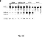


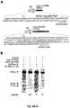


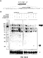
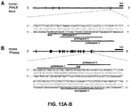


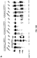


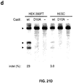
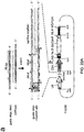


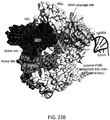


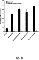
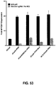


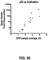


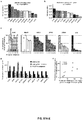
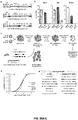


Classifications
-
- C—CHEMISTRY; METALLURGY
- C12—BIOCHEMISTRY; BEER; SPIRITS; WINE; VINEGAR; MICROBIOLOGY; ENZYMOLOGY; MUTATION OR GENETIC ENGINEERING
- C12N—MICROORGANISMS OR ENZYMES; COMPOSITIONS THEREOF; PROPAGATING, PRESERVING, OR MAINTAINING MICROORGANISMS; MUTATION OR GENETIC ENGINEERING; CULTURE MEDIA
- C12N9/00—Enzymes; Proenzymes; Compositions thereof; Processes for preparing, activating, inhibiting, separating or purifying enzymes
- C12N9/14—Hydrolases (3)
- C12N9/16—Hydrolases (3) acting on ester bonds (3.1)
- C12N9/22—Ribonucleases RNAses, DNAses
-
- A—HUMAN NECESSITIES
- A61—MEDICAL OR VETERINARY SCIENCE; HYGIENE
- A61K—PREPARATIONS FOR MEDICAL, DENTAL OR TOILETRY PURPOSES
- A61K38/00—Medicinal preparations containing peptides
- A61K38/16—Peptides having more than 20 amino acids; Gastrins; Somatostatins; Melanotropins; Derivatives thereof
- A61K38/43—Enzymes; Proenzymes; Derivatives thereof
- A61K38/46—Hydrolases (3)
- A61K38/465—Hydrolases (3) acting on ester bonds (3.1), e.g. lipases, ribonucleases
-
- A—HUMAN NECESSITIES
- A61—MEDICAL OR VETERINARY SCIENCE; HYGIENE
- A61K—PREPARATIONS FOR MEDICAL, DENTAL OR TOILETRY PURPOSES
- A61K48/00—Medicinal preparations containing genetic material which is inserted into cells of the living body to treat genetic diseases; Gene therapy
- A61K48/005—Medicinal preparations containing genetic material which is inserted into cells of the living body to treat genetic diseases; Gene therapy characterised by an aspect of the 'active' part of the composition delivered, i.e. the nucleic acid delivered
-
- A—HUMAN NECESSITIES
- A61—MEDICAL OR VETERINARY SCIENCE; HYGIENE
- A61P—SPECIFIC THERAPEUTIC ACTIVITY OF CHEMICAL COMPOUNDS OR MEDICINAL PREPARATIONS
- A61P43/00—Drugs for specific purposes, not provided for in groups A61P1/00-A61P41/00
-
- C—CHEMISTRY; METALLURGY
- C12—BIOCHEMISTRY; BEER; SPIRITS; WINE; VINEGAR; MICROBIOLOGY; ENZYMOLOGY; MUTATION OR GENETIC ENGINEERING
- C12N—MICROORGANISMS OR ENZYMES; COMPOSITIONS THEREOF; PROPAGATING, PRESERVING, OR MAINTAINING MICROORGANISMS; MUTATION OR GENETIC ENGINEERING; CULTURE MEDIA
- C12N15/00—Mutation or genetic engineering; DNA or RNA concerning genetic engineering, vectors, e.g. plasmids, or their isolation, preparation or purification; Use of hosts therefor
- C12N15/09—Recombinant DNA-technology
- C12N15/11—DNA or RNA fragments; Modified forms thereof; Non-coding nucleic acids having a biological activity
- C12N15/113—Non-coding nucleic acids modulating the expression of genes, e.g. antisense oligonucleotides; Antisense DNA or RNA; Triplex- forming oligonucleotides; Catalytic nucleic acids, e.g. ribozymes; Nucleic acids used in co-suppression or gene silencing
-
- C—CHEMISTRY; METALLURGY
- C12—BIOCHEMISTRY; BEER; SPIRITS; WINE; VINEGAR; MICROBIOLOGY; ENZYMOLOGY; MUTATION OR GENETIC ENGINEERING
- C12N—MICROORGANISMS OR ENZYMES; COMPOSITIONS THEREOF; PROPAGATING, PRESERVING, OR MAINTAINING MICROORGANISMS; MUTATION OR GENETIC ENGINEERING; CULTURE MEDIA
- C12N15/00—Mutation or genetic engineering; DNA or RNA concerning genetic engineering, vectors, e.g. plasmids, or their isolation, preparation or purification; Use of hosts therefor
- C12N15/09—Recombinant DNA-technology
- C12N15/87—Introduction of foreign genetic material using processes not otherwise provided for, e.g. co-transformation
- C12N15/90—Stable introduction of foreign DNA into chromosome
- C12N15/902—Stable introduction of foreign DNA into chromosome using homologous recombination
- C12N15/907—Stable introduction of foreign DNA into chromosome using homologous recombination in mammalian cells
-
- C—CHEMISTRY; METALLURGY
- C12—BIOCHEMISTRY; BEER; SPIRITS; WINE; VINEGAR; MICROBIOLOGY; ENZYMOLOGY; MUTATION OR GENETIC ENGINEERING
- C12Q—MEASURING OR TESTING PROCESSES INVOLVING ENZYMES, NUCLEIC ACIDS OR MICROORGANISMS; COMPOSITIONS OR TEST PAPERS THEREFOR; PROCESSES OF PREPARING SUCH COMPOSITIONS; CONDITION-RESPONSIVE CONTROL IN MICROBIOLOGICAL OR ENZYMOLOGICAL PROCESSES
- C12Q1/00—Measuring or testing processes involving enzymes, nucleic acids or microorganisms; Compositions therefor; Processes of preparing such compositions
- C12Q1/68—Measuring or testing processes involving enzymes, nucleic acids or microorganisms; Compositions therefor; Processes of preparing such compositions involving nucleic acids
- C12Q1/6876—Nucleic acid products used in the analysis of nucleic acids, e.g. primers or probes
-
- C—CHEMISTRY; METALLURGY
- C12—BIOCHEMISTRY; BEER; SPIRITS; WINE; VINEGAR; MICROBIOLOGY; ENZYMOLOGY; MUTATION OR GENETIC ENGINEERING
- C12Y—ENZYMES
- C12Y301/00—Hydrolases acting on ester bonds (3.1)
-
- C—CHEMISTRY; METALLURGY
- C12—BIOCHEMISTRY; BEER; SPIRITS; WINE; VINEGAR; MICROBIOLOGY; ENZYMOLOGY; MUTATION OR GENETIC ENGINEERING
- C12N—MICROORGANISMS OR ENZYMES; COMPOSITIONS THEREOF; PROPAGATING, PRESERVING, OR MAINTAINING MICROORGANISMS; MUTATION OR GENETIC ENGINEERING; CULTURE MEDIA
- C12N2310/00—Structure or type of the nucleic acid
- C12N2310/10—Type of nucleic acid
- C12N2310/20—Type of nucleic acid involving clustered regularly interspaced short palindromic repeats [CRISPRs]
-
- Y—GENERAL TAGGING OF NEW TECHNOLOGICAL DEVELOPMENTS; GENERAL TAGGING OF CROSS-SECTIONAL TECHNOLOGIES SPANNING OVER SEVERAL SECTIONS OF THE IPC; TECHNICAL SUBJECTS COVERED BY FORMER USPC CROSS-REFERENCE ART COLLECTIONS [XRACs] AND DIGESTS
- Y02—TECHNOLOGIES OR APPLICATIONS FOR MITIGATION OR ADAPTATION AGAINST CLIMATE CHANGE
- Y02A—TECHNOLOGIES FOR ADAPTATION TO CLIMATE CHANGE
- Y02A50/00—TECHNOLOGIES FOR ADAPTATION TO CLIMATE CHANGE in human health protection, e.g. against extreme weather
- Y02A50/30—Against vector-borne diseases, e.g. mosquito-borne, fly-borne, tick-borne or waterborne diseases whose impact is exacerbated by climate change
Definitions
- the present invention generally relates to systems, methods and compositions used for the control of gene expression involving sequence targeting, such as genome perturbation or gene-editing, that may use vector systems related to Clustered Regularly Interspaced Short Palindromic Repeats (CRISPR) and components thereof.
- CRISPR Clustered Regularly Interspaced Short Palindromic Repeats
- the present invention comprehends optimized functional CRISPR-Cas enzyme systems.
- the CRISPR/Cas or the CRISPR-Cas system does not require the generation of customized proteins to target specific sequences but rather a single Cas enzyme can be programmed by a short RNA molecule to recognize a specific DNA target, in other words the Cas enzyme can be recruited to a specific DNA target using said short RNA molecule.
- Adding the CRISPR-Cas system to the repertoire of genome sequencing techniques and analysis methods may significantly simplify the methodology and accelerate the ability to catalog and map genetic factors associated with a diverse range of biological functions and diseases.
- the invention provides a non-naturally occurring or engineered composition
- a guide RNA comprising a guide sequence capable of hybridizing to a target sequence in a genomic locus of interest in a cell, wherein at least one loop of the sgRNA is modified by the insertion of distinct RNA sequence(s) that bind to one or more adaptor proteins, and wherein the adaptor protein is associated with one or more functional domains.
- the functional domains can be same or different, e.g., two of the same or two different activators or repressors.
- the invention provides a non-naturally occurring or engineered CRISPR-Cas complex composition comprising the herein-mentioned sgRNA and a CRISPR enzyme, as defined in the claims.
- the invention provides a herein-mentioned non-naturally occurring or engineered CRISPR-Cas complex composition wherein: the CRISPR enzyme comprises at least one mutation, such that the CRISPR enzyme has no more than 5% of the nuclease activity of the CRISPR enzyme not having the at least one mutation; and optionally at least one or more nuclear localization sequences, as defined in the claims.
- the invention provides the herein-mentioned sgRNA or the CRISPR-Cas complex wherein one or more adaptor proteins associated with one or more functional domains is present and bound to the distinct RNA sequence(s) inserted into the at least one loop of the sgRNA, as defined in the claims.
- the invention provides a non-naturally occurring or engineered composition
- a non-naturally occurring or engineered composition comprising: one or more guide RNA (sgRNA) comprising a guide sequence capable of hybridizing to a target sequence in a genomic locus of interest in a cell, a CRISPR enzyme comprising at least one or more nuclear localization sequences, wherein the CRISPR enzyme comprises at least one mutation, such that the CRISPR enzyme has no more than 5% of the nuclease activity of the CRISPR enzyme not having the at least one mutation, wherein at least one loop of at least one sgRNA is modified by the insertion of distinct RNA sequence(s) that bind to one or more adaptor proteins, and wherein the adaptor protein is associated with one or more functional domains.
- sgRNA guide RNA
- CRISPR enzyme comprising at least one or more nuclear localization sequences
- the CRISPR enzyme comprises at least one mutation, such that the CRISPR enzyme has no more than 5% of the nuclease activity of
- the invention provides any herein-mentioned composition wherein the CRISPR enzyme has a diminished nuclease activity of at least 97%, or 100% as compared with the CRISPR enzyme not having the at least one mutation, as defined in the claims.
- the invention provides any aforementioned composition wherein the CRISPR enzyme comprises two or more mutations wherein two or more of D10, E762, H840, N854, N863, or D986 according to SpCas9 protein or any corresponding ortholog or N580 according to SaCas9 protein are mutated, or the CRISPR enzyme comprises at least one mutation wherein at least H840 is mutated, as defined in the claims.
- the invention provides a herein-mentioned composition wherein the CRISPR enzyme comprises two or more mutations comprising D10A, E762A, H840A, N854A, N863A or D986A according to SpCas9 protein or any corresponding ortholog, or N580A according to SaCas9 protein, or at least one mutation comprising H840A, as defined in the claims.
- the invention provides any herein-mentioned composition wherein the CRISPR enzyme comprises H840A, or D10A and H840A, or D10A and N863A, according to SpCas9 protein or any corresponding ortholog, as defined in the claims.
- the invention provides any herein-mentioned composition wherein the CRISPR enzyme comprises: N580A according to SaCas9 protein or any corresponding ortholog; or D10A according to SpCas9 protein, or any corresponding ortholog, and N580A according to SaCas9 protein, as defined in the claims.
- the invention provides any herein-mentioned composition wherein the CRISPR enzyme is associated with one or more functional domains, as defined in the claims. In an aspect the invention provides any herein-mentioned composition wherein the one or more functional domains associated with the adaptor protein is a heterologous functional domain, as defined in the claims. In an aspect the invention provides any herein-mentioned composition wherein the one or more functional domains associated with the CRISPR enzyme is a heterologous functional domain, as defined in the claims.
- the invention provides a composition as herein discussed, wherein the adaptor protein is a fusion protein comprising the functional domain, the fusion protein optionally comprising a linker between the adaptor protein and the functional domain, the linker optionally including a GlySer linker, as defined in the claims.
- the invention provides a composition as herein discussed, wherein the at least one loop of the sgRNA is not modified by the insertion of distinct RNA sequence(s) that bind to one or more adaptor proteins and wherein, optionally one of the unmodified sgRNA loops is either one of the tetraloop or the stem-loop 2, as defined in the claims.
- the invention provides a composition as herein discussed wherein the one or more functional domains associated with the adaptor protein is a transcriptional activation domain, as defined in the claims.
- the invention provides a composition as herein discussed wherein the one or more functional domains associated with the CRISPR enzyme is a transcriptional activation domain, as defined in the claims.
- the invention provides a composition as herein discussed wherein the one or more functional domains associated with the adaptor protein is a transcriptional activation domain comprising VP64, p65, MyoD1, HSF1, RTA or SET7/9, as defined in the claims.
- activation (or activator) domains in respect of those associated with the adaptor protein(s) include any known transcriptional activation domain and specifically VP64, p65, MyoD1, HSF1, RTA or SET7/9.
- the invention provides a composition as herein discussed wherein the one or more functional domains associated with the CRISPR enzyme is a transcriptional activation domain comprises VP64, p65, MyoD1, HSF1, RTA and SET7/9, as defined in the claims.
- Other refererences herein to activation (or activator) domains in respect of those associated with the CRISPR enzyme include any known transcriptional activation domain and specifically VP64, p65, MyoD1, HSF1, RTA or SET7/9.
- the invention provides a composition as herein discussed wherein the one or more functional domains associated with the adaptor protein is a transcriptional repressor domain, as defined in the claims.
- the invention provides a composition as herein discussed wherein the one or more functional domains associated with the CRISPR enzyme is a transcriptional repressor domain, as defined in the claims.
- the invention provides a composition as herein discussed wherein the transcriptional repressor domain is a KRAB domain, as defined in the claims.
- the invention provides a composition as herein discussed wherein the transcriptional repressor domain is a NuE domain, NcoR domain, SID domain or a SID4X domain, as defined in the claims.
- the invention provides a composition as herein discussed wherein the one or more functional domains associated with the adaptor protein have one or more activities comprising methylase activity, demethylase activity, transcription activation activity, transcription repression activity, transcription release factor activity, histone modification activity, RNA cleavage activity, DNA cleavage activity, DNA integration activity or nucleic acid binding activity, as defined in the claims.
- the invention provides a composition as herein discussed wherein the one or more functional domains associated with the CRISPR enzyme have one or more activities comprising methylase activity, demethylase activity, transcription activation activity, transcription repression activity, transcription release factor activity, histone modification activity, RNA cleavage activity, DNA cleavage activity, DNA integration activity, nucleic acid binding activity, or molecular switch activity or chemical inducibility or light inducibility, as defined in the claims.
- Histone modifying domains are also preferred in some embodiments. Exemplary histone modifying domains are discussed below.
- Transposase domains, HR (Homologous Recombination) machinery domains, recombinase domains, and/or integrase domains are also preferred as the present functional domains.
- DNA integration activity includes HR machinery domains, integrase domains, recombinase domains and/or transposase domains.
- the invention provides a composition as herein discussed wherein the DNA cleavage activity is due to a nuclease, as defined in the claims.
- the invention provides a composition as herein discussed wherein the nuclease comprises a Fok1 nuclease, as defined in the claims.
- the invention provides a composition as herein discussed wherein the one or more functional domains is attached to the CRISPR enzyme so that upon binding to the sgRNA and target the functional domain is in a spatial orientation allowing for the functional domain to function in its attributed function, as defined in the claims.
- the invention provides a composition as herein discussed wherein the one or more functional domains is attached to the CRISPR enzyme via a linker, optionally a GlySer linker, as defined in the claims.
- the invention provides a composition as herein discussed, wherein the sgRNA is modified so that, after sgRNA binds the adaptor protein and further binds to the CRISPR enzyme and target, the functional domain is in a spatial orientation allowing for the functional domain to function in its attributed function, as defined in the claims.
- the invention provides a composition as herein discussed wherein the at least one loop of the sgRNA is tetraloop and/or loop2, as defined in the claims.
- the invention provides a composition as herein discussed wherein the tetraloop and loop 2 of the sgRNA are modified by the insertion of the distinct RNA sequence(s), as defined in the claims.
- the invention provides a composition as herein discussed wherein the insertion of distinct RNA sequence(s) that bind to one or more adaptor proteins is an aptamer sequence, as defined in the claims.
- the invention provides a composition as herein discussed wherein the aptamer sequence is two or more aptamer sequences specific to the same adaptor protein, as defined in the claims.
- the invention provides a composition as herein discussed wherein the aptamer sequence is two or more aptamer sequences specific to different adaptor proteins, as defined in the claims.
- the invention provides a composition as herein discussed wherein the adaptor protein comprises MS2, PP7, Q ⁇ , F2, GA, fr, JP501, M12, R17, BZ13, JP34, JP500, KU1, M11, MX1, TW18, VK, SP, FI, ID2, NL95, TW19, AP205, ⁇ Cb5, ⁇ Cb8r, ⁇ Cb12r, ⁇ Cb23r, 7s, PRR1, as defined in the claims.
- the adaptor protein comprises MS2, PP7, Q ⁇ , F2, GA, fr, JP501, M12, R17, BZ13, JP34, JP500, KU1, M11, MX1, TW18, VK, SP, FI, ID2, NL95, TW19, AP205, ⁇ Cb5, ⁇ Cb8r, ⁇ Cb12r, ⁇ Cb23r, 7s, PRR1, as defined in the claims.
- the invention provides a composition as herein discussed wherein the cell is a eukaryotic cell, optionally a mammalian cell, as defined in the claims.
- the invention provides a composition as herein discussed wherein the cell is a human cell or a mouse cell, as defined in the claims.
- the invention provides a mammalian cell as herein discussed, e.g., wherein the cell comprises a cell line and is, optionally, a human cell line or a mouse cell line, as defined in the claims.
- the invention provides a transgenic mammalian model, optionally a mouse, e.g., wherein the model has been transformed with a composition as herein discussed or is progeny of said transformant, as defined in the claims.
- the invention provides a method for introducing a genomic locus event comprising the administration of to a host or expression in a host one or more of the compositions as herein discussed, as defined in the claims.
- the invention provides a method as herein discussed, wherein the genomic locus event comprises affecting gene activation, gene inhibition, or cleavage in the locus, or insertion of DNA, as defined in the claims.
- the invention provides a method as herein discussed, comprising the delivery of the composition or nucleic acid molecule(s) coding therefor, wherein said nucleic acid molecule(s) are operatively linked to regulatory sequence(s) and expressed in vivo.
- Expression in vivo can be via a lentivirus, an adenovirus, or an AAV, as defined in the claims.
- the invention provides a vector comprising: a nucleic acid molecule encoding a guide RNA (sgRNA), comprising a regulatory element operable in a eukaryotic cell including a guide sequence (sgRNA) operably linked to a promoter, the sgRNA being capable of hybridizing to a target sequence in a genomic locus of interest in a cell, wherein at least one loop of the sgRNA is modified by the insertion of distinct RNA sequence(s) that bind to one or more adaptor proteins.
- sgRNA guide RNA
- sgRNA guide RNA
- the invention provides a vector comprising a regulatory element operable in a eukaryotic cell including a a nucleic acid molecule encoding a CRISPR enzyme operably linked to a promoter, the enzyme comprising at least one or more nuclear localization sequences, wherein the CRISPR enzyme comprises at least one mutation, such that the CRISPR enzyme has no more than 5% of the nuclease activity of the CRISPR enzyme not having the at least one mutation, and wherein the CRISPR enzyme is associated with one or more functional domains, as defined in the claims.
- the invention provides nucleic acid molecule(s) encoding sgRNA or the CRISPR-Cas complex or a composition as herein discussed, as defined in the claims.
- the disclosure provides a method of screening for gain of function (GOF) or loss of function (LOF) comprising the cell line or cells of the model herein-discussed containing or expressing Cas9 and introducing a composition of claim 1 into cells of the cell line or model, whereby the sgRNA includes either an activator or a repressor, and monitoring for GOF or LOF respectively as to those cells as to which the introduced sgRNA includes an activator or as to those cells as to which the introduced sgRNA includes a repressor.
- GEF gain of function
- LEF loss of function
- a CRISPR Cas complex comprising a CRISPR enzyme and a guide RNA (sgRNA), wherein the CRISPR enzyme comprises at least one mutation, such that the CRISPR enzyme has no more than 5% of the nuclease activity of the CRISPR enzyme not having the at least one mutation and, optional, at least one or more nuclear localization sequences;
- the guide RNA (sgRNA) comprises a guide sequence capable of hybridizing to a target sequence in a genomic locus of interest in a cell; and wherein: the CRISPR enzyme is associated with two or more functional domains, which may be the same or different, and are preferably different functional domains; or at least one loop of the sgRNA is modified by the insertion of distinct RNA sequence(s) that bind to one or more adaptor proteins, and wherein the adaptor protein is associated with two or more functional domains; or the CRISPR enzyme is associated with one or more functional domains and at least one loop of the sgRNA is modified by the insertion of distinct RNA sequence(
- the invention provides a herein-discussed composition wherein the CRISPR enzyme includes one or more functional domains, as defined in the claims.
- the CRISPR enzyme includes one or more functional domains, as defined in the claims.
- the composition can include more than one sgRNA modified by the insertion of distinct RNA sequence(s) that bind to one or more adaptor proteins.
- the composition can involve wherein one or more adaptor proteins associated with one or more functional domains is present and bound to the distinct RNA sequence(s) inserted into the at least one loop of the sgRNA.
- the invention relates to a method for altering or modifying expression of a gene product.
- the said method may comprise introducing into a cell containing and expressing a DNA molecule encoding the gene product an engineered, non-naturally occurring CRISPR-Cas system comprising a Cas protein and guide RNA that targets the DNA molecule, whereby the guide RNA targets the DNA molecule encoding the gene product and the Cas protein cleaves the DNA molecule encoding the gene product, whereby expression of the gene product is altered; and, wherein the Cas protein and the guide RNA do not naturally occur together.
- the invention comprehends the guide RNA comprising a guide sequence fused to a tracr sequence.
- the invention further comprehends the Cas protein being codon optimized for expression in a Eukaryotic cell.
- the Eukaryotic cell is a mammalian cell and in a more preferred embodiment the mammalian cell is a human cell.
- the expression of the gene product is decreased.
- the target sequence(s) can be noncoding or regulatory (including promoter, especially the proximal promoter) or enhancer or silencer sequence(s).
- the invention relates to an engineered, non-naturally occurring CRISPR-Cas system comprising a Cas protein and a guide RNA that targets a DNA molecule encoding a gene product in a cell, whereby the guide RNA targets the DNA molecule encoding the gene product and the Cas protein cleaves the DNA molecule encoding the gene product, whereby expression of the gene product is altered; and, wherein the Cas protein and the guide RNA do not naturally occur together.
- the invention comprehends the guide RNA comprising a guide sequence fused to a tracr sequence.
- the Cas protein is a type II CRISPR-Cas protein and in a preferred embodiment the Cas protein is a Cas9 protein.
- the invention further comprehends the Cas protein being codon optimized for expression in a Eukaryotic cell.
- the Eukaryotic cell is a mammalian cell and in a more preferred embodiment the mammalian cell is a human cell.
- the expression of the gene product is decreased.
- the invention in another aspect, relates to an engineered, non-naturally occurring vector system comprising one or more vectors comprising a first regulatory element operably linked to a CRISPR-Cas system guide RNA that targets a DNA molecule encoding a gene product and a second regulatory element operably linked to a Cas protein.
- Components (a) and (b) may be located on same or different vectors of the system.
- the guide RNA targets the DNA molecule encoding the gene product in a cell and the Cas protein cleaves the DNA molecule encoding the gene product, whereby expression of the gene product is altered; and, wherein the Cas protein and the guide RNA do not naturally occur together.
- the invention comprehends the guide RNA comprising a guide sequence fused to a tracr sequence.
- the Cas protein is a type II CRISPR-Cas protein and in a preferred embodiment the Cas protein is a Cas9 protein.
- the invention further comprehends the Cas protein being codon optimized for expression in a Eukaryotic cell.
- the Eukaryotic cell is a mammalian cell and in a more preferred embodiment the mammalian cell is a human cell.
- the expression of the gene product is decreased.
- the invention relates to a vector system comprising one or more vectors.
- the system comprises: (a) a first regulatory element operably linked to a tracr mate sequence and one or more insertion sites for inserting one or more guide sequences upstream of the tracr mate sequence, wherein when expressed, the guide sequence directs sequence-specific binding of a CRISPR complex to a target sequence in a eukaryotic cell, wherein the CRISPR complex comprises a CRISPR enzyme complexed with (1) the guide sequence that is hybridized to the target sequence, and (2) the tracr mate sequence that is hybridized to the tracr sequence; and (b) a second regulatory element operably linked to an enzyme-coding sequence encoding said CRISPR enzyme comprising a nuclear localization sequence; wherein components (a) and (b) are located on the same or different vectors of the system.
- component (a) further comprises the tracr sequence downstream of the tracr mate sequence under the control of the first regulatory element.
- component (a) further comprises two or more guide sequences operably linked to the first regulatory element, wherein when expressed, each of the two or more guide sequences direct sequence specific binding of a CRISPR complex to a different target sequence in a eukaryotic cell.
- the system comprises the tracr sequence under the control of a third regulatory element, such as a polymerase III promoter.
- the tracr sequence exhibits at least 50%, 60%, 70%, 80%, 90%, 95%, or 99% of sequence complementarity along the length of the tracr mate sequence when optimally aligned.
- the CRISPR complex comprises one or more nuclear localization sequences of sufficient strength to drive accumulation of said CRISPR complex in a detectable amount in the nucleus of a eukaryotic cell.
- a nuclear localization sequence is not necessary for CRISPR complex activity in eukaryotes, but that including such sequences enhances activity of the system, especially as to targeting nucleic acid molecules in the nucleus.
- the CRISPR enzyme is a type II CRISPR system enzyme. In some embodiments, the CRISPR enzyme is a Cas9 enzyme. In some embodiments, the Cas9 enzyme is S. pneumoniae, S. pyogenes, or S. thermophilus Cas9, and may include mutated Cas9 derived from these organisms. The enzyme may be a Cas9 homolog or ortholog. In some embodiments, the CRISPR enzyme is codon-optimized for expression in a eukaryotic cell. In some embodiments, the CRISPR enzyme directs cleavage of one or two strands at the location of the target sequence. In some embodiments, the first regulatory element is a polymerase III promoter.
- the second regulatory element is a polymerase II promoter.
- the guide sequence is at least 15, 16, 17, 18, 19, 20, 25 nucleotides, or between 10-30, or between 15-25, or between 15-20 nucleotides in length.
- vector refers to a nucleic acid molecule capable of transporting another nucleic acid to which it has been linked. Vectors include, but are not limited to, nucleic acid molecules that are single-stranded, double-stranded, or partially double-stranded; nucleic acid molecules that comprise one or more free ends, no free ends (e.g.
- vectors refers to a circular double stranded DNA loop into which additional DNA segments can be inserted, such as by standard molecular cloning techniques.
- viral vector wherein virally-derived DNA or RNA sequences are present in the vector for packaging into a virus (e.g. retroviruses, replication defective retroviruses, adenoviruses, replication defective adenoviruses, and adeno-associated viruses).
- Viral vectors also include polynucleotides carried by a virus for transfection into a host cell.
- vectors are capable of autonomous replication in a host cell into which they are introduced (e.g. bacterial vectors having a bacterial origin of replication and episomal mammalian vectors). Other vectors (e.g., non-episomal mammalian vectors) are integrated into the genome of a host cell upon introduction into the host cell, and thereby are replicated along with the host genome. Moreover, certain vectors are capable of directing the expression of genes to which they are operatively-linked. Such vectors are referred to herein as "expression vectors.” Common expression vectors of utility in recombinant DNA techniques are often in the form of plasmids.
- Recombinant expression vectors can comprise a nucleic acid of the invention in a form suitable for expression of the nucleic acid in a host cell, which means that the recombinant expression vectors include one or more regulatory elements, which may be selected on the basis of the host cells to be used for expression, that is operatively-linked to the nucleic acid sequence to be expressed.
- "operably linked" is intended to mean that the nucleotide sequence of interest is linked to the regulatory element(s) in a manner that allows for expression of the nucleotide sequence (e.g. in an in vitro transcription/translation system or in a host cell when the vector is introduced into the host cell).
- regulatory element is intended to include promoters, enhancers, internal ribosomal entry sites (IRES), and other expression control elements (e.g. transcription termination signals, such as polyadenylation signals and poly-U sequences).
- regulatory elements are described, for example, in Goeddel, GENE EXPRESSION TECHNOLOGY: METHODS IN ENZYMOLOGY 185, Academic Press, San Diego, Calif. (1990 ).
- Regulatory elements include those that direct constitutive expression of a nucleotide sequence in many types of host cell and those that direct expression of the nucleotide sequence only in certain host cells (e.g., tissue-specific regulatory sequences).
- a tissue-specific promoter may direct expression primarily in a desired tissue of interest, such as muscle, neuron, bone, skin, blood, specific organs (e.g. liver, pancreas), or particular cell types (e.g. lymphocytes). Regulatory elements may also direct expression in a temporal-dependent manner, such as in a cell-cycle dependent or developmental stage-dependent manner, which may or may not also be tissue or cell-type specific.
- a vector comprises one or more pol III promoter (e.g. 1, 2, 3, 4, 5, or more pol I promoters), one or more pol II promoters (e.g. 1, 2, 3, 4, 5, or more pol II promoters), one or more pol I promoters (e.g.
- pol III promoters include, but are not limited to, U6 and H1 promoters.
- pol II promoters include, but are not limited to, the retroviral Rous sarcoma virus (RSV) LTR promoter (optionally with the RSV enhancer), the cytomegalovirus (CMV) promoter (optionally with the CMV enhancer) [see, e.g., Boshart et al, Cell, 41:521-530 (1985 )], the SV40 promoter, the dihydrofolate reductase promoter, the ⁇ -actin promoter, the phosphoglycerol kinase (PGK) promoter, and the EF1 ⁇ promoter.
- RSV Rous sarcoma virus
- CMV cytomegalovirus
- PGK phosphoglycerol kinase
- enhancer elements such as WPRE; CMV enhancers; the R-U5' segment in LTR of HTLV-I ( Mol. Cell. Biol., Vol. 8(1), p. 466-472, 1988 ); SV40 enhancer; and the intron sequence between exons 2 and 3 of rabbit ⁇ -globin ( Proc. Natl. Acad. Sci. USA., Vol. 78(3), p. 1527-31, 1981 ).
- WPRE WPRE
- CMV enhancers the R-U5' segment in LTR of HTLV-I
- SV40 enhancer SV40 enhancer
- the intron sequence between exons 2 and 3 of rabbit ⁇ -globin Proc. Natl. Acad. Sci. USA., Vol. 78(3), p. 1527-31, 1981 .
- the design of the expression vector can depend on such factors as the choice of the host cell to be transformed, the level of expression desired, etc.
- a vector can be introduced into host cells to thereby produce transcripts, proteins, or peptides, including fusion proteins or peptides, encoded by nucleic acids as described herein (e.g., clustered regularly interspersed short palindromic repeats (CRISPR) transcripts, proteins, enzymes, mutant forms thereof, fusion proteins thereof, etc.).
- CRISPR clustered regularly interspersed short palindromic repeats
- Advantageous vectors include lentiviruses and adeno-associated viruses, and types of such vectors can also be selected for targeting particular types of cells.
- the invention relates to a eukaryotic host cell comprising (a) a first regulatory element operably linked to a tracr mate sequence and one or more insertion sites for inserting one or more guide sequences upstream of the tracr mate sequence, wherein when expressed, the guide sequence directs sequence-specific binding of a CRISPR complex to a target sequence in a eukaryotic cell, wherein the CRISPR complex comprises a CRISPR enzyme complexed with (1) the guide sequence that is hybridized to the target sequence, and (2) the tracr mate sequence that is hybridized to the tracr sequence; and/or (b) a second regulatory element operably linked to an enzyme-coding sequence encoding said CRISPR enzyme comprising a nuclear localization sequence.
- the host cell comprises components (a) and (b).
- component (a), component (b), or components (a) and (b) are stably integrated into a genome of the host eukaryotic cell.
- component (a) further comprises the tracr sequence downstream of the tracr mate sequence under the control of the first regulatory element.
- component (a) further comprises two or more guide sequences operably linked to the first regulatory element, wherein when expressed, each of the two or more guide sequences direct sequence specific binding of a CRISPR complex to a different target sequence in a eukaryotic cell.
- the eukaryotic host cell further comprises a third regulatory element, such as a polymerase III promoter, operably linked to said tracr sequence.
- the tracr sequence exhibits at least 50%, 60%, 70%, 80%, 90%, 95%, or 99% of sequence complementarity along the length of the tracr mate sequence when optimally aligned.
- the enzyme may be a Cas9 homolog or ortholog.
- the CRISPR enzyme is codon-optimized for expression in a eukaryotic cell.
- the CRISPR enzyme directs cleavage of one or two strands at the location of the target sequence.
- the CRISPR enzyme lacks DNA strand cleavage activity.
- the first regulatory element is a polymerase III promoter.
- the second regulatory element is a polymerase II promoter.
- the guide sequence is at least 15, 16, 17, 18, 19, 20, 25 nucleotides, or between 10-30, or between 15-25, or between 15-20 nucleotides in length.
- the invention relates to a non-human eukaryotic organism; preferably a multicellular eukaryotic organism, comprising a eukaryotic host cell according to any of the described embodiments.
- the invention relates to a eukaryotic organism; preferably a multicellular eukaryotic organism, comprising a eukaryotic host cell according to any of the described embodiments.
- the organism in some embodiments of these aspects may be an animal; for example a mammal. Also, the organism may be an arthropod such as an insect. The organism also may be a plant. Further, the organism may be a fungus.
- the invention relates to a kit comprising one or more of the components described herein.
- the kit comprises a vector system and instructions for using the kit.
- the vector system comprises (a) a first regulatory element operably linked to a tracr mate sequence and one or more insertion sites for inserting one or more guide sequences upstream of the tracr mate sequence, wherein when expressed, the guide sequence directs sequence-specific binding of a CRISPR complex to a target sequence in a eukaryotic cell, wherein the CRISPR complex comprises a CRISPR enzyme complexed with (1) the guide sequence that is hybridized to the target sequence, and (2) the tracr mate sequence that is hybridized to the tracr sequence; and/or (b) a second regulatory element operably linked to an enzyme-coding sequence encoding said CRISPR enzyme comprising a nuclear localization sequence.
- the kit comprises components (a) and (b) located on the same or different vectors of the system.
- component (a) further comprises the tracr sequence downstream of the tracr mate sequence under the control of the first regulatory element.
- component (a) further comprises two or more guide sequences operably linked to the first regulatory element, wherein when expressed, each of the two or more guide sequences direct sequence specific binding of a CRISPR complex to a different target sequence in a eukaryotic cell.
- the system further comprises a third regulatory element, such as a polymerase III promoter, operably linked to said tracr sequence.
- the tracr sequence exhibits at least 50%, 60%, 70%, 80%, 90%, 95%, or 99% of sequence complementarity along the length of the tracr mate sequence when optimally aligned.
- the CRISPR enzyme comprises one or more nuclear localization sequences of sufficient strength to drive accumulation of said CRISPR enzyme in a detectable amount in the nucleus of a eukaryotic cell.
- the CRISPR enzyme is a type II CRISPR system enzyme.
- the CRISPR enzyme is a Cas9 enzyme.
- the Cas9 enzyme is S. pneumoniae, S. pyogenes or S.
- thermophilus Cas9 and may include mutated Cas9 derived from these organisms.
- the enzyme may be a Cas9 homolog or ortholog.
- the CRISPR enzyme is codon-optimized for expression in a eukaryotic cell.
- the CRISPR enzyme directs cleavage of one or two strands at the location of the target sequence.
- the CRISPR enzyme lacks DNA strand cleavage activity.
- the first regulatory element is a polymerase III promoter.
- the second regulatory element is a polymerase II promoter.
- the guide sequence is at least 15, 16, 17, 18, 19, 20, 25 nucleotides, or between 10-30, or between 15-25, or between 15-20 nucleotides in length.
- the invention relates to a method of modifying a target polynucleotide in a eukaryotic cell.
- the method comprises allowing a CRISPR complex to bind to the target polynucleotide to effect cleavage of said target polynucleotide thereby modifying the target polynucleotide, wherein the CRISPR complex comprises a CRISPR enzyme complexed with a guide sequence hybridized to a target sequence within said target polynucleotide, wherein said guide sequence is linked to a tracr mate sequence which in turn hybridizes to a tracr sequence.
- said cleavage comprises cleaving one or two strands at the location of the target sequence by said CRISPR enzyme. In some embodiments, said cleavage results in decreased transcription of a target gene. In some embodiments, the method further comprises repairing said cleaved target polynucleotide by homologous recombination with an exogenous template polynucleotide, wherein said repair results in a mutation comprising an insertion, deletion, or substitution of one or more nucleotides of said target polynucleotide. In some embodiments, said mutation results in one or more amino acid changes in a protein expressed from a gene comprising the target sequence.
- the method further comprises delivering one or more vectors to said eukaryotic cell, wherein the one or more vectors drive expression of one or more of: the CRISPR enzyme, the guide sequence linked to the tracr mate sequence, and the tracr sequence.
- said vectors are delivered to the eukaryotic cell in a subject.
- said modifying takes place in said eukaryotic cell in a cell culture.
- the method further comprises isolating said eukaryotic cell from a subject prior to said modifying.
- the method further comprises returning said eukaryotic cell and/or cells derived therefrom to said subject.
- the invention relates to a method of modifying expression of a polynucleotide in a eukaryotic cell.
- the method comprises allowing a CRISPR complex to bind to the polynucleotide such that said binding results in increased or decreased expression of said polynucleotide; wherein the CRISPR complex comprises a CRISPR enzyme complexed with a guide sequence hybridized to a target sequence within said polynucleotide, wherein said guide sequence is linked to a tracr mate sequence which in turn hybridizes to a tracr sequence.
- the method further comprises delivering one or more vectors to said eukaryotic cells, wherein the one or more vectors drive expression of one or more of: the CRISPR enzyme, the guide sequence linked to the tracr mate sequence, and the tracr sequence.
- the invention relates to a method of generating a model eukaryotic cell comprising a mutated disease gene.
- a disease gene is any gene associated an increase in the risk of having or developing a disease.
- the method comprises (a) introducing one or more vectors into a eukaryotic cell, wherein the one or more vectors drive expression of one or more of: a CRISPR enzyme, a guide sequence linked to a tracr mate sequence, and a tracr sequence; and (b) allowing a CRISPR complex to bind to a target polynucleotide to effect cleavage of the target polynucleotide within said disease gene, wherein the CRISPR complex comprises the CRISPR enzyme complexed with (1) the guide sequence that is hybridized to the target sequence within the target polynucleotide, and (2) the tracr mate sequence that is hybridized to the tracr sequence, thereby generating a model eukaryotic cell compris
- said cleavage comprises cleaving one or two strands at the location of the target sequence by said CRISPR enzyme. In some embodiments, said cleavage results in decreased transcription of a target gene. In some embodiments, the method further comprises repairing said cleaved target polynucleotide by homologous recombination with an exogenous template polynucleotide, wherein said repair results in a mutation comprising an insertion, deletion, or substitution of one or more nucleotides of said target polynucleotide. In some embodiments, said mutation results in one or more amino acid changes in a protein expression from a gene comprising the target sequence.
- the present disclosure provides a method for developing a biologically active agent that modulates a cell signaling event associated with a disease gene.
- a disease gene is any gene associated with an increase in the risk of having or developing a disease.
- the method comprises (a) contacting a test compound with a model cell of any one of the described embodiments; and (b) detecting a change in a readout that is indicative of a reduction or an augmentation of a cell signaling event associated with said mutation in said disease gene, thereby developing said biologically active agent that modulates said cell signaling event associated with said disease gene.
- the invention relates to a recombinant polynucleotide comprising a guide sequence upstream of a tracr mate sequence, wherein the guide sequence when expressed directs sequence-specific binding of a CRISPR complex to a corresponding target sequence present in a eukaryotic cell.
- the target sequence is a viral sequence present in a eukaryotic cell.
- the target sequence is a proto-oncogene or an oncogene.
- the present disclosure provides for a method of selecting one or more cell(s) by introducing one or more mutations in a gene in the one or more cell (s), the method comprising: introducing one or more vectors into the cell (s), wherein the one or more vectors drive expression of one or more of: a CRISPR enzyme, a guide sequence linked to a tracr mate sequence, a tracr sequence, and an editing template; wherein the editing template comprises the one or more mutations that abolish CRISPR enzyme cleavage; allowing homologous recombination of the editing template with the target polynucleotide in the cell(s) to be selected; allowing a CRISPR complex to bind to a target polynucleotide to effect cleavage of the target polynucleotide within said gene, wherein the CRISPR complex comprises the CRISPR enzyme complexed with (1) the guide sequence that is hybridized to the target sequence within the target polynucleotide, and (2) the tracr
- the CRISPR enzyme is Cas9.
- the cell to be selected may be a eukaryotic cell. Aspects of the present disclosure allow for selection of specific cells without requiring a selection marker or a two-step process that may include a counter-selection system.
- the present disclosure comprehends a CRISPR-Cas9 (S. pyogenes) system having an X-ray diffraction pattern corresponding to or resulting from any or all of the foregoing and/or a crystal having the structure defined by the co-ordinates of the Crystral Structure Table in Example 8 (the CRISPR-cas9 crystal structure) or as further described in Example 12.
- CRISPR-Cas9 S. pyogenes
- the present disclosure involves a computer-assisted method for identifying or designing potential compounds to fit within or bind to CRISPR-Cas9 system or a functional portion thereof or vice versa (a computer-assisted method for identifying or designing potential CRISPR-Cas9 systems or a functional portion thereof for binding to desired compounds) or a computer-assisted method for identifying or designing potential CRISPR-Cas9 systems (e.g., with regard to predicting areas of the CRISPR-Cas9 system to be able to be manipulated-for instance, based on crystral structure data or based on data of Cas9 orthologs, or with respect to where a functional group such as an activator or repressor can be attached to the CRISPR-Cas9 system, or as to Cas9 truncations or as to designing nickases), said method comprising:
- the testing can comprise analyzing the CRISPR-Cas9 system resulting from said synthesized selected structure(s), e.g., with respect to binding, or performing a desired function.
- the output in the foregoing methods can comprise data transmission, e.g., transmission of information via telecommunication, telephone, video conference, mass communication, e.g., presentation such as a computer presentation (eg POWERPOINT), internet, email, documentary communication such as a computer program (eg WORD) document and the like.
- computer readable media containing: atomic co-ordinate data according to the herein Crystal Structure Table and/or the Figures, said data defining the three dimensional structure of CRISPR-Cas9 or at least one sub-domain thereof, or structure factor data for CRISPR-Cas9, said structure factor data being derivable from the atomic co-ordinate data of herein Crystal Structure Table and/or the Figures.
- the computer readable media can also contain any data of the foregoing methods.
- the present disclosure further comprehends methods a computer system for generating or performing rational design as in the foregoing methods containing either: atomic co-ordinate data according to herein Crystal Structure Table and/or the Figures, said data defining the three dimensional structure of CRISPR-Cas9 or at least one sub-domain thereof, or structure factor data for CRISPR-Cas9, said structure factor data being derivable from the atomic co-ordinate data of herein Crystal Structure Table and/or the Figures.
- the present disclosure further comprehends a method of doing business comprising providing to a user the computer system or the media or the three dimensional structure of CRISPR-Cas9 or at least one sub-domain thereof, or structure factor data for CRISPR-Cas9, said structure set forth in and said structure factor data being derivable from the atomic co-ordinate data of herein Crystal Structure Table and/or the Figures, or the herein computer media or a herein data transmission.
- binding site or an “active site” comprises or consists essentially of a site (such as an atom, a functional group of an amino acid residue or a plurality of such atoms and/or groups) in a binding cavity or region, which may bind to a compound such as a nucleic acid molecule, which is/are involved in binding.
- fitting is meant determining by automatic, or semi-automatic means, interactions between one or more atoms of a candidate molecule and at least one atom of a structure of the present disclosure, and calculating the extent to which such interactions are stable. Interactions include attraction and repulsion, brought about by charge, steric considerations and the like. Various computer-based methods for fitting are described further By “root mean square (or rms) deviation”, we mean the square root of the arithmetic mean of the squares of the deviations from the mean.
- a “computer system” By a “computer system”, is meant the hardware means, software means and data storage means used to analyze atomic coordinate data.
- the minimum hardware means of the computer-based systems of the present disclosure typically comprises a central processing unit (CPU), input means, output means and data storage means. Desirably a display or monitor is provided to visualize structure data.
- the data storage means may be RAM or means for accessing computer readable media of the present disclosure. Examples of such systems are computer and tablet devices running Unix, Windows or Apple operating systems.
- “computer readable media” any medium or media, which can be read and accessed directly or indirectly by a computer e.g. so that the media is suitable for use in the above-mentioned computer system.
- Such media include, but are not limited to: magnetic storage media such as floppy discs, hard disc storage medium and magnetic tape; optical storage media such as optical discs or CD-ROM; electrical storage media such as RAM and ROM; thumb drive devices; cloud storage devices and hybrids of these categories such as magnetic/optical storage media.
- the conformational variations in the crystal structures of the CRISPR-Cas9 system or of components of the CRISPR-Cas9 provide important and critical information about the flexibility or movement of protein structure regions relative to nucleotide (RNA or DNA) structure regions that may be important for CRISPR-Cas system function.
- the structural information provided for Cas9 e.g. S. pyogenes Cas9 as the CRISPR enzyme in the present application may be used to further engineer and optimize the CRISPR-Cas system and this may be extrapolated to interrogate structure-function relationships in other CRISPR enzyme systems as well, e.g, other Type II CRISPR enzyme systems.
- the invention comprehends optimized functional CRISPR-Cas enzyme systems, especially in combination with the present modified guides and also where the CRISPR enzyme is also associated with a functional domain, as defined in the claims.
- the CRISPR enzyme comprises one or more mutations that converts it to a DNA binding protein to which functional domains exhibiting a function of interest may be recruited or appended or inserted or attached.
- the CRISPR enzyme comprises one or more mutations which include but are not limited to D10A, E762A, H840A, N854A, N863A or D986A (based on the amino acid position numbering of a S.
- the CRISPR enzyme has one or more mutations in a catalytic domain, wherein when transcribed, the tracr mate sequence hybridizes to the tracr sequence and the guide sequence directs sequence-specific binding of a CRISPR complex to the target sequence, and wherein the enzyme further comprises a functional domain.
- a mutation at N580 according to SaCas9 protein is preferred. In particular, it is preferred in place of the mutation, in Sa Cas9, corresponding to H840 in Sp Cas9.
- the N580 mutation may be N580A according to SaCas9 protein. It is believed, without being bound by theory, that this is a more predictable mutation for protein function than the H840A equivalent, which may change binding behaviour.
- the structural information provided herein allows for interrogation of sgRNA (or chimeric RNA) interaction with the target DNA and the CRISPR enzyme (e.g. Cas9) permitting engineering or alteration of sgRNA structure to optimize functionality of the entire CRISPR-Cas system.
- the CRISPR enzyme e.g. Cas9
- loops of the sgRNA may be extended, without colliding with the Cas9 protein by the insertion of adaptor proteins that can bind to RNA.
- These adaptor proteins can further recruit effector proteins or fusions which comprise one or more functional domains.
- the functional domain is a transcriptional activation domain, preferably VP64. In some embodiments, the functional domain is a transcription repression domain, preferably KRAB. In some embodiments, the transcription repression domain is SID, or concatemers of SID (e.g. SID4X). In some embodiments, the functional domain is an epigenetic modifying domain, such that an epigenetic modifying enzyme is provided. In some embodiments, the functional domain is an activation domain, which may be the P65 activation domain.
- a non-naturally occurring or engineered composition may comprise a guide RNA (sgRNA) comprising a guide sequence capable of hybridizing to a target sequence in a genomic locus of interest in a cell and a CRISPR enzyme that may comprise at least one or more nuclear localization sequences, wherein the CRISPR enzyme comprises two or more mutations, such that the enzyme has altered or diminished nuclease activity compared with the wild type enzyme, wherein at least one loop of the sgRNA is modified by the insertion of distinct RNA sequence(s) that bind to one or more adaptor proteins, and wherein the adaptor protein further recruits one or more heterologous functional domains, as defined in the claims.
- sgRNA guide RNA
- CRISPR enzyme comprises two or more mutations, such that the enzyme has altered or diminished nuclease activity compared with the wild type enzyme
- at least one loop of the sgRNA is modified by the insertion of distinct RNA sequence(s) that bind to one or more adaptor proteins, and wherein the
- the CRISPR enzyme comprises two or more mutations in a residue selected from the group consisting of D10, E762, H840, N854, N863, or D986. In a further embodiment the CRISPR enzyme comprises two or more mutations selected from the group comprising D10A, E762A, H840A, N854A, N863A or D986A.
- N580, especially N580A, according to SaCas9 protein is used, especially in Sa Cas9.
- the functional domain is a transcriptional activation domain, e.g. VP64.
- the functional domain is a transcriptional repressor domain, e.g.
- the one or more heterologous functional domains have one or more activities selected from the group consisting of methylase activity, demethylase activity, transcription activation activity, transcription repression activity, transcription release factor activity, histone modification activity, RNA cleavage activity and nucleic acid binding activity.
- the cell is a eukaryotic cell or a mammalian cell or a human cell.
- the adaptor protein is selected from the group consisting of MS2, PP7, Q ⁇ , F2, GA, fr, JP501, M12, R17, BZ13, JP34, JP500, KU1, M11, MX1, TW18, VK, SP, FI, ID2, NL95, TW19, AP205, ⁇ Cb5, ⁇ Cb8r, ⁇ Cb12r, ⁇ Cb23r, 7s, PRR1.
- the at least one loop of the sgRNA is tetraloop and/or loop2.
- compositions are comprised in a single composition or comprised in individual compositions. These compositions may advantageously be applied to a host to elicit a functional effect on the genomic level.
- the sgRNA are modified in a manner that provides specific binding sites (e.g. aptamers) for adapter proteins comprising one or more functional domains (e.g. via fusion protein) to bind to.
- the modified sgRNA are modified such that once the sgRNA forms a CRISPR complex (i.e. CRISPR enzyme binding to sgRNA and target) the adapter proteins bind and, the functional domain on the adapter protein is positioned in a spatial orientation which is advantageous for the attributed function to be effective.
- the functional domain is a transcription activator (e.g. VP64 or p65)
- the transcription activator is placed in a spatial orientation which allows it to affect the transcription of the target.
- a transcription repressor will be advantageously positioned to affect the transcription of the target and a nuclease (e.g. Fok1) will be advantageously positioned to cleave or partially cleave the target.
- the skilled person will understand that modifications to the sgRNA which allow for binding of the adapter + functional domain but not proper positioning of the adapter + functional domain (e.g. due to steric hindrance within the three dimensial structure of the CRISPR complex) are modifications which are not intended.
- the one or more modified sgRNA may be modified at the tetra loop, the stem loop 1, stem loop 2, or stem loop 3, as described herein, preferably at either the tetra loop or stem loop 2, and most preferably at both the tetra loop and stem loop 2.
- the functional domains may be, for example, one or more domains from the group consisting of methylase activity, demethylase activity, transcription activation activity, transcription repression activity, transcription release factor activity, histone modification activity, RNA cleavage activity, DNA cleavage activity, nucleic acid binding activity, and molecular switches (e.g. light inducible).
- methylase activity demethylase activity
- transcription activation activity transcription repression activity
- transcription release factor activity e.g. light inducible
- histone modification activity e.g. light inducible
- RNA cleavage activity e.g. DNA cleavage activity
- nucleic acid binding activity e.g. light inducible
- molecular switches e.g. light inducible
- the sgRNA may be designed to include multiple binding recognition sites (e.g. aptamers) specific to the same or different adapter protein.
- the sgRNA may be designed to bind to the promoter region -1000 - +1 nucleic acids upstream of the transcription start site (i.e. TSS), preferably -200 nucleic acids. This positioning improves functional domains which affect gene activation (e.g. transcription activators) or gene inhibition (e.g. transcription repressors).
- the modified sgRNA may be one or more modified sgRNAs targeted to one or more target loci (e.g.
- the CRISPR enzyme with diminished nuclease activity is most effective when the nuclease activity is inactivated (e.g. nuclease inactivation of at least 70%, at least 80%, at least 90%, at least 95%, at least 97%, or 100% as compared with the wild type enzyme; or to put in another way, a Cas9 enzyme or CRISPR enzyme having advantageously about 0% of the nuclease activity of the non-mutated or wild type Cas9 enzyme or CRISPR enzyme, or no more than about 3% or about 5% or about 10% of the nuclease activity of the non-mutated or wild type Cas9 enzyme or CRISPR enzyme).
- mutations into the RuvC and HNH nuclease domains of the SpCas9 and orthologs thereof For example utilizing mutations in a residue selected from the group consisting of D10, E762, H840, N854, N863, or D986 and more preferably introducing one or more of the mutations selected from the group consisting of D10A, E762A, H840A, N854A, N863A or D986A.
- a preferable pair of mutations is D10A with H840A, more preferable is D10A with N863A of SpCas9 and orthologs thereof.
- N580A according to SaCas9 protein may be used, as discussed herein.
- the inactivated CRISPR enzyme may have associated (e.g. via fusion protein) one or more functional domains, like for example as described herein for the modified sgRNA adaptor proteins, including for example, one or more domains from the group consisting of methylase activity, demethylase activity, transcription activation activity, transcription repression activity, transcription release factor activity, histone modification activity, RNA cleavage activity, DNA cleavage activity, nucleic acid binding activity, and molecular switches (e.g. light inducible).
- Preferred domains are Fok1, VP64, P65, HSF1, MyoD1.
- Fok1 it is advantageous that multiple Fok1 functional domains are provided to allow for a functional dimer and that sgRNAs are designed to provide proper spacing for functional use (Fok1) as specifically described in Tsai et al. Nature Biotechnology, Vol. 32, Number 6, June 2014 ).
- the adaptor protein may utilize known linkers to attach such functional domains.
- the functional domains may be the same or different.
- the positioning of the one or more functional domain on the inactivated CRISPR enzyme is one which allows for correct spatial orientation for the functional domain to affect the target with the attributed functional effect.
- the functional domain is a transcription activator (e.g. VP64 or p65)
- the transcription activator is placed in a spatial orientation which allows it to affect the transcription of the target.
- a transcription repressor will be advantageously positioned to affect the transcription of the target
- a nuclease e.g. Fok1
- This may include positions other than the N- / C- terminus of the CRISPR enzyme.
- the Applicant has identified that positioning the functional domain in the Reel domain, the Rec2 domain, the HNH domain, or the PI domain of the SpCas9 protein or any ortholog corresponding to these domains is advantageous. Positioning of the functional domains to the Reel domain or the Rec2 domain, of the SpCas9 protein or any ortholog corresponding to these domains, in some instances may be preferred.
- Fok1 functional domain may be attached at the N terminus. When more than one functional domain is included, the functional domains may be the same or different.
- the adaptor protein may be any number of proteins that binds to an aptamer or recognition site introduced into the modified sgRNA and which allows proper positioning of one or more functional domains, once the sgRNA has been incorporated into the CRISPR complex, to affect the target with the attributed function.
- such may be coat proteins, preferably bacteriophage coat proteins.
- the functional domains associated with such adaptor proteins e.g.
- fusion protein in the form of fusion protein may include, for example, one or more domains from the group consisting of methylase activity, demethylase activity, transcription activation activity, transcription repression activity, transcription release factor activity, histone modification activity, RNA cleavage activity, DNA cleavage activity, nucleic acid binding activity, and molecular switches (e.g. light inducible).
- Preferred domains are Fok1, VP64, P65, HSF1, MyoD1.
- the functional domain is a transcription activator or transcription repressor it is advantageous that additionally at least an NLS is provided and preferably at the N terminus.
- the functional domains may be the same or different.
- the adaptor protein may utilize known linkers to attach such functional domains.
- the modified sgRNA, the inactivated CRISPR enzyme (with or without functional domains), and the binding protein with one or more functional domains may each individually be comprised in a composition and administered to a host individually or collectively. Alternatively, these components may be provided in a single composition for administration to a host. Administration to a host may be performed via viral vectors known to the skilled person or described herein for delivery to a host (e.g. lentiviral vector, adenoviral vector, AAV vector). As explained herein, use of different selection markers (e.g. for lentiviral sgRNA selection) and concentration of sgRNA (e.g. dependent on whether multiple sgRNAs are used) may be advantageous for eliciting an improved effect.
- compositions may be applied in a wide variety of methods for screening in libraries in cells and functional modeling in vivo (e.g. gene activation of lincRNA and identification of function; gain-of-function modeling; loss-of-function modeling; the use the compositions of the invention to establish cell lines and transgenic animals for optimization and screening purposes).
- the current invention comprehends the use of the compositions of the current invention to establish and utilize conditional or inducible CRISPR transgenic cell /animals.
- CRISPR enzyme e.g. Cas9
- inducibly e.g.
- CRISPR enzyme e.g. Cas9 expression and/or adaptor expression in the target cell.
- CRISPR enzyme e.g. Cas9 expression and/or adaptor expression in the target cell.
- compositions providing one or more modified sgRNA (e.g. -200 nucleotides to TSS of a target gene of interest for gene activation purposes) as described herein (e.g. modified sgRNA with one or more aptamers recognized by coat proteins, e.g. MS2), one or more adapter proteins as described herein (MS2 binding protein linked to one or more VP64) and means for inducing the conditional animal (e.g. Cre recombinase for rendering Cas9 expression inducible).
- modified sgRNA e.g. -200 nucleotides to TSS of a target gene of interest for gene activation purposes
- adapter proteins as described herein
- means for inducing the conditional animal e.g. Cre recombinase for rendering Cas9 expression inducible.
- the adaptor protein may be provided as a conditional or inducible element with a conditional or inducible CRISPR enzyme to provide an effective model for screening purposes, which advantageously only requires minimal design and administration of specific sgRNAs for a broad number of applications.
- the CRISPR enzyme is a Cas9 ortholog of a genus belonging to the group consisting of Corynebacter, Sutterella, Legionella, Treponema, Filifactor, Eubacterium, Streptococcus, Lactobacillus, Mycoplasma, Bacteroides, Flaviivola, Flavobacterium, Sphaerochaeta, Azospirillum, Gluconacetobacter, Neisseria, Roseburia, Parvibaculum, Staphylococcus, Nitratifractor, Mycoplasma and Campylobacter, wherein the Cas comprises a helical domain 2 truncation.
- the present disclosure provides a composition, method or use as herein discussed wherein the helical domain 2 truncation is substituted with one or more sets of flexible glycine-serine (GlyGlyGlySer) or rigid alpha-helical linkers (Ala(GluAlaAlaAlaLys)Ala) in groups of 3, 6, 9, or 12 repeats to replace helical domain 2 for potential structural stabilization and/or aiding of retaining Cas9:sgRNA specificity.
- GlyGlyGlySer flexible glycine-serine
- Alpha-helical linkers Alpha-helical linkers
- the invention provides a composition, method or use as herein discussed wherein the CRISPR enzyme is a Cas such as an SpCas9 or SaCas9.
- the present disclosure provides a non-naturally occurring CRISPR enzyme wherein the HD2 domain has been truncated.
- the CRISPR enzyme can be a Cas9, e.g., an Sp Cas9 or an Sa Cas9.
- the present disclosure provides a CRISPR enzyme wherein the truncation is replacement of the HD2 domain, e.g., wherein the truncation is replacement of the HD2 domain with a linker, such as a flexible linker; for instance, a GlySer linker.
- a linker such as a flexible linker; for instance, a GlySer linker.
- the present disclosure provides a chimeric 3-component CRISPR enzyme comprising N' and C' terminal components from a first CRISPR enzyme, and an internal component from a second CRISPR enzyme, the second CRISPR enzyme being an ortholog of the first CRISPR enzyme; for instance, wherein the first and second CRISPR enzymes each comprise a Cas9, such as an Sp Cas9 or an Sa Cas9 or an St Cas9, e.g., St3 Cas9.
- the CRISPR enzyme as discussed herein can comprise an internal component from the first CRISPR enzyme replaced by an internal component from the second CRISPR enzyme, said internal components being the same or different.
- the second CRISPR enzyme is an Sa CRISPR enzyme or an St CRISPR enzyme or an St3 CRISPR enzyme.
- the present disclosure envisions a composition, method, system, or use of as herein discussed wherein the CRISPR enzyme is chimeric or truncated as herein discussed.
- the MS2-binding loop ggccAACATGAGGATCACCCATGTCTGCAGggcc may replace nucleotides +13 to +16 and nucleotides +53 to +56 of the standard sgRNA backbone.
- the resulting structure is an sgRNA scaffold in which the tetraloop and stemloop 2 sequences have been replaced by an MS2 binding loop.
- the tetraloop and stemloop 2 were selected for replacement based on information obtained from the Cas9/RNA/DNA crystal structure. Specifically, the tetraloop and stemloop 2 were found to protrude from the Cas9 protein in such a way which suggested that adding an MS2 binding loop would not interfere with any Cas9 residues. Additionally, the proximity of the tetraloop and stemloop 2 sites to the DNA suggested that localization to these locations would result in a high degree of interaction between the DNA and any recruited protein, such as a transcriptional activator.
- the guide is modified such that nucleotides corresponding to +13 to +16 and/or nucleotides corresponding to +53 to +56 of the standard sgRNA backbone are replaced by the distinct RNA.
- the adaptor protein is an RNA-binding protein.
- the RNA-binding protein recognises corresponding distinct RNA sequences, which may be aptamers.
- the MS2 RNA-binding protein recognises and binds specifically to the MS2 aptamer (or visa versa ).
- the repression domain(s) for the guide and/or the CRISPR enzyme may be those show in Example 15 to act as follows:
- Example 15 also shows an orthogonal approach, see particularly Figure 48 .
- One guide with a first aptamer/RNA-binding protein pair can be linked or fused to an activator, whilst a second guide with a second aptamer/RNA-binding protein pair can be linked or fused to a repressor.
- the guides are for different targets (loci), so this allows one gene to be activated and one repressed.
- the following schematic shows such an approach:
- FIG 48 is an illustration of orthogonal PP7/MS2 gene targeting.
- sgRNA targeting different loci are modified with distinct RNA loops in order to recruit MS2-VP64 or PP7-SID4X, which activate and repress their target loci, respectively.
- PP7 is the RNA-binding coat protein of the bacteriophage Pseudomonas. Like MS2, it binds a specific RNA sequence and secondary structure. The PP7 RNA-recognition motif is distinct from that of MS2. Consequently, PP7 and MS2 can be multiplexed to mediate distinct effects at different genomic loci simultaneously.
- an sgRNA targeting locus A can be modified with MS2 loops, recruiting MS2-VP64 activators, while another sgRNA targeting locus B can be modified with PP7 loops, recruiting PP7-SID4X repressor domains.
- dCas9 can thus mediate orthogonal, locus-specific modifications. This principle can be extended to incorporate other orthogonal RNA-binding proteins such as Q-beta.
- RNA RNA-binding protein
- the adaptor protein may be associated (preferably linked or fused to) one or more activators or one or more repressors.
- the adaptor protein may be associated with a first activator and a second activator.
- the first and second activators may be the same, but they are preferably different activators.
- Three or more or even four or more activators (or repressors) may be used, but package size may limit the number being higher than 5 different functional domains.
- Linkers are preferably used, over a direct fusion to the adaptor protein, where two or more functional domains are associated with the adaptor protein. Suitable linkers might include the GlySer linker.
- the enzyme-guide complex as a whole may be associated with two or more functional domains.
- there may be two or more functional domains associated with the enzyme or there may be two or more functional domains associated with the guide (via one or more adaptor proteins), or there may be one or more functional domains associated with the enzyme and one or more functional domains associated with the guide (via one or more adaptor proteins).
- the fusion between the adaptor protein and the activator or repressor may include a linker.
- GlySer linkers GGGS can be used. They can be used in repeats of 3 ((GGGGS) 3 ) or 6, 9 or even 12 or more, to provide suitable lengths, as required.
- Linkers can be used between the RNA-binding protein and the functional domain (activator or repressor), or between the CRISPR Enzyme (Cas9) and the functional domain (activator or repressor).
- the linkers the user to engineer appropriate amounts of "mechanical flexibility"; In some embodiments, use of an NLS is envisaged. Applicants found that the NLS from SV40 was helpful in this regard, especially when using lentiviral delivery methods.
- a PP7 variant may be used in some embodiments.
- the adaptor protein is an RNA-binding protein and that RNA-binding protein is PP7
- the PP7 may be the variant described above, i.e. with amino acids 68-69 mutated to SG and/or amino acids 70-75 deleted from the wild type protein.
- an MS2 variant may also be used, such as the N55 mutant, especially the N55K mutant.
- Example 13 Applicants have shown in Example 13 that both insertions in the tetraloop and loop 2 are effective.
- the most efficient combination uses an insertion of aptamers (in this case MS2 loops, but we later show that other aptamers may be used as well) in both in the tetraloop and in loop 2 of the sgRNA.
- this may be used in combination with a dCas9-vp64 and MS2-vp64 construct, in other words where the CRISPR enzyme is also modified.
- This new activator design (illustrated in Fig. 44 and shown as red bar for the TL + L2: Ms2 guide in Fig. 45 ) was found to mediate much higher target gene upregulation compared to the previous design (illustrated in Fig. 43 and shown as the green bar for the regular guide in Fig. 45 ).
- Example 14 showed that an improved Cas9 activator architecture consists of a sgRNA with MS2 loop insertions in the tetraloop and loop 2 in combination with either MS2-VP64 and dCas9-P65 or MS2-P65 and dCas9-VP64.
- 2 different activators can be used, one associated with the CRISPR enzyme (Cas9) and one with the guide via the aptamer.
- Applicants showed increased effectiveness of this design compared to the standard C-terminal fusion of VP64 to Cas9.
- Applicants further confirmed the hypothesis that a combination of two different activation domains could improve target gene activation (via synergy, e.g.
- RNA sequences preferably aptamers
- Example 14 further looks at the use of GC tracts. These are preferred in some embodiments.
- the GC tract may be GC or GGGGC or CCCCG or CGCC or compliments thereof or a mixture of C and G from 2 nucleotides up to, for example 10, 15 or 20 nucleotides.
- the MS2-binding loop sequence: ggccAACATGAGGATCACCCATGTCTGCAGggcc replaced nucleotides +13 to +16 of the standard sgRNA backbone, as above.
- sequence CGCC replaced nucleotides +49 to +52 of the standard sgRNA backbone.
- sequence GGCG also replaced nucleotides +57 to +60 of the standard sgRNA backbone.
- the tetraloop MS2-binding loop insertion was designed with the same rationale as described herein.
- CGCC and GGCG sequences replace the stem portion of stemloop 2.
- the increased base-pairing strength of the CGCC-GGCG stem compared to the original ACTT-AAGT stem was hypothesized to provide additional stability to the stemloop 2 structure, thereby increasing sgRNA performance or longevity.
- one or more GC tracts may replace stem portion of stemloop 2. In some embodiments, one or more GC tracts may replace stem portion of the tetraloop.
- this preferably encompasses modification (or replacement) of the 3 or 4 nucleotides of the guide that were found to protrude beyond the enzyme-sgRNA-DNA complex.
- Suitable numbering will be apparent based on the secondary structure of the guide on its own, i.e. by looking for the loops corresponding to the stem loop 2 and the tetraloop (or by engineering them in), but exemplary number is around +13-16 and/or either side of +49-52 (with one or two nucleotides leeway either side possible, such as +48-52, or +49 to 53 for example).
- a particularly preferred arrangement is to have the aptamer followed by a GGGS linker, preferably (GGGS) 3 , together with an NLS, preferably that from SV40.
- Example 16 generated a dCas9-based light-inducible MS2-effector, characterized by an MS2-CIB1 recruitment component bound to dCas9-sgRNA, and a CRY2-VP64 transcriptional activator domain. Upon activation with blue light, CRY2-VP64 associate with MS2-CIB1, enabling the recruitment of the transcriptional machinery to the target locus.
- the adaptor protein may be fused to (or otherwise associated with) a first inducible element, whilst the functional domain may be fused (or otherwise associated) to a second and complimentary inducible element.
- the complementarity may be provided by heterodimeric binding partners.
- first and second complementary inducible elements is the CIB1 and CRY2 system.
- the CIB1 domain is a heterodimeric binding partner of the light-sensitive Cryptochrome 2 (CRY2).
- Example 17 Applicants replaced dCas9 Rec2 domain with a transcriptional effector domain; replace dCas9 HNH domain with a transcriptional effector domain; inserted a transcriptional effector domain at sites of flexible linkers within dCas9 (amino acid 553, 575, or 1153); and created catalytically inactive dCas9 by combination of D10A and N863A mutations, rather than D10A and H840A mutations. Any of these are preferred in certain distinct embodiments.
- Rec2 may be modified, preferably where amino acids 175-306 of dCas9 were replaced with one of the following inserts, with subdomains listed from N- to C-terminus:
- HNH may be modified.
- AA775-901 of the HNH domain.
- This may be with either an activator, such as vp64 or P65, or a repressor.
- the activator or repressor may be flanked by a (GGGGS)3 or a (GGGGS)6 linker on both sides of the inserted transcriptional effector domain.
- Insertions of transcriptional domains into 3 loops of dCas9 are also envisaged.
- an existing domain e.g. HNH, Rec2
- the locations of G533 and K1153 in the Cas9 protein is indicated in the corresponding Fig. 49 .
- the Cas9 may be modified by insertion of one or more functional domains at any one or more of position corresponding to G533, F575 and K1153 according to SpCas9.
- novel dCas9 mutants are provoided.
- Catalytically inactive dCas9 may be generated by combination of D10 ⁇ and N863A mutations, rather than D10A and H840A mutations, as shown in Example 18. This numbering refers to Sp Cas9, so corresponding positions in orthologs are envisaged.
- N580A as a preferred alternative in Sa Cas9, especially in combination with D10.
- N863, especially N863A, referring to Sp Cas9, is also useful in a dead Cas9 and is preferred in some embodiments.
- This Example also showed that a combination of different activator domains had an improved effect.
- the construct with a p65-HSF1 fusion was found to be a more potent activator than the construct with p65 alone ( Figs. 56 and 57 ).
- fusions of two or more activators are preferred in some embodiments. Fusions of two or more repressors are also preferred in some embodiments.
- the activators or repressors may be in any combination of those known in the art and in particular those especially reference herein.
- TSS transcriptional start site
- Example 19 Multiplexed activation has also been shown in Example 19.
- One important possible advantage of the ability of Applicants' system to provide robust activation with a single guide would be the capacity to easily activate a panel of genes simultaneously (by co-delivery to multiple guides for these genes), which would be intractable if a large number of guides would be required for activation of each gene alone.
- Applicants' system NLS-dCAS(D10,H840A)-NLS-VP64 in combination with MS2-NLS-P65-HSF1
- Applicants co-transfected guides targeting 2, 4, 6, 8 or 10 genes at once were highly successful, as even for a combination of 10 genes each gene was activated significantly, (see Figs.
- an adaptor protein may advantageously be linked or fused to fused or linked activators, as also discussed above, or repressors. This may then be delivered with multiple guides to different targets. This is therefore especially useful in a screening method where the activation or repression of one or more genes is to be interrogated.
- Example 20 is particularly interesting. This focuses on the identification of two 4nt stretches in the guides that are exposed "outside" of Cas9-guide-target DNA complex. One 4nt stretch falls in the tetraloop, the other 4nt stretch falls in the stem loop 2. These 4nt stretches can be replaced by aptamer sequence.
- the one or more aptamer(s) is a polynucleotide and may be DNA or RNA, but RNA is preferred.
- the aptamer has a corresponding RNA-binding protein that recognises a specific RNA sequence.
- the MS2 system used here comprises an RNA sequence inserted into the guide (at one or both of the above locations) and a corresponding MS2 (RNA-binding) protein.
- the RNA-binding protein may then be fused to a functional domain such as an activator or a repressor.
- a functional domain such as an activator or a repressor.
- the RNA-binding protein could be fused to a further element such as an antibody that can then bind to and recognise a functional domain or a molecule fused to a functional domain, similar to the heterduplex CIB1 - Cry2 system described above. This may allow for greater temporal or spatial control.
- RNA sequence may be inserted into the exposed guide loop(s) and a corresponding RNA-binding protein may be used, whether that is fused to a functional domain, or a further element which in turn recognises or binds specifically to a functional domain.
- the functional domain may be a transacting activator or a repressor.
- Figure 70 shows nucleotide numbering corresponding to the stem loop 2 and the tetraloop.
- the tetraloop is or includes nucleotides G29 to A41 of the guide tested and comprises 5'-GCUAGAAUAGCA-3' (positions 29-41).
- Guide nucleotides, such as C40 may preferably interact with Cas9 amino acid Arg340.
- stem loop 2 may be or include nucleotides A68 to G81 of the guide used (5'-AACUUGAAAAAGUG-3').
- Enzyme amino acids His1349 and Ser1351 may, in some embodiments, interact with guide nucleotides, such as A68.
- Lys33 and Tyr 1356 may interact with nucleotide G81.
- GGCC inserts GC tracts flanking the MS insert (the 5'-GGCC-3' being complimentary to the same sequence at the 3' end (and in the opposite orientation i.e. 3'CCGG-5', as shown Fig 70 ).
- single MS2 addition i.e. to one or other of the tetraloop or stem loop 2
- Gain of Function gene upregulation
- the double addition shows even stronger upregulation.
- the use of two or more functional domains with the guide is therefore preferred.
- having one activator, such as VP64, bound to Cas9 and a separate similar activator, again VP64 in this example, bound to the guide via MS2 shows the greatest improvement in terms of Gain of Function (gene upregulation).
- Other activators or repressors may be exchanged here for the activator mentioned.
- LincRNAs (a non-coding RNA produced from bi-directional promoters - the other direction being RNA corresponding to the gene of interest) may also be targeted via the guides and/or interrogated.
- This Example also shows that lentivirus based delivery is useful. Overall, the system showed enhanced transcriptional activation. It is thus useful in a genome-wide transcriptional activation or overexpression screening methods.
- the invention may be used to identify genes whose upregulation causes a certain phenotypic result - in this example, it was resistance to BRAF kinase inhibitor in cancer cells.
- the primary factor influencing activation potency is that the sgRNA site is located within the -200 to +1 bp proximal promoter region. This region is therefore a preferred target for the guide(s).
- the adaptor protein (and hence its corresponding distinct RNA (preferably an aptamer) is preferably chosen from within bacteriophage coat proteins. Preferred examples include those already listed elsewhere herein.
- Example 21 shows that an inducible structural design activation mediator transgenic model, in this case a mouse, may be established.
- a repression model may be similarity generated.
- a mouse engineered with the Lox-Stop-polyA-Lox(LSL) cassette upstream to the coding region of the SpCas9 - VP64 fusion protein is established.
- a second mouse may be engineered with the Lox-Stop-polyA-Lox(LSL) cassette upstream to the coding region of the SpCas9 - VP64 fusion protein and upstream to the coding region of the MS2-P65-HSF1 fusion protein.
- Example 22 investigates targeted lincRNAs of unknown function to determine aberrant phenotypes. It includes an investigation of Gain of Function and Loss of function in human cell lines (using Cre inducibility) and mice through use of guides that target the regulatory regions of these genes, with guides including an activator or a repressor.
- guides may be designed to target the promoter region. Ideally, this should be within 1000 nucleotides upstream of the TTS of the target, in this case, lincRNAs of unknown function. Animals, such as mice, may then be screened for aberrant phenotypes.
- Cells for which the sgRNA has an activator may be monitored for Gain of Function, whilst cells for which the sgRNA has a repressor may be monitored for Loss of Function.
- mammalian, including mouse and human cells can be screened.
- the vector systems used in the methods of the invention comprise one or more lentiviral vector(s).
- the one or more lentiviral vectors may comprise a codon optimized nuclear localization signal (NLS), a codon optimized P2A bicistronic linker sequence and an optimally placed U6 driven guide RNA cassette.
- the vector system comprises two lentiviral vectors, wherein one lentiviral vector comprises the Cas9 enzyme and the other lentiviral vector comprises the guide RNA selected from the libraries of the present disclosure.
- each vector has a different selection marker, e.g. a different antibiotic resistance marker.
- kits comprising the libraries of the present disclosure.
- the kit comprises a single container comprising vectors comprising the library of the present disclosure. In other aspects, the kit comprises a single container comprising plasmids comprising the library of the present disclosure.
- kits comprising a panel comprising a selection of unique CRISPR-Cas system guide sequences from the library of the present disclosure, wherein the selection is indicative of a particular physiological condition.
- the targeting is of about 100 or more sequences, about 1000 or more sequences or about 20,000 or more sequences or the entire genome.
- a panel of target sequences is focused on a relevant or desirable pathway, such as an immune pathway or cell division.
- Examples 21 and 22 show that creation of a non-human animal or cell may be realistically provided. It has preferably been altered, or is a progeny of said altered animal or cell, to constitutively or conditionally express a Cas9 with one or more mutations to modify catalytic activity, as discussed herein.
- the model may be used for screening with appropriate guides and with different adaptors and activators or repressors as discussed herein for multiplexing to show up and/or down-regulation of target gene function.
- corresponding cell lines and transgenic mammalian models are provided. Further guidance on models and cell lines is provided herein.
- the exposed or extraneous portion of the guide (when the guide-Cas9-DNA complex is formed) is preferably a 4 (four) nucleotide stretch.
- the stretch may be in the tetraloop.
- the stretch may be in the stem loop 2.
- stretches in both the tetraloop and the stem loop 2 are envisaged.
- This stretch may be modified, altered or entirely replaced. It is not generally preferred to reduce the number of nucleotides in the exposed stretch to less than 4 for stearic reasons as this could affect the secondary structure of the rest of the guide and thus affect formation of the Cas9-guide-DNA complex or the exposure of the stretch.
- the stem may be flexible, but it is preferred that it is largely self-complementary throughout.
- Unafold is a software tool that can be used to help predict RNA secondary structure in the guide and so assist the skilled person in determine what changes to the guide RNA may be acceptable within the framework discussed herein.
- the loop feature should be retained but protein binding section of the distinct RNA added to the guide will determine this.
- the non-loop ends abutting the edge of the enzyme should ideally be retained in the sense that they need to be present, but the primary sequence of the original guide can be changed, for example by insertion of one or more GC tract(s). Ideally, this should be done at the non-loop (non-protein-binding end) of the distinct RNA added, which may be extended.
- the secondary structure of the non-protein-binding region of the distinct RNA should preferably form a stem, as mentioned.
- non-protein-binding section of the distinct RNA i.e. that between the edge of the enzyme complex and the protein binding domain of the distinct RNA/Aptamer. Rather, it is preferred to retain a stem as secondary structure in the exposed section.
- a stem may be formed in the RNA through use of complimentary sections of roughly the same length, with mismatches minimized.
- the maximum length of the stem (or number of nucleotides forming the stem in both the 5' to 3' and 3'to 5' strands) is preferably 100 nucleotides or so in total (i.e. 2 sections of approx. 50 nucleotides) to reduce stearic effects and reduce possible formation of additional secondary or tertiary structure in the nucleotides.
- 50-60 nucleotides may be a more preferable maximum, but given the general need to keep package size down, 10 to 20 or 30 is most preferable, whilst, 8, 10 or 12 is most preferred.
- a preferred minimum length is 4 nucleotides either side of the protein-binding loop.
- kits for upregulation of gene expression in a target locus comprising administration of the present modified guides directed to the target, where the adaptor protein is associated with an activator.
- the CRISPR enzyme may also be modified with a functional domain.
- kits for down-regulation of gene expression in a target locus comprising administration of the present modified guides directed to the target, where the adaptor protein is associated with a repressor.
- the CRISPR enzyme may also be modified with a functional domain.
- Such methods may be used in a method of treating a subject in need thereof, for example a subject requiring gene upregulation or gene downregulation, as appropriate.
- a multiplex method may also be used where one gene is upregulated and another is down regulated for instance by following the orthogonal approach discussed herein.
- compositions and systems for use in such methods of treatment.
- Use of the present compositions and systems in the manufacture of a medicament for such treatment is also provided.
- the guide has one or more of the following features.
- the tracr sequence has one or more hairpins and is 30 or more nucleotides in length, more preferably 40 or more nucleotides in length, or more preferably 50 or more nucleotides in length.
- the guide sequence is between 10 to 30 nucleotides in length.
- the CRISPR/Cas enzyme is a Type II Cas9 enzyme.
- the tracr sequence has one or more hairpins and is 30 or more nucleotides in length, more preferably 40 or more nucleotides in length, or more preferably 50 or more nucleotides in length, the guide sequence is between 10 to 30 nucleotides in length and the CRISPR/Cas enzyme is a Type II Cas9 enzyme.
- HMTs histone methyltransferases
- HDACs deacetylases
- Repressive histone effector domains are known and an exemplary list is provided below. In the exemplary table, preference was given to proteins and functional truncations of small size to facilitate efficient viral packaging (for instance via AAV). In general, however, the domains may include HDACs, histone methyltransferases (HMTs), and histone acetyltransferase (HAT) inhibitors, as well as HDAC and HMT recruiting proteins.
- HDACs histone methyltransferases
- HAT histone acetyltransferase
- the functional domain may be or include, in some embodiments, HDAC Effector Domains, HDAC recruiter Effector Domains, Histone Methyltransferase (HMT) Effector Domains, Histone Methyltransferase (HMT) recruiter Effector Domains, or Histone Acetyltransferase Inhibitor Effector Domains.
- HDAC Effector Domains Subtype/Complex Name Substrate (if known) Modification (if known) Organism Full size (aa) Selected truncation (aa) Final size (aa) Catalytic domain HDAC I HDAC8 - - X. laevis 325 1-325 325 1-272 HDAC HDAC I RPD3 - - S .
- albicans 331 1-331 (Hnisz) 331 - SIRT I CobB - - E. coli (K12) 242 1-242 (Landry) 242 - SIRT I HST2 - - S. cerevisiae 357 8-298 (Wilson) 291 - SIRT III SIRT5 H4K8Ac - H. sapiens 310 37-310 (Gertz) 274 41-309: SIRT H4K16Ac SIRT III Sir2A - - P. falciparum 273 1-273 (Zhu) 273 19-273: SIRT SIRT IV SIRT6 H3K9Ac - H. sapiens 355 1-289 (Tennen) 289 35-274: SIRT H3K56Ac
- the repressor domains used in the context of the present invention may be selected from histone methyltransferases (HMTs), histone deacetylases (HDACs), histone acetyltransferase (HAT) inhibitors, as well as HDAC and HMT recruiting proteins.
- HMTs histone methyltransferases
- HDACs histone deacetylases
- HAT histone acetyltransferase
- the HDAC domain may be any of those in the table above, namely: HDAC8, RPD3, MesoLo4, HDAC11, HDT1, SIRT3, HST2, CobB, HST2, SIRT5, Sir2A, or SIRT6.
- the functional domain may be a HDAC recruiter Effector Domain.
- HDAC recruiter Effector Domains include those in the Table below, namely MeCP2, MBD2b, Sin3a, NcoR, SALL1, RCOR1. NcoR is exemplified in the present Examples and, although preferred, it is envisaged that others in the class will also be useful.
- the functional domain may be a Methyltransferase (HMT) Effector Domain.
- HMT Methyltransferase
- Preferred examples include those in the Table below, namely NUE, vSET, EHMT2/G9A, SUV39H1, dim-5, KYP, SUVR4, SET4, SET1, SETD8, and TgSET8.
- NUE is exemplified in the present Examples and, although preferred, it is envisaged that others in the class will also be useful.
- HMT Histone Methyltransferase
- Organism Full size aa
- Selected truncation aa
- Final size aa
- bursaria chlorella virus 119 1-119 (Mujtaba) 119 4-112:SET2 SUV39 family EHMT2/G9A H1.4K2, H3K9, H3K27 H3K9me1/2, H1K25me1 M .
- musculus 1263 969-1263 (Tachibana) 295 1025-1233:preSET, SET, postSET SUV 39 SUV39H1 - H3K9me2/3 H. sapiens 412 79-412 (Snowden) 334 172-412:preSET, SET, postSET Suvar3-9 dim-5 - H3K9me3 N. crassa 331 1-331 (Rathert) 331 77-331:preSET, SET, postSET Suvar3-9 (SUVH subfamily) KYP - H3K9me1/2 A. thaliana 624 335-601 267 (Jackson) - Suvar3-9 (SUVR subfamily) SUVR4 H3K9me1 H3K9me2/3 A.
- the functional domain may be a Histone Methyltransferase (HMT) recruiter Effector Domain.
- HMT Histone Methyltransferase
- Preferred examples include those in the Table below, namely Hp1a, PHF19, and NIPP1.
- the functional domain may be Histone Acetyltransferase Inhibitor Effector Domain.
- Preferred examples include SET/TAF-1 ⁇ listed in the Table below. Table of Histone Acetyltransferase Inhibitor Effector Domains Subtype/Complex Name Substrate (if known) Modification (if known) Organism Full size (aa) Selected truncation (aa) Final size (aa) Catalytic domain - SET/TAF-1 ⁇ - - M. musculus 289 1-289 (Cervoni) 289 -
- control elements such as enhancers and silencers
- the invention can also be used to target endogenous control elements (including enhancers and silencers) in addition to targeting of the promoter.
- These control elements can be located upstream and downstream of the transcriptional start site (TSS), starting from 200bp from the TSS to 100kb away. Targeting of known control elements can be used to activate or repress the gene of interest. In some cases, a single control element can influence the transcription of multiple target genes. Targeting of a single control element could therefore be used to control the transcription of multiple genes simultaneously.
- Targeting of putative control elements on the other hand (e.g. by tiling the region of the putative control element as well as 200bp up to 100kB around the element) can be used as a means to verify such elements (by measuring the transcription of the gene of interest) or to detect novel control elements (e.g. by tiling 100kb upstream and downstream of the TSS of the gene of interest).
- targeting of putative control elements can be useful in the context of understanding genetic causes of disease. Many mutations and common SNP variants associated with disease phenotypes are located outside coding regions. Targeting of such regions with either the activation or repression systems described herein can be followed by readout of transcription of either a) a set of putative targets (e.g.
- RNAseq whole-transcriptome readout by e.g. RNAseq or microarray. This would allow for the identification of likely candidate genes involved in the disease phenotype. Such candidate genes could be useful as novel drug targets.
- association is used here in respect of how one molecule 'associates' with respect to another, for example between an adaptor protein and a functional domain, or between the CRISPR enzyme and a functional domain. In the case of such protein-protein interactions, this association may be viewed in terms of recognition in the way an antibody recognises an epitope.
- one protein may be associated with another protein via a fusion of the two, for instance one subunit being fused to another subunit. Fusion typically occurs by addition of the amino acid sequence of one to that of the other, for instance via splicing together of the nucleotide sequences that encode each protein or subunit. Alternatively, this may essentially be viewed as binding between two molecules or direct linkage, such as a fusion protein.
- the fusion protein may include a linker between the two subunits of interest (i.e. between the enzyme and the functional domain or between the adaptor protein and the functional domain).
- the CRISPR enzyme or adaptor protein is associated with a functional domain by binding thereto.
- the CRISPR enzyme or adaptor protein is associated with a functional domain because the two are fused together, optionally via an intermediate linker.
- RNA sequences that bind to protein sequences are known, in particular aptamers, but the way in which they bind to, for example, an adaptor protein is that the RNA sequence recognises and forms a complex with a corresponding RNA-binding domain or portion on the protein. This is an analogous situation to the manner in which an antibody recognises an epitope.
- the distinct RNA sequence recognises and binds to a complementary RNA-binding domain or portion on the adaptor protein.
- the distinct RNA sequence is an aptamer. The functioning of an aptamer is well-known in the way that is associates with its corresponding protein.
- the distinct RNA sequence is a sequence that is different in origin and/or sequence from the guide into which it is inserted.
- the insertion may include the replacement (deletion) of one or more of the original guide nucleotides at the insertion site.
- the original guide nucleotides may be retained with the insertion site between them such that the inserted nucleotides separate the previously neighbouring (in terms of primary structure) original nucleotides.
- the distinct RNA sequence thus may differs in the sense that it has a different primary structure (nucleotide sequence) from the nucleotides that it is replacing. Either way. if replacing or if merely inserting without deletion, the overall primary sequence of the resulting modified guide will change.
- a distinct RNA sequence is one that results in a different sequence (primary structure) in the resulting modified guide.
- the methods provided herein may occur ex vivo unless otherwise apparent.
- Example 9 Applicants have found, both in Example 9 and in Example 24, is that the Helical Domain 2 (HD2) of Sp Cas9 may be deleted. While some activity may be lost, this may only be around 50%; and this truncation may be advantageous. Although relatively small, a modest reduction in the total number of amino acids for Sp Cas9 is seen. This can only help with packaging Cas9 or the coding for Cas9 and guides into a single vector for delivery. In some circumstances a functional but less active Cas9, or relatively active Cas9 is advantageous; for instance if off-target effects are a concern, or when a functional domain is associated with the CRISPR Cas9 complex (e.g., with the Cas9 protein). In place of the portion of Cas9 truncated, Applicants added a linker in Example 9.
- HD2 Helical Domain 2
- Example 24 Applicants also found, in Example 24, that chimeric three-component enzymes can be created, focusing on the HD2 and surrounding regions of Sp Cas9. To date, work has focused on creating chimeras where the N' or C' terminal domains are swapped out of Sp and replaced with corresponding domains from St Cas9, for example providing [N' terminal Sp Cas9 - C' terminal St Cas9] chimeras. These two-component chimeras are useful but Applicants have now found that chimeric 3-component enzymes are possible and functional.
- the present disclosure provides a CRISPR enzyme or Cas enzyme, preferably Cas9, wherein the HD2 domain has been truncated.
- the truncation is replacement of the HD2 domain (i.e. in its entirety).
- the truncation is replacement of the HD2 domain or truncated portion thereof with a linker, preferably a GlySer or other flexible linker, or a rigid linker such as alpha-helical linkers (Ala(GluAlaAlaAlaLys)Ala).
- a linker preferably a GlySer or other flexible linker, or a rigid linker such as alpha-helical linkers (Ala(GluAlaAlaAlaLys)Ala.
- a suitable example of an HD2 domain (in this instance from Sp Cas9) is provided in Example 9 and examples of truncated sequences lacking said HD2 domain are also provided for guidance.
- the CRISPR enzyme is a Cas9 ortholog of a genus belonging to the group consisting of Corynebacter, Sutterella, Legionella, Treponema, Filifactor, Eubacterium, Streptococcus, Lactobacillus, Mycoplasma, Bacteroides, Flaviivola, Flavobacterium, Sphaerochaeta, Azospirillum, Gluconacetobacter, Neisseria, Roseburia, Parvibaculum, Staphylococcus, Nitratifractor, Mycoplasma and Campylobacter, wherein the Cas comprises a helical domain 2 truncation.
- the helical domain 2 truncation is substituted with one or more sets of flexible glycine-serine (GlyGlyGlySer) or rigid alpha-helical linkers (Ala(GluAlaAlaAlaLys)Ala) in groups of 1, 3, 6, 9, or 12 repeats to replace helical domain 2.
- GlyGlyGlySer flexible glycine-serine
- Alpha-helical linkers Alpha-helical linkers
- the present disclosure provides a chimeric 3-component CRISPR enzyme or Cas enzyme, preferably Cas9.
- the chimeric 3-component enzyme preferably comprises N' and C' terminal components from a first CRISPR enzyme or Cas enzyme, for example Sp Cas9, and an internal component from a different enzyme, i.e. a second CRISPR enzyme or Cas enzyme.
- the second CRISPR enzyme or Cas enzyme is typically an ortholog of the first CRISPR enzyme or Cas enzyme.
- the second CRISPR enzyme or Cas enzyme may be any of the orthologs described herein.
- the second CRISPR enzyme or Cas enzyme is Sa.
- the second CRISPR enzyme or Cas enzyme is an St enzyme.
- the second CRISPR enzyme or Cas enzyme is an St3 enzyme.
- the internal component of the second CRISPR enzyme or Cas enzyme does not comprise any amino acids that would be at, or within, 2 or 3 amino acids from the N' or C' terminal ends of the functional wild type second CRISPR enzyme or Cas enzyme (including any post-transcriptional processing).
- a minimal distance from the N' or C' terminal ends of functional wild type second CRISPR enzyme or Cas enzyme would ideally be 5 to 10 amino acids, in some embodiments.
- the components of the first CRISPR enzyme or Cas enzyme or the second CRISPR enzyme or Cas enzyme may, in some embodiments, comprise at least one domain or may span the boundaries between two or more domains. Examples of this can be seen in Example 24 and Fig 86B where the components swapped in an out of the Sp Cas9 (being the first CRISPR enzyme or Cas enzyme) span the boundaries of the various domains identified in Fig 86A . In some embodiments, one or more full domains are preferred. In some embodiments, one or more partial domains are preferred. In some embodiments, the Rec lobe is fully or partially swapped out, so that the internal component from a second CRISPR enzyme or Cas enzyme comprises a full or partial Rec lobe. As such, the N' and C' terminal components from a first CRISPR enzyme or Cas enzyme would lack the Rec lobe.
- the internal component of the second CRISPR enzyme or Cas enzyme is the HD2 domain.
- the internal component of the second CRISPR enzyme or Cas enzyme replaces one or more corresponding components in the first CRISPR enzyme or Cas enzyme.
- HD2 domain of the first CRISPR enzyme or Cas enzyme is truncated or replaced entirely. The truncation may be by at least 20%, more preferably by at least 30%, more preferably by at least 40%, more preferably by at least 50%, more preferably by at least 60%, more preferably by at least 70%, more preferably by at least 70%, more preferably by at least 80%, more preferably by at least 90%, more preferably by at least 95%, more preferably by at least 98%, and more preferably by at least 99%.
- Example 9 a suitable example of an HD2 domain (in this instance from Sp Cas9) is provided in Example 9 and examples of truncated sequences lacking said HD2 domain are also provided for guidance.
- the components may be only portions of domains, for example a preferred N' terminal component is amino acids 1-10 of Sp which is only part of the RuvCI domain in Sp.
- the internal component from the first CRISPR enzyme or Cas enzyme is replaced by an internal component from the second CRISPR enzyme or Cas enzyme.
- the two said internal components may be the same (i.e. correspond to one another between orthologs, comparable by sequence alignment for instance) or different.
- the chimeric 3-component enzyme comprises N' and C' terminal components from Sp Cas9, and internal domains from Sa or St3. This provides Sp-St3-Sp or Sp-Sa-Sp chimeric 3 component enzymes (in the N' to C' direction).
- the N' terminal component of the first CRISPR enzyme or Cas enzyme is Sp 1-10 (amino acids 1-10 of Sp Cas 9). In some embodiments, the N' terminal component of the first CRISPR enzyme or Cas enzyme is Sp 1-189. In some embodiments, the N' terminal component of the first CRISPR enzyme or Cas enzyme is Sp 1-299.
- the C' terminal component of the first CRISPR enzyme or Cas enzyme is Sp 729-1404. In some embodiments, the C' terminal component of the first CRISPR enzyme or Cas enzyme is Sp 190-1404. In some embodiments, the C' terminal component of the first CRISPR enzyme or Cas enzyme is Sp 328-1404.
- the internal component of the second CRISPR enzyme or Cas enzyme is St3 87-712. In some embodiments, the internal component of the second CRISPR enzyme or Cas enzyme is St3 174-712. In some embodiments, the internal component of the second CRISPR enzyme or Cas enzyme is St3 87-173. In some embodiments, the internal component of the second CRISPR enzyme or Cas enzyme is St3 174-311. In some embodiments, the internal component of the second CRISPR enzyme or Cas enzyme is St3 312-712. Thus, in some embodiments, suitable lower range points for the the internal component of the second CRISPR enzyme or Cas enzyme is St3 87, or 174, or 312.
- suitable upper range points for the internal component of the second CRISPR enzyme or Cas enzyme is St3 712, or 172/173, or 311. Any of these combinations are preferred, except where the upper and lower end points are adjacent each other or at least within 10 amino acids.
- the internal component of the second CRISPR enzyme or Cas enzyme is Sa 125-200. In some embodiments, the internal component of the second CRISPR enzyme or Cas enzyme is Sa 125-200 and the the N' terminal component of the first CRISPR enzyme or Cas enzyme is Sp 1-189 and the C' terminal component of the first CRISPR enzyme or Cas enzyme is Sp 328-1404.
- the chimeric 3-component CRISPR enzyme or Cas9 enzyme is functional and preferably has at least at least 20%, more preferably at least 30%, more preferably at least 40%, more preferably at least 50%, more preferably at least 60%, more preferably at least 70%, more preferably at least 70%), more preferably at least 80%, more preferably at least 90%, more preferably at least 95%, more preferably at least 98%, and more preferably at least 99% or more preferably 100% or more of the activity of the wildtype enzyme (an enzyme not modified as herein discussed).
- a hybrid guides comprises a backbone and a targeting sequence.
- the backbone comprises the tracr RNA scaffold (tracr mate and tracr sequence) and the targeting sequence comprises the guide (spacer) sequence, of approx. 20 nts for DNA targeting.
- the backbone may correspond to that from an endogenous a guide from the same species as the first CRISPR enzyme or Cas enzyme or the backbone may correspond to that from an endogenous a guide from the same species as the second CRISPR enzyme or Cas enzyme.
- the targeting sequence may correspond to that from an endogenous a guide from the same species as the first CRISPR enzyme or Cas enzyme or the targeting sequence may correspond to that from an endogenous a guide from the same species as the second CRISPR enzyme or Cas enzyme.
- Fig 27A and B Exemplary arrangement for truncations and chimeric 3-component CRISPR enzyme or Cas9 enzymes are shown in Fig 27A and B , with reference to wt Sp Cas9, except for the last chimera in Fig 27B which is a two-component chimera.
- the truncation shown in the third arrangement in Fig 27A is preferred as this is an HD2 truncation and wherein the HDS region has been replaced by a linker.
- Suitable linkers are Gly Ser linkers as discussed herein or alpha-helical linkers (Ala(GluAlaAlaAlaLys)Ala).
- the CRISPR-Cas enzymes described herein are preferably type II CRISPR-Cas enzymes.
- the tracr sequence has one or more hairpins and is 30 or more nucleotides in length, 40 or more nucleotides in length, or 50 or more nucleotides in length; the guide sequence is between 10 to 30 nucleotides in length, the CRISPR/Cas enzyme is a Type II Cas9 enzyme.
- Jiang et al. used the clustered, regularly interspaced, short palindromic repeats (CRISPR)-associated Cas9 endonuclease complexed with dual-RNAs to introduce precise mutations in the genomes of Streptococcus pneumoniae and Escherichia coli.
- CRISPR clustered, regularly interspaced, short palindromic repeats
- the approach relied on dual-RNA:Cas9-directed cleavage at the targeted genomic site to kill unmutated cells and circumvents the need for selectable markers or counter-selection systems.
- the study reported reprogramming dual-RNA:Cas9 specificity by changing the sequence of short CRISPR RNA (crRNA) to make single- and multinucleotide changes carried on editing templates.
- Konermann et al. addressed the need in the art for versatile and robust technologies that enable optical and chemical modulation of DNA-binding domains based CRISPR Cas9 enzyme and also Transcriptional Activator Like Effectors Cas9 nuclease from the microbial CRISPR-Cas system is targeted to specific genomic loci by a 20 nt guide sequence, which can tolerate certain mismatches to the DNA target and thereby promote undesired off-target mutagenesis.
- Ran et al. described an approach that combined a Cas9 nickase mutant with paired guide RNAs to introduce targeted double-strand breaks.
- Hsu et al. characterized SpCas9 targeting specificity in human cells to inform the selection of target sites and avoid off-target effects.
- the authors further showed that SpCas9-mediated cleavage is unaffected by DNA methylation and that the dosage of SpCas9 and sgRNA can be titrated to minimize off-target modification.
- the authors reported providing a web-based software tool to guide the selection and validation of target sequences as well as off-target analyses.
- Shalem et al. described a new way to interrogate gene function on a genome-wide scale. Their studies showed that delivery of a genome-scale CRISPR-Cas9 knockout (GeCKO) library targeted 18,080 genes with 64,751 unique guide sequences enabled both negative and positive selection screening in human cells. First, the authors showed use of the GeCKO library to identify genes essential for cell viability in cancer and pluripotent stem cells. Next, in a melanoma model, the authors screened for genes whose loss is involved in resistance to vemurafenib, a therapeutic that inhibits mutant protein kinase BRAF.
- GeCKO genome-scale CRISPR-Cas9 knockout
- Nishimasu et al. reported the crystal structure of Streptococcus pyogenes Cas9 in complex with sgRNA and its target DNA at 2.5 A° resolution. The structure revealed a bilobed architecture composed of target recognition and nuclease lobes, accommodating the sgRNA :DNA heteroduplex in a positively charged groove at their interface. Whereas the recognition lobe is essential for binding sgRNA and DNA, the nuclease lobe contains the HNH and RuvC nuclease domains, which are properly positioned for cleavage of the complementary and non-complementary strands of the target DNA, respectively.
- the nuclease lobe also contains a carboxyl-terminal domain responsible for the interaction with the protospacer adjacent motif (PAM).
- PAM protospacer adjacent motif
- Hsu 2014 is a review article that discusses generally CRISPR-Cas9 history from yogurt to genome editing, including genetic screening of cells, that is in the information, data and findings of the applications in the lineage of this specification filed prior to June 5, 2014.
- the general teachings of Hsu 2014 do not involve the specific models, animals of the instant specification.
- CRISPR-Cas or CRISPR system is as used in the foregoing documents, such as WO 2014/093622 ( PCT/US2013/074667 ) and refers collectively to transcripts and other elements involved in the expression of or directing the activity of CRISPR-associated (“Cas") genes, including sequences encoding a Cas gene, a tracr (trans-activating CRISPR) sequence (e.g.
- RNA(s) as that term is herein used (e.g., RNA(s) to guide Cas9, e.g. CRISPR RNA and transactivating (tracr) RNA or a single guide RNA (sgRNA) (chimeric RNA)) or other sequences and transcripts from a CRISPR locus.
- RNA(s) to guide Cas9, e.g. CRISPR RNA and transactivating (tracr) RNA or a single guide RNA (sgRNA) (chimeric RNA)
- a CRISPR system is characterized by elements that promote the formation of a CRISPR complex at the site of a target sequence (also referred to as a protospacer in the context of an endogenous CRISPR system).
- target sequence refers to a sequence to which a guide sequence is designed to have complementarity, where hybridization between a target sequence and a guide sequence promotes the formation of a CRISPR complex.
- a target sequence may comprise any polynucleotide, such as DNA or RNA polynucleotides.
- a target sequence is located in the nucleus or cytoplasm of a cell.
- direct repeats may be identified in silico by searching for repetitive motifs that fulfill any or all of the following criteria: 1. found in a 2Kb window of genomic sequence flanking the type II CRISPR locus; 2. span from 20 to 50 bp; and 3. interspaced by 20 to 50 bp. In some embodiments, 2 of these criteria may be used, for instance 1 and 2, 2 and 3, or 1 and 3. In some embodiments, all 3 criteria may be used.
- polynucleotide refers to a polymeric form of nucleotides of any length, either deoxyribonucleotides or ribonucleotides, or analogs thereof.
- Polynucleotides may have any three dimensional structure, and may perform any function, known or unknown.
- polynucleotides coding or non-coding regions of a gene or gene fragment, loci (locus) defined from linkage analysis, exons, introns, messenger RNA (mRNA), transfer RNA, ribosomal RNA, short interfering RNA (siRNA), short-hairpin RNA (shRNA), micro-RNA (miRNA), ribozymes, cDNA, recombinant polynucleotides, branched polynucleotides, plasmids, vectors, isolated DNA of any sequence, isolated RNA of any sequence, nucleic acid probes, and primers.
- loci locus defined from linkage analysis, exons, introns, messenger RNA (mRNA), transfer RNA, ribosomal RNA, short interfering RNA (siRNA), short-hairpin RNA (shRNA), micro-RNA (miRNA), ribozymes, cDNA, recombinant polynucleotides, branched poly
- a polynucleotide may comprise one or more modified nucleotides, such as methylated nucleotides and nucleotide analogs. If present, modifications to the nucleotide structure may be imparted before or after assembly of the polymer. The sequence of nucleotides may be interrupted by non nucleotide components. A polynucleotide may be further modified after polymerization, such as by conjugation with a labeling component.
- chimeric RNA refers to the polynucleotide sequence comprising the guide sequence, the tracr sequence and the tracr mate sequence.
- guide sequence refers to the about 20bp sequence within the guide RNA that specifies the target site and may be used interchangeably with the terms “guide” or “spacer”.
- tracr mate sequence may also be used interchangeably with the term “direct repeat(s)”.
- An exemplary CRISPR-Cas system is illustrated in Figure 1 .
- wild type is a term of the art understood by skilled persons and means the typical form of an organism, strain, gene or characteristic as it occurs in nature as distinguished from mutant or variant forms.
- variable should be taken to mean the exhibition of qualities that have a pattern that deviates from what occurs in nature.
- nucleic acid molecules or polypeptides mean that the nucleic acid molecule or the polypeptide is at least substantially free from at least one other component with which they are naturally associated in nature and as found in nature.
- “Complementarity” refers to the ability of a nucleic acid to form hydrogen bond(s) with another nucleic acid sequence by either traditional Watson-Crick or other non-traditional types.
- a percent complementarity indicates the percentage of residues in a nucleic acid molecule which can form hydrogen bonds (e.g., Watson-Crick base pairing) with a second nucleic acid sequence (e.g., 5, 6, 7, 8, 9, 10 out of 10 being 50%, 60%, 70%, 80%, 90%, and 100% complementary).
- Perfectly complementary means that all the contiguous residues of a nucleic acid sequence will hydrogen bond with the same number of contiguous residues in a second nucleic acid sequence.
- Substantially complementary refers to a degree of complementarity that is at least 60%, 65%, 70%, 75%, 80%, 85%, 90%, 95%), 97%, 98%, 99%, or 100% over a region of 8, 9, 10, 11, 12, 13, 14, 15, 16, 17, 18, 19, 20, 21, 22, 23, 24, 25, 30, 35, 40, 45, 50, or more nucleotides, or refers to two nucleic acids that hybridize under stringent conditions.
- stringent conditions for hybridization refer to conditions under which a nucleic acid having complementarity to a target sequence predominantly hybridizes with the target sequence, and substantially does not hybridize to non-target sequences. Stringent conditions are generally sequence-dependent, and vary depending on a number of factors. In general, the longer the sequence, the higher the temperature at which the sequence specifically hybridizes to its target sequence. Non-limiting examples of stringent conditions are described in detail in Tijssen (1993), Laboratory Techniques In Biochemistry And Molecular Biology-Hybridization With Nucleic Acid Probes Part 1, Second Chapter “Overview of principles of hybridization and the strategy of nucleic acid probe assay", Elsevier, N.Y .
- Hybridization refers to a reaction in which one or more polynucleotides react to form a complex that is stabilized via hydrogen bonding between the bases of the nucleotide residues.
- the hydrogen bonding may occur by Watson Crick base pairing, Hoogstein binding, or in any other sequence specific manner.
- the complex may comprise two strands forming a duplex structure, three or more strands forming a multi stranded complex, a single self hybridizing strand, or any combination of these.
- a hybridization reaction may constitute a step in a more extensive process, such as the initiation of PCR, or the cleavage of a polynucleotide by an enzyme.
- a sequence capable of hybridizing with a given sequence is referred to as the "complement" of the given sequence.
- expression refers to the process by which a polynucleotide is transcribed from a DNA template (such as into and mRNA or other RNA transcript) and/or the process by which a transcribed mRNA is subsequently translated into peptides, polypeptides, or proteins.
- Transcripts and encoded polypeptides may be collectively referred to as "gene product.” If the polynucleotide is derived from genomic DNA, expression may include splicing of the mRNA in a eukaryotic cell.
- polypeptide refers to polymers of amino acids of any length.
- the polymer may be linear or branched, it may comprise modified amino acids, and it may be interrupted by non amino acids.
- the terms also encompass an amino acid polymer that has been modified; for example, disulfide bond formation, glycosylation, lipidation, acetylation, phosphorylation, or any other manipulation, such as conjugation with a labeling component.
- amino acid includes natural and/or unnatural or synthetic amino acids, including glycine and both the D or L optical isomers, and amino acid analogs and peptidomimetics.
- subject means a vertebrate, preferably a mammal, more preferably a human.
- Mammals include, but are not limited to, murines, simians, humans, farm animals, sport animals, and pets. Tissues, cells and their progeny of a biological entity obtained in vivo or cultured in vitro are also encompassed.
- therapeutic agent refers to a molecule or compound that confers some beneficial effect upon administration to a subject.
- the beneficial effect includes enablement of diagnostic determinations; amelioration of a disease, symptom, disorder, or pathological condition; reducing or preventing the onset of a disease, symptom, disorder or condition; and generally counteracting a disease, symptom, disorder or pathological condition.
- treatment or “treating,” or “palliating” or “ameliorating” are used interchangeably. These terms refer to an approach for obtaining beneficial or desired results including but not limited to a therapeutic benefit and/or a prophylactic benefit.
- therapeutic benefit is meant any therapeutically relevant improvement in or effect on one or more diseases, conditions, or symptoms under treatment.
- the compositions may be administered to a subject at risk of developing a particular disease, condition, or symptom, or to a subject reporting one or more of the physiological symptoms of a disease, even though the disease, condition, or symptom may not have yet been manifested.
- the term "effective amount” or “therapeutically effective amount” refers to the amount of an agent that is sufficient to effect beneficial or desired results.
- the therapeutically effective amount may vary depending upon one or more of: the subject and disease condition being treated, the weight and age of the subject, the severity of the disease condition, the manner of administration and the like, which can readily be determined by one of ordinary skill in the art.
- the term also applies to a dose that will provide an image for detection by any one of the imaging methods described herein.
- the specific dose may vary depending on one or more of: the particular agent chosen, the dosing regimen to be followed, whether it is administered in combination with other compounds, timing of administration, the tissue to be imaged, and the physical delivery system in which it is carried.
- Vectors can be designed for expression of CRISPR transcripts (e.g. nucleic acid transcripts, proteins, or enzymes) in prokaryotic or eukaryotic cells.
- CRISPR transcripts e.g. nucleic acid transcripts, proteins, or enzymes
- CRISPR transcripts can be expressed in bacterial cells such as Escherichia coli, insect cells (using baculovirus expression vectors), yeast cells, or mammalian cells. Suitable host cells are discussed further in Goeddel, GENE EXPRESSION TECHNOLOGY: METHODS IN ENZYMOLOGY 185, Academic Press, San Diego, Calif. (1990 ).
- the recombinant expression vector can be transcribed and translated in vitro, for example using T7 promoter regulatory sequences and T7 polymerase.
- Vectors may be introduced and propagated in a prokaryote.
- a prokaryote is used to amplify copies of a vector to be introduced into a eukaryotic cell or as an intermediate vector in the production of a vector to be introduced into a eukaryotic cell (e.g. amplifying a plasmid as part of a viral vector packaging system).
- a prokaryote is used to amplify copies of a vector and express one or more nucleic acids, such as to provide a source of one or more proteins for delivery to a host cell or host organism.
- Fusion vectors add a number of amino acids to a protein encoded therein, such as to the amino terminus of the recombinant protein.
- Such fusion vectors may serve one or more purposes, such as: (i) to increase expression of recombinant protein; (ii) to increase the solubility of the recombinant protein; and (iii) to aid in the purification of the recombinant protein by acting as a ligand in affinity purification.
- a proteolytic cleavage site is introduced at the junction of the fusion moiety and the recombinant protein to enable separation of the recombinant protein from the fusion moiety subsequent to purification of the fusion protein.
- Such enzymes, and their cognate recognition sequences include Factor Xa, thrombin and enterokinase.
- Example fusion expression vectors include pGEX (Pharmacia Biotech Inc; Smith and Johnson, 1988.
- GST glutathione S-transferase
- E. coli expression vectors examples include pTrc ( Amrann et al., (1988) Gene 69:301-315 ) and pET 11d ( Studier et al., GENE EXPRESSION TECHNOLOGY: METHODS IN ENZYMOLOGY 185, Academic Press, San Diego, Calif. (1990) 60-89 ).
- a vector is a yeast expression vector.
- yeast Saccharomyces cerivisae examples include pYepSec1 ( Baldari, et al., 1987. EMBO J. 6: 229-234 ), pMFa ( Kuijan and Herskowitz, 1982. Cell 30: 933-943 ), pJRY88 ( Schultz et al., 1987. Gene 54: 113-123 ), pYES2 (Invitrogen Corporation, San Diego, Calif.), and picZ (InVitrogen Corp, San Diego, Calif).
- a vector drives protein expression in insect cells using baculovirus expression vectors.
- Baculovirus vectors available for expression of proteins in cultured insect cells include the pAc series ( Smith, et al., 1983. Mol. Cell. Biol. 3: 2156-2165 ) and the pVL series ( Lucklow and Summers, 1989. Virology 170: 31-39 ).
- a vector is capable of driving expression of one or more sequences in mammalian cells using a mammalian expression vector.
- mammalian expression vectors include pCDM8 ( Seed, 1987. Nature 329: 840 ) and pMT2PC ( Kaufman, et al., 1987. EMBO J. 6: 187-195 ).
- the expression vector's control functions are typically provided by one or more regulatory elements.
- commonly used promoters are derived from polyoma, adenovirus 2, cytomegalovirus, simian virus 40, and others disclosed herein and known in the art.
- the recombinant mammalian expression vector is capable of directing expression of the nucleic acid preferentially in a particular cell type (e.g., tissue-specific regulatory elements are used to express the nucleic acid).
- tissue-specific regulatory elements are known in the art.
- suitable tissue-specific promoters include the albumin promoter (liver-specific; Pinkert, et al., 1987. Genes Dev. 1: 268-277 ), lymphoid-specific promoters ( Calame and Eaton, 1988. Adv. Immunol. 43: 235-275 ), in particular promoters of T cell receptors ( Winoto and Baltimore, 1989. EMBO J.
- promoters are also encompassed, e.g., the murine hox promoters ( Kessel and Gruss, 1990. Science 249: 374-379 ) and the ⁇ -fetoprotein promoter ( Campes and Tilghman, 1989. Genes Dev. 3: 537-546 ).
- a regulatory element is operably linked to one or more elements of a CRISPR system so as to drive expression of the one or more elements of the CRISPR system.
- CRISPRs Clustered Regularly Interspaced Short Palindromic Repeats
- SPIDRs Sacer Interspersed Direct Repeats
- the CRISPR locus comprises a distinct class of interspersed short sequence repeats (SSRs) that were recognized in E. coli ( Ishino et al., J. Bacteriol., 169:5429-5433 [1987 ]; and Nakata et al., J.
- the CRISPR loci typically differ from other SSRs by the structure of the repeats, which have been termed short regularly spaced repeats (SRSRs) ( Janssen et al., OMICS J. Integ. Biol., 6:23-33 [2002 ]; and Mojica et al., Mol. Microbiol., 36:244-246 [2000 ]).
- SRSRs short regularly spaced repeats
- the repeats are short elements that occur in clusters that are regularly spaced by unique intervening sequences with a substantially constant length (Mojica et al., [2000], supra).
- CRISPR loci have been identified in more than 40 prokaryotes (See e.g., Jansen et al., Mol.
- CRISPR system refers collectively to transcripts and other elements involved in the expression of or directing the activity of CRISPR-associated (“Cas”) genes, including sequences encoding a Cas gene, a tracr (trans-activating CRISPR) sequence (e.g. tracrRNA or an active partial tracrRNA), a tracr-mate sequence (encompassing a "direct repeat” and a tracrRNA-processed partial direct repeat in the context of an endogenous CRISPR system), a guide sequence (also referred to as a "spacer” in the context of an endogenous CRISPR system), or other sequences and transcripts from a CRISPR locus.
- a tracr trans-activating CRISPR
- tracr-mate sequence encompassing a "direct repeat” and a tracrRNA-processed partial direct repeat in the context of an endogenous CRISPR system
- guide sequence also referred to as a "spacer” in the context of an endogenous CRISPR system
- one or more elements of a CRISPR system is derived from a type I, type II, or type III CRISPR system. In some embodiments, one or more elements of a CRISPR system is derived from a particular organism comprising an endogenous CRISPR system, such as Streptococcus pyogenes. In general, a CRISPR system is characterized by elements that promote the formation of a CRISPR complex at the site of a target sequence (also referred to as a protospacer in the context of an endogenous CRISPR system).
- target sequence refers to a sequence to which a guide sequence is designed to have complementarity, where hybridization between a target sequence and a guide sequence promotes the formation of a CRISPR complex. Full complementarity is not necessarily required, provided there is sufficient complementarity to cause hybridization and promote formation of a CRISPR complex.
- a target sequence may comprise any polynucleotide, such as DNA or RNA polynucleotides.
- a target sequence is located in the nucleus or cytoplasm of a cell.
- the target sequence may be within an organelle of a eukaryotic cell, for example, mitochondrion or chloroplast.
- a sequence or template that may be used for recombination into the targeted locus comprising the target sequences is referred to as an "editing template” or “editing polynucleotide” or “editing sequence”.
- an exogenous template polynucleotide may be referred to as an editing template.
- the recombination is homologous recombination.
- a CRISPR complex comprising a guide sequence hybridized to a target sequence and complexed with one or more Cas proteins
- formation of a CRISPR complex results in cleavage of one or both strands in or near (e.g. within 1, 2, 3, 4, 5, 6, 7, 8, 9, 10, 20, 50, or more base pairs from) the target sequence.
- the tracr sequence which may comprise or consist of all or a portion of a wild-type tracr sequence (e.g.
- a wild-type tracr sequence may also form part of a CRISPR complex, such as by hybridization along at least a portion of the tracr sequence to all or a portion of a tracr mate sequence that is operably linked to the guide sequence.
- the tracr sequence has sufficient complementarity to a tracr mate sequence to hybridize and participate in formation of a CRISPR complex. As with the target sequence, it is believed that complete complementarity is not needed, provided there is sufficient to be functional.
- the tracr sequence has at least 50%, 60%, 70%, 80%, 90%, 95% or 99% of sequence complementarity along the length of the tracr mate sequence when optimally aligned.
- one or more vectors driving expression of one or more elements of a CRISPR system are introduced into a host cell such that expression of the elements of the CRISPR system direct formation of a CRISPR complex at one or more target sites.
- a Cas enzyme, a guide sequence linked to a tracr-mate sequence, and a tracr sequence could each be operably linked to separate regulatory elements on separate vectors.
- two or more of the elements expressed from the same or different regulatory elements may be combined in a single vector, with one or more additional vectors providing any components of the CRISPR system not included in the first vector.
- CRISPR system elements that are combined in a single vector may be arranged in any suitable orientation, such as one element located 5' with respect to ("upstream” of) or 3' with respect to ("downstream" of) a second element.
- the coding sequence of one element may be located on the same or opposite strand of the coding sequence of a second element, and oriented in the same or opposite direction.
- a single promoter drives expression of a transcript encoding a CRISPR enzyme and one or more of the guide sequence, tracr mate sequence (optionally operably linked to the guide sequence), and a tracr sequence embedded within one or more intron sequences (e.g. each in a different intron, two or more in at least one intron, or all in a single intron).
- the CRISPR enzyme, guide sequence, tracr mate sequence, and tracr sequence are operably linked to and expressed from the same promoter.
- Single vector constructs for SpCas9 are illustrated in Figure 22 .
- a vector comprises one or more insertion sites, such as a restriction endonuclease recognition sequence (also referred to as a "cloning site").
- one or more insertion sites e.g. about or more than about 1, 2, 3, 4, 5, 6, 7, 8, 9, 10, or more insertion sites are located upstream and/or downstream of one or more sequence elements of one or more vectors.
- a vector comprises an insertion site upstream of a tracr mate sequence, and optionally downstream of a regulatory element operably linked to the tracr mate sequence, such that following insertion of a guide sequence into the insertion site and upon expression the guide sequence directs sequence-specific binding of a CRISPR complex to a target sequence in a eukaryotic cell.
- a vector comprises two or more insertion sites, each insertion site being located between two tracr mate sequences so as to allow insertion of a guide sequence at each site.
- the two or more guide sequences may comprise two or more copies of a single guide sequence, two or more different guide sequences, or combinations of these.
- a single expression construct may be used to target CRISPR activity to multiple different, corresponding target sequences within a cell.
- a single vector may comprise about or more than about 1, 2, 3, 4, 5, 6, 7, 8, 9, 10, 15, 20, or more guide sequences. In some embodiments, about or more than about 1, 2, 3, 4, 5, 6, 7, 8, 9, 10, or more such guide-sequence-containing vectors may be provided, and optionally delivered to a cell.
- a vector comprises a regulatory element operably linked to an enzyme-coding sequence encoding a CRISPR enzyme, such as a Cas protein.
- Cas proteins include Cas1, Cas1B, Cas2, Cas3, Cas4, Cas5, Cas6, Cas7, Cas8, Cas9 (also known as Csn1 and Csx12), Cas10), Csy1, Csy2, Csy3, Cse1, Cse2, Csc1, Csc2, Csa5, Csn2, Csm2, Csm3, Csm4, Csm5, Csm6, Cmr1, Cmr3, Cmr4, Cmr5, Cmr6, Csb1, Csb2, Csb3, Csx17, Csx14, Csx10, Csx16, CsaX, Csx3, Csx1, Csx15, Csf1, Csf2, Csf3, Csf4, homologs thereof, or modified versions thereof.
- the amino acid sequence of S . pyogenes Cas9 protein may be found in the SwissProt database under accession number Q99ZW2.
- the unmodified CRISPR enzyme has DNA cleavage activity, such as Cas9.
- the CRISPR enzyme is Cas9, and may be Cas9 from S . pyogenes or S . pneumoniae.
- the CRISPR enzyme directs cleavage of one or both strands at the location of a target sequence, such as within the target sequence and/or within the complement of the target sequence.
- the CRISPR enzyme directs cleavage of one or both strands within about 1, 2, 3, 4, 5, 6, 7, 8, 9, 10, 15, 20, 25, 50, 100, 200, 500, or more base pairs from the first or last nucleotide of a target sequence.
- a vector encodes a CRISPR enzyme that is mutated to with respect to a corresponding wild-type enzyme such that the mutated CRISPR enzyme lacks the ability to cleave one or both strands of a target polynucleotide containing a target sequence.
- D10A aspartate-to-alanine substitution
- nickases may be used for genome editing via homologous recombination,
- Figure 21 shows genome editing via homologous recombination.
- a Cas9 nickase may be used in combination with guide sequence(s), e.g., two guide sequences, which target respectively sense and antisense strands of the DNA target. This combination allows both strands to be nicked and used to induce NHEJ.
- guide sequence(s) e.g., two guide sequences, which target respectively sense and antisense strands of the DNA target.
- This combination allows both strands to be nicked and used to induce NHEJ.
- Applicants have demonstrated (data not shown) the efficacy of two nickase targets (i.e., sgRNAs targeted at the same location but to different strands of DNA) in inducing mutagenic NHEJ.
- a single nickase (Cas9-D10A with a single sgRNA) is unable to induce NHEJ and create indels but Applicants have shown that double nickase (Cas9-D10A and two sgRNAs targeted to different strands at the same location) can do so in human embryonic stem cells (hESCs).
- the efficiency is about 50% of nuclease (i.e., regular Cas9 without D10 mutation) in hESCs.
- two or more catalytic domains of Cas9 may be mutated to produce a mutated Cas9 substantially lacking all DNA cleavage activity.
- a D10A mutation is combined with one or more of H840A, N854A, or N863A mutations to produce a Cas9 enzyme substantially lacking all DNA cleavage activity.
- a CRISPR enzyme is considered to substantially lack all DNA cleavage activity when the DNA cleavage activity of the mutated enzyme is less than about 25%, 10%, 5%, 1%, 0.1%, 0.01%, or lower with respect to its non-mutated form.
- Other mutations may be useful; where the Cas9 or other CRISPR enzyme is from a species other than S . pyogenes, mutations in corresponding amino acids may be made to achieve similar effects.
- an enzyme coding sequence encoding a CRISPR enzyme is codon optimized for expression in particular cells, such as eukaryotic cells.
- the eukaryotic cells may be those of or derived from a particular organism, such as a mammal, including but not limited to human, mouse, rat, rabbit, dog, or non-human primate.
- codon optimization refers to a process of modifying a nucleic acid sequence for enhanced expression in the host cells of interest by replacing at least one codon (e.g. about or more than about 1, 2, 3, 4, 5, 10, 15, 20, 25, 50, or more codons) of the native sequence with codons that are more frequently or most frequently used in the genes of that host cell while maintaining the native amino acid sequence.
- Codon bias differs in codon usage between organisms
- mRNA messenger RNA
- tRNA transfer RNA
- the predominance of selected tRNAs in a cell is generally a reflection of the codons used most frequently in peptide synthesis. Accordingly, genes can be tailored for optimal gene expression in a given organism based on codon optimization. Codon usage tables are readily available, for example, at the "Codon Usage Database", and these tables can be adapted in a number of ways.
- codon optimizing a particular sequence for expression in a particular host cell are also available, such as Gene Forge (Aptagen; Jacobus, PA), are also available.
- one or more codons e.g. 1, 2, 3, 4, 5, 10, 15, 20, 25, 50, or more, or all codons
- one or more codons in a sequence encoding a CRISPR enzyme correspond to the most frequently used codon for a particular amino acid.
- a guide sequence is any polynucleotide sequence having sufficient complementarity with a target polynucleotide sequence to hybridize with the target sequence and direct sequence-specific binding of a CRISPR complex to the target sequence.
- the degree of complementarity between a guide sequence and its corresponding target sequence, when optimally aligned using a suitable alignment algorithm is about or more than about 50%, 60%, 75%, 80%, 85%, 90%, 95%, 97.5%, 99%, or more.
- Optimal alignment may be determined with the use of any suitable algorithm for aligning sequences, non-limiting example of which include the Smith-Waterman algorithm, the Needleman-Wunsch algorithm, algorithms based on the Burrows-Wheeler Transform (e.g. the Burrows Wheeler Aligner), ClustalW, Clustal X, BLAT, Novoalign (Novocraft Technologies, ELAND (Illumina, San Diego, CA), SOAP (available at soap. genomics.org.cn), and Maq (available at maq.sourceforge.net).
- Burrows-Wheeler Transform e.g. the Burrows Wheeler Aligner
- ClustalW ClustalW
- Clustal X Clustal X
- BLAT Novoalign
- SOAP available at soap. genomics.org.cn
- Maq available at maq.sourceforge.net
- a guide sequence is about or more than about 5, 10, 11, 12, 13, 14, 15, 16, 17, 18, 19, 20, 21, 22, 23, 24, 25, 26, 27, 28, 29, 30, 35, 40, 45, 50, 75, or more nucleotides in length. In some embodiments, a guide sequence is less than about 75, 50, 45, 40, 35, 30, 25, 20, 15, 12, or fewer nucleotides in length. The ability of a guide sequence to direct sequence-specific binding of a CRISPR complex to a target sequence may be assessed by any suitable assay.
- the components of a CRISPR system sufficient to form a CRISPR complex may be provided to a host cell having the corresponding target sequence, such as by transfection with vectors encoding the components of the CRISPR sequence, followed by an assessment of preferential cleavage within the target sequence, such as by Surveyor assay as described herein.
- cleavage of a target polynucleotide sequence may be evaluated in a test tube by providing the target sequence, components of a CRISPR complex, including the guide sequence to be tested and a control guide sequence different from the test guide sequence, and comparing binding or rate of cleavage at the target sequence between the test and control guide sequence reactions.
- Other assays are possible, and will occur to those skilled in the art.
- a guide sequence may be selected to target any target sequence.
- the target sequence is a sequence within a genome of a cell.
- Exemplary target sequences include those that are unique in the target genome.
- a unique target sequence in a genome may include a Cas9 target site of the form MMMMMMMMNNNNNNNNNNNNXGG where NNNNNNNNNNXGG (N is A, G, T, or C; and X can be anything) has a single occurrence in the genome.
- a unique target sequence in a genome may include an S .
- a unique target sequence in a genome may include a Cas9 target site of the form MMMMMMMMNNNNNNNNNNNNXAGAAW where NNNNNNNNNNXXAGAAW (N is A, G, T, or C; X can be anything; and W is A or T) has a single occurrence in the genome.
- a unique target sequence in a genome may include an S .
- a unique target sequence in a genome may include a Cas9 target site of the form MMMMMMMMNNNNNNNNNNNNNNNNXGGXG where NNNNNNNNNNNNXGGXG (N is A, G, T, or C; and X can be anything) has a single occurrence in the genome.
- a unique target sequence in a genome may include an S .
- N is A, G, T, or C; and X can be anything
- M may be A, G, T, or C, and need not be considered in identifying a sequence as unique.
- a guide sequence is selected to reduce the degree of secondary structure within the guide sequence.
- Secondary structure may be determined by any suitable polynucleotide folding algorithm. Some programs are based on calculating the minimal Gibbs free energy. An example of one such algorithm is mFold, as described by Zuker and Stiegler (Nucleic Acids Res. 9 (1981), 133-148 ). Another example folding algorithm is the online webserver RNAfold, developed at Institute for Theoretical Chemistry at the University of Vienna, using the centroid structure prediction algorithm (see e.g. A.R. Gruber et al., 2008, Cell 106(1): 23-24 ; and PA Carr and GM Church, 2009, Nature Biotechnology 27(12): 1151-62 ). Further algorithms may be found in U.S. application Serial No. 61/836,080 (attorney docket 44790.11.2022; Broad Reference BI-2013/004A); incorporated herein by reference.
- a tracr mate sequence includes any sequence that has sufficient complementarity with a tracr sequence to promote one or more of: (1) excision of a guide sequence flanked by tracr mate sequences in a cell containing the corresponding tracr sequence; and (2) formation of a CRISPR complex at a target sequence, wherein the CRISPR complex comprises the tracr mate sequence hybridized to the tracr sequence.
- degree of complementarity is with reference to the optimal alignment of the tracr mate sequence and tracr sequence, along the length of the shorter of the two sequences.
- Optimal alignment may be determined by any suitable alignment algorithm, and may further account for secondary structures, such as self-complementarity within either the tracr sequence or tracr mate sequence.
- the degree of complementarity between the tracr sequence and tracr mate sequence along the length of the shorter of the two when optimally aligned is about or more than about 25%, 30%, 40%, 50%, 60%, 70%, 80%, 90%, 95%, 97.5%, 99%, or higher.
- Example illustrations of optimal alignment between a tracr sequence and a tracr mate sequence are provided in Figures 10B and 11B .
- the tracr sequence is about or more than about 5, 6, 7, 8, 9, 10, 11, 12, 13, 14, 15, 16, 17, 18, 19, 20, 25, 30, 40, 50, or more nucleotides in length.
- the tracr sequence and tracr mate sequence are contained within a single transcript, such that hybridization between the two produces a transcript having a secondary structure, such as a hairpin.
- Preferred loop forming sequences for use in hairpin structures are four nucleotides in length, and most preferably have the sequence GAAA. However, longer or shorter loop sequences may be used, as may alternative sequences.
- the sequences preferably include a nucleotide triplet (for example, AAA), and an additional nucleotide (for example C or G). Examples of loop forming sequences include CAAA and AAAG.
- the transcript or transcribed polynucleotide sequence has at least two or more hairpins.
- the transcript has two, three, four or five hairpins. In a further embodiment of the invention, the transcript has at most five hairpins.
- the single transcript further includes a transcription termination sequence; preferably this is a polyT sequence, for example six T nucleotides. An example illustration of such a hairpin structure is provided in the lower portion of Figure 11B , where the portion of the sequence 5' of the final "N" and upstream of the loop corresponds to the tracr mate sequence, and the portion of the sequence 3' of the loop corresponds to the tracr sequence.
- single polynucleotides comprising a guide sequence, a tracr mate sequence, and a tracr sequence are as follows (listed 5' to 3'), where "N" represents a base of a guide sequence, the first block of lower case letters represent the tracr mate sequence, and the second block of lower case letters represent the tracr sequence, and the final poly-T sequence represents the transcription terminator: (1) NNNNNNNNNN NNNNNNNNNNNNNNNNNNNNNNNNNNNN gttttgtact ctcaagattt aGAAAtaaat cttgcagaag ctacaagat aaggcttcat gccgaaatca acaccctgtc attttatggc agggtgtttt cgttatttaa TTTTTT (SEQ ID NO:x); (2) NNNNNNNNNNNNNNNNNNNNNNNNNNNNNNNNNNNN gtttt
- thermophilus CRISPR1 sequences (4) to (6) are used in combination with Cas9 from S. pyogenes.
- the tracr sequence is a separate transcript from a transcript comprising the tracr mate sequence (such as illustrated in the top portion of Figure 11B ).
- the CRISPR enzyme is part of a fusion protein comprising one or more heterologous protein domains (e.g. about or more than about 1, 2, 3, 4, 5, 6, 7, 8, 9, 10, or more domains in addition to the CRISPR enzyme).
- a CRISPR enzyme fusion protein may comprise any additional protein sequence, and optionally a linker sequence between any two domains.
- protein domains that may be fused to a CRISPR enzyme include, without limitation, epitope tags, reporter gene sequences, and protein domains having one or more of the following activities: methylase activity, demethylase activity, transcription activation activity, transcription repression activity, transcription release factor activity, histone modification activity, RNA cleavage activity and nucleic acid binding activity.
- epitope tags include histidine (His) tags, V5 tags, FLAG tags, influenza hemagglutinin (HA) tags, Myc tags, VSV-G tags, and thioredoxin (Trx) tags.
- reporter genes include, but are not limited to, glutathione-S-transferase (GST), horseradish peroxidase (HRP), chloramphenicol acetyltransferase (CAT) beta-galactosidase, beta-glucuronidase, luciferase, green fluorescent protein (GFP), HcRed, DsRed, cyan fluorescent protein (CFP), yellow fluorescent protein (YFP), and autofluorescent proteins including blue fluorescent protein (BFP).
- GST glutathione-S-transferase
- HRP horseradish peroxidase
- CAT chloramphenicol acetyltransferase
- beta-galactosidase beta-galactosidase
- beta-glucuronidase beta-galactosidase
- luciferase green fluorescent protein
- GFP green fluorescent protein
- HcRed HcRed
- DsRed cyan fluorescent protein
- a CRISPR enzyme may be fused to a gene sequence encoding a protein or a fragment of a protein that bind DNA molecules or bind other cellular molecules, including but not limited to maltose binding protein (MBP), S-tag, Lex A DNA binding domain (DBD) fusions, GAL4 DNA binding domain fusions, and herpes simplex virus (HSV) BP16 protein fusions. Additional domains that may form part of a fusion protein comprising a CRISPR enzyme are described in US20110059502 , incorporated herein by reference. In some embodiments, a tagged CRISPR enzyme is used to identify the location of a target sequence.
- MBP maltose binding protein
- DBD Lex A DNA binding domain
- HSV herpes simplex virus
- a reporter gene which includes but is not limited to glutathione-S-transferase (GST), horseradish peroxidase (HRP), chloramphenicol acetyltransferase (CAT) beta-galactosidase, beta-glucuronidase, luciferase, green fluorescent protein (GFP), HcRed, DsRed, cyan fluorescent protein (CFP), yellow fluorescent protein (YFP), and autofluorescent proteins including blue fluorescent protein (BFP), may be introduced into a cell to encode a gene product which serves as a marker by which to measure the alteration or modification of expression of the gene product.
- GST glutathione-S-transferase
- HRP horseradish peroxidase
- CAT chloramphenicol acetyltransferase
- beta-galactosidase beta-galactosidase
- beta-glucuronidase beta-galactosidase
- the DNA molecule encoding the gene product may be introduced into the cell via a vector.
- the gene product is luciferase.
- the expression of the gene product is decreased.
- the invention relates to methods comprising delivering one or more polynucleotides, such as or one or more vectors as described herein, one or more transcripts thereof, and/or one or proteins transcribed therefrom, to a host cell.
- the invention further relates to cells produced by such methods, and organisms (such as animals, plants, or fungi) comprising or produced from such cells.
- a CRISPR enzyme in combination with (and optionally complexed with) a guide sequence is delivered to a cell.
- Conventional viral and non-viral based gene transfer methods can be used to introduce nucleic acids in mammalian cells or target tissues.
- Non-viral vector delivery systems include DNA plasmids, RNA (e.g. a transcript of a vector described herein), naked nucleic acid, and nucleic acid complexed with a delivery vehicle, such as a liposome.
- Viral vector delivery systems include DNA and RNA viruses, which have either episomal or integrated genomes after delivery to the cell.
- Methods of non-viral delivery of nucleic acids include lipofection, nucleofection, microinjection, biolistics, virosomes, liposomes, immunoliposomes, polycation or lipid:nucleic acid conjugates, naked DNA, artificial virions, and agent-enhanced uptake of DNA.
- Lipofection is described in e.g., U.S. Pat. Nos. 5,049,386 , 4,946,787 ; and 4,897,355 ) and lipofection reagents are sold commercially (e.g., TransfectamTM and LipofectinTM).
- Cationic and neutral lipids that are suitable for efficient receptor-recognition lipofection of polynucleotides include those of Feigner, WO 91/17424 ; WO 91/16024 . Delivery can be to cells (e.g. in vitro or ex vivo administration) or target tissues (e.g. in vivo administration).
- lipid:nucleic acid complexes including targeted liposomes such as immunolipid complexes
- the preparation of lipid:nucleic acid complexes, including targeted liposomes such as immunolipid complexes is well known to one of skill in the art (see, e.g., Crystal, Science 270:404-410 (1995 ); Blaese et al., Cancer Gene Ther. 2:291-297 (1995 ); Behr et al., Bioconjugate Chem. 5:382-389 (1994 ); Remy et al., Bioconjugate Chem. 5:647-654 (1994 ); Gao et al., Gene Therapy 2:710-722 (1995 ); Ahmad et al., Cancer Res. 52:4817-4820 (1992 ); U.S. Pat. Nos.
- RNA or DNA viral based systems for the delivery of nucleic acids take advantage of highly evolved processes for targeting a virus to specific cells in the body and trafficking the viral payload to the nucleus.
- Viral vectors can be administered directly to patients (in vivo) or they can be used to treat cells in vitro, and the modified cells may optionally be administered to patients (ex vivo).
- Conventional viral based systems could include retroviral, lentivirus, adenoviral, adeno-associated and herpes simplex virus vectors for gene transfer. Integration in the host genome is possible with the retrovirus, lentivirus, and adeno-associated virus gene transfer methods, often resulting in long term expression of the inserted transgene. Additionally, high transduction efficiencies have been observed in many different cell types and target tissues.
- Lentiviral vectors are retroviral vectors that are able to transduce or infect non-dividing cells and typically produce high viral titers. Selection of a retroviral gene transfer system would therefore depend on the target tissue. Retroviral vectors are comprised of cis-acting long terminal repeats with packaging capacity for up to 6-10 kb of foreign sequence. The minimum cis-acting LTRs are sufficient for replication and packaging of the vectors, which are then used to integrate the therapeutic gene into the target cell to provide permanent transgene expression.
- Widely used retroviral vectors include those based upon murine leukemia virus (MuLV), gibbon ape leukemia virus (GaLV), Simian Immuno deficiency virus (SIV), human immuno deficiency virus (HIV), and combinations thereof (see, e.g., Buchscher et al., J. Virol. 66:2731-2739 (1992 ); Johann et al., J. Virol. 66:1635-1640 (1992 ); Sommnerfelt et al., Virol. 176:58-59 (1990 ); Wilson et al., J. Virol. 63:2374-2378 (1989 ); Miller et al., J. Virol.
- MiLV murine leukemia virus
- GaLV gibbon ape leukemia virus
- SIV Simian Immuno deficiency virus
- HAV human immuno deficiency virus
- adenoviral based systems may be used.
- Adenoviral based vectors are capable of very high transduction efficiency in many cell types and do not require cell division. With such vectors, high titer and levels of expression have been obtained. This vector can be produced in large quantities in a relatively simple system.
- Adeno-associated virus vectors may also be used to transduce cells with target nucleic acids, e.g., in the in vitro production of nucleic acids and peptides, and for in vivo and ex vivo gene therapy procedures (see, e.g., West et al., Virology 160:38-47 (1987 ); U.S. Pat. No. 4,797,368 ; WO 93/24641 ; Kotin, Human Gene Therapy 5:793-801 (1994 ); Muzyczka, J. Clin. Invest. 94:1351 (1994 ). Construction of recombinant AAV vectors are described in a number of publications, including U.S. Pat. No.
- Packaging cells are typically used to form virus particles that are capable of infecting a host cell. Such cells include 293 cells, which package adenovirus, and ⁇ 2 cells or PA317 cells, which package retrovirus.
- Viral vectors used in gene therapy are usually generated by producing a cell line that packages a nucleic acid vector into a viral particle. The vectors typically contain the minimal viral sequences required for packaging and subsequent integration into a host, other viral sequences being replaced by an expression cassette for the polynucleotide(s) to be expressed. The missing viral functions are typically supplied in trans by the packaging cell line. For example, AAV vectors used in gene therapy typically only possess ITR sequences from the AAV genome which are required for packaging and integration into the host genome.
- Viral DNA is packaged in a cell line, which contains a helper plasmid encoding the other AAV genes, namely rep and cap, but lacking ITR sequences.
- the cell line may also be infected with adenovirus as a helper.
- the helper virus promotes replication of the AAV vector and expression of AAV genes from the helper plasmid.
- the helper plasmid is not packaged in significant amounts due to a lack of ITR sequences. Contamination with adenovirus can be reduced by, e.g., heat treatment to which adenovirus is more sensitive than AAV. Additional methods for the delivery of nucleic acids to cells are known to those skilled in the art. See, for example, US20030087817 , incorporated herein by reference.
- a host cell is transiently or non-transiently transfected with one or more vectors described herein.
- a cell is transfected as it naturally occurs in a subject.
- a cell that is transfected is taken from a subject.
- the cell is derived from cells taken from a subject, such as a cell line. A wide variety of cell lines for tissue culture are known in the art.
- cell lines include, but are not limited to, C8161, CCRF-CEM, MOLT, mIMCD-3, NHDF, HeLa-S3, Huh1, Huh4, Huh7, HUVEC, HASMC, HEKn, HEKa, MiaPaCell, Panel, PC-3, TF1, CTLL-2, C1R, Rat6, CV1, RPTE, A10, T24, J82, A375, ARH-77, Calu1, SW480, SW620, SKOV3, SK-UT, CaCo2, P388D1, SEM-K2, WEHI-231, HB56, TIB55, Jurkat, J45.01, LRMB, Bcl-1, BC-3, IC21, DLD2, Raw264.7, NRK, NRK-52E, MRC5, MEF, Hep G2, HeLa B, HeLa T4, COS, COS-1, COS-6, COS-M6A, BS-C-1 monkey kidney epithelial, BALB/
- a cell transfected with one or more vectors described herein is used to establish a new cell line comprising one or more vector-derived sequences.
- a cell transiently transfected with the components of a CRISPR system as described herein (such as by transient transfection of one or more vectors, or transfection with RNA), and modified through the activity of a CRISPR complex, is used to establish a new cell line comprising cells containing the modification but lacking any other exogenous sequence.
- cells transiently or non-transiently transfected with one or more vectors described herein, or cell lines derived from such cells are used in assessing one or more test compounds.
- one or more vectors described herein are used to produce a non-human transgenic animal or transgenic plant.
- the transgenic animal is a mammal, such as a mouse, rat, or rabbit.
- the organism or subject is a plant.
- the organism or subject or plant is algae.
- Methods for producing transgenic plants and animals are known in the art, and generally begin with a method of cell transfection, such as described herein.
- Transgenic animals are also provided, as are transgenic plants, especially crops and algae. The transgenic animal or plant may be useful in applications outside of providing a disease model.
- transgenic plants especially pulses and tubers, and animals, especially mammals such as livestock (cows, sheep, goats and pigs), but also poultry and edible insects, are preferred.
- Transgenic algae or other plants such as rape may be particularly useful in the production of vegetable oils or biofuels such as alcohols (especially methanol and ethanol), for instance. These may be engineered to express or overexpress high levels of oil or alcohols for use in the oil or biofuel industries.
- alcohols especially methanol and ethanol
- the invention relates to methods of modifying a target polynucleotide in a eukaryotic cell, which may be in vivo, ex vivo or in vitro.
- the method comprises sampling a cell or population of cells from a human or non-human animal or plant (including micro-algae), and modifying the cell or cells. Culturing may occur at any stage ex vivo. The cell or cells may even be re-introduced into the non-human animal or plant (including micro-algae).
- the invention relates to methods of modifying a target polynucleotide in a eukaryotic cell.
- the method comprises allowing a CRISPR complex to bind to the target polynucleotide to effect cleavage of said target polynucleotide thereby modifying the target polynucleotide, wherein the CRISPR complex comprises a CRISPR enzyme complexed with a guide sequence hybridized to a target sequence within said target polynucleotide, wherein said guide sequence is linked to a tracr mate sequence which in turn hybridizes to a tracr sequence.
- the invention relates to a method of modifying expression of a polynucleotide in a eukaryotic cell.
- the method comprises allowing a CRISPR complex to bind to the polynucleotide such that said binding results in increased or decreased expression of said polynucleotide; wherein the CRISPR complex comprises a CRISPR enzyme complexed with a guide sequence hybridized to a target sequence within said polynucleotide, wherein said guide sequence is linked to a tracr mate sequence which in turn hybridizes to a tracr sequence.
- the contents and disclosure of Morrell et al "Crop genomics: advances and applications" Nat Rev Genet. 2011 Dec 29;13(2):85-96 are also herein incorporated by reference in their entirety.
- the CRISPR/Cas9 system is used to engineer microalgae. That the CRISPR-Cas system is able to be employed in plant systems is also provided in the manuscript " Efficient Genome Editing in Plants using a CRISPR/Cas System", by Feng et al. Cell Res. 2013 Aug 20. doi: 10.1038/cr.2013.114 .
- pathogens are often host-specific.
- Fusarium oxysporum f. sp. lycopersici causes tomato wilt but attacks only tomato
- Plants have existing and induced defenses to resist most pathogens. Mutations and recombination events across plant generations lead to genetic variability that gives rise to susceptibility, especially as pathogens reproduce with more frequency than plants. In plants there can be non-host resistance, e.g., the host and pathogen are incompatible.
- Horizontal Resistance e.g., partial resistance against all races of a pathogen, typically controlled by many genes
- Vertical Resistance e.g., complete resistance to some races of a pathogen but not to other races, typically controlled by a few genes.
- Plant and pathogens evolve together, and the genetic changes in one balance changes in other. Accordingly, using Natural Variability, breeders combine most useful genes for Yield, Quality, Uniformity, Hardiness, Resistance.
- the sources of resistance genes include native or foreign Varieties, Heirloom Varieties, Wild Plant Relatives, and Induced Mutations, e.g., treating plant material with mutagenic agents.
- plant breeders are provided with a new tool to induce mutations. Accordingly, one skilled in the art can analyze the genome of sources of resistance genes, and in Varieties having desired characteristics or traits employ the present invention to induce the rise of resistance genes, with more precision than previous mutagenic agents and hence accelerate and improve plant breeding programs.
- the invention relates to kits containing any one or more of the elements disclosed in the above methods and compositions.
- the kit comprises a vector system and instructions for using the kit.
- the vector system comprises (a) a first regulatory element operably linked to a tracr mate sequence and one or more insertion sites for inserting a guide sequence upstream of the tracr mate sequence, wherein when expressed, the guide sequence directs sequence-specific binding of a CRISPR complex to a target sequence in a eukaryotic cell, wherein the CRISPR complex comprises a CRISPR enzyme complexed with (1) the guide sequence that is hybridized to the target sequence, and (2) the tracr mate sequence that is hybridized to the tracr sequence; and/or (b) a second regulatory element operably linked to an enzyme-coding sequence encoding said CRISPR enzyme comprising a nuclear localization sequence.
- Elements may be provided individually or in combinations, and may be provided in any suitable container, such as a vial, a bottle,
- a kit comprises one or more reagents for use in a process utilizing one or more of the elements described herein.
- Reagents may be provided in any suitable container.
- a kit may provide one or more reaction or storage buffers.
- Reagents may be provided in a form that is usable in a particular assay, or in a form that requires addition of one or more other components before use (e.g. in concentrate or lyophilized form).
- a buffer can be any buffer, including but not limited to a sodium carbonate buffer, a sodium bicarbonate buffer, a borate buffer, a Tris buffer, a MOPS buffer, a HEPES buffer, and combinations thereof.
- the buffer is alkaline.
- the buffer has a pH from about 7 to about 10.
- the kit comprises one or more oligonucleotides corresponding to a guide sequence for insertion into a vector so as to operably link the guide sequence and a regulatory element.
- the kit comprises a homologous recombination template polynucleotide.
- the invention relates to methods for using one or more elements of a CRISPR system.
- the CRISPR complex of the invention provides an effective means for modifying a target polynucleotide.
- the CRISPR complex of the invention has a wide variety of utility including modifying (e.g., deleting, inserting, translocating, inactivating, activating) a target polynucleotide in a multiplicity of cell types.
- the CRISPR complex of the invention has a broad spectrum of applications in, e.g., gene therapy, drug screening, disease diagnosis, and prognosis.
- An exemplary CRISPR complex comprises a CRISPR enzyme complexed with a guide sequence hybridized to a target sequence within the target polynucleotide.
- the guide sequence is linked to a tracr mate sequence, which in turn hybridizes to a tracr sequence.
- the target polynucleotide of a CRISPR complex can be any polynucleotide endogenous or exogenous to the eukaryotic cell.
- the target polynucleotide can be a polynucleotide residing in the nucleus of the eukaryotic cell.
- the target polynucleotide can be a sequence coding a gene product (e.g., a protein) or a non-coding sequence (e.g., a regulatory polynucleotide or a junk DNA).
- the tareget can be a control element or a regulatory element or a promoter or an enhancer or a silencer.
- the promoter may, in some embodiments, be in the region of +200bp or even +1000 bp from the TTS.
- the regulatory region may be an enhancer.
- the enhancer is typically more than +1000 bp from the TTS.
- expression of eukaryotic protein-coding genes generally is regulated through multiple cis-acting transcription-control regions. Some control elements are located close to the start site (promoter-proximal elements), whereas others lie more distant (enhancers and silencers) Promoters determine the site of transcription initiation and direct binding of RNA polymerase II. Three types of promoter sequences have been identified in eukaryotic DNA. The TATA box, the most common, is prevalent in rapidly transcribed genes.
- Promoter-proximal elements occur within about 200 base pairs of the start site. Several such elements, containing up to about 20 base pairs, may help regulate a particular gene. Enhancers, which are usually about 100-200 base pairs in length, contain multiple 8- to 20-bp control elements. They may be located from 200 base pairs to tens of kilobases upstream or downstream from a promoter, within an intron, or downstream from the final exon of a gene. Promoter-proximal elements and enhancers may be cell-type specific, functioning only in specific differentiated cell types. However, any of these regions can be the target sequence and are encompassed by the concept that the tareget can be a control element or a regulatory element or a promoter or an enhancer or a silencer.
- the target sequence should be associated with a PAM (protospacer adjacent motif); that is, a short sequence recognized by the CRISPR complex.
- PAM protospacer adjacent motif
- the precise sequence and length requirements for the PAM differ depending on the CRISPR enzyme used, but PAMs are typically 2-5 base pair sequences adjacent the protospacer (that is, the target sequence) Examples of PAM sequences are given in the examples section below, and the skilled person will be able to identify further PAM sequences for use with a given CRISPR enzyme.
- the target polynucleotide of a CRISPR complex may include a number of disease-associated genes and polynucleotides as well as signaling biochemical pathway-associated genes and polynucleotides as listed in US provisional patent applications 61/736,527 and 61/748,427 having Broad reference BI-2011/008/WSGR Docket No. 44063-701.101 and BI-2011/008/WSGR Docket No. 44063-701.102 respectively, both entitled SYSTEMS METHODS AND COMPOSITIONS FOR SEQUENCE MANIPULATION filed on December 12, 2012 and January 2, 2013, respectively, the contents of all of which are herein incorporated by reference in their entirety.
- target polynucleotides include a sequence associated with a signaling biochemical pathway, e.g., a signaling biochemical pathway-associated gene or polynucleotide.
- target polynucleotides include a disease associated gene or polynucleotide.
- a "disease-associated" gene or polynucleotide refers to any gene or polynucleotide which is yielding transcription or translation products at an abnormal level or in an abnormal form in cells derived from a disease-affected tissues compared with tissues or cells of a non disease control.
- a disease-associated gene also refers to a gene possessing mutation(s) or genetic variation that is directly responsible or is in linkage disequilibrium with a gene(s) that is responsible for the etiology of a disease.
- the transcribed or translated products may be known or unknown, and may be at a normal or abnormal level.
- disease-associated genes and polynucleotides are available from McKusick-Nathans Institute of Genetic Medicine, Johns Hopkins University (Baltimore, Md.) and National Center for Biotechnology Information, National Library of Medicine (Bethesda, Md.), available on the World Wide Web.
- Examples of disease-associated genes and polynucleotides are listed in Tables A and B. Disease specific information is available from McKusick-Nathans Institute of Genetic Medicine, Johns Hopkins University (Baltimore, Md.) and National Center for Biotechnology Information, National Library of Medicine (Bethesda, Md.), available on the World Wide Web. Examples of signaling biochemical pathway-associated genes and polynucleotides are listed in Table C.
- genes and pathways can result in production of improper proteins or proteins in improper amounts which affect function. Further examples of genes, diseases and proteins are hereby incorporated by reference from US Provisional application. Such genes, proteins and pathways may be the target polynucleotide of a CRISPR complex.
- B-cell non-Hodgkin lymphoma BCL7A, BCL7; Leukemia (TAL1, TCL5, SCL, TAL2, FLT3, NBS1, NBS, ZNFN1A1, IK1, LYF1, HOXD4, HOX4B, BCR, CML, PHL, ALL, ARNT, KRAS2, RASK2, GMPS, AF10, ARHGEF12, LARG, KIAA0382, CALM, CLTH, CEBPA, CEBP, CHIC2, BTL, FLT3, KIT, PBT, LPP, NPM1, NUP214, D9S46E, CAN, CAIN, RUNX1, CBFA2, AML1, WHSC1L1, NSD3, FLT3, AF1Q, NPM1, NUMA1, ZNF145, PLZF, PML, MYL, STAT5B, AF10, CALM, CLTH, ARL11, ARLTS1, P2RX7,
- Inflammation and immune related diseases and disorders AIDS (KIR3DL1, NKAT3, NKB1, AMB11, KIR3DS1, IFNG, CXCL12, SDF1); Autoimmune lymphoproliferative syndrome (TNFRSF6, APT1, FAS, CD95, ALPS1A); Combined immunodeficiency, (IL2RG, SCIDX1, SCIDX, IMD4); HIV-1 (CCL5, SCYA5, D17S136E, TCP228), HIV susceptibility or infection (IL10, CSIF, CMKBR2, CCR2, CMKBR5, CCCKR5 (CCR5)); Immunodeficiencies (CD3E, CD3G, AICDA, AID, HIGM2, TNFRSF5, CD40, UNG, DGU, HIGM4, TNFSF5, CD40LG, HIGM1, IGM, FOXP3, IPEX, AIID, XPID, PIDX, TNFRSF14B, TACI); Inflammation (IL-10
- Amyloid neuropathy TTR, PALB
- Amyloidosis APOA1, APP, AAA, CVAP, AD1, GSN, FGA, LYZ, TTR, PALB
- Cirrhosis KRT18, KRT8, CIRH1A, NAIC, TEX292, KIAA1988
- Cystic fibrosis CFTR, ABCC7, CF, MRP7
- Glycogen storage diseases SLC2A2, GLUT2, G6PC, G6PT, G6PT1, GAA, LAMP2, LAMPB, AGL, GDE, GBE1, GYS2, PYGL, PFKM
- Hepatic adenoma, 142330 TF1, HNF1A, MODY3
- Hepatic failure early onset, and neurologic disorder
- SCOD1, SCO1 Hepatic lipase deficiency
- LiPC Hepatoblastoma
- cancer and carcinomas CNNB1, PDGFRL
- Muscular / Skeletal diseases and disorders Becker muscular dystrophy (DMD, BMD, MYF6), Duchenne Muscular Dystrophy (DMD, BMD); Emery-Dreifuss muscular dystrophy (LMNA, LMN1, EMD2, FPLD, CMD1A, HGPS, LGMD1B, LMNA, LMN1, EMD2, FPLD, CMD1A); Facioscapulohumeral muscular dystrophy (FSHMD1A, FSHD1A); Muscular dystrophy (FKRP, MDC1C, LGMD2I, LAMA2, LAMM, LARGE, KIAA0609, MDC1D, FCMD, TTID, MYOT, CAPN3, CANP3, DYSF, LGMD2B, SGCG, LGMD2C, DMDA1, SCG3, SGCA, ADL, DAG2, LGMD2D, DMDA2, SGCB, LGMD2E, SGCD, SGD, LGMD2F, C
- ALS Neurological and neuronal diseases and disorders ALS (SOD1, ALS2, STEX, FUS, TARDBP, VEGF (VEGF-a, VEGF-b, VEGF-c); Alzheimer disease (APP, AAA, CVAP, AD1, APOE, AD2, PSEN2, AD4, STM2, APBB2, FE65L1, NOS3, PLAU, URK, ACE, DCP1, ACE1, MPO, PACIP1, PAXIP1L, PTIP, A2M, BLMH, BMH, PSEN1, AD3); Autism (Mecp2, BZRAP1, MDGA2, Sema5A, Neurexin 1, GLO1, MECP2, RTT, PPMX, MRX16, MRX79, NLGN3, NLGN4, KIAA1260, AUTSX2); Fragile X Syndrome (FMR2, FXR1, FXR2, mGLUR5); Huntington's disease and disease like disorders (HD, IT15, PRNP, PRIP,
- Occular diseases and disorders Age-related macular degeneration (Abcr, Ccl2, Cc2, cp (ceruloplasmin), Timp3, cathepsinD, Vldlr, Ccr2); Cataract (CRYAA, CRYA1, CRYBB2, CRYB2, PITX3, BFSP2, CP49, CP47, CRYAA, CRYA1, PAX6, AN2, MGDA, CRYBA1, CRYB1, CRYGC, CRYG3, CCL, LIM2, MP19, CRYGD, CRYG4, BFSP2, CP49, CP47, HSF4, CTM, HSF4, CTM, MIP, AQP0, CRYAB, CRYA2, CTPP2, CRYBB1, CRYGD, CRYG4, CRYBB2, CRYB2, CRYGC, CRYG3, CCL, CRYAA, CRYA1, GJA8, CX50, CAE1, GJA3, CX46, CZP3, CAE3, CCM1, CAM
- Table C CELLULAR FUNCTION GENES PI3K/AKT Signaling PRKCE; ITGAM; ITGA5; IRAK1; PRKAA2; EIF2AK2; PTEN; EIF4E; PRKCZ; GRK6; MAPK1; TSC1; PLK1; AKT2; IKBKB; PIK3CA; CDK8; CDKN1B; NFKB2; BCL2; PIK3CB; PPP2R1A; MAPK8; BCL2L.1; MAPK3; TSC2; ITGA1; KRAS; EIF4EBP1; RELA; PRKCD; NOS3; PRKAA1; MAPK9; CDK2; PPP2CA; PIM1; ITGB7; YWHAZ; ILK; TP53; RAF1; IKBKG; RELB; DYRK1A; CDKN1A; ITGB1; MAP2K2; JAK1; AKT1; JAK2;
- Embodiments of the invention also relate to methods and compositions related to knocking out genes, amplifying genes and repairing particular mutations associated with DNA repeat instability and neurological disorders ( Robert D. Wells, Tetsuo Ashizawa, Genetic Instabilities and Neurological Diseases, Second Edition, Academic Press, Oct 13, 2011 Medical). Specific aspects of tandem repeat sequences have been found to be responsible for more than twenty human diseases (New insights into repeat instability: role of RNA•DNA hybrids. McIvor EI, Polak U, Napierala M. RNA Biol. 2010 Sep-Oct;7(5):551-8 ). The CRISPR-Cas system may be harnessed to correct these defects of genomic instability.
- a further aspect of the invention relates to utilizing the CRISPR-Cas system for correcting defects in the EMP2A and EMP2B genes that have been identified to be associated with Lafora disease.
- Lafora disease is an autosomal recessive condition which is characterized by progressive myoclonus epilepsy which may start as epileptic seizures in adolescence.
- a few cases of the disease may be caused by mutations in genes yet to be identified.
- the disease causes seizures, muscle spasms, difficulty walking, dementia, and eventually death. There is currently no therapy that has proven effective against disease progression.
- the CRISPR-Cas system may be used to correct ocular defects that arise from several genetic mutations further described in Genetic Diseases of the Eye, Second Edition, edited by Elias I. Traboulsi, Oxford University Press, 2012 .
- the genetic brain diseases may include but are not limited to Adrenoleukodystrophy, Agenesis of the Corpus Callosum, Aicardi Syndrome, Alpers' Disease, Alzheimer's Disease, Barth Syndrome, Batten Disease, CADASIL, Cerebellar Degeneration, Fabry's Disease, Gerstmann-Straussler-Scheinker Disease, Huntington's Disease and other Triplet Repeat Disorders, Leigh's Disease, Lesch-Nyhan Syndrome, Menkes Disease, Mitochondrial Myopathies and NINDS Colpocephaly. These diseases are further described on the website of the National Institutes of Health under the subsection Genetic Brain Disorders.
- the condition may be neoplasia. In some embodiments, where the condition is neoplasia, the genes to be targeted are any of those listed in Table A (in this case PTEN asn so forth). In some embodiments, the condition may be Age-related Macular Degeneration. In some embodiments, the condition may be a Schizophrenic Disorder. In some embodiments, the condition may be a Trinucleotide Repeat Disorder. In some embodiments, the condition may be Fragile X Syndrome. In some embodiments, the condition may be a Secretase Related Disorder. In some embodiments, the condition may be a Prion - related disorder. In some embodiments, the condition may be ALS. In some embodiments, the condition may be a drug addiction. In some embodiments, the condition may be Autism. In some embodiments, the condition may be Alzheimer's Disease. In some embodiments, the condition may be inflammation. In some embodiments, the condition may be Parkinson's Disease.
- proteins associated with Parkinson's disease include but are not limited to a-synuclein, DJ-1, LRRK2, PINK1, Parkin, UCHL1, Synphilin-1, and NURR1.
- addiction-related proteins may include ABAT for example.
- inflammation-related proteins may include the monocyte chemoattractant protein-1 (MCP1) encoded by the Ccr2 gene, the C-C chemokine receptor type 5 (CCR5) encoded by the Ccr5 gene, the IgG receptor IIB (FCGR2b, also termed CD32) encoded by the Fcgr2b gene, or the Fc epsilon R1g (FCER1g) protein encoded by the Fcer1g gene, for example.
- MCP1 monocyte chemoattractant protein-1
- CCR5 C-C chemokine receptor type 5
- FCGR2b also termed CD32
- FCER1g Fc epsilon R1g
- cardiovascular diseases associated proteins may include IL1B (interleukin 1, beta), XDH (xanthine dehydrogenase), TP53 (tumor protein p53), PTGIS (prostaglandin 12 (prostacyclin) synthase), MB (myoglobin), IL4 (interleukin 4), ANGPT1 (angiopoietin 1), ABCG8 (ATP-binding cassette, sub-family G (WHITE), member 8), or CTSK (cathepsin K), for example.
- IL1B interleukin 1, beta
- XDH xanthine dehydrogenase
- TP53 tumor protein p53
- PTGIS prostaglandin 12 (prostacyclin) synthase)
- MB myoglobin
- IL4 interleukin 4
- ANGPT1 angiopoietin 1
- ABCG8 ATP-binding cassette, sub-family G (WHITE), member 8
- CTSK
- Examples of Alzheimer's disease associated proteins may include the very low density lipoprotein receptor protein (VLDLR) encoded by the VLDLR gene, the ubiquitin-like modifier activating enzyme 1 (UBA1) encoded by the UBA1 gene, or the NEDD8-activating enzyme E1 catalytic subunit protein (UBE1C) encoded by the UBA3 gene, for example.
- VLDLR very low density lipoprotein receptor protein
- UBA1 ubiquitin-like modifier activating enzyme 1
- UBE1C NEDD8-activating enzyme E1 catalytic subunit protein
- proteins associated Autism Spectrum Disorder may include the benzodiazapine receptor (peripheral) associated protein 1 (BZRAP1) encoded by the BZRAP1 gene, the AF4/FMR2 family member 2 protein (AFF2) encoded by the AFF2 gene (also termed MFR2), the fragile X mental retardation autosomal homolog 1 protein (FXR1) encoded by the FXR1 gene, or the fragile X mental retardation autosomal homolog 2 protein (FXR2) encoded by the FXR2 gene, for example.
- BZRAP1 benzodiazapine receptor (peripheral) associated protein 1
- AFF2 AF4/FMR2 family member 2 protein
- FXR1 fragile X mental retardation autosomal homolog 1 protein
- FXR2 fragile X mental retardation autosomal homolog 2 protein
- proteins associated Macular Degeneration may include the ATP-binding cassette, sub-family A (ABC1) member 4 protein (ABCA4) encoded by the ABCR gene, the apolipoprotein E protein (APOE) encoded by the APOE gene, or the chemokine (C-C motif) Ligand 2 protein (CCL2) encoded by the CCL2 gene, for example.
- ABC1 sub-family A
- APOE apolipoprotein E protein
- CCL2 Ligand 2 protein
- proteins associated Schizophrenia may include NRG1, ErbB4, CPLX1, TPH1, TPH2, NRXN1, GSK3A, BDNF, DISC1, GSK3B, and combinations thereof.
- proteins involved in tumor suppression may include ATM (ataxia telangiectasia mutated), ATR (ataxia telangiectasia and Rad3 related), EGFR (epidermal growth factor receptor), ERBB2 (v-erb-b2 erythroblastic leukemia viral oncogene homolog 2), ERBB3 (v-erb-b2 erythroblastic leukemia viral oncogene homolog 3), ERBB4 (v-erb-b2 erythroblastic leukemia viral oncogene homolog 4), Notch 1, Notch2, Notch 3, or Notch 4, for example.
- ATM ataxia telangiectasia mutated
- ATR ataxia telangiectasia and Rad3 related
- EGFR epidermatitise
- ERBB2 v-erb-b2 erythroblastic leukemia viral oncogene homolog 2
- ERBB3 v-erb-b2 erythroblastic leukemia viral on
- proteins associated with a secretase disorder may include PSENEN (presenilin enhancer 2 homolog (C. elegans)), CTSB (cathepsin B), PSEN1 (presenilin 1), APP (amyloid beta (A4) precursor protein), APH1B (anterior pharynx defective 1 homolog B (C. elegans)), PSEN2 (presenilin 2 (Alzheimer disease 4)), or BACE1 (beta-site APP-cleaving enzyme 1), for example.
- proteins associated with Amyotrophic Lateral Sclerosis may include SOD1 (superoxide dismutase 1), ALS2 (amyotrophic lateral sclerosis 2), FUS (fused in sarcoma), TARDBP (TAR DNA binding protein), VAGFA (vascular endothelial growth factor A), VAGFB (vascular endothelial growth factor B), and VAGFC (vascular endothelial growth factor C), and any combination thereof.
- SOD1 superoxide dismutase 1
- ALS2 amotrophic lateral sclerosis 2
- FUS fused in sarcoma
- TARDBP TAR DNA binding protein
- VAGFA vascular endothelial growth factor A
- VAGFB vascular endothelial growth factor B
- VAGFC vascular endothelial growth factor C
- proteins associated with prion diseases may include SOD1 (superoxide dismutase 1), ALS2 (amyotrophic lateral sclerosis 2), FUS (fused in sarcoma), TARDBP (TAR DNA binding protein), VAGFA (vascular endothelial growth factor A), VAGFB (vascular endothelial growth factor B), and VAGFC (vascular endothelial growth factor C), and any combination thereof.
- proteins related to neurodegenerative conditions in prion disorders may include A2M (Alpha-2-Macroglobulin), AATF (Apoptosis antagonizing transcription factor), ACPP (Acid phosphatase prostate), ACTA2 (Actin alpha 2 smooth muscle aorta), ADAM22 (ADAM metallopeptidase domain), ADORA3 (Adenosine A3 receptor), or ADRA1D (Alpha-ID adrenergic receptor for Alpha-ID adrenoreceptor), for example.
- A2M Alpha-2-Macroglobulin
- AATF Apoptosis antagonizing transcription factor
- ACPP Acid phosphatase prostate
- ACTA2 Actin alpha 2 smooth muscle aorta
- ADAM22 ADAM metallopeptidase domain
- ADORA3 Adosine A3 receptor
- ADRA1D Alpha-ID adrenergic receptor for Alpha-ID adrenoreceptor
- proteins associated with Immunodeficiency may include A2M [alpha-2-macroglobulin]; AANAT [arylalkylamine N-acety[transferase]; ABCA1 [ATP-binding cassette, sub-family A (ABC1), member 1]; ABCA2 [ATP-binding cassette, sub-family A (ABC1), member 2]; or ABCA3 [ATP-binding cassette, sub-family A (ABC1), member 3]; for example.
- A2M alpha-2-macroglobulin
- AANAT arylalkylamine N-acety[transferase]
- ABCA1 ATP-binding cassette, sub-family A (ABC1), member 1]
- ABCA3 [ATP-binding cassette, sub-family A (ABC1), member 3] for example.
- proteins associated with Trinucleotide Repeat Disorders include AR (androgen receptor), FMR1 (fragile X mental retardation 1), HTT (huntingtin), or DMPK (dystrophia myotonica-protein kinase), FXN (frataxin), ATXN2 (ataxin 2), for example.
- proteins associated with Neurotransmission Disorders include SST (somatostatin), NOS1 (nitric oxide synthase 1 (neuronal)), ADRA2A (adrenergic, alpha-2A-, receptor), ADRA2C (adrenergic, alpha-2C-, receptor), TACR1 (tachykinin receptor 1), or HTR2c (5-hydroxytryptamine (serotonin) receptor 2C), for example.
- neurodevelopmental-associated sequences include A2BP1 [ataxin 2-binding protein 1], -AADAT [aminoadipate aminotransferase], AANAT [arylalkylamine N-acetyltransferase], ABAT [4-aminobutyrate aminotransferase], ABCA1 [ATP-binding cassette, sub-family A (ABC1), member 1], or ABCA13 [ATP-binding cassette, sub-family A (ABC1), member 13], for example.
- A2BP1 ataxin 2-binding protein 1
- -AADAT aminoadipate aminotransferase
- AANAT arylalkylamine N-acetyltransferase
- ABAT 4-aminobutyrate aminotransferase
- ABCA1 ATP-binding cassette, sub-family A (ABC1), member 1
- ABCA13 ATP-binding cassette, sub-family A (ABC1), member 13
- inventions treatable with the present system include may be selected from: Aicardi-Goutieres Syndrome; Alexander Disease; Allan-Herndon-Dudley Syndrome; POLG-Related Disorders; Alpha-Mannosidosis (Type II and III); Alstrom Syndrome; Angelman; Syndrome; Ataxia-Telangiectasia; Neuronal Ceroid-Lipofuscinoses; Beta-Thalassemia; Bilateral Optic Atrophy and (Infantile) Optic Atrophy Type 1; Retinoblastoma (bilateral); Canavan Disease; Cerebrooculofacioskeletal Syndrome 1 [COFS1]; Cerebrotendinous Xanthomatosis; Cornelia de Lange Syndrome; MAPT-Related Disorders; Genetic Prion Diseases; Dravet Syndrome; Early-Onset Familial Alzheimer Disease; Friedreich Ataxia [FRDA]; Fryns Syndrome; Fucosidosis; Fukuyama Congenital Muscular Dystrophy; Galactosialidosis;
- the present system can be used to target any polynucleotide sequence of interest.
- Some examples of conditions or diseases that might be usefully treated using the present system are included in the Tables above and examples of genes currently associated with those conditions are also provided there. However, the genes exemplified are not exhaustive.
- Cas9 and one or more guide RNA can be delivered using adeno associated virus (AAV), lentivirus, adenovirus or other plasmid or viral vector types, in particular, using formulations and doses from, for example, US Patents Nos. 8,454,972 (formulations, doses for adenovirus), 8,404,658 (formulations, doses for AAV) and 5,846,946 (formulations, doses for DNA plasmids) and from clinical trials and publications regarding the clinical trials involving lentivirus, A-AV and adenovirus.
- AAV adeno associated virus
- lentivirus lentivirus
- adenovirus or other plasmid or viral vector types in particular, using formulations and doses from, for example, US Patents Nos. 8,454,972 (formulations, doses for adenovirus), 8,404,658 (formulations, doses for AAV) and 5,846,946 (formulations, doses for
- the route of administration, formulation and dose can be as in US Patent No. 8,454,972 and as in clinical trials involving AAV.
- the route of administration, formulation and dose can be as in US Patent No. 8,404,658 and as in clinical trials involving adenovirus.
- the route of administration, formulation and dose can be as in US Patent No 5,846,946 and as in clinical studies involving plasmids.
- Doses may be based on or extrapolated to an average 70 kg individual, and can be adjusted for patients, subjects, mammals of different weight and species. Frequency of administration is within the ambit of the medical or veterinary practitioner (e.g., physician, veterinarian), depending on usual factors including the age, sex, general health, other conditions of the patient or subject and the particular condition or symptoms being addressed.
- the viral vectors can be injected into the tissue of interest.
- the expression of Cas9 can be driven by a cell-type specific promoter.
- liver-specific expression might use the Albumin promoter and neuron-specific expression might use the Synapsin I promoter.
- Transgenic animals are also provided.
- Preferred examples include animals comprising Cas9, in terms of polynucleotides encoding Cas9 or the protein itself. Mice, rats and rabbits are preferred.
- To generate transgenic mice with the constructs as exemplified herein one may inject pure, linear DNA into the pronucleus of a zygote from a pseudo pregnant female, e.g. a CB56 female. Founders may then be identified, genotyped, and backcrossed to CB57 mice. The constructs may then be cloned and optionally verified, for instance by Sanger sequencing. Knock outs are envisaged where for instance one or more genes are knocked out in a model.
- knockins are also envisaged (alone or in combination).
- An example knockin Cas9 mouse was generated and this is exemplified, but Cas9 knockins are preferred.
- To generate a Cas9 knock in mice one may target the same constitutive and conditional constructs to the Rosa26 locus, as described herein ( Figs. 25A-B and 26 ).
- Methods of US Patent Publication Nos. 20120017290 and 20110265198 assigned to Sangamo BioSciences, Inc. directed to targeting the Rosa locus may be modified to utilize the CRISPR Cas system of the present invention.
- the methods of US Patent Publication No. 20130236946 assigned to Cellectis directed to targeting the Rosa locus may also be modified to utilize the CRISPR Cas system of the present invention.
- conditional Cas9 mouse Applicants have shown in 293 cells that the Cas9 conditional expression construct can be activated by co-expression with Cre. Applicants also show that the correctly targeted R1 mESCs can have active Cas9 when Cre is expressed. Because Cas9 is followed by the P2A peptide cleavage sequence and then EGFP Applicants identify successful expression by observing EGFP. Applicants have shown Cas9 activation in mESCs. This same concept is what makes the conditional Cas9 mouse so useful. Applicants may cross their conditional Cas9 mouse with a mouse that ubiquitously expresses Cre (ACTB-Cre line) and may arrive at a mouse that expresses Cas9 in every cell.
- ACTB-Cre line a mouse that expresses Cas9 in every cell.
- transgenic animals are also provided, as are transgenic plants, especially crops and algae.
- the transgenic plants may be useful in applications outside of providing a disease model. These may include food or feed production through expression of, for instance, higher protein, carbohydrate, nutrient or vitamin levels than would normally be seen in the wildtype.
- transgenic plants especially pulses and tubers, and animals, especially mammals such as livestock (cows, sheep, goats and pigs), but also poultry and edible insects, are preferred.
- Transgenic algae or other plants such as rape may be particularly useful in the production of vegetable oils or biofuels such as alcohols (especially methanol and ethanol), for instance. These may be engineered to express or overexpress high levels of oil or alcohols for use in the oil or biofuel industries.
- alcohols especially methanol and ethanol
- AAV Adeno associated virus
- AAV is advantageous over other viral vectors for a couple of reasons:
- AAV has a packaging limit of 4.5 or 4.75 Kb. This means that Cas9 as well as a promoter and transcription terminator have to be all fit into the same viral vector. Constructs larger than 4.5 or 4.75 Kb will lead to significantly reduced virus production. SpCas9 is quite large, the gene itself is over 4.1 Kb, which makes it difficult for packing into AAV. Therefore embodiments of the invention include utilizing homologs of Cas9 that are shorter.
- an additional vector is used to deliver a homology-direct repair template.
- Promoter used to drive Cas9 coding nucleic acid molecule expression can include: AAV ITR can serve as a promoter: this is advantageous for eliminating the need for an additional promoter element (which can take up space in the vector). The additional space freed up can be used to drive the expression of additional elements (gRNA, etc.). Also, ITR activity is relatively weaker, so can be used to reduce toxicity due to over expression of Cas9.
- promoters CMV, CAG, CBh, PGK, SV40, Ferritin heavy or light chains, etc.
- promoters for brain expression, can use promoters: SynapsinI for all neurons, CaMKIIalpha for excitatory neurons, GAD67 or GAD65 or VGAT for GABAergic neurons, etc.
- Albumin promoter For liver expression, can use Albumin promoter.
- ICAM ICAM
- hematopoietic cells can use IFNbeta or CD4S.
- Promoter used to drive guide RNA can include:
- the AAV can be AAV1, AAV2, AAV5 or any combination thereof.
- AAV8 is useful for delivery to the liver. The above promoters and vectors are preferred individually.
- RNA delivery is also a useful method of in vivo delivery. It is possible to deliver Cas9 and gRNA (and, for instance, HR repair template) into cells using liposomes or nanoparticles.
- delivery of the CRISPR enzyme, such as a Cas9 and/or delivery of the RNAs of the invention may be in RNA form and via microvesicles, liposomes or nanoparticles.
- Cas9 mRNA and gRNA can be packaged into liposomal particles for delivery in vivo.
- Liposomal transfection reagents such as lipofectamine from Life Technologies and other reagents on the market can effectively deliver RNA molecules into the liver.
- NHEJ efficiency is enhanced by co-expressing end-processing enzymes such as Trex2 ( Dumitrache et al. Genetics. 2011 August; 188(4): 787-797 ). It is preferred that HR efficiency is increased by transiently inhibiting NHEJ machineries such as Ku70 and Ku86. HR efficiency can also be increased by co-expressing prokaryotic or eukaryotic homologous recombination enzymes such as RecBCD, RecA.
- the CRISPR enzyme for instance a Cas9, and/or any of the present RNAs, for instance a guide RNA, can be delivered using adeno associated virus (AAV), lentivirus, adenovirus or other viral vector types, or combinations thereof.
- Cas9 and one or more guide RNAs can be packaged into one or more viral vectors.
- the viral vector is delivered to the tissue of interest by, for example, an intramuscular injection, while other times the viral delivery is via intravenous, transdermal, intranasal, oral, mucosal, or other delivery methods. Such delivery may be either via a single dose, or multiple doses.
- the actual dosage to be delivered herein may vary greatly depending upon a variety of factors, such as the vector chose, the target cell, organism, or tissue, the general condition of the subject to be treated, the degree of transformation/modification sought, the administration route, the administration mode, the type of transformation/modification sought, etc.
- Such a dosage may further contain, for example, a carrier (water, saline, ethanol, glycerol, lactose, sucrose, calcium phosphate, gelatin, dextran, agar, pectin, peanut oil, sesame oil, etc.), a diluent, a pharmaceutically-acceptable carrier (e.g., phosphate-buffered saline), a pharmaceutically-acceptable excipient, an adjuvant to enhance antigenicity, an immunostimulatory compound or molecule, and/or other compounds known in the art.
- a carrier water, saline, ethanol, glycerol, lactose, sucrose, calcium phosphate, gelatin, dextran, agar, pectin, peanut oil, sesame oil, etc.
- a pharmaceutically-acceptable carrier e.g., phosphate-buffered saline
- a pharmaceutically-acceptable excipient e.g., an adju
- the adjuvant herein may contain a suspension of minerals (alum, aluminum hydroxide, aluminum phosphate) on which antigen is adsorbed; or water-in-oil emulsion in which antigen solution is emulsified in oil (MF-59, Freund's incomplete adjuvant), sometimes with the inclusion of killed mycobacteria (Freund's complete adjuvant) to further enhance antigenicity (inhibits degradation of antigen and/or causes influx of macrophages).
- Adjuvants also include immunostimulatory molecules, such as cytokines, costimulatory molecules, and for example, immunostimulatory DNA or RNA molecules, such as CpG oligonucleotides. Such a dosage formulation is readily ascertainable by one skilled in the art.
- the dosage may further contain one or more pharmaceutically acceptable salts such as, for example, a mineral acid salt such as a hydrochloride, a hydrobromide, a phosphate, a sulfate, etc.; and the salts of organic acids such as acetates, propionates, malonates, benzoates, etc.
- auxiliary substances such as wetting or emulsifying agents, pH buffering substances, gels or gelling materials, flavorings, colorants, microspheres, polymers, suspension agents, etc. may also be present herein.
- Suitable exemplary ingredients include microcrystalline cellulose, carboxymethylcellulose sodium, polysorbate 80, phenylethyl alcohol, chlorobutanol, potassium sorbate, sorbic acid, sulfur dioxide, propyl gallate, the parabens, ethyl vanillin, glycerin, phenol, parachlorophenol, gelatin, albumin and a combination thereof.
- the delivery is via an adenovirus, which may be at a single booster dose containing at least 1 x 10 5 particles (also referred to as particle units, pu) of adenoviral vector.
- the dose preferably is at least about 1 x 10 6 particles (for example, about 1 x 10 6 -1 x 10 12 particles), more preferably at least about 1 x 10 7 particles, more preferably at least about 1 x 10 8 particles (e.g., about 1 x 10 8 -1 x 10 11 particles or about 1 x 10 8 -1 x 10 12 particles), and most preferably at least about 1 x 10 0 particles (e.g., about 1* x 10 9 -1 x 10 10 particles or about 1 x 10 9 -1 x 10 12 particles), or even at least about 1 x 10 10 particles (e.g., about 1 x 10 10 -1 x 10 12 particles) of the adenoviral vector.
- the dose comprises no more than about 1 x 10 14 particles, preferably no more than about 1 x 10 13 particles, even more preferably no more than about 1 x 10 12 particles, even more preferably no more than about 1 x 10 11 particles, and most preferably no more than about 1 x 10 10 particles (e.g., no more than about 1 x 10 9 articles).
- the dose may contain a single dose of adenoviral vector with, for example, about 1 x 10 6 particle units (pu), about 2 x 10 6 pu, about 4 x 10 6 pu, about 1 x 10 7 pu, about 2 x 10 7 pu, about 4 x 10 7 pu, about 1 x 10 8 pu, about 2 x 10 8 pu, about 4 x 10 8 pu, about 1 x 10 9 pu, about 2 x 10 9 pu, about 4 x 10 9 pu, about 1 x 10 10 pu, about 2 x 10 10 pu, about 4 x 10 10 pu, about 1 x 10 11 pu, about 2 x 10 11 pu, about 4 x 10 11 pu, about 1 x 10 12 pu, about 2 x 1.0 12 pu, or about 4 x 10 12 pu of adenoviral vector.
- adenoviral vector with, for example, about 1 x 10 6 particle units (pu), about 2 x 10 6 pu, about 4 x 10 6 pu, about 1 x 10 7 pu, about 2 x 10 7
- the adenoviral vectors in U.S. Patent No. 8,454,972 B2 to Nabel, et. al. , granted on June 4, 2013; incorporated by reference herein, and the dosages at col 29, lines 36-58 thereof.
- the adenovirus is delivered via multiple doses.
- the delivery is via an AAV.
- a therapeutically effective dosage for in vivo delivery of the AAV to a human is believed to be in the range of from about 20 to about 50 ml of saline solution containing from about 1 x 10 10 to about 1 x 10 10 functional AAV/ml solution. The dosage may be adjusted to balance the therapeutic benefit against any side effects.
- the AAV dose is generally in the range of concentrations of from about 1 x 10 5 to 1 x 10 50 genomes AAV, from about 1 x 10 8 to 1 x 10 20 genomes AAV, from about 1 x 10 10 to about 1 x 10 16 genomes, or about 1 x 10 11 to about 1 x 10 16 genomes AAV.
- a human dosage may be about 1 x 10 13 genomes AAV. Such concentrations may be delivered in from about 0.001 ml to about 100 ml, about 0.05 to about 50 ml, or about 10 to about 25 ml of a carrier solution. Other effective dosages can be readily established by one of ordinary skill in the art through routine trials establishing dose response curves. See, for example, U.S. Patent No. 8,404,658 B2 to Hajjar, et al., granted on March 26, 2013 , at col. 27, lines 45-60.
- the delivery is via a plasmid.
- the dosage should be a sufficient amount of plasmid to elicit a response.
- suitable quantities of plasmid DNA in plasmid compositions can be from about 0.1 to about 2 mg, or from about 1 ⁇ g to about 10 ⁇ g.
- the doses herein are based on an average 70 kg individual.
- the frequency of administration is within the ambit of the medical or veterinary practitioner (e.g., physician, veterinarian), or scientist skilled in the art.
- Lentiviruses are complex retroviruses that have the ability to infect and express their genes in both mitotic and post-mitotic cells.
- the most commonly known lentivirus is the human immunodeficiency virus (HIV), which uses the envelope glycoproteins of other viruses to target a broad range of cell types.
- HIV human immunodeficiency virus
- lentiviral transfer plasmid pCasES10
- pMD2.G VSV-g pseudotype
- psPAX2 gag/pol/rev/tat
- Transfection was done in 4mL OptiMEM with a cationic lipid delivery agent (50uL Lipofectamine 2000 and 100ul Plus reagent). After 6 hours, the media was changed to antibiotic-free DMEM with 10% fetal bovine serum.
- Lentivirus may be purified as follows. Viral supernatants were harvested after 48 hours. Supernatants were first cleared of debris and filtered through a 0.45um low protein binding (PVDF) filter. They were then spun in a ultracentrifuge for 2 hours at 24,000 rpm. Viral pellets were resuspended in 50ul of DMEM overnight at 4C. They were then aliquotted and immediately frozen at -80C.
- PVDF 0.45um low protein binding
- minimal non-primate lentiviral vectors based on the equine infectious anemia virus are also contemplated, especially for ocular gene therapy (see, e.g., Balagaan, J Gene Med 2006; 8: 275 - 285, Published online 21 November 2005 in Wiley InterScience (www.interscience.wiley.com). DOI: 10.1002/jgm.845 ).
- RetinoStat® an equine infectious anemia virus-based lentiviral gene therapy vector that expresses angiostatic proteins endostain and angiostatin that is delivered via a subretinal injection for the treatment of the web form of age-related macular degeneration is also contemplated (see, e.g., Binley et al., HUMAN GENE THERAPY 23:980-991 (September 2012 )) may be modified for the CRISPR-Cas system of the present invention.
- self-inactivating lentiviral vectors with an siRNA targeting a common exon shared by HIV tat/rev, a nucleolar-localizing TAR decoy, and an anti-CCR5-specific hammerhead ribozyme may be used/and or adapted to the CRISPR-Cas system of the present invention.
- a minimum of 2.5 ⁇ 10 6 CD34+ cells per kilogram patient weight may be collected and prestimulated for 16 to 20 hours in X-VIVO 15 medium (Lonza) containing 2mML-glutamine, stem cell factor (100 ng/ml), Flt-3 ligand (Flt-3L) (100 ng/ml), and thrombopoietin (10 ng/ml) (CellGenix) at a density of 2 ⁇ 10° cells/ml.
- Prestimulated cells may be transduced with lentiviral at a multiplicity of infection of 5 for 16 to 24 hours in 75-cm 2 tissue culture flasks coated with fibronectin (25 mg/cm 2 ) (RetroNectin,Takara Bio Inc.).
- Lentiviral vectors have been disclosed as in the treatment for Parkinson's Disease, see, e.g., US Patent Publication No. 20120295960 and US Patent Nos. 7303910 and 7351585 .
- Lentiviral vectors have also been disclosed for the treatment of ocular diseases, see e.g., US Patent Publication Nos. 20060281180 , 20090007284 , US20110117189 ; US20090017543 ; US20070054961 , US20100317109 .
- Lentiviral vectors have also been disclosed for delivery to the train, see, e.g., US Patent Publication Nos. US20110293571 ; US20110293571 , US20040013648 , US20070025970 , US20090111106 and US Patent No. US7259015 .
- RNA delivery The CRISPR enzyme, for instance a Cas9, and/or any of the present RNAs, for instance a guide RNA, can also be delivered in the form of RNA.
- Cas9 mRNA can be generated using in vitro transcription.
- Cas9 mRNA can be synthesized using a PCR cassette containing the following elements: T7_promoter-kozak sequence (GCCACC)-Cas9-3' UTR from beta globin-polyA tail (a string of 120 or more adenines).
- the cassette can be used for transcription by T7 polymerase.
- Guide RNAs can also be transcribed using in vitro transcription from a cassette containing T7 promoter-GG-guide RNA sequence.
- the CRISPR enzyme and/or guide RNA can be modified using pseudo-U or 5-Methyl-C.
- mRNA deliver methods are especially promising for liver delivery currently.
- AAV8 is particularly preferred for delivery to the liver.
- CRISPR enzyme mRNA and guide RNA may be delivered simultaneously using nanoparticles or lipid envelopes.
- nanoparticles based on self assembling bioadhesive polymers are contemplated, which may be applied to oral delivery of peptides, intravenous delivery of peptides and nasal delivery of peptides, all to the brain.
- Other embodiments, such as oral absorption and ocular deliver of hydrophobic drugs are also contemplated.
- the molecular envelope technology involves an engineered polymer envelope which is protected and delivered to the site of the disease (see, e.g., Mazza, M. et al. ACSNano, 2013. 7(2): 1016-1026 ; Siew, A., et al. Mol Pharm, 2012. 9(1):14-28 ; Lalatsa, A., et al. J Contr Rel, 2012.
- nanoparticles that can deliver RNA to a cancer cell to stop tumor growth developed by Dan Anderson's lab at MIT may be used/and or adapted to the CRISPR Cas system of the present invention.
- the Anderson lab developed fully automated, combinatorial systems for the synthesis, purification, characterization, and formulation of new biomaterials and nanoformulations. See, e.g., Alabi et al., Proc Natl Acad Sci USA. 2013 Aug 6;110(32):12881-6 ; Zhang et al., Adv Mater. 2013 Sep 6;25(33):4641-5 ; Jiang et al., Nano Lett.
- US patent application 20110293703 relates to lipidoid compounds are also particularly useful in the administration of polynucleotides, which may be applied to deliver the CRISPR Cas system of the present invention.
- the aminoalcohol lipidoid compounds are combined with an agent to be delivered to a cell or a subject to form microparticles, nanoparticles, liposomes, or micelles.
- the agent to be delivered by the particles, liposomes, or micelles may be in the form of a gas, liquid, or solid, and the agent may be a polynucleotide, protein, peptide, or small molecule.
- the minoalcohol lipidoid compounds may be combined with other aminoalcohol lipidoid compounds, polymers (synthetic or natural), surfactants, cholesterol, carbohydrates, proteins, lipids, etc. to form the particles. These particles may then optionally be combined with a pharmaceutical excipient to form a pharmaceutical composition.
- US Patent Publication No. 0110293703 also provides methods of preparing the aminoalcohol lipidoid compounds.
- One or more equivalents of an amine are allowed to react with one or more equivalents of an epoxide-terminated compound under suitable conditions to form an aminoalcohol lipidoid compound of the present disclosure.
- all the amino groups of the amine are fully reacted with the epoxide-terminated compound to form tertiary amines.
- all the amino groups of the amine are not fully reacted with the epoxide-terminated compound to form tertiary amines thereby resulting in primary or secondary amines in the aminoalcohol lipidoid compound.
- a diamine or polyamine may include one, two, three, or four epoxide-derived compound tails off the various amino moieties of the molecule resulting in primary, secondary, and tertiary amines. In certain embodiments, all the amino groups are not fully functionalized. In certain embodiments, two of the same types of epoxide-terminated compounds are used. In other embodiments, two or more different epoxide-terminated compounds are used.
- the synthesis of the aminoalcohol lipidoid compounds is performed with or without solvent, and the synthesis may be performed at higher temperatures ranging from 30.-100 C., preferably at approximately 50.-90 C.
- the prepared aminoalcohol lipidoid compounds may be optionally purified.
- the mixture of aminoalcohol lipidoid compounds may be purified to yield an aminoalcohol lipidoid compound with a particular number of epoxide-derived compound tails. Or the mixture may be purified to yield a particular stereo- or regioisomer.
- the aminoalcohol lipidoid compounds may also be alkylated using an alkyl halide (e.g., methyl iodide) or other alkylating agent, and/or they may be acylated.
- US Patent Publication No. 0110293703 also provides libraries of aminoalcohol lipidoid compounds prepared by the inventive methods. These aminoalcohol lipidoid compounds may be prepared and/or screened using high-throughput techniques involving liquid handlers, robots, microtiter plates, computers, etc. In certain embodiments, the aminoalcohol lipidoid compounds are screened for their ability to transfect polynucleotides or other agents (e.g., proteins, peptides, small molecules) into the cell.
- agents e.g., proteins, peptides, small molecules
- US Patent Publication No. 20130302401 relates to a class of poly(beta-amino alcohols) (PBAAs) has been prepared using combinatorial polymerization.
- PBAAs poly(beta-amino alcohols)
- the inventive PBAAs may be used in biotechnology and biomedical applications as coatings (such as coatings of films or multilayer films for medical devices or implants), additives, materials, excipients, non-biofouling agents, micropatterning agents, and cellular encapsulation agents.
- coatings such as coatings of films or multilayer films for medical devices or implants
- additives such as coatings of films or multilayer films for medical devices or implants
- materials such as coatings of films or multilayer films for medical devices or implants
- additives such as coatings of films or multilayer films for medical devices or implants
- materials such as coatings of films or multilayer films for medical devices or implants
- excipients such as coatings of films or multilayer films for medical devices or implants
- these coatings reduce the recruitment of inflammatory cells, and reduce fibrosis, following the subcutaneous implantation of carboxylated polystyrene microparticles.
- These polymers may be used to form polyelectrolyte complex capsules for cell encapsulation.
- the invention may also have many other biological applications such as antimicrobial coatings, DNA or siRNA delivery, and stem cell tissue engineering.
- US Patent Publication No. 20130302401 may be applied to the CRISPR Cas system of the present invention.
- lipid nanoparticles are contemplated.
- an antitransthyretin small interfering RNA encapsulated in lipid nanoparticles may be applied to the CRISPR Cas system of the present invention.
- Doses of about 0.01 to about 1 mg per kg of body weight administered intravenously are contemplated.
- Medications to reduce the risk of infusion-related reactions are contemplated, such as dexamethasone, acetampinophen, diphenhydramine or cetirizine, and ranitidine are contemplated.
- Multiple doses of about 0.3 mg per kilogram every 4 weeks for five doses are also contemplated.
- LNPs have been shown to be highly effective in delivering siRNAs to the liver (see, e.g., Tabernero et al., Cancer Discovery, April 2013, Vol. 3, No. 4, pages 363-470 ) and are therefore contemplated for delivering CRISPR Cas to the liver.
- a dosage of about four doses of 6 mg/kg of the LNP every two weeks may be contemplated.
- Tabernero et al. demonstrated that tumor regression was observed after the first 2 cycles of LNPs dosed at 0.7 mg/kg, and by the end of 6 cycles the patient had achieved a partial response with complete regression of the lymph node metastasis and substantial shrinkage of the liver tumors.
- the charge of the LNP must be taken into consideration.
- cationic lipids combined with negatively charged lipids to induce nonbilayer structures that facilitate intracellular delivery.
- ionizable cationic lipids with pKa values below 7 were developed (see, e.g., Rosin et al, Molecular Therapy, vol. 19, no. 12, pages 1286-2200, Dec. 2011 ).
- Negatively charged polymers such as siRNA oligonucleotides may be loaded into LNPs at low pH values (e.g., pH 4) where the ionizable lipids display a positive charge.
- the LNPs exhibit a low surface charge compatible with longer circulation times.
- ionizable cationic lipids Four species of ionizable cationic lipids have been focused upon, namely 1,2-dilineoyl-3-dimethylammonium-propanc (DLinDAP), 1,2-dilinoleyloxy-3-N,N-dimethylaminopropane (DLinDMA), 1,2-dilinoleyloxy-keto-N,N-dimethyl-3-ammopropane (DLinKDMA), and 1,2-dilinoleyl-4-(2-dimethylaminoethyl)-[1,3]-dioxolane (DLmKC2-DMA).
- DLinDAP 1,2-dilineoyl-3-dimethylammonium-propanc
- DLinDMA 1,2-dilinoleyloxy-3-N,N-dimethylaminopropane
- DLinKDMA 1,2-dilinoleyloxy-keto-N,N-dimethyl-3
- LNP siRNA systems containing these lipids exhibit remarkably different gene silencing properties in hepatocytes in vivo, with potencies varying according to the series DLinKC2-DMA>DLinKDMA>DLinDMA>>DLinDAP employing a Factor VII gene silencing model (see, e.g., Rosin et al, Molecular Therapy, vol. 19, no. 12, pages 1286-2200, Dec. 2011 ).
- a dosage of 1 ⁇ g/ml levels may be contemplated, especially for a formulation containing DLinKC2-DMA.
- Preparation of LNPs and CRISPR Cas encapsulation may be used/and or adapted from Rosin et al, Molecular Therapy, vol. 19, no. 12, pages 1286-2200, Dec. 2011 ).
- the cationic lipids 1,2-dilineoyl-3-dimethylammonium-propane (DLinDAP), 1,2-dilinoleyloxy-3-N,N-dimethylaminopropane (DLinDMA), 1,2-dilinoleyloxyketo-N,N-dimethyl-3-ammopropane (DLinK-DMA), 1,2-dilmoleyl-4-(2-dimethylammoethyl)-[1,3]-dioxolane (DLinKC2-DMA), (3-o-[2"-(methoxypolyethyleneglycol 2000) succinoyl]-1,2-dimyristoyl-sn-glycol (PEG-S-DMG), and R-3-[( ⁇
- Cholesterol may be purchased from Sigma (St Louis, MO).
- the specific CRISPR Cas RNA may be encapsulated in LNPs containing DLinDAP, DLinDMA, DLinK-DMA, and DLinKC2-DMA (cationic lipid:DSPC:CHOL: PEGS-DMG or PEG-C-DOMG at 40:10:40:10 molar ratios).
- 0.2% SP-DiOC18 Invitrogen, Burlington, Canada
- Encapsulation may be performed by dissolving lipid mixtures comprised of cationic lipid:DSPC:cholesterol:PEG-c-DOMG (40:10:40:10 molar ratio) in ethanol to a final lipid concentration of 10 mmol/l.
- This ethanol solution of lipid may be added drop-wise to 50 mmol/l citrate, pH 4.0 to form multilamellar vesicles to produce a final concentration of 30% ethanol vol/vol.
- Large unilamellar vesicles may be formed following extrusion of multilamellar vesicles through two stacked 80 nm Nuclepore polycarbonate filters using the Extruder (Northern Lipids, Vancouver, Canada).
- Encapsulation may be achieved by adding RNA dissolved at 2 mg/ml in 50 mmol/l citrate, pH 4.0 containing 30% ethanol vol/vol drop-wise to extruded preformed large unilamellar vesicles and incubation at 31 °C for 30 minutes with constant mixing to a final RNA/lipid weight ratio of 0.06/1 wt/wt, Removal of ethanol and neutralization of formulation buffer were performed by dialysis against phosphate-buffered saline (PBS), pH 7.4 for 16 hours using Spectra/Por 2 regenerated cellulose dialysis membranes.
- PBS phosphate-buffered saline
- Nanoparticle size distribution may be determined by dynamic light scattering using a NICOMP 370 particle sizer, the vesicle/intensity modes, and Gaussian fitting (Nicomp Particle Sizing, Santa Barbara, CA). The particle size for all three LNP systems may be ⁇ 70 nm in diameter.
- siRNA encapsulation efficiency may be determined by removal of free siRNA using VivaPureD MiniH columns (Sartorius Stedim Biotech) from samples collected before and after dialysis. The encapsulated RNA may be extracted from the eluted nanoparticles and quantified at 260 nm.
- siRNA to lipid ratio was determined by measurement of cholesterol content in vesicles using the Cholesterol E enzymatic assay from Wako Chemicals USA (Richmond, VA).
- Preparation of large LNPs may be used/and or adapted from Rosin et al, Molecular Therapy, vol. 19, no. 12, pages 1286-2200, Dec. 2011 .
- a lipid premix solution (20.4 mg/ml total lipid concentration) may be prepared in ethanol containing DLinKC2-DMA, DSPC, and cholesterol at 50:10:38.5 molar ratios.
- Sodium acetate may be added to the lipid premix at a molar ratio of 0.75:1 (sodium acetate:DLinKC2-DMA).
- the lipids may be subsequently hydrated by combining the mixture with 1.85 volumes of citrate buffer (10 mmol/1, pH 3.0) with vigorous stirring, resulting in spontaneous liposome formation in aqueous buffer containing 35% ethanol.
- the liposome solution may be incubated at 37 °C to allow for time-dependent increase in particle size. Aliquots may be removed at various times during incubation to investigate changes in liposome size by dynamic light scattering (Zetasizer Nano ZS, Malvern Instruments, Worcestershire, UK).
- the liposomes should their size, effectively quenching further growth.
- RNA may then be added to the empty liposomes at an siRNA to total lipid ratio of approximately 1:10 (wt:wt), followed by incubation for 30 minutes at 37 °C to form loaded LNPs. The mixture may be subsequently dialyzed overnight in PBS and filtered with a 0.45- ⁇ m syringe filter.
- Spherical Nucleic Acid (SNATM) constructs and other nanoparticles (particularly gold nanoparticles) are also contemplate as a means to delivery CRISPR/Cas system to intended targets.
- Significant data show that AuraSense Therapetitics' Spherical Nucleic Acid (SNATM) constructs, based upon nucleic acid-functionalized gold nanoparticles, are superior to alternative platforms based on multiple key success factors, such as:
- the constructs demonstrate a transfection efficiency of 99% with no need for carriers or transfection agents.
- Therapeutic targeting The unique target binding affinity and specificity of the constructs allow extremely specificity for matched target sequences (i.e., limited off-target effects).
- the constructs can enter a variety of cultured cells, primary cells, and tissues with no apparent toxicity.
- the constructs elicit minimal changes in global gene expression as measured by whole-genome microarray studies and cytokine-specific protein assays.
- nucleic acid-based therapeutics may be applicable to numerous disease states, including inflammation and infectious disease, cancer, skin disorders and cardiovascular disease.
- Citable literature includes: Cutler et al.,. J. Am. Chem. Soc. 2011 133:9254-9257 , Hao et al., Small. 2011 7:3158-3162 , Zhang et al., ACS Nano. 2011 5:6962-6970 , Cutler et al., J. Am. Chem. Soc. 2012 134:1376-1391 , Young et al.,. Nano Lett. 2012 12:3867-71 , Zheng et al., Proc. Natl. Acad. Sci. USA. 2012 109:11975-80 , Mirkin, Nanomedicine 2012 7:635-638 Zhang et al., J. Am. Chem. Soc.
- Self-assembling nanoparticles with siRNA may be constructed with polyethyleneimine (PEI) that is PEGylated with an Arg-Gly-Asp (RGD) peptide ligand attached at the distal end of the polyethylene glycol (PEG), for example, as a means to target tumor neovasculature expressing integrins and used to deliver siRNA inhibiting vascular endothelial growth factor receptor-2 (VEGF R2.) expression and thereby tumor angiogenesis (see, e.g., Schiffelers et al., Nucleic Acids Research, 2004, Vol. 32, No. 19 ).
- PEI polyethyleneimine
- RGD Arg-Gly-Asp
- VEGF R2 vascular endothelial growth factor receptor-2
- Nanoplexes may be prepared by mixing equal volumes of aqueous solutions of cationic polymer and nucleic acid to give a net molar excess of ionizable nitrogen (polymer) to phosphate (nucleic acid) over the range of 2 to 6.
- the electrostatic interactions between cationic polymers and nucleic acid resulted in the formation of polyplexes with average particle size distribution of about 100 nm, hence referred to here as nanoplexes.
- a dosage of about 100 to 200 mg of CRISPR Cas is envisioned for delivery in the self-assembling nanoparticles of Schiffelers et al.
- the nanoplexes of Bartlett et al. may also be applied to the present invention.
- the nanoplexes of Bartlett et al. are prepared by mixing equal volumes of aqueous solutions of cationic polymer and nucleic acid to give a net molar excess of ionizable nitrogen (polymer) to phosphate (nucleic acid) over the range of 2 to 6.
- the electrostatic interactions between cationic polymers and nucleic acid resulted in the formation of polyplexes with average particle size distribution of about 100 nm, hence referred to here as nanoplexes.
- DOTA-NHSester 1,4,7,10-tetraazacyclododecane-1,4,7,10-tetraacetic acid mono(N-hydroxysuccinimide ester)
- DOTA-NHSester 1,4,7,10-tetraazacyclododecane-1,4,7,10-tetraacetic acid mono(N-hydroxysuccinimide ester)
- the amine modified RNA sense strand with a 100-fold molar excess of DOTA-NHS-ester in carbonate buffer (pH 9) was added to a microcentrifuge tube. The contents were reacted by stirring for 4 h at room temperature.
- the DOTA-RNAsense conjugate was ethanol-precipitated, resuspended in water, and annealed to the unmodified antisense strand to yield DOTA-siRNA.
- Tf-targeted and nontargeted siRNA nanoparticles may be formed by using cyclodextrin-containing polycations. Typically, nanoparticles were formed in water at a charge ratio of 3 ( ⁇ /-) and an siRNA concentration of 0.5 g/liter. One percent of the adamantane-PEG molecules on the surface of the targeted nanoparticles were modified with Tf (adamantane-PEG-Tf). The nanoparticles were suspended in a 5% (wt/vol) glucose carrier solution for injection.
- the nanoparticles consist of a synthetic delivery system containing: (1) a linear, cyclodextrin-based polymer (CDP), (2) a human transferrin protein (TF) targeting ligand displayed on the exterior of the nanoparticle to engage TF receptors (TFR) on the surface of the cancer cells, (3) a hydrophilic polymer (polyethylene glycol (PEG) used to promote nanoparticle stability in biological fluids), and (4) siRNA designed to reduce the expression of the RRM2 (sequence used in the clinic was previously denoted siR2B+5).
- CDP linear, cyclodextrin-based polymer
- TF human transferrin protein
- TFR TF receptors
- siRNA designed to reduce the expression of the RRM2 (sequence used in the clinic was previously denoted siR2B+5).
- the TFR has long been known to be upregulated in malignant cells, and RRM2 is an established anti-cancer target.
- nanoparticles (clinical version denoted as CALAA-01) have been shown to be well tolerated in multi-dosing studies in non-human primates.
- Davis et al.'s clinical trial is the initial human trial to systemically deliver siRNA with a targeted delivery system and to treat patients with solid cancer.
- Davis et al. investigated biopsies from three patients from three different dosing cohorts; patients A, B and C, all of whom had metastatic melanoma and received CALAA-01 doses of 18, 24 and 30 mg m -2 siRNA, respectively.
- CRISPR Cas system of the present invention Similar doses may also be contemplated for the CRISPR Cas system of the present invention.
- the delivery of the invention may be achieved with nanoparticles containing a linear, cyclodextrin-based polymer (CDP), a human transferrin protein (TF) targeting ligand displayed on the exterior of the nanoparticle to engage TF receptors (TFR) on the surface of the cancer cells and/or a hydrophilic polymer (for example, polyethylene glycol (PEG) used to promote nanoparticle stability in biological fluids).
- CDP linear, cyclodextrin-based polymer
- TF human transferrin protein
- TFR TF receptors
- hydrophilic polymer for example, polyethylene glycol (PEG) used to promote nanoparticle stability in biological fluids
- Exosomes are endogenous nano-vesicles that transport RNAs and proteins which can deliver short interfering (si)RNA to the brain in mice.
- siRNAs and proteins which can deliver short interfering (si)RNA to the brain in mice.
- Alvarez-Erviti et al. 2011, Nat Biotechnol 29: 341 ) used self-derived dendritic cells for exosome production. Targeting was achieved by engineering the dendritic cells to express Lamp2b, an exosomal membrane protein, fused to the neuron-specific RVG peptide3. Purified exosomes were loaded with exogenous siRNA by electroporation.
- RVG-targeted exosomes delivered GAPDH siRNA specifically to neurons, microglia, oligodendrocytes in the brain, resulting in a specific gene knockdown. Pre-exposure to RVG exosomes did not attenuate knockdown, and non-specific uptake in other tissues was not observed. The therapeutic potential of exosome-mediated siRNA delivery was demonstrated by the strong mRNA (60%) and protein (62%) knockdown of BACE1, a therapeutic target in Alzheimer's disease.
- Alvarez-Erviti et al. harvested bone marrow from inbred C57BL/6 mice with a homogenous major histocompatibility complex (MHC) haplotype.
- MHC major histocompatibility complex
- GM-CSF granulocyte/macrophage-colony stimulating factor
- exosomes produced were physically homogenous, with a size distribution peaking at 80 nm in diameter as determined by nanoparticle tracking analysis (NTA) and electron microscopy.
- NTA nanoparticle tracking analysis
- Alvarez-Erviti et al. obtained 6-12 ⁇ g of exosomes (measured based on protein concentration) per 10 6 cells.
- siRNA-RVG exosomes induced immune responses in vivo by assessing IL-6, IP-10, TNF ⁇ and IFN- ⁇ serum concentrations.
- siRNA-RVG exosome treatment nonsignificant changes in all cytokines were registered similar to siRNA-transfection reagent treatment in contrast to siRNA-RVG-9R, which potently stimulated IL-6 secretion, confirming the immunologically inert profile of the exosome treatment.
- exosomes encapsulate only 20% of siRNA
- delivery with RVG-exosome appears to be more efficient than RVG-9R delivery as comparable mRNA knockdown and greater protein knockdown was achieved with fivefold less siRNA without the corresponding level of immune stimulation.
- This experiment demonstrated the therapeutic potential of RVG-exosome technology, which is potentially suited for long-term silencing of genes related to neurodegenerative diseases.
- the exosome delivery system of Alvarez-Erviti et al. may be applied to deliver the CRISPR-Cas system of the present invention to therapeutic targets, especially neurodegenerative diseases.
- a dosage of about 100 to 1000 mg of CRISPR Cas encapsulated in about 100 to 1000 mg of RVG exosomes may be contemplated for the present invention.
- El-Andaloussi et al. discloses how exosomes derived from cultured cells can be harnessed for delivery of siRNA in vitro and in vivo. This protocol first describes the generation of targeted exosomes through transfection of an expression vector, comprising an exosomal protein fused with a peptide ligand. Next, El-Andaloussi et al. explain how to purify and characterize exosomes from transfected cell supernatant. Next, El-Andaloussi et al. detail crucial steps for loading siRNA into exosomes. Finally, El-Andaloussi et al.
- exosome-mediated siRNA delivery is evaluated by functional assays and imaging.
- the entire protocol takes ⁇ 3 weeks. Delivery or administration according to the invention may be performed using exosomes produced from self-derived dendritic cells.
- Exosomes are nano-sized vesicles (30-90nm in size) produced by many cell types, including dendritic cells (DC), B cells, T cells, mast cells, epithelial cells and tumor cells. These vesicles are formed by inward budding of late endosomes and are then released to the extracellular environment upon fusion with the plasma membrane. Because exosomes naturally carry RNA between cells, this property might be useful in gene therapy.
- DC dendritic cells
- B cells B cells
- T cells T cells
- mast cells epithelial cells
- tumor cells tumor cells.
- Exosomes from plasma are prepared by centrifugation of buffy coat at 900g for 20 min to isolate the plasma followed by harvesting cell supernatants, centrifuging at 300g for 10 min to eliminate cells and at 16 500g for 30 min followed by filtration through a 0.22 mm filter. Exosomes are pelleted by ultracentrifugation at 120 000g for 70 min. Chemical transfection of siRNA into exosomes is carried out according to the manufacturer's instructions in RNAi Human/Mouse Starter Kit (Quiagen, Hilden, Germany). siRNA is added to 100 ml PBS at a final concentration of 2 mmol/ml. After adding HiPerFect transfection reagent, the mixture is incubated for 10 min at RT.
- exosomes are re-isolated using aldehyde/sulfate latex beads.
- the chemical transfection of CRISPR Cas into exosomes may be conducted similarly to siRNA.
- the exosomes may be co-cultured with monocytes and lymphocytes isolated from the peripheral blood of healthy donors. Therefore, it may be contemplated that exosomes containing CRISPR Cas may be introduced to monocytes and lymphocytes of and autologously reintroduced into a human. Accordingly, delivery or administration according to the invention may be performed using plasma exosomes.
- Liposomes are spherical vesicle structures composed of a uni- or multilamellar lipid bilayer surrounding internal aqueous compartments and a relatively impermeable outer lipophilic phospholipid bilayer. Liposomes have gained considerable attention as drug delivery carriers because they are biocompatible, nontoxic, can deliver both hydrophilic and lipophilic drug molecules, protect their cargo from degradation by plasma enzymes, and transport their load across biological membranes and the blood brain barrier (BBB) (see, e.g., Spuch and Navarro, Journal of Drug Delivery, vol. 2011, Article ID 469679, 12 pages, 2011. doi:10.1155/2011/469679 for review).
- BBB blood brain barrier
- Liposomes can be made from several different types of lipids; however, phospholipids are most commonly used to generate liposomes as drug carriers. Although liposome formation is spontaneous when a lipid film is mixed with an aqueous solution, it can also be expedited by applying force in the form of shaking by using a homogenizer, sonicator, or an extrusion apparatus (see, e.g., Spuch and Navarro, Journal of Drug Delivery, vol. 2011, Article ID 469679, 12 pages, 2011. doi:10.1155/2011/469679 for review).
- liposomes may be added to the liposomal mixture in order to help stabilize the liposomal structure and to prevent the leakage of the liposomal inner cargo.
- liposomes are prepared from hydrogenated egg phosphatidylcholine or egg phosphatidylcholine, cholesterol, and dicetyl phosphate, and their mean vesicle sizes were adjusted to about 50 and 100 nm. (see, e.g., Spuch and Navarro, Journal of Drug Delivery, vol. 2011, Article ID 469679, 12 pages, 2011. doi:10.1155/2011/469679 for review).
- Conventional liposome formulation is mainly comprised of natural phospholipids and lipids such as 1,2-distearoryl-sn-glycero-3-phosphatidyl choline (DSPC), sphingomyelin, egg phosphatidylcholines and monosialoganglioside. Since this formulation is made up of phospholipids only, liposomal formulations have encountered many challenges, one of the ones being the instability in plasma. Several attempts to overcome these challenges have been made, specifically in the manipulation of the lipid membrane. One of these attempts focused on the manipulation of cholesterol.
- DSPC 1,2-distearoryl-sn-glycero-3-phosphatidyl choline
- DOPE 1,2-dioleoyl-sn-glycero-3-phosphoethanolamine
- Trojan Horse liposomes are desirable and protocols may be found at http://cshprotocols.cshlp.org/content/2010/4/pdb.prot.5407.long. These perticles allow delivery of a transgene to the entire brain after an intravascular injection. Without being bound by limitation, it is believed that neutral lipid particles with specific antibodies conjugated to surface allow crossing of the blood brain barrier via endocytosis. Applicant postulates utilizing Trojan Horse Liposomes to deliver the CRISPR family of nucleases to the brain via an intravascular injection, which would allow whole brain transgenic animals without the need for embryonic manipulation. About 1-5 g of DNA may be contemplated for in vivo administration in liposomes.
- the CRISPR Cas system may be administered in liposomes, such as a stable nucleic-acid-lipid particle (SNALP) (see, e.g., Morrissey et al., Nature Biotechnology, Vol. 23, No. 8, August 2005 ).
- SNALP stable nucleic-acid-lipid particle
- Daily intravenous injections of about 1, 3 or 5 mg/kg/day of a specific CRISPR Cas targeted in a SNALP are contemplated.
- the daily treatment may be over about three days and then weekly for about five weeks.
- a specific CRISPR Cas encapsulated SNALP administered by intravenous injection to at doses of abpit 1 or 2.5 mg/kg are also contemplated (see, e.g., Zimmerman et al., Nature Letters, Vol. 441, 4 May 2006 ).
- the SNALP formulation may contain the lipids 3-N-[(wmethoxypoly(ethylene glycol)2000)carbamoyl]-!
- PEG-C-DMA 1,2-dilinoleyloxy-N,N-dimethyl-3-aminopropane
- DSPC 1,2-distearoyl-sn-glycero-3-phosphocholine
- cholesterol in a 2:40:10:48 molar per cent ratio (see, e.g., Zimmerman et al., Nature Letters, Vol. 441, 4 May 2006 ).
- SNALPs stable nucleic-acid-lipid particles
- the SNALP liposomes may be prepared by formulating D-Lin-DMA and PEG-C-DMA with distearoylphosphatidylcholine (DSPC), Cholesterol and siRNA using a 25:1 lipid/siRNA ratio and a 48/40/10/2 molar ratio of Cholesterol/D-Lin-DMA/DSPC/PEG-C-DMA.
- DSPC distearoylphosphatidylcholine
- Cholesterol and siRNA using a 25:1 lipid/siRNA ratio and a 48/40/10/2 molar ratio of Cholesterol/D-Lin-DMA/DSPC/PEG-C-DMA.
- the resulted SNALP liposomes are about 80-100 nm in size.
- a SNALP may comprise synthetic cholesterol (Sigma-Aldrich, St Louis, MO, USA), dipalmitoylphosphatidylcholine (Avanti Polar Lipids, Alabaster, AL, USA), 3-N-[(w-methoxy poly(ethylene glycol)2000)carbamoyl]-1,2-dimyrestyloxypropylamine, and cationic 1,2-dilinoleyloxy-3-N,Ndimethylaminopropane (see, e.g., Geisbert et al., Lancet 2010; 375: 1896-905 ).
- a dosage of about 2 mg/kg total CRISPR Cas per dose administered as, for example, a bolus intravenous infusion may be contemplated.
- a SNALP may comprise synthetic cholesterol (Sigma-Aldrich), 1,2-distearoyl-sn-glycero-3-phosphocholine (DSPC; Avanti Polar Lipids Inc.), PEG-cDMA, and 1,2-dilinoleyloxy-3-(N;N-dimethyl)aminopropane (DLinDMA) (see, e.g., Judge, J. Clin. Invest. 119:661-673 (2009 )).
- Formulations used for in vivo studies may comprise a final lipid/RNA mass ratio of about 9:1.
- the stability profile of RNAi nanomedicines has been reviewed by Barros and Gollob of Alnylam Pharmaceuticals (see, e.g., Advanced Drug Delivery Reviews 64 (2012) 1730-1737 ).
- the stable nucleic acid lipid particle is comprised of four different lipids - an ionizable lipid (DLinDMA) that is cationic at low pH, a neutral helper lipid, cholesterol, and a diffusible polyethylene glycol (PEG)-lipid.
- the particle is approximately 80 nm in diameter and is charge-neutral at physiologic pH.
- the ionizable lipid serves to condense lipid with the anionic siRNA during particle formation.
- the ionizable lipid When positively charged under increasingly acidic endosomal conditions, the ionizable lipid also mediates the fusion of SNALP with the endosomal membrane enabling release of siRNA into the cytoplasm.
- the PEG-lipid stabilizes the particle and reduces aggregation during formulation, and subsequently provides a neutral hydrophilic exterior that improves pharmacokinetic properties.
- Tekmira Pharmaceuticals recently completed a phase I single-dose study of SNALP-ApoB in adult volunteers with elevated LDL cholesterol. ApoB is predominantly expressed in the liver and jejunum and is essential for the assembly and secretion of VLDL and LDL. Seventeen subjects received a single dose of SNALP-ApoB (dose escalation across 7 dose levels). There was no evidence of liver toxicity (anticipated as the potential dose-limiting toxicity based on preclinical studies). One (of two) subjects at the highest dose experienced flu-like symptoms consistent with immune system stimulation, and the decision was made to conclude the trial.
- ALN-TTR01 which employs the SNALP technology described above and targets hepatocyte production of both mutant and wildtype TTR to treat TTR amyloidosis (ATTR).
- TTR amyloidosis TTR amyloidosis
- FAP familial amyloidotic polyneuropathy
- FAC familial amyloidotic cardiomyopathy
- SSA senile systemic amyloidosis
- ALN-TTR01 was administered as a 15-minute IV infusion to 31 patients (23 with study drug and 8 with placebo) within a dose range of 0.01 to 1.0 mg/kg (based on siRNA). Treatment was well tolerated with no significant increases in liver function tests. Infusion-related reactions were noted in 3 of 23 patients at ⁇ 0.4 mg/kg; all responded to slowing of the infusion rate and all continued on study. Minimal and transient elevations of serum cytokines IL-6, IP-10 and IL-1ra were noted in two patients at the highest dose of 1 mg/kg (as anticipated from preclinical and NHP studies). Lowering of serum TTR, the expected pharmacodynamics effect of ALN-TTR01, was observed at 1 mg/kg.
- a SNALP may be made by solubilizing a cationic lipid, DSPC, cholesterol and PEG-lipid were solubilized in ethanol at a molar ratio of 40:10:40:10, respectively (see, Semple et al., Nature Niotechnology, Volume 28 Number 2 February 2010, pp. 172-177 ).
- the lipid mixture was added to an aqueous buffer (50 mM citrate, pH 4) with mixing to a final ethanol and lipid concentration of 30% (vol/vol) and 6.1 mg/ml, respectively, and allowed to equilibrate at 22 °C for 2 min before extrusion.
- the hydrated lipids were extruded through two stacked 80 nm pore-sized filters (Nuclepore) at 22 °C using a Lipex Extruder (Northern Lipids) until a vesicle diameter of 70-90 nm, as determined by dynamic light scattering analysis, was obtained. This generally required 1-3 passes.
- the siRNA (solubilized in a 50 mM citrate, pH 4 aqueous solution containing 30% ethanol) was added to the pre-equilibrated (35 °C) vesicles at a rate of ⁇ 5 ml/min with mixing.
- siRNA/lipid ratio 0.06 (wt/wt) was reached, the mixture was incubated for a further 30 min at 35 °C to allow vesicle reorganization and encapsulation of the siRNA.
- the ethanol was then removed and the external buffer replaced with PBS (155 mM NaCl, 3 mM Na2HPO4, 1 mM KH2PO4, pH 7.5) by either dialysis or tangential flow diafiltration.
- siRNA were encapsulated in SNALP using a controlled step-wise dilution method process.
- the lipid constituents of KC2-SNALP were DLin-KC2-DMA (cationic lipid), dipalmitoylphosphatidylcholine (DPPC; Avanti Polar Lipids), synthetic cholesterol (Sigma) and PEG-C-DMA used at a molar ratio of 57.1:7.1:34.3:1.4.
- SNALP were dialyzed against PBS and filter sterilized through a 0.2 ⁇ m filter before use.
- Mean particle sizes were 75-85 nm and 90-95% of the siRNA was encapsulated within the lipid particles.
- the final siRNA/lipid ratio in formulations used for in vivo testing was ⁇ 0.15 (wt/wt).
- LNP-siRNA systems containing Factor VII siRNA were diluted to the appropriate concentrations in sterile PBS immediately before use and the formulations were administered intravenously through the lateral tail vein in a total volume of 10 ml/kg. This method may be extrapolated to the CRISPR Cas system of the present invention.
- cationic lipids such as amino lipid 2,2-dilinoleyl-4-dimethylaminoethyl-[1,3]-dioxolane (DLin-KC2-DMA) may be utilized to encapsulate CRISPR Cas similar to SiRNA (see, e.g., Jayaraman, Angew. Chem. Int. Ed. 2012, 51, 8529-8533 ).
- a preformed vesicle with the following lipid composition may be contemplated: amino lipid, distearoylphosphatidylcholine (DSPC), cholesterol and (R)-2,3-bis(octadecyloxy) propyl-1-(methoxy poly(ethylene glycol)2000)propylcarbamate (PEG-lipid) in the molar ratio 40/10/40/10, respectively, and a FVII siRNA/total lipid ratio of approximately 0.05 (w/w).
- the particles may be extruded up to three times through 80 nm membranes prior to adding the CRISPR Cas RNA.
- Particles containing the highly potent amino lipid 16 may be used, in which the molar ratio of the four lipid components 16, DSPC, cholesterol and PEG-lipid (50/10/38.5/1.5) which may be further optimized to enhance in vivo activity.
- lipids may be formulated with the CRISPR Cas system of the present invention to form lipid nanoparticles (LNPs).
- Lipids include, but are not limited to, DLin-KC2-DMA4, C12-200 and colipids disteroylphosphatidyl choline, cholesterol, and PEG-DMG may be formulated with CRISPR Cas instead of siRNA (see, e.g., Novobrantseva, Molecular Therapy-Nucleic Acids (2012) 1, e4; doi;10.1038/mtna.2011.3 ) using a spontaneous vesicle formation procedure.
- the component molar ratio may be about 50/10/38.5/1.5 (DLin-KC2-DMA or C12-200/disteroylphosphatidyl choline/cholesterol/PEG-DMG).
- the final lipid:siRNA weight ratio may be ⁇ 12:1 and 9:1 in the case of DLin-KC2-DMA and C12-200 lipid nanoparticles (LNPs), respectively.
- the formulations may have mean particle diameters of ⁇ 80 nm with >90% entrapment efficiency. A 3 mg/kg dose may be contemplated.
- Tekmira has a portfolio of approximately 95 patent families, in the U.S. and abroad, that are directed to various aspects of LNPs and LNP formulations (see, e.g., U.S. Pat. Nos. 7,982,027 ; 7,799,565 ; 8,058,069 ; 8,283,333 ; 7,901,708 ; 7,745,651 ; 7,803,397 ; 8,101,741 ; 8,188,263 ; 7,915,399 ; 8,236,943 and 7,838,658 and European Pat. Nos . 1766035 ; 1519714 ; 1781593 and 1664316 ), all of which may be used/and or adapted to the present invention.
- the CRISPR Cas system may be delivered encapsulated in PLGA Microspheres such as that further described in US published applications 20130252281 and 20130245107 and 20130244279 (assigned to Moderna Therapeutics) which relate to aspects of formulation of compositions comprising modified nucleic acid molecules which may encode a protein, a protein precursor, or a partially or fully processed form of the protein or a protein precursor.
- the formulation may have a molar ratio 50:10:38.5:1.5-3.0 (cationic lipid:fusogenic lipid:cholesterol:PEG lipid).
- the PEG lipid may be selected from, but is not limited to PEG-c-DOMG, PEG-DMG.
- the fusogenic lipid may be DSPC. See also, Schrum et al., Delivery and Formulation of Engineered Nucleic Acids, US published application 20120251618 .
- Nanomerics' technology addresses bioavailability challenges for a broad range of therapeutics, including low molecular weight hydrophobic drugs, peptides, and nucleic acid based therapeutics (plasmid, siRNA, miRNA).
- Specific administration routes for which the technology has demonstrated clear advantages include the oral route, transport across the blood-brain-barrier, delivery to solid tumours, as well as to the eye. See, e.g., Mazza et al., 2013, ACS Nano. 2013 Feb 26;7(2):1016-26 ; Uchegbu and Siew, 2013, J Pharm Sci. 102(2):305-10 and Lalatsa et al., 2012, J Control Release. 2012 Jul 20;161(2):523-36 .
- US Patent Publication No. 20050019923 describes cationic dendrimers for delivering bioactive molecules, such as polynucleotide molecules, peptides and polypeptides and/or pharmaceutical agents, to a mammalian body.
- the dendrimers are suitable for targeting the delivery of the bioactive molecules to, for example, the liver, spleen, lung, kidney or heart.
- Dendrimers are synthetic 3-dimensional macromolecules that are prepared in a step-wise fashion from simple branched monomer units, the nature and functionality of which can be easily controlled and varied.
- Dendrimers are synthesised from the repeated addition of building blocks to a multifunctional core (divergent approach to synthesis), or towards a multifunctional core (convergent approach to synthesis) and each addition of a 3-dimensional shell of building blocks leads to the formation of a higher generation of the dendrimers.
- Polypropylenimine dendrimers start from a diaminobutane core to which is added twice the number of amino groups by a double Michael addition of acrylonitrile to the primary amines followed by the hydrogenation of the nitriles. This results in a doubling of the amino groups.
- Polypropylenimine dendrimers contain 100% protonable nitrogens and up to 64 terminal amino groups (generation 5, DAB 64), Protonable groups are usually amine groups which are able to accept protons at neutral pH.
- DAB 64 terminal amino groups
- the use of dendrimers as gene delivery agents has largely focused on the use of the polyamidoamine. and phosphorous containing compounds with a mixture of amine/amide or N--P(O 2 )S as the conjugating units respectively with no work being reported on the use of the lower generation polypropylenimine dendrimers for gene delivery.
- Polypropylenimine dendrimers have also been studied as pH sensitive controlled release systems for drug delivery and for their encapsulation of guest molecules when chemically modified by peripheral amino acid groups. The cytotoxicity and interaction of polypropylenimine dendrimers with DNA as well as the transfection efficacy of DAB 64 has also been studied.
- cationic dendrimers such as polypropylenimine dendrimers
- display suitable properties such as specific targeting and low toxicity, for use in the targeted delivery of bioactive molecules, such as genetic material.
- derivatives of the cationic dendrimer also display suitable properties for the targeted delivery of bioactive molecules.
- Supercharged proteins are a class of engineered or naturally occurring proteins with unusually high positive or negative net theoretical charge. Both supernegatively and superpositively charged proteins exhibit a remarkable ability to withstand thermally or chemically induced aggregation. Superpositively charged proteins are also able to penetrate mammalian cells. Associating cargo with these proteins, such as plasmid DNA, siRNA, or other proteins, can enable the functional delivery of these macromolecules into mammalian cells both in vitro and in vivo. David Liu's lab reported the creation and characterization of supercharged proteins in 2007 ( Lawrence et al., 2007, Journal of the American Chemical Society 129, 10110-10112 ).
- +36 GFP is an effective plasmid delivery reagent in a range of cells.
- plasmid DNA is a larger cargo than siRNA, proportionately more +36 GFP protein is required to effectively complex plasmids.
- Applicants have developed a variant of +36 GFP bearing a C-terminal HA2 peptide tag, a known endosome-disrupting peptide derived from the influenza virus hemagglutinin protein. The following protocol has been effective in a variety of cells, but as above it is advised that plasmid DNA and supercharged protein doses be optimized for specific cell lines and delivery applications.
- CPPs cell penetrating peptides
- CPPs are short peptides that facilitate cellular uptake of various molecular cargo (from nanosize particles to small chemical molecules and large fragments of DNA).
- the term "cargo" as used herein includes but is not limited to the group consisting of therapeutic agents, diagnostic probes, peptides, nucleic acids, antisense oligonucleotides, plasmids, proteins, nanoparticles, liposomes, chromophores, small molecules and radioactive materials.
- the cargo may also comprise any component of the CRISPR Cas system or the entire functional CRISPR Cas system.
- aspects of the present invention further provide methods for delivering a desired cargo into a subject comprising: (a) preparing a complex comprising the cell penetrating peptide of the present invention and a desired cargo, and (b) orally, intraarticularly, intraperitoneally, intrathecally, intrarteriaily, intranasally, intraparenchymally, subcutaneously, intramuscularly, intravenously, dermally, intrarectally, or topically administering the complex to a subject.
- the cargo is associated with the peptides either through chemical linkage via covalent bonds or through noncovalent interactions.
- CPPs The function of the CPPs are to deliver the cargo into cells, a process that commonly occurs through endocytosis with the cargo delivered to the endosomes of living mammalian cells.
- Cell-penetrating peptides are of different sizes, amino acid sequences, and charges but all CPPs have one distinct characteristic, which is the ability to translocate the plasma membrane and facilitate the delivery of various molecular cargoes to the cytoplasm or an organelle.
- CPP translocation may be classified into three main entry mechanisms: direct penetration in the membrane, endocytosis-mediated entry, and translocation through the formation of a transitory structure.
- CPPs have found numerous applications in medicine as drug delivery agents in the treatment of different diseases including cancer and virus inhibitors, as well as contrast agents for cell labeling.
- CPPs hold great potential as in vitro and in vivo delivery vectors for use in research and medicine.
- CPPs typically have an amino acid composition that either contains a high relative abundance of positively charged amino acids such as lysine or arginine or has sequences that contain an alternating pattern of polar/charged amino acids and non-polar, hydrophobic amino acids. These two types of structures are referred to as polycationic or amphipathic, respectively.
- a third class of CPPs are the hydrophobic peptides, containing only apolar residues, with low net charge or have hydrophobic amino acid groups that are crucial for cellular uptake.
- the Cas9 protein was conjugated to CPP via a thioether bond
- the guide RNA was complexed with CPP, forming condensed, positively charged nanoparticles. It was shown that simultaneous and sequential treatment of human cells, including embryonic stem cells, dermal fibroblasts, HEK293T cells, HeLa cells, and embryonic carcinoma cells, with the modified Cas9 and guide RNA led to efficient gene disruptions with reduced off-target mutations relative to plasmid transfections.
- implantable devices are also contemplated for delivery of the CRISPR Cas system.
- US Patent Publication 20110195123 discloses an implantable medical device which elutes a drug locally and in prolonged period is provided, including several types of such a device, the treatment modes of implementation and methods of implantation.
- the device comprising of polymeric substrate, such as a matrix for example, that is used as the device body, and drugs, and in some cases additional scaffolding materials, such as metals or additional polymers, and materials to enhance visibility and imaging.
- RNA interference RNA interference
- si RNA si RNA
- sh RNA sh RNA
- antisense RNA/DNA ribozyme and nucleoside analogs. Therefore, this system may be used/and or adapted to the CRISPR Cas system of the present invention.
- the modes of implantation in some embodiments are existing implantation procedures that are developed and used today for other treatments, including brachytherapy and needle biopsy. In such cases the dimensions of the new implant described in this disclosure are similar to the original implant. Typically a few devices are implanted during the same treatment procedure.
- a drug delivery implantable or insertable system including systems applicable to a cavity such as the abdominal cavity and/or any other type of administration in which the drug delivery system is not anchored or attached, comprising a biostable and/or degradable and/or bioabsorbable polymeric substrate, which may for example optionally be a matrix. It should be noted that the term "insertion” also includes implantation.
- the drug delivery system is preferably implemented as a "Loder” as described in US Patent Publication 20110195123 .
- the polymer or plurality of polymers are biocompatible, incorporating an agent and/or plurality of agents, enabling the release of agent at a controlled rate, wherein the total volume of the polymeric substrate, such as a matrix for example, in some embodiments is optionally and preferably no greater than a maximum volume that permits a therapeutic level of the agent to be reached. As a non-limiting example, such a volume is preferably within the range of 0.1 m 3 to 1000 mm 3 , as required by the volume for the agent load.
- the Loder may optionally be larger, for example when incorporated with a device whose size is determined by functionality, for example and without limitation, a knee joint, an intra-uterine or cervical ring and the like.
- the drug delivery system (for delivering the composition) is designed in some embodiments to preferably employ degradable polymers, wherein the main release mechanism is bulk erosion; or in some embodiments, non degradable, or slowly degraded polymers are used, wherein the main release mechanism is diffusion rather than bulk erosion, so that the outer part functions as membrane, and its internal part functions as a drug reservoir, which practically is not affected by the surroundings for an extended period (for example from about a week to about a few months). Combinations of different polymers with different release mechanisms may also optionally be used.
- the concentration gradient at the surface is preferably maintained effectively constant during a significant period of the total drug releasing period, and therefore the diffusion rate is effectively constant (termed "zero mode" diffusion).
- constant it is meant a diffusion rate that is preferably maintained above the lower threshold of therapeutic effectiveness, but which may still optionally feature an initial burst and/or fluctuate, for example increasing and decreasing to a certain degree.
- the diffusion rate is preferably so maintained for a prolonged period, and it can be considered constant to a certain level to optimize the therapeutically effective period, for example the effective silencing period.
- the drug delivery system optionally and preferably is designed to shield the nucleotide based therapeutic agent from degradation, whether chemical in nature or due to attack from enzymes and other factors in the body of the subject.
- the drug delivery system as described in US Patent Publication 20110195123 is optionally associated with sensing and/or activation appliances that are operated at and/or after implantation of the device, by non and/or minimally invasive methods of activation and/or acceleration/deceleration, for example optionally including but not limited to thermal heating and cooling, laser beams, and ultrasonic, including focused ultrasound and/or RF (radiofrequency) methods or devices.
- sensing and/or activation appliances that are operated at and/or after implantation of the device, by non and/or minimally invasive methods of activation and/or acceleration/deceleration, for example optionally including but not limited to thermal heating and cooling, laser beams, and ultrasonic, including focused ultrasound and/or RF (radiofrequency) methods or devices.
- RF radiofrequency
- the site for local delivery may optionally include target sites characterized by high abnormal proliferation of cells, and suppressed apoptosis, including tumors, active and or chronic inflammation and infection including autoimmune diseases states, degenerating tissue including muscle and nervous tissue, chronic pain, degenerative sites, and location of bone fractures and other wound locations for enhancement of regeneration of tissue, and injured cardiac, smooth and striated muscle.
- the site for local delivery also may optionally include sites enabling performing preventive activities including pregnancy, prevention of infection and aging.
- the site for implantation of the composition, or target site preferably features a radius, area and/or volume that is sufficiently small for targeted local delivery.
- the target site optionally has a diameter in a range of from about 0.1 mm to about 5 cm.
- the location of the target site is preferably selected for maximum therapeutic efficacy.
- the composition of the drug delivery system (optionally with a device for implantation as described above) is optionally and preferably implanted within or in the proximity of a tumor environment, or the blood supply associated thereof.
- composition (optionally with the device) is optionally implanted within or in the proximity to pancreas, prostate, breast, liver, via the nipple, within the vascular system and so forth.
- the target location is optionally selected from the group consisting of (as non-limiting examples only, as optionally any site within the body may be suitable for implanting a Loder): 1. brain at degenerative sites like in Parkinson or Alzheimer disease at the basal ganglia, white and gray matter; 2. spine as in the case of amyotrophic lateral sclerosis (ALS); 3. uterine cervix to prevent HPV infection; 4. active and chronic inflammatory joints; 5. dermis as in the case of psoriasis; 6. sympathetic and sensoric nervous sites for analgesic effect; 7. Intra osseous implantation; 8. acute and chronic infection sites; 9. Intra vaginal; 10. Inner ear--auditory system, labyrinth of the inner ear, vestibular system; 11.
- ALS amyotrophic lateral sclerosis
- uterine cervix to prevent HPV infection
- active and chronic inflammatory joints 5. dermis as in the case of psoriasis; 6. sympathetic and sensoric nervous sites for an
- insertion of the system is associated with injection of material to the ECM at the target site and the vicinity of that site to affect local pH and/or temperature and/or other biological factors affecting the diffusion of the drug and/or drug kinetics in the ECM, of the target site and the vicinity of such a site.
- the release of said agent could be associated with sensing and/or activation appliances that are operated prior and/or at and/or after insertion, by non and/or minimally invasive and/or else methods of activation and/or acceleration/deceleration, including laser beam, radiation, thermal heating and cooling, and ultrasonic, including focused ultrasound and/or RF (radiofrequency) methods or devices, and chemical activators.
- sensing and/or activation appliances that are operated prior and/or at and/or after insertion, by non and/or minimally invasive and/or else methods of activation and/or acceleration/deceleration, including laser beam, radiation, thermal heating and cooling, and ultrasonic, including focused ultrasound and/or RF (radiofrequency) methods or devices, and chemical activators.
- the drug preferably comprises a gene silencing biological RNAi drug, for example for localized cancer cases in breast, pancreas, brain, kidney, bladder, lung, and prostate as described below.
- RNAi drug for example for localized cancer cases in breast, pancreas, brain, kidney, bladder, lung, and prostate as described below.
- many drugs other than siRNA are applicable to be encapsulated in Loder, and can be used in association with this invention, as long as such drugs can be encapsulated with the Loder substrate, such as a matrix for example.
- Such drugs include approved drugs that are delivered today by methods other than of this invention, including Amphotericin B for fungal infection; antibiotics such as in osteomyelitis; pain killers such as narcotics; anti degenerative such as in Alzheimer or Parkinson diseases in a Loder implanted in the vicinity of the spine in the case of back pain.
- Such a system may be used and/or adapted to deliver the CRISPR Cas system of the present invention.
- the drug may optionally be siRNA that silence smooth muscle cells, including H19 silencing, or a drug selected from the group consisting of taxol, rapamycin and rapamycin-analogs.
- the Loder is preferably either a Drug Eluting Stent (DES), with prolonged release at constant rate, or a dedicated device that is implanted separately, in association to the stent. All of this may be used/and or adapted to the CRISPR Cas system of the present invention.
- DES Drug Eluting Stent
- RNAs may have therapeutic properties for interfering with such abnormal gene expression.
- Local delivery of anti apoptotic, anti inflammatory and anti degenerative drugs including small drugs and macromolecules may also optionally be therapeutic.
- the Loder is applied for prolonged release at constant rate and/or through a dedicated device that is implanted separately. All of this may be used and/or adapted to the CRISPR Cas system of the present invention.
- psychiatric and cognitive disorders are treated with gene modifiers.
- Gene knockdown with silencing RNA is a treatment option.
- Loders locally delivering nucleotide based agents to central nervous system sites are therapeutic options for psychiatric and cognitive disorders including but not limited to psychosis, bi-polar diseases, neurotic disorders and behavioral maladies.
- the Loders could also deliver locally drugs including small drugs and macromolecules upon implantation at specific brain sites. All of this may be used and/or adapted to the CRISPR Cas system of the present invention.
- silencing of innate and/or adaptive immune mediators at local sites enables the prevention of organ transplant rejection.
- Local delivery of silencing RNAs and immunomodulating reagents with the Loder implanted into the transplanted organ and/or the implanted site renders local immune suppression by repelling immune cells such as CD8 activated against the transplanted organ. All of this may be used/and or adapted to the CRISPR Cas system of the present invention.
- vascular growth factors including VEGFs and angiogenin and others are essential for neovascularization.
- Local delivery of the factors, peptides, peptidomimetics, or suppressing their repressors is an important therapeutic modality; silencing the repressors and local delivery of the factors, peptides, macromolecules and small drugs stimulating angiogenesis with the Loder is therapeutic for peripheral, systemic and cardiac vascular disease.
- the method of insertion may optionally already be used for other types of tissue implantation and/or for insertions and/or for sampling tissues, optionally without modifications, or alternatively optionally only with non-major modifications in such methods.
- Such methods optionally include but are not limited to brachytherapy methods, biopsy, endoscopy with and/or without ultrasound, such as ERCP, stereotactic methods into the brain tissue, Laparoscopy, including implantation with a laparoscope into joints, abdominal organs, the bladder wall and body cavities.
- CRISPR enzyme mRNA and guide RNA might also be delivered separately.
- CRISPR enzyme mRNA can be delivered prior to the guide RNA to give time for CRISPR enzyme to be expressed.
- CRISPR enzyme mRNA might be administered 1-12 hours (preferably around 2-6 hours) prior to the administration of guide RNA.
- CRISPR enzyme mRNA and guide RNA can be administered together.
- a second booster dose of guide RNA can be administered 1-12 hours (preferably around 2-6 hours) after the initial administration of CRISPR enzyme mRNA + guide RNA.
- CRISPR enzyme mRNA and/or guide RNA might be useful to achieve the most efficient levels of genome modification.
- CRISPR enzyme mRNA and guide RNA delivered For minimization of toxicity and off-target effect, it will be important to control the concentration of CRISPR enzyme mRNA and guide RNA delivered.
- Optimal concentrations of CRISPR enzyme mRNA and guide RNA can be determined by testing different concentrations in a cellular or animal model and using deep sequencing the analyze the extent of modification at potential off-target genomic loci. For example, for the guide sequence targeting 5'-GAGTCCGAGCAGAAGAAGAA-3' in the EMX1 gene of the human genome, deep sequencing can be used to assess the level of modification at the following two off-target loci, 1: 5'-GAGTCCTAGCAGGAGAAGAA-3' and 2: 5'-GAGTCTAAGCAGAAGAAGAA-3'. The concentration that gives the highest level of on-target modification while minimizing the level of off-target modification should be chosen for in vivo delivery.
- the crystals of the present disclosure can be obtained by techniques of protein crystallography, including batch, liquid bridge, dialysis, vapor diffusion and hanging drop methods.
- the crystals of the present disclosure are grown by dissolving substantially pure CRISPR-Cas9 and a nucleic acid molecule to which it binds in an aqueous buffer containing a precipitant at a concentration just below that necessary to precipitate. Water is removed by controlled evaporation to produce precipitating conditions, which are maintained until crystal growth ceases.
- the crystals of the present disclosure and particularly the atomic structure co-ordinates obtained therefrom, have a wide variety of uses.
- the crystals and structure co-ordinates are particularly useful for identifying compounds (nucleic acid molecules) that bind to CRISPR-Cas9, and CRISPR-Cas9s that can bind to particular compounds (nucleic acid molecules).
- the structure co-ordinates described herein can be used as phasing models in determining the crystal structures of additional synthetic or mutated CRISPR-Cas9s, Cas9s, nickases, binding domains.
- the crystal structure demonstrates that there is a flexible loop between approximately CRISPR-Cas9 ( S. pyogenes ) residues 534-676 which is suitable for attachment of a functional group such as an activator or repressor. Attachment can be via a linker, e.g., a flexible glycine-serine (GlyGlyGlySer) or (GGGS) 3 or a rigid alpha-helical linker such as (Ala(GluAlaAlaAlaLys)Ala).
- a linker e.g., a flexible glycine-serine (GlyGlyGlySer) or (GGGS) 3 or a rigid alpha-helical linker such as (Ala(GluAlaAlaAlaLys)Ala).
- GGGS flexible glycine-serine
- helix or “helical” is meant a helix as known in the art, including, but not limited to an alpha-helix. Additionally, the term helix or helical may also be used to indicate a c-terminal helical element with an N-terminal turn.
- CRISPR-Cas9 complexed with a nucleic acid molecule allows a novel approach for drug or compound discovery, identification, and design for compounds that can bind to CRISPR-Cas9 and thus the invention provides tools useful in diagnosis, treatment, or prevention of conditions or diseases of multicellular organisms, e.g., algae, plants, invertebrates, fish, amphibians, reptiles, avians, mammals; for example domesticated plants, animals (e.g., production animals such as swine, bovine, chicken; companion animal such as felines, canines, rodents (rabbit, gerbil, hamster); laboratory animals such as mouse, rat), and humans.
- multicellular organisms e.g., algae, plants, invertebrates, fish, amphibians, reptiles, avians, mammals
- domesticated plants, animals e.g., production animals such as swine, bovine, chicken; companion animal such as felines, canines, rodents (rabbit, gerbil
- This rational design can comprise: providing the structure of the CRISPR-Cas9 complex as defined by some or all (e.g., at least 2 or more, e.g., at least 5, advantageously at least 10, more advantageously at least 50 and even more advantageously at least 100 atoms of the structure) co-ordinates in the herein Crystal Structure Table and/or in Figure(s); providing a structure of a desired nucleic acid molecule as to which a CRISPR-Cas9 complex is desired; and fitting the structure of the CRISPR-Cas9 complex as defined by some or all co-ordinates in the herein Crystal Structure Table and/or in Figures to the desired nucleic acid molecule, including in said fitting obtaining putative modification(s) of the CRISPR-Cas9 complex as defined by some or all co-ordinates in the herein Crystal Structure Table and/or in Figures for said desired nucleic acid molecule
- the method or fitting of the method may use the co-ordinates of atoms of interest of the CRISPR-Cas9 complex as defined by some or all co-ordinates in the herein Crystal Structure Table and/or in Figures which are in the vicinity of the active site or binding region (e.g., at least 2 or more, e.g., at least 5, advantageously at least 10, more advantageously at least 50 and even more advantageously at least 100 atoms of the structure) in order to model the vicinity of the active site or binding region.
- These co-ordinates may be used to define a space which is then screened "in silico" against a desired or candidate nucleic acid molecule.
- the present disclosure provides a computer-based method of rational design of CRISPR-Cas9 complexes.
- This method may include: providing the co-ordinates of at least two atoms of the herein Crystal Structure Table ("selected co-ordinates"); providing the structure of a candidate or desired nucleic acid molecule; and fitting the structure of the candidate to the selected co-ordinates.
- selected co-ordinates the co-ordinates of at least two atoms of the herein Crystal Structure Table
- fitting the structure of the candidate to the selected co-ordinates the skilled person may also fit a functional group and a candidate or desired nucleic acid molecule.
- the present disclosure can be practiced using co-ordinates in the herein Crystal Structure Table and/or in Figures which are in the vicinity of the active site or binding region; and therefore, the methods of the present disclosure can employ a sub-domain of interest of the CRISPR-Cas9 complex. Methods of the present disclosure can be practiced using coordinates of a domain or sub-domain.
- the methods can optionally include synthesizing the candidate or desired nucleic acid molecule and/or the CRISPR-Cas9 systems from the "in silico" output and testing binding and/or activity of "wet” or actual a functional group linked to a "wet” or actual CRISPR-Cas9 system bound to a "wet” or actual candidate or desired nucleic acid molecule.
- the methods can include synthesizing the CRISPR-Cas9 systems (including a functional group) from the "in silico" output and testing binding and/or activity of "wet” or actual a functional group linked to a "wet” or actual CRISPR-Cas9 system bound to an in vivo "wet” or actual candidate or desired nucleic acid molecule, e.g., contacting "wet” or actual CRISPR-Cas9 system including a functional group from the "in silico” output with a cell containing the desired or candidate nucleic acid molecule.
- These methods can include observing the cell or an organism containing the cell for a desired reaction, e.g., reduction of symptoms or condition or disease.
- the step of providing the structure of a candidate nucleic acid molecule may involve selecting the compound by computationally screening a database containing nucleic acid molecule data, e.g., such data as to conditions or diseases.
- a 3-D descriptor for binding of the candidate nucleic acid molecule may be derived from geometric and functional constraints derived from the architecture and chemical nature of the CRISPR-Cas9 complex or domains or regions thereof from the herein crystal structure. In effect, the descriptor can be a type of virtual modification(s) of the CRISPR-Cas9 complex crystal structure herein for binding CRISPR-Cas9 to the candidate or desired nucleic acid molecule.
- the descriptor may then be used to interrogate the nucleic acid molecule database to ascertain those nucleic acid molecules of the database that have putatively good binding to the descriptor.
- the herein "wet” steps can then be performed using the descriptor and nucleic acid molecules that have putatively good binding.
- “Fitting” can mean determining, by automatic or semi-automatic means, interactions between at least one atom of the candidate and at least one atom of the CRISPR-Cas9 complex and calculating the extent to which such an interaction is stable. Interactions can include attraction, repulsion, brought about by charge, steric considerations, and the like.
- a “sub-domain” can mean at least one, e.g., one, two, three, or four, complete element(s) of secondary structure. Particular regions or domains of the CRISPR-Cas9 include those identified in the herein Crystal Structure Table and the Figures.
- CRISPR-cas 9 S. pyogenes Cas9 complex
- determination of the three-dimensional structure of CRISPR-cas 9 ( S. pyogenes Cas9) complex provides a basis for the design of new and specific nucleic acid molecules that bind to CRISPR-cas 9 (e.g., S.
- the functional group may be a regulatory or functional domain which may be selected from the group consisting of a transcriptional repressor, a transcriptional activator, a nuclease domain, a DNA methyl transferase, a protein acetyltransferase, a protein deacetylase, a protein methyltransferase, a protein deaminase, a protein kinas
- CRISPR-Cas9 S. pyogenes Cas9
- crystal structure has a multitude of uses. For example, from knowing the three-dimensional structure of CRISPR-Cas9 ( S. pyogenes Cas9) crystal structure, computer modelling programs may be used to design or identify different molecules expected to interact with possible or confirmed sites such as binding sites or other structural or functional features of the CRISPR-Cas9 system (e.g., S. pyogenes Cas9).
- Binder Compound that potentially bind
- Docking programs are known; for example GRAM, DOCK or AUTODOCK (see Walters et al. Drug Discovery Today, vol. 3, no. 4 (1998), 160-178 , and Dunbrack et al. Folding and Design 2 (1997), 27-42 ).
- This procedure can include computer fitting of potential binders ascertain how well the shape and the chemical structure of the potential binder will bind to a CRISPR-Cas9 system (e.g., S. pyogenes Cas9).
- Computer-assisted, manual examination of the active site or binding site of a CRISPR-Cas9 system e.g., S.
- pyogenes Cas9 may be performed.
- Computer programs can be employed to estimate the attraction, repulsion or steric hindrance of the two binding partners, e.g., CRISPR-Cas9 system (e.g., S. pyogenes Cas9) and a candidate nucleic acid molecule or a nucleic acid molecule and a candidate CRISPR-Cas9 system (e.g., S.
- the present disclosure provides for a method for determining the structure of a binder (e.g., target nucleic acid molecule) of a candidate CRISPR-Cas9 system (e.g., S. pyogenes Cas9) bound to the candidate CRISPR-Cas9 system (e.g., S. pyogenes Cas9), said method comprising, (a) providing a first crystal of a candidate CRISPR-Cas9 system ( S. pyogenes Cas9) according to the present disclosure or a second crystal of a candidate a candidate CRISPR-Cas9 system (e.g., S.
- a binder e.g., target nucleic acid molecule
- CRISPR-Cas9 system e.g., S. pyogenes Cas9
- CRISPR-Cas9 system e.g., S. pyogenes Cas9
- S. pyogenes Cas9 complex e.g., S. pyogenes Cas9 or CRISPR-Cas9 system ( S. pyogenes Cas9) complex.
- the second crystal may have essentially the same coordinates discussed herein, however due to minor alterations in CRISPR-Cas9 system (e.g., from the Cas9 of such a system being e.g., S. pyogenes Cas9 versus being S.
- pyogenes Cas9 e.g., S. pyogenes Cas9
- the crystal may form in a different space group.
- the present disclosure further involves, in place of or in addition to "in silico” methods, other "wet” methods, including high throughput screening of a binder (e.g., target nucleic acid molecule) and a candidate CRISPR-Cas9 system (e.g., S. pyogenes Cas9), or a candidate binder (e.g., target nucleic acid molecule) and a CRISPR-Cas9 system (e.g., S. pyogenes Cas9), or a candidate binder (e.g., target nucleic acid molecule) and a candidate CRISPR-Cas9 system (e.g., S.
- a binder e.g., target nucleic acid molecule
- a candidate CRISPR-Cas9 system e.g., S. pyogenes Cas9 system
- a candidate binder e.g., target nucleic acid molecule
- pyogenes Cas9 (the foregoing CRISPR-Cas9 system(s) with or without one or more functional group(s)), to select compounds with binding activity.
- Those pairs of binder and CRISPR-Cas9 system which show binding activity may be selected and further crystallized with the CRISPR-Cas9 crystal having a structure herein, e.g., by co-crystallization or by soaking, for X-ray analysis.
- the resulting X-ray structure may be compared with that of the herein Crystal Structure Table and the information in the Figures for a variety of purposes, e.g., for areas of overlap.
- the present disclosure can involve: obtaining or synthesizing the possible pairs; and contacting a binder (e.g., target nucleic acid molecule) and a candidate CRISPR-Cas9 system (e.g., S.
- a binder e.g., target nucleic acid molecule
- a candidate CRISPR-Cas9 system e.g., S.
- the contacting is advantageously under conditions to determine function.
- the present disclosure may comprise: obtaining or synthesizing complex(es) from said contacting and analyzing the complex(es), e.g., by X-ray diffraction or NMR or other means, to determine the ability to bind or interact. Detailed structural information can then be obtained about the binding, and in light of this information, adjustments can be made to the structure or functionality of a candidate CRISPR-Cas9 system or components thereof. These steps may be repeated and re-repeated as necessary.
- potential CRISPR-Cas9 systems from or in the foregoing methods can be with nucleic acid molecules in vivo, including without limitation by way of administration to an organism (including non-human animal and human) to ascertain or confirm function, including whether a desired outcome (e.g., reduction of symptoms, treatment) results therefrom.
- a desired outcome e.g., reduction of symptoms, treatment
- the present disclosure further involves a method of determining three dimensional structures of CRISPR-cas systems or complex(es) of unknown structure by using the structural co-ordinates of the herein Crystal Structure Table and the information in the Figures.
- the structure of a CRISPR-Cas9 complex as defined in the herein Crystal Structure Table and the Figures may be used to interpret that data to provide a likely structure for the unknown system or complex by such techniques as by phase modeling in the case of X-ray crystallography.
- an inventive method can comprise: aligning a representation of the CRISPR-cas system or complex having an unknown crystral structure with an analogous representation of the CRISPR-cas(9) system and complex of the crystal structure herein to match homologous or analogous regions (e.g., homologous or analogous sequences); modeling the structure of the matched homologous or analogous regions (e.g., sequences) of the CRISPR-cas system or complex of unknown crystal structure based on the structure as defined in the herein Crystal Structure Table and/or in the Figures of the corresponding regions (e.g., sequences); and, determining a conformation (e.g.
- homologous regions describes, for example as to amino acids, amino acid residues in two sequences that are identical or have similar, e.g., aliphatic, aromatic, polar, negatively charged, or positively charged, side-chain chemical groups.
- Homologous regions as ot nucleic acid molecules can include at least 85% or 86% or 87% or 88% or 89% or 90% or 91% or 92% or 93% or 94% or 95% or 96% or 97% or 98% or 99% homology or identity.
- the first and third steps are performed by computer modeling.
- Homology modeling is a technique that is well known to those skilled in the art (see, e.g., Greer, Science vol. 228 (1985) 1055 , and Blundell et al. Eur J Biochem vol 172 (1988), 513 ).
- the computer representation of the conserved regions of the CRISPR-Cas9 crystral structure herein and those of a CRISPR-cas system of unknown crystral structure aid in the prediction and determination of the crystral structure of the CRISPR-cas system of unknown crystal structure.
- the present disclosure further provides systems, such as computer systems, intended to generate structures and/or perform rational design of a CRISPR-cas system or complex.
- the system can contain: atomic co-ordinate data according to the herein Crystal Structure Table and the Figures or be derived therefrom e.g., by modeling, said data defining the three-dimensional structure of a CRISPR-cas system or complex or at least one domain or sub-domain thereof, or structure factor data therefor, said structure factor data being derivable from the atomic co-ordinate data of the herein Crystal Structure Table and the Figures.
- the present disclosure also involves computer readable media with: atomic co-ordinate data according to the herein Crystal Structure Table and/or the Figures or derived therefrom e.g., by homology modeling, said data defining the three-dimensional structure of a CRISPR-cas system or complex or at least one domain or sub-domain thereof, or structure factor data therefor, said structure factor data being derivable from the atomic co-ordinate data of the herein Crystal Structure Table and/or the Figures.
- Computer readable media refers to any media which can be read and accessed directly by a computer, and includes, but is not limited to: magnetic storage media; optical storage media; electrical storage media; cloud storage and hybrids of these categories.
- the atomic co-ordinate data can be routinely accessed for modeling or other "in silico" methods.
- the present disclosure further comprehends methods of doing business by providing access to such computer readable media, for instance on a subscription basis, via the Internet or a global communication/computer network; or, the computer system can be available to a user, on a subscription basis.
- a "computer system” refers to the hardware means, software means and data storage means used to analyze the atomic co-ordinate data of the present disclosure.
- the minimum hardware means of computer-based systems of the invention may comprise a central processing unit (CPU), input means, output means, and data storage means. Desirably, a display or monitor is provided to visualize structure data.
- the present disclosure further comprehends methods of transmitting information obtained in any method or step thereof described herein or any information described herein, e.g., via telecommunications, telephone, mass communications, mass media, presentations, internet, email, etc.
- the crystal structures of the present disclosure can be analyzed to generate Fourier electron density map(s) of CRISPR-cas systems or complexes; advantageously, the three-dimensional structure being as defined by the atomic co-ordinate data according to the herein Crystal Structure Table and/or the Figures.
- Fourier electron density maps can be calculated based on X-ray diffraction patterns. These maps can then be used to determine aspects of binding or other interactions.
- Electron density maps can be calculated using known programs such as those from the CCP4 computer package ( Collaborative Computing Project, No. 4.
- the CCP4 Suite Programs for Protein Crystallography, Acta Crystallographica, D50, 1994, 760-763 ).
- map visualization and model building programs such as " QUANTA” (1994, San Diego, Calif.: Molecular Simulations , Jones et al., Acta Crystallography A47 (1991), 110-119 ) can be used.
- the herein Crystal Structure Table gives atomic co-ordinate data for a CRISPR-Cas9 ( S. pyogenes ), and lists each atom by a unique number; the chemical element and its position for each amino acid residue (as determined by electron density maps and antibody sequence comparisons), the amino acid residue in which the element is located, the chain identifier, the number of the residue, co-ordinates (e.g., X, Y, Z) which define with respect to the crystallographic axes the atomic position (in angstroms) of the respective atom, the occupancy of the atom in the respective position, "B", isotropic displacement parameter (in angstroms2) which accounts for movement of the atom around its atomic center, and atomic number. See also Example 12, the text herein and the Figures.
- the conformational variations in the crystal structures of the CRISPR-Cas9 system or of components of the CRISPR-Cas9 provide important and critical information about the flexibility or movement of protein structure regions relative to nucleotide (RNA or DNA) structure regions that may be important for CRISPR-Cas system function.
- the structural information provided for Cas9 e.g. S. pyogenes Cas9 as the CRISPR enzyme in the present application may be used to further engineer and optimize the CRISPR-Cas system and this may be extrapolated to interrogate structure-function relationships in other CRISPR enzyme systems as well.
- An aspect of the present disclosure relates to the crystal structure of S.
- the structure revealed a bilobed architecture composed of target recognition and nuclease lobes, accommodating a sgRNA:DNA duplex in a positively-charged groove at their interface.
- the recognition lobe is essential for sgRNA and DNA binding and the nuclease lobe contains the HNH and RuvC nuclease domains, which are properly positioned for the cleavage of complementary and non-complementary strands of the target DNA, respectively.
- This high-resolution structure and the functional analyses provided herein elucidate the molecular mechanism of RNA-guided DNA targeting by Cas9, and provides an abundance of information for generating optimized CRISPR-Cas systems and components thereof.
- the crystal structure provides a critical step towards understanding the molecular mechanism of RNA-guided DNA targeting by Cas9.
- the structural and functional analyses herein provide a useful scaffold for rational engineering of Cas9-based genome modulating technologies and may provide guidance as to Cas9-mediated recognition of PAM sequences on the target DNA or mismatch tolerance between the sgRNA:DNA duplex.
- Aspects of the present disclosure also relate to truncation mutants, e.g. an S. pyogenes Cas9 truncation mutant may facilitate packaging of Cas9 into size-constrained viral vectors for in vivo and therapeutic applications.
- future engineering of the PAM Interacting (PI) domain may allow programing of PAM specificity, improve target site recognition fidelity, and increase the versatility of the Cas9 genome engineering platform.
- the invention comprehends optimized functional CRISPR-Cas enzyme systems.
- the CRISPR enzyme comprises one or more mutations that converts it to a DNA binding protein to which functional domains exhibiting a function of interest may be recruited or appended or inserted or attached.
- the CRISPR enzyme comprises one or more mutations which include but are not limited to D10A, E762A, H840A, N854A, N863A or D986A (based on the amino acid position numbering of a S. pyogenes Cas9) and/or the one or more mutations is in a RuvCl or HNH domain of the CRISPR enzyme or is a mutation as otherwise as discussed herein.
- the CRISPR enzyme has one or more mutations in a catalytic domain, wherein when transcribed, the tracr mate sequence hybridizes to the tracr sequence and the guide sequence directs sequence-specific binding of a CRISPR complex to the target sequence, and wherein the enzyme further comprises a functional domain.
- the structural information provided herein allows for interrogation of sgRNA (or chimeric RNA) interaction with the target DNA and the CRISPR enzyme (e.g. Cas9) permitting engineering or alteration of sgRNA structure to optimize functionality of the entire CRISPR-Cas system.
- the CRISPR enzyme e.g. Cas9
- loops of the sgRNA may be extended, without colliding with the Cas9 protein by the insertion of distinct RNA loop(s) or disctinct sequence(s) that may recruit adaptor proteins that can bind to the distinct RNA loop(s) or distinct sequence(s).
- the adaptor proteins may include but are not limited to orthogonal RNA-binding protein / aptamer combinations that exist within the diversity of bacteriophage coat proteins.
- coat proteins includes, but is not limited to: Q ⁇ , F2, GA, fr, JP501, M12, R17, BZ13, JP34, JP500, KU1, M11, MX1, TW18, VK, SP, FI, ID2, NL95, TW19, AP205, ⁇ Cb5, ⁇ Cb8r, ⁇ Cb12r, ⁇ Cb23r, 7s and PRR1.
- These adaptor proteins or orthogonal RNA binding proteins can further recruit effector proteins or fusions which comprise one or more functional domains.
- the functional domain may be selected from the group consisting of: transposase domain, integrase domain, recombinase domain, resolvase domain, invertase domain, protease domain, DNA methyltransferase domain, DNA hydroxylmethylase domain, DNA demethylase domain, histone acetylase domain, histone deacetylases domain, nuclease domain, repressor domain, activator domain, nuclear-localization signal domains, transcription-regulatory protein (or transcription complex recruiting) domain, cellular uptake activity associated domain, nucleic acid binding domain, antibody presentation domain, histone modifying enzymes, recruiter of histone modifying enzymes; inhibitor of histone modifying enzymes, histone methyltransferase, histone demethylase, histone kinase, histone phosphatase, histone ribosylase, histone deribosylase, histone ubiquitinase,
- the functional domain is a transcriptional activation domain, preferably VP64. In some embodiments, the functional domain is a transcription repression domain, preferably KRAB. In some embodiments, the transcription repression domain is SID, or concatemers of SID (eg SID4X). In some embodiments, the functional domain is an epigenetic modifying domain, such that an epigenetic modifying enzyme is provided. In some embodiments, the functional domain is an activation domain, which may be the P65 activation domain.
- Example 1 CRISPR Complex Activity in the Nucleus of a Eukaryotic Cell
- An example type II CRISPR system is the type II CRISPR locus from Streptococcus pyogenes SF370, which contains a cluster of four genes Cas9, Cas1, Cas2, and Csnl, as well as two non-coding RNA elements, tracrRNA and a characteristic array of repetitive sequences (direct repeats) interspaced by short stretches of non-repetitive sequences (spacers, about 30bp each).
- DSB targeted DNA double-strand break
- Figure 2A First, two non-coding RNAs, the pre-crRNA array and tracrRNA, are transcribed from the CRISPR locus.
- tracrRNA hybridizes to the direct repeats of pre-crRNA, which is then processed into mature crRNAs containing individual spacer sequences.
- the mature crRNA:tracrRNA complex directs Cas9 to the DNA target consisting of the protospacer and the corresponding PAM via heteroduplex formation between the spacer region of the crRNA and the protospacer DNA.
- Cas9 mediates cleavage of target DNA upstream of PAM to create a DSB within the protospacer ( Figure 2A ).
- This example describes an example process for adapting this RNA-programmable nuclease system to direct CRISPR complex activity in the nuclei of eukaryotic cells.
- HEK cell line HEK 293FT Human embryonic kidney (HEK) cell line HEK 293FT (Life Technologies) was maintained in Dulbecco's modified Eagle's Medium (DMEM) supplemented with 10% fetal bovine serum (HyClone), 2mM GlutaMAX (Life Technologies), 100U/mL penicillin, and 100 ⁇ g/mL streptomycin at 37°C with 5% CO 2 incubation.
- DMEM Dulbecco's modified Eagle's Medium
- HyClone fetal bovine serum
- 2mM GlutaMAX Human neuro2A
- streptomycin 100U/mL
- streptomycin 100 ⁇ g/mL streptomycin at 37°C with 5% CO 2 incubation.
- HEK 293FT or N2A cells were seeded into 24-well plates (Corning) one day prior to transfection at a density of 200,000 cells per well. Cells were transfected using Lipofectamine 2000 (Life Technologies) following the manufacturer's recommended protocol. For each well of a 24-well plate a total of 800ng of plasmids were used.
- HEK 293FT or N2A cells were transfected with plasmid DNA as described above. After transfection, the cells were incubated at 37°C for 72 hours before genomic DNA extraction. Genomic DNA was extracted using the QuickExtract DNA extraction kit (Epicentre) following the manufacturer's protocol. Briefly, cells were resuspended in QuickExtract solution and incubated at 65°C for 15 minutes and 98°C for 10 minutes. Extracted genomic DNA was immediately processed or stored at -20°C.
- the genomic region surrounding a CRISPR target site for each gene was PCR amplified, and products were purified using QiaQuick Spin Column (Qiagen) following manufacturer's protocol.
- a total of 400ng of the purified PCR products were mixed with 2 ⁇ l 10X Taq polymerase PCR buffer (Enzymatics) and ultrapure water to a final volume of 20 ⁇ l, and subjected to a re-annealing process to enable heteroduplex formation: 95°C for 10min, 95°C to 85°C ramping at - 2°C/s, 85°C to 25°C at - 0.25°C/s, and 25°C hold for 1 minute.
- HEK 293FT and N2A cells were transfected with plasmid DNA, and incubated at 37°C for 72 hours before genomic DNA extraction as described above.
- the target genomic region was PCR amplified using primers outside the homology arms of the homologous recombination (HR) template. PCR products were separated on a 1% agarose gel and extracted with MinElute GelExtraction Kit (Qiagen). Purified products were digested with HindIII (Fermentas) and analyzed on a 6% Novex TBE poly-acrylamide gel (Life Technologies).
- RNA secondary structure prediction was performed using the online webserver RNAfold developed at Institute for Theoretical Chemistry at the University of Vienna, using the centroid structure prediction algorithm (see e.g. A.R. Gruber et al., 2008, Cell 106(1): 23-24 ; and PA Carr and GM Church, 2009, Nature Biotechnology 27(12): 1151-62 ).
- HEK 293FT cells were maintained and transfected as stated above. Cells were harvested by trypsinization followed by washing in phosphate buffered saline (PBS). Total cell RNA was extracted with TRI reagent (Sigma) following manufacturer's protocol. Extracted total RNA was quantified using Naonodrop (Thermo Scientific) and normalized to same concentration.
- RNAs were mixed with equal volumes of 2X loading buffer (Ambion), heated to 95°C for 5 min, chilled on ice for 1 min, and then loaded onto 8% denaturing polyacrylamide gels (SequaGel, National Diagnostics) after pre-running the gel for at least 30 minutes. The samples were electrophoresed for 1.5 hours at 40W limit. Afterwards, the RNA was transferred to Hybond N+ membrane (GE Healthcare) at 300 mA in a semi-dry transfer apparatus (Bio-rad) at room temperature for 1.5 hours. The RNA was crosslinked to the membrane using autocrosslink button on Stratagene UV Crosslinker the Stratalinker (Stratagene).
- the membrane was pre-hybridized in ULTRAhyb-Oligo Hybridization Buffer (Ambion) for 30 min with rotation at 42°C, and probes were then added and hybridized overnight. Probes were ordered from IDT and labeled with [gamma- 32 P] ATP (Perkin Elmer) with T4 polynucleotide kinase (New England Biolabs). The membrane was washed once with pre-warmed (42°C) 2xSSC, 0.5% SDS for 1 min followed by two 30 minute washes at 42°C. The membrane was exposed to a phosphor screen for one hour or overnight at room temperature and then scanned with a phosphorimager (Typhoon).
- CRISPR locus elements including tracrRNA, Cas9, and leader were PCR amplified from Streptococcus pyogenes SF370 genomic DNA with flanking homology arms for Gibson Assembly. Two Bsa I type IIS sites were introduced in between two direct repeats to facilitate easy insertion of spacers ( Figure 8 ). PCR products were cloned into EcoRV-digested pACYC184 downstream of the tet promoter using Gibson Assembly Master Mix (NEB). Other endogenous CRISPR system elements were omitted, with the exception of the last 50bp of Csn2.
- Oligos Integrated DNA Technology
- pDC000 Bsa I-digested vector pDC000
- T7 ligase Enzymatics
- Challenge plasmids containing spacers with PAM expression in mammalian cells expression constructs illustrated in Figure 6A , with functionality as determined by results of the Surveyor assay shown in Figure 6B ). Transcription start sites are marked as +1, and transcription terminator and the sequence probed by northern blot are also indicated. Expression of processed tracrRNA was also confirmed by Northern blot.
- Figure 6C shows results of a Northern blot analysis of total RNA extracted from 293FT cells transfected with U6 expression constructs carrying long or short tracrRNA, as well as SpCas9 and DR-EMX1(1)-DR.
- Left and right panels are from 293FT cells transfected without or with SpRNase III, respectively.
- U6 indicate loading control blotted with a probe targeting human U6 snRNA.
- Transfection of the short tracrRNA expression construct led to abundant levels of the processed form of tracrRNA ( ⁇ 75bp). Very low amounts of long tracrRNA are detected on the Northern blot.
- RNA polymerase III-based U6 promoter was selected to drive the expression of tracrRNA ( Figure 2C ).
- a U6 promoter-based construct was developed to express a pre-crRNA array consisting of a single spacer flanked by two direct repeats (DRs, also encompassed by the term "tracr-mate sequences"; Figure 2C ).
- the initial spacer was designed to target a 33-base-pair (bp) target site (30-bp protospacer plus a 3-bp CRISPR motif (PAM) sequence satisfying the NGG recognition motif of Cas9) in the human EMX1 locus ( Figure 2C ), a key gene in the development of the cerebral cortex.
- bp 33-base-pair
- PAM 3-bp CRISPR motif
- HEK 293FT cells were transfected with combinations of CRISPR components. Since DSBs in mammalian nuclei are partially repaired by the non-homologous end joining (NHEJ) pathway, which leads to the formation of indels, the Surveyor assay was used to detect potential cleavage activity at the target EMX1 locus ( Figure 7 ) (see e.g. Guschin et al., 2010, Methods Mol Biol 649: 247 ).
- NHEJ non-homologous end joining
- Figure 12 provides an additional Northern blot analysis of crRNA processing in mammalian cells.
- Figure 12A illustrates a schematic showing the expression vector for a single spacer flanked by two direct repeats (DR-EMX1(1)-DR). The 30bp spacer targeting the human EMX1 locus protospacer 1 (see Figure 6 ) and the direct repeat sequences are shown in the sequence beneath Figure 12A . The line indicates the region whose reverse-complement sequence was used to generate Northern blot probes for EMX1(1) crRNA detection.
- Figure 12B shows a Northern blot analysis of total RNA extracted from 293FT cells transfected with U6 expression constructs carrying DR-EMX1(1)-DR.
- DR-EMX1(1)-DR was processed into mature crRNAs only in the presence of SpCas9 and short tracrRNA and was not dependent on the presence of SpRNase III.
- the mature crRNA detected from transfected 293FT total RNA is ⁇ 33bp and is shorter than the 39-42bp mature crRNA from S. pyogenes.
- Figure 2 illustrates the bacterial CRISPR system described in this example.
- Figure 2A illustrates a schematic showing the CRISPR locus 1 from Streptococcus pyogenes SF370 and a proposed mechanism of CRISPR-mediated DNA cleavage by this system.
- Mature crRNA processed from the direct repeat-spacer array directs Cas9 to genomic targets consisting of complimentary protospacers and a protospacer-adjacent motif (PAM).
- PAM protospacer-adjacent motif
- Cas9 mediates a double-strand break in the target DNA.
- Figure 2B illustrates engineering of S.
- FIG. 2C illustrates mammalian expression of SpCas9 and SpRNase III driven by the constitutive EF1a promoter and tracrRNA and pre-crRNA array (DR-Spacer-DR) driven by the RNA Pol3 promoter U6 to promote precise transcription initiation and termination.
- DR-Spacer-DR pre-crRNA array
- FIG. 2D illustrates surveyor nuclease assay for SpCas9-mediated minor insertions and deletions.
- SpCas9 was expressed with and without SpRNase III, tracrRNA, and a pre-crRNA array carrying the EMX1- target spacer.
- Figure 2E illustrates a schematic representation of base pairing between target locus and EMX1 -targeting crRNA, as well as an example chromatogram showing a micro deletion adjacent to the SpCas9 cleavage site.
- a chimeric crRNA-tracrRNA hybrid design was adapted, where a mature crRNA (comprising a guide sequence) may be fused to a partial tracrRNA via a stem-loop to mimic the natural crRNA:tracrRNA duplex.
- a bicistronic expression vector was created to drive co-expression of a chimeric RNA and SpCas9 in transfected cells.
- the bicistronic vectors were used to express a pre-crRNA (DR-guide sequence-DR) with SpCas9, to induce processing into crRNA with a separately expressed tracrRNA (compare Figure 11B top and bottom).
- Figure 8 provides schematic illustrations of bicistronic expression vectors for pre-crRNA array ( Figure 8A ) or chimeric crRNA (represented by the short line downstream of the guide sequence insertion site and upstream of the EF1 ⁇ promoter in Figure 8B ) with hSpCas9, showing location of various elements and the point of guide sequence insertion.
- the expanded sequence around the location of the guide sequence insertion site in Figure 8B also shows a partial DR sequence (GTTTAGAGCTA) and a partial tracrRNA sequence (TAGCAAGTTAAAATAAGGCTAGTCCGTTTTT).
- Guide sequences can be inserted between BhsI sites using annealed oligonucleotides.
- RNA design for the oligonucleotides are shown below the schematic illustrations in Figure 8 , with appropriate ligation adapters indicated.
- WPRE represents the Woodchuck hepatitis virus post-transcriptional regulatory element.
- the efficiency of chimeric RNA-mediated cleavage was tested by targeting the same EMX1 locus described above. Using both Surveyor assay and Sanger sequencing of amplicons, Applicants confirmed that the chimeric RNA design facilitates cleavage of human EMX1 locus with approximately a 4.7% modification rate ( Figure 3 ).
- FIG. 13 illustrates the selection of some additional targeted protospacers in human PVALB ( Figure 13A ) and mouse Th ( Figure 13B ) loci. Schematics of the gene loci and the location of three protospacers within the last exon of each are provided. The underlined sequences include 30bp of protospacer sequence and 3bp at the 3' end corresponding to the PAM sequences. Protospacers on the sense and anti-sense strands are indicated above and below the DNA sequences, respectively.
- Figure 11 provides a further illustration that SpCas9 can be reprogrammed to target multiple genomic loci in mammalian cells.
- Figure 11A provides a schematic of the human EMX1 locus showing the location of five protospacers, indicated by the underlined sequences.
- Figure 11B provides a schematic of the pre-crRNA/trcrRNA complex showing hybridization between the direct repeat region of the pre-crRNA and tracrRNA (top), and a schematic of a chimeric RNA design comprising a 20bp guide sequence, and tracr mate and tracr sequences consisting of partial direct repeat and tracrRNA sequences hybridized in a hairpin structure (bottom).
- RNA Since the secondary structure of RNA can be crucial for intermolecular interactions, a structure prediction algorithm based on minimum free energy and Boltzmann-weighted structure ensemble was used to compare the putative secondary structure of all guide sequences used in the genome targeting experiment (see e.g. Gruber et al., 2008, Nucleic Acids Research, 36: W70 ). Analysis revealed that in most cases, the effective guide sequences in the chimeric crRNA context were substantially free of secondary structure motifs, whereas the ineffective guide sequences were more likely to form internal secondary structures that could prevent base pairing with the target protospacer DNA. It is thus possible that variability in the spacer secondary structure might impact the efficiency of CRISPR-mediated interference when using a chimeric crRNA.
- FIG 22 illustrates single expression vectors incorporating a U6 promoter linked to an insertion site for a guide oligo, and a Cbh promoter linked to SpCas9 coding sequence.
- the vector shown in Figure 22b includes a tracrRNA coding sequence linked to an H1 promoter.
- Figure 3A illustrates results of a Surveyor nuclease assay comparing the cleavage efficiency of Cas9 when paired with different mutant chimeric RNAs.
- Single-base mismatch up to 12-bp 5' of the PAM substantially abrogated genomic cleavage by SpCas9, whereas spacers with mutations at farther upstream positions retained activity against the original protospacer target ( Figure 3B ).
- SpCas9 has single-base specificity within the last 12-bp of the spacer. Furthermore, CRISPR is able to mediate genomic cleavage as efficiently as a pair of TALE nucleases (TALEN) targeting the same EMX1 protospacer.
- Figure 3C provides a schematic showing the design of TALENs targeting EMX1
- RNA to program sequence-specific DNA cleavage defines a new class of genome engineering tools for a variety of research and industrial applications.
- CRISPR system can be further improved to increase the efficiency and versatility of CRISPR targeting.
- Optimal Cas9 activity may depend on the availability of free Mg 2+ at levels higher than that present in the mammalian nucleus (see e.g. Jinek et al., 2012, Science, 337:816 ), and the preference for an NGG motif immediately downstream of the protospacer restricts the ability to target on average every 12-bp in the human genome ( Figure 9 , evaluating both plus and minus strands of human chromosomal sequences).
- FIG. 10 illustrates adaptation of the Type II CRISPR system from CRISPR 1 of Streptococcus thermophilus LMD-9 for heterologous expression in mammalian cells to achieve CRISPR-mediated genome editing.
- Figure 10A provides a Schematic illustration of CRISPR 1 from S. thermophilus LMD-9.
- Figure 10B illustrates the design of an expression system for the S. thermophilus CRISPR system.
- Human codon-optimized hStCas9 is expressed using a constitutive EF1 ⁇ promoter. Mature versions of tracrRNA and crRNA are expressed using the U6 promoter to promote precise transcription initiation. Sequences from the mature crRNA and tracrRNA are illustrated. A single base indicated by the lower case "a" in the crRNA sequence is used to remove the polyU sequence, which serves as a RNA polIII transcriptional terminator.
- Figure 10C provides a schematic showing guide sequences targeting the human EMX1 locus.
- Figure 10D shows the results of hStCas9-mediated cleavage in the target locus using the Surveyor assay. RNA guide spacers 1 and 2 induced 14% and 6.4%, respectively.
- Figure 14 provides a schematic of additional protospacer and corresponding PAM sequence targets of the S. thermophilus CRISPR system in the human EMX1 locus. Two protospacer sequences are highlighted and their corresponding PAM sequences satisfying NNAGAAW motif are indicated by underlining 3' with respect to the corresponding highlighted sequence. Both protospacers target the anti-sense strand.
- a software program is designed to identify candidate CRISPR target sequences on both strands of an input DNA sequence based on desired guide sequence length and a CRISPR motif sequence (PAM) for a specified CRISPR enzyme.
- PAM CRISPR motif sequence
- target sites for Cas9 from S. pyogenes, with PAM sequences NGG may be identified by searching for 5'-N x -NGG-3' both on the input sequence and on the reverse-complement of the input.
- target sites for Cas9 of S. thermophilus CRISPR1 with PAM sequence NNAGAAW, may be identified by searching for 5'-N x -NNAGAAW-3' both on the input sequence and on the reverse-complement of the input.
- thermophilus CRISPR3, with PAM sequence NGGNG may be identified by searching for 5'-N x -NGGNG-3' both on the input sequence and on the reverse-complement of the input.
- the value "x" in N x may be fixed by the program or specified by the user, such as 20.
- the program filters out sequences based on the number of times they appear in the relevant reference genome.
- the filtering step may be based on the seed sequence.
- results are filtered based on the number of occurrences of the seed:PAM sequence in the relevant genome.
- the user may be allowed to choose the length of the seed sequence.
- the user may also be allowed to specify the number of occurrences of the seed:PAM sequence in a genome for purposes of passing the filter.
- the default is to screen for unique sequences. Filtration level is altered by changing both the length of the seed sequence and the number of occurrences of the sequence in the genome.
- the program may in addition or alternatively provide the sequence of a guide sequence complementary to the reported target sequence(s) by providing the reverse complement of the identified target sequence(s).
- An example visualization of some target sites in the human genome is provided in Figure 18 .
- FIG. 16a illustrates a schematic of a bicistronic expression vector for chimeric RNA and Cas9. Cas9 is driven by the CBh promoter and the chimeric RNA is driven by a U6 promoter.
- the chimeric guide RNA consists of a 20bp guide sequence (Ns) joined to the tracr sequence (running from the first "U" of the lower strand to the end of the transcript), which is truncated at various positions as indicated.
- Protospacer IDs and their corresponding genomic target, protospacer sequence, PAM sequence, and strand location are provided in Table D. Guide sequences were designed to be complementary to the entire protospacer sequence in the case of separate transcripts in the hybrid system, or only to the underlined portion in the case of chimeric RNAs.
- Table D protospacer ID genomic target protospacer sequence (5' to 3') PAM strand 1 EMX1 GGACATCGAT GTCACCTCCAATGACTAGGG TGG + 2 EMX1 CATTGGAGGT GACATCGATGTCCTCCCCAT TGG - 3 EMX1 GGAAGGGCCT GAGTCCGAGCAGAAGAAGAA GGG + 4 PVALB GGTGGCGAGA GGGGCCGAGATTGGGTGTTC AGG + 5 PVALB ATGCAGGAGG GTGGCGAGAGGGGCCGAGAT TGG +
- chiRNA(+n) indicate that up to the +n nucleotide of wild-type tracrRNA is included in the chimeric RNA construct, with values of 48, 54, 67, and 85 used for n.
- Chimeric RNAs containing longer fragments of wild-type tracrRNA (chiRNA(+67) and chiRNA(+85)) mediated DNA cleavage at all three EMX1 target sites, with chiRNA(+85) in particular demonstrating significantly higher levels of DNA cleavage than the corresponding crRNA/tracrRNA hybrids that expressed guide and tracr sequences in separate transcripts ( Figures 16b and 17a ).
- Two sites in the PVALB locus that yielded no detectable cleavage using the hybrid system (guide sequence and tracr sequence expressed as separate transcripts) were also targeted using chiRNAs.
- chiRNA(+67) and chiRN-A(85) were able to mediate significant cleavage at the two PVALB protospacers ( Figures 16c and 17b ).
- Figures 16c and 17b For all five targets in the EMX1 and PVALB loci, a consistent increase in genome modification efficiency with increasing tracr sequence length was observed.
- the secondary structure formed by the 3' end of the tracrRNA may play a role in enhancing the rate of CRISPR complex formation.
- the CRISPR-Cas system is an adaptive immune mechanism against invading exogenous DNA employed by diverse species across bacteria and archaea.
- the type II CRISPR-Cas9 system consists of a set of genes encoding proteins responsible for the "acquisition" of foreign DNA into the CRISPR locus, as well as a set of genes encoding the "execution” of the DNA cleavage mechanism; these include the DNA nuclease (Cas9), a non-coding transactivating cr-RNA (tracrRNA), and an array of foreign DNA-derived spacers flanked by direct repeats (crRNAs).
- the tracRNA and crRNA duplex guide the Cas9 nuclease to a target DNA sequence specified by the spacer guide sequences, and mediates double-stranded breaks in the DNA near a short sequence motif in the target DNA that is required for cleavage and specific to each CRISPR-Cas system.
- the type 11 CRISPR-Cas systems are found throughout the bacterial kingdom and highly diverse in in Cas9 protein sequence and size, tracrRNA and crRNA direct repeat sequence, genome organization of these elements, and the motif requirement for target cleavage.
- One species may have multiple distinct CRISPR-Cas systems.
- Applicants analyzed Cas9 orthologs to identify the relevant PAM sequences and the corresponding chimeric guide RNA. Having an expanded set of PAMs provides broader targeting across the genome and also significantly increases the number of unique target sites and provides potential for identifying novel Cas9s with increased levels of specificity in the genome.
- the specificity of Cas9 orthologs can be evaluated by testing the ability of each Cas9 to tolerate mismatches between the guide RNA and its DNA target.
- the specificity of SpCas9 has been characterized by testing the effect of mutations in the guide RNA on cleavage efficiency. Libraries of guide RNAs were made with single or multiple mismatches between the guide sequence and the target DNA. Based on these findings, target sites for SpCas9 can be selected based on the following guidelines:
- a target site within the locus of interest such that potential 'off-target' genomic sequences abide by the following four constraints: First and foremost, they should not be followed by a PAM with either 5'-NGG or NAG sequences. Second, their global sequence similarity to the target sequence should be minimized. Third, a maximal number of mismatches should lie within the PAM-proximal region of the off-target site. Finally, a maximal number of mismatches should be consecutive or spaced less than four bases apart.
- Example 7 Engineering of Plants (Micro-algae) using Cas9 to target and manipulate plant genes
- Applicants provide an additional homology directed repair template.
- T7 promoter T7 promoter, Ns represent targeting sequence
- Chlamydomonas reinhardtii strain CC-124 and CC-125 from the Chlamydomonas Resource Center will be used for electroporation. Electroporation protocol follows standard recommended protocol from the GeneArt Chlamydomonas Engineering kit.
- Applicants generate a line of Chlamydomonas reinhardtii that expresses Cas9 constitutively. This can be done by using pChlamy1 (linearized using PvuI) and selecting for hygromycin resistant colonies. Sequence for pChlamy1 containing Cas9 is below. In this way to achieve gene knockout one simply needs to deliver RNA for the guideRNA. For homologous recombination Applicants deliver guideRNA as well as a linearized homologous recombination template.
- Applicants use PCR, SURVEYOR nuclease assay, and DNA sequencing to verify successful modification.
- Figures 23A-M provide: various views of the CRISPR-cas complex crystal structure (A-I), chemieric RNA architecture from the crystal structure (J-K), an interaction schematic from the crystal structure (L) and a topology schematic from the crystal structure (M).
- FIGS 23J-K concern a SpCas9 sgRNA structural study
- Figures 26A-B also pertain to sgRNA mutations.
- SpCas9 sgRNAs were mutated to investigate contribution of specific bases or groups of bases to activity.
- DR direct repeat
- tracrRNA regions of the sgRNA include mutations in the direct repeat (DR) and tracrRNA regions of the sgRNA, divided into: stem 1 (base-pairing region between DR and tracrRNA), bulge (un-paired bases between DR and tracrRNA), loop 1 (artificial GAAA connector between DR and tracrRNA), linker 1 (between stem 1 and stem 2), stem 2 (first hairpin formed by tracrRNA tail), loop 2 (loop in between stem 2), stem 3 (second, or last hairpin formed by tracrRNA tail), and loop 3 (loop in between stem 3). Mutations were chosen based on predicted secondary structure as well as secondary structure as illustrated in Figures 23A-M , especially Figure 23J .
- sgRNA scaffolds were designed to incorporate MS2 loops in loop regions for interaction/binding to recruit functional domains fused to MBP.
- sgRNAs were synthesized as U6::PCR amplicon and tested in co-transfection with wildtype SpCas9.
- the present disclosure comprehends a CRISPR-cas9 (S. pyogenes) system having a crystal having the structure defined by the co-ordinates of following Cystral Structure Table (the CRISPR-cas9 crystal structure).
- Figures 25A-B pertain to SpCas9 truncations from full length SpCas9. These figures show Surveyor gel test results of SpCas9 truncation mutants from the crystal structure that retain cleavage activity (A) and a table showing the amino acid truncations and flexible (GGGS) or rigid (A(EAAAK)) linker substitutions of the lanes of the gels of Figure 25A (B)
- SpCas9 sequences were analyzed by 1. Comparing against orthologs ( S. aureus, S. thermophilus CRISPR1, S . thermophilus CRISPR3, and N . meningiditis ), including smaller Cas9s ( S . aureus, S . thermophilus CRISPR1, and N . meningiditis ) for regions that are conserved or variable, and 2. Boundaries identified by crystallography as being potentially noncritical for contacting target DNA:sgRNA duplex. A region of SpCas9 (helical domain 2) was not present in many smaller Cas9 orthologs, and predicted to be dispensable for function.
- Truncation mutants were transfected into HEK 293FT cells as follows: 400ng of truncation Cas9 plasmid and 100ng of sgRNA co-transfected into 200,000 cells by Lipofectamine 2000. DNAs from cells were harvested for SURVEYOR analysis.
- Figures 24A-C pertain to new SpCas9 nickases and provide
- A Schematic showing catalytic domains of SpCas9, and sites of mutagenesis for putative new nickases. RuvC domains I, II, and III are shown in orange, HNH domain in white between RuvCII and RuvCIII. Domain sizes not drawn to scale.
- B Schematic showing locations of sgRNAs used for testing double nicking: when sgRNAs are transfected singly (A1 or C1 alone) with SpCas9 nickases, no indels should result.
- nickases were tested in HEK 293FT cells as follows: co-transfection of 400ng of nickase and 100 ng of U6-driven sgRNA (100ng for one guide, or 50ng each for a pair of sgRNAs) by Lipofectamine 2000 into ⁇ 200,000 cells. DNAs from transfected cells were collected for SURVEYOR analysis. Nickases do not result in indel mutations when co-transfected with a single sgRNA, but do when co-transfected with a pair of appropriately off-set sgRNAs.
- NLS-S15A-NLS RuvCI wt nuclease activity
- NLS-E762A-NLS RuvCII catalytically dead
- NLS-H982A-NLS RuvCIII wt nuclease activity
- NLS-H983A-NLS RuvCIII nickase activity NLS-D986A-NLS RuvCIII nickase activity
- Figures 27A-C pertain to truncating and creating chimeric Cas9s based on the herein crystal structure. These figures provide schematics illustrating A. SpCas9 mutants designed for mapping out essential functional domains of Cas9 for truncation of protein. B. chimeric Cas9s that contain sequences (regions in pink) from Cas9 from S . thermophilus CRISPR 1, S. thermophilus CRISPR 3, Staphylococcus aureus, Neisseria meningiditis, or other Cas9 orthologs. C. Designs for creating chemically inducible dimerization of SpCas9. The chemically inducible SpCas9 functions.
- chimeric Cas9s DNA sequences for chimeric Cas9s are optimized for human expression by GenScript and synthesized de novo.
- Chimeric Cas9 proteins can be constructed by cloning and ligating individual functional domains from Cas9 orthologs (i.e. by PCR-amplifying individual functional domains from a desired Cas9 ortholog, then assemblying the pieces together by either Gibson or Golden Gate-cloning). Additionallly, a set of chemically-inducible Cas9s were constructed as two-component systems, where one portion of the Cas9 protein is fused to FKBP, and the remainder fused to FRB (e.g. FKBP-Cas9(amino acids 1-1098), FRB-Cas(1099-1368)). In absence of chemical induction, co-transfection of the two inducible Cas9 components have no catalytic activity, but the functional assembly of the components may be induced using Rapamycin [5 nM to 10 ⁇ M].
- Example 12 Crystal Structure of Cas9 in complex with guide RNA and Target DNA
- Cas9 is an RNA-guided nuclease from the microbial CRISPR-Cas system that can be targeted to specific genomic loci by single guide RNAs (sgRNAs).
- sgRNAs single guide RNAs
- Applicants report the crystal structure of Streptococcus pyogenes Cas9 in complex with sgRNA and its target DNA at 2.4 ⁇ resolution.
- the structure revealed a bilobed architecture composed of target recognition and nuclease lobes, accommodating a sgRNA:DNA duplex in a positively-charged groove at their interface.
- the recognition lobe is essential for sgRNA and DNA binding
- the nuclease lobe contains the HNH and RuvC nuclease domains, which are properly positioned for the cleavage of complementary and non-complementary strands of the target DNA, respectively.
- the CRISPR (clustered regularly interspaced palindromic repeat)-Cas system is a naturally occurring microbial adaptive immune system for defense against invading phages and other mobile genetic elements (Deveau et al., 2010; Horvath and Barrangou, 2010; Marraffini and Sontheimer, 2010; Terns and Terns, 2011).
- Three types (I-III) of CRISPR-Cas systems have been functionally identified across a wide range of microbial species (Barrangou et al., 2007; Brouns et al., 2008; Marraffini and Sontheimer, 2008), each containing a cluster of CRISPR-associated (Cas) genes and its corresponding CRISPR array.
- CRISPR arrays consist of repetitive sequences (direct repeats, referred to as repeats) interspaced by short stretches of non-repetitive sequences (spacers) derived from short segments of foreign genetic material (protospacers).
- the CRISPR array is transcribed and processed into short CRISPR RNAs (crRNAs), which direct Cas proteins to the target nucleic acids, DNA or RNA, via Watson-Crick base pairing to facilitate the nucleic acid destruction.
- Type I and III CRISPR systems utilize ensembles of Cas proteins in complex with crRNA to mediate recognition and subsequent degradation of target nucleic acids (Spilman et al., 2013; Wiedenheft et al., 2011), In contrast, the Type II CRISPR system achieves recognition and cleavage of the target DNA (Garneau et al., 2010) via a single enzyme called Cas9 (Sapranauskas et al., 2011) along with two non-coding RNAs, the crRNA and a trans-activating crRNA (tracrRNA) (Deltcheva et al., 2011).
- the crRNA hybridizes with the tracrRNA to form a crRNA:tracrRNA duplex, which is then loaded onto Cas9 to direct cleavage of cognate DNA sequences bearing appropriate protospacer adjacent motifs (PAM) (Mojica et al., 2009).
- PAM protospacer adjacent motifs
- the Type II CRISPR system was the first to be adapted for facilitating genome editing in eukaryotic cells (Cong et al., 2013; Mali et al., 2013b).
- the Cas9 protein from Streptococcus pyogenes, along with a single guide RNA (sgRNA), a synthetic fusion of crRNA and minimal tracrRNA (Jinek et al., 2012), could be programmed to instruct cleavage of virtually any sequence preceding a 5'-NGG PAM sequence in mammalian cells (Cong et al., 2013; Mali et al., 2013b).
- a catalytically inactive Cas9 can serve as a RNA-guided DNA-binding platform to target effector domains and modulate endogenous transcription (Gilbert et al., 2013; Konermann et al., 2013; Maeder et al., 2013; Perez-Pinera et al., 2013; Qi et al., 2013).
- These Cas9 engineering advances represent just the first steps of what is possible in fully realizing the potential of this flexible RNA-guided genome positioning system.
- Cas9 A precise structural information on Cas9 will thus not only enhance the understanding of how this elegant RNA-guided microbial adaptive immune system functions, but also inform further improvements of Cas9 targeting specificity, simplification of in vitro and in vivo delivery, and engineering of Cas9 for novel functions and optimized features.
- This C80L/C574E mutant retained the ability to efficiently cleave genomic DNA in human embryonic kidney 293FT (HEK293FT) cells, confirming that these mutations have no effects on Cas9 nuclease function (Fig. 38). Additionally, to prevent cleavage of the target DNA during crystallization, Applicants replaced the two catalytic residues, Asp10 from the RuvC domain and His840 from the HNH domain, with alanine.
- Table 1 Data collection and refinement statistics Native Cas9 SeMet Cas9 Data collection Beamline SPring-8 BL32XU SPring-8 BL41XU Wavelength ( ⁇ ) 1,000 0.9791 Space group P 1 P 1 Cell dimensions a, b, c ( ⁇ ) 76.7, 105.7, 126.8 76.2, 104.5, 1 25.5 ⁇ , ⁇ , ⁇ (°) 97.7, 98.4, 100.3 97.0, 98.2, 101.1 Resolution ( ⁇ ) 50-2.4 50-2.6 (2.54-2.4) (2.67-2.6) R sym 0.07 (1.53) 0.167 (1.96) I / ⁇ I 22.53 (1.45) 12.62 (1.44) Completeness (%) 98.2 (96.3) 99.9 (99.9) Redundancy 7.93 (7.88) 19.1 (15.9) CC(1/2) 0.999 (0.671) 0.999 (0.736) Refinement Resolution ( ⁇ ) 50-2.4 No.
- the crystallographic asymmetric unit contained two Cas9-sgRNA-DNA ternary complexes (Mol A and Mol B). Although there are conformational differences between the two complexes, sgRNA and DNA are recognized by Cas9 in a similar manner. Most notably, while the HNH domain in Mol A is connected with the RuvC domain by a disordered linker, the HNH domain in Mol B is not visible in the electron density map, indicating the flexible nature of the HNH domain. Thus, Applicants first describe the structural features of Mol A unless otherwise stated, and then discuss the structural differences between the two complexes, which suggest the conformational flexibility of Cas9.
- Cas9 consists of two lobes, a recognition (REC) lobe and a nuclease (NUC) lobe ( Fig. 30A-C ).
- the REC lobe can be divided into three regions, a long ⁇ -helix referred to as Bridge helix (BH) (residues 60-93), the REC1 (residues 94-179 and 308-713), and REC2 (residues 180-307) domains ( Fig. 30A-C ).
- BH Bridge helix
- the NUC lobe consists of the RuvC (residues 1-59, 718-769, and 909-1098), HNH (residues 775-908), and PAM-interacting (PI) (residues 1099-1368) domains ( Fig. 30A-C ).
- the negatively-charged sgRNA:DNA hybrid duplex is accommodated in a positively-charged groove at the interface between the REC and NUC lobes ( Fig. 30D ).
- the RuvC domain is assembled from the three split RuvC motifs (RuvC I-III), which interfaces with the PI domain to form a positively-charged surface that interacts with the 3' tail of the sgRNA ( Fig. 30D ).
- the HNH domain lies in between the RuvC II-III motifs and forms only a few contacts with the rest of the protein.
- the REC lobe of Cas9 interacted with the repeat:anti-repeat duplex comprises the REC1 and REC2 domains.
- REC1 adopted an elongated, ⁇ -helical structure comprising 26 ⁇ -helices ( ⁇ 2- ⁇ 5 and ⁇ 12- ⁇ 33) and two ⁇ -sheets ( ⁇ 6/ ⁇ 10 and ⁇ 7- ⁇ 9)
- REC2 adopted a six-helix bundle structure ( ⁇ 6- ⁇ 11) ( Figs. 31A and 39 ).
- a Dali search revealed that the REC lobe did not share structural similarity with other known proteins, indicating that it is a Cas9-specific functional domain.
- the REC lobe is one of the least conserved regions across the three families of Cas9 within the Type II CRISPR system (IIA, IIB and IIC) and many Cas9s contain significantly shorter REC lobes ( Figs. 40 , 41 ). Applicants hypothesized that truncations in the REC lobe could be tolerated. As expected, and consistent with the observation that the REC2 domain does not contact the bound sgRNA:DNA hybrid duplex, a Cas9 mutant lacking the REC2 domain ( ⁇ 175-307) showed ⁇ 50% of the wild-type Cas9 activity ( Fig. 31B ), indicating that the REC2 domain is not critical for DNA cleavage.
- the lower cleavage efficiency may be attributed in part to the reduced levels of Cas9 ( ⁇ 175-307) expression relative to that of the wild-type protein ( Fig. 31C ).
- deletion of the crRNA repeat-interacting region ( ⁇ 97-150) or tracrRNA anti-repeat-interacting region ( ⁇ 312-409) of the REC1 domain abolished DNA cleavage activity ( Fig. 31B ), indicating that the recognition of the repeat:anti-repeat duplex by the REC1 domain is critical for Cas9 function.
- the PAM-interacting (PI) domain confers PAM specificity:
- the NUC lobe contains the PI domain, which adopts an elongated structure comprising seven ⁇ -helices ( ⁇ 47- ⁇ 53), a three-stranded antiparallel ⁇ -sheet ( ⁇ 18- ⁇ 20), a five-stranded antiparallel ⁇ -sheet ( ⁇ 21- ⁇ 23, ⁇ 26 and ⁇ 27), and two-stranded antiparallel ⁇ -sheet ( ⁇ 24 and ⁇ 25) ( Figs. 31D and 39 ). Similar to the REC lobe, the PI domain also represents a novel protein fold unique to the Cas9 family.
- the locations of the bound complementary strand DNA and the active site of the RuvC domain in the present structure suggest that the PI domain is positioned to recognize the PAM sequence on the non-complementary strand of the target DNA.
- the Streptococcus thermophilus CRISPR-3 Cas9 (St3Cas9) shares ⁇ 60% sequence identity with SpCas9; furthermore, their crRNA repeats and tracrRNAs are interchangeable (Fonfara et al., 2013).
- SpCas9 and St3Cas9 require different PAM sequences (5'-NGG for Cas9 and 5'-NGGNG for St3Cas9) for target DNA cleavage (Fonfara et al.,
- the RuvC domain targets the non-complementary strand DNA:
- the RuvC domain consists of a six-stranded mixed ⁇ -sheet ( ⁇ 1, ⁇ 2, ⁇ 5, ⁇ 11, ⁇ 14 and ⁇ 17) flanked by ⁇ -helices ( ⁇ 34, ⁇ 35 and ⁇ 40- ⁇ 46) and two additional two-stranded antiparallel ⁇ -sheets ( ⁇ 3/ ⁇ 4 and ⁇ 15/ ⁇ 16) ( Figs. 32A and 39 ).
- RuvC nucleases have four catalytic residues (e.g., Asp7, Glu70, His143 and Asp146 in T. thermophilus RuvC), and cleave Holliday junctions through a two-metal mechanism (Ariyoshi et al., 1994; Chen et al., 2013; Gorecka et al., 2013). Asp10 (Ala), Glu762, His983 and Asp986 of the Cas9 RuvC domain are located at positions similar to those of the catalytic residues of T. thermophilus RuvC ( Fig.
- the HNH domain targets the complementary strand DNA:
- the HNH domain comprises a two-stranded antiparallel ⁇ -sheet ( ⁇ 12 and ⁇ 13) flanked by four ⁇ -helices ( ⁇ 36- ⁇ 42) ( Fig. 32E ).
- HNH endonucleases characterized by a ⁇ -metal fold, such as the phage T4 endonuclease VII (Endo VII) (Biertumpfel et al., 2007) (PDB code 2QNC, 8% identity, rmsd of 2.6 ⁇ for 60 equivalent C ⁇ atoms) ( Fig.
- HNH nucleases have three catalytic residues (e.g., Asp40, His41, and Asn62 in Endo VII), and cleave nucleic acid substrates through a single-metal mechanism (Biertumpfel et al., 2007; Li et al., 2003).
- a Mg2+ ion is coordinated by Asp40, Asp62, and oxygen atoms of the scissile phosphate group of the substrate, while His41 acts as a general base to activate a water molecule for catalysis ( Fig. 32F ).
- Asp839, His840, and Asn863 of the Cas9 HNH domain correspond to Asp40, His41, and Asn62 of Endo VII, respectively ( Fig. 32E ), consistent with the observation that His840 is critical for the cleavage of the complementary DNA strand (Gasiunas et al., 2012; Jinek et al., 2012).
- the N863A mutant functions as a nickase ( Fig. 32C, D ), indicating that Asn863 participates in catalysis.
- the sgRNA recognizes target DNA via Watson-Crick base pairing:
- the sgRNA consists of crRNA- and tracrRNA-derived sequences connected by an artificial tetraloop ( FIG. 33A ).
- the crRNA sequence can be subdivided into guide (20-nt) and repeat (12-nt) regions, and the tracrRNA sequence likewise into anti-repeat (14-nt) and three tracrRNA stem loops ( Fig. 33A ).
- the crystal structure reveals that the sgRNA binds the target DNA to form a T-shaped architecture comprising a guide:DNA duplex, repeat:anti-repeat duplex and stem loops 1-3 ( Fig. 33A, B ).
- the repeat:anti-repeat duplex and stem loop 1 are connected by a single nucleotide (A51), and stem loops 1 and 2 are connected by a 5-nt single-stranded linker (nucleotides 63-67).
- the crRNA repeat (nucleotides 21-32) and tracrRNA anti-repeat (nucleotides 37-50) form the repeat:anti-repeat duplex via nine Watson-Crick base pairs (U22:A49-A26:U45 and G29:C40-A32:U37) ( Fig. 33A, B ).
- G27, A28, A41, A42, G43, and U44 are unpaired, with A28 and U44 flipped out from the duplex ( Fig. 33C ).
- the nucleobases of G27 and A41 stack with the A26:U45 and G29:C40 pairs, respectively, and the 2-amino group of G27 interacts with the backbone phosphate group between G43 and U44, stabilizing the duplex structure ( Fig. 33C ).
- G21 and U50 form a wobble base pair at the three-way junction between the guide:DNA/repeat:anti-repeat duplexes and stem loop 1, stabilizing the T-shaped architecture ( Fig. 33C ).
- nucleotides 68-81 and 82-96 form stem loops 2 and 3 via four and six Watson-Crick base pairs (A69:U80-U72:A77 and G82:C96-G87:C91), respectively ( Fig. 33A, B ).
- nucleotides 52-62 also form a stem loop (stem loop 1) via three Watson-Crick base pairs (G53:C61, G54:C60 and C55:G58), with U59 flipped out from the stem ( Fig. 33A, B ).
- Stem loop 1 is stabilized by the G62-G53:C61 stacking interaction and the G62-A51/A52 polar interactions ( Fig. 33C ).
- the guide:DNA and repeat:anti-repeat duplexes are accommodated and deeply buried in a positively-charged groove at the interface of the two lobes, while the rest of the sgRNA extensively interacts with the positively-charged surface on the back side of the protein ( Fig. 30D ).
- the 3'-terminal bases of the target DNA (3'-ACC complementary to the PAM) are not visible in the electron density map.
- the two adjacent bases (3'-AC) in Mol B are not recognized by Cas9, although they are structurally ordered due to the crystal packing interactions and are visible in the electron density map.
- sgRNA(+48) sgRNA with a 48-nt tracrRNA tail
- sgRNA(+67) and sgRNA(+85) dramatically improved Cas9 cleavage activity in vivo
- the present structure revealed that sgRNA(+48), sgRNA(+67) and sgRNA(+85) contain stem loop 1, stem loops 1-2 and stem loops 1-3, respectively ( Fig. 33A, B ).
- the crRNA guide region is primarily recognized by the REC lobe ( Fig. 34A ).
- the backbone phosphate groups of the crRNA guide region interact with the REC1 domain (Arg165, Gly166, Arg403, Asn407, Lys510, Tyr515 and Arg661) and Bridge helix (Ala59, Arg63, Arg66, Arg70, Arg71, Arg74 and Arg78) ( Fig.
- the crRNA guide region is recognized by Cas9 in a sequence-independent manner except for the U16-Arg447 and G18-Arg71 interactions ( Fig. 34A , B ). This base-specific G18-Arg71 interaction may partly explain the observed preference of Cas9 for sgRNAs having guanines in the four PAM-proximal guide sequences (Wang et al., 2014).
- the REC1 and RuvC domains facilitate RNA-guided DNA targeting: Cas9 recognizes the 20-bp DNA target site in a sequence-independent manner ( Fig. 34A ).
- the backbone phosphate groups of the target DNA (nucleotides 1', 9'-11', 13', and 20') interact with the REC1 (Asn497, Trp659, Arg661 and Gln695), RuvC (Gln926), and PI (Glu1108) domains.
- the C2' atoms of the target DNA form van der Waals interactions with the REC1 domain (Leu169, Tyr450, Met495, Met694 and His698) and RuvC domain (Ala728) ( Fig. 34D ). These interactions are likely to contribute towards discriminating between DNA vs. RNA targets by Cas9.
- the terminal base pair of the guide:DNA duplex (G1:C20') is recognized by the RuvC domain via end-capping interactions ( Fig.
- the repeat:anti-repeat duplex is recognized by the REC and NUC lobes in a sequence-dependent manner: The repeat:anti-repeat duplex is extensively recognized by the REC and NUC lobes ( Fig. 34A ).
- the backbone phosphate groups of the crRNA repeat (nucleotides 24, 26, and 27) and anti-repeat (nucleotides 41, 45, 46, and 48-50) interact with the REC1 domain (Arg115, His116, His160, Lys163, Arg340, and Arg403), PI domain (Lys1113), and Bridge helix (Lys76) ( Fig. 34E, F ).
- the 2'-hydroxyl groups of the crRNA repeat (nucleotides 22-24) and anti-repeat (nucleotides 43-45 and 47) hydrogen bond with the REC1 domain (Leu101, Ser104, Phe105, Ile135, Tyr359, and Gln402) and the PI domain (Ile1110 and Tyr1131).
- the repeat:anti-repeat duplex is recognized primarily by the REC lobe, which is divergent in sequence and length among Cas9 orthologs within the Type II-A-C systems ( Figs. 40 , 41 ), consistent with the previous observation that Cas9 and sgRNA are interchangeable only between closely related Type II systems (Fonfara et al., 2013).
- the three PAM-distal base pairs (C30:G39-A32:U37) are not recognized by Cas9 and protrude from the complex ( Fig. 34A ), consistent with a proposed model in which a Cas9-bound repeat:anti-repeat duplex is processed by the host RNase III enzyme (Deltcheva et al., 2011).
- A51 adopts the syn-conformation, and is oriented in the direction opposite to U50 ( Figs. 33C and 34G ).
- the nucleobase of A51 is sandwiched between the Phe1105 side chain in the PI domain and the U63 nucleobase the linker, with its N7 and N1 atoms hydrogen bonded with the main-chain amide group of Phe1105 and the G62 2'-hydroxyl group in stem loop 1, respectively ( Fig. 34G ).
- a repeat:anti-repeat duplex is diverse in sequence and length among the Type II-A-C systems
- the G21:U50 base pair is highly conserved among Cas9s (Fonfara et al., 2013), suggesting that this wobble pairing is a universal structural feature involved in the three-way junction formation.
- Stem loop 1 is primarily recognized by the REC lobe together with the PI domain ( Fig. 34A ).
- the backbone phosphate groups of stem loop 1 (nucleotides 52, 53, and 59-61) interact with the REC1 domain (Leu455, Ser460, Arg467, Thr472, and Ile473), PI domain (Lys1123 and Lys1124), and Bridge helix (Arg70 and Arg74), with the 2'-hydroxyl group of G58 hydrogen bonded with Leu455 in the REC1 domain.
- A52 interacts with Phe1105 through a face-to-edge ⁇ - ⁇ stacking interaction ( Fig. 34G ), and the flipped U59 nucleobase hydrogen bonds with the side chain of Asn77 ( Fig. 34H ).
- Stem loops 2 and 3, and the single-stranded linker are primarily recognized by the NUC lobe ( Fig. 34A ); this contrasts with stem loop 1 and the guide:DNA/repeat:anti-repeat duplexes, which are recognized by both of the NUC and REC lobes.
- the backbone phosphate groups of the linker (nucleotides 63-65 and 67) interact with the RuvC domain (Glu57, Lys742, and Lys1097), PI domain (Thr1102), and Bridge helix (Arg69), with the 2'-hydroxyl groups of U64 and A65 hydrogen bonded with Glu57 and His721, respectively ( Fig. 34I ).
- the nucleobase of C67 hydrogen bonds with the main-chain amide group of Val1100 ( Fig. 34I ).
- Stem loop 2 is recognized by Cas9 via the interactions between the NUC lobe and the non-Watson-Crick A68:G81 pair, which is formed by direct (between the A68 N6 and G81 06 atoms) and water-mediated (between the A68 N1 and G81 N1 atoms) hydrogen-bonding interactions ( Fig. 34J ).
- the nucleobases of A68 and G81 contact the side chains of Ser1351 and Tyr1356, respectively, with the A68:G81 pair recognized by Thr1358 via a water-mediated hydrogen bond ( Fig. 34J ).
- the 2'-hydroxyl group of A68 hydrogen bonds with the side chain of His1349, and the 2-amino group of G81 hydrogen bonds with the main-chain carbonyl group of Lys33 ( Fig. 34J ).
- Stem loop 3 interacts with the NUC lobe more extensively relative to stem loop 2 ( Fig. 34K ).
- the backbone phosphate groups of C91 and G92 interact with the RuvC domain (Arg40 and Lys44) ( Fig. 34K ), while the nucleobases of G89 and U90 hydrogen bond with Gln1272 and Glu1225/Ala1227, respectively ( Fig. 34K ).
- the nucleobases of A88 and C91 are recognized by the side chain of Asn46 via multiple hydrogen-bonding interactions ( Fig. 34K ).
- HNH domain cleaves the complementary strand of the target DNA at a position three nucleotides upstream of the PAM sequence (Gasiunas et al., 2012; Jinek et al., 2012), in the present structure the HNH domain is positioned away from the scissile phosphate group of the bound complementary strand ( Fig. 35A ).
- Mol A and Mol B provided mechanistic insights into the complementary strand cleavage by the HNH domain.
- the HNH domain is followed by the ⁇ 40 helix of the RuvC domain, which is connected with the ⁇ 41 helix by an ⁇ 40- ⁇ 41 linker (residues 919-925) ( Fig. 35A ).
- residues 913-925 form the C-terminal portion of the ⁇ 43 helix and ⁇ 43- ⁇ 44 linker
- Mol B these residues form an extended ⁇ -helix, which is directed toward the cleavage site of the complementary strand ( Fig. 35A ).
- the structural comparison revealed a conformational flexibility between the REC and NUC lobes ( Fig. 35B ).
- Mol B adopts a more open conformation, in which the two lobes are rotated by 15° at a hinge loop between Bridge helix and the strand ⁇ 5 in the RuvC domain ( Fig. 35B ).
- the bound sgRNA also undergoes an accompanying conformational change at the single-stranded linker, which interacts with the hinge loop ( Fig. 35C ).
- Applicants also observed an accompanying displacement of the ⁇ 17- ⁇ 18 loop of the PI domain, which interacts with the repeat:anti-repeat duplex and the ⁇ 2- ⁇ 3 loop of the REC1 domain ( Fig.
- the crystal structure of Cas9 in complex with guide RNA and target DNA reveals that the 20-bp heteroduplex formed by the crRNA guide region and the complementary strand of the target DNA is accommodated in the positively-charged groove at the interface between the REC and NUC lobes of Cas9, with the scissile phosphate group of the target properly positioned for cleavage by the HNH domain.
- the present structure does not contain the non-complementary DNA strand, the position of the bound complementary strand suggests that the scissile phosphate of the non-complementary strand is located in the vicinity of the active site of the RuvC domain, consistent with previous biochemical data (Gasiunas et al., 2012; Jinek et al., 2012).
- Applicants' structural and functional analyses indicate that the PI domain participates in the recognition of the PAM sequence of the non-complementary strand.
- Cas9 recognizes the PAM-proximal guide region and repeat:anti-repeat duplex of sgRNA to form a Cas9-sgRNA binary complex.
- the binary complex subsequently recognizes the DNA sequence complementary to the 20-nt guide region of the bound sgRNA, forming the final Cas9-sgRNA-target DNA ternary complex.
- the PI domain recognizes the PAM sequence of the non-complementary strand, facilitating the R-loop formation.
- the mobile HNH domain approaches and cleaves the complementary strand in the guide:DNA duplex, whereas the RuvC domain cleaves the single-stranded, non-complementary strand.
- Protein preparation The gene encoding full-length S. pyogenes Cas9 (residues 1-1368) was cloned between the Nde I and Xho I sites of the modified pCold-GST vector (TaKaRa). The protein was expressed at 20°C in Escherichia coli Rosetta 2 (DE3) (Novagen), and was purified by Ni-NTA Superflow resin (QIAGEN). The eluted protein was incubated overnight at 4°C with TEV protease to remove the GST-tag, and further purified by chromatography on Ni-NTA, Mono S (GE Healthcare) and HiLoad Superdex 200 16/60 (GE Healthcare) columns.
- the SeMet-labeled protein was prepared using a similar protocol for the native protein.
- the sgRNA was in vitro transcribed by T7 polymerase using a PCR-amplified template, and was purified on 10% denaturing polyacrylamide gel electrophoresis.
- the target DNA was purchased from Sigma-Aldrich.
- the purified Cas9 protein was mixed with sgRNA and DNA (molar ratio 1:1.5:2), and then the complex was purified using a Superdex 200 Increase column (GE Healthcare) in a buffer containing 10 mM Tris-HCl, pH 8.0, 150 mM NaCl and 1 mM DTT.
- the crystals were cryoprotected in reservoir solution supplemented with 25% ethylene glycol.
- X-ray diffraction data were processed using XDS (Kabsch, 2010) .
- the structure was determined by the SAD method, using the 2.8 ⁇ resolution data from the SeMet-labeled crystal.
- Forty of the potential 44 Se atoms were located using SHELXD (Sheldrick, 2008) and autoSHARP (delaFortelle and Bricogne, 1997).
- the initial phases were calculated using autoSHARP, and further improved by 2-fold NCS averaging using DM (Winn et al., 2011).
- the model was automatically built using PHENIX AutoSol (Adams et al., 2002), followed by manual model building using COOT (Emsley and Cowtan, 2004) and refinement using PHENIX (Adams et al., 2002).
- the resulting model was further refined using for native 2.4 ⁇ resolution data.
- HEK Human embryonic kidney
- DMEM Dulbecco's modified Eagle's Medium
- HyClone 10% fetal bovine serum
- 2 mM GlutaMAX 100 U/ml penicillin
- 100 ⁇ g/ml streptomycin 100 ⁇ g/ml streptomycin at 37°C with 5% CO 2 incubation.
- Cells were seeded onto 24-well plates (Corning) at a density of 120,000 cells/well, 24 h prior to transfection. Cells were transfected using Lipofectamine 2000 (Life Technologies) at 70-80% confluency following the manufacturer's recommended protocol.
- a total of 400 ng Cas9 plasmid and 100 ng of U6::sgRNA PCR product was transfected.
- SURVEYOR nuclease assay for genome modification 293FT cells were transfected with DNA as described above. Cells were incubated at 37°C for 72 h post-transfection prior to genomic DNA extraction. Genomic DNA was extracted using the QuickExtract DNA Extraction Solution (Epicentre) following the manufacturer's protocol. Briefly, pelleted cells were resuspended in QuickExtract solution and incubated at 65°C for 15 min, 68°C for 15 min, and 98°C for 10 min.
- the genomic region flanking the CRISPR target site for each gene was PCR amplified, and products were purified using QiaQuick Spin Column (Qiagen) following the manufacturer's protocol. 400 ng total of the purified PCR products were mixed with 2 ⁇ l 10X Taq DNA Polymerase PCR buffer (Enzymatics) and ultrapure water to a final volume of 20 ⁇ l, and subjected to a re-annealing process to enable heteroduplex formation: 95°C for 10 min, 95°C to 85°C ramping at - 2°C/s, 85°C to 25°C at - 0.25°C/s, and 25°C hold for 1 min.
- Example 13 Generation of optimized functional CRISPR-Cas systems targeting the Neurog2 gene by modifying sgRNA architecture with the insertion of MS2 loops into loops of the sgRNA
- the crystal structure information (described in U.S. provisional applications 61/915,251 filed December 12, 2013 , 61/930,214 filed on January 22, 2014 , 61/980,012 filed April 15, 2014 ) provides structural information to modify sgRNA architecture. Applicants determinded that there was potentially room for extension of both the tetraloop and loop2 of the sgRNA (without collision with the Cas9 protein).
- Either or both of these 4nt stretches can be replaced by aptamer sequence. Each or both can either be replaced completely or partially, or that either or both may be retained completely and a noncoding loop can be added after the 4 nts.
- the aptamers is a polynucleotide and may be DNA or RNA, but RNA is preferred.
- the aptamer has a corresponding RNA-binding protein that recognises a specific RNA sequence. Recruitment of these effector domains to the tetraloop and loop 2 of the sgRNA potentially led to a more favorable positioning relative to the targeted DNA (compared to C-term fusions of effector domains to the Cas9 proteins or addition of Ms2 loops after loop 3 of the sgRNA).
- Neuro 2a cells (Sigma-Aldrich) were grown in media containing a 1:1 ratio of OptiMEM (Life Technologies) to high-glucose DMEM with GlutaMax and sodium pyruvate (Life Technologies) supplemented with 5% HyClone heat-inactivated FBS (Thermo Scientific), 1% penicillin/streptomycin (Life Technologies), and passaged at 1:5 every 2 days.
- 120,000 cells were plated in each well of a 24-well plate 18-20 h before transfection. Cells were transfected with Lipofectamine transfection reagent (Life Technologies) according to the manufacturer's instructions. Plasmid DNA was used for transfection of MS2-VP64 and Cas9 constructs, while PCR product was transfected for the guide RNA expression cassette.
- This new activator design (illustrated in Fig. 44 and shown as red bar for the TL + L2: Ms2 guide in Fig. 45 ) was found to mediate much higher target gene upregulation compared to the previous design (illustrated in Fig. 43 and shown as the green bar for the regular guide in Fig. 45 ).
- Example 14 Further optimization of functional CRISPR-Cas systems by modifying sgRNA backbone or architecture
- Human HEK293FT cells were maintained in high-glucose DMEM with GlutaMax and sodium pyruvate (Life Technologies) supplemented with 10% heat-inactivated characterized HyClone fetal bovine serum (Thermo Scientific) and 1% penicillin/streptomycin (Life Technologies). Cells were passaged daily at a ratio 1 to 2 or 1 to 2.5.
- MS2/dCas9 activator experiments 20,000 HEK293FT cells were plated in 100 ⁇ L of culture medium in poly-d-lysine coated 96-well plates (BD biosciences). 24 hours after plating, cells were transfected with a 1:1:1 mass ratio of:
- Total plasmid mass per well was 0.3 micrograms. Transfection was performed with 1.5 uL Lipfectamine 2000 (Life Technologies), according to the manufacturer's instructions. Culture medium was changed 5 hours after transfection. 48 hours after transfection, cell lysis and reverse transcription were performed using a Cells-to-Ct kit (Life Technologies). Gene expression levels were quantified by using Taqman qPCR probes (Life technologies) and Fast Advanced Master Mix (Life Technologies). ASCL1 and MYOD1 expression levels were calculated relative to GAPDH expression level. Fold gene expression levels were determined by comparison to samples transfected with GFP plasmid only.
- an improved Cas9 activator architecture consists of a sgRNA with MS2 loop insertions in the tetraloop and loop 2 in combination with either MS2-VP64 and dCas9-P65 or MS2-P65 and dCas9-VP64.
- NNNNNNNNNNNNNNNNNNNNNNNNNN represents the locus-specific targeting sequence of each sgRNA.
- T in location +5 (5th nucleotide after target sequence) exchanged with A in location +36.
- TGCTG is added after location +12 of standard backbone and CAGCA is added after location +21 of standard backbone. These insertions pair with one another to created an extended stem at the base of the tetraloop. Authors suggest that this stem extension may help stabilize the sgRNA.
- MS2-binding loop ggccAACATGAGGATCACCCATGTCTGCAGggcc replaces nucleotides +13 to +16 and nucleotides +53 to +56 of the standard sgRNA backbone.
- the resulting structure is an sgRNA scaffold in which the tetraloop and stemloop 2 sequences have been replaced by an MS2 binding loop.
- the tetraloop and stemloop 2 were selected for replacement based on information obtained from the Cas9/RNA/DNA crystal structure. Specifically, the tetraloop and stemloop 2 were found to protrude from the Cas9 protein in such a way which suggested that adding an MS2 binding loop would not interfere with any Cas9 residues. Additionally, the proximity of the tetraloop and stemloop 2 sites to the DNA suggested that localization to these locations would result in a high degree of interaction between the DNA and any recruited protein, such as a transcriptional activator.
- the stem loop extension and MS2-binding loop sequence tgctgggccAACATGAGGATCACCCATGTCTGCAGggcccagca replaces nucleotides +13 to +16 of the standard sgRNA backbone.
- the MS2-binding loop sequence ggccAACATGAGGATCACCCATGTCTGCAGggcc replaces nucleotides +53 to +56 of the standard sgRNA backbone.
- the resulting structure combines the hypotheses described for pSAMca002 and pSAMca009.
- This construct differs from pSAMca010 in that it does not include the additional tgctg tetraloop stem extension from pSAMca002, to determine whether overextending the tetraloop stem would diminish sgRNA functionality in the case of pSAMca010.
- the MS2-binding loop sequence ggccAACATGAGGATCACCCATGTCTGCAGggcc replaces nucleotides +13 to +16 of the standard sgRNA backbone.
- the sequence CGCC replaces nucleotides +49 to +52 of the standard sgRNA backbone.
- the sequence GGCG replaces nucleotides +57 to +60 of the standard sgRNA backbone.
- the tetraloop MS2-binding loop insertion was designed with the same rationale as described for pSAMca009 above.
- the CGCC and GGCG sequences replace the stem portion of stemloop 2.
- the increased base-pairing strength of the CGCC-GGCG stem compared to the original ACTT-AAGT stem was hypothesized to provide additional stability to the stemloop 2 structure, thereby increasing sgRNA performance or longevity.
- the MS2-binding loop sequence ggccAACATGAGGATCACCCATGTCTGCAGggcc replaces nucleotides +13 to +16 of the standard sgRNA backbone.
- the tetraloop MS2-binding loop insertion was designed with the same rationale as described for pSAMca009 above.
- MS2-binding loop ggccAACATGAGGATCACCCATGTCTGCAGggcc replaces nucleotides +13 to +16 and nucleotides +35 to +38 and nucleotides +53 to +56 of the standard sgRNA backbone.
- this structure replaces the loop of stem loop 1 with an MS2-binding loop.
- the exposed state of stemloop 1 as observed in the Cas9/RNA/DNA crystal structure, suggests that adding an MS2-binding loop at this location would not disrupt the Cas9/RNA/DNA interaction. Further, an MS2-binding loop inserted at this location would allow for recruitment of MS2-activator protein in a region local to the target DNA.
- the MS2-VP64 activator protein consists of the following domains from N-term to C-term: the N55K mutant of the MS2 bacteriophage coat protein (shown to have higher binding affinity than wild type MS2 in Lim, F., M. Spingola, and D. S. Peabody. "Altering the RNA binding specificity of a translational repressor.” Journal of Biological Chemistry 269.12 (1994): 9006-9010 .), 3X GGGGS linker, SV40 nuclear localization signal, and VP64 activation domain.
- the MS2 domain binds to its specific RNA aptamer
- the 3XGGGGS linker provides mechanical flexibility between the MS2 and VP64 domains
- the SV40 nuclear localization signal facilitates transport of the protein into the nucleus
- the VP64 activation domain promotes transcriptional activation.
- the MS2-VP64 activator protein consists of the following domains from N-term to C-term: the N55K mutant of the MS2 bacteriophage coat protein (shown to have higher binding affinity than wild type MS2 in Lim, F., M. Spingola, and D. S. Peabody. "Altering the RNA binding specificity of a translational repressor.” Journal of Biological Chemistry 269.12 (1994): 9006-9010 .), 3X GGGGS linker, SV40 nuclear localization signal, and p65 activation domain.
- the MS2 domain binds to its specific RNA aptamer
- the 3XGGGGS linker provides mechanical flexibility between the MS2 and p65 domains
- the SV40 nuclear localization signal facilitates transport of the protein into the nucleus
- the p65 activation domain promotes transcriptional activation.
- PP7 is the RNA-binding coat protein of the bacteriophage Pseudomonas. Like MS2, it binds a specific RNA sequence and secondary structure. The PP7 RNA-recognition motif is distinct from that of MS2. Consequently, PP7 and MS2 may be multiplexed to mediate distinct effects at different genomic loci simultaneously. For example, an sgRNA targeting locus A may be modified with MS2 loops, recruiting MS2-VP64 activators, while another sgRNA targeting locus B may be modified with PP7 loops, recruiting PP7-SID4X repressor domains ( Fig. 48 ). In the same cell, dCas9 may thus mediate orthogonal, locus-specific modifications. This principle may be extended to incorporate other orthogonal RNA-binding proteins such as Q-beta.
- the PP7-VP64 activator protein consists of the following domains from N-term to C-term: the PP7 Pseudomonas bacteriophage coat protein (with amino acids 68-69 mutated to SG and amino acids 70-75 deleted from the wild type protein as described in Wu, Bin, Jeffrey A. Chao, and Robert H. Singer. "Fluorescence fluctuation spectroscopy enables quantitative imaging of single mRNAs in living cells. "Biophysical journal 102.12 (2012): 2936-2944 . and Chao, Jeffrey A., et al.
- the PP7-SID4X repressor protein consists of the following domains from N-term to C-term: the PP7 Pseudomonas bacteriophage coat protein (with amino acids 68-69 mutated to SG and amino acids 70-75 deleted from the wild type protein as described in Wu, Bin, Jeffrey A. Chao, and Robert H. Singer. "Fluorescence fluctuation spectroscopy enables quantitative imaging of single mRNAs in living cells. "Biophysical journal 102.12 (2012): 2936-2944 . and Chao, Jeffrey A., et al.
- the PP7-KRAB repressor protein consists of the following domains from N-term to C-term: the PP7 Pseudomonas bacteriophage coat protein (with amino acids 68-69 mutated to SG and amino acids 70-75 deleted from the wild type protein as described in Wu, Bin, Jeffrey A. Chao, and Robert H. Singer. "Fluorescence fluctuation spectroscopy enables quantitative imaging of single mRNAs in living cells. "Biophysical journal 102.12 (2012): 2936-2944 . and Chao, Jeffrey A., et al.
- the PP7-NUE histone effector protein consists of the following domains from N-term to C-term: the PP7 Pseudomonas bacteriophage coat protein (with amino acids 68-69 mutated to SG and amino acids 70-75 deleted from the wild type protein as described in Wu, Bin, Jeffrey A. Chao, and Robert H. Singer. "Fluorescence fluctuation spectroscopy enables quantitative imaging of single mRNAs in living cells. "Biophysical journal 102.12 (2012): 2936-2944 . and Chao, Jeffrey A., et al.
- the PP7-NcoR histone effector protein consists of the following domains from N-term to C-term: the PP7 Pseudomonas bacteriophage coat protein (with amino acids 68-69 mutated to SG and amino acids 70-75 deleted from the wild type protein as described in Wu, Bin, Jeffrey A. Chao, and Robert H. Singer. "Fluorescence fluctuation spectroscopy enables quantitative imaging of single mRNAs in living cells. "Biophysical journal 102.12 (2012): 2936-2944 . and Chao, Jeffrey A., et al.
- RNA-binding protein/aptamer combinations exist within the diversity of bacteriophage coat proteins. These alternative combinations may be used to develop transcriptional modulators or DNA-effectors analogous to those Applicants have described for MS2 and PP7.
- a list of such coat proteins includes, but is not limited to: Q ⁇ , F2, GA, fr, JP501, M12, R17, BZ13, JP34, JP500, KU1, M11, MX1, TW18, VK, SP, FI, ID2, NL95, TW19, AP205, ⁇ Cb5, ⁇ Cb8r, ⁇ Cb12r, ⁇ Cb23r, 7s, PRR1.
- FIG. 13 Further embodiments of the invention include modification of sgRNA architecture with MS2 loops as described in Examples 13 and 14 with further application in inducible CRISPR-Cas systems as decribed in PCT Application PCT/US2013/051418 , entitled "INDUCIBLE DNA BINDING PROTEINS AND GENOME PERTURBATION TOOLS AND APPLICATIONS THEREOF" filed on July 21, 2013 and published as PCT Publication WO2014018423A2 on January 30, 2014 , the contents of which are incorporated herein by reference in their entirety.
- CRY2 and CIB1 proteins may be fused to transcription activation domains and DNA-binding domains, respectively, in order to allow locus-specific light-inducible control of endogenous transcription ( Konermann S, Brigham MD, Trevino AE, Hsu PD, Heidenreich M, Cong L, Platt RJ, Scott DA, Church GM, Zhang F.. "Optical control of endogenous mammalian transcription and epigenetic states.” Nature. 2013 Aug 22;500(7463):472-6 ). Applicants further showed that this system may be extended to dCas9 transcriptional effectors.
- Applicants generate an analogous dCas9-based light-inducible MS2-effector, characterized by an MS2-CIB1 recruitment component bound to dCas9-sgRNA, and a CRY2-VP64 transcriptional activator domain. Upon activation with blue light, CRY2-VP64 associate with MS2-CIB1, enabling the recruitment of the transcriptional machinery to the target locus.
- the novel MS2-CIB1 inducible recruitment complex consists of the following domains from N-term to C-term: the N55K mutant of the MS2 bacteriophage coat protein (shown to have higher binding affinity than wild type MS2 in Lim, F., M. Spingola, and D. S. Peabody. "Altering the RNA binding specificity of a translational repressor.” Journal of Biological Chemistry 269.12 (1994): 9006-9010 .), 3X GGGGS linker, SV40 nuclear localization signal, and p65 activation domain.
- the MS2 domain binds to its specific RNA aptamer
- the 3XGGGGS linker provides mechanical flexibility between the MS2 and CIB1 domains
- the SV40 nuclear localization signal facilitates transport of the protein into the nucleus
- the CIB1 domain is a heterodimeric binding partner of the light-sensitive Cryptochrome 2 (CRY2).
- dCas9-CIB1 which are previously described, may also be compatible with MS2-CIB1 - i.e., using dCas9-CIB1 and MS2-CIB1 fusions in tandem may provide functional advantages for inducible manipulation of target gene expression.
- optimized LITE architectures may be employed as described in Konermann et al 2013.
- Example 17 New dCas9 activator constructs informed by crystal structure information
- the HNH domain is located close to the DNA/RNA hybrid. In addition, it was found that it is a flexible domain that can move as a consequence of conformational changes while Cas9 is binding target DNA. It is flanked by a disordered linker on the N-term and the a39-a40 linker on the C-term, which can undergo a conformational change to an extended a-helix, moving the HNH domain closer to its target DNA bases.
- the proximity to target DNA and the flexibility identified in the crystal make a replacement of this nuclease domain with a transcriptional effector domain promising. See Fig. 49 for illustration.
- Applicants replace AA775-901 (of the HNH domain) with either vp64 or P65 flanked by a (GGGGS)3 or a (GGGGS)6 linker on both sides of the inserted transcriptional effector domain.
- pSAMca050 dCas9(HNH-->vp64, 3XGS)
- pSAMca051 dCas9(HNH-->vp64, 6XGS)
- pSAMca052 dCas9(HNH-->P65, 3XGS)
- pSAMca053 dCas9(HNH-->P65, 6XGS)
- Insertions of transcriptional domains into 3 loops of dCas9 In addition to replacing an existing domain (e.g. HNH, Rec2) with a transcriptional effector domain, it may be possible to insert a transcriptional effector domain at different positions in the Cas9 protein.
- the crystal structure helps in identifying promising loops for such an insertion (favorable properties for a place for insertion include low secondary structure complexity (loop versus helix or sheet, unobstructed space for the additional domain, proximity to target DNA and no current interactions with either target DNA or sgRNA (as these may be disrupted by the addition of the transcriptional effector domain)).
- G533, F575 and K1153 The locations of G533 and K1153 in the Cas9 protein is indicated in the corresponding Fig. 49 .
- novel dCas9 mutants are created.
- Catalytically inactive dCas9 are generated by combination of D10A and N863A mutations, rather than D10A and H840A mutations.
- the catalytically inactive dCas9 mutant used in the literature and Applicants' previous experiments was generated by mutations D10A and H840A within the wildtype Cas9 protein. From the crystal structure, Applicants made the observation that H840A fails to form a functional DNA-nickase. This result suggests that the H840A mutation has a greater dysfunctional effect on the Cas9 protein that originally hypothesized; the original theory being that H840A would result in loss of a single nucleopytic site, with no other effects. If the H840A mutation is disrupting other functions or conformational properties of the dCas9 protein, it stands to reason that a dCas9-activator fusion might be partially compromised by H840A.
Landscapes
- Health & Medical Sciences (AREA)
- Life Sciences & Earth Sciences (AREA)
- Genetics & Genomics (AREA)
- Chemical & Material Sciences (AREA)
- Engineering & Computer Science (AREA)
- Organic Chemistry (AREA)
- Bioinformatics & Cheminformatics (AREA)
- Wood Science & Technology (AREA)
- Zoology (AREA)
- Molecular Biology (AREA)
- General Engineering & Computer Science (AREA)
- Biotechnology (AREA)
- General Health & Medical Sciences (AREA)
- Biomedical Technology (AREA)
- Biochemistry (AREA)
- Microbiology (AREA)
- Medicinal Chemistry (AREA)
- Biophysics (AREA)
- Physics & Mathematics (AREA)
- Proteomics, Peptides & Aminoacids (AREA)
- Plant Pathology (AREA)
- Animal Behavior & Ethology (AREA)
- Veterinary Medicine (AREA)
- Public Health (AREA)
- Pharmacology & Pharmacy (AREA)
- Epidemiology (AREA)
- Immunology (AREA)
- Mycology (AREA)
- Analytical Chemistry (AREA)
- Gastroenterology & Hepatology (AREA)
- Cell Biology (AREA)
- Chemical Kinetics & Catalysis (AREA)
- General Chemical & Material Sciences (AREA)
- Nuclear Medicine, Radiotherapy & Molecular Imaging (AREA)
- Micro-Organisms Or Cultivation Processes Thereof (AREA)
- Enzymes And Modification Thereof (AREA)
- Medicines That Contain Protein Lipid Enzymes And Other Medicines (AREA)
- Pharmaceuticals Containing Other Organic And Inorganic Compounds (AREA)
- Medicines Containing Material From Animals Or Micro-Organisms (AREA)
- Peptides Or Proteins (AREA)
Description
- This application claims priority from:
US provisional patent applications 61/915,251, filed December 12, 201361/930,214 filed January 22, 2014 61/939,242 filed February 12, 2014 61/980,012 filed April 15, 2014 62/055,484 filed September 25, 2014 62/087,537, filed December 4, 2014 61/915,267, filed December 12, 2013 61/939,256 - The foregoing applications, and all documents cited therein or during their prosecution ("appln cited documents") and all documents cited or referenced in the appln cited documents, and all documents cited or referenced herein ("herein cited documents"), and all documents cited or referenced in herein cited documents, together with any manufacturer's instructions, descriptions, product specifications, and product sheets for any products mentioned herein may be employed in the practice of the invention, within the scope of the appended claims..
- The present invention generally relates to systems, methods and compositions used for the control of gene expression involving sequence targeting, such as genome perturbation or gene-editing, that may use vector systems related to Clustered Regularly Interspaced Short Palindromic Repeats (CRISPR) and components thereof. In particular the present invention comprehends optimized functional CRISPR-Cas enzyme systems.
- This invention was made with government support under NIH Pioneer Award DP1MH100706, awarded by the National Institutes of Health. The government has certain rights in the invention.
- This invention was made with government support under PRESTO (Precursory Research for Embryonic Science and Technology, Sakigake) in the field of "Structural life science and advanced core technologies for innovative life science research", awarded by JST (Japan Science and Technology Agency) in 2012. JST has certain rights in the invention.
- This invention was made with government support under the field of "Development of New CRISPR Cas9 System Set and Its Medical Application", awarded by Ministry of Education, Culture, Sports, Science and Technology (MEXT) in 2014. MEXT has certain rights in the invention.
- Recent advances in genome sequencing techniques and analysis methods have significantly accelerated the ability to catalog and map genetic factors associated with a diverse range of biological functions and diseases. Precise genome targeting technologies are needed to enable systematic reverse engineering of causal genetic variations by allowing selective perturbation of individual genetic elements, as well as to advance synthetic biology, biotechnological, and medical applications. Although genome-editing techniques such as designer zinc fingers, transcription activator-like effectors (TALEs), or homing meganucleases are available for producing targeted genome perturbations, there remains a need for new genome engineering technologies that are affordable, easy to set up, scalable, and amenable to targeting multiple positions within the eukaryotic genome.
- There exists a pressing need for alternative and robust systems and techniques for sequence targeting with a wide array of applications. This invention addresses this need and provides related advantages. The CRISPR/Cas or the CRISPR-Cas system (both terms are used interchangeably throughout this application) does not require the generation of customized proteins to target specific sequences but rather a single Cas enzyme can be programmed by a short RNA molecule to recognize a specific DNA target, in other words the Cas enzyme can be recruited to a specific DNA target using said short RNA molecule. Adding the CRISPR-Cas system to the repertoire of genome sequencing techniques and analysis methods may significantly simplify the methodology and accelerate the ability to catalog and map genetic factors associated with a diverse range of biological functions and diseases. To utilize the CRISPR-Cas system effectively for genome editing without deleterious effects, it is critical to understand aspects of engineering and optimization of these genome engineering tools, which are aspects of the claimed invention and of the present disclosure.
- In an aspect the invention provides a non-naturally occurring or engineered composition comprising a guide RNA (sgRNA) comprising a guide sequence capable of hybridizing to a target sequence in a genomic locus of interest in a cell, wherein at least one loop of the sgRNA is modified by the insertion of distinct RNA sequence(s) that bind to one or more adaptor proteins, and wherein the adaptor protein is associated with one or more functional domains. And when there is more than one functional domain, the functional domains can be same or different, e.g., two of the same or two different activators or repressors. In an aspect the invention provides a non-naturally occurring or engineered CRISPR-Cas complex composition comprising the herein-mentioned sgRNA and a CRISPR enzyme, as defined in the claims. In an aspect the invention provides a herein-mentioned non-naturally occurring or engineered CRISPR-Cas complex composition wherein: the CRISPR enzyme comprises at least one mutation, such that the CRISPR enzyme has no more than 5% of the nuclease activity of the CRISPR enzyme not having the at least one mutation; and optionally at least one or more nuclear localization sequences, as defined in the claims.
- In an aspect the invention provides the herein-mentioned sgRNA or the CRISPR-Cas complex wherein one or more adaptor proteins associated with one or more functional domains is present and bound to the distinct RNA sequence(s) inserted into the at least one loop of the sgRNA, as defined in the claims.
- In an aspect the invention provides a non-naturally occurring or engineered composition comprising: one or more guide RNA (sgRNA) comprising a guide sequence capable of hybridizing to a target sequence in a genomic locus of interest in a cell, a CRISPR enzyme comprising at least one or more nuclear localization sequences, wherein the CRISPR enzyme comprises at least one mutation, such that the CRISPR enzyme has no more than 5% of the nuclease activity of the CRISPR enzyme not having the at least one mutation, wherein at least one loop of at least one sgRNA is modified by the insertion of distinct RNA sequence(s) that bind to one or more adaptor proteins, and wherein the adaptor protein is associated with one or more functional domains.
- In an aspect the invention provides any herein-mentioned composition wherein the CRISPR enzyme has a diminished nuclease activity of at least 97%, or 100% as compared with the CRISPR enzyme not having the at least one mutation, as defined in the claims. In an aspect the invention provides any aforementioned composition wherein the CRISPR enzyme comprises two or more mutations wherein two or more of D10, E762, H840, N854, N863, or D986 according to SpCas9 protein or any corresponding ortholog or N580 according to SaCas9 protein are mutated, or the CRISPR enzyme comprises at least one mutation wherein at least H840 is mutated, as defined in the claims. In an aspect the invention provides a herein-mentioned composition wherein the CRISPR enzyme comprises two or more mutations comprising D10A, E762A, H840A, N854A, N863A or D986A according to SpCas9 protein or any corresponding ortholog, or N580A according to SaCas9 protein, or at least one mutation comprising H840A, as defined in the claims. In an aspect the invention provides any herein-mentioned composition wherein the CRISPR enzyme comprises H840A, or D10A and H840A, or D10A and N863A, according to SpCas9 protein or any corresponding ortholog, as defined in the claims. In an aspect the invention provides any herein-mentioned composition wherein the CRISPR enzyme comprises: N580A according to SaCas9 protein or any corresponding ortholog; or D10A according to SpCas9 protein, or any corresponding ortholog, and N580A according to SaCas9 protein, as defined in the claims.
- In an aspect the invention provides any herein-mentioned composition wherein the CRISPR enzyme is associated with one or more functional domains, as defined in the claims. In an aspect the invention provides any herein-mentioned composition wherein the one or more functional domains associated with the adaptor protein is a heterologous functional domain, as defined in the claims. In an aspect the invention provides any herein-mentioned composition wherein the one or more functional domains associated with the CRISPR enzyme is a heterologous functional domain, as defined in the claims.
- In an aspect the invention provides a composition as herein discussed, wherein the adaptor protein is a fusion protein comprising the functional domain, the fusion protein optionally comprising a linker between the adaptor protein and the functional domain, the linker optionally including a GlySer linker, as defined in the claims.
- In an aspect the invention provides a composition as herein discussed, wherein the at least one loop of the sgRNA is not modified by the insertion of distinct RNA sequence(s) that bind to one or more adaptor proteins and wherein, optionally one of the unmodified sgRNA loops is either one of the tetraloop or the stem-
loop 2, as defined in the claims. - In an aspect the invention provides a composition as herein discussed wherein the one or more functional domains associated with the adaptor protein is a transcriptional activation domain, as defined in the claims.
- In an aspect the invention provides a composition as herein discussed wherein the one or more functional domains associated with the CRISPR enzyme is a transcriptional activation domain, as defined in the claims.
- In an aspect the invention provides a composition as herein discussed wherein the one or more functional domains associated with the adaptor protein is a transcriptional activation domain comprising VP64, p65, MyoD1, HSF1, RTA or SET7/9, as defined in the claims. Other refererences herein to activation (or activator) domains in respect of those associated with the adaptor protein(s) include any known transcriptional activation domain and specifically VP64, p65, MyoD1, HSF1, RTA or SET7/9.
- In an aspect the invention provides a composition as herein discussed wherein the one or more functional domains associated with the CRISPR enzyme is a transcriptional activation domain comprises VP64, p65, MyoD1, HSF1, RTA and SET7/9, as defined in the claims. Other refererences herein to activation (or activator) domains in respect of those associated with the CRISPR enzyme include any known transcriptional activation domain and specifically VP64, p65, MyoD1, HSF1, RTA or SET7/9.
- In an aspect the invention provides a composition as herein discussed wherein the one or more functional domains associated with the adaptor protein is a transcriptional repressor domain, as defined in the claims.
- In an aspect the invention provides a composition as herein discussed wherein the one or more functional domains associated with the CRISPR enzyme is a transcriptional repressor domain, as defined in the claims.
- In an aspect the invention provides a composition as herein discussed wherein the transcriptional repressor domain is a KRAB domain, as defined in the claims.
- In an aspect the invention provides a composition as herein discussed wherein the transcriptional repressor domain is a NuE domain, NcoR domain, SID domain or a SID4X domain, as defined in the claims.
- In an aspect the invention provides a composition as herein discussed wherein the one or more functional domains associated with the adaptor protein have one or more activities comprising methylase activity, demethylase activity, transcription activation activity, transcription repression activity, transcription release factor activity, histone modification activity, RNA cleavage activity, DNA cleavage activity, DNA integration activity or nucleic acid binding activity, as defined in the claims.
- In an aspect the invention provides a composition as herein discussed wherein the one or more functional domains associated with the CRISPR enzyme have one or more activities comprising methylase activity, demethylase activity, transcription activation activity, transcription repression activity, transcription release factor activity, histone modification activity, RNA cleavage activity, DNA cleavage activity, DNA integration activity, nucleic acid binding activity, or molecular switch activity or chemical inducibility or light inducibility, as defined in the claims.
- Histone modifying domains are also preferred in some embodiments. Exemplary histone modifying domains are discussed below. Transposase domains, HR (Homologous Recombination) machinery domains, recombinase domains, and/or integrase domains are also preferred as the present functional domains. In some embodiments, DNA integration activity includes HR machinery domains, integrase domains, recombinase domains and/or transposase domains.
- In an aspect the invention provides a composition as herein discussed wherein the DNA cleavage activity is due to a nuclease, as defined in the claims.
- In an aspect the invention provides a composition as herein discussed wherein the nuclease comprises a Fok1 nuclease, as defined in the claims.
- In an aspect the invention provides a composition as herein discussed wherein the one or more functional domains is attached to the CRISPR enzyme so that upon binding to the sgRNA and target the functional domain is in a spatial orientation allowing for the functional domain to function in its attributed function, as defined in the claims.
- In an aspect the invention provides a composition as herein discussed wherein the one or more functional domains is attached to the CRISPR enzyme via a linker, optionally a GlySer linker, as defined in the claims. In an aspect the invention provides a composition as herein discussed, wherein the sgRNA is modified so that, after sgRNA binds the adaptor protein and further binds to the CRISPR enzyme and target, the functional domain is in a spatial orientation allowing for the functional domain to function in its attributed function, as defined in the claims.
- In an aspect the invention provides a composition as herein discussed wherein the at least one loop of the sgRNA is tetraloop and/or loop2, as defined in the claims.
- In an aspect the invention provides a composition as herein discussed wherein the tetraloop and
loop 2 of the sgRNA are modified by the insertion of the distinct RNA sequence(s), as defined in the claims. - In an aspect the invention provides a composition as herein discussed wherein the insertion of distinct RNA sequence(s) that bind to one or more adaptor proteins is an aptamer sequence, as defined in the claims.
- In an aspect the invention provides a composition as herein discussed wherein the aptamer sequence is two or more aptamer sequences specific to the same adaptor protein, as defined in the claims.
- In an aspect the invention provides a composition as herein discussed wherein the aptamer sequence is two or more aptamer sequences specific to different adaptor proteins, as defined in the claims.
- In an aspect the invention provides a composition as herein discussed wherein the adaptor protein comprises MS2, PP7, Qβ, F2, GA, fr, JP501, M12, R17, BZ13, JP34, JP500, KU1, M11, MX1, TW18, VK, SP, FI, ID2, NL95, TW19, AP205, φCb5, φCb8r, φCb12r, φCb23r, 7s, PRR1, as defined in the claims.
- In an aspect the invention provides a composition as herein discussed wherein the cell is a eukaryotic cell, optionally a mammalian cell, as defined in the claims.
- In an aspect the invention provides a composition as herein discussed wherein the cell is a human cell or a mouse cell, as defined in the claims.
- In an aspect the invention provides a mammalian cell as herein discussed, e.g., wherein the cell comprises a cell line and is, optionally, a human cell line or a mouse cell line, as defined in the claims.
- In an aspect the invention provides a transgenic mammalian model, optionally a mouse, e.g., wherein the model has been transformed with a composition as herein discussed or is progeny of said transformant, as defined in the claims.
- In an aspect the invention provides a method for introducing a genomic locus event comprising the administration of to a host or expression in a host one or more of the compositions as herein discussed, as defined in the claims.
- In an aspect the invention provides a method as herein discussed, wherein the genomic locus event comprises affecting gene activation, gene inhibition, or cleavage in the locus, or insertion of DNA, as defined in the claims.
- In an aspect the invention provides a method as herein discussed, comprising the delivery of the composition or nucleic acid molecule(s) coding therefor, wherein said nucleic acid molecule(s) are operatively linked to regulatory sequence(s) and expressed in vivo. Expression in vivo can be via a lentivirus, an adenovirus, or an AAV, as defined in the claims.
- In an aspect the invention provides a vector comprising: a nucleic acid molecule encoding a guide RNA (sgRNA), comprising a regulatory element operable in a eukaryotic cell including a guide sequence (sgRNA) operably linked to a promoter, the sgRNA being capable of hybridizing to a target sequence in a genomic locus of interest in a cell, wherein at least one loop of the sgRNA is modified by the insertion of distinct RNA sequence(s) that bind to one or more adaptor proteins.
- In an aspect the invention provides a vector comprising a regulatory element operable in a eukaryotic cell including a a nucleic acid molecule encoding a CRISPR enzyme operably linked to a promoter, the enzyme comprising at least one or more nuclear localization sequences, wherein the CRISPR enzyme comprises at least one mutation, such that the CRISPR enzyme has no more than 5% of the nuclease activity of the CRISPR enzyme not having the at least one mutation, and wherein the CRISPR enzyme is associated with one or more functional domains, as defined in the claims.
- In an aspect the invention provides nucleic acid molecule(s) encoding sgRNA or the CRISPR-Cas complex or a composition as herein discussed, as defined in the claims.
- In an aspect the disclosure provides a method of screening for gain of function (GOF) or loss of function (LOF) comprising the cell line or cells of the model herein-discussed containing or expressing Cas9 and introducing a composition of
claim 1 into cells of the cell line or model, whereby the sgRNA includes either an activator or a repressor, and monitoring for GOF or LOF respectively as to those cells as to which the introduced sgRNA includes an activator or as to those cells as to which the introduced sgRNA includes a repressor. - In an aspect there is provided a CRISPR Cas complex comprising a CRISPR enzyme and a guide RNA (sgRNA), wherein the CRISPR enzyme comprises at least one mutation, such that the CRISPR enzyme has no more than 5% of the nuclease activity of the CRISPR enzyme not having the at least one mutation and, optional, at least one or more nuclear localization sequences; the guide RNA (sgRNA) comprises a guide sequence capable of hybridizing to a target sequence in a genomic locus of interest in a cell; and wherein: the CRISPR enzyme is associated with two or more functional domains, which may be the same or different, and are preferably different functional domains; or at least one loop of the sgRNA is modified by the insertion of distinct RNA sequence(s) that bind to one or more adaptor proteins, and wherein the adaptor protein is associated with two or more functional domains; or the CRISPR enzyme is associated with one or more functional domains and at least one loop of the sgRNA is modified by the insertion of distinct RNA sequence(s) that bind to one or more adaptor proteins, and wherein the adaptor protein is associated with one or more functional domains.
- In an aspect the invention provides a herein-discussed composition wherein the CRISPR enzyme includes one or more functional domains, as defined in the claims. In such a composition there can be more than one sgRNA, and the sgRNAs target different sequences whereby when the composition is employed, there is multiplexing. The composition can include more than one sgRNA modified by the insertion of distinct RNA sequence(s) that bind to one or more adaptor proteins. The composition can involve wherein one or more adaptor proteins associated with one or more functional domains is present and bound to the distinct RNA sequence(s) inserted into the at least one loop of the sgRNA.
- In one aspect, the invention relates to a method for altering or modifying expression of a gene product. The said method may comprise introducing into a cell containing and expressing a DNA molecule encoding the gene product an engineered, non-naturally occurring CRISPR-Cas system comprising a Cas protein and guide RNA that targets the DNA molecule, whereby the guide RNA targets the DNA molecule encoding the gene product and the Cas protein cleaves the DNA molecule encoding the gene product, whereby expression of the gene product is altered; and, wherein the Cas protein and the guide RNA do not naturally occur together. The invention comprehends the guide RNA comprising a guide sequence fused to a tracr sequence. The invention further comprehends the Cas protein being codon optimized for expression in a Eukaryotic cell. In a preferred embodiment the Eukaryotic cell is a mammalian cell and in a more preferred embodiment the mammalian cell is a human cell. In a further embodiment of the invention, the expression of the gene product is decreased.
- In an aspect, in herein-discussed compositions, the target sequence(s) can be noncoding or regulatory (including promoter, especially the proximal promoter) or enhancer or silencer sequence(s).
- In one aspect, the invention relates to an engineered, non-naturally occurring CRISPR-Cas system comprising a Cas protein and a guide RNA that targets a DNA molecule encoding a gene product in a cell, whereby the guide RNA targets the DNA molecule encoding the gene product and the Cas protein cleaves the DNA molecule encoding the gene product, whereby expression of the gene product is altered; and, wherein the Cas protein and the guide RNA do not naturally occur together. The invention comprehends the guide RNA comprising a guide sequence fused to a tracr sequence. In an embodiment of the invention the Cas protein is a type II CRISPR-Cas protein and in a preferred embodiment the Cas protein is a Cas9 protein. The invention further comprehends the Cas protein being codon optimized for expression in a Eukaryotic cell. In a preferred embodiment the Eukaryotic cell is a mammalian cell and in a more preferred embodiment the mammalian cell is a human cell. In a further embodiment of the invention, the expression of the gene product is decreased.
- In another aspect, the invention relates to an engineered, non-naturally occurring vector system comprising one or more vectors comprising a first regulatory element operably linked to a CRISPR-Cas system guide RNA that targets a DNA molecule encoding a gene product and a second regulatory element operably linked to a Cas protein. Components (a) and (b) may be located on same or different vectors of the system. The guide RNA targets the DNA molecule encoding the gene product in a cell and the Cas protein cleaves the DNA molecule encoding the gene product, whereby expression of the gene product is altered; and, wherein the Cas protein and the guide RNA do not naturally occur together. The invention comprehends the guide RNA comprising a guide sequence fused to a tracr sequence. In an embodiment of the invention the Cas protein is a type II CRISPR-Cas protein and in a preferred embodiment the Cas protein is a Cas9 protein. The invention further comprehends the Cas protein being codon optimized for expression in a Eukaryotic cell. In a preferred embodiment the Eukaryotic cell is a mammalian cell and in a more preferred embodiment the mammalian cell is a human cell. In a further embodiment of the invention, the expression of the gene product is decreased.
- In one aspect, the invention relates to a vector system comprising one or more vectors. In some embodiments, the system comprises: (a) a first regulatory element operably linked to a tracr mate sequence and one or more insertion sites for inserting one or more guide sequences upstream of the tracr mate sequence, wherein when expressed, the guide sequence directs sequence-specific binding of a CRISPR complex to a target sequence in a eukaryotic cell, wherein the CRISPR complex comprises a CRISPR enzyme complexed with (1) the guide sequence that is hybridized to the target sequence, and (2) the tracr mate sequence that is hybridized to the tracr sequence; and (b) a second regulatory element operably linked to an enzyme-coding sequence encoding said CRISPR enzyme comprising a nuclear localization sequence; wherein components (a) and (b) are located on the same or different vectors of the system. In some embodiments, component (a) further comprises the tracr sequence downstream of the tracr mate sequence under the control of the first regulatory element. In some embodiments, component (a) further comprises two or more guide sequences operably linked to the first regulatory element, wherein when expressed, each of the two or more guide sequences direct sequence specific binding of a CRISPR complex to a different target sequence in a eukaryotic cell. In some embodiments, the system comprises the tracr sequence under the control of a third regulatory element, such as a polymerase III promoter. In some embodiments, the tracr sequence exhibits at least 50%, 60%, 70%, 80%, 90%, 95%, or 99% of sequence complementarity along the length of the tracr mate sequence when optimally aligned. Determining optimal alignment is within the purview of one of skill in the art. For example, there are publically and commercially available alignment algorithms and programs such as, but not limited to, ClustalW, Smith-Waterman in matlab, Bowtie, Geneious, Biopython and SeqMan. In some embodiments, the CRISPR complex comprises one or more nuclear localization sequences of sufficient strength to drive accumulation of said CRISPR complex in a detectable amount in the nucleus of a eukaryotic cell. Without wishing to be bound by theory, it is believed that a nuclear localization sequence is not necessary for CRISPR complex activity in eukaryotes, but that including such sequences enhances activity of the system, especially as to targeting nucleic acid molecules in the nucleus. In some embodiments, the CRISPR enzyme is a type II CRISPR system enzyme. In some embodiments, the CRISPR enzyme is a Cas9 enzyme. In some embodiments, the Cas9 enzyme is S. pneumoniae, S. pyogenes, or S. thermophilus Cas9, and may include mutated Cas9 derived from these organisms. The enzyme may be a Cas9 homolog or ortholog. In some embodiments, the CRISPR enzyme is codon-optimized for expression in a eukaryotic cell. In some embodiments, the CRISPR enzyme directs cleavage of one or two strands at the location of the target sequence. In some embodiments, the first regulatory element is a polymerase III promoter. In some embodiments, the second regulatory element is a polymerase II promoter. In some embodiments, the guide sequence is at least 15, 16, 17, 18, 19, 20, 25 nucleotides, or between 10-30, or between 15-25, or between 15-20 nucleotides in length. In general, and throughout this specification, the term "vector" refers to a nucleic acid molecule capable of transporting another nucleic acid to which it has been linked. Vectors include, but are not limited to, nucleic acid molecules that are single-stranded, double-stranded, or partially double-stranded; nucleic acid molecules that comprise one or more free ends, no free ends (e.g. circular); nucleic acid molecules that comprise DNA, RNA, or both; and other varieties of polynucleotides known in the art. One type of vector is a "plasmid," which refers to a circular double stranded DNA loop into which additional DNA segments can be inserted, such as by standard molecular cloning techniques. Another type of vector is a viral vector, wherein virally-derived DNA or RNA sequences are present in the vector for packaging into a virus (e.g. retroviruses, replication defective retroviruses, adenoviruses, replication defective adenoviruses, and adeno-associated viruses). Viral vectors also include polynucleotides carried by a virus for transfection into a host cell. Certain vectors are capable of autonomous replication in a host cell into which they are introduced (e.g. bacterial vectors having a bacterial origin of replication and episomal mammalian vectors). Other vectors (e.g., non-episomal mammalian vectors) are integrated into the genome of a host cell upon introduction into the host cell, and thereby are replicated along with the host genome. Moreover, certain vectors are capable of directing the expression of genes to which they are operatively-linked. Such vectors are referred to herein as "expression vectors." Common expression vectors of utility in recombinant DNA techniques are often in the form of plasmids.
- Recombinant expression vectors can comprise a nucleic acid of the invention in a form suitable for expression of the nucleic acid in a host cell, which means that the recombinant expression vectors include one or more regulatory elements, which may be selected on the basis of the host cells to be used for expression, that is operatively-linked to the nucleic acid sequence to be expressed. Within a recombinant expression vector, "operably linked" is intended to mean that the nucleotide sequence of interest is linked to the regulatory element(s) in a manner that allows for expression of the nucleotide sequence (e.g. in an in vitro transcription/translation system or in a host cell when the vector is introduced into the host cell).
- The term "regulatory element" is intended to include promoters, enhancers, internal ribosomal entry sites (IRES), and other expression control elements (e.g. transcription termination signals, such as polyadenylation signals and poly-U sequences). Such regulatory elements are described, for example, in Goeddel, GENE EXPRESSION TECHNOLOGY: METHODS IN ). Regulatory elements include those that direct constitutive expression of a nucleotide sequence in many types of host cell and those that direct expression of the nucleotide sequence only in certain host cells (e.g., tissue-specific regulatory sequences). A tissue-specific promoter may direct expression primarily in a desired tissue of interest, such as muscle, neuron, bone, skin, blood, specific organs (e.g. liver, pancreas), or particular cell types (e.g. lymphocytes). Regulatory elements may also direct expression in a temporal-dependent manner, such as in a cell-cycle dependent or developmental stage-dependent manner, which may or may not also be tissue or cell-type specific. In some embodiments, a vector comprises one or more pol III promoter (e.g. 1, 2, 3, 4, 5, or more pol I promoters), one or more pol II promoters (e.g. 1, 2, 3, 4, 5, or more pol II promoters), one or more pol I promoters (e.g. 1, 2, 3, 4, 5, or more pol I promoters), or combinations thereof. Examples of pol III promoters include, but are not limited to, U6 and H1 promoters. Examples of pol II promoters include, but are not limited to, the retroviral Rous sarcoma virus (RSV) LTR promoter (optionally with the RSV enhancer), the cytomegalovirus (CMV) promoter (optionally with the CMV enhancer) [see, e.g., Boshart et al, Cell, 41:521-530 (1985)], the SV40 promoter, the dihydrofolate reductase promoter, the β-actin promoter, the phosphoglycerol kinase (PGK) promoter, and the EF1α promoter. Also encompassed by the term "regulatory element" are enhancer elements, such as WPRE; CMV enhancers; the R-U5' segment in LTR of HTLV-I (Mol. Cell. Biol., Vol. 8(1), p. 466-472, 1988); SV40 enhancer; and the intron sequence between
exons - Advantageous vectors include lentiviruses and adeno-associated viruses, and types of such vectors can also be selected for targeting particular types of cells.
- In one aspect, the invention relates to a eukaryotic host cell comprising (a) a first regulatory element operably linked to a tracr mate sequence and one or more insertion sites for inserting one or more guide sequences upstream of the tracr mate sequence, wherein when expressed, the guide sequence directs sequence-specific binding of a CRISPR complex to a target sequence in a eukaryotic cell, wherein the CRISPR complex comprises a CRISPR enzyme complexed with (1) the guide sequence that is hybridized to the target sequence, and (2) the tracr mate sequence that is hybridized to the tracr sequence; and/or (b) a second regulatory element operably linked to an enzyme-coding sequence encoding said CRISPR enzyme comprising a nuclear localization sequence. In some embodiments, the host cell comprises components (a) and (b). In some embodiments, component (a), component (b), or components (a) and (b) are stably integrated into a genome of the host eukaryotic cell. In some embodiments, component (a) further comprises the tracr sequence downstream of the tracr mate sequence under the control of the first regulatory element. In some embodiments, component (a) further comprises two or more guide sequences operably linked to the first regulatory element, wherein when expressed, each of the two or more guide sequences direct sequence specific binding of a CRISPR complex to a different target sequence in a eukaryotic cell. In some embodiments, the eukaryotic host cell further comprises a third regulatory element, such as a polymerase III promoter, operably linked to said tracr sequence. In some embodiments, the tracr sequence exhibits at least 50%, 60%, 70%, 80%, 90%, 95%, or 99% of sequence complementarity along the length of the tracr mate sequence when optimally aligned. The enzyme may be a Cas9 homolog or ortholog. In some embodiments, the CRISPR enzyme is codon-optimized for expression in a eukaryotic cell. In some embodiments, the CRISPR enzyme directs cleavage of one or two strands at the location of the target sequence. In some embodiments, the CRISPR enzyme lacks DNA strand cleavage activity. In some embodiments, the first regulatory element is a polymerase III promoter. In some embodiments, the second regulatory element is a polymerase II promoter. In some embodiments, the guide sequence is at least 15, 16, 17, 18, 19, 20, 25 nucleotides, or between 10-30, or between 15-25, or between 15-20 nucleotides in length. In an aspect, the invention relates to a non-human eukaryotic organism; preferably a multicellular eukaryotic organism, comprising a eukaryotic host cell according to any of the described embodiments. In other aspects, the invention relates to a eukaryotic organism; preferably a multicellular eukaryotic organism, comprising a eukaryotic host cell according to any of the described embodiments. The organism in some embodiments of these aspects may be an animal; for example a mammal. Also, the organism may be an arthropod such as an insect. The organism also may be a plant. Further, the organism may be a fungus.
- In one aspect, the invention relates to a kit comprising one or more of the components described herein. In some embodiments, the kit comprises a vector system and instructions for using the kit. In some embodiments, the vector system comprises (a) a first regulatory element operably linked to a tracr mate sequence and one or more insertion sites for inserting one or more guide sequences upstream of the tracr mate sequence, wherein when expressed, the guide sequence directs sequence-specific binding of a CRISPR complex to a target sequence in a eukaryotic cell, wherein the CRISPR complex comprises a CRISPR enzyme complexed with (1) the guide sequence that is hybridized to the target sequence, and (2) the tracr mate sequence that is hybridized to the tracr sequence; and/or (b) a second regulatory element operably linked to an enzyme-coding sequence encoding said CRISPR enzyme comprising a nuclear localization sequence. In some embodiments, the kit comprises components (a) and (b) located on the same or different vectors of the system. In some embodiments, component (a) further comprises the tracr sequence downstream of the tracr mate sequence under the control of the first regulatory element. In some embodiments, component (a) further comprises two or more guide sequences operably linked to the first regulatory element, wherein when expressed, each of the two or more guide sequences direct sequence specific binding of a CRISPR complex to a different target sequence in a eukaryotic cell. In some embodiments, the system further comprises a third regulatory element, such as a polymerase III promoter, operably linked to said tracr sequence. In some embodiments, the tracr sequence exhibits at least 50%, 60%, 70%, 80%, 90%, 95%, or 99% of sequence complementarity along the length of the tracr mate sequence when optimally aligned. In some embodiments, the CRISPR enzyme comprises one or more nuclear localization sequences of sufficient strength to drive accumulation of said CRISPR enzyme in a detectable amount in the nucleus of a eukaryotic cell. In some embodiments, the CRISPR enzyme is a type II CRISPR system enzyme. In some embodiments, the CRISPR enzyme is a Cas9 enzyme. In some embodiments, the Cas9 enzyme is S. pneumoniae, S. pyogenes or S. thermophilus Cas9, and may include mutated Cas9 derived from these organisms. The enzyme may be a Cas9 homolog or ortholog. In some embodiments, the CRISPR enzyme is codon-optimized for expression in a eukaryotic cell. In some embodiments, the CRISPR enzyme directs cleavage of one or two strands at the location of the target sequence. In some embodiments, the CRISPR enzyme lacks DNA strand cleavage activity. In some embodiments, the first regulatory element is a polymerase III promoter. In some embodiments, the second regulatory element is a polymerase II promoter. In some embodiments, the guide sequence is at least 15, 16, 17, 18, 19, 20, 25 nucleotides, or between 10-30, or between 15-25, or between 15-20 nucleotides in length.
- In one aspect, the invention relates to a method of modifying a target polynucleotide in a eukaryotic cell. In some embodiments, the method comprises allowing a CRISPR complex to bind to the target polynucleotide to effect cleavage of said target polynucleotide thereby modifying the target polynucleotide, wherein the CRISPR complex comprises a CRISPR enzyme complexed with a guide sequence hybridized to a target sequence within said target polynucleotide, wherein said guide sequence is linked to a tracr mate sequence which in turn hybridizes to a tracr sequence. In some embodiments, said cleavage comprises cleaving one or two strands at the location of the target sequence by said CRISPR enzyme. In some embodiments, said cleavage results in decreased transcription of a target gene. In some embodiments, the method further comprises repairing said cleaved target polynucleotide by homologous recombination with an exogenous template polynucleotide, wherein said repair results in a mutation comprising an insertion, deletion, or substitution of one or more nucleotides of said target polynucleotide. In some embodiments, said mutation results in one or more amino acid changes in a protein expressed from a gene comprising the target sequence. In some embodiments, the method further comprises delivering one or more vectors to said eukaryotic cell, wherein the one or more vectors drive expression of one or more of: the CRISPR enzyme, the guide sequence linked to the tracr mate sequence, and the tracr sequence. In some embodiments, said vectors are delivered to the eukaryotic cell in a subject. In some embodiments, said modifying takes place in said eukaryotic cell in a cell culture. In some embodiments, the method further comprises isolating said eukaryotic cell from a subject prior to said modifying. In some embodiments, the method further comprises returning said eukaryotic cell and/or cells derived therefrom to said subject.
- In one aspect, the invention relates to a method of modifying expression of a polynucleotide in a eukaryotic cell. In some embodiments, the method comprises allowing a CRISPR complex to bind to the polynucleotide such that said binding results in increased or decreased expression of said polynucleotide; wherein the CRISPR complex comprises a CRISPR enzyme complexed with a guide sequence hybridized to a target sequence within said polynucleotide, wherein said guide sequence is linked to a tracr mate sequence which in turn hybridizes to a tracr sequence. In some embodiments, the method further comprises delivering one or more vectors to said eukaryotic cells, wherein the one or more vectors drive expression of one or more of: the CRISPR enzyme, the guide sequence linked to the tracr mate sequence, and the tracr sequence.
- In one aspect, the invention relates to a method of generating a model eukaryotic cell comprising a mutated disease gene. In some embodiments, a disease gene is any gene associated an increase in the risk of having or developing a disease. In some embodiments, the method comprises (a) introducing one or more vectors into a eukaryotic cell, wherein the one or more vectors drive expression of one or more of: a CRISPR enzyme, a guide sequence linked to a tracr mate sequence, and a tracr sequence; and (b) allowing a CRISPR complex to bind to a target polynucleotide to effect cleavage of the target polynucleotide within said disease gene, wherein the CRISPR complex comprises the CRISPR enzyme complexed with (1) the guide sequence that is hybridized to the target sequence within the target polynucleotide, and (2) the tracr mate sequence that is hybridized to the tracr sequence, thereby generating a model eukaryotic cell comprising a mutated disease gene. In some embodiments, said cleavage comprises cleaving one or two strands at the location of the target sequence by said CRISPR enzyme. In some embodiments, said cleavage results in decreased transcription of a target gene. In some embodiments, the method further comprises repairing said cleaved target polynucleotide by homologous recombination with an exogenous template polynucleotide, wherein said repair results in a mutation comprising an insertion, deletion, or substitution of one or more nucleotides of said target polynucleotide. In some embodiments, said mutation results in one or more amino acid changes in a protein expression from a gene comprising the target sequence.
- In one aspect, the present disclosure provides a method for developing a biologically active agent that modulates a cell signaling event associated with a disease gene. In some embodiments, a disease gene is any gene associated with an increase in the risk of having or developing a disease. In some embodiments, the method comprises (a) contacting a test compound with a model cell of any one of the described embodiments; and (b) detecting a change in a readout that is indicative of a reduction or an augmentation of a cell signaling event associated with said mutation in said disease gene, thereby developing said biologically active agent that modulates said cell signaling event associated with said disease gene.
- In one aspect, the invention relates to a recombinant polynucleotide comprising a guide sequence upstream of a tracr mate sequence, wherein the guide sequence when expressed directs sequence-specific binding of a CRISPR complex to a corresponding target sequence present in a eukaryotic cell. In some embodiments, the target sequence is a viral sequence present in a eukaryotic cell. In some embodiments, the target sequence is a proto-oncogene or an oncogene.
- In one aspect the present disclosure provides for a method of selecting one or more cell(s) by introducing one or more mutations in a gene in the one or more cell (s), the method comprising: introducing one or more vectors into the cell (s), wherein the one or more vectors drive expression of one or more of: a CRISPR enzyme, a guide sequence linked to a tracr mate sequence, a tracr sequence, and an editing template; wherein the editing template comprises the one or more mutations that abolish CRISPR enzyme cleavage; allowing homologous recombination of the editing template with the target polynucleotide in the cell(s) to be selected; allowing a CRISPR complex to bind to a target polynucleotide to effect cleavage of the target polynucleotide within said gene, wherein the CRISPR complex comprises the CRISPR enzyme complexed with (1) the guide sequence that is hybridized to the target sequence within the target polynucleotide, and (2) the tracr mate sequence that is hybridized to the tracr sequence, wherein binding of the CRISPR complex to the target polynucleotide induces cell death, thereby allowing one or more cell(s) in which one or more mutations have been introduced to be selected. In a preferred embodiment, the CRISPR enzyme is Cas9. In another preferred embodiment of the present disclosure the cell to be selected may be a eukaryotic cell. Aspects of the present disclosure allow for selection of specific cells without requiring a selection marker or a two-step process that may include a counter-selection system.
- In another aspect the present disclosure comprehends a CRISPR-Cas9 (S. pyogenes) system having an X-ray diffraction pattern corresponding to or resulting from any or all of the foregoing and/or a crystal having the structure defined by the co-ordinates of the Crystral Structure Table in Example 8 (the CRISPR-cas9 crystal structure) or as further described in Example 12.
- In a further aspect, the present disclosure involves a computer-assisted method for identifying or designing potential compounds to fit within or bind to CRISPR-Cas9 system or a functional portion thereof or vice versa (a computer-assisted method for identifying or designing potential CRISPR-Cas9 systems or a functional portion thereof for binding to desired compounds) or a computer-assisted method for identifying or designing potential CRISPR-Cas9 systems (e.g., with regard to predicting areas of the CRISPR-Cas9 system to be able to be manipulated-for instance, based on crystral structure data or based on data of Cas9 orthologs, or with respect to where a functional group such as an activator or repressor can be attached to the CRISPR-Cas9 system, or as to Cas9 truncations or as to designing nickases), said method comprising:
- using a computer system, e.g., a programmed computer comprising a processor, a data storage system, an input device, and an output device, the steps of:
- (a) inputting into the programmed computer through said input device data comprising the three-dimensional co-ordinates of a subset of the atoms from or pertaining to the CRISPR-Cas9 crystal structure, e.g., in the CRISPR-Cas9 system binding domain or alternatively or additionally in domains that vary based on variance among Cas9 orthologs or as to Cas9s or as to nickases or as to functional groups, optionally with structural information from CRISPR-Cas9 system complex(es), thereby generating a data set;
- (b) comparing, using said processor, said data set to a computer database of structures stored in said computer data storage system, e.g., structures of compounds that bind or putatively bind or that are desired to bind to a CRISPR-Cas9 system or as to Cas9 orthologs (e.g., as Cas9s or as to domains or regions that vary amongst Cas9 orthologs) or as to the CRISPR-Cas9 crystal structure or as to nickases or as to functional groups;
- (c) selecting from said database, using computer methods, structure(s)-e.g., CRISPR-Cas9 structures that may bind to desired structures, desired structures that may bind to certain CRISPR-Cas9 structures, portions of the CRISPR-Cas9 system that may be manipulated, e.g., based on data from other portions of the CRISPR-Cas9 crystral structure and/or from Cas9 orthologs, truncated Cas9s, novel nickases or particular functional groups, or positions for attaching functional groups or functional-group-CRISPR-Cas9 systems;
- (d) constructing, using computer methods, a model of the selected structure(s); and
- (e) outputting to said output device the selected structure(s);
- and optionally synthesizing one or more of the selected structure(s);
- and further optionally testing said synthesized selected structure(s) as or in a CRISPR-Cas9 system;
- or, said method comprising: providing the co-ordinates of at least two atoms of the CRISPR-Cas9 crystal structure, e.g., at least two atoms of the herein Crystral Structure Table of the CRISPR-Cas9 crystal structure or co-ordinates of at least a sub-domain of the CRISPR-Cas9 crystral structure ("selected co-ordinates"), providing the structure of a candidate comprising a binding molecule or of portions of the CRISPR-Cas9 system that may be manipulated, e.g., based on data from other portions of the CRISPR-Cas9 crystral structure and/or from Cas9 orthologs, or the structure of functional groups, and fitting the structure of the candidate to the selected co-ordinates, to thereby obtain product data comprising CRISPR-Cas9 structures that may bind to desired structures, desired structures that may bind to certain CRISPR-Cas9 structures, portions of the CRISPR-Cas9 system that may be manipulated, truncated Cas9s, novel nickases, or particular functional groups, or positions for attaching functional groups or functional-group-CRISPR-Cas9 systems, with output thereof; and optionally synthesizing compound(s) from said product data and further optionally comprising testing said synthesized compound(s) as or in a CRISPR-Cas9 system.
- The testing can comprise analyzing the CRISPR-Cas9 system resulting from said synthesized selected structure(s), e.g., with respect to binding, or performing a desired function.
- The output in the foregoing methods can comprise data transmission, e.g., transmission of information via telecommunication, telephone, video conference, mass communication, e.g., presentation such as a computer presentation (eg POWERPOINT), internet, email, documentary communication such as a computer program (eg WORD) document and the like. Accordingly, the present disclosure also comprehends computer readable media containing: atomic co-ordinate data according to the herein Crystal Structure Table and/or the Figures, said data defining the three dimensional structure of CRISPR-Cas9 or at least one sub-domain thereof, or structure factor data for CRISPR-Cas9, said structure factor data being derivable from the atomic co-ordinate data of herein Crystal Structure Table and/or the Figures. The computer readable media can also contain any data of the foregoing methods. The present disclosure further comprehends methods a computer system for generating or performing rational design as in the foregoing methods containing either: atomic co-ordinate data according to herein Crystal Structure Table and/or the Figures, said data defining the three dimensional structure of CRISPR-Cas9 or at least one sub-domain thereof, or structure factor data for CRISPR-Cas9, said structure factor data being derivable from the atomic co-ordinate data of herein Crystal Structure Table and/or the Figures. The present disclosure further comprehends a method of doing business comprising providing to a user the computer system or the media or the three dimensional structure of CRISPR-Cas9 or at least one sub-domain thereof, or structure factor data for CRISPR-Cas9, said structure set forth in and said structure factor data being derivable from the atomic co-ordinate data of herein Crystal Structure Table and/or the Figures, or the herein computer media or a herein data transmission.
- A "binding site" or an "active site" comprises or consists essentially of a site (such as an atom, a functional group of an amino acid residue or a plurality of such atoms and/or groups) in a binding cavity or region, which may bind to a compound such as a nucleic acid molecule, which is/are involved in binding.
- By "fitting", is meant determining by automatic, or semi-automatic means, interactions between one or more atoms of a candidate molecule and at least one atom of a structure of the present disclosure, and calculating the extent to which such interactions are stable. Interactions include attraction and repulsion, brought about by charge, steric considerations and the like. Various computer-based methods for fitting are described further
By "root mean square (or rms) deviation", we mean the square root of the arithmetic mean of the squares of the deviations from the mean. - By a "computer system", is meant the hardware means, software means and data storage means used to analyze atomic coordinate data. The minimum hardware means of the computer-based systems of the present disclosure typically comprises a central processing unit (CPU), input means, output means and data storage means. Desirably a display or monitor is provided to visualize structure data. The data storage means may be RAM or means for accessing computer readable media of the present disclosure. Examples of such systems are computer and tablet devices running Unix, Windows or Apple operating systems.
- By "computer readable media", is meant any medium or media, which can be read and accessed directly or indirectly by a computer e.g. so that the media is suitable for use in the above-mentioned computer system. Such media include, but are not limited to: magnetic storage media such as floppy discs, hard disc storage medium and magnetic tape; optical storage media such as optical discs or CD-ROM; electrical storage media such as RAM and ROM; thumb drive devices; cloud storage devices and hybrids of these categories such as magnetic/optical storage media.
- In particular embodiments of the present disclosure, the conformational variations in the crystal structures of the CRISPR-Cas9 system or of components of the CRISPR-Cas9 provide important and critical information about the flexibility or movement of protein structure regions relative to nucleotide (RNA or DNA) structure regions that may be important for CRISPR-Cas system function. The structural information provided for Cas9 (e.g. S. pyogenes Cas9) as the CRISPR enzyme in the present application may be used to further engineer and optimize the CRISPR-Cas system and this may be extrapolated to interrogate structure-function relationships in other CRISPR enzyme systems as well, e.g, other Type II CRISPR enzyme systems.
- The invention comprehends optimized functional CRISPR-Cas enzyme systems, especially in combination with the present modified guides and also where the CRISPR enzyme is also associated with a functional domain, as defined in the claims. In particular, the CRISPR enzyme comprises one or more mutations that converts it to a DNA binding protein to which functional domains exhibiting a function of interest may be recruited or appended or inserted or attached. In certain embodiments, the CRISPR enzyme comprises one or more mutations which include but are not limited to D10A, E762A, H840A, N854A, N863A or D986A (based on the amino acid position numbering of a S. pyogenes Cas9) and/or the one or more mutations is in a RuvCl or HNH domain of the CRISPR enzyme or is a mutation as otherwise as discussed herein. In some embodiments, the CRISPR enzyme has one or more mutations in a catalytic domain, wherein when transcribed, the tracr mate sequence hybridizes to the tracr sequence and the guide sequence directs sequence-specific binding of a CRISPR complex to the target sequence, and wherein the enzyme further comprises a functional domain. In some embodiments, a mutation at N580 according to SaCas9 protein is preferred. In particular, it is preferred in place of the mutation, in Sa Cas9, corresponding to H840 in Sp Cas9. In some embodiments, in Sa Cas9, mutation at D10 and N580 are preferred. In some embodiments, the N580 mutation may be N580A according to SaCas9 protein. It is believed, without being bound by theory, that this is a more predictable mutation for protein function than the H840A equivalent, which may change binding behaviour.
- The structural information provided herein allows for interrogation of sgRNA (or chimeric RNA) interaction with the target DNA and the CRISPR enzyme (e.g. Cas9) permitting engineering or alteration of sgRNA structure to optimize functionality of the entire CRISPR-Cas system. For example, loops of the sgRNA may be extended, without colliding with the Cas9 protein by the insertion of adaptor proteins that can bind to RNA. These adaptor proteins can further recruit effector proteins or fusions which comprise one or more functional domains.
- In some preferred embodiments, the functional domain is a transcriptional activation domain, preferably VP64. In some embodiments, the functional domain is a transcription repression domain, preferably KRAB. In some embodiments, the transcription repression domain is SID, or concatemers of SID (e.g. SID4X). In some embodiments, the functional domain is an epigenetic modifying domain, such that an epigenetic modifying enzyme is provided. In some embodiments, the functional domain is an activation domain, which may be the P65 activation domain.
- Aspects of the invention encompass a non-naturally occurring or engineered composition that may comprise a guide RNA (sgRNA) comprising a guide sequence capable of hybridizing to a target sequence in a genomic locus of interest in a cell and a CRISPR enzyme that may comprise at least one or more nuclear localization sequences, wherein the CRISPR enzyme comprises two or more mutations, such that the enzyme has altered or diminished nuclease activity compared with the wild type enzyme, wherein at least one loop of the sgRNA is modified by the insertion of distinct RNA sequence(s) that bind to one or more adaptor proteins, and wherein the adaptor protein further recruits one or more heterologous functional domains, as defined in the claims. In an embodiment of the invention the CRISPR enzyme comprises two or more mutations in a residue selected from the group consisting of D10, E762, H840, N854, N863, or D986. In a further embodiment the CRISPR enzyme comprises two or more mutations selected from the group comprising D10A, E762A, H840A, N854A, N863A or D986A. As mentioned above, N580, especially N580A, according to SaCas9 protein is used, especially in Sa Cas9. In another embodiment, the functional domain is a transcriptional activation domain, e.g. VP64. In another embodiment, the functional domain is a transcriptional repressor domain, e.g. KRAB domain, SID domain or a SID4X domain. In embodiments of the invention, the one or more heterologous functional domains have one or more activities selected from the group consisting of methylase activity, demethylase activity, transcription activation activity, transcription repression activity, transcription release factor activity, histone modification activity, RNA cleavage activity and nucleic acid binding activity. In further embodiments of the invention the cell is a eukaryotic cell or a mammalian cell or a human cell. In further embodiments, the adaptor protein is selected from the group consisting of MS2, PP7, Qβ, F2, GA, fr, JP501, M12, R17, BZ13, JP34, JP500, KU1, M11, MX1, TW18, VK, SP, FI, ID2, NL95, TW19, AP205, φCb5, φCb8r, φCb12r, φCb23r, 7s, PRR1. In another embodiment, the at least one loop of the sgRNA is tetraloop and/or loop2. An aspect of the invention encompasses methods of modifying a genomic locus of interest to change gene expression in a cell by introducing into the cell any of the compositions described herein.
- An aspect of the invention is that the above elements are comprised in a single composition or comprised in individual compositions. These compositions may advantageously be applied to a host to elicit a functional effect on the genomic level.
- In general, the sgRNA are modified in a manner that provides specific binding sites (e.g. aptamers) for adapter proteins comprising one or more functional domains (e.g. via fusion protein) to bind to. The modified sgRNA are modified such that once the sgRNA forms a CRISPR complex (i.e. CRISPR enzyme binding to sgRNA and target) the adapter proteins bind and, the functional domain on the adapter protein is positioned in a spatial orientation which is advantageous for the attributed function to be effective. For example, if the functional domain is a transcription activator (e.g. VP64 or p65), the transcription activator is placed in a spatial orientation which allows it to affect the transcription of the target. Likewise, a transcription repressor will be advantageously positioned to affect the transcription of the target and a nuclease (e.g. Fok1) will be advantageously positioned to cleave or partially cleave the target.
- The skilled person will understand that modifications to the sgRNA which allow for binding of the adapter + functional domain but not proper positioning of the adapter + functional domain (e.g. due to steric hindrance within the three dimensial structure of the CRISPR complex) are modifications which are not intended. The one or more modified sgRNA may be modified at the tetra loop, the
stem loop 1, stemloop 2, or stemloop 3, as described herein, preferably at either the tetra loop or stemloop 2, and most preferably at both the tetra loop and stemloop 2. - As explained herein the functional domains may be, for example, one or more domains from the group consisting of methylase activity, demethylase activity, transcription activation activity, transcription repression activity, transcription release factor activity, histone modification activity, RNA cleavage activity, DNA cleavage activity, nucleic acid binding activity, and molecular switches (e.g. light inducible). In some cases it is advantageous that additionally at least one NLS is provided. In some instances, it is advantageous to position the NLS at the N terminus. When more than one functional domain is included, the functional domains may be the same or different.
- The sgRNA may be designed to include multiple binding recognition sites (e.g. aptamers) specific to the same or different adapter protein. The sgRNA may be designed to bind to the promoter region -1000 - +1 nucleic acids upstream of the transcription start site (i.e. TSS), preferably -200 nucleic acids. This positioning improves functional domains which affect gene activation (e.g. transcription activators) or gene inhibition (e.g. transcription repressors). The modified sgRNA may be one or more modified sgRNAs targeted to one or more target loci (e.g. at least 1 sgRNA, at least 2 sgRNA, at least 5 sgRNA, at least 10 sgRNA, at least 20 sgRNA, at least 30 sg RNA, at least 50 sgRNA) comprised in a composition.
- Further, the CRISPR enzyme with diminished nuclease activity is most effective when the nuclease activity is inactivated (e.g. nuclease inactivation of at least 70%, at least 80%, at least 90%, at least 95%, at least 97%, or 100% as compared with the wild type enzyme; or to put in another way, a Cas9 enzyme or CRISPR enzyme having advantageously about 0% of the nuclease activity of the non-mutated or wild type Cas9 enzyme or CRISPR enzyme, or no more than about 3% or about 5% or about 10% of the nuclease activity of the non-mutated or wild type Cas9 enzyme or CRISPR enzyme). This is possible by introducing mutations into the RuvC and HNH nuclease domains of the SpCas9 and orthologs thereof. For example utilizing mutations in a residue selected from the group consisting of D10, E762, H840, N854, N863, or D986 and more preferably introducing one or more of the mutations selected from the group consisting of D10A, E762A, H840A, N854A, N863A or D986A. A preferable pair of mutations is D10A with H840A, more preferable is D10A with N863A of SpCas9 and orthologs thereof. In some embodiments, N580A according to SaCas9 protein, may be used, as discussed herein.
- The inactivated CRISPR enzyme may have associated (e.g. via fusion protein) one or more functional domains, like for example as described herein for the modified sgRNA adaptor proteins, including for example, one or more domains from the group consisting of methylase activity, demethylase activity, transcription activation activity, transcription repression activity, transcription release factor activity, histone modification activity, RNA cleavage activity, DNA cleavage activity, nucleic acid binding activity, and molecular switches (e.g. light inducible). Preferred domains are Fok1, VP64, P65, HSF1, MyoD1. In the event that Fok1 is provided, it is advantageous that multiple Fok1 functional domains are provided to allow for a functional dimer and that sgRNAs are designed to provide proper spacing for functional use (Fok1) as specifically described in Tsai et al. Nature Biotechnology, Vol. 32, ). The adaptor protein may utilize known linkers to attach such functional domains. In some cases it is advantageous that additionally at least one NLS is provided. In some instances, it is advantageous to position the NLS at the N terminus. When more than one functional domain is included, the functional domains may be the same or different.
- In general, the positioning of the one or more functional domain on the inactivated CRISPR enzyme is one which allows for correct spatial orientation for the functional domain to affect the target with the attributed functional effect. For example, if the functional domain is a transcription activator (e.g. VP64 or p65), the transcription activator is placed in a spatial orientation which allows it to affect the transcription of the target. Likewise, a transcription repressor will be advantageously positioned to affect the transcription of the target, and a nuclease (e.g. Fok1) will be advantageously positioned to cleave or partially cleave the target. This may include positions other than the N- / C- terminus of the CRISPR enzyme.
- Due to crystal structure experiments, the Applicant has identified that positioning the functional domain in the Reel domain, the Rec2 domain, the HNH domain, or the PI domain of the SpCas9 protein or any ortholog corresponding to these domains is advantageous. Positioning of the functional domains to the Reel domain or the Rec2 domain, of the SpCas9 protein or any ortholog corresponding to these domains, in some instances may be preferred. Positioning of the functional domains to the Rec1 domain at position 553, Rec1 domain at 575, the Rec2 domain at any position of 175-306 or replacement thereof, the HNH domain at any position of 715-901 or replacement thereof, or the PI domain at position 1153 of the SpCas9 protein or any ortholog corresponding to these domains, in some instances may be preferred. Fok1 functional domain may be attached at the N terminus. When more than one functional domain is included, the functional domains may be the same or different.
- The adaptor protein may be any number of proteins that binds to an aptamer or recognition site introduced into the modified sgRNA and which allows proper positioning of one or more functional domains, once the sgRNA has been incorporated into the CRISPR complex, to affect the target with the attributed function. As explained in detail in this application such may be coat proteins, preferably bacteriophage coat proteins. The functional domains associated with such adaptor proteins (e.g. in the form of fusion protein) may include, for example, one or more domains from the group consisting of methylase activity, demethylase activity, transcription activation activity, transcription repression activity, transcription release factor activity, histone modification activity, RNA cleavage activity, DNA cleavage activity, nucleic acid binding activity, and molecular switches (e.g. light inducible). Preferred domains are Fok1, VP64, P65, HSF1, MyoD1. In the event that the functional domain is a transcription activator or transcription repressor it is advantageous that additionally at least an NLS is provided and preferably at the N terminus. When more than one functional domain is included, the functional domains may be the same or different. The adaptor protein may utilize known linkers to attach such functional domains.
- Thus, the modified sgRNA, the inactivated CRISPR enzyme (with or without functional domains), and the binding protein with one or more functional domains, may each individually be comprised in a composition and administered to a host individually or collectively. Alternatively, these components may be provided in a single composition for administration to a host. Administration to a host may be performed via viral vectors known to the skilled person or described herein for delivery to a host (e.g. lentiviral vector, adenoviral vector, AAV vector). As explained herein, use of different selection markers (e.g. for lentiviral sgRNA selection) and concentration of sgRNA (e.g. dependent on whether multiple sgRNAs are used) may be advantageous for eliciting an improved effect.
- On the basis of this concept, several variations are appropriate to elicit a genomic locus event, including DNA cleavage, gene activation, or gene deactivation. Using the provided compositions, the person skilled in the art can advantageously and specifically target single or multiple loci with the same or different functional domains to elicit one or more genomic locus events. The compositions may be applied in a wide variety of methods for screening in libraries in cells and functional modeling in vivo (e.g. gene activation of lincRNA and identification of function; gain-of-function modeling; loss-of-function modeling; the use the compositions of the invention to establish cell lines and transgenic animals for optimization and screening purposes).
- The current invention comprehends the use of the compositions of the current invention to establish and utilize conditional or inducible CRISPR transgenic cell /animals. (See, e.g., Platt et al., Cell (2014), http://dx.doi.org/10.1016/j.cell.2014.09.014, or PCT patent publications cited herein, such as
WO 2014/093622 PCT/US2013/074667 ), which are not believed prior to the present invention or application). For example, the target cell comprises CRISPR enzyme (e.g. Cas9) conditionally or inducibly (e.g. in the form of Cre dependent constructs) and/or the adapter protein conditionally or inducibly and, on expression of a vector introduced into the target cell, the vector expresses that which induces or gives rise to the condition of CRISPR enzyme (e.g. Cas9) expression and/or adaptor expression in the target cell. By applying the teaching and compositions of the current invention with the known method of creating a CRISPR complex, inducible genomic events affected by functional domains are also an aspect of the current invention. One more example of this is the creation of a CRISPR knock-in / conditional transgenic animal (e.g. mouse comprising e.g. a Lox-Stop-polyA-Lox(LSL) cassette) and subsequent delivery of one or more compositions providing one or more modified sgRNA (e.g. -200 nucleotides to TSS of a target gene of interest for gene activation purposes) as described herein (e.g. modified sgRNA with one or more aptamers recognized by coat proteins, e.g. MS2), one or more adapter proteins as described herein (MS2 binding protein linked to one or more VP64) and means for inducing the conditional animal (e.g. Cre recombinase for rendering Cas9 expression inducible). Alternatively, the adaptor protein may be provided as a conditional or inducible element with a conditional or inducible CRISPR enzyme to provide an effective model for screening purposes, which advantageously only requires minimal design and administration of specific sgRNAs for a broad number of applications. - In some embodiments, the CRISPR enzyme is a Cas9 ortholog of a genus belonging to the group consisting of Corynebacter, Sutterella, Legionella, Treponema, Filifactor, Eubacterium, Streptococcus, Lactobacillus, Mycoplasma, Bacteroides, Flaviivola, Flavobacterium, Sphaerochaeta, Azospirillum, Gluconacetobacter, Neisseria, Roseburia, Parvibaculum, Staphylococcus, Nitratifractor, Mycoplasma and Campylobacter, wherein the Cas comprises a
helical domain 2 truncation. - In an aspect the present disclosure provides a composition, method or use as herein discussed wherein the
helical domain 2 truncation is substituted with one or more sets of flexible glycine-serine (GlyGlyGlySer) or rigid alpha-helical linkers (Ala(GluAlaAlaAlaLys)Ala) in groups of 3, 6, 9, or 12 repeats to replacehelical domain 2 for potential structural stabilization and/or aiding of retaining Cas9:sgRNA specificity. - In an aspect the invention provides a composition, method or use as herein discussed wherein the CRISPR enzyme is a Cas such as an SpCas9 or SaCas9.
- In an aspect the present disclosure provides a non-naturally occurring CRISPR enzyme wherein the HD2 domain has been truncated. The CRISPR enzyme can be a Cas9, e.g., an Sp Cas9 or an Sa Cas9.
- In an aspect the present disclosure provides a CRISPR enzyme wherein the truncation is replacement of the HD2 domain, e.g., wherein the truncation is replacement of the HD2 domain with a linker, such as a flexible linker; for instance, a GlySer linker.
- In an aspect the present disclosure provides a chimeric 3-component CRISPR enzyme comprising N' and C' terminal components from a first CRISPR enzyme, and an internal component from a second CRISPR enzyme, the second CRISPR enzyme being an ortholog of the first CRISPR enzyme; for instance, wherein the first and second CRISPR enzymes each comprise a Cas9, such as an Sp Cas9 or an Sa Cas9 or an St Cas9, e.g., St3 Cas9. In some aspects the CRISPR enzyme as discussed herein can comprise an internal component from the first CRISPR enzyme replaced by an internal component from the second CRISPR enzyme, said internal components being the same or different. In an aspect of the present disclosure the second CRISPR enzyme is an Sa CRISPR enzyme or an St CRISPR enzyme or an St3 CRISPR enzyme. The present disclosure envisions a composition, method, system, or use of as herein discussed wherein the CRISPR enzyme is chimeric or truncated as herein discussed.
- Accordingly, it is an object of the invention not to encompass within the invention any previously known product, process of making the product, or method of using the product such that Applicants reserve the right and hereby disclose a disclaimer of any previously known product, process, or method. It is further noted that the invention does not intend to encompass within the scope of the invention any product, process, or making of the product or method of using the product, which does not meet the written description and enablement requirements of the USPTO (35 U.S.C. § 112, first paragraph) or the EPO (
Article 83 of the EPC), such that Applicants reserve the right and hereby disclose a disclaimer of any previously described product, process of making the product, or method of using the product - It is noted that in this disclosure and particularly in the claims and/or paragraphs, terms such as "comprises", "comprised", "comprising" and the like can have the meaning attributed to it in U.S. Patent law; e.g., they can mean "includes", "included", "including", and the like; and that terms such as "consisting essentially of" and "consists essentially of" have the meaning ascribed to them in U.S. Patent law, e.g., they allow for elements not explicitly recited, but exclude elements that are found in the prior art or that affect a basic or novel characteristic of the invention. Nothing herein is to be construed as a promise.
- These and other embodiments are disclosed or are obvious from and encompassed by, the following Detailed Description.
- The novel features of the invention are set forth with particularity in the appended claims. A better understanding of the features and advantages of the present invention will be obtained by reference to the following detailed description that sets forth illustrative embodiments, in which the principles of the invention are utilized, and the accompanying drawings of which:
-
Figure 1 shows a schematic model of the CRISPR system. The Cas9 nuclease from Streptococcus pyogenes (yellow) is targeted to genomic DNA by a synthetic guide RNA (sgRNA) consisting of a 20-nt guide sequence (blue) and a scaffold (red). The guide sequence base-pairs with the DNA target (blue), directly upstream of a requisite 5'-NGG protospacer adjacent motif (PAM; magenta), and Cas9 mediates a double-stranded break (DSB) ∼3 bp upstream of the PAM (red triangle). -
Figure 2A-F shows an exemplary CRISPR system, a possible mechanism of action, an example adaptation for expression in eukaryotic cells, and results of tests assessing nuclear localization and CRISPR activity. -
Figure 3A-D shows results of an evaluation of SpCas9 specificity for an example target. -
Figure 4A-G show an exemplary vector system and results for its use in directing homologous recombination in eukaryotic cells. -
Figure 5 provides a table of protospacer sequences and summarizes modification efficiency results for protospacer targets designed based on exemplary S. pyogenes and S. thermophilus CRISPR systems with corresponding PAMs against loci in human and mouse genomes. Cells were transfected with Cas9 and either pre-crRNA/tracrRNA or chimeric RNA, and analyzed 72 hours after transfection. Percent indels are calculated based on Surveyor assay results from indicated cell lines (N=3 for all protospacer targets, errors are S.E.M., N.D. indicates not detectable using the Surveyor assay, and N.T. indicates not tested in this study). -
Figure 6A-C shows a comparison of different tracrRNA transcripts for Cas9-mediated gene targeting. -
Figure 7 shows a schematic of a surveyor nuclease assay for detection of double strand break-induced micro-insertions and -deletions. -
Figure 8A-B shows exemplary bicistronic expression vectors for expression of CRISPR system elements in eukaryotic cells. -
Figure 9A-C shows histograms of distances between adjacent S. pyogenesSF370 locus 1 PAM (NGG) (Figure 9A ) and S.thermophilus LMD9 locus 2 PAM (NNAGAAW) (Figure 9B ) in the human genome; and distances for each PAM by chromosome (Chr) (Figure 9C ). -
Figure 10A-D shows an exemplary CRISPR system, an example adaptation for expression in eukaryotic cells, and results of tests assessing CRISPR activity. -
Figure 11A-C shows exemplary manipulations of a CRISPR system for targeting of genomic loci in mammalian cells. -
Figure 12A-B shows the results of a Northern blot analysis of crRNA processing in mammalian cells. -
Figure 13A-B shows an exemplary selection of protospacers in the human PVALB and mouse Th loci. -
Figure 14 shows example protospacer and corresponding PAM sequence targets of the S. thermophilus CRISPR system in the human EMX1 locus. -
Figure 15 provides a table of sequences for primers and probes used for Surveyor, RFLP, genomic sequencing, and Northern blot assays. -
Figure 16A-C shows exemplary manipulation of a CRISPR system with chimeric RNAs and results of SURVEYOR assays for system activity in eukaryotic cells. -
Figure 17A-B shows a graphical representation of the results of SURVEYOR assays for CRISPR system activity in eukaryotic cells. -
Figure 18 shows an exemplary visualization of some S. pyogenes Cas9 target sites in the human genome using the UCSC genome browser. -
Figure 19A-D shows a circular depiction of the phylogenetic analysis revealing five families of Cas9s, including three groups of large Cas9s (∼1400 amino acids) and two of small Cas9s (∼1100 amino acids). -
Figure 20A-F shows the linear depiction of the phylogenetic analysis revealing five families of Cas9s, including three groups of large Cas9s (∼1400 amino acids) and two of small Cas9s (∼1100 amino acids). -
Figure 21A-D shows genome editing via homologous recombination. (a) Schematic of SpCas9 nickase, with D10A mutation in the RuvC I catalytic domain. (b) Schematic representing homologous recombination (HR) at the human EMX1 locus using either sense or antisense single stranded oligonucleotides as repair templates. Red arrow above indicates sgRNA cleavage site; PCR primers for genotyping (Tables J and K) are indicated as arrows in right panel. (c) Sequence of region modified by HR. d, SURVEYOR assay for wildtype (wt) and nickase (D10A) SpCas9-mediated indels at theEMX1 target 1 locus (n=3). Arrows indicate positions of expected fragment sizes. -
Figure 22A-B shows single vector designs for SpCas9. -
Figures 23A-M provide: a diagram showing the topology of the Cas9 protein. Provided is a ribbon representation and various views of the CRISPR-cas complex crystal structure (A-I), chimeric RNA architecture from the crystal structure (J-K), an interaction schematic from the crystal structure (L) and a topology schematic from the crystal structure (M). -
Figures 24A-C show, from the crystal structure, a schematic of showing catalytic domains of SpCas9, sites of mutagenesis for new nickases (A), a schematic showing locations of sgRNAs for testing double nicking (B), and results of a Surveyor gel test results showing 1 HNH mutant N854A that retains nickase activity, and 1 HNH mutant that shows nickase activity (N863A), and 2 RuvCIII mutants that show nickase activity (H983A, D986A) (C). -
Figures 25A-B show Surveyor gel test results of SpCas9 truncation mutants from the crystal structure that retain cleavage activity (A) and a table showing the amino acid truncations and flexible (GGGS) or rigid (A(EAAAK)) linker substitutions of the lanes of the gels ofFigure 25A (B) . -
Figures 26A-B show SpCas9 sgRNAs from the crystal structure including those mutated to investigate contribution to activity of specific bases or groups to basses. -
Figures 27A-C show truncation and creation of chimeric (S. pyogenes) Cas9s based on the herein crystal structure, including mutants for mapping essential functional domains (A), chimeras that contain regions from S. thermophilus Cas9 (B), and designs for chemically inducible dimerization of SpCas9 (C). -
Figure 28 shows a picture of Cas9 crystals (0.2 mm). -
Figure 29 shows a structural figure of showing Cas9 in a surface representation; red, sgRNA; cyan, the guide region of sgRNA; gold, target DNA. -
Figure 30A-D shows the overall structure. (A) Domain organization of S. pyogenes Cas9, and schematic of the sgRNA:target DNA complex. (B) Ribbon representation of the Cas9-sgRNA-DNA complex. Disordered linkers are shown as red dotted lines. (C) Surface representation of the Cas9-sgRNA-DNA complex. The active sites of the RuvC (D10A) and HNH (H840A) domains are indicated by dashed yellow circles. (D) Electrostatic surface potential of the Cas9-sgRNA-DNA complex. The HNH domain is omitted for clarity. Molecular graphic images were prepared using CueMol (see website at cuemol.org). Also refer toFigs. 37 and 38. -
Figure 31A-E shows the REC lobe and PI domain. (A) Structure of the REC lobe. The REC2 domain and Bridge helix are colored dark gray and green, respectively. The REC1 domain is colored gray, with the repeat-interacting and anti-repeat-interacting regions colored pale blue and pink, respectively. The bound sgRNA:DNA is shown as semi-transparent ribbon representation. (B) Schematics indicating positions of SpCas9 truncations in the REC1 and REC2 domains. Bars on the right show indel mutations generated by the truncation mutants, measured by SURVEYOR assay (n = 3, error bars show mean ± S.E.M., N.D., not detectable). (C) Western blot showing expression of truncation mutants in HEK 293FT cells. (D) Structure of the PI domain. The bound sgRNA is shown as semi-transparent ribbon representation. (E) Schematics showing wild-type SpCas9 and St3Cas9, chimeric Cas9, as well as SpCas9 PI domain truncation constructs. Cas9s are assayed for indel generation at target sites upstream of either NGG (left bar graph) or NGGNG (right bar graph) PAMs (n = 3, error bars show mean ± S.E.M., N.D., not detectable). See alsoFigs. 39-41 . -
Figure 32A-F shows the NUC lobe. Helices are shown as tubes and beta sheets are shown as arrows. (A) Structure of the RuvC domain. The core structure of the RNase H fold core is highlighted in cyan. The active-site residues are shown as stick models. (B) Structure of the T. thermophilus RuvC dimer in complex with a Holliday junction (PDB ID 4LD0). The two protomers are colored cyan and gray, respectively. (C) Sequence (top) illustrates Cas9 nicking targets on opposite strands of DNA.Targets sgRNA 1 or 2) or single-stranded breaks (only with both sgRNA together). Gray boxes: not assayed. (D) Indel formation by Cas9 nickases depends on off-set distance between sgRNA pairs (right panel). Off-set distance is defined as the number of base pairs between the PAM-distal (5') ends of the guide sequence of a given sgRNA pair (n = 3, error bars show mean ± S.E.M., N.D., not detectable). (E) Structure of the HNH domain. The core structure of the ββα-metal fold is highlighted in magenta. The active-site residues are shown as stick models. (F) Structure of the T4 Endo VII dimer in complex with a Holliday junction (PDB ID 2QNC). The two protomers are colored pink and gray, respectively, with the ββα-metal fold core highlighted in magenta. The bound Mg2+ ion is shown as an orange sphere. -
Figure 33A-D shows sgRNA and its target DNA. (A) Schematic of the sgRNA:DNA complex. The guide and repeat regions of the crRNA sequence are colored skyblue and blue, respectively. The tracrRNA sequence is colored red, with the linker region colored violet. The target DNA and tetraloop are colored yellow and black, respectively. The numbering of the 3' tails of tracrRNA is shown on red background. Watson-Crick and non-Watson-Crick base pairs are indicated by black and gray lines, respectively. Disordered nucleotides are boxed by dashed lines. (B) Structure of the sgRNA:DNA complex. (C) Structure of the repeat:anti-repeat duplex and three-way junction. Key interactions are shown as gray dashed lines. (D) Effect of sgRNA mutations on ability to induce indels. Base changes from the +83 sgRNA scaffold are shown at respective positions, with dashes indicating unaltered bases (n = 3, error bars show mean ± S.E.M., p values based on unpaired Student's t-test, N.D., not detectable). See alsoFig. 42 . -
Figure 34A-K shows Recognition of the sgRNA:DNA. (A) Schematic of sgRNA:DNA recognition by Cas9. Residues that interact with the sgRNA:DNA via their main chain are shown in parentheses. (B and C-K) Recognition of the guide (B), guide:DNA duplex (D), repeat (E), anti-repeat (F), three-way junction (G), stem loop 1 (H) , linker (I), stem loop 2 (J) and stem loop 3 (K). Hydrogen bonds and salt bridges are shown as dashed lines. (C) Effect of Cas9 (top) and sgRNA (bottom) mutations on ability to induce indels (n = 3, error bars show mean ± S.E.M., p values based on unpaired Student's t-test. N.D., not detectable). -
Figure 35A-D shows Structural flexibility of the complex. (A) Structural comparison of Mol A and Mol B. In Mol A (left), disordered linker between the RuvC and HNH domain is indicated by a dotted line. In Mol B (right), the disordered HNH domain is shown as a dashed circle. The flexible connecting segment (α40 and α41) in the RuvC domain is highlighted in orange. (B) Superimposition of the Cas9 proteins in Mol A and Mol B. The two complexes are superimposed based on the core β-sheet of their RuvC domains. The HNH domain and bound sgRNA:DNA are omitted for clarity. (C) Superimposition of the bound sgRNA:DNA in Mol A and Mol B. After superimposition of the two complexes as in (B), the Cas9 proteins are omitted to show the sgRNA:DNA. (D) Molecular surface of Cas9. The HNH domain and bound sgRNA:DNA complex are omitted for clarity. Note that there is no direct contact between the REC and NUC lobes, expect for the interactions between the a2-α3 loop and β17-β18 loop. - Figure 36 shows a Model of RNA-guided DNA cleavage by Cas9.
-
Figure 37 shows Electron density map. The 2mF O - DF C electron density map around the three-way junction is shown as a gray mesh (contoured at 2.5σ). - Figure 38A-C shows Di-cysteine mutant (C80L/C574E) is functional in HEK 293FT cells. (A) Schematic illustrating positions of cysteine mutations (C80L and C574E) in Cas9. (B) Sequence of the target site used to test the function of the C80L/C574E mutant of Cas9. (C) SURVEYOR nuclease assay showing indels generated by either the wild-type or C80L/C574E mutant (n = 3).
-
Figure 39 shows a schematic drawing of the secondary structural elements of Cas9. -
Figure 40A-B shows the sequence alignment of Cas9 orthologs in families II-A and II-C. The catalytic residues are shown in red triangles. Critical arginine residues on Bridge helix are shown in green triangles. The secondary structure of S. pyogenes Cas9 is shown above the sequences. The figure was prepared using TCoffee (Notredame et al., 2000) and ESPript (Gouet et al., 1999). Sp, S. pyogenes; Sm, Streptococcus mutans; St3, Streptococcus thermophilus CRISPR-3; St1, Streptococcus thermophilus CRISPR-1; Cj, Campylobacter jejuni; Mm; Neisseria meningiditis. -
Figure 41 shows the sequence alignment of ("as9 orthologs in families II-A, II-B and II-C. 35 Cas9 orthologs from families IIA, IIB and IIC are aligned (BLOSUM62) and clustered (Jukes-Cantor model Neighbor-Joining method, with S. pyogenes Cas9 as outgroup). Bars on top show conservation by amino acid. In each line, black bars show residues with at least 75% consensus, and gray bars non-conserved residues. -
Figure 42 shows the comparison of the sgRNA:DNA heteroduplex with a canonical A-form RNA duplex. The sgRNA:DNA heteroduplex are superimposed on an A-form RNA duplex based on their phosphorus atoms. The A-form RNA duplex is colored dark gray. Nucleotides 51-97 of the sgRNA are omitted for clarity. -
Figure 43 shows DNA construct design of the previously studied dCas9 activator design. An activation domain is fused to the C-term of a catalytically inactive dCas via a linker. An NLS is incorporated between Cas9 and VP64. -
Figure 44 shows illustration of insertions of MS2 loops in at the end of the Tetraloop andloop 2 of the sgRNA. An MS2-VP64 fusion protein is recruited to these two loops. Together with dCas9 this leads to a recruitment of the VP64 activation domain to the target DNA of the target locus. Inserted MS2 RNA stem loops are colored dark green. -
Figure 45 shows a graphical representation of the upregulation of Neurog2 expression in Neuro2A cells. 4 different guide RNAs including an Ms2 loop inserted in either the tetraloop orloop 2, both loops or none were tested in combination with dCas9 and MS2-vp64, dCas9-vp64 and MS2-vp64 or dCas9-vp64 alone. TL:MS2, MS2 loop insertion into the sgRNA tetraloop; L2: MS2, MS2 loop insertion intoloop 2 of the sgRNA. Colors indicate which protein-coding constructs were co-transfected with the corresponding guide. -
Figure 46 shows Human ASCL1 upregulation with Cas9 - MS2 activators. -
Figure 47 shows Human MYOD1 upregulation with Cas9 - MS2 activators. -
Figure 48 shows an illustration of orthogonal PP7/MS2 gene targeting. In the schematic, sgRNA targeting different loci are modified with distinct RNA loops in order to recruit MS2-VP64 or PP7-SID4X, which activate and repress their target loci, respectively. -
Figure 49 shows the positions of transcriptional domain replacements and insertions in Cas9. The HNH domain is colored pink. The curved arrow indicates the movement of the HNH domain relative to the DNA (yellow) RNA (blue) duplex due to a conformational change. The A in a red circle indicates the first loop (AA G533) used for insertion of a transcriptional effector domain and its position relative to the target DNA. The third loop (K1153) for insertion of a transcriptional effector domain is indicated by a C on a red circle. -
Figure 50 shows effects of sgRNA modifications on ASCL1 activation. 3' MS2 and modified MS2 1.0 sgRNA architectures were tested for their ability to activate ASCL1. -
Figure 51 shows effects of sgRNA modifications MYOD1 activation. 3' MS2 and modified MS2 1.0 sgRNA architectures were tested for their ability to activate ASCL1. -
Figure 52 shows effects of dCas9 NLS and N863A modifications on ASCL1 activation. -
Figure 53 shows effects of dCas9 NLS and N863A modifications on MYOD1 activation. -
Figure 54 shows a comparison of different NLS and point mutation dCas-VP64 architectures in combination with MS2 fused to individual or combined activation domains. SV40: SV40 NLS; a-imp: a-importin NLS; DV: dCas-SV40 NLS-VP64; Mm; MS2-ASCL1TAD; MP: MS2-p65; MPH: MS2-p65-HSF1; MPm: MS2-p65-ASCL1TAD. All dCas9 proteins contain D10A mutation and H840A (unless indicated otherwise). -
Figure 55 shows a comparison of different NLS and point mutation dCas-VP64 architectures in combination with MS2 fused to individual or combined activation domains. SV40: SV40 NLS; a-imp: a-importin NLS; DV: dCas-SV40 NLS-VP64; Mm: MS2-MyodTAD; MP: MS2-p65; MPH: MS2-p65-HSF1; MPm: MS2-p65-MyodTAD. All dCas9 proteins contain D10A mutation and H840A (unless indicated otherwise). -
Figure 56 shows MS2 double activator fusion proteins for ASCL1 activation. Comparisons of MS2-VP64 and MS2-p65 with and without an additional HSF1 activation domain fusion. The greatest relative improvement occurred for dCas9 without its own activation domain. This improvement is particularly important for the future use of the system in multimodal transcriptional modulation, wherein transcriptional modulation occurs only by way of the sgRNA and its aptamerized proteins, not the dCas9, allowing distinct guide sequences to target distinct functionalities. -
Figure 57 shows MS2 double activator fusion proteins for MYOD1 activation. -
Figure 58 shows fold expression levels activated by single sgRNA guide sequences for 12 difficult to modulate genes. All activation shown with MS2-p65-HSF1 / SV40-dCas9-VP64 system. Guide locations are plotted relative to the TSS of each target. -
Figure 59 shows a plot of the fold expression of the best guide sequence against the deltaCt value from qPCR for that gene in control samples for the difficult targets listed above. -
Figure 60 shows multiplexed activation of ten genes. -
Figure 61 shows multiplexed activation of target genes. -
Figure 62 shows targeting of combinations of 2, 4, 6, 8 or 10 genes simultaneously using the optimal single guide as previously determined. All experiments use NLS-dCAS(D10,H840A)-NLS-VP64 in combination with MS2-NLS-P65-HSF1. -
Figure 63 shows multiplexed activation groups of target genes. -
Figure 64A-F shows structure-guided design and optimization of an RNA-guided transcription activation complex. a, The crystal structure of the Cas9-sgRNA-target DNA tertiary complex (PDB ID: 4OO8) reveals the occlusion of N- and C- terminal fusion sites from the target DNA. The sgRNA tetraloop and stemloop 2 largely do not contact Cas9 amino acid residues in this conformation and can be modified without altering existing Cas9-sgRNA interactions, b, Diagram of three-component transcriptional activation system (SAM): sgRNA2.0, the MS2-p63-HSF1 transcription transactivator, and the dCas9-VP64 fusion protein. MS2 stem-loop additions on the sgRNA are highlighted in red. c, Design and optimization of sgRNA scaffolds for optimal recruitment of MS2-VP64 transactivators. d, MS2 stem-loop placement within the sgRNA significantly affects transcription activation efficiency. e, Combinations of different activation domains act in synergy to further enhance the level of transcription activation. f, Addition of the HSF1 transactivation domain to MS2-p65 further increases the efficiency of transcription activation. All values are mean +- SEM with n = 3. ∗ indicate p <0.05 based on Student's t-test. -
Figure 65A-D shows characterization of SAM-mediated gene activation and selection rules for efficient sgRNAs. a, Fold activation of 12 different genes plotted against the location of the sgRNA. Distances are measured in bp relative to the TSS at +1. sgRNA1.0 with dCas9-VP64 (grey), sgRNA2.0 with dCas9-VP64 and MS2-p65-HSF1 (blue). Arrows indicate sgRNA target sites with poor transcription activation. All values are mean ± SEM with n = 3. b, Comparison of activation efficiency achieved using dCas9-VP64 and a single sgRNA1.0 for the target gene; dCas9-VP64, a single sgRNA2.0 for the same target site as the single sgRNA1.0, and MS2-p65-HSF1; and dCas9-VP64 and a mixture of 8 sgRNAs targeting the same gene. c, Efficiency of target gene transcription activation as a function of their baseline expression levels. Genes with a higher basal level of transcription exhibit a lower fold up-regulation. For each target gene, the baseline expression level is measured using qPCR in the GFP-transfected control cells and expressed as level relative to GAPDH (fold lower expression compared to GAPDH on x-axis). d, Correlation of gene activation efficiency with sgRNA targeting position in the proximal promoter region expressed as distance to the TSS. Activation efficiencies of each sgRNA for the same target gene is normalized against the highest-activating sgRNA. Proximity to the TSS is positively correlated with target up-regulation. Blue lines indicate median values, boxes indicate 25th and 75th percentiles. -
Figure 66A-B shows SAM activates characterized and uncharacterized lincRNA transcripts. a, Fold activation of 6 lincRNAs plotted against the location of the sgRNA2.0 relative to the TSS. All values are mean +- SEM with n = 3. b, Correlation of sgRNA lincRNA-activation efficiency with sgRNA target distance to the TSS. Activation efficiencies of each sgRNA for the same target lincRNA is normalized against the highest-activating sgRNA. In contrast to coding genes, no significant correlation is observed. Blue lines indicate median values, boxes indicate 25th and 75th percentiles. -
Figure 67A-E shows simultaneous activation of endogenous genes using multiplexed sgRNA2.0 expression. a, Activation of individual genes by single sgRNA2.0s with dCas9-VP64 and MS2-p65-HSF1. b, Simultaneous activation of ten genes using a mixture of ten sgRNA2.0s each targeting a different gene. c, The relative efficiency of activation of individual sgRNA2.0 varies depending on the target gene and the number of different-gene targeting sgRNA2.0s. d, Effect of sgRNA dilution on gene activation efficiency. Results are plotted as percentage of activation relative to the fold activation of a single undiluted sgRNA2.0 against the target gene. e, Correlation plot between the activation efficiency of a single 10-fold diluted sgRNA2.0 and the activation efficiency of the same sgRNA2.0 delivered within a mixture of ten different-gene targeting sgRNA2.0s. Performance during sgRNA dilution is significantly predictive of performance in multiplexing, suggesting a guide-autonomous component of multiplexing behaviour. All values are mean +- SEM with n = 3. -
Figure 68A-E shows genome-scale lentiviral screen in mammalian cells using SAM. a, Design of three lentiviral vectors for expressing sgRNA2.0, dCas9-VP64, and MS2-p65-HSF1. Each vector contains a distinct selection marker to enable co-selection of cells expressing all three vectors. b, Lentiviral delivery of SAM components was tested by first generating 293FT cell lines stably integrated with dCas9-VP64 and MS2-p65-HSF1, and subsequently transducing these cells with single-gene targeting lentiviral sgRNA2.0s at MOI <0.2. Transcription activation efficiency is measured 4 days post sgRNA2.0s lentivirus transduction and selection with Zeocin or Puromycin. All values are mean +- SEM with n = 3. c, Flow chart of transcription activation screening using SAM. d, Cumulative frequency ofsgRNA2.0s -
Figure 69A-F shows genome-scale gene activation screening identifies mediators of BRAF inhibitor resistance. a, Box plot showing the distribution of sgRNA2.0 frequencies at different time points post lentiviral transduction, with and without treatment with PLX-4720. Vehicle is DMSO. Two infection replicates are shown. b, Scatterplot showing enrichment of specific sgRNA2.0s after PLX-4720 treatment, c, Identification of top candidate genes using the RIGER P value analysis (KS method) based on the average of both infection replicates. Genes are organized by positions within chromosomes. d, RIGER P values for the top 100 hits from SAM and GeCKO screens, for gene perturbations resulting in BRAF inhibitor resistance. e, The top 10 shared candidates from Puromycin and Zeocin screens, identified using RIGER are shown. For both screens, the percent of unique sgRNA2.0s targeting each gene that are in the top 5% of all enriched sgRNA2.0s is plotted. f, Heat map of z-scores with each column representing a different BRAFV600 melanoma short-term culture and rows representing expression of BRAF-inhibitor marker genes and signatures (upper panel), expression of SAM top screen hits (middle panel) and screen signature scores (see methods for signature generation using single-sample Gene Set Enrichment Analysis) (bottom panel). A distinct transcriptional state of genes and signatures represents BRAF-inhibition resistance as previously defined (Konieczkowski, D. J. et al. )). Columns are sorted by MITF expression with high expression indicating BRAF inhibitor sensitivity. Top hits from the SAM screen are significantly associated with the resistant state (MITF low expression and high levels of resistance markers). A subset of samples were previously tested for PLX sensitivity (blue text/arrows) and resistance (red text/arrows). IC: Information Coefficient (see methods for details). P-values are generated using a permutation test (n = 10,000). -
Figure 70A-E shows structure-guided engineering of Cas9 sgRNA. a, Schematic of the sgRNA stem-loops showing contacts between each stem-loop and Cas9. Contacting amino acid residues are highlighted in yellow. Tetraloop and stem-loop 2 do not have any contacts with Cas9 whereas stem-loops loop 2. c, Addition of a second NLS or an alternative HNH domain inactivating point mutation in Cas9 improve efficiency of transcription activation for MYOD1. d, dCas9-VP64 activators exhibit improved performance by recruitment of MS2-P65 to the tetraloop and stem-loop 2. Addition of an AU flip or extension in the tetraloop does not increased effectiveness of dCas9-mediated transcription activation. e, Tetraloop and stem-loop 2 are amenable to replacement with MS2 stem-loops. Base changes from the sgRNA2.0 scaffold are shown at the respective positions, with dashes indicating unaltered bases and bases below dashes indicating insertions. Deletions are indicated by absence of dashes at respective positions. All figures are n = 3 and mean ± SEM. -
Figure 71A-C shows SAM mediates efficient activation of a panel of 12 genes with low levels of non-specific activation, a, Comparison of the activation levels of 12 genes with dCas9-VP64 in combination with MS2-P65, MS2-P65-HSF1, or MS2-P65-MYOD1. MS2-P65-HSF1 mediated significantly higher activation than MS2-P65 alone for 9 out of 12 genes. The best guide out of 8 tested for each gene (seeFig. 2a ) was used is used in this experiment. Activation levels for each type of MS2-fusion is presented as a percentage relative to the activation achieved using MS2-P65. b, Non-specific background activation by dCas9-VP64 and MS2-p65-HSF1 activator components was determined for all 12 genes. dCas9-VP64 and MS2-p65-HSF1 were co-transfected with non-targeting (scrambled) guides. Cells transfected with GFP were used to measure the baseline expression level for each gene. Only activation of IL1R2 by scrambled guides is significantly different from GFP samples. p < 0.05 by Student's t-test. c, The average activation for both scrambled guides shown as % of the on-target activation as shown in a. Activation by scrambled guides measures below 1% of on-target activation for all 12 genes. Error bars indicate SEM. n = 3. -
Figure 72 shows activation of characterized and uncharacterized lincRNAs by SAM. Six lincRNAs were targeted using SAM. For each lincRNA, 8 sgRNAs were designed to target the proximal promoter region (+1 to -800bp from the TSS) with 4 different MS2 activators (MS2-P65-HSF1, MS2-P65-MyoD1, MS2-P65, and MS2-VP64) in combination with dCas9-VP64. MS2 activators with a combination of 2 different domains (MS2-p65-HSF1 or MS2-p65-MyoD1) consistently provided the highest activation for each lincRNA, p < 0.01 for MS2-p65-HSF1 or MS2-p65-MyoD1 vs. MS2-p65. -
Figure 73 shows multiplexed activation using SAM. Activation of a panel of 10 genes by combinations of 2, 4, 6, or 8 sgRNAs simultaneously. The mean fold up-regulation is shown on a log10 scale. Error bars indicate S.E.M. and n = 3. -
Figure 74A-B shows activation of a panel of 12 genes as a function of the dosage of SAM components. a, Effect of MS2-P65-HSF1 and dCas9-VP64 dilution, at 1:1, 1:4, 1:10, and 1:50 of the original dosage for each component, on the effectiveness of transcription up-regulation. The amount of sgRNA expression plasmid is kept constant. b, Effect of diluting all three SAM components (dCas9-VP64, MS2-p65-HSF1, and sgRNA2.0) at 1:4, 1:10, and 1:50 of the original dosage for each component. Fold up-regulation is calculated using GFP-transfected cells as the baseline. Error bars indicate S.E.M. and n = 3. -
Figure 75A-B shows components of Cancer survival and proliferation pathways are depleted in a genome-wide SAM screen. IPA analysis on the top 300 depleted genes based on average depletion of all 3 guides/gene resulted in 2 networks with scores >30. Depleted genes are indicated in red. a, network score = 39 with 26 depleted genes in the network. b, network score = 37 with 25 depleted genes in the network. Components on all layers of both networks exhibit depletion. -
Figure 76A-D shows genome-scale lentiviral screen using Puromycin-resistant SAM sgRNA2.0 library. a, Cumulative frequency ofsgRNA2.0s -
Figure 77 shows validation of top screen hits using Cancer Cell Line Encyclopedia expression and pharmacological data from additional melanoma cell lines. Heat map of z-scores, with each column representing a different BRAFV600 melanoma cell lines and rows representing sensitivity to different drugs (upper panel), expression of SAM top screen hits (middle panel), and SAM screen signature scores (bottom panel, see methods for signature generation). Drug sensitivity is measured as 8-Activity Area (AA) (Barretina, J. et al. Nature 483, 603-607, doi:10.1038/nature11003 (2012)). The melanoma cell lines are sorted by PLX drug sensitivity where a lower value (blue) corresponds to increased sensitivity. Also displayed are the sensitivities to related MAPK inhibitors. There is a fraction of cell lines that demonstrate resistance to MAPK inhibitors and in these cell lines, many of the SAM top hits are highly expressed. The signatures comprised of these top hits also are highly scored within the resistant cell lines. Associations are measured using the information coefficient (IC) between PLX-4720 sensitivity (index) and each of the features and p-values are determined using a permutation test. RAF inhibitors: PLX4720 and RAF265; MEK inhibitors: AZD6244 and PD-0325901. -
Figure 78 shows validation of top screen hits in primary and metastatic melanoma patient samples from The Cancer Genome Atlas. Heat map of z-scores with each column representing a different BRAFV600 patient melanoma (primary or metastatic) and rows representing expression of gene/signature markers for BRAF-inhibitor sensitivity (top panel), expression of SAM top screen hits (middle panel) and screen signature scores (see methods for signature generation) (bottom panel). Because no pharmacological data is available for these TCGA melanoma samples, TCGA gene expression data is first mapped onto a previously defined transcriptional state for BRAF-inhibitor sensitivity/resistance based on a panel of gene markers and signatures (Konieczkowski, D. J. et al. )). The expression of top SAM screen individual hits is increased and significantly associated with tumors displaying a resistant state (defined as low MITF expression and high expression of resistant markers/signatures). Signatures comprised of the top genes from the SAM screens also are significantly associated with the resistant tumors. The panel of melanoma samples is sorted by decreasing MITF expression where a higher value (red) corresponds to samples that are more sensitive to BRAF inhibition. Associations are measured using the information coefficient (IC) between MITF expression (index) and each of the features and p-values are determined using a permutation test. -
Figure 79A-D shows correlation between sgRNA sequence content and level of depletion in significantly depleted genes. Heat maps of sgRNA nucleotide content versus depletion after 21 days. sgRNA targeting significantly depleted genes (from RIGER analysis) in sgRNA-zeo (a, b) or sgRNA-puro (c, d) screens were analyzed for trends with G content (a, c) or T content (b, d) in the sgRNA sequence. sgRNA depletion is positively correlated with G content and negatively correlated with T content. Other bases analyzed (A and C) had significant (p < 0.0007) but weak (r < 0.2) negative correlation. -
Figure 80A-EE shows exemplary supplementary sequences of Example 20. -
Figure 81A-E shows exemplary target guide sequences used in Example 20. -
Figure 82A-I shows top 300 depleted genes for A375 in Example 20. Mean depletion for each gene is given as the log2 ratio ofDay 21 vs.Day 3 averaged over all sg RNAs for the gene. -
Figure 83A-C shows top 100 genes from the output of the RIGER algorithm for the sgRNA-Zeo PLX screen comparing PLX (mean of the two replicates at Day 21) to DMSO control (mean of the two replicates at Day 21) in Example 20. The Kolmogorov-Smirnov method was used to score genes. -
Figure 84A-C shows top 100 genes from the output of the RIGER algorithm for the sgRNA-Puro PLX screen comparing PLX (mean of the two replicates at Day 21) to DMSO control (mean of the two replicates at Day 21) in Example 20. The Kolmogorov-Smirnov method was used to score genes. -
Figure 85 shows TaqMan qPCR probe ID's used to quantify relative RNA expression levels for each gene (Life Technologies). -
Figure 86 : shows testing of chimera between Sp, Sa and St3 Cas9 based on crystal structure. A) domain organization and amino acid (AA) positions of Sp Cas9. The REC lobe is a newly identified structural component of Cas9. B) Chimera maps of partial or complete swaps of the Nuc lobe, with AA positions of chimera boundaries indicated C) indel % achieved with each corresponding chimera on the left. Labels indicate the sgRNA used. TGS = targeting sequences (20bp spacer portion of the sgRNA), BB = sgRNA backbone. - The figures herein are for illustrative purposes only and are not necessarily drawn to scale.
- In particular, Applicants have found that the MS2-binding loop ggccAACATGAGGATCACCCATGTCTGCAGggcc may replace nucleotides +13 to +16 and nucleotides +53 to +56 of the standard sgRNA backbone. The resulting structure is an sgRNA scaffold in which the tetraloop and
stemloop 2 sequences have been replaced by an MS2 binding loop. Without being bound by theory, the tetraloop andstemloop 2 were selected for replacement based on information obtained from the Cas9/RNA/DNA crystal structure. Specifically, the tetraloop andstemloop 2 were found to protrude from the Cas9 protein in such a way which suggested that adding an MS2 binding loop would not interfere with any Cas9 residues. Additionally, the proximity of the tetraloop andstemloop 2 sites to the DNA suggested that localization to these locations would result in a high degree of interaction between the DNA and any recruited protein, such as a transcriptional activator. - In some embodiments, the guide is modified such that nucleotides corresponding to +13 to +16 and/or nucleotides corresponding to +53 to +56 of the standard sgRNA backbone are replaced by the distinct RNA.
- In some embodiments, the adaptor protein is an RNA-binding protein. The RNA-binding protein recognises corresponding distinct RNA sequences, which may be aptamers. For example, the MS2 RNA-binding protein recognises and binds specifically to the MS2 aptamer (or visa versa).
- In some embodiments, the repression domain(s) for the guide and/or the CRISPR enzyme may be those show in Example 15 to act as follows:
- SID4X domain, which represses transcriptional activity;
- KRAB domain, which represses transcriptional activity;
- the NUE domain, which increases repressive histone methylation; and
- the NcoR domain, which recruits histone deacetylases leading to repressive histone modifications.
- Exemplary sequences for repressor domains are to be found in Example 15.
- Example 15 also shows an orthogonal approach, see particularly
Figure 48 . One guide with a first aptamer/RNA-binding protein pair can be linked or fused to an activator, whilst a second guide with a second aptamer/RNA-binding protein pair can be linked or fused to a repressor. The guides are for different targets (loci), so this allows one gene to be activated and one repressed. For example, the following schematic shows such an approach: - Guide 1- MS2 aptamer-------MS2 RNA-binding protein-------VP64 activator; and
- Guide 2 - PP7 aptamer-------PP7 RNA-binding protein-------SID4x repressor.
-
Figure 48 is an illustration of orthogonal PP7/MS2 gene targeting. In this example, sgRNA targeting different loci are modified with distinct RNA loops in order to recruit MS2-VP64 or PP7-SID4X, which activate and repress their target loci, respectively. PP7 is the RNA-binding coat protein of the bacteriophage Pseudomonas. Like MS2, it binds a specific RNA sequence and secondary structure. The PP7 RNA-recognition motif is distinct from that of MS2. Consequently, PP7 and MS2 can be multiplexed to mediate distinct effects at different genomic loci simultaneously. For example, an sgRNA targeting locus A can be modified with MS2 loops, recruiting MS2-VP64 activators, while another sgRNA targeting locus B can be modified with PP7 loops, recruiting PP7-SID4X repressor domains. In the same cell, dCas9 can thus mediate orthogonal, locus-specific modifications. This principle can be extended to incorporate other orthogonal RNA-binding proteins such as Q-beta. - The use of two different aptamers (distinct RNA) allows an activator-adaptor protein fusion and a repressor-adaptor protein fusion to be used, with different guides, to activate expression of one gene, whilst repressing another. They, along with their different guides can be administered together, or substantially together, in a multiplexed approach. A large number of such modified guides can be used all at the same time, for example 10 or 20 or 30 and so forth, whilst only one (or at least a minimal number) of Cas9s to be delivered, as a comparatively small number of Cas9s can be used with a large number modified guides. The adaptor protein may be associated (preferably linked or fused to) one or more activators or one or more repressors. For example, the adaptor protein may be associated with a first activator and a second activator. The first and second activators may be the same, but they are preferably different activators. For example, one might be VP64, whilst the other might be p65, although these are just examples and other transcriptional activators are envisaged. Three or more or even four or more activators (or repressors) may be used, but package size may limit the number being higher than 5 different functional domains. Linkers are preferably used, over a direct fusion to the adaptor protein, where two or more functional domains are associated with the adaptor protein. Suitable linkers might include the GlySer linker.
- It is also envisaged that the enzyme-guide complex as a whole may be associated with two or more functional domains. For example, there may be two or more functional domains associated with the enzyme, or there may be two or more functional domains associated with the guide (via one or more adaptor proteins), or there may be one or more functional domains associated with the enzyme and one or more functional domains associated with the guide (via one or more adaptor proteins).
- The fusion between the adaptor protein and the activator or repressor may include a linker. For example, GlySer linkers GGGS can be used. They can be used in repeats of 3 ((GGGGS)3) or 6, 9 or even 12 or more, to provide suitable lengths, as required. Linkers can be used between the RNA-binding protein and the functional domain (activator or repressor), or between the CRISPR Enzyme (Cas9) and the functional domain (activator or repressor). The linkers the user to engineer appropriate amounts of "mechanical flexibility";
In some embodiments, use of an NLS is envisaged. Applicants found that the NLS from SV40 was helpful in this regard, especially when using lentiviral delivery methods. - A PP7 variant may be used in some embodiments. For example, Applicants found that the PP7 Pseudomonas bacteriophage coat protein (with amino acids 68-69 mutated to SG and amino acids 70-75 deleted from the wild type protein as described in Wu, Bin, Jeffrey A. Chao, and Robert H. Singer. "Fluorescence fluctuation spectroscopy enables quantitative imaging of single mRNAS in living cellls. "Biophysical journal 102.12 (2012): 2936-2944. and Chao, Jeffrey A., et al. "Structural basis for the coevolution of a viral RNA-protein complex." Nature structural & .), worked well. As such, in some embodiments, where the adaptor protein is an RNA-binding protein and that RNA-binding protein is PP7, the PP7 may be the variant described above, i.e. with amino acids 68-69 mutated to SG and/or amino acids 70-75 deleted from the wild type protein.
- Similarly, an MS2 variant may also be used, such as the N55 mutant, especially the N55K mutant. This is the N55K mutant of the MS2 bacteriophage coat protein (shown to have higher binding affinity than wild type MS2 in Lim, F., M. Spingola, and D. S. Peabody. "Altering the RNA binding specificity of a translational repressor." Journal of Biological Chemistry 269.12 (1994): 9006-9010.), and was shown to work in Example 14
- Applicants have shown in Example 13 that both insertions in the tetraloop and
loop 2 are effective. In this particular example, the most efficient combination uses an insertion of aptamers (in this case MS2 loops, but we later show that other aptamers may be used as well) in both in the tetraloop and inloop 2 of the sgRNA. We also show that this may be used in combination with a dCas9-vp64 and MS2-vp64 construct, in other words where the CRISPR enzyme is also modified. This new activator design (illustrated inFig. 44 and shown as red bar for the TL + L2: Ms2 guide inFig. 45 ) was found to mediate much higher target gene upregulation compared to the previous design (illustrated inFig. 43 and shown as the green bar for the regular guide inFig. 45 ). - It is also envisaged that other activators may be used. For instance, Example 14, showed that an improved Cas9 activator architecture consists of a sgRNA with MS2 loop insertions in the tetraloop and
loop 2 in combination with either MS2-VP64 and dCas9-P65 or MS2-P65 and dCas9-VP64. In other words, 2 different activators can be used, one associated with the CRISPR enzyme (Cas9) and one with the guide via the aptamer. Applicants showed increased effectiveness of this design compared to the standard C-terminal fusion of VP64 to Cas9. Applicants further confirmed the hypothesis that a combination of two different activation domains could improve target gene activation (via synergy, e.g. by recruiting different epigenetic modulators, general transcription factors and co-activators). Applicants also determined that the alternative guide architecture optimized for CRISPR/Cas9 imaging in: Chen, Baohui, et al. "Dynamic Imaging of Genomic Loci in Living Human Cells by an Optimized CRISPR/Cas System." Cell 155.7 (2013): 1479-1491 did not exhibit any improvement over the standard architecture. - Of course, it is envisaged that the activators in these instances may be replaced with repressors.
- Applicants also looked at the arrangement of the distinct RNA sequences (preferably aptamers) within the
stem loop 2 and tetraloop of the modified guides of the present invention. Example 14 further looks at the use of GC tracts. These are preferred in some embodiments. The GC tract may be GC or GGGGC or CCCCG or CGCC or compliments thereof or a mixture of C and G from 2 nucleotides up to, for example 10, 15 or 20 nucleotides. In the particular instance, the MS2-binding loop sequence:
ggccAACATGAGGATCACCCATGTCTGCAGggcc
replaced nucleotides +13 to +16 of the standard sgRNA backbone, as above. Of interest here, the sequence CGCC replaced nucleotides +49 to +52 of the standard sgRNA backbone. The sequence GGCG also replaced nucleotides +57 to +60 of the standard sgRNA backbone. The tetraloop MS2-binding loop insertion was designed with the same rationale as described herein. Essentially, CGCC and GGCG sequences replace the stem portion ofstemloop 2. The increased base-pairing strength of the CGCC-GGCG stem compared to the original ACTT-AAGT stem was hypothesized to provide additional stability to thestemloop 2 structure, thereby increasing sgRNA performance or longevity. - Accordingly, in some embodiments, one or more GC tracts may replace stem portion of
stemloop 2. In some embodiments, one or more GC tracts may replace stem portion of the tetraloop. - When reference is made to the
stemloop 2 or tetraloop being modified (including replaced) by distinct RNA sequence(s) then this preferably encompasses modification (or replacement) of the 3 or 4 nucleotides of the guide that were found to protrude beyond the enzyme-sgRNA-DNA complex. Suitable numbering will be apparent based on the secondary structure of the guide on its own, i.e. by looking for the loops corresponding to thestem loop 2 and the tetraloop (or by engineering them in), but exemplary number is around +13-16 and/or either side of +49-52 (with one or two nucleotides leeway either side possible, such as +48-52, or +49 to 53 for example). - A particularly preferred arrangement is to have the aptamer followed by a GGGS linker, preferably (GGGS)3, together with an NLS, preferably that from SV40.
- Applicants, in Example 16, generated a dCas9-based light-inducible MS2-effector, characterized by an MS2-CIB1 recruitment component bound to dCas9-sgRNA, and a CRY2-VP64 transcriptional activator domain. Upon activation with blue light, CRY2-VP64 associate with MS2-CIB1, enabling the recruitment of the transcriptional machinery to the target locus.
- Thus, in some embodiments, the adaptor protein may be fused to (or otherwise associated with) a first inducible element, whilst the functional domain may be fused (or otherwise associated) to a second and complimentary inducible element. The complementarity may be provided by heterodimeric binding partners. A preferred example of first and second complementary inducible elements is the CIB1 and CRY2 system. The CIB1 domain is a heterodimeric binding partner of the light-sensitive Cryptochrome 2 (CRY2).
- In Example 17 Applicants replaced dCas9 Rec2 domain with a transcriptional effector domain; replace dCas9 HNH domain with a transcriptional effector domain; inserted a transcriptional effector domain at sites of flexible linkers within dCas9 (amino acid 553, 575, or 1153); and created catalytically inactive dCas9 by combination of D10A and N863A mutations, rather than D10A and H840A mutations. Any of these are preferred in certain distinct embodiments.
- In some embodiments, Rec2 may be modified, preferably where amino acids 175-306 of dCas9 were replaced with one of the following inserts, with subdomains listed from N- to C-terminus:
- VP64 activation domain
- 3X GGGGS linker, VP64 activation domain, 3X GGGGS linker
- p65 activation domain
- 3X GGGGS linker, p65 activation domain, 3X GGGGS linker
- In some embodiments, HNH may be modified. For example, in Applicants replaced AA775-901 (of the HNH domain). This may be with either an activator, such as vp64 or P65, or a repressor. The activator or repressor may be flanked by a (GGGGS)3 or a (GGGGS)6 linker on both sides of the inserted transcriptional effector domain.
- Insertions of transcriptional domains into 3 loops of dCas9 are also envisaged. In addition to replacing an existing domain (e.g. HNH, Rec2) with a transcriptional effector domain, it may be useful, in some embodiments, to insert a transcriptional effector domain at different positions in the Cas9 protein. Applicants identified three favorable positions: G533, F575 and K1153. The locations of G533 and K1153 in the Cas9 protein is indicated in the corresponding
Fig. 49 . Applicants insert either vp64 or P65 flanked by a (GGGGS)1 or a (GGGGS)3 linker on both sides of the inserted transcriptional effector domain at these three locations. As such, in some embodiments, the Cas9 may be modified by insertion of one or more functional domains at any one or more of position corresponding to G533, F575 and K1153 according to SpCas9. - In some embodiments, novel dCas9 mutants are provoided. Catalytically inactive dCas9 may be generated by combination of D10Å and N863A mutations, rather than D10A and H840A mutations, as shown in Example 18. This numbering refers to Sp Cas9, so corresponding positions in orthologs are envisaged. We also provide N580A as a preferred alternative in Sa Cas9, especially in combination with D10.
- As shown in Example 19, N863, especially N863A, referring to Sp Cas9, is also useful in a dead Cas9 and is preferred in some embodiments.
- This Example also showed that a combination of different activator domains had an improved effect. For example the construct with a p65-HSF1 fusion was found to be a more potent activator than the construct with p65 alone (
Figs. 56 and57 ). Thus, fusions of two or more activators are preferred in some embodiments. Fusions of two or more repressors are also preferred in some embodiments. The activators or repressors may be in any combination of those known in the art and in particular those especially reference herein. - Of particular note was the use in this Example of an orthogonal system, a combined approach using one activator and one repressor. Different guides and different RNA/adaptor protein pairs allowed for activation at one locus and repression at another locus.
- Applicants observed significant activation for each of a number purportedly difficult gene targets. Additionally, Applicants observed that the success rate of guide sequences typically increased with closer proximity to the transcriptional start site (TSS) of the target gene. In a preferred embodiment of the invention, for particular targets, within 200bp of the TSS is deemed to be an advantageous window to select guide RNAs. This information may also be useful for selection of sgRNA guide sequences.
- Multiplexed activation has also been shown in Example 19. One important possible advantage of the ability of Applicants' system to provide robust activation with a single guide would be the capacity to easily activate a panel of genes simultaneously (by co-delivery to multiple guides for these genes), which would be intractable if a large number of guides would be required for activation of each gene alone. In order to test the ability of Applicants' system (NLS-dCAS(D10,H840A)-NLS-VP64 in combination with MS2-NLS-P65-HSF1) to activate multiple genes simultaneously, Applicants co-transfected guides targeting 2, 4, 6, 8 or 10 genes at once. Activation of multiple genes was highly successful, as even for a combination of 10 genes each gene was activated significantly, (see
Figs. 60-63 ). In some embodiments, therefore, an adaptor protein may advantageously be linked or fused to fused or linked activators, as also discussed above, or repressors. This may then be delivered with multiple guides to different targets. This is therefore especially useful in a screening method where the activation or repression of one or more genes is to be interrogated. - Example 20 is particularly interesting. This focuses on the identification of two 4nt stretches in the guides that are exposed "outside" of Cas9-guide-target DNA complex. One 4nt stretch falls in the tetraloop, the other 4nt stretch falls in the
stem loop 2. These 4nt stretches can be replaced by aptamer sequence. The one or more aptamer(s) is a polynucleotide and may be DNA or RNA, but RNA is preferred. The aptamer has a corresponding RNA-binding protein that recognises a specific RNA sequence. - Thus, the MS2 system used here comprises an RNA sequence inserted into the guide (at one or both of the above locations) and a corresponding MS2 (RNA-binding) protein. The RNA-binding protein may then be fused to a functional domain such as an activator or a repressor. Instead of being fused directly to a functional domain, the RNA-binding protein could be fused to a further element such as an antibody that can then bind to and recognise a functional domain or a molecule fused to a functional domain, similar to the heterduplex CIB1 - Cry2 system described above. This may allow for greater temporal or spatial control.
- In short, a specific RNA sequence may be inserted into the exposed guide loop(s) and a corresponding RNA-binding protein may be used, whether that is fused to a functional domain, or a further element which in turn recognises or binds specifically to a functional domain. The functional domain may be a transacting activator or a repressor.
- This can be used in Screening Methods to assess G.O.F (Gain Of Function) and/or L.O.F. (Loss of Function).
- Identification of the
stemloop 2 and the tetraloop has been discussed above, but the skilled person may also want to refer toFigures 44 ,48 ,64 (especially a and b) and 70 for guidance.Figure 70 shows nucleotide numbering corresponding to thestem loop 2 and the tetraloop. For example, in some embodiments, the tetraloop is or includes nucleotides G29 to A41 of the guide tested and comprises 5'-GCUAGAAUAGCA-3' (positions 29-41). Guide nucleotides, such as C40, may preferably interact with Cas9 amino acid Arg340. In some embodiments, stemloop 2 may be or include nucleotides A68 to G81 of the guide used (5'-AACUUGAAAAAGUG-3'). Enzyme amino acids His1349 and Ser1351 may, in some embodiments, interact with guide nucleotides, such as A68. In some embodiments, Lys33 and Tyr 1356 may interact with nucleotide G81. - In some embodiments, it is preferable to use complimentary GGCC inserts (GC tracts) flanking the MS insert (the 5'-GGCC-3' being complimentary to the same sequence at the 3' end (and in the opposite orientation i.e. 3'CCGG-5', as shown
Fig 70 ). - Although single MS2 addition (i.e. to one or other of the tetraloop or stem loop 2) shows an improvement in terms of Gain of Function (gene upregulation) compared to a standard guide, the double addition (MS2 on both loops) shows even stronger upregulation. The use of two or more functional domains with the guide is therefore preferred.
- As mentioned herein, having one activator, such as VP64, bound to Cas9 and a separate similar activator, again VP64 in this example, bound to the guide via MS2 shows the greatest improvement in terms of Gain of Function (gene upregulation). Other activators or repressors may be exchanged here for the activator mentioned.
- We also show in this Example an improvement in terms of Gain of Function (gene upregulation) compared to a prior art MS-guide RNA arrangement where the MS2 is attached at the 3' end of the guide. This art approach is as opposed to the present loops which are both internal and certainly not 3' terminal or are at least followed (in the 3' direction) by an additional loop (stem loop 3).
- LincRNAs (a non-coding RNA produced from bi-directional promoters - the other direction being RNA corresponding to the gene of interest) may also be targeted via the guides and/or interrogated.
- This Example also shows that lentivirus based delivery is useful. Overall, the system showed enhanced transcriptional activation. It is thus useful in a genome-wide transcriptional activation or overexpression screening methods. For example, the invention may be used to identify genes whose upregulation causes a certain phenotypic result - in this example, it was resistance to BRAF kinase inhibitor in cancer cells.
- Applicants, without being bound by theory, believe that guide direction does not significantly affect activation activity, instead the primary factor influencing activation potency is that the sgRNA site is located within the -200 to +1 bp proximal promoter region. This region is therefore a preferred target for the guide(s).
- The adaptor protein (and hence its corresponding distinct RNA (preferably an aptamer) is preferably chosen from within bacteriophage coat proteins. Preferred examples include those already listed elsewhere herein.
- Example 21 shows that an inducible structural design activation mediator transgenic model, in this case a mouse, may be established. A repression model may be similarity generated. Preferably, a mouse engineered with the Lox-Stop-polyA-Lox(LSL) cassette upstream to the coding region of the SpCas9 - VP64 fusion protein is established. A second mouse may be engineered with the Lox-Stop-polyA-Lox(LSL) cassette upstream to the coding region of the SpCas9 - VP64 fusion protein and upstream to the coding region of the MS2-P65-HSF1 fusion protein.
- Example 22 investigates targeted lincRNAs of unknown function to determine aberrant phenotypes. It includes an investigation of Gain of Function and Loss of function in human cell lines (using Cre inducibility) and mice through use of guides that target the regulatory regions of these genes, with guides including an activator or a repressor.
- When looking at lincRNAs, guides may be designed to target the promoter region. Ideally, this should be within 1000 nucleotides upstream of the TTS of the target, in this case, lincRNAs of unknown function. Animals, such as mice, may then be screened for aberrant phenotypes.
- Cells for which the sgRNA has an activator may be monitored for Gain of Function, whilst cells for which the sgRNA has a repressor may be monitored for Loss of Function. In this fashion, mammalian, including mouse and human cells, can be screened.
- In an aspect, the vector systems used in the methods of the invention comprise one or more lentiviral vector(s). In a preferred embodiment, the one or more lentiviral vectors may comprise a codon optimized nuclear localization signal (NLS), a codon optimized P2A bicistronic linker sequence and an optimally placed U6 driven guide RNA cassette. In another aspect the vector system comprises two lentiviral vectors, wherein one lentiviral vector comprises the Cas9 enzyme and the other lentiviral vector comprises the guide RNA selected from the libraries of the present disclosure. In an embodiment of the invention, each vector has a different selection marker, e.g. a different antibiotic resistance marker. The present disclosure also comprehends kits comprising the libraries of the present disclosure. In certain aspects, the kit comprises a single container comprising vectors comprising the library of the present disclosure. In other aspects, the kit comprises a single container comprising plasmids comprising the library of the present disclosure. The present disclosure also comprehends kits comprising a panel comprising a selection of unique CRISPR-Cas system guide sequences from the library of the present disclosure, wherein the selection is indicative of a particular physiological condition. In preferred embodiments, the targeting is of about 100 or more sequences, about 1000 or more sequences or about 20,000 or more sequences or the entire genome. In other embodiments a panel of target sequences is focused on a relevant or desirable pathway, such as an immune pathway or cell division.
- Accordingly, Examples 21 and 22 show that creation of a non-human animal or cell may be realistically provided. It has preferably been altered, or is a progeny of said altered animal or cell, to constitutively or conditionally express a Cas9 with one or more mutations to modify catalytic activity, as discussed herein. The model may be used for screening with appropriate guides and with different adaptors and activators or repressors as discussed herein for multiplexing to show up and/or down-regulation of target gene function. Thus, corresponding cell lines and transgenic mammalian models are provided. Further guidance on models and cell lines is provided herein.
- The exposed or extraneous portion of the guide (when the guide-Cas9-DNA complex is formed) is preferably a 4 (four) nucleotide stretch. In some embodiments, the stretch may be in the tetraloop. In some embodiments, the stretch may be in the
stem loop 2. In some embodiments, stretches in both the tetraloop and thestem loop 2 are envisaged. - This stretch may be modified, altered or entirely replaced. It is not generally preferred to reduce the number of nucleotides in the exposed stretch to less than 4 for stearic reasons as this could affect the secondary structure of the rest of the guide and thus affect formation of the Cas9-guide-DNA complex or the exposure of the stretch.
- It may be modified or altered in that all four of the original 4 nucleotides in the stretch are retained and additions (or further nucleotides) are made between 1 and 2, 2 and 3, or 3 and 4. It is also envisaged that additions may be made immediately 5' to 1 or 3' immediately to 4. The stem may be flexible, but it is preferred that it is largely self-complementary throughout.
- Unafold is a software tool that can be used to help predict RNA secondary structure in the guide and so assist the skilled person in determine what changes to the guide RNA may be acceptable within the framework discussed herein.
- Ideally, the loop feature should be retained but protein binding section of the distinct RNA added to the guide will determine this. The non-loop ends abutting the edge of the enzyme should ideally be retained in the sense that they need to be present, but the primary sequence of the original guide can be changed, for example by insertion of one or more GC tract(s). Ideally, this should be done at the non-loop (non-protein-binding end) of the distinct RNA added, which may be extended. The secondary structure of the non-protein-binding region of the distinct RNA should preferably form a stem, as mentioned.
- It is preferred to avoid bulges or loops in the exposed section (non-protein-binding section of the distinct RNA, i.e. that between the edge of the enzyme complex and the protein binding domain of the distinct RNA/Aptamer). Rather, it is preferred to retain a stem as secondary structure in the exposed section.
- A stem may be formed in the RNA through use of complimentary sections of roughly the same length, with mismatches minimized. The maximum length of the stem (or number of nucleotides forming the stem in both the 5' to 3' and
3'to 5' strands) is preferably 100 nucleotides or so in total (i.e. 2 sections of approx. 50 nucleotides) to reduce stearic effects and reduce possible formation of additional secondary or tertiary structure in the nucleotides. However, 50-60 nucleotides may be a more preferable maximum, but given the general need to keep package size down, 10 to 20 or 30 is most preferable, whilst, 8, 10 or 12 is most preferred. - A preferred minimum length is 4 nucleotides either side of the protein-binding loop.
- Also provided are methods of upregulation of gene expression in a target locus comprising administration of the present modified guides directed to the target, where the adaptor protein is associated with an activator. The CRISPR enzyme may also be modified with a functional domain.
- Also provided are methods of down-regulation of gene expression in a target locus comprising administration of the present modified guides directed to the target, where the adaptor protein is associated with a repressor. The CRISPR enzyme may also be modified with a functional domain.
- Such methods may be used in a method of treating a subject in need thereof, for example a subject requiring gene upregulation or gene downregulation, as appropriate. A multiplex method may also be used where one gene is upregulated and another is down regulated for instance by following the orthogonal approach discussed herein.
- Also provided is the present compositions and systems for use in such methods of treatment. Use of the present compositions and systems in the manufacture of a medicament for such treatment is also provided.
- In relation to the guides in general, but specifically in respect of the present modified sgRNA and the complex formed therewith, it is preferable that the guide has one or more of the following features. In some embodiments, the tracr sequence has one or more hairpins and is 30 or more nucleotides in length, more preferably 40 or more nucleotides in length, or more preferably 50 or more nucleotides in length. In some embodiments, the guide sequence is between 10 to 30 nucleotides in length. In some embodiments, the CRISPR/Cas enzyme is a Type II Cas9 enzyme. In some embodiments, the tracr sequence has one or more hairpins and is 30 or more nucleotides in length, more preferably 40 or more nucleotides in length, or more preferably 50 or more nucleotides in length, the guide sequence is between 10 to 30 nucleotides in length and the CRISPR/Cas enzyme is a Type II Cas9 enzyme.
- Endogenous transcriptional repression is often mediated by chromatin modifying enzymes such as histone methyltransferases (HMTs) and deacetylases (HDACs). Repressive histone effector domains are known and an exemplary list is provided below. In the exemplary table, preference was given to proteins and functional truncations of small size to facilitate efficient viral packaging (for instance via AAV). In general, however, the domains may include HDACs, histone methyltransferases (HMTs), and histone acetyltransferase (HAT) inhibitors, as well as HDAC and HMT recruiting proteins. The functional domain may be or include, in some embodiments, HDAC Effector Domains, HDAC Recruiter Effector Domains, Histone Methyltransferase (HMT) Effector Domains, Histone Methyltransferase (HMT) Recruiter Effector Domains, or Histone Acetyltransferase Inhibitor Effector Domains.
HDAC Effector Domains Subtype/Complex Name Substrate (if known) Modification (if known) Organism Full size (aa) Selected truncation (aa) Final size (aa) Catalytic domain HDAC I HDAC8 - - X. laevis 325 1-325 325 1-272 HDAC HDAC I RPD3 - - S. cerevisiae 433 19-340 322 (Vannier) 19-331: HDAC HDAC IV MesoLo4 - - M. loti 300 1-300 (Gregoretti) 300 - HDAC IV HDAC11 - - H. sapiens 347 1-347 (Gao) 347 14-326: HDAC HD2 HDT1 - - A. thaliana 245 1-211 (Wu) 211 - SIRT I SIRT3 H3K9Ac - H. sapiens 399 143-399 (Scher) 257 126-382: SIRT H4K16Ac H3K56Ac SIRT I HST2 - - C. albicans 331 1-331 (Hnisz) 331 - SIRT I CobB - - E. coli (K12) 242 1-242 (Landry) 242 - SIRT I HST2 - - S. cerevisiae 357 8-298 (Wilson) 291 - SIRT III SIRT5 H4K8Ac - H. sapiens 310 37-310 (Gertz) 274 41-309: SIRT H4K16Ac SIRT III Sir2A - - P. falciparum 273 1-273 (Zhu) 273 19-273: SIRT SIRT IV SIRT6 H3K9Ac - H. sapiens 355 1-289 (Tennen) 289 35-274: SIRT H3K56Ac - Accordingly, the repressor domains used in the context of the present invention may be selected from histone methyltransferases (HMTs), histone deacetylases (HDACs), histone acetyltransferase (HAT) inhibitors, as well as HDAC and HMT recruiting proteins.
- The HDAC domain may be any of those in the table above, namely: HDAC8, RPD3, MesoLo4, HDAC11, HDT1, SIRT3, HST2, CobB, HST2, SIRT5, Sir2A, or SIRT6.
- In some embodiment, the functional domain may be a HDAC Recruiter Effector Domain. Preferred examples include those in the Table below, namely MeCP2, MBD2b, Sin3a, NcoR, SALL1, RCOR1. NcoR is exemplified in the present Examples and, although preferred, it is envisaged that others in the class will also be useful.
Table of HDAC Recruiter Effector Domains Subtype/Complex Name Substrate (if known) Modification (if known) Organism Full size (aa) Selected truncation (aa) Final size (aa) Catalytic domain Sin3a MeCP2 - - R. norvegicus 492 207-492 (Nan) 286 - Sin3a MBD2b - - H. sapiens 262 45-262 (Boeke) 218 - Sin3a Sin3a - - H. sapiens 1273 524-851 (Laherty) 328 627-829: HDAC1 interaction NcoR NcoR - - H. sapiens 2440 420-488 (Zhang) 69 - NuRD SALL1 - - M. musculus 1322 1-93 (Lauberth) 93 - CoREST RCOR1 - - H. sapiens 482 81-300 (Gu, Ouyang) 220 - - In some embodiment, the functional domain may be a Methyltransferase (HMT) Effector Domain. Preferred examples include those in the Table below, namely NUE, vSET, EHMT2/G9A, SUV39H1, dim-5, KYP, SUVR4, SET4, SET1, SETD8, and TgSET8. NUE is exemplified in the present Examples and, although preferred, it is envisaged that others in the class will also be useful.
Table of Histone Methyltransferase (HMT) Effector Domains Subtype/Complex Name Substrate (if known) Modification (if known) Organism Full size (aa) Selected truncation (aa) Final size (aa) Catalytic domain SET NUE H2B, H3, H4 - C. trachomatis 219 1-219 (Pennini) 219 - SET vSET - H3K27me3 P. bursaria chlorella virus 119 1-119 (Mujtaba) 119 4-112:SET2 SUV39 family EHMT2/G9A H1.4K2, H3K9, H3K27 H3K9me1/2, H1K25me1 M. musculus 1263 969-1263 (Tachibana) 295 1025-1233:preSET, SET, postSET SUV 39 SUV39H1 - H3K9me2/3 H. sapiens 412 79-412 (Snowden) 334 172-412:preSET, SET, postSET Suvar3-9 dim-5 - H3K9me3 N. crassa 331 1-331 (Rathert) 331 77-331:preSET, SET, postSET Suvar3-9 (SUVH subfamily) KYP - H3K9me1/2 A. thaliana 624 335-601 267 (Jackson) - Suvar3-9 (SUVR subfamily) SUVR4 H3K9me1 H3K9me2/3 A. thaliana 492 180-492 313 (Thorstensen) 192-462:preSET, SET, postSET Suvar4-20 SET4 - H4K20me3 C. elegans 288 1-288 (Vielle) 288 - SET8 SET1 - H4K20me1 C. elegans 242 1-242 (Vielle) 242 - SET8 SETD8 - H4K20me1 H. sapiens 393 185-393 209 (Couture) 256-382:SET SET8 TgSET8 - H4K20me1/2/3 T. gondii 1893 1590-1893 (Sautel) 304 1749-1884:SET - In some embodiment, the functional domain may be a Histone Methyltransferase (HMT) Recruiter Effector Domain. Preferred examples include those in the Table below, namely Hp1a, PHF19, and NIPP1.
Table of Histone Methyltransferase (HMT) Recruiter Effector Domains Subtype/Complex Name Substrate (if known) Modification (if known) Organism Full size (aa) Selected truncation (aa) Final size (aa) Catalytic domain - Hp1a - H3K9me3 M. musculus 191 73-191 119 (Hathaway) 121-179: chromoshadow - PHF19 - H3K27me3 H. sapiens 580 (1-250) + GGSG linker + (500-580) 335 (Ballaré) 163-250: PHD2 - NIPP1 - H3K27me3 H. sapiens 351 1-329 (Jin) 329 310-329: EED - In some embodiment, the functional domain may be Histone Acetyltransferase Inhibitor Effector Domain. Preferred examples include SET/TAF-1β listed in the Table below.
Table of Histone Acetyltransferase Inhibitor Effector Domains Subtype/Complex Name Substrate (if known) Modification (if known) Organism Full size (aa) Selected truncation (aa) Final size (aa) Catalytic domain - SET/TAF-1β - - M. musculus 289 1-289 (Cervoni) 289 - - It is also preferred to target endogenous (regulatory) control elements (such as enhancers and silencers) in addition to a promoter or promoter-proximal elements. Thus, the invention can also be used to target endogenous control elements (including enhancers and silencers) in addition to targeting of the promoter. These control elements can be located upstream and downstream of the transcriptional start site (TSS), starting from 200bp from the TSS to 100kb away. Targeting of known control elements can be used to activate or repress the gene of interest. In some cases, a single control element can influence the transcription of multiple target genes. Targeting of a single control element could therefore be used to control the transcription of multiple genes simultaneously.
- Targeting of putative control elements on the other hand (e.g. by tiling the region of the putative control element as well as 200bp up to 100kB around the element) can be used as a means to verify such elements (by measuring the transcription of the gene of interest) or to detect novel control elements (e.g. by tiling 100kb upstream and downstream of the TSS of the gene of interest). In addition, targeting of putative control elements can be useful in the context of understanding genetic causes of disease. Many mutations and common SNP variants associated with disease phenotypes are located outside coding regions. Targeting of such regions with either the activation or repression systems described herein can be followed by readout of transcription of either a) a set of putative targets (e.g. a set of genes located in closest proximity to the control element) or b) whole-transcriptome readout by e.g. RNAseq or microarray. This would allow for the identification of likely candidate genes involved in the disease phenotype. Such candidate genes could be useful as novel drug targets.
- The term "associated with" is used here in respect of how one molecule 'associates' with respect to another, for example between an adaptor protein and a functional domain, or between the CRISPR enzyme and a functional domain. In the case of such protein-protein interactions, this association may be viewed in terms of recognition in the way an antibody recognises an epitope. Alternatively, one protein may be associated with another protein via a fusion of the two, for instance one subunit being fused to another subunit. Fusion typically occurs by addition of the amino acid sequence of one to that of the other, for instance via splicing together of the nucleotide sequences that encode each protein or subunit. Alternatively, this may essentially be viewed as binding between two molecules or direct linkage, such as a fusion protein. In any event, the fusion protein may include a linker between the two subunits of interest (i.e. between the enzyme and the functional domain or between the adaptor protein and the functional domain). Thus, in some embodiments, the CRISPR enzyme or adaptor protein is associated with a functional domain by binding thereto. In other embodiments, the CRISPR enzyme or adaptor protein is associated with a functional domain because the two are fused together, optionally via an intermediate linker.
- RNA sequences that bind to protein sequences are known, in particular aptamers, but the way in which they bind to, for example, an adaptor protein is that the RNA sequence recognises and forms a complex with a corresponding RNA-binding domain or portion on the protein. This is an analogous situation to the manner in which an antibody recognises an epitope. Thus, in some embodiments, the distinct RNA sequence recognises and binds to a complementary RNA-binding domain or portion on the adaptor protein. In some embodiments, the distinct RNA sequence is an aptamer. The functioning of an aptamer is well-known in the way that is associates with its corresponding protein.
- The distinct RNA sequence is a sequence that is different in origin and/or sequence from the guide into which it is inserted. The insertion may include the replacement (deletion) of one or more of the original guide nucleotides at the insertion site. Alternatively, the original guide nucleotides may be retained with the insertion site between them such that the inserted nucleotides separate the previously neighbouring (in terms of primary structure) original nucleotides. The distinct RNA sequence thus may differs in the sense that it has a different primary structure (nucleotide sequence) from the nucleotides that it is replacing. Either way. if replacing or if merely inserting without deletion, the overall primary sequence of the resulting modified guide will change. Thus, in one embodiment. a distinct RNA sequence is one that results in a different sequence (primary structure) in the resulting modified guide.
- In some embodiments, the methods provided herein may occur ex vivo unless otherwise apparent.
- Applicants have found, both in Example 9 and in Example 24, is that the Helical Domain 2 (HD2) of Sp Cas9 may be deleted. While some activity may be lost, this may only be around 50%; and this truncation may be advantageous. Although relatively small, a modest reduction in the total number of amino acids for Sp Cas9 is seen. This can only help with packaging Cas9 or the coding for Cas9 and guides into a single vector for delivery. In some circumstances a functional but less active Cas9, or relatively active Cas9 is advantageous; for instance if off-target effects are a concern, or when a functional domain is associated with the CRISPR Cas9 complex (e.g., with the Cas9 protein). In place of the portion of Cas9 truncated, Applicants added a linker in Example 9.
- Applicants also found, in Example 24, that chimeric three-component enzymes can be created, focusing on the HD2 and surrounding regions of Sp Cas9. To date, work has focused on creating chimeras where the N' or C' terminal domains are swapped out of Sp and replaced with corresponding domains from St Cas9, for example providing [N' terminal Sp Cas9 - C' terminal St Cas9] chimeras. These two-component chimeras are useful but Applicants have now found that chimeric 3-component enzymes are possible and functional.
- Accordingly, in an aspect, the present disclosure provides a CRISPR enzyme or Cas enzyme, preferably Cas9, wherein the HD2 domain has been truncated. In some embodiments, the truncation is replacement of the HD2 domain (i.e. in its entirety). In some embodiments, the truncation is replacement of the HD2 domain or truncated portion thereof with a linker, preferably a GlySer or other flexible linker, or a rigid linker such as alpha-helical linkers (Ala(GluAlaAlaAlaLys)Ala). A suitable example of an HD2 domain (in this instance from Sp Cas9) is provided in Example 9 and examples of truncated sequences lacking said HD2 domain are also provided for guidance.
- In some embodiments, the CRISPR enzyme is a Cas9 ortholog of a genus belonging to the group consisting of Corynebacter, Sutterella, Legionella, Treponema, Filifactor, Eubacterium, Streptococcus, Lactobacillus, Mycoplasma, Bacteroides, Flaviivola, Flavobacterium, Sphaerochaeta, Azospirillum, Gluconacetobacter, Neisseria, Roseburia, Parvibaculum, Staphylococcus, Nitratifractor, Mycoplasma and Campylobacter, wherein the Cas comprises a
helical domain 2 truncation. - In some embodiments, the
helical domain 2 truncation is substituted with one or more sets of flexible glycine-serine (GlyGlyGlySer) or rigid alpha-helical linkers (Ala(GluAlaAlaAlaLys)Ala) in groups of 1, 3, 6, 9, or 12 repeats to replacehelical domain 2. This may provide potential structural stabilization and/or aiding of retaining Cas9:sgRNA specificity. - Accordingly, in another aspect, the present disclosure provides a chimeric 3-component CRISPR enzyme or Cas enzyme, preferably Cas9. The chimeric 3-component enzyme preferably comprises N' and C' terminal components from a first CRISPR enzyme or Cas enzyme, for example Sp Cas9, and an internal component from a different enzyme, i.e. a second CRISPR enzyme or Cas enzyme. The second CRISPR enzyme or Cas enzyme is typically an ortholog of the first CRISPR enzyme or Cas enzyme. In some embodiments, the second CRISPR enzyme or Cas enzyme may be any of the orthologs described herein. In some embodiments, the second CRISPR enzyme or Cas enzyme is Sa. In some embodiments, the second CRISPR enzyme or Cas enzyme is an St enzyme. In some embodiments, the second CRISPR enzyme or Cas enzyme is an St3 enzyme.
- The internal component of the second CRISPR enzyme or Cas enzyme does not comprise any amino acids that would be at, or within, 2 or 3 amino acids from the N' or C' terminal ends of the functional wild type second CRISPR enzyme or Cas enzyme (including any post-transcriptional processing). A minimal distance from the N' or C' terminal ends of functional wild type second CRISPR enzyme or Cas enzyme would ideally be 5 to 10 amino acids, in some embodiments.
- The components of the first CRISPR enzyme or Cas enzyme or the second CRISPR enzyme or Cas enzyme may, in some embodiments, comprise at least one domain or may span the boundaries between two or more domains. Examples of this can be seen in Example 24 and
Fig 86B where the components swapped in an out of the Sp Cas9 (being the first CRISPR enzyme or Cas enzyme) span the boundaries of the various domains identified inFig 86A . In some embodiments, one or more full domains are preferred. In some embodiments, one or more partial domains are preferred. In some embodiments, the Rec lobe is fully or partially swapped out, so that the internal component from a second CRISPR enzyme or Cas enzyme comprises a full or partial Rec lobe. As such, the N' and C' terminal components from a first CRISPR enzyme or Cas enzyme would lack the Rec lobe. - In some embodiments, the internal component of the second CRISPR enzyme or Cas enzyme is the HD2 domain.
- In some embodiments, the internal component of the second CRISPR enzyme or Cas enzyme replaces one or more corresponding components in the first CRISPR enzyme or Cas enzyme. In some embodiments, HD2 domain of the first CRISPR enzyme or Cas enzyme is truncated or replaced entirely. The truncation may be by at least 20%, more preferably by at least 30%, more preferably by at least 40%, more preferably by at least 50%, more preferably by at least 60%, more preferably by at least 70%, more preferably by at least 70%, more preferably by at least 80%, more preferably by at least 90%, more preferably by at least 95%, more preferably by at least 98%, and more preferably by at least 99%. As mentioned above, a suitable example of an HD2 domain (in this instance from Sp Cas9) is provided in Example 9 and examples of truncated sequences lacking said HD2 domain are also provided for guidance. In some embodiments, the components may be only portions of domains, for example a preferred N' terminal component is amino acids 1-10 of Sp which is only part of the RuvCI domain in Sp.
- In some embodiments, the internal component from the first CRISPR enzyme or Cas enzyme is replaced by an internal component from the second CRISPR enzyme or Cas enzyme. The two said internal components may be the same (i.e. correspond to one another between orthologs, comparable by sequence alignment for instance) or different.
- In some embodiments, the chimeric 3-component enzyme comprises N' and C' terminal components from Sp Cas9, and internal domains from Sa or St3. This provides Sp-St3-Sp or Sp-Sa-
Sp chimeric 3 component enzymes (in the N' to C' direction). - In some embodiments, the N' terminal component of the first CRISPR enzyme or Cas enzyme is Sp 1-10 (amino acids 1-10 of Sp Cas 9). In some embodiments, the N' terminal component of the first CRISPR enzyme or Cas enzyme is Sp 1-189. In some embodiments, the N' terminal component of the first CRISPR enzyme or Cas enzyme is Sp 1-299.
- In some embodiments, the C' terminal component of the first CRISPR enzyme or Cas enzyme is Sp 729-1404. In some embodiments, the C' terminal component of the first CRISPR enzyme or Cas enzyme is Sp 190-1404. In some embodiments, the C' terminal component of the first CRISPR enzyme or Cas enzyme is Sp 328-1404.
- In some embodiments, the internal component of the second CRISPR enzyme or Cas enzyme is St3 87-712. In some embodiments, the internal component of the second CRISPR enzyme or Cas enzyme is St3 174-712. In some embodiments, the internal component of the second CRISPR enzyme or Cas enzyme is St3 87-173. In some embodiments, the internal component of the second CRISPR enzyme or Cas enzyme is St3 174-311. In some embodiments, the internal component of the second CRISPR enzyme or Cas enzyme is St3 312-712. Thus, in some embodiments, suitable lower range points for the the internal component of the second CRISPR enzyme or Cas enzyme is
St3 St3 712, or 172/173, or 311. Any of these combinations are preferred, except where the upper and lower end points are adjacent each other or at least within 10 amino acids. - In some embodiments, the internal component of the second CRISPR enzyme or Cas enzyme is Sa 125-200. In some embodiments, the internal component of the second CRISPR enzyme or Cas enzyme is Sa 125-200 and the the N' terminal component of the first CRISPR enzyme or Cas enzyme is Sp 1-189 and the C' terminal component of the first CRISPR enzyme or Cas enzyme is Sp 328-1404.
- Some variation on these boundaries (except where the amino acid is the very first or very last of the wildtype) is envisaged, bearing in mind the requirement above regarding distances from the ends. Suitable ranges of these boundaries would be in the region of 1, 2, 3, 4, 5, 8, 10 15 or 20 amino acids.
- It will be appreciated that the chimeric 3-component CRISPR enzyme or Cas9 enzyme is functional and preferably has at least at least 20%, more preferably at least 30%, more preferably at least 40%, more preferably at least 50%, more preferably at least 60%, more preferably at least 70%, more preferably at least 70%), more preferably at least 80%, more preferably at least 90%, more preferably at least 95%, more preferably at least 98%, and more preferably at least 99% or more preferably 100% or more of the activity of the wildtype enzyme (an enzyme not modified as herein discussed).
- Hybrid guides are preferred for use with the chimeric 3-component CRISPR enzyme or Cas9 enzyme, in some embodiments. A hybrid guides comprises a backbone and a targeting sequence. The backbone comprises the tracr RNA scaffold (tracr mate and tracr sequence) and the targeting sequence comprises the guide (spacer) sequence, of approx. 20 nts for DNA targeting. The backbone may correspond to that from an endogenous a guide from the same species as the first CRISPR enzyme or Cas enzyme or the backbone may correspond to that from an endogenous a guide from the same species as the second CRISPR enzyme or Cas enzyme. The targeting sequence may correspond to that from an endogenous a guide from the same species as the first CRISPR enzyme or Cas enzyme or the targeting sequence may correspond to that from an endogenous a guide from the same species as the second CRISPR enzyme or Cas enzyme.
- Exemplary arrangement for truncations and chimeric 3-component CRISPR enzyme or Cas9 enzymes are shown in
Fig 27A and B , with reference to wt Sp Cas9, except for the last chimera inFig 27B which is a two-component chimera. The truncation shown in the third arrangement inFig 27A is preferred as this is an HD2 truncation and wherein the HDS region has been replaced by a linker. Suitable linkers are Gly Ser linkers as discussed herein or alpha-helical linkers (Ala(GluAlaAlaAlaLys)Ala). - The CRISPR-Cas enzymes described herein are preferably type II CRISPR-Cas enzymes. In some embodiments it may be preferred in a CRISPR complex that the tracr sequence has one or more hairpins and is 30 or more nucleotides in length, 40 or more nucleotides in length, or 50 or more nucleotides in length; the guide sequence is between 10 to 30 nucleotides in length, the CRISPR/Cas enzyme is a Type II Cas9 enzyme.
- With respect to general information on CRISPR-Cas Systems, components thereof, and delivery of such components, including methods, materials, delivery vehicles, vectors, particles, AAV, and making and using thereof, including as to amounts and formulations, all useful in the practice of the instant invention, reference is made to:
US Patents Nos. 8,697,359 ,8,771,945 ,8,795,965 ,8,865,406 ,8,871,445 ,8,889,356 ,8,889,418 and8,895,308 ; US Patent PublicationsUS 2014-0310830 (US APP. Ser. No. 14/105,031 ),US 2014-0287938 A1 (U.S. App. Ser. No. 14/213,991 ),US 2014-0273234 A1 (U.S. App. Ser. No. 14/293,674 ),US2014-0273232 A1 (U.S. App. Ser. No. 14/290,575 ),US 2014-0273231 (U.S. App. Ser. No.14/259,420 US 2014-0256046 A1 (U.S. App. Ser. No. 14/226,274 ),US 2014-0248702 A1 (U.S. App. Ser. No. 14/258,458 ),US 2014-0242700 A1 (U.S. App. Ser. No. 14/222,930 ),US 2014-0242699 A1 (U.S. App. Ser. No. 14/183,512 ),US 2014-0242664 A1 (U.S. App. Ser. No. 14/104,990 ),US 2014-0234972 A1 (U.S. App. Ser. No. 14/183,471 ),US 2014-0227787 A1 (U.S. App. Ser. No. 14/256,912 ),US 2014-0189896 A1 (U.S. App. Ser. No. 14/105,035 ),US 2014-0186958 (U.S. App. Ser. No.14/105,017 US 2014-0186919 A1 (U.S. App. Ser. No. 14/104,977 ),US 2014-0186843 A1 (U.S. App. Ser. No. 14/104,900 ),US 2014-0179770 A1 (U.S. App. Ser. No. 14/104,837 ) andUS 2014-0179006 A1 (U.S. App. Ser. No. 14/183,486 ),US 2014-0170753 (US ); EuropeanApp Ser No 14/183,429Patent Applications EP 2 771 468 (EP13818570.7 EP 2 764 103EP13824232.6 EP 2 784 162EP14170383.5 PCT Patent Publications WO 2014/093661 (PCT/US2013/074743 ),WO 2014/093694 (PCT/US2013/074790 ),WO 2014/093595 (PCT/US2013/074611 ),WO 2014/093718 (PCT/US2013/074825 ),WO 2014/093709 (PCT/US2013/074812 ),WO 2014/093622 (PCT/US2013/074667 ),WO 2014/093635 (PCT/US2013/074691 ),WO 2014/093655 (PCT/US2013/074736 ),WO 2014/093712 (PCT/US2013/074819 ),WO2014/093701 (PCT/US2013/074800 ), andWO2014/018423 (PCT/US2013/051418 ). Reference is also made toUS provisional patent applications 61/758,46861/802,174 61/806,375 61/814,263 61/819,803 61/828,130 US provisional patent application 61/836,123, filed on June 17, 2013US provisional patent applications 61/835,93161/835,936 61/836,127 61/836, 101 61/836,080 61/835,973 US provisional patent applications 61/862,46861/862,355 filed on August 5, 2013 61/871,301 filed on August 28, 2013 61/960,777 filed on September 25, 2013 61/961,980 filed on October 28, 2013 PCT/US2014/041803 ,PCT/US2014/041800 ,PCT/US2014/041809 ,PCT/US2014/041804 andPCT/US2014/041806 , each filed June 10, 20146/10/14 ;PCT/US2014/041808 filed June 11, 2014 ; andPCT/US2014/62558 filed October 28, 2014 , andUS Provisional Patent Applications Serial Nos.: 61/915,150 61/915,301 61/915,267 61/915,260 61/757,972 61/768,959 61/835,936 61/836,127 61/836,101 61/836,080 61/835,973 61/835,931, filed June 17, 2013 62/010,888 62/010,879, both filed June 11, 2014 62/010,329 62/010,441 61/939,228 61/939,242 61/980,012, filed April 15,2014 62/038,358, filed August 17, 2014 62/054,490 62/055,484 62/055,460 62/055,487 62/069,243, filed October 27, 2014 US provisional patent applications Nos. 62/055,484 62/055,460 62/055,487, filed September 25, 2014 US provisional patent application 61/980,012, filed April 15, 2014US provisional patent application 61/939,242 filed February 12, 2014PCT/US14/41806, filed June 10, 2014 . Reference is made toUS provisional patent application 61/930,214 filed on January 22, 2014US provisional patent applications 61/915,25161/915,260 61/915,267 patent application USSN 61/980,012 filed April 15, 2014 PCT/US14/41806, filed June 10, 2014 . Reference is made toUS provisional patent application 61/930,214 filed on January 22, 2014US provisional patent applications 61/915,25161/915,260 61/915,267 - Also with respect to general information on CRISPR-Cas Systems, mention is made of the following (also hereby incorporated herein by reference):
- Multiplex genome engineering using CRISPR/Cas systems. Cong, L., Ran, F.A., Cox, D., Lin, S., Barretto, R., Habib, N., Hsu, P.D., Wu, X., Jiang, W., Marraffini, L.A., & Zhang, );

- RNA-guided editing of bacterial genomes using CRISPR-Cas systems. Jiang W., Bikard D., Cox D., Zhang F, Marraffini LA. Nat Biotechnol Mar;31(3):233-9 (2013);
- One-Step Generation of Mice Carrying Mutations in Multiple Genes by CRISPR/Cas-Mediated Genome Engineering. Wang H., Yang H., Shivalila CS., Dawlaty MM., Cheng AW., Zhang F., Jaenisch R. Cell May 9;153(4):910-8 (2013);
- Optical control of mammalian endogenous transcription and epigenetic states. Konermann S, Brigham MD, Trevino AE, Hsu PD, Heidenreich M, Cong L, Platt RJ, Scott DA, Church GM, Zhang F. Nature. 2013 . Epub 2013
Aug 23; - Double Nicking by RNA-Guided CRISPR Cas9 for Enhanced Genome Editing Specificity. Ran, FA., Hsu, PD., Lin, CY., Gootenberg, JS., Konermann, S., Trevino, AE., Scott, DA., Inoue, A., Matoba, S., Zhang, Y., & Zhang, );
- DNA targeting specificity of RNA-guided Cas9 nucleases. Hsu, P., Scott, D., Weinstein, J., Ran, FA., Konermann, S., Agarwala, V., Li, Y., Fine, E., Wu, X., Shalem, O., Cradick, TJ., Marraffini, LA., Bao, G., & Zhang, F. Nat Biotechnol doi:10.1038/nbt.2647 (2013);
- Genome engineering using the CRISPR-Cas9 system. Ran, FA., Hsu, PD., Wright, J., Agarwala, V., Scott, DA., Zhang, F. Nature Protocols Nov;8(11):2281-308. (2013);
- Genome-Scale CRISPR-Cas9 Knockout Screening in Human Cells. Shalem, O., Sanjana, NE., Hartenian, E., Shi, X., Scott, DA., Mikkelson, T., Heckl, D., Ebert, BL., Root, DE., Doench, JG., Zhang, ). [Epub ahead of print];
- Crystal structure of cas9 in complex with guide RNA and target DNA. Nishimasu, H., Ran, FA., Hsu, PD., Konermann, S., Shehata, SI., Dohmae, N., Ishitani, R., Zhang, F., Nureki, ;
- Genome-wide binding of the CRISPR endonuclease Cas9 in mammalian cells. Wu X., Scott DA., Kriz AJ., Chiu AC., Hsu PD., Dadon DB., Cheng AW., Trevino AE., Konermann S., Chen S., Jaenisch R., Zhang F., Sharp PA. Nat Biotechnol. (2014) ,
- CRISPR-Cas9 Knockin Mice for Genome Editing and Cancer Modeling, Platt et al., Cell 159(2): 440-455 (2014) DOI: 10.1016/j.cell.2014.09.014,
- Development and Applications of CRISPR-Cas9 for Genome Engineering, Hsu et al, ),
- Genetic screens in human cells using the CRISPR/Cas9 system, Wang et al., Science. 2014 January 3; 343(6166): 80-84. doi:10.1126/science.1246981,
- Rational design of highly active sgRNAs for CRISPR-Cas9-mediated gene inactivation, Doench et al., Nature Biotechnology published online 3 September 2014; doi:10.1038/nbt.3026, and
- In vivo interrogation of gene function in the mammalian brain using CRISPR-Cas9, Swiech et al, Nature Biotechnology ; published online 19 October 2014; doi:10.1038/nbt.3055.
- Cong et al. engineered type II CRISPR/Cas systems for use in eukaryotic cells based on both Streptococcus thermophilus Cas9 and also Streptoccocus pyogenes Cas9 and demonstrated that Cas9 nucleases can be directed by short RNAs to induce precise cleavage of DNA in human and mouse cells. Their study further showed that Cas9 as converted into a nicking enzyme can be used to facilitate homology-directed repair in eukaryotic cells with minimal mutagenic activity. Additionally, their study demonstrated that multiple guide sequences can be encoded into a single CRISPR array to enable simultaneous editing of several at endogenous genomic loci sites within the mammalian genome, demonstrating easy programmability and wide applicability of the RNA-guided nuclease technology. This ability to use RNA to program sequence specific DNA cleavage in cells defined a new class of genome engineering tools. These studies further showed that other CRISPR loci are likely to be transplantable into mammalian cells and can also mediate mammalian genome cleavage. Importantly, it can be envisaged that several aspects of the CRISPR/Cas system can be further improved to increase its efficiency and versatility.
- Jiang et al. used the clustered, regularly interspaced, short palindromic repeats (CRISPR)-associated Cas9 endonuclease complexed with dual-RNAs to introduce precise mutations in the genomes of Streptococcus pneumoniae and Escherichia coli. The approach relied on dual-RNA:Cas9-directed cleavage at the targeted genomic site to kill unmutated cells and circumvents the need for selectable markers or counter-selection systems. The study reported reprogramming dual-RNA:Cas9 specificity by changing the sequence of short CRISPR RNA (crRNA) to make single- and multinucleotide changes carried on editing templates. The study showed that simultaneous use of two crRNAs enabled multiplex mutagenesis. Furthermore, when the approach was used in combination with recombineering, in S. pneumoniae, nearly 100% of cells that were recovered using the described approach contained the desired mutation, and in E. coli, 65% that were recovered contained the mutation.
- Konermann et al. addressed the need in the art for versatile and robust technologies that enable optical and chemical modulation of DNA-binding domains based CRISPR Cas9 enzyme and also Transcriptional Activator Like Effectors
Cas9 nuclease from the microbial CRISPR-Cas system is targeted to specific genomic loci by a 20 nt guide sequence, which can tolerate certain mismatches to the DNA target and thereby promote undesired off-target mutagenesis. To address this, Ran et al. described an approach that combined a Cas9 nickase mutant with paired guide RNAs to introduce targeted double-strand breaks. Because individual nicks in the genome are repaired with high fidelity, simultaneous nicking via appropriately offset guide RNAs is required for double-stranded breaks and extends the number of specifically recognized bases for target cleavage. The authors demonstrated that using paired nicking can reduce off-target activity by 50- to 1,500-fold in cell lines and to facilitate gene knockout in mouse zygotes without sacrificing on-target cleavage efficiency. This versatile strategy enables a wide variety of genome editing applications that require high specificity. - Hsu et al. characterized SpCas9 targeting specificity in human cells to inform the selection of target sites and avoid off-target effects. The study evaluated >700 guide RNA variants and SpCas9-induced indel mutation levels at > 100 predicted genomic off-target loci in 293T and 293FT cells. The authors reported that SpCas9 tolerates mismatches between guide RNA and target DNA at different positions in a sequence-dependent manner, sensitive to the number, position and distribution of mismatches. The authors further showed that SpCas9-mediated cleavage is unaffected by DNA methylation and that the dosage of SpCas9 and sgRNA can be titrated to minimize off-target modification. Additionally, to facilitate mammalian genome engineering applications, the authors reported providing a web-based software tool to guide the selection and validation of target sequences as well as off-target analyses.
- Ran et al. described a set of tools for Cas9-mediated genome editing via non-homologous end joining (NHEJ) or homology-directed repair (HDR) in mammalian cells, as well as generation of modified cell lines for downstream functional studies. To minimize off-target cleavage, the authors further described a double-nicking strategy using the Cas9 nickase mutant with paired guide RNAs. The protocol provided by the authors experimentally derived guidelines for the selection of target sites, evaluation of cleavage efficiency and analysis of off-target activity. The studies showed that beginning with target design, gene modifications can be achieved within as little as 1-2 weeks, and modified clonal cell lines can be derived within 2-3 weeks.
- Shalem et al. described a new way to interrogate gene function on a genome-wide scale. Their studies showed that delivery of a genome-scale CRISPR-Cas9 knockout (GeCKO) library targeted 18,080 genes with 64,751 unique guide sequences enabled both negative and positive selection screening in human cells. First, the authors showed use of the GeCKO library to identify genes essential for cell viability in cancer and pluripotent stem cells. Next, in a melanoma model, the authors screened for genes whose loss is involved in resistance to vemurafenib, a therapeutic that inhibits mutant protein kinase BRAF. Their studies showed that the highest-ranking candidates included previously validated genes NF1 and MED12 as well as novel hits NF2, CUL3, TADA2B, and TADA1. The authors observed a high level of consistency between independent guide RNAs targeting the same gene and a high rate of hit confirmation, and thus demonstrated the promise of genome-scale screening with Cas9.
- Nishimasu et al. reported the crystal structure of Streptococcus pyogenes Cas9 in complex with sgRNA and its target DNA at 2.5 A° resolution. The structure revealed a bilobed architecture composed of target recognition and nuclease lobes, accommodating the sgRNA :DNA heteroduplex in a positively charged groove at their interface. Whereas the recognition lobe is essential for binding sgRNA and DNA, the nuclease lobe contains the HNH and RuvC nuclease domains, which are properly positioned for cleavage of the complementary and non-complementary strands of the target DNA, respectively. The nuclease lobe also contains a carboxyl-terminal domain responsible for the interaction with the protospacer adjacent motif (PAM). This high-resolution structure and accompanying functional analyses have revealed the molecular mechanism of RNA-guided DNA targeting by Cas9, thus paving the way for the rational design of new, versatile genome-editing technologies.
- Wu et al. mapped genome-wide binding sites of a catalytically inactive Cas9 (dCas9) from Streptococcus pyogenes loaded with single guide RNAs (sgRNAs) in mouse embryonic stem cells (mESCs). The authors showed that each of the four sgRNAs tested targets dCas9 to between tens and thousands of genomic sites, frequently characterized by a 5-nucleotide seed region in the sgRNA and an NGG protospacer adjacent motif (PAM). Chromatin inaccessibility decreases dCas9 binding to other sites with matching seed sequences; thus 70% of off-target sites are associated with genes. The authors showed that targeted sequencing of 295 dCas9 binding sites in mESCs transfected with catalytically active Cas9 identified only one site mutated above background levels. The authors proposed a two-state model for Cas9 binding and cleavage, in which a seed match triggers binding but extensive pairing with target DNA is required for cleavage.
- Hsu 2014 is a review article that discusses generally CRISPR-Cas9 history from yogurt to genome editing, including genetic screening of cells, that is in the information, data and findings of the applications in the lineage of this specification filed prior to June 5, 2014. The general teachings of Hsu 2014 do not involve the specific models, animals of the instant specification.
- Mention is also made of Tsai et al, "Dimeric CRISPR RNA-guided Fold nucleases for highly specific genome editing," Nature Biotechnology 32(6): 569-77 (2014) which is not believed to be prior art to the instant invention or application, but which may be considered in the practice of the instant invention.
- In general, the CRISPR-Cas or CRISPR system is as used in the foregoing documents, such as
WO 2014/093622 (PCT/US2013/074667 ) and refers collectively to transcripts and other elements involved in the expression of or directing the activity of CRISPR-associated ("Cas") genes, including sequences encoding a Cas gene, a tracr (trans-activating CRISPR) sequence (e.g. tracrRNA or an active partial tracrRNA), a tracr-mate sequence (encompassing a "direct repeat" and a tracrRNA-processed partial direct repeat in the context of an endogenous CRISPR system), a guide sequence (also referred to as a "spacer" in the context of an endogenous CRISPR system), or "RNA(s)" as that term is herein used (e.g., RNA(s) to guide Cas9, e.g. CRISPR RNA and transactivating (tracr) RNA or a single guide RNA (sgRNA) (chimeric RNA)) or other sequences and transcripts from a CRISPR locus. In general, a CRISPR system is characterized by elements that promote the formation of a CRISPR complex at the site of a target sequence (also referred to as a protospacer in the context of an endogenous CRISPR system). In the context of formation of a CRISPR complex, "target sequence" refers to a sequence to which a guide sequence is designed to have complementarity, where hybridization between a target sequence and a guide sequence promotes the formation of a CRISPR complex. A target sequence may comprise any polynucleotide, such as DNA or RNA polynucleotides. In some embodiments, a target sequence is located in the nucleus or cytoplasm of a cell. In some embodiments, direct repeats may be identified in silico by searching for repetitive motifs that fulfill any or all of the following criteria: 1. found in a 2Kb window of genomic sequence flanking the type II CRISPR locus; 2. span from 20 to 50 bp; and 3. interspaced by 20 to 50 bp. In some embodiments, 2 of these criteria may be used, forinstance - The terms "polynucleotide", "nucleotide", "nucleotide sequence", "nucleic acid" and "oligonucleotide" are used interchangeably. They refer to a polymeric form of nucleotides of any length, either deoxyribonucleotides or ribonucleotides, or analogs thereof. Polynucleotides may have any three dimensional structure, and may perform any function, known or unknown. The following are non limiting examples of polynucleotides: coding or non-coding regions of a gene or gene fragment, loci (locus) defined from linkage analysis, exons, introns, messenger RNA (mRNA), transfer RNA, ribosomal RNA, short interfering RNA (siRNA), short-hairpin RNA (shRNA), micro-RNA (miRNA), ribozymes, cDNA, recombinant polynucleotides, branched polynucleotides, plasmids, vectors, isolated DNA of any sequence, isolated RNA of any sequence, nucleic acid probes, and primers. A polynucleotide may comprise one or more modified nucleotides, such as methylated nucleotides and nucleotide analogs. If present, modifications to the nucleotide structure may be imparted before or after assembly of the polymer. The sequence of nucleotides may be interrupted by non nucleotide components. A polynucleotide may be further modified after polymerization, such as by conjugation with a labeling component.
- In aspects of the invention the terms "chimeric RNA", "chimeric guide RNA", "guide RNA", "single guide RNA" and "synthetic guide RNA" are used interchangeably and refer to the polynucleotide sequence comprising the guide sequence, the tracr sequence and the tracr mate sequence. The term "guide sequence" refers to the about 20bp sequence within the guide RNA that specifies the target site and may be used interchangeably with the terms "guide" or "spacer". The term "tracr mate sequence" may also be used interchangeably with the term "direct repeat(s)". An exemplary CRISPR-Cas system is illustrated in
Figure 1 . - As used herein the term "wild type" is a term of the art understood by skilled persons and means the typical form of an organism, strain, gene or characteristic as it occurs in nature as distinguished from mutant or variant forms.
- As used herein the term "variant" should be taken to mean the exhibition of qualities that have a pattern that deviates from what occurs in nature.
- The terms "non-naturally occurring" or "engineered" are used interchangeably and indicate the involvement of the hand of man. The terms, when referring to nucleic acid molecules or polypeptides mean that the nucleic acid molecule or the polypeptide is at least substantially free from at least one other component with which they are naturally associated in nature and as found in nature.
- "Complementarity" refers to the ability of a nucleic acid to form hydrogen bond(s) with another nucleic acid sequence by either traditional Watson-Crick or other non-traditional types. A percent complementarity indicates the percentage of residues in a nucleic acid molecule which can form hydrogen bonds (e.g., Watson-Crick base pairing) with a second nucleic acid sequence (e.g., 5, 6, 7, 8, 9, 10 out of 10 being 50%, 60%, 70%, 80%, 90%, and 100% complementary). "Perfectly complementary" means that all the contiguous residues of a nucleic acid sequence will hydrogen bond with the same number of contiguous residues in a second nucleic acid sequence. "Substantially complementary" as used herein refers to a degree of complementarity that is at least 60%, 65%, 70%, 75%, 80%, 85%, 90%, 95%), 97%, 98%, 99%, or 100% over a region of 8, 9, 10, 11, 12, 13, 14, 15, 16, 17, 18, 19, 20, 21, 22, 23, 24, 25, 30, 35, 40, 45, 50, or more nucleotides, or refers to two nucleic acids that hybridize under stringent conditions.
- As used herein, "stringent conditions" for hybridization refer to conditions under which a nucleic acid having complementarity to a target sequence predominantly hybridizes with the target sequence, and substantially does not hybridize to non-target sequences. Stringent conditions are generally sequence-dependent, and vary depending on a number of factors. In general, the longer the sequence, the higher the temperature at which the sequence specifically hybridizes to its target sequence. Non-limiting examples of stringent conditions are described in detail in Tijssen (1993), Laboratory Techniques In Biochemistry And Molecular Biology-Hybridization With Nucleic Acid Probes .
- "Hybridization" refers to a reaction in which one or more polynucleotides react to form a complex that is stabilized via hydrogen bonding between the bases of the nucleotide residues. The hydrogen bonding may occur by Watson Crick base pairing, Hoogstein binding, or in any other sequence specific manner. The complex may comprise two strands forming a duplex structure, three or more strands forming a multi stranded complex, a single self hybridizing strand, or any combination of these. A hybridization reaction may constitute a step in a more extensive process, such as the initiation of PCR, or the cleavage of a polynucleotide by an enzyme. A sequence capable of hybridizing with a given sequence is referred to as the "complement" of the given sequence.
- As used herein, "expression" refers to the process by which a polynucleotide is transcribed from a DNA template (such as into and mRNA or other RNA transcript) and/or the process by which a transcribed mRNA is subsequently translated into peptides, polypeptides, or proteins. Transcripts and encoded polypeptides may be collectively referred to as "gene product." If the polynucleotide is derived from genomic DNA, expression may include splicing of the mRNA in a eukaryotic cell.
- The terms "polypeptide", "peptide" and "protein" are used interchangeably herein to refer to polymers of amino acids of any length. The polymer may be linear or branched, it may comprise modified amino acids, and it may be interrupted by non amino acids. The terms also encompass an amino acid polymer that has been modified; for example, disulfide bond formation, glycosylation, lipidation, acetylation, phosphorylation, or any other manipulation, such as conjugation with a labeling component. As used herein the term "amino acid" includes natural and/or unnatural or synthetic amino acids, including glycine and both the D or L optical isomers, and amino acid analogs and peptidomimetics.
- The terms "subject," "individual," and "patient" are used interchangeably herein to refer to a vertebrate, preferably a mammal, more preferably a human. Mammals include, but are not limited to, murines, simians, humans, farm animals, sport animals, and pets. Tissues, cells and their progeny of a biological entity obtained in vivo or cultured in vitro are also encompassed.
- The terms "therapeutic agent", "therapeutic capable agent" or "treatment agent" are used interchangeably and refer to a molecule or compound that confers some beneficial effect upon administration to a subject. The beneficial effect includes enablement of diagnostic determinations; amelioration of a disease, symptom, disorder, or pathological condition; reducing or preventing the onset of a disease, symptom, disorder or condition; and generally counteracting a disease, symptom, disorder or pathological condition.
- As used herein, "treatment" or "treating," or "palliating" or "ameliorating" are used interchangeably. These terms refer to an approach for obtaining beneficial or desired results including but not limited to a therapeutic benefit and/or a prophylactic benefit. By therapeutic benefit is meant any therapeutically relevant improvement in or effect on one or more diseases, conditions, or symptoms under treatment. For prophylactic benefit, the compositions may be administered to a subject at risk of developing a particular disease, condition, or symptom, or to a subject reporting one or more of the physiological symptoms of a disease, even though the disease, condition, or symptom may not have yet been manifested.
- The term "effective amount" or "therapeutically effective amount" refers to the amount of an agent that is sufficient to effect beneficial or desired results. The therapeutically effective amount may vary depending upon one or more of: the subject and disease condition being treated, the weight and age of the subject, the severity of the disease condition, the manner of administration and the like, which can readily be determined by one of ordinary skill in the art. The term also applies to a dose that will provide an image for detection by any one of the imaging methods described herein. The specific dose may vary depending on one or more of: the particular agent chosen, the dosing regimen to be followed, whether it is administered in combination with other compounds, timing of administration, the tissue to be imaged, and the physical delivery system in which it is carried.
- The practice of the present invention employs, unless otherwise indicated, conventional techniques of immunology, biochemistry, chemistry, molecular biology, microbiology, cell biology, genomics and recombinant DNA, which are within the skill of the art. See Sambrook, Fritsch and Maniatis, MOLECULAR CLONING: A LABORATORY MANUAL, 2nd edition (1989); CURRENT PROTOCOLS IN MOLECULAR BIOLOGY (F. M. Ausubel, et al. eds., (1987)); the series METHODS IN ENZYMOLOGY (Academic Press, Inc.): PCR 2: A PRACTICAL APPROACH (M.J. MacPherson, B.D. Hames and G.R. Taylor eds. (1995)), Harlow and Lane, eds. (1988) ANTIBODIES, A LABORATORY MANUAL, and ANIMAL CELL CULTURE (R.I. Freshney, ed. (1987)).
- Several aspects of the invention relate to vector systems comprising one or more vectors, or vectors as such. Vectors can be designed for expression of CRISPR transcripts (e.g. nucleic acid transcripts, proteins, or enzymes) in prokaryotic or eukaryotic cells. For example, CRISPR transcripts can be expressed in bacterial cells such as Escherichia coli, insect cells (using baculovirus expression vectors), yeast cells, or mammalian cells. Suitable host cells are discussed further in Goeddel, GENE EXPRESSION TECHNOLOGY: METHODS IN ). Alternatively, the recombinant expression vector can be transcribed and translated in vitro, for example using T7 promoter regulatory sequences and T7 polymerase.
- Vectors may be introduced and propagated in a prokaryote. In some embodiments, a prokaryote is used to amplify copies of a vector to be introduced into a eukaryotic cell or as an intermediate vector in the production of a vector to be introduced into a eukaryotic cell (e.g. amplifying a plasmid as part of a viral vector packaging system). In some embodiments, a prokaryote is used to amplify copies of a vector and express one or more nucleic acids, such as to provide a source of one or more proteins for delivery to a host cell or host organism. Expression of proteins in prokaryotes is most often carried out in Escherichia coli with vectors containing constitutive or inducible promoters directing the expression of either fusion or non-fusion proteins. Fusion vectors add a number of amino acids to a protein encoded therein, such as to the amino terminus of the recombinant protein. Such fusion vectors may serve one or more purposes, such as: (i) to increase expression of recombinant protein; (ii) to increase the solubility of the recombinant protein; and (iii) to aid in the purification of the recombinant protein by acting as a ligand in affinity purification. Often, in fusion expression vectors, a proteolytic cleavage site is introduced at the junction of the fusion moiety and the recombinant protein to enable separation of the recombinant protein from the fusion moiety subsequent to purification of the fusion protein. Such enzymes, and their cognate recognition sequences, include Factor Xa, thrombin and enterokinase. Example fusion expression vectors include pGEX (Pharmacia Biotech Inc; Smith and Johnson, 1988. Gene 67: 31-40), pMAL (New England Biolabs, Beverly, Mass.) and pRIT5 (Pharmacia, Piscataway, N.J.) that fuse glutathione S-transferase (GST), maltose E binding protein, or protein A, respectively, to the target recombinant protein.
- Examples of suitable inducible non-fusion E. coli expression vectors include pTrc (Amrann et al., (1988) Gene 69:301-315) and pET 11d (Studier et al., GENE EXPRESSION TECHNOLOGY: METHODS IN ).
- In some embodiments, a vector is a yeast expression vector. Examples of vectors for expression in yeast Saccharomyces cerivisae include pYepSec1 (Baldari, et al., 1987. EMBO J. 6: 229-234), pMFa (Kuijan and Herskowitz, 1982. Cell 30: 933-943), pJRY88 (Schultz et al., 1987. Gene 54: 113-123), pYES2 (Invitrogen Corporation, San Diego, Calif.), and picZ (InVitrogen Corp, San Diego, Calif).
- In some embodiments, a vector drives protein expression in insect cells using baculovirus expression vectors. Baculovirus vectors available for expression of proteins in cultured insect cells (e.g., SF9 cells) include the pAc series (Smith, et al., 1983. Mol. Cell. Biol. 3: 2156-2165) and the pVL series (Lucklow and Summers, 1989. Virology 170: 31-39).
- In some embodiments, a vector is capable of driving expression of one or more sequences in mammalian cells using a mammalian expression vector. Examples of mammalian expression vectors include pCDM8 (Seed, 1987. Nature 329: 840) and pMT2PC (Kaufman, et al., 1987. EMBO J. 6: 187-195). When used in mammalian cells, the expression vector's control functions are typically provided by one or more regulatory elements. For example, commonly used promoters are derived from polyoma,
adenovirus 2, cytomegalovirus,simian virus 40, and others disclosed herein and known in the art. For other suitable expression systems for both prokaryotic and eukaryotic cells see, e.g.,Chapters - In some embodiments, the recombinant mammalian expression vector is capable of directing expression of the nucleic acid preferentially in a particular cell type (e.g., tissue-specific regulatory elements are used to express the nucleic acid). Tissue-specific regulatory elements are known in the art. Non-limiting examples of suitable tissue-specific promoters include the albumin promoter (liver-specific; Pinkert, et al., 1987. Genes Dev. 1: 268-277), lymphoid-specific promoters (Calame and Eaton, 1988. Adv. Immunol. 43: 235-275), in particular promoters of T cell receptors (Winoto and Baltimore, 1989. EMBO J. 8: 729-733) and immunoglobulins (Baneiji, et al., 1983. Cell 33: 729-740; Queen and Baltimore, 1983. Cell 33: 741-748), neuron-specific promoters (e.g., the neurofilament promoter; Byrne and Ruddle, 1989. Proc. Natl. Acad. Sci. USA 86: 5473-5477), pancreas-specific promoters (Edlund, et al., 1985. Science 230: 912-916), and mammary gland-specific promoters (e.g., milk whey promoter;
U.S. Pat. No. 4,873,316 and European Application Publication No.264,166 - In some embodiments, a regulatory element is operably linked to one or more elements of a CRISPR system so as to drive expression of the one or more elements of the CRISPR system. In general, CRISPRs (Clustered Regularly Interspaced Short Palindromic Repeats), also known as SPIDRs (SPacer Interspersed Direct Repeats), constitute a family of DNA loci that are usually specific to a particular bacterial species. The CRISPR locus comprises a distinct class of interspersed short sequence repeats (SSRs) that were recognized in E. coli (Ishino et al., J. Bacteriol., 169:5429-5433 [1987]; and Nakata et al., J. Bacteriol., 171:3553-3556 [1989]), and associated genes. Similar interspersed SSRs have been identified in Haloferax mediterranei, Streptococcus pyogenes, Anabaena, and Mycobacterium tuberculosis (See, Groenen et al., Mol. Microbiol., 10:1057-1065 [1993]; Hoe et al., Emerg. Infect. Dis., 5:254-263 [1999]; Masepohl et al., Biochim. Biophys. Acta 1307:26-30 [1996]; and Mojica et al., Mol. Microbiol., 17:85-93 [1995]). The CRISPR loci typically differ from other SSRs by the structure of the repeats, which have been termed short regularly spaced repeats (SRSRs) (Janssen et al., OMICS J. Integ. Biol., 6:23-33 [2002]; and Mojica et al., Mol. Microbiol., 36:244-246 [2000]). In general, the repeats are short elements that occur in clusters that are regularly spaced by unique intervening sequences with a substantially constant length (Mojica et al., [2000], supra). Although the repeat sequences are highly conserved between strains, the number of interspersed repeats and the sequences of the spacer regions typically differ from strain to strain (van Embden et al., J. Bacteriol., 182:2393-2401 [2000]), CRISPR loci have been identified in more than 40 prokaryotes (See e.g., Jansen et al., Mol. Microbiol., 43:1565-1575 [2002]; and Mojica et al., [2005]) including, but not limited to Aeropyrum, Pyrobaculum, Sulfolobus, Archaeoglobus, Halocarcula, Methanobacterium, Methanococcus, Methanosarcina, Methanopyrus, Pyrococcus, Picrophilus, Thermoplasma, Corynebacterium, Mycobacterium, Streptomyces, Aquifex, Porphyromonas, Chlorobium, Thermus, Bacillus, Listeria, Staphylococcus, Clostridium, Thermoanaerobacter, Mycoplasma, Fusobacterium, Azarcus, Chromobacterium, Neisseria, Nitrosomonas, Desulfovibrio, Geobacter, Myxococcus, Campylobacter, Wolinella, Acinetobacter, Erwinia, Escherichia, Legionella, Methylococcus, Pasteurella, Photobacterium, Salmonella, Xanthomonas, Yersinia, Treponema, and Thermotoga.
- In general, "CRISPR system" refers collectively to transcripts and other elements involved in the expression of or directing the activity of CRISPR-associated ("Cas") genes, including sequences encoding a Cas gene, a tracr (trans-activating CRISPR) sequence (e.g. tracrRNA or an active partial tracrRNA), a tracr-mate sequence (encompassing a "direct repeat" and a tracrRNA-processed partial direct repeat in the context of an endogenous CRISPR system), a guide sequence (also referred to as a "spacer" in the context of an endogenous CRISPR system), or other sequences and transcripts from a CRISPR locus. In some embodiments, one or more elements of a CRISPR system is derived from a type I, type II, or type III CRISPR system. In some embodiments, one or more elements of a CRISPR system is derived from a particular organism comprising an endogenous CRISPR system, such as Streptococcus pyogenes. In general, a CRISPR system is characterized by elements that promote the formation of a CRISPR complex at the site of a target sequence (also referred to as a protospacer in the context of an endogenous CRISPR system). In the context of formation of a CRISPR complex, "target sequence" refers to a sequence to which a guide sequence is designed to have complementarity, where hybridization between a target sequence and a guide sequence promotes the formation of a CRISPR complex. Full complementarity is not necessarily required, provided there is sufficient complementarity to cause hybridization and promote formation of a CRISPR complex. A target sequence may comprise any polynucleotide, such as DNA or RNA polynucleotides. In some embodiments, a target sequence is located in the nucleus or cytoplasm of a cell. In some embodiments, the target sequence may be within an organelle of a eukaryotic cell, for example, mitochondrion or chloroplast. A sequence or template that may be used for recombination into the targeted locus comprising the target sequences is referred to as an "editing template" or "editing polynucleotide" or "editing sequence". In aspects of the invention, an exogenous template polynucleotide may be referred to as an editing template. In an aspect of the invention the recombination is homologous recombination.
- Typically, in the context of an endogenous CRISPR system, formation of a CRISPR complex (comprising a guide sequence hybridized to a target sequence and complexed with one or more Cas proteins) results in cleavage of one or both strands in or near (e.g. within 1, 2, 3, 4, 5, 6, 7, 8, 9, 10, 20, 50, or more base pairs from) the target sequence. Without wishing to be bound by theory, the tracr sequence, which may comprise or consist of all or a portion of a wild-type tracr sequence (e.g. about or more than about 20, 26, 32, 45, 48, 54, 63, 67, 85, or more nucleotides of a wild-type tracr sequence), may also form part of a CRISPR complex, such as by hybridization along at least a portion of the tracr sequence to all or a portion of a tracr mate sequence that is operably linked to the guide sequence. In some embodiments, the tracr sequence has sufficient complementarity to a tracr mate sequence to hybridize and participate in formation of a CRISPR complex. As with the target sequence, it is believed that complete complementarity is not needed, provided there is sufficient to be functional. In some embodiments, the tracr sequence has at least 50%, 60%, 70%, 80%, 90%, 95% or 99% of sequence complementarity along the length of the tracr mate sequence when optimally aligned. In some embodiments, one or more vectors driving expression of one or more elements of a CRISPR system are introduced into a host cell such that expression of the elements of the CRISPR system direct formation of a CRISPR complex at one or more target sites. For example, a Cas enzyme, a guide sequence linked to a tracr-mate sequence, and a tracr sequence could each be operably linked to separate regulatory elements on separate vectors. Alternatively, two or more of the elements expressed from the same or different regulatory elements, may be combined in a single vector, with one or more additional vectors providing any components of the CRISPR system not included in the first vector. CRISPR system elements that are combined in a single vector may be arranged in any suitable orientation, such as one element located 5' with respect to ("upstream" of) or 3' with respect to ("downstream" of) a second element. The coding sequence of one element may be located on the same or opposite strand of the coding sequence of a second element, and oriented in the same or opposite direction. In some embodiments, a single promoter drives expression of a transcript encoding a CRISPR enzyme and one or more of the guide sequence, tracr mate sequence (optionally operably linked to the guide sequence), and a tracr sequence embedded within one or more intron sequences (e.g. each in a different intron, two or more in at least one intron, or all in a single intron). In some embodiments, the CRISPR enzyme, guide sequence, tracr mate sequence, and tracr sequence are operably linked to and expressed from the same promoter. Single vector constructs for SpCas9 are illustrated in
Figure 22 . - In some embodiments, a vector comprises one or more insertion sites, such as a restriction endonuclease recognition sequence (also referred to as a "cloning site"). In some embodiments, one or more insertion sites (e.g. about or more than about 1, 2, 3, 4, 5, 6, 7, 8, 9, 10, or more insertion sites) are located upstream and/or downstream of one or more sequence elements of one or more vectors. In some embodiments, a vector comprises an insertion site upstream of a tracr mate sequence, and optionally downstream of a regulatory element operably linked to the tracr mate sequence, such that following insertion of a guide sequence into the insertion site and upon expression the guide sequence directs sequence-specific binding of a CRISPR complex to a target sequence in a eukaryotic cell. In some embodiments, a vector comprises two or more insertion sites, each insertion site being located between two tracr mate sequences so as to allow insertion of a guide sequence at each site. In such an arrangement, the two or more guide sequences may comprise two or more copies of a single guide sequence, two or more different guide sequences, or combinations of these. When multiple different guide sequences are used, a single expression construct may be used to target CRISPR activity to multiple different, corresponding target sequences within a cell. For example, a single vector may comprise about or more than about 1, 2, 3, 4, 5, 6, 7, 8, 9, 10, 15, 20, or more guide sequences. In some embodiments, about or more than about 1, 2, 3, 4, 5, 6, 7, 8, 9, 10, or more such guide-sequence-containing vectors may be provided, and optionally delivered to a cell.
- In some embodiments, a vector comprises a regulatory element operably linked to an enzyme-coding sequence encoding a CRISPR enzyme, such as a Cas protein. Non-limiting examples of Cas proteins include Cas1, Cas1B, Cas2, Cas3, Cas4, Cas5, Cas6, Cas7, Cas8, Cas9 (also known as Csn1 and Csx12), Cas10), Csy1, Csy2, Csy3, Cse1, Cse2, Csc1, Csc2, Csa5, Csn2, Csm2, Csm3, Csm4, Csm5, Csm6, Cmr1, Cmr3, Cmr4, Cmr5, Cmr6, Csb1, Csb2, Csb3, Csx17, Csx14, Csx10, Csx16, CsaX, Csx3, Csx1, Csx15, Csf1, Csf2, Csf3, Csf4, homologs thereof, or modified versions thereof. These enzymes are known; for example, the amino acid sequence of S. pyogenes Cas9 protein may be found in the SwissProt database under accession number Q99ZW2. In some embodiments, the unmodified CRISPR enzyme has DNA cleavage activity, such as Cas9. In some embodiments the CRISPR enzyme is Cas9, and may be Cas9 from S. pyogenes or S. pneumoniae. In some embodiments, the CRISPR enzyme directs cleavage of one or both strands at the location of a target sequence, such as within the target sequence and/or within the complement of the target sequence. In some embodiments, the CRISPR enzyme directs cleavage of one or both strands within about 1, 2, 3, 4, 5, 6, 7, 8, 9, 10, 15, 20, 25, 50, 100, 200, 500, or more base pairs from the first or last nucleotide of a target sequence. In some embodiments, a vector encodes a CRISPR enzyme that is mutated to with respect to a corresponding wild-type enzyme such that the mutated CRISPR enzyme lacks the ability to cleave one or both strands of a target polynucleotide containing a target sequence. For example, an aspartate-to-alanine substitution (D10A) in the RuvC I catalytic domain of Cas9 from S. pyogenes converts Cas9 from a nuclease that cleaves both strands to a nickase (cleaves a single strand). Other examples of mutations that render Cas9 a nickase include, without limitation, H840A, N854A, and N863A. In aspects of the invention, nickases may be used for genome editing via homologous recombination, For example,
Figure 21 shows genome editing via homologous recombination.Figure 21 (a) shows the schematic of SpCas9 nickase, with D10A mutation in the RuvC I catalytic domain, (b) Schematic representing homologous recombination (HR) at the human EMX1 locus using either sense or antisense single stranded oligonucleotides as repair templates. (c) Sequence of region modified by HR. d, SURVEYOR assay for wildtype (wt) and nickase (D10A) SpCas9-mediated indels at theEMX1 target 1 locus (n=3). Arrows indicate positions of expected fragment sizes. - In some embodiments, a Cas9 nickase may be used in combination with guide sequence(s), e.g., two guide sequences, which target respectively sense and antisense strands of the DNA target. This combination allows both strands to be nicked and used to induce NHEJ. Applicants have demonstrated (data not shown) the efficacy of two nickase targets (i.e., sgRNAs targeted at the same location but to different strands of DNA) in inducing mutagenic NHEJ. A single nickase (Cas9-D10A with a single sgRNA) is unable to induce NHEJ and create indels but Applicants have shown that double nickase (Cas9-D10A and two sgRNAs targeted to different strands at the same location) can do so in human embryonic stem cells (hESCs). The efficiency is about 50% of nuclease (i.e., regular Cas9 without D10 mutation) in hESCs.
- As a further example, two or more catalytic domains of Cas9 (RuvC I, RuvC II, and RuvC III) may be mutated to produce a mutated Cas9 substantially lacking all DNA cleavage activity. In some embodiments, a D10A mutation is combined with one or more of H840A, N854A, or N863A mutations to produce a Cas9 enzyme substantially lacking all DNA cleavage activity. In some embodiments, a CRISPR enzyme is considered to substantially lack all DNA cleavage activity when the DNA cleavage activity of the mutated enzyme is less than about 25%, 10%, 5%, 1%, 0.1%, 0.01%, or lower with respect to its non-mutated form. Other mutations may be useful; where the Cas9 or other CRISPR enzyme is from a species other than S. pyogenes, mutations in corresponding amino acids may be made to achieve similar effects.
- In some embodiments, an enzyme coding sequence encoding a CRISPR enzyme is codon optimized for expression in particular cells, such as eukaryotic cells. The eukaryotic cells may be those of or derived from a particular organism, such as a mammal, including but not limited to human, mouse, rat, rabbit, dog, or non-human primate. In general, codon optimization refers to a process of modifying a nucleic acid sequence for enhanced expression in the host cells of interest by replacing at least one codon (e.g. about or more than about 1, 2, 3, 4, 5, 10, 15, 20, 25, 50, or more codons) of the native sequence with codons that are more frequently or most frequently used in the genes of that host cell while maintaining the native amino acid sequence. Various species exhibit particular bias for certain codons of a particular amino acid. Codon bias (differences in codon usage between organisms) often correlates with the efficiency of translation of messenger RNA (mRNA), which is in turn believed to be dependent on, among other things, the properties of the codons being translated and the availability of particular transfer RNA (tRNA) molecules. The predominance of selected tRNAs in a cell is generally a reflection of the codons used most frequently in peptide synthesis. Accordingly, genes can be tailored for optimal gene expression in a given organism based on codon optimization. Codon usage tables are readily available, for example, at the "Codon Usage Database", and these tables can be adapted in a number of ways. See Nakamura, Y., et al. "Codon usage tabulated from the international DNA sequence databases: status for the ). Computer algorithms for codon optimizing a particular sequence for expression in a particular host cell are also available, such as Gene Forge (Aptagen; Jacobus, PA), are also available. In some embodiments, one or more codons (e.g. 1, 2, 3, 4, 5, 10, 15, 20, 25, 50, or more, or all codons) in a sequence encoding a CRISPR enzyme correspond to the most frequently used codon for a particular amino acid.
- In general, a guide sequence is any polynucleotide sequence having sufficient complementarity with a target polynucleotide sequence to hybridize with the target sequence and direct sequence-specific binding of a CRISPR complex to the target sequence. In some embodiments, the degree of complementarity between a guide sequence and its corresponding target sequence, when optimally aligned using a suitable alignment algorithm, is about or more than about 50%, 60%, 75%, 80%, 85%, 90%, 95%, 97.5%, 99%, or more. Optimal alignment may be determined with the use of any suitable algorithm for aligning sequences, non-limiting example of which include the Smith-Waterman algorithm, the Needleman-Wunsch algorithm, algorithms based on the Burrows-Wheeler Transform (e.g. the Burrows Wheeler Aligner), ClustalW, Clustal X, BLAT, Novoalign (Novocraft Technologies, ELAND (Illumina, San Diego, CA), SOAP (available at soap. genomics.org.cn), and Maq (available at maq.sourceforge.net). In some embodiments, a guide sequence is about or more than about 5, 10, 11, 12, 13, 14, 15, 16, 17, 18, 19, 20, 21, 22, 23, 24, 25, 26, 27, 28, 29, 30, 35, 40, 45, 50, 75, or more nucleotides in length. In some embodiments, a guide sequence is less than about 75, 50, 45, 40, 35, 30, 25, 20, 15, 12, or fewer nucleotides in length. The ability of a guide sequence to direct sequence-specific binding of a CRISPR complex to a target sequence may be assessed by any suitable assay. For example, the components of a CRISPR system sufficient to form a CRISPR complex, including the guide sequence to be tested, may be provided to a host cell having the corresponding target sequence, such as by transfection with vectors encoding the components of the CRISPR sequence, followed by an assessment of preferential cleavage within the target sequence, such as by Surveyor assay as described herein. Similarly, cleavage of a target polynucleotide sequence may be evaluated in a test tube by providing the target sequence, components of a CRISPR complex, including the guide sequence to be tested and a control guide sequence different from the test guide sequence, and comparing binding or rate of cleavage at the target sequence between the test and control guide sequence reactions. Other assays are possible, and will occur to those skilled in the art.
- A guide sequence may be selected to target any target sequence. In some embodiments, the target sequence is a sequence within a genome of a cell. Exemplary target sequences include those that are unique in the target genome. For example, for the S. pyogenes Cas9, a unique target sequence in a genome may include a Cas9 target site of the form MMMMMMMMNNNNNNNNNNNNXGG where NNNNNNNNNNNNXGG (N is A, G, T, or C; and X can be anything) has a single occurrence in the genome. A unique target sequence in a genome may include an S. pyogenes Cas9 target site of the form MMMMMMMMMNNNNNNNNNNNXGG where NNNNNNNNNNNXGG (N is A, G, T, or C; and X can be anything) has a single occurrence in the genome. For the S. thermophilus CRISPR1 Cas9, a unique target sequence in a genome may include a Cas9 target site of the form MMMMMMMMNNNNNNNNNNNNXXAGAAW where NNNNNNNNNNNNXXAGAAW (N is A, G, T, or C; X can be anything; and W is A or T) has a single occurrence in the genome. A unique target sequence in a genome may include an S. thermophilus CRISPR1 Cas9 target site of the form MMMMMMMMMNNNNNNNNNNNXXAGAAW where NMNMNMNMNMNXXAGAAW (N is A, G, T, or C; X can be anything; and W is A or T) has a single occurrence in the genome. For the S. pyogenes Cas9, a unique target sequence in a genome may include a Cas9 target site of the form MMMMMMMMNNNNNNNNNNNNXGGXG where NNNNNNNNNNNNXGGXG (N is A, G, T, or C; and X can be anything) has a single occurrence in the genome. A unique target sequence in a genome may include an S. pyogenes Cas9 target site of the form MMMMMMMMMNNNNNNNNNNNXGGXG where NNNNNNNNNNNXGGXG (N is A, G, T, or C; and X can be anything) has a single occurrence in the genome. In each of these sequences "M" may be A, G, T, or C, and need not be considered in identifying a sequence as unique.
- In some embodiments, a guide sequence is selected to reduce the degree of secondary structure within the guide sequence. Secondary structure may be determined by any suitable polynucleotide folding algorithm. Some programs are based on calculating the minimal Gibbs free energy. An example of one such algorithm is mFold, as described by Zuker and Stiegler (Nucleic Acids Res. 9 (1981), 133-148). Another example folding algorithm is the online webserver RNAfold, developed at Institute for Theoretical Chemistry at the University of Vienna, using the centroid structure prediction algorithm (see e.g. A.R. Gruber et al., 2008, Cell 106(1): 23-24; and PA Carr and GM Church, 2009, Nature Biotechnology 27(12): 1151-62). Further algorithms may be found in
U.S. application Serial No. 61/836,080 - In general, a tracr mate sequence includes any sequence that has sufficient complementarity with a tracr sequence to promote one or more of: (1) excision of a guide sequence flanked by tracr mate sequences in a cell containing the corresponding tracr sequence; and (2) formation of a CRISPR complex at a target sequence, wherein the CRISPR complex comprises the tracr mate sequence hybridized to the tracr sequence. In general, degree of complementarity is with reference to the optimal alignment of the tracr mate sequence and tracr sequence, along the length of the shorter of the two sequences. Optimal alignment may be determined by any suitable alignment algorithm, and may further account for secondary structures, such as self-complementarity within either the tracr sequence or tracr mate sequence. In some embodiments, the degree of complementarity between the tracr sequence and tracr mate sequence along the length of the shorter of the two when optimally aligned is about or more than about 25%, 30%, 40%, 50%, 60%, 70%, 80%, 90%, 95%, 97.5%, 99%, or higher. Example illustrations of optimal alignment between a tracr sequence and a tracr mate sequence are provided in
Figures 10B and11B . In some embodiments, the tracr sequence is about or more than about 5, 6, 7, 8, 9, 10, 11, 12, 13, 14, 15, 16, 17, 18, 19, 20, 25, 30, 40, 50, or more nucleotides in length. In some embodiments, the tracr sequence and tracr mate sequence are contained within a single transcript, such that hybridization between the two produces a transcript having a secondary structure, such as a hairpin. Preferred loop forming sequences for use in hairpin structures are four nucleotides in length, and most preferably have the sequence GAAA. However, longer or shorter loop sequences may be used, as may alternative sequences. The sequences preferably include a nucleotide triplet (for example, AAA), and an additional nucleotide (for example C or G). Examples of loop forming sequences include CAAA and AAAG. In an embodiment of the invention, the transcript or transcribed polynucleotide sequence has at least two or more hairpins. In preferred embodiments, the transcript has two, three, four or five hairpins. In a further embodiment of the invention, the transcript has at most five hairpins. In some embodiments, the single transcript further includes a transcription termination sequence; preferably this is a polyT sequence, for example six T nucleotides. An example illustration of such a hairpin structure is provided in the lower portion ofFigure 11B , where the portion of thesequence 5' of the final "N" and upstream of the loop corresponds to the tracr mate sequence, and the portion of thesequence 3' of the loop corresponds to the tracr sequence. Further non-limiting examples of single polynucleotides comprising a guide sequence, a tracr mate sequence, and a tracr sequence are as follows (listed 5' to 3'), where "N" represents a base of a guide sequence, the first block of lower case letters represent the tracr mate sequence, and the second block of lower case letters represent the tracr sequence, and the final poly-T sequence represents the transcription terminator: (1) NNNNNNNNNN NNNNNNNNNN gtttttgtact ctcaagattt aGAAAtaaat cttgcagaag ctacaaagat aaggcttcat gccgaaatca acaccctgtc attttatggc agggtgtttt cgttatttaa TTTTTT (SEQ ID NO:x); (2) NNNNNNNNNN NNNNNNNNNN gtttttgtac tctcaGAAAt gcagaagcta caaagataag gcttcatgcc gaaatcaaca ccctgtcatt ttatggcagg gtgttttcgt tatttaaTTT TTT (SEQ ID NO:x); (3) NNNNNNNNNN NNNNNNNNNN gtttttgtac tctcaGAAAt gcagaagcta caaagataag gcttcatgcc gaaatcaaca ccctgtcatt ttatggcagg gtgtTTTTTT (SEQ ID NO:x); (4) NNNNNNNNNN NNNNNNNNNN gttttagagc taGAAAtagc aagttaaaat aaggctagtc cgttatcaac ttgaaaaagt ggcaccgagt cggtgcTTTT TT (SEQ ID NO:x); (5) NNNNNNNNNN NNNNNNNNNN gtttagagc ta GAAATAGc aagttaaaat aaggctagtc cgttatcaac ttgaaaaagt gTTTTTTT (SEQ ID NO:x); and (6) NNNNNNNNNN NNNNNNNNNN gttttagagc tagAAATAGc aagttaaaat aaggctagtc cgttatcaTT TTTTTT (SEQ ID NO:x)In some embodiments, sequences (1) to (3) are used in combination with Cas9 from S. thermophilus CRISPR1. In some embodiments, sequences (4) to (6) are used in combination with Cas9 from S. pyogenes. In some embodiments, the tracr sequence is a separate transcript from a transcript comprising the tracr mate sequence (such as illustrated in the top portion ofFigure 11B ). - In some embodiments, the CRISPR enzyme is part of a fusion protein comprising one or more heterologous protein domains (e.g. about or more than about 1, 2, 3, 4, 5, 6, 7, 8, 9, 10, or more domains in addition to the CRISPR enzyme). A CRISPR enzyme fusion protein may comprise any additional protein sequence, and optionally a linker sequence between any two domains. Examples of protein domains that may be fused to a CRISPR enzyme include, without limitation, epitope tags, reporter gene sequences, and protein domains having one or more of the following activities: methylase activity, demethylase activity, transcription activation activity, transcription repression activity, transcription release factor activity, histone modification activity, RNA cleavage activity and nucleic acid binding activity. Non-limiting examples of epitope tags include histidine (His) tags, V5 tags, FLAG tags, influenza hemagglutinin (HA) tags, Myc tags, VSV-G tags, and thioredoxin (Trx) tags. Examples of reporter genes include, but are not limited to, glutathione-S-transferase (GST), horseradish peroxidase (HRP), chloramphenicol acetyltransferase (CAT) beta-galactosidase, beta-glucuronidase, luciferase, green fluorescent protein (GFP), HcRed, DsRed, cyan fluorescent protein (CFP), yellow fluorescent protein (YFP), and autofluorescent proteins including blue fluorescent protein (BFP). A CRISPR enzyme may be fused to a gene sequence encoding a protein or a fragment of a protein that bind DNA molecules or bind other cellular molecules, including but not limited to maltose binding protein (MBP), S-tag, Lex A DNA binding domain (DBD) fusions, GAL4 DNA binding domain fusions, and herpes simplex virus (HSV) BP16 protein fusions. Additional domains that may form part of a fusion protein comprising a CRISPR enzyme are described in
US20110059502 , incorporated herein by reference. In some embodiments, a tagged CRISPR enzyme is used to identify the location of a target sequence. - In an aspect of the invention, a reporter gene which includes but is not limited to glutathione-S-transferase (GST), horseradish peroxidase (HRP), chloramphenicol acetyltransferase (CAT) beta-galactosidase, beta-glucuronidase, luciferase, green fluorescent protein (GFP), HcRed, DsRed, cyan fluorescent protein (CFP), yellow fluorescent protein (YFP), and autofluorescent proteins including blue fluorescent protein (BFP), may be introduced into a cell to encode a gene product which serves as a marker by which to measure the alteration or modification of expression of the gene product. In a further embodiment of the invention, the DNA molecule encoding the gene product may be introduced into the cell via a vector. In a preferred embodiment of the invention the gene product is luciferase. In a further embodiment of the invention the expression of the gene product is decreased.
- In some aspects, the invention relates to methods comprising delivering one or more polynucleotides, such as or one or more vectors as described herein, one or more transcripts thereof, and/or one or proteins transcribed therefrom, to a host cell. In some aspects, the invention further relates to cells produced by such methods, and organisms (such as animals, plants, or fungi) comprising or produced from such cells. In some embodiments, a CRISPR enzyme in combination with (and optionally complexed with) a guide sequence is delivered to a cell. Conventional viral and non-viral based gene transfer methods can be used to introduce nucleic acids in mammalian cells or target tissues. Such methods can be used to administer nucleic acids encoding components of a CRISPR system to cells in culture, or in a host organism. Non-viral vector delivery systems include DNA plasmids, RNA (e.g. a transcript of a vector described herein), naked nucleic acid, and nucleic acid complexed with a delivery vehicle, such as a liposome. Viral vector delivery systems include DNA and RNA viruses, which have either episomal or integrated genomes after delivery to the cell. For a review of gene therapy procedures, see Anderson, Science 256:808-813 (1992); Nabel & Felgner, TIBTECH 11:211-217 (1993); Mitani & Caskey, TIBTECH 11:162-166 (1993); Dillon, TIBTECH 11:167-175 (1993); Miller, Nature 357:455-460 (1992); Van Brunt, Biotechnology 6(10):1149-1154 (1988); Vigne, Restorative Neurology and Neuroscience 8:35-36 (1995); Kremer & Perricaudet, British Medical Bulletin 51(1):31-44 (1995); Haddada et al., in Current Topics in Microbiology and Immunology Doerfler and Böhm (eds) (1995); and Yu et al., Gene Therapy 1:13-26 (1994).
- Methods of non-viral delivery of nucleic acids include lipofection, nucleofection, microinjection, biolistics, virosomes, liposomes, immunoliposomes, polycation or lipid:nucleic acid conjugates, naked DNA, artificial virions, and agent-enhanced uptake of DNA. Lipofection is described in e.g.,
U.S. Pat. Nos. 5,049,386 ,4,946,787 ; and4,897,355 ) and lipofection reagents are sold commercially (e.g., Transfectam™ and Lipofectin™). Cationic and neutral lipids that are suitable for efficient receptor-recognition lipofection of polynucleotides include those of Feigner,WO 91/17424 WO 91/16024 - The preparation of lipid:nucleic acid complexes, including targeted liposomes such as immunolipid complexes, is well known to one of skill in the art (see, e.g., Crystal, Science 270:404-410 (1995); Blaese et al., Cancer Gene Ther. 2:291-297 (1995); Behr et al., Bioconjugate Chem. 5:382-389 (1994); Remy et al., Bioconjugate Chem. 5:647-654 (1994); Gao et al., Gene Therapy 2:710-722 (1995); Ahmad et al., Cancer Res. 52:4817-4820 (1992);
U.S. Pat. Nos. 4,186,183 ,4,217,344 ,4,235,871 ,4,261,975 ,4,485,054 ,4,501,728 ,4,774,085 ,4,837,028 , and4,946,787 ). - The use of RNA or DNA viral based systems for the delivery of nucleic acids take advantage of highly evolved processes for targeting a virus to specific cells in the body and trafficking the viral payload to the nucleus. Viral vectors can be administered directly to patients (in vivo) or they can be used to treat cells in vitro, and the modified cells may optionally be administered to patients (ex vivo). Conventional viral based systems could include retroviral, lentivirus, adenoviral, adeno-associated and herpes simplex virus vectors for gene transfer. Integration in the host genome is possible with the retrovirus, lentivirus, and adeno-associated virus gene transfer methods, often resulting in long term expression of the inserted transgene. Additionally, high transduction efficiencies have been observed in many different cell types and target tissues.
- The tropism of a retrovirus can be altered by incorporating foreign envelope proteins, expanding the potential target population of target cells. Lentiviral vectors are retroviral vectors that are able to transduce or infect non-dividing cells and typically produce high viral titers. Selection of a retroviral gene transfer system would therefore depend on the target tissue. Retroviral vectors are comprised of cis-acting long terminal repeats with packaging capacity for up to 6-10 kb of foreign sequence. The minimum cis-acting LTRs are sufficient for replication and packaging of the vectors, which are then used to integrate the therapeutic gene into the target cell to provide permanent transgene expression. Widely used retroviral vectors include those based upon murine leukemia virus (MuLV), gibbon ape leukemia virus (GaLV), Simian Immuno deficiency virus (SIV), human immuno deficiency virus (HIV), and combinations thereof (see, e.g., Buchscher et al., J. Virol. 66:2731-2739 (1992); Johann et al., J. Virol. 66:1635-1640 (1992); Sommnerfelt et al., Virol. 176:58-59 (1990); Wilson et al., J. Virol. 63:2374-2378 (1989); Miller et al., J. Virol. 65:2220-2224 (1991);
PCT/US94/05700 U.S. Pat. No. 4,797,368 ;WO 93/24641 U.S. Pat. No. 5,173,414 ; Tratschin et al., Mol. Cell. Biol. 5:3251-3260 (1985); Tratschin, et al., Mol. Cell. Biol. 4:2072-2081 (1984); Hermonat & Muzyczka, PNAS 81:6466-6470 (1984); and Samulski et al., J. Virol. 63:03822-3828 (1989). - Packaging cells are typically used to form virus particles that are capable of infecting a host cell. Such cells include 293 cells, which package adenovirus, and ψ2 cells or PA317 cells, which package retrovirus. Viral vectors used in gene therapy are usually generated by producing a cell line that packages a nucleic acid vector into a viral particle. The vectors typically contain the minimal viral sequences required for packaging and subsequent integration into a host, other viral sequences being replaced by an expression cassette for the polynucleotide(s) to be expressed. The missing viral functions are typically supplied in trans by the packaging cell line. For example, AAV vectors used in gene therapy typically only possess ITR sequences from the AAV genome which are required for packaging and integration into the host genome. Viral DNA is packaged in a cell line, which contains a helper plasmid encoding the other AAV genes, namely rep and cap, but lacking ITR sequences. The cell line may also be infected with adenovirus as a helper. The helper virus promotes replication of the AAV vector and expression of AAV genes from the helper plasmid. The helper plasmid is not packaged in significant amounts due to a lack of ITR sequences. Contamination with adenovirus can be reduced by, e.g., heat treatment to which adenovirus is more sensitive than AAV. Additional methods for the delivery of nucleic acids to cells are known to those skilled in the art. See, for example,
US20030087817 , incorporated herein by reference. - In some embodiments, a host cell is transiently or non-transiently transfected with one or more vectors described herein. In some embodiments, a cell is transfected as it naturally occurs in a subject. In some embodiments, a cell that is transfected is taken from a subject. In some embodiments, the cell is derived from cells taken from a subject, such as a cell line. A wide variety of cell lines for tissue culture are known in the art. Examples of cell lines include, but are not limited to, C8161, CCRF-CEM, MOLT, mIMCD-3, NHDF, HeLa-S3, Huh1, Huh4, Huh7, HUVEC, HASMC, HEKn, HEKa, MiaPaCell, Panel, PC-3, TF1, CTLL-2, C1R, Rat6, CV1, RPTE, A10, T24, J82, A375, ARH-77, Calu1, SW480, SW620, SKOV3, SK-UT, CaCo2, P388D1, SEM-K2, WEHI-231, HB56, TIB55, Jurkat, J45.01, LRMB, Bcl-1, BC-3, IC21, DLD2, Raw264.7, NRK, NRK-52E, MRC5, MEF, Hep G2, HeLa B, HeLa T4, COS, COS-1, COS-6, COS-M6A, BS-C-1 monkey kidney epithelial, BALB/ 3T3 mouse embryo fibroblast, 3T3 Swiss, 3T3-L1, 132-d5 human fetal fibroblasts; 10.1 mouse fibroblasts, 293-T, 3T3, 721, 9L, A2780, A2780ADR, A2780cis, A172, A20, A253, A431, A-549, ALC, B16, B35, BCP-1 cells, BEAS-2B, bEnd.3, BHK-21, BR 293, BxPC3, C3H-10T1/2, C6/36, Cal-27, CHO, CHO-7, CHO-IR, CHO-K1, CHO-K2, CHO-T, CHO Dhfr -/-, COR-L23, COR-L23/CPR, COR-L23/5010, COR-L23/R23, COS-7, COV-434, CML T1, CMT, CT26, D17, DH82, DU145, DuCaP, EL4, EM2, EM3, EMT6/AR1, EMT6/AR10.0, FM3, H1299, H69, HB54, HB55, HCA2, HEK-293, HeLa, Hepa1c1c7, HL-60, HMEC, HT-29, Jurkat, JY cells, K562 cells, Ku812, KCL22, KG1, KYO1, LNCap, Ma-Me1 1-48, MC-38, MCF-7, MCF-10A, MDA-MB-231, MDA-MB-468, MDA-MB-435, MDCK II, MDCK II, MOR/0.2R, MONO-MAC 6, MTD-1A, MyEnd, NCI-H69/CPR, NCI-H69/LX10, NCI-H69/LX20, NCI-H69/LX4, NIH-3T3, NALM-1, NW-145, OPCN / OPCT cell lines, Peer, PNT-1A/ PNT 2, RenCa, RIN-5F, RMA/RMAS, Saos-2 cells, Sf-9, SkBr3, T2, T-47D, T84, THP1 cell line, U373, U87, U937, VCaP, Vero cells, WM39, WT-49, X63, YAC-1, YAR, and transgenic varieties thereof. Cell lines are available from a variety of sources known to those with skill in the art (see, e.g., the American Type Culture Collection (ATCC) (Manassus, Va.)). In some embodiments, a cell transfected with one or more vectors described herein is used to establish a new cell line comprising one or more vector-derived sequences. In some embodiments, a cell transiently transfected with the components of a CRISPR system as described herein (such as by transient transfection of one or more vectors, or transfection with RNA), and modified through the activity of a CRISPR complex, is used to establish a new cell line comprising cells containing the modification but lacking any other exogenous sequence. In some embodiments, cells transiently or non-transiently transfected with one or more vectors described herein, or cell lines derived from such cells are used in assessing one or more test compounds.
- In some embodiments, one or more vectors described herein are used to produce a non-human transgenic animal or transgenic plant. In some embodiments, the transgenic animal is a mammal, such as a mouse, rat, or rabbit. In certain embodiments, the organism or subject is a plant. In certain embodiments, the organism or subject or plant is algae. Methods for producing transgenic plants and animals are known in the art, and generally begin with a method of cell transfection, such as described herein. Transgenic animals are also provided, as are transgenic plants, especially crops and algae. The transgenic animal or plant may be useful in applications outside of providing a disease model. These may include food or feed production through expression of, for instance, higher protein, carbohydrate, nutrient or vitamins levels than would normally be seen in the wildtype. In this regard, transgenic plants, especially pulses and tubers, and animals, especially mammals such as livestock (cows, sheep, goats and pigs), but also poultry and edible insects, are preferred.
- Transgenic algae or other plants such as rape may be particularly useful in the production of vegetable oils or biofuels such as alcohols (especially methanol and ethanol), for instance. These may be engineered to express or overexpress high levels of oil or alcohols for use in the oil or biofuel industries.
- In one aspect, the invention relates to methods of modifying a target polynucleotide in a eukaryotic cell, which may be in vivo, ex vivo or in vitro. In some embodiments, the method comprises sampling a cell or population of cells from a human or non-human animal or plant (including micro-algae), and modifying the cell or cells. Culturing may occur at any stage ex vivo. The cell or cells may even be re-introduced into the non-human animal or plant (including micro-algae).
- In one aspect, the invention relates to methods of modifying a target polynucleotide in a eukaryotic cell. In some embodiments, the method comprises allowing a CRISPR complex to bind to the target polynucleotide to effect cleavage of said target polynucleotide thereby modifying the target polynucleotide, wherein the CRISPR complex comprises a CRISPR enzyme complexed with a guide sequence hybridized to a target sequence within said target polynucleotide, wherein said guide sequence is linked to a tracr mate sequence which in turn hybridizes to a tracr sequence.
- In one aspect, the invention relates to a method of modifying expression of a polynucleotide in a eukaryotic cell. In some embodiments, the method comprises allowing a CRISPR complex to bind to the polynucleotide such that said binding results in increased or decreased expression of said polynucleotide; wherein the CRISPR complex comprises a CRISPR enzyme complexed with a guide sequence hybridized to a target sequence within said polynucleotide, wherein said guide sequence is linked to a tracr mate sequence which in turn hybridizes to a tracr sequence.
- With recent advances in crop genomics, the ability to use CRISPR-Cas systems to perform efficient and cost effective gene editing and manipulation will allow the rapid selection and comparison of single and and multiplexed genetic manipulations to transform such genomes for improved production and enhanced traits. In this regard reference is made to US patents and publications:
US Patent No. 6,603,061 - Agrobacterium-Mediated Plant Transformation Method;US Patent No. 7,868,149 - Plant Genome Sequences and Uses Thereof andUS 2009/0100536 - Transgenic Plants with Enhanced Agronomic Traits, all the contents and disclosure of each of which are herein incorporated by reference in their entirety. In the practice of the invention, the contents and disclosure of Morrell et al "Crop genomics: advances and applications" Nat Rev Genet. 2011 are also herein incorporated by reference in their entirety. In an advantageous embodiment of the invention, the CRISPR/Cas9 system is used to engineer microalgae. That the CRISPR-Cas system is able to be employed in plant systems is also provided in the manuscript "Efficient Genome Editing in Plants using a CRISPR/Cas System", by Feng et al. Cell Res. 2013 . [Epub ahead of print], incorporated by reference in its entirety, wherein it is demonstrated that engineered CRISPR/Cas complexes may be used to create double strand breaks at specific sites of the plant genome to achieve targeted genome modifications in both dicot and monocot plants. Accordingly, reference herein to animal cells may also apply, mutatis mutandis, to plant cells unless otherwise apparent. - In plants, pathogens are often host-specific. For example, Fusarium oxysporum f. sp. lycopersici causes tomato wilt but attacks only tomato, and F. oxysporum f dianthii Puccinia graminis f. sp. tritici attacks only wheat. Plants have existing and induced defenses to resist most pathogens. Mutations and recombination events across plant generations lead to genetic variability that gives rise to susceptibility, especially as pathogens reproduce with more frequency than plants. In plants there can be non-host resistance, e.g., the host and pathogen are incompatible. There can also be Horizontal Resistance, e.g., partial resistance against all races of a pathogen, typically controlled by many genes and Vertical Resistance, e.g., complete resistance to some races of a pathogen but not to other races, typically controlled by a few genes. In a Gene-for-Gene level, plants and pathogens evolve together, and the genetic changes in one balance changes in other. Accordingly, using Natural Variability, breeders combine most useful genes for Yield, Quality, Uniformity, Hardiness, Resistance. The sources of resistance genes include native or foreign Varieties, Heirloom Varieties, Wild Plant Relatives, and Induced Mutations, e.g., treating plant material with mutagenic agents. Using the present invention, plant breeders are provided with a new tool to induce mutations. Accordingly, one skilled in the art can analyze the genome of sources of resistance genes, and in Varieties having desired characteristics or traits employ the present invention to induce the rise of resistance genes, with more precision than previous mutagenic agents and hence accelerate and improve plant breeding programs.
- In one aspect, the invention relates to kits containing any one or more of the elements disclosed in the above methods and compositions. In some embodiments, the kit comprises a vector system and instructions for using the kit. In some embodiments, the vector system comprises (a) a first regulatory element operably linked to a tracr mate sequence and one or more insertion sites for inserting a guide sequence upstream of the tracr mate sequence, wherein when expressed, the guide sequence directs sequence-specific binding of a CRISPR complex to a target sequence in a eukaryotic cell, wherein the CRISPR complex comprises a CRISPR enzyme complexed with (1) the guide sequence that is hybridized to the target sequence, and (2) the tracr mate sequence that is hybridized to the tracr sequence; and/or (b) a second regulatory element operably linked to an enzyme-coding sequence encoding said CRISPR enzyme comprising a nuclear localization sequence. Elements may be provided individually or in combinations, and may be provided in any suitable container, such as a vial, a bottle, or a tube. In some embodiments, the kit includes instructions in one or more languages, for example in more than one language.
- In some embodiments, a kit comprises one or more reagents for use in a process utilizing one or more of the elements described herein. Reagents may be provided in any suitable container. For example, a kit may provide one or more reaction or storage buffers. Reagents may be provided in a form that is usable in a particular assay, or in a form that requires addition of one or more other components before use (e.g. in concentrate or lyophilized form). A buffer can be any buffer, including but not limited to a sodium carbonate buffer, a sodium bicarbonate buffer, a borate buffer, a Tris buffer, a MOPS buffer, a HEPES buffer, and combinations thereof. In some embodiments, the buffer is alkaline. In some embodiments, the buffer has a pH from about 7 to about 10. In some embodiments, the kit comprises one or more oligonucleotides corresponding to a guide sequence for insertion into a vector so as to operably link the guide sequence and a regulatory element. In some embodiments, the kit comprises a homologous recombination template polynucleotide.
- In one aspect, the invention relates to methods for using one or more elements of a CRISPR system. The CRISPR complex of the invention provides an effective means for modifying a target polynucleotide. The CRISPR complex of the invention has a wide variety of utility including modifying (e.g., deleting, inserting, translocating, inactivating, activating) a target polynucleotide in a multiplicity of cell types. As such the CRISPR complex of the invention has a broad spectrum of applications in, e.g., gene therapy, drug screening, disease diagnosis, and prognosis. An exemplary CRISPR complex comprises a CRISPR enzyme complexed with a guide sequence hybridized to a target sequence within the target polynucleotide. The guide sequence is linked to a tracr mate sequence, which in turn hybridizes to a tracr sequence.
- The target polynucleotide of a CRISPR complex can be any polynucleotide endogenous or exogenous to the eukaryotic cell. For example, the target polynucleotide can be a polynucleotide residing in the nucleus of the eukaryotic cell. The target polynucleotide can be a sequence coding a gene product (e.g., a protein) or a non-coding sequence (e.g., a regulatory polynucleotide or a junk DNA). The tareget can be a control element or a regulatory element or a promoter or an enhancer or a silencer. The promoter may, in some embodiments, be in the region of +200bp or even +1000 bp from the TTS. In some embodiments, the regulatory region may be an enhancer. The enhancer is typically more than +1000 bp from the TTS. More in particular, expression of eukaryotic protein-coding genes generally is regulated through multiple cis-acting transcription-control regions. Some control elements are located close to the start site (promoter-proximal elements), whereas others lie more distant (enhancers and silencers) Promoters determine the site of transcription initiation and direct binding of RNA polymerase II. Three types of promoter sequences have been identified in eukaryotic DNA. The TATA box, the most common, is prevalent in rapidly transcribed genes. Initiator promoters infrequently are found in some genes, and CpG islands are characteristic of transcribed genes. Promoter-proximal elements occur within about 200 base pairs of the start site. Several such elements, containing up to about 20 base pairs, may help regulate a particular gene. Enhancers, which are usually about 100-200 base pairs in length, contain multiple 8- to 20-bp control elements. They may be located from 200 base pairs to tens of kilobases upstream or downstream from a promoter, within an intron, or downstream from the final exon of a gene. Promoter-proximal elements and enhancers may be cell-type specific, functioning only in specific differentiated cell types. However, any of these regions can be the target sequence and are encompassed by the concept that the tareget can be a control element or a regulatory element or a promoter or an enhancer or a silencer.
- Without wishing to be bound by theory, it is believed that the target sequence should be associated with a PAM (protospacer adjacent motif); that is, a short sequence recognized by the CRISPR complex. The precise sequence and length requirements for the PAM differ depending on the CRISPR enzyme used, but PAMs are typically 2-5 base pair sequences adjacent the protospacer (that is, the target sequence) Examples of PAM sequences are given in the examples section below, and the skilled person will be able to identify further PAM sequences for use with a given CRISPR enzyme.
- The target polynucleotide of a CRISPR complex may include a number of disease-associated genes and polynucleotides as well as signaling biochemical pathway-associated genes and polynucleotides as listed in
US provisional patent applications 61/736,52761/748,427 - Examples of target polynucleotides include a sequence associated with a signaling biochemical pathway, e.g., a signaling biochemical pathway-associated gene or polynucleotide. Examples of target polynucleotides include a disease associated gene or polynucleotide. A "disease-associated" gene or polynucleotide refers to any gene or polynucleotide which is yielding transcription or translation products at an abnormal level or in an abnormal form in cells derived from a disease-affected tissues compared with tissues or cells of a non disease control. It may be a gene that becomes expressed at an abnormally high level; it may be a gene that becomes expressed at an abnormally low level, where the altered expression correlates with the occurrence and/or progression of the disease. A disease-associated gene also refers to a gene possessing mutation(s) or genetic variation that is directly responsible or is in linkage disequilibrium with a gene(s) that is responsible for the etiology of a disease. The transcribed or translated products may be known or unknown, and may be at a normal or abnormal level.
- Examples of disease-associated genes and polynucleotides are available from McKusick-Nathans Institute of Genetic Medicine, Johns Hopkins University (Baltimore, Md.) and National Center for Biotechnology Information, National Library of Medicine (Bethesda, Md.), available on the World Wide Web.
- Examples of disease-associated genes and polynucleotides are listed in Tables A and B. Disease specific information is available from McKusick-Nathans Institute of Genetic Medicine, Johns Hopkins University (Baltimore, Md.) and National Center for Biotechnology Information, National Library of Medicine (Bethesda, Md.), available on the World Wide Web. Examples of signaling biochemical pathway-associated genes and polynucleotides are listed in Table C.
- Mutations in these genes and pathways can result in production of improper proteins or proteins in improper amounts which affect function. Further examples of genes, diseases and proteins are hereby incorporated by reference from US Provisional application. Such genes, proteins and pathways may be the target polynucleotide of a CRISPR complex.
Table A DISEASE/DISORDERS GENE(S) Neoplasia PTEN; ATM; ATR; EGFR; ERBB2; ERBB3; ERBB4; Notch1; Notch2; Notch3; Notch4; AKT; AKT2; AKT3; HIF; HIF1a; HIF3a; Met; HRG; Bcl2; PPAR alpha; PPAR gamma; WT1 (Wilms Tumor); FGF Receptor Family members (5 members: 1, 2, 3, 4, 5); CDKN2a; APC; RB (retinoblastoma); MEN1; VHL; BRCA1; BRCA2; AR (Androgen Receptor); TSG101; IGF; IGF Receptor; Igfl (4 variants); Igf2 (3 variants); Igf 1 Receptor; Igf 2 Receptor; Bax; Bcl2; caspases family (9 members: 1, 2, 3, 4, 6, 7, 8, 9, 12); Kras; Apc Age-related Macular Abcr; Ccl2; Cc2; cp (ceruloplasmin); Timp3; cathepsinD; Degeneration Vldlr; Ccr2 Schizophrenia Neuregulin1 (Nrg1); Erb4 (receptor for Neuregulin); Complexin1 (Cplx1); Tph1 Tryptophan hydroxylase; Tph2 Tryptophan hydroxylase 2; Neurexin 1; GSK3; GSK3a; GSK3b Disorders 5-HTT (Slc6a4); COMT; DRD (Drd1a); SLC6A3; DAOA; DTNBP1; Dao (Dao1) Trinucleotide Repeat HTT (Huntington's Dx); SBMA/SMAX1/AR (Kennedy's Disorders Dx); FXN/X25 (Friedrich's Ataxia); ATX3 (Machado- Joseph's Dx); ATXN 1 and ATXN2 (spinocerebellar ataxias); DMPK (myotonic dystrophy); Atrophin-1 and Atn1 (DRPLA Dx); CBP (Creb-BP - global instability); VLDLR (Alzheimer's); Atxn7; Atxn10 Fragile X Syndrome FMR2; FXR1; FXR2; mGLUR5 Secretase Related APH-1 (alpha and beta); Presenilin (Psen1); nicastrin Disorders (Ncstn); PEN-2 Others Nos1; Parp1; Natl; Nat2 Prion - related disorders Prp ALS SOD1; ALS2; STEX; FUS; TARDBP; VEGF (VEGF-a; VEGF-b; VEGF-c) Drug addiction Prkce (alcohol); Drd2; Drd4; ABAT (alcohol); GRIA2; Grm5; Grin1; Htrlb; Grin2a; Drd3; Pdyn; Gria1 (alcohol) Autism Mecp2; BZRAP1; MDGA2; Sema5A; Neurexin 1; Fragile X (FMR2 (AFF2); FXR1; FXR2; Mglur5) Alzheimer's Disease E1; CHIP; UCH; UBB; Tau; LRP; PICALM; Clusterin; PS1; SORL1; CR1; Vldlr; Uba1; Uba3; CHIP28 (Aqp1, Aquaporin 1); Uchl1; Uchl3; APP Inflammation IL-10; IL-1 (IL-1a; IL-1b); IL-13; IL-17 (IL-17a (CTLA8); IL- 17b; IL-17c; IL-17d; IL-17f); 11-23; Cx3cr1; ptpn22; TNFa; NOD2/CARD15 for IBD; IL-6; IL-12 (IL-12a; IL-12b); CTLA4; Cx3cl1 Parkinson's Disease x-Synuclein; DJ-1; LRRK2; Parkin; PINK1 Table B: Blood and coagulation diseases and disorders Anemia (CDAN1, CDA1, RPS19, DBA, PKLR, PK1, NT5C3, UMPH1, PSN1, RHAG, RH50A, NRAMP2, SPTB, ALAS2, ANH1, ASB, ABCB7, ABC7, ASAT); Bare lymphocyte syndrome (TAPBP, TPSN, TAP2, ABCB3, PSF2, RING 11, MHC2TA, C2TA, RFX5, RFXAP, RFX5), Bleeding disorders (TBXA2R, P2RX1, P2X1); Factor H and factor H-like 1 (HF1, CFH, HUS); Factor V and factor VIII (MCFD2); Factor VII deficiency (F7); Factor X deficiency (F10); Factor XI deficiency (F11); Factor XII deficiency (F12, HAF); Factor XIIIA deficiency (F13A1, F13A); Factor XIIIB deficiency (F13B); Fanconi anemia (FANCA, FACA, FA1, FA, FAA, FAAP95, FAAP90, FLJ34064, FANCB, FANCC, FACC, BRCA2, FANCD1, FANCD2, FANCD, FACD, FAD, FANCE, FACE, FANCF, XRCC9, FANCG, BRIP1, BACH1, FANCJ, PHF9, FANCL, FANCM, KIAA1596); Hemophagocytic lymphohistiocytosis disorders (PRF1, HPLH2, UNC13D, MUNC13-4, HPLH3, HLH3, FHL3); Hemophilia A (F8, F8C, HEMA); Hemophilia B (F9, HEMB), Hemorrhagic disorders (PI, ATT, F5); Leukocyde deficiencies and disorders (ITGB2, CD18, LCAMB, LAD, EIF2B1, EIF2BA, EIF2B2, EIF2B3, EIF2B5, LVWM, CACH, CLE, EIF2B4); Sickle cell anemia (HBB); Thalassemia (HBA2, HBB, HBD, LCRB, HBA1). Cell dysregulation and oncology diseases and disorders B-cell non-Hodgkin lymphoma (BCL7A, BCL7); Leukemia (TAL1, TCL5, SCL, TAL2, FLT3, NBS1, NBS, ZNFN1A1, IK1, LYF1, HOXD4, HOX4B, BCR, CML, PHL, ALL, ARNT, KRAS2, RASK2, GMPS, AF10, ARHGEF12, LARG, KIAA0382, CALM, CLTH, CEBPA, CEBP, CHIC2, BTL, FLT3, KIT, PBT, LPP, NPM1, NUP214, D9S46E, CAN, CAIN, RUNX1, CBFA2, AML1, WHSC1L1, NSD3, FLT3, AF1Q, NPM1, NUMA1, ZNF145, PLZF, PML, MYL, STAT5B, AF10, CALM, CLTH, ARL11, ARLTS1, P2RX7, P2X7, BCR, CML, PHL, ALL, GRAF, NF1, VRNF, WSS, NFNS, PTPN11, PTP2C, SHP2, NS1, BCL2, CCND1, PRAD1, BCL1, TCRA, GATA1, GF1, ERYF1, NFE1, ABL1, NQO1, DIA4, NMOR1, NUP214, D9S46E, CAN, CAIN). Inflammation and immune related diseases and disorders AIDS (KIR3DL1, NKAT3, NKB1, AMB11, KIR3DS1, IFNG, CXCL12, SDF1); Autoimmune lymphoproliferative syndrome (TNFRSF6, APT1, FAS, CD95, ALPS1A); Combined immunodeficiency, (IL2RG, SCIDX1, SCIDX, IMD4); HIV-1 (CCL5, SCYA5, D17S136E, TCP228), HIV susceptibility or infection (IL10, CSIF, CMKBR2, CCR2, CMKBR5, CCCKR5 (CCR5)); Immunodeficiencies (CD3E, CD3G, AICDA, AID, HIGM2, TNFRSF5, CD40, UNG, DGU, HIGM4, TNFSF5, CD40LG, HIGM1, IGM, FOXP3, IPEX, AIID, XPID, PIDX, TNFRSF14B, TACI); Inflammation (IL-10, IL-1 (IL-1a, IL-1b), IL-13, IL-17 (IL-17a (CTLA8), IL-17b, IL-17c, IL-17d, IL-17f), II-23, Cx3cr1, ptpn22, TNFa, NOD2/CARD15 for IBD, IL-6, IL-12 (IL-12a, IL-12b), CTLA4, Cx3cl1); Severe combined immunodeficiencies (SCIDs)(JAK3, JAKL, DCLRE1C, ARTEMIS, SCIDA, RAG1, RAG2, ADA, PTPRC, CD45, LCA, IL7R, CD3D, T3D, IL2RG, SCIDX1, SCIDX, IMD4). Metabolic, liver, kidney and protein diseases and disorders Amyloid neuropathy (TTR, PALB); Amyloidosis (APOA1, APP, AAA, CVAP, AD1, GSN, FGA, LYZ, TTR, PALB); Cirrhosis (KRT18, KRT8, CIRH1A, NAIC, TEX292, KIAA1988); Cystic fibrosis (CFTR, ABCC7, CF, MRP7); Glycogen storage diseases (SLC2A2, GLUT2, G6PC, G6PT, G6PT1, GAA, LAMP2, LAMPB, AGL, GDE, GBE1, GYS2, PYGL, PFKM); Hepatic adenoma, 142330 (TCF1, HNF1A, MODY3), Hepatic failure, early onset, and neurologic disorder (SCOD1, SCO1), Hepatic lipase deficiency (LiPC), Hepatoblastoma, cancer and carcinomas (CTNNB1, PDGFRL, PDGRL, PRLTS, AXIN1, AXIN, CTNNB1, TP53, P53, LFS1, IGF2R, MPRI, MET, CASP8, MCH5; Medullary cystic kidney disease (UMOD, HNFJ, FJHN, MCKD2, ADMCKD2); Phenylketonuria (PAH, PKU1, QDPR, DHPR, PTS); Polycystic kidney and hepatic disease (FCYT, PKHD1, ARPKD, PKD1, PKD2, PKD4, PKDTS, PRKCSH, G19P1, PCLD, SEC63). Muscular / Skeletal diseases and disorders Becker muscular dystrophy (DMD, BMD, MYF6), Duchenne Muscular Dystrophy (DMD, BMD); Emery-Dreifuss muscular dystrophy (LMNA, LMN1, EMD2, FPLD, CMD1A, HGPS, LGMD1B, LMNA, LMN1, EMD2, FPLD, CMD1A); Facioscapulohumeral muscular dystrophy (FSHMD1A, FSHD1A); Muscular dystrophy (FKRP, MDC1C, LGMD2I, LAMA2, LAMM, LARGE, KIAA0609, MDC1D, FCMD, TTID, MYOT, CAPN3, CANP3, DYSF, LGMD2B, SGCG, LGMD2C, DMDA1, SCG3, SGCA, ADL, DAG2, LGMD2D, DMDA2, SGCB, LGMD2E, SGCD, SGD, LGMD2F, CMD1L, TCAP, LGMD2G, CMD1N, TRIM32, HT2A, LGMD2H, FKRP, MDC1C, LGMD2I, TTN, CMD1G, TMD, LGMD2J, POMT1, CAV3, LGMD1C, SEPN1, SELN, RSMD1, PLEC1, PLTN, EBS1); Osteopetrosis (LRP5, BMND1, LRP7, LR3, OPPG, VBCH2, CLCN7, CLC7, OPTA2, OSTM1, GL, TCIRG1, TIRC7, OC116, OPTB1); Muscular atrophy (VAPB, VAPC, ALS8, SMN1, SMA1, SMA2, SMA3, SMA4, BSCL2, SPG17, GARS, SMAD1, CMT2D, HEXB, IGHMBP2, SMUBP2, CATF1, SMARD1). Neurological and neuronal diseases and disorders ALS (SOD1, ALS2, STEX, FUS, TARDBP, VEGF (VEGF-a, VEGF-b, VEGF-c); Alzheimer disease (APP, AAA, CVAP, AD1, APOE, AD2, PSEN2, AD4, STM2, APBB2, FE65L1, NOS3, PLAU, URK, ACE, DCP1, ACE1, MPO, PACIP1, PAXIP1L, PTIP, A2M, BLMH, BMH, PSEN1, AD3); Autism (Mecp2, BZRAP1, MDGA2, Sema5A, Neurexin 1, GLO1, MECP2, RTT, PPMX, MRX16, MRX79, NLGN3, NLGN4, KIAA1260, AUTSX2); Fragile X Syndrome (FMR2, FXR1, FXR2, mGLUR5); Huntington's disease and disease like disorders (HD, IT15, PRNP, PRIP, JPH3, JP3, HDL2, TBP, SCA17); Parkinson disease (NR4A2, NURR1, NOT, TINUR, SNCAIP, TBP, SCA17, SNCA, NACP, PARK1, PARK4, DJ1, PARK7, LRRK2, PARK8, PINK1, PARK6, UCHL1, PARK5, SNCA, NACP, PARK-1, PARK4, PRKN, PARK2, PDJ, DBH, NDUFV2); Rett syndrome (MECP2, RTT, PPMX, MRX16, MRX79, CDKL5, STK9, MECP2, RTT, PPMX, MRX16, MRX79, x-Synuclein, DJ-1); Schizophrenia (Neuregulin1 (Nrg1), Erb4 (receptor for Neuregulin), Complexin1 (Cplx1), Tph1 Tryptophan hydroxylase, Tph2, Tryptophan hydroxylase 2, Neurexin 1, GSK3, GSK3a, GSK3b, 5-HTT (Slc6a4), COMT, DRD (Drd1a), SLC6A3, DAOA, DTNBP1, Dao (Dao1)); Secretase Related Disorders (APH-1 (alpha and beta), Presenilin (Psen1), nicastrin, (Ncstn), PEN-2, Nos1, Parp1, Natl, Nat2); Trinucleotide Repeat Disorders (HTT (Huntington's Dx), SBMA/SMAX1/AR (Kennedy's Dx), FXN/X25 (Friedrich's Ataxia), ATX3 (Machado- Joseph's Dx), ATXN1 and ATXN2 (spinocerebellar ataxias), DMPK (myotonic dystrophy), Atrophin-1 and Atn1 (DRPLA Dx), CBP (Creb-BP - global instability), VLDLR (Alzheimer's), Atxn7, Atxn10). Occular diseases and disorders Age-related macular degeneration (Abcr, Ccl2, Cc2, cp (ceruloplasmin), Timp3, cathepsinD, Vldlr, Ccr2); Cataract (CRYAA, CRYA1, CRYBB2, CRYB2, PITX3, BFSP2, CP49, CP47, CRYAA, CRYA1, PAX6, AN2, MGDA, CRYBA1, CRYB1, CRYGC, CRYG3, CCL, LIM2, MP19, CRYGD, CRYG4, BFSP2, CP49, CP47, HSF4, CTM, HSF4, CTM, MIP, AQP0, CRYAB, CRYA2, CTPP2, CRYBB1, CRYGD, CRYG4, CRYBB2, CRYB2, CRYGC, CRYG3, CCL, CRYAA, CRYA1, GJA8, CX50, CAE1, GJA3, CX46, CZP3, CAE3, CCM1, CAM, KRIT1); Corneal clouding and dystrophy (APOA1, TGFBI, CSD2, CDGG1, CSD, BIGH3, CDG2, TACSTD2, TROP2, M1S1, VSX1, RINX, PPCD, PPD, KTCN, COL8A2, FECD, PPCD2, PIP5K3, CFD); Cornea plana congenital (KERA, CNA2); Glaucoma (MYOC, TIGR, GLC1A, JOAG, GPOA, OPTN, GLC1E, FIP2, HYPL, NRP, CYP1B1, GLC3A, OPA1, NTG, NPG, CYP1B1, GLC3A); Leber congenital amaurosis (CRB1, RP12, CRX, CORD2, CRD, RPGRIP1, LCA6, CORD9, RPE65, RP20, AIPL1, LCA4, GUCY2D, GUC2D, LCA1, CORD6, RDH12, LCA3); Macular dystrophy (ELOVL4, ADMD, STGD2, STGD3, RDS, RP7, PRPH2, PRPH, AVMD, AOFMD, VMD2). Table C: CELLULAR FUNCTION GENES PI3K/AKT Signaling PRKCE; ITGAM; ITGA5; IRAK1; PRKAA2; EIF2AK2; PTEN; EIF4E; PRKCZ; GRK6; MAPK1; TSC1; PLK1; AKT2; IKBKB; PIK3CA; CDK8; CDKN1B; NFKB2; BCL2; PIK3CB; PPP2R1A; MAPK8; BCL2L.1; MAPK3; TSC2; ITGA1; KRAS; EIF4EBP1; RELA; PRKCD; NOS3; PRKAA1; MAPK9; CDK2; PPP2CA; PIM1; ITGB7; YWHAZ; ILK; TP53; RAF1; IKBKG; RELB; DYRK1A; CDKN1A; ITGB1; MAP2K2; JAK1; AKT1; JAK2; PIK3R1; CHUK; PDPK1; PPP2R5C; CTNNB1; MAP2K1; NFKB1; PAK3; ITGB3; CCND1; GSK3A; FRAP1; SFN; ITGA2; TTK; CSNK1A1; BRAF; GSK3B; AKT3; FOXO1; SGK; HSP90AA1; RPS6KB1 ERK/MAPK Signaling PRKCE; ITGAM; ITGA5; HSPB1; IRAK1; PRKAA2; EIF2AK2; RAC1; RAP1A; TLN1; EIF4E; ELK1; GRK6; MARK1; RAC2; PLK1; AKT2; PIK3CA; CDK8; CREB1; PRKCI; PTK2; FOS; RPS6KA4; PIK3CB; PPP2R1A; PIK3C3; MAPK8; MAPK3; ITGA1; ETS1; KRAS; MYCN; EIF4EBP1; PPARG; PRKCD; PRKAA1; MAPK9; SRC; CDK2; PPP2CA; PIM1; PIK3C2A; ITGB7; YWHAZ; PPP1CC; KSR1; PXN; RAF1; FYN; DYRK1A; ITGB1; MAP2K2; PAK4; PIK3R1; STAT3; PPP2R5C; MAP2K1; PAK3; ITGB3; ESR1; ITGA2; MYC; TTK; CSNK1A1; CRKL; BRAF; ATF4; PRKCA; SRF; STAT1; SGK Glucocorticoid Receptor RAC1; TAF4B; EP300; SMAD2; TRAF6; PCAF; ELK1; Signaling MAPK1; SMAD3; AKT2; IKBKB; NCOR2; UBE2I; PIK3CA; CREB1; FOS; HSPA5; NFKB2; BCL2; MAP3K14; STAT5B; PIK3CB; PIK3C3; MAPK8; BCL2L1; MAPK3; TSC22D3; MAPK10; NRIP1; KRAS; MAPK13; RELA; STAT5A; MAPK9; NOS2A; PBX1; NR3C1; PIK3C2A; CDKN1C; TRAF2; SERPINE1; NCOA3; MAPK14; TNF; RAF1; IKBKG; MAP3K7; CREBBP; CDKN1A; MAP2K2; JAK1; IL8; NCOA2; AKT1; JAK2; PIK3R1; CHUK; STAT3; MAP2K1; NFKB1; TGFBR1; ESR1; SMAD4; CEBPB; JUN; AR; AKT3; CCL2; MMP1; STAT1; IL6; HSP90AA1 Axonal Guidance Signaling PRKCE; ITGAM; ROCK1; ITGA5; CXCR4; ADAM12; IGF1; RAC1; RAP1A; EIF4E; PRKCZ; NRP1; NTRK2; ARHGEF7; SMO; ROCK2; MAPK1; PGF; RAC2; PTPN11; GNAS; AKT2; PIK3CA; ERBB2; PRKCI; PTK2; CFL1; GNAQ; PIK3CB; CXCL12; PIK3C3; WNT11; PRKD1; GNB2L1; ABL1; MAPK3; ITGA1; KRAS; RHOA; PRKCD; PIK3C2A; ITGB7; GLI2; PXN; VASP; RAF1; FYN; ITGB1; MAP2K2; PAK4; ADAM17; AKT1; PIK3R1; G LI1; WNT5A; ADAM10; MAP2K1; PAK3; ITGB3; CDC42; VEGFA; ITGA2; EPHA8; CRKL; RND1; GSK3B; AKT3; PRKCA Ephrin Receptor Signaling PRKCE; ITGAM; ROCK1; ITGA5; CXCR4; IRAK1; PRKAA2; EIF2AK2; RAC1; RAP1A; GRK6; ROCK2; MAPK1; PGF; RAC2; PTPN11; GNAS; PLK1; AKT2; DOK1; CDK8; CREB1; PTK2; CFL1; GNAQ; MAP3K14; CXCL12; MAPK8; GNB2L1; ABL1; MAPK3; ITGA1; KRAS; RHOA; PRKCD; PRKAA1; MAPK9; SRC; CDK2; PIM1; ITGB7; PXN; RAF1; FYN; DYRK1A; ITGB1; MAP2K2; PAK4; AKT1; JAK2; STAT3; ADAM10; MAP2K1; PAK3; ITGB3; CDC42; VEGFA; ITGA2; EPHA8; TTK; CSNK1A1; CRKL; BRAF; PTPN13; ATF4; AKT3; SGK Actin Cytoskeleton ACTN4; PRKCE; ITGAM; ROCK1; ITGA5; IRAK1; Signaling PRKAA2; EIF2AK2; RAC1; INS; ARHGEF7; GRK6; ROCK2; MAPK1; RAC2; PLK1; AKT2; PIK3CA; CDK8; PTK2; CFL1; PIK3CB; MYH9; DIAPH1; PIK3C3; MAPK8; F2R; MAPK3; SLC9A1: ITGA1; KRAS; RHOA; PRKCD; PRKAA1; MAPK9; CDK2; PIM1; PIK3C2A; ITGB7; PPP1CC; PXN; VIL2; RAF1; GSN; DYRK1A; ITGB1; MAP2K2; PAK4; PIP5K1A; PIK3R1; MAP2K1; PAK3; ITGB3; CDC42; APC; ITGA2; TTK; CSNK1A1; CRKL; BRAF; VAV3; SGK Huntington's Disease PRKCE; IGF1; EP300; RCOR1; PRKCZ; HDAC4; TGM2; Signaling MAPK1; CAPNS1; AKT2; EGFR; NCOR2; SP1; CAPN2; PIK3CA; HDAC5; CREB1; PRKCI; HSPA5; REST; GNAQ; PIK3CB; PIK3C3; MAPK8; IGF1R; PRKD1; GNB2L1; BCL2L1; CAPN1; MAPK3; CASP8; HDAC2; HDAC7A; PRKCD; HDAC11; MAPK9; HDAC9; PIK3C2A; HDAC3; TP53; CASP9; CREBBP; AKT1; PIK3R1; PDPK1; CASP1; APAF1; FRAP1; CASP2; JUN; BAX; ATF4; AKT3; PRKCA; CLTC; SGK; HDAC6; CASP3 Apoptosis Signaling PRKCE; ROCK1; BID; IRAK1; PRKAA2; EIF2AK2; BAK1; BIRC4; GRK6; MAPK1; CAPNS1; PLK1; AKT2; IKBKB; CAPN2; CDK8; FAS; NFKB2; BCL2; MAP3K14; MAPK8; BCL2L1; CAPN1; MAPK3; CASP8; KRAS; RELA; PRKCD; PRKAA1; MAPK9; CDK2; PIM1; TP53; TNF; RAF1; IKBKG; RELB; CASP9; DYRK1A; MAP2K2; CHUK; APAF1; MAP2K1; NFKB1; PAK3; LMNA; CASP2; BIRC2; TTK; CSNK1A1; BRAF; BAX; PRKCA; SGK; CASP3; BIRC3; PARP1 B Cell Receptor Signaling RAC1; PTEN; LYN; ELK1; MAPK1; RAC2; PTPN11; AKT2; IKBKB; PIK3CA; CREB1; SYK; NFKB2; CAMK2A; MAP3K14; PIK3CB; PIK3C3; MAPK8; BCL2L1, ABL1 ; MAPK3; ETS1; KRAS; MAPK13; RELA; PTPN6; MAPK9; EGR1; PIK3C2A; BTK; MAPK14; RAF1; IKBKG; RELB; MAP3K7; MAP2K2; AKT1; PIK3R1; CHUK; MAP2K1; NFKB1; CDC42; GSK3A; FRAP1; BCL6; BCL10; JUN; GSK3B; ATF4; AKT3; VAV3; RPS6KB1 Leukocyte Extravasation ACTN4; CD44; PRKCE; ITGAM; ROCK1; CXCR4; CYBA; Signaling RAC1; RAP1A; PRKCZ; ROCK2; RAC2; PTPN11; MMP14; PIK3CA; PRKCI; PTK2; PSK3CB; CXCL12; PIK3C3; MAPK8; PRKD1; ABL1; MAPK10; CYBB; MAPK13; RHOA; PRKCD; MAPK9; SRC; PIK3C2A; BTK; MAPK14; NOX1; PXN; VIL2; VASP; ITGB1; MAP2K2; CTNND1; PIK3R1; CTNNB1; CLDN1; CDC42; F11R; ITK; CRKL; VAV3; CTTN; PRKCA; MMP1; MMP9 Integrin Signaling ACTN4; ITGAM; ROCK1; ITGA5; RAC1; PTEN; RAP1A; TLN1; ARHGEF7; MAPK1; RAC2; CAPNS1; AKT2; CAPN2; PIK3CA; PTK2; PIK3CB; PIK3C3; MAPK8; CAV1; CAPN1; ABL1; MAPK3; ITGA1; KRAS; RHOA; SRC; PIK3C2A; ITGB7; PPP1CC; ILK; PXN; VASP; RAF1; FYN; ITGB1; MAP2K2; PAK4; AKT1; PIK3R1; TNK2; MAP2K1; PAK3; ITGB3; CDC42; RND3; ITGA2; CRKL; BRAF; GSK3B; AKT3 Acute Phase Response IRAK1; SOD2; MYD88; TRAF6; ELK1; MAPK1; PTPN11; Signaling AKT2; IKBKB; PIK3CA; FOS; NFKB2; MAP3K14; PIK3CB; MAPK8; RIPK1; MAPK3; IL6ST; KRAS; MAPK13; IL6R; RELA; SOCS1; MAPK9; FTL; NR3C1; TRAF2; SERPINE1; MAPK14, TNF; RAF1; PDK1; IKBKG; RELB; MAP3K7; MAP2K2; AKT1; JAK2; PIK3R1; CHUK; STAT3; MAP2K1; NFKB1; FRAP1; CEBPB; JUN; AKT3; IL1R1; IL6 PTEN Signaling ITGAM; ITGA5; RAC1; PTEN; PRKCZ; BCL2L11: MAPK1; RAC2; AKT2; EGFR; IKBKB; CBL; PIK3CA; CDKN1B; PTK2; NFKB2; BCL2; PIK3CB; BCL2L1; MAPK3, ITGA1; KRAS; ITGB7; ILK; PDGFRB; INSR; RAF1; IKBKG; CASP9; CDKN1A; ITGB1; MAP2K2; AKT1; PIK3R1; CHUK; PDGFRA; PDPK1; MAP2K1; NFKB1; ITGB3; CDC42; CCND1; GSK3A; ITGA2; GSK3B; AKT3; FOXO1; CASP3; RPS6KB1 p53 Signaling PTEN; EP300; BBC3; PCAF; FASN; BRCA1; GADD45A; BIRC5; AKT2; PIK3CA; CHEK1; TP53INP1; BCL2; PIK3CB; PIK3C3; MAPK8; THBS1; ATR; BCL2L1; E2F1; PMAIP1; CHEK2; TNFRSF10B; TP73; RB1; HDAC9; CDK2; PIK3C2A; MAPK14; TP53; LRDD; CDKN1A; HIPK2; AKT1; PIK3R1; RRM2B; APAF1; CTNNB1; SIRT1, CCND1; PRKDC; ATM; SFN; CDKN2A; JUN; SNAI2; GSK3B; BAX; AKT3 Aryl Hydrocarbon Receptor HSPB1; EP300; FASN; TGM2; RXRA; MAPK1; NQO1; Signaling NCOR2; SP1; ARNT; CDKN1B; FOS; CHEK1; SMARCA4; NFKB2; MAPK8; ALDH1A1; ATR; E2F1; MAPK3; NRIP1; CHEK2; RELA; TP73; GSTP1; RB1; SRC; CDK2; AHR; NFE2L2; NCOA3; TP53; TNF; CDKN1A; NCOA2; APAF1; NFKB1; CCND1; ATM; ESR1; CDKN2A; MYC; JUN; ESR2; BAX; IL6; CYP1B1; HSP90AA1 Xenobiotic Metabolism PRKCE; EP300; PRKCZ; RXRA; MAPK1; NQO1; Signaling NCOR2; PIK3CA; ARNT; PRKCI; NFKB2; CAMK2A; PIK3CB; PPP2R1A; PIK3C3; MAPK8; PRKD1; ALDH1A1; MAPK3; NRIP1; KRAS; MAPK13; PRKCD; GSTP1; MAPK9; NOS2A; ABCB1; AHR; PPP2CA; FTL; NFE2L2; PIK3C2A; PPARGC1A; MAPK14; TNF; RAF1; CREBBP; MAP2K2; PIK3R1; PPP2R5C; MAP2K1; NFKB1; KEAP1; PRKCA; EIF2AK3; IL6; CYP1B1; HSP90AA1 SAPK/JNK Signaling PRKCE; IRAK1; PRKAA2; EIF2AK2; RAC1; ELK1; GRK6; MAPK1; GADD45A; RAC2; PLK1; AKT2; PIK3CA; FADD; CDK8; PIK3CB; PIK3C3; MAPK8; RIPK1; GNB2L1; IRS1; MAPK3; MAPK10; DAXX; KRAS; PRKCD; PRKAA1; MAPK9; CDK2; PIM1; PIK3C2A; TRAF2; TP53; LCK; MAP3K7; DYRK1A; MAP2K2; PIK3R1; MAP2K1; PAK3; CDC42; JUN; TTK; CSNK1A1; CRKL; BRAF; SGK PPAr/RXR Signaling PRKAA2; EP300; INS; SMAD2; TRAF6; PPARA; FASN; RXRA; MAPK1; SMAD3; GNAS; IKBKB; NCOR2; ABCA1; GNAQ; NFKB2; MAP3K14; STAT5B; MAPK8; IRS1; MAPK3; KRAS; RELA; PRKAA1; PPARGC1A; NCOA3; MAPK14; INSR; RAF1; IKBKG; RELB; MAP3K7; CREBBP; MAP2K2; JAK2; CHUK; MAP2K1; NFKB1; TGFBR1; SMAD4; JUN; IL1R1; PRKCA; IL6; HSP90AA1; ADIPOQ NF-KB Signaling IRAK1; EIF2AK2; EP300; INS; MYD88; PRKCZ; TRAF6; TBK1; AKT2; EGFR; IKBKB; PIK3CA; BTRC; NFKB2; MAP3K14; PIK3CB; PIK3C3; MAPK8; RIPK1; HDAC2; KRAS; RELA; PIK3C2A; TRAF2; TLR4; PDGFRB; TNF; INSR; LCK; IKBKG; RELB; MAP3K7; CREBBP; AKT1; PIK3R1; CHUK; PDGFRA; NFKB1; TLR2; BCL10; GSK3B; AKT3; TNFAIP3; IL1R1 Neuregulin Signaling ERBB4; PRKCE; ITGAM; ITOPA5; PTEN; PRKCZ; ELK1; MAPK1; PTPN11; AKT2; EGFR; ERBB2; PRKCI; CDKN1B; STAT5B; PRKD1; MAPK3; ITGA1; KRAS; PRKCD; STAT5A; SRC; ITGB7; RAF1; ITGB1; MAP2K2; ADAM17; AKT1; PIK3R1; PDPK1; MAP2K1; ITGB3; EREG; FRAP1; PSEN1; ITGA2; MYC; NRG1; CRKL; AKT3; PRKCA; HSP90AA1; RPS6KB1 Wnt & Beta catenin CD44; EP300; LRP6; DVL3; CSNK1E; GJA1; SMO; Signaling AKT2; PIN1; CDH1; BTRC; GNAQ; MARK2; PPP2R1A; WNT11; SRC; DKK1; PPP2CA; SOX6; SFRP2; ILK; LEF1; SOX9; TP53; MAP3K7; CREBBP; TCF7L2; AKT1; PPP2R5C; WNT5A; LRP5; CTNNB1; TGFBR1; CCND1; GSK3A; DVL1; APC; CDKN2A; MYC; CSNK1A1; GSK3B; AKT3; SOX2 Insulin Receptor Signaling PTEN; INS; EIF4E; PTPN1; PRKCZ; MAPK1; TSC1; PTPN11; AKT2; CBL; PIK3CA; PRKCI; PIK3CB; PIK3C3; MAPK8; IRS1; MAPK3; TSC2; KRAS; EIF4EBP1; SLC2A4; PIK3C2A; PPP1CC; INSR; RAF1; FYN; MAP2K2; JAK1; AKT1; JAK2; PIK3R1; PDPK1; MAP2K1; GSK3A; FRAP1; CRKL; GSK3B; AKT3; FOXO1; SGK; RPS6KB1 IL-6 Signaling HSPB1; TRAF6; MAPKAPK2; ELK1; MAPK1; PTPN11; IKBKB; FOS; NFKB2; MAP3K14; MAPK8; MAPK3; MAPK10; IL6ST; KRAS; MAPK13; IL6R; RELA; SOCS1; MAPK9; ABCB1; TRAF2; MAPK14; TNF; RAF1; IKBKG; RELB; MAP3K7; MAP2K2; IL8; JAK2; CHUK; STAT3; MAP2K1; NFKB1; CEBPB; JUN; IL1R1; SRF; IL6 Hepatic Cholestasis PRKCE; IRAK1; INS; MYD88; PRKCZ; TRAF6; PPARA; RXRA; IKBKB; PRKCI; NFKB2; MAP3K14; MAPK8; PRKD1; MAPK10; RELA; PRKCD; MAPK9; ABCB1; TRAF2; TLR4; TNF; INSR; IKBKG; RELB; MAP3K7; IL8; CHUK; NR1H2; TJP2; NFKB1; ESR1; SREBF1; FGFR4; JUN; IL1R1; PRKCA; IL6 IGF-1 Signaling IGF1; PRKCZ; ELK1; MAPK1; PTPN1 1; NEDD4; AKT2; PIK3CA; PRKCI; PTK2; FOS; PIK3CB; PIK3C3; MAPK8; IGF1R; IRS1; MAPK3; IGFBP7; KRAS; PIK3C2A; YWHAZ; PXN; RAF1; CASP9; MAP2K2; AKT1; PIK3R1; PDPK1; MAP2K1; IGFBP2; SFN; JUN; CYR61; AKT3; FOXO1; SRF; CTGF; RPS6KB1 NRF2-mediated Oxidative PRKCE; EP300; SOD2; PRKCZ; MAPK1; SQSTM1; Stress Response NQO1; PIK3CA; PRKCI; FOS; PIK3CB; PIK3C3; MAPK8; PRKD1; MAPK3; KRAS; PRKCD; GSTP1; MAPK9; FTL; NFE2L2; PIK3C2A; MAPK14; RAF1; MAP3K7; CREBBP; MAP2K2; AKT1; PIK3R1; MAP2K1; PPIB; JUN; KEAP1; GSK3B; ATF4; PRKCA; EIF2AK3; HSP90AA1 Hepatic Fibrosis/Hepatic EDN1; IGF1; KDR; FLT1; SMAD2; FGFR1; MET; PGF; Stellate Cell Activation SMAD3: EGFR; FAS; CSF1; NFKB2; BCL2; MYH9; IGF1R; IL6R; RELA; TLR4; PDGFRB; TNF; RELB; IL8; PDGFRA; NFKB1; TGFBR1; SMAD4; VEGFA; BAX; IL1R1; CCL2; HGF; MMP1; STAT1; IL6; CTGF; MMP9 PPAR Signaling EP300; INS; TRAF6; PPARA; RXRA; MAPK1; IKBKB; NCOR2; FOS; NFKB2; MAP3K14; STAT5B; MAPK3; NRIP1, KRAS; PPARG; RELA; STAT5A; TRAF2; PPARGC1 A; PDGFRB; TNF; INSR; RAF1; IKBKG; RELB; MAP3K7; CREBBP; MAP2K2; CHUK; PDGFRA; MAP2K1; NFKB1; JUN; IL1R1; HSP90AA1 Fc Epsilon RI Signaling PRKCE; RAC1; PRKCZ; LYN; MAPK1; RAC2; PTPN11; AKT2; PIK3CA; SYK; PRKCI; PIK3CB; PIK3C3; MAPK8; PRKD1; MAPK3; MAPK10; KRAS; MAPK13; PRKCD; MAPK9; PIK3C2A; BTK; MAPK14; TNF; RAF1; FYN; MAP2K2; AKT1; PIK3R1; PDPK1; MAP2K1; AKT3; VAV3; PRKCA G-Protein Coupled PRKCE; RAP1A; RGS16; MAPK1; GNAS; AKT2; IKBKB; Receptor Signaling PIK3CA; CREB1; GNAQ; NFKB2; CAMK2A; PIK3CB; PIK3C3; MAPK3; KRAS; RELA; SRC; PIK3C2A; RAF1; IKBKG; RELB; FYN; MAP2K2; AKT1; PIK3R1; CHUK; PDPK1; STAT3; MAP2K1, NFKB1; BRAF; ATF4; AKT3; PRKCA Inositol Phosphate PRKCE; IRAK1; PRKAA2; EIF2AK2; PTEN; GRK6; Metabolism MAPK1; PLK1; AKT2; PIK3CA; CDK8; PIK3CB; PIK3C3; MAPK8; MAPK3; PRKCD; PRKAA1; MAPK9; CDK2; PIM1; PIK3C2A; DYRK1A; MAP2K2; PIP5K1A; PIK3R1; MAP2K1; PAK3; ATM; TTK; CSNK1A1; BRAF; SGK PDGF Signaling EIF2AK2; ELK1; ABL2; MAPK1; PIK3CA; FOS; PIK3CB; PIK3C3; MAPK8; CAV1; ABL1; MAPK3; KRAS; SRC; PIK3C2A; PDGFRB; RAF1; MAP2K2; JAK1; JAK2; PIK3R1; PDGFRA; STAT3; SPHK1; MAP2K1; MY C; JUN; CRKL; PRKCA; SRF; STAT1; SPHK2 VEGF Signaling ACTN4; ROCK-1; KDR; FLT1; ROCK2; MAPK1; PGF; AKT2; PIK3CA; ARNT; PTK2; BCL2; PIK3CB; PIK3C3; BCL2L1; MAPK3; KRAS; HIF1A; NOS3; PIK3C2A; PXN; RAF1; MAP2K2; ELAVL1; AKT1; PIK3R1; MAP2K1; SFN; VEGFA; AKT3; FOXO1; PRKCA Natural Killer Cell Signaling PRKCE; RAC 1; PRKCZ; MAPK1; RAC2; PTPN11; KIR2DL3; AKT2; PIK3CA; SYK; PRKCI; PIK3CB; PIK3C3; PRKD1; MAPK3; KRAS; PRKCD; PTPN6; PIK3C2A; LCK; RAF1; FYN; MAP2K2; PAK4; AKT1; PIK3R1; MAP2K1; PAK3; AKT3; VAV3; PRKCA Cell Cycle: G1/S HDAC4; SMAD3; SUV39H1; HDAC5; CDKN1B; BTRC; Checkpoint Regulation ATR; ABL1; E2F1; HDAC2; HDAC7A; RB1; HDAC11; HDAC9; CDK2; E2F2; HDAC3; TP53; CDKN1A; CCND1; E2F4; ATM; RBL2; SMAD4; CDKN2A; MYC; NRG1; GSK3B; RBL1; HDAC6 T Cell Receptor Signaling RAC1; ELK1; MAPK1; IKBKB; CBL; PIK3CA; FOS; NFKB2; PIK3CB; PIK3C3; MAPK8; MAPK3; KRAS; RELA; PIK3C2A; BTK; LCK; RAF1; IKBKG; RELB; FYN; MAP2K2; PIK3R1; CHUK; MAP2K1; NFKB1; ITK; BCL10; JUN; VAV3 Death Receptor Signaling CRADD; HSPB1; BID; BIRC4; TBK1; IKBKB; FADD; FAS; NFKB2; BCL2; MAP3K14; MAPK8; RIPK1; CASP8; DAXX; TNFRSF10B; RELA; TRAF2; T NF; IKBKG; RELB; CASP9; CHUK; APAF1; NFKB1; CASP2; BIRC2; CASP3; BIRC3 FGF Signaling RAC1; FGFR1; MET; MAPKAPK2; MAPK1; PTPN11; AKT2; PIK3CA; CREB1; PIK3CB; PIK3C3; MAPK8; MAPK3; MAPK13; PTPN6; PIK3C2A; MAPK14; RAF1; AKT1; PIK3R1; STAT3; MAP2K1; FGFR4; CRKL; ATF4; AKT3; PRKCA; HGF GM-CSF Signaling LYN; ELK1; MAPK1; PTPN11; AKT2; PIK3CA; CAMK2A; STAT5B; PIK3CB; PIK3C3; GNB2L1; BCL2L1; MAPK3; ETS1; KRAS; RUNX1; PIM1; PIK3C2A; RAF1, MAP2K2; AKT1; JAK2; PIK3R1; STAT3; MAP2K1; CCND1; AKT3; STAT1 Amyotrophic Lateral BID; IGF1; RAC1; BIRC4; PGF; CAPNS1; CAPN2; Sclerosis Signaling PIK3CA; BCL2; PIK3CB; PIK3C3; BCL2L1; CAPN1; PIK3C2A; TP53; CASP9; PIK3R1; RAB5A; CASP1; APAF1; VEGFA; BIRC2; BAX; AKT3; CASP3; BIRC3 JAK/Stat Signaling PTPN1; MAPK1; PTPN11; AKT2; PIK3CA; STAT5B; PIK3CB; PIK3C3; MAPK3; KRAS; SOCS1; STAT5A; PTPN6; PIK3C2A; RAF1; CDKN1A; MAP2K2; JAK1; AKT1; JAK2; PIK3R1; STAT3; MAP2K1; FRAP1; AKT3; STAT1 Nicotinate and Nicotinamide PRKCE; IRAK1; PRKAA2; EIF2AK2; GRK6; MAPK1; Metabolism PLK1; AKT2; CDK8; MAPK8; MAPK3; PRKCD; PRKAA1; PBEF1; MAPK9; CDK2; PIM1; DYRK1A; MAP2K2; MAP2K1; PAK3; NT5E; TTK; CSNK1A1; BRAF; SGK Chemokine Signaling CXCR4; ROCK2; MAPK1; PTK2; FOS; CFL1; GNAQ; CAMK2A; CXCL12; MAPK8; MAPK3; KRAS; MAPK13; RHOA; CCR3; SRC; PPP1CC; MAPK14; NOX1; RAF1 ; MAP2K2; MAP2K1; JUN; CCL2; PRKCA IL-2 Signaling ELK1; MAPK1; PTPN11; AKT2; PIK3CA; SYK; FOS; STAT5B; PIK3CB; PIK3C3; MAPK8; MAPK3; KRAS; SOCS1; STAT5A; PIK3C2A; LCK; RAF1; MAP2K2; JAK1; AKT1; PIK3R1; MAP2K1; JUN; AKT3 Synaptic Long Term PRKCE; IGF1; PRKCZ; PRDX6; LYN; MAPK1; GNAS; Depression PRKCI; GNAQ; PPP2R1A; IGF1R; PRKD1; MAPK3; KRAS; GRN; PRKCD; NOS3; NOS2A; PPP2CA; YWHAZ; RAF1; MAP2K2; PPP2R5C; MAP2K1; PRKCA Estrogen Receptor TAF4B; EP300; CARM1; PCAF; MAPK1; NCOR2; Signaling SMARCA4; MAPK3; NRIP1; KRAS; SRC; NR3C1; HDAC3; PPARGC1A; RBM9; NCOA3; RAF1; CREBBP; MAP2K2; NCOA2; MAP2K1; PRKDC; ESR1; ESR2 Protein Ubiquitination TRAF6; SMURF1; BIRC4; BRCA1; UCHL1; NEDD4; Pathway CBL; UBE2I; BTRC; HSPA5; USP7; USP10; FBXW7; USP9X; STUB1; USP22; B2M; BIRC2; PARK2; USP8; USP1; VHL; HSP90AA1; BIRC3 IL-10 Signaling TRAF6; CCR1; ELK1; IKBKB: SP1; FOS; NFKB2; MAP3K14; MAPK8; MAPK13; RELA; MAPK14; TNF; IKBKG; RELB; MAP3K7; JAK1; CHUK; STAT3; NFKB1; JUN; IL1R1; IL6 VDR/RXR Activation PRKCE; EP300; PRKCZ; RXRA; GADD45A; HES1; NCOR2; SP1; PRKCI; CDKN1B; PRKD1; PRKCD; RUNX2; KLF4; YY1; NCOA3; CDKN1A; NCOA2; SPP1; LRP5; CEBPB; FOXO1; PRKCA TGF-beta Signaling EP300; SMAD2; SMURF1; MAPK1: SMAD3; SMAD1; FOS; MAPK8; MAPK3; KRAS; MAPK9; RUNX2; SERPINE1; RAF1; MAP3K7; CREBBP; MAP2K2; MAP2K1; TGFBR1; SMAD4; JUN; SMAD5 Toll-like Receptor Signaling IRAK1; EIF2AK2; MYD88; TRAF6; PPARA; ELK1; IKBKB; FOS; NFKB2; MAP3K14; MAPK8; MAPK13; RELA; TLR4; MAPK14; IKBKG; RELB; MAP3K7; CHUK; NFKB1; TLR2; JUN p38 MAPK Signaling HSPB1; IRAK1; TRAF6; MAPKAPK2; ELK1; FADD; FAS; CREB1; DDIT3; RPS6KA4; DAXX; MAPK13; TRAF2; MAPK14; TNF; MAP3K7; TGFBR1; MYC; ATF4; IL1R1; SRF; STAT1 Neurotrophin/TRK Signaling NTRK2; MAPK1; PTPN11; PIK3CA; CREB1; FOS; PIK3CB; PIK3C3; MAPK8; MAPK3; KRAS; PIK3C2A; RAF1; MAP2K2; AKT1; PIK3R1; PDPK1; MAP2K1; CDC42; JUN; ATF4 FXR/RXR Activation INS; PPARA; FASN; RXRA; AKT2; SDC1; MAPK8; APOB; MAPK10; PPARG; MTTP; MAPK9; PPARGC1A; TNF; CREBBP; AKT1; SREBF1; FGFR4; AKT3; FOXO1 Synaptic Long Term PRKCE; RAP1A; EP300; PRKCZ; MAPK1; CREB1; Potentiation PRKCI; GNAQ; CAMK2A; PRKD1; MAPK3; KRAS; PRKCD; PPP1CC; RAF1; CREBBP; MAP2K2; MAP2K1; ATF4; PRKCA Calcium Signaling RAP1A; EP300; HDAC4; MAPK1; HDAC5; CREB1; CAMK2A; MYH9; MAPK3; HDAC2; HDAC7A; HDAC11; HDAC9; HDAC3; CREBBP; CALR; CAMKK2; ATF4; HDAC6 EGF Signaling ELK1; MAPK1; EGFR; PIK3CA; FOS; PIK3CB; PIK3C3; MAPK8; MAPK3; PIK3C2A; RAF1; JAK1; PIK3R1; STAT3; MAP2K1; JUN; PRKCA; SRF; STAT1 Hypoxia Signaling in the EDN1; PTEN; EP300; NQO1; UBE2I; CREB1; ARNT; Cardiovascular System HIF1A; SLC2A4; NOS3; TP53; LDHA; AKT1; A T M; VEGFA; JUN; ATF4; VHL; HSP90AA1 LPS/IL-1 Mediated Inhibition IRAK1; MYD88; TRAF6; PPARA; RXRA; ABCA1: of RXR Function MAPK8; ALDH1A1; GSTP1; MAPK9; ABCB1; TRAF2; TLR4; TNF; MAP3K7; NR1H2; SREBF1; JUN; IL1R1 LXR/RXR Activation FASN; RXRA; NCOR2; ABCA1; NFKB2; IRF3; RELA; NOS2A; TLR4; TNF; RELB; LDLR; NR1H2; NFKB1; SREBF1; IL1R1; CCL2; IL6; MMP9 Amyloid Processing PRKCE; CSNK1E; MAPK1; CAPNS1; AKT2; CAPN2; CAPN1; MAPK3; MAPK13; MAPT; MAPK14; AKT1; PSEN1; CSNKlA1; GSK3B; AKT3; APP IL-4 Signaling AKT2; PIK3CA; PIK3CB; PIK3C3; IRS1; KRAS; SOCS1; PTPN6; NR3C1; PIK3C2A; JAK1; AKT1; JAK2; PIK3R1; FRAP1; AKT3; RPS6KB1 Cell Cycle: G2/M DNA EP300; PCAF; BRCA1; GADD45A; PLK1; BTRC; Damage Checkpoint CHEK1; ATR; CHEK2; YWHAZ; TP53; CDKN1A; Regulation PRKDC; ATM; SFN; CDKN2A Nitric Oxide Signaling in the KDR; FLT1; PGF; AKT2; PIK3CA; PIK3CB; PIK3C3; Cardiovascular System CAV1; PRKCD; NOS3; PIK3C2A; AKT1; PIK3P1; VEGFA; AKT3; HSP90AA1 Purine Metabolism NME2; SMARCA4; MYH9; RRM2; ADAR; EIF2AK4; PKM2; ENTPD1; RAD51; RRM2B; TJP2; RAD51C; NT5E; POLD1; NME1 cAMP-mediated Signaling RAP1A; MAPK1; GNAS; CREB1; CAMK2A; MAPK3; SRC; RAF1; MAP2K2; STAT3; MAP2K1; BRAF; ATF4 Mitochondrial Dysfunction SOD2; MAPK8; CASP8; MAPK10; MAPK9; CASP9; PARK7; PSEN1; PARK2; APP; CASP3 Notch Signaling HES1; JAG1; NUMB; NOTCH4; ADAM17; NOTCH2; PSEN1; NOTCH3; NOTCH1; DLL4 Endoplasmic Reticulum HSPA5; MAPK8; XBP1; TRAF2; ATF6; CASP9; ATF4; Stress Pathway EIF2AK3; CASP3 Pyrimidine Metabolism NME2; AICDA; RRM2; EIF2AK4; ENTPD1; RRM2B; NT5E; POLD1; NME1 Parkinson's Signaling UCHL1; MAPK8; MAPK13; MAPK14; CASP9; PARK7; PARK2; CASP3 Cardiac & Beta Adrenergic GNAS; GNAQ; PPP2R1A; GNB2L1; PPP2CA; PPP1CC; Signaling PPP2R5C Glycolysis/Gluconeogene sis HK2; GCK; GPI; ALDH1A1; PKM2; LDHA; HK1 Interferon Signaling IRF1; SOCS1; JAK1; JAK2; IFITM1; STAT1; IFIT3 Sonic Hedgehog Signaling ARRB2; SMO; GLI2; DYRK1A; GLI1; GSK3B; DYRK1B Glycerophospholipid PLD1; GRN; GPAM; YWHAZ; SPHK1; SPHK2 Metabolism Phospholipid Degradation PRDX6; PLD1; GRN; YWHAZ; SPHK1; SPHK2 Tryptophan Metabolism SLAH2; PRMT5; NEDD4; ALDH1A1; CYP1B1; SIAH1 Lysine Degradation SUV39H1; EHMT2; NSD1; SETD7; PPP2R5C Nucleotide Excision Repair ERCC5; ERCC4; XPA; XPC; ERCC1 Pathway Starch and Sucrose UCHL1; HK2; GCK; GPI; HK1 Metabolism Aminosugars Metabolism NQO1; HK2; GCK; HK1 Arachidonic Acid PRDX6; GRN; YWHAZ; CYP1B1 Metabolism Circadian Rhythm Signaling CSNK1E; CREB1; ATF4; NR1D1 Coagulation System BDKRB1; F2R; SERPINE1; F3 Dopamine Receptor PPP2R1A; PPP2CA; PPP1CC; PPP2R5C Signaling Glutathione Metabolism IDH2; GSTP1; ANPEP; IDH1 Glycerolipid Metabolism ALDH1A1; GPAM; SPHK1; SPHK2 Linoleic Acid Metabolism PRDX6; GRN; YWHAZ; CYP1B1 Methionine Metabolism DNMT1; DNMT3B; AHCY; DNMT3A Pyruvate Metabolism GLO1; ALDH1A1; PKM2; LDHA Arginine and Proline ALDH1A1; NOS3; NOS2A Metabolism Eicosanoid Signaling PRDX6; GRN; YWHAZ Fructose and Mannose HK2; GCK; HK1 Metabolism Galactose Metabolism HK2; GCK; HK1 Stilbene, Coumarine and PRDX6; PRDX1; TYR Lignin Biosynthesis Antigen Presentation CALR; B2M Pathway Biosynthesis of Steroids NQO1; DHCR7 Butanoate Metabolism ALDH1A1; NLGN1 Citrate Cycle IDH2: IDH1 Fatty Acid Metabolism ALDH1A1: CYP1B1 Glycerophospholipid PRDX6; CHKA Metabolism Histidine Metabolism PRMT5; ALDH1A1 Inositol Metabolism ERO1L; APEX1 Metabolism of Xenobiotics GSTP1; CYP1B1 by Cytochrome p450 Methane Metabolism PRDX6; PRDX1 Phenylalanine Metabolism PRDX6; PRDX1 Propanoate Metabolism ALDH1A1; LDHA Selenoamino Acid PRMT5; AHCY Metabolism Sphingolipid Metabolism SPHK1; SPHK2 Aminophosphonate PRMT5 Metabolism Androgen and Estrogen PRMT5 Metabolism Ascorbate and Aldarate ALDH1A1 Metabolism Bile Acid Biosynthesis ALDH1A1 Cysteine Metabolism LDHA Fatty Acid Biosynthesis FASN Glutamate Receptor GNB2L1 Signaling NRF2-mediated Oxidative PRDX1 Stress Response Pentose Phosphate GPI Pathway Pentose and Glucuronate UCHL1 Interconversions Retinol Metabolism ALDH1A1 Riboflavin Metabolism TYR Tyrosine Metabolism PRMT5, TYR Ubiquinone Biosynthesis PRMT5 Valine, Leucine and ALDH1A1 Isoleucine Degradation Glycine, Serine and CHKA Threonine Metabolism Lysine Degradation ALDH1A1 Pain/Taste TRPM5; TPPA1 Pain TRPM7; TRPC5; TRPC6; TRPC1; Cnr1; cnr2; Grk2; Trpa1; Pomc; Cgi-p; Crf; Pka; Era; Nr2b; TRPM5; Prkaca; Prkacb; Prkar1a; Prkar2a Mitochondrial Function AIF; CytC; SMAC (Diablo); Aifm-1; Aifm-2 Developmental Neurology BMP-4; Chordin (Chrd); Noggin (Nog); WNT (Wnt2; Wnt2b; Wnt3a; Wnt4; Wnt5a; Wnt6; Wnt7b; Wnt8b; Wnt9a; Wnt9b; Wnt10a; Wnt10b; Wnt16); beta-catenin; Dkk-1; Frizzled related proteins; Otx-2; Gbx2; FGF-8; Reelin; Dab1; unc-86 (Pou4f1 or Brn3a); Numb; Reln - Embodiments of the invention also relate to methods and compositions related to knocking out genes, amplifying genes and repairing particular mutations associated with DNA repeat instability and neurological disorders (Robert D. Wells, Tetsuo Ashizawa, Genetic Instabilities and Neurological Diseases, Second Edition, Academic Press, Medical). Specific aspects of tandem repeat sequences have been found to be responsible for more than twenty human diseases (New insights into repeat instability: role of RNA•DNA hybrids. McIvor EI, Polak U, Napierala M. RNA Biol. 2010 Sep-Oct;7(5):551-8). The CRISPR-Cas system may be harnessed to correct these defects of genomic instability.
- A further aspect of the invention relates to utilizing the CRISPR-Cas system for correcting defects in the EMP2A and EMP2B genes that have been identified to be associated with Lafora disease. Lafora disease is an autosomal recessive condition which is characterized by progressive myoclonus epilepsy which may start as epileptic seizures in adolescence. A few cases of the disease may be caused by mutations in genes yet to be identified. The disease causes seizures, muscle spasms, difficulty walking, dementia, and eventually death. There is currently no therapy that has proven effective against disease progression. Other genetic abnormalities associated with epilepsy may also be targeted by the CRISPR-Cas system and the underlying genetics is further described in Genetics of Epilepsy and Genetic Epilepsies, edited by Giuliano Avanzini, Jeffrey L. Noebels, Mariani Foundation Paediatric Neurology:20; 2009).
- In yet another aspect of the invention, the CRISPR-Cas system may be used to correct ocular defects that arise from several genetic mutations further described in Genetic Diseases of the Eye, Second Edition, edited by Elias I. Traboulsi, Oxford University Press, 2012.
- Several further aspects of the invention relate to correcting defects associated with a wide range of genetic diseases which are further described on the website of the National Institutes of Health under the topic subsection Genetic Disorders (website at health.nih.gov/topic/GeneticDisorders). The genetic brain diseases may include but are not limited to Adrenoleukodystrophy, Agenesis of the Corpus Callosum, Aicardi Syndrome, Alpers' Disease, Alzheimer's Disease, Barth Syndrome, Batten Disease, CADASIL, Cerebellar Degeneration, Fabry's Disease, Gerstmann-Straussler-Scheinker Disease, Huntington's Disease and other Triplet Repeat Disorders, Leigh's Disease, Lesch-Nyhan Syndrome, Menkes Disease, Mitochondrial Myopathies and NINDS Colpocephaly. These diseases are further described on the website of the National Institutes of Health under the subsection Genetic Brain Disorders.
- In some embodiments, the condition may be neoplasia. In some embodiments, where the condition is neoplasia, the genes to be targeted are any of those listed in Table A (in this case PTEN asn so forth). In some embodiments, the condition may be Age-related Macular Degeneration. In some embodiments, the condition may be a Schizophrenic Disorder. In some embodiments, the condition may be a Trinucleotide Repeat Disorder. In some embodiments, the condition may be Fragile X Syndrome. In some embodiments, the condition may be a Secretase Related Disorder. In some embodiments, the condition may be a Prion - related disorder. In some embodiments, the condition may be ALS. In some embodiments, the condition may be a drug addiction. In some embodiments, the condition may be Autism. In some embodiments, the condition may be Alzheimer's Disease. In some embodiments, the condition may be inflammation. In some embodiments, the condition may be Parkinson's Disease.
- Examples of proteins associated with Parkinson's disease include but are not limited to a-synuclein, DJ-1, LRRK2, PINK1, Parkin, UCHL1, Synphilin-1, and NURR1.
- Examples of addiction-related proteins may include ABAT for example.
- Examples of inflammation-related proteins may include the monocyte chemoattractant protein-1 (MCP1) encoded by the Ccr2 gene, the C-C chemokine receptor type 5 (CCR5) encoded by the Ccr5 gene, the IgG receptor IIB (FCGR2b, also termed CD32) encoded by the Fcgr2b gene, or the Fc epsilon R1g (FCER1g) protein encoded by the Fcer1g gene, for example.
- Examples of cardiovascular diseases associated proteins may include IL1B (
interleukin 1, beta), XDH (xanthine dehydrogenase), TP53 (tumor protein p53), PTGIS (prostaglandin 12 (prostacyclin) synthase), MB (myoglobin), IL4 (interleukin 4), ANGPT1 (angiopoietin 1), ABCG8 (ATP-binding cassette, sub-family G (WHITE), member 8), or CTSK (cathepsin K), for example. - Examples of Alzheimer's disease associated proteins may include the very low density lipoprotein receptor protein (VLDLR) encoded by the VLDLR gene, the ubiquitin-like modifier activating enzyme 1 (UBA1) encoded by the UBA1 gene, or the NEDD8-activating enzyme E1 catalytic subunit protein (UBE1C) encoded by the UBA3 gene, for example.
- Examples of proteins associated Autism Spectrum Disorder may include the benzodiazapine receptor (peripheral) associated protein 1 (BZRAP1) encoded by the BZRAP1 gene, the AF4/
FMR2 family member 2 protein (AFF2) encoded by the AFF2 gene (also termed MFR2), the fragile X mental retardationautosomal homolog 1 protein (FXR1) encoded by the FXR1 gene, or the fragile X mental retardationautosomal homolog 2 protein (FXR2) encoded by the FXR2 gene, for example. - Examples of proteins associated Macular Degeneration may include the ATP-binding cassette, sub-family A (ABC1)
member 4 protein (ABCA4) encoded by the ABCR gene, the apolipoprotein E protein (APOE) encoded by the APOE gene, or the chemokine (C-C motif)Ligand 2 protein (CCL2) encoded by the CCL2 gene, for example. - Examples of proteins associated Schizophrenia may include NRG1, ErbB4, CPLX1, TPH1, TPH2, NRXN1, GSK3A, BDNF, DISC1, GSK3B, and combinations thereof.
- Examples of proteins involved in tumor suppression may include ATM (ataxia telangiectasia mutated), ATR (ataxia telangiectasia and Rad3 related), EGFR (epidermal growth factor receptor), ERBB2 (v-erb-b2 erythroblastic leukemia viral oncogene homolog 2), ERBB3 (v-erb-b2 erythroblastic leukemia viral oncogene homolog 3), ERBB4 (v-erb-b2 erythroblastic leukemia viral oncogene homolog 4),
Notch 1, Notch2,Notch 3, orNotch 4, for example. - Examples of proteins associated with a secretase disorder may include PSENEN (
presenilin enhancer 2 homolog (C. elegans)), CTSB (cathepsin B), PSEN1 (presenilin 1), APP (amyloid beta (A4) precursor protein), APH1B (anterior pharynx defective 1 homolog B (C. elegans)), PSEN2 (presenilin 2 (Alzheimer disease 4)), or BACE1 (beta-site APP-cleaving enzyme 1), for example. - Examples of proteins associated with Amyotrophic Lateral Sclerosis may include SOD1 (superoxide dismutase 1), ALS2 (amyotrophic lateral sclerosis 2), FUS (fused in sarcoma), TARDBP (TAR DNA binding protein), VAGFA (vascular endothelial growth factor A), VAGFB (vascular endothelial growth factor B), and VAGFC (vascular endothelial growth factor C), and any combination thereof.
- Examples of proteins associated with prion diseases may include SOD1 (superoxide dismutase 1), ALS2 (amyotrophic lateral sclerosis 2), FUS (fused in sarcoma), TARDBP (TAR DNA binding protein), VAGFA (vascular endothelial growth factor A), VAGFB (vascular endothelial growth factor B), and VAGFC (vascular endothelial growth factor C), and any combination thereof.
- Examples of proteins related to neurodegenerative conditions in prion disorders may include A2M (Alpha-2-Macroglobulin), AATF (Apoptosis antagonizing transcription factor), ACPP (Acid phosphatase prostate), ACTA2 (
Actin alpha 2 smooth muscle aorta), ADAM22 (ADAM metallopeptidase domain), ADORA3 (Adenosine A3 receptor), or ADRA1D (Alpha-ID adrenergic receptor for Alpha-ID adrenoreceptor), for example. - Examples of proteins associated with Immunodeficiency may include A2M [alpha-2-macroglobulin]; AANAT [arylalkylamine N-acety[transferase]; ABCA1 [ATP-binding cassette, sub-family A (ABC1), member 1]; ABCA2 [ATP-binding cassette, sub-family A (ABC1), member 2]; or ABCA3 [ATP-binding cassette, sub-family A (ABC1), member 3]; for example.
- Examples of proteins associated with Trinucleotide Repeat Disorders include AR (androgen receptor), FMR1 (fragile X mental retardation 1), HTT (huntingtin), or DMPK (dystrophia myotonica-protein kinase), FXN (frataxin), ATXN2 (ataxin 2), for example.
- Examples of proteins associated with Neurotransmission Disorders include SST (somatostatin), NOS1 (nitric oxide synthase 1 (neuronal)), ADRA2A (adrenergic, alpha-2A-, receptor), ADRA2C (adrenergic, alpha-2C-, receptor), TACR1 (tachykinin receptor 1), or HTR2c (5-hydroxytryptamine (serotonin) receptor 2C), for example.
- Examples of neurodevelopmental-associated sequences include A2BP1 [ataxin 2-binding protein 1], -AADAT [aminoadipate aminotransferase], AANAT [arylalkylamine N-acetyltransferase], ABAT [4-aminobutyrate aminotransferase], ABCA1 [ATP-binding cassette, sub-family A (ABC1), member 1], or ABCA13 [ATP-binding cassette, sub-family A (ABC1), member 13], for example.
- Further examples of preferred conditions treatable with the present system include may be selected from: Aicardi-Goutieres Syndrome; Alexander Disease; Allan-Herndon-Dudley Syndrome; POLG-Related Disorders; Alpha-Mannosidosis (Type II and III); Alstrom Syndrome; Angelman; Syndrome; Ataxia-Telangiectasia; Neuronal Ceroid-Lipofuscinoses; Beta-Thalassemia; Bilateral Optic Atrophy and (Infantile) Optic Atrophy Type 1; Retinoblastoma (bilateral); Canavan Disease; Cerebrooculofacioskeletal Syndrome 1 [COFS1]; Cerebrotendinous Xanthomatosis; Cornelia de Lange Syndrome; MAPT-Related Disorders; Genetic Prion Diseases; Dravet Syndrome; Early-Onset Familial Alzheimer Disease; Friedreich Ataxia [FRDA]; Fryns Syndrome; Fucosidosis; Fukuyama Congenital Muscular Dystrophy; Galactosialidosis; Gaucher Disease; Organic Acidemias; Hemophagocytic Lymphohistiocytosis; Hutchinson-Gilford Progeria Syndrome; Mucolipidosis II; Infantile Free Sialic Acid Storage Disease; PLA2G6-Associated Neurodegeneration; Jervell and Lange-Nielsen Syndrome; Junctional Epidermolysis Bullosa; Huntington Disease; Krabbe Disease (Infantile); Mitochondrial DNA-Associated Leigh Syndrome and NARP; Lesch-Nyhan Syndrome; LIS1-Associated Lissencephaly; Lowe Syndrome; Maple Syrup Urine Disease; MECP2 Duplication Syndrome; ATP7A-Related Copper Transport Disorders; LAMA2-Related Muscular Dystrophy; Arylsulfatase A Deficiency; Mucopolysaccharidosis Types I, II or III; Peroxisome Biogenesis Disorders, Zellweger Syndrome Spectrum; Neurodegeneration with Brain Iron Accumulation Disorders; Acid Sphingomyelinase Deficiency; Niemann-Pick Disease Type C; Glycine Encephalopathy; ARX-Related Disorders; Urea Cycle Disorders; COL1A1/2-Related Osteogenesis Imperfecta; Mitochondrial DNA Deletion Syndromes; PLP1-Related Disorders; Perry Syndrome; Phelan-McDermid Syndrome; Glycogen Storage Disease Type II (Pompe Disease) (Infantile); MAPT-Related Disorders; MECP2-Related Disorders; Rhizomelic Chondrodysplasia Punctata Type 1; Roberts Syndrome; Sandhoff Disease; Schindler Disease - Type 1; Adenosine Deaminase Deficiency; Smith-Lemli-Opitz Syndrome; Spinal Muscular Atrophy; Infantile-Onset Spinocerebellar Ataxia; Hexosaminidase A Deficiency; Thanatophoric Dysplasia Type 1; Collagen Type VI-Related Disorders; Usher Syndrome Type I; Congenital Muscular Dystrophy; Wolf-Hirschhom Syndrome; Lysosomal Acid Lipase Deficiency; and Xeroderma Pigmentosum.
- As will be apparent, it is envisaged that the present system can be used to target any polynucleotide sequence of interest. Some examples of conditions or diseases that might be usefully treated using the present system are included in the Tables above and examples of genes currently associated with those conditions are also provided there. However, the genes exemplified are not exhaustive.
- Aspects of the invention also encompass delivery of engineered and optimized CRISPR-Cas systems. Cas9 and one or more guide RNA can be delivered using adeno associated virus (AAV), lentivirus, adenovirus or other plasmid or viral vector types, in particular, using formulations and doses from, for example,
US Patents Nos. 8,454,972 (formulations, doses for adenovirus),8,404,658 (formulations, doses for AAV) and5,846,946 (formulations, doses for DNA plasmids) and from clinical trials and publications regarding the clinical trials involving lentivirus, A-AV and adenovirus. For examples, for AAV, the route of administration, formulation and dose can be as inUS Patent No. 8,454,972 and as in clinical trials involving AAV. For Adenovirus, the route of administration, formulation and dose can be as inUS Patent No. 8,404,658 and as in clinical trials involving adenovirus. For plasmid delivery, the route of administration, formulation and dose can be as inUS Patent No 5,846,946 and as in clinical studies involving plasmids. Doses may be based on or extrapolated to an average 70 kg individual, and can be adjusted for patients, subjects, mammals of different weight and species. Frequency of administration is within the ambit of the medical or veterinary practitioner (e.g., physician, veterinarian), depending on usual factors including the age, sex, general health, other conditions of the patient or subject and the particular condition or symptoms being addressed. - The viral vectors can be injected into the tissue of interest. For cell-type specific genome modification, the expression of Cas9 can be driven by a cell-type specific promoter. For example, liver-specific expression might use the Albumin promoter and neuron-specific expression might use the Synapsin I promoter.
- Transgenic animals are also provided. Preferred examples include animals comprising Cas9, in terms of polynucleotides encoding Cas9 or the protein itself. Mice, rats and rabbits are preferred. To generate transgenic mice with the constructs, as exemplified herein one may inject pure, linear DNA into the pronucleus of a zygote from a pseudo pregnant female, e.g. a CB56 female. Founders may then be identified, genotyped, and backcrossed to CB57 mice. The constructs may then be cloned and optionally verified, for instance by Sanger sequencing. Knock outs are envisaged where for instance one or more genes are knocked out in a model. However, are knockins are also envisaged (alone or in combination). An example knockin Cas9 mouse was generated and this is exemplified, but Cas9 knockins are preferred. To generate a Cas9 knock in mice one may target the same constitutive and conditional constructs to the Rosa26 locus, as described herein (
Figs. 25A-B and 26 ). Methods ofUS Patent Publication Nos. 20120017290 and20110265198 assigned to Sangamo BioSciences, Inc. directed to targeting the Rosa locus may be modified to utilize the CRISPR Cas system of the present invention. In another embodiment, the methods ofUS Patent Publication No. 20130236946 assigned to Cellectis directed to targeting the Rosa locus may also be modified to utilize the CRISPR Cas system of the present invention. - Utility of the conditional Cas9 mouse: Applicants have shown in 293 cells that the Cas9 conditional expression construct can be activated by co-expression with Cre. Applicants also show that the correctly targeted R1 mESCs can have active Cas9 when Cre is expressed. Because Cas9 is followed by the P2A peptide cleavage sequence and then EGFP Applicants identify successful expression by observing EGFP. Applicants have shown Cas9 activation in mESCs. This same concept is what makes the conditional Cas9 mouse so useful. Applicants may cross their conditional Cas9 mouse with a mouse that ubiquitously expresses Cre (ACTB-Cre line) and may arrive at a mouse that expresses Cas9 in every cell. It should only take the delivery of chimeric RNA to induce genome editing in embryonic or adult mice. Interestingly, if the conditional Cas9 mouse is crossed with a mouse expressing Cre under a tissue specific promoter, there should only be Cas9 in the tissues that also express Cre. This approach may be used to edit the genome in only precise tissues by delivering chimeric RNA to the same tissue.
- As mentioned above, transgenic animals are also provided, as are transgenic plants, especially crops and algae. The transgenic plants may be useful in applications outside of providing a disease model. These may include food or feed production through expression of, for instance, higher protein, carbohydrate, nutrient or vitamin levels than would normally be seen in the wildtype. In this regard, transgenic plants, especially pulses and tubers, and animals, especially mammals such as livestock (cows, sheep, goats and pigs), but also poultry and edible insects, are preferred.
- Transgenic algae or other plants such as rape may be particularly useful in the production of vegetable oils or biofuels such as alcohols (especially methanol and ethanol), for instance. These may be engineered to express or overexpress high levels of oil or alcohols for use in the oil or biofuel industries.
- In terms of in vivo delivery, AAV is advantageous over other viral vectors for a couple of reasons:
- Low toxicity (this may be due to the purification method not requiring ultra centrifugation of cell particles that can activate the immune response)
- Low probability of causing insertional mutagenesis because it doesn't integrate into the host genome.
- AAV has a packaging limit of 4.5 or 4.75 Kb. This means that Cas9 as well as a promoter and transcription terminator have to be all fit into the same viral vector. Constructs larger than 4.5 or 4.75 Kb will lead to significantly reduced virus production. SpCas9 is quite large, the gene itself is over 4.1 Kb, which makes it difficult for packing into AAV. Therefore embodiments of the invention include utilizing homologs of Cas9 that are shorter. For example:
Species Cas9 Size Corynebacter diphtheriae 3252 Eubacterium ventriosum 3321 Streptococcus pasteurianus 3390 Lactobacillus farciminis 3378 Sphaerochaeta globus 3537 Azospirillum B510 3504 Gluconacetobacter diazotrophicus 3150 Neisseria cinerea 3246 Roseburia intestinaiis 3420 Parvibaculum lavamentivorans 3111 Staphylococcus aureus 3159 Nitratifractor salsuginis DSM 16511 3396 Campylobacter lari CF89-12 3009 Streptococcus thermophilus LMD-9 3396 - These species are therefore, in general, preferred Cas9 species. Applicants have shown delivery and in vivo mouse brain Cas9 expression data.
- Two ways to package Cas9 coding nucleic acid molecules, e.g., DNA, into viral vectors to mediate genome modification in vivo are preferred:
To achieve NHEJ-mediated gene knockout: - Single virus vector:
Vector containing two or more expression cassettes:- Promoter-Cas9 coding nucleic acid molecule -terminator
- Promoter-gRNA1-terminator
- Promoter-gRNA2-terminator
- Promoter-gRNA(N)-terminator (up to size limit of vector)
- Double virus vector:
-
Vector 1 containing one expression cassette for driving the expression of Cas9 - Promoter-Cas9 coding nucleic acid molecule-terminator
-
Vector 2 containing one more expression cassettes for driving the expression of one or more guideRNAs - Promoter-gRNA1-terminator
- Promoter-gRNA(N)-terminator (up to size limit of vector)
-
- To mediate homology-directed repair: In addition to the single and double virus vector approaches described above, an additional vector is used to deliver a homology-direct repair template.
- Promoter used to drive Cas9 coding nucleic acid molecule expression can include: AAV ITR can serve as a promoter: this is advantageous for eliminating the need for an additional promoter element (which can take up space in the vector). The additional space freed up can be used to drive the expression of additional elements (gRNA, etc.). Also, ITR activity is relatively weaker, so can be used to reduce toxicity due to over expression of Cas9.
- For ubiquitous expression, can use promoters: CMV, CAG, CBh, PGK, SV40, Ferritin heavy or light chains, etc.
- For brain expression, can use promoters: SynapsinI for all neurons, CaMKIIalpha for excitatory neurons, GAD67 or GAD65 or VGAT for GABAergic neurons, etc.
- For liver expression, can use Albumin promoter.
- For lung expression, can use SP-B.
- For endothelial cells, can use ICAM.
- For hematopoietic cells can use IFNbeta or CD4S.
- For Osteoblasts can use OG-2.
- Promoter used to drive guide RNA can include:
- Pol III promoters such as U6 or HI
- Use of Pol II promoter and intronic cassettes to express gRNA.
- As to AAV, the AAV can be AAV1, AAV2, AAV5 or any combination thereof. One can select the AAV of the AAV with regard to the cells to be targeted; e.g., one can select AAV serotypes 1, 2, 5 or a hybrid or capsid AAV1, AAV2, AAV5 or any combination thereof for targeting brain or neuronal cells; and one can select AAV4 for targeting cardiac tissue. AAV8 is useful for delivery to the liver. The above promoters and vectors are preferred individually.
- RNA delivery is also a useful method of in vivo delivery. It is possible to deliver Cas9 and gRNA (and, for instance, HR repair template) into cells using liposomes or nanoparticles. Thus delivery of the CRISPR enzyme, such as a Cas9 and/or delivery of the RNAs of the invention may be in RNA form and via microvesicles, liposomes or nanoparticles. For example, Cas9 mRNA and gRNA can be packaged into liposomal particles for delivery in vivo. Liposomal transfection reagents such as lipofectamine from Life Technologies and other reagents on the market can effectively deliver RNA molecules into the liver.
- Enhancing NHEJ or HR efficiency is also helpful for delivery. It is preferred that NHEJ efficiency is enhanced by co-expressing end-processing enzymes such as Trex2 (Dumitrache et al. Genetics. 2011 August; 188(4): 787-797). It is preferred that HR efficiency is increased by transiently inhibiting NHEJ machineries such as Ku70 and Ku86. HR efficiency can also be increased by co-expressing prokaryotic or eukaryotic homologous recombination enzymes such as RecBCD, RecA.
- Various means of delivery are described herein, and further discussed in this section.
- Viral delivery: The CRISPR enzyme, for instance a Cas9, and/or any of the present RNAs, for instance a guide RNA, can be delivered using adeno associated virus (AAV), lentivirus, adenovirus or other viral vector types, or combinations thereof. Cas9 and one or more guide RNAs can be packaged into one or more viral vectors. In some embodiments, the viral vector is delivered to the tissue of interest by, for example, an intramuscular injection, while other times the viral delivery is via intravenous, transdermal, intranasal, oral, mucosal, or other delivery methods. Such delivery may be either via a single dose, or multiple doses. One skilled in the art understands that the actual dosage to be delivered herein may vary greatly depending upon a variety of factors, such as the vector chose, the target cell, organism, or tissue, the general condition of the subject to be treated, the degree of transformation/modification sought, the administration route, the administration mode, the type of transformation/modification sought, etc.
- Such a dosage may further contain, for example, a carrier (water, saline, ethanol, glycerol, lactose, sucrose, calcium phosphate, gelatin, dextran, agar, pectin, peanut oil, sesame oil, etc.), a diluent, a pharmaceutically-acceptable carrier (e.g., phosphate-buffered saline), a pharmaceutically-acceptable excipient, an adjuvant to enhance antigenicity, an immunostimulatory compound or molecule, and/or other compounds known in the art. The adjuvant herein may contain a suspension of minerals (alum, aluminum hydroxide, aluminum phosphate) on which antigen is adsorbed; or water-in-oil emulsion in which antigen solution is emulsified in oil (MF-59, Freund's incomplete adjuvant), sometimes with the inclusion of killed mycobacteria (Freund's complete adjuvant) to further enhance antigenicity (inhibits degradation of antigen and/or causes influx of macrophages). Adjuvants also include immunostimulatory molecules, such as cytokines, costimulatory molecules, and for example, immunostimulatory DNA or RNA molecules, such as CpG oligonucleotides. Such a dosage formulation is readily ascertainable by one skilled in the art. The dosage may further contain one or more pharmaceutically acceptable salts such as, for example, a mineral acid salt such as a hydrochloride, a hydrobromide, a phosphate, a sulfate, etc.; and the salts of organic acids such as acetates, propionates, malonates, benzoates, etc. Additionally, auxiliary substances, such as wetting or emulsifying agents, pH buffering substances, gels or gelling materials, flavorings, colorants, microspheres, polymers, suspension agents, etc. may also be present herein. In addition, one or more other conventional pharmaceutical ingredients, such as preservatives, humectants, suspending agents, surfactants, antioxidants, anticaking agents, fillers, chelating agents, coating agents, chemical stabilizers, etc. may also be present, especially if the dosage form is a reconstitutable form. Suitable exemplary ingredients include microcrystalline cellulose, carboxymethylcellulose sodium,
polysorbate 80, phenylethyl alcohol, chlorobutanol, potassium sorbate, sorbic acid, sulfur dioxide, propyl gallate, the parabens, ethyl vanillin, glycerin, phenol, parachlorophenol, gelatin, albumin and a combination thereof. A thorough discussion of pharmaceutically acceptable excipients is available in REMINGTON'S PHARMACEUTICAL SCIENCES (Mack Pub. Co., N.J. 1991) which is incorporated by reference herein. - In an embodiment herein the delivery is via an adenovirus, which may be at a single booster dose containing at least 1 x 10⁵ particles (also referred to as particle units, pu) of adenoviral vector. In an embodiment herein, the dose preferably is at least about 1 x 10⁶ particles (for example, about 1 x 10⁶-1
x 1012 particles), more preferably at least about 1 x 10⁷ particles, more preferably at least about 1 x 10⁸ particles (e.g., about 1 x 10⁸-1x 10¹¹ particles or about 1 x 10⁸-1x 10¹² particles), and most preferably at least about 1 x 100 particles (e.g., about 1* x 109-1x 10¹⁰ particles or about 1 x 10⁹-1x 10¹² particles), or even at least about 1 x 10¹⁰ particles (e.g., about 1 x 10¹⁰-1x 10¹² particles) of the adenoviral vector. Alternatively, the dose comprises no more than about 1 x 10¹⁴ particles, preferably no more than about 1 x 10¹³ particles, even more preferably no more than about 1 x 10¹² particles, even more preferably no more than about 1 x 10¹¹ particles, and most preferably no more than about 1 x 10¹⁰ particles (e.g., no more than about 1 x 10⁹ articles). Thus, the dose may contain a single dose of adenoviral vector with, for example, about 1 x 10⁶ particle units (pu), about 2 x 10⁶ pu, about 4 x 10⁶ pu, about 1 x 10⁷ pu, about 2 x 10⁷ pu, about 4 x 10⁷ pu, about 1 x 10⁸ pu, about 2 x 10⁸ pu, about 4 x 10⁸ pu, about 1 x 10⁹ pu, about 2 x 10⁹ pu, about 4 x 10⁹ pu, about 1 x 10¹⁰ pu, about 2 x 10¹⁰ pu, about 4 x 10¹⁰ pu, about 1 x 10¹¹ pu, about 2 x 10¹¹ pu, about 4 x 10¹¹ pu, about 1 x 10¹² pu, about 2 x 1.012 pu, or about 4 x 10¹² pu of adenoviral vector. See, for example, the adenoviral vectors inU.S. Patent No. 8,454,972 B2 to Nabel, et. al. , granted on June 4, 2013; incorporated by reference herein, and the dosages atcol 29, lines 36-58 thereof. In an embodiment herein, the adenovirus is delivered via multiple doses. - In an embodiment herein, the delivery is via an AAV. A therapeutically effective dosage for in vivo delivery of the AAV to a human is believed to be in the range of from about 20 to about 50 ml of saline solution containing from about 1 x 10¹⁰ to about 1 x 10¹⁰ functional AAV/ml solution. The dosage may be adjusted to balance the therapeutic benefit against any side effects. In an embodiment herein, the AAV dose is generally in the range of concentrations of from about 1 x 10⁵ to 1 x 1050 genomes AAV, from about 1 x 10⁸ to 1 x 10²⁰ genomes AAV, from about 1 x 10¹⁰ to about 1 x 10¹⁶ genomes, or about 1 x 10¹¹ to about 1 x 10¹⁶ genomes AAV. A human dosage may be about 1 x 10¹³ genomes AAV. Such concentrations may be delivered in from about 0.001 ml to about 100 ml, about 0.05 to about 50 ml, or about 10 to about 25 ml of a carrier solution. Other effective dosages can be readily established by one of ordinary skill in the art through routine trials establishing dose response curves. See, for example,
U.S. Patent No. 8,404,658 B2 to Hajjar, et al., granted on March 26, 2013 , at col. 27, lines 45-60. - In an embodiment herein the delivery is via a plasmid. In such plasmid compositions, the dosage should be a sufficient amount of plasmid to elicit a response. For instance, suitable quantities of plasmid DNA in plasmid compositions can be from about 0.1 to about 2 mg, or from about 1 µg to about 10 µg.
- The doses herein are based on an average 70 kg individual. The frequency of administration is within the ambit of the medical or veterinary practitioner (e.g., physician, veterinarian), or scientist skilled in the art.
- Lentiviruses are complex retroviruses that have the ability to infect and express their genes in both mitotic and post-mitotic cells. The most commonly known lentivirus is the human immunodeficiency virus (HIV), which uses the envelope glycoproteins of other viruses to target a broad range of cell types.
- Lentiviruses may be prepared as follows. After cloning pCasES10 (which contains a lentiviral transfer plasmid backbone), HEK293FT at low passage (p=5) were seeded in a T-75 flask to 50% confluence the day before transfection in DMEM with 10% fetal bovine serum and without antibiotics. After 20 hours, media was changed to OptiMEM (serum-free) media and transfection was done 4 hours later. Cells were transfected with 10 µg of lentiviral transfer plasmid (pCasES10) and the following packaging plasmids: 5 µg of pMD2.G (VSV-g pseudotype), and 7.5ug of psPAX2 (gag/pol/rev/tat). Transfection was done in 4mL OptiMEM with a cationic lipid delivery agent (
50uL Lipofectamine 2000 and 100ul Plus reagent). After 6 hours, the media was changed to antibiotic-free DMEM with 10% fetal bovine serum. - Lentivirus may be purified as follows. Viral supernatants were harvested after 48 hours. Supernatants were first cleared of debris and filtered through a 0.45um low protein binding (PVDF) filter. They were then spun in a ultracentrifuge for 2 hours at 24,000 rpm. Viral pellets were resuspended in 50ul of DMEM overnight at 4C. They were then aliquotted and immediately frozen at -80C.
- In another embodiment, minimal non-primate lentiviral vectors based on the equine infectious anemia virus (EIAV) are also contemplated, especially for ocular gene therapy (see, e.g., Balagaan, J Gene Med 2006; 8: 275 - 285, Published online 21 November 2005 in Wiley InterScience (www.interscience.wiley.com). DOI: 10.1002/jgm.845). In another embodiment, RetinoStat®, an equine infectious anemia virus-based lentiviral gene therapy vector that expresses angiostatic proteins endostain and angiostatin that is delivered via a subretinal injection for the treatment of the web form of age-related macular degeneration is also contemplated (see, e.g., Binley et al., HUMAN GENE THERAPY 23:980-991 (September 2012)) may be modified for the CRISPR-Cas system of the present invention.
- In another embodiment, self-inactivating lentiviral vectors with an siRNA targeting a common exon shared by HIV tat/rev, a nucleolar-localizing TAR decoy, and an anti-CCR5-specific hammerhead ribozyme (see, e.g., DiGiusto et al. (2010) Sci Trans1 Med 2:36ra43) may be used/and or adapted to the CRISPR-Cas system of the present invention. A minimum of 2.5 × 10⁶ CD34+ cells per kilogram patient weight may be collected and prestimulated for 16 to 20 hours in
X-VIVO 15 medium (Lonza) containing 2mML-glutamine, stem cell factor (100 ng/ml), Flt-3 ligand (Flt-3L) (100 ng/ml), and thrombopoietin (10 ng/ml) (CellGenix) at a density of 2 × 10° cells/ml. Prestimulated cells may be transduced with lentiviral at a multiplicity of infection of 5 for 16 to 24 hours in 75-cm2 tissue culture flasks coated with fibronectin (25 mg/cm2) (RetroNectin,Takara Bio Inc.). - Lentiviral vectors have been disclosed as in the treatment for Parkinson's Disease, see, e.g.,
US Patent Publication No. 20120295960 andUS Patent Nos. 7303910 and7351585 . Lentiviral vectors have also been disclosed for the treatment of ocular diseases, see e.g.,US Patent Publication Nos. 20060281180 ,20090007284 ,US20110117189 ;US20090017543 ;US20070054961 ,US20100317109 . Lentiviral vectors have also been disclosed for delivery to the train, see, e.g., US Patent Publication Nos.US20110293571 ;US20110293571 ,US20040013648 ,US20070025970 ,US20090111106 andUS Patent No. US7259015 . - RNA delivery: The CRISPR enzyme, for instance a Cas9, and/or any of the present RNAs, for instance a guide RNA, can also be delivered in the form of RNA. Cas9 mRNA can be generated using in vitro transcription. For example, Cas9 mRNA can be synthesized using a PCR cassette containing the following elements: T7_promoter-kozak sequence (GCCACC)-Cas9-3' UTR from beta globin-polyA tail (a string of 120 or more adenines). The cassette can be used for transcription by T7 polymerase. Guide RNAs can also be transcribed using in vitro transcription from a cassette containing T7 promoter-GG-guide RNA sequence.
- To enhance expression and reduce toxicity, the CRISPR enzyme and/or guide RNA can be modified using pseudo-U or 5-Methyl-C.
- mRNA deliver methods are especially promising for liver delivery currently. In particular, for AAV8 is particularly preferred for delivery to the liver.
- CRISPR enzyme mRNA and guide RNA may be delivered simultaneously using nanoparticles or lipid envelopes.
- For example, Su X, Fricke J, Kavanagh DG, Irvine DJ ("In vitro and in vivo mRNA delivery using lipid-enveloped pH-responsive polymer nanoparticles" Mol Pharm. 2011 . Epub 2011 Apr 1) describes biodegradable core-shell structured nanoparticles with a poly(β-amino ester) (PBAE) core enveloped by a phospholipid bilayer shell. These were developed for in vivo mRNA delivery. The pH-responsive PBAE component was chosen to promote endosome disruption, while the lipid surface layer was selected to minimize toxicity of the polycation core. Such are, therefore, preferred for delivering RNA of the present invention.
- In one embodiment, nanoparticles based on self assembling bioadhesive polymers are contemplated, which may be applied to oral delivery of peptides, intravenous delivery of peptides and nasal delivery of peptides, all to the brain. Other embodiments, such as oral absorption and ocular deliver of hydrophobic drugs are also contemplated. The molecular envelope technology involves an engineered polymer envelope which is protected and delivered to the site of the disease (see, e.g., Mazza, M. et al. ACSNano, 2013. 7(2): 1016-1026; Siew, A., et al. Mol Pharm, 2012. 9(1):14-28; Lalatsa, A., et al. J Contr Rel, 2012. 161(2):523-36; Lalatsa, A., et al., Mol Pharm, 2012. 9(6):1665-80; Lalatsa, A., et al. Mol Pharm, 2012. 9(6):1764-74; Garrett, N.L., et al. J Biophotonics, 2012. 5(5-6):458-68; Garrett, N.L., et al. J Raman Spect, 2012. 43(5):681-688; Ahmad, S., et al. J ; Uchegbu, I.F. Expert Opin Drug Deliv, 2006. 3(5):629-40; Qu, X., et al. Biomacromolecules, 2006. 7(12):3452-9 and Uchegbu, I.F., et al. Int J Pharm, 2001. 224:185-199). Doses of about 5 mg/kg are contemplated, with single or multiple doses, depending on the target tissue.
- In one embodiment, nanoparticles that can deliver RNA to a cancer cell to stop tumor growth developed by Dan Anderson's lab at MIT may be used/and or adapted to the CRISPR Cas system of the present invention. In particular, the Anderson lab developed fully automated, combinatorial systems for the synthesis, purification, characterization, and formulation of new biomaterials and nanoformulations. See, e.g., Alabi et al., Proc Natl Acad Sci USA. 2013 ; Zhang et al., Adv Mater. 2013 ; Jiang et al., Nano Lett. 2013 ; Karagiannis et al., ACS Nano. 2012 ; Whitehead et al., ACS Nano. 2012 and Lee et al., Nat Nanotechnol. 2012 .
-
US patent application 20110293703 relates to lipidoid compounds are also particularly useful in the administration of polynucleotides, which may be applied to deliver the CRISPR Cas system of the present invention. In one aspect, the aminoalcohol lipidoid compounds are combined with an agent to be delivered to a cell or a subject to form microparticles, nanoparticles, liposomes, or micelles. The agent to be delivered by the particles, liposomes, or micelles may be in the form of a gas, liquid, or solid, and the agent may be a polynucleotide, protein, peptide, or small molecule. The minoalcohol lipidoid compounds may be combined with other aminoalcohol lipidoid compounds, polymers (synthetic or natural), surfactants, cholesterol, carbohydrates, proteins, lipids, etc. to form the particles. These particles may then optionally be combined with a pharmaceutical excipient to form a pharmaceutical composition. -
US Patent Publication No. 0110293703 also provides methods of preparing the aminoalcohol lipidoid compounds. One or more equivalents of an amine are allowed to react with one or more equivalents of an epoxide-terminated compound under suitable conditions to form an aminoalcohol lipidoid compound of the present disclosure. In certain embodiments, all the amino groups of the amine are fully reacted with the epoxide-terminated compound to form tertiary amines. In other embodiments, all the amino groups of the amine are not fully reacted with the epoxide-terminated compound to form tertiary amines thereby resulting in primary or secondary amines in the aminoalcohol lipidoid compound. These primary or secondary amines are left as is or may be reacted with another electrophile such as a different epoxide-terminated compound. As will be appreciated by one skilled in the art, reacting an amine with less than excess of epoxide-terminated compound will result in a plurality of different aminoalcohol lipidoid compounds with various numbers of tails. Certain amines may be fully functionalized with two epoxide-derived compound tails while other molecules will not be completely functionalizad with epoxide-derived compound tails. For example, a diamine or polyamine may include one, two, three, or four epoxide-derived compound tails off the various amino moieties of the molecule resulting in primary, secondary, and tertiary amines. In certain embodiments, all the amino groups are not fully functionalized. In certain embodiments, two of the same types of epoxide-terminated compounds are used. In other embodiments, two or more different epoxide-terminated compounds are used. The synthesis of the aminoalcohol lipidoid compounds is performed with or without solvent, and the synthesis may be performed at higher temperatures ranging from 30.-100 C., preferably at approximately 50.-90 C. The prepared aminoalcohol lipidoid compounds may be optionally purified. For example, the mixture of aminoalcohol lipidoid compounds may be purified to yield an aminoalcohol lipidoid compound with a particular number of epoxide-derived compound tails. Or the mixture may be purified to yield a particular stereo- or regioisomer. The aminoalcohol lipidoid compounds may also be alkylated using an alkyl halide (e.g., methyl iodide) or other alkylating agent, and/or they may be acylated. -
US Patent Publication No. 0110293703 also provides libraries of aminoalcohol lipidoid compounds prepared by the inventive methods. These aminoalcohol lipidoid compounds may be prepared and/or screened using high-throughput techniques involving liquid handlers, robots, microtiter plates, computers, etc. In certain embodiments, the aminoalcohol lipidoid compounds are screened for their ability to transfect polynucleotides or other agents (e.g., proteins, peptides, small molecules) into the cell. -
US Patent Publication No. 20130302401 relates to a class of poly(beta-amino alcohols) (PBAAs) has been prepared using combinatorial polymerization. The inventive PBAAs may be used in biotechnology and biomedical applications as coatings (such as coatings of films or multilayer films for medical devices or implants), additives, materials, excipients, non-biofouling agents, micropatterning agents, and cellular encapsulation agents. When used as surface coatings, these PBAAs elicited different levels of inflammation, both in vitro and in vivo, depending on their chemical structures. The large chemical diversity of this class of materials allowed us to identify polymer coatings that inhibit macrophage activation in vitro. Furthermore, these coatings reduce the recruitment of inflammatory cells, and reduce fibrosis, following the subcutaneous implantation of carboxylated polystyrene microparticles. These polymers may be used to form polyelectrolyte complex capsules for cell encapsulation. The invention may also have many other biological applications such as antimicrobial coatings, DNA or siRNA delivery, and stem cell tissue engineering. The teachings ofUS Patent Publication No. 20130302401 may be applied to the CRISPR Cas system of the present invention. - In another embodiment, lipid nanoparticles (LNPs) are contemplated. In particular, an antitransthyretin small interfering RNA encapsulated in lipid nanoparticles (see, e.g., Coelho et al., N Engl J Med 2013;369:819-29) may be applied to the CRISPR Cas system of the present invention. Doses of about 0.01 to about 1 mg per kg of body weight administered intravenously are contemplated. Medications to reduce the risk of infusion-related reactions are contemplated, such as dexamethasone, acetampinophen, diphenhydramine or cetirizine, and ranitidine are contemplated. Multiple doses of about 0.3 mg per kilogram every 4 weeks for five doses are also contemplated.
- LNPs have been shown to be highly effective in delivering siRNAs to the liver (see, e.g., Tabernero et al., Cancer Discovery, April 2013, Vol. 3, No. 4, pages 363-470) and are therefore contemplated for delivering CRISPR Cas to the liver. A dosage of about four doses of 6 mg/kg of the LNP every two weeks may be contemplated. Tabernero et al. demonstrated that tumor regression was observed after the first 2 cycles of LNPs dosed at 0.7 mg/kg, and by the end of 6 cycles the patient had achieved a partial response with complete regression of the lymph node metastasis and substantial shrinkage of the liver tumors. A complete response was obtained after 40 doses in this patient, who has remained in remission and completed treatment after receiving doses over 26 months. Two patients with RCC and extrahepatic sites of disease including kidney, lung, and lymph nodes that were progressing following prior therapy with VEGF pathway inhibitors had stable disease at all sites for approximately 8 to 12 months, and a patient with PNET and liver metastases continued on the extension study for 18 months (36 doses) with stable disease.
- However, the charge of the LNP must be taken into consideration. As cationic lipids combined with negatively charged lipids to induce nonbilayer structures that facilitate intracellular delivery. Because charged LNPs are rapidly cleared from circulation following intravenous injection, ionizable cationic lipids with pKa values below 7 were developed (see, e.g., Rosin et al, Molecular Therapy, vol. 19, no. 12, pages 1286-2200, Dec. 2011). Negatively charged polymers such as siRNA oligonucleotides may be loaded into LNPs at low pH values (e.g., pH 4) where the ionizable lipids display a positive charge. However, at physiological pH values, the LNPs exhibit a low surface charge compatible with longer circulation times. Four species of ionizable cationic lipids have been focused upon, namely 1,2-dilineoyl-3-dimethylammonium-propanc (DLinDAP), 1,2-dilinoleyloxy-3-N,N-dimethylaminopropane (DLinDMA), 1,2-dilinoleyloxy-keto-N,N-dimethyl-3-ammopropane (DLinKDMA), and 1,2-dilinoleyl-4-(2-dimethylaminoethyl)-[1,3]-dioxolane (DLmKC2-DMA). It has been shown that LNP siRNA systems containing these lipids exhibit remarkably different gene silencing properties in hepatocytes in vivo, with potencies varying according to the series DLinKC2-DMA>DLinKDMA>DLinDMA>>DLinDAP employing a Factor VII gene silencing model (see, e.g., Rosin et al, Molecular Therapy, vol. 19, no. 12, pages 1286-2200, Dec. 2011). A dosage of 1 µg/ml levels may be contemplated, especially for a formulation containing DLinKC2-DMA.
- Preparation of LNPs and CRISPR Cas encapsulation may be used/and or adapted from Rosin et al, Molecular Therapy, vol. 19, no. 12, pages 1286-2200, Dec. 2011). The
cationic lipids 1,2-dilineoyl-3-dimethylammonium-propane (DLinDAP), 1,2-dilinoleyloxy-3-N,N-dimethylaminopropane (DLinDMA), 1,2-dilinoleyloxyketo-N,N-dimethyl-3-ammopropane (DLinK-DMA), 1,2-dilmoleyl-4-(2-dimethylammoethyl)-[1,3]-dioxolane (DLinKC2-DMA), (3-o-[2"-(methoxypolyethyleneglycol 2000) succinoyl]-1,2-dimyristoyl-sn-glycol (PEG-S-DMG), and R-3-[(ω-methoxy-poly(ethylene glycol)2000) carbamoyl]-1,2-dimyristyloxlpropyl-3-amine (PEG-C-DOMG) may be provided by Tekmira Pharmaceuticals (Vancouver, Canada) or synthesized. Cholesterol may be purchased from Sigma (St Louis, MO). The specific CRISPR Cas RNA may be encapsulated in LNPs containing DLinDAP, DLinDMA, DLinK-DMA, and DLinKC2-DMA (cationic lipid:DSPC:CHOL: PEGS-DMG or PEG-C-DOMG at 40:10:40:10 molar ratios). When required, 0.2% SP-DiOC18 (Invitrogen, Burlington, Canada) may be incorporated to assess cellular uptake, intracellular delivery, and biodistribution. Encapsulation may be performed by dissolving lipid mixtures comprised of cationic lipid:DSPC:cholesterol:PEG-c-DOMG (40:10:40:10 molar ratio) in ethanol to a final lipid concentration of 10 mmol/l. This ethanol solution of lipid may be added drop-wise to 50 mmol/l citrate, pH 4.0 to form multilamellar vesicles to produce a final concentration of 30% ethanol vol/vol. Large unilamellar vesicles may be formed following extrusion of multilamellar vesicles through two stacked 80 nm Nuclepore polycarbonate filters using the Extruder (Northern Lipids, Vancouver, Canada). Encapsulation may be achieved by adding RNA dissolved at 2 mg/ml in 50 mmol/l citrate, pH 4.0 containing 30% ethanol vol/vol drop-wise to extruded preformed large unilamellar vesicles and incubation at 31 °C for 30 minutes with constant mixing to a final RNA/lipid weight ratio of 0.06/1 wt/wt, Removal of ethanol and neutralization of formulation buffer were performed by dialysis against phosphate-buffered saline (PBS), pH 7.4 for 16 hours using Spectra/Por 2 regenerated cellulose dialysis membranes. Nanoparticle size distribution may be determined by dynamic light scattering using a NICOMP 370 particle sizer, the vesicle/intensity modes, and Gaussian fitting (Nicomp Particle Sizing, Santa Barbara, CA). The particle size for all three LNP systems may be ∼70 nm in diameter. siRNA encapsulation efficiency may be determined by removal of free siRNA using VivaPureD MiniH columns (Sartorius Stedim Biotech) from samples collected before and after dialysis. The encapsulated RNA may be extracted from the eluted nanoparticles and quantified at 260 nm. siRNA to lipid ratio was determined by measurement of cholesterol content in vesicles using the Cholesterol E enzymatic assay from Wako Chemicals USA (Richmond, VA). - Preparation of large LNPs may be used/and or adapted from Rosin et al, Molecular Therapy, vol. 19, no. 12, pages 1286-2200, Dec. 2011. A lipid premix solution (20.4 mg/ml total lipid concentration) may be prepared in ethanol containing DLinKC2-DMA, DSPC, and cholesterol at 50:10:38.5 molar ratios. Sodium acetate may be added to the lipid premix at a molar ratio of 0.75:1 (sodium acetate:DLinKC2-DMA). The lipids may be subsequently hydrated by combining the mixture with 1.85 volumes of citrate buffer (10 mmol/1, pH 3.0) with vigorous stirring, resulting in spontaneous liposome formation in aqueous buffer containing 35% ethanol. The liposome solution may be incubated at 37 °C to allow for time-dependent increase in particle size. Aliquots may be removed at various times during incubation to investigate changes in liposome size by dynamic light scattering (Zetasizer Nano ZS, Malvern Instruments, Worcestershire, UK). Once the desired particle size is achieved, an aqueous PEG lipid solution (stock = 10 mg/ml PEG-DMG in 35% (vol/vol) ethanol) may be added to the liposome mixture to yield a final PEG molar concentration of 3.5% of total lipid. Upon addition of PEG-lipids, the liposomes should their size, effectively quenching further growth. RNA may then be added to the empty liposomes at an siRNA to total lipid ratio of approximately 1:10 (wt:wt), followed by incubation for 30 minutes at 37 °C to form loaded LNPs. The mixture may be subsequently dialyzed overnight in PBS and filtered with a 0.45-µm syringe filter.
- Spherical Nucleic Acid (SNA™) constructs and other nanoparticles (particularly gold nanoparticles) are also contemplate as a means to delivery CRISPR/Cas system to intended targets. Significant data show that AuraSense Therapetitics' Spherical Nucleic Acid (SNA™) constructs, based upon nucleic acid-functionalized gold nanoparticles, are superior to alternative platforms based on multiple key success factors, such as:
- High in vivo stability. Due to their dense loading, a majority of cargo (DNA or siRNA) remains bound to the constructs inside cells, conferring nucleic acid stability and resistance to enzymatic degradation.
- Deliverability. For all cell types studied (e.g., neurons, tumor cell lines, etc.) the constructs demonstrate a transfection efficiency of 99% with no need for carriers or transfection agents.
- Therapeutic targeting. The unique target binding affinity and specificity of the constructs allow exquisite specificity for matched target sequences (i.e., limited off-target effects).
- Superior efficacy. The constructs significantly outperform leading conventional transfection reagents (
Lipofectamine 2000 and Cytofectin). - Low toxicity. The constructs can enter a variety of cultured cells, primary cells, and tissues with no apparent toxicity.
- No significant immune response. The constructs elicit minimal changes in global gene expression as measured by whole-genome microarray studies and cytokine-specific protein assays.
- Chemical tailorability. Any number of single or combinatorial agents (e.g., proteins, peptides, small molecules) can be used to tailor the surface of the constructs.
- This platform for nucleic acid-based therapeutics may be applicable to numerous disease states, including inflammation and infectious disease, cancer, skin disorders and cardiovascular disease.
- Citable literature includes: Cutler et al.,. J. Am. Chem. Soc. 2011 133:9254-9257, Hao et al., Small. 2011 7:3158-3162, Zhang et al., ACS Nano. 2011 5:6962-6970, Cutler et al., J. Am. Chem. Soc. 2012 134:1376-1391, Young et al.,. Nano Lett. 2012 12:3867-71, Zheng et al., Proc. Natl. Acad. Sci. USA. 2012 109:11975-80, Mirkin, Nanomedicine 2012 7:635-638 Zhang et al., J. Am. Chem. Soc. 2012 134:16488-1691, Weintraub, Nature 2013 495:S14-S16, Choi et al., Proc. Natl. Acad. Sci. USA. 2013 110(19):7625-7630, Jensen et al., Sci. Transl. Med. 5, 209ra152 (2013) and Mirkin, et al., Small, doi.org/10.1002/smll.201302143.
- Self-assembling nanoparticles with siRNA may be constructed with polyethyleneimine (PEI) that is PEGylated with an Arg-Gly-Asp (RGD) peptide ligand attached at the distal end of the polyethylene glycol (PEG), for example, as a means to target tumor neovasculature expressing integrins and used to deliver siRNA inhibiting vascular endothelial growth factor receptor-2 (VEGF R2.) expression and thereby tumor angiogenesis (see, e.g., Schiffelers et al., Nucleic Acids Research, 2004, Vol. 32, No. 19). Nanoplexes may be prepared by mixing equal volumes of aqueous solutions of cationic polymer and nucleic acid to give a net molar excess of ionizable nitrogen (polymer) to phosphate (nucleic acid) over the range of 2 to 6. The electrostatic interactions between cationic polymers and nucleic acid resulted in the formation of polyplexes with average particle size distribution of about 100 nm, hence referred to here as nanoplexes. A dosage of about 100 to 200 mg of CRISPR Cas is envisioned for delivery in the self-assembling nanoparticles of Schiffelers et al.
- The nanoplexes of Bartlett et al. (PNAS, September 25, 2007,vol. 104, no. 39) may also be applied to the present invention. The nanoplexes of Bartlett et al. are prepared by mixing equal volumes of aqueous solutions of cationic polymer and nucleic acid to give a net molar excess of ionizable nitrogen (polymer) to phosphate (nucleic acid) over the range of 2 to 6. The electrostatic interactions between cationic polymers and nucleic acid resulted in the formation of polyplexes with average particle size distribution of about 100 nm, hence referred to here as nanoplexes. The DOTA-siRNA of Bartlett et al. was synthesized as follows: 1,4,7,10-tetraazacyclododecane-1,4,7,10-tetraacetic acid mono(N-hydroxysuccinimide ester) (DOTA-NHSester) was ordered from Macrocyclics (Dallas, TX). The amine modified RNA sense strand with a 100-fold molar excess of DOTA-NHS-ester in carbonate buffer (pH 9) was added to a microcentrifuge tube. The contents were reacted by stirring for 4 h at room temperature. The DOTA-RNAsense conjugate was ethanol-precipitated, resuspended in water, and annealed to the unmodified antisense strand to yield DOTA-siRNA. All liquids were pretreated with Chelex-100 (Bio-Rad, Hercules, CA) to remove trace metal contaminants. Tf-targeted and nontargeted siRNA nanoparticles may be formed by using cyclodextrin-containing polycations. Typically, nanoparticles were formed in water at a charge ratio of 3 (÷/-) and an siRNA concentration of 0.5 g/liter. One percent of the adamantane-PEG molecules on the surface of the targeted nanoparticles were modified with Tf (adamantane-PEG-Tf). The nanoparticles were suspended in a 5% (wt/vol) glucose carrier solution for injection.
- Davis et al. (Nature, ) conducts a siRNA clinical trial that uses a targeted nanoparticle-delivery system (clinical trial registration number NCT00689065). Patients with solid cancers refractory to standard-of-care therapies are administered doses of targeted nanoparticles on
days - Exosomes are endogenous nano-vesicles that transport RNAs and proteins which can deliver short interfering (si)RNA to the brain in mice. To reduce immunogenicity, Alvarez-Erviti et al. (2011, Nat Biotechnol 29: 341) used self-derived dendritic cells for exosome production. Targeting was achieved by engineering the dendritic cells to express Lamp2b, an exosomal membrane protein, fused to the neuron-specific RVG peptide3. Purified exosomes were loaded with exogenous siRNA by electroporation. Intravenously injected RVG-targeted exosomes delivered GAPDH siRNA specifically to neurons, microglia, oligodendrocytes in the brain, resulting in a specific gene knockdown. Pre-exposure to RVG exosomes did not attenuate knockdown, and non-specific uptake in other tissues was not observed. The therapeutic potential of exosome-mediated siRNA delivery was demonstrated by the strong mRNA (60%) and protein (62%) knockdown of BACE1, a therapeutic target in Alzheimer's disease.
- To obtain a pool of immunologically inert exosomes, Alvarez-Erviti et al. harvested bone marrow from inbred C57BL/6 mice with a homogenous major histocompatibility complex (MHC) haplotype. As immature dendritic cells produce large quantities of exosomes devoid of T-cell activators such as MHC-II and CD86, Alvarez-Erviti et al. selected for dendritic cells with granulocyte/macrophage-colony stimulating factor (GM-CSF) for 7 d. Exosomes were purified from the culture supernatant the following day using well-established ultracentrifugation protocols. The exosomes produced were physically homogenous, with a size distribution peaking at 80 nm in diameter as determined by nanoparticle tracking analysis (NTA) and electron microscopy. Alvarez-Erviti et al. obtained 6-12 µg of exosomes (measured based on protein concentration) per 106 cells.
- Next, Alvarez-Erviti et al. investigated the possibility of loading modified exosomes with exogenous cargoes using electroporation protocols adapted for nanoscale applications. As electroporation for membrane particles at the nanometer scale is not well-characterized, nonspecific Cy5-labeled siRNA was used for the empirical optimization of the electroporation protocol. The amount of encapsulated siRNA was assayed after ultracentrifugation and lysis of exosomes. Electroporation at 400 V and 125 µF resulted in the greatest retention of siRNA and was used for all subsequent experiments.
- Alvarez-Erviti et al. administered 150 µg of each BACE1 siRNA encapsulated in 150 µg of RVG exosomes to normal C57BL/6 mice and compared the knockdown efficiency to four controls: untreated mice, mice injected with RVG exosomes only, mice injected with BACE1 siRNA complexed to an in vivo cationic liposome reagent and mice injected with
BACE 1 siRNA complexed to RVG-9R, the RVG pep tide conjugated to 9 D-arginines that electrostatically binds to the siRNA. Cortical tissue samples were analyzed 3 d after administration and a significant protein knockdown (45%, P < 0.05, versus 62%, P < 0.01) in both siRNA-RVG-9R-trcated and siRNARVG exosome-treated mice was observed, resulting from a significant decrease in BACE1 mRNA levels (66% [+ or -] 15%, P < 0.001 and 61% [+ or -] 13% respectively, P < 0.01). Moreover, Applicants demonstrated a significant decrease (55%, P < 0.05) in the total [beta]-amyloid 1-42 levels, a main component of the amyloid plaques in Alzheimer's pathology, in the RVG-exosome-treated animals. The decrease observed was greater than the β-amyloid 1-40 decrease demonstrated in normal mice after intraventricular injection of BACE1 inhibitors. Alvarez-Erviti et al. carried out 5'-rapid amplification of cDNA ends (RACE) on BACE1 cleavage product, which provided evidence of RNAi-mediated knockdown by the siRNA. - Finally, Alvarez-Erviti et al. investigated whether siRNA-RVG exosomes induced immune responses in vivo by assessing IL-6, IP-10, TNFα and IFN-α serum concentrations. Following siRNA-RVG exosome treatment, nonsignificant changes in all cytokines were registered similar to siRNA-transfection reagent treatment in contrast to siRNA-RVG-9R, which potently stimulated IL-6 secretion, confirming the immunologically inert profile of the exosome treatment. Given that exosomes encapsulate only 20% of siRNA, delivery with RVG-exosome appears to be more efficient than RVG-9R delivery as comparable mRNA knockdown and greater protein knockdown was achieved with fivefold less siRNA without the corresponding level of immune stimulation. This experiment demonstrated the therapeutic potential of RVG-exosome technology, which is potentially suited for long-term silencing of genes related to neurodegenerative diseases. The exosome delivery system of Alvarez-Erviti et al. may be applied to deliver the CRISPR-Cas system of the present invention to therapeutic targets, especially neurodegenerative diseases. A dosage of about 100 to 1000 mg of CRISPR Cas encapsulated in about 100 to 1000 mg of RVG exosomes may be contemplated for the present invention.
- El-Andaloussi et al. ()) discloses how exosomes derived from cultured cells can be harnessed for delivery of siRNA in vitro and in vivo. This protocol first describes the generation of targeted exosomes through transfection of an expression vector, comprising an exosomal protein fused with a peptide ligand. Next, El-Andaloussi et al. explain how to purify and characterize exosomes from transfected cell supernatant. Next, El-Andaloussi et al. detail crucial steps for loading siRNA into exosomes. Finally, El-Andaloussi et al. outline how to use exosomes to efficiently deliver siRNA in vitro and in vivo in mouse brain. Examples of anticipated results in which exosome-mediated siRNA delivery is evaluated by functional assays and imaging are also provided. The entire protocol takes ∼3 weeks. Delivery or administration according to the invention may be performed using exosomes produced from self-derived dendritic cells.
- In another embodiment, the plasma exosomes of Wahlgren et al. (Nucleic Acids Research, 2012, Vol. 40, No. 17 e130) are contemplated. Exosomes are nano-sized vesicles (30-90nm in size) produced by many cell types, including dendritic cells (DC), B cells, T cells, mast cells, epithelial cells and tumor cells. These vesicles are formed by inward budding of late endosomes and are then released to the extracellular environment upon fusion with the plasma membrane. Because exosomes naturally carry RNA between cells, this property might be useful in gene therapy.
- Exosomes from plasma are prepared by centrifugation of buffy coat at 900g for 20 min to isolate the plasma followed by harvesting cell supernatants, centrifuging at 300g for 10 min to eliminate cells and at 16 500g for 30 min followed by filtration through a 0.22 mm filter. Exosomes are pelleted by ultracentrifugation at 120 000g for 70 min. Chemical transfection of siRNA into exosomes is carried out according to the manufacturer's instructions in RNAi Human/Mouse Starter Kit (Quiagen, Hilden, Germany). siRNA is added to 100 ml PBS at a final concentration of 2 mmol/ml. After adding HiPerFect transfection reagent, the mixture is incubated for 10 min at RT. In order to remove the excess of micelles, the exosomes are re-isolated using aldehyde/sulfate latex beads. The chemical transfection of CRISPR Cas into exosomes may be conducted similarly to siRNA. The exosomes may be co-cultured with monocytes and lymphocytes isolated from the peripheral blood of healthy donors. Therefore, it may be contemplated that exosomes containing CRISPR Cas may be introduced to monocytes and lymphocytes of and autologously reintroduced into a human. Accordingly, delivery or administration according to the invention may be performed using plasma exosomes.
- Delivery or administration according to the invention can be performed with liposomes. Liposomes are spherical vesicle structures composed of a uni- or multilamellar lipid bilayer surrounding internal aqueous compartments and a relatively impermeable outer lipophilic phospholipid bilayer. Liposomes have gained considerable attention as drug delivery carriers because they are biocompatible, nontoxic, can deliver both hydrophilic and lipophilic drug molecules, protect their cargo from degradation by plasma enzymes, and transport their load across biological membranes and the blood brain barrier (BBB) (see, e.g., Spuch and Navarro, Journal of Drug Delivery, vol. 2011, for review).
- Liposomes can be made from several different types of lipids; however, phospholipids are most commonly used to generate liposomes as drug carriers. Although liposome formation is spontaneous when a lipid film is mixed with an aqueous solution, it can also be expedited by applying force in the form of shaking by using a homogenizer, sonicator, or an extrusion apparatus (see, e.g., Spuch and Navarro, Journal of Drug Delivery, vol. 2011, for review).
- Several other additives may be added to liposomes in order to modify their structure and properties. For instance, either cholesterol or sphingomyelin may be added to the liposomal mixture in order to help stabilize the liposomal structure and to prevent the leakage of the liposomal inner cargo. Further, liposomes are prepared from hydrogenated egg phosphatidylcholine or egg phosphatidylcholine, cholesterol, and dicetyl phosphate, and their mean vesicle sizes were adjusted to about 50 and 100 nm. (see, e.g., Spuch and Navarro, Journal of Drug Delivery, vol. 2011, for review).
- Conventional liposome formulation is mainly comprised of natural phospholipids and lipids such as 1,2-distearoryl-sn-glycero-3-phosphatidyl choline (DSPC), sphingomyelin, egg phosphatidylcholines and monosialoganglioside. Since this formulation is made up of phospholipids only, liposomal formulations have encountered many challenges, one of the ones being the instability in plasma. Several attempts to overcome these challenges have been made, specifically in the manipulation of the lipid membrane. One of these attempts focused on the manipulation of cholesterol. Addition of cholesterol to conventional formulations reduces rapid release of the encapsulated bioactive compound into the plasma or 1,2-dioleoyl-sn-glycero-3-phosphoethanolamine (DOPE) increases the stability (see, e.g., Spuch and Navarro, Journal of Drug Delivery, vol. 2011, for review).
- In a particularly advantageous embodiment, Trojan Horse liposomes (also known as Molecular Trojan Horses) are desirable and protocols may be found at http://cshprotocols.cshlp.org/content/2010/4/pdb.prot.5407.long. These perticles allow delivery of a transgene to the entire brain after an intravascular injection. Without being bound by limitation, it is believed that neutral lipid particles with specific antibodies conjugated to surface allow crossing of the blood brain barrier via endocytosis. Applicant postulates utilizing Trojan Horse Liposomes to deliver the CRISPR family of nucleases to the brain via an intravascular injection, which would allow whole brain transgenic animals without the need for embryonic manipulation. About 1-5 g of DNA may be contemplated for in vivo administration in liposomes.
- In another embodiment, the CRISPR Cas system may be administered in liposomes, such as a stable nucleic-acid-lipid particle (SNALP) (see, e.g., Morrissey et al., Nature Biotechnology, Vol. 23, No. 8, August 2005). Daily intravenous injections of about 1, 3 or 5 mg/kg/day of a specific CRISPR Cas targeted in a SNALP are contemplated. The daily treatment may be over about three days and then weekly for about five weeks. In another embodiment, a specific CRISPR Cas encapsulated SNALP) administered by intravenous injection to at doses of
abpit 1 or 2.5 mg/kg are also contemplated (see, e.g., Zimmerman et al., Nature Letters, Vol. 441, 4 May 2006). The SNALP formulation may contain the lipids 3-N-[(wmethoxypoly(ethylene glycol)2000)carbamoyl]-! ,2-dimyristyloxy-propylamine (PEG-C-DMA), 1,2-dilinoleyloxy-N,N-dimethyl-3-aminopropane (DLinDMA), 1,2-distearoyl-sn-glycero-3-phosphocholine (DSPC) and cholesterol, in a 2:40:10:48 molar per cent ratio (see, e.g., Zimmerman et al., Nature Letters, Vol. 441, 4 May 2006). - In another embodiment, stable nucleic-acid-lipid particles (SNALPs) have proven to be effective delivery molecules to highly vascularized HepG2-derived liver tumors but not in poorly vascularized HCT-116 derived liver tumors (see, e.g., Li, Gene Therapy (2012) 19, 775-780). The SNALP liposomes may be prepared by formulating D-Lin-DMA and PEG-C-DMA with distearoylphosphatidylcholine (DSPC), Cholesterol and siRNA using a 25:1 lipid/siRNA ratio and a 48/40/10/2 molar ratio of Cholesterol/D-Lin-DMA/DSPC/PEG-C-DMA. The resulted SNALP liposomes are about 80-100 nm in size.
- In yet another embodiment, a SNALP may comprise synthetic cholesterol (Sigma-Aldrich, St Louis, MO, USA), dipalmitoylphosphatidylcholine (Avanti Polar Lipids, Alabaster, AL, USA), 3-N-[(w-methoxy poly(ethylene glycol)2000)carbamoyl]-1,2-dimyrestyloxypropylamine, and
cationic 1,2-dilinoleyloxy-3-N,Ndimethylaminopropane (see, e.g., Geisbert et al., ). A dosage of about 2 mg/kg total CRISPR Cas per dose administered as, for example, a bolus intravenous infusion may be contemplated. - In yet another embodiment, a SNALP may comprise synthetic cholesterol (Sigma-Aldrich), 1,2-distearoyl-sn-glycero-3-phosphocholine (DSPC; Avanti Polar Lipids Inc.), PEG-cDMA, and 1,2-dilinoleyloxy-3-(N;N-dimethyl)aminopropane (DLinDMA) (see, e.g., Judge, J. Clin. Invest. 119:661-673 (2009)). Formulations used for in vivo studies may comprise a final lipid/RNA mass ratio of about 9:1.
- The safety profile of RNAi nanomedicines has been reviewed by Barros and Gollob of Alnylam Pharmaceuticals (see, e.g., Advanced Drug Delivery Reviews 64 (2012) 1730-1737). The stable nucleic acid lipid particle (SNALP) is comprised of four different lipids - an ionizable lipid (DLinDMA) that is cationic at low pH, a neutral helper lipid, cholesterol, and a diffusible polyethylene glycol (PEG)-lipid. The particle is approximately 80 nm in diameter and is charge-neutral at physiologic pH. During formulation, the ionizable lipid serves to condense lipid with the anionic siRNA during particle formation. When positively charged under increasingly acidic endosomal conditions, the ionizable lipid also mediates the fusion of SNALP with the endosomal membrane enabling release of siRNA into the cytoplasm. The PEG-lipid stabilizes the particle and reduces aggregation during formulation, and subsequently provides a neutral hydrophilic exterior that improves pharmacokinetic properties.
- To date, two clinical programs have been initiated using SNALPsiRNA formulations. Tekmira Pharmaceuticals recently completed a phase I single-dose study of SNALP-ApoB in adult volunteers with elevated LDL cholesterol. ApoB is predominantly expressed in the liver and jejunum and is essential for the assembly and secretion of VLDL and LDL. Seventeen subjects received a single dose of SNALP-ApoB (dose escalation across 7 dose levels). There was no evidence of liver toxicity (anticipated as the potential dose-limiting toxicity based on preclinical studies). One (of two) subjects at the highest dose experienced flu-like symptoms consistent with immune system stimulation, and the decision was made to conclude the trial.
- Alnylam Pharmaceuticals has similarly advanced ALN-TTR01, which employs the SNALP technology described above and targets hepatocyte production of both mutant and wildtype TTR to treat TTR amyloidosis (ATTR). Three ATTR syndromes have been described: familial amyloidotic polyneuropathy (FAP) and familial amyloidotic cardiomyopathy (FAC) - both caused by autosomal dominant mutations in TTR; and senile systemic amyloidosis (SSA) cause by wildtype TTR. A placebo-controlled, single dose-escalation phase I trial of ALN-TTR01 was recently completed in patients with ATTR. ALN-TTR01 was administered as a 15-minute IV infusion to 31 patients (23 with study drug and 8 with placebo) within a dose range of 0.01 to 1.0 mg/kg (based on siRNA). Treatment was well tolerated with no significant increases in liver function tests. Infusion-related reactions were noted in 3 of 23 patients at≥0.4 mg/kg; all responded to slowing of the infusion rate and all continued on study. Minimal and transient elevations of serum cytokines IL-6, IP-10 and IL-1ra were noted in two patients at the highest dose of 1 mg/kg (as anticipated from preclinical and NHP studies). Lowering of serum TTR, the expected pharmacodynamics effect of ALN-TTR01, was observed at 1 mg/kg.
- In yet another embodiment, a SNALP may be made by solubilizing a cationic lipid, DSPC, cholesterol and PEG-lipid were solubilized in ethanol at a molar ratio of 40:10:40:10, respectively (see, Semple et al., Nature Niotechnology, ). The lipid mixture was added to an aqueous buffer (50 mM citrate, pH 4) with mixing to a final ethanol and lipid concentration of 30% (vol/vol) and 6.1 mg/ml, respectively, and allowed to equilibrate at 22 °C for 2 min before extrusion. The hydrated lipids were extruded through two stacked 80 nm pore-sized filters (Nuclepore) at 22 °C using a Lipex Extruder (Northern Lipids) until a vesicle diameter of 70-90 nm, as determined by dynamic light scattering analysis, was obtained. This generally required 1-3 passes. The siRNA (solubilized in a 50 mM citrate,
pH 4 aqueous solution containing 30% ethanol) was added to the pre-equilibrated (35 °C) vesicles at a rate of ∼5 ml/min with mixing. After a final target siRNA/lipid ratio of 0.06 (wt/wt) was reached, the mixture was incubated for a further 30 min at 35 °C to allow vesicle reorganization and encapsulation of the siRNA. The ethanol was then removed and the external buffer replaced with PBS (155 mM NaCl, 3 mM Na2HPO4, 1 mM KH2PO4, pH 7.5) by either dialysis or tangential flow diafiltration. siRNA were encapsulated in SNALP using a controlled step-wise dilution method process. The lipid constituents of KC2-SNALP were DLin-KC2-DMA (cationic lipid), dipalmitoylphosphatidylcholine (DPPC; Avanti Polar Lipids), synthetic cholesterol (Sigma) and PEG-C-DMA used at a molar ratio of 57.1:7.1:34.3:1.4. Upon formation of the loaded particles, SNALP were dialyzed against PBS and filter sterilized through a 0.2 µm filter before use. Mean particle sizes were 75-85 nm and 90-95% of the siRNA was encapsulated within the lipid particles. The final siRNA/lipid ratio in formulations used for in vivo testing was ∼0.15 (wt/wt). LNP-siRNA systems containing Factor VII siRNA were diluted to the appropriate concentrations in sterile PBS immediately before use and the formulations were administered intravenously through the lateral tail vein in a total volume of 10 ml/kg. This method may be extrapolated to the CRISPR Cas system of the present invention. - Other cationic lipids, such as
amino lipid 2,2-dilinoleyl-4-dimethylaminoethyl-[1,3]-dioxolane (DLin-KC2-DMA) may be utilized to encapsulate CRISPR Cas similar to SiRNA (see, e.g., Jayaraman, Angew. Chem. Int. Ed. 2012, 51, 8529-8533). A preformed vesicle with the following lipid composition may be contemplated: amino lipid, distearoylphosphatidylcholine (DSPC), cholesterol and (R)-2,3-bis(octadecyloxy) propyl-1-(methoxy poly(ethylene glycol)2000)propylcarbamate (PEG-lipid) in themolar ratio 40/10/40/10, respectively, and a FVII siRNA/total lipid ratio of approximately 0.05 (w/w). To ensure a narrow particle size distribution in the range of 70-90 nm and a low polydispersity index of 0.11_0.04 (n=56), the particles may be extruded up to three times through 80 nm membranes prior to adding the CRISPR Cas RNA. Particles containing the highly potentamino lipid 16 may be used, in which the molar ratio of the fourlipid components 16, DSPC, cholesterol and PEG-lipid (50/10/38.5/1.5) which may be further optimized to enhance in vivo activity. - Michael S D Kormann et al. ("Expression of therapeutic proteins after delivery of chemically modified mRNA in mice: Nature Biotechnology, Volume:29, Pages: 154-157 (2011) Published online 09 January 2011) describes the use of lipid envelopes to deliver RNA. Use of lipid envelopes is also preferred in the present invention.
- In another embodiment, lipids may be formulated with the CRISPR Cas system of the present invention to form lipid nanoparticles (LNPs). Lipids include, but are not limited to, DLin-KC2-DMA4, C12-200 and colipids disteroylphosphatidyl choline, cholesterol, and PEG-DMG may be formulated with CRISPR Cas instead of siRNA (see, e.g., Novobrantseva, Molecular Therapy-Nucleic Acids (2012) 1, e4; doi;10.1038/mtna.2011.3) using a spontaneous vesicle formation procedure. The component molar ratio may be about 50/10/38.5/1.5 (DLin-KC2-DMA or C12-200/disteroylphosphatidyl choline/cholesterol/PEG-DMG). The final lipid:siRNA weight ratio may be ∼12:1 and 9:1 in the case of DLin-KC2-DMA and C12-200 lipid nanoparticles (LNPs), respectively. The formulations may have mean particle diameters of ∼80 nm with >90% entrapment efficiency. A 3 mg/kg dose may be contemplated.
- Tekmira has a portfolio of approximately 95 patent families, in the U.S. and abroad, that are directed to various aspects of LNPs and LNP formulations (see, e.g.,
U.S. Pat. Nos. 7,982,027 ;7,799,565 ;8,058,069 ;8,283,333 ;7,901,708 ;7,745,651 ;7,803,397 ;8,101,741 ;8,188,263 ;7,915,399 ;8,236,943 and7,838,658 and European Pat. Nos .1766035 ;1519714 ;1781593 and1664316 ), all of which may be used/and or adapted to the present invention. - The CRISPR Cas system may be delivered encapsulated in PLGA Microspheres such as that further described in
US published applications 20130252281 and20130245107 and20130244279 (assigned to Moderna Therapeutics) which relate to aspects of formulation of compositions comprising modified nucleic acid molecules which may encode a protein, a protein precursor, or a partially or fully processed form of the protein or a protein precursor. The formulation may have a molar ratio 50:10:38.5:1.5-3.0 (cationic lipid:fusogenic lipid:cholesterol:PEG lipid). The PEG lipid may be selected from, but is not limited to PEG-c-DOMG, PEG-DMG. The fusogenic lipid may be DSPC. See also, Schrum et al., Delivery and Formulation of Engineered Nucleic Acids,US published application 20120251618 . - Nanomerics' technology addresses bioavailability challenges for a broad range of therapeutics, including low molecular weight hydrophobic drugs, peptides, and nucleic acid based therapeutics (plasmid, siRNA, miRNA). Specific administration routes for which the technology has demonstrated clear advantages include the oral route, transport across the blood-brain-barrier, delivery to solid tumours, as well as to the eye. See, e.g., Mazza et al., 2013, ACS Nano. 2013 ; Uchegbu and Siew, 2013, J Pharm Sci. 102(2):305-10 and Lalatsa et al., 2012, J Control Release. 2012 .
-
US Patent Publication No. 20050019923 describes cationic dendrimers for delivering bioactive molecules, such as polynucleotide molecules, peptides and polypeptides and/or pharmaceutical agents, to a mammalian body. The dendrimers are suitable for targeting the delivery of the bioactive molecules to, for example, the liver, spleen, lung, kidney or heart. Dendrimers are synthetic 3-dimensional macromolecules that are prepared in a step-wise fashion from simple branched monomer units, the nature and functionality of which can be easily controlled and varied. Dendrimers are synthesised from the repeated addition of building blocks to a multifunctional core (divergent approach to synthesis), or towards a multifunctional core (convergent approach to synthesis) and each addition of a 3-dimensional shell of building blocks leads to the formation of a higher generation of the dendrimers. Polypropylenimine dendrimers start from a diaminobutane core to which is added twice the number of amino groups by a double Michael addition of acrylonitrile to the primary amines followed by the hydrogenation of the nitriles. This results in a doubling of the amino groups. Polypropylenimine dendrimers contain 100% protonable nitrogens and up to 64 terminal amino groups (generation 5, DAB 64), Protonable groups are usually amine groups which are able to accept protons at neutral pH. The use of dendrimers as gene delivery agents has largely focused on the use of the polyamidoamine. and phosphorous containing compounds with a mixture of amine/amide or N--P(O2)S as the conjugating units respectively with no work being reported on the use of the lower generation polypropylenimine dendrimers for gene delivery. Polypropylenimine dendrimers have also been studied as pH sensitive controlled release systems for drug delivery and for their encapsulation of guest molecules when chemically modified by peripheral amino acid groups. The cytotoxicity and interaction of polypropylenimine dendrimers with DNA as well as the transfection efficacy ofDAB 64 has also been studied. -
US Patent Publication No. 20050019923 is based upon the observation that, contrary to earlier reports, cationic dendrimers, such as polypropylenimine dendrimers, display suitable properties, such as specific targeting and low toxicity, for use in the targeted delivery of bioactive molecules, such as genetic material. In addition, derivatives of the cationic dendrimer also display suitable properties for the targeted delivery of bioactive molecules. See also, Bioactive Polymers,US published application 20080267903 , which discloses "Various polymers, including cationic polyamine polymers and dendrimeric polymers, are shown to possess anti-proliferative activity, and may therefore be useful for treatment of disorders characterised by undesirable cellular proliferation such as neoplasms and tumours, inflammatory disorders (including autoimmune disorders), psoriasis and atherosclerosis. The polymers may be used alone as active agents, or as delivery vehicles for other therapeutic agents, such as drug molecules or nucleic acids for gene therapy. In such cases, the polymers' own intrinsic antitumour activity may complement the activity of the agent to be delivered." - Supercharged proteins are a class of engineered or naturally occurring proteins with unusually high positive or negative net theoretical charge. Both supernegatively and superpositively charged proteins exhibit a remarkable ability to withstand thermally or chemically induced aggregation. Superpositively charged proteins are also able to penetrate mammalian cells. Associating cargo with these proteins, such as plasmid DNA, siRNA, or other proteins, can enable the functional delivery of these macromolecules into mammalian cells both in vitro and in vivo. David Liu's lab reported the creation and characterization of supercharged proteins in 2007 (Lawrence et al., 2007, Journal of the ).
- The nonviral delivery of siRNA and plasmid DNA into mammalian cells are valuable both for research and therapeutic applications (Akinc et al., 2010, Nat. Biotech. 26, 561-569). Purified +36 GFP protein (or other superpositively charged protein) is mixed with siRNAs in the appropriate serum-free media and allowed to complex prior addition to cells. Inclusion of serum at this stage inhibits formation of the supercharged protein-siRNA complexes and reduces the effectiveness of the treatment. The following protocol has been found to be effective for a variety of cell lines (McNaughton et al., 2009, Proc. Natl. ). However, pilot experiments varying the dose of protein and siRNA should be performed to optimize the procedure for specific cell lines.
- (1) One day before treatment, plate 1 x 10⁵ cells per well in a 48-well plate.
- (2) On the day of treatment, dilute purified +36 GFP protein in serumfree media to a final concentration 200nM. Add siRNA to a final concentration of 50nM, Vortex to mix and incubate at room temperature for 10min.
- (3) During incubation, aspirate media from cells and wash once with PBS.
- (4) Following incubation of +36 GFP and siRNA, add the protein-siRNA complexes to cells.
- (5) Incubate cells with complexes at 37 C for 4h.
- (6) Following incubation, aspirate the media and wash three times with 20 U/mL heparin PBS. Incubate cells with serum-containing media for a further 48h or longer depending upon the assay for knockdown.
- (7) Analyze cells by immunoblot, qPCR, phenotypic assay, or other appropriate method.
- David Liu's lab has further found +36 GFP to be an effective plasmid delivery reagent in a range of cells. As plasmid DNA is a larger cargo than siRNA, proportionately more +36 GFP protein is required to effectively complex plasmids. For effective plasmid delivery Applicants have developed a variant of +36 GFP bearing a C-terminal HA2 peptide tag, a known endosome-disrupting peptide derived from the influenza virus hemagglutinin protein. The following protocol has been effective in a variety of cells, but as above it is advised that plasmid DNA and supercharged protein doses be optimized for specific cell lines and delivery applications.
- (1) One day before treatment, plate 1 x 10⁵ per well in a 48-well plate.
- (2) On the day of treatment, dilute purified Þ36 GFP protein in serumfree media to a
final concentration 2 mM. Add 1mg of plasmid DNA. Vortex to mix and incubate at room temperature for 10min. - (3) During incubation, aspirate media from cells and wash once with PBS.
- (4) Following incubation of Þ36 GFP and plasmid DNA, gently add the protein-DNA complexes to cells.
- (5) Incubate cells with complexes at 37 C for 4h.
- (6) Following incubation, aspirate the media and wash with PBS. Incubate cells in serum-containing media and incubate for a further 24-48h.
- (7) Analyze plasmid delivery (e.g., by plasmid-driven gene expression) as appropriate.
- See also, e.g., McNaughton et al., Proc. Natl. ); Cronican et al., ); Cronican et al., Chemistry & ); Thompson et al., Methods in Enzymology 503, 293-319 (2012); Thompson, D.B., et al., Chemistry & Biology 19 (7), 831-843 (2012). The methods of the super charged proteins may be used and/or adapted for delivery of the CRISPR Cas system of the present invention.
- In yet another embodiment, cell penetrating peptides (CPPs) are contemplated for the delivery of the CRISPR Cas system. CPPs are short peptides that facilitate cellular uptake of various molecular cargo (from nanosize particles to small chemical molecules and large fragments of DNA). The term "cargo" as used herein includes but is not limited to the group consisting of therapeutic agents, diagnostic probes, peptides, nucleic acids, antisense oligonucleotides, plasmids, proteins, nanoparticles, liposomes, chromophores, small molecules and radioactive materials. In aspects of the invention, the cargo may also comprise any component of the CRISPR Cas system or the entire functional CRISPR Cas system. Aspects of the present invention further provide methods for delivering a desired cargo into a subject comprising: (a) preparing a complex comprising the cell penetrating peptide of the present invention and a desired cargo, and (b) orally, intraarticularly, intraperitoneally, intrathecally, intrarteriaily, intranasally, intraparenchymally, subcutaneously, intramuscularly, intravenously, dermally, intrarectally, or topically administering the complex to a subject. The cargo is associated with the peptides either through chemical linkage via covalent bonds or through noncovalent interactions.
- The function of the CPPs are to deliver the cargo into cells, a process that commonly occurs through endocytosis with the cargo delivered to the endosomes of living mammalian cells. Cell-penetrating peptides are of different sizes, amino acid sequences, and charges but all CPPs have one distinct characteristic, which is the ability to translocate the plasma membrane and facilitate the delivery of various molecular cargoes to the cytoplasm or an organelle. CPP translocation may be classified into three main entry mechanisms: direct penetration in the membrane, endocytosis-mediated entry, and translocation through the formation of a transitory structure. CPPs have found numerous applications in medicine as drug delivery agents in the treatment of different diseases including cancer and virus inhibitors, as well as contrast agents for cell labeling. Examples of the latter include acting as a carrier for GFP, MRI contrast agents, or quantum dots. CPPs hold great potential as in vitro and in vivo delivery vectors for use in research and medicine. CPPs typically have an amino acid composition that either contains a high relative abundance of positively charged amino acids such as lysine or arginine or has sequences that contain an alternating pattern of polar/charged amino acids and non-polar, hydrophobic amino acids. These two types of structures are referred to as polycationic or amphipathic, respectively. A third class of CPPs are the hydrophobic peptides, containing only apolar residues, with low net charge or have hydrophobic amino acid groups that are crucial for cellular uptake. One of the initial CPPs discovered was the trans-activating transcriptional activator (Tat) from Human Immunodeficiency Virus 1 (HIV-1) which was found to be efficiently taken up from the surrounding media by numerous cell types in culture. Since then, the number of known CPPs has expanded considerably and small molecule synthetic analogues with more effective protein transduction properties have been generated. CPPs include but are not limited to Penetratin, Tat (48-60), Transportan, and (R-AhX-R4) (Ahx=aminohexanoyl).
- As described in
U.S. patent 8,372,951 , there is provided a CPP derived from eosinophil cationic protein (ECP) which exhibits highly cell-penetrating efficiency and low toxicity. Aspects of delivering the CPP with its cargo into a vertebrate subject are also provided. Further aspects of CPPs and their delivery are described inU. S. patents 8,575,305 ;8;614,194 and8,044,019 . - That CPPs can be employed to deliver the CRISPR-Cas system is also provided in the manuscript "Gene disruption by cell-penetrating peptide-mediated delivery of Cas9 protein and guide RNA", by Suresh Ramakrishna, Abu-Bonsrah Kwaku Dad, Jagadish Beloor, et al. Genome Res. 2014 . [Epub ahead of print], incorporated by reference in its entirety, wherein it is demonstrated that treatment with CPP-conjugated recombinant Cas9 protein and CPP-complexed guide RNAs lead to endogenous gene disruptions in human cell lines. In the paper the Cas9 protein was conjugated to CPP via a thioether bond, whereas the guide RNA was complexed with CPP, forming condensed, positively charged nanoparticles. It was shown that simultaneous and sequential treatment of human cells, including embryonic stem cells, dermal fibroblasts, HEK293T cells, HeLa cells, and embryonic carcinoma cells, with the modified Cas9 and guide RNA led to efficient gene disruptions with reduced off-target mutations relative to plasmid transfections.
- In another embodiment, implantable devices are also contemplated for delivery of the CRISPR Cas system. For example,
US Patent Publication 20110195123 discloses an implantable medical device which elutes a drug locally and in prolonged period is provided, including several types of such a device, the treatment modes of implementation and methods of implantation. The device comprising of polymeric substrate, such as a matrix for example, that is used as the device body, and drugs, and in some cases additional scaffolding materials, such as metals or additional polymers, and materials to enhance visibility and imaging. The selection of drug is based on the advantageous of releasing drug locally and in prolonged period, where drug is released directly to the extracellular matrix (ECM) of the diseased area such as tumor, inflammation, degeneration or for symptomatic objectives, or to injured smooth muscle cells, or for prevention. One kind of drug is the gene silencing drugs based on RNA interference (RNAi), including but not limited to si RNA, sh RNA, or antisense RNA/DNA, ribozyme and nucleoside analogs. Therefore, this system may be used/and or adapted to the CRISPR Cas system of the present invention. The modes of implantation in some embodiments are existing implantation procedures that are developed and used today for other treatments, including brachytherapy and needle biopsy. In such cases the dimensions of the new implant described in this disclosure are similar to the original implant. Typically a few devices are implanted during the same treatment procedure. - As described in
US Patent Publication 20110195123 , there is provided a drug delivery implantable or insertable system, including systems applicable to a cavity such as the abdominal cavity and/or any other type of administration in which the drug delivery system is not anchored or attached, comprising a biostable and/or degradable and/or bioabsorbable polymeric substrate, which may for example optionally be a matrix. It should be noted that the term "insertion" also includes implantation. The drug delivery system is preferably implemented as a "Loder" as described inUS Patent Publication 20110195123 . - The polymer or plurality of polymers are biocompatible, incorporating an agent and/or plurality of agents, enabling the release of agent at a controlled rate, wherein the total volume of the polymeric substrate, such as a matrix for example, in some embodiments is optionally and preferably no greater than a maximum volume that permits a therapeutic level of the agent to be reached. As a non-limiting example, such a volume is preferably within the range of 0.1 m3 to 1000 mm3, as required by the volume for the agent load. The Loder may optionally be larger, for example when incorporated with a device whose size is determined by functionality, for example and without limitation, a knee joint, an intra-uterine or cervical ring and the like.
- The drug delivery system (for delivering the composition) is designed in some embodiments to preferably employ degradable polymers, wherein the main release mechanism is bulk erosion; or in some embodiments, non degradable, or slowly degraded polymers are used, wherein the main release mechanism is diffusion rather than bulk erosion, so that the outer part functions as membrane, and its internal part functions as a drug reservoir, which practically is not affected by the surroundings for an extended period (for example from about a week to about a few months). Combinations of different polymers with different release mechanisms may also optionally be used. The concentration gradient at the surface is preferably maintained effectively constant during a significant period of the total drug releasing period, and therefore the diffusion rate is effectively constant (termed "zero mode" diffusion). By the term "constant" it is meant a diffusion rate that is preferably maintained above the lower threshold of therapeutic effectiveness, but which may still optionally feature an initial burst and/or fluctuate, for example increasing and decreasing to a certain degree. The diffusion rate is preferably so maintained for a prolonged period, and it can be considered constant to a certain level to optimize the therapeutically effective period, for example the effective silencing period.
- The drug delivery system optionally and preferably is designed to shield the nucleotide based therapeutic agent from degradation, whether chemical in nature or due to attack from enzymes and other factors in the body of the subject.
- The drug delivery system as described in
US Patent Publication 20110195123 is optionally associated with sensing and/or activation appliances that are operated at and/or after implantation of the device, by non and/or minimally invasive methods of activation and/or acceleration/deceleration, for example optionally including but not limited to thermal heating and cooling, laser beams, and ultrasonic, including focused ultrasound and/or RF (radiofrequency) methods or devices. - According to some embodiments of
US Patent Publication 20110195123 , the site for local delivery may optionally include target sites characterized by high abnormal proliferation of cells, and suppressed apoptosis, including tumors, active and or chronic inflammation and infection including autoimmune diseases states, degenerating tissue including muscle and nervous tissue, chronic pain, degenerative sites, and location of bone fractures and other wound locations for enhancement of regeneration of tissue, and injured cardiac, smooth and striated muscle. The site for local delivery also may optionally include sites enabling performing preventive activities including pregnancy, prevention of infection and aging. - The site for implantation of the composition, or target site, preferably features a radius, area and/or volume that is sufficiently small for targeted local delivery. For example, the target site optionally has a diameter in a range of from about 0.1 mm to about 5 cm.
- The location of the target site is preferably selected for maximum therapeutic efficacy. For example, the composition of the drug delivery system (optionally with a device for implantation as described above) is optionally and preferably implanted within or in the proximity of a tumor environment, or the blood supply associated thereof.
- For example the composition (optionally with the device) is optionally implanted within or in the proximity to pancreas, prostate, breast, liver, via the nipple, within the vascular system and so forth.
- The target location is optionally selected from the group consisting of (as non-limiting examples only, as optionally any site within the body may be suitable for implanting a Loder): 1. brain at degenerative sites like in Parkinson or Alzheimer disease at the basal ganglia, white and gray matter; 2. spine as in the case of amyotrophic lateral sclerosis (ALS); 3. uterine cervix to prevent HPV infection; 4. active and chronic inflammatory joints; 5. dermis as in the case of psoriasis; 6. sympathetic and sensoric nervous sites for analgesic effect; 7. Intra osseous implantation; 8. acute and chronic infection sites; 9. Intra vaginal; 10. Inner ear--auditory system, labyrinth of the inner ear, vestibular system; 11. Intra tracheal; 12. Intra-cardiac; coronary, epicardiac; 13. urinary bladder; 14. biliary system; 15. parenchymal tissue including and not limited to the kidney, liver, spleen; 16. lymph nodes; 17. salivary glands; 18. dental gums; 19. Intra-articular (into joints); 20. Intra-ocular; 21. Brain tissue; 22. Brain ventricles; 23. Cavities, including abdominal cavity (for example but without limitation, for ovary cancer); 24. Intra esophageal and 25. Intra rectal.
- Optionally insertion of the system (for example a device containing the composition) is associated with injection of material to the ECM at the target site and the vicinity of that site to affect local pH and/or temperature and/or other biological factors affecting the diffusion of the drug and/or drug kinetics in the ECM, of the target site and the vicinity of such a site.
- Optionally, according to some embodiments, the release of said agent could be associated with sensing and/or activation appliances that are operated prior and/or at and/or after insertion, by non and/or minimally invasive and/or else methods of activation and/or acceleration/deceleration, including laser beam, radiation, thermal heating and cooling, and ultrasonic, including focused ultrasound and/or RF (radiofrequency) methods or devices, and chemical activators.
- According to other embodiments of
US Patent Publication 20110195123 , the drug preferably comprises a gene silencing biological RNAi drug, for example for localized cancer cases in breast, pancreas, brain, kidney, bladder, lung, and prostate as described below. Moreover, many drugs other than siRNA are applicable to be encapsulated in Loder, and can be used in association with this invention, as long as such drugs can be encapsulated with the Loder substrate, such as a matrix for example. Such drugs include approved drugs that are delivered today by methods other than of this invention, including Amphotericin B for fungal infection; antibiotics such as in osteomyelitis; pain killers such as narcotics; anti degenerative such as in Alzheimer or Parkinson diseases in a Loder implanted in the vicinity of the spine in the case of back pain. Such a system may be used and/or adapted to deliver the CRISPR Cas system of the present invention. - For example, for specific applications such as prevention of growth or regrowth of smooth muscle cells (that are injured during a stenting procedure and as a result tend to proliferate), the drug may optionally be siRNA that silence smooth muscle cells, including H19 silencing, or a drug selected from the group consisting of taxol, rapamycin and rapamycin-analogs. In such cases the Loder is preferably either a Drug Eluting Stent (DES), with prolonged release at constant rate, or a dedicated device that is implanted separately, in association to the stent. All of this may be used/and or adapted to the CRISPR Cas system of the present invention.
- As another example of a specific application, neuro and muscular degenerative diseases develop due to abnormal gene expression. Local delivery of silencing RNAs may have therapeutic properties for interfering with such abnormal gene expression. Local delivery of anti apoptotic, anti inflammatory and anti degenerative drugs including small drugs and macromolecules may also optionally be therapeutic. In such cases the Loder is applied for prolonged release at constant rate and/or through a dedicated device that is implanted separately. All of this may be used and/or adapted to the CRISPR Cas system of the present invention.
- As yet another example of a specific application, psychiatric and cognitive disorders are treated with gene modifiers. Gene knockdown with silencing RNA is a treatment option. Loders locally delivering nucleotide based agents to central nervous system sites are therapeutic options for psychiatric and cognitive disorders including but not limited to psychosis, bi-polar diseases, neurotic disorders and behavioral maladies. The Loders could also deliver locally drugs including small drugs and macromolecules upon implantation at specific brain sites. All of this may be used and/or adapted to the CRISPR Cas system of the present invention.
- As another example of a specific application, silencing of innate and/or adaptive immune mediators at local sites enables the prevention of organ transplant rejection. Local delivery of silencing RNAs and immunomodulating reagents with the Loder implanted into the transplanted organ and/or the implanted site renders local immune suppression by repelling immune cells such as CD8 activated against the transplanted organ. All of this may be used/and or adapted to the CRISPR Cas system of the present invention.
- As another example of a specific application, vascular growth factors including VEGFs and angiogenin and others are essential for neovascularization. Local delivery of the factors, peptides, peptidomimetics, or suppressing their repressors is an important therapeutic modality; silencing the repressors and local delivery of the factors, peptides, macromolecules and small drugs stimulating angiogenesis with the Loder is therapeutic for peripheral, systemic and cardiac vascular disease.
- The method of insertion, such as implantation, may optionally already be used for other types of tissue implantation and/or for insertions and/or for sampling tissues, optionally without modifications, or alternatively optionally only with non-major modifications in such methods. Such methods optionally include but are not limited to brachytherapy methods, biopsy, endoscopy with and/or without ultrasound, such as ERCP, stereotactic methods into the brain tissue, Laparoscopy, including implantation with a laparoscope into joints, abdominal organs, the bladder wall and body cavities.
- CRISPR enzyme mRNA and guide RNA might also be delivered separately. CRISPR enzyme mRNA can be delivered prior to the guide RNA to give time for CRISPR enzyme to be expressed. CRISPR enzyme mRNA might be administered 1-12 hours (preferably around 2-6 hours) prior to the administration of guide RNA.
- Alternatively, CRISPR enzyme mRNA and guide RNA can be administered together. Advantageously, a second booster dose of guide RNA can be administered 1-12 hours (preferably around 2-6 hours) after the initial administration of CRISPR enzyme mRNA + guide RNA.
- Additional administrations of CRISPR enzyme mRNA and/or guide RNA might be useful to achieve the most efficient levels of genome modification.
- For minimization of toxicity and off-target effect, it will be important to control the concentration of CRISPR enzyme mRNA and guide RNA delivered. Optimal concentrations of CRISPR enzyme mRNA and guide RNA can be determined by testing different concentrations in a cellular or animal model and using deep sequencing the analyze the extent of modification at potential off-target genomic loci. For example, for the guide sequence targeting 5'-GAGTCCGAGCAGAAGAAGAA-3' in the EMX1 gene of the human genome, deep sequencing can be used to assess the level of modification at the following two off-target loci, 1: 5'-GAGTCCTAGCAGGAGAAGAA-3' and 2: 5'-GAGTCTAAGCAGAAGAAGAA-3'. The concentration that gives the highest level of on-target modification while minimizing the level of off-target modification should be chosen for in vivo delivery.
- The crystals of the present disclosure can be obtained by techniques of protein crystallography, including batch, liquid bridge, dialysis, vapor diffusion and hanging drop methods. Generally, the crystals of the present disclosure are grown by dissolving substantially pure CRISPR-Cas9 and a nucleic acid molecule to which it binds in an aqueous buffer containing a precipitant at a concentration just below that necessary to precipitate. Water is removed by controlled evaporation to produce precipitating conditions, which are maintained until crystal growth ceases.
- Uses of the Crystals, Crystal Structure and Atomic Structure Co-Ordinates: The crystals of the present disclosure, and particularly the atomic structure co-ordinates obtained therefrom, have a wide variety of uses. The crystals and structure co-ordinates are particularly useful for identifying compounds (nucleic acid molecules) that bind to CRISPR-Cas9, and CRISPR-Cas9s that can bind to particular compounds (nucleic acid molecules). Thus, the structure co-ordinates described herein can be used as phasing models in determining the crystal structures of additional synthetic or mutated CRISPR-Cas9s, Cas9s, nickases, binding domains. The provision of the crystal structure of CRISPR-Cas9 complexed with a nucleic acid molecule as in the herein Crystal Structure Table and the Figures provide the skilled artisan with a detailed insight into the mechanisms of action of CRISPR-Cas9. This insight provides a means to design modified CRISPR-Cas9s, such as by attaching thereto a functional group, such as a repressor or activator. While one can attach a functional group such as a repressor or activator to the N or C terminal of CRISPR-Cas9, the crystal structure demonstrates that the N terminal seems obscured or hidden, whereas the C terminal is more available for a functional group such as repressor or activator. Moreover, the crystal structure demonstrates that there is a flexible loop between approximately CRISPR-Cas9 (S. pyogenes) residues 534-676 which is suitable for attachment of a functional group such as an activator or repressor. Attachment can be via a linker, e.g., a flexible glycine-serine (GlyGlyGlySer) or (GGGS)3 or a rigid alpha-helical linker such as (Ala(GluAlaAlaAlaLys)Ala). In addition to the flexible loop there is also a nuclease or H3 region, an H2 region and a helical region. By "helix" or "helical", is meant a helix as known in the art, including, but not limited to an alpha-helix. Additionally, the term helix or helical may also be used to indicate a c-terminal helical element with an N-terminal turn.
- The provision of the crystal structure of CRISPR-Cas9 complexed with a nucleic acid molecule allows a novel approach for drug or compound discovery, identification, and design for compounds that can bind to CRISPR-Cas9 and thus the invention provides tools useful in diagnosis, treatment, or prevention of conditions or diseases of multicellular organisms, e.g., algae, plants, invertebrates, fish, amphibians, reptiles, avians, mammals; for example domesticated plants, animals (e.g., production animals such as swine, bovine, chicken; companion animal such as felines, canines, rodents (rabbit, gerbil, hamster); laboratory animals such as mouse, rat), and humans. Accordingly, the present disclosure provides a computer-based method of rational design of CRISPR-Cas9 complexes. This rational design can comprise: providing the structure of the CRISPR-Cas9 complex as defined by some or all (e.g., at least 2 or more, e.g., at least 5, advantageously at least 10, more advantageously at least 50 and even more advantageously at least 100 atoms of the structure) co-ordinates in the herein Crystal Structure Table and/or in Figure(s); providing a structure of a desired nucleic acid molecule as to which a CRISPR-Cas9 complex is desired; and fitting the structure of the CRISPR-Cas9 complex as defined by some or all co-ordinates in the herein Crystal Structure Table and/or in Figures to the desired nucleic acid molecule, including in said fitting obtaining putative modification(s) of the CRISPR-Cas9 complex as defined by some or all co-ordinates in the herein Crystal Structure Table and/or in Figures for said desired nucleic acid molecule to bind for CRISPR-Cas9 complex(es) involving the desired nucleic acid molecule. The method or fitting of the method may use the co-ordinates of atoms of interest of the CRISPR-Cas9 complex as defined by some or all co-ordinates in the herein Crystal Structure Table and/or in Figures which are in the vicinity of the active site or binding region (e.g., at least 2 or more, e.g., at least 5, advantageously at least 10, more advantageously at least 50 and even more advantageously at least 100 atoms of the structure) in order to model the vicinity of the active site or binding region. These co-ordinates may be used to define a space which is then screened "in silico" against a desired or candidate nucleic acid molecule. Thus, the present disclosure provides a computer-based method of rational design of CRISPR-Cas9 complexes. This method may include: providing the co-ordinates of at least two atoms of the herein Crystal Structure Table ("selected co-ordinates"); providing the structure of a candidate or desired nucleic acid molecule; and fitting the structure of the candidate to the selected co-ordinates. In this fashion, the skilled person may also fit a functional group and a candidate or desired nucleic acid molecule. For example, providing the structure of the CRISPR-Cas9 complex as defined by some or all (e.g., at least 2 or more, e.g., at least 5, advantageously at least 10, more advantageously at least 50 and even more advantageously at least 100 atoms of the structure) co-ordinates in the herein Crystal Structure Table and/or in Figure(s); providing a structure of a desired nucleic acid molecule as to which a CRISPR-Cas9 complex is desired; fitting the structure of the CRISPR-Cas9 complex as defined by some or all co-ordinates in the herein Crystal Structure Table and/or in Figures to the desired nucleic acid molecule, including in said fitting obtaining putative modification(s) of the CRISPR-Cas9 complex as defined by some or all co-ordinates in the herein Crystal Structure Table and/or in Figures for said desired nucleic acid molecule to bind for CRISPR-Cas9 complex(es) involving the desired nucleic acid molecule; selecting putative fit CRISPR-Cas9-desired nucleic acid molecule complex(es), fitting such putative fit CRISPR-Cas9-desired nucleic acid molecule complex(es) to the functional group (e.g., activator, repressor), e.g., as to locations for situating the functional group (e.g., positions within the flexible loop) and/or putative modifications of the putative fit CRISPR-Cas9-desired nucleic acid molecule complex(es) for creating locations for situating the functional group. As alluded to, the present disclosure can be practiced using co-ordinates in the herein Crystal Structure Table and/or in Figures which are in the vicinity of the active site or binding region; and therefore, the methods of the present disclosure can employ a sub-domain of interest of the CRISPR-Cas9 complex. Methods of the present disclosure can be practiced using coordinates of a domain or sub-domain. The methods can optionally include synthesizing the candidate or desired nucleic acid molecule and/or the CRISPR-Cas9 systems from the "in silico" output and testing binding and/or activity of "wet" or actual a functional group linked to a "wet" or actual CRISPR-Cas9 system bound to a "wet" or actual candidate or desired nucleic acid molecule. The methods can include synthesizing the CRISPR-Cas9 systems (including a functional group) from the "in silico" output and testing binding and/or activity of "wet" or actual a functional group linked to a "wet" or actual CRISPR-Cas9 system bound to an in vivo "wet" or actual candidate or desired nucleic acid molecule, e.g., contacting "wet" or actual CRISPR-Cas9 system including a functional group from the "in silico" output with a cell containing the desired or candidate nucleic acid molecule. These methods can include observing the cell or an organism containing the cell for a desired reaction, e.g., reduction of symptoms or condition or disease. The step of providing the structure of a candidate nucleic acid molecule may involve selecting the compound by computationally screening a database containing nucleic acid molecule data, e.g., such data as to conditions or diseases. A 3-D descriptor for binding of the candidate nucleic acid molecule may be derived from geometric and functional constraints derived from the architecture and chemical nature of the CRISPR-Cas9 complex or domains or regions thereof from the herein crystal structure. In effect, the descriptor can be a type of virtual modification(s) of the CRISPR-Cas9 complex crystal structure herein for binding CRISPR-Cas9 to the candidate or desired nucleic acid molecule. The descriptor may then be used to interrogate the nucleic acid molecule database to ascertain those nucleic acid molecules of the database that have putatively good binding to the descriptor. The herein "wet" steps can then be performed using the descriptor and nucleic acid molecules that have putatively good binding.
- "Fitting" can mean determining, by automatic or semi-automatic means, interactions between at least one atom of the candidate and at least one atom of the CRISPR-Cas9 complex and calculating the extent to which such an interaction is stable. Interactions can include attraction, repulsion, brought about by charge, steric considerations, and the like. A "sub-domain" can mean at least one, e.g., one, two, three, or four, complete element(s) of secondary structure. Particular regions or domains of the CRISPR-Cas9 include those identified in the herein Crystal Structure Table and the Figures.
- In any event, the determination of the three-dimensional structure of CRISPR-cas 9 (S. pyogenes Cas9) complex provides a basis for the design of new and specific nucleic acid molecules that bind to CRISPR-cas 9 (e.g., S. pyogenes Cas9), as well as the design of new CRISPR-Cas9 systems, such as by way of modification of the CRISPR-Cas9 system to bind to various nucleic acid molecules, by way of modification of the CRISPR-Cas9 system to have linked thereto to any one or more of various functional groups that may interact with each other, with the CRISPR-Cas9 (e.g., an inducible system that provides for self-activation and/or self-termination of function), with the nucleic acid molecule nucleic acid molecules (e.g., the functional group may be a regulatory or functional domain which may be selected from the group consisting of a transcriptional repressor, a transcriptional activator, a nuclease domain, a DNA methyl transferase, a protein acetyltransferase, a protein deacetylase, a protein methyltransferase, a protein deaminase, a protein kinase, and a protein phosphatase; and, in some aspects, the functional domain is an epigenetic regulator; see, e.g.,
Zhang et al., US Patent No. 8,507,272 , and it is again mentioned that it and all documents cited herein and all appln cited documents are hereby incorporated herein by reference), by way of modification of Cas9, by way of novel nickases). Indeed, the herewith CRISPR-Cas9 (S. pyogenes Cas9) crystal structure has a multitude of uses. For example, from knowing the three-dimensional structure of CRISPR-Cas9 (S. pyogenes Cas9) crystal structure, computer modelling programs may be used to design or identify different molecules expected to interact with possible or confirmed sites such as binding sites or other structural or functional features of the CRISPR-Cas9 system (e.g., S. pyogenes Cas9). Compound that potentially bind ("binder") can be examined through the use of computer modeling using a docking program. Docking programs are known; for example GRAM, DOCK or AUTODOCK (see Walters et al. Drug Discovery Today, vol. 3, no. 4 (1998), 160-178, and Dunbrack et al. Folding and Design 2 (1997), 27-42). This procedure can include computer fitting of potential binders ascertain how well the shape and the chemical structure of the potential binder will bind to a CRISPR-Cas9 system (e.g., S. pyogenes Cas9). Computer-assisted, manual examination of the active site or binding site of a CRISPR-Cas9 system (e.g., S. pyogenes Cas9) may be performed. Programs such as GRID (P. Goodford, J. Med. Chem, 1985, 28, 849-57)-a program that determines probable interaction sites between molecules with various functional groups-may also be used to analyze the active site or binding site to predict partial structures of binding compounds. Computer programs can be employed to estimate the attraction, repulsion or steric hindrance of the two binding partners, e.g., CRISPR-Cas9 system (e.g., S. pyogenes Cas9) and a candidate nucleic acid molecule or a nucleic acid molecule and a candidate CRISPR-Cas9 system (e.g., S. pyogenes Cas9); and the CRISPR-Cas9 crystral structure (S. pyogenes Cas9) herewith enables such methods. Generally, the tighter the fit, the fewer the steric hindrances, and the greater the attractive forces, the more potent the potential binder, since these properties are consistent with a tighter binding constant. Furthermore, the more specificity in the design of a candidate CRISPR-Cas9 system (e.g., S. pyogenes Cas9), the more likely it is that it will not interact with off-target molecules as well. Also, "wet" methods are enabled by the instant disclosure. For example, in an aspect, the present disclosure provides for a method for determining the structure of a binder (e.g., target nucleic acid molecule) of a candidate CRISPR-Cas9 system (e.g., S. pyogenes Cas9) bound to the candidate CRISPR-Cas9 system (e.g., S. pyogenes Cas9), said method comprising, (a) providing a first crystal of a candidate CRISPR-Cas9 system (S. pyogenes Cas9) according to the present disclosure or a second crystal of a candidate a candidate CRISPR-Cas9 system (e.g., S. pyogenes Cas9), (b) contacting the first crystal or second crystal with said binder under conditions whereby a complex may form; and (c) determining the structure of said a candidate (e.g., CRISPR-Cas9 system (e.g., S. pyogenes Cas9) or CRISPR-Cas9 system (S. pyogenes Cas9) complex. The second crystal may have essentially the same coordinates discussed herein, however due to minor alterations in CRISPR-Cas9 system (e.g., from the Cas9 of such a system being e.g., S. pyogenes Cas9 versus being S. pyogenes Cas9), wherein "e.g., S. pyogenes Cas9" indicates that the Cas9 is a Cas9 and can be of or derived from S. pyogenes or an ortholog thereof), the crystal may form in a different space group. - The present disclosure further involves, in place of or in addition to "in silico" methods, other "wet" methods, including high throughput screening of a binder (e.g., target nucleic acid molecule) and a candidate CRISPR-Cas9 system (e.g., S. pyogenes Cas9), or a candidate binder (e.g., target nucleic acid molecule) and a CRISPR-Cas9 system (e.g., S. pyogenes Cas9), or a candidate binder (e.g., target nucleic acid molecule) and a candidate CRISPR-Cas9 system (e.g., S. pyogenes Cas9) (the foregoing CRISPR-Cas9 system(s) with or without one or more functional group(s)), to select compounds with binding activity. Those pairs of binder and CRISPR-Cas9 system which show binding activity may be selected and further crystallized with the CRISPR-Cas9 crystal having a structure herein, e.g., by co-crystallization or by soaking, for X-ray analysis. The resulting X-ray structure may be compared with that of the herein Crystal Structure Table and the information in the Figures for a variety of purposes, e.g., for areas of overlap. Having designed, identified, or selected possible pairs of binder and CRISPR-Cas9 system by determining those which have favorable fitting properties, e.g., predicted strong attraction based on the pairs of binder and CRISPR-Cas9 crystral structure data herein, these possible pairs can then be screened by "wet" methods for activity. Consequently, in an aspect the present disclosure can involve: obtaining or synthesizing the possible pairs; and contacting a binder (e.g., target nucleic acid molecule) and a candidate CRISPR-Cas9 system (e.g., S. pyogenes Cas9), or a candidate binder (e.g., target nucleic acid molecule) and a CRISPR-Cas9 system (e.g., S. pyogenes Cas9), or a candidate binder (e.g., target nucleic acid molecule) and a candidate CRISPR-Cas9 system (e.g., S. pyogenes Cas9) (the foregoing CRISPR-Cas9 system(s) with or without one or more functional group(s)) to determine ability to bind. In the latter step, the contacting is advantageously under conditions to determine function. Instead of, or in addition to, performing such an assay, the present disclosure may comprise: obtaining or synthesizing complex(es) from said contacting and analyzing the complex(es), e.g., by X-ray diffraction or NMR or other means, to determine the ability to bind or interact. Detailed structural information can then be obtained about the binding, and in light of this information, adjustments can be made to the structure or functionality of a candidate CRISPR-Cas9 system or components thereof. These steps may be repeated and re-repeated as necessary. Alternatively or additionally, potential CRISPR-Cas9 systems from or in the foregoing methods can be with nucleic acid molecules in vivo, including without limitation by way of administration to an organism (including non-human animal and human) to ascertain or confirm function, including whether a desired outcome (e.g., reduction of symptoms, treatment) results therefrom.
- The present disclosure further involves a method of determining three dimensional structures of CRISPR-cas systems or complex(es) of unknown structure by using the structural co-ordinates of the herein Crystal Structure Table and the information in the Figures. For example, if X-ray crystallographic or NMR spectroscopic data are provided for a CRISPR-cas system or complex of unknown crystal structure, the structure of a CRISPR-Cas9 complex as defined in the herein Crystal Structure Table and the Figures may be used to interpret that data to provide a likely structure for the unknown system or complex by such techniques as by phase modeling in the case of X-ray crystallography. Thus, an inventive method can comprise: aligning a representation of the CRISPR-cas system or complex having an unknown crystral structure with an analogous representation of the CRISPR-cas(9) system and complex of the crystal structure herein to match homologous or analogous regions (e.g., homologous or analogous sequences); modeling the structure of the matched homologous or analogous regions (e.g., sequences) of the CRISPR-cas system or complex of unknown crystal structure based on the structure as defined in the herein Crystal Structure Table and/or in the Figures of the corresponding regions (e.g., sequences); and, determining a conformation (e.g. taking into consideration favorable interactions should be formed so that a low energy conformation is formed) for the unknown crystal structure which substantially preserves the structure of said matched homologous regions. "Homologous regions" describes, for example as to amino acids, amino acid residues in two sequences that are identical or have similar, e.g., aliphatic, aromatic, polar, negatively charged, or positively charged, side-chain chemical groups. Homologous regions as ot nucleic acid molecules can include at least 85% or 86% or 87% or 88% or 89% or 90% or 91% or 92% or 93% or 94% or 95% or 96% or 97% or 98% or 99% homology or identity. Identical and similar regions are sometimes described as being respectively "invariant" and "conserved" by those skilled in the art. Advantageously, the first and third steps are performed by computer modeling. Homology modeling is a technique that is well known to those skilled in the art (see, e.g., Greer, Science vol. 228 (1985) 1055, and Blundell et al. Eur J Biochem vol 172 (1988), 513). The computer representation of the conserved regions of the CRISPR-Cas9 crystral structure herein and those of a CRISPR-cas system of unknown crystral structure aid in the prediction and determination of the crystral structure of the CRISPR-cas system of unknown crystal structure.
- Further still, the aspects of the present disclosure which employ the CRISPR-Cas9 crystral structure in silico may be equally applied to new CRISPR-cas crystral structures divined by using the herein CRISPR-Cas9 crystral structure. In this fashion, a library of CRISPR-cas crystral structures can be obtained. Rational CRISPR-cas system design is thus provided by the instant disclosure. For instance, having determined a conformation or crystral structure of a CRISPR-cas system or complex, by the methods described herein, such a conformation may be used in a computer-based methods herein for determining the conformation or crystal structure of other CRISPR-cas systems or complexes whose crystral structures are yet unkown. Data from all of these crystal structures can be in a database, and the herein methods can be more robust by having herein comparisons involving the herein crystral structure or portions thereof be with respect to one or more crystal structures in the library. The present disclosure further provides systems, such as computer systems, intended to generate structures and/or perform rational design of a CRISPR-cas system or complex. The system can contain: atomic co-ordinate data according to the herein Crystal Structure Table and the Figures or be derived therefrom e.g., by modeling, said data defining the three-dimensional structure of a CRISPR-cas system or complex or at least one domain or sub-domain thereof, or structure factor data therefor, said structure factor data being derivable from the atomic co-ordinate data of the herein Crystal Structure Table and the Figures. The present disclosure also involves computer readable media with: atomic co-ordinate data according to the herein Crystal Structure Table and/or the Figures or derived therefrom e.g., by homology modeling, said data defining the three-dimensional structure of a CRISPR-cas system or complex or at least one domain or sub-domain thereof, or structure factor data therefor, said structure factor data being derivable from the atomic co-ordinate data of the herein Crystal Structure Table and/or the Figures. "Computer readable media" refers to any media which can be read and accessed directly by a computer, and includes, but is not limited to: magnetic storage media; optical storage media; electrical storage media; cloud storage and hybrids of these categories. By providing such computer readable media, the atomic co-ordinate data can be routinely accessed for modeling or other "in silico" methods. The present disclosure further comprehends methods of doing business by providing access to such computer readable media, for instance on a subscription basis, via the Internet or a global communication/computer network; or, the computer system can be available to a user, on a subscription basis. A "computer system" refers to the hardware means, software means and data storage means used to analyze the atomic co-ordinate data of the present disclosure. The minimum hardware means of computer-based systems of the invention may comprise a central processing unit (CPU), input means, output means, and data storage means. Desirably, a display or monitor is provided to visualize structure data. The present disclosure further comprehends methods of transmitting information obtained in any method or step thereof described herein or any information described herein, e.g., via telecommunications, telephone, mass communications, mass media, presentations, internet, email, etc. The crystal structures of the present disclosure can be analyzed to generate Fourier electron density map(s) of CRISPR-cas systems or complexes; advantageously, the three-dimensional structure being as defined by the atomic co-ordinate data according to the herein Crystal Structure Table and/or the Figures. Fourier electron density maps can be calculated based on X-ray diffraction patterns. These maps can then be used to determine aspects of binding or other interactions. Electron density maps can be calculated using known programs such as those from the CCP4 computer package (Collaborative Computing Project, No. 4. The CCP4 Suite: Programs for Protein Crystallography, Acta Crystallographica, D50, 1994, 760-763). For map visualization and model building programs such as "QUANTA" (1994, San Diego, Calif.: Molecular Simulations, Jones et al., Acta Crystallography A47 (1991), 110-119) can be used.
- The herein Crystal Structure Table (see Example 8) gives atomic co-ordinate data for a CRISPR-Cas9 (S. pyogenes), and lists each atom by a unique number; the chemical element and its position for each amino acid residue (as determined by electron density maps and antibody sequence comparisons), the amino acid residue in which the element is located, the chain identifier, the number of the residue, co-ordinates (e.g., X, Y, Z) which define with respect to the crystallographic axes the atomic position (in angstroms) of the respective atom, the occupancy of the atom in the respective position, "B", isotropic displacement parameter (in angstroms2) which accounts for movement of the atom around its atomic center, and atomic number. See also Example 12, the text herein and the Figures.
- In particular embodiments of the present disclosure, the conformational variations in the crystal structures of the CRISPR-Cas9 system or of components of the CRISPR-Cas9 provide important and critical information about the flexibility or movement of protein structure regions relative to nucleotide (RNA or DNA) structure regions that may be important for CRISPR-Cas system function. The structural information provided for Cas9 (e.g. S. pyogenes Cas9) as the CRISPR enzyme in the present application may be used to further engineer and optimize the CRISPR-Cas system and this may be extrapolated to interrogate structure-function relationships in other CRISPR enzyme systems as well. An aspect of the present disclosure relates to the crystal structure of S. pyogenes Cas9 in complex with sgRNA and its target DNA at 2.4 Å resolution. The structure revealed a bilobed architecture composed of target recognition and nuclease lobes, accommodating a sgRNA:DNA duplex in a positively-charged groove at their interface. The recognition lobe is essential for sgRNA and DNA binding and the nuclease lobe contains the HNH and RuvC nuclease domains, which are properly positioned for the cleavage of complementary and non-complementary strands of the target DNA, respectively. This high-resolution structure and the functional analyses provided herein elucidate the molecular mechanism of RNA-guided DNA targeting by Cas9, and provides an abundance of information for generating optimized CRISPR-Cas systems and components thereof.
- In particular embodiments of the present disclosure, the crystal structure provides a critical step towards understanding the molecular mechanism of RNA-guided DNA targeting by Cas9. The structural and functional analyses herein provide a useful scaffold for rational engineering of Cas9-based genome modulating technologies and may provide guidance as to Cas9-mediated recognition of PAM sequences on the target DNA or mismatch tolerance between the sgRNA:DNA duplex. Aspects of the present disclosure also relate to truncation mutants, e.g. an S. pyogenes Cas9 truncation mutant may facilitate packaging of Cas9 into size-constrained viral vectors for in vivo and therapeutic applications. Similarly, future engineering of the PAM Interacting (PI) domain may allow programing of PAM specificity, improve target site recognition fidelity, and increase the versatility of the Cas9 genome engineering platform.
- The invention comprehends optimized functional CRISPR-Cas enzyme systems. In particular the CRISPR enzyme comprises one or more mutations that converts it to a DNA binding protein to which functional domains exhibiting a function of interest may be recruited or appended or inserted or attached. In certain embodiments, the CRISPR enzyme comprises one or more mutations which include but are not limited to D10A, E762A, H840A, N854A, N863A or D986A (based on the amino acid position numbering of a S. pyogenes Cas9) and/or the one or more mutations is in a RuvCl or HNH domain of the CRISPR enzyme or is a mutation as otherwise as discussed herein. In some embodiments, the CRISPR enzyme has one or more mutations in a catalytic domain, wherein when transcribed, the tracr mate sequence hybridizes to the tracr sequence and the guide sequence directs sequence-specific binding of a CRISPR complex to the target sequence, and wherein the enzyme further comprises a functional domain.
- The structural information provided herein allows for interrogation of sgRNA (or chimeric RNA) interaction with the target DNA and the CRISPR enzyme (e.g. Cas9) permitting engineering or alteration of sgRNA structure to optimize functionality of the entire CRISPR-Cas system. For example, loops of the sgRNA may be extended, without colliding with the Cas9 protein by the insertion of distinct RNA loop(s) or disctinct sequence(s) that may recruit adaptor proteins that can bind to the distinct RNA loop(s) or distinct sequence(s). The adaptor proteins may include but are not limited to orthogonal RNA-binding protein / aptamer combinations that exist within the diversity of bacteriophage coat proteins. A list of such coat proteins includes, but is not limited to: Qβ, F2, GA, fr, JP501, M12, R17, BZ13, JP34, JP500, KU1, M11, MX1, TW18, VK, SP, FI, ID2, NL95, TW19, AP205, φCb5, φCb8r, φCb12r, φCb23r, 7s and PRR1. These adaptor proteins or orthogonal RNA binding proteins can further recruit effector proteins or fusions which comprise one or more functional domains. In some embodiments, the functional domain may be selected from the group consisting of: transposase domain, integrase domain, recombinase domain, resolvase domain, invertase domain, protease domain, DNA methyltransferase domain, DNA hydroxylmethylase domain, DNA demethylase domain, histone acetylase domain, histone deacetylases domain, nuclease domain, repressor domain, activator domain, nuclear-localization signal domains, transcription-regulatory protein (or transcription complex recruiting) domain, cellular uptake activity associated domain, nucleic acid binding domain, antibody presentation domain, histone modifying enzymes, recruiter of histone modifying enzymes; inhibitor of histone modifying enzymes, histone methyltransferase, histone demethylase, histone kinase, histone phosphatase, histone ribosylase, histone deribosylase, histone ubiquitinase, histone deubiquitinase, histone biotinase and histone tail protease.
In some preferred embodiments, the functional domain is a transcriptional activation domain, preferably VP64. In some embodiments, the functional domain is a transcription repression domain, preferably KRAB. In some embodiments, the transcription repression domain is SID, or concatemers of SID (eg SID4X). In some embodiments, the functional domain is an epigenetic modifying domain, such that an epigenetic modifying enzyme is provided. In some embodiments, the functional domain is an activation domain, which may be the P65 activation domain. - The invention will now be further described by way of the following non-limiting examples.
- The following examples are given for the purpose of illustrating various embodiments of the invention and are not meant to limit the present invention in any fashion. The present examples, along with the methods described herein are presently representative of preferred embodiments, are exemplary, and are not intended as limitations on the scope of the invention. Changes therein and other uses which are encompassed within the invention as defined by the scope of the claims will occur to those skilled in the art.
- An example type II CRISPR system is the type II CRISPR locus from Streptococcus pyogenes SF370, which contains a cluster of four genes Cas9, Cas1, Cas2, and Csnl, as well as two non-coding RNA elements, tracrRNA and a characteristic array of repetitive sequences (direct repeats) interspaced by short stretches of non-repetitive sequences (spacers, about 30bp each). In this system, targeted DNA double-strand break (DSB) is generated in four sequential steps (
Figure 2A ). First, two non-coding RNAs, the pre-crRNA array and tracrRNA, are transcribed from the CRISPR locus. Second, tracrRNA hybridizes to the direct repeats of pre-crRNA, which is then processed into mature crRNAs containing individual spacer sequences. Third, the mature crRNA:tracrRNA complex directs Cas9 to the DNA target consisting of the protospacer and the corresponding PAM via heteroduplex formation between the spacer region of the crRNA and the protospacer DNA. Finally, Cas9 mediates cleavage of target DNA upstream of PAM to create a DSB within the protospacer (Figure 2A ). This example describes an example process for adapting this RNA-programmable nuclease system to direct CRISPR complex activity in the nuclei of eukaryotic cells. - Human embryonic kidney (HEK) cell line HEK 293FT (Life Technologies) was maintained in Dulbecco's modified Eagle's Medium (DMEM) supplemented with 10% fetal bovine serum (HyClone), 2mM GlutaMAX (Life Technologies), 100U/mL penicillin, and 100µg/mL streptomycin at 37°C with 5% CO2 incubation. Mouse neuro2A (N2A) cell line (ATCC) was maintained with DMEM supplemented with 5% fetal bovine serum (HyClone), 2mM GlutaMAX (Life Technologies), 100U/mL penicillin, and 100µg/mL streptomycin at 37°C with 5% CO2.
- HEK 293FT or N2A cells were seeded into 24-well plates (Corning) one day prior to transfection at a density of 200,000 cells per well. Cells were transfected using Lipofectamine 2000 (Life Technologies) following the manufacturer's recommended protocol. For each well of a 24-well plate a total of 800ng of plasmids were used.
- HEK 293FT or N2A cells were transfected with plasmid DNA as described above. After transfection, the cells were incubated at 37°C for 72 hours before genomic DNA extraction. Genomic DNA was extracted using the QuickExtract DNA extraction kit (Epicentre) following the manufacturer's protocol. Briefly, cells were resuspended in QuickExtract solution and incubated at 65°C for 15 minutes and 98°C for 10 minutes. Extracted genomic DNA was immediately processed or stored at -20°C.
- The genomic region surrounding a CRISPR target site for each gene was PCR amplified, and products were purified using QiaQuick Spin Column (Qiagen) following manufacturer's protocol. A total of 400ng of the purified PCR products were mixed with 2µl 10X Taq polymerase PCR buffer (Enzymatics) and ultrapure water to a final volume of 20µl, and subjected to a re-annealing process to enable heteroduplex formation: 95°C for 10min, 95°C to 85°C ramping at - 2°C/s, 85°C to 25°C at - 0.25°C/s, and 25°C hold for 1 minute. After re-annealing, products were treated with Surveyor nuclease and Surveyor enhancer S (Transgenomics) following the manufacturer's recommended protocol, and analyzed on 4-20% Novex TBE poly-acrylamide gels (Life Technologies). Gels were stained with SYBR Gold DNA stain (Life Technologies) for 30 minutes and imaged with a Gel Doc gel imaging system (Bio-rad). Quantification was based on relative band intensities, as a measure of the fraction of cleaved DNA.
Figure 7 provides a schematic illustration of this Surveyor assay. - Restriction fragment length polymorphism assay for detection of homologous recombination.
- HEK 293FT and N2A cells were transfected with plasmid DNA, and incubated at 37°C for 72 hours before genomic DNA extraction as described above. The target genomic region was PCR amplified using primers outside the homology arms of the homologous recombination (HR) template. PCR products were separated on a 1% agarose gel and extracted with MinElute GelExtraction Kit (Qiagen). Purified products were digested with HindIII (Fermentas) and analyzed on a 6% Novex TBE poly-acrylamide gel (Life Technologies).
- RNA secondary structure prediction was performed using the online webserver RNAfold developed at Institute for Theoretical Chemistry at the University of Vienna, using the centroid structure prediction algorithm (see e.g. A.R. Gruber et al., 2008, Cell 106(1): 23-24; and PA Carr and GM Church, 2009, Nature Biotechnology 27(12): 1151-62).
- HEK 293FT cells were maintained and transfected as stated above. Cells were harvested by trypsinization followed by washing in phosphate buffered saline (PBS). Total cell RNA was extracted with TRI reagent (Sigma) following manufacturer's protocol. Extracted total RNA was quantified using Naonodrop (Thermo Scientific) and normalized to same concentration.
- RNAs were mixed with equal volumes of 2X loading buffer (Ambion), heated to 95°C for 5 min, chilled on ice for 1 min, and then loaded onto 8% denaturing polyacrylamide gels (SequaGel, National Diagnostics) after pre-running the gel for at least 30 minutes. The samples were electrophoresed for 1.5 hours at 40W limit. Afterwards, the RNA was transferred to Hybond N+ membrane (GE Healthcare) at 300 mA in a semi-dry transfer apparatus (Bio-rad) at room temperature for 1.5 hours. The RNA was crosslinked to the membrane using autocrosslink button on Stratagene UV Crosslinker the Stratalinker (Stratagene). The membrane was pre-hybridized in ULTRAhyb-Oligo Hybridization Buffer (Ambion) for 30 min with rotation at 42°C, and probes were then added and hybridized overnight. Probes were ordered from IDT and labeled with [gamma-32P] ATP (Perkin Elmer) with T4 polynucleotide kinase (New England Biolabs). The membrane was washed once with pre-warmed (42°C) 2xSSC, 0.5% SDS for 1 min followed by two 30 minute washes at 42°C. The membrane was exposed to a phosphor screen for one hour or overnight at room temperature and then scanned with a phosphorimager (Typhoon).
- CRISPR locus elements, including tracrRNA, Cas9, and leader were PCR amplified from Streptococcus pyogenes SF370 genomic DNA with flanking homology arms for Gibson Assembly. Two BsaI type IIS sites were introduced in between two direct repeats to facilitate easy insertion of spacers (
Figure 8 ). PCR products were cloned into EcoRV-digested pACYC184 downstream of the tet promoter using Gibson Assembly Master Mix (NEB). Other endogenous CRISPR system elements were omitted, with the exception of the last 50bp of Csn2. Oligos (Integrated DNA Technology) encoding spacers with complimentary overhangs were cloned into the BsaI-digested vector pDC000 (NEB) and then ligated with T7 ligase (Enzymatics) to generate pCRISPR plasmids. Challenge plasmids containing spacers with PAM
expression in mammalian cells (expression constructs illustrated inFigure 6A , with functionality as determined by results of the Surveyor assay shown inFigure 6B ). Transcription start sites are marked as +1, and transcription terminator and the sequence probed by northern blot are also indicated. Expression of processed tracrRNA was also confirmed by Northern blot.Figure 6C shows results of a Northern blot analysis of total RNA extracted from 293FT cells transfected with U6 expression constructs carrying long or short tracrRNA, as well as SpCas9 and DR-EMX1(1)-DR. Left and right panels are from 293FT cells transfected without or with SpRNase III, respectively. U6 indicate loading control blotted with a probe targeting human U6 snRNA. Transfection of the short tracrRNA expression construct led to abundant levels of the processed form of tracrRNA (∼75bp). Very low amounts of long tracrRNA are detected on the Northern blot. - To promote precise transcriptional initiation, the RNA polymerase III-based U6 promoter was selected to drive the expression of tracrRNA (
Figure 2C ). Similarly, a U6 promoter-based construct was developed to express a pre-crRNA array consisting of a single spacer flanked by two direct repeats (DRs, also encompassed by the term "tracr-mate sequences";Figure 2C ). The initial spacer was designed to target a 33-base-pair (bp) target site (30-bp protospacer plus a 3-bp CRISPR motif (PAM) sequence satisfying the NGG recognition motif of Cas9) in the human EMX1 locus (Figure 2C ), a key gene in the development of the cerebral cortex. - To test whether heterologous expression of the CRISPR system (SpCas9, SpRNase III, tracrRNA, and pre-crRNA) in mammalian cells can achieve targeted cleavage of mammalian chromosomes, HEK 293FT cells were transfected with combinations of CRISPR components. Since DSBs in mammalian nuclei are partially repaired by the non-homologous end joining (NHEJ) pathway, which leads to the formation of indels, the Surveyor assay was used to detect potential cleavage activity at the target EMX1 locus (
Figure 7 ) (see e.g. Guschin et al., 2010, Methods Mol Biol 649: 247). Co-transfection of all four CRISPR components was able to induce up to 5.0% cleavage in the protospacer (seeFigure 2D ). Co-transfection of all CRISPR components minus SpRNase III also induced up to 4.7% indel in the protospacer, suggesting that there may be endogenous mammalian RNases that are capable of assisting with crRNA maturation, such as for example the related Dicer and Drosha enzymes. Removing any of the remaining three components abolished the genome cleavage activity of the CRISPR system (Figure 2D ). Sanger sequencing of amplicons containing the target locus verified the cleavage activity: in 43 sequenced clones, 5 mutated alleles (11.6%) were found. Similar experiments using a variety of guide sequences produced indel percentages as high as 29% (seeFigures 3-6 ,10 , and11 ). These results define a three-component system for efficient CRISPR-mediated genome modification in mammalian cells. To optimize the cleavage efficiency, Applicants also tested whether different isoforms of tracrRNA affected the cleavage efficiency and found that, in this example system, only the short (89-bp) transcript form was able to mediate cleavage of the human EMX1 genomic locus (Figure 6B ). -
Figure 12 provides an additional Northern blot analysis of crRNA processing in mammalian cells.Figure 12A illustrates a schematic showing the expression vector for a single spacer flanked by two direct repeats (DR-EMX1(1)-DR). The 30bp spacer targeting the human EMX1 locus protospacer 1 (seeFigure 6 ) and the direct repeat sequences are shown in the sequence beneathFigure 12A . The line indicates the region whose reverse-complement sequence was used to generate Northern blot probes for EMX1(1) crRNA detection.Figure 12B shows a Northern blot analysis of total RNA extracted from 293FT cells transfected with U6 expression constructs carrying DR-EMX1(1)-DR. Left and right panels are from 293FT cells transfected without or with SpRNase III respectively. DR-EMX1(1)-DR was processed into mature crRNAs only in the presence of SpCas9 and short tracrRNA and was not dependent on the presence of SpRNase III. The mature crRNA detected from transfected 293FT total RNA is ∼33bp and is shorter than the 39-42bp mature crRNA from S. pyogenes. These results demonstrate that a CRISPR system can be transplanted into eukaryotic cells and reprogrammed to facilitate cleavage of endogenous mammalian target polynucleotides. -
Figure 2 illustrates the bacterial CRISPR system described in this example.Figure 2A illustrates a schematic showing theCRISPR locus 1 from Streptococcus pyogenes SF370 and a proposed mechanism of CRISPR-mediated DNA cleavage by this system. Mature crRNA processed from the direct repeat-spacer array directs Cas9 to genomic targets consisting of complimentary protospacers and a protospacer-adjacent motif (PAM). Upon target-spacer base pairing, Cas9 mediates a double-strand break in the target DNA.Figure 2B illustrates engineering of S. pyogenes Cas9 (SpCas9) and RNase III (SpRNase III) with nuclear localization signals (NLSs) to enable import into the mammalian nucleus.Figure 2C illustrates mammalian expression of SpCas9 and SpRNase III driven by the constitutive EF1a promoter and tracrRNA and pre-crRNA array (DR-Spacer-DR) driven by the RNA Pol3 promoter U6 to promote precise transcription initiation and termination. A protospacer from the human EMX1 locus with a satisfactory PAM sequence is used as the spacer in the pre-crRNA array.Figure 2D illustrates surveyor nuclease assay for SpCas9-mediated minor insertions and deletions. SpCas9 was expressed with and without SpRNase III, tracrRNA, and a pre-crRNA array carrying the EMX1-target spacer.Figure 2E illustrates a schematic representation of base pairing between target locus and EMX1-targeting crRNA, as well as an example chromatogram showing a micro deletion adjacent to the SpCas9 cleavage site.Figure 2F illustrates mutated alleles identified from sequencing analysis of 43 clonal amplicons showing a variety of micro insertions and deletions. Dashes indicate deleted bases, and non-aligned or mismatched bases indicate insertions or mutations. Scale bar = 10µm. - To further simplify the three-component system, a chimeric crRNA-tracrRNA hybrid design was adapted, where a mature crRNA (comprising a guide sequence) may be fused to a partial tracrRNA via a stem-loop to mimic the natural crRNA:tracrRNA duplex. To increase co-delivery efficiency, a bicistronic expression vector was created to drive co-expression of a chimeric RNA and SpCas9 in transfected cells. In parallel, the bicistronic vectors were used to express a pre-crRNA (DR-guide sequence-DR) with SpCas9, to induce processing into crRNA with a separately expressed tracrRNA (compare
Figure 11B top and bottom).Figure 8 provides schematic illustrations of bicistronic expression vectors for pre-crRNA array (Figure 8A ) or chimeric crRNA (represented by the short line downstream of the guide sequence insertion site and upstream of the EF1α promoter inFigure 8B ) with hSpCas9, showing location of various elements and the point of guide sequence insertion. The expanded sequence around the location of the guide sequence insertion site inFigure 8B also shows a partial DR sequence (GTTTAGAGCTA) and a partial tracrRNA sequence (TAGCAAGTTAAAATAAGGCTAGTCCGTTTTT). Guide sequences can be inserted between BhsI sites using annealed oligonucleotides. Sequence design for the oligonucleotides are shown below the schematic illustrations inFigure 8 , with appropriate ligation adapters indicated. WPRE represents the Woodchuck hepatitis virus post-transcriptional regulatory element. The efficiency of chimeric RNA-mediated cleavage was tested by targeting the same EMX1 locus described above. Using both Surveyor assay and Sanger sequencing of amplicons, Applicants confirmed that the chimeric RNA design facilitates cleavage of human EMX1 locus with approximately a 4.7% modification rate (Figure 3 ). - Generalizahility of CRISPR-mediated cleavage in eukaryotic cells was tested by targeting additional genomic loci in both human and mouse cells by designing chimeric RNA targeting multiple sites in the human EMX1 and PVALB, as well as the mouse Th loci.
Figure 13 illustrates the selection of some additional targeted protospacers in human PVALB (Figure 13A ) and mouse Th (Figure 13B ) loci. Schematics of the gene loci and the location of three protospacers within the last exon of each are provided. The underlined sequences include 30bp of protospacer sequence and 3bp at the 3' end corresponding to the PAM sequences. Protospacers on the sense and anti-sense strands are indicated above and below the DNA sequences, respectively. A modification rate of 6.3% and 0.75% was achieved for the human PVALB and mouse Th loci respectively, demonstrating the broad applicability of the CRISPR system in modifying different loci across multiple organisms (Figure 5 ). While cleavage was only detected with one out of three spacers for each locus using the chimeric constructs, all target sequences were cleaved with efficiency of indel production reaching 27% when using the co-expressed pre-crRNA arrangement (Figures 6 and13 ). -
Figure 11 provides a further illustration that SpCas9 can be reprogrammed to target multiple genomic loci in mammalian cells.Figure 11A provides a schematic of the human EMX1 locus showing the location of five protospacers, indicated by the underlined sequences.Figure 11B provides a schematic of the pre-crRNA/trcrRNA complex showing hybridization between the direct repeat region of the pre-crRNA and tracrRNA (top), and a schematic of a chimeric RNA design comprising a 20bp guide sequence, and tracr mate and tracr sequences consisting of partial direct repeat and tracrRNA sequences hybridized in a hairpin structure (bottom). Results of a Surveyor assay comparing the efficacy of Cas9-mediated cleavage at five protospacers in the human EMX1 locus is illustrated inFigure 11C . Each protospacer is targeted using either processed pre-crRNA/tracrRNA complex (crRNA) or chimeric RNA (chiRNA). - Since the secondary structure of RNA can be crucial for intermolecular interactions, a structure prediction algorithm based on minimum free energy and Boltzmann-weighted structure ensemble was used to compare the putative secondary structure of all guide sequences used in the genome targeting experiment (see e.g. Gruber et al., 2008, Nucleic Acids Research, 36: W70). Analysis revealed that in most cases, the effective guide sequences in the chimeric crRNA context were substantially free of secondary structure motifs, whereas the ineffective guide sequences were more likely to form internal secondary structures that could prevent base pairing with the target protospacer DNA. It is thus possible that variability in the spacer secondary structure might impact the efficiency of CRISPR-mediated interference when using a chimeric crRNA.
- Further vector designs for SpCas9 are shown in
Figure 22 , which illustrates single expression vectors incorporating a U6 promoter linked to an insertion site for a guide oligo, and a Cbh promoter linked to SpCas9 coding sequence. The vector shown inFigure 22b includes a tracrRNA coding sequence linked to an H1 promoter. - In the bacterial assay, all spacers facilitated efficient CRISPR interference (
Figure 3C ). These results suggest that there may be additional factors affecting the efficiency of CRISPR activity in mammalian cells. - To investigate the specificity of CRISPR-mediated cleavage, the effect of single-nucleotide mutations in the guide sequence on protospacer cleavage in the mammalian genome was analyzed using a series of EMX1-targeting chimeric crRNAs with single point mutations (
Figure 3A). Figure 3B illustrates results of a Surveyor nuclease assay comparing the cleavage efficiency of Cas9 when paired with different mutant chimeric RNAs. Single-base mismatch up to 12-bp 5' of the PAM substantially abrogated genomic cleavage by SpCas9, whereas spacers with mutations at farther upstream positions retained activity against the original protospacer target (Figure 3B ). In addition to the PAM, SpCas9 has single-base specificity within the last 12-bp of the spacer. Furthermore, CRISPR is able to mediate genomic cleavage as efficiently as a pair of TALE nucleases (TALEN) targeting the same EMX1 protospacer.Figure 3C provides a schematic showing the design of TALENs targeting EMX1, andFigure 3D shows a Surveyor gel comparing the efficiency of TALEN and Cas9 (n=3). - Having established a set of components for achieving CRISPR-mediated gene editing in mammalian cells through the error-prone NHEJ mechanism, the ability of CRISPR to stimulate homologous recombination (HR), a high fidelity gene repair pathway for making precise edits in the genome, was tested. The wild type SpCas9 is able to mediate site-specific DSBs, which can be repaired through both NHEJ and HR. In addition, an aspartate-to-alanine substitution (D10A) in the RuvC I catalytic domain of SpCas9 was engineered to convert the nuclease into a nickase (SpCas9n; illustrated in
Figure 4A ) (see e.g. Sapranausaks et al., 2011, Nucleic Acids Research, 39: 9275; Gasiunas et al., 2012, Proc. Natl. Acad. Sci. USA, 109:E2579), such that nicked genomic DNA undergoes the high-fidelity homology-directed repair (HDR). Surveyor assay confirmed that SpCas9n does not generate indels at the EMX1 protospacer target. As illustrated inFigure 4B , co-expression of EMX1-targeting chimeric crRNA with SpCas9 produced indels in the target site, whereas co-expression with SpCas9n did not (n=3). Moreover, sequencing of 327 amplicons did not detect any indels induced by SpCas9n. The same locus was selected to test CRISPR-mediated HR by co-transfecting HEK 293FT cells with the chimeric RNA targeting EMX1, hSpCas9 or hSpCas9n, as well as a HR template to introduce a pair of restriction sites (HindIII and NheI) near the protospacer.Figure 4C provides a schematic illustration of the HR strategy, with relative locations of recombination points and primer annealing sequences (arrows). SpCas9 and SpCas9n indeed catalyzed integration of the HR template into the EMX1 locus. PCR amplification of the target region followed by restriction digest with HindIII revealed cleavage products corresponding to expected fragment sizes (arrows in restriction fragment length polymorphism gel analysis shown inFigure 4D ), with SpCas9 and SpCas9n mediating similar levels of HR efficiencies. Applicants further verified HR using Sanger sequencing of genomic amplicons (Figure 4E ). These results demonstrate the utility of CRISPR for facilitating targeted gene insertion in the mammalian genome. Given the 14-bp (12-bp from the spacer and 2-bp from the PAM) target specificity of the wild type SpCas9, the availability of a nickase can significantly reduce the likelihood of off-target modifications, since single strand breaks are not substrates for the error-prone NHEJ pathway. - Expression constructs mimicking the natural architecture of CRISPR loci with arrayed spacers (
Figure 2A ) were constructed to test the possibility of multiplexed sequence targeting. Using a single CRISPR array encoding a pair of EMX1- and PVALB-targeting spacers, efficient cleavage at both loci was detected (Figure 4F , showing both a schematic design of the crRNA array and a Surveyor blot showing efficient mediation of cleavage). Targeted deletion of larger genomic regions through concurrent DSBs using spacers against two targets within EMX1 spaced by 119bp was also tested, and a 1.6% deletion efficacy (3 out of 182 amplicons;Figure 4G ) was detected. This demonstrates that the CRISPR system can mediate multiplexed editing within a single genome. - The ability to use RNA to program sequence-specific DNA cleavage defines a new class of genome engineering tools for a variety of research and industrial applications. Several aspects of the CRISPR system can be further improved to increase the efficiency and versatility of CRISPR targeting. Optimal Cas9 activity may depend on the availability of free Mg2+ at levels higher than that present in the mammalian nucleus (see e.g. Jinek et al., 2012, Science, 337:816), and the preference for an NGG motif immediately downstream of the protospacer restricts the ability to target on average every 12-bp in the human genome (
Figure 9 , evaluating both plus and minus strands of human chromosomal sequences). Some of these constraints can be overcome by exploring the diversity of CRISPR loci across the microbial metagenome (see e.g. Makarova et al., 2011, Nat Rev Microbiol, 9:467). Other CRISPR loci may be transplanted into the mammalian cellular milieu by a process similar to that described in Example 1. For example,Figure 10 illustrates adaptation of the Type II CRISPR system fromCRISPR 1 of Streptococcus thermophilus LMD-9 for heterologous expression in mammalian cells to achieve CRISPR-mediated genome editing.Figure 10A provides a Schematic illustration ofCRISPR 1 from S. thermophilus LMD-9.Figure 10B illustrates the design of an expression system for the S. thermophilus CRISPR system. Human codon-optimized hStCas9 is expressed using a constitutive EF1α promoter. Mature versions of tracrRNA and crRNA are expressed using the U6 promoter to promote precise transcription initiation. Sequences from the mature crRNA and tracrRNA are illustrated. A single base indicated by the lower case "a" in the crRNA sequence is used to remove the polyU sequence, which serves as a RNA polIII transcriptional terminator.Figure 10C provides a schematic showing guide sequences targeting the human EMX1 locus.Figure 10D shows the results of hStCas9-mediated cleavage in the target locus using the Surveyor assay.RNA guide spacers Figure 5 .Figure 14 provides a schematic of additional protospacer and corresponding PAM sequence targets of the S. thermophilus CRISPR system in the human EMX1 locus. Two protospacer sequences are highlighted and their corresponding PAM sequences satisfying NNAGAAW motif are indicated by underlining 3' with respect to the corresponding highlighted sequence. Both protospacers target the anti-sense strand. - A software program is designed to identify candidate CRISPR target sequences on both strands of an input DNA sequence based on desired guide sequence length and a CRISPR motif sequence (PAM) for a specified CRISPR enzyme. For example, target sites for Cas9 from S. pyogenes, with PAM sequences NGG, may be identified by searching for 5'-Nx-NGG-3' both on the input sequence and on the reverse-complement of the input. Likewise, target sites for Cas9 of S. thermophilus CRISPR1, with PAM sequence NNAGAAW, may be identified by searching for 5'-Nx-NNAGAAW-3' both on the input sequence and on the reverse-complement of the input. Likewise, target sites for Cas9 of S. thermophilus CRISPR3, with PAM sequence NGGNG, may be identified by searching for 5'-Nx-NGGNG-3' both on the input sequence and on the reverse-complement of the input. The value "x" in Nx may be fixed by the program or specified by the user, such as 20.
- Since multiple occurrences in the genome of the DNA target site may lead to nonspecific genome editing, after identifying all potential sites, the program filters out sequences based on the number of times they appear in the relevant reference genome. For those CRISPR enzymes for which sequence specificity is determined by a 'seed' sequence, such as the 11-
12bp 5' from the PAM sequence, including the PAM sequence itself, the filtering step may be based on the seed sequence. Thus, to avoid editing at additional genomic loci, results are filtered based on the number of occurrences of the seed:PAM sequence in the relevant genome. The user may be allowed to choose the length of the seed sequence. The user may also be allowed to specify the number of occurrences of the seed:PAM sequence in a genome for purposes of passing the filter. The default is to screen for unique sequences. Filtration level is altered by changing both the length of the seed sequence and the number of occurrences of the sequence in the genome. The program may in addition or alternatively provide the sequence of a guide sequence complementary to the reported target sequence(s) by providing the reverse complement of the identified target sequence(s). An example visualization of some target sites in the human genome is provided inFigure 18 . - Further details of methods and algorithms to optimize sequence selection can be found in
U.S. application Serial No. 61/064,798 - This example describes results obtained for chimeric RNAs (chiRNAs; comprising a guide sequence, a tracr mate sequence, and a tracr sequence in a single transcript) having tracr sequences that incorporate different lengths of wild-type tracrRNA sequence.
Figure 16a illustrates a schematic of a bicistronic expression vector for chimeric RNA and Cas9. Cas9 is driven by the CBh promoter and the chimeric RNA is driven by a U6 promoter. The chimeric guide RNA consists of a 20bp guide sequence (Ns) joined to the tracr sequence (running from the first "U" of the lower strand to the end of the transcript), which is truncated at various positions as indicated. The guide and tracr sequences are separated by the tracr-mate sequence GUUUUAGAGCUA followed by the loop sequence GAAA. Results of SURVEYOR assays for Cas9-mediated indels at the human EMX1 and PVALB loci are illustrated inFigure 16b and16c , respectively. Arrows indicate the expected SURVEYOR fragments. ChiRNAs are indicated by their "+n" designation, and crRNA refers to a hybrid RNA where guide and tracr sequences are expressed as separate transcripts. Quantification of these results, performed in triplicate, are illustrated by histogram inFigures 17a and 17b , corresponding toFigures 1 6b and16c , respectively ("N.D." indicates no indels detected). Protospacer IDs and their corresponding genomic target, protospacer sequence, PAM sequence, and strand location are provided in Table D. Guide sequences were designed to be complementary to the entire protospacer sequence in the case of separate transcripts in the hybrid system, or only to the underlined portion in the case of chimeric RNAs.Table D: protospacer ID genomic target protospacer sequence (5' to 3') PAM strand 1 EMX1 GGACATCGATGTCACCTCCAATGACTAGGG TGG + 2 EMX1 CATTGGAGGTGACATCGATGTCCTCCCCAT TGG - 3 EMX1 GGAAGGGCCTGAGTCCGAGCAGAAGAAGAA GGG + 4 PVALB GGTGGCGAGAGGGGCCGAGATTGGGTGTTC AGG + 5 PVALB ATGCAGGAGGGTGGCGAGAGGGGCCGAGAT TGG + - Further details to optimize guide sequences can be found in
U.S. application Serial No. 61/836,127 - Initially, three sites within the EMX1 locus in human HEK 293FT cells were targeted. Genome modification efficiency of each chiRNA was assessed using the SURVEYOR nuclease assay, which detects mutations resulting from DNA double-strand breaks (DSBs) and their subsequent repair by the non-homologous end joining (NHEJ) DNA damage repair pathway. Constructs designated chiRNA(+n) indicate that up to the +n nucleotide of wild-type tracrRNA is included in the chimeric RNA construct, with values of 48, 54, 67, and 85 used for n. Chimeric RNAs containing longer fragments of wild-type tracrRNA (chiRNA(+67) and chiRNA(+85)) mediated DNA cleavage at all three EMX1 target sites, with chiRNA(+85) in particular demonstrating significantly higher levels of DNA cleavage than the corresponding crRNA/tracrRNA hybrids that expressed guide and tracr sequences in separate transcripts (
Figures 16b and17a ). Two sites in the PVALB locus that yielded no detectable cleavage using the hybrid system (guide sequence and tracr sequence expressed as separate transcripts) were also targeted using chiRNAs. chiRNA(+67) and chiRN-A(85) were able to mediate significant cleavage at the two PVALB protospacers (Figures 16c and17b ).
For all five targets in the EMX1 and PVALB loci, a consistent increase in genome modification efficiency with increasing tracr sequence length was observed. Without wishing to be bound by any theory, the secondary structure formed by the 3' end of the tracrRNA may play a role in enhancing the rate of CRISPR complex formation. - The CRISPR-Cas system is an adaptive immune mechanism against invading exogenous DNA employed by diverse species across bacteria and archaea. The type II CRISPR-Cas9 system consists of a set of genes encoding proteins responsible for the "acquisition" of foreign DNA into the CRISPR locus, as well as a set of genes encoding the "execution" of the DNA cleavage mechanism; these include the DNA nuclease (Cas9), a non-coding transactivating cr-RNA (tracrRNA), and an array of foreign DNA-derived spacers flanked by direct repeats (crRNAs). Upon maturation by Cas9, the tracRNA and crRNA duplex guide the Cas9 nuclease to a target DNA sequence specified by the spacer guide sequences, and mediates double-stranded breaks in the DNA near a short sequence motif in the target DNA that is required for cleavage and specific to each CRISPR-Cas system. The
type 11 CRISPR-Cas systems are found throughout the bacterial kingdom and highly diverse in in Cas9 protein sequence and size, tracrRNA and crRNA direct repeat sequence, genome organization of these elements, and the motif requirement for target cleavage. One species may have multiple distinct CRISPR-Cas systems. - Applicants evaluated 207 putative Cas9s from bacterial species identified based on sequence homology to known Cas9s and structures orthologous to known subdomains, including the HNH endonuclease domain and the RuvC endonuclease domains [information from the Eugene Koonin and Kira Makarova]. Phylogenetic analysis based on the protein sequence conservation of this set revealed five families of Cas9s, including three groups of large Cas9s (∼1400 amino acids) and two of small Cas9s (∼1100 amino acids) (see
Figures 19 and20A-F ). - Further details of Cas9s and mutations of the Cas9 enzyme to convert into a nickase or DNA binding protein and use of same with altered functionality can be found in
U.S. application Serial Nos 61/836,10161/835,936 - Applicants analyzed Cas9 orthologs to identify the relevant PAM sequences and the corresponding chimeric guide RNA. Having an expanded set of PAMs provides broader targeting across the genome and also significantly increases the number of unique target sites and provides potential for identifying novel Cas9s with increased levels of specificity in the genome.
- The specificity of Cas9 orthologs can be evaluated by testing the ability of each Cas9 to tolerate mismatches between the guide RNA and its DNA target. For example, the specificity of SpCas9 has been characterized by testing the effect of mutations in the guide RNA on cleavage efficiency. Libraries of guide RNAs were made with single or multiple mismatches between the guide sequence and the target DNA. Based on these findings, target sites for SpCas9 can be selected based on the following guidelines:
- To maximize SpCas9 specificity for editing a particular gene, one should choose a target site within the locus of interest such that potential 'off-target' genomic sequences abide by the following four constraints: First and foremost, they should not be followed by a PAM with either 5'-NGG or NAG sequences. Second, their global sequence similarity to the target sequence should be minimized. Third, a maximal number of mismatches should lie within the PAM-proximal region of the off-target site. Finally, a maximal number of mismatches should be consecutive or spaced less than four bases apart.
- Similar methods can be used to evaluate the specificity of other Cas9 orthologs and to establish criteria for the selection of specific target sites within the genomes of target species. As mentioned previously phylogenetic analysis based on the protein sequence conservation of this set revealed five families of Cas9s, including three groups of large Cas9s (∼1400 amino acids) and two of small Cas9s (∼1100 amino acids) (see
Figures 19 and20A-F ). Further details on Cas orthologs can be found inU.S. application Serial Nos 61/836,10161/835,936 -
- Method 1: Applicants deliver Cas9 and guide RNA using a vector that expresses Cas9 under the control of a constitutive promoter such as Hsp70A-Rbc S2 or Beta2-tubulin.
- Method 2: Applicants deliver Cas9 and T7 polymerase using vectors that expresses Cas9 and T7 polymerase under the control of a constitutive promoter such as Hsp70A-Rbc S2 or Beta2-tubulin. Guide RNA will be delivered using a vector containing T7 promoter driving the guide RNA.
- Method 3: Applicants deliver Cas9 mRNA and in vitro transcribed guide RNA to algae cells. RNA can be in vitro transcribed. Cas9 mRNA will consist of the coding region for Cas9 as well as 3'UTR from Cop1 to ensure stabilization of the Cas9 mRNA.
- For Homologous recombination, Applicants provide an additional homology directed repair template.
- Sequence for a cassette driving the expression of Cas9 under the control of beta-2 tubulin promoter, followed by the 3' UTR of
Cop 1.


- Sequence for a cassette driving the expression of T7 polymerase under the control of beta-2 tubulin promoter, followed by the 3' UTR of Cop1:


- Sequence of guide RNA driven by the T7 promoter (T7 promoter, Ns represent targeting sequence):

- Chlamydomonas reinhardtii strain CC-124 and CC-125 from the Chlamydomonas Resource Center will be used for electroporation. Electroporation protocol follows standard recommended protocol from the GeneArt Chlamydomonas Engineering kit.
- Also, Applicants generate a line of Chlamydomonas reinhardtii that expresses Cas9 constitutively. This can be done by using pChlamy1 (linearized using PvuI) and selecting for hygromycin resistant colonies. Sequence for pChlamy1 containing Cas9 is below. In this way to achieve gene knockout one simply needs to deliver RNA for the guideRNA. For homologous recombination Applicants deliver guideRNA as well as a linearized homologous recombination template.
-


- For all modified Chlamydomonas reinhardtii cells, Applicants use PCR, SURVEYOR nuclease assay, and DNA sequencing to verify successful modification.
-
Figures 23A-M provide: various views of the CRISPR-cas complex crystal structure (A-I), chemieric RNA architecture from the crystal structure (J-K), an interaction schematic from the crystal structure (L) and a topology schematic from the crystal structure (M). -
Figures 23J-K concern a SpCas9 sgRNA structural study, andFigures 26A-B also pertain to sgRNA mutations. SpCas9 sgRNAs were mutated to investigate contribution of specific bases or groups of bases to activity. These include mutations in the direct repeat (DR) and tracrRNA regions of the sgRNA, divided into: stem 1 (base-pairing region between DR and tracrRNA), bulge (un-paired bases between DR and tracrRNA), loop 1 (artificial GAAA connector between DR and tracrRNA), linker 1 (betweenstem 1 and stem 2), stem 2 (first hairpin formed by tracrRNA tail), loop 2 (loop in between stem 2), stem 3 (second, or last hairpin formed by tracrRNA tail), and loop 3 (loop in between stem 3). Mutations were chosen based on predicted secondary structure as well as secondary structure as illustrated inFigures 23A-M , especiallyFigure 23J . In addition, three (3) sgRNA scaffolds were designed to incorporate MS2 loops in loop regions for interaction/binding to recruit functional domains fused to MBP. sgRNAs were synthesized as U6::PCR amplicon and tested in co-transfection with wildtype SpCas9. - 400ng of Cas9 plasmid, 100ng of sgRNA into 200,000 HEK 293FT cells with
Lipofectamine 2000; DNA was harvested 3 days post-transfection for SURVEYOR analysis. - The present disclosure comprehends a CRISPR-cas9 (S. pyogenes) system having a crystal having the structure defined by the co-ordinates of following Cystral Structure Table (the CRISPR-cas9 crystal structure).


-
Figures 25A-B pertain to SpCas9 truncations from full length SpCas9. These figures show Surveyor gel test results of SpCas9 truncation mutants from the crystal structure that retain cleavage activity (A) and a table showing the amino acid truncations and flexible (GGGS) or rigid (A(EAAAK)) linker substitutions of the lanes of the gels ofFigure 25A (B) - In this Example, SpCas9 sequences were analyzed by 1. Comparing against orthologs (S. aureus, S. thermophilus CRISPR1, S. thermophilus CRISPR3, and N. meningiditis), including smaller Cas9s (S. aureus, S. thermophilus CRISPR1, and N. meningiditis) for regions that are conserved or variable, and 2. Boundaries identified by crystallography as being potentially noncritical for contacting target DNA:sgRNA duplex. A region of SpCas9 (helical domain 2) was not present in many smaller Cas9 orthologs, and predicted to be dispensable for function. Two similar sets of truncations were made, one by sequence alignment with smaller Cas9s, one by crystal prediction. In addition, several sets of flexible glycine-serine (GlyGlyGlySer) or rigid alpha-helical linkers (Ala(GluAlaAlaAlaLys)Ala) in groups of 3, 6, 9, or 12 repeats were also used to replace
helical domain 2 for potential structural stabilization and/or aiding of retaining SpCas9:sgRNA specificity. All of thehelical region 2 truncations and linker substitutions retained SpCas9 activity. SpCas9 was truncated systematically inHelical Lipofectamine 2000. DNAs from cells were harvested for SURVEYOR analysis. - Below: full length SpCas9 DNA sequence and sequences of the subdomains; followed by
helical domain 2 truncation and variants. - > Full length NLS-SpCas9-NLS


- >N'-terminal NLS

- >RuvCI domain

- >Bridging helix

- >
Helical domain 1
- >Helical domain 2 (dispensable)


- >
Helical domain 3
- >Flexible linker
CAGGTGTCCGGCCAGGGCGAT - >RuvC II
ATCGTGATCGAAATGGCCAGAGAG - >HNH

- >RuvCIII
CACCACGCCCACGACGCerACCTG - >C-terminal (PAM recognizing domain)

- C'-NLS
AAAAGGCCGGCGGCCACGAAAAAGGCCGGCCAGGCAAAAAAGAAAAAG - 6. Sp_Δ_hel 2(174-311)
helical domain 2 deletion (from ortholog alignment)


- 7. Sp_Δ_hel 2-(GGGGS)3
helical domain 2 deletion (from ortholog alignment)


- 8. Sp_Δ_hel 2-(GGGGS)6
helical domain 2 deletion (from ortholog alignment)


- 9. Sp_Δ_hel 2-(GGGGS)9
helical domain 2 deletion (from ortholog alignment)


- 10. Sp_Δ_hel 2-(GGGGS)12
helical domain 2 deletion (from ortholog alignment)


- 11. Sp_Δ_hel 2-A(EAAAKL)3A
helical domain 2 deletion (from ortholog alignment)


- 12. Sp_Δ_hel 2-A(EAAAK)3ALEA(EAAAK)3A
helical domain 2 deletion (from ortholog alignment)


- 13. Sp_Δ_hel 2-A(EAAAK)3ALEA(EAAAK)3ALEA(EAAAK)3A
helical domain 2 deletion (from ortholog alignment)


- 14. Sp_del_hel 2-A(EAAAK)3LE(EAAAK)3LE(EAAAK)3LE(EAAAK)312A
helical domain 2 deletion (from ortholog alignment)


- 30. Sp_del (175-307) (Hiroshi's prediction)


- 31. Sp_del (1098-end)


- 32. Sp_del (175-307)-(GGGGS)3


- 33. Sp_del (175-307)-(GGGGS)6


- 34. Sp_del (175-307)-(GGGGS)9


- 35. Sp_del (175-307)-(GGGGS)12


- 36. Sp_del(175-307)-A(EAAAK)3A


- 37. Sp_del(175-307)-A(EAAAK)3ALEA(EAAAK)3A


- 38. Sp_del(175-307)-A(EAAAK)3ALEA(EAAAK)3ALEA(EAAAK)3A


- 39. Sp_del(175-307)-A(EAAAK)3LE(EAAAK)3LE(EAAAK)3LE(EAAAK)312A


-
Figures 24A-C pertain to new SpCas9 nickases and provide A. Schematic showing catalytic domains of SpCas9, and sites of mutagenesis for putative new nickases. RuvC domains I, II, and III are shown in orange, HNH domain in white between RuvCII and RuvCIII. Domain sizes not drawn to scale. B. Schematic showing locations of sgRNAs used for testing double nicking: when sgRNAs are transfected singly (A1 or C1 alone) with SpCas9 nickases, no indels should result. The combination of A1 + C1, used in combination with RuvCIII mutation nickases result in 5'-overhang, where as D1 + A1 and C7 + A1 would result in 3'-overhangs. Conversely, those three combinations used with HNH mutation nickases would result in 3'-, 5'-, and 5'-overhangs, respectively. C. Surveyor test showing 1 HNH mutant that retains nuclease activity (N854A), and 1 HNH mutant that shows nickase activity (N863A), as well as 2 RuvCIII mutants that show nickase activity (H983A, D986A). - In this Example, five potential nicking mutation sites were chosen based on sequence homology between Cas9 orthologs. And three additional sites were chosen based on herein crystallography data. A subset of these sets of nickase mutant Cas9s were re-cloned to incorporate both N' and C'-NLS sequences that are identical to those of optimized SpCas9. Sequences are below.
- Nickase mutants were re-cloned to incorporated designated mutations into pAAV-vector under Cbh promoter and sequence validated.
- Nuclease and double-nicking activities for all potential nickases were tested in HEK 293FT cells as follows: co-transfection of 400ng of nickase and 100 ng of U6-driven sgRNA (100ng for one guide, or 50ng each for a pair of sgRNAs) by
Lipofectamine 2000 into ∼200,000 cells. DNAs from transfected cells were collected for SURVEYOR analysis. Nickases do not result in indel mutations when co-transfected with a single sgRNA, but do when co-transfected with a pair of appropriately off-set sgRNAs. Based on data from the original D10A SpCas9 nickase, the pair of sgRNA chosen (A1/C1) for RuvC domain mutants have 0-bp offset and 5'-overhang for maximal cleavage.Homology set: Mutant domain Functional? Cbh-hSpCas9(D10A)-NLS RuvCI nickase activity Cbh-hSpCas9(E762A)-NLS RuvCII Cbh-hSpCas9(H840A)-NLS HNH no activity Cbh-hSpCas9(N854A)-NLS HNH wt nuclease activity Cbh-hSpCas9(N863A)-NLS HNH nickase activity Cbh-hSpCas9(D986A)-NLS RuvCIII Crystal set set: Mutant domain Functional? NLS-S15A-NLS RuvCI wt nuclease activity NLS-E762A-NLS RuvCII catalytically dead NLS-H982A-NLS RuvCIII wt nuclease activity NLS-H983A-NLS RuvCIII nickase activity NLS-D986A-NLS RuvCIII nickase activity - >NLS-S15A-NLS


- >NLS-E762A-NLS


- >NLS-H982A-NLS


- >NLS-H983A-NLS


- >NLS-D986A-NLS


-
Figures 27A-C pertain to truncating and creating chimeric Cas9s based on the herein crystal structure. These figures provide schematics illustrating A. SpCas9 mutants designed for mapping out essential functional domains of Cas9 for truncation of protein. B. chimeric Cas9s that contain sequences (regions in pink) from Cas9 fromS . thermophilus CRISPR 1,S. thermophilus CRISPR 3, Staphylococcus aureus, Neisseria meningiditis, or other Cas9 orthologs. C. Designs for creating chemically inducible dimerization of SpCas9. The chemically inducible SpCas9 functions. - DNA sequences for chimeric Cas9s are optimized for human expression by GenScript and synthesized de novo. Chimeric Cas9 proteins can be constructed by cloning and ligating individual functional domains from Cas9 orthologs (i.e. by PCR-amplifying individual functional domains from a desired Cas9 ortholog, then assemblying the pieces together by either Gibson or Golden Gate-cloning). Additionallly, a set of chemically-inducible Cas9s were constructed as two-component systems, where one portion of the Cas9 protein is fused to FKBP, and the remainder fused to FRB (e.g. FKBP-Cas9(amino acids 1-1098), FRB-Cas(1099-1368)). In absence of chemical induction, co-transfection of the two inducible Cas9 components have no catalytic activity, but the functional assembly of the components may be induced using Rapamycin [5 nM to 10µM].
- Cas9 is an RNA-guided nuclease from the microbial CRISPR-Cas system that can be targeted to specific genomic loci by single guide RNAs (sgRNAs). Applicants report the crystal structure of Streptococcus pyogenes Cas9 in complex with sgRNA and its target DNA at 2.4 Å resolution. The structure revealed a bilobed architecture composed of target recognition and nuclease lobes, accommodating a sgRNA:DNA duplex in a positively-charged groove at their interface. Whereas the recognition lobe is essential for sgRNA and DNA binding, the nuclease lobe contains the HNH and RuvC nuclease domains, which are properly positioned for the cleavage of complementary and non-complementary strands of the target DNA, respectively. This high-resolution structure and accompanying functional analyses elucidate the molecular mechanism of RNA-guided DNA targeting by Cas9, paving the way for rational design of new and versatile genome-editing technologies.
- The CRISPR (clustered regularly interspaced palindromic repeat)-Cas system is a naturally occurring microbial adaptive immune system for defense against invading phages and other mobile genetic elements (Deveau et al., 2010; Horvath and Barrangou, 2010; Marraffini and Sontheimer, 2010; Terns and Terns, 2011). Three types (I-III) of CRISPR-Cas systems have been functionally identified across a wide range of microbial species (Barrangou et al., 2007; Brouns et al., 2008; Marraffini and Sontheimer, 2008), each containing a cluster of CRISPR-associated (Cas) genes and its corresponding CRISPR array. These characteristic CRISPR arrays consist of repetitive sequences (direct repeats, referred to as repeats) interspaced by short stretches of non-repetitive sequences (spacers) derived from short segments of foreign genetic material (protospacers). The CRISPR array is transcribed and processed into short CRISPR RNAs (crRNAs), which direct Cas proteins to the target nucleic acids, DNA or RNA, via Watson-Crick base pairing to facilitate the nucleic acid destruction.
- Type I and III CRISPR systems utilize ensembles of Cas proteins in complex with crRNA to mediate recognition and subsequent degradation of target nucleic acids (Spilman et al., 2013; Wiedenheft et al., 2011), In contrast, the Type II CRISPR system achieves recognition and cleavage of the target DNA (Garneau et al., 2010) via a single enzyme called Cas9 (Sapranauskas et al., 2011) along with two non-coding RNAs, the crRNA and a trans-activating crRNA (tracrRNA) (Deltcheva et al., 2011). The crRNA hybridizes with the tracrRNA to form a crRNA:tracrRNA duplex, which is then loaded onto Cas9 to direct cleavage of cognate DNA sequences bearing appropriate protospacer adjacent motifs (PAM) (Mojica et al., 2009).
- The Type II CRISPR system was the first to be adapted for facilitating genome editing in eukaryotic cells (Cong et al., 2013; Mali et al., 2013b). The Cas9 protein from Streptococcus pyogenes, along with a single guide RNA (sgRNA), a synthetic fusion of crRNA and minimal tracrRNA (Jinek et al., 2012), could be programmed to instruct cleavage of virtually any sequence preceding a 5'-NGG PAM sequence in mammalian cells (Cong et al., 2013; Mali et al., 2013b). This unprecedented flexibility has enabled a broad range of applications including rapid generation of genetically modified cells and animal models (Gratz et al., 2013; Hwang et al., 2013; Wang et al., 2013; Yang et al., 2013), and genome-scale genetic screening (Qi et al., 2013; Shalem et al., 2014; Wang et al., 2014).
- However, despite brisk progress in the development of the Cas9 technology, the mechanism of how the Cas9-sgRNA complex recognizes and cleaves its target DNA remains to be elucidated. Up to date, biochemical analyses at the domain levels have enabled site-specific engineering to convert the native Cas9 into a DNA nicking enzyme (Gasiunas et al., 2012; Jinek et al., 2012; Sapranauskas et al., 2011) that facilitates homology-directed repair in eukaryotic cells (Cong et al., 2013; Mali et al., 2013b) and further cleaves DNA with improved specificity given appropriately paired sgRNAs (Mali et al., 2013a; Ran et al., 2013). Moreover, a catalytically inactive Cas9 can serve as a RNA-guided DNA-binding platform to target effector domains and modulate endogenous transcription (Gilbert et al., 2013; Konermann et al., 2013; Maeder et al., 2013; Perez-Pinera et al., 2013; Qi et al., 2013). These Cas9 engineering advances represent just the first steps of what is possible in fully realizing the potential of this flexible RNA-guided genome positioning system. A precise structural information on Cas9 will thus not only enhance the understanding of how this elegant RNA-guided microbial adaptive immune system functions, but also inform further improvements of Cas9 targeting specificity, simplification of in vitro and in vivo delivery, and engineering of Cas9 for novel functions and optimized features.
- In this example, Applicants report the crystal structure of S. pyogenes Cas9 in complex with sgRNA and its target DNA at 2.4 Å resolution. This high-resolution structure along with functional analysis reveals the key functional interactions that integrate the guide RNA, target DNA, and Cas9 protein, paving the way towards enhancing Cas9 function as well as engineering novel applications.
- Overall structure of the Cas9-sgRNA-DNA ternary complex: Applicants solved the crystal structure of full-length S. pyogenes Cas9 (residues 1-1368; D10A/C80L/C574E/H840A) in complex with a 98-nucleotide (nt) sgRNA and a 23-nt target DNA, at 2.4 Å resolution, by the SAD (single-wavelength anomalous dispersion) method using a SeMet-labeled protein (
Figs. 1 ,37 and Table 1). To improve the solution behavior of Cas9, Applicants replaced two less conserved cysteine residues (Cys80 and Cys574) with leucine and glutamic acid, respectively. This C80L/C574E mutant retained the ability to efficiently cleave genomic DNA in human embryonic kidney 293FT (HEK293FT) cells, confirming that these mutations have no effects on Cas9 nuclease function (Fig. 38). Additionally, to prevent cleavage of the target DNA during crystallization, Applicants replaced the two catalytic residues, Asp10 from the RuvC domain and His840 from the HNH domain, with alanine.Table 1: Data collection and refinement statistics Native Cas9 SeMet Cas9 Data collection Beamline SPring-8 BL32XU SPring-8 BL41XU Wavelength (Å) 1,000 0.9791 Space group P1 P1 Cell dimensions a, b, c (Å) 76.7, 105.7, 126.8 76.2, 104.5, 1 25.5 α, β, γ (°) 97.7, 98.4, 100.3 97.0, 98.2, 101.1 Resolution (Å) 50-2.4 50-2.6 (2.54-2.4) (2.67-2.6) R sym 0.07 (1.53) 0.167 (1.96) I/σI 22.53 (1.45) 12.62 (1.44) Completeness (%) 98.2 (96.3) 99.9 (99.9) Redundancy 7.93 (7.88) 19.1 (15.9) CC(1/2) 0.999 (0.671) 0.999 (0.736) Refinement Resolution (Å) 50-2.4 No. reflections 146,862 R work/ R free 0.241 / 0.276 No. atoms Protein 19,021 Nucleic acid 5,013 Solvent 200 B-factors Protein 72.6 Nucleic acid 72.6 Solvent 53.3 R.m.s deviations Bond lengths (Å) 0.002 Bond angles (°) 0.454 Ramachandran plot Favored region 96.8% All lowed region 3.2% Outlier region 0.0% *Highest resolution shell is shown in parenthesis. - The crystallographic asymmetric unit contained two Cas9-sgRNA-DNA ternary complexes (Mol A and Mol B). Although there are conformational differences between the two complexes, sgRNA and DNA are recognized by Cas9 in a similar manner. Most notably, while the HNH domain in Mol A is connected with the RuvC domain by a disordered linker, the HNH domain in Mol B is not visible in the electron density map, indicating the flexible nature of the HNH domain. Thus, Applicants first describe the structural features of Mol A unless otherwise stated, and then discuss the structural differences between the two complexes, which suggest the conformational flexibility of Cas9.
- The crystal structure revealed that Cas9 consists of two lobes, a recognition (REC) lobe and a nuclease (NUC) lobe (
Fig. 30A-C ). The REC lobe can be divided into three regions, a long α-helix referred to as Bridge helix (BH) (residues 60-93), the REC1 (residues 94-179 and 308-713), and REC2 (residues 180-307) domains (Fig. 30A-C ). The NUC lobe consists of the RuvC (residues 1-59, 718-769, and 909-1098), HNH (residues 775-908), and PAM-interacting (PI) (residues 1099-1368) domains (Fig. 30A-C ). The negatively-charged sgRNA:DNA hybrid duplex is accommodated in a positively-charged groove at the interface between the REC and NUC lobes (Fig. 30D ). In the NUC lobe, the RuvC domain is assembled from the three split RuvC motifs (RuvC I-III), which interfaces with the PI domain to form a positively-charged surface that interacts with the 3' tail of the sgRNA (Fig. 30D ). The HNH domain lies in between the RuvC II-III motifs and forms only a few contacts with the rest of the protein. - The REC lobe of Cas9 interacted with the repeat:anti-repeat duplex: The REC lobe comprises the REC1 and REC2 domains. REC1 adopted an elongated, α-helical structure comprising 26 α-helices (α2-α5 and α12-α33) and two β-sheets (β6/β10 and β7-β9), whereas REC2 adopted a six-helix bundle structure (α6-α11) (
Figs. 31A and39 ). A Dali search (Holm and Rosenstrom, 2010) revealed that the REC lobe did not share structural similarity with other known proteins, indicating that it is a Cas9-specific functional domain. - The REC lobe is one of the least conserved regions across the three families of Cas9 within the Type II CRISPR system (IIA, IIB and IIC) and many Cas9s contain significantly shorter REC lobes (
Figs. 40 ,41 ). Applicants hypothesized that truncations in the REC lobe could be tolerated. As expected, and consistent with the observation that the REC2 domain does not contact the bound sgRNA:DNA hybrid duplex, a Cas9 mutant lacking the REC2 domain (Δ175-307) showed ∼50% of the wild-type Cas9 activity (Fig. 31B ), indicating that the REC2 domain is not critical for DNA cleavage. The lower cleavage efficiency may be attributed in part to the reduced levels of Cas9 (Δ175-307) expression relative to that of the wild-type protein (Fig. 31C ). In striking contrast, deletion of the crRNA repeat-interacting region (Δ97-150) or tracrRNA anti-repeat-interacting region (Δ312-409) of the REC1 domain abolished DNA cleavage activity (Fig. 31B ), indicating that the recognition of the repeat:anti-repeat duplex by the REC1 domain is critical for Cas9 function. - The PAM-interacting (PI) domain confers PAM specificity: The NUC lobe contains the PI domain, which adopts an elongated structure comprising seven α-helices (α47-α53), a three-stranded antiparallel β-sheet (β18-β20), a five-stranded antiparallel β-sheet (β21-β23, β26 and β27), and two-stranded antiparallel β-sheet (β24 and β25) (
Figs. 31D and39 ). Similar to the REC lobe, the PI domain also represents a novel protein fold unique to the Cas9 family. - The locations of the bound complementary strand DNA and the active site of the RuvC domain in the present structure suggest that the PI domain is positioned to recognize the PAM sequence on the non-complementary strand of the target DNA. Applicants tested whether replacement of the S. pyogenes Cas9 (SpCas9; Cas9 in this study) PI domain with that of an orthologous Cas9 protein recognizing a different PAM would be sufficient to alter SpCas9 PAM specificity. The Streptococcus thermophilus CRISPR-3 Cas9 (St3Cas9) shares ∼60% sequence identity with SpCas9; furthermore, their crRNA repeats and tracrRNAs are interchangeable (Fonfara et al., 2013). However, SpCas9 and St3Cas9 require different PAM sequences (5'-NGG for Cas9 and 5'-NGGNG for St3Cas9) for target DNA cleavage (Fonfara et al., 2013).
- Applicants swapped the two PI domains to generate two chimeras, Sp-St3Cas9 (SpCas9 with the PI domain of St3Cas9) and St3-SpCas9 (St3Cas9 with the PI domain of SpCas9), and examined their cleavage activities for target DNA sequences bearing 5'-NGG PAM (5'-GGGCT) or 5'-NGGNG PAM (5'-GGGCG) (
Fig. 31E ). SpCas9 and St3-SpCas9, but not St3Cas9, cleaved the target DNA with 5-NGG PAM (Fig. 31E ), indicating that the PI domain of SpCas9 is required for the recognition of 5'-NGG PAM and is sufficient to alter the PAM recognition of St3Cas9. Sp-St3Cas9 retained cleavage activity for the target DNA with 5'-NGG PAM, albeit at a lower level than that of SpCas9 (Fig. 31E ). Additionally, deletion of the PI domain (Δ1099-1368) abolished the cleavage activity (Fig. 31E ), indicating that the PI domain is critical for Cas9 function. These results reveal that the PI domain is a major determinant of PAM specificity. - The RuvC domain targets the non-complementary strand DNA: The RuvC domain consists of a six-stranded mixed β-sheet (β1, β2, β5, β11, β14 and β17) flanked by α-helices (α34, α35 and α40-α46) and two additional two-stranded antiparallel β-sheets (β3/β4 and β15/β16) (
Figs. 32A and39 ). It shares structural similarity with retroviral integrase superfamily members characterized by an RNase H fold, such as Escherichia coli RuvC (PDB code 1HJR, 13% identity, root-mean-square deviation (rmsd) of 3.4 Å for 123 equivalent Cα atoms) (Ariyoshi et al., 1994) and Thermus thermophilus RuvC (PDB code 4LD0, 17%) identity, rmsd of 3.4 Å for 129 equivalent Cα atoms) (Ariyoshi et al., 1994) and Thermus thermophilus RuvC (PDB code 4LD0, 17% identity, rmsd of 3.4 Å for 129 equivalent Cα atoms) (Gorecka et al., 2013) (Fig. 32B ). RuvC nucleases have four catalytic residues (e.g., Asp7, Glu70, His143 and Asp146 in T. thermophilus RuvC), and cleave Holliday junctions through a two-metal mechanism (Ariyoshi et al., 1994; Chen et al., 2013; Gorecka et al., 2013). Asp10 (Ala), Glu762, His983 and Asp986 of the Cas9 RuvC domain are located at positions similar to those of the catalytic residues of T. thermophilus RuvC (Fig. 32A, B ), consistent with the previous results that the D10A mutation abolished cleavage of the non-complementary DNA strand and that Cas9 requires Mg2+ ions for cleavage activity (Gasiunas et al., 2012; Jinek et al., 2012). Moreover, alanine substitution of Glu762, His983 or Asp986 also converted Cas9 into nickases (Fig. 32C, D ). Each nickase mutant was able to facilitate targeted double strand breaks using pairs of juxtaposed sgRNAs (Fig. 32C, D ), as demonstrated with the D10A nickase previously (Ran et al., 2013). This combination of structural observations and mutational analysis suggest that the Cas9 RuvC domain cleaves the non-complementary strand of the target DNA through the two-metal mechanism previously observed for other retroviral integrase superfamily nucleases. - It is important to note that there are key structural dissimilarities between the Cas9 RuvC domain and RuvC nucleases, explaining their functional differences. Unlike the Cas9 RuvC domain, RuvC nucleases forms a dimer and recognize a Holliday junction (Gorecka et al., 2013) (
Fig. 32B ). In addition to the conserved RNase H fold, the RuvC domain of Cas9 has additional structural elements involved in the interactions with the guide:DNA duplex (an end-capping loop between α43 and α44), and the PI domain/stem loop 3 (β-hairpin formed by β3 and β4) (Fig. 32A ). - The HNH domain targets the complementary strand DNA: The HNH domain comprises a two-stranded antiparallel β-sheet (β12 and β13) flanked by four α-helices (α36-α42) (
Fig. 32E ). Likewise, it shares structural similarity with HNH endonucleases characterized by a ββα-metal fold, such as the phage T4 endonuclease VII (Endo VII) (Biertumpfel et al., 2007) (PDB code 2QNC, 8% identity, rmsd of 2.6 Å for 60 equivalent Cα atoms) (Fig. 32F ) and Vibrio vulnificus nuclease (Li et al., 2003) (PDB code 1OUP, 8% identity, rmsd of 2.9 Å for 78 equivalent Cα atoms). HNH nucleases have three catalytic residues (e.g., Asp40, His41, and Asn62 in Endo VII), and cleave nucleic acid substrates through a single-metal mechanism (Biertumpfel et al., 2007; Li et al., 2003). In the structure of the Endo VII N62D mutant in complex with a Holliday junction, a Mg2+ ion is coordinated by Asp40, Asp62, and oxygen atoms of the scissile phosphate group of the substrate, while His41 acts as a general base to activate a water molecule for catalysis (Fig. 32F ). Asp839, His840, and Asn863 of the Cas9 HNH domain correspond to Asp40, His41, and Asn62 of Endo VII, respectively (Fig. 32E ), consistent with the observation that His840 is critical for the cleavage of the complementary DNA strand (Gasiunas et al., 2012; Jinek et al., 2012). The N863A mutant functions as a nickase (Fig. 32C, D ), indicating that Asn863 participates in catalysis. These observations suggest that the Cas9 HNH domain may cleave the complementary strand of the target DNA through a single-metal mechanism as observed for other HNH superfamily nucleases. However, in the present structure, Asn863 of Cas9 is located at a position different from that of Asn62 in Endo VII (Biertumpfel et al., 2007), whereas Asp839 and His840 (Ala) of Cas9 are located at positions similar to those of Asp40 and His41 of Endo VII, respectively (Fig. 32E, F ). This might be due to the absence of divalent ions, such as Mg2+, in Applicants' crystallization solution, suggesting that Asn863 can point towards the active site and participate in catalysis. Whereas the HNH domain shares a ββα-metal fold with other HNN endonuclease, their overall structures are different (Fig. 32E, F ), consistent with the differences in their substrate specificities. - sgRNA recognizes target DNA via Watson-Crick base pairing: The sgRNA consists of crRNA- and tracrRNA-derived sequences connected by an artificial tetraloop (
Fig. 33A ). The crRNA sequence can be subdivided into guide (20-nt) and repeat (12-nt) regions, and the tracrRNA sequence likewise into anti-repeat (14-nt) and three tracrRNA stem loops (Fig. 33A ). The crystal structure reveals that the sgRNA binds the target DNA to form a T-shaped architecture comprising a guide:DNA duplex, repeat:anti-repeat duplex and stem loops 1-3 (Fig. 33A, B ). The repeat:anti-repeat duplex and stemloop 1 are connected by a single nucleotide (A51), andstem loops - The guide (nucleotides 1-20) and target DNA (
nucleotides 3'-23') form the guide:DNA hybrid duplex via 20 Watson-Crick base pairs, with the conformation of the duplex distorted from a canonical A-form RNA duplex (Figs. 33B and42 ). The crRNA repeat (nucleotides 21-32) and tracrRNA anti-repeat (nucleotides 37-50) form the repeat:anti-repeat duplex via nine Watson-Crick base pairs (U22:A49-A26:U45 and G29:C40-A32:U37) (Fig. 33A, B ). Within this region, G27, A28, A41, A42, G43, and U44 are unpaired, with A28 and U44 flipped out from the duplex (Fig. 33C ). The nucleobases of G27 and A41 stack with the A26:U45 and G29:C40 pairs, respectively, and the 2-amino group of G27 interacts with the backbone phosphate group between G43 and U44, stabilizing the duplex structure (Fig. 33C ). G21 and U50 form a wobble base pair at the three-way junction between the guide:DNA/repeat:anti-repeat duplexes and stemloop 1, stabilizing the T-shaped architecture (Fig. 33C ). - As expected from the RNA-fold predictions of the nucleotide sequence, the
tracrRNA 3' tail (nucleotides 68-81 and 82-96)form stem loops Fig. 33A, B ). Previously unappreciated, nucleotides 52-62 also form a stem loop (stem loop 1) via three Watson-Crick base pairs (G53:C61, G54:C60 and C55:G58), with U59 flipped out from the stem (Fig. 33A, B ).Stem loop 1 is stabilized by the G62-G53:C61 stacking interaction and the G62-A51/A52 polar interactions (Fig. 33C ). - The guide:DNA and repeat:anti-repeat duplexes are accommodated and deeply buried in a positively-charged groove at the interface of the two lobes, while the rest of the sgRNA extensively interacts with the positively-charged surface on the back side of the protein (
Fig. 30D ). In Mol A, the 3'-terminal bases of the target DNA (3'-ACC complementary to the PAM) are not visible in the electron density map. In contrast, the two adjacent bases (3'-AC) in Mol B are not recognized by Cas9, although they are structurally ordered due to the crystal packing interactions and are visible in the electron density map. These observations suggest that the 3'-ACC sequence complementary to the PAM (5'-TGG) is not recognized by Cas9, consistent with the previous biochemical data demonstrating that Cas9-catalyzed DNA cleavage requires the 5'-NGG PAM on the non-complementary strand, but not the 3'-NCC sequence on the complementary strand (Jinek et al., 2012). - Previous studies showed that although sgRNA with a 48-nt tracrRNA tail (referred to as sgRNA(+48)) is a minimal region for the Cas9-catalyzed DNA cleavage in vitro (Jinek et al., 2012), sgRNAs with extended tracrRNA tails, sgRNA(+67) and sgRNA(+85), dramatically improved Cas9 cleavage activity in vivo (Hsu et al., 2013). The present structure revealed that sgRNA(+48), sgRNA(+67) and sgRNA(+85) contain
stem loop 1, stem loops 1-2 and stem loops 1-3, respectively (Fig. 33A, B ). These observations indicated that, whereasstem loop 1 is essential for the formation of the functional Cas9-sgRNA complex,stem loops - To confirm the significance of each sgRNA structural component on Cas9 function, Applicants tested a number of sgRNAs with mutations in the repeat:anti-repeat duplex, stem loops 1-3, and the linker between
stem loops stem loops loop 1 are critical for Cas9 function (Fig. 33D ). Moreover, the sgRNA sequence can tolerate a large number of mutations (Fig. 33D , reconstructed sgRNA). These results highlight the functional significance of the structure-dependent recognition of the repeat:anti-repeat duplex by Cas9. - Conserved arginine cluster on Bridge helix play a critical role in sgRNA:DNA interaction: The crRNA guide region is primarily recognized by the REC lobe (
Fig. 34A ). The backbone phosphate groups of the crRNA guide region (nucleotides 4-6 and 13-20) interact with the REC1 domain (Arg165, Gly166, Arg403, Asn407, Lys510, Tyr515 and Arg661) and Bridge helix (Ala59, Arg63, Arg66, Arg70, Arg71, Arg74 and Arg78) (Fig. 34B ), and the 2'-hydroxyl groups of C15, U16 and G19 hydrogen bond with Tyr450, Arg447/Ile448 and Thr404 in the REC1 domain (Fig. 34B ), respectively. These observations suggested that the Watson-Crick faces of eight PAM-proximal nucleotides of the Cas9-bound sgRNA are exposed to the solvent, thus serving as a nucleation site for pairing with the target complementary strand. This is consistent with previous reports that the 10-12 bp PAM-proximal "seed" region is critical for Cas9-catalyzed DNA cleavage (Cong et al., 2013; Fu et al., 2013; Hsu et al., 2013; Jinek et al., 2012; Mali et al., 2013a; Pattanayak et al., 2013). - Mutational analysis demonstrated that the R66A, R70A and R74A mutations on Bridge helix markedly reduced DNA cleavage activities (
Fig. 34C ), highlighting the functional significance of the recognition of the sgRNA "seed" region by the Bridge helix. Although Arg78 and Arg165 also interact with the "seed" region, the R78A and R165A mutants showed only moderately decreased activities (Fig. 34C ). These results may reflect that, whereas Arg66, Arg70 and Arg74 form bifurcated salt bridges with the sgRNA backbone, Arg78 and Arg165 form a single salt bridge with the sgRNA backbone. A cluster of arginine residues on the Bridge helix are highly conserved among Cas9 proteins in the Type II-A-C systems (Figs. 40 ,41 ), suggesting that the Bridge helix is a universal structural feature of Cas9 proteins involved in recognition of the sgRNA and target DNA. This notion is supported by a previous observation that a strictly conserved arginine residue, equivalent to Arg70 of S. pyogenes Cas9, is essential for the function of Francisella novicida Cas9 in the Type II-B system (Sampson et al., 2013). Moreover, the alanine mutation of the repeat:anti-repeat duplex-interacting residues (Arg75 and Lys163) and stem loop 1-interacting residue (Arg69) resulted in decreased DNA cleavage activity (Fig. 34C ), confirming the functional importance of the recognition of the repeat:anti-repeat duplex and stemloop 1 by Cas9. - The crRNA guide region is recognized by Cas9 in a sequence-independent manner except for the U16-Arg447 and G18-Arg71 interactions (
Fig. 34A ,B ). This base-specific G18-Arg71 interaction may partly explain the observed preference of Cas9 for sgRNAs having guanines in the four PAM-proximal guide sequences (Wang et al., 2014). - The REC1 and RuvC domains facilitate RNA-guided DNA targeting: Cas9 recognizes the 20-bp DNA target site in a sequence-independent manner (
Fig. 34A ). The backbone phosphate groups of the target DNA (nucleotides 1', 9'-11', 13', and 20') interact with the REC1 (Asn497, Trp659, Arg661 and Gln695), RuvC (Gln926), and PI (Glu1108) domains. The C2' atoms of the target DNA (nucleotides 5', 7', 8', 11', 19', and 20') form van der Waals interactions with the REC1 domain (Leu169, Tyr450, Met495, Met694 and His698) and RuvC domain (Ala728) (Fig. 34D ). These interactions are likely to contribute towards discriminating between DNA vs. RNA targets by Cas9. The terminal base pair of the guide:DNA duplex (G1:C20') is recognized by the RuvC domain via end-capping interactions (Fig. 34D ); the nucleobases of sgRNA G1 and target DNA C20' interact with the side chains of Tyr1013 and Val1015, respectively, whereas the 2'-hydroxyl and phosphate groups of sgRNA G1 interact with Val1009 and Gln926, respectively. These end-capping interactions are consistent with the previous observation that Cas9 recognizes a 17-20-bp guide:DNA duplex, and that extended guide sequences are degraded in cells and do not contribute to improving sequence specificity (Mali et al., 2013a; Ran et al., 2013). Taken together, these structural findings explain the RNA-guided DNA targeting mechanism of Cas9. - The repeat:anti-repeat duplex is recognized by the REC and NUC lobes in a sequence-dependent manner: The repeat:anti-repeat duplex is extensively recognized by the REC and NUC lobes (
Fig. 34A ). The backbone phosphate groups of the crRNA repeat (nucleotides nucleotides Fig. 34E, F ). The 2'-hydroxyl groups of the crRNA repeat (nucleotides 22-24) and anti-repeat (nucleotides 43-45 and 47) hydrogen bond with the REC1 domain (Leu101, Ser104, Phe105, Ile135, Tyr359, and Gln402) and the PI domain (Ile1110 and Tyr1131). - In contrast to the sequence-independent recognition of the guide region, there are sequence-dependent interactions between Cas9 and the repeat:anti-repeat duplex. The nucleobase of the flipped U44 is sandwiched between the side chains of Tyr325 and His328, with its N3 atom hydrogen bonded with the carbonyl group of Tyr325, while that of unpaired G43 stacks with the side chain of Tyr359 and hydrogen bonds with the side chain of Asp364 (
Fig. 34A ,F ). Finally, the nucleobases of U23/A49 and A42/G43 hydrogen bond with the side chain of Arg1122 and the main-chain carbonyl group of Phe351, respectively. - In the present structure, the repeat:anti-repeat duplex is recognized primarily by the REC lobe, which is divergent in sequence and length among Cas9 orthologs within the Type II-A-C systems (
Figs. 40 ,41 ), consistent with the previous observation that Cas9 and sgRNA are interchangeable only between closely related Type II systems (Fonfara et al., 2013). The three PAM-distal base pairs (C30:G39-A32:U37) are not recognized by Cas9 and protrude from the complex (Fig. 34A ), consistent with a proposed model in which a Cas9-bound repeat:anti-repeat duplex is processed by the host RNase III enzyme (Deltcheva et al., 2011). - The nucleobases of G21 and U50 in the G21:U50 wobble pair stack with the terminal C20:G1' pair in the guide:DNA duplex and the side chain of Tyr72 on Bridge helix, respectively, with the
U50 04 atom hydrogen bonded with the side chain of Arg75 (Fig. 34E ). Notably, A51 adopts the syn-conformation, and is oriented in the direction opposite to U50 (Figs. 33C and34G ). The nucleobase of A51 is sandwiched between the Phe1105 side chain in the PI domain and the U63 nucleobase the linker, with its N7 and N1 atoms hydrogen bonded with the main-chain amide group of Phe1105 and the G62 2'-hydroxyl group instem loop 1, respectively (Fig. 34G ). Whereas a repeat:anti-repeat duplex is diverse in sequence and length among the Type II-A-C systems, the G21:U50 base pair is highly conserved among Cas9s (Fonfara et al., 2013), suggesting that this wobble pairing is a universal structural feature involved in the three-way junction formation. - To verify the sequence-dependent recognition of the repeat:anti-repeat duplex, Applicants evaluated the effect of repeat:anti-repeat mutations on Cas9-meditated DNA cleavage, and found multiple mutations that significantly reduce Cas9 activity (
Fig. 34C ). Notably, replacement of G43, which forms a base-specific hydrogen bond with Asp364, with adenine reduced Cas9 activity by over 3-fold. In addition, replacement of the flipped U44 in the repeat:anti-repeat duplex with adenine resulted in over a 5-fold drop in cleavage activity, whereas replacement of U44 with another pyrimidine base (cytosine) did not significantly affect cleavage activity (Fig. 34C ). These results suggest that base-specific recognition of G43 and U44 could play an important role in sgRNA recognition by Cas9. - sgRNA stem loops 1-3 interact with Cas9:
Stem loop 1 is primarily recognized by the REC lobe together with the PI domain (Fig. 34A ). The backbone phosphate groups of stem loop 1 (nucleotides Fig. 34G ), and the flipped U59 nucleobase hydrogen bonds with the side chain of Asn77 (Fig. 34H ). -
Stem loops Fig. 34A ); this contrasts withstem loop 1 and the guide:DNA/repeat:anti-repeat duplexes, which are recognized by both of the NUC and REC lobes. The backbone phosphate groups of the linker (nucleotides 63-65 and 67) interact with the RuvC domain (Glu57, Lys742, and Lys1097), PI domain (Thr1102), and Bridge helix (Arg69), with the 2'-hydroxyl groups of U64 and A65 hydrogen bonded with Glu57 and His721, respectively (Fig. 34I ). The nucleobase of C67 hydrogen bonds with the main-chain amide group of Val1100 (Fig. 34I ). -
Stem loop 2 is recognized by Cas9 via the interactions between the NUC lobe and the non-Watson-Crick A68:G81 pair, which is formed by direct (between the A68 N6 and G81 06 atoms) and water-mediated (between the A68 N1 and G81 N1 atoms) hydrogen-bonding interactions (Fig. 34J ). The nucleobases of A68 and G81 contact the side chains of Ser1351 and Tyr1356, respectively, with the A68:G81 pair recognized by Thr1358 via a water-mediated hydrogen bond (Fig. 34J ). The 2'-hydroxyl group of A68 hydrogen bonds with the side chain of His1349, and the 2-amino group of G81 hydrogen bonds with the main-chain carbonyl group of Lys33 (Fig. 34J ). -
Stem loop 3 interacts with the NUC lobe more extensively relative to stem loop 2 (Fig. 34K ). The backbone phosphate groups of C91 and G92 interact with the RuvC domain (Arg40 and Lys44) (Fig. 34K ), while the nucleobases of G89 and U90 hydrogen bond with Gln1272 and Glu1225/Ala1227, respectively (Fig. 34K ). The nucleobases of A88 and C91 are recognized by the side chain of Asn46 via multiple hydrogen-bonding interactions (Fig. 34K ). - Structural flexibility of Cas9 and sgRNA: Although the HNH domain cleaves the complementary strand of the target DNA at a position three nucleotides upstream of the PAM sequence (Gasiunas et al., 2012; Jinek et al., 2012), in the present structure the HNH domain is positioned away from the scissile phosphate group of the bound complementary strand (
Fig. 35A ). A structural comparison of Mol A and Mol B provided mechanistic insights into the complementary strand cleavage by the HNH domain. In Mol A, the HNH domain is followed by the α40 helix of the RuvC domain, which is connected with the α41 helix by an α40-α41 linker (residues 919-925) (Fig. 35A ). Whereas in Mol A residues 913-925 form the C-terminal portion of the α43 helix and α43-α44 linker, in Mol B these residues form an extended α-helix, which is directed toward the cleavage site of the complementary strand (Fig. 35A ). These observations suggest that the HNH domain can approach and cleave the target DNA through conformational changes in the segment connecting the HNH and RuvC domains. - Moreover, the structural comparison revealed a conformational flexibility between the REC and NUC lobes (
Fig. 35B ). Compared to Mol A, Mol B adopts a more open conformation, in which the two lobes are rotated by 15° at a hinge loop between Bridge helix and the strand β5 in the RuvC domain (Fig. 35B ). The bound sgRNA also undergoes an accompanying conformational change at the single-stranded linker, which interacts with the hinge loop (Fig. 35C ). Applicants also observed an accompanying displacement of the β17-β18 loop of the PI domain, which interacts with the repeat:anti-repeat duplex and the α2-α3 loop of the REC1 domain (Fig. 35B ). Notably, there is no direct contact between the two lobes in the present structure, except for the interactions between the α2-α3 and β17-β18 loops (Fig. 35D ), suggesting that Cas9 is highly flexible in the absence of the sgRNA. The flexible nature of Cas9 is likely to play a role in the assembly of the Cas9-sgRNA-DNA ternary complex. - The crystal structure of Cas9 in complex with guide RNA and target DNA reveals that the 20-bp heteroduplex formed by the crRNA guide region and the complementary strand of the target DNA is accommodated in the positively-charged groove at the interface between the REC and NUC lobes of Cas9, with the scissile phosphate group of the target properly positioned for cleavage by the HNH domain. Although the present structure does not contain the non-complementary DNA strand, the position of the bound complementary strand suggests that the scissile phosphate of the non-complementary strand is located in the vicinity of the active site of the RuvC domain, consistent with previous biochemical data (Gasiunas et al., 2012; Jinek et al., 2012). Furthermore, Applicants' structural and functional analyses indicate that the PI domain participates in the recognition of the PAM sequence of the non-complementary strand.
- Based on these observations, Applicants propose a model for the Cas9-catalyzed RNA-guided DNA cleavage (Fig. 36). Cas9 recognizes the PAM-proximal guide region and repeat:anti-repeat duplex of sgRNA to form a Cas9-sgRNA binary complex. The binary complex subsequently recognizes the DNA sequence complementary to the 20-nt guide region of the bound sgRNA, forming the final Cas9-sgRNA-target DNA ternary complex. During the ternary complex formation, the PI domain recognizes the PAM sequence of the non-complementary strand, facilitating the R-loop formation. Upon assembly of the ternary complex, the mobile HNH domain approaches and cleaves the complementary strand in the guide:DNA duplex, whereas the RuvC domain cleaves the single-stranded, non-complementary strand.
- Applicants' crystal structure provides a critical step towards understanding the molecular mechanism of RNA-guided DNA targeting by Cas9. Further structural and functional studies with S. pyogenes Cas9 or related orthologs, including the structural determination of the Cas9-sgRNA-DNA ternary complex containing the non-complementary strand, may be important for illuminating details such as Cas9-mediated recognition of PAM sequences on the target DNA or mismatch tolerance between the sgRNA:DNA duplex. However, the present structural and functional analyses already provide a useful scaffold for rational engineering of Cas9-based genome modulating technologies. Applicants reported, for example, an S. pyogenes Cas9 truncation mutant (
Fig. 31B ) that will facilitate packaging of Cas9 into size-constrained viral vectors for in vivo and therapeutic applications. Similarly, future engineering of the PI domain allows for programming of PAM specificity, improving target site recognition fidelity, and increasing the versatility of the Cas9 genome engineering platform. - Protein preparation: The gene encoding full-length S. pyogenes Cas9 (residues 1-1368) was cloned between the NdeI and XhoI sites of the modified pCold-GST vector (TaKaRa). The protein was expressed at 20°C in Escherichia coli Rosetta 2 (DE3) (Novagen), and was purified by Ni-NTA Superflow resin (QIAGEN). The eluted protein was incubated overnight at 4°C with TEV protease to remove the GST-tag, and further purified by chromatography on Ni-NTA, Mono S (GE Healthcare) and
HiLoad Superdex 200 16/60 (GE Healthcare) columns. The SeMet-labeled protein was prepared using a similar protocol for the native protein. The sgRNA was in vitro transcribed by T7 polymerase using a PCR-amplified template, and was purified on 10% denaturing polyacrylamide gel electrophoresis. The target DNA was purchased from Sigma-Aldrich. The purified Cas9 protein was mixed with sgRNA and DNA (molar ratio 1:1.5:2), and then the complex was purified using aSuperdex 200 Increase column (GE Healthcare) in a buffer containing 10 mM Tris-HCl, pH 8.0, 150 mM NaCl and 1 mM DTT. - Crystallography: The purified Cas9-sgRNA-DNA complex was crystallized at 20°C by the hanging-drop vapor diffusion method. Crystals were obtained by mixing 1 µl of complex solution (A260 nm = 15) and 1 µl of reservoir solution (12% PEG 3,350, 100 mM Tris-HCl, pH 8.0, 200 mM ammonium acetate, 150 mM NaCl and 100 mM NDSB-256). The SeMet-labeled protein was crystallized under conditions similar to those for the native protein. X-ray diffraction data were collected at 100 K on the beamlines BL32XU and BL41XU at SPring-8 (Hyogo, Japan). The crystals were cryoprotected in reservoir solution supplemented with 25% ethylene glycol. X-ray diffraction data were processed using XDS (Kabsch, 2010) . The structure was determined by the SAD method, using the 2.8 Å resolution data from the SeMet-labeled crystal. Forty of the potential 44 Se atoms were located using SHELXD (Sheldrick, 2008) and autoSHARP (delaFortelle and Bricogne, 1997). The initial phases were calculated using autoSHARP, and further improved by 2-fold NCS averaging using DM (Winn et al., 2011). The model was automatically built using PHENIX AutoSol (Adams et al., 2002), followed by manual model building using COOT (Emsley and Cowtan, 2004) and refinement using PHENIX (Adams et al., 2002). The resulting model was further refined using for native 2.4 Å resolution data.
- Cell culture and transfection: Human embryonic kidney (HEK) cell line 293FT (Life Technologies) or mouse Neuro 2a (Sigma-Aldrich) cell line was maintained in Dulbecco's modified Eagle's Medium (DMEM) supplemented with 10% fetal bovine serum (HyClone), 2 mM GlutaMAX (Life Technologies), 100 U/ml penicillin, and 100 µg/ml streptomycin at 37°C with 5% CO2 incubation. Cells were seeded onto 24-well plates (Corning) at a density of 120,000 cells/well, 24 h prior to transfection. Cells were transfected using Lipofectamine 2000 (Life Technologies) at 70-80% confluency following the manufacturer's recommended protocol. A total of 400 ng Cas9 plasmid and 100 ng of U6::sgRNA PCR product was transfected.
- SURVEYOR nuclease assay for genome modification: 293FT cells were transfected with DNA as described above. Cells were incubated at 37°C for 72 h post-transfection prior to genomic DNA extraction. Genomic DNA was extracted using the QuickExtract DNA Extraction Solution (Epicentre) following the manufacturer's protocol. Briefly, pelleted cells were resuspended in QuickExtract solution and incubated at 65°C for 15 min, 68°C for 15 min, and 98°C for 10 min.
- The genomic region flanking the CRISPR target site for each gene was PCR amplified, and products were purified using QiaQuick Spin Column (Qiagen) following the manufacturer's protocol. 400 ng total of the purified PCR products were mixed with 2
µl 10X Taq DNA Polymerase PCR buffer (Enzymatics) and ultrapure water to a final volume of 20 µl, and subjected to a re-annealing process to enable heteroduplex formation: 95°C for 10 min, 95°C to 85°C ramping at - 2°C/s, 85°C to 25°C at - 0.25°C/s, and 25°C hold for 1 min. After re-annealing, products were treated with SURVEYOR nuclease and SURVEYOR enhancer S (Transgenomics) following the manufacturer's recommended protocol, and analyzed on 4-20% Novex TBE poly-acrylamide gels (Life Technologies). Gels were stained with SYBR Gold DNA stain (Life Technologies) for 30 min and imaged with a Gel Doc gel imaging system (Bio-rad). Quantification was based on relative band intensities. Indel percentage was determined by the formula, 100 x (1 - (1 - (b + c) / (a + b + c))1/2), where a is the integrated intensity of the undigested PCR product, and b and c are the integrated intensities of each cleavage product. - Western blot: HEK 293FT cells were transfected and lysed in IX RIPA buffer (Sigma-Aldrich) supplemented with Protease Inhibitor (Roche). Lysates were loaded onto Bolt 4-12% Bis-Tris Plus Gel (Invitrogen) and transferred to nitrocellulose membranes. Membranes were blocked in Tris-buffered saline containing 0.1% Tween-20 and 5%) blocking agent (G-Biosciences). Membrane was probed with rabbit anti-FLAG (1:5000, Abcam), HRP-conjugated anti-GAPDH (1:5,000 Cell Signaling Technology), and HRP-conjugated anti-rabbit (1:1000). Blots were visualized on Gel Doc XR+ System (Bio-rad).
- Italic: 3XFLAG sequence
Underlined: NLS sequences
Wildtype SpCas9

Sp_del(97-150)

Sp_del (175-307)

Sp_del(312-409)

Sp_del (1098-end)

St3Cas9

SpCas9(C80L, C574A)

Sp_St3 Cas9 chimera (St3 in bold)

St3_Sp Cas9 chimera (St3 in bold)

SpCas9 nickases
Mutated residues (changed to GCC) bolded in order: D10, E762, N863, H983, D986

SpCas9 point mutants
Mutated residues (changed to GCC) bolded in order: R63A, R66A, R69A, R70A, R74A, R75A, R78A, K163A, R165A, K510A

sgRNA sequences:
guide sequence underlined
mutate proximal crRNA:tracrRNA duplex
truncate distal crRNA:tracrRNA duplex
remove crRNA:tracrRNA duplex bulge
abolishstemloop 1
mutate stemloop 1
truncate linker
replacestemloop 2
lengthenstemloop 2
mutate stemloop 3
lengthenstemloop 3
reconstructed sgRNA
G43A
U44G
U44C
Primers Cas9 Target PAM SURVEYOR primer F SURVEYOR PRIMER R Sp 
GGG 

St3 

same same - The crystal structure information (described in
U.S. provisional applications 61/915,251 filed December 12, 201361/930,214 filed on January 22, 2014 61/980,012 filed April 15, 2014 stem loop 2. Either or both of these 4nt stretches can be replaced by aptamer sequence. Each or both can either be replaced completely or partially, or that either or both may be retained completely and a noncoding loop can be added after the 4 nts. The aptamers is a polynucleotide and may be DNA or RNA, but RNA is preferred. The aptamer has a corresponding RNA-binding protein that recognises a specific RNA sequence. Recruitment of these effector domains to the tetraloop andloop 2 of the sgRNA potentially led to a more favorable positioning relative to the targeted DNA (compared to C-term fusions of effector domains to the Cas9 proteins or addition of Ms2 loops afterloop 3 of the sgRNA). - Neuro 2a cells (Sigma-Aldrich) were grown in media containing a 1:1 ratio of OptiMEM (Life Technologies) to high-glucose DMEM with GlutaMax and sodium pyruvate (Life Technologies) supplemented with 5% HyClone heat-inactivated FBS (Thermo Scientific), 1% penicillin/streptomycin (Life Technologies), and passaged at 1:5 every 2 days. 120,000 cells were plated in each well of a 24-well plate 18-20 h before transfection. Cells were transfected with Lipofectamine transfection reagent (Life Technologies) according to the manufacturer's instructions. Plasmid DNA was used for transfection of MS2-VP64 and Cas9 constructs, while PCR product was transfected for the guide RNA expression cassette.
- RNA was extracted using the RNeasy kit (Qiagen) according to manufacturer's instructions and 1mg of RNA per sample was reverse-transcribed using qScript (Quanta Biosystems). Relative mRNA levels were measured by reverse transcription and quantitative PCR (qRT-PCR) using TaqMan probes specific for the targeted gene as well as GAPDH as an endogenous control (Life Technologies). ddCt analysis was used to obtain fold-changes relative to negative controls transfected with GFP only.
- Results indicated that both insertions in the tetraloop and
loop 2 are effective and that the most efficient combination uses an insertion of MS2 loops in both in the tetraloop and inloop 2 of the sgRNA in combination with a dCas9-vp64 and MS2-vp64 construct. This new activator design (illustrated inFig. 44 and shown as red bar for the TL + L2: Ms2 guide inFig. 45 ) was found to mediate much higher target gene upregulation compared to the previous design (illustrated inFig. 43 and shown as the green bar for the regular guide inFig. 45 ). - MS2 Pilot Sequences are indicated below:
- Neurog2 target sequence
GATACGATGAAAAGAATAAGC - Tetraloop MS2 stem loop insertion sgRNA scaffold

-
Loop 2 MS2 stem loop insertion sgRNA scaffold
- Tetraloop and
Loop 2 MS2 stem loop insertion sgRNA scaffold
- Standard guide scaffold

- MS2-vp64 sequence

- Applicants tested the efficiency of the tetraloop and loop2 MS2 loop insertions on two additional gene targets (human ASCL1 and human MYOD1) and confirmed the increased effectiveness of sgRNA design as described in Example 13 compared to the standard C-terminal fusion of VP64 to Cas9 (See
Figs. 46 and47 ). Applicants further tested the hypothesis that a combination of two different activation domains (for e.g. VP64 and P65) could lead to synergy and therefore increased efficiency of target gene upregulation compared to using the same total number of a single type of activation domain. Applicants also tested an alternative guide architecture optimized for CRISPR/Cas9 imaging in: Chen, Baohui, et al. "Dynamic Imaging of Genomic Loci in Living Human Cells by an Optimized CRISPR/Cas System." Cell 155.7 (2013): 1479-1491 in the context of gene activation. -
- ASCL1
GCAGCCGCTCGCTGCAGCAG - MYOD1
GGGCCCCTGCGGCCACCCCG - Human HEK293FT cells were maintained in high-glucose DMEM with GlutaMax and sodium pyruvate (Life Technologies) supplemented with 10% heat-inactivated characterized HyClone fetal bovine serum (Thermo Scientific) and 1% penicillin/streptomycin (Life Technologies). Cells were passaged daily at a
ratio 1 to 2 or 1 to 2.5. For MS2/dCas9 activator experiments, 20,000 HEK293FT cells were plated in 100 µL of culture medium in poly-d-lysine coated 96-well plates (BD biosciences). 24 hours after plating, cells were transfected with a 1:1:1 mass ratio of: - sgRNA backbone plasmid with gene specific targeting sequence or pUC19 control plasmid
- MS2-VP64 plasmid or MS2-p65 plasmid or pUC19 control plasmid
- dCas9 plasmid or dCas9-VP64 plasmid or dCas9-p65 plasmid or pUC19 control plasmid
- Total plasmid mass per well was 0.3 micrograms. Transfection was performed with 1.5 uL Lipfectamine 2000 (Life Technologies), according to the manufacturer's instructions. Culture medium was changed 5 hours after transfection. 48 hours after transfection, cell lysis and reverse transcription were performed using a Cells-to-Ct kit (Life Technologies). Gene expression levels were quantified by using Taqman qPCR probes (Life technologies) and Fast Advanced Master Mix (Life Technologies). ASCL1 and MYOD1 expression levels were calculated relative to GAPDH expression level. Fold gene expression levels were determined by comparison to samples transfected with GFP plasmid only.
- The results indicate that the Applicants validated the efficiency of the tetraloop and loop2 MS2 loop insertions on two additional gene targets and confirmed the increased effectiveness of this design compared to the standard C-terminal fusion of VP64 to Cas9. Applicants further confirmed the hypothesis that a combination of two different activation domains could improve target gene activation (via synergy, e.g. by recruiting different epigenetic modulators, general transcription factors and co-activators). Applicants also determined that the alternative guide architecture optimized for CRISPR/Cas9 imaging in: Chen, Baohui, et al. "Dynamic Imaging of Genomic Loci in Living Human Cells by an Optimized CRISPR/Cas System." Cell 155.7 (2013): 1479-1491 did not exhibit any improvement over the standard architecture.
- In conclusion, these experiments showed that an improved Cas9 activator architecture consists of a sgRNA with MS2 loop insertions in the tetraloop and
loop 2 in combination with either MS2-VP64 and dCas9-P65 or MS2-P65 and dCas9-VP64. - In all sequences below, NNNNNNNNNNNNNNNNNNNN represents the locus-specific targeting sequence of each sgRNA.
- pSAMca006 standard sgRNA backbone+83 nucleotide chimeric backbone used in Zhang Lab CRISPR/Cas9 publications

- pSAMca002 Tetraloop stem extension + AU flip sgRNA backbone

- Backbone optimized for CRISPR/Cas9 imaging in:
Chen, Baohui, et al. "Dynamic Imaging of Genomic Loci in Living Human Cells by an Optimized CRISPR/Cas System." Cell 155.7 (2013): 1479-1491. - T in location +5 (5th nucleotide after target sequence) exchanged with A in location +36. Authors suggest this change should increase sgRNA concentration by removing putative U6 termination site at location +2 to +5.
- TGCTG is added after location +12 of standard backbone and CAGCA is added after location +21 of standard backbone. These insertions pair with one another to created an extended stem at the base of the tetraloop. Authors suggest that this stem extension may help stabilize the sgRNA.
- pSAMca009 MS2-binding loop on tetraloop and
stemloop 2 sgRNA backbone
- MS2-binding loop ggccAACATGAGGATCACCCATGTCTGCAGggcc replaces nucleotides +13 to +16 and nucleotides +53 to +56 of the standard sgRNA backbone. The resulting structure is an sgRNA scaffold in which the tetraloop and
stemloop 2 sequences have been replaced by an MS2 binding loop. The tetraloop andstemloop 2 were selected for replacement based on information obtained from the Cas9/RNA/DNA crystal structure. Specifically, the tetraloop andstemloop 2 were found to protrude from the Cas9 protein in such a way which suggested that adding an MS2 binding loop would not interfere with any Cas9 residues. Additionally, the proximity of the tetraloop andstemloop 2 sites to the DNA suggested that localization to these locations would result in a high degree of interaction between the DNA and any recruited protein, such as a transcriptional activator. - pSAMca010 MS2-binding loop on tetraloop and
stemloop 2 + tetraloop stem extension + AU flip sgRNA backbone
- T in location +5 of standard sgRNA backbone exchanged with A in location +36 of standard sgRNA backbone. The stem loop extension and MS2-binding loop sequence tgctgggccAACATGAGGATCACCCATGTCTGCAGggcccagca replaces nucleotides +13 to +16 of the standard sgRNA backbone. The MS2-binding loop sequence ggccAACATGAGGATCACCCATGTCTGCAGggcc replaces nucleotides +53 to +56 of the standard sgRNA backbone. The resulting structure combines the hypotheses described for pSAMca002 and pSAMca009.
- pSAMca011 MS2-binding loop on tetraloop and
stemloop 2 + AU flip sgRNA backbone
- T in location +5 of standard sgRNA backbone exchanged with A in location +36 of standard sgRNA backbone. The MS2-binding loop sequence ggccAACATGAGGATCACCCATGTCTGCAGggcc replaces nucleotides +13 to +16 and nucleotides +53 to +56 of the standard sgRNA backbone. The resulting structure combines the hypothesis described for pSAMca009 with the AUflip hypothesis of pSAMca002 (removing putative U6 termination). This construct differs from pSAMca010 in that it does not include the additional tgctg tetraloop stem extension from pSAMca002, to determine whether overextending the tetraloop stem would diminish sgRNA functionality in the case of pSAMca010.
- pSAMca003 MS2-binding loop on
tetraloop + stemloop 2 GC tract switch sgRNA backbone
- The MS2-binding loop sequence ggccAACATGAGGATCACCCATGTCTGCAGggcc replaces nucleotides +13 to +16 of the standard sgRNA backbone. The sequence CGCC replaces nucleotides +49 to +52 of the standard sgRNA backbone. The sequence GGCG replaces nucleotides +57 to +60 of the standard sgRNA backbone. The tetraloop MS2-binding loop insertion was designed with the same rationale as described for pSAMca009 above. The CGCC and GGCG sequences replace the stem portion of
stemloop 2. The increased base-pairing strength of the CGCC-GGCG stem compared to the original ACTT-AAGT stem was hypothesized to provide additional stability to thestemloop 2 structure, thereby increasing sgRNA performance or longevity. - pSAMca013 MS2-binding loop on tetraloop No stemloop 2 GC tract switch sgRNA backbone

- The MS2-binding loop sequence ggccAACATGAGGATCACCCATGTCTGCAGggcc replaces nucleotides +13 to +16 of the standard sgRNA backbone. The tetraloop MS2-binding loop insertion was designed with the same rationale as described for pSAMca009 above.
- pSAMca025 MS2-binding loop on tetraloop and
stemloop 2 + 2 MS2 binding loops on 3' tail sgRNA backbone
- The sequence TAACATGAGGATCACCCATGTCTGCAGTGCAGGTCGACTCTAGAAACATGAGGATC ACCCATGT comprising two MS2-binding loops separated by a short linker was inserted between nucleotide +76 and +77 of the standard sgRNA backbone. We hypothesize that adding 2 additional MS2-binding loops to the 3' tail of the sgRNA will increase the activity of the MS2/CRISPR/dCas9 activator system by providing a greater number of MS2 domain binding sites and facilitating increased recruitment of activation domains.
- pSAMca026 MS2-binding loop on tetraloop and
stemloop 1 andstemloop 2 sgRNA backbone
- MS2-binding loop ggccAACATGAGGATCACCCATGTCTGCAGggcc replaces nucleotides +13 to +16 and nucleotides +35 to +38 and nucleotides +53 to +56 of the standard sgRNA backbone. In addition to the tetraloop and
stemloop 1 MS2-binding loop replacements described for pSAMca009, this structure replaces the loop ofstem loop 1 with an MS2-binding loop. The exposed state ofstemloop 1, as observed in the Cas9/RNA/DNA crystal structure, suggests that adding an MS2-binding loop at this location would not disrupt the Cas9/RNA/DNA interaction. Further, an MS2-binding loop inserted at this location would allow for recruitment of MS2-activator protein in a region local to the target DNA. -
- DNA sequence


- Amino acid sequence

- The MS2-VP64 activator protein consists of the following domains from N-term to C-term: the N55K mutant of the MS2 bacteriophage coat protein (shown to have higher binding affinity than wild type MS2 in Lim, F., M. Spingola, and D. S. Peabody. "Altering the RNA binding specificity of a translational repressor." Journal of Biological Chemistry 269.12 (1994): 9006-9010.), 3X GGGGS linker, SV40 nuclear localization signal, and VP64 activation domain. Functionally, the MS2 domain binds to its specific RNA aptamer, the 3XGGGGS linker provides mechanical flexibility between the MS2 and VP64 domains, the SV40 nuclear localization signal facilitates transport of the protein into the nucleus, and the VP64 activation domain promotes transcriptional activation.
-
- DNA sequence


- Amino acid sequence

- The MS2-VP64 activator protein consists of the following domains from N-term to C-term: the N55K mutant of the MS2 bacteriophage coat protein (shown to have higher binding affinity than wild type MS2 in Lim, F., M. Spingola, and D. S. Peabody. "Altering the RNA binding specificity of a translational repressor." Journal of Biological Chemistry 269.12 (1994): 9006-9010.), 3X GGGGS linker, SV40 nuclear localization signal, and p65 activation domain. Functionally, the MS2 domain binds to its specific RNA aptamer, the 3XGGGGS linker provides mechanical flexibility between the MS2 and p65 domains, the SV40 nuclear localization signal facilitates transport of the protein into the nucleus, and the p65 activation domain promotes transcriptional activation.
- PP7 is the RNA-binding coat protein of the bacteriophage Pseudomonas. Like MS2, it binds a specific RNA sequence and secondary structure. The PP7 RNA-recognition motif is distinct from that of MS2. Consequently, PP7 and MS2 may be multiplexed to mediate distinct effects at different genomic loci simultaneously. For example, an sgRNA targeting locus A may be modified with MS2 loops, recruiting MS2-VP64 activators, while another sgRNA targeting locus B may be modified with PP7 loops, recruiting PP7-SID4X repressor domains (
Fig. 48 ). In the same cell, dCas9 may thus mediate orthogonal, locus-specific modifications. This principle may be extended to incorporate other orthogonal RNA-binding proteins such as Q-beta. - Applicants construct PP7-effector constructs as previously described in Examples 13 and 14. Sequence information on these contructs are provided below:
-
- DNA sequence

- Amino acid sequence

- The PP7-VP64 activator protein consists of the following domains from N-term to C-term: the PP7 Pseudomonas bacteriophage coat protein (with amino acids 68-69 mutated to SG and amino acids 70-75 deleted from the wild type protein as described in Wu, Bin, Jeffrey A. Chao, and Robert H. Singer. "Fluorescence fluctuation spectroscopy enables quantitative imaging of single mRNAs in living cells. "Biophysical journal 102.12 (2012): 2936-2944. and Chao, Jeffrey A., et al. "Structural basis for the coevolution of a viral RNA-protein complex." Nature structural & molecular biology 15.1 (2007): 103-105.), 3X GGGGS linker, SV40 nuclear localization signal, and VP64 activation domain. Functionally, the PP7 domain binds to its specific RNA aptamer, the 3XGGGGS linker provides mechanical flexibility between the MS2 and VP64 domains, the SV40 nuclear localization signal facilitates transport of the protein into the nucleus, and the VP64 activation domain promotes transcriptional activation.
-
- DNA sequence

- Amino acid sequence

- The PP7-SID4X repressor protein consists of the following domains from N-term to C-term: the PP7 Pseudomonas bacteriophage coat protein (with amino acids 68-69 mutated to SG and amino acids 70-75 deleted from the wild type protein as described in Wu, Bin, Jeffrey A. Chao, and Robert H. Singer. "Fluorescence fluctuation spectroscopy enables quantitative imaging of single mRNAs in living cells. "Biophysical journal 102.12 (2012): 2936-2944. and Chao, Jeffrey A., et al. "Structural basis for the coevolution of a viral RNA-protein complex." Nature structural & molecular biology 15.1 (2007): 103-105.), 3X GGGGS linker, SV40 nuclear localization signal, and SID4X repressor domain. Functionally, the PP7 domain binds to its specific RNA aptamer, the 3XGGGGS linker provides mechanical flexibility between the MS2 and SID4X domains, the SV40 nuclear localization signal facilitates transport of the protein into the nucleus, and the SID4X domain represses transcriptional activity.
-
- DNA sequence

- Amino acid sequence

- The PP7-KRAB repressor protein consists of the following domains from N-term to C-term: the PP7 Pseudomonas bacteriophage coat protein (with amino acids 68-69 mutated to SG and amino acids 70-75 deleted from the wild type protein as described in Wu, Bin, Jeffrey A. Chao, and Robert H. Singer. "Fluorescence fluctuation spectroscopy enables quantitative imaging of single mRNAs in living cells. "Biophysical journal 102.12 (2012): 2936-2944. and Chao, Jeffrey A., et al. "Structural basis for the coevolution of a viral RNA-protein complex." Nature structural & molecular biology 15.1 (2007): 103-105.), 3X GGGGS linker, SV40 nuclear localization signal, and KRAB repressor domain. Functionally, the PP7 domain binds to its specific RNA aptamer, the 3XGGGGS linker provides mechanical flexibility between the MS2 and KRAB domains, the SV40 nuclear localization signal facilitates transport of the protein into the nucleus, and the KRAB domain represses transcriptional activity.
-
- DNA sequence


- Amino acid sequence

- The PP7-NUE histone effector protein consists of the following domains from N-term to C-term: the PP7 Pseudomonas bacteriophage coat protein (with amino acids 68-69 mutated to SG and amino acids 70-75 deleted from the wild type protein as described in Wu, Bin, Jeffrey A. Chao, and Robert H. Singer. "Fluorescence fluctuation spectroscopy enables quantitative imaging of single mRNAs in living cells. "Biophysical journal 102.12 (2012): 2936-2944. and Chao, Jeffrey A., et al. "Structural basis for the coevolution of a viral RNA-protein complex." Nature structural & molecular biology 15.1 (2007): 103-105.), 3X GGGGS linker, SV40 nuclear localization signal, and the NUE histone methyltransferase domain from Chlamydia trachomatis. Functionally, the PP7 domain binds to its specific RNA aptamer, the 3XGGGGS linker provides mechanical flexibility between the MS2 and NUE domains, the SV40 nuclear localization signal facilitates transport of the protein into the nucleus, and the NUE domain increases repressive histone methylation.
-
- DNA sequence

- Amino acid sequence

- The PP7-NcoR histone effector protein consists of the following domains from N-term to C-term: the PP7 Pseudomonas bacteriophage coat protein (with amino acids 68-69 mutated to SG and amino acids 70-75 deleted from the wild type protein as described in Wu, Bin, Jeffrey A. Chao, and Robert H. Singer. "Fluorescence fluctuation spectroscopy enables quantitative imaging of single mRNAs in living cells. "Biophysical journal 102.12 (2012): 2936-2944. and Chao, Jeffrey A., et al. "Structural basis for the coevolution of a viral RNA-protein complex." Nature structural & molecular biology 15.1 (2007): 103-105.), 3X GGGGS linker, SV40 nuclear localization signal, and the HDAC recruiter domain of the human NcoR protein (amino acids 420-488 of wild type). Functionally, the PP7 domain binds to its specific RNA aptamer, the 3XGGGGS linker provides mechanical flexibility between the MS2 and NcoR domains, the SV40 nuclear localization signal facilitates transport of the protein into the nucleus, and the NcoR domain recruits histone deacetylases leading to repressive histone modifications.
- Other potential orthogonal RNA-binding proteins: Additional orthogonal RNA-binding protein/aptamer combinations exist within the diversity of bacteriophage coat proteins. These alternative combinations may be used to develop transcriptional modulators or DNA-effectors analogous to those Applicants have described for MS2 and PP7. A list of such coat proteins includes, but is not limited to: Qβ, F2, GA, fr, JP501, M12, R17, BZ13, JP34, JP500, KU1, M11, MX1, TW18, VK, SP, FI, ID2, NL95, TW19, AP205, φCb5, φCb8r, φCb12r, φCb23r, 7s, PRR1.
- Further embodiments of the invention include modification of sgRNA architecture with MS2 loops as described in Examples 13 and 14 with further application in inducible CRISPR-Cas systems as decribed in
PCT Application PCT/US2013/051418 , entitled "INDUCIBLE DNA BINDING PROTEINS AND GENOME PERTURBATION TOOLS AND APPLICATIONS THEREOF" filed on July 21, 2013 and published asPCT Publication WO2014018423A2 on January 30, 2014 - Applicants previously showed that CRY2 and CIB1 proteins may be fused to transcription activation domains and DNA-binding domains, respectively, in order to allow locus-specific light-inducible control of endogenous transcription (Konermann S, Brigham MD, Trevino AE, Hsu PD, Heidenreich M, Cong L, Platt RJ, Scott DA, Church GM, Zhang F.. "Optical control of endogenous mammalian transcription and epigenetic states." Nature. 2013 ). Applicants further showed that this system may be extended to dCas9 transcriptional effectors. Applicants generate an analogous dCas9-based light-inducible MS2-effector, characterized by an MS2-CIB1 recruitment component bound to dCas9-sgRNA, and a CRY2-VP64 transcriptional activator domain. Upon activation with blue light, CRY2-VP64 associate with MS2-CIB1, enabling the recruitment of the transcriptional machinery to the target locus.
- The novel MS2-CIB1 inducible recruitment complex consists of the following domains from N-term to C-term: the N55K mutant of the MS2 bacteriophage coat protein (shown to have higher binding affinity than wild type MS2 in Lim, F., M. Spingola, and D. S. Peabody. "Altering the RNA binding specificity of a translational repressor." Journal of Biological Chemistry 269.12 (1994): 9006-9010.), 3X GGGGS linker, SV40 nuclear localization signal, and p65 activation domain. Functionally, the MS2 domain binds to its specific RNA aptamer, the 3XGGGGS linker provides mechanical flexibility between the MS2 and CIB1 domains, the SV40 nuclear localization signal facilitates transport of the protein into the nucleus, and the CIB1 domain is a heterodimeric binding partner of the light-sensitive Cryptochrome 2 (CRY2).
- The alternative sgRNA designs, orthogonal RNA-binding proteins, and MS2 fusion architectures discussed in previous Examples are entirely compatible with the MS2-CIB1 fusion, with CIB1 acting as the "effector" domain. dCas9-CIB1, which are previously described, may also be compatible with MS2-CIB1 - i.e., using dCas9-CIB1 and MS2-CIB1 fusions in tandem may provide functional advantages for inducible manipulation of target gene expression. Finally, optimized LITE architectures may be employed as described in Konermann et al 2013.
- Sequence information for MS2 CasLITE constructs are provided below:
- MS2-CIB1 DNA Sequence


- MS2-CIB1 Amino Acid Sequence:

- An optimized CRISPR/Cas9 activator system requires improvements not only in the sgRNA backbone, but in the dCas9-activator fusion constructs. The Cas9/RNA/DNA crystal structure has led to the generation of several hypotheses for improving dCas9-activator function. The crystal structure showed that the C-terminus of dCas9, where the activation domain of the standard dCas9-activator is fused, is poorly localized to the target DNA. Most, but not all, of these hypotheses seek to improve dCas9-activator function by finding preferable locations for the activation domain within the dCas9 protein, rather than at the C-terminus.
- In brief:
- Replace dCas9 Rec2 domain with transcriptional effector domain,
- Replace dCas9 HNH domain with transcriptional effector domain,
- Insert transcriptional effector domain at sites of flexible linkers within dCas9: amino acid 553, 575, or 1153
- Create catalytically inactive dCas9 by combination of D10A and N863A mutations, rather than D10A and H840A mutations.
- Replacing the dCas9 Rec2 domain with transcriptional effector domain:
The Cas9/RNA/DNA crystal structure experiments showed that a Cas9 mutant from which the Rec2 domain had been deleted maintained a significant level of nuclease activity. This finding suggests that the Rec2 domain is not essential for the formation of the Cas9/RNA/DNA complex. We hypothesize that replacing the Rec2 domain in dCas9 with a transcriptional effector domain would not inhibit formation of the dCas9/RNA/DNA complex and could facilitate a more efficient interaction between the transcriptional effector domain and the target DNA. Several constructs have been synthesized to investigate this theory. - In each case amino acids 175-306 of dCas9 were replaced with one of the following inserts, with subdomains listed from N- to C-terminus:
- VP64 activation domain
- 3X GGGGS linker, VP64 activation domain, 3X GGGGS linker
- p65 activation domain
- 3X GGGGS linker, p65 activation domain, 3X GGGGS linker
- Replacing the HNH domain with a transcriptional effector domain:
Based on the crystal structure, the HNH domain is located close to the DNA/RNA hybrid. In addition, it was found that it is a flexible domain that can move as a consequence of conformational changes while Cas9 is binding target DNA. It is flanked by a disordered linker on the N-term and the a39-a40 linker on the C-term, which can undergo a conformational change to an extended a-helix, moving the HNH domain closer to its target DNA bases. The proximity to target DNA and the flexibility identified in the crystal make a replacement of this nuclease domain with a transcriptional effector domain promising. SeeFig. 49 for illustration. - Applicants replace AA775-901 (of the HNH domain) with either vp64 or P65 flanked by a (GGGGS)3 or a (GGGGS)6 linker on both sides of the inserted transcriptional effector domain.
Corresponding constructs pSAMca050 dCas9(HNH-->vp64, 3XGS) pSAMca051 dCas9(HNH-->vp64, 6XGS) pSAMca052 dCas9(HNH-->P65, 3XGS) pSAMca053 dCas9(HNH-->P65, 6XGS) - Insertions of transcriptional domains into 3 loops of dCas9:
In addition to replacing an existing domain (e.g. HNH, Rec2) with a transcriptional effector domain, it may be possible to insert a transcriptional effector domain at different positions in the Cas9 protein. The crystal structure helps in identifying promising loops for such an insertion (favorable properties for a place for insertion include low secondary structure complexity (loop versus helix or sheet, unobstructed space for the additional domain, proximity to target DNA and no current interactions with either target DNA or sgRNA (as these may be disrupted by the addition of the transcriptional effector domain)). - Applicants identified three favorable positions: G533, F575 and K1153. The locations of G533 and K1153 in the Cas9 protein is indicated in the corresponding
Fig. 49 . Applicants insert either vp64 or P65 flanked by a (GGGGS)1 or a (GGGGS)3 linker on both sides of the inserted transcriptional effector domain at these three locations.Corresponding constructs pSAMca054 dCas9(G533-vp64, 1XGS) pSAMca055 dCas9(G533-vp64, 3XGS) pSAMca056 dCas9(G533-P65, 1XGS) pSAMca057 dCas9(G533-P65, 3XGS) pSAMca058 dCas9(F575-vp64, 1XGS) pSAMca059 dCas9(F575-vp64, 3XGS) pSAMca060 dCas9(F575-P65, 1XGS) pSAMca061 dCas9(F575-P65, 3XGS) pSAMca062 dCas9(K1153-vp64, 1XGS) pSAMca063 dCas9(K1153-vp64, 3XGS) pSAMca064 dCas9(K1153-P65, 1XGS) pSAMca065 dCas9(K1153-P65, 3XGS) - dCas activator sequence information is provided below:
-
- pSAMca042 dCas(hel2-->vp64) - DNA


- pSAMca042 dCas(hel2-->vp64) - amino acid


- pSAMca043 dCas(hel2-->vp64, GSlinker) - DNA


- pSAMca043 dCas(hel2-->vp64, GSlinker) - amino acid

- pSAMca044 dCas(hel2-->P65) - DNA


- pSAMca044 dCas(hel2-->P65) - amino acid


- pSAMca045 dCas(hel2-->P65 GSlinker) - DNA


- pSAMca045 dCas(hel2-->P65 GSlinker) - amino acid

-
- pSAMca050 dCas9(HNH-->vp64, 3XGS) - DNA


- pSAMca050 dCas9(HNH-->vp64, 3XGS) - AA


- pSAMca051 dCas9(HNH-->vp64, 6XGS) - DNA


- pSAMca051 dCas9(HNH-->vp64, 6XGS) - AA


- pSAMca052 dCas9(HNH-->P65, 3XGS) - DNA


- pSAMca052 dCas9(HNH-->P65, 3XGS) - AA

- pSAMca053 dCas9(HNH-->P65, 6XGS) - DNA


- pSAMca053 dCas9(HNH-->P65, 6XGS) - AA


-
- pSAMca054 dCas9(G533-vp64, 1XGS) - DNA


- pSAMca054 dCas9(G533-vp64, 1XGS) - AA

- pSAMca055 dCas9(G533-vp64, 3XGS) - DNA


- pSAMca055 dCas9(G533-vp64, 3XGS) - AA


- pSAMca056 dCas9(G533-P65, 1XGS) - DNA


- pSAMca056 dCas9(G533-P65, 1XGS) - AA


- pSAMca057 dCas9(G533-P65, 3XGS) - DNA


- pSAMca037 dCas9(G533-P65, 3XGS) - AA


- pSAMca058 dCas9(F575-vp64, 1XGS) - DNA


- pSAMca058 dCas9(F575-vp64, 1XGS) - AA


- pSAMca059 dCas9(F575-vp64, 3XGS) - DNA


- pSAMca059 dCas9(F575-vp64, 3XGS) - AA

- pSAMca060 dCas9(F575-P65, 1XGS) - DNA


- pSAMca060 dCas9(F575-P65, 1XGS) - AA


- pSAMca061 dCas9(F575-P65, 3XGS) - DNA


- pSAMca061 dCas9(F575-P65, 3XGS) - AA


- pSAMca062 dCas9(K1153-vp64, 1XGS) - DNA


- pSAMca062 dCas9(K1153-vp64, 1XGS) - AA

- pSAMca063 dCas9(K1153-vp64, 3XGS) - DNA


- pSAMca063 dCas9(K1153-vp64, 3XGS) - AA


- pSAMca064 dCas9(K1153-P65, 1XGS) - DNA


- pSAMca064 dCas9(K1153-P65, 1XGS) - AA


- pSAMca065 dCas9(K1153-P65, 3XGS) - DNA


- pSAMca065 dCas9(K1153-P65, 3XGS) - AA


- In another aspect of the invention, novel dCas9 mutants are created. Catalytically inactive dCas9 are generated by combination of D10A and N863A mutations, rather than D10A and H840A mutations.
- The catalytically inactive dCas9 mutant used in the literature and Applicants' previous experiments was generated by mutations D10A and H840A within the wildtype Cas9 protein. From the crystal structure, Applicants made the observation that H840A fails to form a functional DNA-nickase. This result suggests that the H840A mutation has a greater dysfunctional effect on the Cas9 protein that originally hypothesized; the original theory being that H840A would result in loss of a single nucleopytic site, with no other effects. If the H840A mutation is disrupting other functions or conformational properties of the dCas9 protein, it stands to reason that a dCas9-activator fusion might be partially compromised by H840A. Thus, Applicants are interested in finding other mutations within the HNH domain which could knock out HNH nuclease activity, without disrupting other Cas9 functions. The Cas9/RNA/DNA crystal structure manuscript identifies mutation N863A as precisely such a mutation: N863A knocks out Cas9 double stranded nuclease activity, but permits nickase activity, suggesting that the global function of N863A Cas9 is not fully disrupted. In light of this observation, Applicants have synthesized a double knockout D10A N863A Cas9 mutant for use as a dCas9-activator.
Corresponding constructs pSAMca041 dCas(N863A)-vp64 - Sequence information for creating catalytically inactive dCas9 by combination of D10A and N863A mutations, rather than D10A and H840A mutations is provided below:
- pSAMca041 dCas(N863A)-vp64 - DNA


- pSAMca041 dCas(N863A)-vp64 - amino acid


- Applicants generated additional 3' MS2 constructs and other MS2 sgRNA modifications to understand the effects of MS2 sgRNA sequence architecture. The experiments performed focused on two further ideas regarding the MS2 sgRNA sequence architecture.
- First, the idea of placing the MS2 binding stems at the 3' end of the sgRNA, rather than inserting these binding sites into the native stemloops of the sgRNA. The use of a pair of 3' MS2 binding sites had previously been described in Mali, Prashant, et al. "CAS9 transcriptional activators for target specificity screening and paired nickases for cooperative genome engineering." Nature biotechnology (2013)), though the system was found to perform more poorly than the standard dCas9-VP64/sgRNA activation system. Applicants found that an sgRNA, of their own design, with 2 MS2 binding sites at the 3' end of the sgRNA, as well as MS2 sites at both the tetraloop and
stemloop 2, activated both ASCL1 and MYOD1 at a higher level than the 3' MS2 sgRNA from Mali et al. (seeFig. 50 ) However, Applicants' MS2 1.0, with MS2 sites only at the tetraloop andstemloop 2, was more potent than either of the 3' MS2 sgRNA architectures, (seeFig. 50 ) - Second, Applicants tested variations within the MS2 1.0 architectures. These modifications included but were not limited to removing the bulge from the MS2 1.0 binding site stem, removing the stabilizing GC tract that had been added to MS2 1.0, shortening the engineered stem by replacing the natural sgRNA stem with the stem of the MS2 binding site, as well as combinations of these approaches. These modifications had little effect on activation level for either ASCL1 or MYOD1, suggesting that the MS2 stemloops are somewhat robust to structural alterations within the MS2/dCas9/sgRNA activation context. In addition to the tetraloop modifications shown in
Fig. 51 , equivalent modifications were also tested for the MS2 binding site atstemloop 2, with similar results. - dCas9 protein modifications (NLS, N863A): Applicants tested two hypotheses for improvement of the dCas9-activator protein. First, the addition of a second SV40 nuclear localization signal, in addition to the NLS contained in the dCas9 to VP64 linker, was examined as a method of improving dCas9 nuclear localization and transcriptional modulation activity. Placement of the second NLS at the N-terminus of the dCas9 was observed to increase activation in several contexts. The effect was diminished when the second NLS was placed at the C-terminus of the VP64 activation domain. Later experiments (
Figs. 54 and55 ) would confirm these effects and suggest a possible improvement by use of an N-terminal alpha-importin NLS, rahter than a second SV40 signal. - Second, Applicants created a version of dCas9 using the N863A mutation, demonstrated in Nishimasu et al., "Crystal structure of Cas9 in complex with guide RNA and target DNA." Cell. 2014 , to be a functional nickase-creating mutation site. This mutation replaces the H840A mutation which was observed to be a suboptimal nickase-creating mutation, suggesting that the H840A mutation, though it can be used with the D10A mutation to abolish nuclease activity, is detrimental in some way to the conformation or functionality of the nickase or dCas9 protein. Applicants observed that the N863A dCas9 acted as a more potent activator protein in certain contexts as shown in
Figs. 52 and53 for ASCL1 and MYOD1, respectively. - New MS2 activator fusions proteins (HSF1, MyoTAD): Based on Applicants' previous finding that a combination of two different activation domains (P65 and VP64) in the same activator complex (dCas and MS2) yielded greater activation than either domain simply used twice, Applicants wanted to test the potential for synergy between different activation domains further. Applicants constructed fusion proteins of MS2 with two distinct activation domains - either P65 in combination with HSF1 activation domain or P65 in combination with MyoD transactivation domain. Applicants observed the fold upregulation in both ASCL1 and MYOD1 using constructs with different NLS and point mutation dCas-VP64 architectures in combination with MS2 fused to individual or combined activation domains. It was noticed that the addition of an a-importin NLS had a favorable effect on localizing the Cas9 to the nucleus and that the N863A mutation was an advantageous mutation to generate a potent activator (
Figs. 53 and55 ). Applicants also determined that a combination of different activator domains had an increased effect. E.g., The construct with a p65-HSF1 fusion was found to be a more potent activator than the construct with p65 alone (Figs. 56 and57 ). - PP7-VP64 activation: In addition to the MS2 phage coat protein, which Applicants have employed, a number of phage coat proteins exhibit RNA sequence specific binding. Applicants designed and tested an orthogonal activation system using the RNA binding domain from the PP7 phage. This new system includes the usual (previously described) dCas9-activator protein, a PP7-activator fusion protein, and an sgRNA with PP7 binding sites integrated at the tetraloop and
stemloop 2. Applicants observed that the PP7 system functions equally as well as the MS2/dCas9/sgRNA activation system. These results suggest that the sgRNA RNA aptamer approach is generalizable and points to the future possibility of orthogonal modulation modalities using dCas9 and mutually exclusive RNA-binding proteins (such as MS2, PP7, qBeta, GA, and others). - Difficult activation targets and sgRNA TSS proximity: Applicants' early work on CRISPR/Cas9 transcriptional modulation, as well as the published literature has found the majority of targets to be unamenable to activation by single sgRNA guides. Applicants selected 12 gene targets from the literature and Applicants own work which had previously proven difficult or intractable to dCas9 mediated activation. (see
Fig. 58 ) Applicants attempted to activate each of these genes with the MS2-p65-HSF1/SV40-dCas9-VP64/sgRNA system using 1 of 8 guide sequences. Applicants observed significant activation for each of these difficult gene targets, with activation levels for the best guide ranging from 2 fold for MYC to >10,000 for IL1B, 8 of the 12 genes exhibited at least 15 fold expression. (seeFig. 58 ) For each guide sequence tested, the MS2/dCas9 system performed better than the standard dCas9-VP64 architecture, and no standard system fold expression was greater than 2 for any gene. (seeFig. 58 ) Additionally, Applicants observed that the success rate of guide sequences typically increased with closer proximity to the transcriptional start site (TSS) of the target gene. In a preferred embodiment of the invention, for particular targets, within 200bp of the TSS is deemed to be an advantageous window to select guide RNAs. This information could be useful for selection of sgRNA guide sequences for future experiments. - Activation vs. Basal expression: An open question in the field of artificial endogenous transcriptional modulation is why are some genes more amenable to activation than others? For the difficult targets listed above, Applicants plotted the fold expression of the best guide sequence against the deltaCt value from qPCR for that gene in control samples. These results suggest a strong inverse correlation between basal gene expression (higher deltaCt corresponds to lower basal expression) and maximal transcriptional activation by the MS2/dCas9/sgRNA system. (see
Fig. 59 ) - Multiplexed activation: One important possible advantage of the ability of Applicants' system to provide robust activation with a single guide would be the capacity to easily activate a panel of genes simultaneously (by co-delivery to multiple guides for these genes), which would be intractable if a large number of guides would be required for activation of each gene alone.
- In order to test the ability of Applicants' system (NLS-dCAS(D10,H840A)-NLS-VP64 in combination with MS2-NLS-P65-HSF1) to activate multiple genes simultaneously, Applicants co-transfected guides targeting 2, 4, 6, 8 or 10 genes at once. Activation of multiple genes was highly successful, as even for a combination of 10 genes each gene was activated significantly, (see
Figs. 60-63 ) - Systematic interrogation of the functional organization of genomes requires the ability to perturb gene expression in a robust and generalizable manner. Structure-guided engineering of the CRiSPR-Cas9 complex to mediate efficient transcriptional activation at endogenous genomic loci is described. Engineered Cas9 activators are used to investigate sgRNA-targeting rules for effective transcriptional activation, to demonstrate efficient multiplexed activation of 10 genes simultaneously, and to upregulate long intergenic non-coding RNA (lincRNA) transcripts. A library consisting of 70,290 guides targeting all human RefSeq coding isoforms was synthesized and SAM applied in a melanoma model to screen for genes whose activation confers resistance to the RAF inhibitor PLX-4720, an analog of the therapeutic compound vemurafenib. Expected resistance genes, such as EGFR and G protein-coupled receptor proteins, were enriched in the top hits, as were potentially novel resistance genes, such as members of the integrin family. The signature of the top screening hits was significantly predictive of BRAF inhibitor-resistant states in 29 short-term patient tumor cultures as well as 27 different melanoma cell lines and 113 primary and metastatic patient melanomas, demonstrating the potential of Cas9 activators as a powerful genetics tool.
- Achieving genome-scale systematic perturbations within intact biological systems is important for elucidating the function of genes and epigenetic regulation. Genetic perturbations can be broadly classified as either loss-of-function or gain-of-function (GOF) based on their mode of action. Various genome-scale loss-of-function screening methods have been developed, including RNA interference1,2 and the RNA-guided endonuclease Cas9 from the microbial adaptive immune system CRISPR3. Genome-scale GOF screening approaches have largely remained limited to the use of cDNA library overexpression systems. However, it is difficult to capture the complexity of transcript isoform variance using these libraries, and large cDNA sequences are often difficult to clone into viral expression vectors. Moreover, cDNA constructs tend to overdrive gene expression and may not be reflective of physiological protein levels. More generally, the endogenous regulatory contexts of the overexpressed genes cannot be recapitulated. Therefore, methods to enable genome-scale GOF perturbations at endogenous loci remain sought-after.
- Programmable DNA binding proteins have emerged as an exciting platform for modulating transcription at endogenous genomic loci4-13. Among the established synthetic transcription factor platforms, the CRISPR-associated endonuclease Cas9 is most easily scaled to facilitate genome-scale perturbations14-16 due to the simplicity of programming and producing the system relative to zinc finger proteins and transcription activator-like effectors (TALEs). Cas9 nuclease can be easily converted into a RNA-guided DNA binding protein (dCas9) by inactivating both of its catalytic domains17,18, dCas9 can be fused with transcription activation domains and retargeted to the promoter region of endogenous genes to achieve targeted modulation of gene expression7,8,10-12. Although the current generation of dCas9-based transcription effectors are able to achieve activation of some endogenous loci, the magnitude of transcriptional up-regulation achieved by individual single-guide RNAs (sgRNAs) typically ranges from low to ineffective8,10,12. Targeting a combination of sgRNAs tiling to a given promoter region can result in more robust transcriptional activation10-12, but this requirement presents enormous challenges for scalability, and in particular for establishing pooled, genome-wide GOF screens using dCas9.
- In order to improve and expand applications of Cas9, crystallographic studies, elucidating the atomic structure of the Cas9-sgRNA-target DNA tertiary complex17, were undertaken, enabling rational engineering of Cas9 and sgRNA. This example provides a series of structure-guided engineering steps resulting in a potent transcription activation complex capable of mediating robust up-regulation with a single sgRNA. Using this new activation system, activation of endogenous genes as well as non-coding RNAs is demonstrated, the design rules for effective sgRNA target sites are elucidated, and a genome-wide dCas9-based transcription activation screening system to study targeted therapy resistance in a cellular model of melanoma is established and applied. These results collectively demonstrate the potentially broad applicability of RNA-guided gain-of-function (GOF) screening for functional genomics research.
- A key step in transforming the Cas9-sgRNA complex into an effective transcriptional activator is finding optimal anchoring positions for the activation domains. An ideal position would be proximally located relative to the target DNA to allow efficient interaction between the transcription machinery and target DNA, as well as permit unobstructed presentation of the transactivating effector to recruit transcription machinery. The crystal structure of the Streptococcus pyogenes dCas9 (D10A/H840A) in complex with a single guide RNA (sgRNA) and complementary target DNA17 revealed a ribonucleoprotein complex in which the sgRNA-target DNA heteroduplex serves as a scaffold for the three-dimensional organization of the Cas9 protein domains. The N- and C-termini of Cas9 are located at the opposite side to the sgRNA-target DNA heteroduplex-binding groove (
Fig. 64a ), indicating that fusing transactivating peptides at these locations, as reported in previous dCas9-activator designs, may be suboptimal. It was observed that the tetraloop and stem-loop 2 of the sgRNA protrude outside of the Cas9-sgRNA ribonucleoprotein complex, with the distal 4 bp of each stem completely free of interactions with Cas9 amino acid sidechains (Fig. 70a ). Both tetraloop and stem-loop 2 are also more proximal to the target DNA than either the N- or C-terminus and could provide better anchoring positions for effectors. Based on these observations and functional data showing that substitutions and deletions in the tetraloop and stem-loop 2 regions of the sgRNA sequence do not affect Cas9 catalytic function17 (Fig. 64a ), it was reasoned that the tetraloop and stem-loop 2 can be extended to incorporate protein-interacting aptamers, facilitating the recruitment of effectors to the Cas9 complex (Fig. 64b ). - A minimal hairpin aptamer capable of binding to the bacteriophage coat protein MS2, which is known to be capable of binding MS2 through strong sequence- and structure-specific interactions in mammalian cells18,19, to incorporate into tetraloop and stem-loop 2 (
Fig. 70b ) was chosen. Tests were performed to evaluate whether MS2-mediated recruitment of VP64 to the tetraloop and stem-loop 2 could mediate transcriptional up-regulation more efficiently than a dCas9-VP64 fusion alone. Aptamer-mediated recruitment of MS2-VP64 to either tetraloop (sgRNA 1.1) or stem-loop 2 (sgRNA 1.2) mediated 3- and 5-fold higher levels of Neurog2 up-regulation than a dCas9-VP64 fusion (sgRNA 1.0), respectively. Recruitment of VP64 to both positions (sgRNA 2.0) resulted in an additive effect, leading to 12-fold increase over dCas9-VP64 (sgRNA 1.0). Combining sgRNA 2.0 with dCas9-VP64 instead of dCas9 provided an additional 1.3-fold increase in Neurog2 up-regulation. - To confirm that spatial positioning, and not simply the number of activation domains, is the critical factor for effective transcription activation, sgRNA2.0 was compared to a previously described sgRNA bearing two MS2-binding stem-loops at the 3' end (sgRNA + 2xMS2)11, sgRNA2.0 drove 14- and 8.5-fold higher levels of transcription activation than sgRNA + 2xMS2 for ASCL1 and MYOD1, respectively (
Fig. 64d ). - To further improve the potency of Cas9-mediated transcription activation, how transcription activation is achieved in natural contexts was considered. Endogenous transcription factors generally act in synergy with co-factors to stimulate transcription20. It was hypothesized that combining VP64 with additional, distinct activation domains could improve activation efficiency through synergy. NF-κB trans-activating subunit p65 was chosen, which, while sharing some common co-factors with VP64, recruits a distinct subset of transcription factors and chromatin remodeling complexes. For example, p65 has been shown to recruit Ap-1, ATF/CREB, and Sp121, whereas VP64 recruits PC422, CBP/p30023, and the SWI/SNF complex24.
- The effector domain fused to dCas9 and MS2 was varied. Hetero-effector pairing of dCas9 and MS2 fusion proteins (e.g. dCas9-VP64 paired with MS2-p65 or dCas9-p65 with MS2-VP64) provided over 2.5-fold higher transcription activation for both ASCL1 and MYOD1 than same-effector pairing (e.g. dCas9-VP64 paired with MS2-VP64 or dCas9-p65 with MS2-p65) (
Fig. 64e ). This concept of domain synergy was further explored by introducing the activation domain from human heat-shock factor 1 (HSF1) (Marinho et al., Redox Biol 2014) as a third activation domain, and it was demonstrated that an MS2-p65-HSF1 fusion protein further improved transcriptional activation of ASCL1 (12%) and MYOD1 (37%). Additional modifications to the sgRNA as well as Cas9 protein provided only minor improvements (Fig. 70c -e). Based on these results it was concluded that the combination of sgRNA2.0, NLS-dCas9-VP64, and MS2-p65-HSF1 comprises the most effective transcription activation system, and designated it SAM. For simplicity, sgRNA2.0 is referred to as sgRNA in subsequent discussions of this example, unless noted otherwise. - To thoroughly evaluate the effectiveness of SAM for activating endogenous gene transcription, 12 genes were chosen that have been found previously to be difficult to activate using dCas9-VP64 and individual sgRNA1.0 guides8,11,12. For each gene, 8 sgRNA target sites spread across the proximal promoter between -1000bp and the +1 transcription start site (TSS) were selected. For 9 out of 12 genes, the maximum level of activation achieved using dCas9-VP64 with any of the 8 sgRNA1.0 guides was less than 2-fold, while the remaining three genes (ZFP42, KLF4 and IL1b) were maximally activated between 2- and 5-fold (
Fig. 65a ). In contrast, SAM stimulated transcription at least 2-fold for all genes and more than 15-fold for 8 out of 12 genes. Consistently, SAM performed better than sgRNA1.0+dCas9-VP64 for all 96 guides, with a median gain of 105-fold higher expression up-regulation across all 12 genes. - Previous studies have demonstrated that poor activation efficiency of single sgRNA can be overcome by combining dCas9-VP64 with a pool of sgRNAs tiling the proximal promoter region of the target gene10-12. Therefore the single sgRNA activation efficiency of SAM was compared with dCas9-VP64 combined with a pool of 8 same-gene targeting sgRNA 1.0 guides. For most genes, SAM with a single sgRNA performed more robustly than dCas9-VP64 with pools of 8 sgRNA1.0 guides (
Fig. 65b ). On average, SAM with single sgRNAs achieved 15 times more activation than dCas9-VP64 combined with pools of 8 sgRNA 1.0 guides. For all 12 genes, SAM incorporating three distinct activation domains (dCas9-VP64 with either MS2-p65-HSF1 or MS2-p65-MyoD1, whereas MyoD1 is a transactivating peptide derived from the human MYOD1 gene25) performed better than SAM incorporating only two distinct activation domains (dCas9-VP64 with MS2-p65) (Fig. 71a ). For 9 out of 12 genes, triple-domain SAM achieved between 42% to 196% greater activation than double-domain SAM (p < 0.01, Student's t-test with FDR correction). Also, triple-domain SAM with a non-targeting sgRNA generated less than 1% non-specific activation compared to activation by a targeting sgRNA (Fig. 71b and c). - Next, studies were performed to determine factors that contribute inter- and intragenic variability of activation efficiency by different sgRNAs. For intergene variability, the variation in the activation levels between sgRNAs and target genes was analyzed. Differences in activation levels could stem from how tightly a given locus is regulated and/or from variation in its basal level of transcription. Thus, correlation between basal transcription and the level of transcription activation achieved using SAM was of particular interest. Using the relative transcript level of target genes in control samples, a highly significant correlation between the inverse of basal transcript level and the fold up-regulation achieved using SAM was observed (
Fig. 65c ; r = 0.94, p < 0.0001). Whereas highly expressed genes (e.g. MYC, VEGFA, TERT, SOX2) were moderately upregulated, lowly expressed genes (e.g. HBG1, IL1B, ZFP42) were more significantly upregulated by SAM. - For intragenic variability, the activation data was aggregated for all 96 guides and the distance between the guide RNA target site and the TSS was found to be the most significant predictor of activation efficiency (
Fig. 65d ; r = 0.67, p < 0.0001). The strongest guides for each gene were always located within -200 bp and +1. A high fraction of guides were efficient in this window - 85% of guides within 200 bp upstream of the TSS achieved as least 25% of the maximal activation of a given gene. This simple finding can be used to inform the selection of efficient sgRNAs for gene activation. - Long intergenic noncoding RNAs (lincRNAs) are a class of non-protein-coding transcripts longer than 200 bp26. While numerous lincRNAs have been identified by transcriptome sequencing, most of these molecules lack functional characterization. Nonetheless, some have so far been shown to play crucial roles in epigenetic regulation, cancer, and development27. Targeted activation of these transcripts would be a valuable tool for revealing their biological significance. To test whether SAM is able to activate lincRNAs, 3 targets with known functions (TINCR28, HOTTIP29, and PCAT30) and 3 with unknown functions (LINC00925, LINC00514 and LSNC00028) were chosen. Similar to previous mRNA up-regulation experiments, RefSeq annotations were used to select 8 sgRNA target sites from the proximal promoter (-800bp to +1) of each lincRNA. SAM indeed mediated significant up-regulation of lincRNA transcripts from 3-fold up-regulation of PCAT to 360-fold up-regulation of LINC00514 (
Fig. 66a ). Interestingly, and in contrast to mRNA data, no significant correlation between the distance of lincRNA-targeted guides to the TSS and fold activation was found (Fig.66b ). Possibly, this discrepancy could arise from the complex isoform structure of non-coding transcripts - the targets all have at least 2 isoforms with a different TSS reported31. - In order to find an effective activation domain for lincRNAs, the efficacy of different transactivator components was compared. A comparison of MS2 fusions to VP64 alone, p65 alone, p65-HSF1, and p65-MyoD1 for each of the 48 lincRNA-targeting guides was conducted (
Fig. 72 ). Triple domain SAMs, dCas9-VP64 coupled with MS2-p65-HSF1 or MS2-p65-MyoD1, led to significantly higher activation than the dual domain SAM (dCas9-VP64 with MS2-P65) for the best guides for all 6 lincRNAs (p < 0.01). Single domain SAM, dCas9-VP64 with MS2-VP64, performed worst for all 6 lincRNAs, suggesting that activation with a complex of synergistic domains may be important for efficient artificial up-regulation of non-coding RNAs based on the domains tested. - In order to study the complexity of gene network and transcription regulation, tools for simultaneous modulation of gene expression at multiple loci are needed. This would enable targeting of multiple elements of a signaling pathway or sets of genes that coordinate signaling in disease states. To that end, it was sought to test whether SAM can activate multiple genes simultaneously, and characterize factors impacting multiplexing performance. Simultaneous activation of three sets of 2, 4, 6 or 8 genes and one set of 10 genes was tested (
Fig. 73 ) by co-expressing combinations of sgRNAs. Successful activation of all genes (>2-fold) for all gene combinations tested, including simultaneous activation of 10 genes was observed (Fig. 67a and 67b ). Most genes (excluding IL1R2) exhibited a drop in the amount of up-regulation achieved when concurrently targeted with 9 other genes (Fig. 67a and 67b ). Interestingly, the relative activation levels of each gene changed between multiplex activation and single-gene activation experiments. For example, whereas NANOG ranked 5th among the 10 targeted genes during single-gene activation, it ranked 10th in the 10-plex activation experiment. Some genes showed no change or only a modest and gradual drop in activation when concurrently targeted alongside an increasing number of genes (e.g. IL1R2, MYOD1, ASCL1). Others, however, displayed a steep decrease in up-regulation when combined with even a single gene partner (e.g. LIN28A, IL1B, NANOG). These distinct behaviours between genes were observed generally, across different gene pairings (Fig. 73 ). - It was evaluated whether reduced activation of targets during multiplexing of 10 genes was due to the reduced amounts of sgRNA or SAM protein components available per gene. Surprisingly, diluting the sgRNA expression plasmid by 10-fold in single-gene activation experiments did not reduce activation for all genes (
Fig. 67d ). For example, activation for 4 out of 10 genes (IL1R2, KLF4, ASCL1, and MYOD1) increased by an average of 90% with 10X dilution of sgRNA expression plasmid. The remaining 6 genes were decreased by an average of 51%. Genes whose activation was reduced as a result of sgRNA dilution were also dampened by multiplexing (Fig. 67e ; r = 0.94, p<0.001). - The activation efficiency of SAM was generally stable to dilution of its protein components (dCas9-VP64 and MS2-p65-HSF1). Reducing the amount of expression plasmids for both components by 10-fold led to an average drop of 26% in activation efficiency (
Fig. 74a ). Activation efficiency was particularly stable when all three components (including sgRNA) were diluted, retaining on average 100% activation efficiency across a 50-fold dilution range (Fig. 74b ). The finding that SAM is highly efficient even at low transfection concentrations was particularly promising for application in genome-scale pooled screens, which rely on single copy lentiviral integration. - The ability to activate target genes using a single sgRNA opens the possibility of conducting pooled genome-scale pooled transcription activation screening. As a first step towards developing a SAM-based screen, all three components were cloned into lentiviral vectors (
Fig. 68a ). Each vector encodes a unique selection marker (Blast, Hygro, and Zeocin or Puromycin) to enable selection of cells co-expressing all three SAM components. To assess the efficiency of SAM when delivered via lentivirus at low multiplicity of infection (MOI), three validated genes were targeted: MYC, which is weakly activated; and KLF4 and MYOD1, which are only moderately activated. HEK293FT cells were co-transduced with lenti-dCas9-VP64 and lenti-MS2-p65-HSF1 at MOI <1 and concurrently selected with Blast and Hygro for 7 days. dCas9-VP65- and MS2-p65-HSF1-expressing cells were then transduced with lentiviral sgRNA vectors (lenti-sgRNA) at low MOI (< 0.2) and selected for successfully transduced cells using either Puromycin or Zeocin. Target gene expression levels were measured four days post-transduction. All three genes were efficiently upregulated to levels comparable (MYOD1) or greater than those observed after transient SAM transfection (MFC and KLF4). Notably, expression levels achieved with Puromycin or Zeocin resistance markers on the sgRNA construct were not equal (Fig. 68b ). - Having validated lentiSAM constructs (lenti-dCas9-VP64, lenti-MS2-p65-HSF1, and lenti-sgRNA), a genome-scale sgRNA library targeting every coding isoform from the RefSeq database (23430 isoforms) was designed. 3 sgRNA per isoform were designed and target sites within 200 bp upstream of the TSS, which was previously determined to provide more efficient activation (
Fig. 65d ), were chosen. The final library contained 70,290 guides, and two separate libraries with Zeocin (lenti-sgRNA-Zeo) or Puromycin (lenti-sgRNA-Puro) resistance were generated. As gene activation can have both a negative and positive effect on proliferation and cell survival a genome-wide screen for effectors of cellular growth was conducated. A polyclonal A375 melanoma cell line constitutively expressing both dCas9-VP64 and MS2-p65-HSF1 components was generated and these cells were transduced with a genome-scale lenti-sgRNA-Zeo library at a MOI of 0.2 (Fig. 68c ). Genomic DNA was extracted 3 and 21 days after transduction by the sgRNA lentivirus, and guide counts were determined by NGS. Log2 normalized guide counts for these two timepoints were compared. As expected for a population under selection, the distribution of guide counts displayed increased variance after 21 days in culture, with a large fraction of guides exhibiting depletion (Fig. 68d ) (Wilcoxon rank sum test, p < 0.0001). Enrichment of functional gene categories for the top 1000 depleted sgRNAs was analyzed, as well as the top 1000 depleted genes (determined based on the average depletion of all three guides targeted to a given gene), using Ingenuity pathway analysis. Categories with p < 0.01 after Benjamini-Hochberg FDR correction are shown inFig. 68e . Enrichment for cancer and pluripotency related gene categories (including PTEN32 and STAT333 signaling pathways, which have been implicated in cancer regulation) was observed. These results suggest that dysregulation of members of these gene categories may negatively impact melanoma proliferation and that SAM can be used for depletion screening. - Previously it has been demonstrated that genome-scale screening using Cas9-mediated gene knockout can facilitate the identification of loss-of-function mutations that confer BRAF inhibitor resistance in a cell Line model of melanoma14. The complementary genome-scale transcription activation screen using SAM would enable the identification of gain-of-function perturbations involved in melanoma drug resistance. To test the efficiency of SAM for genome-wide positive selection screening one aim was to identify genes implicated in the development of BRAF inhibitor resistance in BRAFV600E mutant melanoma. The A375 melanoma cell line harbors the BRAFV600E mutation and is naturally sensitive to BRAF inhibitors such as PLX4720 (PLX) and the closely related commercial therapeutic Vemurafenib. Cells harboring sgRNAs that activate genes leading to PLX resistance should therefore be enriched after continued culture in the presence of the drug, whereas no such effect should be observed in cells treated with vehicle only. Normalized guide counts for the input sgRNA-zeo library at the baseline time point (3 days post infection) as well as 14 days post treatment with either PLX or vehicle were analyzed. The sgRNA distribution was significantly different between cells treated with PLX and vehicle for two independent infection replicate screens, with the majority of sgRNAs exhibiting a reduced representation and a small set of guides showing high enrichment for PLX treated cells (Wilcoxon rank sum test, P < 0.0001, median -1.3 for PLX vs. DMSO)(
Fig. 69a ). - For a number of gene targets, several sgRNAs for the same gene were enriched in PLX-treated cells (
Fig. 69b ), suggesting the importance of these genes for the formation of PLX resistance. To determine genes exhibiting consistently high enrichment across multiple sgRNAs, the RNAi Gene Enrichment Ranking (RIGER) algorithm (Fig. 69c ) was employed. The 10 most significant hits were distributed throughout the genome (Fig. 69c ). 50% of the top 20 RIGER hits were replicated in a validation screen using puro selection, rather than zeo, on the sgRNA library (Fig. 75 ). The significance of the p-values of the top 100 RIGER hits was comparable to those observed for GeCKO screening14, indicating that the results obtained from the SAM gain-of-function activation screen have similar statistical power compared to Cas9 nuclease-based knockout screening (Fig. 69d ). In addition, for the top 10 shared hits between zeo and puro screens, the fraction of effectively enriched guides per gene (present in the top 5% of all guides) was very high with 97% for zeo and 81% for puro (89% ± 10.7% overall, compared to 78% ± 27% for the top 10 GECKO hits,Fig. 69e ). - Ectopic expression of the top hit from both screens - EGFR - was previously shown to cause PLX resistance in tumor types harboring BRAFV600E mutations by activating AKT in a pathway parallel to BRAF34. In addition, patient-derived BRAF mutant melanomas were sensitized to PLX when treated with EGFR and AKT inhibitors35. Furthermore, four out of the top 10 hits from the first screen belong to the family of G protein-coupled receptors (GPR35, LPAR1, LPAR5, and P2RY8). GPCR also emerged as the top-ranked protein class conferring resistance to multiple MAP kinase inhibitors in melanoma cells in a recent screen using cDNA overexpression by Johannessen et al.36 GPR35 and LPAR1 have previously been found to mediate PLX resistance in A375 cells when overexpressed via cDNA36. GPR35, LPAR1 and LPAR5 share Gα13 as a downstream target37,38 and induce cell proliferation through the ERK/GSK3β/β-catenin pathway, leading to a growth advantage in multiple cancer types39,40. Although the exact molecular mechanism for P2RY8 action has not been identified, P2RY8 is abundantly expressed in leukemia cells41. Overexpression of P2RY8 in NIH3T3 cells with cDNA led to increased CREB, Elk-1, c-Fos, and c-Myc activity, suggesting that P2RY8, may evoke cell proliferation through the ERK pathway41. RAF-independent activation of ERK has previously been shown as a resistance mechanism to BRAF inhibitors42. A second family of proteins present in the top 20 hits of both screens are Rho guanine nucleotide exchange factors (ARHGEF1 and ARHGEF2) which also act on Gα13, downstream of GPCR. The activation of the GPCR pathway was shown to act as an independent mechanism for resistance to BRAF inhibition therapy through cAMP/PKA-mediated activation of transcription through CREB and ATF136. While only two of the top hits (GPR35 and LPAR1) overlap with the top hits from the Johannessen screen36, many novel members of the GPCR pathway enriched in the top hits were in agreement with a model where GPCR pathway activation can mediate resistance to MAPK pathway inhibitors. Additionally, top hits include multiple integrin genes (ITGA9, ITGB3, and ITGB5) that have roles in tumorigenesis and malignancy. Particularly, all three integrin hits are capable of driving MAPK signaling and promoting malignancy, anchorage independence, and migration in melanoma and various carcinomas43-46. Additionally, ITGB3 is capable of driving cancerous cells towards a stem-like state through NF-κB pathway activation, which has been shown to mediate resistance to BRAF-inhibition therapy47 (
Fig. 69f ). Therefore, these integrin top hits may play a role in circumventing BRAF inhibition by activating accessory pathways known to promote resistance and re-activate the MAPK downstream of RAF to promote malignancy. - To verify the biological relevance of the top hits from the genome-wide screen, a collection of gene expression data from BRAF600-mutant melanoma cell lines in the Cancer Cell Line Encyclopedia (CCLE)48, short-term cultures of patient tumors49, and a collection of primary and metastatic patient melanoma samples from The Cancer Genome Atlas (TCGA) (https: 7tcga-data.nci.nih.gov/tcga/) was examined. As shown previously47, a distinct transcriptional state defines BRAF-inhibition sensitivity/resistance where sensitive and resistant states are described by activation of endogenous MITF/associated markers (e.g. PMEL) and NF-κB-pathway activity/associated markers (e.g. AXL), respectively (
Fig 6f ). Using gene expression profiles from 29 melanoma short-term cultures, it was found that top genes from the SAM screen were significantly co-expressed within the resistant state and that a gene expression signature representing the top hits was predictive of this BRAF-inhibitor resistant transcriptional state (Fig 69f , p<0.0001 for overlapping hits from zeo and puro screens). - The expression of the top hits in 27 BRAFV600-mutant melanoma cell lines from CCLE for which gene expression and pharmacological data were available was additionally investigated. The gene expression of the top hits from the activation screen are enriched and significantly associated with resistance to BRAF-inhibition (PLX4720) as is the top-hit signature from the SAM screens (
Fig. 77 ; p = 0.007 for overlapping hits from zeo and puro screens). To confirm that the top hits were representative of a resistant state in vivo, gene expression data from 113 primary and metastatic melanoma samples from TCGA (Fig. 78 ) was analyzed. The same gene and signature markers as described above was used to define sensitive and resistant transcriptional states and found that top hits and signatures from the SAM screens were significantly associated with a BRAF-inhibitor resistant phenotype (Fig. 78 , p<0.0001 for both zeo and puro screens). Thus, both in vitro (short-term cultures of patient melanoma samples and a panel of established melanoma cell lines) and in vivo (TCGA), the hits expand the understanding of the transcriptional state associated with BRAF-inhibition resistance with potentially novel therapeutic targets. - In summary, a structure-guided approach has been taken to design a dCas9-based transcription activation system for achieving robust, single sgRNA-mediated gene up-regulation. By engineering the sgRNA to incorporate protein-interacting aptamers, a synthetic transcription activation complex consisting of multiple distinct effector domains that more closely mimic natural transcription activation processes was assembled. Additional developments may be able to take advantage of the modularity and customizability of the sgRNA scaffold to establish a series of sgRNA scaffolds with different aptamers for recruiting distinct types of effectors. For instance, replacement of the MS2 stem-loops with PP7-interacting stem-loops may be used to recruit transcription repression elements.
- The exemplary steps toward defining selection rules for potent sgRNAs provided in this example allows one skilled in the art to reveal additional selection criteria, such as sequence-intrinsic properties (
Fig. 79 ), that are useful for guide efficacy. - Further characterization and improved understanding of the targeting specificity will also be useful for continued utility of Cas9 or SAM. Recent analysis of genome-wide dCas9-binding revealed significant concentration-dependent off-target binding50.
- Application of the Cas9 transcription activation complex, either in the context of individual gene perturbation or as genome-scale gene activation libraries, further allows for the dissection of many types of genetic elements, ranging from protein-coding genes to non-coding lincRNA elements. Furthermore, combining SAM with Cas9 mediated genome editing or dCas9-mediated gene repression allows for powerful approaches for studying gene interactions in diverse biological processes in contexts spanning from development and regeneration to many diseases.
- Transient transfection experiments: Neuro-2a cells (Sigma-Aldrich) were grown in media containing 1:1 ratio of OptiMEM (Life Technologies) to high-glucose DMEM with GlutaMax and sodium pyruvate (Life Technologies) supplemented with 5% HyClone heat-inactivated FBS (Thermo Scientific), 1% penicillin/streptomycin (Life Technologies), and passaged at 1:5 every 2 days.
- HEK293FT cells (Life Technologies) were maintained in high-glucose DMEM with GlutaMax and sodium pyruvate (Life Technologies) supplemented with 10% heat-inactivated characterized HyClone fetal bovine serum (Thermo Scientific) and 1% penicillin/streptomycin (Life Technologies). Cells were passaged daily at a ratio 1:2 or 1:2.5. For gene activation experiments, 20,000 HEK293FT cells/well were plated in 100 µL media in poly-D-lysine coated 96-well plates (BD BioSciences). 24 hours after plating, cells were transfected with a 1:1:1 mass ratio of:
- sgRNA plasmid with gene-specific targeting sequence or pUC19 control plasmid
- MS2-effector plasmid or pUC19.
- dCas9 plasmid, dCas9-effector plasmid, or pUC19.
- Lentivirus production: HEK293T cells (Life Technologies) were cultured as described above for HEK293FT cells. 1 day prior to transfection, cells were seeded at ∼40% confluency (12 T225 flasks for library scale production, 1 T75 flask for individual guide production). Cells were transfected the next day at ∼80-90% confluency. For each flask, 20 ug of plasmid containing the vector of interest, 10 ug of pVSVG, and 15 ug of psPAX2 (Addgene) were transfected using 100 uL of
Lipofectamine - Lentiviral transduction: A375 cells (ATCC) were cultured in RPMI 1640 (Life Technologies) supplemented with 10% FBS (Seradigm) and 1% penicillin/streptomycin (Life Technologies) and passaged every other day at a 1:4 ratio. Cells were transduced with lentivirus via spinfection in 12-well plates. 3 x 10⁶ cells in 2 mL of media supplemented with 8 ug/mL polybrene (Sigma) were added to each well, supplemented with lentiviral supernatant and centrifuged for 2h at 1000g. 24h after spinfection, cells were detached with TrypLE (Life Technologies) and counted. Cells were replated at low density (7.5 x 10⁶ cells per T225 Flask) and a selection agent was added either immediately (zeocin, blasticidin and hygromycin, all Life technologies) or 3h after plating (puromycin). Concentrations for selection agents were determined using a kill curve: 0.5 ug/ml puromycin, 200 ug/mL zeocin, 2 ug/mL blasticidin, and 300 ug/mL hygromycin. Media was refreshed on
day 2 and cells were passaged every other day starting onday 4 after replating. The duration of selection was 4 days for puromycin and 7 days for zeocin, hygromycin and blasticidin. Lentiviral titers were determined by spinfecting cells with 6 different volumes of lentivirus ranging from 0 to 600 uL and counting the number of surviving cells after a complete selection (3-6 days). - Design and Cloning of SAM library: RefSeq coding gene isoforms with a unique TSS (total of 23430 isoforms) were targeted with three guides each for a total library of 70300 guides. Guides were designed to target the first 200 bp upstream of each TSS and subsequently filtered for GC content >25% and minimal overlap of the target sequence. After filtering, the remaining guides were scored according to predicted off-target matches based on Hsu et al. and three guides with the best off-target scores were selected. Cloning of the SAM sgRNA libraries was performed as previously described14 with a minimal representation of 100 transformed colonies/guide.
- Depletion and PLX Screen: A375 cells stably integrated with SAM Cas9 and effector components were transduced with SAM sgRNA libraries as described above at an MOI of 0.2, with a minimal representation of 500 transduced cells/guide. Cells were maintained at > 1000 cells/guide during subsequent passaging. At 7 DPI (complete selection, see above), cells were split into vehicle (DMSO) and PLX4720 conditions (2 uM PLX dissolved in DMSO, Selleckchem). Cells were passaged every 2 days for a total of 14 days of drug treatment. >1000 cells/guide were harvested as a baseline at 3 DPI (4 days before treatment) and at 21 DPI (after 14 days of treatment) for gDNA extraction. Genomic DNA was extracted using the Zymo Quick-gDNA midi kit (Zymo Research). PCR of the virally integrated guides was performed on gDNA at the equivalent of >500cells/guide in 96 parallel reactions using
NEBnext High Fidelity 2X Master Mix (New England Biolabs) in a single-step reaction of 22 cycles. Primers are listed below: - forward primer: 8 bp barcode indicated in red

- reverse primer: 8 bp index read barcode indicated in red

- PCR products from all 96 reactions were pooled, purified using Zymo-Spin™ V with Reservoir (Zymo research) and gel extracted using the Zymoclean™ Gel DNA Recovery Kit (Zymo research). Resulting libraries were deep-sequenced on Illumina Miseq and Hiseq platforms with a total coverage of >35 million reads passing filter per library.
- NGS and screen hits analysis: NGS data were demultiplexed using unique index reads. Guide counts were determined based on perfectly-matched sequencing reads only. For each condition, guide counts were normalized to the total number of counts per condition, and log2 counts were calculated based on these values. Ratios of counts between conditions were calculated as log2((count 1 + 1)/(count 2 +1)) based on normalized counts.
- RIGER analysis was performed using GENE-E based on the normalized
day 14 log2 ratios (PLX/DMSO) averaged over two independent infection replicates. All RIGER analysis used the Kolmogorov-Smirnov method as described previously51, except forFig. 6c , where the weighed average method was used in order to enable comparison to GeCKO values determined by that method. - Gene expression and Pharmacological Validation Analysis: Gene expression data (CCLE, TCGA, short-term cultures) and pharmacological data (CCLE, short-term cultures) were analyzed to better understand the biological relevance of the top gene hits from the SAM screens. In the CCLE dataset48, gene expression data (RNA-sequencing) and pharmacological data (activity area for MAPK pathway inhibitors) from BRAFV600 mutant melanoma cell lines were used to compute the association between PLX-4720 resistance and the gene expression of each of the top hits. Additionally, gene expression signatures comprised of the top hits were generated using single-sample Gene Set Enrichment Analysis (ssGSEA)52,53, and the associations between PLX-4720 resistance and these signatures were computed.
- Gene expression data (Affymetrix GeneChip HT-HGU133) and PLX-4720 pharmacological data (GI50; only for a subset of the samples) from short term melanoma cultures (STC)49 was also used for plotting the gene expression of top hits and their ssGSEA signature scores. Expression data for the STC samples were collapsed to maximum probe value per gene and preprocessed using robust spline normalization.
- Gene expression (RNA-sequencing) and genotyping data were collected from 113 BRAFV600-mutant primary and metastatic patient tumors from The Cancer Genome Atlas (https://tcga-data.nci.nih.gov/tcga/) and this data was similarly used for determining the association between resistance and the expression of top hits/ssGSEA signature scores. Because pharmacological data was not available for the STCs (only a subset had PLX-4720 data) and the TCGA melanoma samples, a transcriptional state was plotted using marker genes and signatures47 in order to identify which samples were resistant to BRAF-inhibition.
- Single Sample Gene Set Enrichment Analysis: While there was a significant association between the overexpression of some of the top individual SAM screen hits and resistance in three external cancer datasets, a more robust scoring system independent of any single gene was sought. Gene expression signatures were generated based on the set of top hits from each of the two SAM screens and for the overlap between them. Using single-sample Gene Set Enrichment analysis (ssGSEA), a score was generated for each sample that represents the enrichment of the SAM screen gene expression signature in that sample and the extent to which those genes are coordinately up- or down-regulated. Additionally, signature gene sets from the Molecular Signature Database (MSigDB)54 were used in order to fully map the transcriptional BRAF-inhibitor resistant/sensitive states in the short-term culture and TCGA datasets as previously described47.
- Information Coefficient for Measuring Associatiotis in External Datasets: To measure correlations between different features (signature scores, gene expression, or drug-resistance data) in the external cancer datasets, an information-theoretic approach (Information Coefficient; IC) was used and significance was measured using a permutation test (n=10,000), as previously described47. The IC was calculated between the feature used to sort the samples (columns) in each dataset and each of the features plotted in the heatmap (pharmacological data, gene expression, and signature scores).
- sgRNA sequence analysis: Depletion for each sgRNA was calculated as the ratio of counts (see "NGS and screen hits analysis") between
day 3 andday 21. sgRNAs corresponding to genes with significant depletion (p <=.05 by RIGER analysis) in sgRNA-puro and sgRNA-zeo libraries were selected for analyses. Selected sgRNA were counted for nucleotide occurrence in the sgRNA sequence, and for each nucleotide type, the correlation and significance with the sgRNA ratio of counts was calculated by Ordinary Least Squares linear regression. -
- 1 Berns, K. et al. A large-scale RNAi screen in human cells identifies new components of the p53 pathway. Nature 428, 431-437, doi:10.1038/nature02371 (2004).
- 2 Boutros, M. et al. Genome-wide RNAi analysis of growth and viability in Drosophila cells. Science 303, 832-835, doi:10.1126/science.1091266 (2004).
- 3 Horvath, P. & Barrangou, R. CRISPR/Cas, the immune system of bacteria and archaea. ).
- 4 Beerli, R. R. & Barbas, C. F., 3rd. Engineering polydactyl zinc-finger transcription factors. ).
- 5 Beerli, R. R., Segal, D. J., Dreier, B. & Barbas, C. F., 3rd. Toward controlling gene expression at will: specific regulation of the erbB-2/HER-2 promoter by using polydactyl zinc finger proteins constructed from modular building blocks. Proceedings of the National Academy of Sciences of the United States of ).
- 6 Zhang, F. et al. Efficient construction of sequence-specific TAL effectors for modulating mammalian transcription. ).
- 7 Gilbert, L. A. et al. CRISPR-mediated modular RNA-guided regulation of transcription in eukaryotes. ).
- 8 Konermann, S. et al. Optical control of mammalian endogenous transcription and epigenetic states. ).
- 9 Maeder, M. L. et al. Targeted DNA demethylation and activation of endogenous genes using programmable TALE-TET1 fusion proteins. ).
- 10 Maeder, M. L. et al. CRISPR RNA-guided activation of endogenous human genes. ).
- 11 Mali, P. et al. CAS9 transcriptional activators for target specificity screening and paired nickases for cooperative genome engineering. ).
- 12 Perez-Pinera, P. et al. RNA-guided gene activation by CRISPR-Cas9-based transcription factors. ).
- 13 Perez-Pinera, P. et al. Synergistic and tunable human gene activation by combinations of synthetic transcription factors. ).
- 14 Shalem, O. et al. Genome-scale CRISPR-Cas9 knockout screening in human cells. Science 343, 84-87, doi:10.1126/science.1247005 (2014).
- 15 Wang, T., Wei, J. J., Sabatini, D. M. & Lander, E. S. Genetic screens in human cells using the CRISPR-Cas9 system. Science 343, 80-84, doi:10.1126/science.1246981 (2014).
- 16 Koike-Yusa, H., Li, Y., Tan, E. P., Velasco-Herrera Mdel, C. & Yusa, K. Genome-wide recessive genetic screening in mammalian cells with a lentiviral CRISPR-guide RNA library. ).
- 17 Nishimasu, H. et al. Crystal structure of Cas9 in complex with guide RNA and target DNA. ).
- 18 Peabody, D. S. The RNA binding site of bacteriophage MS2 coat protein. The ).
- 19 Auslander, S., Auslander, D., Muller, M., Wieland, M. & Fussenegger, M. Programmable single-cell mammalian biocomputers. Nature 487, 123-127, doi:10.1038/nature11149 (2012).
- 20 Lemon, B. & Tjian, R. Orchestrated response: a symphony of transcription factors for gene control. Genes & ).
- 21 van Essen, D., Engist, B., Natoli, G. & Saccani, S. Two modes of transcriptional activation at native promoters by NF-kappaB p65. ).
- 22 Kretzschmar, M., Kaiser, K., Lottspeich, F. & Meisterernst, M. A novel mediator of class II gene transcription with homology to viral immediate-early transcriptional regulators. ).
- 23 Ikeda, K., Stuehler, T. & Meisterernst, M. The H1 and H2 regions of the activation domain of herpes ).
- 24 Neely, K. E. et al. Activation domain-mediated targeting of the SWI/SNF complex to promoters stimulates transcription from nucleosome arrays. ).
- 25 Weintraub, H. et al. Muscle-specific transcriptional activation by MyoD. Genes & ).
- 26 Cech, T. R. & Steitz, J. A. The noncoding RNA revolution-trashing old rules to forge new ones. ).
- 27 Engreitz, J. M. et al. The Xist IncRNA exploits three-dimensional genome architecture to spread across the X chromosome. Science 341, 1237973, doi:10.1126/science.1237973 (2013).
- 28 Kretz, M. et al. Control of somatic tissue differentiation by the long non-coding RNA TINCR. Nature 493, 231-235, doi:10.1038/nature11661 (2013).
- 29 Wang, K. C. et al. A long noncoding RNA maintains active chromatin to coordinate homeotic gene expression. Nature 472, 120-124, doi:10.0.1038/nature09819 (2011).
- 30 Prensner, J. R. et al. Transcriptome sequencing across a prostate cancer cohort identifies PCAT-1, an unannotated lincRNA implicated in disease progression. ).
- 31 Cabili, M. N. et al. Integrative annotation of human large intergenic noncoding RNAs reveals global properties and specific subclasses. Genes & ).
- 32 Li, J. et al. PTEN, a putative protein tyrosine phosphatase gene mutated in human brain, breast, and prostate cancer. ).
- 33 Bowman, T. et al. Stat3-mediated Myc expression is required for Src transformation and PDGF-induced mitogenesis. Proceedings of the National Academy of Sciences of the United States of ).
- 34 Prahallad, A. et al. Unresponsiveness of colon cancer to BRAF(V600E) inhibition through feedback activation of EGFR. Nature 483, 100-103, doi:10.1038/nature10868 (2012).
- 35 Held, M. A. et al. Genotype-selective combination therapies for melanoma identified by high-throughput drug screening. ).
- 36 Johannessen, C. M. et al. A melanocyte lineage program confers resistance to MAP kinase pathway inhibition. Nature 504, 138-142, doi:10.1038/nature12688 (2013).
- 37 Jenkins, L. et al. Agonist activation of the G protein-coupled receptor GPR35 involves transmembrane domain III and is transduced via Galpha(1)(3) and beta-arrestin-2. British journal of ).
- 38 Choi, J. W. et al. LPA receptors: subtypes and biological actions. Annual review of pharmacology and ).
- 39 Kumar, S. A. et al. Lysophosphatidic acid receptor expression in chronic lymphocytic leukemia leads to cell survival mediated though vascular endothelial growth factor expression. Leukemia & ).
- 40 Okabe, K. et al. Possible involvement of lysophosphatidic acid receptor-5 gene in the acquisition of growth advantage of rat tumor cells. ).
- 41 Fujiwara, S. et al. Transforming activity of purinergic receptor P2Y, G protein coupled, 8 revealed by retroviral expression screening. Leukemia & ).
- 42 Johannessen, C. M. et al. COT drives resistance to RAF inhibition through MAP kinase pathway reactivation. Nature 468, 968-972, doi:10.1038/nature09627 (2010).
- 43 Gupta, S. K., Oommen, S., Aubry, M. C., Williams, B. P. & Vlahakis, N. E. Integrin alpha9beta1 promotes malignant tumor growth and metastasis by potentiating epithelial-mesenchymal transition. ).
- 44 Bao, W. & Stromblad, S. Integrin alphav-mediated inactivation of p53 controls a MEK1-dependent melanoma cell survival pathway in three-dimensional collagen. The Journal of ).
- 45 Desgrosellier, J. S. & Cheresh, D. A. Integrins in cancer: biological implications and therapeutic opportunities. Nature reviews. ).
- 46 Seguin, L. et al. An integrin beta(3)-KRAS-RalB complex drives tumour stemness and resistance to EGFR inhibition. ).
- 47 Konieczkowski, D. J. et al. A melanoma cell state distinction influences sensitivity to MAPK pathway inhibitors. ).
- 48 Barretina, J. et al. The Cancer Cell Line Encyclopedia enables predictive modelling of anticancer drug sensitivity. Nature 483, 603-607, doi:10.1038/nature11003 (2012).
- 49 Lin, W. M. et al. Modeling genomic diversity and tumor dependency in malignant melanoma. ).
- 50 Wu, X. et al. Genome-wide binding of the CRISPR endonuclease Cas9 in mammalian cells. ).
- 51 Luo, B. et al. Highly parallel identification of essential genes in cancer cells. Proceedings of the National Academy of Sciences of the United States of ).
- 52 Subramanian, A. et al. Gene set enrichment analysis: a knowledge-based approach for interpreting genome-wide expression profiles. Proceedings of the National Academy of Sciences of the United States of ).
- 53 Barbie, D. A. et al. Systematic RNA interference reveals that oncogenic KRAS-driven cancers require TBK1. Nature 462, 108-112, doi:10.1038/nature08460 (2009).
- 54 Liberzon, A. et al. Molecular signatures database (MSigDB) 3.0. ).
- On the basis of Platt et al., Cell (2014), 159(2): 440-455, or PCT patent publications as herein cited, such as
WO 2014/093622 PCT/US2013/074667 ), an inducible structural design activation mediator transgenic mouse is established. A mouse enginered with the Lox-Stop-polyA-Lox(LSL) cassette upstream to the coding region of the SpCas9 - VP64 fusion protein is established. A second mouse enginered with the Lox-Stop-polyA-Lox(LSL) cassette upstream to the coding region of the SpCas9 - VP64 fusion protein and upstream to the coding region of the MS2-P65-HSF1 fusion protein is established. - The mice established in Example 21 are transfected with a AAV - Cre construct coding for and expressing Cre (such as under the control of a U6 promoter) and also coding for and expressing modified sgRNA (such as U6-modified sgRNA), according to the present invention via AAV. sgRNAs are designed to target the promoter region within 1000 nucleotides upstream of the TTS of lincRNAs of unknown function. Animals are screened for aberrant phenotypes.
- Human guides and mouse guides of PCT application designating, inter alia, the United States, application No.
PCT/US14/41806, filed June 10, 2014 , and the applications in the lineage of this PCT application (i.e., guides in the applications as to whichPCT/US14/41806 claims priority), all incorporated herein by reference, are modified to contain an activator as herein discussed, or a repressor as herein discussed. - Human cells containing or modified to constitutively express or inducibly express Cas9 are transfected with an AAV construct coding for human sgRNA of PCT application designating, inter alia, the United States, application No.
PCT/US14/41806, filed June 10, 2014 , and the applications in the lineage of this PCT application (i.e., guides in the applications as to whichPCT/US14/41806 claims priority), wherein the guides include either at least one repressor or at least one activator, in accordance with the herein discussion, under the control of and operably linked to a promoter, such as U6-modified sgRNA, according to the present invention; and in the case of such cells wherein the Cas9 is inducibly expressed, Cre induces expression and the construct also via AAV codes for and expresses Cre, such as by way of coding therefor operably linked to a U6 promoter. The cells as to which the sgRNA has a activator are monitored for Gain of Function and the cells as to which the sgRNA has a repressor are monitored for Loss of Function. The cells as to which the modified sgRNA has an activator show gain of function, and the cells as to which the modified sgRNA has a repressor show loss of function. In this fashion, human cells can be screened. - The Cas9 mouse of Example 21, Platt et al., Cell (2014), 159(2): 440-455, or PCT publications as herein cited, such as
WO 2014/093622 PCT/US2013/074667 ), and are transfected with a AAV - Cre construct coding for and expressing Cre (such as under the control of a U6 promoter) and also coding for and expressing modified mouse sgRNA (such as U6-modified sgRNA) of PCT application designating, inter alia, the United States, application No.PCT/US14/41806, filed June 10, 2014 , and the applications in the lineage of this PCT application (i.e., guides in the applications as to whichPCT/US14/41806 claims priority), wherein the guides include either at least one repressor or at least one activator, in accordance with the herein disclosure. The mice as to which the sgRNA has a activator are monitored for Gain of Function and the mice as to which the sgRNA has a repressor are monitored for Loss of Function. The mice as to which the modified sgRNA has an activator show gain of function, and the mice as to which the modified sgRNA has a repressor show loss of function. In this fashion, mice can be screened. - In an aspect, the vector systems in the methods of the invention comprise one or more lentiviral vector(s). In a preferred embodiment, the one or more lentiviral vectors may comprise a codon optimized nuclear localization signal (NLS), a codon optimized P2A bicistronic linker sequence and an optimally placed U6 driven guide RNA cassette. In another aspect the vector system comprises two lentiviral vectors, wherein one lentiviral vector comprises the Cas9 enzyme and the other lentiviral vector comprises the guide RNA selected from the libraries of the present disclosure. In an embodiment of the invention, each vector has a different selection marker, e.g. a different antibiotic resistance marker. The invention also relates to kits comprising the libraries of the invention. In certain aspects, the kit comprises a single container comprising vectors comprising the library of the invention. In other aspects, the kit comprises a single container comprising plasmids comprising the library of the present disclosure. The invention also relates to kits comprising a panel comprising a selection of unique CRISPR-Cas system guide sequences from the library of the present disclosure, wherein the selection is indicative of a particular physiological condition. In preferred embodiments, the targeting is of about 100 or more sequences, about 1000 or more sequences or about 20,000 or more sequences or the entire genome. In other embodiments a panel of target sequences is focused on a relevant or desirable pathway, such as an immune pathway or cell division.
- Paired CRISPR-Cas complexes having a mutated CRISPR enzyme whereby the CRISPR enzyme is "dead" (has at most 5% nuclease activity of non-mutated Cas9 or CRISPR enzyme), and a Fok1 nuclease is operably linked to sgRNA are delivered to cells, whereby in the pair, a first CRISPR-Cas complex makes a cut at a first loci in the cells and a second CRISPR-Cas complex makes cut at a second loci in the cells; the two Fok1 enzymes provide a double stranded break such as when the first and second loci are at or near each other but on different strands of double stranded DNA, whereby such that the CRISPR-Cas complex(es) provide(s) a particular specific cut or double stranded cut, and the CRISPR-Cas complexes have a greater reduction in off-target cutting, than unmodified CRISPR-Cas complexes. The paired CRISPR-Cas9 complexes can cut the two strands of double stranded DNA such that HDR can occur. In embodiments template DNA is introduced into the cells whereby there is homologous recombination inserting the template DNA where the double stranded cut has been made.
- Chimeric Cas9 enzymes were constructed and tested. The Chimeric enzymes had N' and C' terminal domains from Sp Cas9, but internal domains were swapped out for Sa or St3 domains to provide Sp-St3-Sp or Sp-Sa-
Sp chimeric 3 component enzymes. - A range of guides were tested with each chimeric enzyme. The guides were either pure Sp, Sa or St3 wildtype, or they were engineered such that they were hybrids of Sp with Sa or St3. Where the enzyme included one or more St3 internal domains to form an Sp-St3-Sp chimeric 3-component enzyme, the hybrid guides comprised either: an Sp BackBone (BB) and an St3 Targeting Sequence (TGS); or an St3 BackBone (BB) and an Sp Targeting Sequence (TGS). Where one or more Sa internal domains were swapped in to form Sp-Sa-Sp chimeric 3-component enzyme, the guides were engineered to comprise either: an Sp BackBone (BB) and an Sa Targeting Sequence (TGS); or an Sa BackBone (BB) and an Sp Targeting Sequence (TGS). The BackBone comprises the sgRNA scaffold (or tracr sequence and tracr mate) and the Targeting Sequence consisted of the 20bp spacer portion of the sgRNA (specific for the DNA target).
- The domains swapped in or out were not necessarily complete domains in that they included full and partial domains. While complete swap of the Rec lobe is within the ambit of the instant disclosure, for illustrative purposes, this work focused on partial swaps of the Rec lobe. The Nuc lobe comprises the RuvCI domain, the RuvCII domain, the HNH domain, RuvCIII domain and the PI domain, whilst the Rec lobe comprises the BH, REC1 and REC2 domains.
- Human embryonic kidney (HEK) cell line 293FT (Life Technologies) or mouse Neuro 2a (Sigma-Aldrich) cell line was maintained in Dulbecco's modified Eagle's Medium (DMEM) supplemented with 10 % fetal bovine serum (HyClone), 2mM GlutaMAX (Life Technologies), 100 U/mL penicillin, and 100µg/mL streptomycin at 37 °C with 5 % CO2 incubation.
- Cells were seeded onto 24-well plates (Corning) at a density of 120,000 cells/well, 24 hours prior to transfection. Cells were transfected using Lipofectamine 2000 (Life Technologies) at 80-90 % confluency following the manufacturer's recommended protocol. A total of 500 ng Cas9 plasmid and 100 ng of U6-sgRNA PCR product was transfected.
- 293FT and HUES62 cells were transfected with DNA as described above. Cells were incubated at 37 °C for 72 hours post-transfection prior to genomic DNA extraction. Genomic DNA was extracted using the QuickExtract DNA Extraction Solution (Epicentre) following the manufacturer's protocol. Briefly, pelleted cells were resuspended in QuickExtract solution and incubated at 65 °C for 15 minutes, 68 °C for 15 minutes, and 98 °C for 10 minutes.
- The genomic region flanking the CRISPR target site for each gene was PCR amplified using primers as follows:
primer name genomic target primer sequence (5' to 3') SUV901 EMX1 CCATCCCCTTCTGTGAATGT SUV902 EMX1 GGAGATTGGAGACACGGAGA - Products were purified using QiaQuick Spin Column (Qiagen) following the manufacturer's protocol. 400 ng total of the purified PCR products were mixed with 2
microliters 10X Taq DNA Polymerase PCR buffer (Enzymatics) and ultrapure water to a final volume of 20 microliters, and subjected to a re-annealing process to enable heteroduplex formation: 95 °C for 10 min, 95 °C to 85 °C ramping at - 2 °C/s, 85 °C to 25 °C at - 0.25 °C/s, and 25 °C hold for 1 minute. After re-annealing, products were treated with SURVEYOR nuclease and SURVEYOR enhancer S (Transgenomics) following the manufacturer's recommended protocol, and analyzed on 4-20% Novex TBE poly-acrylamide gels (Life Technologies). Gels were stained with SYBR Gold DNA stain (Life Technologies) for 30 minutes and imaged with a Gel Doc gel imaging system (Bio-rad). Quantification was based on relative band intensities. Indel percentage was determined by the formula:
- The helical domains, e.g., HD2 or
Helical domain 2, discussed earlier herein, are initial annotations of the 1368 amino acid Sp Cas9 and current terminology also involves a Recognition or Rec lobe with three domains: REC1 (with reference to Sp Cas9 residues 94-179), REC2 (residues 180-307, with reference to Sp Cas9) and a long alpha helix referred to as the bridge helix (residues 60-93 with reference to Sp Cas9) (see Nishimasu et al). The results of this Example are shown inFigure 86. Figure 86 shows testing of chimera between Sp, Sa and St3 Cas9 based on crystal structure. A) domain organization and amino acid (AA) positions of Sp Cas9. The REC lobe is a newly identified structural component of Cas9. B) Chimera maps of partial or complete swaps of the Nuc lobe, with AA positions of chimera boundaries indicated C) indel % achieved with each corresponding chimera on the left. Labels indicate the sgRNA used. TGS = targeting sequences (20bp spacer portion of the sgRNA), BB = sgRNA backbone Applicants found that it is possible to construct chimera between different Cas9 proteins (originating from different species) that consist of at least three components, thereby enabling internal domains to be swapped out. Applicants illustrated this with swaps performed on the internal REC lobe of Cas9, which was newly identified based on the crystal structure provided herein. - While preferred embodiments of the present invention have been shown and described herein, it will be obvious to those skilled in the art that such embodiments are provided by way of example only. Numerous variations, changes, and substitutions will now occur to those skilled in the art without departing from the invention. It should be understood that various alternatives to the embodiments of the invention described herein may be employed in practicing the invention. It is intended that the following claims define the scope of the invention and that methods and structures within the scope of these claims and their equivalents be covered thereby.
-
- 1. Urnov, F.D., Rebar, E.J., Holmes, M.C., Zhang, H.S. & Gregory, P.D. Genome editing with engineered zinc finger nucleases. Nat. Rev. Genet. 11, 636-646 (2010).
- 2. Bogdanove, A.J. & Voytas, D.F. TAL effectors: customizable proteins for DNA targeting. Science 333, 1843-1846 (2011).
- 3. Stoddard, B.L. Homing endonuclease structure and function. Q. Rev. Biophys. 38, 49-95 (2005).
- 4. Bae, T. & Schneewind, O. Allelic replacement in Staphylococcus aureus with inducible counter-selection. ).
- 5. Sung, C.K., Li, H., Claverys, J.P. & Morrison, D.A. An rpsL cassette, janus, for gene replacement through negative selection in Streptococcus pneumoniae. Appl. Environ. Microbiol. 67, 5190-5196 (2001).
- 6. Sharan, S.K., Thomason, L.C., Kuznetsov, S.G. & Court, D.L. Recombineering: a homologous recombination-based method of genetic engineering. Nat. Protoc. 4, 206-223 (2009).
- 7. Jinek, M. et al. A programmable dual-RNA-guided DNA endonuclease in adaptive bacterial immunity. Science 337, 816-821 (2012).
- 8. Deveau, H., Garneau, J.E. & Moineau, S. CRISPR/Cas system and its role in phage-bacteria interactions. Annu. Rev. Microbiol. 64, 475-493 (2010).
- 9. Horvath, P. & Barrangou, R. CRISPR/Cas, the immune system of bacteria and archaea. ).
- 10. Terns, M.P. & Terns, R.M. CRISPR-based adaptive immune systems. Curr. Opin. Microbiol. 14, 321-327 (2011).
- 11. van der Oost, J., Jore, M.M., Westra, E.R., Lundgren, M. & Brouns, S.J. CRISPR-based adaptive and heritable immunity in prokaryotes. Trends. Biochem. Sci. 34, 401-407 (2009).
- 12. Brouns, S.J. et al. Small CRISPR RNAs guide antiviral defense in prokaryotes. Science 321, 960-964 (2008).
- 13. Carte, J., Wang, R., Li, H., Terns, R.M. & Terns, M.P. Cas6 is an endoribonuclease that generates guide RNAs for invader defense in prokaryotes. Genes Dev. 22, 3489-3496 (2008).
- 14. Deltcheva, E. et al. CRISPR RNA maturation by trans-encoded small RNA and host factor RNase III. Nature 471, 602-607 (2011).
- 15. Hatoum-Aslan, A., Maniv, I. & Marraffini, L.A. Mature clustered, regularly interspaced, short palindromic repeats RNA (crRNA) length is measured by a ruler mechanism anchored at the precursor processing site. Proc. Natl. Acad. Sci. U.S.A. 108, 21218-21222 (2011).
- 16. Haurwitz, R.E., Jinek, M., Wiedenheft, B., Zhou, K. & Doudna, J.A. Sequence- and structure-specific RNA processing by a CRISPR endonuclease. Science 329, 1355-1358 (2010).
- 17. Deveau, H. et al. Phage response to CRISPR-encoded resistance in Streptococcus thermophilus. J. Bacteriol. 190, 1390-1400 (2008).
- 18. Gasiunas, G., Barrangou, R., Horvath, P. & Siksnys, V. Cas9-crRNA ribonucleoprotein complex mediates specific DNA cleavage for adaptive immunity in bacteria. Proc. Natl. Acad. Sci. U.S.A. (2012).
- 19. Makarova, K.S., Aravind, L., Wolf, Y.I. & Koonin, E.V. Unification of Cas protein families and a simple scenario for the origin and evolution of CRISPR-Cas systems. Biol. Direct. 6, 38 (2011).
- 20. Barrangou, R. RNA-mediated programmable DNA cleavage. Nat. Biotechnol. 30, 836-838 (2012).
- 21. Brouns, S.J. Molecular biology. A Swiss army knife of immunity. Science 337, 808-809 (2012).
- 22. Carroll, D. A CRISPR Approach to Gene Targeting. Mol. Ther. 20, 1658-1660 (2012).
- 23. Bikard, D., Hatoum-Aslan, A., Mucida, D. & Marraffini, L.A. CRISPR interference can prevent natural transformation and virulence acquisition during in vivo bacterial infection. ).
- 24. Sapranauskas, R. et al. The Streptococcus thermophilic CRISPR/Cas system provides immunity in Escherichia coli. Nucleic Acids Res. (2011).
- 25. Semenova, E. et al. Interference by clustered regularly interspaced short palindromic repeat (CRISPR) RNA is governed by a seed sequence. Proc. Natl. Acad. Sci. U.S.A. (2011).
- 26. Wiedenheft, B. et al. RNA-guided complex from a bacterial immune system enhances target recognition through seed sequence interactions. Proc. Natl. Acad. Sci. U.S.A. (2011).
- 27. Zahner, D. & Hakenbeck, R. The Streptococcus pneumoniae beta-galactosidase is a surface protein. J. Bacteriol. 182, 5919-5921 (2000).
- 28. Marraffini, L.A., Dedent, A.C. & Schneewind, O. Sortases and the art of anchoring proteins to the envelopes of gram-positive bacteria. Microbiol. Mol. Biol. Rev. 70, 192-221 (2006).
- 29. Motamedi, M.R., Szigety, S.K. & Rosenberg, S.M. Double-strand-break repair recombination in Escherichia coli: physical evidence for a DNA replication mechanism in vivo. Genes Dev. 13, 2889-2903 (1999).
- 30. Hosaka, T. et al. The novel mutation K87E in ribosomal protein S12 enhances protein synthesis activity during the late growth phase in Escherichia coli. Mol. Genet. ).
- 31. Costantino, N. & Court, D.L. Enhanced levels of lambda Red-mediated recombinants in mismatch repair mutants. Proc. Natl. Acad. Sci. U.S.A. 100, 15748-15753 (2003).
- 32. Edgar, R. & Qimron, U. The Escherichia coli CRISPR system protects from lambda lysogenization, lysogens, and prophage induction. J. Bacterial, 192, 6291-6294 (2010).
- 33. Marraffini, L.A. & Sontheimer, E.J. Self versus non-self discrimination during CHISPR RNA-directed immunity. Nature 463, 568-571 (2010).
- 34. Fischer, S. et al. An archaeal immune system can detect multiple Protospacer Adjacent Motifs (PAMs) to target invader DNA. J. Biol. Chem. 287, 33351-33363 (2012).
- 35. Gudbergsdottir, S. et al. Dynamic properties of the Sulfolobus CRISPR/Cas and CRISPR/Cmr systems when challenged with vector-borne viral and plasmid genes and protospacers. Mol. Microbiol. 79, 35-49 (2011).
- 36. Wang, H.H. et al. Genome-scale promoter engineering by coselection MAGE. ).
- 37. Cong L, Ran FA, Cox D, Lin S, Barretto R, Habib N, Hsu PD, Wu X, Jiang W, Marraffini LA, Zhang F. Multiplex Genome Engineering Using CRISPR/Cas Systems. Science. 2013 .
- 38. Mali, P., Yang, L., Esvelt, K.M., Aach, J., Guell, M., DiCarlo, J.E., Norville, J.E., and Church, G.M. (2013b). RNA-guided human genome engineering via Cas9. Science 339, 823-826.
- 39. Hoskins, J. et al. Genome of the bacterium Streptococcus pneumoniae strain R6. J. Bacterial. 183, 5709-5717 (2001).
- 40. Havarstein, L.S., Coomaraswamy, G. & Morrison, D.A. An unmodified heptadecapeptide pheromone induces competence for genetic transformation in Streptococcus pneumoniae. Proc. Natl. Acad. Sci. U.S.A. 92, 11140-11144 (1995).
- 41. Horinouchi, S. & Weisblum, B. Nucleotide sequence and functional map of pC194, a plasmid that specifies inducible chloramphenicol resistance. J. Bacteriol. 150, 815-825 (1982).
- 42. Horton, R.M. In Vitro Recombination and Mutagenesis of DNA : SOEing Together Tailor-Made Genes. Methods Mol. Biol. 15, 251-261 (1993).
- 43. Podbielski, A., Spellerberg, B., Woischnik, M., Pohl, B. & Lutticken, R. Novel series of plasmid vectors for gene inactivation and expression analysis in group A streptococci (GAS). ).
- 44. Husmann, L.K., Scott, J.R., Lindahl, G. & Stenberg, L. Expression of the Arp protein, a member of the M protein family, is not sufficient to inhibit phagocytosis of Streptococcus pyogenes. Infection and ).
- 45. Gibson, D.G. et al. Enzymatic assembly of DNA molecules up to several hundred kilobases. ).
- 46. Garneau J. E. et al. The CRISPR/Cas bacterial immune system cleaves bacteriophage and plasmid DNA. Nature 468,67-71(04 November 2010)
- 47. Barrangou R. et al. CRISPR provides acquired resistance against viruses in prokaryotes. Science. 2007 .
- 48. Ishino Y. et al. Nucleotide sequence of the iap gene, responsible for alkaline phosphatase isozyme conversion in Escherichia coli, and identification of the gene product. J Bacteriol. 1987 Dec;169(12):5429-33.
- 49. Mojica F. J. M et al. Biological significance of a family of regularly spaced repeats in the genomes of Archaea, Bacteria and mitochondria. Molecular Microbiology (2000) 36(1), 244-246.
- 50. Jansen R. et al. Identification of genes that are associated with DNA repeats in prokaryotes. Molecular Microbiology (2002) 43(6), 1565-1575.
- 51. Gouet, P., Courcelle, E., Stuart, D.I., and Metoz, F. (1999). ESPript: analysis of multiple sequence alignments in PostScript. .
- 52. Notredame, C., Higgins, D.G., and Heringa, J. (2000). T-Coffee: A novel method for fast and accurate multiple sequence alignment. J Mol Biol 302, 205-217.
- 53. Adams, P.D., Grosse-Kunstleve, R.W., Hung, L.W., Ioerger, T.R., McCoy, A.J., Moriarty, N.W., Read, R.J., Sacchettini, J.C., Sauter, N.K., and Terwilliger, T.C. (2002). PHENIX: building new software for automated crystallographic structure determination. Acta Crystallogr .
- 54. Ariyoshi, M., Vassylyev, D.G., Iwasaki, H., Nakamura, H., Shinagawa, H., and Morikawa, K. (1994). Atomic structure of the RuvC resolvase: a holliday junction-specific endonuclease from E. coli. .
- 55. Biertumpfel, C., Yang, W., and Suck, D. (2007). Crystal structure of T4 endonuclease VII resolving a Holliday junction. Nature 449, 616-620.
- 56. Chen, L., Shi, K., Yin, Z., and Aihara, H. (2013). Structural asymmetry in the Thermus thermophilus RuvC dimer suggests a basis for sequential strand cleavages during Holliday junction resolution. .
- 57. delaFortelle, E., and Bricogne, G. (1997). Maximum-likelihood heavy-atom parameter refinement for multiple isomorphous replacement and multiwavelength anomalous diffraction methods. .
- 58. Emsley, P., and Cowtan, K. (2004). Coot: model-building tools for molecular graphics. Acta Crystallogr .
- 59. Fonfara, I., Le Rhun, A., Chylinski, K., Makarova, K.S., Lecrivain, A.L., Bzdrenga, J., Koonin, E.V., and Charpentier, E. (2013). Phylogeny of Cas9 determines functional exchangeability of dual-RNA and Cas9 among orthologous type II CRISPR-Cas systems. Nucleic acids research.
- 60. Fu, Y., Foden, J.A., Khayter, C., Maeder, M.L., Reyon, D., Joung, J.K., and Sander, J.D. (2013). High-frequency off-target mutagenesis induced by CRISPR-Cas nucleases in human cells. .
- 61. Gilbert, L.A., Larson, M.H., Morsut, L., Liu, Z., Brar, G.A., Torres, S.E., Stern-Ginossar, N., Brandman, O., Whitehead, E.H., Doudna, J.A., et al. (2013). CRISPR-mediated modular RNA-guided regulation of transcription in eukaryotes. .
- 62. Gorecka, K.M., Komorowska, W., and Nowotny, M. (2013). Crystal structure of RuvC resolvase in complex with Holiday junction substrate. .
- 63. Gratz, S.J., Cummings, A.M., Nguyen, J.N., Hamm, D.C., Donohue, L.K., Harrison, M.M., Wildonger, J., and O'Connor-Giles, K.M. (2013). Genome engineering of Drosophila with the CRISPR RNA-guided Cas9 nuclease. .
- 64. Holm, L., and Rosenstrom, P. (2010). Dali server: conservation mapping in 3D. .
- 65. Hsu, P.D., Scott, D.A., Weinstein, J.A., Ran, F.A., Konermann, S., Agarwala, V., Li, Y., Fine, E.J., Wu, X., Shalem, O., et al. (2013). DNA targeting specificity of RNA-guided Cas9 nucleases. .
- 66. Hwang, W.Y., Fu, Y., Reyon, D., Maeder, M.L., Tsai, S.Q., Sander, J.D., Peterson, R.T., Yeh, J.R., and Joung, J.K. (2013). Efficient genome editing in zebrafish using a CRISPR-Cas system. .
- 67. Kabsch, W. (2010). Xds. Acta crystallographica Section D, .
- 68. Konermann, S., Brigham, M.D., Trevino, A.E., Hsu, P.D., Heidenreich, M., Cong, L., Platt, R.J., Scott, D.A., Church, G.M., and Zhang, F. (2013). Optical control of mammalian endogenous transcription and epigenetic states. .
- 69. Li, C.L., Hor, L.I., Chang, Z.F., Tsai, L.C., Yang, W.Z., and Yuan, H.S. (2003). DNA binding and cleavage by the periplasmic nuclease Vvn: a novel structure with a known active site. The .
- 70. Maeder, M.L., Linder, S.J., Cascio, V.M., Fu, Y., Ho, Q.H., and Joung, J.K. (2013). CRISPR RNA-guided activation of endogenous human genes. .
- 71. Mali, P., Aach, J., Stranges, P.B., Esvelt, K.M., Moosburner, M., Kosuri, S., Yang, L., and Church, G.M. (2013a). CAS9 transcriptional activators for target specificity screening and paired nickases for cooperative genome engineering. .
- 72. Marraffini, L.A., and Sontheimer, E.J. (2008). CRISPR interference limits horizontal gene transfer in staphylococci by targeting DNA. Science 322, 1843-1845.
- 73. Marraffini, L.A., and Sontheimer, E.J. (2010). CRISPR interference: RNA-directed adaptive immunity in bacteria and archaea. .
- 74. Mojica, F.J., Diez-Villasenor, C., Garcia-Martinez, J., and Almendros, C. (2009). Short motif sequences determine the targets of the prokaryotic CRISPR defence system. .
- 75. Pattanayak, V., Lin, S., Guilinger, J.P., Ma, E., Doudna, J.A., and Liu, D.R. (2013). High-throughput profiling of off-target DNA cleavage reveals RNA-programmed Cas9 nuclease specificity. .
- 76. Perez-Pinera, P., Kocak, D.D., Vockley, C.M., Adler, A.F., Kabadi, A.M., Polstein, L.R., Thakore, P.I., Glass, K.A., Ousterout, D.G., Leong, K.W., et al. (2013). RNA-guided gene activation by CRISPR-Cas9-based transcription factors. .
- 77. Qi, L.S., Larson, M.H., Gilbert, L.A., Doudna, J.A., Weissman, J.S., Arkin, A.P., and Lim, W.A. (2013). Repurposing CRISPR as an RNA-guided platform for sequence-specific control of gene expression. .
- 78. Ran, F.A., Hsu, P.D., Lin, C.Y., Gootenberg, J.S., Konermann, S., Trevino, A.E., Scott, D.A., Inoue, A., Matoba, S., Zhang, Y., et al. (2013). Double nicking by RNA-guided CRISPR Cas9 for enhanced genome editing specificity. .
- 79. Sampson, T.R., Saroj, S.D., Llewellyn, A.C., Tzeng, Y.L., and Weiss, D.S. (2013). A CRISPR/Cas system mediates bacterial innate immune evasion and virulence. Nature 497, 254-257.
- 80. Shalem, O., Sanjana, N.E., Hartenian, E., Shi, X., Scott, D.A., Mikkelsen, T.S., Heckl, D., Ebert, B.L., Root, D.E., Doench, J.G., et al. (2014). Genome-scale CRISPR-Cas9 knockout screening in human cells. Science 343, 84-87.
- 81. Sheldrick, G.M. (2008). A short history of SHELX. Acta crystallographica Section A, Foundations of .
- 82. Spilman, M., Cocozaki, A., Hale, C., Shao, Y., Ramia, N., Terns, R., Terns, M., Li, H., and Stagg, S. (2013). Structure of an RNA silencing complex of the CRISPR-Cas immune system. .
- 83. Wang, H., Yang, H., Shivalila, C.S., Dawlaty, M.M., Cheng, A.W., Zhang, F., and Jaenisch, R. (2013). One-step generation of mice carrying mutations in multiple genes by CRISPR/Cas-mediated genome engineering. .
- 84. Wang, T., Wei, J.J., Sabatini, D.M., and Lander, E.S. (2014). Genetic screens in human cells using the CRISPR-Cas9 system. Science 343, 80-84.
- 85. Wiedenheft, B., Lander, G.C., Zhou, K., Jore, M.M., Brouns, S.J., van der Oost, J., Doudna, J.A., and Nogales, E. (2011). Structures of the RNA-guided surveillance complex from a bacterial immune system. .
- 86. Winn, M.D., Ballard, C.C., Cowtan, K.D., Dodson, E.J., Emsley, P., Evans, P.R., Keegan, R.M., Krissinel, E.B., Leslie, A.G., McCoy, A., et al. (2011). Overview of the CCP4 suite and current developments. Acta crystallographica Section D, .
- 87. Yang, H., Wang, H., Shivalila, C.S., Cheng, A.W., Shi, L., and Jaenisch, R. (2013). One-step generation of mice carrying reporter and conditional alleles by CRISPR/Cas-mediated genome engineering. .
| pSAMca042 | dCas(hel2->vp64) |
| pSAMca043 | dCas(hel2->vp64, GSlinker) |
| pSAMca044 | dCas(hel2->P65) |
| pSAMca045 | dCas(hel2->P65 GSlinker) |
Claims (15)
- A composition comprising a CRISPR-Cas chimeric guide, said CRISPR-Cas chimeric guide comprising a guide sequence, a tracr sequence and a tracr mate sequence, the guide sequence being capable of hybridizing to a target sequence in a genomic locus of interest in a cell, wherein at least one loop of the CRISPR-Cas chimeric guide is modified by the insertion of distinct RNA sequence(s) that bind to one or more adaptor proteins, and wherein the adaptor protein is associated with one or more functional domains, optionally wherein one or more adaptor proteins is present and bound to the distinct RNA sequence(s) inserted into the at least one loop of the CRISPR-Cas chimeric guide.
- A composition comprising a non-naturally occurring or engineered CRISPR-Cas complex, said CRISPR-Cas complex comprising the CRISPR-Cas chimeric guide as defined in claim 1 and a CRISPR-Cas protein,
wherein the CRISPR-Cas protein comprises at least one mutation, such that the CRISPRCas protein has no more than 5% of the nuclease activity of the CRISPR-Cas protein not having the at least one mutation; or wherein the CRISPR-Cas protein has a diminished nuclease activity of at least 97%, or 100% as compared with the CRISPR-Cas protein not having the at least one mutation; optionally whereinthe CRISPR-Cas protein comprises two or more mutations, wherein two or more of D10, E762, H840, N854, N863, or D986 according to SpCas9 protein or any corresponding ortholog or N580 according to SaCas9 protein are mutated, or the CRISPR-Cas protein comprises at least one mutation wherein at least H840 is mutated, orthe CRISPR-Cas protein comprises two or more mutations comprising D10A, E762A, H840A, N854A, N863A or D986A according to SpCas9 protein or any corresponding ortholog, or N580A according to SaCas9 protein, or at least one mutation comprising H840A,or the CRISPR-Cas protein comprises H840A, or D10A and H840A, or D10A and N863A, according to SpCas9 protein or any corresponding ortholog, orthe CRISPR-Cas protein comprises: N580A according to SaCas9 protein or any corresponding ortholog; or D10A according to SpCas9 protein, or any corresponding ortholog, and N580A according to SaCas9 protein.wherein optionally the CRISPR-Cas protein comprises at least one or more nuclear localization sequences. - The composition according to claim 2, wherein the CRISPR-Cas protein is associated with, or includes, one or more functional domains, optionally the one or more functional domains is/are associated to the CRISPR-Cas protein via a linker, optionally a GlySer linker.
- The composition according to any one of the preceding claims,
wherein the one or more functional domains is a heterologous functional domain, optionally wherein:the one or more functional domains is a transcriptional activation domain, said transcriptional activation domain optionally comprising VP64, p65, MyoD1, HSF1, RTA or SET7/9, orthe one or more functional domains is a transcriptional repressor domain, further optionally a KRAB domain, or a NuE domain, NcoR domain, SID domain or a SID4X domain,or
wherein the one or more functional domains have one or more activities comprising methylase activity, demethylase activity, transcription activation activity, transcription repression activity, transcription release factor activity, histone modification activity, RNA cleavage activity, DNA cleavage activity, DNA integration activity or nucleic acid binding activity, optionally the DNA cleavage activity is due to a nuclease, and optionally the nuclease comprises a Fok1 nuclease. - The composition of any one of the preceding claims, wherein the at least one loop of the CRISPR-Cas chimeric guide which is modified by the insertion of distinct nucleic acid sequence(s) is tetraloop and/or loop2, optionally the tetraloop and loop 2 of the CRISPR-Cas chimeric guide are modified by the insertion of the distinct RNA sequence(s),
optionally wherein distinct nucleic acid sequence(s) that bind to one or more adaptor proteins is an aptamer sequence, optionally the aptamer sequence is two or more aptamer sequences specific to the same adaptor protein, optionally the aptamer sequence is two or more aptamer sequences specific to different adaptor proteins. - The composition of any one of the preceding claims, wherein
the adaptor protein comprises MS2, PP7, Qβ, F2, GA, fr, JP501, M12, R17, BZ13, JP34, JP500, KU1, M11, MX1, TW18, VK, SP, FI, ID2, NL95, TW19, AP205, φCb5, φCb8r, φCb12r, φCb23r, 7s, PRR1.
and/or
the adaptor protein is a fusion protein comprising the functional domain, the fusion protein optionally comprising a linker between the adaptor protein and the functional domain, the linker optionally including a GlySer linker. - The composition of any one of claims 2-6 wherein the CRISPR-Cas protein is a type II CRISPR-Cas protein, optionally a Cas9 protein or any corresponding ortholog, optionally SaCas9, SpCas9, StCas9 or CjCas9.
- The composition of any one of the preceding claims, wherein the target sequence(s) are non-coding or regulatory sequences, optionally the regulatory sequences are promoter, enhancer or silencer sequence(s).
- The composition of any one of claims 1-8, wherein there is more than one CRISPR-Cas chimeric guide, and the CRISPR-Cas chimeric guides target different sequences whereby when the composition is employed, there is multiplexing, optionally there is more than one CRISPR-Cas chimeric guide modified by the insertion of distinct RNA sequence(s) that bind to one or more adaptor proteins, optionally one or more adaptor proteins associated with one or more functional domains is present and bound to the distinct RNA sequence(s) inserted into the at least one loop of the CRISPR-Cas chimeric guide; and/or
wherein the composition further comprises an editing template or a repair template. - A cell, or a progeny thereof, comprising the composition of any one of the preceding claims, optionally wherein the cell is a eukaryotic cell, optionally the eukaryotic cell is a mammalian cell or a plant cell, optionally the mammalian cell is a human cell or a mouse cell, optionally wherein the mammalian cell comprises a cell line and is, optionally, a human cell line or a mouse cell line.
- A transgenic plant or a transgenic non-human mammalian model, optionally a mouse, wherein the model comprises the cell according to claim 10 or is a progeny of said cell.
- A method for introducing a genomic locus event comprising the administration to a host, or expression in a host, of one or more of the compositions from any of claims 2-10,
optionally wherein the genomic locus event comprises affecting gene activation, gene inhibition, or cleavage in the locus, or insertion of DNA;
and/or optionally comprising the delivery of the composition or nucleic acid molecule(s) coding therefore;
and/or optionally wherein said nucleic acid molecule(s) are operatively linked to regulatory sequence(s) and expressed in vivo;
and/or optionally wherein the expression in vivo is via a lentivirus, an adenovirus, or an AAV;
wherein the method is not a method for the treatment of the human or animal body by surgery or therapy, and wherein the method is not a process for modifying the germ line genetic identity of human beings. - A nucleic acid molecule encoding a CRISPR-Cas chimeric guide or a CRISPR-Cas complex as defined in any one of the preceding claims; or a vector comprising said nucleic acid, operably linked to a promoter.
- A method for modifying a target polynucleotide in a eukaryotic cell, comprising contacting the cell with a composition according to any one of claims 1-9, a cell according to claim 10, a nucleic acid molecule or a vector according to claim 13,
wherein the method is in vivo, ex vivo or in vitro,optionally the method comprises sampling a cell or population of cells from a human or non-human animal or plant and modifying the cell(s),further optionally the cell(s) is re-introduced into the non-human animal or plant;wherein the method is not a method for treatment of the human or animal body by surgery or therapy; and wherein the method is not a process for modifying the germ line genetic identity of human beings. - A composition according to any one of claims 1-9, a cell according to claim 10, a nucleic acid molecule or a vector according to claim 13, for use in therapy,
optionally for use in the treatment of ocular defects, or genetic disorders, wherein genetic disorders include Adrenoleukodystrophy, Agenesis of the Corpus Callosum, Aicardi Syndrome, Alpers' Disease, Alzheimer's Disease, Barth Syndrome, Batten Disease, CADASIL, Cerebellar Degeneration, Fabry's Disease, Gerstmann-Straussler-Scheinker Disease, Huntington's Disease and other Triplet Repeat Disorders, Leigh's Disease, Lesch-Nyhan Syndrome, Menkes Disease, Mitochondrial Myopathies and NINDS Colpocephaly.
Applications Claiming Priority (9)
| Application Number | Priority Date | Filing Date | Title |
|---|---|---|---|
| US201361915251P | 2013-12-12 | 2013-12-12 | |
| US201361915267P | 2013-12-12 | 2013-12-12 | |
| US201461930214P | 2014-01-22 | 2014-01-22 | |
| US201461939256P | 2014-02-12 | 2014-02-12 | |
| US201461939242P | 2014-02-12 | 2014-02-12 | |
| US201461980012P | 2014-04-15 | 2014-04-15 | |
| US201462055484P | 2014-09-25 | 2014-09-25 | |
| US201462087537P | 2014-12-04 | 2014-12-04 | |
| PCT/US2014/070175 WO2015089486A2 (en) | 2013-12-12 | 2014-12-12 | Systems, methods and compositions for sequence manipulation with optimized functional crispr-cas systems |
Publications (2)
| Publication Number | Publication Date |
|---|---|
| EP3080271A2 EP3080271A2 (en) | 2016-10-19 |
| EP3080271B1 true EP3080271B1 (en) | 2020-02-12 |
Family
ID=52350309
Family Applications (1)
| Application Number | Title | Priority Date | Filing Date |
|---|---|---|---|
| EP14827609.0A Active EP3080271B1 (en) | 2013-12-12 | 2014-12-12 | Systems, methods and compositions for sequence manipulation with optimized functional crispr-cas systems |
Country Status (4)
| Country | Link |
|---|---|
| US (3) | US10550372B2 (en) |
| EP (1) | EP3080271B1 (en) |
| JP (3) | JP6793547B2 (en) |
| WO (1) | WO2015089486A2 (en) |
Cited By (1)
| Publication number | Priority date | Publication date | Assignee | Title |
|---|---|---|---|---|
| US11519004B2 (en) | 2018-03-19 | 2022-12-06 | Regeneran Pharmaceuticals, Inc. | Transcription modulation in animals using CRISPR/Cas systems |
Families Citing this family (181)
| Publication number | Priority date | Publication date | Assignee | Title |
|---|---|---|---|---|
| AU2012333134B2 (en) | 2011-07-22 | 2017-05-25 | John Paul Guilinger | Evaluation and improvement of nuclease cleavage specificity |
| NZ712727A (en) * | 2013-03-14 | 2017-05-26 | Caribou Biosciences Inc | Compositions and methods of nucleic acid-targeting nucleic acids |
| US9163284B2 (en) | 2013-08-09 | 2015-10-20 | President And Fellows Of Harvard College | Methods for identifying a target site of a Cas9 nuclease |
| US9359599B2 (en) | 2013-08-22 | 2016-06-07 | President And Fellows Of Harvard College | Engineered transcription activator-like effector (TALE) domains and uses thereof |
| US9737604B2 (en) | 2013-09-06 | 2017-08-22 | President And Fellows Of Harvard College | Use of cationic lipids to deliver CAS9 |
| US9388430B2 (en) | 2013-09-06 | 2016-07-12 | President And Fellows Of Harvard College | Cas9-recombinase fusion proteins and uses thereof |
| US9340800B2 (en) | 2013-09-06 | 2016-05-17 | President And Fellows Of Harvard College | Extended DNA-sensing GRNAS |
| WO2015066119A1 (en) | 2013-10-30 | 2015-05-07 | North Carolina State University | Compositions and methods related to a type-ii crispr-cas system in lactobacillus buchneri |
| WO2015070083A1 (en) | 2013-11-07 | 2015-05-14 | Editas Medicine,Inc. | CRISPR-RELATED METHODS AND COMPOSITIONS WITH GOVERNING gRNAS |
| US9068179B1 (en) | 2013-12-12 | 2015-06-30 | President And Fellows Of Harvard College | Methods for correcting presenilin point mutations |
| US10787654B2 (en) | 2014-01-24 | 2020-09-29 | North Carolina State University | Methods and compositions for sequence guiding Cas9 targeting |
| US11439712B2 (en) | 2014-04-08 | 2022-09-13 | North Carolina State University | Methods and compositions for RNA-directed repression of transcription using CRISPR-associated genes |
| AU2015298571B2 (en) | 2014-07-30 | 2020-09-03 | President And Fellows Of Harvard College | Cas9 proteins including ligand-dependent inteins |
| US10450584B2 (en) | 2014-08-28 | 2019-10-22 | North Carolina State University | Cas9 proteins and guiding features for DNA targeting and genome editing |
| WO2016049258A2 (en) * | 2014-09-25 | 2016-03-31 | The Broad Institute Inc. | Functional screening with optimized functional crispr-cas systems |
| US11680268B2 (en) | 2014-11-07 | 2023-06-20 | Editas Medicine, Inc. | Methods for improving CRISPR/Cas-mediated genome-editing |
| EP3224381B1 (en) | 2014-11-25 | 2019-09-04 | The Brigham and Women's Hospital, Inc. | Method of identifying a person having a predisposition to or afflicted with a cardiometabolic disease |
| JP6529110B2 (en) * | 2014-12-01 | 2019-06-12 | 国立大学法人 東京大学 | Method for producing a DNA cassette in which a plurality of units are multiply linked and a vector comprising the cassette |
| US10900034B2 (en) | 2014-12-03 | 2021-01-26 | Agilent Technologies, Inc. | Guide RNA with chemical modifications |
| WO2016094874A1 (en) | 2014-12-12 | 2016-06-16 | The Broad Institute Inc. | Escorted and functionalized guides for crispr-cas systems |
| EP3230452A1 (en) | 2014-12-12 | 2017-10-18 | The Broad Institute Inc. | Dead guides for crispr transcription factors |
| WO2016094867A1 (en) | 2014-12-12 | 2016-06-16 | The Broad Institute Inc. | Protected guide rnas (pgrnas) |
| EP3234192B1 (en) | 2014-12-19 | 2021-07-14 | The Broad Institute, Inc. | Unbiased identification of double-strand breaks and genomic rearrangement by genome-wide insert capture sequencing |
| WO2016106236A1 (en) | 2014-12-23 | 2016-06-30 | The Broad Institute Inc. | Rna-targeting system |
| AU2015369725A1 (en) | 2014-12-24 | 2017-06-29 | Massachusetts Institute Of Technology | CRISPR having or associated with destabilization domains |
| WO2016108926A1 (en) | 2014-12-30 | 2016-07-07 | The Broad Institute Inc. | Crispr mediated in vivo modeling and genetic screening of tumor growth and metastasis |
| WO2016130600A2 (en) | 2015-02-09 | 2016-08-18 | Duke University | Compositions and methods for epigenome editing |
| EP3280803B1 (en) | 2015-04-06 | 2021-05-26 | The Board of Trustees of the Leland Stanford Junior University | Chemically modified guide rnas for crispr/cas-mediated gene regulation |
| GB201506509D0 (en) | 2015-04-16 | 2015-06-03 | Univ Wageningen | Nuclease-mediated genome editing |
| JP7030522B2 (en) | 2015-05-11 | 2022-03-07 | エディタス・メディシン、インコーポレイテッド | Optimized CRISPR / CAS9 system and method for gene editing in stem cells |
| CN108026536A (en) | 2015-05-29 | 2018-05-11 | 北卡罗来纳州立大学 | Use the method for CRISPR nucleic acid screening bacterium, archeobacteria, algae and yeast |
| WO2016201047A1 (en) | 2015-06-09 | 2016-12-15 | Editas Medicine, Inc. | Crispr/cas-related methods and compositions for improving transplantation |
| EP4299754A3 (en) | 2015-06-15 | 2024-03-20 | North Carolina State University | Methods and compositions for efficient delivery of nucleic acids and rna-based antimicrobials |
| US10648020B2 (en) | 2015-06-18 | 2020-05-12 | The Broad Institute, Inc. | CRISPR enzymes and systems |
| US9790490B2 (en) | 2015-06-18 | 2017-10-17 | The Broad Institute Inc. | CRISPR enzymes and systems |
| WO2016205749A1 (en) | 2015-06-18 | 2016-12-22 | The Broad Institute Inc. | Novel crispr enzymes and systems |
| FI3430134T3 (en) | 2015-06-18 | 2023-01-13 | Novel crispr enzymes and systems | |
| JP2018519811A (en) | 2015-06-29 | 2018-07-26 | アイオーニス ファーマシューティカルズ, インコーポレーテッドIonis Pharmaceuticals,Inc. | Modified CRISPR RNA and modified single CRISPR RNA and uses thereof |
| US20180201912A1 (en) * | 2015-07-14 | 2018-07-19 | The University Of Tokyo | Modified fncas9 protein and use thereof |
| CN108291218B (en) | 2015-07-15 | 2022-08-19 | 新泽西鲁特格斯州立大学 | Nuclease-independent targeted gene editing platform and application thereof |
| WO2017015637A1 (en) * | 2015-07-22 | 2017-01-26 | Duke University | High-throughput screening of regulatory element function with epigenome editing technologies |
| AU2016308339A1 (en) | 2015-08-18 | 2018-04-12 | Baylor College Of Medicine | Methods and compositions for altering function and structure of chromatin loops and/or domains |
| MX2018002339A (en) | 2015-08-25 | 2018-12-19 | Univ Duke | Compositions and methods of improving specificity in genomic engineering using rna-guided endonucleases. |
| KR20240090567A (en) * | 2015-08-28 | 2024-06-21 | 더 제너럴 하스피탈 코포레이션 | Engineered crispr-cas9 nucleases |
| WO2017040511A1 (en) * | 2015-08-31 | 2017-03-09 | Agilent Technologies, Inc. | Compounds and methods for crispr/cas-based genome editing by homologous recombination |
| US20180245057A1 (en) * | 2015-09-01 | 2018-08-30 | Dana-Farber Cancer Institute, Inc. | Systems and methods for selection of grna targeting strands for cas9 localization |
| CA3035810A1 (en) | 2015-09-02 | 2017-03-09 | University Of Massachusetts | Detection of gene loci with crispr arrayed repeats and/or polychromatic single guide ribonucleic acids |
| WO2017048969A1 (en) * | 2015-09-17 | 2017-03-23 | The Regents Of The University Of California | Variant cas9 polypeptides comprising internal insertions |
| EP3353296B1 (en) | 2015-09-24 | 2020-11-04 | Editas Medicine, Inc. | Use of exonucleases to improve crispr/cas-mediated genome editing |
| EP3356533A1 (en) * | 2015-09-28 | 2018-08-08 | North Carolina State University | Methods and compositions for sequence specific antimicrobials |
| US11970710B2 (en) | 2015-10-13 | 2024-04-30 | Duke University | Genome engineering with Type I CRISPR systems in eukaryotic cells |
| US10968253B2 (en) | 2015-10-20 | 2021-04-06 | Institut National De La Sante Et De La Recherche Medicale (Inserm) | Methods and products for genetic engineering |
| WO2017070632A2 (en) | 2015-10-23 | 2017-04-27 | President And Fellows Of Harvard College | Nucleobase editors and uses thereof |
| AU2016355178B9 (en) | 2015-11-19 | 2019-05-30 | Massachusetts Institute Of Technology | Lymphocyte antigen CD5-like (CD5L)-interleukin 12B (p40) heterodimers in immunity |
| CN105400773B (en) * | 2015-12-14 | 2018-06-26 | 同济大学 | CRISPR/Cas9 applied to Large-scale Screening cancer gene is enriched with sequencing approach |
| US20190233814A1 (en) * | 2015-12-18 | 2019-08-01 | The Broad Institute, Inc. | Novel crispr enzymes and systems |
| WO2017112620A1 (en) | 2015-12-22 | 2017-06-29 | North Carolina State University | Methods and compositions for delivery of crispr based antimicrobials |
| WO2017161325A1 (en) | 2016-03-17 | 2017-09-21 | Massachusetts Institute Of Technology | Methods for identifying and modulating co-occurant cellular phenotypes |
| EP3219799A1 (en) | 2016-03-17 | 2017-09-20 | IMBA-Institut für Molekulare Biotechnologie GmbH | Conditional crispr sgrna expression |
| CA3018729A1 (en) | 2016-03-23 | 2017-09-28 | The Regents Of The University Of California | Methods of treating mitochondrial disorders |
| US12011488B2 (en) | 2016-03-23 | 2024-06-18 | The Regents Of The University Of California | Methods of treating mitochondrial disorders |
| WO2017165826A1 (en) | 2016-03-25 | 2017-09-28 | Editas Medicine, Inc. | Genome editing systems comprising repair-modulating enzyme molecules and methods of their use |
| EP3443086B1 (en) | 2016-04-13 | 2021-11-24 | Editas Medicine, Inc. | Cas9 fusion molecules, gene editing systems, and methods of use thereof |
| EP3445853A1 (en) | 2016-04-19 | 2019-02-27 | The Broad Institute, Inc. | Cpf1 complexes with reduced indel activity |
| US10767175B2 (en) | 2016-06-08 | 2020-09-08 | Agilent Technologies, Inc. | High specificity genome editing using chemically modified guide RNAs |
| WO2018005445A1 (en) * | 2016-06-27 | 2018-01-04 | The Broad Institute, Inc. | Compositions and methods for detecting and treating diabetes |
| US20210222164A1 (en) * | 2016-06-29 | 2021-07-22 | The Broad Institute, Inc. | Crispr-cas systems having destabilization domain |
| CA3032699A1 (en) | 2016-08-03 | 2018-02-08 | President And Fellows Of Harvard College | Adenosine nucleobase editors and uses thereof |
| WO2018031683A1 (en) | 2016-08-09 | 2018-02-15 | President And Fellows Of Harvard College | Programmable cas9-recombinase fusion proteins and uses thereof |
| US11630103B2 (en) | 2016-08-17 | 2023-04-18 | The Broad Institute, Inc. | Product and methods useful for modulating and evaluating immune responses |
| CN110114461A (en) | 2016-08-17 | 2019-08-09 | 博德研究所 | Novel C RISPR enzyme and system |
| WO2018039438A1 (en) | 2016-08-24 | 2018-03-01 | President And Fellows Of Harvard College | Incorporation of unnatural amino acids into proteins using base editing |
| EP3526320A1 (en) | 2016-10-14 | 2019-08-21 | President and Fellows of Harvard College | Aav delivery of nucleobase editors |
| US10745677B2 (en) | 2016-12-23 | 2020-08-18 | President And Fellows Of Harvard College | Editing of CCR5 receptor gene to protect against HIV infection |
| CN110520163A (en) | 2017-01-05 | 2019-11-29 | 新泽西鲁特格斯州立大学 | Independently of the target gene editing platform and application thereof of DNA double chain fracture |
| TW201839136A (en) * | 2017-02-06 | 2018-11-01 | 瑞士商諾華公司 | Compositions and methods for the treatment of hemoglobinopathies |
| CN110612113B (en) * | 2017-02-07 | 2024-03-26 | 加利福尼亚大学董事会 | Gene therapy for haploid insufficiency |
| GB201702847D0 (en) * | 2017-02-22 | 2017-04-05 | Cancer Res Tech Ltd | Cell labelling, tracking and retrieval |
| EP3592853A1 (en) | 2017-03-09 | 2020-01-15 | President and Fellows of Harvard College | Suppression of pain by gene editing |
| JP2020510439A (en) | 2017-03-10 | 2020-04-09 | プレジデント アンド フェローズ オブ ハーバード カレッジ | Base-editing factor from cytosine to guanine |
| KR102687373B1 (en) | 2017-03-23 | 2024-07-23 | 프레지던트 앤드 펠로우즈 오브 하바드 칼리지 | Nucleobase editing agent comprising a nucleic acid programmable DNA binding protein |
| WO2018175872A1 (en) * | 2017-03-24 | 2018-09-27 | President And Fellows Of Harvard College | Methods of genome engineering by nuclease-transposase fusion proteins |
| US11963966B2 (en) | 2017-03-31 | 2024-04-23 | Dana-Farber Cancer Institute, Inc. | Compositions and methods for treating ovarian tumors |
| WO2018183921A1 (en) | 2017-04-01 | 2018-10-04 | The Broad Institute, Inc. | Methods and compositions for detecting and modulating an immunotherapy resistance gene signature in cancer |
| EP3610266A4 (en) | 2017-04-12 | 2021-04-21 | Massachusetts Eye and Ear Infirmary | Tumor signature for metastasis, compositions of matter methods of use thereof |
| US20210115407A1 (en) | 2017-04-12 | 2021-04-22 | The Broad Institute, Inc. | Respiratory and sweat gland ionocytes |
| WO2018195486A1 (en) | 2017-04-21 | 2018-10-25 | The Broad Institute, Inc. | Targeted delivery to beta cells |
| US11732251B2 (en) * | 2017-04-24 | 2023-08-22 | Dupont Nutrition Biosciences Aps | Anti-CRISPR polynucleotides and polypeptides and methods of use |
| EP3615672A1 (en) | 2017-04-28 | 2020-03-04 | Editas Medicine, Inc. | Methods and systems for analyzing guide rna molecules |
| WO2018204777A2 (en) | 2017-05-05 | 2018-11-08 | The Broad Institute, Inc. | Methods for identification and modification of lncrna associated with target genotypes and phenotypes |
| WO2018209320A1 (en) | 2017-05-12 | 2018-11-15 | President And Fellows Of Harvard College | Aptazyme-embedded guide rnas for use with crispr-cas9 in genome editing and transcriptional activation |
| JP7364472B2 (en) | 2017-05-18 | 2023-10-18 | ザ・ブロード・インスティテュート・インコーポレイテッド | Systems, methods, and compositions for targeted nucleic acid editing |
| EP3625338A4 (en) * | 2017-05-19 | 2021-01-20 | Tsinghua University | Engineering of a minimal sacas9 crispr/cas system for gene editing and transcriptional regulation optimized by enhanced guide rna |
| EP3635113A4 (en) | 2017-06-05 | 2021-03-17 | Fred Hutchinson Cancer Research Center | Genomic safe harbors for genetic therapies in human stem cells and engineered nanoparticles to provide targeted genetic therapies |
| US11892381B2 (en) | 2017-06-05 | 2024-02-06 | Maumita Mandal | Methods, compositions, and devices involving pseudoknot formation |
| KR20200016892A (en) | 2017-06-09 | 2020-02-17 | 에디타스 메디신, 인코포레이티드 | Engineered CAS9 Nuclease |
| US11897953B2 (en) | 2017-06-14 | 2024-02-13 | The Broad Institute, Inc. | Compositions and methods targeting complement component 3 for inhibiting tumor growth |
| KR20200018645A (en) | 2017-06-15 | 2020-02-19 | 더 리전트 오브 더 유니버시티 오브 캘리포니아 | Targeted non-viral DNA insertion |
| EP3645054A4 (en) | 2017-06-26 | 2021-03-31 | The Broad Institute, Inc. | Crispr/cas-adenine deaminase based compositions, systems, and methods for targeted nucleic acid editing |
| US12049643B2 (en) | 2017-07-14 | 2024-07-30 | The Broad Institute, Inc. | Methods and compositions for modulating cytotoxic lymphocyte activity |
| US11866726B2 (en) | 2017-07-14 | 2024-01-09 | Editas Medicine, Inc. | Systems and methods for targeted integration and genome editing and detection thereof using integrated priming sites |
| EP3658573A1 (en) | 2017-07-28 | 2020-06-03 | President and Fellows of Harvard College | Methods and compositions for evolving base editors using phage-assisted continuous evolution (pace) |
| KR20200017479A (en) * | 2017-07-31 | 2020-02-18 | 시그마-알드리치 컴퍼니., 엘엘씨 | Synthetic Induced RNA for CRISPR / CAS Activator Systems |
| US11643667B2 (en) | 2017-08-28 | 2023-05-09 | Cellino Biotech, Inc. | Microfluidic laser-activated intracellular delivery systems and methods |
| WO2019139645A2 (en) | 2017-08-30 | 2019-07-18 | President And Fellows Of Harvard College | High efficiency base editors comprising gam |
| US11618896B2 (en) | 2017-09-21 | 2023-04-04 | The Broad Institute, Inc. | Systems, methods, and compositions for targeted nucleic acid editing |
| EP3695408A4 (en) | 2017-10-02 | 2021-12-15 | The Broad Institute, Inc. | Methods and compositions for detecting and modulating an immunotherapy resistance gene signature in cancer |
| EP3692152A4 (en) | 2017-10-04 | 2021-12-01 | The Broad Institute, Inc. | Methods and compositions for altering function and structure of chromatin loops and/or domains |
| CN111757937A (en) | 2017-10-16 | 2020-10-09 | 布罗德研究所股份有限公司 | Use of adenosine base editor |
| KR20200085781A (en) | 2017-10-20 | 2020-07-15 | 프레드 헛친슨 켄서 리서치 센터 | Systems and methods for producing B cells genetically modified to express selected antibodies |
| BR112020008201A2 (en) | 2017-10-27 | 2020-10-06 | The Regents Of The University Of California | target replacement of endogenous t cell receptors |
| WO2019087113A1 (en) * | 2017-11-01 | 2019-05-09 | Novartis Ag | Synthetic rnas and methods of use |
| AU2018364993B2 (en) * | 2017-11-10 | 2022-10-06 | University Of Massachusetts | Targeted CRISPR delivery platforms |
| US20210180059A1 (en) * | 2017-11-16 | 2021-06-17 | Astrazeneca Ab | Compositions and methods for improving the efficacy of cas9-based knock-in strategies |
| EP3720453B1 (en) * | 2017-12-05 | 2024-07-10 | Caribou Biosciences, Inc. | Modified lymphocytes |
| WO2019113506A1 (en) | 2017-12-07 | 2019-06-13 | The Broad Institute, Inc. | Methods and compositions for multiplexing single cell and single nuclei sequencing |
| US11994512B2 (en) | 2018-01-04 | 2024-05-28 | Massachusetts Institute Of Technology | Single-cell genomic methods to generate ex vivo cell systems that recapitulate in vivo biology with improved fidelity |
| EP3781711A4 (en) | 2018-04-19 | 2022-01-26 | Massachusetts Institute Of Technology | Single-stranded break detection in double-stranded dna |
| EP3560330B1 (en) | 2018-04-24 | 2022-06-15 | KWS SAAT SE & Co. KGaA | Plants with improved digestibility and marker haplotypes |
| US11957695B2 (en) | 2018-04-26 | 2024-04-16 | The Broad Institute, Inc. | Methods and compositions targeting glucocorticoid signaling for modulating immune responses |
| US20210147831A1 (en) | 2018-04-27 | 2021-05-20 | The Broad Institute, Inc. | Sequencing-based proteomics |
| US20210393796A1 (en) * | 2018-05-03 | 2021-12-23 | Cognigenics, Inc. | Methods for sustainable human cognitive enhancement |
| US20190336585A1 (en) * | 2018-05-03 | 2019-11-07 | John Lawrence Mee | Method for sustainable human cognitive enhancement |
| US20210386829A1 (en) | 2018-05-04 | 2021-12-16 | The Broad Institute, Inc. | Compositions and methods for modulating cgrp signaling to regulate innate lymphoid cell inflammatory responses |
| US20220119809A1 (en) * | 2018-05-11 | 2022-04-21 | UNIVERSITé LAVAL | CRISPR-cas9 SYSTEM AND USES THEREOF |
| US20190359985A1 (en) * | 2018-05-22 | 2019-11-28 | John Lawrence Mee | Adjustable method for sustainable human cognitive enhancement |
| US20210371932A1 (en) | 2018-06-01 | 2021-12-02 | Massachusetts Institute Of Technology | Methods and compositions for detecting and modulating microenvironment gene signatures from the csf of metastasis patients |
| CA3102779A1 (en) | 2018-06-08 | 2019-12-12 | Locanabio, Inc. | Rna-targeting fusion protein compositions and methods for use |
| US12036240B2 (en) | 2018-06-14 | 2024-07-16 | The Broad Institute, Inc. | Compositions and methods targeting complement component 3 for inhibiting tumor growth |
| US20190390193A1 (en) * | 2018-06-23 | 2019-12-26 | John Lawrence Mee | Reversible method for sustainable human cognitive enhancement |
| US20220000988A1 (en) * | 2018-06-23 | 2022-01-06 | John Lawrence Mee | Reversible method for sustainable human cognitive enhancement |
| CN112368396A (en) | 2018-06-26 | 2021-02-12 | 博德研究所 | CRISPR/CAS and transposase-based amplification compositions, systems and methods |
| MX2020013836A (en) | 2018-06-26 | 2021-05-27 | Massachusetts Inst Technology | Crispr effector system based amplification methods, systems, and diagnostics. |
| WO2020033601A1 (en) | 2018-08-07 | 2020-02-13 | The Broad Institute, Inc. | Novel cas12b enzymes and systems |
| US20210317429A1 (en) | 2018-08-20 | 2021-10-14 | The Broad Institute, Inc. | Methods and compositions for optochemical control of crispr-cas9 |
| US20210324357A1 (en) | 2018-08-20 | 2021-10-21 | The Brigham And Women's Hospital, Inc. | Degradation domain modifications for spatio-temporal control of rna-guided nucleases |
| WO2020041456A1 (en) * | 2018-08-22 | 2020-02-27 | The Regents Of The University Of California | Variant type v crispr/cas effector polypeptides and methods of use thereof |
| GB201814041D0 (en) * | 2018-08-29 | 2018-10-10 | Univ Oxford Innovation Ltd | Modified sgrnas |
| US20210317479A1 (en) | 2018-09-06 | 2021-10-14 | The Broad Institute, Inc. | Nucleic acid assemblies for use in targeted delivery |
| EP3849565A4 (en) | 2018-09-12 | 2022-12-28 | Fred Hutchinson Cancer Research Center | Reducing cd33 expression to selectively protect therapeutic cells |
| KR20210070305A (en) | 2018-09-18 | 2021-06-14 | 브이엔브이 뉴코 인크. | ARC-based capsids and uses thereof |
| WO2020072248A1 (en) | 2018-10-01 | 2020-04-09 | North Carolina State University | Recombinant type i crispr-cas system |
| WO2020077236A1 (en) | 2018-10-12 | 2020-04-16 | The Broad Institute, Inc. | Method for extracting nuclei or whole cells from formalin-fixed paraffin-embedded tissues |
| US20210379057A1 (en) | 2018-10-16 | 2021-12-09 | Massachusetts Institute Of Technology | Nutlin-3a for use in treating a mycobacterium tuberculosis infection |
| CN113286520A (en) | 2018-11-08 | 2021-08-20 | 特里同阿盖亚创新公司 | Compositions and methods for incorporating algal heme into edible products |
| KR20210102247A (en) * | 2018-11-08 | 2021-08-19 | 아리조나 보드 오브 리젠츠 온 비하프 오브 아리조나 스테이트 유니버시티 | Synthetic Immunomodulation In Vivo by CRISPR Super-Repressors |
| WO2020131586A2 (en) | 2018-12-17 | 2020-06-25 | The Broad Institute, Inc. | Methods for identifying neoantigens |
| CA3124110A1 (en) | 2018-12-17 | 2020-06-25 | The Broad Institute, Inc. | Crispr-associated transposase systems and methods of use thereof |
| US11739156B2 (en) | 2019-01-06 | 2023-08-29 | The Broad Institute, Inc. Massachusetts Institute of Technology | Methods and compositions for overcoming immunosuppression |
| BR112021016019A2 (en) | 2019-02-15 | 2021-10-05 | Sigma-Aldrich Co. Llc | CRISPR/CAS FUSION SYSTEMS AND PROTEINS |
| WO2020191249A1 (en) | 2019-03-19 | 2020-09-24 | The Broad Institute, Inc. | Methods and compositions for editing nucleotide sequences |
| WO2020229533A1 (en) | 2019-05-13 | 2020-11-19 | KWS SAAT SE & Co. KGaA | Drought tolerance in corn |
| JP2022532236A (en) * | 2019-05-16 | 2022-07-13 | トラスティーズ オブ ボストン ユニバーシティ | Regulated synthetic gene expression system |
| US20220220469A1 (en) | 2019-05-20 | 2022-07-14 | The Broad Institute, Inc. | Non-class i multi-component nucleic acid targeting systems |
| AR118995A1 (en) | 2019-05-25 | 2021-11-17 | Kws Saat Se & Co Kgaa | HAPLOID INDUCTION ENHANCER |
| US20220243178A1 (en) | 2019-05-31 | 2022-08-04 | The Broad Institute, Inc. | Methods for treating metabolic disorders by targeting adcy5 |
| EP3994270A1 (en) | 2019-07-02 | 2022-05-11 | Fred Hutchinson Cancer Research Center | Recombinant ad35 vectors and related gene therapy improvements |
| EP3772542A1 (en) | 2019-08-07 | 2021-02-10 | KWS SAAT SE & Co. KGaA | Modifying genetic variation in crops by modulating the pachytene checkpoint protein 2 |
| US20220298501A1 (en) | 2019-08-30 | 2022-09-22 | The Broad Institute, Inc. | Crispr-associated mu transposase systems |
| CN114616002A (en) * | 2019-09-13 | 2022-06-10 | 瑞泽恩制药公司 | Transcriptional regulation in animals using CRISPR/CAS systems delivered by lipid nanoparticles |
| US11981922B2 (en) | 2019-10-03 | 2024-05-14 | Dana-Farber Cancer Institute, Inc. | Methods and compositions for the modulation of cell interactions and signaling in the tumor microenvironment |
| CN114846022A (en) | 2019-10-17 | 2022-08-02 | 科沃施种子欧洲股份两合公司 | Enhancing disease resistance in crops by downregulation of repressor genes |
| EP4061338A1 (en) | 2019-11-22 | 2022-09-28 | President And Fellows Of Harvard College | Ionic liquids for drug delivery |
| MX2022014008A (en) | 2020-05-08 | 2023-02-09 | Broad Inst Inc | Methods and compositions for simultaneous editing of both strands of a target double-stranded nucleotide sequence. |
| EP4156913A1 (en) | 2020-05-29 | 2023-04-05 | KWS SAAT SE & Co. KGaA | Plant haploid induction |
| JP7539136B2 (en) | 2020-08-05 | 2024-08-23 | 国立大学法人 長崎大学 | Method for site-specific introduction of cas9 gene using viral vector |
| WO2022072324A1 (en) * | 2020-09-29 | 2022-04-07 | NeuExcell Therapeutics Inc. | Isl1 and lhx3 vector |
| CN112538471B (en) * | 2020-12-28 | 2023-12-12 | 南方医科大学 | CRISPR SpCas9 (K510A) mutant and application thereof |
| EP4095246A1 (en) * | 2021-05-27 | 2022-11-30 | Max-Planck-Gesellschaft zur Förderung der Wissenschaften e.V. | Novel crispr grnas |
| EP4376596A1 (en) | 2021-07-30 | 2024-06-05 | KWS SAAT SE & Co. KGaA | Plants with improved digestibility and marker haplotypes |
| KR20240055811A (en) | 2021-09-10 | 2024-04-29 | 애질런트 테크놀로지스, 인크. | Guide RNA with chemical modifications for prime editing |
| IL311885A (en) | 2021-10-08 | 2024-06-01 | Harvard College | Ionic liquids for drug delivery |
| KR20240099259A (en) | 2021-10-14 | 2024-06-28 | 아스널 바이오사이언시스, 인크. | Immune cells with co-expressed SHRNA and logic gate system |
| AU2022375820A1 (en) | 2021-11-01 | 2024-06-13 | Tome Biosciences, Inc. | Single construct platform for simultaneous delivery of gene editing machinery and nucleic acid cargo |
| WO2023122764A1 (en) | 2021-12-22 | 2023-06-29 | Tome Biosciences, Inc. | Co-delivery of a gene editor construct and a donor template |
| WO2023205744A1 (en) | 2022-04-20 | 2023-10-26 | Tome Biosciences, Inc. | Programmable gene insertion compositions |
| WO2023215831A1 (en) | 2022-05-04 | 2023-11-09 | Tome Biosciences, Inc. | Guide rna compositions for programmable gene insertion |
| WO2023225670A2 (en) | 2022-05-20 | 2023-11-23 | Tome Biosciences, Inc. | Ex vivo programmable gene insertion |
| WO2024020587A2 (en) | 2022-07-22 | 2024-01-25 | Tome Biosciences, Inc. | Pleiopluripotent stem cell programmable gene insertion |
| WO2024042199A1 (en) | 2022-08-26 | 2024-02-29 | KWS SAAT SE & Co. KGaA | Use of paired genes in hybrid breeding |
| WO2024138194A1 (en) | 2022-12-22 | 2024-06-27 | Tome Biosciences, Inc. | Platforms, compositions, and methods for in vivo programmable gene insertion |
Family Cites Families (199)
| Publication number | Priority date | Publication date | Assignee | Title |
|---|---|---|---|---|
| US11029A (en) | 1854-06-06 | Improvement in seed-planters | ||
| US4217344A (en) | 1976-06-23 | 1980-08-12 | L'oreal | Compositions containing aqueous dispersions of lipid spheres |
| US4235871A (en) | 1978-02-24 | 1980-11-25 | Papahadjopoulos Demetrios P | Method of encapsulating biologically active materials in lipid vesicles |
| US4186183A (en) | 1978-03-29 | 1980-01-29 | The United States Of America As Represented By The Secretary Of The Army | Liposome carriers in chemotherapy of leishmaniasis |
| US4261975A (en) | 1979-09-19 | 1981-04-14 | Merck & Co., Inc. | Viral liposome particle |
| US4485054A (en) | 1982-10-04 | 1984-11-27 | Lipoderm Pharmaceuticals Limited | Method of encapsulating biologically active materials in multilamellar lipid vesicles (MLV) |
| US4501728A (en) | 1983-01-06 | 1985-02-26 | Technology Unlimited, Inc. | Masking of liposomes from RES recognition |
| US5049386A (en) | 1985-01-07 | 1991-09-17 | Syntex (U.S.A.) Inc. | N-ω,(ω-1)-dialkyloxy)- and N-(ω,(ω-1)-dialkenyloxy)Alk-1-YL-N,N,N-tetrasubstituted ammonium lipids and uses therefor |
| US4897355A (en) | 1985-01-07 | 1990-01-30 | Syntex (U.S.A.) Inc. | N[ω,(ω-1)-dialkyloxy]- and N-[ω,(ω-1)-dialkenyloxy]-alk-1-yl-N,N,N-tetrasubstituted ammonium lipids and uses therefor |
| US4946787A (en) | 1985-01-07 | 1990-08-07 | Syntex (U.S.A.) Inc. | N-(ω,(ω-1)-dialkyloxy)- and N-(ω,(ω-1)-dialkenyloxy)-alk-1-yl-N,N,N-tetrasubstituted ammonium lipids and uses therefor |
| US4797368A (en) | 1985-03-15 | 1989-01-10 | The United States Of America As Represented By The Department Of Health And Human Services | Adeno-associated virus as eukaryotic expression vector |
| US4774085A (en) | 1985-07-09 | 1988-09-27 | 501 Board of Regents, Univ. of Texas | Pharmaceutical administration systems containing a mixture of immunomodulators |
| JP2874751B2 (en) | 1986-04-09 | 1999-03-24 | ジェンザイム・コーポレーション | Transgenic animals secreting the desired protein into milk |
| US4837028A (en) | 1986-12-24 | 1989-06-06 | Liposome Technology, Inc. | Liposomes with enhanced circulation time |
| US4873316A (en) | 1987-06-23 | 1989-10-10 | Biogen, Inc. | Isolation of exogenous recombinant proteins from the milk of transgenic mammals |
| US5264618A (en) | 1990-04-19 | 1993-11-23 | Vical, Inc. | Cationic lipids for intracellular delivery of biologically active molecules |
| WO1991017424A1 (en) | 1990-05-03 | 1991-11-14 | Vical, Inc. | Intracellular delivery of biologically active substances by means of self-assembling lipid complexes |
| US5173414A (en) | 1990-10-30 | 1992-12-22 | Applied Immune Sciences, Inc. | Production of recombinant adeno-associated virus vectors |
| US7150982B2 (en) | 1991-09-09 | 2006-12-19 | Third Wave Technologies, Inc. | RNA detection assays |
| US5587308A (en) | 1992-06-02 | 1996-12-24 | The United States Of America As Represented By The Department Of Health & Human Services | Modified adeno-associated virus vector capable of expression from a novel promoter |
| US7745416B2 (en) | 1995-04-11 | 2010-06-29 | The Regents Of The University Of California | Method for in vivo regulation of cardiac muscle contractility |
| US5622856A (en) | 1995-08-03 | 1997-04-22 | Avigen | High efficiency helper system for AAV vector production |
| US5846946A (en) | 1996-06-14 | 1998-12-08 | Pasteur Merieux Serums Et Vaccins | Compositions and methods for administering Borrelia DNA |
| GB9907461D0 (en) | 1999-03-31 | 1999-05-26 | King S College London | Neurite regeneration |
| US6251677B1 (en) | 1997-08-25 | 2001-06-26 | The Trustees Of The University Of Pennsylvania | Hybrid adenovirus-AAV virus and methods of use thereof |
| GB9720465D0 (en) | 1997-09-25 | 1997-11-26 | Oxford Biomedica Ltd | Dual-virus vectors |
| US6534261B1 (en) | 1999-01-12 | 2003-03-18 | Sangamo Biosciences, Inc. | Regulation of endogenous gene expression in cells using zinc finger proteins |
| US7868149B2 (en) | 1999-07-20 | 2011-01-11 | Monsanto Technology Llc | Plant genome sequence and uses thereof |
| US6479808B1 (en) | 1999-07-07 | 2002-11-12 | The United States Of America As Represented By The Administrator Of The National Aeronautics And Space Administration | Method and systems for collecting data from multiple fields of view |
| US6603061B1 (en) | 1999-07-29 | 2003-08-05 | Monsanto Company | Agrobacterium-mediated plant transformation method |
| GB0024550D0 (en) | 2000-10-06 | 2000-11-22 | Oxford Biomedica Ltd | |
| WO2002074968A1 (en) | 2001-03-16 | 2002-09-26 | Naoya Kobayashi | Method for proliferating a liver cell, a liver cell obtained thereby, and use thereof |
| EP1385538B1 (en) | 2001-04-05 | 2013-01-30 | The Johns Hopkins University | Chimeric Vaccines comprising the lumenal domain of LAMP-1 or LAMP-2 |
| WO2003006477A1 (en) | 2001-07-12 | 2003-01-23 | University Of Massachusetts | IN VIVO PRODUCTION OF SMALL INTERFERING RNAs THAT MEDIATE GENE SILENCING |
| EP1572885A2 (en) | 2001-08-08 | 2005-09-14 | Genzyme Corporation | Methods for treating diabetes and other blood sugar disorders |
| WO2003016338A1 (en) | 2001-08-15 | 2003-02-27 | Parker Hughes Institute | Crystal structure of the btk kinase domain |
| GB0125216D0 (en) | 2001-10-19 | 2001-12-12 | Univ Strathclyde | Dendrimers for use in targeted delivery |
| US20090100536A1 (en) | 2001-12-04 | 2009-04-16 | Monsanto Company | Transgenic plants with enhanced agronomic traits |
| WO2003056022A2 (en) | 2001-12-21 | 2003-07-10 | Oxford Biomedica (Uk) Limited | Method for producing a transgenic organism using a lentiviral expression vector such as eiav |
| US7539579B2 (en) | 2002-04-09 | 2009-05-26 | Beattie Kenneth L | Oligonucleotide probes for genosensor chips |
| AU2003274404A1 (en) | 2002-06-11 | 2003-12-22 | The Scripps Research Institute | Artificial transcription factors |
| CA2491164C (en) | 2002-06-28 | 2012-05-08 | Cory Giesbrecht | Method and apparatus for producing liposomes |
| GB0220467D0 (en) | 2002-09-03 | 2002-10-09 | Oxford Biomedica Ltd | Composition |
| CA2499188A1 (en) | 2002-09-27 | 2004-04-08 | Cold Spring Harbor Laboratory | Cell-based rna interference and related methods and compositions |
| WO2004046321A2 (en) | 2002-11-15 | 2004-06-03 | Trustees Of Boston University | Cis/trans riboregulators |
| US20060178297A1 (en) | 2003-01-28 | 2006-08-10 | Troy Carol M | Systems and methods for silencing expression of a gene in a cell and uses thereof |
| US7601492B2 (en) | 2003-07-03 | 2009-10-13 | The Regents Of The University Of California | Genome mapping of functional DNA elements and cellular proteins |
| JP4951338B2 (en) | 2003-07-16 | 2012-06-13 | プロチバ バイオセラピューティクス インコーポレイティッド | Interfering RNA encapsulated in lipid |
| EP2927318B1 (en) | 2003-08-08 | 2020-05-20 | Sangamo Therapeutics, Inc. | Methods and compositions for targeted cleavage and recombination |
| WO2005026372A1 (en) | 2003-09-15 | 2005-03-24 | Protiva Biotherapeutics, Inc. | Polyethyleneglycol-modified lipid compounds and uses thereof |
| GB0325379D0 (en) | 2003-10-30 | 2003-12-03 | Oxford Biomedica Ltd | Vectors |
| FR2862659B1 (en) | 2003-11-21 | 2006-02-10 | Pasteur Institut | GENOME OF LEGIONELLA PNEUMOPHILA SOUCHE PARIS- DIAGNOSTIC AND EPIDEMIOLOGICAL APPLICATIONS |
| WO2005070948A1 (en) | 2004-01-23 | 2005-08-04 | Intronn, Inc. | Correction of alpha-1-antitrypsin genetic defects using spliceosome mediated rna trans splicing |
| US20070244031A1 (en) | 2004-01-27 | 2007-10-18 | Quan Lu | Methods and Compositions for Homozygous Gene Inactivation Using Collections of Pre-Defined Nucleotide Sequences Complementary Chromosomal Transcripts |
| US20050220796A1 (en) | 2004-03-31 | 2005-10-06 | Dynan William S | Compositions and methods for modulating DNA repair |
| JP4764426B2 (en) | 2004-06-07 | 2011-09-07 | プロチバ バイオセラピューティクス インコーポレイティッド | Cationic lipids and methods of use |
| CA2569664C (en) | 2004-06-07 | 2013-07-16 | Protiva Biotherapeutics, Inc. | Lipid encapsulated interfering rna |
| FR2872170B1 (en) | 2004-06-25 | 2006-11-10 | Centre Nat Rech Scient Cnrse | NON-INTERACTIVE AND NON-REPLICATIVE LENTIVIRUS, PREPARATION AND USES |
| JP4772045B2 (en) | 2004-07-16 | 2011-09-14 | アメリカ合衆国 | Vaccine against AIDS containing CMV / R nucleic acid construct |
| GB0422877D0 (en) | 2004-10-14 | 2004-11-17 | Univ Glasgow | Bioactive polymers |
| CA2606002A1 (en) | 2005-04-28 | 2006-11-02 | Benitec Limited | Multiple-rnai expression cassettes for simultaneous delivery of rnai agents related to heterozygotic expression patterns |
| US7892224B2 (en) | 2005-06-01 | 2011-02-22 | Brainlab Ag | Inverse catheter planning |
| AU2006272634B2 (en) | 2005-07-26 | 2013-01-24 | Sangamo Therapeutics, Inc. | Targeted integration and expression of exogenous nucleic acid sequences |
| CA2961849C (en) | 2005-08-26 | 2022-08-02 | Dupont Nutrition Biosciences Aps | Use of crispr associated genes (cas) |
| WO2007048046A2 (en) | 2005-10-20 | 2007-04-26 | Protiva Biotherapeutics, Inc. | Sirna silencing of filovirus gene expression |
| ES2377660T3 (en) | 2005-10-28 | 2012-03-29 | Mitsubishi Tanabe Pharma Corporation | New peptide that penetrates cells |
| WO2007051303A1 (en) | 2005-11-02 | 2007-05-10 | Protiva Biotherapeutics, Inc. | MODIFIED siRNA MOLECULES AND USES THEREOF |
| GB0526211D0 (en) | 2005-12-22 | 2006-02-01 | Oxford Biomedica Ltd | Viral vectors |
| WO2007091269A2 (en) | 2006-02-08 | 2007-08-16 | Quark Pharmaceuticals, Inc. | NOVEL TANDEM siRNAS |
| WO2007106690A2 (en) | 2006-03-15 | 2007-09-20 | Siemens Healthcare Diagnostics Inc. | Degenerate nucleobase analogs |
| AU2007249329C1 (en) | 2006-05-11 | 2011-03-24 | Alnylam Pharmaceuticals, Inc. | Compositions and methods for inhibiting expression of the PCSK9 gene |
| WO2008054544A2 (en) | 2006-05-22 | 2008-05-08 | Immune Disease Institute, Inc. | Method for delivery across the blood brain barrier |
| US7915399B2 (en) | 2006-06-09 | 2011-03-29 | Protiva Biotherapeutics, Inc. | Modified siRNA molecules and uses thereof |
| WO2008093152A1 (en) | 2007-02-01 | 2008-08-07 | Cellectis | Obligate heterodimer meganucleases and uses thereof |
| MX2009009071A (en) | 2007-03-02 | 2009-09-24 | Danisco | Cultures with improved phage resistance. |
| PE20090064A1 (en) | 2007-03-26 | 2009-03-02 | Novartis Ag | DOUBLE-CHAIN RIBONUCLEIC ACID TO INHIBIT THE EXPRESSION OF THE HUMAN E6AP GENE AND THE PHARMACEUTICAL COMPOSITION THAT INCLUDES IT |
| WO2008149176A1 (en) | 2007-06-06 | 2008-12-11 | Cellectis | Meganuclease variants cleaving a dna target sequence from the mouse rosa26 locus and uses thereof |
| CN101970051A (en) | 2007-12-31 | 2011-02-09 | 纳诺科尔治疗公司 | Rna interference for the treatment of heart failure |
| WO2009105690A2 (en) | 2008-02-21 | 2009-08-27 | Targeted Genetics Corporation | Devices and methods for delivering polynucleotides into retinal cells of the macula and fovea |
| CA2721333C (en) | 2008-04-15 | 2020-12-01 | Protiva Biotherapeutics, Inc. | Novel lipid formulations for nucleic acid delivery |
| US8575305B2 (en) | 2008-06-04 | 2013-11-05 | Medical Research Council | Cell penetrating peptides |
| CN102137658A (en) | 2008-06-30 | 2011-07-27 | 斯兰斯德有限公司 | Methods, compositions and systems for local delivery of drugs |
| US20110117189A1 (en) | 2008-07-08 | 2011-05-19 | S.I.F.I. Societa' Industria Farmaceutica Italiana S.P.A. | Ophthalmic compositions for treating pathologies of the posterior segment of the eye |
| WO2010011961A2 (en) | 2008-07-25 | 2010-01-28 | University Of Georgia Research Foundation, Inc. | Prokaryotic rnai-like system and methods of use |
| US20100076057A1 (en) | 2008-09-23 | 2010-03-25 | Northwestern University | TARGET DNA INTERFERENCE WITH crRNA |
| US9404098B2 (en) | 2008-11-06 | 2016-08-02 | University Of Georgia Research Foundation, Inc. | Method for cleaving a target RNA using a Cas6 polypeptide |
| CA3006395C (en) | 2008-11-07 | 2022-05-31 | Massachusetts Institute Of Technology | Aminoalcohol lipidoids and uses thereof |
| CA2745031C (en) | 2008-12-04 | 2018-08-14 | Sangamo Biosciences, Inc. | Genome editing in rats using zinc-finger nucleases |
| US20110016540A1 (en) | 2008-12-04 | 2011-01-20 | Sigma-Aldrich Co. | Genome editing of genes associated with trinucleotide repeat expansion disorders in animals |
| EP3225281A1 (en) | 2008-12-10 | 2017-10-04 | Alnylam Pharmaceuticals, Inc. | Gnaq targeted dsrna compositions and methods for inhibiting expression |
| WO2010075424A2 (en) | 2008-12-22 | 2010-07-01 | The Regents Of University Of California | Compositions and methods for downregulating prokaryotic genes |
| EP2206723A1 (en) | 2009-01-12 | 2010-07-14 | Bonas, Ulla | Modular DNA-binding domains |
| US20110239315A1 (en) | 2009-01-12 | 2011-09-29 | Ulla Bonas | Modular dna-binding domains and methods of use |
| RU2556376C2 (en) | 2009-04-07 | 2015-07-10 | ДАУ АГРОСАЙЕНСИЗ ЭлЭлСи | Nanoparticle mediated delivery of sequence specific nucleases |
| WO2011036510A1 (en) | 2009-09-24 | 2011-03-31 | Cellectis | Meganuclease variants cleaving the genome of the herpes simplex virus and uses thereof |
| WO2010143917A2 (en) | 2009-06-11 | 2010-12-16 | Toolgen Incorporation | Targeted genomic rearrangements using site-specific nucleases |
| JP5766188B2 (en) | 2009-07-01 | 2015-08-19 | プロチバ バイオセラピューティクス インコーポレイティッド | Lipid formulations for delivering therapeutic agents to solid tumors |
| EP2449106B1 (en) | 2009-07-01 | 2015-04-08 | Protiva Biotherapeutics Inc. | Compositions and methods for silencing apolipoprotein b |
| WO2011011767A1 (en) | 2009-07-24 | 2011-01-27 | Sigma-Aldrich Co. | Method for genome editing |
| WO2011016840A2 (en) | 2009-07-28 | 2011-02-10 | Sangamo Biosciences, Inc. | Methods and compositions for treating trinucleotide repeat disorders |
| US8889394B2 (en) | 2009-09-07 | 2014-11-18 | Empire Technology Development Llc | Multiple domain proteins |
| EP2504439B1 (en) | 2009-11-27 | 2016-03-02 | BASF Plant Science Company GmbH | Optimized endonucleases and uses thereof |
| AU2010327998B2 (en) | 2009-12-10 | 2015-11-12 | Iowa State University Research Foundation, Inc. | TAL effector-mediated DNA modification |
| WO2011076873A1 (en) | 2009-12-23 | 2011-06-30 | MAX-PLANCK-Gesellschaft zur Förderung der Wissenschaften e.V. | Influenza targets |
| CA2788850C (en) | 2010-02-09 | 2019-06-25 | Sangamo Biosciences, Inc. | Targeted genomic modification with partially single-stranded donor molecules |
| US10087431B2 (en) | 2010-03-10 | 2018-10-02 | The Regents Of The University Of California | Methods of generating nucleic acid fragments |
| CA2796600C (en) | 2010-04-26 | 2019-08-13 | Sangamo Biosciences, Inc. | Genome editing of a rosa locus using zinc-finger nucleases |
| US8927514B2 (en) | 2010-04-30 | 2015-01-06 | City Of Hope | Recombinant adeno-associated vectors for targeted treatment |
| MX2012013037A (en) | 2010-05-10 | 2013-07-29 | Univ California | Endoribonuclease compositions and methods of use thereof. |
| US8372951B2 (en) | 2010-05-14 | 2013-02-12 | National Tsing Hua University | Cell penetrating peptides for intracellular delivery |
| JP6208580B2 (en) | 2010-05-17 | 2017-10-04 | サンガモ セラピューティクス, インコーポレイテッド | Novel DNA binding protein and use thereof |
| DK2575894T3 (en) | 2010-05-28 | 2015-05-26 | Oxford Biomedica Ltd | FEED lentiviral vectors FOR BRAIN |
| BR112013001685B1 (en) | 2010-07-23 | 2021-10-13 | Sigma-Aldrich Co. Llc | METHOD FOR EDITING AT LEAST ONE ENDOGENOUS CHROMOSOMIC SEQUENCE IN A CELL BY INSERTING A SEQUENCE INTO THE CHROMOSOMIC SEQUENCE |
| EP2601611B1 (en) | 2010-08-02 | 2020-12-09 | Integrated Dna Technologies, Inc. | Methods for predicting stability and melting temperatures of nucleic acid duplexes |
| US9193827B2 (en) | 2010-08-26 | 2015-11-24 | Massachusetts Institute Of Technology | Poly(beta-amino alcohols), their preparation, and uses thereof |
| US9549901B2 (en) | 2010-09-03 | 2017-01-24 | The Brigham And Women's Hospital, Inc. | Lipid-polymer hybrid particles |
| US9175280B2 (en) | 2010-10-12 | 2015-11-03 | Sangamo Biosciences, Inc. | Methods and compositions for treating hemophilia B |
| US9405700B2 (en) | 2010-11-04 | 2016-08-02 | Sonics, Inc. | Methods and apparatus for virtualization in an integrated circuit |
| AU2012236099A1 (en) | 2011-03-31 | 2013-10-03 | Moderna Therapeutics, Inc. | Delivery and formulation of engineered nucleic acids |
| JP6072772B2 (en) | 2011-04-22 | 2017-02-01 | ザ リージェンツ オブ ザ ユニバーシティ オブ カリフォルニア | Adeno-associated virus virions with mutant capsids and methods of use thereof |
| SG194089A1 (en) | 2011-04-27 | 2013-11-29 | Amyris Inc | Methods for genomic modification |
| US20120295960A1 (en) | 2011-05-20 | 2012-11-22 | Oxford Biomedica (Uk) Ltd. | Treatment regimen for parkinson's disease |
| US20140113376A1 (en) | 2011-06-01 | 2014-04-24 | Rotem Sorek | Compositions and methods for downregulating prokaryotic genes |
| US9150847B2 (en) | 2011-09-21 | 2015-10-06 | Sangamo Biosciences, Inc. | Methods and compositions for regulation of transgene expression |
| US20130171732A1 (en) | 2011-10-06 | 2013-07-04 | Sangamo Biosciences, Inc. | Methods and compositions for regulating hiv infection |
| US20130122096A1 (en) | 2011-11-14 | 2013-05-16 | Silenseed Ltd. | Compositions for drug delivery and methods of manufacturing and using same |
| CA2855653A1 (en) | 2011-11-18 | 2013-05-23 | Universite Laval | Methods and products for increasing frataxin levels and uses thereof |
| JP6348064B2 (en) | 2011-11-22 | 2018-06-27 | ザ チルドレンズ ホスピタル オブ フィラデルフィア | Viral vectors for efficient transgene delivery |
| US8450107B1 (en) | 2011-11-30 | 2013-05-28 | The Broad Institute Inc. | Nucleotide-specific recognition sequences for designer TAL effectors |
| DK2791160T3 (en) | 2011-12-16 | 2022-05-30 | Modernatx Inc | MODIFIED MRNA COMPOSITIONS |
| GB201122458D0 (en) | 2011-12-30 | 2012-02-08 | Univ Wageningen | Modified cascade ribonucleoproteins and uses thereof |
| WO2013130824A1 (en) | 2012-02-29 | 2013-09-06 | Sangamo Biosciences, Inc. | Methods and compositions for treating huntington's disease |
| WO2013141680A1 (en) | 2012-03-20 | 2013-09-26 | Vilnius University | RNA-DIRECTED DNA CLEAVAGE BY THE Cas9-crRNA COMPLEX |
| US9637739B2 (en) | 2012-03-20 | 2017-05-02 | Vilnius University | RNA-directed DNA cleavage by the Cas9-crRNA complex |
| AU2013204327B2 (en) | 2012-04-20 | 2016-09-01 | Aviagen | Cell transfection method |
| DK2800811T3 (en) | 2012-05-25 | 2017-07-17 | Univ Vienna | METHODS AND COMPOSITIONS FOR RNA DIRECTIVE TARGET DNA MODIFICATION AND FOR RNA DIRECTIVE MODULATION OF TRANSCRIPTION |
| CA2874828A1 (en) | 2012-06-01 | 2013-12-05 | Drexel University | Modulation of hepatitis b virus cccdna transcription |
| US8614194B1 (en) | 2012-07-25 | 2013-12-24 | Kaohsiung Medical University | Anionic cell penetrating peptide and its use for intracellular delivery |
| JP2015527889A (en) | 2012-07-25 | 2015-09-24 | ザ ブロード インスティテュート, インコーポレイテッド | Inducible DNA binding protein and genomic disruption tools and their applications |
| EP2906684B8 (en) | 2012-10-10 | 2020-09-02 | Sangamo Therapeutics, Inc. | T cell modifying compounds and uses thereof |
| DE202013012610U1 (en) | 2012-10-23 | 2017-11-24 | Toolgen, Inc. | A composition for cleaving a target DNA comprising a guide RNA specific for the target DNA and a Cas protein-encoding nucleic acid or Cas protein, and their use |
| WO2014089290A1 (en) | 2012-12-06 | 2014-06-12 | Sigma-Aldrich Co. Llc | Crispr-based genome modification and regulation |
| WO2014093479A1 (en) | 2012-12-11 | 2014-06-19 | Montana State University | Crispr (clustered regularly interspaced short palindromic repeats) rna-guided control of gene regulation |
| EP2931899A1 (en) | 2012-12-12 | 2015-10-21 | The Broad Institute, Inc. | Functional genomics using crispr-cas systems, compositions, methods, knock out libraries and applications thereof |
| US20140310830A1 (en) | 2012-12-12 | 2014-10-16 | Feng Zhang | CRISPR-Cas Nickase Systems, Methods And Compositions For Sequence Manipulation in Eukaryotes |
| US8697359B1 (en) | 2012-12-12 | 2014-04-15 | The Broad Institute, Inc. | CRISPR-Cas systems and methods for altering expression of gene products |
| ES2658401T3 (en) | 2012-12-12 | 2018-03-09 | The Broad Institute, Inc. | Supply, modification and optimization of systems, methods and compositions for the manipulation of sequences and therapeutic applications |
| EP3825401A1 (en) | 2012-12-12 | 2021-05-26 | The Broad Institute, Inc. | Crispr-cas component systems, methods and compositions for sequence manipulation |
| US8993233B2 (en) | 2012-12-12 | 2015-03-31 | The Broad Institute Inc. | Engineering and optimization of systems, methods and compositions for sequence manipulation with functional domains |
| ES2598115T3 (en) | 2012-12-12 | 2017-01-25 | The Broad Institute, Inc. | Systems engineering, methods and guide compositions optimized for sequence manipulation |
| DK2921557T3 (en) | 2012-12-12 | 2016-11-07 | Broad Inst Inc | Design of systems, methods and optimized sequence manipulation guide compositions |
| EP2931892B1 (en) | 2012-12-12 | 2018-09-12 | The Broad Institute, Inc. | Methods, models, systems, and apparatus for identifying target sequences for cas enzymes or crispr-cas systems for target sequences and conveying results thereof |
| AU2013359212B2 (en) | 2012-12-12 | 2017-01-19 | Massachusetts Institute Of Technology | Engineering and optimization of improved systems, methods and enzyme compositions for sequence manipulation |
| MY170059A (en) | 2012-12-17 | 2019-07-02 | Harvard College | Rna-guided human genome engineering |
| WO2014113493A1 (en) | 2013-01-16 | 2014-07-24 | Emory University | Cas9-nucleic acid complexes and uses related thereto |
| US11135273B2 (en) | 2013-02-07 | 2021-10-05 | The Rockefeller University | Sequence specific antimicrobials |
| WO2014124226A1 (en) | 2013-02-07 | 2014-08-14 | The Rockefeller University | Sequence specific antimicrobials |
| US9163837B2 (en) | 2013-02-27 | 2015-10-20 | Siemens Aktiengesellschaft | Flow conditioner in a combustor of a gas turbine engine |
| NZ712727A (en) | 2013-03-14 | 2017-05-26 | Caribou Biosciences Inc | Compositions and methods of nucleic acid-targeting nucleic acids |
| US11332719B2 (en) | 2013-03-15 | 2022-05-17 | The Broad Institute, Inc. | Recombinant virus and preparations thereof |
| KR102271292B1 (en) | 2013-03-15 | 2021-07-02 | 더 제너럴 하스피탈 코포레이션 | Using rna-guided foki nucleases (rfns) to increase specificity for rna-guided genome editing |
| HUE034635T2 (en) | 2013-03-27 | 2018-02-28 | Wilco Ag | Method of inline inspecting and/or testing devices and apparatus to perform such method |
| EP2981612B1 (en) | 2013-04-04 | 2019-07-03 | Trustees of Dartmouth College | Compositions and methods for in vivo excision of hiv-1 proviral dna |
| CN105683376A (en) | 2013-05-15 | 2016-06-15 | 桑格摩生物科学股份有限公司 | Methods and compositions for treatment of a genetic condition |
| US20160122774A1 (en) | 2013-05-29 | 2016-05-05 | Cellectis | A method for producing precise dna cleavage using cas9 nickase activity |
| KR102512979B1 (en) | 2013-06-04 | 2023-03-22 | 프레지던트 앤드 펠로우즈 오브 하바드 칼리지 | Rna-guideded transcriptional regulation |
| US9267135B2 (en) | 2013-06-04 | 2016-02-23 | President And Fellows Of Harvard College | RNA-guided transcriptional regulation |
| AU2014274840B2 (en) | 2013-06-05 | 2020-03-12 | Duke University | RNA-guided gene editing and gene regulation |
| WO2014204729A1 (en) | 2013-06-17 | 2014-12-24 | The Broad Institute Inc. | Delivery, use and therapeutic applications of the crispr-cas systems and compositions for targeting disorders and diseases using viral components |
| AU2014281027A1 (en) | 2013-06-17 | 2016-01-28 | Massachusetts Institute Of Technology | Optimized CRISPR-Cas double nickase systems, methods and compositions for sequence manipulation |
| EP3011033B1 (en) | 2013-06-17 | 2020-02-19 | The Broad Institute, Inc. | Functional genomics using crispr-cas systems, compositions methods, screens and applications thereof |
| DK3011032T3 (en) | 2013-06-17 | 2020-01-20 | Broad Inst Inc | MANUFACTURING, MODIFICATION AND OPTIMIZATION OF SYSTEMS, PROCEDURES AND COMPOSITIONS FOR TARGETING AGAINST AND MODELING DISEASES AND DISORDERS IN POSTMITOTIC CELLS |
| CN106062197A (en) | 2013-06-17 | 2016-10-26 | 布罗德研究所有限公司 | Delivery, engineering and optimization of tandem guide systems, methods and compositions for sequence manipulation |
| RU2716420C2 (en) | 2013-06-17 | 2020-03-11 | Те Брод Инститьют Инк. | Delivery and use of systems of crispr-cas, vectors and compositions for targeted action and therapy in liver |
| CN103343120B (en) | 2013-07-04 | 2015-03-04 | 中国科学院遗传与发育生物学研究所 | Wheat genome site-specific modification method |
| PL3019619T3 (en) | 2013-07-11 | 2022-01-10 | Modernatx, Inc. | Compositions comprising synthetic polynucleotides encoding crispr related proteins and synthetic sgrnas and methods of use |
| CN103388006B (en) | 2013-07-26 | 2015-10-28 | 华东师范大学 | A kind of construction process of site-directed point mutation |
| EA037850B1 (en) | 2013-08-29 | 2021-05-27 | Тэмпл Юниверсити Оф Зе Коммонвэлс Систем Оф Хайе Эдьюкейшн | Methods and compositions for rna-guided treatment of hiv infection |
| US9737604B2 (en) | 2013-09-06 | 2017-08-22 | President And Fellows Of Harvard College | Use of cationic lipids to deliver CAS9 |
| US9388430B2 (en) | 2013-09-06 | 2016-07-12 | President And Fellows Of Harvard College | Cas9-recombinase fusion proteins and uses thereof |
| WO2015048690A1 (en) | 2013-09-27 | 2015-04-02 | The Regents Of The University Of California | Optimized small guide rnas and methods of use |
| US20160237455A1 (en) | 2013-09-27 | 2016-08-18 | Editas Medicine, Inc. | Crispr-related methods and compositions |
| WO2015065964A1 (en) | 2013-10-28 | 2015-05-07 | The Broad Institute Inc. | Functional genomics using crispr-cas systems, compositions, methods, screens and applications thereof |
| WO2015070083A1 (en) | 2013-11-07 | 2015-05-14 | Editas Medicine,Inc. | CRISPR-RELATED METHODS AND COMPOSITIONS WITH GOVERNING gRNAS |
| WO2015071474A2 (en) | 2013-11-18 | 2015-05-21 | Crispr Therapeutics Ag | Crispr-cas system materials and methods |
| WO2015089364A1 (en) | 2013-12-12 | 2015-06-18 | The Broad Institute Inc. | Crystal structure of a crispr-cas system, and uses thereof |
| BR112016013547A2 (en) | 2013-12-12 | 2017-10-03 | Broad Inst Inc | COMPOSITIONS AND METHODS OF USE OF CRISPR-CAS SYSTEMS IN NUCLEOTIDE REPEAT DISORDERS |
| WO2015089427A1 (en) | 2013-12-12 | 2015-06-18 | The Broad Institute Inc. | Crispr-cas systems and methods for altering expression of gene products, structural information and inducible modular cas enzymes |
| EP3079726B1 (en) | 2013-12-12 | 2018-12-05 | The Broad Institute, Inc. | Delivery, use and therapeutic applications of the crispr-cas systems and compositions for targeting disorders and diseases using particle delivery components |
| CN103668472B (en) | 2013-12-31 | 2014-12-24 | 北京大学 | Method for constructing eukaryon gene knockout library by using CRISPR (Clustered Regularly Interspaced Short Palindromic Repeats)/Cas9 system |
| WO2015113063A1 (en) | 2014-01-27 | 2015-07-30 | Georgia Tech Research Corporation | Methods and systems for identifying crispr/cas off-target sites |
| JP6323228B2 (en) | 2014-07-18 | 2018-05-16 | 富士電機株式会社 | Power converter |
| US9932566B2 (en) | 2014-08-07 | 2018-04-03 | Agilent Technologies, Inc. | CIS-blocked guide RNA |
| ES2780904T3 (en) | 2014-08-17 | 2020-08-27 | Broad Inst Inc | Genomic editing using Cas9 nickases |
| CA2978314A1 (en) | 2015-03-03 | 2016-09-09 | The General Hospital Corporation | Engineered crispr-cas9 nucleases with altered pam specificity |
| EP4005386A1 (en) | 2015-05-06 | 2022-06-01 | SNIPR Technologies Limited | Altering microbial populations & modifying microbiota |
| MX2017016289A (en) | 2015-06-18 | 2018-08-15 | Broad Inst Inc | Crispr enzyme mutations reducing off-target effects. |
| US9512446B1 (en) | 2015-08-28 | 2016-12-06 | The General Hospital Corporation | Engineered CRISPR-Cas9 nucleases |
| JP6186470B2 (en) | 2016-04-20 | 2017-08-23 | パイオニア株式会社 | Acoustic device, volume control method, volume control program, and recording medium |
-
2014
- 2014-12-12 EP EP14827609.0A patent/EP3080271B1/en active Active
- 2014-12-12 WO PCT/US2014/070175 patent/WO2015089486A2/en active Application Filing
- 2014-12-12 JP JP2016539047A patent/JP6793547B2/en active Active
-
2016
- 2016-06-10 US US15/179,912 patent/US10550372B2/en active Active
-
2019
- 2019-10-03 US US16/592,744 patent/US11597919B2/en active Active
-
2020
- 2020-05-07 JP JP2020082024A patent/JP7094323B2/en active Active
-
2022
- 2022-06-20 JP JP2022099209A patent/JP7536053B2/en active Active
- 2022-12-27 US US18/089,023 patent/US20240035007A1/en active Pending
Non-Patent Citations (1)
| Title |
|---|
| None * |
Cited By (1)
| Publication number | Priority date | Publication date | Assignee | Title |
|---|---|---|---|---|
| US11519004B2 (en) | 2018-03-19 | 2022-12-06 | Regeneran Pharmaceuticals, Inc. | Transcription modulation in animals using CRISPR/Cas systems |
Also Published As
| Publication number | Publication date |
|---|---|
| WO2015089486A3 (en) | 2015-08-20 |
| US20240035007A1 (en) | 2024-02-01 |
| JP6793547B2 (en) | 2020-12-02 |
| JP2017532001A (en) | 2017-11-02 |
| US11597919B2 (en) | 2023-03-07 |
| EP3080271A2 (en) | 2016-10-19 |
| US20160355797A1 (en) | 2016-12-08 |
| JP2022127638A (en) | 2022-08-31 |
| WO2015089486A2 (en) | 2015-06-18 |
| JP2020202823A (en) | 2020-12-24 |
| JP7094323B2 (en) | 2022-07-04 |
| US10550372B2 (en) | 2020-02-04 |
| JP7536053B2 (en) | 2024-08-20 |
| US20200115687A1 (en) | 2020-04-16 |
Similar Documents
| Publication | Publication Date | Title |
|---|---|---|
| US11597919B2 (en) | Systems, methods and compositions for sequence manipulation with optimized functional CRISPR-Cas systems | |
| US20200354751A1 (en) | Genome editing using cas9 nickases | |
| US20210108196A1 (en) | Functional genomics using crispr-cas systems, compositions, methods, screens and applications thereof | |
| US20210292794A1 (en) | Delivery, engineering and optimization of tandem guide systems, methods and compositions for sequence manipulation | |
| US10577630B2 (en) | Delivery and use of the CRISPR-Cas systems, vectors and compositions for hepatic targeting and therapy | |
| EP3011030B1 (en) | Optimized crispr-cas double nickase systems, methods and compositions for sequence manipulation | |
| EP3011035B1 (en) | Assay for quantitative evaluation of target site cleavage by one or more crispr-cas guide sequences | |
| EP2921557B1 (en) | Engineering of systems, methods and optimized guide compositions for sequence manipulation | |
| CA2915795C (en) | Delivery, use and therapeutic applications of the crispr-cas systems and compositions for targeting disorders and diseases using viral components | |
| US20160340662A1 (en) | Engineering of systems, methods and optimized guide compositions for sequence manipulation | |
| CA2894684A1 (en) | Engineering and optimization of improved crispr-cas systems, methods and enzyme compositions for sequence manipulation in eukaryotes |
Legal Events
| Date | Code | Title | Description |
|---|---|---|---|
| PUAI | Public reference made under article 153(3) epc to a published international application that has entered the european phase |
Free format text: ORIGINAL CODE: 0009012 |
|
| 17P | Request for examination filed |
Effective date: 20160708 |
|
| AK | Designated contracting states |
Kind code of ref document: A2 Designated state(s): AL AT BE BG CH CY CZ DE DK EE ES FI FR GB GR HR HU IE IS IT LI LT LU LV MC MK MT NL NO PL PT RO RS SE SI SK SM TR |
|
| AX | Request for extension of the european patent |
Extension state: BA ME |
|
| DAX | Request for extension of the european patent (deleted) | ||
| STAA | Information on the status of an ep patent application or granted ep patent |
Free format text: STATUS: EXAMINATION IS IN PROGRESS |
|
| 17Q | First examination report despatched |
Effective date: 20170919 |
|
| GRAP | Despatch of communication of intention to grant a patent |
Free format text: ORIGINAL CODE: EPIDOSNIGR1 |
|
| STAA | Information on the status of an ep patent application or granted ep patent |
Free format text: STATUS: GRANT OF PATENT IS INTENDED |
|
| INTG | Intention to grant announced |
Effective date: 20190829 |
|
| GRAS | Grant fee paid |
Free format text: ORIGINAL CODE: EPIDOSNIGR3 |
|
| GRAA | (expected) grant |
Free format text: ORIGINAL CODE: 0009210 |
|
| STAA | Information on the status of an ep patent application or granted ep patent |
Free format text: STATUS: THE PATENT HAS BEEN GRANTED |
|
| AK | Designated contracting states |
Kind code of ref document: B1 Designated state(s): AL AT BE BG CH CY CZ DE DK EE ES FI FR GB GR HR HU IE IS IT LI LT LU LV MC MK MT NL NO PL PT RO RS SE SI SK SM TR |
|
| REG | Reference to a national code |
Ref country code: GB Ref legal event code: FG4D |
|
| REG | Reference to a national code |
Ref country code: CH Ref legal event code: EP |
|
| REG | Reference to a national code |
Ref country code: AT Ref legal event code: REF Ref document number: 1232152 Country of ref document: AT Kind code of ref document: T Effective date: 20200215 |
|
| REG | Reference to a national code |
Ref country code: IE Ref legal event code: FG4D |
|
| REG | Reference to a national code |
Ref country code: DE Ref legal event code: R096 Ref document number: 602014060994 Country of ref document: DE |
|
| PG25 | Lapsed in a contracting state [announced via postgrant information from national office to epo] |
Ref country code: NO Free format text: LAPSE BECAUSE OF FAILURE TO SUBMIT A TRANSLATION OF THE DESCRIPTION OR TO PAY THE FEE WITHIN THE PRESCRIBED TIME-LIMIT Effective date: 20200512 Ref country code: FI Free format text: LAPSE BECAUSE OF FAILURE TO SUBMIT A TRANSLATION OF THE DESCRIPTION OR TO PAY THE FEE WITHIN THE PRESCRIBED TIME-LIMIT Effective date: 20200212 Ref country code: RS Free format text: LAPSE BECAUSE OF FAILURE TO SUBMIT A TRANSLATION OF THE DESCRIPTION OR TO PAY THE FEE WITHIN THE PRESCRIBED TIME-LIMIT Effective date: 20200212 |
|
| REG | Reference to a national code |
Ref country code: LT Ref legal event code: MG4D |
|
| REG | Reference to a national code |
Ref country code: NL Ref legal event code: MP Effective date: 20200212 |
|
| PG25 | Lapsed in a contracting state [announced via postgrant information from national office to epo] |
Ref country code: BG Free format text: LAPSE BECAUSE OF FAILURE TO SUBMIT A TRANSLATION OF THE DESCRIPTION OR TO PAY THE FEE WITHIN THE PRESCRIBED TIME-LIMIT Effective date: 20200512 Ref country code: GR Free format text: LAPSE BECAUSE OF FAILURE TO SUBMIT A TRANSLATION OF THE DESCRIPTION OR TO PAY THE FEE WITHIN THE PRESCRIBED TIME-LIMIT Effective date: 20200513 Ref country code: IS Free format text: LAPSE BECAUSE OF FAILURE TO SUBMIT A TRANSLATION OF THE DESCRIPTION OR TO PAY THE FEE WITHIN THE PRESCRIBED TIME-LIMIT Effective date: 20200612 Ref country code: HR Free format text: LAPSE BECAUSE OF FAILURE TO SUBMIT A TRANSLATION OF THE DESCRIPTION OR TO PAY THE FEE WITHIN THE PRESCRIBED TIME-LIMIT Effective date: 20200212 Ref country code: LV Free format text: LAPSE BECAUSE OF FAILURE TO SUBMIT A TRANSLATION OF THE DESCRIPTION OR TO PAY THE FEE WITHIN THE PRESCRIBED TIME-LIMIT Effective date: 20200212 Ref country code: SE Free format text: LAPSE BECAUSE OF FAILURE TO SUBMIT A TRANSLATION OF THE DESCRIPTION OR TO PAY THE FEE WITHIN THE PRESCRIBED TIME-LIMIT Effective date: 20200212 |
|
| PG25 | Lapsed in a contracting state [announced via postgrant information from national office to epo] |
Ref country code: NL Free format text: LAPSE BECAUSE OF FAILURE TO SUBMIT A TRANSLATION OF THE DESCRIPTION OR TO PAY THE FEE WITHIN THE PRESCRIBED TIME-LIMIT Effective date: 20200212 |
|
| PG25 | Lapsed in a contracting state [announced via postgrant information from national office to epo] |
Ref country code: SK Free format text: LAPSE BECAUSE OF FAILURE TO SUBMIT A TRANSLATION OF THE DESCRIPTION OR TO PAY THE FEE WITHIN THE PRESCRIBED TIME-LIMIT Effective date: 20200212 Ref country code: PT Free format text: LAPSE BECAUSE OF FAILURE TO SUBMIT A TRANSLATION OF THE DESCRIPTION OR TO PAY THE FEE WITHIN THE PRESCRIBED TIME-LIMIT Effective date: 20200705 Ref country code: DK Free format text: LAPSE BECAUSE OF FAILURE TO SUBMIT A TRANSLATION OF THE DESCRIPTION OR TO PAY THE FEE WITHIN THE PRESCRIBED TIME-LIMIT Effective date: 20200212 Ref country code: LT Free format text: LAPSE BECAUSE OF FAILURE TO SUBMIT A TRANSLATION OF THE DESCRIPTION OR TO PAY THE FEE WITHIN THE PRESCRIBED TIME-LIMIT Effective date: 20200212 Ref country code: ES Free format text: LAPSE BECAUSE OF FAILURE TO SUBMIT A TRANSLATION OF THE DESCRIPTION OR TO PAY THE FEE WITHIN THE PRESCRIBED TIME-LIMIT Effective date: 20200212 Ref country code: SM Free format text: LAPSE BECAUSE OF FAILURE TO SUBMIT A TRANSLATION OF THE DESCRIPTION OR TO PAY THE FEE WITHIN THE PRESCRIBED TIME-LIMIT Effective date: 20200212 Ref country code: EE Free format text: LAPSE BECAUSE OF FAILURE TO SUBMIT A TRANSLATION OF THE DESCRIPTION OR TO PAY THE FEE WITHIN THE PRESCRIBED TIME-LIMIT Effective date: 20200212 Ref country code: CZ Free format text: LAPSE BECAUSE OF FAILURE TO SUBMIT A TRANSLATION OF THE DESCRIPTION OR TO PAY THE FEE WITHIN THE PRESCRIBED TIME-LIMIT Effective date: 20200212 Ref country code: RO Free format text: LAPSE BECAUSE OF FAILURE TO SUBMIT A TRANSLATION OF THE DESCRIPTION OR TO PAY THE FEE WITHIN THE PRESCRIBED TIME-LIMIT Effective date: 20200212 |
|
| REG | Reference to a national code |
Ref country code: DE Ref legal event code: R097 Ref document number: 602014060994 Country of ref document: DE |
|
| REG | Reference to a national code |
Ref country code: AT Ref legal event code: MK05 Ref document number: 1232152 Country of ref document: AT Kind code of ref document: T Effective date: 20200212 |
|
| PLBE | No opposition filed within time limit |
Free format text: ORIGINAL CODE: 0009261 |
|
| STAA | Information on the status of an ep patent application or granted ep patent |
Free format text: STATUS: NO OPPOSITION FILED WITHIN TIME LIMIT |
|
| 26N | No opposition filed |
Effective date: 20201113 |
|
| PG25 | Lapsed in a contracting state [announced via postgrant information from national office to epo] |
Ref country code: IT Free format text: LAPSE BECAUSE OF FAILURE TO SUBMIT A TRANSLATION OF THE DESCRIPTION OR TO PAY THE FEE WITHIN THE PRESCRIBED TIME-LIMIT Effective date: 20200212 Ref country code: AT Free format text: LAPSE BECAUSE OF FAILURE TO SUBMIT A TRANSLATION OF THE DESCRIPTION OR TO PAY THE FEE WITHIN THE PRESCRIBED TIME-LIMIT Effective date: 20200212 |
|
| PG25 | Lapsed in a contracting state [announced via postgrant information from national office to epo] |
Ref country code: SI Free format text: LAPSE BECAUSE OF FAILURE TO SUBMIT A TRANSLATION OF THE DESCRIPTION OR TO PAY THE FEE WITHIN THE PRESCRIBED TIME-LIMIT Effective date: 20200212 Ref country code: PL Free format text: LAPSE BECAUSE OF FAILURE TO SUBMIT A TRANSLATION OF THE DESCRIPTION OR TO PAY THE FEE WITHIN THE PRESCRIBED TIME-LIMIT Effective date: 20200212 |
|
| PG25 | Lapsed in a contracting state [announced via postgrant information from national office to epo] |
Ref country code: MC Free format text: LAPSE BECAUSE OF FAILURE TO SUBMIT A TRANSLATION OF THE DESCRIPTION OR TO PAY THE FEE WITHIN THE PRESCRIBED TIME-LIMIT Effective date: 20200212 |
|
| REG | Reference to a national code |
Ref country code: BE Ref legal event code: MM Effective date: 20201231 |
|
| PG25 | Lapsed in a contracting state [announced via postgrant information from national office to epo] |
Ref country code: LU Free format text: LAPSE BECAUSE OF NON-PAYMENT OF DUE FEES Effective date: 20201212 Ref country code: IE Free format text: LAPSE BECAUSE OF NON-PAYMENT OF DUE FEES Effective date: 20201212 |
|
| PG25 | Lapsed in a contracting state [announced via postgrant information from national office to epo] |
Ref country code: TR Free format text: LAPSE BECAUSE OF FAILURE TO SUBMIT A TRANSLATION OF THE DESCRIPTION OR TO PAY THE FEE WITHIN THE PRESCRIBED TIME-LIMIT Effective date: 20200212 Ref country code: MT Free format text: LAPSE BECAUSE OF FAILURE TO SUBMIT A TRANSLATION OF THE DESCRIPTION OR TO PAY THE FEE WITHIN THE PRESCRIBED TIME-LIMIT Effective date: 20200212 Ref country code: CY Free format text: LAPSE BECAUSE OF FAILURE TO SUBMIT A TRANSLATION OF THE DESCRIPTION OR TO PAY THE FEE WITHIN THE PRESCRIBED TIME-LIMIT Effective date: 20200212 |
|
| PG25 | Lapsed in a contracting state [announced via postgrant information from national office to epo] |
Ref country code: MK Free format text: LAPSE BECAUSE OF FAILURE TO SUBMIT A TRANSLATION OF THE DESCRIPTION OR TO PAY THE FEE WITHIN THE PRESCRIBED TIME-LIMIT Effective date: 20200212 Ref country code: AL Free format text: LAPSE BECAUSE OF FAILURE TO SUBMIT A TRANSLATION OF THE DESCRIPTION OR TO PAY THE FEE WITHIN THE PRESCRIBED TIME-LIMIT Effective date: 20200212 |
|
| PG25 | Lapsed in a contracting state [announced via postgrant information from national office to epo] |
Ref country code: BE Free format text: LAPSE BECAUSE OF NON-PAYMENT OF DUE FEES Effective date: 20201231 |
|
| P01 | Opt-out of the competence of the unified patent court (upc) registered |
Effective date: 20230526 |
|
| PGFP | Annual fee paid to national office [announced via postgrant information from national office to epo] |
Ref country code: GB Payment date: 20231227 Year of fee payment: 10 |
|
| PGFP | Annual fee paid to national office [announced via postgrant information from national office to epo] |
Ref country code: FR Payment date: 20231227 Year of fee payment: 10 |
|
| PGFP | Annual fee paid to national office [announced via postgrant information from national office to epo] |
Ref country code: DE Payment date: 20231229 Year of fee payment: 10 Ref country code: CH Payment date: 20240102 Year of fee payment: 10 |