EP3043718B1 - Biological fluid filtration assembly - Google Patents
Biological fluid filtration assembly Download PDFInfo
- Publication number
- EP3043718B1 EP3043718B1 EP14784509.3A EP14784509A EP3043718B1 EP 3043718 B1 EP3043718 B1 EP 3043718B1 EP 14784509 A EP14784509 A EP 14784509A EP 3043718 B1 EP3043718 B1 EP 3043718B1
- Authority
- EP
- European Patent Office
- Prior art keywords
- filter
- cancer
- collection chamber
- storage unit
- cells
- Prior art date
- Legal status (The legal status is an assumption and is not a legal conclusion. Google has not performed a legal analysis and makes no representation as to the accuracy of the status listed.)
- Active
Links
Images


Classifications
-
- G—PHYSICS
- G01—MEASURING; TESTING
- G01N—INVESTIGATING OR ANALYSING MATERIALS BY DETERMINING THEIR CHEMICAL OR PHYSICAL PROPERTIES
- G01N1/00—Sampling; Preparing specimens for investigation
- G01N1/28—Preparing specimens for investigation including physical details of (bio-)chemical methods covered elsewhere, e.g. G01N33/50, C12Q
- G01N1/40—Concentrating samples
- G01N1/4077—Concentrating samples by other techniques involving separation of suspended solids
-
- A—HUMAN NECESSITIES
- A61—MEDICAL OR VETERINARY SCIENCE; HYGIENE
- A61B—DIAGNOSIS; SURGERY; IDENTIFICATION
- A61B10/00—Instruments for taking body samples for diagnostic purposes; Other methods or instruments for diagnosis, e.g. for vaccination diagnosis, sex determination or ovulation-period determination; Throat striking implements
- A61B10/0045—Devices for taking samples of body liquids
- A61B10/007—Devices for taking samples of body liquids for taking urine samples
-
- A—HUMAN NECESSITIES
- A61—MEDICAL OR VETERINARY SCIENCE; HYGIENE
- A61B—DIAGNOSIS; SURGERY; IDENTIFICATION
- A61B10/00—Instruments for taking body samples for diagnostic purposes; Other methods or instruments for diagnosis, e.g. for vaccination diagnosis, sex determination or ovulation-period determination; Throat striking implements
- A61B10/0096—Casings for storing test samples
-
- B—PERFORMING OPERATIONS; TRANSPORTING
- B01—PHYSICAL OR CHEMICAL PROCESSES OR APPARATUS IN GENERAL
- B01D—SEPARATION
- B01D27/00—Cartridge filters of the throw-away type
- B01D27/08—Construction of the casing
-
- B—PERFORMING OPERATIONS; TRANSPORTING
- B01—PHYSICAL OR CHEMICAL PROCESSES OR APPARATUS IN GENERAL
- B01L—CHEMICAL OR PHYSICAL LABORATORY APPARATUS FOR GENERAL USE
- B01L3/00—Containers or dishes for laboratory use, e.g. laboratory glassware; Droppers
- B01L3/50—Containers for the purpose of retaining a material to be analysed, e.g. test tubes
- B01L3/502—Containers for the purpose of retaining a material to be analysed, e.g. test tubes with fluid transport, e.g. in multi-compartment structures
-
- C—CHEMISTRY; METALLURGY
- C12—BIOCHEMISTRY; BEER; SPIRITS; WINE; VINEGAR; MICROBIOLOGY; ENZYMOLOGY; MUTATION OR GENETIC ENGINEERING
- C12N—MICROORGANISMS OR ENZYMES; COMPOSITIONS THEREOF; PROPAGATING, PRESERVING, OR MAINTAINING MICROORGANISMS; MUTATION OR GENETIC ENGINEERING; CULTURE MEDIA
- C12N15/00—Mutation or genetic engineering; DNA or RNA concerning genetic engineering, vectors, e.g. plasmids, or their isolation, preparation or purification; Use of hosts therefor
- C12N15/09—Recombinant DNA-technology
- C12N15/10—Processes for the isolation, preparation or purification of DNA or RNA
- C12N15/1003—Extracting or separating nucleic acids from biological samples, e.g. pure separation or isolation methods; Conditions, buffers or apparatuses therefor
- C12N15/1017—Extracting or separating nucleic acids from biological samples, e.g. pure separation or isolation methods; Conditions, buffers or apparatuses therefor by filtration, e.g. using filters, frits, membranes
-
- C—CHEMISTRY; METALLURGY
- C12—BIOCHEMISTRY; BEER; SPIRITS; WINE; VINEGAR; MICROBIOLOGY; ENZYMOLOGY; MUTATION OR GENETIC ENGINEERING
- C12Q—MEASURING OR TESTING PROCESSES INVOLVING ENZYMES, NUCLEIC ACIDS OR MICROORGANISMS; COMPOSITIONS OR TEST PAPERS THEREFOR; PROCESSES OF PREPARING SUCH COMPOSITIONS; CONDITION-RESPONSIVE CONTROL IN MICROBIOLOGICAL OR ENZYMOLOGICAL PROCESSES
- C12Q1/00—Measuring or testing processes involving enzymes, nucleic acids or microorganisms; Compositions therefor; Processes of preparing such compositions
- C12Q1/68—Measuring or testing processes involving enzymes, nucleic acids or microorganisms; Compositions therefor; Processes of preparing such compositions involving nucleic acids
- C12Q1/6806—Preparing nucleic acids for analysis, e.g. for polymerase chain reaction [PCR] assay
-
- C—CHEMISTRY; METALLURGY
- C12—BIOCHEMISTRY; BEER; SPIRITS; WINE; VINEGAR; MICROBIOLOGY; ENZYMOLOGY; MUTATION OR GENETIC ENGINEERING
- C12Q—MEASURING OR TESTING PROCESSES INVOLVING ENZYMES, NUCLEIC ACIDS OR MICROORGANISMS; COMPOSITIONS OR TEST PAPERS THEREFOR; PROCESSES OF PREPARING SUCH COMPOSITIONS; CONDITION-RESPONSIVE CONTROL IN MICROBIOLOGICAL OR ENZYMOLOGICAL PROCESSES
- C12Q1/00—Measuring or testing processes involving enzymes, nucleic acids or microorganisms; Compositions therefor; Processes of preparing such compositions
- C12Q1/68—Measuring or testing processes involving enzymes, nucleic acids or microorganisms; Compositions therefor; Processes of preparing such compositions involving nucleic acids
- C12Q1/6876—Nucleic acid products used in the analysis of nucleic acids, e.g. primers or probes
- C12Q1/6883—Nucleic acid products used in the analysis of nucleic acids, e.g. primers or probes for diseases caused by alterations of genetic material
- C12Q1/6886—Nucleic acid products used in the analysis of nucleic acids, e.g. primers or probes for diseases caused by alterations of genetic material for cancer
-
- B—PERFORMING OPERATIONS; TRANSPORTING
- B01—PHYSICAL OR CHEMICAL PROCESSES OR APPARATUS IN GENERAL
- B01L—CHEMICAL OR PHYSICAL LABORATORY APPARATUS FOR GENERAL USE
- B01L2200/00—Solutions for specific problems relating to chemical or physical laboratory apparatus
- B01L2200/04—Exchange or ejection of cartridges, containers or reservoirs
-
- B—PERFORMING OPERATIONS; TRANSPORTING
- B01—PHYSICAL OR CHEMICAL PROCESSES OR APPARATUS IN GENERAL
- B01L—CHEMICAL OR PHYSICAL LABORATORY APPARATUS FOR GENERAL USE
- B01L2200/00—Solutions for specific problems relating to chemical or physical laboratory apparatus
- B01L2200/06—Fluid handling related problems
- B01L2200/0631—Purification arrangements, e.g. solid phase extraction [SPE]
-
- B—PERFORMING OPERATIONS; TRANSPORTING
- B01—PHYSICAL OR CHEMICAL PROCESSES OR APPARATUS IN GENERAL
- B01L—CHEMICAL OR PHYSICAL LABORATORY APPARATUS FOR GENERAL USE
- B01L2300/00—Additional constructional details
- B01L2300/04—Closures and closing means
- B01L2300/041—Connecting closures to device or container
-
- B—PERFORMING OPERATIONS; TRANSPORTING
- B01—PHYSICAL OR CHEMICAL PROCESSES OR APPARATUS IN GENERAL
- B01L—CHEMICAL OR PHYSICAL LABORATORY APPARATUS FOR GENERAL USE
- B01L2300/00—Additional constructional details
- B01L2300/06—Auxiliary integrated devices, integrated components
- B01L2300/0681—Filter
-
- B—PERFORMING OPERATIONS; TRANSPORTING
- B01—PHYSICAL OR CHEMICAL PROCESSES OR APPARATUS IN GENERAL
- B01L—CHEMICAL OR PHYSICAL LABORATORY APPARATUS FOR GENERAL USE
- B01L2300/00—Additional constructional details
- B01L2300/08—Geometry, shape and general structure
- B01L2300/0832—Geometry, shape and general structure cylindrical, tube shaped
-
- B—PERFORMING OPERATIONS; TRANSPORTING
- B01—PHYSICAL OR CHEMICAL PROCESSES OR APPARATUS IN GENERAL
- B01L—CHEMICAL OR PHYSICAL LABORATORY APPARATUS FOR GENERAL USE
- B01L2300/00—Additional constructional details
- B01L2300/08—Geometry, shape and general structure
- B01L2300/0861—Configuration of multiple channels and/or chambers in a single devices
- B01L2300/087—Multiple sequential chambers
-
- B—PERFORMING OPERATIONS; TRANSPORTING
- B01—PHYSICAL OR CHEMICAL PROCESSES OR APPARATUS IN GENERAL
- B01L—CHEMICAL OR PHYSICAL LABORATORY APPARATUS FOR GENERAL USE
- B01L2300/00—Additional constructional details
- B01L2300/12—Specific details about materials
-
- B—PERFORMING OPERATIONS; TRANSPORTING
- B01—PHYSICAL OR CHEMICAL PROCESSES OR APPARATUS IN GENERAL
- B01L—CHEMICAL OR PHYSICAL LABORATORY APPARATUS FOR GENERAL USE
- B01L2400/00—Moving or stopping fluids
- B01L2400/04—Moving fluids with specific forces or mechanical means
- B01L2400/0475—Moving fluids with specific forces or mechanical means specific mechanical means and fluid pressure
- B01L2400/0478—Moving fluids with specific forces or mechanical means specific mechanical means and fluid pressure pistons
-
- B—PERFORMING OPERATIONS; TRANSPORTING
- B01—PHYSICAL OR CHEMICAL PROCESSES OR APPARATUS IN GENERAL
- B01L—CHEMICAL OR PHYSICAL LABORATORY APPARATUS FOR GENERAL USE
- B01L2400/00—Moving or stopping fluids
- B01L2400/04—Moving fluids with specific forces or mechanical means
- B01L2400/0475—Moving fluids with specific forces or mechanical means specific mechanical means and fluid pressure
- B01L2400/0487—Moving fluids with specific forces or mechanical means specific mechanical means and fluid pressure fluid pressure, pneumatics
-
- C—CHEMISTRY; METALLURGY
- C12—BIOCHEMISTRY; BEER; SPIRITS; WINE; VINEGAR; MICROBIOLOGY; ENZYMOLOGY; MUTATION OR GENETIC ENGINEERING
- C12Q—MEASURING OR TESTING PROCESSES INVOLVING ENZYMES, NUCLEIC ACIDS OR MICROORGANISMS; COMPOSITIONS OR TEST PAPERS THEREFOR; PROCESSES OF PREPARING SUCH COMPOSITIONS; CONDITION-RESPONSIVE CONTROL IN MICROBIOLOGICAL OR ENZYMOLOGICAL PROCESSES
- C12Q2600/00—Oligonucleotides characterized by their use
- C12Q2600/154—Methylation markers
-
- C—CHEMISTRY; METALLURGY
- C12—BIOCHEMISTRY; BEER; SPIRITS; WINE; VINEGAR; MICROBIOLOGY; ENZYMOLOGY; MUTATION OR GENETIC ENGINEERING
- C12Q—MEASURING OR TESTING PROCESSES INVOLVING ENZYMES, NUCLEIC ACIDS OR MICROORGANISMS; COMPOSITIONS OR TEST PAPERS THEREFOR; PROCESSES OF PREPARING SUCH COMPOSITIONS; CONDITION-RESPONSIVE CONTROL IN MICROBIOLOGICAL OR ENZYMOLOGICAL PROCESSES
- C12Q2600/00—Oligonucleotides characterized by their use
- C12Q2600/158—Expression markers
-
- G—PHYSICS
- G01—MEASURING; TESTING
- G01N—INVESTIGATING OR ANALYSING MATERIALS BY DETERMINING THEIR CHEMICAL OR PHYSICAL PROPERTIES
- G01N1/00—Sampling; Preparing specimens for investigation
- G01N1/28—Preparing specimens for investigation including physical details of (bio-)chemical methods covered elsewhere, e.g. G01N33/50, C12Q
- G01N1/40—Concentrating samples
- G01N1/4077—Concentrating samples by other techniques involving separation of suspended solids
- G01N2001/4088—Concentrating samples by other techniques involving separation of suspended solids filtration
Definitions
- the present invention relates to biological fluid filtration assemblies and to methods of using such assemblies.
- Bladder cancer is the sixth most common cancer in the world.
- the symptoms include microscopic or macroscopic hematuria, painful urination and polyuria; however, none of these symptoms is specific for the disease.
- the gold standard for diagnosing bladder cancer is cystoscopy and subsequent transurethral resection of the bladder tumour (TURBT).
- the sensitivity of cystoscopy for non-muscle invasive bladder cancer (NMIBC; stage Ta, T1 and Tis) is around 80% with white-light cystoscopy and >95% with fluorescence (hexaminolevulinate)-guided cystoscopy.
- bladder tumour patients 70-80% are diagnosed with NMIBC, which has a relatively good prognosis.
- the recurrence rate for these tumours is very high, with around 70% of the patients experiencing relapses, and up to 25% of these recurrences will progress to muscle invasive cancers (MIBC; stage T2-4) with a poor prognosis.
- MIBC muscle invasive cancers
- the high recurrence rate and the risk of progression require life-long surveillance with periodic cystoscopy, making bladder cancer the most expensive cancer to treat (Avritscher et al., 2006).
- cystoscopy is an invasive method that causes considerable discomfort to the patients, there is an unmet need for noninvasive techniques for reliable and cost-effective diagnosis and surveillance of bladder cancer.
- Urine cytology has been used for decades and is still the most common noninvasive technique for detection of bladder tumours. However, it has a low sensitivity for detection of NMIBC (10-20%).
- FDA Food and Drug Administration
- Bladder tumour antigen assay, NMP22, ImmunoCyt and Urovysion have been approved by the U.S. Food and Drug Administration (FDA): Bladder tumour antigen assay, NMP22, ImmunoCyt and Urovysion. To date, none of these tests has achieved widespread use in clinical practice due to low specificity ( Liou,L.S. (2006). Urothelial cancer biomarkers for detection and surveillance. Urology 67, 25-33 ; Tetu,B. (2009).
- Bladder tumour cells contain a large number of genome alterations, including gross chromosomal aberrations, amplifications, deletions, single nucleotide substitutions and aberrant DNA methylation. Only a minority of the changes found in individual tumours may be required for initiating and maintaining neoplastic growth ("drivers"), with the remainder being "passenger” events that have no or little effect on the malignant phenotype. Both driver and passenger events may have a potential as biomarkers for bladder cancer, provided that they are cancer specific (i.e., not found in normal tissues or present at a different level of expression) and recurrent (i.e., occur in independently arising tumours at appreciable frequencies).
- FGFR3 The most frequently mutated genes in bladder cancer include the proto-oncogenes FGFR3, RAS, and PIK3CA, and the tumour suppressor gene TP53. Mutations in FGFR3 are common in NMIBC, with reported frequencies of >60%, whereas TP53 mutations are predominantly found in MIBC. In addition, hundreds of genes have been shown to be differentially methylated between bladder tumours and normal bladder epithelium.
- WO2010/131140 A1 describes a biological sample selection device.
- US 4,829,005 describes an apparatus for isolating and identifying microorganisms in blood or the like which comprises a tubular collection system.
- WO2014/081877 A1 describes a medical apparatus and method of preparing one or more cell blocks.
- US 5,849,505 describes a biological fluid collection container comprising a cup member, a lid assembly removably mounted to the cup member comprising a housing with a downwardly extending cylindrical skirt, a luer lock with a throughgoing bore extending from one side of the lid housing.
- the present invention is based on the inventors' insight that a convenient and efficient assembly for capturing and storing biological material obtained from biological fluids may offer significant advantages for patients and medical practitioners in the diagnosis and long-term monitoring of conditions and disorders.
- the present invention relates to filtration assemblies for easy and low-cost collection of biological material from biological fluids and to methods using these filtration assemblies.
- the present invention further relates to assemblies for the storage of biological material collected from such fluids, and methods of using the same.
- the provision of assemblies for easy and low-cost collection of biological material from biological fluids which may, for example, be provided to a patient for use at home, offers significant advantages to patients.
- the captured material may be immediately stored, either for later provision to an analyst or medical practitioner at an appointment, or mailed to an appropriate medical centre or testing facility for analysis through a mail carrier.
- Assemblies of the present invention also offer advantages in the provision of medical care in a patient's home by visiting medical practitioners and carers. Captured material may be stored immediately, either for mailing to an appropriate medical centre or testing facility or transport there by the medical practitioner or carer. Assemblies of the present invention also offer advantages in the provision of medical care during clinic or hospital visits and/or stays.
- assemblies and methods described herein may be of relevance to the collection and filtering of urine for the capture and detection of cells associated with genitourinary disorders.
- disorders may include genitourinary cancers such as for example, and not by way of limitation, bladder, prostate and renal cancer.
- genitourinary cancers such as for example, and not by way of limitation, bladder, prostate and renal cancer.
- gynecological cancers such as endometrial cancer or cancers that have metastasized to the genitourinary site from other sites.
- Uses of the assemblies described herein directed to urine filtration were prompted by the inventors' insights into the limitations of current procedures for bladder tumour diagnosis and the disadvantages of cystoscopy, which is commonly used for the diagnosis and long-term monitoring of patients, both in terms of discomfort to the patient and the burdens placed by this approach on health care systems.
- cells and other biological material associated with urological disorders other than cancer may also be captured and stored using assemblies of the present invention.
- assemblies of the invention may also be used for the collection of cells (such as for example, and not by way of limitation, normal epithelial, cancer, bacterial or yeast cells) and other biological material (such as for example, and not by way of limitation, proteins or nucleic acids) from other biological samples, such as for example, and not by way of limitation, saliva, sera, blood, and washes, for example, bladder washes.
- cells such as for example, and not by way of limitation, normal epithelial, cancer, bacterial or yeast cells
- other biological material such as for example, and not by way of limitation, proteins or nucleic acids
- the assembly may comprise a filtration device and a storage unit.
- the method may comprise an initial step of capturing biological material by forcing fluid through a filter that is housed in a support, for example, a removable filter cartridge. After filtration, the support with filter content can be removed from the filtration device and placed into the storage unit, which may contain an appropriate solution for facilitating storage and/or analysis of the captured biological material.
- the present invention may provide a biological fluid filtration assembly comprising a filtration device for filtering a biological fluid sample, and a storage unit, the filtration device having a collection chamber, a waste reservoir, and a filter support platform, the filter support platform housing a removable filter cartridge having a filter suitable for capturing biological material present in the biological fluid sample; wherein the collection chamber, waste reservoir and filter support platform are connectable to permit passage of a biological fluid from the collection chamber into the waste reservoir through the filter of the filter cartridge; and the storage unit having a body configured to engage with the removable filter cartridge such that, when engaged, the filter of the filter cartridge is sealed within the body of the storage unit.
- the filter cartridge is slidably retained in the filter support platform. That is, the filter support platform may have a recess of a size and shape suitable for receiving the filter cartridge such that, when the filter cartridge is inserted, the filter is positioned as described so that, in use, fluid passes from the collection chamber into the waste reservoir through the filter.
- This slidable engagement may be provided with complementary protrusions and recesses on the filter cartridge and in the recess to improve the fit and hold and/or to provide a snap fit-type interaction to prevent accidental removal of the filter cartridge in use.
- the storage unit body comprises a recess for slidably receiving the filter cartridge.
- the recess of the storage unit body is configured to engage with the filter cartridge such that the filter cartridge may not be removed accidentally. This may be through use of a sufficiently close fit, or by the provision of complementary protrusions and recesses on the filter cartridge and in the recess to improve the fit and hold and/or to provide a snap fit-type interaction to retain the filter cartridge in place.
- the storage unit body may have an opening to permit access to the filter and/or filter content of the filter and/or a liquid surrounding the filter when the filter cartridge is in place.
- the storage unit may further comprise a removable lid covering the opening. It will be appreciated that depending on the intended use and on the nature of the lid, in some embodiments the lid may be arranged to provide access only to the filter content, that is, the biological material trapped on the filter following use, or to the filter content and/or any surrounding liquid following use.
- a suitable solution may, for example, be a buffer suitable for inducing cell lysis, a fixative/preservative, a culture medium, an isotonic buffer, or an appropriate buffer for elution, each as described herein. It will be appreciated that the provision of a solution chamber, and the inclusion of a solution, is an optional feature.
- the storage unit is arranged such that the lid has a solution chamber containing a solution selected to facilitate storage and/or analysis of the biological material, wherein engagement of the lid with the storage unit body causes the solution to be released such that it contacts the filter.
- the filter cartridge may be inserted into the storage unit without the lid in place. The lid may then be fitted, thereby releasing the solution.
- the storage unit may alternatively be configured to have a solution chamber arranged such that engagement of the filter cartridge with the storage unit causes the release of the solution into contact with the filter.
- the storage unit has a piston retained within the recess, the piston and recess defining a solution chamber distal from the recess opening, the solution chamber containing a solution selected to facilitate storage, processing and/or analysis of the biological material, the piston being configured such that insertion of the filter cartridge into the recess causes the piston to move further in to the recess, such that the solution contained within the chamber is forced around the piston into contact with the filter, are therefore with any filter content present.
- the storage unit may be provided with a solution in place in the chamber, or may be provided separately for inclusion in the storage unit by a user. Accordingly, access to the solution chamber may be permitted by removal of a lid.
- assemblies described herein may be used to filter biological fluids using only gravity, that is, through gravitational percolation, it is preferable to provide a means of, or for, facilitating passage of the biological liquid through the filter. This may be achieved by creating a pressure differential, for example, by providing means for applying pressure to the liquid in the collection chamber to push the biological fluid through the filter, or by providing means for creating a vacuum in the waste reservoir to pull the biological fluid through the filter.
- the filtration device has means to enable application of pressure to a fluid contained within the collection chamber when the device is assembled to force the fluid through the filter into the waste reservoir.
- the collection chamber may itself be compressible such that when the filtration device is assembled and the collection chamber contains a fluid sample, compression of the collection chamber applies pressure to the fluid, thereby forcing the fluid through the filter into the waste reservoir.
- the collection chamber may be a cylindrical bag with a spring surrounding the cylindrical bag along its cylindrical axis, thereby permitting compression of the cylindrical bag in the direction of its cylindrical axis.
- the collection chamber may be provided with a piston configured to force biological fluid through the filter from the collection chamber to the waste reservoir when the filtration device is assembled following sample provision.
- a pump system may also be used to apply pressure.
- means may be provided for generating a vacuum to pull/suck the fluid through the filter. This may be through use of a pump arranged to draw air out of the waste reservoir, thereby creating a vacuum, or the waste reservoir may itself be provided with chamber under vacuum. This chamber may then be opened to the remainder of the waste reservoir, for example by releasing a valve, to draw the fluid through the filter during filtration.
- a pressure differential is to be used to force/draw the biological fluid through the filter, it may be desirable to include one or valves configured to allow pressure within the device to equilibrate during and after application of pressure/vacuum.
- the biological fluid is urine or a bladder wash, most preferably urine.
- the fluid may be blood or serum.
- the waste reservoir may contain an absorbent and/or deodorising material, which may be especially advantageous for the filtration of urine samples.
- the filter may be selected to capture biological material as desired and as described herein.
- the filter is selected to capture biological material associated with the diagnosis and/or prognosis of a disease, condition or disorder, for example, with cancer.
- the biological material is cells suspended in the biological fluid, more preferably, cells suspended in urine.
- the biological material may be tested for the presence of, for example, markers associated with the diagnosis and/or prognosis of a disease, condition or disorder.
- the biological material may be cells suitable for testing for the presence of a marker that is indicative of a particular disease, condition or disorder, for example, markers associated with the diagnosis and/or prognosis of urological cancers.
- the present invention may provide method of capturing biological material from a biological sample using an assembly as described herein, the method comprising:
- the method may further comprise the step of applying pressure to the biological fluid sample in the collection chamber to force flow of the biological fluid sample from the collection chamber into the waste reservoir through the filter, for example, by compressing the collection chamber, if the assembly is suitably arranged.
- the method may further comprise the step of generating a vacuum within the waste reservoir to suck the biological fluid sample through the filter.
- the filter cartridge and storage unit combination may provide a convenient sealed unit for storage and/or transportation of the captured biological material.
- the filter cartridge and storage unit combination may then be stored prior to testing, given to an appropriate care giver, for example, a medical practitioner, or transported using, for example, a national mail carrier or internal mail system, in each case conveniently and hygienically.
- the captured biological material may be retrieved from the filter and/or any surrounding liquid and tested as described herein. This testing may assist in the diagnosis and/or prognosis of conditions as described herein. Accordingly, described here in is a method wherein, having filtered a biological fluid sample using an assembly and/or method as described herein, a method comprising the steps of
- kits as described herein will typically be provided to a user, who may be the patient themselves or an appropriate care giver such as a medical practitioner, in a kit form. Accordingly, in a further aspect the present invention provides a kit comprising a collection chamber, a filter support platform, a waste reservoir, and a storage unit, as any one embodiment described herein, and, optionally, instructions for a method as described herein.
- the individual elements of the assembly may be provided separately, and that the invention also provides a filter cartridge as described herein and a storage unit as described herein which may be supplied separately to the remainder of the assembly.
- the present invention includes any combination of the aspects and preferred features described herein except where such a combination is clearly impermissible or expressly avoided.
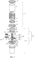
- FIG. 1 An exploded view of an assembled biological fluid filtration assembly according to the present invention is shown in Figure 1 .
- the assembled device and use thereof is shown in Figure 3
- Figure 2 shows the collection chamber (left) and a filtration unit assembled from the filter support platform and waste reservoir (right) prior to their coupling to afford the assembled device.
- the collection chamber 1 is open-topped for convenience of sample provision.
- the collection chamber is formed of a cylindrical bag 3 of water-impermeable material, which is approximately 100 mm in length and 95 mm in diameter and is suitable for housing a volume of approximately 500 mL for convenience of sample provision and maximal DNA when analysing urine samples.
- the collection chamber is suitable for housing up to 400 mL, 300 mL, 250 mL, 100 mL, 50 mL, or up to 20 mL. While larger volumes may be appropriate for urine collection, smaller volumes may be preferable for the filtration of, for example, saliva.
- the cylindrical bag is contained within a spring 5 which imparts some rigidity to the cylindrical bag of the collection chamber.
- a lid 7 At the sealed end of the cylindrical bag is a lid 7 and at the open end of the cylindrical bag is an annular spring attachment portion 9 which encircles the open end of the cylindrical bag without substantially occluding the open portion.
- the spring 5 is connected to or abuts the lid 7 at one end and the annular attachment portion 9 at the other end.
- the lid 7 and the annular spring attachment portion 9 are rigid and made of plastics material, although other suitable rigid materials, for example, a metal such as stainless steel, may be used.
- the lid 7 is circular and imperforate, and of a diameter slightly larger than the diameter of the cylindrical bag.
- the collection chamber has a locking ring attachment 11 to which annular spring attachment portion 9 can be fixed by means of a snap fit interaction.
- Other fixing means may be used, including complementary screw threads and rotatably engaging lugs.
- the collection chamber is connectable via the locking ring attachment 11 to the filtration unit 13 to assemble the complete filtration device.
- This connection is necessarily substantially watertight to permit use of the device as described herein without loss of fluid before filtration, with O-ring 14 which is retained in an annular groove around the top of the filtration unit improving the seal.
- the filtration unit 13 has a filter support platform 15. Small protrusions on the filter support platform 15 are located to engage with complementary indents in the annular attachment portion 9. It will be appreciated that other attachment means may alternatively be provided.
- the filter support platform 15 has a removable filter cartridge 17 with a membrane filter 19, and is connectable to a waste reservoir 21. It will be appreciated that other filter materials as described herein may also be used.
- the waste reservoir 21 is a rigid cylindrical container made of plastics material able to accommodate a volume of at least 500 mL (that is, the entire volume of liquid contained in the collection chamber prior to filtration). Other suitable rigid materials suitable for receiving fluids may be used in place of plastics material.
- the waste reservoir 21 and the filter support platform 15 are connectable to form, in combination with the filter cartridge 17, the filtration unit 13. This connection is necessarily substantially watertight to permit use of the device as described herein without loss of fluid during filtration.
- the waste reservoir 21 and the filter support platform 15 are connectable by a snap fit connection between a protrusion on the outside of waste reservoir 21 and an annular groove on the inside of the filter support platform 15.
- the waste reservoir 21 contains a moisture absorbing material and/or a deodorant.
- Suitable moisture absorbing materials may include absorbent material such as paper, cotton wool or sponge, or silica gel and/or other water-absorbent polymers known in the art. The inclusion of a moisture absorbent material improves ease of disposal of the waste reservoir after use.
- Suitable deodorants may include carbonates such as potassium carbonate.
- the exploded view shown in Figure 1 shows the component parts of the filter support platform 15.
- the filter support platform is connectable to both the connection chamber at its open end and to the waste reservoir, and when the device is fully-assembled separates the two.
- the filter support platform has an opening 23 to allow fluid communication between the collection chamber and the reservoir and the filter 19 of the filter support portion, in this case, filter cartridge 17, occludes this opening such that any fluid passing from the collection chamber to the waste reservoir passes through the filter.
- the filter support platform has a slotted recess 25 suitable for receiving a filter cartridge 17 such that the filter of the filter cartridge occludes the opening as described. The filter cartridge may be inserted and removed from the slotted recess in a sliding movement.
- the filter support platform is assembled from a top portion 27 and a bottom portion 29, which clip together by means of a snap-fit connection between protrusions on the top portion and complementary recesses on the bottom portion.
- Other connecting means may be envisaged including other snap-fit interactions and complementary screw threads.
- the embodiment shown in Figure 1 has two handles, 31 and 32 to facilitate ease of use. It will be understood that handles are not necessary, and that other handle arrangements, for example, a single handle, a continuous annular handle, or one or more D-shaped handles may be used.
- O-ring 33 is provided to prevent leakage during use.
- the filter support platform further comprises a back flow membrane 35 and a pressure relief valve 37.
- the pressure relief valve is configured to activate at a certain pressure to allow liquid to pass into the waste chamber should the filter becomes saturated.
- the backflow membrane 35 is adapted to allow air to pass from the reservoir 21 into the collection chamber 1 during intermittent application of pressure to prevent the filter content becoming disturbed due to turbulence.
- the relief valve is an umbrella-type valve that opens at 10-12 kg pressure, but other suitable valves may be used.
- the filter cartridge 17 has a body of a width complementary to the width of the slot, and is sufficiently longer in length to cause a portion of the body to protrude from the slotted recess during use (as shown in Figure 2 ) to facilitate ease of removal of the cartridge from the device.
- the filter cartridge housing may have one or more indentations or perforations 41 to improve grip and aid removal.
- the filter 19 is housed on a ledge within an opening in the filter cartridge housing and maintained in place by a perforated over support 43 which is connected to the housing by means of a snap-fit connection between protrusions on the perforated over support and complementary recesses in the housing.
- Other connecting means may be envisaged including other snap-fit interactions and complementary screw threads.
- O-rings 45, 47, and 48 improve the seal.
- O-ring 47 improves the seal of the assembled filter cartridge 17 around the filter 19
- O-rings 45 and 48 are present on the external surface of the filter cartridge 17 and serve both to improve the seal when the filter cartridge 17 is housed within the filter support platform for filtration of the biological fluid and to improve the seal when the filter cartridge 17 is inserted into a storage unit 49 according to the present invention.
- Figure 1 further shows such a storage unit 49 according to the present invention.
- the filter cartridge 17 may be inserted into the storage unit 49 after use to facilitate ease of storage and transportation and may preserve the sample during storage.
- the storage unit 49 further provides a means for ease of access to the filter content (and any surrounding liquid) for analysis without the need to remove the cartridge from the storage unit.
- the storage unit comprises a base 51 having a recess 53 suitable for receiving the filter cartridge. This base has an opening 55 located to permit access to the filter content for analysis and processing when the filter cartridge is inserted. The opening is covered by a lid 57 to preserve the sample and to permit storage and transportation.
- the lid connects to the base by means of complementary screw threads, although other connection means may be envisaged including a suitable snap-fit interaction or hinged lid.
- the lid 57 comprises a chamber containing an appropriate liquid that is released during engagement of the lid with the base 51.
- the lid may be an OG-250 lid from Oragene®, developed by DNA Genotek® and containing a DNA lysis buffer.
- Base 51 has sharp protrusions which break a seal of the chamber in the lid when the lid is screwed onto the base, thereby releasing the solution. Removing the lid for analysis thereby permits access not only to the filter content but also to the contained solution in which the filter content is stored.
- the filter cartridge 17 and storage unit 49 form a water tight seal around the filter, the filter content, and any surrounding liquid that may be present.
- FIG. 4 shows an alternative storage unit according to the present invention, and the assembly thereof.
- the storage unit 490 comprises a housing 510 having a recess 530 suitable for receiving the filter cartridge and a first opening 550 to permit access to the filter content when a filter cartridge is inserted.
- the lid 570 engages with the housing by a sliding cooperation between protrusions on the housing and complementary recesses on the lid, and is retained in place by abutment against a stop plate and by a retaining clip.
- the storage unit also has a bottom 571 that engages with the housing in a manner analogous to that of the lid.
- a piston 600 is retained within the recess at a point beyond the first opening and defines a chamber 602 at the end of the recess distal from the recess opening.
- the housing has a second opening 603 into this chamber.
- the chamber 602 is suitable for receiving a fluid, for example, and not by way of limitation, a buffer for lysis of cells and preservation of nucleic acids and/or proteins, a fixative/preservative to prepare cells with the retention of the characteristic morphology (for cytological examination), a culture medium to sustain cell growth or an isotonic buffer suitable for the storage of biological material, or an appropriate buffer for the elution of the biological material from the filter.
- the storage unit is provided with a suitable fluid of this type contained within the chamber. It will be appreciated that the fluid may be selected in accordance with the nature of the sample to be stored and the subsequent analysis required.
- the piston 600 is retained within the recess but application of pressure, for example, by insertion of a filter cartridge, is able to push the piston further into the recess, reducing the size of the chamber and forcing the fluid therein into the remainder of the recess, and into contact with the filter and filter content.
- the filter cartridge 17 and storage unit 490 form a water tight seal around the filter, the filter content, and any surrounding liquid that may be present. It will be appreciated that varying the filter cartridge and storage unit dimensions and the provision and location of suitable O-rings in order to achieve said water-tight seal will be apparent to the skilled person.
- Assemblies of the invention may be provided as a kit directly to the user, who can then:
- assemblies according to the present invention may be used in the home, with samples stored, optionally in a solution selected to facilitate storage and/or analysis of the captured material, and transported to a relevant care giver or to an appropriate medical centre or testing facility.
- Use of a storage unit according to the present invention permits samples captured on filters according to methods of the present invention to be sent hygienically and efficiently using, for example, regular national mail services.
- Patients thought to be at risk of developing, for example, bladder cancers, or those patients in remission from bladder cancer at present often have to undergo regular cystoscopy investigations, necessitating frequent hospital visits which may be inconvenient. Cystoscopy investigations are often uncomfortable and may carry a risk of complications. They are also expensive for the healthcare provider.
- the provision of a suitable device or kit for obtaining and processing samples at home which may be analysed without requiring the participation of the patient represents a significant improvement to patient wellbeing.
- a kit comprising a collection chamber as described herein and a filtration unit as described herein, and, optionally, instructions for using the assembly in a method as described herein.
- the filtration unit is provided fully assembled.
- a sample is then provided, for example, through normal urination, into the collection chamber.
- the filtration unit is then fastened to the collection chamber.
- the user then flips the assembled device so that the collection chamber is now upside down at the top of the device, as shown in Figure 3 , and then provides manual pressure to force the liquid through the filter into the waste reservoir.
- the provision of one or more valves and/or backflow membranes allows pressure to equalise within the device.
- kits of the invention may further comprise a storage unit as described herein. The user then inserts the filter cartridge into the storage unit as shown in, for example, Figure 4 for convenient storage and transport.
- the storage unit is provided as a lid and a base (denoted 57 and 51, respectively, in Figure 1 ).
- the filter cartridge is first inserted into the base 51.
- Lid 57 is then added, with the engagement of the lid with the base causing the seal to a chamber containing solution within the lid to break, thereby releasing the solution into contact with the filter content.
- the storage unit is provided as a single unit (denoted 490 in Figure 4 ).
- This storage unit comprises a piston retained within the recess at a point beyond the first opening and defining a chamber at the end of the recess distal from the recess opening.
- the base of the storage unit has a second opening into this chamber.
- the chamber contains a solution, for example, and not by way of limitation, a buffer for lysis of cells and preservation of nucleic acids, a fixative/preservative to prepare cells with the retention of the characteristic morphology (for cytological examination), or a culture medium to sustain cell growth. Inserting the filter cartridge into the recess of the storage unit pushes the piston further into the recess, reducing the size of the chamber and forcing the liquid therein around the piston and into contact with the filter content where it may be retained during storage and transport.
- the combined filter cartridge and storage unit may then be conveniently and hygienically transported to a testing/ analysing facility or appropriate medical centre. Access to the filter content is facilitated by removal of the lid (denoted 57 or 570 in Figure 1 or Figure 4 , respectively) to reveal the relevant opening in the storage unit housing.
- Filter content for example, DNA, may be analysed using methods known in the art and methods described herein, with the presence or absence of certain known markers used to provide a diagnosis.
- the assembly may be provided as a kit comprising a waste reservoir and filter support base that have not yet been fastened together.
- the user must first assemble the filtration unit.
- filter cartridges and storage units optionally comprising a solution housed within a chamber as described herein, may be provided separately to the remainder of the assembly as these may be selected specifically with regard to the intended application.
- the present invention is based on the inventors' insight that devices comprising certain suitable filters may be utilised for capturing material from biological fluids for efficient analysis, for use in the diagnosis and monitoring of relevant conditions and diseases.
- the assemblies and methods of the present invention may be used to capture cells from biological fluids.
- Previous studies have shown that it is possible to capture and separate cells from fluids using mechanical filtering (Wilding et al., 1998; Mohamed et al., 2004; Zheng et al., 2007; Lin et al., 2010).
- none of these methods provides the convenience and efficacy associated with the assemblies and methods of the present invention, that is, the provision of an assembly for the inexpensive and easy collection and processing of a sample which may be used by the patient or another caregiver to provide a sample of captured cells suitable for storing and sending through the post to a testing facility or appropriate medical centre.
- any filter material having the necessary character to capture material of interest may be used in assemblies and methods of the present invention. It will be appreciated that assemblies and methods of the present invention may be used for the capturing of different types of biological material from various biological fluids, for the detection, diagnosis and monitoring of a variety of diseases and conditions. Accordingly, it will be appreciated that the filter may be selected from filter media known in the art to have certain desirable characteristics, and in some cases it may be desirable to provide multiple filters in series. Where multiple filters are used, each filter may be identical to, or have different characteristics to, any other filter in the assembly.
- the capturing of cells of different sizes and different types may be achieved by use of a filter, or use of multiple filters, configured to exclude certain sizes or forms of cells, most likely by selection of filter pore size and or/ pore arrangement.
- Filters may also be used that are made from materials, or have coatings, designed specifically to capture certain materials, for example, macromolecules such as proteins, DNA, RNA and metabolites.
- filter characteristics that may be suitable for use in some embodiments of the present invention are provided by way of illustration and are not intended to limit the invention to any particular filter type. These and other suitable filters are known in the art, and may be commercially available.
- a pore size of about 0.5 ⁇ m to 4 ⁇ m may be preferred.
- a pore size of about 20 nm to 300 nm, more preferably of about 20 nm to about 50 nm may be used.
- a pore size of about 4 ⁇ m to 10 ⁇ m may be preferred.
- a pore size of about 7 ⁇ m to 12 ⁇ m may be preferred.
- a pore size of about 8 ⁇ m to 20 ⁇ m may be preferred, with about 8 ⁇ m to 12 ⁇ m being especially preferred, about 8 ⁇ m most preferred.
- ultrafiltration membrane filters with a specific molecular weight cut off limit (for example, but not by way of limitation, 50 kDa) selected to capture the macromolecules of interest may be used.
- a capture agent such as an antibody specific to a protein of interest or nucleic acid with a sequence that is complementary to that of interest, may be adhered to filter media (for example, but not by way of limitation, membrane filters, such as those made of nylon, Polyvinylidene difluoride or nitrocellulose, or chromatographic media such as sepharose or magnetic beads) allowing the macromolecule of interest to be captured during filtration.
- the filter may be made of a suitable polymer material such as polycarbonate, nylon, or parylene, or a suitable non-polymer material such as silicone, as appropriate.
- membrane filters may be preferred, for example, in the capturing of cells from, for example, urine.
- the membrane filter may be a polycarbonate membrane, preferably a polycarbonate hydrophilic membrane, for example, a track-etched polycarbonate hydrophilic membrane.
- the filter may have a pore size of about 5-10 ⁇ m, preferably about 8 ⁇ m.
- Preferred membrane filters may include micromembrane filters such as commercially available polycarbonate filters, for example, Whatman Nuclepore track-etched polycarbonate hydrophilic filters, (diameter 25 mm, pore size 8 ⁇ m).
- the storage unit contains a solution selected to facilitate storage and/or analysis of the biological material.
- the solution may be, for example, a buffer suitable for inducing cell lysis to permit analysis of nucleic acids or proteins released from the cell, a fixative/preservative to prepare cells with the retention of the characteristic morphology, a culture medium to sustain cell growth, an isotonic buffer suitable for storage of biological material, for example, phosphate buffered saline solution, or an appropriate buffer for the elution of the biological material from the filter.
- the solution will preferably be selected to correspond to the biological material to be captured and the analysis to be performed.
- assemblies of the invention could be used for the collection of exfoliated tumour cells from urine with the aim of analysing alterations in their DNA.
- this may be using a polycarbonate membrane filter with a pore size of 8 ⁇ m to capture the tumour cells, then inserting the filter cartridge into the storage unit, and, optionally, releasing a cell-lysis and nucleic acid-preserving solution such as those commercially available from Qiagen or DNA Genotek [for example, as described in WO2003104251 A9 ] onto the filter content.
- the solution released onto the filter content may, for example, be a cell-lysis and protein-preserving solution such as RIPA buffer (commercially available from Millipore) or cell extraction buffer (commercially available from Invitrogen).
- RIPA buffer commercially available from Millipore
- cell extraction buffer commercially available from Invitrogen
- the solution released onto the filter content may, for example, be a preservative buffer, for example one commercially available from Hologic (PreservCyt Solution, containing methanol) or a cellular growth medium, for example DMEM supplemented with 10% FBS, 1% L-glutamine, 100 U/ml penicillin and 100 ⁇ g/ml streptomycin.
- a preservative buffer for example one commercially available from Hologic (PreservCyt Solution, containing methanol) or a cellular growth medium, for example DMEM supplemented with 10% FBS, 1% L-glutamine, 100 U/ml penicillin and 100 ⁇ g/ml streptomycin.
- assembles of the invention may be used for the collection of a particular cell-free protein from urine, for example, by using filter composed of Protein A/G coated sepharose beads to which an antibody which binds to the protein of interest has been attached, the filter cartridge then being placed into the storage unit and, optionally, an isotonic buffer such as phosphate buffered saline being released onto the filter content.
- Assemblies and methods for the collection of biological material from biological fluids, and the subsequent storage and optional processing of said biological material, as described herein, are of particular relevance for the detection, diagnosis and monitoring of diseases and conditions.
- kits and methods as described herein may find utility in the detection, diagnosis and monitoring of a variety of diseases and conditions. For example, detection of hypermethylation of genes such as GSTP1, VHL, APC RASSF1A, Timp-3 in tumour cells from urine sediments is found in prostate and renal cancers ( Cairns et al Nature Reviews Cancer 2007; 7:531-543 ).
- RNA isolated from urine sediments has been analysed for diagnosis of acute rejection in kidney transplants, offering potential for the replacement of renal biopsies ( Suthanthiran et al N. Engl. J. Med. 2013; 369:20-31 ).
- Assemblies and methods of the invention may be used to capture free macromolecules (e.g. proteins, DNA, RNA or metabolites) in urine or other fluids.
- free macromolecules e.g. proteins, DNA, RNA or metabolites
- ovarian cancer patients have been shown to have altered levels of Glycosylated eosinophil-derived neurotoxin, COOH-terminal osteopontin fragments and the ⁇ -subunit core fragment of human chorionic gonadotrophin, SMRP and Bcl-2 in their urine( Das and Bast Biomark Med. 2008; 2(3): 291-303 ).
- Detection of the S100A6 and S100A9 proteins in urine may have utility in the detection of upper GI tract cancers ( Husi et al Proteomics Clin Appl. 2011; 5(5-6):289-99 ), whilst detection of the SAA4 and ProEGF proteins in urine may have utility in the detection of bladder cancer ( Chen et al Journal of Prote
- the assemblies and methods described herein may also be used for the collection and filtration of other biological fluids, such as saliva, sputum and blood, and bodily fluids obtained using more invasive methods such as, for example, pleural effusions, lavage fluid (for example ductal,, bronchoalveolar) and sera for the analysis of captured material including via detection of genomic alterations associated with certain diseases and disorders including cancers such as lung and breast cancer ( Belinsky et al Proc. Natl. Acad. Sci. USA, 95: 11891-11896, 1998 ; Ahrendt et al J. Natl. Cancer Inst., 91: 332-339, 1999 ; Evron et al Lancet, 357:1335-1336, 2001 ).
- other biological fluids such as saliva, sputum and blood, and bodily fluids obtained using more invasive methods such as, for example, pleural effusions, lavage fluid (for example ductal,, bronchoalveolar) and sera for the
- CTCs circulating tumour cells
- Isolation and characterization of CTCs is a technical challenge as they make up only a small fraction of the total cells present in the blood.
- CTCs reflect molecular features of cells within the tumour mass, they offer a potential way to diagnose or monitor progression/response of a patient in a relatively non-invasive way.
- CTCs have been identified in cancers such as in breast, prostate, lung, ovarian and colon cancer patients, where they have been shown to provide predictive and prognostic information.
- CTCs have also been identified in pancreatic patients, although no pivotal study using CTCs to guide clinical treatment has been undertaken ( Cen et al Biochimica et Biophysica Acta 2012; 1826:350-356 ).
- the capture of circulating tumour cells from blood of patients with prostate, colorectal and breast cancer has been shown to be possible using a filtration method to take advantage of the increased size of tumour cells as compared to normal cells.
- assemblies and methods described herein may also be applied to the capture and analysis of circulating cell-free DNA (cf-DNA).
- markers known to be associated with a particular disease or condition may be genetic markers, genomic alterations, the presence of or elevated/decreased levels of proteins (for example, antibodies), the presence of or elevated/decreased levels of bacteria or yeast, both as described herein and as documented in the art.
- the marker may be a marker known to be associated with cancer.
- the cancer may be urinary, or gynecological cancer, for example, bladder cancer, prostate cancer, renal cancer, urethral cancer, ureteral cancer, urothelial cancer, urachal cancer, endometrial cancer, or ovarian cancer.
- the cancer may be a cancer associated with other organs, for example, liver cancer, melanoma, colorectal cancer, head and neck cancer, lung cancer, breast cancer, pancreatic cancer, or a cancer of the upper GI tract.
- the cancer may be a metastatic cancer. Markers associated with these and other cancers are known in the art.
- the marker is associated with a genitourinary cancer, preferably, bladder, prostate, or renal cancer.
- the marker is associated with bladder cancer, more preferably non-muscle invasive bladder cancer.
- the marker is associated with a condition other than cancer.
- the marker may be associated with acute rejection in kidney transplants, which has the advantage of potentially obviating the need for invasive renal biopsies, or markers associated with bacterial and/or yeast infections, for example urinary tract infections such as cystitis and pyelonephritis.
- the marker may not in itself be associated with a disease or condition but may instead be a genetic marker associated with an individual or particular parentage, for example, for use in forensic and paternity testing.
- Assemblies and methods described herein may be used in conjunction with UCyt+® and Urovysion® kits.
- a problem with these systems is the transportation of urine as well as the low number of cells and the low fraction of tumor cells in these samples. Proper preservation, cell isolation and increasing the fraction of tumor cells as provided by the assemblies and methods described herein may improve the use.
- Galvan et al (Cancer Cytopathol 2011; 119:395-403 ) noted that around 10% of samples could not be analysed due to too few urothelial cells in the sample or other technical reasons.
- a key application of the invention is the diagnosis and surveillance of bladder cancer.
- the present invention was developed to provide a simple means for capturing bladder tumour cells from urine and storing/preserving DNA from these cells for later analysis. Important advantages include:
- the filter cartridge was transferred to the storage cassette, which was then mounted with the lid from an Oragene DNA Self-Collection Kit (disk format OG-250, DNA Genotek, Ottowa, Ontario, Canada).
- Oragene DNA Self-Collection Kit disk format OG-250, DNA Genotek, Ottowa, Ontario, Canada.
- Bisulfite conversion was done using the EZ DNA Methylation-Gold Kit (Zymo Research) according to the manufacturer's protocol.
- the bisulfite-treated DNA was eluted in 20 ⁇ l of M-Elution Buffer and stored at -80°C.
- paired samples sediment and filter sample
- the same amount of DNA was used, with a maximum of 500 ng.
- the maximum sample volume (20 ⁇ l) was used for bisulfite treatment.
- IVM In vitro methylated DNA
- CpGenomeTM Universal Methylated DNA Chemicon/Millipore, Billerica, MA
- whole-genome amplified DNA served as positive and negative controls for methylation, respectively.
- Methylation levels were calculated as percent methylated reference (PMR; Ref. [ Weisenberger DJ, Campan M, Long TI, Kim M, Woods C et al. (2005). Nucleic Acids Res 33: 6823-6836 ]) by normalizing marker-specific reaction values to ALUC4 values relative to the same values for fully methylated control (IVM). Samples with a concentration below the equivalent of 0.25 ng/ ⁇ l non bisulfite-treated DNA were excluded.
- Cut-off PMR values for HOXA9, POU4F2, SALL3 and VIM2 were 3, 2, 0.5 and 2, respectively.
- BCL2, CCNA1 and EOMES showed no background methylation in DNA isolated from urine filter and sediment samples from healthy controls.
- Methylation analysis was performed using MethyLight, a quantitative fluorescence-based, real-time PCR assay ( Eads et al., 2000, Nucleic Acids Res. 28, E32 ). Primers and probes were designed for 7 gene promoter CpG islands and for ALUC4, which was used to control for the amount of input DNA ( Weisenberger et al., 2005, Nucleic Acids Res. 33, 6823-6836 ). Bisulfite-converted, in vitro-methylated DNA (IVM; CpGenomeTM Universal Methylated DNA, Chemicon) was analyzed to normalize for any amplification bias between a target gene and ALUC4. Reactions were performed on the Roche LightCycler® 480 Real-time PCR system using the Lightcycler® 480 Probes Master Kit (Roche).
- the human ureter transitional cell carcinoma cell line 639V was purchased from DSMZ (Braunschweig, Germany). Cells were maintained in DMEM medium supplemented with 10% fetal bovine serum at 37°C in a humidified incubator with 5% CO 2 . Lymphocytes from a healthy donor were prepared from peripheral blood according to a previously described protocol [ Thurner B, Roder C, Dieckmann D, Heuer M, Kruse M et al. (1999) J Immunol Methods 223: 1-15 ] and stored at -80°C until use. Cells in suspension were counted and their diameter was measured using a Countess Automated Cell Counter (Invitrogen, Carlsbad, CA, USA). Lymphocytes and 639V cells were mixed in different ratios in 100 ml of PBS and processed using the filtration device.
- FGFR3 mutations R248C, S249C, G370C and Y373C
- ddPCR droplet digital PCR
- the PCR mixture contained 11 ⁇ l of ddPCR droplet supermix for probes (no dUTPs), 1.1 ⁇ l of mutation primer/probe mix (FAM), 1.1 ⁇ l of wildtype primer/probe mix (HEX) and 2 ⁇ l of DNA in a final volume of 22 ⁇ L.
- the inventors first used cultured cells to test 1) if it was possible to capture cells on a commercial micromembrane filter and 2) if low-abundant bladder tumour cells could be enriched.
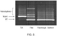
- Purified, cultured human lymphocytes diluted in PBS were spiked with 0.5% bladder cancer cells (the human cell line T24).
- Half of the volume of the cell mixture was sedimented by centrifugation, and the remaining half was passed through a filter.
- the flowthrough from the filter was also collected and sedimented by centrifugation.
- DNA was isolated from the unfiltered, filter and flowthrough samples and analysed for the HRAS G12V mutation previously established to be present in the cell line T24.
- PCR in combination with denaturing gradient gel electrophoresis (DGGE) was used to resolve mutant and wildtype HRAS.
- the filtered sample was clearly positive for the HRAS G12V mutation, whereas the unfiltered and flowthrough samples were negative (DGGE has a detection level at around 2-3% mutated allele on a wild-type background).
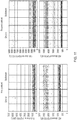
- Pathology Sediment Filter Low grade Ta/dysplasia 74/98 (75%) 82/98 (84%) High grade Ta 24/31 (77%) 25/31 (81%) T1 27/30 (90%) 28/30 (93%) >T2 17/19 (89%) 18/19 (95%) CIS 24/26 (92%) 25/26 (96%) Total 166/204 (81%) 178/204 (87%)
- NMIBC carcinoma in situ
- CIS carcinoma in situ
- T1 tumours two T1 tumours.
- CIS carcinoma in situ
- T1 tumours two T1 tumours.
- three were positive (two in both filter and sediment; one in filter only).
- One of these had been misclassified and had a bladder tumour.
- the second had prior problems with the bladder, and subsequent cystoscopy showed the presence of a hyperplastic lesion.
- the third was negative on cystoscopy.
- the present inventors have shown that using micromembrane filters (for examples, commercial polycarbonate membrane filters), it is possible to capture cells from urine samples and isolate DNA for subsequent methylation analysis. Accordingly, in some embodiments, the present invention relates to a method of passing a biological fluid sample, such as a urine sample, through a micromembrane filter.
- a biological fluid sample such as a urine sample
- the filter sample was positive for tumour-specific DNA methylation markers.
- the corresponding urine sediments were positive in 81% of the cases.
- Morning urine samples were collected from 30 patients admitted for bladder cystoscopy at Herlev Hospital. The samples were processed within 3-6 hours at the Danish Cancer Research Center. The sample volume varied between 150 and 400 ml, average 240 ml (Table 2). The filtration devices were mounted with an 8 ⁇ m pore size, track-etched polycarbonate filter (Whatman). After filtration, the filters were removed from the filtration device and stored at -80 °C until further processing.
- DNA was isolated from the filters as described in above. DNA was eluted in 50 ⁇ l of AE buffer and stored at -80°C. Bisulfite conversion of DNA was performed as described above. The DNA concentration was determined by quantitative PCR analysis of GAPDH. The methylation status of seven methylation markers (CCNA1, BCL2, EOMES, POU4F2, SALL3, HOXA9 and VIM2 ) was determined using MethyLight assays, as described above. The average DNA yield for the 30 urine samples was 242 ng (range 6 to 1,000 ng; Table 2 ). Table 2. DNA yield from 30 urine samples, processed using the urine filtration device. The DNA concentration was determined by qPCR (* estimated figure, measure out of range).
- the inventors used 639V bladder cancer cells, which have a point mutation (p.R248C; c.742C>T) in the gene encoding fibroblast growth factor receptor 3 ( FGFR3 ) with loss of the corresponding wildtype allele.
- p.R248C point mutation
- c.742C>T point mutation in the gene encoding fibroblast growth factor receptor 3
- FGFR3 fibroblast growth factor receptor 3
- the 30% loss of input material may at least in part be ascribed to an expected loss of DNA during extraction.
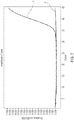
- concentrations of cells there was a decrease in recovery rate, down to ⁇ 5% at 5 ⁇ 10 6 cells/100 ml. This lower recovery was expected as saturation of the filter will cause release of the pressure valve and a direct flow of the remaining fluid and its cellular content into the waste reservoir.
- This initial testing suggested that the filtration device can be used to effectively capture bladder cancer cells from a fluid, and that the recovery rate is particularly high at low concentrations of cells where the capacity of the filter has not yet been reached.
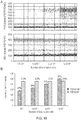
- the inventors spiked between 10 3 and 5 ⁇ 10 5 639V bladder cancer cells into 100 ml of PBS containing 10 7 normal purified cultured human lymphocytes (diameter 7-8 ⁇ m) and processed the suspension using the filtration device.
- Analysis of DNA extracted from filters by ddPCR showed signals for both mutant (R248C) and wildtype FGFR3 ( Figure 10A ). Vertical lines represent manually set cutoff settings. DNA was extracted from the filters and tested for mutant FGFR3 (R248C) molecules using ddPCR. DNA from normal peripheral blood lymphocytes (PBL) was used as a control for wildtype FGFR.
- the device is capable of isolating low abundant tumour cells, and therefore may therefore be useful for diagnosing smaller less aggressive tumours earlier.
- the size and stage of the tumour is normally reflected by the number of cells expected in a urine sample. The smaller less aggressive tumours would not shed as many cells into the urine as a more established tumour and therefore could potentially be missed on standard diagnostic techniques.
- DNA can be isolated from tumour cells spiked into PBS containing normal peripheral blood lymphocytes, showing that the device can isolate tumour cells from normal blood cells.
- Cells shed into the urine provide a convenient source for noninvasive detection of bladder cancer. Collection of cells and downstream testing by cytology or analysis of tumor-specific markers may offer an alternative or adjunct to cystoscopy in bladder cancer diagnosis and surveillance.
- Collection of cells and downstream testing by cytology or analysis of tumor-specific markers may offer an alternative or adjunct to cystoscopy in bladder cancer diagnosis and surveillance.
- the practical use of urine-based tests is often limited by inconvenience of sample handling, difficulties in analyzing large sample volumes, the need for rapid sample processing to avoid degradation of the cellular content, and insufficient analytical sensitivity due to a low ratio of tumor-to-normal cells.
- Described herein is a filtration device, designed for home or point-of-care use, which enables collection, enrichment and immediate preservation or treatment of tumor cells from urine.
- the use of this device in combination with droplet digital PCR for DNA-biomarker quantification provided efficient recovery of bladder cancer cells with elimination of >99% of excess lymphocytes.
- the performance of the device was further evaluated by DNA-based analysis of cells collected from urine from patients with bladder cancer, including some with low-grade Ta tumors. The ratio of tumor-to-normal DNA was higher in filtered samples compared with the same samples processed by sedimentation and showed high sensitivity.
- the ability to easily collect, process and ship diagnostic cells from urine may broaden the use of noninvasive tests for detection and follow-up of bladder cancer.
Landscapes
- Health & Medical Sciences (AREA)
- Chemical & Material Sciences (AREA)
- Life Sciences & Earth Sciences (AREA)
- Engineering & Computer Science (AREA)
- General Health & Medical Sciences (AREA)
- Analytical Chemistry (AREA)
- Organic Chemistry (AREA)
- Pathology (AREA)
- Biomedical Technology (AREA)
- Molecular Biology (AREA)
- Wood Science & Technology (AREA)
- Genetics & Genomics (AREA)
- Zoology (AREA)
- Proteomics, Peptides & Aminoacids (AREA)
- Physics & Mathematics (AREA)
- Immunology (AREA)
- Biochemistry (AREA)
- Bioinformatics & Cheminformatics (AREA)
- General Engineering & Computer Science (AREA)
- Biotechnology (AREA)
- Chemical Kinetics & Catalysis (AREA)
- Hematology (AREA)
- Public Health (AREA)
- Animal Behavior & Ethology (AREA)
- Heart & Thoracic Surgery (AREA)
- Veterinary Medicine (AREA)
- Surgery (AREA)
- Medical Informatics (AREA)
- Biophysics (AREA)
- Microbiology (AREA)
- General Physics & Mathematics (AREA)
- Clinical Laboratory Science (AREA)
- Plant Pathology (AREA)
- Hospice & Palliative Care (AREA)
- Oncology (AREA)
- Crystallography & Structural Chemistry (AREA)
- Apparatus Associated With Microorganisms And Enzymes (AREA)
- Sampling And Sample Adjustment (AREA)
- Measuring Or Testing Involving Enzymes Or Micro-Organisms (AREA)
Priority Applications (1)
| Application Number | Priority Date | Filing Date | Title |
|---|---|---|---|
| PL14784509T PL3043718T3 (pl) | 2013-09-13 | 2014-09-12 | Zespół do filtracji płynów biologicznych |
Applications Claiming Priority (2)
| Application Number | Priority Date | Filing Date | Title |
|---|---|---|---|
| GB201316347A GB201316347D0 (en) | 2013-09-13 | 2013-09-13 | Biological fluid filtration assembly |
| PCT/GB2014/052776 WO2015036781A1 (en) | 2013-09-13 | 2014-09-12 | Biological fluid filtration assembly |
Publications (2)
| Publication Number | Publication Date |
|---|---|
| EP3043718A1 EP3043718A1 (en) | 2016-07-20 |
| EP3043718B1 true EP3043718B1 (en) | 2020-06-17 |
Family
ID=49552641
Family Applications (1)
| Application Number | Title | Priority Date | Filing Date |
|---|---|---|---|
| EP14784509.3A Active EP3043718B1 (en) | 2013-09-13 | 2014-09-12 | Biological fluid filtration assembly |
Country Status (11)
| Country | Link |
|---|---|
| US (1) | US20160223442A1 (enExample) |
| EP (1) | EP3043718B1 (enExample) |
| JP (1) | JP6552504B2 (enExample) |
| CN (1) | CN105722466B (enExample) |
| AU (1) | AU2014320077B2 (enExample) |
| CA (1) | CA2924086C (enExample) |
| DK (1) | DK3043718T3 (enExample) |
| ES (1) | ES2808664T3 (enExample) |
| GB (1) | GB201316347D0 (enExample) |
| PL (1) | PL3043718T3 (enExample) |
| WO (1) | WO2015036781A1 (enExample) |
Cited By (1)
| Publication number | Priority date | Publication date | Assignee | Title |
|---|---|---|---|---|
| US11612888B2 (en) | 2017-01-04 | 2023-03-28 | The Research Foundation For The State University Of New York | Biomarker detection device |
Families Citing this family (28)
| Publication number | Priority date | Publication date | Assignee | Title |
|---|---|---|---|---|
| US10533932B2 (en) | 2013-09-13 | 2020-01-14 | Cancer Research Technology Limited | Apparatus and methods for liquid separation and capture of biologics |
| ES2806498T3 (es) * | 2015-06-08 | 2021-02-17 | Arquer Diagnostics Ltd | Métodos para el análisis de una muestra de orina |
| WO2016205233A2 (en) | 2015-06-15 | 2016-12-22 | Cepheid | Integrated purification and measurement of dna methylation and co-measurement of mutations and/or mrna expression levels in an automated reaction cartridge |
| WO2017154349A1 (ja) * | 2016-03-10 | 2017-09-14 | パナソニックIpマネジメント株式会社 | 核酸抽出装置、核酸抽出ユニット及び核酸抽出方法 |
| US10585101B2 (en) * | 2016-03-10 | 2020-03-10 | Wavesense, Inc. | Prostatic liquid biopsy for the detection of prostate cancer and benign prostatic hyperplasia |
| JP7082453B2 (ja) * | 2016-07-21 | 2022-06-08 | エージェンシー フォー サイエンス,テクノロジー アンド リサーチ | 高体積分率粒子精密濾過のための外壁集束のための装置及びその製造方法 |
| JP7123050B2 (ja) * | 2016-12-12 | 2022-08-22 | セファイド | 自動反応カートリッジにおける統合された、DNAメチル化の精製及び測定並びに変異及び/又はmRNA発現レベルの同時測定 |
| WO2018226097A1 (en) * | 2017-06-08 | 2018-12-13 | Rijksuniversiteit Gronigen | Sensor cartridge for chemical assays of a liquid sample containing analyte moleculess |
| WO2019005827A1 (en) * | 2017-06-30 | 2019-01-03 | Boston Scientific Scimed, Inc. | FILTRATION DEVICE WITH REMOVABLE PROTECTION ELEMENT |
| WO2019043656A1 (en) * | 2017-09-01 | 2019-03-07 | Genus Plc | METHODS AND SYSTEMS FOR ASSESSING AND / OR QUANTIFYING POPULATIONS OF SPERMATOZOIDS WITH SEXUAL ASYMMETRY |
| KR20210006337A (ko) | 2018-03-15 | 2021-01-18 | 크립토스 바이오테크놀로지스, 인코포레이티드 | 열 보조 생화학 반응을 수행하기 위한 방법 및 시스템 |
| IT201800004625A1 (it) * | 2018-04-17 | 2019-10-17 | Dispositivo per la separazione di un campione biologico | |
| KR102117656B1 (ko) * | 2018-06-18 | 2020-06-02 | 한국원자력연구원 | 플란쳇 타입 샘플 제작 장치 |
| CN111467853B (zh) * | 2019-01-23 | 2022-08-02 | 杭州科百特过滤器材有限公司 | 一种用于固定过滤单元的夹具及其操作方法 |
| WO2020172712A1 (en) * | 2019-02-27 | 2020-09-03 | Epiaxis Therapeutics Pty Ltd | Methods and agents for assessing t-cell function and predicting response to therapy |
| CN111855333B (zh) * | 2019-04-24 | 2023-10-03 | 青岛言鼎生物医疗科技有限公司 | 体液中有核细胞吸印富集与吸印染色一体化反应装置及方法 |
| CN109991170A (zh) * | 2019-04-25 | 2019-07-09 | 泗洪县正心医疗技术有限公司 | 一种大样本尿沉渣滤出观察系统 |
| EP3733869A1 (en) * | 2019-05-02 | 2020-11-04 | QIAGEN GmbH | Automated method and system for split pool based barcoding of cellular molecules |
| CN110411787B (zh) * | 2019-06-28 | 2021-10-12 | 李美琴 | 一种用于检验科尿液样品的收集处理装置 |
| CN112362426B (zh) * | 2020-10-30 | 2023-07-21 | 南京华银医学检验所有限公司 | 一种病理检查淋巴结分离装置 |
| WO2023017251A1 (en) | 2021-08-09 | 2023-02-16 | Encelo Laboratories Limited | Primary cell extraction and preservation from fluids |
| WO2023247543A1 (en) * | 2022-06-20 | 2023-12-28 | Testmate Health Sa | Urine, saliva, and/or mouthwash sample preparation system |
| WO2023250074A1 (en) * | 2022-06-23 | 2023-12-28 | Predicine, Inc. | Devices and methods for sample collection |
| CN115855580B (zh) * | 2022-12-01 | 2024-04-19 | 嵩明珍茗食品有限公司 | 一种水体环境取样检测系统 |
| WO2024151767A2 (en) * | 2023-01-12 | 2024-07-18 | Health Science Funding, LLC | Solid tumor therapy |
| DE102023102184A1 (de) * | 2023-01-30 | 2024-08-01 | The smart period blood GmbH | Adapter zum Überführen und Aufbereiten einer Menstruationsausscheidungen enthaltenden Probe in ein verschließbares Probengefäß |
| CN115919375B (zh) * | 2023-03-15 | 2023-05-12 | 潍坊医学院附属医院 | 一种手术后用的唾液收集装置 |
| WO2025248141A1 (en) * | 2024-05-31 | 2025-12-04 | Stratifyer Molecular Pathology Gmbh | Method of detecting urinary tract cancer in a urine sample |
Family Cites Families (13)
| Publication number | Priority date | Publication date | Assignee | Title |
|---|---|---|---|---|
| US3788484A (en) * | 1971-08-23 | 1974-01-29 | Coulter Electronics | Inline fluid filter |
| BE878481A (fr) * | 1978-09-09 | 1979-12-17 | Bullock George P | Filtres demontables perfectionnes pour canalisations de liquides |
| US4829005A (en) * | 1984-06-15 | 1989-05-09 | Friedman Michael P | Sedimentation filtration microorganism growth culture system |
| US5429803A (en) * | 1991-04-18 | 1995-07-04 | Lamina, Inc. | Liquid specimen container and attachable testing modules |
| US5846487A (en) * | 1996-11-26 | 1998-12-08 | Bennett, Ii; Edward R. | Specimen cartridge |
| JP2002273113A (ja) * | 2001-03-15 | 2002-09-24 | Koganei Corp | 濾過器および薬液供給装置並びに薬液供給方法 |
| US7378054B2 (en) * | 2004-04-16 | 2008-05-27 | Savvipharm Inc | Specimen collecting, processing and analytical assembly |
| EP2024742B1 (en) * | 2006-05-22 | 2020-03-04 | 3M Innovative Properties Company | System and method for preparing samples |
| US20100297691A1 (en) * | 2007-05-31 | 2010-11-25 | Ribeiro Alice Maria M | Devices and processes for collecting and concentrating samples for microbiological analysis |
| FR2934049B1 (fr) * | 2008-07-16 | 2010-10-15 | Millipore Corp | Unite et procede de preparation d'un echantillon pour l'analyse microbiologique d'un liquide. |
| WO2010131140A1 (en) * | 2009-05-09 | 2010-11-18 | Diagcor Bioscience Incorporation Limited | Biological sample collection device |
| JP5800797B2 (ja) * | 2010-03-11 | 2015-10-28 | 株式会社カネカ | 細胞濃縮・回収方法及び細胞回収液 |
| US10166009B2 (en) * | 2012-11-20 | 2019-01-01 | The Trustees Of Columbia University In The City Of New York | Medical apparatus and method for collecting biological samples |
-
2013
- 2013-09-13 GB GB201316347A patent/GB201316347D0/en not_active Ceased
-
2014
- 2014-09-12 CN CN201480062490.6A patent/CN105722466B/zh active Active
- 2014-09-12 WO PCT/GB2014/052776 patent/WO2015036781A1/en not_active Ceased
- 2014-09-12 DK DK14784509.3T patent/DK3043718T3/da active
- 2014-09-12 JP JP2016542375A patent/JP6552504B2/ja active Active
- 2014-09-12 ES ES14784509T patent/ES2808664T3/es active Active
- 2014-09-12 CA CA2924086A patent/CA2924086C/en active Active
- 2014-09-12 US US15/021,470 patent/US20160223442A1/en not_active Abandoned
- 2014-09-12 AU AU2014320077A patent/AU2014320077B2/en active Active
- 2014-09-12 EP EP14784509.3A patent/EP3043718B1/en active Active
- 2014-09-12 PL PL14784509T patent/PL3043718T3/pl unknown
Non-Patent Citations (1)
| Title |
|---|
| None * |
Cited By (1)
| Publication number | Priority date | Publication date | Assignee | Title |
|---|---|---|---|---|
| US11612888B2 (en) | 2017-01-04 | 2023-03-28 | The Research Foundation For The State University Of New York | Biomarker detection device |
Also Published As
| Publication number | Publication date |
|---|---|
| GB201316347D0 (en) | 2013-10-30 |
| AU2014320077A1 (en) | 2016-04-21 |
| WO2015036781A1 (en) | 2015-03-19 |
| CN105722466A (zh) | 2016-06-29 |
| EP3043718A1 (en) | 2016-07-20 |
| PL3043718T3 (pl) | 2020-12-14 |
| DK3043718T3 (da) | 2020-08-10 |
| JP2016534363A (ja) | 2016-11-04 |
| CA2924086A1 (en) | 2015-03-19 |
| AU2014320077B2 (en) | 2019-01-17 |
| ES2808664T3 (es) | 2021-03-01 |
| JP6552504B2 (ja) | 2019-07-31 |
| US20160223442A1 (en) | 2016-08-04 |
| CN105722466B (zh) | 2019-03-19 |
| CA2924086C (en) | 2023-09-12 |
| NZ718485A (en) | 2020-10-30 |
Similar Documents
| Publication | Publication Date | Title |
|---|---|---|
| EP3043718B1 (en) | Biological fluid filtration assembly | |
| US10533932B2 (en) | Apparatus and methods for liquid separation and capture of biologics | |
| Khalid et al. | Pancreatic cyst fluid DNA analysis in evaluating pancreatic cysts: a report of the PANDA study | |
| US8597954B2 (en) | Stool sample processing method and stool sample processing container | |
| CN105018594B (zh) | 一种结直肠癌早期诊断标记物及相关试剂盒 | |
| JP2019501651A (ja) | 腫瘍分子の検出/診断試薬 | |
| JP4206425B2 (ja) | 大腸癌マーカー検出方法 | |
| EP1809770A2 (en) | Molecular analysis of cellular fluid and liquid cytology specimens for clinical diagnosis, characterization, and integration with microscopic pathology evaluation | |
| EP3800273A1 (en) | Use of detection reagent for detecting methylation of genes associated with colorectal cancer, and kit | |
| CN109072310A (zh) | 在尿液中检测癌症 | |
| Hayashi et al. | Paired box 5 methylation detection by droplet digital PCR for ultra-sensitive deep surgical margins analysis of head and neck squamous cell carcinoma | |
| JP2010008106A (ja) | 糞便処理容器及び糞便処理方法 | |
| Chang et al. | Urinary cell-free DNA as a potential tumor marker for bladder cancer | |
| JP5562554B2 (ja) | 胃粘膜洗浄液を利用した疾患関連マーカー検出法 | |
| US20080145932A1 (en) | Method and apparatus for cell recovery | |
| US7833757B2 (en) | Method for conducting non-invasive early detection of colon cancer and/or of colon cancer precursor cells | |
| CN106148501A (zh) | 一种检测Septin9基因甲基化的试剂盒及其应用 | |
| CN118995931B (zh) | LncRNA AC113143.1作为结肠癌诊断标志物的应用 | |
| NZ718485B2 (en) | Biological fluid filtration assembly | |
| CN118480423A (zh) | 一种用于泌尿系统肿瘤检测的尿液中肿瘤细胞分选纯化装置、应用及检测试剂盒 | |
| CN111690746A (zh) | 与肺癌相关的血小板rna标志物及其应用 | |
| WO2011004517A1 (ja) | 標的核酸の検出方法、及び大腸癌の検査方法 | |
| Petras et al. | KRAS detection in colonic tumors by DNA extraction from FTA paper: the molecular touch-prep | |
| CN116590412A (zh) | 用于检测宫颈腺癌的核酸分子、试剂盒及使用方法 | |
| Sakaguchi et al. | P1-197: Urinary N1, N12-Diacetylspermine (DiAcSpm) as a tumor marker for non-small cell lung cancer |
Legal Events
| Date | Code | Title | Description |
|---|---|---|---|
| PUAI | Public reference made under article 153(3) epc to a published international application that has entered the european phase |
Free format text: ORIGINAL CODE: 0009012 |
|
| 17P | Request for examination filed |
Effective date: 20160322 |
|
| AK | Designated contracting states |
Kind code of ref document: A1 Designated state(s): AL AT BE BG CH CY CZ DE DK EE ES FI FR GB GR HR HU IE IS IT LI LT LU LV MC MK MT NL NO PL PT RO RS SE SI SK SM TR |
|
| AX | Request for extension of the european patent |
Extension state: BA ME |
|
| DAX | Request for extension of the european patent (deleted) | ||
| STAA | Information on the status of an ep patent application or granted ep patent |
Free format text: STATUS: EXAMINATION IS IN PROGRESS |
|
| 17Q | First examination report despatched |
Effective date: 20180409 |
|
| GRAP | Despatch of communication of intention to grant a patent |
Free format text: ORIGINAL CODE: EPIDOSNIGR1 |
|
| STAA | Information on the status of an ep patent application or granted ep patent |
Free format text: STATUS: GRANT OF PATENT IS INTENDED |
|
| INTG | Intention to grant announced |
Effective date: 20200102 |
|
| GRAS | Grant fee paid |
Free format text: ORIGINAL CODE: EPIDOSNIGR3 |
|
| GRAA | (expected) grant |
Free format text: ORIGINAL CODE: 0009210 |
|
| STAA | Information on the status of an ep patent application or granted ep patent |
Free format text: STATUS: THE PATENT HAS BEEN GRANTED |
|
| AK | Designated contracting states |
Kind code of ref document: B1 Designated state(s): AL AT BE BG CH CY CZ DE DK EE ES FI FR GB GR HR HU IE IS IT LI LT LU LV MC MK MT NL NO PL PT RO RS SE SI SK SM TR |
|
| RAP1 | Party data changed (applicant data changed or rights of an application transferred) |
Owner name: CANCER RESEARCH TECHNOLOGY LIMITED |
|
| REG | Reference to a national code |
Ref country code: GB Ref legal event code: FG4D |
|
| REG | Reference to a national code |
Ref country code: CH Ref legal event code: EP |
|
| REG | Reference to a national code |
Ref country code: IE Ref legal event code: FG4D |
|
| REG | Reference to a national code |
Ref country code: DE Ref legal event code: R096 Ref document number: 602014066739 Country of ref document: DE |
|
| REG | Reference to a national code |
Ref country code: AT Ref legal event code: REF Ref document number: 1280386 Country of ref document: AT Kind code of ref document: T Effective date: 20200715 |
|
| REG | Reference to a national code |
Ref country code: DK Ref legal event code: T3 Effective date: 20200806 |
|
| REG | Reference to a national code |
Ref country code: NL Ref legal event code: FP |
|
| REG | Reference to a national code |
Ref country code: NO Ref legal event code: T2 Effective date: 20200617 |
|
| PG25 | Lapsed in a contracting state [announced via postgrant information from national office to epo] |
Ref country code: GR Free format text: LAPSE BECAUSE OF FAILURE TO SUBMIT A TRANSLATION OF THE DESCRIPTION OR TO PAY THE FEE WITHIN THE PRESCRIBED TIME-LIMIT Effective date: 20200918 Ref country code: FI Free format text: LAPSE BECAUSE OF FAILURE TO SUBMIT A TRANSLATION OF THE DESCRIPTION OR TO PAY THE FEE WITHIN THE PRESCRIBED TIME-LIMIT Effective date: 20200617 Ref country code: SE Free format text: LAPSE BECAUSE OF FAILURE TO SUBMIT A TRANSLATION OF THE DESCRIPTION OR TO PAY THE FEE WITHIN THE PRESCRIBED TIME-LIMIT Effective date: 20200617 Ref country code: LT Free format text: LAPSE BECAUSE OF FAILURE TO SUBMIT A TRANSLATION OF THE DESCRIPTION OR TO PAY THE FEE WITHIN THE PRESCRIBED TIME-LIMIT Effective date: 20200617 |
|
| REG | Reference to a national code |
Ref country code: LT Ref legal event code: MG4D |
|
| PG25 | Lapsed in a contracting state [announced via postgrant information from national office to epo] |
Ref country code: HR Free format text: LAPSE BECAUSE OF FAILURE TO SUBMIT A TRANSLATION OF THE DESCRIPTION OR TO PAY THE FEE WITHIN THE PRESCRIBED TIME-LIMIT Effective date: 20200617 Ref country code: RS Free format text: LAPSE BECAUSE OF FAILURE TO SUBMIT A TRANSLATION OF THE DESCRIPTION OR TO PAY THE FEE WITHIN THE PRESCRIBED TIME-LIMIT Effective date: 20200617 Ref country code: BG Free format text: LAPSE BECAUSE OF FAILURE TO SUBMIT A TRANSLATION OF THE DESCRIPTION OR TO PAY THE FEE WITHIN THE PRESCRIBED TIME-LIMIT Effective date: 20200917 Ref country code: LV Free format text: LAPSE BECAUSE OF FAILURE TO SUBMIT A TRANSLATION OF THE DESCRIPTION OR TO PAY THE FEE WITHIN THE PRESCRIBED TIME-LIMIT Effective date: 20200617 |
|
| REG | Reference to a national code |
Ref country code: AT Ref legal event code: MK05 Ref document number: 1280386 Country of ref document: AT Kind code of ref document: T Effective date: 20200617 |
|
| PG25 | Lapsed in a contracting state [announced via postgrant information from national office to epo] |
Ref country code: AL Free format text: LAPSE BECAUSE OF FAILURE TO SUBMIT A TRANSLATION OF THE DESCRIPTION OR TO PAY THE FEE WITHIN THE PRESCRIBED TIME-LIMIT Effective date: 20200617 |
|
| PG25 | Lapsed in a contracting state [announced via postgrant information from national office to epo] |
Ref country code: AT Free format text: LAPSE BECAUSE OF FAILURE TO SUBMIT A TRANSLATION OF THE DESCRIPTION OR TO PAY THE FEE WITHIN THE PRESCRIBED TIME-LIMIT Effective date: 20200617 Ref country code: PT Free format text: LAPSE BECAUSE OF FAILURE TO SUBMIT A TRANSLATION OF THE DESCRIPTION OR TO PAY THE FEE WITHIN THE PRESCRIBED TIME-LIMIT Effective date: 20201019 Ref country code: SM Free format text: LAPSE BECAUSE OF FAILURE TO SUBMIT A TRANSLATION OF THE DESCRIPTION OR TO PAY THE FEE WITHIN THE PRESCRIBED TIME-LIMIT Effective date: 20200617 Ref country code: EE Free format text: LAPSE BECAUSE OF FAILURE TO SUBMIT A TRANSLATION OF THE DESCRIPTION OR TO PAY THE FEE WITHIN THE PRESCRIBED TIME-LIMIT Effective date: 20200617 Ref country code: CZ Free format text: LAPSE BECAUSE OF FAILURE TO SUBMIT A TRANSLATION OF THE DESCRIPTION OR TO PAY THE FEE WITHIN THE PRESCRIBED TIME-LIMIT Effective date: 20200617 Ref country code: RO Free format text: LAPSE BECAUSE OF FAILURE TO SUBMIT A TRANSLATION OF THE DESCRIPTION OR TO PAY THE FEE WITHIN THE PRESCRIBED TIME-LIMIT Effective date: 20200617 |
|
| PG25 | Lapsed in a contracting state [announced via postgrant information from national office to epo] |
Ref country code: SK Free format text: LAPSE BECAUSE OF FAILURE TO SUBMIT A TRANSLATION OF THE DESCRIPTION OR TO PAY THE FEE WITHIN THE PRESCRIBED TIME-LIMIT Effective date: 20200617 Ref country code: IS Free format text: LAPSE BECAUSE OF FAILURE TO SUBMIT A TRANSLATION OF THE DESCRIPTION OR TO PAY THE FEE WITHIN THE PRESCRIBED TIME-LIMIT Effective date: 20201017 |
|
| REG | Reference to a national code |
Ref country code: ES Ref legal event code: FG2A Ref document number: 2808664 Country of ref document: ES Kind code of ref document: T3 Effective date: 20210301 |
|
| REG | Reference to a national code |
Ref country code: DE Ref legal event code: R097 Ref document number: 602014066739 Country of ref document: DE |
|
| PLBE | No opposition filed within time limit |
Free format text: ORIGINAL CODE: 0009261 |
|
| STAA | Information on the status of an ep patent application or granted ep patent |
Free format text: STATUS: NO OPPOSITION FILED WITHIN TIME LIMIT |
|
| PG25 | Lapsed in a contracting state [announced via postgrant information from national office to epo] |
Ref country code: MC Free format text: LAPSE BECAUSE OF FAILURE TO SUBMIT A TRANSLATION OF THE DESCRIPTION OR TO PAY THE FEE WITHIN THE PRESCRIBED TIME-LIMIT Effective date: 20200617 |
|
| REG | Reference to a national code |
Ref country code: CH Ref legal event code: PL |
|
| 26N | No opposition filed |
Effective date: 20210318 |
|
| PG25 | Lapsed in a contracting state [announced via postgrant information from national office to epo] |
Ref country code: SI Free format text: LAPSE BECAUSE OF FAILURE TO SUBMIT A TRANSLATION OF THE DESCRIPTION OR TO PAY THE FEE WITHIN THE PRESCRIBED TIME-LIMIT Effective date: 20200617 |
|
| PG25 | Lapsed in a contracting state [announced via postgrant information from national office to epo] |
Ref country code: LU Free format text: LAPSE BECAUSE OF NON-PAYMENT OF DUE FEES Effective date: 20200912 |
|
| PG25 | Lapsed in a contracting state [announced via postgrant information from national office to epo] |
Ref country code: LI Free format text: LAPSE BECAUSE OF NON-PAYMENT OF DUE FEES Effective date: 20200930 Ref country code: IE Free format text: LAPSE BECAUSE OF NON-PAYMENT OF DUE FEES Effective date: 20200912 Ref country code: CH Free format text: LAPSE BECAUSE OF NON-PAYMENT OF DUE FEES Effective date: 20200930 |
|
| PG25 | Lapsed in a contracting state [announced via postgrant information from national office to epo] |
Ref country code: TR Free format text: LAPSE BECAUSE OF FAILURE TO SUBMIT A TRANSLATION OF THE DESCRIPTION OR TO PAY THE FEE WITHIN THE PRESCRIBED TIME-LIMIT Effective date: 20200617 Ref country code: MT Free format text: LAPSE BECAUSE OF FAILURE TO SUBMIT A TRANSLATION OF THE DESCRIPTION OR TO PAY THE FEE WITHIN THE PRESCRIBED TIME-LIMIT Effective date: 20200617 Ref country code: CY Free format text: LAPSE BECAUSE OF FAILURE TO SUBMIT A TRANSLATION OF THE DESCRIPTION OR TO PAY THE FEE WITHIN THE PRESCRIBED TIME-LIMIT Effective date: 20200617 |
|
| PG25 | Lapsed in a contracting state [announced via postgrant information from national office to epo] |
Ref country code: MK Free format text: LAPSE BECAUSE OF FAILURE TO SUBMIT A TRANSLATION OF THE DESCRIPTION OR TO PAY THE FEE WITHIN THE PRESCRIBED TIME-LIMIT Effective date: 20200617 |
|
| P01 | Opt-out of the competence of the unified patent court (upc) registered |
Effective date: 20230511 |
|
| REG | Reference to a national code |
Ref country code: DE Ref legal event code: R082 Ref document number: 602014066739 Country of ref document: DE Representative=s name: MEWBURN ELLIS LLP, DE Ref country code: DE Ref legal event code: R081 Ref document number: 602014066739 Country of ref document: DE Owner name: CELLCAP SOLUTIONS APS, DK Free format text: FORMER OWNER: CANCER RESEARCH TECHNOLOGY LIMITED, LONDON, GB |
|
| REG | Reference to a national code |
Ref country code: GB Ref legal event code: 732E Free format text: REGISTERED BETWEEN 20241024 AND 20241030 |
|
| REG | Reference to a national code |
Ref country code: NL Ref legal event code: PD Owner name: CELLCAP SOLUTIONS APS; DK Free format text: DETAILS ASSIGNMENT: CHANGE OF OWNER(S), ASSIGNMENT; FORMER OWNER NAME: CANCER RESEARCH TECHNOLOGY LIMITED Effective date: 20241128 |
|
| PGFP | Annual fee paid to national office [announced via postgrant information from national office to epo] |
Ref country code: ES Payment date: 20241001 Year of fee payment: 11 |
|
| REG | Reference to a national code |
Ref country code: BE Ref legal event code: PD Owner name: CELLCAP SOLUTIONS APS; DK Free format text: DETAILS ASSIGNMENT: CHANGE OF OWNER(S), ASSIGNMENT; FORMER OWNER NAME: CANCER RESEARCH TECHNOLOGY LIMITED Effective date: 20241125 |
|
| REG | Reference to a national code |
Ref country code: ES Ref legal event code: PC2A Owner name: CELLCAP SOLUTIONS APS Effective date: 20250328 |
|
| PGFP | Annual fee paid to national office [announced via postgrant information from national office to epo] |
Ref country code: DK Payment date: 20250923 Year of fee payment: 12 Ref country code: DE Payment date: 20250924 Year of fee payment: 12 |
|
| PGFP | Annual fee paid to national office [announced via postgrant information from national office to epo] |
Ref country code: NO Payment date: 20250926 Year of fee payment: 12 |
|
| PGFP | Annual fee paid to national office [announced via postgrant information from national office to epo] |
Ref country code: PL Payment date: 20250910 Year of fee payment: 12 Ref country code: IT Payment date: 20250929 Year of fee payment: 12 Ref country code: NL Payment date: 20250924 Year of fee payment: 12 |
|
| PGFP | Annual fee paid to national office [announced via postgrant information from national office to epo] |
Ref country code: BE Payment date: 20250926 Year of fee payment: 12 Ref country code: GB Payment date: 20250924 Year of fee payment: 12 |
|
| PGFP | Annual fee paid to national office [announced via postgrant information from national office to epo] |
Ref country code: FR Payment date: 20250923 Year of fee payment: 12 |