EP2977448B1 - Verfahren zur herstellung von chondrozyten - Google Patents
Verfahren zur herstellung von chondrozyten Download PDFInfo
- Publication number
- EP2977448B1 EP2977448B1 EP14769212.3A EP14769212A EP2977448B1 EP 2977448 B1 EP2977448 B1 EP 2977448B1 EP 14769212 A EP14769212 A EP 14769212A EP 2977448 B1 EP2977448 B1 EP 2977448B1
- Authority
- EP
- European Patent Office
- Prior art keywords
- cartilage
- cells
- vascular
- tissue
- growth factor
- Prior art date
- Legal status (The legal status is an assumption and is not a legal conclusion. Google has not performed a legal analysis and makes no representation as to the accuracy of the status listed.)
- Active
Links
Images


Classifications
-
- C—CHEMISTRY; METALLURGY
- C12—BIOCHEMISTRY; BEER; SPIRITS; WINE; VINEGAR; MICROBIOLOGY; ENZYMOLOGY; MUTATION OR GENETIC ENGINEERING
- C12N—MICROORGANISMS OR ENZYMES; COMPOSITIONS THEREOF; PROPAGATING, PRESERVING, OR MAINTAINING MICROORGANISMS; MUTATION OR GENETIC ENGINEERING; CULTURE MEDIA
- C12N5/00—Undifferentiated human, animal or plant cells, e.g. cell lines; Tissues; Cultivation or maintenance thereof; Culture media therefor
- C12N5/0062—General methods for three-dimensional culture
-
- A—HUMAN NECESSITIES
- A61—MEDICAL OR VETERINARY SCIENCE; HYGIENE
- A61K—PREPARATIONS FOR MEDICAL, DENTAL OR TOILETRY PURPOSES
- A61K35/00—Medicinal preparations containing materials or reaction products thereof with undetermined constitution
- A61K35/12—Materials from mammals; Compositions comprising non-specified tissues or cells; Compositions comprising non-embryonic stem cells; Genetically modified cells
- A61K35/32—Bones; Osteocytes; Osteoblasts; Tendons; Tenocytes; Teeth; Odontoblasts; Cartilage; Chondrocytes; Synovial membrane
-
- A—HUMAN NECESSITIES
- A61—MEDICAL OR VETERINARY SCIENCE; HYGIENE
- A61L—METHODS OR APPARATUS FOR STERILISING MATERIALS OR OBJECTS IN GENERAL; DISINFECTION, STERILISATION OR DEODORISATION OF AIR; CHEMICAL ASPECTS OF BANDAGES, DRESSINGS, ABSORBENT PADS OR SURGICAL ARTICLES; MATERIALS FOR BANDAGES, DRESSINGS, ABSORBENT PADS OR SURGICAL ARTICLES
- A61L27/00—Materials for grafts or prostheses or for coating grafts or prostheses
- A61L27/36—Materials for grafts or prostheses or for coating grafts or prostheses containing ingredients of undetermined constitution or reaction products thereof, e.g. transplant tissue, natural bone, extracellular matrix
- A61L27/3604—Materials for grafts or prostheses or for coating grafts or prostheses containing ingredients of undetermined constitution or reaction products thereof, e.g. transplant tissue, natural bone, extracellular matrix characterised by the human or animal origin of the biological material, e.g. hair, fascia, fish scales, silk, shellac, pericardium, pleura, renal tissue, amniotic membrane, parenchymal tissue, fetal tissue, muscle tissue, fat tissue, enamel
- A61L27/3612—Cartilage, synovial fluid
-
- A—HUMAN NECESSITIES
- A61—MEDICAL OR VETERINARY SCIENCE; HYGIENE
- A61L—METHODS OR APPARATUS FOR STERILISING MATERIALS OR OBJECTS IN GENERAL; DISINFECTION, STERILISATION OR DEODORISATION OF AIR; CHEMICAL ASPECTS OF BANDAGES, DRESSINGS, ABSORBENT PADS OR SURGICAL ARTICLES; MATERIALS FOR BANDAGES, DRESSINGS, ABSORBENT PADS OR SURGICAL ARTICLES
- A61L27/00—Materials for grafts or prostheses or for coating grafts or prostheses
- A61L27/36—Materials for grafts or prostheses or for coating grafts or prostheses containing ingredients of undetermined constitution or reaction products thereof, e.g. transplant tissue, natural bone, extracellular matrix
- A61L27/38—Materials for grafts or prostheses or for coating grafts or prostheses containing ingredients of undetermined constitution or reaction products thereof, e.g. transplant tissue, natural bone, extracellular matrix containing added animal cells
- A61L27/3804—Materials for grafts or prostheses or for coating grafts or prostheses containing ingredients of undetermined constitution or reaction products thereof, e.g. transplant tissue, natural bone, extracellular matrix containing added animal cells characterised by specific cells or progenitors thereof, e.g. fibroblasts, connective tissue cells, kidney cells
- A61L27/3808—Endothelial cells
-
- A—HUMAN NECESSITIES
- A61—MEDICAL OR VETERINARY SCIENCE; HYGIENE
- A61L—METHODS OR APPARATUS FOR STERILISING MATERIALS OR OBJECTS IN GENERAL; DISINFECTION, STERILISATION OR DEODORISATION OF AIR; CHEMICAL ASPECTS OF BANDAGES, DRESSINGS, ABSORBENT PADS OR SURGICAL ARTICLES; MATERIALS FOR BANDAGES, DRESSINGS, ABSORBENT PADS OR SURGICAL ARTICLES
- A61L27/00—Materials for grafts or prostheses or for coating grafts or prostheses
- A61L27/36—Materials for grafts or prostheses or for coating grafts or prostheses containing ingredients of undetermined constitution or reaction products thereof, e.g. transplant tissue, natural bone, extracellular matrix
- A61L27/38—Materials for grafts or prostheses or for coating grafts or prostheses containing ingredients of undetermined constitution or reaction products thereof, e.g. transplant tissue, natural bone, extracellular matrix containing added animal cells
- A61L27/3804—Materials for grafts or prostheses or for coating grafts or prostheses containing ingredients of undetermined constitution or reaction products thereof, e.g. transplant tissue, natural bone, extracellular matrix containing added animal cells characterised by specific cells or progenitors thereof, e.g. fibroblasts, connective tissue cells, kidney cells
- A61L27/3817—Cartilage-forming cells, e.g. pre-chondrocytes
-
- A—HUMAN NECESSITIES
- A61—MEDICAL OR VETERINARY SCIENCE; HYGIENE
- A61P—SPECIFIC THERAPEUTIC ACTIVITY OF CHEMICAL COMPOUNDS OR MEDICINAL PREPARATIONS
- A61P19/00—Drugs for skeletal disorders
- A61P19/08—Drugs for skeletal disorders for bone diseases, e.g. rachitism, Paget's disease
-
- A—HUMAN NECESSITIES
- A61—MEDICAL OR VETERINARY SCIENCE; HYGIENE
- A61P—SPECIFIC THERAPEUTIC ACTIVITY OF CHEMICAL COMPOUNDS OR MEDICINAL PREPARATIONS
- A61P43/00—Drugs for specific purposes, not provided for in groups A61P1/00-A61P41/00
-
- C—CHEMISTRY; METALLURGY
- C12—BIOCHEMISTRY; BEER; SPIRITS; WINE; VINEGAR; MICROBIOLOGY; ENZYMOLOGY; MUTATION OR GENETIC ENGINEERING
- C12N—MICROORGANISMS OR ENZYMES; COMPOSITIONS THEREOF; PROPAGATING, PRESERVING, OR MAINTAINING MICROORGANISMS; MUTATION OR GENETIC ENGINEERING; CULTURE MEDIA
- C12N5/00—Undifferentiated human, animal or plant cells, e.g. cell lines; Tissues; Cultivation or maintenance thereof; Culture media therefor
- C12N5/06—Animal cells or tissues; Human cells or tissues
- C12N5/0602—Vertebrate cells
- C12N5/0652—Cells of skeletal and connective tissues; Mesenchyme
- C12N5/0655—Chondrocytes; Cartilage
-
- G—PHYSICS
- G01—MEASURING; TESTING
- G01N—INVESTIGATING OR ANALYSING MATERIALS BY DETERMINING THEIR CHEMICAL OR PHYSICAL PROPERTIES
- G01N33/00—Investigating or analysing materials by specific methods not covered by groups G01N1/00 - G01N31/00
- G01N33/48—Biological material, e.g. blood, urine; Haemocytometers
- G01N33/50—Chemical analysis of biological material, e.g. blood, urine; Testing involving biospecific ligand binding methods; Immunological testing
- G01N33/5005—Chemical analysis of biological material, e.g. blood, urine; Testing involving biospecific ligand binding methods; Immunological testing involving human or animal cells
- G01N33/5008—Chemical analysis of biological material, e.g. blood, urine; Testing involving biospecific ligand binding methods; Immunological testing involving human or animal cells for testing or evaluating the effect of chemical or biological compounds, e.g. drugs, cosmetics
- G01N33/5082—Supracellular entities, e.g. tissue, organisms
-
- A—HUMAN NECESSITIES
- A61—MEDICAL OR VETERINARY SCIENCE; HYGIENE
- A61L—METHODS OR APPARATUS FOR STERILISING MATERIALS OR OBJECTS IN GENERAL; DISINFECTION, STERILISATION OR DEODORISATION OF AIR; CHEMICAL ASPECTS OF BANDAGES, DRESSINGS, ABSORBENT PADS OR SURGICAL ARTICLES; MATERIALS FOR BANDAGES, DRESSINGS, ABSORBENT PADS OR SURGICAL ARTICLES
- A61L2430/00—Materials or treatment for tissue regeneration
- A61L2430/06—Materials or treatment for tissue regeneration for cartilage reconstruction, e.g. meniscus
-
- A—HUMAN NECESSITIES
- A61—MEDICAL OR VETERINARY SCIENCE; HYGIENE
- A61L—METHODS OR APPARATUS FOR STERILISING MATERIALS OR OBJECTS IN GENERAL; DISINFECTION, STERILISATION OR DEODORISATION OF AIR; CHEMICAL ASPECTS OF BANDAGES, DRESSINGS, ABSORBENT PADS OR SURGICAL ARTICLES; MATERIALS FOR BANDAGES, DRESSINGS, ABSORBENT PADS OR SURGICAL ARTICLES
- A61L2430/00—Materials or treatment for tissue regeneration
- A61L2430/10—Materials or treatment for tissue regeneration for reconstruction of tendons or ligaments
-
- C—CHEMISTRY; METALLURGY
- C12—BIOCHEMISTRY; BEER; SPIRITS; WINE; VINEGAR; MICROBIOLOGY; ENZYMOLOGY; MUTATION OR GENETIC ENGINEERING
- C12N—MICROORGANISMS OR ENZYMES; COMPOSITIONS THEREOF; PROPAGATING, PRESERVING, OR MAINTAINING MICROORGANISMS; MUTATION OR GENETIC ENGINEERING; CULTURE MEDIA
- C12N2501/00—Active agents used in cell culture processes, e.g. differentation
- C12N2501/10—Growth factors
- C12N2501/105—Insulin-like growth factors [IGF]
-
- C—CHEMISTRY; METALLURGY
- C12—BIOCHEMISTRY; BEER; SPIRITS; WINE; VINEGAR; MICROBIOLOGY; ENZYMOLOGY; MUTATION OR GENETIC ENGINEERING
- C12N—MICROORGANISMS OR ENZYMES; COMPOSITIONS THEREOF; PROPAGATING, PRESERVING, OR MAINTAINING MICROORGANISMS; MUTATION OR GENETIC ENGINEERING; CULTURE MEDIA
- C12N2501/00—Active agents used in cell culture processes, e.g. differentation
- C12N2501/10—Growth factors
- C12N2501/11—Epidermal growth factor [EGF]
-
- C—CHEMISTRY; METALLURGY
- C12—BIOCHEMISTRY; BEER; SPIRITS; WINE; VINEGAR; MICROBIOLOGY; ENZYMOLOGY; MUTATION OR GENETIC ENGINEERING
- C12N—MICROORGANISMS OR ENZYMES; COMPOSITIONS THEREOF; PROPAGATING, PRESERVING, OR MAINTAINING MICROORGANISMS; MUTATION OR GENETIC ENGINEERING; CULTURE MEDIA
- C12N2501/00—Active agents used in cell culture processes, e.g. differentation
- C12N2501/10—Growth factors
- C12N2501/113—Acidic fibroblast growth factor (aFGF, FGF-1)
-
- C—CHEMISTRY; METALLURGY
- C12—BIOCHEMISTRY; BEER; SPIRITS; WINE; VINEGAR; MICROBIOLOGY; ENZYMOLOGY; MUTATION OR GENETIC ENGINEERING
- C12N—MICROORGANISMS OR ENZYMES; COMPOSITIONS THEREOF; PROPAGATING, PRESERVING, OR MAINTAINING MICROORGANISMS; MUTATION OR GENETIC ENGINEERING; CULTURE MEDIA
- C12N2501/00—Active agents used in cell culture processes, e.g. differentation
- C12N2501/10—Growth factors
- C12N2501/115—Basic fibroblast growth factor (bFGF, FGF-2)
-
- C—CHEMISTRY; METALLURGY
- C12—BIOCHEMISTRY; BEER; SPIRITS; WINE; VINEGAR; MICROBIOLOGY; ENZYMOLOGY; MUTATION OR GENETIC ENGINEERING
- C12N—MICROORGANISMS OR ENZYMES; COMPOSITIONS THEREOF; PROPAGATING, PRESERVING, OR MAINTAINING MICROORGANISMS; MUTATION OR GENETIC ENGINEERING; CULTURE MEDIA
- C12N2501/00—Active agents used in cell culture processes, e.g. differentation
- C12N2501/10—Growth factors
- C12N2501/119—Other fibroblast growth factors, e.g. FGF-4, FGF-8, FGF-10
-
- C—CHEMISTRY; METALLURGY
- C12—BIOCHEMISTRY; BEER; SPIRITS; WINE; VINEGAR; MICROBIOLOGY; ENZYMOLOGY; MUTATION OR GENETIC ENGINEERING
- C12N—MICROORGANISMS OR ENZYMES; COMPOSITIONS THEREOF; PROPAGATING, PRESERVING, OR MAINTAINING MICROORGANISMS; MUTATION OR GENETIC ENGINEERING; CULTURE MEDIA
- C12N2501/00—Active agents used in cell culture processes, e.g. differentation
- C12N2501/10—Growth factors
- C12N2501/12—Hepatocyte growth factor [HGF]
-
- C—CHEMISTRY; METALLURGY
- C12—BIOCHEMISTRY; BEER; SPIRITS; WINE; VINEGAR; MICROBIOLOGY; ENZYMOLOGY; MUTATION OR GENETIC ENGINEERING
- C12N—MICROORGANISMS OR ENZYMES; COMPOSITIONS THEREOF; PROPAGATING, PRESERVING, OR MAINTAINING MICROORGANISMS; MUTATION OR GENETIC ENGINEERING; CULTURE MEDIA
- C12N2501/00—Active agents used in cell culture processes, e.g. differentation
- C12N2501/10—Growth factors
- C12N2501/15—Transforming growth factor beta (TGF-β)
-
- C—CHEMISTRY; METALLURGY
- C12—BIOCHEMISTRY; BEER; SPIRITS; WINE; VINEGAR; MICROBIOLOGY; ENZYMOLOGY; MUTATION OR GENETIC ENGINEERING
- C12N—MICROORGANISMS OR ENZYMES; COMPOSITIONS THEREOF; PROPAGATING, PRESERVING, OR MAINTAINING MICROORGANISMS; MUTATION OR GENETIC ENGINEERING; CULTURE MEDIA
- C12N2501/00—Active agents used in cell culture processes, e.g. differentation
- C12N2501/10—Growth factors
- C12N2501/155—Bone morphogenic proteins [BMP]; Osteogenins; Osteogenic factor; Bone inducing factor
-
- C—CHEMISTRY; METALLURGY
- C12—BIOCHEMISTRY; BEER; SPIRITS; WINE; VINEGAR; MICROBIOLOGY; ENZYMOLOGY; MUTATION OR GENETIC ENGINEERING
- C12N—MICROORGANISMS OR ENZYMES; COMPOSITIONS THEREOF; PROPAGATING, PRESERVING, OR MAINTAINING MICROORGANISMS; MUTATION OR GENETIC ENGINEERING; CULTURE MEDIA
- C12N2501/00—Active agents used in cell culture processes, e.g. differentation
- C12N2501/10—Growth factors
- C12N2501/165—Vascular endothelial growth factor [VEGF]
-
- C—CHEMISTRY; METALLURGY
- C12—BIOCHEMISTRY; BEER; SPIRITS; WINE; VINEGAR; MICROBIOLOGY; ENZYMOLOGY; MUTATION OR GENETIC ENGINEERING
- C12N—MICROORGANISMS OR ENZYMES; COMPOSITIONS THEREOF; PROPAGATING, PRESERVING, OR MAINTAINING MICROORGANISMS; MUTATION OR GENETIC ENGINEERING; CULTURE MEDIA
- C12N2501/00—Active agents used in cell culture processes, e.g. differentation
- C12N2501/30—Hormones
- C12N2501/38—Hormones with nuclear receptors
- C12N2501/39—Steroid hormones
-
- C—CHEMISTRY; METALLURGY
- C12—BIOCHEMISTRY; BEER; SPIRITS; WINE; VINEGAR; MICROBIOLOGY; ENZYMOLOGY; MUTATION OR GENETIC ENGINEERING
- C12N—MICROORGANISMS OR ENZYMES; COMPOSITIONS THEREOF; PROPAGATING, PRESERVING, OR MAINTAINING MICROORGANISMS; MUTATION OR GENETIC ENGINEERING; CULTURE MEDIA
- C12N2502/00—Coculture with; Conditioned medium produced by
- C12N2502/28—Vascular endothelial cells
-
- C—CHEMISTRY; METALLURGY
- C12—BIOCHEMISTRY; BEER; SPIRITS; WINE; VINEGAR; MICROBIOLOGY; ENZYMOLOGY; MUTATION OR GENETIC ENGINEERING
- C12N—MICROORGANISMS OR ENZYMES; COMPOSITIONS THEREOF; PROPAGATING, PRESERVING, OR MAINTAINING MICROORGANISMS; MUTATION OR GENETIC ENGINEERING; CULTURE MEDIA
- C12N2513/00—3D culture
Definitions
- the present invention relates to a method for preparing chondrocytes.
- a method is known in which a grafting material is formed by filling a carrier (such as porous, biocompatible scaffold material) with cultured chondrogenic cells or by allowing the cells to adhere to the carrier (Patent Documents Nos. 2-5).
- a carrier such as porous, biocompatible scaffold material
- Patent Document No. 6 relates to methods of making living tissue constructs that reportedly can be used to repair perforations in tympanic membranes.
- Patent Document No. 7 concerns methods and compositions for stabilizing, repairing, or replacing damaged or diseased tissues or organs by engineering blood vessels in such tissues or organs.
- Non-Patent Document 2 discloses the experimental generation of a tissue-engineered functional and vascularized trachea.
- Non-Patent Document No. 3 reports on the dynamics of bone marrow-derived endothelial progenitor cell/mesenchymal stem cell interaction in co-culture and its implications in angiogenesis.
- Non-Patent Document No. 4 relates to the evaluation of drug efficacy and toxicology in three dimensions, in particular the use of synthetic extracellular matrices in drug discovery.
- Non-Patent Document No. 5 reports on the role of the subchondral vascular system in endochondral ossification.
- the method of inducing differentiation into chondrocytes using a differentiation-inducing factor such as cytokine in planar culture had the following problems.
- the method of reconstructing a three-dimensional cartilage tissue using a carrier had the following problems.
- the present inventors have established a three-dimensional culture system which recapitulates the interactions between chondrogenic cells and vascular endothelial cells that have not attracted researchers' attention at all.
- the three-dimensional tissue induced in this system was capable of constructing an elastic cartilage more efficiently than the conventional method (pellet transplantation).
- the technique attempting at three-dimensional reconstruction of cartilage tissue by focusing on the intercellular interactions between chondrogenic cells and vascular cells (e.g., vascular endothelial cells) has not existed to date and is a method that features extremely high novelty.
- the present invention provides a method for preparing chondrocytes as defined in claim 1.
- cartilage cells refers to cells capable of forming cartilage.
- cells that express markers such as BRACHYURY, KDR, CXCR4, PDGFRA, PDGFRB, CD44, SOX5, SOX6, SOX9, RUNX2, CDH1, Aggrecan, collagen II, versican, elsatin, CSP, etc. are preferable.
- Chondrogenic cells may be obtained from various sources. Generally, chondrogenic cells are isolated from ear cartilage or ear perichondrium, but their sources are not limited to these two. Further, chondrogenic cells may be classified into chondrocytes, immature chondrocytes, cartilage progenitor cells, cartilage stem cells, or the like. Chondrocytes may be obtained from any of the following cartilages: hyaline cartilage, elastic cartilage or fibrocartilage.
- chondrocytes are obtained from rib cartilage, nasal cartilage, ear cartilage, tracheal cartilage, pharyngeal cartilage, thyroid cartilage, arytenoid cartilage, cricoid cartilage, tendon, ligament, interarticular cartilage, intervertebral disc and the like.
- cultured chondrocytes are dedifferentiated.
- dedifferentiated chondrocytes may also be used.
- Immature chondrocytes, cartilage progenitor cells or cartilage stem cells are obtained from tissues such as cartilage, perichondrium, bone marrow, placenta, umbilical cord, skin, muscle, fat, periosteum and the like.
- chondrogenic cells those derived from human may be used mainly. Chondrogenic cells derived from non-human animals (e.g., animals used for such purposes as experimental animals, pet animals, working animals, race horse and fighting dog; more specifically, mouse, rat, rabbit, pig, dog, monkey, cattle, horse, sheep, chicken, shark, devilfish, ratfish, salmon, shrimp, crab or the like) may also be used.
- Vascular cells may be isolated from vascular tissue. However, vascular cells are not limited to those isolated from vascular tissue. Vascular cells differentiated from totipotent or pluripotent cells (e.g., iPS cells or ES cells) may also be used. As vascular cells, vascular endothelial cells are preferable.
- vascular endothelial cells refers to cells constituting vascular endothelium or cells capable of differentiating into such cells (e.g., vascular endothelial progenitor cells, vascular endothelial stem cells and the like). Whether a specific cell is vascular endothelial cell or not may be determined by checking for the expression of marker proteins such as TIE2, VEGFR-1, VEGFR-2, VEGFR-3 or CD31 (if any one or a plurality of the above-listed marker proteins are expressed, the cell may be judged as vascular endothelial cell). As markers for vascular endothelial progenitor cells, c-kit, Sca-1 and so on have been reported.
- vascular endothelial progenitor cells S Fang, et al., PLOS Biology 2012;10(10):e1001407 ).
- endothelial cells umbilical vein endothelial cells, endothelial progenitor cells, endothelial precursor cells, vasculogenic progenitors, hemangioblast ( HJ. Joo, et al., Blood 25;118(8):2094-104 (2011 )) and the like are also encompassed in the vascular endothelial cells used in the present invention.
- vascular cells those derived from human may be used mainly.
- Vascular cells derived from non-human animals may also be used.
- Vascular cells may be obtained from umbilical cord blood, umbilical cord vessel, neonatal tissue, liver, aorta, brain, bone marrow, fat tissue and the like.
- Chondrogenic cells and vascular cells may be any of the following cells: cells taken from organisms, primary or sub-cultures of cells taken from organisms, or cell lines established therefrom, cells differentiated from totipotent or pluripotent cells (such as immature cells, progenitor cells, stem cells, iPS cells, ES cells or the like). Further, chondrogenic cells and vascular cells may be derived either from the same individual or from different individuals. When application to regenerative therapy is intended, chondrogenic cells and vascular cells (especially, vascular cells) are preferably HLA-matched to avoid immune rejection.
- the mixing ratio of the two types of cells to be co-cultured is not particularly limited as long as it is within the range that enables formation of cartilage.
- a preferable ratio in cell count is 1 (chondrogenic cells) vs 0.4-1 (vascular cells).
- chondrogenic cells By co-culturing chondrogenic cells with vascular cells, the interactions between chondrogenic cells and vascular cells cause the chondrogenic cells to change into the state of pre-chondrocytes (immature chondrocytes) as predestined for cartilage. At the same time, cell proliferation is activated.
- chondrocytes prepared by the method of the present invention encompasses not only completely differentiated chondrocytes but also pre-chondrocytes (immature chondrocytes), cartilage progenitor cells, cartilage stem cells, mixtures thereof, and the like. Differentiation into cartilage may be confirmed by Alcian Blue staining, decreased expression of markers indicating undifferentiated (dedifferentiated) state (e.g., collagen I or CD44), enhanced expression of cartilage differentiation markers (e.g., Sox9, RUNX2, aggrecan, collagen II, versican, elastin or CSPG), etc.
- markers indicating undifferentiated (dedifferentiated) state e.g., collagen I or CD44
- enhanced expression of cartilage differentiation markers e.g., Sox9, RUNX2, aggrecan, collagen II, versican, elastin or CSPG
- chondrogenic cells By co-culturing chondrogenic cells with vascular cells, chondrogenic cells amplify. (See Example 1 provided later and Fig. 6 .) Amplification of cells means that cells proliferate by self-replication.
- three-dimensional tissues may be formed.
- the three-dimensional tissues may be formed by co-culturing chondrogenic cells with vascular cells on a support.
- the support may be a substrate with a hardness of 0.5-25 kPa.
- examples of such a substrate include, but are not limited to, gels (e.g., MatrigelTM either in stock solution or diluted up to 4-fold, agarose gel, acrylamide gel, hydrogel, collagen gel or urethane gel). It is possible to form three-dimensional tissues of a large size (approximately 2 to 3 mm or larger) on a substrate with a hardness of 0.5-25 kPa. Such large-sized three-dimensional tissues are suitable for subcutaneous mass transplantation. (See Example 4 provided later and Fig. 13 .)
- three-dimensional tissues by co-culturing chondrogenic cells and vascular cells not on a support such as gel but on a plate having a shape in which cells gather in the bottom (e.g., a U-shaped or V-shaped bottom).
- Small-sized (i.e., approximately several hundred ⁇ m or smaller) three-dimensional tissues may be formed on such a plate.
- Such small-sized three-dimensional tissues are suitable for transplantation into deficient sites in articular cartilage. (See Example 5 provided later and Fig. 14 .)
- Formation of three-dimensional tissues is observed about one day from the beginning of co-culture of chondrogenic cells and vascular cells. Upon further culture (about two days from the beginning of the co-culture), formation of vasculatures is recognized in the three-dimensional tissues. In the subsequent period (about ten days from the beginning of the co-culture), the vasculatures were confirmed to disappear.
- the medium for the co-culture may be any medium capable of maintaining chondrogenic cells and vascular cells.
- the medium may contain at least one component selected from the group consisting of fibroblast growth factor 2 (bFGF (FGF2)), fibroblast growth factor 5 (FGF5), bone morphogenetic protein 2 (BMP2), bone morphogenetic protein 4 (BMP4), bone morphogenetic protein 6 (BMP6), connective tissue growth factor (CTGF), transforming growth factor ⁇ 1 (TGF- ⁇ 1), transforming growth factor ⁇ 2 (TGF-P2), transforming growth factor ⁇ 3 (TGF- ⁇ 3), insulin-like growth factor 1 (IGF-1), hepatocyte growth factor (HGF), fibroblast growth factor 4 (FGF4), bone morphogenetic protein 3 (BMP3), aggrecan, hyaluronic acid, endothelial cell growth factor (ECGF), endothelial cell growth supplement (ECGS), endothelial cell-derived growth factor (ECDGF), epidermal growth factor
- growth factors or hormones may be added to the medium either alone or in suitable mixtures.
- glucocorticoid include, but are not limited to, hydrocortisone, cortisone, corticosterone, dexamethasone, triamcinolone and prednisolone.
- vitamins include vitamin C and the like.
- bFGF (FGF2), FGF5, FGF5, BMP2, BMP4, BMP6, CTGF, TGF- ⁇ 1, TGF- ⁇ 2, TGF- ⁇ 3, IGF-1, HGF, EGF, FGF4, BMP3, aggrecan and hyaluronic acid are capable of supporting differentiation into cartilage.
- Endothelial cell growth factor ECGF
- endothelial cell growth supplement ECGS
- endothelial cell-derived growth factor EGF
- epidermal growth factor EGF
- acidic fibroblast growth factor acidic FGF
- basic fibroblast growth factor bFGF
- insulin-like growth factor -1 IGF-1
- macrophage-derived growth factor MDGF
- platelet-derived growth factor PDGF
- tumor angiogenesis factor TAF
- vascular endothelial growth factor VEGF
- bovine brain extract liquid BBE
- bovine pituitary extract BPE
- glucocorticoids hydrocortisone, cortisone, coach corticosterone, dexamethasone, triamcinolone, prednisolone, etc.
- cholesterol vitamins and so forth are capable of supporting the survival of vascular cells.
- presence of protein preparations such as those listed above is not essential for the formation of three-dimensional tissues; any medium may be used as long as it is capable of maintaining vascular
- the differentiation of three-dimensional tissues may be at any stage, such as pre-chondrocyte (immature chondrocyte) or mature chondrocyte.
- the shape of three-dimensional tissues is usually spherical. However, a complex shape such as ear may be formed by selecting an appropriate substrate. Thus, the shape of three-dimensional tissues is not particularly limited.
- the three-dimensional tissue formed by the method of the present invention may have such a high mechanical strength that it will not be destroyed even if it is manually compressed with tweezers.
- the temperature for culturing is not particularly limited and it is preferably 30-40°C, more preferably 37°C.
- the period of culture is not particularly limited and it is preferably 1-20 days, more preferably 2-3 days.
- the chondrocytes prepared by the above-described method- which may be any of completely differentiated chondrocytes, pre-chondrocytes (immature chondrocytes), cartilage progenitor cells, cartilage stem cells or a mixture thereof and may or may not form three-dimensional tissues-may be cryopreserved. Further, the frozen chondrocytes may be thawed before use. Therefore, the method of the present invention for preparing chondrocytes may comprise a step of freezing chondrocytes. Further, the method of the invention may comprise a step of thawing the frozen chondrocytes.
- the three-dimensional tissue containing chondrocytes prepared by the method of the present invention may be used in cartilage regenerative therapy. Therefore, the present invention provides a composition for use in cartilage regenerative therapy as defined in claim 10.
- the composition of the present invention may be used for transplantation into an organism to induce the formation of cartilage tissues. Therefore, the present invention also provides a method for cartilage regeneration, comprising transplanting the three-dimensional tissue containing chondrocytes prepared by the above-described method into an organism to induce the formation of cartilage tissues.
- organisms examples include human and non-human animals (e.g., animals used for such purposes as experimental animals, pet animals, working animals, race horse and fighting dog; more specifically, mouse, rat, rabbit, pig, dog, monkey, cattle, horse, sheep, chicken, shark, devilfish, ratfish, salmon, shrimp, crab or the like).
- animals used for such purposes as experimental animals, pet animals, working animals, race horse and fighting dog; more specifically, mouse, rat, rabbit, pig, dog, monkey, cattle, horse, sheep, chicken, shark, devilfish, ratfish, salmon, shrimp, crab or the like).
- a vascular network When the composition of the present invention has been transplanted into an organism, a vascular network can be constructed.
- Vascular perfusion may occur in the vascular network.
- chondrogenic cells can engraft efficiently.
- the vascular network once constructed disappears, potentially resulting in the formation of an vascular cartilage tissue.
- vasculatures are gradually eliminated while, at the same time, terminal differentiation of chondrogenic cells occurs.
- three-dimensional tissues about three days after the beginning of co-culture seem to be suitable.
- three-dimensional tissues with an approximate size of 3 mm may be transplanted subcutaneously in large quantity (several to several tens of tissues; Example 4 provided later and Fig. 13 ).
- Example 4 provided later and Fig. 13
- several hundreds of three-dimensional tissues approximately 200-400 ⁇ m in size may be transplanted (Example 5 provided later and Fig. 14 ).
- good results are often produced if they are recognizable but their presence is not essential. It seems important that three-dimensional tissue formation be physically (macroscopically) good enough to present a certain degree of hardness.
- the present invention provides a technique to prepare a therapeutic three-dimensional cartilage device.
- This cartilage device is intended to realize cartilage regenerative therapy for those patients with deformations in face and those patients with joint diseases caused by traumas resulting from aging or sport activities.
- Cartilage regenerative therapy will become feasible which has by far superior clinical therapeutic effects over the current surgical treatment or the conventional technology of regenerative therapy.
- it has become possible to reconstruct disease model three-dimensional cartilage tissues from chondrogenic cells harvested from patients with cartilage degenerative diseases.
- the reconstructed disease model three-dimensional cartilage tissue is expected to provide a screening system beneficial for developing new drugs.
- the present invention provides a method of screening for drugs effective as pharmaceuticals, comprising using chondrocytes prepared by co-culturing chondrogenic cells and vascular cells, a cartilage tissue formed from the chondrocytes and/or cells derived from the cartilage tissue.
- effective drugs can be selected by, for example, adding candidate substances to a culture system for three-dimensional tissues and checking for the presence or absence of promoted chondrogenesis.
- effective drugs can be selected by, for example, adding drugs to a culture system for three-dimensional tissues prepared using synoviocytes or chondrocytes harvested from patients with autoinflammatory joint diseases and then checking to see whether production of collagenase and protease (tissue-lysing enzymes) which are involved in autoinflammatory joint diseases is inhibited or not.
- Test substances supplied to the screening method of the present invention may be any known compounds and/or novel compounds. Further, test substances may be either one or both of high molecular weight compounds (e.g., nucleic acids, saccharides, lipids, proteins, peptides, etc.) and/or low molecular weight compounds. These substances may be derived from nature (e.g., natural components derived from microorganisms, animals, plants, marine organisms or the like) or may be synthesized (e.g., existing compound libraries, compound libraries prepared by combinatorial chemistry technology, peptide libraries, virtual libraries or the like).
- high molecular weight compounds e.g., nucleic acids, saccharides, lipids, proteins, peptides, etc.
- low molecular weight compounds e.g., low molecular weight compounds. These substances may be derived from nature (e.g., natural components derived from microorganisms, animals, plants, marine organisms or the like) or may be synthesized (e.
- the three-dimensional tissue containing chondrocytes prepared by the method of the present invention may be used for preparing substrates that chondrocytes produce. Chondroitin sulfate extracted from cartilage tissues derived from animals such as shark is widely used as a health-promoting food.
- the present invention provides a method for preparing a substrate produced by chondrocytes, comprising using chondrocytes prepared by co-culturing chondrogenic cells and vascular cells, a cartilage tissue formed from the chondrocytes and/or cells derived from the cartilage tissue.
- chondroitin sulfate examples include, but are not limited to, hyaluronic acid, proteoglycan, collagen and elastin.
- chondrocytes prepared by co-culturing chondrogenic cells and vascular cells, a cartilage tissue formed from the chondrocytes and/or cells derived from the cartilage tissue.
- the present inventors attempted to construct a novel culture system which would recapitulate the transient vascular invasion in the above-described developmental process, using the human CPCs recently identified by the present inventors.
- CPCs will construct three-dimensional tissues in vitro in a self-driven manner by co-culture with vascular endothelial cells.
- this technology is expected to become an elastic cartilage reconstruction technology that is extremely beneficial in view of safety and cost. It is believed that a new therapy for treating tissue deformations caused by congenital defects or traumas in craniofacial area can be provided in future by co-culturing a patient's HLA-matched vascular endothelial cells with CPCs harvested in a minimally invasive manner and cultured, inducing three-dimensional organization, and transplanting the resultant three-dimensional tissue into the patient.
- human ear chondrocyte is superior in such aspects as cartilage differentiation potency, this cell not only involves the problem of invasion into the site of cell collection but also has a problem in that long-term tissue maintenance is difficult to achieve due to the cell's short life because there are no stem cells with self-replication competence.
- Bone marrow-derived human mesenchymal cell is one of those cells which may potentially resolve the above problems 17,18 .
- this cell has various problems such as bone marrow aspiration being highly invasive, potency of differentiation into mature chondrocytes being extremely low, and vascular invasion or calcinosis being likely to occur and, therefore, the potential of this cell for practical application is low 19-21 .
- fat tissue-derived human mesenchymal stem cells such cells are low in the potency of differentiation into mature chondrocytes and their capacity to produce extracellular substrate in elastic cartilage has not been confirmed at all.
- excellent cell sources applicable to reconstruction of human elastic cartilage have not been found yet 22-24 .
- human cartilage progenitor cells which may be collected by a low invasive operation and have high proliferative activity, high potency of differentiation into mature chondrocytes and self-replication competence, and to develop various cell manipulating techniques related to methods of isolation/culture of the identified cells and differentiation induction method for the identified cells.
- FGFs fibroblast growth factors
- CPCs differentiating from undifferentiated mesenchymal cells emerge and differentiate/mature into chondrocytes through mesenchymal condensation. As a result, cartilage tissues are formed.
- physiological differentiation processes of CPCs have much to be elucidated. Under the circumstances, the present inventors have attempted to develop a highly efficient method of generating elastic cartilage by elucidating the totally unknown physiological differentiation processes and recapitulating the interactions between those processes.
- NOD/SCID mice Six-week-old female NOD/SCID mice were purchased from Sankyo Lab. Co. and bred and maintained at Animal Experiment Center, Joint Research Support Section, Advanced Medical Research Center, Yokohama City University. Animal experiments using these mice were performed in accordance with the Yokohama City University Fukuura Campus Animal Experiment Guidelines.
- ketalar Sankyo Yell Yakuhin Co.
- xylazine Sigma Chemical Co.
- Ketalar was used according to the Narcotics Administration Law.
- the heads of NOD/SCID mice were sterilized with 70% ethanol. The skin on the head was incised. The periosteum on the surface of the skull was removed with cotton swab.
- the skull was thinly cut with a dental microdrill (Fine Science Tools) in a circular manner, and the resultant circular portion was removed carefully. Then, the dura was scraped off with tweezers. When bleeding occurred, hemostasis was performed with spongel (Astellas Co.). After confirmation of the absence of bleeding, the surface of the brain was filled with physiological saline (Otsuka Pharmaceutical Co.). Then, a custom-made circular slide glass 7 mm in diameter (Matsunami) was mounted thereon and sealed tightly with an adhesive prepared by mixing coatley plastic powder (Yoshida) with Aron Alpha (Toagosei Co.) until the mixture become cementitious.
- a dental microdrill Feine Science Tools
- mice which did not have bleeding or inflammation at the site of surgery were selected and used in the subsequent experiments.
- Ketalar/xylazine mixed anesthesia was administered intraperitoneally to the thus prepared cranial window mice, which were then laid on their back so that the glass in the head became horizontal.
- the mice were fixed on cover glasses with cellophane tape, followed by observation of immature cartilage tissues and cells of GFP-mouse (C57BL/6-Tg (CAG-EGFP)) (Japan SLC) 40,41 .
- a confocal microscope (Leica) was used for observation.
- the tissues were subjected to quick freezing with liquid nitrogen to thereby prepare frozen blocks.
- the resultant frozen blocks were sliced into 5 ⁇ m thick sections with cryostat HM 500 O (Zeiss) to prepare frozen tissue sections.
- the resultant tissue sections were subjected to Alcian blue staining (Muto Pure Chemicals) and Elastica van Gieson staining (Muto Pure Chemicals).
- tissue sections were washed with 1xPBS (phosphate-buffered saline) to remove OCT Compound and then pre-treated with 3% aqueous acetic acid for 3 min, followed by staining with Alcian blue solution for 60 min. Stain solution was washed out with 3% aqueous acetic acid. After washing with pure water, tissue sections were nuclear-stained with Kernechtrot for 5 min. Excessive Kernechtrot was washed out with pure water. Subsequently, tissue sections were dehydrated with a series of ethanol bath of increasing concentration and cleared with xylene.
- 1xPBS phosphate-buffered saline
- Elastica van Gieson staining was performed as follows. Briefly, after removal of OCT Compound, tissue sections were pre-treated with 1% hydrochloric acid/70% ethanol for 3 min and then soaked in Weigert's resorcin-fuchsin solution for 60 min. Stain solution was removed with 1% hydrochloric acid/70% ethanol. Then, tissue sections were nuclear-stained in Weigert's iron hematoxylin solution for 5 min. Saddening was performed with lukewarm water. Subsequently, tissue sections were soaked in van Gieson solution for 15 min, dehydrated with a series of ethanol bath of increasing concentration and cleared with xylene.
- the resultant frozen blocks were sliced into 5 ⁇ m thick sections with cryostat HM 500 O (Zeiss) to prepare frozen tissue sections.
- the resultant tissue sections were washed with 0.1% tween TBS to remove O.C.T. Compound. TBS-T around the frozen sections were wiped out.
- the targets of staining were marked with a water-repellent pen (Dako) in an enclosing manner to make them water-repellent.
- the tissues were blocked with Protein Block Serum-Free Ready-to-Use (Dako) at 4 °C for 24 hr. Primary antibodies were reacted at 4 °C overnight. After this treatment, the resultant tissues were washed with TBS-T for 5 min three times.
- Alexa Fluor647 anti-mouse CD31 Biolegend
- rabbit anti-polyclonal laminin Dako
- mouse anti-mouse/human CD44 Biolegend
- Rabbit anti-polyclonal Ki67 (Abcam) (1:200
- rabbit anti-human Collagen type I (MONOSAN) (1:200)
- mouse anti-chicken Collagen type II (CHEMICON) (1:200) were used.
- Alexa488 Goat Anti-mouse IgG1 (Molecular Probe) (1:500)
- Alexa555 Goat Anti-rabbit IgG (Molecular Probe) (1:500)
- Alexa555 Goat Anti-mouse IgG2b (Molecular Probe) (1:500)
- Alexa555 rabbit Anti-rat IgG2b (Molecular Probe) (1:500)
- Alexa546 Goat Anti-rabbit IgG (Molecular Probe) (1:500)
- Ear elastic cartilage remnants from microtia patients were kindly supplied under the approval of the Ethics Committee of Yokohama City University (approval No. 03-074) and used in experiments.
- the supplied human ear elastic cartilage was stereomicroscopically separated into two layers, the perichondrium tissue layer and the cartilage tissue layer.
- the human ear elastic cartilage was stereomicroscopically separated into two layers, the perichondrium portion and the cartilage parenchyma portion, and dissected tissues were cut into small pieces. The resultant pieces were suspended and shaken in 0.2 % Collagenase Type II (Worthington) to thereby decompose cartilage matrix and isolate cells. In the process, perichondrium tissue and perichondrium were shaken for 2 hr, whereas cartilage tissue was shaken for 10-15 hr. Cell suspensions of individual tissues were filtered with a 100 ⁇ m Cell Strainer (BD Falcon) and centrifuged (1500 rpm, 4 °C, 5 min).
- BD Falcon Cell Strainer
- Subculture of cells was performed with 0.2% Collagenase Type II (Worthington)-containing Dulbecco's modified Eagle medium and Ham's F-12 medium (D-MEM/F-12; Sigma). To medium-free dishes, the 0.2% collagenase solution mentioned above was added. Dishes were left stationary for 20 min in an incubator. Standard medium was added to the dishes, and cells were recovered by pipetting. The recovered cells were centrifuged (1500 rpm, 4 °C, 5 min), washed, seeded on dishes and cultured again. When dishes reached confluence, the same passage operations were performed. These operations were repeated.
- Production of virus vectors pGCD ⁇ NsamEGFP and pGCD ⁇ NsamKO was performed by the method described below. Briefly, 293GPG/pGCD ⁇ NsamEGFP cells and 293GPG/pGCD ⁇ NsamKO cells were seeded on poly-L-lysine-coated dishes and cultured in a custom-made medium (designated "293GPG medium").
- DMEM fetal bovine serum
- Gibco 2 mmol/L L-glutamine
- Sibco 1 ⁇ penicillin/streptomycin
- SIGMA T-7660 1 ⁇ g/mL tetracycline hydrochloride
- puromycin Sigma P-7255
- G418 0.3 mg/mL G418
- the medium was exchanged with a medium prepared by removing tetracycline hydrochloride, puromycin and G418 from the 293GPG medium (and designated "293GP medium") (the day of exchange is written as day 0).
- the medium inclusive of the virus was recovered starting at day 4 and 293GP medium was filled again.
- the recovered medium was passed through a 0.45 ⁇ m filter and temporarily stored at 4 °C.
- the medium recovered up to day 7 by the above-described procedures was centrifuged (6000xg, 4 °C, 16 hr). To the resultant pellet, 400 ⁇ L of Stempro (Invitrogen) was added. After shaking at 4 °C for 72 hr, the resultant solution was recovered and stored at -80 °C (designated "100-fold concentrated virus solution").
- Perichondrocytes were seeded at a density of 4.0 ⁇ 10 4 cells/cm 2 and cultured in a growth medium. After 24 hours, cell culture inserts (BD Falcon) plated with perichondrocytes and HUVECs each at a density of 4.0 ⁇ 10 4 cells/mL were inserted. After 12 day cultivation, one drop of Nuc Blue Live Cell Stain (Molecular Probes) was added to the medium. Cells were left stationary in an incubator for 10 min, followed by determination of cell count with IN Cell Analyzer 2000 (GE).
- GE IN Cell Analyzer 2000
- Standard medium 10% fetal bovine serum (FBS; Gibco)- and 1% antibiotic antimycotic solution (Sigma)-supplemented Dulbecco's Modified Eagle's Medium Nutrient Mixture F-12 HAM (D-MEM/F-12; Sigma) was used as a growth medium.
- Perichondrocytes were seeded at a density of 1.0 ⁇ 10 5 cells/mL and cultured in a growth medium. After 24 hours, cell culture inserts (BD Falcon) plated with perichondrocytes and vascular endothelial cells each at a density of 7.5 ⁇ 10 4 cells/mL were inserted. After 3 day cultivation, 0.2% collagenase solution mentioned above was added to medium-free dishes, which were then left stationary in an incubator for 20 min. Standard medium was added to dishes, and cells were recovered by pipetting.
- Ear perichondrocytes were differentiated into chondrocytes by layered cultivation.
- Perichondrocytes were prepared to give a density of 2.5 ⁇ 10 4 cells/cm 2 and seeded on cell culture dishes (Falcon).
- Falcon cell culture dishes
- cells were cultured in Standard medium to promote cell adhesion.
- cells were cultured in a cartilage differentiation inducing medium for 5 days.
- This medium is D-MEM/F-12 medium (Sigma) supplemented with 10% FBS (Gibco), 1% antibiotic antimycotic solution, L-ascorbic acid 2-phosphate (Wako), dexamethasone (Sigma), insulin growth factor-I (Sigma) and basic fibroblast growth factor (Kaken Pharmaceutical).
- EGM and Matrigel were added separately to 24-well plates, which were then left stationary in an incubator for 30 min.
- Cell suspensions 1.0 ⁇ 10 5 cells/ml each) of perichondrocytes and HUVEC were mixed and centrifuged (950 rpm, 4 °C, 5 min). Recovered cells were placed in a small amount of growth medium and seeded on the wells. After the cells were left stationary for 5-20 min, 1 mL of a medium prepared by removing EGF from Endothelial Cell Growth Medium SingleQuots Supplements and Growth Factors (EGM) (and designated as EGM- ⁇ EGF) (Lonza) was added to the well. Cells were cultured for 3 days while EGM was exchanged every other day.
- EGM Endothelial Cell Growth Medium SingleQuots Supplements and Growth Factors
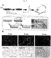
- the thus induced three-dimensional tissues were transplanted into cranial windows and subjected to live imaging with the eye and confocal microscope. Further, the tissues were taken out at days 15, 30 and 60 of transplantation and subjected to histochemical staining.
- Data are shown as the mean ⁇ s.d. obtained from experiments using at least three independent specimens.
- 3 or 4 groups were analysed using the Kruskal Wallis-H test. When found P ⁇ 0.01, multiple comparison test was performed using Mann-Whitney's U test with Bonferroni correction. When P value satisfied P ⁇ 0.001 or P ⁇ 0.01, the result was found to have a statistically significant difference.
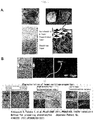
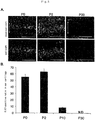
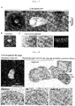
- Ear cartilage of E17.5 EGFP transgenic mouse was transplanted into the cranial window. Processes of differentiation from CPCs into mature chondrocytes were observed in a tracking manner by live imaging with confocal microscope. Macroscopic observations confirmed that anastomosis of transplant blood vessels and host mouse blood vessels was beginning to occur at day 1 or 2 of transplantation. However, at day 5 and thereafter, it was confirmed that blood vessels were completely anastomosed. From day 5, blood vessels gradually regressed until they disappeared almost completely from the transplant at day 11 ( Fig. 2A ).
- mice endothelial cells and blood flow by intravascular administration of tetramethylrhodamine-conjugated dextran and Alexa647-conjugated mouse specific CD31 (mCD31) antibody confirmed that at day 3 of transplantation, vessels with blood flow had invaded into the transplanted ear cartilage. At day 7, vascular network had regressed, leaving only part of vascular endothelial cells ( Fig. 2B ). At the same time, circular CPCs had changed into a cobblestone-like shape resembling chondrocytes. At day 10, blood vessels had completely regressed from the transplanted ear cartilage ( Fig. 2B ).
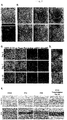
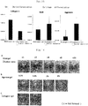
- Frozen tissue sections were prepared with a cryostat from ear cartilage samples at developmental stages of E18.5, P0, P2, P10 and P30 and subjected to immunohistochemical staining against laminin, or a component protein of matrix membrane supporting vascular endothelial cells, and the vascular endothelial cell marker mCD31.
- E18.5 cells expressing laminin and mCD31 were present at the site destined for chondrogenesis.
- P0 more blood vessels were present than at E18.5; a maximum number of blood vessels were observed at P2.
- blood vessels were slightly observable at P10 and no blood vessels were observable at P30 ( Fig. 3A ).
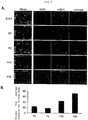
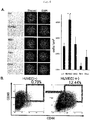
- the inventors examined whether cells were proliferating or not in ear cartilage of early developmental stages where invasion of blood vessels occurs. Using ear cartilage samples of P0 and P2 (where invasion of blood vessels occurred) and P30 (where blood vessels completely regressed), immunohistochemical staining was performed against Ki67 and CD44. Proliferating CPCs were observed by focusing on CD44 43 (which we have reported as a CPC specific marker) and the cell growth marker Ki67. At stages P0 and P2 where blood vessels were invading, Ki67-positive cells could be observed. At P2 where blood vessel invasion was highest, the largest number of Ki67-positive cells could be observed. At P30 where blood vessels had regressed, Ki67-positive cells could not be observed ( Fig.
- CPCs were seeded at a low density and co-cultured with normal human umbilical vascular endothelial cells (HUVECs) in a Transwell system to evaluate the effect of vascular endothelial cells on the proliferative capacity of CPCs.
- HUVECs normal human umbilical vascular endothelial cells
- CPCs co-cultured with HUVECs became so dense as to reach nearly 100% confluence. Cell counts were determined with In Cell Analyzer.
- the cell count of CPCs cultured alone was approximately 2500 cells/cm 2 whereas the cell count of CPCs co-cultured with HUVECs was approximately 4000 cells/cm 2 .
- Co-cultivation with mesenchymal stem cells, fibroblasts or chondrocytes showed no effect on the proliferative capacity of CPCs ( Fig. 6A ).
- changes in the cell surface antigens caused by co-cultivation were analyzed by flow cytometry.
- CD44 + CD90 + cells occupied 0.79% of total cells in control (CPCs alone) while CD44 + CD90 + cells increased to 12.44% as a result of co-culture with HUVECs ( Fig. 6B ).
- CPCs alone Control cells
- CD44 + CD90 + cells increased to 12.44% as a result of co-culture with HUVECs ( Fig. 6B ).
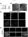
- 4-5. Three-Dimensional Organization of CPCs by Co-culture with Vascular Endothelial Cells The present inventors have developed a three-dimensional culture system by recapitulating interactions between CPCs and endothelial cells; this culture system does not use scaffold materials or growth factors.
- HUVECs and mouse blood vessels were completely anastomosed to thereby construct a vascular network within the transplant; thus, transient vascularization could be recapitulated ( Fig. 7B ).
- Tracking observation of vascularized sites revealed that at day 30 of transplantation, the vascular network completely disappeared and CPCs metamorphosed to a cobblestone-like shape similar to that of chondrocytes, suggesting that CPCs differentiated into mature chondrocytes.
- Live imaging analyses also confirmed that at day 3 of transplantation, HUVECs constructed a vascular network which regressed completely at day 30 of transplantation. Histological analysis was conducted to examine whether the transplanted three-dimensional tissues formed cartilage tissues or not.
- chondrocytes were co-cultured with human umbilical vascular endothelial cells (HUVECs) in the same three-dimensional culture system. As in the case where CPCs were used, reconstruction of elastic cartilage was confirmed by Alcian blue staining and Elastica van Gieson staining ( Fig. 7E ).
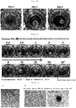
- a blood flow-blocked transplantation model was established in which a nanomesh (pore size: 0.45 ⁇ m) was placed between the three-dimensional tissues constructed by co-culturing CPCs and HUVECs and the mouse brain so that the mouse blood flow would not act on the transplant ( Fig. 8A ). It was confirmed that no blood flow was present around the transplanted three-dimensional tissues even at day 15 of transplantation ( Fig. 8B ). Live imaging showed that a large number of HUVECs were present at day 3 of transplantation; HUVECs decreased at day 7 of transplantation; and most of the HUVECs were dead at day 10 of transplantation.
- the Alcian blue-positive area was approximately 100,000 ⁇ m 2 at day 10 of transplantation, approximately 130,000 ⁇ m 2 at day 30 and approximately 250,000 ⁇ m 2 at day 60.
- the transplanted pellet produced cartilage tissues of the following areas: approximately 35,000 ⁇ m 2 at day 10 of transplantation, approximately 20,000 ⁇ m 2 at day 30 and approximately 80,000 ⁇ m 2 at day 60. Comparison of these results showed that the three-dimensional tissues constructed by co-cultivation formed cartilage tissues with areas that were 2.85 times larger at day 10 of transplantation, 6.5 times larger at day 30, and 3.27 times larger at day 60 ( Fig. 9C ).
- Cartilage tissue is a supporting organ consisting of chondrocytes and extracellular matrix surrounding the chondrocytes.
- the cell stroma of cartilage tissue differs from other supporting tissues such as connective tissue or bone tissue in that it does not contain blood vessels, lymphatic vessels, nerves and the like 45,46 . Therefore, compared to solid organs with complex higher structures, cartilage is an area where early realization of regenerative therapy is expected 47,48 .
- the inventors have found by tracking observation of chondrogenesis via live imaging that blood vessels (which have been considered unnecessary) transiently invade in the differentiation process of CPCs. Transplanted ear cartilage at an early developmental stage swelled immediately after the blood vessel invasion, suggesting rapid proliferation of CPCs.
- CD44 reportedly a specific marker for mesenchymal stem cells
- Ki67-positive the inventors co-cultured CPCs and vascular endothelial cells and found enhanced proliferation of CD44 + CD90 + cells (which are CPCs).
- the technique of the present invention which performs co-culturing of CPCs with endothelial vascular cells to enable construction of cartilage tissue from chondrocytes without using growth factors has a potential to become a technique that offers great benefits toward clinical application.
- CPCs if co-cultured with vascular endothelial cells, are capable of inducing three-dimensional tissues in vitro in a self-driven manner without using scaffold materials.
- This three-dimensional tissue has a sufficient mechanical strength to be transplanted without deformation of its shape, so it would be easy to control its morphology during transplantation.
- HLA human leukocyte antigen
- vascular endothelial cell banks are necessary which collect, culture and preserve vascular endothelial cells from umbilical cords.
- umbilical cord is a biological resource that has been abandoned to date, it would be capable of consistent supply of vascular endothelial cells if it could be preserved like umbilical cord blood. If a patient's HLA-matched vascular endothelial cells and CPCs collected/cultured in a minimally invasive manner were co-cultured to induce three-dimensional organization and the resultant three-dimensional tissues were transplanted, a novel method could be provided for treating craniofacial tissue deformations resulting from congenital defects or traumas.
- cartilage tissues be reconstructed three-dimensionally in vitro.
- a method which mimics a microgravity environment by using a rotating reactor called rotating wall vessel (RWV) bioreactor may be given.
- RWV rotating wall vessel
- the inventors reconstructed cartilage-like tissue by using an RWV bioreactor and combining human ear perichondrocytes and a small-sized novel scaffold 53 .
- a problem concerning the strength of cartilage-like tissue remained to be solved.
- RWV by combining RWV with the unique approach of recapitulating the vascularization occurring in the process of chondrogenesis, terminal cartilage differentiation induction in vitro which has been difficult to achieve by conventional cultivation techniques is expected to materialize.
- CPCs present in ear perichondrium have a capacity to differentiate into hyaline cartilage, a different type of cartilage tissue. It has been revealed that articular cartilage tissue can be reconstructed by transplanting such CPCs (data unpublished). Since the technique of the present invention efficiently generates cartilage by recapitulating transient vascularization, it is capable of resolving the problem that the post-transplantation efficiency of cartilage reconstruction is low in the regenerative therapy on articular cartilage deficiencies. Once articular cartilage is damaged, the damage proceeds to secondary degenerative diseases such as arthritis or knee osteoarthritis since articular cartilage does not have a healing capacity. It is estimated that the number of patients with knee osteoarthritis amounts to 25,300,000 in Japan alone.
- Perichondrocytes (3x10 6 ) and vascular endothelial cells (1x10 6 ) collected and subcultured in Example 1 were cultured under the same conditions as in Example 1 on a gel with a hardness of 0.5 kPa (hydrogel for cell cultivation; sample plate for evaluation (VERITAS)).
- the states of three-dimensional tissue formation at days 0, 1 and 2 of culture are shown in Fig. 10 .
- a hydrogel for cell cultivation and sample plates for evaluation (VERITAS) cells were seeded on the gel with a hardness of 0.2-50 kPa. The results are shown in Fig. 11 .
- As a conventional culture dish 10 cm easy grip cell culture dish (Falcon) was used.
- perichondrocytes When plates with a hardness of 0.5-25 kPa were used, perichondrocytes satisfactorily formed three-dimensional tissues capable of withstanding transplantation operations. When conventional culture dishes were used, cells failed to form three-dimensional tissues.
- Perichondrocytes (3x10 4 ) and vascular endothelial cells (1x10 4 ) were cultured on Prime Surface 96-well cell culture substrate having a U-shaped bottom (Sumitomo Bakelite) ( Fig. 12A ).
- a medium for inducing differentiation into cartilage was used. This medium is D-MEM/F-12 medium (Sigma) supplemented with 10% FBS (Gibco), 1% antibiotic antimycotic solution, L-ascorbic acid 2-phosphate (Wako), dexamethasone (Sigma), insulin growth factor-I (Sigma) and basic fibroblast growth factor (Kaken Pharmaceutical).
- Fig. 12B The states of three-dimensional tissue formation at day 0 and day 2 of culture are shown in Fig. 12B .
- the seeded perichondrocytes autonomously began to aggregate and formed spherical, three-dimensional tissues with an approximate suze of 400 ⁇ m the day after the seeding. These tissues could be easily recovered by pipetting or the like without deformation of their shapes.
- Fig. 13A shows the state in which a large amount of three-dimensional tissues are located subcutaneously.
- Fig. 13B shows approximately 30 three-dimensional tissues being recovered with a spatula.
- Fig. 14A shows the transplantation of three-dimensional tissues (approx. 400 ⁇ m) recovered in a large quantity into the deficient site with a pipette.
- Fig. 14B shows the deficient site in articular cartilage immediately after transplantation. After transplantation, the site was left stationary for about 20 min. When the three-dimensional tissues were in such a condition that they would not flow out, the surgical incision was closed.
- Perichondrocytes (3x10 6 ) and vascular endothelial cells (1x10 6 ) were co-cultured on Matrigel (BD) in Endothelial Cell Growth Medium SingleQuots Supplements and Growth Factors (EGM) (Lonza) for 2 days to thereby form three-dimensional tissues 4 mm in size. Formation of vasculatures was recognized in the tissues.
- the cells were cultured further in a growth medium [10% fetal bovine serum (FBS; Gibco), 1% antibiotic antimycotic solution (Sigma)-supplemented Dulbecco's Modified Eagle's Medium Nutrient Mixture F-12 HAM (D-MEM/F-12; Sigma)] or a medium for inducing differentiation into cartilage [10 % FBS (Gibco), 1% antibiotic antimycotic solution, L-ascorbic acid 2-phosphate (Wako), dexamethasone (Sigma), insulin growth factor-I (Sigma), basic fibroblast growth factor (Kaken Phannaceutical)-supplemented D-MEM/F-12 medium (Sigma)] for about 10 days.
- FBS fetal bovine serum
- FDA antibiotic antimycotic solution
- D-MEM/F-12 D-MEM/F-12
- a medium for inducing differentiation into cartilage 10 % FBS (Gibco), 1% antibiotic antimycotic solution, L-as
- a plurality of supports as listed below were gelled and fixed on 24-well plates.
- TC protector was dispensed into tubes for freezing (200-1000 ul/tube). Subsequently, three-dimensional tissues induced in 24-well plates were dipped in and left at 4 °C for several hours to overnight, followed by slow freezing at -80°C.
- tissues supplemented with 10% DMSO, 5% ethylene glycol and 10% sucrose were dipped in EGM medium for 15-20 min and then transferred to DMEM/F12 (bFGF, IGF, Dex, ascorbic acid, ITS-X, 10% FBS, 1% ABAM) medium (200 ⁇ l/tube) supplemented with 2M DMSO, 1M acetamide, 3M propylene glycol.
- DMEM/F12 bFGF, IGF, Dex, ascorbic acid, ITS-X, 10% FBS, 1% ABAM
- Cryopreserved tissues were transferred from -80 °C to a 37 °C water bath. After thawing, tissues were transferred to a 15 ml centrifugal tube and centrifuged (4 °C, 750 rpm, 3 min). The resultant supernatant was removed with a Pipetman or the like (aspirator was not used because it would suck in tissues). After PBS wash (5 ml/tube), tissues were re-centrifuged at 4 °C, 1500 rpm, for 5 min. After removal of the supernatant (with a Pipetman or the like), tissues were used in transplantation or cultivation experiments.
- mice Six-week-old female NOD/SCID (immunodeficient) mice were purchased from Sankyo Lab. Co., and bred and maintained at Animal Experiment Center, Joint Research Support Section, Advanced Medical Research Center, Yokohama City University. Animal experiments using these mice were performed in accordance with the Yokohama City University Fukuura Campus Animal Experiment Guidelines. The immunodeficient mice were shaved. Skin in the back or face was incised and ablated. The recovered tissues were embedded in the ablated area to perform transplantation.
- the resultant frozen blocks were sliced into 5 ⁇ m thick sections with cryostat HM 500 O (Zeiss) to prepare frozen tissue sections.
- the resultant tissue sections were subjected to Alcian blue staining (Muto Pure Chemicals) and Elastica van Gieson staining (Muto Pure Chemicals).
- frozen blocks were sliced into 5 ⁇ m thick sections with cryostat HM 500 O (Zeiss) to prepare frozen tissue sections.
- the resultant tissue sections were washed with 0.1% tween TBS to remove O.C.T. Compound.
- TBS-T around the frozen sections was wiped out.
- the targets of staining were marked with a water-repellent pen (Dako) in an enclosing manner to make them water-repellent.
- the tissues were blocked with Protein Block Serum-Free Ready-to-Use (Dako) at 4 °C for 24 hr. Primary antibodies were reacted at 4 °C overnight.
- Dako Protein Block Serum-Free Ready-to-Use
- Fig. 17A shows the processes of cryopreserving vascularized cartilage.
- Left panel Tissues formed in a culture dish.
- Center panel Tissues recovered with a spatula.
- Right panel The recovered tissues as dipped in cryopreservation solvent (TC protector) immediately before freezing.
- Fig. 17B shows gross observation of human vascularized cartilage thawed one month after freezing.
- Fig. 17C shows histological analysis of subcutaneously transplanted sample of thawed human vascularized cartilage. Histological analysis of the sample subcutaneously transplanted into the back of immunodeficient mice revealed that the transplanted human vascularized cartilage reconstructed cartilage tissues comprising cartilage matrix stained by Alcian blue and collagen II antibody.
- EGM and Matrigel were added separately to 24-well plates, which were then left stationary in an incubator for 30 min.
- Cell suspensions of perichondrocytes (2.0 ⁇ 10 6 cells/ml) and HUVEC (0.6 ⁇ 10 6 cells/ml) were mixed and centrifuged (950 rpm, 4 °C, 5 min). Recovered cells were seeded on the wells. After the cells were left stationary for 30 min, 1 mL of EGM was added. Then, cells were cultured for 3 days.
- Induced three-dimensional tissues were seeded on culture vessels and subjected to rotary culture with an RWV bioreactor (Synthecon) using a three-dimensional culture medium for differentiation into cartilage (DMEM/F12, dexamethasone, ascorbic acid 2-phosphate, bFGF, IGF-1, ITS-X, 1% antibiotic antimycotic solution) in an incubator whose gas phase condition had been set at 37°C and a CO 2 concentration of 5%. The rotation rate was adjusted to 7-12 rpm. After 60 days of culture for differentiation into cartilage, cell masses 5 mm to 1 cm in size were recovered. The mechanical strength of these cell masses was confirmed by applying manual pressure with tweezers or the like. After they were confirmed to have sufficient strength, cell masses were transplanted into the head of immunodeficient mice.
- Fig. 18 shows histological analysis of long-term cultured vascularized cartilage.
- a blood vessel-containing perichondral tissue and a blood vessel-free cartilage tissue were shown to have formed.
- Upper left panel macroscopic image of the formed tissues.
- Upper right panel immunostaining showed that the central part expressed the cartilage marker aggrecan, with laminin surrounding the central part.
- Lower left panel enlarged image of immunostaining.
- Lower center panel HE staining.
- Lower right panel Alcian Blue staining.
- the induced three-dimensional tissue had such a high mechanical strength that it was not destroyed even when it was manually compressed with tweezers.
- Fig. 19 shows transplantation of mature cartilage derived from long-term cultured vascularized cartilage.
- Long-term cultured vascularized cartilage (shown in a small window at the lower left corner of the upper left panel) was transplanted into a facial site and found to have a sufficient mechanical strength to withstand subcutaneous tension.
- Upper and lower photographs were taken to confirm the raised portion as viewed from different angles.
- the present invention has potential applications including regenerative therapy, drug screening, and preparation of matrixes produced by chondrocytes.
Landscapes
- Health & Medical Sciences (AREA)
- Life Sciences & Earth Sciences (AREA)
- Engineering & Computer Science (AREA)
- Biomedical Technology (AREA)
- Chemical & Material Sciences (AREA)
- Cell Biology (AREA)
- Zoology (AREA)
- General Health & Medical Sciences (AREA)
- Medicinal Chemistry (AREA)
- Veterinary Medicine (AREA)
- Public Health (AREA)
- Animal Behavior & Ethology (AREA)
- Biotechnology (AREA)
- Bioinformatics & Cheminformatics (AREA)
- Rheumatology (AREA)
- Urology & Nephrology (AREA)
- Organic Chemistry (AREA)
- Epidemiology (AREA)
- Chemical Kinetics & Catalysis (AREA)
- Genetics & Genomics (AREA)
- Wood Science & Technology (AREA)
- Oral & Maxillofacial Surgery (AREA)
- Botany (AREA)
- Dermatology (AREA)
- Transplantation (AREA)
- Immunology (AREA)
- Microbiology (AREA)
- Biochemistry (AREA)
- Molecular Biology (AREA)
- Pharmacology & Pharmacy (AREA)
- General Engineering & Computer Science (AREA)
- Orthopedic Medicine & Surgery (AREA)
- Hematology (AREA)
- Developmental Biology & Embryology (AREA)
- Virology (AREA)
- Nuclear Medicine, Radiotherapy & Molecular Imaging (AREA)
- General Chemical & Material Sciences (AREA)
- Physical Education & Sports Medicine (AREA)
- Toxicology (AREA)
- Physics & Mathematics (AREA)
Claims (17)
- Ein Verfahren zur Herstellung von Chondrozyten und/oder eines dreidimensionalen Gewebes, das Chondrozyten enthält, umfassend Mischen und Co-Kultivieren von chondrogenen Zellen mit vaskulären Zellen.
- Das Verfahren nach Anspruch 1, wobei sich die chondrogenen Zellen infolge von Co-Kultivierung mit vaskulären Zellen vermehren.
- Das Verfahren nach Anspruch 1 oder 2, wobei dreidimensionale Gewebe durch Co-Kultivierung von chondrogenen Zellen mit vaskulären Zellen auf einem Träger gebildet werden, wobei der Träger vorzugsweise ein Substrat mit einer Härte von 0,5-25 kPa ist.
- Das Verfahren nach Anspruch 1 oder 2, wobei dreidimensionale Gewebe durch Co-Kultivierung von chondrogenen Zellen mit vaskulären Zellen auf einer Platte mit einer Form, in der sich Zellen am Boden ansammeln, gebildet werden.
- Das Verfahren nach einem der Ansprüche 1 bis 4, wobei chondrogene Zellen mit vaskulären Zellen in Gegenwart mindestens einer Komponente, ausgewählt aus der Gruppe bestehend aus Fibroblasten-Wachstumsfaktor 2 (bFGF (FGF2)), Fibroblasten-Wachstumsfaktor 4 (FGF4), Fibroblasten-Wachstumsfaktor 5 (FGF5), knochenmorphogenetischem Protein 2 (BMP2), knochenmorphogenetischem Protein 3 (BMP3), knochenmorphogenetischem Protein 4 (BMP4), knochenmorphogenetischem Protein 6 (BMP6), Bindegewebe-Wachstumsfaktor (CTGF), transformierendem Wachstumsfaktor β1 (TGF-β1), transformierendem Wachstumsfaktor β2 (TGF-β2), transformierendem Wachstumsfaktor β3 (TGF-β3), insulinähnlichem Wachstumsfaktor 1 (IGF-1), Hepatozyten-Wachstumsfaktor (HGF), Aggrecan, Hyaluronsäure, Endothelzell-Wachstumsfaktor (ECGF), Endothelzell-Wachstumszusatz (ECGS), Endothelzell-abgeleitetem Wachstumsfaktor (ECDGF), epidermalem Wachstumsfaktor (EGF), saurem Fibroblasten-Wachstumsfaktor (saurem FGF), Makrophagen-abgeleitetem Wachstumsfaktor (MDGF), Blutplättchenabgeleitetem Wachstumsfaktor (PDGF), Tumorangiogenesefaktor (TAF), vaskulärem endothelialem Wachstumsfaktor (VEGF), Rinderhimextrakt (BBE), Rinderhypophysenextrakt (BPE), Glucocorticoid, Cholesterin und Vitaminen, cokultiviert werden.
- Das Verfahren nach einem der Ansprüche 1 bis 5, wobei chondrogene Zellen und vaskuläre Zellen in einem Mischverhältnis von 1:0,3-1 co-kultutiviert werden.
- Das Verfahren nach einem der Ansprüche 1 bis 6, wobei die chondrogene Zelle eine aus Chondrozyt, unreifem Chondrozyt, Knorpelvorläuferzelle oder Knorpelstammzelle ist;
wobei der Chondrozyt vorzugweise aus einem Gewebe, ausgewählt aus der Gruppe bestehend aus Rippenknorpel, Nasenknorpel, Ohrknorpel, Luftröhrenknorpel, Rachenknorpel, Schilddrüsenknorpel, Stellknorpel, Ringknorpel, Sehne, Ligament, interartikulärem Knorpel, und Bandscheibe, erhalten wurde; und
wobei der unreife Chondrozyt, die Knorpelvorläuferzelle oder die Knorpelstammzelle vorzugsweise aus einem Gewebe, ausgewählt aus der Gruppe bestehend aus Knorpel, Perichondrium, Knochenmark, Plazenta, Bauchnabelschnur, Haut, Muskel, Fett und Periosteum, erhalten wurde. - Das Verfahren nach einem der Ansprüche 1 bis 7, wobei die chondrogene Zelle und die vaskuläre Zelle von demselben Individuum stammen.
- Das Verfahren nach einem der Ansprüche 1 bis 7, wobei die chondrogene Zelle und die vaskuläre Zelle von verschiedenen Individuen stammen.
- Eine Zusammensetzung zur Verwendung in Knorpelregenerationstherapie, umfassend das dreidimensionale Gewebe, das durch das Verfahren nach einem der Ansprüche 1 bis 9 hergestellte Chondrozyten enthält.
- Die Zusammensetzung zur Verwendung nach Anspruch 10, wobei die Zusammensetzung für die Transplantation in einen Organismus zur Anregung der Bildung eines Knorpelgewebes zu verwenden ist.
- Die Zusammensetzung zur Verwendung nach Anspruch 11, wobei sich ein vaskuläres Netzwerk nach der Transplantation in einen Organismus bildet,
vorzugsweise, wobei eine vaskuläre Perfusion in dem vaskulären Netzwerk stattfindet, und
stärker bevorzugt, wobei eine vaskuläre Perfusion in dem vaskulären Netzwerk stattfindet und das einmal gebildete vaskuläre Netzwerk verschwindet, um ein avaskuläres Knorpelgewebe entstehen zu lassen. - Ein Verfahren zur Erkennung von Arzneistoffen, die als Arzneimittel wirksam sind, umfassend Verwenden des dreidimensionalen Gewebes, das durch das Verfahren nach einem der Ansprüche 1 bis 9 hergestellte Chondrozyten enthält.
- Ein Verfahren zur Herstellung einer durch Chondrozyten hergestellten Matrix, umfassend Verwenden des dreidimensionalen Gewebes, das durch das Verfahren nach einem der Ansprüche 1 bis 9 hergestellte Chondrozyten enthält.
- Ein dreidimensionales Gewebe, enthaltend durch das Verfahren nach einem der Ansprüche 1 bis 9 hergestellte Chondrozyten zur Verwendung in einem Verfahren zur Knorpelregeneration, wobei das Verfahren Transplantieren des dreidimensionalen Gewebes in einen Organismus, um ein Knorpelgewebe zu bilden, umfasst.
- Ein Verfahren zur Herstellung eines dreidimensionalen Gewebes, das Chondrozyten umfasst, umfassend Mischen und Co-Kultivieren von chondrogenen Zellen mit vaskulären Zellen, wobei die chondrogenen Zellen aus Knorpelgewebe und/oder Perichondrium erhalten werden.
- Ein dreidimensionales Gewebe, erhältlich durch das Verfahren nach Anspruch 16, zur Verwendung bei der Knorpelregenerationstherapie.
Applications Claiming Priority (2)
| Application Number | Priority Date | Filing Date | Title |
|---|---|---|---|
| JP2013058534 | 2013-03-21 | ||
| PCT/JP2014/057673 WO2014148592A1 (ja) | 2013-03-21 | 2014-03-20 | 軟骨細胞の調製方法 |
Publications (3)
| Publication Number | Publication Date |
|---|---|
| EP2977448A1 EP2977448A1 (de) | 2016-01-27 |
| EP2977448A4 EP2977448A4 (de) | 2016-11-09 |
| EP2977448B1 true EP2977448B1 (de) | 2019-07-10 |
Family
ID=51580262
Family Applications (1)
| Application Number | Title | Priority Date | Filing Date |
|---|---|---|---|
| EP14769212.3A Active EP2977448B1 (de) | 2013-03-21 | 2014-03-20 | Verfahren zur herstellung von chondrozyten |
Country Status (4)
| Country | Link |
|---|---|
| US (1) | US10100274B2 (de) |
| EP (1) | EP2977448B1 (de) |
| JP (1) | JP6341574B2 (de) |
| WO (1) | WO2014148592A1 (de) |
Families Citing this family (12)
| Publication number | Priority date | Publication date | Assignee | Title |
|---|---|---|---|---|
| JP2019010003A (ja) * | 2015-11-13 | 2019-01-24 | 協和発酵キリン株式会社 | 軟骨組織塊及びその製造方法、並びに幹細胞から軟骨組織塊を誘導するための培地 |
| JP2019010002A (ja) * | 2015-11-13 | 2019-01-24 | 協和発酵キリン株式会社 | 軟骨組織塊及びその製造方法、並びに幹細胞から軟骨組織塊を誘導するための培地 |
| US10767164B2 (en) | 2017-03-30 | 2020-09-08 | The Research Foundation For The State University Of New York | Microenvironments for self-assembly of islet organoids from stem cells differentiation |
| US12180442B2 (en) | 2017-11-30 | 2024-12-31 | Public University Corporation Yokohama City University | Method for aggregating cell mass and device for aggregating cell mass |
| CA3149691A1 (en) * | 2019-08-14 | 2021-02-18 | Vanarix Sa | Method for in vitro production of hyaline cartilage tissue |
| CN112430564B (zh) * | 2019-08-26 | 2023-09-08 | 上海交通大学医学院附属第九人民医院 | 一种磁控三维细胞培养物调控方法 |
| EP4032971A4 (de) * | 2019-09-18 | 2023-10-25 | National University Corporation Okayama University | Lbm, cpc, opc, herstellungs- und qualitätskontrollverfahren dafür, kit, transplantatmaterial und krankheitsmodell |
| CA3176522A1 (en) * | 2020-04-27 | 2021-11-04 | Daisuke Sakai | Method for culturing cell population containing cartilage-derived tie2-positive cells and use of said method |
| JP2022041601A (ja) * | 2020-09-01 | 2022-03-11 | 国立大学法人 岡山大学 | 骨又は軟骨関連疾患のスクリーニング方法 |
| US20220347349A1 (en) * | 2021-04-30 | 2022-11-03 | Stemcellago Inc. | Engineered cartilage |
| KR102307115B1 (ko) * | 2021-05-12 | 2021-10-01 | 주식회사 스마트셀랩 | 시프로플록사신에 의한 줄기세포의 연골전구세포로의 유도 및 연골세포로의 분화 |
| CN117716021A (zh) * | 2021-08-31 | 2024-03-15 | 公立大学法人横滨市立大学 | 可成型且无需支架的软骨组织的制作方法 |
Family Cites Families (9)
| Publication number | Priority date | Publication date | Assignee | Title |
|---|---|---|---|---|
| JP4763960B2 (ja) | 2000-08-09 | 2011-08-31 | 博子 矢永 | ヒト軟骨細胞培養方法 |
| US6852331B2 (en) | 2002-02-11 | 2005-02-08 | Taipei Biotechnology Ltd., Inc. | Fabrication of a cartilage implant |
| AU2003287444A1 (en) | 2002-10-31 | 2004-05-25 | The General Hospital Corporation | Repairing or replacing tissues or organs |
| KR101503939B1 (ko) | 2007-01-23 | 2015-03-18 | 고리츠다이가쿠호진 요코하마시리츠다이가쿠 | 연골 세포 조제 방법 |
| JP5495486B2 (ja) | 2007-10-31 | 2014-05-21 | 恒夫 高橋 | 培養軟骨製造方法および培養軟骨 |
| US20090317448A1 (en) | 2008-06-18 | 2009-12-24 | University Of Massachusetts | Tympanic membrane patch |
| JP5545563B2 (ja) | 2009-09-08 | 2014-07-09 | 利江 土屋 | 軟骨用移植材 |
| JP2012000262A (ja) | 2010-06-17 | 2012-01-05 | Yokohama City Univ | ヒト軟骨細胞と新規足場材料を用いた軟骨組織の製法 |
| JP2012031127A (ja) * | 2010-08-03 | 2012-02-16 | Nagoya Univ | 臍帯由来間葉系幹細胞を含む組成物 |
-
2014
- 2014-03-20 US US14/778,700 patent/US10100274B2/en not_active Expired - Fee Related
- 2014-03-20 EP EP14769212.3A patent/EP2977448B1/de active Active
- 2014-03-20 WO PCT/JP2014/057673 patent/WO2014148592A1/ja not_active Ceased
- 2014-03-20 JP JP2015506846A patent/JP6341574B2/ja not_active Expired - Fee Related
Non-Patent Citations (1)
| Title |
|---|
| None * |
Also Published As
| Publication number | Publication date |
|---|---|
| US10100274B2 (en) | 2018-10-16 |
| JPWO2014148592A1 (ja) | 2017-02-16 |
| EP2977448A1 (de) | 2016-01-27 |
| WO2014148592A1 (ja) | 2014-09-25 |
| US20160046903A1 (en) | 2016-02-18 |
| EP2977448A4 (de) | 2016-11-09 |
| JP6341574B2 (ja) | 2018-06-13 |
Similar Documents
| Publication | Publication Date | Title |
|---|---|---|
| EP2977448B1 (de) | Verfahren zur herstellung von chondrozyten | |
| JP4745386B2 (ja) | 組織増加のための分化した未成熟脂肪細胞および生分解性骨格の移植 | |
| EP2970904B1 (de) | Isolierte diskogene zellen, verfahren zur verwendung und verfahren zur herstellung davon aus säugergewebe | |
| US9234173B2 (en) | Regulating stem cells | |
| JP6890549B2 (ja) | 間葉系幹細胞の製造方法 | |
| JP6434014B2 (ja) | 球状軟骨細胞治療剤の製造方法 | |
| JP6193214B2 (ja) | 歯髄由来の多能性幹細胞の製造方法 | |
| CN115305233A (zh) | 大豆苷元与芹菜素复合琼脂糖-胶原蛋白水凝胶三维培养干细胞及细胞外囊泡的制备与用途 | |
| JP5964956B2 (ja) | 胎児の組織からの親細胞バンクの調製 | |
| Ghiasi et al. | Use of mesenchymal adult stem cell for cartilage regeneration by hydrogel | |
| JP6721504B2 (ja) | 多能性幹細胞及び前駆細胞を生産するためのプロセス | |
| CN102002479A (zh) | 一种脐带血间充质干细胞及其制备方法和用途 | |
| CN120418415A (zh) | 细胞组合物 | |
| US20120207715A1 (en) | Methods and systems for storing and prolonging viability of matrix dependent cells | |
| RU2800991C9 (ru) | Способ биофабрикации трансплантата в виде клеточных сфероидов для регенеративных технологий восстановления хряща субъекта на основе надхрящницы собственного реберного хряща субъекта и мультипотентных мезенхимальных стромальных клеток костного мозга этого же субъекта | |
| van Osch et al. | Cells for Cartilage Regeneration | |
| RU2800991C2 (ru) | Способ биофабрикации трансплантата в виде клеточных сфероидов для регенеративных технологий восстановления хряща субъекта на основе надхрящницы собственного реберного хряща субъекта и мультипотентных мезенхимальных стромальных клеток костного мозга этого же субъекта | |
| KR20100069376A (ko) | 성장 인자를 이용한 지방기원 중간엽 줄기세포의 연골세포로의 분화 방법 | |
| Joshi | Master of Veterinary | |
| Deegan | Novel tissue engineering approaches to enhance natural bone formation | |
| Dashtdar | The Use of Alginate as a Potential Chondrogenic Differentiation Promoter of Mesenchymal Stromal Cells and its Application | |
| Semenov | Engineering mesenchymal stem cell sheets on functional polyelectrolyte multilayer substrates | |
| IL193947A (en) | Cells cultured as cd31brigi: it, a method for stimuli to differentiate into a congenital / precursor cell population (pcp), use of the same drug preparation population, and implantation device that includes it | |
| HK1217213B (en) | Isolated discogenic cells, methods of using, and methods of preparing same from mammalian tissue |
Legal Events
| Date | Code | Title | Description |
|---|---|---|---|
| PUAI | Public reference made under article 153(3) epc to a published international application that has entered the european phase |
Free format text: ORIGINAL CODE: 0009012 |
|
| 17P | Request for examination filed |
Effective date: 20151021 |
|
| AK | Designated contracting states |
Kind code of ref document: A1 Designated state(s): AL AT BE BG CH CY CZ DE DK EE ES FI FR GB GR HR HU IE IS IT LI LT LU LV MC MK MT NL NO PL PT RO RS SE SI SK SM TR |
|
| AX | Request for extension of the european patent |
Extension state: BA ME |
|
| DAX | Request for extension of the european patent (deleted) | ||
| A4 | Supplementary search report drawn up and despatched |
Effective date: 20161007 |
|
| RIC1 | Information provided on ipc code assigned before grant |
Ipc: A61L 27/00 20060101ALI20160930BHEP Ipc: A61K 35/32 20060101ALI20160930BHEP Ipc: C12N 5/077 20100101AFI20160930BHEP Ipc: A61P 19/08 20060101ALI20160930BHEP Ipc: A61P 43/00 20060101ALI20160930BHEP Ipc: A61K 35/12 20060101ALI20160930BHEP Ipc: G01N 33/15 20060101ALI20160930BHEP |
|
| STAA | Information on the status of an ep patent application or granted ep patent |
Free format text: STATUS: EXAMINATION IS IN PROGRESS |
|
| 17Q | First examination report despatched |
Effective date: 20180320 |
|
| GRAP | Despatch of communication of intention to grant a patent |
Free format text: ORIGINAL CODE: EPIDOSNIGR1 |
|
| STAA | Information on the status of an ep patent application or granted ep patent |
Free format text: STATUS: GRANT OF PATENT IS INTENDED |
|
| INTG | Intention to grant announced |
Effective date: 20181221 |
|
| RIN1 | Information on inventor provided before grant (corrected) |
Inventor name: TAKEBE, TAKANORI Inventor name: TANIGUCHI, HIDEKI Inventor name: KOBAYASHI, SHINJI |
|
| GRAJ | Information related to disapproval of communication of intention to grant by the applicant or resumption of examination proceedings by the epo deleted |
Free format text: ORIGINAL CODE: EPIDOSDIGR1 |
|
| STAA | Information on the status of an ep patent application or granted ep patent |
Free format text: STATUS: EXAMINATION IS IN PROGRESS |
|
| GRAS | Grant fee paid |
Free format text: ORIGINAL CODE: EPIDOSNIGR3 |
|
| STAA | Information on the status of an ep patent application or granted ep patent |
Free format text: STATUS: GRANT OF PATENT IS INTENDED |
|
| GRAP | Despatch of communication of intention to grant a patent |
Free format text: ORIGINAL CODE: EPIDOSNIGR1 |
|
| INTC | Intention to grant announced (deleted) | ||
| GRAA | (expected) grant |
Free format text: ORIGINAL CODE: 0009210 |
|
| STAA | Information on the status of an ep patent application or granted ep patent |
Free format text: STATUS: THE PATENT HAS BEEN GRANTED |
|
| INTG | Intention to grant announced |
Effective date: 20190517 |
|
| AK | Designated contracting states |
Kind code of ref document: B1 Designated state(s): AL AT BE BG CH CY CZ DE DK EE ES FI FR GB GR HR HU IE IS IT LI LT LU LV MC MK MT NL NO PL PT RO RS SE SI SK SM TR |
|
| REG | Reference to a national code |
Ref country code: GB Ref legal event code: FG4D |
|
| REG | Reference to a national code |
Ref country code: CH Ref legal event code: EP Ref country code: AT Ref legal event code: REF Ref document number: 1153598 Country of ref document: AT Kind code of ref document: T Effective date: 20190715 |
|
| REG | Reference to a national code |
Ref country code: DE Ref legal event code: R096 Ref document number: 602014049857 Country of ref document: DE |
|
| REG | Reference to a national code |
Ref country code: IE Ref legal event code: FG4D |
|
| REG | Reference to a national code |
Ref country code: NL Ref legal event code: MP Effective date: 20190710 |
|
| REG | Reference to a national code |
Ref country code: LT Ref legal event code: MG4D |
|
| REG | Reference to a national code |
Ref country code: AT Ref legal event code: MK05 Ref document number: 1153598 Country of ref document: AT Kind code of ref document: T Effective date: 20190710 |
|
| PG25 | Lapsed in a contracting state [announced via postgrant information from national office to epo] |
Ref country code: NO Free format text: LAPSE BECAUSE OF FAILURE TO SUBMIT A TRANSLATION OF THE DESCRIPTION OR TO PAY THE FEE WITHIN THE PRESCRIBED TIME-LIMIT Effective date: 20191010 Ref country code: BG Free format text: LAPSE BECAUSE OF FAILURE TO SUBMIT A TRANSLATION OF THE DESCRIPTION OR TO PAY THE FEE WITHIN THE PRESCRIBED TIME-LIMIT Effective date: 20191010 Ref country code: PT Free format text: LAPSE BECAUSE OF FAILURE TO SUBMIT A TRANSLATION OF THE DESCRIPTION OR TO PAY THE FEE WITHIN THE PRESCRIBED TIME-LIMIT Effective date: 20191111 Ref country code: NL Free format text: LAPSE BECAUSE OF FAILURE TO SUBMIT A TRANSLATION OF THE DESCRIPTION OR TO PAY THE FEE WITHIN THE PRESCRIBED TIME-LIMIT Effective date: 20190710 Ref country code: LT Free format text: LAPSE BECAUSE OF FAILURE TO SUBMIT A TRANSLATION OF THE DESCRIPTION OR TO PAY THE FEE WITHIN THE PRESCRIBED TIME-LIMIT Effective date: 20190710 Ref country code: HR Free format text: LAPSE BECAUSE OF FAILURE TO SUBMIT A TRANSLATION OF THE DESCRIPTION OR TO PAY THE FEE WITHIN THE PRESCRIBED TIME-LIMIT Effective date: 20190710 Ref country code: AT Free format text: LAPSE BECAUSE OF FAILURE TO SUBMIT A TRANSLATION OF THE DESCRIPTION OR TO PAY THE FEE WITHIN THE PRESCRIBED TIME-LIMIT Effective date: 20190710 Ref country code: SE Free format text: LAPSE BECAUSE OF FAILURE TO SUBMIT A TRANSLATION OF THE DESCRIPTION OR TO PAY THE FEE WITHIN THE PRESCRIBED TIME-LIMIT Effective date: 20190710 Ref country code: FI Free format text: LAPSE BECAUSE OF FAILURE TO SUBMIT A TRANSLATION OF THE DESCRIPTION OR TO PAY THE FEE WITHIN THE PRESCRIBED TIME-LIMIT Effective date: 20190710 |
|
| PG25 | Lapsed in a contracting state [announced via postgrant information from national office to epo] |
Ref country code: AL Free format text: LAPSE BECAUSE OF FAILURE TO SUBMIT A TRANSLATION OF THE DESCRIPTION OR TO PAY THE FEE WITHIN THE PRESCRIBED TIME-LIMIT Effective date: 20190710 Ref country code: ES Free format text: LAPSE BECAUSE OF FAILURE TO SUBMIT A TRANSLATION OF THE DESCRIPTION OR TO PAY THE FEE WITHIN THE PRESCRIBED TIME-LIMIT Effective date: 20190710 Ref country code: GR Free format text: LAPSE BECAUSE OF FAILURE TO SUBMIT A TRANSLATION OF THE DESCRIPTION OR TO PAY THE FEE WITHIN THE PRESCRIBED TIME-LIMIT Effective date: 20191011 Ref country code: IS Free format text: LAPSE BECAUSE OF FAILURE TO SUBMIT A TRANSLATION OF THE DESCRIPTION OR TO PAY THE FEE WITHIN THE PRESCRIBED TIME-LIMIT Effective date: 20191110 Ref country code: RS Free format text: LAPSE BECAUSE OF FAILURE TO SUBMIT A TRANSLATION OF THE DESCRIPTION OR TO PAY THE FEE WITHIN THE PRESCRIBED TIME-LIMIT Effective date: 20190710 Ref country code: LV Free format text: LAPSE BECAUSE OF FAILURE TO SUBMIT A TRANSLATION OF THE DESCRIPTION OR TO PAY THE FEE WITHIN THE PRESCRIBED TIME-LIMIT Effective date: 20190710 |
|
| PG25 | Lapsed in a contracting state [announced via postgrant information from national office to epo] |
Ref country code: TR Free format text: LAPSE BECAUSE OF FAILURE TO SUBMIT A TRANSLATION OF THE DESCRIPTION OR TO PAY THE FEE WITHIN THE PRESCRIBED TIME-LIMIT Effective date: 20190710 |
|
| PG25 | Lapsed in a contracting state [announced via postgrant information from national office to epo] |
Ref country code: RO Free format text: LAPSE BECAUSE OF FAILURE TO SUBMIT A TRANSLATION OF THE DESCRIPTION OR TO PAY THE FEE WITHIN THE PRESCRIBED TIME-LIMIT Effective date: 20190710 Ref country code: EE Free format text: LAPSE BECAUSE OF FAILURE TO SUBMIT A TRANSLATION OF THE DESCRIPTION OR TO PAY THE FEE WITHIN THE PRESCRIBED TIME-LIMIT Effective date: 20190710 Ref country code: IT Free format text: LAPSE BECAUSE OF FAILURE TO SUBMIT A TRANSLATION OF THE DESCRIPTION OR TO PAY THE FEE WITHIN THE PRESCRIBED TIME-LIMIT Effective date: 20190710 Ref country code: DK Free format text: LAPSE BECAUSE OF FAILURE TO SUBMIT A TRANSLATION OF THE DESCRIPTION OR TO PAY THE FEE WITHIN THE PRESCRIBED TIME-LIMIT Effective date: 20190710 Ref country code: PL Free format text: LAPSE BECAUSE OF FAILURE TO SUBMIT A TRANSLATION OF THE DESCRIPTION OR TO PAY THE FEE WITHIN THE PRESCRIBED TIME-LIMIT Effective date: 20190710 |
|
| PG25 | Lapsed in a contracting state [announced via postgrant information from national office to epo] |
Ref country code: CZ Free format text: LAPSE BECAUSE OF FAILURE TO SUBMIT A TRANSLATION OF THE DESCRIPTION OR TO PAY THE FEE WITHIN THE PRESCRIBED TIME-LIMIT Effective date: 20190710 Ref country code: SK Free format text: LAPSE BECAUSE OF FAILURE TO SUBMIT A TRANSLATION OF THE DESCRIPTION OR TO PAY THE FEE WITHIN THE PRESCRIBED TIME-LIMIT Effective date: 20190710 Ref country code: SM Free format text: LAPSE BECAUSE OF FAILURE TO SUBMIT A TRANSLATION OF THE DESCRIPTION OR TO PAY THE FEE WITHIN THE PRESCRIBED TIME-LIMIT Effective date: 20190710 Ref country code: IS Free format text: LAPSE BECAUSE OF FAILURE TO SUBMIT A TRANSLATION OF THE DESCRIPTION OR TO PAY THE FEE WITHIN THE PRESCRIBED TIME-LIMIT Effective date: 20200224 |
|
| REG | Reference to a national code |
Ref country code: DE Ref legal event code: R097 Ref document number: 602014049857 Country of ref document: DE |
|
| PLBE | No opposition filed within time limit |
Free format text: ORIGINAL CODE: 0009261 |
|
| STAA | Information on the status of an ep patent application or granted ep patent |
Free format text: STATUS: NO OPPOSITION FILED WITHIN TIME LIMIT |
|
| PG2D | Information on lapse in contracting state deleted |
Ref country code: IS |
|
| 26N | No opposition filed |
Effective date: 20200603 |
|
| PG25 | Lapsed in a contracting state [announced via postgrant information from national office to epo] |
Ref country code: SI Free format text: LAPSE BECAUSE OF FAILURE TO SUBMIT A TRANSLATION OF THE DESCRIPTION OR TO PAY THE FEE WITHIN THE PRESCRIBED TIME-LIMIT Effective date: 20190710 |
|
| PG25 | Lapsed in a contracting state [announced via postgrant information from national office to epo] |
Ref country code: MC Free format text: LAPSE BECAUSE OF FAILURE TO SUBMIT A TRANSLATION OF THE DESCRIPTION OR TO PAY THE FEE WITHIN THE PRESCRIBED TIME-LIMIT Effective date: 20190710 |
|
| REG | Reference to a national code |
Ref country code: CH Ref legal event code: PL |
|
| REG | Reference to a national code |
Ref country code: BE Ref legal event code: MM Effective date: 20200331 |
|
| PG25 | Lapsed in a contracting state [announced via postgrant information from national office to epo] |
Ref country code: LU Free format text: LAPSE BECAUSE OF NON-PAYMENT OF DUE FEES Effective date: 20200320 |
|
| PG25 | Lapsed in a contracting state [announced via postgrant information from national office to epo] |
Ref country code: IE Free format text: LAPSE BECAUSE OF NON-PAYMENT OF DUE FEES Effective date: 20200320 Ref country code: CH Free format text: LAPSE BECAUSE OF NON-PAYMENT OF DUE FEES Effective date: 20200331 Ref country code: LI Free format text: LAPSE BECAUSE OF NON-PAYMENT OF DUE FEES Effective date: 20200331 |
|
| PG25 | Lapsed in a contracting state [announced via postgrant information from national office to epo] |
Ref country code: BE Free format text: LAPSE BECAUSE OF NON-PAYMENT OF DUE FEES Effective date: 20200331 |
|
| PGFP | Annual fee paid to national office [announced via postgrant information from national office to epo] |
Ref country code: FR Payment date: 20210323 Year of fee payment: 8 |
|
| PGFP | Annual fee paid to national office [announced via postgrant information from national office to epo] |
Ref country code: GB Payment date: 20210324 Year of fee payment: 8 Ref country code: DE Payment date: 20210319 Year of fee payment: 8 |
|
| PG25 | Lapsed in a contracting state [announced via postgrant information from national office to epo] |
Ref country code: MT Free format text: LAPSE BECAUSE OF FAILURE TO SUBMIT A TRANSLATION OF THE DESCRIPTION OR TO PAY THE FEE WITHIN THE PRESCRIBED TIME-LIMIT Effective date: 20190710 Ref country code: CY Free format text: LAPSE BECAUSE OF FAILURE TO SUBMIT A TRANSLATION OF THE DESCRIPTION OR TO PAY THE FEE WITHIN THE PRESCRIBED TIME-LIMIT Effective date: 20190710 |
|
| PG25 | Lapsed in a contracting state [announced via postgrant information from national office to epo] |
Ref country code: MK Free format text: LAPSE BECAUSE OF FAILURE TO SUBMIT A TRANSLATION OF THE DESCRIPTION OR TO PAY THE FEE WITHIN THE PRESCRIBED TIME-LIMIT Effective date: 20190710 |
|
| REG | Reference to a national code |
Ref country code: DE Ref legal event code: R119 Ref document number: 602014049857 Country of ref document: DE |
|
| GBPC | Gb: european patent ceased through non-payment of renewal fee |
Effective date: 20220320 |
|
| PG25 | Lapsed in a contracting state [announced via postgrant information from national office to epo] |
Ref country code: GB Free format text: LAPSE BECAUSE OF NON-PAYMENT OF DUE FEES Effective date: 20220320 Ref country code: FR Free format text: LAPSE BECAUSE OF NON-PAYMENT OF DUE FEES Effective date: 20220331 Ref country code: DE Free format text: LAPSE BECAUSE OF NON-PAYMENT OF DUE FEES Effective date: 20221001 |