EP2961426B1 - Treatment of central nervous system disorders by intranasal administration of immunoglobulin g - Google Patents
Treatment of central nervous system disorders by intranasal administration of immunoglobulin g Download PDFInfo
- Publication number
- EP2961426B1 EP2961426B1 EP14714815.9A EP14714815A EP2961426B1 EP 2961426 B1 EP2961426 B1 EP 2961426B1 EP 14714815 A EP14714815 A EP 14714815A EP 2961426 B1 EP2961426 B1 EP 2961426B1
- Authority
- EP
- European Patent Office
- Prior art keywords
- var
- subject
- composition
- igg
- disorder
- Prior art date
- Legal status (The legal status is an assumption and is not a legal conclusion. Google has not performed a legal analysis and makes no representation as to the accuracy of the status listed.)
- Active
Links
- 229940027941 immunoglobulin g Drugs 0.000 title claims description 692
- 208000015114 central nervous system disease Diseases 0.000 title claims description 129
- 238000011282 treatment Methods 0.000 title description 105
- 239000000203 mixture Substances 0.000 claims description 438
- 238000000034 method Methods 0.000 claims description 287
- 210000004556 brain Anatomy 0.000 claims description 144
- 208000037265 diseases, disorders, signs and symptoms Diseases 0.000 claims description 129
- 210000003169 central nervous system Anatomy 0.000 claims description 127
- 208000035475 disorder Diseases 0.000 claims description 105
- 210000002850 nasal mucosa Anatomy 0.000 claims description 97
- 208000024827 Alzheimer disease Diseases 0.000 claims description 89
- 210000003928 nasal cavity Anatomy 0.000 claims description 71
- 210000001706 olfactory mucosa Anatomy 0.000 claims description 68
- 239000000843 powder Substances 0.000 claims description 68
- 210000003901 trigeminal nerve Anatomy 0.000 claims description 66
- DHMQDGOQFOQNFH-UHFFFAOYSA-N Glycine Chemical compound NCC(O)=O DHMQDGOQFOQNFH-UHFFFAOYSA-N 0.000 claims description 60
- 208000018737 Parkinson disease Diseases 0.000 claims description 45
- 238000002560 therapeutic procedure Methods 0.000 claims description 45
- 239000004471 Glycine Substances 0.000 claims description 31
- 239000000443 aerosol Substances 0.000 claims description 29
- 230000037396 body weight Effects 0.000 claims description 29
- 229940098197 human immunoglobulin g Drugs 0.000 claims description 29
- 208000015122 neurodegenerative disease Diseases 0.000 claims description 24
- 208000016192 Demyelinating disease Diseases 0.000 claims description 14
- 239000000544 cholinesterase inhibitor Substances 0.000 claims description 13
- 239000003112 inhibitor Substances 0.000 claims description 13
- 229940122041 Cholinesterase inhibitor Drugs 0.000 claims description 12
- 102000018899 Glutamate Receptors Human genes 0.000 claims description 10
- 108010027915 Glutamate Receptors Proteins 0.000 claims description 10
- 239000007864 aqueous solution Substances 0.000 claims description 8
- 239000008263 liquid aerosol Substances 0.000 claims description 5
- 238000012384 transportation and delivery Methods 0.000 description 90
- 239000007788 liquid Substances 0.000 description 85
- 210000004369 blood Anatomy 0.000 description 83
- 239000008280 blood Substances 0.000 description 83
- 239000003814 drug Substances 0.000 description 80
- 108060003951 Immunoglobulin Proteins 0.000 description 77
- 102000018358 immunoglobulin Human genes 0.000 description 77
- 238000001990 intravenous administration Methods 0.000 description 66
- 238000002360 preparation method Methods 0.000 description 65
- 238000009472 formulation Methods 0.000 description 61
- 210000001519 tissue Anatomy 0.000 description 60
- 241000700159 Rattus Species 0.000 description 57
- 210000000278 spinal cord Anatomy 0.000 description 52
- HNDVDQJCIGZPNO-UHFFFAOYSA-N histidine Natural products OC(=O)C(N)CC1=CN=CN1 HNDVDQJCIGZPNO-UHFFFAOYSA-N 0.000 description 49
- 201000006417 multiple sclerosis Diseases 0.000 description 48
- 229940079593 drug Drugs 0.000 description 45
- 241001269524 Dura Species 0.000 description 43
- 241001465754 Metazoa Species 0.000 description 43
- 208000011580 syndromic disease Diseases 0.000 description 43
- LFQSCWFLJHTTHZ-UHFFFAOYSA-N Ethanol Chemical compound CCO LFQSCWFLJHTTHZ-UHFFFAOYSA-N 0.000 description 39
- 239000006196 drop Substances 0.000 description 38
- 239000011780 sodium chloride Substances 0.000 description 37
- 241000699670 Mus sp. Species 0.000 description 36
- FAPWRFPIFSIZLT-UHFFFAOYSA-M Sodium chloride Chemical compound [Na+].[Cl-] FAPWRFPIFSIZLT-UHFFFAOYSA-M 0.000 description 35
- 230000000694 effects Effects 0.000 description 33
- 239000004005 microsphere Substances 0.000 description 33
- 230000001225 therapeutic effect Effects 0.000 description 33
- 241000699666 Mus <mouse, genus> Species 0.000 description 32
- 230000003542 behavioural effect Effects 0.000 description 31
- 239000000499 gel Substances 0.000 description 31
- 210000004086 maxillary sinus Anatomy 0.000 description 31
- 210000001320 hippocampus Anatomy 0.000 description 29
- 229940072221 immunoglobulins Drugs 0.000 description 29
- 208000024891 symptom Diseases 0.000 description 27
- 229940124597 therapeutic agent Drugs 0.000 description 27
- 239000002674 ointment Substances 0.000 description 26
- 239000006071 cream Substances 0.000 description 25
- 206010012289 Dementia Diseases 0.000 description 24
- 201000010099 disease Diseases 0.000 description 24
- 239000000523 sample Substances 0.000 description 24
- 239000007921 spray Substances 0.000 description 24
- 230000009885 systemic effect Effects 0.000 description 24
- 208000028017 Psychotic disease Diseases 0.000 description 23
- 239000006228 supernatant Substances 0.000 description 23
- 108090000623 proteins and genes Proteins 0.000 description 22
- 201000000980 schizophrenia Diseases 0.000 description 22
- 102000013455 Amyloid beta-Peptides Human genes 0.000 description 21
- 108010090849 Amyloid beta-Peptides Proteins 0.000 description 21
- 235000001014 amino acid Nutrition 0.000 description 21
- 229940024606 amino acid Drugs 0.000 description 21
- 150000001413 amino acids Chemical class 0.000 description 21
- 239000003795 chemical substances by application Substances 0.000 description 21
- 208000037259 Amyloid Plaque Diseases 0.000 description 20
- 238000004458 analytical method Methods 0.000 description 20
- 239000006172 buffering agent Substances 0.000 description 20
- ADEBPBSSDYVVLD-UHFFFAOYSA-N donepezil Chemical compound O=C1C=2C=C(OC)C(OC)=CC=2CC1CC(CC1)CCN1CC1=CC=CC=C1 ADEBPBSSDYVVLD-UHFFFAOYSA-N 0.000 description 20
- 210000000956 olfactory bulb Anatomy 0.000 description 20
- 235000018102 proteins Nutrition 0.000 description 20
- 102000004169 proteins and genes Human genes 0.000 description 20
- 239000000243 solution Substances 0.000 description 20
- 239000003623 enhancer Substances 0.000 description 19
- 210000005036 nerve Anatomy 0.000 description 19
- 210000003977 optic chiasm Anatomy 0.000 description 19
- 208000022821 personality disease Diseases 0.000 description 18
- 206010002026 amyotrophic lateral sclerosis Diseases 0.000 description 17
- 230000008499 blood brain barrier function Effects 0.000 description 17
- 210000001218 blood-brain barrier Anatomy 0.000 description 17
- 210000001715 carotid artery Anatomy 0.000 description 17
- 210000004072 lung Anatomy 0.000 description 17
- 210000001533 respiratory mucosa Anatomy 0.000 description 17
- 210000001841 basilar artery Anatomy 0.000 description 16
- 210000001259 mesencephalon Anatomy 0.000 description 16
- 230000001817 pituitary effect Effects 0.000 description 16
- 210000002254 renal artery Anatomy 0.000 description 16
- 210000002700 urine Anatomy 0.000 description 16
- -1 ARICEPT®) Chemical compound 0.000 description 15
- 230000007423 decrease Effects 0.000 description 15
- 210000003238 esophagus Anatomy 0.000 description 15
- 210000003734 kidney Anatomy 0.000 description 15
- 210000004185 liver Anatomy 0.000 description 15
- 210000003205 muscle Anatomy 0.000 description 15
- 210000003437 trachea Anatomy 0.000 description 15
- 210000001638 cerebellum Anatomy 0.000 description 14
- 210000000981 epithelium Anatomy 0.000 description 14
- ASUTZQLVASHGKV-JDFRZJQESA-N galanthamine Chemical compound O1C(=C23)C(OC)=CC=C2CN(C)CC[C@]23[C@@H]1C[C@@H](O)C=C2 ASUTZQLVASHGKV-JDFRZJQESA-N 0.000 description 14
- 230000035699 permeability Effects 0.000 description 14
- 230000003442 weekly effect Effects 0.000 description 14
- 208000027534 Emotional disease Diseases 0.000 description 13
- 208000023105 Huntington disease Diseases 0.000 description 13
- 210000003016 hypothalamus Anatomy 0.000 description 13
- 210000001577 neostriatum Anatomy 0.000 description 13
- 208000002851 paranoid schizophrenia Diseases 0.000 description 13
- 208000020016 psychiatric disease Diseases 0.000 description 13
- 210000000952 spleen Anatomy 0.000 description 13
- 210000001103 thalamus Anatomy 0.000 description 13
- 210000000115 thoracic cavity Anatomy 0.000 description 13
- 206010003694 Atrophy Diseases 0.000 description 12
- 208000024254 Delusional disease Diseases 0.000 description 12
- 208000020401 Depressive disease Diseases 0.000 description 12
- 208000021155 Paediatric autoimmune neuropsychiatric disorders associated with streptococcal infection Diseases 0.000 description 12
- 230000037444 atrophy Effects 0.000 description 12
- 230000036651 mood Effects 0.000 description 12
- 210000003739 neck Anatomy 0.000 description 12
- 210000003625 skull Anatomy 0.000 description 12
- 238000010186 staining Methods 0.000 description 12
- 210000001685 thyroid gland Anatomy 0.000 description 12
- XLYOFNOQVPJJNP-UHFFFAOYSA-N water Substances O XLYOFNOQVPJJNP-UHFFFAOYSA-N 0.000 description 12
- 208000019022 Mood disease Diseases 0.000 description 11
- ONIBWKKTOPOVIA-UHFFFAOYSA-N Proline Natural products OC(=O)C1CCCN1 ONIBWKKTOPOVIA-UHFFFAOYSA-N 0.000 description 11
- 210000005153 frontal cortex Anatomy 0.000 description 11
- 210000005230 lumbar spinal cord Anatomy 0.000 description 11
- 239000002245 particle Substances 0.000 description 11
- 210000003491 skin Anatomy 0.000 description 11
- 238000012360 testing method Methods 0.000 description 11
- 210000000275 circle of willis Anatomy 0.000 description 10
- LOKCTEFSRHRXRJ-UHFFFAOYSA-I dipotassium trisodium dihydrogen phosphate hydrogen phosphate dichloride Chemical compound P(=O)(O)(O)[O-].[K+].P(=O)(O)([O-])[O-].[Na+].[Na+].[Cl-].[K+].[Cl-].[Na+] LOKCTEFSRHRXRJ-UHFFFAOYSA-I 0.000 description 10
- 230000001667 episodic effect Effects 0.000 description 10
- 238000004519 manufacturing process Methods 0.000 description 10
- 230000001314 paroxysmal effect Effects 0.000 description 10
- 239000002953 phosphate buffered saline Substances 0.000 description 10
- 230000002829 reductive effect Effects 0.000 description 10
- 230000035882 stress Effects 0.000 description 10
- 208000017194 Affective disease Diseases 0.000 description 9
- ONIBWKKTOPOVIA-BYPYZUCNSA-N L-Proline Chemical compound OC(=O)[C@@H]1CCCN1 ONIBWKKTOPOVIA-BYPYZUCNSA-N 0.000 description 9
- HNDVDQJCIGZPNO-YFKPBYRVSA-N L-histidine Chemical compound OC(=O)[C@@H](N)CC1=CN=CN1 HNDVDQJCIGZPNO-YFKPBYRVSA-N 0.000 description 9
- 208000016285 Movement disease Diseases 0.000 description 9
- XSVMFMHYUFZWBK-NSHDSACASA-N Rivastigmine Chemical compound CCN(C)C(=O)OC1=CC=CC([C@H](C)N(C)C)=C1 XSVMFMHYUFZWBK-NSHDSACASA-N 0.000 description 9
- 208000027520 Somatoform disease Diseases 0.000 description 9
- 230000001154 acute effect Effects 0.000 description 9
- 210000004727 amygdala Anatomy 0.000 description 9
- 210000005013 brain tissue Anatomy 0.000 description 9
- 238000009826 distribution Methods 0.000 description 9
- 238000001647 drug administration Methods 0.000 description 9
- 238000005516 engineering process Methods 0.000 description 9
- 206010015037 epilepsy Diseases 0.000 description 9
- 210000003128 head Anatomy 0.000 description 9
- 230000003340 mental effect Effects 0.000 description 9
- 230000001769 paralizing effect Effects 0.000 description 9
- 230000001936 parietal effect Effects 0.000 description 9
- 229960001685 tacrine Drugs 0.000 description 9
- YLJREFDVOIBQDA-UHFFFAOYSA-N tacrine Chemical compound C1=CC=C2C(N)=C(CCCC3)C3=NC2=C1 YLJREFDVOIBQDA-UHFFFAOYSA-N 0.000 description 9
- 230000002123 temporal effect Effects 0.000 description 9
- 206010002091 Anaesthesia Diseases 0.000 description 8
- 206010012559 Developmental delay Diseases 0.000 description 8
- 206010026749 Mania Diseases 0.000 description 8
- 208000027626 Neurocognitive disease Diseases 0.000 description 8
- 208000021384 Obsessive-Compulsive disease Diseases 0.000 description 8
- 230000002411 adverse Effects 0.000 description 8
- 230000037005 anaesthesia Effects 0.000 description 8
- 208000018300 basal ganglia disease Diseases 0.000 description 8
- 206010008129 cerebral palsy Diseases 0.000 description 8
- 238000002224 dissection Methods 0.000 description 8
- 239000002874 hemostatic agent Substances 0.000 description 8
- 238000001802 infusion Methods 0.000 description 8
- 239000000463 material Substances 0.000 description 8
- 238000010172 mouse model Methods 0.000 description 8
- 208000008795 neuromyelitis optica Diseases 0.000 description 8
- 230000036961 partial effect Effects 0.000 description 8
- 230000037361 pathway Effects 0.000 description 8
- 239000008188 pellet Substances 0.000 description 8
- 230000002093 peripheral effect Effects 0.000 description 8
- 108090000765 processed proteins & peptides Proteins 0.000 description 8
- 150000003839 salts Chemical class 0.000 description 8
- 208000011117 substance-related disease Diseases 0.000 description 8
- 208000016686 tic disease Diseases 0.000 description 8
- 230000009261 transgenic effect Effects 0.000 description 8
- 210000003462 vein Anatomy 0.000 description 8
- 208000019901 Anxiety disease Diseases 0.000 description 7
- 206010010904 Convulsion Diseases 0.000 description 7
- 206010019468 Hemiplegia Diseases 0.000 description 7
- 238000010521 absorption reaction Methods 0.000 description 7
- 230000001684 chronic effect Effects 0.000 description 7
- 150000001875 compounds Chemical group 0.000 description 7
- 208000015181 infectious disease Diseases 0.000 description 7
- 210000004379 membrane Anatomy 0.000 description 7
- 239000012528 membrane Substances 0.000 description 7
- 210000001331 nose Anatomy 0.000 description 7
- 210000000056 organ Anatomy 0.000 description 7
- 229920000036 polyvinylpyrrolidone Polymers 0.000 description 7
- 235000013855 polyvinylpyrrolidone Nutrition 0.000 description 7
- 230000000241 respiratory effect Effects 0.000 description 7
- 210000002966 serum Anatomy 0.000 description 7
- 230000002792 vascular Effects 0.000 description 7
- 206010000117 Abnormal behaviour Diseases 0.000 description 6
- IJGRMHOSHXDMSA-UHFFFAOYSA-N Atomic nitrogen Chemical compound N#N IJGRMHOSHXDMSA-UHFFFAOYSA-N 0.000 description 6
- 229920000742 Cotton Polymers 0.000 description 6
- 208000027776 Extrapyramidal disease Diseases 0.000 description 6
- WSFSSNUMVMOOMR-UHFFFAOYSA-N Formaldehyde Chemical compound O=C WSFSSNUMVMOOMR-UHFFFAOYSA-N 0.000 description 6
- 108010021625 Immunoglobulin Fragments Proteins 0.000 description 6
- 102000008394 Immunoglobulin Fragments Human genes 0.000 description 6
- 208000012902 Nervous system disease Diseases 0.000 description 6
- 108060008682 Tumor Necrosis Factor Proteins 0.000 description 6
- 210000002808 connective tissue Anatomy 0.000 description 6
- 235000014113 dietary fatty acids Nutrition 0.000 description 6
- 229960003530 donepezil Drugs 0.000 description 6
- 229930195729 fatty acid Natural products 0.000 description 6
- 239000000194 fatty acid Substances 0.000 description 6
- 150000004665 fatty acids Chemical class 0.000 description 6
- 239000000706 filtrate Substances 0.000 description 6
- 238000010438 heat treatment Methods 0.000 description 6
- 210000001165 lymph node Anatomy 0.000 description 6
- BUGYDGFZZOZRHP-UHFFFAOYSA-N memantine Chemical compound C1C(C2)CC3(C)CC1(C)CC2(N)C3 BUGYDGFZZOZRHP-UHFFFAOYSA-N 0.000 description 6
- 229960004640 memantine Drugs 0.000 description 6
- 230000015654 memory Effects 0.000 description 6
- 239000000178 monomer Substances 0.000 description 6
- 229940100662 nasal drops Drugs 0.000 description 6
- 210000000196 olfactory nerve Anatomy 0.000 description 6
- 239000012188 paraffin wax Substances 0.000 description 6
- 239000008194 pharmaceutical composition Substances 0.000 description 6
- 229920001223 polyethylene glycol Polymers 0.000 description 6
- 208000028529 primary immunodeficiency disease Diseases 0.000 description 6
- 230000008569 process Effects 0.000 description 6
- 239000000047 product Substances 0.000 description 6
- 239000003380 propellant Substances 0.000 description 6
- 239000012460 protein solution Substances 0.000 description 6
- 230000009467 reduction Effects 0.000 description 6
- 230000029058 respiratory gaseous exchange Effects 0.000 description 6
- 239000012465 retentate Substances 0.000 description 6
- 239000003381 stabilizer Substances 0.000 description 6
- 210000001562 sternum Anatomy 0.000 description 6
- 230000008685 targeting Effects 0.000 description 6
- 102000003390 tumor necrosis factor Human genes 0.000 description 6
- 229910001868 water Inorganic materials 0.000 description 6
- FWBHETKCLVMNFS-UHFFFAOYSA-N 4',6-Diamino-2-phenylindol Chemical compound C1=CC(C(=N)N)=CC=C1C1=CC2=CC=C(C(N)=N)C=C2N1 FWBHETKCLVMNFS-UHFFFAOYSA-N 0.000 description 5
- 208000022099 Alzheimer disease 2 Diseases 0.000 description 5
- 208000023275 Autoimmune disease Diseases 0.000 description 5
- 208000020925 Bipolar disease Diseases 0.000 description 5
- 208000014644 Brain disease Diseases 0.000 description 5
- 208000027691 Conduct disease Diseases 0.000 description 5
- 208000014094 Dystonic disease Diseases 0.000 description 5
- 208000002877 Epileptic Syndromes Diseases 0.000 description 5
- DGAQECJNVWCQMB-PUAWFVPOSA-M Ilexoside XXIX Chemical compound C[C@@H]1CC[C@@]2(CC[C@@]3(C(=CC[C@H]4[C@]3(CC[C@@H]5[C@@]4(CC[C@@H](C5(C)C)OS(=O)(=O)[O-])C)C)[C@@H]2[C@]1(C)O)C)C(=O)O[C@H]6[C@@H]([C@H]([C@@H]([C@H](O6)CO)O)O)O.[Na+] DGAQECJNVWCQMB-PUAWFVPOSA-M 0.000 description 5
- 206010029333 Neurosis Diseases 0.000 description 5
- 208000002193 Pain Diseases 0.000 description 5
- 208000002552 acute disseminated encephalomyelitis Diseases 0.000 description 5
- 230000006378 damage Effects 0.000 description 5
- 230000007812 deficiency Effects 0.000 description 5
- 230000008021 deposition Effects 0.000 description 5
- 238000010790 dilution Methods 0.000 description 5
- 239000012895 dilution Substances 0.000 description 5
- 238000012377 drug delivery Methods 0.000 description 5
- 208000025688 early-onset autosomal dominant Alzheimer disease Diseases 0.000 description 5
- 229960003980 galantamine Drugs 0.000 description 5
- ASUTZQLVASHGKV-UHFFFAOYSA-N galanthamine hydrochloride Natural products O1C(=C23)C(OC)=CC=C2CN(C)CCC23C1CC(O)C=C2 ASUTZQLVASHGKV-UHFFFAOYSA-N 0.000 description 5
- 239000011521 glass Substances 0.000 description 5
- 239000006210 lotion Substances 0.000 description 5
- 239000011159 matrix material Substances 0.000 description 5
- 208000005264 motor neuron disease Diseases 0.000 description 5
- 210000000118 neural pathway Anatomy 0.000 description 5
- 230000010004 neural pathway Effects 0.000 description 5
- 230000004770 neurodegeneration Effects 0.000 description 5
- 230000036407 pain Effects 0.000 description 5
- 239000006072 paste Substances 0.000 description 5
- 230000035515 penetration Effects 0.000 description 5
- 230000010412 perfusion Effects 0.000 description 5
- 239000001267 polyvinylpyrrolidone Substances 0.000 description 5
- 102000004196 processed proteins & peptides Human genes 0.000 description 5
- 230000000750 progressive effect Effects 0.000 description 5
- 238000011084 recovery Methods 0.000 description 5
- 229960004136 rivastigmine Drugs 0.000 description 5
- 201000008628 secondary progressive multiple sclerosis Diseases 0.000 description 5
- 239000004094 surface-active agent Substances 0.000 description 5
- 230000004083 survival effect Effects 0.000 description 5
- 210000001578 tight junction Anatomy 0.000 description 5
- 206010003591 Ataxia Diseases 0.000 description 4
- 206010003805 Autism Diseases 0.000 description 4
- 208000020706 Autistic disease Diseases 0.000 description 4
- 102000004506 Blood Proteins Human genes 0.000 description 4
- 108010017384 Blood Proteins Proteins 0.000 description 4
- 206010012305 Demyelination Diseases 0.000 description 4
- 208000032274 Encephalopathy Diseases 0.000 description 4
- 108010010803 Gelatin Proteins 0.000 description 4
- 206010019233 Headaches Diseases 0.000 description 4
- 208000025966 Neurological disease Diseases 0.000 description 4
- 206010033892 Paraplegia Diseases 0.000 description 4
- 239000004372 Polyvinyl alcohol Substances 0.000 description 4
- 206010067063 Progressive relapsing multiple sclerosis Diseases 0.000 description 4
- 206010037714 Quadriplegia Diseases 0.000 description 4
- 208000010112 Spinocerebellar Degenerations Diseases 0.000 description 4
- 229920002472 Starch Polymers 0.000 description 4
- 102000000591 Tight Junction Proteins Human genes 0.000 description 4
- 108010002321 Tight Junction Proteins Proteins 0.000 description 4
- 229940039856 aricept Drugs 0.000 description 4
- 208000029560 autism spectrum disease Diseases 0.000 description 4
- 210000000133 brain stem Anatomy 0.000 description 4
- 201000007293 brain stem infarction Diseases 0.000 description 4
- 239000001768 carboxy methyl cellulose Substances 0.000 description 4
- 210000004027 cell Anatomy 0.000 description 4
- 210000003710 cerebral cortex Anatomy 0.000 description 4
- 230000008859 change Effects 0.000 description 4
- ZPUCINDJVBIVPJ-LJISPDSOSA-N cocaine Chemical compound O([C@H]1C[C@@H]2CC[C@@H](N2C)[C@H]1C(=O)OC)C(=O)C1=CC=CC=C1 ZPUCINDJVBIVPJ-LJISPDSOSA-N 0.000 description 4
- 239000002552 dosage form Substances 0.000 description 4
- 201000009028 early myoclonic encephalopathy Diseases 0.000 description 4
- 201000002491 encephalomyelitis Diseases 0.000 description 4
- 150000002148 esters Chemical class 0.000 description 4
- 229940108366 exelon Drugs 0.000 description 4
- 238000013401 experimental design Methods 0.000 description 4
- 238000002474 experimental method Methods 0.000 description 4
- 238000000605 extraction Methods 0.000 description 4
- 210000003191 femoral vein Anatomy 0.000 description 4
- WMWTYOKRWGGJOA-CENSZEJFSA-N fluticasone propionate Chemical compound C1([C@@H](F)C2)=CC(=O)C=C[C@]1(C)[C@]1(F)[C@@H]2[C@@H]2C[C@@H](C)[C@@](C(=O)SCF)(OC(=O)CC)[C@@]2(C)C[C@@H]1O WMWTYOKRWGGJOA-CENSZEJFSA-N 0.000 description 4
- 239000012634 fragment Substances 0.000 description 4
- 239000008273 gelatin Substances 0.000 description 4
- 229920000159 gelatin Polymers 0.000 description 4
- 229940014259 gelatin Drugs 0.000 description 4
- 235000019322 gelatine Nutrition 0.000 description 4
- 235000011852 gelatine desserts Nutrition 0.000 description 4
- 230000036541 health Effects 0.000 description 4
- 239000011539 homogenization buffer Substances 0.000 description 4
- 238000003364 immunohistochemistry Methods 0.000 description 4
- 230000002757 inflammatory effect Effects 0.000 description 4
- 238000002347 injection Methods 0.000 description 4
- 239000007924 injection Substances 0.000 description 4
- 238000007918 intramuscular administration Methods 0.000 description 4
- 210000005240 left ventricle Anatomy 0.000 description 4
- 230000000670 limiting effect Effects 0.000 description 4
- 230000004807 localization Effects 0.000 description 4
- 230000033001 locomotion Effects 0.000 description 4
- 238000005259 measurement Methods 0.000 description 4
- 239000007923 nasal drop Substances 0.000 description 4
- 210000000653 nervous system Anatomy 0.000 description 4
- 230000000626 neurodegenerative effect Effects 0.000 description 4
- 208000015238 neurotic disease Diseases 0.000 description 4
- 229920001184 polypeptide Polymers 0.000 description 4
- 229920002451 polyvinyl alcohol Polymers 0.000 description 4
- 239000002244 precipitate Substances 0.000 description 4
- 238000001556 precipitation Methods 0.000 description 4
- 229940051845 razadyne Drugs 0.000 description 4
- 230000001105 regulatory effect Effects 0.000 description 4
- 239000011734 sodium Substances 0.000 description 4
- 229910052708 sodium Inorganic materials 0.000 description 4
- 201000008417 spastic hemiplegia Diseases 0.000 description 4
- 208000002320 spinal muscular atrophy Diseases 0.000 description 4
- 230000000087 stabilizing effect Effects 0.000 description 4
- 238000007920 subcutaneous administration Methods 0.000 description 4
- 239000000126 substance Substances 0.000 description 4
- 238000013268 sustained release Methods 0.000 description 4
- BYJAVTDNIXVSPW-UHFFFAOYSA-N tetryzoline Chemical compound N1CCN=C1C1C2=CC=CC=C2CCC1 BYJAVTDNIXVSPW-UHFFFAOYSA-N 0.000 description 4
- 238000011830 transgenic mouse model Methods 0.000 description 4
- 210000005166 vasculature Anatomy 0.000 description 4
- 239000005526 vasoconstrictor agent Substances 0.000 description 4
- KIUKXJAPPMFGSW-DNGZLQJQSA-N (2S,3S,4S,5R,6R)-6-[(2S,3R,4R,5S,6R)-3-Acetamido-2-[(2S,3S,4R,5R,6R)-6-[(2R,3R,4R,5S,6R)-3-acetamido-2,5-dihydroxy-6-(hydroxymethyl)oxan-4-yl]oxy-2-carboxy-4,5-dihydroxyoxan-3-yl]oxy-5-hydroxy-6-(hydroxymethyl)oxan-4-yl]oxy-3,4,5-trihydroxyoxane-2-carboxylic acid Chemical compound CC(=O)N[C@H]1[C@H](O)O[C@H](CO)[C@@H](O)[C@@H]1O[C@H]1[C@H](O)[C@@H](O)[C@H](O[C@H]2[C@@H]([C@@H](O[C@H]3[C@@H]([C@@H](O)[C@H](O)[C@H](O3)C(O)=O)O)[C@H](O)[C@@H](CO)O2)NC(C)=O)[C@@H](C(O)=O)O1 KIUKXJAPPMFGSW-DNGZLQJQSA-N 0.000 description 3
- LNAZSHAWQACDHT-XIYTZBAFSA-N (2r,3r,4s,5r,6s)-4,5-dimethoxy-2-(methoxymethyl)-3-[(2s,3r,4s,5r,6r)-3,4,5-trimethoxy-6-(methoxymethyl)oxan-2-yl]oxy-6-[(2r,3r,4s,5r,6r)-4,5,6-trimethoxy-2-(methoxymethyl)oxan-3-yl]oxyoxane Chemical compound CO[C@@H]1[C@@H](OC)[C@H](OC)[C@@H](COC)O[C@H]1O[C@H]1[C@H](OC)[C@@H](OC)[C@H](O[C@H]2[C@@H]([C@@H](OC)[C@H](OC)O[C@@H]2COC)OC)O[C@@H]1COC LNAZSHAWQACDHT-XIYTZBAFSA-N 0.000 description 3
- SVUOLADPCWQTTE-UHFFFAOYSA-N 1h-1,2-benzodiazepine Chemical compound N1N=CC=CC2=CC=CC=C12 SVUOLADPCWQTTE-UHFFFAOYSA-N 0.000 description 3
- QTBSBXVTEAMEQO-UHFFFAOYSA-N Acetic acid Chemical compound CC(O)=O QTBSBXVTEAMEQO-UHFFFAOYSA-N 0.000 description 3
- 208000032194 Acute haemorrhagic leukoencephalitis Diseases 0.000 description 3
- 208000000044 Amnesia Diseases 0.000 description 3
- 101710137189 Amyloid-beta A4 protein Proteins 0.000 description 3
- 102100022704 Amyloid-beta precursor protein Human genes 0.000 description 3
- 101710151993 Amyloid-beta precursor protein Proteins 0.000 description 3
- 201000010717 Bruton-type agammaglobulinemia Diseases 0.000 description 3
- 108010078791 Carrier Proteins Proteins 0.000 description 3
- 208000012239 Developmental disease Diseases 0.000 description 3
- 229920001353 Dextrin Polymers 0.000 description 3
- 239000004375 Dextrin Substances 0.000 description 3
- 208000004986 Diffuse Cerebral Sclerosis of Schilder Diseases 0.000 description 3
- WQZGKKKJIJFFOK-GASJEMHNSA-N Glucose Natural products OC[C@H]1OC(O)[C@H](O)[C@@H](O)[C@@H]1O WQZGKKKJIJFFOK-GASJEMHNSA-N 0.000 description 3
- 241000282412 Homo Species 0.000 description 3
- 101001046686 Homo sapiens Integrin alpha-M Proteins 0.000 description 3
- 206010020772 Hypertension Diseases 0.000 description 3
- 201000001916 Hypochondriasis Diseases 0.000 description 3
- 206010021750 Infantile Spasms Diseases 0.000 description 3
- 206010061218 Inflammation Diseases 0.000 description 3
- 102100022338 Integrin alpha-M Human genes 0.000 description 3
- 208000004552 Lacunar Stroke Diseases 0.000 description 3
- OFOBLEOULBTSOW-UHFFFAOYSA-N Malonic acid Chemical compound OC(=O)CC(O)=O OFOBLEOULBTSOW-UHFFFAOYSA-N 0.000 description 3
- 208000019695 Migraine disease Diseases 0.000 description 3
- 208000000060 Migraine with aura Diseases 0.000 description 3
- CTQNGGLPUBDAKN-UHFFFAOYSA-N O-Xylene Chemical compound CC1=CC=CC=C1C CTQNGGLPUBDAKN-UHFFFAOYSA-N 0.000 description 3
- MUBZPKHOEPUJKR-UHFFFAOYSA-N Oxalic acid Chemical compound OC(=O)C(O)=O MUBZPKHOEPUJKR-UHFFFAOYSA-N 0.000 description 3
- KWYUFKZDYYNOTN-UHFFFAOYSA-M Potassium hydroxide Chemical compound [OH-].[K+] KWYUFKZDYYNOTN-UHFFFAOYSA-M 0.000 description 3
- 208000032319 Primary lateral sclerosis Diseases 0.000 description 3
- 208000033042 Somatoform disorder cardiovascular Diseases 0.000 description 3
- 208000006011 Stroke Diseases 0.000 description 3
- 229930006000 Sucrose Natural products 0.000 description 3
- CZMRCDWAGMRECN-UGDNZRGBSA-N Sucrose Chemical compound O[C@H]1[C@H](O)[C@@H](CO)O[C@@]1(CO)O[C@@H]1[C@H](O)[C@@H](O)[C@H](O)[C@@H](CO)O1 CZMRCDWAGMRECN-UGDNZRGBSA-N 0.000 description 3
- 229920001615 Tragacanth Polymers 0.000 description 3
- 239000007983 Tris buffer Substances 0.000 description 3
- 206010046298 Upper motor neurone lesion Diseases 0.000 description 3
- 206010047700 Vomiting Diseases 0.000 description 3
- 208000016349 X-linked agammaglobulinemia Diseases 0.000 description 3
- 208000003554 absence epilepsy Diseases 0.000 description 3
- DPXJVFZANSGRMM-UHFFFAOYSA-N acetic acid;2,3,4,5,6-pentahydroxyhexanal;sodium Chemical compound [Na].CC(O)=O.OCC(O)C(O)C(O)C(O)C=O DPXJVFZANSGRMM-UHFFFAOYSA-N 0.000 description 3
- 230000009471 action Effects 0.000 description 3
- VREFGVBLTWBCJP-UHFFFAOYSA-N alprazolam Chemical compound C12=CC(Cl)=CC=C2N2C(C)=NN=C2CN=C1C1=CC=CC=C1 VREFGVBLTWBCJP-UHFFFAOYSA-N 0.000 description 3
- DZHSAHHDTRWUTF-SIQRNXPUSA-N amyloid-beta polypeptide 42 Chemical compound C([C@@H](C(=O)N[C@@H](C)C(=O)N[C@@H](CCC(O)=O)C(=O)N[C@@H](CC(O)=O)C(=O)N[C@H](C(=O)NCC(=O)N[C@@H](CO)C(=O)N[C@@H](CC(N)=O)C(=O)N[C@@H](CCCCN)C(=O)NCC(=O)N[C@@H](C)C(=O)N[C@H](C(=O)N[C@@H]([C@@H](C)CC)C(=O)NCC(=O)N[C@@H](CC(C)C)C(=O)N[C@@H](CCSC)C(=O)N[C@@H](C(C)C)C(=O)NCC(=O)NCC(=O)N[C@@H](C(C)C)C(=O)N[C@@H](C(C)C)C(=O)N[C@@H]([C@@H](C)CC)C(=O)N[C@@H](C)C(O)=O)[C@@H](C)CC)C(C)C)NC(=O)[C@H](CC=1C=CC=CC=1)NC(=O)[C@@H](NC(=O)[C@H](CC(C)C)NC(=O)[C@H](CCCCN)NC(=O)[C@H](CCC(N)=O)NC(=O)[C@H](CC=1N=CNC=1)NC(=O)[C@H](CC=1N=CNC=1)NC(=O)[C@@H](NC(=O)[C@H](CCC(O)=O)NC(=O)[C@H](CC=1C=CC(O)=CC=1)NC(=O)CNC(=O)[C@H](CO)NC(=O)[C@H](CC(O)=O)NC(=O)[C@H](CC=1N=CNC=1)NC(=O)[C@H](CCCNC(N)=N)NC(=O)[C@H](CC=1C=CC=CC=1)NC(=O)[C@H](CCC(O)=O)NC(=O)[C@H](C)NC(=O)[C@@H](N)CC(O)=O)C(C)C)C(C)C)C1=CC=CC=C1 DZHSAHHDTRWUTF-SIQRNXPUSA-N 0.000 description 3
- 230000036506 anxiety Effects 0.000 description 3
- 210000000709 aorta Anatomy 0.000 description 3
- 208000015802 attention deficit-hyperactivity disease Diseases 0.000 description 3
- 239000002585 base Substances 0.000 description 3
- 230000009286 beneficial effect Effects 0.000 description 3
- 230000008901 benefit Effects 0.000 description 3
- 229940049706 benzodiazepine Drugs 0.000 description 3
- WQZGKKKJIJFFOK-VFUOTHLCSA-N beta-D-glucose Chemical compound OC[C@H]1O[C@@H](O)[C@H](O)[C@@H](O)[C@@H]1O WQZGKKKJIJFFOK-VFUOTHLCSA-N 0.000 description 3
- 239000000227 bioadhesive Substances 0.000 description 3
- 239000012472 biological sample Substances 0.000 description 3
- 210000004204 blood vessel Anatomy 0.000 description 3
- 208000022266 body dysmorphic disease Diseases 0.000 description 3
- 208000029028 brain injury Diseases 0.000 description 3
- 239000000872 buffer Substances 0.000 description 3
- 235000010980 cellulose Nutrition 0.000 description 3
- 229920002678 cellulose Polymers 0.000 description 3
- 230000002490 cerebral effect Effects 0.000 description 3
- 238000011109 contamination Methods 0.000 description 3
- 230000007850 degeneration Effects 0.000 description 3
- 235000019425 dextrin Nutrition 0.000 description 3
- 210000002451 diencephalon Anatomy 0.000 description 3
- 239000002270 dispersing agent Substances 0.000 description 3
- 230000004064 dysfunction Effects 0.000 description 3
- 208000010118 dystonia Diseases 0.000 description 3
- 210000003414 extremity Anatomy 0.000 description 3
- 208000014337 facial nerve disease Diseases 0.000 description 3
- 239000008103 glucose Substances 0.000 description 3
- 150000003278 haem Chemical class 0.000 description 3
- 231100000869 headache Toxicity 0.000 description 3
- 229920002674 hyaluronan Polymers 0.000 description 3
- 229960003160 hyaluronic acid Drugs 0.000 description 3
- 238000003125 immunofluorescent labeling Methods 0.000 description 3
- 230000006872 improvement Effects 0.000 description 3
- 208000027866 inflammatory disease Diseases 0.000 description 3
- 230000004054 inflammatory process Effects 0.000 description 3
- 208000014674 injury Diseases 0.000 description 3
- 201000010901 lateral sclerosis Diseases 0.000 description 3
- 210000003141 lower extremity Anatomy 0.000 description 3
- 238000012423 maintenance Methods 0.000 description 3
- 229910052751 metal Inorganic materials 0.000 description 3
- 239000002184 metal Substances 0.000 description 3
- 229920000609 methyl cellulose Polymers 0.000 description 3
- 235000010981 methylcellulose Nutrition 0.000 description 3
- 239000001923 methylcellulose Substances 0.000 description 3
- 229960002900 methylcellulose Drugs 0.000 description 3
- 210000003007 myelin sheath Anatomy 0.000 description 3
- 239000013642 negative control Substances 0.000 description 3
- 208000009157 neurocirculatory asthenia Diseases 0.000 description 3
- 230000000926 neurological effect Effects 0.000 description 3
- 229910052757 nitrogen Inorganic materials 0.000 description 3
- 210000001328 optic nerve Anatomy 0.000 description 3
- 230000007170 pathology Effects 0.000 description 3
- 239000000137 peptide hydrolase inhibitor Substances 0.000 description 3
- 206010063401 primary progressive multiple sclerosis Diseases 0.000 description 3
- 238000012545 processing Methods 0.000 description 3
- 201000002241 progressive bulbar palsy Diseases 0.000 description 3
- 238000001671 psychotherapy Methods 0.000 description 3
- 230000011514 reflex Effects 0.000 description 3
- 230000001953 sensory effect Effects 0.000 description 3
- 208000019116 sleep disease Diseases 0.000 description 3
- 235000019812 sodium carboxymethyl cellulose Nutrition 0.000 description 3
- 229920001027 sodium carboxymethylcellulose Polymers 0.000 description 3
- 238000010561 standard procedure Methods 0.000 description 3
- 235000019698 starch Nutrition 0.000 description 3
- 239000005720 sucrose Substances 0.000 description 3
- 235000000346 sugar Nutrition 0.000 description 3
- 239000012730 sustained-release form Substances 0.000 description 3
- LENZDBCJOHFCAS-UHFFFAOYSA-N tris Chemical compound OCC(N)(CO)CO LENZDBCJOHFCAS-UHFFFAOYSA-N 0.000 description 3
- 230000008673 vomiting Effects 0.000 description 3
- 239000008096 xylene Substances 0.000 description 3
- KWGRBVOPPLSCSI-WPRPVWTQSA-N (-)-ephedrine Chemical compound CN[C@@H](C)[C@H](O)C1=CC=CC=C1 KWGRBVOPPLSCSI-WPRPVWTQSA-N 0.000 description 2
- MTCFGRXMJLQNBG-REOHCLBHSA-N (2S)-2-Amino-3-hydroxypropansäure Chemical compound OC[C@H](N)C(O)=O MTCFGRXMJLQNBG-REOHCLBHSA-N 0.000 description 2
- GAMKNLFIHBMGQT-UHFFFAOYSA-N 3-hexadecanoyloxy-4-(trimethylazaniumyl)butanoate;hydrochloride Chemical compound [Cl-].CCCCCCCCCCCCCCCC(=O)OC(CC(O)=O)C[N+](C)(C)C GAMKNLFIHBMGQT-UHFFFAOYSA-N 0.000 description 2
- 108010088751 Albumins Proteins 0.000 description 2
- 102000009027 Albumins Human genes 0.000 description 2
- GUBGYTABKSRVRQ-XLOQQCSPSA-N Alpha-Lactose Chemical compound O[C@@H]1[C@@H](O)[C@@H](O)[C@@H](CO)O[C@H]1O[C@@H]1[C@@H](CO)O[C@H](O)[C@H](O)[C@H]1O GUBGYTABKSRVRQ-XLOQQCSPSA-N 0.000 description 2
- 206010059245 Angiopathy Diseases 0.000 description 2
- 239000004475 Arginine Substances 0.000 description 2
- 208000036640 Asperger disease Diseases 0.000 description 2
- 201000006062 Asperger syndrome Diseases 0.000 description 2
- 241000416162 Astragalus gummifer Species 0.000 description 2
- 206010003594 Ataxia telangiectasia Diseases 0.000 description 2
- 208000006096 Attention Deficit Disorder with Hyperactivity Diseases 0.000 description 2
- 208000036864 Attention deficit/hyperactivity disease Diseases 0.000 description 2
- 208000008035 Back Pain Diseases 0.000 description 2
- 201000006390 Brachial Plexus Neuritis Diseases 0.000 description 2
- 206010068597 Bulbospinal muscular atrophy congenital Diseases 0.000 description 2
- 206010006550 Bulimia nervosa Diseases 0.000 description 2
- 241000218236 Cannabis Species 0.000 description 2
- 208000032840 Catheter-Related Infections Diseases 0.000 description 2
- 201000001913 Childhood absence epilepsy Diseases 0.000 description 2
- 229920001661 Chitosan Polymers 0.000 description 2
- 201000003874 Common Variable Immunodeficiency Diseases 0.000 description 2
- 229920002261 Corn starch Polymers 0.000 description 2
- FBPFZTCFMRRESA-KVTDHHQDSA-N D-Mannitol Chemical compound OC[C@@H](O)[C@@H](O)[C@H](O)[C@H](O)CO FBPFZTCFMRRESA-KVTDHHQDSA-N 0.000 description 2
- 206010064687 Device related infection Diseases 0.000 description 2
- 229920002307 Dextran Polymers 0.000 description 2
- IAZDPXIOMUYVGZ-UHFFFAOYSA-N Dimethylsulphoxide Chemical compound CS(C)=O IAZDPXIOMUYVGZ-UHFFFAOYSA-N 0.000 description 2
- 206010061818 Disease progression Diseases 0.000 description 2
- 208000001495 Disorganized Schizophrenia Diseases 0.000 description 2
- 206010049669 Dyscalculia Diseases 0.000 description 2
- 206010049096 Dysmorphophobia Diseases 0.000 description 2
- 206010013975 Dyspnoeas Diseases 0.000 description 2
- KCXVZYZYPLLWCC-UHFFFAOYSA-N EDTA Chemical compound OC(=O)CN(CC(O)=O)CCN(CC(O)=O)CC(O)=O KCXVZYZYPLLWCC-UHFFFAOYSA-N 0.000 description 2
- 206010049020 Encephalitis periaxialis diffusa Diseases 0.000 description 2
- 206010015150 Erythema Diseases 0.000 description 2
- 208000026097 Factitious disease Diseases 0.000 description 2
- 208000019454 Feeding and Eating disease Diseases 0.000 description 2
- 208000024412 Friedreich ataxia Diseases 0.000 description 2
- 201000011240 Frontotemporal dementia Diseases 0.000 description 2
- 102100039289 Glial fibrillary acidic protein Human genes 0.000 description 2
- 101710193519 Glial fibrillary acidic protein Proteins 0.000 description 2
- PEDCQBHIVMGVHV-UHFFFAOYSA-N Glycerine Chemical compound OCC(O)CO PEDCQBHIVMGVHV-UHFFFAOYSA-N 0.000 description 2
- 208000034308 Grand mal convulsion Diseases 0.000 description 2
- 241000725303 Human immunodeficiency virus Species 0.000 description 2
- VEXZGXHMUGYJMC-UHFFFAOYSA-N Hydrochloric acid Chemical compound Cl VEXZGXHMUGYJMC-UHFFFAOYSA-N 0.000 description 2
- 239000004354 Hydroxyethyl cellulose Substances 0.000 description 2
- 229920000663 Hydroxyethyl cellulose Polymers 0.000 description 2
- 208000003352 Hyper-IgM Immunodeficiency Syndrome Diseases 0.000 description 2
- 206010020751 Hypersensitivity Diseases 0.000 description 2
- 208000027747 Kennedy disease Diseases 0.000 description 2
- WTDRDQBEARUVNC-LURJTMIESA-N L-DOPA Chemical group OC(=O)[C@@H](N)CC1=CC=C(O)C(O)=C1 WTDRDQBEARUVNC-LURJTMIESA-N 0.000 description 2
- WTDRDQBEARUVNC-UHFFFAOYSA-N L-Dopa Natural products OC(=O)C(N)CC1=CC=C(O)C(O)=C1 WTDRDQBEARUVNC-UHFFFAOYSA-N 0.000 description 2
- QNAYBMKLOCPYGJ-REOHCLBHSA-N L-alanine Chemical compound C[C@H](N)C(O)=O QNAYBMKLOCPYGJ-REOHCLBHSA-N 0.000 description 2
- ODKSFYDXXFIFQN-BYPYZUCNSA-P L-argininium(2+) Chemical compound NC(=[NH2+])NCCC[C@H]([NH3+])C(O)=O ODKSFYDXXFIFQN-BYPYZUCNSA-P 0.000 description 2
- WHUUTDBJXJRKMK-VKHMYHEASA-N L-glutamic acid Chemical compound OC(=O)[C@@H](N)CCC(O)=O WHUUTDBJXJRKMK-VKHMYHEASA-N 0.000 description 2
- KDXKERNSBIXSRK-YFKPBYRVSA-N L-lysine Chemical compound NCCCC[C@H](N)C(O)=O KDXKERNSBIXSRK-YFKPBYRVSA-N 0.000 description 2
- AYFVYJQAPQTCCC-GBXIJSLDSA-N L-threonine Chemical compound C[C@@H](O)[C@H](N)C(O)=O AYFVYJQAPQTCCC-GBXIJSLDSA-N 0.000 description 2
- GUBGYTABKSRVRQ-QKKXKWKRSA-N Lactose Natural products OC[C@H]1O[C@@H](O[C@H]2[C@H](O)[C@@H](O)C(O)O[C@@H]2CO)[C@H](O)[C@@H](O)[C@H]1O GUBGYTABKSRVRQ-QKKXKWKRSA-N 0.000 description 2
- 208000006136 Leigh Disease Diseases 0.000 description 2
- 208000009829 Lewy Body Disease Diseases 0.000 description 2
- 201000002832 Lewy body dementia Diseases 0.000 description 2
- KDXKERNSBIXSRK-UHFFFAOYSA-N Lysine Natural products NCCCCC(N)C(O)=O KDXKERNSBIXSRK-UHFFFAOYSA-N 0.000 description 2
- 239000004472 Lysine Substances 0.000 description 2
- 229930195725 Mannitol Natural products 0.000 description 2
- 201000002571 Melkersson-Rosenthal syndrome Diseases 0.000 description 2
- 208000026139 Memory disease Diseases 0.000 description 2
- 206010061285 Mental disorder due to a general medical condition Diseases 0.000 description 2
- 229920000168 Microcrystalline cellulose Polymers 0.000 description 2
- 102000010909 Monoamine Oxidase Human genes 0.000 description 2
- 108010062431 Monoamine oxidase Proteins 0.000 description 2
- 206010027926 Monoplegia Diseases 0.000 description 2
- 241000699660 Mus musculus Species 0.000 description 2
- 208000000112 Myalgia Diseases 0.000 description 2
- 206010028570 Myelopathy Diseases 0.000 description 2
- 208000036572 Myoclonic epilepsy Diseases 0.000 description 2
- WHNWPMSKXPGLAX-UHFFFAOYSA-N N-Vinyl-2-pyrrolidone Chemical compound C=CN1CCCC1=O WHNWPMSKXPGLAX-UHFFFAOYSA-N 0.000 description 2
- 208000036110 Neuroinflammatory disease Diseases 0.000 description 2
- 208000000224 Night Terrors Diseases 0.000 description 2
- 102000012547 Olfactory receptors Human genes 0.000 description 2
- 108050002069 Olfactory receptors Proteins 0.000 description 2
- 206010033799 Paralysis Diseases 0.000 description 2
- 206010033864 Paranoia Diseases 0.000 description 2
- 208000027099 Paranoid disease Diseases 0.000 description 2
- 206010034010 Parkinsonism Diseases 0.000 description 2
- 241001482237 Pica Species 0.000 description 2
- 208000000609 Pick Disease of the Brain Diseases 0.000 description 2
- 239000002202 Polyethylene glycol Substances 0.000 description 2
- 201000009916 Postpartum depression Diseases 0.000 description 2
- 206010037660 Pyrexia Diseases 0.000 description 2
- 208000006289 Rett Syndrome Diseases 0.000 description 2
- 208000021235 Schilder disease Diseases 0.000 description 2
- MTCFGRXMJLQNBG-UHFFFAOYSA-N Serine Natural products OCC(N)C(O)=O MTCFGRXMJLQNBG-UHFFFAOYSA-N 0.000 description 2
- 208000013738 Sleep Initiation and Maintenance disease Diseases 0.000 description 2
- 206010041250 Social phobia Diseases 0.000 description 2
- 229920002125 Sokalan® Polymers 0.000 description 2
- 206010041347 Somnambulism Diseases 0.000 description 2
- 208000003954 Spinal Muscular Atrophies of Childhood Diseases 0.000 description 2
- 208000009415 Spinocerebellar Ataxias Diseases 0.000 description 2
- 206010072148 Stiff-Person syndrome Diseases 0.000 description 2
- 208000003028 Stuttering Diseases 0.000 description 2
- 208000007271 Substance Withdrawal Syndrome Diseases 0.000 description 2
- 206010042674 Swelling Diseases 0.000 description 2
- 206010042928 Syringomyelia Diseases 0.000 description 2
- AYFVYJQAPQTCCC-UHFFFAOYSA-N Threonine Natural products CC(O)C(N)C(O)=O AYFVYJQAPQTCCC-UHFFFAOYSA-N 0.000 description 2
- 239000004473 Threonine Substances 0.000 description 2
- 206010044221 Toxic encephalopathy Diseases 0.000 description 2
- 206010044565 Tremor Diseases 0.000 description 2
- 208000036826 VIIth nerve paralysis Diseases 0.000 description 2
- 201000004810 Vascular dementia Diseases 0.000 description 2
- 206010048010 Withdrawal syndrome Diseases 0.000 description 2
- 208000027418 Wounds and injury Diseases 0.000 description 2
- 208000006269 X-Linked Bulbo-Spinal Atrophy Diseases 0.000 description 2
- HUCJFAOMUPXHDK-UHFFFAOYSA-N Xylometazoline Chemical compound CC1=CC(C(C)(C)C)=CC(C)=C1CC1=NCCN1 HUCJFAOMUPXHDK-UHFFFAOYSA-N 0.000 description 2
- 239000004480 active ingredient Substances 0.000 description 2
- 208000012826 adjustment disease Diseases 0.000 description 2
- 230000032683 aging Effects 0.000 description 2
- 235000004279 alanine Nutrition 0.000 description 2
- 150000001298 alcohols Chemical class 0.000 description 2
- 235000010443 alginic acid Nutrition 0.000 description 2
- 229920000615 alginic acid Polymers 0.000 description 2
- 208000026935 allergic disease Diseases 0.000 description 2
- 230000007815 allergy Effects 0.000 description 2
- 108090000185 alpha-Synuclein Proteins 0.000 description 2
- 102000003802 alpha-Synuclein Human genes 0.000 description 2
- 230000003109 amnesic effect Effects 0.000 description 2
- 108010064539 amyloid beta-protein (1-42) Proteins 0.000 description 2
- 208000024823 antisocial personality disease Diseases 0.000 description 2
- 201000007201 aphasia Diseases 0.000 description 2
- 238000013459 approach Methods 0.000 description 2
- ODKSFYDXXFIFQN-UHFFFAOYSA-N arginine Natural products OC(=O)C(N)CCCNC(N)=N ODKSFYDXXFIFQN-UHFFFAOYSA-N 0.000 description 2
- 210000001367 artery Anatomy 0.000 description 2
- 230000001363 autoimmune Effects 0.000 description 2
- 210000003050 axon Anatomy 0.000 description 2
- 239000003833 bile salt Substances 0.000 description 2
- 229940093761 bile salts Drugs 0.000 description 2
- 230000015572 biosynthetic process Effects 0.000 description 2
- 229940126587 biotherapeutics Drugs 0.000 description 2
- 239000004067 bulking agent Substances 0.000 description 2
- 239000002775 capsule Substances 0.000 description 2
- 125000002057 carboxymethyl group Chemical group [H]OC(=O)C([H])([H])[*] 0.000 description 2
- 239000001913 cellulose Substances 0.000 description 2
- 208000004711 cerebrospinal fluid leak Diseases 0.000 description 2
- 239000013043 chemical agent Substances 0.000 description 2
- 238000006243 chemical reaction Methods 0.000 description 2
- 210000000038 chest Anatomy 0.000 description 2
- 229960003920 cocaine Drugs 0.000 description 2
- 230000001149 cognitive effect Effects 0.000 description 2
- 230000003920 cognitive function Effects 0.000 description 2
- 238000002648 combination therapy Methods 0.000 description 2
- 238000013270 controlled release Methods 0.000 description 2
- 229920001577 copolymer Polymers 0.000 description 2
- 230000001054 cortical effect Effects 0.000 description 2
- 238000005520 cutting process Methods 0.000 description 2
- KWGRBVOPPLSCSI-UHFFFAOYSA-N d-ephedrine Natural products CNC(C)C(O)C1=CC=CC=C1 KWGRBVOPPLSCSI-UHFFFAOYSA-N 0.000 description 2
- 230000034994 death Effects 0.000 description 2
- 230000003247 decreasing effect Effects 0.000 description 2
- 230000006735 deficit Effects 0.000 description 2
- 208000010934 demyelinating disease of central nervous system Diseases 0.000 description 2
- 230000003001 depressive effect Effects 0.000 description 2
- 238000011161 development Methods 0.000 description 2
- 230000018109 developmental process Effects 0.000 description 2
- 239000013024 dilution buffer Substances 0.000 description 2
- 229940042399 direct acting antivirals protease inhibitors Drugs 0.000 description 2
- 230000005750 disease progression Effects 0.000 description 2
- 239000006185 dispersion Substances 0.000 description 2
- 239000003136 dopamine receptor stimulating agent Substances 0.000 description 2
- 239000003937 drug carrier Substances 0.000 description 2
- 206010013932 dyslexia Diseases 0.000 description 2
- 206010014599 encephalitis Diseases 0.000 description 2
- 201000006517 essential tremor Diseases 0.000 description 2
- 210000001031 ethmoid bone Anatomy 0.000 description 2
- 239000003925 fat Substances 0.000 description 2
- 206010016256 fatigue Diseases 0.000 description 2
- 235000019197 fats Nutrition 0.000 description 2
- 210000001105 femoral artery Anatomy 0.000 description 2
- 239000000945 filler Substances 0.000 description 2
- 239000012530 fluid Substances 0.000 description 2
- GNBHRKFJIUUOQI-UHFFFAOYSA-N fluorescein Chemical compound O1C(=O)C2=CC=CC=C2C21C1=CC=C(O)C=C1OC1=CC(O)=CC=C21 GNBHRKFJIUUOQI-UHFFFAOYSA-N 0.000 description 2
- 238000005194 fractionation Methods 0.000 description 2
- 210000001035 gastrointestinal tract Anatomy 0.000 description 2
- 210000005046 glial fibrillary acidic protein Anatomy 0.000 description 2
- 125000003630 glycyl group Chemical group [H]N([H])C([H])([H])C(*)=O 0.000 description 2
- 210000004884 grey matter Anatomy 0.000 description 2
- BXWNKGSJHAJOGX-UHFFFAOYSA-N hexadecan-1-ol Chemical compound CCCCCCCCCCCCCCCCO BXWNKGSJHAJOGX-UHFFFAOYSA-N 0.000 description 2
- 210000000548 hind-foot Anatomy 0.000 description 2
- 239000003906 humectant Substances 0.000 description 2
- 229940071826 hydroxyethyl cellulose Drugs 0.000 description 2
- 235000019447 hydroxyethyl cellulose Nutrition 0.000 description 2
- 235000010979 hydroxypropyl methyl cellulose Nutrition 0.000 description 2
- 239000001866 hydroxypropyl methyl cellulose Substances 0.000 description 2
- 229920003088 hydroxypropyl methyl cellulose Polymers 0.000 description 2
- UFVKGYZPFZQRLF-UHFFFAOYSA-N hydroxypropyl methyl cellulose Chemical compound OC1C(O)C(OC)OC(CO)C1OC1C(O)C(O)C(OC2C(C(O)C(OC3C(C(O)C(O)C(CO)O3)O)C(CO)O2)O)C(CO)O1 UFVKGYZPFZQRLF-UHFFFAOYSA-N 0.000 description 2
- 206010066130 hyper-IgM syndrome Diseases 0.000 description 2
- 208000013403 hyperactivity Diseases 0.000 description 2
- 238000000338 in vitro Methods 0.000 description 2
- 208000035231 inattentive type attention deficit hyperactivity disease Diseases 0.000 description 2
- 238000011534 incubation Methods 0.000 description 2
- NOESYZHRGYRDHS-UHFFFAOYSA-N insulin Chemical compound N1C(=O)C(NC(=O)C(CCC(N)=O)NC(=O)C(CCC(O)=O)NC(=O)C(C(C)C)NC(=O)C(NC(=O)CN)C(C)CC)CSSCC(C(NC(CO)C(=O)NC(CC(C)C)C(=O)NC(CC=2C=CC(O)=CC=2)C(=O)NC(CCC(N)=O)C(=O)NC(CC(C)C)C(=O)NC(CCC(O)=O)C(=O)NC(CC(N)=O)C(=O)NC(CC=2C=CC(O)=CC=2)C(=O)NC(CSSCC(NC(=O)C(C(C)C)NC(=O)C(CC(C)C)NC(=O)C(CC=2C=CC(O)=CC=2)NC(=O)C(CC(C)C)NC(=O)C(C)NC(=O)C(CCC(O)=O)NC(=O)C(C(C)C)NC(=O)C(CC(C)C)NC(=O)C(CC=2NC=NC=2)NC(=O)C(CO)NC(=O)CNC2=O)C(=O)NCC(=O)NC(CCC(O)=O)C(=O)NC(CCCNC(N)=N)C(=O)NCC(=O)NC(CC=3C=CC=CC=3)C(=O)NC(CC=3C=CC=CC=3)C(=O)NC(CC=3C=CC(O)=CC=3)C(=O)NC(C(C)O)C(=O)N3C(CCC3)C(=O)NC(CCCCN)C(=O)NC(C)C(O)=O)C(=O)NC(CC(N)=O)C(O)=O)=O)NC(=O)C(C(C)CC)NC(=O)C(CO)NC(=O)C(C(C)O)NC(=O)C1CSSCC2NC(=O)C(CC(C)C)NC(=O)C(NC(=O)C(CCC(N)=O)NC(=O)C(CC(N)=O)NC(=O)C(NC(=O)C(N)CC=1C=CC=CC=1)C(C)C)CC1=CN=CN1 NOESYZHRGYRDHS-UHFFFAOYSA-N 0.000 description 2
- 238000007913 intrathecal administration Methods 0.000 description 2
- 238000007914 intraventricular administration Methods 0.000 description 2
- 238000004255 ion exchange chromatography Methods 0.000 description 2
- 206010023461 kleptomania Diseases 0.000 description 2
- 239000008101 lactose Substances 0.000 description 2
- 229960004502 levodopa Drugs 0.000 description 2
- 150000002632 lipids Chemical class 0.000 description 2
- 150000002634 lipophilic molecules Chemical class 0.000 description 2
- 210000002751 lymph Anatomy 0.000 description 2
- 238000007726 management method Methods 0.000 description 2
- 239000000594 mannitol Substances 0.000 description 2
- 235000010355 mannitol Nutrition 0.000 description 2
- 239000003550 marker Substances 0.000 description 2
- 238000004949 mass spectrometry Methods 0.000 description 2
- 210000004228 maxillary nerve Anatomy 0.000 description 2
- 230000001404 mediated effect Effects 0.000 description 2
- 238000002483 medication Methods 0.000 description 2
- 230000006984 memory degeneration Effects 0.000 description 2
- 208000023060 memory loss Diseases 0.000 description 2
- 210000002418 meninge Anatomy 0.000 description 2
- 235000019813 microcrystalline cellulose Nutrition 0.000 description 2
- 239000008108 microcrystalline cellulose Substances 0.000 description 2
- 229940016286 microcrystalline cellulose Drugs 0.000 description 2
- 238000000386 microscopy Methods 0.000 description 2
- 206010027599 migraine Diseases 0.000 description 2
- 206010052787 migraine without aura Diseases 0.000 description 2
- 238000012544 monitoring process Methods 0.000 description 2
- 230000035772 mutation Effects 0.000 description 2
- 201000003631 narcolepsy Diseases 0.000 description 2
- 229940097496 nasal spray Drugs 0.000 description 2
- 239000007922 nasal spray Substances 0.000 description 2
- 210000000492 nasalseptum Anatomy 0.000 description 2
- 239000006199 nebulizer Substances 0.000 description 2
- 230000003959 neuroinflammation Effects 0.000 description 2
- 210000002569 neuron Anatomy 0.000 description 2
- 208000030459 obsessive-compulsive personality disease Diseases 0.000 description 2
- GLDOVTGHNKAZLK-UHFFFAOYSA-N octadecan-1-ol Chemical compound CCCCCCCCCCCCCCCCCCO GLDOVTGHNKAZLK-UHFFFAOYSA-N 0.000 description 2
- 230000008520 organization Effects 0.000 description 2
- WYWIFABBXFUGLM-UHFFFAOYSA-N oxymetazoline Chemical compound CC1=CC(C(C)(C)C)=C(O)C(C)=C1CC1=NCCN1 WYWIFABBXFUGLM-UHFFFAOYSA-N 0.000 description 2
- 230000008506 pathogenesis Effects 0.000 description 2
- 230000002085 persistent effect Effects 0.000 description 2
- 235000019271 petrolatum Nutrition 0.000 description 2
- 210000003800 pharynx Anatomy 0.000 description 2
- 150000003904 phospholipids Chemical class 0.000 description 2
- 229920000058 polyacrylate Polymers 0.000 description 2
- 229920000155 polyglutamine Polymers 0.000 description 2
- 108010040003 polyglutamine Proteins 0.000 description 2
- 229920001282 polysaccharide Polymers 0.000 description 2
- 239000005017 polysaccharide Substances 0.000 description 2
- 150000004804 polysaccharides Chemical class 0.000 description 2
- 229920001289 polyvinyl ether Polymers 0.000 description 2
- 239000013641 positive control Substances 0.000 description 2
- 229940098458 powder spray Drugs 0.000 description 2
- 230000002250 progressing effect Effects 0.000 description 2
- 201000002212 progressive supranuclear palsy Diseases 0.000 description 2
- 230000000069 prophylactic effect Effects 0.000 description 2
- 210000004129 prosencephalon Anatomy 0.000 description 2
- 230000001681 protective effect Effects 0.000 description 2
- 238000000746 purification Methods 0.000 description 2
- 201000004645 pyromania Diseases 0.000 description 2
- 230000005855 radiation Effects 0.000 description 2
- 230000002285 radioactive effect Effects 0.000 description 2
- 239000000700 radioactive tracer Substances 0.000 description 2
- 210000003370 receptor cell Anatomy 0.000 description 2
- 102000005962 receptors Human genes 0.000 description 2
- 108020003175 receptors Proteins 0.000 description 2
- 230000000306 recurrent effect Effects 0.000 description 2
- 230000004044 response Effects 0.000 description 2
- 210000001202 rhombencephalon Anatomy 0.000 description 2
- 230000009183 running Effects 0.000 description 2
- 208000022610 schizoaffective disease Diseases 0.000 description 2
- 238000012216 screening Methods 0.000 description 2
- HEMHJVSKTPXQMS-UHFFFAOYSA-M sodium hydroxide Inorganic materials [OH-].[Na+] HEMHJVSKTPXQMS-UHFFFAOYSA-M 0.000 description 2
- FIWQZURFGYXCEO-UHFFFAOYSA-M sodium;decanoate Chemical compound [Na+].CCCCCCCCCC([O-])=O FIWQZURFGYXCEO-UHFFFAOYSA-M 0.000 description 2
- 201000008425 spastic quadriplegia Diseases 0.000 description 2
- 208000031409 spastic quadriplegic cerebral palsy Diseases 0.000 description 2
- 241000894007 species Species 0.000 description 2
- 238000013222 sprague-dawley male rat Methods 0.000 description 2
- 239000008107 starch Substances 0.000 description 2
- 239000007858 starting material Substances 0.000 description 2
- 230000008961 swelling Effects 0.000 description 2
- XOAAWQZATWQOTB-UHFFFAOYSA-N taurine Chemical compound NCCS(O)(=O)=O XOAAWQZATWQOTB-UHFFFAOYSA-N 0.000 description 2
- 229960000337 tetryzoline Drugs 0.000 description 2
- 230000004797 therapeutic response Effects 0.000 description 2
- 238000012546 transfer Methods 0.000 description 2
- 230000001052 transient effect Effects 0.000 description 2
- 230000000007 visual effect Effects 0.000 description 2
- 210000004885 white matter Anatomy 0.000 description 2
- 229920001285 xanthan gum Polymers 0.000 description 2
- 235000010493 xanthan gum Nutrition 0.000 description 2
- 239000000230 xanthan gum Substances 0.000 description 2
- 229940082509 xanthan gum Drugs 0.000 description 2
- SFLSHLFXELFNJZ-QMMMGPOBSA-N (-)-norepinephrine Chemical compound NC[C@H](O)C1=CC=C(O)C(O)=C1 SFLSHLFXELFNJZ-QMMMGPOBSA-N 0.000 description 1
- PROQIPRRNZUXQM-UHFFFAOYSA-N (16alpha,17betaOH)-Estra-1,3,5(10)-triene-3,16,17-triol Natural products OC1=CC=C2C3CCC(C)(C(C(O)C4)O)C4C3CCC2=C1 PROQIPRRNZUXQM-UHFFFAOYSA-N 0.000 description 1
- FDKWRPBBCBCIGA-REOHCLBHSA-N (2r)-2-azaniumyl-3-$l^{1}-selanylpropanoate Chemical compound [Se]C[C@H](N)C(O)=O FDKWRPBBCBCIGA-REOHCLBHSA-N 0.000 description 1
- LUEWUZLMQUOBSB-FSKGGBMCSA-N (2s,3s,4s,5s,6r)-2-[(2r,3s,4r,5r,6s)-6-[(2r,3s,4r,5s,6s)-4,5-dihydroxy-2-(hydroxymethyl)-6-[(2r,4r,5s,6r)-4,5,6-trihydroxy-2-(hydroxymethyl)oxan-3-yl]oxyoxan-3-yl]oxy-4,5-dihydroxy-2-(hydroxymethyl)oxan-3-yl]oxy-6-(hydroxymethyl)oxane-3,4,5-triol Chemical compound O[C@H]1[C@@H](O)[C@H](O)[C@@H](CO)O[C@H]1O[C@@H]1[C@@H](CO)O[C@@H](O[C@@H]2[C@H](O[C@@H](OC3[C@H](O[C@@H](O)[C@@H](O)[C@H]3O)CO)[C@@H](O)[C@H]2O)CO)[C@H](O)[C@H]1O LUEWUZLMQUOBSB-FSKGGBMCSA-N 0.000 description 1
- PVOAHINGSUIXLS-UHFFFAOYSA-N 1-Methylpiperazine Chemical compound CN1CCNCC1 PVOAHINGSUIXLS-UHFFFAOYSA-N 0.000 description 1
- IXPNQXFRVYWDDI-UHFFFAOYSA-N 1-methyl-2,4-dioxo-1,3-diazinane-5-carboximidamide Chemical compound CN1CC(C(N)=N)C(=O)NC1=O IXPNQXFRVYWDDI-UHFFFAOYSA-N 0.000 description 1
- PLRACCBDVIHHLZ-UHFFFAOYSA-N 1-methyl-4-phenyl-1,2,3,6-tetrahydropyridine Chemical compound C1N(C)CCC(C=2C=CC=CC=2)=C1 PLRACCBDVIHHLZ-UHFFFAOYSA-N 0.000 description 1
- OWEGMIWEEQEYGQ-UHFFFAOYSA-N 100676-05-9 Natural products OC1C(O)C(O)C(CO)OC1OCC1C(O)C(O)C(O)C(OC2C(OC(O)C(O)C2O)CO)O1 OWEGMIWEEQEYGQ-UHFFFAOYSA-N 0.000 description 1
- YFGHCGITMMYXAQ-UHFFFAOYSA-N 2-[(diphenylmethyl)sulfinyl]acetamide Chemical compound C=1C=CC=CC=1C(S(=O)CC(=O)N)C1=CC=CC=C1 YFGHCGITMMYXAQ-UHFFFAOYSA-N 0.000 description 1
- OQDPVLVUJFGPGQ-UHFFFAOYSA-N 2-[4-(1,3-benzodioxol-5-ylmethyl)-1-piperazinyl]pyrimidine Chemical compound C=1C=C2OCOC2=CC=1CN(CC1)CCN1C1=NC=CC=N1 OQDPVLVUJFGPGQ-UHFFFAOYSA-N 0.000 description 1
- ZGXJTSGNIOSYLO-UHFFFAOYSA-N 88755TAZ87 Chemical compound NCC(=O)CCC(O)=O ZGXJTSGNIOSYLO-UHFFFAOYSA-N 0.000 description 1
- 101150037123 APOE gene Proteins 0.000 description 1
- 206010000087 Abdominal pain upper Diseases 0.000 description 1
- 244000215068 Acacia senegal Species 0.000 description 1
- 206010000346 Acalculia Diseases 0.000 description 1
- QTBSBXVTEAMEQO-UHFFFAOYSA-M Acetate Chemical compound CC([O-])=O QTBSBXVTEAMEQO-UHFFFAOYSA-M 0.000 description 1
- 206010000605 Acrophobia Diseases 0.000 description 1
- 206010067484 Adverse reaction Diseases 0.000 description 1
- 208000008190 Agammaglobulinemia Diseases 0.000 description 1
- 208000008811 Agoraphobia Diseases 0.000 description 1
- 239000012114 Alexa Fluor 647 Substances 0.000 description 1
- 208000036022 Alpers' disease Diseases 0.000 description 1
- QGZKDVFQNNGYKY-UHFFFAOYSA-O Ammonium Chemical compound [NH4+] QGZKDVFQNNGYKY-UHFFFAOYSA-O 0.000 description 1
- 229920000856 Amylose Polymers 0.000 description 1
- 208000000103 Anorexia Nervosa Diseases 0.000 description 1
- 206010002820 Antisocial behaviour Diseases 0.000 description 1
- 206010002859 Anxiety disorder due to a general medical condition Diseases 0.000 description 1
- 102100029470 Apolipoprotein E Human genes 0.000 description 1
- 101710095339 Apolipoprotein E Proteins 0.000 description 1
- 108010039627 Aprotinin Proteins 0.000 description 1
- 102400000059 Arg-vasopressin Human genes 0.000 description 1
- 101800001144 Arg-vasopressin Proteins 0.000 description 1
- 208000006820 Arthralgia Diseases 0.000 description 1
- DCXYFEDJOCDNAF-UHFFFAOYSA-N Asparagine Natural products OC(=O)C(N)CC(N)=O DCXYFEDJOCDNAF-UHFFFAOYSA-N 0.000 description 1
- 102000014461 Ataxins Human genes 0.000 description 1
- 108010078286 Ataxins Proteins 0.000 description 1
- 206010003628 Atonic seizures Diseases 0.000 description 1
- 208000027448 Attention Deficit and Disruptive Behavior disease Diseases 0.000 description 1
- 208000032116 Autoimmune Experimental Encephalomyelitis Diseases 0.000 description 1
- 206010003840 Autonomic nervous system imbalance Diseases 0.000 description 1
- 108010027740 BHT 3009 Proteins 0.000 description 1
- 208000035143 Bacterial infection Diseases 0.000 description 1
- 208000006373 Bell palsy Diseases 0.000 description 1
- 208000034577 Benign intracranial hypertension Diseases 0.000 description 1
- 208000002381 Brain Hypoxia Diseases 0.000 description 1
- 208000024806 Brain atrophy Diseases 0.000 description 1
- 201000002345 Brain compression Diseases 0.000 description 1
- 206010048962 Brain oedema Diseases 0.000 description 1
- 206010006362 Brief psychotic disorder, with postpartum onset Diseases 0.000 description 1
- 238000011740 C57BL/6 mouse Methods 0.000 description 1
- KORNTPPJEAJQIU-KJXAQDMKSA-N Cabaser Chemical compound C1=CC([C@H]2C[C@H](CN(CC=C)[C@@H]2C2)C(=O)N(CCCN(C)C)C(=O)NCC)=C3C2=CNC3=C1 KORNTPPJEAJQIU-KJXAQDMKSA-N 0.000 description 1
- 241000283707 Capra Species 0.000 description 1
- BVKZGUZCCUSVTD-UHFFFAOYSA-L Carbonate Chemical compound [O-]C([O-])=O BVKZGUZCCUSVTD-UHFFFAOYSA-L 0.000 description 1
- 229920002134 Carboxymethyl cellulose Polymers 0.000 description 1
- 208000001573 Cataplexy Diseases 0.000 description 1
- 208000009810 Catatonic Schizophrenia Diseases 0.000 description 1
- PTHCMJGKKRQCBF-UHFFFAOYSA-N Cellulose, microcrystalline Chemical compound OC1C(O)C(OC)OC(CO)C1OC1C(O)C(O)C(OC)C(CO)O1 PTHCMJGKKRQCBF-UHFFFAOYSA-N 0.000 description 1
- 206010008025 Cerebellar ataxia Diseases 0.000 description 1
- 206010061445 Cerebral cyst Diseases 0.000 description 1
- 206010008469 Chest discomfort Diseases 0.000 description 1
- 229920002101 Chitin Polymers 0.000 description 1
- 206010008748 Chorea Diseases 0.000 description 1
- 206010008874 Chronic Fatigue Syndrome Diseases 0.000 description 1
- KRKNYBCHXYNGOX-UHFFFAOYSA-K Citrate Chemical compound [O-]C(=O)CC(O)(CC([O-])=O)C([O-])=O KRKNYBCHXYNGOX-UHFFFAOYSA-K 0.000 description 1
- PTOAARAWEBMLNO-KVQBGUIXSA-N Cladribine Chemical compound C1=NC=2C(N)=NC(Cl)=NC=2N1[C@H]1C[C@H](O)[C@@H](CO)O1 PTOAARAWEBMLNO-KVQBGUIXSA-N 0.000 description 1
- 208000033647 Classic progressive supranuclear palsy syndrome Diseases 0.000 description 1
- 206010009244 Claustrophobia Diseases 0.000 description 1
- GJSURZIOUXUGAL-UHFFFAOYSA-N Clonidine Chemical compound ClC1=CC=CC(Cl)=C1NC1=NCCN1 GJSURZIOUXUGAL-UHFFFAOYSA-N 0.000 description 1
- 208000006561 Cluster Headache Diseases 0.000 description 1
- 208000022497 Cocaine-Related disease Diseases 0.000 description 1
- 206010010099 Combined immunodeficiency Diseases 0.000 description 1
- 208000033001 Complex partial seizures Diseases 0.000 description 1
- 208000032106 Complicated migraine Diseases 0.000 description 1
- 206010010252 Concentric sclerosis Diseases 0.000 description 1
- 206010010356 Congenital anomaly Diseases 0.000 description 1
- 208000032065 Convulsion neonatal Diseases 0.000 description 1
- 206010011224 Cough Diseases 0.000 description 1
- 208000020406 Creutzfeldt Jacob disease Diseases 0.000 description 1
- 208000003407 Creutzfeldt-Jakob Syndrome Diseases 0.000 description 1
- 208000010859 Creutzfeldt-Jakob disease Diseases 0.000 description 1
- 229920000858 Cyclodextrin Polymers 0.000 description 1
- FBPFZTCFMRRESA-FSIIMWSLSA-N D-Glucitol Natural products OC[C@H](O)[C@H](O)[C@@H](O)[C@H](O)CO FBPFZTCFMRRESA-FSIIMWSLSA-N 0.000 description 1
- FDKWRPBBCBCIGA-UWTATZPHSA-N D-Selenocysteine Natural products [Se]C[C@@H](N)C(O)=O FDKWRPBBCBCIGA-UWTATZPHSA-N 0.000 description 1
- FBPFZTCFMRRESA-JGWLITMVSA-N D-glucitol Chemical compound OC[C@H](O)[C@@H](O)[C@H](O)[C@H](O)CO FBPFZTCFMRRESA-JGWLITMVSA-N 0.000 description 1
- RGHNJXZEOKUKBD-SQOUGZDYSA-M D-gluconate Chemical compound OC[C@@H](O)[C@@H](O)[C@H](O)[C@@H](O)C([O-])=O RGHNJXZEOKUKBD-SQOUGZDYSA-M 0.000 description 1
- 101100216294 Danio rerio apoeb gene Proteins 0.000 description 1
- 206010012218 Delirium Diseases 0.000 description 1
- 206010012225 Delirium tremens Diseases 0.000 description 1
- 208000032538 Depersonalisation Diseases 0.000 description 1
- 206010012422 Derealisation Diseases 0.000 description 1
- 208000019246 Developmental coordination disease Diseases 0.000 description 1
- FEWJPZIEWOKRBE-JCYAYHJZSA-N Dextrotartaric acid Chemical compound OC(=O)[C@H](O)[C@@H](O)C(O)=O FEWJPZIEWOKRBE-JCYAYHJZSA-N 0.000 description 1
- 206010012735 Diarrhoea Diseases 0.000 description 1
- 206010013033 Diplegia Diseases 0.000 description 1
- 208000007590 Disorders of Excessive Somnolence Diseases 0.000 description 1
- 208000026331 Disruptive, Impulse Control, and Conduct disease Diseases 0.000 description 1
- 208000025967 Dissociative Identity disease Diseases 0.000 description 1
- 208000035713 Dissociative fugue Diseases 0.000 description 1
- 208000012661 Dyskinesia Diseases 0.000 description 1
- 208000000059 Dyspnea Diseases 0.000 description 1
- 208000016753 Early-onset cerebellar ataxia with retained tendon reflexes Diseases 0.000 description 1
- 208000030814 Eating disease Diseases 0.000 description 1
- 208000000271 Encopresis Diseases 0.000 description 1
- 208000008967 Enuresis Diseases 0.000 description 1
- 208000024658 Epilepsy syndrome Diseases 0.000 description 1
- 208000016132 Epilepsy with myoclonic absences Diseases 0.000 description 1
- 229920000896 Ethulose Polymers 0.000 description 1
- 239000001856 Ethyl cellulose Substances 0.000 description 1
- ZZSNKZQZMQGXPY-UHFFFAOYSA-N Ethyl cellulose Chemical compound CCOCC1OC(OC)C(OCC)C(OCC)C1OC1C(O)C(O)C(OC)C(CO)O1 ZZSNKZQZMQGXPY-UHFFFAOYSA-N 0.000 description 1
- 239000001859 Ethyl hydroxyethyl cellulose Substances 0.000 description 1
- 206010015548 Euthanasia Diseases 0.000 description 1
- 208000010201 Exanthema Diseases 0.000 description 1
- 206010073306 Exposure to radiation Diseases 0.000 description 1
- 208000021584 Expressive language disease Diseases 0.000 description 1
- 208000004929 Facial Paralysis Diseases 0.000 description 1
- 108010071289 Factor XIII Proteins 0.000 description 1
- 208000011514 Familial renal glucosuria Diseases 0.000 description 1
- 102000015212 Fas Ligand Protein Human genes 0.000 description 1
- 108010039471 Fas Ligand Protein Proteins 0.000 description 1
- 108091006020 Fc-tagged proteins Proteins 0.000 description 1
- 208000018460 Feeding disease Diseases 0.000 description 1
- 108010049003 Fibrinogen Proteins 0.000 description 1
- 102000008946 Fibrinogen Human genes 0.000 description 1
- 208000001640 Fibromyalgia Diseases 0.000 description 1
- 208000001836 Firesetting Behavior Diseases 0.000 description 1
- 208000013279 Foix-Alajouanine syndrome Diseases 0.000 description 1
- BDAGIHXWWSANSR-UHFFFAOYSA-M Formate Chemical compound [O-]C=O BDAGIHXWWSANSR-UHFFFAOYSA-M 0.000 description 1
- 229930091371 Fructose Natural products 0.000 description 1
- 239000005715 Fructose Substances 0.000 description 1
- RFSUNEUAIZKAJO-ARQDHWQXSA-N Fructose Chemical compound OC[C@H]1O[C@](O)(CO)[C@@H](O)[C@@H]1O RFSUNEUAIZKAJO-ARQDHWQXSA-N 0.000 description 1
- 229920000926 Galactomannan Polymers 0.000 description 1
- 208000001613 Gambling Diseases 0.000 description 1
- 208000011688 Generalised anxiety disease Diseases 0.000 description 1
- 208000007223 Gerstmann syndrome Diseases 0.000 description 1
- 229920002581 Glucomannan Polymers 0.000 description 1
- WHUUTDBJXJRKMK-UHFFFAOYSA-N Glutamic acid Natural products OC(=O)C(N)CCC(O)=O WHUUTDBJXJRKMK-UHFFFAOYSA-N 0.000 description 1
- WDZVGELJXXEGPV-YIXHJXPBSA-N Guanabenz Chemical compound NC(N)=N\N=C\C1=C(Cl)C=CC=C1Cl WDZVGELJXXEGPV-YIXHJXPBSA-N 0.000 description 1
- INJOMKTZOLKMBF-UHFFFAOYSA-N Guanfacine Chemical compound NC(=N)NC(=O)CC1=C(Cl)C=CC=C1Cl INJOMKTZOLKMBF-UHFFFAOYSA-N 0.000 description 1
- 229920000084 Gum arabic Polymers 0.000 description 1
- 229940121710 HMGCoA reductase inhibitor Drugs 0.000 description 1
- 208000016619 Histrionic personality disease Diseases 0.000 description 1
- 101000619542 Homo sapiens E3 ubiquitin-protein ligase parkin Proteins 0.000 description 1
- UFHFLCQGNIYNRP-UHFFFAOYSA-N Hydrogen Chemical compound [H][H] UFHFLCQGNIYNRP-UHFFFAOYSA-N 0.000 description 1
- 229920001612 Hydroxyethyl starch Polymers 0.000 description 1
- 229920002153 Hydroxypropyl cellulose Polymers 0.000 description 1
- 206010020710 Hyperphagia Diseases 0.000 description 1
- 206010020983 Hypogammaglobulinaemia Diseases 0.000 description 1
- 206010021030 Hypomania Diseases 0.000 description 1
- 208000018127 Idiopathic intracranial hypertension Diseases 0.000 description 1
- 208000028622 Immune thrombocytopenia Diseases 0.000 description 1
- 208000026350 Inborn Genetic disease Diseases 0.000 description 1
- 108090001061 Insulin Proteins 0.000 description 1
- 102000004877 Insulin Human genes 0.000 description 1
- 108090000723 Insulin-Like Growth Factor I Proteins 0.000 description 1
- 102000004218 Insulin-Like Growth Factor I Human genes 0.000 description 1
- 108010005716 Interferon beta-1a Proteins 0.000 description 1
- 206010049977 Intracranial hypotension Diseases 0.000 description 1
- 206010071082 Juvenile myoclonic epilepsy Diseases 0.000 description 1
- YQEZLKZALYSWHR-UHFFFAOYSA-N Ketamine Chemical compound C=1C=CC=C(Cl)C=1C1(NC)CCCCC1=O YQEZLKZALYSWHR-UHFFFAOYSA-N 0.000 description 1
- XUJNEKJLAYXESH-REOHCLBHSA-N L-Cysteine Chemical compound SC[C@H](N)C(O)=O XUJNEKJLAYXESH-REOHCLBHSA-N 0.000 description 1
- AHLPHDHHMVZTML-BYPYZUCNSA-N L-Ornithine Chemical compound NCCC[C@H](N)C(O)=O AHLPHDHHMVZTML-BYPYZUCNSA-N 0.000 description 1
- DCXYFEDJOCDNAF-REOHCLBHSA-N L-asparagine Chemical compound OC(=O)[C@@H](N)CC(N)=O DCXYFEDJOCDNAF-REOHCLBHSA-N 0.000 description 1
- CKLJMWTZIZZHCS-REOHCLBHSA-N L-aspartic acid Chemical compound OC(=O)[C@@H](N)CC(O)=O CKLJMWTZIZZHCS-REOHCLBHSA-N 0.000 description 1
- ZDXPYRJPNDTMRX-VKHMYHEASA-N L-glutamine Chemical compound OC(=O)[C@@H](N)CCC(N)=O ZDXPYRJPNDTMRX-VKHMYHEASA-N 0.000 description 1
- AGPKZVBTJJNPAG-WHFBIAKZSA-N L-isoleucine Chemical compound CC[C@H](C)[C@H](N)C(O)=O AGPKZVBTJJNPAG-WHFBIAKZSA-N 0.000 description 1
- ROHFNLRQFUQHCH-YFKPBYRVSA-N L-leucine Chemical compound CC(C)C[C@H](N)C(O)=O ROHFNLRQFUQHCH-YFKPBYRVSA-N 0.000 description 1
- FFEARJCKVFRZRR-BYPYZUCNSA-N L-methionine Chemical compound CSCC[C@H](N)C(O)=O FFEARJCKVFRZRR-BYPYZUCNSA-N 0.000 description 1
- COLNVLDHVKWLRT-QMMMGPOBSA-N L-phenylalanine Chemical compound OC(=O)[C@@H](N)CC1=CC=CC=C1 COLNVLDHVKWLRT-QMMMGPOBSA-N 0.000 description 1
- QIVBCDIJIAJPQS-VIFPVBQESA-N L-tryptophane Chemical compound C1=CC=C2C(C[C@H](N)C(O)=O)=CNC2=C1 QIVBCDIJIAJPQS-VIFPVBQESA-N 0.000 description 1
- OUYCCCASQSFEME-QMMMGPOBSA-N L-tyrosine Chemical compound OC(=O)[C@@H](N)CC1=CC=C(O)C=C1 OUYCCCASQSFEME-QMMMGPOBSA-N 0.000 description 1
- KZSNJWFQEVHDMF-BYPYZUCNSA-N L-valine Chemical compound CC(C)[C@H](N)C(O)=O KZSNJWFQEVHDMF-BYPYZUCNSA-N 0.000 description 1
- JVTAAEKCZFNVCJ-UHFFFAOYSA-M Lactate Chemical compound CC(O)C([O-])=O JVTAAEKCZFNVCJ-UHFFFAOYSA-M 0.000 description 1
- 239000004166 Lanolin Substances 0.000 description 1
- 208000017507 Leigh syndrome Diseases 0.000 description 1
- 201000006792 Lennox-Gastaut syndrome Diseases 0.000 description 1
- 108010092277 Leptin Proteins 0.000 description 1
- 102000016267 Leptin Human genes 0.000 description 1
- ROHFNLRQFUQHCH-UHFFFAOYSA-N Leucine Natural products CC(C)CC(N)C(O)=O ROHFNLRQFUQHCH-UHFFFAOYSA-N 0.000 description 1
- 102000009784 Leucine-Rich Repeat Serine-Threonine Protein Kinase-2 Human genes 0.000 description 1
- 108010020246 Leucine-Rich Repeat Serine-Threonine Protein Kinase-2 Proteins 0.000 description 1
- 108010016230 MBP-8298 Proteins 0.000 description 1
- 235000019759 Maize starch Nutrition 0.000 description 1
- GUBGYTABKSRVRQ-PICCSMPSSA-N Maltose Natural products O[C@@H]1[C@@H](O)[C@H](O)[C@@H](CO)O[C@@H]1O[C@@H]1[C@@H](CO)OC(O)[C@H](O)[C@H]1O GUBGYTABKSRVRQ-PICCSMPSSA-N 0.000 description 1
- 241000124008 Mammalia Species 0.000 description 1
- 108010036176 Melitten Proteins 0.000 description 1
- 208000024556 Mendelian disease Diseases 0.000 description 1
- 208000036626 Mental retardation Diseases 0.000 description 1
- WJAJPNHVVFWKKL-UHFFFAOYSA-N Methoxamine Chemical compound COC1=CC=C(OC)C(C(O)C(C)N)=C1 WJAJPNHVVFWKKL-UHFFFAOYSA-N 0.000 description 1
- 206010067462 Millard-Gubler syndrome Diseases 0.000 description 1
- 238000012347 Morris Water Maze Methods 0.000 description 1
- 208000019430 Motor disease Diseases 0.000 description 1
- 208000026072 Motor neurone disease Diseases 0.000 description 1
- 208000005314 Multi-Infarct Dementia Diseases 0.000 description 1
- 208000015903 Munchausen Syndrome Diseases 0.000 description 1
- 101001135571 Mus musculus Tyrosine-protein phosphatase non-receptor type 2 Proteins 0.000 description 1
- 208000007101 Muscle Cramp Diseases 0.000 description 1
- 206010028403 Mutism Diseases 0.000 description 1
- 208000003926 Myelitis Diseases 0.000 description 1
- 208000037004 Myoclonic-astatic epilepsy Diseases 0.000 description 1
- 208000002033 Myoclonus Diseases 0.000 description 1
- 206010028665 Myxoedema Diseases 0.000 description 1
- 208000027120 Narcissistic personality disease Diseases 0.000 description 1
- 206010028813 Nausea Diseases 0.000 description 1
- 206010028980 Neoplasm Diseases 0.000 description 1
- 206010029229 Neuralgic amyotrophy Diseases 0.000 description 1
- 208000007443 Neurasthenia Diseases 0.000 description 1
- 208000009668 Neurobehavioral Manifestations Diseases 0.000 description 1
- 206010060860 Neurological symptom Diseases 0.000 description 1
- 206010029323 Neuromyopathy Diseases 0.000 description 1
- 206010029350 Neurotoxicity Diseases 0.000 description 1
- 208000007125 Neurotoxicity Syndromes Diseases 0.000 description 1
- 206010057852 Nicotine dependence Diseases 0.000 description 1
- 206010029412 Nightmare Diseases 0.000 description 1
- AHLPHDHHMVZTML-UHFFFAOYSA-N Orn-delta-NH2 Natural products NCCCC(N)C(O)=O AHLPHDHHMVZTML-UHFFFAOYSA-N 0.000 description 1
- UTJLXEIPEHZYQJ-UHFFFAOYSA-N Ornithine Natural products OC(=O)C(C)CCCN UTJLXEIPEHZYQJ-UHFFFAOYSA-N 0.000 description 1
- 206010069350 Osmotic demyelination syndrome Diseases 0.000 description 1
- 229910019142 PO4 Inorganic materials 0.000 description 1
- 101150035190 PSEN1 gene Proteins 0.000 description 1
- 229930040373 Paraformaldehyde Natural products 0.000 description 1
- 208000037158 Partial Epilepsies Diseases 0.000 description 1
- 208000022779 Passive-Aggressive Personality disease Diseases 0.000 description 1
- 206010034158 Pathological gambling Diseases 0.000 description 1
- 235000019483 Peanut oil Nutrition 0.000 description 1
- 229920002230 Pectic acid Polymers 0.000 description 1
- 208000012202 Pervasive developmental disease Diseases 0.000 description 1
- 206010034759 Petit mal epilepsy Diseases 0.000 description 1
- 208000004983 Phantom Limb Diseases 0.000 description 1
- 206010034912 Phobia Diseases 0.000 description 1
- 229920000954 Polyglycolide Polymers 0.000 description 1
- 208000008348 Post-Concussion Syndrome Diseases 0.000 description 1
- 206010036313 Post-traumatic headache Diseases 0.000 description 1
- 208000036757 Postencephalitic parkinsonism Diseases 0.000 description 1
- 206010052469 Postictal paralysis Diseases 0.000 description 1
- 208000010366 Postpoliomyelitis syndrome Diseases 0.000 description 1
- ZLMJMSJWJFRBEC-UHFFFAOYSA-N Potassium Chemical compound [K] ZLMJMSJWJFRBEC-UHFFFAOYSA-N 0.000 description 1
- 102100022033 Presenilin-1 Human genes 0.000 description 1
- 108010036933 Presenilin-1 Proteins 0.000 description 1
- 108010036908 Presenilin-2 Proteins 0.000 description 1
- 102000012419 Presenilin-2 Human genes 0.000 description 1
- 101150111533 Prkn gene Proteins 0.000 description 1
- XBDQKXXYIPTUBI-UHFFFAOYSA-M Propionate Chemical compound CCC([O-])=O XBDQKXXYIPTUBI-UHFFFAOYSA-M 0.000 description 1
- 229940124158 Protease/peptidase inhibitor Drugs 0.000 description 1
- 208000033526 Proximal spinal muscular atrophy type 3 Diseases 0.000 description 1
- 101150096148 Psen2 gene Proteins 0.000 description 1
- 206010037180 Psychiatric symptoms Diseases 0.000 description 1
- 208000036992 Psychogenic pain disease Diseases 0.000 description 1
- 208000012545 Psychophysiologic disease Diseases 0.000 description 1
- 108010049420 RE1-silencing transcription factor Proteins 0.000 description 1
- 208000028665 Reactive Attachment disease Diseases 0.000 description 1
- 208000022372 Reading disease Diseases 0.000 description 1
- 108020004511 Recombinant DNA Proteins 0.000 description 1
- 208000007400 Relapsing-Remitting Multiple Sclerosis Diseases 0.000 description 1
- 208000001647 Renal Insufficiency Diseases 0.000 description 1
- 208000027032 Renal vascular disease Diseases 0.000 description 1
- 208000005793 Restless legs syndrome Diseases 0.000 description 1
- BKRGVLQUQGGVSM-KBXCAEBGSA-N Revanil Chemical compound C1=CC(C=2[C@H](N(C)C[C@H](C=2)NC(=O)N(CC)CC)C2)=C3C2=CNC3=C1 BKRGVLQUQGGVSM-KBXCAEBGSA-N 0.000 description 1
- 201000007981 Reye syndrome Diseases 0.000 description 1
- 208000030988 Schizoid Personality disease Diseases 0.000 description 1
- 208000036755 Schizophrenia simple Diseases 0.000 description 1
- 208000036754 Schizophrenia, catatonic type Diseases 0.000 description 1
- 208000036753 Schizophrenia, disorganised type Diseases 0.000 description 1
- 208000036752 Schizophrenia, paranoid type Diseases 0.000 description 1
- 208000036750 Schizophrenia, residual type Diseases 0.000 description 1
- 208000020186 Schizophreniform disease Diseases 0.000 description 1
- 208000008039 Secondary Parkinson Disease Diseases 0.000 description 1
- 206010057863 Selective IgG subclass deficiency Diseases 0.000 description 1
- 206010039917 Selective mutism Diseases 0.000 description 1
- 206010040030 Sensory loss Diseases 0.000 description 1
- 208000000810 Separation Anxiety Diseases 0.000 description 1
- 241000270295 Serpentes Species 0.000 description 1
- 201000001880 Sexual dysfunction Diseases 0.000 description 1
- 206010040703 Simple partial seizures Diseases 0.000 description 1
- 206010041010 Sleep terror Diseases 0.000 description 1
- DBMJMQXJHONAFJ-UHFFFAOYSA-M Sodium laurylsulphate Chemical compound [Na+].CCCCCCCCCCCCOS([O-])(=O)=O DBMJMQXJHONAFJ-UHFFFAOYSA-M 0.000 description 1
- ABBQHOQBGMUPJH-UHFFFAOYSA-M Sodium salicylate Chemical compound [Na+].OC1=CC=CC=C1C([O-])=O ABBQHOQBGMUPJH-UHFFFAOYSA-M 0.000 description 1
- 208000032930 Spastic paraplegia Diseases 0.000 description 1
- 206010041549 Spinal cord compression Diseases 0.000 description 1
- 208000028994 Spinal vascular disease Diseases 0.000 description 1
- 206010052945 Status migrainosus Diseases 0.000 description 1
- 235000021355 Stearic acid Nutrition 0.000 description 1
- 238000000692 Student's t-test Methods 0.000 description 1
- 208000010513 Stupor Diseases 0.000 description 1
- 208000005716 Subacute Combined Degeneration Diseases 0.000 description 1
- KDYFGRWQOYBRFD-UHFFFAOYSA-N Succinic acid Natural products OC(=O)CCC(O)=O KDYFGRWQOYBRFD-UHFFFAOYSA-N 0.000 description 1
- 208000037114 Symptom Flare Up Diseases 0.000 description 1
- 102000019355 Synuclein Human genes 0.000 description 1
- 108050006783 Synuclein Proteins 0.000 description 1
- 206010042953 Systemic sclerosis Diseases 0.000 description 1
- 102100040347 TAR DNA-binding protein 43 Human genes 0.000 description 1
- 101710150875 TAR DNA-binding protein 43 Proteins 0.000 description 1
- 208000001871 Tachycardia Diseases 0.000 description 1
- CBPNZQVSJQDFBE-FUXHJELOSA-N Temsirolimus Chemical compound C1C[C@@H](OC(=O)C(C)(CO)CO)[C@H](OC)C[C@@H]1C[C@@H](C)[C@H]1OC(=O)[C@@H]2CCCCN2C(=O)C(=O)[C@](O)(O2)[C@H](C)CC[C@H]2C[C@H](OC)/C(C)=C/C=C/C=C/[C@@H](C)C[C@@H](C)C(=O)[C@H](OC)[C@H](O)/C(C)=C/[C@@H](C)C(=O)C1 CBPNZQVSJQDFBE-FUXHJELOSA-N 0.000 description 1
- 208000008548 Tension-Type Headache Diseases 0.000 description 1
- 244000299461 Theobroma cacao Species 0.000 description 1
- 235000005764 Theobroma cacao ssp. cacao Nutrition 0.000 description 1
- 235000005767 Theobroma cacao ssp. sphaerocarpum Nutrition 0.000 description 1
- 208000031981 Thrombocytopenic Idiopathic Purpura Diseases 0.000 description 1
- 208000007536 Thrombosis Diseases 0.000 description 1
- 208000023655 Tic Diseases 0.000 description 1
- 208000025569 Tobacco Use disease Diseases 0.000 description 1
- 206010043994 Tonic convulsion Diseases 0.000 description 1
- 206010044074 Torticollis Diseases 0.000 description 1
- 208000000323 Tourette Syndrome Diseases 0.000 description 1
- 231100000076 Toxic encephalopathy Toxicity 0.000 description 1
- 206010044334 Trance Diseases 0.000 description 1
- 102000004338 Transferrin Human genes 0.000 description 1
- 108090000901 Transferrin Proteins 0.000 description 1
- 208000005386 Transient Global Amnesia Diseases 0.000 description 1
- 208000030886 Traumatic Brain injury Diseases 0.000 description 1
- 102000004142 Trypsin Human genes 0.000 description 1
- 108090000631 Trypsin Proteins 0.000 description 1
- QIVBCDIJIAJPQS-UHFFFAOYSA-N Tryptophan Natural products C1=CC=C2C(CC(N)C(O)=O)=CNC2=C1 QIVBCDIJIAJPQS-UHFFFAOYSA-N 0.000 description 1
- 208000024780 Urticaria Diseases 0.000 description 1
- KZSNJWFQEVHDMF-UHFFFAOYSA-N Valine Natural products CC(C)C(N)C(O)=O KZSNJWFQEVHDMF-UHFFFAOYSA-N 0.000 description 1
- 208000001407 Vascular Headaches Diseases 0.000 description 1
- 201000006559 Vertebrobasilar insufficiency Diseases 0.000 description 1
- 208000035562 Wallenberg syndrome Diseases 0.000 description 1
- 208000013058 Weber syndrome Diseases 0.000 description 1
- 208000002579 Wernicke Aphasia Diseases 0.000 description 1
- 241000710886 West Nile virus Species 0.000 description 1
- 201000006791 West syndrome Diseases 0.000 description 1
- 208000013142 Writer cramp Diseases 0.000 description 1
- 206010000059 abdominal discomfort Diseases 0.000 description 1
- 239000002250 absorbent Substances 0.000 description 1
- 230000002745 absorbent Effects 0.000 description 1
- 235000010489 acacia gum Nutrition 0.000 description 1
- 239000000205 acacia gum Substances 0.000 description 1
- 238000009825 accumulation Methods 0.000 description 1
- NOSIYYJFMPDDSA-UHFFFAOYSA-N acepromazine Chemical compound C1=C(C(C)=O)C=C2N(CCCN(C)C)C3=CC=CC=C3SC2=C1 NOSIYYJFMPDDSA-UHFFFAOYSA-N 0.000 description 1
- 229960005054 acepromazine Drugs 0.000 description 1
- 239000002253 acid Substances 0.000 description 1
- 208000011152 acquired idiopathic torsion dystonia Diseases 0.000 description 1
- 231100001133 acute intoxication condition Toxicity 0.000 description 1
- 208000026345 acute stress disease Diseases 0.000 description 1
- 208000018254 acute transverse myelitis Diseases 0.000 description 1
- 230000000996 additive effect Effects 0.000 description 1
- 230000006838 adverse reaction Effects 0.000 description 1
- 238000012387 aerosolization Methods 0.000 description 1
- 230000001476 alcoholic effect Effects 0.000 description 1
- 229960000548 alemtuzumab Drugs 0.000 description 1
- 239000000783 alginic acid Substances 0.000 description 1
- 229960001126 alginic acid Drugs 0.000 description 1
- 150000004781 alginic acids Chemical class 0.000 description 1
- 229910052783 alkali metal Inorganic materials 0.000 description 1
- 102000015395 alpha 1-Antitrypsin Human genes 0.000 description 1
- 108010050122 alpha 1-Antitrypsin Proteins 0.000 description 1
- CJCSPKMFHVPWAR-JTQLQIEISA-N alpha-methyl-L-dopa Chemical compound OC(=O)[C@](N)(C)CC1=CC=C(O)C(O)=C1 CJCSPKMFHVPWAR-JTQLQIEISA-N 0.000 description 1
- 208000007957 amaurosis fugax Diseases 0.000 description 1
- 229960002749 aminolevulinic acid Drugs 0.000 description 1
- 108010064397 amyloid beta-protein (1-40) Proteins 0.000 description 1
- 238000001949 anaesthesia Methods 0.000 description 1
- 208000003455 anaphylaxis Diseases 0.000 description 1
- 238000010171 animal model Methods 0.000 description 1
- 201000002426 animal phobia Diseases 0.000 description 1
- 208000022531 anorexia Diseases 0.000 description 1
- 230000009517 anoxic brain damage Effects 0.000 description 1
- 201000007301 anterior cerebral artery infarction Diseases 0.000 description 1
- 230000001078 anti-cholinergic effect Effects 0.000 description 1
- 239000002246 antineoplastic agent Substances 0.000 description 1
- 239000002249 anxiolytic agent Substances 0.000 description 1
- 230000000949 anxiolytic effect Effects 0.000 description 1
- 229960004046 apomorphine Drugs 0.000 description 1
- VMWNQDUVQKEIOC-CYBMUJFWSA-N apomorphine Chemical compound C([C@H]1N(C)CC2)C3=CC=C(O)C(O)=C3C3=C1C2=CC=C3 VMWNQDUVQKEIOC-CYBMUJFWSA-N 0.000 description 1
- 229960004405 aprotinin Drugs 0.000 description 1
- 210000000576 arachnoid Anatomy 0.000 description 1
- KBZOIRJILGZLEJ-LGYYRGKSSA-N argipressin Chemical compound C([C@H]1C(=O)N[C@@H](CCC(N)=O)C(=O)N[C@@H](CC(N)=O)C(=O)N[C@@H](CSSC[C@@H](C(N[C@@H](CC=2C=CC(O)=CC=2)C(=O)N1)=O)N)C(=O)N1[C@@H](CCC1)C(=O)N[C@@H](CCCN=C(N)N)C(=O)NCC(N)=O)C1=CC=CC=C1 KBZOIRJILGZLEJ-LGYYRGKSSA-N 0.000 description 1
- 208000030137 articulation disease Diseases 0.000 description 1
- 235000009582 asparagine Nutrition 0.000 description 1
- 229960001230 asparagine Drugs 0.000 description 1
- 235000003704 aspartic acid Nutrition 0.000 description 1
- 238000003556 assay Methods 0.000 description 1
- 206010003549 asthenia Diseases 0.000 description 1
- 239000000305 astragalus gummifer gum Substances 0.000 description 1
- 210000001130 astrocyte Anatomy 0.000 description 1
- 201000007742 ataxic cerebral palsy Diseases 0.000 description 1
- 208000018028 athetoid cerebral palsy Diseases 0.000 description 1
- 201000007197 atypical autism Diseases 0.000 description 1
- 208000025748 atypical depressive disease Diseases 0.000 description 1
- 201000004562 autosomal dominant cerebellar ataxia Diseases 0.000 description 1
- 230000007844 axonal damage Effects 0.000 description 1
- NGPGDYLVALNKEG-UHFFFAOYSA-N azanium;azane;2,3,4-trihydroxy-4-oxobutanoate Chemical compound [NH4+].[NH4+].[O-]C(=O)C(O)C(O)C([O-])=O NGPGDYLVALNKEG-UHFFFAOYSA-N 0.000 description 1
- 208000019804 backache Diseases 0.000 description 1
- 208000022362 bacterial infectious disease Diseases 0.000 description 1
- 230000004888 barrier function Effects 0.000 description 1
- 210000004227 basal ganglia Anatomy 0.000 description 1
- 235000013871 bee wax Nutrition 0.000 description 1
- 235000015278 beef Nutrition 0.000 description 1
- 239000012166 beeswax Substances 0.000 description 1
- 230000006399 behavior Effects 0.000 description 1
- 231100000871 behavioral problem Toxicity 0.000 description 1
- 238000009227 behaviour therapy Methods 0.000 description 1
- WPYMKLBDIGXBTP-UHFFFAOYSA-N benzoic acid Chemical compound OC(=O)C1=CC=CC=C1 WPYMKLBDIGXBTP-UHFFFAOYSA-N 0.000 description 1
- OQFSQFPPLPISGP-UHFFFAOYSA-N beta-carboxyaspartic acid Natural products OC(=O)C(N)C(C(O)=O)C(O)=O OQFSQFPPLPISGP-UHFFFAOYSA-N 0.000 description 1
- 230000002146 bilateral effect Effects 0.000 description 1
- 230000027455 binding Effects 0.000 description 1
- 230000004071 biological effect Effects 0.000 description 1
- 229960000074 biopharmaceutical Drugs 0.000 description 1
- 208000022257 bipolar II disease Diseases 0.000 description 1
- 210000001052 bipolar neuron Anatomy 0.000 description 1
- 206010005159 blepharospasm Diseases 0.000 description 1
- 230000000744 blepharospasm Effects 0.000 description 1
- 230000000903 blocking effect Effects 0.000 description 1
- 230000017531 blood circulation Effects 0.000 description 1
- 230000036772 blood pressure Effects 0.000 description 1
- 210000000988 bone and bone Anatomy 0.000 description 1
- 208000030963 borderline personality disease Diseases 0.000 description 1
- 201000006431 brachial plexus neuropathy Diseases 0.000 description 1
- 230000006931 brain damage Effects 0.000 description 1
- 231100000874 brain damage Toxicity 0.000 description 1
- 230000005978 brain dysfunction Effects 0.000 description 1
- 208000006752 brain edema Diseases 0.000 description 1
- 208000025698 brain inflammatory disease Diseases 0.000 description 1
- 229960002802 bromocriptine Drugs 0.000 description 1
- OZVBMTJYIDMWIL-AYFBDAFISA-N bromocriptine Chemical compound C1=CC(C=2[C@H](N(C)C[C@@H](C=2)C(=O)N[C@]2(C(=O)N3[C@H](C(N4CCC[C@H]4[C@]3(O)O2)=O)CC(C)C)C(C)C)C2)=C3C2=C(Br)NC3=C1 OZVBMTJYIDMWIL-AYFBDAFISA-N 0.000 description 1
- KDYFGRWQOYBRFD-NUQCWPJISA-N butanedioic acid Chemical compound O[14C](=O)CC[14C](O)=O KDYFGRWQOYBRFD-NUQCWPJISA-N 0.000 description 1
- 235000014121 butter Nutrition 0.000 description 1
- 229960004596 cabergoline Drugs 0.000 description 1
- 235000001046 cacaotero Nutrition 0.000 description 1
- 239000001506 calcium phosphate Substances 0.000 description 1
- 229910000389 calcium phosphate Inorganic materials 0.000 description 1
- 235000011010 calcium phosphates Nutrition 0.000 description 1
- 238000004364 calculation method Methods 0.000 description 1
- 239000010495 camellia oil Substances 0.000 description 1
- 229940112129 campath Drugs 0.000 description 1
- 150000001720 carbohydrates Chemical class 0.000 description 1
- 235000014633 carbohydrates Nutrition 0.000 description 1
- 235000010948 carboxy methyl cellulose Nutrition 0.000 description 1
- 239000008112 carboxymethyl-cellulose Substances 0.000 description 1
- 229940105329 carboxymethylcellulose Drugs 0.000 description 1
- 230000000747 cardiac effect Effects 0.000 description 1
- 229920001525 carrageenan Polymers 0.000 description 1
- 239000005018 casein Substances 0.000 description 1
- BECPQYXYKAMYBN-UHFFFAOYSA-N casein, tech. Chemical compound NCCCCC(C(O)=O)N=C(O)C(CC(O)=O)N=C(O)C(CCC(O)=N)N=C(O)C(CC(C)C)N=C(O)C(CCC(O)=O)N=C(O)C(CC(O)=O)N=C(O)C(CCC(O)=O)N=C(O)C(C(C)O)N=C(O)C(CCC(O)=N)N=C(O)C(CCC(O)=N)N=C(O)C(CCC(O)=N)N=C(O)C(CCC(O)=O)N=C(O)C(CCC(O)=O)N=C(O)C(COP(O)(O)=O)N=C(O)C(CCC(O)=N)N=C(O)C(N)CC1=CC=CC=C1 BECPQYXYKAMYBN-UHFFFAOYSA-N 0.000 description 1
- 235000021240 caseins Nutrition 0.000 description 1
- 206010007776 catatonia Diseases 0.000 description 1
- 206010007821 cauda equina syndrome Diseases 0.000 description 1
- 230000030833 cell death Effects 0.000 description 1
- 208000009885 central pontine myelinolysis Diseases 0.000 description 1
- 210000001175 cerebrospinal fluid Anatomy 0.000 description 1
- 229960000541 cetyl alcohol Drugs 0.000 description 1
- 238000007385 chemical modification Methods 0.000 description 1
- 208000024825 childhood disintegrative disease Diseases 0.000 description 1
- 208000012601 choreatic disease Diseases 0.000 description 1
- 238000004587 chromatography analysis Methods 0.000 description 1
- 208000021703 chronic tic disease Diseases 0.000 description 1
- 210000004081 cilia Anatomy 0.000 description 1
- 230000001886 ciliary effect Effects 0.000 description 1
- 230000027288 circadian rhythm Effects 0.000 description 1
- 229960002436 cladribine Drugs 0.000 description 1
- 230000002566 clonic effect Effects 0.000 description 1
- 208000022681 clonic hemifacial spasm Diseases 0.000 description 1
- 208000028502 clonic seizure Diseases 0.000 description 1
- 229960002896 clonidine Drugs 0.000 description 1
- QZUDBNBUXVUHMW-UHFFFAOYSA-N clozapine Chemical compound C1CN(C)CCN1C1=NC2=CC(Cl)=CC=C2NC2=CC=CC=C12 QZUDBNBUXVUHMW-UHFFFAOYSA-N 0.000 description 1
- 229960004170 clozapine Drugs 0.000 description 1
- 208000018912 cluster headache syndrome Diseases 0.000 description 1
- 201000006145 cocaine dependence Diseases 0.000 description 1
- 230000007278 cognition impairment Effects 0.000 description 1
- 230000003930 cognitive ability Effects 0.000 description 1
- 208000010877 cognitive disease Diseases 0.000 description 1
- 231100000870 cognitive problem Toxicity 0.000 description 1
- 238000004891 communication Methods 0.000 description 1
- 230000000052 comparative effect Effects 0.000 description 1
- 238000013329 compounding Methods 0.000 description 1
- 230000001010 compromised effect Effects 0.000 description 1
- 238000012790 confirmation Methods 0.000 description 1
- 238000010226 confocal imaging Methods 0.000 description 1
- IQFVPQOLBLOTPF-HKXUKFGYSA-L congo red Chemical compound [Na+].[Na+].C1=CC=CC2=C(N)C(/N=N/C3=CC=C(C=C3)C3=CC=C(C=C3)/N=N/C3=C(C4=CC=CC=C4C(=C3)S([O-])(=O)=O)N)=CC(S([O-])(=O)=O)=C21 IQFVPQOLBLOTPF-HKXUKFGYSA-L 0.000 description 1
- 238000007796 conventional method Methods 0.000 description 1
- 230000036461 convulsion Effects 0.000 description 1
- 239000008120 corn starch Substances 0.000 description 1
- 229940099112 cornstarch Drugs 0.000 description 1
- 210000000877 corpus callosum Anatomy 0.000 description 1
- 210000003792 cranial nerve Anatomy 0.000 description 1
- 238000009295 crossflow filtration Methods 0.000 description 1
- 208000026725 cyclothymic disease Diseases 0.000 description 1
- XUJNEKJLAYXESH-UHFFFAOYSA-N cysteine Natural products SCC(N)C(O)=O XUJNEKJLAYXESH-UHFFFAOYSA-N 0.000 description 1
- 235000018417 cysteine Nutrition 0.000 description 1
- 229940127089 cytotoxic agent Drugs 0.000 description 1
- 229960002806 daclizumab Drugs 0.000 description 1
- 238000007405 data analysis Methods 0.000 description 1
- 206010061428 decreased appetite Diseases 0.000 description 1
- 230000013872 defecation Effects 0.000 description 1
- 230000002950 deficient Effects 0.000 description 1
- 230000003412 degenerative effect Effects 0.000 description 1
- 210000000852 deltoid muscle Anatomy 0.000 description 1
- 230000003210 demyelinating effect Effects 0.000 description 1
- 229960003964 deoxycholic acid Drugs 0.000 description 1
- 208000030964 dependent personality disease Diseases 0.000 description 1
- 210000004207 dermis Anatomy 0.000 description 1
- 238000001514 detection method Methods 0.000 description 1
- 206010012601 diabetes mellitus Diseases 0.000 description 1
- 238000003745 diagnosis Methods 0.000 description 1
- 238000009792 diffusion process Methods 0.000 description 1
- LDCRTTXIJACKKU-ONEGZZNKSA-N dimethyl fumarate Chemical compound COC(=O)\C=C\C(=O)OC LDCRTTXIJACKKU-ONEGZZNKSA-N 0.000 description 1
- 229960004419 dimethyl fumarate Drugs 0.000 description 1
- 201000001110 diplegia of upper limb Diseases 0.000 description 1
- RCTCWZRPYFBGLQ-WMCRPSJMSA-N dirucotide Chemical compound C([C@@H](C(=O)N[C@@H](CCCCN)C(=O)N[C@@H](CC(N)=O)C(=O)N[C@@H]([C@@H](C)CC)C(=O)N[C@@H](C(C)C)C(=O)N[C@@H]([C@@H](C)O)C(=O)N1[C@@H](CCC1)C(=O)N[C@@H](CCCNC(N)=N)C(=O)N[C@@H]([C@@H](C)O)C(O)=O)NC(=O)[C@H](CC=1C=CC=CC=1)NC(=O)[C@H](CC=1NC=NC=1)NC(=O)[C@@H](NC(=O)[C@@H](NC(=O)[C@H]1N(CCC1)C(=O)[C@H](CC(N)=O)NC(=O)[C@H](CCC(O)=O)NC(=O)[C@@H](N)CC(O)=O)C(C)C)C(C)C)C1=CC=CC=C1 RCTCWZRPYFBGLQ-WMCRPSJMSA-N 0.000 description 1
- 229950003050 dirucotide Drugs 0.000 description 1
- 235000021186 dishes Nutrition 0.000 description 1
- 235000014632 disordered eating Nutrition 0.000 description 1
- 206010013461 dissociative amnesia Diseases 0.000 description 1
- 208000018459 dissociative disease Diseases 0.000 description 1
- 208000002173 dizziness Diseases 0.000 description 1
- 229940052760 dopamine agonists Drugs 0.000 description 1
- 238000011143 downstream manufacturing Methods 0.000 description 1
- 206010013663 drug dependence Diseases 0.000 description 1
- 238000007876 drug discovery Methods 0.000 description 1
- 230000000857 drug effect Effects 0.000 description 1
- 229940112141 dry powder inhaler Drugs 0.000 description 1
- 201000007740 dyskinetic cerebral palsy Diseases 0.000 description 1
- 230000008482 dysregulation Effects 0.000 description 1
- 208000024732 dysthymic disease Diseases 0.000 description 1
- 210000005069 ears Anatomy 0.000 description 1
- 238000001962 electrophoresis Methods 0.000 description 1
- 230000008451 emotion Effects 0.000 description 1
- 230000003511 endothelial effect Effects 0.000 description 1
- 229960002179 ephedrine Drugs 0.000 description 1
- 208000008965 epilepsia partialis continua Diseases 0.000 description 1
- 201000002933 epilepsy with generalized tonic-clonic seizures Diseases 0.000 description 1
- 208000028329 epileptic seizure Diseases 0.000 description 1
- PROQIPRRNZUXQM-ZXXIGWHRSA-N estriol Chemical compound OC1=CC=C2[C@H]3CC[C@](C)([C@H]([C@H](O)C4)O)[C@@H]4[C@@H]3CCC2=C1 PROQIPRRNZUXQM-ZXXIGWHRSA-N 0.000 description 1
- 229960001348 estriol Drugs 0.000 description 1
- 235000019325 ethyl cellulose Nutrition 0.000 description 1
- 229920001249 ethyl cellulose Polymers 0.000 description 1
- 229960004667 ethyl cellulose Drugs 0.000 description 1
- 235000019326 ethyl hydroxyethyl cellulose Nutrition 0.000 description 1
- 201000005884 exanthem Diseases 0.000 description 1
- 208000012997 experimental autoimmune encephalomyelitis Diseases 0.000 description 1
- 230000021824 exploration behavior Effects 0.000 description 1
- 229940012444 factor xiii Drugs 0.000 description 1
- 208000011149 familial idiopathic torsion dystonia Diseases 0.000 description 1
- 239000000835 fiber Substances 0.000 description 1
- 229940012952 fibrinogen Drugs 0.000 description 1
- 239000010408 film Substances 0.000 description 1
- 210000003811 finger Anatomy 0.000 description 1
- 238000010579 first pass effect Methods 0.000 description 1
- 238000011010 flushing procedure Methods 0.000 description 1
- 201000002865 focal hand dystonia Diseases 0.000 description 1
- 235000013305 food Nutrition 0.000 description 1
- 210000002683 foot Anatomy 0.000 description 1
- 230000006870 function Effects 0.000 description 1
- 230000004927 fusion Effects 0.000 description 1
- 108020001507 fusion proteins Proteins 0.000 description 1
- 102000037865 fusion proteins Human genes 0.000 description 1
- 230000005021 gait Effects 0.000 description 1
- 229940009600 gammagard Drugs 0.000 description 1
- 239000007789 gas Substances 0.000 description 1
- 230000002496 gastric effect Effects 0.000 description 1
- 208000029364 generalized anxiety disease Diseases 0.000 description 1
- 208000016361 genetic disease Diseases 0.000 description 1
- 230000002068 genetic effect Effects 0.000 description 1
- 201000011360 geniculate ganglionitis Diseases 0.000 description 1
- 210000001932 glossopharyngeal nerve Anatomy 0.000 description 1
- 229940046240 glucomannan Drugs 0.000 description 1
- 229940050410 gluconate Drugs 0.000 description 1
- 229930195712 glutamate Natural products 0.000 description 1
- 235000013922 glutamic acid Nutrition 0.000 description 1
- 239000004220 glutamic acid Substances 0.000 description 1
- ZDXPYRJPNDTMRX-UHFFFAOYSA-N glutamine Natural products OC(=O)C(N)CCC(N)=O ZDXPYRJPNDTMRX-UHFFFAOYSA-N 0.000 description 1
- 125000005456 glyceride group Chemical group 0.000 description 1
- 235000011187 glycerol Nutrition 0.000 description 1
- 229940074046 glyceryl laurate Drugs 0.000 description 1
- 239000008187 granular material Substances 0.000 description 1
- 230000012010 growth Effects 0.000 description 1
- 229960004553 guanabenz Drugs 0.000 description 1
- 229960002048 guanfacine Drugs 0.000 description 1
- 230000003400 hallucinatory effect Effects 0.000 description 1
- 230000005802 health problem Effects 0.000 description 1
- 208000008675 hereditary spastic paraplegia Diseases 0.000 description 1
- 230000000971 hippocampal effect Effects 0.000 description 1
- 208000003906 hydrocephalus Diseases 0.000 description 1
- 239000000017 hydrogel Substances 0.000 description 1
- 239000001341 hydroxy propyl starch Substances 0.000 description 1
- 229940050526 hydroxyethylstarch Drugs 0.000 description 1
- 239000002471 hydroxymethylglutaryl coenzyme A reductase inhibitor Substances 0.000 description 1
- 235000010977 hydroxypropyl cellulose Nutrition 0.000 description 1
- 239000001863 hydroxypropyl cellulose Substances 0.000 description 1
- 235000013828 hydroxypropyl starch Nutrition 0.000 description 1
- 229940071676 hydroxypropylcellulose Drugs 0.000 description 1
- 230000000521 hyperimmunizing effect Effects 0.000 description 1
- 230000001660 hyperkinetic effect Effects 0.000 description 1
- 206010020765 hypersomnia Diseases 0.000 description 1
- 210000001169 hypoglossal nerve Anatomy 0.000 description 1
- 230000001505 hypomanic effect Effects 0.000 description 1
- 238000003384 imaging method Methods 0.000 description 1
- 210000002865 immune cell Anatomy 0.000 description 1
- 230000036039 immunity Effects 0.000 description 1
- 230000002055 immunohistochemical effect Effects 0.000 description 1
- 238000001727 in vivo Methods 0.000 description 1
- 230000006698 induction Effects 0.000 description 1
- ZPNFWUPYTFPOJU-LPYSRVMUSA-N iniprol Chemical compound C([C@H]1C(=O)NCC(=O)NCC(=O)N[C@H]2CSSC[C@H]3C(=O)N[C@@H](CCCCN)C(=O)N[C@@H](C)C(=O)N[C@@H](CCCNC(N)=N)C(=O)N[C@H](C(N[C@H](C(=O)N[C@@H](CCCNC(N)=N)C(=O)N[C@@H](CC=4C=CC(O)=CC=4)C(=O)N[C@@H](CC=4C=CC=CC=4)C(=O)N[C@@H](CC=4C=CC(O)=CC=4)C(=O)N[C@@H](CC(N)=O)C(=O)N[C@@H](C)C(=O)N[C@@H](CCCCN)C(=O)N[C@@H](C)C(=O)NCC(=O)N[C@@H](CC(C)C)C(=O)N[C@@H](CSSC[C@H](NC(=O)[C@H](CC(O)=O)NC(=O)[C@H](CCC(O)=O)NC(=O)[C@H](C)NC(=O)[C@H](CO)NC(=O)[C@H](CCCCN)NC(=O)[C@H](CC=4C=CC=CC=4)NC(=O)[C@H](CC(N)=O)NC(=O)[C@H](CC(N)=O)NC(=O)[C@H](CCCNC(N)=N)NC(=O)[C@H](CCCCN)NC(=O)[C@H](C)NC(=O)[C@H](CCCNC(N)=N)NC2=O)C(=O)N[C@@H](CCSC)C(=O)N[C@@H](CCCNC(N)=N)C(=O)N[C@@H]([C@@H](C)O)C(=O)N[C@@H](CSSC[C@H](NC(=O)[C@H](CC=2C=CC=CC=2)NC(=O)[C@H](CC(O)=O)NC(=O)[C@H]2N(CCC2)C(=O)[C@@H](N)CCCNC(N)=N)C(=O)N[C@@H](CC(C)C)C(=O)N[C@@H](CCC(O)=O)C(=O)N2[C@@H](CCC2)C(=O)N2[C@@H](CCC2)C(=O)N[C@@H](CC=2C=CC(O)=CC=2)C(=O)N[C@@H]([C@@H](C)O)C(=O)NCC(=O)N2[C@@H](CCC2)C(=O)N3)C(=O)NCC(=O)NCC(=O)N[C@@H](C)C(O)=O)C(=O)N[C@@H](CCC(N)=O)C(=O)N[C@H](C(=O)N[C@@H](CC=2C=CC=CC=2)C(=O)N[C@H](C(=O)N1)C(C)C)[C@@H](C)O)[C@@H](C)CC)=O)[C@@H](C)CC)C1=CC=C(O)C=C1 ZPNFWUPYTFPOJU-LPYSRVMUSA-N 0.000 description 1
- 206010022437 insomnia Diseases 0.000 description 1
- 229940125396 insulin Drugs 0.000 description 1
- 230000010354 integration Effects 0.000 description 1
- 229960004461 interferon beta-1a Drugs 0.000 description 1
- 238000011835 investigation Methods 0.000 description 1
- 150000002500 ions Chemical class 0.000 description 1
- 230000001788 irregular Effects 0.000 description 1
- 230000007794 irritation Effects 0.000 description 1
- 230000000302 ischemic effect Effects 0.000 description 1
- 238000002955 isolation Methods 0.000 description 1
- AGPKZVBTJJNPAG-UHFFFAOYSA-N isoleucine Natural products CCC(C)C(N)C(O)=O AGPKZVBTJJNPAG-UHFFFAOYSA-N 0.000 description 1
- 229960000310 isoleucine Drugs 0.000 description 1
- 230000000366 juvenile effect Effects 0.000 description 1
- 201000004815 juvenile spinal muscular atrophy Diseases 0.000 description 1
- 229960003299 ketamine Drugs 0.000 description 1
- 208000017169 kidney disease Diseases 0.000 description 1
- 201000006370 kidney failure Diseases 0.000 description 1
- 210000003127 knee Anatomy 0.000 description 1
- 238000011813 knockout mouse model Methods 0.000 description 1
- 230000008449 language Effects 0.000 description 1
- 208000011977 language disease Diseases 0.000 description 1
- 235000019388 lanolin Nutrition 0.000 description 1
- 229940039717 lanolin Drugs 0.000 description 1
- GKWPCEFFIHSJOE-UHFFFAOYSA-N laquinimod Chemical compound OC=1C2=C(Cl)C=CC=C2N(C)C(=O)C=1C(=O)N(CC)C1=CC=CC=C1 GKWPCEFFIHSJOE-UHFFFAOYSA-N 0.000 description 1
- 229960004577 laquinimod Drugs 0.000 description 1
- 208000004343 lateral medullary syndrome Diseases 0.000 description 1
- 210000002414 leg Anatomy 0.000 description 1
- NRYBAZVQPHGZNS-ZSOCWYAHSA-N leptin Chemical compound O=C([C@H](CO)NC(=O)[C@H](CC(C)C)NC(=O)[C@H](CC(O)=O)NC(=O)[C@H](CC(C)C)NC(=O)[C@H](CCC(N)=O)NC(=O)[C@H](CC=1C2=CC=CC=C2NC=1)NC(=O)[C@H](CC(C)C)NC(=O)[C@@H](NC(=O)[C@H](CC(O)=O)NC(=O)[C@H](CCC(N)=O)NC(=O)[C@H](CC(C)C)NC(=O)[C@H](CO)NC(=O)CNC(=O)[C@H](CCC(N)=O)NC(=O)[C@@H](N)CC(C)C)CCSC)N1CCC[C@H]1C(=O)NCC(=O)N[C@@H](CS)C(O)=O NRYBAZVQPHGZNS-ZSOCWYAHSA-N 0.000 description 1
- 229940039781 leptin Drugs 0.000 description 1
- 230000003902 lesion Effects 0.000 description 1
- 210000003041 ligament Anatomy 0.000 description 1
- 239000003446 ligand Substances 0.000 description 1
- 230000002197 limbic effect Effects 0.000 description 1
- 239000002502 liposome Substances 0.000 description 1
- 229960003587 lisuride Drugs 0.000 description 1
- 210000000627 locus coeruleus Anatomy 0.000 description 1
- 231100000875 loss of motor control Toxicity 0.000 description 1
- 238000009593 lumbar puncture Methods 0.000 description 1
- 210000002988 lumbosacral plexus Anatomy 0.000 description 1
- 230000001926 lymphatic effect Effects 0.000 description 1
- 210000004324 lymphatic system Anatomy 0.000 description 1
- 229920002521 macromolecule Polymers 0.000 description 1
- 206010025482 malaise Diseases 0.000 description 1
- 229940049920 malate Drugs 0.000 description 1
- VZCYOOQTPOCHFL-UPHRSURJSA-N maleic acid Chemical compound OC(=O)\C=C/C(O)=O VZCYOOQTPOCHFL-UPHRSURJSA-N 0.000 description 1
- 239000011976 maleic acid Substances 0.000 description 1
- BJEPYKJPYRNKOW-UHFFFAOYSA-N malic acid Chemical compound OC(=O)C(O)CC(O)=O BJEPYKJPYRNKOW-UHFFFAOYSA-N 0.000 description 1
- 230000003211 malignant effect Effects 0.000 description 1
- 210000002698 mandibular nerve Anatomy 0.000 description 1
- YKSNLCVSTHTHJA-UHFFFAOYSA-L maneb Chemical compound [Mn+2].[S-]C(=S)NCCNC([S-])=S YKSNLCVSTHTHJA-UHFFFAOYSA-L 0.000 description 1
- 229920000940 maneb Polymers 0.000 description 1
- 210000001352 masseter muscle Anatomy 0.000 description 1
- 230000007246 mechanism Effects 0.000 description 1
- VDXZNPDIRNWWCW-JFTDCZMZSA-N melittin Chemical compound NCC(=O)N[C@@H]([C@@H](C)CC)C(=O)NCC(=O)N[C@@H](C)C(=O)N[C@@H](C(C)C)C(=O)N[C@@H](CC(C)C)C(=O)N[C@@H](CCCCN)C(=O)N[C@@H](C(C)C)C(=O)N[C@@H](CC(C)C)C(=O)N[C@@H]([C@@H](C)O)C(=O)N[C@@H]([C@@H](C)O)C(=O)NCC(=O)N[C@@H](CC(C)C)C(=O)N1CCC[C@H]1C(=O)N[C@@H](C)C(=O)N[C@@H](CC(C)C)C(=O)N[C@@H]([C@@H](C)CC)C(=O)N[C@@H](CO)C(=O)N[C@H](C(=O)N[C@@H]([C@@H](C)CC)C(=O)N[C@@H](CCCCN)C(=O)N[C@@H](CCCNC(N)=N)C(=O)N[C@@H](CCCCN)C(=O)N[C@@H](CCCNC(N)=N)C(=O)N[C@@H](CCC(N)=O)C(=O)N[C@@H](CCC(N)=O)C(N)=O)CC1=CNC2=CC=CC=C12 VDXZNPDIRNWWCW-JFTDCZMZSA-N 0.000 description 1
- 229940071648 metered dose inhaler Drugs 0.000 description 1
- 229930182817 methionine Natural products 0.000 description 1
- 229960005192 methoxamine Drugs 0.000 description 1
- 230000002025 microglial effect Effects 0.000 description 1
- 230000027939 micturition Effects 0.000 description 1
- 201000007309 middle cerebral artery infarction Diseases 0.000 description 1
- DYKFCLLONBREIL-KVUCHLLUSA-N minocycline Chemical compound C([C@H]1C2)C3=C(N(C)C)C=CC(O)=C3C(=O)C1=C(O)[C@@]1(O)[C@@H]2[C@H](N(C)C)C(O)=C(C(N)=O)C1=O DYKFCLLONBREIL-KVUCHLLUSA-N 0.000 description 1
- 229960004023 minocycline Drugs 0.000 description 1
- 230000004065 mitochondrial dysfunction Effects 0.000 description 1
- 229960001165 modafinil Drugs 0.000 description 1
- 238000012986 modification Methods 0.000 description 1
- 230000004048 modification Effects 0.000 description 1
- 230000007659 motor function Effects 0.000 description 1
- 210000002200 mouth mucosa Anatomy 0.000 description 1
- 230000003232 mucoadhesive effect Effects 0.000 description 1
- 210000004877 mucosa Anatomy 0.000 description 1
- 210000004400 mucous membrane Anatomy 0.000 description 1
- 208000027881 multiple personality disease Diseases 0.000 description 1
- 208000013465 muscle pain Diseases 0.000 description 1
- 208000029766 myalgic encephalomeyelitis/chronic fatigue syndrome Diseases 0.000 description 1
- 108010064578 myelin proteolipid protein (139-151) Proteins 0.000 description 1
- 208000010125 myocardial infarction Diseases 0.000 description 1
- 230000002151 myoclonic effect Effects 0.000 description 1
- 208000016313 myoclonic-astastic epilepsy Diseases 0.000 description 1
- GOQYKNQRPGWPLP-UHFFFAOYSA-N n-heptadecyl alcohol Natural products CCCCCCCCCCCCCCCCCO GOQYKNQRPGWPLP-UHFFFAOYSA-N 0.000 description 1
- 229920005615 natural polymer Polymers 0.000 description 1
- 230000008693 nausea Effects 0.000 description 1
- 210000004237 neck muscle Anatomy 0.000 description 1
- 230000001613 neoplastic effect Effects 0.000 description 1
- 230000005709 nerve cell growth Effects 0.000 description 1
- 210000001640 nerve ending Anatomy 0.000 description 1
- 210000002682 neurofibrillary tangle Anatomy 0.000 description 1
- 239000003176 neuroleptic agent Substances 0.000 description 1
- 230000000701 neuroleptic effect Effects 0.000 description 1
- 201000001119 neuropathy Diseases 0.000 description 1
- 230000007823 neuropathy Effects 0.000 description 1
- 231100000228 neurotoxicity Toxicity 0.000 description 1
- 230000007135 neurotoxicity Effects 0.000 description 1
- 229940021182 non-steroidal anti-inflammatory drug Drugs 0.000 description 1
- 230000009871 nonspecific binding Effects 0.000 description 1
- 229960002748 norepinephrine Drugs 0.000 description 1
- SFLSHLFXELFNJZ-UHFFFAOYSA-N norepinephrine Natural products NCC(O)C1=CC=C(O)C(O)=C1 SFLSHLFXELFNJZ-UHFFFAOYSA-N 0.000 description 1
- 206010063546 nosophobia Diseases 0.000 description 1
- QIQXTHQIDYTFRH-UHFFFAOYSA-N octadecanoic acid Chemical compound CCCCCCCCCCCCCCCCCC(O)=O QIQXTHQIDYTFRH-UHFFFAOYSA-N 0.000 description 1
- OQCDKBAXFALNLD-UHFFFAOYSA-N octadecanoic acid Natural products CCCCCCCC(C)CCCCCCCCC(O)=O OQCDKBAXFALNLD-UHFFFAOYSA-N 0.000 description 1
- 239000003921 oil Substances 0.000 description 1
- 235000019198 oils Nutrition 0.000 description 1
- 239000004006 olive oil Substances 0.000 description 1
- 235000008390 olive oil Nutrition 0.000 description 1
- 238000012346 open field test Methods 0.000 description 1
- 210000004273 ophthalmic nerve Anatomy 0.000 description 1
- 208000024196 oppositional defiant disease Diseases 0.000 description 1
- 229960003104 ornithine Drugs 0.000 description 1
- 235000020830 overeating Nutrition 0.000 description 1
- 235000006408 oxalic acid Nutrition 0.000 description 1
- KHPXUQMNIQBQEV-UHFFFAOYSA-N oxaloacetic acid Chemical compound OC(=O)CC(=O)C(O)=O KHPXUQMNIQBQEV-UHFFFAOYSA-N 0.000 description 1
- 229960001528 oxymetazoline Drugs 0.000 description 1
- 208000019906 panic disease Diseases 0.000 description 1
- 208000002593 pantothenate kinase-associated neurodegeneration Diseases 0.000 description 1
- 229920002866 paraformaldehyde Polymers 0.000 description 1
- 208000024817 paranoid personality disease Diseases 0.000 description 1
- FIKAKWIAUPDISJ-UHFFFAOYSA-L paraquat dichloride Chemical compound [Cl-].[Cl-].C1=C[N+](C)=CC=C1C1=CC=[N+](C)C=C1 FIKAKWIAUPDISJ-UHFFFAOYSA-L 0.000 description 1
- 102000045222 parkin Human genes 0.000 description 1
- 239000000312 peanut oil Substances 0.000 description 1
- LCLHHZYHLXDRQG-ZNKJPWOQSA-N pectic acid Chemical compound O[C@@H]1[C@@H](O)[C@@H](O)O[C@H](C(O)=O)[C@@H]1OC1[C@H](O)[C@@H](O)[C@@H](OC2[C@@H]([C@@H](O)[C@@H](O)[C@H](O2)C(O)=O)O)[C@@H](C(O)=O)O1 LCLHHZYHLXDRQG-ZNKJPWOQSA-N 0.000 description 1
- 239000003961 penetration enhancing agent Substances 0.000 description 1
- 229960001412 pentobarbital Drugs 0.000 description 1
- WEXRUCMBJFQVBZ-UHFFFAOYSA-N pentobarbital Chemical compound CCCC(C)C1(CC)C(=O)NC(=O)NC1=O WEXRUCMBJFQVBZ-UHFFFAOYSA-N 0.000 description 1
- 230000008447 perception Effects 0.000 description 1
- 229960004851 pergolide Drugs 0.000 description 1
- YEHCICAEULNIGD-MZMPZRCHSA-N pergolide Chemical compound C1=CC([C@H]2C[C@@H](CSC)CN([C@@H]2C2)CCC)=C3C2=CNC3=C1 YEHCICAEULNIGD-MZMPZRCHSA-N 0.000 description 1
- 230000003836 peripheral circulation Effects 0.000 description 1
- 208000033808 peripheral neuropathy Diseases 0.000 description 1
- 210000003200 peritoneal cavity Anatomy 0.000 description 1
- 239000000546 pharmaceutical excipient Substances 0.000 description 1
- COLNVLDHVKWLRT-UHFFFAOYSA-N phenylalanine Natural products OC(=O)C(N)CC1=CC=CC=C1 COLNVLDHVKWLRT-UHFFFAOYSA-N 0.000 description 1
- 229960001802 phenylephrine Drugs 0.000 description 1
- SONNWYBIRXJNDC-VIFPVBQESA-N phenylephrine Chemical compound CNC[C@H](O)C1=CC=CC(O)=C1 SONNWYBIRXJNDC-VIFPVBQESA-N 0.000 description 1
- 208000019899 phobic disease Diseases 0.000 description 1
- NBIIXXVUZAFLBC-UHFFFAOYSA-K phosphate Chemical compound [O-]P([O-])([O-])=O NBIIXXVUZAFLBC-UHFFFAOYSA-K 0.000 description 1
- 239000010452 phosphate Substances 0.000 description 1
- WTJKGGKOPKCXLL-RRHRGVEJSA-N phosphatidylcholine Chemical compound CCCCCCCCCCCCCCCC(=O)OC[C@H](COP([O-])(=O)OCC[N+](C)(C)C)OC(=O)CCCCCCCC=CCCCCCCCC WTJKGGKOPKCXLL-RRHRGVEJSA-N 0.000 description 1
- 208000024335 physical disease Diseases 0.000 description 1
- 230000035790 physiological processes and functions Effects 0.000 description 1
- 229960004310 piribedil Drugs 0.000 description 1
- 229940068196 placebo Drugs 0.000 description 1
- 239000000902 placebo Substances 0.000 description 1
- 230000036470 plasma concentration Effects 0.000 description 1
- 238000001470 plasma protein fractionation Methods 0.000 description 1
- 229920003023 plastic Polymers 0.000 description 1
- 239000004033 plastic Substances 0.000 description 1
- 210000003281 pleural cavity Anatomy 0.000 description 1
- 238000005498 polishing Methods 0.000 description 1
- 229920000747 poly(lactic acid) Polymers 0.000 description 1
- 229920001495 poly(sodium acrylate) polymer Polymers 0.000 description 1
- 239000010318 polygalacturonic acid Substances 0.000 description 1
- 239000004633 polyglycolic acid Substances 0.000 description 1
- 239000004626 polylactic acid Substances 0.000 description 1
- 235000010482 polyoxyethylene sorbitan monooleate Nutrition 0.000 description 1
- 239000000244 polyoxyethylene sorbitan monooleate Substances 0.000 description 1
- 229940068968 polysorbate 80 Drugs 0.000 description 1
- 229920000053 polysorbate 80 Polymers 0.000 description 1
- 229920002689 polyvinyl acetate Polymers 0.000 description 1
- 239000011118 polyvinyl acetate Substances 0.000 description 1
- 238000011176 pooling Methods 0.000 description 1
- 230000004481 post-translational protein modification Effects 0.000 description 1
- 208000028173 post-traumatic stress disease Diseases 0.000 description 1
- 208000000170 postencephalitic Parkinson disease Diseases 0.000 description 1
- 201000005894 posterior cerebral artery infarction Diseases 0.000 description 1
- 208000021011 postpartum psychosis Diseases 0.000 description 1
- 229910052700 potassium Inorganic materials 0.000 description 1
- 239000011591 potassium Substances 0.000 description 1
- 229920001592 potato starch Polymers 0.000 description 1
- 229940069328 povidone Drugs 0.000 description 1
- 229960003089 pramipexole Drugs 0.000 description 1
- FASDKYOPVNHBLU-ZETCQYMHSA-N pramipexole Chemical compound C1[C@@H](NCCC)CCC2=C1SC(N)=N2 FASDKYOPVNHBLU-ZETCQYMHSA-N 0.000 description 1
- 230000003244 pro-oxidative effect Effects 0.000 description 1
- 208000032207 progressive 1 supranuclear palsy Diseases 0.000 description 1
- 208000036469 progressive bulbar palsy of childhood Diseases 0.000 description 1
- 201000008752 progressive muscular atrophy Diseases 0.000 description 1
- 230000000770 proinflammatory effect Effects 0.000 description 1
- 230000002035 prolonged effect Effects 0.000 description 1
- 238000011321 prophylaxis Methods 0.000 description 1
- KWGRBVOPPLSCSI-WCBMZHEXSA-N pseudoephedrine Chemical compound CN[C@@H](C)[C@@H](O)C1=CC=CC=C1 KWGRBVOPPLSCSI-WCBMZHEXSA-N 0.000 description 1
- 229960003908 pseudoephedrine Drugs 0.000 description 1
- 208000001381 pseudotumor cerebri Diseases 0.000 description 1
- 230000001107 psychogenic effect Effects 0.000 description 1
- 230000002685 pulmonary effect Effects 0.000 description 1
- 230000010349 pulsation Effects 0.000 description 1
- ARIWANIATODDMH-UHFFFAOYSA-N rac-1-monolauroylglycerol Chemical compound CCCCCCCCCCCC(=O)OCC(O)CO ARIWANIATODDMH-UHFFFAOYSA-N 0.000 description 1
- GHBFNMLVSPCDGN-UHFFFAOYSA-N rac-1-monooctanoylglycerol Chemical compound CCCCCCCC(=O)OCC(O)CO GHBFNMLVSPCDGN-UHFFFAOYSA-N 0.000 description 1
- 206010037844 rash Diseases 0.000 description 1
- 208000026759 receptive aphasia Diseases 0.000 description 1
- 208000007278 renal glycosuria Diseases 0.000 description 1
- 210000002345 respiratory system Anatomy 0.000 description 1
- 230000003938 response to stress Effects 0.000 description 1
- 208000017779 riboflavin transporter deficiency Diseases 0.000 description 1
- 229940100486 rice starch Drugs 0.000 description 1
- 229960004641 rituximab Drugs 0.000 description 1
- 238000011808 rodent model Methods 0.000 description 1
- 229960001879 ropinirole Drugs 0.000 description 1
- UHSKFQJFRQCDBE-UHFFFAOYSA-N ropinirole Chemical compound CCCN(CCC)CCC1=CC=CC2=C1CC(=O)N2 UHSKFQJFRQCDBE-UHFFFAOYSA-N 0.000 description 1
- 229940080817 rotenone Drugs 0.000 description 1
- JUVIOZPCNVVQFO-UHFFFAOYSA-N rotenone Natural products O1C2=C3CC(C(C)=C)OC3=CC=C2C(=O)C2C1COC1=C2C=C(OC)C(OC)=C1 JUVIOZPCNVVQFO-UHFFFAOYSA-N 0.000 description 1
- 210000003079 salivary gland Anatomy 0.000 description 1
- 210000003752 saphenous vein Anatomy 0.000 description 1
- 229920006298 saran Polymers 0.000 description 1
- 210000004761 scalp Anatomy 0.000 description 1
- HFHDHCJBZVLPGP-UHFFFAOYSA-N schardinger α-dextrin Chemical compound O1C(C(C2O)O)C(CO)OC2OC(C(C2O)O)C(CO)OC2OC(C(C2O)O)C(CO)OC2OC(C(O)C2O)C(CO)OC2OC(C(C2O)O)C(CO)OC2OC2C(O)C(O)C1OC2CO HFHDHCJBZVLPGP-UHFFFAOYSA-N 0.000 description 1
- 230000000698 schizophrenic effect Effects 0.000 description 1
- 238000007790 scraping Methods 0.000 description 1
- MEZLKOACVSPNER-GFCCVEGCSA-N selegiline Chemical compound C#CCN(C)[C@H](C)CC1=CC=CC=C1 MEZLKOACVSPNER-GFCCVEGCSA-N 0.000 description 1
- 229960003946 selegiline Drugs 0.000 description 1
- 229940055619 selenocysteine Drugs 0.000 description 1
- ZKZBPNGNEQAJSX-UHFFFAOYSA-N selenocysteine Natural products [SeH]CC(N)C(O)=O ZKZBPNGNEQAJSX-UHFFFAOYSA-N 0.000 description 1
- 235000016491 selenocysteine Nutrition 0.000 description 1
- 201000007982 senile degeneration of brain Diseases 0.000 description 1
- 230000035945 sensitivity Effects 0.000 description 1
- 208000025874 separation anxiety disease Diseases 0.000 description 1
- 239000008159 sesame oil Substances 0.000 description 1
- 235000011803 sesame oil Nutrition 0.000 description 1
- 231100000872 sexual dysfunction Toxicity 0.000 description 1
- 238000001542 size-exclusion chromatography Methods 0.000 description 1
- 230000007958 sleep Effects 0.000 description 1
- 201000002859 sleep apnea Diseases 0.000 description 1
- 208000020685 sleep-wake disease Diseases 0.000 description 1
- 150000003384 small molecules Chemical class 0.000 description 1
- 235000010413 sodium alginate Nutrition 0.000 description 1
- 239000000661 sodium alginate Substances 0.000 description 1
- 229940005550 sodium alginate Drugs 0.000 description 1
- 229960005480 sodium caprylate Drugs 0.000 description 1
- NRHMKIHPTBHXPF-TUJRSCDTSA-M sodium cholate Chemical compound [Na+].C([C@H]1C[C@H]2O)[C@H](O)CC[C@]1(C)[C@@H]1[C@@H]2[C@@H]2CC[C@H]([C@@H](CCC([O-])=O)C)[C@@]2(C)[C@@H](O)C1 NRHMKIHPTBHXPF-TUJRSCDTSA-M 0.000 description 1
- FHHPUSMSKHSNKW-SMOYURAASA-M sodium deoxycholate Chemical compound [Na+].C([C@H]1CC2)[C@H](O)CC[C@]1(C)[C@@H]1[C@@H]2[C@@H]2CC[C@H]([C@@H](CCC([O-])=O)C)[C@@]2(C)[C@@H](O)C1 FHHPUSMSKHSNKW-SMOYURAASA-M 0.000 description 1
- OABYVIYXWMZFFJ-ZUHYDKSRSA-M sodium glycocholate Chemical compound [Na+].C([C@H]1C[C@H]2O)[C@H](O)CC[C@]1(C)[C@@H]1[C@@H]2[C@@H]2CC[C@H]([C@@H](CCC(=O)NCC([O-])=O)C)[C@@]2(C)[C@@H](O)C1 OABYVIYXWMZFFJ-ZUHYDKSRSA-M 0.000 description 1
- 235000019333 sodium laurylsulphate Nutrition 0.000 description 1
- BYKRNSHANADUFY-UHFFFAOYSA-M sodium octanoate Chemical compound [Na+].CCCCCCCC([O-])=O BYKRNSHANADUFY-UHFFFAOYSA-M 0.000 description 1
- NNMHYFLPFNGQFZ-UHFFFAOYSA-M sodium polyacrylate Chemical compound [Na+].[O-]C(=O)C=C NNMHYFLPFNGQFZ-UHFFFAOYSA-M 0.000 description 1
- 229960004025 sodium salicylate Drugs 0.000 description 1
- MOCOXAJEZKHXSF-IHMBCTQLSA-M sodium;(2s,4as,6ar,6as,6br,8ar,10s,12as,14br)-10-hydroxy-2,4a,6a,6b,9,9,12a-heptamethyl-13-oxo-3,4,5,6,6a,7,8,8a,10,11,12,14b-dodecahydro-1h-picene-2-carboxylate Chemical compound [Na+].C([C@H]1C2=CC(=O)[C@H]34)[C@@](C)(C([O-])=O)CC[C@]1(C)CC[C@@]2(C)[C@]4(C)CC[C@@H]1[C@]3(C)CC[C@H](O)C1(C)C MOCOXAJEZKHXSF-IHMBCTQLSA-M 0.000 description 1
- 239000007787 solid Substances 0.000 description 1
- 208000016994 somatization disease Diseases 0.000 description 1
- 239000000600 sorbitol Substances 0.000 description 1
- 238000001179 sorption measurement Methods 0.000 description 1
- 239000003549 soybean oil Substances 0.000 description 1
- 235000012424 soybean oil Nutrition 0.000 description 1
- 230000001148 spastic effect Effects 0.000 description 1
- 208000012217 specific developmental disease Diseases 0.000 description 1
- 201000001716 specific phobia Diseases 0.000 description 1
- 230000003595 spectral effect Effects 0.000 description 1
- 208000027765 speech disease Diseases 0.000 description 1
- 238000005507 spraying Methods 0.000 description 1
- 208000005809 status epilepticus Diseases 0.000 description 1
- 239000008117 stearic acid Substances 0.000 description 1
- 210000002784 stomach Anatomy 0.000 description 1
- 208000003755 striatonigral degeneration Diseases 0.000 description 1
- 208000023516 stroke disease Diseases 0.000 description 1
- 230000007847 structural defect Effects 0.000 description 1
- 210000002330 subarachnoid space Anatomy 0.000 description 1
- 230000002739 subcortical effect Effects 0.000 description 1
- 210000003523 substantia nigra Anatomy 0.000 description 1
- KDYFGRWQOYBRFD-UHFFFAOYSA-L succinate(2-) Chemical compound [O-]C(=O)CCC([O-])=O KDYFGRWQOYBRFD-UHFFFAOYSA-L 0.000 description 1
- 150000008163 sugars Chemical class 0.000 description 1
- 230000003319 supportive effect Effects 0.000 description 1
- 230000002459 sustained effect Effects 0.000 description 1
- 230000035900 sweating Effects 0.000 description 1
- 206010042772 syncope Diseases 0.000 description 1
- 229920001059 synthetic polymer Polymers 0.000 description 1
- 238000007910 systemic administration Methods 0.000 description 1
- 238000012385 systemic delivery Methods 0.000 description 1
- 238000012353 t test Methods 0.000 description 1
- 230000006794 tachycardia Effects 0.000 description 1
- 229940095064 tartrate Drugs 0.000 description 1
- 102000013498 tau Proteins Human genes 0.000 description 1
- 108010026424 tau Proteins Proteins 0.000 description 1
- 229960003080 taurine Drugs 0.000 description 1
- AWDRATDZQPNJFN-VAYUFCLWSA-N taurodeoxycholic acid Chemical compound C([C@H]1CC2)[C@H](O)CC[C@]1(C)[C@@H]1[C@@H]2[C@@H]2CC[C@H]([C@@H](CCC(=O)NCCS(O)(=O)=O)C)[C@@]2(C)[C@@H](O)C1 AWDRATDZQPNJFN-VAYUFCLWSA-N 0.000 description 1
- 210000001738 temporomandibular joint Anatomy 0.000 description 1
- 229960000235 temsirolimus Drugs 0.000 description 1
- QFJCIRLUMZQUOT-UHFFFAOYSA-N temsirolimus Natural products C1CC(O)C(OC)CC1CC(C)C1OC(=O)C2CCCCN2C(=O)C(=O)C(O)(O2)C(C)CCC2CC(OC)C(C)=CC=CC=CC(C)CC(C)C(=O)C(OC)C(O)C(C)=CC(C)C(=O)C1 QFJCIRLUMZQUOT-UHFFFAOYSA-N 0.000 description 1
- UTNUDOFZCWSZMS-YFHOEESVSA-N teriflunomide Chemical compound C\C(O)=C(/C#N)C(=O)NC1=CC=C(C(F)(F)F)C=C1 UTNUDOFZCWSZMS-YFHOEESVSA-N 0.000 description 1
- 229960000331 teriflunomide Drugs 0.000 description 1
- 229940126585 therapeutic drug Drugs 0.000 description 1
- 206010048627 thoracic outlet syndrome Diseases 0.000 description 1
- 210000003813 thumb Anatomy 0.000 description 1
- 230000003867 tiredness Effects 0.000 description 1
- 208000016255 tiredness Diseases 0.000 description 1
- 238000004448 titration Methods 0.000 description 1
- 230000001256 tonic effect Effects 0.000 description 1
- 208000028500 tonic seizure Diseases 0.000 description 1
- 231100000331 toxic Toxicity 0.000 description 1
- 230000002588 toxic effect Effects 0.000 description 1
- VZCYOOQTPOCHFL-UHFFFAOYSA-N trans-butenedioic acid Natural products OC(=O)C=CC(O)=O VZCYOOQTPOCHFL-UHFFFAOYSA-N 0.000 description 1
- 239000012581 transferrin Substances 0.000 description 1
- 238000012301 transgenic model Methods 0.000 description 1
- 208000027100 transient tic disease Diseases 0.000 description 1
- 208000009174 transverse myelitis Diseases 0.000 description 1
- 230000008733 trauma Effects 0.000 description 1
- 230000009529 traumatic brain injury Effects 0.000 description 1
- 150000003626 triacylglycerols Chemical class 0.000 description 1
- QORWJWZARLRLPR-UHFFFAOYSA-H tricalcium bis(phosphate) Chemical compound [Ca+2].[Ca+2].[Ca+2].[O-]P([O-])([O-])=O.[O-]P([O-])([O-])=O QORWJWZARLRLPR-UHFFFAOYSA-H 0.000 description 1
- 208000002271 trichotillomania Diseases 0.000 description 1
- 210000000427 trigeminal ganglion Anatomy 0.000 description 1
- 206010044652 trigeminal neuralgia Diseases 0.000 description 1
- 230000001960 triggered effect Effects 0.000 description 1
- 239000012588 trypsin Substances 0.000 description 1
- 210000001944 turbinate Anatomy 0.000 description 1
- 208000032471 type 1 spinal muscular atrophy Diseases 0.000 description 1
- 208000032527 type III spinal muscular atrophy Diseases 0.000 description 1
- OUYCCCASQSFEME-UHFFFAOYSA-N tyrosine Natural products OC(=O)C(N)CC1=CC=C(O)C=C1 OUYCCCASQSFEME-UHFFFAOYSA-N 0.000 description 1
- 210000001364 upper extremity Anatomy 0.000 description 1
- 210000000689 upper leg Anatomy 0.000 description 1
- 210000001186 vagus nerve Anatomy 0.000 description 1
- 238000010200 validation analysis Methods 0.000 description 1
- 239000004474 valine Substances 0.000 description 1
- 208000019553 vascular disease Diseases 0.000 description 1
- 201000008767 vascular myelopathy Diseases 0.000 description 1
- 239000013598 vector Substances 0.000 description 1
- 230000002861 ventricular Effects 0.000 description 1
- 238000012795 verification Methods 0.000 description 1
- 230000003612 virological effect Effects 0.000 description 1
- 230000009184 walking Effects 0.000 description 1
- 239000011534 wash buffer Substances 0.000 description 1
- 239000002699 waste material Substances 0.000 description 1
- 238000001262 western blot Methods 0.000 description 1
- 229940100445 wheat starch Drugs 0.000 description 1
- 239000003871 white petrolatum Substances 0.000 description 1
- 230000003936 working memory Effects 0.000 description 1
- BPICBUSOMSTKRF-UHFFFAOYSA-N xylazine Chemical compound CC1=CC=CC(C)=C1NC1=NCCCS1 BPICBUSOMSTKRF-UHFFFAOYSA-N 0.000 description 1
- 229960001600 xylazine Drugs 0.000 description 1
- 229960000833 xylometazoline Drugs 0.000 description 1
Images
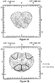

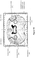

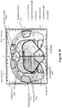
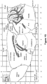


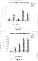

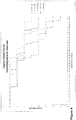


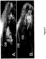
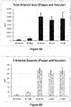
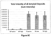


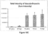

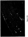
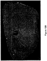

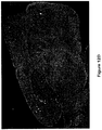

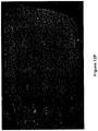
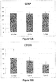
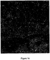
Classifications
-
- C—CHEMISTRY; METALLURGY
- C07—ORGANIC CHEMISTRY
- C07K—PEPTIDES
- C07K16/00—Immunoglobulins [IGs], e.g. monoclonal or polyclonal antibodies
- C07K16/18—Immunoglobulins [IGs], e.g. monoclonal or polyclonal antibodies against material from animals or humans
-
- A—HUMAN NECESSITIES
- A61—MEDICAL OR VETERINARY SCIENCE; HYGIENE
- A61K—PREPARATIONS FOR MEDICAL, DENTAL OR TOILETRY PURPOSES
- A61K31/00—Medicinal preparations containing organic active ingredients
- A61K31/185—Acids; Anhydrides, halides or salts thereof, e.g. sulfur acids, imidic, hydrazonic or hydroximic acids
- A61K31/19—Carboxylic acids, e.g. valproic acid
- A61K31/195—Carboxylic acids, e.g. valproic acid having an amino group
- A61K31/197—Carboxylic acids, e.g. valproic acid having an amino group the amino and the carboxyl groups being attached to the same acyclic carbon chain, e.g. gamma-aminobutyric acid [GABA], beta-alanine, epsilon-aminocaproic acid, pantothenic acid
- A61K31/198—Alpha-aminoacids, e.g. alanine, edetic acids [EDTA]
-
- A—HUMAN NECESSITIES
- A61—MEDICAL OR VETERINARY SCIENCE; HYGIENE
- A61K—PREPARATIONS FOR MEDICAL, DENTAL OR TOILETRY PURPOSES
- A61K31/00—Medicinal preparations containing organic active ingredients
- A61K31/33—Heterocyclic compounds
- A61K31/395—Heterocyclic compounds having nitrogen as a ring hetero atom, e.g. guanethidine or rifamycins
- A61K31/40—Heterocyclic compounds having nitrogen as a ring hetero atom, e.g. guanethidine or rifamycins having five-membered rings with one nitrogen as the only ring hetero atom, e.g. sulpiride, succinimide, tolmetin, buflomedil
- A61K31/401—Proline; Derivatives thereof, e.g. captopril
-
- A—HUMAN NECESSITIES
- A61—MEDICAL OR VETERINARY SCIENCE; HYGIENE
- A61K—PREPARATIONS FOR MEDICAL, DENTAL OR TOILETRY PURPOSES
- A61K31/00—Medicinal preparations containing organic active ingredients
- A61K31/33—Heterocyclic compounds
- A61K31/395—Heterocyclic compounds having nitrogen as a ring hetero atom, e.g. guanethidine or rifamycins
- A61K31/41—Heterocyclic compounds having nitrogen as a ring hetero atom, e.g. guanethidine or rifamycins having five-membered rings with two or more ring hetero atoms, at least one of which being nitrogen, e.g. tetrazole
- A61K31/4164—1,3-Diazoles
- A61K31/4172—Imidazole-alkanecarboxylic acids, e.g. histidine
-
- A—HUMAN NECESSITIES
- A61—MEDICAL OR VETERINARY SCIENCE; HYGIENE
- A61K—PREPARATIONS FOR MEDICAL, DENTAL OR TOILETRY PURPOSES
- A61K9/00—Medicinal preparations characterised by special physical form
- A61K9/0012—Galenical forms characterised by the site of application
- A61K9/0043—Nose
-
- A—HUMAN NECESSITIES
- A61—MEDICAL OR VETERINARY SCIENCE; HYGIENE
- A61P—SPECIFIC THERAPEUTIC ACTIVITY OF CHEMICAL COMPOUNDS OR MEDICINAL PREPARATIONS
- A61P25/00—Drugs for disorders of the nervous system
-
- A—HUMAN NECESSITIES
- A61—MEDICAL OR VETERINARY SCIENCE; HYGIENE
- A61P—SPECIFIC THERAPEUTIC ACTIVITY OF CHEMICAL COMPOUNDS OR MEDICINAL PREPARATIONS
- A61P25/00—Drugs for disorders of the nervous system
- A61P25/08—Antiepileptics; Anticonvulsants
-
- A—HUMAN NECESSITIES
- A61—MEDICAL OR VETERINARY SCIENCE; HYGIENE
- A61P—SPECIFIC THERAPEUTIC ACTIVITY OF CHEMICAL COMPOUNDS OR MEDICINAL PREPARATIONS
- A61P25/00—Drugs for disorders of the nervous system
- A61P25/14—Drugs for disorders of the nervous system for treating abnormal movements, e.g. chorea, dyskinesia
- A61P25/16—Anti-Parkinson drugs
-
- A—HUMAN NECESSITIES
- A61—MEDICAL OR VETERINARY SCIENCE; HYGIENE
- A61P—SPECIFIC THERAPEUTIC ACTIVITY OF CHEMICAL COMPOUNDS OR MEDICINAL PREPARATIONS
- A61P25/00—Drugs for disorders of the nervous system
- A61P25/18—Antipsychotics, i.e. neuroleptics; Drugs for mania or schizophrenia
-
- A—HUMAN NECESSITIES
- A61—MEDICAL OR VETERINARY SCIENCE; HYGIENE
- A61P—SPECIFIC THERAPEUTIC ACTIVITY OF CHEMICAL COMPOUNDS OR MEDICINAL PREPARATIONS
- A61P25/00—Drugs for disorders of the nervous system
- A61P25/22—Anxiolytics
-
- A—HUMAN NECESSITIES
- A61—MEDICAL OR VETERINARY SCIENCE; HYGIENE
- A61P—SPECIFIC THERAPEUTIC ACTIVITY OF CHEMICAL COMPOUNDS OR MEDICINAL PREPARATIONS
- A61P25/00—Drugs for disorders of the nervous system
- A61P25/28—Drugs for disorders of the nervous system for treating neurodegenerative disorders of the central nervous system, e.g. nootropic agents, cognition enhancers, drugs for treating Alzheimer's disease or other forms of dementia
-
- A—HUMAN NECESSITIES
- A61—MEDICAL OR VETERINARY SCIENCE; HYGIENE
- A61P—SPECIFIC THERAPEUTIC ACTIVITY OF CHEMICAL COMPOUNDS OR MEDICINAL PREPARATIONS
- A61P43/00—Drugs for specific purposes, not provided for in groups A61P1/00-A61P41/00
-
- C—CHEMISTRY; METALLURGY
- C07—ORGANIC CHEMISTRY
- C07K—PEPTIDES
- C07K16/00—Immunoglobulins [IGs], e.g. monoclonal or polyclonal antibodies
- C07K16/06—Immunoglobulins [IGs], e.g. monoclonal or polyclonal antibodies from serum
-
- A—HUMAN NECESSITIES
- A61—MEDICAL OR VETERINARY SCIENCE; HYGIENE
- A61K—PREPARATIONS FOR MEDICAL, DENTAL OR TOILETRY PURPOSES
- A61K39/00—Medicinal preparations containing antigens or antibodies
- A61K2039/505—Medicinal preparations containing antigens or antibodies comprising antibodies
-
- A—HUMAN NECESSITIES
- A61—MEDICAL OR VETERINARY SCIENCE; HYGIENE
- A61K—PREPARATIONS FOR MEDICAL, DENTAL OR TOILETRY PURPOSES
- A61K39/00—Medicinal preparations containing antigens or antibodies
- A61K2039/54—Medicinal preparations containing antigens or antibodies characterised by the route of administration
- A61K2039/541—Mucosal route
- A61K2039/543—Mucosal route intranasal
-
- A—HUMAN NECESSITIES
- A61—MEDICAL OR VETERINARY SCIENCE; HYGIENE
- A61K—PREPARATIONS FOR MEDICAL, DENTAL OR TOILETRY PURPOSES
- A61K9/00—Medicinal preparations characterised by special physical form
- A61K9/08—Solutions
-
- C—CHEMISTRY; METALLURGY
- C07—ORGANIC CHEMISTRY
- C07K—PEPTIDES
- C07K2317/00—Immunoglobulins specific features
- C07K2317/20—Immunoglobulins specific features characterized by taxonomic origin
- C07K2317/21—Immunoglobulins specific features characterized by taxonomic origin from primates, e.g. man
-
- C—CHEMISTRY; METALLURGY
- C07—ORGANIC CHEMISTRY
- C07K—PEPTIDES
- C07K2317/00—Immunoglobulins specific features
- C07K2317/50—Immunoglobulins specific features characterized by immunoglobulin fragments
- C07K2317/55—Fab or Fab'
-
- C—CHEMISTRY; METALLURGY
- C07—ORGANIC CHEMISTRY
- C07K—PEPTIDES
- C07K2317/00—Immunoglobulins specific features
- C07K2317/90—Immunoglobulins specific features characterized by (pharmaco)kinetic aspects or by stability of the immunoglobulin
- C07K2317/92—Affinity (KD), association rate (Ka), dissociation rate (Kd) or EC50 value
Definitions
- the central nervous system is the processing center for the nervous system.
- CNS disorders can affect the brain, the spinal cord, and nerve endings, resulting in neurological and/or psychiatric disorders.
- CNS disorders can be caused by genetic inheritance, trauma, infection, autoimmune disorders, structural defects, tumors, and stroke.
- Certain CNS disorders are characterized as neurodegenerative disease, many of which are inherited genetic diseases. Examples of neurodegenerative diseases include Huntington's disease, ALS, hereditary spastic hemiplegia, primary lateral sclerosis, spinal muscular atrophy, Kennedy's disease, Alzheimer's disease, a polyglutamine repeat disease, or Parkinson's disease.
- Treatment of CNS disorders e.g., genetic diseases of the brain such as Parkinson's disease, Huntington's disease, and Alzheimer's disease, remain an ongoing problem.
- Alzheimer's disease is a common form of age-related dementia that causes gradual loss of cognitive function, including memory and critical thinking abilities. Alzheimer's disease is diagnosed clinically by through a finding of progressive memory loss and decrease in cognitive abilities. However, confirmation of Alzheimer's disease does not occur until after death.
- Alzheimer's disease is becoming more prevalent in developed countries, where an increase in the population of elder persons has occurred due in part to improved healthcare. While less than 1% of the population under the age of 60 is affected by Alzheimer's, it is estimated that 25% to 33% of persons develop some form of Alzheimer's by the age of 85. As of 2012, 5.4 million Americans were diagnosed with Alzheimer's. As life expectancy continues to increase worldwide, the prevalence of Alzheimer's and other age-related dementia should continue to grow as well.
- Alzheimer's disease is typically classified as either "early onset,” referring to cases that begin to manifest at between 30 and 60 years of age in affected individuals, and the more common "late onset” Alzheimer's, in which symptoms first become apparent after the age of 60. Although only about 10% of all Alzheimer's cases are familial, early onset Alzheimer's disease has been linked to mutations in the amyloid precursor protein ( app ), presenilin 1 ( psen1 ), and presenilin 2 ( psen2 ) genes, while late onset Alzheimer's disease has been linked to mutations in the apolipoprotein E ( apoE ) gene ( Ertekin-Taner N., Neurol Clin., 25:611-667 (2007 )).
- apoE apolipoprotein E
- this neurodegenerative disease is characterized by the formation of amyloid plaques, neurofibrillary tangles, amyloid angiopathy, and granolovacuolar degeneration in the cerebral cortex ( Mirra et al., Arch Pathol Lab Med., 117:132-144 (1993 ); Perl DP, Neurol Clin., 18:847-864 (2000 )).
- the characteristic amyloid plaques used to confirm Alzheimer's disease post-mortem, are formed largely by deposition of a small amyloid-beta (A ⁇ ) peptide derived from the amyloid precursor protein (APP).
- cholinesterase inhibitors including donepezil (e.g ., ARICEPT®), rivastigmine (e.g ., EXELON®), galantamine (e.g ., RAZADYNE®), and tacrine (e.g ., COGNEX®); and the NMDA-type glutamate receptor inhibitor memantine (marketed under a number of different brands).
- Alzheimer's disease Although a cure for Alzheimer's disease has not been identified, these therapies serve to alleviate cognitive symptoms such as memory loss, confusion, and loss of critical thinking abilities in subjects diagnosed with age-related dementia (e.g ., Alzheimer's disease). In all, it is estimated that healthcare spending on Alzheimer's disease and related age-related dementias in 2012 will be $200 billion in the United States alone ( Factsheet, Alzheimer's Association, Mar 2012 ).
- IVIG intravenous immunoglobulin
- Immune globulin products from human plasma were first used in 1952 to treat immune deficiency. Initially, intramuscular or subcutaneous administration of immunoglobulin isotype G (IgG) isolated from plasma were the methods of choice. However, IgG products that could be administered intravenously, referred to as intravenous immunoglobulin (IVIG), were later developed to allow for the administration of larger amounts of IgG necessary for effective treatment of various diseases. Usually, IVIG contains the pooled immunoglobulin G (IgG) immunoglobulins from the plasma of multiple donors, e.g ., more than a hundred or more than a thousand blood donors.
- IgG immunoglobulin G
- IgG products are primarily used in treating three main categories of medical conditions: (1) immune deficiencies: X-linked agammaglobulinemia, hypogammaglobulinemia (primary immune deficiencies), and acquired compromised immunity conditions (secondary immune deficiencies), featuring low antibody levels; (2) inflammatory and autoimmune diseases; and (3) acute infections.
- IgG immunodeficiency
- CVID Common Variable Immunodeficiency
- HIM Hyper-IgM Syndrome
- SCID Severe Combined Immunodeficiency
- IgG subclass deficiencies Blaese and Winkelstein, J. Patient & Family Handbook for Primary Immunodeficiency Diseases. Towson, MD: Immune Deficiency Foundation; 2007 ).
- IgG treatment can be very effective for managing primary immunodeficiency disorders, this therapy is only a temporary replacement for antibodies that are not being produced in the body, rather than a cure for the disease. Accordingly, patients depend upon repeated doses of IgG therapy, typically about once a month for life. This therapy places a great demand on the continued production of IgG compositions.
- IgG is fractionated from human blood and plasma donations. Thus, the level of commercially available IgG is limited by the available supply of blood and plasma donations.
- Fraction II+III precipitate or a Kistler-Nitschmann precipitate A as the starting material for downstream processing. Both fractions are formed by a two step process in which proteins such as fibrinogen and Factor XIII are removed by an initial precipitation step (Fraction I precipitation) performed at high pH (7.2) with low ethanol concentration (8-10% v/v), followed by a second precipitation step in which IgG is precipitated from the Fraction I supernatant at pH 6.8 with 20-25% (v/v) ethanol (Fraction II+III) or at pH 5.85 with 19% ethanol (v/v) ethanol (precipitate A), while albumin and a significant portion of A1PI remain in the supernatant.
- IVIG intravenous IgG
- IVIG intravenous IgG
- IVIG intravenous IgG
- IVIG may be administered alone or in combination with other compositions. IVIG is often administered over a 2 to 5 hour period, once a day for 2 to 7 days, with follow-up doses every 10 to 21 days or every 3 to 4 weeks.
- Such an administration regime is time consuming and inconvenient for many patients. This inconvenience may be aggravated in the case of Alzheimer's patients, who may have difficulty sitting quietly during the infusion period, and may have to rely on their caregiver to bring them to an infusion center or supervise their infusion.
- Systemic IVIG administration may cause adverse side effects.
- IVIG may cause backache, headache, migraine, joint or muscle pain, general feeling of discomfort, leg cramps, rash, pain at the injection site, hives, dizziness, unusual fatigue or tiredness or weakness, chills, fever, sweating, increased heart rate, increased blood pressure, cough, redness of the face, upset stomach, upper abdominal pain, and vomiting.
- Immediate adverse effects post-IVIG administration which have been observed include headache, flushing, malaise, chest tightness, fever, chills, myalgia, fatigue, dyspnea, back pain, nausea, vomiting, diarrhea, blood pressure changes, tachycardia, and anaphylactic reactions.
- the adverse side effects may vary based on the IVIG manufacturer. Most manufactures preparations contain between 90% and 99% purified IgG in combination with stabilizers and liquid(s) for reconstitution. Orange et al. 2006 (J. Allergy Clin. Immunol. 117(4 Suppl.): S525 ); Vo et al. 2006 (Clin. J.Am. Soc. Nephrol. 1(4): 844 ; Stiehm et al. 2006 (J. Pediatr. 148(1): 6 ). For example, some manufacturers use maltose as a stabilizer while others use sucrose or amino acids.
- IVIG sodium and sugar content in IVIG, along with varying amounts of IgA and additional chemicals used in the IVIG production can affect the tolerability and efficacy of the brand of IVIG in patients.
- older patients often suffer from co-morbid conditions that increase the risk of IVIG adverse side effects.
- subjects with renal disorders, vascular disorders, or diabetes also have a heightened risk of renal insufficiency and thrombotic events following IVIG administration because IVIG compositions are commonly hyper-viscous and contain high concentrations of sugar and salt.
- IVIG also carries the risk of catheter-related infection, i.e ., an infection where the catheter or needle enters a subject's vein or skin.
- catheter-related infection i.e ., an infection where the catheter or needle enters a subject's vein or skin.
- catheter-related infection are tenderness, warmth, irritation, drainage, redness, swelling, and pain at the catheter site. Accordingly, alternate modes of administration would be beneficial from the standpoint of time, convenience, and adverse side effects.
- the BBB provides an efficient barrier, preventing and/or limiting access to the CNS of therapeutic compositions administered intravenously into the peripheral circulation. Specifically, the BBB prevents diffusion of most therapeutic compositions, especially polar compositions, into the brain from the circulating blood.
- At least three methods for increasing the passage of molecules through the BBB have been developed.
- lipophilic compounds such as lipid-soluble drugs and polar drugs encased in a lipid membrane have been developed. Lipophilic compounds with a molecular weight of less than 600 Da can diffuse through the BBB.
- therapeutic compounds can be bound to transporter molecules which can cross the BBB through a saturable transporter system. Examples of saturable transporter molecules are transferrin, insulin, IGF-1, and leptin.
- therapeutic compounds can cross the BBB by binding the therapeutic compounds to polycationic molecules such as positively-charged proteins that preferentially bind to the negatively-charged endothelial surface of the BBB. Patridge et al. 2003 (Mol. Interv.
- intranasal administration of therapeutics has become an increasingly explored method for delivering therapeutic agents to the brain because it circumvents the BBB and is a localized, non-invasive method for delivery.
- intranasal administration offers the advantages, over more traditional methods of delivery (e.g ., intravenous, subcutaneous, oral transmucosal, oral or rectal administration), of being simple to administer, providing rapid onset of action, and avoiding first-pass metabolism.
- intranasal administration has only been shown effective for the transport of small molecules, and to a certain extent smaller Fc fusion proteins, to the brain. The delivery of larger molecules, such as intact antibodies, has not yet been demonstrated.
- U.S. Patent No. 5,624,898 to Frey describes compositions and methods for transporting neurologic agents, which promote nerve cell growth and survival or augment the activity of functioning cells, to the brain by means of the olfactory neural pathway.
- the neurological agents of the '898 patent are transported to the brain by means of the nervous system, rather than the circulatory system, so that potentially therapeutic agents that are unable to cross the blood-brain barrier may be delivered to damaged neurons in the brain.
- the compositions described in the '898 patent include a neurologic agent in combination with a pharmaceutical carrier and/or additive which promote the transfer of the agent within the olfactory system.
- the '898 patent does not teach intranasal administration of pooled human immunoglobulins.
- the '957 PCT publication published three years after the '332 PCT publication, states that "[i]n published delivery studies to date, intranasal delivery efficiency to the CNS has been very low and the delivery of large globular macromolecules, such as antibodies and their fragments, has not been demonstrated.”
- the '957 PCT publication purports to solve this problem through the use of a permeability enhancer (e.g ., membrane fluidizers, tight junction modulators, and medium chain length fatty acids and salts and esters thereof, as described below), which enhances intranasal administration to the central nervous system.
- a permeability enhancer e.g ., membrane fluidizers, tight junction modulators, and medium chain length fatty acids and salts and esters thereof, as described below
- Neither PCT publication teaches intranasal administration of pooled human immunoglobulins.
- PCT publication WO 2003/028668 by Barstow et al. describes the treatment of immune-mediated diseases by alimentary administration (i.e., administration to the digestive tract) of pooled immunoglobulins.
- alimentary administration i.e., administration to the digestive tract
- the '668 PCT publication discloses nasal administration of a composition of pooled immunoglobulins, it is in the context of delivering the composition to the digestive tract.
- the '668 PCT publication does not teach the delivery of pooled human immunoglobulins to the brain via intranasal administration.
- PCT publication WO 2001/60420 by Adjei et al. describes aerosol formulations of therapeutic polypeptides, including immunoglobulins, for pulmonary delivery. These aerosolizable compositions are formulated such that after oral or nasal inhalation, the therapeutic agent is effectively delivered to the patient's lungs.
- the '420 PCT publication does not teach the delivery of therapeutic agents to the brain via intranasal administration.
- WO 2009/111240 A1 concerns anti-amyloid ⁇ activity of intravenous immunoglobulin (IVIG) in vitro.
- the current invention is defined, inter alia, by the following items:
- intranasal administration provides directed delivery of pooled IgG to the brain, bypassing the requirement that it pass through the blood brain barrier (BBB).
- BBB blood brain barrier
- intranasal administration allows the delivery of intact IgG to the brain. This results in greater efficiency for the treatment and reduces the necessary IgG dose that must be administered to achieve the desired effect.
- pooled human IgG is isolated from donated human plasma, pooled IgG is a limited resource.
- IgG is efficiently transported to the brain when intranasally administered in the absence of permeability enhancers, some of which have neurostimulatory effects themselves.
- the disclosure provides a method for treating a central nervous system (CNS) disorder in a subject in need thereof, the method including delivering a therapeutically effective amount of a composition comprising pooled human immunoglobulin G (IgG) to the brain of the subject, where delivering the composition to the brain includes intranasally administering the composition directly to the olfactory epithelium of the nasal cavity of the subject.
- CNS central nervous system
- the disclosure provides a method for treating a central nervous system (CNS) disorder in a subject in need thereof, the method including delivering a therapeutically effective amount of a composition comprising pooled human immunoglobulin G (IgG) to the brain of the subject, where delivering the composition to the brain includes intranasally administering the composition to a nasal epithelium of the subject associated with trigeminal nerve endings.
- CNS central nervous system
- the disclosure provides a method for treating a central nervous system (CNS) disorder in a subject in need thereof, the method including delivering a therapeutically effective amount of a composition comprising pooled human immunoglobulin G (IgG) to the brain of the subject, where delivering the composition to the brain includes intranasally administering the composition to the upper third of the nasal cavity of the subject.
- CNS central nervous system
- the disclosure provides a method for treating a central nervous system (CNS) disorder in a subject in need thereof, the method including delivering a therapeutically effective amount of a composition comprising pooled human immunoglobulin G (IgG) to the brain of the subject, where delivering the composition to the brain includes intranasally administering the composition to one or both maxillary sinus of the subject.
- CNS central nervous system
- the CNS disorder is selected from the group consisting of a neurodegenerative disorder of the central nervous system, a systemic atrophy primarily affecting the central nervous system, an extrapyramidal and movement disorder, a demyelinating disorder of the central nervous system, an episodic or paroxysmal disorder of the central nervous system, a paralytic syndrome of the central nervous system, a nerve, nerve root, or plexus disorder of the central nervous system, an organic mental disorder, a mental or behavioral disorder caused by psychoactive substance use, a schizophrenia, schizotypal, or delusional disorder, a mood (affective) disorder, neurotic, stress-related, or somatoform disorder, a behavioral syndrome, an adult personality or behavior disorder, a psychological development disorder, and a child onset behavioral or emotional disorder.
- a neurodegenerative disorder of the central nervous system a systemic atrophy primarily affecting the central nervous system
- an extrapyramidal and movement disorder a demyelinating disorder of the central nervous system
- the CNS disorder is selected from the group consisting of Alzheimer's disease, Parkinson's disease, multiple sclerosis, amyotrophic lateral sclerosis (ALS), Huntington's disease, cerebral palsy, bipolar disorder, schizophrenia, and Pediatric acute-onset neuropyschiatric syndrome (PANS).
- ALS amyotrophic lateral sclerosis
- PANS Pediatric acute-onset neuropyschiatric syndrome
- the CNS disorder is selected from the group consisting of Alzheimer's disease, Parkinson's disease, multiple sclerosis, Pediatric Autoimmune Neuropsychiatric Disorders Associated with Streptococcal infections (PANDAS), and Pediatric acute-onset neuropyschiatric syndrome (PANS).
- PANDAS Pediatric Autoimmune Neuropsychiatric Disorders Associated with Streptococcal infections
- PANS Pediatric acute-onset neuropyschiatric syndrome
- the CNS disorder is selected from the group consisting of Alzheimer's disease, multiple sclerosis, and Parkinson's disease.
- the CNS disorder is Alzheimer's disease.
- intranasal administration of the composition includes the use of a non-invasive intranasal delivery device.
- intranasal administration of the composition includes administration of a liquid drop of the composition directly onto the nasal epithelium, the nasal epithelium of the subject associated with trigeminal nerve endings, or the upper third of the nasal cavity of the subject.
- intranasal administration of the composition includes directed administration of an aerosol of the composition to the nasal epithelium, the nasal epithelium of the subject associated with trigeminal nerve endings, or the upper third of the nasal cavity of the subject.
- the aerosol of the composition is a liquid aerosol.
- the aerosol of the composition is a powder aerosol.
- At least 40% of the pooled human IgG administered to the subject contacts the nasal epithelium of the subject, the olfactory epithelium of the nasal cavity of the subject, a nasal epithelium of the subject associated with trigeminal nerve endings, the upper third of the nasal cavity of the subject, or one or both maxillary sinus of the subject.
- At least 50% of the pooled human IgG administered to the subject contacts the olfactory epithelium of the nasal cavity of the subject, the nasal epithelium of the subject associated with trigeminal nerve endings, the upper third of the nasal cavity of the subject, or one or both maxillary sinus of the subject.
- At least 60% of the pooled human IgG administered to the subject contacts the olfactory epithelium of the nasal cavity of the subject, the nasal epithelium of the subject associated with trigeminal nerve endings, the upper third of the nasal cavity of the subject, or one or both maxillary sinus of the subject.
- the pooled human IgG composition does not contain a permeability enhancer.
- the pooled human IgG composition consists essentially of pooled human IgG and an amino acid.
- the amino acid is selected from the group consisting of glycine, histidine, and proline. In a specific embodiment of the methods provided above, the amino acid is glycine.
- the pooled human IgG composition is an aqueous composition.
- the pooled human IgG composition includes from 10 mg/mL to 250 mg/mL pooled human IgG and from 50 mM to 500 mM glycine.
- the pH of the composition is from 4.0 to 6.0. In another embodiment of the methods provided above, the pH of the composition is from 4.0 to 7.5. In another embodiment of the methods provided above, the pH of the composition is from 6.0 to 7.5.
- the pooled human IgG composition is a dry powder composition.
- the dry powder composition is prepared from an aqueous solution including from 10 mg/mL to 250 mg/mL pooled human IgG and from 50 mM to 500 mM glycine.
- the dry powder composition is prepared from an aqueous solution having a pH of from 4.0 to 6.0.
- the pH of the composition is from 4.0 to 7.5
- the pH of the composition is from 6.0 to 7.5
- the method includes intranasally administering to the subject a dose of from 0.08 mg to 100 mg pooled human IgG per kg body weight of the subject (mg IgG/kg). In a specific embodiment of the methods provided above, the method includes intranasally administering to the subject a dose of from 0.2 mg to 40 mg pooled human IgG per kg body weight of the subject (mg IgG/kg). In a specific embodiment of the methods provided above, the method includes intranasally administering to the subject a dose of from 5 mg to 20 mg pooled human IgG per kg body weight of the subject (mg IgG/kg).
- the method includes intranasally administering to the subject a dose of from 5 mg to 10 mg pooled human IgG per kg body weight of the subject (mg IgG/kg). In a specific embodiment of the methods provided above, the method includes intranasally administering to the subject a dose of from 1 mg to 5 mg pooled human IgG per kg body weight of the subject (mg IgG/kg).
- the method includes intranasally administering to the subject a fixed dose of from 50 mg to 10 g pooled human IgG. In a specific embodiment of the methods provided above, the method includes intranasally administering to the subject a fixed dose of from 100 mg to 5.0 g pooled human IgG. In a specific embodiment of the methods provided above, the method includes intranasally administering to the subject a fixed dose of from 500 mg to 2.5 g pooled human IgG.
- the method includes intranasally administering to the subject a dose of pooled human IgG at least twice monthly. In a specific embodiment of the methods described above, the method includes intranasally administering to the subject a dose of pooled human IgG at least once weekly. In a specific embodiment of the methods described above, the method includes intranasally administering to the subject a dose of pooled human IgG at least twice weekly. In a specific embodiment of the methods described above, the method includes intranasally administering to the subject a dose of pooled human IgG at least once daily. In a specific embodiment of the methods described above, the method includes intranasally administering to the subject a dose of pooled human IgG at least twice daily.
- the pooled human IgG composition includes at least 0.1% anti-amyloid ⁇ IgG.
- the method further includes administering a second therapy for the CNS disorder to the subject in need thereof.
- the second therapy for the CNS disorder is a cholinesterase inhibitor.
- the cholinesterase inhibitor is selected from the group consisting of donepezil (e.g ., ARICEPT®), rivastigmine (e.g ., EXELON®), galantamine ( e.g ., RAZADYNE®), and tacrine ( e.g ., COGNEX®).
- the second therapy for the CNS disorder is an inhibitor of NMDA-type glutamate receptor.
- the inhibitor of NMDA-type glutamate receptor is memantine.
- the disclosure provides the use of a composition comprising pooled human immunoglobulin G (IgG) for the treatment of a central nervous system (CNS) disorder in a subject in need thereof by intranasal administration.
- IgG human immunoglobulin G
- intranasal administration includes administration to the nasal epithelium of the subject. In other embodiments of the uses described above, intranasal administration comprises administration to the olfactory epithelium of the nasal cavity of the subject. In other embodiments of the uses described above, intranasal administration includes administration to a nasal epithelium of the subject associated with trigeminal nerve endings. In other embodiments of the uses described above, intranasal administration includes administration to the upper third of the nasal epithelium of the nasal cavity of the subject. In yet other embodiments, of the uses described above, intranasal administration includes administration to one or both maxillary sinus of the subject.
- the CNS disorder is selected from the group consisting of a neurodegenerative disorder of the central nervous system, a systemic atrophy primarily affecting the central nervous system, an extrapyramidal and movement disorder, a demyelinating disorder of the central nervous system, an episodic or paroxysmal disorder of the central nervous system, a paralytic syndrome of the central nervous system, a nerve, nerve root, or plexus disorder of the central nervous system, an organic mental disorder, a mental or behavioral disorder caused by psychoactive substance use, a schizophrenia, schizotypal, or delusional disorder, a mood (affective) disorder, neurotic, stress-related, or somatoform disorder, a behavioral syndrome, an adult personality or behavior disorder, a psychological development disorder, and a child onset behavioral or emotional disorder.
- a neurodegenerative disorder of the central nervous system a systemic atrophy primarily affecting the central nervous system
- an extrapyramidal and movement disorder a demyelinating disorder of the central nervous system
- the CNS disorder is selected from the group consisting of Alzheimer's disease, Parkinson's disease, multiple sclerosis, amyotrophic lateral sclerosis (ALS), Huntington's disease, cerebral palsy, bipolar disorder, schizophrenia, and Pediatric acute-onset neuropyschiatric syndrome (PANS).
- ALS amyotrophic lateral sclerosis
- PANS Pediatric acute-onset neuropyschiatric syndrome
- the CNS disorder is selected from the group consisting of Alzheimer's disease, Parkinson's disease, multiple sclerosis, Pediatric Autoimmune Neuropsychiatric Disorders Associated with Streptococcal infections (PANDAS), and Pediatric acute-onset neuropyschiatric syndrome (PANS).
- PANDAS Pediatric Autoimmune Neuropsychiatric Disorders Associated with Streptococcal infections
- PANS Pediatric acute-onset neuropyschiatric syndrome
- the CNS disorder is selected from the group consisting of Alzheimer's disease, multiple sclerosis, and Parkinson's disease.
- the CNS disorder is Alzheimer's disease.
- intranasal administration of the composition includes the use of a non-invasive intranasal delivery device.
- intranasal administration of the composition includes administration of a liquid drop of the composition directly onto the nasal epithelium, the nasal epithelium of the subject associated with trigeminal nerve endings, or the upper third of the nasal cavity of the subject.
- intranasal administration of the composition includes directed administration of an aerosol of the composition to the nasal epithelium, the nasal epithelium of the subject associated with trigeminal nerve endings, or the upper third of the nasal cavity of the subject.
- the aerosol of the composition is a liquid aerosol.
- the aerosol of the composition is a powder aerosol.
- At least 40% of the pooled human IgG administered to the subject contacts the nasal epithelium of the subject, the olfactory epithelium of the nasal cavity of the subject, a nasal epithelium of the subject associated with trigeminal nerve endings, the upper third of the nasal cavity of the subject, or one or both maxillary sinus of the subject.
- At least 50% of the pooled human IgG administered to the subject contacts the nasal epithelium of the subject, the olfactory epithelium of the nasal cavity of the subject, a nasal epithelium of the subject associated with trigeminal nerve endings, the upper third of the nasal cavity of the subject, or one or both maxillary sinus of the subject.
- At least 60% of the pooled human IgG administered to the subject contacts the nasal epithelium of the subject, the olfactory epithelium of the nasal cavity of the subject, a nasal epithelium of the subject associated with trigeminal nerve endings, the upper third of the nasal cavity of the subject, or one or both maxillary sinus of the subject.
- the pooled human IgG composition does not contain a permeability enhancer.
- the pooled human IgG composition consists essentially of pooled human IgG and an amino acid.
- the amino acid is selected from the group consisting of glycine, histidine, and proline. In a specific embodiment of the methods provided above, the amino acid is glycine.
- the pooled human IgG composition is an aqueous composition.
- the pooled human IgG composition includes from 10 mg/mL to 250 mg/mL pooled human IgG and from 50 mM to 500 mM glycine.
- the pH of the composition is from 4.0 to 6.0. In another embodiment of the uses described above, the pH of the composition is from 4.0 to 7.5. In another embodiment of the methods provided above, the pH of the composition is from 6.0 to 7.5.
- the pooled human IgG composition is a dry powder composition.
- the dry powder composition is prepared from an aqueous solution including from 10 mg/mL to 250 mg/mL pooled human IgG and from 50 mM to 500 mM glycine.
- the dry powder composition is prepared from an aqueous solution having a pH of from 4.0 to 6.0.
- the pH of the composition is from 4.0 to 7.5 In another embodiment of the uses described above, the pH of the composition is from 6.0 to 7.5
- the use includes intranasally administering to the subject a dose of from 0.08 mg to 100 mg pooled human IgG per kg body weight of the subject (mg IgG/kg). In a specific embodiment of the uses described above, the use includes intranasally administering to the subject a dose of from 0.2 mg to 40 mg pooled human IgG per kg body weight of the subject (mg IgG/kg). In a specific embodiment of the uses described above, the use includes intranasally administering to the subject a dose of from 5 mg to 20 mg pooled human IgG per kg body weight of the subject (mg IgG/kg).
- the use includes intranasally administering to the subject a dose of from 5 mg to 10 mg pooled human IgG per kg body weight of the subject (mg IgG/kg). In a specific embodiment of the uses described above, the use includes intranasally administering to the subject a dose of from 1 mg to 5 mg pooled human IgG per kg body weight of the subject (mg IgG/kg).
- the use includes intranasally administering to the subject a fixed dose of from 50 mg to 10 g pooled human IgG. In a specific embodiment of the uses provided above, the use includes intranasally administering to the subject a fixed dose of from 100 mg to 5.0 g pooled human IgG. In a specific embodiment of the uses provided above, the use includes intranasally administering to the subject a fixed dose of from 500 mg to 2.5 g pooled human IgG.
- the method includes intranasally administering to the subject a dose of pooled human IgG at least twice monthly. In a specific embodiment of the uses described above, the method includes intranasally administering to the subject a dose of pooled human IgG at least once weekly. In a specific embodiment of the uses described above, the method includes intranasally administering to the subject a dose of pooled human IgG at least twice weekly. In a specific embodiment of the uses described above, the method includes intranasally administering to the subject a dose of pooled human IgG at least once daily. In a specific embodiment of the uses described above, the method includes intranasally administering to the subject a dose of pooled human IgG at least twice daily.
- the pooled human IgG composition includes at least 0.1% anti-amyloid ⁇ IgG.
- the method further includes administering a second therapy for the CNS disorder to the subject in need thereof.
- the second therapy for the CNS disorder is a cholinesterase inhibitor.
- the cholinesterase inhibitor is selected from the group consisting of donepezil (e.g ., ARICEPT®), rivastigmine (e.g ., EXELON®), galantamine ( e.g ., RAZADYNE®), or tacrine ( e.g ., COGNEX®).
- the second therapy for the CNS disorder is an inhibitor of NMDA-type glutamate receptor.
- the inhibitor of NMDA-type glutamate receptor is memantine.
- the present disclosure provides methods and compositions for treating a central nervous system (CNS) disorder in a subject by intranasal delivery of a therapeutically effective amount of pooled human immunoglobulin G (IgG) directly to the epithelium of the nasal cavity of the subject.
- IgG immunoglobulin G
- pooled human IgG is administered directly to the olfactory epithelium of the nasal cavity.
- pooled IgG is delivered to the upper third of the nasal cavity, e.g ., above the lower turbinates.
- pooled IgG is delivered to the brain via the trigeminal nerve after intranasal administration to the nasal respiratory epithelium.
- pooled IgG is delivered to the brain via the maxillary nerve after intranasal administration to the nasal respiratory epithelium. In other embodiments, pooled IgG is delivered to the brain after administration to the maxillary sinus.
- methods and compositions for the treatment of Alzheimer's disease, multiple sclerosis, and Parkinson's disease via intranasal administration of pooled human IgG are provided herein.
- the methods and compositions provided herein are useful for the treatment of CNS disorder known to one of skill in the art including, without limitation, a neurodegenerative disorder of the central nervous system, a systemic atrophy primarily affecting the central nervous system, an extrapyramidal and movement disorder, a demyelinating disorder of the central nervous system, an episodic or paroxysmal disorder of the central nervous system, a paralytic syndrome of the central nervous system, a nerve, nerve root, or plexus disorder of the central nervous system, an organic mental disorder, a mental or behavioral disorder caused by psychoactive substance use, a schizophrenia, schizotypal, or delusional disorder, a mood (affective) disorder, neurotic, stress-related, or somatoform disorder, a behavioral syndrome, an adult personality or behavior disorder, a psychological development disorder, or
- the CNS disorder is selected from the group consisting of Alzheimer's disease, Parkinson's disease, multiple sclerosis, amyotrophic lateral sclerosis (ALS), Huntington's disease, cerebral palsy, bipolar disorder, schizophrenia, or Pediatric acute-onset neuropyschiatric syndrome (PANS).
- the CNS disorder is selected from the group consisting of Alzheimer's disease, Parkinson's disease, multiple sclerosis, Pediatric Autoimmune Neuropsychiatric Disorders Associated with Streptococcal infections (PANDAS), or Pediatric acute-onset neuropyschiatric syndrome (PANS).
- intranasal administration of IgG increased weight and survival time of Alzheimer's disease mice models.
- intranasal administration of IgG at either high (0.8 g/kg once every two weeks) or low (0.4 g/k once every two weeks) doses, prolonged the lifespan of TG2576 mice.
- This result shows that intranasal administration of IgG is capable of increasing lifespan in the Alzheimer's mouse model, indicating efficacy in Alzheimer's treatment.
- intranasal administration results in a much more discriminate delivery of pooled human IgG to the brain, as compared to intravenous administration.
- intranasal administration of pooled human immunoglobulin G resulted in a 6-fold lower blood exposure as compared to intravenous administration.
- the lower system exposure of IgG provided by intranasal administration advantageously reduces the risk of side effects associated with the systemic exposure of IgG.
- pooled human immunoglobulin G was efficiently delivered to the brain following intranasal administration in the absence of a permeability enhancer (e.g ., membrane fluidizers, tight junction modulators, and medium chain length fatty acids and salts and esters thereof, as described below).
- a permeability enhancer e.g ., membrane fluidizers, tight junction modulators, and medium chain length fatty acids and salts and esters thereof, as described below.
- biotherapeutics e.g ., mimetibodies and Fc fusions
- a permeability enhancer is required ( WO 2009/058957 ).
- pooled human IgG is efficiently delivered to the brain when intranasally administered as a liquid or dry powder formulated with only an amino acid (e.g ., glycine).
- the dose of pooled human IgG can be significantly reduced when administered intranasally, as compared to intravenous administration.
- Example 9 administration of a low dose of pooled human IgG (0.002 g/kg IgG) intranasally delivered directly to the olfactory epithelium results in substantially the same amount of IgG being delivered to the right and left hemispheres of the brain as for intravenous administration of a ten-fold higher dose of pooled human IgG (0.02 g/kg IgG; compare corrected AUC values for right and left hemisphere IgG delivery in Table 71 and Table 72 ).
- a ten-fold reduction in the amount of pooled human IgG required for administration is significant because of the limited supply of pooled human IgG and the high cost associated therewith.
- Example 2 it is shown in Example 2 that, on average, the concentration of Fabs in brain tissue post-intranasal administration is 19-times higher than the concentration of full-length immunoglobulins post-intranasal administration. Given the significantly lower delivery of full-length immunoglobulins to the brain, it is surprising that intranasal administration of pooled immunoglobulins provides the effective results shown herein.
- intranasal delivery of the pooled human IgG composition disclosed herein can be accomplished by a non-invasive means, as compared to intravenous, subcutaneous, and intramuscular administration, all of which require puncture of the skin of the subject.
- a non-invasive means as compared to intravenous, subcutaneous, and intramuscular administration, all of which require puncture of the skin of the subject.
- pooled human IgG can be efficiently delivered to the brain using nasal drops or a nasal spray.
- IVIG intravenous IgG
- IVIG intravenous IgG
- initial administration of IVIG occurs over a 2 to 5 hour period, once a day for 2 to 7 consecutive days.
- follow-up doses also typically administered at a hospital over a period of 2 to 5 hours, are required every 1 to 4 weeks depending on the indication being treated and dosing regimen.
- Such an administration regime is time consuming and inconvenient for many patients.
- intranasal administration can be administered at home without medical supervision.
- intranasal administration can be performed quickly, over several minutes depending on the number of drops/sprays required, rather than several hours as required for intravenous administration.
- treatment can be prescribed more frequently at lower doses to maintain an effective level of IgG in the CNS with minimal inconvenience because administration occurs at home in a shorter period of time.
- IVIG therapy requires catheterization which can cause discomfort and infection at the site of the catheter.
- IVIG solutions are often high in sodium and glucose to create isotonicity, causing increased risk to the elderly population, which already have increased rates of diabetes and high blood pressure.
- intranasal administration is non-invasive, i.e. there is no catheterization and does not carry invasive-procedure related risks such as infection and discomfort at the site of the catheter.
- Intranasal administration of pooled human IgG compositions is an improved procedure for elderly persons because it does not require IV perfusion and thus does not create a systemic increase in concentrations of salt or glucose in the blood.
- pooled human IgG e.g., intravenous, subcutaneous, and intramuscular
- intranasal administration increases the ease of administration, decreases overall administration time, decreases the number of hospital visits required, and eliminates the risks associated with catheter-based administration (e.g ., IV administration).
- catheter-based administration e.g ., IV administration
- implementation of intranasal administration of pooled human IgG will result in improved patient compliance.
- disorder of the central nervous system refers to a disorder affecting either the spinal cord (e.g ., a myelopathy) or brain (e.g ., an encephalopathy) of a subject, which commonly presents with neurological and/or psychiatric symptoms.
- CNS disorders include many neurodegenerative diseases (e.g ., Huntington's disease, Amyotrophic lateral sclerosis (ALS), hereditary spastic hemiplegia, primary lateral sclerosis, spinal muscular atrophy, Kennedy's disease, Alzheimer's disease, ataxias, Huntington's disease, Lewy body disease, a polyglutamine repeat disease, and Parkinson's disease) and psychiatric disorders (e.g ., mood disorders, schizophrenias, and autism).
- ataxia include Friedreich's ataxia and the spinocerebellar ataxias.
- CNS disorders do not include disorders resulting from acute viral and bacterial infections.
- Non-limiting examples of CNS disorders include neurodegenerative disorders of the central nervous system, systemic atrophies primarily affecting the central nervous system, extrapyramidal and movement disorders, demyelinating disorders of the central nervous system, episodic or paroxysmal disorders of the central nervous system, paralytic syndromes of the central nervous system, nerve, nerve root, or plexus disorders of the central nervous system, organic mental disorders, mental or behavioral disorders caused by psychoactive substance use, schizophrenic, schizotypal, or delusional disorders, mood (affective) disorders, neurotic, stress-related, or somatoform disorders, behavioral syndromes, adult personality or behavior disorders, psychological development disorders, and child onset behavioral or emotional disorders.
- DSM-IV Diagnostic and Statistical Manual of Mental Disorders, 4th Edition (DSM-IV); The World Health Organization, The International Classification of Diseases, 10th revision (ICD-10), Chapter V.
- Further exemplary CNS disorders are provided herein below.
- Neurodegenerative CNS disorders are typically characterized by progressive dysfunction and/or cell death in the central nervous system.
- the hallmark of many neurodegenerative CNS disorders is the accumulation of misfolded proteins, such as beta-amyloid, tau, alpha-synuclein, and TDP-43, both intracellularly and extracellularly.
- Many neurodegenerative diseases are also associated with gross mitochondrial dysfunction.
- Common examples of neurodegenerative CNS disorders include Alzheimer's disease (AD), Parkinson's disease (PD), Huntington's disease, and Amyotrophic lateral sclerosis (ALS).
- AD Alzheimer's disease
- PD Parkinson's disease
- Huntington's disease Huntington's disease
- ALS Amyotrophic lateral sclerosis
- Psychiatric disorders also referred to as mental illnesses
- Psychiatric disorders are generally defined by a combination of how a person feels, acts, thinks or perceives.
- Well established systems for the classification of psychiatric disorders include the International Statistical Classification of Diseases and Related Health Problems, 10th Revision (World Health Organization, tenth revision (2010), ) and the Diagnostic and Statistical Manual of Mental Disorders (DSM-IV; American Psychiatric Association, DS-IV-TR (2000), ).
- DSM-IV Diagnostic and Statistical Manual of Mental Disorders
- Common examples of psychiatric disorders include mood disorders, schizophrenia, and autism.
- the terms “pooled human immunoglobulin G” and “pooled human IgG” refer to a composition containing polyvalent immunoglobulin G (IgG) purified from the blood/plasma of multiple donors, e.g ., more than a hundred or more than a thousand blood donors.
- the composition will be at least 80% IgG (w/w, e.g ., weight IgG per weight total protein), preferably at least 85%, 86%, 87%, 88%, 89%, 90%, 91%, 92%, 93%, 94%, 95%, 96%, 97%, 98%, or 99% IgG (w/w).
- the pooled human IgG composition contains intact IgG immunoglobulins. In other embodiments, the pooled human IgG composition contains IgG fragments, for example those prepared by treatment of intact antibodies with trypsin. In certain embodiments, the pooled human IgG compositions used in the treatments disclosed herein contain natural or synthetic modifications, e.g ., post-translational modifications and/or chemical modifications.
- high titer anti-amyloid ⁇ pooled immunoglobulin G and "high titer anti-amyloid ⁇ pooled IgG” refer to a composition containing polyvalent immunoglobulin G (IgG) purified from the blood/plasma of multiple donors, e.g ., more than a hundred or more than a thousand blood donors, having a relative titer of anti-amyloid ⁇ immunoglobulin G that is greater than the expected titer of anti-amyloid ⁇ immunoglobulins in a pooled IgG composition prepared from the blood/plasma of more than a thousand random individuals.
- IgG polyvalent immunoglobulin G
- IVIG intravenous immunoglobulin G
- IgGs that specifically recognize epitopes of various conformers of amyloid ⁇ , e.g ., amyloid ⁇ monomers, amyloid ⁇ fibrils, and cross-linked amyloid ⁇ protein species (CAPS).
- GAMMAGARD LIQUID® (10% Immune Globulin Infusion (Human); Baxter International Inc., Deerfield, IL) contains 0.1% anti-amyloid ⁇ fibril IgG, 0.04% anti-CAPS IgG, and 0.02% anti-amyloid ⁇ monomer IgG, having EC 50 affinities of 40 nM, 40 nM, and 350 nM for their target amyloid ⁇ conformer, respectively ( O'Nuallain B.
- a high titer anti-amyloid ⁇ pooled immunoglobulin G composition contains a high titer of IgG specific for one or more conformer of amyloid ⁇ . In other embodiments, a high titer anti-amyloid ⁇ pooled immunoglobulin G composition contains a high titer of IgG specific for amyloid ⁇ monomers, amyloid ⁇ fibrils, and cross-linked amyloid ⁇ protein species (CAPS).
- CAPS cross-linked amyloid ⁇ protein species
- a high titer anti-amyloid ⁇ pooled immunoglobulin G composition contains at least 0.1% anti-amyloid ⁇ IgG (e.g ., 0.1% IgG with specific affinity for any amyloid ⁇ conformer). In another embodiment, a high titer anti-amyloid ⁇ pooled immunoglobulin G composition contains at least 0.2% anti-amyloid ⁇ IgG.
- a high titer anti-amyloid ⁇ pooled immunoglobulin G composition contains at least 0.15%, 0.2%, 0.25%, 0.3%, 0.35%, 0.4%, 0.45%, 0.5%, 0.55%, 0.6%, 0.65%, 0.7%, 0.75%, 0.8%, 0.85%, 0.9%, 0.95%, 1.0%, 1.5%, 2.0%, 2.5%, 3.0%, 3.5%, 4.0%, 4.5%, 5.0% or more anti-amyloid ⁇ IgG.
- a high titer anti-amyloid ⁇ pooled immunoglobulin G composition contains at least 0.1% anti-amyloid ⁇ fibril IgG. In another embodiment, a high titer anti-amyloid ⁇ pooled immunoglobulin G composition contains at least 0.2% anti-amyloid ⁇ fibril IgG.
- a high titer anti-amyloid ⁇ pooled immunoglobulin G composition contains at least 0.15%, 0.2%, 0.25%, 0.3%, 0.35%, 0.4%, 0.45%, 0.5%, 0.55%, 0.6%, 0.65%, 0.7%, 0.75%, 0.8%, 0.85%, 0.9%, 0.95%, 1.0%, 1.5%, 2.0%, 2.5%, 3.0%, 3.5%, 4.0%, 4.5%, 50% or more anti-amyloid ⁇ fibril IgG.
- a high titer anti-amyloid ⁇ pooled immunoglobulin G composition contains at least 0.04% anti-CAPS IgG. In another embodiment, a high titer anti-amyloid ⁇ pooled immunoglobulin G composition contains at least 0.08% anti-CAPS IgG.
- a high titer anti-amyloid ⁇ pooled immunoglobulin G composition contains at least 0.04%, 0.05%, 0.06%, 0.07%, 0.08%, 0.09%, 0.1%, 0.15%, 0.2%, 0.25%, 0.3%, 0.35%, 0.4%, 0.45%, 0.5%, 0.55%, 0.6%, 0.65%, 0.7%, 0.75%, 0.8%, 0.85%, 0.9%, 0.95%, 1.0%, 1.5%, 2.0%, 2.5%, 3.0%, 3.5%, 4.0%, 4.5%, 5.0% or more anti-CAPS IgG.
- a high titer anti-amyloid ⁇ pooled immunoglobulin G composition contains at least 0.02% anti-amyloid ⁇ monomer IgG. In another embodiment, a high titer anti-amyloid ⁇ pooled immunoglobulin G composition contains at least 0.04% anti-amyloid ⁇ monomer IgG.
- a high titer anti-amyloid ⁇ pooled immunoglobulin G composition contains at least 0.02%, 0.03%, 0.04%, 0.05%, 0.06%, 0.07%, 0.08%, 0.09%, 0.1%, 0.15%, 0.2%, 0.25%, 0.3%, 0.35%, 0.4%, 0.45%, 0.5%, 0.55%, 0.6%, 0.65%, 0.7%, 0.75%, 0.8%, 0.85%, 0.9%, 0.95%, 1.0%, 1.5%, 2.0%, 2.5%, 3.0%, 3.5%, 4.0%, 4.5%, 5.0% or more anti-amyloid ⁇ monomer IgG.
- High titer anti-amyloid ⁇ pooled IgG can be prepared according to standard methods for the manufacture of pooled IgG starting with a standard pool of blood/plasma of multiple donors, e.g ., more than a hundred or more than a thousand blood donors, and subsequently enriched for anti-amyloid ⁇ immunoglobulin G. Methods for the enrichment of target-specific immunoglobulin G molecules are well known in the art (for example, see U.S. Patent Application Publication No. 2004/0101909 ).
- high titer anti-amyloid ⁇ pooled IgG can be prepared according to standard methods for the manufacture of pooled IgG starting with an enriched pool of blood/plasma from at least fifty, one hundred, two hundred, five hundred, or one thousand donors having a high relative titer of anti-amyloid ⁇ immunoglobulin G.
- hyperimmune IgG preparations are commonly prepared from smaller donor pools.
- enriched pools of blood/plasma can be formed, for example, by selectively pooling blood/plasma donations or donors with a high relative titer of anti-amyloid ⁇ immunoglobulin G, e.g ., by selection of high titer blood/plasma donations or donors.
- an enriched pool of blood/plasma can be formed by screening for blood/plasma donations or donors with a low relative titer of anti-amyloid ⁇ immunoglobulin G and excluding these donations or donors from the starting blood/plasma pool, e.g ., screening for low titer blood/plasma donations or donors.
- the term "intactness” refers to a percentage of therapeutic agent that has not been at least partially degraded at a particular point in time following administration.
- intactness is a measure of the total administered dose of the therapeutic agent that has not been at least partially degraded at the particular point in time (i.e ., systemic intactness).
- intactness is a measure of the therapeutic agent present at a particular site of the subject, e.g ., brain or bloodstream, which has not been at least partially degraded ( i.e ., local intactness).
- the intactness of administered immunoglobulin e.g ., pooled human IgG is measured by mass spectroscopy.
- the intactness of the administered immunoglobulins is determined by analyzing a biological sample from the subject, or proteins extracted from the biological sample, by mass spectroscopy.
- the intactness of the administered immunoglobulins is determined by separating proteins present in a biological sample from the subject by molecular weight, size, or shape ( e.g ., by electrophoresis or size exclusion chromatography) and determining the size distribution of administered immunoglobulins in the sample.
- the intactness of immunoglobulin (e.g ., pooled human IgG) in the brain of a subject following intranasal administration is at least 40%. In preferred embodiments, the intactness of immunoglobulin (e.g ., pooled human IgG) in the brain of a subject following intranasal administration is at least 50%, preferably at least 60%.
- the intactness of immunoglobulin e.g ., pooled human IgG
- the intactness of immunoglobulin is at least 35%, 36%, 37%, 38%, 39%, 40%, 41%, 42%, 43%, 44%, 45%, 46%, 47%, 48%, 49%, 50%, 51%, 52%, 53%, 54%, 55%, 56%, 57%, 58%, 59%, 60%, 61%, or higher.
- intranasal administration and “nasal administration” refer to administration of a therapeutic composition to the nasal cavity of a subject such that a therapeutic agent in the composition is delivered directly to one or more epithelium located in the nose.
- intranasal administration is achieved using a liquid preparation (e.g ., an aqueous preparation), an aerosolized preparation, or a dry powder preparation, each of which can be administered via an externally propelled or self-propelled (e.g ., via inhalation) non-invasive nasal delivery device, or via a gel, cream, ointment, lotion, or paste directly applied to one or more nasal epithelium (e.g ., olfactory epithelium or nasal respiratory epithelium).
- a liquid preparation e.g ., an aqueous preparation
- an aerosolized preparation e.g ., via inhalation
- nasal epithelium refers to the tissues lining the internal structure of the nasal cavity.
- nasal epithelium includes both the nasal respiratory epithelium, located in the lower two-thirds of the nasal cavity in humans, and the olfactory epithelium, located in the upper third of the nasal cavity of humans.
- olfactory epithelium refers to a specialized epithelial tissue inside the nasal cavity involved in smell. In humans, the olfactory epithelium is located in the upper third of the nasal cavity.
- directed administration refers to a process of preferentially delivering a therapeutic agent to a first location in a subject as compared a second location or systemic distribution of the agent.
- directed administration of a therapeutic agent results in at least a two-fold increase in the ratio of therapeutic agent delivered to a targeted site to therapeutic agent delivered to a non-targeted site, as compared to the ratio following systemic or non-directed administration.
- directed administration of a therapeutic agent results in at least a 3-fold, 4-fold, 5-fold, 6-fold, 7-fold, 8-fold, 9-fold, 10-fold, 15-fold, 20-fold, 25-fold, 30-fold, 35-fold, 40-fold, 45-fold, 50-fold, 60-fold, 70-fold, 80-fold, 90-fold, 100-fold, 200-fold, 300-fold, 400-fold, 500-fold, 750-fold, 1000-fold, or greater increase in the ratio of therapeutic agent delivered to a targeted site to therapeutic agent delivered to a non-targeted site, as compared to the ratio following systemic or non-directed administration.
- directed administration of an agent is contrasted to intravenous administration of the agent.
- the ratio of therapeutic agent present at a targeted site to therapeutic agent present in the blood stream is increased at least two-fold when the agent is subject to directed administration (e.g ., by delivery to the brain via intranasal administration), as compared to when the therapeutic agent is administered intravenously.
- non-invasive nasal delivery device refers an instrument that is capable of delivering a therapeutic composition (e.g ., pooled human IgG) to the nasal cavity without piercing the epithelium of the subject.
- a therapeutic composition e.g ., pooled human IgG
- Non-limiting examples of non-invasive nasal delivery devices include propellant (e.g ., a pressurized inhaler) and non-propellant (e.g ., a pump-type inhaler) types of aerosol or atomizer devices, particle dispersion devices, nebulizers, and pressurized olfactory delivery devices for delivery of liquid or powder formulations.
- treatment generally means obtaining a desired physiologic effect.
- the effect may be prophylactic in terms of completely or partially preventing a disease or condition or symptom thereof and/or may be therapeutic in terms of a partial or complete cure for an injury, disease or condition and/or amelioration of an adverse effect attributable to the injury, disease or condition and includes arresting the development or causing regression of a disease or condition.
- Treatment can also refer to any delay in onset, amelioration of symptoms, improvement in patient survival, increase in survival time or rate, improvement in cognitive function, etc.
- the effect of treatment can be compared to an individual or pool of individuals not receiving the treatment.
- a “therapeutically effective amount or dose” or “sufficient/effective amount or dose,” refers to a dose that produces effects for which it is administered.
- the exact dose will depend on the purpose of the treatment, and will be ascertainable by one skilled in the art using known techniques (see, e.g., Lieberman, Pharmaceutical Dosage Forms (vols. 1-3, 1992 ); Lloyd, The Art, Science and Technology of Pharmaceutical Compounding (1999 ); Pickar, Dosage Calculations (1999 ); and Remington: The Science and Practice of Pharmacy, 20th Edition, 2003, Gennaro, Ed., Lippincott, Williams & Wilkins ).
- dose and “dosage” are used interchangeably and refer to the amount of active ingredient given to an individual at each administration.
- the dose will vary depending on a number of factors, including frequency of administration; size and tolerance of the individual; severity of the condition; risk of side effects; and the route of administration.
- dose can be modified depending on the above factors or based on therapeutic progress.
- dosage form refers to the particular format of the pharmaceutical, and depends on the route of administration.
- a dosage form can be a liquid or dry powder, formulated for intranasal administration.
- a therapeutic composition consisting essentially of a buffering agent and pooled human IgG
- chemical agents used during the manufacturing process e.g ., surfactants, buffers, salts, and stabilizing agents, as well as chemical agents used to pH the final composition, for example, counter ions contributed by an acid (e.g ., hydrochloric acid or acetic acid) or base (e.g ., sodium or potassium hydroxide), and/or trace amounts of contaminating proteins.
- an acid e.g ., hydrochloric acid or acetic acid
- base e.g
- permeability enhancer refers to a component of a therapeutic composition formulated for intranasal administration which promotes the passage of biotherapeutics (e.g ., mimetibodies and Fc-fusion polypeptides) through the nasal epithelium.
- biotherapeutics e.g ., mimetibodies and Fc-fusion polypeptides
- permeability enhancers include membrane fluidizers, tight junction modulators, and medium chain length fatty acids and salts and esters thereof.
- Non-limiting examples of medium chain length fatty acids and salts and esters thereof included mono-, di-, and triglycerides (such as sodium caprylate, sodium caprate, glycerides (CAPMUL, GELUCIRE 44/14 PEG32 glyceryl laurate EP); lipids; pegylated peptides; and liposomes.
- Surfactants and similarly acting compounds can also be used as permeability enhancers.
- Non-limiting examples of surfactants and similarly acting compounds include polysorbate-80, phosphatidylcholine, N-methylpiperazine, sodium salicylate, melittin, and palmitoyl carnitine chloride (PCC).
- PCC palmitoyl carnitine chloride
- the pooled human immunoglobulin G compositions described herein are formulated for intranasal administration in the absence of permeability enhancers.
- dry powder composition refers to a lyophilized or spray dried form of a therapeutic pooled human IgG formulation.
- a dry powder composition contains less than 10%, 9%, 8%, 7%, 6%, 5%, 4%, 3%, 2%, 1%, or less residual water content.
- control refers to a reference, usually a known reference, for comparison to an experimental group.
- controls are valuable in a given situation and be able to analyze data based on comparisons to control values. Controls are also valuable for determining the significance of data. For example, if values for a given parameter vary widely in controls, variation in test samples will not be considered as significant.
- Intranasal (IN) administration is an advantageous mode of delivering a drug to the brain because it is non-invasive and there is a direct connection between the olfactory system and the brain.
- Intranasal administration of IgG (INIG) to treat neurological diseases is particularly advantageous because the direct connection between the olfactory system and the brain obviates delivery concerns associated with the blood-brain barrier (BBB) and minimizes systemic exposure to the drug, thereby minimizing side effects of the drug.
- IN delivery allows compositions such as powders, granules, solutions, ointments, and creams, thereby obviating the need for intravenous and intramuscular administration.
- a drug when administered intranasally, it is transported through the nasal mucosa and along the olfactory neural pathway.
- the drug can be administered alone or can be combined with a carrier molecule(s) to promote transport through the nasal mucosa and along the olfactory neural pathway.
- the drug can also be administered in combination with an absorption enhancer. Absorption enhancers promote the absorption of the drug through the nasal mucosa and along the olfactory neural pathway.
- additional molecules can be added to facilitate drug transport across the olfactory neural pathway.
- IN administration can also be used to deliver therapeutic drugs to the brain via the trigeminal pathway.
- IN administration can be used to deliver IgG via the trigeminal pathway.
- the olfactory and trigeminal nerves receive high concentrations of a drug with IN administration because the absorbent respiratory and olfactory pseudoepithelium are innervated by the trigeminal nerve. These nerves can then transport the drug into the brain and other connected structures.
- the trigeminal nerve branches directly or indirectly reach the maxillary sinus, brainstem, hindbrain, cribriform plate, forebrain (e.g ., cortex and diencephalon), orofacial structures (e.g ., teeth, masseter muscle, and the temporomandibular joint), midbrain, cerebellum, cervical spinal cord, thoracic spinal cord, and lumbar spinal cord.
- forebrain e.g ., cortex and diencephalon
- orofacial structures e.g ., teeth, masseter muscle, and the temporomandibular joint
- midbrain cerebellum
- cervical spinal cord thoracic spinal cord
- lumbar spinal cord lumbar spinal cord
- methods are provided for the treatment of CNS disorders by administration of pooled human immunoglobulins to tissue innervated by the olfactory and/or trigeminal nerves.
- therapeutically effective amounts of pooled human immunoglobulin are delivered to the CNS when administered intranasally.
- intranasal administration of pooled human immunoglobulins is effective to reduce total amyloid plaque load in a rodent model of Alzheimer's disease.
- systemic exposure of the pooled human immunoglobulins is reduced.
- the delivery device is an intranasal device for the administration of liquids.
- devices useful for the administration of liquid compositions include vapor devices (e.g ., vapor inhalers), drop devices ( e.g ., catheters, single-dose droppers, multi-dose droppers, and unit-dose pipettes), mechanical spray pump devices ( e.g ., squeeze bottles, multi-dose metered-dose spray pumps, and single/duo-dose spray pumps), bi-directional spray pumps ( e.g ., breath-actuated nasal delivery devices), gas-driven spray systems/atomizers (e.g ., single- or multi-dose HFA or nitrogen propellant-driven metered-dose inhalers, including traditional and circumferential velocity inhalers), and electrically powered nebulizers/atomizers (e.g .,
- the delivery device is an intranasal device for the administration of powders or gels.
- devices useful for the administration of powder compositions include mechanical powder sprayers (e.g ., hand-actuated capsule-based powder spray devices and hand-actuated powder spray devices, hand actuated gel delivery devices), breath-actuated inhalers ( e.g ., single- or multi-dose nasal inhalers and capsule-based single- or multi-dose nasal inhalers), and insufflators (e.g ., breath-actuated nasal delivery devices).
- the pooled human immunoglobulins are preferentially administered to the olfactory area, located in the upper third of the nasal cavity, and particularly to the olfactory epithelium.
- Fibers of the olfactory nerve are unmyelinated axons of olfactory receptor cells, which are located in the superior one-third of the nasal cavity.
- the olfactory receptor cells are bipolar neurons with swellings covered by hair-like cilia that project into the nasal cavity.
- axons from these cells collect into aggregates and enter the cranial cavity at the roof of the nose.
- the olfactory nerves Surrounded by a thin tube of pia, the olfactory nerves cross the subarachnoid space containing CSF and enter the inferior aspects of the olfactory bulbs.
- the immunoglobulin can undergo transport through the nasal mucosa and into the olfactory bulb and interconnected areas of the brain, such as the hippocampal formation, amygdaloid nuclei, nucleus basalis of Meynert, locus ceruleus, the brain stem, and the like ( e.g ., Johnson et al., Molecular Pharmaceutics (2010) 7(3):884-893 ).
- pooled human immunoglobulin is administered to tissue innervated by the trigeminal nerve.
- the trigeminal nerve innervates tissues of a mammal's ( e.g ., human) head including skin of the face and scalp, oral tissues, and tissues surrounding the eye.
- the trigeminal nerve has three major branches, the ophthalmic nerve, the maxillary nerve, and the mandibular nerve.
- the methods provided herein include targeted administration of pooled human immunoglobulin to one or more of these trigeminal branches, i.e. the trigeminal pathway.
- the methods provided herein include targeted administration of pooled human immunoglobulin to the maxillary sinus, thereby reaching the brainstem, hindbrain, cribriform plate, forebrain (e.g ., cortex and diencephalon), midbrain, cerebellum, cervical spinal cord, thoracic spinal cord, and lumbar spinal cord through the trigeminal pathway.
- methods provided herein include targeted administration of pooled human immunoglobulin for treatment of a disorder of the CNS (e.g ., Alzheimer's disease).
- the pooled human immunoglobulin is administered to nasal tissues innervated by the trigeminal nerve, for example, to nasal tissues including the sinuses, the inferior two-thirds of the nasal cavity and the nasal septum. In certain embodiments, the pooled human immunoglobulin is targeted to the inferior two-thirds of the nasal cavity and/or the nasal septum.
- the pooled human immunoglobulin is administered to one or both maxillary sinus of the individual.
- Methods and devices for administration to the maxillary sinus are known in the art, for example, see United States Patent Application Publication Number 2011/0151393 .
- the maxillary sinus is in fluid communication with the patient's nasal cavity and comprises right and left maxillary sinuses. Each maxillary sinus communicates with the corresponding nasal passage via the orifice of the maxillary sinus.
- the maximum volume of the maxillary sinus in adults is approximately 4 to 15 ml, though individual sinuses may comprise volumes outside of this range.
- the pathway from the nasal passages to the corresponding orifice of maxillary sinus, and ultimately to the corresponding maxillary sinus allows for a device to be inserted into the nasal passage to the orifice of the maxillary sinus, whereupon at least one effective amount or dose of pooled human immunoglobulins may be administered and delivered into the maxillary sinus.
- the pathway to the maxillary sinus is tortuous and requires: traversing the nostril, moving through the region between the lower and middle concha, navigating over and into the semilunar hiatus, traveling superiorly into the maxillary sinus opening, resisting the ciliated action of the ostium/tube passing into the maxillary sinus and ultimately moving into the sinus itself.
- the trigeminal nerve passes through the maxillary sinus, the pooled human immunoglobulins in the maxillary sinus after delivery therein will be moved along the trigeminal nerve to structures innervated by the trigeminal nerve. In this fashion, pooled human IgG administered to one or both of the maxillary sinus is delivered to the brain via the trigeminal nerve.
- the pooled human IgG compositions provided herein for the treatment of a CNS disorder are intranasally administered as a liquid preparation, e.g ., an aqueous based preparation.
- a CNS disorder e.g ., Alzheimer's disease
- nasal drops are instilled in the nasal cavity by tilting the head back sufficiently and apply the drops into the nares.
- the drops are snorted up the nose.
- nasal drops are applied with an applicator or tube onto the upper third of the nasal mucosa.
- nasal drops are applied with an applicator or tube into one or both of maxillary sinus of the subject.
- the liquid preparation may be placed into an appropriate device so that it may be aerosolized for inhalation through the nasal cavity.
- the therapeutic agent is placed into a plastic bottle atomizer.
- the atomizer is advantageously configured to allow a substantial amount of the spray to be directed to the upper one-third region or portion of the nasal cavity ( e.g ., the olfactory epithelium).
- the liquid preparation is aerosolized and applied via an inhaler, such as a metered-dose inhaler (for example, see, U.S. Patent No. 6,715,485 ).
- the inhaler is advantageously configured to allow a substantial amount of the aerosol to be directed to the upper one-third region or portion of the nasal cavity (e.g ., the olfactory epithelium).
- a substantial amount of the pooled human immunoglobulin refers to at least 25%, 30%, 35%, 40%, 45%, 50%, 55%, 60%, 65%, 70%, 75%, 80%, 85%, 90%, 95%, or 100% of the composition, which is administered to the upper one-third region of the nasal cavity ( e.g ., administered to the upper one-third of the nasal epithelium).
- the pooled human IgG compositions provided herein for the treatment of a CNS disorder are intranasally administered as a dry powder.
- Dry powder nasal delivery devices are well known in the art, for example, see PCT publication No. WO 1996/222802 .
- pooled human IgG is absorbed across the olfactory epithelium, which is found in the upper third of the nasal cavity.
- pooled human IgG is absorbed across the nasal respiratory epithelium, which is innervated with trigeminal nerves, in the lower two-thirds of the nasal cavity.
- the trigeminal nerves also innervate the conjunctive, oral mucosa, and certain areas of the dermis of the face and head, and absorption after intranasal administration of the IgG from these regions may also occur.
- pooled human IgG is absorbed across the maxillary sinus epithelium.
- pooled human IgG may be absorbed across more than one of these nasal epitheliums and subsequently delivered to the brain of the subject.
- a single dosage of pooled human IgG may be partitioned into two or more physical compositions for administration.
- a 200 mg dose of pooled human IgG in a liquid composition formulated at 200 g/L IgG may be administered to a 50 kg subject (4 mg/kg IgG) in four drops having a volume of 250 ⁇ L each.
- a dry powder composition containing a single dosage of pooled human IgG may be administered, for example, in two or more distinct puffs.
- pooled human IgG is administered in one or more puffs or sprays into each nare of the individual ( e.g ., one or more puff into the right nare and one or more puffs into the left nare).
- the methods described herein for treating a CNS disorder include intranasal administration of pooled human IgG via a non-invasive intranasal delivery device.
- the non-invasive intranasal delivery device is a non-propellant type aerosol or atomizer device, a propellant type aerosol or atomizer device, a non-propellant pump-type device, a particle dispersion device, a nebulizer device, or a pressurized olfactory delivery device.
- the non-invasive intranasal delivery device delivers a liquid drop of a pooled human IgG composition to the nasal cavity of a subject.
- the non-invasive intranasal delivery device delivers a liquid drop of pooled human IgG directly to a nasal epithelium of the subject.
- the non-invasive intranasal delivery device delivers a liquid drop of pooled human IgG directly to the olfactory epithelium of the subject.
- the liquid drop is administered by tilting the head of the subject back and administering the drop into a nare of the subject.
- the liquid drop is administered by inserting the tip of a non-invasive intranasal delivery device into a nare of the subject and squirting or spraying the drop into the nasal cavity of the subject.
- the non-invasive intranasal delivery device delivers a liquid or a powder aerosol of a pooled human IgG composition to the nasal cavity of a subject.
- the non-invasive intranasal delivery device delivers a liquid or a powder aerosol of pooled human IgG directly to a nasal epithelium of the subject.
- the non-invasive intranasal delivery device delivers a liquid or a powder aerosol of pooled human IgG directly to the olfactory epithelium of the subject.
- the non-invasive intranasal delivery device delivers a dry powder composition of pooled human IgG composition to the nasal cavity of a subject.
- the non-invasive intranasal delivery device delivers a dry powder composition of pooled human IgG directly to a nasal epithelium of the subject.
- the non-invasive intranasal delivery device delivers a dry powder composition of pooled human IgG directly to the olfactory epithelium of the subject.
- the non-invasive intranasal delivery device delivers a sustained release or controlled release composition of pooled human IgG composition to the nasal cavity of a subject.
- the sustained release composition comprises a dry powder composition of pooled human IgG.
- the sustained release composition is a gel, paste, hydrogel, cream, lotion, film, or similar form that coats at least a portion of the nasal epithelium (e.g ., all or a portion of the olfactory epithelium, all or a portion of a nasal epithelium associated with trigeminal nerve endings, all or a portion of the upper third of the nasal epithelium, all or a portion of the lower third of the nasal epithelium, or all or a portion of the nasal maxillary epithelium.
- the nasal epithelium e.g ., all or a portion of the olfactory epithelium, all or a portion of a nasal epithelium associated with trigeminal nerve endings, all or a portion of the upper third of the nasal epithelium, all or a portion of the lower third of the nasal epithelium, or all or a portion of the nasal maxillary epithelium.
- the intranasal device is a single-use, disposable device. In another embodiment, the intranasal device is a multi- or repeat-use device. In certain embodiments, the single-use or multi-use device is pre-metered. In a specific embodiment, the single-use or multi-use device is pre-filled. In certain embodiments, the multi- or repeat-use device is refillable. In certain embodiments, the device is refilled by inserting a pooled human IgG composition into a chamber of the device. In other embodiments, a chamber of the multi- or repeat-use device designed to hold the pooled human IgG composition is replaced with a new, pre-filled chamber.
- the pooled human immunoglobulin compositions are administered by a pressurized nasal delivery (PND) device.
- the PND device can be used to deliver a liquid IgG composition to the nasal cavity.
- the PND device can be used to deliver a powder IgG composition to the nasal cavity.
- the PND device administers an IgG composition into one nostril.
- the Impel device administers an IgG composition into both nostrils.
- the PND device is configured to deliver the liquid or powder IgG compositions to a particular epithelium, location, and/or structure of the nasal cavity.
- the PND device is configured to deliver the IgG composition to the upper nasal cavity.
- the PND device is configured to deliver the IgG composition to the olfactory epithelium of the nasal cavity.
- the PND device is configured to deliver the IgG composition to the lower two thirds of the nasal epithelium.
- the PND device is configured to deliver the IgG composition to a nasal epithelium associated with trigeminal nerve endings.
- the PND device is configured to deliver the IgG composition to the nasal maxillary sinus epithelium.
- a pressurized nasal delivery device is configured to produce a stream, spray, puff, etc ., have a particular characteristic.
- the device is configured to produce a strong, focused stream, spray, puff, etc.
- the strong focused spray is created by imparting circumferential and/or axial velocity onto the stream of the therapeutic composition (e.g ., pooled human IgG) being administered into the nose.
- the device is configured to produce a diffuse and/or weaker stream, spray, puff, etc.
- the tip of the delivery device is configured to physically direct the stream, spray, puff, etc ., to the desired intranasal location when inserted into the subject's nare.
- a kink or bend may be introduced into the tip of the delivery device to "point" the stream, spray, puff, etc ., at a targeted epithelium.
- the delivery pattern of the device is adjustable, such that the device can be differentially configured to target the therapeutic agent (e.g ., pooled human IgG) to a particular epithelium, structure, or location within the nose.
- the pooled human immunoglobulin compositions are administered by a breath-powered technology device.
- the breath-powered technology provides positive pressure during administration.
- the positive pressure expands narrow nasal passages.
- the expansion of the nasal passages allows reliable delivery of liquid or powder pooled human immunoglobulin compositions described herein to the CNS.
- exhalation into the device propels the therapeutic (e.g ., pooled human IgG) into the nose, while at the same time closing the soft-palette, thereby reducing deposition of the therapeutic into the throat and/or lungs.
- the breath-powered technology device administers an IgG composition described herein into one nostril. In one embodiment, the breath-powered technology device administers an IgG composition described herein into two nostrils.
- Non-limiting examples of commercial intranasal delivery devices include the EQUADEL® nasal spray pump (Aptar Pharma), the Solovent dry powder device (BD Technologies), the Unidose nasal drug delivery device (Consort Medical PLC), the NasoNeb® Nasal Nebulizer (Medlnvent, LLC), the VeriDoser® nasal delivery device (Mystic Pharmaceuticals), the VRx2TM nasal delivery device (Mystic Pharmaceuticals), the DirectHalerTM Nasal device (Direct-Haler A/S), the TriViarTM single-use unit-dose dry powder inhaler (Trimel Pharmaceuticals), the SinuStarTM Aerosol Delivery System (Pari USA), the Aero Pump (Aero Pump GmbH), the Fit-LizerTM nasal delivery device (Shin Nippon Biomedical Laboratories), the LMA MAD NasalTM device (LMA North America, Inc.), the Compleo intranasal bioadhesive gel delivery system (Trimel Pharmaceuticals), Impel's Pressurized Olfactory Delivery (POD) device (Im
- an intranasal device described herein can deliver 10%-20% of the metered IgG dose to the olfactory region. In one embodiment, an intranasal device described herein can deliver 20%-30% of the metered IgG dose to the olfactory region. In one embodiment, an intranasal device described herein can deliver 5%-20% of the metered IgG dose to the olfactory region. In one embodiment, an intranasal device described herein can deliver 30%-40% of the metered IgG dose to the olfactory region. In one embodiment, an intranasal device described herein can deliver 40%-50% of the metered IgG dose to the olfactory region.
- an intranasal device described herein can deliver 60%-70% of the metered IgG dose to the olfactory region. In one embodiment, an intranasal device described herein can deliver 60%-80% of the metered IgG dose to the olfactory region. In one embodiment, an intranasal device described herein can deliver 70%-80% of the metered IgG dose to the olfactory region. In one embodiment, an intranasal device described herein can deliver 80%-90% of the metered IgG dose to the olfactory region. In one embodiment, an intranasal device described herein can deliver 60%-80% of the metered IgG dose to the olfactory region.
- the pooled human immunoglobulin compositions are administered by an intranasal device described above in one or more doses.
- the more than one dose is administer by the intranasal device in alternating nostrils.
- the more than one does is administered by the intranasal device at different time points throughout the day.
- the more than one dose is two, three, four, five, six, seven, eight, nine, ten, eleven, twelve, thirteen, fourteen, fifteen, sixteen, seventeen, eighteen, nineteen, or twenty or more doses.
- the more than one dose is administered by the intranasal device one, two, three, four, five, six, seven, eight, nine, or ten or more time points throughout the day.
- the pooled human immunoglobulin compositions are administered by an intranasal device described above in an initial dose or set of doses followed by repeat maintenance doses.
- the initial dose is one, two, three, four, five, six, seven, eight, nine, ten, eleven, twelve, thirteen, fourteen, fifteen, sixteen, seventeen, eighteen, nineteen, or twenty or more doses.
- a gel, cream, ointment, lotion, or paste containing pooled human IgG is applied onto the nasal epithelium, for example, by use of an application stick or swab.
- a gel, cream, ointment, lotion, or paste containing pooled human IgG is applied directly onto a nasal epithelium of the subject.
- a gel, cream, ointment, lotion, or paste containing pooled human IgG is applied directly onto the olfactory epithelium of the subject.
- a substantial fraction of the therapeutic agent present in the composition is delivered directly to one or more nasal epithelium. In certain embodiments, at least 25%, 30%, 35%, 40%, 45%, 50%, 55%, 60%, 65%, 70%, 75%, 80%, 85%, 90%, 95%, or 100% of the therapeutic agent present in the composition is delivered directly to a nasal epithelium. In a specific embodiment, a substantial fraction of the therapeutic agent present in the composition is delivered directly to the olfactory epithelium.
- At least 25%, 30%, 35%, 40%, 45%, 50%, 55%, 60%, 65%, 70%, 75%, 80%, 85%, 90%, 95%, or 100% of the therapeutic agent present in the composition is delivered directly to the olfactory epithelium.
- a substantial fraction of the therapeutic agent present in the composition is delivered directly to nasal epithelium innervated with trigeminal nerves.
- at least 25%, 30%, 35%, 40%, 45%, 50%, 55%, 60%, 65%, 70%, 75%, 80%, 85%, 90%, 95%, or 100% of the therapeutic agent present in the composition is delivered directly to nasal epithelium innervated with trigeminal nerves.
- pooled human IgG can be administered to a subject as a combination therapy with another treatment, e.g ., another treatment for a disorder of the central nervous system (e.g ., Alzheimer's disease, age-related dementia, Parkinson's disease, or multiple sclerosis).
- the combination therapy can include administering to the subject (e.g., a human patient) one or more additional agents that provide a therapeutic benefit to the subject who has, or is at risk of developing, a disorder of the central nervous system, e.g ., Alzheimer's disease.
- the pooled human IgG and the one or more additional agents are administered at the same time.
- the pooled human IgG is administered first in time and the one or more additional agents are administered second in time.
- the one or more additional agents are administered first in time and the pooled human IgG is administered second in time.
- the pooled human IgG can replace or augment a previously or currently administered therapy.
- administration of the one or more additional agents can cease or diminish, e.g ., be administered at lower levels.
- administration of the previous therapy is maintained.
- a previous therapy will be maintained until the level of polyclonal IgG reaches a level sufficient to provide a therapeutic effect.
- the two therapies can be administered in combination.
- a human receiving a first therapy for a disorder of the central nervous system e.g ., Alzheimer's disease, who is then treated with pooled human IgG, continues to receive the first therapy at the same or a reduced amount.
- treatment with the first therapy overlaps for a time with treatment with pooled human IgG, but treatment with the first therapy is subsequently halted.
- pooled human IgG may be administered in combination with a treatment for an age-related dementia, e.g ., Alzheimer's disease.
- the treatment for an age-related dementia co-administered with pooled human IgG is administration of a cholinesterase inhibitor (e.g ., ARICEPT (donepezil), EXELON (rivastigmine), RAZADYNE (galantamine), or COGNEX (tacrine), or an inhibitor of the NMDA-type glutamate receptor (e.g ., memantine).
- a cholinesterase inhibitor e.g ., ARICEPT (donepezil), EXELON (rivastigmine), RAZADYNE (galantamine), or COGNEX (tacrine
- an inhibitor of the NMDA-type glutamate receptor e.g ., memantine
- the second therapy is levodopa (L-DOPA).
- the second therapy can also be a dopamine agonist.
- dopamine agonists include bromocriptine, pergolide, pramipexole, ropinirole, piribedil, cabergoline, apomorphine and lisuride.
- the second therapy can be a MAO-B inhibitor.
- MAO-B inhibitors are selegiline and rasgiline.
- Addition second therapies can include amantaine, anticholinergic compositions, clozapine, modafinil, and non-steroidal anti-inflammatory drugs.
- the second therapy is CAMPATH (alemtuzumab), ZENAPX (daclizumab), rituximab, dirucotide, BHT-3009, cladribine, dimethyl fumarate, estriol, laquinimod, pegylated interferon- ⁇ -1a, minocycline, statins, temsirolimus, teriflunomide, and low dose naltexone.
- the second therapy is psychotherapy.
- psychotherapy are psychosocial intervention, behavioral intervention, reminiscence therapy, validation therapy, supportive psychotherapy, sensory integration, simulated presence therapy, cognitive retraining, and stimulation-oriented therapies such as art, music, pet, exercise, and recreational activities.
- two or more second therapies can be combined with therapeutic intranasal IgG.
- therapeutic intranasal IgG can be combined with memantine and donepezil.
- IVIG intravenous immunoglobulin G
- CNS central nervous system
- IVIG treatment regimes may be 1.0 g/kg body weight IVIG administered twice a day (total 2.0 g/kg body weight IVIG per day).
- the initial 2 to 5 day IVIG dosages can also be followed with maintenance doses ranging from 0.4 g/kg to 0.5 g /kg body weight IVIG. Due to the limited supply of pooled human IgG, and high cost associated therewith, large-scale implementation of these treatments may prove problematic if they are approved by major regulatory bodies.
- Typical intravenous dosing of IgG in human Alzheimer's trials ranges from 200 mg/kg to 400 mg/kg every two weeks.
- the inventors have found that levels of pooled human IgG seen in the brain after intravenous administration can also be achieved by intranasal administration.
- Example 3 administration of pooled human IgG (0.02 g/kg IgG) intranasally as drops (IN1) or a liquid spray delivered directly to the olfactory epithelium (IN3) results in substantially the same amount of IgG being delivered to the right and left hemispheres of the brain as for intravenous administration of pooled human IgG (0.02 g/kg IgG; compare corrected AUC values for right and left hemisphere IgG delivery in Table 69, Table 71, and Table 72 ).
- IgG liquid drops at concentrations ten-fold lower (0.002 g/kg IgG) also resulted in the delivery of intact IgG to the cerebral cortex ( see , Table 70 ).
- Any reduction in the amount of pooled human IgG required for administration is significant because of the limited supply of pooled human IgG and the high cost associated therewith.
- the methods for treating a CNS disorder provided herein include intranasally administering from about 0.05 mg of pooled human IgG per kg body weight (mg/kg IgG) to about 500 mg/kg IgG in a single dosage.
- the methods for treating a CNS disorder provided herein include intranasally administering a low dose of pooled human IgG.
- a low dose of pooled human IgG is from about 0.05 mg/kg IgG to about 10 mg/kg IgG.
- a low dose of pooled human IgG is about 0.05 mg/kg, 0.06 mg/kg, 0.07 mg/kg, 0.08 mg/kg, 0.09 mg/kg, 0.10 mg/kg, 0.15 mg/kg, 0.20 mg/kg, 0.25 mg/kg, 0.30 mg/kg, 0.35 mg/kg, 0.40 mg/kg, 0.45 mg/kg, 0.50 mg/kg, 0.55 mg/kg, 0.60 mg/kg, 0.65 mg/kg, 0.70 mg/kg, 0.75 mg/kg, 0.80 mg/kg, 0.85 mg/kg, 0.90 mg/kg, 0.95 mg/kg, 1.0 mg/kg, 1.5 mg/kg, 2.0 mg/kg, 2.5 mg/kg, 3.0 mg/kg, 3.5 mg/kg, 4.0 mg/kg, 4.5 mg/kg, 5.0 mg/kg, 6.0 mg/kg, 7.0 mg/kg, 8.0 mg/kg, 9.0 mg/kg, or 10.0 mg/kg IgG.
- a low dose of pooled human IgG is from 0.1 mg/kg to 5 mg/kg, 0.5 mg/kg to 5 mg/kg, 1 mg/kg to 5 mg/kg, 2 mg/kg to 5 mg/kg, 0.5 mg/kg to 10 mg/kg, 1 mg/kg to 10 mg/kg, 2 mg/kg to 10 mg/kg, 1 mg/kg to 8 mg/kg, 2 mg/kg to 8 mg/kg, 3 mg/kg to 8 mg/kg, 4 mg/kg to 8 mg/kg, 5 mg/kg to 8 mg/kg, 1 mg/kg to 6 mg/kg, 2 mg/kg to 6 mg/kg, 3 mg/kg to 6 mg/kg, 4 mg/kg to 6 mg/kg, 5 mg/kg to 6 mg/kg, 1 mg/kg to 4 mg/kg, 2 mg/kg to 4 mg/kg, or 3 mg/kg to 4 mg/kg IgG.
- the methods for treating a CNS disorder provided herein include intranasally administering a medium dose of pooled human IgG.
- a medium dose of pooled human IgG is from about 10 mg/kg IgG to about 100 mg/kg IgG.
- a medium dose of pooled human IgG is about 10 mg/kg, 11 mg/kg, 12 mg/kg, 13 mg/kg, 14 mg/kg, 15 mg/kg, 16 mg/kg, 17 mg/kg, 18 mg/kg, 19 mg/kg, 20 mg/kg, 21 mg/kg, 22 mg/kg, 23 mg/kg, 24 mg/kg, 25 mg/kg, 26 mg/kg, 27 mg/kg, 28 mg/kg, 29 mg/kg, 30 mg/kg, 31 mg/kg, 32 mg/kg, 33 mg/kg, 34 mg/kg, 35 mg/kg, 36 mg/kg, 37 mg/kg, 38 mg/kg, 39 mg/kg, 40 mg/kg, 41 mg/kg, 42 mg/kg, 43 mg/kg, 44 mg/kg, 45 mg/kg, 46 mg/kg, 47 mg/kg, 48 mg/kg, 49 mg/kg, 50 mg/kg, 55, 60, 65, 70, 75, 80, 85, 90, 95, or 100 mg/kg IgG.
- a medium dose of pooled human IgG is from 10 mg/kg to 100 mg/kg, 25 mg/kg to 100 mg/kg, 50 mg/kg to 100 mg/kg, 75 mg/kg to 100 mg/kg, 10 mg/kg to 75 mg/kg, 25 mg/kg to 75 mg/kg, 50 mg/kg to 75 mg/kg, 10 mg/kg to 50 mg/kg, 25 mg/kg to 50 mg/kg, or 10 mg/kg to 25 mg/kg IgG.
- the methods for treating a CNS disorder provided herein include intranasally administering a high dose of pooled human IgG.
- a high dose of pooled human IgG is from about 100 mg/kg IgG to about 400 mg/kg IgG.
- a high dose of pooled human IgG is about 100 mg/kg, 110, 120 mg/kg, 130 mg/kg, 140 mg/kg, 150 mg/kg, 175 mg/kg, 200 mg/kg, 225 mg/kg, 250 mg/kg, 275 mg/kg, 300 mg/kg, 325 mg/kg, 350 mg/kg, 375 mg/kg, 400 mg/kg, or higher.
- a high dose of pooled human IgG is from 100 mg/kg to 400 mg/kg, 150 mg/kg to 400 mg/kg, 200 mg/kg to 400 mg/kg, 250 mg/kg to 400 mg/kg, 300 mg/kg to 400 mg/kg, 350 mg/kg to 400 mg/kg, 100 mg/kg to 300 mg/kg, 150 mg/kg to 300 mg/kg, 200 mg/kg to 300 mg/kg, 250 mg/kg to 300 mg/kg, 100 mg/kg to 200 mg/kg, 150 mg/kg to 200 mg/kg, or 100 mg/kg to 150 mg/kg IgG.
- pooled human IgG is administered at a set dosage, regardless of the weight of the subject.
- the final concentration of IgG in the brain should be independent of total body weight when administered intranasally since the therapeutic will travel directly from the nose to the brain. Accordingly, a standard dose of intranasal pooled human IgG, which is independent of body weight, may simplify the process of dosing individual subjects.
- the methods described herein include intranasal administration of a fixed dose of pooled human IgG of from about 50 mg to about 10 g.
- the fixed dose of IgG is about 50 mg, 75 mg, 100 mg, 125 mg, 150 mg, 175 mg, 200 mg, 225 mg, 250 mg, 275 mg, 300 mg, 350 mg, 400 mg, 450 mg, 500 mg, 550 mg, 600 mg, 650 mg, 700 mg, 750 mg, 800 mg, 850 mg, 900 mg, 950 mg, 1.0 g, 1.25 g, 1.5 g, 1.75 g, 2.0 g, 2.5 g, 3.0 g, 3.5 g, 4.0 g, 4.5 g, 5.0 g, 5.5 g, 6.0 g, 6.5 g, 7.0 g, 7.5 g, 8.0 g, 8.5 g, 9.0 g, 9.5 g, 10.0 g, or more IgG.
- the methods described herein include intranasal administration of from 50 mg to 5 g, 100 mg to 5 g, 250 mg to 5 g, 500 mg to 5 g, 750 mg to 5 g, 1 g to 5 g, 2.5 g to 5 g, 50 mg to 2.5 g, 100 mg to 2.5 g, 250 mg to 2.5 g, 500 mg to 2.5 g, 750 mg to 2.5 g, 1 g to 2.5 g, 50 mg to 1 g, 100 mg to 1 g, 250 mg to 1 g, 500 mg to 1 g, 750 mg to 1 g, 50 mg to 500 mg, 100 mg to 500 mg, 250 mg to 500 mg, 50 mg to 250 mg, 100 mg to 250 mg, or 50 mg to 100 mg pooled human IgG.
- the pooled human IgG compositions described herein are intranasally administered to a subject anywhere from several times daily to monthly.
- a subject diagnosed with a CNS disorder in an early stage of progression may require only a low dosage and/or low dosage frequency, while a subject diagnosed with a CNS disorder in a late stage of progression may require a high dose and/or high dosage frequency.
- a subject having a high likelihood of developing a CNS disorder may also be prescribed a low dose and/or low dosing frequency as a prophylactic treatment or to delay onset of symptoms associated with a CNS disorder.
- a subject with a familial history of an age-related dementia may be intranasally administered pooled human IgG at a low dosage and/or low frequency to delay the onset of symptoms associated with the age-related dementia.
- a skilled physician will readily be able to determine an appropriate dosage and dosing frequency for a subject diagnosed with or having a high likelihood of developing a CNS disorder.
- the methods provided herein for treating a disorder of the central nervous system include administering a composition comprising pooled human immunoglobulin G (IgG) to the subject at least once a week.
- the method includes administering a composition comprising pooled human immunoglobulin G (IgG) to the subject at least two, three, four, five, or six times a week.
- the method includes administering a composition comprising pooled human immunoglobulin G (IgG) to the subject at least once daily.
- the method includes administering a composition comprising pooled human immunoglobulin G (IgG) to the subject at least two, three, four, five, or more times daily.
- the CNS disorder is an age-related dementia, Parkinson's disease, or multiple sclerosis.
- the CNS disorder is Alzheimer's disease.
- the methods provided herein for treating a disorder of the central nervous system include administering a composition comprising pooled human immunoglobulin G (IgG) to the subject at least once a month.
- the method includes administering a composition comprising pooled human immunoglobulin G (IgG) to the subject at least two, three, four, five, six, or more times a month.
- the method includes administering a composition comprising pooled human immunoglobulin G (IgG) to the subject at least once daily.
- the method includes administering a composition comprising pooled human immunoglobulin G (IgG) to the subject at least two, three, four, five, or more times daily.
- the CNS disorder is an age-related dementia, Parkinson's disease, or multiple sclerosis.
- the CNS disorder is Alzheimer's disease.
- the composition can be administered 1, 2, 3, 4, 5, 6, 7, 8, 9, 10, 11, 12, 13, 14, 15, 16, 17, 18, 19, 20, 21, 22, 23, 24, 25, 26, 27, 28, 29, 30, or 31 times a month.
- the composition can be administered between equally spaced days of the month, for example, on the 1 st and the 15 th of each month.
- the composition can be administered in block dosing at the beginning, end, or middle of the month.
- the composition can be administered only on the 1 st , 1 st -2 nd , 1 st -3 rd , 1 st -4 th , 1 st -5 th , 1 st -6 th , or 1 st -7 th days of the month. Similar dosing schemes can be administered toward the middle or end of the month.
- the dosing can change between dosing days. For example, on the first day of dosing a subject can receive 10 mg/kg IgG and on the second day of dosing the subject can receive 20 mg/kg IgG. Similarly, a subject who is administered two or more doses per day of intranasal IgG can receive two different doses. For example, the first dose of the day can be 10 mg/kg IgG and the second dose of the day can be 5 mg/kg IgG.
- the methods provided herein for the treatment of a CNS disorder include intranasally administering from 0.05 mg/kg to 50 mg/kg pooled human immunoglobulin to a subject in need thereof daily.
- the methods provided herein for the treatment of a CNS disorder include intranasally administering pooled human IgG in a dosage/frequency combination selected from variations 1 to 816 found in Table 1 and Table 2 .
- Table 1 Exemplary combinations of dosage and frequency for methods of treating a CNS disorder by intranasal administration of pooled human IgG.
- Var. 63 1.5 mg/kg Var. 7 Var. 58 Var. 109 Var. 160 Var. 211 Var. 262 Var. 313 Var. 364 2.0 mg/kg Var. 8 Var. 59 Var. 110 Var. 161 Var. 212 Var. 263 Var. 314 Var. 365 2.5 mg/kg Var. 9 Var. 60 Var. 111 Var. 162 Var. 213 Var. 264 Var. 315 Var. 366 3.0 mg/kg Var. 10 Var. 61 Var. 112 Var. 163 Var. 214 Var. 265 Var. 316 Var. 367 3.5 mg/kg Var. 11 Var. 62 Var. 113 Var. 164 Var. 215 Var. 266 Var. 317 Var. 368 4.0 mg/kg Var. 12 Var. 63 Var.
- Var. 74 Var. 125 Var. 176 Var. 227 Var. 278 Var. 329 Var. 380 15 mg/kg Var. 24 Var. 75 Var. 126 Var. 177 Var. 228 Var. 279 Var. 330 Var. 381 16 mg/kg Var. 25 Var. 76 Var. 127 Var. 178 Var. 229 Var. 280 Var. 331 Var. 382 17 mg/kg Var. 26 Var. 77 Var. 128 Var. 179 Var. 230 Var. 281 Var. 332 Var. 383 18 mg/kg Var. 27 Var. 78 Var. 129 Var. 180 Var. 231 Var. 282 Var. 333 Var. 384 19 mg/kg Var. 28 Var. 79 Var. 130 Var. 181 Var. 232 Var. 283 Var.
- Var. 334 Var. 385 20 mg/kg Var. 29 Var. 80 Var. 131 Var. 182 Var. 233 Var. 284 Var. 335 Var. 386 22.5 mg/kg Var. 30 Var. 81 Var. 132 Var. 183 Var. 234 Var. 285 Var. 336 Var. 387 25 mg/kg Var. 31 Var. 82 Var. 133 Var. 184 Var. 235 Var. 286 Var. 337 Var. 388 27.5 mg/kg Var. 32 Var. 83 Var. 134 Var. 185 Var. 236 Var. 287 Var. 338 Var. 389 30 mg/kg Var. 33 Var. 84 Var. 135 Var. 186 Var. 237 Var. 288 Var. 339 Var. 390 32.5 mg/kg Var. 34 Var.
- Var. 345 Var. 396 0.5-40 mg/kg Var. 40 Var. 91 Var. 142 Var. 193 Var. 244 Var. 295 Var. 346 Var. 397 0.5-30 mg/kg Var. 41 Var. 92 Var. 143 Var. 194 Var. 245 Var. 296 Var. 347 Var. 398 0.5-20 mg/kg Var. 42 Var. 93 Var. 144 Var. 195 Var. 246 Var. 297 Var. 348 Var. 399 0.5-20 mg/kg Var. 43 Var. 94 Var. 145 Var. 196 Var. 247 Var. 298 Var. 349 Var. 400 0.5-10 mg/kg Var. 44 Var. 95 Var. 146 Var. 197 Var. 248 Var. 299 Var. 350 Var.
- Var. 401 0.5-5 mg/kg Var. 45 Var. 96 Var. 147 Var. 198 Var. 249 Var. 300 Var. 351 Var. 402 1-20 mg/kg Var. 46 Var. 97 Var. 148 Var. 199 Var. 250 Var. 301 Var. 352 Var. 403 1-15 mg/kg Var. 47 Var. 98 Var. 149 Var. 200 Var. 251 Var. 302 Var. 353 Var. 404 1-10 mg/kg Var. 48 Var. 99 Var. 150 Var. 201 Var. 252 Var. 303 Var. 354 Var. 405 1-5 mg/kg Var. 49 Var. 100 Var. 151 Var. 202 Var. 253 Var. 304 Var. 355 Var. 406 2-10 mg/kg Var. 50 Var. 101 Var.
- Var. 772 2.0 mg/kg Var. 416 Var. 467 Var. 518 Var. 569 Var. 620 Var. 671 Var. 722 Var. 773 2.5 mg/kg Var. 417 Var. 468 Var. 519 Var. 570 Var. 621 Var. 672 Var. 723 Var. 774 3.0 mg/kg Var. 418 Var. 469 Var. 520 Var. 571 Var. 622 Var. 673 Var. 724 Var. 775 3.5 mg/kg Var. 419 Var. 470 Var. 521 Var. 572 Var. 623 Var. 674 Var. 725 Var. 776 4.0 mg/kg Var. 420 Var. 471 Var. 522 Var. 573 Var. 624 Var. 675 Var. 726 Var. 777 4.5 mg/kg Var.
- Var. 635 Var. 686 Var. 737 Var. 788 15 mg/kg Var. 432 Var. 483 Var. 534 Var. 585 Var. 636 Var. 687 Var. 738 Var. 789 16 mg/kg Var. 433 Var. 484 Var. 535 Var. 586 Var. 637 Var. 688 Var. 739 Var. 790 17 mg/kg Var. 434 Var. 485 Var. 536 Var. 587 Var. 638 Var. 689 Var. 740 Var. 791 18 mg/kg Var. 435 Var. 486 Var. 537 Var. 588 Var. 639 Var. 690 Var. 741 Var. 792 19 mg/kg Var. 436 Var. 487 Var. 538 Var. 589 Var. 640 Var. 691 Var. 742 Var.
- Var. 442 Var. 493 Var. 544 Var. 595 Var. 646 Var. 697 Var. 748 Var. 799 35 mg/kg Var. 443 Var. 494 Var. 545 Var. 596 Var. 647 Var. 698 Var. 749 Var. 800 37.5 mg/kg Var. 444 Var. 495 Var. 546 Var. 597 Var. 648 Var. 699 Var. 750 Var. 801 40 mg/kg Var. 445 Var. 496 Var. 547 Var. 598 Var. 649 Var. 700 Var. 751 Var. 802 45 mg/kg Var. 446 Var. 497 Var. 548 Var. 599 Var. 650 Var. 701 Var. 752 Var. 803 50 mg/kg Var. 447 Var. 498 Var. 549 Var.
- Var. 707 Var. 758 Var. 809 0.5-5 mg/kg Var. 453 Var. 504 Var. 555 Var. 606 Var. 657 Var. 708 Var. 759 Var. 810 1-20 mg/kg Var. 454 Var. 505 Var. 556 Var. 607 Var. 658 Var. 709 Var. 760 Var. 811 1-15 mg/kg Var. 455 Var. 506 Var. 557 Var. 608 Var. 659 Var. 710 Var. 761 Var. 812 1-10 mg/kg Var. 456 Var. 507 Var. 558 Var. 609 Var. 660 Var. 711 Var. 762 Var. 813 1-5 mg/kg Var. 457 Var. 508 Var. 559 Var. 610 Var. 661 Var.
- Var. 712 Var. 763 Var. 814 2-10 mg/kg Var. 458 Var. 509 Var. 560
- Var. 611 Var. 662
- Var. 713 Var. 764
- Var. 815 2-5 mg/kg Var. 459
- Var. 510 Var. 561
- Var. 612 Var. 663
- Var. 714 Var. 765
- Var. 816 * Var. variation
- compositions of pooled human immunoglobulin G described herein can be prepared in accordance with methods well known and routinely practiced in the art. See, e . g ., Remington: The Science and Practice of Pharmacy, Mack Publishing Co., 20th ed., 2000 ; and Sustained and Controlled Release Drug Delivery Systems, J.R. Robinson, ed., Marcel Dekker, Inc., New York, 1978 . Pharmaceutical compositions are preferably manufactured under GMP conditions. Typically, a therapeutically effective dose or efficacious dose of the pooled human IgG preparation is employed in the pharmaceutical compositions described herein. The pharmaceutical composition can be formulated into dosage forms by conventional methods known to those of skill in the art.
- Dosage regimens are adjusted to provide the optimum desired response (e.g., a therapeutic response). For example, a single bolus may be administered, several divided doses may be administered over time or the dose may be proportionally reduced or increased as indicated by the exigencies of the therapeutic situation. It can be advantageous to formulate parenteral compositions in dosage unit form for ease of administration and uniformity of dosage.
- Dosage unit form as used herein refers to physically discrete units suited as unitary dosages for the subjects to be treated; each unit contains a predetermined quantity of active compound calculated to produce the desired therapeutic effect in association with the required pharmaceutical carrier.
- Actual dosage levels can be varied so as to obtain an amount of the active ingredient which is effective to achieve the desired therapeutic response for a particular patient without being toxic to the patient.
- a physician can start doses of the pharmaceutical composition at levels lower than that required to achieve the desired therapeutic effect and gradually increase the dosage until the desired effect is achieved.
- effective doses vary depending upon many different factors, including the specific disease or condition to be treated, its severity, physiological state of the patient, other medications administered, and whether treatment is prophylactic or therapeutic.
- a therapeutic composition of pooled human IgG formulated for intranasal administration does not contain a permeability enhancer.
- Permeability enhancers facilitate the transport of molecules through the mucosa, including the mucous, and the nasal epithelium.
- absorption enhancers include mucoadhesives, ciliary beat inhibitors, mucous fluidizers, membrane fluidizers, and tight junction modulators.
- Specific non-limiting examples include bile salts, phospholipids, sodium glycyrrhetinate, sodium caprate, ammonium tartrate, gamma. aminolevulinic acid, oxalic acid, malonic acid, succinic acid, maleic acid, and oxaloacetic acid.
- the pharmaceutical compositions provided herein include one or more stabilizing agents.
- the stabilizing agent is a buffering agent suitable for intranasal administration.
- buffering agents suitable for formulating the pooled human IgG compositions provided herein include an amino acid (e.g., glycine, histidine, or proline) a salt (e.g., citrate, phosphate, acetate, glutamate, tartrate, benzoate, lactate, gluconate, malate, succinate, formate, propionate, or carbonate), or any combination thereof adjusted to an appropriate pH.
- the buffering agent will be sufficient to maintain a suitable pH in the formulation for an extended period of time.
- the buffering agent is sufficient to maintain a pH of 4 to 7.5. In a specific embodiment, the buffering agent is sufficient to maintain a pH of approximately 4.0, or approximately 4.5, or approximately 5.0, or approximately 5.5, or approximately 6.0, or approximately 6.5, or approximately 7.0, or approximately 7.5.
- a pooled human IgG composition described herein for the treatment of a CNS disorder via intranasal administration contains a stabilizing amount of an amino acid.
- a stabilizing amount of an amino acid is from about 25 mM to about 500 mM
- the stabilizing agent employed in the pooled human IgG compositions provided herein is an amino acid.
- amino acids include isoleucine, alanine, leucine, asparagine, lysine, aspartic acid, methionine, cysteine, phenylalanine, glutamic acid, threonine, glutamine, tryptophan, glycine, valine, proline, selenocysteine, serine, tyrosine, arginine, histidine, ornithine, taurine, combinations thereof, and the like.
- the stabilizing amino acids include arginine, histidine, lysine, serine, proline, glycine, alanine, threonine, and a combination thereof.
- the amino acid is glycine.
- the amino acid is proline.
- the amino acid is histidine.
- the buffering agent e.g., glycine, histidine, or proline
- the buffering agent will typically be added to the formulation (or to a solution from which a dry powder composition is to be prepared) at a concentration from 5 mM to 0.75 M.
- at least 100 mM of the buffering agent is added to the formulation.
- at least 200 mM of the buffering agent is added to the formulation.
- at least 250 mM of the buffering agent is added to the formulation.
- the formulations provided herein contains at least 25 mM, 50 mM, 75 mM, 100 mM, 150 mM, 200 mM, 250 mM, 300 mM, 350 mM, 400 mM, 450 mM, 500 mM, 550 mM, 600 mM, 650 mM, 700 mM, 750 mM, or more of the buffering agent.
- the buffering agent is glycine.
- the concentration of buffering agent e . g ., glycine, histidine, or proline
- concentration of buffering agent is at or about from 5 mM to 500 mM.
- the concentration of the buffering agent in the formulation will be at or about 5 mM, 10 mM, 15 mM, 20 mM, 25 mM, 50 mM, 75 mM, 100 mM, 125 mM, 150 mM, 175 mM, 200 mM, 225 mM, 250 mM, 275 mM , 300 mM, 325 mM, 350 mM, 375 mM, 400 mM, 425 mM, 450 mM, 475 mM, 500 mM or higher.
- the buffering agent is glycine.
- the concentration of the buffering agent (e . g ., glycine, histidine, or proline) in formulation (or in the solution from which a dry powder composition is to be prepared) is from 50 mM to 500 mM, 100 mM to 500 mM, 200 mM to 500 mM, 250 mM to 500 mM, 300 mM to 500 mM, 50 mM to 300 mM, 100 mM to 300 mM, 200 mM to 300 mM, or 225 mM to 275 mM.
- g ., glycine, histidine, or proline in formulations provided herein is 250 ⁇ 50 mM, 250 ⁇ 40 mM, 250 ⁇ 30 mM, 250 ⁇ 25 mM, 250 ⁇ 20 mM, 250 ⁇ 15 mM, 250 ⁇ 10 mM, 250 ⁇ 5 mM, or 250 mM.
- the pooled human immunoglobulins are formulated with between 100 mM and 400 mM histidine; no more than 10 mM of an alkali metal cation; and a pH between 5.0 and 7.0.
- the concentration of histidine is between 5 mM and 500 mM. In another embodiment, the concentration of histidine in the formulation will be between 100 mM and 400 mM. In another embodiment, the concentration of histidine in the formulation will be between 200 mM and 300 mM. In another embodiment, the concentration of histidine in the formulation will be between 225 mM and 275 mM. In another embodiment, the concentration of histidine in the formulation will be between 240 mM and 260 mM. In a particular embodiment, the concentration of histidine will be 250 mM.
- the concentration of histidine in the formulation will be 5 ⁇ 0.5 mM, 10 ⁇ 1 mM, 15 ⁇ 1.5 mM, 20 ⁇ 2 mM, 25 ⁇ 2.5 mM, 50 ⁇ 5 mM, 75 ⁇ 7.5 mM, 100 ⁇ 10 mM, 125 ⁇ 12.5 mM, 150 ⁇ 15 mM, 175 ⁇ 17.5 mM, 200 ⁇ 20 mM, 225 ⁇ 22.5 mM, 250 ⁇ 25 mM, 275 ⁇ 27.5 mM, 300 ⁇ 30 mM, 325 ⁇ 32.5 mM, 350 ⁇ 35 mM, 375 ⁇ 37.5 mM, 400 ⁇ 40 mM, 425 ⁇ 42.5 mM, 450 ⁇ 45 mM, 475 ⁇ 47.5 mM, 500 ⁇ 50 mM or higher.
- the concentration of histidine in the formulation will be 5 mM, 10 mM, 15 mM, 20 mM, 25 mM, 50 mM, 75 mM, 100 mM, 125 mM, 150 mM, 175 mM, 200 mM, 225 mM, 250 mM, 275 mM , 300 mM, 325 mM, 350 mM, 375 mM, 400 mM, 425 mM, 450 mM, 475 mM, 500 mM or higher.
- the pH of the histidine formulation is from 4.0 to 7.5. In some embodiments, the pH of the histidine formulation is from 4.0 to 6.0. In some embodiments, the pH of the histidine formulation is from 4.0 to 4.5. In some embodiments, the pH of the histidine formulation is from 4.5 to 5.0. In some embodiments, the pH of the histidine formulation is from 4.0 to 5.5. In some embodiments, the pH of the histidine formulation is from 4.0 to 6.5. In some embodiments, the pH of the histidine formulation is from 4.0 to 7.0. In some embodiments, the pH of the histidine formulation is from 4.5 to 6.0.
- the pH of the histidine formulation is from 4.5 to 6.5. In some embodiments, the pH of the histidine formulation is from 4.5 to 7.0. In some embodiments, the pH of the histidine formulation is from 4.5 to 7.5. In some embodiments, the pH of the histidine formulation is from 5.5 to 7.0. In some embodiments, the pH of the histidine formulation is from 6.0 to 7.0. In some embodiments, the pH of the histidine formulation is from 6.5 to 7.0. In some embodiments, the pH of the histidine formulation is from 5.0 to 6.5. In some embodiments, the pH of the histidine formulation is from 5.0 to 7.0. In some embodiments, the pH of the histidine formulation is from 5.5 to 6.5.
- the pH of the histidine formulation is from 6.0 to 6.5. In some embodiments, the pH of the histidine formulation is from 5.0 to 6.0. In some embodiments, the pH of the histidine formulation is from 5.5 to 6.0. In some embodiments, the pH of the histidine formulation is from 5.0 to 5.5. In some embodiments, the pH of the histidine formulation is from 7.0 to 7.5. In some embodiments, the pH of the histidine formulation is from 6.0 to 7.5. In some embodiments, the pH of the histidine formulation is from 5.5 to 7.5. In some embodiments, the pH of the histidine formulation is from 5.0 to 7.5.
- the pH of the histidine formulation is 5.0 ⁇ 0.2, 5.1 ⁇ 0.2, 5.2 ⁇ 0.2, 5.3 ⁇ 0.2, 5.4 ⁇ 0.2, 5.5 ⁇ 0.2, 5.6 ⁇ 0.2, 5.7 ⁇ 0.2, 5.8 ⁇ 0.2, 5.9 ⁇ 0.2, 6.0 ⁇ 0.2, 6.1 ⁇ 0.2, 6.2 ⁇ 0.2, 6.3 ⁇ 0.2, 6.4 ⁇ 0.2, 6.5 ⁇ 0.2, 6.6 ⁇ 0.2, 6.7 ⁇ 0.2, 6.8 ⁇ 0.2, 6.9 ⁇ 0.2, or 7.0 ⁇ 0.2.
- the pH of the histidine formulation is 5.0 ⁇ 0.1, 5.1 ⁇ 0.1, 5.2 ⁇ 0.1, 5.3 ⁇ 0.1, 5.4 ⁇ 0.1, 5.5 ⁇ 0.1, 5.6 ⁇ 0.1, 5.7 ⁇ 0.1, 5.8 ⁇ 0.1, 5.9 ⁇ 0.1, 6.0 ⁇ 0.1, 6.1 ⁇ 0.1, 6.2 ⁇ 0.1, 6.3 ⁇ 0.1, 6.4 ⁇ 0.1, 6.5 ⁇ 0.1, 6.6 ⁇ 0.1, 6.7 ⁇ 0.1, 6.8 ⁇ 0.1, 6.9 ⁇ 0.1, or 7.0 ⁇ 0.1.
- the pooled human IgG compositions described herein for the treatment of a CNS disorder via intranasal administration is formulated at a pH from about 4.0 to about 7.0.
- a pooled human IgG compositions is formulated at a pH of about 4.0, 4.1, 4.2, 4.3, 4.4, 4.5, 4.6, 4.7, 4.8, 4.9, 5.0, 5.1, 5.2, 5.3, 5.4, 5.5, 5.6, 5.7, 5.8, 5.9, 6.0, 6.1, 6.2, 6.3, 6.4, 6.5, 6.7, 6.8, 6.9, or 7.0.
- a pooled human IgG composition is formulated at a pH from 4.0 to 6.5, 4.0 to 6.0, 4.0 to 5.5, 4.0 to 5.0, 4.0 to 4.5, 4.5 to 6.5, 4.5 to 6.0, 4.5 to 5.5, 4.5 to 5.0.
- a pooled human IgG composition is formulated at a pH of 4.8 ⁇ 0.5, 4.8 ⁇ 0.4, 4.8 ⁇ 0.3, 4.8 ⁇ 0.2, 4.8 ⁇ 0.1, or about 4.8.
- liquid compositions of pooled human IgG formulated for intranasal administration are provided for the treatment of CNS disorders (e . g ., Alzheimer's disease, Parkinson's disease, and multiple sclerosis).
- the liquid composition is an aqueous composition.
- an aqueous therapeutic composition formulated for intranasal administration provided herein consists essentially of a buffering agent and pooled human IgG.
- a liquid composition formulated for intranasal administration contains from about 1.0 g pooled human IgG per liter (g/L IgG) to about 250 g/L IgG.
- the liquid composition formulated for intranasal administration contains about 1 g/L, 2 g/L, 3 g/L, 4 g/L, 5 g/L, 6 g/L, 7 g/L, 8 g/L, 9 g/L, 10 g/L, 12.5 g/L, 15 g/L, 17.5 g/L, 20 g/L, 25 g/L, 30 g/L, 35 g/L, 40 g/L, 45 g/L, 50 g/L, 55 g/L, 60 g/L, 65 g/L, 70 g/L, 75 g/L, 80 g/L, 85 g/L, 90 g/L, 95 g/L, 100 g/L, 110
- the liquid composition formulated for intranasal administration contains from 5.0 g/L to 250 g/L, 10 g/L to 250 g/L, 20 g/L to 250 g/L, 30 g/L to 250 g/L, 40 g/L to 250 g/L, 50 g/L to 250 g/L, 60 g/L to 250 g/L, 70 g/L to 250 g/L, 80 g/L to 250 g/L, 90 g/L to 250 g/L, 100 g/L to 250 g/L, 125 g/L to 250 g/L, 150 g to 250 g/L, 175 g/L to 250 g/L, 200 g/L to 250 g/L IgG.
- the methods for treating a CNS disorder provided herein include intranasally administering a liquid composition containing a low concentration of pooled human IgG.
- a low concentration of pooled human IgG contains from 1.0 g/L to 100 g/L, 5.0 g/L to 100 g/L, 10 g/L to 100 g/L, 20 g/L to 100 g/L, 30 g/L to 100 g/L, 40 g/L to 100 g/L, 50 g/L to 100 g/L, 60 g/L to 100 g/L, 70 g/L to 100 g/L, 75 g/L to 100 g/L, 80 g/L to 100 g/L, 1.0 g/L to 50 g/L, 5.0 g/L to 50 g/L, 10 g/L to 50 g/L, 20 g/L to 50 g/L, 30 g/L to 50 g
- the methods for treating a CNS disorder provided herein include intranasally administering a liquid composition containing an intermediate concentration of pooled human IgG.
- an intermediate concentration of pooled human IgG contains from 75 g/L to 200 g/L, 100 g/L to 200 g/L, 110 g/L to 200 g/L, 120 g/L to 200 g/L, 130 g/L to 200 g/L, 140 g/L to 200 g/L, 150 g/L to 200 g/L, 160 g/L to 200 g/L, 170 g/L to 200 g/L, 175 g/L to 200 g/L, 180 g/L to 200 g/L, 75 g/L to 150 g/L, 100 g/L to 150 g/L, 110 g/L to 150 g/L, 120 g/L to 150 g/L, 130 g/L to 150 g/L, or
- the methods for treating a CNS disorder provided herein include intranasally administering a liquid composition containing a high concentration of pooled human IgG.
- a high concentration of pooled human IgG contains from 175 g/L to 250 g/L, 200 g/L to 250 g/L, 210 g/L to 250 g/L, 220 g/L to 250 g/L, 230 g/L to 250 g/L, or 240 g/L to 250 g/L IgG.
- a liquid compositions of pooled human IgG formulated for intranasal administration consists essentially of from 100 g/L to 250 g/L pooled human IgG and from 150 mM to 350 mM glycine.
- a liquid compositions of pooled human IgG formulated for intranasal administration consists essentially of from 150 g/L to 250 g/L pooled human IgG and from 200 mM to 300 mM glycine.
- a liquid compositions of pooled human IgG formulated for intranasal administration consists essentially of from 200 g/L to 250 g/L pooled human IgG and 250 ⁇ 25 mM glycine.
- the liquid compositions of pooled human IgG formulated for intranasal administration provided herein further include a humectant.
- humectants include glycerin, polysaccharides, and polyethylene glycols.
- the liquid compositions of pooled human IgG formulated for intranasal administration provided herein further include an agent that increases the flow properties of the composition.
- agents that increase to flow properties of an aqueous composition include sodium carboxymethyl cellulose, hyaluronic acid, gelatin, algin, carageenans, carbomers, galactomannans, polyethylene glycols, polyvinyl alcohol, polyvinylpyrrolidone, sodium carboxymethyl dextran, and xantham.
- dry powder compositions of pooled human IgG formulated for intranasal administration are provided for the treatment of CNS disorders (e . g ., Alzheimer's disease, Parkinson's disease, and multiple sclerosis).
- a dry powder therapeutic composition formulated for intranasal administration provided herein consists essentially of a buffering agent and pooled human IgG.
- a dry powder composition of pooled human IgG formulated for intranasal administration further comprises a bulking agent.
- bulking agents include oxyethylene maleic anhydride copolymer, polyvinylether, polyvinylpyrrolidone polyvinyl alcohol, polyacrylates, including sodium, potassium or ammonium polyacrylate, polylactic acid, polyglycolic acid, polyvinyl alcohol, polyvinyl acetate, carboxyvinyl polymer, polyvinylpyrrolidone, polyethylene glycol, celluloses (including cellulose, microcrystalline cellulose, and alpha-cellulose), cellulose derivatives (including methyl cellulose, ethyl cellulose, hydroxyethyl cellulose, hydroxypropyl cellulose, hydroxypropyl methyl cellulose, sodium carboxymethyl cellulose and ethylhydroxy ethyl cellulose), dextrins (including alpha-, beta-, or gamma-cyclodexthn, and
- a dry powder composition of pooled human IgG formulated for intranasal administration further comprises a mucosal penetration enhancer.
- mucosal penetration enhancers are bile salts, fatty acids, surfactants and alcohols.
- Specific non-limiting examples of mucosal penetration enhancers are sodium cholate, sodium dodecyl sulphate, sodium deoxycholate, taurodeoxycholate, sodium glycocholate, dimethylsulfoxide or ethanol.
- a dry powder composition of pooled human IgG formulated for intranasal administration further comprises a dispersant.
- a dispersant is an agent that assists aerosolization of the IgG or the absorption of the IgG in intranasal mucosal tissue, or both.
- Non-limiting examples of dispersants are a mucosal penetration enhancers and surfactants.
- a dry powder composition of pooled human IgG formulated for intranasal administration further comprises a bioadhesive agent.
- bioadhesive agents include chitosan or cyclodextrin.
- a dry powder composition of pooled human IgG formulated for intranasal administration further comprises a filler.
- Non-limiting examples of fillers include sugars, including lactose, sucrose, mannitol, or sorbitol; cellulose preparations such as: for example, maize starch, wheat starch, rice starch, potato starch, gelatin, gum tragacanth, methylcellulose, microcrystalline cellulose, hydroxypropylmethylcellulose, sodium carboxymethylcellulose; or others such as: polyvinylpyrrolidone (PVP or povidone) or calcium phosphate.
- PVP polyvinylpyrrolidone
- povidone povidone
- the particle size a dry powder composition of pooled human IgG can be determined by standard methods in the art.
- the particles can be screened or filtered through a mesh sieve.
- the dry particles have an average diameter from about 0.1 ⁇ m to about 250 ⁇ m. In some embodiments, the dry particles have an average diameter between from 1 ⁇ m to about 25 ⁇ m. In some embodiments, the dry particles have an average diameter between from 10 ⁇ m to about 100 ⁇ m.
- the dray particles have an average diameter of about 0.1 ⁇ m ⁇ 10%, 0.2 ⁇ m ⁇ 10%, 0.3 ⁇ m ⁇ 10%, 0.4 ⁇ m ⁇ 10%, 0.5 ⁇ m ⁇ 10%, 0.6 ⁇ m ⁇ 10%, 0.7 ⁇ m ⁇ 10%, 0.8 ⁇ m ⁇ 10%, 0.9 ⁇ m ⁇ 10%, 1.0 ⁇ m ⁇ 10%, 2 ⁇ m ⁇ 10%, 3 ⁇ m ⁇ 10%, 4 ⁇ m ⁇ 10%, 5 ⁇ m ⁇ 10%, 6 ⁇ m ⁇ 10%, 7 ⁇ m ⁇ 10%, 8 ⁇ m ⁇ 10%, 9 ⁇ m ⁇ 10%, 10 ⁇ m ⁇ 10%, 11 ⁇ m ⁇ 10%, 12 ⁇ m ⁇ 10%, 13 ⁇ m ⁇ 10%, 14 ⁇ m ⁇ 10%, 15 ⁇ m ⁇ 10%, 16 ⁇ m ⁇ 10%, 17 ⁇ m ⁇ 10%, 18 ⁇ m ⁇ 10%, 19 ⁇ m ⁇ 10%, 20 ⁇ m
- gel, cream, or ointment compositions of pooled human IgG formulated for intranasal administration are provided for the treatment of CNS disorders (e.g., Alzheimer's disease, Parkinson's disease, and multiple sclerosis).
- a gel, cream, or ointment therapeutic composition formulated for intranasal administration provided herein consists essentially of a buffering agent and pooled human IgG.
- a gel, cream, or ointment composition of pooled human IgG formulated for intranasal administration further comprises a carrier agent.
- carrier agents for gel and ointment compositions include natural or synthetic polymers such as hyaluronic acid, sodium alginate, gelatin, corn starch, gum tragacanth, methylcellulose, hydroxyethylcellulose, carboxymethylcellulose, xanthan gum, dextrin, carboxymethylstarch, polyvinyl alcohol, sodium polyacrylate, methoxyethylene maleic anhydride copolymer, polyvinylether, polyvinylpyrrolidone, fats and oils such as beeswax, olive oil, cacao butter, sesame oil, soybean oil, camellia oil, peanut oil, beef fat, lard, and lanolin, white petrolatum, paraffins, hydrocabon gel ointments, fatty acids such as stearic acid, alcohols such as cetyl alcohol and
- the pooled human immunoglobulins are co-formulated with one or more vasoconstrictor agents.
- the vasoconstrictor agent reduces non-target exposure (e . g ., systemic exposure) of the pooled human immunoglobulin, by reducing absorption of the immunoglobulins into the blood, effectively increasing the targeting of the immunoglobulin to the CNS ( e . g ., to the brain).
- Methods for the co-formulation of other pharmaceuticals and vasoconstrictors can be found in U.S. Patent Application Publication No. 2008/0305077 .
- Non-limiting examples of vasoconstrictors that may be co-formulated with pooled human immunoglobulins in this fashion include tetrahydrozoline, methoxamine, phenylephrine, ephedrine, norepinephrine, oxymetazoline, tetrahydrozoline, xylometazoline, clonidine, guanabenz, guanfacine, ⁇ -methyldopa, arginine vasopressin, and pseudoephedrine.
- IVIG treatment has been used in the treatment of CNS disorders. Specifically, IVIG has been studied or used in the treatment of Multiple Sclerosis (MS), stiff-person syndrome, Alzheimer's disease (AD), postpolio syndrome, narcolepsy, stroke, and fibromyalgia and other pain syndromes. Stangle 2008 (Therapeutic Advances in Neurological Disorders, 1(2):115-124 ).
- IVIG has also been used to treat neuromyelitis optica (NMO).
- NMO neuromyelitis optica
- Devic's disease or Devic's syndrome is an autoimmune, inflammatory disorder of the optic nerves and spinal cord.
- IVIG has been used to treat NMO.
- Awad et al. 2011 Current Neuropharmacology, 9:417428 ).
- ADEM acute disseminated encephalomyelitis
- IVIG has also studied and used in the treatment of Parkinson's disease (PD). For example, studies have shown that IVIG may reduce ⁇ -synuclein neurotoxicity, a possible contributing factor to the pathogenesis of PD, through an unknown mechanism. Smith et al. 2012 (International Immunopharmacology, 14:550-557 ) and Patrias et al. (Clinical and Experimental Immunology, 161:527-535 ).
- IVIG has also been used and studied for the treatment of MS. For example, IVIG has been used successfully in the treatment of Schilder's disease (SD), a rare variant of MS. Krause et al. 2012 (European J. of Paediatric Neurology, 16:206-208 ). IVIG has also been suggested to be beneficial in the treatment of acute relapses in MS patients. Elovaara et al. 2011 (Clinical Neuropharmacology, 34(2):84-89 ).
- IVIG has also been used and studied for the treatment of obsessive-compulsive disorders (OCD) and tic disorders.
- OCD obsessive-compulsive disorders
- IVIG was shown to lessen the severity of symptoms of OCD and tic disorders in children with infection-triggered OCD and tic disorders.
- Perlmutter, et al. 1999 The Lancet, 354:1153-1158 .
- IVIG is effective in reducing neuropsychiatric symptom severity in a subgroup OCD and tic disorder patients with childhood-onset OCD and tic disorders.
- Snider et al. 2003 J. of Child and Adolescent Psychopharmacology, 13(supp 1): S81-S88 ).
- the present description provides a method for treating a central nervous system (CNS) disorder in a subject in need thereof by delivering a therapeutically effective amount of a composition comprising pooled human immunoglobulin G (IgG) to the brain of the subject, wherein delivering the composition to the brain comprises intranasally administering the composition directly to an epithelium of the nasal cavity of the subject.
- the composition is administered directly to the olfactory epithelium of the nasal cavity.
- the CNS disorder is selected from the group consisting of a systemic atrophy primarily affecting the central nervous system, an extrapyramidal and movement disorder, a neurodegenerative disorder of the central nervous system, a demyelinating disorder of the central nervous system, an episodic or paroxysmal disorder of the central nervous system, a paralytic syndrome of the central nervous system, a nerve, nerve root, or plexus disorder of the central nervous system, an organic mental disorder, a mental or behavioral disorder caused by psychoactive substance use, a schizophrenia, schizotypal, or delusional disorder, a mood (affective) disorder, neurotic, stress-related, or somatoform disorder, a behavioral syndrome, an adult personality or behavior disorder, a psychological development disorder, or a child onset behavioral or emotional disorder.
- a systemic atrophy primarily affecting the central nervous system
- an extrapyramidal and movement disorder a neurodegenerative disorder of the central nervous system
- a demyelinating disorder of the central nervous system an episodic or paroxys
- the CNS disorder is selected from the group consisting of Alzheimer's disease, Parkinson's disease, multiple sclerosis, amyotrophic lateral sclerosis (ALS), Huntington's disease, cerebral palsy, bipolar disorder, schizophrenia, or Pediatric acute-onset neuropyschiatric syndrome (PANS).
- the CNS disorder is selected from the group consisting of Alzheimer's disease, Parkinson's disease, multiple sclerosis, Pediatric Autoimmune Neuropsychiatric Disorders Associated with Streptococcal infections (PANDAS), or Pediatric acute-onset neuropyschiatric syndrome (PANS).
- the CNS disorder is a systemic atrophy primarily affecting the central nervous system.
- systemic atrophies that primarily affect the central nervous system include: Huntington's disease; hereditary ataxias (e.g., congenital non-progressive ataxia, early-onset cerebellar ataxias - such as early-onset cerebellar ataxia with essential tremor, Hunt's ataxia, early-onset cerebellar ataxia with retained tendon reflexes, Friedreich's ataxia, and X-linked recessive spinocerebellar ataxia - late-onset cerebellar ataxia, ataxia telangiectasia (Louis-Bar syndrome), or hereditary spastic paraplegia); a spinal muscular atrophy or related disorder thereof ( e .
- Werdnig-Hoffman disease Type 1
- progressive bulbar palsy of childhood Fazio-Londe syndrome
- Kugelberg-Welander disease Type 3
- a motor neuron disease such as familial motor neuron disease, amyotrophic lateral sclerosis (ALS), primary lateral sclerosis, progressive bulbar palsy, and progressive spinal muscular atrophy); paraneoplastic neuromyopathy and neuropathy; systemic atrophy primarily affecting the central nervous system in neoplastic disease; paraneoplastic limbic encephalopathy; and systemic atrophy primarily affecting the central nervous system in myxoedema.
- the CNS disorder is an extrapyramidal and movement disorder.
- extrapyramidal and movement disorders that affect the central nervous system include: Parkinson's disease; a secondary parkinsonism (e . g ., malignant neuroleptic syndrome or postencephalitic parkinsonism); a degenerative disease of the basal ganglia ( e . g ., Hallervorden-Spatz disease, progressive supranuclear ophthalmoplegia (Steele-Richardson-Olszewski disease), or striatonigral degeneration), a dystonia ( e .
- drug-induced dystonia idiopathic familial dystonia, idiopathic non-familial dystonia, spasmodic torticollis, idiopathic orofacial dystonia - such as orofacial dyskinesia - or blepharospasm
- an essential tremor a drug-induced tremor, myoclonus, drug-induced chorea, drug-induced tics; restless legs syndrome; and stiff-man syndrome.
- the CNS disorder is a neurodegenerative disorder of the central nervous system.
- neurodegenerative disorders that affect the central nervous system include: Alzheimer's disease; a circumscribed brain atrophy (e . g ., Pick's disease); senile degeneration of brain; a degeneration of nervous system due to alcohol; grey-matter degeneration (e . g ., Alpers' disease); Lewy body dementia, subacute necrotizing encephalopathy ( e . g ., Leigh's disease); and subacute combined degeneration of spinal cord.
- the CNS disorder is disorder characterized by dementia.
- the dementia is a cortical dementia (associated, for example, with Alzheimer's) arising from a disorder affecting the cerebral cortex.
- the dementia is a subcortical dementia (associated, for example, with Parkinson's disease and Huntington's disease) resulting from dysfunction in the parts of the brain that are beneath the cortex.
- the dementia is a side effect of drug administration.
- the dementia is a side effect of the administration of a chemotherapeutic agent.
- the dementia is a result of undergoing cardiac bypass.
- the dementia is a result of a vascular disorder ( e . g ., myocardial infarction, stroke, high blood pressure).
- the dementia is a result of depression.
- the CNS disorder is a demyelinating disorder of the central nervous system.
- demyelinating disorders that affect the central nervous system include: multiple sclerosis; an acute disseminated demyelination disorder (e . g ., neuromyelitis optica (Devic's syndrome) or acute and subacute hemorrhagic leukoencephalitis (Hurst's disease)); diffuse sclerosis; central demyelination of corpus callosum; central pontine myelinolysis; acute transverse myelitis in demyelinating disease of central nervous system; subacute necrotizing myelitis; and concentric sclerosis (Baló disease).
- an acute disseminated demyelination disorder e . g ., neuromyelitis optica (Devic's syndrome) or acute and subacute hemorrhagic leukoencephalitis (Hurst's disease)
- diffuse sclerosis central demyelination of corpus callo
- the CNS disorder is an episodic or paroxysmal disorder of the central nervous system.
- episodic and paroxysmal disorders that affect the central nervous system include: epilepsy ( e . g ., localization-related (focal)(partial) idiopathic epilepsy and epileptic syndromes with seizures of localized onset, localization-related (focal)(partial) symptomatic epilepsy and epileptic syndromes with simple partial seizures; localization-related (focal)(partial) symptomatic epilepsy and epileptic syndromes with complex partial seizures; a benign epileptic syndrome - such as myoclonic epilepsy in infancy and neonatal convulsions (familial) - childhood absence epilepsy ( e .
- epilepsy e . g ., localization-related (focal)(partial) idiopathic epilepsy and epileptic syndromes with seizures of localized onset, localization-related (focal)(par
- epilepsy with grand mal seizures on awakening a juvenile epilepsy - such as absence epilepsy or myoclonic epilepsy (impulsive petit mal) - a nonspecific epileptic seizure - such as an atonic, clonic, myoclonic, tonic, or tonic-clonic epileptic seizure, epilepsy with myoclonic absences or myoclonic-astatic seizures, infantile spasms, Lennox-Gastaut syndrome, Salaam attacks, symptomatic early myoclonic encephalopathy, West's syndrome, epilepsia partialis continua (Kozhevnikov epilepsy), grand mal seizures, or petit mal); headaches ( e .
- a migraine - such as a migraine without aura (common migraine), a migraine with aura (classical migraine), status migrainosus, and complicated migraine - cluster headache syndrome, a vascular headache, a tension-type headache, a chronic post-traumatic headache, or a drug-induced headache); a cerebrovascular episodic or paroxysmal disorder ( e .
- a transient cerebral ischaemic attacks or related syndrome - such as vertebrobasilar artery syndrome, carotid artery syndrome (hemispheric), a multiple and bilateral precerebral artery syndrome, amaurosis fugax, and transient global amnesia - a vascular syndrome of the brain - such as middle cerebral artery syndrome, anterior cerebral artery syndrome, posterior cerebral artery syndrome, a brain stem stroke syndrome ( e .
- a sleep disorder e . g ., insomnia, hyperinsomnia, a disruption in circadian rhythm, sleep apnoea, narcolepsy, or cataplexy.
- the CNS disorder is a paralytic syndrome of the central nervous system.
- paralytic syndromes that affect the central nervous system include: a cerebral palsy (e . g ., spastic quadriplegic cerebral palsy, spastic diplegic cerebral palsy, spastic hemiplegic cerebral palsy, dyskinetic cerebral palsy, or ataxic cerebral palsy); a hemiplegia (e.g., flaccid hemiplegia or spastic hemiplegia); a paraplegia or tetraplegia ( e .
- flaccid paraplegia flaccid paraplegia, spastic paraplegia, paralysis of both lower limbs, lower paraplegia, flaccid tetraplegia, spastic tetraplegia, or quadriplegia
- diplegia of upper limbs monoplegia of a lower limb, monoplegia of an upper limb; cauda equina syndrome; and Todd's paralysis (postepileptic).
- the CNS disorder is a nerve, nerve root, or plexus disorder of the central nervous system.
- nerve, nerve root, or plexus disorders that affect the central nervous system include: a disorder of the trigeminal nerve (V; e . g ., trigeminal neuralgia); a facial nerve disorders (VII; e .
- the CNS disorder is an otherwise classified disorder of the central nervous system.
- these disorders include: hydrocephalus; a toxic encephalopathy, a cerebral cyst; anoxic brain damage; benign intracranial hypertension; postviral fatigue syndrome; an encephalopathy; compression of brain; cerebral oedema; reye's syndrome; postradiation encephalopathy; traumatic brain injury; syringomyelia; syringobulbia; a vascular myelopathy; spinal cord compression; myelopathy; a cerebrospinal fluid leak; a disorder of the meninges ( e .
- a post-procedural disorder of nervous system e . g ., cerebrospinal fluid leak from spinal puncture, an adverse reaction to a spinal or lumbar puncture, or intracranial hypotension following ventricular shunting.
- the CNS disorder is an organic mental disorder.
- organic mental disorders that affect the central nervous system include: dementia (e . g ., dementia associated with Alzheimer's disease, Pick's disease, Creutzfeldt-Jakob disease, Huntington's disease, Parkinson's disease, or human immunodeficiency virus (HIV) disease, or vascular dementia - such as multi-infarct dementia); organic amnesic syndrome not induced by alcohol and other psychoactive substances); delirium not induced by alcohol and other psychoactive substances; a mental disorder due to brain damage and dysfunction and to physical disease ( e .
- dementia e . g ., dementia associated with Alzheimer's disease, Pick's disease, Creutzfeldt-Jakob disease, Huntington's disease, Parkinson's disease, or human immunodeficiency virus (HIV) disease, or vascular dementia - such as multi-infarct dementia
- organic amnesic syndrome not induced by alcohol and other psychoactive substances delirium not induced by alcohol and other psychoactive substances
- organic hallucinosis organic hallucinosis, organic catatonic disorder, organic delusional (schizophrenia-like) disorder, organic mood (affective) disorder, organic anxiety disorder, organic dissociative disorder; organic emotionally labile (asthenic) disorder; a mild cognitive disorder, or organic brain syndrome); and a personality and behavioral disorders due to brain disease, damage and dysfunction (e . g ., organic personality disorder, postencephalitic syndrome, or postconcussional syndrome).
- the CNS disorder is a mental or behavioral disorder caused by psychoactive substance use.
- mental or behavioral disorders caused by psychoactive substance use that affect the central nervous system include: acute intoxication (e . g ., from alcohol, opioid, cannabis, benzodiazepine, or cocaine use); a dependence syndrome (e . g ., from alcohol, opioid, cannabis, benzodiazepine, cocaine, or nicotine addiction); a withdrawal syndrome ( e . g ., an alcohol or benzodiazepine withdrawal syndrome); delirium tremens; and a psychotic disorder ( e . g ., alcoholic hallucinosis or stimulant psychosis); an amnesic syndrome ( e . g ., Korsakoff s syndrome); a residual and late-onset psychotic disorder (e.g., posthallucinogen perception disorder).
- acute intoxication e . g ., from alcohol, opioid, cannabis, benzodiazepine, or cocaine use
- a dependence syndrome e .
- the CNS disorder is an autism spectrum disorder.
- the CNS disorder is autism, Asperger syndrome, pervasive developmental disorder not otherwise specified (PDD-NOS), childhood disintegrative disorder, or Rett syndrome.
- PDD-NOS pervasive developmental disorder not otherwise specified
- Rett syndrome childhood disintegrative disorder
- the CNS disorder is a schizophrenia, schizotypal, or delusional disorder.
- schizophrenia, schizotypal, and delusional disorders that affect the central nervous system include: schizophrenia (e . g ., paranoid schizophrenia, hebephrenic schizophrenia (disorganized schizophrenia), catatonic schizophrenia, undifferentiated schizophrenia, post-schizophrenic depression, residual schizophrenia, simple schizophrenia, cenesthopathic schizophrenia, schizophreniform disorder, or schizophreniform psychosis); schizotypal disorder; a persistent delusional disorder ( e .
- delusional disorder e . g ., delusional disorder, delusional dysmorphophobia, involutional paranoid state, or paranoia querulans
- an acute or transient psychotic disorder e . g ., acute polymorphic psychotic disorder without symptoms of schizophrenia, acute polymorphic psychotic disorder with symptoms of schizophrenia, or acute schizophrenia-like psychotic disorder
- an induced delusional disorder e . g ., folie àides, induced paranoid disorder, or induced psychotic disorder
- a schizoaffective disorder e . g ., manic type, depressive type, or mixed type schizoaffective disorder
- chronic hallucinatory psychosis e . g ., delusional disorder, delusional dysmorphophobia, involutional paranoid state, or paranoia querulans
- an acute or transient psychotic disorder e . g ., acute polymorphic psychotic disorder without symptoms of schizophrenia, acute polymorphic psychotic disorder with
- the CNS disorder is a mood (affective) disorder.
- mood (affective) disorders that affect the central nervous system include: a manic episode (e . g ., hypomania, mania without psychotic symptoms, or mania with psychotic symptoms); a bipolar affective disorder ( e .
- bipolar affective disorder - current episode hypomanic bipolar affective disorder - current episode manic without psychotic symptoms
- bipolar affective disorder - current episode manic with psychotic symptoms
- bipolar affective disorder - current episode mild or moderate depression bipolar affective disorder - current episode severe depression without psychotic symptoms
- bipolar affective disorder - current episode severe depression with psychotic symptoms bipolar affective disorder - current episode mixed, bipolar affective disorder - currently in remission, bipolar II disorder, or recurrent manic episodes); a depressive episode ( e .
- a recurrent depressive disorder e . g ., mild depressive episode, moderate depressive episode, severe depressive episode without psychotic symptoms, severe depressive episode with psychotic symptoms, atypical depression, or single episodes of "masked” depression
- a recurrent depressive disorder e . g ., recurrent depressive disorder - current episode mild, recurrent depressive disorder - current episode moderate, recurrent depressive disorder - current episode severe without psychotic symptoms, recurrent depressive disorder - current episode severe with psychotic symptoms, or recurrent depressive disorder - currently in remission
- a persistent mood (affective) disorder e . g ., cyclothymia or dysthymia
- mixed affective episode e recurrent brief depressive episodes.
- the CNS disorder is a neurotic, stress-related, or somatoform disorder.
- neurotic, stress-related, or somatoform disorders that affect the central nervous system include: a phobic anxiety disorder (e . g ., agoraphobia, anthropophobia, social neurosis, acrophobia, animal phobias, claustrophobia, or simple phobia); an otherwise categorized anxiety disorder (e . g ., panic disorder (episodic paroxysmal anxiety) or generalized anxiety disorder); obsessive-compulsive disorder; an adjustment disorder ( e .
- a phobic anxiety disorder e . g ., agoraphobia, anthropophobia, social neurosis, acrophobia, animal phobias, claustrophobia, or simple phobia
- an otherwise categorized anxiety disorder e . g ., panic disorder (episodic paroxysmal anxiety) or generalized anxiety disorder
- obsessive-compulsive disorder e
- a dissociative (conversion) disorder e . g ., dissociative amnesia, dissociative fugue, dissociative stupor; trance disorder, possession disorder, dissociative motor disorder, dissociative convulsions, dissociative anaesthesia and sensory loss, mixed dissociative (conversion) disorder, Ganser's syndrome, or multiple personality disorder
- a somatoform disorder e .
- a hypochondriacal disorder - such as body dysmorphic disorder, dysmorphophobia (nondelusional), hypochondriacal neurosis, hypochondriasis, and nosophobia - a somatoform autonomic dysfunction - such as cardiac neurosis, Da Costa's syndrome, gastric neurosis, and neurocirculatory asthenia - or psychalgia); neurasthenia; depersonalization-derealization syndrome; Dhat syndrome, occupational neurosis ( e . g ., writer's cramp); psychasthenia; psychasthenic neurosis; and psychogenic syncope.
- a hypochondriacal disorder - such as body dysmorphic disorder, dysmorphophobia (nondelusional), hypochondriacal neurosis, hypochondriasis, and nosophobia -
- a somatoform autonomic dysfunction - such as cardiac neurosis, Da Costa's syndrome, gas
- the CNS disorder is a behavioral syndrome associated with physiological disturbances or physical factors.
- behavioral syndromes associated with physiological disturbances or physical factors that affect the central nervous system include: an eating disorder (e . g ., anorexia nervos, atypical anorexia nervosa, bulimia nervosa, atypical bulimia nervosa, overeating associated with other psychological disturbances, vomiting associated with other psychological disturbances, or pica in adults); a nonorganic sleep disorder ( e .
- nonorganic insomnia nonorganic hypersomnia, nonorganic disorder of the sleep-wake schedule, sleepwalking (somnambulism), sleep terrors (night terrors), or nightmares); a sexual dysfunction not caused by organic disorder or disease; a mental or behavioral disorder associated with the puerperium ( e . g ., postnatal depression, postpartum depression, or puerperal psychosis); and abuse of non-dependence-producing substances.
- the CNS disorder is an adult personality or behavior disorder.
- adult personality and behavior disorders that affect the central nervous system include: a specific personality disorder (e . g ., paranoid personality disorder, schizoid personality disorder, a dissocial personality disorder - such as antisocial personality disorder - an emotionally unstable personality disorder - such as borderline personality disorder - histrionic personality disorder, an anankastic personality disorder - such as obsessive-compulsive personality disorder, anxious (avoidant) personality disorder, dependent personality disorder, eccentric personality disorder, haltlose personality disorder, immature personality disorder, narcissistic personality disorder, passive-aggressive personality disorder, or psychoneurotic personality disorder); mixed personality disorder; a habit or impulse disorder (e . g ., pathological gambling, pathological fire-setting (pyromania), pathological stealing (kleptomania), or trichotillomania); and Munchausen syndrome.
- a specific personality disorder e . g ., paranoid personality disorder, schizoid
- the CNS disorder is a psychological development disorder.
- psychological development disorders that affect the central nervous system include: a developmental disorder of speech or language (e . g ., specific speech articulation disorder, expressive language disorder, receptive language disorder (receptive aphasia), acquired aphasia with epilepsy (Landau-Kleffner disorder), or lisping); a developmental disorder of scholastic skills ( e .
- a specific reading disorder - such as developmental dyslexia - specific spelling disorder, a specific disorder of arithmetical skills - such as developmental acalculia and Gerstmann syndrome - or a mixed disorder of scholastic skills); a developmental disorder of motor function (e . g ., developmental dyspraxia); a mixed specific developmental disorder; and a pervasive developmental disorder ( e . g ., childhood autism, atypical autism, Rett's syndrome, overactive disorder associated with mental retardation and stereotyped movements, or Asperger's syndrome).
- a specific reading disorder - such as developmental dyslexia - specific spelling disorder, a specific disorder of arithmetical skills - such as developmental acalculia and Gerstmann syndrome - or a mixed disorder of scholastic skills
- a developmental disorder of motor function e . g ., developmental dyspraxia
- a mixed specific developmental disorder e. g ., childhood autism, atypical autism, Rett
- the CNS disorder is a behavioral or emotional disorder with onset usually occurring in childhood and adolescence.
- behavioral or emotional disorders with onset usually occurring in childhood and adolescence that affect the central nervous system include: a hyperkinetic disorder (e . g ., a disturbance of activity and attention - such as attention-deficit hyperactivity disorder and attention deficit syndrome with hyperactivity - or hyperkinetic conduct disorder); a conduct disorder ( e . g ., conduct disorder confined to the family context, unsocialized conduct disorder, socialized conduct disorder, or oppositional defiant disorder); a mixed disorder of conduct or emotions ( e . g ., depressive conduct disorder); an emotional disorder with onset specific to childhood ( e .
- a hyperkinetic disorder e . g ., a disturbance of activity and attention - such as attention-deficit hyperactivity disorder and attention deficit syndrome with hyperactivity - or hyperkinetic conduct disorder
- a conduct disorder e . g ., conduct disorder confined to the family context
- a disorder of social functioning with onset specific to childhood and adolescence e . g ., elective mutism, reactive attachment disorder of childhood, or disinhibited attachment disorder of childhood
- a tic disorder e . g ., transient tic disorder, chronic motor or vocal tic disorder, or combined vocal and multiple motor tic disorder (de la Tourette); and an otherwise classified behavioral or emotional disorder with onset usually occurring in childhood and adolescence ( e .
- nonorganic enuresis nonorganic encopresis
- feeding disorder of infancy and childhood pica of infancy and childhood, stereotyped movement disorders, stuttering (stammering), cluttering, attention deficit disorder without hyperactivity, Pediatric Autoimmune Neuropsychiatric Disorders Associated with Streptococcal infections (PANDAS), or Pediatric acute-onset neuropyschiatric syndrome (PANS)).
- PANDAS Pediatric Autoimmune Neuropsychiatric Disorders Associated with Streptococcal infections
- PANS Pediatric acute-onset neuropyschiatric syndrome
- the method includes intranasally administering a dry powder composition containing from 0.05 mg/kg to 50 mg/kg pooled human immunoglobulin to a subject in need thereof daily.
- the methods provided herein for the treatment of a CNS disorder include intranasally administering a dry powder composition of pooled human IgG in a dosage/frequency combination selected from variations 1 to 816 found in Table 1 and Table 2 .
- the method comprises administering the dry powder composition directly to a nasal epithelium of the subject.
- the method comprises administering the dry powder composition directly to the olfactory epithelium of the subject.
- the method includes intranasally administering a liquid (e.g., an aqueous) composition containing from 0.05 mg/kg to 50 mg/kg pooled human immunoglobulin to a subject in need thereof daily.
- a liquid e.g., an aqueous
- the methods provided herein for the treatment of a CNS disorder include intranasally administering a liquid (e . g ., an aqueous) composition of pooled human IgG in a dosage/frequency combination selected from variations 1 to 816 found in Table 1 and Table 2.
- the method comprises administering the composition drop-wise directly to a nasal epithelium of the subject.
- the method comprises administering the composition drop-wise directly to the olfactory epithelium of the subject. In another particular embodiment, the method comprises administering the composition via a spray directly to a nasal epithelium of the subject. In a particular embodiment, the method comprises administering the composition via a spray directly to the olfactory epithelium of the subject.
- the method includes intranasally administering a gel, cream, or ointment composition containing from 0.05 mg/kg to 50 mg/kg pooled human immunoglobulin to a subject in need thereof daily.
- the methods provided herein for the treatment of a CNS disorder include intranasally administering a gel, cream, or ointment composition of pooled human IgG in a dosage/frequency combination selected from variations 1 to 816 found in Table 1 and Table 2.
- the method comprises administering the gel, cream, or ointment composition directly to a nasal epithelium of the subject.
- the method comprises administering the gel, cream, or ointment composition directly to the olfactory epithelium of the subject.
- IVIG has been used in the treatment of Alzheimer's disease. It has been proposed that IVIG contains antibodies against ⁇ -amyloid. Relkin et al. 2009 (Neurobiol. Aging 30(11): 1728-36 ).
- IVIG therapy intravenously
- AD mild Alzheimer's disease
- the patients received IVIG therapy for 6 months, discontinued treatment, and then resumed treatment for 9 more months.
- ⁇ -amyloid antibodies in the serum from AD patients increased in proportion to IVIG dose and plasma levels of ⁇ -amyloid increased transiently after each infusion.
- mini-mental state tests were performed on the patients. The mini-mental state scores increased an average of 2.5 points after 6 months, returned to baseline during washout and remained stable during subsequent IVIG treatment.
- the present description provides a method for treating Alzheimer's disease in a subject in need thereof by delivering a therapeutically effective amount of a composition comprising pooled human immunoglobulin G (IgG) to the brain of the subject, wherein delivering the composition to the brain comprises intranasally administering the composition directly to an epithelium of the nasal cavity of the subject.
- the composition is administered directly to the olfactory epithelium of the nasal cavity.
- the Alzheimer's disease is early-onset Alzheimer's disease.
- the Alzheimer's disease is late-onset Alzheimer's disease.
- the method includes intranasally administering a dry powder composition containing from 0.05 mg/kg to 50 mg/kg pooled human immunoglobulin to a subject in need thereof daily.
- the methods provided herein for the treatment of Alzheimer's disease include intranasally administering a dry powder composition of pooled human IgG in a dosage/frequency combination selected from variations 1 to 816 found in Table 1 and Table 2 .
- the method comprises administering the dry powder composition directly to a nasal epithelium of the subject.
- the method comprises administering the dry powder composition directly to the olfactory epithelium of the subject.
- the Alzheimer's disease is early-onset Alzheimer's disease. In another embodiment, the Alzheimer's disease is late-onset Alzheimer's disease.
- the method includes intranasally administering a liquid (e.g., an aqueous) composition containing from 0.05 mg/kg to 50 mg/kg pooled human immunoglobulin to a subject in need thereof daily.
- a liquid e.g., an aqueous
- the methods provided herein for the treatment of Alzheimer's disease include intranasally administering a liquid (e.g., an aqueous) composition of pooled human IgG in a dosage/frequency combination selected from variations 1 to 816 found in Table 1 and Table 2.
- the method comprises administering the composition drop-wise directly to a nasal epithelium of the subject.
- the method comprises administering the composition drop-wise directly to the olfactory epithelium of the subject. In another particular embodiment, the method comprises administering the composition via a spray directly to a nasal epithelium of the subject. In a particular embodiment, the method comprises administering the composition via a spray directly to the olfactory epithelium of the subject.
- the Alzheimer's disease is early-onset Alzheimer's disease. In another embodiment, the Alzheimer's disease is late-onset Alzheimer's disease.
- the method includes intranasally administering a gel, cream, or ointment composition containing from 0.05 mg/kg to 50 mg/kg pooled human immunoglobulin to a subject in need thereof daily.
- the methods provided herein for the treatment of Alzheimer's disease include intranasally administering a gel, cream, or ointment composition of pooled human IgG in a dosage/frequency combination selected from variations 1 to 816 found in Table 1 and Table 2.
- the method comprises administering the gel, cream, or ointment composition directly to a nasal epithelium of the subject.
- the method comprises administering the gel, cream, or ointment composition directly to the olfactory epithelium of the subject.
- the Alzheimer's disease is early-onset Alzheimer's disease. In another embodiment, the Alzheimer's disease is late-onset Alzheimer's disease.
- MS Multiple sclerosis
- CNS central nervous system
- MS multiple sclerosis
- CNS central nervous system
- MS multiple sclerosis
- MS is an autoimmune disease that specifically affects the brain and spinal cord. MS is caused by damage to the myelin sheath, the protective covering that surrounds nerve cells. When the myelin sheath is damaged, nerve signals slow down or stop. Damage to the myelin sheath is caused by inflammation which occurs when the body's own immune cells attack the nervous system. This can occur along any area of the brain, optic nerve, and spinal cord.
- MS is classified into four subtypes based on the disease's progression: Relapsing-Remitting MS (RMSS), Secondary Progressive MS (SPMS), Primary-Progressive MS (PPMS), and Progressive-Relapsing MS (PRMS). More than 80 percent of patients who are diagnosed with MS exhibit initial signs of RMSS.
- RMSS is characterized by relapse (characterized by symptom flare-ups) followed by remission. The relapses can be mild to severe flare-ups and the remissions can last for days to months.
- RMSS patients often develop SPMS.
- SPMS is characterized by relapses followed by only partial recoveries. During the partial recovery phase, the symptoms may lessen but do not go into full remission.
- SPMS is a progressive subtype of MS wherein the symptoms steadily worsen until a chronic disability replaces the cycles of recovery and partial recovery.
- PPMS accounts for approximately 15 percent of MS occurrences. It is characterized by a slow and steady progression without periods of remission or partial recovery.
- PRMS is the least common subtype of MS. PRMS is characterized by steadily worsening symptoms and attacks followed by periods of remission.
- EAE Experimental autoimmune encephalomyelitis
- CNS CNS
- Acute and relapsing EAE is characterized by the formation of focal inflammatory demyelinating lesions in the white matter of the brain.
- This phenotype can be induced in normal SJL mice through the administration of PLP139-151 peptide.
- Chronic progressive EAE is pathologically associated with a widespread axonal damage in the normal appearing white matter and massive demyelination in the grey matter, particularly in the cortex. This phenotype can be induced in normal C57BL/6 mice through the administration of MOG35-55 peptide.
- TNF tumor necrosis factor
- Fas/Fas ligand Fas/Fas ligand
- IVIG has proven useful in the treatment of a number of autoimmune diseases; however its role in the treatment of MS remains uncertain. IVIG trials in different types of MS patients have produced variable results ranging from reports of monthly IVIG being beneficial to IVIG administration not slowing disease progression or reversing disease-induced deficits.
- the present description provides a method for treating multiple sclerosis in a subject in need thereof by delivering a therapeutically effective amount of a composition comprising pooled human immunoglobulin G (IgG) to the brain of the subject, wherein delivering the composition to the brain comprises intranasally administering the composition directly to an epithelium of the nasal cavity of the subject.
- the composition is administered directly to the olfactory epithelium of the nasal cavity.
- the method includes intranasally administering a dry powder composition containing from 0.05 mg/kg to 50 mg/kg pooled human immunoglobulin to a subject in need thereof daily.
- the methods provided herein for the treatment of multiple sclerosis include intranasally administering a dry powder composition of pooled human IgG in a dosage/frequency combination selected from variations 1 to 816 found in Table 1 and Table 2 .
- the method comprises administering the dry powder composition directly to a nasal epithelium of the subject.
- the method comprises administering the dry powder composition directly to the olfactory epithelium of the subject.
- the method includes intranasally administering a liquid (e . g ., an aqueous) composition containing from 0.05 mg/kg to 50 mg/kg pooled human immunoglobulin to a subject in need thereof daily.
- the methods provided herein for the treatment of multiple sclerosis include intranasally administering a liquid ( e . g ., an aqueous) composition of pooled human IgG in a dosage/frequency combination selected from variations 1 to 816 found in Table 1 and Table 2.
- the method comprises administering the composition drop-wise directly to a nasal epithelium of the subject.
- the method comprises administering the composition drop-wise directly to the olfactory epithelium of the subject. In another particular embodiment, the method comprises administering the composition via a spray directly to a nasal epithelium of the subject. In a particular embodiment, the method comprises administering the composition via a spray directly to the olfactory epithelium of the subject.
- the method includes intranasally administering a gel, cream, or ointment composition containing from 0.05 mg/kg to 50 mg/kg pooled human immunoglobulin to a subject in need thereof daily.
- the methods provided herein for the treatment of multiple sclerosis include intranasally administering a gel, cream, or ointment composition of pooled human IgG in a dosage/frequency combination selected from variations 1 to 816 found in Table 1 and Table 2.
- the method comprises administering the gel, cream, or ointment composition directly to a nasal epithelium of the subject.
- the method comprises administering the gel, cream, or ointment composition directly to the olfactory epithelium of the subject.
- Parkinson's disease is a degenerative disorder of the CNS. PD is notably linked to a decrease in motor control. The loss of motor control caused by PD results from the death of dopamine-generating cells in the substantia nigra, a region of the midbrain. Early in the progression of the disease, the most common symptoms include shaking, rigidity, slowness of movement and difficulty with walking and gait. As the disease progresses, cognitive and behavioral problems arise, with dementia occurring in the advanced stages of the disease. Additional symptoms include sensory, sleep and emotional problems. PD is more common in the elderly, with symptoms most commonly occurring after the age of 50.
- transgenic mouse models for PD There are numerous transgenic mouse models for PD. These models include, for example, Park2 (parkin) transgenic strains, LRRK2 transgenic strains, and synuclein transgenic strains (Jackson Laboratories, Bar Harbor, Maine). In addition to transgenic models, parkinsonian symptoms can also be induced in mice by administering the compounds MPTP, rotenone, paraquat, or maneb.
- Park2 parkin
- LRRK2 transgenic strains LRRK2 transgenic strains
- synuclein transgenic strains Jackson Laboratories, Bar Harbor, Maine.
- parkinsonian symptoms can also be induced in mice by administering the compounds MPTP, rotenone, paraquat, or maneb.
- the present description provides a method for treating Parkinson's disease in a subject in need thereof by delivering a therapeutically effective amount of a composition comprising pooled human immunoglobulin G (IgG) to the brain of the subject, wherein delivering the composition to the brain comprises intranasally administering the composition directly to an epithelium of the nasal cavity of the subject.
- the composition is administered directly to the olfactory epithelium of the nasal cavity.
- the method includes intranasally administering a dry powder composition containing from 0.05 mg/kg to 50 mg/kg pooled human immunoglobulin to a subject in need thereof daily.
- the methods provided herein for the treatment of Parkinson's disease include intranasally administering a dry powder composition of pooled human IgG in a dosage/frequency combination selected from variations 1 to 816 found in Table 1 and Table 2 .
- the method comprises administering the dry powder composition directly to a nasal epithelium of the subject.
- the method comprises administering the dry powder composition directly to the olfactory epithelium of the subject.
- the method includes intranasally administering a liquid (e.g., an aqueous) composition containing from 0.05 mg/kg to 50 mg/kg pooled human immunoglobulin to a subject in need thereof daily.
- a liquid e.g., an aqueous
- the methods provided herein for the treatment of Parkinson's disease include intranasally administering a liquid (e.g., an aqueous) composition of pooled human IgG in a dosage/frequency combination selected from variations 1 to 816 found in Table 1 and Table 2 .
- the method comprises administering the composition drop-wise directly to a nasal epithelium of the subject.
- the method comprises administering the composition drop-wise directly to the olfactory epithelium of the subject. In another particular embodiment, the method comprises administering the composition via a spray directly to a nasal epithelium of the subject. In a particular embodiment, the method comprises administering the composition via a spray directly to the olfactory epithelium of the subject.
- the method includes intranasally administering a gel, cream, or ointment composition containing from 0.05 mg/kg to 50 mg/kg pooled human immunoglobulin to a subject in need thereof daily.
- the methods provided herein for the treatment of Parkinson's disease include intranasally administering a gel, cream, or ointment composition of pooled human IgG in a dosage/frequency combination selected from variations 1 to 816 found in Table 1 and Table 2 .
- the method comprises administering the gel, cream, or ointment composition directly to a nasal epithelium of the subject.
- the method comprises administering the gel, cream, or ointment composition directly to the olfactory epithelium of the subject.
- the disclosure provides a method for treating a central nervous system (CNS) disorder in a subject in need thereof, the method comprising: delivering a therapeutically effective amount of a composition comprising pooled human immunoglobulin G (IgG) to the brain of the subject, wherein delivering the composition to the brain comprises intranasally administering the composition directly to a nasal epithelium of the subject.
- CNS central nervous system
- At least 40% of the pooled human IgG administered to the subject contacts the nasal epithelium of the subject.
- At least 50% of the pooled human IgG administered to the subject contacts the nasal epithelium of the subject.
- At least 60% of the pooled human IgG administered to the subject contacts the nasal epithelium of the subject.
- the nasal epithelium is the olfactory epithelium of the subject.
- At least 40% of the pooled human IgG administered to the subject contacts the olfactory epithelium of the subject.
- At least 50% of the pooled human IgG administered to the subject contacts the olfactory epithelium of the subject.
- At least 60% of the pooled human IgG administered to the subject contacts the olfactory epithelium of the subject.
- the nasal epithelium is a nasal epithelium of the subject associated with trigeminal nerve endings.
- At least 40% of the pooled human IgG administered to the subject contacts the nasal epithelium of the subject associated with trigeminal nerve endings.
- At least 50% of the pooled human IgG administered to the subject contacts the nasal epithelium of the subject associated with trigeminal nerve endings.
- At least 60% of the pooled human IgG administered to the subject contacts the nasal epithelium of the subject associated with trigeminal nerve endings.
- delivering the composition to the brain comprises intranasally administering the composition to the upper third of the nasal cavity of the subject.
- At least 40% of the pooled human IgG administered to the subject contacts the upper third of the nasal cavity of the subject.
- At least 50% of the pooled human IgG administered to the subject contacts the upper third of the nasal cavity of the subject.
- At least 60% of the pooled human IgG administered to the subject contacts the upper third of the nasal cavity of the subject.
- the CNS disorder is a neurodegenerative disorder of the central nervous system.
- the neurodegenerative disorder of the central nervous system is Alzheimer's disease.
- the neurodegenerative disorder of the central nervous system is Parkinson's disease.
- the CNS disorder is a systemic atrophy primarily affecting the central nervous system.
- the systemic atrophy primarily affecting the central nervous system is amyotrophic lateral sclerosis (ALS).
- ALS amyotrophic lateral sclerosis
- the systemic atrophy primarily affecting the central nervous system is Huntington's disease.
- the CNS disorder is an extrapyramidal and movement disorder.
- the CNS disorder is a demyelinating disorder of the central nervous system.
- the demylelinating disorder of the central nervous system is multiple sclerosis.
- the CNS disorder is an episodic or paroxysmal disorder of the central nervous system.
- the CNS disorder is a paralytic syndrome of the central nervous system.
- the CNS disorder is a paralytic syndrome of the central nervous system is cerebral palsy
- the CNS disorder is a nerve, nerve root, or plexus disorder of the central nervous system.
- the CNS disorder is an organic mental disorder.
- the CNS disorder is a mental or behavioral disorder caused by psychoactive substance use.
- the CNS disorder is a schizophrenia, schizotypal, or delusional disorder.
- the schizophrenia, schizotypal, or delusional disorder is schizophrenia.
- the CNS disorder is a mood (affective) disorder.
- the mood (affective) disorder is bipolar disorder.
- the CNS disorder is a neurotic, stress-related, or somatoform disorder.
- the CNS disorder is a behavioral syndrome.
- the CNS disorder is an adult personality or behavior disorder.
- the CNS disorder is a psychological development disorder.
- the CNS disorder is a child onset behavioral or emotional disorder.
- the child onset behavioral or emotional disorder is Pediatric acute-onset neuropyschiatric syndrome (PANS).
- the child onset behavioral or emotional disorder is Pediatric Autoimmune Neuropsychiatric Disorders Associated with Streptococcal infections (PANDAS).
- intranasal administration of the composition comprises the use of a non-invasive intranasal delivery device.
- intranasal administration of the composition comprises administration of a liquid drop of the composition directly onto the nasal epithelium.
- intranasal administration of the composition comprises directed administration of an aerosol of the composition to the nasal epithelium.
- the aerosol of the composition is a liquid aerosol.
- the aerosol of the composition is a powder aerosol.
- the composition comprising pooled human IgG does not contain a permeability enhancer.
- the composition comprising pooled human IgG consists essentially of pooled human IgG and an amino acid.
- the amino acid is glycine.
- the amino acid is histidine.
- the amino acid is proline.
- the composition comprising pooled human IgG is an aqueous composition.
- the composition comprises: from 10 mg/mL to 250 mg/mL pooled human IgG; and from 50 mM to 500 mM glycine.
- the pH of the composition is from 4.0 to 7.5.
- the pH of the composition is from 4.0 to 6.0.
- the pH of the composition is from 6.0 to 7.5.
- the composition comprising pooled human IgG is a dry powder composition.
- the dry powder composition is prepared from an aqueous solution comprising: from 10 mg/mL to 250 mg/mL pooled human IgG; and from 50 mM to 500 mM glycine.
- the dry powder composition is prepared from an aqueous solution having a pH of from 4.0 to 7.5.
- the pH of the composition is from 4.0 to 6.0.
- the pH of the composition is from 6.0 to 7.5.
- the method includes intranasally administering to the subject a dose of from 0.08 mg to 100 mg pooled human IgG per kg body weight of the subject (mg IgG/kg).
- the method includes intranasally administering to the subject a dose of from 0.2 mg to 40 mg pooled human IgG per kg body weight of the subject (mg IgG/kg).
- the method includes intranasally administering to the subject a dose of from 0.5 mg to 20 mg pooled human IgG per kg body weight of the subject (mg IgG/kg).
- the method includes intranasally administering to the subject a dose of from 0.5 mg to 10 mg pooled human IgG per kg body weight of the subject (mg IgG/kg).
- the method includes intranasally administering to the subject a dose of from 1 mg to 5 mg pooled human IgG per kg body weight of the subject (mg IgG/kg).
- the method includes intranasally administering to the subject a fixed dose of from 50 mg to 10 g pooled human IgG.
- the method includes intranasally administering to the subject a fixed dose of from 100 mg to 5 g pooled human IgG.
- the method includes intranasally administering to the subject a fixed dose of from 500 mg to 2.5 g pooled human IgG.
- the method includes intranasally administering to the subject a dose of pooled human IgG at least twice monthly.
- the method includes intranasally administering to the subject a dose of pooled human IgG at least once weekly.
- the method includes intranasally administering to the subject a dose of pooled human IgG at least twice weekly.
- the method includes intranasally administering to the subject a dose of pooled human IgG at least once daily.
- the method includes intranasally administering to the subject a dose of pooled human IgG at least twice daily.
- the composition comprising pooled human IgG comprises at least 0.1% anti-amyloid ⁇ IgG.
- the method includes administering a second therapy for the CNS disorder to the subject in need thereof.
- the second therapy for the CNS disorder is a cholinesterase inhibitor.
- the cholinesterase inhibitor is donepezil.
- the cholinesterase inhibitor is rivastigmine.
- the cholinesterase inhibitor is galantamine.
- the cholinesterase inhibitor is tacrine.
- the second therapy for the CNS disorder is an inhibitor of NMDA-type glutamate receptor.
- the inhibitor of NMDA-type glutamate receptor is memantine.
- IgG was prepared as a liquid protein solution or as a microsphere preparation.
- the liquid IgG protein solution was prepared in glycine at 200 mg/mL and 100 mg/mL and had a pH of 5.1 - 5.3.
- the IgG microsphere preparation was prepared at 200 mg/mL and 150 mg/mL in PEG.
- the IgG preparations were administered to 8 anesthetized, adult male Sprague Dawley rats.
- each rat Prior to anesthesia, each rat was weighed. An anesthesia cocktail was prepared and full, half, and quarter anesthesia doses were calculated according to the animal's weight with a full dose containing 30 mg/kg ketamine, 6 mg/kg xylazine, and 1 mg/kg acepromazine. The anesthesia was administered subcutaneously into the left hind leg, above the thigh. Anesthesia was monitored throughout the procedures by assessing reflexes using pinching of the hind paw or tail. If a reflex was present, a half or quarter dose booster was administered as necessary. During drug administration, animals received a half dose booster roughly 20-25 min after initial dose if needed.
- Anesthetized rats were placed on their backs on a heating pad in a metal surgical tray.
- the heating pad was connected to a thermostat and was automatically regulated to maintain a 37 °C temperature based on continuous measurement from a rectal probe.
- a 2" x 2" gauze pad was rolled tightly into a pillow, taped together, and under the neck to maintain a correct neck position horizontal with the counter.
- a 6 ⁇ L drop was loaded into a pipette and wiped dry with a tissue.
- a cotton swab covered in parafilm was used to occlude one naris completely (the flat part of the swab was pushed gently against the naris to prevent airflow), while the 6 ⁇ L drop was expelled slowly from the pipette (held at a 45° angle from the rat's midline), forming a drop on the pipette tip.
- the drop was lowered onto the open naris to be inhaled.
- the IgG preparations were administered intranasally as described in Table 3 . Table 3. Intranasal administration of IgG to 8 rats to test for intranasal tolerability.
- mice received the microsphere preparation. Two rats received 60 ⁇ L at 150 mg/ml. One rat received 60 ⁇ L at 200 mg/ml. One rat received 162 ⁇ L at 150 mg/ml. These rats tolerated the highest concentration available at 200 mg/ml very well.
- the rats tolerated the liquid and microsphere preparations; however, the rats did tolerate the microsphere preparation better than the protein preparation.
- mice 40 male Sprague-Dawley rats were given one of three preparations of 125 I radiolabeled IgG. These included liquid IgG protein solution in glycine at pH 5.1-5.3, IgG in a microsphere preparation including PEG, or as Fab antibody fragments in phosphate buffered saline (PBS). Drug administration was either intranasal or intravenous. Rats were sacrificed either 30 or 90 min after the onset of delivery of the IgG preparations for biodistribution studies..
- PBS phosphate buffered saline
- the rats were anesthetized and placed on their backs on a heating pad in a metal surgical tray.
- the heating pad was connected to a thermostat and was automatically regulated to maintain a 37 °C temperature based on continuous measurement from a rectal probe.
- a 2" x 2" gauze pad was rolled tightly into a pillow, taped together, and under the neck to maintain a correct neck position horizontal with the counter.
- a lead impregnated shield was placed between the surgical tray and the experimenter for protection against radiation. The dose solution, pipette, pipette tips, and waste receptacle were arranged behind the shield for easy access.
- a 6 ⁇ L drop was loaded into the pipette behind the shield and wiped dry with a tissue.
- a cotton swab covered in parafilm was used to occlude one naris completely (the flat part of the swab was pushed gently against the naris to prevent airflow), while the 6 ⁇ L drop was expelled slowly from the pipette (held at a 45° angle from the rat's midline), forming a drop on the pipette tip.
- the drop was lowered onto the open naris to be inhaled. After two minutes, the alternate naris was occluded and a 6 ⁇ L drop was administered in the same fashion.
- a drop was administered as described above every two minutes to alternating nares until a total of 8 drops was delivered (4 to each naris) over 14 min. Delivered time of each drop was noted as well as any details regarding the animal's respiration or success of the delivery. Three 3 ⁇ L aliquots of each dosing solution were gamma counted to determine the measured specific activity.
- the rats For intravenous IgG delivery, the rats required cannulation of the femoral artery. Anesthetized animals were positioned on their backs in surgical tray on a heating pad maintained at 37 °C. Both hind legs were secured by loosely tying a suture around the limbs and weighting them with a hemostat. Small, superficial cuts with blunt scissors were made at the mid inguinal point, making sure not to cut the superficial blood vessels. Gentle, blunt dissection using cotton swabs exposed the femoral vein from the great saphenous vein to the inguinal ligament. Blunt scissors were used to cut away the skin to get a better view the area.
- Overlying muscle was retracted by threading a 4-0 suture with a curved needle through the muscle, attaching a curved hemostat to the end of the suture and weighting it in place.
- Connective tissue surrounding the femoral vein and artery was carefully removed with blunt dissection (cotton swabs).
- Connective tissue between the vein and artery was teased apart using two pairs of forceps carefully using a motion running parallel to the blood vessels and being careful not to rupture the vessels. Saline was applied if the area was dry.
- the angled forceps was inserted underneath the vein, the tip poked through the connective tissue, and the forceps slowly opened to pull a 12 inch 4-0 suture through very carefully. If the vein collapsed, a cotton swab was used to gently pump the vein full of blood. A second suture was pulled through in a similar manner. The medial and lateral sutures were tied into loose knots. A cotton swab was used to pump the vein full of blood. The lateral suture (closest to the knee) was tied into a tight knot. A hemostat was attached to the suture strings of the medial suture and some tension was added to occlude blood flow.
- a 1 mm transverse incision was made in the femoral vein and a blunted 25 G butterfly needle connected to tubing previously filled with 0.9% NaCl and attached to a 3-way stopcock was immediately inserted.
- the medial suture was tied down around the needle to secure it in place.
- a small amount of blood was withdrawn then saline was pushed.
- Free suture strings were tied to the butterfly needle securing the cannula in place. Muscles were protracted, sutures securing the limbs removed, and the surgical area was covered with gauze wet with saline.
- a syringe pump was placed in the hood behind the lead shield. Parts of the pump were covered with parafilm (or saran wrap) to prevent contamination with radiation. The pump was set for 4.75 mm diameter and rate of 50 ⁇ L/min.
- the dose solution 48 ⁇ L was mixed with 452 ⁇ L of saline (0.9% NaCl, total volume 500 ⁇ L) in a 1.5 mL microcentrifuge tube.
- a 1 cc syringe filled with saline was attached to the 3-way stopcock attached to the butterfly needle and placed in the pump.
- a piece of parafilm was used to secure the saline syringe to the stopcock. With the stopcock closed to the rat, the pump was started to fill the stopcock with saline.
- a 1 cc syringe attached to a 27 G or 30 G needle was used to collect the drug from the microcentrifuge tube and then the syringe was connected to the 3-way stopcock.
- the stopcock was turned so that the flow was open between the dose solution and the rat.
- the tubing was filled with dose solution making sure that no air bubbles are pushed into the rat and that fluid does not pool near the femoral vein (this would indicate the needle was not in the vein).
- the stopcock was turned so that flow was open to the saline syringe and the rats.
- the time and start volume of the saline syringe was noted and the pump was started.
- the stop volume of the saline syringe was also noted at the end of the 14 min infusion. At least 700 ⁇ L of saline was infused (50 ⁇ L/min over 14 min). The volume of saline administered was slightly more than the volume of the tubing which ensured that all of the dose solution was administered.
- anesthetized animals were laid flat on their backs in a metal surgical tray.
- the heating pad, rectal probe, and neck pillow were removed.
- Tape was used to secure the front limbs to the pan.
- the back of the pan was elevated slightly to allow blood to run away from the animal.
- the sternum was exposed by cutting through the skin.
- the sternum was clamped with a hemostat and the rib cage was cut open laterally, exposing the diaphragm.
- the diaphragm was cut laterally to expose the pleural cavity.
- Surgical scissors were used to cut up the sides of the ribcage toward the armpits of the animal, creating a 'V' shaped incision exposing the heart.
- the hemostat holding the sternum was taped above the head to hold the cavity open.
- the heart was stabilized using the blunt forceps while a small cut was made into the left ventricle.
- a 1 cc-syringe with 18 G, 1" blunt needle was inserted into the left ventricle and approximately 0.1 mL of blood was removed and placed into a pre-weighed tube for gamma counting.
- a second 18 G blunt needle attached to an extension set filled with 60 cc of saline was inserted through the left ventricle and into the aorta.
- a large bulldog clamp was placed just above the heart on the aorta, securing the blunt needle in place.
- the animal was perfused with 60 mL of saline followed by 360 mL of paraformaldehyde using a syringe pump at a rate of 15 mL/min.
- each tissue sample was placed into a pre-labeled and pre-weighed gamma tube for later measurement.
- the head was inverted and a small spatula was used to free it from the cavity.
- the posterior optic nerve and trigeminal nerves were cut close to the brain.
- the brain was then placed into a clean Petri dish for dissection.
- the ventral dura was collected by scraping a forceps on the ventral skull walls.
- the pituitary, optic chiasm, and trigeminal nerves were collected.
- the anterior portion of the trigeminal nerve consisted of the portion before the visible branch in the skull, while the remainder containing the trigeminal ganglion was considered as the posterior section.
- the head was then set aside and covered with a kim-wipe for later dissection.
- a microscope was used to help remove vessels from the brain. Using surgical forceps, microscissors, and a 30 G needle, the basilar artery and circle of Willis were removed and placed onto pre-weighed paper (paper was used because of the small weight of this tissue). The needle was used to lift the vessels away from the brain, the forceps to grab hold, and the microscissors to make the cuts. This tissue was weighed immediately upon collection and then the entire paper was crumpled and placed into the bottom of tube).
- the olfactory bulbs Prior to placing the brain into the coronal matrix, the olfactory bulbs were cut off at the natural angle using a razor blade.
- a razor blade was inserted at the center of where optic chiasm was before removal to normalize each animal to the same location (bregma). Additional blades were placed every 2 mm from the first blade, resulting in 6 x 2 mm slices, 3 rostral to the optic chiasm and 3 caudal.
- Blades were removed and tissues were dissected from each slice ( Figure 1A-1F ).
- the remaining section of cortex and hippocampus was dissected from the remaining brain tissue in the matrix and placed in respective tubes.
- the upper cervical spinal cord was collected.
- the remaining brain was then bisected along the midline and dissected into midbrain, pons, medulla, and cerebellum according to Figure 1G .
- the ventral side of the neck was cut anteriorly and skin peeled back exposing lymph nodes, salivary glands, and neck muscles.
- the superficial nodes, deep cervical nodes, carotid arteries, and thyroid gland were dissected and cleared of connective tissue.
- a razor blade was used to bisect the skull along the midline. The olfactory epithelium and respiratory epithelium were collected.
- Bodies were flipped over onto the stomach and a superficial incision was made down the length of the animal from shoulders to hips, following the spine. The skin was peeled away from the underlying tissue on both sides to expose the shoulder blades. Axillary nodes in the armpits were dissected and cleared of connective tissue. A piece of right deltoid muscle was collected ( ⁇ 3 mm 2 ).
- the muscles overlying the spine were scored with a scalpel.
- a small hemostat was inserted into the spinal column and used to chip away overlying vertebrae and tissues.
- a small spatula was used to loosen the cord from the spinal cavity and forceps used to remove it and place into a petri dish.
- the dura was peeled off of the cord using forceps.
- the cord was dissected into lower cervical, thoracic, and lumbar portions. The top ⁇ 2 mm of lower cervical segment was discarded.
- a 2 cm segment of trachea and esophagus was dissected from the body and connective tissues were removed. The top 0.5 cm (closest to the decapitation point) of each was discarded.
- Pre-weighed gamma tubes containing samples were reweighed to determine tissue weight. Tissue samples from the rats were counted using a COBRA II Auto-Gamma Counter using a standard 125 I protocol and a 5 min count time. Counters were normalized weekly to ensure a counting efficiency at or above 80%. Background counts were subtracted.
- the average IgG concentrations in the respiratory and olfactory epithelia were 136,213 nM and 442 nM respectively.
- a rostral to caudal gradient of 13.1 nM to 6.0 nM IgG was observed in the trigeminal nerve.
- a similar gradient from the olfactory bulb to the anterior olfactory nucleus of 4.1 nM to 1.5 nM IgG was observed.
- the average cortex concentration of IgG after IN administration was 1.3 nM. Concentrations of IgG in other brain regions ranged from a low of 0.7 nM in the striatum to a high of 1.7 nM in the hypothalamus.
- the hippocampus was found to contain 0.6 nM IgG.
- a rostral to caudal concentration gradient (1.6 nM to 0.7 nM) was observed within the extra brain material sampled.
- a rostral to caudal concentration gradient (1.2 nM to 0.3 nM) was observed in the spinal cord.
- the average concentration of IgG in the dura of the brain was 15.2 nM compared to a spinal cord dura concentration of 2.8 nM.
- the dura likely also contains some or most of the arachnoid membrane and together comprise two of the three components of the meninges.
- Other tissues sampled from the cavity of the ventral skull contained 8.2 nM and 7.4 nM IgG respectively.
- the blood concentration of IgG at the 30 min end point was 13.9 nM.
- Concentrations of IgG in peripheral organs ranged from a low of 1.3 nM in the heart to a high of 6.1 nM in the spleen and kidney, with urine containing 8.1 nM.
- Concentrations of IgG in the basilar and carotid arteries were considerably higher than the renal artery (11.7 and 14.1 nM versus 4.4 nM).
- Average concentration of IgG in the sampled lymph nodes was 4.7 nM.
- Levels of IgG in tissues measured to assess variability of IN administration and breathing difficulty (lung, esophagus, and trachea) were consistent across animals.
- the average IgG concentrations in the respiratory and olfactory epithelia were 74,844.8 nM and 326 nM respectively.
- a rostral to caudal gradient of 1.5 nM to 1.1 nM IgG was observed in the trigeminal nerve.
- a similar gradient from the olfactory bulb to the anterior olfactory nucleus of 1.2 nM to 0.6 nM IgG was observed.
- the average cortex concentration of IgG after IN administration was 0.5 nM. Concentrations of IgG in other brain regions ranged from a low of 0.1 nM in the thalamus to a high of 1.1 nM in the striatum.
- the hippocampus was found to contain 0.2 nM IgG.
- the average concentration of IgG in the extra brain material sampled was 0.4 nM, similar to the average cortex concentration, and a concentration gradient was not observed.
- a rostral to caudal concentration gradient (0.42 nM to 0.06 nM) was observed in the spinal cord.
- the average concentration of IgG in the dura of the brain was 4.3 nM compared to a spinal cord dura concentration of 0.6 nM.
- Other tissues sampled from the ventral skull, the pituitary and optic chiasm contained 2.2 nM and 1.7 nM IgG respectively.
- the blood concentration of IgG at the 30 min end point was 175 nM.
- Concentrations of IgG in peripheral organs ranged from a low of 0.2 nM in the liver to a high of 0.8 nM in the kidney, with urine containing 1.6 nM.
- Concentrations of IgG in the basilar and carotid arteries were considerable greater than the concentration in the renal artery (9.3 and 3.1 nM versus 0.6 nM).
- Average concentration of IgG in the sampled lymph nodes was 1.0 nM.
- Levels of IgG in tissues measured to assess variability of IN administration and breathing difficulty (lung, esophagus, and trachea) were consistent across animals. IgG levels in the thyroid varied greatly ranging from 16.1 nM to 3,275 nM, even after the removal of outliers.
- the average IgG Fab concentrations in the respiratory and olfactory epithelia were 108,076 nM and 313 nM respectively.
- a rostral to caudal gradient of 97.2 nM to 30.1 nM IgG Fab was observed in the trigeminal nerve.
- a similar gradient from the olfactory bulb to the anterior olfactory nucleus of 37.3 nM to 18.8 nM IgG Fab was observed.
- the average cortex concentration of IgG Fab after IN administration was 9.3 nM. Concentrations of IgG in other brain regions ranged from a low of 5.6 nM in the striatum to a high of 16.3 nM in the hypothalamus.
- the hippocampus was found to contain 5.8 nM IgG Fab.
- a rostral to caudal concentration gradient (27.0 nM to 5.7 nM) was observed within the extra brain material sampled.
- a rostral to caudal concentration gradient (14.7 nM to 0.7 nM) was observed in the spinal cord.
- the average concentration of IgG Fab in the dura of the brain was 113.1 nM compared to a spinal cord dura concentration of 4.7 nM.
- Other tissues sampled from the cavity of the ventral skull contained 42.6 nM and 25.6 nM IgG Fab respectively.
- the blood concentration of IgG Fab at the 30 min end point was 32.9 nM.
- Concentrations of IgG Fab in peripheral organs ranged from a low of 2.5 nM in the heart to a high of 12.4 nM in the kidney and urine, with the spleen containing 8.7 nM.
- Concentrations of IgG Fab in the basilar and carotid arteries were considerably higher than the renal artery (65.7 and 38.1 nM versus 8.6 nM).
- IgG concentrations in the respiratory and olfactory epithelia were 163,627 nM and 355 nM respectively.
- a rostral to caudal gradient of 19.3 nM to 8.4 nM IgG was observed in the trigeminal nerve.
- a similar gradient from the olfactory bulb to the anterior olfactory nucleus of 9.9 nM to 2.5 nM IgG was observed.
- the average cortex concentration of IgG after IN administration was 2.2 nM. Concentrations of IgG in other brain regions ranged from a low of 0.6 nM in the thalamus to a high of 1.9 nM in the hypothalamus.
- the hippocampus was found to contain 1.0 nM IgG.
- a rostral to caudal concentration gradient (3.3 nM to 1.2 nM) was observed within the extra brain material sampled.
- a rostral to caudal concentration gradient (2.0 nM to 0.4 nM) was observed in the spinal cord.
- the average concentration of IgG in the dura of the brain was 31.7 nM compared to a spinal cord dura concentration of 3.3 nM.
- Other tissues sampled from the ventral skull, the pituitary and optic chiasm contained 8.4 nM and 8.0 nM IgG respectively.
- the blood concentration of IgG at the 30 min end point was 19.7 nM.
- Concentrations of IgG in peripheral organs ranged from a low of 2.6 nM in the liver to a high of 7.7 nM in the spleen, with urine containing 17.5 nM.
- Concentrations of IgG in the basilar and carotid arteries were similar to the concentration in the renal artery (14.8 and 16.1 nM versus 11.4 nM). Average concentration of IgG in the sampled lymph nodes was 7.8 nM.
- Levels of IgG in tissues measured to assess variability of IN administration and breathing difficulty (lung, esophagus, and trachea) were consistent across animals.
- IgG concentrations in the respiratory and olfactory epithelia were 124,509 nM and 3,191 nM respectively.
- a rostral to caudal gradient of 8.0 nM to 3.1 nM IgG was observed in the trigeminal nerve.
- a similar gradient from the olfactory bulb to the anterior olfactory nucleus of 1.5 nM to 0.6 nM IgG was observed.
- the average cortex concentration of IgG after IN administration was 0.5 nM. Concentrations of IgG in other brain regions ranged from a low of 0.3 nM in the thalamus to a high of 0.65 nM in the septal nucleus.
- the hippocampus was found to contain 0.5 nM IgG.
- the average concentration of IgG in the extra brain material sampled was 0.4 nM, similar to the average cortex concentration, and a rostral to caudal concentration gradient was observed. Similarly, a rostral to caudal concentration gradient (0.6 nM to 0.2 nM) was observed in the spinal cord.
- the average concentration of IgG in the dura of the brain was 8.6 nM compared to a spinal cord dura concentration of 0.7 nM.
- Other tissues sampled from the ventral skull, the pituitary and optic chiasm contained 2.8 nM and 1.9 nM IgG respectively.
- the blood concentration of IgG at the 30 min end point was 223.0 nM.
- Concentrations of IgG in peripheral organs ranged from a low of 0.6 nM in the heart to a high of 2.6 nM in the kidney, with urine containing 6.3 nM.
- Concentrations of IgG in the basilar and carotid arteries were similar to the concentration in the renal artery (5.8 and 6.3 nM versus 3.7 nM). Average concentration of IgG in the sampled lymph nodes was 2.5 nM.
- Levels of IgG in tissues measured to assess variability of IN administration and breathing difficulty (lung, esophagus, and trachea) were fairly consistent across animals. IgG levels in the thyroid varied greatly prior to the removal of outliers.
- Fab tissue concentrations were on average 19-fold higher in the brain than the liquid IgG preparations.
- a summary of the tissue concentrations of IgG normalized to a 6 mg dose is presented in Table 9 .
- the three times smaller molecular weight of Fab versus intact IgG is likely responsible for the increased efficiency of direct delivery from the nasal cavity to the CNS. If the Fab has similar biological effects as IgG for the treatment of Alzheimer's disease, it would be a promising candidate for IN delivery.
- IN administration targets the central nervous system.
- the ratio of tissue concentrations to blood concentrations of intranasal and intravenous IgG is presented in Table 12 .
- IN administration results in a 48 fold higher concentration than IV when normalizing for blood concentration, 40 fold higher when normalizing to liver concentration, and 5 fold higher when normalizing to average lymph concentration.
- Intranasal administration increased IgG targeting about 50-fold more than IV administration (relative to the blood) to areas of the brain known to accumulate ⁇ -amyloid and heme (both known to bind IgG) including the frontal cortex, hippocampus, and the blood vessel walls of the cerebrovasculature.
- ⁇ -amyloid tightly binds heme and heme is both a strong pro-oxidant and pro-inflammatory agent known to inactivate brain receptors involved in memory.
- Table 12 Comparison of intranasal and intravenous targeting of IgG.
- the blood concentration of IgG at the 30 min end point was 1,361 nM. Concentrations of IgG in the respiratory and olfactory epithelia were low as expected (43 nM and 41 nM respectively). A rostral to caudal gradient of 10.5 nM to 6.3 nM IgG was observed in the trigeminal nerve. A similar gradient from the olfactory bulb to the anterior olfactory nucleus of 3.4 nM to 1.9 nM IgG was observed. The average cortex concentration of IgG after IV administration was 2.6 nM. Concentrations of IgG in other brain regions ranged from a low of 1.1 nM in the hippocampus to a high of 2.0 nM in the hypothalamus.
- the average concentration of IgG in the extra brain material sampled was 2.3 nM, similar to the average cortex concentration, and a concentration gradient was not observed. Similarly, a concentration gradient was not observed in the spinal cord and the average IgG concentration was 2.1 nM.
- the average concentration of IgG in the dura of the brain was 25.6 nM compared to a spinal cord dura concentration of 47.2 nM.
- IgG Concentrations of IgG in peripheral organs ranged from a low of 19.1 nM in the muscle to a high of 355.1 in the kidney, with urine containing 92.6 nM. IgG concentrations in basilar and carotid arteries were considerably lower than the renal artery (18.1 and 33.2 nM versus 111.2 nM). Average concentration of IgG in the sampled lymph nodes was 43.6 nM.
- mice were anesthetized and IgG was administered as described above in Example 2. Blood and brain was sampled and intact IgG was detected.
- Blood was sampled approximately 30 minutes after intranasal administration prior to perfusing with at least 100 mL of saline containing protease inhibitors and serum was processed.
- Each blood sample (1.0 mL) was added to glass/tissue homogenizer containing 2.0 mL of homogenization buffer (H.B., 10 mM tris buffer, pH 8.0 containing protease inhibitors) and aprotinin (100 ⁇ L per mL blood).
- the sample was manually homogenized (30 passes) and then transferred into a pre-weighed conical tube (15 mL) and stored on ice. Triplicate 25 ⁇ L samples were removed for gamma counting.
- the sample was centrifuged at 1,000 x g (3,160 rpm) for 10 minutes at 4 °C. Blood supernatant was removed into a pre-weighed ultracentrifuge tube and stored on ice. The extraction procedure was repeated on the blood pellet a second time (i.e. same volume of homogenization buffer added to conical test tube containing pellet, inverted several times to dislodge the pellet, transferred into glass homogenizer, homogenized with 15 passes, transferred to same pre-weighed conical test tube, centrifuged, and blood supernatant removed). All blood supernatant was pooled and stored in the same pre-weighed conical tube. The extraction procedure was repeated on the blood pellet a third time. Triplicate 25 ⁇ L samples from pooled blood supernatant were remove for gamma counting.
- 2 mL of the pooled blood supernatant was ultracentrifuged at 5,000 x g (7,071 rpm) for 90 minutes at 4 °C to in a 100 kDa filter tube. After the first two rats, it was found that 2 mL took a lot of time to filter so for and animals that followed, we centrifuged only 1 mL of the pooled blood supernatant. At the same time, 2 mL of the pooled blood supernatant in the ultracentrifuged at 5,000 x g (7,071 rpm) for 90 minutes at 4 °C to in a 30 kDa filter tube. After the first two rats, it was found that 2 mL took a lot of time to filter so for and animals that followed, only 1 mL of the pooled blood supernatant was centrifuged.
- Triplicate 25 ⁇ L samples were removed for gamma counting from the filtrate (100 kDa filter tube), the retentate (100 kDa filter tube), the filtrate (30 kDa filter tube), the retentate (30 kDa filter tube), the filtrate (10 kDa filter tube) for gamma counting, the retentate (10 kDa filter tube) for gamma counting.
- Each brain was removed (on ice), weighed, and placed into a glass tissue homogenizer.
- the brain was manually homogenized (40-50 passes) with homogenization buffer at a 1:3 dilution (i . e ., 2 mL buffer per g wet brain) and the homogenate was transferred into a pre-weighed conical tube (15 mL) and stored on ice.
- Triplicate 25 ⁇ L samples from brain homogenate were removed for gamma counting. The sample was centrifuged at 1,000 x g (3,160 rpm) for 10 minutes at 4 °C. Brain supernatant was removed into pre-weighed ultracentrifuge tube and stored on ice.
- the extraction procedure was repeated a second time on the pellet (i.e ., added same volume of homogenization buffer to conical test tube containing pellet, inverted several times to dislodge the pellet, transferred into glass homogenizer, homogenized with 20-30 passes, transferred to same pre-weighed conical test tube, centrifuged, and removed supernatant). All brain supernatant was pooled and stored in the same pre-weighed conical tube. The extraction procedure was repeated a third time on the pellets. Triplicate 25 ⁇ L samples from pooled brain supernatant were removed for gamma counting.
- 2 mL of the pooled brain supernatant was ultracentrifuged at 5,000 x g (7,071 rpm) for 90 minutes at 4 °C to in a 100 kDa filter tube. After the first two rats, it was found that 2 mL took a lot of time to filter, for subsequent animals only 1 mL of the pooled blood supernatant was centrifuged. At the same time, 2 mL of the pooled brain supernatant in the ultracentrifuged at 5,000 x g (7,071 rpm) for 90 minutes at 4 °C to in a 30 kDa filter tube.
- Triplicate 25 ⁇ L samples were removed for gamma counting from the filtrate (100 kDa filter tube), the retentate (100 kDa filter tube), the filtrate (30 kDa filter tube), the retentate (30 kDa filter tube), the filtrate (10 kDa filter tube) for gamma counting, the retentate (10 kDa filter tube) for gamma counting.
- mice The TG2576 (“TG”) amyloid mouse model was used in this study as a mouse model for Alzheimer's disease and C57 mice were used as controls.
- TG2576 mice (cat. # 1349-RD1-M) were acquired from Taconic, Inc. in two batches of 50 spaced one month apart (Batch 1 and Batch 2). Animals were individually housed with free access to food and water, and were kept on a 12 hour light cycle.
- mice were divided into three size classes within each treatment group, small, medium, and large. Size groups were made to include 1/3 of animals in each size group. Mice were reevaluated to make new size groups every two weeks. The mice were divided into five treatment groups of 20 mice as described in Table 15.
- mice were ordered and received in the animal facility at 2 months of age and were singly housed and aged for 6 months. At 8 months of age, the mice were acclimated to handling for awake intranasal delivery over the course of 1 month. Mice were then intranasally treated with IgG or saline three times/week for 7 months. At 16 months of age, behavioral testing occurred for 5 weeks while intranasal treatment continued. At ⁇ 17 months of age, 12 mice/group were euthanized and brain tissue was collected for analysis.
- IgG and saline for IN delivery was prepared Friday afternoons from stocks sent by Baxter, and stored at ⁇ 4 °C for use the following week. Solutions were made to deliver a dose of either 0.4 mg/kg/2 wk IgG or 0.8 mg/kg/2 wk IgG, and were made to deliver a total of 24 ⁇ L. Drug was also made to cater to each of the three size classes within a treatment group.
- mice were acclimated to handling for a period of two-four weeks before the onset of intranasal dosing. Acclimation to handling was important, as it helped ensure a correct body position for maximum effectiveness of awake intranasal drug delivery. In addition, mice that have not been properly accustomed to this process can have a severe anxiety reaction after dosing. Mice spent about 1-3 days on each of nine steps before progressing to the next step, depending upon the animal's comfort to handling. The mouse's stress level was used as a measure of progress. This means monitoring the mouse's movements, the amount/frequency of urination, defecation, trembling, and biting.
- mice had a high stress response, it remained on that step before progressing to the next until the response is reduced.
- a sample acclimation schedule can be seen in Table 16. Acclimation of the mice progressed through the following once-a-day steps. The steps were not performed more than once per day in order to minimize the anxiety in the mice.
- the mouse was placed in the palm of the hand for a period of two to three minutes, no more than one foot above the cage top, as animals frequently jumped during this introductory step. If the mouse attempted to crawl out of the hand and up one's arm, the mouse was lifted by the base of the tail and placed back in one's hand. Second, the mouse was placed in the palm of the hand for three minutes and petted gently. The mouse was petted directionally from head to tail, while allowing the animal to move about freely. Third, the mouse was placed in the palm of the hand for three minutes while massaging behind the ears (lightly pinching together the skin on the back of the neck using the thumb and pointer finger).
- the mouse was held/lifted by the scruff of its neck for 30 seconds, letting the mouse rest on the cage top for 30 seconds before repeating the hold again.
- Fifth the mouse was held using the intranasal grip, without inverting the animal, for a period of 30 seconds and then released back to the cage top. This was repeated a second time after a one-minute rest period.
- Sixth the mouse was held with the intranasal grip while inverting the animal so its ventral side was facing up towards the ceiling with the animal's neck is parallel to the floor. This position was held for 30 seconds and was then repeated a second time after a one-minute rest period. If the mouse freed itself from the grip, the mouse was put back on the cage top and re-gripped.
- mice were returned it to the cage. Seventh, the mouse was held with the intranasal grip, inverted and a pipettor with an empty tip was briefly placed over each nostril for 30 seconds. This step was repeated after a one-minute rest period. Eighth, the mouse was held with the intranasal grip, inverted, and intranasally administered 6 ⁇ l of saline into the left and right nare. Ninth, the mouse was held with the intranasal grip, inverted, and intranasally administered 6 ⁇ l of saline into the left and right nare twice placing the animal back on the cage top in between. Table 16. Sample schedule for acclimation to awake IN drug delivery.
- mice For awake intranasal delivery of drug, the intranasal grip, each mouse was restrained twice and held with their necks parallel to the floor while a volume of 24 ⁇ l of liquid was administered. Specifically, un-anesthetized mice were grabbed by the scruff of their necks and held gently, but firmly, in an inverted position so that the mouse cannot move around. Each mouse was given four 6 ⁇ l nose drops (alternating nares) using a 20- ⁇ l pipettor. Intranasal drug delivery began when mice were 9 months of age.
- mice were subjected to a five week battery of behavioral tests to assess for memory, sensorimotor, and anxiolytic changes. These included Morris water maze hidden and visual platform tests (reference memory, visual ability), radial arm water maze (working memory), passive avoidance task (memory), Barnes maze (memory), open field test (exploratory behavior), elevated plus maze (anxiety), and rotarod (motor skills).
- mice from each treatment group were euthanized and their brains collected for biochemical analyses. These analyses include immunohistochemistry (IHC) for amyloid plaques, inflammatory markers, and soluble and insoluble amyloid.
- IHC immunohistochemistry
- mice Prior to euthanasia via transcardial perfusion, mice were anesthetized with sodium pentobarbital (60 mg/kg diluted 1:4 with sterile saline). A first booster of half the full dose was given followed by additional quarter-dose boosters, if necessary. The level of anesthesia and sensitivity to pain was monitored every five minutes throughout the procedure by testing reflexes including pinching the hind paw and tail. Mice were then euthanized with transcardial perfusion with 15 ml ice cold saline (no protease inhibitor needed) and blood was collected from the heart. Briefly, the arms of the mouse were taped down. The skin was cut to expose the sternum.
- a hemostat was used to hold the sternum while blunt dissection scissors were used to cut vertically on both sides of the sternum making an incision with a V-shape to expose the heart.
- Blood was collected from the heart prior to perfusion and processed into serum.
- a small hole in the left ventricle was made using a 24-gauge cannula. The cannula was inserted into the aorta and held in place. Extension tubing (filled with 5 mL of 0.9% NaCl) was attached to the cannula and the animal was manually perfused with 15 ml saline.
- Blood was spun down and serum divided into two aliquots. One aliquot was 50 ⁇ L and will be eventually pooled and sent for analyses of overall health of the treatment group. The remaining serum was placed into its own tube and snap frozen for other analyses.
- the brain was collected and hemisected sagitally in a mouse brain matrix.
- the left half of the brain was dissected into olfactory bulbs, cortex/hippocampus mix, septum, midbrain/diencephalon, brainstem (down to the V of the upper cervical spinal cord), and cerebellum. These tissues were placed into microcentrifuge tube and snap frozen in liquid nitrogen.
- the right half was left in the matrix and sliced 3 mm from the centerline.
- the inner portion towards the center of the brain was post-fixed in formalin (in a 15 ml conical tube filled with 10 ml formalin) and sliced for IHC analyses.
- the outer portion was snap frozen in liquid nitrogen for eventual analysis for inflammation.
- the medial 3 mm sagittal section of the right half of the mouse brain was fixed by placing them each into 20 mL of 10% formalin. These samples were fixed for several hours at room temperature and then overnight at 4 °C on slow moving rocker. The fixed sagittal brain sections were placed medial side down into labeled pathology cassettes.
- the pathology department at Region's Hospital conducted the paraffin processing and embedding (dehydrate, infiltrate with paraffin, mount into paraffin blocks). The paraffin blocks were blinded by coding/relabeling.
- the paraffin blocks were sectioned at a thickness of 5 ⁇ m using the Leica RM2235 microtome and collected on Superfrost Plus microscope slides (Cardinal Health, cat# M6146-PLUS). Seven sections were collected per mouse, with at least/approximately 100 ⁇ m of tissue removed between tissue section collections (labeled 1-6 from, medial to lateral). To increase the quality of the sections to be stained, a dissection microscope was used to identify and remove one of the seven sections.
- Slides were deparaffinized and hydrated. Specifically, the slides were placed in a glass staining jar rack for easy transfer between staining dishes. The paraffin wax was removed with xylene washes (clearing) and then hydrated with ethanol/water. Specifically, the slides were washed in xylene three times for five minute intervals, washed in 100% ethanol two times for five minute intervals, washed in 95% ethanol one time for five minutes, rinsed in running water for five minutes, and rinsed in PBS for five minutes.
- Heat induced epitope retrieval was used to pretreat the slides prior to antibody staining.
- a Tris/EDTA Buffer pH 9 was used.
- the slides were immersed in a steamer proof dish containing the Target Retrieval Solution (Tris/EDTA pH 9) pre-warmed to 70 °C.
- the dish with slides was then placed in the steamer and incubate for 30 minutes at 97 °C.
- the steamer was turned off and allowed to cool to at least 65 °C.
- the container of slides was removed from the steamer and allowed to cool for another 10-15 minutes.
- the slides were then removed from the container and rinsed in PBS for 10 min in a coplan jar.
- Non-specific binding sites were then blocked with normal serum blocking solution (300 ⁇ L/slide) for 1 hour in a humidity chamber. Sections were incubated in a humid box with primary antibody against amyloid (purified Anti-Beta-Amyloid, 17-24 (4G8) Monoclonal Antibody, from Covance (SIG-39220)) at a 1:200 dilution in primary antibody dilution buffer (0.01 M PBS pH 7.2) for 1 hour at room temperature.
- primary antibody against amyloid purified Anti-Beta-Amyloid, 17-24 (4G8) Monoclonal Antibody, from Covance (SIG-39220)
- Sections were incubated in secondary antibody (Goat anti-mouse IgG, Alexa Fluor 647 (2 mg/ml) from Invitrogen (A21235)) dilution buffer (0.01 M PBS, pH 7.2) with a 1:200 secondary concentration for 1 hr at room temperature.
- secondary antibody Goat anti-mouse IgG, Alexa Fluor 647 (2 mg/ml) from Invitrogen (A21235)
- dilution buffer (0.01 M PBS, pH 7.2
- the slides were then dehydrated, cleared, and mounted. Specifically, the slides were incubated in 95% ethanol for 5 minutes, 100% ethanol for two five minute increments, xylene for three five minute increments, and mounted with a coverslip in DPX without letting the specimen dry. The mounted slides were stored at room temperature.
- Images of the fluorescently stained plaques were captured with the AZ100 Multizoom Macroscope with the C1si Spectral Confocal attachment and an AZ Plan Apo 4X objective. Initial localization and focusing of the hippocampus and cortex was conducted through epifluorescence imaging using filters for the DAPI stain. The scope was then switched to confocal imaging using the 637 nm laser for acquisition of the IHF-labeled amyloid. Fine tuning within the z-axis for optimal signal detection was confirmed with a 512 x 512 pixel resolution. Images were then captured at 1024 x 1024 with the Nikon EZ-C1 software and the raw image files were saved in Nikon's ".ids" file format.
- Corresponding tiff files of the 637 nm channel were generated using Fiji (ImageJ).
- the tiff files were then converted to 8-bit images (from 16-bit) and the contrast was enhanced by 0.5% through batch processing (Macro programming) in Fiji (ImageJ).
- Plaques were quantitated in selected regions of interest in the hippocampus and cortex by determining the average number of plaques detected in each region and by determining the percent area covered by plaques within each region. Image processing and analysis was conducted in Fiji. Plaques were defined within Fiji by using the particle analysis and the threshold function to select a minimum pixel value that defined each identified particle as qualifying as a plaque. These values were determined by analyzing multiple positive and negative controls and verifying which values correctly identified the plaques in these control slides. The region of interest within each image was chosen by a blinded researcher who was instructed to place the region of interest in the position that would maximize the inclusion of plaques. The size (pixels) and number of plaques identified were copied into excel for data analysis. The plaques were then characterized by their relative size.
- plaque sizes reported in this study refer to the calculated radius of a plaque assuming the particle conformed to the shape of a perfect circle.
- the number of plaques and percent area covered by plaques calculated from each region of interest was used as a single data point in comparing the treatment groups. Two tailed t-tests were used to assess the significance between groups.
- Tissue supernatants were analyzed using kits from Life Technologies (formerly Invitrogen; Carlsbad, CA; part #s KHB3482 (A ⁇ 40) and KHB3442 (A ⁇ 42)). Generally, the proper dilutions were first determined with three samples from either TG or WT mice, and then all samples were run at that dilution. Samples were quantified using a polynomial equation fit to a standard curve. Quantities of AB measured in the wells were then corrected for dilutions and total protein (as determined by a BCA assay).
- the number of plaques in the cortex of both low dose and high dose IgG treatment groups showed a trend toward a decrease in the numbers of plaques detected ( Figure 3B ).
- This decrease reached significance in the low dose IgG treatment group when small plaques (less than 50 ⁇ m radius) were not included in the analysis.
- treatment with intranasally administered IgG provided a significant reduction in plaque load when the data were analyzed by inclusion of plaques having a radius of from 50 ⁇ m to 100 ⁇ m, greater than 100 ⁇ m, and greater than 50 ⁇ m.
- the decrease in plaque load reached significance for the high dose IgG treatment group when the radius of analyzed plaques was set at greater than 100 ⁇ m.
- IgG treatments did not result in a significant change in either the percent area covered by plaques or the numbers of plaques detected in the hippocampus ( Figures 3C and 3D ).
- intranasal administration of both low and high dose IgG appeared to result in a slightly reduced plaque load in the hippocampus, the reduction was minimal and did not reach significance in this region of the brain.
- Immunofluorescent staining of amyloid plaques in the hippocampus and cortex of aged TG mice is depicted in Figure 3E . As show, there is a decrease staining for amyloid plaques in the hippocampus and cortex in mice that were treated with low and high IgG doses compared to TG mice treated with saline.
- TG2576 TG2576 amyloid mouse model was used in this study as a mouse model for Alzheimer's disease and C57 mice were used as controls. The handling of the mice, preparation of drug, and administration of drug was conducted as described above in Example 4 .
- mice were divided into five treatment groups of 20 mice as described in Table 15. At approximately 17 months of age and 12 months of treatment, 12 mice from each treatment group were euthanized and the concentration of the A ⁇ 40 and A ⁇ 42 amyloid peptides in the brains of the TG and control mice were measured by ELISA to determine whether amyloid plaque concentrations changes could be detected.
- a ⁇ 40 and A ⁇ 42 were measured by ELISA using Invitrogen ELISA kits.
- the ELISA kits were stored in refrigerator until they were ready to use. The kits were removed from refrigerator and allowed to warm to room temperature before use.
- a standard reconstitution buffer was prepared by dissolving 2.31 grams of sodium bicarbonate in 500 mL of deionized water and the pH was adjusted using 2 N sodium hydroxide until the pH was 9.0.
- the standards for a quantitative standard curve were prepared.
- the Hu A ⁇ 42 Standard was used.
- the Hu A ⁇ 42 Standard was allowed to equilibrate to room temperature (RT) and then reconstituted to 100 ng/mL with Standard Reconstitution Buffer (55 mM sodium bicarbonate, pH 9.0).
- the standard mixture was swirled and mixed gently and allowed to sit for 10 minutes to ensure complete reconstitution.
- the standard was then briefly vortexed prior to preparing standard curve.
- Generation of the standard curve using the A ⁇ peptide standard was performed using the same composition of buffers used for the diluted experimental samples.
- 0.1 mL of the reconstituted standard was added to a tube containing 0.9 mL of the Standard Diluent Buffer and labeled as 10,000 pg/mL Hu A ⁇ 40.
- the standard was mixed and then 0.1 mL of the 10,000 pg/mL standard was added to a tube containing 1.9 mL Standard Diluent Buffer and labeled as 500 pg/mL Hu A ⁇ 40. Mix. The standard was mixed and then 0.15 mL of Standard Diluent Buffer was added to each of 6 tubes labeled 250, 125, 62.5, 31.25, 15.63, 7.81, and 0 pg/mL Hu A ⁇ 40 to make serial dilutions of the standard.
- the samples were then prepared for the plate. Specifically, the samples were remove from the freezer, allowed to thaw, and diluted to the desired dilution using dilution buffer provided with the kit mixed with a protease inhibitor tablet. The samples were kept on ice until loaded into the wells on the plate.
- the plates were labeled as being either AB40 or AB42 with a sharpie.
- 50 ul of standards and sample were added to the pre-labeled wells.
- 50 ⁇ L of Hu A ⁇ 40 or A ⁇ 42 Detection Antibody solution provided with the kit was added to each well.
- the plate was covered and incubated for 3 hours at room temperature with shaking. Shortly before the 3 hours expired, the Anti-Rabbit IgG HRP Working Solution was prepared. To make this, 10 ⁇ L of Anti-Rabbit IgG HRP (100X) concentrated solution was diluted in 1 mL of HRP Diluent for each 8-well strip used in the assay and labeled as Anti-Rabbit IgG HRP Working Solution.
- the solution was thoroughly decanted from wells and the wells were washed 5 times with 300 ⁇ L of wash solution.
- the plates were banged hard on lab bench to be sure it was dry.
- 100 ⁇ L of the Anti-Rabbit IgG HRP working solution was added to each well. The plate was covered and allowed to sit at room temp for 30 min.
- the solution was thoroughly decanted from wells and the wells were washed 5 times with 300 ⁇ L of wash solution.
- the plates were banged hard on lab bench to be sure it was dry.
- 100 ⁇ L of Stabilized Chromogen was added to each well and the plate was immediately placed in the dark and allowed to sit for 20 min.
- 100 ⁇ L of Stop Solution was added to each well and the sides of the plate were gently tapped to mix.
- results The ELISA plates for both A ⁇ 40 and A ⁇ 42 purchased from Invitrogen yielded consistent standard curves.
- a summary of the soluble and insoluble A ⁇ 40 concentrations are presented in Table 17 and Table 18.
- a summary of the soluble and insoluble A ⁇ 42 concentrations are presented in Table 19 and Table 20 .
- the ratios of soluble A ⁇ 40/A ⁇ 42 are provided in Table 21 and the ratios of insoluble A ⁇ 40/A ⁇ 42 are provided in Table 22.
- Table 17 Soluble A ⁇ 40 detected in brain.
- Ratios of soluble A ⁇ 40/A ⁇ 42 Mouse sac order # Group Ratio of AB42/AB40 Mouse sac order # Group Ratio of AB42/AB40 1 TG-Low 0.160295 33 TG-Saline 0.1588125 6 TG-Low 0.13894 38 TG-Saline 0.2960768 11 TG-Low 0.164329 43 TG-Saline 0.2834986 16 TG-Low 0.266987 48 TG-Saline 0.1180902 21 TG-Low 0.179196 53 TG-Saline 0.19472 26 TG-Low 0.16629 58 TG-Saline 0.2209363 31 TG-Low 0.1219 4 WT-High 0 36 TG-Low 0.111683 9 WT-High 0 41 TG-Low 0.297232 14 WT-High 0 46 TG-Low 0.290904 19 WT-High 0 51 TG-Low 0.327174 24 WT-High
- Ratios of insoluble A ⁇ 40/A ⁇ 42 Mouse sac order # Group Ratio of AB42/AB40 Mouse sac order # Group Ratio of AB42/AB40 1 TG-Low 0.43806 33 TG-Saline 0.14524 6 TG-Low 0.62584 38 TG-Saline 0.31072 11 TG-Low 0.34875 43 TG-Saline 0.26396 16 TG-Low 0.19763 48 TG-Saline 0.31298 21 TG-Low 0.37995 53 TG-Saline 0.35516 26 TG-Low 0.13045 58 TG-Saline 0.30663 31 TG-Low 0.36682 4 WT-High 0.41437 36 TG-Low 0.21269 9 WT-High 1.10685 41 TG-Low 0.22742 14 WT-High 0.80589 46 TG-Low 0.2882 19 WT-High 0.62142 51 TG-Low 0.25915 24 WT-High
- TG2576 TG amyloid mouse model was used in this study as a mouse model for Alzheimer's disease and C57 mice were used as controls. The handling of the mice, preparation of drug, and administration of drug was conducted as described above in Example 4.
- mice were divided into five treatment groups of 20 mice as described in Table 15. The weight and survival of the mice were monitored for 103 weeks. The weight of each mouse was recorded weekly (data not shown).
- IgG intranasal administration of IgG affects the memory in the brain in vivo.
- the purpose of this study was to examine whether chronic treatment with intranasally delivered IgG at two doses (0.4 g/kg/2 wk and 0.8 g/kg/2 wk) would have any effect on memory in a transgenic amyloid mouse model of Alzheimer's disease.
- mice described in Example 4 were subjected to a six week battery of behavioral tests to assess for memory, sensorimotor, and anxiolytic changes. These included Morris water maze hidden and visual platform tests (reference memory, visual ability), radial arm water maze (working memory), passive avoidance task (memory), Barnes maze (memory), open field test (exploratory behavior), elevated plus maze (anxiety), and rotarod (motor skills).
- mice learned over time and there was generally improved performance in the WT mice as compared to the TG mice, which was expected.
- There were minimal observed differences in the Rotarod and Open Field Tests but differences were not expected.
- MWM Morris Water Maze
- MWM is a standard test of spatial memory. MWM performance was assessed using hidden-platform testing (4 days, 4 trials/day). Before trials began, the mice were acclimated to swimming in the water. For each of these blocks of trials, mice were randomly dropped into four quadrants within the MWM (round tub with water) and allowed to swim for 60 seconds or until they reached the platform. The mouse's ability to reach the platform depended on his ability to remember visual cues from previous trials and their location in relation to the platform. Mice that did not reach the platform after 60 seconds were placed on the platform. Mice were allowed to remain/rest on the platform for 20 seconds between trials. All data was recorded using MouseApp software, which records escape latency.
- the Morris Water Maze Visual Platform is designed to assess visual ability. It was run just like the MWM hidden platform, except the platform was raised just above the surface of the water, has a flag on top to identify it, and stripes along the side to make it more visual. It was only run for one day. Analysis was performed the same as with the MWM hidden platform tests.
- mice Like with RAWM, the three transgenic groups are grouped closely in Table 25 and Table 26. The only significant differences between TG-Sal and TG-low came on B1-T1, B2-T2, and B4-T1 ( Table 24 ), and in each case, TG-Sal mice had shorter escape latencies than TG-low mice, who performed particularly poor in this task. There was only one example in which there was a statistical difference between TG-high and TG-Sal (B1-T1). In this instance, TG-Sal did very well and outperformed the TG-high mice. However, it should be noted that the WT-high mice consistently outperformed all other groups in this task. Although T-tests performed at each trial showed no statistical differences between WT-high and WT-Saline, repeated measures ANOVA would demonstrate a difference between these two groups.
- RAWM Radial Arm Water Maze
- mice The visual cue was a large 'X' made of tape and placed on the inner wall of the maze above the arm with the escape platform. Both errors and escape latency were recorded. Table 29. RAWM escape latency (seconds) of mice grouped in blocks 1-4.
- WT-Sal mice had significantly fewer errors than TG-Sal mice in B1-T3, B3-T2, and B4-T2 (p ⁇ 0.05 or 0.1) ( Table 24 ).
- WT mice outperformed TG mice.
- WT mice had shorter escape latency and fewer errors than TG mice in all trials ( Table 35 and Table 36 ).
- all WT mice had shorter escape latency and fewer errors in all trials in blocks 2-4 ( Table 35 and Table 36 ).
- WT mice had shorter escape latencies than TG mice in B1-T1, B3-T2, B3-T4, and B4-T4 (p ⁇ 0.05) ( Table 24 ).
- WT mice had fewer errors than TG mice in B1-T1, B3-T2, B3-T4, B4-T2, and B4-T4 (p ⁇ 0.05) ( Table 24 ).
- Table 33 RAWM escape latencies (seconds) recorded for 12 days of RAWM testing.
- WT-Sal mice had significantly fewer errors than TG-Sal mice in B1-T3, B3-T2, and B4-T2 (p ⁇ 0.05 or 0.1) ( Table 24 ).
- Table 35 WT mice had shorter escape latency and fewer errors than TG mice in all trials.
- all WT mice had shorter escape latency and fewer errors in all trials in blocks 2-4 ( Table 35 and Table 36 ).
- WT mice had shorter escape latencies than TG mice in B1-T1, B3-T2, B3-T4, and B4-T4 (p ⁇ 0.05) ( Table 24 ).
- WT mice had fewer errors than TG mice in B1-T1, B3-T2, B3-T4, B4-T2, and B4-T4 (p ⁇ 0.05) ( Table 24 ).
- the Barnes maze is a visual memory task based on finding an escape hole on a table, aided by visual cues throughout the room.
- the table was round, elevated 1 m from the floor, and had 40 escape holes spaced equally around the periphery of the table. One of these holes had an escape box directly underneath, while the others were open.
- the motivation to find the escape box was aversive stimuli in the form of bright lights and fans blowing above the surface of the table.
- the escape box was located in one location for the duration of the study.
- the mouse was given 4 days, with 3 trials/day to learn the location of the escape box. Mice were given up to two minutes on the table to find the escape hole. If after 2 minutes they did not find the escape box, they were placed into the box. Both escape latency to find the hole and errors were recorded and analyzed. Errors were defined as head-pokes through holes that do not have the escape box.
- Table 37 and Table 38 shows that both WT groups have lower escape latencies on days 3 and 4 than all three TG groups. This mirrors data collected with the RAWM and MWM tests, the other two long-term memory tasks. This difference is also seen when all WT mice and TG mice were combined as in Table 39 and Table 40.
- Table 37 Average escape latencies (sec) from the Barnes Water Maze by treatment.
- Elevated Plus Maze is a standard test of baseline anxiety in which the animal is placed in the center of an elevated 4-arm maze that consists of two arms that are open and two arms that are enclosed. The number of times the animal entered each of the arms and the time spent in each arm over 4 minutes was recorded. The test was used to determine the unconditioned response to a potentially dangerous environment (the open, unprotected arms) and anxiety-related behavior was measured by the degree to which the rodent avoids the open arms of the maze.
- mice were not included in the analyses if they fell off the maze in less than 120 seconds. There were 3 mice that fell off, all from different groups. For outliers, mice were removed if both their time spent in open arms and frequency of entries into open arms were more than two standard deviations from the mean of their treatment group. Outliers included 3 mice, all from different groups.
- the Open Field Maze Test is used to detect any change in spontaneous locomotor activity due to drug treatment or anxiety. Each mouse was given 4 minutes to individually explore a rectangular box, while being tracked by the EthoVision video tracking system. For analysis, the box was subdivided into 16 equally sized squares that are separated by manually drawn lines using the "line draw" feature in EthoVision. The number of line crossings and patterns of exploration were measured.
- mice had very similar line crossings and velocity ( Table 24 , Table 45, Table 46, Table 47 , and Table 48 ).
- Table 45 Average velocity of mice.
- Table 46 Average velocity of mice, averaged by genotype.
- the Rotarod Performance Test The Rotarod Performance Testis used to detect any changes in endurance, balance, and coordination. Mice were placed on an automated rotating bar and allowed to walk on the bar for up to 60 seconds. The speed of rotation was gradually increased and the rodent's ability to remain on the rotating bar was recorded as the total time spent on the bar. Mice were given three trials, and the best time is used for analysis.
- the Passive Avoidance Task is a classical conditioning test used to assess short-term or long-term memory for mice and rats.
- the passive avoidance apparatus consists of equal-sized light and dark compartments with a light bulb fixed in the center of the roof of the light compartment.
- the floor consists of a metal grid connected to a shocker. The two compartments are separated by a trap door.
- a mouse was placed in the light compartment and the time taken to enter the dark compartment was recorded and termed as initial latency.
- a door was automatically closed and an electric footshock (0.7 mA) was delivered for 3 seconds.
- phase 1 There were two phases of this experiment.
- phase 1 six mice and rats were used to test a variety of different methods including anesthesia with 2 hour survival, drug administration methods (intravenous infusion through cannulations of the jugular vein in rats and mice, intranasal tube method in rats), transcardial perfusion (with and without a non-ionic detergent), and tissue processing for capillary depletion and gamma counting. Animals and the methods tested with each are shown in Table 51 . Table 59 .
- Phase 2 three tissue processing techniques after administration of high IVIG does in 18 rats were tested in order to determine the optimal technique of subsequent Phase 1 experiments.
- the 18 rats were divided into 3 experimental groups (Table 60). Table 60.
- mice Prior to commencing the Phase 1 and 2 experiments, the animals were allowed to normalize in the facility for a period of three days before handling occurred. Animals were slowly acclimated to human handling over a period of about two weeks. Enrichment food treats are given after handling to encourage a human-animal bond while the acclimation process proceeds. Restraint techniques were kept brief and facilitated by using a towel, restraint device, or scruffing, when working with mice.
- Ketamine HCl (30 mg/kg), Xylazine HCl (6 mg/kg), and Acepromazine (1 mg/kg) was used. All anesthesia was administered as subcutaneous injections. Boosters alternated between the Cocktail above and 50 mg/kg Ketamine. Reflexes were tested to assess level of anesthesia every 10-15 minutes throughout the study.
- Intranasal deliver in rats was performed using a specialized pipette tip.
- the specialized pipette tip was inserted into the rat naris.
- ID 0.04 mm
- OD 0.07 mm
- the tubing was placed over the wide end of the pipette tip with an overlap of 5.5 mm, and a black mark with a sharpie was made at 14.5 mm from the narrowest end of the pipette tip. The narrow end was ultimately inserted into the rat's nose up to the black mark.
- the fully anesthetized rat was placed on its back on a heating pad in a metal surgical tray.
- the heating pad and rectal probe was used to maintain the rat's core temperature at 37 °C.
- a 2" x 2" gauze pad was rolled into a pillow and was securely taped. The pillow was then placed under the rat's neck to ensure that the underside from nostril to torso was planar and horizontal.
- a lead impregnated shield was placed between the surgical tray and the experimenter for protection against radiation.
- the dose solution, pipette, pipette tips, and waste receptacle were arranged behind the shield for easy access.
- the modified pipette tip was inserted into the rat naris up to the black mark.
- the sample to be delivered (40-50 ⁇ l) was drawn into a pipettor, the tip of the pipettor placed into the open tube at the end of the modified pipette tip (while carefully holding the modified pipette tip in place in the rat's nose), and then the entire dose was slowly expelled into the rat's nostril.
- a small spatula was used to loosen the lateral surfaces of the brain from the skull and dura.
- the brain was inverted over a clean Petri dish.
- the optic nerve was severed, which released the brain from the skull.
- the brain was assessed for quality of perfusion.
- the brain was placed dorsal side up.
- a single edged razor blade was used to sever the olfactory bulbs from the brain at the natural angle. Olfactory bulbs were collected. Razor blades were used to cut the brain into seven coronal sections (see Figure 5 ). Each section was hemisected and placed into tubes for counting.
- each brain section was weighed and transferred to an ice cold ground glass homogonizer.
- the brain sample was homogenized using vertical strokes. A small volume of 26% dextran solution was added to the homogenized brain sample in order to provide a final concentration of 15.5% Dextran in the homogenate.
- the homogenate was then vortexted, homogenized for a second time with vertical strokes, and then decanted into a small glass centrifuge tube.
- the homogenate was then centrifuged in a swinging bucket rotor for 15 minutes at 4 °C at a speed of 5400 x g.
- the homogenate was separated into the following layers: a bottom pellet containing the capillary segments, a clear liquid layer, and a top "cream" layer containing the nervous tissue. Using a transfer pipette, the cream and clear liquid layers were transferred into new tubes. The radioactivity of the supernatant and the pellet was determined using a gamma counter.
- the low amount of IVIG in the capillary pellet could be a result of homogenization friction during processing, releasing the IVIG stuck to the capillary walls and allowing it to be mixed in with the supernatant instead of staying with the capillaries in the pellet.
- the Triton X perfusion methods resulted in a 28% reduction of IVIG whole brain concentration versus the saline perfusion control.
- the perfusate should show the amount of IVIG cleared from the blood vessels over the course of the 25 ml (perfused at a rate of 15 ml/min).
- Three 250 ⁇ l samples of each perfusate were counted in the gamma counter. Averages of the three were than calculated.
- the ng/ ⁇ l IVIG concentration was determined and multiplied by 25000 (the 25 ml of perfusate used).
- IgG immunoglobulin G
- IgG radiolabeled with iodine-125 was either infused into the left femoral vein (intravenous administration; IV) or intranasally administered (IN) as drops to anesthetized rats over 14 minutes. Animals were sacrificed and concentrations of 125 I-IgG were determined in the brain, blood, and body of the mice at nine different time points (15 min, 30 min, 1 h, 2 h, 4 h, 8 h, 12 h, 24 h, and 72 h post-IgG administration). Blood samples were taken from the heart, animals were perfused, and brains removed. Radiolabeled IgG was detected with a gamma counter for quantitative analysis.
- each 125 I-IgG aliquot was removed from the freezer and allowed to come to room temperature (about 20 minutes). The aliquots were then gently vortexed. A sample of 1 ⁇ l was placed into 999 ⁇ l of water and vortexed (1:1,000 dilution). Three 20 ⁇ l samples were removed from the dilution and placed into labeled gamma tubes. An additional 10 ⁇ l was placed into 90 ⁇ l of water and vortexed (1:10,000 dilution). Three 20 ⁇ l samples were removed from the 1:10,000 dilution and placed into labeled gamma tubes. Standards were later quantified through gamma counting. All doses within groups were equalized for volume, weight (mg), and radioactivity ( ⁇ Ci) by varying the dilution with saline to account for the decay of 125 I.
- anesthesia cocktail containing ketamine HCl (30 mg/kg), xylazine HCl (6 mg/kg), and acepromazine (1 mg/kg) was used. All anesthesia was administered as subcutaneous injections. Boosters alternated between the cocktail described above and 50 mg/kg ketamine. Reflexes were tested to assess level of anesthesia every 10-15 minutes throughout the study. Animals in groups sacrificed at 4 hr and beyond were allowed to recover from anesthesia and were re-anesthetized prior to euthanasia.
- anesthetized rats were placed on their backs on a heating pad.
- 125 I-IgG was administered intranasally as 8 x 6 ⁇ L nose drops with an Eppendorf pipettor to alternating nares every 2 minutes for a total volume of 48 ⁇ L. Animals were then monitored for adverse effects and anesthesia levels until the euthanasia time point was reached.
- a 500 ⁇ L sample of saline was infused over 14 minutes through the left femoral vein. All animals were rolled off of their backs at 15 minutes after the completion of delivery.
- anesthetized animals were placed on their backs and a tube was inserted about 14 mm deep into the nostril.
- the tube was connected to an actuator that delivered 15 ⁇ L of dosing solution toward the olfactory epithelium.
- One bolus was sprayed at the start of delivery, one was sprayed at 10 minutes after the onset of delivery. Animals were then monitored for adverse effects and anesthesia levels until the euthanasia time point was reached.
- a 500 ⁇ l sample of saline was infused over 14 minutes through the left femoral vein. All animals were rolled off of their backs at 15 min after the completion of delivery.
- blood was drawn directly from the heart and animals were perfused with 120 ml ice cold saline directly through the heart.
- One small drop of blood was placed into a pre-weighed, labeled gamma tube and approximately 0.6 mL was placed into a labeled serum separator tube.
- the serum separator tube was spun and serum was collected.
- the serum was diluted in homogenization buffer.
- the diluted serum was further spun down in a 100 kDa size exclusion filtration device. Samples were collected from both the top of the filter and the bottom and placed into labeled gamma tubes.
- the filter was also collected and placed into a labeled gamma tube.
- the brain was extracted from the skull and hemisected.
- the left hemisphere was further processed as described below.
- the right hemisphere was weighed, cut into 7 pieces and placed into labeled gamma tubes.
- the olfactory and respiratory epithelia were collected separately.
- the epithelia were expected to contain higher amounts of 125 I than the quantitation limit of the gamma counter, so both were split into multiple pieces.
- Each piece of epithelia was placed into a pre-weighed, labeled gamma tube.
- the left hemisphere was weighed after removal from the skull. It was homogenized and spun down to retrieve supernatant. The supernatant was further spun down in a 100 kDa size exclusion filtration device. Samples were collected from both the top of the filter and the bottom and placed into labeled gamma tubes. The pellet was collected and placed into a pre-weighed labeled gamma tube. The filter was also collected and placed into a labeled gamma tube.
- Body tissues include: liver, spleen, kidney, small intestine, lung, esophagus, trachea, and blood (drawn directly from the heart as described above).
- the gamma tubes containing samples were counted using a COBRA II Auto-Gamma Counter.
- Hemisphere (total recovered) 0.028 0.019 0.010 0.009 0.0078 0.0175 0.014 0.0068 0.0029 Blood 0.12 0.12 0.14 0.08 0.10 0.28 0.25 0.17 0.035
- Uncorrected R. Hemisphere 0.060 0.048 0.031 0.020 0.015 0.026 0.032 0.015 0.004
- Hemisphere (total recovered) 0.057 0.042 0.023 0.018 0.016 0.027 0.030 0.014 0.004 Blood 0.41 0.56 0.51 0.44 0.37 0.78 1.0 0.57 0.067 Corrected Trap. Calcs AUC R. Hemisphere 0.4 0.5 0.7 1.0 2.9 3.9 8.2 15.0 33 L.
- the maximum brain delivery of intact IgG achieved with the intranasal IN Device (0.71 ⁇ g IgG/g tissue) was almost twice the maximum brain delivery achieved with intravenous (IV) administration (0.40 ⁇ g IgG/g tissue) of the same dose (0.02 g/kg), while resulting in a maximum blood concentration of 25 times less (IN Device - 5.3 ⁇ g IgG/g, IV - 132 ⁇ g IgG/g) (Table 71 and Table 72 ).
- the AUC of brain exposure over 72 hr was fairly equivalent with the IN Device versus intravenous administration (318/262 IN Device vs 398/369 IV in right/left hemisphere as corrected for intactness) while the AUC of blood exposure was almost six times greater with intravenous (5,181 Device vs. 32,694 as corrected for intactness; Table 73 ).
- Table 73 Area under the curve and maximum concentrations of IN and IV 125 I-IgG over time in rats administered pooled human IgG via IN1, IN2, or IV.
- a second 125 I-IgG peak was observed most of the tissues in the IN administration groups (Table 65, Table 66, and Table 67 ). This second peak may be due to an artifact of the animal model. The animals began waking up from anesthesia around 2 hours and were able to eat, drink, and groom normally. As a result, they may have ingested some of the residue IgG that was on their noses and passed through the nasopharynx into the mouth and esophagus once they were upright. Therefore this second peak that occurred after 4 hours is likely a result of blood-to-brain delivery of degraded IgG instead of direct nose to brain delivery of intact IgG.
- a small group of three rats was also treated intranasally with a non-enhancer based dosing solution (Table 74 ).
- This group had only one time point (30 minutes) and was dosed with the same concentration and method as the IN high group described in Example 9 and Table 65 .
- the concentration was much lower in both the respiratory (2,097 ⁇ g IgG/g) and olfactory (37 ⁇ g IgG/g) compared to the IN high group at 15 minutes (8,614 ⁇ g IgG/g and 585 ⁇ g IgG/g) and 30 minutes (11,790 ⁇ g IgG/g and 127 ⁇ g IgG/g) respectively.
- the right hemisphere is equal (0.22 ⁇ g IgG/g for both the non-enhancer and IN high groups), however the left hemisphere is much lower (0.04 ⁇ g IgG/g) compared to the IN high group Table 74.
- the bioavailability was calculated as the percent of CPMs measured in the brain (or estimated blood volume) of the total amount of CPMs delivered. Delivery via the intranasal device resulted in the highest bioavailability of all methods (0.037% at 30 min) and was twice as high as the maximum bioavailability with intravenous delivery (0.018% at 2 hr) (Table 75 and Table 76 ). Table 75 . Bioavailability - Brain (% of CPMs delivered that reached the brain - not corrected for intactness).
- the increased targeting to the brain i.e. less blood and systemic exposure
- the increased targeting to the brain i.e. less blood and systemic exposure
- the increased targeting to the brain i.e. less blood and systemic exposure
- the increased targeting to the brain i.e. less blood and systemic exposure
- the increased targeting to the brain i.e. less blood and systemic exposure
- the increased targeting to the brain i.e. less blood and systemic exposure
- IgG therapy would be expected to reduce the risk of systemic side effects of IgG therapy.
- Example 10 Stability of IgG administered by IN Drops and IN Device & Olfactory Epithelium Targeting
- the stability of IgG administered by IN Drops or IV and IN Device was compared to determine optimal modes of administration. Degradation and aggregation of the IgG administered via the IN Device was measured and olfactory targeting was assessed.
- IgG Olfactory Epithelium Targeting: IgG was administered to rats with the intranasal Device and with intranasal drops. A 25% solution of IgG formulated in histidine buffer was spiked with 0.01% fluorescein tracer. It was then intranasally delivered to a rat using an intranasal Device or drops and the brain of the rat was imaged to detect neural IgG-fluorescein deposition.
- Figures 7A and 7B Representative images of the deposition pattern of intranasal IgG (less than 2 minutes after drug administration) are depicted in Figures 7A and 7B.
- Figure 7A shows the deposition after Device administration of 15 ⁇ L of 25% IgG solution spiked with 0.01% fluorescein tracer in a rat.
- Figure 7B shows the deposition patter after deposition of the same compound that was administered with nose drops.
- OE olfactory epithelium
- Example 11 - Intranasal IgG administration decreases the area of the brain covered by plaques
- Plaque amyloid reduction was not observed with the vascular amyloid in either the low or high dose IgG groups. Although not statistically significant, a slight increase in vascular amyloid was observed with the high-dose IgG group.
- mice were intranasally treated with IgG or saline three times per week for 7 months (a description of the experimental groups is provided in Table 15 ).
- mice were intranasally treated with IgG or saline three times per week for 7 months (a description of the experimental groups is provided in Table 15 ).
- mice per group were euthanized (transcardial perfusion with saline) and brain tissue was collected for analysis.
- the brain was sagittally hemisected, the right half was fixed in formalin, embedded in paraffin and sectioned at 5 ⁇ m. Sections approximately 2.5 mm from sagittal midline were used for Congo Red staining.
- a single image of the complete sagittal section was obtained by stitching together 150 individual images (thirty in the x-y plane and five in z dimension). Representative examples of these images are included in Figure 12 .
- the plaque and vasculature amyloid deposits were distinguished manually by a blinded researcher through the de-selection of the vascular components from the total amyloid using Nikon's Elements AR software.
- the area covered by the amyloid (Area), the number of individual amyloid deposits (#) and the sum of the intensities of these objects (Sum Intensity) were determined for the total of the amyloid deposits (both plaque and vascular), and for the plaque and vascular deposits individually.
- image analysis and quantification consisted of first adjusting look-up tables (LUTs) for optimal and consistent viewing to distinguish vascular and plaque amyloid.
- LUTs look-up tables
- the whole brain area of the brain section was quantified to determine the fraction of total brain covered by amyloid wherein the threshold was set so all tissue was highlighted and the cerebellum was deselected.
- the total amyloid occupied areas were quantified by setting the threshold to accurately select the amyloid stained objects, deselect any staining due to background or tissue/staining artifacts prior to quantification, and deselect object in the cerebellum.
- the vasculature amyloid was deselected by manually zooming into the image to a 1:1 resolution and individually deselecting the highlighted selections that were associated with blood vessels.
- the plaque amyloid occupied areas were quantified.
- the total amyloid stained objects (Total), the amyloid stained plaques (Plaques), and the amyloid stained vascular deposits (Vasculature) were measured and data collected. Separate measurements were directly captured for Total and Plaques. Values for Vascular were obtained by subtracting Plaques from Total.
- parapeters were determined, including 1) number of objects, 2) area (% area occupied by the collection of objects), and 3) SumIntensity (equals the sum of [Mean Intensity ⁇ Area] for each individual object). Two separate values were calculated for the area parameter: 1) AreaImg (areas summed from image divided by total area of Image ⁇ 100), and 2) AreaTis (Areas summed from image divided by the area occupied by brain tissue ⁇ 100).
- Figure 12A depicts a portion of the cortex and hippocampus at full resolution, highlighting both amyloid plaques and vascular amyloid.
- Figure 12A is an example of a section from a Tg-Low mouse without highlighting. Representative images from the groups, WT-Saline ( Figure 12C ), Tg-Saline ( Figure 12D ), Tg-Low ( Figure 12E ) and Tg-High ( Figure 12F ) are shown with thresholding.
- the IHC analysis was limited to either the cortex or hippocampus of the mouse brain and a significant decrease was only observed in the cortex.
- the complete brain section that is anterior to the pons and cerebellum, with the exception of the olfactory bulb was included. Variability and dilution of the plaque load throughout these additional areas of the brain may have contributed to a less significant p value.
- three tissue sections from each mouse were included in the IHC analysis, whereas a single section was analyzed with the Congo Red staining. The difference could also be related to the staining properties of each method.
- the Congo Red detects the insoluble fibrous protein aggregates of ⁇ -sheets of amyloid, whereas the IHC detects all Tg human ⁇ -amyloid protein.
- Amyloid staining in the vasculature is, for the most part, observed only in the larger vessels of the brain. It has been suggested that this is at least partially due to the absence of the efflux amyloid transporter, LRP1 in these vessels. Consistent with this point, vascular amyloid was observed almost exclusively in the larger vessels in this study and IgG either did not affect the aggregation of vascular amyloid or may have slightly increased it.
- the brain does not have a separate lymphatic system, the perivascular space surrounding the larger cerebral vessels provides a path by which interstitial fluid and extracellular solutes, including ⁇ -amyloid can exit the brain.
- Example 12 Immunofluorescent Staining of Intranasal IgG Treated Tg2576 Mice Astrocyte (GFAP) and Microglial (CD11b) Quantification
- brains were sectioned into 2 mm sagittal sections and placed into 20% sucrose. Sections were then stored at 4 °C until all animals from the study were collected. Once all samples were collected the tissue was mounted in OCT (frozen quickly with dry ice) and sectioned on the Leica CM3050 cryostat at 20 ⁇ m. Slide were allowed to dry at room temp overnight and stored in the -20 °C freezer. Prepared sections were then stained according to standard IHC staining protocols.
- the fluorescence of stained sections was imaged using three channels corresponding to AlxaFluor405, AlexaFluor488, and AlexaFluor 568 with a Nikon Al Spectral Confocal Microscope.
- the system was mounted on a Nikon Ti2000E widefield, inverted, fluorescence microscope.
- NIS Elements imaging software was used to control acquisition and analysis.
- An objective with 20X magnification was used for capturing images.
- Individual 512x512 images that included a portion of the frontal cortex and hippocampus were captured for quantitative analysis.
- Image quantification was conducted with Nikon's Elements AR software and total and average intensity values (corresponding to GFAP and CD11b) were determined for each image.
Priority Applications (4)
| Application Number | Priority Date | Filing Date | Title |
|---|---|---|---|
| EP19197257.9A EP3597220A1 (en) | 2013-02-26 | 2014-02-25 | Treatment of central nervous system disorders by intranasal administration of immunoglobulin g |
| SI201431417T SI2961426T1 (sl) | 2013-02-26 | 2014-02-25 | Zdravljanje motenj centralnega živčnega sistema z intranazalnim dajanjem imunoglobulina G |
| PL14714815T PL2961426T3 (pl) | 2013-02-26 | 2014-02-25 | Leczenie zaburzeń ośrodkowego układu nerwowego przez donosowe podawanie immunoglobuliny g |
| HRP20192111TT HRP20192111T1 (hr) | 2013-02-26 | 2019-11-25 | Liječenje poremećaja središnjeg živčanog sustava intranazalnom primjenom imunoglobulina g |
Applications Claiming Priority (3)
| Application Number | Priority Date | Filing Date | Title |
|---|---|---|---|
| US201361769673P | 2013-02-26 | 2013-02-26 | |
| US201361862814P | 2013-08-06 | 2013-08-06 | |
| PCT/US2014/018426 WO2014134070A1 (en) | 2013-02-26 | 2014-02-25 | Treatment of central nervous system disorders by intranasal administration of immunoglobulin g |
Related Child Applications (2)
| Application Number | Title | Priority Date | Filing Date |
|---|---|---|---|
| EP19197257.9A Division-Into EP3597220A1 (en) | 2013-02-26 | 2014-02-25 | Treatment of central nervous system disorders by intranasal administration of immunoglobulin g |
| EP19197257.9A Division EP3597220A1 (en) | 2013-02-26 | 2014-02-25 | Treatment of central nervous system disorders by intranasal administration of immunoglobulin g |
Publications (2)
| Publication Number | Publication Date |
|---|---|
| EP2961426A1 EP2961426A1 (en) | 2016-01-06 |
| EP2961426B1 true EP2961426B1 (en) | 2019-10-30 |
Family
ID=50424697
Family Applications (2)
| Application Number | Title | Priority Date | Filing Date |
|---|---|---|---|
| EP19197257.9A Withdrawn EP3597220A1 (en) | 2013-02-26 | 2014-02-25 | Treatment of central nervous system disorders by intranasal administration of immunoglobulin g |
| EP14714815.9A Active EP2961426B1 (en) | 2013-02-26 | 2014-02-25 | Treatment of central nervous system disorders by intranasal administration of immunoglobulin g |
Family Applications Before (1)
| Application Number | Title | Priority Date | Filing Date |
|---|---|---|---|
| EP19197257.9A Withdrawn EP3597220A1 (en) | 2013-02-26 | 2014-02-25 | Treatment of central nervous system disorders by intranasal administration of immunoglobulin g |
Country Status (13)
| Country | Link |
|---|---|
| US (5) | US9556260B2 (no) |
| EP (2) | EP3597220A1 (no) |
| JP (1) | JP6379113B2 (no) |
| AU (2) | AU2014223679B2 (no) |
| DK (1) | DK2961426T3 (no) |
| ES (1) | ES2759226T3 (no) |
| HR (1) | HRP20192111T1 (no) |
| HU (1) | HUE046752T2 (no) |
| LT (1) | LT2961426T (no) |
| PL (1) | PL2961426T3 (no) |
| PT (1) | PT2961426T (no) |
| SI (1) | SI2961426T1 (no) |
| WO (1) | WO2014134070A1 (no) |
Families Citing this family (7)
| Publication number | Priority date | Publication date | Assignee | Title |
|---|---|---|---|---|
| US20150100042A1 (en) * | 2013-10-07 | 2015-04-09 | Impel Neuropharma Inc. | Muroid Family Nasal Device |
| US20170027963A1 (en) * | 2014-04-10 | 2017-02-02 | Patrick Crowley | Delivery of non-steroidal agents to the brain via the nasal tract to treat neurological disorders |
| US11844859B2 (en) | 2017-08-20 | 2023-12-19 | Nasus Pharma Ltd. | Dry powder compositions for intranasal delivery |
| CA3071552A1 (en) | 2017-08-20 | 2019-02-28 | Formulex Pharma Innovations Ltd. | Dry powder compositions for intranasal delivery |
| WO2020163815A1 (en) * | 2019-02-07 | 2020-08-13 | Innomed Technologies, Inc. | Ivig treatments for bell's palsy |
| WO2020263484A1 (en) * | 2019-06-28 | 2020-12-30 | Mayo Foundation For Medical Education And Research | Methods and materials for treating neuromyelitis optica spectrum diseases |
| JP2022544414A (ja) * | 2019-08-13 | 2022-10-18 | アルティミューン インコーポレーティッド | 治療剤の有効性及びその投与の経路 |
Family Cites Families (18)
| Publication number | Priority date | Publication date | Assignee | Title |
|---|---|---|---|---|
| SE348942B (no) | 1970-06-02 | 1972-09-18 | Statens Bakteriologiska Labor | |
| US5624898A (en) | 1989-12-05 | 1997-04-29 | Ramsey Foundation | Method for administering neurologic agents to the brain |
| ES2145421T3 (es) | 1995-01-23 | 2000-07-01 | Direct Haler A S | Inhalador. |
| DK1555038T3 (da) | 1999-03-03 | 2011-10-17 | Optinose As | Nasal administrationsindretning |
| US6585957B1 (en) | 2000-01-25 | 2003-07-01 | Aeropharm Technology Incorporated | Medicinal aerosol formulation |
| US20070254889A1 (en) * | 2001-04-23 | 2007-11-01 | Lee-Way Jin | Compounds and methods useful for rescuing cells from beta-amyloid toxicity and treatment of Alzheimer's disease |
| JP2005508338A (ja) | 2001-10-04 | 2005-03-31 | プロテイン セラピューティクス、インク. | 免疫性疾患を処置するためのガンマグロブリンの使用 |
| US20040101909A1 (en) | 2002-08-20 | 2004-05-27 | Hema-Quebec, 2535 Boul. Laurier, Ste-Foy, Quebec, Canada G1V 4M3 | Purification of polyreactive autoantibodies and uses thereof |
| US20090136505A1 (en) | 2005-02-23 | 2009-05-28 | Johanna Bentz | Intranasal Administration of Active Agents to the Central Nervous System |
| CA2598666A1 (en) | 2005-02-23 | 2006-08-31 | Alza Corporation | Intranasal administration of active agents to the central nervous system |
| US9084777B2 (en) * | 2005-12-28 | 2015-07-21 | Chugai Seiyaku Kabushiki Kaisha | Stabilized antibody-containing formulations |
| PT1981540E (pt) * | 2006-01-30 | 2013-05-07 | Grifols Therapeutics Inc | Método de tratamento e profilaxia de doenças relacionadas com a deposição de amilóides utilizando igm |
| JP2009529320A (ja) * | 2006-02-15 | 2009-08-20 | ラモット・アット・テルアビブ・ユニバーシティ | 脳への送達のための抗体および免疫複合体のバインダーとしてのプロテインaをディスプレイする繊維状バクテリオファージ |
| CA2687505C (en) | 2007-06-08 | 2013-10-08 | Healthpartners Research Foundation | Pharmaceutical compositions and methods for enhancing targeting of therapeutic compounds to the central nervous system |
| US8852874B2 (en) * | 2008-02-29 | 2014-10-07 | Baxter International Inc. | Anti-amyloid β activity of intravenous immunoglobulin (IVIG) in vitro |
| US8622993B2 (en) | 2009-12-18 | 2014-01-07 | Healthpartners Research Foundation | Device and method for delivering therapeutic substances to the maxillary sinus of a patient |
| PL2531218T3 (pl) * | 2010-02-04 | 2019-05-31 | Csl Behring Ag | Preparat immunoglobuliny |
| US8772462B2 (en) * | 2010-05-26 | 2014-07-08 | Baxter International Inc. | Removal of serine proteases by treatment with finely divided silicon dioxide |
-
2014
- 2014-02-25 HU HUE14714815A patent/HUE046752T2/hu unknown
- 2014-02-25 WO PCT/US2014/018426 patent/WO2014134070A1/en active Application Filing
- 2014-02-25 DK DK14714815T patent/DK2961426T3/da active
- 2014-02-25 ES ES14714815T patent/ES2759226T3/es active Active
- 2014-02-25 EP EP19197257.9A patent/EP3597220A1/en not_active Withdrawn
- 2014-02-25 US US14/189,981 patent/US9556260B2/en active Active
- 2014-02-25 JP JP2015559285A patent/JP6379113B2/ja active Active
- 2014-02-25 SI SI201431417T patent/SI2961426T1/sl unknown
- 2014-02-25 AU AU2014223679A patent/AU2014223679B2/en active Active
- 2014-02-25 PL PL14714815T patent/PL2961426T3/pl unknown
- 2014-02-25 EP EP14714815.9A patent/EP2961426B1/en active Active
- 2014-02-25 LT LTEP14714815.9T patent/LT2961426T/lt unknown
- 2014-02-25 PT PT147148159T patent/PT2961426T/pt unknown
-
2016
- 2016-10-26 US US15/335,027 patent/US10144776B2/en active Active
-
2018
- 2018-10-19 US US16/165,993 patent/US20190055307A1/en not_active Abandoned
- 2018-11-08 AU AU2018260883A patent/AU2018260883B2/en not_active Expired - Fee Related
-
2019
- 2019-11-25 HR HRP20192111TT patent/HRP20192111T1/hr unknown
-
2020
- 2020-12-14 US US17/121,553 patent/US11851481B2/en active Active
-
2023
- 2023-11-13 US US18/507,265 patent/US20240117025A1/en active Pending
Non-Patent Citations (1)
| Title |
|---|
| None * |
Also Published As
| Publication number | Publication date |
|---|---|
| AU2018260883B2 (en) | 2020-08-06 |
| US9556260B2 (en) | 2017-01-31 |
| US20170044244A1 (en) | 2017-02-16 |
| AU2014223679A1 (en) | 2015-09-24 |
| SI2961426T1 (sl) | 2020-01-31 |
| JP2016512490A (ja) | 2016-04-28 |
| US11851481B2 (en) | 2023-12-26 |
| WO2014134070A1 (en) | 2014-09-04 |
| PT2961426T (pt) | 2019-12-09 |
| US10144776B2 (en) | 2018-12-04 |
| US20240117025A1 (en) | 2024-04-11 |
| US20210101967A1 (en) | 2021-04-08 |
| AU2018260883A1 (en) | 2018-11-29 |
| DK2961426T3 (da) | 2019-12-02 |
| EP2961426A1 (en) | 2016-01-06 |
| HUE046752T2 (hu) | 2020-03-30 |
| JP6379113B2 (ja) | 2018-08-22 |
| HRP20192111T1 (hr) | 2020-03-20 |
| PL2961426T3 (pl) | 2020-02-28 |
| AU2014223679B2 (en) | 2018-11-29 |
| ES2759226T3 (es) | 2020-05-08 |
| EP3597220A1 (en) | 2020-01-22 |
| US20190055307A1 (en) | 2019-02-21 |
| US20140242067A1 (en) | 2014-08-28 |
| LT2961426T (lt) | 2019-12-27 |
Similar Documents
| Publication | Publication Date | Title |
|---|---|---|
| US11851481B2 (en) | Treatment of central nervous system disorders by intranasal administration of immunoglobulin g | |
| US20210238267A1 (en) | Treatment of alzheimer's disease subpopulations with pooled immunoglobulin g | |
| CN107921085B (zh) | 用于治疗衰老相关病症的方法和组合物 | |
| US9301994B2 (en) | LIGHT inhibitors for asthma, lung and airway inflammation, respiratory, interstitial, pulmonary and fibrotic disease treatment | |
| ES2247329T3 (es) | Prevencion de muerte celular utilizando segmentos de proteinas fibrilares neurales. | |
| JP2020530846A (ja) | 方法 | |
| US11541086B2 (en) | Compositions and methods for treating Alzheimer's disease | |
| KR102388363B1 (ko) | 알츠하이머병(ad)의 치료 및 예방 | |
| KR20160145821A (ko) | 알츠하이머병(ad)의 치료 및 예방 | |
| RU2810732C1 (ru) | Композиция для профилактики или лечения заболеваний легких, содержащая гиалуронан/протеогликан-связывающий белок 1 | |
| JP2023155237A (ja) | サイクリン依存性キナーゼ5(cdk5)阻害性ペプチド | |
| CA2663293A1 (en) | Compositions and methods for treatment of chronic fatigue syndrome and neurodegenerative diseases | |
| WO2002089841A2 (en) | Method of preventing cell death using antibodies to neural thread proteins | |
| Class et al. | Patent application title: Antagonists of the Interleukin-1 Receptor Inventors: Vladimir Berezin (Copenhagen N, DK) Vladimir Berezin (Copenhagen N, DK) Elisabeth Bock (Charlottenlund, DK) Assignees: SERODUS ASA | |
| WO2015165971A1 (en) | Treatment and prevention of alzheimer's disease (ad) |
Legal Events
| Date | Code | Title | Description |
|---|---|---|---|
| PUAI | Public reference made under article 153(3) epc to a published international application that has entered the european phase |
Free format text: ORIGINAL CODE: 0009012 |
|
| 17P | Request for examination filed |
Effective date: 20150915 |
|
| AK | Designated contracting states |
Kind code of ref document: A1 Designated state(s): AL AT BE BG CH CY CZ DE DK EE ES FI FR GB GR HR HU IE IS IT LI LT LU LV MC MK MT NL NO PL PT RO RS SE SI SK SM TR |
|
| AX | Request for extension of the european patent |
Extension state: BA ME |
|
| DAX | Request for extension of the european patent (deleted) | ||
| RAP1 | Party data changed (applicant data changed or rights of an application transferred) |
Owner name: BAXALTA GMBH Owner name: BAXALTA INCORPORATED Owner name: BAXTER INTERNATIONAL INC |
|
| STAA | Information on the status of an ep patent application or granted ep patent |
Free format text: STATUS: EXAMINATION IS IN PROGRESS |
|
| 17Q | First examination report despatched |
Effective date: 20180118 |
|
| RIC1 | Information provided on ipc code assigned before grant |
Ipc: A61K 9/00 20060101ALI20181123BHEP Ipc: C07K 16/18 20060101ALI20181123BHEP Ipc: A61K 31/4172 20060101ALI20181123BHEP Ipc: A61K 31/198 20060101ALI20181123BHEP Ipc: A61K 39/395 20060101AFI20181123BHEP Ipc: A61K 31/401 20060101ALI20181123BHEP Ipc: C07K 16/06 20060101ALI20181123BHEP |
|
| GRAP | Despatch of communication of intention to grant a patent |
Free format text: ORIGINAL CODE: EPIDOSNIGR1 |
|
| STAA | Information on the status of an ep patent application or granted ep patent |
Free format text: STATUS: GRANT OF PATENT IS INTENDED |
|
| RAP1 | Party data changed (applicant data changed or rights of an application transferred) |
Owner name: BAXTER INTERNATIONAL INC. Owner name: BAXALTA GMBH Owner name: BAXALTA INCORPORATED |
|
| INTG | Intention to grant announced |
Effective date: 20190109 |
|
| RAP1 | Party data changed (applicant data changed or rights of an application transferred) |
Owner name: BAXALTA GMBH Owner name: BAXALTA INCORPORATED |
|
| GRAJ | Information related to disapproval of communication of intention to grant by the applicant or resumption of examination proceedings by the epo deleted |
Free format text: ORIGINAL CODE: EPIDOSDIGR1 |
|
| STAA | Information on the status of an ep patent application or granted ep patent |
Free format text: STATUS: EXAMINATION IS IN PROGRESS |
|
| GRAP | Despatch of communication of intention to grant a patent |
Free format text: ORIGINAL CODE: EPIDOSNIGR1 |
|
| STAA | Information on the status of an ep patent application or granted ep patent |
Free format text: STATUS: GRANT OF PATENT IS INTENDED |
|
| INTC | Intention to grant announced (deleted) | ||
| INTG | Intention to grant announced |
Effective date: 20190516 |
|
| GRAS | Grant fee paid |
Free format text: ORIGINAL CODE: EPIDOSNIGR3 |
|
| GRAA | (expected) grant |
Free format text: ORIGINAL CODE: 0009210 |
|
| STAA | Information on the status of an ep patent application or granted ep patent |
Free format text: STATUS: THE PATENT HAS BEEN GRANTED |
|
| AK | Designated contracting states |
Kind code of ref document: B1 Designated state(s): AL AT BE BG CH CY CZ DE DK EE ES FI FR GB GR HR HU IE IS IT LI LT LU LV MC MK MT NL NO PL PT RO RS SE SI SK SM TR |
|
| REG | Reference to a national code |
Ref country code: GB Ref legal event code: FG4D |
|
| REG | Reference to a national code |
Ref country code: CH Ref legal event code: EP |
|
| REG | Reference to a national code |
Ref country code: AT Ref legal event code: REF Ref document number: 1195449 Country of ref document: AT Kind code of ref document: T Effective date: 20191115 |
|
| REG | Reference to a national code |
Ref country code: DE Ref legal event code: R096 Ref document number: 602014055897 Country of ref document: DE |
|
| REG | Reference to a national code |
Ref country code: HR Ref legal event code: TUEP Ref document number: P20192111T Country of ref document: HR |
|
| REG | Reference to a national code |
Ref country code: IE Ref legal event code: FG4D |
|
| REG | Reference to a national code |
Ref country code: DK Ref legal event code: T3 Effective date: 20191125 |
|
| REG | Reference to a national code |
Ref country code: PT Ref legal event code: SC4A Ref document number: 2961426 Country of ref document: PT Date of ref document: 20191209 Kind code of ref document: T Free format text: AVAILABILITY OF NATIONAL TRANSLATION Effective date: 20191127 |
|
| REG | Reference to a national code |
Ref country code: SE Ref legal event code: TRGR |
|
| REG | Reference to a national code |
Ref country code: NL Ref legal event code: FP |
|
| REG | Reference to a national code |
Ref country code: NO Ref legal event code: T2 Effective date: 20191030 |
|
| REG | Reference to a national code |
Ref country code: HR Ref legal event code: ODRP Ref document number: P20192111T Country of ref document: HR Payment date: 20200121 Year of fee payment: 7 |
|
| REG | Reference to a national code |
Ref country code: CH Ref legal event code: NV Representative=s name: VALIPAT S.A. C/O BOVARD SA NEUCHATEL, CH |
|
| REG | Reference to a national code |
Ref country code: SK Ref legal event code: T3 Ref document number: E 32943 Country of ref document: SK |
|
| REG | Reference to a national code |
Ref country code: HR Ref legal event code: T1PR Ref document number: P20192111 Country of ref document: HR |
|
| REG | Reference to a national code |
Ref country code: HU Ref legal event code: AG4A Ref document number: E046752 Country of ref document: HU |
|
| REG | Reference to a national code |
Ref country code: GR Ref legal event code: EP Ref document number: 20190403671 Country of ref document: GR Effective date: 20200318 |
|
| RAP2 | Party data changed (patent owner data changed or rights of a patent transferred) |
Owner name: BAXALTA GMBH Owner name: BAXALTA INCORPORATED |
|
| REG | Reference to a national code |
Ref country code: ES Ref legal event code: FG2A Ref document number: 2759226 Country of ref document: ES Kind code of ref document: T3 Effective date: 20200508 |
|
| PG25 | Lapsed in a contracting state [announced via postgrant information from national office to epo] |
Ref country code: RS Free format text: LAPSE BECAUSE OF FAILURE TO SUBMIT A TRANSLATION OF THE DESCRIPTION OR TO PAY THE FEE WITHIN THE PRESCRIBED TIME-LIMIT Effective date: 20191030 Ref country code: IS Free format text: LAPSE BECAUSE OF FAILURE TO SUBMIT A TRANSLATION OF THE DESCRIPTION OR TO PAY THE FEE WITHIN THE PRESCRIBED TIME-LIMIT Effective date: 20200229 |
|
| PG25 | Lapsed in a contracting state [announced via postgrant information from national office to epo] |
Ref country code: AL Free format text: LAPSE BECAUSE OF FAILURE TO SUBMIT A TRANSLATION OF THE DESCRIPTION OR TO PAY THE FEE WITHIN THE PRESCRIBED TIME-LIMIT Effective date: 20191030 |
|
| PG25 | Lapsed in a contracting state [announced via postgrant information from national office to epo] |
Ref country code: RO Free format text: LAPSE BECAUSE OF FAILURE TO SUBMIT A TRANSLATION OF THE DESCRIPTION OR TO PAY THE FEE WITHIN THE PRESCRIBED TIME-LIMIT Effective date: 20191030 Ref country code: EE Free format text: LAPSE BECAUSE OF FAILURE TO SUBMIT A TRANSLATION OF THE DESCRIPTION OR TO PAY THE FEE WITHIN THE PRESCRIBED TIME-LIMIT Effective date: 20191030 |
|
| REG | Reference to a national code |
Ref country code: DE Ref legal event code: R097 Ref document number: 602014055897 Country of ref document: DE |
|
| PG25 | Lapsed in a contracting state [announced via postgrant information from national office to epo] |
Ref country code: SM Free format text: LAPSE BECAUSE OF FAILURE TO SUBMIT A TRANSLATION OF THE DESCRIPTION OR TO PAY THE FEE WITHIN THE PRESCRIBED TIME-LIMIT Effective date: 20191030 |
|
| PLBE | No opposition filed within time limit |
Free format text: ORIGINAL CODE: 0009261 |
|
| STAA | Information on the status of an ep patent application or granted ep patent |
Free format text: STATUS: NO OPPOSITION FILED WITHIN TIME LIMIT |
|
| 26N | No opposition filed |
Effective date: 20200731 |
|
| PG25 | Lapsed in a contracting state [announced via postgrant information from national office to epo] |
Ref country code: MC Free format text: LAPSE BECAUSE OF FAILURE TO SUBMIT A TRANSLATION OF THE DESCRIPTION OR TO PAY THE FEE WITHIN THE PRESCRIBED TIME-LIMIT Effective date: 20191030 |
|
| REG | Reference to a national code |
Ref country code: CH Ref legal event code: PUE Owner name: TAKEDA PHARMACEUTICAL COMPANY LIMITED, JP Free format text: FORMER OWNER: BAXALTA INCORPORATED, CH |
|
| REG | Reference to a national code |
Ref country code: HR Ref legal event code: ODRP Ref document number: P20192111 Country of ref document: HR Payment date: 20210125 Year of fee payment: 8 |
|
| REG | Reference to a national code |
Ref country code: NO Ref legal event code: CHAD Owner name: TAKEDA PHARMACEUTICAL COMPANY LIMITED, JP |
|
| REG | Reference to a national code |
Ref country code: FI Ref legal event code: PCE Owner name: TAKEDA PHARMACEUTICAL COMPANY LIMITED |
|
| REG | Reference to a national code |
Ref country code: HU Ref legal event code: GB9C Owner name: TAKEDA PHARMACEUTICAL COMPANY LIMITED, JP Free format text: FORMER OWNER(S): BAXALTA GMBH, CH; BAXALTA INCORPORATED, US |
|
| REG | Reference to a national code |
Ref country code: LU Ref legal event code: PD Owner name: TAKEDA PHARMACEUTICAL COMPANY LIMITED; JP Free format text: FORMER OWNER: BAXALTA GMBH Effective date: 20210419 |
|
| REG | Reference to a national code |
Ref country code: DE Ref legal event code: R081 Ref document number: 602014055897 Country of ref document: DE Owner name: TAKEDA PHARMACEUTICAL COMPANY LIMITED, OSAKA-S, JP Free format text: FORMER OWNERS: BAXALTA GMBH, ZUG, CH; BAXALTA INCORPORATED, BANNOCKBURN, ILL., US Ref country code: DE Ref legal event code: R082 Ref document number: 602014055897 Country of ref document: DE Representative=s name: HOFFMANN - EITLE PATENT- UND RECHTSANWAELTE PA, DE Ref country code: DE Ref legal event code: R081 Ref document number: 602014055897 Country of ref document: DE Owner name: TAKEDA PHARMACEUTICAL CO. LTD., JP Free format text: FORMER OWNERS: BAXALTA GMBH, ZUG, CH; BAXALTA INCORPORATED, BANNOCKBURN, ILL., US |
|
| REG | Reference to a national code |
Ref country code: SK Ref legal event code: PC4A Ref document number: E 32943 Country of ref document: SK Owner name: TAKEDA PHARMACEUTICAL COMPANY LIMITED, CHUO-KU, JP Free format text: FORMER OWNER: BAXALTA GMBH, ZUG, CH; BAXALTA INCORPORATED, BANNOCKBURN, IL, US Effective date: 20210819 |
|
| REG | Reference to a national code |
Ref country code: AT Ref legal event code: PC Ref document number: 1195449 Country of ref document: AT Kind code of ref document: T Owner name: TAKEDA PHARMACEUTICAL COMPANY LIMITED, JP Effective date: 20210719 |
|
| REG | Reference to a national code |
Ref country code: BE Ref legal event code: PD Owner name: TAKEDA PHARMACEUTICAL COMPANY LIMITED; JP Free format text: DETAILS ASSIGNMENT: CHANGE OF OWNER(S), ASSIGNMENT; FORMER OWNER NAME: BAXALTA INCORPORATED Effective date: 20210401 |
|
| REG | Reference to a national code |
Ref country code: HR Ref legal event code: PPPP Ref document number: P20192111 Country of ref document: HR Owner name: TAKEDA PHARMACEUTICAL COMPANY LIMITED, JP Ref country code: HR Ref legal event code: PNAN Ref document number: P20192111 Country of ref document: HR Owner name: BAXALTA GMBH, CH |
|
| REG | Reference to a national code |
Ref country code: SI Ref legal event code: SP73 Owner name: TAKEDA PHARMACEUTICAL COMPANY LIMITED; JP Effective date: 20210930 |
|
| REG | Reference to a national code |
Ref country code: NL Ref legal event code: PD Owner name: TAKEDA PHARMACEUTICAL COMPANY LIMITED; JP Free format text: DETAILS ASSIGNMENT: CHANGE OF OWNER(S), ASSIGNMENT; FORMER OWNER NAME: BAXALTA INCORPORATED Effective date: 20211222 |
|
| REG | Reference to a national code |
Ref country code: GB Ref legal event code: 732E Free format text: REGISTERED BETWEEN 20220210 AND 20220216 |
|
| REG | Reference to a national code |
Ref country code: HR Ref legal event code: ODRP Ref document number: P20192111 Country of ref document: HR Payment date: 20220225 Year of fee payment: 9 |
|
| REG | Reference to a national code |
Ref country code: ES Ref legal event code: PC2A Owner name: TAKEDA PHARMACEUTICAL COMPANY LIMITED Effective date: 20220418 |
|
| PG25 | Lapsed in a contracting state [announced via postgrant information from national office to epo] |
Ref country code: MT Free format text: LAPSE BECAUSE OF FAILURE TO SUBMIT A TRANSLATION OF THE DESCRIPTION OR TO PAY THE FEE WITHIN THE PRESCRIBED TIME-LIMIT Effective date: 20191030 Ref country code: CY Free format text: LAPSE BECAUSE OF FAILURE TO SUBMIT A TRANSLATION OF THE DESCRIPTION OR TO PAY THE FEE WITHIN THE PRESCRIBED TIME-LIMIT Effective date: 20191030 |
|
| PG25 | Lapsed in a contracting state [announced via postgrant information from national office to epo] |
Ref country code: MK Free format text: LAPSE BECAUSE OF FAILURE TO SUBMIT A TRANSLATION OF THE DESCRIPTION OR TO PAY THE FEE WITHIN THE PRESCRIBED TIME-LIMIT Effective date: 20191030 |
|
| REG | Reference to a national code |
Ref country code: HR Ref legal event code: ODRP Ref document number: P20192111 Country of ref document: HR Payment date: 20230202 Year of fee payment: 10 |
|
| PGFP | Annual fee paid to national office [announced via postgrant information from national office to epo] |
Ref country code: NO Payment date: 20230120 Year of fee payment: 10 Ref country code: LU Payment date: 20230119 Year of fee payment: 10 Ref country code: LT Payment date: 20230119 Year of fee payment: 10 Ref country code: IE Payment date: 20230120 Year of fee payment: 10 Ref country code: FR Payment date: 20230119 Year of fee payment: 10 Ref country code: FI Payment date: 20230117 Year of fee payment: 10 Ref country code: ES Payment date: 20230301 Year of fee payment: 10 Ref country code: DK Payment date: 20230119 Year of fee payment: 10 Ref country code: CZ Payment date: 20230124 Year of fee payment: 10 Ref country code: CH Payment date: 20230307 Year of fee payment: 10 Ref country code: BG Payment date: 20230206 Year of fee payment: 10 Ref country code: AT Payment date: 20230120 Year of fee payment: 10 |
|
| PGFP | Annual fee paid to national office [announced via postgrant information from national office to epo] |
Ref country code: TR Payment date: 20230124 Year of fee payment: 10 Ref country code: SK Payment date: 20230123 Year of fee payment: 10 Ref country code: SI Payment date: 20230131 Year of fee payment: 10 Ref country code: SE Payment date: 20230119 Year of fee payment: 10 Ref country code: PT Payment date: 20230123 Year of fee payment: 10 Ref country code: PL Payment date: 20230123 Year of fee payment: 10 Ref country code: LV Payment date: 20230123 Year of fee payment: 10 Ref country code: IT Payment date: 20230120 Year of fee payment: 10 Ref country code: HU Payment date: 20230130 Year of fee payment: 10 Ref country code: HR Payment date: 20230202 Year of fee payment: 10 Ref country code: GR Payment date: 20230120 Year of fee payment: 10 Ref country code: GB Payment date: 20230121 Year of fee payment: 10 Ref country code: DE Payment date: 20230119 Year of fee payment: 10 Ref country code: BE Payment date: 20230119 Year of fee payment: 10 |
|
| PGFP | Annual fee paid to national office [announced via postgrant information from national office to epo] |
Ref country code: NL Payment date: 20230119 Year of fee payment: 10 |
|
| REG | Reference to a national code |
Ref country code: DE Ref legal event code: R081 Ref document number: 602014055897 Country of ref document: DE Owner name: TAKEDA PHARMACEUTICAL COMPANY LIMITED, OSAKA-S, JP Free format text: FORMER OWNER: TAKEDA PHARMACEUTICAL CO. LTD., OSAKA, JP |
|
| PGFP | Annual fee paid to national office [announced via postgrant information from national office to epo] |
Ref country code: NL Payment date: 20240123 Year of fee payment: 11 |
|
| PGFP | Annual fee paid to national office [announced via postgrant information from national office to epo] |
Ref country code: LU Payment date: 20240123 Year of fee payment: 11 |
|
| REG | Reference to a national code |
Ref country code: HR Ref legal event code: ODRP Ref document number: P20192111 Country of ref document: HR Payment date: 20240206 Year of fee payment: 11 |
|
| PGFP | Annual fee paid to national office [announced via postgrant information from national office to epo] |
Ref country code: GR Payment date: 20240125 Year of fee payment: 11 |