EP2942006A1 - Procédé et système de calcul non invasif des indices hémodynamiques pour la sténose artérienne coronaire - Google Patents
Procédé et système de calcul non invasif des indices hémodynamiques pour la sténose artérienne coronaire Download PDFInfo
- Publication number
- EP2942006A1 EP2942006A1 EP15166816.7A EP15166816A EP2942006A1 EP 2942006 A1 EP2942006 A1 EP 2942006A1 EP 15166816 A EP15166816 A EP 15166816A EP 2942006 A1 EP2942006 A1 EP 2942006A1
- Authority
- EP
- European Patent Office
- Prior art keywords
- coronary
- pressure
- simulated
- blood flow
- patient
- Prior art date
- Legal status (The legal status is an assumption and is not a legal conclusion. Google has not performed a legal analysis and makes no representation as to the accuracy of the status listed.)
- Granted
Links
- 238000000034 method Methods 0.000 title claims abstract description 83
- 230000000004 hemodynamic effect Effects 0.000 title claims abstract description 53
- 201000000057 Coronary Stenosis Diseases 0.000 title claims abstract description 39
- 206010011089 Coronary artery stenosis Diseases 0.000 title claims abstract description 35
- 210000004351 coronary vessel Anatomy 0.000 claims abstract description 157
- 230000017531 blood circulation Effects 0.000 claims abstract description 132
- 230000036772 blood pressure Effects 0.000 claims abstract description 108
- 208000031481 Pathologic Constriction Diseases 0.000 claims abstract description 102
- 208000037804 stenosis Diseases 0.000 claims abstract description 95
- 230000036262 stenosis Effects 0.000 claims abstract description 95
- 230000000747 cardiac effect Effects 0.000 claims abstract description 93
- 238000004088 simulation Methods 0.000 claims abstract description 74
- 230000004087 circulation Effects 0.000 claims abstract description 57
- 230000010455 autoregulation Effects 0.000 claims abstract description 54
- 238000005094 computer simulation Methods 0.000 claims abstract description 46
- 238000005259 measurement Methods 0.000 claims abstract description 46
- 238000004590 computer program Methods 0.000 claims description 10
- 230000003205 diastolic effect Effects 0.000 claims description 8
- 238000012800 visualization Methods 0.000 claims description 2
- 210000002216 heart Anatomy 0.000 description 29
- 238000004422 calculation algorithm Methods 0.000 description 21
- 230000002107 myocardial effect Effects 0.000 description 12
- 210000005240 left ventricle Anatomy 0.000 description 11
- 230000000284 resting effect Effects 0.000 description 9
- 238000002591 computed tomography Methods 0.000 description 8
- 230000010412 perfusion Effects 0.000 description 8
- 230000002861 ventricular Effects 0.000 description 8
- 210000001367 artery Anatomy 0.000 description 7
- 230000008602 contraction Effects 0.000 description 7
- 230000000694 effects Effects 0.000 description 7
- 230000003902 lesion Effects 0.000 description 7
- 210000004165 myocardium Anatomy 0.000 description 7
- 230000035488 systolic blood pressure Effects 0.000 description 7
- 210000000709 aorta Anatomy 0.000 description 6
- 238000009530 blood pressure measurement Methods 0.000 description 6
- 230000035487 diastolic blood pressure Effects 0.000 description 5
- 238000010968 computed tomography angiography Methods 0.000 description 4
- 230000004089 microcirculation Effects 0.000 description 4
- 238000013459 approach Methods 0.000 description 3
- QVGXLLKOCUKJST-UHFFFAOYSA-N atomic oxygen Chemical compound [O] QVGXLLKOCUKJST-UHFFFAOYSA-N 0.000 description 3
- 230000006399 behavior Effects 0.000 description 3
- 208000029078 coronary artery disease Diseases 0.000 description 3
- 230000007423 decrease Effects 0.000 description 3
- 230000003247 decreasing effect Effects 0.000 description 3
- 238000002059 diagnostic imaging Methods 0.000 description 3
- 230000000544 hyperemic effect Effects 0.000 description 3
- 238000003384 imaging method Methods 0.000 description 3
- 229910052760 oxygen Inorganic materials 0.000 description 3
- 239000001301 oxygen Substances 0.000 description 3
- 238000002604 ultrasonography Methods 0.000 description 3
- 238000002583 angiography Methods 0.000 description 2
- 210000001765 aortic valve Anatomy 0.000 description 2
- 230000008901 benefit Effects 0.000 description 2
- 239000008280 blood Substances 0.000 description 2
- 210000004369 blood Anatomy 0.000 description 2
- 210000005242 cardiac chamber Anatomy 0.000 description 2
- 238000007906 compression Methods 0.000 description 2
- 230000006835 compression Effects 0.000 description 2
- 238000002586 coronary angiography Methods 0.000 description 2
- 230000008878 coupling Effects 0.000 description 2
- 238000010168 coupling process Methods 0.000 description 2
- 238000005859 coupling reaction Methods 0.000 description 2
- 230000034994 death Effects 0.000 description 2
- 231100000517 death Toxicity 0.000 description 2
- 238000001514 detection method Methods 0.000 description 2
- 238000010586 diagram Methods 0.000 description 2
- 230000010247 heart contraction Effects 0.000 description 2
- 230000003993 interaction Effects 0.000 description 2
- 208000028867 ischemia Diseases 0.000 description 2
- 210000005241 right ventricle Anatomy 0.000 description 2
- 230000011218 segmentation Effects 0.000 description 2
- 230000009885 systemic effect Effects 0.000 description 2
- 230000024883 vasodilation Effects 0.000 description 2
- 238000012935 Averaging Methods 0.000 description 1
- 208000024172 Cardiovascular disease Diseases 0.000 description 1
- 230000009471 action Effects 0.000 description 1
- 238000004458 analytical method Methods 0.000 description 1
- 230000003872 anastomosis Effects 0.000 description 1
- 210000003484 anatomy Anatomy 0.000 description 1
- 230000004872 arterial blood pressure Effects 0.000 description 1
- 210000002565 arteriole Anatomy 0.000 description 1
- 230000001042 autoregulative effect Effects 0.000 description 1
- 238000004364 calculation method Methods 0.000 description 1
- 210000001627 cerebral artery Anatomy 0.000 description 1
- 238000005229 chemical vapour deposition Methods 0.000 description 1
- 208000037998 chronic venous disease Diseases 0.000 description 1
- 239000002872 contrast media Substances 0.000 description 1
- 238000012937 correction Methods 0.000 description 1
- 238000003745 diagnosis Methods 0.000 description 1
- 239000013013 elastic material Substances 0.000 description 1
- 238000011156 evaluation Methods 0.000 description 1
- 238000000605 extraction Methods 0.000 description 1
- 239000012530 fluid Substances 0.000 description 1
- 238000009472 formulation Methods 0.000 description 1
- 210000002837 heart atrium Anatomy 0.000 description 1
- 210000003709 heart valve Anatomy 0.000 description 1
- 210000003090 iliac artery Anatomy 0.000 description 1
- 230000006872 improvement Effects 0.000 description 1
- 230000010354 integration Effects 0.000 description 1
- 210000005246 left atrium Anatomy 0.000 description 1
- 238000010801 machine learning Methods 0.000 description 1
- 230000007246 mechanism Effects 0.000 description 1
- 210000004115 mitral valve Anatomy 0.000 description 1
- 239000000203 mixture Substances 0.000 description 1
- 238000012986 modification Methods 0.000 description 1
- 230000004048 modification Effects 0.000 description 1
- 230000000877 morphologic effect Effects 0.000 description 1
- 208000010125 myocardial infarction Diseases 0.000 description 1
- 208000031225 myocardial ischemia Diseases 0.000 description 1
- 230000036284 oxygen consumption Effects 0.000 description 1
- 230000001575 pathological effect Effects 0.000 description 1
- 230000002028 premature Effects 0.000 description 1
- 210000003102 pulmonary valve Anatomy 0.000 description 1
- 238000011002 quantification Methods 0.000 description 1
- 230000009467 reduction Effects 0.000 description 1
- 210000002254 renal artery Anatomy 0.000 description 1
- 210000005245 right atrium Anatomy 0.000 description 1
- 238000000926 separation method Methods 0.000 description 1
- 238000002603 single-photon emission computed tomography Methods 0.000 description 1
- 230000001052 transient effect Effects 0.000 description 1
- 210000000591 tricuspid valve Anatomy 0.000 description 1
- 238000011144 upstream manufacturing Methods 0.000 description 1
- 230000002792 vascular Effects 0.000 description 1
- 210000005166 vasculature Anatomy 0.000 description 1
- 239000003190 viscoelastic substance Substances 0.000 description 1
- 230000000007 visual effect Effects 0.000 description 1
Images


Classifications
-
- G—PHYSICS
- G06—COMPUTING; CALCULATING OR COUNTING
- G06T—IMAGE DATA PROCESSING OR GENERATION, IN GENERAL
- G06T7/00—Image analysis
-
- A—HUMAN NECESSITIES
- A61—MEDICAL OR VETERINARY SCIENCE; HYGIENE
- A61B—DIAGNOSIS; SURGERY; IDENTIFICATION
- A61B5/00—Measuring for diagnostic purposes; Identification of persons
- A61B5/02—Detecting, measuring or recording pulse, heart rate, blood pressure or blood flow; Combined pulse/heart-rate/blood pressure determination; Evaluating a cardiovascular condition not otherwise provided for, e.g. using combinations of techniques provided for in this group with electrocardiography or electroauscultation; Heart catheters for measuring blood pressure
- A61B5/02007—Evaluating blood vessel condition, e.g. elasticity, compliance
-
- A—HUMAN NECESSITIES
- A61—MEDICAL OR VETERINARY SCIENCE; HYGIENE
- A61B—DIAGNOSIS; SURGERY; IDENTIFICATION
- A61B5/00—Measuring for diagnostic purposes; Identification of persons
- A61B5/02—Detecting, measuring or recording pulse, heart rate, blood pressure or blood flow; Combined pulse/heart-rate/blood pressure determination; Evaluating a cardiovascular condition not otherwise provided for, e.g. using combinations of techniques provided for in this group with electrocardiography or electroauscultation; Heart catheters for measuring blood pressure
- A61B5/02028—Determining haemodynamic parameters not otherwise provided for, e.g. cardiac contractility or left ventricular ejection fraction
-
- A—HUMAN NECESSITIES
- A61—MEDICAL OR VETERINARY SCIENCE; HYGIENE
- A61B—DIAGNOSIS; SURGERY; IDENTIFICATION
- A61B5/00—Measuring for diagnostic purposes; Identification of persons
- A61B5/72—Signal processing specially adapted for physiological signals or for diagnostic purposes
- A61B5/7271—Specific aspects of physiological measurement analysis
- A61B5/7278—Artificial waveform generation or derivation, e.g. synthesising signals from measured signals
-
- G—PHYSICS
- G16—INFORMATION AND COMMUNICATION TECHNOLOGY [ICT] SPECIALLY ADAPTED FOR SPECIFIC APPLICATION FIELDS
- G16H—HEALTHCARE INFORMATICS, i.e. INFORMATION AND COMMUNICATION TECHNOLOGY [ICT] SPECIALLY ADAPTED FOR THE HANDLING OR PROCESSING OF MEDICAL OR HEALTHCARE DATA
- G16H30/00—ICT specially adapted for the handling or processing of medical images
- G16H30/40—ICT specially adapted for the handling or processing of medical images for processing medical images, e.g. editing
-
- G—PHYSICS
- G16—INFORMATION AND COMMUNICATION TECHNOLOGY [ICT] SPECIALLY ADAPTED FOR SPECIFIC APPLICATION FIELDS
- G16H—HEALTHCARE INFORMATICS, i.e. INFORMATION AND COMMUNICATION TECHNOLOGY [ICT] SPECIALLY ADAPTED FOR THE HANDLING OR PROCESSING OF MEDICAL OR HEALTHCARE DATA
- G16H50/00—ICT specially adapted for medical diagnosis, medical simulation or medical data mining; ICT specially adapted for detecting, monitoring or modelling epidemics or pandemics
- G16H50/30—ICT specially adapted for medical diagnosis, medical simulation or medical data mining; ICT specially adapted for detecting, monitoring or modelling epidemics or pandemics for calculating health indices; for individual health risk assessment
-
- G—PHYSICS
- G16—INFORMATION AND COMMUNICATION TECHNOLOGY [ICT] SPECIALLY ADAPTED FOR SPECIFIC APPLICATION FIELDS
- G16H—HEALTHCARE INFORMATICS, i.e. INFORMATION AND COMMUNICATION TECHNOLOGY [ICT] SPECIALLY ADAPTED FOR THE HANDLING OR PROCESSING OF MEDICAL OR HEALTHCARE DATA
- G16H50/00—ICT specially adapted for medical diagnosis, medical simulation or medical data mining; ICT specially adapted for detecting, monitoring or modelling epidemics or pandemics
- G16H50/50—ICT specially adapted for medical diagnosis, medical simulation or medical data mining; ICT specially adapted for detecting, monitoring or modelling epidemics or pandemics for simulation or modelling of medical disorders
-
- A—HUMAN NECESSITIES
- A61—MEDICAL OR VETERINARY SCIENCE; HYGIENE
- A61B—DIAGNOSIS; SURGERY; IDENTIFICATION
- A61B2576/00—Medical imaging apparatus involving image processing or analysis
-
- A—HUMAN NECESSITIES
- A61—MEDICAL OR VETERINARY SCIENCE; HYGIENE
- A61B—DIAGNOSIS; SURGERY; IDENTIFICATION
- A61B2576/00—Medical imaging apparatus involving image processing or analysis
- A61B2576/02—Medical imaging apparatus involving image processing or analysis specially adapted for a particular organ or body part
- A61B2576/023—Medical imaging apparatus involving image processing or analysis specially adapted for a particular organ or body part for the heart
-
- G—PHYSICS
- G06—COMPUTING; CALCULATING OR COUNTING
- G06T—IMAGE DATA PROCESSING OR GENERATION, IN GENERAL
- G06T2207/00—Indexing scheme for image analysis or image enhancement
- G06T2207/10—Image acquisition modality
- G06T2207/10116—X-ray image
-
- G—PHYSICS
- G06—COMPUTING; CALCULATING OR COUNTING
- G06T—IMAGE DATA PROCESSING OR GENERATION, IN GENERAL
- G06T2207/00—Indexing scheme for image analysis or image enhancement
- G06T2207/30—Subject of image; Context of image processing
- G06T2207/30004—Biomedical image processing
- G06T2207/30048—Heart; Cardiac
-
- G—PHYSICS
- G06—COMPUTING; CALCULATING OR COUNTING
- G06T—IMAGE DATA PROCESSING OR GENERATION, IN GENERAL
- G06T2207/00—Indexing scheme for image analysis or image enhancement
- G06T2207/30—Subject of image; Context of image processing
- G06T2207/30004—Biomedical image processing
- G06T2207/30101—Blood vessel; Artery; Vein; Vascular
- G06T2207/30104—Vascular flow; Blood flow; Perfusion
Definitions
- the present invention relates to non-invasive functional assessment of coronary artery stenosis, and more particularly, to non-invasive functional assessment of coronary artery stenosis from medical image data and blood flow simulations.
- Cardiovascular disease is the leading cause of deaths worldwide.
- coronary artery disease CAD
- CAD coronary artery disease
- QCA Quantitative Coronary Angiography
- the instantaneous wave-Free Ratio has been proposed as an index for classifying coronary artery stenoses into hemodynamically significant and non-significant lesions.
- Measuring iFR typically requires invasive pressure measurements performed both proximal and distal to a stenosis acquired at a rest state of the patient by inserting a coronary pressure wire into the stenosed vessel. The iFR is then calculated as the average pressure distal to a stenosis during the diastolic wave-free period divided by the average aortic pressure during the wave-free period.
- invasive pressure measurements acquired using a pressure involve risks associated with the intervention necessary to insert the pressure wire into the stenosed vessel, and, for a very narrow stenosis, the pressure wire may induce an additional pressure drop.
- the present invention provides a method and system for non-invasive computation of hemodynamic indices for a coronary artery stenosis.
- Embodiments of the present invention provide a method for non-invasive computation of a pressure difference over a coronary artery stenosis and hemodynamic indices derived from the pressure difference from medical images of vessels acquired when a patient is in a rest-state.
- Embodiments of the present invention compute the instantaneous wave-free ratio (iFR) for a stenosis from medical images without requiring the need for inserting a pressure wire across the stenosis.
- the iFR and other hemodynamic metrics can be used for functional assessment of the coronary artery stenosis.
- patient-specific anatomical measurements of the coronary arteries are extracted from medical image data of a patient.
- Patient-specific boundary conditions of a computational model of coronary circulation representing the coronary arteries are calculated based on the patient-specific anatomical measurements of the coronary arteries.
- Blood flow and pressure in the coronary arteries are simulated using the computational model of coronary circulation and the patient-specific boundary conditions and coronary autoregulation is modeled during the simulation of blood flow and pressure in the coronary arteries.
- a hemodynamic index is calculated for at least one stenosis region in the coronary arteries based on the simulated blood flow and pressure.
- a wave-free period can be identified in at least one simulated cardiac cycle in the simulation of blood flow and pressure in the coronary arteries, and the hemodynamic index calculated for the at least one stenosis region can be an instantaneous wave-Free Ratio (iFR) value calculated based on simulated pressure values in the wave-free period identified in the at least one simulated cardiac cycle.
- iFR instantaneous wave-Free Ratio
- the present invention relates to a method and system for non-invasive computation of hemodynamic indices for coronary artery stenosis using medical image data and blood flow simulations.
- Embodiments of the present invention are described herein to give a visual understanding of the methods for simulating blood flow and assessing coronary artery stenosis.
- a digital image is often composed of digital representations of one or more objects (or shapes).
- the digital representation of an object is often described herein in terms of identifying and manipulating the objects.
- Such manipulations are virtual manipulations accomplished in the memory or other circuitry / hardware of a computer system. Accordingly, is to be understood that embodiments of the present invention may be performed within a computer system using data stored within the computer system.
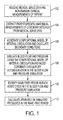
- FIG. 1 illustrates a method for non-invasive computation of a hemodynamic index for a coronary artery stenosis according to an embodiment of the present invention.
- the method of FIG. 1 transforms medical image data of a patient to extract the patient-specific geometry of the coronary arteries, generate a model of coronary arterial circulation, simulate patient-specific blood flow and pressure in the coronary arteries, and calculate one or more hemodynamic indices for a coronary artery stenosis without the use of invasive pressure measurements acquired via a pressure wire across the stenosis.
- the method of FIG. 1 computes the instantaneous wave-free ratio (iFR) for a coronary artery stenosis without using invasive pressure measurements.
- iFR instantaneous wave-free ratio
- the present invention is not limited to iFR and the method of FIG. 1 may be similarly applied to compute other hemodynamic indices as well.
- medical image data and non-invasive clinical measurements of a patient are received.
- Medical image data from one or multiple imaging modalities can be received.
- the medical image data can include, computed tomography (CT), Dyna CT, magnetic resonance (MR), Angiography, Ultrasound, Single Photon Emission computed Tomography (SPECT), and any other type of medical imaging modality.
- CT computed tomography
- MR magnetic resonance
- Angiography Ultrasound
- SPECT Single Photon Emission computed Tomography
- the medical image data can be 2D, 3D, or 4D (3D+time) medical image data.
- the medical image data can be received directly from one or more image acquisition devices, such as a CT scanner, MR scanner, Angiography scanner, Ultrasound device, etc., or the medical image data may be received by loading previously stored medical image data for a patient.
- 3D coronary CT angiography (CTA) images are acquired on a CT scanner.
- the CTA images ensure that the coronary vasculature, including the vessel(s) that contain the stenosis, is adequately imaged using a contrast agent that is injected into the patient.
- the clinician may be provided with an option of identifying lesions (stenoses) of interest by interactively viewing them on the images.
- This step can also be performed on a patient-specific anatomical model that is extracted from the image data (step 104).
- the stenoses may be automatically detected in the image data using an algorithm for automatic detection of coronary artery stenosis, such as the method for automatic detection of coronary artery stenosis described in United States Published Patent Application No. 2011/0224542 , which is incorporated herein by reference.
- algorithm for automatic detection of coronary artery stenosis such as the method for automatic detection of coronary artery stenosis described in United States Published Patent Application No. 2011/0224542 , which is incorporated herein by reference.
- other non-invasive clinical measurements such as the patient's heart rate and systolic and diastolic blood pressure are also acquired.
- patient-specific anatomical measurements of the coronary arteries are extracted from the medical image data.
- the medical image data is acquired at rest-state and the measurements of the coronary arteries are extracted from the medical image data acquired at rest-state.
- the measurements of the coronary arteries can be extracted by generating a patient-specific anatomical model of the coronary artery tree from the medical image data.
- the patient-specific anatomical model may be a patient-specific anatomical model of any portion of the full coronary artery tree of the patient.
- the coronary arteries can be segmented in the 3D medical image data using an automated coronary artery centerline extraction algorithm.
- the coronary arteries can be segmented in a CT volume using the method described United States Published Patent Application No. 2010/0067760 , which is incorporated herein by reference.
- a coronary artery centerline tree is extracted, cross-section contours can be generated at each point of the centerline tree.
- the cross-section contour at each centerline point gives a corresponding cross-section area measurement at that point in the coronary artery.
- a geometric surface model is then generated for the segmented coronary arteries.
- methods for anatomical modeling of the coronary arteries are described in United States Patent No. 7,860,290 and United States Patent No. 7,953,266 , both of which are incorporated herein by reference.
- the patient-specific anatomical model can include the aortic root together with the proximal part of the aorta.
- a detailed 3D model of each stenosis can also be extracted using similar algorithms, which includes the quantification of the proximal vessel diameter and area, distal vessel diameter and area, minimal lumen diameter and area, and length of stenosis.
- the above described anatomical modeling tasks can be performed automatically or can be user-driven, thereby allowing the user (clinician) to interactively make changes to the anatomical models to analyze the effects of such changes on the subsequent computation of FFR.
- the myocardium may also be segmented (either automatically or manually) in the medical image data to determine an estimate of the left ventricular mass, which in a possible implementation, may be used to estimate the absolute resting flow for the patient which is used to calculate boundary conditions for a computational blood flow and pressure simulation.
- the resting flow could also be computed based on the total volume of the segmented coronary tree, or from the outlet radius of the different coronary vessels.
- a patient-specific anatomical model of the heart that is automatically generated from the image data may be used for this purpose.
- the anatomical heart model is a multi-component model having multiple cardiac components, including the four chambers (left ventricle, left atrium, right ventricle, and right atrium).
- the anatomical heart model may also include components such as the heart valves (aortic valve, mitral valve, tricuspid valve, and pulmonary valve) and the aorta.
- Such a comprehensive model of the heart is used to capture a large variety of morphological, functional, and pathological variations.
- a modular and hierarchical approach can be used to reduce anatomical complexity and facilitate an effective and flexible estimation of individual anatomies.
- the 4D anatomical heart model can be generated by generating individual models of each heart component, for example using marginal space learning (MSL), and then integrating the heart component models by establishing mesh point correspondence. Additional details regarding generation of such a 4D patient-specific heart model are described in United States Published Patent Application No. 2012/0022843 , which is incorporated herein by reference.
- MSL marginal space learning
- a computational model of coronary arterial circulation is generated based on the patient-specific anatomical measurements of the coronary arteries, and inlet and outlet boundary conditions are calculated.
- the iFR value for a stenosis is based on pressure vales computed by simulating blood flow and pressure through the coronary artery tree for a patient at a rest state.
- a balance between model complexity and computation time, without compromising the accuracy of the results is desirable.
- reduced-order models can be used for the patient-specific blood flow simulation, which enables the assessment of the functional significance of a coronary artery stenosis.
- the reduced-order models provide accurate estimates of flow and pressure distribution in the vessel tree, and are computationally efficient, thus enabling a seamless integration with the clinical workflow.
- a reduced order model of coronary arterial circulation is described herein, the present invention is not limited thereto, and a full-scale model or a multi-scale model can be used as well.
- FIG. 2 illustrates a computational model of coronary arterial circulation according to an embodiment of the present invention.
- a heart model 202 is coupled at the root of the aorta.
- the heart model 202 may be implemented as a lumped model parameterized through patient-specific data as shown in FIG. 2 , or may be implemented as a full 3D heart model.
- Large arteries, such as the aorta 204 together with the large arteries supplied by the aorta (e.g., subclavian, brachiocephalic, common carotid, etc.), the left coronary artery (LCA) 206, and the right coronary artery 208 can be represented as 1 D blood flow models or full 3D models.
- LCA left coronary artery
- semi-analytical circulatory models can be used either separately for certain arterial segments, or embedded within the 1 D or 3D models.
- the vessel walls can be modeled as a purely elastic or visco-elastic material.
- the wall properties may be determined through an empirical relationship fit to measured data or based on patient-specific estimations of wall compliance.
- all microvascular beds are simulated through lumped parameter models 210 which account for the resistance applied to the blood flow and for the compliance of the distal vessels.
- the coronary vascular bed is modeled through such lumped parameter models 210, which are adapted to the coronary circulation in the sense that they take into account the effects of the myocardial contraction on the flow waveform.
- Stenosis segments 212 and 214 are shown in the model of coronary arterial circulation.
- the stenosis segments 212 and 214 cannot be simulated using the 1 D blood flow models since there is a high variation in cross-sectional area and the shape of the stenosis influences the blood flow behavior and especially the trans-stenotic pressure drop which plays a major role in the assessment of the functional importance of such a stenosis. Accordingly, when 1 D blood flow models are used for the coronary arteries, a full 3D model or a reduced-order stenosis pressure drop model can be used for each stenosis segment 212 and 214.
- the inlet boundary condition for the coronary artery blood flow can be prescribed through an implicit coupling with the heart model 202, or through measured pressure or flow data (e.g., acquired through various imaging techniques).
- the wall properties may be determined through an empirical relationship fit to the measured data in the extracted patient-specific anatomical model or based on patient-specific estimations of the wall compliance.
- Other alternative formulations of the quasi-1-D flow equations can also be used, modeling the effects of visco-elasticity, non-Newtonian behavior, etc.
- Equations 1-3 are derived by considering a series of simplifying assumptions which all hold well for normal, healthy vessels. One of the assumptions is that the axial velocity is dominant and the radial components are negligible. This assumption no longer holds in case of sudden changes in lumen diameter, e.g. for a stenosis, and the radial components can no longer be excluded.
- full scale 3D models can be used for blood flow and pressure simulation in stenosis regions 212 and 214.
- the patient-specific 3D geometric model of the stenosis extracted from the medical image data e.g., CTA data
- CTA quantitative coronary angiography
- semi-empirical stenosis models can be included in the 1-D blood flow models, which produce accurate results as compared to full scale models.
- such a semi-empirical model for each stenosis segment is coupled with the vessel tree (and the underlying heart and coronary bed models) to compute the physiological pressure drop across the stenosis.
- the present invention is not limited to the semi-empirical stenosis model of Equation (4), and other such models of the stenosis, with multiple pressure drop factors, may be used alternatively.
- the momentum equation is adapted and the additional pressure drop determined by the turbulent term is included on the right hand side of the equation as an additional loss term.
- the regular momentum equation is disregarded completely and replaced by Equation (2).
- the segments treated as stenosis segments are coupled to the regular segments of the coronary vessel tree by considering continuity of total pressure and flow rate.
- outlet boundary conditions At the termination of the coronary vessel tree (outflow boundary conditions).
- pressure, flow, or a relationship between flow and pressure may be imposed at the terminal sites of the arterial vessel tree. If measured data, e.g. time-varying velocity, flow rate, or even pressure, are available, they can be readily applied.
- embodiments of the present invention calculate special boundary conditions that model the behavior of the distal arterial segments. For example, lumped parameters models (as in FIG. 2 ) or microvascular wave propagation models can be used to determine the outlet boundary conditions.
- the microvascular beds are modeled through lumped or 0-D models 210.
- the systemic beds can be represented by regular windkessel elements containing varying number of elements (for example, between two and four elements), while coronary beds are represented by special models which account for the influence of the myocardial contraction on the flow waveform (low during systole and high during early diastole).
- FIG. 2 displays an example of such specialized models for the coronary circulation and presents the detailed elements of this type of boundary condition.
- R a denotes proximal arterial resistance
- C a denotes arterial compliance
- R am denotes microvascular arterial resistance
- C im denotes intra-myocardial compliance
- R vm denotes microvascular venous resistance
- R v denotes venous resistance.
- the main characteristic of such lumped models is that the myocardial contraction is taken into account by introducing the left or right ventricular pressure, depending on the location of the coronary tree on the heart.
- the model displayed in FIG. 4 treats the microvascular bed as a single unit, but it is also possible to utilize more specialized models which consider separately the contribution of the subepicardial and subendocardial microvascular beds.
- subepicardial vessels are less affected by heart contraction (they represent the outer layers of the myocardium), while subendocardial vessels are more affected by the action of the contraction (they represent the inner layers, closer to the heart chambers). This is the main reason why subendocardial are more prone to ischemia and to myocardial infarction.
- the resistance values inside the systemic or coronary lumped models for the rest state may be obtained from patient-specific measurements, from literature data, or from the non-linear relationship between resistances and lumen size. Compliances play a secondary role since they only influence the transient values and not the average pressures which are of interest for the evaluation of iFR. Coronary auto-regulation protects the myocardium against ischemia during rest state and leads to decreased resistances for the diseased vessel, the reference value being the flow which has to be identical to the non-diseased case. The rest state outlet boundary conditions can thus be modeled using this information.
- the parameters that are estimated to determine the rest state outlet boundary conditions are the mean arterial pressure (MAP) and the coronary microvascular resistances (the resistances of the proximal epicardial arteries are negligible compared to the microvascular resistances). Since iFR uses only average measures of pressures (distal and proximal to the stenosis) in the wave free period of diastole, compliances need not be estimated accurately because they only influence the waveform of pressure and flow, but not the average values, which are only determined by the resistances. MAP can be easily measured non-invasively based on the patient's heart rate, systolic blood pressure, and diastolic blood pressure.
- HR, SBP, and DBP denote the patient's heart rate, systolic blood pressure, diastolic blood pressure, respectively, which are measured non-invasively.
- the rest-state cardiac microvascular resistances can be calculated as follows.
- the total myocardial perfusion q rest can be estimated using the rate-pressure product (RPP) relationship.
- the RPP is the product of the heart rate and the systolic blood pressure.
- the total resting coronary flow can then be estimated based on the resting perfusion q rest and the mass of the patient's left ventricle (LV).
- the mass of the left ventricle can be estimated based on quantities derived from segmentation of the medical image data.
- the myocardium is segmented using automatic heart chamber segmentation, for example using a MSL machine-learning based method.
- the volume can be automatically calculated from the segmented myocardium, for example using the method described in United States Patent No. 8,098,918 , entitled "Method and System for Measuring Left Ventricle Volume", which is incorporated herein by reference.
- the LV volume is then multiplied by the density to provide the mass of the LV ( M LV ). Other possible methods for calculating the mass of the LV can be used as well.
- the resting perfusion can be multiplied by the myocardial mass.
- the terminal resistance for each vessel branch is then calculated.
- blood flow and pressure are simulated using the computational model of coronary arterial circulation and coronary circulatory autoregulation is modeled in the blood flow and pressure simulation.
- the simulation incrementally computes blood flow rates and pressure values within the coronary arteries over a period of time at each of a plurality of time steps using the computational model of coronary arterial circulation.
- the simulation can be run over a plurality of heart cycles, each corresponding to a heartbeat.
- the flow and pressure computations may be performed using Computational Fluid Dynamics (CFD), or any other standard numerical techniques, such as but not limited to, finite-element method, finite-difference method, finite-volume method, boundary element method, embedded boundary method, immersed boundary method, lattice Boltzmann method, etc.
- CFD Computational Fluid Dynamics
- coronary circulatory autoregulation is modeled in the blood flow and pressure simulation.
- Coronary autoregulation plays an important role in coronary hemodynamics.
- the purpose of coronary autoregulation is to maintain a certain level of myocardial perfusion, given by the myocardial oxygen demand, to the microvascular beds perfused by arteries which may or may not contain stenoses.
- FIG. 3 illustrates coronary circulatory autoregulation. As shown in FIG. 3 , the autoregulatory mechanism adapts the microvascular resistances so as to maintain perfusion at the level dictated by the myocardial oxygen demand.
- microvascular resistance has a certain minimum threshold value, when a stenosis is very severe and the pressure distal to the stenosis decreases considerably, autoregulation reaches its limit and the myocardial perfusion decreases below the required level, leading to myocardial ischemia.
- coronary autoregulation In order to accurately determine iFR for a stenosis non-invasively based on blood flow and pressure simulations without explicitly measuring the pressure difference across the stenosis, coronary autoregulation has to be taken into consideration.
- the microvascular resistances at each outlet of the coronary arterial circulation model e.g., FIG. 2
- an equivalent coronary microvascular resistance is calculated for each coronary artery based on the microvascular resistances calculated at the outlets of the coronary artery.
- the blood flow and pressure simulation can be performed and an algorithm (Algorithm 1 of FIG. 4 ) for modeling coronary autoregulation can be applied for each coronary tree during the simulation in order to adjust the microvascular resistances to model the effect of coronary autoregulation.
- Algorithm 1 of FIG. 4 an algorithm for modeling coronary autoregulation can be applied for each coronary tree during the simulation in order to adjust the microvascular resistances to model the effect of coronary autoregulation.
- FIG. 4 illustrates an algorithm for modeling coronary autoregulation in a simulation of blood flow and pressure in the coronary arteries according to an embodiment of the present invention.
- the algorithm 400 of FIG. 4 (Algorithm 1) can be performed at the end of each cardiac cycle in the blood flow and pressure simulation for each coronary artery tree.
- the algorithm 400 has an input parameter of nrVessel, which is a number identifying a current vessel or branch in the coronary artery tree.
- the microvascular resistances are modified only if the current branch is stenosed. If the current branch is stenosed, steps 402-412 are performed for the current branch.
- a resistance ( R t-current ) i introduced by the stenosis in the current branch is calculated by subtracting a resistance of the stenosis, computed as a ratio of the pressure drop along the stenosis to the flow rate through the stenosis (( P in ) i - ( P out ) i )/ Q i , from the equivalent microvascular resistance of the current branch ( R t - microv ) i .
- FIG. 5 illustrates an exemplary coronary artery tree with branch labels used by the coronary autoregulation algorithm of FIG. 4 .
- the coronary artery tree 500 shows a root branch 502 and a current branch 504 having a stenosis 506.
- Branches 508 and 510 are daughter branches to the current branch 504, and branches 508, 512, and 514 are terminal branches.
- an equivalent resistance ( R t-downs ) i is calculated from the total resistances of the daughter branches j downstream of the current branch i.
- the resistances of the daughter branches are adapted so as to not to modify the flow rate split ratios of the daughter branches.
- the flow rate split ratio ⁇ j is calculated from the previous value of the downstream resistance for the daughter branch ( R t-microv ) j and the equivalent downstream resistance calculated for the current branch ( R t-dows ) i .
- a new value for the total downstream resistance ( R t-microv ) j is calculated for each daughter branch j based on the new resistance value calculated for the current branch ( R t-current ) i and the flow rate split ratio calculated for the daughter branch ⁇ j .
- step 410 the new values of the downstream resistances are distributed to the terminal branches of each daughter branch, and at step 412, new equivalent microvascular resistances are computed for all of the branches downstream of each daughter branch.
- steps 410 and 412 can be implemented by repeating calculations similar to those in steps 404, 406, and 408 for downstream branches for each daughter branch until all terminal branches are reached.
- the autoregulation algorithm 400 can be called for the root branch and then called recursively for each daughter branch, so as to cover the entire coronary tree and modify the terminal resistances while taking into account all stenosed branches in the coronary tree.
- coronary autoregulation has a limit, i.e. when the mircovascular resistance cannot be decreased below a certain minimum value, which corresponds to the hyperemic state of the patient.
- a limit i.e. when the mircovascular resistance cannot be decreased below a certain minimum value, which corresponds to the hyperemic state of the patient.
- coronary autoregulation may reach its limit, and the flow may decrease below the level given by the myocardial demand (myocardial oxygen consumption).
- myocardial demand myocardial oxygen consumption
- the blood flow and pressure computations can be performed over a plurality of cardiac cycles, with the algorithm for modeling coronary autoregulation performed at the end of each cardiac cycle.
- Different approaches can be used for matching the blood flow and pressure simulations with non-invasive clinical measurements of the patient in order to achieve patient-specific blood flow and pressure simulations.
- parameters of the computational model of coronary arterial circulation can be directly estimated once prior to simulating the blood flow and pressure.
- iterative estimation can be used to adapt the parameters during the blood flow and pressure computation. It is also possible to use a combination of these approaches.
- simulated measurements based on the blood flow and pressure computations can be compared to non-invasive measurements of the patient (e.g., systolic and diastolic blood pressure, heart rate, etc.) and one or more parameters of the computational model of coronary arterial circulation or one or more boundary conditions can be refined to minimize a difference between the simulated measurements and the non-invasive measurements acquired for the patient.
- the blood flow and pressure computations can be performed at least until the simulated measurements converge to the non-invasive measurements of the patient, and iFR and/or other hemodynamic indices can be calculated using the simulated blood flow and pressure values for one or more cardiac cycles after the simulated measurements have converged to the non-invasive measurements of the patient.
- a wave free period is identified in each heart cycle in the blood flow and pressure simulation.
- iFR is computed from invasively measured pressures obtained during the wave-free period.
- Instantaneous coronary resistance calculated as the ratio of instantaneous pressure to instantaneous flow rate, varies during the heart cycle due to the interaction between the microvasculature and the myocardium, with higher fluctuations during systole.
- coronary resistance is naturally approximately constant and minimized.
- FIG. 6 illustrates an example of instantaneous pressure values 602, instantaneous flow velocity values 604 and instantaneous coronary resistance values 606 during one cardiac cycle at rest. As shown in FIG. 6 , the coronary resistance 606 is naturally constant and minimized during a wave-free period 608.
- the wave-free period In order to compute iFR based on the blood flow and pressure simulations, the wave-free period must be determined.
- waves can originate from both upstream locations (e.g. left ventricle), referred to as forward travelling waves, or from downstream locations (e.g., bifurcations or the microcirculation), referred to as backward travelling waves.
- forward travelling waves In order to establish the origin of the waves, both pressure and flow rate profiles are required.
- forward and backward travelling waves can occur at the same time.
- a wave intensity analysis can be performed.
- FIG. 7 illustrates identification of waves in the human left coronary artery tree. As shown in FIG.
- the end of diastole is defined as the moment when the early backward-travelling pushing wave 702 is detected.
- WI - [ diastole ] is the wave intensity of distally originating waves (backward travelling waves).
- the wave-free period considered for the invasive measurement of iFR typically runs from 25% of the way into diastole (the onset of diastole is identified from the dicrotic notch) to 5 ms before the end of diastole.
- the wave-free period in the simulated cardiac cycle can similarly be identified as an interval of time which runs from 25% of the way into diastole to 5 ms before the end of diastole.
- Other possible implementations that utilize the simulated pressure and/or velocity (flow rate) profiles can also be used to identify the wave-free period.
- the wave-free period in the simulated cardiac cycle can be identified as an interval of time which begins at 250 ms after dU max is obtained and lasts for 150 ms, where dU is the derivative of the blood velocity with respect to time and dU max is the maximum value of dU within one cardiac cycle.
- the wave-free period in the simulated cardiac cycle can be identified as an interval of time which runs from 150 ms after a maximum pressure P max is obtained until the end of the cardiac cycle minus 50 ms.
- the wave-free period in the simulated cardiac cycle can be identified as the period, after peak pressure P max , during which the standard deviation of the forward travelling wave is in the lowest 5% (or in the lowest 10% if no such period exists for the lowest 5%).
- the wave-free period in the simulated cardiac cycle can be identified as a period that is the mid window between the peak pressure time point and the end of the heart cycle (or a 3/5 window between these two time points).
- the wave-free period in the simulated cardiac cycle can be identified the window during which dU is less than 10% (or 5%) of dU max . It is to be understood that the present invention is not limited to these techniques for identifying the wave-free period and any method for identifying the wave-free period may be used in conjunction with the method of FIG. 1
- iFR is calculated for at least one coronary artery stenosis based on the computed pressures in the wave-free period.
- the iFR for a particular coronary artery stenosis is calculated non-invasively from the computed pressure values in the blood flow and pressure simulation as the ratio of the average computed distal pressure during the wave-free period of the simulated cardiac cycle to the average computed aortic pressure during the wave-free period of the simulated cardiac cycle.
- the blood flow and computations are repeated to simulate a plurality of cardiac cycles with the coronary autoregulation algorithm performed at the end of each cardiac cycle.
- simulated measurements based on the blood flow and pressure computations are compared to non-invasive measurements of the patient (e.g., systolic and diastolic blood pressure, heart rate, etc.) and one or more parameters of the computational model of coronary arterial circulation or one or more boundary conditions can be refined to minimize a difference between the simulated measurements and the non-invasive measurements acquired for the patient.
- non-invasive measurements of the patient e.g., systolic and diastolic blood pressure, heart rate, etc.
- the computations can be performed until the simulated measurements based on the blood flow and pressure computations converge to the non-invasive measurements of the patient, and the iFR for a particular coronary stenosis can be calculated based on the computed pressure values in the wave-free period of a simulated cardiac cycle for which the simulated measurements have converged to the non-invasive measurements of the patient.
- the iFR be calculated for each simulated cardiac cycle or by averaging data from multiple simulated cardiac cycles.
- a respective iFR value can be non-invasively calculated for each of a plurality of stenoses in the coronary artery tree.
- Other hemodynamic indices particularly hemodynamic indices typically measured for a patient at rest, such as rest (basal) P d / P a , can also be calculated for each coronary artery stenosis from the simulated pressure and/or flow values. It is also to be understood that the iFR (or other hemodynamic indices) can be calculated for any location in the coronary artery tree, and not only at stenoses.
- Certain parameters of the microvascular model may further be adapted to ensure that a certain simulated hemodynamic index matches the measured value of that hemodynamic index. For example, since iFR is computed during the wave-free window, it depends on the trans-stenotic pressure drop during diastole (the wave-free period is part of diastole). The pressure drop in turn depends on the flow rate, and hence it is important to control the amount of coronary flow at systole and at diastole. Previous studies that have examined the amount of coronary flow at systole and diastole indicate that systolic flow is proportionally lower in the LCA as compared to the RCA.
- systolic flow represents 20% of the total coronary flow for LCA branches, and 31% of the total coronary flow for RCA branches.
- Typical coronary microvascular models use the simulated left/right ventricular pressure in order to model the effect of the cardiac contraction on the flow rate, thus leading to low flow during systole and high flow during diastole (see for example the lumped parameter model 210 in FIG. 2 ).
- a proportionality constant c may be used when applying the left/right ventricular pressure in the coronary microvascular models. This proportionality constant may be adapted separately for each outlet branch so as to match a certain ratio of systolic to diastolic flow.
- Exemplary results for non-invasively computing iFR from blood flow and pressure simulations based on coronary imaging data obtained from a CT scan are provided for three cases.
- Case 1 is an anatomically severe stenosis.
- Case 2 is an anatomically intermediate stenosis.
- Case 3 is an anatomically mild stenosis. Computations were run until convergence and results are provided herein for only the last heart cycle.
- Table 1 shows average distal and aortic pressures and pressure ratios (basal P d / P a and iFR) for entire cardiac cycle and for the wave-free period for the three cases.
- both basal P d / P a and iFR are computed non-invasively from medical imaging data based on pressure and flow computations using the method of FIG. 1 .
- Table 1 Configuration Quantity Case 1 Case 2 Case 3 Entire heart cycle - Rest P a [mmHg] 106.084 100.172 102.803 P d [mmHg] 59.609 95.07 100.312 Basal P d /P a 0.562 0.949 0.976 Wave-free period - Rest P a [mmHg] 100.051 91.895 97.233 P d [mmHg] 37.736 85.154 93.499 iFR 0.377 0.927 0.962
- FIG. 8 illustrates exemplary results for simulating blood flow and pressure and calculating iFR for an anatomically severe stenosis (Case 1).
- image 800 shows the computed time-varying proximal (aortic) and distal aortic pressures 802 and 804, respectively, for the stenosis over a cardiac cycle.
- Image 810 shows the computed time-varying flow rate 812 in the stenosis over the cardiac cycle.
- Image 820 shows the instantaneous coronary resistance 822 over the cardiac cycle.
- Image 830 shows the instantaneous distal to proximal pressure ratio 832 over the cardiac cycle.
- the iFR value is computed by calculating the ratio of the average distal pressure to the average proximal pressure in the wave-free period.
- Image 840 shows the wave intensity of forward and backward travelling waves 842 and 843, respectively, over the cardiac cycle.
- the diastolic period runs from 6.828 seconds (dicrotic notch) to 7.5 seconds.
- the wave-free period considered for the computation of iFR, runs from 25% of the way into diastole to 5 ms before the end of diastole (6.99 seconds to 7.45 seconds).
- the resistance 822 is constant during the wave-free period and also the instantaneous distal to proximal pressure ratio 832 is relatively constant during the wave-free period.
- FIG. 9 illustrates exemplary results for simulating blood flow and pressure and calculating iFR for an anatomically intermediate stenosis (Case 2).
- image 900 shows the computed time-varying proximal (aortic) and distal aortic pressures 902 and 904, respectively, for the stenosis over a cardiac cycle.
- Image 910 shows the computed time-varying flow rate 912 in the stenosis over the cardiac cycle.
- Image 920 shows the instantaneous coronary resistance 922 over the cardiac cycle.
- Image 930 shows the instantaneous distal to proximal pressure ratio 932 over the cardiac cycle.
- the iFR value is computed by calculating the ratio of the average distal pressure to the average proximal pressure in the wave-free period.
- Image 940 shows the wave intensity of forward and backward travelling waves 942 and 943, respectively, over the cardiac cycle.
- the diastolic period runs from 6.65 seconds to 7.19 seconds.
- the wave-free period runs from 6.797 seconds to 7.19 seconds.
- the resistance 922 is constant during the wave-free period and also the instantaneous distal to proximal pressure ratio 932 is relatively constant during the wave-free period.
- image 940 it can be observed that there are no waves in the wave-free period.
- Table 1 the computed iFR value for the anatomically intermediate stenosis in Case 2 is 0.927, which falls into a grey zone identified in previous studies for which it is not clear if the lesion is hemodynamically significant.
- FIG. 10 illustrates exemplary results for simulating blood flow and pressure and calculating iFR for an anatomically mild stenosis (Case 3).
- image 1000 shows the computed time-varying proximal (aortic) and distal aortic pressures 1002 and 1004, respectively, for the stenosis over a cardiac cycle.
- Image 1010 shows the computed time-varying flow rate 1012 in the stenosis over the cardiac cycle.
- Image 1020 shows the instantaneous coronary resistance 1022 over the cardiac cycle.
- Image 1030 shows the instantaneous distal to proximal pressure ratio 1032 over the cardiac cycle.
- the iFR value is computed by calculating the ratio of the average distal pressure to the average proximal pressure in the wave-free period.
- Image 1040 shows the wave intensity of forward and backward travelling waves 1042 and 1043, respectively, over the cardiac cycle.
- the diastolic period runs from 4.235 seconds to 4.565 seconds.
- the wave-free period runs from 4.317 seconds to 4.515 seconds.
- the resistance 1022 is constant during the wave-free period and also the instantaneous distal to proximal pressure ratio 1032 is relatively constant during the wave-free period.
- image 1040 it can be observed that there are no waves in the wave-free period.
- Table 1 the computed iFR value for the anatomically mild stenosis in Case 3 is 0.962, which indicates that this is a hemodynamically non-significant lesion.
- the computed iFR for each stenosis and the simulation results can be output, for example by being displayed on a display device of a computer system.
- the simulation results can be output as shown in FIGS. 8 , 9 , and 10 .
- the computed iFR value may be output by simply displaying a listing or table of iFR values for the stenoses.
- one or more computed iFR values can be visualized on a medical image of the patient that is displayed on a display device.
- FIG. 11 illustrates an example of visualizing iFR values on a medical image. As shown in FIG.
- FIG. 12 illustrates an exemplary plot of iFR values at selected locations along a vessel.
- image 1200 shows various locations 1202, 1204, 1204, 1206, 1208, 1210, 1212, 1214, and 1216 along a vessel, at which iFR is computed using the method of FIG. 1 .
- Image 1220 shows a plot of computed iFR along the length of the vessel generated based on the computed iFR values at the selected locations 1202, 1204, 1204, 1206, 1208, 1210, 1212, 1214, and 1216 along the vessel.
- an aortic pressure measurement for the patient may be determined invasively using a pressure measuring device, such as a pressure wire.
- the aortic pressure measurement may be used in a number of different ways. For example, in a possible implementation, the measured aortic pressure value during the wave-free period may be used in the iFR computation as the denominator in the iFR formula. In another possible implementation, the measured aortic pressure may be used as a boundary condition for the flow and pressure computations.
- the measured aortic pressure may be used to identify the wave-free period.
- Such embodiments would not require advancing the pressure wire distal to the stenosis, as is done for invasive iFR measurement, thereby reducing the acquisition time and reducing the risk of dislodging the plaque or puncturing the lumen.
- the method is used for computing hemodynamic indices for coronary artery stenoses
- the present invention is not limited to coronary arteries and may be similarly applied to other arteries, such as the aorta, renal arteries, cerebral arteries, iliac arteries, supra-aortic arteries, etc.
- the method of claim 1 performs the simulations and identifies the wave-free period at a rest state
- the method can be similarly applied for a wave-free window obtained in a maximum vasodilation state or any other intermediate state between rest and maximum vasodilation.
- the method can be used to calculate different hemodynamic indices, such as fractional flow reserve (FFR).
- FFR fractional flow reserve
- FIG. 1 can be applied for the entire cardiac cycle, or for any sub-part of the cardiac cycle (e.g., entire systole or a sub-part of systole, diastole or a sub-part of diastole, or combinations thereof).
- the method of FIG. 1 can be applied not only for the non-invasive computation of pressure-based indices, but also for other indices computed as a combination of coronary pressure, coronary perfusion, and/or coronary resistance.
- the algorithm for modeling coronary circulatory autoregulation can be applied at the end of every cardiac cycle.
- the present invention is not limited thereto, and in various embodiments, the algorithm for modeling coronary circulatory autoregulation can be applied multiple times during a single cardiac cycle (e.g., end of systole and end of diastole) or only after a certain number of cardiac cycles.
- Computer 1302 contains a processor 1304, which controls the overall operation of the computer 1302 by executing computer program instructions which define such operation.
- the computer program instructions may be stored in a storage device 1312 (e.g., magnetic disk) and loaded into memory 1310 when execution of the computer program instructions is desired.
- a storage device 1312 e.g., magnetic disk
- FIGS. 1 and 4 may be defined by the computer program instructions stored in the memory 1310 and/or storage 1312 and controlled by the processor 1304 executing the computer program instructions.
- An image acquisition device 1320 such as a CT scanning device, MR scanning device, Ultrasound device, etc., can be connected to the computer 1302 to input image data to the computer 1302. It is possible to implement the image acquisition device 1320 and the computer 1302 as one device. It is also possible that the image acquisition device 1320 and the computer 1302 communicate wirelessly through a network. In a possible embodiment, the computer 1302 may be located remotely with respect to the image acquisition device 1320 and the method steps are performed as part of a server or cloud based service. The computer 1302 also includes one or more network interfaces 1306 for communicating with other devices via a network.
- the computer 1302 also includes other input/output devices 1308 that enable user interaction with the computer 1302 (e.g., display, keyboard, mouse, speakers, buttons, etc.). Such input/output devices 1308 may be used in conjunction with a set of computer programs as an annotation tool to annotate volumes received from the image acquisition device 1320.
- input/output devices 1308 may be used in conjunction with a set of computer programs as an annotation tool to annotate volumes received from the image acquisition device 1320.
- FIG. 13 is a high level representation of some of the components of such a computer for illustrative purposes.
Landscapes
- Health & Medical Sciences (AREA)
- Engineering & Computer Science (AREA)
- Life Sciences & Earth Sciences (AREA)
- Public Health (AREA)
- Medical Informatics (AREA)
- General Health & Medical Sciences (AREA)
- Pathology (AREA)
- Biomedical Technology (AREA)
- Physics & Mathematics (AREA)
- Cardiology (AREA)
- Primary Health Care (AREA)
- Epidemiology (AREA)
- Animal Behavior & Ethology (AREA)
- Surgery (AREA)
- Molecular Biology (AREA)
- Heart & Thoracic Surgery (AREA)
- Biophysics (AREA)
- Veterinary Medicine (AREA)
- Physiology (AREA)
- Databases & Information Systems (AREA)
- Data Mining & Analysis (AREA)
- Computer Vision & Pattern Recognition (AREA)
- Vascular Medicine (AREA)
- Radiology & Medical Imaging (AREA)
- Nuclear Medicine, Radiotherapy & Molecular Imaging (AREA)
- Signal Processing (AREA)
- Psychiatry (AREA)
- Artificial Intelligence (AREA)
- Theoretical Computer Science (AREA)
- General Physics & Mathematics (AREA)
- Measuring Pulse, Heart Rate, Blood Pressure Or Blood Flow (AREA)
Applications Claiming Priority (2)
| Application Number | Priority Date | Filing Date | Title |
|---|---|---|---|
| US201461990775P | 2014-05-09 | 2014-05-09 | |
| US14/689,083 US9595089B2 (en) | 2014-05-09 | 2015-04-17 | Method and system for non-invasive computation of hemodynamic indices for coronary artery stenosis |
Publications (2)
| Publication Number | Publication Date |
|---|---|
| EP2942006A1 true EP2942006A1 (fr) | 2015-11-11 |
| EP2942006B1 EP2942006B1 (fr) | 2017-06-28 |
Family
ID=53189640
Family Applications (1)
| Application Number | Title | Priority Date | Filing Date |
|---|---|---|---|
| EP15166816.7A Active EP2942006B1 (fr) | 2014-05-09 | 2015-05-07 | Procédé et système de calcul non invasif des indices hémodynamiques pour la sténose artérienne coronaire |
Country Status (4)
| Country | Link |
|---|---|
| US (1) | US9595089B2 (fr) |
| EP (1) | EP2942006B1 (fr) |
| CN (1) | CN105078440B (fr) |
| ES (1) | ES2641688T3 (fr) |
Cited By (9)
| Publication number | Priority date | Publication date | Assignee | Title |
|---|---|---|---|---|
| WO2016113646A1 (fr) * | 2015-01-15 | 2016-07-21 | Koninklijke Philips N.V. | Tomodensitométrie ifr |
| US20170281018A1 (en) * | 2016-04-05 | 2017-10-05 | Martin Kramer | Determining arterial wall property with blood flow model |
| EP3453326A1 (fr) * | 2017-09-08 | 2019-03-13 | Koninklijke Philips N.V. | Enregistrement et comparaison de courbes de retrait intracoronaire simulées et mesurées |
| EP3564961A1 (fr) * | 2018-05-03 | 2019-11-06 | Koninklijke Philips N.V. | Marquage coronaire interactif au moyen d'images radiologiques interventionnelles et apprentissage profond |
| EP3858247A1 (fr) * | 2020-01-29 | 2021-08-04 | Tata Consultancy Services Limited | Contrôleur adaptatif basé sur la neuromodulation pour la sténose mitrale |
| CN113379679A (zh) * | 2021-05-14 | 2021-09-10 | 中国科学院深圳先进技术研究院 | 脑动脉波强度和波功率的测量方法、终端设备及存储介质 |
| WO2021262062A1 (fr) * | 2020-06-22 | 2021-12-30 | Coroventis Research Ab | Système et procédé de détermination de la réserve de résistance microvasculaire |
| WO2022136688A1 (fr) * | 2020-12-23 | 2022-06-30 | Flowreserve Labs S.L. | Procédé non effractif de détermination d'un indicateur d'endommagement de vaisseau |
| CN117476238A (zh) * | 2023-11-08 | 2024-01-30 | 中国医学科学院北京协和医院 | 用于评估心肌缺血的计算流体力学仿真分析方法及系统 |
Families Citing this family (73)
| Publication number | Priority date | Publication date | Assignee | Title |
|---|---|---|---|---|
| US9462954B2 (en) * | 2013-09-04 | 2016-10-11 | Siemens Aktiengesellschaft | Method and system for blood flow velocity reconstruction from medical images |
| US9754082B2 (en) * | 2014-05-30 | 2017-09-05 | Heartflow, Inc. | Systems and methods for reporting blood flow characteristics |
| US9747525B2 (en) | 2014-06-16 | 2017-08-29 | Siemens Healthcare Gmbh | Method and system for improved hemodynamic computation in coronary arteries |
| US10849511B2 (en) * | 2014-07-14 | 2020-12-01 | Philips Image Guided Therapy Corporation | Devices, systems, and methods for assessment of vessels |
| WO2016174010A1 (fr) * | 2015-04-30 | 2016-11-03 | Koninklijke Philips N.V. | Détermination de la réserve coronaire |
| WO2016182508A1 (fr) | 2015-05-12 | 2016-11-17 | Singapore Health Services Pte Ltd | Procédés et systèmes de traitement d'image médicale |
| US10872698B2 (en) * | 2015-07-27 | 2020-12-22 | Siemens Healthcare Gmbh | Method and system for enhancing medical image-based blood flow computations using physiological measurements |
| US11113812B2 (en) | 2015-08-14 | 2021-09-07 | Elucid Bioimaging Inc. | Quantitative imaging for detecting vulnerable plaque |
| US11094058B2 (en) | 2015-08-14 | 2021-08-17 | Elucid Bioimaging Inc. | Systems and method for computer-aided phenotyping (CAP) using radiologic images |
| US11676359B2 (en) | 2015-08-14 | 2023-06-13 | Elucid Bioimaging Inc. | Non-invasive quantitative imaging biomarkers of atherosclerotic plaque biology |
| US11071501B2 (en) | 2015-08-14 | 2021-07-27 | Elucid Bioiwaging Inc. | Quantitative imaging for determining time to adverse event (TTE) |
| US12026868B2 (en) | 2015-08-14 | 2024-07-02 | Elucid Bioimaging Inc. | Quantitative imaging for detecting histopathologically defined plaque erosion non-invasively |
| US12008751B2 (en) | 2015-08-14 | 2024-06-11 | Elucid Bioimaging Inc. | Quantitative imaging for detecting histopathologically defined plaque fissure non-invasively |
| US10176408B2 (en) | 2015-08-14 | 2019-01-08 | Elucid Bioimaging Inc. | Systems and methods for analyzing pathologies utilizing quantitative imaging |
| US11087459B2 (en) | 2015-08-14 | 2021-08-10 | Elucid Bioimaging Inc. | Quantitative imaging for fractional flow reserve (FFR) |
| CN105512489B (zh) * | 2015-12-10 | 2018-03-30 | 哈尔滨工业大学 | 一种基于多尺度心脏Thimthy综合症发病机制的建模方法 |
| DE102016203860A1 (de) * | 2016-03-09 | 2017-09-14 | Siemens Healthcare Gmbh | Vorrichtung und Verfahren zum Ermitteln zumindest eines individuellen fluiddynamischen Kennwerts einer Stenose in einem mehrere serielle Stenosen aufweisenden Gefäßsegment |
| US10971271B2 (en) | 2016-04-12 | 2021-04-06 | Siemens Healthcare Gmbh | Method and system for personalized blood flow modeling based on wearable sensor networks |
| US10674986B2 (en) | 2016-05-13 | 2020-06-09 | General Electric Company | Methods for personalizing blood flow models |
| US11020011B2 (en) * | 2016-07-20 | 2021-06-01 | Cardiac Pacemakers, Inc. | Estimate diastolic pressure |
| WO2018050806A1 (fr) * | 2016-09-16 | 2018-03-22 | Koninklijke Philips N.V. | Appareil et procédé de détermination d'une réserve de débit fractionnaire |
| EP3516558B1 (fr) * | 2016-09-23 | 2020-02-26 | Koninklijke Philips N.V. | Système et procédé d'évaluation de l'obstruction des voies d'évacuation du c ur d'un sujet |
| CN106473731A (zh) * | 2016-10-25 | 2017-03-08 | 北京工业大学 | 基于个性化冠状动脉分支血流量的ffrct计算方法 |
| CN108451540B (zh) * | 2017-02-17 | 2021-08-31 | 深圳先进技术研究院 | 一种血流储备分数测量方法和装置 |
| WO2018156961A1 (fr) * | 2017-02-24 | 2018-08-30 | Heartflow, Inc. | Systèmes et méthodes pour identifier des caractéristiques de flux sanguin pertinentes sur le plan anatomique chez un patient |
| US11710569B2 (en) * | 2017-04-06 | 2023-07-25 | Koninklijke Philips N.V. | Coronary artery disease metric based on estimation of myocardial microvascular resistance from ECG signal |
| US10980427B2 (en) * | 2017-06-22 | 2021-04-20 | Dextera Medical | Method and apparatus for full-system, cardiovascular simulation and prediction |
| CN109256205B (zh) * | 2017-07-12 | 2022-07-05 | 西门子保健有限责任公司 | 用于利用本地和远程分析学进行的临床决策支持的方法和系统 |
| US10909676B2 (en) * | 2017-07-12 | 2021-02-02 | Siemens Healthcare Gmbh | Method and system for clinical decision support with local and remote analytics |
| US11589924B2 (en) | 2017-08-01 | 2023-02-28 | Siemens Healthcare Gmbh | Non-invasive assessment and therapy guidance for coronary artery disease in diffuse and tandem lesions |
| EP3457404A1 (fr) * | 2017-09-18 | 2019-03-20 | Koninklijke Philips N.V. | Estimation d'écoulement dans des bifurcations de récipients pour hémodynamique simulée |
| CN111356406B (zh) | 2017-10-06 | 2024-05-28 | 埃默里大学 | 用于确定一个或多个动脉节段的血液动力学信息的方法和系统 |
| EP3488774A1 (fr) * | 2017-11-23 | 2019-05-29 | Koninklijke Philips N.V. | Guide de mesure pour l'estimation du débit coronaire selon le principe de bernoulli |
| EP3755215B1 (fr) * | 2018-02-23 | 2022-07-20 | Boston Scientific Scimed Inc. | Système d'évaluation d'un vaisseau avec des mesures physiologiques séquentielles |
| CN108573488B (zh) * | 2018-03-27 | 2021-06-01 | 杭州脉流科技有限公司 | 一种计算瞬时无波形比率的装置 |
| CN108309229B (zh) * | 2018-04-18 | 2019-09-03 | 电子科技大学 | 一种针对眼底图像视网膜血管的层次结构划分方法 |
| US11259871B2 (en) * | 2018-04-26 | 2022-03-01 | Vektor Medical, Inc. | Identify ablation pattern for use in an ablation |
| US11576624B2 (en) | 2018-04-26 | 2023-02-14 | Vektor Medical, Inc. | Generating approximations of cardiograms from different source configurations |
| US11389130B2 (en) | 2018-05-02 | 2022-07-19 | Siemens Healthcare Gmbh | System and methods for fast computation of computed tomography based fractional flow reserve |
| US11478162B2 (en) | 2018-05-23 | 2022-10-25 | Acist Medical Systems, Inc. | Flow measurement using image data |
| JP2021529015A (ja) * | 2018-06-27 | 2021-10-28 | オプセンス インコーポレイテッド | ハイブリッドな画像−侵襲性−圧力血行動態機能評価 |
| CN109036551B (zh) * | 2018-07-10 | 2021-05-11 | 北京心世纪医疗科技有限公司 | 一种冠状动脉生理学指标关系建立及应用方法、装置 |
| US20210272699A1 (en) * | 2018-07-20 | 2021-09-02 | University Of Louisville Research Foundation, Inc. | Method and system for assessing a coronary stenosis |
| CA3106237A1 (fr) * | 2018-07-26 | 2020-01-30 | Waseda University | Systeme d'aide au diagnostic d'une cardiopathie ischemique |
| EP3605554A1 (fr) * | 2018-08-03 | 2020-02-05 | Koninklijke Philips N.V. | Mesure du flux sanguin basée sur la pente vaisseau-carte |
| KR102094825B1 (ko) | 2018-08-14 | 2020-03-30 | 제주대학교병원 | 중등도 관상동맥 협착의 시술을 위한 iFR 산출을 위한 웨이브-프리 구간의 시작 시점을 산출하는 방법 및 시스템 |
| CN109259751B (zh) * | 2018-08-27 | 2022-03-11 | 杭州晟视科技有限公司 | 一种评估血流储备分数的方法及装置、设备、存储介质 |
| EP3624056B1 (fr) | 2018-09-13 | 2021-12-01 | Siemens Healthcare GmbH | Traitement de trames d'images d'une sequence d'images cardiaques |
| CN110384493A (zh) * | 2018-09-19 | 2019-10-29 | 苏州润迈德医疗科技有限公司 | 测量微循环阻力指数的系统以及冠脉分析系统 |
| JP7153973B2 (ja) | 2018-11-13 | 2022-10-17 | ベクトル メディカル インコーポレイテッド | 発生源位置を有する画像の拡張 |
| CN111166316B (zh) * | 2018-11-13 | 2023-03-21 | 苏州润迈德医疗科技有限公司 | 基于造影图像计算造影瞬时无波型比率和造影舒张期压力比率的方法 |
| CN111166315B (zh) * | 2018-11-13 | 2023-03-28 | 苏州润迈德医疗科技有限公司 | 基于造影图像计算瞬时无波型比率和静息态舒张期压力比率的方法 |
| WO2020142789A1 (fr) * | 2019-01-06 | 2020-07-09 | Covanos, Inc. | Détermination non invasive d'informations hémodynamiques de diastole à l'état de repos |
| EP3905951B1 (fr) * | 2019-01-06 | 2024-02-28 | Covanos, Inc. | Test de contrainte virtuel basé sur les données de patient électroniques |
| WO2020048642A1 (fr) * | 2019-01-11 | 2020-03-12 | LifeFlow Sp. z.o.o. | Modélisation de paramètres hémodynamiques spécifique d'un patient dans des artères coronaires |
| CN109846500A (zh) * | 2019-03-15 | 2019-06-07 | 浙江大学 | 一种确定冠状动脉血流储备分数的方法和装置 |
| US20220237864A1 (en) * | 2019-06-26 | 2022-07-28 | The Regents Of The University Of California | Reduced order model for computing blood flow dynamics |
| EP3770921A1 (fr) * | 2019-07-22 | 2021-01-27 | Tata Consultancy Services Limited | Procédé et système pour la synthèse de signal photopléthysmographique basée sur une autorégulation de pression |
| EP3899864A4 (fr) | 2019-08-05 | 2022-08-31 | Elucid Bioimaging Inc. | Évaluation combinée de marqueurs de pathologie morphologique et périvasculaire |
| CN110353645A (zh) * | 2019-08-19 | 2019-10-22 | 苏州润迈德医疗科技有限公司 | 测量无波形期压力、比率方法、装置、系统及存储介质 |
| KR102130254B1 (ko) * | 2019-10-15 | 2020-07-03 | 주식회사 실리콘사피엔스 | 대상자 고유의 혈관에 대한 혈류 시뮬레이션 방법 및 장치 |
| CN111067494B (zh) * | 2019-12-27 | 2022-04-26 | 西北工业大学 | 基于血流储备分数和血流阻力模型的微循环阻力快速计算方法 |
| US11633534B2 (en) | 2020-08-18 | 2023-04-25 | Acist Medical Systems, Inc. | Angiogram injections using electrocardiographic synchronization |
| CN112494022A (zh) * | 2020-11-26 | 2021-03-16 | 苏州润迈德医疗科技有限公司 | 获取冠状动脉血管评价参数的方法及存储介质 |
| WO2022188881A1 (fr) * | 2021-03-12 | 2022-09-15 | 北京阅影科技有限公司 | Procédé de détection de ffr sans fil-guide, d'imr sans fil-guide, et de cfr sans fil-guide |
| CN113040796B (zh) * | 2021-03-12 | 2022-12-02 | 北京阅影科技有限公司 | 获取冠状动脉功能学指标的方法与装置 |
| EP4099219A1 (fr) * | 2021-06-02 | 2022-12-07 | Siemens Healthcare GmbH | Procédé et dispositif pour déterminer la présence d'une tumeur |
| CN114052764B (zh) * | 2021-11-02 | 2024-07-12 | 杭州脉流科技有限公司 | 获取血流储备分数的方法、装置、系统和计算机存储介质 |
| CN116115208B (zh) * | 2022-11-18 | 2024-06-04 | 北京工业大学 | 一种基于物理驱动预测静息冠状动脉微循环阻力的方法 |
| EP4394694A1 (fr) * | 2022-12-30 | 2024-07-03 | Siemens Healthineers AG | Détermination d'indices hémodynamiques |
| CN115869003A (zh) * | 2022-12-30 | 2023-03-31 | 杭州脉流科技有限公司 | 基于ct图像的冠状动脉微循环阻力指数计算方法和装置 |
| CN116453697B (zh) * | 2022-12-30 | 2023-12-19 | 徐州医科大学 | 基于ffr拟合的冠脉狭窄血流动力学模拟方法及系统 |
| CN117679059B (zh) * | 2024-02-01 | 2024-04-26 | 北京大学第三医院(北京大学第三临床医学院) | 一种量化功能性血流动力学参数的系统和方法 |
Citations (8)
| Publication number | Priority date | Publication date | Assignee | Title |
|---|---|---|---|---|
| US20100067760A1 (en) | 2008-09-15 | 2010-03-18 | Siemens Corporate Research, Inc. | Method and System for Automatic Coronary Artery Detection |
| US7860290B2 (en) | 2006-04-21 | 2010-12-28 | Siemens Medical Solutions Usa, Inc. | Three-dimensional (3D) modeling of coronary arteries |
| US7953266B2 (en) | 2007-02-06 | 2011-05-31 | Siemens Medical Solutions Usa, Inc. | Robust vessel tree modeling |
| US20110224542A1 (en) | 2010-03-12 | 2011-09-15 | Sushil Mittal | Method and System for Automatic Detection and Classification of Coronary Stenoses in Cardiac CT Volumes |
| US8098918B2 (en) | 2007-09-21 | 2012-01-17 | Siemens Corporation | Method and system for measuring left ventricle volume |
| US20120022843A1 (en) | 2010-07-21 | 2012-01-26 | Razvan Ioan Ionasec | Method and System for Comprehensive Patient-Specific Modeling of the Heart |
| US20130132054A1 (en) * | 2011-11-10 | 2013-05-23 | Puneet Sharma | Method and System for Multi-Scale Anatomical and Functional Modeling of Coronary Circulation |
| US20140058715A1 (en) * | 2012-03-13 | 2014-02-27 | Siemens Aktiengesellschaft | Method and System for Non-Invasive Functional Assessment of Coronary Artery Stenosis |
Family Cites Families (18)
| Publication number | Priority date | Publication date | Assignee | Title |
|---|---|---|---|---|
| US6236878B1 (en) | 1998-05-22 | 2001-05-22 | Charles A. Taylor | Method for predictive modeling for planning medical interventions and simulating physiological conditions |
| US20070016046A1 (en) * | 2000-09-29 | 2007-01-18 | New Health Sciences, Inc. | Systems and methods for using dynamic vascular assessment to distinguish among vascular states and for investigating intracranial pressure |
| US8180601B2 (en) * | 2006-03-09 | 2012-05-15 | The Cleveland Clinic Foundation | Systems and methods for determining volume of activation for deep brain stimulation |
| US8200466B2 (en) | 2008-07-21 | 2012-06-12 | The Board Of Trustees Of The Leland Stanford Junior University | Method for tuning patient-specific cardiovascular simulations |
| US8157742B2 (en) | 2010-08-12 | 2012-04-17 | Heartflow, Inc. | Method and system for patient-specific modeling of blood flow |
| US8315812B2 (en) | 2010-08-12 | 2012-11-20 | Heartflow, Inc. | Method and system for patient-specific modeling of blood flow |
| DE102010039312B4 (de) | 2010-08-13 | 2020-02-13 | Siemens Healthcare Gmbh | Verfahren zur Simulation eines Blutflusses |
| US9119540B2 (en) | 2010-09-16 | 2015-09-01 | Siemens Aktiengesellschaft | Method and system for non-invasive assessment of coronary artery disease |
| DE102010043849B3 (de) | 2010-11-12 | 2012-02-16 | Siemens Aktiengesellschaft | Vorrichtung und Computertomograph zur Bestimmung und Darstellung der Durchblutung des Herzmuskels |
| US9141763B2 (en) | 2011-02-07 | 2015-09-22 | Siemens Aktiengesellschaft | Method and system for patient-specific computational modeling and simulation for coupled hemodynamic analysis of cerebral vessels |
| WO2012126503A1 (fr) * | 2011-03-18 | 2012-09-27 | St. Jude Medical Ab | Évaluation du statut hémodynamique |
| US10186056B2 (en) | 2011-03-21 | 2019-01-22 | General Electric Company | System and method for estimating vascular flow using CT imaging |
| CN108294735B (zh) * | 2012-03-13 | 2021-09-07 | 西门子公司 | 用于冠状动脉狭窄的非侵入性功能评估的方法和系统 |
| US9501620B2 (en) * | 2012-03-20 | 2016-11-22 | Oxford University Innovation Limited | Quantification of blood volume flow rates from dynamic angiography data |
| US9247918B2 (en) * | 2012-07-09 | 2016-02-02 | Siemens Aktiengesellschaft | Computation of hemodynamic quantities from angiographic data |
| EP2934308B1 (fr) * | 2012-12-21 | 2022-02-09 | Philips Image Guided Therapy Corporation | Dispositifs d'interface sans fil, et systèmes pour utilisation avec des dispositifs de surveillance de la pression intravasculaire |
| US9700219B2 (en) | 2013-10-17 | 2017-07-11 | Siemens Healthcare Gmbh | Method and system for machine learning based assessment of fractional flow reserve |
| US10130266B2 (en) * | 2014-06-30 | 2018-11-20 | Siemens Healthcare Gmbh | Method and system for prediction of post-stenting hemodynamic metrics for treatment planning of arterial stenosis |
-
2015
- 2015-04-17 US US14/689,083 patent/US9595089B2/en active Active
- 2015-05-07 ES ES15166816.7T patent/ES2641688T3/es active Active
- 2015-05-07 EP EP15166816.7A patent/EP2942006B1/fr active Active
- 2015-05-08 CN CN201510374133.XA patent/CN105078440B/zh active Active
Patent Citations (8)
| Publication number | Priority date | Publication date | Assignee | Title |
|---|---|---|---|---|
| US7860290B2 (en) | 2006-04-21 | 2010-12-28 | Siemens Medical Solutions Usa, Inc. | Three-dimensional (3D) modeling of coronary arteries |
| US7953266B2 (en) | 2007-02-06 | 2011-05-31 | Siemens Medical Solutions Usa, Inc. | Robust vessel tree modeling |
| US8098918B2 (en) | 2007-09-21 | 2012-01-17 | Siemens Corporation | Method and system for measuring left ventricle volume |
| US20100067760A1 (en) | 2008-09-15 | 2010-03-18 | Siemens Corporate Research, Inc. | Method and System for Automatic Coronary Artery Detection |
| US20110224542A1 (en) | 2010-03-12 | 2011-09-15 | Sushil Mittal | Method and System for Automatic Detection and Classification of Coronary Stenoses in Cardiac CT Volumes |
| US20120022843A1 (en) | 2010-07-21 | 2012-01-26 | Razvan Ioan Ionasec | Method and System for Comprehensive Patient-Specific Modeling of the Heart |
| US20130132054A1 (en) * | 2011-11-10 | 2013-05-23 | Puneet Sharma | Method and System for Multi-Scale Anatomical and Functional Modeling of Coronary Circulation |
| US20140058715A1 (en) * | 2012-03-13 | 2014-02-27 | Siemens Aktiengesellschaft | Method and System for Non-Invasive Functional Assessment of Coronary Artery Stenosis |
Non-Patent Citations (5)
| Title |
|---|
| A. R. PRIES ET AL: "Remodeling of Blood Vessels: Responses of Diameter and Wall Thickness to Hemodynamic and Metabolic Stimuli", HYPERTENSION, vol. 46, no. 4, 19 September 2005 (2005-09-19), pages 725 - 731, XP055127457, ISSN: 0194-911X, DOI: 10.1161/01.HYP.0000184428.16429.be * |
| ANNEMIEK J. M. CORNELISSEN ET AL: "Balance between myogenic, flow-dependent, and metabolic flow control in coronary arterial tree: a model study", AJP: HEART AND CIRCULATORY PHYSIOLOGY, vol. 282, no. 6, 1 June 2002 (2002-06-01), pages H2224 - H2237, XP055213954, ISSN: 0363-6135, DOI: 10.1152/ajpheart.00491.2001 * |
| COLIN BERRY ET AL: "VERIFY (VERification of Instantaneous Wave-Free Ratio and Fractional Flow Reserve for the Assessment of Coronary Artery Stenosis Severity in EverydaY Practice)", JOURNAL OF THE AMERICAN COLLEGE OF CARDIOLOGY, vol. 61, no. 13, 1 April 2013 (2013-04-01), pages 1421 - 1427, XP055214390, ISSN: 0735-1097, DOI: 10.1016/j.jacc.2012.09.065 * |
| E. SHALMAN ET AL: "Numerical modeling of the flow in stenosed coronary artery. The relationship between main hemodynamic parameters", COMPUTERS IN BIOLOGY AND MEDICINE, vol. 32, no. 5, 1 September 2002 (2002-09-01), pages 329 - 344, XP055198775, ISSN: 0010-4825, DOI: 10.1016/S0010-4825(01)00033-6 * |
| MITSUYASU KAGIYAMA ET AL: "Model analysis of coronary hemodynamics incorporating autoregulation", SYSTEMS AND COMPUTERS IN JAPAN, vol. 35, no. 14, 1 December 2004 (2004-12-01), pages 21 - 31, XP055213930, ISSN: 0882-1666, DOI: 10.1002/scj.10709 * |
Cited By (18)
| Publication number | Priority date | Publication date | Assignee | Title |
|---|---|---|---|---|
| WO2016113646A1 (fr) * | 2015-01-15 | 2016-07-21 | Koninklijke Philips N.V. | Tomodensitométrie ifr |
| US10646185B2 (en) | 2015-01-15 | 2020-05-12 | Koninklijke Philips N.V. | iFR-CT |
| US10945613B2 (en) | 2016-04-05 | 2021-03-16 | Siemens Healthcare Gmbh | Determining arterial wall property with blood flow model |
| US20170281018A1 (en) * | 2016-04-05 | 2017-10-05 | Martin Kramer | Determining arterial wall property with blood flow model |
| EP3228245A1 (fr) * | 2016-04-05 | 2017-10-11 | Siemens Healthcare GmbH | Détermination d'une propriété de paroi artérielle modèle avec l'écoulement sanguin |
| EP3453326A1 (fr) * | 2017-09-08 | 2019-03-13 | Koninklijke Philips N.V. | Enregistrement et comparaison de courbes de retrait intracoronaire simulées et mesurées |
| US11678855B2 (en) | 2017-09-08 | 2023-06-20 | Koninklijke Philips N.V. | Registration and comparison of measured and simulated intracoronary pullback curves |
| WO2019048508A3 (fr) * | 2017-09-08 | 2019-11-28 | Koninklijke Philips N.V. | Recalage et comparaison de courbes de retrait intracoronaire mesuré et simulé |
| EP3564961A1 (fr) * | 2018-05-03 | 2019-11-06 | Koninklijke Philips N.V. | Marquage coronaire interactif au moyen d'images radiologiques interventionnelles et apprentissage profond |
| WO2019211131A1 (fr) * | 2018-05-03 | 2019-11-07 | Koninklijke Philips N.V. | Marquage coronaire interactif utilisant des images radiologiques interventionnelles et un apprentissage profond |
| US11861825B2 (en) | 2018-05-03 | 2024-01-02 | Koninklijke Philips N.V. | Interactive coronary labeling using interventional X-ray images and deep learning |
| EP3858247A1 (fr) * | 2020-01-29 | 2021-08-04 | Tata Consultancy Services Limited | Contrôleur adaptatif basé sur la neuromodulation pour la sténose mitrale |
| WO2021262062A1 (fr) * | 2020-06-22 | 2021-12-30 | Coroventis Research Ab | Système et procédé de détermination de la réserve de résistance microvasculaire |
| US11963743B2 (en) | 2020-06-22 | 2024-04-23 | Coroventis Research Ab | System and method for determining the microvascular resistance reserve |
| WO2022136688A1 (fr) * | 2020-12-23 | 2022-06-30 | Flowreserve Labs S.L. | Procédé non effractif de détermination d'un indicateur d'endommagement de vaisseau |
| CN113379679A (zh) * | 2021-05-14 | 2021-09-10 | 中国科学院深圳先进技术研究院 | 脑动脉波强度和波功率的测量方法、终端设备及存储介质 |
| CN117476238A (zh) * | 2023-11-08 | 2024-01-30 | 中国医学科学院北京协和医院 | 用于评估心肌缺血的计算流体力学仿真分析方法及系统 |
| CN117476238B (zh) * | 2023-11-08 | 2024-05-28 | 中国医学科学院北京协和医院 | 用于评估心肌缺血的计算流体力学仿真分析方法及系统 |
Also Published As
| Publication number | Publication date |
|---|---|
| CN105078440B (zh) | 2020-06-12 |
| US20150324962A1 (en) | 2015-11-12 |
| CN105078440A (zh) | 2015-11-25 |
| EP2942006B1 (fr) | 2017-06-28 |
| ES2641688T3 (es) | 2017-11-13 |
| US9595089B2 (en) | 2017-03-14 |
Similar Documents
| Publication | Publication Date | Title |
|---|---|---|
| EP2942006B1 (fr) | Procédé et système de calcul non invasif des indices hémodynamiques pour la sténose artérienne coronaire | |
| US10354744B2 (en) | Non-invasive functional assessment of coronary artery stenosis including simulation of hyperemia by changing resting microvascular resistance | |
| US10872698B2 (en) | Method and system for enhancing medical image-based blood flow computations using physiological measurements | |
| US10134129B2 (en) | Method and system for hemodynamic computation in coronary arteries | |
| US9629563B2 (en) | Method and system for functional assessment of renal artery stenosis from medical images | |
| CN108294735B (zh) | 用于冠状动脉狭窄的非侵入性功能评估的方法和系统 | |
| US10162932B2 (en) | Method and system for multi-scale anatomical and functional modeling of coronary circulation | |
| US10803995B2 (en) | Method and system for non-invasive functional assessment of coronary artery stenosis using flow computations in diseased and hypothetical normal anatomical models | |
| US10130266B2 (en) | Method and system for prediction of post-stenting hemodynamic metrics for treatment planning of arterial stenosis | |
| US9119540B2 (en) | Method and system for non-invasive assessment of coronary artery disease | |
| KR101677085B1 (ko) | 휴식 및 충혈 동안 관상동맥 혈류량 계산의 개인화를 위한 프레임워크 | |
| EP2977922A2 (fr) | Procédé et système de planification de thérapie automatisée pour la sténose artérielle | |
| EP2936364B1 (fr) | Procédé et appareil de simulation de débit sanguin dans des conditions limites spécifiques au patient obtenues à partir d'une sortie d'éjection cardiaque estimée | |
| EP4122381B1 (fr) | Modélisation spécifique au patient des paramètres hémodynamiques dans les artères coronaires | |
| CN114530252A (zh) | 冠状动脉血流动力学模拟仿真方法及装置 | |
| Lo | Integration of patient-specific myocardial perfusion in CT-based FFR computations | |
| EA042942B1 (ru) | Специфическое для пациента моделирование гемодинамических параметров в коронарных артериях |
Legal Events
| Date | Code | Title | Description |
|---|---|---|---|
| PUAI | Public reference made under article 153(3) epc to a published international application that has entered the european phase |
Free format text: ORIGINAL CODE: 0009012 |
|
| AK | Designated contracting states |
Kind code of ref document: A1 Designated state(s): AL AT BE BG CH CY CZ DE DK EE ES FI FR GB GR HR HU IE IS IT LI LT LU LV MC MK MT NL NO PL PT RO RS SE SI SK SM TR |
|
| AX | Request for extension of the european patent |
Extension state: BA ME |
|
| 17P | Request for examination filed |
Effective date: 20160211 |
|
| RBV | Designated contracting states (corrected) |
Designated state(s): AL AT BE BG CH CY CZ DE DK EE ES FI FR GB GR HR HU IE IS IT LI LT LU LV MC MK MT NL NO PL PT RO RS SE SI SK SM TR |
|
| RAP1 | Party data changed (applicant data changed or rights of an application transferred) |
Owner name: SIEMENS HEALTHCARE GMBH |
|
| GRAP | Despatch of communication of intention to grant a patent |
Free format text: ORIGINAL CODE: EPIDOSNIGR1 |
|
| STAA | Information on the status of an ep patent application or granted ep patent |
Free format text: STATUS: GRANT OF PATENT IS INTENDED |
|
| INTG | Intention to grant announced |
Effective date: 20170220 |
|
| GRAS | Grant fee paid |
Free format text: ORIGINAL CODE: EPIDOSNIGR3 |
|
| GRAA | (expected) grant |
Free format text: ORIGINAL CODE: 0009210 |
|
| STAA | Information on the status of an ep patent application or granted ep patent |
Free format text: STATUS: THE PATENT HAS BEEN GRANTED |
|
| AK | Designated contracting states |
Kind code of ref document: B1 Designated state(s): AL AT BE BG CH CY CZ DE DK EE ES FI FR GB GR HR HU IE IS IT LI LT LU LV MC MK MT NL NO PL PT RO RS SE SI SK SM TR |
|
| REG | Reference to a national code |
Ref country code: GB Ref legal event code: FG4D |
|
| REG | Reference to a national code |
Ref country code: CH Ref legal event code: EP |
|
| REG | Reference to a national code |
Ref country code: AT Ref legal event code: REF Ref document number: 904119 Country of ref document: AT Kind code of ref document: T Effective date: 20170715 |
|
| REG | Reference to a national code |
Ref country code: IE Ref legal event code: FG4D |
|
| REG | Reference to a national code |
Ref country code: DE Ref legal event code: R096 Ref document number: 602015003286 Country of ref document: DE |
|
| PG25 | Lapsed in a contracting state [announced via postgrant information from national office to epo] |
Ref country code: GR Free format text: LAPSE BECAUSE OF FAILURE TO SUBMIT A TRANSLATION OF THE DESCRIPTION OR TO PAY THE FEE WITHIN THE PRESCRIBED TIME-LIMIT Effective date: 20170929 Ref country code: NO Free format text: LAPSE BECAUSE OF FAILURE TO SUBMIT A TRANSLATION OF THE DESCRIPTION OR TO PAY THE FEE WITHIN THE PRESCRIBED TIME-LIMIT Effective date: 20170928 Ref country code: FI Free format text: LAPSE BECAUSE OF FAILURE TO SUBMIT A TRANSLATION OF THE DESCRIPTION OR TO PAY THE FEE WITHIN THE PRESCRIBED TIME-LIMIT Effective date: 20170628 Ref country code: HR Free format text: LAPSE BECAUSE OF FAILURE TO SUBMIT A TRANSLATION OF THE DESCRIPTION OR TO PAY THE FEE WITHIN THE PRESCRIBED TIME-LIMIT Effective date: 20170628 Ref country code: LT Free format text: LAPSE BECAUSE OF FAILURE TO SUBMIT A TRANSLATION OF THE DESCRIPTION OR TO PAY THE FEE WITHIN THE PRESCRIBED TIME-LIMIT Effective date: 20170628 |
|
| REG | Reference to a national code |
Ref country code: NL Ref legal event code: MP Effective date: 20170628 |
|
| REG | Reference to a national code |
Ref country code: LT Ref legal event code: MG4D |
|
| REG | Reference to a national code |
Ref country code: ES Ref legal event code: FG2A Ref document number: 2641688 Country of ref document: ES Kind code of ref document: T3 Effective date: 20171113 |
|
| REG | Reference to a national code |
Ref country code: AT Ref legal event code: MK05 Ref document number: 904119 Country of ref document: AT Kind code of ref document: T Effective date: 20170628 |
|
| PG25 | Lapsed in a contracting state [announced via postgrant information from national office to epo] |
Ref country code: BG Free format text: LAPSE BECAUSE OF FAILURE TO SUBMIT A TRANSLATION OF THE DESCRIPTION OR TO PAY THE FEE WITHIN THE PRESCRIBED TIME-LIMIT Effective date: 20170928 Ref country code: RS Free format text: LAPSE BECAUSE OF FAILURE TO SUBMIT A TRANSLATION OF THE DESCRIPTION OR TO PAY THE FEE WITHIN THE PRESCRIBED TIME-LIMIT Effective date: 20170628 Ref country code: NL Free format text: LAPSE BECAUSE OF FAILURE TO SUBMIT A TRANSLATION OF THE DESCRIPTION OR TO PAY THE FEE WITHIN THE PRESCRIBED TIME-LIMIT Effective date: 20170628 Ref country code: SE Free format text: LAPSE BECAUSE OF FAILURE TO SUBMIT A TRANSLATION OF THE DESCRIPTION OR TO PAY THE FEE WITHIN THE PRESCRIBED TIME-LIMIT Effective date: 20170628 Ref country code: LV Free format text: LAPSE BECAUSE OF FAILURE TO SUBMIT A TRANSLATION OF THE DESCRIPTION OR TO PAY THE FEE WITHIN THE PRESCRIBED TIME-LIMIT Effective date: 20170628 |
|
| PG25 | Lapsed in a contracting state [announced via postgrant information from national office to epo] |
Ref country code: AT Free format text: LAPSE BECAUSE OF FAILURE TO SUBMIT A TRANSLATION OF THE DESCRIPTION OR TO PAY THE FEE WITHIN THE PRESCRIBED TIME-LIMIT Effective date: 20170628 Ref country code: CZ Free format text: LAPSE BECAUSE OF FAILURE TO SUBMIT A TRANSLATION OF THE DESCRIPTION OR TO PAY THE FEE WITHIN THE PRESCRIBED TIME-LIMIT Effective date: 20170628 Ref country code: RO Free format text: LAPSE BECAUSE OF FAILURE TO SUBMIT A TRANSLATION OF THE DESCRIPTION OR TO PAY THE FEE WITHIN THE PRESCRIBED TIME-LIMIT Effective date: 20170628 Ref country code: SK Free format text: LAPSE BECAUSE OF FAILURE TO SUBMIT A TRANSLATION OF THE DESCRIPTION OR TO PAY THE FEE WITHIN THE PRESCRIBED TIME-LIMIT Effective date: 20170628 Ref country code: EE Free format text: LAPSE BECAUSE OF FAILURE TO SUBMIT A TRANSLATION OF THE DESCRIPTION OR TO PAY THE FEE WITHIN THE PRESCRIBED TIME-LIMIT Effective date: 20170628 |
|
| PG25 | Lapsed in a contracting state [announced via postgrant information from national office to epo] |
Ref country code: PL Free format text: LAPSE BECAUSE OF FAILURE TO SUBMIT A TRANSLATION OF THE DESCRIPTION OR TO PAY THE FEE WITHIN THE PRESCRIBED TIME-LIMIT Effective date: 20170628 Ref country code: IS Free format text: LAPSE BECAUSE OF FAILURE TO SUBMIT A TRANSLATION OF THE DESCRIPTION OR TO PAY THE FEE WITHIN THE PRESCRIBED TIME-LIMIT Effective date: 20171028 Ref country code: IT Free format text: LAPSE BECAUSE OF FAILURE TO SUBMIT A TRANSLATION OF THE DESCRIPTION OR TO PAY THE FEE WITHIN THE PRESCRIBED TIME-LIMIT Effective date: 20170628 Ref country code: SM Free format text: LAPSE BECAUSE OF FAILURE TO SUBMIT A TRANSLATION OF THE DESCRIPTION OR TO PAY THE FEE WITHIN THE PRESCRIBED TIME-LIMIT Effective date: 20170628 |
|
| REG | Reference to a national code |
Ref country code: DE Ref legal event code: R097 Ref document number: 602015003286 Country of ref document: DE |
|
| PG25 | Lapsed in a contracting state [announced via postgrant information from national office to epo] |
Ref country code: DK Free format text: LAPSE BECAUSE OF FAILURE TO SUBMIT A TRANSLATION OF THE DESCRIPTION OR TO PAY THE FEE WITHIN THE PRESCRIBED TIME-LIMIT Effective date: 20170628 |
|
| PLBE | No opposition filed within time limit |
Free format text: ORIGINAL CODE: 0009261 |
|
| STAA | Information on the status of an ep patent application or granted ep patent |
Free format text: STATUS: NO OPPOSITION FILED WITHIN TIME LIMIT |
|
| REG | Reference to a national code |
Ref country code: FR Ref legal event code: PLFP Year of fee payment: 4 |
|
| 26N | No opposition filed |
Effective date: 20180329 |
|
| PG25 | Lapsed in a contracting state [announced via postgrant information from national office to epo] |
Ref country code: SI Free format text: LAPSE BECAUSE OF FAILURE TO SUBMIT A TRANSLATION OF THE DESCRIPTION OR TO PAY THE FEE WITHIN THE PRESCRIBED TIME-LIMIT Effective date: 20170628 |
|
| REG | Reference to a national code |
Ref country code: CH Ref legal event code: PL |
|
| REG | Reference to a national code |
Ref country code: BE Ref legal event code: MM Effective date: 20180531 |
|
| PG25 | Lapsed in a contracting state [announced via postgrant information from national office to epo] |
Ref country code: MC Free format text: LAPSE BECAUSE OF FAILURE TO SUBMIT A TRANSLATION OF THE DESCRIPTION OR TO PAY THE FEE WITHIN THE PRESCRIBED TIME-LIMIT Effective date: 20170628 |
|
| REG | Reference to a national code |
Ref country code: IE Ref legal event code: MM4A |
|
| PG25 | Lapsed in a contracting state [announced via postgrant information from national office to epo] |
Ref country code: LI Free format text: LAPSE BECAUSE OF NON-PAYMENT OF DUE FEES Effective date: 20180531 Ref country code: CH Free format text: LAPSE BECAUSE OF NON-PAYMENT OF DUE FEES Effective date: 20180531 |
|
| PG25 | Lapsed in a contracting state [announced via postgrant information from national office to epo] |
Ref country code: LU Free format text: LAPSE BECAUSE OF NON-PAYMENT OF DUE FEES Effective date: 20180507 |
|
| PG25 | Lapsed in a contracting state [announced via postgrant information from national office to epo] |
Ref country code: IE Free format text: LAPSE BECAUSE OF NON-PAYMENT OF DUE FEES Effective date: 20180507 |
|
| PG25 | Lapsed in a contracting state [announced via postgrant information from national office to epo] |
Ref country code: BE Free format text: LAPSE BECAUSE OF NON-PAYMENT OF DUE FEES Effective date: 20180531 |
|
| PG25 | Lapsed in a contracting state [announced via postgrant information from national office to epo] |
Ref country code: MT Free format text: LAPSE BECAUSE OF NON-PAYMENT OF DUE FEES Effective date: 20180507 |
|
| PG25 | Lapsed in a contracting state [announced via postgrant information from national office to epo] |
Ref country code: TR Free format text: LAPSE BECAUSE OF FAILURE TO SUBMIT A TRANSLATION OF THE DESCRIPTION OR TO PAY THE FEE WITHIN THE PRESCRIBED TIME-LIMIT Effective date: 20170628 |
|
| PG25 | Lapsed in a contracting state [announced via postgrant information from national office to epo] |
Ref country code: PT Free format text: LAPSE BECAUSE OF FAILURE TO SUBMIT A TRANSLATION OF THE DESCRIPTION OR TO PAY THE FEE WITHIN THE PRESCRIBED TIME-LIMIT Effective date: 20170628 |
|
| PG25 | Lapsed in a contracting state [announced via postgrant information from national office to epo] |
Ref country code: MK Free format text: LAPSE BECAUSE OF NON-PAYMENT OF DUE FEES Effective date: 20170628 Ref country code: CY Free format text: LAPSE BECAUSE OF FAILURE TO SUBMIT A TRANSLATION OF THE DESCRIPTION OR TO PAY THE FEE WITHIN THE PRESCRIBED TIME-LIMIT Effective date: 20170628 Ref country code: HU Free format text: LAPSE BECAUSE OF FAILURE TO SUBMIT A TRANSLATION OF THE DESCRIPTION OR TO PAY THE FEE WITHIN THE PRESCRIBED TIME-LIMIT; INVALID AB INITIO Effective date: 20150507 |
|
| PG25 | Lapsed in a contracting state [announced via postgrant information from national office to epo] |
Ref country code: AL Free format text: LAPSE BECAUSE OF FAILURE TO SUBMIT A TRANSLATION OF THE DESCRIPTION OR TO PAY THE FEE WITHIN THE PRESCRIBED TIME-LIMIT Effective date: 20170628 |
|
| PGFP | Annual fee paid to national office [announced via postgrant information from national office to epo] |
Ref country code: DE Payment date: 20220620 Year of fee payment: 9 |
|
| PGFP | Annual fee paid to national office [announced via postgrant information from national office to epo] |
Ref country code: ES Payment date: 20230823 Year of fee payment: 9 |
|
| REG | Reference to a national code |
Ref country code: DE Ref legal event code: R081 Ref document number: 602015003286 Country of ref document: DE Owner name: SIEMENS HEALTHINEERS AG, DE Free format text: FORMER OWNER: SIEMENS HEALTHCARE GMBH, MUENCHEN, DE |
|
| PGFP | Annual fee paid to national office [announced via postgrant information from national office to epo] |
Ref country code: GB Payment date: 20240603 Year of fee payment: 10 |
|
| PGFP | Annual fee paid to national office [announced via postgrant information from national office to epo] |
Ref country code: FR Payment date: 20240515 Year of fee payment: 10 |