EP2932267B1 - Reconnaissance de liaison d'une cible cellulaire par un agent bioactif à l'aide d'un transfert d'énergie de résonance de bioluminescence intracellulaire - Google Patents
Reconnaissance de liaison d'une cible cellulaire par un agent bioactif à l'aide d'un transfert d'énergie de résonance de bioluminescence intracellulaire Download PDFInfo
- Publication number
- EP2932267B1 EP2932267B1 EP13861977.0A EP13861977A EP2932267B1 EP 2932267 B1 EP2932267 B1 EP 2932267B1 EP 13861977 A EP13861977 A EP 13861977A EP 2932267 B1 EP2932267 B1 EP 2932267B1
- Authority
- EP
- European Patent Office
- Prior art keywords
- bioactive agent
- bret
- examples
- nanoluc
- binding
- Prior art date
- Legal status (The legal status is an assumption and is not a legal conclusion. Google has not performed a legal analysis and makes no representation as to the accuracy of the status listed.)
- Active
Links
- 239000012867 bioactive agent Substances 0.000 title claims description 165
- 238000000225 bioluminescence resonance energy transfer Methods 0.000 title claims description 153
- 230000001413 cellular effect Effects 0.000 title claims description 136
- 230000027455 binding Effects 0.000 title claims description 110
- 230000003834 intracellular effect Effects 0.000 title description 18
- 108090000765 processed proteins & peptides Proteins 0.000 claims description 90
- 230000004927 fusion Effects 0.000 claims description 55
- 238000000034 method Methods 0.000 claims description 47
- 229920001184 polypeptide Polymers 0.000 claims description 44
- 102000004196 processed proteins & peptides Human genes 0.000 claims description 44
- 239000000758 substrate Substances 0.000 claims description 42
- 238000006243 chemical reaction Methods 0.000 claims description 34
- 238000003556 assay Methods 0.000 claims description 25
- 150000003384 small molecules Chemical group 0.000 claims description 22
- -1 carboxy rhodamine analog Chemical class 0.000 claims description 15
- 239000007795 chemical reaction product Substances 0.000 claims description 8
- 230000005284 excitation Effects 0.000 claims description 5
- 238000000295 emission spectrum Methods 0.000 claims description 4
- 238000003786 synthesis reaction Methods 0.000 claims description 4
- 238000000862 absorption spectrum Methods 0.000 claims description 3
- 210000004027 cell Anatomy 0.000 description 169
- 230000003993 interaction Effects 0.000 description 107
- 108020004414 DNA Proteins 0.000 description 88
- 108700043045 nanoluc Proteins 0.000 description 72
- 102000004169 proteins and genes Human genes 0.000 description 62
- 108090000623 proteins and genes Proteins 0.000 description 62
- 229940079593 drug Drugs 0.000 description 56
- 239000003814 drug Substances 0.000 description 56
- 230000000295 complement effect Effects 0.000 description 48
- 239000000700 radioactive tracer Substances 0.000 description 47
- 150000001875 compounds Chemical class 0.000 description 44
- 238000012546 transfer Methods 0.000 description 32
- 238000006073 displacement reaction Methods 0.000 description 30
- 238000002474 experimental method Methods 0.000 description 29
- HTBLMRUZSCCOLL-UHFFFAOYSA-N 8-benzyl-2-(furan-2-ylmethyl)-6-phenylimidazo[1,2-a]pyrazin-3-ol Chemical compound OC1=C(CC2=CC=CO2)N=C2N1C=C(N=C2CC1=CC=CC=C1)C1=CC=CC=C1 HTBLMRUZSCCOLL-UHFFFAOYSA-N 0.000 description 28
- 238000001890 transfection Methods 0.000 description 28
- WAEXFXRVDQXREF-UHFFFAOYSA-N vorinostat Chemical compound ONC(=O)CCCCCCC(=O)NC1=CC=CC=C1 WAEXFXRVDQXREF-UHFFFAOYSA-N 0.000 description 28
- 229960000237 vorinostat Drugs 0.000 description 28
- 238000001514 detection method Methods 0.000 description 27
- 108010023925 Histone Deacetylase 6 Proteins 0.000 description 25
- 102100022537 Histone deacetylase 6 Human genes 0.000 description 25
- MVCOAUNKQVWQHZ-UHFFFAOYSA-N doramapimod Chemical compound C1=CC(C)=CC=C1N1C(NC(=O)NC=2C3=CC=CC=C3C(OCCN3CCOCC3)=CC=2)=CC(C(C)(C)C)=N1 MVCOAUNKQVWQHZ-UHFFFAOYSA-N 0.000 description 24
- 239000003446 ligand Substances 0.000 description 24
- 230000002860 competitive effect Effects 0.000 description 23
- 239000000975 dye Substances 0.000 description 23
- 108020001507 fusion proteins Proteins 0.000 description 22
- 102000037865 fusion proteins Human genes 0.000 description 22
- YHIPILPTUVMWQT-UHFFFAOYSA-N Oplophorus luciferin Chemical class C1=CC(O)=CC=C1CC(C(N1C=C(N2)C=3C=CC(O)=CC=3)=O)=NC1=C2CC1=CC=CC=C1 YHIPILPTUVMWQT-UHFFFAOYSA-N 0.000 description 21
- ZMANZCXQSJIPKH-UHFFFAOYSA-N Triethylamine Chemical compound CCN(CC)CC ZMANZCXQSJIPKH-UHFFFAOYSA-N 0.000 description 21
- 239000000370 acceptor Substances 0.000 description 21
- 102100029895 Bromodomain-containing protein 4 Human genes 0.000 description 19
- 108091005625 BRD4 Proteins 0.000 description 18
- 239000002609 medium Substances 0.000 description 18
- 230000014509 gene expression Effects 0.000 description 17
- 239000003795 chemical substances by application Substances 0.000 description 16
- 238000005259 measurement Methods 0.000 description 16
- 230000000694 effects Effects 0.000 description 14
- 239000000126 substance Substances 0.000 description 14
- 102000003688 G-Protein-Coupled Receptors Human genes 0.000 description 13
- 108090000045 G-Protein-Coupled Receptors Proteins 0.000 description 13
- 108060001084 Luciferase Proteins 0.000 description 13
- 102000003964 Histone deacetylase Human genes 0.000 description 12
- 108090000353 Histone deacetylase Proteins 0.000 description 12
- 231100000673 dose–response relationship Toxicity 0.000 description 12
- 150000002632 lipids Chemical class 0.000 description 12
- 239000013612 plasmid Substances 0.000 description 12
- 102000005962 receptors Human genes 0.000 description 12
- 108020003175 receptors Proteins 0.000 description 12
- 230000003595 spectral effect Effects 0.000 description 12
- WEVYAHXRMPXWCK-UHFFFAOYSA-N Acetonitrile Chemical compound CC#N WEVYAHXRMPXWCK-UHFFFAOYSA-N 0.000 description 11
- 239000012124 Opti-MEM Substances 0.000 description 11
- 238000011161 development Methods 0.000 description 11
- 238000010790 dilution Methods 0.000 description 11
- 239000012895 dilution Substances 0.000 description 11
- 125000005647 linker group Chemical group 0.000 description 11
- 238000012216 screening Methods 0.000 description 11
- 102000004127 Cytokines Human genes 0.000 description 10
- 108090000695 Cytokines Proteins 0.000 description 10
- 102000004190 Enzymes Human genes 0.000 description 10
- 108090000790 Enzymes Proteins 0.000 description 10
- 238000004020 luminiscence type Methods 0.000 description 10
- 108020004707 nucleic acids Proteins 0.000 description 10
- 102000039446 nucleic acids Human genes 0.000 description 10
- 150000007523 nucleic acids Chemical class 0.000 description 10
- 239000007787 solid Substances 0.000 description 10
- IAZDPXIOMUYVGZ-UHFFFAOYSA-N Dimethylsulphoxide Chemical compound CS(C)=O IAZDPXIOMUYVGZ-UHFFFAOYSA-N 0.000 description 9
- 101150031398 MAPK9 gene Proteins 0.000 description 9
- 230000008859 change Effects 0.000 description 9
- 238000009826 distribution Methods 0.000 description 9
- 239000003112 inhibitor Substances 0.000 description 9
- 239000000203 mixture Substances 0.000 description 9
- 239000012679 serum free medium Substances 0.000 description 9
- 239000000243 solution Substances 0.000 description 9
- DTQVDTLACAAQTR-UHFFFAOYSA-N Trifluoroacetic acid Chemical compound OC(=O)C(F)(F)F DTQVDTLACAAQTR-UHFFFAOYSA-N 0.000 description 8
- 201000010099 disease Diseases 0.000 description 8
- 208000037265 diseases, disorders, signs and symptoms Diseases 0.000 description 8
- 238000003384 imaging method Methods 0.000 description 8
- 239000006166 lysate Substances 0.000 description 8
- 230000036515 potency Effects 0.000 description 8
- 238000002953 preparative HPLC Methods 0.000 description 8
- 239000000047 product Substances 0.000 description 8
- 230000001225 therapeutic effect Effects 0.000 description 8
- 239000005089 Luciferase Substances 0.000 description 7
- 108010085220 Multiprotein Complexes Proteins 0.000 description 7
- 102000007474 Multiprotein Complexes Human genes 0.000 description 7
- 238000004458 analytical method Methods 0.000 description 7
- HKSZLNNOFSGOKW-UHFFFAOYSA-N ent-staurosporine Natural products C12=C3N4C5=CC=CC=C5C3=C3CNC(=O)C3=C2C2=CC=CC=C2N1C1CC(NC)C(OC)C4(C)O1 HKSZLNNOFSGOKW-UHFFFAOYSA-N 0.000 description 7
- HKSZLNNOFSGOKW-FYTWVXJKSA-N staurosporine Chemical compound C12=C3N4C5=CC=CC=C5C3=C3CNC(=O)C3=C2C2=CC=CC=C2N1[C@H]1C[C@@H](NC)[C@@H](OC)[C@]4(C)O1 HKSZLNNOFSGOKW-FYTWVXJKSA-N 0.000 description 7
- CGPUWJWCVCFERF-UHFFFAOYSA-N staurosporine Natural products C12=C3N4C5=CC=CC=C5C3=C3CNC(=O)C3=C2C2=CC=CC=C2N1C1CC(NC)C(OC)C4(OC)O1 CGPUWJWCVCFERF-UHFFFAOYSA-N 0.000 description 7
- AAAQFGUYHFJNHI-SFHVURJKSA-N 2-[(4S)-6-(4-chlorophenyl)-8-methoxy-1-methyl-4H-[1,2,4]triazolo[4,3-a][1,4]benzodiazepin-4-yl]-N-ethylacetamide Chemical compound N([C@H](C1=NN=C(C)N1C1=CC=C(OC)C=C11)CC(=O)NCC)=C1C1=CC=C(Cl)C=C1 AAAQFGUYHFJNHI-SFHVURJKSA-N 0.000 description 6
- 239000003596 drug target Substances 0.000 description 6
- 238000011534 incubation Methods 0.000 description 6
- 238000010899 nucleation Methods 0.000 description 6
- 102000002574 p38 Mitogen-Activated Protein Kinases Human genes 0.000 description 6
- 108010068338 p38 Mitogen-Activated Protein Kinases Proteins 0.000 description 6
- 101001051777 Homo sapiens Protein kinase C alpha type Proteins 0.000 description 5
- 102000043136 MAP kinase family Human genes 0.000 description 5
- 108091054455 MAP kinase family Proteins 0.000 description 5
- 108700012928 MAPK14 Proteins 0.000 description 5
- 108010021466 Mutant Proteins Proteins 0.000 description 5
- 102000008300 Mutant Proteins Human genes 0.000 description 5
- 102100039388 Polyamine deacetylase HDAC10 Human genes 0.000 description 5
- 102100024924 Protein kinase C alpha type Human genes 0.000 description 5
- 238000013459 approach Methods 0.000 description 5
- 102000000072 beta-Arrestins Human genes 0.000 description 5
- 108010080367 beta-Arrestins Proteins 0.000 description 5
- 230000015572 biosynthetic process Effects 0.000 description 5
- 238000004108 freeze drying Methods 0.000 description 5
- 239000012528 membrane Substances 0.000 description 5
- 230000008569 process Effects 0.000 description 5
- 229940002612 prodrug Drugs 0.000 description 5
- 239000000651 prodrug Substances 0.000 description 5
- 230000004044 response Effects 0.000 description 5
- 102000000844 Cell Surface Receptors Human genes 0.000 description 4
- 108010001857 Cell Surface Receptors Proteins 0.000 description 4
- 101100457345 Danio rerio mapk14a gene Proteins 0.000 description 4
- 101100457347 Danio rerio mapk14b gene Proteins 0.000 description 4
- 108010047357 Luminescent Proteins Proteins 0.000 description 4
- 102000006830 Luminescent Proteins Human genes 0.000 description 4
- 101150003941 Mapk14 gene Proteins 0.000 description 4
- 102100026929 Mitogen-activated protein kinase 11 Human genes 0.000 description 4
- 102000054819 Mitogen-activated protein kinase 14 Human genes 0.000 description 4
- 241000242739 Renilla Species 0.000 description 4
- 230000008901 benefit Effects 0.000 description 4
- VHRGRCVQAFMJIZ-UHFFFAOYSA-N cadaverine Chemical compound NCCCCCN VHRGRCVQAFMJIZ-UHFFFAOYSA-N 0.000 description 4
- 238000012512 characterization method Methods 0.000 description 4
- 238000003776 cleavage reaction Methods 0.000 description 4
- JIAUJOOGSKJAFH-UHFFFAOYSA-N coelenteramide Chemical compound C1=CC(O)=CC=C1CC(=O)NC(N=CC(N1)=C2C=CC(=O)C=C2)=C1CC1=CC=CC=C1 JIAUJOOGSKJAFH-UHFFFAOYSA-N 0.000 description 4
- 230000001419 dependent effect Effects 0.000 description 4
- 238000005516 engineering process Methods 0.000 description 4
- 238000011067 equilibration Methods 0.000 description 4
- 229940082789 erbitux Drugs 0.000 description 4
- 150000002148 esters Chemical class 0.000 description 4
- 239000007850 fluorescent dye Substances 0.000 description 4
- 238000013537 high throughput screening Methods 0.000 description 4
- 230000001976 improved effect Effects 0.000 description 4
- 230000002401 inhibitory effect Effects 0.000 description 4
- 238000002955 isolation Methods 0.000 description 4
- 229960001972 panitumumab Drugs 0.000 description 4
- PYWVYCXTNDRMGF-UHFFFAOYSA-N rhodamine B Chemical compound [Cl-].C=12C=CC(=[N+](CC)CC)C=C2OC2=CC(N(CC)CC)=CC=C2C=1C1=CC=CC=C1C(O)=O PYWVYCXTNDRMGF-UHFFFAOYSA-N 0.000 description 4
- OHRURASPPZQGQM-GCCNXGTGSA-N romidepsin Chemical compound O1C(=O)[C@H](C(C)C)NC(=O)C(=C/C)/NC(=O)[C@H]2CSSCC\C=C\[C@@H]1CC(=O)N[C@H](C(C)C)C(=O)N2 OHRURASPPZQGQM-GCCNXGTGSA-N 0.000 description 4
- 238000004448 titration Methods 0.000 description 4
- NEAQRZUHTPSBBM-UHFFFAOYSA-N 2-hydroxy-3,3-dimethyl-7-nitro-4h-isoquinolin-1-one Chemical compound C1=C([N+]([O-])=O)C=C2C(=O)N(O)C(C)(C)CC2=C1 NEAQRZUHTPSBBM-UHFFFAOYSA-N 0.000 description 3
- ZBNZXTGUTAYRHI-UHFFFAOYSA-N Dasatinib Chemical compound C=1C(N2CCN(CCO)CC2)=NC(C)=NC=1NC(S1)=NC=C1C(=O)NC1=C(C)C=CC=C1Cl ZBNZXTGUTAYRHI-UHFFFAOYSA-N 0.000 description 3
- QRLVDLBMBULFAL-UHFFFAOYSA-N Digitonin Natural products CC1CCC2(OC1)OC3C(O)C4C5CCC6CC(OC7OC(CO)C(OC8OC(CO)C(O)C(OC9OCC(O)C(O)C9OC%10OC(CO)C(O)C(OC%11OC(CO)C(O)C(O)C%11O)C%10O)C8O)C(O)C7O)C(O)CC6(C)C5CCC4(C)C3C2C QRLVDLBMBULFAL-UHFFFAOYSA-N 0.000 description 3
- 108091006027 G proteins Proteins 0.000 description 3
- 102000030782 GTP binding Human genes 0.000 description 3
- 108091000058 GTP-Binding Proteins 0.000 description 3
- 108010033040 Histones Proteins 0.000 description 3
- 101000628967 Homo sapiens Mitogen-activated protein kinase 11 Proteins 0.000 description 3
- 101001035694 Homo sapiens Polyamine deacetylase HDAC10 Proteins 0.000 description 3
- 239000002067 L01XE06 - Dasatinib Substances 0.000 description 3
- OKKJLVBELUTLKV-UHFFFAOYSA-N Methanol Chemical compound OC OKKJLVBELUTLKV-UHFFFAOYSA-N 0.000 description 3
- 102100026930 Mitogen-activated protein kinase 13 Human genes 0.000 description 3
- 241001443978 Oplophorus Species 0.000 description 3
- 108010052090 Renilla Luciferases Proteins 0.000 description 3
- 239000002253 acid Substances 0.000 description 3
- 150000003931 anilides Chemical class 0.000 description 3
- 239000011230 binding agent Substances 0.000 description 3
- 238000005415 bioluminescence Methods 0.000 description 3
- 230000029918 bioluminescence Effects 0.000 description 3
- 239000013592 cell lysate Substances 0.000 description 3
- CJIIERPDFZUYPI-UHFFFAOYSA-N coelenteramide Natural products C1=CC(O)=CC=C1CC(=O)NC1=NC=C(C=2C=CC(O)=CC=2)N=C1CC1=CC=CC=C1 CJIIERPDFZUYPI-UHFFFAOYSA-N 0.000 description 3
- 238000012875 competitive assay Methods 0.000 description 3
- 229960002448 dasatinib Drugs 0.000 description 3
- UVYVLBIGDKGWPX-KUAJCENISA-N digitonin Chemical compound O([C@@H]1[C@@H]([C@]2(CC[C@@H]3[C@@]4(C)C[C@@H](O)[C@H](O[C@H]5[C@@H]([C@@H](O)[C@@H](O[C@H]6[C@@H]([C@@H](O[C@H]7[C@@H]([C@@H](O)[C@H](O)CO7)O)[C@H](O)[C@@H](CO)O6)O[C@H]6[C@@H]([C@@H](O[C@H]7[C@@H]([C@@H](O)[C@H](O)[C@@H](CO)O7)O)[C@@H](O)[C@@H](CO)O6)O)[C@@H](CO)O5)O)C[C@@H]4CC[C@H]3[C@@H]2[C@@H]1O)C)[C@@H]1C)[C@]11CC[C@@H](C)CO1 UVYVLBIGDKGWPX-KUAJCENISA-N 0.000 description 3
- UVYVLBIGDKGWPX-UHFFFAOYSA-N digitonine Natural products CC1C(C2(CCC3C4(C)CC(O)C(OC5C(C(O)C(OC6C(C(OC7C(C(O)C(O)CO7)O)C(O)C(CO)O6)OC6C(C(OC7C(C(O)C(O)C(CO)O7)O)C(O)C(CO)O6)O)C(CO)O5)O)CC4CCC3C2C2O)C)C2OC11CCC(C)CO1 UVYVLBIGDKGWPX-UHFFFAOYSA-N 0.000 description 3
- 238000010494 dissociation reaction Methods 0.000 description 3
- 230000005593 dissociations Effects 0.000 description 3
- 230000002068 genetic effect Effects 0.000 description 3
- 229940022353 herceptin Drugs 0.000 description 3
- 238000001727 in vivo Methods 0.000 description 3
- 230000007246 mechanism Effects 0.000 description 3
- 238000002156 mixing Methods 0.000 description 3
- 239000013642 negative control Substances 0.000 description 3
- 230000008823 permeabilization Effects 0.000 description 3
- 230000009120 phenotypic response Effects 0.000 description 3
- 238000003498 protein array Methods 0.000 description 3
- 230000012743 protein tagging Effects 0.000 description 3
- 230000002829 reductive effect Effects 0.000 description 3
- 230000007017 scission Effects 0.000 description 3
- 230000009870 specific binding Effects 0.000 description 3
- 238000001228 spectrum Methods 0.000 description 3
- XLYOFNOQVPJJNP-UHFFFAOYSA-N water Substances O XLYOFNOQVPJJNP-UHFFFAOYSA-N 0.000 description 3
- NXLNNXIXOYSCMB-UHFFFAOYSA-N (4-nitrophenyl) carbonochloridate Chemical compound [O-][N+](=O)C1=CC=C(OC(Cl)=O)C=C1 NXLNNXIXOYSCMB-UHFFFAOYSA-N 0.000 description 2
- QGKMIGUHVLGJBR-UHFFFAOYSA-M (4z)-1-(3-methylbutyl)-4-[[1-(3-methylbutyl)quinolin-1-ium-4-yl]methylidene]quinoline;iodide Chemical class [I-].C12=CC=CC=C2N(CCC(C)C)C=CC1=CC1=CC=[N+](CCC(C)C)C2=CC=CC=C12 QGKMIGUHVLGJBR-UHFFFAOYSA-M 0.000 description 2
- QTBSBXVTEAMEQO-UHFFFAOYSA-N Acetic acid Chemical compound CC(O)=O QTBSBXVTEAMEQO-UHFFFAOYSA-N 0.000 description 2
- USFZMSVCRYTOJT-UHFFFAOYSA-N Ammonium acetate Chemical compound N.CC(O)=O USFZMSVCRYTOJT-UHFFFAOYSA-N 0.000 description 2
- BPYKTIZUTYGOLE-IFADSCNNSA-N Bilirubin Chemical compound N1C(=O)C(C)=C(C=C)\C1=C\C1=C(C)C(CCC(O)=O)=C(CC2=C(C(C)=C(\C=C/3C(=C(C=C)C(=O)N\3)C)N2)CCC(O)=O)N1 BPYKTIZUTYGOLE-IFADSCNNSA-N 0.000 description 2
- 101100123850 Caenorhabditis elegans her-1 gene Proteins 0.000 description 2
- OYPRJOBELJOOCE-UHFFFAOYSA-N Calcium Chemical compound [Ca] OYPRJOBELJOOCE-UHFFFAOYSA-N 0.000 description 2
- OKTJSMMVPCPJKN-UHFFFAOYSA-N Carbon Chemical group [C] OKTJSMMVPCPJKN-UHFFFAOYSA-N 0.000 description 2
- 102000003952 Caspase 3 Human genes 0.000 description 2
- 108090000397 Caspase 3 Proteins 0.000 description 2
- RTZKZFJDLAIYFH-UHFFFAOYSA-N Diethyl ether Chemical compound CCOCC RTZKZFJDLAIYFH-UHFFFAOYSA-N 0.000 description 2
- 208000030453 Drug-Related Side Effects and Adverse reaction Diseases 0.000 description 2
- XEKOWRVHYACXOJ-UHFFFAOYSA-N Ethyl acetate Chemical compound CCOC(C)=O XEKOWRVHYACXOJ-UHFFFAOYSA-N 0.000 description 2
- 101000628968 Homo sapiens Mitogen-activated protein kinase 13 Proteins 0.000 description 2
- 101001047681 Homo sapiens Tyrosine-protein kinase Lck Proteins 0.000 description 2
- 101150113474 MAPK10 gene Proteins 0.000 description 2
- 102100026932 Mitogen-activated protein kinase 12 Human genes 0.000 description 2
- IMNFDUFMRHMDMM-UHFFFAOYSA-N N-Heptane Chemical class CCCCCCC IMNFDUFMRHMDMM-UHFFFAOYSA-N 0.000 description 2
- 101710107444 Polyamine deacetylase HDAC10 Proteins 0.000 description 2
- 101150001535 SRC gene Proteins 0.000 description 2
- 102100024036 Tyrosine-protein kinase Lck Human genes 0.000 description 2
- 238000010521 absorption reaction Methods 0.000 description 2
- 230000004913 activation Effects 0.000 description 2
- 239000000556 agonist Substances 0.000 description 2
- 150000001413 amino acids Chemical group 0.000 description 2
- 239000012491 analyte Substances 0.000 description 2
- 239000000427 antigen Substances 0.000 description 2
- 108091007433 antigens Proteins 0.000 description 2
- 102000036639 antigens Human genes 0.000 description 2
- 239000012131 assay buffer Substances 0.000 description 2
- 238000002820 assay format Methods 0.000 description 2
- 230000000975 bioactive effect Effects 0.000 description 2
- 230000004071 biological effect Effects 0.000 description 2
- 229960000074 biopharmaceutical Drugs 0.000 description 2
- 239000000872 buffer Substances 0.000 description 2
- 239000006227 byproduct Substances 0.000 description 2
- 229910052791 calcium Inorganic materials 0.000 description 2
- 239000011575 calcium Substances 0.000 description 2
- 230000009137 competitive binding Effects 0.000 description 2
- 230000009918 complex formation Effects 0.000 description 2
- 230000000875 corresponding effect Effects 0.000 description 2
- 238000007865 diluting Methods 0.000 description 2
- 230000003292 diminished effect Effects 0.000 description 2
- 238000007876 drug discovery Methods 0.000 description 2
- 230000007613 environmental effect Effects 0.000 description 2
- 210000001723 extracellular space Anatomy 0.000 description 2
- 125000005842 heteroatom Chemical group 0.000 description 2
- 238000004128 high performance liquid chromatography Methods 0.000 description 2
- 229960001340 histamine Drugs 0.000 description 2
- 230000005764 inhibitory process Effects 0.000 description 2
- 230000008606 intracellular interaction Effects 0.000 description 2
- 150000002611 lead compounds Chemical class 0.000 description 2
- 229920002521 macromolecule Polymers 0.000 description 2
- BDAGIHXWWSANSR-UHFFFAOYSA-N methanoic acid Natural products OC=O BDAGIHXWWSANSR-UHFFFAOYSA-N 0.000 description 2
- 238000012544 monitoring process Methods 0.000 description 2
- 238000005457 optimization Methods 0.000 description 2
- 229940100684 pentylamine Drugs 0.000 description 2
- 238000012247 phenotypical assay Methods 0.000 description 2
- 230000026731 phosphorylation Effects 0.000 description 2
- 238000006366 phosphorylation reaction Methods 0.000 description 2
- 230000035479 physiological effects, processes and functions Effects 0.000 description 2
- 229920000642 polymer Polymers 0.000 description 2
- 230000003389 potentiating effect Effects 0.000 description 2
- 239000002243 precursor Substances 0.000 description 2
- 238000002360 preparation method Methods 0.000 description 2
- 230000004850 protein–protein interaction Effects 0.000 description 2
- 238000000746 purification Methods 0.000 description 2
- ZKDXRFMOHZVXSG-HNNXBMFYSA-N purvalanol B Chemical compound C=12N=CN(C(C)C)C2=NC(N[C@@H](CO)C(C)C)=NC=1NC1=CC=C(C(O)=O)C(Cl)=C1 ZKDXRFMOHZVXSG-HNNXBMFYSA-N 0.000 description 2
- 238000002165 resonance energy transfer Methods 0.000 description 2
- 238000013207 serial dilution Methods 0.000 description 2
- 238000013456 study Methods 0.000 description 2
- 238000013519 translation Methods 0.000 description 2
- 238000010200 validation analysis Methods 0.000 description 2
- AOUOVFRSCMDPFA-QSDJMHMYSA-N (2s)-2-[[(2s)-2-[[(2s)-2-[[(2s)-2-amino-3-carboxypropanoyl]amino]-4-carboxybutanoyl]amino]-3-methylbutanoyl]amino]butanedioic acid Chemical compound OC(=O)C[C@@H](C(O)=O)NC(=O)[C@H](C(C)C)NC(=O)[C@H](CCC(O)=O)NC(=O)[C@@H](N)CC(O)=O AOUOVFRSCMDPFA-QSDJMHMYSA-N 0.000 description 1
- JWOGUUIOCYMBPV-GMFLJSBRSA-N (3S,6S,9S,12R)-3-[(2S)-Butan-2-yl]-6-[(1-methoxyindol-3-yl)methyl]-9-(6-oxooctyl)-1,4,7,10-tetrazabicyclo[10.4.0]hexadecane-2,5,8,11-tetrone Chemical compound N1C(=O)[C@H](CCCCCC(=O)CC)NC(=O)[C@H]2CCCCN2C(=O)[C@H]([C@@H](C)CC)NC(=O)[C@@H]1CC1=CN(OC)C2=CC=CC=C12 JWOGUUIOCYMBPV-GMFLJSBRSA-N 0.000 description 1
- QHFKWIKCUHNXAU-UHFFFAOYSA-N (4-nitrophenyl) carbamate Chemical compound NC(=O)OC1=CC=C([N+]([O-])=O)C=C1 QHFKWIKCUHNXAU-UHFFFAOYSA-N 0.000 description 1
- SLLFVLKNXABYGI-UHFFFAOYSA-N 1,2,3-benzoxadiazole Chemical compound C1=CC=C2ON=NC2=C1 SLLFVLKNXABYGI-UHFFFAOYSA-N 0.000 description 1
- VGIRNWJSIRVFRT-UHFFFAOYSA-N 2',7'-difluorofluorescein Chemical compound OC(=O)C1=CC=CC=C1C1=C2C=C(F)C(=O)C=C2OC2=CC(O)=C(F)C=C21 VGIRNWJSIRVFRT-UHFFFAOYSA-N 0.000 description 1
- SGTNSNPWRIOYBX-UHFFFAOYSA-N 2-(3,4-dimethoxyphenyl)-5-{[2-(3,4-dimethoxyphenyl)ethyl](methyl)amino}-2-(propan-2-yl)pentanenitrile Chemical compound C1=C(OC)C(OC)=CC=C1CCN(C)CCCC(C#N)(C(C)C)C1=CC=C(OC)C(OC)=C1 SGTNSNPWRIOYBX-UHFFFAOYSA-N 0.000 description 1
- JKMHFZQWWAIEOD-UHFFFAOYSA-N 2-[4-(2-hydroxyethyl)piperazin-1-yl]ethanesulfonic acid Chemical compound OCC[NH+]1CCN(CCS([O-])(=O)=O)CC1 JKMHFZQWWAIEOD-UHFFFAOYSA-N 0.000 description 1
- MPPQGYCZBNURDG-UHFFFAOYSA-N 2-propionyl-6-dimethylaminonaphthalene Chemical compound C1=C(N(C)C)C=CC2=CC(C(=O)CC)=CC=C21 MPPQGYCZBNURDG-UHFFFAOYSA-N 0.000 description 1
- BNBQQYFXBLBYJK-UHFFFAOYSA-N 2-pyridin-2-yl-1,3-oxazole Chemical compound C1=COC(C=2N=CC=CC=2)=N1 BNBQQYFXBLBYJK-UHFFFAOYSA-N 0.000 description 1
- OSWFIVFLDKOXQC-UHFFFAOYSA-N 4-(3-methoxyphenyl)aniline Chemical compound COC1=CC=CC(C=2C=CC(N)=CC=2)=C1 OSWFIVFLDKOXQC-UHFFFAOYSA-N 0.000 description 1
- WCKQPPQRFNHPRJ-UHFFFAOYSA-N 4-[[4-(dimethylamino)phenyl]diazenyl]benzoic acid Chemical compound C1=CC(N(C)C)=CC=C1N=NC1=CC=C(C(O)=O)C=C1 WCKQPPQRFNHPRJ-UHFFFAOYSA-N 0.000 description 1
- UWAUSMGZOHPBJJ-UHFFFAOYSA-N 4-nitro-1,2,3-benzoxadiazole Chemical compound [O-][N+](=O)C1=CC=CC2=C1N=NO2 UWAUSMGZOHPBJJ-UHFFFAOYSA-N 0.000 description 1
- HDBXMTYVJRFSOJ-UHFFFAOYSA-N 8-oxo-8-(trityloxyamino)octanoic acid Chemical compound C=1C=CC=CC=1C(C=1C=CC=CC=1)(ONC(=O)CCCCCCC(=O)O)C1=CC=CC=C1 HDBXMTYVJRFSOJ-UHFFFAOYSA-N 0.000 description 1
- 239000012099 Alexa Fluor family Substances 0.000 description 1
- 101710126815 Bromodomain-containing protein 4 Proteins 0.000 description 1
- FBMMJGMMRSAFIV-UHFFFAOYSA-O CN(C)C(CC1O2)=CC=C1C(c(cc(cc1)C(NCc(cc3)ccc3NC(CCCCCCC(NO)=O)=O)=O)c1C(O)=O)=C(C=C1)C2=CC1=[N+](C)C Chemical compound CN(C)C(CC1O2)=CC=C1C(c(cc(cc1)C(NCc(cc3)ccc3NC(CCCCCCC(NO)=O)=O)=O)c1C(O)=O)=C(C=C1)C2=CC1=[N+](C)C FBMMJGMMRSAFIV-UHFFFAOYSA-O 0.000 description 1
- 108010078791 Carrier Proteins Proteins 0.000 description 1
- 241000254173 Coleoptera Species 0.000 description 1
- 102000016911 Deoxyribonucleases Human genes 0.000 description 1
- 108010053770 Deoxyribonucleases Proteins 0.000 description 1
- 102100023272 Dual specificity mitogen-activated protein kinase kinase 5 Human genes 0.000 description 1
- 101150029707 ERBB2 gene Proteins 0.000 description 1
- 241000588724 Escherichia coli Species 0.000 description 1
- 241000963438 Gaussia <copepod> Species 0.000 description 1
- 102000010084 Group III Histone Deacetylases Human genes 0.000 description 1
- 108010077337 Group III Histone Deacetylases Proteins 0.000 description 1
- 239000007995 HEPES buffer Substances 0.000 description 1
- 102100039236 Histone H3.3 Human genes 0.000 description 1
- 102100039996 Histone deacetylase 1 Human genes 0.000 description 1
- 102000006947 Histones Human genes 0.000 description 1
- 101001115390 Homo sapiens Dual specificity mitogen-activated protein kinase kinase 5 Proteins 0.000 description 1
- 101001035024 Homo sapiens Histone deacetylase 1 Proteins 0.000 description 1
- 101000628954 Homo sapiens Mitogen-activated protein kinase 12 Proteins 0.000 description 1
- 101000823316 Homo sapiens Tyrosine-protein kinase ABL1 Proteins 0.000 description 1
- 241000254158 Lampyridae Species 0.000 description 1
- 241000713666 Lentivirus Species 0.000 description 1
- 101150101215 Mapk8 gene Proteins 0.000 description 1
- 108700027654 Mitogen-Activated Protein Kinase 10 Proteins 0.000 description 1
- 108700036166 Mitogen-Activated Protein Kinase 11 Proteins 0.000 description 1
- 108700027648 Mitogen-Activated Protein Kinase 8 Proteins 0.000 description 1
- 108700027653 Mitogen-Activated Protein Kinase 9 Proteins 0.000 description 1
- 102100026931 Mitogen-activated protein kinase 10 Human genes 0.000 description 1
- 108700015929 Mitogen-activated protein kinase 12 Proteins 0.000 description 1
- 108700015928 Mitogen-activated protein kinase 13 Proteins 0.000 description 1
- 102100023482 Mitogen-activated protein kinase 14 Human genes 0.000 description 1
- 102100037808 Mitogen-activated protein kinase 8 Human genes 0.000 description 1
- 102100037809 Mitogen-activated protein kinase 9 Human genes 0.000 description 1
- JWOGUUIOCYMBPV-UHFFFAOYSA-N OT-Key 11219 Natural products N1C(=O)C(CCCCCC(=O)CC)NC(=O)C2CCCCN2C(=O)C(C(C)CC)NC(=O)C1CC1=CN(OC)C2=CC=CC=C12 JWOGUUIOCYMBPV-UHFFFAOYSA-N 0.000 description 1
- 241000522587 Oplophorus gracilirostris Species 0.000 description 1
- 239000002033 PVDF binder Substances 0.000 description 1
- 108091000080 Phosphotransferase Proteins 0.000 description 1
- 241001505950 Photuris pensylvanica Species 0.000 description 1
- 108010010522 Phycobilisomes Proteins 0.000 description 1
- WDVSHHCDHLJJJR-UHFFFAOYSA-N Proflavine Chemical compound C1=CC(N)=CC2=NC3=CC(N)=CC=C3C=C21 WDVSHHCDHLJJJR-UHFFFAOYSA-N 0.000 description 1
- 102000001253 Protein Kinase Human genes 0.000 description 1
- 108010076504 Protein Sorting Signals Proteins 0.000 description 1
- VYPSYNLAJGMNEJ-UHFFFAOYSA-N Silicium dioxide Chemical compound O=[Si]=O VYPSYNLAJGMNEJ-UHFFFAOYSA-N 0.000 description 1
- 102000011990 Sirtuin Human genes 0.000 description 1
- 108050002485 Sirtuin Proteins 0.000 description 1
- 102100022596 Tyrosine-protein kinase ABL1 Human genes 0.000 description 1
- 241000238584 Vargula Species 0.000 description 1
- 241000700605 Viruses Species 0.000 description 1
- ZHAFUINZIZIXFC-UHFFFAOYSA-N [9-(dimethylamino)-10-methylbenzo[a]phenoxazin-5-ylidene]azanium;chloride Chemical compound [Cl-].O1C2=CC(=[NH2+])C3=CC=CC=C3C2=NC2=C1C=C(N(C)C)C(C)=C2 ZHAFUINZIZIXFC-UHFFFAOYSA-N 0.000 description 1
- 230000001594 aberrant effect Effects 0.000 description 1
- 230000021736 acetylation Effects 0.000 description 1
- 238000006640 acetylation reaction Methods 0.000 description 1
- DPKHZNPWBDQZCN-UHFFFAOYSA-N acridine orange free base Chemical compound C1=CC(N(C)C)=CC2=NC3=CC(N(C)C)=CC=C3C=C21 DPKHZNPWBDQZCN-UHFFFAOYSA-N 0.000 description 1
- BGLGAKMTYHWWKW-UHFFFAOYSA-N acridine yellow Chemical compound [H+].[Cl-].CC1=C(N)C=C2N=C(C=C(C(C)=C3)N)C3=CC2=C1 BGLGAKMTYHWWKW-UHFFFAOYSA-N 0.000 description 1
- 150000001251 acridines Chemical class 0.000 description 1
- 230000009471 action Effects 0.000 description 1
- 239000013543 active substance Substances 0.000 description 1
- 230000009056 active transport Effects 0.000 description 1
- 230000002411 adverse Effects 0.000 description 1
- 125000004202 aminomethyl group Chemical group [H]N([H])C([H])([H])* 0.000 description 1
- 239000005557 antagonist Substances 0.000 description 1
- 229940027998 antiseptic and disinfectant acridine derivative Drugs 0.000 description 1
- 108010082820 apicidin Proteins 0.000 description 1
- 229930186608 apicidin Natural products 0.000 description 1
- 238000003491 array Methods 0.000 description 1
- JPIYZTWMUGTEHX-UHFFFAOYSA-N auramine O free base Chemical compound C1=CC(N(C)C)=CC=C1C(=N)C1=CC=C(N(C)C)C=C1 JPIYZTWMUGTEHX-UHFFFAOYSA-N 0.000 description 1
- 230000009286 beneficial effect Effects 0.000 description 1
- DZBUGLKDJFMEHC-UHFFFAOYSA-N benzoquinolinylidene Natural products C1=CC=CC2=CC3=CC=CC=C3N=C21 DZBUGLKDJFMEHC-UHFFFAOYSA-N 0.000 description 1
- 229940125385 biologic drug Drugs 0.000 description 1
- 230000008827 biological function Effects 0.000 description 1
- 210000004899 c-terminal region Anatomy 0.000 description 1
- 150000001720 carbohydrates Chemical class 0.000 description 1
- 235000014633 carbohydrates Nutrition 0.000 description 1
- CZPLANDPABRVHX-UHFFFAOYSA-N cascade blue Chemical compound C=1C2=CC=CC=C2C(NCC)=CC=1C(C=1C=CC(=CC=1)N(CC)CC)=C1C=CC(=[N+](CC)CC)C=C1 CZPLANDPABRVHX-UHFFFAOYSA-N 0.000 description 1
- 230000022131 cell cycle Effects 0.000 description 1
- 239000006285 cell suspension Substances 0.000 description 1
- 230000004700 cellular uptake Effects 0.000 description 1
- 238000007385 chemical modification Methods 0.000 description 1
- 239000003153 chemical reaction reagent Substances 0.000 description 1
- 238000010367 cloning Methods 0.000 description 1
- 238000004440 column chromatography Methods 0.000 description 1
- 238000010835 comparative analysis Methods 0.000 description 1
- 230000002596 correlated effect Effects 0.000 description 1
- 230000008878 coupling Effects 0.000 description 1
- 238000010168 coupling process Methods 0.000 description 1
- 238000005859 coupling reaction Methods 0.000 description 1
- 239000013078 crystal Substances 0.000 description 1
- 230000009089 cytolysis Effects 0.000 description 1
- 210000000805 cytoplasm Anatomy 0.000 description 1
- 125000001295 dansyl group Chemical group [H]C1=C([H])C(N(C([H])([H])[H])C([H])([H])[H])=C2C([H])=C([H])C([H])=C(C2=C1[H])S(*)(=O)=O 0.000 description 1
- 238000003745 diagnosis Methods 0.000 description 1
- 238000010586 diagram Methods 0.000 description 1
- 238000009792 diffusion process Methods 0.000 description 1
- 238000004090 dissolution Methods 0.000 description 1
- 230000012202 endocytosis Effects 0.000 description 1
- 230000002255 enzymatic effect Effects 0.000 description 1
- 238000006911 enzymatic reaction Methods 0.000 description 1
- 239000002532 enzyme inhibitor Substances 0.000 description 1
- 229940125532 enzyme inhibitor Drugs 0.000 description 1
- YQGOJNYOYNNSMM-UHFFFAOYSA-N eosin Chemical compound [Na+].OC(=O)C1=CC=CC=C1C1=C2C=C(Br)C(=O)C(Br)=C2OC2=C(Br)C(O)=C(Br)C=C21 YQGOJNYOYNNSMM-UHFFFAOYSA-N 0.000 description 1
- 229940116977 epidermal growth factor Drugs 0.000 description 1
- ZLHYDRXTDZFRDZ-UHFFFAOYSA-N epsilon-aminocaproamide Chemical compound NCCCCCC(N)=O ZLHYDRXTDZFRDZ-UHFFFAOYSA-N 0.000 description 1
- 235000019439 ethyl acetate Nutrition 0.000 description 1
- 238000011156 evaluation Methods 0.000 description 1
- 230000001747 exhibiting effect Effects 0.000 description 1
- GNBHRKFJIUUOQI-UHFFFAOYSA-N fluorescein Chemical compound O1C(=O)C2=CC=CC=C2C21C1=CC=C(O)C=C1OC1=CC(O)=CC=C21 GNBHRKFJIUUOQI-UHFFFAOYSA-N 0.000 description 1
- 238000002866 fluorescence resonance energy transfer Methods 0.000 description 1
- 108091006047 fluorescent proteins Proteins 0.000 description 1
- 102000034287 fluorescent proteins Human genes 0.000 description 1
- 235000019253 formic acid Nutrition 0.000 description 1
- 230000006870 function Effects 0.000 description 1
- 239000001963 growth medium Substances 0.000 description 1
- 235000003642 hunger Nutrition 0.000 description 1
- 125000001183 hydrocarbyl group Chemical group 0.000 description 1
- 230000007062 hydrolysis Effects 0.000 description 1
- 238000006460 hydrolysis reaction Methods 0.000 description 1
- 230000006872 improvement Effects 0.000 description 1
- 238000000338 in vitro Methods 0.000 description 1
- 230000001939 inductive effect Effects 0.000 description 1
- 238000003780 insertion Methods 0.000 description 1
- 230000037431 insertion Effects 0.000 description 1
- 238000002372 labelling Methods 0.000 description 1
- 230000000670 limiting effect Effects 0.000 description 1
- 230000029226 lipidation Effects 0.000 description 1
- 238000004895 liquid chromatography mass spectrometry Methods 0.000 description 1
- 239000012160 loading buffer Substances 0.000 description 1
- 230000033001 locomotion Effects 0.000 description 1
- 239000012139 lysis buffer Substances 0.000 description 1
- FDZZZRQASAIRJF-UHFFFAOYSA-M malachite green Chemical compound [Cl-].C1=CC(N(C)C)=CC=C1C(C=1C=CC=CC=1)=C1C=CC(=[N+](C)C)C=C1 FDZZZRQASAIRJF-UHFFFAOYSA-M 0.000 description 1
- 229940107698 malachite green Drugs 0.000 description 1
- 238000013507 mapping Methods 0.000 description 1
- 238000004949 mass spectrometry Methods 0.000 description 1
- 230000010534 mechanism of action Effects 0.000 description 1
- 230000001404 mediated effect Effects 0.000 description 1
- DZVCFNFOPIZQKX-LTHRDKTGSA-M merocyanine Chemical compound [Na+].O=C1N(CCCC)C(=O)N(CCCC)C(=O)C1=C\C=C\C=C/1N(CCCS([O-])(=O)=O)C2=CC=CC=C2O\1 DZVCFNFOPIZQKX-LTHRDKTGSA-M 0.000 description 1
- 230000004060 metabolic process Effects 0.000 description 1
- 239000002207 metabolite Substances 0.000 description 1
- 230000011987 methylation Effects 0.000 description 1
- 238000007069 methylation reaction Methods 0.000 description 1
- 244000005700 microbiome Species 0.000 description 1
- 210000003470 mitochondria Anatomy 0.000 description 1
- 230000004048 modification Effects 0.000 description 1
- 238000012986 modification Methods 0.000 description 1
- 230000009456 molecular mechanism Effects 0.000 description 1
- SHXOKQKTZJXHHR-UHFFFAOYSA-N n,n-diethyl-5-iminobenzo[a]phenoxazin-9-amine;hydrochloride Chemical compound [Cl-].C1=CC=C2C3=NC4=CC=C(N(CC)CC)C=C4OC3=CC(=[NH2+])C2=C1 SHXOKQKTZJXHHR-UHFFFAOYSA-N 0.000 description 1
- 239000002159 nanocrystal Substances 0.000 description 1
- 239000002105 nanoparticle Substances 0.000 description 1
- 150000002790 naphthalenes Chemical class 0.000 description 1
- VOFUROIFQGPCGE-UHFFFAOYSA-N nile red Chemical compound C1=CC=C2C3=NC4=CC=C(N(CC)CC)C=C4OC3=CC(=O)C2=C1 VOFUROIFQGPCGE-UHFFFAOYSA-N 0.000 description 1
- 229910052757 nitrogen Inorganic materials 0.000 description 1
- 230000009871 nonspecific binding Effects 0.000 description 1
- 230000002018 overexpression Effects 0.000 description 1
- 150000004866 oxadiazoles Chemical class 0.000 description 1
- GHTWDWCFRFTBRB-UHFFFAOYSA-M oxazine-170 Chemical compound [O-]Cl(=O)(=O)=O.N1=C2C3=CC=CC=C3C(NCC)=CC2=[O+]C2=C1C=C(C)C(N(C)CC)=C2 GHTWDWCFRFTBRB-UHFFFAOYSA-M 0.000 description 1
- 150000004893 oxazines Chemical class 0.000 description 1
- 229910052760 oxygen Inorganic materials 0.000 description 1
- 239000004031 partial agonist Substances 0.000 description 1
- 230000009057 passive transport Effects 0.000 description 1
- 230000007170 pathology Effects 0.000 description 1
- 230000037361 pathway Effects 0.000 description 1
- 230000000144 pharmacologic effect Effects 0.000 description 1
- 229910052698 phosphorus Inorganic materials 0.000 description 1
- 102000020233 phosphotransferase Human genes 0.000 description 1
- 108060006184 phycobiliprotein Proteins 0.000 description 1
- 210000002306 phycobilisome Anatomy 0.000 description 1
- 238000007747 plating Methods 0.000 description 1
- 229920000136 polysorbate Polymers 0.000 description 1
- 229920002981 polyvinylidene fluoride Polymers 0.000 description 1
- RKCAIXNGYQCCAL-UHFFFAOYSA-N porphin Chemical compound N1C(C=C2N=C(C=C3NC(=C4)C=C3)C=C2)=CC=C1C=C1C=CC4=N1 RKCAIXNGYQCCAL-UHFFFAOYSA-N 0.000 description 1
- 239000013641 positive control Substances 0.000 description 1
- 230000004481 post-translational protein modification Effects 0.000 description 1
- 238000012545 processing Methods 0.000 description 1
- 229960000286 proflavine Drugs 0.000 description 1
- 238000004393 prognosis Methods 0.000 description 1
- 230000002035 prolonged effect Effects 0.000 description 1
- 230000001737 promoting effect Effects 0.000 description 1
- 230000004853 protein function Effects 0.000 description 1
- 230000006916 protein interaction Effects 0.000 description 1
- 108060006633 protein kinase Proteins 0.000 description 1
- 150000003220 pyrenes Chemical class 0.000 description 1
- 238000011002 quantification Methods 0.000 description 1
- 238000012207 quantitative assay Methods 0.000 description 1
- 239000002096 quantum dot Substances 0.000 description 1
- 108091006082 receptor inhibitors Proteins 0.000 description 1
- 108010054624 red fluorescent protein Proteins 0.000 description 1
- 230000009467 reduction Effects 0.000 description 1
- 238000011160 research Methods 0.000 description 1
- 238000012827 research and development Methods 0.000 description 1
- 210000001995 reticulocyte Anatomy 0.000 description 1
- 150000003839 salts Chemical class 0.000 description 1
- 229920006395 saturated elastomer Polymers 0.000 description 1
- 238000009738 saturating Methods 0.000 description 1
- 230000035945 sensitivity Effects 0.000 description 1
- 238000000926 separation method Methods 0.000 description 1
- 210000002966 serum Anatomy 0.000 description 1
- 238000002415 sodium dodecyl sulfate polyacrylamide gel electrophoresis Methods 0.000 description 1
- 239000002904 solvent Substances 0.000 description 1
- 241000894007 species Species 0.000 description 1
- 230000037351 starvation Effects 0.000 description 1
- 230000000638 stimulation Effects 0.000 description 1
- 238000003756 stirring Methods 0.000 description 1
- 230000004960 subcellular localization Effects 0.000 description 1
- 125000000020 sulfo group Chemical group O=S(=O)([*])O[H] 0.000 description 1
- 229910052717 sulfur Inorganic materials 0.000 description 1
- 208000024891 symptom Diseases 0.000 description 1
- 230000008685 targeting Effects 0.000 description 1
- UXWQXBSQQHAGMG-UHFFFAOYSA-N tert-butyl n-[(4-aminophenyl)methyl]carbamate Chemical compound CC(C)(C)OC(=O)NCC1=CC=C(N)C=C1 UXWQXBSQQHAGMG-UHFFFAOYSA-N 0.000 description 1
- 238000012360 testing method Methods 0.000 description 1
- MPLHNVLQVRSVEE-UHFFFAOYSA-N texas red Chemical compound [O-]S(=O)(=O)C1=CC(S(Cl)(=O)=O)=CC=C1C(C1=CC=2CCCN3CCCC(C=23)=C1O1)=C2C1=C(CCC1)C3=[N+]1CCCC3=C2 MPLHNVLQVRSVEE-UHFFFAOYSA-N 0.000 description 1
- 230000036962 time dependent Effects 0.000 description 1
- 231100000331 toxic Toxicity 0.000 description 1
- 230000002588 toxic effect Effects 0.000 description 1
- 238000013518 transcription Methods 0.000 description 1
- 230000035897 transcription Effects 0.000 description 1
- 230000009466 transformation Effects 0.000 description 1
- 230000005945 translocation Effects 0.000 description 1
- 230000001960 triggered effect Effects 0.000 description 1
- 239000013598 vector Substances 0.000 description 1
- 238000001262 western blot Methods 0.000 description 1
- 125000001834 xanthenyl group Chemical class C1=CC=CC=2OC3=CC=CC=C3C(C12)* 0.000 description 1
Images


Classifications
-
- G—PHYSICS
- G01—MEASURING; TESTING
- G01N—INVESTIGATING OR ANALYSING MATERIALS BY DETERMINING THEIR CHEMICAL OR PHYSICAL PROPERTIES
- G01N33/00—Investigating or analysing materials by specific methods not covered by groups G01N1/00 - G01N31/00
- G01N33/48—Biological material, e.g. blood, urine; Haemocytometers
- G01N33/50—Chemical analysis of biological material, e.g. blood, urine; Testing involving biospecific ligand binding methods; Immunological testing
- G01N33/58—Chemical analysis of biological material, e.g. blood, urine; Testing involving biospecific ligand binding methods; Immunological testing involving labelled substances
- G01N33/582—Chemical analysis of biological material, e.g. blood, urine; Testing involving biospecific ligand binding methods; Immunological testing involving labelled substances with fluorescent label
-
- C—CHEMISTRY; METALLURGY
- C12—BIOCHEMISTRY; BEER; SPIRITS; WINE; VINEGAR; MICROBIOLOGY; ENZYMOLOGY; MUTATION OR GENETIC ENGINEERING
- C12N—MICROORGANISMS OR ENZYMES; COMPOSITIONS THEREOF; PROPAGATING, PRESERVING, OR MAINTAINING MICROORGANISMS; MUTATION OR GENETIC ENGINEERING; CULTURE MEDIA
- C12N15/00—Mutation or genetic engineering; DNA or RNA concerning genetic engineering, vectors, e.g. plasmids, or their isolation, preparation or purification; Use of hosts therefor
- C12N15/09—Recombinant DNA-technology
- C12N15/11—DNA or RNA fragments; Modified forms thereof; Non-coding nucleic acids having a biological activity
- C12N15/62—DNA sequences coding for fusion proteins
-
- C—CHEMISTRY; METALLURGY
- C12—BIOCHEMISTRY; BEER; SPIRITS; WINE; VINEGAR; MICROBIOLOGY; ENZYMOLOGY; MUTATION OR GENETIC ENGINEERING
- C12N—MICROORGANISMS OR ENZYMES; COMPOSITIONS THEREOF; PROPAGATING, PRESERVING, OR MAINTAINING MICROORGANISMS; MUTATION OR GENETIC ENGINEERING; CULTURE MEDIA
- C12N5/00—Undifferentiated human, animal or plant cells, e.g. cell lines; Tissues; Cultivation or maintenance thereof; Culture media therefor
- C12N5/10—Cells modified by introduction of foreign genetic material
-
- C—CHEMISTRY; METALLURGY
- C12—BIOCHEMISTRY; BEER; SPIRITS; WINE; VINEGAR; MICROBIOLOGY; ENZYMOLOGY; MUTATION OR GENETIC ENGINEERING
- C12Q—MEASURING OR TESTING PROCESSES INVOLVING ENZYMES, NUCLEIC ACIDS OR MICROORGANISMS; COMPOSITIONS OR TEST PAPERS THEREFOR; PROCESSES OF PREPARING SUCH COMPOSITIONS; CONDITION-RESPONSIVE CONTROL IN MICROBIOLOGICAL OR ENZYMOLOGICAL PROCESSES
- C12Q1/00—Measuring or testing processes involving enzymes, nucleic acids or microorganisms; Compositions therefor; Processes of preparing such compositions
- C12Q1/66—Measuring or testing processes involving enzymes, nucleic acids or microorganisms; Compositions therefor; Processes of preparing such compositions involving luciferase
-
- C—CHEMISTRY; METALLURGY
- C40—COMBINATORIAL TECHNOLOGY
- C40B—COMBINATORIAL CHEMISTRY; LIBRARIES, e.g. CHEMICAL LIBRARIES
- C40B40/00—Libraries per se, e.g. arrays, mixtures
- C40B40/04—Libraries containing only organic compounds
-
- C—CHEMISTRY; METALLURGY
- C40—COMBINATORIAL TECHNOLOGY
- C40B—COMBINATORIAL CHEMISTRY; LIBRARIES, e.g. CHEMICAL LIBRARIES
- C40B40/00—Libraries per se, e.g. arrays, mixtures
- C40B40/04—Libraries containing only organic compounds
- C40B40/10—Libraries containing peptides or polypeptides, or derivatives thereof
-
- G—PHYSICS
- G01—MEASURING; TESTING
- G01N—INVESTIGATING OR ANALYSING MATERIALS BY DETERMINING THEIR CHEMICAL OR PHYSICAL PROPERTIES
- G01N21/00—Investigating or analysing materials by the use of optical means, i.e. using sub-millimetre waves, infrared, visible or ultraviolet light
- G01N21/62—Systems in which the material investigated is excited whereby it emits light or causes a change in wavelength of the incident light
- G01N21/63—Systems in which the material investigated is excited whereby it emits light or causes a change in wavelength of the incident light optically excited
- G01N21/64—Fluorescence; Phosphorescence
- G01N21/6486—Measuring fluorescence of biological material, e.g. DNA, RNA, cells
-
- G—PHYSICS
- G01—MEASURING; TESTING
- G01N—INVESTIGATING OR ANALYSING MATERIALS BY DETERMINING THEIR CHEMICAL OR PHYSICAL PROPERTIES
- G01N33/00—Investigating or analysing materials by specific methods not covered by groups G01N1/00 - G01N31/00
- G01N33/48—Biological material, e.g. blood, urine; Haemocytometers
- G01N33/50—Chemical analysis of biological material, e.g. blood, urine; Testing involving biospecific ligand binding methods; Immunological testing
- G01N33/5005—Chemical analysis of biological material, e.g. blood, urine; Testing involving biospecific ligand binding methods; Immunological testing involving human or animal cells
- G01N33/5008—Chemical analysis of biological material, e.g. blood, urine; Testing involving biospecific ligand binding methods; Immunological testing involving human or animal cells for testing or evaluating the effect of chemical or biological compounds, e.g. drugs, cosmetics
- G01N33/502—Chemical analysis of biological material, e.g. blood, urine; Testing involving biospecific ligand binding methods; Immunological testing involving human or animal cells for testing or evaluating the effect of chemical or biological compounds, e.g. drugs, cosmetics for testing non-proliferative effects
-
- G—PHYSICS
- G01—MEASURING; TESTING
- G01N—INVESTIGATING OR ANALYSING MATERIALS BY DETERMINING THEIR CHEMICAL OR PHYSICAL PROPERTIES
- G01N33/00—Investigating or analysing materials by specific methods not covered by groups G01N1/00 - G01N31/00
- G01N33/48—Biological material, e.g. blood, urine; Haemocytometers
- G01N33/50—Chemical analysis of biological material, e.g. blood, urine; Testing involving biospecific ligand binding methods; Immunological testing
- G01N33/53—Immunoassay; Biospecific binding assay; Materials therefor
- G01N33/536—Immunoassay; Biospecific binding assay; Materials therefor with immune complex formed in liquid phase
- G01N33/542—Immunoassay; Biospecific binding assay; Materials therefor with immune complex formed in liquid phase with steric inhibition or signal modification, e.g. fluorescent quenching
-
- C—CHEMISTRY; METALLURGY
- C07—ORGANIC CHEMISTRY
- C07K—PEPTIDES
- C07K2319/00—Fusion polypeptide
- C07K2319/60—Fusion polypeptide containing spectroscopic/fluorescent detection, e.g. green fluorescent protein [GFP]
Definitions
- the present invention provides compositions and methods for detection and analysis of intracellular binding of a bioactive agent to a cellular target.
- bioactive agents tethered to fluorophores cellular targets fused to bioluminescent reporters, or portions, components, or subunits of bioluminescent reporters, and methods of detecting and analyzing the interaction of bioactive agents with cellular targets therewith.
- Phenotypic-based screening with a small molecule library plays an important role in the drug discovery field.
- compound libraries without prior knowledge of their underlying cellular targets, are screened for their ability to elicit a phenotypic response, e.g., mitigate disease symptoms. While this approach can be used to identify bioactive agents, e.g., small molecules, that are able to modulate cellular physiology, determining the biological relevant targets of these small molecule hits is a major technical challenge.
- small molecules promoting some desirable phenotypic responses may pose in vivo liabilities due to off-target interactions. In order to predict drug selectivity and minimize potential side effects, it is important to also identify off-target interactions (e.g., lower affinity).
- Contributing factors to this failures include: compounds binding multiple targets with low to moderate affinity with these relative weak interactions difficult to detect; lack of robust, straightforward, unbiased technologies to characterize the detected interactions; inability to perform target isolation within the native cellular environment upon which interactions may depend; limited information provided about the binding potency of targets in the cell; and high background of false positive interactions due to non-specific binding to the solid support or functionalized small molecule.
- WO 2004/034054 A2 discloses an improved BRET assay, wherein the BRET signal is enhanced and/or prolonged.
- the improved BRET assay comprises the steps of i) adding a substrate to a cell comprising GPCR-Rluc fusion protein and a beta -arrestin-GFP fusion protein, wherein the (beta-arrestin is mutated, ii) adding a ligand to obtain, if possible, a GPCR-Rluc/(beta-arrestin-GFP complex, and iii) measuring a BRET signal to obtain a BRET ratio, wherein the improvement leads to an increased BRET ratio compared with the ratios obtained by use of the same process employing a beta-arrestin-GFP fusion protein wherein the beta-arrestin is the wild type beta-arrestin, or employing a 13-arrestin-GFP fusion protein, wherein the (beta-arrestin is a beta-arrestin specifically mutated so that it
- WO 2006/086883 A1 relates to novel biosensors that are based on bioluminescence resonance energy transfer (BRET). These biosensors may be used to monitor rapid interaction and conformational changes within G protein-coupled receptor/G protein complexes and, in this way, reflect the activation status of the receptor.
- the biosensors may be used as a highly sensitive and quantitative assay for the identification of ligands (agonists, antagonists, inverse agonists, partial agonists, etc.) targeting G protein-coupled receptors (GPCRs) as well as for the analysis of the activation status of these receptors.
- GPCRs G protein-coupled receptors
- multiplexing different biosensors within receptors/G protein complexes allows for mapping ligand textures.
- the biosensors permit the direct, real-time examination of interactions between receptors and G protein in their natural environment, the living cell.
- WO 2013/078244 A1 provides novel fluorescent dyes and kits containing the same, which are useful for labeling a wide variety of biomolecules, cells and microorganisms, and also provides various methods of using the fluorescent dyes for research and development, forensic identification, environmental studies, diagnosis, prognosis and/or treatment of disease conditions.
- US 2006/211045 A1 provides a library comprising a plurality of tagged non-peptide ligands of formula I (LigJ L ) m L(J T Tag) m (J T L(J L Lig) m ) p including and salts thereof comprising one or a plurality of same or different ligand moieties Lig each linked to a one or a plurality of same or different tag moieties Tag via same or different linker moieties L and same or different linking site or linking functionality JT and JL wherein Lig comprises a GPCR ligand, an inhibitor of an intracellular enzyme or a substrate or inhibitor of a drug transporter; L is a single bond or is any linking moiety selected from a heteroatom such as N, O, S, P, branched or straight chain saturated or unsaturated, optionally heteroatom containing, C1-600 hydrocarbyl and combinations thereof, which may be monomeric, oligomeric having oligomeric repeat of 2 to 30 or polymeric having polymeric repeat in excess
- the present disclosure provides BRET assay systems comprising: (a) a bioactive agent tethered to a chromophore (e.g., fluorophore); (b) a cellular target fused to a bioluminescent reporter; and (c) a substrate for the bioluminescent reporter.
- the bioactive agent is a small molecule.
- the bioactive agent is an inhibitor of protein function, e.g., an enzyme inhibitor or a receptor inhibitor.
- the chromophore is a fluorophore.
- the fluorophore is a carboxy rhodamine analog.
- the bioluminescent reporter comprises a polypeptide with at least 70% sequence identity (e.g., 75% identity... 80% identity... 85% identity... 90% identity, 95% identity... 98% identity... 99% identity) with SEQ ID NO: 1.
- (b) is a cellular target fused to a portion, or subunit, or component of a bioluminescent reporter.
- (b) is a cellular target fused to a polypeptide that requires interaction with another polypeptide to produce luminescence.
- (a) and (b) are within a cell.
- (b) is expressed intracellularly as a fusion protein with a protein of interest, e.g., cellular target.
- the cellular target is composed of more than one component, subunit or polypeptide, e.g., the cellular target is a protein complex.
- the bioluminescent reporter is composed of more than one component, subunit or polypeptide, e.g., the bioluminescent reporter is a protein complex.
- (a) is added extracellularly and enters the cell. In some examples, (a) is present both within a cell and in the medium surrounding the cell. In some examples, (a) is present both bound to a cell and in the medium surrounding the cell.
- the amount of (a) present in the surrounding medium is significantly greater than the amount in the cell or bound to the cell, e.g., greater by at least 2 fold, at least 5 fold, 10 fold, 30 fold, or 100 fold.
- the cellular target is a binding partner of the bioactive agent.
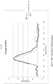
- the emission spectrum of the bioluminescent reporter overlaps with the absorption spectrum of a fluorophore.
- conversion of the substrate to a reaction product by the bioluminescent reporter results in excitation of a fluorophore by BRET and light emission from a fluorophore.
- (a) is one of a library of agents or compounds tethered to a chromophore. In some embodiments, (a) is one of a library of agents or compounds tethered to fluorophores. In some embodiments, (b) is one of a plurality of potential cellular targets fused to a bioluminescent reporter. In some examples, (b) is a library of cells expressing one of a plurality of potential cellular targets fused to a bioluminescent reporter.
- the present invention provides methods of detecting, analyzing, characterizing, etc. the binding of a bioactive agent to a cellular target.
- the cellular target may be the primary drug target.
- the cellular target is an off-target liability that may cause undesirable side effects in vivo.
- binding by the bioactive agent to the cellular target may have no discernible biological effect.
- the present disclosure provides cells comprising one or more of (e.g., each of) (a) a bioactive agent tethered to a chromophore (e.g., fluorophore); (b) a cellular target fused to a bioluminescent reporter; and (c) a substrate for the bioluminescent reporter.
- the binding of a bioactive agent to a cellular target is non-covalent.
- the chromophore is a fluorophore.
- the present invention provides methods for detection of an interaction between a bioactive agent and a cellular target comprising: (a) expressing in a cell a fusion of said cellular target and a bioluminescent reporter that emits energy at a first wavelength (e.g., range of wavelengths, spectral distribution, etc.); (b) contacting said cell with said bioactive agent tethered to a fluorophore, wherein said fluorophore accepts energy at said first wavelength and emits energy at a second wavelength (e.g., range of wavelengths, spectral distribution, etc.); (c) contacting said cell with a substrate for said bioluminescent reporter; (d) detecting energy at said second wavelength, wherein the presence of said energy at said second wavelength indicates the interaction of said bioactive agent with said cellular target.
- a first wavelength e.g.
- the present disclosure also provides methods for detection of an interaction between a bioactive agent and a cellular target comprising: (a) providing a fusion of said cellular target and a bioluminescent reporter; (b) contacting said fusion with said bioactive agent tethered to a chromophore (e.g., fluorophore); (c) contacting said fusion with a substrate for said reporter; (d) detecting a change in the spectral distribution of emitted light relative to said fusion contacted with said substrate in the absence of said chromophore.
- a chromophore e.g., fluorophore
- the present disclosure provides a method for detection of an interaction of a second bioactive agent and a cellular target comprising: (a) providing a fusion of said cellular target and a bioluminescent reporter; (b) contacting said fusion with both a first bioactive agent tethered to a chromophore (e.g., fluorophore) and with said second bioactive agent; (c) contacting said fusion with a substrate for said reporter; (d) detecting a change in the spectral distribution of emitted light relative to said fusion contacted with said first bioactive agent and said substrate in the absence of said second bioactive agent.
- a chromophore e.g., fluorophore
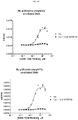
- the change in spectral distribution of emitted light is caused by displacement of said first bioactive agent by said second bioactive agent. In some examples, said displacement is competitive displacement. In some examples, the change in spectral distribution of emitted light is used to estimate binding affinity of the bioactive agent to cellular target. In some examples, said second bioactive agent is one of a plurality of bioactive agents. In some examples, the change in spectral distribution of emitted light is used to estimate the relative binding affinity of a plurality of bioactive agents to a cellular target. In some examples, said first and said second bioactive agents are synthetic molecules. In some examples, said cellular targets are in living cells, permeabilized cells or cell lysates.
- the change in the spectral distribution of emitted light is determined by measuring the ratio of light intensity at two different wavelengths, or two different ranges of wavelengths. In some examples, the change in spectral distribution over time is detected. In some examples, the chromophore is a fluorophore.
- a BRET assay system comprising: (a) a bioactive agent tethered to a fluorophore; (b) a first interaction partner fused to a structurally complementary peptide of a bioluminescent reporter; (c) a second interaction partner fused to a structurally complementary polypeptide of a bioluminescent reporter; and (d) a substrate for the bioluminescent reporter, wherein the first and second interaction partners interact to form an interaction complex, and wherein the first interaction partner, second interaction partner, and/or interaction complex are binding partners for the bioactive agent.
- the first interaction partner and the second interaction partner are proteins or polypeptides that interact to form a protein complex.
- the first interaction partner and the second interaction partner are brought together by the interaction of the structurally complementary polypeptides.
- the structurally complementary polypeptides are brought together by the interaction of first interaction partner and the second interaction partner.
- the interaction of the first interaction partner and the second interaction partner is determined by an increase in luminescence.
- the first and second interaction partners form the interaction complex in the presence or absence of the bioactive agent.
- the interaction complex is a binding partner for the bioactive agent, but neither the first or second interaction partners alone are binding partners for the bioactive agent.
- one of the interaction partners, but not the other is a binding partner for the bioactive agent.
- interaction complex formation requires binding of the bioactive agent to an interaction partner.
- interaction complex formation is independent of binding of the bioactive agent.
- BRET assay systems comprising: (a) a bioactive agent tethered to a fluorophore; (b) a cellular target fused to a structurally complementary peptide of a bioluminescent reporter; (c) a structurally complementary polypeptide of a bioluminescent reporter; and (d) a substrate for the bioluminescent reporter.
- the complementary peptide and polypeptide of the bioluminescent reporter associate to form active bioluminescent reporter enzyme.
- conversion of the substrate to a reaction product by the bioluminescent reporter enzyme results in excitation of the fluorophore by BRET and fluorescence emission from the fluorophore.
- an assay system comprising: (a) a bioactive agent tethered to a fluorophore; (b) a first binding partner fused to a complementary peptide of a bioluminescent reporter; (c) a second binding partner fused to a complementary polypeptide of a bioluminescent reporter; and (c) a substrate for the bioluminescent reporter.
- the complementary peptide and polypeptide of the bioluminescent reporter associate to form active bioluminescent reporter enzyme.
- the bioactive agent when the first and second binding partners interact, the bioactive agent binds to the interacting partners and association of the complementary peptide and polypeptide occurs, conversion of the substrate to a reaction product by the bioluminescent reporter enzyme results in excitation of the fluorophore by BRET and fluorescence emission from the fluorophore.
- the bioactive agent is tethered to a fluorescence or luminescence quencher (e.g., Dabcyl).
- a fluorescence or luminescence quencher e.g., Dabcyl
- titration of the untethered bioactive agent produces a gain in signal.
- imaging e.g., BRET imaging, via charge couple device camera, etc.
- imaging e.g., BRET imaging, via charge couple device camera, etc.
- location e.g., intracellular, extracellular, etc.
- BRET signals resulting from the presence and/or interactions of system components (e.g., bioactive agent, cellular target, etc.).
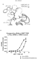
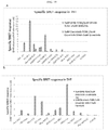
- systems and methods described herein are useful for the measurement of intracellular selectivity and affinity of a bioactive agent against a panel of putative cellular targets.
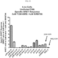
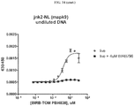
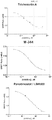
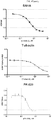
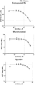
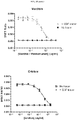
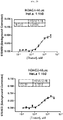
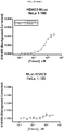
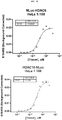
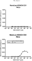
- the effect on BRET is monitored as the bioactive agent/cellular target complex is competitively disrupted.
- affinities are inferred by the IC50 value generated via competitive disruption.
- the affinities of inhibitors to individual domains of a cellular target are determined via genetic fusion of a bioluminescent reporter to a segregated domain of the cellular target.
- the present invention provides compositions and methods for detection and analysis of intracellular binding of a bioactive agent to a cellular target.
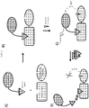
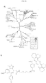
- a bioactive agent tethered to a chromophore, e.g., fluorophores, potential cellular targets fused to bioluminescent reporter proteins and methods of detecting and analyzing the interaction of bioactive agents with cellular targets therewith (See Figure 1 ).
- the interaction of a first entity (e.g., bioactive agent) and a second entity (e.g., cellular target) is detected, characterized, quantified, analyzed, etc. through the detection/measurement of a signal produced by signal transfer (e.g., transfer of energy (e.g., fluorescence, light energy, resonance, by BRET, etc.)) between a third entity (e.g., fluorophore) connected, fused, tethered, linked, etc. to the first entity and a fourth entity (e.g., bioluminescent reporter protein) connected, fused, tethered, linked, etc. to the second entity.
- signal transfer e.g., transfer of energy (e.g., fluorescence, light energy, resonance, by BRET, etc.)
- a third entity e.g., fluorophore
- a fourth entity e.g., bioluminescent reporter protein
- the interaction and/or binding of the first and second entities bring the third and fourth entities into close enough proximity to allow signal transfer (e.g., energy transfer) from one to another.
- the fourth entity e.g., bioluminescent reporter protein
- the third entity e.g., fluorophore
- the third entity e.g., fluorophore
- detection of the energy emitted from the third entity e.g., light at the emission maximum of the third entity
- the fourth entity e.g., bioluminescent reporter
- the duration, kinetics, affinity, strength, and/or specificity, of the binding of the first and second entities is detected, measured, quantified, determined, interrogated, etc. based on measurement of the signal output of the fourth entity (e.g., bioluminescent reporter protein) under various conditions.
- a cellular target fused to a bioluminescent reporter protein and a bioactive agent tethered to a chromophore, e.g., fluorophore, are provided (e.g., intracellularly, extracellularly, in lysate, in vitro, etc.).
- a bioactive agent tethered to a chromophore e.g., fluorophore
- Substrate for the bioluminescent reporter protein is added to the system.
- the bioluminescent reporter protein and chromophore e.g., fluorophore
- the bioluminescent reporter protein and chromophore are brought into close enough proximity for BRET to occur, and a detectable signal to be emitted from the chromophore (e.g., fluorophore).
- a complementary peptide and polypeptide that can interact are used.
- a complementary peptide is fused to a first interaction partner and a complementary polypeptide is fused to a second interaction partner.
- the first and second interaction partners form a complex (e.g., by binding to each other).
- the first and second interaction partners form an interaction complex when one or both interacts with a bioactive agent.
- the first and second interaction partners form an interaction complex in the presence or absence of a bioactive agent.
- formation of the interaction complex brings the complementary peptide and polypeptide together to form the bioluminescent reporter.
- formation of the bioluminescent reporter allows detection of formation of the interaction complex.
- a fluorophore is tethered to the bioactive agent.
- energy is transferred from the bioluminescent reporter to the fluorophore when the interaction complex is formed and the bioactive agent is bound to one of the interaction partners or the interaction complex.
- the fluorophore allows detection or measurement of the interaction of the first and second interaction partners.
- the fluorophore allows detection or measurement of the binding of the bioactive agent to its binding partner (e.g., the first interaction partner, the second interaction partner, and/or the interaction complex).
- a complementary peptide of a bioluminescent reporter is fused to a target of interest.
- a complementary polypeptide and a bioactive agent tethered to a fluorophore are provided to detect or measure the interaction of the bioactive agent to the target of interest.
- the complementary peptide of a bioluminescent reporter is fused to a target of interest, a complementary polypeptide of the bioluminescent reporter protein and a bioactive agent tethered to a fluorophore is applied to detecting proximity of the fluorescently labeled ligand, e.g., a bioactive agent, to protein complexes (e.g.
- the cellular target and bioluminescent reporter fusion is expressed in cells in which an assay is to be performed. In some examples, the fusion is expressed at or near the native abundance for the cellular target.
- the fluorophore-tethered bioactive agent is added extracellularly (e.g., added to the culture medium) and enters the cell via diffusion, active transport, passive transport, endocytosis, or any suitable mechanism. In some examples, varying amount of fluorophore-tethered bioactive agent is added to the cells to assay binding kinetics, assay binding affinity, provide sufficient signal, etc.
- the present invention provides compositions, methods, and systems for detection of intracellular interactions between a bioactive agent and cellular target (e.g., known or unknown).
- a fusion of a bioluminescent reporter and a cellular target are expressed within a cell.
- the bioactive agent, tethered to a fluorophore is introduced to the cell (e.g., conjugate of fluorophore and bioactive agent is cell permeable, the cell is permeablized, etc.).
- a substrate for the bioluminescent reporter protein is added to the cell prior, concurrently or subsequent to the addition of the bioactive agent.
- Detection of a fluorescent signal from the fluorophore indicates an intracellular interaction (e.g., binding) between the bioactive agent and cellular target.
- a cellular target fused to a bioluminescent reporter is expressed at a natural cellular abundance (e.g., relative to the native cellular target, or at a level appropriate for proper biological function of the fused target).
- interaction of a bioactive agent with a cellular target is detected intracellularly.
- characteristics of the interaction between the bioactive agent and cellular target are interrogated by altering the cellular conditions or the conditions of the system.
- a competitive binder of the cellular target e.g., untethered bioactive agent
- a library of bioluminescent reporter protein-tagged cellular targets is provided (e.g., in solution, in a lysate, immobilized on a surface, expressed within a cell, etc.).
- a bioactive agent is provided which lacks a known cellular target or where knowledge of the cellular target is uncertain or incomplete. The cellular target of a bioactive agent is determined by adding the bioactive agent to the library and determining which cellular-target fusion produces BRET induced fluorescence of the bioactive agent-tethered fluorophore.
- the library of bioluminescent reporter-tagged cellular targets is provided as a collection of nucleic acids or vectors comprising nucleic acids (e.g., plasmids, BacMam viruses, Lentiviruses, etc.) encoding the protein fusions.
- bioluminescent reporter protein-tagged cellular targets are expressed within cells.
- the library of bioluminescent reporter-tagged cellular targets is provided by translating nucleic acids in cell-free translation reactions.
- a library of cellular target fusions, or cells expressing cellular target fusions are provided in a microplate format.
- bioactive agent e.g., one identified through a phenotypic assay or screen
- the entire library of cellular targets can be interrogated (e.g., in solution, in lysates, intracellularly, etc.) in a high-throughput manner.
- bioluminescent reporter-tagged cellular targets are immobilized on a solid surface to create a protein array.
- cellular targets are also expressed as fusions with a tag or coupled to a protein (e.g., HALOTAG, Promega) that allows for the proteins to be covalently immobilized on a solid surface (e.g., a surface displaying appropriate ligands (e.g., HALOTAG Ligand)).
- a library of potential bioactive agents e.g., hit compounds or drug-like small molecules
- the system e.g., array
- any pairs capable of producing BRET are identified.
- cellular targets for all or a portion of a library of bioactive agents are unknown.
- compositions, methods, and systems herein provide a conjugate of a bioactive agent and an energy acceptor (e.g., fluorophore, chromophore).
- a bioactive agent is any small molecule (e.g., > 2000 daltons, >1000 daltons, >500 daltons, etc.), macromolecule, synthetic molecule or molecular complex capable of interacting with the biology of a cell.
- an energy acceptor is an entity capable of generating, exhibiting, and/or emitting a signal (e.g., light, heat, chemical reaction, fluorescence, resonance energy, etc.) when triggered by energy absorption (e.g., resonance energy transfer).
- a bioactive agent and energy acceptor e.g., fluorophore, chromophore
- any suitable structure or mechanism e.g., expressed as a fusion construct (e.g., with or without peptide linker), chemically linked (e.g., directly or indirectly), enzymatically linked, linked by a linker (e.g., peptide, nucleic acid, polymer, ester linkage, PEG linker, carbon chain, etc.)).
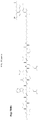
- the conjugate of a bioactive agent and an energy acceptor is produced by non-natural chemical synthesis (e.g., a purposeful execution of chemical reactions not present in natural cells).
- the type of linkage should not be viewed as limiting.
- bioactive agent refers generally to any physiologically or pharmacologically active substance or a substance suitable for detection.
- a bioactive agent is a potential therapeutic compound (e.g., small molecule, peptide, nucleic acid, etc.), or drug-like molecule.
- the bioactive agent is produced by non-natural chemical synthesis (e.g., a purposeful execution of chemical reactions not present in natural cells). Bioactive agents for use in embodiments described herein are not limited by size or structure.
- libraries of bioactive agents are provided.
- systems, methods, and compositions are provided for screening libraries of bioactive agents.
- means of identifying the bioactive agent in a library responsible for producing, eliciting, inducing, etc. phenotypic effect and/or activity are also disclosed herein.
- means of identifying the cellular target of a bioactive agent e.g., a bioactive agent responsible for the phenotypic effect and/or activity).
- an energy acceptor refers to any small molecule (e.g., chromophore), macromolecule (e.g., autofluorescent protein, phycobiliproteins, nanoparticle, surface, etc.), or molecular complex that produces a readily detectable signal in response to energy absorption (e.g., resonance energy transfer).
- an energy acceptor is a fluorophore or other detectable chromophore.
- Suitable fluorophores include, but are not limited to: xanthene derivatives (e.g., fluorescein, rhodamine, Oregon green, eosin, Texas red, etc.), cyanine derivatives (e.g., cyanine, indocarbocyanine, oxacarbocyanine, thiacarbocyanine, merocyanine, etc.), naphthalene derivatives (e.g., dansyl and prodan derivatives), oxadiazole derivatives (e.g., pyridyloxazole, nitrobenzoxadiazole, benzoxadiazole, etc.), pyrene derivatives (e.g., cascade blue), oxazine derivatives (e.g., Nile red, Nile blue, cresyl violet, oxazine 170, etc.), acridine derivatives (e.g., proflavin, acridine orange, acrid
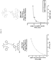
- a fluorophore is a rhodamine analog (e.g., carboxy rhodamine analog), such as those described in WO2013/078244 .
- rhodamine analog e.g., carboxy rhodamine analog
- Some such fluorophores are described herein in Example 8.
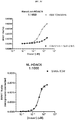
- BRET efficiency is significantly enhanced by the technical features of rhodamine analog (e.g., carboxy rhodamine analog) as an energy acceptor compared to other fluorophores.
- the left shifted EC50 and reduced nonspecific background of these dyes is advantageous for use in some embodiments.
- TOM and “NonChloroTOM” (or “NCT”) refer to the same type of fluorophore and are used interchangeably throughout the application.
- compositions, methods, and systems herein provide a fusion of a cellular target and a bioluminescent reporter protein (e.g., luciferase).
- a cellular target and bioluminescent reporter protein are fused, tethered, connected, etc. by any suitable structure or mechanism (e.g., expressed as a fusion construct (e.g., with or without peptide linker), chemically linked (e.g., through covalent or non-covalent bonds), enzymatically linked, linked by a linker (e.g., peptide, nucleic acid, other polymer (e.g., ester linkage, PEG linker, carbon chain, etc.)).
- a linker e.g., peptide, nucleic acid, other polymer (e.g., ester linkage, PEG linker, carbon chain, etc.
- an amino acid chain (e.g., 3-100 amino acids) is used to connect the cellular target and a bioluminescent reporter protein.
- the structure and/or function of neither the cellular target nor the bioluminescent reporter are impacted (e.g., significantly impacted) by fusion or the presence of the linker.
- a linker allows fusion without loss of activity or one or both of the elements.
- an amino acid linker properly spaces and/or orients the bioluminescent reported for energy transfer with the fluorophore.
- a cellular target comprises any suitable binding/interaction partner (e.g., receptor, enzyme, protein complex) for a bioactive agent (e.g., small molecule, protein, nucleic acid, lipid, etc.).
- a bioactive agent e.g., small molecule, protein, nucleic acid, lipid, etc.
- a cellular target is a protein or protein complex that binds to or otherwise interacts with (e.g., has a binding affinity to) a bioactive agent.
- a cellular target is a receptor protein or an enzyme that binds to or otherwise interacts with (e.g., has a binding affinity to) a small molecule bioactive agent.
- cellular targets may include nucleic acids (e.g., DNA or RNA), polysaccaharides or a complex comprising any of these with polypeptides.
- a cellular target is a G-protein coupled receptor or a protein kinase.
- the bioluminescent reporter is a luciferase.
- a luciferase is selected from those found in Gaussia, Coleoptera, (e.g., fireflies), Renilla, Vargula, Oplophorus, Aequoria, mutants thereof, portions thereof, variants thereof, and any other luciferase enzymes suitable for the systems and methods described herein.
- the bioluminescent reporter protein is a modified, enhanced luciferase enzyme from Oplophorus (e.g., NANOLUC enzyme from Promega Corporation, SEQ ID NO: 1, or a sequence with at least 70% identity (e.g., >70%, >80%, >90%, >95%) thereto).
- Oplophorus e.g., NANOLUC enzyme from Promega Corporation, SEQ ID NO: 1, or a sequence with at least 70% identity (e.g., >70%, >80%, >90%, >95%) thereto).
- the bioluminescent reporter protein is a thermostable Photuris pennsylvanica luciferase or a sequence with at least 70% identity (e.g., >70%, >80%, >90%, >95%) thereto).
- Exemplary bioluminescent reporters are described, for example, in U.S. Pat. App. No. 2010/0281552 and U.S. Pat. App. No. 2012/0174242
- the bioluminescent reporter protein comprises NANOLUC (See U.S. Pat. App. Nos. 2010/0281552 and 2012/0174242 ).
- the bioluminescent reporter protein comprises a polypeptide with at least 70% identity (e.g., >70%, >80%, >90%, >95%) to SEQ ID NO: 1 that retains bioluminescent characteristics.
- the use of the NANOLUC enzyme, or a variant thereof provides features (e.g., signal intensity, brightness, high light output, narrow spectrum, etc.) that enable the use of the BRET assays described herein.
- the high light output of NANOLUC enables the low concentration (e.g., ⁇ 1 ⁇ M, ⁇ 100 nM, ⁇ 10 nm, ⁇ 1 nm, etc.) of assay components, e.g., DNA encoding NANOLUC, useful to carry out assays under physiologically relevant conditions.
- NANOLUC enables the use of BRET in characterizing cellular targets identified in a phenotypic screen.
- a substrate for the bioluminescent reporter protein converts the substrate into a reaction product and releases light energy as a by-product.
- the substrate is a substrate for a luciferase enzyme.
- the substrate is a structural variant or derivative of coelenterazine (e.g., furimazine).
- the substrate is a substrate for a modified, enhanced luciferase enzyme from Oplophorus, e.g., NANOLUC enzyme from Promega Corporation (e.g., SEQ ID NO: 1).
- a pro-substrate for the bioluminescent reporter protein which produces a substrate through a chemical or physical process (e.g., hydrolysis, enzymatic reaction, photo-cleavage, etc.).
- the pro-substrate comprises coelenterazine, a coelenterazine derivative, a structural or functional equivalent of coelenterazine, a molecule substantially equivalent to coelenterazine (e.g., structurally and/or functionally), or molecule functionally or structurally similar to coelenterazine.
- the bioluminescent reporter protein converts the coelenterazine, coelenterazine derivative, structural or functional equivalent of coelenterazine, or substantial equivalent to coelenterazine into coelenteramide, a coelenteramide derivative, a structural or functional equivalent of coelenteramide, or a substantial equivalent to coelenteramide and releases light energy as a by-product.
- fluorophore and bioluminescent reporter are selected that exhibit sufficient overlap of emission (e.g., of bioluminescent reporter) and excitation (e.g., of fluorophore) spectra to provide efficient energy transfer between the two (e.g., by non-radiative dipole-dipole coupling).
- the peak emission of the bioluminescent reporter is substantially separated from the peak emission of the fluorophore, for example by at least 80 nm, 100 nm, 120 nm, 140 nm, etc., in wavelength.
- the Forster distance of the fluorophore and bioluminescent reporter pair is small (e.g., ⁇ 20 nm, ⁇ 10 nm, ⁇ 5 nm, ⁇ 3 nm, etc.).
- the short Forster distance results in the requirement that the fluorophore and bioluminescent reporter must be brought into very close proximity for energy transfer to occur. Therefore, the short Forster distance reduces aberrant and/or background signal (e.g., created by diffusing fluorophore and/or reporter).
- a fluorophore and bioluminescent reporter pair are selected that are sufficiently bright to allow detection of the transferred signal at a native abundance (or near native abundance) of the protein of interest and/or the cellular target fused to the bioluminescent reporter.
- either the selected fluorophore or bioluminescent reporter produce insufficient energy (light) emission
- either the fusion of the cellular target and bioluminescent reporter will need to be overexpressed (e.g., beyond native abundance, beyond a biologically relevant level, etc.), and/or the amount of fluorophore-tethered bioactive agent will have to be increased (e.g., to a potentially toxic level, beyond a physiologically relevant level, above the amount when kinetic experiments can be performed, etc.).
- sufficient brightness of the bioluminescent reporter and fluorophore allows detection of bioactive agent and cellular target interaction at a range of concentrations and ratios.
- compositions, methods, and systems are provided for identification of the cellular targets of hit compounds emerging from a phenotypic assay or phenotypic screen.
- "hit compounds" tethered to fluorophores are used to identify cellular targets, through their linkage to a bioluminescence reporter protein (e.g., binding of bioactive agent to cellular target results in BRET between bioluminescence reporter protein and fluorophore).
- a bioluminescence reporter protein e.g., binding of bioactive agent to cellular target results in BRET between bioluminescence reporter protein and fluorophore.
- the interaction partner e.g., cellular target for that bioactive agent in unknown or is uncertain.
- a bioactive agent tethered to a fluorophore is able to regenerate a phenotype, thereby validating that tethering the bioactive agent to the fluorophore (or the cellular target to the bioluminescent reporter) does not affect the cellular binding patterns and/or disrupt its biological activity.
- the use of a library of cellular targets, each fused to bioluminescent reporter protein e.g., NANOLUC
- detection method provides a greater degree of sensitivity or specificity.
- target identification through linkage to a bioluminescence reporter protein enables detection through energy transfer, even when binding of the target is inefficient or incomplete.
- binding of a bioactive agent e.g., small molecule
- binding of a bioactive agent e.g., small molecule
- BRET allows for analysis of the hit compound's binding affinity in live cells using competitive displacement of a fluorescent tracer (e.g., fluorophore) that binds to the same site on the cellular target.
- systems and methods described herein provide the ability to use two distinct methods for target identification, thus providing higher stringency for identifying cellular targets, complementary methods addressing limitations with other approaches (e.g., BRET provides the advantage of maintaining the bioactive agent in equilibrium with the target throughout the experiment).
- Some embodiments described herein find use in drug discovery, drug validation, drug target discovery, or drug target validation.
- the binding interaction between a bioactive agent e.g., drug-like small molecule
- a cellular target can be detected, validated, and/or characterized.
- the relative binding affinity of bioactive agents for a cellular target e.g., in solution, in a lysate, on a surface, in a cell, etc.
- bioactive agent e.g., drug-like small molecule
- the relative binding affinity of bioactive agents for a cellular target can be determined by their ability to displace a bioactive agent that has been tethered to a fluorophore.
- higher binding affinity of a first bioactive agent relative to a second bioactive agent is indicated by requiring a lower concentration of the first bioactive agent to displace a tethered bioactive agent relative to the second bioactive agent.
- Displacement of the tethered bioactive agent is determined by the loss or reduction of energy transfer from the bioluminescent reporter protein fused to a cellular target.
- the concentration of bioactive agent needed to displace a tethered bioactive agent is used to estimate the binding EC50 or the inhibition constant (Ki) for bioactive agent.
- the development of new or modified bioactive agents is guided by their ability to displace one or more bioactive agents, each tethered to a fluorophore, from one or more cellular targets, each fused to a bioluminescent reporter protein.
- a collection of compounds which may have unknown binding affinity to a cellular target may be screened for their ability to bind the target fused to a bioluminescent protein by determining their ability to displace a bioactive agent tethered to a fluorophore.
- compounds may be screened for their ability to bind a first cellular target preferentially relative to a second cellular target by their ability to displace a first tethered bioactive agent from the first cellular target relative to a second tethered bioactive agent from the second bioactive target.
- the first and second tethered bioactive agents are the same.
- systems and methods described herein provide the ability to determine the affinity of a bioactive agent (e.g., hit compound, research lead, lead compound, etc.) for wild-type and mutant version(s) of a cellular target, e.g., target protein.
- a bioactive agent e.g., hit compound, research lead, lead compound, etc.
- characterization of the affinity and selectivity of the fluorescently-labeled bioactive agent (e.g., drug) to a disease-relevant mutant protein may be performed in cells.
- Such systems and methods may be useful to identify bioactive agent(s) (e.g., drug) that selectively bind a wild-type or mutant protein differentially.
- bioactive agents may also bind to unexpected and/or unintended cellular targets (off targets).
- off targets In some cases, off target binding of a bioactive agent is responsible for a portion of the therapeutic and/or adverse effects associated with administration of the bioactive agent.
- systems and methods described herein provide the ability to identify off targets of a bioactive agent. Understanding the identity and extent of off-target bioactive agent binding provides valuable information regarding the pharmacology of the agent.
- systems and methods described herein provide the ability to estimate the binding characteristics of a bioactive agent tethered with a fluorophore to a cellular target fused to a bioluminescent reporter (e.g., EC50, Kd, binding rate, environmental influences, etc.)
- the binding characteristics are correlated to biochemical, physical, or phenotypic characteristics related to the cellular target.
- the kinetic profile of the formation or dissolution of the BRET complex can be used to infer association or dissociation rates of untethered bioactive agents. In some examples, these association/dissociation rates can be used to estimate drug residence times for individual targets within intact cells.
- systems and methods are useful in thermodynamic molecular mechanism of action (MMOA) studies on entropic vs. enthalpic interactions.
- displacement of the tethered bioactive agent by a separate agent may occur through separate sites on the cellular target.
- the binding characteristics of the tethered bioactive agent may be used to determine the influence of post-translational modification (e.g., cleavage, phosphorylation, methylation, acetylation, lipidation, etc.), intracellular translocation (e.g., movement to a nucleus, mitochondria, membrane, etc.), or protein interaction (e.g., interactions with other proteins, nucleic acids, lipids, etc.) on the cellular target.
- post-translational modification e.g., cleavage, phosphorylation, methylation, acetylation, lipidation, etc.
- intracellular translocation e.g., movement to a nucleus, mitochondria, membrane, etc.
- protein interaction e.
- An example is determining the binding characteristics of an antibody to a cellular target by its influence on the binding characteristics of the tethered bioactive agent.
- the binding characteristics of the tethered bioactive agent may be used to determine the influence of chemical modifications or transformation of the bioactive agent or the tethered fluorophore (e.g., intracellular metabolism, change in ionic state, etc.).
- the cellular target may comprise more than one molecular component.
- the target may comprise more than one polypeptide, and may further comprise other natural or synthetic molecules (e.g., prosthetic groups, cofactors, metabolites, nucleic acids, lipids, carbohydrates, etc.).
- a bioactive agent tethered to a fluorophore binds to a first molecular component, and a bioluminescent reporter is fused to a second molecular component, so that a signal from the tethered bioactive agent is produced when bound to the first molecular component and the first molecular component is in close proximity to the second molecular component.
- a priori knowledge of the existence of an interaction between a bioactive agent and cellular target is not needed to practice the invention.
- detection and/or characterization of an unknown or previously unidentified interaction by energy transfer is provided.
- Advantages of the systems, compositions, and methods described herein, over other methods of target discovery may include: a broad range of possible bioactive agent concentrations because (in some examples) it need not be expressed within cells (e.g., allows for addition of enough acceptor fluorophore to generate detectable signal), natural protein concentration of cellular target (e.g., no need for overexpression to gain enough signal for detection), signal detectable on a plate reader (e.g., high throughput detection, no imaging necessary), detection of interactions within a cell, etc.
- bioactive agents e.g., hit compounds
- a fluorescent energy acceptor dye e.g., NANOLUC-fused
- the binding of the modified agent to its luciferase-fused (e.g., NANOLUC-fused) cellular target results in energy transfer from NANOLUC to the acceptor dye.
- NANOLUC-fused luciferase-fused
- the labeled bioactive agent remains in equilibrium with the cellular target throughout the experiment allowing detection of targets that interact with the hit compounds with low affinity.
- BRET enables measurements of binding affinity in living cells by the competitive displacement of a fluorescent tracer designed for the same binding site.
- NANOLUC is significantly brighter than other luciferases commonly used for BRET, thereby allowing energy transfer to be quantitated at lower expression levels which are more suitable for maintaining relevant biology within a cell.
- the narrow emission spectrum of NANOLUC increases the dynamic range by reducing spectral cross-over in the acceptor channel.
- the dynamic range can be further increased by using long-wavelength acceptors that emit in the near-red region of the spectrum (600 to 650 nm).
- rhodamine analogs e.g., carboxy rhodamine analog
- WO2013/078244 delivers an optimal dynamic range for use with NANOLUC and/or in the BRET applications described herein.
- HEK293 cells were transfected using FuGene HD (Promega Corp.) with plasmid DNA encoding a NanoLuc-HDAC6 fusion protein.
- NanoLuc-HDAC6 DNA was diluted 1:1000 with a promoterless carrier DNA (pGEM3Z) to yield a final concentration of 50ng/well total DNA in 96-well plate format (at a seeding density of 20,000 cells/well). Twenty-four hours post-transfection, cells were then incubated with serially-diluted tracer in the presence or absence of a molar excess of unlabeled SAHA (as a specificity control).
- furimazine (a coelenterazine derivative substrate for NanoLuc; Promega Corp.) was added at a concentration of 20uM, and BRET was quantified on a Varioskan luminometer.
- the specific BRET signal was calculated by subtracting the non-specific signal (in the presence of unlabeled SAHA) from the signal generated from the SAHA tracer alone.
- results in Figure 2 demonstrate that the SAHA-TOM (PBI-4968) conjugate/tracer generated a superior, specific BRET signal compared to SAHA-TMR (PBI-4967) and also generated a left-shifted EC 50 value for binding within living cells.
- the benefit of the PBI-4968 TOM dye over other commonly used fluorophores can be applied to competitive binding assays or target identification (chemical proteomics) screening using NANOLUC fusion protein libraries.
- HEK293 cells were transfected with DNA encoding NanoLuc-HDAC6 at a 1:1000 dilution with carrier DNA as previously described. Twenty-four hours post-transfection, cells were incubated with serially-diluted natural pro-drug, FK-228; in the presence of a fixed concentration (1uM) of SAHA-TOM (PBI-4968) conjugate. For comparison, a biochemical, activity-based HDAC6 assay was run in parallel. Briefly, a 3-fold serial dilution of FK-228 (Selleckchem catalog # S3020) was performed at 100X in 100% DMSO in wells of a 96-well plate.
- HDAC-GloTM I/II Final Detection Reagent (Promega Corp.) was added to all wells (20 ⁇ L final assay volume), and luminescence was measured after a10 minute incubation at room temperature. For all test conditions, the final concentration of HDAC-GloTM I/II substrate was 50 ⁇ M. The data was plotted (sigmoidal dose response - variable slope) using PrismTM software from GraphPad (no DTT: closed circles; 0.25 mM DTT: open squares).
- HEK293 cells were transfected with DNA encoding PKCalpha (PKCa)-NanoLuc or NanoLuc-PKCalpha at a 1:1000 dilution of DNA with promoterless carrier DNA as described previously. Twenty-four hours post-transfection, cells were treated with or without digitonin to a final concentration of 50ug/mL. Cells were then treated with serially-diluted a staurosporine-PBI-dye conjugate (PBI-5129; See Figure 8 ). Cells were co-incubated in the presence or absence of 5uM unconjugated staurosporine as a specificity control for binding. Following two hours of equilibration with a tracer, furimazine was added to a final concentration of 20uM, and BRET ratios were measured on a Varioskan luminometer ( Figures 5A and 5B ).
- Figure 5A-C demonstrate the ability to use permeabilization agent to potentiate the entry of impermeable drug tracers and applied to BRET-based chemical proteomics screens of NanoLucTM fusion protein libraries.
- HEK293 cells were transfected with DNA encoding a NanoLuc-p38 fusion protein (to a final concentration of 50ng/well DNA in 96-well format). Twenty-four hours post-transfection, cells were treated with serially diluted BIRB-796 or PBI-4835 in the presence of 0.5uM PBI-4838 (BIRB conjugate derivative; see, e.g., WO2013/078244 . After two hours of equilibration, furimazine was added to a concentration of 20uM, and BRET was measured on a Varioskan luminometer.
- Figure 6 demonstrates that the dose-response BRET curves support the higher known affinity of BIRB-796 over PBI-4835 ("BIRB Derivative").
- BIRB Derivative The ability to measure the relative affinities of unlabeled drugs can be applied to HTS screens, lead optimization or chemical proteomics applications.
- similar experiments are configured to characterize optimized hit compounds from high-throughput chemical screens (e.g., compounds with lower IC50 values for tracer displacement would indicate higher binding efficiency to the target of interest).
- HEK293 cells were transfected with DNA encoding a NanoLuc-p38 fusion (to a final concentration of 50ng/well DNA in 96-well format). Twenty-four hours post-transfection, cells were pretreated for 2 hours with protected furimazine (PBI-4378; See Figure 8 ) to a final concentration of 20uM. BRET was measured over time on a Varioskan luminometer set to 37°C. After a short pre-read, cells were stimulated with varying concentrations of PBI-4838 (in the presence or absence of 1uM BIRB796 as a specificity control). Dose and time-dependent increases in BRET were then monitored kinetically over 4 hours.
- transfected cells were pretreated with both 20uM PBI-4377 and 1uM PBI-4838 to generate a steady BRET signal. After a short pre-read, cells were stimulated with 1uM unlabeled BIRB-796, and competitive displacement was monitored by BRET in real-time.
- the following example relates to the use of specific NANOLUC substrates (SEE FIG. 8 ) or enzyme components to discriminate the subcellular localization of a drug binding event using BRET.
- NANOLUC complementary NANOLUC enzyme polypeptides or peptide
- the small signal peptide is genetically tethered to a cell surface receptor.
- the donor signal is isolated to the extracellular space. This signal isolation increases the signal/background generated in various BRET applications including drug tracer binding/displacement.
- full length NANOLUC protein fusions are utilized if impermeable NANOLUC substrate is applied.
- Staurosporine p-nitrophenylcarbamate (3 mg, 4.8 umol) was dissolved in 0.5 mL of DMF and treated with excess cadaverine. The reaction was warmed in a 70°C oil bath for 2 h, then diluted with H 2 O, acidified with formic acid, and subjected to preparative HPLC eluting with 25%->75% MeCN in 10 mM aqueous NH 4 OAc. The appropriate fractions were lyophilized to afford a slightly yellow solid. Calcd for M+H: 595.3; found 595.5
- Staurosporine 5-aminopentylcarboxamide (5 mg, 8.4 umol) was dissolved in 1 mL DMF and treated with triethylamine and TOM 6-SE (4 mg, 6 umol). The reaction was monitored by analytical HPLC. Upon completion of the reaction, MeCN and H 2 O were added, and the TEA was neutralized by addition of a small amount of AcOH. Preparative HPLC (25%->100% MeOH in 10 mM NH4OAc) and subsequent concentration yielded 1.8 mg of a dark blue solid. Calcd for M+H: 1139.5; found 1139.8.
- TOM 6-carboxylic acid (26 mg, 46 umol) was stirred in 1 mL of DMF with 2 equiv of triethylamine and treated with TSTU (17 mg, 56.5 umol, 1.2 equiv), and the reaction was monitored by HPLC. After 40 min, the reaction was added to a solution of cadaverine (94 mg, 0.92 mmol, 20 equiv) in 0.5 mL DMF and stirred for 20 min. The reaction was then neutralized by addition of TFA and diluted with MeCN and water. Preparative HPLC (25->100% MeCN in 0.1 % aqueous TFA) and subsequent lyophilization afforded the desired product as a purple solid. LCMS: Calcd for ((M+2H)/2) 324.4; found 324.3.
- HEK293 cells were transfected with plasmid DNA encoding NanoLuc-p38alpha or Renilla Luc-p38alpha using Fugene HD(Promega Corporation) at a 3:1 lipid:DNA ratio and seeded into 96-well plates at a density of 20,000 cells per well (to yield 50ng/well of DNA). 24h post-transfection, cell medium was replaced with serum-free medium (Opti-MEM) and incubated with serially-diluted PBI-4838 (BIRB796 derivative conjugated to TOM dye) in the presence/absence of BIRB796 at 1uM. Cells were incubated at 37°C for two hours. To the NanoLuc-expressing samples, furimazine was added to a final concentration of 20uM.
- Figure 9 demonstrates that both Renilla luciferase and NanoLuc luciferase can be used in the method of the present invention.
- HEK293 cells were transfected with plasmid DNA encoding NanoLuc-p38alpha using Fugene HD and seeded into 96-well plates (to yield 50ng/well of DNA). 24h post-transfection, cell medium was replaced with serum-free medium (Opti-MEM) and incubated with serially-diluted PBI-4838 (BIRB796 derivative conjugated to TOM dye) or PBI-4836 (BIRB derivative conjugated to TMR dye) in the presence/absence of BIRB796 at 1uM. Cells were incubated at 37°C for two hours. To the NanoLuc-expressing samples, furimazine was added to a final concentration of 20uM. BRET was then measured on a Varioskan luminometer equipped with 450nm bandpass and 630nm longpass filters. The BRET ratio was determined by dividing the signal in the 630 channel by the signal in the 450 channel.
- Figure 11 demonstrates another example of the increased potency of the TOM adduct over the TMR adduct for binding to a target protein in a cell-based format.
- Dose-response curves for NanoLuc-p38alpha binding indicate an EC50 of 38nM for the BIRB-TOM conjugate, compared to an EC50 of 450nM for the BIRB-TMR conjugate.
- the reported affinity of BIRB-TOM is more consistent with literature reports compared to the BIRB-TMR ( Chem Biol Drug Des 2009; 74: 547-559 ).
- HEK293 cells were transfected with plasmid DNA encoding PKCalpha-NanoLuc (diluted 1:1000 into pGEM3Z carrier DNA) using Fugene HD and seeded into 96-well plates (to yield 50ng/well of total DNA). 24h post-transfection, cell medium was replaced with serum-free medium (Opti-MEM) and incubated with serially-diluted PBI-5075 (BIM conjugated to TOM dye) or PBI-5051 (BIM conjugated to TMR dye) in the presence/absence of staurosporine at 5uM. Cells were incubated at 37°C for two hours. To the NanoLuc-expressing samples, furimazine was added to a final concentration of 20uM.
- BRET was then measured on a Varioskan luminometer equipped with 450nm bandpass and 630nm longpass filters. The BRET ratio was determined by dividing the signal in the 630 channel by the signal in the 450 channel. To determine specific BRET signals, the BRET ratio at each concentration of tracer + unlabeled staurosporine was subtracted from the BRET ratio at each concentration of tracer without unlabeled staurosporine.
- Figure 12 demonstrates another example of the increased potency of the TOM adduct over the TMR adduct for binding to a target protein in a permeabilized cell format.
- Dose-response curves for PKCalpha-NanoLuc binding indicate an EC50 of 483nM for the BIM-TOM conjugate, compared to dramatically right-shifted potency for the BIM-TMR conjugate.
- the reported affinity of BIM-TOM is more consistent with literature reports compared to the BIM-TMR ( J Biol Chem. 1991 Aug 25; 266(24):15771-81 .).
- the following example serves to demonstrate the ability to profile the selectivity of a drug tracer against a panel of putative targets within a given phylogenetic target family. Similar experimental configurations may be used to identify the target of fluorescently labeled drugs using BRET in cells. This configuration may lead to the identification of the primary drug target, as well as off-target interactors. Engagement of multiple targets may be indicative of drug promiscuity and potential drug side effects. This example serves to demonstrate the ability to measure both primary, as well s secondary target interactions via BRET.
- HEK293 cells were transfected with plasmid DNA encoding N or C-terminal NanoLuc fusions to various members of the MAPK pathway (Jnk1, Jnk2, Jnk3, p38alpha, p38beta, p38gamma, p38 delta, or PKCalpha or MAPK 8, 9, 10, 14, 11, 12, or 13, PKCalpha respectively) using Fugene HD (Promega Corporation) at a 3:1 lipid:DNA ratio and seeded into 96-well plates at a density of 20,000 cells per well (yield 50ng/well of DNA).
- Fugene HD Promega Corporation
- Figure 13 indicates selectivity of PBI-4838 to Jnk2, p38beta and p38alpha, consistent with literature reports.
- P38alpha is recognized as the primary target of BIRB796.
- interactions with targets such as Jnk2 and p38beta could indicate potential off-target liabilities.
- PKCalpha-NanoLuc fusion combined with PBI-4838 showed relatively little specific BRET signal owing to poor affinity of BIRB796 toward this target.
- the following example serves to demonstrate the ability to profile the affinity of a drug tracer against a panel of putative targets within a given phylogenetic target family. Similar experimental configurations may be used to characterize the affinity of the fluorescently labeled drug to a given target using BRET in cells.
- HEK293 cells were transfected with plasmid DNA encoding NanoLuc-p38alpha, NanoLuc-p38beta, Jnk2-NanoLuc, PKCalpha-NanoLuc, or NanoLuc-HDAC6 using Fugene HD (Promega Corporation) at a 3:1 lipid:DNA ratio and seeded into 96-well plates at a density of 20,000 cells per well (yield 50ng/well of DNA). Twenty-four hours post-transfection, cell medium was replaced with serum-free medium (Opti-MEM) and incubated with serially-diluted PBI-4838 in the presence or absence of BIRB796 at 4uM. Cells were incubated at 37°C for two hours.
- Fugene HD Promega Corporation
- transfected cells were treated with serially-diluted BIRB796 in the presence of 1uM PBI-4838.
- furimazine was added to a final concentration of 20uM.
- BRET was then measured on a Varioskan luminometer equipped with 450nm bandpass and 630nm longpass filters. The BRET ratio was determined by dividing the signal in the 630 channel by the signal in the 450 channel.
- Figures 14 and 15 demonstrate the apparent affinity of PBI-4838 to Jnk2, p38beta, and p38alpha.
- PKCalpha and HDAC6 showed relatively little specific BRET signal owing to poor affinity of PBI-4838 to these targets.
- the high affinity of BIRB796 toward Jnk2, p38alpha, and p38beta is demonstrated by competitive displacement in cells. Similar experiments could be configured to rank-order the affinity of drug tracers to a given set of targets (by an increase in BRET) or unmodified drugs (by competitive displacement). As indicated above, high-affinity interactions with targets such as Jnk2 and p38beta could indicate potential off-target liabilities.
- the following example serves to demonstrate the ability to measure the relative affinity of a drug tracer to a wild-type versus mutant target protein.
- affinity of a fluorescently-labeled iBET compound small-molecule inhibitor that binds BRD4 preventing its interaction with acetylated histones
- BRD4 Bromodomain-containing protein 4
- Similar experimental configurations may be used to characterize the affinity and selectivity of the fluorescently-labeled drug to a disease-relevant mutant protein in cells. Such experiments may be useful to identify drugs that selectively bind to wild-type or mutant proteins differentially.
- HEK293 cells were transfected with plasmid DNA encoding NanoLuc-BRD4 or NanoLuc-BRD4 (N140A/N433A) using Fugene HD (Promega Corporation) at a 3:1 lipid:DNA ratio and seeded into 96-well plates at a density of 20,000 cells per well (yield 50ng/well of DNA). Twenty-four hours post-transfection, cell medium was replaced with serum-free medium (Opti-MEM) and incubated with serially-diluted PBI-4966 (iBET conjugated to TOM dye) in the presence/absence of iBET at 10uM. Cells were incubated at 37°C for two hours.
- Opti-MEM serum-free medium
- PBI-4966 iBET conjugated to TOM dye
- Figure 16 indicates relative affinities of PBI-4966 to wild-type BRD4 versus the mutant BRD4. As expected, a right-shifted potency is observed with the mutant BRD4 indicating a lowered affinity for PBI-4966. Similar experiments could be configured to rank the order of the affinity of drug tracers to disease-relevant mutants of a target. This principle could be expanded to measuring relative affinities of drugs toward wild-type or mutant proteins by competitive tracer displacement. Since increased BRET signal is only indicative of binding to the NanoLuc fusion, this method enables measurements of target engagement amidst a complex mixture of similar antigens present in the assay sample. These antigens could include targets of similar properties, but non-identical to the target of interest (for instance, to discriminate heterologous vs. endogenous analyte or mutant vs. wt. analyte, etc.).
- the following example demonstrates that the method of the present invention can be used to screen compound panels to determine target engagement.
- This screening method could be expanded to larger compound libraries (e.g. LOPAC) as well.
- HEK293 cells were transfected with NanoLuc-HDAC6 DNA using Fugene HD and seeded into wells of a 96-well plate.
- NanoLuc-HDAC6 DNA was diluted with promoterless carrier DNA (pGEM3Z). The final DNA concentration/well remained 50ng/well; however the NanoLuc-HDAC6 DNA was diluted 1:1000 (at a seeding density of 20,000 cells/well in 96-well format).
- SEE Figure 17 serially-diluted inhibitors
- furimazine was added to 20uM, and BRET detected on a Varioskan luminometer.
- HEK293 cells were transfected with plasmid DNA encoding CDK2-NanoLuc (diluted 1:100 into pGEM3Z carrier DNA) using Fugene HD (Promega Corporation) at a 3:1 lipid:DNA ratio and seeded into wells of 96-well plates at a density of 20,000 cells per well (yield 50ng/well of total DNA). Twenty-four hours post-transfection, cell medium was replaced with serum-free medium (Opti-MEM) and incubated with serially-diluted PBI-5077 (purvalanol B conjugated to TOM dye) in the presence/absence of Purvalanol B at 10uM. Cells were incubated at 37°C for two hours.
- Opti-MEM serum-free medium
- PBI-5077 purvalanol B conjugated to TOM dye
- This example supports use of fluorophore tracers in medium for monitoring drug/target interactions via BRET.
- BRET fluorophore tracers
- the following example demonstrates the ability to generate NanoLuc fusion arrays using cell-free expression and measure drug binding and relative affinity by BRET.
- a panel of 11 kinases including putative targets of BIRB796 (such as members of the MAPK and Jnk families), putative targets of Dasatinib (such as Src and LCK) as well as an irrelevant HDAC6 negative control was expressed in TnT® T7 Quick Coupled Reticulocyte Transcription/Translation System (Promega Corporation) as recommended by the manufacturer to create an array with 23 different NanoLuc fusions.
- Each TNT® reaction was diluted 1:100 in PBS and plated in replicates into wells of 96-well plates.
- the NanoLuc fusion array was screened with 2 different TOM-drug tracer (BIRB-TOM; PBI-4838 and Dasatinib-TOM; PBI-5170 ) in the presence and absence of excess relevant free drug.
- BIRB-TOM TOM-drug tracer
- 4 replicas with 1uM Dasatinib-TOM/ 4 replicates with 1uM Dasatinib-TOM+5uM Dasatinib were performed.
- the reactions were incubated with constant mixing for 2h at room temperature.
- BRET was then measured on a Varioskan luminometer equipped with 450nm bandpass and 610nm longpass filters. The BRET ratio was determined by dividing the signal in the 610 channel by the signal in the 450 channel. To determine response ratios, the BRET values with tracer alone were divided by the BRET values with tracer + unmodified.
- the BIRB-TOM hits were further analyzed for binding affinity to the BIRB-TOM tracer and competitive displacement of the BIRB TOM Tracer with the BIRB796.
- Binding affinity experiments to the BIRB-TOM tracer were preformed by incubating 3 replicates of the diluted TNT reactions with serially-diluted BIRB-TOM tracer in the presence/absence of BIRB796 at 10uM. Reactions were incubated at room temperature with constant mixing for two hours and after adding furimazine to a final concentration of 20uM analyzed as described above.
- the following example demonstrates that high affinity interaction with the drug tracer with optimal signal to background is achieved when the expression of the NanoLuc fusion is close to endogenous level.
- a 1:1000 dilution of the NanoLuc HDAC6 DNA with a carrier DNA was required to achieve a high affinity interaction.
- the expression of serial dilutions of the NanoLuc HDAC6 DNA was compared to the expression of the endogenous HDAC6 using western blot analysis.
- HEK293 cells were seeded into wells of 6-well plates at 2.5x10 5 cells/well and transfected with PEI with varying amounts of NanoLuc HDAC6 DNA.
- the NanoLuc HDAC6 DNA was diluted with a non-expressing carrier DNA (pCI neo). The final concentration of DNA remained 2ug/well while the NanoLuc HDAC6 DNA was diluted 1:0, 1:10, 1:100,1:1000 and 1:10000.
- the control was un-transfected cells. 24 hours post transfection, the media was removed, and cells washed with PBS and lysed for 10 min with 400ul of mammalian lysis buffer (Promega) +1:50 RQ1 DNase (Promega) +1x RQ1 buffer.
- TAMRA-epidermal growth factor (TMR-EGF; Life Technologies) was applied in a cellular BRET assay using a stable cell line expressing a NanoLuc-EGFR (Her1) fusion protein.
- TMR-EGF TAMRA-epidermal growth factor
- the BRET signal generated between TMR-EGF and NanoLuc-EGFR can be inhibited upon treatment with therapeutic antibodies known to interfere with binding of EGF.
- a HEK293 stable cell line was generated by transfection of pF5 NanoLuc-EGFR plasmid DNA (Promega Corp.) and FuGene HD (Promega Corp.). After transfection, the stable cell line was generated by G418 selection, followed by limited dilution cloning. Clonally-derived cells expressing NanoLuc-EGFR were seeded into 96-well plates at a density of 20,000 cells per well. 20 hours after seeding, medium was replaced with serum-free OptiMEM and cells were serum starved. After starvation, cells were treated with serially-diluted TMR-EGF (Life Technologies).
- furimazine (a coelenterazine derivative substrate for NanoLuc; Promega Corp.) was added at a concentration of 20uM, and BRET was quantified on a Varioskan luminometer.
- antibody-mediated displacement of TMR-EGF was measured by pretreating cells with Vectibix, Erbitux, or Herceptin (negative control) prior to stimulation with TMR-EGF (10ng/mL final concentration).
- Results in Figure 20 demonstrate that the BRET can be used to quantify binding of fluorescent cytokines to cell surface receptors tethered to NanoLuc. Furthermore, the affinities of therapeutic antibodies capable of inhibiting cytokine binding can be measured by competitive displacement of the fluorescent cytokine. Vectibix and Erbitux were capable of inhibiting the binding of TMR-EGF to NanoLuc-EGFR, whereas Herceptin (a Her2 binder with negligible affinity to Her1) was incapable of inhibiting TMR-EGF binding. This example demonstrates that the binding of a fluorescent cytokine to a known high-affinity target can be quantified by BRET, in a format that can be readily reconfigured to monitor binding of therapeutic antibodies (e.g.
- the present disclosure enables the detection/measurement of three moieties coming together: (1) A peptide of a bioluminescent reporter protein and (2) a polypeptide of a bioluminescent reporter protein which interact through structural complementation to form a complex, and (3) a fluorescent third moiety (e.g., fluorescent small molecule).
- a fluorescent third moiety e.g., fluorescent small molecule
- This example demonstrates energy transfer from a bioluminescent complex formed from a structurally complementary peptide (SEQ ID NO: 6) and polypeptide (SEQ ID NO: 7), which form a bioluminescent complex.
- a fluorescent dye was attached to the complementary peptide sequence.
- a fluorescent protein could be fused (e.g., a fusion protein) with a peptide or polypeptide (e.g., created from a genetic construct).
- E. coli clarified lysate expressing the complementary polypeptide sequence was prepared (poly lysate). 40uL of the poly lysate was mixed with 10uL of complementary peptide (pep) or fluorescently-labeled complementary peptide (pep-TMR) and incubated for 10min at RT. 50uL 100uM substrate for the complex (furimazine) in 50mM HEPES pH 7.4 was added and incubated for 30min at RT. Luminescence was measured over 400-700nm on TECAN M1000.
- Figure 21 illustrates very efficient energy transfer from the Poly/Pep complex (donor) to TMR (acceptor), and the corresponding red shift in the wavelength of light being emitted.
- B) This example demonstrates using BRET in detection, such as detecting small molecule concentration or enzymatic activity. Because energy transfer is strongly dependent on distance, the magnitude of energy transfer can often be related to the conformation of the system. For instance, insertion of a polypeptide that chelates calcium can be used to measure calcium concentration through modulation of energy transfer.
- An enzyme that also changes the distance, either through causing a conformational change of the sensor as above or through cleavage of the sensor from the fluorescent moiety, can be measured through a system as described herein.
- One structurally complementary member of a complex is bound to a fluorescent moiety which transfers its energy when the structurally complementary peptide and polypeptide of a bioluminescent reporter protein interact.
- a fluorescent moiety which transfers its energy when the structurally complementary peptide and polypeptide of a bioluminescent reporter protein interact.
- a linker e.g., DEVD Caspase-3 cleavage site.
- a complementary polypeptide (SEQ ID NO: 8) and NL-HT (NanoLuc fused to HaloTag) were purified.
- 20uL of 8pM NL-HT was mixed with 20uL of 100nM PBI-3781 (See, e.g., WO2013/078244 ) and incubated for 10min at RT.
- 40uL NanoGlo+100uM furimazine was added, and luminescence measured over 300-800nm on TECAN M1000.
- 20uL of 33ng/uL of a complementary polypeptide (SEQ ID NO: 6) was mixed with 20uL of ⁇ 500uM PBI-5074 (fluorescent dye-linker-complementary peptide).
- 40uL NanoGlo+100uM furimazine was added, and luminescence measured over 300-800nm on TECAN M1000.
- Figure 22 illustrates energy transfer from the complementary peptide/polypeptide complex (donor) to the TOM-dye of PBI-5074 (acceptor), and the corresponding red shift in the wavelength of light being emitted.
- the energy transfer with a structurally complementary pair of a bioluminescent reporter protein can also be used to measure three molecules interacting.
- One example is a GPCR fused to a complementary polypeptide of a bioluminescent reporter protein and a GPCR interacting protein fused with a complementary peptide of the bioluminescent reporter protein that form a bioluminescent complex when they interact. This allows measurement of the binary interaction. If a small molecule GPCR ligand bearing a fluorescent moiety for energy transfer interacts with the complex, energy transfer occurs. Therefore, the binary protein-protein interaction and the ternary drug-protein-protein interaction are measured in the same experiment. Also, the fluorescent molecule only causes a signal when interacting with the protein pair, which removes any signal from the ligand interacting with an inactive protein ( Figure 23 ).
- the donor is comprised of two complementary subunits, an 1 IS polypeptide and a PEP-80 peptide.
- the 1 IS polypeptide was genetically tethered to the N-terminus of BRD4, and the fusion protein expressed in HEK293 cells. Luminescence was generated from the fusion protein only in the presence of both furimazine () and the PEP-80 complementary polypeptide ( Figure 24A ).
- HEK293 cells were transfected with 11S-BRD4 DNA using Fugene HD and seeded in a 96-well plate format.
- 11S-BRD4 DNA was diluted with promoterless carrier DNA (pGEM3Z). The final DNA concentration/well remained 50ng/well; however the 11 S-BRD4 DNA was diluted 1:10 (at a seeding density of 20,000 cells/well in 96-well format). Twenty hours post-transfection, cells were lysed in OptiMEM + 50ug/mL digitonin in the presence of absence of 1uM PEP-80 peptide.
- the concept illustrated by this example can be applied to detecting proximity of a fluorescently labeled ligand to protein complexes (e.g. detecting ligand binding selectively to heterodimeric or homodimeric receptors) by linking members of the complex to components of a multicomponent luminescent donor. This application could be thus used to monitor target engagement within disease-relevant protein complexes.
- the donor is comprised of two subunits, an 1 IS peptide and a 114 peptide.
- the 1 IS peptide was genetically tethered to the N-terminus of BRD4, and the 114 peptide was genetically tethered to the C-terminus of Histone H3.3.
- These fusion proteins were simultaneously expressed in HEK293 cells. Luminescence was generated from the fusion protein only in the presence of both fusion constructs ( Figure 26A ).
- HEK293 cells were transfected using Fugene HD with BRD4 DNA fusion constructs +/-Histone H3.3 DNA fusion constructs and seeded in a 96-well plate format.
- the reporter constructs were diluted with promoterless carrier DNA (pGEM3Z). The final DNA concentration/well remained 50ng/well; however each DNA expression construct was diluted 1:10 (at a seeding density of 20,000 cells/well in 96-well format).
- Twenty hours post-transfection cells were treated with serially-diluted iBET-NCT (PBI-4966) in the presence or absence of 10uM iBET 151 as a specificity control.
- transfected cells were treated with serially diluted iBET-151 in the presence of a fixed concentration (2uM) of iBET-NCT/PBI-4966 tracer. After 1 hour of incubation, furimazine was added to 10uM, and BRET detected on a Varioskan luminometer.
- the concept illustrated by this example can be applied to detecting proximity of a fluorescently-labeled ligand to protein complexes (e.g., detecting ligand binding selectively to heterodimeric or homodimeric receptors) by linking members of the complex to components of a multicomponent luminescent donor.
- This application could be used to monitor target engagement within protein complexes.
- this technique could enable the identification or characterization of compounds selective to disease-relevant intracellular protein complexes.
- HDACs 1-11 non-sirtuin histone deacetylases 1-11.
- Cells were transfected by diluting each DNA construct at a mass ratio of 1:100 into pGEM3Z carrier DNA and forming lipid DNA complexes using Fugene HD (Promega Corporation) at a 3:1 lipid:DNA ratio.
- Transfected cells were then seeded into 96-well plates at a density of 20,000 cells per well (total yield 50ng/well of DNA).
- the following example serves to demonstrate the ability to measure the binding of a compound to an intracellular target with single cell resolution by using BRET imaging.
- This example shows the binding of SAHA and its fluorescent derivative , SAHA-NCT (PBI-4968) to NanoLuc-HDAC6 and HDAC10-NanoLuc in living cells.
- HeLa cells were transfected with plasmid DNA encoding NanoLuc fusions with HDAC6 and HDAC10 respectively.
- Cells were transfected by diluting each DNA construct at a mass ratio of 1:100 into pGEM3Z carrier DNA and forming lipid DNA complexes by adding Fugene HD (Promega Corporation) at a 3:1 lipid:DNA ratio.
- the transfection complex was mixed with a cell suspension (1e5 cells per ml) followed by plating into glass-bottom 35 mm dishes at a density of 200,000 cells per dish (total yield 50ng/well of DNA).
- the results demonstrate the cellular uptake and specific binding of PBI-4968 as well as SAHA to different members of the HDAC family (HDAC6 and 10) on a single cell level.
- the results suggest that the degree of uptake and/or binding varies considerably with in a cell population.
- the image based BRET assay format enables the analysis of target specific compound binding on a single cell level to determine differences based on cell type, cell density, cell cycle status and other physiological parameters that require single cell resolution for analysis.
Claims (11)
- Système d'analyse comprenant :(a) une banque d'agents bioactifs, dont chacun est attaché à un fluorophore ;(b) une cible cellulaire fusionnée à un rapporteur bioluminescent, dans lequel la cible cellulaire est un partenaire de liaison d'au moins l'un des agents bioactifs ; et(c) un substrat pour le rapporteur bioluminescent
dans lequel le spectre d'émission du rapporteur bioluminescent chevauche le spectre d'absorption des fluorophores ; et
dans lequel l'agent bioactif est une petite molécule. - Système d'analyse comprenant :(a) un agent bioactif attaché à un fluorophore ;(b) une pluralité de cibles cellulaires dont chacune est fusionnée à un rapporteur bioluminescent, dans lequel au moins une des cibles cellulaires est un partenaire de liaison de l'agent bioactif ; et(c) un substrat pour les rapporteurs bioluminescents
dans lequel le spectre d'émission des rapporteurs bioluminescents chevauche le spectre d'absorption du fluorophore ; et
dans lequel l'agent bioactif est une petite molécule. - Système selon la revendication 1 ou 2, dans lequel le fluorophore est un analogue de carboxy rhodamine.
- Système selon la revendication 1 ou 2, dans lequel le rapporteur bioluminescent comprend un polypeptide ayant une identité de séquence d'au moins 70 % avec SEQ ID NO : 1.
- Système selon la revendication 1 ou 2, dans lequel (a) et (b) sont à l'intérieur d'une cellule.
- Système selon la revendication 5, dans lequel (b) est exprimé intracellulairement sous la forme d'une fusion.
- Système selon la revendication 5, dans lequel (a) est ajouté extracellulairement et pénètre dans la cellule.
- Système selon la revendication 1 ou 2, dans lequel lors de la liaison de l'agent bioactif à la cible cellulaire, la conversion du substrat en un produit de réaction par le rapporteur bioluminescent entraîne une excitation du fluorophore par BRET et une émission de fluorescence à partir du fluorophore.
- Système selon la revendication 1 ou 2, dans lequel l'agent bioactif ou l'agent bioactif attaché à un fluorophore est produit par synthèse chimique non naturelle.
- Procédé de détection de la liaison d'un agent bioactif à une cible cellulaire comprenant le système de l'une des revendications 1 à 9 ;
dans lequel l'agent bioactif est une petite molécule. - Cellule comprenant les systèmes de l'une des revendications 1 à 9.
Priority Applications (2)
| Application Number | Priority Date | Filing Date | Title |
|---|---|---|---|
| PL13861977T PL2932267T3 (pl) | 2012-12-12 | 2013-12-12 | Rozpoznawanie wiązania czynnego biologicznie środka z celem komórkowym z użyciem wewnątrzkomórkowego rezonansowego przeniesienia energii bioluminescencji |
| EP20169678.8A EP3712154A1 (fr) | 2012-12-12 | 2013-12-12 | Reconnaissance de liaisons cellulaires cibles à l'aide d'un agent bioactif à l'aide d'un transfert d'énergie de résonance de bioluminescence intracellulaire |
Applications Claiming Priority (4)
| Application Number | Priority Date | Filing Date | Title |
|---|---|---|---|
| US201261736429P | 2012-12-12 | 2012-12-12 | |
| US201361794461P | 2013-03-15 | 2013-03-15 | |
| US201361880048P | 2013-09-19 | 2013-09-19 | |
| PCT/US2013/074765 WO2014093677A1 (fr) | 2012-12-12 | 2013-12-12 | Reconnaissance de liaison d'une cible cellulaire par un agent bioactif à l'aide d'un transfert d'énergie de résonance de bioluminescence intracellulaire |
Related Child Applications (1)
| Application Number | Title | Priority Date | Filing Date |
|---|---|---|---|
| EP20169678.8A Division EP3712154A1 (fr) | 2012-12-12 | 2013-12-12 | Reconnaissance de liaisons cellulaires cibles à l'aide d'un agent bioactif à l'aide d'un transfert d'énergie de résonance de bioluminescence intracellulaire |
Publications (3)
| Publication Number | Publication Date |
|---|---|
| EP2932267A1 EP2932267A1 (fr) | 2015-10-21 |
| EP2932267A4 EP2932267A4 (fr) | 2016-06-22 |
| EP2932267B1 true EP2932267B1 (fr) | 2020-05-06 |
Family
ID=50934963
Family Applications (2)
| Application Number | Title | Priority Date | Filing Date |
|---|---|---|---|
| EP20169678.8A Pending EP3712154A1 (fr) | 2012-12-12 | 2013-12-12 | Reconnaissance de liaisons cellulaires cibles à l'aide d'un agent bioactif à l'aide d'un transfert d'énergie de résonance de bioluminescence intracellulaire |
| EP13861977.0A Active EP2932267B1 (fr) | 2012-12-12 | 2013-12-12 | Reconnaissance de liaison d'une cible cellulaire par un agent bioactif à l'aide d'un transfert d'énergie de résonance de bioluminescence intracellulaire |
Family Applications Before (1)
| Application Number | Title | Priority Date | Filing Date |
|---|---|---|---|
| EP20169678.8A Pending EP3712154A1 (fr) | 2012-12-12 | 2013-12-12 | Reconnaissance de liaisons cellulaires cibles à l'aide d'un agent bioactif à l'aide d'un transfert d'énergie de résonance de bioluminescence intracellulaire |
Country Status (13)
| Country | Link |
|---|---|
| US (1) | US10024862B2 (fr) |
| EP (2) | EP3712154A1 (fr) |
| JP (4) | JP6751294B2 (fr) |
| KR (2) | KR102150479B1 (fr) |
| CN (2) | CN104871001B (fr) |
| AU (1) | AU2013359166B2 (fr) |
| BR (1) | BR112015013710B1 (fr) |
| CA (2) | CA3174484A1 (fr) |
| DK (1) | DK2932267T3 (fr) |
| ES (1) | ES2803503T3 (fr) |
| PL (1) | PL2932267T3 (fr) |
| SG (1) | SG11201504525VA (fr) |
| WO (1) | WO2014093677A1 (fr) |
Families Citing this family (23)
| Publication number | Priority date | Publication date | Assignee | Title |
|---|---|---|---|---|
| US8524457B2 (en) | 2009-09-22 | 2013-09-03 | William Patterson | Method for the selection of specific affinity binders by homogeneous noncompetitive assay |
| US10067149B2 (en) * | 2012-12-12 | 2018-09-04 | Promega Corporation | Recognition of cellular target binding by a bioactive agent using intracellular bioluminescence resonance energy transfer |
| WO2014093677A1 (fr) * | 2012-12-12 | 2014-06-19 | Promega Corporation | Reconnaissance de liaison d'une cible cellulaire par un agent bioactif à l'aide d'un transfert d'énergie de résonance de bioluminescence intracellulaire |
| KR20150129721A (ko) | 2013-03-15 | 2015-11-20 | 프로메가 코포레이션 | 구조적 보완에 의한 바이오발광성의 활성화 |
| CN104225611B (zh) * | 2013-06-18 | 2017-06-30 | 天津键凯科技有限公司 | 达沙替尼与非线性构型聚乙二醇的结合物 |
| EP3191601B1 (fr) * | 2014-09-12 | 2022-11-02 | Promega Corporation | Étiquette de protéine intérieure |
| US11365402B2 (en) | 2014-09-12 | 2022-06-21 | Promega Corporation | Internal protein tags |
| US10571471B2 (en) | 2015-02-05 | 2020-02-25 | Promega Corporation | Luciferase-based thermal shift assays |
| US11768203B2 (en) * | 2016-03-31 | 2023-09-26 | University Of Southern California | Highly sensitive and specific luciferase based reporter assay for antigen detection |
| WO2017206041A1 (fr) * | 2016-05-31 | 2017-12-07 | 王帅 | Procédé pour déterminer la capacité à résister de l'interaction entre un ligand et une protéine cible dans une cellule |
| EP3535270B1 (fr) * | 2016-11-01 | 2022-07-27 | Promega Corporation | Analogues de coelentérazine attachés à des accepteurs d'énergie |
| CN110226087A (zh) * | 2017-01-27 | 2019-09-10 | 国立大学法人大阪大学 | 生物体物质的检测方法、该方法中使用的化学发光指示剂 |
| EP3578978B1 (fr) * | 2017-02-03 | 2022-07-13 | Osaka University | Dispositif et système de détermination correspondant |
| WO2019232225A1 (fr) | 2018-05-30 | 2019-12-05 | Promega Corporation | Agents de liaison à une kinase à large spectre |
| JP2021534211A (ja) | 2018-08-23 | 2021-12-09 | プロメガ コーポレイションPromega Corporation | セレンテラジン類似体 |
| WO2020048607A1 (fr) | 2018-09-07 | 2020-03-12 | Université Libre de Bruxelles | Srsf2 en tant que facteur épigénétique de l'arn |
| WO2020113036A2 (fr) | 2018-11-28 | 2020-06-04 | Promega Corporation | Marquage de peptides réactifs |
| EP3941195A4 (fr) | 2019-03-20 | 2023-03-22 | Promega Corporation | Sondes de photoaffinité |
| CN112147114A (zh) * | 2019-06-27 | 2020-12-29 | 成都先导药物开发股份有限公司 | 一种利用荧光标记化合物确定化合物在活细胞中与靶标相互作用的方法 |
| WO2022047186A1 (fr) | 2020-08-28 | 2022-03-03 | Promega Corporation | Essai de mise en prise de cible pour protéines ras |
| CN114394970B (zh) * | 2022-01-05 | 2023-06-06 | 山东大学 | 一种酯类Furimazine衍生物及制备方法与应用 |
| CN114813706B (zh) * | 2022-06-29 | 2022-12-13 | 国科大杭州高等研究院 | 一种血细胞高光谱光镊捕获能量共振转移分析仪 |
| CN117330755B (zh) * | 2023-08-25 | 2024-03-26 | 广东省农业科学院农业生物基因研究中心 | 一种基于酶催化生物荧光定量检测dna-蛋白质相互作用的方法 |
Family Cites Families (21)
| Publication number | Priority date | Publication date | Assignee | Title |
|---|---|---|---|---|
| US5928888A (en) * | 1996-09-26 | 1999-07-27 | Aurora Biosciences Corporation | Methods and compositions for sensitive and rapid, functional identification of genomic polynucleotides and secondary screening capabilities |
| CA2335305C (fr) | 1998-06-16 | 2006-05-23 | Biosignal Packard Inc. | Systeme de transfert d'energie de resonance par bioluminescence et utilisation dudit systeme |
| ATE320000T1 (de) | 1999-12-22 | 2006-03-15 | Perkinelmer Biosignal Inc | Ein biolumineszenz resonanz energie transfer (bret) system mit breitem spektralen resolution zwischen donator- und akzeptor-emission wellenlänge und ihre anwendung |
| ATE536550T1 (de) | 2000-10-06 | 2011-12-15 | Life Technologies Corp | Halbleiter nanokristalle enthaltende transfektibare mizellen |
| DE10058091A1 (de) * | 2000-11-23 | 2002-06-06 | Bayer Ag | Isolierte Luziferasen Lu164, LuAL und Lu22, sowie deren Verwendung |
| US20060099646A1 (en) * | 2002-10-11 | 2006-05-11 | Anders Heding | Bret assay |
| GB0307559D0 (en) | 2003-04-02 | 2003-05-07 | Univ Nottingham | Fluorescently tagged ligands |
| CN100473988C (zh) * | 2004-07-08 | 2009-04-01 | 厦门大学 | 具有荧光共振能量转移特点的蛋白组合及其用途 |
| WO2006086883A1 (fr) * | 2005-02-16 | 2006-08-24 | Universite De Montreal | Biocapteurs permettant de surveiller l'activation de la proteine g induite par un recepteur |
| GB0625678D0 (en) * | 2006-12-21 | 2007-01-31 | Lux Biotechnology Ltd | Composition and method for detection of demineralisation |
| US20080299592A1 (en) * | 2007-03-02 | 2008-12-04 | Miller Stephen C | Red-Shifted Luciferase |
| EP2006686A1 (fr) * | 2007-06-22 | 2008-12-24 | Institut Pasteur | Procédé de criblage de modulateur sélectif de l'activation de chemin NF-kB |
| WO2009061413A2 (fr) * | 2007-11-05 | 2009-05-14 | Promega Corporation | Reporteur de fusion hybride et ses utilisations |
| US8962341B2 (en) * | 2008-12-12 | 2015-02-24 | The Commonwealth Medical College | Cell-based detection of APF through its interaction with CKAP4 for diagnosis of interstitial cystitis |
| BRPI1010019B1 (pt) | 2009-05-01 | 2019-02-05 | Promega Corp | polipeptídeo de luciferase modificada, vetor e kit compreendendo os mesmos |
| US8263350B2 (en) * | 2009-06-29 | 2012-09-11 | The University Of Chicago | Molecular affinity clamp technology and uses thereof |
| ES2567410T3 (es) * | 2010-11-02 | 2016-04-22 | Promega Corporation | Nuevos sustratos de coelenterazina y métodos de uso |
| US9056885B2 (en) * | 2011-11-21 | 2015-06-16 | Promega Corporation | Carboxy X rhodamine analogs |
| CN102798717B (zh) * | 2012-06-15 | 2014-11-26 | 杭州师范大学 | 一种o6-甲基鸟嘌呤-dna甲基转移酶活性检测方法 |
| US10067149B2 (en) | 2012-12-12 | 2018-09-04 | Promega Corporation | Recognition of cellular target binding by a bioactive agent using intracellular bioluminescence resonance energy transfer |
| WO2014093677A1 (fr) * | 2012-12-12 | 2014-06-19 | Promega Corporation | Reconnaissance de liaison d'une cible cellulaire par un agent bioactif à l'aide d'un transfert d'énergie de résonance de bioluminescence intracellulaire |
-
2013
- 2013-12-12 WO PCT/US2013/074765 patent/WO2014093677A1/fr active Application Filing
- 2013-12-12 CA CA3174484A patent/CA3174484A1/fr active Pending
- 2013-12-12 KR KR1020157018230A patent/KR102150479B1/ko active IP Right Grant
- 2013-12-12 BR BR112015013710-5A patent/BR112015013710B1/pt active IP Right Grant
- 2013-12-12 AU AU2013359166A patent/AU2013359166B2/en active Active
- 2013-12-12 EP EP20169678.8A patent/EP3712154A1/fr active Pending
- 2013-12-12 DK DK13861977.0T patent/DK2932267T3/da active
- 2013-12-12 ES ES13861977T patent/ES2803503T3/es active Active
- 2013-12-12 CN CN201380064252.4A patent/CN104871001B/zh active Active
- 2013-12-12 EP EP13861977.0A patent/EP2932267B1/fr active Active
- 2013-12-12 KR KR1020207024622A patent/KR102292431B1/ko active IP Right Grant
- 2013-12-12 CN CN201810902469.2A patent/CN109612981B/zh active Active
- 2013-12-12 PL PL13861977T patent/PL2932267T3/pl unknown
- 2013-12-12 SG SG11201504525VA patent/SG11201504525VA/en unknown
- 2013-12-12 JP JP2015547562A patent/JP6751294B2/ja active Active
- 2013-12-12 CA CA2894435A patent/CA2894435A1/fr active Pending
- 2013-12-12 US US14/104,860 patent/US10024862B2/en active Active
-
2019
- 2019-01-21 JP JP2019007974A patent/JP7092691B2/ja active Active
-
2022
- 2022-02-14 JP JP2022020320A patent/JP7393451B2/ja active Active
-
2023
- 2023-08-22 JP JP2023134744A patent/JP2023179413A/ja active Pending
Non-Patent Citations (1)
| Title |
|---|
| None * |
Also Published As
| Publication number | Publication date |
|---|---|
| DK2932267T3 (da) | 2020-07-20 |
| JP6751294B2 (ja) | 2020-09-02 |
| KR102292431B1 (ko) | 2021-08-20 |
| EP2932267A1 (fr) | 2015-10-21 |
| JP2022078077A (ja) | 2022-05-24 |
| EP3712154A1 (fr) | 2020-09-23 |
| JP2023179413A (ja) | 2023-12-19 |
| EP2932267A4 (fr) | 2016-06-22 |
| CN109612981A (zh) | 2019-04-12 |
| CN109612981B (zh) | 2022-05-13 |
| AU2013359166A1 (en) | 2015-07-09 |
| BR112015013710A2 (pt) | 2018-11-21 |
| CN104871001B (zh) | 2018-09-11 |
| KR102150479B1 (ko) | 2020-09-01 |
| JP2016500263A (ja) | 2016-01-12 |
| JP2019071899A (ja) | 2019-05-16 |
| JP7092691B2 (ja) | 2022-06-28 |
| WO2014093677A1 (fr) | 2014-06-19 |
| KR20150094696A (ko) | 2015-08-19 |
| PL2932267T3 (pl) | 2020-10-19 |
| SG11201504525VA (en) | 2015-07-30 |
| KR20200104928A (ko) | 2020-09-04 |
| ES2803503T3 (es) | 2021-01-27 |
| US10024862B2 (en) | 2018-07-17 |
| CA3174484A1 (fr) | 2014-06-19 |
| JP7393451B2 (ja) | 2023-12-06 |
| CN104871001A (zh) | 2015-08-26 |
| BR112015013710B1 (pt) | 2022-08-09 |
| AU2013359166B2 (en) | 2019-09-19 |
| CA2894435A1 (fr) | 2014-06-19 |
| US20140194307A1 (en) | 2014-07-10 |
Similar Documents
| Publication | Publication Date | Title |
|---|---|---|
| EP2932267B1 (fr) | Reconnaissance de liaison d'une cible cellulaire par un agent bioactif à l'aide d'un transfert d'énergie de résonance de bioluminescence intracellulaire | |
| US20230417778A1 (en) | Recognition of cellular target binding by a bioactive agent using intracellular bioluminescence resonance energy transfer | |
| JP7280842B2 (ja) | 構造的相補性による生物発光の活性化 | |
| Giuliano et al. | Fluorescent-protein biosensors: new tools for drug discovery | |
| Wehr et al. | Split protein biosensor assays in molecular pharmacological studies | |
| AU2010224363B2 (en) | Protein fragment complementation assays for high-throughput and high-content screening | |
| Böhme et al. | Illuminating the life of GPCRs | |
| US8470523B2 (en) | Method for detecting intracellular interaction between biomolecules | |
| WO2005031309A2 (fr) | Analyses de complementation de fragments proteiques pour recepteurs couples a la proteine g et leurs voies de signalisation | |
| Wolf et al. | Strategies for Site‐Specific Labeling of Receptor Proteins on the Surfaces of Living Cells by Using Genetically Encoded Peptide Tags | |
| Sakamoto et al. | Construction of ligand assay systems by protein-based semisynthetic biosensors | |
| US9915650B2 (en) | Method of detection of cAMP and cGMP via a fluorophore and dequencher complex | |
| Heuninck et al. | Time-resolved FRET-based assays to characterize G protein-coupled receptor hetero-oligomer pharmacology | |
| US20210382054A1 (en) | Methods for screening ubiquitin ligase agonists | |
| Sosa-Peinado et al. | Fluorescent Biosensors for Protein Interactions and Drug Discovery | |
| EP1936377A1 (fr) | Procédé pour la détection de cAMP et de cGMP |
Legal Events
| Date | Code | Title | Description |
|---|---|---|---|
| PUAI | Public reference made under article 153(3) epc to a published international application that has entered the european phase |
Free format text: ORIGINAL CODE: 0009012 |
|
| 17P | Request for examination filed |
Effective date: 20150707 |
|
| AK | Designated contracting states |
Kind code of ref document: A1 Designated state(s): AL AT BE BG CH CY CZ DE DK EE ES FI FR GB GR HR HU IE IS IT LI LT LU LV MC MK MT NL NO PL PT RO RS SE SI SK SM TR |
|
| AX | Request for extension of the european patent |
Extension state: BA ME |
|
| DAX | Request for extension of the european patent (deleted) | ||
| REG | Reference to a national code |
Ref country code: DE Ref legal event code: R079 Ref document number: 602013068924 Country of ref document: DE Free format text: PREVIOUS MAIN CLASS: G01N0033520000 Ipc: C07D0491220000 |
|
| A4 | Supplementary search report drawn up and despatched |
Effective date: 20160520 |
|
| RIC1 | Information provided on ipc code assigned before grant |
Ipc: C07D 491/22 20060101AFI20160513BHEP Ipc: C07D 491/147 20060101ALI20160513BHEP Ipc: C12N 15/62 20060101ALI20160513BHEP Ipc: C12Q 1/66 20060101ALI20160513BHEP Ipc: G01N 33/50 20060101ALI20160513BHEP Ipc: C12N 9/02 20060101ALI20160513BHEP Ipc: C40B 40/04 20060101ALI20160513BHEP Ipc: C40B 40/10 20060101ALI20160513BHEP Ipc: C12N 5/10 20060101ALI20160513BHEP |
|
| STAA | Information on the status of an ep patent application or granted ep patent |
Free format text: STATUS: EXAMINATION IS IN PROGRESS |
|
| 17Q | First examination report despatched |
Effective date: 20170330 |
|
| GRAP | Despatch of communication of intention to grant a patent |
Free format text: ORIGINAL CODE: EPIDOSNIGR1 |
|
| STAA | Information on the status of an ep patent application or granted ep patent |
Free format text: STATUS: GRANT OF PATENT IS INTENDED |
|
| INTG | Intention to grant announced |
Effective date: 20191210 |
|
| GRAS | Grant fee paid |
Free format text: ORIGINAL CODE: EPIDOSNIGR3 |
|
| GRAA | (expected) grant |
Free format text: ORIGINAL CODE: 0009210 |
|
| STAA | Information on the status of an ep patent application or granted ep patent |
Free format text: STATUS: THE PATENT HAS BEEN GRANTED |
|
| AK | Designated contracting states |
Kind code of ref document: B1 Designated state(s): AL AT BE BG CH CY CZ DE DK EE ES FI FR GB GR HR HU IE IS IT LI LT LU LV MC MK MT NL NO PL PT RO RS SE SI SK SM TR |
|
| REG | Reference to a national code |
Ref country code: GB Ref legal event code: FG4D |
|
| REG | Reference to a national code |
Ref country code: CH Ref legal event code: EP Ref country code: AT Ref legal event code: REF Ref document number: 1266548 Country of ref document: AT Kind code of ref document: T Effective date: 20200515 |
|
| REG | Reference to a national code |
Ref country code: IE Ref legal event code: FG4D |
|
| REG | Reference to a national code |
Ref country code: DE Ref legal event code: R096 Ref document number: 602013068924 Country of ref document: DE |
|
| REG | Reference to a national code |
Ref country code: CH Ref legal event code: NV Representative=s name: ISLER AND PEDRAZZINI AG, CH |
|
| REG | Reference to a national code |
Ref country code: DK Ref legal event code: T3 Effective date: 20200715 |
|
| REG | Reference to a national code |
Ref country code: FI Ref legal event code: FGE |
|
| REG | Reference to a national code |
Ref country code: SE Ref legal event code: TRGR |
|
| REG | Reference to a national code |
Ref country code: NL Ref legal event code: FP |
|
| REG | Reference to a national code |
Ref country code: NO Ref legal event code: T2 Effective date: 20200506 |
|
| REG | Reference to a national code |
Ref country code: LT Ref legal event code: MG4D |
|
| PG25 | Lapsed in a contracting state [announced via postgrant information from national office to epo] |
Ref country code: IS Free format text: LAPSE BECAUSE OF FAILURE TO SUBMIT A TRANSLATION OF THE DESCRIPTION OR TO PAY THE FEE WITHIN THE PRESCRIBED TIME-LIMIT Effective date: 20200906 Ref country code: PT Free format text: LAPSE BECAUSE OF FAILURE TO SUBMIT A TRANSLATION OF THE DESCRIPTION OR TO PAY THE FEE WITHIN THE PRESCRIBED TIME-LIMIT Effective date: 20200907 Ref country code: LT Free format text: LAPSE BECAUSE OF FAILURE TO SUBMIT A TRANSLATION OF THE DESCRIPTION OR TO PAY THE FEE WITHIN THE PRESCRIBED TIME-LIMIT Effective date: 20200506 Ref country code: GR Free format text: LAPSE BECAUSE OF FAILURE TO SUBMIT A TRANSLATION OF THE DESCRIPTION OR TO PAY THE FEE WITHIN THE PRESCRIBED TIME-LIMIT Effective date: 20200807 |
|
| PG25 | Lapsed in a contracting state [announced via postgrant information from national office to epo] |
Ref country code: BG Free format text: LAPSE BECAUSE OF FAILURE TO SUBMIT A TRANSLATION OF THE DESCRIPTION OR TO PAY THE FEE WITHIN THE PRESCRIBED TIME-LIMIT Effective date: 20200806 Ref country code: RS Free format text: LAPSE BECAUSE OF FAILURE TO SUBMIT A TRANSLATION OF THE DESCRIPTION OR TO PAY THE FEE WITHIN THE PRESCRIBED TIME-LIMIT Effective date: 20200506 Ref country code: LV Free format text: LAPSE BECAUSE OF FAILURE TO SUBMIT A TRANSLATION OF THE DESCRIPTION OR TO PAY THE FEE WITHIN THE PRESCRIBED TIME-LIMIT Effective date: 20200506 Ref country code: HR Free format text: LAPSE BECAUSE OF FAILURE TO SUBMIT A TRANSLATION OF THE DESCRIPTION OR TO PAY THE FEE WITHIN THE PRESCRIBED TIME-LIMIT Effective date: 20200506 |
|
| PG25 | Lapsed in a contracting state [announced via postgrant information from national office to epo] |
Ref country code: AL Free format text: LAPSE BECAUSE OF FAILURE TO SUBMIT A TRANSLATION OF THE DESCRIPTION OR TO PAY THE FEE WITHIN THE PRESCRIBED TIME-LIMIT Effective date: 20200506 |
|
| REG | Reference to a national code |
Ref country code: ES Ref legal event code: FG2A Ref document number: 2803503 Country of ref document: ES Kind code of ref document: T3 Effective date: 20210127 |
|
| PG25 | Lapsed in a contracting state [announced via postgrant information from national office to epo] |
Ref country code: EE Free format text: LAPSE BECAUSE OF FAILURE TO SUBMIT A TRANSLATION OF THE DESCRIPTION OR TO PAY THE FEE WITHIN THE PRESCRIBED TIME-LIMIT Effective date: 20200506 Ref country code: SM Free format text: LAPSE BECAUSE OF FAILURE TO SUBMIT A TRANSLATION OF THE DESCRIPTION OR TO PAY THE FEE WITHIN THE PRESCRIBED TIME-LIMIT Effective date: 20200506 Ref country code: RO Free format text: LAPSE BECAUSE OF FAILURE TO SUBMIT A TRANSLATION OF THE DESCRIPTION OR TO PAY THE FEE WITHIN THE PRESCRIBED TIME-LIMIT Effective date: 20200506 Ref country code: CZ Free format text: LAPSE BECAUSE OF FAILURE TO SUBMIT A TRANSLATION OF THE DESCRIPTION OR TO PAY THE FEE WITHIN THE PRESCRIBED TIME-LIMIT Effective date: 20200506 |
|
| REG | Reference to a national code |
Ref country code: DE Ref legal event code: R097 Ref document number: 602013068924 Country of ref document: DE |
|
| PG25 | Lapsed in a contracting state [announced via postgrant information from national office to epo] |
Ref country code: SK Free format text: LAPSE BECAUSE OF FAILURE TO SUBMIT A TRANSLATION OF THE DESCRIPTION OR TO PAY THE FEE WITHIN THE PRESCRIBED TIME-LIMIT Effective date: 20200506 |
|
| PLBE | No opposition filed within time limit |
Free format text: ORIGINAL CODE: 0009261 |
|
| STAA | Information on the status of an ep patent application or granted ep patent |
Free format text: STATUS: NO OPPOSITION FILED WITHIN TIME LIMIT |
|
| 26N | No opposition filed |
Effective date: 20210209 |
|
| REG | Reference to a national code |
Ref country code: AT Ref legal event code: UEP Ref document number: 1266548 Country of ref document: AT Kind code of ref document: T Effective date: 20200506 |
|
| PG25 | Lapsed in a contracting state [announced via postgrant information from national office to epo] |
Ref country code: SI Free format text: LAPSE BECAUSE OF FAILURE TO SUBMIT A TRANSLATION OF THE DESCRIPTION OR TO PAY THE FEE WITHIN THE PRESCRIBED TIME-LIMIT Effective date: 20200506 |
|
| PG25 | Lapsed in a contracting state [announced via postgrant information from national office to epo] |
Ref country code: MC Free format text: LAPSE BECAUSE OF FAILURE TO SUBMIT A TRANSLATION OF THE DESCRIPTION OR TO PAY THE FEE WITHIN THE PRESCRIBED TIME-LIMIT Effective date: 20200506 |
|
| PG25 | Lapsed in a contracting state [announced via postgrant information from national office to epo] |
Ref country code: LU Free format text: LAPSE BECAUSE OF NON-PAYMENT OF DUE FEES Effective date: 20201212 Ref country code: IE Free format text: LAPSE BECAUSE OF NON-PAYMENT OF DUE FEES Effective date: 20201212 |
|
| PG25 | Lapsed in a contracting state [announced via postgrant information from national office to epo] |
Ref country code: TR Free format text: LAPSE BECAUSE OF FAILURE TO SUBMIT A TRANSLATION OF THE DESCRIPTION OR TO PAY THE FEE WITHIN THE PRESCRIBED TIME-LIMIT Effective date: 20200506 Ref country code: MT Free format text: LAPSE BECAUSE OF FAILURE TO SUBMIT A TRANSLATION OF THE DESCRIPTION OR TO PAY THE FEE WITHIN THE PRESCRIBED TIME-LIMIT Effective date: 20200506 Ref country code: CY Free format text: LAPSE BECAUSE OF FAILURE TO SUBMIT A TRANSLATION OF THE DESCRIPTION OR TO PAY THE FEE WITHIN THE PRESCRIBED TIME-LIMIT Effective date: 20200506 |
|
| PG25 | Lapsed in a contracting state [announced via postgrant information from national office to epo] |
Ref country code: MK Free format text: LAPSE BECAUSE OF FAILURE TO SUBMIT A TRANSLATION OF THE DESCRIPTION OR TO PAY THE FEE WITHIN THE PRESCRIBED TIME-LIMIT Effective date: 20200506 |
|
| PGFP | Annual fee paid to national office [announced via postgrant information from national office to epo] |
Ref country code: PL Payment date: 20221121 Year of fee payment: 10 Ref country code: BE Payment date: 20221227 Year of fee payment: 10 |
|
| PGFP | Annual fee paid to national office [announced via postgrant information from national office to epo] |
Ref country code: ES Payment date: 20230102 Year of fee payment: 10 Ref country code: CH Payment date: 20230109 Year of fee payment: 10 |
|
| PGFP | Annual fee paid to national office [announced via postgrant information from national office to epo] |
Ref country code: DE Payment date: 20221228 Year of fee payment: 10 |
|
| PGFP | Annual fee paid to national office [announced via postgrant information from national office to epo] |
Ref country code: GB Payment date: 20231227 Year of fee payment: 11 |
|
| PGFP | Annual fee paid to national office [announced via postgrant information from national office to epo] |
Ref country code: SE Payment date: 20231227 Year of fee payment: 11 Ref country code: NO Payment date: 20231227 Year of fee payment: 11 Ref country code: NL Payment date: 20231226 Year of fee payment: 11 Ref country code: IT Payment date: 20231220 Year of fee payment: 11 Ref country code: FR Payment date: 20231227 Year of fee payment: 11 Ref country code: FI Payment date: 20231227 Year of fee payment: 11 Ref country code: DK Payment date: 20231229 Year of fee payment: 11 Ref country code: AT Payment date: 20231121 Year of fee payment: 11 |
|
| PGFP | Annual fee paid to national office [announced via postgrant information from national office to epo] |
Ref country code: PL Payment date: 20231121 Year of fee payment: 11 Ref country code: BE Payment date: 20231227 Year of fee payment: 11 |
|
| PGFP | Annual fee paid to national office [announced via postgrant information from national office to epo] |
Ref country code: ES Payment date: 20240102 Year of fee payment: 11 |