EP2904393B1 - Method and kit for analyte determination at acidic conditions - Google Patents
Method and kit for analyte determination at acidic conditions Download PDFInfo
- Publication number
- EP2904393B1 EP2904393B1 EP13843833.8A EP13843833A EP2904393B1 EP 2904393 B1 EP2904393 B1 EP 2904393B1 EP 13843833 A EP13843833 A EP 13843833A EP 2904393 B1 EP2904393 B1 EP 2904393B1
- Authority
- EP
- European Patent Office
- Prior art keywords
- analyte
- protein
- ligand
- acidic
- sample
- Prior art date
- Legal status (The legal status is an assumption and is not a legal conclusion. Google has not performed a legal analysis and makes no representation as to the accuracy of the status listed.)
- Active
Links
- 239000012491 analyte Substances 0.000 title claims description 62
- 238000000034 method Methods 0.000 title claims description 53
- 230000002378 acidificating effect Effects 0.000 title claims description 30
- 102000004169 proteins and genes Human genes 0.000 claims description 87
- 108090000623 proteins and genes Proteins 0.000 claims description 87
- 239000003446 ligand Substances 0.000 claims description 35
- 239000003153 chemical reaction reagent Substances 0.000 claims description 34
- 239000000872 buffer Substances 0.000 claims description 29
- 238000003018 immunoassay Methods 0.000 claims description 20
- 238000001514 detection method Methods 0.000 claims description 10
- 239000012634 fragment Substances 0.000 claims description 9
- 239000012530 fluid Substances 0.000 claims description 8
- 239000007787 solid Substances 0.000 claims description 6
- 239000007790 solid phase Substances 0.000 claims description 4
- 108010090804 Streptavidin Proteins 0.000 claims description 3
- 108090001008 Avidin Proteins 0.000 claims description 2
- 230000000536 complexating effect Effects 0.000 claims description 2
- 239000012515 MabSelect SuRe Substances 0.000 description 61
- 239000000523 sample Substances 0.000 description 45
- 238000010494 dissociation reaction Methods 0.000 description 36
- 230000005593 dissociations Effects 0.000 description 36
- 238000003556 assay Methods 0.000 description 24
- 238000004458 analytical method Methods 0.000 description 18
- 239000002253 acid Substances 0.000 description 13
- 230000003993 interaction Effects 0.000 description 9
- 238000003908 quality control method Methods 0.000 description 9
- 238000011002 quantification Methods 0.000 description 9
- 108060003951 Immunoglobulin Proteins 0.000 description 8
- 238000010586 diagram Methods 0.000 description 8
- 231100000673 dose–response relationship Toxicity 0.000 description 8
- 102000018358 immunoglobulin Human genes 0.000 description 8
- 238000002156 mixing Methods 0.000 description 7
- 230000008569 process Effects 0.000 description 7
- 238000002474 experimental method Methods 0.000 description 6
- 238000000746 purification Methods 0.000 description 6
- 239000011347 resin Substances 0.000 description 6
- 229920005989 resin Polymers 0.000 description 6
- QZTKDVCDBIDYMD-UHFFFAOYSA-N 2,2'-[(2-amino-2-oxoethyl)imino]diacetic acid Chemical compound NC(=O)CN(CC(O)=O)CC(O)=O QZTKDVCDBIDYMD-UHFFFAOYSA-N 0.000 description 5
- 239000007988 ADA buffer Substances 0.000 description 5
- 241000287828 Gallus gallus Species 0.000 description 5
- 230000015572 biosynthetic process Effects 0.000 description 5
- 238000005516 engineering process Methods 0.000 description 5
- 239000007788 liquid Substances 0.000 description 5
- 230000007935 neutral effect Effects 0.000 description 5
- YBJHBAHKTGYVGT-ZKWXMUAHSA-N (+)-Biotin Chemical compound N1C(=O)N[C@@H]2[C@H](CCCCC(=O)O)SC[C@@H]21 YBJHBAHKTGYVGT-ZKWXMUAHSA-N 0.000 description 4
- 102000004190 Enzymes Human genes 0.000 description 4
- 108090000790 Enzymes Proteins 0.000 description 4
- 238000004113 cell culture Methods 0.000 description 4
- 238000007865 diluting Methods 0.000 description 4
- 238000010790 dilution Methods 0.000 description 4
- 239000012895 dilution Substances 0.000 description 4
- 239000003814 drug Substances 0.000 description 4
- 230000000694 effects Effects 0.000 description 4
- 238000010438 heat treatment Methods 0.000 description 4
- 239000012535 impurity Substances 0.000 description 4
- 239000000203 mixture Substances 0.000 description 4
- 238000012545 processing Methods 0.000 description 4
- 241000894007 species Species 0.000 description 4
- 229940126622 therapeutic monoclonal antibody Drugs 0.000 description 4
- 238000011282 treatment Methods 0.000 description 4
- 206010028980 Neoplasm Diseases 0.000 description 3
- 229960002685 biotin Drugs 0.000 description 3
- 239000011616 biotin Substances 0.000 description 3
- 239000007853 buffer solution Substances 0.000 description 3
- 201000011510 cancer Diseases 0.000 description 3
- 239000003795 chemical substances by application Substances 0.000 description 3
- 229940079593 drug Drugs 0.000 description 3
- 229940072221 immunoglobulins Drugs 0.000 description 3
- 229920001184 polypeptide Polymers 0.000 description 3
- 238000002360 preparation method Methods 0.000 description 3
- 108090000765 processed proteins & peptides Proteins 0.000 description 3
- 102000004196 processed proteins & peptides Human genes 0.000 description 3
- 238000002198 surface plasmon resonance spectroscopy Methods 0.000 description 3
- 230000001225 therapeutic effect Effects 0.000 description 3
- UVGHPGOONBRLCX-NJSLBKSFSA-N (2,5-dioxopyrrolidin-1-yl) 6-[5-[(3as,4s,6ar)-2-oxo-1,3,3a,4,6,6a-hexahydrothieno[3,4-d]imidazol-4-yl]pentanoylamino]hexanoate Chemical compound C([C@H]1[C@H]2NC(=O)N[C@H]2CS1)CCCC(=O)NCCCCCC(=O)ON1C(=O)CCC1=O UVGHPGOONBRLCX-NJSLBKSFSA-N 0.000 description 2
- 102000013455 Amyloid beta-Peptides Human genes 0.000 description 2
- 108010090849 Amyloid beta-Peptides Proteins 0.000 description 2
- -1 Sulpho Chemical class 0.000 description 2
- 102000013394 Troponin I Human genes 0.000 description 2
- 108010065729 Troponin I Proteins 0.000 description 2
- 238000010306 acid treatment Methods 0.000 description 2
- 239000000427 antigen Substances 0.000 description 2
- 102000036639 antigens Human genes 0.000 description 2
- 108091007433 antigens Proteins 0.000 description 2
- 230000004888 barrier function Effects 0.000 description 2
- 230000008901 benefit Effects 0.000 description 2
- 235000020958 biotin Nutrition 0.000 description 2
- 239000007979 citrate buffer Substances 0.000 description 2
- 230000009918 complex formation Effects 0.000 description 2
- 238000004925 denaturation Methods 0.000 description 2
- 230000036425 denaturation Effects 0.000 description 2
- 238000003505 heat denaturation Methods 0.000 description 2
- 230000002209 hydrophobic effect Effects 0.000 description 2
- 230000001965 increasing effect Effects 0.000 description 2
- 230000003834 intracellular effect Effects 0.000 description 2
- 238000002386 leaching Methods 0.000 description 2
- 230000007246 mechanism Effects 0.000 description 2
- 239000000178 monomer Substances 0.000 description 2
- 229940126619 mouse monoclonal antibody Drugs 0.000 description 2
- 238000002203 pretreatment Methods 0.000 description 2
- 238000011084 recovery Methods 0.000 description 2
- 238000009987 spinning Methods 0.000 description 2
- 238000011144 upstream manufacturing Methods 0.000 description 2
- 239000011534 wash buffer Substances 0.000 description 2
- YBHYYFYQHRADCQ-UHFFFAOYSA-N 2-aminoacetic acid;2-hydroxypropane-1,2,3-tricarboxylic acid Chemical compound NCC(O)=O.OC(=O)CC(O)(C(O)=O)CC(O)=O YBHYYFYQHRADCQ-UHFFFAOYSA-N 0.000 description 1
- IVLXQGJVBGMLRR-UHFFFAOYSA-N 2-aminoacetic acid;hydron;chloride Chemical compound Cl.NCC(O)=O IVLXQGJVBGMLRR-UHFFFAOYSA-N 0.000 description 1
- ZKRFOXLVOKTUTA-KQYNXXCUSA-N 9-(5-phosphoribofuranosyl)-6-mercaptopurine Chemical compound O[C@@H]1[C@H](O)[C@@H](COP(O)(O)=O)O[C@H]1N1C(NC=NC2=S)=C2N=C1 ZKRFOXLVOKTUTA-KQYNXXCUSA-N 0.000 description 1
- 229920000936 Agarose Polymers 0.000 description 1
- 206010002199 Anaphylactic shock Diseases 0.000 description 1
- LFQSCWFLJHTTHZ-UHFFFAOYSA-N Ethanol Chemical compound CCO LFQSCWFLJHTTHZ-UHFFFAOYSA-N 0.000 description 1
- 108010008177 Fd immunoglobulins Proteins 0.000 description 1
- 101000669513 Homo sapiens Metalloproteinase inhibitor 1 Proteins 0.000 description 1
- 102000005741 Metalloproteases Human genes 0.000 description 1
- 108010006035 Metalloproteases Proteins 0.000 description 1
- 102100039364 Metalloproteinase inhibitor 1 Human genes 0.000 description 1
- 102000007474 Multiprotein Complexes Human genes 0.000 description 1
- 108010085220 Multiprotein Complexes Proteins 0.000 description 1
- 241000295644 Staphylococcaceae Species 0.000 description 1
- 108010088160 Staphylococcal Protein A Proteins 0.000 description 1
- 241000191967 Staphylococcus aureus Species 0.000 description 1
- 238000001042 affinity chromatography Methods 0.000 description 1
- 238000001261 affinity purification Methods 0.000 description 1
- 230000003321 amplification Effects 0.000 description 1
- 238000002820 assay format Methods 0.000 description 1
- 230000001580 bacterial effect Effects 0.000 description 1
- 239000011324 bead Substances 0.000 description 1
- 230000004071 biological effect Effects 0.000 description 1
- 210000004027 cell Anatomy 0.000 description 1
- 230000005779 cell damage Effects 0.000 description 1
- 208000037887 cell injury Diseases 0.000 description 1
- 238000006243 chemical reaction Methods 0.000 description 1
- 239000012501 chromatography medium Substances 0.000 description 1
- 230000024203 complement activation Effects 0.000 description 1
- 150000001875 compounds Chemical class 0.000 description 1
- 239000000356 contaminant Substances 0.000 description 1
- 230000001419 dependent effect Effects 0.000 description 1
- 239000003085 diluting agent Substances 0.000 description 1
- 239000000539 dimer Substances 0.000 description 1
- 208000037265 diseases, disorders, signs and symptoms Diseases 0.000 description 1
- 238000006073 displacement reaction Methods 0.000 description 1
- 239000002532 enzyme inhibitor Substances 0.000 description 1
- 229940125532 enzyme inhibitor Drugs 0.000 description 1
- 238000005194 fractionation Methods 0.000 description 1
- 238000002523 gelfiltration Methods 0.000 description 1
- 210000002064 heart cell Anatomy 0.000 description 1
- 230000013632 homeostatic process Effects 0.000 description 1
- 230000028993 immune response Effects 0.000 description 1
- 210000000987 immune system Anatomy 0.000 description 1
- 230000000984 immunochemical effect Effects 0.000 description 1
- 208000015181 infectious disease Diseases 0.000 description 1
- 208000027866 inflammatory disease Diseases 0.000 description 1
- 239000003112 inhibitor Substances 0.000 description 1
- 230000005764 inhibitory process Effects 0.000 description 1
- 238000001990 intravenous administration Methods 0.000 description 1
- 238000004255 ion exchange chromatography Methods 0.000 description 1
- 238000011898 label-free detection Methods 0.000 description 1
- 238000004519 manufacturing process Methods 0.000 description 1
- 239000000463 material Substances 0.000 description 1
- 238000006386 neutralization reaction Methods 0.000 description 1
- 230000003472 neutralizing effect Effects 0.000 description 1
- 238000003199 nucleic acid amplification method Methods 0.000 description 1
- 230000020477 pH reduction Effects 0.000 description 1
- 238000012856 packing Methods 0.000 description 1
- 239000002245 particle Substances 0.000 description 1
- 239000013610 patient sample Substances 0.000 description 1
- 230000004962 physiological condition Effects 0.000 description 1
- 239000002244 precipitate Substances 0.000 description 1
- 239000000047 product Substances 0.000 description 1
- 230000006337 proteolytic cleavage Effects 0.000 description 1
- 239000013014 purified material Substances 0.000 description 1
- 230000001105 regulatory effect Effects 0.000 description 1
- 239000012898 sample dilution Substances 0.000 description 1
- 238000012216 screening Methods 0.000 description 1
- 230000035945 sensitivity Effects 0.000 description 1
- 239000000243 solution Substances 0.000 description 1
- 239000011550 stock solution Substances 0.000 description 1
- 239000000126 substance Substances 0.000 description 1
- 229940126585 therapeutic drug Drugs 0.000 description 1
- 239000013638 trimer Substances 0.000 description 1
- 210000004881 tumor cell Anatomy 0.000 description 1
- 238000005406 washing Methods 0.000 description 1
Images


Classifications
-
- G—PHYSICS
- G01—MEASURING; TESTING
- G01N—INVESTIGATING OR ANALYSING MATERIALS BY DETERMINING THEIR CHEMICAL OR PHYSICAL PROPERTIES
- G01N33/00—Investigating or analysing materials by specific methods not covered by groups G01N1/00 - G01N31/00
- G01N33/48—Biological material, e.g. blood, urine; Haemocytometers
- G01N33/50—Chemical analysis of biological material, e.g. blood, urine; Testing involving biospecific ligand binding methods; Immunological testing
- G01N33/53—Immunoassay; Biospecific binding assay; Materials therefor
- G01N33/543—Immunoassay; Biospecific binding assay; Materials therefor with an insoluble carrier for immobilising immunochemicals
- G01N33/54393—Improving reaction conditions or stability, e.g. by coating or irradiation of surface, by reduction of non-specific binding, by promotion of specific binding
-
- G—PHYSICS
- G01—MEASURING; TESTING
- G01N—INVESTIGATING OR ANALYSING MATERIALS BY DETERMINING THEIR CHEMICAL OR PHYSICAL PROPERTIES
- G01N33/00—Investigating or analysing materials by specific methods not covered by groups G01N1/00 - G01N31/00
- G01N33/48—Biological material, e.g. blood, urine; Haemocytometers
- G01N33/50—Chemical analysis of biological material, e.g. blood, urine; Testing involving biospecific ligand binding methods; Immunological testing
- G01N33/53—Immunoassay; Biospecific binding assay; Materials therefor
- G01N33/5306—Improving reaction conditions, e.g. reduction of non-specific binding, promotion of specific binding
-
- G—PHYSICS
- G01—MEASURING; TESTING
- G01N—INVESTIGATING OR ANALYSING MATERIALS BY DETERMINING THEIR CHEMICAL OR PHYSICAL PROPERTIES
- G01N33/00—Investigating or analysing materials by specific methods not covered by groups G01N1/00 - G01N31/00
- G01N33/48—Biological material, e.g. blood, urine; Haemocytometers
- G01N33/50—Chemical analysis of biological material, e.g. blood, urine; Testing involving biospecific ligand binding methods; Immunological testing
- G01N33/53—Immunoassay; Biospecific binding assay; Materials therefor
- G01N33/536—Immunoassay; Biospecific binding assay; Materials therefor with immune complex formed in liquid phase
- G01N33/537—Immunoassay; Biospecific binding assay; Materials therefor with immune complex formed in liquid phase with separation of immune complex from unbound antigen or antibody
- G01N33/5375—Immunoassay; Biospecific binding assay; Materials therefor with immune complex formed in liquid phase with separation of immune complex from unbound antigen or antibody by changing the physical or chemical properties of the medium or immunochemicals, e.g. temperature, density, pH, partitioning
-
- G—PHYSICS
- G01—MEASURING; TESTING
- G01N—INVESTIGATING OR ANALYSING MATERIALS BY DETERMINING THEIR CHEMICAL OR PHYSICAL PROPERTIES
- G01N2333/00—Assays involving biological materials from specific organisms or of a specific nature
- G01N2333/195—Assays involving biological materials from specific organisms or of a specific nature from bacteria
Definitions
- the present invention relates to the determination of total concentration of Protein A in fluid samples wherein Protein A may at least partially be in complex form, typically as an immune complex. More particularly, the invention relates to an assay method where preformed complexes are dissociated prior to determining the analyte, as well as a kit for performing the method.
- proteins may form homo-multimers, e.g. fibrillating proteins, where critical epitopes essential for assaying will not be fully accessible in protein aggregates. If the monomer has a limited number of epitopes this might contribute to underestimation of the monomer protein concentration when multimerization is prone to occur (1).
- protein complexes may be formed between active enzymes and their inhibitors in a pre-determined ratio complicating accurate determination of the enzyme. This may lead to certain epitopes being inaccessible in immunoassays and hence an immunoassay may generate inaccurate concentration estimates (2).
- Intermittent release of intracellular proteins over longer periods of time due to cell damage of e.g. cardiac cells (3) or tumour cells (4) may generate immune responses against intracellular proteins. These may in a later stage contribute to the formation of immune complexes composed of target molecules and auto-antibodies. Given the amplification properties of the immune system, antibodies may be formed at much higher relative concentrations compared to the target protein leading to formation of immune complexes complicating accurate quantification of target protein.
- the ligand used for purification may leach from the resin during the process and end up as an impurity in the purified material. Leaching may occur as a consequence of the dissociation conditions used, for example, proteolytic cleavage of ligand by components from the cell culture, but also the property of resins used, the immobilization chemistry and other aspects related to manufacturing of the affinity resin, as well as the forces involved in the bio-specific interaction between the interactants, may all contribute to ligand leaching to some degree. Irrespective of which specific mechanism is involved, the ligand may contaminate the product being purified on the affinity resin.
- therapeutic proteins purified according to these principles may induce non-desired side effects, e.g. allergic shock or complement activation, increasing the risk-profile of the treatment.
- Native protein A produced by staphylococci, interacts with immunoglobulins in two principally different manners:
- Native protein A has five immunoglobulin binding domains (10), each of which can interact independently with IgG portions Fc ⁇ and Fab, respectively. This creates a multitude of interaction possibilities between IgG and protein A, even forming precipitates at equimolar proportions (7). However, it is likely that also under conditions when the proportions of interactants are very dissimilar, heterogeneous complexes will be formed engaging several of the potential interactions in complex formation.
- Native protein A has been modified using recombinant technologies (11).
- native, staphylococcal protein A or recombinant versions of the same molecule, Fragment Z in multimer version (11), or MabSelect SuReTM ligand (GE Healthcare Life Sciences, Uppsala, Sweden)
- a protein A-derived molecule and modified with respect to alkaline tolerance (12) immobilized on agarose in chromatography medium MabSelect SuReTM to improve stability upon repeated cleaning-in-place procedures, are used as ligand in the purification process.
- native protein A or its recombinant relatives, respectively, may leach from the resin and form complexes with the eluted IgG once buffer conditions during the continued purification process reach a pH allowing complex formation between protein A and IgG.
- Attempts to quantify the amount of protein A in relation to IgG expressed as ppm in neutral pH are likely to be severely affected by limited access to relevant epitopes on protein A. This is likely to lead to underestimation of the real concentration of protein A. In order to avoid patient exposure for too high concentrations of leached protein A these levels should be less than 12-14 ppm (13).
- WO 2008/033073 A1 discloses a method of determining the total concentration of an analyte in a fluid sample, wherein at least part of the analyte is present as a complex with an analyte-binding species.
- the method comprises the steps of: a) subjecting the sample to conditions that reduce the binding affinity between analyte and analyte-binding species sufficiently to dissociate substantially any analyte complex and provide substantially all analyte in free form, b) subjecting the sample to conditions that restore the binding affinity between analyte and analyte-binding species, and c) immediately after the binding affinity has been restored, and before any substantial re-complexing of the analyte has taken place, determining the concentration of free analyte in the sample.
- the method is performed in a flow system using label-free detection, such as surface plasmon resonance (SPR).
- SPR surface plasmon resonance
- WO 2009/022001 A1 discloses a method based on surface plasmon resonance for detection of anti-drug antibodies (ADAs) against a therapeutic drug. Drug interference in the presence of drug in the patient sample to be analysed is overcome by acidifying the sample (pH 2.5 or 3), and then neutralizing the sample before analysis.
- EP0213093 discloses a method and means for immunologically determining a bacterial polypeptide possessing affinity to the Fc portion of mammalian IgG, or an antibody directed against such a polypeptide. The method may be carried out at a pH of less than 4.
- the above-mentioned object is achieved by an improved method wherein separate acidic pH's are used for, on the one hand, dissociating preformed complexes in fluid samples and, on the other hand, performing the immunoassay, viz. at a pH where reformation of complexes is largely prevented, and at which the capture molecule, typically antibody, is sufficiently active to generate a dose response for the analyte, even in presence of large amounts of complexing component.
- the present invention therefore provides a method of quantitatively determining an analyte in a fluid sample by an immunoassay comprising binding of the analyte to a ligand capable of specifically binding to the analyte, wherein at least part of the analyte is present as an analyte complex, and wherein the method comprises the steps of:
- analyte complex includes complexes with specifically as well as non-specifically binding species, and also includes multimers, such as dimers or trimers, of the analyte.
- the ligand may, for example, be an antibody.
- antibody as used herein is to be interpreted in a broad sense and refers to an immunoglobulin which may be natural or partly or wholly synthetically produced and also includes active fragments, including Fab antigen-binding fragments, univalent fragments and bivalent fragments. The term also covers any protein having a binding domain which is homologous to an immunoglobulin binding domain. Such proteins can be derived from natural sources, or partly or wholly synthetically produced. Exemplary antibodies are the immunoglobulin isotypes and the Fab, Fab', F(ab')2, scFv, Fv, dAb, and Fd fragments.
- the ligand is immobilized to a solid support.
- the analyte is protein A or derivatives thereof (including native variants and recombinantly produced proteins or polypeptides), and the sample contains IgG, or an active fragment thereof.
- the first acidic pH is selected in the range from about 1.5 to about 3.2 (especially 1.5 to 3.2), and the second acidic pH is selected in the range from about 2.7 to about 4.5 (especially 2.7 to 4.5), or more preferably from about 2.8 to about 4.5 (especially 2.8 to 4.5).
- the second acidic pH may, for example, be selected in the range of from about 3.0 to about 4.5 (especially 3.0 to 4.5).
- the first acidic pH is selected in the range from about 2.3 to about 2.5 (especially 2.3 to 2.5) and/or the second acidic pH is selected in the range from about 2.8 to about 3.2 (especially 2.8 to 3.2).
- the second acidic pH is selected in the range from about 3.3 to about 3.5 (especially 3.3 to 3.5) or from about 3.0 to about 3.2 (especially 3.0 to 3.2).
- the method may conveniently be performed in a microfluidic system.
- the present invention provides a kit for performing an immunoassay of an analyte which is present in a fluid sample at least partially in complex form, comprising:
- Protein A is capable of binding to a ligand immobilized to a solid phase
- the kit further comprises a capture reagent as a ligand for the analyte, wherein the capture reagent is capable of binding to the solid phase.
- the capture reagent is biotinylated and the ligand is avidin or streptavidin.
- MabSelect SuReTM ligand For simplicity and brevity, the term “MabSelect SuReTM ligand” will in the following frequently be referred to as “MabSelect SuRe”.
- the present invention is based on the principle of using separate acidic pH's for the dissociation of preformed complexes in a sample and for performing the immunoassay, and more specifically by first using a relatively low pH for efficient complex dissociation, and subsequently performing the assay at a higher acidic pH where restoration of complexes is largely prevented, while the capture agent (typically antibody) is sufficiently active for efficient capture of the analyte to be quantified.
- the capture agent typically antibody
- the method may be performed using a wide variety of assay systems and assay formats.
- a heterogeneous assay system comprising a solid support surface with an immobilized analyte-specific ligand is used for measuring analyte concentration by detecting directly or indirectly the amount of binding to the solid support surface, either of the analyte (direct assay, including sandwich assay; or displacement assay) or of a detectable analyte analogue (competition assay).
- the solid support surface may have a variety of shapes as is per se known in the art and may, for example, be particles in a packed bed, typically provided in a microfluidic channel or cavity; or may be a surface area of a cuvette or well, such as a micro-well or a flow cell or channel, or the like.
- the method may be generally applicable to a wide variety of analytes and analyte complexes, it will be described with regard to the quantification of protein A and protein A derivatives in the presence of IgG in a liquid sample forming part of the invention, and with regard to performing the assay in a microfluidic system specifically the above-mentioned GyrolabTM platform.
- Examples of other analytes when present at least partially in complex form that may be contemplated for determination by the method include:
- the present invention is based on transferring the above observations into practice, i.e. that it would be attractive to quantitatively dissociate complexes at a first low pH, and perform the assay at a second higher acidic pH, which is compatible with functional properties of the antibodies used, but selected such that re-formation of complexes is prevented (at least to a substantial degree). It should be emphasized that performing immunoassays at a mildly acidic pH, such as 3.5, is still very demanding on most antibodies.
- the present invention takes advantage of the hysteresis seen in interactions between molecules displaying natural affinity and of which at least one of the counterparts is subject to quantification using immunoassay.
- the method of the invention will now be described in the context of being used with a GyrolabTM immunoassay platform (Gyros AB, Uppsala, Sweden).
- the GyrolabTM system, or workstation uses compact discs (CD) with a plurality of microfluidic structures.
- CD compact discs
- this type of microfluidic analytical technology it may be referred to, for example, WO 99/058245 , WO 02/074438 A2 , WO 02/075312 A1 , WO 03/018198 A1 , WO 2004/083108 A1 and WO 2004/083109 A1 .
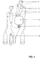
- Figure 1 illustrates one of the microstructures of GyrolabTM CD, CDMX1, containing two liquid inlets (1), two volume definition units (2), an overflow channel (3), a mixing chamber (4), an enforced finger valve (5), and a capture column (6) where reactions take place and which contains beads coupled with ligand, here typically streptavidin to be coupled to biotinylated capture antibody (the streptavidin-biotin interaction is stable in the acid pH conditions used in the present method).
- a hydrophobic barrier (not shown) separates the mixing chamber (4) from the capture column (6).
- Reagents and buffers for performing the immunoassay are introduced in the left inlet (1), and samples and reagents for sample pre-treatment are introduced in the right inlet (1).
- the mixing chamber (4) is located upstream of the capture column (6), i.e. spinning of the CD will cause liquid to flow from the mixing chamber (4) to the capture column (6).
- Aliquots of sample and buffers aimed for pre-treatment of sample can sequentially be added in portions of, typically, 200 nl after volume definition within the CD into the mixing chamber (4).
- any type of liquid compatible with the microfluidic principles can be added.
- buffers aimed for efficient dissociation will generate a resulting pH of 1.5 to 3.2
- buffers intended to prepare samples for analysis will generate a resulting pH of 2.7 to 4.5, or more specifically 2.8 to 4.5, e.g. 3.0-4.5, depending on the nature of interactants, concentrations of interactants and the tolerability vs acid pH of the reagents used for the assay.
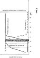
- Figure 2 shows exemplary pH intervals for protein A/IgG complex dissociation, and analysis of released protein A at mildly acidic pH preventing re-association of protein A-IgG complexes.
- the dissociating effect of acid buffer addition is usually very rapid. In the protein A-IgG system, it seems that the dissociation is quantitative after 1-5 min generating a resulting pH of 2.5. The next step of adjusting the pH of the sample to running conditions for the assay is also very rapid.
- the analysis step is initiated by increasing the spinning speed of the CD to overcome the resistance of the hydrophobic barrier separating the mixing chamber (4) from the capture column (6) ( Fig. 1 ).
- the capture column is functionalized with an appropriate capture antibody and the capture column may have to be prewashed with the same buffer composition as the sample to prevent any momentary re-formation of protein A-IgG complexes.
- the sample Once the sample has been processed through the capture column, it may have to be washed with acid buffer 2-4 times at the same pH as the sample to prevent reformation of protein A-IgG complexes, now between protein A captured on the column and any remaining IgG present in the microfluidic paths utilized during processing.
- the pH of the capture column is elevated to neutral to facilitate the formation of a sandwich immunoassay. The process is finalized by necessary column washes prior to detection.
- a polyclonal chicken anti-Protein A antibody was purchased from Cygnus Technologies, Southport, NC, U.S.A. (www.cygnustechnologies.com). Aliquots of the antibody were labelled with biotin using EZ-link Sulpho NHS-LC-Biotin (21338, Thermo Scientific, Rockford, IL, USA - www.piercenet.com) according to the manufacturer's instructions. RexxipTM ADA buffer was used (Gyros AB, Uppsala, Sweden).
- a biotinylated mouse monoclonal antibody directed against protein A was purchased from Sigma-Aldrich, St. Louis, MO, U.S.A. (cta no B3150; www.sigmaaldrich.com).
- a proprietary polyclonal antibody directed against protein A and designed to sustain low pH conditions was provided. An aliquot of the antibody was labelled with biotin using EZ-link Sulpho NHS-LC-Biotin (21338, Thermo Scientific).
- hIgG Polyclonal human IgG (hIgG) for intravenous administration, OctagamTM (Octapharma AB, Sweden), 50 mg/ml, was purchased from the pharmacy on prescription. This preparation is purified by alcohol fractionation and has never been in contact with protein A or any derivative of protein A.
- HumiraTM (a therapeutic antibody, marketed by Abbott Laboratories, Abbott Park, Illinois, USA) was purchased from the pharmacy on prescription.
- HerceptinTM (a therapeutic antibody, marketed by F. Hoffmann-La Roche Ltd, Basel, Switzerland) was purchased from the pharmacy on prescription.
- Buffers were prepared from solid chemicals at appropriate buffer capacity and pH.
- Protein A (native, 17-0872-05) and derivatives thereof (MabSelect SuReTM ligand, 28-4018-60) were purchased from GE Healthcare Life Sciences, Uppsala, Sweden (www.gelifesciences.com).
- CDMX1 (P0020026), also called “Gyrolab ADA CD", was from Gyros AB, Uppsala, Sweden (www.gyros.com). Column packing was (15 ⁇ m) streptavidin-derivatised DynospheresTM (Invitrogen Dynal A.S., Oslo, Norway).
- Standard curves were prepared by dilution of protein A in 5 mg/ml of polyclonal IgG in PBS, pH 7.4, allowing complexes between protein A and IgG to be formed.
- Quality control (QC) samples were prepared in separate dilutions with known concentrations of protein A in the presence of polyclonal or monoclonal IgG at 5 mg/ml.
- MabSelect SuRe can be determined at sub-ppm levels in concentrations of IgG at 5 mg/ml.
- the assay used for MabSelect SuRe spans from approximately 1 ng/ml and upwards generating a sensitivity of approximately 0.2 ppm (w/w).
- Chicken polyclonal anti-Protein A antibody as capture and detection antibodies (dissociation at pH 2.5 and capture at pH 3.5)
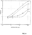
- Figure 4 illustrates the dose response relationship of assaying standard curves containing MabSelect SuReTM ligand only (1), and Mabselect SuRe + hIgG at 5 mg/ml after processing at pH 2.5 and 3.5 (2) as described for the method.
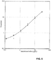
- Figure 5 shows a standard curve of MabSelect SuRe in the presence of hIgG at 5 mg/ml.
- the capture column was washed prior to the capture step using a glycinecitrate buffer, pH 3.5, and further washed twice before the column was neutralized using PBS before the analytical process was finalized.
- Table 1 shows the average bias of QC samples when using the standard curve in Figure 5 .
- Table 1 QC samples n IgG contents (mg/ml) MabSelect SuRe (ng/ml) Average Bias (%) QC1 3 5 1 34.1 QC2 3 5 2.5 0.3 QC3 3 5 5 -4.1 QC4 3 5 10 -0.8
- Figure 6 shows a dose response curve for MabSelect SuRe in presence of hIgG at 5 mg/ml.
- the capture column was kept in PBS, pH 7.4, during the entire process.
- Table 2 shows the average bias of QC samples when using the standard curve in Figure 6 .
- Table 2 QC samples n IgG contents (mg/ml) MabSelect SuRe (ng/ml) Average Bias (%) QC1 3 5 1 18.5 QC2 3 5 2.5 0.4 QC3 3 5 5 -6.7 QC4 3 5 10 -8.5
- Mouse monoclonal anti-Protein A antibody as capture antibody and chicken polyclonal anti-Protein A antibody as detection antibody (dissociation at pH 2.3 and capture at pH 3.3)
- Figure 7 shows a dose response curve for native protein A in the presence of polyclonal, human IgG at 5 mg/ml.
- the pH selected for dissociation of protein A-IgG complexes was 2.3 and the pH selected for analysis was 3.3.
- Table 3 shows analysis of QC samples containing different concentrations of native protein A in the presence of polyclonal human IgG at 5 mg/ml employing dissociation at pH 2.3 and analysis at pH 3.3 in the assay. As can be seen the bias is within ⁇ 20 % from protein A concentrations exceeding 2.5 ng/ml.
- Table 3 QC samples n IgG contents (mg/ml) Protein A (ng/ml) Average Bias (%) QC1 3 5 1 -70.9 QC2 3 5 2.5 11.2 QC3 3 5 5 8.1 QC4 3 5 10 -11.2
- Proprietary polyclonal anti-Protein A antibody as capture and detection antibodies (dissociation at pH 2.5 and capture at pH 2.8)
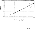
- Figure 8 shows an overlay chart of standard curves for (1) protein A and (2) MabSelect SuRe in the presence of human polyclonal IgG (OctagamTM) at a concentration of 5 mg/ml.
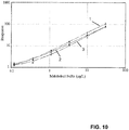
- Figure 9 shows an overlay chart of standard curves for (1) MabSelect SuRe in buffer only, (2) MabSelect SuRe in 10 mg/ml HumiraTM (a therapeutic monoclonal antibody), (3) MabSelect SuRe in 5 mg/ml HumiraTM, and (4) MabSelect SuRe in 2 mg/ml HumiraTM.
- Figure 10 shows an overlay chart of standard curves for residual MabSelect SuRe (1) in buffer only, (2) in the presence of 5 mg per ml HerceptinTM, and (3) in the presence of 5 mg per ml HumiraTM.
- IgG containing (HumiraTM) samples contaminated with residual MabSelect SuRe were provided. Samples were first normalized by diluting them to a concentration of 5 g/L. Samples were then analyzed for protein A using the above outlined procedure involving acid dissociation at pH 2.5 and capture at pH 2.8. A standard curve for dose-response vs concentration of MabSelect SuRe was prepared as shown in Figure 11 . The concentration of residual MabSelect SuRe in the original samples were then determined using the standard curve and calculated by compensating for the dilution factor. The precision (CV%) of duplicate determinations is reported. The results are shown in Table 6 below.
- the procedure can be performed in a CD containing microfluidic structures, each having a mixing chamber upstream the capture column in which pretreatment of sample with different buffers at different pH can be performed in a standardized manner.
- the procedure takes, depending on the specific set up, on average approximately one hour.
- the relative concentration of MabSelect SuReTM ligand that can be detected is in the range of 0.2-0.5 ppm at 5 mg/ml of IgG (w/w), a relative concentration that is far below the regulatory accepted level of impurity (13).
- the principal dissociation and analysis procedure is also compatible with native protein A in polyclonal, human IgG at 5 mg/ml.
- An exemplary kit for performing analysis of residual protein A (or MabSelect SuRe) in the presence of IgG comprises the following reagents A to I.
- Reagents A, B and C are provided as stock solutions intended to be diluted with diluent reagents G, H and I, respectively.
- the entire kit is composed of nine different types of liquids sufficient for 5 GyrolabTM ADA CDs (Gyros AB) generating 240 data points (48/CD).
Landscapes
- Health & Medical Sciences (AREA)
- Immunology (AREA)
- Life Sciences & Earth Sciences (AREA)
- Engineering & Computer Science (AREA)
- Chemical & Material Sciences (AREA)
- Molecular Biology (AREA)
- Biomedical Technology (AREA)
- Hematology (AREA)
- Urology & Nephrology (AREA)
- Biotechnology (AREA)
- Microbiology (AREA)
- Cell Biology (AREA)
- Food Science & Technology (AREA)
- Medicinal Chemistry (AREA)
- Physics & Mathematics (AREA)
- Analytical Chemistry (AREA)
- Biochemistry (AREA)
- General Health & Medical Sciences (AREA)
- General Physics & Mathematics (AREA)
- Pathology (AREA)
- Chemical Kinetics & Catalysis (AREA)
- Peptides Or Proteins (AREA)
Applications Claiming Priority (3)
| Application Number | Priority Date | Filing Date | Title |
|---|---|---|---|
| SE1251116 | 2012-10-03 | ||
| SE1350373 | 2013-03-25 | ||
| PCT/SE2013/051161 WO2014055025A1 (en) | 2012-10-03 | 2013-10-03 | Method and kit for analyte determination at acidic conditions |
Publications (3)
| Publication Number | Publication Date |
|---|---|
| EP2904393A1 EP2904393A1 (en) | 2015-08-12 |
| EP2904393A4 EP2904393A4 (en) | 2016-05-04 |
| EP2904393B1 true EP2904393B1 (en) | 2018-11-28 |
Family
ID=50435248
Family Applications (1)
| Application Number | Title | Priority Date | Filing Date |
|---|---|---|---|
| EP13843833.8A Active EP2904393B1 (en) | 2012-10-03 | 2013-10-03 | Method and kit for analyte determination at acidic conditions |
Country Status (5)
| Country | Link |
|---|---|
| US (1) | US10036745B2 (ja) |
| EP (1) | EP2904393B1 (ja) |
| JP (1) | JP6279590B2 (ja) |
| CN (1) | CN104718453B (ja) |
| WO (1) | WO2014055025A1 (ja) |
Families Citing this family (7)
| Publication number | Priority date | Publication date | Assignee | Title |
|---|---|---|---|---|
| CN118443922A (zh) * | 2016-09-06 | 2024-08-06 | 富士瑞必欧株式会社 | 肿瘤标记物的测定方法及测定试剂 |
| CN109477832A (zh) * | 2016-09-06 | 2019-03-15 | 富士瑞必欧株式会社 | 甲状腺球蛋白的测定方法及测定试剂 |
| JP6999561B2 (ja) * | 2016-09-13 | 2022-02-10 | 富士レビオ株式会社 | 心筋トロポニンの測定方法及び測定試薬 |
| MX2020004921A (es) * | 2017-11-29 | 2020-08-27 | Hoffmann La Roche | Ensayo de anticuerpo antifarmaco con supresion de interferencia del objetivo. |
| CN112534263A (zh) * | 2018-08-03 | 2021-03-19 | 百时美施贵宝公司 | 用于检测抗药抗体的方法 |
| CN113960318A (zh) * | 2021-09-28 | 2022-01-21 | 苏州赛分科技股份有限公司 | 一种基于酶联免疫的重组蛋白a测定方法及试剂盒 |
| TW202409562A (zh) * | 2022-07-13 | 2024-03-01 | 美商再生元醫藥公司 | 用於偵測分析物的弱酸免疫分析 |
Family Cites Families (24)
| Publication number | Priority date | Publication date | Assignee | Title |
|---|---|---|---|---|
| SE448190B (sv) | 1985-06-03 | 1987-01-26 | Pharmacia Ab | Sett att bestemma fc-dels-bindande bakteriella polypeptider |
| US4703001A (en) * | 1985-10-23 | 1987-10-27 | Synbiotics, Corporation | Immunoassay for the detection of serum analytes using pH dependent chastropic acids |
| WO1991010911A1 (en) * | 1990-01-19 | 1991-07-25 | Repligen Corporation | Immunoassay of protein a under acidic conditions |
| US5314804A (en) * | 1992-03-24 | 1994-05-24 | Serim Research Corporation | Test for Helicobacter pylori |
| GB9809943D0 (en) | 1998-05-08 | 1998-07-08 | Amersham Pharm Biotech Ab | Microfluidic device |
| WO2002074438A2 (en) | 2001-03-19 | 2002-09-26 | Gyros Ab | Structural units that define fluidic functions |
| JP4323806B2 (ja) | 2001-03-19 | 2009-09-02 | ユィロス・パテント・アクチボラグ | 反応可変要素の特徴付け |
| EP2283924B1 (en) | 2001-08-28 | 2013-04-17 | Gyros Patent Ab | Inlet unit with means supporting liquid entrance into a microchannel structure |
| AU2003213022A1 (en) * | 2002-02-11 | 2003-09-22 | Alexion Pharmaceuticals, Inc. | Immunotherapeutics for biodefense |
| SE0200943D0 (sv) | 2002-03-25 | 2002-03-25 | Amersham Biosciences Ab | Mutant protein |
| JP4115728B2 (ja) * | 2002-03-26 | 2008-07-09 | デンカ生研株式会社 | フロースルー式検査法用組成物、これを用いたキット及び検査法 |
| WO2003102559A1 (en) * | 2002-05-31 | 2003-12-11 | Gyros Ab | Detector arrangement based on surface plasmon resonance |
| WO2004083108A1 (en) | 2003-03-23 | 2004-09-30 | Gyros Patent Ab | Preloaded microscale devices |
| SE0300822D0 (sv) | 2003-03-23 | 2003-03-23 | Gyros Ab | A collection of Micro Scale Devices |
| CN101097217A (zh) * | 2006-06-29 | 2008-01-02 | 王珊珊 | 一种提高血清特异性生长因子免疫学测定灵敏度的技术 |
| EP2062052A4 (en) * | 2006-09-14 | 2010-02-03 | Ge Healthcare Bio Sciences Ab | METHOD FOR DETERMINING ANALYZ CONCENTRATION |
| CN101583872A (zh) * | 2006-09-14 | 2009-11-18 | 通用电气健康护理生物科学股份公司 | 测定待分析物浓度的方法 |
| WO2008043075A2 (en) * | 2006-10-05 | 2008-04-10 | Wyeth | Compositions for the treatment of scleritis |
| WO2009022001A1 (en) * | 2007-08-16 | 2009-02-19 | Novartis Ag | Improvement of drug tolerance in immunogenicity testing |
| ES2375606T3 (es) | 2008-04-03 | 2012-03-02 | F. Hoffmann-La Roche Ag | Ensayo de factor de crecimiento tipo insulina pegilado. |
| KR101651304B1 (ko) * | 2008-09-11 | 2016-08-25 | 앵스티띠 파스퇴르 | Hmgb1 의존적인 hiv-1 복제 및 지속감염 촉발의 조절에 의한 인간 면역결핍 바이러스 감염의 모니터링 및 억제 |
| US8546149B2 (en) * | 2010-08-27 | 2013-10-01 | Intervet Inc. | Potency test for vaccine formulations |
| DK2676137T3 (en) | 2011-02-17 | 2015-01-19 | Nestec Sa | TESTS TO DETECT AUTO ANTIBODIES FOR ANTI-TNF PHARMACEUTICALS |
| AU2013219968B2 (en) | 2012-02-15 | 2018-11-08 | Biocon Limited | A process for detection and optional quantification of an analyte |
-
2013
- 2013-10-03 EP EP13843833.8A patent/EP2904393B1/en active Active
- 2013-10-03 US US14/433,276 patent/US10036745B2/en active Active
- 2013-10-03 JP JP2015535610A patent/JP6279590B2/ja active Active
- 2013-10-03 CN CN201380051645.1A patent/CN104718453B/zh active Active
- 2013-10-03 WO PCT/SE2013/051161 patent/WO2014055025A1/en active Application Filing
Non-Patent Citations (1)
| Title |
|---|
| None * |
Also Published As
| Publication number | Publication date |
|---|---|
| EP2904393A4 (en) | 2016-05-04 |
| US20150268238A1 (en) | 2015-09-24 |
| JP2015531487A (ja) | 2015-11-02 |
| EP2904393A1 (en) | 2015-08-12 |
| CN104718453A (zh) | 2015-06-17 |
| CN104718453B (zh) | 2017-03-08 |
| US10036745B2 (en) | 2018-07-31 |
| WO2014055025A1 (en) | 2014-04-10 |
| JP6279590B2 (ja) | 2018-02-14 |
Similar Documents
| Publication | Publication Date | Title |
|---|---|---|
| EP2904393B1 (en) | Method and kit for analyte determination at acidic conditions | |
| Zhang et al. | High performance affinity chromatography and related separation methods for the analysis of biological and pharmaceutical agents | |
| EP2333116B1 (en) | Markers of renal transplant rejection and renal damage | |
| KR101826449B1 (ko) | 펩티드 단편의 조제 방법 및 그것에 사용되는 펩티드 단편 조제용 킷, 및 분석 방법 | |
| US20180238910A1 (en) | Methods for providing information relevant to diagnosis of neurodegenerative disorder | |
| JP7382071B2 (ja) | 糖化ヘモグロビン(%)の測定方法 | |
| WO2004011607A2 (en) | Reagent and method for determination of a substance using an immunoaggregator | |
| JP2020516865A (ja) | ヒト血清または尿におけるalxn1210およびエクリズマブの同時定量のための方法 | |
| WO2013170057A2 (en) | Quantification of lipoproteins | |
| CN110573863B (zh) | 用于检测分析物的方法 | |
| CA2640835A1 (en) | Immunoassay for the simultaneous immunochemical determination of an analyte (antigen) and a treatment antibody targeting the analyte in samples (recovery immunoassay) | |
| JP4554356B2 (ja) | サンドイッチアッセイおよびキット | |
| JP2017187476A (ja) | 変性抗体測定方法 | |
| US11585811B2 (en) | Immobilized analytes | |
| Cho et al. | Minimum-step immuno-analysis based on continuous recycling of the capture antibody | |
| US20060275849A1 (en) | Monoclonal antibody reagents | |
| EP3760641A1 (en) | Monoclonal antibody against apoa4, immunoassay method, and kit for measurement | |
| Mortezai et al. | Combining lectin affinity chromatography and immunodepletion–A novel method for the enrichment of disease-specific glycoproteins in human plasma | |
| Zhu-Shimoni et al. | Trace level analysis of leached Protein A in bioprocess samples without interference from the large excess of rhMAb IgG | |
| JP6152908B2 (ja) | ペプチド断片の調製方法および分析方法 | |
| WO2017033846A1 (ja) | 免疫試験方法および免疫試験キット | |
| JP5231954B2 (ja) | アルブミン測定試薬 | |
| JP4753366B2 (ja) | %cdtの定量方法 | |
| JP3898618B2 (ja) | 酵素免疫測定法による抗血液凝固因子抗体の定量方法 | |
| JP5448424B2 (ja) | ヒトIgGのFcを含有するタンパク質の測定試薬 |
Legal Events
| Date | Code | Title | Description |
|---|---|---|---|
| PUAI | Public reference made under article 153(3) epc to a published international application that has entered the european phase |
Free format text: ORIGINAL CODE: 0009012 |
|
| 17P | Request for examination filed |
Effective date: 20150309 |
|
| AK | Designated contracting states |
Kind code of ref document: A1 Designated state(s): AL AT BE BG CH CY CZ DE DK EE ES FI FR GB GR HR HU IE IS IT LI LT LU LV MC MK MT NL NO PL PT RO RS SE SI SK SM TR |
|
| AX | Request for extension of the european patent |
Extension state: BA ME |
|
| DAX | Request for extension of the european patent (deleted) | ||
| RA4 | Supplementary search report drawn up and despatched (corrected) |
Effective date: 20160406 |
|
| RIC1 | Information provided on ipc code assigned before grant |
Ipc: G01N 33/84 20060101ALI20160331BHEP Ipc: G01N 33/53 20060101AFI20160331BHEP Ipc: G01N 33/537 20060101ALI20160331BHEP |
|
| STAA | Information on the status of an ep patent application or granted ep patent |
Free format text: STATUS: EXAMINATION IS IN PROGRESS |
|
| 17Q | First examination report despatched |
Effective date: 20171123 |
|
| INTG | Intention to grant announced |
Effective date: 20180611 |
|
| RAP1 | Party data changed (applicant data changed or rights of an application transferred) |
Owner name: GYROS PATENT AB |
|
| GRAP | Despatch of communication of intention to grant a patent |
Free format text: ORIGINAL CODE: EPIDOSNIGR1 |
|
| STAA | Information on the status of an ep patent application or granted ep patent |
Free format text: STATUS: GRANT OF PATENT IS INTENDED |
|
| GRAS | Grant fee paid |
Free format text: ORIGINAL CODE: EPIDOSNIGR3 |
|
| GRAA | (expected) grant |
Free format text: ORIGINAL CODE: 0009210 |
|
| STAA | Information on the status of an ep patent application or granted ep patent |
Free format text: STATUS: THE PATENT HAS BEEN GRANTED |
|
| AK | Designated contracting states |
Kind code of ref document: B1 Designated state(s): AL AT BE BG CH CY CZ DE DK EE ES FI FR GB GR HR HU IE IS IT LI LT LU LV MC MK MT NL NO PL PT RO RS SE SI SK SM TR |
|
| REG | Reference to a national code |
Ref country code: CH Ref legal event code: EP |
|
| REG | Reference to a national code |
Ref country code: AT Ref legal event code: REF Ref document number: 1070837 Country of ref document: AT Kind code of ref document: T Effective date: 20181215 |
|
| REG | Reference to a national code |
Ref country code: DE Ref legal event code: R096 Ref document number: 602013047591 Country of ref document: DE |
|
| REG | Reference to a national code |
Ref country code: IE Ref legal event code: FG4D |
|
| REG | Reference to a national code |
Ref country code: NL Ref legal event code: MP Effective date: 20181128 |
|
| REG | Reference to a national code |
Ref country code: LT Ref legal event code: MG4D |
|
| REG | Reference to a national code |
Ref country code: AT Ref legal event code: MK05 Ref document number: 1070837 Country of ref document: AT Kind code of ref document: T Effective date: 20181128 |
|
| PG25 | Lapsed in a contracting state [announced via postgrant information from national office to epo] |
Ref country code: BG Free format text: LAPSE BECAUSE OF FAILURE TO SUBMIT A TRANSLATION OF THE DESCRIPTION OR TO PAY THE FEE WITHIN THE PRESCRIBED TIME-LIMIT Effective date: 20190228 Ref country code: NO Free format text: LAPSE BECAUSE OF FAILURE TO SUBMIT A TRANSLATION OF THE DESCRIPTION OR TO PAY THE FEE WITHIN THE PRESCRIBED TIME-LIMIT Effective date: 20190228 Ref country code: IS Free format text: LAPSE BECAUSE OF FAILURE TO SUBMIT A TRANSLATION OF THE DESCRIPTION OR TO PAY THE FEE WITHIN THE PRESCRIBED TIME-LIMIT Effective date: 20190328 Ref country code: FI Free format text: LAPSE BECAUSE OF FAILURE TO SUBMIT A TRANSLATION OF THE DESCRIPTION OR TO PAY THE FEE WITHIN THE PRESCRIBED TIME-LIMIT Effective date: 20181128 Ref country code: LT Free format text: LAPSE BECAUSE OF FAILURE TO SUBMIT A TRANSLATION OF THE DESCRIPTION OR TO PAY THE FEE WITHIN THE PRESCRIBED TIME-LIMIT Effective date: 20181128 Ref country code: LV Free format text: LAPSE BECAUSE OF FAILURE TO SUBMIT A TRANSLATION OF THE DESCRIPTION OR TO PAY THE FEE WITHIN THE PRESCRIBED TIME-LIMIT Effective date: 20181128 Ref country code: HR Free format text: LAPSE BECAUSE OF FAILURE TO SUBMIT A TRANSLATION OF THE DESCRIPTION OR TO PAY THE FEE WITHIN THE PRESCRIBED TIME-LIMIT Effective date: 20181128 Ref country code: ES Free format text: LAPSE BECAUSE OF FAILURE TO SUBMIT A TRANSLATION OF THE DESCRIPTION OR TO PAY THE FEE WITHIN THE PRESCRIBED TIME-LIMIT Effective date: 20181128 Ref country code: AT Free format text: LAPSE BECAUSE OF FAILURE TO SUBMIT A TRANSLATION OF THE DESCRIPTION OR TO PAY THE FEE WITHIN THE PRESCRIBED TIME-LIMIT Effective date: 20181128 |
|
| PG25 | Lapsed in a contracting state [announced via postgrant information from national office to epo] |
Ref country code: GR Free format text: LAPSE BECAUSE OF FAILURE TO SUBMIT A TRANSLATION OF THE DESCRIPTION OR TO PAY THE FEE WITHIN THE PRESCRIBED TIME-LIMIT Effective date: 20190301 Ref country code: PT Free format text: LAPSE BECAUSE OF FAILURE TO SUBMIT A TRANSLATION OF THE DESCRIPTION OR TO PAY THE FEE WITHIN THE PRESCRIBED TIME-LIMIT Effective date: 20190328 Ref country code: SE Free format text: LAPSE BECAUSE OF FAILURE TO SUBMIT A TRANSLATION OF THE DESCRIPTION OR TO PAY THE FEE WITHIN THE PRESCRIBED TIME-LIMIT Effective date: 20181128 Ref country code: RS Free format text: LAPSE BECAUSE OF FAILURE TO SUBMIT A TRANSLATION OF THE DESCRIPTION OR TO PAY THE FEE WITHIN THE PRESCRIBED TIME-LIMIT Effective date: 20181128 Ref country code: AL Free format text: LAPSE BECAUSE OF FAILURE TO SUBMIT A TRANSLATION OF THE DESCRIPTION OR TO PAY THE FEE WITHIN THE PRESCRIBED TIME-LIMIT Effective date: 20181128 |
|
| PG25 | Lapsed in a contracting state [announced via postgrant information from national office to epo] |
Ref country code: NL Free format text: LAPSE BECAUSE OF FAILURE TO SUBMIT A TRANSLATION OF THE DESCRIPTION OR TO PAY THE FEE WITHIN THE PRESCRIBED TIME-LIMIT Effective date: 20181128 |
|
| PG25 | Lapsed in a contracting state [announced via postgrant information from national office to epo] |
Ref country code: DK Free format text: LAPSE BECAUSE OF FAILURE TO SUBMIT A TRANSLATION OF THE DESCRIPTION OR TO PAY THE FEE WITHIN THE PRESCRIBED TIME-LIMIT Effective date: 20181128 Ref country code: CZ Free format text: LAPSE BECAUSE OF FAILURE TO SUBMIT A TRANSLATION OF THE DESCRIPTION OR TO PAY THE FEE WITHIN THE PRESCRIBED TIME-LIMIT Effective date: 20181128 Ref country code: IT Free format text: LAPSE BECAUSE OF FAILURE TO SUBMIT A TRANSLATION OF THE DESCRIPTION OR TO PAY THE FEE WITHIN THE PRESCRIBED TIME-LIMIT Effective date: 20181128 Ref country code: PL Free format text: LAPSE BECAUSE OF FAILURE TO SUBMIT A TRANSLATION OF THE DESCRIPTION OR TO PAY THE FEE WITHIN THE PRESCRIBED TIME-LIMIT Effective date: 20181128 |
|
| REG | Reference to a national code |
Ref country code: DE Ref legal event code: R097 Ref document number: 602013047591 Country of ref document: DE |
|
| PG25 | Lapsed in a contracting state [announced via postgrant information from national office to epo] |
Ref country code: SK Free format text: LAPSE BECAUSE OF FAILURE TO SUBMIT A TRANSLATION OF THE DESCRIPTION OR TO PAY THE FEE WITHIN THE PRESCRIBED TIME-LIMIT Effective date: 20181128 Ref country code: EE Free format text: LAPSE BECAUSE OF FAILURE TO SUBMIT A TRANSLATION OF THE DESCRIPTION OR TO PAY THE FEE WITHIN THE PRESCRIBED TIME-LIMIT Effective date: 20181128 Ref country code: SM Free format text: LAPSE BECAUSE OF FAILURE TO SUBMIT A TRANSLATION OF THE DESCRIPTION OR TO PAY THE FEE WITHIN THE PRESCRIBED TIME-LIMIT Effective date: 20181128 Ref country code: RO Free format text: LAPSE BECAUSE OF FAILURE TO SUBMIT A TRANSLATION OF THE DESCRIPTION OR TO PAY THE FEE WITHIN THE PRESCRIBED TIME-LIMIT Effective date: 20181128 |
|
| PLBE | No opposition filed within time limit |
Free format text: ORIGINAL CODE: 0009261 |
|
| STAA | Information on the status of an ep patent application or granted ep patent |
Free format text: STATUS: NO OPPOSITION FILED WITHIN TIME LIMIT |
|
| PG25 | Lapsed in a contracting state [announced via postgrant information from national office to epo] |
Ref country code: SI Free format text: LAPSE BECAUSE OF FAILURE TO SUBMIT A TRANSLATION OF THE DESCRIPTION OR TO PAY THE FEE WITHIN THE PRESCRIBED TIME-LIMIT Effective date: 20181128 |
|
| 26N | No opposition filed |
Effective date: 20190829 |
|
| PG25 | Lapsed in a contracting state [announced via postgrant information from national office to epo] |
Ref country code: TR Free format text: LAPSE BECAUSE OF FAILURE TO SUBMIT A TRANSLATION OF THE DESCRIPTION OR TO PAY THE FEE WITHIN THE PRESCRIBED TIME-LIMIT Effective date: 20181128 |
|
| PG25 | Lapsed in a contracting state [announced via postgrant information from national office to epo] |
Ref country code: MC Free format text: LAPSE BECAUSE OF FAILURE TO SUBMIT A TRANSLATION OF THE DESCRIPTION OR TO PAY THE FEE WITHIN THE PRESCRIBED TIME-LIMIT Effective date: 20181128 |
|
| PG25 | Lapsed in a contracting state [announced via postgrant information from national office to epo] |
Ref country code: LU Free format text: LAPSE BECAUSE OF NON-PAYMENT OF DUE FEES Effective date: 20191003 |
|
| REG | Reference to a national code |
Ref country code: BE Ref legal event code: MM Effective date: 20191031 |
|
| PG25 | Lapsed in a contracting state [announced via postgrant information from national office to epo] |
Ref country code: BE Free format text: LAPSE BECAUSE OF NON-PAYMENT OF DUE FEES Effective date: 20191031 |
|
| PG25 | Lapsed in a contracting state [announced via postgrant information from national office to epo] |
Ref country code: IE Free format text: LAPSE BECAUSE OF NON-PAYMENT OF DUE FEES Effective date: 20191003 |
|
| PG25 | Lapsed in a contracting state [announced via postgrant information from national office to epo] |
Ref country code: CY Free format text: LAPSE BECAUSE OF FAILURE TO SUBMIT A TRANSLATION OF THE DESCRIPTION OR TO PAY THE FEE WITHIN THE PRESCRIBED TIME-LIMIT Effective date: 20181128 |
|
| PG25 | Lapsed in a contracting state [announced via postgrant information from national office to epo] |
Ref country code: MT Free format text: LAPSE BECAUSE OF FAILURE TO SUBMIT A TRANSLATION OF THE DESCRIPTION OR TO PAY THE FEE WITHIN THE PRESCRIBED TIME-LIMIT Effective date: 20181128 Ref country code: HU Free format text: LAPSE BECAUSE OF FAILURE TO SUBMIT A TRANSLATION OF THE DESCRIPTION OR TO PAY THE FEE WITHIN THE PRESCRIBED TIME-LIMIT; INVALID AB INITIO Effective date: 20131003 |
|
| PG25 | Lapsed in a contracting state [announced via postgrant information from national office to epo] |
Ref country code: MK Free format text: LAPSE BECAUSE OF FAILURE TO SUBMIT A TRANSLATION OF THE DESCRIPTION OR TO PAY THE FEE WITHIN THE PRESCRIBED TIME-LIMIT Effective date: 20181128 |
|
| PGFP | Annual fee paid to national office [announced via postgrant information from national office to epo] |
Ref country code: GB Payment date: 20231018 Year of fee payment: 11 |
|
| PGFP | Annual fee paid to national office [announced via postgrant information from national office to epo] |
Ref country code: FR Payment date: 20231016 Year of fee payment: 11 Ref country code: DE Payment date: 20231020 Year of fee payment: 11 Ref country code: CH Payment date: 20231102 Year of fee payment: 11 |