EP2892562B1 - Anticorps anti-mcam et méthodes d'utilisation associées - Google Patents
Anticorps anti-mcam et méthodes d'utilisation associées Download PDFInfo
- Publication number
- EP2892562B1 EP2892562B1 EP13836030.0A EP13836030A EP2892562B1 EP 2892562 B1 EP2892562 B1 EP 2892562B1 EP 13836030 A EP13836030 A EP 13836030A EP 2892562 B1 EP2892562 B1 EP 2892562B1
- Authority
- EP
- European Patent Office
- Prior art keywords
- mcam
- antibody
- seq
- amino acid
- cells
- Prior art date
- Legal status (The legal status is an assumption and is not a legal conclusion. Google has not performed a legal analysis and makes no representation as to the accuracy of the status listed.)
- Active
Links
Images


Classifications
-
- A—HUMAN NECESSITIES
- A61—MEDICAL OR VETERINARY SCIENCE; HYGIENE
- A61P—SPECIFIC THERAPEUTIC ACTIVITY OF CHEMICAL COMPOUNDS OR MEDICINAL PREPARATIONS
- A61P17/00—Drugs for dermatological disorders
- A61P17/04—Antipruritics
-
- A—HUMAN NECESSITIES
- A61—MEDICAL OR VETERINARY SCIENCE; HYGIENE
- A61P—SPECIFIC THERAPEUTIC ACTIVITY OF CHEMICAL COMPOUNDS OR MEDICINAL PREPARATIONS
- A61P17/00—Drugs for dermatological disorders
- A61P17/06—Antipsoriatics
-
- A—HUMAN NECESSITIES
- A61—MEDICAL OR VETERINARY SCIENCE; HYGIENE
- A61P—SPECIFIC THERAPEUTIC ACTIVITY OF CHEMICAL COMPOUNDS OR MEDICINAL PREPARATIONS
- A61P19/00—Drugs for skeletal disorders
- A61P19/02—Drugs for skeletal disorders for joint disorders, e.g. arthritis, arthrosis
-
- A—HUMAN NECESSITIES
- A61—MEDICAL OR VETERINARY SCIENCE; HYGIENE
- A61P—SPECIFIC THERAPEUTIC ACTIVITY OF CHEMICAL COMPOUNDS OR MEDICINAL PREPARATIONS
- A61P25/00—Drugs for disorders of the nervous system
-
- A—HUMAN NECESSITIES
- A61—MEDICAL OR VETERINARY SCIENCE; HYGIENE
- A61P—SPECIFIC THERAPEUTIC ACTIVITY OF CHEMICAL COMPOUNDS OR MEDICINAL PREPARATIONS
- A61P25/00—Drugs for disorders of the nervous system
- A61P25/14—Drugs for disorders of the nervous system for treating abnormal movements, e.g. chorea, dyskinesia
- A61P25/16—Anti-Parkinson drugs
-
- A—HUMAN NECESSITIES
- A61—MEDICAL OR VETERINARY SCIENCE; HYGIENE
- A61P—SPECIFIC THERAPEUTIC ACTIVITY OF CHEMICAL COMPOUNDS OR MEDICINAL PREPARATIONS
- A61P25/00—Drugs for disorders of the nervous system
- A61P25/28—Drugs for disorders of the nervous system for treating neurodegenerative disorders of the central nervous system, e.g. nootropic agents, cognition enhancers, drugs for treating Alzheimer's disease or other forms of dementia
-
- A—HUMAN NECESSITIES
- A61—MEDICAL OR VETERINARY SCIENCE; HYGIENE
- A61P—SPECIFIC THERAPEUTIC ACTIVITY OF CHEMICAL COMPOUNDS OR MEDICINAL PREPARATIONS
- A61P29/00—Non-central analgesic, antipyretic or antiinflammatory agents, e.g. antirheumatic agents; Non-steroidal antiinflammatory drugs [NSAID]
-
- A—HUMAN NECESSITIES
- A61—MEDICAL OR VETERINARY SCIENCE; HYGIENE
- A61P—SPECIFIC THERAPEUTIC ACTIVITY OF CHEMICAL COMPOUNDS OR MEDICINAL PREPARATIONS
- A61P35/00—Antineoplastic agents
-
- A—HUMAN NECESSITIES
- A61—MEDICAL OR VETERINARY SCIENCE; HYGIENE
- A61P—SPECIFIC THERAPEUTIC ACTIVITY OF CHEMICAL COMPOUNDS OR MEDICINAL PREPARATIONS
- A61P37/00—Drugs for immunological or allergic disorders
- A61P37/02—Immunomodulators
-
- A—HUMAN NECESSITIES
- A61—MEDICAL OR VETERINARY SCIENCE; HYGIENE
- A61P—SPECIFIC THERAPEUTIC ACTIVITY OF CHEMICAL COMPOUNDS OR MEDICINAL PREPARATIONS
- A61P37/00—Drugs for immunological or allergic disorders
- A61P37/02—Immunomodulators
- A61P37/06—Immunosuppressants, e.g. drugs for graft rejection
-
- A—HUMAN NECESSITIES
- A61—MEDICAL OR VETERINARY SCIENCE; HYGIENE
- A61P—SPECIFIC THERAPEUTIC ACTIVITY OF CHEMICAL COMPOUNDS OR MEDICINAL PREPARATIONS
- A61P37/00—Drugs for immunological or allergic disorders
- A61P37/08—Antiallergic agents
-
- C—CHEMISTRY; METALLURGY
- C07—ORGANIC CHEMISTRY
- C07K—PEPTIDES
- C07K16/00—Immunoglobulins [IGs], e.g. monoclonal or polyclonal antibodies
- C07K16/18—Immunoglobulins [IGs], e.g. monoclonal or polyclonal antibodies against material from animals or humans
- C07K16/28—Immunoglobulins [IGs], e.g. monoclonal or polyclonal antibodies against material from animals or humans against receptors, cell surface antigens or cell surface determinants
- C07K16/2896—Immunoglobulins [IGs], e.g. monoclonal or polyclonal antibodies against material from animals or humans against receptors, cell surface antigens or cell surface determinants against molecules with a "CD"-designation, not provided for elsewhere
-
- C—CHEMISTRY; METALLURGY
- C07—ORGANIC CHEMISTRY
- C07K—PEPTIDES
- C07K16/00—Immunoglobulins [IGs], e.g. monoclonal or polyclonal antibodies
- C07K16/18—Immunoglobulins [IGs], e.g. monoclonal or polyclonal antibodies against material from animals or humans
- C07K16/28—Immunoglobulins [IGs], e.g. monoclonal or polyclonal antibodies against material from animals or humans against receptors, cell surface antigens or cell surface determinants
- C07K16/30—Immunoglobulins [IGs], e.g. monoclonal or polyclonal antibodies against material from animals or humans against receptors, cell surface antigens or cell surface determinants from tumour cells
- C07K16/3076—Immunoglobulins [IGs], e.g. monoclonal or polyclonal antibodies against material from animals or humans against receptors, cell surface antigens or cell surface determinants from tumour cells against structure-related tumour-associated moieties
- C07K16/3092—Immunoglobulins [IGs], e.g. monoclonal or polyclonal antibodies against material from animals or humans against receptors, cell surface antigens or cell surface determinants from tumour cells against structure-related tumour-associated moieties against tumour-associated mucins
-
- A—HUMAN NECESSITIES
- A61—MEDICAL OR VETERINARY SCIENCE; HYGIENE
- A61K—PREPARATIONS FOR MEDICAL, DENTAL OR TOILETRY PURPOSES
- A61K39/00—Medicinal preparations containing antigens or antibodies
- A61K2039/505—Medicinal preparations containing antigens or antibodies comprising antibodies
-
- C—CHEMISTRY; METALLURGY
- C07—ORGANIC CHEMISTRY
- C07K—PEPTIDES
- C07K2317/00—Immunoglobulins specific features
- C07K2317/20—Immunoglobulins specific features characterized by taxonomic origin
- C07K2317/24—Immunoglobulins specific features characterized by taxonomic origin containing regions, domains or residues from different species, e.g. chimeric, humanized or veneered
-
- C—CHEMISTRY; METALLURGY
- C07—ORGANIC CHEMISTRY
- C07K—PEPTIDES
- C07K2317/00—Immunoglobulins specific features
- C07K2317/30—Immunoglobulins specific features characterized by aspects of specificity or valency
-
- C—CHEMISTRY; METALLURGY
- C07—ORGANIC CHEMISTRY
- C07K—PEPTIDES
- C07K2317/00—Immunoglobulins specific features
- C07K2317/50—Immunoglobulins specific features characterized by immunoglobulin fragments
- C07K2317/56—Immunoglobulins specific features characterized by immunoglobulin fragments variable (Fv) region, i.e. VH and/or VL
- C07K2317/565—Complementarity determining region [CDR]
-
- C—CHEMISTRY; METALLURGY
- C07—ORGANIC CHEMISTRY
- C07K—PEPTIDES
- C07K2317/00—Immunoglobulins specific features
- C07K2317/50—Immunoglobulins specific features characterized by immunoglobulin fragments
- C07K2317/56—Immunoglobulins specific features characterized by immunoglobulin fragments variable (Fv) region, i.e. VH and/or VL
- C07K2317/567—Framework region [FR]
-
- C—CHEMISTRY; METALLURGY
- C07—ORGANIC CHEMISTRY
- C07K—PEPTIDES
- C07K2317/00—Immunoglobulins specific features
- C07K2317/70—Immunoglobulins specific features characterized by effect upon binding to a cell or to an antigen
- C07K2317/76—Antagonist effect on antigen, e.g. neutralization or inhibition of binding
-
- C—CHEMISTRY; METALLURGY
- C07—ORGANIC CHEMISTRY
- C07K—PEPTIDES
- C07K2317/00—Immunoglobulins specific features
- C07K2317/90—Immunoglobulins specific features characterized by (pharmaco)kinetic aspects or by stability of the immunoglobulin
- C07K2317/92—Affinity (KD), association rate (Ka), dissociation rate (Kd) or EC50 value
Definitions
- the present invention is directed to antibodies that bind to melanoma cell adhesion molecule (MCAM) which are capable of blocking the interaction between MCAM and its ligand, the laminin alpha-4 chain.
- MCAM melanoma cell adhesion molecule
- the present invention is also directed to methods of use of the novel anti-MCAM antibodies described herein.
- TH17 cells T helper 17 cells
- EAE experimental autoimmune encephalomyelitis
- TH17 cytokines themselves has been called into question, as a conditional knockout of IL-17 is insufficient to affect EAE progression. See, e.g., Haak et al., J. Clin. Invest. 119: 61-69 (2009 ); see also Kreymborg et al., J. Immunol. 179: 8098-8104 (2007 ).
- IL-17 affects such vital aspects of EAE as endothelial cell permeability, TH17 cells appear to do more than just produce any one cytokine. The molecular determinants of the pathogenic function of TH17 cells remain elusive.
- TH17 cells The pathogenicity of TH17 cells can be partially explained by their unique migration pattern as evidenced by their expression of chemokine receptors. See, e.g., Kim, Inflamm. Allergy Drug Targets 8: 221-228 (2009 ). It has been established that IL-17 producing cells are enriched within the CCR6+ population of CD4+ T cells, likely conferring a unique migration pattern throughout the vasculature. See , e . g ., Acosta-Rodriguez et al., Nat. Immunol. 8:639-646 (2007 ). In fact, CCR6 expression on T cells is required for T cell migration into the CNS and the progression of EAE. Reboldi et al., Nat. Immunol.
- TH17 cells play a significant role in the pathogenesis of various autoimmune diseases, particularly those displaying neuroinflammatory conditions involving T cells' infiltration into CNS. It has been newly discovered that (1) MCAM is selectively enriched on TH17 cells; and (2) MCAM interacts with a laminin ⁇ 4 chain, such as, for example, the ⁇ 4 chain of laminin 411, present in the endothelial basement membrane.
- An MCAM antagonist, e is an MCAM antagonist, e .
- a monoclonal antibody capable of inhibiting MCAM's binding to a molecule containing a laminin ⁇ 4 chain, such as, for example, a laminin 411 molecule, may inhibit the migration of TH17 cells into CNS, and thus can be used as a therapeutic agent to prevent or treat diseases displaying TH17-mediated neuroinflammatory conditions.
- MCAM antagonists such as an MCAM monoclonal antibody or an antigen-binding fragment thereof, may also be useful to prevent or treat and TH17-mediated disease, including for example, autoimmune disease, for example, multiple sclerosis, inflammatory bowel disease, psoriasis, and rheumatoid arthritis.
- the present invention is directed to novel antibodies that are capable of binding to MCAM protein on the surface of cells and, in turn, that are capable of interfering with the interaction of MCAM with its ligand, a protein comprising a laminin alpha-4 chain.
- the antibody is a monoclonal antibody, antibody fragment, chimeric antibody, humanized antibody, single-chain antibody or antibody that competitively inhibits the binding of an anti-MCAM antibody to its respective antigenic epitope.
- the present invention provides an isolated anti-MCAM antibody, a pharmaceutical composition comprising said antibody, an article of manufacture comprising said pharmaceutical and medical uses of the antibody, as defined in the appended claims.
- the disclosure describes vectors comprising DNA encoding any of the herein described antibodies and host cells comprising such vectors, wherein such host cells may be CHO cells, E. coli cells, or yeast cells.
- a process for producing any of the herein described antibodies is further dislcosed and comprises the steps of culturing host cells under conditions suitable for expression of the desired antibody, and recovering the desired antibody from the cell culture.
- the present invention is directed to an isolated anti-MCAM antibody, or antigen binding fragment thereof, wherein the antibody or antigen binding fragment thereof comprises three light chain hypervariable regions (HVR-L1, HVR-L2, and HVR-L3) and three heavy chain hypervariable regions (HVR-H1, HVR-H2, and HVR-H3), and wherein: HVR-L1 comprises the amino acid sequence of SEQ ID NO:73, HVR-L2 comprises the amino acid sequence of SEQ ID NO:74, HVR-L3 comprises the amino acid sequence of SEQ ID NO:75, HVR-H1 comprises the amino acid sequence of SEQ ID NO:78, HVR-H2 comprises the amino acid sequence of SEQ ID NO:79, and HVR-H3 comprises the amino acid sequence of SEQ ID NO:80.
- HVR-L1 comprises the amino acid sequence of SEQ ID NO:73
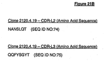
- HVR-L2 comprises the amino acid sequence of SEQ ID NO:74
- HVR-L3 comprises the amino acid sequence of SEQ ID NO:75
- the anti-MCAM antibody may be a chimeric or humanized antibody.
- the anti-MCAM antibody may be an IgG1 antibody which may optionally be produced in bacteria or CHO cells.
- the present invention is directed to an isolated anti-MCAM antibody, or antigen binding fragment thereof, said antibody or antigen binding fragment thereof comprising a light chain variable region and a heavy chain variable region, wherein:
- the anti-MCAM antibody may be a chimeric or humanized antibody.
- the anti-MCAM antibody may be an IgG1 antibody which may optionally be produced in bacteria or CHO cells.
- the present disclosure is directed to an isolated anti-MCAM antibody, or antigen binding fragment thereof, that binds substantially the same epitope as, or competes for binding with, any of the anti-MCAM antibodies described herein.
- the present disclosure is directed to an isolated anti-MCAM antibody, or antigen binding fragment thereof, that blocks the interaction between an MCAM protein comprising the amino acid sequence of SEQ ID NO:22 and a protein comprising a laminin ⁇ -4 chain.
- the present disclosure is directed to an isolated anti-MCAM antibody, or antigen binding fragment thereof, that blocks the interaction between an MCAM protein comprising the amino acid sequences of SEQ ID NOS:22 and 23 and a protein comprising a laminin ⁇ -4 chain.
- the present disclosure is directed to an isolated anti-MCAM antibody or antigen binding fragment thereof which does not block the interaction between an MCAM protein consisting of the amino acid sequence of SEQ ID NO:22 and a protein comprising a laminin ⁇ -4 chain.
- the present disclosure is directed to an isolated anti-MCAM antibody, or antigen binding fragment thereof, that blocks the interaction between an MCAM protein comprising the amino acid sequences of SEQ ID NOS:22, 23, and 24 and a protein comprising a laminin ⁇ -4 chain.
- the present disclosure is directed to isolated anti-MCAM antibodies, or antigen binding fragments thereof, that bind to antigenic epitopes defined by domains 1 and 2, or domain 3 of the human MCAM protein.
- the anti-MCAM antibody or fragment thereof does not bind to a protein consisting of amino acids 19-129 of the human MCAM protein.
- compositions comprising any of the herein described antibodies, or antigen binding fragment thereof, and articles of manufacture comprising the same.
- the present disclosure is directed to the use of an anti-MCAM antibody, or antigen binding fragment thereof, in the manufacture of a medicament for the treatment of an inflammatory disorder characterized by infiltration of MCAM-expressing cells into a site of inflammation in the body.
- the inflammatory disorder may be a central nervous system (CNS) inflammatory disorder characterized by infiltration of MCAM-expressing cells into the CNS.
- CNS central nervous system
- the invention also provides an anti-MCAM antibody, or antigen binding fragment thereof, for use in the treatment of multiple sclerosis or Parkinson's disease.
- the invention also provides an anti-MCAM antibody, or antigen binding fragment thereof, for use in the treatment of allergic contact dermatitis.
- the invention also provides an anti-MCAM antibody, or antigen binding fragment thereof, for use in the treatment of psoriasis.
- the invention also provides an anti-MCAM antibody, or antigen binding fragment thereof, for use in the treatment of psoriatic arthritis.
- the disclosure also describes for the use of an anti-MCAM antibody, or antigen binding fragment thereof, in the manufacture of a medicament for the treatment of cancer, for example, a solid tumor, such as a melanoma.
- the disclosure also describes for the use of an anti-MCAM antibody, or antigen binding fragment thereof, in the manufacture of a medicament for the treatment of sarcoidosis.
- the present disclosure is directed to a method for the treatment of an inflammatory disorder characterized by infiltration of MCAM-expressing cells to a site of inflammation, the method comprising administering to a mammalian subject in need thereof an effective amount of an anti-MCAM antibody or antigen binding fragment thereof that inhibits the binding of MCAM to a protein comprising a laminin ⁇ -4 chain.
- the mammalian subject may be a human and the MCAM-expressing s may be TH17 cells.
- the present invention provides an isolated h2120 anti-MCAM antibody, or antigen binding fragment thereof.
- the antibody or antigen binding fragment thereof comprises three light chain hypervariable regions (HVR-L1, HVR-L2, and HVR-L3) and three heavy chain hypervariable regions (HVR-H1, HVR-H2, and HVR-H3), wherein HVR-L1 comprises the amino acid sequence of SEQ ID NO:73, HVR-L2 comprises the amino acid sequence of SEQ ID NO:74, HVR-L3 comprises the amino acid sequence of SEQ ID NO:75, HVR-H1 comprises the amino acid sequence of SEQ ID NO:141, HVR-H2 comprises the amino acid sequence of SEQ ID NO:79, and HVR-H3 comprises the amino acid sequence of SEQ ID NO:80.
- the isolated anti-MCAM antibody, or antibody binding fragment thereof comprises a heavy chain framework region 2 (FR2) comprising the amino acid sequence of SEQ ID NO:134 and/or a heavy chain framework region 3 (FR3) comprising the amino acid sequence of SEQ ID NO:135.
- the isolated anti-MCAM antibody, or antibody binding fragment thereof further comprises a light chain framework region 1 (FR1) comprising the amino acid sequence of SEQ ID NO:147; a light chain framework region 2 (FR2) comprising the amino acid sequence of SEQ ID NO:148; a light chain framework region 3 (FR3) comprising the amino acid sequence of SEQ ID NO:149; or any combination thereof.
- an “individual” or “subject” as used herein may be any of mammalian animals (e.g., domesticated animals), including human, dog, cat, cattle, horse, goat, pig, swine, sheep, monkey, guinea pig, rat, and mouse.
- the individual or subject can be a human.
- MCAM melanoma cell adhesion molecule
- CD146 and MUC18 refers to a cell surface glycoprotein belonging to the immunoglobulin superfamily involved in cell adhesion, and in cohesion of the endothelial monolayer at intercellular junctions in vascular tissue. It also promotes tumor progression of many cancers, such as solid tumors, including melanoma and prostate cancer. It is known to interact in a homotypic/homophilic manner and may also bind to other ligands.
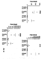
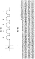
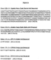
- the human MCAM has the amino acid sequence of SEQ ID NO: 11 ( FIG.
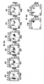
- FIG. 11A which includes five immunoglobulin domains (1: amino acid residues 19-129; 2: amino acid residues 139-242; 3: amino acid residues 244-321; 4: amino acid residues 335-424; and 5: amino acid residues 430-510) shown as SEQ ID NOS:22-26, which are also depicted schematically in FIG. 11B .
- laminin ⁇ 4 chain refers to one of the polypeptide chains found in laminin molecules, which are expressed in the basal lamina (of the basement membrane), a protein network foundation for most cells and organs. Laminins are known to bind to cell membranes through plasma membrane molecules and contribute to cell attachment.
- the laminin ⁇ 4 chain typically forms a complex with a laminin ⁇ -chain, and a laminin ⁇ -chain.
- the laminin ⁇ 4 chain is found in numerous laminin molecules including, without limitation, laminin 411 (laminin 8 or ⁇ 4 ⁇ 1 ⁇ 1); laminin 421 (laminin 9 or ⁇ 4 ⁇ 2 ⁇ 1), and laminin 423 (laminin 14 or ⁇ 4 ⁇ 2 ⁇ 3).
- laminin 411 refers to a trimeric polypeptide complex made up of three polypeptide subunits or chains: ⁇ 4-chain, a ⁇ 1-chain, and a ⁇ 1-chain.
- the term "antagonist” is used in the broadest sense, and includes any molecule that partially or fully blocks, inhibits, or neutralizes a qualitative biological activity of an MCAM polypeptide.
- the biological activity preferably is the ability to inhibit the ability of MCAM (i) to specifically bind its ligand: a laminin ⁇ 4 chain, e . g ., the ⁇ 4 chain of laminin 411; and/or (ii) to facilitate an MCAM-expressing cell, e . g ., a TH17 cell, to infiltrate into or migrate to a subject's tissue.
- Antagonists of MCAM can be identified, for example, based upon their ability to inhibit or block the specific binding of MCAM to its ligand: a laminin ⁇ 4 chain, e . g ., the ⁇ 4 chain of laminin 411.
- MCAM antagonists specifically include, without limitation, antibodies ( e . g ., antagonist or neutralizing antibodies), including chimeric, humanized and human antibodies and their functional fragments, small molecules, ribozymes, aptamers, peptides, and nucleic acids that encode polypeptide antagonists or antagonist antibodies.
- MCAM antagonist antibody refers to an antibody which inhibits or neutralizes the activity of MCAM. Such an antibody specifically binds to a polypeptide target involved in the infiltration of an MCAM-expressing cell into the CNS, e.g., MCAM or a laminin ⁇ 4 chain ( e.g., the ⁇ 4 chain of laminin 411).
- a “blocking” antibody, "neutralizing” antibody, or “antagonist” antibody is one which inhibits or reduces a biological activity of the antigen it binds. Such antibodies may substantially or completely inhibit the biological activity of the antigen.
- binding partner may show at least 1000 times the affinity of binding (measured as an apparent association constant) for its specific binding pair partner than a non-specific binding partner.
- antibodies that bind to MCAM with a binding affinity of 10 7 mole/L or more, typically 10 8 mole/L or more, are said to bind specifically to MCAM.
- biological activity and “biologically active” with regard to MCAM refer to its ability to specifically bind its ligand (a laminin ⁇ 4 chain, e . g ., the ⁇ 4 chain of laminin 411) and/or to facilitate the infiltration of MCAM-expressing cells, e . g ., TH17 cells, into the CNS.
- ligand a laminin ⁇ 4 chain, e . g ., the ⁇ 4 chain of laminin 411
- MCAM-expressing cells e . g ., TH17 cells
- MCAM-expressing cell refers to a cell of the immune system that expresses MCAM.
- MCAM expression is enriched on memory T lymphocytes, e.g., TH17 cells.
- binding molecule refers to a molecule that specifically binds to a target.
- the term specifically includes, without limitation, antibodies and antibody fragments (e.g. those comprising one or more of the CDRs described herein), and peptide and non-peptide small molecules.
- Antibodies are glycoproteins having some common structural characteristics. While antibodies exhibit binding specificity to a specific antigen, immunoglobulins include both antibodies and other antibody-like molecules which lack antigen specificity. Polypeptides of the latter kind can be, for example, produced at low levels by the lymph system and at increased levels by myelomas.
- antibody used herein may encompass intact monoclonal antibodies, polyclonal antibodies, multispecific antibodies (e.g. bispecific antibodies) formed from at least two intact antibodies, and antibody fragments, so long as they exhibit the desired biological activity.
- antigen-binding fragment refers to a portion of the full-length immunoglobulin molecule that specifically binds to the antigen.
- An antigen-binding fragment of an antibody thus includes an antigen-binding heavy chain, light chain, heavy chain-light chain dimer, Fab fragment, F(ab') 2 fragment, Fv fragment, single chain Fv (scFv), diabodies, linear antibodies, and multispecific antibodies formed from antibody fragment(s).
- monoclonal antibody refers to an antibody from a population of substantially homogeneous antibodies, i.e., the individual antibodies comprising the population are substantially similar and bind the same epitope(s), except for possible variants that may arise during production of the monoclonal antibody, such variants generally being present in minor amounts.
- Such monoclonal antibody typically includes an antibody comprising a variable region that binds a target, wherein the antibody was obtained by a process that includes the selection of the antibody from a plurality of antibodies.
- the selection process can be the selection of a unique clone from a plurality of clones, such as a pool of hybridoma clones, phage clones or recombinant DNA clones.
- the selected antibody can be further altered, for example, to improve affinity for the target, to humanize the antibody, to improve its production in cell culture, to reduce its immunogenicity in vivo, to create a multispecific antibody, etc., and that an antibody comprising the altered variable region sequence is also a monoclonal antibody of this invention.
- the monoclonal antibody preparations are advantageous in that they are typically uncontaminated by other immunoglobulins.
- the modifier "monoclonal” indicates the character of the antibody as being obtained from a substantially homogeneous population of antibodies, and is not to be construed as requiring production of the antibody by any particular method.
- the monoclonal antibodies to be used in accordance with the present invention may be made by a variety of techniques, including the hybridoma method (e.g., Kohler et al., Nature, 256:495 (1975 ); Harlow et al., Antibodies: A Laboratory Manual, (Cold Spring Harbor Laboratory Press, 2nd ed. 1988 ); Hammerling et al., in: Monoclonal Antibodies and T-Cell Hybridomas 563-681, (Elsevier, N.
- J. Immunol. Methods 284(1-2):119-132 (2004 ) and technologies for producing human or human-like antibodies from animals that have parts or all of the human immunoglobulin loci or genes encoding human immunoglobulin sequences see, e.g., WO98/24893 , WO/9634096 , WO/9633735 , and WO/91 10741 , Jakobovits et al., Proc. Natl. Acad. Sci. USA, 90:2551 (1993 ); Jakobovits et al., Nature, 362:255-258 (1993 ); Bruggemann et al., Year in Immune, 7:33 (1993 ); U.S.
- the monoclonal antibodies herein specifically include "chimeric" antibodies in which a portion of the heavy and/or light chain is identical with or homologous to corresponding sequences in antibodies derived from a particular species or belonging to a particular antibody class or subclass, while the remainder of the chain(s) is identical with or homologous to corresponding sequences in antibodies derived from another species or belonging to another antibody class or subclass, as well as fragments of such antibodies, so long as they exhibit the desired biological activity ( U.S. Patent No. 4,816,567 ; and Morrison et al., Proc. Natl. Acad. Sci. USA, 81:6851-6855 (1984 )).
- Chimeric antibodies of interest herein include "primatized” antibodies comprising variable domain antigen-binding sequences derived from a non-human primate (e.g. Old World Monkey, Ape etc) and human constant region sequences, as well as “humanized” antibodies.
- a non-human primate e.g. Old World Monkey, Ape etc
- human constant region sequences e.g. Old World Monkey, Ape etc
- Humanized forms of non-human (e.g., rodent) antibodies are chimeric antibodies that contain minimal sequence derived from non-human immunoglobulin.
- humanized antibodies are human immunoglobulins (recipient antibody) in which residues from a hypervariable region of the recipient are replaced by residues from a hypervariable region of a non-human species (donor antibody) such as mouse, rat, rabbit or nonhuman primate having the desired specificity, affinity, and capacity.
- donor antibody such as mouse, rat, rabbit or nonhuman primate having the desired specificity, affinity, and capacity.
- framework region (FR) residues of the human immunoglobulin are replaced by corresponding non-human residues.
- humanized antibodies may comprise residues that are not found in the recipient antibody or in the donor antibody. These modifications are made to further refine antibody performance.
- the humanized antibody will comprise substantially all of at least one, and typically two, variable domains, in which all or substantially all of the hypervariable loops correspond to those of a non-human immunoglobulin and all or substantially all of the FRs are those of a human immunoglobulin sequence.
- the humanized antibody optionally also will comprise at least a portion of an immunoglobulin constant region (Fc), typically that of a human immunoglobulin.
- Fc immunoglobulin constant region
- an “intact antibody” herein is one which comprises two antigen binding regions, and an Fc region.
- the intact antibody has a functional Fc region.
- an "antibody (or any other binding molecule) that binds to the same epitope" as a reference antibody (or any other binding molecule) refers to an antibody (or any other binding molecule) that blocks binding of the reference antibody (or any other binding molecule) to its antigen in a competition assay by 50% or more, and conversely, the reference antibody (or any other binding molecule) blocks binding of the antibody to its antigen in a competition assay by 50% or more.
- affinity matured antibody is one with one or more alterations in one or more hypervariable regions thereof which result an improvement in the affinity of the antibody for antigen, compared to a parent antibody which does not possess those alteration(s).
- Preferred affinity matured antibodies will have nanomolar or even picomolar affinities for the target antigen.
- Affinity matured antibodies are produced by procedures known in the art. Marks et al. Bio/Technology 10:779-783 (1992 ) describes affinity maturation by VH and VL domain shuffling. Random mutagenesis of CDR and/or framework residues is described by: Barbas et al. Proc Nat. Acad. Sci, USA 91:3809-3813 (1994 ); Schier et al.
- the "light chains" of antibodies from any vertebrate species can be assigned to one of two clearly distinct types, called ⁇ and ⁇ , based on the amino acid sequences of their constant domains.
- intact antibodies can be assigned to different "classes.”
- the heavy-chain constant domains that correspond to the different classes of antibodies are called ⁇ , ⁇ , ⁇ , ⁇ , and ⁇ , respectively.
- the subunit structures and three-dimensional configurations of different classes of immunoglobulins are well known.
- variable refers to the fact that certain portions of the variable domains differ extensively in sequence among antibodies and are used in the binding and specificity of each particular antibody for its particular antigen. However, the variability is not evenly distributed throughout the variable domains of antibodies. It is concentrated in three segments called complementarity-determining regions (CDRs) or hypervariable regions (HVRs) both in the light-chain and heavy-chain variable domains. The more highly conserved portions of variable domains are called the framework (FR).
- CDRs complementarity-determining regions
- HVRs hypervariable regions
- the more highly conserved portions of variable domains are called the framework (FR).
- the variable domains of native heavy and light chains each comprise four FR regions, largely adopting a ⁇ -sheet configuration, connected by three CDRs, which form loops connecting, and in some cases forming part of, the ⁇ -sheet structure.
- the CDRs in each chain are held together in close proximity by the FR regions and, with the CDRs from the other chain, contribute to the formation of the antigen-binding site of antibodies.
- the constant domains are not involved directly in binding an antibody to an antigen, but exhibit various effector functions, such as participation of the antibody in antibody-dependent cellular toxicity.
- Fv is the minimum antibody fragment which contains a complete antigen-recognition and binding site.
- this region consists of a dimer of one heavy- and one light-chain variable domain in tight, non-covalent association.
- one heavy- and one light-chain variable domain can be covalently linked by a flexible peptide linker such that the light and heavy chains can associate in a "dimeric" structure analogous to that in a two-chain Fv species. It is in this configuration that the three CDRs of each variable domain interact to define an antigen-binding site on the surface of the VH-VL dimer.
- the six CDRs confer antigen-binding specificity to the antibody.
- “Hypervariable region” or “HVR” refers to the amino acid residues of an antibody that are responsible for antigen-binding.
- the hypervariable region generally comprises amino acid residues from a "complementarity determining region” or “CDR” ( Kabat et al., Sequences of Proteins of Immunological Interest, 5th Ed. Public Health Service, National Institutes of Health, Bethesda, Md. (1991 )) and/or those residues from a "hypervariable loop” ( Chothia and Lesk, J. Mol. Biol. 196: 901-917 (1987 )).
- CDR complementarity determining region
- CDRs complementarity determining regions
- the CDRs of immunological receptors are the most variable part of the receptor protein, giving receptors their diversity, and are carried on six loops at the distal end of the receptor's variable domains, three loops coming from each of the two variable domains of the receptor.
- epitope is used to refer to binding sites for (monoclonal or polyclonal) antibodies on protein antigens.
- an epitope refers to a unit of structure conventionally bound by an immunoglobulin VH-VL pair.
- Epitopes define the minimum binding site for an antibody, and thus represent the target of specificity of an antibody.
- Epitopes can be linear or conformational, and can be as small as three amino acids.
- a "small molecule” is defined herein to have a molecular weight below about 600, preferably below about 1000 daltons. Generally, a small molecule is a non-peptide small organic molecule.
- isolated when used to describe the various polypeptides, proteins and antibodies disclosed herein, means polypeptide, protein or antibody that has been identified and separated and/or recovered from a component of its natural environment. Contaminant components of its natural environment are materials that would typically interfere with diagnostic or therapeutic uses for the polypeptide, protein or antibody, and may include enzymes, hormones, and other proteinaceous or non-proteinaceous solutes.
- the polypeptide, protein or antibody will be purified (1) to a degree sufficient to obtain at least 15 residues of N-terminal or internal amino acid sequence by use of a spinning cup sequenator, or (2) to homogeneity by SDS-PAGE under non-reducing or reducing conditions using Coomassie blue or, preferably, silver stain.
- Isolated polypeptide, protein or antibody includes polypeptide, protein or antibody in situ within recombinant cells, since at least one component of the associated natural environment will not be present. Ordinarily, however, isolated polypeptide, protein or antibody will be prepared by at least one purification step.
- affinity refers to the equilibrium dissociation constant (expressed in units of concentration) associated with each MCAM binding molecule--target complex, such as between an anti-MCAM antibody and MCAM.
- the binding affinity is directly related to the ratio of the off-rate constant (generally reported in units of inverse time, e.g., seconds -1 ) to the on-rate constant (generally reported in units of concentration per unit time, e.g., molar/second).
- the binding affinity may be determined by, for example, an ELISA assay, kinetic exclusion assay or surface plasmon resonance.
- certain epitopes can occur repetitively (multivalent) on a cell surface and that the dissociation constant (koff) for the binding of an antibody to a repetitive epitope may be greatly diminished over the dissociation constant for the reaction of the same antibody with the corresponding ligand in univalent form.
- the diminished dissociation constant arises because when one antibody-ligand bond dissociates, other bonds hold the bivalent (or multivalent) antibody to the multivalent ligand, allowing the dissociated bond to form again.
- the dissociation constant for the reaction between bivalent (or multivalent) Ab and multivalent ligand has been termed the functional affinity to contrast it with intrinsic affinity, which is the association constant for an antibodies representative individual site.
- dissociation is intended to refer to the off rate constant for dissociation of a binding molecule, such as an antibody, from the binding molecule/target, e.g. antibody/antigen complex.
- association means association, association rate and “k on “ as used herein, are intended to refer to the on rate constant for association of a binding molecule with a target, such as an antibody with an antigen, to form a complex.
- EC 50 as used herein, are intended to refer to the concentration of a binding molecule (e/g/ antibody) capable of interacting with sufficient quantities of target molecules to produce an effect on approximately 50% of the treated cells.
- treatment refers to clinical intervention in an attempt to alter the natural course of the individual being treated, and can be performed either for prophylaxis/prevention, or during the course of clinical pathology.
- the term refers to both therapeutic treatment and prophylactic or preventative measures, wherein the object is to prevent or slow down (lessen) an undesired physiological change or disorder.
- beneficial or desired clinical results include, but are not limited to, alleviation of symptoms, diminishment of extent of disease, stabilized (i.e., not worsening) state of disease, delay or slowing of disease progression, amelioration or palliation of the disease state, and remission (whether partial or total), whether detectable or undetectable.
- Treatment can also mean prolonging survival as compared to expected survival if not receiving treatment.
- Those in need of treatment include those already with the condition or disorder as well as those prone to have the condition or disorder or those in which the condition or disorder is to be prevented.
- Chronic administration refers to administration of the agent(s) in a continuous mode as opposed to an acute mode, so as to maintain the desired effect for an extended period of time.
- an “effective amount” refers to an amount effective, at dosages and for periods of time necessary, to achieve the desired prophylactic or therapeutic result.
- An effective amount refers to the amount of active compound or pharmaceutical agent that elicits the biological or medicinal response in a tissue, system, animal, individual or human that is being sought by a researcher, veterinarian, medical doctor or other clinician, which includes one or more of the following:
- kits and reagents are generally carried out in accordance with manufacturer defined protocols and/or parameters unless otherwise noted.
- this invention is not limited to the particular methodology, protocols, cell lines, animal species or genera, constructs, and reagents described as such may, of course, vary.
- the terminology used herein is for the purpose of describing particular embodiments only, and is not intended to limit the scope of the present invention which will be limited only by the appended claims.
- Abs antibodies CDR complementarity determining region CFA complete Freund's adjuvant CFSE carboxyfluorescein succinimidyl ester CNS central nervous system DAPI 4',6-diamidino-2-phenylindole DN dopamine-containing neuron EAE experimental autoimmune encephalomyelitis ECM extracellular matrix FACS fluorescence Activated cell sorting FR Framework Region IFA incomplete Freund's adjuvant Igs immunoglobulins MCAM melanoma cell adhesion molecule MOG myelin oligodendrocyte glycoprotein (MOG) MS multiple sclerosis PD Parkinson's disease PMA phorbol myristate acetate
- MCAM melanoma cell adhesion molecule
- MCAM is a cell-surface glycoprotein originally identified as a melanoma antigen, whose expression is associated with tumor progression and the development of metastatic potential.
- MCAM is a 113 kDa cell surface integral membrane glycoprotein composed of a signal peptide, five immunoglobulin-like domains (1, 2, 3, 4, and 5; or V-V-C2-C2-C2), a transmembrane region, and a short cytoplasmic tail. See , e . g ., Lehmann et al., Proc. Nat'l Acad. Sci. USA 86: 9891-9895 (1989 ) and FIG. 11B .
- MCAM is a member of the immunoglobulin superfamily and has significant sequence homology to a number of cell adhesion molecules of the Ig superfamily, including BEN ( Pourquie et al., Proc. Nat'l Acad. Sci. USA 89: 5261-5265 (1992 )), neural-cell adhesion molecule (N-CAM) ( Owens et al., Proc. Nat'l Acad. Sci. USA 84: 294-298 (1987 )), myelin-associated glycoprotein (MAG) ( Lai et al., Proc. Nat'l Acad. Sci.
- BEN Pourquie et al., Proc. Nat'l Acad. Sci. USA 89: 5261-5265 (1992 )
- N-CAM neural-cell adhesion molecule
- MAG myelin-associated glycoprotein
- MCAM colorectal cancer protein
- MCAM is also expressed on a variety of malignant neoplasms including smooth muscle neoplasms (Leiomyomas and leiomyosarcomas), tumors of vascular origin (angiosarcomas and Kaposi's sarcomas), placental site trophoblastic tumors, choriocarcinomas, and melanomas ( Shih et al., Clinical Cancer Res.
- MUC18 correlates directly with the metastatic potential of human melanoma cells ( Bar-Eli, Cancer Metastasis, 18: 377-385 (1999 )).
- MCAM a marker of tumor progression and metastasis in melanomas.
- the expression of MCAM is absent in normal melanocytes and benign nevi but prominent on many primary melanomas and in most metastatic lesions (Lehmann et al., supra ; Shih et al., supra ). MCAM expression correlates well with tumor vertical thickness and metastasis formation, and greater than 80% of metastatic lesions express MCAM (Lehmann et al., supra ; Xie et al., Cancer Res. 57: 2295-2303 (1997 ); and Shih et al., supra ). Modulators of MCAM have been generated to treat melanomas. See , e .
- a neuroinflammatory condition refers to a condition associated with inflammation of the nervous system, such as the central nervous system (CNS), and which is associated with cell/tissue damage. It is typically characterized by, for example, increased glial activation, increased pro-inflammatory cytokine/chemokine levels (e.g., TNF ⁇ , INF ⁇ , IL-1 ⁇ ), increased blood-brain-barrier permeability, and/or increased immune cell (e.g., leukocyte) recruitment/invasion to the CNS. It may refer to, for example, chronic neuroinflammation, such as an inflammation associated with chronic activation of cells of the immune system
- autoimmune-associated neuroinflammation i.e., autoimmune-associated neuroinflammation.
- MS multiple sclerosis
- PD Parkinson's disease
- PD is a neurodegenerative disease displaying neuroinflammation, for example, activated microglia and infiltrating T cells.
- MS multiple sclerosis, as a progressive neurological autoimmune disease, results from chronic, pathological inflammation ( Yednock et al., Nature 356: 63-66 (1992 ); Baron et al., J. Exp. Med. 177: 57-68 (1993 )).
- MS affects an estimated 250,000 to 350,000 people in the United States. Multiple sclerosis is thought to be the result of a specific autoimmune reaction wherein certain leukocytes attack and initiate the destruction of myelin, the insulating sheath covering nerve fibers. The onset of MS may be dramatic or so mild as to not cause a patient to seek medical attention.
- the most common symptoms include weakness in one or more limbs, visual blurring due to optic neuritis, sensory disturbances, diplopia, and ataxia.
- the course of disease may be stratified into three general categories: (1) relapsing MS, (2) chronic progressive MS, and (3) inactive MS.
- MS Relapsing MS is generally characterized by recurrent attacks of neurologic dysfunction. MS attacks generally evolve over days to weeks and may be followed by complete, partial, or no recovery. Recovery from attacks generally occurs within weeks to several months from the peak of symptoms, although rarely some recovery may continue for 2 or more years.
- Chronic progressive MS results in gradually progressive worsening without periods of stabilization or remission. This form develops in patients with a prior history of relapsing MS, although in 20% of patients, no relapses can be recalled. Acute relapses also may occur during the progressive course of MS.
- inactive MS is characterized by fixed neurologic deficits of variable magnitude. Most patients with inactive MS have an earlier history of relapsing MS. The course of MS is also dependent on the age of the patient. For example, favorable prognostic factors include early onset (excluding childhood), a relapsing course and little residual disability 5 years after onset. By contrast, poor prognosis is associated with a late age of onset (i.e., age 40 or older) and a progressive course. These variables are interdependent, since chronic progressive MS tends to begin at a later age that relapsing MS. Disability from chronic progressive MS is usually due to progressive paraplegia or quadriplegia in individual patients.
- Parkinson's disease is a progressive neurodegenerative disease displaying primary clinical features of motor abnormalities, e . g ., resting tremor, bradykinesia, and rigidity.
- PD is characterized by the loss of dopamine-containing neuron (DN) cells in the substantia nigra parts compacta ( Forno, J. Neurophthol. Exp. Neurol. 55: 259-272 (1996 )).
- DN dopamine-containing neuron
- One of the hallmarks of PD is neuroinflammation characterized by activated microglia and infiltrating T cells.
- CD4 positive T cells e . g ., proinflammatory T17 cells
- mediate cytotoxicity by activating microglia in PD and/or exert a direct toxic effect on substantia nigra DNs ( Appel, J. Clin. Invest. 119: 13-15 (2009 )).
- An autoimmune disease herein is a disease or disorder arising from and directed against an individual's own tissues or a co-segregate or manifestation thereof or resulting condition therefrom.
- autoimmune diseases or disorders include, but are not limited to arthritis (rheumatoid arthritis such as acute arthritis, chronic rheumatoid arthritis, gout or gouty arthritis, acute gouty arthritis, acute immunological arthritis, chronic inflammatory arthritis, degenerative arthritis, type II collagen-induced arthritis, infectious arthritis, Lyme arthritis, proliferative arthritis, psoriatic arthritis, Still's disease, vertebral arthritis, and juvenile-onset rheumatoid arthritis, osteoarthritis, arthritis chronica progrediente, arthritis deformans, polyarthritis chronica primaria, reactive arthritis, and ankylosing spondylitis), inflammatory hyperproliferative skin diseases, psoriasis such as plaque psoriasis, gutatte psoriasis, pustular psoria
- Cancer or a cancerous condition is the physiological condition in mammals that is typically characterized by unregulated cell growth/proliferation. Included in this definition are benign and malignant cancers, as well metastatic cancers. Also included are solid tumors and hematopoietic malignancies. Metastatic cancer refers to a cancer that has spread from the place where it first started to another place in the body. Tumors formed by metastatic cancer cells are called a metastatic tumor or a metastasis (which is also used to refer to the process by which cancer cells spread to other parts of the body). In general, metastatic cancer has the same name and same type of cancer cells as the original, or primary, cancer. Metastatic cancer includes prostate cancer, lung cancer, and pancreas cancer.
- prostate cancer or conditions related to prostate cancer is meant the malignant growth of abnormal cells in the prostate gland, capable of invading and destroying other prostate cells, and spreading (metastasizing) to other parts of the body, including bones, lungs, liver, and lymph nodes.
- lung cancer or conditions related to lung cancer is meant the malignant growth of abnormal cells in the lungs, capable of invading and destroying other lung cells, and spreading (metastasizing) to other parts of the body, including the adrenal gland, and liver.
- pancreatic cancer or conditions related to pancreatic cancer is meant the malignant growth of abnormal cells in the pancreas, capable of invading and destroying other pancreas cells, and spreading (metastasizing) to other parts of the body, including the liver, lungs, and peritoneum.
- the present invention provides antagonists of MCAM.
- Such antagonists encompass those that directly act upon MCAM (e . g ., an anti-MCAM antibody) and those that indirectly affect MCAM activity (e . g ., an anti-laminin ⁇ 4 chain antibody).
- Such antagonists are useful, for example, for treating a central nervous system (CNS) inflammatory disorder characterized by infiltration of MCAM-expressing cells into the CNS.
- a composition comprising an MCAM antagonist may be useful for reducing inflammation in a mammalian subject. Such a composition may be useful for partially or fully inhibiting CNS infiltration of MCAM-expressing cells.
- MCAM antagonists include, without limitation, antagonist or neutralizing antibodies or antibody fragments against one or more domains, e .
- Reference to "an" antagonist encompasses a single antagonist.
- the MCAM antagonists may be antibodies including, without limitation, chimeric, humanized and human antibodies and their functional fragments.
- the laminin ⁇ 4 chain is an ⁇ 4 chain of laminin 411.
- the MCAM antagonist blocks the interaction of an MCAM domain comprising the amino acid sequence of SEQ ID NO:22 and/or SEQ ID NO:23 with a laminin ⁇ 4 chain.
- the present disclosure describes screening assays to identify MCAM antagonists, which find utility in the treatment of inflammatory conditions characterized by infiltration of MCAM-expressing cells into the central nervous system (CNS).
- CNS central nervous system
- the disclosure describes a method for identifying an inhibitor of CNS infiltration by MCAM-expressing cells comprising the steps of: (a) incubating a population of cells expressing a laminin ⁇ 4 chain, e.g., an ⁇ 4 chain of laminin 411, with MCAM, in the presence or absence of a candidate molecule; (b) monitoring the level of binding of MCAM to the cells; and (c) identifying said candidate molecule as an inhibitor of CNS infiltration by MCAM-expressing cells if the level of MCAM binding is lower in the presence than in the absence of said candidate molecule.
- the candidate molecule may be selected from the group consisting of a small molecule, a peptide, a polypeptide, and an antibody.
- the level of binding of MCAM may be monitored by known techniques including, without limitation, fluorescent microscopy, FACS, and ELISA.
- the cells expressing a laminin ⁇ 4 chain may be endothelial cells.
- the laminin ⁇ 4 chain may be an ⁇ 4 chain of laminin 411.
- Screening assays for antagonist drug candidates may be designed to identify compounds that bind or complex with MCAM (including a subunit or other fragment thereof) or with an MCAM ligand, such as a laminin ⁇ 4 chain ( e . g ., an ⁇ 4 chain of laminin 411), or otherwise interfere with the interaction of MCAM with other cellular proteins, thereby interfering with the interaction of MCAM with its ligand, e.g., a laminin ⁇ 4 chain.
- the screening assays provided herein include assays amenable to high-throughput screening of chemical libraries, making them particularly suitable for identifying small molecule drug candidates. Generally, binding assays and activity assays are provided.
- the assays can be performed in a variety of formats, including, without limitation, protein-protein binding assays, biochemical screening assays, immunoassays, and cell-based assays, which are well characterized in the art.
- All assays for antagonists and agonists are common in that they call for contacting the drug candidate with an MCAM polypeptide, or an MCAM ligand polypeptide, e . g ., a laminin ⁇ 4 chain, or a fragment of such polypeptides (specifically including MCAM and laminin ⁇ 4 chains) under conditions and for a time sufficient to allow these two components to interact.
- an MCAM polypeptide or an MCAM ligand polypeptide, e . g ., a laminin ⁇ 4 chain, or a fragment of such polypeptides (specifically including MCAM and laminin ⁇ 4 chains) under conditions and for a time sufficient to allow these two components to interact.
- human MCAM is a 646 amino acid polypeptide, the sequence of which is available from the GenBank database under Accession Number AAA20922.1 (CAA48332) (SEQ ID NO:11; FIG. 11A ).
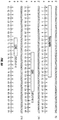
- Amino acid sequences for human laminin ⁇ 4-chain are available from the GenBank database under Accession Nos. NP001098676 and NP001098677 (SEQ ID NOS: 27-28; FIGS. 12A-B ).
- the making of antibodies or small molecules binding to such polypeptides is well within the skill of the ordinary artisan.
- the interaction is binding, and the complex formed can be isolated or detected in the reaction mixture.
- Either the MCAM or MCAM ligand polypeptide or the drug candidate may be immobilized on a solid phase, e.g., on a microtiter plate, by covalent or non-covalent attachments.
- Non-covalent attachment generally is accomplished by coating the solid surface with a solution of the MCAM or MCAM ligand polypeptide and drying.
- an immobilized antibody e.g., a monoclonal antibody, specific for the MCAM or MCAM ligand polypeptide to be immobilized can be used to anchor it to a solid surface.
- the assay is performed by adding the non-immobilized component, which may be labeled by a detectable label, to the immobilized component, e.g., the coated surface containing the anchored component.
- the non-reacted components are removed, e.g., by washing, and complexes anchored on the solid surface are detected.
- the detection of label immobilized on the surface indicates that complexing occurred.
- complexing can be detected, for example, by using a labeled antibody specifically binding the immobilized complex.
- the candidate compound is a polypeptide which interacts with but does not bind to MCAM or the MCAM ligand polypeptide
- its interaction with the respective polypeptide can be assayed by methods well known for detecting protein-protein interactions.
- assays include traditional approaches, such as, e.g., cross-linking, co-immunoprecipitation, and co-purification through gradients or chromatographic columns.
- protein-protein interactions can be monitored by using a yeast-based genetic system described by Fields and co-workers ( Fields and Song, Nature (London), 340:245-246 (1989 ); Chien et al., Proc. Natl. Acad. Sci. USA, 88:9578-9582 (1991 )) as disclosed by Chevray and Nathans, Proc. Natl. Acad. Sci. USA, 89: 5789-5793 (1991 ).
- MCAM ligand polypeptide Compounds that interfere with the interaction of MCAM and other extracellular components, in particular an MCAM ligand polypeptide, can be tested as follows. Usually a reaction mixture is prepared containing MCAM and the extracellular component (e . g ., MCAM ligand such as a laminin ⁇ 4 chain, e . g ., an ⁇ 4 chain of laminin 411) under conditions and for a time allowing for the interaction of the two products. To test the ability of a candidate compound to inhibit the interaction of MCAM and its ligand, the reaction is run in the absence and in the presence of the test compound. In addition, a placebo may be added to a third reaction mixture, to serve as positive control.
- MCAM ligand such as a laminin ⁇ 4 chain, e . g ., an ⁇ 4 chain of laminin 41
- the ability of the test compound to inhibit the MCAM/MCAM ligand interaction can, for example, be tested by measuring the degree of binding between MCAM and its ligand in the absence and presence of the test compound. If the degree of MCAM binding to its ligand is lower in the absence of the candidate compound than in its presence, the candidate compound is an MCAM antagonist by the definition of the present invention.
- An alternate screening protocol involves the use of a population of cells expressing a laminin ⁇ 4 chain, e . g ., an ⁇ 4 chain of laminin 411, which can be incubated with MCAM, in the presence and absence of a test compound, and binding of MCAM to the cell population monitored, e.g. by fluorescent microscopy (exemplified in Example 5). Other methods of monitoring will be appreciated by those skilled in the art, including fluorescence-activated cell sorting (FACS) and enzyme-linked immunosorbent assay (ELISA). If the binding of MCAM to the cell population in the presence of the test compound is lower than in its absence, the test compound is an MCAM antagonist.
- FACS fluorescence-activated cell sorting
- ELISA enzyme-linked immunosorbent assay
- the MCAM antagonists identified based upon their ability to inhibit the binding of MCAM to its ligand, e.g., a laminin ⁇ 4 chain, are drug candidates for the treatment of neuroinflammatory conditions characterized by infiltration of MCAM-expressing cells into the CNS.
- screening assays specifically discussed herein are for illustration only.
- a variety of other assays, which can be selected depending on the type of the antagonist candidates screened e.g. polypeptides, peptides, non-peptide small organic molecules, aptamers, ribozymes, nucleic acid, etc.
- polypeptides, peptides, non-peptide small organic molecules, aptamers, ribozymes, nucleic acid, etc. are well known to those skilled in the art and are equally suitable for the purposes of the present invention.
- an MCAM antagonist is an anti-MCAM antibody or an anti-laminin ⁇ 4 chain, e.g., ⁇ 4 chain of laminin 411, antibody, or an antigen-binding fragment thereof.
- an anti-MCAM antibody is a blocking antibody that fully or partially blocks the interaction of MCAM with its ligand, a laminin ⁇ 4 chain.
- an anti-laminin ⁇ 4 chain antibody is a blocking antibody that fully or partially blocks the interaction of a laminin ⁇ 4 chain with MCAM.
- the anti-MCAM antibody binds to the extracellular domain of MCAM which interacts with its ligand, a laminin ⁇ 4 chain.
- the laminin ⁇ 4 chain is an ⁇ 4 chain of laminin 411.
- An anti-MCAM antibody may specifically or selectively binds to an MCAM fragment comprising or having the amino acid sequence of position 19 to position 129 of SEQ ID NO: 11 (SEQ ID NO:22).
- An anti-MCAM antibody may specifically or selectively bind to an MCAM fragment comprising or having the amino acid sequence of position 139 to position 242 of SEQ ID NO: 11 (SEQ ID NO:23).
- An anti-MCAM antibody may specifically or selectively bind to an MCAM fragment comprising the amino acid sequences of SEQ ID NOS:22 and 23.
- the antagonist antibody may block the interaction of an MCAM domain comprising the amino acid sequence of SEQ ID NO:22 and/or SEQ ID NO:23 with a laminin ⁇ 4 chain.
- the anti-MCAM antibody or antibody fragment comprises the following hypervariable regions (HVRs):
- the anti-MCAM antibody or antibody fragment comprises a light chain variable domain shown as any one of SEQ ID NOS:70, 71, or 72 and/or a heavy chain variable domain shown as SEQ ID NO:77.
- the anti-MCAM antibody or antibody fragment comprises the following hypervariable regions (HVRs):
- the anti-MCAM antibody or antibody fragment further comprises (a) a heavy chain framework region 2 (FR2) comprising the amino acid sequence of SEQ ID NO: 134; (b) a heavy chain framework region 3 (FR3) comprising the amino acid sequence of SEQ ID NO:135; (c) a light chain framework region 1 (FR1) comprising the amino acid sequence of SEQ ID NO:147; (d) a light chain framework region 2 (FR2) comprising the amino acid sequence of SEQ ID NO:148; and/or (e) a light chain framework region 3 (FR3) comprising the amino acid sequence of SEQ ID NO:149.
- FR2 heavy chain framework region 2
- FR3 heavy chain framework region 3
- the MCAM antagonist binds to substantially the same epitope as, or competes for binding with, an anti-MCAM antibody comprising the following HVRs:
- the MCAM antagonist binds to substantially the same epitope as, or competes for binding with, an anti-MCAM antibody comprising a light chain variable domain shown as any one of SEQ ID NOS:70, 71, or 72 and/or a heavy chain variable domain shown as SEQ ID NO:77.
- the disclosure provides an antibody that binds to MCAM, wherein the antibody comprises a heavy chain variable domain having at least 90%, at least 91%, at least 92%, at least 93%, at least 94%, at least 95%, at least 96%, at least 97%, at least 98%, or at least 99% sequence identity to an amino acid sequence selected from SEQ ID NO: 94, SEQ ID NO: 95, SEQ ID NO: 102, SEQ ID NO: 103, SEQ ID NO: 104; SEQ ID NO: 105, SEQ ID NO: 106, SEQ ID NO: 107, SEQ ID NO: 115, SEQ ID NO: 116, SEQ ID NO: 117, SEQ ID NO: 118, and SEQ ID NO: 119.
- the anti-MCAM antibody may comprise a light chain variable domain having at least 90%, at least 91%, at least 92%, at least 93%, at least 94%, at least 95%, at least 96%, at least 97%, at least 98%, or at least 99% sequence identity to an amino acid sequence selected from the group consisting of SEQ ID NO: 98, SEQ ID NO: 99, SEQ ID NO: 110, SEQ ID NO: 111, SEQ ID NO: 112, SEQ ID NO: 121, SEQ ID NO: 122, and SEQ ID NO: 123.
- the antibody may comprise a heavy chain variable domain having at least 90%, at least 91%, at least 92%, at least 93%, at least 94%, at least 95%, at least 96%, at least 97%, at least 98%, or at least 99% sequence identity to an amino acid sequence selected from SEQ ID NO: 94, SEQ ID NO: 95, SEQ ID NO: 102, SEQ ID NO: 103, SEQ ID NO: 104; SEQ ID NO: 105, SEQ ID NO: 106, SEQ ID NO: 107, SEQ ID NO: 115, SEQ ID NO: 116, SEQ ID NO: 117, SEQ ID NO: 118, and SEQ ID NO: 119, and the antibody further comprises a light chain variable domain having at least 90%, at least 91%, at least 92%, at least 93%, at least 94%, at least 95%, at least 96%, at least 97%, at least 98%, or at least 99% sequence identity to an amino acid sequence selected from the group consisting of SEQ ID NO:
- the disclosure describes an antibody that is a variant of any of the above antibodies having one or more amino acid substitutions, deletions, insertions or modifications, and which retains a biological function of the antibody.
- the invention provides an antibody that binds to MCAM expressed on the cell surface and inhibits the binding of MCAM to laminin 411.
- the anti-MCAM antibody binds to MCAM expressed on the cell surface and inhibits disease progression. In some embodiments, the progression of an autoimmune disease is inhibited. In one embodiment, the progression of multiple sclerosis is inhibited.
- the disclosure describes an antibody that is a variant of any one of the above antibodies having improvements in one or more of a property such as binding affinity, specificity, thermostability, expression level, effector function, glycosylation, reduced immunogenicity, or solubility as compared to the unmodified antibody.
- the anti-MCAM antibody binds to MCAM expressed on the cell surface and inhibits progression of a metastatic cancer.
- the metastatic cancer is selected from the group consisting of prostate cancer, lung cancer, and pancreatic cancer.
- MCAM antibodies can include, but are not limited to, polyclonal, monoclonal, multispecific, human, humanized, primatized, or chimeric antibodies, single chain antibodies (e.g., scFv), Fab fragments, F(ab') fragments, fragments produced by a Fab expression library, anti-idiotypic (anti-Id) antibodies (including, e.g., anti-Id antibodies to antibodies of the present embodiments), and epitope-binding fragments of any of the above.
- Human antigen-binding antibody fragments include, but are not limited to, Fab, Fab' and F(ab') 2 , Fd, single-chain Fvs (scFv), single-chain antibodies, disulfide-linked Fvs (sdFv), and fragments comprising either a VL or VH domain.
- Antigen-binding antibody fragments, including single-chain antibodies may comprise the variable region(s) alone or in combination with the entirety or a portion of the following: hinge region, CH1, CH2, and CH3 domains. Also included are antigen-binding fragments that can comprise any combination of variable region(s) with a hinge region, CH1, CH2, and CH3 domains.
- the antibodies may be from any animal origin including birds and mammals.
- the antibodies are from human or other primates, murine (e.g., mouse and rat), donkey, sheep, monkey, rabbit, goat, guinea pig, pig, camel, horse, or chicken (or other avian).
- murine e.g., mouse and rat
- donkey sheep, monkey
- rabbit goat
- guinea pig pig
- camel horse
- chicken or other avian
- human antibodies include antibodies having the amino acid sequence of a human immunoglobulin and include antibodies isolated from human immunoglobulin libraries or from animals transgenic for one or more human immunoglobulins and that do not express endogenous immunoglobulins, as described, for example in, U.S. Patent No. 5,939,598 .
- the MCAM antibody can be a monoclonal antibody.
- the antibody may be chemically modified, e.g., by pegylation.
- other antibodies can be identified using techniques available in the art. For example, antibodies capable of specifically binding to MCAM can be produced using phage display technology. Antibody fragments that selectively bind to MCAM can then be isolated. Exemplary methods for producing such antibodies via phage display are disclosed, for example, in U.S. Patent No. 6,225,447 , for example.
- Monoclonal antibodies can also be produced using the conventional hybridoma methods. These methods have been widely applied to produce hybrid cell lines that secrete high levels of monoclonal antibodies against many specific antigens, and can also be used to produce monoclonal antibodies capable of specifically binding to MCAM.
- mice e.g., Balb/c mice
- mice can be immunized with an antigenic MCAM epitope by intraperitoneal injection. After sufficient time has passed to allow for an immune response, the mice are sacrificed, and the spleen cells obtained and fused with myeloma cells, using techniques well known in the art. The resulting fused cells, hybridomas, are then grown in a selective medium, and the surviving cells grown in such medium using limiting dilution conditions.
- hybridomas can be isolated for secreting antibodies (for example, of the IgG or IgM class or IgG1 subclass) that selectively bind to MCAM.
- antibodies for example, of the IgG or IgM class or IgG1 subclass
- the isolated monoclonal can then be used to produce chimeric and humanized antibodies.
- MCAM antagonist antibodies are selected using an antigen derived from a mammalian species.
- the antigen is human MCAM or a laminin ⁇ 4 chain, e.g., ⁇ 4 chain of laminin 411.
- polypeptides from other species such as murine MCAM or laminin ⁇ 4 chain can also be used as the target antigen.
- the antigens from various mammalian species may be isolated from natural sources. In other embodiments, the antigen is produced recombinantly or made using other synthetic methods known in the art.
- the antibody selected will normally have a sufficiently strong binding affinity for the antigen. For example, the antibody may bind human MCAM or a laminin ⁇ 4 chain, e .
- Antibody affinities may be determined by a surface plasmon resonance based assay (such as the BIAcore assay as described in Examples); enzyme-linked immunoabsorbent assay (ELISA); and competition assays (e.g. RIA's), for example.
- a surface plasmon resonance based assay such as the BIAcore assay as described in Examples
- ELISA enzyme-linked immunoabsorbent assay
- competition assays e.g. RIA's
- the antibody may be subject to other biological activity assays, e.g., in order to evaluate its effectiveness as a therapeutic.
- biological activity assays are known in the art and depend on the target antigen and intended use for the antibody. Examples include the experimental autoimmune encephalomyelitis (EAE) (as described in Example 7 below), and in vitro and in vivo assays described herein for identifying MCAM antagonists.
- EAE experimental autoimmune encephalomyelitis
- Antagonist antibodies may be selected using a unique phage display approach.
- the approach involves generation of synthetic antibody phage libraries based on single framework template, design of sufficient diversities within variable domains, display of polypeptides having the diversified variable domains, selection of candidate antibodies with high affinity to target antigen, and isolation of the selected antibodies. Details of the phage display methods can be found, for example, in WO03/102157 published Dec. 11, 2003 .
- the antibody generated from phage libraries can be further modified to generate antibody mutants with improved physical, chemical and or biological properties over the parent antibody.
- the antibody mutant preferably has a biological activity in the assay of choice which is at least about 10 fold better, preferably at least about 20 fold better, more preferably at least about 50 fold better, and sometimes at least about 100 fold or 200 fold better, than the biological activity of the parent antibody in that assay.
- an anti-MCAM antibody mutant preferably has a binding affinity for MCAM which is at least about 10 fold stronger, preferably at least about 20 fold stronger, more preferably at least about 50 fold stronger, and sometimes at least about 100 fold or 200 fold stronger, than the binding affinity of the parent anti-MCAM antibodies, such as clone 15 or 17 antibodies.
- Chimeric and humanized antibodies can be produced from non-human antibodies, and can have the same or similar binding affinity as the antibody from which they are produced.
- Exemplary techniques for producing chimeric antibodies include splicing the genes from, e.g., a mouse antibody molecule of appropriate antigen specificity together with genes from a human antibody molecule of appropriate biological activity. See , e . g ., Morrison et al., 1984 Proc. Nat'l. Acad. Sci. USA 81: 6851 ; Neuberger et al., 1984 Nature 312: 604 ; and Takeda et al., 1985 Nature 314: 452 .
- a nucleic acid encoding a variable (V) region of a mouse monoclonal antibody can be joined to a nucleic acid encoding a human constant (C) region, e.g., IgG1 or IgG4.
- C human constant
- the resulting antibody is thus a species hybrid, generally with the antigen binding domain from the non-human antibody and the C or effector domain from a human or primate antibody.
- Humanized antibodies are antibodies with variable regions that are primarily from a human antibody (i.e., the acceptor antibody), but which have complementarity determining regions substantially from a non-human antibody (the donor antibody). See , e . g ., Queen et al., Proc. Nat'l. Acad. Sci USA 86: 10029-10033 (1989 ); WO 90/07861 , U.S. Patent Nos. 7,435,802 , 6,054,297 ; 5,693,761 ; 5,585,089 ; 5,530,101 ; and 5,224,539 .
- the constant region or regions of these antibodies are generally also from a human antibody.
- the human variable domains are typically chosen from human antibodies having sequences displaying a high homology with the desired non-human variable region binding domains.
- the heavy and light chain variable residues can be derived from the same antibody, or a different human antibody.
- the sequences can be chosen as a consensus of several human antibodies, such as described in WO 92/22653 .
- a "PrimatizedTM antibody” is a recombinant antibody containing primate variable sequences or antigen binding portions, and human constant domain sequences. See e . g ., Newman, Bio/Technology, 1992, 10: 1455-60 . Primatization of antibodies results in the generation of antibodies that contain primate ( e . g ., monkey) variable domains and human constant sequences. See , e . g ., U.S. Patent No. 6,113,898 . This technique modifies antibodies such that they are not rejected upon administration in humans because they are antigenic. This technique relies on immunization of cynomolgus monkeys with human antigens or receptors. This technique was developed to create high affinity monoclonal antibodies directed to human cell surface antigens.
- specific amino acids within the human variable region can be selected for substitution based on the predicted conformation and antigen binding properties. This can be determined using techniques such as computer modeling, prediction of the behavior and binding properties of amino acids at certain locations within the variable region, and observation of effects of substitution. For example, when an amino acid differs between a non-human variable region and a human variable region, the human variable region can be altered to reflect the amino acid composition of the non-human variable region.
- the antibodies used in the chronic dosage regime can be humanized antibodies as disclosed in U.S. Patent No. 5,840,299 .
- Transgenic mice containing human antibody genes can be immunized with an antigenic MCAM structure and hybridoma technology can be used to generate human antibodies that selectively bind to MCAM.
- Chimeric, human and/or humanized antibodies can be produced by using recombinant expression, e.g., expression in human hybridomas ( Cole et al., Monoclonal Antibodies and Cancer Therapy, Alan R. Liss, p. 77 (1985 )), in myeloma cells, or in Chinese hamster ovary (CHO) cells.
- antibody coding sequences can be incorporated into transgenes for introduction into the genome of a transgenic animal and subsequent expression in the milk of the transgenic animal. See , e . g ., U.S. Patent No. 6,197,946 .
- Exemplary suitable transgenes include, but are not limited to, transgenes having a promoter and/or enhancer from a mammary gland specific gene, for example casein or ⁇ -lactoglobulin.
- Anti-MCAM antagonist antibody variants can be prepared by introducing appropriate nucleotide changes into the encoding DNA, and/or by synthesis of the desired antibody. Those skilled in the art will appreciate that amino acid changes may alter post-translational processes of the anti-MCAM antibody, such as changing the number or position of glycosylation sites.
- Variations in the MCAM antagonist antibodies described herein can be made, for example, using any of the techniques and guidelines for conservative and non-conservative mutations set forth, for instance, in U.S. Patent No. 5,364,934 .
- Variations may be a substitution, deletion or insertion of one or more codons encoding the antibody that results in a change in the amino acid sequence as compared with the native sequence antibody.
- the variation is by substitution of at least one amino acid with any other amino acid in one or more of the domains of the MCAM antagonist antibody.
- Guidance in determining which amino acid residue may be inserted, substituted or deleted without adversely affecting the desired activity may be found by comparing the sequence of the MCAM antagonist antibody with that of homologous known protein molecules and minimizing the number of amino acid sequence changes made in regions of high homology.
- Amino acid substitutions can be the result of replacing one amino acid with another amino acid having similar structural and/or chemical properties, such as the replacement of a leucine with a serine, i.e., conservative amino acid replacements.
- Insertions or deletions may optionally be in the range of about 1 to 5 amino acids. The variation allowed may be determined by systematically making insertions, deletions or substitutions of amino acids in the sequence and testing the resulting variants for activity exhibited by the full-length or mature native sequence.
- Covalent modifications of anti-MCAM antibodies are included within the scope of this invention. Covalent modifications include reacting targeted amino acid residues of an anti-MCAM antibody with an organic derivatizing agent that is capable of reacting with selected side chains or the N- or C- terminal residues of the anti-MCAM antibody. Other modifications include deamidation of glutaminyl and asparaginyl residues to the corresponding glutamyl and aspartyl residues, respectively, hydroxylation of proline and lysine, phosphorylation of hydroxyl groups of seryl or threonyl residues, methylation of the ⁇ -amino groups of lysine, arginine, and histidine side chains ( T.E.
- the MCAM antagonist antibodies of the present disclosure comprise one or more deamidation mutations in the amino acid sequence.
- the deamidation of amino acid residues is a common structural modification in recombinant polypeptides, which can lead to the formation of iso-aspartic acid resulting in decreased stability.
- Deamidation may be associated with glycine (G)-asparginine (N) sequences, including G-N and N-G sequences.
- the antibody may comprise a deamidation mutation.
- the deamidation mutation may be the substitution of an N amino acid residue or a G amino acid residue. The substitution may be selected from the group consisting of N ⁇ S, N ⁇ A, and G ⁇ Q.
- the deamidation mutation may be located at Kabat residue N32 or G33.
- covalent modification of the anti-MCAM antibody included within the disclosure include altering the native glycosylation pattern of the antibody or polypeptide ( Beck et al., Curr. Pharm. Biotechnol. 9: 482-501, 2008 ; Walsh, Drug Discov. Today 15: 773-780, 2010 ), and linking the antibody to one of a variety of nonproteinaceous polymers, e.g., polyethylene glycol (PEG), polypropylene glycol, or polyoxyalkylenes, in the manner set forth in U.S. Patent Nos. 4,640,835 ; 4,496,689 ; 4,301,144 ; 4,670,417 ; 4,791,192 or 4,179,337 .
- PEG polyethylene glycol
- the MCAM antagonist antibodies of disclosure comprise one or more glycosylation mutations in the amino acid sequence.
- the mutation may remove or restore/introduce a glycosylation site.
- the glycosylation mutation is associated with an asparginine (N) residue.
- the antibody may comprise a glycosylation mutation.
- the glycosylation mutation may be the introduction of an N amino acid residue.
- the introduction may be the replacement of an aspartic acid (D) residue with an N residue (D ⁇ N).
- the mutation may be located at Kabat residue 72.
- MCAM antagonist antibody fragments are provided herein. Such fragments may be truncated at the N-terminus or C-terminus, or may lack internal residues, for example, when compared with a full length native antibody. Certain fragments lack amino acid residues that are not essential for a desired biological activity of the MCAM antagonist antibody.
- MCAM antagonist antibody fragments may be prepared by any of a number of conventional techniques. Desired peptide fragments may be chemically synthesized. An alternative approach involves generating antibody or polypeptide fragments by enzymatic digestion, e.g., by treating the protein with an enzyme known to cleave proteins at sites defined by particular amino acid residues, or by digesting the DNA with suitable restriction enzymes and isolating the desired fragment. Yet another suitable technique involves isolating and amplifying a DNA fragment encoding a desired antibody or polypeptide fragment, by polymerase chain reaction (PCR). Oligonucleotides that define the desired termini of the DNA fragment are employed at the 5' and 3' primers in the PCR. Preferably, anti-MCAM antagonist antibody fragments share at least one biological and/or immunological activity with a native MCAM antagonist antibody disclosed herein.
- PCR polymerase chain reaction
- Substantial modifications in function or immunological identity of the MCAM antagonist antibody are accomplished by selecting substitutions that differ significantly in their effect on maintaining (a) the structure of the polypeptide backbone in the area of the substitution, for example, as a sheet or helical conformation, (b) the charge or hydrophobicity of the molecule at the target site, or (c) the bulk of the side chain.
- Naturally occurring residues are divided into groups based on common side-chain properties:
- Non-conservative substitutions will entail exchanging a member of one of these classes for another class. Such substituted residues also may be introduced into the conservative substitution sites or, more preferably, into the remaining (non-conserved) sites.
- Table 1 Original Residue Exemplary Substitutions Preferred Substitutions Arg (R) lys; gln; asn lys Asn (N) gln; his; lys; arg gln Asp (D) glu glu Cys (C) ser ser Gln (Q) asn asn Glu (E) asp asp Gly (G) pro; ala ala His (H) asn; gln; lys; arg arg Ile (I) leu; val; met; ala; phe; norleucine leu Leu (L) norleucine; ile; val; met; ala; phe ile Lys (K) arg; gln; asn arg
- the variations can be made using methods known in the art such as oligonucleotide-mediated (site-directed) mutagenesis, alanine scanning, and PCR mutagenesis.
- Site-directed mutagenesis [ Carter et al., Nucl. Acids Res., 13:4331 (1986 ); Zoller et al., Nucl. Acids Res., 10:6487 (1987 )], cassette mutagenesis [ Wells et al., Gene, 34:315 (1985 )], restriction selection mutagenesis [ Wells et al., Philos. Trans. R. Soc. London SerA, 317:415 (1986 )] or other known techniques can be performed on the cloned DNA to produce the MCAM antagonist antibody variant DNA.
- Scanning amino acid analysis can also be employed to identify one or more amino acids along a contiguous sequence.
- preferred scanning amino acids are relatively small, neutral amino acids.
- amino acids include alanine, glycine, serine, and cysteine.
- Alanine is typically a preferred scanning amino acid among this group because it eliminates the side-chain beyond the beta-carbon and is less likely to alter the main-chain conformation of the variant [ Cunningham and Wells, Science, 244:1081-1085 (1989 )].
- Alanine is also typically preferred because it is the most common amino acid. Further, it is frequently found in both buried and exposed positions [ Creighton, The Proteins, (W.H. Freeman & Co., N.Y .); Chothia, J. Mol. Biol., 150:1 (1976 )]. If alanine substitution does not yield adequate amounts of variant, an isoteric amino acid can be used.
- cysteine residue not involved in maintaining the proper conformation of the MCAM antagonist antibody also may be substituted, generally with serine, to improve the oxidative stability of the molecule and prevent aberrant crosslinking.
- cysteine bond(s) may be added to the MCAM antagonist antibody to improve its stability (particularly where the antibody is an antibody fragment such as an Fv fragment).
- a particularly preferred type of substitutional variant involves substituting one or more hypervariable region residues of a parent antibody (e.g., a humanized or human antibody).
- a parent antibody e.g., a humanized or human antibody
- the resulting variant(s) selected for further development will have improved biological properties relative to the parent antibody from which they are generated.
- a convenient way for generating such substitutional variants involves affinity maturation using phage display. Briefly, several hypervariable region sites (e.g., 6-7 sites) are mutated to generate all possible amino substitutions at each site.
- the antibody variants thus generated are displayed in a monovalent fashion from filamentous phage particles as fusions to the gene III product of M13 packaged within each particle.
- the phage-displayed variants are then screened for their biological activity (e.g., binding affinity) as herein disclosed.
- alanine scanning mutagenesis can be performed to identify hypervariable region residues contributing significantly to antigen binding.
- the panel of variants is subjected to screening as described herein and antibodies with superior properties in one or more relevant assays may be selected for further development.
- Preferred affinity matured antibodies have an affinity which is five times, more preferably 10 times, even more preferably 20 or 30 times greater than the starting antibody (generally murine, humanized or human) from which the matured antibody is prepared.
- Nucleic acid molecules encoding amino acid sequence variants of the MCAM antagonist antibody are prepared by a variety of methods known in the art. These methods include, but are not limited to, isolation from a natural source (in the case of naturally occurring amino acid sequence variants) or preparation by oligonucleotide-mediated (or site-directed) mutagenesis, PCR mutagenesis, and cassette mutagenesis of an earlier prepared variant or a non-variant version of the MCAM antagonist antibody.
- antibodies that bind to the same epitope as the antibodies described herein specifically bind to an epitope that includes one or more amino acid residues on human MCAM (Accession No. AAA20922.1/CAA48332).
- Antibodies of the disclosure specifically bind MCAM, wherein the antibody binds to an epitope on human MCAM (e.g., Accession No. AAA20922.1/CAA48332).
- a monoclonal antibody e.g., fully human monoclonal antibody
- a monoclonal antibody has the same specificity as a monoclonal antibody of the disclosure by ascertaining whether the former prevents the latter from binding to MCAM. If the monoclonal antibody being tested competes with the monoclonal antibody of the disclosure, as shown by a decrease in binding by the monoclonal antibody of the disclosure, then the two monoclonal antibodies bind to the same, or a closely related, epitope.
- An alternative method for determining whether a monoclonal antibody has the specificity of monoclonal antibody of the disclosure is to pre-incubate the monoclonal antibody of the disclosure with MCAM (e.g., an MCAM-Fc molecule exemplified in the Examples) and then add the monoclonal antibody being tested to determine if the monoclonal antibody being tested is inhibited in its ability to bind MCAM. If the monoclonal antibody being tested is inhibited then, in all likelihood, it has the same, or functionally equivalent, epitopic specificity as the monoclonal antibody of the disclosure.
- MCAM e.g., an MCAM-Fc molecule exemplified in the Examples
- peptide molecules can be designed that retain the ability to bind the target protein sequence.
- Such peptides can be synthesized chemically and/or produced by recombinant DNA technology. See, e.g., Marasco et al., Proc. Natl. Acad. Sci. USA, 90: 7889-7893 (1993 ).
- the disclosure comprises a binding agent that binds to essentially the same epitope as any of the antibodies disclosed herein.
- the binding agent is capable of binding to MCAM protein on the surface of cells.
- the binding agent may inhibit the interaction of MCAM (e . g ., cell surface MCAM) with its ligand, a protein comprising a laminin alpha-4 chain.
- the binding agent may be an antibody or a functional fragment thereof.
- the disclosure provides a binding agent capable of binding to MCAM, wherein any one of the antibodies disclosed above displaces the binding agent in a competitive binding assay.
- the binding agent may be an antibody, or a functional fragment thereof.
- the disclosure provides a binding agent capable of binding to MCAM, wherein the binding agent displaces any one of the antibodies disclosed above in a competitive binding assay.
- the binding agent may be an antibody, or a functional fragment thereof.
- the binding agent may be an alternative binding agent.
- These alternative binding agents may include, for example, any of the engineered protein scaffolds known in the art.
- Such scaffolds include, for example, anticalins, which are based upon the lipocalin scaffold, a protein structure characterized by a rigid beta-barrel that supports four hypervariable loops which form the ligand binding site. Novel binding specificities are engineered by targeted random mutagenesis in the loop regions, in combination with functional display and guided selection ( Skerra (2008) FEBS J. 275: 2677-2683 ).
- Other suitable scaffolds may include, for example, adnectins, or monobodies, based on the tenth extracellular domain of human fibronectin III ( Koide and Koide (2007) Methods Mol. Biol.
- the binding agent may be an anticalin, an adnectin, an affibody, a DARPin, a fynomer, an affitin, an affilin, an avimer, a cysteine-rich knottin peptide, or an engineered Kunitz-type inhibitor.
- the present invention provides MCAM antagonists for use as therapeutic agents in the treatment of neuroinflammatory conditions, cancer, and autoimmune diseases.
- the appropriate dosage of a compound of the invention will depend on the type of disease or condition to be treated, as defined above, the severity and course of the disease or condition, whether the agent is administered for preventive or therapeutic purposes, previous therapy, the patient's clinical history and response to the compound, and the discretion of the attending physician.
- the compound is suitably administered to the patient at one time or over a series of treatments.
- the present disclosure provides a method for inhibiting or blocking the interaction of MCAM expressed on T cells and laminin ⁇ 4 chain, e . g ., an ⁇ 4 chain of laminin 411, comprising treating the T cells with an MCAM antagonist (as described herein), thereby inhibiting the interaction of MCAM with laminin ⁇ 4 chain.
- the laminin ⁇ 4 chain may be expressed on the surface of a cell, e . g ., an endothelial cell.
- the MCAM antagonist may be an anti-MCAM antibody.
- the T cells may be TH17 cells.
- the treatment with an MCAM antagonist may be performed in vivo.
- the treatment may be performed in a mammalian subject.
- the mammalian subject may be a human.
- the present disclosure provides a method for inhibiting or preventing extravasation of MCAM-expressing T cells into the central nervous system (CNS) comprising treating the T cells with an MCAM antagonist (as described herein), thereby inhibiting or preventing the extravasation of MCAM-expressing T cells into the CNS.
- the MCAM antagonist may block the interaction of MCAM with laminin ⁇ 4 chain, e . g ., an ⁇ 4 chain of laminin 411.
- the MCAM antagonist may be an anti-MCAM antibody.
- the laminin ⁇ 4 chain may be expressed on the surface of a cell, e . g ., an endothelial cell.
- the T cells may be TH17 cells.
- the treatment with an MCAM antagonist may be performed in vivo.
- the treatment may be performed in a mammalian subject.
- the mammalian subject may be a human.
- the present disclosure describes methods of treatment for a neuroinflammatory condition, a cancerous condition, or an autoimmune disease.
- the method comprises administering to a mammalian subject in need a therapeutically effective amount of an MCAM antagonist.
- the disclosure describes a method for the delaying or slowing down of the progression of a neuroinflammatory condition, a cancerous condition, or an autoimmune disease.
- the method comprises administering to subject diagnosed with the condition or disease, an effective amount of an MCAM antagonist.
- the disclosure describes a method for preventing indicia of a neuroinflammatory condition, cancerous condition, or an autoimmune disease.
- the method comprises administering an effective amount of an MCAM antagonist to a subject at risk of the condition or disease, wherein the MCAM antagonist is effective against the development of indicia of the condition or disease.
- the present disclosure describes methods of treatment for a metastatic cancer.
- the present disclosure describes an MCAM antagonist for use as a medicament for, or for the treatment of a disease, condition or disorder described herein.
- the present disclosure describes the use of an MCAM antagonist for the manufacture of a medicament for treating a disease, condition or disorder described herein.
- the present disclosure describes the use of an MCAM antagonist described herein, in the manufacture of a medicament for the treatment of a central nervous system (CNS) inflammatory disorder characterized by infiltration of MCAM-expressing cells into the CNS.
- CNS central nervous system
- the MCAM antagonists provide a preventative or prophylactic effect against the development of, or the progression of, clinical and/or histological and/or biochemical and/or pathological indicia (including both symptoms and signs) of neuroinflammatory conditions in a subject.
- the neuroinflammatory condition is characterized by CNS inflammation and/or cell/tissue damage.
- the indicia include increased glial activation, increased pro-inflammatory cytokine/chemokine levels (e.g., TNF ⁇ , INF ⁇ , IL-1 ⁇ ), increased blood-brain-barrier permeability, and/or increased immune cell (e.g., leukocyte) recruitment/invasion to the CNS.
- the neuroinflammation is progressive or chronic neuroinflammation associated with chronic activation of cells of the immune system (i . e ., autoimmune-associated neuroinflammation).
- Chronic neuroinflammation conditions include, without limitation, relapsing multiple sclerosis (MS), chronic progressive MS, inactive MS, and Parkinson's disease (PD).
- the subject is at risk for a neuroinflammatory condition. In general, a subject at risk will previously have had a neuroinflammatory condition as described herein, or will have a genetic predisposition for neuroinflammatory condition.
- CNS health can be evaluated by testing for MS symptoms including, but not limited to, impaired vision (e.g., blurred or double vision, red-green color distortion, or blindness); muscle weakness in the extremities; impaired coordination and balance; partial or complete paralysis, paresthesias, transitory abnormal sensory feelings (e.g., numbness, prickling, or "pins and needles" sensations); pain; speech impediments; tremors; dizziness; hearing loss; cognitive impairments (e.g., difficulties with concentration, attention, memory, and poor judgment); and depression.
- impaired vision e.g., blurred or double vision, red-green color distortion, or blindness
- muscle weakness in the extremities e.g., impaired coordination and balance
- partial or complete paralysis, paresthesias, transitory abnormal sensory feelings e.g., numbness, prickling, or "pins and needles" sensations
- pain e.g., numbness, prickling, or "pins and needle
- MS testing may also include a lumbar puncture (spinal tap) for cerebrospinal fluid (CSF) tests (e.g., CSF oligoclonal banding suggesting inflammation of the CNS); a magnetic resonance imaging (MRI) scan of the head or spine; and a nerve function test (e.g., evoked potential test).
- CSF cerebrospinal fluid
- MRI magnetic resonance imaging
- CNS health may also be evaluated by testing for PD symptoms including, but not limited to, tremor (e.g., trembling in hands, arms, legs, jaw, and face); rigidity or stiffness of the limbs and trunk; bradykinesia or slowness of movement; postural instability or impaired balance and coordination; depression and other emotional changes; difficulty in swallowing, chewing, and speaking; urinary problems or constipation; skin problems; sleep disruptions; and brain scans or other tests to rule out other diseases.
- tremor e.g., trembling in hands, arms, legs, jaw, and face
- rigidity or stiffness of the limbs and trunk e.g., bradykinesia or slowness of movement
- postural instability or impaired balance and coordination e.g., depression and other emotional changes
- difficulty in swallowing, chewing, and speaking e.g., urinary problems or constipation
- skin problems e.g., sleep disruptions
- treatment refers to both therapeutic treatment and prophylactic or preventative measures for an autoimmune disease, wherein the object is to prevent or slow down (lessen) the targeted pathologic condition or disorder.
- Those in need of treatment include those already with an autoimmune disease as well as those prone to have an autoimmune disease or those in whom the autoimmune disease is to be prevented.
- the MCAM antagonists provide a preventative or prophylactic effect against the development of, or the progression of, clinical and/or histological and/or biochemical and/or pathological indicia (including both symptoms and signs) of autoimmune disease in a subject.
- the subject is at risk for autoimmune disease or an autoimmune disease flare-up.
- a subject at risk will previously have had autoimmune disease and/or one or more autoimmune disease flare-ups, or will have a genetic predisposition for an autoimmune disease.
- treatment refers to both therapeutic treatment and prophylactic or preventative measures for a metastatic cancer, wherein the object is to prevent or slow down (lessen) the targeted pathologic condition or disorder.
- Those in need of treatment include those already with a metastatic cancer as well as those prone to have a metastatic cancer or those in whom the metastatic cancer is to be prevented.
- the MCAM antagonists provide a preventative or prophylactic effect against the development of, or the progression of, clinical and/or histological and/or biochemical and/or pathological indicia (including both symptoms and signs) of a metastatic cancer in a subject.
- the metastatic cancer is selected from the group consisting of prostate cancer, lung cancer, and pancreas cancer.
- Antibodies of the invention can be used either alone or in combination with other compositions in a therapy.
- an antibody of the invention may be co-administered with at least one additional therapeutic agent.
- an additional therapeutic agent is one or more of the following disease-modifying agents: teriflunomide, interferon beta-1a, interferon beta-lb, glatiramer acetate, fingolimod, and mitoxantrone.
- an additional therapeutic agent is an agent which treats an acute exacerbation of a disease.
- the additional therapeutic agent for acute exacerbation is one or more corticosteroids.
- the one or more corticosteroids are selected from the group consisting of prednisone, methylprednisolone, and dexamethasone.
- the disease is multiple sclerosis and the acute exacerbation is a relapse or attack ( e.g ., inflammation of the CNS).
- Such combination therapies noted above encompass combined administration (where two or more therapeutic agents are included in the same or separate formulations), and separate administration, in which case, administration of the antibody of the invention can occur prior to, simultaneously, and/or following, administration of the additional therapeutic agent.
- MCAM antagonist antibodies specifically binding MCAM or a laminin ⁇ 4 chain, e.g. , an ⁇ 4 chain of laminin 411, as well as other MCAM antagonist molecules identified by the screening assays disclosed hereinbefore, can be administered for the treatment of various disorders, in particular neuroinflammatory diseases or diseases benefiting from the inhibition of the infiltration of MCAM-expressing cells into the CNS, in the form of pharmaceutical compositions.
- the present invention concerns pharmaceutical compositions comprising an antibody, or antigen binding fragment thereof, as described herein.
- the pharmaceutical compositions of the present disclosure may comprise
- the pharmaceutical composition comprises an isolated anti-MCAM antibody, or antigen binding fragment thereof, comprising the following hypervariable regions (HVRs):
- the pharmaceutical composition comprises an isolated anti-MCAM antibody, or antigen binding fragment thereof, comprising a light chain variable domain comprising the amino acid sequence shown as any one of SEQ ID NOS:70, 71, or 72 and a heavy chain variable domain comprising the amino acid sequence shown as SEQ ID NO:77.
- the pharmaceutical composition comprises an isolated anti-MCAM antibody, or antigen binding fragment thereof, which binds to substantially the same epitope as an antibody described herein.
- the pharmaceutical composition may comprise an isolated anti-MCAM antibody, or antigen binding fragment thereof, that competes for binding to human MCAM with an antibody described herein.
- the present disclosure describes the use of an anti-MCAM antibody, or antigen binding fragment thereof, as described herein, in the manufacture of a medicament for the treatment of a central nervous system (CNS) inflammatory disorder characterized by infiltration of MCAM-expressing cells into the CNS.
- CNS central nervous system
- the compounds of the disclosure for prevention or treatment of a neuroinflammatory condition or autoimmune disease are typically administered by intravenous injection.
- Other methods administration by also be used, which includes but is not limited to, topical, parenteral, subcutaneous, intraperitoneal, intrapulmonary, intranasal, ocular, intraocular, intravitreal, intralesional, intracerobrospinal, intra-articular, intrasynovial, intrathecal, oral, topical, or inhalation administration.
- Parenteral infusions include intramuscular, intravenous, intraarterial, intraperitoneal, or subcutaneous administration.
- the compounds described herein are administered to a human subject, in accord with known methods, such as intravenous administration as a bolus or by continuous infusion over a period of time.
- the present disclosure describes dosages for the MCAM antagonist-based therapeutics.
- about 1 ⁇ g/kg to 15 mg/kg (e.g. 0.1-20 mg/kg) of polypeptide is an initial candidate dosage for administration to the patient, whether, for example, by one or more separate administrations, or by continuous infusion.
- a typical daily dosage might range from about 1 ⁇ g/kg to 100 mg/kg or more, depending on the factors mentioned above.
- the treatment is sustained until a desired suppression of disease symptoms occurs.
- other dosage regimens may be useful. The progress of this therapy is easily monitored by conventional techniques and assays.
- the MCAM antagonist (including MCAM antagonist antibody) compositions herein will be formulated, dosed, and administered in a fashion consistent with good medical practice.
- Factors for consideration in this context include the particular disorder being treated, the particular mammal being treated, the clinical condition of the individual patient, the cause of the disorder, the site of delivery of the agent, the method of administration, the scheduling of administration, and other factors known to medical practitioners.
- the "therapeutically effective amount" of the antagonist to be administered will be governed by such considerations, and is the minimum amount necessary to prevent, ameliorate, or treat a given disease or condition.
- the composition is used to prevent the occurrence or reoccurrence of the disease or condition disease in the subject.
- the present invention can be used for increasing the duration of survival of a human patient susceptible to or diagnosed with the disease or condition disease. Duration of survival is defined as the time from first administration of the drug to death.
- Therapeutic formulations are prepared using standard methods known in the art by mixing the active ingredient having the desired degree of purity with optional physiologically acceptable carriers, excipients or stabilizers (see, e.g. , Alfonso R Gennaro (ed), Remington: The Science and Practice of Pharmacy, formerly Remington's Pharmaceutical Sciences 20th ed., Lippincott, Williams & Wilkins, 2003 ).
- Acceptable carriers include saline, or buffers such as phosphate, citrate and other organic acids; antioxidants including ascorbic acid; low molecular weight (less than about 10 residues) polypeptides; proteins, such as serum albumin, gelatin or immunoglobulins; hydrophilic polymers such as polyvinylpyrrolidone, amino acids such as glycine, glutamine, asparagines, arginine or lysine; monosaccharides, disaccharides, and other carbohydrates including glucose, mannose, or dextrins; chelating agents such as EDTA; sugar alcohols such as mannitol or sorbitol; salt-forming counterions such as sodium; and/or nonionic surfactants such as TWEENTM, PLURONICSTM, or PEG.
- buffers such as phosphate, citrate and other organic acids
- antioxidants including ascorbic acid
- low molecular weight (less than about 10 residues) polypeptides such as serum albumin, gelatin or
- the formulation contains a pharmaceutically acceptable salt, preferably sodium chloride, and preferably at about physiological concentrations.
- the formulations of the disclosure can contain a pharmaceutically acceptable preservative.
- the preservative concentration may range from 0.1 to 2.0%, typically v/v. Suitable preservatives include those known in the pharmaceutical arts. Benzyl alcohol, phenol, m-cresol, methylparaben, and propylparaben are preferred preservatives.
- the formulations of the disclosure can include a pharmaceutically acceptable surfactant at a concentration of 0.005 to 0.02%.
- the active ingredients may also be entrapped in microcapsule prepared, for example, by coacervation techniques or by interfacial polymerization, for example, hydroxymethylcellulose or gelatin-microcapsule and poly-(methylmethacylate) microcapsule, respectively, in colloidal drug delivery systems (for example, liposomes, albumin microspheres, microemulsions, nano-particles and nanocapsules) or in macroemulsions.
- colloidal drug delivery systems for example, liposomes, albumin microspheres, microemulsions, nano-particles and nanocapsules
- Sustained-release preparations may be prepared. Suitable examples of sustained-release preparations include semipermeable matrices of solid hydrophobic polymers containing the antibody, which matrices are in the form of shaped articles, e.g., films, or microcapsule. Examples of sustained-release matrices include polyesters, hydrogels (for example, poly(2-hydroxyethyl-methacrylate), or poly(vinylalcohol)), polylactides ( U.S. Pat. No. 3,773,919 ), copolymers of L-glutamic acid and .gamma.
- sustained-release preparations include polyesters, hydrogels (for example, poly(2-hydroxyethyl-methacrylate), or poly(vinylalcohol)), polylactides ( U.S. Pat. No. 3,773,919 ), copolymers of L-glutamic acid and .gamma.
- ethyl-L-glutamate nondegradable ethylene -vinyl acetate
- degradable lactic acid-glycolic acid copolymers such as the LUPRON DEPOTTM (injectable microspheres composed of lactic acid-glycolic acid copolymer and leuprolide acetate)
- poly-D-(-)-3-hydroxybutyric acid While polymers such as ethylene-vinyl acetate and lactic acid-glycolic acid enable release of molecules for over 100 days, certain hydrogels release proteins for shorter time periods.
- encapsulated antibodies When encapsulated antibodies remain in the body for a long time, they may denature or aggregate as a result of exposure to moisture at 37°C, resulting in a loss of biological activity and possible changes in immunogenicity. Rational strategies can be devised for stabilization depending on the mechanism involved. For example, if the aggregation mechanism is discovered to be intermolecular S-S bond formation through thio-disulfide interchange, stabilization may be achieved by modifying sulfhydryl residues, lyophilizing from acidic solutions, controlling moisture content, using appropriate additives, and developing specific polymer matrix compositions.
- kits comprising the MCAM antagonists of the disclosure and related materials, such as instructions for use.
- the instructions for use may contain, for example, instructions for administration of the MCAM antagonists and optionally one or more additional agents.
- the disorders include, without limitation, neuroinflammatory conditions, such as, for example, multiple sclerosis and Parkinson's disease, and autoimmune disease.
- the kits of the disclosure comprise one or more containers of at least one MCAM antagonist, preferably an antibody, in combination with a set of instructions, generally written instructions, relating to the use and dosage of the MCAM antagonist for the treatment of the disorder.
- the instructions included with the kit generally include information as to dosage, dosing schedule, and route of administration for the treatment of the target disorder, such as a neuroinflammatory condition or an autoimmune disease.
- the containers of MCAM antagonist(s) may be unit doses, bulk packages (e.g., multi-dose packages), or sub-unit doses.
- the present disclosure describes a kit comprising an MCAM antagonist as described herein and instructions for use in the treatment of a central nervous system (CNS) inflammatory disorder characterized by infiltration of MCAM-expressing cells into the CNS.
- the present disclosure describes a kit for the treatment of a central nervous system (CNS) inflammatory disorder characterized by infiltration of MCAM-expressing cells into the CNS, said kit comprising: (a) a container comprising an MCAM antagonist antibody; and (b) a label or instructions for administering said antibody to treat said CNS inflammatory disorder.
- the CNS inflammatory disorder is a neuroinflammatory condition or an autoimmune disease.
- the CNS inflammatory disorder may be multiple sclerosis or Parkinson's disease.
- an article of manufacture for therapeutic use comprising a container and a label or package insert on or associated with the container.
- Suitable containers include, for example, bottles, vials, syringes, etc.
- the containers may be formed from a variety of materials such as glass or plastic.
- the container holds a composition which is effective for treating the condition and may have a sterile access port (for example the container may be an intravenous solution bag or a vial having a stopper pierceable by a hypodermic injection needle).
- At least one active agent in the composition is an MCAM antagonist of the disclosure.
- the label or package insert indicates that the composition is used for treating the particular condition.
- the label or package insert will further comprise instructions for administering the antibody composition to the patient.
- Articles of manufacture and kits comprising combinatorial therapies described herein are also contemplated.
- Package insert refers to instructions customarily included in commercial packages of therapeutic products that contain information about the indications, usage, dosage, administration, contraindications and/or warnings concerning the use of such therapeutic products
- the article of manufacture may further comprise a second container comprising a pharmaceutically-acceptable buffer, such as bacteriostatic water for injection (BWFI), phosphate-buffered saline, Ringer's solution and dextrose solution. It may further include other materials desirable from a commercial and user standpoint, including other buffers, diluents, filters, needles, and syringes.
- a pharmaceutically-acceptable buffer such as bacteriostatic water for injection (BWFI), phosphate-buffered saline, Ringer's solution and dextrose solution.
- mice (Jackson), 8-16 week old, were immunized with PLP 139-151 peptide emulsified in CFA.
- the commercial kit, EK-0122 (Hooke Laboratories) was used for this immunization experiment.
- spleens were removed 11 days later and processed into a single cell suspension.
- splenocytes were processed for in vitro analysis as described below.
- mice were injected on days 5, 9, 13, and 17 after PLP immunization with either PBS, isotype control antibody (BioXcell), or anti-MCAM clone 17. Progression of the disease was monitored daily and scored in a blinded fashion by standard techniques. Mice were sacrificed 35 days after PLP immunization, and brains and spinal cords were analyzed for infiltration of immune cells.
- mice For analysis of MCAM-Fc binding to EAE tissues, 8-16 week old C57BL6 mice were immunized with myelin oligodendrocyte glycoprotein (MOG) 35-55 emulsified in CFA.
- the commercial kit, EK-0111 (Hooke laboratories) was used for this immunization experiment.
- the immunized animals were sacrificed at the peak of disease. Brains and spinal cords were snap frozen in OCT (optimal cutting temperature media) and analyzed by fluorescent microscopy as described below.
- OCT optical cutting temperature media
- Buffy coats were obtained from healthy human donors (Stanford Blood Center, Palo Alto, CA) and CD4 T cells were negatively enriched using RosetteSep (Stem Cell Technologies). Where indicated, CD4+/CD45RO+ memory T cells were further negatively purified using magnetic beads (Miltenyi Biotec).
- T cells were plated (2 ⁇ 10 5 cells/well) in anti-CD3 (5 ⁇ g/ml, BD Pharmingen) coated 96 well U bottom plates in RPMI containing 10% heat-inactivated FCS (HyClone Laboratories), penicillin, streptomycin, L-glutamine, anti-IFNy (5 ⁇ g/ml; R&D Systems), anti-IL4 (0.5 ⁇ g/ml, R&D Systems), and anti-CD28 (2 ⁇ g/ml; BD Pharmingen) for five days.
- TGF ⁇ (2 ng/ml, unless otherwise indicated
- IL12, IL1 ⁇ , and/or IL-23 all at 20 ng/ml
- cytokines were obtained from R&D Systems. Analysis of intracellular cytokines occurred following five hours in the presence of PMA (50 ng/ml) and Ionomycin (500 ng/ml; both from Sigma-Aldrich) and GolgiStop (BD Pharmingen). Surface staining with anti-MCAM (Pharmingen) was followed by fixation, permeabilization, and staining with anti-IL-17A (Ebioscience), IL-22 (R&D Systems), CCL20 (R&D Systems) and/or FOXP3 using a FOXP3 staining kit (Biolegend). In some experiments, unmanipulated whole blood was stained for surface expression with anti-CCR7, anti-CCR6, anti-integrin alpha 4, anti-integrin beta 7, or anti-integrin beta 1 (all from BD Pharmingen).
- MCAM-Fc was generated by fusing the extracellular domain of murine MCAM to human IgG and produced in CHO cells using standard techniques. Lou/M rats were immunized with 100 ⁇ g of MCAM-Fc protein in CFA (1:1 volume). Rats were boosted two times at two week intervals with MCAM-Fc protein in incomplete Freund's adjuvant (IFA) (1:1 volume). Hybridomas were generated from immunized rats using standard protocols and clones were selected by Clonepix. CHO cells were transfected with the full length murine MCAM gene and selected for stable expression using neomycin and standard techniques.
- IFA incomplete Freund's adjuvant
- Parental CHO cells (MCAM negative) were fluorescently labeled with carboxyfluorescein succinimidyl ester (CFSE) using standard techniques and mixed at a 1:1 ratio with unlabeled MCAM transfected CHO cells. Hybridoma supernatants were incubated with this mixture of cells for 30 minutes and binding of potential MCAM specific antibodies was detected with a fluorescently labeled anti-rat secondary antibody (Jackson Immuno) by flow cytometry.
- CFSE carboxyfluorescein succinimidyl ester
- hMCAM-Fc was generated by fusing the extracellular domain of human MCAM to human IgG and produced in CHO cells using standard techniques. Lou/M rats were immunized with 250 ⁇ g of hMCAM-Fc protein in CFA (1:1 volume). Rats were boosted two times at two week intervals with hMCAM-Fc protein in incomplete Freund's adjuvant (IFA) (1:1 volume). Hybridomas were generated from immunized rats using standard protocols and clones were selected by Clonepix. CHO cells were transfected with the full length human MCAM gene and selected for stable expression using neomycin and standard techniques.
- IFA incomplete Freund's adjuvant
- Parental CHO cells (MCAM negative) were fluorescently labeled with carboxyfluorescein succinimidyl ester (CFSE) using standard techniques and mixed at a 1:1 ratio with unlabeled human MCAM transfected CHO cells. Hybridoma supernatants were incubated with this mixture of cells for 30 minutes and binding of potential human MCAM specific antibodies was detected with a fluorescently labeled anti-rat secondary antibody (Jackson Immuno) by flow cytometry.
- CFSE carboxyfluorescein succinimidyl ester
- hMCAM-Fc was generated by fusing the extracellular domain of human MCAM to human IgG and produced in CHO cells using standard techniques.
- Balb/c mice were immunized with 50 ⁇ g of hMCAM-Fc protein in CFA (1:1 volume). Mice were boosted two times at two week intervals with hMCAM-Fc protein in incomplete Freund's adjuvant (IFA) (1:1 volume).
- IFA incomplete Freund's adjuvant
- Hybridomas were generated from immunized mice using standard protocols and clones were selected by Clonepix.
- CHO cells were transfected with the full length human MCAM gene and selected for stable expression using neomycin and standard techniques.
- Parental CHO cells (MCAM negative) were fluorescently labeled with carboxyfluorescein succinimidyl ester (CFSE) using standard techniques and mixed at a 1:1 ratio with unlabeled human MCAM transfected CHO cells. Hybridoma supernatants were incubated with this mixture of cells for 30 minutes and binding of potential human MCAM specific antibodies was detected with a fluorescently labeled anti-mouse secondary antibody (Jackson Immuno) by flow cytometry.
- CFSE carboxyfluorescein succinimidyl ester
- RNA was isolated from hybridoma cells using RNAquous-4PCR kit (Ambion), and was used for cDNA synthesis.
- First and second strand cDNA was synthesized using methods modified from Marathon cDNA amplification (Clontech) with the cDNA adaptor ligated to the 5'-end of the obtained dscDNA.
- the reverse specific primer was designed based on the specific antibody isotype constant region sequence for both heavy and light chains, and was used along with the adaptor primer in the PCR amplification of both VL and VH fragments using Pfu Ultra DNA polymerase (Stratagene).
- the amplified PCR product was cloned into pCR-Blunt-TOPO (Invitrogen), and the nucleotide sequence was determined. Identical VL and VH sequences (those of clone 17) were identified from at least 3 out of 5 individual clones for both light and heavy chains.
- ELISA was performed using a commercial kit (R&D Systems).
- Tissues from EAE induced mice were snap frozen in OCT and sectioned at 10 ⁇ M. Sections were fixed in cold acetone and stained with directly conjugated anti-pan-laminin (Novus Biologicals), MCAM-Fc, anti-CD31 (BD Pharmingen), or anti-laminin ⁇ 4 (Novus biological). In some experiments, MCAM-Fc was preincubated with anti-MCAM antibodies prior to addition to tissues to ascertain neutralization of MCAM binding to its ligand on tissues.
- Splenocytes from mice immunized with PLP in CFA for 11 days were isolated and cultured in the presence of PLP (5 ⁇ g/mL, Hooke Laboratories).
- PLP 5 ⁇ g/mL, Hooke Laboratories
- human TGF ⁇ (5 ng/ml) and/or murine IL-23 (20 ng/mL), and murine IL-1 ⁇ (20 ng/mL) were added for five days in RPMI containing 10% heat-inactivated FCS (HyClone Laboratories), penicillin, streptomycin, L-glutamine, anti-IFN ⁇ (5 ⁇ g/ml; R&D Systems), anti-IL4 (0.5 ⁇ g/ml, R&D Systems) and ⁇ -ME (50 ⁇ M). All cytokines were from R&D Systems. Cells were stained with anti-CD4, anti-NK1.1 (both from BD Pharmingen) and anti-MCAM generated as described above.
- MCAM is a gene up-regulated in IL-17-producing human CD4+ T cells
- human CD4+ T cells from three healthy donors were enriched by magnetic negative selection as described in Materials and Methods above.
- the enriched human CD4+ T cells were stained for surface expression of CD161 and CCR6, cells were FACS sorted into two populations: CCR6-/CD161- (representing circulating non-TH17 cells) and CCR6+/CD161+ (representing circulating TH17 cells) as described in Materials and Methods above.
- RNA was isolated immediately from half of the cells in each population as described in Materials and Methods above.
- MCAM expressing T cells have been described previously as having enriched expression among T cell clones generated from multiple sclerosis patients, and are particularly prominent at sites of inflammation. See, e.g., Brucklacher-Waldert et al., Brian 132: 3329-3341 (2009 ); see also Pickl et al., J. Immunol. 158: 2107-2115 (1997 ).
- the MCAM protein was found to be present on the surface of a small population of CD4+ T cells (typically 3-5% of healthy donors). MCAM protein was also found to exist nearly entirely within the CD45RO+ memory population of T cells ( FIG. 1B ).
- the human CD4+ T cells were isolated as above, and stimulated for four hours with phorbol myristate acetate (PMA)/Ionomycin.
- the stimulated CD4+ T cells were analyzed for intracellular IL-17 and surface MCAM levels as described in Materials and Methods above.
- FIG. 1C although the majority of T cells producing IL-17 under these conditions were MCAM negative, MCAM protein was enriched on IL-17-producing cells. Only 2.3% of MCAM negative cells (2.18% / (2.18% + 92.62%)) stained positive for IL-17; while 11.9% of MCAM expressing cells (0.62% / (0.62% + 4.58%)) were IL-17 positive. Given these data, MCAM is enriched in IL-17-producing human CD4+ T cells.
- CD4+/CD45RO+ memory T cells were separated into purified populations of MCAM positive and MCAM negative cells and stimulated in vitro with anti-CD3 and anti-CD28, the MCAM positive population produced nearly ten times as much IL-17 (data not shown).
- the majority of the potential IL-17 production was found to be from the small population of T cells expressing MCAM. In one donor, only the MCAM positive population produced detectable levels of IL-17. Thus, the majority of the potential IL-17 production is from the small population of T cells expressing MCAM.
- the CD45RO+ memory population of human CD4 T cells can be segregated into (1) effector memory cells with tissue tropism, and (2) central memory cells with lymphoid tissue homing based upon expression of CCR7. See, e.g., Sallusto et al., Nature 401: 708-712 (1999 ).
- MCAM expression in T cells was further characterized by staining peripheral human T cells with various markers (CCR6, CCR7, integrin subunits alpha 4, beta 1, and beta 7) as described in Materials and Methods above.
- MCAM expressing CD4+ T cells were largely CCR7 negative, indicating that most are effector memory T cells, and would be more likely to home to tissues ( FIG. 2A ).
- the TH17 enrichment protocol suggested that MCAM expressing T cells obtained would be disproportionately CCR6+. As shown in FIG.
- the integrin expression pattern of MCAM expressing T cells was further characterized.
- the majority of MCAM expressing T cells are integrin ⁇ 4 positive, but are largely integrin ⁇ 7 negative and ⁇ 1 positive ( FIG. 2B ), which is a phenotype associated with the T cells involved in the pathogenesis of EAE (experimental autoimmune encephalomyelitis). See, e.g., Bauer et al., Proc. Nat'l Acad. Sci. USA 106: 1920-1925 (2009 ).
- Example 3 MCAM expressing T cells are expanded by IL1 ⁇ and produce the majority of both IL-17 and IL-22 under TH17 conditions
- MCAM expressing CD4+ T cells at only 3-5% of cells, is a small minority of the T cell population. It is of interest to determine the conditions under which this population expands and exerts TH17 effector function.
- human CD4+/CD45RO+ T cells were purified as described in Materials and Methods above and stimulated in vitro with anti-CD3 and anti-CD28 in the presence of a number of cytokine conditions (TGF ⁇ , IL-12, IL-1 ⁇ , IL-23, and various combinations), and the percentage of MCAM expressing cells, as well as IL-17 expressing cells, was determined by flow cytometry ( FIG. 3A ). MCAM expression expanded upon stimulation with IL-1 ⁇ alone (16.4% in the absence of IL-1 ⁇ vs.
- IL-22 Liang et al., J. Exp. Med. 203: 2271-2279 (2006 ) was also elevated in MCAM expressing T cells.
- IL-22 receptor is largely expressed on non-immune cells such as epithelial cells and functions in anti-microbial responses as well as tissue remodeling. See, e.g., Dumoutier et al., J. Immunol. 167: 3545-3549 (2001 ); see also Zenewicz et al., Int. Immunol. 23: 159-163 (2011 ).
- IL-22 has been shown to be involved in blood brain barrier function, it is not absolutely required for induction or progression of EAE.
- TH17 cells have also been reported to express CCL20. See, e.g., Hirota et al., J. Exp. Med. 204: 2803-2812 (2007 ). Similar to IL-17 and IL-22, there was a considerably higher population of MCAM expressing T cells that were positive for CCL20 ( FIG. 3E ), suggesting a possible positive feedback loop in the migration of CCR6+ T cells.
- Example 4 MCAM binds to the ECM at known sites of T cell infiltration of the CNS, and the MCAM ligand is laminin 411
- MCAM The function of MCAM has been elucidated in tumor models, showing that MCAM expression confers an adhesive, infiltrative, and ultimately metastatic phenotype to tumor cells. See, e.g., Xie et al., Cancer Res. 57: 2295-2303 (1997 ). However, the ligand that MCAM binds remains to be identified. Although the above data indicate that MCAM is enriched in TH17 cells, it is unknown whether MCAM is functionally involved in the T cell infiltration of the CNS. It was thus of great interest to determine (1) where MCAM binds, i.e. , the identity of the MCAM ligand, (2) whether MCAM is critical to initial infiltration of TH17 cells into the uninflamed brain, and (3) whether the expression of the MCAM ligand is required at the established points of entry to the CNS.
- MCAM-Fc fusion protein was generated (as described in Materials and Methods above) to detect MCAM binding on healthy mouse tissue, particularly those regions known to be involved in T cell infiltration.
- healthy choroid plexus tissue was stained with MCAM-Fc and anti-laminin.
- the choroid plexus widely expresses the MCAM ligand, but is negative for MCAM.
- MCAM unlikely mediates adhesion to the choroid plexus tissue through a homotypic MCAM/MCAM interaction; and (2) there is an additional MCAM ligand with considerably more widespread expression than MCAM, whose expression was limited to vascular endothelium within healthy tissues ( FIG. 4C ).
- MCAM-Fc bound nearly ubiquitously to healthy mouse spinal cord ( FIG. 4D ) in a pattern that was suggestive of an extracellular matrix (ECM) protein, and specifically laminin.
- ECM extracellular matrix
- MCAM-Fc and anti-laminin co-localized on healthy mouse spinal cord ( FIG. 4E ), suggesting that the ligand for MCAM might be a form of laminin.
- MCAM ligand was confirmed to be in the ECM, as it was exterior to the endothelial cell layer within the vasculature, as determined by CD31 co-staining ( FIG. 4F ).
- MCAM co-localized with laminin within healthy mouse tissues
- the identity of the MCAM ligand was further confirmed by co-staining EAE tissues with laminin and MCAM-Fc.
- the basement membrane separates into two distinct membranes, the endothelial basement membrane and the parenchymal basement membrane with important distinctions in laminin isoform composition. See, e.g., Sixt et al., J. Cell Biol. 153: 933-945 (2001 ).
- Monoclonal antibodies against mouse MCAM were generated as described in Materials and Methods above. The specific binding between the monoclonal antibody and MCAM was confirmed by assessing the monoclonal antibody's ability to bind to cells transfected with either mouse or human MCAM. For this, untransfected cells were labeled with carboxyfluorescein succinimidyl ester (CFSE) and mixed with unlabeled MCAM transfected cells. Untransfected cells (in blue) could therefore be differentiated. As shown in FIG. 5A , clones 15 and 17 showed specific binding to mouse MCAM (top, orange) while only clone 17 bound to human MCAM (bottom, orange).
- CFSE carboxyfluorescein succinimidyl ester
- Murine or human MCAM-Fc protein (5 ⁇ g/mL) was pre-incubated with isotype control antibody, clone 15, or clone 17 (10 ⁇ g/mL) for 30 minutes in PBS. The mixture was added to healthy spinal cord tissue sections and subsequently characterized by fluorescence microscopy as described in Materials and Methods above.
- both clones 15 and 17 could block binding of the murine MCAM-Fc protein to the tissue, while only clone 17 could block human MCAM-Fc protein binding to the tissue.
- CDRs of clone 17 have been sequenced and are presented in FIGS. 6A (SEQ ID NO:2, light chain) and 6B (SEQ ID NO:7, heavy chain).
- Non-denaturing Western blot analysis using clone 17 on individual Fc domains of MCAM confirmed that clone 17 binds specifically to a domain comprising amino acid residues 19 to 129 of MCAM. This binding was confirmed by ForteBio analysis.
- MCAM monoclonal antibodies were shown to inhibit the interaction between MCAM and its ligand, laminin 411.
- Parental CHO cells (CHOK1) or CHO cells transfected with mouse MCAM gene were preincubated with CHO culture media (DMEM), recombinant laminin 411 (10 ⁇ g/ml), or recombinant laminin 511 (i.e., laminin 10 ( ⁇ 5 ⁇ 1 ⁇ 1)) (10 ⁇ g/ml) at 37°C for 45 minutes. Cells were washed, and specific binding of laminin 411, but not laminin 511, to MCAM was detected with a pan-laminin antibody by flow cytometry ( FIG. 5C , top right panel).
- clone 17 which is capable of specifically blocking the binding of human MCAM to its ligand, might be useful to prevent or treat various TH17-mediated diseases by inhibiting MCAM-mediated adhesion of TH17 cells to the vasculature and blocking the migration of TH17 cells into central nervous system.
- Example 6 MCAM is not expressed on circulating mouse T cells, but is induced following TH17 polarization
- mice peripheral mouse blood was stained to detect MCAM expressing T cells in mice as described in Materials and Methods above.
- mouse T cells lack expression of MCAM, while expression is noted on a population of NK cells ( FIG. 7A ).
- the expression of MCAM solely on memory T cells in humans suggests that mice, if living in a clean environment with limited previous T cell activation, would have to be polarized in order to generate a population of MCAM expressing T cells.
- experiments were conducted to determine whether it was possible to induce a population of MCAM expressing T cells in mice.
- Myelin proteolipid protein (PLP) specific T cells were generated by immunizing wild type mice with PLP in the presence of complete Freund's adjuvant (CFA) as described in Materials and Methods above. Splenocytes were restimulated in vitro with 5 ⁇ g/mL PLP in the presence of the indicated cytokines and analyzed five days later for MCAM expression ( FIG. 7B ). In the absence of exogenous cytokines, the restimulation did not induce statistically significant MCAM expression on CD4+ cells (as compared to isotype control). In the presence of IL-23, a small population of MCAM expressing CD4+ T cells was detectable.
- CFA complete Freund's adjuvant
- TGF ⁇ alone did not induce a sizable population of MCAM expressing T cells
- TGF ⁇ and IL-23 synergistically generated MCAM expression among CD4+ T cells.
- Both of these cytokines have an important role in the polarization and effector function of mouse TH17 cells.
- MCAM was expressed on a population of CD4 high T cells which have been described to exclusively contain the pathogenic T cells in EAE. See, e.g., Li et al., J. Neuroimmunol. 192: 57-67 (2007 ).
- mice do not possess a population of circulating CD4+MCAM+ T cells, but polarization under TH17 conditions with TGF ⁇ and IL-23 is sufficient to generate such a population. Mice remain a viable model to study the role of MCAM in the infiltration of CNS by pathogenic T cells.
- Example 7 MCAM blockade by an anti-MCAM antibody inhibits EAE disease progression
- EAE is a disease that is generated laboratory animals to produce symptoms similar to those of multiple sclerosis (MS) in humans.
- EAE is generally produced by injecting animals with different proteins from the central nervous system of other animals, for example, extracts of myelin basic protein and whole spinal cord or brain tissue, or with T cells that specifically react to myelin.
- EAE is commonly used to follow the course the relapsing or progressive forms of MS.
- EAE has been served as a suitable animal model to both develop therapeutic agents for MS and study the specific disease processes of MS. See, e.g., Gold et al., Brain 129: 1953-1971 (2006 ); see also Steinman et al., Ann. Neurol. 60: 12-21 (2006 ).
- MCAM blockade does not appear to affect the severity or duration of the ongoing acute phase of the disease, relapse was delayed and was significantly less severe in mice treated with anti-MCAM antibody (clone 15).
- ForteBio Domain Mapping Protocol ForteBio anti-human IgG Fc biosensors were used to immobilize various mouse MCAMhFc domains including full length mouse MCAMhFc protein on to biosensor surface. These sensors were dipped into either clone 15 or 17 MCAM specific antibody for detection of binding to these domains or full length protein. After loading these samples into a black 96 well plate, the Octet Red was programmed as follows: 60 seconds for baseline #1; 180 seconds for loading various domains; 60 seconds for baseline #2; 180 seconds for association of antibody to domain; and 240 seconds for dissociation of antibody from domain.
- Fig. 10A demonstrates that clone 15 binds specifically to MCAM Fc domains 1 and 2, but not Fc domain 1 alone.
- Fig. 10B demonstrates that clone 17 binds specifically to either MCAM Fc domains 1 and 2, or Fc domain 1 alone.
- clones 15 and 17 were tested against the following protein samples (all have human IgG Fc tag): Murine MCAM; Human Fc full length protein; Murine MCAM domain 1 (Ig1); Murine MCAM domain 2 (Ig2); and Murine MCAM domain 1 and 2 (Ig1-2A).
- the binding affinity of the human laminin- ⁇ 4 to human MCAM IgG1-2A was measured by Surface Plasmon Resonance on a Biacore T200 machine.
- Human Fc-specific F(ab') 2 IgG (Jackson Laboratories) was immobilized on a CM5 chip using amine coupling.
- the four flow cells of the CM5 chips dextran surface are activated by a 7 min injection of freshly prepared 1:1 50 mM NHS: EDC at a flow rate of 5 ⁇ l/min.
- 70 ⁇ l IgG solution (pH 4.5) was injected for 3 min to a density of up to 3 000 RU.
- the coupling is then blocked by a 7 min injection of 1M ethanolamine to deactivate residual reactive sites.
- Recombinant human Fc-tagged MCAM IgG1-2A in degassed and filtered HBS-P buffer containing 12mg/ml BSA and 12 mg/ml carboxy-methylated dextran sodium salt was captured by anti-Fc IgG to a capture level 1560 RU.
- Recombinant human Fc-tagged MCAM IgG1-2A was centrifuged at 14 000 rpm for 5 min at 4°C before injection for 20 min at a flow rate of 5 ⁇ l/min over the anti-Fc IgG containing surface.
- Flow cell 1 was left free of IgG to serve as a control surface.
- One flow cell was used to capture recombinant human IgG1 Fc (R&D systems) to serve as a negative control.
- Recombinant human laminin- ⁇ 4 (R&D systems) or recombinant human laminin 411 (Biolamina) or recombinant human laminin 511 (Biolamina) (negative control) was diluted in degassed and filtered HBS-P buffer containing 12mg/ml BSA and 12 mg/ml carboxy-methylated dextran sodium salt to concentrations spanning 5 - 175nM and injected (1min association, 3 min dissociation) over the MCAM IgG1-2A surfaces and control surfaces at a flow rate of 10 ⁇ l/min. Buffer injections served as negative control.
- Data evaluation Data from the buffer injections and the control surface were subtracted to remove artifacts. The data was fitted globally to a 1:1 interaction model using the Biaevaluation software or Scrubber.
- the laminin ⁇ -4 chain was found to bind specifically to the MCAM Fc domains 1 and 2, but not to Fc domain 1 alone (data not shown).
- Recombinant human laminin- ⁇ 4 (R&D systems) binds to human Fc-tagged MCAM IgG1-2A (data not shown) at an affinity of 60 nM, but not to recombinant human IgG1 Fc (R&D systems) (data not shown).
- Recombinant human laminin 411 (Biolamina) binds to human Fc-tagged MCAM IgG1-2A at an affinity of 66 nM as measured by steady state kinetics (data not shown) but not to recombinant human IgG1 Fc (R&D systems) (data not shown).
- the negative control, recombinant human laminin 511 (Biolamina) does not bind to human Fc-tagged MCAM IgG1-2A (data not shown).
- Mouse and rat monoclonal antibodies directed against human MCAM protein were generated as described in Materials and Methods above. The specific binding between the monoclonal antibody and human MCAM was confirmed by assessing the monoclonal antibody's ability to bind to cells transfected with human MCAM. For this, untransfected cells were labeled with carboxyfluorescein succinimidyl ester (CFSE) and mixed with unlabeled human MCAM transfected cells. Untransfected cells could, therefore, be differentiated.
- CFSE carboxyfluorescein succinimidyl ester
- 823 independent mouse fusions clones were isolated and shown to express an antibody capable of binding to human MCAM. Additionally, 152 independent rat fusions clones were isolated and shown to express an antibody capable of binding to human MCAM.
- Human MCAM-Fc protein (5 ⁇ g/mL) was pre-incubated with isotype control antibody, or 10 ⁇ g/mL of the test monoclonal antibody for 30 minutes in PBS. The mixture was added to healthy spinal cord tissue sections and subsequently characterized by fluorescence microscopy as described in Materials and Methods above.
- parental CHO cells (CHOK1) or CHO cells transfected with a human MCAM gene were preincubated with CHO culture media (DMEM), recombinant laminin 411 (10 ⁇ g/ml), or recombinant laminin 511 ( i.e.
- laminin 10 ( ⁇ 5 ⁇ 1 ⁇ 1)) (10 ⁇ g/ml) at 37°C for 45 minutes. Cells were washed, and specific binding of laminin 411, but not laminin 511, to MCAM was detected with a pan-laminin antibody by flow cytometry. Preincubation of human MCAM transfected CHO cells with the anti-MCAM antibody (at 20 ⁇ g/ml), prior to laminin incubation, abolished the binding of human MCAM to laminin 411.
- the 87 independent mouse fusion clones and 26 independent rat fusion clones described in Example 10 above as being capable of (i) binding to human MCAM, and (ii) blocking the interaction between human MCAM and the ⁇ -4 chain of laminin were further characterized as follows.
- IC50 quantitation for the ability of the monoclonal antibody to block the binding of human MCAM to the ⁇ -4 chain of laminin was determined as follows. CHO cells expressing human MCAM were incubated with an anti-human MCAM antibody (at various concentrations) for 30 minutes at 4 degrees Celsius. Unbound antibody was then washed away, and the cells were incubated with recombinant human laminin 411 at 20 ug/ml for 45 minutes at 37 degrees Celsius.
- Unbound laminin was then washed away, and the laminin bound to the surface of the cells was detected with fluorescently labeled anti-laminin antibodies. After washing, the amount of laminin bound to the surface was detected by flow cytometry, and IC50s were calculated based on the mean fluorescent intensity.
- anti-human MCAM monoclonal antibody clones were identified as binding to human MCAM and having the greatest ability to block the interaction between human MCAM expressed on the surface of cells and its binding ligand, human laminin 411. These six anti-MCAM monoclonal antibody clones are herein referred to as (i) the mouse anti-human MCAM monoclonal clones 1174.1.3, 1414.1.2, 1415.1.1, and 1749.1.3, and (ii) the rat anti-human MCAM monoclonal antibody clones 2120.4.19 and 2107.4.10. Amino acid and nucleic acid sequences of the heavy and light chains of these antibodies are provided in Figs. 13-24 .
- IC50s for the monoclonal antibody clones 1174.1.3, 1414.1.2, 1415.1.1, 1749.1.3, 2120.4.19, and 2107.4.10 were determined to be 0.469 ug/ml, 0.431 ug/ml, 0.307 ug/ml, 0.545 ug/ml, 0.888 ug/ml, and 0.290 ug/ml, respectively.
- experiments performed to determine the specific binding affinity of each monoclonal antibody demonstrated that each was capable of binding to human MCAM protein with high affinity (data not shown).
- each of these specific monoclonal antibodies was very capable of binding to human MCAM and inhibiting the interaction of cell-expressed human MCAM with its ⁇ -4 laminin binding ligand.
- two control antibodies a non-specific human IgG1 antibody and a previously described, fully human anti-MCAM antibody referred to as ABX-MA1 (e.g., see Mills et al., Cancer Res. 62:5106 (2002 ), and US Patent Nos. 6,924,360 , 7,067,131 , and 7,090,844 ) were both incapable of blocking the binding interaction between human MCAM and its laminin 411 counterpart.
- the six specific monoclonal antibodies identified above possess the novel ability to both (i) bind with high affinity to human MCAM on the surface of living cells, and (ii) block the interaction of cell expressed human MCAM with a laminin protein comprising an ⁇ -4 laminin polypeptide chain.
- Example 8 The technique described in Example 8 above was employed to determine the location of the antigen epitope on the human MCAM protein that is recognized and bound by monoclonal antibody clones 1174.1.3, 1414.1.2, 1415.1.1, 1749.1.3, 2120.4.19, and 2107.4.10. The results from these analyses are as follows.
- Monoclonal antibody clones 1174.1.3, 1414.1.2, 1415.1.1, and 1749.1.3 were all shown to bind to an antigenic epitope found on domain 3 of the human MCAM protein, defined specifically by amino acids 244-321 (SEQ ID NO:24) of the human MCAM protein. These monoclonal antibodies were not capable of binding to human MCAM domain 1 (namely amino acids 19-129, SEQ ID NO:22), domain 2 (namely amino acids 139-242, SEQ ID NO:23), or the combination of domains 1 and 2 (namely, amino acids 19-242). Hence, monoclonal antibody clones 1174.1.3, 1414.1.2, 1415.1.1, and 1749.1.3 define a novel antigenic epitope located within domain 3 of the human MCAM protein.
- Monoclonal antibody clones 2120.4.19, and 2107.4.10 were each shown to bind to an antigenic epitope defined by the combination of human MCAM domains 1 (namely amino acids 19-129, SEQ ID NO:22), and domain 2 (namely amino acids 139-242, SEQ ID NO:23). Neither of these two monoclonal antibodies bound to human MCAM domain 1 by itself. Hence, monoclonal antibody clones 2120.4.19, and 2107.4.10 define a novel antigenic epitope determined by the presence of both human MCAM protein domains 1 and 2.
- the previously described fully human anti-MCAM antibody ABX-MA1 binds to a different antigenic epitope than those described above, namely an antigenic epitope that is fully defined and encompassed within human MCAM domain 1 only.
- antibodies which bind to human MCAM domain 2, human MCAM domain 3, and/or the combination thereof would find use as agents capable of blocking the interaction between human MCAM and ⁇ -4 laminin and, thereby, find use for inhibiting the various consequences described herein resulting from that interaction.
- antibodies that bind to an antigenic epitope defined solely by human MCAM domain 1 are not useful for blocking the MCAM / ⁇ -4 laminin interaction and its various downstream biological consequences.
- Figs. 25-27 depict the alignment of various 1749, 2107, and 2120 heavy and light chain sequences. Residue numbering is according to Kabat numbering. Different mutations to the framework (FR) amino acid residues involved in CDR formation and antigen binding were identified depending upon the version of antibody. For example, exemplary mutations of the 1749 antibodies are depicted in Fig. 25A (boxed residue between CDR-H2 and CDR-H3 (A93T) affects CDR contact; the A93T mutation combined with mutations of the boxed residue between CDR-H1 and CDR-H2 (G44R) affects VH/VL interface and CDR conformation) and Fig.
- Fig. 26A boxed residues before CDR-H1 (S30T), between CDR-H1 and CDR-H2 (I37V and L48I), and between CDR-H2 and CDR-H3 (K71R) affect CDR contacts; and S30T, I37V, L48I and K71R mutations combined with a mutation of the boxed residue immediately after CDR-H2 (T68S) affect antigen/antibody interactions); and Fig.
- 26B boxed residues between CDR-L1 and CDR-L2 (Y36F), and between CDR-L2 and CDR-L3 (V58I) affect CDR contact; and Y36F and V58I mutations combined with (i) an additional mutation of the boxed residue between CDR-L1 and CDR-L2 (Q38L) affect CDR structure; or (ii) an additional mutation before CDR-L1 (T22N) affect antibody antigen interaction).
- Exemplary mutations of the 2120 antibodies are depicted in Fig.
- the heavy and light chain plasmids were co-transfected into 293F cells with the FreeStyleTM MAX transfection regent (Invitrogen) according to the manufacturer's protocol.
- the expressed antibody was purified with protein A PhyTip columns (Phynexus) and quantified via OD280.
- the apparent affinities of the humanized antibodies were compared to the parental rodent or chimeric antibody in a competitive ELISA according to the following protocol.
- ELISA plates were coated with recombinant hMCAM-His, and blocked with casein buffer to prevent non-specific binding.
- Biotinylated rodent or chimeric antibody was added at a subsaturating concentration, in the presence or absence of 3x increasing concentrations of unlabeled competitor (humanized antibody, rodent, or chimeric). After washing to remove unbound antibody, streptavidin HRP was added to allow detection of the biotinylated antibody.
- the ELISA was developed with TMB substrate and the OD450 was measured. The IC50 of the unlabeled competitor was determined using the GraphPad Prism5 software.
- Table 2 summarizes the design of humanized sequences (see also Figs. 25-27 and 30-32).
- Table 2 1749 Donor Framework Mutations VH1 U96282 IGHV3-7*02 A93T VH2 U96282 IGHV3-7*02 VH1 + G44R VL1 X02990 IGKV4-1 *01 None VL2 X02990 IGHKV-1*01 VL1 + S63T 2107 Donor Framework Mutations VH1 AF062133 IGHV2-26*01 S30T*, I37V, L48I and K71R VH2 AF062133 IGHV2-26*01 VH1 mutations + T68S VH3 AF062133 IGHV2-26*01 VH1 mutations + D72N VH4 AF062133 IGHV2-26*01 VH1 mutations + N32S VH5 AF062133 IGHV2-26*01 VH1 mutations + N32Q VH6 AF062133 I
- the heavy and light chain plasmids were co-transfected into 293F cells with the FreeStyleTM MAX transfection regent (Invitrogen) according to the manufacturer's protocol.
- the expressed antibody was purified with protein A PhyTip columns (Phynexus) and quantified via OD280.
- ELISA plates were coated with recombinant hMCAM-His, and blocked with casein buffer to prevent non-specific binding.
- Biotinylated rodent or chimeric antibody was added at a subsaturating concentration, in the presence or absence of 3x increasing concentrations of unlabeled competitor (humanized antibody, rodent, or chimeric). After washing to remove unbound antibody, streptavidin HRP was added to allow detection of the biotinylated antibody.
- the ELISA was developed with TMB substrate and the OD450 was measured. The IC50 of the unlabeled competitor was determined using the GraphPad Prism5 software.
- affinities were measured using the ForteBio Octet Red.
- Anti-human Fc sensors were used to capture the humanized antibodies, and several concentrations of hMCAMHis analyte were used to determine the affinity using a 1:1 fitting model.
- the potencies of the antibodies were measured in the laminin/FACS assay according to the following protocol: recombinant laminin 411 (Biolaminate) was added to hMCAM expressing CHO cells in the presence or absence of varying concentrations of the humanized, rodent, or chimeric antibodies. Following incubation for 30-45 minutes, the cells were washed and anti-laminin conjugated to AF650 (NovusBio) was added to detect the bound laminin. The cells were run on a flow cytometer to measure the laminin binding signal.
- Table 3 provides the constructs used for transfection. Table 3 Construct Description h1749VH1 Standard h1749VH2 Conservative h1749VL1 Standard h1749VL2 Conservative h2120_VH1 Standard h2120_VH2 Conservative h2120_VH3 Standard+N-S h2120_VH4 Standard+N-Q h2120_VH5 Standard+G-A h2120_VL1 Standard h2120_VL2 Aggressive h2120_VL3 Conservative h2107_VH1 Standard h2107_VH2 Conservative h2107_VH3 Standard+glyc h2107_VH4 Standard+N-S h2107_VH5 Standard+N-Q h2107_VH6 Standard+G-A h2107_VL1 Standard h2107_VL2 Conservative--rec h2107_VL3 Conservative
- Table 4 describes the specific transfection experiments. Table 4 Transfection--round 1 h1749VH1+h1749VL1 Standard VH and VL h1749VH2+h1749VL2 Conservative VH and VL h2120_VH1+h2120_VL3 Standard VH + conservative VL h2120_VH2+h2120_VL3 Conservative VH + conservative VL h2120_VH3+h2120_VL3 N-S deamidate VH + conservative VL h2107_VH1+h2107_VL2 Standard VH + conservative VL h2107_VH2+h2107_VL2 Conservative VH + conservative VL h2107_VH3+h2107_VL2 Standard VH/restore N-gly + conservative VL h2107_VH4+h2107_VL2 N-S deamidate mut VH + conservative VL Transfection--round 2 h1749VH1+h1749
- Table 5 shows the relative affinities of the humanized antibodies compared to the rodent parent as measured by ForteBio and competitive ELISA, as well as the expression levels for the first round of transfections.
- Fig. 28A-C compares the blocking of various 1749, 2120, and 2107 antibodies of MCAM binding to laminin from the first round of transfections.
- Table 5 Forte ELISA Expt. #1 Expt. #2 Expt. #1 Expt.
- Table 6 shows the measured affinity by ForteBio, competitive ELISA, and functional blocking data (laminin/FACS assay) compared to the rodent parent, as well as the expression levels, from the second round of transfections.
- Table 6 Forte Forte Blocking ELISA Expt#1 Expt#2 Transfection--round 2 Fold over rodent Fold over rodent Fold over rodent fold over rodent fold over rodent Expression level h1749VH1+h1749VL2 2.5 1.0 1.1 1.2 6.9mg/L h1749VH2+h1749VL1 1.2 1.0 1.5 1.8 3.2mg/L h1749VH1+h1749VL1 2.5 1.0 1.4 1.4 7.2mg/L h1749VH2+h1749VL2 0.7 1.1 1.8 1.7 7.2mg/L chimeric 1749 0.6 1.4 1.4 rodent 1749 1.0 1.0 1.0 1.0 1.0 1.0 1.0 1.0 1.0 1.0 1.0 1.0
- the data demonstrates that the various 1749 humanized antibodies have ⁇ 2x reduction in potency and affinity compared to the rodent.
- the various 2120 humanized antibodies have a >5x reduction in affinity as measured by ForteBio, and most have a >2-3x reduction in apparent affinity and potency as measured by the competitive ELISA and laminin blocking assay, with the exception of VH5VL3 (G-A N-deamidation mutant VH/conservative VL), which had a ⁇ 2x reduction in affinity and potency.
- VH1VL2, VH2VL2, and VH3VL2 conservative light chain with standard, conservative, and the restored N-glycosylation site D72N heavy chains; these generally had ⁇ 3x reduction and affinity and potency while retaining the N-deamidation site.
- Example 14 Analysis of MCAM blockade by a humanized anti-MCAM antibody
- Example 13 The humanized antibodies described herein, e.g., Example 13, are tested for their effect on EAE disease progression according to the protocol provided in Example 7.
- the humanized antibodies described herein are used to analyze MCAM expression following TH17 polarization according to the protocol provided in Example 6.
- DNFB 2,4-dinitrofluorobezene, Sigma
- acetone and olive oil 4/1).
- Abdomen of BALB/c mice (6-10 weeks old) were shaved and sensitized with 25ul of 0.5% DNFB solution on days 0 and 1 as a model of allergic contact dermatitis and psoriasis.
- Animals received intraperitoneal treatment of antibodies (Anti-MCAM Clone 15 and Isotype, 10mg/kg) on days 6 and 7.
- Right mouse ear was challenge with 5ul of 0.2% DNFB and the left ear received vehicle (acetone/olive oil) on day 7 ( Nakae et al., Immunity. 2002 Sep;17(3):375-871 ).
- mice were euthanized on day 8 and ear thickness or swelling was monitored using a micrometer (Mitutoyo, USA).
- the ear swelling was calculated as [(T1+T2+T3+T4)/4 left ear]-[(T1+T2+T3+T4)/4 right ear], where T represent four different values of ear thickness.
- Fig. 30 shows the antibody significantly inhibited inflammation.
- mice 5 x 10 5 human MCAM expressing melanoma cells (WM2664) were injected subcutaneously into male nude severe combined immunodeficiency (SCID) mice. Mice were treated weekly with 1 mg of total antibody (0.5 mg of each antibody in the combination group) weekly beginning on day 4 post tumor implantation.
- mice On day 40 post tumor implantation, mice were euthanized, and tumors were removed intact, and weighed ( Fig. 31B ). Each antibody inhibited tumor growth and inhibition was most notable in the combination treatment. It is believed the combination treatment is more efficient in mice containing human cancer cells.
- the human cancer cells express human MCAM and human laminin, and the mice express mouse MCAM and mouse laminin. Because mouse and human MCAM/laminin bind to each other, antibodies with specificity for human and mouse MCAM act synergistically to inhibit tumor growth.
- human anti-MCAM clone 2120.4.19 (or similar antibody) is expected to be similarly effective as the combination treatment in mice.
Claims (28)
- Anticorps anti-MCAM isolé ou fragment de liaison à l'antigène de celui-ci, ledit anticorps ou fragment de liaison à l'antigène de celui-ci comprenant trois régions hypervariables de la chaîne légère (HVR-L1, HVR-L2 et HVR-L3) et trois régions hypervariables de la chaîne lourde (HVR-H1, HVR-H2 et HVR-H3), où HVR-L1 comprend la séquence d'acides aminés de SÉQ ID n° : 73, HVR-L2 comprend la séquence d'acides aminés de SÉQ ID n° : 74, HVR-L3 comprend la séquence d'acides aminés de SÉQ ID n° : 75, HVR-H1 comprend la séquence d'acides aminés de SÉQ ID n° : 78 ou 141, HVR-H2 comprend la séquence d'acides aminés de SÉQ ID n° : 79 et HVR-H3 comprend la séquence d'acides aminés de SÉQ ID n° : 80.
- Anticorps anti-MCAM isolé ou fragment de liaison à l'antigène selon la revendication 1, où HVR-L1 comprend la séquence d'acides aminés de SÉQ ID n° : 73, HVR-L2 comprend la séquence d'acides aminés de SÉQ ID n° : 74, HVR-L3 comprend la séquence d'acides aminés de SÉQ ID n° : 75, HVR-H1 comprend la séquence d'acides aminés de SÉQ ID n° : 78, HVR-H2 comprend la séquence d'acides aminés de SÉQ ID n° : 79 et HVR-H3 comprend la séquence d'acides aminés de SÉQ ID n° : 80.
- Anticorps isolé selon la revendication 1 qui est un anticorps chimérique ou humanisé.
- Anticorps isolé selon la revendication 1 qui est un anticorps IgG1.
- Anticorps isolé selon la revendication 1 qui est produit dans des bactéries ou des cellules CHO.
- Anticorps anti-MCAM isolé ou fragment de liaison à l'antigène de celui-ci selon la revendication 1, ledit anticorps ou fragment de liaison à l'antigène de celui-ci comprenant une région variable de la chaîne légère et une région variable de la chaîne lourde, où la région variable de la chaîne légère comprend la séquence d'acides aminés selon l'une quelconque de SÉQ ID n° : 70, 71 ou 72, et la région variable de la chaîne lourde comprend la séquence d'acides aminés de SÉQ ID n° : 77.
- Anticorps anti-MCAM isolé ou fragment de liaison à l'antigène de celui-ci, ledit anticorps ou fragment de liaison à l'antigène de celui-ci selon la revendication 1, ledit anticorps ou fragment de liaison à l'antigène de celui-ci comprenant trois régions hypervariables de la chaîne légère (HVR-L1, HVR-L2 et HVR-L3) et trois régions hypervariables de la chaîne lourde (HVR-H1, HVR-H2 et HVR-H3),
dans lequel HVR-L1 comprend la séquence d'acides aminés de SÉQ ID n° : 73, HVR-L2 comprend la séquence d'acides aminés de SÉQ ID n° : 74, HVR-L3 comprend la séquence d'acides aminés de SÉQ ID n° : 75, HVR-H1 comprend la séquence d'acides aminés de SÉQ ID n°: 141, HVR-H2 comprend la séquence d'acides aminés de SÉQ ID n° : 79, et HVR-H3 comprend la séquence d'acides aminés de SÉQ ID n° : 80. - Anticorps anti-MCAM isolé ou fragment de liaison à l'antigène de celui-ci selon la revendication 1, lequel anticorps comprend en outre une région charpente de la chaîne lourde 2 (FR2) comprenant la séquence d'acides aminés de SÉQ ID n° : 134.
- Anticorps anti-MCAM isolé ou fragment de liaison à l'antigène de celui-ci selon la revendication 8, lequel anticorps comprend en outre une région charpente de la chaîne lourde 3 (FR3) comprenant la séquence d'acides aminés de SÉQ ID n° : 135.
- Anticorps anti-MCAM isolé ou fragment de liaison à l'antigène de celui-ci selon la revendication 9, lequel anticorps comprend en outre une région charpente de la chaîne légère 1 (FR1) comprenant la séquence d'acides aminés de SÉQ ID n° : 147.
- Anticorps anti-MCAM isolé ou fragment de liaison à l'antigène de celui-ci selon la revendication 10, lequel anticorps comprend en outre une région charpente de la chaîne légère 2 (FR2) comprenant la séquence d'acides aminés de SÉQ ID n° : 148.
- Anticorps anti-MCAM isolé ou fragment de liaison à l'antigène de celui-ci selon la revendication 11, lequel anticorps comprend en outre une région charpente de la chaîne légère 3 (FR3) comprenant la séquence d'acides aminés de SÉQ ID n° : 149.
- Anticorps anti-MCAM isolé ou fragment de liaison à l'antigène de celui-ci selon la revendication 7, lequel anticorps comprend une chaîne lourde ayant au moins 95 % d'identité de séquence avec SÉQ ID n° : 119 et une région variable de la chaîne légère ayant au moins 95 % d'identité de séquence avec SÉQ ID n° : 123.
- Anticorps anti-MCAM isolé ou fragment de liaison à l'antigène de celui-ci selon la revendication 7, lequel anticorps comprend une chaîne lourde ayant au moins 99 % d'identité de séquence avec SÉQ ID n° : 119 et une région variable de la chaîne légère ayant au moins 99 % d'identité de séquence avec SÉQ ID n° : 123.
- Anticorps anti-MCAM isolé ou fragment de liaison à l'antigène de celui-ci selon la revendication 7, lequel anticorps comprend une chaîne lourde de SÉQ ID n° : 119 et une région variable de la chaîne légère de SÉQ ID n° : 123.
- Anticorps isolé selon la revendication 7 qui est un anticorps chimérique ou humanisé.
- Anticorps isolé selon l'une quelconque des revendications 7 à 15 qui est un anticorps IgG1.
- Anticorps isolé selon la revendication 17 qui est produit dans des bactéries ou des cellules CHO.
- Composition pharmaceutique comprenant un anticorps ou un fragment de liaison à l'antigène de celui-ci selon l'une quelconque des revendications précédentes.
- Article manufacturé comprenant la composition pharmaceutique selon la revendication 19.
- Anticorps anti-MCAM ou fragment de liaison à l'antigène de celui-ci selon l'une quelconque des revendications précédentes pour une utilisation dans le traitement de la sclérose en plaques.
- Anticorps anti-MCAM ou fragment de liaison à l'antigène de celui-ci selon l'une quelconque des revendications précédentes pour une utilisation dans le traitement du psoriasis.
- Anticorps anti-MCAM ou fragment de liaison à l'antigène de celui-ci selon l'une quelconque des revendications précédentes pour une utilisation dans le traitement d'une tumeur solide, telle qu'un mélanome.
- Anticorps anti-MCAM ou fragment de liaison à l'antigène de celui-ci selon l'une quelconque des revendications précédentes pour une utilisation dans le traitement de la sarcoïdose.
- Anticorps anti-MCAM ou fragment de liaison à l'antigène de celui-ci selon l'une quelconque des revendications précédentes pour une utilisation dans le traitement de l'arthrite psoriasique.
- Anticorps anti-MCAM ou fragment de liaison à l'antigène de celui-ci selon l'une quelconque des revendications précédentes pour une utilisation dans le traitement de la maladie de Parkinson.
- Anticorps anti-MCAM ou fragment de liaison à l'antigène de celui-ci selon l'une quelconque des revendications précédentes pour une utilisation dans le traitement de la dermatite de contact allergique.
- Anticorps anti-MCAM ou fragment de liaison à l'antigène de celui-ci, pour une utilisation selon l'une quelconque des revendications 21 à 27, où le traitement est le traitement d'un humain.
Priority Applications (1)
| Application Number | Priority Date | Filing Date | Title |
|---|---|---|---|
| EP19155167.0A EP3530287A1 (fr) | 2012-09-10 | 2013-09-09 | Anticorps anti-mcam et méthodes d'utilisation associées |
Applications Claiming Priority (4)
| Application Number | Priority Date | Filing Date | Title |
|---|---|---|---|
| US201261698916P | 2012-09-10 | 2012-09-10 | |
| US201261797179P | 2012-11-30 | 2012-11-30 | |
| US201261797356P | 2012-12-05 | 2012-12-05 | |
| PCT/US2013/058773 WO2014039975A2 (fr) | 2012-09-10 | 2013-09-09 | Anticorps anti-mcam et méthodes d'utilisation associées |
Related Child Applications (1)
| Application Number | Title | Priority Date | Filing Date |
|---|---|---|---|
| EP19155167.0A Division EP3530287A1 (fr) | 2012-09-10 | 2013-09-09 | Anticorps anti-mcam et méthodes d'utilisation associées |
Publications (3)
| Publication Number | Publication Date |
|---|---|
| EP2892562A2 EP2892562A2 (fr) | 2015-07-15 |
| EP2892562A4 EP2892562A4 (fr) | 2016-05-04 |
| EP2892562B1 true EP2892562B1 (fr) | 2019-03-06 |
Family
ID=50237783
Family Applications (2)
| Application Number | Title | Priority Date | Filing Date |
|---|---|---|---|
| EP19155167.0A Pending EP3530287A1 (fr) | 2012-09-10 | 2013-09-09 | Anticorps anti-mcam et méthodes d'utilisation associées |
| EP13836030.0A Active EP2892562B1 (fr) | 2012-09-10 | 2013-09-09 | Anticorps anti-mcam et méthodes d'utilisation associées |
Family Applications Before (1)
| Application Number | Title | Priority Date | Filing Date |
|---|---|---|---|
| EP19155167.0A Pending EP3530287A1 (fr) | 2012-09-10 | 2013-09-09 | Anticorps anti-mcam et méthodes d'utilisation associées |
Country Status (24)
| Country | Link |
|---|---|
| US (6) | US20150239980A1 (fr) |
| EP (2) | EP3530287A1 (fr) |
| JP (1) | JP6339574B2 (fr) |
| KR (1) | KR102194568B1 (fr) |
| CN (2) | CN107266571A (fr) |
| AR (1) | AR092488A1 (fr) |
| AU (2) | AU2013312203B2 (fr) |
| CA (1) | CA2884463A1 (fr) |
| CL (1) | CL2015000582A1 (fr) |
| CO (1) | CO7400878A2 (fr) |
| CU (1) | CU20150023A7 (fr) |
| DK (1) | DK2892562T3 (fr) |
| EA (1) | EA201590528A1 (fr) |
| ES (1) | ES2718208T3 (fr) |
| HK (1) | HK1212256A1 (fr) |
| IL (1) | IL237625A0 (fr) |
| MX (1) | MX2015002918A (fr) |
| MY (1) | MY182427A (fr) |
| PE (1) | PE20151286A1 (fr) |
| PH (1) | PH12015500514A1 (fr) |
| SG (2) | SG11201501773RA (fr) |
| TW (3) | TWI595007B (fr) |
| WO (1) | WO2014039975A2 (fr) |
| ZA (1) | ZA201501858B (fr) |
Families Citing this family (31)
| Publication number | Priority date | Publication date | Assignee | Title |
|---|---|---|---|---|
| EP4342995A2 (fr) | 2006-03-31 | 2024-03-27 | Chugai Seiyaku Kabushiki Kaisha | Procédés de régulation de la pharmacocinétique sanguine d'anticorps |
| EP3127921A1 (fr) | 2007-09-26 | 2017-02-08 | Chugai Seiyaku Kabushiki Kaisha | Procédé de modification d'un anticorps par point isoélectrique par substitution d'acide aminé dans cdr |
| KR102269708B1 (ko) | 2008-04-11 | 2021-06-25 | 추가이 세이야쿠 가부시키가이샤 | 복수 분자의 항원에 반복 결합하는 항원 결합 분자 |
| AU2011337704B2 (en) | 2010-11-30 | 2017-06-15 | Chugai Seiyaku Kabushiki Kaisha | Antigen-binding molecule capable of binding to plurality of antigen molecules repeatedly |
| US11236168B2 (en) | 2012-08-24 | 2022-02-01 | Chugai Seiyaku Kabushiki Kaisha | Mouse FcγammaRII-specific Fc antibody |
| TWI717591B (zh) | 2012-08-24 | 2021-02-01 | 日商中外製藥股份有限公司 | FcγRIIb特異性Fc區域變異體 |
| TWI595007B (zh) | 2012-09-10 | 2017-08-11 | Neotope Biosciences Ltd | 抗mcam抗體及相關使用方法 |
| KR20210130260A (ko) | 2013-04-02 | 2021-10-29 | 추가이 세이야쿠 가부시키가이샤 | Fc영역 개변체 |
| KR20160131073A (ko) | 2014-03-12 | 2016-11-15 | 프로테나 바이오사이언시즈 리미티드 | Lg4-5에 대해 특이적인 항-라미닌4 항체 |
| TW201623331A (zh) * | 2014-03-12 | 2016-07-01 | 普羅帝納生物科學公司 | 抗黑色素瘤細胞黏著分子(mcam)抗體類及使用彼等之相關方法 |
| KR20160127825A (ko) * | 2014-03-12 | 2016-11-04 | 프로테나 바이오사이언시즈 리미티드 | 항-mcam 항체 및 관련된 사용 방법 |
| US20170002077A1 (en) * | 2014-03-13 | 2017-01-05 | Prothena Biosciences Limited | Combination treatment for multiple sclerosis |
| WO2016022694A1 (fr) * | 2014-08-05 | 2016-02-11 | The University Of Chicago | Modulation de la laminine alpha-4 dans la prévention, le traitement et la gestion de syndromes métaboliques |
| MA41294A (fr) | 2014-12-19 | 2017-11-08 | Chugai Pharmaceutical Co Ltd | Anticorps anti-myostatine, polypeptides contenant des variants de régions fc, et procédés d'utilisation |
| CN114773470A (zh) | 2015-02-05 | 2022-07-22 | 中外制药株式会社 | 包含离子浓度依赖性的抗原结合结构域的抗体,fc区变体,il-8-结合抗体及其应用 |
| CN104710513A (zh) * | 2015-04-06 | 2015-06-17 | 苏州普罗达生物科技有限公司 | 黑色素瘤细胞粘附分子拮抗剂多肽及其应用 |
| CN104725483A (zh) * | 2015-04-06 | 2015-06-24 | 苏州普罗达生物科技有限公司 | 一种黑色素瘤细胞粘附分子拮抗剂多肽及其应用 |
| WO2017046774A2 (fr) * | 2015-09-16 | 2017-03-23 | Prothena Biosciences Limited | Utilisation d'anticorps anti-mcam pour le traitement ou la prophylaxie de l'artérite à cellules géantes, la pseudo-polyarthrite rhizomélique ou l'artérite de takayasu |
| CA2998716A1 (fr) * | 2015-09-16 | 2017-03-23 | Prothena Biosciences Limited | Utilisation d'anticorps anti-mcam pour le traitement ou la prophylaxie de l'arterite a cellules geantes, la pseudo-polyarthrite rhizomelique ou l'arterite de takayasu |
| MX2018006973A (es) | 2015-12-09 | 2019-05-16 | Corvus Pharmaceuticals Inc | Anticuerpos anti-cd73 humanizados. |
| JP7141336B2 (ja) | 2015-12-25 | 2022-09-22 | 中外製薬株式会社 | 抗ミオスタチン抗体および使用方法 |
| WO2017149513A1 (fr) | 2016-03-03 | 2017-09-08 | Prothena Biosciences Limited | Anticorps anti-mcam et méthodes d'utilisation associées |
| WO2017153953A1 (fr) * | 2016-03-09 | 2017-09-14 | Prothena Biosciences Limited | Utilisation d'anticorps anti-mcam pour le traitement ou la prophylaxie de maladies pulmonaires granulomateuses |
| WO2017208210A1 (fr) | 2016-06-03 | 2017-12-07 | Prothena Biosciences Limited | Anticorps anti-mcam et methodes d'utilisation associées |
| CN116271014A (zh) | 2016-08-05 | 2023-06-23 | 中外制药株式会社 | 用于预防或治疗il-8相关疾病的组合物 |
| WO2018223140A1 (fr) * | 2017-06-02 | 2018-12-06 | Prothena Biosciences Limited | Anticorps anti-mcam et methodes d'utilisation associées |
| WO2019133639A2 (fr) * | 2017-12-26 | 2019-07-04 | The Regents Of The University Of California | Anticorps humains qui se lient et sont assimilés par des cellules de mésothéliome et autres cellules cancéreuses |
| US11883432B2 (en) | 2020-12-18 | 2024-01-30 | Century Therapeutics, Inc. | Chimeric antigen receptor system with adaptable receptor specificity |
| WO2023133595A2 (fr) | 2022-01-10 | 2023-07-13 | Sana Biotechnology, Inc. | Méthodes de dosage et d'administration ex vivo de particules lipidiques ou de vecteurs viraux ainsi que systèmes et utilisations associés |
| WO2024040194A1 (fr) | 2022-08-17 | 2024-02-22 | Capstan Therapeutics, Inc. | Conditionnement pour l'ingénierie de cellules immunitaires in vivo |
| WO2024081820A1 (fr) | 2022-10-13 | 2024-04-18 | Sana Biotechnology, Inc. | Particules virales ciblant des cellules souches hématopoïétiques |
Family Cites Families (87)
| Publication number | Priority date | Publication date | Assignee | Title |
|---|---|---|---|---|
| US3773919A (en) | 1969-10-23 | 1973-11-20 | Du Pont | Polylactide-drug mixtures |
| US4179337A (en) | 1973-07-20 | 1979-12-18 | Davis Frank F | Non-immunogenic polypeptides |
| GB1520819A (en) | 1976-05-10 | 1978-08-09 | Robertson H J | Medical protein hydrolysate process of making the same and processes of utilizing the protein hydrolysate to aid in healing traumatized areas |
| JPS6023084B2 (ja) | 1979-07-11 | 1985-06-05 | 味の素株式会社 | 代用血液 |
| US4640835A (en) | 1981-10-30 | 1987-02-03 | Nippon Chemiphar Company, Ltd. | Plasminogen activator derivatives |
| US4816567A (en) | 1983-04-08 | 1989-03-28 | Genentech, Inc. | Recombinant immunoglobin preparations |
| US4496689A (en) | 1983-12-27 | 1985-01-29 | Miles Laboratories, Inc. | Covalently attached complex of alpha-1-proteinase inhibitor with a water soluble polymer |
| DE3675588D1 (de) | 1985-06-19 | 1990-12-20 | Ajinomoto Kk | Haemoglobin, das an ein poly(alkenylenoxid) gebunden ist. |
| US4791192A (en) | 1986-06-26 | 1988-12-13 | Takeda Chemical Industries, Ltd. | Chemically modified protein with polyethyleneglycol |
| GB8823869D0 (en) | 1988-10-12 | 1988-11-16 | Medical Res Council | Production of antibodies |
| US5175384A (en) | 1988-12-05 | 1992-12-29 | Genpharm International | Transgenic mice depleted in mature t-cells and methods for making transgenic mice |
| US5530101A (en) | 1988-12-28 | 1996-06-25 | Protein Design Labs, Inc. | Humanized immunoglobulins |
| IL162181A (en) | 1988-12-28 | 2006-04-10 | Pdl Biopharma Inc | A method of producing humanized immunoglubulin, and polynucleotides encoding the same |
| US5859205A (en) | 1989-12-21 | 1999-01-12 | Celltech Limited | Humanised antibodies |
| DE69133566T2 (de) | 1990-01-12 | 2007-12-06 | Amgen Fremont Inc. | Bildung von xenogenen Antikörpern |
| US5545806A (en) | 1990-08-29 | 1996-08-13 | Genpharm International, Inc. | Ransgenic non-human animals for producing heterologous antibodies |
| US5633425A (en) | 1990-08-29 | 1997-05-27 | Genpharm International, Inc. | Transgenic non-human animals capable of producing heterologous antibodies |
| US5625126A (en) | 1990-08-29 | 1997-04-29 | Genpharm International, Inc. | Transgenic non-human animals for producing heterologous antibodies |
| DK0814159T3 (da) | 1990-08-29 | 2005-10-24 | Genpharm Int | Transgene, ikke-humane dyr, der er i stand til at danne heterologe antistoffer |
| US5661016A (en) | 1990-08-29 | 1997-08-26 | Genpharm International Inc. | Transgenic non-human animals capable of producing heterologous antibodies of various isotypes |
| US5206161A (en) | 1991-02-01 | 1993-04-27 | Genentech, Inc. | Human plasma carboxypeptidase B |
| US7192584B2 (en) * | 1991-03-18 | 2007-03-20 | Centocor, Inc. | Methods of treating psoriasis with anti-TNF antibodies |
| US6225447B1 (en) | 1991-05-15 | 2001-05-01 | Cambridge Antibody Technology Ltd. | Methods for producing members of specific binding pairs |
| US5224539A (en) | 1991-06-14 | 1993-07-06 | Coen Company, Inc. | Cooling system for air heaters and the like |
| DE122004000008I1 (de) | 1991-06-14 | 2005-06-09 | Genentech Inc | Humanisierter Heregulin Antikörper. |
| WO1994004679A1 (fr) | 1991-06-14 | 1994-03-03 | Genentech, Inc. | Procede pour fabriquer des anticorps humanises |
| US5840299A (en) | 1994-01-25 | 1998-11-24 | Athena Neurosciences, Inc. | Humanized antibodies against leukocyte adhesion molecule VLA-4 |
| US7435802B2 (en) | 1994-01-25 | 2008-10-14 | Elan Pharaceuticals, Inc. | Humanized anti-VLA4 immunoglobulins |
| GB9406974D0 (en) | 1994-04-08 | 1994-06-01 | Pharmaceutical Proteins Ltd | Transgenic production |
| EP0822830B1 (fr) | 1995-04-27 | 2008-04-02 | Amgen Fremont Inc. | Anticorps anti-IL-8 dérivés de xenosouris immunisées |
| AU2466895A (en) | 1995-04-28 | 1996-11-18 | Abgenix, Inc. | Human antibodies derived from immunized xenomice |
| US6113898A (en) | 1995-06-07 | 2000-09-05 | Idec Pharmaceuticals Corporation | Human B7.1-specific primatized antibodies and transfectomas expressing said antibodies |
| DE19544393A1 (de) | 1995-11-15 | 1997-05-22 | Hoechst Schering Agrevo Gmbh | Synergistische herbizide Mischungen |
| CA2722378C (fr) | 1996-12-03 | 2015-02-03 | Amgen Fremont Inc. | Anticorps humains qui se lient au tnf.alpha. |
| AU757961B2 (en) | 1998-09-30 | 2003-03-13 | Sankyo Company Limited | Anti-Fas antibodies |
| US20040220103A1 (en) | 1999-04-19 | 2004-11-04 | Immunex Corporation | Soluble tumor necrosis factor receptor treatment of medical disorders |
| US7192931B2 (en) | 2000-10-12 | 2007-03-20 | Neuren Pharmaceuticals Ltd. | Treatment of demyelinating diseases |
| US6866866B1 (en) | 2000-11-03 | 2005-03-15 | Andrx Labs, Llc | Controlled release metformin compositions |
| CA2441953A1 (fr) * | 2001-03-23 | 2002-10-03 | The Board Of Regents Of The University Of Texas System | Methodes servant a inhiber l'angiogenese, la croissance tumorale et la metastase faisant appel a un anti-il8 et a un anti-muc18 entierement humains dans divers types de tumeurs |
| WO2003005783A2 (fr) | 2001-07-03 | 2003-01-16 | Sciperio, Inc. | Procedes et systemes permettant d'incorporer des composants electriques dans un dispositif contenant une structure sensible a la frequence |
| TWI327597B (en) | 2001-08-01 | 2010-07-21 | Centocor Inc | Anti-tnf antibodies, compositions, methods and uses |
| EP1470146B8 (fr) * | 2001-12-28 | 2007-09-12 | Amgen Fremont Inc. | Anticorps contre l'antigene muc18 |
| EP1467756A4 (fr) * | 2001-12-28 | 2007-03-21 | Abgenix Inc | Procedes d'utilisation d'anticorps anti-muc18 |
| CA2472154A1 (fr) | 2001-12-28 | 2003-07-17 | Abgenix, Inc. | Utilisation d'anticorps diriges contre l'antigene muc18 |
| AU2003239966B9 (en) | 2002-06-03 | 2010-08-26 | Genentech, Inc. | Synthetic antibody phage libraries |
| EP1551453A4 (fr) | 2002-06-17 | 2007-04-25 | Us Gov Health & Human Serv | Greffage par specificite d'un anticorps murin sur une charpente humaine |
| EP2365004B1 (fr) * | 2002-06-21 | 2016-01-06 | Johns Hopkins University School of Medicine | Marqueurs associés à la membrane d'endothélium tumoral |
| EP1382615A1 (fr) * | 2002-07-15 | 2004-01-21 | Xerion Pharmaceuticals AG | Inhibiteurs de MCAM |
| SK50642005A3 (sk) | 2003-01-24 | 2006-02-02 | Elan Pharmaceuticals, Inc. | Kompozícia na liečenie demyelizačných ochorení a paralýzy podávaním remyelizačných látok |
| MY148646A (en) * | 2004-05-10 | 2013-05-15 | Abgenomics Cooperatief Ua | Anti-psgl-1 antibodies |
| US8647625B2 (en) * | 2004-07-26 | 2014-02-11 | Biogen Idec Ma Inc. | Anti-CD154 antibodies |
| JO3000B1 (ar) | 2004-10-20 | 2016-09-05 | Genentech Inc | مركبات أجسام مضادة . |
| WO2007058725A2 (fr) * | 2005-10-12 | 2007-05-24 | The Regents Of The University Of California | Fragment d’anticorps genetiquement modifie qui se lie a un antigene de maniere irreversible |
| US20100021473A1 (en) * | 2005-12-06 | 2010-01-28 | Domantis Limited | Bispecific Ligands With Binding Specificity to Cell Surface Targets and Methods of Use Therefor |
| WO2009028663A1 (fr) | 2007-08-30 | 2009-03-05 | Kyowa Hakko Kirin Co., Ltd. | Anticorps anti-claudine-3 |
| EP2230300A4 (fr) | 2007-10-24 | 2012-08-08 | Otsuka Chemical Holdings Co Ltd | Polypeptide à fonction effectrice améliorée |
| MX2010005282A (es) | 2007-11-13 | 2010-08-31 | Teva Pharma | Anticuerpos humanizados contra tl1a. |
| KR101247418B1 (ko) | 2007-12-21 | 2013-03-25 | 에프. 호프만-라 로슈 아게 | 항체 제형 |
| US8155019B2 (en) * | 2008-01-07 | 2012-04-10 | Canon Kabushiki Kaisha | Information processing apparatus, device information display method, and computer-readable storage medium |
| AR070141A1 (es) | 2008-01-23 | 2010-03-17 | Glenmark Pharmaceuticals Sa | Anticuerpos humanizados especificos para el factor von willebrand |
| US8293468B2 (en) | 2009-06-10 | 2012-10-23 | Centre Hospitalier De L'université De Montréal | MCAM modulation and uses thereof |
| JP5858785B2 (ja) * | 2009-07-29 | 2016-02-10 | シスメックス株式会社 | ヒトil−17産生ヘルパーt細胞検出用マーカーおよび試薬、並びにヒトil−17産生ヘルパーt細胞の検出方法 |
| WO2011100477A2 (fr) * | 2010-02-10 | 2011-08-18 | Taiga Biotechnologies, Inc. | Anticorps et procédés de préparation de ceux-ci |
| SG10201501562VA (en) | 2010-03-02 | 2015-04-29 | Abbvie Inc | Therapeutic dll4 binding proteins |
| JP5899213B2 (ja) | 2010-08-06 | 2016-04-06 | マイカーティス エヌ.ヴェ.MyCartis NV | 腎機能不全のバイオマーカーとしてのパールカン |
| CN103298833B (zh) | 2010-08-14 | 2015-12-16 | Abbvie公司 | β淀粉样蛋白结合蛋白 |
| MY170435A (en) * | 2010-11-25 | 2019-07-31 | Allecra Therapeutics Gmbh | Compounds and their use |
| JP5946605B2 (ja) * | 2011-01-28 | 2016-07-06 | シスメックス株式会社 | ヒトil−17産生ヘルパーt細胞の検出方法 |
| WO2012170071A1 (fr) * | 2011-06-06 | 2012-12-13 | Elan Pharmaceuticas, Inc | Antagonistes de mcam et méthodes de traitement |
| CA2864539C (fr) | 2012-02-16 | 2022-06-07 | Santarus, Inc. | Formulations d'anticorps |
| US20140004131A1 (en) | 2012-05-04 | 2014-01-02 | Novartis Ag | Antibody formulation |
| US9216219B2 (en) | 2012-06-12 | 2015-12-22 | Novartis Ag | Anti-BAFFR antibody formulation |
| TWI595007B (zh) | 2012-09-10 | 2017-08-11 | Neotope Biosciences Ltd | 抗mcam抗體及相關使用方法 |
| CN112107538A (zh) | 2013-10-24 | 2020-12-22 | 阿斯利康(瑞典)有限公司 | 稳定的水性抗体配制品 |
| WO2015136570A1 (fr) | 2014-03-11 | 2015-09-17 | パナソニック液晶ディスプレイ株式会社 | Dispositif d'affichage et son procédé de commande |
| KR20160131073A (ko) | 2014-03-12 | 2016-11-15 | 프로테나 바이오사이언시즈 리미티드 | Lg4-5에 대해 특이적인 항-라미닌4 항체 |
| US20170101470A1 (en) | 2014-03-12 | 2017-04-13 | Prothena Biosciences Limited | Use of Anti-MCAM Antibodies for Treatment or Prophylaxis of Giant Cell Arteritis, Polymyalgia Rheumatica or Takayasus Arteritis |
| US20170129954A1 (en) | 2014-03-12 | 2017-05-11 | Prothena Biosciences Limited | Use of Anti-MCAM Antibodies for Treatment or Prophylaxis of Giant Cell Arteritis, Polymyalgia Rheumatica or Takayasus Arteritis |
| KR20160127825A (ko) | 2014-03-12 | 2016-11-04 | 프로테나 바이오사이언시즈 리미티드 | 항-mcam 항체 및 관련된 사용 방법 |
| EP3116906A1 (fr) | 2014-03-12 | 2017-01-18 | Prothena Biosciences Limited | Anticorps anti-laminine 4 spécifiques de lg1-3 |
| TW201623331A (zh) | 2014-03-12 | 2016-07-01 | 普羅帝納生物科學公司 | 抗黑色素瘤細胞黏著分子(mcam)抗體類及使用彼等之相關方法 |
| US20170002077A1 (en) | 2014-03-13 | 2017-01-05 | Prothena Biosciences Limited | Combination treatment for multiple sclerosis |
| CA2998716A1 (fr) | 2015-09-16 | 2017-03-23 | Prothena Biosciences Limited | Utilisation d'anticorps anti-mcam pour le traitement ou la prophylaxie de l'arterite a cellules geantes, la pseudo-polyarthrite rhizomelique ou l'arterite de takayasu |
| WO2017046774A2 (fr) | 2015-09-16 | 2017-03-23 | Prothena Biosciences Limited | Utilisation d'anticorps anti-mcam pour le traitement ou la prophylaxie de l'artérite à cellules géantes, la pseudo-polyarthrite rhizomélique ou l'artérite de takayasu |
| WO2017149513A1 (fr) | 2016-03-03 | 2017-09-08 | Prothena Biosciences Limited | Anticorps anti-mcam et méthodes d'utilisation associées |
| WO2017208210A1 (fr) | 2016-06-03 | 2017-12-07 | Prothena Biosciences Limited | Anticorps anti-mcam et methodes d'utilisation associées |
| WO2018223140A1 (fr) | 2017-06-02 | 2018-12-06 | Prothena Biosciences Limited | Anticorps anti-mcam et methodes d'utilisation associées |
-
2013
- 2013-09-06 TW TW102132203A patent/TWI595007B/zh active
- 2013-09-06 TW TW108108483A patent/TW201922795A/zh unknown
- 2013-09-06 TW TW106116728A patent/TWI660972B/zh not_active IP Right Cessation
- 2013-09-09 MY MYPI2015700717A patent/MY182427A/en unknown
- 2013-09-09 MX MX2015002918A patent/MX2015002918A/es unknown
- 2013-09-09 US US14/427,290 patent/US20150239980A1/en not_active Abandoned
- 2013-09-09 EA EA201590528A patent/EA201590528A1/ru unknown
- 2013-09-09 US US14/021,777 patent/US9447190B2/en active Active
- 2013-09-09 CN CN201710411450.3A patent/CN107266571A/zh active Pending
- 2013-09-09 DK DK13836030.0T patent/DK2892562T3/en active
- 2013-09-09 ES ES13836030T patent/ES2718208T3/es active Active
- 2013-09-09 AR ARP130103202A patent/AR092488A1/es unknown
- 2013-09-09 CN CN201380047188.9A patent/CN105120895A/zh active Pending
- 2013-09-09 PE PE2015000328A patent/PE20151286A1/es not_active Application Discontinuation
- 2013-09-09 SG SG11201501773RA patent/SG11201501773RA/en unknown
- 2013-09-09 SG SG10201708127UA patent/SG10201708127UA/en unknown
- 2013-09-09 EP EP19155167.0A patent/EP3530287A1/fr active Pending
- 2013-09-09 JP JP2015531286A patent/JP6339574B2/ja not_active Expired - Fee Related
- 2013-09-09 AU AU2013312203A patent/AU2013312203B2/en not_active Ceased
- 2013-09-09 WO PCT/US2013/058773 patent/WO2014039975A2/fr active Application Filing
- 2013-09-09 KR KR1020157008706A patent/KR102194568B1/ko active IP Right Grant
- 2013-09-09 CA CA2884463A patent/CA2884463A1/fr not_active Abandoned
- 2013-09-09 EP EP13836030.0A patent/EP2892562B1/fr active Active
-
2015
- 2015-03-09 IL IL237625A patent/IL237625A0/en unknown
- 2015-03-09 CL CL2015000582A patent/CL2015000582A1/es unknown
- 2015-03-10 CU CUP2015000023A patent/CU20150023A7/es unknown
- 2015-03-10 PH PH12015500514A patent/PH12015500514A1/en unknown
- 2015-03-18 ZA ZA2015/01858A patent/ZA201501858B/en unknown
- 2015-04-08 CO CO15077895A patent/CO7400878A2/es unknown
-
2016
- 2016-01-14 HK HK16100406.0A patent/HK1212256A1/xx not_active IP Right Cessation
- 2016-07-28 US US15/222,849 patent/US10414825B2/en not_active Expired - Fee Related
- 2016-07-28 US US15/222,848 patent/US10407507B2/en not_active Expired - Fee Related
-
2017
- 2017-10-05 US US15/726,170 patent/US10584177B2/en not_active Expired - Fee Related
-
2018
- 2018-10-04 AU AU2018241135A patent/AU2018241135B2/en not_active Ceased
-
2020
- 2020-01-16 US US16/745,178 patent/US20200216560A1/en not_active Abandoned
Non-Patent Citations (1)
| Title |
|---|
| ZHENG C ET AL: "Endothelial CD146 is required for in vitro tumor-induced angiogenesis: The role of a disulfide bond in signaling and dimerization", INTERNATIONAL JOURNAL OF BIOCHEMISTRY AND CELL BIOLOGY, PERGAMON, GB, vol. 41, no. 11, 1 November 2009 (2009-11-01), pages 2163 - 2172, XP026644025, ISSN: 1357-2725, [retrieved on 20090405], DOI: 10.1016/J.BIOCEL.2009.03.014 * |
Also Published As
Similar Documents
| Publication | Publication Date | Title |
|---|---|---|
| AU2018241135B2 (en) | Anti-MCAM antibodies and associated methods of use | |
| US20200165336A1 (en) | Mcam antagonists and methods of treatment | |
| US9527917B2 (en) | Nucleic acid encoding anti-OX40 antibodies | |
| AU2013206788B2 (en) | Method of providing monoclonal auto-antibodies with desired specificity | |
| WO2018223140A1 (fr) | Anticorps anti-mcam et methodes d'utilisation associées | |
| CA2845810A1 (fr) | Anticorps anti-ox40 et leurs procedes d'utilisation | |
| ES2718226T3 (es) | Accionador telescópico lineal | |
| AO et al. | International Bureau |
Legal Events
| Date | Code | Title | Description |
|---|---|---|---|
| PUAI | Public reference made under article 153(3) epc to a published international application that has entered the european phase |
Free format text: ORIGINAL CODE: 0009012 |
|
| 17P | Request for examination filed |
Effective date: 20150410 |
|
| AK | Designated contracting states |
Kind code of ref document: A2 Designated state(s): AL AT BE BG CH CY CZ DE DK EE ES FI FR GB GR HR HU IE IS IT LI LT LU LV MC MK MT NL NO PL PT RO RS SE SI SK SM TR |
|
| AX | Request for extension of the european patent |
Extension state: BA ME |
|
| A4 | Supplementary search report drawn up and despatched |
Effective date: 20160404 |
|
| RIC1 | Information provided on ipc code assigned before grant |
Ipc: A61P 25/00 20060101ALI20160404BHEP Ipc: A61P 35/00 20060101ALI20160404BHEP Ipc: A61K 39/395 20060101AFI20160404BHEP Ipc: A61P 17/06 20060101ALI20160404BHEP Ipc: C07K 16/30 20060101ALI20160404BHEP |
|
| REG | Reference to a national code |
Ref country code: HK Ref legal event code: DE Ref document number: 1212256 Country of ref document: HK |
|
| RAP1 | Party data changed (applicant data changed or rights of an application transferred) |
Owner name: PROTHENA BIOSCIENCES LIMITED |
|
| STAA | Information on the status of an ep patent application or granted ep patent |
Free format text: STATUS: EXAMINATION IS IN PROGRESS |
|
| 17Q | First examination report despatched |
Effective date: 20170316 |
|
| GRAP | Despatch of communication of intention to grant a patent |
Free format text: ORIGINAL CODE: EPIDOSNIGR1 |
|
| STAA | Information on the status of an ep patent application or granted ep patent |
Free format text: STATUS: GRANT OF PATENT IS INTENDED |
|
| INTG | Intention to grant announced |
Effective date: 20180329 |
|
| GRAJ | Information related to disapproval of communication of intention to grant by the applicant or resumption of examination proceedings by the epo deleted |
Free format text: ORIGINAL CODE: EPIDOSDIGR1 |
|
| STAA | Information on the status of an ep patent application or granted ep patent |
Free format text: STATUS: EXAMINATION IS IN PROGRESS |
|
| GRAP | Despatch of communication of intention to grant a patent |
Free format text: ORIGINAL CODE: EPIDOSNIGR1 |
|
| STAA | Information on the status of an ep patent application or granted ep patent |
Free format text: STATUS: GRANT OF PATENT IS INTENDED |
|
| INTC | Intention to grant announced (deleted) | ||
| INTG | Intention to grant announced |
Effective date: 20180917 |
|
| GRAS | Grant fee paid |
Free format text: ORIGINAL CODE: EPIDOSNIGR3 |
|
| GRAA | (expected) grant |
Free format text: ORIGINAL CODE: 0009210 |
|
| STAA | Information on the status of an ep patent application or granted ep patent |
Free format text: STATUS: THE PATENT HAS BEEN GRANTED |
|
| AK | Designated contracting states |
Kind code of ref document: B1 Designated state(s): AL AT BE BG CH CY CZ DE DK EE ES FI FR GB GR HR HU IE IS IT LI LT LU LV MC MK MT NL NO PL PT RO RS SE SI SK SM TR |
|
| AX | Request for extension of the european patent |
Extension state: BA ME |
|
| REG | Reference to a national code |
Ref country code: GB Ref legal event code: FG4D |
|
| REG | Reference to a national code |
Ref country code: CH Ref legal event code: EP Ref country code: AT Ref legal event code: REF Ref document number: 1103643 Country of ref document: AT Kind code of ref document: T Effective date: 20190315 |
|
| REG | Reference to a national code |
Ref country code: DE Ref legal event code: R096 Ref document number: 602013051999 Country of ref document: DE |
|
| REG | Reference to a national code |
Ref country code: IE Ref legal event code: FG4D |
|
| REG | Reference to a national code |
Ref country code: DK Ref legal event code: T3 Effective date: 20190408 |
|
| REG | Reference to a national code |
Ref country code: SE Ref legal event code: TRGR |
|
| REG | Reference to a national code |
Ref country code: NL Ref legal event code: FP |
|
| REG | Reference to a national code |
Ref country code: CH Ref legal event code: NV Representative=s name: RENTSCH PARTNER AG, CH Ref country code: CH Ref legal event code: PCOW Free format text: NEW ADDRESS: 77 SIR ROGERSON'S QUAY BLOCK C GRAND CANAL DOCKLANDS, DUBLIN 2, D02 T804 (IE) |
|
| REG | Reference to a national code |
Ref country code: ES Ref legal event code: FG2A Ref document number: 2718208 Country of ref document: ES Kind code of ref document: T3 Effective date: 20190628 |
|
| REG | Reference to a national code |
Ref country code: LT Ref legal event code: MG4D |
|
| PG25 | Lapsed in a contracting state [announced via postgrant information from national office to epo] |
Ref country code: NO Free format text: LAPSE BECAUSE OF FAILURE TO SUBMIT A TRANSLATION OF THE DESCRIPTION OR TO PAY THE FEE WITHIN THE PRESCRIBED TIME-LIMIT Effective date: 20190606 Ref country code: FI Free format text: LAPSE BECAUSE OF FAILURE TO SUBMIT A TRANSLATION OF THE DESCRIPTION OR TO PAY THE FEE WITHIN THE PRESCRIBED TIME-LIMIT Effective date: 20190306 Ref country code: LT Free format text: LAPSE BECAUSE OF FAILURE TO SUBMIT A TRANSLATION OF THE DESCRIPTION OR TO PAY THE FEE WITHIN THE PRESCRIBED TIME-LIMIT Effective date: 20190306 |
|
| PG25 | Lapsed in a contracting state [announced via postgrant information from national office to epo] |
Ref country code: LV Free format text: LAPSE BECAUSE OF FAILURE TO SUBMIT A TRANSLATION OF THE DESCRIPTION OR TO PAY THE FEE WITHIN THE PRESCRIBED TIME-LIMIT Effective date: 20190306 Ref country code: HR Free format text: LAPSE BECAUSE OF FAILURE TO SUBMIT A TRANSLATION OF THE DESCRIPTION OR TO PAY THE FEE WITHIN THE PRESCRIBED TIME-LIMIT Effective date: 20190306 Ref country code: GR Free format text: LAPSE BECAUSE OF FAILURE TO SUBMIT A TRANSLATION OF THE DESCRIPTION OR TO PAY THE FEE WITHIN THE PRESCRIBED TIME-LIMIT Effective date: 20190607 Ref country code: BG Free format text: LAPSE BECAUSE OF FAILURE TO SUBMIT A TRANSLATION OF THE DESCRIPTION OR TO PAY THE FEE WITHIN THE PRESCRIBED TIME-LIMIT Effective date: 20190606 Ref country code: RS Free format text: LAPSE BECAUSE OF FAILURE TO SUBMIT A TRANSLATION OF THE DESCRIPTION OR TO PAY THE FEE WITHIN THE PRESCRIBED TIME-LIMIT Effective date: 20190306 |
|
| PG25 | Lapsed in a contracting state [announced via postgrant information from national office to epo] |
Ref country code: PT Free format text: LAPSE BECAUSE OF FAILURE TO SUBMIT A TRANSLATION OF THE DESCRIPTION OR TO PAY THE FEE WITHIN THE PRESCRIBED TIME-LIMIT Effective date: 20190706 Ref country code: CZ Free format text: LAPSE BECAUSE OF FAILURE TO SUBMIT A TRANSLATION OF THE DESCRIPTION OR TO PAY THE FEE WITHIN THE PRESCRIBED TIME-LIMIT Effective date: 20190306 Ref country code: RO Free format text: LAPSE BECAUSE OF FAILURE TO SUBMIT A TRANSLATION OF THE DESCRIPTION OR TO PAY THE FEE WITHIN THE PRESCRIBED TIME-LIMIT Effective date: 20190306 Ref country code: SK Free format text: LAPSE BECAUSE OF FAILURE TO SUBMIT A TRANSLATION OF THE DESCRIPTION OR TO PAY THE FEE WITHIN THE PRESCRIBED TIME-LIMIT Effective date: 20190306 Ref country code: EE Free format text: LAPSE BECAUSE OF FAILURE TO SUBMIT A TRANSLATION OF THE DESCRIPTION OR TO PAY THE FEE WITHIN THE PRESCRIBED TIME-LIMIT Effective date: 20190306 Ref country code: AL Free format text: LAPSE BECAUSE OF FAILURE TO SUBMIT A TRANSLATION OF THE DESCRIPTION OR TO PAY THE FEE WITHIN THE PRESCRIBED TIME-LIMIT Effective date: 20190306 |
|
| PG25 | Lapsed in a contracting state [announced via postgrant information from national office to epo] |
Ref country code: SM Free format text: LAPSE BECAUSE OF FAILURE TO SUBMIT A TRANSLATION OF THE DESCRIPTION OR TO PAY THE FEE WITHIN THE PRESCRIBED TIME-LIMIT Effective date: 20190306 Ref country code: PL Free format text: LAPSE BECAUSE OF FAILURE TO SUBMIT A TRANSLATION OF THE DESCRIPTION OR TO PAY THE FEE WITHIN THE PRESCRIBED TIME-LIMIT Effective date: 20190306 |
|
| REG | Reference to a national code |
Ref country code: DE Ref legal event code: R097 Ref document number: 602013051999 Country of ref document: DE |
|
| PG25 | Lapsed in a contracting state [announced via postgrant information from national office to epo] |
Ref country code: IS Free format text: LAPSE BECAUSE OF FAILURE TO SUBMIT A TRANSLATION OF THE DESCRIPTION OR TO PAY THE FEE WITHIN THE PRESCRIBED TIME-LIMIT Effective date: 20190706 |
|
| PLBE | No opposition filed within time limit |
Free format text: ORIGINAL CODE: 0009261 |
|
| STAA | Information on the status of an ep patent application or granted ep patent |
Free format text: STATUS: NO OPPOSITION FILED WITHIN TIME LIMIT |
|
| 26N | No opposition filed |
Effective date: 20191209 |
|
| PG25 | Lapsed in a contracting state [announced via postgrant information from national office to epo] |
Ref country code: SI Free format text: LAPSE BECAUSE OF FAILURE TO SUBMIT A TRANSLATION OF THE DESCRIPTION OR TO PAY THE FEE WITHIN THE PRESCRIBED TIME-LIMIT Effective date: 20190306 |
|
| PG25 | Lapsed in a contracting state [announced via postgrant information from national office to epo] |
Ref country code: TR Free format text: LAPSE BECAUSE OF FAILURE TO SUBMIT A TRANSLATION OF THE DESCRIPTION OR TO PAY THE FEE WITHIN THE PRESCRIBED TIME-LIMIT Effective date: 20190306 |
|
| PG25 | Lapsed in a contracting state [announced via postgrant information from national office to epo] |
Ref country code: MC Free format text: LAPSE BECAUSE OF FAILURE TO SUBMIT A TRANSLATION OF THE DESCRIPTION OR TO PAY THE FEE WITHIN THE PRESCRIBED TIME-LIMIT Effective date: 20190306 |
|
| PG25 | Lapsed in a contracting state [announced via postgrant information from national office to epo] |
Ref country code: LU Free format text: LAPSE BECAUSE OF NON-PAYMENT OF DUE FEES Effective date: 20190909 |
|
| REG | Reference to a national code |
Ref country code: AT Ref legal event code: UEP Ref document number: 1103643 Country of ref document: AT Kind code of ref document: T Effective date: 20190306 |
|
| PG25 | Lapsed in a contracting state [announced via postgrant information from national office to epo] |
Ref country code: CY Free format text: LAPSE BECAUSE OF FAILURE TO SUBMIT A TRANSLATION OF THE DESCRIPTION OR TO PAY THE FEE WITHIN THE PRESCRIBED TIME-LIMIT Effective date: 20190306 |
|
| PG25 | Lapsed in a contracting state [announced via postgrant information from national office to epo] |
Ref country code: HU Free format text: LAPSE BECAUSE OF FAILURE TO SUBMIT A TRANSLATION OF THE DESCRIPTION OR TO PAY THE FEE WITHIN THE PRESCRIBED TIME-LIMIT; INVALID AB INITIO Effective date: 20130909 Ref country code: MT Free format text: LAPSE BECAUSE OF FAILURE TO SUBMIT A TRANSLATION OF THE DESCRIPTION OR TO PAY THE FEE WITHIN THE PRESCRIBED TIME-LIMIT Effective date: 20190306 |
|
| PG25 | Lapsed in a contracting state [announced via postgrant information from national office to epo] |
Ref country code: MK Free format text: LAPSE BECAUSE OF FAILURE TO SUBMIT A TRANSLATION OF THE DESCRIPTION OR TO PAY THE FEE WITHIN THE PRESCRIBED TIME-LIMIT Effective date: 20190306 |
|
| PGFP | Annual fee paid to national office [announced via postgrant information from national office to epo] |
Ref country code: NL Payment date: 20220715 Year of fee payment: 10 |
|
| PGFP | Annual fee paid to national office [announced via postgrant information from national office to epo] |
Ref country code: SE Payment date: 20220714 Year of fee payment: 10 Ref country code: IT Payment date: 20220811 Year of fee payment: 10 Ref country code: IE Payment date: 20220712 Year of fee payment: 10 Ref country code: GB Payment date: 20220721 Year of fee payment: 10 Ref country code: DK Payment date: 20220909 Year of fee payment: 10 Ref country code: DE Payment date: 20220609 Year of fee payment: 10 Ref country code: AT Payment date: 20220825 Year of fee payment: 10 |
|
| PGFP | Annual fee paid to national office [announced via postgrant information from national office to epo] |
Ref country code: FR Payment date: 20220709 Year of fee payment: 10 Ref country code: BE Payment date: 20220823 Year of fee payment: 10 |
|
| PGFP | Annual fee paid to national office [announced via postgrant information from national office to epo] |
Ref country code: ES Payment date: 20221005 Year of fee payment: 10 |
|
| PGFP | Annual fee paid to national office [announced via postgrant information from national office to epo] |
Ref country code: CH Payment date: 20221001 Year of fee payment: 10 |