EP2881402B1 - Expression mutanter ROS bei menschlichem Leberkrebs - Google Patents
Expression mutanter ROS bei menschlichem Leberkrebs Download PDFInfo
- Publication number
- EP2881402B1 EP2881402B1 EP14184691.5A EP14184691A EP2881402B1 EP 2881402 B1 EP2881402 B1 EP 2881402B1 EP 14184691 A EP14184691 A EP 14184691A EP 2881402 B1 EP2881402 B1 EP 2881402B1
- Authority
- EP
- European Patent Office
- Prior art keywords
- ros
- cancer
- polypeptide
- fusion
- fusion polypeptide
- Prior art date
- Legal status (The legal status is an assumption and is not a legal conclusion. Google has not performed a legal analysis and makes no representation as to the accuracy of the status listed.)
- Active
Links
- 101000686031 Homo sapiens Proto-oncogene tyrosine-protein kinase ROS Proteins 0.000 title claims description 424
- 102100023347 Proto-oncogene tyrosine-protein kinase ROS Human genes 0.000 title claims description 421
- 208000014018 liver neoplasm Diseases 0.000 title claims description 51
- 201000007270 liver cancer Diseases 0.000 title claims description 43
- 230000014509 gene expression Effects 0.000 title description 74
- 241000282414 Homo sapiens Species 0.000 title description 61
- 108090000765 processed proteins & peptides Proteins 0.000 claims description 507
- 102000004196 processed proteins & peptides Human genes 0.000 claims description 400
- 229920001184 polypeptide Polymers 0.000 claims description 367
- 206010028980 Neoplasm Diseases 0.000 claims description 219
- 201000011510 cancer Diseases 0.000 claims description 151
- 230000000694 effects Effects 0.000 claims description 95
- 108091000080 Phosphotransferase Proteins 0.000 claims description 62
- 102000020233 phosphotransferase Human genes 0.000 claims description 62
- 239000003112 inhibitor Substances 0.000 claims description 61
- 230000005945 translocation Effects 0.000 claims description 55
- 230000002401 inhibitory effect Effects 0.000 claims description 51
- QQWUGDVOUVUTOY-UHFFFAOYSA-N 5-chloro-N2-[2-methoxy-4-[4-(4-methyl-1-piperazinyl)-1-piperidinyl]phenyl]-N4-(2-propan-2-ylsulfonylphenyl)pyrimidine-2,4-diamine Chemical group COC1=CC(N2CCC(CC2)N2CCN(C)CC2)=CC=C1NC(N=1)=NC=C(Cl)C=1NC1=CC=CC=C1S(=O)(=O)C(C)C QQWUGDVOUVUTOY-UHFFFAOYSA-N 0.000 claims description 25
- 101150042287 ros gene Proteins 0.000 claims description 12
- 208000006990 cholangiocarcinoma Diseases 0.000 claims description 7
- 230000035755 proliferation Effects 0.000 claims description 4
- 206010073071 hepatocellular carcinoma Diseases 0.000 claims description 2
- 231100000844 hepatocellular carcinoma Toxicity 0.000 claims description 2
- 230000004927 fusion Effects 0.000 description 391
- 210000004027 cell Anatomy 0.000 description 213
- 238000000034 method Methods 0.000 description 189
- 102000040430 polynucleotide Human genes 0.000 description 162
- 108091033319 polynucleotide Proteins 0.000 description 162
- 239000002157 polynucleotide Substances 0.000 description 162
- 108090000623 proteins and genes Proteins 0.000 description 157
- 239000002773 nucleotide Substances 0.000 description 114
- 125000003729 nucleotide group Chemical group 0.000 description 114
- 102000004169 proteins and genes Human genes 0.000 description 104
- 235000018102 proteins Nutrition 0.000 description 90
- 125000003275 alpha amino acid group Chemical group 0.000 description 79
- 239000012472 biological sample Substances 0.000 description 75
- 150000007523 nucleic acids Chemical group 0.000 description 73
- 108020001507 fusion proteins Proteins 0.000 description 68
- 102000037865 fusion proteins Human genes 0.000 description 68
- 239000000523 sample Substances 0.000 description 68
- 235000001014 amino acid Nutrition 0.000 description 63
- 239000000203 mixture Substances 0.000 description 60
- 229940024606 amino acid Drugs 0.000 description 57
- 150000001413 amino acids Chemical class 0.000 description 53
- 150000001875 compounds Chemical class 0.000 description 51
- 239000012634 fragment Substances 0.000 description 51
- 210000001519 tissue Anatomy 0.000 description 50
- 108091032973 (ribonucleotides)n+m Proteins 0.000 description 49
- 239000003814 drug Substances 0.000 description 45
- 108091028043 Nucleic acid sequence Proteins 0.000 description 44
- 239000011230 binding agent Substances 0.000 description 43
- 108020004414 DNA Proteins 0.000 description 42
- 238000004458 analytical method Methods 0.000 description 42
- 102000039446 nucleic acids Human genes 0.000 description 42
- 108020004707 nucleic acids Proteins 0.000 description 42
- 230000027455 binding Effects 0.000 description 40
- 230000001225 therapeutic effect Effects 0.000 description 40
- 239000013598 vector Substances 0.000 description 40
- 238000003752 polymerase chain reaction Methods 0.000 description 34
- 238000003556 assay Methods 0.000 description 33
- 239000003153 chemical reaction reagent Substances 0.000 description 31
- 229940079593 drug Drugs 0.000 description 27
- 238000011282 treatment Methods 0.000 description 26
- 102000040650 (ribonucleotides)n+m Human genes 0.000 description 22
- 208000008839 Kidney Neoplasms Diseases 0.000 description 21
- 206010061902 Pancreatic neoplasm Diseases 0.000 description 21
- 208000024313 Testicular Neoplasms Diseases 0.000 description 21
- 241001430294 unidentified retrovirus Species 0.000 description 20
- 239000000427 antigen Substances 0.000 description 19
- 108091007433 antigens Proteins 0.000 description 19
- 102000036639 antigens Human genes 0.000 description 19
- 238000009396 hybridization Methods 0.000 description 19
- 230000005764 inhibitory process Effects 0.000 description 19
- 210000004185 liver Anatomy 0.000 description 19
- 238000005516 engineering process Methods 0.000 description 18
- 238000012228 RNA interference-mediated gene silencing Methods 0.000 description 17
- 201000010099 disease Diseases 0.000 description 17
- 208000037265 diseases, disorders, signs and symptoms Diseases 0.000 description 17
- 238000009472 formulation Methods 0.000 description 17
- 230000009368 gene silencing by RNA Effects 0.000 description 17
- 238000001727 in vivo Methods 0.000 description 17
- 241001465754 Metazoa Species 0.000 description 16
- 108020004459 Small interfering RNA Proteins 0.000 description 16
- 239000002299 complementary DNA Substances 0.000 description 16
- 238000000684 flow cytometry Methods 0.000 description 16
- 230000002055 immunohistochemical effect Effects 0.000 description 16
- 210000003734 kidney Anatomy 0.000 description 16
- 230000000670 limiting effect Effects 0.000 description 15
- 239000002502 liposome Substances 0.000 description 15
- 201000002528 pancreatic cancer Diseases 0.000 description 15
- 238000001228 spectrum Methods 0.000 description 15
- 241000124008 Mammalia Species 0.000 description 14
- 206010038389 Renal cancer Diseases 0.000 description 14
- 206010057644 Testis cancer Diseases 0.000 description 14
- 125000000539 amino acid group Chemical group 0.000 description 14
- 238000001514 detection method Methods 0.000 description 14
- 238000010166 immunofluorescence Methods 0.000 description 14
- 201000010982 kidney cancer Diseases 0.000 description 14
- 208000015486 malignant pancreatic neoplasm Diseases 0.000 description 14
- 108020004999 messenger RNA Proteins 0.000 description 14
- 208000008443 pancreatic carcinoma Diseases 0.000 description 14
- 238000006467 substitution reaction Methods 0.000 description 14
- 201000003120 testicular cancer Diseases 0.000 description 14
- 238000004519 manufacturing process Methods 0.000 description 13
- 239000000243 solution Substances 0.000 description 13
- 238000001262 western blot Methods 0.000 description 13
- 210000000349 chromosome Anatomy 0.000 description 12
- -1 glutamine asparagines Chemical class 0.000 description 12
- 150000002500 ions Chemical class 0.000 description 12
- 238000000746 purification Methods 0.000 description 12
- 230000005754 cellular signaling Effects 0.000 description 11
- 230000000875 corresponding effect Effects 0.000 description 11
- 238000012217 deletion Methods 0.000 description 11
- 230000037430 deletion Effects 0.000 description 11
- 238000000338 in vitro Methods 0.000 description 11
- 230000026731 phosphorylation Effects 0.000 description 11
- 238000006366 phosphorylation reaction Methods 0.000 description 11
- 239000000047 product Substances 0.000 description 11
- 150000003384 small molecules Chemical class 0.000 description 11
- 241000894007 species Species 0.000 description 11
- 102100033793 ALK tyrosine kinase receptor Human genes 0.000 description 10
- 102100039064 Interleukin-3 Human genes 0.000 description 10
- 108010001441 Phosphopeptides Proteins 0.000 description 10
- 230000004913 activation Effects 0.000 description 10
- 238000002820 assay format Methods 0.000 description 10
- 239000000284 extract Substances 0.000 description 10
- 230000006870 function Effects 0.000 description 10
- 230000012010 growth Effects 0.000 description 10
- 208000002154 non-small cell lung carcinoma Diseases 0.000 description 10
- 238000012216 screening Methods 0.000 description 10
- 238000010561 standard procedure Methods 0.000 description 10
- 238000004885 tandem mass spectrometry Methods 0.000 description 10
- QTBSBXVTEAMEQO-UHFFFAOYSA-N Acetic acid Chemical compound CC(O)=O QTBSBXVTEAMEQO-UHFFFAOYSA-N 0.000 description 9
- 102000004190 Enzymes Human genes 0.000 description 9
- 108090000790 Enzymes Proteins 0.000 description 9
- 108010002386 Interleukin-3 Proteins 0.000 description 9
- 102000001253 Protein Kinase Human genes 0.000 description 9
- 238000007792 addition Methods 0.000 description 9
- 210000000013 bile duct Anatomy 0.000 description 9
- 230000037396 body weight Effects 0.000 description 9
- 229940088598 enzyme Drugs 0.000 description 9
- KTUFNOKKBVMGRW-UHFFFAOYSA-N imatinib Chemical compound C1CN(C)CCN1CC1=CC=C(C(=O)NC=2C=C(NC=3N=C(C=CN=3)C=3C=NC=CC=3)C(C)=CC=2)C=C1 KTUFNOKKBVMGRW-UHFFFAOYSA-N 0.000 description 9
- 238000003018 immunoassay Methods 0.000 description 9
- 229940076264 interleukin-3 Drugs 0.000 description 9
- 239000000758 substrate Substances 0.000 description 9
- 230000008685 targeting Effects 0.000 description 9
- 102000035195 Peptidases Human genes 0.000 description 8
- 108091005804 Peptidases Proteins 0.000 description 8
- 230000000692 anti-sense effect Effects 0.000 description 8
- 238000013459 approach Methods 0.000 description 8
- 238000006243 chemical reaction Methods 0.000 description 8
- 230000000295 complement effect Effects 0.000 description 8
- 101150016193 fig gene Proteins 0.000 description 8
- 238000007901 in situ hybridization Methods 0.000 description 8
- 230000035772 mutation Effects 0.000 description 8
- 230000008569 process Effects 0.000 description 8
- 108060006633 protein kinase Proteins 0.000 description 8
- 239000007787 solid Substances 0.000 description 8
- 230000009870 specific binding Effects 0.000 description 8
- 238000001890 transfection Methods 0.000 description 8
- WEVYAHXRMPXWCK-UHFFFAOYSA-N Acetonitrile Chemical compound CC#N WEVYAHXRMPXWCK-UHFFFAOYSA-N 0.000 description 7
- 108091003079 Bovine Serum Albumin Proteins 0.000 description 7
- 108020004635 Complementary DNA Proteins 0.000 description 7
- 230000003321 amplification Effects 0.000 description 7
- 208000026900 bile duct neoplasm Diseases 0.000 description 7
- 210000004369 blood Anatomy 0.000 description 7
- 239000008280 blood Substances 0.000 description 7
- 239000013592 cell lysate Substances 0.000 description 7
- 230000002759 chromosomal effect Effects 0.000 description 7
- 210000005266 circulating tumour cell Anatomy 0.000 description 7
- 238000003776 cleavage reaction Methods 0.000 description 7
- 230000030944 contact inhibition Effects 0.000 description 7
- 239000005547 deoxyribonucleotide Substances 0.000 description 7
- 238000011161 development Methods 0.000 description 7
- 239000012091 fetal bovine serum Substances 0.000 description 7
- 230000002068 genetic effect Effects 0.000 description 7
- 208000005017 glioblastoma Diseases 0.000 description 7
- 238000003364 immunohistochemistry Methods 0.000 description 7
- 210000004962 mammalian cell Anatomy 0.000 description 7
- 239000003550 marker Substances 0.000 description 7
- 239000000463 material Substances 0.000 description 7
- 238000003199 nucleic acid amplification method Methods 0.000 description 7
- 238000002360 preparation method Methods 0.000 description 7
- 108091008598 receptor tyrosine kinases Proteins 0.000 description 7
- 230000001177 retroviral effect Effects 0.000 description 7
- 230000007017 scission Effects 0.000 description 7
- 238000012163 sequencing technique Methods 0.000 description 7
- 239000000126 substance Substances 0.000 description 7
- 238000012360 testing method Methods 0.000 description 7
- 238000002560 therapeutic procedure Methods 0.000 description 7
- FWMNVWWHGCHHJJ-SKKKGAJSSA-N 4-amino-1-[(2r)-6-amino-2-[[(2r)-2-[[(2r)-2-[[(2r)-2-amino-3-phenylpropanoyl]amino]-3-phenylpropanoyl]amino]-4-methylpentanoyl]amino]hexanoyl]piperidine-4-carboxylic acid Chemical compound C([C@H](C(=O)N[C@H](CC(C)C)C(=O)N[C@H](CCCCN)C(=O)N1CCC(N)(CC1)C(O)=O)NC(=O)[C@H](N)CC=1C=CC=CC=1)C1=CC=CC=C1 FWMNVWWHGCHHJJ-SKKKGAJSSA-N 0.000 description 6
- 101710168331 ALK tyrosine kinase receptor Proteins 0.000 description 6
- 206010004593 Bile duct cancer Diseases 0.000 description 6
- 108091026890 Coding region Proteins 0.000 description 6
- 102000053602 DNA Human genes 0.000 description 6
- 238000002965 ELISA Methods 0.000 description 6
- 241000588724 Escherichia coli Species 0.000 description 6
- OKKJLVBELUTLKV-UHFFFAOYSA-N Methanol Chemical compound OC OKKJLVBELUTLKV-UHFFFAOYSA-N 0.000 description 6
- 239000004365 Protease Substances 0.000 description 6
- FAPWRFPIFSIZLT-UHFFFAOYSA-M Sodium chloride Chemical compound [Na+].[Cl-] FAPWRFPIFSIZLT-UHFFFAOYSA-M 0.000 description 6
- XSQUKJJJFZCRTK-UHFFFAOYSA-N Urea Chemical compound NC(N)=O XSQUKJJJFZCRTK-UHFFFAOYSA-N 0.000 description 6
- 241000700605 Viruses Species 0.000 description 6
- 230000001594 aberrant effect Effects 0.000 description 6
- 239000004480 active ingredient Substances 0.000 description 6
- 239000011324 bead Substances 0.000 description 6
- 230000000903 blocking effect Effects 0.000 description 6
- 238000010276 construction Methods 0.000 description 6
- 125000002637 deoxyribonucleotide group Chemical group 0.000 description 6
- 239000003623 enhancer Substances 0.000 description 6
- 102000052116 epidermal growth factor receptor activity proteins Human genes 0.000 description 6
- 108700015053 epidermal growth factor receptor activity proteins Proteins 0.000 description 6
- 239000000499 gel Substances 0.000 description 6
- 210000004408 hybridoma Anatomy 0.000 description 6
- 238000002347 injection Methods 0.000 description 6
- 239000007924 injection Substances 0.000 description 6
- 230000004048 modification Effects 0.000 description 6
- 238000012986 modification Methods 0.000 description 6
- YOHYSYJDKVYCJI-UHFFFAOYSA-N n-[3-[[6-[3-(trifluoromethyl)anilino]pyrimidin-4-yl]amino]phenyl]cyclopropanecarboxamide Chemical compound FC(F)(F)C1=CC=CC(NC=2N=CN=C(NC=3C=C(NC(=O)C4CC4)C=CC=3)C=2)=C1 YOHYSYJDKVYCJI-UHFFFAOYSA-N 0.000 description 6
- 238000011002 quantification Methods 0.000 description 6
- 102000005962 receptors Human genes 0.000 description 6
- 238000003757 reverse transcription PCR Methods 0.000 description 6
- 229920001817 Agar Polymers 0.000 description 5
- 239000005517 L01XE01 - Imatinib Substances 0.000 description 5
- 239000007993 MOPS buffer Substances 0.000 description 5
- 241001529936 Murinae Species 0.000 description 5
- 241000699666 Mus <mouse, genus> Species 0.000 description 5
- 108010021466 Mutant Proteins Proteins 0.000 description 5
- 102000008300 Mutant Proteins Human genes 0.000 description 5
- 108091034117 Oligonucleotide Proteins 0.000 description 5
- 108010038807 Oligopeptides Proteins 0.000 description 5
- 102000015636 Oligopeptides Human genes 0.000 description 5
- 108010017324 STAT3 Transcription Factor Proteins 0.000 description 5
- 102100024040 Signal transducer and activator of transcription 3 Human genes 0.000 description 5
- 239000002253 acid Chemical group 0.000 description 5
- 239000008272 agar Substances 0.000 description 5
- 230000004075 alteration Effects 0.000 description 5
- 239000003795 chemical substances by application Substances 0.000 description 5
- 238000010367 cloning Methods 0.000 description 5
- 230000029087 digestion Effects 0.000 description 5
- 230000007783 downstream signaling Effects 0.000 description 5
- 238000013467 fragmentation Methods 0.000 description 5
- 238000006062 fragmentation reaction Methods 0.000 description 5
- 229940043355 kinase inhibitor Drugs 0.000 description 5
- 239000003446 ligand Substances 0.000 description 5
- 238000001294 liquid chromatography-tandem mass spectrometry Methods 0.000 description 5
- 210000005229 liver cell Anatomy 0.000 description 5
- 238000013507 mapping Methods 0.000 description 5
- 238000004949 mass spectrometry Methods 0.000 description 5
- 230000007246 mechanism Effects 0.000 description 5
- 239000013610 patient sample Substances 0.000 description 5
- 239000008194 pharmaceutical composition Substances 0.000 description 5
- 239000003757 phosphotransferase inhibitor Substances 0.000 description 5
- 108020003175 receptors Proteins 0.000 description 5
- 230000004044 response Effects 0.000 description 5
- 238000013518 transcription Methods 0.000 description 5
- 230000035897 transcription Effects 0.000 description 5
- 210000004881 tumor cell Anatomy 0.000 description 5
- 230000003612 virological effect Effects 0.000 description 5
- WEVYNIUIFUYDGI-UHFFFAOYSA-N 3-[6-[4-(trifluoromethoxy)anilino]-4-pyrimidinyl]benzamide Chemical compound NC(=O)C1=CC=CC(C=2N=CN=C(NC=3C=CC(OC(F)(F)F)=CC=3)C=2)=C1 WEVYNIUIFUYDGI-UHFFFAOYSA-N 0.000 description 4
- 208000032791 BCR-ABL1 positive chronic myelogenous leukemia Diseases 0.000 description 4
- 208000005443 Circulating Neoplastic Cells Diseases 0.000 description 4
- 239000006144 Dulbecco’s modified Eagle's medium Substances 0.000 description 4
- 241000283073 Equus caballus Species 0.000 description 4
- ZHNUHDYFZUAESO-UHFFFAOYSA-N Formamide Chemical compound NC=O ZHNUHDYFZUAESO-UHFFFAOYSA-N 0.000 description 4
- DHMQDGOQFOQNFH-UHFFFAOYSA-N Glycine Chemical compound NCC(O)=O DHMQDGOQFOQNFH-UHFFFAOYSA-N 0.000 description 4
- 101000779641 Homo sapiens ALK tyrosine kinase receptor Proteins 0.000 description 4
- 108060003951 Immunoglobulin Proteins 0.000 description 4
- COLNVLDHVKWLRT-QMMMGPOBSA-N L-phenylalanine Chemical compound OC(=O)[C@@H](N)CC1=CC=CC=C1 COLNVLDHVKWLRT-QMMMGPOBSA-N 0.000 description 4
- OUYCCCASQSFEME-QMMMGPOBSA-N L-tyrosine Chemical compound OC(=O)[C@@H](N)CC1=CC=C(O)C=C1 OUYCCCASQSFEME-QMMMGPOBSA-N 0.000 description 4
- 108700026244 Open Reading Frames Proteins 0.000 description 4
- 241000283973 Oryctolagus cuniculus Species 0.000 description 4
- 108020004511 Recombinant DNA Proteins 0.000 description 4
- 108091028664 Ribonucleotide Proteins 0.000 description 4
- 238000012300 Sequence Analysis Methods 0.000 description 4
- 239000012491 analyte Substances 0.000 description 4
- 239000007900 aqueous suspension Substances 0.000 description 4
- 230000001580 bacterial effect Effects 0.000 description 4
- 238000001574 biopsy Methods 0.000 description 4
- 230000015572 biosynthetic process Effects 0.000 description 4
- 210000001185 bone marrow Anatomy 0.000 description 4
- 239000000872 buffer Substances 0.000 description 4
- 239000001506 calcium phosphate Substances 0.000 description 4
- 229910000389 calcium phosphate Inorganic materials 0.000 description 4
- 235000011010 calcium phosphates Nutrition 0.000 description 4
- 238000004422 calculation algorithm Methods 0.000 description 4
- 238000004113 cell culture Methods 0.000 description 4
- 239000013068 control sample Substances 0.000 description 4
- 230000009260 cross reactivity Effects 0.000 description 4
- 239000003085 diluting agent Substances 0.000 description 4
- 239000003937 drug carrier Substances 0.000 description 4
- 239000012636 effector Substances 0.000 description 4
- 239000007850 fluorescent dye Substances 0.000 description 4
- 229940080856 gleevec Drugs 0.000 description 4
- 238000004128 high performance liquid chromatography Methods 0.000 description 4
- 210000003917 human chromosome Anatomy 0.000 description 4
- 229960002411 imatinib Drugs 0.000 description 4
- 102000018358 immunoglobulin Human genes 0.000 description 4
- 230000001939 inductive effect Effects 0.000 description 4
- 238000003780 insertion Methods 0.000 description 4
- 230000037431 insertion Effects 0.000 description 4
- 230000003993 interaction Effects 0.000 description 4
- 238000007918 intramuscular administration Methods 0.000 description 4
- 239000007791 liquid phase Substances 0.000 description 4
- 239000006166 lysate Substances 0.000 description 4
- 230000014759 maintenance of location Effects 0.000 description 4
- 238000011580 nude mouse model Methods 0.000 description 4
- 230000002018 overexpression Effects 0.000 description 4
- 102000027426 receptor tyrosine kinases Human genes 0.000 description 4
- 230000001105 regulatory effect Effects 0.000 description 4
- 239000002336 ribonucleotide Substances 0.000 description 4
- 125000002652 ribonucleotide group Chemical group 0.000 description 4
- 150000003839 salts Chemical class 0.000 description 4
- 230000028327 secretion Effects 0.000 description 4
- 230000011664 signaling Effects 0.000 description 4
- 238000007920 subcutaneous administration Methods 0.000 description 4
- 230000004083 survival effect Effects 0.000 description 4
- 230000002381 testicular Effects 0.000 description 4
- 238000013519 translation Methods 0.000 description 4
- QORWJWZARLRLPR-UHFFFAOYSA-H tricalcium bis(phosphate) Chemical compound [Ca+2].[Ca+2].[Ca+2].[O-]P([O-])([O-])=O.[O-]P([O-])([O-])=O QORWJWZARLRLPR-UHFFFAOYSA-H 0.000 description 4
- 208000029729 tumor suppressor gene on chromosome 11 Diseases 0.000 description 4
- 238000011144 upstream manufacturing Methods 0.000 description 4
- 241000283690 Bos taurus Species 0.000 description 3
- 208000010833 Chronic myeloid leukaemia Diseases 0.000 description 3
- 108010047041 Complementarity Determining Regions Proteins 0.000 description 3
- 102100031780 Endonuclease Human genes 0.000 description 3
- LFQSCWFLJHTTHZ-UHFFFAOYSA-N Ethanol Chemical compound CCO LFQSCWFLJHTTHZ-UHFFFAOYSA-N 0.000 description 3
- 241000238631 Hexapoda Species 0.000 description 3
- 101001072499 Homo sapiens Golgi-associated PDZ and coiled-coil motif-containing protein Proteins 0.000 description 3
- ROHFNLRQFUQHCH-YFKPBYRVSA-N L-leucine Chemical compound CC(C)C[C@H](N)C(O)=O ROHFNLRQFUQHCH-YFKPBYRVSA-N 0.000 description 3
- FFEARJCKVFRZRR-BYPYZUCNSA-N L-methionine Chemical compound CSCC[C@H](N)C(O)=O FFEARJCKVFRZRR-BYPYZUCNSA-N 0.000 description 3
- QIVBCDIJIAJPQS-VIFPVBQESA-N L-tryptophane Chemical compound C1=CC=C2C(C[C@H](N)C(O)=O)=CNC2=C1 QIVBCDIJIAJPQS-VIFPVBQESA-N 0.000 description 3
- 241000699660 Mus musculus Species 0.000 description 3
- 101100091501 Mus musculus Ros1 gene Proteins 0.000 description 3
- 241000699670 Mus sp. Species 0.000 description 3
- 208000033761 Myelogenous Chronic BCR-ABL Positive Leukemia Diseases 0.000 description 3
- 238000012408 PCR amplification Methods 0.000 description 3
- 208000008900 Pancreatic Ductal Carcinoma Diseases 0.000 description 3
- 108010033276 Peptide Fragments Proteins 0.000 description 3
- 102000007079 Peptide Fragments Human genes 0.000 description 3
- 108010092799 RNA-directed DNA polymerase Proteins 0.000 description 3
- 241000700159 Rattus Species 0.000 description 3
- 240000004808 Saccharomyces cerevisiae Species 0.000 description 3
- 235000014680 Saccharomyces cerevisiae Nutrition 0.000 description 3
- HEMHJVSKTPXQMS-UHFFFAOYSA-M Sodium hydroxide Chemical compound [OH-].[Na+] HEMHJVSKTPXQMS-UHFFFAOYSA-M 0.000 description 3
- 108090000631 Trypsin Proteins 0.000 description 3
- 102000004142 Trypsin Human genes 0.000 description 3
- QIVBCDIJIAJPQS-UHFFFAOYSA-N Tryptophan Natural products C1=CC=C2C(CC(N)C(O)=O)=CNC2=C1 QIVBCDIJIAJPQS-UHFFFAOYSA-N 0.000 description 3
- KZSNJWFQEVHDMF-UHFFFAOYSA-N Valine Chemical compound CC(C)C(N)C(O)=O KZSNJWFQEVHDMF-UHFFFAOYSA-N 0.000 description 3
- 230000002159 abnormal effect Effects 0.000 description 3
- 238000010521 absorption reaction Methods 0.000 description 3
- 230000035508 accumulation Effects 0.000 description 3
- 238000009825 accumulation Methods 0.000 description 3
- 239000011543 agarose gel Substances 0.000 description 3
- 238000010171 animal model Methods 0.000 description 3
- 239000005557 antagonist Substances 0.000 description 3
- 230000002788 anti-peptide Effects 0.000 description 3
- 230000000890 antigenic effect Effects 0.000 description 3
- 239000000074 antisense oligonucleotide Substances 0.000 description 3
- 238000012230 antisense oligonucleotides Methods 0.000 description 3
- 230000006907 apoptotic process Effects 0.000 description 3
- 230000008901 benefit Effects 0.000 description 3
- 238000005251 capillar electrophoresis Methods 0.000 description 3
- 239000004202 carbamide Substances 0.000 description 3
- 125000002091 cationic group Chemical group 0.000 description 3
- 238000005119 centrifugation Methods 0.000 description 3
- 238000004587 chromatography analysis Methods 0.000 description 3
- 238000001360 collision-induced dissociation Methods 0.000 description 3
- 238000012258 culturing Methods 0.000 description 3
- 230000001419 dependent effect Effects 0.000 description 3
- 238000003745 diagnosis Methods 0.000 description 3
- 239000003651 drinking water Substances 0.000 description 3
- 235000020188 drinking water Nutrition 0.000 description 3
- 238000004520 electroporation Methods 0.000 description 3
- 238000000605 extraction Methods 0.000 description 3
- 239000012530 fluid Substances 0.000 description 3
- 210000002288 golgi apparatus Anatomy 0.000 description 3
- 230000003053 immunization Effects 0.000 description 3
- 238000001114 immunoprecipitation Methods 0.000 description 3
- 238000011534 incubation Methods 0.000 description 3
- 238000007912 intraperitoneal administration Methods 0.000 description 3
- 238000001990 intravenous administration Methods 0.000 description 3
- 238000002955 isolation Methods 0.000 description 3
- 238000000021 kinase assay Methods 0.000 description 3
- 238000002372 labelling Methods 0.000 description 3
- 208000032839 leukemia Diseases 0.000 description 3
- 230000001404 mediated effect Effects 0.000 description 3
- 229930182817 methionine Natural products 0.000 description 3
- 108091005601 modified peptides Proteins 0.000 description 3
- 238000010369 molecular cloning Methods 0.000 description 3
- 201000008129 pancreatic ductal adenocarcinoma Diseases 0.000 description 3
- 230000037361 pathway Effects 0.000 description 3
- YPJUNDFVDDCYIH-UHFFFAOYSA-N perfluorobutyric acid Chemical compound OC(=O)C(F)(F)C(F)(F)C(F)(F)F YPJUNDFVDDCYIH-UHFFFAOYSA-N 0.000 description 3
- 239000000825 pharmaceutical preparation Substances 0.000 description 3
- 239000012071 phase Substances 0.000 description 3
- COLNVLDHVKWLRT-UHFFFAOYSA-N phenylalanine Natural products OC(=O)C(N)CC1=CC=CC=C1 COLNVLDHVKWLRT-UHFFFAOYSA-N 0.000 description 3
- DCWXELXMIBXGTH-UHFFFAOYSA-N phosphotyrosine Chemical compound OC(=O)C(N)CC1=CC=C(OP(O)(O)=O)C=C1 DCWXELXMIBXGTH-UHFFFAOYSA-N 0.000 description 3
- 239000003755 preservative agent Substances 0.000 description 3
- 230000017854 proteolysis Effects 0.000 description 3
- 239000002096 quantum dot Substances 0.000 description 3
- 238000003127 radioimmunoassay Methods 0.000 description 3
- 238000010188 recombinant method Methods 0.000 description 3
- 108091008146 restriction endonucleases Proteins 0.000 description 3
- 230000003248 secreting effect Effects 0.000 description 3
- 238000000926 separation method Methods 0.000 description 3
- 210000002966 serum Anatomy 0.000 description 3
- 239000011780 sodium chloride Substances 0.000 description 3
- 238000010186 staining Methods 0.000 description 3
- 230000004960 subcellular localization Effects 0.000 description 3
- 239000003826 tablet Substances 0.000 description 3
- 238000010361 transduction Methods 0.000 description 3
- 230000026683 transduction Effects 0.000 description 3
- 239000012588 trypsin Substances 0.000 description 3
- OUYCCCASQSFEME-UHFFFAOYSA-N tyrosine Natural products OC(=O)C(N)CC1=CC=C(O)C=C1 OUYCCCASQSFEME-UHFFFAOYSA-N 0.000 description 3
- 241001515965 unidentified phage Species 0.000 description 3
- 239000003981 vehicle Substances 0.000 description 3
- XLYOFNOQVPJJNP-UHFFFAOYSA-N water Substances O XLYOFNOQVPJJNP-UHFFFAOYSA-N 0.000 description 3
- YBJHBAHKTGYVGT-ZKWXMUAHSA-N (+)-Biotin Chemical compound N1C(=O)N[C@@H]2[C@H](CCCCC(=O)O)SC[C@@H]21 YBJHBAHKTGYVGT-ZKWXMUAHSA-N 0.000 description 2
- HNSDLXPSAYFUHK-UHFFFAOYSA-N 1,4-bis(2-ethylhexyl) sulfosuccinate Chemical compound CCCCC(CC)COC(=O)CC(S(O)(=O)=O)C(=O)OCC(CC)CCCC HNSDLXPSAYFUHK-UHFFFAOYSA-N 0.000 description 2
- VBICKXHEKHSIBG-UHFFFAOYSA-N 1-monostearoylglycerol Chemical compound CCCCCCCCCCCCCCCCCC(=O)OCC(O)CO VBICKXHEKHSIBG-UHFFFAOYSA-N 0.000 description 2
- JKMHFZQWWAIEOD-UHFFFAOYSA-N 2-[4-(2-hydroxyethyl)piperazin-1-yl]ethanesulfonic acid Chemical compound OCC[NH+]1CCN(CCS([O-])(=O)=O)CC1 JKMHFZQWWAIEOD-UHFFFAOYSA-N 0.000 description 2
- IZHVBANLECCAGF-UHFFFAOYSA-N 2-hydroxy-3-(octadecanoyloxy)propyl octadecanoate Chemical compound CCCCCCCCCCCCCCCCCC(=O)OCC(O)COC(=O)CCCCCCCCCCCCCCCCC IZHVBANLECCAGF-UHFFFAOYSA-N 0.000 description 2
- FJKROLUGYXJWQN-UHFFFAOYSA-N 4-hydroxybenzoic acid Chemical compound OC(=O)C1=CC=C(O)C=C1 FJKROLUGYXJWQN-UHFFFAOYSA-N 0.000 description 2
- 108010082775 97-kDa Golgi complex autoantigen Proteins 0.000 description 2
- QGZKDVFQNNGYKY-UHFFFAOYSA-N Ammonia Chemical compound N QGZKDVFQNNGYKY-UHFFFAOYSA-N 0.000 description 2
- 108020005544 Antisense RNA Proteins 0.000 description 2
- XKRFYHLGVUSROY-UHFFFAOYSA-N Argon Chemical compound [Ar] XKRFYHLGVUSROY-UHFFFAOYSA-N 0.000 description 2
- IJGRMHOSHXDMSA-UHFFFAOYSA-N Atomic nitrogen Chemical compound N#N IJGRMHOSHXDMSA-UHFFFAOYSA-N 0.000 description 2
- 241000894006 Bacteria Species 0.000 description 2
- VTYYLEPIZMXCLO-UHFFFAOYSA-L Calcium carbonate Chemical compound [Ca+2].[O-]C([O-])=O VTYYLEPIZMXCLO-UHFFFAOYSA-L 0.000 description 2
- 241000283707 Capra Species 0.000 description 2
- 108090000565 Capsid Proteins Proteins 0.000 description 2
- 102000003952 Caspase 3 Human genes 0.000 description 2
- 108090000397 Caspase 3 Proteins 0.000 description 2
- 108091033380 Coding strand Proteins 0.000 description 2
- 102100032218 Cytokine-inducible SH2-containing protein Human genes 0.000 description 2
- 108010017826 DNA Polymerase I Proteins 0.000 description 2
- 102000004594 DNA Polymerase I Human genes 0.000 description 2
- 238000001712 DNA sequencing Methods 0.000 description 2
- 229920002307 Dextran Polymers 0.000 description 2
- 241000196324 Embryophyta Species 0.000 description 2
- 102000007665 Extracellular Signal-Regulated MAP Kinases Human genes 0.000 description 2
- 108010007457 Extracellular Signal-Regulated MAP Kinases Proteins 0.000 description 2
- 108700028146 Genetic Enhancer Elements Proteins 0.000 description 2
- 102100031181 Glyceraldehyde-3-phosphate dehydrogenase Human genes 0.000 description 2
- DHCLVCXQIBBOPH-UHFFFAOYSA-N Glycerol 2-phosphate Chemical compound OCC(CO)OP(O)(O)=O DHCLVCXQIBBOPH-UHFFFAOYSA-N 0.000 description 2
- 239000004471 Glycine Substances 0.000 description 2
- 102100032563 Golgin subfamily A member 1 Human genes 0.000 description 2
- 239000007995 HEPES buffer Substances 0.000 description 2
- 206010019695 Hepatic neoplasm Diseases 0.000 description 2
- 108010093488 His-His-His-His-His-His Proteins 0.000 description 2
- 241000282412 Homo Species 0.000 description 2
- 101000943420 Homo sapiens Cytokine-inducible SH2-containing protein Proteins 0.000 description 2
- 101001034652 Homo sapiens Insulin-like growth factor 1 receptor Proteins 0.000 description 2
- 101000932478 Homo sapiens Receptor-type tyrosine-protein kinase FLT3 Proteins 0.000 description 2
- 108090000144 Human Proteins Proteins 0.000 description 2
- 102000003839 Human Proteins Human genes 0.000 description 2
- MHAJPDPJQMAIIY-UHFFFAOYSA-N Hydrogen peroxide Chemical compound OO MHAJPDPJQMAIIY-UHFFFAOYSA-N 0.000 description 2
- 108010031794 IGF Type 1 Receptor Proteins 0.000 description 2
- 102000038455 IGF Type 1 Receptor Human genes 0.000 description 2
- 101150026109 INSR gene Proteins 0.000 description 2
- DGAQECJNVWCQMB-PUAWFVPOSA-M Ilexoside XXIX Chemical compound C[C@@H]1CC[C@@]2(CC[C@@]3(C(=CC[C@H]4[C@]3(CC[C@@H]5[C@@]4(CC[C@@H](C5(C)C)OS(=O)(=O)[O-])C)C)[C@@H]2[C@]1(C)O)C)C(=O)O[C@H]6[C@@H]([C@H]([C@@H]([C@H](O6)CO)O)O)O.[Na+] DGAQECJNVWCQMB-PUAWFVPOSA-M 0.000 description 2
- 108010054477 Immunoglobulin Fab Fragments Proteins 0.000 description 2
- 102000001706 Immunoglobulin Fab Fragments Human genes 0.000 description 2
- 108010021625 Immunoglobulin Fragments Proteins 0.000 description 2
- 102000008394 Immunoglobulin Fragments Human genes 0.000 description 2
- 102000003746 Insulin Receptor Human genes 0.000 description 2
- 108010001127 Insulin Receptor Proteins 0.000 description 2
- 102100039688 Insulin-like growth factor 1 receptor Human genes 0.000 description 2
- 108091092195 Intron Proteins 0.000 description 2
- AGPKZVBTJJNPAG-WHFBIAKZSA-N L-isoleucine Chemical compound CC[C@H](C)[C@H](N)C(O)=O AGPKZVBTJJNPAG-WHFBIAKZSA-N 0.000 description 2
- KZSNJWFQEVHDMF-BYPYZUCNSA-N L-valine Chemical compound CC(C)[C@H](N)C(O)=O KZSNJWFQEVHDMF-BYPYZUCNSA-N 0.000 description 2
- ROHFNLRQFUQHCH-UHFFFAOYSA-N Leucine Natural products CC(C)CC(N)C(O)=O ROHFNLRQFUQHCH-UHFFFAOYSA-N 0.000 description 2
- 206010058467 Lung neoplasm malignant Diseases 0.000 description 2
- 125000001429 N-terminal alpha-amino-acid group Chemical group 0.000 description 2
- 125000000729 N-terminal amino-acid group Chemical group 0.000 description 2
- 238000000636 Northern blotting Methods 0.000 description 2
- 102000016979 Other receptors Human genes 0.000 description 2
- 238000009004 PCR Kit Methods 0.000 description 2
- 238000002944 PCR assay Methods 0.000 description 2
- 229930040373 Paraformaldehyde Natural products 0.000 description 2
- 241000276498 Pollachius virens Species 0.000 description 2
- 241001505332 Polyomavirus sp. Species 0.000 description 2
- 108091008611 Protein Kinase B Proteins 0.000 description 2
- 108010076504 Protein Sorting Signals Proteins 0.000 description 2
- 102000004022 Protein-Tyrosine Kinases Human genes 0.000 description 2
- 108090000412 Protein-Tyrosine Kinases Proteins 0.000 description 2
- 102100033810 RAC-alpha serine/threonine-protein kinase Human genes 0.000 description 2
- 108091034057 RNA (poly(A)) Proteins 0.000 description 2
- 239000012980 RPMI-1640 medium Substances 0.000 description 2
- 102100020718 Receptor-type tyrosine-protein kinase FLT3 Human genes 0.000 description 2
- 108020005091 Replication Origin Proteins 0.000 description 2
- 241000283984 Rodentia Species 0.000 description 2
- 241000714474 Rous sarcoma virus Species 0.000 description 2
- 102100033019 Tyrosine-protein phosphatase non-receptor type 11 Human genes 0.000 description 2
- 101710116241 Tyrosine-protein phosphatase non-receptor type 11 Proteins 0.000 description 2
- DRTQHJPVMGBUCF-XVFCMESISA-N Uridine Chemical compound O[C@@H]1[C@H](O)[C@@H](CO)O[C@H]1N1C(=O)NC(=O)C=C1 DRTQHJPVMGBUCF-XVFCMESISA-N 0.000 description 2
- 108091008605 VEGF receptors Proteins 0.000 description 2
- 102100033177 Vascular endothelial growth factor receptor 2 Human genes 0.000 description 2
- 230000002378 acidificating effect Effects 0.000 description 2
- 238000001042 affinity chromatography Methods 0.000 description 2
- 239000000556 agonist Substances 0.000 description 2
- 239000002246 antineoplastic agent Substances 0.000 description 2
- 239000007864 aqueous solution Substances 0.000 description 2
- 210000001106 artificial yeast chromosome Anatomy 0.000 description 2
- 125000003118 aryl group Chemical group 0.000 description 2
- 230000004071 biological effect Effects 0.000 description 2
- 210000001124 body fluid Anatomy 0.000 description 2
- 239000010839 body fluid Substances 0.000 description 2
- 210000004899 c-terminal region Anatomy 0.000 description 2
- 238000010804 cDNA synthesis Methods 0.000 description 2
- 239000002775 capsule Substances 0.000 description 2
- 239000000969 carrier Substances 0.000 description 2
- 230000015556 catabolic process Effects 0.000 description 2
- 230000003197 catalytic effect Effects 0.000 description 2
- 230000010261 cell growth Effects 0.000 description 2
- 230000001413 cellular effect Effects 0.000 description 2
- 230000008859 change Effects 0.000 description 2
- 238000012512 characterization method Methods 0.000 description 2
- 230000004540 complement-dependent cytotoxicity Effects 0.000 description 2
- 239000003184 complementary RNA Substances 0.000 description 2
- 230000009918 complex formation Effects 0.000 description 2
- 238000004590 computer program Methods 0.000 description 2
- 238000007796 conventional method Methods 0.000 description 2
- 230000002596 correlated effect Effects 0.000 description 2
- 238000004163 cytometry Methods 0.000 description 2
- 230000001086 cytosolic effect Effects 0.000 description 2
- 229940127089 cytotoxic agent Drugs 0.000 description 2
- 238000006731 degradation reaction Methods 0.000 description 2
- 238000013461 design Methods 0.000 description 2
- 229960002086 dextran Drugs 0.000 description 2
- 238000002405 diagnostic procedure Methods 0.000 description 2
- 239000010432 diamond Substances 0.000 description 2
- 235000005911 diet Nutrition 0.000 description 2
- 230000037213 diet Effects 0.000 description 2
- 231100000676 disease causative agent Toxicity 0.000 description 2
- 238000009826 distribution Methods 0.000 description 2
- 238000007877 drug screening Methods 0.000 description 2
- 239000000975 dye Substances 0.000 description 2
- 230000008030 elimination Effects 0.000 description 2
- 238000003379 elimination reaction Methods 0.000 description 2
- 238000010828 elution Methods 0.000 description 2
- 210000002472 endoplasmic reticulum Anatomy 0.000 description 2
- 210000003527 eukaryotic cell Anatomy 0.000 description 2
- 238000011156 evaluation Methods 0.000 description 2
- 230000029142 excretion Effects 0.000 description 2
- 238000002474 experimental method Methods 0.000 description 2
- 239000013604 expression vector Substances 0.000 description 2
- 238000001914 filtration Methods 0.000 description 2
- 239000000796 flavoring agent Substances 0.000 description 2
- GNBHRKFJIUUOQI-UHFFFAOYSA-N fluorescein Chemical compound O1C(=O)C2=CC=CC=C2C21C1=CC=C(O)C=C1OC1=CC(O)=CC=C21 GNBHRKFJIUUOQI-UHFFFAOYSA-N 0.000 description 2
- 235000013355 food flavoring agent Nutrition 0.000 description 2
- 239000007903 gelatin capsule Substances 0.000 description 2
- 230000030279 gene silencing Effects 0.000 description 2
- 238000007429 general method Methods 0.000 description 2
- 108020004445 glyceraldehyde-3-phosphate dehydrogenase Proteins 0.000 description 2
- 238000013537 high throughput screening Methods 0.000 description 2
- 230000008105 immune reaction Effects 0.000 description 2
- 230000028993 immune response Effects 0.000 description 2
- 230000002163 immunogen Effects 0.000 description 2
- 229940072221 immunoglobulins Drugs 0.000 description 2
- 238000011532 immunohistochemical staining Methods 0.000 description 2
- 230000001976 improved effect Effects 0.000 description 2
- 230000001965 increasing effect Effects 0.000 description 2
- 230000006698 induction Effects 0.000 description 2
- 239000003701 inert diluent Substances 0.000 description 2
- 208000015181 infectious disease Diseases 0.000 description 2
- 238000004255 ion exchange chromatography Methods 0.000 description 2
- 238000011068 loading method Methods 0.000 description 2
- 229940052961 longrange Drugs 0.000 description 2
- 239000000314 lubricant Substances 0.000 description 2
- 210000004072 lung Anatomy 0.000 description 2
- 210000004698 lymphocyte Anatomy 0.000 description 2
- 210000002540 macrophage Anatomy 0.000 description 2
- HQKMJHAJHXVSDF-UHFFFAOYSA-L magnesium stearate Chemical compound [Mg+2].CCCCCCCCCCCCCCCCCC([O-])=O.CCCCCCCCCCCCCCCCCC([O-])=O HQKMJHAJHXVSDF-UHFFFAOYSA-L 0.000 description 2
- 238000001819 mass spectrum Methods 0.000 description 2
- 239000012528 membrane Substances 0.000 description 2
- 230000031864 metaphase Effects 0.000 description 2
- 238000000386 microscopy Methods 0.000 description 2
- 239000004005 microsphere Substances 0.000 description 2
- 238000012544 monitoring process Methods 0.000 description 2
- 239000001788 mono and diglycerides of fatty acids Substances 0.000 description 2
- 239000000178 monomer Substances 0.000 description 2
- 238000002703 mutagenesis Methods 0.000 description 2
- 231100000350 mutagenesis Toxicity 0.000 description 2
- 239000013642 negative control Substances 0.000 description 2
- 238000007857 nested PCR Methods 0.000 description 2
- 230000003472 neutralizing effect Effects 0.000 description 2
- 102000027450 oncoproteins Human genes 0.000 description 2
- 108091008819 oncoproteins Proteins 0.000 description 2
- 229920002866 paraformaldehyde Polymers 0.000 description 2
- 238000002823 phage display Methods 0.000 description 2
- 239000008196 pharmacological composition Substances 0.000 description 2
- 229920001223 polyethylene glycol Polymers 0.000 description 2
- 229920000642 polymer Polymers 0.000 description 2
- 239000002244 precipitate Substances 0.000 description 2
- 238000001556 precipitation Methods 0.000 description 2
- 239000002243 precursor Substances 0.000 description 2
- 125000002924 primary amino group Chemical group [H]N([H])* 0.000 description 2
- 238000012545 processing Methods 0.000 description 2
- QELSKZZBTMNZEB-UHFFFAOYSA-N propylparaben Chemical compound CCCOC(=O)C1=CC=C(O)C=C1 QELSKZZBTMNZEB-UHFFFAOYSA-N 0.000 description 2
- 230000004853 protein function Effects 0.000 description 2
- 230000009257 reactivity Effects 0.000 description 2
- 230000010076 replication Effects 0.000 description 2
- 238000011160 research Methods 0.000 description 2
- 229920005989 resin Polymers 0.000 description 2
- 239000011347 resin Substances 0.000 description 2
- 238000012552 review Methods 0.000 description 2
- 230000019491 signal transduction Effects 0.000 description 2
- 239000001509 sodium citrate Substances 0.000 description 2
- 238000002415 sodium dodecyl sulfate polyacrylamide gel electrophoresis Methods 0.000 description 2
- 239000001488 sodium phosphate Substances 0.000 description 2
- 229910000162 sodium phosphate Inorganic materials 0.000 description 2
- 210000004989 spleen cell Anatomy 0.000 description 2
- 102000009076 src-Family Kinases Human genes 0.000 description 2
- 108010087686 src-Family Kinases Proteins 0.000 description 2
- 239000000375 suspending agent Substances 0.000 description 2
- 238000013268 sustained release Methods 0.000 description 2
- 239000012730 sustained-release form Substances 0.000 description 2
- 208000024891 symptom Diseases 0.000 description 2
- 238000003786 synthesis reaction Methods 0.000 description 2
- 230000009885 systemic effect Effects 0.000 description 2
- 210000001550 testis Anatomy 0.000 description 2
- 229940124597 therapeutic agent Drugs 0.000 description 2
- 229940104230 thymidine Drugs 0.000 description 2
- 231100000419 toxicity Toxicity 0.000 description 2
- 230000001988 toxicity Effects 0.000 description 2
- 230000005740 tumor formation Effects 0.000 description 2
- 230000004614 tumor growth Effects 0.000 description 2
- 239000004474 valine Substances 0.000 description 2
- 239000012130 whole-cell lysate Substances 0.000 description 2
- 238000001086 yeast two-hybrid system Methods 0.000 description 2
- DIGQNXIGRZPYDK-WKSCXVIASA-N (2R)-6-amino-2-[[2-[[(2S)-2-[[2-[[(2R)-2-[[(2S)-2-[[(2R,3S)-2-[[2-[[(2S)-2-[[2-[[(2S)-2-[[(2S)-2-[[(2R)-2-[[(2S,3S)-2-[[(2R)-2-[[(2S)-2-[[(2S)-2-[[(2S)-2-[[2-[[(2S)-2-[[(2R)-2-[[2-[[2-[[2-[(2-amino-1-hydroxyethylidene)amino]-3-carboxy-1-hydroxypropylidene]amino]-1-hydroxy-3-sulfanylpropylidene]amino]-1-hydroxyethylidene]amino]-1-hydroxy-3-sulfanylpropylidene]amino]-1,3-dihydroxypropylidene]amino]-1-hydroxyethylidene]amino]-1-hydroxypropylidene]amino]-1,3-dihydroxypropylidene]amino]-1,3-dihydroxypropylidene]amino]-1-hydroxy-3-sulfanylpropylidene]amino]-1,3-dihydroxybutylidene]amino]-1-hydroxy-3-sulfanylpropylidene]amino]-1-hydroxypropylidene]amino]-1,3-dihydroxypropylidene]amino]-1-hydroxyethylidene]amino]-1,5-dihydroxy-5-iminopentylidene]amino]-1-hydroxy-3-sulfanylpropylidene]amino]-1,3-dihydroxybutylidene]amino]-1-hydroxy-3-sulfanylpropylidene]amino]-1,3-dihydroxypropylidene]amino]-1-hydroxyethylidene]amino]-1-hydroxy-3-sulfanylpropylidene]amino]-1-hydroxyethylidene]amino]hexanoic acid Chemical compound C[C@@H]([C@@H](C(=N[C@@H](CS)C(=N[C@@H](C)C(=N[C@@H](CO)C(=NCC(=N[C@@H](CCC(=N)O)C(=NC(CS)C(=N[C@H]([C@H](C)O)C(=N[C@H](CS)C(=N[C@H](CO)C(=NCC(=N[C@H](CS)C(=NCC(=N[C@H](CCCCN)C(=O)O)O)O)O)O)O)O)O)O)O)O)O)O)O)N=C([C@H](CS)N=C([C@H](CO)N=C([C@H](CO)N=C([C@H](C)N=C(CN=C([C@H](CO)N=C([C@H](CS)N=C(CN=C(C(CS)N=C(C(CC(=O)O)N=C(CN)O)O)O)O)O)O)O)O)O)O)O)O DIGQNXIGRZPYDK-WKSCXVIASA-N 0.000 description 1
- KUHSEZKIEJYEHN-BXRBKJIMSA-N (2s)-2-amino-3-hydroxypropanoic acid;(2s)-2-aminopropanoic acid Chemical compound C[C@H](N)C(O)=O.OC[C@H](N)C(O)=O KUHSEZKIEJYEHN-BXRBKJIMSA-N 0.000 description 1
- PSLCKQYQNVNTQI-BHFSHLQUSA-N (2s)-2-aminobutanedioic acid;(2s)-2-aminopentanedioic acid Chemical compound OC(=O)[C@@H](N)CC(O)=O.OC(=O)[C@@H](N)CCC(O)=O PSLCKQYQNVNTQI-BHFSHLQUSA-N 0.000 description 1
- IXPNQXFRVYWDDI-UHFFFAOYSA-N 1-methyl-2,4-dioxo-1,3-diazinane-5-carboximidamide Chemical compound CN1CC(C(N)=N)C(=O)NC1=O IXPNQXFRVYWDDI-UHFFFAOYSA-N 0.000 description 1
- IIZPXYDJLKNOIY-JXPKJXOSSA-N 1-palmitoyl-2-arachidonoyl-sn-glycero-3-phosphocholine Chemical compound CCCCCCCCCCCCCCCC(=O)OC[C@H](COP([O-])(=O)OCC[N+](C)(C)C)OC(=O)CCC\C=C/C\C=C/C\C=C/C\C=C/CCCCC IIZPXYDJLKNOIY-JXPKJXOSSA-N 0.000 description 1
- NOBKLTMGMUWAFK-UHFFFAOYSA-N 4-chloro-6-pyrimidin-2-ylbenzene-1,3-diamine Chemical compound C1=C(Cl)C(N)=CC(N)=C1C1=NC=CC=N1 NOBKLTMGMUWAFK-UHFFFAOYSA-N 0.000 description 1
- 229940090248 4-hydroxybenzoic acid Drugs 0.000 description 1
- 208000024893 Acute lymphoblastic leukemia Diseases 0.000 description 1
- 208000014697 Acute lymphocytic leukaemia Diseases 0.000 description 1
- 208000010507 Adenocarcinoma of Lung Diseases 0.000 description 1
- 229920000936 Agarose Polymers 0.000 description 1
- 108010025188 Alcohol oxidase Proteins 0.000 description 1
- 102000002260 Alkaline Phosphatase Human genes 0.000 description 1
- 108020004774 Alkaline Phosphatase Proteins 0.000 description 1
- 108700028369 Alleles Proteins 0.000 description 1
- GUBGYTABKSRVRQ-XLOQQCSPSA-N Alpha-Lactose Chemical compound O[C@@H]1[C@@H](O)[C@@H](O)[C@@H](CO)O[C@H]1O[C@@H]1[C@@H](CO)O[C@H](O)[C@H](O)[C@H]1O GUBGYTABKSRVRQ-XLOQQCSPSA-N 0.000 description 1
- 108010032595 Antibody Binding Sites Proteins 0.000 description 1
- 108020004491 Antisense DNA Proteins 0.000 description 1
- 108020000948 Antisense Oligonucleotides Proteins 0.000 description 1
- 101100031730 Arabidopsis thaliana PUMP1 gene Proteins 0.000 description 1
- 206010003445 Ascites Diseases 0.000 description 1
- 241000416162 Astragalus gummifer Species 0.000 description 1
- 241000972773 Aulopiformes Species 0.000 description 1
- 102000016614 Autophagy-Related Protein 5 Human genes 0.000 description 1
- 108010092776 Autophagy-Related Protein 5 Proteins 0.000 description 1
- 108090001008 Avidin Proteins 0.000 description 1
- DWRXFEITVBNRMK-UHFFFAOYSA-N Beta-D-1-Arabinofuranosylthymine Natural products O=C1NC(=O)C(C)=CN1C1C(O)C(O)C(CO)O1 DWRXFEITVBNRMK-UHFFFAOYSA-N 0.000 description 1
- 101100230428 Caenorhabditis elegans hil-5 gene Proteins 0.000 description 1
- 101710132601 Capsid protein Proteins 0.000 description 1
- 102000005367 Carboxypeptidases Human genes 0.000 description 1
- 108010006303 Carboxypeptidases Proteins 0.000 description 1
- 201000009030 Carcinoma Diseases 0.000 description 1
- 102000014914 Carrier Proteins Human genes 0.000 description 1
- 108090000994 Catalytic RNA Proteins 0.000 description 1
- 102000053642 Catalytic RNA Human genes 0.000 description 1
- 102000005600 Cathepsins Human genes 0.000 description 1
- 108010084457 Cathepsins Proteins 0.000 description 1
- 241000282693 Cercopithecidae Species 0.000 description 1
- 108090000317 Chymotrypsin Proteins 0.000 description 1
- 108091062157 Cis-regulatory element Proteins 0.000 description 1
- 101710094648 Coat protein Proteins 0.000 description 1
- 108020004705 Codon Proteins 0.000 description 1
- 102000008186 Collagen Human genes 0.000 description 1
- 108010035532 Collagen Proteins 0.000 description 1
- 108010003384 Colony-Stimulating Factor Receptors Proteins 0.000 description 1
- 102000004626 Colony-Stimulating Factor Receptors Human genes 0.000 description 1
- 229920002261 Corn starch Polymers 0.000 description 1
- 241000699800 Cricetinae Species 0.000 description 1
- 229920000858 Cyclodextrin Polymers 0.000 description 1
- 241000701022 Cytomegalovirus Species 0.000 description 1
- 238000007400 DNA extraction Methods 0.000 description 1
- 239000003298 DNA probe Substances 0.000 description 1
- 102000004163 DNA-directed RNA polymerases Human genes 0.000 description 1
- 108090000626 DNA-directed RNA polymerases Proteins 0.000 description 1
- BWGNESOTFCXPMA-UHFFFAOYSA-N Dihydrogen disulfide Chemical compound SS BWGNESOTFCXPMA-UHFFFAOYSA-N 0.000 description 1
- 241000255581 Drosophila <fruit fly, genus> Species 0.000 description 1
- 241000701959 Escherichia virus Lambda Species 0.000 description 1
- LYCAIKOWRPUZTN-UHFFFAOYSA-N Ethylene glycol Chemical class OCCO LYCAIKOWRPUZTN-UHFFFAOYSA-N 0.000 description 1
- 241000206602 Eukaryota Species 0.000 description 1
- 108700024394 Exon Proteins 0.000 description 1
- 108060002716 Exonuclease Proteins 0.000 description 1
- 206010015866 Extravasation Diseases 0.000 description 1
- 108091008794 FGF receptors Proteins 0.000 description 1
- 241000282326 Felis catus Species 0.000 description 1
- 101150073333 GOPC gene Proteins 0.000 description 1
- 241000287828 Gallus gallus Species 0.000 description 1
- 108010010803 Gelatin Proteins 0.000 description 1
- WHUUTDBJXJRKMK-UHFFFAOYSA-N Glutamic acid Natural products OC(=O)C(N)CCC(O)=O WHUUTDBJXJRKMK-UHFFFAOYSA-N 0.000 description 1
- 102100021181 Golgi phosphoprotein 3 Human genes 0.000 description 1
- 206010053759 Growth retardation Diseases 0.000 description 1
- 101710154606 Hemagglutinin Proteins 0.000 description 1
- 108090001101 Hepsin Proteins 0.000 description 1
- 102000004989 Hepsin Human genes 0.000 description 1
- 102000006947 Histones Human genes 0.000 description 1
- 108010033040 Histones Proteins 0.000 description 1
- 101100130645 Homo sapiens MMP7 gene Proteins 0.000 description 1
- 101000916644 Homo sapiens Macrophage colony-stimulating factor 1 receptor Proteins 0.000 description 1
- 101000692455 Homo sapiens Platelet-derived growth factor receptor beta Proteins 0.000 description 1
- 101000823316 Homo sapiens Tyrosine-protein kinase ABL1 Proteins 0.000 description 1
- 108010001336 Horseradish Peroxidase Proteins 0.000 description 1
- 206010061598 Immunodeficiency Diseases 0.000 description 1
- 208000026350 Inborn Genetic disease Diseases 0.000 description 1
- 108010076876 Keratins Proteins 0.000 description 1
- 102000011782 Keratins Human genes 0.000 description 1
- XUJNEKJLAYXESH-REOHCLBHSA-N L-Cysteine Chemical compound SC[C@H](N)C(O)=O XUJNEKJLAYXESH-REOHCLBHSA-N 0.000 description 1
- ONIBWKKTOPOVIA-BYPYZUCNSA-N L-Proline Chemical compound OC(=O)[C@@H]1CCCN1 ONIBWKKTOPOVIA-BYPYZUCNSA-N 0.000 description 1
- GUBGYTABKSRVRQ-QKKXKWKRSA-N Lactose Natural products OC[C@H]1O[C@@H](O[C@H]2[C@H](O)[C@@H](O)C(O)O[C@@H]2CO)[C@H](O)[C@@H](O)[C@H]1O GUBGYTABKSRVRQ-QKKXKWKRSA-N 0.000 description 1
- 108090001090 Lectins Proteins 0.000 description 1
- 102000004856 Lectins Human genes 0.000 description 1
- 206010025323 Lymphomas Diseases 0.000 description 1
- KDXKERNSBIXSRK-UHFFFAOYSA-N Lysine Natural products NCCCCC(N)C(O)=O KDXKERNSBIXSRK-UHFFFAOYSA-N 0.000 description 1
- 238000000719 MTS assay Methods 0.000 description 1
- 231100000070 MTS assay Toxicity 0.000 description 1
- 102100028198 Macrophage colony-stimulating factor 1 receptor Human genes 0.000 description 1
- 101710125418 Major capsid protein Proteins 0.000 description 1
- 102100030417 Matrilysin Human genes 0.000 description 1
- 108010006035 Metalloproteases Proteins 0.000 description 1
- 102000005741 Metalloproteases Human genes 0.000 description 1
- 108090000157 Metallothionein Proteins 0.000 description 1
- 102000003792 Metallothionein Human genes 0.000 description 1
- 101000969137 Mus musculus Metallothionein-1 Proteins 0.000 description 1
- 229930193140 Neomycin Natural products 0.000 description 1
- 101710163270 Nuclease Proteins 0.000 description 1
- 101710141454 Nucleoprotein Proteins 0.000 description 1
- CTQNGGLPUBDAKN-UHFFFAOYSA-N O-Xylene Chemical compound CC1=CC=CC=C1C CTQNGGLPUBDAKN-UHFFFAOYSA-N 0.000 description 1
- 101710093908 Outer capsid protein VP4 Proteins 0.000 description 1
- 101710135467 Outer capsid protein sigma-1 Proteins 0.000 description 1
- 108050008994 PDZ domains Proteins 0.000 description 1
- 102000000470 PDZ domains Human genes 0.000 description 1
- 108010067372 Pancreatic elastase Proteins 0.000 description 1
- 102000016387 Pancreatic elastase Human genes 0.000 description 1
- 235000019483 Peanut oil Nutrition 0.000 description 1
- 102000057297 Pepsin A Human genes 0.000 description 1
- 108090000284 Pepsin A Proteins 0.000 description 1
- 108010067902 Peptide Library Proteins 0.000 description 1
- 229940049937 Pgp inhibitor Drugs 0.000 description 1
- 108010004729 Phycoerythrin Proteins 0.000 description 1
- 206010035226 Plasma cell myeloma Diseases 0.000 description 1
- 102100026547 Platelet-derived growth factor receptor beta Human genes 0.000 description 1
- RVGRUAULSDPKGF-UHFFFAOYSA-N Poloxamer Chemical compound C1CO1.CC1CO1 RVGRUAULSDPKGF-UHFFFAOYSA-N 0.000 description 1
- 229920000776 Poly(Adenosine diphosphate-ribose) polymerase Polymers 0.000 description 1
- 229920002730 Poly(butyl cyanoacrylate) Polymers 0.000 description 1
- 229920002732 Polyanhydride Polymers 0.000 description 1
- 239000002202 Polyethylene glycol Substances 0.000 description 1
- 229920000954 Polyglycolide Polymers 0.000 description 1
- 229920001710 Polyorthoester Polymers 0.000 description 1
- 239000004793 Polystyrene Substances 0.000 description 1
- 208000006664 Precursor Cell Lymphoblastic Leukemia-Lymphoma Diseases 0.000 description 1
- 241000288906 Primates Species 0.000 description 1
- 101710083689 Probable capsid protein Proteins 0.000 description 1
- ONIBWKKTOPOVIA-UHFFFAOYSA-N Proline Natural products OC(=O)C1CCCN1 ONIBWKKTOPOVIA-UHFFFAOYSA-N 0.000 description 1
- 101710176177 Protein A56 Proteins 0.000 description 1
- 108010029485 Protein Isoforms Proteins 0.000 description 1
- 102000001708 Protein Isoforms Human genes 0.000 description 1
- 102100032702 Protein jagged-1 Human genes 0.000 description 1
- 108010045292 Proto-Oncogene Proteins c-abl Proteins 0.000 description 1
- 102000005663 Proto-Oncogene Proteins c-abl Human genes 0.000 description 1
- 108020004518 RNA Probes Proteins 0.000 description 1
- 238000002123 RNA extraction Methods 0.000 description 1
- 239000003391 RNA probe Substances 0.000 description 1
- 230000007022 RNA scission Effects 0.000 description 1
- 102000000574 RNA-Induced Silencing Complex Human genes 0.000 description 1
- 108010016790 RNA-Induced Silencing Complex Proteins 0.000 description 1
- 238000011530 RNeasy Mini Kit Methods 0.000 description 1
- 108091005682 Receptor kinases Proteins 0.000 description 1
- 108700005075 Regulator Genes Proteins 0.000 description 1
- 238000011579 SCID mouse model Methods 0.000 description 1
- 108091006576 SLC34A2 Proteins 0.000 description 1
- 241000293869 Salmonella enterica subsp. enterica serovar Typhimurium Species 0.000 description 1
- 108091081021 Sense strand Proteins 0.000 description 1
- 108010022999 Serine Proteases Proteins 0.000 description 1
- 102000012479 Serine Proteases Human genes 0.000 description 1
- 108020004682 Single-Stranded DNA Proteins 0.000 description 1
- 102100038437 Sodium-dependent phosphate transport protein 2B Human genes 0.000 description 1
- 241000256248 Spodoptera Species 0.000 description 1
- 229920002472 Starch Polymers 0.000 description 1
- 235000021355 Stearic acid Nutrition 0.000 description 1
- 241000187747 Streptomyces Species 0.000 description 1
- 108010006785 Taq Polymerase Proteins 0.000 description 1
- 108020005038 Terminator Codon Proteins 0.000 description 1
- 239000004098 Tetracycline Substances 0.000 description 1
- 108090001109 Thermolysin Proteins 0.000 description 1
- AYFVYJQAPQTCCC-UHFFFAOYSA-N Threonine Natural products CC(O)C(N)C(O)=O AYFVYJQAPQTCCC-UHFFFAOYSA-N 0.000 description 1
- 239000004473 Threonine Substances 0.000 description 1
- 108010022394 Threonine synthase Proteins 0.000 description 1
- IQFYYKKMVGJFEH-XLPZGREQSA-N Thymidine Natural products O=C1NC(=O)C(C)=CN1[C@@H]1O[C@H](CO)[C@@H](O)C1 IQFYYKKMVGJFEH-XLPZGREQSA-N 0.000 description 1
- 102000006601 Thymidine Kinase Human genes 0.000 description 1
- 108020004440 Thymidine kinase Proteins 0.000 description 1
- 101710120037 Toxin CcdB Proteins 0.000 description 1
- 229920001615 Tragacanth Polymers 0.000 description 1
- 108091023040 Transcription factor Proteins 0.000 description 1
- 102100022596 Tyrosine-protein kinase ABL1 Human genes 0.000 description 1
- IXKSXJFAGXLQOQ-XISFHERQSA-N WHWLQLKPGQPMY Chemical compound C([C@@H](C(=O)N[C@@H](CC=1C2=CC=CC=C2NC=1)C(=O)N[C@@H](CC(C)C)C(=O)N[C@@H](CCC(N)=O)C(=O)N[C@@H](CC(C)C)C(=O)N1CCC[C@H]1C(=O)NCC(=O)N[C@@H](CCC(N)=O)C(=O)N[C@@H](CC(O)=O)C(=O)N1CCC[C@H]1C(=O)N[C@@H](CCSC)C(=O)N[C@@H](CC=1C=CC(O)=CC=1)C(O)=O)NC(=O)[C@@H](N)CC=1C2=CC=CC=C2NC=1)C1=CNC=N1 IXKSXJFAGXLQOQ-XISFHERQSA-N 0.000 description 1
- ZKHQWZAMYRWXGA-KNYAHOBESA-N [[(2r,3s,4r,5r)-5-(6-aminopurin-9-yl)-3,4-dihydroxyoxolan-2-yl]methoxy-hydroxyphosphoryl] dihydroxyphosphoryl hydrogen phosphate Chemical compound C1=NC=2C(N)=NC=NC=2N1[C@@H]1O[C@H](COP(O)(=O)OP(O)(=O)O[32P](O)(O)=O)[C@@H](O)[C@H]1O ZKHQWZAMYRWXGA-KNYAHOBESA-N 0.000 description 1
- 238000002835 absorbance Methods 0.000 description 1
- 230000021736 acetylation Effects 0.000 description 1
- 238000006640 acetylation reaction Methods 0.000 description 1
- 150000007513 acids Chemical class 0.000 description 1
- 230000009471 action Effects 0.000 description 1
- 230000003213 activating effect Effects 0.000 description 1
- 102000035181 adaptor proteins Human genes 0.000 description 1
- 108091005764 adaptor proteins Proteins 0.000 description 1
- 239000000654 additive Substances 0.000 description 1
- 238000005377 adsorption chromatography Methods 0.000 description 1
- 239000000443 aerosol Substances 0.000 description 1
- 235000010443 alginic acid Nutrition 0.000 description 1
- 239000000783 alginic acid Substances 0.000 description 1
- 229920000615 alginic acid Polymers 0.000 description 1
- 229960001126 alginic acid Drugs 0.000 description 1
- 150000004781 alginic acids Chemical class 0.000 description 1
- 150000001408 amides Chemical group 0.000 description 1
- 229910021529 ammonia Inorganic materials 0.000 description 1
- BFNBIHQBYMNNAN-UHFFFAOYSA-N ammonium sulfate Chemical compound N.N.OS(O)(=O)=O BFNBIHQBYMNNAN-UHFFFAOYSA-N 0.000 description 1
- 229910052921 ammonium sulfate Inorganic materials 0.000 description 1
- 238000012870 ammonium sulfate precipitation Methods 0.000 description 1
- 235000011130 ammonium sulphate Nutrition 0.000 description 1
- 229960000723 ampicillin Drugs 0.000 description 1
- AVKUERGKIZMTKX-NJBDSQKTSA-N ampicillin Chemical compound C1([C@@H](N)C(=O)N[C@H]2[C@H]3SC([C@@H](N3C2=O)C(O)=O)(C)C)=CC=CC=C1 AVKUERGKIZMTKX-NJBDSQKTSA-N 0.000 description 1
- 210000004102 animal cell Anatomy 0.000 description 1
- 238000005571 anion exchange chromatography Methods 0.000 description 1
- 150000001450 anions Chemical class 0.000 description 1
- 230000001028 anti-proliverative effect Effects 0.000 description 1
- 238000011091 antibody purification Methods 0.000 description 1
- 239000003963 antioxidant agent Substances 0.000 description 1
- 239000003816 antisense DNA Substances 0.000 description 1
- 208000034615 apoptosis-related disease Diseases 0.000 description 1
- 239000012062 aqueous buffer Substances 0.000 description 1
- 239000008135 aqueous vehicle Substances 0.000 description 1
- 108010062796 arginyllysine Proteins 0.000 description 1
- 229910052786 argon Inorganic materials 0.000 description 1
- 210000004436 artificial bacterial chromosome Anatomy 0.000 description 1
- 210000004507 artificial chromosome Anatomy 0.000 description 1
- 235000009582 asparagine Nutrition 0.000 description 1
- 239000012752 auxiliary agent Substances 0.000 description 1
- 108010058966 bacteriophage T7 induced DNA polymerase Proteins 0.000 description 1
- 108010056708 bcr-abl Fusion Proteins Proteins 0.000 description 1
- 102000004441 bcr-abl Fusion Proteins Human genes 0.000 description 1
- 230000009286 beneficial effect Effects 0.000 description 1
- SRSXLGNVWSONIS-UHFFFAOYSA-N benzenesulfonic acid Chemical compound OS(=O)(=O)C1=CC=CC=C1 SRSXLGNVWSONIS-UHFFFAOYSA-N 0.000 description 1
- 229940092714 benzenesulfonic acid Drugs 0.000 description 1
- IQFYYKKMVGJFEH-UHFFFAOYSA-N beta-L-thymidine Natural products O=C1NC(=O)C(C)=CN1C1OC(CO)C(O)C1 IQFYYKKMVGJFEH-UHFFFAOYSA-N 0.000 description 1
- DRTQHJPVMGBUCF-PSQAKQOGSA-N beta-L-uridine Natural products O[C@H]1[C@@H](O)[C@H](CO)O[C@@H]1N1C(=O)NC(=O)C=C1 DRTQHJPVMGBUCF-PSQAKQOGSA-N 0.000 description 1
- 108091008324 binding proteins Proteins 0.000 description 1
- 239000000227 bioadhesive Substances 0.000 description 1
- 238000004166 bioassay Methods 0.000 description 1
- 229920000249 biocompatible polymer Polymers 0.000 description 1
- 229920002988 biodegradable polymer Polymers 0.000 description 1
- 239000004621 biodegradable polymer Substances 0.000 description 1
- 230000033228 biological regulation Effects 0.000 description 1
- 229960002685 biotin Drugs 0.000 description 1
- 235000020958 biotin Nutrition 0.000 description 1
- 239000011616 biotin Substances 0.000 description 1
- 230000008499 blood brain barrier function Effects 0.000 description 1
- 210000000601 blood cell Anatomy 0.000 description 1
- 230000017531 blood circulation Effects 0.000 description 1
- 210000001218 blood-brain barrier Anatomy 0.000 description 1
- 239000007853 buffer solution Substances 0.000 description 1
- 244000309464 bull Species 0.000 description 1
- DQXBYHZEEUGOBF-UHFFFAOYSA-N but-3-enoic acid;ethene Chemical compound C=C.OC(=O)CC=C DQXBYHZEEUGOBF-UHFFFAOYSA-N 0.000 description 1
- 229910000019 calcium carbonate Inorganic materials 0.000 description 1
- 210000000234 capsid Anatomy 0.000 description 1
- 125000003178 carboxy group Chemical group [H]OC(*)=O 0.000 description 1
- 239000012876 carrier material Substances 0.000 description 1
- 238000005277 cation exchange chromatography Methods 0.000 description 1
- 238000000423 cell based assay Methods 0.000 description 1
- 239000006143 cell culture medium Substances 0.000 description 1
- 230000025084 cell cycle arrest Effects 0.000 description 1
- 230000030833 cell death Effects 0.000 description 1
- 239000008004 cell lysis buffer Substances 0.000 description 1
- 239000002771 cell marker Substances 0.000 description 1
- 230000033077 cellular process Effects 0.000 description 1
- 239000001913 cellulose Substances 0.000 description 1
- 229920002678 cellulose Polymers 0.000 description 1
- 210000003169 central nervous system Anatomy 0.000 description 1
- 238000002512 chemotherapy Methods 0.000 description 1
- 238000003200 chromosome mapping Methods 0.000 description 1
- 229960002376 chymotrypsin Drugs 0.000 description 1
- 230000004087 circulation Effects 0.000 description 1
- 239000005515 coenzyme Substances 0.000 description 1
- 229920001436 collagen Polymers 0.000 description 1
- 239000003086 colorant Substances 0.000 description 1
- 238000004891 communication Methods 0.000 description 1
- 230000000052 comparative effect Effects 0.000 description 1
- 238000005094 computer simulation Methods 0.000 description 1
- 229940124301 concurrent medication Drugs 0.000 description 1
- 239000000470 constituent Substances 0.000 description 1
- 230000006552 constitutive activation Effects 0.000 description 1
- 238000013270 controlled release Methods 0.000 description 1
- 238000011443 conventional therapy Methods 0.000 description 1
- 239000008120 corn starch Substances 0.000 description 1
- 238000010219 correlation analysis Methods 0.000 description 1
- 230000008878 coupling Effects 0.000 description 1
- 238000010168 coupling process Methods 0.000 description 1
- 238000005859 coupling reaction Methods 0.000 description 1
- 239000012228 culture supernatant Substances 0.000 description 1
- 229940097362 cyclodextrins Drugs 0.000 description 1
- 235000018417 cysteine Nutrition 0.000 description 1
- XUJNEKJLAYXESH-UHFFFAOYSA-N cysteine Natural products SCC(N)C(O)=O XUJNEKJLAYXESH-UHFFFAOYSA-N 0.000 description 1
- 231100000135 cytotoxicity Toxicity 0.000 description 1
- 230000003013 cytotoxicity Effects 0.000 description 1
- 230000034994 death Effects 0.000 description 1
- 230000002950 deficient Effects 0.000 description 1
- 230000002939 deleterious effect Effects 0.000 description 1
- 229960000633 dextran sulfate Drugs 0.000 description 1
- 230000004069 differentiation Effects 0.000 description 1
- 102000004419 dihydrofolate reductase Human genes 0.000 description 1
- 238000010790 dilution Methods 0.000 description 1
- 239000012895 dilution Substances 0.000 description 1
- BNIILDVGGAEEIG-UHFFFAOYSA-L disodium hydrogen phosphate Chemical compound [Na+].[Na+].OP([O-])([O-])=O BNIILDVGGAEEIG-UHFFFAOYSA-L 0.000 description 1
- 229910000397 disodium phosphate Inorganic materials 0.000 description 1
- 239000002552 dosage form Substances 0.000 description 1
- 239000012154 double-distilled water Substances 0.000 description 1
- 239000000890 drug combination Substances 0.000 description 1
- 238000012377 drug delivery Methods 0.000 description 1
- 238000007876 drug discovery Methods 0.000 description 1
- 229940121647 egfr inhibitor Drugs 0.000 description 1
- 238000005538 encapsulation Methods 0.000 description 1
- 230000003511 endothelial effect Effects 0.000 description 1
- 230000002708 enhancing effect Effects 0.000 description 1
- 230000007613 environmental effect Effects 0.000 description 1
- 238000001976 enzyme digestion Methods 0.000 description 1
- 210000000918 epididymis Anatomy 0.000 description 1
- 201000010063 epididymitis Diseases 0.000 description 1
- 150000002148 esters Chemical class 0.000 description 1
- 238000012869 ethanol precipitation Methods 0.000 description 1
- BEFDCLMNVWHSGT-UHFFFAOYSA-N ethenylcyclopentane Chemical compound C=CC1CCCC1 BEFDCLMNVWHSGT-UHFFFAOYSA-N 0.000 description 1
- 125000001495 ethyl group Chemical group [H]C([H])([H])C([H])([H])* 0.000 description 1
- 239000005038 ethylene vinyl acetate Substances 0.000 description 1
- 230000007717 exclusion Effects 0.000 description 1
- 102000013165 exonuclease Human genes 0.000 description 1
- 230000036251 extravasation Effects 0.000 description 1
- XUFQPHANEAPEMJ-UHFFFAOYSA-N famotidine Chemical compound NC(N)=NC1=NC(CSCCC(N)=NS(N)(=O)=O)=CS1 XUFQPHANEAPEMJ-UHFFFAOYSA-N 0.000 description 1
- 102000052178 fibroblast growth factor receptor activity proteins Human genes 0.000 description 1
- 230000009969 flowable effect Effects 0.000 description 1
- 235000003599 food sweetener Nutrition 0.000 description 1
- 230000037433 frameshift Effects 0.000 description 1
- 230000002538 fungal effect Effects 0.000 description 1
- 239000007789 gas Substances 0.000 description 1
- 210000001035 gastrointestinal tract Anatomy 0.000 description 1
- 238000001502 gel electrophoresis Methods 0.000 description 1
- 229920000159 gelatin Polymers 0.000 description 1
- 239000008273 gelatin Substances 0.000 description 1
- 235000019322 gelatine Nutrition 0.000 description 1
- 235000011852 gelatine desserts Nutrition 0.000 description 1
- 238000002523 gelfiltration Methods 0.000 description 1
- 238000012226 gene silencing method Methods 0.000 description 1
- 208000016361 genetic disease Diseases 0.000 description 1
- ZDXPYRJPNDTMRX-UHFFFAOYSA-N glutamine Natural products OC(=O)C(N)CCC(N)=O ZDXPYRJPNDTMRX-UHFFFAOYSA-N 0.000 description 1
- 229940074045 glyceryl distearate Drugs 0.000 description 1
- 229940075507 glyceryl monostearate Drugs 0.000 description 1
- 239000002748 glycoprotein P inhibitor Substances 0.000 description 1
- 239000008187 granular material Substances 0.000 description 1
- 231100000001 growth retardation Toxicity 0.000 description 1
- 230000036541 health Effects 0.000 description 1
- 238000010438 heat treatment Methods 0.000 description 1
- 239000001307 helium Substances 0.000 description 1
- 229910052734 helium Inorganic materials 0.000 description 1
- SWQJXJOGLNCZEY-UHFFFAOYSA-N helium atom Chemical compound [He] SWQJXJOGLNCZEY-UHFFFAOYSA-N 0.000 description 1
- 201000005787 hematologic cancer Diseases 0.000 description 1
- 230000002489 hematologic effect Effects 0.000 description 1
- 208000024200 hematopoietic and lymphoid system neoplasm Diseases 0.000 description 1
- 238000012188 high-throughput screening assay Methods 0.000 description 1
- HNDVDQJCIGZPNO-UHFFFAOYSA-N histidine Natural products OC(=O)C(N)CC1=CN=CN1 HNDVDQJCIGZPNO-UHFFFAOYSA-N 0.000 description 1
- 239000012456 homogeneous solution Substances 0.000 description 1
- 102000054387 human GOPC Human genes 0.000 description 1
- 210000000688 human artificial chromosome Anatomy 0.000 description 1
- 230000000887 hydrating effect Effects 0.000 description 1
- 239000000017 hydrogel Substances 0.000 description 1
- 230000002209 hydrophobic effect Effects 0.000 description 1
- 238000004191 hydrophobic interaction chromatography Methods 0.000 description 1
- 125000002887 hydroxy group Chemical group [H]O* 0.000 description 1
- 238000012872 hydroxylapatite chromatography Methods 0.000 description 1
- 206010020718 hyperplasia Diseases 0.000 description 1
- 230000002390 hyperplastic effect Effects 0.000 description 1
- 229960003685 imatinib mesylate Drugs 0.000 description 1
- YLMAHDNUQAMNNX-UHFFFAOYSA-N imatinib methanesulfonate Chemical compound CS(O)(=O)=O.C1CN(C)CCN1CC1=CC=C(C(=O)NC=2C=C(NC=3N=C(C=CN=3)C=3C=NC=CC=3)C(C)=CC=2)C=C1 YLMAHDNUQAMNNX-UHFFFAOYSA-N 0.000 description 1
- 230000001900 immune effect Effects 0.000 description 1
- 230000008073 immune recognition Effects 0.000 description 1
- 238000002649 immunization Methods 0.000 description 1
- 230000000984 immunochemical effect Effects 0.000 description 1
- 238000003365 immunocytochemistry Methods 0.000 description 1
- 238000010324 immunological assay Methods 0.000 description 1
- 238000013198 immunometric assay Methods 0.000 description 1
- 239000012742 immunoprecipitation (IP) buffer Substances 0.000 description 1
- 239000007943 implant Substances 0.000 description 1
- 238000002513 implantation Methods 0.000 description 1
- 238000000126 in silico method Methods 0.000 description 1
- 238000010348 incorporation Methods 0.000 description 1
- 206010022000 influenza Diseases 0.000 description 1
- 238000001802 infusion Methods 0.000 description 1
- 230000000977 initiatory effect Effects 0.000 description 1
- 239000002198 insoluble material Substances 0.000 description 1
- 238000007852 inverse PCR Methods 0.000 description 1
- 238000011835 investigation Methods 0.000 description 1
- 238000005040 ion trap Methods 0.000 description 1
- AGPKZVBTJJNPAG-UHFFFAOYSA-N isoleucine Natural products CCC(C)C(N)C(O)=O AGPKZVBTJJNPAG-UHFFFAOYSA-N 0.000 description 1
- 229960000310 isoleucine Drugs 0.000 description 1
- 101150109249 lacI gene Proteins 0.000 description 1
- 101150066555 lacZ gene Proteins 0.000 description 1
- 239000008101 lactose Substances 0.000 description 1
- 239000004816 latex Substances 0.000 description 1
- 229920000126 latex Polymers 0.000 description 1
- 239000000787 lecithin Substances 0.000 description 1
- 235000010445 lecithin Nutrition 0.000 description 1
- 229940067606 lecithin Drugs 0.000 description 1
- 239000002523 lectin Substances 0.000 description 1
- 231100000518 lethal Toxicity 0.000 description 1
- 230000001665 lethal effect Effects 0.000 description 1
- 150000002632 lipids Chemical class 0.000 description 1
- 239000007788 liquid Substances 0.000 description 1
- 238000004895 liquid chromatography mass spectrometry Methods 0.000 description 1
- 229940057995 liquid paraffin Drugs 0.000 description 1
- 210000005228 liver tissue Anatomy 0.000 description 1
- 230000004807 localization Effects 0.000 description 1
- 201000005249 lung adenocarcinoma Diseases 0.000 description 1
- 201000005202 lung cancer Diseases 0.000 description 1
- 201000005296 lung carcinoma Diseases 0.000 description 1
- 208000020816 lung neoplasm Diseases 0.000 description 1
- 239000012139 lysis buffer Substances 0.000 description 1
- 235000019359 magnesium stearate Nutrition 0.000 description 1
- 230000036210 malignancy Effects 0.000 description 1
- 238000005259 measurement Methods 0.000 description 1
- 230000010534 mechanism of action Effects 0.000 description 1
- 239000002609 medium Substances 0.000 description 1
- 201000001441 melanoma Diseases 0.000 description 1
- 238000002844 melting Methods 0.000 description 1
- 230000008018 melting Effects 0.000 description 1
- 230000001394 metastastic effect Effects 0.000 description 1
- 206010061289 metastatic neoplasm Diseases 0.000 description 1
- 125000001360 methionine group Chemical group N[C@@H](CCSC)C(=O)* 0.000 description 1
- 238000000520 microinjection Methods 0.000 description 1
- 230000005012 migration Effects 0.000 description 1
- 238000013508 migration Methods 0.000 description 1
- 102000035118 modified proteins Human genes 0.000 description 1
- 108091005573 modified proteins Proteins 0.000 description 1
- 239000002808 molecular sieve Substances 0.000 description 1
- 201000000050 myeloid neoplasm Diseases 0.000 description 1
- 239000002088 nanocapsule Substances 0.000 description 1
- 239000002105 nanoparticle Substances 0.000 description 1
- 230000035407 negative regulation of cell proliferation Effects 0.000 description 1
- 229960004927 neomycin Drugs 0.000 description 1
- 230000003982 neuronal uptake Effects 0.000 description 1
- 230000007935 neutral effect Effects 0.000 description 1
- 239000002547 new drug Substances 0.000 description 1
- 229910052757 nitrogen Inorganic materials 0.000 description 1
- 102000037979 non-receptor tyrosine kinases Human genes 0.000 description 1
- 108091008046 non-receptor tyrosine kinases Proteins 0.000 description 1
- 230000000683 nonmetastatic effect Effects 0.000 description 1
- 108091008104 nucleic acid aptamers Proteins 0.000 description 1
- 235000015097 nutrients Nutrition 0.000 description 1
- GYCKQBWUSACYIF-UHFFFAOYSA-N o-hydroxybenzoic acid ethyl ester Natural products CCOC(=O)C1=CC=CC=C1O GYCKQBWUSACYIF-UHFFFAOYSA-N 0.000 description 1
- QIQXTHQIDYTFRH-UHFFFAOYSA-N octadecanoic acid Chemical compound CCCCCCCCCCCCCCCCCC(O)=O QIQXTHQIDYTFRH-UHFFFAOYSA-N 0.000 description 1
- OQCDKBAXFALNLD-UHFFFAOYSA-N octadecanoic acid Natural products CCCCCCCC(C)CCCCCCCCC(O)=O OQCDKBAXFALNLD-UHFFFAOYSA-N 0.000 description 1
- 239000003921 oil Substances 0.000 description 1
- 235000019198 oils Nutrition 0.000 description 1
- 239000002751 oligonucleotide probe Substances 0.000 description 1
- 239000004006 olive oil Substances 0.000 description 1
- 235000008390 olive oil Nutrition 0.000 description 1
- 230000014207 opsonization Effects 0.000 description 1
- 210000000056 organ Anatomy 0.000 description 1
- 238000004806 packaging method and process Methods 0.000 description 1
- 230000000242 pagocytic effect Effects 0.000 description 1
- 210000000496 pancreas Anatomy 0.000 description 1
- 210000004923 pancreatic tissue Anatomy 0.000 description 1
- 230000036961 partial effect Effects 0.000 description 1
- 239000000312 peanut oil Substances 0.000 description 1
- 229940111202 pepsin Drugs 0.000 description 1
- 238000010647 peptide synthesis reaction Methods 0.000 description 1
- 210000001322 periplasm Anatomy 0.000 description 1
- 230000008823 permeabilization Effects 0.000 description 1
- 230000002688 persistence Effects 0.000 description 1
- 239000000546 pharmaceutical excipient Substances 0.000 description 1
- 238000002733 pharmacodynamic assay Methods 0.000 description 1
- 230000003285 pharmacodynamic effect Effects 0.000 description 1
- 230000000144 pharmacologic effect Effects 0.000 description 1
- 210000004214 philadelphia chromosome Anatomy 0.000 description 1
- 229940080469 phosphocellulose Drugs 0.000 description 1
- DCWXELXMIBXGTH-QMMMGPOBSA-N phosphonotyrosine Chemical compound OC(=O)[C@@H](N)CC1=CC=C(OP(O)(O)=O)C=C1 DCWXELXMIBXGTH-QMMMGPOBSA-N 0.000 description 1
- 108091005981 phosphorylated proteins Proteins 0.000 description 1
- 210000002826 placenta Anatomy 0.000 description 1
- 239000013612 plasmid Substances 0.000 description 1
- 239000013600 plasmid vector Substances 0.000 description 1
- 239000004033 plastic Substances 0.000 description 1
- 229920003023 plastic Polymers 0.000 description 1
- 229920001200 poly(ethylene-vinyl acetate) Polymers 0.000 description 1
- 229920000747 poly(lactic acid) Polymers 0.000 description 1
- 230000008488 polyadenylation Effects 0.000 description 1
- 239000004633 polyglycolic acid Substances 0.000 description 1
- 239000004626 polylactic acid Substances 0.000 description 1
- 229920002223 polystyrene Polymers 0.000 description 1
- 239000001267 polyvinylpyrrolidone Substances 0.000 description 1
- 235000013855 polyvinylpyrrolidone Nutrition 0.000 description 1
- 239000013641 positive control Substances 0.000 description 1
- 230000001323 posttranslational effect Effects 0.000 description 1
- 230000003389 potentiating effect Effects 0.000 description 1
- 239000000843 powder Substances 0.000 description 1
- 230000001915 proofreading effect Effects 0.000 description 1
- 230000004952 protein activity Effects 0.000 description 1
- 230000009145 protein modification Effects 0.000 description 1
- 230000009822 protein phosphorylation Effects 0.000 description 1
- 239000012264 purified product Substances 0.000 description 1
- 238000012205 qualitative assay Methods 0.000 description 1
- 238000004445 quantitative analysis Methods 0.000 description 1
- 238000012207 quantitative assay Methods 0.000 description 1
- 230000005855 radiation Effects 0.000 description 1
- 230000002285 radioactive effect Effects 0.000 description 1
- 238000000163 radioactive labelling Methods 0.000 description 1
- 238000003259 recombinant expression Methods 0.000 description 1
- 230000002829 reductive effect Effects 0.000 description 1
- 108091006091 regulatory enzymes Proteins 0.000 description 1
- 230000008844 regulatory mechanism Effects 0.000 description 1
- 210000005084 renal tissue Anatomy 0.000 description 1
- 238000002271 resection Methods 0.000 description 1
- 230000004043 responsiveness Effects 0.000 description 1
- 238000004366 reverse phase liquid chromatography Methods 0.000 description 1
- 238000004007 reversed phase HPLC Methods 0.000 description 1
- 108091092562 ribozyme Proteins 0.000 description 1
- 210000003935 rough endoplasmic reticulum Anatomy 0.000 description 1
- 235000019515 salmon Nutrition 0.000 description 1
- 208000011581 secondary neoplasm Diseases 0.000 description 1
- 239000004054 semiconductor nanocrystal Substances 0.000 description 1
- 230000035945 sensitivity Effects 0.000 description 1
- 238000010008 shearing Methods 0.000 description 1
- 108091006024 signal transducing proteins Proteins 0.000 description 1
- 102000034285 signal transducing proteins Human genes 0.000 description 1
- 230000007781 signaling event Effects 0.000 description 1
- 238000002553 single reaction monitoring Methods 0.000 description 1
- 210000002027 skeletal muscle Anatomy 0.000 description 1
- 239000002002 slurry Substances 0.000 description 1
- 239000004055 small Interfering RNA Substances 0.000 description 1
- 235000010413 sodium alginate Nutrition 0.000 description 1
- 239000000661 sodium alginate Substances 0.000 description 1
- 229940005550 sodium alginate Drugs 0.000 description 1
- URGAHOPLAPQHLN-UHFFFAOYSA-N sodium aluminosilicate Chemical compound [Na+].[Al+3].[O-][Si]([O-])=O.[O-][Si]([O-])=O URGAHOPLAPQHLN-UHFFFAOYSA-N 0.000 description 1
- WXMKPNITSTVMEF-UHFFFAOYSA-M sodium benzoate Chemical compound [Na+].[O-]C(=O)C1=CC=CC=C1 WXMKPNITSTVMEF-UHFFFAOYSA-M 0.000 description 1
- 239000004299 sodium benzoate Substances 0.000 description 1
- 235000010234 sodium benzoate Nutrition 0.000 description 1
- CDBYLPFSWZWCQE-UHFFFAOYSA-L sodium carbonate Substances [Na+].[Na+].[O-]C([O-])=O CDBYLPFSWZWCQE-UHFFFAOYSA-L 0.000 description 1
- 229910000029 sodium carbonate Inorganic materials 0.000 description 1
- NLJMYIDDQXHKNR-UHFFFAOYSA-K sodium citrate Chemical compound O.O.[Na+].[Na+].[Na+].[O-]C(=O)CC(O)(CC([O-])=O)C([O-])=O NLJMYIDDQXHKNR-UHFFFAOYSA-K 0.000 description 1
- FQENQNTWSFEDLI-UHFFFAOYSA-J sodium diphosphate Chemical compound [Na+].[Na+].[Na+].[Na+].[O-]P([O-])(=O)OP([O-])([O-])=O FQENQNTWSFEDLI-UHFFFAOYSA-J 0.000 description 1
- CMZUMMUJMWNLFH-UHFFFAOYSA-N sodium metavanadate Chemical compound [Na+].[O-][V](=O)=O CMZUMMUJMWNLFH-UHFFFAOYSA-N 0.000 description 1
- 229940048086 sodium pyrophosphate Drugs 0.000 description 1
- 239000007790 solid phase Substances 0.000 description 1
- 238000000527 sonication Methods 0.000 description 1
- 239000004334 sorbic acid Substances 0.000 description 1
- 235000010199 sorbic acid Nutrition 0.000 description 1
- 229940075582 sorbic acid Drugs 0.000 description 1
- 235000014347 soups Nutrition 0.000 description 1
- 210000000952 spleen Anatomy 0.000 description 1
- 239000003381 stabilizer Substances 0.000 description 1
- 230000010473 stable expression Effects 0.000 description 1
- 235000019698 starch Nutrition 0.000 description 1
- 239000008107 starch Substances 0.000 description 1
- 239000007858 starting material Substances 0.000 description 1
- 238000007619 statistical method Methods 0.000 description 1
- 239000008117 stearic acid Substances 0.000 description 1
- 238000003860 storage Methods 0.000 description 1
- 210000002536 stromal cell Anatomy 0.000 description 1
- 239000006228 supernatant Substances 0.000 description 1
- 239000000725 suspension Substances 0.000 description 1
- 239000003765 sweetening agent Substances 0.000 description 1
- 208000011580 syndromic disease Diseases 0.000 description 1
- 230000002195 synergetic effect Effects 0.000 description 1
- 238000007910 systemic administration Methods 0.000 description 1
- 239000000454 talc Substances 0.000 description 1
- 235000012222 talc Nutrition 0.000 description 1
- 229910052623 talc Inorganic materials 0.000 description 1
- 238000002626 targeted therapy Methods 0.000 description 1
- 229960002180 tetracycline Drugs 0.000 description 1
- 229930101283 tetracycline Natural products 0.000 description 1
- 235000019364 tetracycline Nutrition 0.000 description 1
- 150000003522 tetracyclines Chemical class 0.000 description 1
- 235000019818 tetrasodium diphosphate Nutrition 0.000 description 1
- 239000001577 tetrasodium phosphonato phosphate Substances 0.000 description 1
- 231100001274 therapeutic index Toxicity 0.000 description 1
- 230000004797 therapeutic response Effects 0.000 description 1
- 210000001541 thymus gland Anatomy 0.000 description 1
- 230000000699 topical effect Effects 0.000 description 1
- 231100000331 toxic Toxicity 0.000 description 1
- 230000002588 toxic effect Effects 0.000 description 1
- 230000005026 transcription initiation Effects 0.000 description 1
- 230000005030 transcription termination Effects 0.000 description 1
- 230000002103 transcriptional effect Effects 0.000 description 1
- 230000002463 transducing effect Effects 0.000 description 1
- 238000012546 transfer Methods 0.000 description 1
- 230000009466 transformation Effects 0.000 description 1
- 230000009261 transgenic effect Effects 0.000 description 1
- 238000011269 treatment regimen Methods 0.000 description 1
- HRXKRNGNAMMEHJ-UHFFFAOYSA-K trisodium citrate Chemical compound [Na+].[Na+].[Na+].[O-]C(=O)CC(O)(CC([O-])=O)C([O-])=O HRXKRNGNAMMEHJ-UHFFFAOYSA-K 0.000 description 1
- 229940038773 trisodium citrate Drugs 0.000 description 1
- RYFMWSXOAZQYPI-UHFFFAOYSA-K trisodium phosphate Chemical compound [Na+].[Na+].[Na+].[O-]P([O-])([O-])=O RYFMWSXOAZQYPI-UHFFFAOYSA-K 0.000 description 1
- 125000000430 tryptophan group Chemical group [H]N([H])C(C(=O)O*)C([H])([H])C1=C([H])N([H])C2=C([H])C([H])=C([H])C([H])=C12 0.000 description 1
- 230000005909 tumor killing Effects 0.000 description 1
- 108091005990 tyrosine-phosphorylated proteins Proteins 0.000 description 1
- 230000004222 uncontrolled growth Effects 0.000 description 1
- 241000701161 unidentified adenovirus Species 0.000 description 1
- DRTQHJPVMGBUCF-UHFFFAOYSA-N uracil arabinoside Natural products OC1C(O)C(CO)OC1N1C(=O)NC(=O)C=C1 DRTQHJPVMGBUCF-UHFFFAOYSA-N 0.000 description 1
- 229940045145 uridine Drugs 0.000 description 1
- 210000002700 urine Anatomy 0.000 description 1
- 238000010200 validation analysis Methods 0.000 description 1
- 239000013603 viral vector Substances 0.000 description 1
- 238000005406 washing Methods 0.000 description 1
- 239000000080 wetting agent Substances 0.000 description 1
- 239000008096 xylene Substances 0.000 description 1
- 210000005253 yeast cell Anatomy 0.000 description 1
- 229910000166 zirconium phosphate Inorganic materials 0.000 description 1
Images

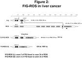
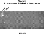
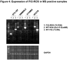

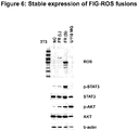
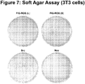
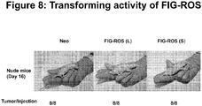
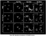
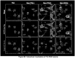
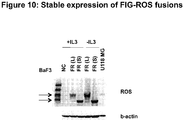

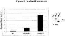


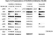
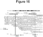


Classifications
-
- A—HUMAN NECESSITIES
- A61—MEDICAL OR VETERINARY SCIENCE; HYGIENE
- A61K—PREPARATIONS FOR MEDICAL, DENTAL OR TOILETRY PURPOSES
- A61K31/00—Medicinal preparations containing organic active ingredients
- A61K31/33—Heterocyclic compounds
- A61K31/395—Heterocyclic compounds having nitrogen as a ring hetero atom, e.g. guanethidine or rifamycins
- A61K31/495—Heterocyclic compounds having nitrogen as a ring hetero atom, e.g. guanethidine or rifamycins having six-membered rings with two or more nitrogen atoms as the only ring heteroatoms, e.g. piperazine or tetrazines
- A61K31/505—Pyrimidines; Hydrogenated pyrimidines, e.g. trimethoprim
- A61K31/506—Pyrimidines; Hydrogenated pyrimidines, e.g. trimethoprim not condensed and containing further heterocyclic rings
-
- A—HUMAN NECESSITIES
- A61—MEDICAL OR VETERINARY SCIENCE; HYGIENE
- A61K—PREPARATIONS FOR MEDICAL, DENTAL OR TOILETRY PURPOSES
- A61K31/00—Medicinal preparations containing organic active ingredients
- A61K31/33—Heterocyclic compounds
- A61K31/395—Heterocyclic compounds having nitrogen as a ring hetero atom, e.g. guanethidine or rifamycins
- A61K31/435—Heterocyclic compounds having nitrogen as a ring hetero atom, e.g. guanethidine or rifamycins having six-membered rings with one nitrogen as the only ring hetero atom
- A61K31/44—Non condensed pyridines; Hydrogenated derivatives thereof
- A61K31/445—Non condensed piperidines, e.g. piperocaine
- A61K31/4523—Non condensed piperidines, e.g. piperocaine containing further heterocyclic ring systems
- A61K31/4545—Non condensed piperidines, e.g. piperocaine containing further heterocyclic ring systems containing a six-membered ring with nitrogen as a ring hetero atom, e.g. pipamperone, anabasine
-
- A—HUMAN NECESSITIES
- A61—MEDICAL OR VETERINARY SCIENCE; HYGIENE
- A61P—SPECIFIC THERAPEUTIC ACTIVITY OF CHEMICAL COMPOUNDS OR MEDICINAL PREPARATIONS
- A61P1/00—Drugs for disorders of the alimentary tract or the digestive system
- A61P1/16—Drugs for disorders of the alimentary tract or the digestive system for liver or gallbladder disorders, e.g. hepatoprotective agents, cholagogues, litholytics
-
- A—HUMAN NECESSITIES
- A61—MEDICAL OR VETERINARY SCIENCE; HYGIENE
- A61P—SPECIFIC THERAPEUTIC ACTIVITY OF CHEMICAL COMPOUNDS OR MEDICINAL PREPARATIONS
- A61P35/00—Antineoplastic agents
-
- A—HUMAN NECESSITIES
- A61—MEDICAL OR VETERINARY SCIENCE; HYGIENE
- A61P—SPECIFIC THERAPEUTIC ACTIVITY OF CHEMICAL COMPOUNDS OR MEDICINAL PREPARATIONS
- A61P43/00—Drugs for specific purposes, not provided for in groups A61P1/00-A61P41/00
-
- C—CHEMISTRY; METALLURGY
- C12—BIOCHEMISTRY; BEER; SPIRITS; WINE; VINEGAR; MICROBIOLOGY; ENZYMOLOGY; MUTATION OR GENETIC ENGINEERING
- C12Q—MEASURING OR TESTING PROCESSES INVOLVING ENZYMES, NUCLEIC ACIDS OR MICROORGANISMS; COMPOSITIONS OR TEST PAPERS THEREFOR; PROCESSES OF PREPARING SUCH COMPOSITIONS; CONDITION-RESPONSIVE CONTROL IN MICROBIOLOGICAL OR ENZYMOLOGICAL PROCESSES
- C12Q1/00—Measuring or testing processes involving enzymes, nucleic acids or microorganisms; Compositions therefor; Processes of preparing such compositions
- C12Q1/68—Measuring or testing processes involving enzymes, nucleic acids or microorganisms; Compositions therefor; Processes of preparing such compositions involving nucleic acids
- C12Q1/6813—Hybridisation assays
- C12Q1/6827—Hybridisation assays for detection of mutation or polymorphism
-
- C—CHEMISTRY; METALLURGY
- C12—BIOCHEMISTRY; BEER; SPIRITS; WINE; VINEGAR; MICROBIOLOGY; ENZYMOLOGY; MUTATION OR GENETIC ENGINEERING
- C12Q—MEASURING OR TESTING PROCESSES INVOLVING ENZYMES, NUCLEIC ACIDS OR MICROORGANISMS; COMPOSITIONS OR TEST PAPERS THEREFOR; PROCESSES OF PREPARING SUCH COMPOSITIONS; CONDITION-RESPONSIVE CONTROL IN MICROBIOLOGICAL OR ENZYMOLOGICAL PROCESSES
- C12Q1/00—Measuring or testing processes involving enzymes, nucleic acids or microorganisms; Compositions therefor; Processes of preparing such compositions
- C12Q1/68—Measuring or testing processes involving enzymes, nucleic acids or microorganisms; Compositions therefor; Processes of preparing such compositions involving nucleic acids
- C12Q1/6844—Nucleic acid amplification reactions
- C12Q1/6858—Allele-specific amplification
-
- C—CHEMISTRY; METALLURGY
- C12—BIOCHEMISTRY; BEER; SPIRITS; WINE; VINEGAR; MICROBIOLOGY; ENZYMOLOGY; MUTATION OR GENETIC ENGINEERING
- C12Q—MEASURING OR TESTING PROCESSES INVOLVING ENZYMES, NUCLEIC ACIDS OR MICROORGANISMS; COMPOSITIONS OR TEST PAPERS THEREFOR; PROCESSES OF PREPARING SUCH COMPOSITIONS; CONDITION-RESPONSIVE CONTROL IN MICROBIOLOGICAL OR ENZYMOLOGICAL PROCESSES
- C12Q1/00—Measuring or testing processes involving enzymes, nucleic acids or microorganisms; Compositions therefor; Processes of preparing such compositions
- C12Q1/68—Measuring or testing processes involving enzymes, nucleic acids or microorganisms; Compositions therefor; Processes of preparing such compositions involving nucleic acids
- C12Q1/6869—Methods for sequencing
-
- C—CHEMISTRY; METALLURGY
- C12—BIOCHEMISTRY; BEER; SPIRITS; WINE; VINEGAR; MICROBIOLOGY; ENZYMOLOGY; MUTATION OR GENETIC ENGINEERING
- C12Q—MEASURING OR TESTING PROCESSES INVOLVING ENZYMES, NUCLEIC ACIDS OR MICROORGANISMS; COMPOSITIONS OR TEST PAPERS THEREFOR; PROCESSES OF PREPARING SUCH COMPOSITIONS; CONDITION-RESPONSIVE CONTROL IN MICROBIOLOGICAL OR ENZYMOLOGICAL PROCESSES
- C12Q1/00—Measuring or testing processes involving enzymes, nucleic acids or microorganisms; Compositions therefor; Processes of preparing such compositions
- C12Q1/68—Measuring or testing processes involving enzymes, nucleic acids or microorganisms; Compositions therefor; Processes of preparing such compositions involving nucleic acids
- C12Q1/6876—Nucleic acid products used in the analysis of nucleic acids, e.g. primers or probes
- C12Q1/6883—Nucleic acid products used in the analysis of nucleic acids, e.g. primers or probes for diseases caused by alterations of genetic material
- C12Q1/6886—Nucleic acid products used in the analysis of nucleic acids, e.g. primers or probes for diseases caused by alterations of genetic material for cancer
-
- G—PHYSICS
- G01—MEASURING; TESTING
- G01N—INVESTIGATING OR ANALYSING MATERIALS BY DETERMINING THEIR CHEMICAL OR PHYSICAL PROPERTIES
- G01N33/00—Investigating or analysing materials by specific methods not covered by groups G01N1/00 - G01N31/00
- G01N33/48—Biological material, e.g. blood, urine; Haemocytometers
- G01N33/50—Chemical analysis of biological material, e.g. blood, urine; Testing involving biospecific ligand binding methods; Immunological testing
- G01N33/53—Immunoassay; Biospecific binding assay; Materials therefor
- G01N33/574—Immunoassay; Biospecific binding assay; Materials therefor for cancer
- G01N33/57407—Specifically defined cancers
- G01N33/57438—Specifically defined cancers of liver, pancreas or kidney
-
- G—PHYSICS
- G01—MEASURING; TESTING
- G01N—INVESTIGATING OR ANALYSING MATERIALS BY DETERMINING THEIR CHEMICAL OR PHYSICAL PROPERTIES
- G01N33/00—Investigating or analysing materials by specific methods not covered by groups G01N1/00 - G01N31/00
- G01N33/48—Biological material, e.g. blood, urine; Haemocytometers
- G01N33/50—Chemical analysis of biological material, e.g. blood, urine; Testing involving biospecific ligand binding methods; Immunological testing
- G01N33/53—Immunoassay; Biospecific binding assay; Materials therefor
- G01N33/574—Immunoassay; Biospecific binding assay; Materials therefor for cancer
- G01N33/57484—Immunoassay; Biospecific binding assay; Materials therefor for cancer involving compounds serving as markers for tumor, cancer, neoplasia, e.g. cellular determinants, receptors, heat shock/stress proteins, A-protein, oligosaccharides, metabolites
-
- C—CHEMISTRY; METALLURGY
- C12—BIOCHEMISTRY; BEER; SPIRITS; WINE; VINEGAR; MICROBIOLOGY; ENZYMOLOGY; MUTATION OR GENETIC ENGINEERING
- C12Q—MEASURING OR TESTING PROCESSES INVOLVING ENZYMES, NUCLEIC ACIDS OR MICROORGANISMS; COMPOSITIONS OR TEST PAPERS THEREFOR; PROCESSES OF PREPARING SUCH COMPOSITIONS; CONDITION-RESPONSIVE CONTROL IN MICROBIOLOGICAL OR ENZYMOLOGICAL PROCESSES
- C12Q2600/00—Oligonucleotides characterized by their use
- C12Q2600/156—Polymorphic or mutational markers
-
- C—CHEMISTRY; METALLURGY
- C12—BIOCHEMISTRY; BEER; SPIRITS; WINE; VINEGAR; MICROBIOLOGY; ENZYMOLOGY; MUTATION OR GENETIC ENGINEERING
- C12Q—MEASURING OR TESTING PROCESSES INVOLVING ENZYMES, NUCLEIC ACIDS OR MICROORGANISMS; COMPOSITIONS OR TEST PAPERS THEREFOR; PROCESSES OF PREPARING SUCH COMPOSITIONS; CONDITION-RESPONSIVE CONTROL IN MICROBIOLOGICAL OR ENZYMOLOGICAL PROCESSES
- C12Q2600/00—Oligonucleotides characterized by their use
- C12Q2600/158—Expression markers
-
- G—PHYSICS
- G01—MEASURING; TESTING
- G01N—INVESTIGATING OR ANALYSING MATERIALS BY DETERMINING THEIR CHEMICAL OR PHYSICAL PROPERTIES
- G01N2333/00—Assays involving biological materials from specific organisms or of a specific nature
- G01N2333/90—Enzymes; Proenzymes
- G01N2333/91—Transferases (2.)
- G01N2333/912—Transferases (2.) transferring phosphorus containing groups, e.g. kinases (2.7)
- G01N2333/91205—Phosphotransferases in general
- G01N2333/9121—Phosphotransferases in general with an alcohol group as acceptor (2.7.1), e.g. general tyrosine, serine or threonine kinases
-
- G—PHYSICS
- G01—MEASURING; TESTING
- G01N—INVESTIGATING OR ANALYSING MATERIALS BY DETERMINING THEIR CHEMICAL OR PHYSICAL PROPERTIES
- G01N2800/00—Detection or diagnosis of diseases
- G01N2800/52—Predicting or monitoring the response to treatment, e.g. for selection of therapy based on assay results in personalised medicine; Prognosis
Definitions
- the invention relates generally to ROS kinase proteins and genes involved in cancer, and to the treatment of cancer.
- BCR-ABL oncoprotein a tyrosine kinase fusion protein
- CML chronic myelogenous leukemia
- the BCR-ABL oncoprotein which is found in at least 90-95% of CML cases, is generated by the translocation of gene sequences from the c-ABL protein tyrosine kinase on chromosome 9 into BCR sequences on chromosome 22, producing the so-called Philadelphia chromosome. See, e.g. Kurzock et al., N. Engl. J. Med. 319: 990-998 (1988 ).
- the translocation is also observed in acute lymphocytic leukemia and AML cases.
- Identifying translocations and mutations in human cancers is highly desirable because it can lead to the development of new therapeutics that target such fusion or mutant proteins, and to new diagnostics for identifying patients that have such gene translocations.
- BCR-ABL has become a target for the development of therapeutics to treat leukemia.
- Gleevec® Imatinib mesylate, STI-571
- This drug is the first of a new class of anti-proliferative agents designed to interfere with the signaling pathways that drive the growth of tumor cells.
- WO2007/084631 discloses a novel gene translocation in human non-small cell lung carcinoma (NSCLC) that results in a fusion protein combining part of SLC34A2 with ROS kinase, and provides polynucleotides and vectors encoding the disclosed mutant ROS kinase polypeptides, probes for detecting it, isolated mutant polypeptides, recombinant polypeptides, and reagents for detecting the fusion and truncated polypeptides.
- NSCLC human non-small cell lung carcinoma
- FIG-ROS fusion proteins namely, FIG-ROS(S), FIG-ROS(L), and FIG-ROS(XL)
- FIG-ROS(S), FIG-ROS(L), and FIG-ROS(XL) retain ROS tyrosine kinase activity.
- the invention provides treatment of human cancers such as liver, kidney, pancreatic, and testicular cancers (including cancers in the ducts of these tissues, such as bile duct liver cancer), which arise not only from gene translocations involving the ROS kinase, but also from aberrant expression of the ROS kinase in these tissues.
- human cancers such as liver, kidney, pancreatic, and testicular cancers (including cancers in the ducts of these tissues, such as bile duct liver cancer), which arise not only from gene translocations involving the ROS kinase, but also from aberrant expression of the ROS kinase in these tissues.
- a truncated ROS kinase whereby the kinase domain (with or without the transmembrane domain) of the ROS kinase is active but separated from the rest of the full-length ROS kinase (e.g., separate from the extracellular domain of the ROS protein).
- ROS kinase with active kinase activity may drive the proliferation and survival of liver, pancreatic, kidney, and testicular cancers in a subset of such cancers in which a truncated ROS kinase with active kinase activity is expressed.
- the FIG-ROS fusion polypeptide comprises the amino acid sequence set forth in SEQ ID NO: 4.
- the FIG-ROS fusion polypeptide comprises the amino acid sequence set forth in SEQ ID NO: 2.
- the FIG-ROS fusion polypeptide comprises the amino acid sequence set forth in SEQ ID NO: 17.
- the FIG-ROS fusion polypeptide is encoded by the nucleic acid sequence set forth in SEQ ID NO: 3.
- the FIG-ROS fusion polypeptide is encoded by the nucleic acid sequence set forth in SEQ ID NO: 1. In some embodiments, the FIG-ROS fusion polypeptide is encoded by the nucleic acid sequence set forth in SEQ ID NO: 16.
- FIG-ROS fusion polynucleotide comprises the nucleotide sequence set forth in SEQ ID NO: 3. In some embodiments, the FIG-ROS fusion polynucleotide comprises the nucleotide sequence set forth in SEQ ID NO: 1. In some embodiments, the FIG-ROS fusion polynucleotide comprises the nucleotide sequence set forth in SEQ ID NO: 16.
- the binding agent specifically binds to a FIG-ROS fusion polypeptide.
- the binding agent specifically binds to a fusion junction between a FIG portion and a ROS portion in said FIG-ROS fusion polypeptide.
- the fusion junction comprises an amino acid sequence selected from the group consisting of AGSTLP, LQVWHR, and LQAGVP.
- the FIG-ROS fusion polypeptide is a FIG-ROS(S) fusion polypeptide, is a FIG-ROS (XL) fusion polypeptide, or is a FIG-ROS (L) fusion polypeptide.
- the binding agent is an antibody and an AQUA peptide.
- the AQUA peptide comprises an amino acid sequence selected from the group consisting of AGSTLP, LQVWHR, and LQAGVP.
- FIG-ROS fusion polynucleotide for detecting a FIG-ROS fusion polynucleotide, wherein said probe hybridizes to said FIG-ROS fusion polynucleotide under stringent conditions.
- the FIG-ROS fusion polynucleotide comprises the nucleotide sequence set forth in SEQ ID NO: 3.
- the FIG-ROS fusion polynucleotide encodes a polypeptide comprising the amino acid sequence set forth in SEQ ID NO: 4.
- the FIG-ROS fusion polynucleotide comprises the nucleotide sequence set forth in SEQ ID NO: 1.
- the FIG-ROS fusion polynucleotide encodes a polypeptide comprising the amino acid sequence set forth in SEQ ID NO: 2 In some embodiments, the FIG-ROS fusion polynucleotide comprises the nucleotide sequence set forth in SEQ ID NO: 16. In some embodiments, the FIG-ROS fusion polynucleotide encodes a polypeptide comprising the amino acid sequence set forth in SEQ ID NO: 17.
- FIG-ROS gene translocation comprising contacting a biological sample with a binding agent that specifically binds to a FIG-ROS fusion polypeptide (e.g., a FIG-ROS(S), FIG-ROS(XL), or a FIG-ROS(L) fusion polypeptide), where specific binding of the binding agent to the biological sample indicates the presence of a FIG-ROS gene translocation (e.g ., that encodes a FIG-ROS(S), FIG-ROS(XL), or FIG-ROS(L) fusion polypeptide) in said biological sample.
- a binding agent that specifically binds to a FIG-ROS fusion polypeptide
- FIG-ROS fusion polypeptide e.g., a FIG-ROS(S), FIG-ROS(XL), or a FIG-ROS(L) fusion polypeptide
- Also disclosed herein is a method for detecting a FIG-ROS gene translocation by contacting a biological sample with a nucleotide probe that hybridizes to a FIG-ROS fusion polynucleotide under stringent conditions, wherein hybridization of said nucleotide probe to said biological sample indicates a FIG-ROS gene translocation (e.g ., that encodes a FIG-ROS(S), FIG-ROS(XL) or FIG-ROS(L) fusion polypeptide) in said biological sample.
- a FIG-ROS gene translocation e.g ., that encodes a FIG-ROS(S), FIG-ROS(XL) or FIG-ROS(L) fusion polypeptide
- Also disclosed herein is a method for diagnosing a patient as having a cancer or a suspected cancer characterized by a ROS kinase.
- the cancer or suspected cancer is not non-small cell lung carcinoma or glioblastoma.
- the method includes contacting a biological sample of said cancer or suspected cancer (where the biological sample comprising at least one polypeptide) with a binding agent that specifically binds to a mutant ROS polypeptide, wherein specific binding of said binding agent to at least one polypeptide in said biological sample identifies said patient as having a cancer or a suspected cancer characterized by a ROS kinase.
- Also disclosed herein is a method for identifying a cancer (or a suspected cancer) that is likely to respond to a ROS inhibitor.
- the cancer or suspected cancer is not non-small cell lung carcinoma or glioblastoma.
- the method includes contacting a biological sample of said cancer (or suspected cancer) comprising at least one polypeptide, with a binding agent that specifically binds to a mutant ROS polypeptide, wherein specific binding of said binding agent to at least one polypeptide in said biological sample identifies said cancer or suspected cancer as a cancer or suspected cancer that is likely to respond to a ROS inhibitor.
- the mutant ROS polypeptide is aberrantly expressed wild-type ROS polypeptide.
- aberrant expression can be where wild-type ROS kinase is overexpressed in a cancer or a suspected cancer as compared to the level of expression of wild-type ROS kinase in normal tissue of the same tissue type as the cancer or suspected cancer.
- ROS protein expression levels can be determined by standard means (e.g., Western blotting analysis, mass spectrometry, IHC staining).
- the mutant ROS polypeptide is a truncated ROS polypeptide or a ROS fusion polypeptide.
- ROS fusion polypeptides include a FIG-ROS(S) fusion polypeptide, a FIG-ROS(L) fusion polypeptide, a FIG-ROS(XL) fusion polypeptide, a SLC34A2-ROS(S) fusion polypeptide, a SLC34A2-ROS(L) fusion polypeptide, a SLC34A2-ROS(VS) fusion polypeptide, and a CD74-ROS fusion polypeptide.
- Non-limiting examples of a truncated ROS polypeptide include the kinase domain of ROS lacking the extracellular and transmembrane domains of wild-type ROS and the transmembrane and kinase domains of ROS lacking the extracellular domain of wild-type ROS.
- the binding agent is an antibody or an AQUA peptide.
- the cancer is from a patient (e.g., a human patient).
- Also disclosed herein is a method for diagnosing a patient as having a cancer or a suspected cancer characterized by a ROS kinase.
- the cancer or suspected cancer is not non-small cell lung carcinoma or glioblastoma.
- the method includes contacting a biological sample of said cancer or a suspected cancer (where the biological sample comprising at least one nucleic acid molecule) with a probe that hybridizes under stringent conditions to a nucleic acid molecule selected from the group consisting of a FIG-ROS fusion polynucleotide, a SLC34A2-ROS fusion polypeptide, a CD74-ROS fusion polypeptide, and a truncated ROS polynucleotide, and wherein hybridization of said probe to at least one nucleic acid molecule in said biological sample identifies said patient as having a cancer or a suspected cancer characterized by a ROS kinase.
- Also disclosed herein is another method for identifying a cancer (or suspected cancer) that is likely to respond to a ROS inhibitor.
- the method includes contacting a biological sample of said cancer comprising at least one nucleic acid molecule with a nucleotide probe that hybridizes under stringent conditions to a either a FIG-ROS fusion polynucleotide (e.g ., a FIG-ROS(S) or FIG-ROS(L) fusion polynucleotide) or a mutant ROS polynucleotide, and wherein hybridization of said nucleotide probe to at least one nucleic acid molecule in said biological sample identifies said cancer as a cancer that is likely to respond to a ROS inhibitor.
- a FIG-ROS fusion polynucleotide e.g ., a FIG-ROS(S) or FIG-ROS(L) fusion polynucleotide
- a mutant ROS polynucleotide e.
- the FIG-ROS fusion polynucleotide encodes a FIG-ROS(S) fusion polypeptide, a FIG-ROS(L) fusion polypeptide, or a FIG-ROS(XL) fusion polypeptide.
- the SCL34A2-ROS fusion polynucleotide encodes a SCL34A2-ROS(S) fusion polypeptide, a SCL34A2-ROS(L) fusion polypeptide, or a SCL34A2-ROS(VS) fusion polypeptide.
- the cancer is from a patient (e.g., a cancer patient). In some embodiments, the patient is human.
- the cancer may be a liver cancer, a pancreatic cancer, a kidney cancer, or a testicular cancer.
- the cancer may be a duct cancer (e.g., a liver bile duct cancer or a pancreatic duct cancer).
- the cancer is not a non-small cell lung cancer (NSCLC).
- the cancer is not a glioblastoma.
- the ROS inhibitor also inhibits the activity of an ALK kinase an LTK kinase, an insulin receptor, or an IGF1 receptor.
- the ROS inhibitor is PF-02341066 or NVP-TAE684.
- the ROS inhibitor is a binding agent that specifically binds to a FIG-ROS fusion polypeptide, a binding agent that specifically binds to a truncated ROS polypeptide, an siRNA targeting a FIG-ROS fusion polynucleotide, or an siRNA targeting a truncated ROS polynucleotide.
- mutant ROS kinase which is expressed in a subset of human liver, kidney, pancreatic, and testicular cancers (e.g ., bile duct liver cancer).
- the mutant ROS kinase may drive the proliferation and survival of liver, pancreatic, kidney, and testicular cancers in a subset of such cancers in which the mutant ROS kinase is expressed.
- Standard reference works setting forth the general principles of recombinant DNA technology include Sambrook et al., Molecular Cloning: A Laboratory Manual, 2nd Ed., Cold Spring Harbor Laboratory Press, New York (1989 ); Kaufman et al., Eds., Handbook of Molecular and Cellular Methods in Biology in Medicine, CRC Press, Boca Raton (1995 ); McPherson, Ed., Directed Mutagenesis: A Practical Approach, IRL Press, Oxford (1991 ). Standard reference works setting forth the general principles of pharmacology include Goodman and Gilman's The Pharmacological Basis of Therapeutics, 11th Ed., McGraw Hill Companies Inc., New York (2006 ).
- the invention relates to the discovery of mutant ROS (i.e., aberrantly expressed full length ROS, truncated (i.e., less than full length) ROS, or ROS fusion proteins (e.g., the FIG-ROS fusions, the SLC34A2-ROS fusions, or the CD74-ROS fusion)) in liver cancer (including bile duct cancer), pancreatic cancer, kidney cancer, and testicular cancer.
- the invention further relates to the discovery of new ROS gene translocations, resulting in fusions between the FIG gene and the ROS gene.
- ROS kinase Full length (wild-type) ROS kinase is a 2347 amino acid long receptor tyrosine kinase.
- ROS kinase RNA has been detected in placenta, lung and skeletal muscle, with possible low levels of expression in testes (see J. Acquaviva, et al., Biochim. Biophys. Acta 1795(1):37-52, 2009 .
- full-length ROS kinase does not appear to be expressed in normal liver, kidney, and pancreas tissue in humans (see J. Acquaviva, et al., supra). While Abcam Inc.
- ROS-specific antibody (clone ab5512) that allegedly stains (i.e., specifically binds to) human hepatocarcinoma tissue by IHC, this ab5512 was found to stain paraffin-embedded HCC78 cells (lung carcinoma which express ROS) and HCC827 cells (lung adenocarcinoma which do not express ROS) with equal intensity (cells obtained from the ATCC, data not shown). Additionally, although ROS kinase may be present in human testicular tissue, its expression appears to be limited to the epididymis (see Acquaviva, et al.,supra).
- FIG-ROS fusion polypeptide is meant the FIG-ROS fusion polypeptide ( e.g ., FIG-ROS(L), FIG-ROS(XL), or FIG-ROS (S)) described herein, obtained from any species, particularly mammalian, including bovine, ovine, porcine, murine, equine, and human, from any source whether natural, synthetic, semi-synthetic, or recombinant.
- purified refers to a nucleic acid sequence (e.g ., a polynucleotide) or an amino acid sequence (e.g ., a polypeptide) that is removed or separated from other components present in its natural environment.
- an isolated FIG-ROS fusion polypeptide is one that is separated from other components of a eukaryotic cell (e.g., the endoplasmic reticulum or cytoplasmic proteins and RNA).
- An isolated FIG-ROS polynucleotide is one that is separated from other nuclear components (e.g ., histones) and/or from upstream or downstream nucleic acid sequences (e.g ., an isolated FIG-ROS polynucleotide is separated from the endogenous FIG gene promoter).
- An isolated nucleic acid sequence of amino acid sequence disclosed herein is at least 60% free, or at least 75% free, or at least 90% free, or at least 95% free from other components present in natural environment of the indicated nucleic acid sequence or acid sequence.
- FIG-ROS fusion polypeptide disclosed herein is a non-limiting example of mutant ROS polypeptide.
- mutant ROS polypeptide or polynucleotide means either the aberrant expression of the wild-type ROS kinase polypeptide or polynucleotide in a tissue in which ROS kinase is not normally expressed (or expressed at a different level) or the kinase domain of a ROS or a polynucleotide encoding the kinase domain of a ROS kinase without the extracellular domain or without the transmembrane domains of wild-type (i.
- ROS full length ROS
- the kinase domain (with or without the transmembrane domain) is either alone (also referred to as truncated ROS) or is fused to all or a portion of a second protein (e.g., a FIG protein).
- Wild-type ROS kinase is a 2347 amino acid long receptor tyrosine kinase, where approximately the first 36 amino acids (i. e., the N-terminal 36 amino acids) are the signal peptide.
- the sequence of human ROS kinase can be found at GenBank Accession No. M34353, and the protein sequence (including the signal peptide) is provided herein as SEQ ID NO: 9.
- mutant ROS polypeptide disclosed herein include polypeptides comprising the amino acid sequences set forth in SEQ ID NO: 12 or SEQ ID NO: 13.
- mutant ROS polynucleotides disclosed herein include polynucleotides encoding polypeptides comprising the amino acid sequences set forth in SEQ ID NO: 12 or SEQ ID NO: 13.
- the mutant ROS polynucleotide comprises a portion of the nucleotide sequence set forth in SEQ ID NO: 5, SEQ ID NO: 6, SEQ ID NO:7, or SEQ ID NO: 8.
- mutant ROS polypeptide disclosed herein does not include the sequences of SEQ ID NO: 10 or SEQ ID NO: 11.
- non-limiting examples of mutant ROS polynucleotides disclosed herein do not include polynucleotides encoding polypeptides comprising the amino acid sequences set forth in SEQ ID NO: 10 or SEQ ID NO: 11.
- a mutant ROS comprises the kinase domain, with or without the transmembrane domain, of ROS (or nucleotide sequences encoding the same) such that the kinase domain of the ROS kinase (with or without the transmembrane domain) is separated from the other domains (e.g., the extracellular domain) of wild-type (i.e., full-length) ROS kinase.
- the full length amino acid sequence of ROS kinase is provided in SEQ ID NO: 9.
- the kinase domain of the ROS kinase is provided in SEQ ID NOs: 12 and 13; however the term "mutant ROS" includes also those amino acid residues which flank the kinase domain provided that the flanking amino acid residues are not within the transmembrane domain or extracellular domain of the full-length ROS protein.
- the mutant ROS excludes the sequence set forth in SEQ ID NO: 11.
- the mutant ROS excludes the sequence set forth in SEQ ID NO: 10.
- the mutant ROS described herein includes the amino acid sequence set forth in SEQ ID NO: 3 and a nucleotide sequence encoding the same.
- mutant ROS polypeptide also includes a chimeric protein that includes all or part of a second protein fused by a peptide bond to the kinase domain of a ROS polypeptide.
- mutant ROS polypeptide that is a chimeric protein is the FIG-ROS(S) fusion polypeptide described herein.
- mutant ROS polynucleotide also includes a polynucleotide encoding a chimeric protein that includes all or part of a second protein fused by a peptide bond to the kinase domain of a ROS polypeptide.
- mutant ROS includes, without limitation, the FIG-ROS (L) fusion polypeptide (see nucleic acid sequence in SEQ ID NO: 1 and amino acid sequence in SEQ ID NO: 2), the FIG-ROS (S) fusion polypeptide (see nucleic acid sequence in SEQ ID NO: 3 and amino acid sequence in SEQ ID NO: 4), the FIG-ROS(XL) fusion polypeptide (see nucleic acid sequence in SEQ ID NO: 16 and amino acid sequence in SEQ ID NO: 17), the SLC34A2-ROS (L) fusion polypeptide (see nucleic acid sequence in SEQ ID NO: 18 and amino acid sequence in SEQ ID NO: 19), the SLC34A2-ROS (S) fusion protein (see nucleic acid sequence in SEQ ID NO: 20 and amino acid sequence in SEQ ID NO: 21), the SLC34A2-ROS (VS) fusion protein (see nucleic acid sequence in SEQ ID NO: 22 and amino acid sequence in SEQ ID NO:
- ROS fusion polypeptides are disclosed in PCT Publication No. WO2007084631 ; Rikova, K et al., Cell 131:1190-1203, 2007 , and PCT Publication No. WO/2009/051846 .
- polynucleotide refers to an oligonucleotide, nucleotide, or polynucleotide, and fragments or portions thereof, and to DNA or RNA of genomic or synthetic origin, which may be single- or double-stranded, and represent the sense or anti-sense strand.
- polypeptide refers to an oligopeptide, peptide, polypeptide, or protein sequence, and fragments or portions thereof, and to naturally occurring or synthetic molecules.
- amino acid sequence and like terms, such as “polypeptide” or “protein”, are not meant to limit the indicated amino acid sequence to the complete, native amino acid sequence associated with the recited protein molecule.
- FIG-ROS gene translocation have been identified using global phosphopeptide profiling in liver cancer samples taken from human patients ( see Examples below). These gene translocations which occurs on human chromosome (6q22) result in expression of two variant fusion proteins, namely the FIG-ROS(S) fusion polypeptide and the FIG-ROS(L) fusion polypeptide) that combine the N-terminus of FIG with the kinase domain of ROS.
- cancer or “cancerous” is meant a cell that shows abnormal growth as compared to a normal (i.e., non-cancerous) cell of the same cell type.
- a cancerous cell may be metastatic or non-metastatic.
- a cancerous cell may also show lack of contact inhibition where a normal cell of that same cell type shows contact inhibition.
- tissue suspected of being cancerous is meant a cell or tissue that has some aberrant characteristics (e.g., hyperplastic or lack of contact inhibition) as compared to normal cells or tissues of that same cell or tissue type as the suspected cancer, but where the cell or tissue is not yet confirmed by a physician or pathologist as being cancerous.
- the FIG-ROS(L) translocation combines the nucleic acid sequence encoding the N-terminus of FIG (amino acids 1-412) with the nucleic acid sequences encoding the kinase domain of ROS (amino acids 413-878 which correspond to amino acids 1882-2347 from ROS) (see SEQ ID NO: 2), to produce a fusion, namely FIG-ROS(L) fusion polypeptide.
- the resulting FIG-ROS(L) fusion protein which comprises 878 amino acids, was found to retain the kinase activity of ROS.
- the FIG-ROS fusion polypeptide is a FIG-ROS(L) fusion polypeptide.
- the FIG-ROS (L) fusion polypeptide comprises the amino acid sequence set forth in SEQ ID NO: 2. In some embodiments, the FIG-ROS (L) fusion polypeptide is encoded by the nucleic acid sequence set forth in SEQ ID NO: 1.
- the FIG-ROS(S) translocation combines the nucleic acid sequence encoding the N-terminus of FIG (amino acids 1-209) with the nucleic acid sequence encoding the kinase domain of ROS (amino acids 210-630 which correspond to amino acids 1927-2347 from ROS) ( see also SEQ ID NO:4), to produce a fusion, namely the FIG-ROS(S) fusion polypeptide.
- the resulting FIG-ROS(S) fusion protein which comprises 630 amino acids, was found to retain the kinase activity of ROS.
- the FIG-ROS fusion polypeptide disclosed herein is a FIG-ROS(S) fusion polypeptide.
- the FIG-ROS(S) fusion polypeptide comprises the amino acid sequence set forth in SEQ ID NO: 4.
- the FIG-ROS(S) fusion polypeptide is encoded by the nucleic acid sequence set forth in SEQ ID NO: 3.
- FIG-ROS(XL) also disclosed herein is a third FIG-ROS fusion, namely FIG-ROS(XL), which translocation combines the nucleic acid sequence encoding the N-terminus of FIG (amino acids 1-411 or 1-412) with the nucleic acid sequences encoding the transmembrane and kinase domains of ROS kinase to result in a fusion protein of 1009 amino acids in length.
- the amino acid at the fusion junction may appear in either wild-type protein member of the fusion (e.g., the amino acid at the fusion junction in a FIG-ROS fusion polypeptide may appear in either wild-type FIG protein or wild-type ROS protein), or the amino acid, being created by a codon with nucleotides from fused exons of both protein members, may be unique to the fusion polypeptide and not appear in either wild-type protein member of the fusion.
- mutant ROS may be present in liver cancer (including bile duct cancer), kidney cancer, testicular cancer, and pancreatic cancer. Based on these discoveries, patients suffering from these cancers whose cancers express mutant ROS (e.g., over-express wild-type ROS or express a truncated ROS or a ROS fusion polypeptide such as one of the FIG-ROS fusion polypeptides disclosed herein) may respond favorably to administration of a ROS inhibitor (e.g ., the growth of their cancer may slow or stop as compared to untreated patients suffering from the same cancer).
- a ROS inhibitor e.g ., the growth of their cancer may slow or stop as compared to untreated patients suffering from the same cancer.
- FIG-ROS fusion polypeptides and fragments thereof.
- the isolated polypeptide can comprise an amino acid sequence at least 95% identical or at least 99% identical to a sequence selected from the group consisting of: (a) an amino acid sequence encoding a FIG-ROS fusion polypeptide comprising the amino acid sequence of SEQ ID NO: 1; (b) an amino acid sequence encoding a FIG-ROS fusion polypeptide comprising the amino acid sequence of SEQ ID NO: 17; (c) an amino acid sequence encoding a FIG-ROS fusion polypeptide comprising all or a portion of the FIG polypeptide with the kinase domain of ROS ( e.g., SEQ ID NO: 12 or 13)); and (d) an amino acid sequence encoding a polypeptide comprising at least six contiguous amino acids encompassing the fusion junction of a FIG-ROS fusion polypeptide (e.g ., AGSTLP of FIG-ROS (S), LQVW
- FIG-ROS (S) fusion polypeptide having the amino acid sequence set forth in SEQ ID NO: 4.
- the isolated FIG-ROS (XL) fusion polypeptide can have the amino acid sequence set forth in SEQ ID NO: 17.
- recombinant mutant polypeptides disclosed herein are provided, which may be produced using a recombinant vector or recombinant host cell as described above.
- FIG-ROS variant of a FIG-ROS fusion polypeptide that shows substantial ROS kinase activity or that includes regions of FIG and ROS proteins.
- a FIG-ROS variant disclosed herein contains conservative substitutions as compared to FIG-ROS(L), FIG-ROS (XL), or FIG-ROS(S).
- Some non-limiting conservative substitutions include the exchange, one for another, among the aliphatic amino acids Ala, Val, Leu and Ile; exchange of the hydroxyl residues Ser and Thr; exchange of the acidic residues Asp and Glu; exchange of the amide residues Asn and Gln; exchange of the basic residues Lys and Arg; and exchange of the aromatic residues Phe and Tyr.
- Aromatic phenylalanine tryptophan tyrosine (e.g., a tryptophan residue is replaced with a phenylalanine); Hydrophobic: leucine isoleucine valine; Polar: glutamine asparagines; Basic: arginine lysine histidine; Acidic: aspartic acid glutamic acid; Small: alanine serine threonine methionine glycine.
- Aromatic phenylalanine tryptophan tyrosine (e.g., a tryptophan residue is replaced with a phenylalanine); Hydrophobic: leucine isoleucine valine; Polar: glutamine asparagines; Basic: arginine lysine histidine; Acidic: aspartic acid glutamic acid; Small: alanine serine threonine methionine glycine.
- further guidance concerning which amino acid changes are likely to be phenotypically silent i.e.
- a variant may have "nonconservative" changes, e.g ., replacement of a glycine with a tryptophan. Similar variants may also include amino acid deletions or insertions, or both. Guidance in determining which amino acid residues may be substituted, inserted, or deleted without abolishing biological or immunological activity may be found using computer programs well known in the art, for example, DNASTAR software.
- FIG-ROS fusion polypeptides, fragments thereof, and variants thereof disclosed herein may be provided in an isolated or purified form.
- a recombinantly produced version of a FIG-ROS fusion polypeptide disclosed herein can be substantially purified by the one-step method described in Smith and Johnson, Gene 67: 31-40 (1988 ).
- polypeptides disclosed herein include the FIG-ROS fusion polypeptides having the sequences set forth in SEQ ID NOs: 2 and 4, and 17 (whether or not including a leader sequence), an amino acid sequence encoding a polypeptide comprising at least six contiguous amino acids encompassing the fusion junction of a FIG-ROS fusion polypeptide disclosed herein, as well as polypeptides that have at least 90% similarity, more preferably at least 95% similarity, and still more preferably at least 96%, 97%, 98% or 99% similarity to those described above.
- % similarity for two polypeptides is intended a similarity score produced by comparing the amino acid sequences of the two polypeptides using the Bestfit program (Wisconsin Sequence Analysis Package, Version 8 for Unix, Genetics Computer Group, University Research Park, 575 Science Drive, Madison, Wis. 53711) and the default settings for determining similarity. Bestfit uses the local homology algorithm of Smith and Waterman (Advances in Applied Mathematics 2: 482-489 (1981 )) to find the best segment of similarity between two sequences.
- polypeptide having an amino acid sequence at least, for example, 95% "identical" to a reference amino acid sequence of a mutant ROS polypeptide disclosed herein is intended that the amino acid sequence of the polypeptide is identical to the reference sequence except that the polypeptide sequence may include up to five amino acid alterations per each 100 amino acids of the reference amino acid sequence of the FIG-ROS fusion polypeptide.
- up to 5% of the amino acid residues in the reference sequence may be deleted or substituted with another amino acid, or a number of amino acids up to 5% of the total amino acid residues in the reference sequence may be inserted into the reference sequence.
- These alterations of the reference sequence may occur at the amino or carboxy terminal positions of the reference amino acid sequence or anywhere between those terminal positions, interspersed either individually among residues in the reference sequence or in one or more contiguous groups within the reference sequence.
- the parameters are set, of course, such that the percentage of identity is calculated over the full length of the reference amino acid sequence and that gaps in homology of up to 5% of the total number of amino acid residues in the reference sequence are allowed.
- FIG-ROS fusion polypeptide disclosed herein could be used as a molecular weight marker on SDS-PAGE gels or on molecular sieve gel filtration columns, for example, using methods well known to those of skill in the art.
- polypeptides disclosed herein can also be used to generate fusion polypeptide specific reagents, such as polyclonal and monoclonal antibodies, which are useful in assays for detecting mutant ROS polypeptide expression as described below or as agonists and antagonists capable of enhancing or inhibiting mutant ROS protein function/activity.
- fusion polypeptide specific reagents such as polyclonal and monoclonal antibodies
- polypeptides can be used in the yeast two-hybrid system to "capture" FIG-ROS fusion polypeptide binding proteins, which are also candidate agonist and antagonist disclosed herein.
- yeast two hybrid system is described in Fields and Song, Nature 340: 245-246 (1989 ).
- a peptide or polypeptide comprising an epitope-bearing portion of a polypeptide disclosed herein, such as an epitope comprising the fusion junction of a FIG-ROS fusion polypeptide variant
- An "epitope" refers to either an immunogenic epitope (i.e., capable of eliciting an immune response) or an antigenic epitope ( i. e., the region of a protein molecule to which an antibody can specifically bind.
- the number of immunogenic epitopes of a protein generally is less than the number of antigenic epitopes. See, for instance, Geysen et al., Proc. Natl. Acad. Sci. USA 81:3998-4002 (1983 ).
- the production of FIG-ROS fusion polypeptide-specific antibodies disclosed herein is described in further detail below.
- the antibodies that specifically bind to an epitope-bearing peptides or polypeptides are useful to detect a mimicked protein, and antibodies to different peptides may be used for tracking the fate of various regions of a protein precursor which undergoes post-translational processing.
- the peptides and anti-peptide antibodies may be used in a variety of qualitative or quantitative assays for the mimicked protein, for instance in competition assays since it has been shown that even short peptides ( e.g ., about 9 amino acids) can bind and displace the larger peptides in immunoprecipitation assays. See, for instance, Wilson et al., Cell 37: 767-778 (1984 ) at 777.
- the anti-peptide antibodies disclosed herein also are useful for purification of the mimicked protein, for instance, by adsorption chromatography using methods well known in the art. Immunological assay formats are described in further detail below.
- Recombinant mutant ROS kinase polypeptides are also disclosed herein, and may be producing using fusion polynucleotides disclosed herein, as described above.
- a method for producing a recombinant FIG-ROS fusion polypeptide by culturing a recombinant host cell (as described above) under conditions suitable for the expression of the fusion polypeptide and recovering the polypeptide.
- Culture conditions suitable for the growth of host cells and the expression of recombinant polypeptides from such cells are well known to those of skill in the art.
- FIG-ROS fusion polynucleotide or “FIG-ROS polynucleotide” is meant a FIG-ROS translocation gene ( i.e., a gene that has undergone translocation) or polynucleotide encoding a FIG-ROS fusion polypeptide (e.g., the FIG-ROS(L), FIG-ROS (XL), or FIG-ROS (S)) fusion polypeptides described herein), obtained from any species, particularly mammalian, including bovine, ovine, porcine, murine, equine, and human, from any source whether natural, synthetic, semi-synthetic, or recombinant.
- FIG-ROS fusion polynucleotide or “FIG-ROS polynucleotide” is meant a FIG-ROS translocation gene (i.e., a gene that has undergone translocation) or polynucleotide encoding a FIG-ROS fusion polypeptide (
- the FIG-ROS fusion polynucleotide comprises the nucleotide sequence set forth in SEQ ID NO:1. In some embodiments, the FIG-ROS fusion polynucleotide encodes a polypeptide having the amino acid sequence set forth in SEQ ID NO: 2. In some embodiments, the FIG-ROS fusion polynucleotide comprises the nucleotide sequence set forth in SEQ ID NO:3. In some embodiments, the FIG-ROS fusion polynucleotide encodes a polypeptide having the amino acid sequence set forth in SEQ ID NO: 4. In some embodiments, the FIG-ROS fusion polynucleotide comprises the nucleotide sequence set forth in SEQ ID NO:16. In some embodiments, the FIG-ROS fusion polynucleotide encodes a polypeptide having the amino acid sequence set forth in SEQ ID NO: 17.
- the FIG-ROS fusion polynucleotide comprises a portion of the nucleotide sequence set forth in SEQ ID NO: 5, SEQ ID NO: 6, SEQ ID NO:7, SEQ ID NO: 8, SEQ ID NO: 14, SEQ ID NO: 15, or SEQ ID NO: 26.
- a "portion" or “fragment” means a sequence fragment less than the whole sequence ( e.g ., a 50 nucleotide sequence is a portion of a 100 nucleotide long sequence).
- the FIG-ROS fusion polynucleotide may comprise portions of intron sequences that do not encode any amino acids in the resulting FIG-ROS fusion polypeptide.
- isolated polynucleotides that encode a FIG-ROS fusion polypeptide disclosed herein, nucleotide probes that hybridize to such polynucleotides, and methods, vectors, and host cells for utilizing such polynucleotides to produce recombinant fusion polypeptides.
- all nucleotide sequences determined by sequencing a DNA molecule herein were determined using an automated DNA sequencer (such as the Model 373 from Applied Biosystems, Inc.), and all amino acid sequences of polypeptides encoded by DNA molecules determined herein were determined using an automated peptide sequencer.
- any nucleotide sequence determined herein may contain some errors.
- Nucleotide sequences determined by automation are typically at least about 90% identical, and more typically at least about 95% to about 99.9% identical to the actual nucleotide sequence of the sequenced DNA molecule.
- the actual sequence can be more precisely determined by other approaches including manual DNA sequencing methods well known in the art.
- each nucleotide sequence set forth herein is presented as a sequence of deoxyribonucleotides (abbreviated A, G, C and T).
- nucleotide sequence of a nucleic acid molecule or polynucleotide is intended, for a DNA molecule or polynucleotide, a sequence of deoxyribonucleotides, and for an RNA molecule or polynucleotide, the corresponding sequence of ribonucleotides (A, G, C and U), where each thymidine deoxyribonucleotide (T) in the specified deoxyribonucleotide sequence is replaced by the ribonucleotide uridine (U).
- RNA molecule having the sequence of SEQ ID NO: 3 or set forth using deoxyribonucleotide abbreviations is intended to indicate an RNA molecule having a sequence in which each deoxyribonucleotide A, G or C of SEQ ID NO: 3 has been replaced by the corresponding ribonucleotide A, G or C, and each deoxyribonucleotide T has been replaced by a ribonucleotide U.
- an isolated polynucleotide comprising a nucleotide sequence at least about 95% identical to a sequence selected from the group consisting of: (a) a nucleotide sequence encoding a FIG-ROS fusion polypeptide comprising the amino acid sequence of SEQ ID NO: 4 (FIG-ROS(S)); (b) a nucleotide sequence encoding a FIG-ROS fusion polypeptide comprising the amino acid sequence of SEQ ID NO: 17 (FIG-ROS(XL)); (c) a nucleotide sequence comprising at least six contiguous nucleotides encompassing the fusion junction of a FIG-ROS(S) fusion polynucleotide (e.g ., AAGTAC), a nucleotide sequence comprising at least six contiguous nucleotides encompassing the fusion junction of a FIG-ROS(XL) fusion polynucleotide (e.g .
- a nucleic acid molecule disclosed herein encoding a FIG-ROS fusion polypeptide disclosed herein may be obtained using standard cloning and screening procedures, such as those for cloning cDNAs using mRNA as starting material.
- the fusion gene can also be identified in cDNA libraries in other human cancers in which the FIG-ROS translocation occurs, or in which a deletion or alternative translocation results in expression of a truncated ROS kinase lacking the extracellular domain and may additionally lack the transmembrane domain of the wild type ROS kinase.
- the determined nucleotide sequence of the FIG-ROS translocation genes encode the FIG-ROS(S) fusion polypeptide, the FIG-ROS(L) fusion polypeptide, and the FIG-ROS(XL) fusion polypeptide.
- the FIG-ROS fusion polynucleotides comprise the portion of the nucleotide sequence of wild type FIG that encodes the N-terminus of that protein with the portion of the nucleotide sequence of wild type ROS that encodes the kinase domain of that protein
- FIG-ROS fusion proteins The mature form of the FIG-ROS fusion proteins is also described herein.
- proteins secreted by mammalian cells have a signal or secretory leader sequence which is cleaved from the mature protein once export of the growing protein chain across the rough endoplasmic reticulum has been initiated.
- Most mammalian cells and even insect cells cleave secreted proteins with the same specificity.
- cleavage of a secreted protein is not entirely uniform, which results in two or more mature species on the protein.
- the cleavage specificity of a secreted protein is ultimately determined by the primary structure of the complete protein, that is, it is inherent in the amino acid sequence of the polypeptide.
- nucleotide sequences encoding a mature FIG-ROS(S) fusion polypeptide having the nucleotide sequence set forth in SEQ ID NO: 3 with additional nucleic acid residues located 5' to the 5'-terminal residues of SEQ ID NO. 3 and includes the amino acid sequence of a FIG-ROS(S) fusion polypeptide having the amino acid sequence set forth in SEQ ID NO: 4 with additional amino acid residues located N-terminally to the N-terminal residue of SEQ ID NO. 4.
- nucleotide sequences encoding a mature FIG-ROS(XL) fusion polypeptide having the nucleotide sequence set forth in SEQ ID NO: 16 with additional nucleic acid residues located 5' to the 5'-terminal residues of SEQ ID NO. 16 and includes the amino acid sequence of a FIG-ROS(XL) fusion polypeptide having the amino acid sequence set forth in SEQ ID NO: 17 with additional amino acid residues located N-terminally to the N-terminal residue of SEQ ID NO. 17.
- polynucleotides disclosed herein may be in the form of RNA, such as mRNA, or in the form of DNA, including, for instance, cDNA and genomic DNA obtained by cloning or produced synthetically.
- the DNA may be double-stranded or single-stranded.
- Single-stranded DNA or RNA may be the coding strand, also known as the sense strand, or it may be the non-coding strand, also referred to as the anti-sense strand.
- Isolated polynucleotides disclosed herein are nucleic acid molecules, DNA or RNA, which have been removed from their native environment.
- recombinant DNA molecules contained in a vector are considered isolated for the purposes of the present disclosure.
- Further examples of isolated DNA molecules include recombinant DNA molecules maintained in heterologous host cells or purified (partially or substantially) DNA molecules in solution.
- Isolated RNA molecules include in vivo or in vitro RNA transcripts of the DNA molecules disclosed herein.
- Isolated nucleic acid molecules disclosed herein further include such molecules produced synthetically.
- Isolated polynucleotides disclosed herein include the nucleic acid molecules having the sequences set forth in (SEQ ID NOs: 1, 3, and 16, nucleic acid molecules comprising the coding sequence for the FIG-ROS(S), FIG-ROS(L), and FIG-ROS(XL) fusion proteins that comprise a sequence different from those described above but which, due to the degeneracy of the genetic code, still a mutant ROS polypeptide disclosed herein.
- the genetic code is well known in the art, thus, it would be routine for one skilled in the art to generate such degenerate variants.
- FIG-ROS fusion polypeptide comprising the FIG-ROS translocation nucleotide sequence contained in the above-described cDNA clones.
- nucleic acid molecule will encode the mature FIG-ROS (S) fusion polypeptide, the mature FIG-ROS(L) fusion polypeptide, or the mature FIG-ROS(XL) fusion polypeptide.
- an isolated nucleotide sequence encoding a FIG-ROS fusion polypeptide comprising the N-terminal amino acid sequence of FIG and the kinase domain of ROS.
- the polypeptide comprising the kinase domain of ROS comprises the animo acid sequence set forth in SEQ ID NO: 12 or SEQ ID NO: 13.
- the N-terminal amino acid sequence of FIG and kinase domain of ROS are encoded by the nucleotide sequences set forth in SEQ ID NO: 1, SEQ ID NO: 3, or SEQ ID NO: 16.
- isolated polynucleotides comprising nucleotide sequences having a sequence complementary to one of the mutant ROS polypeptides disclosed herein.
- isolated molecules particularly DNA molecules, are useful as probes for gene mapping, by in situ hybridization with chromosomes, and for detecting expression of the FIG-ROS fusion protein or truncated ROS kinase polypeptide in human tissue, for instance, by Northern blot analysis.
- fragments of the isolated nucleic acid molecules described herein are fragments of the isolated nucleic acid molecules described herein.
- a fragment of an isolated FIG-ROS polynucleotide or truncated ROS polynucleotide disclosed herein is intended fragments at least about 15 nucleotides, or at least about 20 nucleotides, still more preferably at least about 30 nucleotides, or at least about 40 nucleotides in length, which are useful as diagnostic probes and primers as discussed herein.
- fragments of about 50-1500 nucleotides in length are also useful according to the present disclosure, as are fragments corresponding to most, if not all, of the FIG-ROS nucleotide sequence of the cDNAs having sequences set forth in SEQ ID NOs: 1, 3, or 16.
- a fragment at least 20 nucleotides in length is meant fragments that include 20 or more contiguous bases from the respective nucleotide sequences from which the fragments are derived.
- DNA fragments are routine to the skilled artisan, and may be accomplished, by way of example, by restriction endonuclease cleavage or shearing by sonication of DNA obtainable from the cDNA clone described herein or synthesized according to the sequence disclosed herein. Alternatively, such fragments can be directly generated synthetically.
- nucleotide probe e.g ., a nucleotide probe
- stringent conditions is the "stringency” that occurs within a range from about T m minus 5 °C (i. e., 5 °C below the melting temperature (T m ) of the probe or sequence) to about 20 °C to 25 °C below T m .
- Typical stringent conditions are: overnight incubation at 42 °C in a solution comprising: 50% formamide, 5 X.SSC (750 mM NaCl, 75 mM trisodium citrate), 50 mM sodium phosphate (pH 7.6), 5X Denhardt's solution, 10% dextran sulfate, and 20 micrograms/ml denatured, sheared salmon sperm DNA, followed by washing the filters in 0.1X SSC at about 65 °C.
- the stringency of hybridization may be altered in order to identify or detect identical or related polynucleotide sequences.
- a polynucleotide or nucleotide probe that hybridizes to a reference polynucleotide is intended that the polynucleotide or nucleotide probe (e.g., DNA, RNA, or a DNA-RNA hybrid) hybridizes along the entire length of the reference polynucleotide or hybridizes to a portion of the reference polynucleotide that is at least about 15 nucleotides (nt), or to at least about 20 nt, or to at least about 30 nt, or to about 30-70 nt of the reference polynucleotide.
- These nucleotide probes are useful as diagnostic probes and primers ( e.g . for PCR) as discussed herein.
- polynucleotides hybridizing to a larger portion of the reference polynucleotide e.g. the FIG-ROS(S) fusion polynucleotide having the sequence set forth in SEQ ID NO: 3, for instance, a portion 50-750 nt in length, or even to the entire length of the reference polynucleotide, are useful as probes according to the present disclosure, as are polynucleotides corresponding to most, if not all, of the nucleotide sequence of the cDNAs described herein or the nucleotide sequences set forth in SEQ ID NOs: 1 or 3.
- a portion of a polynucleotide of 'at least 15 nucleotides' in length is intended 15 or more contiguous nucleotides from the nucleotide sequence of the reference polynucleotide. As indicated, such portions are useful as nucleotide probes for use diagnostically according to conventional DNA hybridization techniques or for use as primers for amplification of a target sequence by the polymerase chain reaction (PCR), as described, for instance, in MOLECULAR CLONING, A LABORATORY MANUAL, 2nd. edition, Sambrook, J., Fritsch, E. F. and Maniatis, T., eds., Cold Spring Harbor Laboratory Press, Cold Spring Harbor, N.Y.
- PCR polymerase chain reaction
- a polynucleotide which hybridizes only to a poly A sequence such as the 3' terminal poly(A) tract of the FIG-ROS sequences ( e.g ., SEQ ID NOs: 1 or 3) or to a complementary stretch of T (or U) resides, would not be included in a polynucleotide disclosed herein used to hybridize to a portion of a nucleic acid disclosed herein, since such a polynucleotide would hybridize to any nucleic acid molecule containing a poly (A) stretch or the complement thereof ( e.g ., practically any double-stranded cDNA clone).
- nucleic acid molecules disclosed herein which encode a mutant ROS kinase polypeptide disclosed herein, may include but are not limited to those encoding the amino acid sequence of the mature polypeptide, by itself; the coding sequence for the mature polypeptide and additional sequences, such as those encoding the leader or secretory sequence, such as a pre-, or pro- or pre-pro-protein sequence; the coding sequence of the mature polypeptide, with or without the aforementioned additional coding sequences, together with additional, non-coding sequences, including for example, but not limited to introns and non-coding 5' and 3' sequences, such as the transcribed, non-translated sequences that play a role in transcription, mRNA processing, including splicing and polyadenylation signals, for exampleribosome binding and stability of mRNA; an additional coding sequence which codes for additional amino acids, such as those which provide additional functionalities.
- the sequence encoding the polypeptide may be fused to a marker sequence, such as a sequence encoding a peptide that facilitates purification of the fused polypeptide.
- the marker amino acid sequence is a hexa-histidine peptide, such as the tag provided in a pQE vector (Qiagen, Inc.), among others, many of which are commercially available. As described in Gentz et al., Proc. Natl. Acad. Sci. USA 86: 821-824 (1989 ), for instance, hexa-histidine provides for convenient purification of the fusion protein.
- the "HA” tag is another peptide useful for purification which corresponds to an epitope derived from the influenza hemagglutinin protein, which has been described by Wilson et al., Cell 37: 767 (1984 ).
- other such fusion proteins include the FIG-ROS fusion polypeptide itself fused to Fc at the N- or C-terminus.
- variants of the nucleic acid molecules disclosed herein which encode portions, analogs or derivatives of a FIG-ROS fusion polypeptide or truncated ROS kinase polypeptide disclosed herein.
- Variants may occur naturally, such as a natural allelic variant.
- allelic variant is intended one of several alternate forms of a gene occupying a given locus on a chromosome of an organism. See, e.g. GENES II, Lewin, B., ed., John Wiley & Sons, New York (1985 ).
- Non-naturally occurring variants may be produced using art-known mutagenesis techniques.
- variants include those produced by nucleotide substitutions, deletions or additions.
- the substitutions, deletions or additions may involve one or more nucleotides.
- the variants may be altered in coding regions, non-coding regions, or both. Alterations in the coding regions may produce conservative or non-conservative amino acid substitutions, deletions or additions. Some alterations included are silent substitutions, additions and deletions, which do not alter the properties and activities (e.g . kinase activity) of the FIG-ROS fusion polypeptides disclosed herein.
- nucleotide sequence comprising a nucleotide sequence at least 90% identical.
- nucleotide is at least 95%, 96%, 97%, 98% or 99% identical, to a mutant ROS polynucleotide disclosed herein (for example, a nucleotide sequence encoding the FIG-ROS(S) fusion polypeptide having the complete amino acid sequence set forth in SEQ ID NOs: 4, or a nucleotide sequence encoding the N-terminal of FIG and the kinase domain of ROS; or a nucleotide complementary to such exemplary sequences.
- a polynucleotide having a nucleotide sequence at least, for example, 95% "identical" to a reference nucleotide sequence encoding a mutant ROS polypeptide is intended that the nucleotide sequence of the polynucleotide is identical to the reference sequence except that the polynucleotide sequence may include up to five point mutations per each 100 nucleotides of the reference nucleotide sequence encoding the mutant ROS polypeptide.
- a polynucleotide having a nucleotide sequence at least 95% identical to a reference nucleotide sequence up to 5% of the nucleotides in the reference sequence may be deleted or substituted with another nucleotide, or a number of nucleotides up to 5% of the total nucleotides in the reference sequence may be inserted into the reference sequence.
- These mutations of the reference sequence may occur at the 5'' terminal positions of the reference nucleotide sequence or anywhere between those terminal positions, interspersed either individually among nucleotides in the reference sequence or in one or more contiguous groups within the reference sequence.
- nucleic acid molecule is at least 90%, 95%, 96%, 97%, 98% or 99% identical to, for instance, the nucleotide sequences set forth in SEQ ID NOs: 1 and 3 or to the nucleotide sequence of the cDNA clones described herein can be determined conventionally using known computer programs such as the Bestfit program (Wisconsin Sequence Analysis Package, Version 8 for Unix, Genetics Computer Group, University Research Park, 575 Science Drive, Madison, Wis. 53711. Bestfit uses the local homology algorithm of Smith and Waterman, Advances in Applied Mathematics 2: 482-489 (1981 ), to find the best segment of homology between two sequences.
- Bestfit program Wiconsin Sequence Analysis Package, Version 8 for Unix, Genetics Computer Group, University Research Park, 575 Science Drive, Madison, Wis. 53711. Bestfit uses the local homology algorithm of Smith and Waterman, Advances in Applied Mathematics 2: 482-489 (1981 ), to find the best segment of homology between two sequences.
- the parameters are set, of course, such that the percentage of identity is calculated over the full length of the reference nucleotide sequence and that gaps in homology of up to 5% of the total number of nucleotides in the reference sequence are allowed.
- nucleic acid molecules at least 90%, 95%, 96%, 97%, 98% or 99% identical to the nucleic acid sequences set forth in SEQ ID NOs: 1 or 3, or nucleotides encoding the amino acid sequences set forth in SEQ ID NOs 2, 4, D, or E, irrespective of whether they encode a polypeptide having ROS kinase activity. This is because even where a particular nucleic acid molecule does not encode a fusion polypeptide having ROS kinase activity, one of skill in the art would still know how to use the nucleic acid molecule, for instance, as a hybridization probe or a polymerase chain reaction (PCR) primer.
- PCR polymerase chain reaction
- nucleic acid molecules disclosed herein that do not encode a polypeptide having kinase include, inter alia, (1) isolating the FIG-ROS translocation gene or allelic variants thereof in a cDNA library; (2) in situ hybridization (e.g., "FISH") to metaphase chromosomal spreads to provide precise chromosomal location of the FIG-ROS translocation gene, as described in Verma et al., HUMAN CHROMOSOMES: A MANUAL OF BASIC TECHNIQUES, Pergamon Press, New York (1988 ); and Northern Blot analysis for detecting FIG-ROS fusion protein mRNA expression in specific tissues.
- FISH in situ hybridization
- nucleic acid molecules having sequences at least 95% identical to a nucleic acid sequence that encodes a FIG-ROS fusion polypeptide (e.g., FIG-ROS(S)) or truncated ROS lacking an extracellular domain of wild-type ROS kinase or lacking both the extracellular domain and transmembrane domain of wild-type ROS kinase.
- a FIG-ROS fusion polypeptide e.g., FIG-ROS(S)
- truncated ROS lacking an extracellular domain of wild-type ROS kinase or lacking both the extracellular domain and transmembrane domain of wild-type ROS kinase.
- the encoded Fig-ROS fusion polypeptide and/or truncated ROS has kinase activity.
- Such activity may be similar, but not necessarily identical, to the activity of the FIG-ROS fusion protein disclosed herein (either the full-length protein, the mature protein, or a protein fragment that retains kinase activity), as measured in a particular biological assay.
- the kinase activity of ROS can be examined by determining its ability to phosphorylate one or more tyrosine containing peptide substrates, for example, "Src-related peptide" (RRLIEDAEYAARG), which is a substrate for many receptor and nonreceptor tyrosine kinases.
- nucleic acid molecules having a sequence at least 90%, 95%, 96%, 97%, 98%, or 99% identical to the nucleic acid sequence of the cDNAs described herein, to the nucleic acid sequences set forth in SEQ ID NOs 1, 3, or 16 or to nucleic acid sequences encoding the amino acid sequences set forth in SEQ ID NOs: 2, 4, or 17 will encode a fusion polypeptide having ROS kinase activity.
- degenerate variants of these nucleotide sequences all encode the same polypeptide, this will be clear to the skilled artisan even without performing the above described comparison assay.
- Methods for DNA sequencing may be used to practice any polynucleotide embodiments disclosed herein.
- the methods may employ such enzymes as the Klenow fragment of DNA polymerase I, SEQUENASE® (US Biochemical Corp, Cleveland, Ohio), Taq polymerase (Invitrogen), thermostable T7 polymerase (Amersham, Chicago, Ill.), or combinations of recombinant polymerases and proofreading exonucleases such as the ELONGASE Amplification System marketed by Gibco BRL (Gaithersburg, Md.).
- the process may be automated with machines such as the Hamilton Micro Lab 2200 (Hamilton, Reno, Nev.), Peltier Thermal Cycler (PTC200; MJ Research, Watertown, Mass.) and the ABI 377 DNA sequencers (Applied Biosystems).
- Polynucleotide sequences encoding a mutant ROS polypeptide disclosed herein may be extended utilizing a partial nucleotide sequence and employing various methods known in the art to detect upstream sequences such as promoters and regulatory elements.
- one method that may be employed "restriction-site" PCR, uses universal primers to retrieve unknown sequence adjacent to a known locus ( Sarkar, G., PCR Methods Applic. 2: 318-322 (1993 )).
- genomic DNA is first amplified in the presence of primer to linker sequence and a primer specific to the known region.
- Exemplary primers are those described in Example 4 herein.
- amplified sequences are then subjected to a second round of PCR with the same linker primer and another specific primer internal to the first one.
- Products of each round of PCR are transcribed with an appropriate RNA polymerase and sequenced using reverse transcriptase.
- Inverse PCR may also be used to amplify or extend sequences using divergent primers based on a known region ( Triglia et al., Nucleic Acids Res. 16: 8186 (1988 )).
- the primers may be designed using OLIGO 4.06 Primer Analysis software (National Biosciences Inc., Madison, Minn.), or another appropriate program, to be 22-30 nucleotides in length, to have a GC content of 50% or more, and to anneal to the target sequence at temperatures about 68-72 °C.
- the method uses several restriction enzymes to generate a suitable fragment in the known region of a gene. The fragment is then circularized by intramolecular ligation and used as a PCR template.
- Another method which may be used is capture PCR which involves PCR amplification of DNA fragments adjacent to a known sequence in human and yeast artificial chromosome DNA ( Lagerstrom et al., PCR Methods Applic. 1: 111-119 (1991 )).
- multiple restriction enzyme digestions and ligations may also be used to place an engineered double-stranded sequence into an unknown portion of the DNA molecule before performing PCR.
- Another method which may be used to retrieve unknown sequences is that described in Parker et al., Nucleic Acids Res. 19: 3055-3060 (1991 )).
- PCR, nested primers, and PROMOTERFINDER® libraries to walk in genomic DNA (Clontech, Palo Alto, Calif.). This process avoids the need to screen libraries and is useful in finding intron/exon junctions.
- libraries that have been size-selected to include larger cDNAs may be used or random-primed libraries, which contain more sequences that contain the 5' regions of genes.
- a randomly primed library is useful for situations in which an oligo d(T) library does not yield a full-length cDNA.
- Genomic libraries may be useful for extension of sequence into the 5' and 3' non-transcribed regulatory regions.
- Capillary electrophoresis systems which are commercially available, may be used to analyze the size or confirm the nucleotide sequence of sequencing or PCR products.
- capillary sequencing may employ flowable polymers for electrophoretic separation, four different fluorescent dyes (one for each nucleotide) that are laser activated, and detection of the emitted wavelengths by a charge coupled device camera.
- Output/light intensity may be converted to electrical signal using appropriate software (e.g ., GENOTYPERTM and SEQUENCE NAVIGATORTM, Applied Biosystems) and the entire process from loading of samples to computer analysis and electronic data display may be computer controlled.
- Capillary electrophoresis is useful for the sequencing of small pieces of DNA that might be present in limited amounts in a particular sample.
- recombinant vectors that comprise an isolated polynucleotide disclosed herein, host cells which are genetically engineered with the recombinant vectors, and the production of recombinant FIG-ROS polypeptides or fragments thereof by recombinant techniques.
- Recombinant constructs may be introduced into host cells using well-known techniques such infection, transduction, transfection, transvection, electroporation and transformation.
- the vector may be, for example, a phage, plasmid, viral or retroviral vector.
- Retroviral vectors may be replication competent or replication defective. In the latter case, viral propagation generally will occur only in complementing host cells.
- the polynucleotides may be joined to a vector containing a selectable marker for propagation in a host.
- a plasmid vector is introduced in a precipitate, such as a calcium phosphate precipitate, or in a complex with a charged lipid.
- the vector may be packaged in vitro using an appropriate packaging cell line and then transduced into host cells.
- Vectors comprising cis-acting control regions to the polynucleotide of interest are disclosed herein.
- Appropriate trans-acting factors may be supplied by the host, supplied by a complementing vector or supplied by the vector itself upon introduction into the host.
- the vectors provide for specific expression, which may be inducible and/or cell type-specific (e.g ., those inducible by environmental factors that are easy to manipulate, such as temperature and nutrient additives).
- the DNA insert comprising a FIG-ROS polynucleotide or truncated ROS polynucleotide disclosed herein should be operatively linked to an appropriate promoter, such as the phage lambda PL promoter, the E. coli lac, trp and tac promoters, the SV40 early and late promoters and promoters of retroviral LTRs, to name a few. Other suitable promoters are known to the skilled artisan.
- the expression constructs will further contain sites for transcription initiation, termination and, in the transcribed region, a ribosome binding site for translation.
- the coding portion of the mature transcripts expressed by the constructs may include a translation initiating at the beginning and a termination codon (UAA, UGA or UAG) appropriately positioned at the end of the polypeptide to be translated.
- the expression vectors may include at least one selectable marker.
- markers include dihydrofolate reductase or neomycin resistance for eukaryotic cell culture and tetracycline or ampicillin resistance genes for culturing in E. coli and other bacteria.
- Representative examples of appropriate hosts include, but are not limited to, bacterial cells, such as E. coli, Streptomyces and Salmonella typhimurium cells; fungal cells, such as yeast cells; insect cells such as Drosophila S2 and Spodoptera Sf9 cells; animal cells such as CHO, COS and Bowes melanoma cells; and plant cells. Appropriate culture mediums and conditions for the above-described host cells are known in the art.
- Non-limiting vectors for use in bacteria include pQE70, pQE60 and pQE-9, available from Qiagen; pBS vectors, Phagescript vectors, Bluescript vectors, pNH8A, pNH16a, pNH18A, pNH46A, available from Stratagene; and ptrc99a, pKK223-3, pKK233-3, pDR540, pRIT5 available from Pharmacia.
- Non-limiting eukaryotic vectors include pWLNEO, pSV2CAT, pOG44, pXT1 and pSG available from Stratagene; and pSVK3, pBPV, pMSG and pSVL available from Pharmacia. Other suitable vectors will be readily apparent to the skilled artisan.
- Non-limiting bacterial promoters suitable for use in the present disclosure include the E. coli lacI and lacZ promoters, the T3 and T7 promoters, the gpt promoter, the lambda PR and PL promoters and the trp promoter.
- Suitable eukaryotic promoters include the CMV immediate early promoter, the HSV thymidine kinase promoter, the early and late SV40 promoters, the promoters of retroviral LTRs, such as those of the Rous sarcoma virus (RSV), and metallothionein promoters, such as the mouse metallothionein-I promoter.
- yeast Saccharomyces cerevisiae
- a number of vectors containing constitutive or inducible promoters such as alpha factor, alcohol oxidase, and PGH may be used.
- constitutive or inducible promoters such as alpha factor, alcohol oxidase, and PGH.
- Introduction of the construct into the host cell can be effected by calcium phosphate transfection, DEAE-dextran mediated transfection, cationic lipid-mediated transfection, electroporation, transduction, infection or other methods. Such methods are described in many standard laboratory manuals, such as Davis et al., BASIC METHODS IN MOLECULAR BIOLOGY (1986 ).
- Enhancers are cis-acting elements of DNA, usually about from 10 to 300 bp that act to increase transcriptional activity of a promoter in a given host cell-type.
- enhancers include the SV40 enhancer, which is located on the late side of the replication origin at basepairs 100 to 270, the cytomegalovirus early promoter enhancer, the polyoma enhancer on the late side of the replication origin, and adenovirus enhancers.
- secretion signals may be incorporated into the expressed polypeptide.
- the signals may be endogenous to the polypeptide or they may be heterologous signals.
- the polypeptide may be expressed in a modified form, such as a fusion protein (e.g., a GST-fusion), and may include not only secretion signals, but also additional heterologous functional regions. For instance, a region of additional amino acids, particularly charged amino acids, may be added to the N-terminus of the polypeptide to improve stability and persistence in the host cell, during purification, or during subsequent handling and storage. Also, peptide moieties may be added to the polypeptide to facilitate purification. Such regions may be removed prior to final preparation of the polypeptide. The addition of peptide moieties to polypeptides to engender secretion or excretion, to improve stability and to facilitate purification, among others, are familiar and routine techniques in the art.
- a fusion protein e.g., a GST-fusion
- a FIG-ROS fusion polypeptide disclosed herein may comprise a heterologous region from an immunoglobulin that is useful to solubilize proteins.
- EP-A-O 464 533 (Canadian counterpart 2045869 ) discloses fusion proteins comprising various portions of constant region of immunoglobin molecules together with another human protein or part thereof.
- the Fc part in a fusion protein is thoroughly advantageous for use in therapy and diagnosis and thus results, for example, in improved pharmacokinetic properties ( EP-A 0232 262 ).
- Fc portion proves to be a hindrance to use in therapy and diagnosis, for example when the fusion protein is to be used as antigen for immunizations.
- human proteins such as, hIL5- has been fused with Fc portions for the purpose of high-throughput screening assays to identify antagonists of hIL-5. See Bennett et al., Journal of Molecular Recognition 8: 52-58 (1995 ) and Johanson et al., The Journal of Biological Chemistry 270(16): 9459-9471 (1995 ).
- FIG-ROS polypeptides can be recovered and purified from recombinant cell cultures by well-known methods including ammonium sulfate or ethanol precipitation, acid extraction, anion or cation exchange chromatography, phosphocellulose chromatography, hydrophobic interaction chromatography, affinity chromatography, hydroxylapatite chromatography and lectin chromatography.
- HPLC high performance liquid chromatography
- Polypeptides disclosed herein include naturally purified products, products of chemical synthetic procedures, and products produced by recombinant techniques from a prokaryotic or eukaryotic host, including, for example, bacterial, yeast, higher plant, insect and mammalian cells.
- polypeptides disclosed herein may be glycosylated or may be non-glycosylated.
- polypeptides disclosed herein may also include an initial modified methionine residue, in some cases as a result of host-mediated processes.
- FIG-ROS fusion polypeptide by culturing a recombinant host cell (as described above) under conditions suitable for the expression of the fusion polypeptide and recovering the polypeptide.
- Culture conditions suitable for the growth of host cells and the expression of recombinant polypeptides from such cells are well known to those of skill in the art. See, e.g., CURRENT PROTOCOLS IN MOLECULAR BIOLOGY, Ausubel FM et al., eds., Volume 2, Chapter 16, Wiley Interscience .
- FIG-ROS fusion polypeptide is a FIG-ROS(S) fusion polypeptide, a FIG-ROS(L) fusion polypeptide, or a FIG-ROS (XL) fusion polypeptide.
- the binding agent is attached to a detectable label.
- detectable label with respect to a polypeptide, polynucleotide, or binding agent disclosed herein means a chemical, biological, or other modification of or to the polypeptide, polynucleotide, or binding agent, including but not limited to fluorescence, mass, residue, dye, radioisotope, label, or tag modifications, etc., by which the presence of the molecule of interest may be detected.
- the detectable label may be attached to the polypeptide, polynucleotide, or binding agent by a covalent or non-covalent chemical bond.
- binding agents such as antibodies or AQUA peptides, or binding fractions thereof, that specifically bind to the FIG-ROS fusion polypeptides (e.g., FIG-ROS(S), FIG-ROS(L), or FIG-ROS(XL) disclosed herein).
- binding agent e.g., an antibody or AQUA peptide
- target molecule e.g., a FIG-ROS fusion polypeptide
- binding fragment thereof means a fragment or portion of a binding reagent that specifically binds the target molecule (e.g ., an Fab fragment of an antibody).
- a binding agent that specifically binds to the target molecule may be referred to as a target specific binding agent.
- a target specific binding agent an antibody that specifically binds to a FIG-ROS(L) polypeptide may be referred to as a FIG-ROS(L) specific antibody.
- a binding agent has a binding affinity (K D ) for its target molecule (e.g., a FIG-ROS fusion polypeptide) of 1x 10 -6 M or less.
- a binding agent binds to its target molecule with a K D of 1 x10 -7 M or less, or a K D of 1 x10 -8 M or less, or a K D of 1 x 10 -9 M or less, or a K D of 1 x 10 -10 M or less, of a K D of 1 x 10 -11 M or less, of a K D of 1 x 10 -12 M or less.
- the K D of a binding agent for its target molecule is 1 pM to 500 pM, or between 500 pM to 1 ⁇ M, or between 1 ⁇ M to 100 nM, or between 100 mM to 10 nM.
- Non-limiting examples of a target molecule to which a binding agent specifically binds to include the FIG-ROS(L) fusion polypeptide, the FIG-ROS(S) fusion polypeptide, and fragments thereof, particularly those fragments that include the junction between the FIG portion and the ROS portion of a FIG-ROS fusion polypeptide.
- FIG-ROS fusion polypeptide specific antibodies and AQUA peptides (heavy-isotope labeled peptides) corresponding to, and suitable for detection and quantification of, FIG-ROS fusion polypeptide expression in a biological sample.
- a "FIG-ROS fusion polypeptide-specific binding agent” is any reagent, biological or chemical, capable of specifically binding to, detecting and/or quantifying the presence/level of expressed FIG-ROS fusion polypeptide in a biological sample.
- the term includes, but is not limited to, the antibodies and AQUA peptide reagents discussed below, and equivalent binding agents.
- the binding agent that specifically binds to a FIG-ROS fusion polypeptide is an antibody (i.e., a FIG-ROS fusion polypeptide-specific antibody).
- a FIG-ROS fusion polypeptide-specific antibody disclosed herein is an isolated antibody or antibodies that specifically bind(s) a FIG-ROS fusion polypeptide disclosed herein (e.g., FIG-ROS(L), FIG-ROS (XL) or FIG-ROS(S)) but does not substantially bind either wild-type FIG or wild-type ROS.
- Also useful in practicing the methods disclosed herein are other reagents such as epitope-specific antibodies that specifically bind to an epitope in the extracelluar or kinase domains of wild-type ROS protein sequence (which domains are not present in the truncated ROS kinase disclosed herein), and are therefore capable of detecting the presence (or absence) of wild type ROS in a sample.
- other reagents such as epitope-specific antibodies that specifically bind to an epitope in the extracelluar or kinase domains of wild-type ROS protein sequence (which domains are not present in the truncated ROS kinase disclosed herein), and are therefore capable of detecting the presence (or absence) of wild type ROS in a sample.
- FIG-ROS fusion polypeptide-specific antibodies may also bind to highly homologous and equivalent epitopic peptide sequences in other mammalian species, for example murine or rabbit, and vice versa.
- Antibodies useful in practicing the methods disclosed herein include (a) monoclonal antibodies, (b) purified polyclonal antibodies that specifically bind to the target polypeptide ( e.g., the fusion junction of FIG-ROS fusion polypeptide, (c) antibodies as described in (a)-(b) above that bind equivalent and highly homologous epitopes or phosphorylation sites in other non-human species ( e.g ., mouse, rat), and (d) fragments of (a)-(c) above that bind to the antigen (or more preferably the epitope) bound by the exemplary antibodies disclosed herein.
- antibody refers to all types of immunoglobulins, including IgG, IgM, IgA, IgD, and IgE, including binding fragments thereof (i.e., fragments of an antibody that are capable of specifically binding to the antibody's target molecule, such as F ab , and F(ab') 2 fragments), as well as recombinant, humanized, polyclonal, and monoclonal antibodies and/or binding fragments thereof.
- Antibodies disclosed herein can be derived from any species of animal, such as from a mammal.
- Non-limiting exemplary natural antibodies include antibodies derived from human, chicken, goats, and rodents (e.g ., rats, mice, hamsters and rabbits), including transgenic rodents genetically engineered to produce human antibodies (see, e.g., Lonberg et al., WO93/12227 ; U.S. Pat. No. 5,545,806 ; and Kucherlapati, et al., WO91/10741 ; U.S. Pat. No. 6,150,584 .
- Antibodies disclosed herein may be also be chimeric antibodies. See, e.g., M. Walker et al., Molec. Immunol.
- the antibodies may be recombinant monoclonal antibodies produced according to the methods disclosed in U.S. Pat. No. 4,474,893 (Reading) or U.S. Pat. No. 4,816,567 (Cabilly et al. )
- the antibodies may also be chemically constructed specific antibodies made according to the method disclosed in U.S. Pat. No. 4,676,980 (Segel et al. ) .
- Natural antibodies are the antibodies produced by a host animal, however also contemplated are genetically altered antibodies wherein the amino acid sequence has been varied from that of a native antibody. Because of the relevance of recombinant DNA techniques to this application, one need not be confined to the sequences of amino acids found in natural antibodies; antibodies can be redesigned to obtain desired characteristics. The possible variations are many and range from the changing of just one or a few amino acids to the complete redesign of, for example, the variable or constant region. Changes in the constant region will, in general, be made in order to improve or alter characteristics, such as complement fixation, interaction with membranes and other effector functions. Changes in the variable region will be made in order to improve the antigen binding characteristics.
- oligoclonal antibodies refers to a predetermined mixture of distinct monoclonal antibodies. See, e.g., PCT publication WO 95/20401 ; U.S. Patent Nos. 5,789,208 and 6,335,163 . In one embodiment, oligoclonal antibodies consisting of a predetermined mixture of antibodies against one or more epitopes are generated in a single cell.
- oligoclonal antibodies comprise a plurality of heavy chains capable of pairing with a common light chain to generate antibodies with multiple specificities (e.g ., PCT publication WO 04/009618 ). Oligoclonal antibodies are particularly useful when it is desired to target multiple epitopes on a single target molecule. In view of the assays and epitopes disclosed herein, those skilled in the art can generate or select antibodies or mixtures of antibodies that are applicable for an intended purpose and desired need.
- Recombinant antibodies are also included in the present disclosure. These recombinant antibodies have the same amino acid sequence as the natural antibodies or have altered amino acid sequences of the natural antibodies. They can be made in any expression systems including both prokaryotic and eukaryotic expression systems or using phage display methods (see, e.g., Dower et al., WO91/17271 and McCafferty et al., WO92/01047 ; U.S. Pat. No. 5,969,108 ). Antibodies can be engineered in numerous ways. They can be made as single-chain antibodies (including small modular immunopharmaceuticals or SMIPsTM), Fab and F(ab') 2 fragments, etc.
- SMIPsTM small modular immunopharmaceuticals
- Antibodies can be humanized, chimerized, deimmunized, or fully human. Numerous publications set forth the many types of antibodies and the methods of engineering such antibodies. For example, see U.S. Patent Nos. 6,355,245 ; 6,180,370 ; 5,693,762 ; 6,407,213 ; 6,548,640 ; 5,565,332 ; 5,225,539 ; 6,103,889 ; and 5,260,203 . The genetically altered antibodies disclosed herein may be functionally equivalent to the above-mentioned natural antibodies. In certain embodiments, modified antibodies disclosed herein provide improved stability or/and therapeutic efficacy.
- Non-limiting examples of modified antibodies include those with conservative substitutions of amino acid residues, and one or more deletions or additions of amino acids that do not significantly deleteriously alter the antigen binding utility. Substitutions can range from changing or modifying one or more amino acid residues to complete redesign of a region as long as the therapeutic utility is maintained.
- Antibodies disclosed herein can be modified post-translationally (e.g ., acetylation, and/or phosphorylation) or can be modified synthetically ( e.g ., the attachment of a labeling group).
- Antibodies with engineered or variant constant or Fc regions can be useful in modulating effector functions, such as, for example, antigen-dependent cytotoxicity (ADCC) and complement-dependent cytotoxicity (CDC).
- ADCC antigen-dependent cytotoxicity
- CDC complement-dependent cytotoxicity
- Such antibodies with engineered or variant constant or Fc regions may be useful in instances where a parent singling protein (Table 1) is expressed in normal tissue; variant antibodies without effector function in these instances may elicit the desired therapeutic response while not damaging normal tissue. Accordingly, certain aspects and methods of the present disclosure relate to antibodies with altered effector functions that comprise one or more amino acid substitutions, insertions, and/or deletions.
- biologically active refers to a protein having structural, regulatory, or biochemical functions of a naturally occurring molecule.
- immunologically active refers to the capability of the natural, recombinant, or synthetic FIG-ROS fusion polypeptide or truncated ROS polypeptide, or any oligopeptide thereof, to induce a specific immune response in appropriate animals or cells and to bind with specific antibodies.
- an immunoglobulin chain may comprise in order from 5' to 3', a variable region and a constant region.
- the variable region may comprise three complementarity determining regions (CDRs), with interspersed framework (FR) regions for a structure FR1, CDR1, FR2, CDR2, FR3, CDR3 and FR4.
- CDRs complementarity determining regions
- FR interspersed framework
- heavy or light chain variable regions, framework regions and CDRs may comprise a heavy chain constant region that comprises some or all of a CH1 region, hinge, CH2 and CH3 region.
- FIG-ROS fusion polypeptide specific antibody disclosed herein is a peptide fragment consisting essentially of about 11 to 17 amino acids of a human FIG-ROS fusion polypeptide sequence, which fragment encompasses the fusion junction between the FIG portion of the molecule and the ROS portion of the molecule. It will be appreciated that antibodies that specifically binding shorter or longer peptides/epitopes encompassing the fusion junction of a FIG-ROS fusion polypeptide are envisaged.
- the disclosure is not limited to use of antibodies, but includes equivalent molecules, such as protein binding domains or nucleic acid aptamers, which bind, in a fusion-protein or truncated-protein specific manner, to essentially the same epitope to which a FIG-ROS fusion polypeptide-specific antibody or ROS truncation point epitope-specific antibody useful in the methods disclosed herein binds. See, e.g., Neuberger et al., Nature 312: 604 (1984 ). Such equivalent non-antibody reagents may be suitably employed in the methods disclosed herein further described below.
- Polyclonal antibodies useful in practicing the methods disclosed herein may be produced according to standard techniques by immunizing a suitable animal (e.g ., rabbit, goat, etc.) with an antigen encompassing a desired fusion-protein specific epitope (e.g. the fusion junction between FIG and ROS in the FIG-ROS fusion polypeptide), collecting immune serum from the animal, and separating the polyclonal antibodies from the immune serum, and purifying polyclonal antibodies having the desired specificity, in accordance with known procedures.
- the antigen may be a synthetic peptide antigen comprising the desired epitopic sequence, selected and constructed in accordance with well-known techniques. See, e.g., ANTIBODIES: A LABORATORY MANUAL, Chapter 5, p.
- Monoclonal antibodies may also be beneficially employed in the methods disclosed herein, and may be produced in hybridoma cell lines according to the well-known technique of Kohler and Milstein. Nature 265: 495-97 (1975 ); Kohler and Milstein, Eur. J. Immunol. 6: 511 (1976 ); see also, CURRENT PROTOCOLS IN MOLECULAR BIOLOGY, Ausubel et al. Eds. (1989 ). Monoclonal antibodies so produced are highly specific, and improve the selectivity and specificity of assay methods disclosed herein. For example, a solution containing the appropriate antigen (e.g .
- a synthetic peptide comprising the fusion junction of FIG-ROS fusion polypeptide may be injected into a mouse and, after a sufficient time (in keeping with conventional techniques), the mouse sacrificed and spleen cells obtained.
- the spleen cells are then immortalized by fusing them with myeloma cells, typically in the presence of polyethylene glycol, to produce hybridoma cells.
- Rabbit fusion hybridomas may be produced as described in U.S Patent No. 5,675,063 .
- the hybridoma cells are then grown in a suitable selection media, such as hypoxanthine-aminopterin-thymidine (HAT), and the supernatant screened for monoclonal antibodies having the desired specificity, as described below.
- the secreted antibody may be recovered from tissue culture supernatant by conventional methods such as precipitation, ion exchange or affinity chromatography, or the like.
- Monoclonal Fab fragments may also be produced in Escherichia coli by recombinant techniques known to those skilled in the art. See, e.g., W. Huse, Science 246: 1275-81 (1989 ); Mullinax et al., Proc. Nat'l Acad. Sci. 87: 8095 (1990 ). If monoclonal antibodies of one isotype are desired for a particular application, particular isotypes can be prepared directly, by selecting from the initial fusion, or prepared secondarily, from a parental hybridoma secreting a monoclonal antibody of different isotype by using the sib selection technique to isolate class-switch variants ( Steplewski, et al., Proc. Nat'l.
- the antigen combining site of the monoclonal antibody can be cloned by PCR and single-chain antibodies produced as phage-displayed recombinant antibodies or soluble antibodies in E. coli ( see, e.g., ANTIBODY ENGINEERING PROTOCOLS, 1995, Humana Press, Sudhir Paul edit or.)
- U.S. Pat. No. 5,194,392 Geysen (1990 ) describes a general method of detecting or determining the sequence of monomers (amino acids or other compounds) which is a topological equivalent of the epitope (i. e., a "mimotope") which is complementary to a particular paratope (antigen binding site) of an antibody of interest. More generally, this method involves detecting or determining a sequence of monomers which is a topographical equivalent of a ligand which is complementary to the ligand binding site of a particular receptor of interest.
- U.S. Pat. No. 5,480,971 Houghten et al.
- Antibodies useful in the methods disclosed herein, whether polyclonal or monoclonal, may be screened for epitope and fusion protein specificity according to standard techniques. See, e.g., Czernik et al., Methods in Enzymology, 201: 264-283 (1991 ).
- the antibodies may be screened against a peptide library by ELISA to ensure specificity for both the desired antigen and, if desired, for reactivity only with a FIG-ROS fusion polypeptide disclosed herein and not with wild type FIG or wild type ROS.
- the antibodies may also be tested by Western blotting against cell preparations containing target protein to confirm reactivity with the only the desired target and to ensure no appreciable binding to other fusion proteins involving ROS.
- the production, screening, and use of fusion protein-specific antibodies is known to those of skill in the art, and has been described. See, e.g., U.S. Patent Publication No. 20050214301 .
- FIG-ROS fusion polypeptide-specific antibodies useful in the methods disclosed herein may exhibit some limited cross-reactivity with similar fusion epitopes in other fusion proteins or with the epitopes in wild type FIG and wild type ROS that form the fusion junction. This is not unexpected as most antibodies exhibit some degree of cross-reactivity, and anti-peptide antibodies will often cross-react with epitopes having high homology or identity to the immunizing peptide. See, e.g., Czernik, supra. Cross-reactivity with other fusion proteins is readily characterized by Western blotting alongside markers of known molecular weight.
- Amino acid sequences of cross-reacting proteins may be examined to identify sites highly homologous or identical to the FIG-ROS fusion polypeptide sequence to which the antibody binds.
- Undesirable cross-reactivity can be removed by negative selection using antibody purification on peptide columns (e.g. selecting out antibodies that bind either wild type FIG and/or wild type ROS).
- FIG-ROS fusion polypeptide-specific antibodies disclosed herein that are useful in practicing the methods disclosed herein are ideally specific for human fusion polypeptide, but are not limited only to binding the human species, per se.
- the disclosure includes the production and use of antibodies that also bind conserved and highly homologous or identical epitopes in other mammalian species (e.g., mouse, rat, monkey). Highly homologous or identical sequences in other species can readily be identified by standard sequence comparisons, such as using BLAST, with the human FIG-ROS fusion polypeptide sequences disclosed herein (SEQ ID NOs: 1).
- Antibodies employed in the methods disclosed herein may be further characterized by, and validated for, use in a particular assay format, for example FC, IHC, and/or ICC.
- a particular assay format for example FC, IHC, and/or ICC.
- FIG-ROS fusion polypeptide-specific antibodies in such methods is further described herein.
- the antibodies described herein, used alone or in the below-described assays, may also be advantageously conjugated to fluorescent dyes (e.g. Alexa488, phycoerythrin), or labels such as quantum dots, for use in multi-parametric analyses along with other signal transduction (phospho-AKT, phospho-Erk 1/2) and/or cell marker (cytokeratin) antibodies, as further described below.
- fluorescent dyes e.g. Alexa488, phycoerythrin
- labels such as quantum dots
- the expression and/or activity of wild type FIG and/or wild type ROS in a given biological sample may also be advantageously examined using antibodies (either phospho-specific or total) for these wild type proteins.
- CSF receptor phosphorylation-site specific antibodies are commercially available (see CELL SIGNALING TECHNOLOGY, INC., Danvers, MA, 2005/06 Catalogue, #'s 3151, 3155, and 3154; and Upstate Biotechnology, 2006 Catalogue, #06-457).
- Such antibodies may also be produced according to standard methods, as described above.
- the amino acid sequences of both human FIG and ROS are published, as are the sequences of these proteins from other species.
- Detection of wild type FIG and wild type ROS expression and/or activation, along with FIG-ROS fusion polypeptide expression, in a biological sample can provide information on whether the fusion protein alone is driving the tumor, or whether wild type ROS is also activated and driving the tumor. Such information is clinically useful in assessing whether targeting the fusion protein or the wild type protein(s), or both, or is likely to be most beneficial in inhibiting progression of the tumor, and in selecting an appropriate therapeutic or combination thereof.
- Antibodies specific for the wild type ROS kinase extracellular domain, which is not present in the truncated ROS kinase disclosed herein, may be particularly useful for determining the presence/absence of the mutant ROS kinase.
- FIG-ROS fusion polypeptide-specific antibodies together with one or more antibodies specific for another kinase, receptor, or kinase substrate that is suspected of being, or potentially is, activated in a cancer in which FIG-ROS fusion polypeptide is expressed may be simultaneously employed to detect the activity of such other signaling molecules in a biological sample comprising cells from such cancer.
- FIG-ROS fusion polypeptides disclosed herein and the epitope-bearing fragments thereof described above can be combined with parts of other molecules to create chimeric polypeptides.
- an epitope-bearing fragment of a FIG-ROS fusion polypeptide may be combined with the constant domain of immunoglobulins (IgG) to facilitate purification of the chimeric polypeptide and increase the in vivo half-life of the chimeric polypeptide (see, e.g., examples of CD4-Ig chimeric proteins in EPA 394,827 ; Traunecker et al., Nature 331: 84-86 (1988 )).
- IgG immunoglobulins
- Fusion proteins that have a disulfide-linked dimeric structure may also be more efficient in binding and neutralizing other molecules than the monomeric FIG-ROS fusion polypeptide alone (see Fountoulakis et al., J Biochem 270: 3958-3964(1995 )).
- a binding agent that specifically binds to a FIG-ROS fusion polypeptide is a heavy-isotope labeled peptide (i. e., an AQUA peptide).
- an AQUA peptide may be suitable for the absolute quantification of an expressed FIG-ROS fusion polypeptide in a biological sample.
- the term "heavy-isotope labeled peptide” is used interchangeably with "AQUA peptide”. The production and use of AQUA peptides for the absolute quantification or detection of proteins (AQUA) in complex mixtures has been described.
- the AQUA methodology employs the introduction of a known quantity of at least one heavy-isotope labeled peptide standard (which has a unique signature detectable by LC-SRM chromatography) into a digested biological sample in order to determine, by comparison to the peptide standard, the absolute quantity of a peptide with the same sequence and protein modification in the biological sample.
- the AQUA methodology has two stages: peptide internal standard selection and validation and method development; and implementation using validated peptide internal standards to detect and quantify a target protein in sample.
- the method is a powerful technique for detecting and quantifying a given peptide/protein within a complex biological mixture, such as a cell lysate, and may be employed, e.g., to quantify change in protein phosphorylation as a result of drug treatment, or to quantify differences in the level of a protein in different biological states.
- a particular peptide (or modified peptide) within a target protein sequence is chosen based on its amino acid sequence and the particular protease to be used to digest.
- the peptide is then generated by solid-phase peptide synthesis such that one residue is replaced with that same residue containing stable isotopes ( 13 C, 15 N).
- the result is a peptide that is chemically identical to its native counterpart formed by proteolysis, but is easily distinguishable by MS via a 7-Da mass shift.
- the newly synthesized AQUA internal standard peptide is then evaluated by LC-MS/MS. This process provides qualitative information about peptide retention by reverse-phase chromatography, ionization efficiency, and fragmentation via collision-induced dissociation. Informative and abundant fragment ions for sets of native and internal standard peptides are chosen and then specifically monitored in rapid succession as a function of chromatographic retention to form a selected reaction monitoring (LC-SRM) method based on the unique profile of the peptide standard.
- the second stage of the AQUA strategy is its implementation to measure the amount of a protein or modified protein from complex mixtures.
- Whole cell lysates are typically fractionated by SDS-PAGE gel electrophoresis, and regions of the gel consistent with protein migration are excised. This process is followed by in-gel proteolysis in the presence of the AQUA peptides and LC-SRM analysis.
- AQUA peptides are spiked in to the complex peptide mixture obtained by digestion of the whole cell lysate with a proteolytic enzyme and subjected to immunoaffinity purification as described above.
- the retention time and fragmentation pattern of the native peptide formed by digestion (e.g ., trypsinization) is identical to that of the AQUA internal standard peptide determined previously; thus, LC-MS/MS analysis using an SRM experiment results in the highly specific and sensitive measurement of both internal standard and analyte directly from extremely complex peptide mixtures.
- the ratio of the areas under the curve can be used to determine the precise expression levels of a protein or phosphorylated form of a protein in the original cell lysate.
- the internal standard is present during in-gel digestion as native peptides are formed, such that peptide extraction efficiency from gel pieces, absolute losses during sample handling (including vacuum centrifugation), and variability during introduction into the LC-MS system do not affect the determined ratio of native and AQUA peptide abundances.
- An AQUA peptide standard is developed for a known sequence previously identified by the IAP-LC-MS/MS method within in a target protein. If the site is modified, one AQUA peptide incorporating the modified form of the particular residue within the site may be developed, and a second AQUA peptide incorporating the unmodified form of the residue developed. In this way, the two standards may be used to detect and quantify both the modified an unmodified forms of the site in a biological sample.
- Peptide internal standards may also be generated by examining the primary amino acid sequence of a protein and determining the boundaries of peptides produced by protease cleavage.
- a protein may actually be digested with a protease and a particular peptide fragment produced can then sequenced.
- Suitable proteases include, but are not limited to, serine proteases (e.g. trypsin, hepsin), metallo proteases (e.g., PUMP1), chymotrypsin, cathepsin, pepsin, thermolysin, carboxypeptidases, etc.
- a peptide sequence within a target protein is selected according to one or more criteria to optimize the use of the peptide as an internal standard.
- the size of the peptide is selected to minimize the chances that the peptide sequence will be repeated elsewhere in other non-target proteins.
- a peptide is preferably at least about 6 amino acids.
- the size of the peptide is also optimized to maximize ionization frequency.
- the peptide is not longer than about 20 amino acids.
- the peptide is between about 7 to 15 amino acids in length.
- a peptide sequence is also selected that is not likely to be chemically reactive during mass spectrometry, thus sequences comprising cysteine, tryptophan, or methionine are avoided.
- a peptide sequence that does not include a modified region of the target region may be selected so that the peptide internal standard can be used to determine the quantity of all forms of the protein.
- a peptide internal standard encompassing a modified amino acid may be desirable to detect and quantify only the modified form of the target protein.
- Peptide standards for both modified and unmodified regions can be used together, to determine the extent of a modification in a particular sample ( i.e. to determine what fraction of the total amount of protein is represented by the modified form).
- peptide standards for both the phosphorylated and unphosphorylated form of a protein known to be phosphorylated at a particular site can be used to quantify the amount of phosphorylated form in a sample.
- the peptide is labeled using one or more labeled amino acids (i. e., the label is an actual part of the peptide) or less preferably, labels may be attached after synthesis according to standard methods.
- the label is a mass-altering label selected based on the following considerations: The mass should be unique to shift fragments masses produced by MS analysis to regions of the spectrum with low background; the ion mass signature component is the portion of the labeling moiety that preferably exhibits a unique ion mass signature in MS analysis; the sum of the masses of the constituent atoms of the label is preferably uniquely different than the fragments of all the possible amino acids.
- the labeled amino acids and peptides are readily distinguished from unlabeled ones by the ion/mass pattern in the resulting mass spectrum.
- the ion mass signature component imparts a mass to a protein fragment that does not match the residue mass for any of the 20 natural amino acids.
- the label should be robust under the fragmentation conditions of MS and not undergo unfavorable fragmentation. Labeling chemistry should be efficient under a range of conditions, particularly denaturing conditions, and the labeled tag preferably remains soluble in the MS buffer system of choice.
- the label preferably does not suppress the ionization efficiency of the protein and is not chemically reactive.
- the label may contain a mixture of two or more isotopically distinct species to generate a unique mass spectrometric pattern at each labeled fragment position. Stable isotopes, such as 2 H 13 C, 15 N, 17 O, 18 O, or 34 S, are some non-limiting labels. Pairs of peptide internal standards that incorporate a different isotope label may also be prepared. Non-limiting amino acid residues into which a heavy isotope label may be incorporated include leucine, proline, valine, and phenylalanine.
- Peptide internal standards are characterized according to their mass-to-charge (m/z) ratio, and preferably, also according to their retention time on a chromatographic column (e.g ., an HPLC column). Internal standards that co-elute with unlabeled peptides of identical sequence are selected as optimal internal standards.
- the internal standard is then analyzed by fragmenting the peptide by any suitable means, for example by collision-induced dissociation (CID) using, e.g., argon or helium as a collision gas.
- CID collision-induced dissociation
- the fragments are then analyzed, for example by multistage mass spectrometry (MS n ) to obtain a fragment ion spectrum, to obtain a peptide fragmentation signature.
- MS n multistage mass spectrometry
- peptide fragments have significant differences in m/z ratios to enable peaks corresponding to each fragment to be well separated, and a signature is that is unique for the target peptide is obtained. If a suitable fragment signature is not obtained at the first stage, additional stages of MS are performed until a unique signature is obtained.
- Fragment ions in the MS/MS and MS 3 spectra are typically highly specific for the peptide of interest, and, in conjunction with LC methods, allow a highly selective means of detecting and quantifying a target peptide/protein in a complex protein mixture, such as a cell lysate, containing many thousands or tens of thousands of proteins.
- a complex protein mixture such as a cell lysate, containing many thousands or tens of thousands of proteins.
- Any biological sample potentially containing a target protein/peptide of interest may be assayed. Crude or partially purified cell extracts are preferably employed.
- the sample has at least 0.01 mg of protein, typically a concentration of 0.1-10 mg/mL, and may be adjusted to a desired buffer concentration and pH.
- a known amount of a labeled peptide internal standard, preferably about 10 femtomoles, corresponding to a target protein to be detected/quantified is then added to a biological sample, such as a cell lysate.
- the spiked sample is then digested with one or more protease(s) for a suitable time period to allow digestion.
- a separation is then performed (e.g. by HPLC, reverse-phase HPLC, capillary electrophoresis, ion exchange chromatography, etc.) to isolate the labeled internal standard and its corresponding target peptide from other peptides in the sample.
- Microcapillary LC is a one non-limiting method.
- Each isolated peptide is then examined by monitoring of a selected reaction in the MS. This involves using the prior knowledge gained by the characterization of the peptide internal standard and then requiring the MS to continuously monitor a specific ion in the MS/MS or MS n spectrum for both the peptide of interest and the internal standard. After elution, the area under the curve (AUC) for both peptide standard and target peptide peaks are calculated. The ratio of the two areas provides the absolute quantification that can be normalized for the number of cells used in the analysis and the protein's molecular weight, to provide the precise number of copies of the protein per cell. Further details of the AQUA methodology are described in Gygi et al., and Gerber et al. supra.
- AQUA internal peptide standards may desirably be produced, as described above, to detect any quantify any unique site (e.g., the fusion junction within a FIG-ROS fusion polypeptide) within a mutant ROS polypeptide disclosed herein.
- an AQUA phosphopeptide may be prepared that corresponds to the fusion junction sequence of FIG-ROS fusion polypeptide Peptide standards for may be produced for the FIG-ROS fusion junction and such standards employed in the AQUA methodology to detect and quantify the fusion junction ( i.e. the presence of FIG-ROS fusion polypeptide) in a biological sample.
- an exemplary AQUA peptide disclosed herein comprises the amino acid sequence AGS TLP, which corresponds to the three amino acids immediately flanking each side of the fusion junction in the second (short) variant of FIG-ROS fusion polypeptide (i. e., FIG-ROS(S) fusion ppolypeptide).
- FIG-ROS(S) fusion ppolypeptide i. e., FIG-ROS(S) fusion ppolypeptide.
- larger AQUA peptides comprising the fusion junction sequence (and additional residues downstream or upstream of it) may also be constructed.
- a smaller AQUA peptide comprising less than all of the residues of such sequence but still comprising the point of fusion junction itself) may alternatively be constructed.
- Such larger or shorter AQUA peptides are envisaged, and the selection and production of AQUA peptides may be carried out as described above ( see Gygi et al., Gerber et al., supra.
- FIG-ROS gene translocation comprising contacting a biological sample with a binding agent that specifically binds to a FIG-ROS fusion polypeptide (e.g., a FIG-ROS(S), FIG-ROS(XL) or a FIG-ROS(L) fusion polypeptide), where specific binding of the binding agent to the biological sample indicates the presence of a FIG-ROS gene translocation (e.g., that encodes a FIG-ROS(S), FIG-ROS(XL) or FIG-ROS(L) fusion polypeptide) in said biological sample.
- a binding agent that specifically binds to a FIG-ROS fusion polypeptide
- a FIG-ROS fusion polypeptide e.g., a FIG-ROS(S), FIG-ROS(XL) or a FIG-ROS(L) fusion polypeptide
- Also disclosed herein is a method for detecting a FIG-ROS gene translocation by contacting a biological sample with a nucleotide probe that hybridizes to a FIG-ROS fusion polynucleotide under stringent conditions, wherein hybridization of said nucleotide probe to said biological sample indicates a FIG-ROS gene translocation (e.g ., that encodes a FIG-ROS(S), FIG-ROS(XL), or FIG-ROS(L) fusion polypeptide) in said biological sample.
- a FIG-ROS gene translocation e.g ., that encodes a FIG-ROS(S), FIG-ROS(XL), or FIG-ROS(L) fusion polypeptide
- the method includes contacting a biological sample of said cancer comprising at least one polypeptide with a binding agent that specifically binds to either a FIG-ROS fusion polypeptide (e.g., a FIG-ROS(S), FIG-ROS(XL), or FIG-ROS(L) fusion polypeptide) or a mutant ROS polypeptide, wherein specific binding of said binding agent to at least one polypeptide in said biological sample identifies said cancer as a cancer that is likely to respond to a ROS inhibitor.
- the binding agent is an antibody or an AQUA peptide.
- the cancer is from a patient (e.g., a cancer patient).
- the cancer may be a liver cancer, a pancreatic cancer, a kidney cancer, a testicular cancer, or may be a duct cancer (e.g., a bile duct cancer or a pancreatic duct cancer).
- a cancer is more likely to show growth retardation or abrogation in response to ( e.g., upon contact with or treatment by) a ROS inhibitor.
- a cancer that is likely to respond to a ROS inhibitor is one that dies ( e.g., the cancer cells apoptose) in response to the ROS inhibitor.
- certain normal cells e.g., liver cells
- cancerous cells of that cell type do not express any ROS kinase (or show any ROS kinase activity) while cancerous cells of that cell type do.
- the cancerous cell expresses a truncated ROS polypeptide or a ROS fusion protein (e.g., a FIG-ROS fusion polypeptide).
- the cancerous cell may also simply overexpress wild-type, full-length ROS kinase (where "overexpress” simply means that the cancerous cell expresses more ROS kinase than a non-cancerous cell of that same cell type).
- overexpression of ROS is included in the term "mutant ROS”.
- the identification of the presence of the ROS kinase may be an indicator that the cell thus identified is a cancer that is likely to respond to a ROS inhibitor.
- ROS kinase may be followed by further analysis of the ROS kinase within that cell (e.g ., binding of a protein in the cell with a binding agent that specifically binds to a mutant ROS polypeptide or hybridization of a nucleic acid molecule from the cell with a probe that hybridizes to a mutant ROS polynucleotide).
- the method includes contacting a biological sample of said cancer comprising at least one nucleic acid molecule with a nucleotide probe that hybridizes under stringent conditions to a either a FIG-ROS fusion polynucleotide (e.g ., a FIG-ROS(S), FIG-ROS(XL), or FIG-ROS(L) fusion polynucleotide) or a mutant ROS polynucleotide, and wherein hybridization of said nucleotide probe to at least one nucleic acid molecule in said biological sample identifies said cancer as a cancer that is likely to respond to a ROS inhibitor.
- a FIG-ROS fusion polynucleotide e.g ., a FIG-ROS(S), FIG-ROS(XL), or FIG-ROS(L) fusion polynucleotide
- the FIG-ROS fusion polynucleotide encodes a FIG-ROS(S) fusion polypeptide. In some embodiments, the FIG-ROS fusion polynucleotide encodes a FIG-ROS(L) fusion polypeptide. In some embodiments, the FIG-ROS fusion polynucleotide encodes a FIG-ROS(XL) fusion polypeptide. In some embodiments, the cancer is from a patient (e.g., a cancer patient).
- the cancer may be a liver cancer, a pancreatic cancer, a kidney cancer, a testicular cancer, or may be a duct cancer (e.g., a bile duct cancer or a pancreatic duct cancer).
- a duct cancer e.g., a bile duct cancer or a pancreatic duct cancer
- the methods disclosed herein may be carried out in a variety of different assay formats known to those of skill in the art. Some non-limiting examples of methods include immunoassays and peptide and nucleotide assays.
- Immunoassays useful in the practice of the methods disclosed herein may be homogenous immunoassays or heterogeneous immunoassays.
- the immunological reaction usually involves a mutant ROS polypeptide-specific reagent (e.g. a FIG-ROS fusion polypeptide-specific antibody), a labeled analyte, and the biological sample of interest.
- the signal arising from the label is modified, directly or indirectly, upon the binding of the antibody to the labeled analyte. Both the immunological reaction and detection of the extent thereof are carried out in a homogeneous solution.
- Immunochemical labels that may be employed include free radicals, radio-isotopes, fluorescent dyes, enzymes, bacteriophages, coenzymes, and so forth.
- Semi-conductor nanocrystal labels, or “quantum dots”, may also be advantageously employed, and their preparation and use has been well described. See generally, K. Barovsky, Nanotech. Law & Bus. 1(2): Article 14 (2004 ) and patents cited therein.
- the reagents are usually the biological sample, a mutant ROS kinase polypeptide-specific reagent (e.g ., an antibody), and suitable means for producing a detectable signal.
- Biological samples as further described below may be used.
- the antibody is generally immobilized on a support, such as a bead, plate or slide, and contacted with the sample suspected of containing the antigen in a liquid phase.
- the support is then separated from the liquid phase and either the support phase or the liquid phase is examined for a detectable signal employing means for producing such signal.
- the signal is related to the presence of the analyte in the biological sample.
- Means for producing a detectable signal include the use of radioactive labels, fluorescent labels, enzyme labels, quantum dots, and so forth.
- an antibody which binds to that site can be conjugated to a detectable group and added to the liquid phase reaction solution before the separation step.
- the presence of the detectable group on the solid support indicates the presence of the antigen in the test sample.
- suitable immunoassays are the radioimmunoassay, immunofluorescence methods, enzyme-linked immunoassays, and the like.
- Immunoassay formats and variations thereof, which may be useful for carrying out the methods disclosed herein, are well known in the art. See generally E. Maggio, Enzyme-Immunoassay, (1980) (CRC Press, Inc., Boca Raton, F la.); see also, e.g., U.S. Pat. No. 4,727,022 (Skold et al., “Methods for Modulating Ligand-Receptor Interactions and their Application”); U.S. Pat. No. 4,659,678 (Forrest et al., "Immunoassay of Antigens"); U.S. Pat. No.
- FIG-ROS fusion polypeptide-specific monoclonal antibodies may be used in a "two-site” or “sandwich” assay, with a single hybridoma cell line serving as a source for both the labeled monoclonal antibody and the bound monoclonal antibody.
- concentration of detectable reagent should be sufficient such that the binding of FIG-ROS fusion polypeptide is detectable compared to background.
- Antibodies useful in the practice of the methods disclosed herein may be conjugated to a solid support suitable for a diagnostic assay (e.g ., beads, plates, slides or wells formed from materials such as latex or polystyrene) in accordance with known techniques, such as precipitation.
- Antibodies or other FIG-ROS fusion polypeptide-binding reagents may likewise be conjugated to detectable groups such as radiolabels (e.g., 35 S, 125 I, 131 I), enzyme labels (e.g. , horseradish peroxidase, alkaline phosphatase), and fluorescent labels (e.g., fluorescein) in accordance with known techniques.
- Cell-based assays such flow cytometry (FC), immuno-histochemistry (IHC), or immunofluorescence (IF) are particularly desirable in practicing the methods disclosed herein, since such assay formats are clinically-suitable, allow the detection of mutant ROS polypeptide expression in vivo, and avoid the risk of artifact changes in activity resulting from manipulating cells obtained from, e.g. a tumor sample in order to obtain extracts. Accordingly, in some embodiments, the methods disclosed herein are implemented in a flow-cytometry (FC), immuno-histochemistry (IHC), or immunofluorescence (IF) assay format.
- FC flow cytometry
- IHC immuno-histochemistry
- IF immunofluorescence
- Flow cytometry may be employed to determine the expression of mutant ROS polypeptide in a mammalian tumor before, during, and after treatment with a drug targeted at inhibiting ROS kinase activity.
- tumor cells from a fine needle aspirate may be analyzed by flow cytometry for FIG-ROS fusion polypeptide expression and/or activation, as well as for markers identifying cancer cell types, etc., if so desired.
- Flow cytometry may be carried out according to standard methods. See, e.g. Chow et al., Cytometry (Communications in Clinical Cytometry) 46: 72-78 (2001 ).
- the following protocol for cytometric analysis may be employed: fixation of the cells with 2% paraformaldehyde for 10 minutes at 37 C followed by permeabilization in 90% methanol f0 minutes on ice. Cells may then be stained with the primary FIG-ROS fusion polypeptide-specific antibody, washed and labeled with a fluorescent-labeled secondary antibody. The cells would then be analyzed on a flow cytometer ( e.g . a Beckman Coulter FC500) according to the specific protocols of the instrument used. Such an analysis would identify the level of expressed FIG-ROS fusion polypeptide in the tumor. Similar analysis after treatment of the tumor with a ROS-inhibiting therapeutic would reveal the responsiveness of a FIG-ROS fusion polypeptide-expressing tumor to the targeted inhibitor of ROS kinase.
- a flow cytometer e.g . a Beckman Coulter FC500
- Immunohistochemical (IHC) staining may be also employed to determine the expression and/or activation status of mutant ROS kinase polypeptide in a mammalian cancer (e.g., a liver or pancreatic caner) before, during, and after treatment with a drug targeted at inhibiting ROS kinase activity.
- IHC may be carried out according to well-known techniques. See, e.g., ANTIBODIES: A LABORATORY MANUAL, Chapter 10, Harlow & Lane Eds., Cold Spring Harbor Laboratory (1988 ). Briefly, and by way of example, paraffin-embedded tissue ( e.g .
- tumor tissue from a biopsy is prepared for immunohistochemical staining by deparaffinizing tissue sections with xylene followed by ethanol; hydrating in water then PBS; unmasking antigen by heating slide in sodium citrate buffer; incubating sections in hydrogen peroxide; blocking in blocking solution; incubating slide in primary anti-FIG-ROS fusion polypeptide antibody and secondary antibody; and finally detecting using ABC avidin/biotin method according to manufacturer's instructions.
- Immunofluorescence assays may be also employed to determine the expression and/or activation status of FIG-ROS fusion polypeptide in a mammalian cancer before, during, and after treatment with a drug targeted at inhibiting ROS kinase activity.
- IF may be carried out according to well-known techniques. See, e.g., J.M. polak and S. Van Noorden (1997) INTRODUCTION TO IMMUNOCYTOCHEMISTRY, 2nd Ed .; ROYAL MICROSCOPY SOCIETY MICROSCOPY HANDBOOK 37, BioScientific/Springer-Verlag .
- patient samples may be fixed in paraformaldehyde followed by methanol, blocked with a blocking solution such as horse serum, incubated with the primary antibody against FIG-ROS fusion polypeptide followed by a secondary antibody labeled with a fluorescent dye such as Alexa 488 and analyzed with an epifluorescent microscope.
- a blocking solution such as horse serum
- FIG-ROS fusion polypeptide expression is established by combining body fluids or cell extracts taken from normal mammalian subjects, preferably human, with antibody to FIG-ROS fusion polypeptide under conditions suitable for complex formation. The amount of standard complex formation may be quantified by various methods, but preferably by photometric means. Quantities of FIG-ROS fusion polypeptide expressed in subject, control, and disease samples from biopsied tissues are compared with the standard values. Deviation between standard and subject values establishes the parameters for diagnosing disease.
- ELISA enzyme-linked immunosorbent assay
- RIA radio-immunoassay
- FACS fluorescent-activated cell sorting
- FIG-ROS fusion polypeptide-specific reagent comprises a heavy isotope labeled phosphopeptide (AQUA peptide) corresponding to a peptide sequence comprising the fusion junction of FIG-ROS fusion polypeptide, as described above.
- FIG-ROS fusion polypeptide-specific reagents useful in practicing the methods disclosed herein may also be mRNA, oligonucleotide or DNA probes that can directly hybridize to, and detect, fusion or truncated polypeptide expression transcripts in a biological sample. Such probes are discussed in detail herein. Briefly, and by way of example, formalin-fixed, paraffin-embedded patient samples may be probed with a fluorescein-labeled RNA probe followed by washes with formamide, SSC and PBS and analysis with a fluorescent microscope.
- Polynucleotides encoding mutant ROS kinase polypeptide may also be used for diagnostic purposes.
- the polynucleotides that may be used include oligonucleotide sequences, antisense RNA and DNA molecules, and PNAs.
- the polynucleotides may be used to detect and quantitate gene expression in biopsied tissues in which expression of FIG-ROS fusion polypeptide or truncated ROS polypeptide may be correlated with disease.
- the diagnostic assay may be used to distinguish between absence, presence, and excess expression of FIG -ROS fusion polypeptide, and to monitor regulation of FIG -ROS fusion polypeptide levels during therapeutic intervention.
- hybridization with PCR probes which are capable of detecting polynucleotide sequences, including genomic sequences, encoding FIG-ROS fusion polypeptide or truncated ROS kinase polypeptide or closely related molecules, may be used to identify nucleic acid sequences that encode mutant ROS polypeptide.
- genomic sequences including genomic sequences, encoding FIG-ROS fusion polypeptide or truncated ROS kinase polypeptide or closely related molecules.
- the specificity of the probe whether it is made from a highly specific region, e.g., 10 unique nucleotides in the fusion junction, or a less specific region, e.g., the 3' coding region, and the stringency of the hybridization or amplification (maximal, high, intermediate, or low) will determine whether the probe identifies only naturally occurring sequences encoding mutant ROS kinase polypeptide, alleles, or related sequences.
- Probes may also be used for the detection of related sequences, and should preferably contain at least 50% of the nucleotides from any of the mutant ROS polypeptide encoding sequences.
- the hybridization probes disclosed herein may be DNA or RNA and derived from the nucleotide sequences of SEQ ID NOs: 2 or SEQ ID NO: 16, most preferably encompassing the fusion junction, or from genomic sequence including promoter, enhancer elements, and introns of the naturally occurring FIG and ROS polypeptides, as further described above.
- FIG-ROS fusion polynucleotide or truncated ROS polynucleotide disclosed herein may be used in Southern or northern analysis, dot blot, or other membrane-based technologies; in PCR technologies; or in dip stick, pin, ELISA or chip assays utilizing fluids or tissues from patient biopsies to detect altered mutant ROS kinase polypeptide expression. Such qualitative or quantitative methods are well known in the art.
- the nucleotide sequences encoding mutant ROS polypeptide may be useful in assays that detect activation or induction of various cancers, including cancers of the liver, pancreas, kidneys, and testes (as well as cancers that arise in the ducts, such as the bile duct, of these tissues).
- Mutant ROS polynucleotides may be labeled by standard methods, and added to a fluid or tissue sample from a patient under conditions suitable for the formation of hybridization complexes. After a suitable incubation period, the sample is washed and the signal is quantitated and compared with a standard value.
- nucleotide sequences have hybridized with nucleotide sequences in the sample, and the presence of altered levels of nucleotide sequences encoding FIG -ROS fusion polypeptide or truncated ROS kinase polypeptide in the sample indicates the presence of the associated disease.
- assays may also be used to evaluate the efficacy of a particular therapeutic treatment regimen in animal studies, in clinical trials, or in monitoring the treatment of an individual patient.
- Also disclosed herein is a method for diagnosing a patient as having a cancer or a suspected cancer driven by a ROS kinase.
- the method includes contacting a biological sample of said cancer or a suspected cancer (where the biological sample comprising at least one nucleic acid molecule) with a probe that hybridizes under stringent conditions to a nucleic acid molecule selected from the group consisting of a FIG-ROS fusion polynucleotide, a SLC34A2-ROS fusion polypeptide, a CD74-ROS fusion polypeptide, and a truncated ROS polynucleotide, and wherein hybridization of said probe to at least one nucleic acid molecule in said biological sample identifies said patient as having a cancer or a suspected cancer driven by a ROS kinase.
- the method includes contacting a biological sample of said cancer or suspected cancer (where said biological sample comprises at least one polypeptide) with a binding agent that specifically binds to a mutant ROS polypeptide, wherein specific binding of said binding agent to at least one polypeptide in said biological sample identifies said patient as having a cancer or a suspected cancer driven by a ROS kinase.
- a normal or standard profile for expression is established. This may be accomplished by combining body fluids or cell extracts taken from normal subjects, either animal or human, with a sequence, or a fragment thereof, which encodes FIG -ROS fusion polypeptide or truncated ROS kinase polypeptide, under conditions suitable for hybridization or amplification. Standard hybridization may be quantified by comparing the values obtained from normal subjects with those from an experiment where a known amount of a substantially purified polynucleotide is used. Standard values obtained from normal samples may be compared with values obtained from samples from patients who are symptomatic for disease. Deviation between standard and subject values is used to establish the presence of disease.
- standard hybridization may be quantified by comparing the values obtained from normal subjects with those from an experiment where a known amount of a substantially purified polynucleotide is used. Standard values obtained from normal samples may be compared with values obtained from samples from patients who are symptomatic for disease. Deviation between standard and subject values is used to establish the presence of disease
- hybridization assays may be repeated on a regular basis to evaluate whether the level of expression in the patient begins to approximate that which is observed in the normal patient.
- the results obtained from successive assays may be used to show the efficacy of treatment over a period ranging from several days to months.
- FIG-ROS fusion polynucleotides and truncated ROS polynucleotides may involve the use of polymerase chain reaction (PCR), another assay format that is standard to those of skill in the art.
- PCR polymerase chain reaction
- PCR oligomers may be chemically synthesized, generated enzymatically, or produced from a recombinant source. Oligomers will preferably consist of two nucleotide sequences, one with sense orientation (5' to 3') and another with antisense (3' to 5'), employed under optimized conditions for identification of a specific gene or condition. The same two oligomers, nested sets of oligomers, or even a degenerate pool of oligomers may be employed under less stringent conditions for detection and/or quantitation of closely related DNA or RNA sequences.
- Methods which may also be used to quantitate the expression of FIG-ROS fusion polypeptide or truncated ROS kinase polypeptide include radiolabeling or biotinylating nucleotides, coamplification of a control nucleic acid, and standard curves onto which the experimental results are interpolated ( Melby et al., J. Immunol. Methods, 159: 235-244 (1993 ); Duplaa et al. Anal. Biochem. 229-236 (1993 )).
- the speed of quantitation of multiple samples may be accelerated by running the assay in an ELISA format where the oligomer of interest is presented in various dilutions and a spectrophotometric or colorimetric response gives rapid quantitation.
- the mutant ROS polynucelotides disclosed herein may be used to generate hybridization probes which are useful for mapping the naturally occurring genomic sequence.
- the sequences may be mapped to a particular chromosome or to a specific region of the chromosome using well known techniques.
- Such techniques include fluorescence in-situ hybridization (FISH), FACS, or artificial chromosome constructions, such as yeast artificial chromosomes, bacterial artificial chromosomes, bacterial P1 constructions or single chromosome cDNA libraries, as reviewed in Price, C. M., Blood Rev. 7: 127-134 (1993 ), and Trask, B. J., Trends Genet. 7: 149-154 (1991 ).
- FISH fluorescence in-situ hybridization
- Verma et al. HUMAN CHROMOSOMES A MANUAL OF BASIC TECHNIQUES, Pergamon Press, New York, N.Y. (1988 )
- FISH technique is well known (see, e.g., US Patent Nos. 5,756,696 ; 5,447,841 ; 5,776,688 ; and 5,663,319 ). Examples of genetic map data can be found in the 1994 Genome Issue of Science (265: 1981f ).
- Correlation between the location of the gene encoding FIG -ROS fusion polypeptide or truncated ROS polypeptide on a physical chromosomal map and a specific disease, or predisposition to a specific disease, may help delimit the region of DNA associated with that genetic disease.
- the nucleotide sequences disclosed herein may be used to detect differences in gene sequences between normal, carrier, or affected individuals.
- In situ hybridization of chromosomal preparations and physical mapping techniques such as linkage analysis using established chromosomal markers may be used for extending genetic maps. Often the placement of a gene on the chromosome of another mammalian species, such as mouse, may reveal associated markers even if the number or arm of a particular human chromosome is not known. New sequences can be assigned to chromosomal arms, or parts thereof, by physical mapping. This provides valuable information to investigators searching for disease genes using positional cloning or other gene discovery techniques.
- any sequences mapping to that area may represent associated or regulatory genes for further investigation.
- the nucleotide sequence disclosed herein may also be used to detect differences in the chromosomal location due to translocation, inversion, etc., among normal, carrier, or affected individuals.
- FIG-ROS polynucleotide detection of a FIG-ROS polynucleotide in the genetic material of a biological sample may be followed by Western blotting analysis or immuno-histochemistry (IHC) analysis of the proteins of the sample to determine if the FIG-ROS (S) polynucleotide was actually expressed as a FIG-ROS (S) fusion polypeptide in the biological sample.
- a biological sample e.g., FIG-ROS (S) in a circulating tumor cell
- IHC immuno-histochemistry
- Such Western blotting or IHC analyses may be performed using an antibody that specifically binds to the polypeptide encoded by the detected FIG-ROS (S) polynucleotide, or the analyses may be performed using antibodies that specifically bind either to full length FIG (e.g., bind to the N-terminus of the protein) or to full length ROS (e.g., bind an epitope in the kinase domain of ROS).
- S FIG-ROS
- the CISH technology of Dako allows chromatogenic in-situ hybridization with immuno-histochemistry on the same tissue section. See Elliot et al., Br J Biomed Sci 2008; 65(4): 167- 171, 2008 for a comparison of CISH and FISH.
- biological sample is used in its broadest sense, and means any biological sample suspected of containing a FIG-ROS fusion polypeptide, a FIG-ROS fusion polynucleotide, a truncated ROS polynucleotide, a truncated ROS polypeptide (i.e., either lacking the sequences encoding the extracellular domain of wild-type ROS or lacking the sequences encoding both the extracellular and transmembrane domains of wild-type, full-length ROS), a truncated ROS polynucleotide, or a fragment thereof, and may comprise a cell, chromosomes isolated from a cell (e.g., a spread of metaphase chromosomes), genomic DNA (in solution or bound to a solid support such as for Southern analysis), RNA (in solution or bound to a solid support such as for northern analysis), cDNA (in solution or bound to a solid support), an extract from
- Biological samples useful in the practice of the methods disclosed herein may be obtained from any mammal in which a cancer characterized by the presence of a FIG -ROS fusion polypeptide is or might be present or developing.
- a cancer and indicated molecule e.g. , a ROS fusion or a mutant ROS
- a cancer and indicated molecule e.g. , a ROS fusion or a mutant ROS
- a gene translocation or mutation e.g., causing overexpression of wild-type ROS
- an expressed polypeptide e.g.
- FIG-ROS fusion polypeptide is present, as compared to a cancer or a normal tissue in which such translocation, overexpression of wild-type ROS, and/or fusion polypeptide are not present.
- the presence of such translocation, overexpression of wild-type ROS, and/or fusion polypeptide may drive ( i.e. , stimulate or be the causative agent of), in whole or in part, the growth and survival of such cancer or suspected cancer.
- the mammal is a human, and the human may be a candidate for a ROS-inhibiting therapeutic, for the treatment of a cancer, e.g. , a liver, pancreatic, kidney, or testicular cancer.
- the human candidate may be a patient currently being treated with, or considered for treatment with, a ROS kinase inhibitor.
- the mammal is large animal, such as a horse or cow, while in other embodiments, the mammal is a small animal, such as a dog or cat, all of which are known to develop cancers, including liver, kidney, testicular, and pancreatic cancers.
- the biological sample comprises cells obtained from a tumor biopsy.
- the biopsy may be obtained, according to standard clinical techniques, from primary tumors occurring in an organ of a mammal, or by secondary tumors that have metastasized in other tissues.
- the biological sample comprises cells obtained from a fine needle aspirate taken from a tumor, and techniques for obtaining such aspirates are well known in the art ( see Cristallini et al., Acta Cytol. 36(3): 416-22 (1992 )).
- the biological sample comprises circulating tumor cells.
- Circulating tumor cells may be purified, for example, using the kits and reagents sold under the trademarks Vita-AssaysTM, Vita-CapTM, and CellSearch® (commercially available from Vitatex, LLC (a Johnson and Johnson corporation). Other methods for isolating CTCs are described (see, for example, PCT Publication No. WO/2002/020825 , Cristofanilli et al., New Engl. J. of Med. 351 (8):781-791 (2004 ), and Adams et al., J. Amer. Chem. Soc. 130(27): 8633-8641 (July 2008 )).
- a circulating tumor cell (“CTC") may be isolated and identified as having originated from the lung.
- Also disclosed herein is a method for isolating a CTC, and then screening the CTC one or more assay formats to identify the presence of a mutant ROS polypeptide or polynucleotide disclosed herein (e.g., a FIG-ROS fusion polypeptide or polynucleotide) in the CTC.
- Some non-limiting assay formats include Western blotting analysis, flow-cytometry (FC), immuno-histochemistry (IHC), immuno-fluorescence (IF), fluorescence in situ hybridization (FISH) and polymerase chain reaction (PCR).
- a CTC from a patient that is identified as comprising a mutant ROS polypeptide or polynucleotide disclosed herein may indicate that the patient's originating cancer (e.g., a lung cancer such as a non-small cell lung cancer) is likely to respond to a composition comprising at least one ROS kinase-inhibiting therapeutic.
- a mutant ROS polypeptide or polynucleotide disclosed herein e.g., a FIG-ROS fusion polypeptide or polynucleotide
- the patient's originating cancer e.g., a lung cancer such as a non-small cell lung cancer
- a biological sample may comprise cells (or cell extracts) from a cancer in which FIG-ROS fusion polypeptide or mutant ROS polypeptide (e.g .., lacking the extracellular and transmembrane domains) is expressed and/or activated but wild type ROS kinase is not.
- the sample may comprise cells from a cancer in which both a mutant ROS fusion polypeptide and a wild type ROS kinase are expressed and/or activated, or in which wild type ROS kinase is expressed and/or active, but ROS fusion polypeptide is not.
- Cellular extracts of the foregoing biological samples may be prepared, either crude or partially (or entirely) purified, in accordance with standard techniques, and used in the methods disclosed herein.
- biological samples comprising whole cells may be utilized in assay formats such as immunohistochemistry (IHC), flow cytometry (FC), and immunofluorescence (IF), as further described above.
- IHC immunohistochemistry
- FC flow cytometry
- IF immunofluorescence
- whole-cell assays are advantageous in that they minimize manipulation of the tumor cell sample and thus reduce the risks of altering the in vivo signaling/activation state of the cells and/or introducing artifact signals.
- Whole cell assays are also advantageous because they characterize expression and signaling only in tumor cells, rather than a mixture of tumor and normal cells.
- biological samples comprising cells from mammalian xenografts (or bone marrow transplants) may also be advantageously employed.
- mammalian xenografts or bone marrow transplants
- Non-limiting xenografts are small mammals, such as mice, harboring human tumors (or leukemias) that express a FIG-ROS fusion polypeptide (or a mutant ROS kinase containing the kinase domain but lacking the transmembrane and extracellular domains).
- Xenografts harboring human tumors are well known in the art ( see Kal, Cancer Treat Res.
- a control sample representing a cell in which such translocation and/or fusion protein do not occur may desirably be employed for comparative purposes.
- the control sample comprises cells from a subset of the particular cancer (e.g. , bile duct liver cancer) that is representative of the subset in which the mutation ( e.g. , FIG -ROS translocation) does not occur and/or the fusion polypeptide is not expressed. Comparing the level in the control sample versus the test biological sample thus identifies whether the mutant polynucleotide and/or polypeptide is/are present.
- FIG -ROS fusion polynucleotide and/or polypeptide may not be present in the majority of cancers, any tissue that similarly does not express mutant ROS polypeptide (or harbor the mutant polynucleotide) may be employed as a control.
- biological samples may be obtained from a subject that has not been previously diagnosed as having a cancer characterized by since a FIG -ROS translocation and/or fusion polypeptide, nor has yet undergone treatment for such cancer, and the method is employed to diagnostically identify a tumor in such subject as belonging to a subset of tumors ( e.g ., a bile duct tumor) in which mutant ROS polynucleotide and/or polypeptide is present/expressed.
- a subset of tumors e.g ., a bile duct tumor
- a biological sample may be obtained from a subject that has been diagnosed as having a cancer characterized by the presence of one type of kinase, such as EFGR, and has been receiving therapy, such as EGFR inhibitor therapy (e.g ., TarcevaTM, IressaTM) for treatment of such cancer, and the method disclosed herein is employed to identify whether the subject's tumor is also characterized by a FIG -ROS translocation and/or fusion polypeptide, and is therefore likely to fully respond to the existing therapy and/or whether alternative or additional ROS-inhibiting therapy is desirable or warranted.
- the methods disclosed herein may also be employed to monitor the progression or inhibition of a mutant ROS polypeptide-expressing cancer following treatment of a subject with a composition comprising a ROS-inhibiting therapeutic or combination of therapeutics.
- Such diagnostic assay may be carried out subsequent to or prior to preliminary evaluation or surgical surveillance procedures.
- the identification method disclosed herein may be advantageously employed as a diagnostic to identify patients having cancer, such as bile duct liver cancer, characterized by the presence of the FIG-ROS fusion protein, which patients would be most likely to respond to therapeutics targeted at inhibiting ROS kinase activity.
- the ability to select such patients would also be useful in the clinical evaluation of efficacy of future ROS-targeted therapeutics as well as in the future prescription of such drugs to patients.
- FIG -ROS translocation and/or fusion polypeptide is/are present enables important new methods for accurately identifying such tumors for diagnostic purposes, as well as obtaining information useful in determining whether such a tumor is likely to respond to a ROS-inhibiting therapeutic composition, or likely to be partially or wholly non-responsive to an inhibitor targeting a different kinase when administered as a single agent for the treatment of the cancer.
- a method for detecting the presence of a mutant ROS polynucleotide and/or polypeptide in a cancer comprising the steps of: (a) obtaining a biological sample from a patient having cancer; and (b) utilizing at least one reagent that detects a mutant ROS polynucleotide or polypeptide disclosed herein to determine whether a FIG-ROS fusion polynucleotide and/or polypeptide is/are present in the biological sample.
- the cancer is a liver cancer, such as bile duct liver cancer.
- the cancer is a pancreatic cancer, a kidney cancer, or a testicular cancer.
- the presence of a FIG-ROS fusion polypeptide identifies a cancer that is likely to respond to a composition or therapeutic comprising at least one ROS-inhibiting compound.
- the diagnostic methods disclosed herein are implemented in a flow-cytometry (FC), immuno-histochemistry (IHC), or immuno-fluorescence (IF) assay format.
- FC flow-cytometry
- IHC immuno-histochemistry
- IF immuno-fluorescence
- the activity of the FIG-ROS fusion polypeptide is detected.
- the diagnostic methods disclosed herein are implemented in a fluorescence in situ hybridization (FISH) or polymerase chain reaction (PCR) assay format.
- FISH fluorescence in situ hybridization
- PCR polymerase chain reaction
- FIG-ROS fusion polypeptide Disclosed herein is a method for determining whether a compound inhibits the progression of a cancer characterized by a FIG-ROS fusion polynucleotide or polypeptide, said method comprising the step of determining whether said compound inhibits the expression and/or activity of said FIG-ROS fusion in said cancer.
- inhibition of expression and/or activity of the FIG-ROS fusion polypeptide is determined using at least one reagent that detects an FIG-ROS fusion polynucleotide or polypeptide disclosed herein.
- Compounds suitable for inhibition of ROS kinase activity are discussed in more detail herein.
- FIG-ROS fusion polypeptide-specific reagent comprises a fusion polypeptide-specific antibody.
- the fusion polypeptide-specific reagent comprises a heavy-isotope labeled phosphopeptide (AQUA peptide) corresponding to the fusion junction of FIG-ROS fusion polypeptide
- the methods disclosed herein described above may also optionally comprise the step of determining the level of expression or activation of other kinases, such as wild type ROS and EGFR, or other downstream signaling molecules in said biological sample.
- Profiling both FIG-ROS fusion polypeptide expression/activation and expression/activation of other kinases and pathways in a given biological sample can provide valuable information on which kinase(s) and pathway(s) is/are driving the disease, and which therapeutic regime is therefore likely to be of most benefit.
- mutant ROS polypeptides e.g ., the FIG-ROS fusion polypeptides
- FIG-ROS fusion polypeptides e.g ., the FIG-ROS fusion polypeptides
- disclosed herein is a method for determining whether a compound inhibits the progression of a cancer characterized by a FIG-ROS fusion polynucleotide and/or polypeptide, said method comprising the step of determining whether said compound inhibits the expression and/or activity of said FIG-ROS fusion polypeptide in said cancer.
- inhibition of expression and/or activity of the FIG-ROS fusion polypeptide is determined using at least one reagent that detects a FIG-ROS fusion polynucleotide and/or FIG-ROS fusion polypeptide disclosed herein.
- reagents that detects a FIG-ROS fusion polynucleotide and/or FIG-ROS fusion polypeptide disclosed herein.
- Non-limiting examples of such reagents disclosed herein have been described above.
- Compounds suitable for the inhibition of ROS kinase activity are described in more detail below.
- a "ROS inhibitor” or a “ROS-inhibiting compound” means any composition comprising one or more compounds, chemical or biological, which inhibits, either directly or indirectly, the expression and/or activity of either wild type (full length) ROS or the kinase domain of ROS, either alone and/or as part of the FIG-ROS fusion polypeptides disclosed herein. Such inhibition may be in vitro or in vivo.
- "ROS inhibitor therapeutic” or “ROS-inhibiting therapeutic” means a ROS -inhibiting compound used as a therapeutic to treat a patient harboring a cancer (e.g. , a liver, testicular, kidney, or pancreatic cancer) characterized by the presence of a FIG-ROS fusion polypeptide disclosed herein.
- the ROS inhibitor is a binding agent that specifically binds to a FIG-ROS fusion polypeptide, a binding agent that specifically binds to a mutant ROS polypeptide, an siRNA targeting a FIG-ROS fusion polynucleotide (e.g ., a FIG-ROS(S) fusion polynucleotide), or an siRNA targeting a mutant ROS polynucleotide.
- the ROS-inhibiting compound may be, for example, a kinase inhibitor, such as a small molecule or antibody inhibitor. It may be a pan-kinase inhibitor with activity against several different kinases, or a kinase-specific inhibitor. Since ROS, ALK, LTK, InsR, and IGF1R belong to the same family of tyrosine kinases, they may share similar structure in the kinase domain. Thus, in some embodiments, a ROS inhibitor of the invention also inhibits the activity of an ALK kinase an LTK kinase, an insulin receptor, or an IGF1 receptor. ROS-inhibiting compounds are discussed in further detail below.
- Patient biological samples may be taken before and after treatment with the inhibitor and then analyzed, using methods described above, for the biological effect of the inhibitor on ROS kinase activity, including the phosphorylation of downstream substrate protein.
- a pharmacodynamic assay may be useful in determining the biologically active dose of the drug that may be preferable to a maximal tolerable dose.
- Such information would also be useful in submissions for drug approval by demonstrating the mechanism of drug action.
- the FIG -ROS fusion polypeptide may occur in at least one subgroup of human liver, pancreatic, kidney, or testicular cancer. Accordingly, the progression of a mammalian cancer (e.g ., liver, pancreatic, kidney, or testicular cancer) in which FIG -ROS fusion protein is expressed may be inhibited, in vivo, by inhibiting the activity of ROS kinase in such cancer.
- ROS activity in cancers characterized by expression of a FIG-ROS fusion polypeptide (or a mutant ROS polypeptide comprising only the kinase domain) may be inhibited by contacting the cancer (e.g. , a tumor) with a ROS-inhibiting therapeutic.
- the invention provides, in part, a compound for use in inhibiting the progression of a FIG -ROS fusion polypeptide-expressing cancer by inhibiting the expression and/or activity of ROS kinase in the cancer.
- a ROS-inhibiting therapeutic may be any composition comprising at least one ROS inhibitor. Such compositions also include compositions comprising only a single ROS-inhibiting compound, as well as compositions comprising multiple therapeutics (including those against other RTKs), which may also include a non-specific therapeutic agent like a chemotherapeutic agent or general transcription inhibitor.
- the ROS-inhibiting therapeutic for use according to the invention is a targeted, small molecule inhibitor.
- Small molecule targeted inhibitors are a class of molecules that typically inhibit the activity of their target enzyme by specifically, and often irreversibly, binding to the catalytic site of the enzyme, and/or binding to an ATP-binding cleft or other binding site within the enzyme that prevents the enzyme from adopting a conformation necessary for its activity.
- An exemplary small-molecule targeted kinase inhibitor is Gleevec® (Imatinib, STI-571), which inhibits CSF1R and BCR-ABL, and its properties have been well described. See Dewar et al., Blood 105(8): 3127-32 (2005 ). Additional small molecule kinase inhibitors that may target ROS include TAE-684 (see examples below) and PF-02341066 (Pfizer, Inc).
- PF-02341066 has the structure:
- Additional small molecule inhibitors and other inhibitors (e.g., indirect inhibitors) of ROS kinase activity may be rationally designed using X-ray crystallographic or computer modeling of ROS three dimensional structure, or may found by high throughput screening of compound libraries for inhibition of key upstream regulatory enzymes and/or necessary binding molecules, which results in inhibition of ROS kinase activity.
- Such approaches are well known in the art, and have been described.
- ROS inhibition by such therapeutics may be confirmed, for example, by examining the ability of the compound to inhibit ROS activity, but not other kinase activity, in a panel of kinases, and/or by examining the inhibition of ROS activity in a biological sample comprising cancer cells (e.g.
- ROS-inhibiting therapeutics useful in the methods disclosed herein may also be targeted antibodies that specifically bind to critical catalytic or binding sites or domains required for ROS activity, and inhibit the kinase by blocking access of ligands, substrates or secondary molecules to a and/or preventing the enzyme from adopting a conformation necessary for its activity.
- the production, screening, and therapeutic use of humanized target-specific antibodies has been well-described. See Merluzzi et al., Adv Clin Path. 4(2): 77-85 (2000 ).
- Commercial technologies and systems, such as Morphosys, Inc.'s Human Combinatorial Antibody Library (HuCAL®), for the high-throughput generation and screening of humanized target-specific inhibiting antibodies are available.
- Phage display approaches may also be employed to generate ROS-specific antibody inhibitors, and protocols for bacteriophage library construction and selection of recombinant antibodies are provided in the well-known reference text CURRENT PROTOCOLS IN IMMUNOLOGY, Colligan et al. (Eds.), John Wiley & Sons, Inc. (1992-2000), Chapter 17, Section 17.1 . See also U.S. Patent No. 6,319,690 , U.S. Patent No. 6,300,064 , U.S. Patent No. 5,840,479 , and U.S. Patent Publication No. 20030219839 .
- a library of antibody fragments displayed on the surface of bacteriophages may be produced (see , e.g. U. S. Patent 6,300,064 ) and screened for binding to a FIG-ROS fusion protein disclosed herein.
- An antibody fragment that binds to a FIG-ROS fusion polypeptide is identified as a candidate molecule for blocking constitutive activation of the FIG-ROS fusion polypeptide in a cell. See European Patent No. EP1423428 .
- ROS-binding targeted antibodies identified in screening of antibody libraries as describe above may then be further screened for their ability to block the activity of ROS, both in vitro kinase assay and in vivo in cell lines and/or tumors.
- ROS inhibition may be confirmed, for example, by examining the ability of such antibody therapeutic to inhibit ROS kinase activity in a panel of kinases, and/or by examining the inhibition of ROS activity in a biological sample comprising cancer cells, as described above.
- a ROS-inhibiting compound for use according to the invention reduces ROS kinase activity, but reduces the kinase activity of other kinases to a lesser extent (or not at all). Methods for screening such compounds for ROS kinase inhibition are further described above.
- ROS-inhibiting compounds that useful in the practice of the disclosed methods may also be compounds that indirectly inhibit ROS activity by inhibiting the activity of proteins or molecules other than ROS kinase itself.
- Such inhibiting therapeutics may be targeted inhibitors that modulate the activity of key regulatory kinases that phosphorylate or de-phosphorylate (and hence activate or deactivate) ROS itself, or interfere with binding of ligands.
- ROS regulates downstream signaling through a network of adaptor proteins and downstream kinases. As a result, induction of cell growth and survival by ROS activity may be inhibited by targeting these interacting or downstream proteins.
- ROS kinase activity may also be indirectly inhibited by using a compound that inhibits the binding of an activating molecule necessary for ROS to adopt its active conformation.
- a compound that inhibits the binding of an activating molecule necessary for ROS to adopt its active conformation For example, the production and use of anti-PDGF antibodies has been described. See U.S. Patent Publication No. 20030219839 , "Anti-PDGF Antibodies and Methods for Producing Engineered Antibodies," Bowdish et al. Inhibition of ligand (PDGF) binding to the receptor directly down-regulates the receptor activity.
- PDGF ligand
- ROS inhibiting compounds or therapeutics may also comprise anti-sense and/or transcription inhibiting compounds that inhibit ROS kinase activity by blocking transcription of the gene encoding ROS and/or the FIG -ROS fusion gene.
- antisense therapeutics for the treatment of cancer has been described. See, e.g., U.S. Patent Nos. 6,734,017 ; 6, 710,174 , 6,617,162 ; 6,340,674 ; 5,783,683 ; 5,610,288 .
- Antisense oligonucleotides may be designed, constructed, and employed as therapeutic agents against target genes in accordance with known techniques. See, e.g. Cohen, J., Trends in Pharmacol. Sci. 10(11): 435-437 (1989 ); Marcus-Sekura, Anal. Biochem. 172: 289-295 (1988 ); Weintraub, H., Sci. AM. pp. 40-46 (1990 ); Van Der Krol et al., BioTechniques 6(10): 958-976 (1988 ); Skorski et al., Proc. Natl. Acad. Sci. USA (1994) 91: 4504-4508 .
- ROS-inhibiting therapeutic comprising at least one antisense oligonucleotide against a mammalian ROS gene or FIG-ROS fusion polynucleotide or mutant ROS polynucleotide may be prepared according to methods described above.
- Pharmaceutical compositions comprising ROS-inhibiting antisense compounds may be prepared and administered as further described below.
- RNA interference Small interfering RNA molecule compositions, which inhibit translation, and hence activity, of ROS through the process of RNA interference, may also be desirably employed in the methods disclosed herein.
- RNA interference and the selective silencing of target protein expression by introduction of exogenous small double-stranded RNA molecules comprising sequence complimentary to mRNA encoding the target protein, has been well described. See, e.g. U.S. Patent Publication No. 20040038921 , U.S. Patent Publication No. 20020086356 , and U.S. Patent Publication 20040229266 .
- RNA interference Double-stranded RNA molecules (dsRNA) have been shown to block gene expression in a highly conserved regulatory mechanism known as RNA interference (RNAi). Briefly, the RNAse III Dicer processes dsRNA into small interfering RNAs (siRNA) of approximately 22 nucleotides, which serve as guide sequences to induce target-specific mRNA cleavage by an RNA-induced silencing complex RISC ( see Hammond et al., Nature (2000) 404: 293-296 ). RNAi involves a catalytic-type reaction whereby new siRNAs are generated through successive cleavage of longer dsRNA. Thus, unlike antisense, RNAi degrades target RNA in a non-stoichiometric manner. When administered to a cell or organism, exogenous dsRNA has been shown to direct the sequence-specific degradation of endogenous messenger RNA (mRNA) through RNAi.
- mRNA messenger RNA
- RNAi RNAi Technical Reference & Application Guide
- RNAi RNAi: A Guide to Gene Silencing
- ROS-inhibiting siRNA products are also commercially available, and may be suitably employed in the method disclosed herein.
- dsRNA less than 49 nucleotides in length, and preferably 19-25 nucleotides, comprising at least one sequence that is substantially identical to part of a target mRNA sequence, and which dsRNA optimally has at least one overhang of 1-4 nucleotides at an end, are most effective in mediating RNAi in mammals. See U.S. Patent Publication Nos. 20040038921 and 20040229266 . The construction of such dsRNA, and their use in pharmaceutical preparations to silence expression of a target protein, in vivo , are described in detail in such publications.
- 21-23 nt RNAs can be produced and tested for their ability to mediate RNAi in a mammalian cell, such as a human or other primate cell. Those 21-23 nt RNA molecules shown to mediate RNAi can be tested, if desired, in an appropriate animal model to further assess their in vivo effectiveness.
- Target sites that are known, for example target sites determined to be effective target sites based on studies with other nucleic acid molecules, for example ribozymes or antisense, or those targets known to be associated with a disease or condition such as those sites containing mutations or deletions, can be used to design siRNA molecules targeting those sites as well.
- the sequences of effective dsRNA can be rationally designed/predicted screening the target mRNA of interest for target sites, for example by using a computer folding algorithm.
- the target sequence can be parsed in silico into a list of all fragments or subsequences of a particular length, for example 23 nucleotide fragments, using a custom Perl script or commercial sequence analysis programs such as Oligo, MacVector, or the GCG Wisconsin Package.
- RNA sequence can be determined which sites are the most suitable target sites within the target RNA sequence. These parameters include but are not limited to secondary or tertiary RNA structure, the nucleotide base composition of the target sequence, the degree of homology between various regions of the target sequence, or the relative position of the target sequence within the RNA transcript. Based on these determinations, any number of target sites within the RNA transcript can be chosen to screen siRNA molecules for efficacy, for example by using in vitro RNA cleavage assays, cell culture, or animal models. See, e.g., U.S. Patent Publication No. 20030170891 . An algorithm for identifying and selecting RNAi target sites has also recently been described. See U.S. Patent Publication No. 20040236517 .
- DNA may also be introduced into cells using cationic liposomes ( Feigner et al. (1987), Proc. Natl. Acad. Sci USA 84: 7413 ).
- cationic lipid formulations include Tfx 50 (Promega) or Lipofectamin 200 (Life Technologies).
- viral vectors may be employed to deliver dsRNA to a cell and mediate RNAi. See U.S Patent Publication No. 20040023390 .
- RNAi in mammalian cells are commercially available and have been well described. See, e.g., Dharmacon, Inc., DharmaFECTTM system; Promega, Inc., siSTRIKETM U6 Hairpin system; see also Gou et al. (2003) FEBS. 548, 113-118 ; Sui, G. et al. A DNA vector-based RNAi technology to suppress gene expression in mammalian cells (2002) Proc. Natl. Acad. Sci. 99, 5515-5520 ; Yu et al. (2002) Proc. Natl. Acad. Sci. 99, 6047-6052 ; Paul, C. et al. (2002) Nature Biotechnology 19, 505-508 ; McManus et al. (2002) RNA 8, 842-850 .
- siRNA interference in a mammal using prepared dsRNA molecules may then be effected by administering a pharmaceutical preparation comprising the dsRNA to the mammal.
- the pharmaceutical composition is administered in a dosage sufficient to inhibit expression of the target gene.
- dsRNA can typically be administered at a dosage of less than 5 mg dsRNA per kilogram body weight per day, and is sufficient to inhibit or completely suppress expression of the target gene.
- a suitable dose of dsRNA will be in the range of 0.01 to 2.5 milligrams per kilogram body weight of the recipient per day, preferably in the range of 0.1 to 200 micrograms per kilogram body weight per day, more preferably in the range of 0.1 to 100 micrograms per kilogram body weight per day, even more preferably in the range of 1.0 to 50 micrograms per kilogram body weight per day, and most preferably in the range of 1.0 to 25 micrograms per kilogram body weight per day.
- a pharmaceutical composition comprising the dsRNA is administered once daily, or in multiple sub-doses, for example, using sustained release formulations well known in the art. The preparation and administration of such pharmaceutical compositions may be carried out accordingly to standard techniques, as further described below.
- Such dsRNA may then be used to inhibit ROS expression and activity in a cancer, by preparing a pharmaceutical preparation comprising a therapeutically-effective amount of such dsRNA, as described above, and administering the preparation to a human subject having a cancer (e.g. , a liver, pancreatic, kidney, or testicular cancer) expressing FIG -ROS fusion protein or mutnat ROS polypeptide, for example, via direct injection to the tumor.
- a cancer e.g. , a liver, pancreatic, kidney, or testicular cancer
- FIG -ROS fusion protein or mutnat ROS polypeptide for example, via direct injection to the tumor.
- siRNA inhibitors has recently been described. See U.S. Patent Publication No. 20040209832 , U.S. Patent Publication No. 20030170891 , and U.S. Patent Publication No. 20040175703 .
- ROS-inhibiting therapeutic compositions for use according to the invention may be administered to a mammal by any means known in the art including, but not limited to oral or peritoneal routes, including intravenous, intramuscular, intraperitoneal, subcutaneous, transdermal, airway (aerosol), rectal, vaginal and topical (including buccal and sublingual) administration.
- oral or peritoneal routes including intravenous, intramuscular, intraperitoneal, subcutaneous, transdermal, airway (aerosol), rectal, vaginal and topical (including buccal and sublingual) administration.
- a ROS-inhibiting therapeutic will generally be provided in the form of tablets or capsules, as a powder or granules, or as an aqueous solution or suspension.
- Tablets for oral use may include the active ingredients mixed with pharmaceutically acceptable excipients such as inert diluents, disintegrating agents, binding agents, lubricating agents, sweetening agents, flavoring agents, coloring agents and preservatives.
- suitable inert diluents include sodium and calcium carbonate, sodium and calcium phosphate, and lactose, while corn starch and alginic acid are suitable disintegrating agents.
- Binding agents may include starch and gelatin, while the lubricating agent, if present, will generally be magnesium stearate, stearic acid or talc. If desired, the tablets may be coated with a material such as glyceryl monostearate or glyceryl distearate, to delay absorption in the gastrointestinal tract.
- Capsules for oral use include hard gelatin capsules in which the active ingredient is mixed with a solid diluent, and soft gelatin capsules wherein the active ingredients is mixed with water or an oil such as peanut oil, liquid paraffin or olive oil.
- the pharmaceutical compositions for use according to the invention will generally be provided in sterile aqueous solutions or suspensions, buffered to an appropriate pH and isotonicity. Suitable aqueous vehicles include Ringer's solution and isotonic sodium chloride.
- the carrier may consist exclusively of an aqueous buffer ("exclusively" means no auxiliary agents or encapsulating substances are present which might affect or mediate uptake of the ROS-inhibiting therapeutic).
- Aqueous suspensions may include suspending agents such as cellulose derivatives, sodium alginate, polyvinyl-pyrrolidone and gum tragacanth, and a wetting agent such as lecithin.
- suspending agents such as cellulose derivatives, sodium alginate, polyvinyl-pyrrolidone and gum tragacanth, and a wetting agent such as lecithin.
- Suitable preservatives for aqueous suspensions include ethyl and n-propyl p-hydroxybenzoate.
- ROS-inhibiting therapeutic compositions may also include encapsulated formulations to protect the therapeutic (e.g ., a dsRNA compound or an antibody that specifically binds a FIG-ROS fusion polypeptide) against rapid elimination from the body, such as a controlled release formulation, including implants and microencapsulated delivery systems.
- encapsulated formulations to protect the therapeutic (e.g ., a dsRNA compound or an antibody that specifically binds a FIG-ROS fusion polypeptide) against rapid elimination from the body, such as a controlled release formulation, including implants and microencapsulated delivery systems.
- Biodegradable, biocompatible polymers can be used, such as ethylene vinyl acetate, polyanhydrides, polyglycolic acid, collagen, polyorthoesters, and polylactic acid. Methods for preparation of such formulations will be apparent to those skilled in the art. The materials can also be obtained commercially from Alza Corporation and Nova Pharmaceuticals, Inc.
- Liposomal suspensions can also be used as pharmaceutically acceptable carriers. These can be prepared according to methods known to those skilled in the art, for example, as described in U.S. Pat. No. 4,522,811 ; PCT publication WO 91/06309 ; and European patent publication EP-A-43075 .
- An encapsulated formulation may comprise a viral coat protein.
- the viral coat protein may be derived from or associated with a virus, such as a polyoma virus, or it may be partially or entirely artificial.
- the coat protein may be a Virus Protein 1 and/or Virus Protein 2 of the polyoma virus, or a derivative thereof.
- ROS-inhibiting compounds can also comprise a delivery vehicle, including liposomes, for administration to a subject, carriers and diluents and their salts, and/or can be present in pharmaceutically acceptable formulations.
- a delivery vehicle including liposomes
- methods for the delivery of nucleic acid molecules are described in Akhtar et al., 1992, Trends Cell Bio., 2, 139; DELIVERY STRATEGIES FOR ANTISENSE OLIGONUCLEOTIDE THERAPEUTICS, ed. Akbtar, 1995 , Maurer et al., 1999, Mol. Membr. Biol., 16, 129-140 ; Hofland and Huang, 1999, Handb. Exp.
- ROS-inhibiting therapeutics i.e., a ROS-inhibiting compound being administered as a therapeutic
- a mammalian tumor can be administered to a mammalian tumor by a variety of methods known to those of skill in the art, including, but not restricted to, encapsulation in liposomes, by iontophoresis, or by incorporation into other vehicles, such as hydrogels, cyclodextrins, biodegradable nanocapsules, and bioadhesive microspheres, or by proteinaceous vectors (see PCT Publication No. WO 00/53722 ).
- the therapeutic/vehicle combination is locally delivered by direct injection or by use of an infusion pump.
- Direct injection of the composition can take place using standard needle and syringe methodologies, or by needle-free technologies such as those described in Conry et al., 1999, Clin. Cancer Res., 5, 2330-2337 and PCT Publication No. WO 99/3 1262 .
- compositions of ROS-inhibitor therapeutics include salts of the above described compounds, e.g., acid addition salts, for example, salts of hydrochloric, hydrobromic, acetic acid, and benzene sulfonic acid.
- a pharmacological composition or formulation refers to a composition or formulation in a form suitable for administration, e.g., systemic administration, into a cell or patient, including for example a human. Suitable forms, in part, depend upon the use or the route of entry, for example oral, transdermal, or by injection. Such forms should not prevent the composition or formulation from reaching a target cell. For example, pharmacological compositions injected into the blood stream should be soluble. Other factors are known in the art, and include considerations such as toxicity and forms that prevent the composition or formulation from exerting its effect.
- Administration routes that lead to systemic absorption are desirable and include, without limitation: intravenous, subcutaneous, intraperitoneal, inhalation, oral, intrapulmonary and intramuscular.
- Each of these administration routes exposes the ROS-inhibiting therapeutic to an accessible diseased tissue or tumor.
- the rate of entry of a drug into the circulation has been shown to be a function of molecular weight or size.
- the use of a liposome or other drug carrier comprising the compounds for use according to the instant invention can potentially localize the drug, for example, in certain tissue types, such as the tissues of the reticular endothelial system (RES).
- RES reticular endothelial system
- a liposome formulation that can facilitate the association of drug with the surface of cells, such as, lymphocytes and macrophages is also useful. This approach can provide enhanced delivery of the drug to target cells by taking advantage of the specificity of macrophage and lymphocyte immune recognition of abnormal cells, such as cancer cells.
- compositions or formulation that allows for the effective distribution of the nucleic acid molecules for use according to the instant invention in the physical location most suitable for their desired activity.
- agents suitable for formulation with the nucleic acid molecules for use according to the instant invention include: P-glycoprotein inhibitors (such as Pluronic P85), which can enhance entry of drugs into the CNS ( Jolliet-Riant and Tillement, 1999, Fundam. Clin.
- biodegradable polymers such as poly (DL-lactide-coglycolide) microspheres for sustained release delivery after intracerebral implantation ( Emerich et al, 1999, Cell Transplant, 8, 47-58 ) (Alkermes, Inc. Cambridge, Mass.); and loaded nanoparticles, such as those made of polybutylcyanoacrylate, which can deliver drugs across the blood brain barrier and can alter neuronal uptake mechanisms ( Prog Neuro-psychopharmacol Biol Psychiatry, 23, 941-949, 1999 ).
- Other non-limiting examples of delivery strategies for the ROS-inhibiting compounds for use according to the invention include material described in Boado et al., 1998, J. Pharm.
- compositions comprising surface-modified liposomes containing poly (ethylene glycol) lipids (PEG-modified, or long-circulating liposomes or stealth liposomes) may also be suitably employed for use according to the invention.
- PEG-modified, or long-circulating liposomes or stealth liposomes may also be suitably employed for use according to the invention.
- These formulations offer a method for increasing the accumulation of drugs in target tissues.
- This class of drug carriers resists opsonization and elimination by the mononuclear phagocytic system (MPS or RES), thereby enabling longer blood circulation times and enhanced tissue exposure for the encapsulated drug ( Lasic et al. Chem. Rev. 1995, 95, 2601-2627 ; Ishiwata et al., Chem. Pharm. Bull. 1995, 43, 1005-1011 ).
- liposomes have been shown to accumulate selectively in tumors, presumably by extravasation and capture in the neovascularized target tissues ( Lasic et al., Science 1995, 267, 1275-1276 ; Oku et al., 1995, Biochim. Biophys. Acta, 1238, 86-90 ).
- the long-circulating liposomes enhance the pharmacokinetics and pharmacodynamics of DNA and RNA, particularly compared to conventional cationic liposomes which are known to accumulate in tissues of the MPS ( Liu et al., J. Biol. Chem. 1995, 42, 24864-24870 ; PCT Publication No. WO 96/10391 ; PCT Publication No.
- Long-circulating liposomes are also likely to protect drugs from nuclease degradation to a greater extent compared to cationic liposomes, based on their ability to avoid accumulation in metabolically aggressive MPS tissues such as the liver and spleen.
- compositions may include a pharmaceutically effective amount of the desired compounds in a pharmaceutically acceptable carrier or diluent.
- Acceptable carriers or diluents for therapeutic use are well known in the pharmaceutical art, and are described, for example, in REMINGTON'S PHARMACEUTICAL SCIENCES, Mack Publishing Co. (A. R. Gennaro edit. 1985 ).
- preservatives, stabilizers, dyes and flavoring agents can be provided. These include sodium benzoate, sorbic acid and esters of p-hydroxybenzoic acid.
- antioxidants and suspending agents can be used.
- a pharmaceutically effective dose is that dose required to prevent, inhibit the occurrence, or treat (alleviate a symptom to some extent, preferably all of the symptoms) of a disease state.
- the pharmaceutically effective dose depends on the type of disease, the composition used, the route of administration, the type of mammal being treated, the physical characteristics of the specific mammal under consideration, concurrent medication, and other factors that those skilled in the medical arts will recognize. Generally, an amount between 0.1 mg/kg and 100 mg/kg body weight/day of active ingredients is administered dependent upon potency of the negatively charged polymer.
- Dosage levels of the order of from about 0.1 mg to about 140 mg per kilogram of body weight per day are useful in the treatment of the above-indicated conditions (about 0.5 mg to about 7 g per patient per day).
- the amount of active ingredient that can be combined with the carrier materials to produce a single dosage form varies depending upon the host treated and the particular mode of administration.
- Dosage unit forms generally contain between from about 1 mg to about 500 mg of an active ingredient. It is understood that the specific dose level for any particular patient depends upon a variety of factors including the activity of the specific compound employed, the age, body weight, general health, sex, diet, time of administration, route of administration, and rate of excretion, drug combination and the severity of the particular disease undergoing therapy.
- the composition can also be added to the animal feed or drinking water. It can be convenient to formulate the animal feed and drinking water compositions so that the animal takes in a therapeutically appropriate quantity of the composition along with its diet. It can also be convenient to present the composition as a premix for addition to the feed or drinking water.
- a ROS-inhibiting therapeutic useful in the practice of the disclosure may comprise a single compound as described above, or a combination of multiple compounds, whether in the same class of inhibitor (e.g. , antibody inhibitor), or in different classes (e.g. , antibody inhibitors and small-molecule inhibitors). Such combination of compounds may increase the overall therapeutic effect in inhibiting the progression of a fusion protein-expressing cancer.
- the therapeutic composition may a small molecule inhibitor, such as STI-571 (Gleevec®) alone, or in combination with other Gleevec® analogues targeting ROS activity and/or small molecule inhibitors of EGFR, such as TarcevaTM or IressaTM.
- the therapeutic composition may also comprise one or more non-specific chemotherapeutic agent in addition to one or more targeted inhibitors.
- non-specific chemotherapeutic agent in addition to one or more targeted inhibitors.
- a method for determining whether a compound inhibits the progression of a cancer e.g. , a liver, pancreatic, kidney, or testicular cancer
- a cancer e.g. , a liver, pancreatic, kidney, or testicular cancer
- a cancer e.g. , a liver, pancreatic, kidney, or testicular cancer
- a cancer e.g. , a liver, pancreatic, kidney, or testicular cancer
- inhibition of activity of ROS is determined by examining a biological sample comprising cells from bone marrow, blood, or a tumor.
- inhibition of activity of ROS is determined using at least one mutant ROS polynucleotide or polypeptide-specific reagent disclosed herein.
- the tested compound may be any type of therapeutic or composition as described above. Methods for assessing the efficacy of a compound, both in vitro and in vivo, are well established and known in the art. For example, a composition may be tested for ability to inhibit ROS in vitro using a cell or cell extract in which ROS kinase is activated. A panel of compounds may be employed to test the specificity of the compound for ROS (as opposed to other targets, such as EGFR or PDGFR).
- FIG-ROS fusion polypeptides Another technique for drug screening which may be used provides for high throughput screening of compounds having suitable binding affinity to a protein of interest, as described in PCT Publication No. WO 84/03564 .
- a solid substrate such as plastic pins or some other surface.
- the test compounds are reacted with the FIG-ROS fusion polypeptide, or fragments thereof, and washed.
- Bound polypeptide e.g. FIG-ROS(L), FIG-ROS(XL), or FIG-ROS(S) fusion polypeptide
- a purified FIG-ROS fusion polypeptide can also be coated directly onto plates for use in the aforementioned drug screening techniques.
- non-neutralizing antibodies can be used to capture the peptide and immobilize it on a solid support.
- a compound found to be an effective inhibitor of ROS activity in vitro may then be examined for its ability to inhibit the progression of a cancer expressing FIG-ROS fusion polypeptide (such as a liver cancer, testicular cancer, kidney cancer, or a pancreatic cancer), in vivo, using, for example, mammalian xenografts harboring human liver, pancreatic, kidney, or testicular tumors ( e.g ., bile duct cancers) that are express a FIG-ROS fusion polypeptide.
- a cancer expressing FIG-ROS fusion polypeptide such as a liver cancer, testicular cancer, kidney cancer, or a pancreatic cancer
- cancer cell lines known to express a FIG-ROS fusion protein may be placed subcutaneously in an animal (e.g ., into a nude or SCID mouse, or other immune-compromised animal).
- the cells then grow into a tumor mass that may be visually monitored.
- the animal may then be treated with the drug.
- the effect of the drug treatment on tumor size may be externally observed.
- the animal is then sacrificed and the tumor removed for analysis by IHC and Western blot.
- mammalian bone marrow transplants may be prepared, by standard methods, to examine drug response in hematological tumors expressing a mutant ROS kinase.
- the drug's ability to alter signaling in the tumor cells or surrounding stromal cells may be determined by analysis with phosphorylation-specific antibodies.
- the drug's effectiveness in inducing cell death or inhibition of cell proliferation may also be observed by analysis with apoptosis specific markers such as cleaved caspase 3 and cleaved PARP.
- Toxicity and therapeutic efficacy of such compounds can be determined by standard pharmaceutical procedures in cell cultures or experimental animals, e.g., for determining the LD50 (the dose lethal to 50% of the population) and the ED50 (the dose therapeutically effective in 50% of the population).
- the dose ratio between toxic and therapeutic effects is the therapeutic index and it can be expressed as the ratio LD50/ED50.
- the compounds exhibit high therapeutic indices.
- the IAP technique was performed using a phosphotyrosine-specific antibody (CELL SIGNALING TECHNOLOGY, INC., Danvers, MA, 2003/04 Cat. #9411) to isolate, and subsequently characterize, phosphotyrosine-containing peptides from extracts of liver cancer cells taken from 23 human patients and para-tumor tissues.
- a total of 0.2g to 0.5 g tumor tissue was homogenized and lysed in urea lysis buffer (20mM HEPES pH 8.0, 9M urea, 1 mM sodium vanadate, 2.5 mM sodium pyrophosphate, 1mM beta-glycerophosphate) at 1.25 x 10 8 cells/ml and sonicated. Sonicated lysates were cleared by centrifugation at 20,000 x g, and proteins were reduced and alkylated as described previously ( see Rush et al., Nat. Biotechnol. 23(1): 94-101 (2005 )). Samples were diluted with 20 mM HEPES pH 8.0 to a final urea concentration of 2M. Trypsin (1mg/ml in 0.001 M HCl) was added to the clarified lysate at 1:100 v/v. Samples were digested overnight at room temperature.
- Phosphopeptides were prepared using the PhosphoScan kit commercially available from Cell Signaling Technology, Inc. (Danvers, MA). Briefly, peptide purification was carried out using Sep-Pak C 18 columns as described previously ( see Rush et al., supra. ) . Following purification, all elutions (10%, 15%, 20%, 25%, 30%, 35% and 40% acetonitrile in 0.1% TFA) were combined and lyophilized.
- Dried peptides were resuspended in 1.4 ml MOPS buffer (50 mM MOPS/NaOH pH 7.2, 10 mM Na 2 HPO 4 , 50 mM NaCl) and insoluble material removed by centrifugation at 12,000 x g for 10 minutes.
- MOPS buffer 50 mM MOPS/NaOH pH 7.2, 10 mM Na 2 HPO 4 , 50 mM NaCl
- the phosphotyrosine monoclonal antibody P-Tyr-100 (Cell Signaling Technology, Inc., Danvers, MA) from ascites fluid was coupled non-covalently to protein G agarose beads (Roche) at 4 mg/ml beads overnight at 4°C. After coupling, antibody-resin was washed twice with PBS and three times with MOPS buffer. Immobilized antibody (40 ⁇ l, 160 ⁇ g) was added as a 1:1 slurry in MOPS IP buffer to the solubilized peptide fraction, and the mixture was incubated overnight at 4°C. The immobilized antibody beads were washed three times with MOPS buffer and twice with ddH 2 O. Peptides were eluted twice from beads by incubation with 40 ⁇ l of 0.1 % TFA for 20 minutes each, and the fractions were combined.
- Peptides in the IP eluate (40 ⁇ l) were concentrated and separated from eluted antibody using Stop and Go extraction tips (StageTips) ( see Rappsilber et al., Anal. Chem., 75(3): 663-70 (2003 )). Peptides were eluted from the microcolumns with 1 ⁇ l of 60% MeCN, 0.1% TFA into 7.6 ⁇ l of 0.4% acetic acid/0.005% heptafluorobutyric acid (HFBA).
- StageTips Stop and Go extraction tips
- the sample was loaded onto a 10 cm x 75 ⁇ m PicoFrit capillary column (New Objective) packed with Magic C18 AQ reversed-phase resin (Michrom Bioresources) using a Famos autosampler with an inert sample injection valve (Dionex).
- the column was developed with a 45-min linear gradient of acetonitrile in 0.4% acetic acid, 0.005% HFBA delivered at 280 nl/min (Ultimate, Dionex).
- Tandem mass spectra were collected as previously described ( Rikova et al., Cell 131: 1190-1203-, 2007 ). Briefly, pTyr-containing peptides were concentrated on reverse-phase micro tips. LC-MS/MS analysis was performed with an LTQ Orbitrap Mass Spectrometer and peptide mass accuracy of 10 ppm was one of the filters used for peptide identification (Thermo Fisher Scientific). Samples were collected with an LTQ-Orbitrap hybrid mass spectrometer, using a top-ten method, a dynamic exclusion repeat count of 1, and a repeat duration of 30 sec. MS spectra were collected in the Orbitrap component of the mass spectrometer and MS/MS spectra was collected in the LTQ.
- MS/MS spectra were evaluated using TurboSequest (ThermoFinnigan) (in the Sequest Browser package (v. 27, rev. 12) supplied as part of BioWorks 3.0). Individual MS/MS spectra were extracted from the raw data file using the Sequest Browser program CreateDta, with the following settings: bottom MW, 700; top MW, 4,500; minimum number of ions, 20; minimum TIC, 4 x 10 5 ; and precursor charge state, unspecified. Spectra were extracted from the beginning of the raw data file before sample injection to the end of the eluting gradient. The IonQuest and VuDta programs were not used to further select MS/MS spectra for Sequest analysis.
- TurboSequest ThermoFinnigan
- MS/MS spectra were evaluated with the following TurboSequest parameters: peptide mass tolerance, 2.5; fragment ion tolerance, 0.0; maximum number of differential amino acids per modification, 4; mass type parent, average; mass type fragment, average; maximum number of internal cleavage sites, 10; neutral losses of water and ammonia from b and y ions were considered in the correlation analysis.
- Proteolytic enzyme was specified except for spectra collected from elastase digests.
- RNeasy Mini Kit (Qiagen) was used to extract RNA from human tumor samples. DNA was extracted with the use of DNeasy Tissue Kit (Qiagen). Rapid amplification of cDNA ends was performed with the use of 5' RACE system (Invitrogen) with primers ROS-GSP1 for cDNA synthesis and ROS-GSP2 and ROS-GSP3.1 for a nested PCR reaction, followed by cloning and sequencing PCR products.
- 5'RACE system the following primers were used:
- FIG-ROS(S) resulted from the fusion of the first 209 amino acids of FIG to the last 421 amino acids of ROS.
- FIG-ROS(L) resulted from the fusion of the first 412 amino acids of FIG to the last 466 amino acids of ROS.
- FIG-ROS a third FIG-ROS fusion is discovered (FIG-ROS (XL), where the fusion occurs after exon 7 of the FIG gene and before exon 32 of the ROS gene.
- the nucleic acid sequence for the coding region of fusion gene is provided in SEQ ID NO: 16 and the amino acid sequence for the fusion polypeptide encoded by the fusion gene is provided in SEQ NO: 17.
- mutant ROS kinase and/or a FIG-ROS fusion protein of the invention e.g. , FIG-ROS(S) or FIG-ROS(S)
- RT genomic reverse transcriptase
- PCR polymerase chain reaction
- RT-PCR was performed on RNA extracted from the liver cancer cell samples of patients XY3-78T and 090665LC.
- first-strand cDNA was synthesized from 2.5 ug of total RNA with the use of SuperScriptTM III first-strand synthesis system (Invitrogen) with oligo (dT) 20 .
- the FIG -ROS fusion gene was amplified with the use of primer pairs FIG-F2 and ROS-GSP3.1. Their sequences are:
- patient XY3-78T's liver cancer cell samples contained mRNA predicted to encode the FIG-ROS(S) fusion polypeptide.
- the liver cancer cell samples from patient 090665LC contained mRNA predicted to encode the FIG-ROS(L) fusion polypeptide.
- RT-PCR was conducted on RNA isolated from the U118MG cell line, a human glioblastoma known to contain the FIG-ROS(S) translocation.
- U118 MG cells were purchased from American Type Culture Collection (Manassas, VA) and grown in DMEM with 10% FBS.
- RT-PCR was performed using the FIG-F2 and ROS-GSP3.1 primers to amplify the FIG-ROS translocation, as well as the following primers pairs to amplify wild-type FIG ( i.e. , full-length FIG), wild-type ROS, and, as a control, wild-type GAPDH.
- FIG-F3 and FIG-R8 Wild type FIG gene was amplified with the use of primer pairs FIG-F3 and FIG-R8.
- Wild-type ROS gene was amplified using primer pairs ROS-Ex31F and ROS-GSP2.
- Wild-type GAPDH was amplified using primer pairs GAPDH-F and GAPDH-R
- liver cancer cells from patients XY3-78T and 090665LC express wild-type FIG, but neither expresses wild-type ROS.
- the U118MG cell line expresses neither wild-type FIG nor wild-type ROS.
- HCC78 a human non-small cell lung cancer cell line, which contains an SLC34A2-ROS translocation, served as a negative control.
- HCC78 cells were purchased from the ATCC (Manassas, VA), and were maintained in DMEM with 10% FBS.
- PCR amplification of the fusion gene was performed with the use of LongRange PCR kit (Qiagen) with primer pairs FIG-F7 and ROS-GSP4.1 for 090665LC and U118MG.
- FIG-ROS translocation occurred in the genome of the liver cancer cells of patients XY3-78T and 090665LC.
- the U118MG cell line expresses the same FIG-ROS(L) fusion polypeptide as the cells of patient 090665LC, the exact genomic breakpoints in FIG and ROS gene between these two samples are different. The breakpoints were found to be:
- the nucleotide sequence of intron 3 of the human FIG gene is provided herewith as SEQ ID NO:5.
- the nucleotide sequence of intron 7 of the human FIG gene is provided herewith as SEQ ID NO: 6.
- the nucleotide sequence of intron 34 of the human ROS gene is provided herewith as SEQ ID NO: 7.
- the nucleotide sequence of intron 35 of the human ROS gene is provided herewith as SEQ ID NO: 8.
- This assay may be used to detect the presence of a mutant ROS kinase and/or a FIG-ROS fusion protein of the invention (e.g ., FIG-ROS(S) or FIG-ROS(S) in a human cancer sample in other biological tissue samples (e.g ., tumor tissue samples may be obtained from a patient having liver, pancreatic, kidney, or testicular cancer).
- a mutant ROS kinase and/or a FIG-ROS fusion protein of the invention e.g ., FIG-ROS(S) or FIG-ROS(S) in a human cancer sample in other biological tissue samples (e.g ., tumor tissue samples may be obtained from a patient having liver, pancreatic, kidney, or testicular cancer).
- Such an analysis will identify a patient having a cancer characterized by expression of the truncated ROS kinase (and/or FIG-ROS fusion protein), which patient is likely to respond to treatment with a ROS inhibitor.
- FIG-ROS (L) and FIG-ROS (S) fusion gene were amplified by PCR from cDNA isolated from patients 090665LC and XY3-78T, respectively, using the following pair of primers (FIG-Fc: 5'ATGTCGGCGGGCGGTCCATG; ROS-Rc:5'TTAATCAGACCCATCTCCAT).
- PCR products were cloned into the retroviral vector MSCV-Neo with a C-terminal Myc tag (EQKLISEEDL) (MSCV-neo vector and MSCV-puro vector are commercially available from Clontech.).
- FIG-ROS(S) containing MSCV-Neo vector was deposited with the American Type Culture Collection ("ATCC", Manassas, Virginia) under the terms of the Budapest Treaty on January 21, 2009 and assigned ATCC Patent Deposit Designation No. PTA-9721 .
- the resulting recombinant retroviral constructs i.e ., containing FIG-ROS(S) or FIG-ROS(L)
- 293T cells e.g ., commercially available from ATCC
- 10% DMEM containing 10% fetal bovine serum in 10cm tissue culture plates.
- 24-48 hours prior to transfection the 293T cells were plated at about 50-80% confluency.
- Transfection was performed using the FuGENE reagent (commercially available from Roche Diagnostics), according to the manufacturer's instructions.
- a 3:1 ratio of the FuGENE reagent (in ul) to DNA (ug) was used (e.g. , 3ul FuGENE to 1 ug Myc-tagged FIG-ROS(S) in MSCV-Neo).
- the media was removed, and any cells within the media (now containing recombinant virus) was removed by filtering the media through a 0.45 um syringe filter.
- the media also referred to as viral soup
- 3T3 cells were purchased from American Type Culture Collection (Manassas, VA). 3T3 cells were grown at 37°C in DMEM media with 10% FBS.
- recombinant retrovirus encoding the Fig-Ros fusion polypeptides generated as described in Example 4 were used to transducer 3T3 cells from 10 cm plate with 50% confluency.
- an empty retrovirus i.e ., generated from an empty MSCV-Neo vector with a C-terminal Myc tag was transduced into 3T3 cells as a control.
- 3T3 cells were infected with ( i . e ., transduced with) recombinant retrovirus expressing FIG-ROS(S) from XY3-78T, FIG-ROS(L) from 090665LC. Empty retrovirus was also used to infect 3T3 cells as a control. Two days after transduction, 0.5 mg/ml G418 was added to the cell culture media. Two weeks after being transduced ( i.e ., 12 days after selection in G418), 1 million cells were lysed and Western blotting analysis performed, staining the electrophoretically resolved cell lysates with an antibody that specifically bound to the kinase domain of ROS, as well as a phospho-antibody against ROS.
- the cell lysates were also probed with antibodies against several downstream signaling substrates of ROS kinase including p-STAT3 (i.e. , phosphorylated STAT3), STAT3, p-AKT (i.e. , phosphorylated AKT), and AKT.
- p-STAT3 i.e. , phosphorylated STAT3
- STAT3 STAT3
- p-AKT i.e. , phosphorylated AKT
- AKT phosphorylated AKT
- b-actin was also stained to ensure that equivalent amounts of lysates were present in all lanes. All antibodies are from Cell Signaling Technology, Inc.
- FIG-ROS(S) and FIG-ROS(L) As shown in Fig. 6 , the 3T3 cells transduced with recombinant retrovirus stably expressed FIG-ROS(S) and FIG-ROS(L). As expected, the NC (empty vector) cells did not express any ROS. Expression of FIG-ROS(S) and FIG-ROS(L) activate downstream signaling molecules, STAT3 and AKT.
- 3T3 cells have contact inhibition, meaning that they do not form colonies in soft agar.
- retrovirally transduced 3T3 cells were selected for G418 (0.5 mg/ml) for 7 days, and the cells were then cultured in soft agar in triplicate for 17 days.
- a retrovirus encoding the short version of SLC34A2-ROS was also used to transduce 3T3 cells.
- a retrovirus encoding the src kinase was also used to transducer 3T3 cells. The protocol for soft agar assay is attached.
- FIG-ROS(S) As shown in Fig. 7 , 3T3 cells transduced with either src kinase- or FIG-ROS(S)-encoding retrovirus lost their contact inhibition dramatically. This provides evidence that the presence of FIG-ROS(S) is able to drive a cell into a cancerous state of growth. The presence of FIG-ROS(L) also enabled 3T3 cells to lose their contact inhibition (see Fig. 7 , top left panel), as did SLC34A2-ROS(S) (data not shown), although the effect was not as significant as that seen with FIG-ROS(S).
- FIG. 8 As shown in Fig. 8 , two weeks after being injected with 3T3 cells transduced with either FIG-ROS(S) or FIG-ROS(L), tumor formation was apparent in the injected nude mice.
- Recombinant vectors were generated to expressed Myc-tagged versions of FIG-ROS(L) and FIG-ROS(S), where the myc tag was incorporated onto the C-terminus of the FIG-ROS fusion polypeptide.
- 3T3 cells were stably transfected with the recombinant expression vectors or with an empty "neo" only vector (control)
- Immunoflurescence was performed with a standard protocol (publically available from Cell Signaling Technology, Inc.). Briefly, The 9E1H1D9 ROS antibody, Myc-Tag antibody (CST# 2278) and the Golgin-97 antibody were from Cell Signaling Technology, Inc. (Danvers, MA).
- FIG-ROS(L) localized to Golgi apparatus, and co-localizes with the Golgi marker (golgin-97) (see images under "Myc-FR(L)" in both Figs. 9A and 9B ).
- the staining pattern of FIG-ROS(S) was cytoplasmic (see images under "Myc-FR(S)" in both Figs.
- ROS fusions have distinct subcellular localization, suggesting that they may activate different substrates in vivo.
- Murine BaF3 cells normally need interleukin-3 (IL-3) to survive.
- BaF3 cells were obtained from DSMZ (Deutsche Sammlung von Mikroorganismen und Zellkulturen GmbH, Germany) and were maintained at 37°C in RPMI-1640 medium (Invitrogen) with 10% fetal bovine serum (FBS) (Sigma) and 1.0 ng/ml murine IL-3 (R&D Systems).
- FIG-ROS fusion polypeptide of the invention could enable BaF3 cells to survive without IL-3
- retroviruses described in Example 4 encoding FIG-ROS(L) and FIG-ROS(S).
- retrovirus encoding the FIG-ROS(L) from U118MG were also generated and used to transducer BaF3 cells.
- FIG-ROS(S), FIG-ROS(L), and FIG-ROS(L) from U118MG were stably expressed in BaF3 cells grown with or without IL-3.
- FIG-ROS(L) or FIG-ROS(S) enabled BaF3 cells to grow in the absence of IL-3.
- FIG-ROS(S) expressing BaF3 cells grew at a faster pace than the BaF3 expressing FIG-ROS(L).
- FIG-ROS fusion polypeptides were active.
- Cell lysates from FIG-ROS transduced BaF3 cells were subjected to immunoprecipitation with anti-Myc-Tag antibody (which pulls down the Myc-tagged FIG-ROS fusion polypeptides).
- the pulled-down ROS immune complex were washed 3 times with cell lysis buffer, followed by kinase buffer (Cell Signaling Technology).
- FIG-ROS(S) has a much higher kinase activity than FIG-ROS(L).
- Equal loading of the lanes is shown in the Western blotting analysis of the ROS immune complexes using a ROS-specific antibody (seee Fig. 12 , lower panel).
- FIG-ROS(S) The higher potency of FIG-ROS(S) as compared to FIG-ROS(L) is consistent with data from soft agar assay (see Fig. 7 ) and IL-3 independent growth assay (see Fig. 11 ).
- TAE-684 a 5-chloro-2,4-diaminophenylpyrimidine, which has the structure: and has been shown to inhibit the ALK kinase.
- Galkin et al., Proc. National Acad. Sci 104(1) 270-275, 2007 .
- TAE-684 also inhibited FIG-ROS fusion polypeptide.
- BaF3 and Karpas 299 cells were obtained from DSMZ (Deutsche Sammlung von Mikroorganismen und Zellkulturen GmbH, Germany). BaF3 cells were maintained as described above and Karpas 299 cells (a lymphoma cell line) were grown in RPMI-1640 with 10% FBS.
- BaF3 cells were transduced with retrovirus encoding FIG-ROS(S), FIG-ROS(L), or FLT-3ITD (the Internal tandem duplication mutation in FLT3 causes AML leukemia), and selected for IL3 independent growth.
- a MTS assay was performed using the CellTiter 96 Aqueous One Solution Reagent, (Promega, Catalog No. G3582). Briefly, 1 x 10 5 cells/well in 24 well plate were grown in 1 ml medium that included 0nM, 3nM, 10 nM, 30 nM, 100 nM, 300 nM or 1000 nM TAE-684. After 72 hours, 20 ul of the CellTiter 96 Aqueous One Solution Reagent was added into each well of a 96 well assay plate (flat bottom), and then 100 ul of cells grown with or without treatment. Media-only wells were used as controls. The 96 well plate was incubated for 1-4 hours at 37°C, and then viable cells were couted by reading the absorbance at 490 nm using a 96 well plate reader.
- FIG-ROS(S) is less susceptible to TAE-684 than FIG-ROS(L).
- Karpas 299 cells also responded ( i.e. , stopped growing) in the presence of TAE-684, which was expected since they express ALK and TAE-684 inhibits the ALK kinase.
- the BaF3 cells transduced with FLT3/ITD were not susceptible to TAE-684.
- FIG-ROS(S) or FIG-ROS(L) the presence of TAE-684 caused the BaF3 cells expressing FIG-ROS(S) or FIG-ROS(L) to die by apoptosis.
- Karpas 299 cells which stop growing in the presence of TAE-684, did not die by apoptosis-they simply underwent cell cycle arrest.
- the mechanism by which TAE-684 inhibits FIG-ROS fusion polypeptides is likely different from the mechanism by which TAE-684 inhibits the ALK kinase.
- FIG-ROS(S) and FIG-ROS(L) in FIG-ROS(S) and FIG-ROS(L) expressing BaF3 cells was inhibited by TAE-684.
- phosphorylation of STAT3, AKT, and ERK, and Shp2 were inhibited in FIG-ROS(S) and FIG-ROS(L) expressing BaF3 cells.
- the phosphorylation of STAT3, AKT, and ERK, and Shp2 was not affected in the BaF3 cells transduced with the FLT-3ITD retrovirus.
- TAE-684 also inhibited ALK and ERK phosphorylation in Karpas 299 cells.
- ROS, ALK, LTK, InsR, and IGF1R belong to the same family of tyrosine kinases, they may share similar structure in the kinase domain.
- Kinase inhibitors or antibodies designed against ALK, LTK, InsR, and IGF1R may have therapeutic effects against ROS kinase.
- ROS fusion polynucleotide e.g., a FIG-ROS(L), FIG-ROS(S), FIG-ROS(XL), SLC34A2-ROS(S), SLC34A2-ROS(VS), SLC34A2-ROS(L), or CD74-ROS
- liver cancer e.g., in a cholangiocarcinoma
- pancreatic cancer e.g., in a cholangiocarcinoma
- pancreatic cancer e.g., in a cholangiocarcinoma
- kidney cancer e.g., in a cholangiocarcinoma
- testicular cancer is detected using a fluorescence in situ hybridization (FISH) assay.
- FISH assays are well known in the art (see, e.g., Verma et al. Human Chromosomes: A Manual of Basic Techniques, Pergamon Press, New York, N.Y. (1988 ).
- paraffin-embedded human tumor samples are examined.
- Some tissues that are examined include liver, pancreas, testicular, and kidney cancers, particularly cancers affecting the ducts of all of these tissues.
- a dual color break-apart probe can be designed. As shown in Figure 16 , several BAC probes surround the FIG and ROS genes on chromosome 6. While these probes are ideal for identifying translocations between the FIG gene (also known as the GOPC gene-see Fig. 16 ) and the ROS gene, these probes can also be used to identify other ROS gene translocation.
- a proximal probe (BAC clone RP1-179P9) and two distal probes (BAC clone RP11-323O17, RP1-94G16) (all of which are commercially available, for example, from Invitrogen Inc., Carlsbad, CA, as Catalog Nos. RPCI1.C and RPCI11.C) are designed.
- the proximal probe may be labeled with Spectrum Orange dUTP and the distal probe may be labeled with Spectrum Green dUTP, Labeling of the probes by nick translation and interphase FISH using FFPE tissue sections may be done according to the manufacturer's instructions (Vysis Inc., Downers Grove, IL) with the following modifications. In brief, paraffin embedded tissue sections are re-hydrated and are subjected to pretreatment first in 0.2N HCl for 20 minutes followed by 1 M sodium thiocyanate at 80C for 30 min.
- sections are digested with protease (8mg Pepsin, 2000-3000U/mg) for 45-60 minutes at 37°C then fixed in 10% NBF and dehydrated.
- the probe set is then loaded onto the sections and incubated at 94C for 3 min in order to denature the probe and target chromosome. Following denaturation the slides are incubated at 37C for a minimum of 18 hours.
- 4',6-diamidino-2-phenylindole (DAPI; mg/ml) in Vectashield mounting medium (Vector Laboratories, Burlingame, CA) will be applied for nuclear counterstaining.
- the FIG-ROS rearrangement probe will contain three differently labeled probes. Two of these probes (RP11-323O17, RP1-94G16) target the deletion area between the break points of the FIG gene and the ROS gene and the other probe (RP1-179P9) targets the remaining portion of the ROS gene (see Figure 16 ).
- the sequences of the introns containing the break points of the FIG and ROS genes are provided in SEQ ID NO: 5 (intron 3 of FIG), SEQ ID NO: 6 (intron 7 of FIG), SEQ ID NO: 7 (intron 33 of ROS), SEQ ID NO: 8 (intron 34 of ROS), and SEQ ID NO:26 (intron 31 of ROS).
- the probes are designed based on the breakpoints identified in Example 2.
- the native (i.e., wild-type) ROS region When hybridized, the native (i.e., wild-type) ROS region will appear as an orange/ green fusion signal (which may appear yellow under a microscope), while rearrangement at this locus (as occurs in the FIG-ROS fusion protein) will result in only orange signals since the target areas for the green probes have been deleted.
- the FISH analysis will likely reveal a low incidence of ROS gene translocations in the sample population having liver cancer (e.g., in a cholangiocarcinoma), pancreatic cancer, kidney cancer, or testicular cancer. However, it is predicted that a subset of the studied cancers will contain a ROS translocation. These cancers containing the FIG-ROS translocation are identified as those cancers likely to respond to a ROS inhibitor. In other words, cells of the cancer, upon treatment (or contact) with a ROS inhibitor are predicted to show growth retardation, growth abrogation ( i.e. , stop growing) or actually die ( e.g. , by apoptosis) as compared to untreated cancer cells ( i.e., cells not contacted with the ROS inhibitor).
- liver cancer hepatocellular carcinoma
- CCA cholangiocarcinoma
- bile duct cancer cholangiocarcinoma
- an ROS-specific antibody clone no. D4D6
- Such antibodies are commercially available (see, e.g., the Ros (C-20) antibody, Catalog No. sc-6347 from Santa Cruz Biotechnology, Inc., Santa Cruz, CA).
- tissue blocks and slides were obtained from BioChain Institute, Inc., Hayward, CA, Folio Biosciences, Columbus, OH and Analytical Biological Services, Inc., Wilmington, DE. 4-6 ⁇ m tissue sections were deparaffinized through three changes of xylene for 5 minutes each, then rehydrated through two changes of 100% ethanol and 2 changes of 95% ethanol, each for 5 minutes.
- the deparraffinized slides were then rinsed for 5 minuets each in three changes of diH 2 O, then were subjected to antigen retrieval in a Decloaking Chamber (Biocare Medical, Concord, CA). Slides were immersed in 250ml 1.0 mM EDTA, pH 8.0 in a 24 slide holder from Tissue Tek. The Decloaking Chamber was filled with 500 ml diH 2 O, the slide holder was placed in the chamber touching the heat shield, and retrieval was performed with the following settings as set by the manufacturer: SP1 125°C for 30 seconds and SP2 90°C for 10 seconds. Slides were cooled on the bench for 10 minutes, rinsed in diH 2 O, submerged in 3% H 2 O 2 for 10 minutes, then washed twice in diH 2 O.
- Slides were dehydrated in 2 changes of 95% ethanol for 20 seconds each and 2 changes of 100% ethanol for 2 minutes each. Slides were cleared in 2 changes of xylene for 20 seconds each, then air dried. Coverslips were mounted using VectaMount (Vector Laboratories, Burlingame, CA). Slides were air dried, then evaluated under the microscope.
- Figure 17 shows a representative image of slide from a CCA tissue sample that stained positive for ROS expression. This finding is notable because ROS is not expressed in normal bile duct tissue and is also not expressed in normal liver tissue.
- Sequencing analysis of the samples showing strong staining with the ROS-specific antibody is expected to reveal the presence of either mutant ROS expression (e.g., overexpression of wild-type ROS in the bile duct cancer tissue where in normal bile duct tissue there is none) or the presence of a truncated ROS polypeptide or a ROS fusion protein (e.g., a FIG-ROS fusion polypeptide).
- mutant ROS expression e.g., overexpression of wild-type ROS in the bile duct cancer tissue where in normal bile duct tissue there is none
- a truncated ROS polypeptide or a ROS fusion protein e.g., a FIG-ROS fusion polypeptide
- the Decloaking Chamber was filled with 500 ml diH 2 O, the slide holder was placed in the chamber touching the heat shield, and retrieval was performed with the following settings as set by the manufacturer: SP1 125°C for 30 seconds and SP2 90°C for 10 seconds. Slides were cooled on the bench for 10 minutes, rinsed in diH 2 O, submerged in 3% H 2 O 2 for 10 minutes, then washed twice in diH 2 O.
- Figure 18 shows a representative image of slide from a HCC tissue sample that stained moderately positive for ROS expression. This finding is notable because ROS is not expressed in normal bile duct tissue and is also not expressed in normal liver tissue.
- Sequencing analysis of the samples showing strong staining with the ROS-specific antibody is expected to reveal the presence of either mutant ROS expression (e.g., overexpression of wild-type ROS in the hepatocellular carcinoma tissue where there is none in normal liver tissue) or the presence of a truncated ROS polypeptide or a ROS fusion protein (e.g., a FIG-ROS fusion polypeptide).
- mutant ROS expression e.g., overexpression of wild-type ROS in the hepatocellular carcinoma tissue where there is none in normal liver tissue
- a truncated ROS polypeptide or a ROS fusion protein e.g., a FIG-ROS fusion polypeptide
- HCC78 cells a non-small cell lung cancer known to express an SLC34A2-ROS fusion polypeptide
- IHC was performed as described above for the HCC and CCA tissue samples. Briefly, paraffin embedded HCC78 cell pellets were deparaffinized and rehydrated through three changes of xylene and graded ethanol, then rinsed in diH 2 O. Slides were subjected to antigen retrieval in 1.0mM EDTA, pH 8.0 in the microwave. After blocking for 1 hour in TBST/5% goat serum, slides were incubated overnight at 4°C with Ros (D4D6) XPTM Rabbit mAb at 0.19 ⁇ g/ml in the absence of peptide or in the presence of one of 13 different ROS peptides at 1.9 ⁇ g/ml. The ROS peptides were as follows:
- FIG. 19A shows an IHC slide with the addition of peptide ROS-1
- Figure 19B shows an IHC slide with the addition of peptide ROS-9.
- the sequence of ROS-9 namely AGAGVAYCPSGKPE, is within the ROS kinase fragment specifically bound to by the antibody used in these studies. Since this sequence appears within the kinase domain of the ROS kinase, these studies strongly suggest that the CCA and HCC tissues that stained positive for binding with the ROS-specific antibody were expressing the kinase domain of ROS.
Claims (8)
- ROS-Inhibitor zur Verwendung bei der Behandlung von Krebs bei einem Patienten, wobei der Krebs durch eine Translokation des ROS-Gens gekennzeichnet ist oder ein FIG-ROS-Polypeptid exprimiert und der ROS-Inhibitor aus NVP-TAE684 und PF-02341066 ausgewählt ist.
- ROS-Inhibitor nach Anspruch 1, zur Verwendung nach Anspruch 1, wobei der Patient identifiziert wurde einen Krebs aufzuweisen, der durch eine Translokation des ROS-Gens gekennzeichnet ist oder ein FIG-ROS-Polypeptid exprimiert.
- ROS-Inhibitor nach Anspruch 1 oder Anspruch 2, zur Verwendung nach Anspruch 1 oder Anspruch 2, wobei der Krebs Leberkrebs ist.
- ROS-Inhibitor zur Verwendung bei der Inhibierung der Proliferation einer Krebszelle in einem Patienten, wobei die Zelle eine Translokation des ROS-Gens aufweist oder ein FIG-ROS-Polypeptid exprimiert und der ROS-Inhibitor aus NVP-TAE684 und PF-02341066 ausgewählt ist.
- ROS-Inhibitor nach Anspruch 4, zur Verwendung nach Anspruch 4, wobei der Krebs Leberkrebs ist.
- ROS-Inhibitor zur Verwendung bei der Inhibierung der Kinase-Aktivität eines Polypeptids in einem Patienten, das eine ROS-Kinase-Domäne umfasst, wobei der ROS-Inhibitor aus NVP-TAE684 und PF-02341066 ausgewählt ist.
- ROS-Inhibitor nach Anspruch 3 oder Anspruch 5, zur Verwendung nach Anspruch 3 oder Anspruch 5, wobei der Leberkrebs ein hepatozelluläres Karzinom oder Cholangiokarzinom ist.
- ROS-Inhibitor nach einem der vorhergehenden Ansprüche zur Verwendung nach einem der vorhergehenden Ansprüche, wobei der ROS-Inhibitor PF-02341066 ist.
Priority Applications (3)
| Application Number | Priority Date | Filing Date | Title |
|---|---|---|---|
| PL14184691T PL2881402T3 (pl) | 2009-02-12 | 2010-02-12 | Ekspresja zmutowanej ROS w ludzkim raku wątroby |
| EP17169877.2A EP3266795A1 (de) | 2009-02-12 | 2010-02-12 | Verfahren zum detektieren von fig-ros-fusionspolynukleotid |
| SI201031519T SI2881402T1 (sl) | 2009-02-12 | 2010-02-12 | Ekspresija mutantnega ros pri humanem raku jeter |
Applications Claiming Priority (2)
| Application Number | Priority Date | Filing Date | Title |
|---|---|---|---|
| US20748409P | 2009-02-12 | 2009-02-12 | |
| EP10741807.1A EP2396342B1 (de) | 2009-02-12 | 2010-02-12 | Expression mutanter ros bei menschlichem leberkrebs |
Related Parent Applications (1)
| Application Number | Title | Priority Date | Filing Date |
|---|---|---|---|
| EP10741807.1A Division EP2396342B1 (de) | 2009-02-12 | 2010-02-12 | Expression mutanter ros bei menschlichem leberkrebs |
Related Child Applications (1)
| Application Number | Title | Priority Date | Filing Date |
|---|---|---|---|
| EP17169877.2A Division EP3266795A1 (de) | 2009-02-12 | 2010-02-12 | Verfahren zum detektieren von fig-ros-fusionspolynukleotid |
Publications (2)
| Publication Number | Publication Date |
|---|---|
| EP2881402A1 EP2881402A1 (de) | 2015-06-10 |
| EP2881402B1 true EP2881402B1 (de) | 2017-05-10 |
Family
ID=42562298
Family Applications (3)
| Application Number | Title | Priority Date | Filing Date |
|---|---|---|---|
| EP17169877.2A Withdrawn EP3266795A1 (de) | 2009-02-12 | 2010-02-12 | Verfahren zum detektieren von fig-ros-fusionspolynukleotid |
| EP10741807.1A Active EP2396342B1 (de) | 2009-02-12 | 2010-02-12 | Expression mutanter ros bei menschlichem leberkrebs |
| EP14184691.5A Active EP2881402B1 (de) | 2009-02-12 | 2010-02-12 | Expression mutanter ROS bei menschlichem Leberkrebs |
Family Applications Before (2)
| Application Number | Title | Priority Date | Filing Date |
|---|---|---|---|
| EP17169877.2A Withdrawn EP3266795A1 (de) | 2009-02-12 | 2010-02-12 | Verfahren zum detektieren von fig-ros-fusionspolynukleotid |
| EP10741807.1A Active EP2396342B1 (de) | 2009-02-12 | 2010-02-12 | Expression mutanter ros bei menschlichem leberkrebs |
Country Status (13)
| Country | Link |
|---|---|
| US (3) | US9364477B2 (de) |
| EP (3) | EP3266795A1 (de) |
| JP (4) | JP5979877B2 (de) |
| AU (1) | AU2010213578B2 (de) |
| CA (1) | CA2744236C (de) |
| DK (1) | DK2881402T3 (de) |
| ES (1) | ES2637174T3 (de) |
| HK (3) | HK1165007A1 (de) |
| HU (1) | HUE035769T2 (de) |
| PL (1) | PL2881402T3 (de) |
| PT (1) | PT2881402T (de) |
| SI (1) | SI2881402T1 (de) |
| WO (1) | WO2010093928A2 (de) |
Families Citing this family (29)
| Publication number | Priority date | Publication date | Assignee | Title |
|---|---|---|---|---|
| US20120208824A1 (en) * | 2006-01-20 | 2012-08-16 | Cell Signaling Technology, Inc. | ROS Kinase in Lung Cancer |
| US9273077B2 (en) | 2008-05-21 | 2016-03-01 | Ariad Pharmaceuticals, Inc. | Phosphorus derivatives as kinase inhibitors |
| AU2009248923B2 (en) | 2008-05-21 | 2015-01-29 | Takeda Pharmaceutical Company Limited | Phosphorous derivatives as kinase inhibitors |
| WO2010033941A1 (en) | 2008-09-22 | 2010-03-25 | Array Biopharma Inc. | Substituted imidazo[1,2b]pyridazine compounds as trk kinase inhibitors |
| UY32192A (es) | 2008-10-22 | 2011-05-31 | Array Biopharma Inc | COMPUESTOS PIRAZOLO[1,5-a]PIRIMIDINA SUSTITUIDA COMO INHIBIDORES DE TRK CINASA |
| CA2744236C (en) * | 2009-02-12 | 2021-03-16 | Cell Signaling Technology, Inc. | Mutant ros expression in human cancer |
| AR077468A1 (es) | 2009-07-09 | 2011-08-31 | Array Biopharma Inc | Compuestos de pirazolo (1,5 -a) pirimidina sustituidos como inhibidores de trk- quinasa |
| CN110054692A (zh) | 2009-10-23 | 2019-07-26 | 米伦纽姆医药公司 | 抗gcc抗体分子及其相关组合物和方法 |
| HUE035337T2 (en) | 2010-05-20 | 2018-05-02 | Array Biopharma Inc | Macrocyclic compounds as TRK kinase inhibitors |
| US20130288240A1 (en) * | 2010-05-21 | 2013-10-31 | Cell Signaling Technology, Inc. | Alk and ros kinase in cancer |
| JP5861244B2 (ja) | 2010-06-22 | 2016-02-16 | 公益財団法人がん研究会 | 新規ros1融合体の検出法 |
| KR101884010B1 (ko) | 2011-05-04 | 2018-07-31 | 어리어드 파마슈티칼스, 인코포레이티드 | Egfr-유도된 암의 세포 증식을 억제하는 화합물 |
| MX2014001354A (es) * | 2011-08-02 | 2014-10-14 | Pfizer | Crizotinib para uso en el tratamiento de cancer. |
| AU2013216904A1 (en) * | 2012-02-08 | 2014-08-28 | Insight Genetics, Inc. | Methods and compositions relating to fusions of ROS1 for diagnosing and treating cancer |
| DK2838998T3 (en) | 2012-04-18 | 2018-01-15 | Cell Signaling Technology Inc | EGFR AND ROS1 IN CANCER |
| SG11201406855TA (en) * | 2012-04-27 | 2014-11-27 | Millennium Pharm Inc | Anti-gcc antibody molecules and use of same to test for susceptibility to gcc-targeted therapy |
| JP6469567B2 (ja) | 2012-05-05 | 2019-02-13 | アリアド・ファーマシューティカルズ・インコーポレイテッド | Egfr発動性がんの細胞増殖阻害用化合物 |
| AU2013268400B2 (en) | 2012-05-30 | 2017-07-13 | Nippon Shinyaku Co., Ltd. | Aromatic heterocyclic derivative and pharmaceutical |
| AU2013307383A1 (en) * | 2012-08-27 | 2015-03-26 | Cemm - Forschungszentrum Fur Molekulare Medizin Gmbh | Aminoheteroaryl compounds as MTH1 inhibitors |
| US9611283B1 (en) | 2013-04-10 | 2017-04-04 | Ariad Pharmaceuticals, Inc. | Methods for inhibiting cell proliferation in ALK-driven cancers |
| WO2015164869A1 (en) | 2014-04-25 | 2015-10-29 | Exelixis, Inc. | Method of treating lung adenocarcinoma |
| KR102649887B1 (ko) | 2014-11-16 | 2024-03-22 | 어레이 바이오파마 인크. | (s)-n-(5-((r)-2-(2,5-디플루오로페닐)-피롤리딘-1-일)-피라졸로[1,5-a]피리미딘-3-일)-3-히드록시피롤리딘-1-카르복스아미드 히드로겐 술페이트의 결정질 형태 |
| TN2018000138A1 (en) | 2015-10-26 | 2019-10-04 | Array Biopharma Inc | Point mutations in trk inhibitor-resistant cancer and methods relating to the same |
| US10045991B2 (en) | 2016-04-04 | 2018-08-14 | Loxo Oncology, Inc. | Methods of treating pediatric cancers |
| MX2018012163A (es) | 2016-04-04 | 2019-07-08 | Loxo Oncology Inc | Formulaciones liquidas de (s)-n-(5-((r)-2-(2,5-difluorofenil)-pirr olidin-1-il)-pirazolo[1,5-a]pirimidin-3-il)-3-hidroxipirrolidina- 1-carboxamida. |
| JP7443057B2 (ja) | 2016-05-18 | 2024-03-05 | ロクソ オンコロジー, インコーポレイテッド | (S)-N-(5-((R)-2-(2,5-ジフルオロフェニル)ピロリジン-1-イル)-ピラゾロ[1,5-a]ピリミジン-3-イル)-3-ヒドロキシピロリジン-1-カルボキサミドの調製 |
| JOP20190092A1 (ar) | 2016-10-26 | 2019-04-25 | Array Biopharma Inc | عملية لتحضير مركبات بيرازولو[1، 5-a]بيريميدين وأملاح منها |
| JOP20190213A1 (ar) | 2017-03-16 | 2019-09-16 | Array Biopharma Inc | مركبات حلقية ضخمة كمثبطات لكيناز ros1 |
| WO2021216920A1 (en) | 2020-04-22 | 2021-10-28 | Iovance Biotherapeutics, Inc. | Systems and methods for coordinating manufacturing of cells for patient-specific immunotherapy |
Family Cites Families (77)
| Publication number | Priority date | Publication date | Assignee | Title |
|---|---|---|---|---|
| DE3023787A1 (de) | 1980-06-25 | 1982-01-21 | Studiengesellschaft Kohle mbH, 4330 Mülheim | Verfahren zur erhoehung der inkorporation und der expression von genetischem material in die kerne von intakten zellen mit hilfe von liposomen |
| US4376110A (en) | 1980-08-04 | 1983-03-08 | Hybritech, Incorporated | Immunometric assays using monoclonal antibodies |
| US4474893A (en) | 1981-07-01 | 1984-10-02 | The University of Texas System Cancer Center | Recombinant monoclonal antibodies |
| US4522811A (en) | 1982-07-08 | 1985-06-11 | Syntex (U.S.A.) Inc. | Serial injection of muramyldipeptides and liposomes enhances the anti-infective activity of muramyldipeptides |
| US4659678A (en) | 1982-09-29 | 1987-04-21 | Serono Diagnostics Limited | Immunoassay of antigens |
| NZ207394A (en) | 1983-03-08 | 1987-03-06 | Commw Serum Lab Commission | Detecting or determining sequence of amino acids |
| US4816567A (en) | 1983-04-08 | 1989-03-28 | Genentech, Inc. | Recombinant immunoglobin preparations |
| US4727022A (en) | 1984-03-14 | 1988-02-23 | Syntex (U.S.A.) Inc. | Methods for modulating ligand-receptor interactions and their application |
| DE3586772T2 (de) | 1984-07-24 | 1993-03-04 | Coselco Mimotopes Pty Ltd | Verfahren zur bestimmung von mimotopen. |
| WO1987001130A1 (en) | 1985-08-15 | 1987-02-26 | Stauffer Chemical Company | Tryptophan producing microorganism |
| US4676980A (en) | 1985-09-23 | 1987-06-30 | The United States Of America As Represented By The Secretary Of The Department Of Health And Human Services | Target specific cross-linked heteroantibodies |
| US5756696A (en) | 1986-01-16 | 1998-05-26 | Regents Of The University Of California | Compositions for chromosome-specific staining |
| US5447841A (en) | 1986-01-16 | 1995-09-05 | The Regents Of The Univ. Of California | Methods for chromosome-specific staining |
| US6548640B1 (en) | 1986-03-27 | 2003-04-15 | Btg International Limited | Altered antibodies |
| US5225539A (en) | 1986-03-27 | 1993-07-06 | Medical Research Council | Recombinant altered antibodies and methods of making altered antibodies |
| US5260203A (en) | 1986-09-02 | 1993-11-09 | Enzon, Inc. | Single polypeptide chain binding molecules |
| US6121424A (en) | 1991-11-25 | 2000-09-19 | Enzon, Inc. | Multivalent antigen-binding proteins |
| US5530101A (en) | 1988-12-28 | 1996-06-25 | Protein Design Labs, Inc. | Humanized immunoglobulins |
| EP0394827A1 (de) | 1989-04-26 | 1990-10-31 | F. Hoffmann-La Roche Ag | Chimärische CD4-Immunoglobulin-Polypeptide |
| EP0452457B1 (de) | 1989-11-03 | 1997-08-20 | Vanderbilt University | Verfahren zur in vivo-verabreichung von funktionsfähigen fremden genen |
| DE69133566T2 (de) | 1990-01-12 | 2007-12-06 | Amgen Fremont Inc. | Bildung von xenogenen Antikörpern |
| US6150584A (en) | 1990-01-12 | 2000-11-21 | Abgenix, Inc. | Human antibodies derived from immunized xenomice |
| DE4002897A1 (de) | 1990-02-01 | 1991-08-08 | Behringwerke Ag | Herstellung und verwendung von genbanken synthetischer menschlicher antikoerper ("synthetische human-antikoerper-bibliotheken") |
| DE59109273D1 (de) | 1990-02-01 | 2010-01-07 | Siemens Healthcare Diagnostics | Herstellung und Verwendung von Genbanken menschlicher Antikörper("Human-Antikörper-Bibliotheken") |
| US5427908A (en) | 1990-05-01 | 1995-06-27 | Affymax Technologies N.V. | Recombinant library screening methods |
| EP0464533B1 (de) | 1990-06-28 | 1998-07-29 | Hoechst Aktiengesellschaft | Fusionsproteine mit immunglobulinanteilen, ihre Herstellung und Verwendung |
| GB9015198D0 (en) | 1990-07-10 | 1990-08-29 | Brien Caroline J O | Binding substance |
| US5545806A (en) | 1990-08-29 | 1996-08-13 | Genpharm International, Inc. | Ransgenic non-human animals for producing heterologous antibodies |
| US5491224A (en) | 1990-09-20 | 1996-02-13 | Bittner; Michael L. | Direct label transaminated DNA probe compositions for chromosome identification and methods for their manufacture |
| DE122004000008I1 (de) | 1991-06-14 | 2005-06-09 | Genentech Inc | Humanisierter Heregulin Antikörper. |
| US5565332A (en) | 1991-09-23 | 1996-10-15 | Medical Research Council | Production of chimeric antibodies - a combinatorial approach |
| JPH07503132A (ja) | 1991-12-17 | 1995-04-06 | ジェンファーム インターナショナル,インコーポレイティド | 異種抗体を産生することができるトランスジェニック非ヒト動物 |
| EP1251170A3 (de) | 1992-07-17 | 2002-10-30 | Ribozyme Pharmaceuticals, Inc. | Vergahren und Reagenz zur Behandlung von NF-kappaB abhängiger Tierischen Krankheiten |
| US6710174B2 (en) | 2001-09-13 | 2004-03-23 | Isis Pharmaceuticals, Inc. | Antisense inhibition of vascular endothelial growth factor receptor-1 expression |
| IL108367A0 (en) | 1993-01-27 | 1994-04-12 | Hektoen Inst For Medical Resea | Antisense polynzcleotide inhibition of human growth factor-sensitive cancer cells |
| US6340674B1 (en) | 1993-03-26 | 2002-01-22 | Thomas Jefferson University | Method of inhibiting the proliferation and causing the differentiation of cells with IGF-1 receptor antisense oligonucleotides |
| US5480971A (en) | 1993-06-17 | 1996-01-02 | Houghten Pharmaceuticals, Inc. | Peralkylated oligopeptide mixtures |
| ATE300610T1 (de) | 1994-01-31 | 2005-08-15 | Univ Boston | Bibliotheken aus polyklonalen antikörpern |
| US6074642A (en) | 1994-05-02 | 2000-06-13 | Alexion Pharmaceuticals, Inc. | Use of antibodies specific to human complement component C5 for the treatment of glomerulonephritis |
| US5820873A (en) | 1994-09-30 | 1998-10-13 | The University Of British Columbia | Polyethylene glycol modified ceramide lipids and liposome uses thereof |
| US5885613A (en) | 1994-09-30 | 1999-03-23 | The University Of British Columbia | Bilayer stabilizing components and their use in forming programmable fusogenic liposomes |
| US5753613A (en) | 1994-09-30 | 1998-05-19 | Inex Pharmaceuticals Corporation | Compositions for the introduction of polyanionic materials into cells |
| US5783683A (en) | 1995-01-10 | 1998-07-21 | Genta Inc. | Antisense oligonucleotides which reduce expression of the FGFRI gene |
| US5675063A (en) | 1995-02-28 | 1997-10-07 | Loyola University Of Chicago | Immortalized rabbit hybridoma fusion partner |
| AU725609C (en) | 1995-08-18 | 2002-01-03 | Morphosys Ag | Protein/(poly)peptide libraries |
| US6395713B1 (en) | 1997-07-23 | 2002-05-28 | Ribozyme Pharmaceuticals, Inc. | Compositions for the delivery of negatively charged molecules |
| US6617162B2 (en) | 2001-12-18 | 2003-09-09 | Isis Pharmaceuticals, Inc. | Antisense modulation of estrogen receptor alpha expression |
| AU1922999A (en) | 1997-12-16 | 1999-07-05 | Baylor College Of Medicine | Needle-free injection of formulated nucleic acid molecules |
| US7198896B2 (en) | 1998-09-04 | 2007-04-03 | Cell Signaling Technology, Inc. | Immunoaffinity isolation of modified peptides from complex mixtures |
| US6573043B1 (en) | 1998-10-07 | 2003-06-03 | Genentech, Inc. | Tissue analysis and kits therefor |
| AU767195B2 (en) | 1999-03-10 | 2003-11-06 | Phogen Limited | Delivery of substances to cells |
| US7662793B2 (en) | 1999-06-18 | 2010-02-16 | University Of Pittsburgh Of The Commonwealth System Of Higher Education | Inhibition of human squamous cell carcinoma growth in vivo by epidermal growth factor receptor antisense RNA transcribed from a Pol III promoter |
| US7829693B2 (en) | 1999-11-24 | 2010-11-09 | Alnylam Pharmaceuticals, Inc. | Compositions and methods for inhibiting expression of a target gene |
| CA2404890C (en) | 2000-03-30 | 2013-11-19 | Whitehead Institute For Biomedical Research | Rna sequence-specific mediators of rna interference |
| CN1484709A (zh) | 2000-09-09 | 2004-03-24 | ŦԼ������ѧ�о������ | 分离转移性癌细胞的方法与组合物,及其在检测癌转移性上的应用 |
| BRPI0115814B8 (pt) | 2000-12-01 | 2021-05-25 | Europaeisches Laboratorium Fuer Molekularbiologie Embl | moléculas de rna de filamento duplo, seu método de preparação e composição farmacêutica compreendendo as mesmas |
| CA2433800C (en) | 2001-01-05 | 2016-09-13 | Pfizer Inc. | Antibodies to insulin-like growth factor i receptor |
| JP2005514002A (ja) | 2001-05-17 | 2005-05-19 | ザ パブリック ヘルス リサーチ インスティテュート オブ ザ シティ オブ ニューヨーク,インコーポレーテッド | Rnaをアンチセンス攻撃するための標的部位の選択 |
| US20040209832A1 (en) | 2001-11-30 | 2004-10-21 | Mcswiggen James | RNA interference mediated inhibition of vascular endothelial growth factor and vascular endothelial growth factor receptor gene expression using short interfering nucleic acid (siNA) |
| US20030170891A1 (en) | 2001-06-06 | 2003-09-11 | Mcswiggen James A. | RNA interference mediated inhibition of epidermal growth factor receptor gene expression using short interfering nucleic acid (siNA) |
| WO2002102973A2 (en) | 2001-06-20 | 2002-12-27 | Prochon Biotech Ltd. | Antibodies that block receptor protein tyrosine kinase activation, methods of screening for and uses thereof |
| AU2002323139A1 (en) | 2001-08-14 | 2003-03-03 | President And Fellows Of Harvard College | Absolute quantification of proteins and modified forms thereof by multistage mass spectrometry |
| US20030219839A1 (en) | 2001-09-20 | 2003-11-27 | Katherine Bowdish | Anti-PDGF antibodies and methods for producing engineered antibodies |
| US6734017B2 (en) | 2001-09-28 | 2004-05-11 | Isis Pharmaceuticals, Inc. | Antisense modulation of vascular endothelial growth factor receptor-2 expression |
| DE10230997A1 (de) | 2001-10-26 | 2003-07-17 | Ribopharma Ag | Medikament zur Erhöhung der Wirksamkeit eines Rezeptor-vermittelt Apoptose in Tumorzellen auslösenden Arzneimittels |
| EP1575491A4 (de) | 2002-05-20 | 2006-09-27 | Abgenix Inc | Behandlung von nierenkarzinomen mit antikörpern gegen egfr |
| CN100480260C (zh) | 2002-07-18 | 2009-04-22 | 克鲁塞尔荷兰公司 | 抗体混合物的重组生产 |
| US20040023390A1 (en) | 2002-08-05 | 2004-02-05 | Davidson Beverly L. | SiRNA-mediated gene silencing with viral vectors |
| US20050181375A1 (en) | 2003-01-10 | 2005-08-18 | Natasha Aziz | Novel methods of diagnosis of metastatic cancer, compositions and methods of screening for modulators of metastatic cancer |
| CA2518980A1 (en) | 2003-03-14 | 2004-09-30 | Pharmacia Corporation | Antibodies to igf-i receptor for the treatment of cancers |
| US20050214301A1 (en) | 2004-03-24 | 2005-09-29 | Cell Signaling Technology, Inc. | Antibodies specific for BCR-ABL fusion protein and uses thereof |
| US20120208824A1 (en) * | 2006-01-20 | 2012-08-16 | Cell Signaling Technology, Inc. | ROS Kinase in Lung Cancer |
| EP3360965A1 (de) | 2006-01-20 | 2018-08-15 | Cell Signaling Technology, Inc. | Translokation und mutante ros-kinase in humanem nichtkleinzelligem bronchialkarzinom |
| US20110008347A1 (en) * | 2006-12-01 | 2011-01-13 | Agency For Science ,Technology And Research | Cancer-related protein kinases |
| EP3072963B1 (de) | 2007-10-18 | 2020-04-01 | Cell Signaling Technology, Inc. | Translokation und mutations-ros-kinase bei menschlichem nicht-kleinzelligem bronchialkarzinom |
| CA2744236C (en) * | 2009-02-12 | 2021-03-16 | Cell Signaling Technology, Inc. | Mutant ros expression in human cancer |
| MX2014001354A (es) * | 2011-08-02 | 2014-10-14 | Pfizer | Crizotinib para uso en el tratamiento de cancer. |
-
2010
- 2010-02-12 CA CA2744236A patent/CA2744236C/en active Active
- 2010-02-12 ES ES14184691.5T patent/ES2637174T3/es active Active
- 2010-02-12 AU AU2010213578A patent/AU2010213578B2/en active Active
- 2010-02-12 EP EP17169877.2A patent/EP3266795A1/de not_active Withdrawn
- 2010-02-12 EP EP10741807.1A patent/EP2396342B1/de active Active
- 2010-02-12 JP JP2011550275A patent/JP5979877B2/ja active Active
- 2010-02-12 SI SI201031519T patent/SI2881402T1/sl unknown
- 2010-02-12 EP EP14184691.5A patent/EP2881402B1/de active Active
- 2010-02-12 WO PCT/US2010/024109 patent/WO2010093928A2/en active Application Filing
- 2010-02-12 US US13/146,705 patent/US9364477B2/en active Active
- 2010-02-12 DK DK14184691.5T patent/DK2881402T3/en active
- 2010-02-12 HU HUE14184691A patent/HUE035769T2/en unknown
- 2010-02-12 PT PT141846915T patent/PT2881402T/pt unknown
- 2010-02-12 PL PL14184691T patent/PL2881402T3/pl unknown
-
2012
- 2012-06-13 HK HK12105780.9A patent/HK1165007A1/xx unknown
-
2014
- 2014-09-11 US US14/483,681 patent/US9539254B2/en active Active
-
2015
- 2015-11-16 HK HK15111291.6A patent/HK1210482A1/xx unknown
- 2015-11-19 JP JP2015226709A patent/JP6356654B2/ja active Active
-
2016
- 2016-11-30 US US15/364,870 patent/US20170071941A1/en not_active Abandoned
-
2018
- 2018-06-14 JP JP2018113248A patent/JP6564500B2/ja active Active
- 2018-06-15 HK HK18107825.6A patent/HK1248259A1/zh unknown
-
2019
- 2019-07-26 JP JP2019137587A patent/JP6797977B2/ja active Active
Non-Patent Citations (1)
| Title |
|---|
| U. MCDERMOTT ET AL: "Genomic Alterations of Anaplastic Lymphoma Kinase May Sensitize Tumors to Anaplastic Lymphoma Kinase Inhibitors", CANCER RESEARCH, vol. 68, no. 9, 1 May 2008 (2008-05-01), pages 3389 - 3395, XP055065902, ISSN: 0008-5472, DOI: 10.1158/0008-5472.CAN-07-6186 * |
Also Published As
| Publication number | Publication date |
|---|---|
| EP2396342A4 (de) | 2012-09-19 |
| JP6564500B2 (ja) | 2019-08-21 |
| HK1210482A1 (en) | 2016-04-22 |
| PL2881402T3 (pl) | 2017-10-31 |
| EP2396342B1 (de) | 2014-09-17 |
| WO2010093928A3 (en) | 2010-11-18 |
| CA2744236A1 (en) | 2010-08-19 |
| WO2010093928A2 (en) | 2010-08-19 |
| HK1165007A1 (en) | 2012-09-28 |
| CA2744236C (en) | 2021-03-16 |
| EP2881402A1 (de) | 2015-06-10 |
| SI2881402T1 (sl) | 2017-12-29 |
| JP2012517245A (ja) | 2012-08-02 |
| JP2016063819A (ja) | 2016-04-28 |
| JP6797977B2 (ja) | 2020-12-09 |
| HUE035769T2 (en) | 2018-05-28 |
| EP2396342A2 (de) | 2011-12-21 |
| US9364477B2 (en) | 2016-06-14 |
| US20110287445A1 (en) | 2011-11-24 |
| ES2637174T3 (es) | 2017-10-11 |
| US20150119403A1 (en) | 2015-04-30 |
| HK1248259A1 (zh) | 2018-10-12 |
| PT2881402T (pt) | 2017-08-23 |
| JP2019196400A (ja) | 2019-11-14 |
| JP2018166511A (ja) | 2018-11-01 |
| JP5979877B2 (ja) | 2016-08-31 |
| DK2881402T3 (en) | 2017-08-28 |
| AU2010213578A1 (en) | 2010-08-19 |
| JP6356654B2 (ja) | 2018-07-11 |
| US20170071941A1 (en) | 2017-03-16 |
| US9539254B2 (en) | 2017-01-10 |
| AU2010213578B2 (en) | 2015-01-29 |
| EP3266795A1 (de) | 2018-01-10 |
Similar Documents
| Publication | Publication Date | Title |
|---|---|---|
| JP6564500B2 (ja) | ヒト癌における変異ros発現 | |
| AU2017203548B2 (en) | Translocation and mutant ROS kinase in human non-small cell lung carcinoma | |
| US11761005B2 (en) | Translocation and mutant ROS kinase in human non-small cell lung carcinoma | |
| EP2671954B1 (de) | Translokation und Mutations-ROS-Kinase bei menschlichem nicht-kleinzelligem Lungenkrebs | |
| EP3492918B1 (de) | Ros-kinase bei lungenkrebs | |
| EP3336181B1 (de) | Egfr und ros1 bei krebs | |
| US20100143918A1 (en) | Translocation and mutant ros kinase in human non-small cell lung carcinoma | |
| WO2012075318A2 (en) | Fn1 and alk gene translocations in cancer and alk kinase expression in ovarian cancer |
Legal Events
| Date | Code | Title | Description |
|---|---|---|---|
| PUAI | Public reference made under article 153(3) epc to a published international application that has entered the european phase |
Free format text: ORIGINAL CODE: 0009012 |
|
| 17P | Request for examination filed |
Effective date: 20140912 |
|
| AC | Divisional application: reference to earlier application |
Ref document number: 2396342 Country of ref document: EP Kind code of ref document: P |
|
| AK | Designated contracting states |
Kind code of ref document: A1 Designated state(s): AT BE BG CH CY CZ DE DK EE ES FI FR GB GR HR HU IE IS IT LI LT LU LV MC MK MT NL NO PL PT RO SE SI SK SM TR |
|
| R17P | Request for examination filed (corrected) |
Effective date: 20151208 |
|
| RBV | Designated contracting states (corrected) |
Designated state(s): AT BE BG CH CY CZ DE DK EE ES FI FR GB GR HR HU IE IS IT LI LT LU LV MC MK MT NL NO PL PT RO SE SI SK SM TR |
|
| REG | Reference to a national code |
Ref country code: HK Ref legal event code: DE Ref document number: 1210482 Country of ref document: HK |
|
| 17Q | First examination report despatched |
Effective date: 20160713 |
|
| REG | Reference to a national code |
Ref country code: DE Ref legal event code: R079 Ref document number: 602010042347 Country of ref document: DE Free format text: PREVIOUS MAIN CLASS: C07K0014000000 Ipc: A61K0031506000 |
|
| RIC1 | Information provided on ipc code assigned before grant |
Ipc: C12Q 1/68 20060101ALI20161201BHEP Ipc: A61K 31/4545 20060101ALI20161201BHEP Ipc: A61K 31/506 20060101AFI20161201BHEP Ipc: G01N 33/574 20060101ALI20161201BHEP |
|
| GRAP | Despatch of communication of intention to grant a patent |
Free format text: ORIGINAL CODE: EPIDOSNIGR1 |
|
| INTG | Intention to grant announced |
Effective date: 20170117 |
|
| GRAS | Grant fee paid |
Free format text: ORIGINAL CODE: EPIDOSNIGR3 |
|
| GRAA | (expected) grant |
Free format text: ORIGINAL CODE: 0009210 |
|
| AC | Divisional application: reference to earlier application |
Ref document number: 2396342 Country of ref document: EP Kind code of ref document: P |
|
| AK | Designated contracting states |
Kind code of ref document: B1 Designated state(s): AT BE BG CH CY CZ DE DK EE ES FI FR GB GR HR HU IE IS IT LI LT LU LV MC MK MT NL NO PL PT RO SE SI SK SM TR |
|
| REG | Reference to a national code |
Ref country code: GB Ref legal event code: FG4D |
|
| REG | Reference to a national code |
Ref country code: AT Ref legal event code: REF Ref document number: 891589 Country of ref document: AT Kind code of ref document: T Effective date: 20170515 Ref country code: CH Ref legal event code: EP |
|
| REG | Reference to a national code |
Ref country code: IE Ref legal event code: FG4D |
|
| REG | Reference to a national code |
Ref country code: DE Ref legal event code: R096 Ref document number: 602010042347 Country of ref document: DE |
|
| REG | Reference to a national code |
Ref country code: NL Ref legal event code: FP |
|
| REG | Reference to a national code |
Ref country code: PT Ref legal event code: SC4A Ref document number: 2881402 Country of ref document: PT Date of ref document: 20170823 Kind code of ref document: T Free format text: AVAILABILITY OF NATIONAL TRANSLATION Effective date: 20170809 |
|
| REG | Reference to a national code |
Ref country code: DK Ref legal event code: T3 Effective date: 20170822 |
|
| REG | Reference to a national code |
Ref country code: SE Ref legal event code: TRGR |
|
| REG | Reference to a national code |
Ref country code: LT Ref legal event code: MG4D |
|
| REG | Reference to a national code |
Ref country code: ES Ref legal event code: FG2A Ref document number: 2637174 Country of ref document: ES Kind code of ref document: T3 Effective date: 20171011 |
|
| REG | Reference to a national code |
Ref country code: NO Ref legal event code: T2 Effective date: 20170510 |
|
| PG25 | Lapsed in a contracting state [announced via postgrant information from national office to epo] |
Ref country code: HR Free format text: LAPSE BECAUSE OF FAILURE TO SUBMIT A TRANSLATION OF THE DESCRIPTION OR TO PAY THE FEE WITHIN THE PRESCRIBED TIME-LIMIT Effective date: 20170510 Ref country code: GR Free format text: LAPSE BECAUSE OF FAILURE TO SUBMIT A TRANSLATION OF THE DESCRIPTION OR TO PAY THE FEE WITHIN THE PRESCRIBED TIME-LIMIT Effective date: 20170811 Ref country code: LT Free format text: LAPSE BECAUSE OF FAILURE TO SUBMIT A TRANSLATION OF THE DESCRIPTION OR TO PAY THE FEE WITHIN THE PRESCRIBED TIME-LIMIT Effective date: 20170510 |
|
| PG25 | Lapsed in a contracting state [announced via postgrant information from national office to epo] |
Ref country code: BG Free format text: LAPSE BECAUSE OF FAILURE TO SUBMIT A TRANSLATION OF THE DESCRIPTION OR TO PAY THE FEE WITHIN THE PRESCRIBED TIME-LIMIT Effective date: 20170810 Ref country code: IS Free format text: LAPSE BECAUSE OF FAILURE TO SUBMIT A TRANSLATION OF THE DESCRIPTION OR TO PAY THE FEE WITHIN THE PRESCRIBED TIME-LIMIT Effective date: 20170910 Ref country code: LV Free format text: LAPSE BECAUSE OF FAILURE TO SUBMIT A TRANSLATION OF THE DESCRIPTION OR TO PAY THE FEE WITHIN THE PRESCRIBED TIME-LIMIT Effective date: 20170510 |
|
| REG | Reference to a national code |
Ref country code: SK Ref legal event code: T3 Ref document number: E 25024 Country of ref document: SK |
|
| REG | Reference to a national code |
Ref country code: FR Ref legal event code: PLFP Year of fee payment: 9 |
|
| PG25 | Lapsed in a contracting state [announced via postgrant information from national office to epo] |
Ref country code: CZ Free format text: LAPSE BECAUSE OF FAILURE TO SUBMIT A TRANSLATION OF THE DESCRIPTION OR TO PAY THE FEE WITHIN THE PRESCRIBED TIME-LIMIT Effective date: 20170510 Ref country code: RO Free format text: LAPSE BECAUSE OF FAILURE TO SUBMIT A TRANSLATION OF THE DESCRIPTION OR TO PAY THE FEE WITHIN THE PRESCRIBED TIME-LIMIT Effective date: 20170510 Ref country code: EE Free format text: LAPSE BECAUSE OF FAILURE TO SUBMIT A TRANSLATION OF THE DESCRIPTION OR TO PAY THE FEE WITHIN THE PRESCRIBED TIME-LIMIT Effective date: 20170510 |
|
| REG | Reference to a national code |
Ref country code: DE Ref legal event code: R097 Ref document number: 602010042347 Country of ref document: DE |
|
| PG25 | Lapsed in a contracting state [announced via postgrant information from national office to epo] |
Ref country code: SM Free format text: LAPSE BECAUSE OF FAILURE TO SUBMIT A TRANSLATION OF THE DESCRIPTION OR TO PAY THE FEE WITHIN THE PRESCRIBED TIME-LIMIT Effective date: 20170510 |
|
| PLBE | No opposition filed within time limit |
Free format text: ORIGINAL CODE: 0009261 |
|
| STAA | Information on the status of an ep patent application or granted ep patent |
Free format text: STATUS: NO OPPOSITION FILED WITHIN TIME LIMIT |
|
| 26N | No opposition filed |
Effective date: 20180213 |
|
| REG | Reference to a national code |
Ref country code: HK Ref legal event code: GR Ref document number: 1210482 Country of ref document: HK |
|
| REG | Reference to a national code |
Ref country code: HU Ref legal event code: AG4A Ref document number: E035769 Country of ref document: HU |
|
| PG25 | Lapsed in a contracting state [announced via postgrant information from national office to epo] |
Ref country code: MC Free format text: LAPSE BECAUSE OF FAILURE TO SUBMIT A TRANSLATION OF THE DESCRIPTION OR TO PAY THE FEE WITHIN THE PRESCRIBED TIME-LIMIT Effective date: 20170510 |
|
| PG25 | Lapsed in a contracting state [announced via postgrant information from national office to epo] |
Ref country code: LU Free format text: LAPSE BECAUSE OF NON-PAYMENT OF DUE FEES Effective date: 20180212 |
|
| REG | Reference to a national code |
Ref country code: DE Ref legal event code: R082 Ref document number: 602010042347 Country of ref document: DE |
|
| PG25 | Lapsed in a contracting state [announced via postgrant information from national office to epo] |
Ref country code: MT Free format text: LAPSE BECAUSE OF NON-PAYMENT OF DUE FEES Effective date: 20180212 |
|
| PG25 | Lapsed in a contracting state [announced via postgrant information from national office to epo] |
Ref country code: MK Free format text: LAPSE BECAUSE OF NON-PAYMENT OF DUE FEES Effective date: 20170510 Ref country code: CY Free format text: LAPSE BECAUSE OF FAILURE TO SUBMIT A TRANSLATION OF THE DESCRIPTION OR TO PAY THE FEE WITHIN THE PRESCRIBED TIME-LIMIT Effective date: 20170510 |
|
| REG | Reference to a national code |
Ref country code: AT Ref legal event code: UEP Ref document number: 891589 Country of ref document: AT Kind code of ref document: T Effective date: 20170510 |
|
| PGFP | Annual fee paid to national office [announced via postgrant information from national office to epo] |
Ref country code: SE Payment date: 20221212 Year of fee payment: 14 Ref country code: NL Payment date: 20221220 Year of fee payment: 14 Ref country code: IE Payment date: 20221209 Year of fee payment: 14 |
|
| PGFP | Annual fee paid to national office [announced via postgrant information from national office to epo] |
Ref country code: PL Payment date: 20221214 Year of fee payment: 14 |
|
| PGFP | Annual fee paid to national office [announced via postgrant information from national office to epo] |
Ref country code: NO Payment date: 20230208 Year of fee payment: 14 Ref country code: FI Payment date: 20230220 Year of fee payment: 14 Ref country code: ES Payment date: 20230314 Year of fee payment: 14 Ref country code: DK Payment date: 20230213 Year of fee payment: 14 Ref country code: CH Payment date: 20230307 Year of fee payment: 14 Ref country code: AT Payment date: 20230125 Year of fee payment: 14 |
|
| PGFP | Annual fee paid to national office [announced via postgrant information from national office to epo] |
Ref country code: TR Payment date: 20230210 Year of fee payment: 14 Ref country code: SK Payment date: 20230113 Year of fee payment: 14 Ref country code: SI Payment date: 20221230 Year of fee payment: 14 Ref country code: PT Payment date: 20230213 Year of fee payment: 14 Ref country code: IT Payment date: 20230110 Year of fee payment: 14 Ref country code: HU Payment date: 20221227 Year of fee payment: 14 Ref country code: DE Payment date: 20221220 Year of fee payment: 14 Ref country code: BE Payment date: 20230117 Year of fee payment: 14 |
|
| P01 | Opt-out of the competence of the unified patent court (upc) registered |
Effective date: 20230504 |
|
| PGFP | Annual fee paid to national office [announced via postgrant information from national office to epo] |
Ref country code: GB Payment date: 20231221 Year of fee payment: 15 |
|
| PGFP | Annual fee paid to national office [announced via postgrant information from national office to epo] |
Ref country code: FR Payment date: 20231212 Year of fee payment: 15 |