EP2823764B1 - Vorrichtung, verfahren und programm zur verarbeitung medizinischer bilder - Google Patents
Vorrichtung, verfahren und programm zur verarbeitung medizinischer bilder Download PDFInfo
- Publication number
- EP2823764B1 EP2823764B1 EP13757250.9A EP13757250A EP2823764B1 EP 2823764 B1 EP2823764 B1 EP 2823764B1 EP 13757250 A EP13757250 A EP 13757250A EP 2823764 B1 EP2823764 B1 EP 2823764B1
- Authority
- EP
- European Patent Office
- Prior art keywords
- interest
- degree
- image
- images
- path
- Prior art date
- Legal status (The legal status is an assumption and is not a legal conclusion. Google has not performed a legal analysis and makes no representation as to the accuracy of the status listed.)
- Active
Links
- 238000012545 processing Methods 0.000 title claims description 58
- 238000000034 method Methods 0.000 title claims description 40
- 238000003745 diagnosis Methods 0.000 claims description 18
- 210000000621 bronchi Anatomy 0.000 claims description 14
- 238000003384 imaging method Methods 0.000 claims description 7
- 238000003672 processing method Methods 0.000 claims 1
- 210000002429 large intestine Anatomy 0.000 description 29
- 238000010586 diagram Methods 0.000 description 11
- 230000006870 function Effects 0.000 description 10
- 238000002591 computed tomography Methods 0.000 description 9
- 238000000605 extraction Methods 0.000 description 7
- 238000002595 magnetic resonance imaging Methods 0.000 description 4
- 230000003902 lesion Effects 0.000 description 3
- 210000001015 abdomen Anatomy 0.000 description 2
- 238000004195 computer-aided diagnosis Methods 0.000 description 2
- 230000005484 gravity Effects 0.000 description 2
- 238000007726 management method Methods 0.000 description 2
- 238000012546 transfer Methods 0.000 description 2
- 238000004891 communication Methods 0.000 description 1
- 238000001514 detection method Methods 0.000 description 1
- 229940079593 drug Drugs 0.000 description 1
- 239000003814 drug Substances 0.000 description 1
- 230000000694 effects Effects 0.000 description 1
- 239000000284 extract Substances 0.000 description 1
- 238000012986 modification Methods 0.000 description 1
- 230000004048 modification Effects 0.000 description 1
- 210000000056 organ Anatomy 0.000 description 1
- 238000009877 rendering Methods 0.000 description 1
Images


Classifications
-
- A—HUMAN NECESSITIES
- A61—MEDICAL OR VETERINARY SCIENCE; HYGIENE
- A61B—DIAGNOSIS; SURGERY; IDENTIFICATION
- A61B6/00—Apparatus for radiation diagnosis, e.g. combined with radiation therapy equipment
- A61B6/02—Devices for diagnosis sequentially in different planes; Stereoscopic radiation diagnosis
- A61B6/03—Computerised tomographs
- A61B6/032—Transmission computed tomography [CT]
-
- A—HUMAN NECESSITIES
- A61—MEDICAL OR VETERINARY SCIENCE; HYGIENE
- A61B—DIAGNOSIS; SURGERY; IDENTIFICATION
- A61B1/00—Instruments for performing medical examinations of the interior of cavities or tubes of the body by visual or photographical inspection, e.g. endoscopes; Illuminating arrangements therefor
-
- A—HUMAN NECESSITIES
- A61—MEDICAL OR VETERINARY SCIENCE; HYGIENE
- A61B—DIAGNOSIS; SURGERY; IDENTIFICATION
- A61B6/00—Apparatus for radiation diagnosis, e.g. combined with radiation therapy equipment
- A61B6/46—Apparatus for radiation diagnosis, e.g. combined with radiation therapy equipment with special arrangements for interfacing with the operator or the patient
- A61B6/461—Displaying means of special interest
- A61B6/466—Displaying means of special interest adapted to display 3D data
-
- A—HUMAN NECESSITIES
- A61—MEDICAL OR VETERINARY SCIENCE; HYGIENE
- A61B—DIAGNOSIS; SURGERY; IDENTIFICATION
- A61B6/00—Apparatus for radiation diagnosis, e.g. combined with radiation therapy equipment
- A61B6/50—Clinical applications
-
- A—HUMAN NECESSITIES
- A61—MEDICAL OR VETERINARY SCIENCE; HYGIENE
- A61B—DIAGNOSIS; SURGERY; IDENTIFICATION
- A61B6/00—Apparatus for radiation diagnosis, e.g. combined with radiation therapy equipment
- A61B6/52—Devices using data or image processing specially adapted for radiation diagnosis
- A61B6/5211—Devices using data or image processing specially adapted for radiation diagnosis involving processing of medical diagnostic data
-
- G—PHYSICS
- G06—COMPUTING; CALCULATING OR COUNTING
- G06T—IMAGE DATA PROCESSING OR GENERATION, IN GENERAL
- G06T19/00—Manipulating 3D models or images for computer graphics
- G06T19/003—Navigation within 3D models or images
-
- G—PHYSICS
- G06—COMPUTING; CALCULATING OR COUNTING
- G06T—IMAGE DATA PROCESSING OR GENERATION, IN GENERAL
- G06T19/00—Manipulating 3D models or images for computer graphics
- G06T19/20—Editing of 3D images, e.g. changing shapes or colours, aligning objects or positioning parts
-
- G—PHYSICS
- G06—COMPUTING; CALCULATING OR COUNTING
- G06T—IMAGE DATA PROCESSING OR GENERATION, IN GENERAL
- G06T7/00—Image analysis
- G06T7/0002—Inspection of images, e.g. flaw detection
- G06T7/0012—Biomedical image inspection
-
- G—PHYSICS
- G16—INFORMATION AND COMMUNICATION TECHNOLOGY [ICT] SPECIALLY ADAPTED FOR SPECIFIC APPLICATION FIELDS
- G16H—HEALTHCARE INFORMATICS, i.e. INFORMATION AND COMMUNICATION TECHNOLOGY [ICT] SPECIALLY ADAPTED FOR THE HANDLING OR PROCESSING OF MEDICAL OR HEALTHCARE DATA
- G16H50/00—ICT specially adapted for medical diagnosis, medical simulation or medical data mining; ICT specially adapted for detecting, monitoring or modelling epidemics or pandemics
- G16H50/20—ICT specially adapted for medical diagnosis, medical simulation or medical data mining; ICT specially adapted for detecting, monitoring or modelling epidemics or pandemics for computer-aided diagnosis, e.g. based on medical expert systems
-
- G—PHYSICS
- G06—COMPUTING; CALCULATING OR COUNTING
- G06T—IMAGE DATA PROCESSING OR GENERATION, IN GENERAL
- G06T2200/00—Indexing scheme for image data processing or generation, in general
- G06T2200/04—Indexing scheme for image data processing or generation, in general involving 3D image data
-
- G—PHYSICS
- G06—COMPUTING; CALCULATING OR COUNTING
- G06T—IMAGE DATA PROCESSING OR GENERATION, IN GENERAL
- G06T2207/00—Indexing scheme for image analysis or image enhancement
- G06T2207/10—Image acquisition modality
- G06T2207/10068—Endoscopic image
-
- G—PHYSICS
- G06—COMPUTING; CALCULATING OR COUNTING
- G06T—IMAGE DATA PROCESSING OR GENERATION, IN GENERAL
- G06T2207/00—Indexing scheme for image analysis or image enhancement
- G06T2207/30—Subject of image; Context of image processing
- G06T2207/30004—Biomedical image processing
- G06T2207/30028—Colon; Small intestine
-
- G—PHYSICS
- G06—COMPUTING; CALCULATING OR COUNTING
- G06T—IMAGE DATA PROCESSING OR GENERATION, IN GENERAL
- G06T2207/00—Indexing scheme for image analysis or image enhancement
- G06T2207/30—Subject of image; Context of image processing
- G06T2207/30004—Biomedical image processing
- G06T2207/30061—Lung
-
- G—PHYSICS
- G06—COMPUTING; CALCULATING OR COUNTING
- G06T—IMAGE DATA PROCESSING OR GENERATION, IN GENERAL
- G06T2210/00—Indexing scheme for image generation or computer graphics
- G06T2210/41—Medical
Definitions
- the present invention relates to techniques for assisting on image-based diagnosis using plural virtual endoscopic images generated from a three-dimensional image.
- Such image-based diagnosis using virtual endoscopic images can reduce a burden on the patient in examination, because it is not necessary to actually insert an endoscope into the patient. Further, even if an obstructed region exists in the tubular structure and the endoscope is not insertable, it is possible to display, based on the CT image, the condition of a part of the tubular structure behind the obstructed region. Therefore, there are growing expectations for the image-based diagnosis using such virtual endoscopic images.
- Patent Document 1 proposes a method in which a virtual endoscopic image is generated for a tube, such as a bronchus or bronchi, and a path to a target point along the tube is obtained in advance in the virtual endoscopic image, and a virtual endoscopic image along the path is used, as a guide image, in actual observation using an endoscope.
- Patent Document 2 proposes a method in which a trigger is stored in such a manner to be related to a region of interest of a user.
- virtual endoscopic images are successively displayed along a path as if a motion image is displayed, enhanced display is possible for the region in which the trigger has been set.
- a resolution, a display rate, a display time, the magnification of an image, a view angle and the like are changed based on a set viewing protocol.
- WO 99/42977 A1 discloses a system for automatically planning a path for virtual endoscopy. Data representing a cavity and a boundary are obtained, and also plural points between a starting point and an end point are determined. Passing through points on said path is calculated using penalties. Each position on the path is associated with a penalty value.
- WO 2005/069223 A2 discloses a method of centerline detection of vessels.
- the position and the display method are selected and set so that only a virtual endoscopic image or images representing a region, such as a region of interest, necessary for image-based diagnosis are intensively observable.
- a medical image processing apparatus includes the features of claim 1.
- a medical image processing program according to the first invention of the present invention causes a computer to function as each of the aforementioned units.
- a medical image processing apparatus includes the features of claim 2.
- the "path" may be set by using arbitrary methods.
- a path setting unit may set a core line connecting the center of the tubular structure, as the path.
- a display control apparatus may receive instructions about a movement direction and a movement amount input by a user while a display image at a predetermined position is displayed by using a predetermined display method. Further, the display control apparatus may display a display image at a position to which a current display position is moved by the received movement amount in the received movement direction. Further, the path setting unit may set the path by connecting display positions to each other in the order of display.
- the method for setting a path may be an automatic setting method using a known technique.
- a method for setting the path by connecting positions that have been input by operations by a user at an input device may be used.
- the automatically set path may be corrected by an operation by the user at the input device.
- the whole path may be automatically set by an interpolation operation or the like based on a part of the path that has been set by an operation by the user at the input device.
- the degree-of-interest setting unit may obtain a position of interest in the three-dimensional image and a degree of interest corresponding to the position of interest, and set a degree of interest by weighting in such a manner that the degree of interest is lower than the degree of interest corresponding to the position of interest as a distance from the obtained position of selection is longer.
- the degree-of-interest setting unit may set the degree of interest in such a manner that the degree of interest is in inverse proportion to the distance from the position of interest.
- the "degree of interest” represents the level of the magnitude of a request for generating or displaying a virtual endoscopic image.
- the expression of setting "a relative degree of interest at each of positions on a path” means setting a degree of interest based on a relative level of the magnitude at each of the positions on the path with respect to each other.
- the "position of interest” represents a localized position at which a request for generating or displaying a virtual endoscopic image is large.
- the position of interest may be set at an arbitrary position by using an arbitrary method.
- the position of interest may be located on the path, or at a position other than the path.
- the position of interest may be a position, such as a center of gravity of a lesion extracted by using a computer-aided diagnosis technique (CAD technique), and which is included in a region of interest.
- CAD technique computer-aided diagnosis technique
- the position of interest may be input by an operation by the user at the input device.
- the path connects the plural positions within a tubular structure included in the three-dimensional image, and that the degree-of-interest setting unit sets the degree of interest based on a curvature of the path by weighting in such a manner that the degree of interest is higher as the curvature is larger.
- the expression “in such a manner that the degree of interest is lower” means weighting in such a manner that the level of a request for generating or displaying a virtual endoscopic image is lower.
- the expression “the degree of interest is higher” means weighting in such a manner that the level of a request for generating or displaying a virtual endoscopic image is higher.
- a degree of interest may be set, for example, in such a manner that the degree of interest is higher as a value representing the degree of interest is lower.
- a degree of interest may be set in such a manner that the degree of interest is higher as a value representing the degree of interest is higher.
- the discrimination condition may be a threshold condition for identifying the virtual endoscopic image or images.
- the discrimination condition obtainment unit may obtain the number of a virtual endoscopic image or images displayable at a predetermined display speed within a predetermined display time, and set a threshold to identify a virtual endoscopic image or images corresponding to the obtained number of a virtual endoscopic image or images.
- the virtual endoscopic image may be a virtual bronchial endoscopic image, and the position of interest may be a branching part of a bronchus or bronchi included in the three-dimensional image. Further, the virtual endoscopic image may be a virtual laparoscopic endoscopic image.
- the medical image processing apparatus in the first invention of the present invention sets a relative degree of interest at each of plural positions on a path connecting the plural positions in the three-dimensional image, obtains a discrimination condition for identifying, based on the set degree or degrees of interest, a virtual endoscopic image or images to be generated from the three-dimensional image, and generates, based on the three-dimensional image, only the virtual endoscopic image or images that have been identified based on the discrimination condition. Therefore, it is possible to easily identify, based on the relative degree or degrees of interest set for the path, the virtual endoscopic image or images to be generated from the three-dimensional image only by setting the discrimination condition. Hence, a work time and a work load for selectively generating a virtual endoscopic image or images are reduced, and that can assist in improving the efficiency of image reading.
- the medical image processing apparatus in the second invention of the present invention sets a relative degree of interest at each of plural positions on a path connecting the plural positions in the three-dimensional image, obtains a discrimination condition for identifying, based on the set degrees of interest, one or more of virtual endoscopic images to be displayed, and displays only one or more of the virtual endoscopic images that have been identified based on the discrimination condition. Therefore, it is possible to easily identify, based on the relative degrees of interest that have been set for the path, a virtual endoscopic image or images to be displayed from the three-dimensional image only by setting the discrimination condition. Hence, a work time and a work load for selectively displaying a virtual endoscopic image or images are reduced, and that can assist in improving the efficiency of image reading.
- FIG. 1 is a schematic diagram illustrating the configuration of a medical image processing system 1 including a medical image processing apparatus 6 according to an embodiment of the present invention.
- the medical image processing system 1 includes an examination room system 3, a data server 4 and a workstation (WS) 6 for diagnosis, which are connected to each other through a local area network (LAN) 2.
- LAN local area network
- the examination room system 3 includes various kinds of modality 32 for imaging a subject to be examined and an examination room workstation (WS) 31 for checking or adjusting images output from each of the modalities.
- a CT (Computed Tomography) apparatus and an MRI (Magnetic Resonance Imaging) apparatus which can obtain three-dimensional image V, are provided. Further, an ultrasonic diagnosis apparatus or the like is provided.
- Both of the CT (Computed Tomography) apparatus and the MRI (Magnetic Resonance Imaging) apparatus of the modalities 32 are based on DICOM (Digital Imaging and Communication in Medicine) standard, and output obtained volume data, as a DICOM file, by attaching supplementary information to the volume data.
- DICOM Digital Imaging and Communication in Medicine
- the file that has been output from the modality 32 is transferred to the data server 4 by the examination room WS 31.
- the data server 4 is a relatively high performance computer including a high performance processor and a large capacity memory, and a software program that provides a database management server (DBMS: Database Management Server) function has been installed in the computer.
- the program is stored in a storage, and loaded into the memory at boot-up, and executed by the processor.
- the data server 4 stores the file transferred from the examination room WS 31 in a large capacity storage 5. Further, the data server 4 selects, based on a retrieval request from the WS 6 for diagnosis, a file satisfying the retrieval condition from plural files stored in the large capacity storage 5, and sends the selected file to the WS 6 for diagnosis.
- three-dimensional image data representing the abdomen of a human body including the large intestine have been generated by scanning the human body, as a subject to be examined, in the direction of the body axis by a CT apparatus, and stored in the data server 4.
- the WS 6 for diagnosis is a general-purpose workstation including a standard processor, a memory and a storage, and in which an image processing program for assisting on diagnosis has been installed.
- the image processing program is installed in the WS 6 for diagnosis by download from a recording medium, such as a DVD, or by download from a server computer in a network.
- a display 7 and an input device 8, such as a mouse and a keyboard, are connected to the WS 6 for diagnosis.
- the image processing program installed in the WS 6 for diagnosis is composed of program module groups for realizing various functions, which include a program module group for realizing an image processing function. These programs are stored in a storage, and loaded into the memory at boot-up, and executed by the processor.
- the WS 6 for diagnosis operates as an image obtainment unit 61 that obtains three-dimensional image V, a path setting unit 62 that sets a path connecting plural positions in three-dimensional image V, a degree-of-interest setting unit 63 that sets a relative degree of interest at each position on the path, a discrimination condition obtainment unit 64 that obtains a discrimination condition for identifying, based on the degree or degrees of interest, a virtual endoscopic image or images to be generated from three-dimensional image V, an image generation unit 65 that generates only the virtual endoscopic image or images identified based on the discrimination condition, and a display control unit 66 that displays the virtual endoscopic image or images.
- the abdomen of a subject is imaged by a CT apparatus or the like in an examination on the large intestine.
- Supplementary information is attached to three-dimensional image V (volume data) obtained by imaging, and three-dimensional image data V, as a DICCM file, are transferred to the data server 4, and stored in the large capacity storage 5.
- the image obtainment unit 61 sends the input information to the data server 4, and requests retrieval and transfer of a file stored in the large capacity storage 5.
- the data server 4, which has received the request retrieves the file from the large capacity storage 5, and transfers the requested file to the image obtainment unit 61.
- the image obtainment unit 61 obtains three-dimensional image V included in the file transferred from the data server 4, and stores three-dimensional image V in the memory.
- the path setting unit 62 sets a path connecting plural positions in obtained three-dimensional image V.
- the path setting unit 62 extracts a large intestine region from three-dimensional image V, and performs core line extraction processing on the extracted large intestine region. Further, the path setting unit 62 displays the large intestine region and the core line extracted from three-dimensional image V on a display 7.
- the path setting unit 62 receives selection of one of end points of the core line of the large intestine, as a start point of the path, by an operation by a user using a mouse or the like. Accordingly, path P along the core line of the large intestine from end point P 0 , which is the selected start point, to the other end point P N is set.
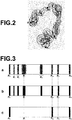
- Figure 2 is a schematic diagram illustrating path P of the large intestine from start point P 0 to the terminal point P N , which has been set for three-dimensional image V.
- processing for extracting the large intestine first, processing for dividing each of axial tomographic images obtained from the three-dimensional medical image, and which represent slice planes perpendicular to the body axis (axial section; axial), into a region in the body and a region outside the body with respect to the surface of the body is performed by using a known technique.
- a known technique there are a method in which binarization processing is performed on an input axial tomographic image, and an outline is extracted by performing outline extraction processing, and a region inside the extracted outline is extracted, as a region in the body (human body), and the like.
- binarization processing using a threshold is performed on the region in the body, and a candidate of the large intestine region is extracted from each of the axial tomographic images. Specifically, since air is present in the tube of the large intestine, binarization processing is performed by setting a threshold corresponding to a CT value of air, and an air region in the body is extracted from each of the axial tomographic images. Finally, only a part in which the extracted air region in the body in each of the axial tomographic images is connected to each other is extracted as the large intestine region.
- the core line passing through a central part of the tube of the large intestine is extracted from the extracted large intestine region by using a known technique.
- a known technique there are a method in which three-dimensional thinning processing is performed on a binary image representing the large intestine region that has been obtained during extraction of the large intestine by extracting only a part in which air regions in the body are connected to each other (please refer to Japanese Unexamined Patent Publication No. 2004-283373 and the like), and the like.
- a user interface for manually correcting the result of extraction of the large intestine and the core line may be further provided.
- the degree-of-interest setting unit 63 sets a relative degree of interest at each of plural positions on path P set by the path setting unit 62, and which connects the plural positions in the three-dimensional image.
- the degree-of-interest setting unit 63 sets one or more positions of interest P q in the three-dimensional image.
- l, m and n are natural numbers less than or equal to N.
- the large intestine region and path P (the core line of the large intestine) are displayed on a display, as illustrated in Figure 2 , and a selected position or positions of interest are obtained by selection of a desirable point or points on path P by a user using a mouse or the like.
- a degree or degrees of interest that have been input by the user using a keyboard or the like, and which correspond to the position or positions of interest, respectively, are obtained.
- the degree-of-interest setting unit 63 stores the obtained position or positions of interest and the degree or degrees of interest in such a manner to be related to each other.
- the degree-of-interest setting unit 63 obtains positions of interest (in this case, P 0 , P l , P m , P n and P N ) and degrees of interest of a0, al, am, an and aN corresponding to the positions of interest. Further, the degree-of-interest setting unit 63 sets a degree of interest by weighting in such a manner that the degree of interest is lower than the degree of interest corresponding to the position of interest as a distance from the obtained position of selection is longer.
- the degree-of-interest setting unit 63 sets a degree of interest, based on the following expression (1), in such a manner that the degree of interest is in inverse proportion to a distance from the position of interest.
- p represents an arbitrary point on the path.
- P q and aq (0 ⁇ q ⁇ N) represent a position of interest and a degree of interest corresponding to the position of interest.
- each of a relative degree or degrees of interest F(p) for the whole path is calculated by using the following expression (1) .
- Q is a set of positions of interest. Further, the present invention is not limited to the embodiment of the present invention.
- the degree-of-interest setting unit 63 sets the degree of interest based on the curvature of the path further by weighting in such a manner that the degree of interest is higher as the curvature of the path is larger. Specifically, the degree-of-interest setting unit 63 calculates a curvature at each position on path P, and sets, based on the curvature, degree of interest Fc(p) for each of the positions on path P in such a manner that the degree of interest is higher as the curvature is larger.
- weighting coefficient ⁇ has been set in such a manner that the product of the maximum value of Fc (p) and ⁇ is less than or equal to 0.5 so that the maximum value of degree of interest Fc (p) based on the curvature is lower than the maximum value of degree of interest F(p) related to the position of interest.
- Section a is a diagram illustrating an image representing degree of interest F(p)', which has been obtained as described above, by grayscale in such a manner that the image is darker as the degree of interest is higher.
- the degrees of interest are set in such a manner that the degree of interest is lower as a distance from position of interest P 0 , P l , P m , P n and P N is longer.
- Relatively high degrees of interest are set for positions included in the ranges represented by P i , P j and P k based on the curvatures.
- the discrimination condition obtainment unit 64 obtains a discrimination condition for identifying, based on the degree or degrees of interest, a virtual endoscopic image or images to be generated from a three-dimensional image.
- the discrimination condition is a threshold condition for identifying a virtual endoscopic image or images to be generated.
- a threshold defined in the threshold condition may be, for example, a default value, which has been set in advance, or a value set by a user or the like by using the input device 8.
- the discrimination condition (a threshold in this case) is changeable by a input by the user or the like at the input device 8.
- the discrimination condition obtainment unit 64 obtains, based on the change in the discrimination condition, a discrimination condition after change.
- the image generation unit 65 generates, based on three-dimensional image V, only a virtual endoscopic image or images identified based on the discrimination condition. Specifically, the image generation unit 65 generates, based on the threshold defined in the discrimination condition, only a virtual endoscopic image or images viewed from a viewpoint or viewpoints corresponding to a degree of interest or degrees of interest that are higher than the threshold. Accordingly, virtual endoscopic image I, which is viewed from a position with a degree of interest that is higher than the threshold, is generated from three-dimensional image V.
- a specific method of center projection for example, a known volume rendering method may be used.
- a viewing angle (the range of view lines) and the center of a viewing field (the center of a projection direction) may be given in advance by a boot parameter of a setting file or a program, an input by a user, or the like.
- Figure 3 , Sections b and c illustrate identified positions of interest of the positions of interest along the path illustrated in Figure 3 , Section a.
- the identified positions of interest based on different discrimination conditions are illustrated in color in Figure 3 , Sections b and c, respectively.
- threshold T is set, for example, at 0.75 as illustrated in Figure 3 , Section c, it is possible to generate only a virtual endoscopic image at each position included in black areas in the vicinities of P 0 , P m and P N , which satisfy the discrimination condition that degree of interest F(p)' is 0.75 or higher.
- threshold T is set, for example, at 0.45 as illustrated in Figure 3 , Section c, it is possible to generate a virtual endoscopic image at each position included in black areas in the vicinity of P 0 , P l , P m , P n and P N .
- the display control unit 66 displays the generated virtual endoscopic image or images on the display of the image processing workstation 3.
- Figure 4 is a flow chart illustrating the flow of medical image processing according to the first embodiment of the present invention. The flow of medical image processing in the first embodiment will be described with reference to Figure 4 .
- the image obtainment unit 61 obtains three-dimensional image V included in a file transferred from the data server 4, and stores three-dimensional image V in a memory (S01).
- the path setting unit 62 sets path P along the large intestine in each three-dimensional image (S02) .
- the degree-of-interest setting unit 63 sets positions of interest P 0 , P l , P m , P n and P N in path P and degrees of interest corresponding to them.
- the degree-of-interest setting unit 63 also calculates, based on expression (1), degree of interest F(p) at each position on the path, and further calculates, based on the curvature of the path, degree of interest Fc(p) at each of the positions on the path.
- the degree-of-interest setting unit 63 adds these together by weighted addition, and sets degree of interest F(p)' at each of the positions, as illustrated in Figure 3 , Section a (S03).
- the discrimination condition obtainment unit 64 obtains a discrimination condition (S04). Then, the image generation unit 65 generates each virtual endoscopic image I identified based on the discrimination condition from three-dimensional image V (S05) . Then, the display control unit 66 displays each virtual endoscopic image I, which has been generated, on a display (S06).
- the first embodiment of the present invention sets a relative degree of interest at each of plural positions on a path connecting the plural positions in the three-dimensional image, obtains a discrimination condition for identifying, based on the set degree or degrees of interest, a virtual endoscopic image or images to be generated from the three-dimensional image, and generates, based on the three-dimensional image, only the virtual endoscopic image or images identified based on the discrimination condition. Therefore, it is possible to easily identify, based on the relative degree or degrees of interest set for the path, the virtual endoscopic image or images to be generated from the three-dimensional image only by setting the discrimination condition. Hence, a work time and a work load for selectively generating a virtual endoscopic image or images are reduced, and that can assist in improving the efficiency of image reading.
- the discrimination condition for identifying "a virtual endoscopic image or images to be generated” is used.
- the discrimination condition for identifying "a virtual endoscopic image or images to be displayed” may be used.
- the second embodiment differs from the first embodiment only in that the discrimination condition identifies a virtual endoscopic image or images to be displayed based on the set degree or degrees of interest, and in that the image generation unit 65 generates, based on a three-dimensional image, virtual endoscopic images for the whole tubular structure or a part of the tubular structure, and in that the display control unit 66 displays only one or more of the virtual endoscopic images generated by the display control unit 66 that have been identified based on the discrimination condition.
- S01 through S04 are the same as the first embodiment.
- the image generation unit 65 in the second embodiment generates, along the path (core line), a predetermined number of virtual endoscopic image or images I with a predetermined interval from three-dimensional image V (S05').
- the display control unit 66 in the second embodiment displays only one or more of generated virtual endoscopic images I that have been identified based on the discrimination condition on a display (S06').
- the second embodiment sets a relative degree of interest at each of plural positions on a path connecting the plural positions in the three-dimensional image, obtains a discrimination condition for identifying, based on the set degrees of interest, a virtual endoscopic image or images to be displayed, and displays only one or more of the generated virtual endoscopic images that have been identified based on the discrimination condition. Therefore, it is possible to easily identify, based on the relative degrees of interest that have been set for the path, the virtual endoscopic image or images to be displayed from the three-dimensional image only by setting the discrimination condition. Hence, a work time and a work load for selectively displaying a virtual endoscopic image or images are reduced, and that can assist in improving the efficiency of image reading.
- the degree-of-interest setting unit 63 obtains a position of interest and a degree of interest corresponding to the position of interest, and sets a degree of interest by weighting in such a manner that the degree of interest is lower than the degree of interest corresponding to the position of interest as a distance from the obtained position of selection is longer. Therefore, it is possible to appropriately set not only the degree of interest at the position of interest, but also degrees of interest in the vicinity of the position of interest based on the level of interest corresponding to the position of interest.
- the degree-of-interest setting unit sets the degree of interest by weighting in such a manner that the degree of interest is in inverse proportion to the distance from the position of interest. Therefore, it is possible to easily set a degree or degrees of interest based on the position of interest.
- the discrimination condition is a threshold condition. Therefore, it is possible to adjust the number of a virtual endoscopic image or images to be generated or the number of a virtual endoscopic image or images to be displayed only by adjusting a threshold. Therefore, a work time and a work load for selectively generating or displaying the virtual endoscopic image or images are very appropriately reducible.
- the path connects plural positions within a tubular structure included in the three-dimensional image.
- the degree-of-interest setting unit sets, based on a curvature of the path, the degree of interest by weighting in such a manner that the degree of interest is higher as the curvature is larger. Therefore, it is possible to set the degree or degrees of interest to satisfy a request for intensively observing a region with a large curvature, and to easily and appropriately generate or display a virtual endoscopic image at a position based on its curvature.
- the virtual endoscopic image is a virtual laparoscopic endoscopic image
- the display control unit 66 displays only a virtual endoscopic image or images based on the discrimination condition after change. Therefore, it is possible to flexibly satisfy the request by the user. Further, it is possible to easily adjust a generation amount and a display amount of the virtual endoscopic image or images.
- the first embodiment and the second embodiment may be combined together, and the image generation unit 65 may generate only a virtual endoscopic image or images identified based on a discrimination condition, and the display control unit 66 may display only the virtual endoscopic image or images identified based on a discrimination condition.
- the discrimination conditions may include a condition for identifying a virtual endoscopic image or images for generation and a condition for identifying a virtual endoscopic image or images for display, respectively.
- the degree-of-interest setting unit 63 in the third embodiment sets a position of interest in the branching part.
- the function of each unit and processing other than processing for setting a degree of interest by the degree-of-interest setting unit 63 are the same as the first embodiment. Therefore, the different point between the first embodiment and the third embodiment will be mainly described. Explanations will be omitted for the function and processing that are the same as those of the first embodiment.
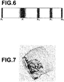
- Figure 5 is a diagram illustrating an example in which branching positions of branching parts automatically obtained by using a known method are set as positions of interest P l , P m and P n for bronchi, which are a tubular structure having branching parts.
- Figure 6 is a diagram representing degrees of interest corresponding to Figure 5 by grayscale in such a manner to be darker as the degree of interest is higher.
- the degree-of-interest setting unit 63 in the third embodiment obtains positions of interest (in this case, P l , P m and P n ) representing branching parts and degrees of interest corresponding to the positions of interest, and sets a degree of interest by weighting in such a manner that the degree of interest is lower than a degree of interest corresponding to the position of interest as a distance from the obtained position of selection is longer.
- start point P 0 and terminal point P N of the path are automatically set as positions of interest in a similar manner to the first embodiment.
- the degree-of-interest setting unit 63 calculates, based on the following expression (2), a degree of interest corresponding to the position of interest, and sets the degree of interest, as illustrated in Figure 6 .
- the virtual endoscopic image is a virtual bronchial endoscopic image
- the position of interest is a branching part of a bronchus or bronchi included in the three-dimensional image. Therefore, it is possible to appropriately and easily generate or display a virtual endoscopic image or images to satisfy a request by a user who wants to check a branching position for a bronchial endoscope.
- a position that has been manually input may be obtained as the position of interest.
- a position that has been judged as a branching part by using a known method may be automatically obtained as the position of interest.
- the degree-of-interest setting unit may use, as a relative degree of interest, only one of a degree of interest based on a position of interest, a degree of interest based on a curvature and a degree of interest based on a branching part.
- the degree-of-interest setting unit may use, as the relative degree of interest, an arbitrary combination of two or more of a degree of interest based on a position of interest, a degree of interest based on a curvature and a degree of interest based on a branching part.
- weighting may be performed on the relative degree of interest in each of the embodiments by using another degree of interest or other degrees of interest.
- a relative degree of interest may be set by combining plural degrees of interest, such as a degree of interest based on a position of interest, a degree of interest based on a curvature and a degree of interest based on a branching part, by using an arbitrary method as long as the relative degree of interest is settable for the path.
- an arbitrary expression in which the degree of interest is lower as a distance from the position of interest is longer may be used.
- expression (2) an arbitrary expression in which the degree of interest is lower as a distance from the branching part is longer may be used.
- the display control unit 66 may generate a display image at an arbitrary predetermined initial position by using a predetermined display method. While the display image is displayed, the display control unit 66 may display movement buttons for up, down, left, right, front and back directions. The display control unit 66 may display, based the kind of the movement button and the number of a click or clicks of a mouse or the like by the user, a new display image at a position moved in the input movement direction by a predetermined movement amount corresponding to the number of the click or clicks.
- the path setting unit 62 may store information for identifying positions of display images (display positions), and set a path by connecting the display positions in the order of display. Even if an arbitrary path that is not related to a structure included in three-dimensional image V is set in this manner, if a position of interest, as indicated by an arrow in Figure 7 , and a corresponding degree of interest are obtainable, it is possible to achieve a similar effect to each of the aforementioned embodiments by setting a degree of interest, based on expression (1), in such a manner that the degree of interest is lower as a distance from the position of interest is longer.
- a region of interest such as a lesion region
- CAD technique computer-aided diagnosis technique
- a position representing the region of interest for example, the center of gravity of the region of interest
- a point included in the extracted region of interest may be obtained as a position of interest.
- the discrimination condition obtainment unit 64 may obtain the number of a virtual endoscopic image or images to be generated or the number of a virtual endoscopic image or images to be displayed. Further, the discrimination condition obtainment unit 64 may automatically set a discrimination condition based on the obtained number of a virtual endoscopic image or images to be generated or the obtained number of a virtual endoscopic image or images to be displayed.
- the discrimination condition obtainment unit 64 may obtain the number of a virtual endoscopic image or images displayable at a predetermined display speed within a predetermined display time, and set a threshold to identify a virtual endoscopic image or images corresponding to the obtained number of a virtual endoscopic image or images. In this case, it is possible to further reduce the work of setting the discrimination condition, and it is possible to easily identify a virtual endoscopic image or images displayable at a predetermined display speed within a predetermined display time.
- discrimination condition obtainment processing may be performed any time as long as the processing is performed before image generation processing.
- discrimination condition obtainment processing may be performed any time as long as the processing is performed before display control processing.
- the configuration illustrated in Figure 1 may include plural image processing workstations 3, and processing may be shared by the workstations.
Claims (12)
- Medizinische Bildverarbeitungsvorrichtung, umfassend:eine Bildgewinnungseinheit (61), die ein durch Bildgebung von einem Patienten erhaltenes dreidimensionales Bild (V) gewinnt;eine Interessensgrad-Einstelleinheit (63), die einen relativen Interessensgrad an jeder von mehreren Positionen (P) auf einem Pfad, der die mehreren Positionen innerhalb des dreidimensionalen Bilds (V) verbindet, einstellt;eine Diskriminierbedingungs-Gewinnungseinheit (64), die eine Diskriminierbedingung gewinnt, um basierend auf dem eingestellten Interessensgrad oder den eingestellten Interessensgraden ein virtuelles Endoskopbild (I) oder Bilder zur Erzeugung aus dem dreidimensionalen Bild (V) identifiziert; undeine Bilderzeugungseinheit (65), die basierend auf dem dreidimensionalen Bild, das virtuelle Endoskopbild oder Bilder an Positionen erzeugt, welche die Diskriminierbedingung, wonach der Interessensgrad größer als ein Schwellenwert ist, erzeugt.
- Medizinische Bildverarbeitungsvorrichtung, umfassend:eine Bildgewinnungseinheit (61), die ein durch Bildgebung von einem Patienten erhaltenes dreidimensionales Bild (V) gewinnt;eine Interessensgrad-Einstelleinheit (63), die einen relativen Interessensgrad an jeder von mehreren Positionen (P) auf einem Pfad, der die mehreren Positionen innerhalb des dreidimensionalen Bilds (V) verbindet, einstellt;eine Bilderzeugungseinheit (65), die eine vorbestimmte Anzahl virtueller Endoskopbilder (I) mit einem vorbestimmten Intervall entlang dem Pfad aus dem dreidimensionalen Bild (V) erzeugt;eine Diskriminierbedingungs-Gewinnungseinheit (64), die eine Diskriminierbedingung gewinnt, um basierend auf den eingestellten Interessensgraden die virtuellen Endoskopbilder entlang dem Pfad (P) für die Anzeige identifiziert;eine Anzeigesteuereinheit (66), die basierend auf der Diskriminierbedingung nur die erzeugten virtuellen Endoskopbilder an Positionen anzeigt, welche die Diskriminierbedingung, wonach ein Interessensgrad größer als ein Schwellenwert ist, erfüllen.
- Vorrichtung nach Anspruch 1 oder 2, bei der die Interessensgrad-Einstelleinheit (63) eine Interessensstelle in dem dreidimensionalen Bild (V) und einen Interessensgrad entsprechend der Interessensstelle gewinnt, und einen Interessensgrad dadurch einstellt, dass sie eine derartige Wichtung vornimmt, dass der Interessensgrad niedriger ist als der Interessensgrad entsprechend der Interessensstelle, wenn ein Abstand von der gewonnenen Interessensstelle größer ist.
- Vorrichtung nach Anspruch 3, bei der die Interessensgrad-Einstelleinheit (63) den Interessensgrad in der Weise einstellt, dass der Interessensgrad umgekehrt proportional ist zu dem Abstand von der Interessensstelle.
- Vorrichtung nach einem der Ansprüche 1 bis 4, bei der der Pfad (P) die mehreren Positionen innerhalb einer schlauchförmigen Struktur innerhalb des dreidimensionalen Bilds (V) verbindet, und
wobei die Interessensgrad-Einstelleinheit den Interessensgrad basierend auf einer Krümmung des Pfads (P) einstellt durch Wichten in der Weise, dass der Interessensgrad größer ist, wenn die Krümmung größer ist. - Vorrichtung nach einem der Ansprüche 1 bis 5, bei der der Schwellenwert eine Bedingung zum Identifizieren der Stelle des Pfads ist.
- Vorrichtung nach Anspruch 6, bei der die Diskriminierbedingungs-Gewinnungseinheit (64) die Anzahl eines virtuellen Endoskopbilds oder von Bildern gewinnt, das/die mit einer vorbestimmten Anzeigegeschwindigkeit innerhalb einer vorbestimmten Anzeigezeit anzeigbar sind, und einen Schwellenwerte der Schwellenwertbedingung einstellt zum Identifizieren der Position des Pfads (P) entsprechend der gewonnenen Anzahl eines oder mehrerer virtueller Endoskopbilder.
- Vorrichtung nach einem der Ansprüche 1 bis 7, bei der das virtuelle Endoskopbild ein virtuelles Bronchien-Endoskopbild ist, und
wobei die Interessensstelle ein Verzweigungsteil einer oder mehrerer Bronchien innerhalb des dreidimensionalen Bilds (V) ist. - Vorrichtung nach einem der Ansprüche 1 bis 6, bei der das virtuelle Endoskopbild ein virtuelles Laparoskop-Endoskopbild ist.
- Vorrichtung nach einem der Ansprüche 1 bis 8, bei der die Diskriminierbedingung änderbar ist durch eine Eingabe über eine Eingabeeinrichtung (8), und die Diskriminierbedingungs-Gewinnungseinheit (64) basierend auf der Änderung der Diskriminierbedingung eine Diskriminierbedingung nach Änderung gewinnt.
- Medizinisches Bildverarbeitungsverfahren zur Ausführung durch eine medizinische Bilddiagnosevorrichtung, die aufweist:eine Bildgewinnungseinheit;eine Interessensgrad-Einstelleinheit;eine Diskriminierbedingungs-Gewinnungseinheit; undeine Bilderzeugungseinheit, undwobei das Verfahren umfasst:den Schritt des Gewinnens eines dreidimensionalen Bilds, erhalten durch Abbilden eines Patienten;den Schritt des Einstellens eines relativen Interessensgrads an jeder von mehreren Stellen auf einem Pfad, der die mehreren Stellen innerhalb des dreidimensionalen Bilds verbindet;den Schritt des Gewinnens einer Diskriminierbedingung, um basierend auf dem oder den eingestellten Interessensgraden ein virtuelles Endoskopbild oder Bilder zu identifizieren, die aus dem dreidimensionalen Bild zu erzeugen sind; undeinen Schritt des Generierens - basierend auf dem dreidimensionalen Bild - nur des virtuellen Endoskopbilds oder der Bilder an Stellen, die die Diskriminierbedingung, wonach der Interessensgrad höher als ein Schwellenwert ist, erfüllen.
- Aufzeichnungsmedium, auf dem ein medizinisches Bildverarbeitungsprogramm gespeichert ist, welches einen Computer veranlasst, als medizinische Bildverarbeitungsvorrichtung nach Anspruch 1 zu fungieren.
Applications Claiming Priority (2)
| Application Number | Priority Date | Filing Date | Title |
|---|---|---|---|
| JP2012052413A JP5932406B2 (ja) | 2012-03-09 | 2012-03-09 | 医用画像処理装置および方法、並びにプログラム |
| PCT/JP2013/001309 WO2013132820A1 (ja) | 2012-03-09 | 2013-03-04 | 医用画像処理装置および方法、並びにプログラム |
Publications (3)
| Publication Number | Publication Date |
|---|---|
| EP2823764A1 EP2823764A1 (de) | 2015-01-14 |
| EP2823764A4 EP2823764A4 (de) | 2015-11-11 |
| EP2823764B1 true EP2823764B1 (de) | 2019-07-17 |
Family
ID=49116316
Family Applications (1)
| Application Number | Title | Priority Date | Filing Date |
|---|---|---|---|
| EP13757250.9A Active EP2823764B1 (de) | 2012-03-09 | 2013-03-04 | Vorrichtung, verfahren und programm zur verarbeitung medizinischer bilder |
Country Status (4)
| Country | Link |
|---|---|
| US (1) | US9390497B2 (de) |
| EP (1) | EP2823764B1 (de) |
| JP (1) | JP5932406B2 (de) |
| WO (1) | WO2013132820A1 (de) |
Families Citing this family (4)
| Publication number | Priority date | Publication date | Assignee | Title |
|---|---|---|---|---|
| US20140253544A1 (en) * | 2012-01-27 | 2014-09-11 | Kabushiki Kaisha Toshiba | Medical image processing apparatus |
| US10025479B2 (en) * | 2013-09-25 | 2018-07-17 | Terarecon, Inc. | Advanced medical image processing wizard |
| KR20170068944A (ko) | 2015-12-10 | 2017-06-20 | 삼성메디슨 주식회사 | 초음파 영상을 디스플레이하는 방법 및 이를 위한 초음파 장치 |
| JP6608111B2 (ja) * | 2016-09-28 | 2019-11-20 | 富士フイルム株式会社 | 医用画像保存再生装置および方法並びにプログラム |
Family Cites Families (17)
| Publication number | Priority date | Publication date | Assignee | Title |
|---|---|---|---|---|
| DE69805209T2 (de) * | 1998-02-23 | 2002-11-28 | Algotec Systems Ltd | System und methode zur automatischen wegplanung |
| JP2000135215A (ja) | 1998-10-30 | 2000-05-16 | Ge Yokogawa Medical Systems Ltd | 管路案内方法および装置並びに放射線断層撮影装置 |
| EP1493128A1 (de) * | 2002-03-29 | 2005-01-05 | Koninklijke Philips Electronics N.V. | Verfahren, system und computerprogramm zur stereoskopischen betrachtung von medizinischen 3d-bildern |
| JP3930423B2 (ja) * | 2002-12-03 | 2007-06-13 | オリンパス株式会社 | 内視鏡装置 |
| JP4421203B2 (ja) | 2003-03-20 | 2010-02-24 | 株式会社東芝 | 管腔状構造体の解析処理装置 |
| EP1709589B1 (de) * | 2004-01-15 | 2013-01-16 | Algotec Systems Ltd. | Bestimmung der mittellinie von blutgefässen |
| JP4493423B2 (ja) * | 2004-07-08 | 2010-06-30 | オリンパス株式会社 | 内視鏡挿入支援装置 |
| CN101116110B (zh) | 2005-02-08 | 2013-03-27 | 皇家飞利浦电子股份有限公司 | 医学图像浏览协议 |
| JP4822142B2 (ja) | 2006-05-02 | 2011-11-24 | 国立大学法人名古屋大学 | 内視鏡挿入支援システム及び内視鏡挿入支援方法 |
| US9037215B2 (en) * | 2007-01-31 | 2015-05-19 | The Penn State Research Foundation | Methods and apparatus for 3D route planning through hollow organs |
| US20090226057A1 (en) * | 2008-03-04 | 2009-09-10 | Adi Mashiach | Segmentation device and method |
| JP5554028B2 (ja) * | 2009-07-28 | 2014-07-23 | 株式会社東芝 | 医用画像処理装置、医用画像処理プログラム、及びx線ct装置 |
| JP5551955B2 (ja) * | 2010-03-31 | 2014-07-16 | 富士フイルム株式会社 | 投影画像生成装置、方法、及びプログラム |
| JP5675227B2 (ja) * | 2010-08-31 | 2015-02-25 | 富士フイルム株式会社 | 内視鏡画像処理装置および作動方法、並びに、プログラム |
| JP2012187161A (ja) * | 2011-03-09 | 2012-10-04 | Fujifilm Corp | 画像処理装置、方法、及びプログラム |
| US9330490B2 (en) * | 2011-04-29 | 2016-05-03 | University Health Network | Methods and systems for visualization of 3D parametric data during 2D imaging |
| US20130250081A1 (en) * | 2012-03-21 | 2013-09-26 | Covidien Lp | System and method for determining camera angles by using virtual planes derived from actual images |
-
2012
- 2012-03-09 JP JP2012052413A patent/JP5932406B2/ja active Active
-
2013
- 2013-03-04 EP EP13757250.9A patent/EP2823764B1/de active Active
- 2013-03-04 WO PCT/JP2013/001309 patent/WO2013132820A1/ja active Application Filing
-
2014
- 2014-09-04 US US14/477,738 patent/US9390497B2/en active Active
Non-Patent Citations (1)
| Title |
|---|
| None * |
Also Published As
| Publication number | Publication date |
|---|---|
| JP5932406B2 (ja) | 2016-06-08 |
| US9390497B2 (en) | 2016-07-12 |
| EP2823764A1 (de) | 2015-01-14 |
| EP2823764A4 (de) | 2015-11-11 |
| US20140369578A1 (en) | 2014-12-18 |
| WO2013132820A1 (ja) | 2013-09-12 |
| JP2013183958A (ja) | 2013-09-19 |
Similar Documents
| Publication | Publication Date | Title |
|---|---|---|
| US9024941B2 (en) | Sequentially displaying virtual endoscopic images by setting an observation path to compensate for a curved region of the tubular structure | |
| US10085672B2 (en) | Diagnostic endoscopic imaging support apparatus and method, and non-transitory computer readable medium on which is recorded diagnostic endoscopic imaging support program | |
| CN107169919B (zh) | 用于3d医学体积的加速读取的方法和系统 | |
| US8994720B2 (en) | Diagnosis assisting apparatus, diagnosis assisting program, and diagnosis assisting method | |
| US8611989B2 (en) | Multi-planar reconstruction lumen imaging method and apparatus | |
| US9697600B2 (en) | Multi-modal segmentatin of image data | |
| US11954860B2 (en) | Image matching method and device, and storage medium | |
| EP3136338A1 (de) | Informationsverarbeitungs-vorrichtung, informationsverarbeitungs-verfahren und programm | |
| US8907949B2 (en) | Image display apparatus, method and program | |
| US8588490B2 (en) | Image-based diagnosis assistance apparatus, its operation method and program | |
| US20080084415A1 (en) | Orientation of 3-dimensional displays as a function of the regions to be examined | |
| US9390497B2 (en) | Medical image processing apparatus, method and program | |
| US9530238B2 (en) | Image processing apparatus, method and program utilizing an opacity curve for endoscopic images | |
| JP3989896B2 (ja) | 医用画像処理装置、関心領域抽出方法、ならびに、プログラム | |
| EP1983487A2 (de) | Projektionsbilderzeugungsvorrichtung, Verfahren und computerlesbares Aufzeichnungsmedium | |
| US9123163B2 (en) | Medical image display apparatus, method and program | |
| JP4668289B2 (ja) | 画像処理装置および方法並びにプログラム | |
| US9558589B2 (en) | Medical image display apparatus, method, and program | |
| JP5534580B2 (ja) | 医用画像表示装置及び医用画像表示方法 |
Legal Events
| Date | Code | Title | Description |
|---|---|---|---|
| PUAI | Public reference made under article 153(3) epc to a published international application that has entered the european phase |
Free format text: ORIGINAL CODE: 0009012 |
|
| 17P | Request for examination filed |
Effective date: 20140924 |
|
| AK | Designated contracting states |
Kind code of ref document: A1 Designated state(s): AL AT BE BG CH CY CZ DE DK EE ES FI FR GB GR HR HU IE IS IT LI LT LU LV MC MK MT NL NO PL PT RO RS SE SI SK SM TR |
|
| AX | Request for extension of the european patent |
Extension state: BA ME |
|
| DAX | Request for extension of the european patent (deleted) | ||
| RA4 | Supplementary search report drawn up and despatched (corrected) |
Effective date: 20151012 |
|
| RIC1 | Information provided on ipc code assigned before grant |
Ipc: A61B 6/03 20060101AFI20151006BHEP |
|
| STAA | Information on the status of an ep patent application or granted ep patent |
Free format text: STATUS: EXAMINATION IS IN PROGRESS |
|
| 17Q | First examination report despatched |
Effective date: 20171012 |
|
| GRAP | Despatch of communication of intention to grant a patent |
Free format text: ORIGINAL CODE: EPIDOSNIGR1 |
|
| STAA | Information on the status of an ep patent application or granted ep patent |
Free format text: STATUS: GRANT OF PATENT IS INTENDED |
|
| INTG | Intention to grant announced |
Effective date: 20190213 |
|
| GRAS | Grant fee paid |
Free format text: ORIGINAL CODE: EPIDOSNIGR3 |
|
| GRAA | (expected) grant |
Free format text: ORIGINAL CODE: 0009210 |
|
| STAA | Information on the status of an ep patent application or granted ep patent |
Free format text: STATUS: THE PATENT HAS BEEN GRANTED |
|
| AK | Designated contracting states |
Kind code of ref document: B1 Designated state(s): AL AT BE BG CH CY CZ DE DK EE ES FI FR GB GR HR HU IE IS IT LI LT LU LV MC MK MT NL NO PL PT RO RS SE SI SK SM TR |
|
| REG | Reference to a national code |
Ref country code: GB Ref legal event code: FG4D |
|
| REG | Reference to a national code |
Ref country code: CH Ref legal event code: EP |
|
| REG | Reference to a national code |
Ref country code: IE Ref legal event code: FG4D |
|
| REG | Reference to a national code |
Ref country code: DE Ref legal event code: R096 Ref document number: 602013057908 Country of ref document: DE |
|
| REG | Reference to a national code |
Ref country code: AT Ref legal event code: REF Ref document number: 1155087 Country of ref document: AT Kind code of ref document: T Effective date: 20190815 |
|
| REG | Reference to a national code |
Ref country code: NL Ref legal event code: MP Effective date: 20190717 |
|
| REG | Reference to a national code |
Ref country code: LT Ref legal event code: MG4D |
|
| REG | Reference to a national code |
Ref country code: AT Ref legal event code: MK05 Ref document number: 1155087 Country of ref document: AT Kind code of ref document: T Effective date: 20190717 |
|
| PG25 | Lapsed in a contracting state [announced via postgrant information from national office to epo] |
Ref country code: FI Free format text: LAPSE BECAUSE OF FAILURE TO SUBMIT A TRANSLATION OF THE DESCRIPTION OR TO PAY THE FEE WITHIN THE PRESCRIBED TIME-LIMIT Effective date: 20190717 Ref country code: BG Free format text: LAPSE BECAUSE OF FAILURE TO SUBMIT A TRANSLATION OF THE DESCRIPTION OR TO PAY THE FEE WITHIN THE PRESCRIBED TIME-LIMIT Effective date: 20191017 Ref country code: NO Free format text: LAPSE BECAUSE OF FAILURE TO SUBMIT A TRANSLATION OF THE DESCRIPTION OR TO PAY THE FEE WITHIN THE PRESCRIBED TIME-LIMIT Effective date: 20191017 Ref country code: SE Free format text: LAPSE BECAUSE OF FAILURE TO SUBMIT A TRANSLATION OF THE DESCRIPTION OR TO PAY THE FEE WITHIN THE PRESCRIBED TIME-LIMIT Effective date: 20190717 Ref country code: HR Free format text: LAPSE BECAUSE OF FAILURE TO SUBMIT A TRANSLATION OF THE DESCRIPTION OR TO PAY THE FEE WITHIN THE PRESCRIBED TIME-LIMIT Effective date: 20190717 Ref country code: AT Free format text: LAPSE BECAUSE OF FAILURE TO SUBMIT A TRANSLATION OF THE DESCRIPTION OR TO PAY THE FEE WITHIN THE PRESCRIBED TIME-LIMIT Effective date: 20190717 Ref country code: NL Free format text: LAPSE BECAUSE OF FAILURE TO SUBMIT A TRANSLATION OF THE DESCRIPTION OR TO PAY THE FEE WITHIN THE PRESCRIBED TIME-LIMIT Effective date: 20190717 Ref country code: PT Free format text: LAPSE BECAUSE OF FAILURE TO SUBMIT A TRANSLATION OF THE DESCRIPTION OR TO PAY THE FEE WITHIN THE PRESCRIBED TIME-LIMIT Effective date: 20191118 Ref country code: LT Free format text: LAPSE BECAUSE OF FAILURE TO SUBMIT A TRANSLATION OF THE DESCRIPTION OR TO PAY THE FEE WITHIN THE PRESCRIBED TIME-LIMIT Effective date: 20190717 |
|
| PG25 | Lapsed in a contracting state [announced via postgrant information from national office to epo] |
Ref country code: AL Free format text: LAPSE BECAUSE OF FAILURE TO SUBMIT A TRANSLATION OF THE DESCRIPTION OR TO PAY THE FEE WITHIN THE PRESCRIBED TIME-LIMIT Effective date: 20190717 Ref country code: LV Free format text: LAPSE BECAUSE OF FAILURE TO SUBMIT A TRANSLATION OF THE DESCRIPTION OR TO PAY THE FEE WITHIN THE PRESCRIBED TIME-LIMIT Effective date: 20190717 Ref country code: ES Free format text: LAPSE BECAUSE OF FAILURE TO SUBMIT A TRANSLATION OF THE DESCRIPTION OR TO PAY THE FEE WITHIN THE PRESCRIBED TIME-LIMIT Effective date: 20190717 Ref country code: RS Free format text: LAPSE BECAUSE OF FAILURE TO SUBMIT A TRANSLATION OF THE DESCRIPTION OR TO PAY THE FEE WITHIN THE PRESCRIBED TIME-LIMIT Effective date: 20190717 Ref country code: GR Free format text: LAPSE BECAUSE OF FAILURE TO SUBMIT A TRANSLATION OF THE DESCRIPTION OR TO PAY THE FEE WITHIN THE PRESCRIBED TIME-LIMIT Effective date: 20191018 Ref country code: IS Free format text: LAPSE BECAUSE OF FAILURE TO SUBMIT A TRANSLATION OF THE DESCRIPTION OR TO PAY THE FEE WITHIN THE PRESCRIBED TIME-LIMIT Effective date: 20191117 |
|
| PG25 | Lapsed in a contracting state [announced via postgrant information from national office to epo] |
Ref country code: TR Free format text: LAPSE BECAUSE OF FAILURE TO SUBMIT A TRANSLATION OF THE DESCRIPTION OR TO PAY THE FEE WITHIN THE PRESCRIBED TIME-LIMIT Effective date: 20190717 |
|
| PG25 | Lapsed in a contracting state [announced via postgrant information from national office to epo] |
Ref country code: RO Free format text: LAPSE BECAUSE OF FAILURE TO SUBMIT A TRANSLATION OF THE DESCRIPTION OR TO PAY THE FEE WITHIN THE PRESCRIBED TIME-LIMIT Effective date: 20190717 Ref country code: PL Free format text: LAPSE BECAUSE OF FAILURE TO SUBMIT A TRANSLATION OF THE DESCRIPTION OR TO PAY THE FEE WITHIN THE PRESCRIBED TIME-LIMIT Effective date: 20190717 Ref country code: DK Free format text: LAPSE BECAUSE OF FAILURE TO SUBMIT A TRANSLATION OF THE DESCRIPTION OR TO PAY THE FEE WITHIN THE PRESCRIBED TIME-LIMIT Effective date: 20190717 Ref country code: EE Free format text: LAPSE BECAUSE OF FAILURE TO SUBMIT A TRANSLATION OF THE DESCRIPTION OR TO PAY THE FEE WITHIN THE PRESCRIBED TIME-LIMIT Effective date: 20190717 Ref country code: IT Free format text: LAPSE BECAUSE OF FAILURE TO SUBMIT A TRANSLATION OF THE DESCRIPTION OR TO PAY THE FEE WITHIN THE PRESCRIBED TIME-LIMIT Effective date: 20190717 |
|
| PG25 | Lapsed in a contracting state [announced via postgrant information from national office to epo] |
Ref country code: SK Free format text: LAPSE BECAUSE OF FAILURE TO SUBMIT A TRANSLATION OF THE DESCRIPTION OR TO PAY THE FEE WITHIN THE PRESCRIBED TIME-LIMIT Effective date: 20190717 Ref country code: CZ Free format text: LAPSE BECAUSE OF FAILURE TO SUBMIT A TRANSLATION OF THE DESCRIPTION OR TO PAY THE FEE WITHIN THE PRESCRIBED TIME-LIMIT Effective date: 20190717 Ref country code: SM Free format text: LAPSE BECAUSE OF FAILURE TO SUBMIT A TRANSLATION OF THE DESCRIPTION OR TO PAY THE FEE WITHIN THE PRESCRIBED TIME-LIMIT Effective date: 20190717 Ref country code: IS Free format text: LAPSE BECAUSE OF FAILURE TO SUBMIT A TRANSLATION OF THE DESCRIPTION OR TO PAY THE FEE WITHIN THE PRESCRIBED TIME-LIMIT Effective date: 20200224 |
|
| REG | Reference to a national code |
Ref country code: DE Ref legal event code: R097 Ref document number: 602013057908 Country of ref document: DE |
|
| PLBE | No opposition filed within time limit |
Free format text: ORIGINAL CODE: 0009261 |
|
| STAA | Information on the status of an ep patent application or granted ep patent |
Free format text: STATUS: NO OPPOSITION FILED WITHIN TIME LIMIT |
|
| PG2D | Information on lapse in contracting state deleted |
Ref country code: IS |
|
| 26N | No opposition filed |
Effective date: 20200603 |
|
| PG25 | Lapsed in a contracting state [announced via postgrant information from national office to epo] |
Ref country code: SI Free format text: LAPSE BECAUSE OF FAILURE TO SUBMIT A TRANSLATION OF THE DESCRIPTION OR TO PAY THE FEE WITHIN THE PRESCRIBED TIME-LIMIT Effective date: 20190717 |
|
| PG25 | Lapsed in a contracting state [announced via postgrant information from national office to epo] |
Ref country code: MC Free format text: LAPSE BECAUSE OF FAILURE TO SUBMIT A TRANSLATION OF THE DESCRIPTION OR TO PAY THE FEE WITHIN THE PRESCRIBED TIME-LIMIT Effective date: 20190717 |
|
| REG | Reference to a national code |
Ref country code: CH Ref legal event code: PL |
|
| REG | Reference to a national code |
Ref country code: BE Ref legal event code: MM Effective date: 20200331 |
|
| PG25 | Lapsed in a contracting state [announced via postgrant information from national office to epo] |
Ref country code: LU Free format text: LAPSE BECAUSE OF NON-PAYMENT OF DUE FEES Effective date: 20200304 |
|
| PG25 | Lapsed in a contracting state [announced via postgrant information from national office to epo] |
Ref country code: CH Free format text: LAPSE BECAUSE OF NON-PAYMENT OF DUE FEES Effective date: 20200331 Ref country code: IE Free format text: LAPSE BECAUSE OF NON-PAYMENT OF DUE FEES Effective date: 20200304 Ref country code: FR Free format text: LAPSE BECAUSE OF NON-PAYMENT OF DUE FEES Effective date: 20200331 Ref country code: LI Free format text: LAPSE BECAUSE OF NON-PAYMENT OF DUE FEES Effective date: 20200331 |
|
| PG25 | Lapsed in a contracting state [announced via postgrant information from national office to epo] |
Ref country code: BE Free format text: LAPSE BECAUSE OF NON-PAYMENT OF DUE FEES Effective date: 20200331 |
|
| GBPC | Gb: european patent ceased through non-payment of renewal fee |
Effective date: 20200304 |
|
| PG25 | Lapsed in a contracting state [announced via postgrant information from national office to epo] |
Ref country code: GB Free format text: LAPSE BECAUSE OF NON-PAYMENT OF DUE FEES Effective date: 20200304 |
|
| PG25 | Lapsed in a contracting state [announced via postgrant information from national office to epo] |
Ref country code: MT Free format text: LAPSE BECAUSE OF FAILURE TO SUBMIT A TRANSLATION OF THE DESCRIPTION OR TO PAY THE FEE WITHIN THE PRESCRIBED TIME-LIMIT Effective date: 20190717 Ref country code: CY Free format text: LAPSE BECAUSE OF FAILURE TO SUBMIT A TRANSLATION OF THE DESCRIPTION OR TO PAY THE FEE WITHIN THE PRESCRIBED TIME-LIMIT Effective date: 20190717 |
|
| PG25 | Lapsed in a contracting state [announced via postgrant information from national office to epo] |
Ref country code: MK Free format text: LAPSE BECAUSE OF FAILURE TO SUBMIT A TRANSLATION OF THE DESCRIPTION OR TO PAY THE FEE WITHIN THE PRESCRIBED TIME-LIMIT Effective date: 20190717 |
|
| P01 | Opt-out of the competence of the unified patent court (upc) registered |
Effective date: 20230515 |
|
| PGFP | Annual fee paid to national office [announced via postgrant information from national office to epo] |
Ref country code: DE Payment date: 20240130 Year of fee payment: 12 |