EP2816058B1 - Anticorps anti-myostatine - Google Patents
Anticorps anti-myostatine Download PDFInfo
- Publication number
- EP2816058B1 EP2816058B1 EP14181801.3A EP14181801A EP2816058B1 EP 2816058 B1 EP2816058 B1 EP 2816058B1 EP 14181801 A EP14181801 A EP 14181801A EP 2816058 B1 EP2816058 B1 EP 2816058B1
- Authority
- EP
- European Patent Office
- Prior art keywords
- antibody
- myostatin
- prt
- artificial
- synthetic construct
- Prior art date
- Legal status (The legal status is an assumption and is not a legal conclusion. Google has not performed a legal analysis and makes no representation as to the accuracy of the status listed.)
- Active
Links
Images


Classifications
-
- C—CHEMISTRY; METALLURGY
- C07—ORGANIC CHEMISTRY
- C07K—PEPTIDES
- C07K16/00—Immunoglobulins [IGs], e.g. monoclonal or polyclonal antibodies
- C07K16/18—Immunoglobulins [IGs], e.g. monoclonal or polyclonal antibodies against material from animals or humans
- C07K16/22—Immunoglobulins [IGs], e.g. monoclonal or polyclonal antibodies against material from animals or humans against growth factors ; against growth regulators
-
- A—HUMAN NECESSITIES
- A61—MEDICAL OR VETERINARY SCIENCE; HYGIENE
- A61K—PREPARATIONS FOR MEDICAL, DENTAL OR TOILETRY PURPOSES
- A61K39/00—Medicinal preparations containing antigens or antibodies
- A61K39/395—Antibodies; Immunoglobulins; Immune serum, e.g. antilymphocytic serum
-
- A—HUMAN NECESSITIES
- A61—MEDICAL OR VETERINARY SCIENCE; HYGIENE
- A61P—SPECIFIC THERAPEUTIC ACTIVITY OF CHEMICAL COMPOUNDS OR MEDICINAL PREPARATIONS
- A61P1/00—Drugs for disorders of the alimentary tract or the digestive system
- A61P1/16—Drugs for disorders of the alimentary tract or the digestive system for liver or gallbladder disorders, e.g. hepatoprotective agents, cholagogues, litholytics
-
- A—HUMAN NECESSITIES
- A61—MEDICAL OR VETERINARY SCIENCE; HYGIENE
- A61P—SPECIFIC THERAPEUTIC ACTIVITY OF CHEMICAL COMPOUNDS OR MEDICINAL PREPARATIONS
- A61P11/00—Drugs for disorders of the respiratory system
-
- A—HUMAN NECESSITIES
- A61—MEDICAL OR VETERINARY SCIENCE; HYGIENE
- A61P—SPECIFIC THERAPEUTIC ACTIVITY OF CHEMICAL COMPOUNDS OR MEDICINAL PREPARATIONS
- A61P13/00—Drugs for disorders of the urinary system
- A61P13/12—Drugs for disorders of the urinary system of the kidneys
-
- A—HUMAN NECESSITIES
- A61—MEDICAL OR VETERINARY SCIENCE; HYGIENE
- A61P—SPECIFIC THERAPEUTIC ACTIVITY OF CHEMICAL COMPOUNDS OR MEDICINAL PREPARATIONS
- A61P19/00—Drugs for skeletal disorders
- A61P19/02—Drugs for skeletal disorders for joint disorders, e.g. arthritis, arthrosis
-
- A—HUMAN NECESSITIES
- A61—MEDICAL OR VETERINARY SCIENCE; HYGIENE
- A61P—SPECIFIC THERAPEUTIC ACTIVITY OF CHEMICAL COMPOUNDS OR MEDICINAL PREPARATIONS
- A61P19/00—Drugs for skeletal disorders
- A61P19/08—Drugs for skeletal disorders for bone diseases, e.g. rachitism, Paget's disease
- A61P19/10—Drugs for skeletal disorders for bone diseases, e.g. rachitism, Paget's disease for osteoporosis
-
- A—HUMAN NECESSITIES
- A61—MEDICAL OR VETERINARY SCIENCE; HYGIENE
- A61P—SPECIFIC THERAPEUTIC ACTIVITY OF CHEMICAL COMPOUNDS OR MEDICINAL PREPARATIONS
- A61P21/00—Drugs for disorders of the muscular or neuromuscular system
-
- A—HUMAN NECESSITIES
- A61—MEDICAL OR VETERINARY SCIENCE; HYGIENE
- A61P—SPECIFIC THERAPEUTIC ACTIVITY OF CHEMICAL COMPOUNDS OR MEDICINAL PREPARATIONS
- A61P3/00—Drugs for disorders of the metabolism
-
- A—HUMAN NECESSITIES
- A61—MEDICAL OR VETERINARY SCIENCE; HYGIENE
- A61P—SPECIFIC THERAPEUTIC ACTIVITY OF CHEMICAL COMPOUNDS OR MEDICINAL PREPARATIONS
- A61P3/00—Drugs for disorders of the metabolism
- A61P3/04—Anorexiants; Antiobesity agents
-
- A—HUMAN NECESSITIES
- A61—MEDICAL OR VETERINARY SCIENCE; HYGIENE
- A61P—SPECIFIC THERAPEUTIC ACTIVITY OF CHEMICAL COMPOUNDS OR MEDICINAL PREPARATIONS
- A61P3/00—Drugs for disorders of the metabolism
- A61P3/08—Drugs for disorders of the metabolism for glucose homeostasis
- A61P3/10—Drugs for disorders of the metabolism for glucose homeostasis for hyperglycaemia, e.g. antidiabetics
-
- A—HUMAN NECESSITIES
- A61—MEDICAL OR VETERINARY SCIENCE; HYGIENE
- A61P—SPECIFIC THERAPEUTIC ACTIVITY OF CHEMICAL COMPOUNDS OR MEDICINAL PREPARATIONS
- A61P7/00—Drugs for disorders of the blood or the extracellular fluid
-
- A—HUMAN NECESSITIES
- A61—MEDICAL OR VETERINARY SCIENCE; HYGIENE
- A61P—SPECIFIC THERAPEUTIC ACTIVITY OF CHEMICAL COMPOUNDS OR MEDICINAL PREPARATIONS
- A61P9/00—Drugs for disorders of the cardiovascular system
- A61P9/04—Inotropic agents, i.e. stimulants of cardiac contraction; Drugs for heart failure
-
- A—HUMAN NECESSITIES
- A61—MEDICAL OR VETERINARY SCIENCE; HYGIENE
- A61P—SPECIFIC THERAPEUTIC ACTIVITY OF CHEMICAL COMPOUNDS OR MEDICINAL PREPARATIONS
- A61P9/00—Drugs for disorders of the cardiovascular system
- A61P9/10—Drugs for disorders of the cardiovascular system for treating ischaemic or atherosclerotic diseases, e.g. antianginal drugs, coronary vasodilators, drugs for myocardial infarction, retinopathy, cerebrovascula insufficiency, renal arteriosclerosis
-
- C—CHEMISTRY; METALLURGY
- C07—ORGANIC CHEMISTRY
- C07K—PEPTIDES
- C07K2317/00—Immunoglobulins specific features
- C07K2317/20—Immunoglobulins specific features characterized by taxonomic origin
- C07K2317/24—Immunoglobulins specific features characterized by taxonomic origin containing regions, domains or residues from different species, e.g. chimeric, humanized or veneered
-
- C—CHEMISTRY; METALLURGY
- C07—ORGANIC CHEMISTRY
- C07K—PEPTIDES
- C07K2317/00—Immunoglobulins specific features
- C07K2317/50—Immunoglobulins specific features characterized by immunoglobulin fragments
- C07K2317/55—Fab or Fab'
-
- C—CHEMISTRY; METALLURGY
- C07—ORGANIC CHEMISTRY
- C07K—PEPTIDES
- C07K2317/00—Immunoglobulins specific features
- C07K2317/50—Immunoglobulins specific features characterized by immunoglobulin fragments
- C07K2317/56—Immunoglobulins specific features characterized by immunoglobulin fragments variable (Fv) region, i.e. VH and/or VL
- C07K2317/565—Complementarity determining region [CDR]
-
- C—CHEMISTRY; METALLURGY
- C07—ORGANIC CHEMISTRY
- C07K—PEPTIDES
- C07K2317/00—Immunoglobulins specific features
- C07K2317/70—Immunoglobulins specific features characterized by effect upon binding to a cell or to an antigen
- C07K2317/76—Antagonist effect on antigen, e.g. neutralization or inhibition of binding
-
- C—CHEMISTRY; METALLURGY
- C07—ORGANIC CHEMISTRY
- C07K—PEPTIDES
- C07K2317/00—Immunoglobulins specific features
- C07K2317/90—Immunoglobulins specific features characterized by (pharmaco)kinetic aspects or by stability of the immunoglobulin
- C07K2317/92—Affinity (KD), association rate (Ka), dissociation rate (Kd) or EC50 value
Definitions
- the invention is in the field of medicine, particularly in the field of monoclonal antibodies against myostatin. More specifically the invention relates to high affinity chimeric or humanized anti-myostatin antibodies and use of the antibodies for therapy, prophylaxis or diagnosis of various disorders or conditions in mammalian and avian species.
- TGF- ⁇ transforming growth factor beta
- the TGF- ⁇ superfamily members share a common structure including a peptide signal sequence required for secretion of the protein and an amino-terminal fragment that is proteolytically cleaved about 105-140 amino acids from the carboxy-terminus of the large precursor protein to produce the mature protein.
- the mature protein is characterized by highly conserved cysteine residues, while the active form of the mature protein is a disulfide-linked homodimer of the proteolytically-cleaved proprotein ( Gray, A., and Maston, A., Science, 247:1328, 1990 ).
- Myostatin also referred to as growth differentiation factor-8 (GDF-8) is a member of the TGF- ⁇ superfamily of proteins. Myostatin shares structural similarities with other TGF- ⁇ family members. It contains a hydrophobic amino-terminus that acts as a secretory signal and a conserved RSRR domain that is important for proteolytic processing. Cleavage of the protein gives rise to an amino-terminal latency associated peptide and a carboxy-terminal mature signaling peptide which forms the biologically active homodimer. Myostatin is expressed largely in developing and adult skeletal muscle and functions as a negative regulator of skeletal muscle.
- GDF-8 growth differentiation factor-8
- myostatin is a desirable target for therapeutic or prophylactic intervention for such disorders or conditions or for monitoring progression of such disorders or conditions.
- myostatin may also be involved in other physiological processes including preadipocyte differentiation to adipocytes ( Kim et al.
- myostatin-specific antagonists e.g., myostatin-specific antibodies, may also prove useful for treating, preventing or monitoring disorders or conditions such as those which benefit from increasing bone density (e.g., osteoporosis), Type II diabetes, metabolic syndrome, obesity and osteoarthritis.
- Myostatin is highly conserved across species; the amino acid sequence of the mature form of myostatin in human, mouse, rat, chicken, turkey and cow are 100% identical (See Figs. 2 and 3 ). There are naturally occurring myostatin mutations in cattle, which have been linked to a double-muscled phenotype ( McPherron, et al. PNAS, 94:12457-61, 1997 ).
- myostatin is highly conserved in sequence and in function across species, not only does an anti-myostatin antibody provide a promising means of increasing muscle mass, or treatment or prevention of such disorders or conditions listed above in humans, but also in other mammals including, e.g., domestic animals (e.g., canine and feline), sports animals (e.g., equine), food-source animals (e.g., bovine, porcine and ovine) and in avian species (e.g., chicken, turkey, duck and other game birds and poultry).
- domestic animals e.g., canine and feline
- sports animals e.g., equine
- food-source animals e.g., bovine, porcine and ovine
- avian species e.g., chicken, turkey, duck and other game birds and poultry.
- Growth differentiation factors-11 also referred to as GDF-11 or BMP-11, is the member of the TGF- ⁇ superfamily of proteins that is most homologous to myostatin.
- the amino acid sequence of the mature forms of human myostatin and GDF-11 are about 90% identical; however, GDF-11 is expressed in a wider range of tissues than is GDF-8 including dental pulp, brain, heart, kidney and lung as well as muscle and adipose tissue ( Nakashima, et al. Mech. of Development 80:185, 1999 ).
- GDF-11 knock-out mice die within 24 hours of birth with multiple abnormalities.
- mice exhibit extra pairs of ribs, lack kidneys and show defects in the stomach, spleen and pancreas ( McPherron et al., Nature Genetics 22:260, 1999 ; Esquela and Lee, Dev. Biol. 257:356, 2003 ; Harmon et al., Devpt. 131:6163, 2004 ).
- Human GDF-11 has recently been found to govern the temporal windows during which multipotent progenitors retain competence to produce distinct neural progeny ( Kim, J. et al. Science 308:1927-1930, 2005 ).
- WO 2004/037861 discloses antibodies against growth and differentiation factor 8 (GDF-8) and their use in the treatment or prevention of degenerative disorders of muscle, bone or disorders of insulin metabolism.
- an anti-myostatin antibody that preferentially binds myostatin over other TGF- ⁇ superfamily proteins, particularly GDF-11.
- myostatin-specific antibodies which bind myostatin with a high affinity, particularly a higher affinity (i.e. a stronger affinity as shown for example by a lower K D value), than with which they bind GDF-11, and thereby allow the dosage level that patients receive to be minimized which may thereby result in less frequent dosing with such an antibody than with an antibody that binds myostatin with a lesser affinity (i.e., a higher K D ).
- a high affinity antibody is also desirable in that it may allow for more flexibility in the route of administration of the antibody to a patient since it is less desirable for a drug to be administered intravenously than subcutaneously for example.
- myostatin-specific antibodies with a low or otherwise favorable IC 50 value in a myostatin bioactivity assay in order to generate a therapeutic anti-myostatin antibody with a minimum effective therapeutic dose. It is also desirable to provide antibodies specific to myostatin where any immune response to the antibody evoked by a patient receiving the antibody is reduced to a minimum. The present invention satisfies these needs and provides related advantages.
- Antibodies of the invention are myostatin antibodies comprising a heavy chain variable region polypeptide of SEQ ID NO: 123 and a light chain variable region polypeptide of SEQ ID NO: 98.
- An anti-myostatin monoclonal antibody of the invention may further comprise a heavy chain constant region selected from the group consisting of human (or substantially of human origin) IgG 1 , IgG 2 , IgG 3 , IgG 4 , IgA, IgE, IgM and IgD, preferably IgG 1 or IgG 4 .
- An anti-myostatin monoclonal antibody of the invention may further comprise a human kappa or lambda light chain constant region.
- the constant region is preferably substantially or entirely of human origin.
- the constant region preferably substantially originates from the animal in which the antibody is to be used as a therapeutic.
- an anti-myostatin monoclonal antibody of the invention may comprise or consist of an intact antibody (i . e ., full-length, having an intact Fc region), a substantially intact antibody, an antigen-binding portion thereof (e.g., a Fab, Fab', F(ab') 2 ) or a single chain Fv fragment. It is understood that all such forms of the antibodies are encompassed herein and throughout within the term "antibody.”
- an antibody of the invention may be labeled with a detectable label, immobilized on a solid phase and/or conjugated with a heterologous compound, e.g., an enzyme or polyethylene glycol molecule.
- antibodies of the invention are contemplated to be of monoclonal origin even though they may differ in glycosylation pattern.
- the invention provides a pharmaceutical composition comprising an anti-myostatin monoclonal antibody of the invention.
- the pharmaceutical composition of the invention may further comprise a pharmaceutically acceptable carrier.
- the anti-myostatin monoclonal antibody of the invention is the active ingredient.
- the pharmaceutical composition comprises a homogeneous or substantially homogeneous population of an anti-myostatin monoclonal antibody of the invention.
- the composition for therapeutic use is sterile and may be lyophilized, optionally supplied with an appropriate diluent.
- the invention provides a method of inhibiting at least one myostatin biological activity in an animal, preferably a mammalian or avian species, preferably a human, in need thereof, comprising administering a therapeutically effective amount, or prophylactically effective amount, or myostatin-neutralizing or myostatin-inhibiting amount of an anti-myostatin monoclonal antibody of the invention to said mammalian or avian species.
- the invention further provides a method of enhancing muscle mass or treating or preventing a disease or disorder or condition ameliorated by neutralizing or antagonizing a myostatin bioactivity that comprises administering to a patient ( e.g. , a human) in need of such treatment or prevention a therapeutically or prophylactically effective amount of a monoclonal antibody of the invention.
- the invention embodies an anti-myostatin monoclonal antibody of the invention for use in the manufacture of a medicament for administration to a mammal, preferably a human, for the treatment of e.g., frailty, cachexia, age-related sarcopenia, muscle wasting or weakness, myopathy, muscular dystrophy, osteoporosis, obesity, COPD, renal failure or disease, liver failure or disease, cardiac failure or disease, metabolic syndrome and Type II diabetes in a mammal, preferably a human, in need thereof by administering to said mammal a therapeutically effective or prophylactically effective amount of an anti-myostatin monoclonal antibody of the invention.
- the invention embodies an article of manufacture comprising a packaging material and an antibody of the invention contained within said packaging material and wherein the packaging material comprises a package insert which indicates that the antibody neutralizes a myostatin activity or decreases the level of myostatin present in the system.
- the invention further provides isolated nucleic acid encoding an antibody of the invention; a vector (or vectors) comprising that nucleic acid, optionally operably linked to control sequences recognized by a host cell transformed with the vector; a host cell comprising that vector; a process for producing an antibody of the invention comprising culturing the host cell so that the nucleic acid is expressed and, optionally, recovering the antibody from the host cell culture medium.
- the term "mature myostatin” refers to the monomeric or the homodimeric form of the protein resulting after proteolytic cleavage at Arg 266 of the 375 amino acid proprotein form of myostatin.
- myostatin refers to mature myostatin.
- promyostatin or proprotein form of myostatin when used with reference to the human protein refers to a protein comprising the sequence shown in SEQ ID NO: 1 either as a monomer or homodimer.
- a full-length antibody as it exists naturally is an immunoglobulin molecule comprised of four peptide chains, two heavy (H) chains (about 50-70 kDa when full length) and two light (L) chains (about 25 kDa when full length) interconnected by disulfide bonds.
- the amino terminal portion of each chain includes a variable region of about 100-110 or more amino acids primarily responsible for antigen recognition.
- the carboxy-terminal portion of each chain defines a constant region primarily responsible for effector function.
- Light chains are classified as kappa or lambda and characterized by a particular constant region as known in the art.
- Heavy chains are classified as gamma, mu, alpha, delta, or epsilon, and define the antibody's isotype as IgG, IgM, IgA, IgD, and IgE, respectively and several of these may be further divided into subclasses (isotypes) e.g., IgG 1 , IgG 2 , IgG 3 , IgG 4 , IgA 1 , and IgA 2 .
- Each heavy chain type is characterized by a particular constant region known in the art.
- the subunit structures and three-dimensional configurations of different classes of antibodies are well known in the art.
- Each heavy chain is comprised of an N-terminal heavy chain variable region (herein “HCVR") and a heavy chain constant region.
- the heavy chain constant region is comprised of three domains (CH1, CH2, and CH3) for IgG, IgD, and IgA; and 4 domains (CH1, CH2, CH3, and CH4) for IgM and IgE.
- Each light chain is comprised of a light chain variable region (herein “LCVR”) and a light chain constant region, CL.
- the HCVR and LCVR regions can be further subdivided into regions of hypervariability, termed complementarity determining regions (CDRs), interspersed with regions that are more conserved, termed framework regions (FR).
- CDRs complementarity determining regions
- Each HCVR and LCVR is composed of three CDRs and four FRs, arranged from amino-terminus to carboxy-terminus in the following order: FR1, CDR1, FR2, CDR2, FR3, CDR3, FR4.
- FR1, CDR1, FR2, CDR2, FR3, CDR3, FR4 the 3 CDRs of the heavy chain are referred to as "CDRH1, CDRH2, and CDRH3" and the 3 CDRs of the light chain are referred to as "CDRL1, CDRL2 and CDRL3.”
- the CDRs contain most of the residues which form specific interactions with the antigen.
- CDR3 is typically the greatest source of molecular diversity within the antibody-binding site.
- antibody in reference to an anti-myostatin monoclonal antibody of the invention (or simply, “monoclonal antibody of the invention”), as used herein, refers to a monoclonal antibody.
- Monoclonal antibodies of the invention can be produced using e.g., hybridoma techniques well known in the art, as well as recombinant technologies, phage display technologies, synthetic technologies or combinations of such technologies or other technologies readily known in the art.
- “Monoclonal antibody” refers to an antibody that is derived from a single copy or clone, including e.g., any eukaryotic, prokaryotic, or phage clone, and not the method by which it is produced.
- a “monoclonal antibody” can be an intact antibody (comprising a complete or full length Fc region), a substantially intact antibody, or a portion or fragment of an antibody comprising an antigen-binding portion, e.g., a Fab fragment, Fab' fragment or F(ab') 2 fragment of a chimeric or humanized antibody.
- variable regions of each light/heavy chain pair form the antigen-binding sites of the antibody.
- an intact IgG antibody has two binding sites. Except in bifunctional or bispecific antibodies, the two binding sites are the same.
- the "antigen-binding portion” or “antigen-binding region” or “antigen-binding fragment” refers interchangeably herein to that portion of an antibody molecule, within the variable region, which contains the amino acid residues that interact with an antigen and confer on the antibody its specificity and affinity for the antigen.
- This antibody portion includes the framework amino acid residues necessary to maintain the proper conformation of the antigen-binding residues.
- a "monoclonal antibody” as used herein can be a single chain Fv fragment that may be produced by joining the DNA encoding the LCVR and HCVR with a linker sequence.
- a linker sequence See, Pluckthun, The Pharmacology of Monoclonal Antibodies, vol. 113, Rosenburg and Moore eds., Springer-Verlag, New York, pp 269-315, 1994 ). It is understood that regardless of whether fragments or portions are specified, the term “antibody” as used herein includes such fragments or portions as well as single chain forms. As long as the protein retains the ability to specifically or preferentially bind its intended target (i.e. , epitope or antigen), it is included within the term "antibody.”
- a “population of monoclonal antibodies,” refers to a homogeneous or substantially homogeneous antibody population (i.e. , at least about 90%, 91%, 92%, 93%, 94%, 95%, 96%, more preferably at least about 97% or 98% or most preferably at least 99% of the antibodies in the population would compete in an ELISA assay for the same antigen or epitope).
- Antibodies may or may not be glycosylated and still fall within the bounds of the invention.
- Monoclonal antibodies may be homogeneous if they have identical amino acid seqeuence although they may differ in a post-translational modification, e.g., glycosylation pattern.
- a “variant" anti-myostatin antibody refers herein to a molecule which differs in amino acid sequence from a "parent" anti-myostatin antibody amino acid sequence by virtue of addition, deletion and/or substitution of one or more amino acid residue(s) in the parent antibody sequence.
- the variant comprises one or more amino acid substitution(s) in one or more CDR region(s) of the parent antibody.
- the variant may comprise at least one (e.g., from about one to about ten, and preferably from about two to about five) substitution in one or more CDR regions of the parent antibody.
- Identity or homology with respect to the variant sequence is defined herein as the percentage of amino acid residues in the variant sequence that are identical with the parent antibody residues, after aligning the sequences and introducing gaps, if necessary, to achieve the maximum percent seuqnece identity. None of N-terminal, C-terminal, or internal extensions, deletions or insertions in the antibody sequence shall be construed as affecting sequence identity or homology.
- the variant retains the ability to bind myostatin and preferably has properties which are superior to those of the parent antibody. For example, the variant may have stronger binding affinity, lower IC50 in a SBE/reporter assay or enhanced ability to inhibit a myostatin bioactivity.
- the variant antibody of particular interest herein is one which displays at least about 5 fold, preferably at least about 10 fold, and more preferably at least about 20, 30, or 50 fold enhancement in a biological activity when compared to the parent antibody.
- the "parent” antibody herein is one which is encoded by an amino acid sequence used for the preparation of the variant.
- the parent antibody may have a murine framework, but preferably has a human framework region.
- the parent antibody may be a murine (see e.g., Figs x herein), chimeric, humanized or human antibody.
- the term "specifically binds" as used herein refers to the situation in which one member of a specific binding pair does not significantly bind to molecules other than its specific binding partner(s) as measured by a technique available in the art, e.g., competition ELISA, BIACORE assay or KINEXA assay.
- the term is also applicable where e.g., an antigen-binding domain of an antibody of the invention is specific for a particular epitope that is carried by a number of antigens, in which case the specific antibody carrying the antigen-binding domain will be able to specifically bind to the various antigens carrying the epitope.
- a monoclonal antibody of the present invention specifically binds GDF-8 and GDF-11.
- a monoclonal antibody of the present invention preferentially binds GDF-8 over GDF-11.
- an antibody may preferentially may bind one epitope within an antigen over a different epitope within the same antigen.
- epitope refers to that portion of a molecule capable of being recognized by and bound by an antibody at one or more of the antibody's antigen-binding regions. Epitopes often consist of a chemically active surface grouping of molecules such as amino acids or sugar side chains and have specific three-dimensional structural characteristics as well as specific charge characteristics.
- inhibiting epitope and/or neutralizing epitope is intended an epitope, which when in the context of the intact molecule (in this case, myostatin) and when bound by antibody specific to the epitope, results in loss or diminution of a biological activity of the molecule or organism containing the molecule, in vivo or in vitro.
- epitope further refers to a portion of a polypeptide having antigenic and/or immunogenic activity in an animal, preferably a mammal, e.g., a mouse or a human.
- antigenic epitope is defined as a portion of a polypeptide to which an antibody can specifically bind as determined by any method well known in the art, for example, by conventional immunoassays. Antigenic epitopes need not necessarily be immunogenic, but may be immunogenic.
- An “immunogenic epitope,” as used herein, is defined as a portion of a polypeptide that elicits an antibody response in an animal, as determined by any method known in the art. (See, e.g., Geysen et al., Proc. Natl. Acad. Sci. USA 81:3998-4002 (1983 )).
- biological property or “bioactivity,” “activity” or “biological activity,” in reference to an antibody of the present invention, are used interchangeably herein and include, but are not limited to, epitope/antigen affinity and specificity, ability to neutralize or antagonize an activity of myostatin in vivo or in vitro, IC 50 in a myostatin/SBE reporter assay or other in vitro activity assay, the in vivo stability of the antibody and the immunogenic properties of the antibody.
- Other identifiable biological properties of an antibody include, for example, cross-reactivity, ( i.e. , with non-human homologs of the targeted peptide, or with other proteins or tissues, generally), and ability to preserve high expression levels of protein in mammalian cells.
- ELISA ELISA
- competitive ELISA surface plasmon resonance analysis
- in vitro and in vivo neutralization assays without limit, receptor binding, cytokine or growth factor production and/or secretion, Xenopus animal cap development, signal transduction and immunohistochemistry with tissue sections from different sources including human, primate, or any other source as the need may be.
- myostatin activity refers to one or more of physiologically growth-regulatory or morphogenetic activities associated with active myostatin protein.
- active myostatin is a negative regulator of skeletal muscle mass.
- Active myostatin can also modulate the production of muscle-specific enzymes (e.g., creatine kinase), stimulate myoblast proliferation, and modulate preadipocyte differentiation to adipocytes.
- inhibitor or “neutralize” as used herein with respect to an activity of an antibody of the invention means the ability to substantially antagonize, prohibit, prevent, restrain, slow, disrupt, eliminate, stop, reduce or reverse e.g., progression or severity of that which is being inhibited including, but not limited to, a biological activity or property, a disease or a condition.
- the inhibition or neutralization is preferably at least about 10%, 20%, 30%, 40%, 50%, 60%, 70%, 80%, 90%, 95% or higher.
- nucleic acid or protein refers to a nucleic acid sequence or protein that is identified and separated from at least one contaminant with which it is ordinarily associated in its natural source.
- an "isolated antibody” is an antibody that is substantially free of other antibodies having different antigenic specificities (e.g. , pharmaceutical compositions of the invention comprise an isolated antibody that specifically binds myostatin and is substantially free of antibodies that specifically bind antigens other than myostatin).
- a polynucleotide is "operably linked" when it is placed into a functional relationship with another polynucleotide.
- a promoter or enhancer is operably linked to a coding sequence if it affects the transcription of the sequence.
- a peptide is "operably linked" to another peptide when the polynucleotides encoding them are operably linked, preferably they are in the same open reading frame.
- the terms "individual,” “subject,” and “patient,” used interchangeably herein, refer to an animal, preferably a mammalian (including a nonprimate and a primate) or avian species, including, but not limited to, murines, simians, humans, mammalian farm animals (e.g., bovine, porcine, ovine), mammalian sport animals (e.g., equine), and mammalian pets (e.g., canine and feline); preferably the term refers to humans.
- mammalian farm animals e.g., bovine, porcine, ovine
- mammalian sport animals e.g., equine
- mammalian pets e.g., canine and feline
- avian species including, but not limited to, chickens and turkeys.
- vector includes a nucleic acid molecule capable of transporting another nucleic acid to which it has been linked including, but not limited to, plasmids and viral vectors. Certain vectors are capable of autonomous replication in a host cell into which they are introduced while other vectors can be integrated into the genome of a host cell upon introduction into the host cell, and thereby, are replicated along with the host genome. Moreover, certain vectors are capable of directing the expression of genes to which they are operably linked. Such vectors are referred to herein as “recombinant expression vectors" (or simply "expression vectors”) and exemplary vectors are well known in the art.
- the expressions "cell,” “host cell,” “cell line,” and “cell culture” are used interchangeably and include an individual cell or cell culture that is a recipient of any isolated polynucleotide of the invention or any recombinant vector(s) comprising a sequence encoding a HCVR, LCVR or monoclonal antibody of the invention.
- Host cells include progeny of a single host cell, and the progeny may not necessarily be completely identical (in morphology or in total DNA complement) to the original parent cell due to natural, accidental, or deliberate mutation and/or change.
- a host cell includes cells transformed, transduced or infected in vivo or in vitro with one or more a recombinant vectors or a polynucleotide expressing a monoclonal antibody of the invention or a light chain or heavy chain thereof
- a host cell which comprises a recombinant vector of the invention may also be referred to as a "recombinant host cell”.
- Preferred host cells for use in the invention are CHO cells (e.g. , ATCC CRL-9096), NS0 cells, SP2/0 cells and COS cells (ATCC e.g ., CRL-1650, CRL-1651), HeLa (ATCC CCL-2).
- Additional host cells for use in the invention include plant cells, yeast cells, other mammalian cells and prokaryotic cells.
- the present invention relates to isolated, monoclonal antibodies that specifically bind myostatin with high affinity.
- the antibodies of the invention are chimeric or humanized antibodies or antigen-binding portions thereof.
- antibodies of the invention neutralize or antagonize a myostatin biological activity in vivo and in vitro.
- Specific binding of anti-myostatin monoclonal antibodies of the invention to myostatin allows the antibodies of the invention to be used as therapeutics or prophylactics for myostatin-associated conditions, diseases or disorders, i.e., conditions, diseases or disorders which benefit from lowering myostatin levels or antagonizing or inhibiting a myostatin biological activity.
- the invention provides an anti-myostatin monoclonal antibody that has an IC 50 of about 1 nM in an in vitro myostatin/SBE reporter assay.
- the anti-myostatin monoclonal antibody's IC 50 in an in vitro myostatin/SBE reporter assay is about four times lower than the IC 50 of the antibody in an in vitro GDF-11/SBE reporter assay (as described in Example 5 herein).
- the myostatin and GDF-11 polypeptides tested for preferential binding of an antibody of the invention are both homodimeric forms of the mature protein, preferably of mammalian or avian origin, even more preferably of human origin.
- the myostatin and GDF-11 polypeptides tested for preferential binding of an antibody of the invention may be the monomeric form of the mature protein or proprotein form.
- Monoclonal antibodies may be made using the hybridoma method widely known in the art (see e.g., Kohler et al., Nature, 256:495, 1975 ) or may be made by recombinant DNA methods (e.g., as in U.S. Patent No. 4,816,567 ).
- a hybridoma can be produced by fusing a suitable immortal cell line (e.g., a myeloma cell line such as SP2/0) with antibody producing cells of the immunized animal
- the antibody producing cell preferably those of the spleen or lymph nodes, are obtained from animals immunized with the antigen of interest.
- the fused cells can be isolated using selective culture conditions, and cloned by limiting dilution.
- Culture medium in which hybridoma cells are growing is assayed for production of monoclonal antibodies directed against the antigen.
- the binding specificity of mabs produced by hybridoma cells is determined by immunoprecipitation or by an in vitro binding assay, such as radioimmunoassay (RIA) or ELISA.
- RIA radioimmunoassay
- ELISA ELISA
- Suitable methods of producing or isolating antibodies of the invention can be used, including, for example, methods which select a recombinant antibody (e.g., single chain Fv or Fab) from a library, or which rely upon immunization of transgenic animals (e.g., mice) capable of producing a repertoire of human antibodies (see e.g., Jakobovits et al., Proc. Natl. Acad. Sci. USA, 90:2551-2555, 1993 ; Jakobovits et al., Nature, 362:255-258, 1993 ; Lonberg et al., U.S. Patent Number 5,545,806 ; Surani et al., U.S. Patent Number 5,545,807 ).
- a recombinant antibody e.g., single chain Fv or Fab
- transgenic animals e.g., mice
- Single chain antibodies, and chimeric or humanized (CDR-grafted) antibodies, as well as chimeric or CDR-grafted single chain antibodies, and the like, comprising portions derived from different species, are also encompassed by the present invention and the term "antibody".
- the various portions of these antibodies can be joined together chemically by conventional techniques, synthetically, or can be prepared as a contiguous protein using genetic engineering techniques. For example, nucleic acids encoding a chimeric or humanized chain can be expressed to produce a contiguous protein. See e.g., U.S. Patent No. 4,816,567 ; European Patent No. 0,125,023 B1 ; U.S. Patent No. 4,816,397 ; European Patent No.
- antibodies including antigen-binding portions of chimeric, humanized, human or single chain antibodies, can also be produced.
- Functional portions of the foregoing antibodies retain at least one antigen-binding function and/or biological function or bioactivity of the full-length antibody from which they are derived.
- Antibody portions or fragments capable of binding to mature myostatin include, but are not limited to, Fv, Fab, Fab' and F(ab') 2 fragments and are encompassed by the invention.
- Such fragments can be produced by enzymatic cleavage or by recombinant techniques. For instance, papain or pepsin cleavage can generate Fab or F(ab') 2 fragments, respectively.
- the smallest antigen-binding fragment is the Fv, which consist of the HCVR and the LCVR domains.
- the Fab fragment consists of the HCVR-CH1 and LCVR-CL domains covalently linked by a disulfide bond between the constant regions.
- a so-called single chain (sc) Fv fragment can be constructed, in which a flexible and adequately long polypeptide links either the C-terminus of the HCVR to the N-terminus of the LCVR or the C-terminus of the LCVR to the N-terminus of the HCVR.
- the most commonly used linker has been a 15-residue (Gly 4 Ser) 3 peptide, but other linkers are also known in the art.
- Antibodies can also be produced in a variety of truncated forms using antibody genes in which one or more stop codons has been introduced upstream of the natural stop site.
- a chimeric gene encoding a F(ab') 2 heavy chain portion can be designed to include DNA sequences encoding the CH 1 domain and hinge region of the heavy chain.
- amino acid substitution variant antibody has at least one amino acid residue of the parent antibody molecule removed and a different residue inserted in its place.
- the sites of greatest interest for substitutional mutagenesis include the CDR regions, but FR alterations are also contemplated. Conservative amino acid substitutions are preferred. If such substitutions result in a change in a biological activity of the antibody, then more substantial changes, i.e., non-conservative amino acid changes, may be introduced and the products screened.
- a convenient way for generating substitutional variants is affinity maturation using phage display. Briefly, several CDR region sites are mutated to generate all possible amino acid substitutions at each site. The antibody variants thus generated are displayed in a monovalent fashion from filamentous phage particles as fusions to the gene III product of M13 packaged within each particle. The phage-displayed variants are then screened for their biological activity (e.g., binding affinity, specificity, IC 50 ) as herein disclosed. In order to identify candidate CDR region sites for modification, alanine scanning mutagens can be performed to identify CDR region residues contributing significantly to antigen binding.
- cysteine residues not involved in maintaining the proper conformation of an anti-myostatin antibody of the invention may be substituted, generally with serine, to improve the oxidative stability of the molecule and prevent aberrant crosslinking.
- cysteine bond(s) may be added to the antibody to improve its stability (particularly where the antibody is an antibody fragment such as an Fv fragment).
- Another type of amino acid variant of the antibody alters the original glycosylation pattern of the antibody. By altering is meant deleting one or more carbohydrate moieties found in the antibody, and/or adding one or more glycosylation sites that are not present in the parent antibody.
- O-linked glycosylation refers to the attachment of one of the sugars N-aceylgalactosamine, galactose, or xylose to a hydroxyamino acid, most commonly serine or threonine, although 5-hydroxyproline or 5-hydroxylysine may also be used.
- glycosylation sites to the antibody is conveniently accomplished by altering the amino acid sequence such that it contains one or more of the above-described tripeptide sequences (for N-linked glycosylation sites).
- the alteration may also be made by the addition of, or substitution by, one or more serine or threonine residues to the sequence of the original antibody (for O-linked glycosylation sites).
- the monoclonal antibody of the invention is a myostatin antibody comprising a heavy chain variable region polypeptide of SEQ ID NO: 123 and a light chain variable region polypeptide of SEQ ID NO: 98.
- An antibody of the invention further characterized by an IC 50 in an in vitro myostatin/SBE reporter assay that is equal to about 1 nM in an in vitro myostatin/SBE reporter assay.
- the anti-myostatin monoclonal antibody's IC 50 in an in vitro myostatin/SBE reporter assay is about four times lower than the IC 50 of the antibody in an in vitro GDF-11/SBE reporter assay (as described in Example 5 herein).
- the present invention is also directed to cell lines that express an anti-myostatin monoclonal antibody of the invention or portion thereof. Creation and isolation of cell lines producing a monoclonal antibody of the invention can be accomplished using standard techniques known in the art. Preferred cell lines include COS, CHO, SP2/0, NS0 and yeast (available from public repositories such as ATCC, American Type Culture Collection, Manassas, VA).
- a wide variety of host expression systems can be used to express an antibody of the present invention including prokaryotic (bacterial) and eukaryotic expression systems (such as yeast, baculovirus, plant, mammalian and other animal cells, transgenic animals, and hybridoma cells), as well as phage display expression systems.
- bacterial bacterial
- eukaryotic expression systems such as yeast, baculovirus, plant, mammalian and other animal cells, transgenic animals, and hybridoma cells
- phage display expression systems e.g., phage display expression systems.
- An example of a suitable bacterial expression vector is pUC119 and a suitable eukaryotic expression vector is a modified pcDNA3.1 vector with a weakened DHFR selection system.
- Other antibody expression systems are also known in the art and are contemplated herein.
- An antibody of the invention can be prepared by recombinant expression of immunoglobulin light and heavy chain genes in a host cell.
- a host cell is transformed, transduced, infected or the like with one or more recombinant expression vectors carrying DNA fragments encoding the immunoglobulin light and/or heavy chains of the antibody such that the light and/or heavy chains are expressed in the host cell.
- the heavy chain and the light chain may be expressed independently from different promoters to which they are operably linked in one vector or, alternatively, the heavy chain and the light chain may be expressed independently from different promoters to which they are operably linked in two vectors - one expressing the heavy chain and one expressing the light chain.
- the heavy chain and light chain may be expressed in different host cells.
- the recombinant antibodies are secreted into the medium in which the host cells are cultured, from which the antibodies can be recovered or purified.
- Standard recombinant DNA methodologies are used to obtain antibody heavy and light chain genes, incorporate these genes into recombinant expression vectors, and introduce the vectors into host cells.
- standard recombinant DNA technologies are described, for example, in Sambrook, Fritsch, and Maniatis (Eds.), Molecular Cloning; A Laboratory Manual, Second Edition, Cold Spring Harbor, N.Y., 1989 ; Ausubel, et al (Eds.) Current Protocols in Molecular Biology, Greene Publishing Associates, 1989 .
- An isolated DNA encoding a HCVR region can be converted to a full-length heavy chain gene by operably linking the HCVR-encoding DNA to another DNA molecule encoding heavy chain constant regions (CH1, CH2, and CH3).
- the sequences of human heavy chain constant region genes are known in the art. See, e.g., Kabat, et al., Sequences of Proteins of Immunological Interest, Fifth Edition, U.S. Department of Health and Human Services, NIH Publication No. 91-3242 (1991 ). DNA fragments encompassing these regions can be obtained e.g., by standard PCR amplification.
- the heavy chain constant region can be of any type, (e.g., IgG, IgA, IgE, IgM or IgD), class (e.g., IgG 1 , IgG 2 , IgG 3 and IgG 4 ) or subclass constant region and any allotypic variant thereof as described in Kabat ( supra ).
- the antigen binding portion can be a Fab fragment, Fab' fragment, F(ab') 2 fragment, Fd, or a single chain Fv fragment (scFv).
- the HCVR-encoding DNA may be operably linked to another DNA molecule encoding only a heavy chain CH1 constant region.
- An isolated DNA encoding a LCVR region may be converted to a full-length light chain gene (as well as to a Fab light chain gene) by operably linking the LCVR-encoding DNA to another DNA molecule encoding a light chain constant region, CL.
- the sequences of human light chain constant region genes are known in the art. See, e.g., Kabat, supra. DNA fragments encompassing these regions can be obtained by standard PCR amplification.
- the light chain constant region can be a kappa or lambda constant region.
- the HCVR- and LCVR-encoding DNA fragments are operably linked to another fragment encoding a flexible linker, e.g., encoding the amino acid sequence (Gly 4 -Ser) 3 , such that the HCVR and LCVR sequences can be expressed as a contiguous single-chain protein, with the LCVR and HCVR regions joined by the flexible linker.
- a flexible linker e.g., encoding the amino acid sequence (Gly 4 -Ser) 3
- the HCVR and LCVR sequences can be expressed as a contiguous single-chain protein, with the LCVR and HCVR regions joined by the flexible linker.
- a DNA encoding a partial or full-length light and/or heavy chain, obtained as described above, are inserted into an expression vector such that the gene is operably linked to transcriptional and translational control sequences.
- the expression vector and expression control sequences are chosen to be compatible with the expression host cell used.
- the antibody light chain gene and the antibody heavy chain gene can be inserted into separate vectors or, more typically, both genes are inserted into the same expression vector.
- the antibody genes are inserted into the expression vector by standard methods.
- the recombinant expression vector can encode a signal peptide that facilitates secretion of the anti-myostatin monoclonal antibody light and/or heavy chain from a host cell.
- the anti-myostatin monoclonal antibody light and/or heavy chain gene can be cloned into the vector such that the signal peptide is operably linked in-frame to the amino terminus of the antibody chain gene.
- the signal peptide can be an immunoglobulin signal peptide or a heterologous signal peptide.
- a recombinant expression vector of the invention carries regulatory sequences that control the expression of the antibody chain gene(s) in a host cell.
- the term "regulatory sequence” is intended to include promoters, enhancers and other expression control elements (e.g., polyadenylation signals), as needed, that control the transcription or translation of the antibody chain gene(s).
- the design of the expression vector, including the selection of regulatory sequences may depend on such factors as the choice of the host cell to be transformed, the level of expression of protein desired.
- Preferred regulatory sequences for mammalian host cell expression include viral elements that direct high levels of protein expression in mammalian cells, such as promoters and/or enhancers derived from cytomegalovirus (CMV), Simian Virus 40 (SV40), adenovirus, (e.g., the adenovirus major late promoter (AdMLP)) and polyoma virus.
- CMV cytomegalovirus
- SV40 Simian Virus 40
- AdMLP adenovirus major late promoter
- polyoma virus e.g., the adenovirus major late promoter (AdMLP)
- the recombinant expression vectors of the invention may carry additional sequences, such as sequences that regulate replication of the vector in host cells (e.g., origins of replication) and one or more selectable marker genes.
- the selectable marker gene facilitates selection of host cells into which the vector has been introduced.
- the selectable marker gene confers resistance to drugs, such as G418, hygromycin, or methotrexate, on a host cell into which the vector has been introduced.
- Preferred selectable marker genes include the dihydrofolate reductase (DHFR) gene (for use in DHFR-minus host cells with methotrexate selection/amplification), the neo gene (for G418 selection), and glutamine synthetase (GS) in a GS-negative cell line (such as NS0) for selection/amplification.
- DHFR dihydrofolate reductase
- GS glutamine synthetase
- the expression vector(s) encoding the heavy and/or light chains is introduced into a host cell by standard techniques e.g., electroporation, calcium phosphate precipitation, DEAE-dextran transfection, transduction, infection and the like.
- electroporation e.g., electroporation, calcium phosphate precipitation, DEAE-dextran transfection, transduction, infection and the like.
- eukaryotic cells are preferred, and most preferably mammalian host cells, because such cells, are more likely to assemble and secrete a properly folded and immunologically active antibody.
- Preferred mammalian host cells for expressing the recombinant antibodies of the invention include Chinese Hamster Ovary (CHO cells) (including DHFR-CHO cells, described in Urlaub and Chasin, Proc. Natl. Acad. Sci. USA 77:4216-20, 1980 , used with a DHFR selectable marker, e.g., as described in Kaufman and Sharp, J. Mol. Biol. 159:601-21, 1982 , NS0 myeloma cells, COS cells, and SP2/0 cells.
- CHO cells including DHFR-CHO cells, described in Urlaub and Chasin, Proc. Natl. Acad. Sci. USA 77:4216-20, 1980 , used with a DHFR selectable marker, e.g., as described in Kaufman and Sharp, J. Mol. Biol. 159:601-21, 1982 , NS0 myeloma cells, COS cells, and SP2/0 cells.
- Host cells can also be used to produce portions, or fragments, of intact antibodies, e.g., Fab fragments or scFv molecules by techniques that are conventional. It will be understood by a skilled artisan that variations on the above procedure are within the scope of the present invention. For example, it may be desirable to transfect a host cell with DNA encoding either the light chain or the heavy chain of an antibody of this invention. Recombinant DNA technology may also be used to remove some or all the DNA encoding either or both of the light and heavy chains that is not necessary for binding to myostatin. The molecules expressed from such truncated DNA molecules are also encompassed by the antibodies of the invention.
- a recombinant expression vector encoding both the antibody heavy chain and the antibody light chain is introduced into DHFR-CHO cells by e.g., calcium phosphate-mediated transfection.
- the antibody heavy and light chain genes are each operably linked to enhancer/promoter regulatory elements (e.g., derived from SV40, CMV, adenovirus and the like, such as a CMV enhancer/AdMLP promoter regulatory element or an SV40 enhancer/AdMLP promoter regulatory element) to drive high levels of transcription of the genes.
- the recombinant expression vector also carries a DHFR gene, which allows for selection of CHO cells that have been transfected with the vector using methotrexate selection/amplification.
- the selected transformant host cells are cultured to allow for expression of the antibody heavy and light chains and intact antibody is recovered from the culture medium.
- Standard molecular biology techniques are used to prepare the recombinant expression vector, transfect the host cells, select for transformants, culture the host cells and recover the antibody from the culture medium.
- Antibodies, or antigen-binding portions thereof, of the invention can be expressed in an animal (e.g., a mouse) that is transgenic for human immunoglobulin genes (see, e.g., Taylor, et al., Nucleic Acids Res. 20:6287-95, 1992 ).
- the intact antibodies, their dimers, individual light and heavy chains, or other immunoglobulin forms of the present invention can be purified according to standard procedures of the art, including ammonium sulfate precipitation, ion exchange, affinity, reverse phase, hydrophobic interaction column chromatography, gel electrophoresis and the like. Substantially pure immunoglobulins of at least about 90%, 92%, 94% or 96% homogeneity are preferred, and 98 to 99% or more homogeneity most preferred, for pharmaceutical uses. Once purified, partially or to homogeneity as desired, the peptides may then be used therapeutically or prophylactically, as directed herein.
- chimeric antibody includes monovalent, divalent or polyvalent immunoglobulins.
- a monovalent chimeric antibody is a dimer formed by a chimeric heavy chain associated through disulfide bridges with a chimeric light chain.
- a divalent chimeric antibody is a tetramer formed by two heavy chain-light chain dimers associated through at least one disulfide bridge.
- hosts expressing chimeric heavy chains are separately cultured from hosts expressing chimeric light chains, and the immunoglobulin chains are separately recovered and then associated.
- the hosts can be co-cultured and the chains allowed to associate spontaneously in the culture medium, followed by recovery of the assembled immunoglobulin or fragment.
- Methods for producing chimeric antibodies are known in the art (see, e.g., U.S. Patent Nos.: 6,284,471 ; 5,807,715 ; 4,816,567 ; and 4,816,397 ).
- an antibody to be used for therapeutic purposes would have the sequence of the framework and constant region (to the extent it exists in the antibody) derived from the mammal in which it would be used as a therapeutic so as to decrease the possibility that the mammal would illicit an immune response against the therapeutic antibody.
- Humanized antibodies are of particular interest since they are considered to be valuable for therapeutic application and avoid the human anti-mouse antibody response frequently observed with rodent antibodies. Additionally, in humanized antibodies the effector portion is human so it may interact better with the other parts of the human immune system (e.g., destroy the target cells more efficiently by complement-dependent cytotoxicity or antibody-dependent cellular cytotoxicity).
- injected humanized antibodies may have a half-life more like that of naturally occurring human antibodies than do e.g., murine antibodies, thereby allowing smaller and less frequent doses to be given.
- humanized antibody refers to an antibody comprising portions of antibodies of different origin, wherein at least one portion is of human origin.
- the humanized antibody can comprise portions derived from an antibody of nonhuman origin with the requisite specificity, such as a mouse, and from an antibody of human origin, joined together chemically by conventional techniques (e.g., synthetic) or prepared as a contiguous polypeptide using genetic engineering techniques.
- a "humanized antibody” has CDRs that originate from a non-human antibody (preferably a mouse monoclonal antibody) while framework and constant region, to the extent it is present, (or a significant or substantial portion thereof, i.e ., at least about 90%, 92%, 94%, 95%, 96%, 97%, 98% or 99%) are encoded by nucleic acid sequence information that occurs in the human germline immunoglobulin region (see, e.g., the International ImMunoGeneTics Database) or in recombined or mutated forms thereof whether or not said antibodies are produced in human cell.
- a non-human antibody preferably a mouse monoclonal antibody
- framework and constant region to the extent it is present, (or a significant or substantial portion thereof, i.e ., at least about 90%, 92%, 94%, 95%, 96%, 97%, 98% or 99%) are encoded by nucleic acid sequence information that occurs in the human germline immunoglobulin region (see,
- the CDRs of a humanized antibody may be optimized from the CDRs of a non-human parent antibody from which they originated to generate desired properties, e.g., specificity, affinity and capacity.
- Optimized CDRs may have amino acid substitutions, additions and/or deletions when compared to the parent CDRs.
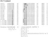
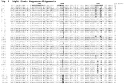
- the amino acid positions of CDRs that are underlined and in bold print in Figs. 6 and 7 are positions which have been optimized from the parent CDRs as shown in Fig 5 .
- the humanized antibody will comprise substantially all of at least one, and typically two, variable domains, in which all or substantially all of the amino acids in the CDR regions correspond to those of a non-human immunoglobulin and all or substantially all of the amino acids in the FR regions are those of a human immunoglobulin consensus sequence.
- the humanized antibody optimally also comprises at least a portion of an immunoglobulin constant region (Fc), typically that of a human immunoglobulin.
- Fc immunoglobulin constant region
- Humanized antibodies may be subjected to in vitro mutagenesis using methods of routine use in the art (or, when an animal transgenic for human Ig sequences is used, in vivo somatic mutagenesis) and, thus, the framework region amino acid sequences of the HCVR and LCVR regions of the humanized recombinant antibodies are sequences that, while derived from those related to human germline HCVR and LCVR sequences, may not naturally exist within the human antibody germline repertoire in vivo. It is contemplated that such amino acid sequences of the HCVR and LCVR framework regions of the humanized recombinant antibodies are at least 90%, 92%, 94%, 95%, 96%, 98% or most preferably at least 99% identical to a human germline sequence.
- those framework residues of the parent antibody e.g. , murine antibody or generally the antibody from which the humanized antibody is derived
- These residues may be identified e.g., by X-ray crystallography of the parent antibody or Fab fragment, thereby identifying the three-dimensional structure of the antigen-binding site.
- the humanized antibody may comprise or be derived from a human germline light chain framework.
- the light chain germline sequence is selected from human VK sequences including, but not limited to, A1, A10, A11, A14, A17, A18, A19, A2, A20, A23, A26, A27, A3, A30, A5, A7, B2, B3, L1, L10, L11, L12, L14, L15, L16, L18, L19, L2, L20, L22, L23, L24, L25, L4/18a, L5, L6, L8, L9, O1, O11, 012, 014, 018, 02, 04, and 08.
- this light chain human germline framework is selected from V1-11, V1-13, V1-16, V1-17, V1-18, V1-19, V1-2, V1-20, V1-22, V1-3, V1-4, V1-5, V1-7, V1-9, V2-1, V2-11, V2-13, V2-14, V2-15, V2-17, V2-19, V2-6, V2-7, V2-8, V3-2, V3-3, V3-4, V4-1, V4-2, V4-3, V4-4, V4-6, V5-1, V5-2, V5-4, and V5-6. See PCT WO 2005/005604 for a description of the different germline sequences.
- the light chain variable region and/or heavy chain variable region comprises a framework region or at least a portion of a framework region (e.g., containing 2 or 3 subregions, such as FR2 and FR3).
- at least FRL1, FRL2, FRL3, or FRL4 is fully human.
- at least FRH1, FRH2, FRH3, or FRH4 is fully human.
- at least FRL1, FRL2, FRL3, or FRL4 is a germline sequence (e.g., human germline) or comprises human consensus sequences for the particular framework.
- At least FRH1, FRH2, FRH3, or FRH4 is a germline sequence (e.g., human germline) or comprises human consensus sequences for the particular framework.
- the framework region is a human framework region.
- humanized antibodies may be produced by obtaining nucleic acid sequences encoding the HCVR and LCVR of an antibody, e.g., a murine antibody or antibody made by a hybridoma, which binds a myostatin epitope of the invention, identifying the CDRs in said HCVR and LCVR (nonhuman), and grafting such CDR-encoding nucleic acid sequences onto selected human framework-encoding nucleic acid sequences.
- a CDR region may be optimized by mutagenizing randomnly or at particular locations in order to substitute one or more amino acids in the CDR with a different amino acid prior to grafting the CDR region into the framework region.
- a CDR region may be optimized subsequent to insertion into the human framework region using methods available to one of skill in the art.
- the human framework amino acid sequences are selected such that the resulting antibody is likely to be suitable for in vivo administration in humans. This can be determined, e.g., based on previous usage of antibodies containing such human framework sequence.
- the human framework sequence will not itself be significantly immunogenic.
- the DNA sequence encoding the HCVR and LCVR regions of the preferably murine anti-myostatin antibody are obtained.
- Methods for cloning nucleic acid sequences encoding immunoglobulins are well known in the art. Such methods may, for example, involve the amplification of the immunoglobulin-encoding sequences to be cloned using appropriate primers by polymerase chain reaction (PCR). Primers suitable for amplifying immunoglobulin nucleic acid sequences, and specifically murine HCVR and LCVR sequences have been reported in the literature. After such immunoglobulin-encoding sequences have been cloned, they will be sequences by methods well known in the art.
- the resultant DNA sequences encoding the "humanized" variable heavy and variable light sequences are then expressed to produce a humanized Fv or humanized antibody that binds myostatin.
- the humanized HCVR and LCVR may be expressed as part of a whole anti-myostatin antibody molecule, i.e ., as a fusion protein with human constant domain sequences whose encoding DNA sequences have been obtained from a commercially available library or which have been obtained using, e.g., one of the above described methods for obtaining DNA sequences, or are in the art.
- HCVR and LCVR sequences can also be expressed in the absence of constant sequences to produce a humanized anti-myostatin Fv. Nevertheless, fusion of human constant sequences onto the variable region is potentially desirable because the resultant humanized anti-myostatin antibody may possess human effector functions.
- DNA sequences which encode the subject humanized HCVR and LCVR sequences are synthesized, and then expressed in a vector system suitable for expression of recombinant antibodies. This may be effected in any vector system which provides for the subject humanized HCVR and LCVR sequences to be expressed as a fusion protein with human constant domain sequences and to associate to produce functional (antigen binding) antibodies or antibody fragments.
- Human constant domain sequences are well known in the art, and have been reported in the literature.
- Preferred human constant light chain sequences include the kappa and lambda constant light chain sequences.
- Preferred human constant heavy chain sequences include human IgG 1 , human IgG 2 , human IgG 3 , human IgG 4 , and mutated versions thereof which provide for altered effector function, e.g., enhanced in vivo half-life, reduced Fc receptor binding, altered deamidation profile and the like.
- human framework regions are preferably derived from a human antibody variable region having sequence similarity to the analogous or equivalent region of the antigen binding region donor (i.e., the parent antibody).
- Other sources of framework regions for portions of human origin of a humanized antibody include human variable consensus sequences (see e.g., Kettleborough, C.A. et al. Protein Engineering 4:773-783 (1991 ); Carter et al., WO 94/04679 .
- the sequence of the antibody or variable region used to obtain the nonhuman portion can be compared to human sequences as described in Kabat et al. Sequences of Proteins of Immunological Interest, Fifth Edition, NIH, U.S. Government Printing Office (1991 ).
- the framework regions of a humanized antibody chain are derived from a human variable region having at least about 60% overall sequence identity, preferably at least about 70% overall sequence identity and more preferably at least about 85% overall sequence identity, with the variable region of the nonhuman donor.
- a human portion can also be derived from a human antibody having at least about 65% sequence identity, and preferably at least about 70% sequence identity, within the particular portion ( e.g., FR) being used, when compared to the equivalent portion ( e.g., FR) of the nonhuman donor.
- references further describing methods involved in humanizing a mouse antibody that maybe used are e.g., Queen et al., Proc. Natl. Acad. Sci. USA 88:2869, 1991 ; U.S. Pat. No. 5,693,761 ; U.S. Pat. No. 4,816,397 ; U.S. Pat. No. 5,225,539 ; computer programs ABMOD and ENCAD as described in Levitt, M., J. Mol. Biol.
- Antibodies of the present invention are useful in therapeutic, prophylactic and research applications as described herein.
- An antibody of the invention may be used to diagnose a disorder or disease associated with the expression of human myostatin.
- the antibody of the invention can be used in an assay to monitor myostatin levels in a subject being treated for a myostatin-associated condition.
- Research application include methods that utilize the antibody of the invention and a label to detect myostatin in a sample, e.g., in a human body fluid or in a cell or tissue extract.
- Antibodies of the invention may by used with or without modification, and are labeled by covalent or noncovalent attachment of a detectable moiety.
- the detectable moiety can be any one that is capable of producing, either directly or indirectly, a detectable signal.
- the detectable moiety may be a radioisotope such as, e.g., 3 H, 14 C, 32 P, 35 S, or 125 I, a fluorescent or chemiluminescent compound, such as fluorescein isothiocyanate, rhodamine, or luciferin; or an exzyme, such as alkaline phosphatase, beta-galactosidase, or horseradish peroxidase.
- any method known in the art for separately conjugating the antibody to the detectable moiety may be employed, including those methods described by Hunter, et al., Nature 144:945, 1962 ; David, et al., Biochemistry 13: 1014, 1974 ; Pain, et al., J. Immunol. Meth. 40: 219, 1981 ; and Nygren, J. Histochem. And Cytochem. 30: 407, 1982 .
- a variety of conventional protocols for measuring myostatin including e.g., ELISAs, RIAs, and FACS, are known in the art and provide a basis for diagnosing altered or abnormal levels of myostatin expression.
- Normal or standard expression values are established using any art known technique, e.g., by combining a sample comprising a myostatin polypeptide with, e.g., antibodies under conditions suitable to form a antigen:antibody complex.
- the antibody is directly or indirectly labeled with a detectable substance to facilitate detection of the bound or unbound antibody.
- Suitable detectable substances include various enzymes, prosthetic groups, fluorescent materials, luminescent materials and radioactive materials.
- suitable enzymes include horseradish peroxidase, alkaline phosphatase, ⁇ -galactosidase, or acetylcholinesterase;
- suitable prosthetic group complexes include streptavidin/biotin and avidin/biotin;
- suitable fluorescent materials include umbelliferone, fluorescein, fluorescein isothiocyanate, rhodamine, dichlorotriazinylamine fluorescein, dansyl chloride or phycoerythrin; an example of a luminescent material includes luminol; and examples of a radioactive material include 125 I, 131 I, 35 S, or 3 H.
- the amount of a standard complex formed is quantitated by various methods, such as, e.g., photometric means. Amounts of myostatin polypeptide expressed in samples are then compared with the standard values.
- the antibody of the present invention can be provided in a kit, a packaged combination of reagents in predetermined amounts with instructions for performing the diagnostic assay.
- the kit will include substrates and cofactors required by the enzyme (e.g., a substrate precursor which provides the detectable chromophore or fluorophore).
- substrates and cofactors required by the enzyme e.g., a substrate precursor which provides the detectable chromophore or fluorophore
- other additives may be included such as stabilizers, buffers (e.g., a blocking buffer or lysis buffer) and the like.
- the relative amounts of the various reagents may be varied widely to provide for concentrations in solution of the reagents which substantially optimize the sensitivity of the assay.
- the reagents may be provided as dry powders, usually lyophilized, including excipients which on dissolution will provide a reagent solution having the appropriate concentration.
- Myostatin plays a role in muscle development and a number of related disorders or diseases. In adults, myostatin mRNA is primarily detected in skeletal muscle although lower concentrations are also found in adipose tissue and cardiac tissue ( Sharma, M., et al, J. Cell Physiol. 180:1, 1999 ). Myostatin knockout mice have two- to three-fold greater muscle mass than their wild type littermates. The increased muscle mass is the result of fiber hypertrophy and hyperplasia ( McPherron, A., et al. Nature 387:83-90, 1997 and Zhu, X. et al., FEBS Letters 474:71 ).
- myostatin knockout mice accumulate less fat than their wild type littermates but otherwise appear normal and healthy.
- Myostatin has also been recently shown to be an important regulator of adipogenesis ( Rebbapragada, A., et al., Mol. and Cell. Bio. 23:7230-7242, 2003 ).
- bone structure and content has been recently studied in myostatin deficient mice ( Hamrick M.W., et al., J. Orthopaedic Research 21:1025, 2003 ; Hamrick, M.W., et al., Calcif Tissue Int 71:63, 2002 .
- a pharmaceutical composition comprising an anti-myostatin monoclonal antibody of the invention may be used to increase muscle mass, increase bone density, decrease muscle wasting, or may be useful for the treatment or prevention of conditions wherein the presence of myostatin causes or contributes to undesirable pathological effects or decrease of myostatin levels has a therapeutic benefit in mammals, preferably humans, including, but not limited to, muscle wasting, muscle injury, surgery, repair of damaged muscle, frailty, age-related sarcopenia, disuse atrophy, osteoporosis, osteoarthritis, ligament growth and repair, obesity, suppression of body fat accumulation, obesity, muscular dystrophy of any type, critical care myopaythy, alcoholic myopathy, cachexia (e.g., cancer-related or HIV-induced, or resulting from COPD, chronic lung disease, recovery from sepsis, renal failure, liver failure, cardiac failure or disease), metabolic syndrome, post-burn muscle wasting, and Type II diabetes.
- ma preferably humans, including, but not limited to, muscle wasting, muscle
- the antibodies of the invention may be used to increase muscle mass, increase bone density or treat or prevent conditions in non-human mammals or avian species [e.g., domestic animals (e.g., canine and feline), sports animals (e.g., equine), food-soiurce animals (e.g., bovine, porcine and ovine), avian species (e.g., chicken, turkey, other game birds or poultry)] wherein the presence of myostatin causes or contributes to undesirable pathological effects or decrease of myostatin levels has a therapeutic benefit.
- avian species e.g., domestic animals (e.g., canine and feline), sports animals (e.g., equine), food-soiurce animals (e.g., bovine, porcine and ovine), avian species (e.g., chicken, turkey, other game birds or poultry)
- an anti-myostatin monoclonal antibody of the present invention for treating or preventing of at least one of the aforementioned disorders in which myostatin activity is detrimental or which benefits for decreased levels of bioactive myostatin is contemplated herein. Additionally, the use of an anti-myostatin monoclonal antibody of the present invention for use in the manufacture of a medicament for the treatment of at least one of the aforementioned disorders is contemplated.
- treatment refers to obtaining a desired pharmacologic and/or physiologic effect.
- the effect may be prophylactic in terms of completely or partially preventing a disease or symptom thereof and/or may be therapeutic in terms of a partial or complete cure for a disease and/or adverse affect attributable to the disease.
- An antibody of the invention can be incorporated into pharmaceutical compositions suitable for administration to a subject.
- the compounds of the invention may be administered alone or in combination with a pharmaceutically acceptable carrier, diluent, and/or excipients, in single or multiple doses.
- the pharmaceutical compositions for administration are designed to be appropriate for the selected mode of administration, and pharmaceutically acceptable diluents, carrier, and/or excipients such as dispersing agents, buffers, surfactants, preservatives, solubilizing agents, isotonicity agents, stabilizing agents and the like are used as appropriate.
- compositions are designed in accordance with conventional techniques as in e.g., Remington, The Science and Practice of Pharmacy, 19th Edition, Gennaro, Ed., Mack Publishing Co., Easton, PA 1995 which provides a compendium of formulation techniques as are generally known to practitioners.
- a pharmaceutical composition comprising an anti-myostatin monoclonal antibody of the present invention can be administered to a subject at risk for or exhibiting pathologies as described herein using standard administration techniques including oral, intravenous, intraperitoneal, subcutaneous, pulmonary, transdermal, intramuscular, intranasal, buccal, sublingual, or suppository administration.
- a pharmaceutical composition of the invention preferably is a "therapeutically effective amount” or a “prophylactically effective amount” of an antibody of the invention.
- a “therapeutically effective amount” refers to an amount effective, at dosages and for periods of time necessary, to achieve the desired therapeutic result.
- a therapeutically effective amount of the antibody may vary according to factors such as the disease state, age, sex, and weight of the individual, and the ability of the antibody or antibody portion to elicit a desired response in the individual.
- a therapeutically effective amount is also one in which any toxic or detrimental effect of the antibody, are outweighed by the therapeutically beneficial effects.
- a “prophylactically effective amount” refers to an amount effective, at dosages and for periods of time necessary, to achieve the desired prophylactic result. Typically, since a prophylactic dose is used in subjects prior to or at an earlier stage of disease, the prophylactically effective amount will be less than the therapeutically effective amount.
- a therapeutically-effective or prophylactically-effective amount is at least the minimal dose, but less than a toxic dose, of an active agent which is necessary to impart therapeutic benefit to a subject.
- a therapeutically-effective amount of an antibody of the invention is an amount which in mammals, preferably humans, increases muscle mass, increases bone density, or treats conditions wherein the presence of myostatin causes or contributes to undesirable pathological effects or decrease in myostatin levels results in a beneficial therapeutic effect in a mammal, preferably a human, including, but not limited to, muscle wasting, muscle injury, surgery frailty, age-related sarcopenia, disuse atrophy, osteoporosis, osteoarthritis, ligament growth and repair, obesity, suppression of body fat accumulation, muscular dystrophy of any type, critical care myopaythy, cachexia (e.g., cancer-related or HIV-induced, or resulting from COPD, renal failure, liver failure, cardiac failure or disease), metabolic syndrome and Type II diabetes.
- cachexia e.g., cancer
- Disuse atrophy may result from numerous causes or incidents including any disorder or disease or state which leads to prolonged immobility or disuse or bed rest including, but not limited to, solid organ transplant, joint replacement, stroke, spinal cord injury, recovery from severe burn, sedentary chronic hemodialysis, post-sepsis recovery and exposure to micro gravity.
- the route of administration of an antibody of the present invention may be oral, parenteral, by inhalation, or topical.
- the antibodies of the invention can be incorporated into a pharmaceutical composition suitable for parenteral administration.
- parenteral as used herein includes intravenous, intramuscular, subcutaneous, rectal, vaginal, or intraperitoneal administration. Peripheral systemic delivery by intravenous or intraperitoneal or subcutaneous injection is preferred. Suitable vehicles for such injections are straightforward in the art.
- compositions typically must be sterile and stable under the conditions of manufacture and storage in the container provided, including e.g., a sealed vial or syringe. Therefore, pharmaceutical compositions may be sterile filtered after making the formulation, or otherwise made microbiologically acceptable.
- a typical composition for intravenous infusion could have a volume as much as 250-1000 ml of fluid, such as sterile Ringer's solution, physiological saline, dextrose solution and Hank's solution and a therapeutically effective dose, ( e.g., 1 to 100 mg/mL, or more) of antibody concentration. Dose may vary depending on the type and severity of the disease.
- dosages for any one subject depends upon many factors, including the patient's size, body surface area, age, the particular compound to be administered, sex, time and route of administration, general health, and other drugs being administered concurrently.
- a typical dose can be, for example, in the range of 0.001 to 1000 ⁇ g; however, doses below or above this exemplary range are envisioned, especially considering the aforementioned factors.
- the daily parenteral dosage regimen can be about 0.1 ⁇ g/kg to about 100 mg/kg of total body weight, preferably from about 0.3 ⁇ g/kg to about 10 mg/kg and more preferably from about 1 ⁇ g/kg to 1 mg/kg, even more preferably from about 0.5 to 10 mg/kg body weight per day.
- the desired dosage can be delivered by a single bolus administration, by multiple bolus administrations, or by continuous infusion administration of antibody, depending on the pattern of pharmacokinetic decay that the practitioner wishes to achieve.
- the label on, or associated with, the container indicates that the composition is used for treating the condition of choice.
- the article of manufacture may further comprise a second container comprising a pharmaceutically-acceptable buffer, such as phosphate-buffered saline, Ringer's solution and dextrose solution. It may further include other materials desirable from a commercial and user standpoint, including other buffers, diluents, filters, needles, syringes, and package inserts with instructions for use.
- Mouse/human chimeric anti-myostatin Fabs of the present invention are tested in an ELISA assay, in which binding of the Fab to mature myostatin (dimeric form) coated at various concentrations on a 96-well plate is measured. Binding of the Fabs to GDF-11 is also tested.
- Each well of two 96-well plates is coated with 50 ⁇ l of recombinant human myostatin (R&D systems, carrier-free, re-supended first in 4mM HCl and then coated at 1 ⁇ g/ml in carbonate buffer, pH 9.6) or 50 ⁇ l recombinant human GDF-11 (Peprotech, Inc., Cat. # 120-11, carrier-free, re-suspended first in 4mM HCl and then coated at 1 ⁇ g/ml in carbonate buffer, pH 9.6).
- the plates are incubated at 4°C overnight.
- the wells are aspirated and washed twice with PBST (PBS + 0.1% Tween-20).
- the plates are blocked with 200 ⁇ l blocking buffer per well (1% BSA in PBST for 1 hour).
- Fabs from periplasmic extracts to be tested are serially diluted into PBST. Fifty microliters of each Fab solution is added to the GDF-8 and GDF-11 coated plate columns. The plates are incubated for 1 hour at room temperature. The wells are then washed 3 times with PBST.
- Alkaline phosphatase-conjugated secondary antibody 50 ⁇ l goat anti-mouse kappa AP (Southern Biotech), diluted 1:1000 in PBST is added to each well and incubated for 30 minutes at room temperature. The wells are then washed 3 times with PBST. Fifty microliters of chromogenic substrate (AMP/PMP) is added to each well and allowed to develop at room temperature. The absorbance of the wells is read at OD of 560 nm. For each Fab a titration curve is obtained an the relative OD at the mid point of of the reference Fab curve reported.
- AMP/PMP chromogenic substrate
- a 96-well plate is coated with 50 ⁇ l of goat-anti-human kappa antibody at 2 ⁇ g per ml in carbonate buffer. The plates are incubated at 4°C overnight. The wells are aspirated and washed twice with PBST (PBS + 0.1% Tween-20). The plates are blocked with 200 ⁇ l blocking buffer per well (1% BSA in PBST for 1 hour)
- Fabs from periplasmic extracts to be tested are captured in columns on the plates for 2 hours at 37°C. After washing 3 times with PBST , 50 ⁇ l of 2 fold serial dilutions of biotinylated Myostatin (from 100nM to 780pM) are added to each column of captured Fabs and incubated for 1 hour at 37°C. The plate is then washed with PBST and incubated with PBST at 37 degrees C for 1-3 hours.
- Alkaline phosphatase-conjugated Neutravidin (Pierce, diluted 1:1000 in PBST) is added to each well and incubated for 2 minutes at room temperature. The wells are then washed 3 times with PBST. 50 ⁇ l of chromogenic substrate (AMP/PMP) is added to each well and allowed to develop at room temperature. The absorbance of the wells is read at an OD of 560 nm. And the relative OD compared to that of the maximum OD for the reference Fab reported.
- AMP/PMP chromogenic substrate
- Ectodermal explants are removed from stage 8-9 blastula Xenopus embryos by standard procedures and cultured in 0.5X MBS (IX MBS: 88 mM NaCl, 1 mM KCI, 0.7 mM CaCl 2 , 1 mM MgSO 4 , 5 mM HEPES, 2.5 mM NaHCO 3 , 1:1000 v/v gentamycin, 0.1% bovine serum albumin) with the addition of growth factor (GDF8 or GDF11) plus or indicated, for 18 hours at 18°C, by which time control embryos reach the early neurula stage (stage 15-16). Explants are photographed and the length of each explant is measured using an image analysis algorithm designed for animal cap quantitation.
- IX MBS 88 mM NaCl, 1 mM KCI, 0.7 mM CaCl 2 , 1 mM MgSO 4 , 5 mM HEPES, 2.5 mM NaHCO 3 , 1:1000 v/v
- Explants not treated with either growth factor or Fab (controls), round into balls of epidermis.
- Myostatin and GDF-11 induce mesoderm in these ectodermal explants which causes the explants to elongate and form dumbbell-like structures.
- Antibodies or Fabs when tested for neutralizing activity, are added to the culture medium containing myostatin for the entire length of the culture period and their ability to inhibit the growth factor-induced elongation movements is assessed.
- Myostatin is added to the explants at 25 ng/ml.
- Antibodies or Fabs to be tested are added at 20 ⁇ g/ml.
- a Fab generated to an irrelevant antigen is used as a control.
- a commercially available monoclonal anti-mouse GDF8 antibody may be tested as a control, this antibody is produced in goats immunized with purified mouse GDF8 and demonstrated by the manufacturer to neutralize elongation of Xenopus animal caps elicited by 25 ng/ml of murine GDF8 when present at about 10-20 ⁇ g/ml (R&D Systems Cat. #MAB788).
- ImagePro (v4.5.1.22, from Media Cybernetics) is used for the image processing.
- a macro is written to automate the image processing.
- the macro processes the image and records length in units of bits.
- Alternative measuring methods may be used as known in the art.
- Antibodies of the invention are contemplated to neutralize GDF8 activity in the animal cap assay.
- the affinity (K D ) and k on and k off rates of anti-myostatin Fabs of the present invention are measured using a BIAcore® 2000 instrument containing a CM4 sensor chip.
- the BIAcore® utilizes the optical properties of surface plasmon resonance to detect alterations in protein concentration of interacting molecules within a dextran biosensor matrix. Except where noted, all reagents and materials are purchased from BIAcore® AB (Upsala, Sweden). All measurements are performed at 25°C. Samples containing Fabs are dissolved in HBS-EP buffer (150 mM sodium chloride, 3 mM EDTA, 0.05% (w/v) surfactant P-20, and 10 mM HEPES, pH 7.4).
- Each analysis cycle consists of (i) 250 ⁇ l injection of a Fab (concentration range of 50 nM to 0.4 nM in 2-fold dilution increments) over all 4 flow cells with flow cell 1 as the reference flow cell, (ii) 20 min dissociation (buffer flow), (iii) regeneration of of the GDF-8 or GDF-11 surface with a two 15 ⁇ l injection of 10 mM glycine, pH 1.5, (iv) a 15 ⁇ l blank injection of running buffer, and (v) a 2 min stabilization time before start of next cycle.
- Fab concentration range of 50 nM to 0.4 nM in 2-fold dilution increments
- the Fabs 41-1 and 412-6 when measured in the above assay, have affinities for GDF-8 of 4.16 nM (4.16 x 10 -9 M) and 0.46 nM (4.6 x 10 -10 M) respectively, and had affinities for GDF-11 of 8.96 nM and 0.81 nM respectively; the relative specificity for both Fabs was approximately a 2-fold preference for GDF-8 relative to GDF-11.
- Binding affinity measurements for full-length monoclonal antibodies of the invention are determined using a Sapidyne KINEXA assay.
- NHS-activated fast-flow sepharose beads (GE Healthcare) are pre-coated with an antibody of the invention (50 ⁇ g anti-myostatin antibody per ml of beads) and blocked with 10 mg/ml BSA in 1 M Tris-HCl, pH 8.0.
- 2 pM, 4 pM, 40 pM of an antibody of the invention (e.g., 3-74/C1E4) is incubated with various concentration (e.g., 2.4 pM to 10 nM, serial dilutions) of myostatin in running buffer (PBS, 0.005% (v/v) Tween-20 and 1 mg/ml ovalbumin) for 10 hours at room temperature.
- concentration e.g., 2.4 pM to 10 nM, serial dilutions
- myostatin in running buffer (PBS, 0.005% (v/v) Tween-20 and 1 mg/ml ovalbumin) for 10 hours at room temperature.
- PBS running buffer
- 0.005% (v/v) Tween-20 and 1 mg/ml ovalbumin 1 mg/ml ovalbumin
- the measured fluorescence signal is proportional to the concentration of free antibody at equilibrium. Each concentration of myostatin is measured in duplicate.
- the equilibrium dissociation constant (K D ) is obtained from non-linear regression of the competition curves using a multiple-curve, one-site homogeneous binding model (KINEXA software).
- a plasmid encoding a reporter gene i.e., luciferase gene
- a reporter gene i.e., luciferase gene
- CAGA SMAD binding element
- a molecule such as myostatin, GDF-11, or other TGF- ⁇ superfamily member binds its own receptor, thereby triggering SMAD signaling which results in a phosphorylated SMAD complex which is capable of binding the SBE.
- the CAGA sequence was previously reported to be a TGF- ⁇ responsive sequence within the promoter of the TGF- ⁇ induced gene PAI-1 ( Denner et al., EMBO J., 17:3091-3100, 1998 ).
- a Myostatin/SBE Reporter Assay not be limited to the exact conditions described herein, other types of cells may be used, e.g., 293HEK (ATCC) or A204 Rhabsomyosarcoma cells (see, e.g., Whittemore, et al. BBRC, 200:965-71, 2003 ); other types of reporters may be used, e.g., CAT, ⁇ -gal, GFP, and other growth conditions for the cells and assay conditions including varying amounts of myostatin in the reaction may be used.
- 293HEK ATCC
- A204 Rhabsomyosarcoma cells see, e.g., Whittemore, et al. BBRC, 200:965-71, 2003
- other types of reporters e.g., CAT, ⁇ -gal, GFP, and other growth conditions for the cells and assay conditions including varying amounts of myostatin in the reaction may be used.
- 293E cells (Edge Biosystems) in a T-75 flask are grown in DMEM/F12 media (1:1) (Gibco 10565-042) and 10% FBS.
- the cells are transfected with a mix of 100 ⁇ l lipofectamine 2000 (Invitrogen 11668-019), 5 ml OptiMEM I (Gibco 51985-034) and 30 ⁇ g SB-luciferase DNA for 4 hours at 37°C.
- the transfection mix is then removed and complete media added for 1 hour at 37°C.
- the cells are then trypsinized and re-suspended in complete media at 2 x 10 6 cells/ml and 50 ⁇ l plated in each well of a Biocoat 96-well plate (BD 35-6461) and incubated for 1 hour at 37°C. After the incubation is complete, media is replaced with 100 ⁇ l of each Fab to be tested that is serially diluted 1:2 and pre-incubated for 1 hour at 37°C with a 1:1 solution of 40 ng/ml myostatin (R&D Systems 788-G8) or GDF-11 (R&D Systems) in complete media.
- the plate is left overnight at 37°C, 5% CO 2 and the following day 100 ⁇ l of a 1:1 mix of Glo Lysis Buffer and Bright-Glo Luciferase reagent (Promega) is added to each well and mixed by pipeting. From this mix, 150 ⁇ l is transferred to a white 96-well plate and luminescence measured using a luminometer. Luminescence is then plotted against Fab concentration and the IC 50 for each Fab for myostatin and GDF-11 is calculated.
- Fabs tested using the above-described conditions yield IC 50 values as listed in Table 4 below.
- Table 4 Fab IC 50 GDF8 IC 50 GDF11 41H-A7 10 nM 40 nM 41L3F12 40 nM 100 nM 41C1A2 1 nM 1.2 nM 41C1A4 1 nM 2nM 41C1E4 1 nM 4nM 41C12A4 1 nM 4nM 41C2A4 1 nM 4nM 3-74/C1E4 1 nM 4nM
- PK pharmacokinetics
- IV intravenous
- IP intraperitoneal
- the animals receive a mixture of unlabeled and 125 I-labeled antibodyat a dose described above and serum concentration is determined based on 125 I radioactivity in the serum and the specific activity of the injected dose. Serum concentration of the antibody administered either IV or IP versus time is plotted.
- an antibody of the invention may be tested in adult SCID mice.
- SCID mice suffer from severe combined immune deficiency, and therefore do not generate an immunological reaction following injection of an antibody of the invention.
- Muscle mass is used as an indicator for myostatin activity in mice treated with an antibody of the invention.
- mice Female SCID/CB17 mice (Taconic Biotechnology) are weighed and distributed into groups of ten.
- An antibody of the invention (41C1E4) in PBS buffer is injected subcutaneously into the mice at various doses (10, 5, and 2 mg/kg) on days 0 and 7.
- IgG In a control group, IgG at 10 mg/kg is injected subcutaneously into the mice on days 0 and 7.
- muscle strength the strength of the front limb, is measured with a grip strength test meter (e.g., model 1027 csx, Columbus Instruments).
- the animals are terminated and muscle mass is assessed by nuclear magnetic resonance (NMR).
- NMR nuclear magnetic resonance
- the gastrocnemius and quadriceps muscle wet weights are also measured as well as body weight.
Claims (7)
- Anticorps anti-myostatine comprenant un polypeptide à région variable de chaîne lourde de SEQ ID NO: 123 et un polypeptide à région variable de chaîne légère de SEQ ID NO: 98.
- Anticorps anti-myostatine selon la revendication 1, dans lequel l'anticorps comprend en outre une région constante de chaîne lourde choisie parmi une IgG1, une IgG2, une IgG3, une IgG4, une IgA, une IgE, une IgM et une IgD.
- Anticorps anti-myostatine selon la revendication 2, dans lequel la région constante présente dans l'anticorps provient du génome d'un animal choisi parmi les animaux domestiques, les animaux utilisés pour le sport et les animaux sources d'aliments.
- Composition pharmaceutique comprenant l'anticorps selon l'une quelconque des revendications 1 à 3.
- Composition pharmaceutique selon la revendication 4, comprenant en outre un support pharmaceutiquement acceptable.
- Anticorps anti-myostatine selon l'une quelconque des revendications 1 à 3 ou une composition pharmaceutique selon la revendication 4 ou la revendication 5 pour une utilisation en tant que médicament.
- Anticorps anti-myostatine selon l'une quelconque des revendications 1 à 3 ou une composition pharmaceutique selon la revendication 4 ou la revendication 5 pour une utilisation dans le traitement ou la prévention d'une ou plusieurs affections choisies parmi la fragilité, la cachexie, la fonte musculaire, la faiblesse musculaire, la myopathie, la dystrophie musculaire, l'ostéoporose, la BPCO, une insuffisance ou une maladie rénale, une insuffisance ou une maladie hépatique, une insuffisance cardiaque, un diabète de type II ou un syndrome métabolique.
Priority Applications (3)
| Application Number | Priority Date | Filing Date | Title |
|---|---|---|---|
| RS20180031A RS56755B1 (sr) | 2005-10-12 | 2006-10-05 | Anti-miostatin antitela |
| SI200632235T SI2816058T1 (en) | 2005-10-12 | 2006-10-05 | Antibodies against myostatin |
| PL14181801T PL2816058T3 (pl) | 2005-10-12 | 2006-10-05 | Przeciwciała przeciwko miostatynie |
Applications Claiming Priority (2)
| Application Number | Priority Date | Filing Date | Title |
|---|---|---|---|
| US72573805P | 2005-10-12 | 2005-10-12 | |
| EP06816225.4A EP1951755B1 (fr) | 2005-10-12 | 2006-10-05 | Anticorps anti-myostatine |
Related Parent Applications (2)
| Application Number | Title | Priority Date | Filing Date |
|---|---|---|---|
| EP06816225.4A Division EP1951755B1 (fr) | 2005-10-12 | 2006-10-05 | Anticorps anti-myostatine |
| EP06816225.4A Division-Into EP1951755B1 (fr) | 2005-10-12 | 2006-10-05 | Anticorps anti-myostatine |
Publications (2)
| Publication Number | Publication Date |
|---|---|
| EP2816058A1 EP2816058A1 (fr) | 2014-12-24 |
| EP2816058B1 true EP2816058B1 (fr) | 2017-12-13 |
Family
ID=37681692
Family Applications (2)
| Application Number | Title | Priority Date | Filing Date |
|---|---|---|---|
| EP06816225.4A Active EP1951755B1 (fr) | 2005-10-12 | 2006-10-05 | Anticorps anti-myostatine |
| EP14181801.3A Active EP2816058B1 (fr) | 2005-10-12 | 2006-10-05 | Anticorps anti-myostatine |
Family Applications Before (1)
| Application Number | Title | Priority Date | Filing Date |
|---|---|---|---|
| EP06816225.4A Active EP1951755B1 (fr) | 2005-10-12 | 2006-10-05 | Anticorps anti-myostatine |
Country Status (23)
| Country | Link |
|---|---|
| US (2) | US7632499B2 (fr) |
| EP (2) | EP1951755B1 (fr) |
| JP (1) | JP5031756B2 (fr) |
| KR (1) | KR101128548B1 (fr) |
| CN (1) | CN101282992B (fr) |
| AU (1) | AU2006303921B2 (fr) |
| BR (1) | BRPI0617345A2 (fr) |
| CA (1) | CA2624935C (fr) |
| DK (2) | DK2816058T3 (fr) |
| EA (1) | EA015903B1 (fr) |
| ES (2) | ES2657292T3 (fr) |
| HK (1) | HK1118564A1 (fr) |
| HR (1) | HRP20150176T1 (fr) |
| HU (1) | HUE036566T2 (fr) |
| IL (1) | IL189580A (fr) |
| LT (1) | LT2816058T (fr) |
| NO (1) | NO20082167L (fr) |
| PL (2) | PL1951755T3 (fr) |
| PT (2) | PT2816058T (fr) |
| RS (2) | RS56755B1 (fr) |
| SI (2) | SI2816058T1 (fr) |
| UA (1) | UA92504C2 (fr) |
| WO (1) | WO2007047112A2 (fr) |
Families Citing this family (45)
| Publication number | Priority date | Publication date | Assignee | Title |
|---|---|---|---|---|
| KR20150107899A (ko) | 2002-12-20 | 2015-09-23 | 암겐 인코포레이티드 | 미오스타틴을 저해하는 결합제 |
| CA2624976A1 (fr) | 2005-10-06 | 2007-04-19 | Eli Lilly And Company | Anticorps anti-myostatine |
| UA92504C2 (en) | 2005-10-12 | 2010-11-10 | Эли Лилли Энд Компани | Anti-myostatin monoclonal antibody |
| CN104761637B (zh) | 2006-03-31 | 2021-10-15 | 中外制药株式会社 | 调控抗体血液动力学的方法 |
| JP5058261B2 (ja) * | 2006-09-05 | 2012-10-24 | イーライ リリー アンド カンパニー | 抗ミオスタチン抗体 |
| EP3127921A1 (fr) | 2007-09-26 | 2017-02-08 | Chugai Seiyaku Kabushiki Kaisha | Procédé de modification d'un anticorps par point isoélectrique par substitution d'acide aminé dans cdr |
| PE20091163A1 (es) * | 2007-11-01 | 2009-08-09 | Wyeth Corp | Anticuerpos para gdf8 |
| TWI564021B (zh) | 2008-04-11 | 2017-01-01 | Chugai Pharmaceutical Co Ltd | Repeated binding of antigen to antigen binding molecules |
| JP5856480B2 (ja) | 2008-06-25 | 2016-02-09 | エスバテック − ア ノバルティスカンパニー エルエルシー | 汎用性抗体フレームワークを用いたイエウサギ抗体のヒト化 |
| CN102076716A (zh) | 2008-06-25 | 2011-05-25 | 艾斯巴技术,爱尔康生物医药研究装置有限责任公司 | 抑制TNFα的稳定和可溶的抗体 |
| AR074777A1 (es) * | 2008-12-19 | 2011-02-09 | Glaxo Group Ltd | Proteinas de union a antigeno |
| JO3340B1 (ar) | 2010-05-26 | 2019-03-13 | Regeneron Pharma | مضادات حيوية لـعامل تمايز النمو 8 البشري |
| TW201210612A (en) | 2010-06-03 | 2012-03-16 | Glaxo Group Ltd | Humanised antigen binding proteins |
| MX2013001845A (es) | 2010-08-16 | 2013-08-27 | Amgen Inc | Polipetidos que enlazan miostatina, composiciones y metodos. |
| UY33679A (es) | 2010-10-22 | 2012-03-30 | Esbatech | Anticuerpos estables y solubles |
| TWI761912B (zh) | 2010-11-30 | 2022-04-21 | 日商中外製藥股份有限公司 | 具有鈣依存性的抗原結合能力之抗體 |
| JP6130307B2 (ja) | 2011-03-17 | 2017-05-17 | ザ ユニバーシティ オブ バーミンガム | 再指向性免疫療法 |
| EP2554353B1 (fr) | 2011-08-05 | 2017-06-07 | Trisa Holding AG | Procédé de fabrication d'une brosse à dents et brosse à dents |
| HUE038570T2 (hu) | 2011-11-14 | 2018-10-29 | Regeneron Pharma | Készítmények és eljárások izomtömeg és izomerõ növelésére GDF8 és/vagy aktivin A specifikus antagonizálásával |
| WO2014030750A1 (fr) | 2012-08-24 | 2014-02-27 | 中外製薬株式会社 | ANTICORPS Fc SPÉCIFIQUE À FcyRII DE SOURIS |
| CN113831406A (zh) | 2012-08-24 | 2021-12-24 | 中外制药株式会社 | FcγRIIb特异性Fc区变体 |
| NZ628446A (en) | 2012-09-13 | 2017-06-30 | Bristol Myers Squibb Co | Fibronectin based scaffold domain proteins that bind to myostatin |
| US20150266950A1 (en) | 2012-10-24 | 2015-09-24 | Celgene Corporation | Methods for treating anemia |
| JPWO2014119634A1 (ja) * | 2013-01-30 | 2017-01-26 | 国立研究開発法人国立精神・神経医療研究センター | 筋増加剤及びそれを含む医薬組成物 |
| CA2908350C (fr) | 2013-04-02 | 2023-08-08 | Futa Mimoto | Variant de region fc |
| CA2911514A1 (fr) | 2013-05-06 | 2014-11-13 | Scholar Rock, Inc. | Compositions et procedes de modulation du facteur de croissance |
| TWI655207B (zh) | 2013-07-30 | 2019-04-01 | 再生元醫藥公司 | 抗活化素a之抗體及其用途 |
| JP2017510622A (ja) | 2014-01-27 | 2017-04-13 | ノバルティス アーゲー | 筋萎縮を予測するバイオマーカー、方法および使用 |
| US10307480B2 (en) | 2014-11-06 | 2019-06-04 | Scholar Rock, Inc. | Anti-pro/latent-myostatin antibodies and uses thereof |
| WO2016073906A2 (fr) * | 2014-11-06 | 2016-05-12 | Scholar Rock, Inc. | Immunoessais liés à des facteurs de croissance transformants |
| PE20171111A1 (es) | 2014-12-19 | 2017-08-07 | Chugai Pharmaceutical Co Ltd | Anticuerpos antimiostatina, polipeptidos que contienen regiones fc variantes, y metodos de uso |
| EA201791754A1 (ru) | 2015-02-05 | 2019-01-31 | Чугаи Сейяку Кабусики Кайся | АНТИТЕЛА, СОДЕРЖАЩИЕ ЗАВИСЯЩИЙ ОТ КОНЦЕНТРАЦИИ ИОНОВ АНТИГЕНСВЯЗЫВАЮЩИЙ ДОМЕН, ВАРИАНТЫ Fc-ОБЛАСТИ, IL-8-СВЯЗЫВАЮЩИЕ АНТИТЕЛА И ИХ ПРИМЕНЕНИЯ |
| US20180031579A1 (en) | 2015-02-12 | 2018-02-01 | INSERM (Institut National de la Santé et de la Recherche Médicale) | Methods for predicting the responsiveness of a patient affected with malignant hematological disease to chemotherapy treatment and methods of treatment of such disease |
| CA2982810A1 (fr) * | 2015-04-15 | 2016-10-20 | Regeneron Pharmaceuticals, Inc. | Methode pour augmenter la resistance et la fonctionnalite avec des inhibiteurs de gdf-8 |
| CN113896789A (zh) | 2015-09-15 | 2022-01-07 | 供石公司 | 抗-原肌生长抑制素/潜伏肌生长抑制素抗体及其用途 |
| US11359009B2 (en) | 2015-12-25 | 2022-06-14 | Chugai Seiyaku Kabushiki Kaisha | Anti-myostatin antibodies and methods of use |
| JP2019504064A (ja) | 2016-01-08 | 2019-02-14 | スカラー ロック インコーポレイテッドScholar Rock,Inc. | 抗プロ/潜在型ミオスタチン抗体およびその使用方法 |
| JP6823167B2 (ja) | 2016-06-13 | 2021-01-27 | スカラー ロック インコーポレイテッドScholar Rock,Inc. | ミオスタチン阻害剤の使用および併用療法 |
| TW202228778A (zh) | 2016-06-17 | 2022-08-01 | 日商中外製藥股份有限公司 | 抗肌抑素抗體及使用方法 |
| WO2018025982A1 (fr) | 2016-08-05 | 2018-02-08 | 中外製薬株式会社 | Composition pour la prophylaxie ou le traitement de maladies liées à il-8 |
| HUE061656T2 (hu) | 2017-01-06 | 2023-07-28 | Scholar Rock Inc | Metabolikus betegségek kezelése a miosztatin aktiválás gátlásával |
| AU2019226551A1 (en) | 2018-03-01 | 2020-10-15 | Regeneron Pharmaceuticals, Inc. | Methods for altering body composition |
| JP2022514262A (ja) | 2018-12-17 | 2022-02-10 | レビトープ リミテッド | 双子型免疫細胞エンゲージャー |
| CN113195532A (zh) | 2018-12-18 | 2021-07-30 | 瑞泽恩制药公司 | 使用针对瘦素受体、gdf8和活化素a的拮抗剂增加体重和瘦肌肉质量的组合物和方法 |
| WO2024064842A1 (fr) | 2022-09-21 | 2024-03-28 | Regeneron Pharmaceuticals, Inc. | Méthodes de traitement de l'obésité, du diabète et de la dysfonction hépatique |
Family Cites Families (62)
| Publication number | Priority date | Publication date | Assignee | Title |
|---|---|---|---|---|
| GB8308235D0 (en) | 1983-03-25 | 1983-05-05 | Celltech Ltd | Polypeptides |
| US4816567A (en) | 1983-04-08 | 1989-03-28 | Genentech, Inc. | Recombinant immunoglobin preparations |
| US5807715A (en) | 1984-08-27 | 1998-09-15 | The Board Of Trustees Of The Leland Stanford Junior University | Methods and transformed mammalian lymphocyte cells for producing functional antigen-binding protein including chimeric immunoglobulin |
| GB8422238D0 (en) | 1984-09-03 | 1984-10-10 | Neuberger M S | Chimeric proteins |
| GB8607679D0 (en) | 1986-03-27 | 1986-04-30 | Winter G P | Recombinant dna product |
| US5225539A (en) | 1986-03-27 | 1993-07-06 | Medical Research Council | Recombinant altered antibodies and methods of making altered antibodies |
| US4946778A (en) | 1987-09-21 | 1990-08-07 | Genex Corporation | Single polypeptide chain binding molecules |
| GB8823869D0 (en) | 1988-10-12 | 1988-11-16 | Medical Res Council | Production of antibodies |
| US5175384A (en) | 1988-12-05 | 1992-12-29 | Genpharm International | Transgenic mice depleted in mature t-cells and methods for making transgenic mice |
| US5530101A (en) | 1988-12-28 | 1996-06-25 | Protein Design Labs, Inc. | Humanized immunoglobulins |
| US5545806A (en) | 1990-08-29 | 1996-08-13 | Genpharm International, Inc. | Ransgenic non-human animals for producing heterologous antibodies |
| US5661016A (en) | 1990-08-29 | 1997-08-26 | Genpharm International Inc. | Transgenic non-human animals capable of producing heterologous antibodies of various isotypes |
| US5789650A (en) | 1990-08-29 | 1998-08-04 | Genpharm International, Inc. | Transgenic non-human animals for producing heterologous antibodies |
| ES2108048T3 (es) | 1990-08-29 | 1997-12-16 | Genpharm Int | Produccion y utilizacion de animales inferiores transgenicos capaces de producir anticuerpos heterologos. |
| US5814318A (en) | 1990-08-29 | 1998-09-29 | Genpharm International Inc. | Transgenic non-human animals for producing heterologous antibodies |
| US5633425A (en) | 1990-08-29 | 1997-05-27 | Genpharm International, Inc. | Transgenic non-human animals capable of producing heterologous antibodies |
| WO1992003917A1 (fr) | 1990-08-29 | 1992-03-19 | Genpharm International | Recombinaison homologue dans des cellules de mammiferes |
| US5625126A (en) | 1990-08-29 | 1997-04-29 | Genpharm International, Inc. | Transgenic non-human animals for producing heterologous antibodies |
| US5770429A (en) | 1990-08-29 | 1998-06-23 | Genpharm International, Inc. | Transgenic non-human animals capable of producing heterologous antibodies |
| US6284471B1 (en) | 1991-03-18 | 2001-09-04 | New York University Medical Center | Anti-TNFa antibodies and assays employing anti-TNFa antibodies |
| WO1994004679A1 (fr) | 1991-06-14 | 1994-03-03 | Genentech, Inc. | Procede pour fabriquer des anticorps humanises |
| WO1993004169A1 (fr) | 1991-08-20 | 1993-03-04 | Genpharm International, Inc. | Ciblage de genes dans des cellules animales au moyen de produits de synthese d'adn isogeniques |
| EP0626012B1 (fr) | 1992-02-11 | 2003-07-09 | Cell Genesys, Inc. | Obtention d'homozygotes par ciblage de genes |
| US5994618A (en) | 1997-02-05 | 1999-11-30 | Johns Hopkins University School Of Medicine | Growth differentiation factor-8 transgenic mice |
| US7393682B1 (en) | 1993-03-19 | 2008-07-01 | The Johns Hopkins University School Of Medicine | Polynucleotides encoding promyostatin polypeptides |
| EP1333035A3 (fr) | 1993-03-19 | 2004-07-07 | The Johns Hopkins University School Of Medicine | Facteur GDF-8 de différentiation de croissance |
| DE69433530T2 (de) | 1993-05-12 | 2005-01-05 | Genetics Institute, LLC, Cambridge | Bmp-11 zusammensetzungen |
| US5625825A (en) | 1993-10-21 | 1997-04-29 | Lsi Logic Corporation | Random number generating apparatus for an interface unit of a carrier sense with multiple access and collision detect (CSMA/CD) ethernet data network |
| ATE305036T1 (de) | 1994-07-08 | 2005-10-15 | Univ Johns Hopkins Med | Wachstums-differenzierungsfaktor-11 |
| US6008434A (en) | 1994-07-08 | 1999-12-28 | Johns Hopkins University School Of Medicine | Growth differentiation factor-11 transgenic mice |
| US5698762A (en) | 1994-12-09 | 1997-12-16 | Dauerman; Leonard | Microwave-assisted pyrolysis of waste polyaromatic hydrocarbons |
| EP1916300A1 (fr) | 1995-08-29 | 2008-04-30 | Kirin Pharma Kabushiki Kaisha | Animal chimère et son procédé de construction |
| US20020165350A1 (en) | 1996-11-13 | 2002-11-07 | Tasuku Honjo | Novel polypeptide, a method of producing it, and utility of the polypeptide |
| US6656475B1 (en) * | 1997-08-01 | 2003-12-02 | The Johns Hopkins University School Of Medicine | Growth differentiation factor receptors, agonists and antagonists thereof, and methods of using same |
| WO1999006559A1 (fr) | 1997-08-01 | 1999-02-11 | The Johns Hopkins University School Of Medicine | Procedes d'identification des recepteurs des facteurs de differenciation de la croissance |
| US6696260B1 (en) | 1997-08-01 | 2004-02-24 | The Johns Hopkins University School Of Medicine | Methods to identify growth differentiation factor (GDF) binding proteins |
| WO1999024057A2 (fr) | 1997-11-07 | 1999-05-20 | Genetics Inst | Utilisations neuronales de bmp-11 |
| GB2333706A (en) | 1998-02-02 | 1999-08-04 | Merck & Co Inc | Method for increasing muscle mass in animals |
| WO1999040181A1 (fr) | 1998-02-05 | 1999-08-12 | The Johns Hopkins University School Of Medicine | Facteur-8 de differenciation de la croissance |
| CA2331410C (fr) | 1998-05-06 | 2009-07-14 | Metamorphix, Inc. | Methodes de traitement du diabete par inhibition du gdf-8 |
| BR0008188A (pt) | 1999-01-21 | 2002-02-13 | Metamorphix Inc | Inibidores de fator de diferenciação de crescimento e usos para os mesmos |
| US7320789B2 (en) | 2001-09-26 | 2008-01-22 | Wyeth | Antibody inhibitors of GDF-8 and uses thereof |
| US20040138118A1 (en) | 2002-09-16 | 2004-07-15 | Neil Wolfman | Metalloprotease activation of myostatin, and methods of modulating myostatin activity |
| US7261893B2 (en) | 2002-10-22 | 2007-08-28 | Wyeth | Neutralizing antibodies against GDF-8 and uses therefor |
| KR20150107899A (ko) | 2002-12-20 | 2015-09-23 | 암겐 인코포레이티드 | 미오스타틴을 저해하는 결합제 |
| BRPI0410927A (pt) | 2003-06-02 | 2006-06-27 | Wyeth Corp | métodos terapêuticos e profiláticos para distúrbios neuromusculares |
| EP1644416A4 (fr) | 2003-06-30 | 2007-08-29 | Centocor Inc | Proteines derivees de l'immunoglobuline anti-cible et genetiquement modifiees, compositions, procedes et utilisations |
| NZ529860A (en) | 2003-11-28 | 2006-10-27 | Ovita Ltd | Muscle growth regulator mighty and use in promoting muscle mass and treating muscle wasting diseases |
| SG153874A1 (en) | 2003-12-31 | 2009-07-29 | Schering Plough Ltd | Neutralizing epitope-based growth enhancing vaccine |
| ATE557042T1 (de) | 2004-03-23 | 2012-05-15 | Lilly Co Eli | Anti-myostatin-antikörper |
| CN101001642A (zh) | 2004-08-12 | 2007-07-18 | 惠氏公司 | 使用gdf-8抑制剂对糖尿病、肥胖症和心血管病的联合治疗 |
| NZ538097A (en) | 2005-02-07 | 2006-07-28 | Ovita Ltd | Method and compositions for improving wound healing |
| AU2006226878A1 (en) | 2005-03-23 | 2006-09-28 | Wyeth | Detection of GDF-8 modulating agents |
| DOP2006000093A (es) * | 2005-04-25 | 2007-01-31 | Pfizer | Anticuerpos contra miostatina |
| US20090215671A1 (en) | 2005-05-27 | 2009-08-27 | The Regents Of The University Of California | Compositions And Methods For Treatment of Neural Disorders Using Transforming Growth Factor-Beta Superfamily Proteins And Their Antagonists |
| US7888486B2 (en) | 2005-08-19 | 2011-02-15 | Wyeth Llc | Antagonist antibodies against GDF-8 |
| CA2624976A1 (fr) | 2005-10-06 | 2007-04-19 | Eli Lilly And Company | Anticorps anti-myostatine |
| UA92504C2 (en) | 2005-10-12 | 2010-11-10 | Эли Лилли Энд Компани | Anti-myostatin monoclonal antibody |
| MX2008007324A (es) | 2005-12-06 | 2009-03-04 | Amgen Inc | Usos de antagonistas de miostatina. |
| US20070190056A1 (en) | 2006-02-07 | 2007-08-16 | Ravi Kambadur | Muscle regeneration compositions and uses therefor |
| JP5058261B2 (ja) | 2006-09-05 | 2012-10-24 | イーライ リリー アンド カンパニー | 抗ミオスタチン抗体 |
| US7947646B2 (en) | 2007-03-06 | 2011-05-24 | Amgen Inc. | Variant activin receptor polypeptides |
-
2006
- 2006-05-10 UA UAA200803004A patent/UA92504C2/ru unknown
- 2006-10-05 PL PL06816225T patent/PL1951755T3/pl unknown
- 2006-10-05 LT LTEP14181801.3T patent/LT2816058T/lt unknown
- 2006-10-05 AU AU2006303921A patent/AU2006303921B2/en not_active Ceased
- 2006-10-05 SI SI200632235T patent/SI2816058T1/en unknown
- 2006-10-05 WO PCT/US2006/038817 patent/WO2007047112A2/fr active Application Filing
- 2006-10-05 EP EP06816225.4A patent/EP1951755B1/fr active Active
- 2006-10-05 BR BRPI0617345-4A patent/BRPI0617345A2/pt not_active Application Discontinuation
- 2006-10-05 SI SI200631876T patent/SI1951755T1/sl unknown
- 2006-10-05 PT PT141818013T patent/PT2816058T/pt unknown
- 2006-10-05 CA CA2624935A patent/CA2624935C/fr active Active
- 2006-10-05 ES ES14181801.3T patent/ES2657292T3/es active Active
- 2006-10-05 HU HUE14181801A patent/HUE036566T2/hu unknown
- 2006-10-05 ES ES06816225.4T patent/ES2533462T3/es active Active
- 2006-10-05 EP EP14181801.3A patent/EP2816058B1/fr active Active
- 2006-10-05 DK DK14181801.3T patent/DK2816058T3/en active
- 2006-10-05 PT PT06816225T patent/PT1951755E/pt unknown
- 2006-10-05 RS RS20180031A patent/RS56755B1/sr unknown
- 2006-10-05 CN CN2006800375720A patent/CN101282992B/zh not_active Expired - Fee Related
- 2006-10-05 EA EA200801065A patent/EA015903B1/ru not_active IP Right Cessation
- 2006-10-05 KR KR1020087008707A patent/KR101128548B1/ko not_active IP Right Cessation
- 2006-10-05 PL PL14181801T patent/PL2816058T3/pl unknown
- 2006-10-05 US US12/066,838 patent/US7632499B2/en not_active Expired - Fee Related
- 2006-10-05 RS RS20150157A patent/RS54046B1/en unknown
- 2006-10-05 JP JP2008535569A patent/JP5031756B2/ja not_active Expired - Fee Related
- 2006-10-05 DK DK06816225.4T patent/DK1951755T3/en active
-
2008
- 2008-02-18 IL IL189580A patent/IL189580A/en not_active IP Right Cessation
- 2008-05-09 NO NO20082167A patent/NO20082167L/no not_active Application Discontinuation
- 2008-11-14 HK HK08112498.4A patent/HK1118564A1/xx not_active IP Right Cessation
-
2009
- 2009-11-03 US US12/611,470 patent/US8066995B2/en not_active Expired - Fee Related
-
2015
- 2015-02-16 HR HRP20150176TT patent/HRP20150176T1/hr unknown
Non-Patent Citations (1)
| Title |
|---|
| None * |
Also Published As
Similar Documents
| Publication | Publication Date | Title |
|---|---|---|
| EP2816058B1 (fr) | Anticorps anti-myostatine | |
| EP1951756B1 (fr) | Anticorps anti-myostatine | |
| JP5063612B2 (ja) | 抗il−17抗体 | |
| JP2009519348A5 (fr) | ||
| BRPI0619792B1 (pt) | Anticorpo monoclonal anti-il-17 humanizado, composição e uso do referido anticorpo |
Legal Events
| Date | Code | Title | Description |
|---|---|---|---|
| PUAI | Public reference made under article 153(3) epc to a published international application that has entered the european phase |
Free format text: ORIGINAL CODE: 0009012 |
|
| 17P | Request for examination filed |
Effective date: 20140821 |
|
| AC | Divisional application: reference to earlier application |
Ref document number: 1951755 Country of ref document: EP Kind code of ref document: P |
|
| AK | Designated contracting states |
Kind code of ref document: A1 Designated state(s): AT BE BG CH CY CZ DE DK EE ES FI FR GB GR HU IE IS IT LI LT LU LV MC NL PL PT RO SE SI SK TR |
|
| AX | Request for extension of the european patent |
Extension state: AL BA HR MK RS |
|
| RIN1 | Information on inventor provided before grant (corrected) |
Inventor name: O'BRYAN, LINDA O Inventor name: DAVIES, JULIAN Inventor name: SMITH, ROSAMUND CAROL Inventor name: WANG, RONG Inventor name: KORYTKO, ANDREW IHOR Inventor name: JONES, BRYAN EDWARD Inventor name: MITCHELL, PAMELA JEAN |
|
| R17P | Request for examination filed (corrected) |
Effective date: 20150624 |
|
| RAX | Requested extension states of the european patent have changed |
Extension state: MK Payment date: 20150624 Extension state: RS Payment date: 20150624 Extension state: AL Payment date: 20150624 Extension state: HR Payment date: 20150624 Extension state: BA Payment date: 20150624 |
|
| RBV | Designated contracting states (corrected) |
Designated state(s): AT BE BG CH CY CZ DE DK EE ES FI FR GB GR HU IE IS IT LI LT LU LV MC NL PL PT RO SE SI SK TR |
|
| 17Q | First examination report despatched |
Effective date: 20160630 |
|
| GRAP | Despatch of communication of intention to grant a patent |
Free format text: ORIGINAL CODE: EPIDOSNIGR1 |
|
| INTG | Intention to grant announced |
Effective date: 20170707 |
|
| GRAS | Grant fee paid |
Free format text: ORIGINAL CODE: EPIDOSNIGR3 |
|
| GRAA | (expected) grant |
Free format text: ORIGINAL CODE: 0009210 |
|
| REG | Reference to a national code |
Ref country code: GB Ref legal event code: FG4D |
|
| REG | Reference to a national code |
Ref country code: AT Ref legal event code: REF Ref document number: 954268 Country of ref document: AT Kind code of ref document: T Effective date: 20171215 Ref country code: CH Ref legal event code: EP Ref country code: CH Ref legal event code: NV Representative=s name: E. BLUM AND CO. AG PATENT- UND MARKENANWAELTE , CH |
|
| REG | Reference to a national code |
Ref country code: IE Ref legal event code: FG4D |
|
| REG | Reference to a national code |
Ref country code: DE Ref legal event code: R096 Ref document number: 602006054366 Country of ref document: DE |
|
| REG | Reference to a national code |
Ref country code: DK Ref legal event code: T3 Effective date: 20180116 |
|
| REG | Reference to a national code |
Ref country code: NL Ref legal event code: FP |
|
| REG | Reference to a national code |
Ref country code: RO Ref legal event code: EPE |
|
| REG | Reference to a national code |
Ref country code: SE Ref legal event code: TRGR Ref country code: PT Ref legal event code: SC4A Ref document number: 2816058 Country of ref document: PT Date of ref document: 20180227 Kind code of ref document: T Free format text: AVAILABILITY OF NATIONAL TRANSLATION Effective date: 20180212 |
|
| REG | Reference to a national code |
Ref country code: ES Ref legal event code: FG2A Ref document number: 2657292 Country of ref document: ES Kind code of ref document: T3 Effective date: 20180302 |
|
| REG | Reference to a national code |
Ref country code: EE Ref legal event code: FG4A Ref document number: E015076 Country of ref document: EE Effective date: 20180207 |
|
| REG | Reference to a national code |
Ref country code: GR Ref legal event code: EP Ref document number: 20180400268 Country of ref document: GR Effective date: 20180518 Ref country code: SK Ref legal event code: T3 Ref document number: E 26730 Country of ref document: SK |
|
| REG | Reference to a national code |
Ref country code: HU Ref legal event code: AG4A Ref document number: E036566 Country of ref document: HU |
|
| PG25 | Lapsed in a contracting state [announced via postgrant information from national office to epo] |
Ref country code: CY Free format text: LAPSE BECAUSE OF FAILURE TO SUBMIT A TRANSLATION OF THE DESCRIPTION OR TO PAY THE FEE WITHIN THE PRESCRIBED TIME-LIMIT Effective date: 20171213 |
|
| PG25 | Lapsed in a contracting state [announced via postgrant information from national office to epo] |
Ref country code: IS Free format text: LAPSE BECAUSE OF FAILURE TO SUBMIT A TRANSLATION OF THE DESCRIPTION OR TO PAY THE FEE WITHIN THE PRESCRIBED TIME-LIMIT Effective date: 20180413 |
|
| REG | Reference to a national code |
Ref country code: DE Ref legal event code: R097 Ref document number: 602006054366 Country of ref document: DE |
|
| REG | Reference to a national code |
Ref country code: FR Ref legal event code: PLFP Year of fee payment: 13 |
|
| PLBE | No opposition filed within time limit |
Free format text: ORIGINAL CODE: 0009261 |
|
| STAA | Information on the status of an ep patent application or granted ep patent |
Free format text: STATUS: NO OPPOSITION FILED WITHIN TIME LIMIT |
|
| 26N | No opposition filed |
Effective date: 20180914 |
|
| PGFP | Annual fee paid to national office [announced via postgrant information from national office to epo] |
Ref country code: MC Payment date: 20190926 Year of fee payment: 14 Ref country code: FI Payment date: 20190925 Year of fee payment: 14 Ref country code: EE Payment date: 20190919 Year of fee payment: 14 Ref country code: SK Payment date: 20190923 Year of fee payment: 14 Ref country code: LT Payment date: 20190924 Year of fee payment: 14 Ref country code: NL Payment date: 20190927 Year of fee payment: 14 Ref country code: PT Payment date: 20190926 Year of fee payment: 14 Ref country code: FR Payment date: 20190924 Year of fee payment: 14 Ref country code: TR Payment date: 20190923 Year of fee payment: 14 Ref country code: LU Payment date: 20190918 Year of fee payment: 14 Ref country code: IE Payment date: 20190925 Year of fee payment: 14 Ref country code: CZ Payment date: 20190919 Year of fee payment: 14 Ref country code: DK Payment date: 20190925 Year of fee payment: 14 |
|
| PGFP | Annual fee paid to national office [announced via postgrant information from national office to epo] |
Ref country code: GR Payment date: 20190925 Year of fee payment: 14 Ref country code: LV Payment date: 20190923 Year of fee payment: 14 Ref country code: PL Payment date: 20190919 Year of fee payment: 14 Ref country code: BE Payment date: 20190918 Year of fee payment: 14 |
|
| PGFP | Annual fee paid to national office [announced via postgrant information from national office to epo] |
Ref country code: GB Payment date: 20190925 Year of fee payment: 14 |
|
| PGFP | Annual fee paid to national office [announced via postgrant information from national office to epo] |
Ref country code: DE Payment date: 20190917 Year of fee payment: 14 Ref country code: HU Payment date: 20191002 Year of fee payment: 14 Ref country code: RO Payment date: 20191002 Year of fee payment: 14 Ref country code: SE Payment date: 20191011 Year of fee payment: 14 Ref country code: BG Payment date: 20191002 Year of fee payment: 14 |
|
| PGFP | Annual fee paid to national office [announced via postgrant information from national office to epo] |
Ref country code: ES Payment date: 20191104 Year of fee payment: 14 Ref country code: SI Payment date: 20190924 Year of fee payment: 14 Ref country code: IT Payment date: 20191022 Year of fee payment: 14 |
|
| PGFP | Annual fee paid to national office [announced via postgrant information from national office to epo] |
Ref country code: AT Payment date: 20190925 Year of fee payment: 14 Ref country code: CH Payment date: 20191028 Year of fee payment: 14 |
|
| REG | Reference to a national code |
Ref country code: AT Ref legal event code: UEP Ref document number: 954268 Country of ref document: AT Kind code of ref document: T Effective date: 20171213 |
|
| PG25 | Lapsed in a contracting state [announced via postgrant information from national office to epo] |
Ref country code: CZ Free format text: LAPSE BECAUSE OF NON-PAYMENT OF DUE FEES Effective date: 20201005 |
|
| REG | Reference to a national code |
Ref country code: DE Ref legal event code: R119 Ref document number: 602006054366 Country of ref document: DE |
|
| REG | Reference to a national code |
Ref country code: EE Ref legal event code: MM4A Ref document number: E015076 Country of ref document: EE Effective date: 20201031 |
|
| REG | Reference to a national code |
Ref country code: DK Ref legal event code: EBP Effective date: 20201031 |
|
| REG | Reference to a national code |
Ref country code: CH Ref legal event code: PL Ref country code: FI Ref legal event code: MAE |
|
| REG | Reference to a national code |
Ref country code: SE Ref legal event code: EUG |
|
| REG | Reference to a national code |
Ref country code: NL Ref legal event code: MM Effective date: 20201101 |
|
| REG | Reference to a national code |
Ref country code: AT Ref legal event code: MM01 Ref document number: 954268 Country of ref document: AT Kind code of ref document: T Effective date: 20201005 |
|
| GBPC | Gb: european patent ceased through non-payment of renewal fee |
Effective date: 20201005 |
|
| REG | Reference to a national code |
Ref country code: SK Ref legal event code: MM4A Ref document number: E 26730 Country of ref document: SK Effective date: 20201005 |
|
| PG25 | Lapsed in a contracting state [announced via postgrant information from national office to epo] |
Ref country code: MC Free format text: LAPSE BECAUSE OF NON-PAYMENT OF DUE FEES Effective date: 20201103 Ref country code: LU Free format text: LAPSE BECAUSE OF NON-PAYMENT OF DUE FEES Effective date: 20201005 |
|
| REG | Reference to a national code |
Ref country code: BE Ref legal event code: MM Effective date: 20201031 |
|
| PG25 | Lapsed in a contracting state [announced via postgrant information from national office to epo] |
Ref country code: NL Free format text: LAPSE BECAUSE OF NON-PAYMENT OF DUE FEES Effective date: 20201101 Ref country code: EE Free format text: LAPSE BECAUSE OF NON-PAYMENT OF DUE FEES Effective date: 20201031 Ref country code: DE Free format text: LAPSE BECAUSE OF NON-PAYMENT OF DUE FEES Effective date: 20210501 Ref country code: BG Free format text: LAPSE BECAUSE OF NON-PAYMENT OF DUE FEES Effective date: 20210430 Ref country code: FI Free format text: LAPSE BECAUSE OF NON-PAYMENT OF DUE FEES Effective date: 20201005 Ref country code: GR Free format text: LAPSE BECAUSE OF NON-PAYMENT OF DUE FEES Effective date: 20210510 Ref country code: FR Free format text: LAPSE BECAUSE OF NON-PAYMENT OF DUE FEES Effective date: 20201031 Ref country code: RO Free format text: LAPSE BECAUSE OF NON-PAYMENT OF DUE FEES Effective date: 20201005 Ref country code: SK Free format text: LAPSE BECAUSE OF NON-PAYMENT OF DUE FEES Effective date: 20201005 Ref country code: PT Free format text: LAPSE BECAUSE OF NON-PAYMENT OF DUE FEES Effective date: 20210408 |
|
| REG | Reference to a national code |
Ref country code: LT Ref legal event code: MM4D Effective date: 20201005 |
|
| PG25 | Lapsed in a contracting state [announced via postgrant information from national office to epo] |
Ref country code: LV Free format text: LAPSE BECAUSE OF NON-PAYMENT OF DUE FEES Effective date: 20201005 Ref country code: AT Free format text: LAPSE BECAUSE OF NON-PAYMENT OF DUE FEES Effective date: 20201005 Ref country code: BE Free format text: LAPSE BECAUSE OF NON-PAYMENT OF DUE FEES Effective date: 20201031 Ref country code: CH Free format text: LAPSE BECAUSE OF NON-PAYMENT OF DUE FEES Effective date: 20201031 Ref country code: HU Free format text: LAPSE BECAUSE OF NON-PAYMENT OF DUE FEES Effective date: 20201006 Ref country code: GB Free format text: LAPSE BECAUSE OF NON-PAYMENT OF DUE FEES Effective date: 20201005 Ref country code: LI Free format text: LAPSE BECAUSE OF NON-PAYMENT OF DUE FEES Effective date: 20201031 Ref country code: SE Free format text: LAPSE BECAUSE OF NON-PAYMENT OF DUE FEES Effective date: 20201006 Ref country code: SI Free format text: LAPSE BECAUSE OF NON-PAYMENT OF DUE FEES Effective date: 20201006 |
|
| REG | Reference to a national code |
Ref country code: SI Ref legal event code: KO00 Effective date: 20210810 |
|
| PG25 | Lapsed in a contracting state [announced via postgrant information from national office to epo] |
Ref country code: IT Free format text: LAPSE BECAUSE OF NON-PAYMENT OF DUE FEES Effective date: 20201005 Ref country code: IE Free format text: LAPSE BECAUSE OF NON-PAYMENT OF DUE FEES Effective date: 20201005 Ref country code: LT Free format text: LAPSE BECAUSE OF NON-PAYMENT OF DUE FEES Effective date: 20201005 |
|
| PG25 | Lapsed in a contracting state [announced via postgrant information from national office to epo] |
Ref country code: DK Free format text: LAPSE BECAUSE OF NON-PAYMENT OF DUE FEES Effective date: 20201031 |
|
| REG | Reference to a national code |
Ref country code: ES Ref legal event code: FD2A Effective date: 20220120 |
|
| PG25 | Lapsed in a contracting state [announced via postgrant information from national office to epo] |
Ref country code: ES Free format text: LAPSE BECAUSE OF NON-PAYMENT OF DUE FEES Effective date: 20201006 |
|
| PG25 | Lapsed in a contracting state [announced via postgrant information from national office to epo] |
Ref country code: TR Free format text: LAPSE BECAUSE OF NON-PAYMENT OF DUE FEES Effective date: 20201005 |
|
| PG25 | Lapsed in a contracting state [announced via postgrant information from national office to epo] |
Ref country code: PL Free format text: LAPSE BECAUSE OF NON-PAYMENT OF DUE FEES Effective date: 20201005 |