EP2811300A1 - Étalonnage pour dosages à composants multiples - Google Patents
Étalonnage pour dosages à composants multiples Download PDFInfo
- Publication number
- EP2811300A1 EP2811300A1 EP13171044.4A EP13171044A EP2811300A1 EP 2811300 A1 EP2811300 A1 EP 2811300A1 EP 13171044 A EP13171044 A EP 13171044A EP 2811300 A1 EP2811300 A1 EP 2811300A1
- Authority
- EP
- European Patent Office
- Prior art keywords
- calibration
- signal
- assay
- analyzer
- components
- Prior art date
- Legal status (The legal status is an assumption and is not a legal conclusion. Google has not performed a legal analysis and makes no representation as to the accuracy of the status listed.)
- Withdrawn
Links
Images


Classifications
-
- G—PHYSICS
- G01—MEASURING; TESTING
- G01N—INVESTIGATING OR ANALYSING MATERIALS BY DETERMINING THEIR CHEMICAL OR PHYSICAL PROPERTIES
- G01N33/00—Investigating or analysing materials by specific methods not covered by groups G01N1/00 - G01N31/00
- G01N33/48—Biological material, e.g. blood, urine; Haemocytometers
- G01N33/50—Chemical analysis of biological material, e.g. blood, urine; Testing involving biospecific ligand binding methods; Immunological testing
- G01N33/53—Immunoassay; Biospecific binding assay; Materials therefor
- G01N33/5306—Improving reaction conditions, e.g. reduction of non-specific binding, promotion of specific binding
-
- G—PHYSICS
- G01—MEASURING; TESTING
- G01N—INVESTIGATING OR ANALYSING MATERIALS BY DETERMINING THEIR CHEMICAL OR PHYSICAL PROPERTIES
- G01N35/00—Automatic analysis not limited to methods or materials provided for in any single one of groups G01N1/00 - G01N33/00; Handling materials therefor
- G01N35/00584—Control arrangements for automatic analysers
- G01N35/00594—Quality control, including calibration or testing of components of the analyser
- G01N35/00693—Calibration
-
- G—PHYSICS
- G01—MEASURING; TESTING
- G01N—INVESTIGATING OR ANALYSING MATERIALS BY DETERMINING THEIR CHEMICAL OR PHYSICAL PROPERTIES
- G01N33/00—Investigating or analysing materials by specific methods not covered by groups G01N1/00 - G01N31/00
- G01N33/48—Biological material, e.g. blood, urine; Haemocytometers
- G01N33/50—Chemical analysis of biological material, e.g. blood, urine; Testing involving biospecific ligand binding methods; Immunological testing
- G01N33/53—Immunoassay; Biospecific binding assay; Materials therefor
- G01N33/5308—Immunoassay; Biospecific binding assay; Materials therefor for analytes not provided for elsewhere, e.g. nucleic acids, uric acid, worms, mites
-
- G—PHYSICS
- G01—MEASURING; TESTING
- G01N—INVESTIGATING OR ANALYSING MATERIALS BY DETERMINING THEIR CHEMICAL OR PHYSICAL PROPERTIES
- G01N33/00—Investigating or analysing materials by specific methods not covered by groups G01N1/00 - G01N31/00
- G01N33/48—Biological material, e.g. blood, urine; Haemocytometers
- G01N33/50—Chemical analysis of biological material, e.g. blood, urine; Testing involving biospecific ligand binding methods; Immunological testing
- G01N33/53—Immunoassay; Biospecific binding assay; Materials therefor
- G01N33/543—Immunoassay; Biospecific binding assay; Materials therefor with an insoluble carrier for immobilising immunochemicals
- G01N33/54313—Immunoassay; Biospecific binding assay; Materials therefor with an insoluble carrier for immobilising immunochemicals the carrier being characterised by its particulate form
-
- G—PHYSICS
- G01—MEASURING; TESTING
- G01N—INVESTIGATING OR ANALYSING MATERIALS BY DETERMINING THEIR CHEMICAL OR PHYSICAL PROPERTIES
- G01N33/00—Investigating or analysing materials by specific methods not covered by groups G01N1/00 - G01N31/00
- G01N33/48—Biological material, e.g. blood, urine; Haemocytometers
- G01N33/50—Chemical analysis of biological material, e.g. blood, urine; Testing involving biospecific ligand binding methods; Immunological testing
- G01N33/68—Chemical analysis of biological material, e.g. blood, urine; Testing involving biospecific ligand binding methods; Immunological testing involving proteins, peptides or amino acids
- G01N33/6893—Chemical analysis of biological material, e.g. blood, urine; Testing involving biospecific ligand binding methods; Immunological testing involving proteins, peptides or amino acids related to diseases not provided for elsewhere
-
- G—PHYSICS
- G01—MEASURING; TESTING
- G01N—INVESTIGATING OR ANALYSING MATERIALS BY DETERMINING THEIR CHEMICAL OR PHYSICAL PROPERTIES
- G01N35/00—Automatic analysis not limited to methods or materials provided for in any single one of groups G01N1/00 - G01N33/00; Handling materials therefor
- G01N35/00584—Control arrangements for automatic analysers
- G01N35/00594—Quality control, including calibration or testing of components of the analyser
- G01N35/00693—Calibration
- G01N2035/00702—Curve-fitting; Parameter matching; Calibration constants
-
- G—PHYSICS
- G01—MEASURING; TESTING
- G01N—INVESTIGATING OR ANALYSING MATERIALS BY DETERMINING THEIR CHEMICAL OR PHYSICAL PROPERTIES
- G01N21/00—Investigating or analysing materials by the use of optical means, i.e. using sub-millimetre waves, infrared, visible or ultraviolet light
- G01N21/17—Systems in which incident light is modified in accordance with the properties of the material investigated
- G01N21/59—Transmissivity
-
- G—PHYSICS
- G01—MEASURING; TESTING
- G01N—INVESTIGATING OR ANALYSING MATERIALS BY DETERMINING THEIR CHEMICAL OR PHYSICAL PROPERTIES
- G01N2201/00—Features of devices classified in G01N21/00
- G01N2201/12—Circuits of general importance; Signal processing
- G01N2201/127—Calibration; base line adjustment; drift compensation
-
- G—PHYSICS
- G01—MEASURING; TESTING
- G01N—INVESTIGATING OR ANALYSING MATERIALS BY DETERMINING THEIR CHEMICAL OR PHYSICAL PROPERTIES
- G01N2333/00—Assays involving biological materials from specific organisms or of a specific nature
- G01N2333/435—Assays involving biological materials from specific organisms or of a specific nature from animals; from humans
- G01N2333/46—Assays involving biological materials from specific organisms or of a specific nature from animals; from humans from vertebrates
- G01N2333/47—Assays involving proteins of known structure or function as defined in the subgroups
- G01N2333/4701—Details
- G01N2333/4737—C-reactive protein
-
- G—PHYSICS
- G01—MEASURING; TESTING
- G01N—INVESTIGATING OR ANALYSING MATERIALS BY DETERMINING THEIR CHEMICAL OR PHYSICAL PROPERTIES
- G01N27/00—Investigating or analysing materials by the use of electric, electrochemical, or magnetic means
- G01N27/26—Investigating or analysing materials by the use of electric, electrochemical, or magnetic means by investigating electrochemical variables; by using electrolysis or electrophoresis
Definitions
- the invention relates to the measurement of intensive properties of a biological sample using an assay, in particular when the assay has at least two components.
- nonlinear functions as calibration models for diagnostic assays is standard.
- these models allow for significant enlargement of the dynamic range of an assay where the signal-to-concentration relation is nonlinear.
- there is a specific class of assays where different biochemical processes are used to obtain accurate and precise results throughout the dynamical range, measurement range.
- the conventional nonlinear models have to be demonstrated to be not up to the clinical needs. This has led to the use of spline models, which however have disadvantages as they cannot discriminate insofar between measurement and model error.
- the invention provides for a method and an automatic analyzer in the independent claims. Embodiments are given in the dependent claims.
- 'analyzer' refers to a device being operable to execute one or multiple analyses on biological samples such as blood, urine, saliva, or other sample types.
- An analyzer is operable to determine via various chemical, biological, physical, optical or other technical procedures a parameter of the sample or a component thereof, the parameter in the following being referred to as 'measurement value'.
- An analyzer is operable to measure said parameter of the sample or of at least one assay and return the obtained measurement value.
- the list of possible analysis results returned by the analyzer comprises, without limitation, concentrations of the assay in the sample, a digital (yes or no) result indicating the existence of the assay in the sample (corresponding to a concentration above the detection level), optical parameters, DNA or RNA sequences, data obtained from mass spectroscopy of proteins or metabolites and physical or chemical parameters of various type.
- aspects of the present invention may be embodied as a apparatus, method or computer program product. Accordingly, aspects of the present invention may take the form of an entirely hardware embodiment, an entirely software embodiment (including firmware, resident software, micro-code, etc.) or an embodiment combining software and hardware aspects that may all generally be referred to herein as a "circuit,” “module” or “system.” Furthermore, aspects of the present invention may take the form of a computer program product embodied in one or more computer readable medium(s) having computer executable code embodied thereon.
- the computer readable medium may be a computer readable signal medium or a computer readable storage medium.
- a "computer-readable storage medium' as used herein encompasses any tangible storage medium which may store instructions which are executable by a processor of a computing device.
- the computer-readable storage medium may be referred to as a computer-readable non-transitory storage medium.
- the computer-readable storage medium may also be referred to as a tangible computer readable medium.
- a computer-readable storage medium may also be able to store data which is able to be accessed by the processor of the computing device.
- Examples of computer-readable storage media include, but are not limited to: a floppy disk, a magnetic hard disk drive, a solid state hard disk, flash memory, a USB thumb drive, Random Access Memory (RAM), Read Only Memory (ROM), an optical disk, a magnetooptical disk, and the register file of the processor.
- Examples of optical disks include Compact Disks (CD) and Digital Versatile Disks (DVD), for example CD-ROM, CD-RW, CD-R, DVD-ROM, DVD-RW, or DVD-R disks.
- the term computer readable-storage medium also refers to various types of recording media capable of being accessed by the computer device via a network or communication link.
- a data may be retrieved over a modem, over the internet, or over a local area network.
- Computer executable code embodied on a computer readable medium may be transmitted using any appropriate medium, including but not limited to wireless, wireline, optical fiber cable, RF, etc., or any suitable combination of the foregoing.
- a computer readable signal medium may include a propagated data signal with computer executable code embodied therein, for example, in baseband or as part of a carrier wave. Such a propagated signal may take any of a variety of forms, including, but not limited to, electro-magnetic, optical, or any suitable combination thereof.
- a computer readable signal medium may be any computer readable medium that is not a computer readable storage medium and that can communicate, propagate, or transport a program for use by or in connection with an instruction execution system, apparatus, or device.
- 'Computer memory' or 'memory' is an example of a computer-readable storage medium.
- Computer memory is any memory which is directly accessible to a processor.
- 'Computer storage' or 'storage' is a further example of a computer-readable storage medium.
- Computer storage is any non-volatile computer-readable storage medium. In some embodiments computer storage may also be computer memory or vice versa.
- a 'processor' as used herein encompasses an electronic component which is able to execute a program or machine executable instruction or computer executable code.
- References to the computing device comprising "a processor” should be interpreted as possibly containing more than one processor or processing core.
- the processor may for instance be a multi-core processor.
- a processor may also refer to a collection of processors within a single computer system or distributed amongst multiple computer systems.
- the term computing device should also be interpreted to possibly refer to a collection or network of computing devices each comprising a processor or processors.
- the computer executable code may be executed by multiple processors that may be within the same computing device or which may even be distributed across multiple computing devices.
- Computer executable code or machine executable instructions may comprise machine executable instructions or a program which causes a processor to perform an aspect of the present invention.
- Computer executable code for carrying out operations for aspects of the present invention may be written in any combination of one or more programming languages, including an object oriented programming language such as Java, Smalltalk, C++ or the like and conventional procedural programming languages, such as the "C" programming language or similar programming languages and compiled into machine executable instructions.
- the computer executable code may be in the form of a high level language or in a pre-compiled form and be used in conjunction with an interpreter which generates the machine executable instructions on the fly.
- the machine executable instructions may execute entirely on the user's computer, partly on the user's computer, as a stand-alone software package, partly on the user's computer and partly on a remote computer or entirely on the remote computer or server.
- the remote computer may be connected to the user's computer through any type of network, including a local area network (LAN) or a wide area network (WAN), or the connection may be made to an external computer (for example, through the Internet using an Internet Service Provider).
- These computer program instructions may be provided to a processor of a general purpose computer, special purpose computer, or other programmable data processing apparatus to produce a machine, such that the instructions, which execute via the processor of the computer or other programmable data processing apparatus, create means for implementing the functions/acts specified in the flowchart and/or block diagram block or blocks.
- These computer program instructions may also be stored in a computer readable medium that can direct a computer, other programmable data processing apparatus, or other devices to function in a particular manner, such that the instructions stored in the computer readable medium produce an article of manufacture including instructions which implement the function/act specified in the flowchart and/or block diagram block or blocks.
- the computer program instructions may also be loaded onto a computer, other programmable data processing apparatus, or other devices to cause a series of operational steps to be performed on the computer, other programmable apparatus or other devices to produce a computer implemented process such that the instructions which execute on the computer or other programmable apparatus provide processes for implementing the functions/acts specified in the flowchart and/or block diagram block or blocks.
- a 'hardware interface' as used herein encompasses a interface which enables the processor of a computer system to interact with and/or control an external computing device and/or apparatus.
- a hardware interface may allow a processor to send control signals or instructions to an external computing device and/or apparatus.
- a hardware interface may also enable a processor to exchange data with an external computing device and/or apparatus. Examples of a hardware interface include, but are not limited to: a universal serial bus, IEEE 1394 port, parallel port, IEEE 1284 port, serial port, RS-232 port, IEEE-488 port, Bluetooth connection, Wireless local area network connection, TCP/IP connection, Ethernet connection, control voltage interface, MIDI interface, analog input interface, and digital input interface.
- the invention provides for a method of analyzing a biological sample using an analyzer and an assay.
- the analyzer is operable for measuring a signal indicative of an intensive property of an analyte in the biological sample.
- An intensive property is a physical property of the biological sample which is scale invariant. For example the concentration of a particular molecule, compound, or substance is an intensive property.
- An intensive property may also be referred to as a bulk property, an intensive quantity, or an intensive variable.
- the assay is added to the biological sample and then a measurement is performed using the analyzer.
- the assay reacts in some manner with the analyte and is also responsible for generating the signal.
- the analyzer may have a transmission photo spectrometer where the transmission of light through the biological sample is measured.
- the assay may react with the analyte in such a way that an intensive property can be determined by making specific transmission measurements of the biological sample.
- a biological sample as used herein encompasses a sample which comprises material generated by a biochemical process or a biological system.
- a biological system may in some cases include parts or products of a living organism or chemicals or materials derived, replicated, or copied from an organism. For instance DNA or RNA may be copied by a PCR process although the material is not directly generated by an organism it was derived from a biological system or organism.
- the intensive property of the analyte may be a physical property of the biological sample which may be measured.
- the method comprises the step of providing the assay.
- the assay is operable to produce the signal.
- the assay has a predetermined number of components. That is to say the assay is a mixture of a predetermined number of components.
- the predetermined components is greater than or equal to 2.
- Each of the predetermined number of components has a distinct relation between the intensive property and the signal.
- the assay is made up of two or more distinct assays. These two assays are mixed together to form the assay and each of the individual components has a distinct relation between the intensive property and the signal. Since the individual components all react and contribute to the signal the calibration takes into account the contribution from each of the components.
- the method further comprises providing a set number of calibration samples with known values for the intensive property.
- the method further comprises measuring a calibration signal for each of the calibration samples using the analyzer and the assay. That is to say the assay is added to each of the calibration samples and a calibration signal is measured for each one.
- the method further comprises the step of determining a calibration by fitting a calibration function to the calibration signal for each of the calibration samples and the known values for the intensive property.
- the calibration function is equivalent to a constant plus an exponential decay term for each of the predetermined number of components. The term equivalent is used here because the calibration function can be arranged in different ways algebraically.
- the calibration function can be written in a variety of ways.
- An exponential decay function can be considered to have the form ( 1 - Exp(-p3*conc)) or Exp(-p3*conc), wherein p3 is a constant or calibration parameter.
- the method further comprises the step of measuring the signal of the biological sample using the analyzer and the assay.
- the method further comprises the step of calculating the intensive property using the signal and the calibration. This embodiment may be beneficial because when there is an assay with multiple components and they each contribute to the same signal the calibration may be performed using a smaller number of calibration samples.
- the components of the assay each have a distinct relationship with the signal. Depending upon the measurement system and the particular assay this may have different relationships.
- the different components may have different chemical processes which are followed. For instance there may be different changes in wavelengths, different changes in temperatures after the assay is changed or different transmission properties. Different modes may be used for measuring the signal. Spectroscopic methods which rely on the transmission of light may be used. In other examples chemiluminescence may be used, temperature or pH measurements may also be made. The method is also applicable to nuclear magnetic resonance or NMR methods.
- the set number is at least 2 times the predetermined number of components plus 1.
- Each exponential decay term has two calibration parameters.
- each predetermined number of components has one exponential decay term which corresponds to it.
- the exponential decay term may be written in different ways; however, each exponential decay term regardless of its form will have two calibration parameters.
- a calibration parameter is a constant which may be adjusted to adjust the calibration curve. If the set number is equal to the number of calibration parameters then a solution for the calibration function can be calculated. In some examples a larger number than 2 times the predetermined number of components plus 1 is also used. In this case a larger number of calibration samples have to be measured. If the method or samples has some noise in it may be beneficial to have additional repetitions of the calibration measurements.
- the calibration function has a number of calibration parameters.
- the set number is equal or larger than the number of calibration parameters.
- the calibration parameters as mentioned above are constants which can be adjusted in the calibration function.
- the signal comprises any one of the following: an electrochemical measurement, a chemiluminescence measurement, nephelometric measurement, a radioactive decay or radioactive count measurement, a photometric transmission measurement, a photometric scattering measurement, and combinations thereof.
- the intensive property is a concentration of the analyte.
- concentration may be a molecular concentration.
- the analyzer comprises a photometric measurement module operable for measuring the signal wherein the signal is at least a portion of a photometric transmission spectra.
- the molecular concentration is a molecular concentration of c-reactive protein.
- the predetermined number of components is 2.
- the predetermined number of components comprises first sized particles and second sized particles.
- the first sized particles are 130nm (+/-20nm) in diameter and are coated with murine monoclonal antibody.
- the second sized particles are 200nm (+/-20nm) and coated with different murine monoclonal antibodies.
- the particles are different in size and the affinities of the murine monoclonal antibodies in the two types are different by nature.
- the analyzer is an automatic analyzer.
- An automatic analyzer as used herein is an analyzer which automates at least a portion of the process of analyzing the biological sample. This may include measurements and the recording of data.
- the automatic analyzer is operable for preparing the sample such as adding a solution or assay or even dilutant to the biological sample.
- the automatic analyzer may also be able to automatically load and unload different biological samples. For instance the automatic analyzer may be operable for receiving the set of calibration samples and performing the calibration automatically without human intervention.
- the invention provides for an automatic analyzer for analyzing the biological sample.
- the analyzer is operable for measuring a signal descriptive of an intensive property of an analyte in the biological sample using an assay. That is to say the assay is mixed in with the biological sample and then the analyzer measures the signal.
- the automatic analyzer may be operable for dispensing the assay automatically into the biological sample before making the measurement.
- the automatic analyzer is operable to receive the assay.
- the assay has a predetermined number of components. The predetermined number of components is greater than or equal to 2. Each of the predetermined number of components has a distinct relation between the intensive property and the signal.
- the automatic analyzer comprises a memory for storing machine-executable instructions.
- the automatic analyzer comprises a processor for controlling the automatic analyzer. Execution of the instructions causes the processor to receive a calibration.
- the calibration is defined by a calibration function equivalent to a constant plus an exponential decay for each of the predetermined components. Execution of the instructions further cause the processor to add the assay to the biological sample using the automatic analyzer. Execution of the instructions further cause the processor to measure the signal of the biological sample using the automatic analyzer. Execution of the instructions further causes the processor to calculate the physical property using the signal and the calibration.
- execution of the instructions further cause the processor to receive a set number of calibration samples with known values for the intensive property using the analyzer. Execution of the instructions further cause the processor to measure a calibration signal for each of the calibration samples using the analyzer and the assay. In some examples execution of the instructions may also cause the processor to dispense the assay into each of the calibration samples using the analyzer. Execution of the instructions further cause the processor to determine a calibration by fitting the calibration function to the calibration signal for each of the calibration samples and the known value for the intensive property. In some examples the set number is at least 2 times the predetermined number of components plus 1. Each of the exponential decay terms has two calibration parameters. Also in some examples the calibration function has a number of calibration parameters. The set number is equal to or greater than the number of calibration parameters.
- execution of the instructions further cause the processor to receive parameter information descriptive of the calibration.
- the parameter information may for instance be pre-measured calibration data, values for constants in the calibration function, and/or ranges of values for constants in the calibration function. For instance the values or the range of values may be known for a particular lot or type of assay in advance. This may further reduce the number of calibration samples necessary to use.
- the parameter information may also be used in conjunction measured calibration signals to determine the calibration function.
- the automatic analyzer comprises the assay.
- the signal comprises any one of the following: an electrochemical measurement, a chemiluminescence measurement, nephelometric measurement, a radioactive decay or counts, a photometric scattering measurement, a photometric transmission measurement, and combinations thereof.
- the intensive property is a concentration
- the analyzer comprises a photometric measurement module operable for measuring the signal.
- the signal is at least a portion of the photometric transmission spectra.
- the concentration is the concentration of c-reactor protein.
- the predetermined number of components is 2.
- the predetermined number of components comprises first sized particles and second sized particles.
- the first sized particles are approximately 130nm in diameter and are coated with a murine monoclonal antibody.
- the second sized particles are approximately 220nm and are coated with a different affinity murine monoclonal antibody.
- Fig. 1 shows a flow diagram which illustrates an example of a method for analyzing the biological sample using an analyzer and an assay.
- the analyzer is operable for measuring a signal indicative of the intensive property of an analyte in the biological sample.
- an assay is provided.
- the assay is operable to produce a signal.
- the assay may be operable to produce a signal when measured with the analyzer.
- the assay has a predetermined number of components.
- a set of calibration samples is provided.
- the calibration samples have known values for the intensive property. For instance the calibration samples may have different concentrations of a particular compound or chemical.
- a calibration signal is measured for each of the calibration samples using the analyzer and the assay. For instance the assay may be dispensed into each of the calibration samples.
- a calibration is determined by fitting a calibration function to the calibration signal for each of the calibration samples and the known values for the intensive property. The calibration function is equivalent to a constant plus an exponential decay term for each of the predetermined number of components.
- the signal of the biological sample is measured using the analyzer and the assay.
- the intensive property is calculated using the signal and the calibration.
- Fig. 2 shows another example of a method of analyzing a biological sample using an analyzer and an assay.
- a calibration is performed and then the calibration is used for determination of a physical property or intensive property of an analyte.
- calibrator set point values are downloaded to an analyzer. These are equivalent to the known values for the intensive property.
- the measurement of the calibrator set points is performed using the analyzer. This provides signal values. The signal values are equivalent to the calibration signals for each of the calibration samples.
- the concentration values and signal values provides a dataset for each calibrator set point.
- the calibration curve is calculated. This in some examples may be described by the curve formula and the curve parameters.
- the curve formula and curve parameters is the calibration function.
- the curve parameters are the calibration parameters or the constants which are varied in the calibration function. This calculation of the calibration may for instance be performed by regression of the signal and concentration datasets using an iterative fitting algorithm to minimize deviation between the datasets and the fitted curve. This may be performed in a variety of ways. A simple way of performing this would be using a least squared fitting method. If the number of the data points of signal and concentration is equal to 2 x the predetermined number of components + 1 then the parameters of the calibration curve can be solved.
- Step 206 provides a calibration function or a calibration curve with parameters.
- a sample is measured using the analyzer. This for instance may be a patient sample or some sort of control material for checking the calibration. This provides a signal value or signal.
- the calculation of the sample concentration or intensive property is performed using the sample signal and a fitted calibration curve.
- Fig. 3 shows a plot of several functions.
- the x-axis is in arbitrary units 300 and the y-axis 302 is also in arbitrary units.
- the function 304 plots the value of the curve of 1-e raised to the -x.
- 304 is an example of an exponential decay term.
- the curve 306 is the function 1-e raised to the -5x.
- Curve 306 illustrates another example of an exponential decay function.
- the curve 308 is the sum of curves 304 + 306. It can be noted that upon examination the curve 308 looks similar form to an exponential decay term. When performing a calibration one may actually try to fit an exponential decay term to the curve 308 by itself. However, because of the sum of curves 304 and 306 the fit would be mediocre at best.
- Fig. 3 illustrates how when there are multiple components that both contribute to the same signal 308 the invention may provide for a better means of fitting a calibration curve.
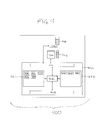
- Fig. 4 shows an example of an automatic analyzer 400.
- the automatic analyzer 400 is operable for analyzing a biological sample 404.
- a dispenser 406 which is operable for dispensing an assay 408 into a biological sample 404.
- the biological sample comprises an analyte.
- the assay 208 may for instance be located within a cartridge.
- the assay 208 comprises at least two components which are also assays.
- the automatic analyzer 400 may have an apparatus for positioning multiple biological samples 404 for dispensing the analyte and also for being analyzed by a measurement module 410.
- the measurement module 410 is representative of many different types of sensors or instruments which are capable of making measurements on a biological sample 404.
- the dispenser 406 and the measurement module 410 are connected to a hardware interface 414 of a computer system 412.
- the computer 412 further comprises a processor 416 which is in connection with the hardware interface 414, a user interface 418, computer storage 420 and computer memory 422.
- the computer storage 420 is shown as containing values 430 for the intensive properties of calibration samples.
- the computer storage 420 is further shown as containing calibration signals for the calibration samples 432.
- the computer storage is further shown as containing a calibration 434 which has been calculated using the values 430 and the calibration signals 432.
- the computer storage 420 is further showing the signal of a biological sample 436 that has been measured by the measurement module 410.
- the computer storage 420 is further shown as containing a value 438 of an intensive property calculated using the signal 436 and the calibration 434.
- the computer memory 422 is shown as containing a control module 440.
- the control module 440 comprises computer-executable code which enables the processor 416 to control the operation and function of the automatic analyzer 400. For instance it enables the process 416 to send commands via the hardware interface 414 and receive information from the dispenser 406 and the measurement module 410. If they are present this module also enables the processor 416 to control automatic routing and processing of the biological sample 404.
- the computer memory 422 is further shown as containing a curve fitting module 442.
- the curve fitting module contains computer-executable code which enables the processor 416 to calculate the calibration 434 using the calibration signals 432 and the values 430.
- the computer memory 422 is further shown as containing an intensive property determination module 444. The module 444 uses the calibration 434 and the signal 436 to calculate the value 438.
- the known calibration curves may not match the shape of the calibration curve well.
- the spline model either linear or a cubic spline is used.
- the use of a spline may require a large number of calibration measurements.
- Examples of the calibration function may be applied to the so called CRP test.
- CRP is an abbreviation for c-reactive protein.
- the c-reactive proteins are proteins found in the blood which have their levels rise in response to an inflammation.
- the CRP is a classic acute phase protein to inflammate reactions. It is synthesized by the liver and consists of five identical polypeptide chains that form a five-membered ring having a molecular weight of 120000 daltons.
- CRP is the most sensitive of the acute phase reactants and its concentration increases rapidly during the inflammation processes. Obviously healthy persons have only a very low concentration of the c-reactor protein in their blood. Reference values according to IFCC/ERM protein standardization are less than 5mg per liter. During acute inflammation processes the CRP concentrations in serum and/or plasma can increase up to 1000 fold. The CRP assay therefore faces two challenges. The low serum concentrations of CRP of obviously healthy people, especially around the decision range of 5mg per liter should be measured with high accuracy and sensitivity as well as the assay should be able to detect high serum concentrations of CRP patients with acute inflammatory processes without too high re-run rates.
- Some CRP assays are able to detect CRP without re-runs within the concentration range of 0.3mg per liter and 350mg per liter. This can be achieved by the help of two different microparticles (DuRel).
- the combination of those two particle types in one reagent leads to an overlap of the binding curves or calibration curves.
- a sharp increase of the signal at low CRP concentrations induced by the large particles is combined with a moderate increase of the signal at higher concentrations, induced by the small particles.
- the dose signal curve is a combination of the two binding curves and its shape is not typical for latex-based turbidimetric assays.
- the six available set-points have to be distributed in the majority of a lower concentration range of the calibration curve, four points between 0 and 9mg per liter, two between 90 and 350mg per liter, in order to achieve the necessary accuracy in a lower concentration range.
- the fitting of the resultant calibration curves with standard calibration functions is a problem that has not been solved with the accuracy needed to date. Most of the nonlinear calibration functions do not meet the set points adequately. Therefore they are not suitable because the accuracy at the medical decision point is insufficient. Actually a linear model is not suitable to cover the full measuring range between 0.3 and 350mg per liter. Even the aforementioned nonlinear Rodbard model is not able to provide sufficiently accurate fits especially in the medical decision range, approximately 5mg per liter.
- the next generation of calibration curves are nonlinear sigmoidal curves. They originate from dose-response theory that were introduced to model enzymatic reactions by Rodbard. These models are state of the art and are available at nearly all analyzers on the market. They model the reaction of a given reagent with the analyte within the human specimen. These models work well for very many immunological assays and allowed to broaden measurement ranges very significantly. With further increase of the requirements especially with respect to the measurement range more complex assay formats are needed. Eventually multiple superimposed reaction flows help to account for the enhanced requirements.
- the classic sigmoidal model classic one reaction mode is not up to the mark of new types of multiple superimposed reactions with sufficient performance. The idea of superimposed reactions on the chemical side needs an appropriate answer on the mathematical side. Starting from basic kinetics the super-position idea was analyzed and transferred in a very systematic way to fulfill the new requirements met.
- a key point is the strategy for the selection and the evaluation of the mathematical model. While the selection is more or less straight forward: any nonlinear analytical, small parameter number of scalar function capable to model smooth monotonous curves is a valid candidate.
- the evaluation of the new function of the algorithm is a complex process. To get all relevant steps as clear as possible it makes sense to refer to the final solution: at the very end we want to have only a few calibrators 4-5 to model the relationship between the concentration and signal without bias and variability due to calibration. This should hold true for the whole measurement range and especially the area where the outcome is clinically critical.
- the next step is to search for a suitable function which is capable to adequately describe the signal-to-concentration relationship.
- the mathematical function is only half the story.
- Nonlinear optimization can be done following different approaches. Basically in the parameter fitting a quadratic form is minimized.
- an optimization algorithm finally leads to a final set of parameters for the selected calibration function.
- Different optimization strategy algorithms like the 'Neider-Mead', 'Taylor', 'Levenberg- Marquardt' or simulated annealing were examined.
- the shape of the curve is fixed well and the fitted curve can be verified easily by checking the closeness of agreement between data and curve on a large and well-defined set of calibration curve data.
- Once good solutions have been identified for the large calibrator set it has to be shown that the mathematical model and the fitting procedure works for the routine with 4-5 calibrators as well.
- the goal here is to demonstrate that there is no loss of fit quality when we move from 20 to 5 calibrators.
- An important criterion to evaluate the candidate models are the residuals as well as the overall deviation of a 5 calibrator solution from the original n, n>10 calibrator solution.
- Equation 1 A concrete example of a calibration formula which is robust is Equation 1, which may be used with the previously discussed CRP assay.
- Examples of the calibration function may have the advantage of providing a valid calibration over a large measurement range and also to discriminate between measurement and model error.
- the detection process can be different biochemical processes. So for example the CRP assay latex-particles different sizes are involved in the same assay formulation to detect low and high concentrations of CRP, respectively.
- This design leads to assay calibration curves which are different in the different concentration regimes and constitute a challenge for the classical one-detection process modeling mathematical functions used in nonlinear calibrations. As a consequence these assays are often fitted by spline functions, which have well-known inherent disadvantages.
Priority Applications (9)
| Application Number | Priority Date | Filing Date | Title |
|---|---|---|---|
| EP13171044.4A EP2811300A1 (fr) | 2013-06-07 | 2013-06-07 | Étalonnage pour dosages à composants multiples |
| CN201480027599.6A CN105637365B (zh) | 2013-06-07 | 2014-06-04 | 对多组分被分析物的校准 |
| ES14728186T ES2768198T3 (es) | 2013-06-07 | 2014-06-04 | Calibración para ensayos de múltiples componentes |
| PCT/EP2014/061571 WO2014195355A1 (fr) | 2013-06-07 | 2014-06-04 | Étalonnage pour dosages multicomposants |
| JP2016517286A JP6419791B2 (ja) | 2013-06-07 | 2014-06-04 | 多成分分析の較正 |
| EP14728186.9A EP3004880B1 (fr) | 2013-06-07 | 2014-06-04 | Étalonnage pour dosages à composants multiples |
| CA2904256A CA2904256C (fr) | 2013-06-07 | 2014-06-04 | Etalonnage pour dosages multicomposants |
| US14/956,733 US11378573B2 (en) | 2013-06-07 | 2015-12-02 | Calibration for multi-component assays |
| HK16108603.4A HK1220758A1 (zh) | 2013-06-07 | 2016-07-20 | 對多組分被分析物的校準 |
Applications Claiming Priority (1)
| Application Number | Priority Date | Filing Date | Title |
|---|---|---|---|
| EP13171044.4A EP2811300A1 (fr) | 2013-06-07 | 2013-06-07 | Étalonnage pour dosages à composants multiples |
Publications (1)
| Publication Number | Publication Date |
|---|---|
| EP2811300A1 true EP2811300A1 (fr) | 2014-12-10 |
Family
ID=48578854
Family Applications (2)
| Application Number | Title | Priority Date | Filing Date |
|---|---|---|---|
| EP13171044.4A Withdrawn EP2811300A1 (fr) | 2013-06-07 | 2013-06-07 | Étalonnage pour dosages à composants multiples |
| EP14728186.9A Active EP3004880B1 (fr) | 2013-06-07 | 2014-06-04 | Étalonnage pour dosages à composants multiples |
Family Applications After (1)
| Application Number | Title | Priority Date | Filing Date |
|---|---|---|---|
| EP14728186.9A Active EP3004880B1 (fr) | 2013-06-07 | 2014-06-04 | Étalonnage pour dosages à composants multiples |
Country Status (8)
| Country | Link |
|---|---|
| US (1) | US11378573B2 (fr) |
| EP (2) | EP2811300A1 (fr) |
| JP (1) | JP6419791B2 (fr) |
| CN (1) | CN105637365B (fr) |
| CA (1) | CA2904256C (fr) |
| ES (1) | ES2768198T3 (fr) |
| HK (1) | HK1220758A1 (fr) |
| WO (1) | WO2014195355A1 (fr) |
Cited By (4)
| Publication number | Priority date | Publication date | Assignee | Title |
|---|---|---|---|---|
| CN110023764A (zh) * | 2016-12-02 | 2019-07-16 | 豪夫迈·罗氏有限公司 | 用于分析生物样本的自动分析仪的故障状态预测 |
| GB2579810A (en) * | 2018-12-14 | 2020-07-08 | Aeirtec Ltd | Assay Analysis |
| WO2021122737A1 (fr) * | 2019-12-17 | 2021-06-24 | Roche Diagnostics Gmbh | Procédé d'étalonnage d'au moins un dispositif analytique comportant de multiples composants matériels répétés |
| US11982657B2 (en) | 2019-12-17 | 2024-05-14 | Roche Diagnostics Operations, Inc. | Method for calibrating at least one analytic device with multiple repeated hardware components |
Families Citing this family (10)
| Publication number | Priority date | Publication date | Assignee | Title |
|---|---|---|---|---|
| CA2900201A1 (fr) | 2014-08-25 | 2016-02-25 | Alireza Ebrahim | Attribution de valeur aux controles de qualite personnalisables |
| KR102146434B1 (ko) | 2015-12-17 | 2020-08-21 | 에이에스엠엘 네델란즈 비.브이. | 측정을 향상시키기 위한 비대칭 서브 해상도 피처를 사용하는 리소그래피 공정의 광학적 메트롤로지 |
| WO2017216208A1 (fr) * | 2016-06-15 | 2017-12-21 | Roche Diagnostics Gmbh | Étalonnage amélioré du dosage de d-dimère |
| US11209417B2 (en) * | 2017-10-12 | 2021-12-28 | Carrot, Inc. | Breath sensor apparatus and methods of use |
| CN111239426B (zh) * | 2018-11-29 | 2023-12-01 | 深圳市帝迈生物技术有限公司 | 样本分析仪及其自动校准的方法 |
| JP2020201174A (ja) | 2019-06-12 | 2020-12-17 | 国立研究開発法人物質・材料研究機構 | スペクトル解析装置用の成分同定装置及びその方法、コンピュータプログラム |
| EP3757547B1 (fr) * | 2019-06-28 | 2022-11-23 | ABB Schweiz AG | Appareil de correction d'étalonnage de la turbidité et procédé de correction d'étalonnage automatique |
| JP2023509643A (ja) | 2019-12-31 | 2023-03-09 | マクニール アーベー | 呼気センサの較正方法及び装置 |
| CN112462078A (zh) * | 2020-11-16 | 2021-03-09 | 三诺生物传感股份有限公司 | 一种用于荧光免疫分析仪台间差校准的方法 |
| CN112730203B (zh) * | 2020-12-29 | 2023-06-16 | 深圳市科曼医疗设备有限公司 | 血球分析仪的光学系统、光学增益校准方法和存储介质 |
Citations (3)
| Publication number | Priority date | Publication date | Assignee | Title |
|---|---|---|---|---|
| EP0898169B1 (fr) * | 1997-08-11 | 2002-02-06 | F. Hoffmann-La Roche Ag | Essai à base de la diffusion de la lumière augmenté par les microparticules et réactifs microparticulaires pour la mise en oeuvre de cet essai |
| EP0790500B1 (fr) * | 1996-01-09 | 2002-06-05 | Fuji Photo Film Co., Ltd. | Méthode de détermination d'une courbe de calibrage et méthode d'analyse et appareil l'utilisant. |
| EP2357475A1 (fr) * | 2008-12-04 | 2011-08-17 | Sekisui Medical Co., Ltd. | Procédé pour mesurer la cystatine c dans un fluide corporel humain |
Family Cites Families (6)
| Publication number | Priority date | Publication date | Assignee | Title |
|---|---|---|---|---|
| CA2244326C (fr) * | 1997-08-11 | 2006-03-28 | Shinichi Eda | Essai d'agglutination par diffusion de la lumiere, ameliore par des microparticules; reactifs microparticulaires utiles a cette fin |
| US6277584B1 (en) * | 1998-12-16 | 2001-08-21 | Dade Behring Inc. | Method for calibrating a chemical analyzer with improved accuracy at low signal levels |
| EP1489961B1 (fr) * | 2002-03-22 | 2010-09-29 | Animas Technologies LLC | Amelioration de l'efficacite d'un dispositif de surveillance d'analyte |
| JP4331073B2 (ja) * | 2004-08-31 | 2009-09-16 | デンカ生研株式会社 | 抗原の測定方法及びそのための試薬 |
| JP2009063335A (ja) * | 2007-09-05 | 2009-03-26 | Fujifilm Corp | 生理活性物質と被験物質との相互作用の測定方法 |
| DE112009002702B4 (de) * | 2008-11-17 | 2013-09-05 | Hitachi High-Technologies Corporation | Automatischer Analysator |
-
2013
- 2013-06-07 EP EP13171044.4A patent/EP2811300A1/fr not_active Withdrawn
-
2014
- 2014-06-04 CA CA2904256A patent/CA2904256C/fr active Active
- 2014-06-04 CN CN201480027599.6A patent/CN105637365B/zh active Active
- 2014-06-04 WO PCT/EP2014/061571 patent/WO2014195355A1/fr active Application Filing
- 2014-06-04 JP JP2016517286A patent/JP6419791B2/ja active Active
- 2014-06-04 ES ES14728186T patent/ES2768198T3/es active Active
- 2014-06-04 EP EP14728186.9A patent/EP3004880B1/fr active Active
-
2015
- 2015-12-02 US US14/956,733 patent/US11378573B2/en active Active
-
2016
- 2016-07-20 HK HK16108603.4A patent/HK1220758A1/zh unknown
Patent Citations (3)
| Publication number | Priority date | Publication date | Assignee | Title |
|---|---|---|---|---|
| EP0790500B1 (fr) * | 1996-01-09 | 2002-06-05 | Fuji Photo Film Co., Ltd. | Méthode de détermination d'une courbe de calibrage et méthode d'analyse et appareil l'utilisant. |
| EP0898169B1 (fr) * | 1997-08-11 | 2002-02-06 | F. Hoffmann-La Roche Ag | Essai à base de la diffusion de la lumière augmenté par les microparticules et réactifs microparticulaires pour la mise en oeuvre de cet essai |
| EP2357475A1 (fr) * | 2008-12-04 | 2011-08-17 | Sekisui Medical Co., Ltd. | Procédé pour mesurer la cystatine c dans un fluide corporel humain |
Cited By (6)
| Publication number | Priority date | Publication date | Assignee | Title |
|---|---|---|---|---|
| CN110023764A (zh) * | 2016-12-02 | 2019-07-16 | 豪夫迈·罗氏有限公司 | 用于分析生物样本的自动分析仪的故障状态预测 |
| CN110023764B (zh) * | 2016-12-02 | 2023-12-22 | 豪夫迈·罗氏有限公司 | 用于分析生物样本的自动分析仪的故障状态预测 |
| GB2579810A (en) * | 2018-12-14 | 2020-07-08 | Aeirtec Ltd | Assay Analysis |
| GB2579810B (en) * | 2018-12-14 | 2023-04-26 | Aeirtec Ltd | Assay Analysis |
| WO2021122737A1 (fr) * | 2019-12-17 | 2021-06-24 | Roche Diagnostics Gmbh | Procédé d'étalonnage d'au moins un dispositif analytique comportant de multiples composants matériels répétés |
| US11982657B2 (en) | 2019-12-17 | 2024-05-14 | Roche Diagnostics Operations, Inc. | Method for calibrating at least one analytic device with multiple repeated hardware components |
Also Published As
| Publication number | Publication date |
|---|---|
| US20160084861A1 (en) | 2016-03-24 |
| CA2904256C (fr) | 2019-09-10 |
| CA2904256A1 (fr) | 2014-12-11 |
| JP2016523359A (ja) | 2016-08-08 |
| EP3004880B1 (fr) | 2019-11-20 |
| EP3004880A1 (fr) | 2016-04-13 |
| CN105637365B (zh) | 2018-04-24 |
| ES2768198T3 (es) | 2020-06-22 |
| WO2014195355A1 (fr) | 2014-12-11 |
| US11378573B2 (en) | 2022-07-05 |
| HK1220758A1 (zh) | 2017-05-12 |
| CN105637365A (zh) | 2016-06-01 |
| JP6419791B2 (ja) | 2018-11-07 |
Similar Documents
| Publication | Publication Date | Title |
|---|---|---|
| US11378573B2 (en) | Calibration for multi-component assays | |
| Dimeski | Interference testing | |
| US9952234B2 (en) | Calibration method for photometry | |
| Vashist et al. | Bioanalytical requirements and regulatory guidelines for immunoassays | |
| Campbell et al. | Development of a rapid and quantitative lateral flow assay for the simultaneous measurement of serum κ and λ immunoglobulin free light chains (FLC): inception of a new near-patient FLC screening tool | |
| Jasensky et al. | Evaluation of three different point‐of‐care tests for quantitative measurement of canine C‐reactive protein | |
| JP6129570B2 (ja) | アッセイ法のレンジを拡大するための多重時間窓 | |
| Loh et al. | Lot-to-lot variation and verification | |
| Geertjens et al. | Straightforward model construction and analysis of multicomponent biomolecular systems in equilibrium | |
| AU2011308657B2 (en) | Dose surface method for determination of analyte ratios | |
| Geertjens et al. | A general framework for straightforward model construction of multi-component thermodynamic equilibrium systems | |
| Tan et al. | Establishment, verification, and application of concentration intervals corresponding to dipstick grades for urinary protein | |
| Rajasekariah et al. | Assessment of assay sensitivity and precision in a malaria antibody ELISA | |
| JP4871761B2 (ja) | 生体サンプルの分析方法、及び自動分析装置 | |
| Baral et al. | Comparisons of results between three in-house biochemistry analyzers and a commercial laboratory analyzer for feline plasma using multiple quality specifications | |
| Jayakody et al. | Instrument quality control | |
| JP5602027B2 (ja) | 未来の性質を予測する方法 | |
| Chen et al. | Biomarker Assay Development, Qualification, and Validation | |
| Ramamohan et al. | Modeling uncertainty due to instrument drift in clinical laboratories | |
| Ramamohan et al. | A simulation-based methodology for uncertainty modeling and analysis of clinical laboratory measurement processes | |
| Ramamohan | A Simulation-Based Methodology for the Estimation and Analysis of the Uncertainty of Clinical Laboratory Measurement Processes | |
| Muravskaya et al. | Metrological support for automatic biochemical analyzers | |
| Mansfield et al. | Biomarkers for pharmacogenetic and pharmacogenomic studies: Locking down analytical performance | |
| Geertjens et al. | Chemical Biology | |
| GB2457678A (en) | Method for predicting a future property |
Legal Events
| Date | Code | Title | Description |
|---|---|---|---|
| PUAI | Public reference made under article 153(3) epc to a published international application that has entered the european phase |
Free format text: ORIGINAL CODE: 0009012 |
|
| 17P | Request for examination filed |
Effective date: 20130607 |
|
| AK | Designated contracting states |
Kind code of ref document: A1 Designated state(s): AL AT BE BG CH CY CZ DE DK EE ES FI FR GB GR HR HU IE IS IT LI LT LU LV MC MK MT NL NO PL PT RO RS SE SI SK SM TR |
|
| AX | Request for extension of the european patent |
Extension state: BA ME |
|
| R17P | Request for examination filed (corrected) |
Effective date: 20150210 |
|
| RBV | Designated contracting states (corrected) |
Designated state(s): AL AT BE BG CH CY CZ DE DK EE ES FI FR GB GR HR HU IE IS IT LI LT LU LV MC MK MT NL NO PL PT RO RS SE SI SK SM TR |
|
| 18D | Application deemed to be withdrawn |
Effective date: 20170103 |
|
| STAA | Information on the status of an ep patent application or granted ep patent |
Free format text: STATUS: THE APPLICATION IS DEEMED TO BE WITHDRAWN |