EP2788478B1 - Multiplex immuno screening assay - Google Patents
Multiplex immuno screening assay Download PDFInfo
- Publication number
- EP2788478B1 EP2788478B1 EP12798308.8A EP12798308A EP2788478B1 EP 2788478 B1 EP2788478 B1 EP 2788478B1 EP 12798308 A EP12798308 A EP 12798308A EP 2788478 B1 EP2788478 B1 EP 2788478B1
- Authority
- EP
- European Patent Office
- Prior art keywords
- snap
- agt
- seq
- virus
- dna
- Prior art date
- Legal status (The legal status is an assumption and is not a legal conclusion. Google has not performed a legal analysis and makes no representation as to the accuracy of the status listed.)
- Active
Links
- 0 C*NCCOCCOCCOCCNC(NCc1ccc(C=CCc2c3nc[n]c3nc(*)n2)cc1)=O Chemical compound C*NCCOCCOCCOCCNC(NCc1ccc(C=CCc2c3nc[n]c3nc(*)n2)cc1)=O 0.000 description 1
Images


Classifications
-
- G—PHYSICS
- G02—OPTICS
- G02B—OPTICAL ELEMENTS, SYSTEMS OR APPARATUS
- G02B27/00—Optical systems or apparatus not provided for by any of the groups G02B1/00 - G02B26/00, G02B30/00
- G02B27/01—Head-up displays
- G02B27/017—Head mounted
- G02B27/0172—Head mounted characterised by optical features
-
- G—PHYSICS
- G01—MEASURING; TESTING
- G01N—INVESTIGATING OR ANALYSING MATERIALS BY DETERMINING THEIR CHEMICAL OR PHYSICAL PROPERTIES
- G01N33/00—Investigating or analysing materials by specific methods not covered by groups G01N1/00 - G01N31/00
- G01N33/48—Biological material, e.g. blood, urine; Haemocytometers
- G01N33/50—Chemical analysis of biological material, e.g. blood, urine; Testing involving biospecific ligand binding methods; Immunological testing
- G01N33/53—Immunoassay; Biospecific binding assay; Materials therefor
- G01N33/543—Immunoassay; Biospecific binding assay; Materials therefor with an insoluble carrier for immobilising immunochemicals
- G01N33/54306—Solid-phase reaction mechanisms
-
- G—PHYSICS
- G01—MEASURING; TESTING
- G01N—INVESTIGATING OR ANALYSING MATERIALS BY DETERMINING THEIR CHEMICAL OR PHYSICAL PROPERTIES
- G01N33/00—Investigating or analysing materials by specific methods not covered by groups G01N1/00 - G01N31/00
- G01N33/48—Biological material, e.g. blood, urine; Haemocytometers
- G01N33/50—Chemical analysis of biological material, e.g. blood, urine; Testing involving biospecific ligand binding methods; Immunological testing
- G01N33/53—Immunoassay; Biospecific binding assay; Materials therefor
- G01N33/543—Immunoassay; Biospecific binding assay; Materials therefor with an insoluble carrier for immobilising immunochemicals
- G01N33/54313—Immunoassay; Biospecific binding assay; Materials therefor with an insoluble carrier for immobilising immunochemicals the carrier being characterised by its particulate form
-
- G—PHYSICS
- G01—MEASURING; TESTING
- G01N—INVESTIGATING OR ANALYSING MATERIALS BY DETERMINING THEIR CHEMICAL OR PHYSICAL PROPERTIES
- G01N33/00—Investigating or analysing materials by specific methods not covered by groups G01N1/00 - G01N31/00
- G01N33/48—Biological material, e.g. blood, urine; Haemocytometers
- G01N33/50—Chemical analysis of biological material, e.g. blood, urine; Testing involving biospecific ligand binding methods; Immunological testing
- G01N33/53—Immunoassay; Biospecific binding assay; Materials therefor
- G01N33/543—Immunoassay; Biospecific binding assay; Materials therefor with an insoluble carrier for immobilising immunochemicals
- G01N33/54353—Immunoassay; Biospecific binding assay; Materials therefor with an insoluble carrier for immobilising immunochemicals with ligand attached to the carrier via a chemical coupling agent
-
- G—PHYSICS
- G01—MEASURING; TESTING
- G01N—INVESTIGATING OR ANALYSING MATERIALS BY DETERMINING THEIR CHEMICAL OR PHYSICAL PROPERTIES
- G01N33/00—Investigating or analysing materials by specific methods not covered by groups G01N1/00 - G01N31/00
- G01N33/48—Biological material, e.g. blood, urine; Haemocytometers
- G01N33/50—Chemical analysis of biological material, e.g. blood, urine; Testing involving biospecific ligand binding methods; Immunological testing
- G01N33/53—Immunoassay; Biospecific binding assay; Materials therefor
- G01N33/564—Immunoassay; Biospecific binding assay; Materials therefor for pre-existing immune complex or autoimmune disease, i.e. systemic lupus erythematosus, rheumatoid arthritis, multiple sclerosis, rheumatoid factors or complement components C1-C9
-
- G—PHYSICS
- G01—MEASURING; TESTING
- G01N—INVESTIGATING OR ANALYSING MATERIALS BY DETERMINING THEIR CHEMICAL OR PHYSICAL PROPERTIES
- G01N33/00—Investigating or analysing materials by specific methods not covered by groups G01N1/00 - G01N31/00
- G01N33/48—Biological material, e.g. blood, urine; Haemocytometers
- G01N33/50—Chemical analysis of biological material, e.g. blood, urine; Testing involving biospecific ligand binding methods; Immunological testing
- G01N33/53—Immunoassay; Biospecific binding assay; Materials therefor
- G01N33/569—Immunoassay; Biospecific binding assay; Materials therefor for microorganisms, e.g. protozoa, bacteria, viruses
- G01N33/56983—Viruses
-
- G—PHYSICS
- G01—MEASURING; TESTING
- G01N—INVESTIGATING OR ANALYSING MATERIALS BY DETERMINING THEIR CHEMICAL OR PHYSICAL PROPERTIES
- G01N33/00—Investigating or analysing materials by specific methods not covered by groups G01N1/00 - G01N31/00
- G01N33/48—Biological material, e.g. blood, urine; Haemocytometers
- G01N33/50—Chemical analysis of biological material, e.g. blood, urine; Testing involving biospecific ligand binding methods; Immunological testing
- G01N33/68—Chemical analysis of biological material, e.g. blood, urine; Testing involving biospecific ligand binding methods; Immunological testing involving proteins, peptides or amino acids
- G01N33/6803—General methods of protein analysis not limited to specific proteins or families of proteins
- G01N33/6845—Methods of identifying protein-protein interactions in protein mixtures
-
- G—PHYSICS
- G02—OPTICS
- G02B—OPTICAL ELEMENTS, SYSTEMS OR APPARATUS
- G02B27/00—Optical systems or apparatus not provided for by any of the groups G02B1/00 - G02B26/00, G02B30/00
- G02B27/0081—Optical systems or apparatus not provided for by any of the groups G02B1/00 - G02B26/00, G02B30/00 with means for altering, e.g. enlarging, the entrance or exit pupil
-
- G—PHYSICS
- G02—OPTICS
- G02B—OPTICAL ELEMENTS, SYSTEMS OR APPARATUS
- G02B27/00—Optical systems or apparatus not provided for by any of the groups G02B1/00 - G02B26/00, G02B30/00
- G02B27/42—Diffraction optics, i.e. systems including a diffractive element being designed for providing a diffractive effect
- G02B27/4272—Diffraction optics, i.e. systems including a diffractive element being designed for providing a diffractive effect having plural diffractive elements positioned sequentially along the optical path
-
- G—PHYSICS
- G02—OPTICS
- G02B—OPTICAL ELEMENTS, SYSTEMS OR APPARATUS
- G02B5/00—Optical elements other than lenses
- G02B5/18—Diffraction gratings
- G02B5/1814—Diffraction gratings structurally combined with one or more further optical elements, e.g. lenses, mirrors, prisms or other diffraction gratings
-
- G—PHYSICS
- G02—OPTICS
- G02B—OPTICAL ELEMENTS, SYSTEMS OR APPARATUS
- G02B6/00—Light guides; Structural details of arrangements comprising light guides and other optical elements, e.g. couplings
- G02B6/0001—Light guides; Structural details of arrangements comprising light guides and other optical elements, e.g. couplings specially adapted for lighting devices or systems
- G02B6/0011—Light guides; Structural details of arrangements comprising light guides and other optical elements, e.g. couplings specially adapted for lighting devices or systems the light guides being planar or of plate-like form
- G02B6/0013—Means for improving the coupling-in of light from the light source into the light guide
- G02B6/0015—Means for improving the coupling-in of light from the light source into the light guide provided on the surface of the light guide or in the bulk of it
- G02B6/0016—Grooves, prisms, gratings, scattering particles or rough surfaces
-
- G—PHYSICS
- G02—OPTICS
- G02B—OPTICAL ELEMENTS, SYSTEMS OR APPARATUS
- G02B6/00—Light guides; Structural details of arrangements comprising light guides and other optical elements, e.g. couplings
- G02B6/0001—Light guides; Structural details of arrangements comprising light guides and other optical elements, e.g. couplings specially adapted for lighting devices or systems
- G02B6/0011—Light guides; Structural details of arrangements comprising light guides and other optical elements, e.g. couplings specially adapted for lighting devices or systems the light guides being planar or of plate-like form
- G02B6/0033—Means for improving the coupling-out of light from the light guide
- G02B6/0035—Means for improving the coupling-out of light from the light guide provided on the surface of the light guide or in the bulk of it
- G02B6/0038—Linear indentations or grooves, e.g. arc-shaped grooves or meandering grooves, extending over the full length or width of the light guide
-
- G—PHYSICS
- G06—COMPUTING OR CALCULATING; COUNTING
- G06T—IMAGE DATA PROCESSING OR GENERATION, IN GENERAL
- G06T19/00—Manipulating 3D models or images for computer graphics
- G06T19/006—Mixed reality
-
- G—PHYSICS
- G01—MEASURING; TESTING
- G01N—INVESTIGATING OR ANALYSING MATERIALS BY DETERMINING THEIR CHEMICAL OR PHYSICAL PROPERTIES
- G01N2333/00—Assays involving biological materials from specific organisms or of a specific nature
- G01N2333/005—Assays involving biological materials from specific organisms or of a specific nature from viruses
- G01N2333/08—RNA viruses
- G01N2333/18—Togaviridae; Flaviviridae
- G01N2333/183—Flaviviridae, e.g. pestivirus, mucosal disease virus, bovine viral diarrhoea virus, classical swine fever virus (hog cholera virus) or border disease virus
- G01N2333/185—Flaviviruses or Group B arboviruses, e.g. yellow fever virus, japanese encephalitis, tick-borne encephalitis, dengue
-
- G—PHYSICS
- G01—MEASURING; TESTING
- G01N—INVESTIGATING OR ANALYSING MATERIALS BY DETERMINING THEIR CHEMICAL OR PHYSICAL PROPERTIES
- G01N2469/00—Immunoassays for the detection of microorganisms
- G01N2469/20—Detection of antibodies in sample from host which are directed against antigens from microorganisms
-
- G—PHYSICS
- G02—OPTICS
- G02B—OPTICAL ELEMENTS, SYSTEMS OR APPARATUS
- G02B27/00—Optical systems or apparatus not provided for by any of the groups G02B1/00 - G02B26/00, G02B30/00
- G02B27/01—Head-up displays
- G02B27/0101—Head-up displays characterised by optical features
- G02B2027/0123—Head-up displays characterised by optical features comprising devices increasing the field of view
- G02B2027/0125—Field-of-view increase by wavefront division
-
- G—PHYSICS
- G02—OPTICS
- G02B—OPTICAL ELEMENTS, SYSTEMS OR APPARATUS
- G02B27/00—Optical systems or apparatus not provided for by any of the groups G02B1/00 - G02B26/00, G02B30/00
- G02B27/01—Head-up displays
- G02B27/017—Head mounted
- G02B27/0172—Head mounted characterised by optical features
- G02B2027/0174—Head mounted characterised by optical features holographic
-
- Y—GENERAL TAGGING OF NEW TECHNOLOGICAL DEVELOPMENTS; GENERAL TAGGING OF CROSS-SECTIONAL TECHNOLOGIES SPANNING OVER SEVERAL SECTIONS OF THE IPC; TECHNICAL SUBJECTS COVERED BY FORMER USPC CROSS-REFERENCE ART COLLECTIONS [XRACs] AND DIGESTS
- Y02—TECHNOLOGIES OR APPLICATIONS FOR MITIGATION OR ADAPTATION AGAINST CLIMATE CHANGE
- Y02A—TECHNOLOGIES FOR ADAPTATION TO CLIMATE CHANGE
- Y02A50/00—TECHNOLOGIES FOR ADAPTATION TO CLIMATE CHANGE in human health protection, e.g. against extreme weather
- Y02A50/30—Against vector-borne diseases, e.g. mosquito-borne, fly-borne, tick-borne or waterborne diseases whose impact is exacerbated by climate change
Definitions
- VHFs viral hemorrhagic fevers
- RNA viruses from several families including the Flaviviridae (dengue, Yellow fever, West Nile, Japanese encephalitis, Tick-Borne Encephalitis, Hepatitis C viruses), the Togaviridae (Chikungunya, Ross River, Mayaro, Western Equine encephalitis, Eastern Equine Encephalitis, Venezuela Equine Encephalitis viruses) the Bunyaviridae (Crimean-Congo hemorrhagic fever, Rift Valley Fever, Schmallenberg viruses), the Caliciviridae (Hepatitis E virus), the Arenaviridae (Lassa) and the Filoviridae (Ebola, Marburg).
- Flaviviridae dengue, Yellow fever, West Nile, Japanese encephalitis, Tick-Borne Encephalitis, Hepatitis C viruses
- Togaviridae Chikungunya, Ross River, Mayaro, Western Equine encephalitis, Eastern Equine Encephalitis,
- detection of antibodies in body fluids constitutes a major part of the diagnosis of virally induced diseases, autoimmune diseases and the detection of cancer.
- certain antibodies can serve as markers in diagnosis and can lead to prognosis and treatment, as their presence are known to correlate with the outbreak of a pathogen. This is particularly the case for the antibodies targeting viral antigens exclusively.
- the parallel detection of several antibodies simultaneously may be particularly useful by minimizing the matrix effects that exist between individual assays, such as in ELISAs, because the calibrators and the antibodies are analyzed under the same conditions; it therefore will generate comparable results for the measurement of multiple antibodies present within the same sample.
- a recent new technique, capable of analyzing these complex staining patterns of Western blots simultaneously, is based on digital image analysis. This technique has been successfully used in studies of myasthenia gravis, Graves' disease and experimental uveitis ( Zimmerman CW, Electrophoresis 1995 ).
- the antibodies may also be detected and measured on a protein chip array using surface-enhanced laser desorption/ionization (SELDI) or matrix assisted laser desorption/ionization mass spectrometry techniques, preferably SELDI mass spectrometry technique ( US 2006/166268 ). Yet, these techniques use large cumbersome equipment that is complex and expensive to maintain, and requires high amount of the biological samples to achieve the detection of antibodies being in a low amount.
- the 6-alkylguanine-DNA-alkyltransferase enzyme (AGT, also known as ATase or MGMT, and hereafter referred to as "AGT") is numbered EC 2.1.1.63 in the IUBMB enzyme nomenclature. It is a 6-alkylguanine-DNA-alkyltransferase DNA repair enzyme of 207 amino acid residues whose function in the cells is to repair alkylated DNA. More precisely, AGT acts on O 6 -methylated guanine in DNA by irreversibly transferring the methyl group in an S N 2 reaction to a reactive cysteine residue (Cys 145).
- AGT acts on O 6 -methylated guanine in DNA by irreversibly transferring the methyl group in an S N 2 reaction to a reactive cysteine residue (Cys 145).
- the present inventors have developed and validated immunoassays leading to rapid and simultaneous detection of several antibodies generated by a wide range of diseases, in particular arboviral diseases and VHFs, in biological fluids.
- This oriented antigen coupling procedure is based on the covalent interaction between the AGT enzymes and their substrates, the O6-benzylguanine derivatives, which irreversibly react with AGT enzymes by transferring their benzyl group to the active site cysteine of the enzyme. Accordingly, a number of target antigens can be fused to an AGT enzyme moiety, resulting in different chimeric fusion proteins (hereafter referred to as [AGT- Antigen] fusion proteins), that can be used as capture reagents for the antibodies present in a biological sample.
- [AGT- Antigen] fusion proteins chimeric fusion proteins
- the present inventors have shown that this antibody capture is enhanced when these fusion proteins are bound to solid supports thanks to the specific AGT-substrate interaction. Coating the said solid supports with AGT-substrate is thus an essential step of the immunoassay of the invention.
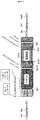
- the method for coupling antigens to solid supports comprises the two following steps: i) the coating of solid surfaces with an AGT substrate (e.g. BG-PEG-amino), and ii) the covalent immobilization of chimeric [AGT-Antigen] fusion proteins using the AGT substrate as an anchor (see Figure 1 ).
- an AGT substrate e.g. BG-PEG-amino
- the solid surfaces are advantageously functionalized, preferably by using an optimized two-step carbodiimide process ( Kufer SK, Eur. Biophys.J. 2005 ), so that the AGT substrate is covalently attached to the solid surfaces.
- the solid surfaces carry AGT substrates that are irreversibly linked to the chimeric [AGT-antigen] fusion proteins. Due to the high specificity of this reaction, the fusion protein is exclusively coupled via the cysteine-containing domain of the AGT enzyme, thus leaving the antigen accessible for its interactions with antibodies.
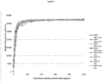
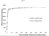
- This coupling procedure is very advantageous as it allows the binding of the antigen in an oriented manner on the solid supports. Also, this antigen coupling procedure advantageously enables to obtain a multimeric antigen organization on a solid surface, so as to enhance immunoglobulin G, and potentially immunoglobulin M, capture efficiency. Consequently, the antigen-coupled microspheres developed in the experimental part of the application have shown enhanced capture of specific antibodies as compared to antigen-coupled microspheres produced by standard non-oriented amine coupling procedures (see the experimental part below and figure 3 ). Finally, this antigen coupling procedure enables to obtain a high coupling efficiency and a long-term stability of the antigen-conjugated microspheres (>6 months at 4°C).
- the solid supports used in the immunoassays of the invention should be intrinsically identifiable, so that it is possible to determine precisely which antigen is carried by which solid support.
- the antigen-coupled and identifiable solid supports are then used as capture reagents for specific human immunoglobulins and are therefore contacted with the biological sample of the patient.
- the final step of the method of the invention involves the detection of the solid supports which are effectively bound to immunoglobulins.
- the identification of immunoglobulin-coated solid support(s) enables to diagnose which pathogen was infecting the patient (as each solid support matches with a defined pathogenic antigen).
- This final detection step is performed by any usual means, for example by using labeled detection antibodies and by identifying the nature of the solid support.
- the method of the invention involves only the detection of the presence of antibodies in diseased patients, but knowledge about the identity of those antibodies is not required.
- the inventors have used the antigen-coupling procedure of the invention to generate a number of different antigen-coated fluorescent microspheres.
- 16 distinct sets of microspheres have been coupled with 16 purified chimeric [AGT-Antigen] fusion proteins, allowing titration of 16 serum antibodies specific to different proteins of the dengue serotypes 1 to 4, West Nile, yellow fever, Japanese encephalitis, tick-borne encephalitis, Saint-Louis encephalitis, Murray Valley encephalitis, Wesselsbron, Zika, Rocio, Usutu, Rift Valley fever, and chikungunya virus.
- the present invention relates to a method for detecting at least two target antibodies in a biological sample comprising:
- the present invention relates to an in vitro assay method for detecting at least two different target antibodies present in a biological sample from a subject, said method comprising the steps of:
- an antibody As used hereafter, the terms “an antibody”, “a fusion protein”, “an epitope”, “an antigen”, “an AGT polypeptide”, “a solid support” and the like have obviously to be understood as usual in the art, that is, in a broad manner. In particular, they encompass not only particular single molecules but a number of said molecules.
- solid support encompasses a subset of numerous identical solid supports
- microparticle encompasses a subset of numerous identical microparticles
- fusion protein encompasses a number of identical single protein molecules.
- a solid support carries a number of identical fusion proteins, said fusion proteins containing, apart from the AGT polypeptide, identical antigen, and therefore identical epitopes, so that the antibodies which will be detected on the solid support can be unambiguously identified.
- fusion protein means a polypeptide containing a protein or a polypeptide created through the artificial joining of two or more polypeptides.
- said fusion proteins contain a AGT polypeptide and an antigen, containing at least one epitope. Fusion proteins can be obtained through genetic engineering of a fusion gene. This typically involves removing the stop codon from a cDNA sequence coding for the first protein, then appending the cDNA sequence of the second protein in frame through ligation or overlap extension PCR. That DNA sequence will then be expressed by a cell as a single protein.
- the protein can be engineered to include the full sequence of both original proteins, or only a portion of either.
- the fusion proteins of the invention can be obtained by providing vectors comprising AGT encoding sequences in frame with an epitope or antigen encoding sequences, either attached to the N-terminal or to the C-terminal side of the AGT DNA sequence.
- vectors may be introduced in prokaryotic hosts, including eubacteria such as E.coli bacteria, or eukaryotic hosts, e.g., yeast, insect cells or mammalian cells and the recombinant fusion proteins may be produced under appropriate conditions. Typical constructions are presented in the experimental part of this application.
- antibody as used herein is intended to include monoclonal antibodies, polyclonal antibodies, and chimeric antibodies.
- the antibodies which are to be detected by the immunoassays of the invention are polyclonal antibodies, which are present in biological samples of diseased patients, and have therefore been generated from different B cell sources. As such, they recognize different epitopes exhibited by a pathogenic antigen (on the other hand, monoclonal antibodies are derived from a single cell line and recognize the same epitope).
- An antibody (or "immunoglobulin") consists of a glycoprotein comprising at least two heavy (H) chains and two light (L) chains inter-connected by disulfide bonds.
- Each heavy chain comprises a heavy chain variable region (or domain) (abbreviated herein as HCVR or VH) and a heavy chain constant region.
- the heavy chain constant region comprises three domains, CH1, CH2 and CH3.
- Each light chain comprises a light chain variable region (abbreviated herein as LCVR or VL) and a light chain constant region.
- the light chain constant region comprises one domain, CL.
- VH and VL regions can be further subdivided into regions of hypervariability, termed “complementarity determining regions” (CDR) or “hypervariable regions", which are primarily responsible for binding an epitope of an antigen, and which are interspersed with regions that are more conserved, termed framework regions (FR).
- CDR complementarity determining regions
- FR framework regions
- Each VH and VL is composed of three CDRs and four FRs, arranged from amino-terminus to carboxy-terminus in the following order: FR1, CDR1, FR2, CDR2, FR3, CDR3, FR4.
- the variable regions of the heavy and light chains contain a binding domain that interacts with an antigen.
- the constant regions of the antibodies may mediate the binding of the immunoglobulin to host tissues or factors, including various cells of the immune system (e.g. effector cells) and the first component (Clq) of the classical complement system.
- Antibody can be of different isotypes (namely IgA, IgD, IgE, IgG or IgM). Both IgG and IgM type antibodies can be detected by the present method. Of note, these isotypes are composed of two identical heavy chains and two identical light chains that are joined by disulfide bonds. Importantly, IgM antibodies form polymers where multiple immunoglobulins are covalently linked together with disulfide bonds, mostly as a pentamer but also as a hexamer, so that they have a molecular mass of approximately 900 kDa (in their pentamer form). Because each monomer has two antigen binding sites, a pentameric IgM has 10 binding sites. Typically, however, IgM antibodies cannot bind 10 antigens at the same time because the large size of most antigens hinders binding to nearby sites. Due to its polymeric nature, IgM possesses high avidity.
- Antibody fragments can also be detected thanks to the present method.
- This term is intended to include Fab, Fab', F(ab')2, scFv, dsFv, ds-scFv, dimers, minibodies, diabodies, and multimers thereof and bispecific antibody fragments.
- Monoclonal antibodies can be used in the present immunoassays; for example for detecting the immunoglobulins that are bound to the solid supports.
- "monoclonal antibody” defines an antibody arising from a homogeneous antibody population. More particularly, the individual antibodies of a population are identical except for a few possible naturally-occurring mutations which can be found in minimal proportions.
- a monoclonal antibody consists of a homogeneous antibody arising from the growth of a single cell clone (for example a hybridoma, a eukaryotic host cell transfected with a DNA molecule coding for the homogeneous antibody, a prokaryotic host cell transfected with a DNA molecule coding for the homogeneous antibody, etc.) and is generally characterized by heavy chains of one and only one class and subclass, and light chains of only one type.
- Monoclonal antibodies are highly specific and are directed against a single antigen.
- each monoclonal antibody is directed against a single epitope of the antigen.
- antigen herein means any substance that causes the immune system to produce antibodies against the said substance.
- An "immunogenic" antigen is a specific type of antigen which is able to stimulate an adaptive immune response if injected on its own. At the molecular level, an antigen is thus characterized by its ability to be "bound" to the antigen-binding site of an antibody.
- an antibody is said to "bind" a define antigen (or epitope) or to "recognize” said antigen (or epitope) if said antibody has an affinity constant K a (which is the inverted dissociation constant, i.e. 1/K d ) higher than 10 5 M -1 , preferably higher than 10 6 M -1 , more preferably higher than 10 7 M -1 for said antigen (or epitope).
- K a which is the inverted dissociation constant, i.e. 1/K d
- This affinity can be measured for example by equilibrium dialysis or by fluorescence quenching, both technologies being routinely used in the art.
- antigens or epitopes include: proteins, lipoproteins, polysaccharides, and glycoproteins.
- Said proteins include viral, bacterial, parasitic, animal, and fungal proteins such as albumins, tetanus toxoid, diphtheria toxoid, pertussis toxoid, bacterial outer membrane proteins (including meningococcal outer membrane protein), RSV-F protein, malarial derived peptide, B-lactoglobulin B, aprotinin, ovalbumin, lysozyme, linear peptides, oligopeptides etc.
- proteins include viral, bacterial, parasitic, animal, and fungal proteins such as albumins, tetanus toxoid, diphtheria toxoid, pertussis toxoid, bacterial outer membrane proteins (including meningococcal outer membrane protein), RSV-F protein, malarial derived peptide, B-lactoglobulin B,
- Said antigens can also be tumor associated antigens such as carcinoembryonic antigen (CEA), CA 15-3, CA 125, CA 19-9, prostate specific antigen (PSA), TAA complexes, SSX2 or NERCMSL.
- Said antigens can also be haptens, and other moieties comprising low molecular weight molecules, such as saccharides, oligosaccharides, polysaccharides, peptides, mucins, toxins, and allergens (pollen, egg white). Infectious toxins are well known in the art.
- polysaccharides such as Group B steptococcal and pneumococcal capsular polysaccharides (including type III), Pseudomonas aeruginosa mucoexopolysaccharide, and capsular polysaccharides (including fisher type I), and Haemophilus influenzas polysaccharides.
- said antigen or epitope is expressed by a virus which is selected in the group consisting of: the influenza virus, the hepatitis A virus, the Hepatitis B virus, the Hepatitis C virus, the Hepatitis E virus, the Hepatitis G virus, the HIV virus, the yellow fever virus, the dengue virus, the Japanese encephalitis virus, the tick-borne encephalitis virus, the Usutu or West Nile viruses, the Rift Valley fever or Toscana viruses, the chikungunya virus, the respiratory synticial virus, the Rocio virus, the morbillivirus, the Murray encephalitis virus, the Wesselbron virus, the Zika virus, the lymphocytic choreomeningitis virus, the Ebola virus, the Marburg virus, the Crimean-Congo hemorrhagic fever virus, the Lassa virus, the Junin virus, the Machupo virus, the Sabia virus, the Guanarito virus, the mumps virus,
- said antigen or epitope is expressed by a virus belonging to a family which is selected from the group consisting of: the Flaviviridae (Dengue, Yellow fever, West Nile, Japanese encephalitis, Tick-Borne Encephalitis, Hepatitis C viruses), the Togaviridae (Chikungunya, Ross River, Mayaro, Western Equine encephalitis, Eastern Equine Encephalitis, Venezuela Equine Encephalitis viruses), the Bunyaviridae (Crimean-Congo hemorrhagic fever, Rift Valley Fever, Schmallenberg viruses), the Caliciviridae (Hepatitis E virus), the Arenaviridae (Lassa) and the Filoviridae (Ebola, Marburg).
- the Flaviviridae Dengue, Yellow fever, West Nile, Japanese encephalitis, Tick-Borne Encephalitis, Hepatitis C viruses
- Togaviridae Chikungunya,
- said antigen or epitope is expressed by a parasitic protozoa (such as those from the Leishmania genus, or Toxoplasma Gondii, Entamoeba histolytica, Plasmodium falciparum, Pneumocystis carinii, or Giardia lamblia ), worms (such as nematodes, cestodes, or trematodes), or arthropods (such as crustaceans, insects, arachnids).
- a parasitic protozoa such as those from the Leishmania genus, or Toxoplasma Gondii, Entamoeba histolytica, Plasmodium falciparum, Pneumocystis carinii, or Giardia lamblia
- worms such as nematodes, cestodes, or trematodes
- arthropods such as crustaceans, insects, arachnids
- said antigen or epitope is expressed by an infectious bacterium, for example of the genera Salmonella, Shigella, Streptococcus, Staphylococcus, Mycoplasma, Diphteriae, Leptospirosa, Rickettsia or Escherichia.
- the said bacterium belongs to one of the species selected from H. influenzae, S. pneumoniae, Klebsiella pneumoniae, S. aureus, Bacillus anthracis, Listeria monocytogenes, Bordetella pertussis, Clostridium tetani, S. epidermidis, N.
- meningiditis Pseudomonas aeruginosa, Chlamydia trachomatis, Mycobacterium tuberculosis, Coxiella burrcetii, Leptospirosa interrogans and E. coli.
- said antigen or epitope is expressed by a fungus or yeast (e.g. from the species Candida, Aspergillus, Cryptococcus, Histoplasma, Pneumocystis, or Stachybotrys ).
- Antigens usually present several surface features that can act as points of interaction for specific antibodies. Any such distinct molecular feature constitutes an epitope.
- epitope therefore designates a particular molecular surface feature of an antigen, for example a fragment of an antigen, which is capable of being bound by at least one antibody.
- an epitope therefore corresponds to a particular molecular surface feature of an antigen (for example a fragment of an antigen) which is recognized and bound by a specific antibody.
- the "fusion proteins” contain at least one epitope that is recognized by a target antibody. Preferably, said fusion proteins contain whole antigens, comprising several epitopes.
- epitopes can be linear or conformational epitopes.
- a linear (or sequential) epitope is an epitope that is recognized by antibodies by its linear sequence of amino acids, or primary structure.
- a conformational epitope is recognized by its specific three-dimensional shape.
- the fusion proteins of the invention contain conformational epitopes, as most polyclonal antibodies recognize same.
- said epitope is present on a viral protein which is selected in the group consisting of: the EDIII protein of the dengue virus 1 encoded by SEQ ID NO:3, the EDIII protein of the dengue virus 2 encoded by SEQ ID NO:4, the EDIII protein of the dengue virus 3 encoded by SEQ ID NO:5, the EDIII protein of the dengue virus 4 encoded by SEQ ID NO:6, the EDIII protein of the West Nile virus encoded by SEQ ID NO:7, the EDIII protein of the yellow fever virus encoded by SEQ ID NO:8, the EDIII protein of the Japanese encephalitis virus encoded by SEQ ID NO:9, the EDIII protein of the Zika virus encoded by SEQ ID NO:10, the EDIII protein of the Wesselbron virus encoded by SEQ ID NO:11, the EDIII protein of the Rocio virus encoded by SEQ ID NO:12, the EDIII protein of the Murray encephalitis virus encoded by SEQ ID NO:
- the first and second epitopes (or antigens) that are fused with the hAGT enzyme in the fusion proteins used in the method of the invention belong to the same taxonomic level, i.e. they belong to the same family (e.g. the Flaviviridae family, the Bunyaviridae family, the Arenaviridae family or the Filoviridae family) or genus or species, but which have different serotypes.
- the said first and second epitopes can be expressed by closely related viruses, e.g. belong to the same family, genus or species but having different serotypes such as the dengue virus 1, 2, 3, or 4.
- said first and second epitopes belong to unrelated biological families or genus or specie.
- the immunoassays of the invention rely on the detection of a large number of antibodies, which are known or unknown.
- large number it is herein understood at least 5, more preferably at least 15, more preferably at least 50 and even more preferably at least 100 antibodies. Therefore, in a preferred embodiment, the assay method of the invention is used to detect at least 5, more preferably at least 15, and preferably at least 50 and even more preferably at least 100 target antibodies in a biological sample from a subject. It is of no relevance for the method of the invention whether the particular antibodies are properly characterized, since the procedure relies only on the detection of the presence of said antibodies, and not on their nature.
- the said first and second fusion proteins that are coupled with the said first and second solid supports are selected in the group consisting of:
- the in vitro method of the invention enables to detect target disease(s) that is (are) viral, bacterial, yeast or fungi-mediated infection.
- said viral infection is caused by a Papillomavirus or RNA viruses from the families of the Flaviviridae (Dengue, Yellow fever, West Nile, Japanese encephalitis, Tick-Borne Encephalitis, Hepatitis C viruses), the Togaviridae (Chikungunya, Ross River, Mayaro, Western Equine encephalitis, Eastern Equine Encephalitis, Venezuela Equine Encephalitis viruses), the Bunyaviridae (Crimean-Congo hemorrhagic fever, Rift Valley Fever, Schmallenberg viruses), the Caliciviridae (Hepatitis E virus), the Arenaviridae (Lassa) and the Filoviridae (Ebola, Marburg).
- said bacterial infection is caused by Leptospirosa Interrogans.
- biological sample refers to any samples which have been obtained from a patient and which might contain antibodies.
- said biological sample is a biological fluid, for example an unfiltered biological fluid such as urine, cerebrospinal fluid, pleural fluid, synovial fluid, peritoneal fluid, amniotic fluid, gastric fluid, blood, serum, plasma, lymph fluid, interstitial fluid, saliva, physiological secretions, tears, mucus, sweat, milk, semen, seminal fluid, vaginal secretions, fluid from ulcers and other surface eruptions, blisters, and abscesses. It also refers to an extract of tissues including biopsies of normal, malignant, and suspect tissues or any other constituents of the body which may contain antibodies.
- the said biological sample can be pre-treated prior to use, such as preparing plasma from blood, diluting viscous fluids, or the like; methods of treatment can involve filtration, distillation, concentration, inactivation of interfering compounds, and the addition of reagents.
- said biological sample is chosen from whole blood, serum, plasma, urine, seminal fluid, cerebrospinal fluid and saliva.
- AGT polypeptides Any polypeptide having O 6 -alkylguanine-DNA alkyltransferase activity can be used in the method of the present invention.
- these polypeptides will be referred to as "AGT polypeptides”.
- AGT polypeptides Some examples of AGT polypeptides that can be used for covalently binding fusion proteins to solid supports are described in Kindermann et al, 2003 and Engin et al, 2010.
- AGT irreversibly transfers the alkyl group from its substrate, O 6 -alkylguanine-DNA, to one of its cysteine residues.
- a substrate analogue that rapidly reacts with AGT is O 6 -benzylguanine, the second order rate constant being approximately 10 3 sec -1 M -1 .
- a polypeptide is said to have "O 6 -alkylguanine-DNA alkyltransferase activity" (or “AGT activity”) if it is capable of irreversibly transferring an alkyl group from a O 6 -alkylguanine-containing molecule to one of its own cysteine residues.
- the "O6-alkylguanine-DNA alkyltransferase activity" of the said polypeptide can be demonstrated by, for example, contacting known labeled O 6 -benzyl-guanine derivatives and monitoring the transfer of said label on to the tested polypeptide.
- the reaction of the endogenous AGT of the host cells should be controlled, so that endogenous AGT does not interfere with the said polypeptide. Therefore, known AGT-deficient cell lines are preferably used.
- Assays for identifying AGT activity are now well described.
- O 6 -benzyl-guanine derivatives are commercially available (O 6 -benzyl-guanine is distributed for example by Santa Cruz biotechnology, and fluorescently-labeled O 6 -benzyl-guanine derivatives can be obtained from New England Biolabs NEB). Some of these assays are disclosed in WO 2005/085470 and in WO 2004/031405 .
- the "catalytic domain" of the AGT polypeptide corresponds to the active site of said enzyme, or, in other words, to the part of the enzyme at which the transfer of the alkyl group from its substrate, O 6 -alkylguanine-DNA, to a reactive cysteine residue, occurs.
- O 6 -alkylguanine-DNA to a reactive cysteine residue.
- four amino acids are in proximity of either the benzyl ring (Pro140, Ser159, Gly160), or could make contact with the N9 of the nucleobase (Asn157).
- the AGT polypeptide having O 6 -alkylguanine-DNA alkyltransferase activity is the human AGT polypeptide (referenced as NP_002403.2) of sequence SEQ ID NO: 1, the mouse AGT identified as NP_032624.1 (SEQ ID NO: 18), the rat MGMT identified as NP_036993.1 (SEQ ID NO: 19) or a homologous sequence thereof, said homologous sequence having O 6 -alkylguanine-DNA alkyltransferase activity.
- sequence similarity in all its grammatical forms, refers to the degree of identity or correspondence between nucleic acid or amino acid sequences.
- two amino acid sequences are "homologous" when at least about 80 %, alternatively at least about 81 %, alternatively at least about 82 %, alternatively at least about 83 %, alternatively at least about 84 %, alternatively at least about 85 %, alternatively at least about 86 %, alternatively at least about 87 %, alternatively at least about 88 %, alternatively at least about 89 %, alternatively at least about 90 %, alternatively at least about 91 %, alternatively at least about 92 %, alternatively at least about 93 %, alternatively at least about 94 %, alternatively at least about 95 %, alternatively at least about 96 %, alternatively at least about 97
- the homologous sequence to the AGT enzyme shares at least 64 % amino acid sequence identity, preferably at least about 65 % amino acid sequence identity, alternatively at least about 66 % amino acid sequence identity, alternatively at least about 67 % amino acid sequence identity, alternatively at least about 68 % amino acid sequence identity, alternatively at least about 69 % amino acid sequence identity, alternatively at least about 70 % amino acid sequence identity, alternatively at least about 71 % amino acid sequence identity, alternatively at least about 72 % amino acid sequence identity, alternatively at least about 73 % amino acid sequence identity, alternatively at least about 74 % amino acid sequence identity, alternatively at least about 75 % amino acid sequence identity, alternatively at least about 76 % amino acid sequence identity, alternatively at least about 77 % amino acid sequence identity, alternatively at least about 78 % amino acid sequence identity, alternatively at least about 79 % amino acid sequence identity, alternatively at least 80 % amino acid identity, alternatively at least about 81 % amino acid sequence identity, alternatively
- the said homologous polypeptide is a fragment or a mutant of the hAGT polypeptide of SEQ ID NO: 1, said fragment or mutant having a O 6 -alkylguanine-DNA alkyltransferase activity.
- Said fragments can have a size of at least 50, preferably 100, and more preferably 150 amino acids, and contain at least the "catalytic domain" of the AGT polypeptide as defined above, which is responsible of the O 6 -alkylguanine-DNA alkyltransferase activity of the AGT enzyme.
- These fragments can be obtained using common techniques which are known by the skilled person.
- sequence of a more preferred AGT polypeptide contains the mutations described in WO 2005/085470 , which positions can be easily transposed in view of SEQ ID NO: 1, the starting methionine residue of SNAP26 corresponding to the methionine residue in position 32 of SEQ ID NO: 1 (31 amino acids should therefore be added to the positions disclosed in WO 2005/085470 so as to obtain the corresponding ones in SEQ ID NO: 1).
- the AGT homologous sequence useful in the invention corresponds to the native AGT sequence of SEQ ID NO: 1, in which between 1 and 30, preferably between 6 and 25, and in particular 14, 15, 16, 17, 18, 19, 20, 21, 22, or 23 amino acids are substituted by other amino acids, and/or 1 to 40, preferably 1 to 20, in particular 10 to 20 amino acids, more preferably 15 amino acids at the C-terminus are deleted.
- the AGT homologous sequence contains the following mutations as compared with SEQ ID NO: 1:
- Preferred AGT homologous sequences are those being truncated after Leu223.
- Preferred AGT homologous sequences are those wherein two out of the modifications (B) are present, and optionally truncation after Leu223.
- Preferred AGT homologous sequences are those wherein three out of the modifications (B) are present, and optionally truncation after Leu223.
- Preferred AGT homologous sequences are those wherein four out of the modifications (B) are present, and optionally truncation after Leu223.
- Preferred AGT homologous sequences are those wherein five out of the modifications (B) are present, and optionally truncation after Leu223.
- Preferred AGT homologous sequences are those wherein six out of the modifications (B) are present, and optionally truncation after Leu223.
- AGT homologous sequences are those containing a combination of 2, 3, 4, 5, 6, 7, 8, 9, 10, 11, 12, 13, 14, 15, 16, 17, 18, 19, or 20 mutations chosen among the modifications disclosed in (A), and optionally truncated after Leu223.
- the AGT polypeptide of the invention is the SNAP mutant of SEQ ID NO: 2, which is homologous to the hAGT enzyme and contains the mutations Lys31Arg, Met32Ser, Cys93Ala, Lys156Ala, Ala158Thr, Arg159Ala, Gly162Lys, Gly163Thr, Met165Leu, Arg166Ser, Cys181Ser, Asn188Gly, Ser190Glu, Gly214Pro, Ser215Ala, Ser216Gly, Gly217Ile, Leu218Gly, Gly220Pro, Ala221Gly, Trp222Ser and truncation after Leu223 as compared with SEQ ID NO: 1.
- the SNAP mutant of SEQ ID NO: 2 shares 77% homology with the amino acid sequence of the human 6-methylguanine-DNA-methyltransferase (NP_002403.2, SEQ ID NO: 1), and 70 % homology with the amino acid sequence of the mouse 6-methylguanine-DNA-methyltransferase (NP_032624.1, SEQ ID NO: 18).
- the AGT enzyme is the SNAP mutant protein of SEQ ID NO: 2 or a homologous thereof, having O 6 -alkylguanine-DNA alkyltransferase activity.
- said homologous sequence to the SNAP mutant protein is at least identical at more than 80 %, preferably 81 %, more preferably 82 %, more preferably 83 %, more preferably 84 %, more preferably 85 %, preferably 86 %, more preferably 87 %, more preferably 88 %, more preferably 89 %, more preferably 90 %, more preferably 91 %, more preferably 92 %, more preferably 93 %, more preferably 94 %, more preferably 95 %, more preferably 96 % to the and even more preferably 97 % to the SNAP mutant protein of sequence SEQ ID NO: 2, and has O 6 -alkylguanine-DNA alkyltransferase activity as
- Said homologous polypeptides having O 6 -alkylguanine-DNA alkyltransferase activity can be produced using protein engineering techniques known to the skilled person and/or using molecular evolution to generate and select new O 6 -alkylguanine-DNA alkyltransferases.
- Such techniques are e.g. targeted mutagenesis, phage display methods, saturation mutagenesis, error prone PCR to introduce variations anywhere in the sequence, DNA shuffling used after saturation mutagenesis and/or error prone PCR, or family shuffling using genes from several species.
- the AGT polypeptide used in the method of the invention is the SNAP mutant of SEQ ID NO: 2.
- the AGT enzyme irreversibly transfers the alkyl group from its substrate, O 6 -alkylguanine-DNA, to one of its cysteine residues.
- substitutions of O 6 -benzylguanine at the C4 of the benzyl ring do not significantly affect the reactivity of AGT against O 6 -benzylguanine derivatives. This property has been used to transfer a label attached to the C4 of the benzyl ring to AGT (see WO 2004/031404 and WO 2005/085470 ).
- the AGT substrates used in the method of the invention are or benzyl guanine derivatives having the formula I: R1-X-CH 2 -R3-R4-Y wherein:
- said linker moiety R 4 is a flexible linker.
- Linker units are chosen in the context of the envisioned application, i.e. in the transfer of the substrate to a fusion protein comprising AGT. The linker does not interfere with the reaction with AGT nor with the target antibody.
- it can be a straight or branched chain alkylene group with 1 to 20 carbon atoms, preferably 5 to 15 carbon atoms, wherein :
- Substituents considered are e.g. lower alkyl, e.g. methyl, lower alkoxy, e.g. methoxy, lower acyloxy, e.g. acetoxy, or halogenyl, e.g. chloro.
- R4 is a polyethyleneoxy group with 1 to 8 ethyleneoxy units, further comprising one to four nitrogen atoms carrying a hydrogen atom, which adjacent carbon atoms are substituted by oxo, representing an amide function -NH-CO-.
- R4 is -CH 2 -NH-CO-NH-[C 2 H 4 -O] n -, wherein n is comprised between 1 to 8, preferably 2 to 6, and is most preferably 3.
- said reactive group is a functional group that facilitates the attachment and bonding of the substrate on the solid support.
- Such functional groups are well-known in the art. They include amine, activated esters, acrylamides, acyl azides, acyl halides, acyl nitriles, aldehydes, ketones, alkyl halides, anhydrides, aryl halides, aziridines, boronates, activated carnoxylic acids, carbodiimides, diazoalkanes, epoxides, haloacetamides, haloplatinate, halotriazines, imido esters, isocyanates, isothiocyanates, maleimides, phosphoramidites, solyl halides, sulfonate esters and sulfonyl halides. It is preferably the amine group -NH 2 .
- the solid support should be functionalized by complementary groups corresponding to such reactive groups.
- the complementary groups corresponding to each of these reactive groups are well-known in the art. They are given for example on the table I of WO 2010/107433 .
- the AGT substrate used in the method of the invention is:
- the AGT substrate used in the method of the invention is the fluorescent linker designated "SNAP-cell® 505", having the following formula:
- This benzylguanine derivative possesses one benzyl purine group (guanine) for the specific interaction with the SNAP domain, as well as one free amine group for the covalent coupling to the microsphere surface. It is commercialized by New England BioLaps and has been successfully coupled to the surface of the microparticles of the invention.
- Substrates of the invention are generally prepared by standard methods known in the art. Particular methods are explained e.g. in patent application WO 2005/085470 .
- an AGT substrate is "covalently coupled" to a solid support if it is permanently attached to the said solid support, and will not desorb or leach over time.
- an AGT substrate is permanently attached to the said solid support if it stays attached for a long period of storage, e.g., typically, at least 6 months of storage.
- a number of coupling proceedings have been described so far. Any of these coupling proceedings can be used in the immunoassay of the invention, provided that the AGT substrate becomes permanently attached to the solid support.
- the covalent coupling is preferably performed by contacting the AGT substrates (with contain a reactive group Y, as mentioned above) with solid supports which have been previously functionalized with a complementary group such as those disclosed in table I of WO 2010/107433 .
- the methods of the invention use solid supports that have been functionalized with a group which is complementary to the reactive group of the AGT substrate, before being contacted with the AGT substrate.
- a preferred and conventional procedure for covalently coupling an AGT substrate to the surface of solid supports is based on the carbodiimide reaction and uses water-soluble carbodiimide.
- solid supports have surface carboxyl groups available for attachment of the reactive amine- or sulfhydryl-containing AGT substrate.
- the methods of the invention use solid supports that have been functionalized with surface carboxyl groups prior to be contacted with the AGT substrate.
- the first step of the method of the invention is to activate the carboxyl groups coating the solid supports.
- This activation is usually performed by adding a so-called "activation buffer", for example a 50 mg/mL EDAC solution or a 50 mg/mL S-NHS solution. These solutions are commercially available.
- Activation of the solid supports is typically performed by incubating said supports with the activation buffer at room temperature for a few minutes (e.g. 5 minutes to 30 minutes), according to the manufacturer's instructions.
- covalent coupling of the AGT substrate to the solid support has to be performed under particular conditions, so as to preserve the AGT substrate solubility and the integrity of the bead (internal fluorochrome).
- the inventors have observed that the AGT substrates should be suspended in a "covalent coupling" buffer containing between 0 and 20 % of dimethylsulfoxide (DMSO).
- DMSO dimethylsulfoxide
- concentrations of DMSO above 20 % may affect the detection step of the methods of the invention.
- said buffer is a PBS buffer containing between 0 and 20 % of DMSO, more preferably between 10 % and 20 % of DMSO.
- the unspecific sites on the solid supports that have not been covalently attached to the AGT substrate can be further blocked by any conventional means, for example, by using a blocking buffer containing 1 % of bovine serum albumin (BSA) or any saturating protein (e.g . casein).
- BSA bovine serum albumin
- any saturating protein e.g . casein
- the solid supports of the invention have been covalently coupled with the AGT substrate (preferably through a carbodiimide covalent linkage), the solid supports are then contacted by the fusion proteins of the invention, so as to couple the epitopes that are specifically recognized by the target antibodies to said supports.
- the fusion protein should be suspended in a dithiothreitol (DTT)-containing buffer, preferably a PBS/DTT buffer, for the coupling to be efficient.
- DTT dithiothreitol
- the said coupling buffer contains tween 20; indeed, it has been observed by the present inventors that addition of tween 20 to the coupling medium helps avoiding bead aggregation.
- the coupling buffer contains 0,02 % tween 20.
- the covalent coupling buffer of the invention is a PBS buffer of pH 7,4, containing 0,02 % tween 20, and 1 mM DTT.
- the covalent coupling of the AGT substrate and the coupling of the fusion protein to the solid supports are performed at room temperature. If the solid supports are fluorescently labeled, said proceedings are more preferably performed in darkness.
- the present application describes a method for covalently coupling a AGT polypeptide having O 6 -alkylguanine-DNA alkyltransferase activity, on a functionalized solid support, comprising the following steps:
- Washings can be performed by using any kind of appropriate washing buffers.
- Such buffers are routinely used by the person of skills in the art and need not be further detailed here.
- a PBS buffer is used.
- the covalent coupling of the AGT substrate is performed at room temperature and, if the solid supports are fluorescently labeled, in darkness.
- the functionalization of the solid support can be performed by any conventional means (as those reminded above).
- the activation of said functionalized solid support is performed accordingly.
- the said solid supports are functionalized with surface carboxyl groups and further activated with a classical activation buffer, for example a 50 mg/mL EDAC solution or a 50 mg/mL S-NHS solution.
- DTT is at a concentration of 1 mM in the PBS/DTT buffer.
- the present application discloses a solid support which has been obtained by the said method, and to the use of said solid support in the immunoassay of the invention.
- Said solid supports can then be stored in conventional storage buffers, for example containing 0.5 g/L sodium azide, 0.1 % BSA, 0.02 % tween 20, and/or 1 mM DTT.
- All these coupling steps are preferably performed in vitro, in buffers which are devoid of living cells, so that there is no need to take into account the reaction with endogenous AGT enzymes, and the reaction of the (exogenous) AGT fusion protein is therefore highly specific.
- the solid supports that can be used in the methods of the invention can be of any kind, e.g. test tubes, microtiter wells, sheets, beads, chips, and/or microparticles, provided that they can be specifically identified from each other. Such identification is possible for example when they are separately located in space (e.g. the wells in a microtiter plate, or different locations on a chip) or when they are differently labeled.
- a "solid support” has therefore to be understood in a broad meaning, that is, by designating either discrete small parts of a whole solid supports (in case of a plate or a biochip) or a large number of identical microparticles that share common detectable characteristics (hereafter referred to as microparticles "subset").
- the solid supports used in this invention can be specifically identified by their specific location, size, diameter, weight, granulometry, and/or labeling.
- labeling is for example a fluorochrome, a fluorophore, a chromophore, a radioisotope, a mass tag, or any kind of detectable tag which is known in the art.
- the solid supports used in the invention can be made of any material, for example in polystyrene, cellulose, nitrocellulose, glass, ceramic, resin, rubber, plastic, silica, silicone, metal, and/or polymer.
- Polymeric materials include brominated polystyrene, polyacrylic acid, polyacrylonitrile, polyamide, polyacrylamide, polyacrolein, polybutadiene, polycaprolactone, polycarbonate, polyester, polyethylene, polyethylene terephthalate, polydimethylsiloxane, polyisoprene, polyurethane, polyvinylacetate, polyvinylchloride, polyvinylpyridine, polyvinylbenzylchloride, polyvinyltoluene, polyvinylidene chloride, polydivinylbenzene, polymethylmethacrylate, polylactide, polyglycolide, poly(lactide-co-glycolide), polyanhydride, polyorthoester, polyphosphazen
- beads from synthetic polymers such as polystyrene, polyacrylamide, polyacrylate, or latex are commercially available from numerous sources such as Bio-Rad Laboratories (Richmond, Calif.) and LKB Fetter (Stockholm, Sweden).
- Beads formed from natural macromolecules and particles such as agarose, cross-linked agarose, globulin, deoxyribose nucleic acid, and liposomes are commercially available from sources such as Bio-Rad Laboratories, Pharmacia (Piscataway, NJ), and IBF (France).
- Beads formed from copolymers of polyacrylamide and agarose are commercially available from sources such as IBF and Pharmacia.
- carboxyl groups can be added to the surface of the solid support by incorporating monomers containing such groups into the polymers (for example, acrylic acid, methacrylic acid, itaconic acid, and the like). Alternatively, they can be added to the support by further chemical reaction of a polymer having other precursor reactive groups which can be converted to carboxyl groups (for example, by hydrolysis of anhydrides, such as maleic anhydride, or by oxidation of surface methylol or aldehyde end groups), as already described.
- monomers containing such groups for example, acrylic acid, methacrylic acid, itaconic acid, and the like.
- monomers containing such groups for example, acrylic acid, methacrylic acid, itaconic acid, and the like.
- they can be added to the support by further chemical reaction of a polymer having other precursor reactive groups which can be converted to carboxyl groups (for example, by hydrolysis of anhydrides, such as maleic anhydride, or by oxidation of surface methylol or aldeh
- the solid supports used in the invention are microparticles.
- Said microparticles have preferably a diameter of less than one millimeter, preferably a diameter ranging from about 0.1 to about 1,000 micrometers ( ⁇ m). Even though the microparticles can be of any size, the preferred size is 1-100 ⁇ m, more preferably 2-50 ⁇ m, more preferably 3-25 ⁇ m, and even more preferably about 6-12 ⁇ m.
- Microparticles are made of any regularly shaped material. The preferred shape is spherical; however, particles of any other shape can be employed since this parameter is immaterial to the nature of the invention. The shape of the particle can serve as an additional distinction parameter, which is discriminated by flow cytometry, e.g., by a high-resolution slit-scanning method.
- microparticles As used hereinafter the terms “microparticles”, “microspheres”, or “microbeads” are used interchangeably and bear equivalent meanings as they refer to small particles with overall diameter that falls essentially in the micrometer range.
- nanospheres As used hereinafter the general term particles, spheres, or “beads” refers both to microparticles and nanoparticles, which can effectively serve as solid supports in the methods of the invention.
- a "subset" of microparticles corresponds to numerous identical microparticles having the same characteristics and that have been coated with the same epitope. Importantly, each subset of microparticles should be distinguishable from other subsets of the population by at least one characteristic (e.g. location, size, diameter, weight, granulometry, and/or labeling).
- the different subsets of microparticles can be distinguished as they are differently labeled (e.g. with a fluorochrome, a fluorophore, a chromophore, a radioisotope, a mass tag, or any kind of detectable tag which is known in the art).
- a fluorochrome e.g. with a fluorochrome, a fluorophore, a chromophore, a radioisotope, a mass tag, or any kind of detectable tag which is known in the art.
- the different subsets of microparticles can be distinguished as they are differently fluorescently labeled, as proposed in US 5,736,330 , US 5,981,180 , US 6,057,107 , US 6,268,222 , US 6,449,562 , US 6,514,295 , US 6,524,793 and US 6,528,165 . More precisely, these different subsets can be dyed with different fluorescent dyes, and/or different concentrations of one or more fluorescent dyes. As such, the different subsets can have different fluorescent signatures (e.g. different fluorescent wavelength(s), different fluorescent intensities, etc.) that can be measured and used by a measurement system to determine the subset that individual microparticles belong to (i.e., to classify the microparticles according to the subset).
- fluorescent signatures e.g. different fluorescent wavelength(s), different fluorescent intensities, etc.
- microparticles used in the invention are internally labeled with fluorescent dyes, as proposed in EP 1 204 869 .
- microparticles may also incorporate magnet or magnetically responsive metal oxides selected from the group consisting of superparamagnetic, paramagnetic, and ferromagnetic metal oxide.
- Magnetic beads are for example commercially available from sources such as Dynal Inc. (Great Neck, NY) or can be prepared using known in the art methods as disclosed for example in U.S. 4,358,388 ; US 4,654,267 ; US 4,774,265 ; US 5,320,944 ; and US 5,356,713 .
- the solid supports used in the invention are therefore magnetic.
- the solid supports used in the invention are microparticles internally labeled with fluorescent dyes with magnetite encapsulated in a functional polymer outer coat containing surface carboxyl groups for covalent coupling of ligands, such as those marketed by Luminex Corp under the trade name MagPlex.
- MicroPlex microspheres sold by Luminex
- Luminex carboxylated polystyrene micro-particles that have been color coded into spectrally distinct regions. These regions can be quickly distinguished by an xMAP Instrument allowing for the interrogation of up to 100 different analytes simultaneously from one single sample volume.
- SeroMAP microspheres sold by Luminex
- Luminex are a special formulation of MicroPlex microspheres which have been optimized to reduce non-specific binding in serology assays.
- the last step of the method of the invention consists in detecting the presence of the antibodies that are bound to the epitopes and therefore to the detectable solid support. By analyzing to which subset of microparticles antibodies are bound, it can be easily inferred which antibodies were present in the biological sample, and therefore by which pathogen the tested subject was infected.
- any known technology can be used to detect the presence of the antibodies that are bound to the solid supports.
- labeled secondary antibodies recognizing specifically the constant part of the subject immunoglobulins can be used, as shown in the experimental part below. It is important to note that the labeling of the detecting-antibodies should be different from the one of the solid support, so as to distinguish between the solid supports that are coupled to antibodies, and those that are not.
- immunoglobulins present in sera from infected animals or humans can be directly conjugated to R-phycoerythrin (R-PE), using a one-step antibody labeling protocol (Lightning-Link TM R-Phycoerythrin Conjugation Kit - Innova Biosciences).
- R-PE R-phycoerythrin
- the hands-on time for the entire procedure is usually 20-30 seconds, and allows the labeling of small quantities of immunoglobulins with 100% recovery.
- This procedure eliminates the need for secondary reagents, such as conjugated anti-species antibodies and streptavidin-R-phycoerythrin, in multiplex-immunoassay experiments.
- the fluorescent detection instrument should be equipped with a first laser for detecting the type of microsphere, and a second laser to ensure the quantification of captured IgM or IgG by exciting the fluorophore which is conjugated to the specific detection antibody.
- the selected sets of microspheres are adaptable to an affordable, compact, and robust fluorescent detection system such as the MagPix (Luminex Corporation).
- the method of the invention makes it possible to simultaneously analyze up to 100 types of coupled microspheres per well by using a flow analysis tool, and affords greatly enhanced sensitivity that is expected to be on the order of several orders of magnitude larger than that of currently used systems and methods.
- the method of the invention enables to perform high throughput serological screening to diagnose multiple infections in an individual, either a human or an animal.
- the present application discloses a kit which is suitable for use in the detection of antibodies according to the method of the invention.
- This kit comprises at least two solid supports as defined above, more precisely:
- the present invention relates to the use of a kit for the detection of at least two target antibodies in a biological sample said kit comprising:
- said first and/or second epitope is present on a viral protein chosen in the group consisting of: the EDIII protein of the dengue virus 1 of SEQ ID NO:3, the EDIII protein of the dengue virus 2 of SEQ ID NO:4, the EDIII protein of the dengue virus 3 of SEQ ID NO:5, the EDIII protein of the dengue virus 4 of SEQ ID NO:6, the EDIII protein of the West Nile virus of SEQ ID NO:7, the EDIII protein of the Yellow Fever virus of SEQ ID NO:8,, the EDIII protein of the Japanese encephalitis virus of SEQ ID NO:9, the EDIII protein of the Zika virus of SEQ ID NO:10, the EDIII protein of the Wesselbron virus of SEQ ID NO:11, the EDIII protein of the Rocio virus of SEQ ID NO:12, the EDIII protein of the Murray encephalitis virus of SEQ ID NO:13, and the EDIII protein of the Saint-Louis encephalitis virus
- this kit also contains the means to detect the at least two target antibodies which are bound to the solid supports.
- Said means are more preferably secondary antibodies recognizing the constant part of the target antibodies.
- Said secondary antibodies can be labeled, provided that the labeling is not the same as the ones that are present on the solid support. However, it is possible to use the same labeling for all the secondary antibodies that are used for detecting the antibodies bound to solid support(s), since the information concerning the infectious pathogen(s) are given only by the identification of the solid support which is bound to the antibodies.
- This kit may contain other ingredients that are accepted as standard reagents such as a wash buffer, necessary plasticware, and the like.
- the kit used in the invention comprises at least 10, preferably at least 50, more preferably at least 100 differently coupled-solid supports, said solid supports being for example subsets of microparticles as defined above.
- the said solid supports are microspheres, for example those which are internally labeled with a fluorescent dye with magnetite encapsulated in a functional polymer outer coat containing surface carboxyl groups.
- the said solid supports are mixed together in at least one single compartment.
- this kit contains conventional support(s), e.g., microtiter plates, containing the different antigen-coated microparticles subsets defined above.
- the said microparticles subsets are mixed together in at least one single compartment (e.g. a well or a tube).
- a single compartment e.g. a well or a tube.
- the kit may also contain recipients (e.g., tubes) containing the said subsets of antigen-coated microparticles.
- the present invention targets the use of the kit for detecting at least two, preferably at least 10, more preferably at least 50 and even more preferably at least 100 target antibodies in a biological sample from a subject.
- this kit is used for detecting at least two, preferably at least 10, and more preferably at least 20 target antibodies that are generated upon infection by endemic viruses or parasites of the same geographic region.
- this kit could contain microparticles that are coated with antigens of viruses or parasites that are specific of Africa regions, such as the Dengue virus type 1, type 2, type 3, type 4, the Yellow fever virus, the West-Nile virus, the Usutu virus, the Zika virus, the Wesselsbron virus, the Shamonda virus, the Rift Valley fever virus, the Chikungunya virus, the Crimean-Congo hemorrhagic fever virus, the Ebola virus, the Marburg virus, the Lassa virus, the Hepatitis C virus, the Hepatitis E virus, the Enterovirus 71, Plasmodium falciparum, or Leptospira interrogans.
- Table 1 below discloses examples of antigen-coupled microspheres combinations which can be included in this kit depending on the geographic region it is intended for (Asia, Europa, America, Oceania, or Africa).
- This kit may alternatively contain antigen-coupled microspheres that enable the diagnosis of viruses or parasites inducing specific symptoms (flu-like, encephalitis, or hemorrhagic fever) or infecting specific animals, so that it can be adapted to each patient / animal.
- Table 1 below discloses examples of antigen-coupled microspheres combinations which can be included in this kit depending on the symptoms of the patient or of the animal.
- kits containing antigen combinations that are proposed by national sanitary agencies are obviously also encompassed in the present invention.
- the kit used in the invention comprises at least two solid supports coated with at least two fusion proteins that are selected in the group consisting of: SEQ ID NO:21, SEQ ID NO:42, SEQ ID NO:49, SEQ ID NO:51, SEQ ID NO:53, SEQ ID NO:60, SEQ ID NO:62, SEQ ID NO:64, SEQ ID NO:66, SEQ ID NO:68, SEQ ID NO:70, SEQ ID NO:72, SEQ ID NO:74, SEQ ID NO:76, SEQ ID NO:78, SEQ ID NO:80, SEQ ID NO:82, SEQ ID NO:84, SEQ ID NO:86, SEQ ID NO:88, SEQ ID NO:90, SEQ ID NO:92, SEQ ID NO:94, SEQ ID NO:96, SEQ ID NO:98, SEQ ID NO:100, SEQ ID NO:102 SEQ ID NO:104 SEQ ID NO:109 SEQ ID NO:111, SEQ ID NO:113, SEQ ID NO:115, SEQ ID
- the kit used in the invention contains a combination of at least two, at least three, at least four, at least five, at least six, at least seven, at least eight, at least nine, at least ten, at least eleven, at least twelve, at least thirteen, at least fourteen, at least fifteen, at least sixteen, at least seventeen, at least eighteen, at least nineteen or at least twenty solid supports coated with said fusion proteins.
- the kit used in the invention contains a combination of at least five solid supports (e.g., microsphere subsets) that are coated with at least five different fusion proteins containing antigens as recommended by the Food and Drug Administration, namely, antigens from the HBV, HCV, HIV1, HIV2 and West Nile viruses.
- solid supports e.g., microsphere subsets
- the present application discloses a method for manufacturing the kit as defined above, said method comprising the steps of:
- the present invention relates to a multiplex immuno screening assay method comprising at least 2, 25, 50, 96 solid supports as defined above and wherein each of said solid supports emits a different and distinguishable wave length after excitation.
- the present invention relates to a multiplex immuno screening assay method comprising:
- said target antibodies are specific to antigen from viruses to be detected in blood bank according to WHO or FDA guidelines, such as for example viruses selected from HBV, HCV, HIV1, HIV2, and WNV.
- said target antibodies are specific to oncogenic HPV strains such as HPV 16, 18, 31, 33, 35, 39, 45, 51, 52, 56, 58, 59, 66 and 68.
- each of said target antibodies are labeled with a detectable label.
- the present application discloses an apparatus for carrying out the method for manufacturing the kit as defined above, comprising a technical device for detecting the light sources emitted from the solid supports and the light source emitted from the target antibodies or labeled antibodies binding to the target antibodies, and a calculating or computer device for identifying which solid supports are bound with target antibodies, thereby indicating the presence or absence of antigens, bacteria, virus, or parasites in the analyzed sample.
- the present invention relates to an in vitro method for diagnosing at least one target disease in a subject, said target disease being known to induce the synthesis of at least two target antibodies in said subject, comprising performing the immunoassay method of the invention, wherein said subject is diagnosed to be suffering from said at least one target disease if the amount of said at least two target antibodies is higher than a control value.
- This diagnosing method preferably enables to diagnose two, preferably three, and more preferably four target diseases in a subject in need thereof. This number is however not limiting: it is indeed possible to diagnose until 100 target diseases in so far as it is possible to detect until 100 different antibodies with the detecting method of the invention.
- said at least one target disease is a viral, a bacterial, a yeast or a fungi-mediated infection, preferably a viral infection caused by a Papillomavirus or a RNA virus from the family of the Flaviviridae (Dengue, Yellow fever, West Nile, Japanese encephalitis, Tick-Borne Encephalitis, Hepatitis C viruses), the Togaviridae (Chikungunya, Ross River, Mayaro, Western Equine encephalitis, Eastern Equine Encephalitis, Venezuela Equine Encephalitis viruses), the Bunyaviridae (Crimean-Congo hemorrhagic fever, Rift Valley Fever, Schmallenberg viruses), the Caliciviridae (Hepatitis E virus), the Arenaviridae (Lassa) or the Filoviridae (Ebola, Marburg), a bacterial infection caused by Leptospirosa Interrogans, or an infection caused by Plasmod
- said in vitro method is used to diagnose at least 5, more preferably at least 15, more preferably at least 50, and even more preferably at least 100 viral and/or bacterial and/or parasite infections in said subject.
- control value used in said method represents the amount of said target antibody in a sample from a subject which is not suffering from said target disease, preferably, a healthy subject.
- the methods of the invention can be used to diagnose infections in animals.
- DIVA Differentiating Infected from Vaccinated Animals
- the use of a DIVA strategy complementing novel vaccines would allow the implementation of vaccination as targeted control strategy alongside conventional strategies (test, slaughter and meat inspection).
- increased test specificity would have a major economic benefit by reducing the numbers of false-positive animals that may be slaughtered needlessly.
- improved sensitivity, particularly when novel diagnostic assays are used, would have a further benefit in reducing the economic burden of disease control even in the absence of vaccination
- the methods of the invention are applied to human individuals.
- the present invention finally relates to the use of the kit of the invention for diagnosing at least two target diseases in a subject, wherein said target disease is a viral infection caused by a Papillomavirus or a RNA virus from the family of the Flaviviridae (Dengue, Yellow fever, West Nile, Japanese encephalitis, Tick-Borne Encephalitis, Hepatitis C viruses), the Togaviridae (Chikungunya, Ross River, Mayaro, Western Equine encephalitis, Eastern Equine Encephalitis, Venezuela Equine Encephalitis viruses), the Bunyaviridae (Crimean-Congo hemorrhagic fever, Rift Valley Fever, Schmallenberg viruses), the Caliciviridae (Hepatitis E virus), the Arenaviridae (Lassa) or the Filoviridae (Ebola, Marburg), a bacterial infection caused by Leptospirosa Interrogans, or an infection caused by Plasmodium fal
- Schmallenberg virus SBV
- the viral genome of the Schmallenberg virus comprises three single-stranded RNA segments known as S, L and M.
- S segment encodes the N nucleoprotein and the NSs non-structural protein.
- the N nucleoprotein shares antigenic determinants with different Bunyaviruses.
- the three RNA viral sequences of the BH80/11-4 strain of the Schmallenberg virus are available under the numbers HE649913.1, HE649914.1, and HE649912.1.
- the present inventors observed that the fusion as a chimeric protein of the 6-alkylguanine-DNA-alkyltransferase enzyme (AGT) with the SBV N protein greatly improves the production of recombinant N protein, in particular in invertebrate cells such as S2 cells.
- AGT 6-alkylguanine-DNA-alkyltransferase enzyme
- the present inventors propose here for the first time to use the AGT enzyme (EC 2.1.1.63), a mutant thereof, a catalytic domain thereof or sub-fragments thereof, for enhancing the production of the N nucleoprotein from SBV in host cells, in particular in non-vertebrate cells.
- the enhancing effect is observed when the host cells express a fusion polypeptide comprising at least i) a secretion signal peptide which is functional in said host cells, ii) the AGT enzyme, mutant, catalytic domain or sub-fragments thereof, and iii) the N nucleoprotein of SBV.
- the AGT enzyme has to be physically linked, directly or indirectly (spacers and other amino acids might be introduced), to the protein of interest.
- the AGT enzyme acts as a chaperone protein, for example by facilitating the secretion from the host cell and stabilising the synthesised fusion polypeptide in the supernatant of the host cells, or for preventing it to be metabolised during and after its synthesis and secretion from the host cells.
- AGT has a 3D globular structure comprising ⁇ helix (Wibley J.E.A. et al, 2000), which is compatible with a scaffolding role of AGT.
- host cells are any cells which can be used for producing recombinant proteins, such as “non-vertebrate” (or invertebrate) cells, vertebrate cells, plant cells, yeast cells, or prokaryote cells. They are preferably non-vertebrate and vertebrate cells.
- Non-vertebrate also known as invertebrate
- invertebrate comprises different phyla, the most famous being the Insect, Arachnida, Crustacea, Mollusca, Annelida, Cirripedia, Radiata, Coelenterata and Infusoria. They are now classified into over 30 phyla, from simple organisms such as sea sponges and flatworms to complex animals such as arthropods and molluscs.
- non-vertebrate cells are preferably insect cells, such as Drosophila or Mosquito cells, more preferably Drosophila S2 cells.
- Examples of cells derived from vertebrate organisms that are useful as host cell lines include non-human embryonic stem cells or derivative thereof, for example avian EBX cells; monkey kidney CVI line transformed by SV40 sequences (COS-7, ATCC CRL 1651); a human embryonic kidney line (293); baby hamster kidney cells (BHK, ATCC CCL 10); Chinese hamster ovary cells (CHO); mouse sertoli cells [TM4]; monkey kidney cells (CVI, ATCC CCL 70); African green monkey kidney cells (VERO-76, ATCC CRL-1587); human cervical carcinoma cells (HeLa, ATCC CCL 2); canine kidney cells (MDCK, ATCC CCL 34); buffalo rat liver cells (BRL 3A, ATCC CRL 1442); human lung cells (W138, ATCC CCL 75); human liver cells (Hep G2, HB 8065); mouse mammary tumor cells (MMT 060562, ATCC CCL51); rat hepatoma cells [HTC, M1.5]; YB2/O
- Plant cells which can be used in the context of the invention are the tobacco cultivars Bright Yellow 2 (BY2) and Nicotiana tabaccum 1 (NT-1).
- Yeast cells which can be used in the context of the invention are: Saccharomyces cerevisiae, Schizosaccharomyces pombe, and Hansenula pofymorpha, as well as methylotropic yeasts like Pichia pastoris and Pichia methanolica.
- Prokaryote cells which can be used in the context of the invention are typically E.coli bacteria or Bacillus subtilis bacteria.
- the methods herein disclosed enable to obtain at least 40 mg/L, preferably at least 50 mg/L, more preferably at least 60 mg/L of the substantially pure N nucleoprotein of the Schmallenberg virus in the recovered cell culture supernatant.
- the present application discloses a fusion polypeptide comprising a) a 6-alkylguanine-DNA-alkyltransferase enzyme (AGT) (EC 2.1.1.63), a mutant or a catalytic domain thereof and b) the N nucleoprotein of Schmallenberg virus of SEQ ID NO: 16.
- AGT 6-alkylguanine-DNA-alkyltransferase enzyme
- said AGT enzyme is preferably the protein of SEQ ID NO: 2, or a homologue thereof (said homologous being as defined above).
- This fusion polypeptide preferably further comprises a label, as defined above.
- This label is preferably a poly-histidine label, and is preferably located at the C terminal end of the N nucleoprotein of the Schmallenberg virus.
- the fusion polypeptide of the invention is for example the amino acid sequence of SEQ ID NO: 41 (corresponding to the BiPlike/SNAP/SBV.N/Histag fusion protein) or SEQ ID NO: 46 (corresponding to the ssBiP/SNAP/SBV.N/Histag fusion protein) or SEQ ID NO:42 (corresponding to the SNAP/SBV.N fusion protein).
- This chimeric protein SNAP-SBV. N may be useful as a diagnostic agent for the detection of the viral infection by the Schmallenberg virus, or for the detection of antibodies specific of the said virus in biological fluids, such as blood, serum, saliva, and the like.
- the present application discloses the use of the fusion protein [SNAP- SBV. N] obtained above for identifying the presence of said pathogenic or non-pathogenic microorganisms in a biological sample, for example thanks to the immunoassay of the present invention.
- a fusion polypeptide comprising a) a 6-alkylguanine-DNA-alkyltransferase enzyme (AGT) (EC 2.1.1.63), a mutant or a catalytic domain thereof and b) the VP1 protein from the EV71 virus.
- AGT 6-alkylguanine-DNA-alkyltransferase enzyme
- said AGT enzyme is preferably the protein of SEQ ID NO: 2, or a homologue thereof (said homologous being as defined above).
- This fusion polypeptide is for example the amino acid sequence of SEQ ID NO: 49 (corresponding to the SNAP-like/proTEV1/EV71.VP1/proTEV2/Histag fusion protein).
- the present application discloses the use of this fusion protein [SNAP- EV71.VP1] for identifying the presence of the enterovirus 71 in a biological sample, for example thanks to the immunoassay of the present invention.
- a fusion polypeptide comprising a) a 6-alkylguanine-DNA-alkyltransferase enzyme (AGT) (EC 2.1.1.63), a mutant or a catalytic domain thereof and b) the soluble E protein (sE) from the Japanese Encephalitis virus (JEV.sE).
- AGT 6-alkylguanine-DNA-alkyltransferase enzyme
- sE soluble E protein
- JEV.sE Japanese Encephalitis virus
- said AGT enzyme is preferably the protein of SEQ ID NO: 2, or a homologue thereof (said homologous being as defined above).
- This fusion polypeptide is for example the amino acid sequence of SEQ ID NO: 51 (corresponding to the JEV.sE/SNAP-like/Histag fusion protein).
- the present application discloses the use of this fusion protein [SNAP-JEV.sE] for identifying the presence of the Japanese Encephalitis virus JEV) in a biological sample, for example thanks to the immunoassay of the present invention.
- a fusion polypeptide comprising a) a 6-alkylguanine-DNA-alkyltransferase enzyme (AGT) (EC 2.1.1.63), a mutant or a catalytic domain thereof and b) the EDIII protein from the JE-1, JE-2, JE-4, or JE-5 virus.
- AGT enzyme is preferably the protein of SEQ ID NO: 2, or a homologue thereof (said homologous being as defined above).
- This fusion polypeptide is for example the amino acid sequence of SEQ ID NO: 53 (corresponding to the SNAP-like/JE-1.EDIII/Histag fusion protein), SEQ ID NO: 60 (corresponding to the SNAP-like/JE-2.EDIII/Histag fusion protein) SEQ ID NO: 62 (corresponding to the SNAP-like/JE-4.EDIII/Histag fusion protein) or SEQ ID NO: 64 (corresponding to the SNAP-like/JE-5.EDIII/Histag fusion protein).
- the present application discloses the use of any of these fusion proteins [SNAP- JE-1.EDIII], [SNAP- JE-2.EDIII], [SNAP- JE-4.EDIII] or [SNAP- JE-5.EDIII] for identifying the presence of the Japanese encephalitis virus of genotype 1,2,4 or 5 respectively in a biological sample, for example thanks to the immunoassay of the present invention.
- a fusion polypeptide comprising a) a 6-alkylguanine-DNA-alkyltransferase enzyme (AGT) (EC 2.1.1.63), a mutant or a catalytic domain thereof and b) the EDIII protein from the Rabensburg virus.
- AGT 6-alkylguanine-DNA-alkyltransferase enzyme
- said AGT enzyme is preferably the protein of SEQ ID NO: 2, or a homologue thereof (said homologous being as defined above).
- This fusion polypeptide is for example the amino acid sequence of SEQ ID NO: 66 (corresponding to the SNAP-like/RabV.EDIII/Histag fusion protein).
- the present application discloses the use of this fusion protein [SNAP- RabV.EDIII] for identifying the presence of the Rabensburg virus in a biological sample, for example thanks to the immunoassay of the present invention.
- a fusion polypeptide comprising a) a 6-alkylguanine-DNA-alkyltransferase enzyme (AGT) (EC 2.1.1.63), a mutant or a catalytic domain thereof and b) the sE2 protein from the Ross River virus.
- AGT 6-alkylguanine-DNA-alkyltransferase enzyme
- said AGT enzyme is preferably the protein of SEQ ID NO: 2, or a homologue thereof (said homologous being as defined above).
- This fusion polypeptide is for example the amino acid sequence of SEQ ID NO: 70 (corresponding to the RR.sE2/SNAP-like/Histag fusion protein).
- the present application discloses the use of this fusion protein [SNAP- RR.sE2] for identifying the presence of the Ross River virus in a biological sample, for example thanks to the immunoassay of the present invention.
- a fusion polypeptide comprising a) a 6-alkylguanine-DNA-alkyltransferase enzyme (AGT) (EC 2.1.1.63), a mutant or a catalytic domain thereof and b) the sE2 protein from the Mayaro virus (MAY.sE2).
- AGT 6-alkylguanine-DNA-alkyltransferase enzyme
- MAY.sE2 the sE2 protein from the Mayaro virus
- said AGT enzyme is preferably the protein of SEQ ID NO: 2, or a homologue thereof (said homologous being as defined above).
- This fusion polypeptide is for example the amino acid sequence of SEQ ID NO: 72 (corresponding to the MAY.sE2/SNAP-like/Histag fusion protein).
- the present application discloses the use of this fusion protein [SNAP- MAY.sE2] for identifying the presence of the Mayaro virus in a biological sample, for example thanks to the immunoassay of the present invention.
- a fusion polypeptide comprising a) a 6-alkylguanine-DNA-alkyltransferase enzyme (AGT) (EC 2.1.1.63), a mutant or a catalytic domain thereof and b) the sE2 protein from the WEE virus.
- AGT 6-alkylguanine-DNA-alkyltransferase enzyme
- said AGT enzyme is preferably the protein of SEQ ID NO: 2, or a homologue thereof (said homologous being as defined above).
- This fusion polypeptide is for example the amino acid sequence of SEQ ID NO: 74 (corresponding to the WEE.sE2/SNAP-like/Histag fusion protein).
- the present application discloses the use of this fusion protein [SNAP- WEE.sE2] for identifying the presence of the Western Equine Encephalitis virus in a biological sample, for example thanks to the immunoassay of the present invention.
- a fusion polypeptide comprising a) a 6-alkylguanine-DNA-alkyltransferase enzyme (AGT) (EC 2.1.1.63), a mutant or a catalytic domain thereof and b) the sE2 protein from the EEE virus.
- AGT 6-alkylguanine-DNA-alkyltransferase enzyme
- said AGT enzyme is preferably the protein of SEQ ID NO: 2, or a homologue thereof (said homologous being as defined above).
- This fusion polypeptide is for example the amino acid sequence of SEQ ID NO: 76 (corresponding to the EEE.sE2/SNAP-like/Histag fusion protein).
- the present application discloses the use of this fusion protein [SNAP- EEE.sE2] for identifying the presence of the Eastern Equine Encephalitis virus in a biological sample, for example thanks to the immunoassay of the present invention.
- the present application discloses a fusion polypeptide comprising a) a 6-alkylguanine-DNA-alkyltransferase enzyme (AGT) (EC 2.1.1.63), a mutant or a catalytic domain thereof and b) the sE2 protein from the VEE virus.
- AGT 6-alkylguanine-DNA-alkyltransferase enzyme
- said AGT enzyme is preferably the protein of SEQ ID NO: 2, or a homologue thereof (said homologous being as defined above).
- This fusion polypeptide is for example the amino acid sequence of SEQ ID NO: 78 (corresponding to the VEE.sE2/SNAP-like/Histag fusion protein).
- the present application discloses the use of this fusion protein [SNAP- VEE.sE2] for identifying the presence of the Venezuelan Equine Encephalitis virus in a biological sample, for example thanks to the immunoassay of the present invention.
- a fusion polypeptide comprising a) a 6-alkylguanine-DNA-alkyltransferase enzyme (AGT) (EC 2.1.1.63), a mutant or a catalytic domain thereof and b) the N nucleoprotein from the Akabane virus.
- AGT 6-alkylguanine-DNA-alkyltransferase enzyme
- said AGT enzyme is preferably the protein of SEQ ID NO: 2, or a homologue thereof (said homologous being as defined above).
- This fusion polypeptide is for example the amino acid sequence of SEQ ID NO: 80 (corresponding to the SNAP-like/proTEV1/AKA.N/pro-TEV2/Histag fusion protein).
- the present invention discloses the use of this fusion protein [SNAP- AKA.N] for identifying the presence of the Akabane virus in a biological sample, for example thanks to the immunoassay of the present invention.
- a fusion polypeptide comprising a) a 6-alkylguanine-DNA-alkyltransferase enzyme (AGT) (EC 2.1.1.63), a mutant or a catalytic domain thereof and b) the N nucleoprotein from the Aino virus.
- AGT 6-alkylguanine-DNA-alkyltransferase enzyme
- said AGT enzyme is preferably the protein of SEQ ID NO: 2, or a homologue thereof (said homologous being as defined above).
- This fusion polypeptide is for example the amino acid sequence of SEQ ID NO: 82 (corresponding to the SNAP-like/proTEV1/AIN.N/pro-TEV2/Histag fusion protein).
- the present application discloses the use of this fusion protein [SNAP- AIN.N] for identifying the presence of the Aino virus in a biological sample, for example thanks to the immunoassay of the present invention.
- a fusion polypeptide comprising a) a 6-alkylguanine-DNA-alkyltransferase enzyme (AGT) (EC 2.1.1.63), a mutant or a catalytic domain thereof and b) the N nucleoprotein from the Shamonda virus.
- AGT 6-alkylguanine-DNA-alkyltransferase enzyme
- said AGT enzyme is preferably the protein of SEQ ID NO: 2, or a homologue thereof (said homologous being as defined above).
- This fusion polypeptide is for example the amino acid sequence of SEQ ID NO: 84 (corresponding to the SNAP-like/proTEV1/SHA.N/pro-TEV2/Histag fusion protein).
- the present application discloses the use of this fusion protein [SNAP- SHA.N] for identifying the presence of the Shamonda virus in a biological sample, for example thanks to the immunoassay of the present invention.
- a fusion polypeptide comprising a) a 6-alkylguanine-DNA-alkyltransferase enzyme (AGT) (EC 2.1.1.63), a mutant or a catalytic domain thereof and b) the Nucleoprotein N from human betacoronavirus.
- AGT 6-alkylguanine-DNA-alkyltransferase enzyme
- said AGT enzyme is preferably the protein of SEQ ID NO: 2, or a homologue thereof (said homologous being as defined above).
- This fusion polypeptide is for example the amino acid sequence of SEQ ID NO: 86 (corresponding to the SNAP-like/proTEV1/huCOV.N/proTEV2/Histag fusion protein).
- the present application discloses the use of this fusion protein [SNAP- huCOV.N] for identifying the presence of the human betacoronavirus in a biological sample, for example thanks to the immunoassay of the present invention.
- a fusion polypeptide comprising a) a 6-alkylguanine-DNA-alkyltransferase enzyme (AGT) (EC 2.1.1.63), a mutant or a catalytic domain thereof and b) the soluble form of the spike S protein from human betacoronavirus.
- AGT 6-alkylguanine-DNA-alkyltransferase enzyme
- said AGT enzyme is preferably the protein of SEQ ID NO: 2, or a homologue thereof (said homologous being as defined above).
- This fusion polypeptide is for example the amino acid sequence of SEQ ID NO: 88 (corresponding to the huCOV.S/SNAP-like/Histag fusion protein).
- the present application discloses the use of this fusion protein [SNAP- huCOV.S] for identifying the presence of the human betacoronavirus in a biological sample, for example thanks to the immunoassay of the present invention.
- a fusion polypeptide comprising a) a 6-alkylguanine-DNA-alkyltransferase enzyme (AGT) (EC 2.1.1.63), a mutant or a catalytic domain thereof and b) the protein C from Hepatitis C virus (HCV.C).
- AGT 6-alkylguanine-DNA-alkyltransferase enzyme
- HCV.C Hepatitis C virus
- said AGT enzyme is preferably the protein of SEQ ID NO: 2, or a homologue thereof (said homologous being as defined above).
- This fusion polypeptide is for example the amino acid sequence of SEQ ID NO: 90 (corresponding to the SNAP-like/proTEV1/HCV.C/proTEV2/Histag fusion protein).
- the present application discloses the use of this fusion protein [SNAP- HCV.C] for identifying the presence of the Hepatitis C virus in a biological sample, for example thanks to the immunoassay of the present invention.
- a fusion polypeptide comprising a) a 6-alkylguanine-DNA-alkyltransferase enzyme (AGT) (EC 2.1.1.63), a mutant or a catalytic domain thereof and b) the protein C from Hepatitis E virus (HEV.C).
- AGT 6-alkylguanine-DNA-alkyltransferase enzyme
- said AGT enzyme is preferably the protein of SEQ ID NO: 2, or a homologue thereof (said homologous being as defined above).
- This fusion polypeptide is for example the amino acid sequence of SEQ ID NO: 151 (corresponding to the SNAP-like/proTEV1/HEV.C/proTEV2/Histag fusion protein).
- the present application discloses the use of this fusion protein [SNAP- HEV.C] for identifying the presence of the Hepatitis E virus in a biological sample, for example thanks to the immunoassay of the present invention.
- a fusion polypeptide comprising a) a 6-alkylguanine-DNA-alkyltransferase enzyme (AGT) (EC 2.1.1.63), a mutant or a catalytic domain thereof and b) the MSP-1+AMA-1 protein from Plasmodium falciparum.
- AGT 6-alkylguanine-DNA-alkyltransferase enzyme
- said AGT enzyme is preferably the protein of SEQ ID NO: 2, or a homologue thereof (said homologous being as defined above).
- This fusion polypeptide is for example the amino acid sequence of SEQ ID NO: 92 (corresponding to the SNAP-like/MSP-1/proTEV2/AMA-1/Histag fusion protein).
- the present application discloses the use of this fusion protein [SNAP- MSP-1+AMA-1] for identifying the presence of the parasite Plasmodium falciparum in a biological sample, for example thanks to the immunoassay of the present invention.
- a fusion polypeptide comprising a) a 6-alkylguanine-DNA-alkyltransferase enzyme (AGT) (EC 2.1.1.63), a mutant or a catalytic domain thereof and b) the HbpA protein from Leptospira interrogans bacteria.
- AGT 6-alkylguanine-DNA-alkyltransferase enzyme
- said AGT enzyme is preferably the protein of SEQ ID NO: 2, or a homologue thereof (said homologous being as defined above).
- This fusion polypeptide is for example the amino acid sequence of SEQ ID NO: 94 (corresponding to the SNAP-like/proTEV1/HbpA/proTEV2/Histag fusion protein).
- the present application discloses the use of this fusion protein [SNAP-HbpA] for identifying the presence of the Leptospira bacteria in a biological sample, for example thanks to the immunoassay of the present invention.
- a fusion polypeptide comprising a) a 6-alkylguanine-DNA-alkyltransferase enzyme (AGT) (EC 2.1.1.63), a mutant or a catalytic domain thereof and b) the MUB 40 peptide.
- AGT 6-alkylguanine-DNA-alkyltransferase enzyme
- said AGT enzyme is preferably the protein of SEQ ID NO: 2, or a homologue thereof (said homologous being as defined above).
- This fusion polypeptide is for example the amino acid sequence of SEQ ID NO: 96 (corresponding to the SNAP-like/proTEV1/MUB40/proTEV2/Histag fusion protein).
- the present application discloses the use of this fusion protein [SNAP-MUB40] for identifying the presence of a ligand in a biological sample, for example thanks to the immunoassay of the present invention.
- a fusion polypeptide comprising a) a 6-alkylguanine-DNA-alkyltransferase enzyme AGT) (EC 2.1.1.63), a mutant or a catalytic domain thereof and b) the mouse or the human soluble form of C-type like lectin (CLEC5A).
- AGT enzyme is preferably the protein of SEQ ID NO: 2, or a homologue thereof (said homologous being as defined above).
- This fusion polypeptide is for example the amino acid sequence of SEQ ID NO: 98 (corresponding to the SNAP-like/proTEV1/mo-CLEC5A/proTEV2/Histag fusion protein) or the amino acid sequence of SEQ ID NO: 100 (corresponding to the SNAP-like/proTEV1/hu-CLEC5A/proTEV2/Histag fusion protein).
- the present application discloses the use of this fusion protein [SNAP-mo-CLEC5A] or [SNAP-hu-CLEC5A] for detection of presence of flaviviruses in a biological sample, for example thanks to the immunoassay of the present invention.
- a fusion polypeptide comprising a) a 6-alkylguanine-DNA-alkyltransferase enzyme (AGT) (EC 2.1.1.63), a mutant or a catalytic domain thereof and b) the VAGO protein from the Aedes albopictus mosquito.
- AGT 6-alkylguanine-DNA-alkyltransferase enzyme
- said AGT enzyme is preferably the protein of SEQ ID NO: 2, or a homologue thereof (said homologous being as defined above).
- This fusion polypeptide is for example the amino acid sequence of SEQ ID NO: 104 (corresponding to the SNAP-like/aaVAGO/Histag fusion protein).
- the present application discloses the use of this fusion protein [SNAP-aaVAGO] for identifying the presence of a ligand in a biological sample, for example thanks to the immunoassay of the present invention.
- a fusion polypeptide comprising a) a 6-alkylguanine-DNA-alkyltransferase enzyme (AGT) (EC 2.1.1.63), a mutant or a catalytic domain thereof and b) the VAGO protein from the Culex quinquefasciatus mosquito.
- AGT 6-alkylguanine-DNA-alkyltransferase enzyme
- said AGT enzyme is preferably the protein of SEQ ID NO: 2, or a homologue thereof (said homologous being as defined above).
- This fusion polypeptide is for example the amino acid sequence of SEQ ID NO: 102 (corresponding to the SNAP-like/cxVAGO/Histag fusion protein).
- the present application discloses the use of this fusion protein [SNAP-cxVAGO] for identifying the presence of a ligand in a biological sample, for example thanks to the immunoassay of the present invention.
- a fusion polypeptide comprising a) a 6-alkylguanine-DNA-alkyltransferase enzyme (AGT) (EC 2.1.1.63), a mutant or a catalytic domain thereof and b) the Nucleoprotein N from the Crimean-Congo virus.
- AGT 6-alkylguanine-DNA-alkyltransferase enzyme
- said AGT enzyme is preferably the protein of SEQ ID NO: 2, or a homologue thereof (said homologous being as defined above).
- This fusion polypeptide is for example the amino acid sequence of SEQ ID NO: 109 (corresponding to the SNAP-like/proTEV1/CCHF.N/proTEV2/Histag fusion protein).
- the present application discloses the use of this fusion protein [SNAP- CCHF.N] for identifying the presence of the Crimean-Congo virus in a biological sample, for example thanks to the immunoassay of the present invention.
- a fusion polypeptide comprising a) a 6-alkylguanine-DNA-alkyltransferase enzyme (AGT) (EC 2.1.1.63), a mutant or a catalytic domain thereof and b) the Nucleoprotein N from the Ebola virus.
- AGT 6-alkylguanine-DNA-alkyltransferase enzyme
- said AGT enzyme is preferably the protein of SEQ ID NO: 2, or a homologue thereof (said homologous being as defined above).
- This fusion polypeptide is for example the amino acid sequence of SEQ ID NO: 111 (corresponding to the SNAP-like/proTEV1/EBO.N/proTEV2/Histag fusion protein).
- the present application discloses the use of this fusion protein [SNAP- EBO.N] for identifying the presence of the Ebola virus in a biological sample, for example thanks to the immunoassay of the present invention.
- a fusion polypeptide comprising a) a 6-alkylguanine-DNA-alkyltransferase enzyme (AGT) (EC 2.1.1.63), a mutant or a catalytic domain thereof and b) the Nucleoprotein N from the Marburg virus.
- AGT 6-alkylguanine-DNA-alkyltransferase enzyme
- said AGT enzyme is preferably the protein of SEQ ID NO: 2, or a homologue thereof (said homologous being as defined above).
- This fusion polypeptide is for example the amino acid sequence of SEQ ID NO: 113 (corresponding to the SNAP-like/proTEV1/MAR.N /proTEV2/Histag fusion protein).
- the present application discloses the use of this fusion protein [SNAP-MAR.N] for identifying the presence of the Marburg virus in a biological sample, for example thanks to the immunoassay of the present invention.
- a fusion polypeptide comprising a) a 6-alkylguanine-DNA-alkyltransferase enzyme (AGT) (EC 2.1.1.63), a mutant or a catalytic domain thereof and b) the Nucleoprotein N from the Lassa virus.
- AGT 6-alkylguanine-DNA-alkyltransferase enzyme
- said AGT enzyme is preferably the protein of SEQ ID NO: 2, or a homologue thereof (said homologous being as defined above).
- This fusion polypeptide is for example the amino acid sequence of SEQ ID NO: 115 (corresponding to the SNAP-like/proTEV1/LAS.N/proTEV2/Histag fusion protein).
- the present application discloses the use of this fusion protein [SNAP-LAS.N] for identifying the presence of the Lassa virus in a biological sample, for example thanks to the immunoassay of the present invention.
- a fusion polypeptide comprising a) a 6-alkylguanine-DNA-alkyltransferase enzyme (AGT) (EC 2.1.1.63), a mutant or a catalytic domain thereof and b) the Nucleoprotein N from the Junin virus.
- AGT 6-alkylguanine-DNA-alkyltransferase enzyme
- said AGT enzyme is preferably the protein of SEQ ID NO: 2, or a homologue thereof (said homologous being as defined above).
- This fusion polypeptide is for example the amino acid sequence of SEQ ID NO: 117 (corresponding to the SNAP-like/proTEV1/JUN.N/proTEV2/Histag fusion protein).
- the present application discloses the use of this fusion protein [SNAP-JUN.N] for identifying the presence of the Junin virus in a biological sample, for example thanks to the immunoassay of the present invention.
- a fusion polypeptide comprising a) a 6-alkylguanine-DNA-alkyltransferase enzyme (AGT) (EC 2.1.1.63), a mutant or a catalytic domain thereof and b) the Nucleoprotein N from the Machupo virus.
- AGT 6-alkylguanine-DNA-alkyltransferase enzyme
- said AGT enzyme is preferably the protein of SEQ ID NO: 2, or a homologue thereof (said homologous being as defined above).
- This fusion polypeptide is for example the amino acid sequence of SEQ ID NO: 119 (corresponding to the SNAP-like/proTEV1/MAC.N/proTEV2/Histag fusion protein).
- the present application discloses the use of this fusion protein [SNAP-MAC.N] for identifying the presence of the Machupo virus in a biological sample, for example thanks to the immunoassay of the present invention.
- a fusion polypeptide comprising a) a 6-alkylguanine-DNA-alkyltransferase enzyme (AGT) (EC 2.1.1.63), a mutant or a catalytic domain thereof and b) the Nucleoprotein N from the Guanarito virus.
- AGT 6-alkylguanine-DNA-alkyltransferase enzyme
- said AGT enzyme is preferably the protein of SEQ ID NO: 2, or a homologue thereof (said homologous being as defined above).
- This fusion polypeptide is for example the amino acid sequence of SEQ ID NO: 121 (corresponding to the SNAP-like/proTEV1/GUA.N/proTEV2/Histag fusion protein).
- the present application discloses the use of this fusion protein [SNAP-GUA.N] for identifying the presence of the Guanarito virus in a biological sample, for example thanks to the immunoassay of the present invention.
- a fusion polypeptide comprising a) a 6-alkylguanine-DNA-alkyltransferase enzyme (AGT) (EC 2.1.1.63), a mutant or a catalytic domain thereof and b) the Nucleoprotein N from the Sabia virus.
- AGT 6-alkylguanine-DNA-alkyltransferase enzyme
- said AGT enzyme is preferably the protein of SEQ ID NO: 2, or a homologue thereof (said homologous being as defined above).
- This fusion polypeptide is for example the amino acid sequence of SEQ ID NO: 123 (corresponding to the SNAP-like/proTEV1/SAB.N/proTEV2/Histag fusion protein).
- the present application discloses the use of this fusion protein [SNAP-SAB.N] for identifying the presence of the Sabia virus in a biological sample, for example thanks to the immunoassay of the present invention.
- a fusion polypeptide comprising a) a 6-alkylguanine-DNA-alkyltransferase enzyme (AGT) (EC 2.1.1.63), a mutant or a catalytic domain thereof and b) the EDIII protein from the Omsk virus.
- AGT 6-alkylguanine-DNA-alkyltransferase enzyme
- said AGT enzyme is preferably the protein of SEQ ID NO: 2, or a homologue thereof (said homologous being as defined above).
- This fusion polypeptide is for example the amino acid sequence of SEQ ID NO: 125 (corresponding to the SNAP-like/OMSK.EDIII/Histag fusion protein).
- the present application discloses the use of this fusion protein [SNAP- OMSK.EDIII] for identifying the presence of the Omsk virus in a biological sample, for example thanks to the immunoassay of the present invention.
- a fusion polypeptide comprising a) a 6-alkylguanine-DNA-alkyltransferase enzyme (AGT) (EC 2.1.1.63), a mutant or a catalytic domain thereof and b) the EDIII protein from the Kyasanur Forest Disease virus.
- AGT 6-alkylguanine-DNA-alkyltransferase enzyme
- said AGT enzyme is preferably the protein of SEQ ID NO: 2, or a homologue thereof (said homologous being as defined above).
- This fusion polypeptide is for example the amino acid sequence of SEQ ID NO: 127 (corresponding to the SNAP-like/KYA.EDIII/Histag fusion protein).
- the present application discloses the use of this fusion protein [SNAP-KYA.EDIII] for identifying the presence of the Kyasanur Forest Disease virus in a biological sample, for example thanks to the immunoassay of the present invention.
- a fusion polypeptide comprising a) a 6-alkylguanine-DNA-alkyltransferase enzyme (AGT) (EC 2.1.1.63), a mutant or a catalytic domain thereof and b) the EDIII protein from the Alkhurma virus.
- AGT 6-alkylguanine-DNA-alkyltransferase enzyme
- said AGT enzyme is preferably the protein of SEQ ID NO: 2, or a homologue thereof (said homologous being as defined above).
- This fusion polypeptide is for example the amino acid sequence of SEQ ID NO: 129 (corresponding to the SNAP-like/ALK.EDIII/Histag fusion protein).
- the present application discloses the use of this fusion protein [SNAP-ALK.EDIII] for identifying the presence of the Alkhurma virus in a biological sample, for example thanks to the immunoassay of the present invention.
- a fusion polypeptide comprising a) a 6-alkylguanine-DNA-alkyltransferase enzyme (AGT) (EC 2.1.1.63), a mutant or a catalytic domain thereof and b) the Glycoprotein GP1 ectodomain from the Lassa virus.
- AGT 6-alkylguanine-DNA-alkyltransferase enzyme
- said AGT enzyme is preferably the protein of SEQ ID NO: 2, or a homologue thereof (said homologous being as defined above).
- This fusion polypeptide is for example the amino acid sequence of SEQ ID NO: 131 (corresponding to the SNAP-like/ LAS.ectoGP1/Histag fusion protein).
- the present application discloses the use of this fusion protein [SNAP- LAS.ectoGP1] for identifying the presence of the Lassa virus in a biological sample, for example thanks to the immunoassay of the present invention.
- a fusion polypeptide comprising a) a 6-alkylguanine-DNA-alkyltransferase enzyme (AGT) (EC 2.1.1.63), a mutant or a catalytic domain thereof and b) the Glycoprotein GP1 ectodomain from the Junin virus.
- AGT 6-alkylguanine-DNA-alkyltransferase enzyme
- said AGT enzyme is preferably the protein of SEQ ID NO: 2, or a homologue thereof (said homologous being as defined above).
- This fusion polypeptide is for example the amino acid sequence of SEQ ID NO: 133 (corresponding to the SNAP-like/ JUN.ectoGP1/Histag fusion protein).
- the present application discloses the use of this fusion protein [SNAP- JUN.ectoGP1] for identifying the presence of the Junin virus in a biological sample, for example thanks to the immunoassay of the present invention.
- a fusion polypeptide comprising a) a 6-alkylguanine-DNA-alkyltransferase enzyme (AGT) (EC 2.1.1.63), a mutant or a catalytic domain thereof and b) the Glycoprotein GP1 ectodomain from the Machupo virus.
- AGT 6-alkylguanine-DNA-alkyltransferase enzyme
- said AGT enzyme is preferably the protein of SEQ ID NO: 2, or a homologue thereof (said homologous being as defined above).
- This fusion polypeptide is for example the amino acid sequence of SEQ ID NO: 135 (corresponding to the SNAP-like/ MAC.ectoGP1/Histag fusion protein).
- the present application discloses the use of this fusion protein [SNAP-MAC.ectoGP1] for identifying the presence of the Machupo virus in a biological sample, for example thanks to the immunoassay of the present invention.
- a fusion polypeptide comprising a) a 6-alkylguanine-DNA-alkyltransferase enzyme (AGT) (EC 2.1.1.63), a mutant or a catalytic domain thereof and b) the Glycoprotein GP1 ectodomain from the Guanarito virus.
- AGT 6-alkylguanine-DNA-alkyltransferase enzyme
- said AGT enzyme is preferably the protein of SEQ ID NO: 2, or a homologue thereof (said homologous being as defined above).
- This fusion polypeptide is for example the amino acid sequence of SEQ ID NO: 137 (corresponding to the SNAP-like/ GUA.ectoGP1/Histag fusion protein).
- the present application discloses the use of this fusion protein [SNAP-GUA.ectoGP1] for identifying the presence of the Guanarito virus in a biological sample, for example thanks to the immunoassay of the present invention.
- a fusion polypeptide comprising a) a 6-alkylguanine-DNA-alkyltransferase enzyme (AGT) (EC 2.1.1.63), a mutant or a catalytic domain thereof and b) the Glycoprotein GP1 ectodomain from the Sabia virus.
- AGT 6-alkylguanine-DNA-alkyltransferase enzyme
- said AGT enzyme is preferably the protein of SEQ ID NO: 2, or a homologue thereof (said homologous being as defined above).
- This fusion polypeptide is for example the amino acid sequence of SEQ ID NO: 139 (corresponding to the SNAP-like/ SAB.ectoGP1/Histag fusion protein).
- the present application discloses the use of this fusion protein [SNAP-SAB.ectoGP1] for identifying the presence of the Sabia virus in a biological sample, for example thanks to the immunoassay of the present invention.
- a fusion polypeptide comprising a) a 6-alkylguanine-DNA-alkyltransferase enzyme (AGT) (EC 2.1.1.63), a mutant or a catalytic domain thereof and b) the Glycoprotein GP2 ectodomain from the Lassa virus.
- AGT 6-alkylguanine-DNA-alkyltransferase enzyme
- said AGT enzyme is preferably the protein of SEQ ID NO: 2, or a homologue thereof (said homologous being as defined above).
- This fusion polypeptide is for example the amino acid sequence of SEQ ID NO: 141 (corresponding to the SNAP-like/ LAS.ectoGP2/Histag fusion protein).
- the present application discloses the use of this fusion protein [SNAP-LAS.ectoGP2] for identifying the presence of the Lassa virus in a biological sample, for example thanks to the immunoassay of the present invention.
- a fusion polypeptide comprising a) a 6-alkylguanine-DNA-alkyltransferase enzyme (AGT) (EC 2.1.1.63), a mutant or a catalytic domain thereof and b) the Glycoprotein GP2 ectodomain from the Junin virus.
- AGT 6-alkylguanine-DNA-alkyltransferase enzyme
- said AGT enzyme is preferably the protein of SEQ ID NO: 2, or a homologue thereof (said homologous being as defined above).
- This fusion polypeptide is for example the amino acid sequence of SEQ ID NO: 143 (corresponding to the SNAP-like/ JUN.ectoGP2/Histag fusion protein).
- the present application discloses the use of this fusion protein [SNAP-JUN.ectoGP2] for identifying the presence of the Junin virus in a biological sample, for example thanks to the immunoassay of the present invention.
- a fusion polypeptide comprising a) a 6-alkylguanine-DNA-alkyltransferase enzyme (AGT) (EC 2.1.1.63), a mutant or a catalytic domain thereof and b) the Glycoprotein GP2 ectodomain from the Machupo virus.
- AGT 6-alkylguanine-DNA-alkyltransferase enzyme
- said AGT enzyme is preferably the protein of SEQ ID NO: 2, or a homologue thereof (said homologous being as defined above).
- This fusion polypeptide is for example the amino acid sequence of SEQ ID NO: 145 (corresponding to the SNAP-like/ MAC.ectoGP2/Histag fusion protein).
- the present application discloses the use of this fusion protein [SNAP-MAC.ectoGP2] for identifying the presence of the Machupo virus in a biological sample, for example thanks to the immunoassay of the present invention.
- a fusion polypeptide comprising a) a 6-alkylguanine-DNA-alkyltransferase enzyme (AGT) (EC 2.1.1.63), a mutant or a catalytic domain thereof and b) the Glycoprotein GP2 ectodomain from the Guanarito virus.
- AGT 6-alkylguanine-DNA-alkyltransferase enzyme
- said AGT enzyme is preferably the protein of SEQ ID NO: 2, or a homologue thereof (said homologous being as defined above).
- This fusion polypeptide is for example the amino acid sequence of SEQ ID NO: 147 (corresponding to the SNAP-like/ GUA.ectoGP2/Histag fusion protein).
- the present application discloses the use of this fusion protein [SNAP-GUA.ectoGP2] for identifying the presence of the Guanarito virus in a biological sample, for example thanks to the immunoassay of the present invention.
- a fusion polypeptide comprising a) a 6-alkylguanine-DNA-alkyltransferase enzyme (AGT) (EC 2.1.1.63), a mutant or a catalytic domain thereof and b) the Glycoprotein GP2 ectodomain from the Sabia virus.
- AGT 6-alkylguanine-DNA-alkyltransferase enzyme
- said AGT enzyme is preferably the protein of SEQ ID NO: 2, or a homologue thereof (said homologous being as defined above).
- This fusion polypeptide is for example the amino acid sequence of SEQ ID NO: 149 (corresponding to the SNAP-like/ SAB.ectoGP2/Histag fusion protein).
- the present application discloses the use of this fusion protein [SNAP-SAB.ectoGP2] for identifying the presence of the Sabia virus in a biological sample, for example thanks to the immunoassay of the present invention.
- a multiplex bead-based immunoassay was developed for rapid and simultaneous detection of antibodies to arboviruses in biological fluids.
- the system is based on the xMAP technology (Luminex corporation) and uses a mixture of antigen-coated microspheres as capture reagents for specific human immunoglobulins. Distinct sets of microspheres (Magplex, Luminex corporation) were coupled with purified AGT fusion proteins, namely the SNAP-tagged viral recombinant proteins: sSNAP-DV1.EDIII, sSNAP-DV2.EDIII, sSNAP-DV3.EDIII, sSNAP-DV4.EDIII, sSNAP-WN.EDIII, sSNAP-JE.EDIII, sSNAP-USU.EDIII, sSNAP-TBE.EDIII, sSNAP-YF.EDIII, sSNAP-MVE.EDIII, sSNAP-Rocio.EDIII, sSNAP-WSL.EDIII, sSNAP-ZIKA.EDIII, SNAP-DV1ectoM, sSNAP-N.RVF, sSNAP-N
- Recombinant antigens were covalently coupled to the carboxyl microsphere surface using a substrate of the AGT protein as linker (BG-PEG-NH2, New England Biolabs), thereby enhancing antibody capture efficiency as compared to standard amine coupling procedures.
- a fusion protein comprising AGT and viral EDIII moieties
- AGT AGT
- viral EDIII moieties The generation of a fusion protein comprising AGT and viral EDIII moieties is well-known to the skilled person. Every known synthesis process can be used for this purpose, provided that the AGT enzyme remains active in the fusion protein.
- the AGT mutant SNAP of SEQ ID NO: 2 has been used and SNAP-viral EDIII fusion proteins have been generated.
- the Drosophila S2 inducible expression system (DES, Invitrogen), has been chosen for the mass production of individual EDIII from flaviviruses in non-vertebrate cells and the plasmid pMT/BiP/V5-HisA from Invitrogen has been used.
- This plasmid contains:
- the synthetic genes coding for the full-length domain III of the E proteins from flaviviruses WN, USU, JE, TBE, DEN-1 to DEN-4, YF, Rocio, MVE, Zika, SLE, and WSL are listed in SEQ ID NO: 3 to SEQ ID NO: 14.
- the ED III amino acid sequences were fused in frame to the C-terminus of the SNAP protein, with both moieties being separated by a linker GGGS (SEQ ID NO: 25).
- the DNA sequences encoding SNAP-EDIII were inserted in the plasmid pMT/BiP/V5-Histag (Invitrogen) to generate the plasmids pMT/BiP/SNAP/EDIII/Histag.
- the resulting plasmids pMT/BiP/SNAP-EDIII-Histag which can drive the expression of secreted SNAP-EDIII-His 6 fusion proteins, were co-transfected with selection marker pCo-Blast into S2 cells to generate the stable S2/sSNAP-ED III- Histag cell line showing resistance to blasticidine.
- Stable S2 cell lines grown in spinner 1000 ml were stimulated 10 days with heavy metal cadmium (Cd 2+ ) and proteins from extracellular medium were concentrated and purified.
- Cd 2+ heavy metal cadmium
- the proteins SNAP-DEN1.EDIII of SEQ ID NO: 21, SNAP-DEN2.EDIII of SEQ ID NO:X, SNAP-DEN3.EDIII of SEQ ID NO:X, SNAP-DEN4.EDIII of SEQ ID NO:X, SNAP-WN.EDIII of SEQ ID NO:X, SNAP-JE.EDIII of SEQ ID NO:X, SNAP-YF.EDIII of SEQ ID NO:X, SNAP-MVE.EDIII of SEQ ID NO:X, SNAP-Rocio.EDIII of SEQ ID NO:X, SNAP-WSL.EDIII of SEQ ID NO:X, SNAP-ZIKA.EDIII of SEQ ID NO:X, SNAP-SLE.EDIII of SEQ ID NO:X have been produced accordingly.
- the capture and detection of specific antibodies by the antigen-conjugated microspheres was assessed using purified monoclonal mouse antibodies (anti-WNV, anti-DV1 and anti-DV2) and polyclonal mouse sera (anti-DV3, anti-DV4, anti-YF and anti-JE) or human sera (anti-DV1).
- the fluorescence immunoassays were performed in singleplex and multiplex format, as described above.
- Figure 3 compares the sensitivity of the immunoassay experiment for the detection of purified monoclonal anti-DV2 antibody on chimeric SNAP-DV2.EDIII protein conjugated to microspheres via the substrate of the hAGT protein (coupling of the invention) or coupled through Bio-Plex Amine Coupling Kit, BIORAD.
- Figure 4 compares the sensitivity of the immunoassay experiment for the detection of purified monoclonal anti-DV1 antibody on chimeric SNAP-DV1.EDIII protein conjugated to microspheres, either in a singleplex or in a multiplex format with other chimeric SNAP-viral Ags proteins (SNAP-DV2.EDIII, SNAP.DV3.EDIII, SNAP.DV4.EDIII, SNAP-WNV, SNAP-YF, SNAP-JE, SNAP-TBE) coupled to microspheres.
- SNAP-DV2.EDIII, SNAP.DV3.EDIII, SNAP.DV4.EDIII, SNAP-WNV, SNAP-YF, SNAP-JE, SNAP-TBE chimeric SNAP-viral Ags proteins
- Figure 5 shows the reactivity and specificity of the multiplex immunoassay experiment for the detection of dilutions of purified monoclonal anti-WNV antibody on chimeric SNAP-viral Ags proteins (SNAP-DV1.EDIII, SNAP-DV2.EDIII, SNAP.DV3.EDIII, SNAP.DV4.EDIII, SNAP-WNV, SNAP-YF, SNAP-JE, SNAP-TBE) coupled to microspheres.
- SNAP-DV1.EDIII, SNAP-DV2.EDIII, SNAP.DV3.EDIII, SNAP.DV4.EDIII, SNAP-WNV, SNAP-YF, SNAP-JE, SNAP-TBE coupled to microspheres.
- Figure 6 shows the reactivity and specificity of anti-DV3 IgG detection in mouse polyclonal serum against DV3 (A) and anti-YF IgG detection in mouse polyclonal serum against YF (B) in multiplex immunoassays on chimeric SNAP-viral Ags proteins (SNAP-DV1.EDIII, SNAP-DV2.EDIII, SNAP.DV3.EDIII, SNAP.DV4.EDIII, SNAP-WNV, SNAP-YF, SNAP-JE, SNAP-WSL, SNAP-ROCIO, SNAP-MVE, SNAP-SLE, SNAP-ZIKA) coupled to microspheres.
- Figure 7 shows the reactivity and specificity of anti-DV1 IgM detection (A) and anti-DV1 IgG detection (B) in DV1-infected serum of a human patient in multiplex immunoassays on chimeric SNAP-viral Ags proteins (SNAP-DV1.EDIII, SNAP-DV2.EDIII, SNAP.DV3.EDIII, SNAP.DV4.EDIII, SNAP-WNV, SNAP-YF, SNAP-JE, SNAP-WSL, SNAP-ROCIO, SNAP-MVE, SNAP-SLE, SNAP-ZIKA, SNAP-TBE) coupled to microspheres.
- A anti-DV1 IgM detection
- B anti-DV1 IgG detection
- the system of the invention uses a mixture of antigen-coated Magplex microspheres (Luminex Corporation) as capture reagents for specific human immunoglobulins.
- Each set of internally color-coded microspheres have been coupled to a specific recombinant antigen and mixed with other types of microspheres in a small sample volume.
- the power of this system lies in the fact that it is possible to simultaneously analyze up to 100 types of coupled microspheres per well using a flow analysis tool.
- the fluorescent detection instrument is equipped with a first laser for detecting the type of bead, and a second to ensure the quantification of captured IgM or IgG by exciting the fluorophore (phycoerythrin) conjugated to the specific detection antibody. With its extensive multiplexing capabilities and lower limit of detection, this approach offers substantial cost and sample savings over traditional ELISA measurements.
- microspheres have been coupled with purified chimeric SNAP-viral Ags proteins, allowing titration of serum antibodies specific to dengue serotypes 1 to 4, West Nile, Yellow fever, Japanese encephalitis, tick-borne encephalitis, Saint-Louis encephalitis, Murray Valley encephalitis, Wesselsbron, Zika, Rocio, Usutu, Rift Valley fever, and Chikungunya virus.
- the production of the system is highly time- and cost-effective, as only a very small amount of recombinant antigen ( ⁇ 50 ⁇ g) is required to produce one set of antigen-coupled microspheres ( ⁇ 1.25 x 10 6 microspheres), sufficient to perform 500 individual assays.
- the selected sets of microspheres are adaptable to an affordable, compact, and robust fluorescent detection system such as the MagPix (Luminex Corporation).
- the chimeric fusion protein comprising SNAP and the N nucleoprotein of the Schmallenberg virus has been obtained as follows:
- the so-called "pDeSNAP Univ/SBV.N” DNA cassette comprises (see figure 9 and SEQ ID NO: 36):
- this cassette comprises in addition an NheI site upstream of the ATG, a BglII site between the BiP-like sequence and the SNAP-like sequence, and an AgeI site and a HindIII site which are both located downstream of the stop codon.
- the sequence comprised between the BglII and AgeI restriction sites of the pDeSNAPUniv/SBV.N cassette (see figure 9 ) was excised by enzymatic digestion, then cloned into the pMT/BiP/V5-A plasmid (Invitrogen) to generate the pMT/BiP/SNAP-SBV.N vector.
- This vector has been used to generate stable S2 cells secreting the SNAP-SBV.N fusion protein.
- the sequence comprised between the NheI and NotI restriction sites of the pDeSNAPUniv/SBV.N cassette is then cloned into the pcDNA3 plasmid (Invitrogen) to generate the pcDNA3/SNAP-SBV.N vector.
- This vector is then used to generate stable mammalian cells secreting the SNAP-SBV.N fusion protein.
- Stable S2 cell lines grown in spinner 1000 ml were stimulated 10 days with heavy metal cadmium (Cd 2+ ).
- the chimeric protein SNAP-SBV.N was compared with defined amounts of the SNAP-TOS.N chimeric protein (corresponding to the fusion protein comprising SNAP and the N nucleoprotein from the Toscana virus, which is a phlebovirus).
- the production of purified SNAP-SBV.N from induced S2/SNAP+SBV.N cells for 10 days is 18 mg per liter of cell culture ( Fig. 10B ).
Landscapes
- Health & Medical Sciences (AREA)
- Life Sciences & Earth Sciences (AREA)
- Immunology (AREA)
- Engineering & Computer Science (AREA)
- Physics & Mathematics (AREA)
- Molecular Biology (AREA)
- Hematology (AREA)
- Chemical & Material Sciences (AREA)
- Biomedical Technology (AREA)
- Urology & Nephrology (AREA)
- General Physics & Mathematics (AREA)
- Microbiology (AREA)
- Biotechnology (AREA)
- Food Science & Technology (AREA)
- Medicinal Chemistry (AREA)
- Analytical Chemistry (AREA)
- Biochemistry (AREA)
- General Health & Medical Sciences (AREA)
- Cell Biology (AREA)
- Pathology (AREA)
- Optics & Photonics (AREA)
- Virology (AREA)
- Bioinformatics & Computational Biology (AREA)
- Bioinformatics & Cheminformatics (AREA)
- Rehabilitation Therapy (AREA)
- Biophysics (AREA)
- Proteomics, Peptides & Aminoacids (AREA)
- Rheumatology (AREA)
- Tropical Medicine & Parasitology (AREA)
- Chemical Kinetics & Catalysis (AREA)
- Computer Hardware Design (AREA)
- Computer Graphics (AREA)
- General Engineering & Computer Science (AREA)
- Software Systems (AREA)
- Theoretical Computer Science (AREA)
- Peptides Or Proteins (AREA)
- Measuring Or Testing Involving Enzymes Or Micro-Organisms (AREA)
- Immobilizing And Processing Of Enzymes And Microorganisms (AREA)
- Enzymes And Modification Thereof (AREA)
- Micro-Organisms Or Cultivation Processes Thereof (AREA)
Applications Claiming Priority (3)
| Application Number | Priority Date | Filing Date | Title |
|---|---|---|---|
| PCT/EP2011/072387 WO2012076715A1 (en) | 2010-12-09 | 2011-12-09 | Mgmt-based method for obtaining high yield of recombinant protein expression |
| US201261642924P | 2012-05-04 | 2012-05-04 | |
| PCT/EP2012/074986 WO2013083847A2 (en) | 2011-12-09 | 2012-12-10 | Multiplex immuno screening assay |
Publications (2)
| Publication Number | Publication Date |
|---|---|
| EP2788478A2 EP2788478A2 (en) | 2014-10-15 |
| EP2788478B1 true EP2788478B1 (en) | 2017-08-23 |
Family
ID=49514226
Family Applications (2)
| Application Number | Title | Priority Date | Filing Date |
|---|---|---|---|
| EP12798308.8A Active EP2788478B1 (en) | 2011-12-09 | 2012-12-10 | Multiplex immuno screening assay |
| EP13723051.2A Active EP2844748B1 (en) | 2012-05-04 | 2013-05-03 | Multiplex immuno screening assay |
Family Applications After (1)
| Application Number | Title | Priority Date | Filing Date |
|---|---|---|---|
| EP13723051.2A Active EP2844748B1 (en) | 2012-05-04 | 2013-05-03 | Multiplex immuno screening assay |
Country Status (15)
| Country | Link |
|---|---|
| US (5) | US9638692B2 (enExample) |
| EP (2) | EP2788478B1 (enExample) |
| JP (2) | JP6215223B2 (enExample) |
| KR (2) | KR102007061B1 (enExample) |
| CN (2) | CN104114697A (enExample) |
| AU (2) | AU2012350229B2 (enExample) |
| BR (2) | BR112014013745B1 (enExample) |
| CA (2) | CA2857998C (enExample) |
| DK (1) | DK2788478T3 (enExample) |
| ES (1) | ES2648899T3 (enExample) |
| MX (2) | MX349562B (enExample) |
| NO (1) | NO2788478T3 (enExample) |
| NZ (1) | NZ701380A (enExample) |
| SG (2) | SG11201403010TA (enExample) |
| WO (2) | WO2013083847A2 (enExample) |
Families Citing this family (62)
| Publication number | Priority date | Publication date | Assignee | Title |
|---|---|---|---|---|
| CN103476928B (zh) * | 2010-12-09 | 2017-03-29 | 巴斯德研究所 | 用于获得高产量重组蛋白表达的基于mgmt的方法 |
| EP2788478B1 (en) | 2011-12-09 | 2017-08-23 | Institut Pasteur | Multiplex immuno screening assay |
| GB201210226D0 (en) * | 2012-06-11 | 2012-07-25 | Univ St Andrews | Vaccine |
| WO2014045254A2 (en) | 2012-09-23 | 2014-03-27 | Erasmus University Medical Center Rotterdam | Human betacoronavirus lineage c and identification of n-terminal dipeptidyl peptidase as its virus receptor |
| AU2013322258B2 (en) * | 2012-09-27 | 2019-06-13 | Idexx Laboratories, Inc. | Method of detection of Schmallenberg virus (SBV) and kit |
| HK1216006A1 (zh) * | 2013-03-12 | 2016-10-07 | Boehringer Ingelheim Animal Health USA Inc. | 反向遺傳學施馬倫貝格病毒疫苗組合物,和其使用方法 |
| WO2014154336A1 (en) * | 2013-03-26 | 2014-10-02 | Iffmedic Gmbh | Microtiter plate-based microarray |
| CN103472221A (zh) * | 2013-08-12 | 2013-12-25 | 南昌大学 | 荧光微球标记沙丁胺醇多克隆抗体的方法 |
| KR20230122181A (ko) | 2013-11-29 | 2023-08-22 | 더 트러스티스 오브 더 유니버시티 오브 펜실바니아 | 중동 호흡기 증후군 코로나바이러스(MERS-CoV) 백신 |
| KR102630880B1 (ko) | 2014-03-07 | 2024-01-29 | 더 리전트 오브 더 유니버시티 오브 캘리포니아 | 분석물 추출, 농축 및 검출을 통합하기 위한 장치 |
| KR102268785B1 (ko) | 2014-11-27 | 2021-06-25 | 킴벌리-클라크 월드와이드, 인크. | 표면에서 노로바이러스를 검출하기 위한 장치 및 방법 |
| CN104844710B (zh) * | 2015-04-13 | 2018-02-02 | 西北大学 | 定向固定化pega复合型树酯的制备方法及应用 |
| RU2018107453A (ru) | 2015-08-10 | 2019-09-12 | Эссенликс Корп. | Устройства для анализов и способы в биологии/химии, предусматривающие упрощенные стадии, образцы небольшого объема, повышенную скорость и простоту применения |
| WO2017041030A1 (en) | 2015-09-04 | 2017-03-09 | The Regents Of The University Of California | Methods and devices for analyte collection, extraction, concentration, and detection for clinical applications |
| US20210011025A1 (en) | 2015-11-11 | 2021-01-14 | Serimmune Inc. | Methods and compositions for assessing antibody specificities |
| CN105368873A (zh) * | 2015-11-23 | 2016-03-02 | 艾军 | 一种表达施马伦贝格病毒重组n基因杆状病毒的制备、鉴定方法以及应用 |
| EP3184119A1 (en) * | 2015-12-23 | 2017-06-28 | Themis Bioscience GmbH | Chromatography based purification strategies for measles scaffold based viruses |
| CN105548540A (zh) * | 2015-12-29 | 2016-05-04 | 中国医学科学院医学生物学研究所 | 一种i-iv型登革热病毒的通用检测试剂盒 |
| US11648303B2 (en) | 2016-02-25 | 2023-05-16 | The Trustees Of The University Of Pennsylvania | Vaccines against zika virus |
| EP3229024A1 (de) * | 2016-04-08 | 2017-10-11 | Viramed Biotech AG | Zika virus diagnostik |
| EP4202439B1 (en) | 2016-06-09 | 2025-02-19 | The Regents of the University of California | Method of purifying and amplifying a nucleic acid |
| US11327075B2 (en) * | 2016-08-22 | 2022-05-10 | The Regents Of The University Of California | Hydrogel platform for aqueous two-phase concentration of a target to enhance its detection |
| US10012645B2 (en) * | 2016-08-24 | 2018-07-03 | The Florida International University Board Of Trustees | Rapid zika virus detection using nano-enabled electrochemical sensing system |
| US11651866B2 (en) | 2016-10-17 | 2023-05-16 | Reliant Immune Diagnostics, Inc. | System and method for real-time insurance quote in response to a self-diagnostic test |
| US11579145B2 (en) | 2016-10-17 | 2023-02-14 | Reliant Immune Diagnostics, Inc. | System and method for image analysis of medical test results |
| US11107585B2 (en) | 2016-10-17 | 2021-08-31 | Reliant Immune Diagnostics, Inc | System and method for a digital consumer medical wallet and storehouse |
| US11802868B2 (en) | 2016-10-17 | 2023-10-31 | Reliant Immune Diagnostics, Inc. | System and method for variable function mobile application for providing medical test results |
| US12009078B2 (en) | 2016-10-17 | 2024-06-11 | Reliant Immune Diagnostics, Inc. | System and method for medical escalation and intervention that is a direct result of a remote diagnostic test |
| US10902951B2 (en) | 2016-10-17 | 2021-01-26 | Reliant Immune Diagnostics, Inc. | System and method for machine learning application for providing medical test results using visual indicia |
| US9857372B1 (en) | 2016-10-17 | 2018-01-02 | Reliant Immune Diagnostics, LLC | Arbovirus indicative birth defect risk test |
| US11693002B2 (en) | 2016-10-17 | 2023-07-04 | Reliant Immune Diagnostics, Inc. | System and method for variable function mobile application for providing medical test results using visual indicia to determine medical test function type |
| WO2018148342A1 (en) | 2017-02-07 | 2018-08-16 | Essenlix Corporation | Compressed open flow assay and use |
| EP3361256A1 (en) * | 2017-02-08 | 2018-08-15 | Fundación para la Investigación Médica Aplicada | In vitro method for the diagnosis of lung cancer |
| CN108503697B (zh) * | 2017-02-27 | 2023-03-31 | 中国科学院上海巴斯德研究所 | 一种果蝇细胞表达的寨卡病毒亚单位疫苗 |
| CN107102144A (zh) * | 2017-03-16 | 2017-08-29 | 深圳市梓健生物科技有限公司 | 快速检测寨卡病毒ns1蛋白的荧光定量免疫层析试纸条及其制作方法 |
| US11209427B2 (en) | 2017-03-27 | 2021-12-28 | The Regents Of The University Of California | Semi-quantitative lateral-flow immunoassay for the detection of CSF leaks |
| JP7572780B2 (ja) * | 2017-04-26 | 2024-10-24 | エフ. ホフマン-ラ ロシュ アーゲー | 可溶性および免疫反応性のジカウイルスns1ポリペプチド |
| CN107287347B (zh) * | 2017-05-09 | 2020-12-25 | 广州机场出入境检验检疫局综合技术服务中心 | 戊型肝炎病毒实时荧光逆转录pcr检测引物、探针、检测试剂盒和检测方法 |
| RU2661085C1 (ru) * | 2017-06-01 | 2018-07-11 | Федеральное бюджетное учреждение науки "Государственный научный центр вирусологии и биотехнологии "Вектор" Федеральной службы по надзору в сфере защиты прав потребителей и благополучия человека (ФБУН ГНЦ ВБ "Вектор" Роспотребнадзора) | Рекомбинантные белки, содержащие антигенные эпитопы белков Core, Small Envelope, NS2A и preM вируса Зика, и комплексный антиген, содержащий указанные белки и используемый в качестве компонента иммуноферментной тест-системы для выявления антител классов G и M к вирусу Зика |
| WO2019039557A1 (ja) * | 2017-08-24 | 2019-02-28 | 富士レビオ株式会社 | ジカウイルスを検出する方法及びキット |
| WO2019055971A2 (en) * | 2017-09-18 | 2019-03-21 | Carrera Bioscience Inc. | MULTIPLEX ENTERIC MEASUREMENTS AND METHODS OF USE |
| JP6939535B2 (ja) * | 2017-12-27 | 2021-09-22 | トヨタ自動車株式会社 | 車両用撮影装置及び加熱装置 |
| KR102198342B1 (ko) * | 2019-05-22 | 2021-01-04 | 연세대학교 산학협력단 | 항체 선별 방법 및 이를 이용한 항체 선별 시스템 |
| US11497808B2 (en) | 2019-09-30 | 2022-11-15 | Gilead Sciences, Inc. | HBV vaccines and methods treating HBV |
| CN118787385A (zh) * | 2019-11-29 | 2024-10-18 | 重庆大学 | 胃液采样胶囊 |
| CA3172315A1 (en) * | 2020-03-20 | 2021-09-23 | Biontech Us Inc. | Coronavirus vaccines and methods of use |
| WO2021194540A1 (en) * | 2020-03-23 | 2021-09-30 | Baseline Viral Diagnostics, Inc. | Methods, systems, and a kit for detection, diagnosis, monitoring and treatment of covid-19 |
| IL297731A (en) * | 2020-04-28 | 2022-12-01 | Aviana Molecular Tech Llc | Compositions and surface acoustic wave based methods for identifying infectious disease |
| WO2021226437A1 (en) * | 2020-05-07 | 2021-11-11 | Bio-Rad Laboratories, Inc. | Sars-cov-2 immunoassay and materials therefor |
| WO2021236050A1 (en) | 2020-05-18 | 2021-11-25 | Baseline Global, Inc. | Assay device, system, method, and kit |
| US20210364530A1 (en) * | 2020-05-19 | 2021-11-25 | Aditx Therapeutics Inc. | Double-multiplex assay for multiple immunoglobulin isotypes |
| WO2021247398A1 (en) * | 2020-05-30 | 2021-12-09 | Pangolin Llc | Systems and methods to detect pathogens |
| EP4176053A4 (en) * | 2020-07-02 | 2024-12-25 | The Regents of the University of California | PERSISTENT MEMORY T CELL RESPONSES TO CANCER AND INFECTIOUS DISEASE ANTIGENS BY MANIPULATION OF AMINO ACID CATABOLIC PATHWAYS |
| EP4182687A4 (en) * | 2020-07-20 | 2024-09-11 | Bio-Rad Laboratories, Inc. | IMMUNOASSAY FOR SARS-COV-2 NEUTRALISING ANTIBODIES AND MATERIALS THEREFOR |
| US20220050105A1 (en) * | 2020-08-12 | 2022-02-17 | Boston Molecules, Inc. | Method and apparatus for detecting viruses in biological samples |
| WO2022036337A1 (en) * | 2020-08-13 | 2022-02-17 | Diaclone Llc | Compositions and methods for recombinant polypeptide mimicking sars-cov-2 nucleocapsid protein (np) |
| CN112760420B (zh) * | 2021-02-05 | 2023-05-26 | 中国科学院长春应用化学研究所 | 一种用于检测新冠病毒SARS-CoV-2的引物、探针和试剂盒 |
| WO2022241256A1 (en) * | 2021-05-13 | 2022-11-17 | Serimmune Inc. | Serum antibody profiling for leptospirosis |
| US12196755B2 (en) * | 2021-12-02 | 2025-01-14 | Zymeron Corporation | Immunoassay platform for the serological discrimination of closely related viruses |
| WO2025117490A1 (en) * | 2023-11-30 | 2025-06-05 | La Jolla Institute For Immunology | Arenavirus t cell epitopes, megapools and uses thereof |
| WO2025147586A1 (en) * | 2024-01-05 | 2025-07-10 | Agilent Technologies, Inc. | Controlling antibody specificities in a polyclonal humoral response by epitope tuning |
| CN118987257B (zh) * | 2024-08-09 | 2025-09-30 | 中国人民解放军海军军医大学第一附属医院 | 一种靶向中性粒细胞pd-l1的脂质体递药系统、其制备方法和应用 |
Citations (2)
| Publication number | Priority date | Publication date | Assignee | Title |
|---|---|---|---|---|
| WO2006107921A2 (en) * | 2005-04-05 | 2006-10-12 | Allergan, Inc. | Lipophilic dye-based fret assays for clostridial toxin activity |
| WO2008036060A2 (en) * | 2005-04-05 | 2008-03-27 | Allergan, Inc. | Clostridial toxin activity assays |
Family Cites Families (46)
| Publication number | Priority date | Publication date | Assignee | Title |
|---|---|---|---|---|
| FR2480764B1 (fr) | 1980-04-18 | 1985-10-04 | Rhone Poulenc Spec Chim | Latex de polymeres magnetiques et procede de preparation |
| NO155316C (no) | 1982-04-23 | 1987-03-11 | Sintef | Fremgangsmaate for fremstilling av magnetiske polymerpartikler. |
| GB2125047B (en) | 1982-08-09 | 1986-02-19 | Ciba Geigy Ag | Yeast hybrid vectors and their use for the production of polypeptides |
| WO1984001153A1 (en) | 1982-09-15 | 1984-03-29 | Collaborative Res Inc | Production of interferon in yeast by use of invertase promoter |
| WO1987002670A1 (en) | 1985-10-25 | 1987-05-07 | Mackay Vivian L | Method of using bar1 for secreting foreign proteins |
| US5879926A (en) | 1989-03-31 | 1999-03-09 | Transgene S.A. | Yeast strains for the production of mature heterologous proteins, especially hirudin |
| FR2645160B1 (enExample) | 1989-03-31 | 1992-10-02 | Rhone Poulenc Chimie | |
| JP2979414B2 (ja) | 1989-09-29 | 1999-11-15 | 富士レビオ株式会社 | 磁性粒子およびそれを用いた免疫測定法 |
| US5264618A (en) | 1990-04-19 | 1993-11-23 | Vical, Inc. | Cationic lipids for intracellular delivery of biologically active molecules |
| GB9224888D0 (en) * | 1992-11-27 | 1993-01-13 | Univ Singapore | Monoclonal antibody |
| DK82893D0 (da) | 1993-07-08 | 1993-07-08 | Novo Nordisk As | Peptid |
| FR2714830B1 (fr) | 1994-01-10 | 1996-03-22 | Rhone Poulenc Rorer Sa | Composition contenant des acides nucléiques, préparation et utilisations. |
| FR2715847B1 (fr) | 1994-02-08 | 1996-04-12 | Rhone Poulenc Rorer Sa | Composition contenant des acides nucléiques, préparation et utilisations. |
| FR2727679B1 (fr) | 1994-12-05 | 1997-01-03 | Rhone Poulenc Rorer Sa | Nouveaux agents de transfection et leurs applications pharmaceutiques |
| FR2730637B1 (fr) | 1995-02-17 | 1997-03-28 | Rhone Poulenc Rorer Sa | Composition pharmaceutique contenant des acides nucleiques, et ses utilisations |
| US5981180A (en) | 1995-10-11 | 1999-11-09 | Luminex Corporation | Multiplexed analysis of clinical specimens apparatus and methods |
| WO1997014028A2 (en) | 1995-10-11 | 1997-04-17 | Luminex Corporation | Multiplexed analysis of clinical specimens apparatus and method |
| US5736330A (en) | 1995-10-11 | 1998-04-07 | Luminex Corporation | Method and compositions for flow cytometric determination of DNA sequences |
| WO1998010088A1 (en) | 1996-09-06 | 1998-03-12 | Trustees Of The University Of Pennsylvania | An inducible method for production of recombinant adeno-associated viruses utilizing t7 polymerase |
| US6449562B1 (en) | 1996-10-10 | 2002-09-10 | Luminex Corporation | Multiplexed analysis of clinical specimens apparatus and method |
| WO1999019515A1 (en) | 1997-10-14 | 1999-04-22 | Luminex Corporation | Precision fluorescently dyed particles and methods of making and using same |
| KR100593712B1 (ko) | 1998-01-22 | 2006-06-30 | 루미넥스 코포레이션 | 다수의 형광 시그널을 갖는 마이크로입자 |
| FR2777909B1 (fr) | 1998-04-24 | 2002-08-02 | Pasteur Institut | Utilisation de sequences d'adn de structure triplex pour le tranfert de sequences de nucleotides dans des cellules, vecteurs recombinants contenant ces sequences triplex |
| ATE324588T1 (de) | 1999-08-17 | 2006-05-15 | Luminex Corp | Verkapselung von fluoreszierenden partikeln |
| EP1204869B1 (en) | 1999-08-17 | 2008-10-22 | Luminex Corporation | Method for analyzing a number of samples from a variety of sources for a single analyte |
| DE69941703D1 (de) | 1999-10-11 | 2010-01-07 | Pasteur Institut | Lentivirale Vektoren für die Herstellung von immunotherapeutischen Zusammensetzungen |
| US9709559B2 (en) * | 2000-06-21 | 2017-07-18 | Bioarray Solutions, Ltd. | Multianalyte molecular analysis using application-specific random particle arrays |
| US6689615B1 (en) * | 2000-10-04 | 2004-02-10 | James Murto | Methods and devices for processing blood samples |
| NZ540410A (en) * | 2001-04-10 | 2006-06-30 | Ecole Polytech | Methods using O6-alkylguanine-DNA alkyltransferases |
| EP3252139A1 (en) * | 2001-09-06 | 2017-12-06 | Rapid Micro Biosystems, Inc. | Rapid detection of replicating cells |
| US6682507B2 (en) | 2002-02-20 | 2004-01-27 | Doug's Kangaroo Pouch, Llc | User wearable device having sterile environment for connecting peritoneal dialysis tubes |
| WO2004040263A2 (en) * | 2002-10-31 | 2004-05-13 | Health Research, Inc. | Diagnostic test for west nile virus |
| EP1501945A4 (en) * | 2002-04-30 | 2008-02-20 | Merck & Co Inc | MULTIPLEX ASSAY FOR HUMAN PAPILLOMA VIRUS |
| EP1546371A1 (en) | 2002-10-03 | 2005-06-29 | Ecole Polytechnique Federale De Lausanne (Epfl) | Substrates for o6-alkylguanine-dna alkyltransferase |
| WO2004031404A1 (en) | 2002-10-03 | 2004-04-15 | Ecole Polytechnique Federale De Lausanne (Epfl) | Protein labelling with o6-alkylguanine-dna alkyltransferase |
| EP1552307A1 (en) | 2002-10-18 | 2005-07-13 | Rescom GmbH | Diagnosis of glaucoma by complex autoantibody repertoires in body fluids |
| JP2007526282A (ja) | 2004-03-02 | 2007-09-13 | ウペエフエル・エコル・ポリテクニック・フェデラル・ドゥ・ローザンヌ | O6−アルキルグアニンdnaアルキルトランスフェラーゼ特異的基質 |
| CA2775655C (en) * | 2005-01-20 | 2014-03-25 | The Government Of The United States Of America As Represented By The Secretary Of The Department Of Health And Human Services, Centers For Disease Control And Prevention | Multiplexed analysis for determining a serodiagnosis of viral infection |
| JP2008539198A (ja) | 2005-04-27 | 2008-11-13 | コヴァリス・バイオサイエンシス・アーゲー | O6−アルキルグアニン−dnaアルキルトランスフェラーゼと反応するピリミジン |
| EP1882688A1 (en) * | 2006-07-25 | 2008-01-30 | EPFL Ecole Polytechnique Fédérale de Lausanne | Labelling of fusion proteins with synthetic probes |
| AU2009251881B2 (en) * | 2008-03-31 | 2015-03-26 | Pacific Biosciences Of California, Inc | Single molecule loading methods and compositions |
| FR2936245B1 (fr) * | 2008-09-23 | 2012-07-06 | Cis Bio Int | Nouveaux substrats d'o6-alkylguanine-adn alkyltransferase et ses mutants. |
| CA2754980A1 (en) | 2009-03-18 | 2010-09-23 | Prometheus Laboratories Inc. | Addressable antibody arrays and methods of use |
| JP2013527752A (ja) * | 2010-03-23 | 2013-07-04 | ジンジェン バイオ カンパニー リミテッド | 融合ポリペプチドhpv抗原を用いたhpv抗体スクリーニング方法 |
| CN103476928B (zh) * | 2010-12-09 | 2017-03-29 | 巴斯德研究所 | 用于获得高产量重组蛋白表达的基于mgmt的方法 |
| EP2788478B1 (en) | 2011-12-09 | 2017-08-23 | Institut Pasteur | Multiplex immuno screening assay |
-
2012
- 2012-12-10 EP EP12798308.8A patent/EP2788478B1/en active Active
- 2012-12-10 CA CA2857998A patent/CA2857998C/en active Active
- 2012-12-10 BR BR112014013745-5A patent/BR112014013745B1/pt active IP Right Grant
- 2012-12-10 SG SG11201403010TA patent/SG11201403010TA/en unknown
- 2012-12-10 KR KR1020147018819A patent/KR102007061B1/ko not_active Expired - Fee Related
- 2012-12-10 JP JP2014545306A patent/JP6215223B2/ja active Active
- 2012-12-10 ES ES12798308.8T patent/ES2648899T3/es active Active
- 2012-12-10 US US13/883,339 patent/US9638692B2/en active Active
- 2012-12-10 NO NO12798308A patent/NO2788478T3/no unknown
- 2012-12-10 CN CN201280069229.XA patent/CN104114697A/zh active Pending
- 2012-12-10 DK DK12798308.8T patent/DK2788478T3/en active
- 2012-12-10 AU AU2012350229A patent/AU2012350229B2/en active Active
- 2012-12-10 MX MX2014006870A patent/MX349562B/es active IP Right Grant
- 2012-12-10 WO PCT/EP2012/074986 patent/WO2013083847A2/en not_active Ceased
-
2013
- 2013-05-03 MX MX2014013417A patent/MX357087B/es active IP Right Grant
- 2013-05-03 BR BR112014027465-7A patent/BR112014027465B1/pt active IP Right Grant
- 2013-05-03 NZ NZ701380A patent/NZ701380A/en not_active IP Right Cessation
- 2013-05-03 KR KR1020147031586A patent/KR102049623B1/ko active Active
- 2013-05-03 WO PCT/EP2013/059312 patent/WO2013164476A1/en not_active Ceased
- 2013-05-03 JP JP2015509458A patent/JP6351571B2/ja active Active
- 2013-05-03 CN CN201380035238.1A patent/CN104704117B/zh active Active
- 2013-05-03 EP EP13723051.2A patent/EP2844748B1/en active Active
- 2013-05-03 SG SG11201407167RA patent/SG11201407167RA/en unknown
- 2013-05-03 CA CA2872127A patent/CA2872127A1/en not_active Abandoned
- 2013-05-03 US US14/396,841 patent/US10119967B2/en active Active
- 2013-05-03 AU AU2013255744A patent/AU2013255744A1/en not_active Abandoned
-
2017
- 2017-04-06 US US15/480,451 patent/US10197562B2/en active Active
- 2017-07-19 US US15/654,272 patent/US10352930B2/en active Active
- 2017-07-19 US US15/653,949 patent/US10209248B2/en active Active
Patent Citations (2)
| Publication number | Priority date | Publication date | Assignee | Title |
|---|---|---|---|---|
| WO2006107921A2 (en) * | 2005-04-05 | 2006-10-12 | Allergan, Inc. | Lipophilic dye-based fret assays for clostridial toxin activity |
| WO2008036060A2 (en) * | 2005-04-05 | 2008-03-27 | Allergan, Inc. | Clostridial toxin activity assays |
Non-Patent Citations (1)
| Title |
|---|
| SINEM ENGIN ET AL: "Supporting Information: Benzylguanine Thiol Self-Assembled Monolayers for the Immobilization of SNAP-tag Proteins on Microcontact-Printed Surface Structures", LANGMUIR LETTER, 4 June 2010 (2010-06-04), pages 1 - 6, XP055308163, Retrieved from the Internet <URL:http://pubs.acs.org/doi/suppl/10.1021/la904829y> [retrieved on 20161006] * |
Also Published As
Similar Documents
| Publication | Publication Date | Title |
|---|---|---|
| EP2788478B1 (en) | Multiplex immuno screening assay | |
| KR20230096021A (ko) | 신종 코로나바이러스에 대한 항체, 신종 코로나바이러스의 검출을 위한 시약 및 키트 | |
| KR20230093028A (ko) | 신종 코로나바이러스에 대한 항체 및 신종 코로나바이러스 검출용 키트 | |
| US12371477B2 (en) | NS1-binding protein | |
| KR102671155B1 (ko) | Ns1 단백질의 결합 단백질 | |
| US20210395347A1 (en) | Ns1-binding protein | |
| KR20210031946A (ko) | Ns1 단백질의 결합 단백질 및 응용 | |
| JP7414225B2 (ja) | SARS-CoV-2結合ペプチド | |
| CN115843334A (zh) | 用于抗体免疫测定的冠状核衣壳抗原 | |
| KR20230054460A (ko) | Hcv 재조합 항원 및 그의 돌연변이체 | |
| WO2006098485A1 (ja) | グルタミン酸脱炭酸酵素変異体 | |
| US20250298022A1 (en) | Hiv gp41 variants for immunodiagnostic assays |
Legal Events
| Date | Code | Title | Description |
|---|---|---|---|
| PUAI | Public reference made under article 153(3) epc to a published international application that has entered the european phase |
Free format text: ORIGINAL CODE: 0009012 |
|
| 17P | Request for examination filed |
Effective date: 20140709 |
|
| AK | Designated contracting states |
Kind code of ref document: A2 Designated state(s): AL AT BE BG CH CY CZ DE DK EE ES FI FR GB GR HR HU IE IS IT LI LT LU LV MC MK MT NL NO PL PT RO RS SE SI SK SM TR |
|
| DAX | Request for extension of the european patent (deleted) | ||
| 17Q | First examination report despatched |
Effective date: 20151106 |
|
| GRAP | Despatch of communication of intention to grant a patent |
Free format text: ORIGINAL CODE: EPIDOSNIGR1 |
|
| INTG | Intention to grant announced |
Effective date: 20170406 |
|
| GRAS | Grant fee paid |
Free format text: ORIGINAL CODE: EPIDOSNIGR3 |
|
| GRAA | (expected) grant |
Free format text: ORIGINAL CODE: 0009210 |
|
| AK | Designated contracting states |
Kind code of ref document: B1 Designated state(s): AL AT BE BG CH CY CZ DE DK EE ES FI FR GB GR HR HU IE IS IT LI LT LU LV MC MK MT NL NO PL PT RO RS SE SI SK SM TR |
|
| REG | Reference to a national code |
Ref country code: GB Ref legal event code: FG4D |
|
| RIN1 | Information on inventor provided before grant (corrected) |
Inventor name: DESPRES, PHILIPPE Inventor name: VANHOMWEGEN, JESSICA Inventor name: MANUGUERRA, JEAN-CLAUDE Inventor name: PAULOUS, SYLVIE |
|
| REG | Reference to a national code |
Ref country code: CH Ref legal event code: EP |
|
| REG | Reference to a national code |
Ref country code: AT Ref legal event code: REF Ref document number: 921359 Country of ref document: AT Kind code of ref document: T Effective date: 20170915 |
|
| REG | Reference to a national code |
Ref country code: IE Ref legal event code: FG4D |
|
| REG | Reference to a national code |
Ref country code: DE Ref legal event code: R096 Ref document number: 602012036380 Country of ref document: DE |
|
| REG | Reference to a national code |
Ref country code: DK Ref legal event code: T3 Effective date: 20171003 |
|
| REG | Reference to a national code |
Ref country code: CH Ref legal event code: NV Representative=s name: MICHELI AND CIE SA, CH |
|
| REG | Reference to a national code |
Ref country code: SE Ref legal event code: TRGR |
|
| REG | Reference to a national code |
Ref country code: NL Ref legal event code: FP |
|
| REG | Reference to a national code |
Ref country code: FR Ref legal event code: PLFP Year of fee payment: 6 |
|
| REG | Reference to a national code |
Ref country code: ES Ref legal event code: FG2A Ref document number: 2648899 Country of ref document: ES Kind code of ref document: T3 Effective date: 20180108 |
|
| REG | Reference to a national code |
Ref country code: LT Ref legal event code: MG4D |
|
| REG | Reference to a national code |
Ref country code: AT Ref legal event code: MK05 Ref document number: 921359 Country of ref document: AT Kind code of ref document: T Effective date: 20170823 |
|
| REG | Reference to a national code |
Ref country code: NO Ref legal event code: T2 Effective date: 20170823 |
|
| PG25 | Lapsed in a contracting state [announced via postgrant information from national office to epo] |
Ref country code: HR Free format text: LAPSE BECAUSE OF FAILURE TO SUBMIT A TRANSLATION OF THE DESCRIPTION OR TO PAY THE FEE WITHIN THE PRESCRIBED TIME-LIMIT Effective date: 20170823 Ref country code: AT Free format text: LAPSE BECAUSE OF FAILURE TO SUBMIT A TRANSLATION OF THE DESCRIPTION OR TO PAY THE FEE WITHIN THE PRESCRIBED TIME-LIMIT Effective date: 20170823 Ref country code: LT Free format text: LAPSE BECAUSE OF FAILURE TO SUBMIT A TRANSLATION OF THE DESCRIPTION OR TO PAY THE FEE WITHIN THE PRESCRIBED TIME-LIMIT Effective date: 20170823 Ref country code: FI Free format text: LAPSE BECAUSE OF FAILURE TO SUBMIT A TRANSLATION OF THE DESCRIPTION OR TO PAY THE FEE WITHIN THE PRESCRIBED TIME-LIMIT Effective date: 20170823 |
|
| PG25 | Lapsed in a contracting state [announced via postgrant information from national office to epo] |
Ref country code: BG Free format text: LAPSE BECAUSE OF FAILURE TO SUBMIT A TRANSLATION OF THE DESCRIPTION OR TO PAY THE FEE WITHIN THE PRESCRIBED TIME-LIMIT Effective date: 20171123 Ref country code: LV Free format text: LAPSE BECAUSE OF FAILURE TO SUBMIT A TRANSLATION OF THE DESCRIPTION OR TO PAY THE FEE WITHIN THE PRESCRIBED TIME-LIMIT Effective date: 20170823 Ref country code: PL Free format text: LAPSE BECAUSE OF FAILURE TO SUBMIT A TRANSLATION OF THE DESCRIPTION OR TO PAY THE FEE WITHIN THE PRESCRIBED TIME-LIMIT Effective date: 20170823 Ref country code: GR Free format text: LAPSE BECAUSE OF FAILURE TO SUBMIT A TRANSLATION OF THE DESCRIPTION OR TO PAY THE FEE WITHIN THE PRESCRIBED TIME-LIMIT Effective date: 20171124 Ref country code: IS Free format text: LAPSE BECAUSE OF FAILURE TO SUBMIT A TRANSLATION OF THE DESCRIPTION OR TO PAY THE FEE WITHIN THE PRESCRIBED TIME-LIMIT Effective date: 20171223 Ref country code: RS Free format text: LAPSE BECAUSE OF FAILURE TO SUBMIT A TRANSLATION OF THE DESCRIPTION OR TO PAY THE FEE WITHIN THE PRESCRIBED TIME-LIMIT Effective date: 20170823 |
|
| PG25 | Lapsed in a contracting state [announced via postgrant information from national office to epo] |
Ref country code: RO Free format text: LAPSE BECAUSE OF FAILURE TO SUBMIT A TRANSLATION OF THE DESCRIPTION OR TO PAY THE FEE WITHIN THE PRESCRIBED TIME-LIMIT Effective date: 20170823 Ref country code: CZ Free format text: LAPSE BECAUSE OF FAILURE TO SUBMIT A TRANSLATION OF THE DESCRIPTION OR TO PAY THE FEE WITHIN THE PRESCRIBED TIME-LIMIT Effective date: 20170823 |
|
| REG | Reference to a national code |
Ref country code: DE Ref legal event code: R097 Ref document number: 602012036380 Country of ref document: DE |
|
| PG25 | Lapsed in a contracting state [announced via postgrant information from national office to epo] |
Ref country code: EE Free format text: LAPSE BECAUSE OF FAILURE TO SUBMIT A TRANSLATION OF THE DESCRIPTION OR TO PAY THE FEE WITHIN THE PRESCRIBED TIME-LIMIT Effective date: 20170823 Ref country code: SM Free format text: LAPSE BECAUSE OF FAILURE TO SUBMIT A TRANSLATION OF THE DESCRIPTION OR TO PAY THE FEE WITHIN THE PRESCRIBED TIME-LIMIT Effective date: 20170823 Ref country code: SK Free format text: LAPSE BECAUSE OF FAILURE TO SUBMIT A TRANSLATION OF THE DESCRIPTION OR TO PAY THE FEE WITHIN THE PRESCRIBED TIME-LIMIT Effective date: 20170823 |
|
| PLBE | No opposition filed within time limit |
Free format text: ORIGINAL CODE: 0009261 |
|
| STAA | Information on the status of an ep patent application or granted ep patent |
Free format text: STATUS: NO OPPOSITION FILED WITHIN TIME LIMIT |
|
| 26N | No opposition filed |
Effective date: 20180524 |
|
| PG25 | Lapsed in a contracting state [announced via postgrant information from national office to epo] |
Ref country code: SI Free format text: LAPSE BECAUSE OF FAILURE TO SUBMIT A TRANSLATION OF THE DESCRIPTION OR TO PAY THE FEE WITHIN THE PRESCRIBED TIME-LIMIT Effective date: 20170823 |
|
| PG25 | Lapsed in a contracting state [announced via postgrant information from national office to epo] |
Ref country code: MT Free format text: LAPSE BECAUSE OF NON-PAYMENT OF DUE FEES Effective date: 20171210 Ref country code: LU Free format text: LAPSE BECAUSE OF NON-PAYMENT OF DUE FEES Effective date: 20171210 |
|
| PG25 | Lapsed in a contracting state [announced via postgrant information from national office to epo] |
Ref country code: HU Free format text: LAPSE BECAUSE OF FAILURE TO SUBMIT A TRANSLATION OF THE DESCRIPTION OR TO PAY THE FEE WITHIN THE PRESCRIBED TIME-LIMIT; INVALID AB INITIO Effective date: 20121210 Ref country code: MC Free format text: LAPSE BECAUSE OF FAILURE TO SUBMIT A TRANSLATION OF THE DESCRIPTION OR TO PAY THE FEE WITHIN THE PRESCRIBED TIME-LIMIT Effective date: 20170823 |
|
| PG25 | Lapsed in a contracting state [announced via postgrant information from national office to epo] |
Ref country code: CY Free format text: LAPSE BECAUSE OF FAILURE TO SUBMIT A TRANSLATION OF THE DESCRIPTION OR TO PAY THE FEE WITHIN THE PRESCRIBED TIME-LIMIT Effective date: 20170823 |
|
| PG25 | Lapsed in a contracting state [announced via postgrant information from national office to epo] |
Ref country code: MK Free format text: LAPSE BECAUSE OF FAILURE TO SUBMIT A TRANSLATION OF THE DESCRIPTION OR TO PAY THE FEE WITHIN THE PRESCRIBED TIME-LIMIT Effective date: 20170823 |
|
| PG25 | Lapsed in a contracting state [announced via postgrant information from national office to epo] |
Ref country code: TR Free format text: LAPSE BECAUSE OF FAILURE TO SUBMIT A TRANSLATION OF THE DESCRIPTION OR TO PAY THE FEE WITHIN THE PRESCRIBED TIME-LIMIT Effective date: 20170823 |
|
| PG25 | Lapsed in a contracting state [announced via postgrant information from national office to epo] |
Ref country code: PT Free format text: LAPSE BECAUSE OF FAILURE TO SUBMIT A TRANSLATION OF THE DESCRIPTION OR TO PAY THE FEE WITHIN THE PRESCRIBED TIME-LIMIT Effective date: 20170823 |
|
| PG25 | Lapsed in a contracting state [announced via postgrant information from national office to epo] |
Ref country code: AL Free format text: LAPSE BECAUSE OF FAILURE TO SUBMIT A TRANSLATION OF THE DESCRIPTION OR TO PAY THE FEE WITHIN THE PRESCRIBED TIME-LIMIT Effective date: 20170823 |
|
| PGFP | Annual fee paid to national office [announced via postgrant information from national office to epo] |
Ref country code: IT Payment date: 20201214 Year of fee payment: 9 |
|
| PG25 | Lapsed in a contracting state [announced via postgrant information from national office to epo] |
Ref country code: IT Free format text: LAPSE BECAUSE OF NON-PAYMENT OF DUE FEES Effective date: 20211210 |
|
| PGFP | Annual fee paid to national office [announced via postgrant information from national office to epo] |
Ref country code: NL Payment date: 20241127 Year of fee payment: 13 |
|
| PGFP | Annual fee paid to national office [announced via postgrant information from national office to epo] |
Ref country code: DE Payment date: 20241211 Year of fee payment: 13 |
|
| PGFP | Annual fee paid to national office [announced via postgrant information from national office to epo] |
Ref country code: NO Payment date: 20241128 Year of fee payment: 13 |
|
| PGFP | Annual fee paid to national office [announced via postgrant information from national office to epo] |
Ref country code: DK Payment date: 20241125 Year of fee payment: 13 |
|
| PGFP | Annual fee paid to national office [announced via postgrant information from national office to epo] |
Ref country code: BE Payment date: 20241220 Year of fee payment: 13 |
|
| PGFP | Annual fee paid to national office [announced via postgrant information from national office to epo] |
Ref country code: GB Payment date: 20241219 Year of fee payment: 13 |
|
| PGFP | Annual fee paid to national office [announced via postgrant information from national office to epo] |
Ref country code: FR Payment date: 20241224 Year of fee payment: 13 |
|
| PGFP | Annual fee paid to national office [announced via postgrant information from national office to epo] |
Ref country code: IE Payment date: 20241120 Year of fee payment: 13 |
|
| PGFP | Annual fee paid to national office [announced via postgrant information from national office to epo] |
Ref country code: SE Payment date: 20241220 Year of fee payment: 13 |
|
| PGFP | Annual fee paid to national office [announced via postgrant information from national office to epo] |
Ref country code: ES Payment date: 20250107 Year of fee payment: 13 |
|
| PGFP | Annual fee paid to national office [announced via postgrant information from national office to epo] |
Ref country code: CH Payment date: 20250101 Year of fee payment: 13 |