EP2696908B1 - Matériaux régénératifs - Google Patents
Matériaux régénératifs Download PDFInfo
- Publication number
- EP2696908B1 EP2696908B1 EP12716959.7A EP12716959A EP2696908B1 EP 2696908 B1 EP2696908 B1 EP 2696908B1 EP 12716959 A EP12716959 A EP 12716959A EP 2696908 B1 EP2696908 B1 EP 2696908B1
- Authority
- EP
- European Patent Office
- Prior art keywords
- tissue
- matrix
- tissue matrix
- flakes
- solution
- Prior art date
- Legal status (The legal status is an assumption and is not a legal conclusion. Google has not performed a legal analysis and makes no representation as to the accuracy of the status listed.)
- Not-in-force
Links
Images


Classifications
-
- A—HUMAN NECESSITIES
- A61—MEDICAL OR VETERINARY SCIENCE; HYGIENE
- A61L—METHODS OR APPARATUS FOR STERILISING MATERIALS OR OBJECTS IN GENERAL; DISINFECTION, STERILISATION OR DEODORISATION OF AIR; CHEMICAL ASPECTS OF BANDAGES, DRESSINGS, ABSORBENT PADS OR SURGICAL ARTICLES; MATERIALS FOR BANDAGES, DRESSINGS, ABSORBENT PADS OR SURGICAL ARTICLES
- A61L27/00—Materials for grafts or prostheses or for coating grafts or prostheses
- A61L27/14—Macromolecular materials
- A61L27/22—Polypeptides or derivatives thereof, e.g. degradation products
- A61L27/24—Collagen
-
- A—HUMAN NECESSITIES
- A61—MEDICAL OR VETERINARY SCIENCE; HYGIENE
- A61F—FILTERS IMPLANTABLE INTO BLOOD VESSELS; PROSTHESES; DEVICES PROVIDING PATENCY TO, OR PREVENTING COLLAPSING OF, TUBULAR STRUCTURES OF THE BODY, e.g. STENTS; ORTHOPAEDIC, NURSING OR CONTRACEPTIVE DEVICES; FOMENTATION; TREATMENT OR PROTECTION OF EYES OR EARS; BANDAGES, DRESSINGS OR ABSORBENT PADS; FIRST-AID KITS
- A61F2/00—Filters implantable into blood vessels; Prostheses, i.e. artificial substitutes or replacements for parts of the body; Appliances for connecting them with the body; Devices providing patency to, or preventing collapsing of, tubular structures of the body, e.g. stents
- A61F2/02—Prostheses implantable into the body
-
- A—HUMAN NECESSITIES
- A61—MEDICAL OR VETERINARY SCIENCE; HYGIENE
- A61K—PREPARATIONS FOR MEDICAL, DENTAL OR TOILETRY PURPOSES
- A61K35/00—Medicinal preparations containing materials or reaction products thereof with undetermined constitution
- A61K35/12—Materials from mammals; Compositions comprising non-specified tissues or cells; Compositions comprising non-embryonic stem cells; Genetically modified cells
- A61K35/35—Fat tissue; Adipocytes; Stromal cells; Connective tissues
-
- A—HUMAN NECESSITIES
- A61—MEDICAL OR VETERINARY SCIENCE; HYGIENE
- A61K—PREPARATIONS FOR MEDICAL, DENTAL OR TOILETRY PURPOSES
- A61K35/00—Medicinal preparations containing materials or reaction products thereof with undetermined constitution
- A61K35/12—Materials from mammals; Compositions comprising non-specified tissues or cells; Compositions comprising non-embryonic stem cells; Genetically modified cells
- A61K35/48—Reproductive organs
- A61K35/54—Ovaries; Ova; Ovules; Embryos; Foetal cells; Germ cells
- A61K35/545—Embryonic stem cells; Pluripotent stem cells; Induced pluripotent stem cells; Uncharacterised stem cells
-
- A—HUMAN NECESSITIES
- A61—MEDICAL OR VETERINARY SCIENCE; HYGIENE
- A61K—PREPARATIONS FOR MEDICAL, DENTAL OR TOILETRY PURPOSES
- A61K38/00—Medicinal preparations containing peptides
- A61K38/16—Peptides having more than 20 amino acids; Gastrins; Somatostatins; Melanotropins; Derivatives thereof
- A61K38/17—Peptides having more than 20 amino acids; Gastrins; Somatostatins; Melanotropins; Derivatives thereof from animals; from humans
- A61K38/18—Growth factors; Growth regulators
-
- A—HUMAN NECESSITIES
- A61—MEDICAL OR VETERINARY SCIENCE; HYGIENE
- A61K—PREPARATIONS FOR MEDICAL, DENTAL OR TOILETRY PURPOSES
- A61K38/00—Medicinal preparations containing peptides
- A61K38/16—Peptides having more than 20 amino acids; Gastrins; Somatostatins; Melanotropins; Derivatives thereof
- A61K38/17—Peptides having more than 20 amino acids; Gastrins; Somatostatins; Melanotropins; Derivatives thereof from animals; from humans
- A61K38/19—Cytokines; Lymphokines; Interferons
-
- A—HUMAN NECESSITIES
- A61—MEDICAL OR VETERINARY SCIENCE; HYGIENE
- A61L—METHODS OR APPARATUS FOR STERILISING MATERIALS OR OBJECTS IN GENERAL; DISINFECTION, STERILISATION OR DEODORISATION OF AIR; CHEMICAL ASPECTS OF BANDAGES, DRESSINGS, ABSORBENT PADS OR SURGICAL ARTICLES; MATERIALS FOR BANDAGES, DRESSINGS, ABSORBENT PADS OR SURGICAL ARTICLES
- A61L27/00—Materials for grafts or prostheses or for coating grafts or prostheses
- A61L27/14—Macromolecular materials
- A61L27/20—Polysaccharides
-
- A—HUMAN NECESSITIES
- A61—MEDICAL OR VETERINARY SCIENCE; HYGIENE
- A61L—METHODS OR APPARATUS FOR STERILISING MATERIALS OR OBJECTS IN GENERAL; DISINFECTION, STERILISATION OR DEODORISATION OF AIR; CHEMICAL ASPECTS OF BANDAGES, DRESSINGS, ABSORBENT PADS OR SURGICAL ARTICLES; MATERIALS FOR BANDAGES, DRESSINGS, ABSORBENT PADS OR SURGICAL ARTICLES
- A61L27/00—Materials for grafts or prostheses or for coating grafts or prostheses
- A61L27/14—Macromolecular materials
- A61L27/26—Mixtures of macromolecular compounds
-
- A—HUMAN NECESSITIES
- A61—MEDICAL OR VETERINARY SCIENCE; HYGIENE
- A61L—METHODS OR APPARATUS FOR STERILISING MATERIALS OR OBJECTS IN GENERAL; DISINFECTION, STERILISATION OR DEODORISATION OF AIR; CHEMICAL ASPECTS OF BANDAGES, DRESSINGS, ABSORBENT PADS OR SURGICAL ARTICLES; MATERIALS FOR BANDAGES, DRESSINGS, ABSORBENT PADS OR SURGICAL ARTICLES
- A61L27/00—Materials for grafts or prostheses or for coating grafts or prostheses
- A61L27/36—Materials for grafts or prostheses or for coating grafts or prostheses containing ingredients of undetermined constitution or reaction products thereof, e.g. transplant tissue, natural bone, extracellular matrix
- A61L27/3604—Materials for grafts or prostheses or for coating grafts or prostheses containing ingredients of undetermined constitution or reaction products thereof, e.g. transplant tissue, natural bone, extracellular matrix characterised by the human or animal origin of the biological material, e.g. hair, fascia, fish scales, silk, shellac, pericardium, pleura, renal tissue, amniotic membrane, parenchymal tissue, fetal tissue, muscle tissue, fat tissue, enamel
- A61L27/362—Skin, e.g. dermal papillae
-
- A—HUMAN NECESSITIES
- A61—MEDICAL OR VETERINARY SCIENCE; HYGIENE
- A61L—METHODS OR APPARATUS FOR STERILISING MATERIALS OR OBJECTS IN GENERAL; DISINFECTION, STERILISATION OR DEODORISATION OF AIR; CHEMICAL ASPECTS OF BANDAGES, DRESSINGS, ABSORBENT PADS OR SURGICAL ARTICLES; MATERIALS FOR BANDAGES, DRESSINGS, ABSORBENT PADS OR SURGICAL ARTICLES
- A61L27/00—Materials for grafts or prostheses or for coating grafts or prostheses
- A61L27/36—Materials for grafts or prostheses or for coating grafts or prostheses containing ingredients of undetermined constitution or reaction products thereof, e.g. transplant tissue, natural bone, extracellular matrix
- A61L27/3683—Materials for grafts or prostheses or for coating grafts or prostheses containing ingredients of undetermined constitution or reaction products thereof, e.g. transplant tissue, natural bone, extracellular matrix subjected to a specific treatment prior to implantation, e.g. decellularising, demineralising, grinding, cellular disruption/non-collagenous protein removal, anti-calcification, crosslinking, supercritical fluid extraction, enzyme treatment
- A61L27/3687—Materials for grafts or prostheses or for coating grafts or prostheses containing ingredients of undetermined constitution or reaction products thereof, e.g. transplant tissue, natural bone, extracellular matrix subjected to a specific treatment prior to implantation, e.g. decellularising, demineralising, grinding, cellular disruption/non-collagenous protein removal, anti-calcification, crosslinking, supercritical fluid extraction, enzyme treatment characterised by the use of chemical agents in the treatment, e.g. specific enzymes, detergents, capping agents, crosslinkers, anticalcification agents
-
- A—HUMAN NECESSITIES
- A61—MEDICAL OR VETERINARY SCIENCE; HYGIENE
- A61L—METHODS OR APPARATUS FOR STERILISING MATERIALS OR OBJECTS IN GENERAL; DISINFECTION, STERILISATION OR DEODORISATION OF AIR; CHEMICAL ASPECTS OF BANDAGES, DRESSINGS, ABSORBENT PADS OR SURGICAL ARTICLES; MATERIALS FOR BANDAGES, DRESSINGS, ABSORBENT PADS OR SURGICAL ARTICLES
- A61L27/00—Materials for grafts or prostheses or for coating grafts or prostheses
- A61L27/36—Materials for grafts or prostheses or for coating grafts or prostheses containing ingredients of undetermined constitution or reaction products thereof, e.g. transplant tissue, natural bone, extracellular matrix
- A61L27/3683—Materials for grafts or prostheses or for coating grafts or prostheses containing ingredients of undetermined constitution or reaction products thereof, e.g. transplant tissue, natural bone, extracellular matrix subjected to a specific treatment prior to implantation, e.g. decellularising, demineralising, grinding, cellular disruption/non-collagenous protein removal, anti-calcification, crosslinking, supercritical fluid extraction, enzyme treatment
- A61L27/3691—Materials for grafts or prostheses or for coating grafts or prostheses containing ingredients of undetermined constitution or reaction products thereof, e.g. transplant tissue, natural bone, extracellular matrix subjected to a specific treatment prior to implantation, e.g. decellularising, demineralising, grinding, cellular disruption/non-collagenous protein removal, anti-calcification, crosslinking, supercritical fluid extraction, enzyme treatment characterised by physical conditions of the treatment, e.g. applying a compressive force to the composition, pressure cycles, ultrasonic/sonication or microwave treatment, lyophilisation
-
- A—HUMAN NECESSITIES
- A61—MEDICAL OR VETERINARY SCIENCE; HYGIENE
- A61L—METHODS OR APPARATUS FOR STERILISING MATERIALS OR OBJECTS IN GENERAL; DISINFECTION, STERILISATION OR DEODORISATION OF AIR; CHEMICAL ASPECTS OF BANDAGES, DRESSINGS, ABSORBENT PADS OR SURGICAL ARTICLES; MATERIALS FOR BANDAGES, DRESSINGS, ABSORBENT PADS OR SURGICAL ARTICLES
- A61L27/00—Materials for grafts or prostheses or for coating grafts or prostheses
- A61L27/36—Materials for grafts or prostheses or for coating grafts or prostheses containing ingredients of undetermined constitution or reaction products thereof, e.g. transplant tissue, natural bone, extracellular matrix
- A61L27/38—Materials for grafts or prostheses or for coating grafts or prostheses containing ingredients of undetermined constitution or reaction products thereof, e.g. transplant tissue, natural bone, extracellular matrix containing added animal cells
- A61L27/3804—Materials for grafts or prostheses or for coating grafts or prostheses containing ingredients of undetermined constitution or reaction products thereof, e.g. transplant tissue, natural bone, extracellular matrix containing added animal cells characterised by specific cells or progenitors thereof, e.g. fibroblasts, connective tissue cells, kidney cells
- A61L27/3834—Cells able to produce different cell types, e.g. hematopoietic stem cells, mesenchymal stem cells, marrow stromal cells, embryonic stem cells
-
- A—HUMAN NECESSITIES
- A61—MEDICAL OR VETERINARY SCIENCE; HYGIENE
- A61L—METHODS OR APPARATUS FOR STERILISING MATERIALS OR OBJECTS IN GENERAL; DISINFECTION, STERILISATION OR DEODORISATION OF AIR; CHEMICAL ASPECTS OF BANDAGES, DRESSINGS, ABSORBENT PADS OR SURGICAL ARTICLES; MATERIALS FOR BANDAGES, DRESSINGS, ABSORBENT PADS OR SURGICAL ARTICLES
- A61L27/00—Materials for grafts or prostheses or for coating grafts or prostheses
- A61L27/50—Materials characterised by their function or physical properties, e.g. injectable or lubricating compositions, shape-memory materials, surface modified materials
- A61L27/54—Biologically active materials, e.g. therapeutic substances
-
- A—HUMAN NECESSITIES
- A61—MEDICAL OR VETERINARY SCIENCE; HYGIENE
- A61L—METHODS OR APPARATUS FOR STERILISING MATERIALS OR OBJECTS IN GENERAL; DISINFECTION, STERILISATION OR DEODORISATION OF AIR; CHEMICAL ASPECTS OF BANDAGES, DRESSINGS, ABSORBENT PADS OR SURGICAL ARTICLES; MATERIALS FOR BANDAGES, DRESSINGS, ABSORBENT PADS OR SURGICAL ARTICLES
- A61L27/00—Materials for grafts or prostheses or for coating grafts or prostheses
- A61L27/50—Materials characterised by their function or physical properties, e.g. injectable or lubricating compositions, shape-memory materials, surface modified materials
- A61L27/56—Porous materials, e.g. foams or sponges
-
- A—HUMAN NECESSITIES
- A61—MEDICAL OR VETERINARY SCIENCE; HYGIENE
- A61P—SPECIFIC THERAPEUTIC ACTIVITY OF CHEMICAL COMPOUNDS OR MEDICINAL PREPARATIONS
- A61P31/00—Antiinfectives, i.e. antibiotics, antiseptics, chemotherapeutics
- A61P31/04—Antibacterial agents
-
- A—HUMAN NECESSITIES
- A61—MEDICAL OR VETERINARY SCIENCE; HYGIENE
- A61L—METHODS OR APPARATUS FOR STERILISING MATERIALS OR OBJECTS IN GENERAL; DISINFECTION, STERILISATION OR DEODORISATION OF AIR; CHEMICAL ASPECTS OF BANDAGES, DRESSINGS, ABSORBENT PADS OR SURGICAL ARTICLES; MATERIALS FOR BANDAGES, DRESSINGS, ABSORBENT PADS OR SURGICAL ARTICLES
- A61L2/00—Methods or apparatus for disinfecting or sterilising materials or objects other than foodstuffs or contact lenses; Accessories therefor
- A61L2/02—Methods or apparatus for disinfecting or sterilising materials or objects other than foodstuffs or contact lenses; Accessories therefor using physical phenomena
- A61L2/08—Radiation
- A61L2/081—Gamma radiation
-
- A—HUMAN NECESSITIES
- A61—MEDICAL OR VETERINARY SCIENCE; HYGIENE
- A61L—METHODS OR APPARATUS FOR STERILISING MATERIALS OR OBJECTS IN GENERAL; DISINFECTION, STERILISATION OR DEODORISATION OF AIR; CHEMICAL ASPECTS OF BANDAGES, DRESSINGS, ABSORBENT PADS OR SURGICAL ARTICLES; MATERIALS FOR BANDAGES, DRESSINGS, ABSORBENT PADS OR SURGICAL ARTICLES
- A61L2/00—Methods or apparatus for disinfecting or sterilising materials or objects other than foodstuffs or contact lenses; Accessories therefor
- A61L2/02—Methods or apparatus for disinfecting or sterilising materials or objects other than foodstuffs or contact lenses; Accessories therefor using physical phenomena
- A61L2/08—Radiation
- A61L2/087—Particle radiation, e.g. electron-beam, alpha or beta radiation
-
- A—HUMAN NECESSITIES
- A61—MEDICAL OR VETERINARY SCIENCE; HYGIENE
- A61L—METHODS OR APPARATUS FOR STERILISING MATERIALS OR OBJECTS IN GENERAL; DISINFECTION, STERILISATION OR DEODORISATION OF AIR; CHEMICAL ASPECTS OF BANDAGES, DRESSINGS, ABSORBENT PADS OR SURGICAL ARTICLES; MATERIALS FOR BANDAGES, DRESSINGS, ABSORBENT PADS OR SURGICAL ARTICLES
- A61L2/00—Methods or apparatus for disinfecting or sterilising materials or objects other than foodstuffs or contact lenses; Accessories therefor
- A61L2/16—Methods or apparatus for disinfecting or sterilising materials or objects other than foodstuffs or contact lenses; Accessories therefor using chemical substances
- A61L2/20—Gaseous substances, e.g. vapours
- A61L2/206—Ethylene oxide
-
- A—HUMAN NECESSITIES
- A61—MEDICAL OR VETERINARY SCIENCE; HYGIENE
- A61L—METHODS OR APPARATUS FOR STERILISING MATERIALS OR OBJECTS IN GENERAL; DISINFECTION, STERILISATION OR DEODORISATION OF AIR; CHEMICAL ASPECTS OF BANDAGES, DRESSINGS, ABSORBENT PADS OR SURGICAL ARTICLES; MATERIALS FOR BANDAGES, DRESSINGS, ABSORBENT PADS OR SURGICAL ARTICLES
- A61L2430/00—Materials or treatment for tissue regeneration
- A61L2430/34—Materials or treatment for tissue regeneration for soft tissue reconstruction
Definitions
- the present disclosure relates to tissue fillers, and more particularly, to methods of preparing tissue fillers having a flake-like shape and tissue fillers prepared according to those methods.
- wound dressings and soft tissue fillers are used to regenerate, repair, or otherwise treat diseased or damaged tissues and organs.
- Such materials include hyaluronan-based gels, solubilized and crosslinked collagen compositions, micronized tissue matrices, and synthetic polymeric compositions in hydrogel or other forms.
- Each of these materials has potential drawbacks if used as bulk soft tissue fillers or deep wound dressings, including limited suitability for deep wounds, inability to regenerate, tendency to increase inflammatory response, or tendency to degrade upon exposure to radiation.
- a tissue filler according to the claims is provided.
- a method according to the claims for preparing a tissue matrix composition comprises selecting a collagen-based matrix, contacting the matrix with a cryoprotectant solution, freezing the matrix, adjusting the temperature of the tissue matrix and the cryoprotectant to between -10°C to -40°C, cutting the frozen tissue matrix, placing the cut tissue matrix in a liquid to form a suspension, and freeze-drying the suspension.
- tissue matrix will refer to material derived from animal tissue that includes a collagen-containing matrix. Such tissue matrices can include intact tissues, tissues that have been partially or completely decellularized, or synthetic collagenous matrices (e.g., 3-D matrices formed from suspended or otherwise processed tissues). As described further below, suitable tissue matrices can be acellular. Any suitable tissue matrix can be used, depending on the intended implantation site, so long as the tissue is amenable for use with the methods described herein.
- the flake-like tissue matrix disclosed herein possesses several properties that make it suitable to use as a bulk soft tissue filler or deep wound dressing.
- the flake-like tissue matrix is regenerative, permitting host cell repopulation and revascularization.
- the large size of the individual tissue flakes, relative to micronized tissue particles, prevents migration of the flakes into the surrounding areas.
- the flake-like tissue matrix disclosed herein has been found to be more stable and resistant to enzymatic degradation when compared to micronized tissue particles.
- the geometry of the tissue flakes allows them to form a stable suspension without gelling or phase separation, which makes the tissue fillers suitable for filling large voids (tens to hundreds of milliliters).
- the suspension of tissue flakes When used to form a suspension that can be freeze-dried, the suspension of tissue flakes can contain relatively large, inter-connected channels that permit fluids to flow freely. When used as wound dressings and/or with negative-pressure wound therapy systems, the channels can help support host cell repopulation and revascularization. These inter-connected channels can transduce pressure, making the flake-like tissue matrix more amenable for the treatment of deep wounds, while also being compatible with negative pressure therapy.
- a method for preparing a tissue matrix composition comprises selecting a collagen-based tissue matrix, contacting the matrix with a cryoprotectant solution, freezing the matrix, and cutting the matrix, wherein the temperature of the tissue matrix ranges from -10°C to -40°C for the cutting step.
- a tissue filler is provided.
- the filler comprises a collagen-based tissue matrix, wherein the matrix has been contacted with a cryoprotectant solution and frozen thereafter, and wherein the matrix has been cut after freezing at a temperature between -10°C and -40°C.
- an additional method for preparing a tissue matrix composition is provided.
- the method comprises selecting a collagen-based matrix, contacting the matrix with a cryoprotectant solution, freezing the matrix, adjusting the temperature of the tissue matrix and the cryoprotectant to between -10°C to -40°C, cutting the frozen tissue matrix, placing the cut tissue matrix in a liquid to form a suspension, and freeze-drying the suspension.
- a tissue matrix is provided.
- tissue matrices produced according to the methods described herein are provided.
- a cryoprotectant solution can be used to treat the tissue matrix prior to freezing.
- the cryoprotectant solution can prevent damage to the tissue from damage as a result of freezing and/or thawing, can reduce the amount of frozen water in the tissue through osmotic dehydration, and can ensure the formation of a high subzero temperature glassy matrix in the tissue.
- the use of a cryoprotectant can also help maintain a desired balance between ice content and non-frozen tissue after freezing. A frozen tissue matrix containing too much ice can be brittle and difficult to cut. Conversely, frozen tissue matrix with insufficient ice is too soft and warms rapidly during cutting, making it difficult to cut as well.
- the concentration of the cryoprotectant in the cryoprotectant solution can be used to control the ice content and hardness of the frozen matrix.
- the ice content of the frozen matrix ranges from 40-60%.
- cryoprotectant can include maltodextrin, sucrose, polyethylene glycol (PEG), and polyvinylpyrrolidone (PVP), or combinations thereof.
- the cryoprotectant comprises maltodextrin.
- the cryoprotectant solution contains 5-50% (w/v) maltodextrin. In some embodiments, the solution contains 15-25% (w/v) maltodextrin.
- the tissue matrix is then frozen.
- the tissue matrix is frozen at -80°C. Freezing can be accomplished by using a -80°C freezer.
- the temperature of the tissue matrix can be adjusted if needed to a temperature ranging from -10°C to -40°C for the cutting step. That temperature range can help maintain the proper balance between ice content and non-frozen tissue, which facilitates cutting.
- the tissue can be cut. Into pieces of irregular shape and size.
- the irregular shape and size gives the cut tissue a flake-like appearance.
- the cut tissue has a selected size distribution.
- the cut tissue has a size distribution ranging from 0.2-5 mm in length, 0.2-3 mm in width, and 0.2-0.3 mm in thickness. When hydrated, the majority of cut tissue within this size distribution weighs between 0.5 and 2.0 mg.
- the irregular shape and size distribution of the cut tissue facilitate the formation of large, interconnected channels that permit body fluid to flow freely when the cut tissue is placed in suspension, and thus aid host cell repopulation and revascularization.
- the large size of the cut tissue prevents migration of the cut tissues into surrounding anatomic sites when implanted or placed in or on a wound. Cut tissue that is larger than the disclosed size distribution may also be undesirable. If the cut tissue is too large, the individual pieces may take up too much space when in suspension and also impede formation of sufficiently large channels.
- the tissue matrix is cut by grating the tissue matrix.
- a grating device is used to perform the grating step. Any grating device can be used in accordance with the disclosed method, provided it cuts the tissue matrix into pieces of irregular size and shape.
- the grating device is a grater, such as a MICROPLANE® grater. In other embodiments, the grating device is a grating wheel.
- the cutting step can be automated. Automating reduces the amount of time necessary to prepare the cut tissue and facilitates cutting larger amounts of tissue.
- one or more aspects of the cutting process can be automated.
- the grating device itself can be automated so that manual effort is no longer required to cut the tissue matrix. It is also possible to automate delivery of the tissue matrix to the cutting device. Further, it is possible to automate removal of tissue matrix once it has been cut.
- FIG. 1 One example of an automated cutting apparatus is illustrated in Fig. 1 . As shown, the apparatus comprises a grating wheel 101 that is fitted with blades 102 for cutting a matrix 107. Below the grating wheel is a collection tray 103.
- the grating wheel 101 and collection tray are enclosed in a housing unit 104, which protects the operator from direct contact with the blades 102.
- the housing unit 104 is fitted with a loading bay 106.
- the loading bay 106 is inclined so that any samples loaded onto it will move toward the grating wheel 101 by gravity.
- Tissue matrix 107 is placed onto the loading bay 106 and moves toward the grating wheel 101.
- the blades 102 of the grating wheel 101 cut the tissue matrix 107 into flakes 108 which fall into the collection tray 103 below.
- the disclosed methods can further comprise additional processing before or after cutting.
- the disclosed methods comprise use of a pre-processed acellular tissue matrix, which is described in more detail below.
- an intact, cellular tissue may be used, which can be further processed to produce a suitable acellular tissue matrix.
- the further processing may comprise decellularization, DNA removal, and removal of ⁇ -gal epitopes or other antigens. Decellularization, DNA removal, and ⁇ -gal antigen removal are described in further detail below.
- Further processing of the cut tissue may include disinfecting the tissue matrix.
- the flake-like tissue matrix is disinfected with isopropyl alcohol (IPA) (e.g., at about 70% IPA).
- IPA isopropyl alcohol
- Automation of tissue processing comprises use of a closed, low-pressure perfusion column.
- Decellularization, DNA removal, ⁇ -gal antigen removal, and disinfection can all be performed within the column, which allows for high-throughput processing of the cut tissue. Cut tissue matrix is loaded into the column, and air is purged from the column. Decellularization, DNA removal, ⁇ -gal antigen removal, and disinfection are achieved by stepwise perfusion of the appropriate solutions at or near atmospheric pressure. Detergent, DNAse, and ⁇ -galactosidase solutions are described in more detail below.
- the processing system comprises a solution pumping system 201 into which various processing solutions are placed.
- the pumping system 201 sends an appropriate solution through tubing 202 into a closed perfusion column 204, which has been fitted with a process solution inlet 203 and outlet 205.
- the appropriate solution then contacts the tissue flakes 108 inside the perfusion column 204 and then leaves the column 204 through tubing 202 connected to the process solution outlet 205 on one end and the solution pumping system 201 on the other end.
- tissue fillers can be further treated to produce aseptic or sterile materials. Accordingly, in various embodiments, the tissue fillers can be sterilized after preparation.
- a "sterilization process" can include any process that reduces bioburden in a sample, but need not render the sample absolutely sterile.
- Certain exemplary processes include, but are not limited to, a gamma irradiation process, an e-beam irradiation process, ethylene oxide treatment, and propylene oxide treatment.
- Suitable sterilization processes include, but are not limited to, those described in, for example, U.S. Patent Publication No. 2006/0073592A1, to Sun et al. ; U.S. Patent No. 5,460,962 to Kemp ; U.S. Patent Publication No. 2008/0171092A1, to Cook et al .
- sterilization is performed in conjunction with packaging of the flakes, while in other embodiments, sterilization can occur after packaging.
- the tissue flakes may be stored for some time before implantation in or on a patient.
- the tissue filler may be packaged in a Tyvek pouch for storage purposes.
- the tissue filler may also be stored in different states.
- the flake-like tissue filler is freeze-dried after preparation.
- freeze-drying of the flake-like tissue filler is performed before or during packaging.
- the tissue flakes are stored in a hydrated state.
- the tissue flakes can be hydrated in various solutions, for example, an aqueous preservation solution.
- the various steps described above can be combined, added, deleted, or otherwise modified as necessary.
- the starting material is a porcine hide
- removing the epidermis and subcutaneous fat may be necessary before cutting.
- removal of those components is not required if the starting material is an acellular tissue matrix.
- additional processing may be necessary after cutting.
- a sample protocol in accordance with the disclosed methods is provided in Fig. 3 , using porcine hides as the starting material. Specific details regarding each step are provided throughout the present disclosure.
- Fresh porcine hides are collected with the hairs subsequently removed 301.
- the epidermis and subcutaneous fat layers are then removed, leaving the dermal tissue 302.
- the dermal tissue is then incubated in a cryoprotectant solution prior to freezing to maintain the proper balance between ice content and amorphous tissue during cutting 303. After incubation in the cryoprotectant solution, the tissue is frozen, brought to a temperature between -10°C and -40°C, and cut to a desired size and shape.
- the flakes are then incubated in an appropriate solution to decellularize the tissue 305.
- the tissue is then treated with treated with enzymes to remove DNA and ⁇ -gal epitopes 306. After enzyme treatment, the flakes are disinfected with using an IPA solution 307 and washed in buffer 308.
- the tissue flakes are packaged for storage.
- Packaging can be performed in conjunction with freeze-drying 309 or with sterilization 312. If the flakes are freeze-dried in conjunction with packaging 309, they are then sterilized 310, resulting in sterile, freeze-dried flakes 311. Alternatively, if packaging and sterilization are performed together 312, the flakes can be freeze-dried after 314, which also results in sterile, freeze dried flakes 311. The flakes can also be hydrated after the packaging and sterilization step 312, resulting in sterile, hydrated tissue flakes 313.
- the disclosed flakes can form a stable, non-gelling, low density suspension that can be used in various ways.
- the flake-like tissue filler can be used as a bulk tissue filler for tissue regeneration and repair.
- Methods of treatment using the tissue filler include selecting an anatomical site for treatment and implanting the tissue filler into the treatment site. Examples include the direct application of the flake-like tissue material to deep wounds and large soft tissue voids that may occur during certain types of surgeries, such as lumpectomies.
- the tissue filler may be also be used in the treatment of pressure ulcers, diabetic foot ulcers, or periosteal bone defects.
- the tissue filler can also be used for reconstructing facial features as well as correcting facial defects, including treatment of wrinkles, skin loss, or skin atrophy.
- the flake-like tissue material may be used as a carrier for controlled delivery of other bioactive substances.
- bioactive substances include, but are not limited to, antimicrobial agents, cytokines, growth factors, and drugs.
- Bioactive substances can also include non-collagenous tissue, such as adipose tissue, or cells, including stem cells.
- tissue flakes that have been subsequently freeze-dried can be hydrated in solutions that contain bioactive substances, and then applied to the sites as needed.
- the flake-like tissue filler can be applied as a slurry. If an aqueous suspension of tissue flakes is blended briefly (30 to 300 seconds, for example), it becomes a loosely intertwined fibrillar slurry that is flowable for convenient application.
- tissue foam may be desired.
- tissue foams can be used to treat wounds or damaged tissues that are not defined by voids with a particular boundary. For example, one could use surgical adhesives or sutures to attach a tissue foam to tissues or organs.
- tissue flakes may be more desirable when there is a need to fill voids of any shape, which are defined by a particular boundary. For example, tissue flakes are suitable when tumors are removed via laproscopic procedures or cryosurgery, due to the small openings that result.
- Tissue foams can also be made to have specific sizes and shapes, such as sheets, spheres, and cubes, for certain well-defined surgeries.
- tissue foam sheets can be used to partially or completely cover surgical implants to reduce the dramatic effect of initial implant/body interactions and potentially slow capsule formation.
- tissue foams can be used as components of negative pressure wound therapy systems, such as the VAC® system, which is produced by Kinetic Concepts, Inc.
- VAC® system which is produced by Kinetic Concepts, Inc.
- Such systems can be used to treat a variety of tissues sites and include, for example, a negative pressure source such as a pump and one or more treatment materials, which often include a porous foam or manifold.
- a negative pressure source such as a pump
- treatment materials which often include a porous foam or manifold.
- General examples of such systems are described in U.S. Patent Publication Number, 2010/0040687 A1, which was filed on August 13, 2009 .
- tissue flakes facilitates the preparation of tissue foam in several ways.
- the tissue flakes have a small mass but large surface area, which facilitates further processing, and the flakes are amenable to preparing a uniform fiber suspension.
- tissue flakes are unlikely to gel or cake during decellularization.
- the process of preparing the tissue flakes first adds an additional layer of size reduction, which assists in the preparation of a uniform suspension.
- the methods described herein can also be used to prepare a regenerative foam using the cut tissue described above as the starting material.
- a tissue matrix is selected and a cryoprotectant is used to treat the tissue matrix prior to freezing.
- the tissue matrix is then frozen and cut at a temperature ranging from -10°C to -40°C after freezing.
- a pre-processed, acellular tissue matrix may be used in conjunction with the disclosed method.
- the tissue matrix can be decellularized after cutting as described above.
- the cut tissue matrix is then placed in a liquid to form a suspension.
- Any suitable liquid may be used, provided it does not interfere with the regenerative properties of the tissue matrix.
- the cut tissue matrix is placed in an aqueous solution.
- the tissue matrix can be mixed within the solution.
- the solution is mixed until a stable tissue suspension is formed, and/or until the tissue size distribution reaches a desired level.
- Mixing can be accomplished by any suitable means that achieve these ends, such as agitating, shaking, or vortexing the tissue matrix once it is in solution. Blending can also be used to mix the tissue matrix in solution. In certain embodiments, a blender is used to mix the matrix.
- tissue flakes in solution can go through a pressurized nozzle, where the tissue flakes break into fibers.
- tissue flakes in solution can go through a pressurized nozzle, where the tissue flakes break into fibers.
- tissue flakes in solution can also place tissue flakes in solution into an ultrasonic field to break the tissue flakes into a fiber suspension.
- a sonic nozzle can also be used, which combines ultrasound and pressure to break the tissue flakes into a fibrous suspension.
- the consistency of the suspension can be controlled by how much cut tissue matrix is added to the liquid.
- the amount of cut tissue matrix in the liquid ranges from 20-40% (w/v). In certain embodiments, the amount of cut tissue matrix in the liquid is 25% (w/v).
- tissue suspension After formation of a suspension, the tissue suspension is then freeze-dried. Freeze-drying can be performed under aseptic conditions to prevent contamination of the tissue matrix.
- the tissue suspension can also be aliquoted into an appropriate container, so that upon freeze-drying, the tissue matrix will be cast in the desired shape.
- tissue composition wherein small tissue filaments of various dimensions are intertwined and interlocked with one another.
- the tissue composition has a foam-like appearance. Due to the intertwined pieces of small tissue, the tissue composition contains interconnected macropores, which permit the free flow of fluid and help support cell repopulation and revascularization.
- the tissue foam disclosed herein can be further processed as needed.
- the tissue foam can be disinfected or sterilized as described above.
- the freeze-dried material can also be further treated to increase the strength of the tissue foam using heat and vacuum conditions.
- strengthening of the tissue foam may occur through physical interlocking, biochemical cross-linking, or a combination of the two. Physically, final dehydration results in surface tension, which pulls tissue fragments closer together and forms hydrogen bonds between the hydroxyl groups of adjacent collagen fibers. Chemically, the treatment results in amide formation between carboxyl and amino groups, as well as esterification and glycation of collagen and other extracellular matrix protein amino groups. The heat applied to the tissue matrix cannot be so great as to cause denaturation of the dry proteins contained in the tissue matrix.
- Dry proteins in the tissue matrix typically denature between 130°C and 170°C.
- the strength of the tissue foam can be increased by treating the freeze-dried material at a temperature above 30°C but below the denaturation temperatures listed above under vacuum conditions. In some embodiments, the strength of the tissue foam can be increased by treating the freeze-dried material at approximately 100°C under vacuum for 24 hours.
- the tissue foam may be stored for some time before implantation in or on a patient.
- the tissue foam may be packaged in a Tyvek pouch for storage purposes.
- the tissue foam itself may also be stored in different states.
- the tissue foam is stored in its already freeze-dried state. In other embodiments, the tissue foam is stored in a hydrated state.
- tissue matrices can include intact tissues, tissues that have been partially or completely decellularized, or synthetic collagenous matrices (e.g., 3-D matrices formed from suspended or otherwise processed tissues).
- the tissue matrix can be produced from a range of tissue types.
- the tissue matrix can be derived from fascia, pericardial tissue, dura, umbilical tissue, placental tissue, cardiac valve tissue, ligament tissue, tendon tissue, arterial tissue, venous tissue, neural connective tissue, urinary bladder tissue, ureter tissue, and intestinal tissue.
- the tissue matrix comprises a dermal tissue matrix.
- the tissue matrix comprises porcine dermal matrix.
- the tissues can include a mammalian soft tissue.
- the tissue can include mammalian dermis.
- the dermis can be separated from surrounding epidermis and/or other tissues, such as subcutaneous fat.
- the tissue sample can include small intestine submucosa.
- the tissue samples can include human or non-human sources. Exemplary, suitable non-human tissue sources include, but are not limited to, pigs, sheep, goats, rabbits, monkeys, and/or other non-human mammals.
- tissue matrices can be implanted at a variety of different anatomic sites.
- tissue matrices can be implanted around breast implants; around or replacing vascular structures; around or replacing luminal structures (e.g., ureters, nerves, lymphatic tissues, gastrointestinal structures); on or replacing heart valves, pericardium, or other cardiac structures; in or on bony or cartilaginous materials (e.g., ears, noses, articular surfaces, around dental structures, or along any short of long bone); and/or surrounding, lining, supporting, or replacing any body cavity (e.g., bladder, stomach).
- luminal structures e.g., ureters, nerves, lymphatic tissues, gastrointestinal structures
- heart valves pericardium, or other cardiac structures
- bony or cartilaginous materials e.g., ears, noses, articular surfaces, around dental structures, or along any short of long bone
- surrounding, lining, supporting, or replacing any body cavity e.g., bladder, stomach.
- Tissue matrices can be selected to provide a variety of different biological and mechanical properties.
- an acellular tissue matrix can be selected to allow tissue ingrowth and remodeling to assist in regeneration of tissue normally found at the site where the matrix is implanted.
- an acellular tissue matrix when implanted on or into fascia, may be selected to allow regeneration of the fascia without excessive fibrosis or scar formation.
- the tissue matrix can be formed from ALLODERM® or STRATTICETM, which are human and porcine acellular dermal matrices, respectively.
- ALLODERM® or STRATTICETM are human and porcine acellular dermal matrices, respectively.
- other suitable acellular tissue matrices can be used, as described further below.
- the collagen-based material comprises an acellular tissue matrix.
- these matrices can be completely decellularized to yield acellular tissue matrices to be used for patients.
- various tissues such as skin, intestine, bone, cartilage, nerve tissue (e.g., nerve fibers or dura), tendons, ligaments, or other tissues can be completely decellularized to produce tissue matrices useful for patients. Suitable processes for producing acellular tissue matrices are described below.
- the steps involved in the production of an acellular tissue matrix include harvesting the tissue from a donor (e.g., a human cadaver or animal source) and cell removal under conditions that preserve biological and structural function.
- the process includes chemical treatment to stabilize the tissue and avoid biochemical and structural degradation together with or before cell removal.
- the stabilizing solution arrests and prevents osmotic, hypoxic, autolytic, and proteolytic degradation, protects against microbial contamination, and reduces mechanical damage that can occur with tissues that contain, for example, smooth muscle components (e.g., blood vessels).
- the stabilizing solution may contain an appropriate buffer, one or more antioxidants, one or more oncotic agents, one or more antibiotics, one or more protease inhibitors, and/or one or more smooth muscle relaxants.
- the tissue is then placed in a decellularization solution to remove viable cells (e.g., epithelial cells, endothelial cells, smooth muscle cells, and fibroblasts) from the structural matrix without damaging the biological and structural integrity of the collagen matrix.
- the decellularization solution may contain an appropriate buffer, salt, an antibiotic, one or more detergents (e.g., TRITON X-100TM, sodium deoxycholate, polyoxyethylene (20) sorbitan monooleate), one or more agents to prevent cross-linking, one or more protease inhibitors, and/or one or more enzymes.
- the decellularization solution comprises 1% TRITON X-100TM in RPMI media with Gentamicin and 25 mM EDTA (ethylenediaminetetraacetic acid).
- the tissue is incubated in the decellularization solution overnight at 37 °C with gentle shaking at 90 rpm.
- 2% sodium deoxycholate is added to the decellularization solution.
- the tissue sample is washed thoroughly with saline.
- the decellularized tissue is then treated overnight at room temperature with a deoxyribonuclease (DNase) solution.
- DNase deoxyribonuclease
- the tissue sample is treated with a DNase solution prepared in DNase buffer (20 mM HEPES (4-(2-hydroxyethyl)-1-piperazineethanesulfonic acid), 20 mM CaCl 2 and 20 mM MgCl 2 ).
- an antibiotic solution e.g., Gentamicin
- Any suitable buffer can be used as long as the buffer provides suitable DNase activity.
- an acellular tissue matrix may be made from the same species as the acellular tissue matrix graft recipient, different species can also serve as tissue sources.
- an acellular tissue matrix may be made from porcine tissue and implanted in a human patient.
- Species that can serve as recipients of acellular tissue matrix and donors of tissues or organs for the production of the acellular tissue matrix include, without limitation, mammals, such as humans, nonhuman primates (e.g., monkeys, baboons, or chimpanzees), pigs, cows, horses, goats, sheep, dogs, cats, rabbits, guinea pigs, gerbils, hamsters, rats, or mice.
- Elimination of the ⁇ -gal epitopes from the collagen-containing material may diminish the immune response against the collagen-containing material.
- the ⁇ -gal epitope is expressed in non-primate mammals and in New World monkeys (monkeys of South America) as well as on macromolecules such as proteoglycans of the extracellular components.
- This epitope is absent in Old World primates (monkeys of Asia and Africa and apes) and humans, however.
- Anti-gal antibodies are produced in humans and primates as a result of an immune response to ⁇ -gal epitope carbohydrate structures on gastrointestinal bacteria.
- non-primate mammals e.g., pigs
- xenotransplantation of collagen-containing material from these mammals into primates often results in immunological activation because of primate anti-Gal antibodies binding to these epitopes on the collagen-containing material.
- xenotransplantation results in major activation of the immune system to produce increased amounts of high affinity anti-gal antibodies. Accordingly, in some embodiments, when animals that produce ⁇ -gal epitopes are used as the tissue source, the substantial elimination of ⁇ -gal epitopes from cells and from extracellular components of the collagen-containing material, and the prevention of re-expression of cellular ⁇ -gal epitopes can diminish the immune response against the collagen-containing material associated with anti-gal antibody binding to ⁇ -gal epitopes.
- the tissue sample may be subjected to one or more enzymatic treatments to remove certain immunogenic antigens, if present in the sample.
- the tissue sample may be treated with an ⁇ -galactosidase enzyme to eliminate ⁇ -gal epitopes if present in the tissue.
- the tissue sample is treated with ⁇ -galactosidase at a concentration of 300 U/L prepared in 100 mM phosphate buffer at pH 6.0. In other embodiments, the concentration of ⁇ -galactosidase is increased to 400 U/L for adequate removal of the ⁇ -gal epitopes from the harvested tissue. Any suitable enzyme concentration and buffer can be used as long as sufficient removal of antigens is achieved.
- animals that have been genetically modified to lack one or more antigenic epitopes may be selected as the tissue source.
- animals e.g., pigs
- animals that have been genetically engineered to lack the terminal ⁇ -galactose moiety can be selected as the tissue source.
- appropriate animals see co-pending U.S. Application Serial No. 10/896,594 and U.S. Patent No. 6,166 .
- histocompatible, viable cells may optionally be seeded in the acellular tissue matrix to produce a graft that may be further remodeled by the host.
- histocompatible viable cells may be added to the matrices by standard in vitro cell co-culturing techniques prior to transplantation, or by in vivo repopulation following transplantation. In vivo repopulation can be by the recipient's own cells migrating into the acellular tissue matrix or by infusing or injecting cells obtained from the recipient or histocompatible cells from another donor into the acellular tissue matrix in situ.
- Various cell types can be used, including embryonic stem cells, adult stem cells (e.g.
- the cells can be directly applied to the inner portion of the acellular tissue matrix just before or after implantation. In certain embodiments, the cells can be placed within the acellular tissue matrix to be implanted, and cultured prior to implantation.
- Dermis tissue was obtained from the hides by splitting off the epidermis layer and subcutaneous fat (hypodermis) layer.
- the porcine dermis was rinsed with Dulbecco's phosphate-buffered saline (PBS), and incubated for 4 to 6 hours in a cryoprotectant solution containing 50 mM sodium phosphate, 10 mM ethylenediamine tetraacetic acid (EDTA) and 35% (w/v) maltodextrin (pH 7.0).
- Cryoprotectant-treated dermis sheets were then frozen in a -80°C freezer.
- Frozen dermis tissue was grated into tissue flakes by using a medium-sized MICROPLANE® grater (grater openings are approximately 2.2 to 3.2 mm wide and 0.2 mm thick).
- tissue flakes were then washed for 10-15 minutes in PBS to remove the cryoprotectant and then fixed in 2% glutaraldehyde solution.
- the morphology of the tissue flakes was then observed using light microscopy and scanning electron microscopy (SEM).
- SEM scanning electron microscopy
- tissue samples were stained with Sirius Red.
- SEM examination tissue samples were dehydrated stepwise in ethanol solutions of increasing concentration (25%, 50%, 75%, 90%, 95%, 98% and 100%), dried with supercritical CO 2 in a critical point dryer, and sputter-coated with gold.
- Tissue specimens were viewed under high vacuum and low electron voltage (5 kV). As shown in Figs. 4A and 4B , the morphology of the cut pieces was observed to be flake-like under both types of microscopy.
- tissue fragment size was in the expected range after using a medium microplane (2 to 3 mm wide and 0.2 mm thick, nominal tissue volume of 0.8 to 1.8 mm 3 ).
- the proper ratio between ice and amorphous phase frozen dermis tissue facilitates cutting dermis sheets into a flake-like tissue matrix.
- Frozen dermis sheets that were not treated in cryoprotectant solution contained too much ice, were too brittle, and were difficult to cut.
- frozen dermis sheets with low ice content were soft and warmed rapidly during cutting, also making them difficult to cut.
- the ice content and the hardness of the frozen dermis sheets were controlled by pretreating the dermis sheets in cryoprotectant solutions that reduced ice formation while facilitating formation of an amorphous tissue matrix with a high subzero glass transition temperature.
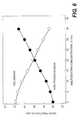
- Split porcine dermis was prepared as described in Example 1. Maltodextrin solutions containing 0% to 35% (w/v) maltodextrin were made with phosphate buffer solution (50 mM sodium phosphate, 10 mM EDTA, pH 7.0). Samples of split porcine dermis were incubated in the cryoprotectant solutions overnight. After incubation, water and cyroprotectant content in dermis tissue samples were determined. Ice contents of frozen tissue samples were determined by differential scanning calorimetry according to their ice melting enthalpies. Figure 6 shows the mass fractions of ice and amorphous tissue domain in the frozen tissue pretreated in different cryoprotectant solutions.
- porcine dermis had 75% (w/v) water and 25% dry mass.
- Upon freezing ⁇ 93% of dermis water crystallized as ice, resulting in a mass fraction of 70% ice and a mass fraction of 30% freeze-concentrated amorphous (non-frozen) domain.
- the corresponding volume fractions were 77% and 23% for ice and non-frozen domain, respectively.
- the ice fraction of incubated porcine dermis tissue decreased and the fraction of the non-frozen domain increased.
- Tissue flakes prepared as described in Example 1 were further processed to remove cellular components and ⁇ -galactosyl epitopes.
- the process included the following steps: (i) rinsing off the cryoprotectants, (ii) decellularization, (iii) enzyme treatment, (iv) disinfection, (v) freeze drying, (vi) sterilization, and (vii) secondary packaging.
- Tissue material was decellularized at ambient temperature with agitation in 150 ml of 2% (w/v) sodium deoxycholate dissolved in 10 mM HEPES buffer with 10 mM EDTA (pH 7.8). Decellularization solution was changed after 1 hour through centrifugation at 500 rpm for 3 minutes. After fresh solution was added, the tissue material was allowed to incubate for another 4 hours. Decellularization solution was drained after another round of centrifugation.
- Enzyme treatment Decellularized tissue flakes were washed twice for 30 minutes at a time with 10 mM HEPES buffer containing 5 mM EDTA (pH 7.3). Enzyme treatment was carried out for 4 hours in 150 mL of HEPES buffer with 2 mM MgCl 2 , 2 mM CaCl 2 , 1 mg/L dornase alfa, and 1 mg/L ⁇ -galactosidase. Enzyme solution was drained after centrifugation at 500 rpm for 3 minutes.
- EO sterilization included (a) conditioning at 52°C to 63°C and 55 to 75% Relative Humidity for 30 to 45 minutes, (b) EO exposure with a gas concentration of 600 ⁇ 50 mg/L for 4 hours, and (c) aeration at 38 to 54°C for at least 12 hours.
- the processed tissue flakes were acellular. Both IPA-treated and EO-treated materials tested to be sterile.
- Figs. 8A and 8B show the resistance of processed tissue material to collagenase and trypsin digestion, respectively.
- IPA disinfected tissue flakes resisted collagenase and trypsin digestion fairly well.
- EO-sterilized material had increased susceptibility to proteolysis compared to the IPA-disinfected material.
- Example 5 Suitability of Tissue Flakes to Permit Fluid Flow and Pressure Transduction
- tissue flakes Upon rehydration, tissue flakes form a stable suspension without gelling or phase separation, allowing the tissue flakes to be used for filling large voids or defects (tens to hundreds of milliliters).
- the suspension of tissue flakes contains large, inter-connected channels that permit fluid to flow freely, and thus aid cell repopulation and revascularization when the flakes are used as tissue fillers.
- the ability of the tissue flake suspension to permit the flow of fluids and the transduction of pressure differentials was investigated. Rehydrated tissue flakes were placed onto Organza mesh and spread out over a 3" x 3" pressure distribution pad with 36 sensor ports, as shown in Figs. 9A and 9B .
- a piece of GranuFoamTM (3" x 3", and 1.5" thick) was placed on the top of the tissue flake material and a 6" x 6" V.A.C.® was attached to the dressing assembly along with a T.R.A.C.® pad.
- the assembly was connected to the V.A.C.® ATS therapy unit to produce a continuous pressure at 125 mm Hg. After 5 minutes of equilibrium, the negative pressure detected at the 36 ports was noted. Thereafter, 4 of the ports were disconnected from the pressure sensors and connected to a reservoir of dyed 0.9% saline solution for infusion at a rate of 500 mL per day via a peristaltic pump. The pressure at the 32 remaining ports was then monitored over time.
- the average pressure detected was approximately 110-112 mm Hg.
- the negative pressure was 111.4 ⁇ 0.9 (mean ⁇ SD), 110.1 ⁇ 1.4, and 110.6 ⁇ 1.3 mm Hg after 1, 2, and 3 hours respectively.
- tissue matrix derived from porcine dermis was frozen at -80°C and used to make tissue flakes aseptically with a medium size MICROPLANE® cheese grater.
- tissue sample was suspended in 200 mL sterile water and then mixed using a RETSCH® blender at 4000 rpm in one minute intervals for a total of 5 cycles.
- RETSCH® blender As shown in Figs. 10A and 10B , blending reduced the size of the tissue flake material. Blending the tissue flakes also resulted in a stable and consistent tissue suspension.
- the tissue suspension was distributed in 80 cm 2 plastic petri dishes at 25 mL suspension per dish and freeze-dried aseptically.
- the freeze-drying process included the controlled cooling of the tissue suspension from room temperature to -30°C within 60 minutes and drying at a chamber pressure of 100 mT and a shelf temperature of 20°C for 24 hours.
- Fig. 11A presents a gross appearance of the tissue foam.
- Fig. 11B shows the interconnected macropores of the soft tissue foam under SEM microscopy.
- Fig. 11C is a histogram of the extracellular matrix structure of the foam using a hematoxylin and eosin stain.
- Fig. 11D is an enlarged view of a section found within Fig. 11C .
- the tissue matrix of the foam has a fibrous, filamentous nature after blending and freeze-drying.
- Some of the freeze-dried tissue material was further treated at ⁇ 100°C under vacuum for 24 hours to increase the strength of the tissue foam.
- Calorimetric measurement detected an onset denaturation temperature of 62.2 ⁇ 0.1°C and a denaturation enthalpy of 60.5 J/g tissue mass, indicating no tissue collagen denaturation.
Claims (15)
- Procédé de préparation d'une composition de matrice de tissu consistant à :sélectionner une matrice de tissu à base de collagène ;mettre en contact la matrice de tissu avec une solution cryoprotectrice ;congeler la matrice de tissu ;découper la matrice de tissu en une pluralité de morceaux de tissu découpés, la température de la matrice de tissu allant de -10 °C à -40 °C pour l'étape de découpage, le découpage consistant en un râpage ;placer la pluralité de morceaux de tissu découpés dans un liquide pour former une suspension ; etlyophiliser la suspension pour former une composition de matrice de tissu.
- Procédé selon la revendication 1, dans lequel la matrice de tissu consiste en une matrice de tissu acellulaire.
- Procédé selon la revendication 2, dans lequel la matrice de tissu acellulaire consiste en une matrice dermique acellulaire.
- Procédé selon la revendication 2, dans lequel la matrice de tissu acellulaire consiste en une matrice dermique porcine acellulaire.
- Procédé selon l'une quelconque des revendications 1 à 4, dans lequel la solution cryoprotectrice consiste au moins en une solution de maltodextrine, de saccharose, de polyéthylène glycol (PEG) et/ou de polyvinylpyrrolidone (PVP), ou en une combinaison correspondante.
- Procédé selon l'une quelconque des revendications 1 à 5, dans lequel la solution cryoprotectrice consiste en une solution de maltodextrine.
- Procédé selon la revendication 6, dans lequel la solution de maltodextrine comprend de 5 à 50 % de maltodextrine (p/v).
- Procédé selon la revendication 6, dans lequel la solution de maltodextrine comprend de 15 à 25 % de maltodextrine (p/v).
- Procédé selon l'une quelconque des revendications 1 à 8, dans lequel la matrice de tissu est découpée avec une râpe.
- Procédé selon l'une quelconque des revendications 1 à 8, dans lequel la matrice de tissu est découpée avec une roue de râpage.
- Procédé selon l'une quelconque des revendications 1 à 10, dans lequel la pluralité de morceaux de tissu découpés présente une distribution de taille allant de 0,2 à 5 mm de long, de 0,2 à 3 mm de large et de 0,02 à 0,3 mm d'épaisseur.
- Procédé selon l'une quelconque des revendications 1 à 11, consistant en outre à exposer la matrice de tissu à un désinfectant, le désinfectant étant de préférence un alcool isopropylique.
- Procédé selon l'une quelconque des revendications 1 et 12, consistant en outre à stériliser la matrice de tissu.
- Procédé selon la revendication 13, dans lequel la stérilisation de la matrice de tissu consiste à appliquer de l'oxyde d'éthylène, de l'oxyde de propylène, un rayonnement gamma ou une irradiation par faisceau d'électrons.
- Tissu de remplissage, comprenant :une matrice de tissu à base de collagène pouvant être produit par un procédé selon l'une quelconque des revendications 1 à 14.
Applications Claiming Priority (2)
| Application Number | Priority Date | Filing Date | Title |
|---|---|---|---|
| US201161475378P | 2011-04-14 | 2011-04-14 | |
| PCT/US2012/033533 WO2012142419A1 (fr) | 2011-04-14 | 2012-04-13 | Matériaux régénératifs |
Publications (2)
| Publication Number | Publication Date |
|---|---|
| EP2696908A1 EP2696908A1 (fr) | 2014-02-19 |
| EP2696908B1 true EP2696908B1 (fr) | 2015-03-11 |
Family
ID=46001826
Family Applications (1)
| Application Number | Title | Priority Date | Filing Date |
|---|---|---|---|
| EP12716959.7A Not-in-force EP2696908B1 (fr) | 2011-04-14 | 2012-04-13 | Matériaux régénératifs |
Country Status (5)
| Country | Link |
|---|---|
| US (3) | US9375513B2 (fr) |
| EP (1) | EP2696908B1 (fr) |
| BR (1) | BR112013026200A2 (fr) |
| CA (1) | CA2832838C (fr) |
| WO (1) | WO2012142419A1 (fr) |
Cited By (1)
| Publication number | Priority date | Publication date | Assignee | Title |
|---|---|---|---|---|
| CN108094410A (zh) * | 2018-01-26 | 2018-06-01 | 山东大学齐鲁医院 | 一种皮肤深低温保护剂及皮肤保存方法 |
Families Citing this family (43)
| Publication number | Priority date | Publication date | Assignee | Title |
|---|---|---|---|---|
| US8469779B1 (en) | 2009-01-02 | 2013-06-25 | Lifecell Corporation | Method for debristling animal skin |
| WO2012142419A1 (fr) | 2011-04-14 | 2012-10-18 | Lifecell Corporation | Matériaux régénératifs |
| US9089523B2 (en) | 2011-07-28 | 2015-07-28 | Lifecell Corporation | Natural tissue scaffolds as tissue fillers |
| EP3622974A1 (fr) * | 2011-11-10 | 2020-03-18 | LifeCell Corporation | Méthode pour combler les interstices dans le rapprochement de tissus |
| US9162011B2 (en) | 2011-12-19 | 2015-10-20 | Allosource | Flowable matrix compositions and methods |
| WO2013096252A1 (fr) | 2011-12-20 | 2013-06-27 | Lifecell Corporation | Produits de tissu fluides |
| WO2013096249A1 (fr) | 2011-12-20 | 2013-06-27 | Lifecell Corporation | Produits de tissu en feuille |
| CA2861048C (fr) | 2012-01-24 | 2021-01-12 | Lifecell Corporation | Matrices tissulaires allongees |
| BR112014026088B1 (pt) | 2012-04-24 | 2019-11-05 | Lifecell Corp | produto de tratamento de tecidos |
| CA2871665A1 (fr) | 2012-04-24 | 2013-10-31 | Lifecell Corporation | Matrices tissulaires fluides |
| ES2753156T3 (es) | 2012-07-13 | 2020-04-07 | Lifecell Corp | Procedimientos de tratamiento mejorado de tejido adiposo |
| WO2014052376A1 (fr) | 2012-09-26 | 2014-04-03 | Lifecell Corporation | Tissu adipeux traité |
| CA2899724A1 (fr) | 2013-02-06 | 2014-08-14 | Lifecell Corporation | Procedes pour modification localisee de produits de tissu |
| EP2964154A4 (fr) | 2013-03-07 | 2016-08-17 | Allosource | Systèmes et procédés d'allogreffe osseuse à teneur en calcium constante |
| NZ710330A (en) | 2013-03-14 | 2017-03-31 | Musculoskeletal Transplant Foundation | Soft tissue repair allografts and methods for preparing same |
| WO2014150288A2 (fr) | 2013-03-15 | 2014-09-25 | Insera Therapeutics, Inc. | Dispositifs et procédés de traitement vasculaire |
| EP2970882B1 (fr) | 2013-03-15 | 2018-11-28 | AlloSource | Matrice de collagène repeuplée de cellules pour réparation et régénération des tissus mous |
| WO2017142874A2 (fr) | 2016-02-16 | 2017-08-24 | Insera Therapeutics, Inc. | Dispositifs d'aspiration et dispositifs de déviation de flux ancrés |
| WO2015017500A1 (fr) | 2013-07-30 | 2015-02-05 | Musculoskeletal Transplant Foundation | Matrices dérivées de tissu mou acellulaire et leurs procédés de préparation |
| US10912864B2 (en) | 2015-07-24 | 2021-02-09 | Musculoskeletal Transplant Foundation | Acellular soft tissue-derived matrices and methods for preparing same |
| US11052175B2 (en) | 2015-08-19 | 2021-07-06 | Musculoskeletal Transplant Foundation | Cartilage-derived implants and methods of making and using same |
| KR20170049784A (ko) * | 2015-10-28 | 2017-05-11 | 재단법인 아산사회복지재단 | 섬유화 무세포 진피 기질 및 생체적합성 고분자를 포함하는 창상 피복재 및 이의 제조 방법 |
| AU2016367116A1 (en) | 2015-12-11 | 2018-06-14 | Lifecell Corporation | Wound treatment device |
| CN106913907B (zh) * | 2015-12-25 | 2022-04-05 | 北京瑞健高科生物科技有限公司 | 一种具有结构记忆特性的细胞生长支架的制备方法 |
| CN106913908B (zh) | 2015-12-25 | 2020-05-26 | 北京瑞健高科生物科技有限公司 | 一种具有结构记忆特性的细胞生长支架 |
| US10945831B2 (en) | 2016-06-03 | 2021-03-16 | Musculoskeletal Transplant Foundation | Asymmetric tissue graft |
| EP3463500A1 (fr) | 2016-06-03 | 2019-04-10 | LifeCell Corporation | Procédés de modification localisée de produits tissulaires |
| USD856517S1 (en) | 2016-06-03 | 2019-08-13 | Musculoskeletal Transplant Foundation | Asymmetric tissue graft |
| AU2017382173A1 (en) * | 2016-12-22 | 2019-06-06 | Lifecell Corporation | Devices and methods for tissue cryomilling |
| WO2018165131A1 (fr) | 2017-03-06 | 2018-09-13 | Tei Biosciences, Inc. | Greffe de tissu perforé |
| US20200129667A1 (en) * | 2017-06-30 | 2020-04-30 | Vascudyne Inc | Regenerative tissue and natural tissue implants |
| AU2018351051A1 (en) | 2017-10-18 | 2020-03-19 | Lifecell Corporation | Adipose tissue products and methods of production |
| US11123375B2 (en) | 2017-10-18 | 2021-09-21 | Lifecell Corporation | Methods of treating tissue voids following removal of implantable infusion ports using adipose tissue products |
| US11246994B2 (en) | 2017-10-19 | 2022-02-15 | Lifecell Corporation | Methods for introduction of flowable acellular tissue matrix products into a hand |
| JP7463273B2 (ja) | 2017-10-19 | 2024-04-08 | ライフセル コーポレーション | 流動性無細胞組織マトリックス製品および製造方法 |
| USD847864S1 (en) | 2018-01-22 | 2019-05-07 | Insera Therapeutics, Inc. | Pump |
| US10813743B2 (en) | 2018-09-07 | 2020-10-27 | Musculoskeletal Transplant Foundation | Soft tissue repair grafts and processes for preparing and using same |
| USD895812S1 (en) | 2018-09-07 | 2020-09-08 | Musculoskeletal Transplant Foundation | Soft tissue repair graft |
| CN109701077B (zh) * | 2019-01-29 | 2021-07-30 | 北京颢美细胞基因生物技术有限公司 | 一种微孔再生组织基质及其制备和应用 |
| US11633521B2 (en) | 2019-05-30 | 2023-04-25 | Lifecell Corporation | Biologic breast implant |
| CN111184891B (zh) * | 2020-02-25 | 2020-11-27 | 刘云云 | 一种医疗器械刀具用除菌封装设备 |
| CN111671974A (zh) * | 2020-04-26 | 2020-09-18 | 上海亚朋生物技术有限公司 | 一种吸水诱导形状记忆性的脱细胞真皮基质组织填充物及其制备方法 |
| WO2022014769A1 (fr) * | 2020-07-13 | 2022-01-20 | 주식회사 엘앤씨바이오 | Substitut de peau acellulaire hydraté et son procédé de fabrication |
Family Cites Families (81)
| Publication number | Priority date | Publication date | Assignee | Title |
|---|---|---|---|---|
| US4582640A (en) | 1982-03-08 | 1986-04-15 | Collagen Corporation | Injectable cross-linked collagen implant material |
| US4969912A (en) | 1988-02-18 | 1990-11-13 | Kelman Charles D | Human collagen processing and autoimplant use |
| US5024841A (en) | 1988-06-30 | 1991-06-18 | Collagen Corporation | Collagen wound healing matrices and process for their production |
| US4902508A (en) | 1988-07-11 | 1990-02-20 | Purdue Research Foundation | Tissue graft composition |
| US4938763B1 (en) | 1988-10-03 | 1995-07-04 | Atrix Lab Inc | Biodegradable in-situ forming implants and method of producing the same |
| JP2515928B2 (ja) | 1988-11-07 | 1996-07-10 | シナージェン,インコーポレーテッド | 高分子量ヒト血管形成因子 |
| US5290558A (en) | 1989-09-21 | 1994-03-01 | Osteotech, Inc. | Flowable demineralized bone powder composition and its use in bone repair |
| US5131850A (en) | 1989-11-03 | 1992-07-21 | Cryolife, Inc. | Method for cryopreserving musculoskeletal tissues |
| US5104957A (en) | 1990-02-28 | 1992-04-14 | Autogenesis Technologies, Inc. | Biologically compatible collagenous reaction product and articles useful as medical implants produced therefrom |
| US8067149B2 (en) | 1990-09-12 | 2011-11-29 | Lifecell Corporation | Acellular dermal matrix and method of use thereof for grafting |
| CA2051092C (fr) | 1990-09-12 | 2002-07-23 | Stephen A. Livesey | Methode et appareillage pour la cryopreservation, la stabilisation a sec et la rehydratation de suspensions biologiques |
| US5336616A (en) * | 1990-09-12 | 1994-08-09 | Lifecell Corporation | Method for processing and preserving collagen-based tissues for transplantation |
| US5231169A (en) | 1990-10-17 | 1993-07-27 | Norian Corporation | Mineralized collagen |
| US5254133A (en) | 1991-04-24 | 1993-10-19 | Seid Arnold S | Surgical implantation device and related method of use |
| US5160313A (en) | 1991-05-14 | 1992-11-03 | Cryolife, Inc. | Process for preparing tissue for transplantation |
| US5275826A (en) | 1992-11-13 | 1994-01-04 | Purdue Research Foundation | Fluidized intestinal submucosa and its use as an injectable tissue graft |
| US5641518A (en) | 1992-11-13 | 1997-06-24 | Purdue Research Foundation | Method of repairing bone tissue |
| US5460962A (en) | 1994-01-04 | 1995-10-24 | Organogenesis Inc. | Peracetic acid sterilization of collagen or collagenous tissue |
| JPH09510108A (ja) | 1994-03-14 | 1997-10-14 | クリオライフ,インコーポレイティド | 移植用処理組織及び調製方法 |
| US5489304A (en) | 1994-04-19 | 1996-02-06 | Brigham & Women's Hospital | Method of skin regeneration using a collagen-glycosaminoglycan matrix and cultured epithelial autograft |
| US5906827A (en) | 1994-06-03 | 1999-05-25 | Creative Biomolecules, Inc. | Matrix for the manufacture of autogenous replacement body parts |
| US5599852A (en) | 1994-10-18 | 1997-02-04 | Ethicon, Inc. | Injectable microdispersions for soft tissue repair and augmentation |
| US5622867A (en) | 1994-10-19 | 1997-04-22 | Lifecell Corporation | Prolonged preservation of blood platelets |
| US6485723B1 (en) | 1995-02-10 | 2002-11-26 | Purdue Research Foundation | Enhanced submucosal tissue graft constructs |
| US6166288A (en) | 1995-09-27 | 2000-12-26 | Nextran Inc. | Method of producing transgenic animals for xenotransplantation expressing both an enzyme masking or reducing the level of the gal epitope and a complement inhibitor |
| US6666892B2 (en) | 1996-08-23 | 2003-12-23 | Cook Biotech Incorporated | Multi-formed collagenous biomaterial medical device |
| EP0925077B1 (fr) | 1996-08-23 | 2003-10-15 | Cook Biotech, Inc. | Methode d'obtention d'une matrice purifiee a base de collagen a partir de sous-muqueuse tissulaire |
| EP0936930B1 (fr) | 1996-11-05 | 2004-07-28 | Purdue Research Foundation | Constructions de greffons du myocarde |
| AUPO599897A0 (en) | 1997-04-03 | 1997-05-01 | Vidal, Linus | Clear collagen for facial implants |
| US6613278B1 (en) * | 1998-11-13 | 2003-09-02 | Regeneration Technologies, Inc. | Tissue pooling process |
| US6371992B1 (en) | 1997-12-19 | 2002-04-16 | The Regents Of The University Of California | Acellular matrix grafts: preparation and use |
| US6326018B1 (en) | 1998-02-27 | 2001-12-04 | Musculoskeletal Transplant Foundation | Flexible sheet of demineralized bone |
| US20030039678A1 (en) | 1998-03-16 | 2003-02-27 | Stone Kevin R. | Xenograft bone matrix for orthopedic applications |
| US6179872B1 (en) | 1998-03-17 | 2001-01-30 | Tissue Engineering | Biopolymer matt for use in tissue repair and reconstruction |
| US6432710B1 (en) | 1998-05-22 | 2002-08-13 | Isolagen Technologies, Inc. | Compositions for regenerating tissue that has deteriorated, and methods for using such compositions |
| EP1087756B1 (fr) | 1998-06-19 | 2009-08-05 | Lifecell Corporation | Matrice tissulaires acellulaires particulaires |
| US6933326B1 (en) | 1998-06-19 | 2005-08-23 | Lifecell Coporation | Particulate acellular tissue matrix |
| WO2000016822A1 (fr) | 1998-09-21 | 2000-03-30 | The Brigham And Women's Hospital, Inc. | Reparations tissulaires et compositions a cet effet |
| US6565874B1 (en) | 1998-10-28 | 2003-05-20 | Atrix Laboratories | Polymeric delivery formulations of leuprolide with improved efficacy |
| WO2000047114A1 (fr) | 1999-02-12 | 2000-08-17 | Collagenesis, Inc. | Systeme d'apport de proteines osseuses morphogenetiques par injection de collagene |
| US6599318B1 (en) | 1999-11-30 | 2003-07-29 | Shlomo Gabbay | Implantable support apparatus and method of using same |
| US6576265B1 (en) | 1999-12-22 | 2003-06-10 | Acell, Inc. | Tissue regenerative composition, method of making, and method of use thereof |
| WO2002022184A2 (fr) | 2000-09-18 | 2002-03-21 | Organogenesis Inc. | Procedes de traitement de patients a l'aide de protheses greffees a feuilles planes cultivees par genie genetique |
| US7153518B2 (en) | 2001-08-27 | 2006-12-26 | Regeneration Technologies, Inc. | Processed soft tissue for topical or internal application |
| AU2002362932B2 (en) | 2001-10-18 | 2008-06-19 | Lifecell Corporation | Remodeling of tissues and organs |
| ATE410198T1 (de) | 2002-02-21 | 2008-10-15 | Encelle Inc | Immobilisierte bioaktive hydrogel matrizen für oberflächenbeschichtungen |
| WO2003080119A1 (fr) | 2002-03-26 | 2003-10-02 | Yissum Research Development Company Of The Hebrew University Of Jerusalem | Composites biomedicaux sensibles |
| US7498040B2 (en) | 2005-10-12 | 2009-03-03 | Lifenet Health | Compositions for repair of defects in osseous tissues, and methods of making the same |
| US20040037735A1 (en) | 2002-08-23 | 2004-02-26 | Depaula Carl Alexander | Allograft tissue purification process for cleaning bone |
| US7824701B2 (en) | 2002-10-18 | 2010-11-02 | Ethicon, Inc. | Biocompatible scaffold for ligament or tendon repair |
| US7115100B2 (en) * | 2002-11-15 | 2006-10-03 | Ethicon, Inc. | Tissue biopsy and processing device |
| FR2856305B1 (fr) | 2003-06-19 | 2007-08-24 | Inst Nat Sante Rech Med | Protheses avec revetements biologiquement actifs |
| CA2533259C (fr) | 2003-07-21 | 2014-01-28 | Lifecell Corporation | Matrices de tissus acellulaires realisees a partir de tissus deficitaires en galactose .alpha.-1,3-galactose |
| US7901461B2 (en) | 2003-12-05 | 2011-03-08 | Ethicon, Inc. | Viable tissue repair implants and methods of use |
| KR100680134B1 (ko) | 2004-06-10 | 2007-02-08 | 박우삼 | 무세포 진피를 주사가능하도록 입자 형태로 가공한 필러 |
| US20060058892A1 (en) | 2004-09-16 | 2006-03-16 | Lesh Michael D | Valved tissue augmentation implant |
| US20060073592A1 (en) | 2004-10-06 | 2006-04-06 | Wendell Sun | Methods of storing tissue matrices |
| DE102005002644A1 (de) * | 2005-01-19 | 2006-07-20 | Schülke & Mayr GmbH | Zusammensetzungen für die hygienische Händedesinfektion und die desinfizierende Händewaschung |
| CA2604856A1 (fr) | 2005-04-25 | 2006-11-02 | Eric F. Bernstein | Matieres de remplissage dermales pour applications biomedicales et procedes d'utilisation associes |
| AU2006294654B2 (en) | 2005-09-26 | 2012-05-24 | Lifecell Corporation | Dry platelet composition |
| US7498041B2 (en) | 2005-10-12 | 2009-03-03 | Lifenet Health | Composition for repair of defects in osseous tissues |
| JP5002805B2 (ja) | 2005-10-14 | 2012-08-15 | 財団法人ヒューマンサイエンス振興財団 | 生物由来スキャフォールドの作製方法 |
| US20070248575A1 (en) | 2006-04-19 | 2007-10-25 | Jerome Connor | Bone graft composition |
| EP2015707B1 (fr) | 2006-05-09 | 2019-01-23 | LifeCell Corporation | Tissu biologique renforcé |
| JP5050197B2 (ja) | 2006-07-31 | 2012-10-17 | 財団法人ヒューマンサイエンス振興財団 | 生物由来スキャフォールドの作製方法 |
| CA2685048A1 (fr) * | 2007-04-26 | 2008-11-06 | Advanced Technologies And Regenerative Medicine, Llc | Dispositifs et procedes de manipulation de tissus pour organes a lumiere |
| US9034367B2 (en) | 2007-05-10 | 2015-05-19 | Cormatrix Cardiovascular, Inc. | Articles for tissue regeneration with biodegradable polymer |
| EP3473260B1 (fr) | 2007-07-10 | 2020-09-23 | LifeCell Corporation | Compositions de matrice tissulaire acellulaire pour la réparation tissulaire |
| US20090024224A1 (en) * | 2007-07-16 | 2009-01-22 | Chen Silvia S | Implantation of cartilage |
| EP3287151B1 (fr) | 2008-06-06 | 2019-08-07 | LifeCell Corporation | Traitement de l'élastase de matrices tissulaires |
| AU2009281937B2 (en) | 2008-08-14 | 2015-05-07 | 3M Innovative Properties Company | Tissue scaffolds |
| US7927414B2 (en) * | 2008-09-05 | 2011-04-19 | Ethicon, Inc. | Method of manufacturing acellular matrix glue |
| WO2010059783A2 (fr) | 2008-11-21 | 2010-05-27 | Lifecell Corporation | Matériau biologique renforcé |
| CN102264432B (zh) | 2008-12-31 | 2013-10-23 | 凯希特许有限公司 | 向组织提供流体流的系统 |
| EP2432495B1 (fr) | 2009-05-20 | 2017-03-22 | Humacyte, Inc. | Elastine pour l'augmentation des tissus mous |
| JP5967721B2 (ja) | 2010-07-08 | 2016-08-10 | ライフセル コーポレーションLifeCell Corporation | 組織マトリックスの成形方法 |
| CA2806464C (fr) | 2010-08-10 | 2018-10-23 | Lifecell Corporation | Echafaudages tissulaires regenerateurs |
| WO2012142419A1 (fr) | 2011-04-14 | 2012-10-18 | Lifecell Corporation | Matériaux régénératifs |
| EP2714111B1 (fr) | 2011-05-31 | 2021-03-17 | LifeCell Corporation | Matrices de tissu adipeux |
| EP3622974A1 (fr) | 2011-11-10 | 2020-03-18 | LifeCell Corporation | Méthode pour combler les interstices dans le rapprochement de tissus |
| WO2013096252A1 (fr) | 2011-12-20 | 2013-06-27 | Lifecell Corporation | Produits de tissu fluides |
-
2012
- 2012-04-13 WO PCT/US2012/033533 patent/WO2012142419A1/fr active Application Filing
- 2012-04-13 EP EP12716959.7A patent/EP2696908B1/fr not_active Not-in-force
- 2012-04-13 BR BR112013026200A patent/BR112013026200A2/pt not_active IP Right Cessation
- 2012-04-13 US US13/446,422 patent/US9375513B2/en active Active
- 2012-04-13 CA CA2832838A patent/CA2832838C/fr active Active
-
2016
- 2016-05-27 US US15/166,848 patent/US10828391B2/en active Active
-
2018
- 2018-08-22 US US16/108,581 patent/US20180353644A1/en not_active Abandoned
Cited By (2)
| Publication number | Priority date | Publication date | Assignee | Title |
|---|---|---|---|---|
| CN108094410A (zh) * | 2018-01-26 | 2018-06-01 | 山东大学齐鲁医院 | 一种皮肤深低温保护剂及皮肤保存方法 |
| CN108094410B (zh) * | 2018-01-26 | 2021-08-31 | 山东大学齐鲁医院 | 一种皮肤深低温保护剂及皮肤保存方法 |
Also Published As
| Publication number | Publication date |
|---|---|
| AU2012242694A1 (en) | 2013-10-17 |
| WO2012142419A1 (fr) | 2012-10-18 |
| CA2832838A1 (fr) | 2012-10-18 |
| EP2696908A1 (fr) | 2014-02-19 |
| US10828391B2 (en) | 2020-11-10 |
| BR112013026200A2 (pt) | 2019-08-27 |
| US20160271295A1 (en) | 2016-09-22 |
| US20120263763A1 (en) | 2012-10-18 |
| AU2012242694A8 (en) | 2013-11-21 |
| US9375513B2 (en) | 2016-06-28 |
| US20180353644A1 (en) | 2018-12-13 |
| CA2832838C (fr) | 2019-08-13 |
Similar Documents
| Publication | Publication Date | Title |
|---|---|---|
| US10828391B2 (en) | Regenerative materials | |
| US20200323617A1 (en) | Flowable tissue products | |
| US20210220113A1 (en) | Sheet tissue products | |
| US11957814B2 (en) | Adipose tissue matrices | |
| AU2012242694B2 (en) | Regenerative materials | |
| AU2012355466B2 (en) | Flowable tissue products |
Legal Events
| Date | Code | Title | Description |
|---|---|---|---|
| PUAI | Public reference made under article 153(3) epc to a published international application that has entered the european phase |
Free format text: ORIGINAL CODE: 0009012 |
|
| 17P | Request for examination filed |
Effective date: 20130927 |
|
| AK | Designated contracting states |
Kind code of ref document: A1 Designated state(s): AL AT BE BG CH CY CZ DE DK EE ES FI FR GB GR HR HU IE IS IT LI LT LU LV MC MK MT NL NO PL PT RO RS SE SI SK SM TR |
|
| DAX | Request for extension of the european patent (deleted) | ||
| GRAP | Despatch of communication of intention to grant a patent |
Free format text: ORIGINAL CODE: EPIDOSNIGR1 |
|
| INTG | Intention to grant announced |
Effective date: 20141103 |
|
| RIN1 | Information on inventor provided before grant (corrected) |
Inventor name: GOEL, RAGHAV Inventor name: SUN, WENQUAN Inventor name: BOWLEY, MELISSA RICHTER |
|
| GRAS | Grant fee paid |
Free format text: ORIGINAL CODE: EPIDOSNIGR3 |
|
| GRAA | (expected) grant |
Free format text: ORIGINAL CODE: 0009210 |
|
| AK | Designated contracting states |
Kind code of ref document: B1 Designated state(s): AL AT BE BG CH CY CZ DE DK EE ES FI FR GB GR HR HU IE IS IT LI LT LU LV MC MK MT NL NO PL PT RO RS SE SI SK SM TR |
|
| REG | Reference to a national code |
Ref country code: GB Ref legal event code: FG4D |
|
| REG | Reference to a national code |
Ref country code: CH Ref legal event code: EP |
|
| REG | Reference to a national code |
Ref country code: IE Ref legal event code: FG4D |
|
| REG | Reference to a national code |
Ref country code: AT Ref legal event code: REF Ref document number: 714951 Country of ref document: AT Kind code of ref document: T Effective date: 20150415 |
|
| REG | Reference to a national code |
Ref country code: DE Ref legal event code: R096 Ref document number: 602012005856 Country of ref document: DE Effective date: 20150423 |
|
| REG | Reference to a national code |
Ref country code: NL Ref legal event code: VDEP Effective date: 20150311 |
|
| REG | Reference to a national code |
Ref country code: NL Ref legal event code: VDEP Effective date: 20150311 |
|
| PG25 | Lapsed in a contracting state [announced via postgrant information from national office to epo] |
Ref country code: HR Free format text: LAPSE BECAUSE OF FAILURE TO SUBMIT A TRANSLATION OF THE DESCRIPTION OR TO PAY THE FEE WITHIN THE PRESCRIBED TIME-LIMIT Effective date: 20150311 Ref country code: NO Free format text: LAPSE BECAUSE OF FAILURE TO SUBMIT A TRANSLATION OF THE DESCRIPTION OR TO PAY THE FEE WITHIN THE PRESCRIBED TIME-LIMIT Effective date: 20150611 Ref country code: ES Free format text: LAPSE BECAUSE OF FAILURE TO SUBMIT A TRANSLATION OF THE DESCRIPTION OR TO PAY THE FEE WITHIN THE PRESCRIBED TIME-LIMIT Effective date: 20150311 Ref country code: SE Free format text: LAPSE BECAUSE OF FAILURE TO SUBMIT A TRANSLATION OF THE DESCRIPTION OR TO PAY THE FEE WITHIN THE PRESCRIBED TIME-LIMIT Effective date: 20150311 Ref country code: LT Free format text: LAPSE BECAUSE OF FAILURE TO SUBMIT A TRANSLATION OF THE DESCRIPTION OR TO PAY THE FEE WITHIN THE PRESCRIBED TIME-LIMIT Effective date: 20150311 Ref country code: FI Free format text: LAPSE BECAUSE OF FAILURE TO SUBMIT A TRANSLATION OF THE DESCRIPTION OR TO PAY THE FEE WITHIN THE PRESCRIBED TIME-LIMIT Effective date: 20150311 |
|
| REG | Reference to a national code |
Ref country code: AT Ref legal event code: MK05 Ref document number: 714951 Country of ref document: AT Kind code of ref document: T Effective date: 20150311 |
|
| REG | Reference to a national code |
Ref country code: LT Ref legal event code: MG4D |
|
| PG25 | Lapsed in a contracting state [announced via postgrant information from national office to epo] |
Ref country code: LV Free format text: LAPSE BECAUSE OF FAILURE TO SUBMIT A TRANSLATION OF THE DESCRIPTION OR TO PAY THE FEE WITHIN THE PRESCRIBED TIME-LIMIT Effective date: 20150311 Ref country code: RS Free format text: LAPSE BECAUSE OF FAILURE TO SUBMIT A TRANSLATION OF THE DESCRIPTION OR TO PAY THE FEE WITHIN THE PRESCRIBED TIME-LIMIT Effective date: 20150311 Ref country code: GR Free format text: LAPSE BECAUSE OF FAILURE TO SUBMIT A TRANSLATION OF THE DESCRIPTION OR TO PAY THE FEE WITHIN THE PRESCRIBED TIME-LIMIT Effective date: 20150612 |
|
| PG25 | Lapsed in a contracting state [announced via postgrant information from national office to epo] |
Ref country code: NL Free format text: LAPSE BECAUSE OF FAILURE TO SUBMIT A TRANSLATION OF THE DESCRIPTION OR TO PAY THE FEE WITHIN THE PRESCRIBED TIME-LIMIT Effective date: 20150311 |
|
| PG25 | Lapsed in a contracting state [announced via postgrant information from national office to epo] |
Ref country code: CZ Free format text: LAPSE BECAUSE OF FAILURE TO SUBMIT A TRANSLATION OF THE DESCRIPTION OR TO PAY THE FEE WITHIN THE PRESCRIBED TIME-LIMIT Effective date: 20150311 Ref country code: RO Free format text: LAPSE BECAUSE OF FAILURE TO SUBMIT A TRANSLATION OF THE DESCRIPTION OR TO PAY THE FEE WITHIN THE PRESCRIBED TIME-LIMIT Effective date: 20150311 Ref country code: PT Free format text: LAPSE BECAUSE OF FAILURE TO SUBMIT A TRANSLATION OF THE DESCRIPTION OR TO PAY THE FEE WITHIN THE PRESCRIBED TIME-LIMIT Effective date: 20150713 Ref country code: SK Free format text: LAPSE BECAUSE OF FAILURE TO SUBMIT A TRANSLATION OF THE DESCRIPTION OR TO PAY THE FEE WITHIN THE PRESCRIBED TIME-LIMIT Effective date: 20150311 Ref country code: EE Free format text: LAPSE BECAUSE OF FAILURE TO SUBMIT A TRANSLATION OF THE DESCRIPTION OR TO PAY THE FEE WITHIN THE PRESCRIBED TIME-LIMIT Effective date: 20150311 |
|
| PG25 | Lapsed in a contracting state [announced via postgrant information from national office to epo] |
Ref country code: IS Free format text: LAPSE BECAUSE OF FAILURE TO SUBMIT A TRANSLATION OF THE DESCRIPTION OR TO PAY THE FEE WITHIN THE PRESCRIBED TIME-LIMIT Effective date: 20150711 Ref country code: PL Free format text: LAPSE BECAUSE OF FAILURE TO SUBMIT A TRANSLATION OF THE DESCRIPTION OR TO PAY THE FEE WITHIN THE PRESCRIBED TIME-LIMIT Effective date: 20150311 Ref country code: MC Free format text: LAPSE BECAUSE OF FAILURE TO SUBMIT A TRANSLATION OF THE DESCRIPTION OR TO PAY THE FEE WITHIN THE PRESCRIBED TIME-LIMIT Effective date: 20150311 Ref country code: AT Free format text: LAPSE BECAUSE OF FAILURE TO SUBMIT A TRANSLATION OF THE DESCRIPTION OR TO PAY THE FEE WITHIN THE PRESCRIBED TIME-LIMIT Effective date: 20150311 |
|
| REG | Reference to a national code |
Ref country code: CH Ref legal event code: PL |
|
| REG | Reference to a national code |
Ref country code: DE Ref legal event code: R097 Ref document number: 602012005856 Country of ref document: DE |
|
| PG25 | Lapsed in a contracting state [announced via postgrant information from national office to epo] |
Ref country code: IT Free format text: LAPSE BECAUSE OF FAILURE TO SUBMIT A TRANSLATION OF THE DESCRIPTION OR TO PAY THE FEE WITHIN THE PRESCRIBED TIME-LIMIT Effective date: 20150311 |
|
| PLBE | No opposition filed within time limit |
Free format text: ORIGINAL CODE: 0009261 |
|
| STAA | Information on the status of an ep patent application or granted ep patent |
Free format text: STATUS: NO OPPOSITION FILED WITHIN TIME LIMIT |
|
| REG | Reference to a national code |
Ref country code: IE Ref legal event code: MM4A |
|
| PG25 | Lapsed in a contracting state [announced via postgrant information from national office to epo] |
Ref country code: DK Free format text: LAPSE BECAUSE OF FAILURE TO SUBMIT A TRANSLATION OF THE DESCRIPTION OR TO PAY THE FEE WITHIN THE PRESCRIBED TIME-LIMIT Effective date: 20150311 Ref country code: LI Free format text: LAPSE BECAUSE OF NON-PAYMENT OF DUE FEES Effective date: 20150430 Ref country code: CH Free format text: LAPSE BECAUSE OF NON-PAYMENT OF DUE FEES Effective date: 20150430 |
|
| 26N | No opposition filed |
Effective date: 20151214 |
|
| PG25 | Lapsed in a contracting state [announced via postgrant information from national office to epo] |
Ref country code: SI Free format text: LAPSE BECAUSE OF FAILURE TO SUBMIT A TRANSLATION OF THE DESCRIPTION OR TO PAY THE FEE WITHIN THE PRESCRIBED TIME-LIMIT Effective date: 20150311 |
|
| REG | Reference to a national code |
Ref country code: FR Ref legal event code: PLFP Year of fee payment: 5 |
|
| PG25 | Lapsed in a contracting state [announced via postgrant information from national office to epo] |
Ref country code: IE Free format text: LAPSE BECAUSE OF NON-PAYMENT OF DUE FEES Effective date: 20150413 |
|
| PG25 | Lapsed in a contracting state [announced via postgrant information from national office to epo] |
Ref country code: BE Free format text: LAPSE BECAUSE OF FAILURE TO SUBMIT A TRANSLATION OF THE DESCRIPTION OR TO PAY THE FEE WITHIN THE PRESCRIBED TIME-LIMIT Effective date: 20150311 |
|
| PG25 | Lapsed in a contracting state [announced via postgrant information from national office to epo] |
Ref country code: MT Free format text: LAPSE BECAUSE OF FAILURE TO SUBMIT A TRANSLATION OF THE DESCRIPTION OR TO PAY THE FEE WITHIN THE PRESCRIBED TIME-LIMIT Effective date: 20150311 |
|
| REG | Reference to a national code |
Ref country code: FR Ref legal event code: PLFP Year of fee payment: 6 |
|
| PG25 | Lapsed in a contracting state [announced via postgrant information from national office to epo] |
Ref country code: HU Free format text: LAPSE BECAUSE OF FAILURE TO SUBMIT A TRANSLATION OF THE DESCRIPTION OR TO PAY THE FEE WITHIN THE PRESCRIBED TIME-LIMIT; INVALID AB INITIO Effective date: 20120413 Ref country code: BG Free format text: LAPSE BECAUSE OF FAILURE TO SUBMIT A TRANSLATION OF THE DESCRIPTION OR TO PAY THE FEE WITHIN THE PRESCRIBED TIME-LIMIT Effective date: 20150311 Ref country code: SM Free format text: LAPSE BECAUSE OF FAILURE TO SUBMIT A TRANSLATION OF THE DESCRIPTION OR TO PAY THE FEE WITHIN THE PRESCRIBED TIME-LIMIT Effective date: 20150311 |
|
| PG25 | Lapsed in a contracting state [announced via postgrant information from national office to epo] |
Ref country code: CY Free format text: LAPSE BECAUSE OF FAILURE TO SUBMIT A TRANSLATION OF THE DESCRIPTION OR TO PAY THE FEE WITHIN THE PRESCRIBED TIME-LIMIT Effective date: 20150311 |
|
| PG25 | Lapsed in a contracting state [announced via postgrant information from national office to epo] |
Ref country code: TR Free format text: LAPSE BECAUSE OF FAILURE TO SUBMIT A TRANSLATION OF THE DESCRIPTION OR TO PAY THE FEE WITHIN THE PRESCRIBED TIME-LIMIT Effective date: 20150311 |
|
| PG25 | Lapsed in a contracting state [announced via postgrant information from national office to epo] |
Ref country code: LU Free format text: LAPSE BECAUSE OF NON-PAYMENT OF DUE FEES Effective date: 20150413 |
|
| REG | Reference to a national code |
Ref country code: FR Ref legal event code: PLFP Year of fee payment: 7 |
|
| PG25 | Lapsed in a contracting state [announced via postgrant information from national office to epo] |
Ref country code: MK Free format text: LAPSE BECAUSE OF FAILURE TO SUBMIT A TRANSLATION OF THE DESCRIPTION OR TO PAY THE FEE WITHIN THE PRESCRIBED TIME-LIMIT Effective date: 20150311 |
|
| PG25 | Lapsed in a contracting state [announced via postgrant information from national office to epo] |
Ref country code: AL Free format text: LAPSE BECAUSE OF FAILURE TO SUBMIT A TRANSLATION OF THE DESCRIPTION OR TO PAY THE FEE WITHIN THE PRESCRIBED TIME-LIMIT Effective date: 20150311 |
|
| PGFP | Annual fee paid to national office [announced via postgrant information from national office to epo] |
Ref country code: DE Payment date: 20210428 Year of fee payment: 10 Ref country code: FR Payment date: 20210426 Year of fee payment: 10 |
|
| PGFP | Annual fee paid to national office [announced via postgrant information from national office to epo] |
Ref country code: GB Payment date: 20210427 Year of fee payment: 10 |
|
| REG | Reference to a national code |
Ref country code: DE Ref legal event code: R119 Ref document number: 602012005856 Country of ref document: DE |
|
| GBPC | Gb: european patent ceased through non-payment of renewal fee |
Effective date: 20220413 |
|
| PG25 | Lapsed in a contracting state [announced via postgrant information from national office to epo] |
Ref country code: GB Free format text: LAPSE BECAUSE OF NON-PAYMENT OF DUE FEES Effective date: 20220413 Ref country code: FR Free format text: LAPSE BECAUSE OF NON-PAYMENT OF DUE FEES Effective date: 20220430 Ref country code: DE Free format text: LAPSE BECAUSE OF NON-PAYMENT OF DUE FEES Effective date: 20221103 |