EP2635111B1 - Stabilisierte opsin-proteine mit stufenfunktion und verwendungsverfahren dafür - Google Patents
Stabilisierte opsin-proteine mit stufenfunktion und verwendungsverfahren dafür Download PDFInfo
- Publication number
- EP2635111B1 EP2635111B1 EP11838898.2A EP11838898A EP2635111B1 EP 2635111 B1 EP2635111 B1 EP 2635111B1 EP 11838898 A EP11838898 A EP 11838898A EP 2635111 B1 EP2635111 B1 EP 2635111B1
- Authority
- EP
- European Patent Office
- Prior art keywords
- light
- neurons
- protein
- ssfo
- aspects
- Prior art date
- Legal status (The legal status is an assumption and is not a legal conclusion. Google has not performed a legal analysis and makes no representation as to the accuracy of the status listed.)
- Not-in-force
Links
Images


Classifications
-
- A—HUMAN NECESSITIES
- A01—AGRICULTURE; FORESTRY; ANIMAL HUSBANDRY; HUNTING; TRAPPING; FISHING
- A01K—ANIMAL HUSBANDRY; CARE OF BIRDS, FISHES, INSECTS; FISHING; REARING OR BREEDING ANIMALS, NOT OTHERWISE PROVIDED FOR; NEW BREEDS OF ANIMALS
- A01K67/00—Rearing or breeding animals, not otherwise provided for; New breeds of animals
- A01K67/027—New breeds of vertebrates
- A01K67/0275—Genetically modified vertebrates, e.g. transgenic
-
- C—CHEMISTRY; METALLURGY
- C07—ORGANIC CHEMISTRY
- C07K—PEPTIDES
- C07K14/00—Peptides having more than 20 amino acids; Gastrins; Somatostatins; Melanotropins; Derivatives thereof
- C07K14/405—Peptides having more than 20 amino acids; Gastrins; Somatostatins; Melanotropins; Derivatives thereof from algae
-
- C—CHEMISTRY; METALLURGY
- C12—BIOCHEMISTRY; BEER; SPIRITS; WINE; VINEGAR; MICROBIOLOGY; ENZYMOLOGY; MUTATION OR GENETIC ENGINEERING
- C12N—MICROORGANISMS OR ENZYMES; COMPOSITIONS THEREOF; PROPAGATING, PRESERVING, OR MAINTAINING MICROORGANISMS; MUTATION OR GENETIC ENGINEERING; CULTURE MEDIA
- C12N5/00—Undifferentiated human, animal or plant cells, e.g. cell lines; Tissues; Cultivation or maintenance thereof; Culture media therefor
- C12N5/06—Animal cells or tissues; Human cells or tissues
- C12N5/0602—Vertebrate cells
- C12N5/0618—Cells of the nervous system
- C12N5/0619—Neurons
-
- G—PHYSICS
- G01—MEASURING; TESTING
- G01N—INVESTIGATING OR ANALYSING MATERIALS BY DETERMINING THEIR CHEMICAL OR PHYSICAL PROPERTIES
- G01N33/00—Investigating or analysing materials by specific methods not covered by groups G01N1/00 - G01N31/00
- G01N33/48—Biological material, e.g. blood, urine; Haemocytometers
- G01N33/50—Chemical analysis of biological material, e.g. blood, urine; Testing involving biospecific ligand binding methods; Immunological testing
- G01N33/5005—Chemical analysis of biological material, e.g. blood, urine; Testing involving biospecific ligand binding methods; Immunological testing involving human or animal cells
- G01N33/5008—Chemical analysis of biological material, e.g. blood, urine; Testing involving biospecific ligand binding methods; Immunological testing involving human or animal cells for testing or evaluating the effect of chemical or biological compounds, e.g. drugs, cosmetics
- G01N33/5082—Supracellular entities, e.g. tissue, organisms
- G01N33/5088—Supracellular entities, e.g. tissue, organisms of vertebrates
-
- G—PHYSICS
- G01—MEASURING; TESTING
- G01N—INVESTIGATING OR ANALYSING MATERIALS BY DETERMINING THEIR CHEMICAL OR PHYSICAL PROPERTIES
- G01N33/00—Investigating or analysing materials by specific methods not covered by groups G01N1/00 - G01N31/00
- G01N33/48—Biological material, e.g. blood, urine; Haemocytometers
- G01N33/50—Chemical analysis of biological material, e.g. blood, urine; Testing involving biospecific ligand binding methods; Immunological testing
- G01N33/5005—Chemical analysis of biological material, e.g. blood, urine; Testing involving biospecific ligand binding methods; Immunological testing involving human or animal cells
- G01N33/5091—Chemical analysis of biological material, e.g. blood, urine; Testing involving biospecific ligand binding methods; Immunological testing involving human or animal cells for testing the pathological state of an organism
-
- A—HUMAN NECESSITIES
- A01—AGRICULTURE; FORESTRY; ANIMAL HUSBANDRY; HUNTING; TRAPPING; FISHING
- A01K—ANIMAL HUSBANDRY; CARE OF BIRDS, FISHES, INSECTS; FISHING; REARING OR BREEDING ANIMALS, NOT OTHERWISE PROVIDED FOR; NEW BREEDS OF ANIMALS
- A01K2217/00—Genetically modified animals
- A01K2217/20—Animal model comprising regulated expression system
- A01K2217/206—Animal model comprising tissue-specific expression system, e.g. tissue specific expression of transgene, of Cre recombinase
-
- A—HUMAN NECESSITIES
- A01—AGRICULTURE; FORESTRY; ANIMAL HUSBANDRY; HUNTING; TRAPPING; FISHING
- A01K—ANIMAL HUSBANDRY; CARE OF BIRDS, FISHES, INSECTS; FISHING; REARING OR BREEDING ANIMALS, NOT OTHERWISE PROVIDED FOR; NEW BREEDS OF ANIMALS
- A01K2227/00—Animals characterised by species
- A01K2227/10—Mammal
- A01K2227/105—Murine
-
- A—HUMAN NECESSITIES
- A01—AGRICULTURE; FORESTRY; ANIMAL HUSBANDRY; HUNTING; TRAPPING; FISHING
- A01K—ANIMAL HUSBANDRY; CARE OF BIRDS, FISHES, INSECTS; FISHING; REARING OR BREEDING ANIMALS, NOT OTHERWISE PROVIDED FOR; NEW BREEDS OF ANIMALS
- A01K2267/00—Animals characterised by purpose
- A01K2267/03—Animal model, e.g. for test or diseases
- A01K2267/0393—Animal model comprising a reporter system for screening tests
-
- C—CHEMISTRY; METALLURGY
- C12—BIOCHEMISTRY; BEER; SPIRITS; WINE; VINEGAR; MICROBIOLOGY; ENZYMOLOGY; MUTATION OR GENETIC ENGINEERING
- C12N—MICROORGANISMS OR ENZYMES; COMPOSITIONS THEREOF; PROPAGATING, PRESERVING, OR MAINTAINING MICROORGANISMS; MUTATION OR GENETIC ENGINEERING; CULTURE MEDIA
- C12N2740/00—Reverse transcribing RNA viruses
- C12N2740/00011—Details
- C12N2740/10011—Retroviridae
- C12N2740/16011—Human Immunodeficiency Virus, HIV
- C12N2740/16041—Use of virus, viral particle or viral elements as a vector
- C12N2740/16043—Use of virus, viral particle or viral elements as a vector viral genome or elements thereof as genetic vector
-
- C—CHEMISTRY; METALLURGY
- C12—BIOCHEMISTRY; BEER; SPIRITS; WINE; VINEGAR; MICROBIOLOGY; ENZYMOLOGY; MUTATION OR GENETIC ENGINEERING
- C12N—MICROORGANISMS OR ENZYMES; COMPOSITIONS THEREOF; PROPAGATING, PRESERVING, OR MAINTAINING MICROORGANISMS; MUTATION OR GENETIC ENGINEERING; CULTURE MEDIA
- C12N2750/00—MICROORGANISMS OR ENZYMES; COMPOSITIONS THEREOF; PROPAGATING, PRESERVING, OR MAINTAINING MICROORGANISMS; MUTATION OR GENETIC ENGINEERING; CULTURE MEDIA ssDNA viruses
- C12N2750/00011—Details
- C12N2750/14011—Parvoviridae
- C12N2750/14111—Dependovirus, e.g. adenoassociated viruses
- C12N2750/14141—Use of virus, viral particle or viral elements as a vector
- C12N2750/14143—Use of virus, viral particle or viral elements as a vector viral genome or elements thereof as genetic vector
Definitions
- This application pertains to non-human mammals expressing stabilized step function opsin (SSFO) proteins on their plasma membranes and methods of using the same to selectively depolarize neurons residing in microcircuits of the pre-frontal cortex to affect one or more social behaviors, communications, and/or conditioned behaviors in the non-human animal.
- SSFO stabilized step function opsin
- Optogenetics is the combination of genetic and optical methods used to control specific events in targeted cells of living tissue, even within freely moving mammals and other animals, with the temporal precision (millisecond-timescale) needed to keep pace with functioning intact biological systems.
- the hallmark of optogenetics is the introduction of fast light-activated channel proteins to the plasma membranes of target neuronal cells that allow temporally precise manipulation of neuronal membrane potential while maintaining cell-type resolution through the use of specific targeting mechanisms.
- microbial opsins which can be used to investigate the function of neural systems are the channelrhodopsins (ChR2, ChR1, VChR1, and SFOs) used to promote depolarization in response to light.
- SFOs also has the potential to address the hardware challenge, since the orders-of-magnitude greater light sensitivity characteristic of SFOs could in theory allow non- brain penetrating light delivery, and the persistent action of the bistable SFOs after light-off could allow hardware-free behavioral testing.
- the known SFOs C128A,S,T and D156A are not stable enough to produce constant photocurrent after a single light flash over the many minutes required for complex behavioral testing.
- WO 2010/056970 relates to SFOs and their use for controlling currents in a cell, e.g., in in vivo and in vitro environments.
- the present invention provides a non-human mammal comprising a first light-activated cation channel protein expressed on the cell membrane of neurons of the pre-frontal cortex of the mammal, wherein the protein comprises an amino acid sequence at least 95% identical to the sequence shown in one of SEQ ID NOs:1-4, and comprises a Cys ⁇ Ser substitution at an amino acid position corresponding to amino acid 128 of SEQ ID NO:1, and an Asp ⁇ Ala substitution at an amino acid position corresponding to amino acid 156 of SEQ ID NO:1, wherein the protein is capable of inducing depolarizing current in the neurons by light and exhibits rapid step-like activation in response to a single pulse of light having a first wavelength and deactivation in response to a pulse of light having a second wavelength, wherein the depolarizing current in the neurons is maintained for at least ten minutes; and wherein the activation of the protein in the pre-frontal cortex neurons induces changes in social behaviours, communications, and/
- animal cells non-human animals, brain slices comprising cells expressing stabilized step function opsin proteins on their plasma membranes and methods of using the same to selectively depolarize neurons residing in microcircuits of the pre-frontal cortex.
- non-human animals comprising a first light-activated cation channel protein expressed in neurons of the pre-frontal cortex of the animal, wherein the protein is capable of inducing depolarizing current in the neurons by light and exhibits rapid step-like activation in response to a single pulse of light having a first wavelength and deactivation in response to a pulse of light having a second wavelength, wherein the depolarizing current in the neurons is maintained for at least about ten minutes; and wherein the activation of the protein in the pre-frontal cortex neurons induces changes in social behaviors, communications, and/or conditioned behaviors in the animal.
- a brain slice comprising neurons of the pre-frontal cortex, wherein a light-activated protein is expressed in the neurons of the pre-frontal cortex, wherein the protein is capable of inducing depolarizing current in the neurons by light and exhibits rapid step-like activation in response to a single pulse of light having a first wavelength and deactivation in response to a pulse of light having a second wavelength; wherein the depolarizing current in the neurons is maintained for at least about ten minutes.
- a method for identifying a chemical compound that inhibits the depolarization of excitatory or inhibitory neurons in the prefrontal cortex of a non-human animal comprising:(a) depolarizing excitatory or inhibitory neurons in the prefrontal cortex of a non-human animal comprising a first light-activated protein cation channel protein expressed on the cell membrane of the neurons of the pre-frontal cortex of the animal, wherein the protein is capable of mediating a depolarizing current in the neurons when the neurons are illuminated with light, wherein the protein exhibits rapid step-like activation in response to a single pulse of light having a first wavelength and deactivation in response to a pulse of light having a second wavelength; wherein the depolarizing current in the neurons is maintained for at least about ten minutes; wherein the protein comprises the amino acid sequence of ChR2, ChR1, VChR1, or VChR2 with amino acid substitutions at amino acid residues corresponding to C128 and D156 of the amino acid sequence of ChR2
- a method for identifying a chemical compound that restores a social behavior, communication, and/or conditioned behavior in a non-human animal comprising: (a) depolarizing excitatory neurons in the prefrontal cortex of a non-human animal comprising a light-activated protein cation channel protein expressed on the cell membrane of the neurons, wherein the protein is capable of inducing a depolarizing current in the neurons when the neurons are illuminated with light, wherein the protein exhibits rapid step-like activation in response to a single pulse of light having a first wavelength and deactivation in response to a pulse of light having a second wavelength; wherein the depolarizing current in the neurons is maintained for at least about ten minutes; and wherein the protein comprises the amino acid sequence of ChR2, ChR1, VChR1, or VChR2 with amino acid substitutions at amino acid residues corresponding to C 12 8 and D156 of the amino acid sequence of ChR2, wherein depolarizing the excitatory neuron inhibits one or more social
- the present disclosure relates to optical control over nervous system disorders (such as disorders associated with social dysfunction), as described herein. While the present disclosure is not necessarily limited in these contexts, various aspects of the disclosure may be appreciated through a discussion of examples using these and other contexts.
- Various embodiments of the present disclosure relate to an optogenetic system or method that correlates temporal, spatio and/or cell-type control over a neural circuit with measurable metrics. For instance, various metrics or symptoms might be associated with a neurological disorder (such as a neurological disorder exhibiting various symptoms of social dysfunction).
- the optogenetic system targets a neural circuit within a subject/patient for selective control thereof
- the optogenetic system involves monitoring the subject/patient for the metrics or symptoms associated with the neurological disorder. In this manner, the optogenetic system can provide detailed information about the neural circuit, its function and/or the neurological disorder.
- particular embodiments relate to studying and probing disorders.
- Other embodiments relate to the identification and/or study of phenotypes and endophenotypes.
- Still other embodiments relate to the identification of treatment targets.
- aspects of the present disclosure are directed toward the artificial inducement of disorder/disease states on a fast-temporal time scale. These aspects allow for study of disease states in otherwise healthy animals. This can be particularly useful for diseases that are poorly understood and otherwise difficult to accurately model in live animals. For instance, it can be difficult to test and/or study disease states due to the lack of available animals exhibiting the disease state. Moreover, certain embodiments allow for reversible disease states, which can be particularly useful for establishing baseline/control points for testing and/or for testing the effects of a treatment on the same animal when exhibiting the disease state and when not exhibiting the disease state. Various other possibilities exist, some of which are discussed in more detail herein.
- aspects of the present disclosure are directed to using an artificially induced disorder/disease state for the study of disease states in otherwise healthy animals. This can be particularly useful for diseases that are poorly understood and otherwise difficult to accurately model in living animals. For instance, it can be difficult to test and/or study disease states due to the lack of available animals exhibiting the disease state. Moreover, certain embodiments allow for reversible disease states, which can be particularly useful in establishing baseline/control points for testing and/or for testing the effects of a treatment on the same animal when exhibiting the disease state and when not exhibiting the disease state.
- Certain aspects of the present disclosure are directed to a method that includes modifying (e.g., elevating or lowering) an excitation/inhibition (E/I) balance in a targeted neural circuit in a prefrontal cortex of a subject/patient. For instance, the E/I balance is changed to a level that preserves the responsiveness of the targeted neural circuit to intrinsic electrical activity while symptoms of a disorder are temporally increased. While the E/I balance is changed, a stimulus is introduce to the subject/patient and the symptoms of the disorder are monitored.
- the subject can be a test animal that is healthy, or an animal model of a disorder.
- the result of the manipulation is either a transient recapitulation of disease symptoms (in an otherwise healthy animal) or alleviation of symptoms (in an animal model of a neurological disorder).
- the monitoring of the symptoms also includes assessing the efficacy of the stimulus in mitigating the symptoms of the disorder.
- This disclosure provides, inter alia, animal cells, non-human animals, and brain slices comprising cells expressing stabilized step function opsin proteins on their plasma membranes, and methods of using the stabilized step function opsin proteins to selectively depolarize excitatory or inhibitory neurons residing in the same microcircuit in the pre-frontal cortex.
- the step function opsins, or SFOs are ChR2 light-activated cation channel proteins that can induce prolonged stable excitable states in neurons upon exposure to blue light and then be reversed upon exposure to green or yellow light.
- the SFOs were developed to implement bistable changes in excitability of targeted populations operating on timescales up to 4 orders of magnitude longer than that of wild type (wt) ChR2 for more stable state modulation (SFOs: up to 10-100 seconds). While these opsin genes delivered a new kind of optogenetic control complementary to that of conventional channelrhodopsins designed to control individual action potentials, the timescale was still not suitable for evaluating prolonged and complex mammalian behaviors over many minutes.
- SSFO stabilized step function opsin
- brain slices from non-human animals containing cortical excitatory or inhibitory neurons expressing the stabilized step function opsin proteins disclosed herein can be used to search for chemical compounds which can selectively inhibit the depolarization of either excitatory or inhibitory neurons residing within a neural circuit.
- These cortical neurons may be responsible for or involved with the social and cognitive behavioral defects associated with neurological disorders such as schizophrenia and/or autism spectrum disorder.
- an “animal” can be a vertebrate, such as any common laboratory model organism, or a mammal. Mammals include, but are not limited to, humans and non-human primates, farm animals, sport animals, pets, mice, rats, and other rodents.
- amino acid substitution or “mutation” as used herein means that at least one amino acid component of a defined amino acid sequence is altered or substituted with another amino acid leading to the protein encoded by that amino acid sequence having altered activity or expression levels within a cell.
- ChR2-C1208 Previously described SFOs capitalize on slower channel deactivation kinetics, as introduced by mutation of ChR2-C128, which was chosen based on the homology between channelrhodopsin-2 (ChR2) and bacteriorhodopsin (BR), in which similar mutations led to moderate slowing of the photocycle.
- ChR2 channelrhodopsin-2
- BR bacteriorhodopsin
- T90 the BR homolog of ChR2-C128, is hydrogen-bonded to D 115 of BR; these two amino acids are thought to work in concert to stabilize the all- trans conformation of the retinal chromophore, and ChR2-D156 is the homolog of BR D115.
- ChR2 closure solely via their presumptive shared hydrogen bond IfC128 and D156 modulate ChR2 closure solely via their presumptive shared hydrogen bond, then a combination mutation of these two residues would not be expected to generate significantly greater effects on channel kinetics than either mutation alone. However, contrary to expectations, neurons expressing the ChR2-C128S/D156A double mutant gave rise to sustained photocurrents that were far more stable than those from cells expressing either single mutant alone.
- the disclosure provides proteins comprising substituted or mutated amino acid sequences, wherein the mutant protein retains the characteristic light-activatable nature of the precursor SFO protein but may also possess altered properties in some specific aspects.
- the mutant light-activated SFO proteins described herein may exhibit an increased level of expression both within an animal cell or on the animal cell plasma membrane; an increased level of sustained photocurrents in response to a first wavelength of light; a faster but less complete deactivation when exposed to a second wavelength of light; and/or a combination of traits whereby the SFO protein possess the properties of low desensitization, fast deactivation, and/or strong expression in animal cells.
- Light-activated SFO proteins comprising amino acid substitutions or mutations include those in which one or more amino acid residues have undergone an amino acid substitution while retaining the ability to respond to light and the ability to control the polarization state of a plasma membrane.
- light-activated proteins comprising amino acid substitutions or mutations can be made by substituting one or more amino acids into the amino acid sequence corresponding to SEQ ID NO:1, SEQ ID NO:2, SEQ ID NO:3, or SEQ ID NO:4.
- the disclosure includes proteins comprising altered amino acid sequences in comparison with the amino acid sequence in SEQ ID NO:1, SEQ ID NO:2, SEQ ID NO:3, or SEQ ID NO:4, wherein the altered light-activated stabilized step function opsin protein retains the characteristic light-activated nature and/or the ability to regulate ion flow across plasma membranes of the protein with the amino acid sequence represented in SEQ ID NO:1, SEQ ID NO:2, SEQ ID NO:3, or SEQ ID NO:4 but may have altered properties in some specific aspects.
- Amino acid substitutions in a native protein sequence may be conservative or non-conservative and such substituted amino acid residues may or may not be one encoded by the genetic code.
- the standard twenty amino acid "alphabet" is divided into chemical families based on chemical properties of their side chains.
- amino acids with basic side chains e.g., lysine, arginine, histidine
- acidic side chains e.g., aspartic acid, glutamic acid
- uncharged polar side chains e.g ., glycine, asparagine, glutamine, serine, threonine, tyrosine, cysteine
- nonpolar side chains e.g ., alanine, valine, leucine, isoleucine, proline, phenylalanine, methionine, tryptophan
- beta-branched side chains e.g., threonine, valine, isoleucine
- side chains having aromatic groups e.g., tyrosine, phenylalanine, tryptophan, histidine).
- a “conservative amino acid substitution” is one in which the amino acid residue is replaced with an amino acid residue having a chemically similar side chain (i.e ., replacing an amino acid possessing a basic side chain with another amino acid with a basic side chain).
- a “non-conservative amino acid substitution” is one in which the amino acid residue is replaced with an amino acid residue having a chemically different side chain ( i.e ., replacing an amino acid having a basic side chain with an amino acid having an aromatic side chain).
- the amino acid substitutions may be conservative or non-conservative. Additionally, the amino acid substitutions may be located in the SFO retinal binding pocket, in one or more of the SFO intracellular loop domains, and/or in both the retinal binding pocket or the intracellular loop domains.
- the SFO protein can have a mutation at amino acid residue C128 of SEQ ID NO:1.
- the SFO protein can have a mutation at amino acid residue D156 of SEQ ID NO:1.
- the SFO protein can have a mutation at both amino acid residues C128 and D156 of SEQ ID NO:1 (SSFO).
- each of the disclosed mutant stabilized step function opsin proteins can have specific properties and characteristics for use in depolarizing the membrane of an animal cell in response to light.
- a light-activated SSFO protein expressed on a cell plasma membrane capable of mediating a depolarizing current in the cell when the cell is illuminated with light, wherein the protein exhibits rapid step-like activation in response to a single pulse of light having a first wavelength and deactivation in response to a pulse of light having a second wavelength; wherein the depolarizing current in the cell is maintained for up to about five, about ten, about fifteen, or about twenty minutes.
- the protein comprises the amino acid sequence of ChR2, ChR1, VChR1, or VChR2 with amino acid substitutions at amino acid residues corresponding to C128 and D156 of the amino acid sequence of ChR2 ( See, e.g., Figure 1B of International Patent Application Publication No. WO 2009/131837 , illustrating conservation of amino acid residues corresponding to C128 and D156 of the amino acid sequence of ChR2 between several species of channelrhopsin cation channels; see also Kianianmomeni et al., Plant Physiol., 2009, 151:347-356 ).
- the light-activated SSFO protein can comprise an amino acid sequence at least 90%, 91%, 92%, 93%, 94%, 95%, 96%, 97%, 98%, 99%, or 100% identical to the sequence shown in SEQ ID NO:1 without the signal peptide sequence.
- the light-activated SSFO protein can comprise an amino acid sequence at least 90%, 91%, 92%, 93%, 94%, 95%, 96%, 97%, 98%, 99%, or 100% identical to the sequence shown in SEQ ID NO:1.
- the light-activated SSFO protein can comprise an amino acid sequence at least 90%, 91%, 92%, 93%, 94%, 95%, 96%, 97%, 98%, 99%, or 100% identical to the sequence shown in SEQ ID NO:2.
- the light-activated SSFO protein can comprise an amino acid sequence at least 90%, 91%, 92%, 93%, 94%, 95%, 96%, 97%, 98%, 99%, or 100% identical to the sequence shown in SEQ ID NO:3.
- the light-activated SSFO protein can comprise an amino acid sequence at least 90%, 91%, 92%, 93%, 94%, 95%, 96%, 97%, 98%, 99%, or 100% identical to the sequence shown in SEQ ID NO:4.
- the signal peptide sequence in the SSFO proteins is deleted or substituted with a signal peptide sequence from a different protein.
- the substitution at amino acid residues corresponding to C128 and D156 of the amino acid sequence of ChR2 are conservative amino acid substitutions.
- the substitution at amino acid residues corresponding to C128 and D156 of the amino acid sequence of ChR2 are non-conservative amino acid substitutions.
- the substitution at the amino acid residue corresponding to C128 of the amino acid sequence of ChR2 is a substitution to serine.
- the substitution at the amino acid residue corresponding to D156 of the amino acid sequence of ChR2 is a substitution to a non-acidic amino acid.
- the substitution at the amino acid residue corresponding to D156 of the amino acid sequence of ChR2 is a substitution to alanine.
- the protein can further comprise a C-terminal fluorescent protein.
- the C-terminal fluorescent protein can be enhanced yellow fluorescent protein (EYFP), green fluorescent protein (GFP), cyan fluorescent protein (CFP), or red fluorescent protein (RFP).
- the second light-activated protein can be capable of mediating a hyperpolarizing current in the cell when the cell is illuminated with light.
- the second light-activated protein can be NpHR, eNpHR2.0, eNpHR3.0, eNpHR3.1, GtR3, or a C1V1 chimeric protein as described in International Patent Application No: PCT/US2011/028893 and U.S. Provisional Patent Application Nos: 61/410,736 and 61/410,744 .
- the C1V1 chimeric protein comprises a light-activated protein expressed on the cell membrane, wherein the protein is a chimeric protein derived from VChR1 from Volvox carteri and ChR1 from Chlamydomonas reinhardti, wherein the protein comprises the amino acid sequence of VChR1 having at least the first and second transmembrane helices replaced by the first and second transmembrane helices of ChR1; is responsive to light; and is capable of mediating a depolarizing current in the cell when the cell is illuminated with light.
- the protein further comprises a replacement within the intracellular loop domain located between the second and third transmembrane helices of the chimeric light responsive protein, wherein at least a portion of the intracellular loop domain is replaced by the corresponding portion from the ChR1.
- the portion of the intracellular loop domain of the C1V1 chimeric protein is replaced with the corresponding portion from the ChR1 extending to amino acid residue A145 of the ChR1.
- the C1V1 chimeric protein further comprises a replacement within the third transmembrane helix of the chimeric light responsive protein, wherein at least a portion of the third transmembrane helix is replaced by the corresponding sequence of ChR1.
- the portion of the intracellular loop domain of the C1V1 chimeric protein is replaced with the corresponding portion from the ChR1 extending to amino acid residue W163 of the ChR1.
- the light having a first wavelength can be blue light. In other aspects, said light having a first wavelength can be about 445 nm. In another aspect, said light having a second wavelength can be green light or yellow light. In other aspects, said light having a second wavelength can be about 590 nm. In other aspects, said light having a second wavelength can be between about 390-400 nm, inclusive, as well as every number within this range.
- the light-activated stabilized step function opsin proteins described herein can be activated by light pulses that can have a duration for any of about 1 millisecond (ms), about 2 ms, about 3, ms, about 4, ms, about 5 ms, about 6 ms, about 7 ms, about 8 ms, about 9 ms, about 10 ms, about 15 ms, about 20 ms, about 25 ms, about 30 ms, about 35 ms, about 40 ms, about 45 ms, about 50 ms, about 60 ms, about 70 ms, about 80 ms, about 90 ms, about 100 ms, about 200 ms, about 300 ms, about 400 ms, about 500 ms, about 600 ms, about 700 ms, about 800 ms, about 900 ms, about 1 sec, about 1.25 sec, about 1.5 sec, or about 2 sec, inclusive, including any times in between these numbers.
- the light-activated stabilized step function opsin proteins described herein can be activated by light pulses that can have a light power density of any of about 1 ⁇ W mm -2 , about 2 ⁇ W mm -2 , about 3 ⁇ W mm -2 , about 4 ⁇ W mm -2 , about 5 ⁇ W mm -2 , about 6 ⁇ W mm -2 , about 7 ⁇ W mm -2 , about 8 ⁇ W mm -2 , about 9 ⁇ W mm -2 , about 10 ⁇ W mm -2 , about 11 ⁇ W mm -2 , about 12 ⁇ W mm -2 , about 13 ⁇ W mm -2 , about 14 ⁇ W mm -2 , about 15 ⁇ W mm -2 , about 16 ⁇ W mm -2 , about 17 ⁇ W mm -2 , about 18 ⁇ W mm -2 , about 19 ⁇ W mm -2 , or about 20 ⁇ W mm -2
- the light-activated proteins can be activated by light pulses that can have a light power density of any of about 1 mW mm -2 , about 2 mW mm -2 , about 3 mW mm -2 , about 4 mW mm -2 , about 5 mW mm -2 , about 6 mW mm -2 , about 7 mW mm -2 , about 8 mW mm -2 , about 9 mW mm -2 , about 10 mW mm -2 , about 11 mW mm -2 , about 12 mW mm -2 , about 13 mW mm -2 , about 14 mW mm -2 , about 15 mW mm -2 , about 16 mW mm -2 , about 17 mW mm -2 , about 18 mW mm -2 , about 19 mW mm -2 , about 20 mW mm -2 , about 21 mW
- the light-activated stabilized step function opsin proteins described herein can maintain a sustained photocurrent for about 20 minutes. In other aspects, the light-activated stabilized step function opsin proteins described herein can maintain a sustained photocurrent for any of about 10, 11, 12, 13, 14, 15, 16, 17, 18, 19, 20, 21, 22, 23, 24, 25, 26 27, 28, 29, or 30 minutes, inclusive, including for any times in between these numbers. In other aspects, the photocycle progression of any of the light-activated stabilized step function opsin proteins described herein is completely blocked after the protein is illuminated with said single pulse of light having a first wavelength.
- the cell can be an animal cell.
- the animal cell can be a neuronal cell, a cardiac cell, or a stem cell.
- the animal cell can be a neuronal cell.
- the animal cells comprise neurons that effect social behavior when depolarized.
- the neuronal cell is a neuron that changes innate social behavior and/or conditioned behavior when depolarized.
- the animal cells comprise neurons that give rise to the social and cognitive defects in autism and/or schizophrenia when depolarized.
- the neuronal cell can be an excitatory neuron located in the pre-frontal cortex of a non-human animal.
- the excitatory neuron can be a pyramidal neuron.
- the neuronal cell can be an inhibitory neuron located in the pre-frontal cortex of a non-human animal.
- the inhibitory neuron can be a parvalbumin neuron.
- the inhibitory and excitatory neurons can be in a living non-human animal.
- the cells can be neurons in a living brain slice from a non-human animal.
- the brain slices are coronal brain slices.
- the brain slices are from the pre-frontal cortex of a non-human animal.
- the brain slices comprise neurons that effect social behavior when depolarized.
- the brain slices comprise neurons that change innate social behavior and/or conditioned behavior when depolarized.
- the brain slices comprise neurons that give rise to the social and cognitive defects in autism and/or schizophrenia when depolarized.
- the stabilized step function opsin proteins described herein may be modified by the addition of one or more amino acid sequence motifs which enhance transport to the plasma membranes of mammalian cells.
- Light-activated opsin proteins are derived from evolutionarily simpler organisms and therefore may not be expressed or tolerated by mammalian cells or may exhibit impaired subcellular localization when expressed at high levels in mammalian cells. Consequently, in some aspects, the stabilized step function opsin proteins described herein may be fused to one or more amino acid sequence motifs selected from the group consisting of a signal peptide, an endoplasmic reticulum (ER) export signal, a membrane trafficking signal, and an N-terminal golgi export signal.
- ER endoplasmic reticulum
- the one or more amino acid sequence motifs which enhance the light-activated stabilized step function opsin proteins transport to the plasma membranes of mammalian cells can be fused to the N-terminus, the C-terminus, or to both the N- and C-terminal ends of the light-activated protein.
- the light-activated protein and the one or more amino acid sequence motifs may be separated by a linker.
- the stabilized step function opsin protein is modified by the addition of a trafficking signal (ts) which enhances transport of the protein to the cell plasma membrane.
- the trafficking signal is derived from the amino acid sequence of the human inward rectifier potassium channel K ir 2.1.
- the trafficking signal comprises the amino acid sequence KSRITSEGEYIPLDQIDINV.
- the light-activated stabilized step function opsin protein is modified by the addition of a signal peptide (e.g., which enhances transport to the plasma membrane).
- the signal peptide may be fused to the C-terminus of the core amino acid sequence or may be fused to the N-terminus of the core amino acid sequence.
- the signal peptide is linked to the core amino acid sequence by a linker.
- the linker can comprise any of 5, 10, 20, 30, 40, 50, 75, 100, 125, 150, 175, 200, 225, 250, 275, 300, 400, or 500 amino acids in length.
- the signal peptide comprises the amino acid sequence MDYGGALSAVGRELLFVTNPVVVNGSVLVPEDQCYCAGWIESRGTNG.
- the light-activated stabilized step function opsin protein is modified by the addition of an endoplasmic reticulum (ER) export signal.
- the ER export signal may be fused to the C-terminus of the core amino acid sequence or may be fused to the N-terminus of the core amino acid sequence.
- the ER export signal is linked to the core amino acid sequence by a linker.
- the linker can comprise any of 5, 10, 20, 30, 40, 50, 75, 100, 125, 150, 175, 200, 225, 250, 275, 300, 400, or 500 amino acids in length.
- the ER export signal comprises the amino acid sequence FXYENE, where X can be any amino acid. In some aspects, the ER export signal comprises the amino acid sequence VXXSL, where X can be any amino acid. In some aspects, the ER export signal comprises the amino acid sequence FCYENEV.
- the cells comprising the light activated chimeric proteins disclosed herein.
- the cells are animal cells.
- the animal cells comprise the protein corresponding to SEQ ID NO:1.
- the animal cells comprise the stabilized step function opsin proteins disclosed herein.
- the animal cell can be a neuronal cell.
- the animal cells are from the pre-frontal cortex of a non-human animal.
- the animal cells comprise neurons that effect social behavior when depolarized.
- the neuronal cell is a neuron that changes innate social behavior and/or conditioned behavior when depolarized.
- the animal cells comprise neurons that give rise to the social and cognitive defects in autism and/or schizophrenia when depolarized.
- the neuronal cell can be an excitatory neuron located in the pre-frontal cortex of a non-human animal.
- the excitatory neuron can be a pyramidal neuron.
- the neuronal cell can be an inhibitory neuron located in the pre-frontal cortex of a non-human animal.
- the inhibitory neuron can be a parvalbumin neuron.
- non-human animals comprising the proteins disclosed herein.
- the non-human animals comprise the protein corresponding to SEQ ID NO:1.
- the animals comprise the stabilized step function opsin proteins disclosed herein.
- the animals comprising the stabilized step function opsin proteins disclosed herein are transgenically expressing said stabilized step function opsin proteins.
- the animals comprising the stabilized step function opsin proteins described herein have been virally transfected with a vector carrying the stabilized step function opsin proteins such as, but not limited to, an adenoviral vector.
- the animals comprising the stabilized step function opsin proteins disclosed herein exhibit changes in behavior when said stabilized step function opsin proteins are depolarized by activation with light. In other aspects, the animals comprising the stabilized step function opsin proteins disclosed herein exhibit changes in innate and learned social behaviors when said stabilized step function opsin proteins are depolarized by activation with light. In other aspects, the animals comprising the stabilized step function opsin proteins disclosed herein exhibit changes in conditioned behaviors when said stabilized step function opsin proteins are depolarized by activation with light.
- the brain slices are from non-human animals transgenically expressing the stabilized step function opsin proteins described herein. In other aspects, the brain slices are from non-human animals that have been virally transfected with a vector carrying said stabilized step function opsin proteins such as, but not limited to, an adenoviral vector. In some aspects, the brain slices are coronal brain slices. In some aspects, the brain slices are from the pre-frontal cortex of a non-human animal. In other aspects, the brain slices comprise neurons that effect social behavior when depolarized. In some aspects, the brain slices comprise neurons that change innate social behavior and/or conditioned behavior when depolarized.
- the brain slices comprise neurons that give rise to the social and cognitive defects in autism and/or schizophrenia when depolarized.
- the brain slices are any of about 100 ⁇ m, about 150 ⁇ m, about 200 ⁇ m, about 250 ⁇ m, about 300 ⁇ m, about 350 ⁇ m, about 400 ⁇ m, about 450 ⁇ m, or about 500 ⁇ m thick, inclusive, including any thicknesses in between these numbers.
- isolated polynucleotides that encode stabilized step function opsin proteins that have at least one activity of a step function opsin protein.
- the disclosure provides isolated, synthetic, or recombinant polynucleotides comprising a nucleic acid sequence having at least about 70%, e.g., at least about 71%, 72%, 73%, 74%, 75%, 76%, 77%, 78%, 79%, 80%, 81%, 82%, 83%, 84%, 85%, 86%, 87%, 88%; 89%, 90%, 91%, 92%, 93%, 94%, 95%, 96%, 97%, 98%, or 99%, or complete (100%) sequence identity to the nucleic acid of SEQ ID NO:2 over a region of at least about 10, e.g., at least about 15, 20, 25, 30, 35, 40, 45, 50, 75, 100, 150, 200, 250, 300, 350, 400, 450, 500, 550, 600, 650, 700, 750, 800
- the disclosure specifically provides a polynucleotide comprising a nucleic acid sequence encoding a stabilized step function opsin protein and/or a mutant variant thereof.
- the disclosure provides an isolated polynucleotide molecule, wherein the polynucleotide molecule encodes a protein comprising an amino acid sequence with at least 90%, 91%, 92%, 93%, 94%, 95%, 96%, 97%, 98%, 99% or 100% sequence identity to the amino acid sequence of SEQ ID NO:1.
- the disclosure also provides an isolated polynucleotide molecule, wherein the polynucleotide molecule encodes a protein comprising an amino acid sequence with at least 90%, 91%, 92%, 93%, 94%, 95%, 96%, 97%, 98%, 99% or 100% sequence identity to the amino acid sequence of SEQ ID NO:2.
- the disclosure moreover provides an isolated polynucleotide molecule, wherein the polynucleotide molecule encodes a protein comprising an amino acid sequence with at least 90%, 91%, 92%, 93%, 94%, 95%, 96%, 97%, 98%, 99% or 100% sequence identity to the amino acid sequence of SEQ ID NO:3.
- the disclosure additionally provides an isolated polynucleotide molecule, wherein the polynucleotide molecule encodes a protein comprising an amino acid sequence with at least 90%, 91%, 92%, 93%, 94%, 95%, 96%, 97%, 98%, 99% or 100% sequence identity to the amino acid sequence of SEQ ID NO:4.
- the disclosure also provides expression cassettes and/or vectors comprising the above-described nucleic acids.
- the nucleic acid encoding a stabilized step function opsin protein of the disclosure is operably linked to a promoter.
- Promoters are well known in the art. Any promoter that functions in the host cell can be used for expression of SSFO and/or any variant thereof of the present disclosure. Initiation control regions or promoters, which are useful to drive expression of a SSFO protein or variant thereof in a specific animal cell are numerous and familiar to those skilled in the art. Virtually any promoter capable of driving these nucleic acids can be used.
- a human calmodulin-dependent protein kinase II alpha (CaMKII ⁇ ) promoter may be used.
- an elongation factor 1a (EF-1a) promoter in conjunction with a Cre-inducible recombinant AAV vector can be used with parvalbumin-Cre transgenic mice to target expression SSFO proteins to inhibitory neurons.
- vectors comprising the polynucleotides disclosed herein encoding a stabilized step function opsin proteins or any variant thereof
- the vectors that can be administered according to the present disclosure also include vectors comprising a polynucleotide which encodes an RNA (e.g., an mRNA) that when transcribed from the polynucleotides of the vector will result in the accumulation of light-activated stabilized step function opsin proteins on the plasma membranes of target animal cells.
- Vectors which may be used include, without limitation, lentiviral, HSV, adenoviral, and andeno-associated viral (AAV) vectors.
- Lentiviruses include, but are not limited to HIV-1, HIV-2, SIV, FIV and EIAV. Lentiviruses may be pseudotyped with the envelope proteins of other viruses, including, but not limited to VSV, rabies, Mo-MLV, baculovirus and Ebola. Such vectors may be prepared using standard methods in the art.
- the vector is a recombinant AAV vector.
- AAV vectors are DNA viruses of relatively small size that can integrate, in a stable and sitespecific manner, into the genome of the cells that they infect. They are able to infect a wide spectrum of cells without inducing any effects on cellular growth, morphology or differentiation, and they do not appear to be involved in human pathologies.
- the AAV genome has been cloned, sequenced and characterized. It encompasses approximately 4700 bases and contains an inverted terminal repeat (ITR) region of approximately 145 bases at each end, which serves as an origin of replication for the virus.
- ITR inverted terminal repeat
- the remainder of the genome is divided into two essential regions that carry the encapsidation functions: the left-hand part of the genome, that contains the rep gene involved in viral replication and expression of the viral genes; and the right-hand part of the genome, that contains the cap gene encoding the capsid proteins of the virus.
- AAV vectors may be prepared using standard methods in the art.
- Adeno-associated viruses of any serotype are suitable (see, e.g., Blacklow, pp. 165-174 of "Parvoviruses and Human Disease” J. R. Pattison, ed. (1988 ); Rose, Comprehensive Virology 3:1, 1974 ; P. Tattersall "The Evolution of Parvovirus Taxonomy” in Parvoviruses (JR Kerr, SF Cotmore. ME Bloom, RM Linden, CR Parrish, Eds.) p5-14, Hudder Arnold, London, UK (2006 ); and DE Bowles, JE Rabinowitz, RJ Samulski "The Genus Dependovirus” (JR Kerr, SF Cotmore.
- the replication defective recombinant AAVs according to the disclosure can be prepared by co-transfecting a plasmid containing the nucleic acid sequence of interest flanked by two AAV inverted terminal repeat (ITR) regions, and a plasmid carrying the AAV encapsidation genes ( rep and cap genes), into a cell line that is infected with a human helper virus (for example an adenovirus).
- ITR inverted terminal repeat
- rep and cap genes a plasmid carrying the AAV encapsidation genes
- the AAV recombinants that are produced are then purified by standard techniques.
- the vector(s) for use in the methods of the disclosure are encapsidated into a virus particle (e.g. AAV virus particle including, but not limited to, AAV1, AAV2, AAV3, AAV4, AAV5, AAV6, AAV7, AAV8, AAV9, AAV10, AAV11, AAV 12, AAV 13, AAV 14, AAV 15, and AAV 16).
- a virus particle e.g. AAV virus particle including, but not limited to, AAV1, AAV2, AAV3, AAV4, AAV5, AAV6, AAV7, AAV8, AAV9, AAV10, AAV11, AAV 12, AAV 13, AAV 14, AAV 15, and AAV 16.
- a virus particle e.g. AAV virus particle including, but not limited to, AAV1, AAV2, AAV3, AAV4, AAV5, AAV6, AAV7, AAV8, AAV9, AAV10, AAV11, AAV 12, AAV 13, AAV 14, AAV 15, and AAV 16.
- the disclosure provides a
- one or more vectors may be administered to neural cells, heart cells, or stem cells. If more than one vector is used, it is understood that they may be administered at the same or at different times to the animal cells.
- a method for using the stabilized step function opsin proteins described herein by activating proteins with light can be used.
- the stabilized step function opsin proteins disclosed herein can be expressed in an excitatory neuron or in an inhibitory neuron.
- method for using the stabilized step function opsin proteins disclosed herein can be in a living non-human animal or in a living brain slice from a non-human animal.
- a method for identifying a chemical compound that inhibits the depolarization of excitatory neurons in the prefrontal cortex of a non-human animal In other aspects, there is provided a method for identifying a chemical compound that restores an innate social behavior and/or communication in a non-human animal.
- the proteins can be activated with light having a first wavelength that can be blue light. In other embodiments, said light having a first wavelength can be about 445 nm.
- the stabilized step function opsin proteins disclosed herein can be deactivated with light having a second wavelength.
- said light having a second wavelength can be green light or yellow light.
- said light having a second wavelength can be about 590 nm.
- said light having a second wavelength can be between about 390-400 nm, inclusive, as well as every number within this range.
- the stabilized step function opsin proteins can be activated by light pulses that can have a duration for any of about 1 millisecond (ms), about 2 ms, about 3, ms, about 4, ms, about 5 ms, about 6 ms, about 7 ms, about 8 ms, about 9 ms, about 10 ms, about 15 ms, about 20 ms, about 25 ms, about 30 ms, about 35 ms, about 40 ms, about 45 ms, about 50 ms, about 60 ms, about 70 ms, about 80 ms, about 90 ms, about 100 ms, about 200 ms, about 300 ms, about 400 ms, about 500 ms, about 600 ms, about 700 ms, about 800 ms, about 900 ms, about 1 sec, about 1.25 sec, about 1.5 sec, or about 2 sec, inclusive, including any times in between these numbers.
- ms millisecond
- the stabilized step function opsin proteins can be activated by light pulses that can have a light power density of any of about 1 ⁇ W mm -2 , about 2 ⁇ W mm -2 , about 3 ⁇ W mm -2 , about 4 ⁇ W mm -2 , about 5 ⁇ W mm -2 , about 6 ⁇ W mm -2 , about 7 ⁇ W mm -2 , about 8 ⁇ W mm -2 , about 9 ⁇ W mm -2 , about 10 ⁇ W mm -2 , about 11 ⁇ W mm -2 , about 12 ⁇ W mm -2 , about 13 ⁇ W mm -2 , about 14 ⁇ W mm -2 , about 15 ⁇ W mm -2 , about 16 ⁇ W mm -2 , about 17 ⁇ W mm -2 , about 18 ⁇ W mm -2 , about 19 ⁇ W mm -2 , or about 20 ⁇ W mm mm
- the light-activated stabilized step function opsin proteins can be activated by light pulses that can have a light power density of any of about 1 mW mm -2 , about 2 mW mm -2 , about 3 mW mm -2 , about 4 mW mm -2 , about 5 mW mm -2 , about 6 mW mm -2 , about 7 mW mm -2 , about 8 mW mm -2 , about 9 mW mm -2 , about 10 mW mm -2 , about 11 mW mm -2 , about 12 mW mm -2 , about 13 mW mm -2 , about 14 mW mm -2 , about 15 mW mm -2 , about 16 mW mm -2 , about 17 mW mm -2 , about 18 mW mm -2 , about 19 mW mm -2 , about 20 mW mm -2 , about 1
- the light-activated stabilized step function opsin proteins of the methods described herein can maintain a sustained photocurrent for about 10 minutes or longer. In other aspects, the light-activated stabilized step function opsin proteins described herein can maintain a sustained photocurrent for any of about 10, 11, 12, 13, 14, 15, 16, 17, 18, 19, 20, 21, 22, 23, 24, 25, 26 27, 28, 29, or 30 minutes, inclusive, including for any times in between these numbers. In other aspects, the methods provided herein comprise completely blocking the photocycle progression of any of the light-activated stabilized step function opsin proteins described herein after the protein is illuminated with a single pulse of light having a first wavelength.
- the animal cell can be a neuronal cell, a cardiac cell, or a stem cell.
- the animal cell can be a neuronal cell.
- the neuronal cell can be an excitatory neuron located in the pre-frontal cortex of a non-human animal.
- the excitatory neuron can be a pyramidal neuron.
- the animal cells comprise neurons that effect social behavior when depolarized.
- the neuronal cell is a neuron that changes innate social behavior and/or conditioned behavior when depolarized.
- the animal cells comprise neurons that give rise to the social and cognitive defects in autism and/or schizophrenia when depolarized.
- the neuronal cell can be an inhibitory neuron located in the pre-frontal cortex of a non-human animal.
- the inhibitory neuron can be a parvalbumin neuron.
- the inhibitory and excitatory neurons can be in a living non-human animal. In other aspects, the inhibitory and excitatory neurons can be in a brain slice from a non-human animal.
- a method for identifying a chemical compound that inhibits the depolarization of excitatory or inhibitory neurons in the prefrontal cortex of a non-human animal comprising: (a) depolarizing an excitatory or inhibitory neuron in the prefrontal cortex of a non-human animal or a living tissue slice from a non-human animal comprising a light-activated protein cation channel expressed on the cell membrane capable of mediating a depolarizing current in the cell when the cell is illuminated with light, wherein the protein exhibits rapid step-like activation in response to a single pulse of light having a first wavelength and deactivation in response to a pulse of light having a second wavelength; wherein the depolarizing current in the cell is maintained for up to about twenty minutes; and wherein the protein comprises the amino acid sequence of ChR2, ChR1, VChR1, or VChR2 with amino acid substitutions at amino acid residues corresponding to C128 and D156 of the amino acid sequence of ChR2; (b) measuring an exciton
- the proteins can be activated with light having a first wavelength that can be blue light. In other aspects, said light having a first wavelength can be about 445 nm. In other aspects, said light having a second wavelength can be green light or yellow light. In other aspects, said light having a second wavelength can be about 590 nm. In still other aspects, said light having a second wavelength can be between about 390-400 nm, inclusive, as well as every number within this range. In some aspects, the chemical compound can be a member of a combinatorial chemical library.
- the light-activated stabilized step function opsin proteins can be activated by light pulses that can have a duration for any of about 1 millisecond (ms), about 2 ms, about 3, ms, about 4, ms, about 5 ms, about 6 ms, about 7 ms, about 8 ms, about 9 ms, about 10 ms, about 15 ms, about 20 ms, about 25 ms, about 30 ms, about 35 ms, about 40 ms, about 45 ms, about 50 ms, about 60 ms, about 70 ms, about 80 ms, about 90 ms, about 100 ms, about 200 ms, about 300 ms, about 400 ms, about 500 ms, about 600 ms, about 700 ms, about 800 ms, about 900 ms, about 1 sec, about 1.25 sec, about 1.5 sec, or about 2 sec, inclusive, including any times in between
- the light-activated stabilized step function opsin proteins can be activated by light pulses that can have a light power density of any of about 1 ⁇ W mm -2 , about 2 ⁇ W mm -2 , about 3 ⁇ W mm -2 , about 4 ⁇ W mm -2 , about 5 ⁇ W mm -2 , about 6 ⁇ W mm -2 , about 7 ⁇ W mm -2 , about 8 ⁇ W mm -2 , about 9 ⁇ W mm -2 , about 10 ⁇ W mm -2 , about 11 ⁇ W mm -2 , about 12 ⁇ W mm -2 , about 13 ⁇ W mm -2 , about 14 ⁇ W mm -2 , about 15 ⁇ W mm -2 , about 16 ⁇ W mm -2 , about 17 ⁇ W mm -2 , about 18 ⁇ W mm -2 , about 19 ⁇ W mm -2 , or about 20
- the light-activated stabilized step function opsin proteins can be activated by light pulses that can have a light power density of any of about 1 mW mm -2 , about 2 mW mm -2 , about 3 mW mm -2 , about 4 mW mm -2 , about 5 mW mm -2 , about 6 mW mm -2 , about 7 mW mm -2 , about 8 mW mm -2 , about 9 mW mm -2 , about 10 mW mm -2 , about 11 mW mm -2 , about 12 mW mm -2 , about 13 mW mm -2 , about 14 mW mm -2 , about 15 mW mm -2 , about 16 mW mm -2 , about 17 mW mm -2 , about 18 mW mm -2 , about 19 mW mm -2 , about 20 mW mm -2 , about 1
- the animal cell can be a neuronal cell, a cardiac cell, or a stem cell.
- the animal cell can be a neuronal cell.
- the neuronal cell can be an excitatory neuron located in the pre-frontal cortex of a non-human animal.
- the excitatory neuron can be a pyramidal neuron.
- the neuronal cell can be an inhibitory neuron located in the pre-frontal cortex of a non-human animal.
- the inhibitory neuron can be a parvalbumin neuron.
- the inhibitory and excitatory neurons can be in a living non-human animal.
- the inhibitory and excitatory neurons can be in a brain slice from a non-human animal.
- the brain slices comprise neurons that effect social behavior when depolarized.
- the neuronal cell is a neuron that changes innate social behavior and/or conditioned behavior when depolarized.
- the brain slices comprise neurons that give rise to the social and cognitive defects in autism and/or schizophrenia when depolarized.
- a chemical compound that restores one or more social behaviors, communications, and/or conditioned behaviors in the non-human animal comprising: (a) depolarizing an excitatory neuron in the prefrontal cortex of a non-human animal comprising a light-activated protein cation channel expressed on the cell membrane capable of mediating a depolarizing current in the cell when the cell is illuminated with light, wherein the protein exhibits rapid step-like activation in response to a single pulse of light having a first wavelength and deactivation in response to a pulse of light having a second wavelength; wherein the depolarizing current in the cell is maintained for up to about twenty minutes; and wherein the protein comprises the amino acid sequence of ChR2, ChR1, VChR1, or VChR2 with amino acid substitutions at amino acid residues corresponding to C128 and D156 of the amino acid sequence of ChR2, wherein depolarizing the excitatory neuron inhibits one or more one or more social behaviors, communications, and/or conditioned behaviors in
- the social behavior is an innate social behavior and is selected from the group consisting of: allogrooming, resident-intruder aggression, isolation-induced fighting, sexual behavior, parental behavior, social recognition, and auditory communication.
- Information pertaining to innate social behavioral tests for mice and other lab models can be found in Crawley, Social Behavior Tests for Mice, Laboratory of Behavioral Neuroscience, National Institute of Mental Health, (Bethesda, MD; 2007 ).
- the behavior is a conditioned behavior, such as, but not limited to, a conditioned fear response.
- the non-human animal is not constrained by any hardware during steps (b) through (c).
- the hardware is a light source attached to a fiber optic cable.
- the non-human animal is separated from hardware immediately after the stabilized step function opsin protein is activated in response to said single pulse of light having a first wavelength.
- the animal cell is located on the surface of a biological tissue.
- the tissue is neural tissue or brain tissue.
- the chemical compound can be a member of a combinatorial chemical library.
- the non-human animals of the methods provided herein comprise the protein corresponding to SEQ ID NO: 1.
- the animals comprise the stabilized step function opsin proteins disclosed herein.
- the animals comprising the stabilized step function opsin proteins disclosed herein are transgenically expressing said stabilized step function opsin proteins.
- the animals comprising the stabilized step function opsin proteins described herein have been virally transfected with a vector carrying the stabilized step function opsin proteins such as, but not limited to, an adenoviral vector or an andeno-associated viral vector.
- the animals comprising the stabilized step function opsin proteins disclosed herein exhibit changes in behavior when said stabilized step function opsin proteins are depolarized by activation with light. In other aspects, the animals comprising the stabilized step function opsin proteins disclosed herein exhibit changes in innate and learned social behaviors when said stabilized step function opsin proteins are depolarized by activation with light. In other aspects, the animals comprising the stabilized step function opsin proteins disclosed herein exhibit changes in conditioned behaviors when said stabilized step function opsin proteins are depolarized by activation with light.
- the present disclosure is believed to be useful for optical control over nervous system disorders.
- Specific applications of the present disclosure relate to optogenetic systems or methods that correlate temporal, spatio, and/or cell-type control over a neural circuit with measurable metrics.
- the following discussion summarizes such previous developments to provide a solid understanding of the foundation and underlying teachings from which implementation details and modifications might be drawn including those found in Yizhar et al., Nature, 2011, 477(7363):171-8 . It is in this context that the following discussion is provided. While the present invention is not necessarily limited to such applications, various aspects of the invention may be appreciated through a discussion of various examples using this context.
- Various aspects of the present disclosure relate to an optogenetic system or method that correlates temporal control over a neural circuit with measurable metrics. For instance, various metrics or symptoms might be associated with a neurological disorder exhibiting various symptoms of social dysfunction.
- the optogenetic system targets a neural circuit within a subject/patient for selective control thereof.
- the optogenetic system involves monitoring the subject/patient for the metrics or symptoms associated with the neurological disorder. In this manner, the optogenetic system can provide detailed information about the neural circuit, its function and/or the neurological disorder.
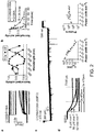
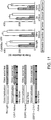
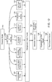
- FIG.12 depicts a flow diagram for testing of a disease model, consistent with various embodiments of the present disclosure.
- the disease models can be for one or more central nervous system (CNS) disorders.
- the models can include various disorders, diseases or even general characteristics of patients (e.g., mood, memory, locomotion or social behavior).
- CNS targets are identified.
- the CNS targets include the properties of the stimulus to be provided as part of assessing, testing or otherwise related to the disease model.
- Non-limiting examples of targets can be spatial targets, cell type targets, temporal targets and combinations thereof.
- the properties of the targets 106-118 can then be used to select a particular opsin from the optogenetic toolkit 120.
- the optogenetic toolkit 120 includes a variety of different opsins, which can be aligned with one or more of the properties 106-118.
- opsins are discussed herein.
- the selected opsin(s) 122 can be those opsins that most closely match the CNS target(s) and/or stimulus properties.
- a desired target may be the modification of excitation/inhibition (E/I) balance within a portion of the brain over an extended period of time.
- E/I excitation/inhibition
- the opsin C1V1 discussed in more detail herein
- its variants could be selected.
- aspects of the present disclosure are directed toward control over the cellular excitation/inhibition (E/I) balance within neocortical microcircuitry. Such E/I balance control can be particularly useful for modeling and/or treatment of social and cognitive deficits ( e.g., autism and schizophrenia) that are linked to elevations in excitation. Aspects of the present disclosure are directed toward the use of opsins for providing a mechanism for inducing an elevated cellular E/I balance with specific spatial and temporal control. This can include expression of light-sensitive opsins in excitatory neurons linked with one or more severe neuropsychiatric diseases.
- Various aspects relate to tools and methods for controlling the E/I balance in freely moving mammals, which can be particularly useful for exploring underlying circuit physiology mechanisms.
- Particular aspects of the present disclosure relate to increasing the excitability of excitatory neurons, relative to the excitability of inhibitory neurons with selective spatial control. This can be particularly useful for increasing the susceptibility of the excitatory neurons to intrinsic stimulus and thereby preserving natural firing patterns. In some implementations, this excitation is reversible.
- Certain aspects are directed toward the use of ion channels that are optically controllable.
- the ion channels When expressed in a neuron, the ion channels are designed to increase the susceptibility of the neurons to intrinsic stimulus to maintain the increased susceptibility for extended periods of time.
- aspects of the present disclosure relate to SSFOs (stabilized step-function opsins) that are stable enough to produce constant photocurrent after a single light flash over many minutes, and the use thereof for complex behavioral testing.
- the increased susceptibility can be maintained from many minutes after optical stimulus is applied.
- Various aspects are directed toward treatments, modeling and other aspects that relate to the discovery that impairments in specific social interaction and cognition behaviors in freely moving mice can be induced from targeted elevation in the E/I balance.
- Still other aspects of the present disclosure are directed toward treatments, modeling and other aspects that relate to the discovery that the dominant circuit-level effect of the behaviorally significant E/I balance intervention is a specific elevation in baseline gamma-band (around 40-60 Hz) recurrent synaptic excitation, analogous to the elevated gamma rhythms seen at baseline in autism and schizophrenia, with concomitant quantitative impairment in microcircuit information transmission.
- aspects of the present disclosure relate to the use of opsins to drive E/I elevations and monitor gamma oscillations in cortical slices.
- C1V1 discussed in more detail herein
- C1V1 various aspects are directed toward the use of C1V1 (discussed in more detail herein) and its variants, which can be particularly useful for driving E/I elevations and monitoring gamma oscillations in cortical slices, with 1) high potency to enable dose-response tests; 2) low desensitization to allow for step-like changes in E/I balance; and 3) red-shifted excitation to allow separable driving of different populations within the same preparation.
- aspects of the present disclosure relate to control over elevated (or lowered) cellular E/I balance. This can be particularly useful for studying, testing and treatment relating to medication-unresponsive social and cognitive impairment in neurological disorders, such as autism and schizophrenia.
- Particular aspects relate to studying and distinguishing the long term effects on the development and maturation of the circuit relative to the immediate effects of E/I abnormalities with regard to the function of the neural circuits involved.
- Other aspects are directed toward the confirmation of elevated cellular E/I balance as a core component of cognitive defects observed in the various disease models and patients (human or otherwise).
- Particular aspects provide timing and specificity sufficient for testing the elevated cellular E/I balance hypothesis in the mammalian brain (e.g., the prefrontal cortex), and identified circuit-physiology manifestations.
- a particular aspect relates to the use of the double-mutant SSFO (discussed in more detail herein), which can be particularly useful for providing stable circuit modulation for time periods that are sufficient for temporally precise and complex behavioral experiments.
- the modulation and behavioral experiments circuit modulation can span several minutes in the absence of ongoing light activation, external fiber optic attachments and/or optical-hardware brain penetration (e.g., using a light delivery device entirely external to the brain).
- Particular implementations use a property of photon integration, which can facilitate activation of cells with low light intensity (e.g., in the low- gm/mm 2 ). This activation can occur with relatively deep penetration of light into brain tissue ( e.g., 3mm or more relative to the light source).
- SSFO activation in excitatory (but not inhibitory) neurons can be used to produce profound and reversible impairments in social and cognitive function.
- the impairments can be produced with little, if any, motor abnormalities or altered fear/anxiety behaviors.
- aspects of the present disclosure also relate to the use of SSFO for in vitro probing of changes in circuit properties.
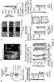
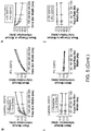
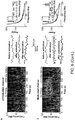
- SSFO's can be used to elevate cellular E/I balance and to measure the transfer functions of pyramidal neurons. Experimental results suggest that such elevation saturates the transfer functions of pyramidal neurons at low excitatory post-synaptic current (EPSC) rates, impairing information transmission within cortical circuitry, in contrast to consequences of reduction in E/I balance.
- EPC post-synaptic current
- PYR cells expressing C1V1-E162T spiked in response to 2 ms 561 nm light pulses, while the same stimulation paradigm reliably evoked excitatory postsynaptic potentials (EPSPs) in non-expressing cells within the same slices.
- ESPs excitatory postsynaptic potentials
- Particular aspects of the present disclosure are directed toward the use of SSFO gene product to selectively favor excitation of one neural population over another.
- the selective favoring of the targeted population can be configured to prevent the SSFOs from overriding intrinsic excitation inputs to the targeted population. In this manner, the targeted population would not be driven with coordinated spikes directly caused by the opsins. Rather, the targeted population would exhibit an increased sensitivity to native inputs, which can be sparse and asynchronous.
- aspects of the present disclosure are directed toward the use of SFOs to address various the hardware challenges. For instance, the significant increase in light sensitivity (e.g., orders-of-magnitude greater) can facilitate the use alternative light delivery mechanisms, and hardware-free behavioral testing.
- aspects of the present disclosure are directed toward identification and modification of specific portions of light-gated channels. These modifications involve identifying key portions of the channels.
- the channels can be identified using high resolution imaging of the tertiary structure of the channel. Alternatively, knowledge of the structure of similar channels can be used.
- the following description provides details of a specific experimental implementation and methodology. The present disclosure is not limited to any one implementation and can be implemented for a number of different molecular modifications at various locations consistent with the teachings herein.
- ChR2 is a rhodopsin derived from the unicellular green algae Chlamydomonas reinhardtii.
- rhodopsin as used herein is a protein that comprises at least two building blocks, an opsin protein, and a covalently bound cofactor, usually retinal (retinaldehyde).
- the rhodopsin ChR2 is derived from the opsin Channelopsin-2 (Chop2), originally named Chlamyopsin-4 (Cop4) in the Chlamydomonas genome.
- Chop2 opsin Channelopsin-2
- Cop4 Chlamyopsin-4
- the temporal properties of one depolarizing channelrhodopsin, ChR2 include fast kinetics of activation and deactivation, affording generation of precisely timed action potential trains.
- the normally fast off-kinetics of the channelrhodopsins can be slowed.
- certain implementations of channelrhodopsins apply 1mW/mm 2 light for virtually the entire time in which depolarization is desired, which can be less than desirable.
- VChR1 Volvox channelrhodopsin
- VChR1 Volvox channelrhodopsin
- modifications/mutations can be made to ChR2 or VChR1 variants.
- the modified variants can be used in combination with light-activated ion pumps.
- aspects of the present disclosure include relatively minor amino acid variants of the naturally occurring sequences.
- the variants are greater than about 75% homologous to the protein sequence of the naturally occurring sequences.
- the homology is greater than about 80%.
- Yet other variants have homology greater than about 85%, greater than 90%, or even as high as about 93% to about 95% or about 98%.
- Homology in this context means sequence similarity or identity, with identity being preferred. This homology can be determined using standard techniques known in the sequence analysis.
- compositions of the present disclosure include the protein and nucleic acid sequences provided herein, including variants which are more than about 50% homologous to the provided sequence, more than about 55% homologous to the provided sequence, more than about 60% homologous to the provided sequence, more than about 65% homologous to the provided sequence, more than about 70% homologous to the provided sequence, more than about 75% homologous to the provided sequence, more than about 80% homologous to the provided sequence, more than about 85% homologous to the provided sequence, more than about 90% homologous to the provided sequence, or more than about 95% homologous to the provided sequence.
- stimulation of a target cell is generally used to describe modification of properties of the cell.
- the stimulus of a target cell may result in a change in the properties of the cell membrane that can lead to the depolarization or polarization of the target cell.
- the target cell is a neuron and the stimulus affects the transmission of impulses by facilitating or inhibiting the generation of impulses (action potentials) by the neuron.
- the activation spectrum of ChR2-C128S/D156A peaks at 445nm.
- a second deactivation peak was found at 390-400nm, with faster but less complete deactivation by comparison with the 590 nm deactivation peak.
- Other aspects are directed toward a similar mutation in VChR1.
- the mutation in VChR1 could be provided at C123S/D151A, to provide a red- shifted photocurrent with slow kinetics comparable to ChR2.
- the double-mutant gene is referred to as SSFO (for stabilized step-function opsin) gene.
- SSFO is also used as shorthand for the active protein. Both residues likely are involved in ChR2 channel closure (gating), and both mutations likely stabilize the open state configuration of the channel.
- aspects of the present disclosure relate to the discovery that SSFO may be completely blocked in photocycle progression, and may therefore represent the maximal stability possible with photocycle engineering. For instance, in contrast to ChR2- C128X and ChR2-D156A, the SSFO photocycle does not appear to access additional inactive deprotonated side products which likely split off the photocycle at later photocycle stages not reached in this mutant, in turn making the SSFO even more reliable for repeated use in vivo than the parental single mutations.
- aspects of the present disclosure are directed toward the sensitivity of the SSFO to light. For instance, channelrhodopsins with slow decay constants effectively act as photon integrators.
- activation time constants that are linearly correlated with the activation light power on a log-log scale, which is indicative of a power-law relationship and suggesting that the SSFO is a pure integrator, with total photon exposure over time as the only determinant of photocurrent. For instance, it is believed that the number of photons per membrane area required for photocurrents to reach a given sub-maximal activation (time to T) is constant regardless of activation light power.
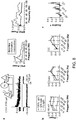
- Example aspects of the present disclosure relate to the use of a hybrid ChR1/VChR1 chimera that contains no ChR2 sequence at all, is derived from two opsins genes that do not express well individually, and is herein referred to as C1V1.
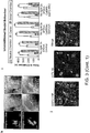
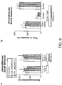
- Aspects of the present disclosure also relate to improvements of the membrane targeting of VChR1 through the addition of a membrane trafficking signal derived from the Ki r 2.1 channel. Confocal images from cultured neurons expressing VChR1-EYFP revealed a large proportion of intracellular protein compared with ChR2; therefore, membrane trafficking signal derived from the Ki r 2.1 channel was used to improve the membrane targeting of VChR1.
- VChR1-ts-EYFP Membrane targeting of this VChR1-ts-EYFP was slightly enhanced compared with VChR1-EYFP; however, mean photocurrents recorded from cultured hippocampal neurons expressing VChR1ts-EYFP were only slightly larger than those of VChRl-EYFP.
- aspects of the present disclosure relate VChR1 modified by exchanging helices with corresponding helices from other ChRs.
- robust improvement has been discovered in two chimeras where helices 1 and 2 were replaced with the homologous segments from ChR1. It was discovered that whether splice sites were in the intracellular loop between helices 2 and 3 (at ChR1 residue A1a145) or within helix 3 (at ChR1 residue Trp163), the resulting chimeras were both robustly expressed and showed similarly enhanced photocurrent and spectral properties. This result was unexpected as ChR1 is only weakly expressed and poorly integrated into membranes of most mammalian host cells. The resulting hybrid ChR1IVChR1 chimera is herein referred to as C1V1.
- C1V1-EYFP exhibits surprisingly improved average fluorescence compared with VChRl-EYFP.
- Whole cell photocurrents in neurons expressing C1V1 were much larger than those of VChR1-EYFP and VChR1-ts-EYFP, and ionic selectivity was similar to that of ChR2 and VChR1.
- C1V1-ts-EYFP mean photocurrents were extremely large, nearly tenfold greater than wild type (WT) VChR1.
- Mean fluorescence levels closely matched the measured photocurrents (mean fluorescence 9.3 ⁇ 1, 19.6 ⁇ 3.4, 19.8 ⁇ 2.8 and 36.3 ⁇ 3.8 for VChR1-EYFP, VChR1-ts-EYFP, C1V1-EYFP and C1V1-ts-EYFP, respectively), suggesting that the increase in photocurrent sizes resulted mainly from the improved expression of these channels in mammalian neurons.
- aspects of the present disclosure relate to opsins with fast decay constants. This property can be particularly useful for providing precise control over spiking, e.g., in order to interfere minimally with intrinsic conductance, trigger single spikes per light pulse and/or minimize plateau potentials during light pulse trains.
- Experimental results suggest that the light-evoked photocurrents recorded in C1V1-ts-EYFP decayed with a time constant similar to that of VChR1.
- aspects of the present disclosure are therefore directed toward modifications in the chromophore region to improve photocycle kinetics, reduced inactivation and/or possible further red-shifted absorption.
- ChETA mutation E162T which experiments suggest provides an accelerated photocycle (e.g., almost 3-fold); reference can be made to Gunaydin, et al., Ultrafast optogenetic control, Nat Neurosci, 2010 . Surprisingly, this mutation was shown to shift the action spectrum hypsochromic to 530 nm, whereas analogous mutations in ChR2 or other microbial rhodopsins have caused a red-shift.
- Another embodiment is directed toward a mutation of glutamate-122 to threonine (C1V1-E122T). Experimental tests showed that C1V1-E122T is inactivated only by 26% compared to 46% inactivation of ChR2; in addition, the spectrum was further red-shifted to 546 nm.
- Another aspect of the present disclosure is directed toward a double mutant of C1V1 including both E122T and E162T mutations.
- Experimental tests have shown that the inactivation of the current was even lower than in the E122T mutant and the photocycle was faster compared to E162T. This suggests that multiple useful properties of the individual mutations were conserved together in the double mutant.
- aspects of the present disclosure include the expression of various light-responsive opsins in neurons.
- Experimental tests of C1V1 opsin genes in neurons were carried out by generating lentiviral vectors encoding C1V1-ts-EYFP and various point mutation combinations discussed herein.
- the opsins were then expressed in cultured hippocampal neurons and recorded whole-cell photocurrents under identical stimulation conditions (2ms pulses, 542nm light, 5.5 mW/mm 2 ). Photocurrents in cells expressing C1V1, C1V1-E162T and C1V1-E122T/E162T were all robust and trended larger than photocurrents of ChR2-H134R. The experiments also included a comparison of integrated somatic YFP fluorescence and photocurrents from cells expressing C1V1-E122T/E162T and from cells expressing ChR2- H134R.
- C1V1-E122T/E162T cells showed stronger photocurrents than ChR2-H134R cells at equivalent fluorescence levels. This suggests that C1V1 could possess a higher unitary conductance compared with ChR2-H134R.
- the test results suggest that the kinetics of C1V1-E122T were slower than those of C1V1-E122T/E162T and that cells expressing C1V1- E122T responded more strongly to red light (630nm) than cells expressing the double mutant. This can be particularly useful for generating optogenetic spiking in response to red-light.
- inhibitory and/or excitatory neurons residing within the same microcircuit are be targeted with the introduction of various opsins.
- Experimental tests were performed by separately expressed C1V1- E122T/E162T and ChR2-H134R under the CaMKIIa promoter in cultured hippocampal neurons.
- Cells expressing C1V1-E122T/E162T spiked in response to 2 ms green light pulses (560nm) but not violet light pulses (405nm).
- Various aspects of the present disclosure relate to independent activation of two neuronal populations within living brain slices. Experimental tests were performed by CaMKIIa-C1V1-E122T/E162Tts-eYFP and EFla-DIO-ChR2-H134R-EYFP in mPFC of PV::Cre mice. In non-expressing PYR cells, 405 nm light pulses triggered robust and fast inhibitory postsynaptic currents due to direct activation of PV cells, while 561 nm light pulses triggered only the expected long-latency polysynaptic IPSCs arising from C1V1-expressing pyramidal cell drive of local inhibitory neurons.
- excitation of independent cellular elements can be performed in vivo.
- Experimental tests were performed using optrode recordings.
- 5Hz violet light pulses to activate ChR2 in PV cells
- 5 Hz green light pulses to activate C1V1 in excitatory pyramidal neurons
- the test results suggest that when violet and green light pulses were separated by 100 ms, responses to green light pulses were not affected by the violet pulses.
- delays between violet and green pulses were reduced, green light-induced events became more readily inhibited until being effectively/completely abolished when light pulses were presented simultaneously.
- aspects of the present disclosure relate to an optogenetic system or method that correlates temporal, spatio and/or cell-type control over a neural circuit with measurable metrics. Consistent with the other aspects discussed herein, particular aspects relate to studying and probing disorders. A non-exhaustive list of example embodiments and experimental results consistent with such embodiments is provided in Yizhar et al., Nature, 2011, 477(7363):171-8 . The references listed therein may assist in providing general information regarding a variety of fields that may relate to one or more aspects of the present disclosure, and further may provide specific information regarding the application of one or more such aspects, to which one or more references as follows may be applicable.
- the present disclosure is believed to be useful as it relates to control over nervous system disorders, such as disorders associated with social dysfunction, as described herein.
- Specific applications of the present disclosure relate to optogenetic systems or methods that correlate temporal, spatio and/or cell-type-specific control over a neural circuit with measurable metrics.
- the following discussion summarizes such previous developments to provide a solid understanding of the foundation and underlying teachings from which implementation details and modifications might be drawn, including those found in the attached Appendix. It is in this context that the following discussion is provided. While the present invention is not necessarily limited to such applications, various aspects of the invention may be appreciated through a discussion of various examples using this context.
- FIG. 13 depicts a model for assessing stimuli and/or potential treatments for various nervous system disorders.
- Baseline observations 220 are taken 202 of behavior and/or cellular response for a subject/patient.
- a target cell population is chosen and modified to express a light-responsive molecule.
- the target cell population is selected to provide control over the E/I balance in the prefrontal cortex of a subject's brain, as discussed in more detail herein.
- the excitation/inhibition (E/I) balance within the target cell population can then modified 204 (e.g., elevated or lowered) by exposing the modified target cell population to light.
- the light can be provided within a predetermined range based on absorption characteristics of the light-responsive molecule.
- Observations 220 of behavior and/or cellular response of the subject are again taken. These observations provide a reference point for how the subject acts under no stimuli or treatment.
- a stimulus and/or potential treatment is chosen 206 for the subject.
- stimuli and treatments include pharmacological/ drugs 208, behavioral 210 and/or electrical stimulus 212.
- the stimuli/treatments can then be assessed 214 by observing the subject's behavior in response to the treatment and/or the target cell population's behavior in response to the treatment. Based on the observations, a determination can be made regarding the need for additional stimulus or treatment 216, or the desire to test additional and/or different treatments.
- the observations 220 from various treatments can be compared 218 to each other as well as the baseline observation and the observations of behavior after E/I elevation. The comparison of the observations 220 can be used to assess the efficacy of various potential treatments.
- the elevation of the E/I balance results in social and cognitive deficits as compared to the behaviors during baseline observations.
- the purposeful and controlled elevation of the E/I balance allows for the testing of potential treatments in mammalian test subjects such as mice that do not otherwise exhibit symptoms of the disease being modeled.
- aspects of the present disclosure relate to assessing the effect of various stimuli on symptoms of neurological diseases.
- modification of the E/I balance in the prefrontal cortex of a subject's brain results in the symptoms similar to those of various neurological disorders, such as autism and schizophrenia.
- the neural circuit identified as effecting E/I balance is manipulated using one or more techniques including pharmacological, electrical, magnetic, surgical and optogenetic methods. The effect of the manipulation of the symptoms displayed is monitored.
- the manipulation of pyramidal neurons and parvalbumin-expressing inhibitory interneurons is used to model disease states, and to identify new treatments for known diseases.
- the E/I balance in the prefrontal cortex is elevated (or lowered) and then a potential treatment is administered to the subject.
- the effect of the treatment on either the observed symptoms or on the neural circuit (or both) can be monitored.
- the information obtained from monitoring the symptoms and/or the neural circuit can be used to provide a better understanding of the neural pathways causing the observed symptoms.
- the information may also be used to determine the efficacy of the potential treatment. Based on the efficacy, or lack thereof, of the potential treatment, modifications can be made resulting in a new potential treatment to be tested.
- a stimulus is provided to a subject that exhibits symptoms of a neural disease such as schizophrenia or autism, for example.
- the stimulus can be pharmacological, electrical, magnetic, surgical, optogenetic or behavioral, for example.
- control over the neural circuit can include inhibition or excitation, which can each include coordinated firing, and/or modified susceptibility to external circuit inputs.
- inhibition can be accomplished using a light-responsive opsin, such as an ion pump (e.g ., NpHR and NpHR variants).
- ion pumps move the membrane potential of the neuron away from its threshold voltage to dissuade or inhibit action potentials.
- excitation can be accomplished using a light-responsive opsin, such as an ion channel ( e.g ., ChR2 and ChR2 variants).
- ion channels can cause the membrane potential to move toward and/or past the threshold voltage, thereby exciting or encouraging action potentials.
- a light- responsive opsin can be used to (temporarily) shift the resting potential of a neuron to increase or decrease its susceptibility to external circuit inputs.
- ChR2(D156A) and SSFO were generated by introducing point mutations into the pLentiCaMKII ⁇ -ChR2-EYFP-WPRE vector using site-directed mutagenesis (Quikchange II XL; Stratagene).
- the membrane trafficking signal was derived from the Kir2.1 channel. Mutations were confirmed by sequencing the coding sequence and splice sites.
- opsin-EYFP fusions along with the CaMKII ⁇ promoter were subcloned into a modified version of the pAAV2-MCS vector.
- Cre dependent opsin expression was achieved by cloning the opsin-EYFP cassette in the reverse orientation between pairs of incompatible lox sites (loxP and lox2722) to generate a doublefloxed inverted open reading frame (D10) under the control of the elongation factor la (EF- 1 ⁇ ) promoter. All constructs are available from the Deisseroth Lab (www.optogenetics.org).
- ChRs for heterologous expression of ChRs in Pichia pastoris cells (strain 1168H, purchased from Invitrogen), human codon-optimized synthetic ChR-fragment encoding amino acids 1-315 ( see accession no. AF461397) was cloned in the pPICZ vector (Invitrogen) via its EcoRI and NotI restriction sites. The C-terminal polyhistidine tag encoded on the vector was modified to a 12His sequence. Mutants of ChR were generated by site-directed mutagenesis (QuickChange kit, Stratagene). Transformation, cell culture and protein purification were performed. After induction of protein expression for 24 h, cells were harvested and gently lysed using a high pressure homogenizer (Avastin).
- Avastin high pressure homogenizer
- the membrane fraction was collected, homogenized and solubilized in 1 % (w/v) dodecylmaltoside.
- ChR protein was binding to a Ni-NTA resin (Qiagen) and washing of the column with 200 mM imidazole, ChR was eluted with 500 mM imidazole. Fractions that contained the protein were pooled, desalted (Float-a-lyzer, Roth) and concentrated (Amicon Ultra, Millipore) to an optical density of 1 at 480 nm. Spectra were recorded in a Cary 50 Bio spectrophotometer (Varian Inc.).
- the ChR2 mutants C128S, D156A, and the double mutant 128S/156A were generated and purified from Pichia pastoris to first measure intrinsic open-state stability in the absence of potentially confounding cellular properties. Absorption spectra showed expected rapid changes in response to brief light delivery that largely recovered within 3 minutes for the single mutants C128S ( FIG. 1B , F ) and D156A ( FIG. 1C , G ). However, in contrast to both single mutants, the double mutant C128S/D156A showed remarkably complete stability of the activated state, with essentially no detectable return to the dark state even after 30 minutes ( FIG. 1D , H ).
- FIG. 1E The unique stability of the double mutant Cl 28S/D 156A is further illustrated by continuous monochromatic absorbance measurements of all three mutants over 35 minutes of recording ( FIG. 1H ).
- Example 2 Validation of activation in neurons and in vivo
- the double mutant therefore appeared to have markedly distinct and near-optimal stability on the mammalian behavioral timescale, but with potentially reduced crucial capability for redshifted light deactivation; all of these issues required validation in neurons and in vivo.
- Electrophysiological recordings from individual neurons identified by fluorescent protein expression were obtained in Tyrode media ([mM] 150 NaCl, 4 KCl, 2 MgCl 2 , 2 MgCl 2 , 10 D-glucose, 10 HEPES, pH 7.35 with NaOH) using a standard internal solution ([mM] 130 KGluconate, 10 KCl, 10 HEPES, 10 EGTA, 2 MgCl 2 , pH 7.3 with KOH) in 3-5 M ⁇ glass pipettes.
- cortical slice physiology acute 300 ⁇ m coronal slices from 8-9 week old wild-type C57BL/6J or PV::Cre mice previously injected with virus were obtained in ice-cold sucrose cutting solution ([mM] 11 D-glucose, 234 sucrose, 2.5 KCl, 1.25 NaH 2 PO 4 , 10 MgSO 4 , 0.5 CaCl 2 , 26 NaHCO3) using a Vibratome (Leica).
- Band pass filters (Semrock) had 20 nm bandwidth, and were adjusted with additional neutral density filters (ThorLabs) to equalize light power output across the spectrum. While handling cells or tissues expressing SSFO, care was taken to minimize light exposure to prevent activation by ambient light. Before each experiment, a 20s pulse of 590 nm light was applied to convert all of the SSFO channels to the dark state and prevent run-down of photocurrents. For acquisition of SSFO activation and deactivation spectra, cultured neurons in voltage clamp mode were recorded. For recording activation spectra, a 1 s pulse of varying wavelength was applied, followed by a 10 s 590 nm pulse.
- Deactivation spectra were acquired by first applying a 1 s 470 nm pulse to activate SSFO, followed by a 10 s pulse of varying wavelength. Net activation or deactivation was calculated by dividing the photocurrent change after the first or second pulse, respectively, by the maximum photocurrent change induced by the peak wavelength for that cell. Negative values in deactivation spectra resulted from traces in which, for example, a 10 s 470nm pulse led to a slight increase in photocurrent rather than deactivate the channels. This could be the result of the relatively wide (20 nm) band-pass filter width used for these recordings with the Sutter DG-4. Intermediate wavelengths (between 470nm and 520nm) are expected to have a mixed effect on the channel population for the same reasons.
- Cultured cell images were acquired on the same microscope using a Retiga Exi CCD camera (Qimaging, Inc.) at 100 ms exposure with 30 gain. Illumination power density was 12 mW mm -2 at 500 nm with a standard EYFP filter set. Quantification of fluorescence was performed with ImageJ software by marking a region containing the soma and proximal neurites and calculating for each cell the total integrated pixel intensity in that region, rather than average fluorescence, since photocurrents are likely to be related to the total number of membrane-bound channels rather than average channel expression per area.
- Photon flux calculations for SSFO integration properties were conducted by calculating the photon flux through the microscope objective at each light power, and then dividing to reach the photon flux across the cell surface, based on the diameter of the recorded cells and approximating cell shape as a spheroid.
- opsins Both Lentiviral- and AAV-mediated gene delivery were used for heterologous expression of opsins in mice. Indicated opsins were driven by either Human calmodulin-dependent protein kinase II alpha (CaMKII ⁇ ) promoter to target cortical excitatory neurons or Elongation Factor la (EF-1a) in conjunction with a Cre-inducible cassette and followed by the Woodchuck hepatitis virus posttranscriptional regulatory element (WPRE). Cre-inducible recombinant AAV vector was produced by the University of North Carolina Vector Core (Chapel Hill, NC, USA) and used in conjunction with parvalbumin::Cre transgenic mice to target parvalbumin positive interneurons.
- CaMKII ⁇ Human calmodulin-dependent protein kinase II alpha
- EF-1a Elongation Factor la
- WPRE Woodchuck hepatitis virus posttranscriptional regulatory element
- SSFO-eYFP was inserted in the reverse orientation between pairs of incompatible lox sites (loxP and lox2722).
- AAV constructs were subcloned into a modified version of the pAAV2-MCS, serotyped with AAV5 coat proteins and packaged by the viral vector core at the University of North Carolina.
- the final viral concentration of AAV vectors was 1*10 12 genome copies (gc)/mL.
- Lentiviral constructs were generated as reported. All constructs are available from the Deisseroth Lab (www.optogenetics.org). Stereotactic viral injections were carried out under protocols approved by Stanford University.
- mice kept under isoflurane anesthesia were arranged in a stereotactic frame (Kopf Instruments) and leveled using bregma and lambda skull landmarks. Craniotomies were performed so as to cause minimal damage to cortical tissue.
- Infralimbic prefrontal cortex (IL; from bregma: 1.8mm anterior, 0.35mm lateral, -2.85mm ventral) was targeted using a 1 OuL syringe and 35g beveled needle (Word Precision Instruments). Virus was infused at a rate of 0.111L/min.
- Subjects injected with virus for behavioral studies were additionally implanted with a chronic fiber optic coupling device to facilitate light delivery either with or without an attached penetrating cerebral fiber for local delivery to target cortical region as noted (Doric Lenses, Canada).
- Penetrating fibers were stereotactically inserted to a depth of -2.5mm from the same anterior and lateral coordinates and affixed using adhesive luting cement (C&B MetaBond) prior to adhesive closure of the scalp (Vetbond, 3M). Animals were administered analgesic relief following recovery from surgery.
- Deactivation was also possible with 390 nm light, at a faster rate than yellow light due to the substantial presence of the P390 species, but was also incomplete due to the residual absorption of the dark state at this wavelength ( FIG. 1A ). Moreover, following deactivation with 390 nm light, reactivation with 470 nm was less effective than following 590 nm deactivation, pointing to a likely photochemical inactivation with UV light due to trapping in a deprotonated/desensitized isoform that is not reached after redshifted-light deactivation (illustrated in FIG. 1E ), and again supporting the use of yellow light deactivation to potentially enhance spectral separation.
- Figure 2C shows a typical long whole-cell recording with both blue light activation and yellow light deactivation in the setting of incoming asynchronous synaptic activity.
- the double-mutant gene is referred to as SSFO (for stabilized step-function opsin) gene, and for simplicity use SSFO as shorthand for the protein as well.
- Channelrhodopsins with such slow decay constants could enable the transduced cell to act as a photon integrator, with effective light sensitivity (i.e. photocurrent amplitude per photon absorbed by the cell) scaling with T off.
- SSFO could therefore enable more-sensitive, less-invasive approaches to optogenetic circuit modulation, but still with temporally precise onset and offset of action and with readily titratable effects on the targeted neuronal population via modulation of light pulse length. Indeed, it was found that with extraordinarily low light intensities (as low as 8 ⁇ W mm -2 ), hundreds of picoamps of whole-cell photocurrent could be obtained from neurons expressing SSFO ( FIG. 2D ).
- SSFO-eYFP in PV::Cre transgenic mice was expressed using a double-floxed inverted open reading frame (DIO) virus; in these mice, SSFO was only expressed in the GABAergic Cre-positive parvalbumin neurons.
- DIO verted open reading frame
- recordings were made at progressively more ventral sites in mice injected with AAV5-CaMKIIa::SSFO-EYFP in medial prefrontal cortex (mPFC), using an advancing two-laser optrode ( FIG. 2E ) and a blue/green activation/deactivation laser protocol ( FIG. 2F-G ).
- Multiunit activity in mPFC of these mice was significantly and stably increased only in the transduced region, in response to a 1 s pulse of 473 nm light (95 mW mm -2 , corresponding to 10 mW mm -2 at the electrode tip). This increased activity was effectively terminated with a 2s 561 nm light pulse (112 mW mm -2 ; FIG. 2F ).
- Significant increases in multiunit spike rate (Hz) were restricted to mPFC ( FIG. 2 ) and no significant reductions in spike rate were observed in any of the recording sites following blue light stimulation.
- SSFO was used to examine the effects of elevated cellular E/I balance on behavior and circuit dynamics in freely moving mice ( FIG. 3 ).
- SSFO was expressed either in prefrontal cortical excitatory neurons using the excitatory neuron-specific CaMKII ⁇ promoter, or in inhibitory parvalbumin (PV)- expressing neurons using a double-floxed, inverted open-reading-frame (DIO) virus in conjunction with PV::Cre transgenic mice ( FIG. 3J-L ).
- Virus was injected in mPFC as described above, followed by a chronic fiber-optic implant that projected past the skull immediately dorsal to mPFC for light delivery ( FIG. 3A , B ).
- the times of the sEPSCs within the 500 ms segment were randomly selected from a uniform distribution extending across the entire segment, simulating excitatory input from a population of unsynchronized neurons. Empirically, these stimulation parameters reliably drove pyramidal neurons at firing rates from 0 - 30 Hz. In conditions marked as baseline, a 10 sec pulse of 590 nm light was delivered to completely inactivate the opsin before running the sEPSC protocol. In conditions where the opsin was activated, a 1 sec pulse of 470 nm light preceded the sEPSC protocol.
- the joint distribution of sEPSC rate and spike rate was estimated by binning in time, sEPSC rate, and spike rate and building a joint histogram.
- Time bins were 125 ms wide, and sEPSC rate was divided into 10 equally spaced bins from 0 to 500 Hz, although the mutual information results were consistent across a wide range of binning parameters.
- Spike rate was binned using the smallest meaningful bin width given the time bin width ( e.g. 8 Hz bin width for 125 ms time bins).
- the input-output transfer function for each neuron was quantified by computing the dynamic range and saturation point of each neuron, treating the baseline and opsin-activated conditions separately.
- Dynamic range was defined as the difference between maximal and minimal output spiking rate across the range of input sEPSC rates.
- Saturation point was defined as the lowest input sEPSC rate which drove the neuron at 90% of its maximal output spike rate within that condition.
- a reduced saturation point cannot result from a multiplicative reduction in gain or dynamic range, but instead indicates that the input-output function becomes flatter at higher input sEPSC rates.
- mice undergoing behavioral experiments were acclimated to a 12-hour reverse light/dark cycle. Prior to behavioral testing, animals were allowed to acclimate to the room in which experiments were to be conducted for at least 1 hour before the experiments started.
- the fear conditioning apparatus consisted of a square conditioning cage (18x18x30 cm) with a grid floor wired to a shock generator and a scrambler, surrounded by an acoustic chamber (Coulburn instruments, PA, USA). The apparatus was modified to enable light delivery during training and/or testing. To induce fear-conditioning mice were placed in the cage for 120 seconds, and then a pure tone (2.9 kHz) was played for 20 sec, followed by a 2 sec foot-shock (0.5 mA). This procedure was then repeated, and immediate freezing behavior was monitored for an additional 30 sec after the delivery of the second shock before the mice were returned to their home cage. Fear conditioning was assessed 24 hours later by a continuous measurement of freezing (complete immobility), the dominant behavioral fear response.
- the three-chamber social test was conducted.
- the test mice were introduced into the center chamber of the three-chambered apparatus and allowed to acclimate for 10 minutes with the doors to the two side chambers closed. Light pulses were applied at the beginning and end of the 10 minute acclimation period.
- a novel conspecific male mouse was introduced to the "social" chamber, inside a wire mesh cup (Galaxy Pencil/Utility cup, Spectrum Diversified Designs).
- a wire mesh cup Gaxy Pencil/Utility cup, Spectrum Diversified Designs
- In the other (non-social) chamber an identical empty cup was placed.
- the designations of the social and non-social chambers were randomly chosen in each test to prevent chamber bias. Between tests, the chambers were cleaned with 20 % ethanol and allowed to dry completely before initiating the next test. The time spent in the non-social, center, and social chambers was quantified using automated tracking software Viewer II (BiObserve, Fort Lee, NJ). Mice not exhibiting social exploration preference
- the novel object exploration experiment was performed in the same three-chamber apparatus used for the social behavior tests, and using the same general method. Mice were placed in the center chamber with the doors to both side chambers closed. Light pulses were delivered during the 10 minute acclimation period, after which the doors were opened and the mice were allowed to explore the entire apparatus. In place of the wire mesh cups, novel objects were presented at random in either of the two end-chambers. Exploration of the novel objects was scored over a period of 10 minutes for each mouse as the time in which the mouse spent actively exploring the object. Objects used were either plastic balls, cubes or porcelain figurines, all of approximately similar size. Objects were thoroughly cleaned between tests to prevent odor traces.
- the open-field chamber (50 x 50 cm) was divided into a central field (center, 23 x 23 cm) and an outer field (periphery). Individual mice were placed in the periphery of the field and the paths of the animals were recorded by a video camera. The total distance traveled was analyzed using the Viewer2 software (BiObserve, Fort Lee, NJ). The open field test for each mouse consisted of a 5-min session divided into two 2.5 minute segments, with a 2 s 473nm light pulse delivered between the two segments. Track length, velocity and % time in the center were scored for each mouse and averaged across mice for each condition
- the elevated plus maze was made of plastic and consisted of two light gray open arms (30 x 5 cm), two black enclosed arms (30 x 5 x 30 cm) extending from a central platform (5 x 5 x 5 cm) 31 at 90 degrees in the form of a plus.
- the maze was placed 30 cm above the floor.
- a 2 s 473 nm light pulse was delivered when the mouse was in the home cage. 5 minutes later, the fiberoptic connector was detached and the mice were individually placed in the center of the maze for a test duration of 15 minutes.
- Video tracking software (ViewerII, BiObserve, Fort Lee, NJ) was used to track mouse location. All measurements displayed were relative to the entire mouse body.
- CMO chronic multisite optrode
- the wires were connected using gold pins to a Mill-Max connector, to which a stainless steel ground wire was also connected.
- the device was implanted stereotactically following virus injection (see above) such that the fiber tip only extended past the skull but not into brain tissue.
- the ground wire was inserted through a small craniotomy above cerebellum. Mice were allowed to recover for two weeks before experiments began.
- mice were first acclimated over several days to the attachment of the headstage and the fiberoptic cable. The mice were allowed to explore the home cage with the headstage attached for 1-2 hours each day. Recordings were carried out 2-4 weeks after surgery. Signals were multiplexed at the headstage into a 3-wire cable that was passed through an electrical commutator (PlasticsOne), demultiplexed using a demultiplexing board (Triangle BioSystems, Inc.) and digitized using Neuralynx Digital Cheetah. The fiberoptic and electrical commutators were suspended from a weighted arm (Harvard Apparatus) to allow the mouse to freely explore a large region (such as in the open field test).
- Wavelet power spectrograms of LFP recordings were analyzed as described above by sampling the power spectrum every 2 s for the duration of the recording. Power was calculated between 2 Hz and 120 Hz with a bin width of 2 Hz.
- the effects of SSFO activation were recorded using a protocol of 2 minutes baseline recording, followed by a 1 s 473 nm pulse at an irradiance of 56 mW mm -2 at the fiber tip. Following the blue pulse, activity was recorded for 2 minutes, followed by a 30 s deactivating light pulse at a wavelength of 594 nm light with similar intensity. Activity was then recorded for 2 additional minutes. For each mouse this protocol was repeated at least 4 times, and power spectra for each of the three periods (pre-activation, post-activation and post-deactivation) were averaged across the 4 repetitions.
- Nuclear localization of c-fos was determined using rabbit anti-c-fos (Calbiochem) on animals that had undergone 1 s 473 nm light stimulation 90 minutes prior to perfusion; parvalbumin targeting was confirmed using colocalization of mouse anti-parvalbumin (Sigma Aldrich) and fluorescent protein. Stained slices were visualized on a Leica SP5 confocal microscope. To calculate average fluorescence in different anatomical subregions, histology images were analyzed using ImageJ. Individual subregion images were thresholded at a fixed threshold level. Mean fluorescence above threshold was calculated and averaged per region between mice. c-fos counts were performed using standardized landmarks to identify regions and were anonymized prior to counting.
- a large fraction of these cells were in fact YFP-positive (61 ⁇ 8 % out of the total c- fos positive population; FIG. 3C ), indicating that even most of these active cells are in fact PV- positive neurons directly activated by the virally-delivered SSFO.
- FIG. 3D-G Three groups of animals to behavioral testing FIG. 3D-G ) CaMKII ⁇ ::SSFO mice, PV::SSFO mice, and control mice (either injected with AAV5-CaMKII ⁇ -eYFP virus or not injected with virus). Two to four weeks after surgery, conditioned learning and unconditioned social behavior was tested, as well as exploration of novel objects and locomotor functioning ( FIG. 3D-G ); all animals received a single 1s pulse of 470 nm light through the implanted fiberoptic connector, followed by removal of the fiberoptic cable 1 minute before introduction into the behavioral chamber, capitalizing on the stability of the SSFO.
- mice 6 PV::SSFO mice; p > 0.1; unpaired t-test).
- the same mice were next subjected to a conditioning protocol performed immediately following delivery of a 1 s 470 nm light pulse. Twenty-four hours later, responses to the conditioned tone and context were assessed in order to evaluate the extent to which the mice learned to associate the conditioned and unconditioned stimuli while under the altered E/I states.
- the elevated E/I (CaMKII ⁇ ::SSFO) animals showed no conditioned responses (to either context: p ⁇ 0.0005 or tone: p ⁇ 0.05, compared with controls; two-sided t -test).
- Example 4 Elevation but not reduction of cellular E/I leads to quantitative reduction in information processing
- Acute 300 pm coronal slices isolated from 8-9 week old wild-type C57BL/6J or PV::Cre mice previously injected with virus were obtained in ice-cold sucrose cutting solution ([mM] 11 D-glucose, 234 sucrose, 2.5 KCl, 1.25 NaH 2 PO 4 , 10 MgSO 4 , 0.5 CaCl 2 , 26 NaHCO 3 ) using a Vibratome (Leica).
- Slices were recovered in oxygenated Artificial Cerebrospinal Fluid (ACSF; [mM] 124 NaCl, 3 KC1, 1.3 MgCl 2 , 2.4 CaCl 2 , 1.25 NaH 2 PO 4 , 26 NaHCO 3 , 10 D-glucose) at 32°C for one hour.
- ACSF Artificial Cerebrospinal Fluid
- Quantification of fluorescence was done with ImageJ software by marking a region containing the soma and proximal neuritis and calculating for each cell the total integrated pixel intensity in that region, rather than average fluorescence, since photocurrents are likely to be related to the total number of membrane-bound channels rather than average channel expression per area.
- Photon flux calculations for SSFO integration properties were done by calculating the photon flux through the microscope objective at each light power, and then dividing to reach the photon flux across the cell membrane, based on the capacitance of individual patched cells.
- Heatmap images were generated in Matlab from an unweighted moving average of 2s with 200ms steps. Moving average value was reset at the onset of external manipulations (beginning of sweep, initiation of light pulses).
- FIG. 4A-B Spectral analysis of responses to SSFO in both expressing and non-expressing cells revealed that this increased activity displayed a broad spectral range with a peak above 20 Hz.
- pyramidal cells in slices expressing SSFO in PV cells showed a robust reduction in synaptic activity and a reduction in power at low frequencies ( FIG. 4C ), consistent with the increased activity of PV cells after activation with SSFO ( FIG. 4D ).
- CaMKII ⁇ ::SSFO or CaMKII ⁇ ::EYFP virus were injected and implanted fiberoptic connectors extending only past the skull ( FIG. 6A ), without entering the cortical surface ( FIG. 6B ).
- Elevated cellular E/I balance during conditioning showed no effect on freezing responses to footshock (indicating intact sensory perception of the aversive unconditioned stimulus; FIG. 6D ), but showed a marked and fully reversible effect on contextual (p ⁇ 0.005; unpaired t-test with unequal variance) and auditory conditioning (p ⁇ 0.005; unpaired t -test with unequal variance; FIG. 6D ).
- social behavior was also impaired in mice receiving noninvasive light stimulation prior to testing (p ⁇ 0.005; unpaired t -test; FIG. 6E ), demonstrating the opportunity afforded by the extreme light sensitivity of the SSFO.
- CMO chronic multisite optrode
- C1V1 is a chimeric light-sensitive protein derived from the VChR1 cation channel from Volvox carteri and the ChR1 cation channel from Chlamydomonas Reinhardti.
- C1V1 and its variants permits the experimental manipulation of cortical E/I elevations and the monitoring of gamma oscillations in cortical slices with high potency (thus allowing enable dose-response tests), low desensitization (thus permitting inducement of step-like changes in E/I balance), and red-shifted excitation (to permit separable drive of different populations within the same neural circuit).
- C1V1 variant with the highest potency to enable the most reliable dose-response was selected.
- mice in the same paradigm were tested with novel juvenile mice, while delivering pulsed laser light at 590 nm to activate only C1V1-E122T/E162T in the PV cells in the SSFO/C1V1 mice ( FIG. 10B ).
- SSFO was activated with a 2s 470 nm light pulse during the pre-test habituation period ( FIG. 10B ).
- CMO chronic multisite optrode
- C1V1 variants were utilized, to independently modulate both excitatory neurons (using SSFO) and inhibitory PV neurons (using a C1V1 variant).
- SSFO excitatory neurons
- inhibitory PV neurons using a C1V1 variant.
Claims (16)
- Nichtmenschliches Säugetier, umfassend ein erstes lichtaktiviertes Kationenkanalprotein, das auf der Zellmembran von Neuronen des präfrontalen Cortex des Säugetiers exprimiert wird, wobei das Protein eine Aminosäuresequenz umfasst, die zumindest zu 95 % identisch mit der in einer von Seq.-ID Nr. 1-4 dargestellten Sequenz ist, und eine Cys→Ser-Substitution an einer Aminosäureposition, die Aminosäure 128 von Seq.-ID Nr. 1 entspricht, und eine Asp→Ala-Substitution an einer Aminosäureposition umfasst, die Aminosäure 156 von Seq.-ID Nr. 1 entspricht, wobei das Protein dazu in der Lage ist, einen depolarisierenden Strom in den Neuronen mittels Licht auszulösen, und als Reaktion auf einen einzelnen Lichtimpuls mit einer ersten Wellenlänge eine rasche stufenähnliche Aktivierung und als Reaktion auf einen Lichtimpuls mit einer zweiten Wellenlänge eine Deaktivierung aufweist, wobei der depolarisierende Strom in den Neuronen für zumindest zehn Minuten aufrechterhalten wird; und wobei die Aktivierung des Proteins in den Neuronen des präfrontalen Cortex Veränderungen des Sozialverhaltens, der Kommunikation und/oder von konditionierten Verhaltensweisen in dem Säugetier auslöst.
- Säugetier nach Anspruch 1, wobei das Protein eine Aminosäuresequenz umfasst, die zumindest zu 95 % identisch zu der in Seq.-ID Nr. 1 dargestellten Sequenz ist.
- Säugetier nach Anspruch 1, wobei das Protein eine Aminosäuresequenz umfasst, die zumindest zu 95 % identisch zu der in einer der Seq.-ID Nr. 2-4 dargelegten Aminosäuresequenz ist.
- Säugetier nach einem der Ansprüche 1-3, wobei das Protein durch Anliefern der für das Protein kodierenden Polynucleotidsequenz unter Verwendung eines Virusvektors, der gegebenenfalls ein adenoassoziierter Virusvektor, ein Retrovirusvektor, ein Lentivirusvektor, ein Adenovirusvektor oder ein HSV-Vektor ist, exprimiert wird.
- Säugetier nach einem der Ansprüche 1-4, wobei das Sozialverhalten aus der aus gegenseitiger Fellpflege, Bewohner-Eindringling-Aggression, isolationsbedingtem Kampfverhalten, Sexualverhalten, elterlichem Verhalten, sozialer Anerkennung und akustischer Kommunikation bestehenden Gruppe ausgewählt ist.
- Säugetier nach Anspruch 5, wobei sich die Neuronen in infralimbischen oder prälimbischen Teilregionen des medialen präfrontalen Cortex befinden.
- Säugetier nach einem der Ansprüche 1-6, wobei das Protein entweder in inhibierenden Neuronen oder in anregenden Neuronen des präfrontalen Cortex exprimiert wird.
- Säugetier nach Anspruch 7, wobei die anregenden Neuronen Pyramiden-Neuronen sind oder wobei die inhibierenden Neuronen GABAerge Parvalbumin-Neuronen sind.
- Säugetier nach Anspruch 7, wobei das Säugetier ferner ein zweites lichtaktiviertes Protein umfasst, das in den inhibierenden Neuronen oder den anregenden Neuronen des präfrontalen Cortex exprimiert wird, und wobei das zweite lichtaktivierte Protein nicht in denselben Neuronen wie das erste lichtaktivierte Protein exprimiert wird.
- Säugetier nach Anspruch 9, wobei das zweite lichtaktivierte Protein C1V1 ist.
- Verfahren zum Identifizieren einer chemischen Verbindung, die ein Sozialverhalten, Kommunikation und/oder konditionierte Verhaltensweisen in einem nichtmenschlichen Tier nach einem der Ansprüche 1-9 wiederherstellt, wobei das Verfahren Folgendes umfasst:(a) Depolarisieren von anregenden Neuronen in dem präfrontalen Cortex des nichtmenschlichen Säugetiers durch Aktivieren des lichtaktivierten Kationenkanalproteins, das auf der Zellmembran der Neuronen des Säugetiers exprimiert wird, unter Verwendung eines Lichtimpulses mit einer ersten Wellenlänge, wobei das Depolarisieren des anregenden Neurons ein oder mehrere aus Sozialverhalten, Kommunikation und/oder konditionierten Verhaltensweisen in dem nichtmenschlichen Säugetier inhibiert;b) Verabreichen einer chemischen Verbindung an das nichtmenschliche Säugetier; undc) Bestimmen, ob die Verabreichung der chemischen Verbindung an das nichtmenschliche Säugetier das eine oder mehrere aus Sozialverhalten, Kommunikation und/oder konditionierten Verhaltensweisen in dem nichtmenschlichen Säugetier wiederherstellt.
- Verfahren nach Anspruch 11, wobei das Sozialverhalten aus der aus Folgenden bestehenden Gruppe ausgewählt ist: gegenseitiger Fellpflege, Bewohner-Eindringling-Aggression, isolationsbedingtem Kampfverhalten, Sexualverhalten, elterlichem Verhalten, sozialer Anerkennung und akustischer Kommunikation.
- Verfahren nach Anspruch 11 oder 12, wobei das Protein wie in einem der Ansprüche 2 bis 5 definiert ist.
- Verfahren nach einem der Ansprüche 11 bis 13, wobei die erste Wellenlänge blaues Licht ist und wobei das Verfahren ferner das Deaktivieren des Kationenkanalproteins durch Licht mit einer zweiten Wellenlänge umfasst, wobei die zweite Wellenlänge grünes oder gelbes Licht ist.
- Nichtmenschliches Säugetier nach einem der Ansprüche 1-10 oder Verfahren nach einem der Ansprüche 11-14, wobei das nichtmenschliche Säugetier eine Maus ist.
- Nichtmenschliches Säugetier nach einem der Ansprüche 1-10 oder Verfahren nach einem der Ansprüche 11-14, wobei das nichtmenschliche Säugetier eine Ratte ist.
Applications Claiming Priority (4)
| Application Number | Priority Date | Filing Date | Title |
|---|---|---|---|
| US41070410P | 2010-11-05 | 2010-11-05 | |
| US41071110P | 2010-11-05 | 2010-11-05 | |
| US201161511905P | 2011-07-26 | 2011-07-26 | |
| PCT/US2011/059390 WO2012061744A2 (en) | 2010-11-05 | 2011-11-04 | Stabilized step function opsin proteins and methods of using the same |
Publications (3)
| Publication Number | Publication Date |
|---|---|
| EP2635111A2 EP2635111A2 (de) | 2013-09-11 |
| EP2635111A4 EP2635111A4 (de) | 2014-10-15 |
| EP2635111B1 true EP2635111B1 (de) | 2018-05-23 |
Family
ID=46025140
Family Applications (1)
| Application Number | Title | Priority Date | Filing Date |
|---|---|---|---|
| EP11838898.2A Not-in-force EP2635111B1 (de) | 2010-11-05 | 2011-11-04 | Stabilisierte opsin-proteine mit stufenfunktion und verwendungsverfahren dafür |
Country Status (8)
| Country | Link |
|---|---|
| US (2) | US10568307B2 (de) |
| EP (1) | EP2635111B1 (de) |
| JP (2) | JP6002140B2 (de) |
| CN (2) | CN105941328B (de) |
| AU (2) | AU2011323199B2 (de) |
| CA (1) | CA2816990A1 (de) |
| ES (1) | ES2684307T3 (de) |
| WO (1) | WO2012061744A2 (de) |
Families Citing this family (48)
| Publication number | Priority date | Publication date | Assignee | Title |
|---|---|---|---|---|
| US9274099B2 (en) | 2005-07-22 | 2016-03-01 | The Board Of Trustees Of The Leland Stanford Junior University | Screening test drugs to identify their effects on cell membrane voltage-gated ion channel |
| US20090093403A1 (en) | 2007-03-01 | 2009-04-09 | Feng Zhang | Systems, methods and compositions for optical stimulation of target cells |
| US10052497B2 (en) | 2005-07-22 | 2018-08-21 | The Board Of Trustees Of The Leland Stanford Junior University | System for optical stimulation of target cells |
| US9278159B2 (en) | 2005-07-22 | 2016-03-08 | The Board Of Trustees Of The Leland Stanford Junior University | Light-activated cation channel and uses thereof |
| US8926959B2 (en) | 2005-07-22 | 2015-01-06 | The Board Of Trustees Of The Leland Stanford Junior University | System for optical stimulation of target cells |
| US9238150B2 (en) | 2005-07-22 | 2016-01-19 | The Board Of Trustees Of The Leland Stanford Junior University | Optical tissue interface method and apparatus for stimulating cells |
| WO2008086470A1 (en) | 2007-01-10 | 2008-07-17 | The Board Of Trustees Of The Leland Stanford Junior University | System for optical stimulation of target cells |
| WO2008101128A1 (en) | 2007-02-14 | 2008-08-21 | The Board Of Trustees Of The Leland Stanford Junior University | System, method and applications involving identification of biological circuits such as neurological characteristics |
| US10434327B2 (en) | 2007-10-31 | 2019-10-08 | The Board Of Trustees Of The Leland Stanford Junior University | Implantable optical stimulators |
| US10035027B2 (en) | 2007-10-31 | 2018-07-31 | The Board Of Trustees Of The Leland Stanford Junior University | Device and method for ultrasonic neuromodulation via stereotactic frame based technique |
| JP5801188B2 (ja) | 2008-04-23 | 2015-10-28 | ザ ボード オブ トラスティーズ オブ ザ レランド スタンフォード ジュニア ユニバーシティー | 標的細胞を光刺激するためのシステム、方法、および組成物 |
| US8729040B2 (en) | 2008-05-29 | 2014-05-20 | The Board Of Trustees Of The Leland Stanford Junior University | Cell line, system and method for optical control of secondary messengers |
| CA2728238C (en) | 2008-06-17 | 2019-03-12 | M. Bret Schneider | Methods, systems and devices for optical stimulation of target cells using an optical transmission element |
| MY162929A (en) | 2008-06-17 | 2017-07-31 | Univ Leland Stanford Junior | Apparatus and methods for controlling cellular development |
| WO2010006049A1 (en) | 2008-07-08 | 2010-01-14 | The Board Of Trustees Of The Leland Stanford Junior University | Materials and approaches for optical stimulation of the peripheral nervous system |
| NZ602416A (en) | 2008-11-14 | 2014-08-29 | Univ Leland Stanford Junior | Optically-based stimulation of target cells and modifications thereto |
| WO2011116238A2 (en) | 2010-03-17 | 2011-09-22 | The Board Of Trustees Of The Leland Stanford Junior University. | Light-sensitive ion-passing molecules |
| EP2635108B1 (de) | 2010-11-05 | 2019-01-23 | The Board of Trustees of the Leland Stanford Junior University | Lichtaktivierte chimäre opsine und verwendungsverfahren dafür |
| AU2011323199B2 (en) | 2010-11-05 | 2016-01-28 | The Board Of Trustees Of The Leland Stanford Junior University | Stabilized step function opsin proteins and methods of using the same |
| AU2011323197B2 (en) | 2010-11-05 | 2015-11-19 | The Board Of Trustees Of The Leland Stanford Junior University | Control and characterization of psychotic states |
| US10086012B2 (en) | 2010-11-05 | 2018-10-02 | The Board Of Trustees Of The Leland Stanford Junior University | Control and characterization of memory function |
| CN103476456B (zh) | 2010-11-05 | 2017-10-03 | 斯坦福大学托管董事会 | 奖赏相关行为的光遗传学控制 |
| CN106106368A (zh) | 2010-11-05 | 2016-11-16 | 斯坦福大学托管董事会 | 光控cns功能障碍 |
| CA2817175C (en) | 2010-11-05 | 2019-09-24 | The Board Of Trustees Of The Leland Stanford Junior University | Upconversion of light using lanthanide-doped nanoparticles for use in optogenetic methods |
| US8696722B2 (en) | 2010-11-22 | 2014-04-15 | The Board Of Trustees Of The Leland Stanford Junior University | Optogenetic magnetic resonance imaging |
| WO2013016392A1 (en) | 2011-07-25 | 2013-01-31 | Neuronexus Technologies, Inc. | Neuromodulation transfection system with passive fluid delivery |
| US9700736B2 (en) | 2011-07-25 | 2017-07-11 | Neuronexus Technologies, Inc. | Neuromodulation transfection system with active fluid delivery |
| WO2013016389A1 (en) | 2011-07-25 | 2013-01-31 | Neuronexus Technologies, Inc. | Opto-electrical device and method for artifact reduction |
| ES2660114T3 (es) | 2011-11-12 | 2018-03-20 | Massachusetts Institute Of Technology | Canalrodopsinas para el control óptico de células |
| EP2791333B1 (de) | 2011-12-16 | 2019-03-06 | The Board of Trustees of the Leland Stanford Junior University | Opsin-polypeptide und verfahren zu ihrer verwendung |
| US20140024701A1 (en) | 2012-02-21 | 2014-01-23 | Circuit Therapeutics, Inc. | Compositions and Methods for Treating Neurogenic Disorders of the Pelvic Floor |
| CN111297360A (zh) | 2013-01-31 | 2020-06-19 | 加利福尼亚大学董事会 | 正常和疾病状态脑动力学的建模系统和方法 |
| US9636380B2 (en) | 2013-03-15 | 2017-05-02 | The Board Of Trustees Of The Leland Stanford Junior University | Optogenetic control of inputs to the ventral tegmental area |
| CA2906756A1 (en) | 2013-03-15 | 2014-09-18 | The Board Of Trustees Of The Leland Stanford Junior University | Optogenetic control of behavioral state |
| JP2014193118A (ja) * | 2013-03-28 | 2014-10-09 | Olympus Corp | 脳活動の解析方法 |
| CA2908864A1 (en) | 2013-04-29 | 2014-11-06 | The Board Of Trustees Of The Leland Stanford Junior University | Devices, systems and methods for optogenetic modulation of action potentials in target cells |
| CN105829538A (zh) | 2013-08-14 | 2016-08-03 | 小利兰·斯坦福大学托管委员会 | 用于控制疼痛的组合物和方法 |
| WO2015167639A1 (en) | 2014-02-07 | 2015-11-05 | Massachusetts Institute Of Technology | Blue light-activated ion channel molecules and uses thereof |
| EP3581580A1 (de) | 2014-03-28 | 2019-12-18 | The Board of Trustees of the Leland Stanford Junior University | Manipulierte lichtaktivierte anionenkanalproteine und verfahren zur verwendung davon |
| US10882892B2 (en) | 2014-08-05 | 2021-01-05 | Massachusetts Institute Of Technology | Channelrhodopsin variants and uses thereof |
| WO2016164642A1 (en) * | 2015-04-08 | 2016-10-13 | The United States Of America, As Represented By The Secretary Of Health And Human Services | Viral gene therapy as treatment for cholesterol storage disease or disorder |
| WO2016209654A1 (en) | 2015-06-22 | 2016-12-29 | The Board Of Trustees Of The Leland Stanford Junior University | Methods and devices for imaging and/or optogenetic control of light-responsive neurons |
| JP7071332B2 (ja) | 2016-07-29 | 2022-05-18 | ザ リージェンツ オブ ザ ユニバーシティ オブ カリフォルニア | 変異カプシドを有するアデノ随伴ウイルスビリオン及びその使用方法 |
| US11294165B2 (en) | 2017-03-30 | 2022-04-05 | The Board Of Trustees Of The Leland Stanford Junior University | Modular, electro-optical device for increasing the imaging field of view using time-sequential capture |
| SG11202107813RA (en) * | 2019-02-05 | 2021-08-30 | Broad Inst Inc | Interneuron-specific therapeutics for normalizing neuronal cell excitability and treating dravet syndrome |
| IL265486A (en) | 2019-03-19 | 2020-09-30 | Yeda Res & Dev | Bistable type ii opsins and uses thereof |
| AU2020367532A1 (en) | 2019-10-17 | 2022-05-12 | Ginkgo Bioworks, Inc. | Adeno-associated viral vectors for treatment of Niemann-Pick disease type C |
| CN112640847B (zh) * | 2020-12-30 | 2022-12-13 | 重庆医科大学附属第一医院 | 一种内源性癫痫发作动物模型及其构建方法 |
Family Cites Families (300)
| Publication number | Priority date | Publication date | Assignee | Title |
|---|---|---|---|---|
| US2968302A (en) | 1956-07-20 | 1961-01-17 | Univ Illinois | Multibeam focusing irradiator |
| US3131690A (en) | 1962-10-22 | 1964-05-05 | American Optical Corp | Fiber optics devices |
| US3499437A (en) | 1967-03-10 | 1970-03-10 | Ultrasonic Systems | Method and apparatus for treatment of organic structures and systems thereof with ultrasonic energy |
| US3567847A (en) | 1969-01-06 | 1971-03-02 | Edgar E Price | Electro-optical display system |
| US4343301A (en) | 1979-10-04 | 1982-08-10 | Robert Indech | Subcutaneous neural stimulation or local tissue destruction |
| US4559951A (en) | 1982-11-29 | 1985-12-24 | Cardiac Pacemakers, Inc. | Catheter assembly |
| US4616231A (en) | 1984-03-26 | 1986-10-07 | Hughes Aircraft Company | Narrow-band beam steering system |
| FR2580277B1 (fr) | 1985-04-15 | 1988-06-10 | Oreal | Nouveaux derives naphtaleniques a action retinoique, le ur procede de preparation et compositions medicamenteuse et cosmetique les contenant |
| US4865042A (en) | 1985-08-16 | 1989-09-12 | Hitachi, Ltd. | Ultrasonic irradiation system |
| EP0335326B1 (de) | 1988-03-28 | 1994-06-15 | Canon Kabushiki Kaisha | Ionenpermeable Membran und Ionentransportverfahren unter Verwendung dieser Membran |
| US5082670A (en) | 1988-12-15 | 1992-01-21 | The Regents Of The University Of California | Method of grafting genetically modified cells to treat defects, disease or damage or the central nervous system |
| JP2882818B2 (ja) | 1989-09-08 | 1999-04-12 | 株式会社エス・エル・ティ・ジャパン | レーザ光の照射装置 |
| CA2028261C (en) | 1989-10-28 | 1995-01-17 | Won Suck Yang | Non-invasive method and apparatus for measuring blood glucose concentration |
| US5032123A (en) | 1989-12-28 | 1991-07-16 | Cordis Corporation | Laser catheter with radially divergent treatment beam |
| DK0574402T3 (da) | 1990-11-26 | 1998-05-18 | Chiron Corp | Ekspression af PACE i værtsceller og fremgangsmåder til anvendelse deraf |
| US5550316A (en) | 1991-01-02 | 1996-08-27 | Fox Chase Cancer Center | Transgenic animal model system for human cutaneous melanoma |
| US6497872B1 (en) | 1991-07-08 | 2002-12-24 | Neurospheres Holdings Ltd. | Neural transplantation using proliferated multipotent neural stem cells and their progeny |
| US5249575A (en) | 1991-10-21 | 1993-10-05 | Adm Tronics Unlimited, Inc. | Corona discharge beam thermotherapy system |
| SE9103752D0 (sv) | 1991-12-18 | 1991-12-18 | Astra Ab | New compounds |
| US5670113A (en) | 1991-12-20 | 1997-09-23 | Sibia Neurosciences, Inc. | Automated analysis equipment and assay method for detecting cell surface protein and/or cytoplasmic receptor function using same |
| US5739273A (en) | 1992-02-12 | 1998-04-14 | Yale University | Transmembrane polypeptide and methods of use |
| US5460954A (en) | 1992-04-01 | 1995-10-24 | Cheil Foods & Chemicals, Inc. | Production of human proinsulin using a novel vector system |
| US5330515A (en) | 1992-06-17 | 1994-07-19 | Cyberonics, Inc. | Treatment of pain by vagal afferent stimulation |
| US5382516A (en) | 1992-09-15 | 1995-01-17 | Schleicher & Schuell, Inc. | Method and devices for delivery of substrate for the detection of enzyme-linked, membrane-based binding assays |
| US5527695A (en) | 1993-01-29 | 1996-06-18 | Purdue Research Foundation | Controlled modification of eukaryotic genomes |
| EP0690913A1 (de) | 1993-03-25 | 1996-01-10 | The Regents Of The University Of California | Expression von heterologen Polypeptiden in Halobakterien |
| US5411540A (en) | 1993-06-03 | 1995-05-02 | Massachusetts Institute Of Technology | Method and apparatus for preferential neuron stimulation |
| GB2278783A (en) | 1993-06-11 | 1994-12-14 | Daniel Shellon Gluck | Method of magnetically stimulating neural cells |
| US6346101B1 (en) | 1993-07-19 | 2002-02-12 | Research Foundation Of City College Of New York | Photon-mediated introduction of biological materials into cells and/or cellular components |
| US5445608A (en) | 1993-08-16 | 1995-08-29 | James C. Chen | Method and apparatus for providing light-activated therapy |
| JPH07171162A (ja) | 1993-09-07 | 1995-07-11 | Olympus Optical Co Ltd | レーザプローブ |
| US6056738A (en) | 1997-01-31 | 2000-05-02 | Transmedica International, Inc. | Interstitial fluid monitoring |
| US5470307A (en) | 1994-03-16 | 1995-11-28 | Lindall; Arnold W. | Catheter system for controllably releasing a therapeutic agent at a remote tissue site |
| EP0755454B1 (de) | 1994-04-13 | 2008-02-13 | The Rockefeller University | Aav-vermittelte überbringung von dna in zellen des nervensystems |
| US6436908B1 (en) | 1995-05-30 | 2002-08-20 | Duke University | Use of exogenous β-adrenergic receptor and β-adrenergic receptor kinase gene constructs to enhance myocardial function |
| US5495541A (en) | 1994-04-19 | 1996-02-27 | Murray; Steven C. | Optical delivery device with high numerical aperture curved waveguide |
| US5503737A (en) | 1994-07-25 | 1996-04-02 | Ingersoll-Rand Company | Air inflow restrictor for disc filters |
| US5807285A (en) | 1994-08-18 | 1998-09-15 | Ethicon-Endo Surgery, Inc. | Medical applications of ultrasonic energy |
| US5520188A (en) | 1994-11-02 | 1996-05-28 | Focus Surgery Inc. | Annular array transducer |
| US5795581A (en) | 1995-03-31 | 1998-08-18 | Sandia Corporation | Controlled release of molecular components of dendrimer/bioactive complexes |
| US6334846B1 (en) | 1995-03-31 | 2002-01-01 | Kabushiki Kaisha Toshiba | Ultrasound therapeutic apparatus |
| WO1996032076A1 (en) | 1995-04-11 | 1996-10-17 | Baxter Internatonal Inc. | Tissue implant systems |
| US6342379B1 (en) | 1995-06-07 | 2002-01-29 | The Regents Of The University Of California | Detection of transmembrane potentials by optical methods |
| US6480743B1 (en) | 2000-04-05 | 2002-11-12 | Neuropace, Inc. | System and method for adaptive brain stimulation |
| US5755750A (en) | 1995-11-13 | 1998-05-26 | University Of Florida | Method and apparatus for selectively inhibiting activity in nerve fibers |
| US5722426A (en) | 1996-02-26 | 1998-03-03 | Kolff; Jack | Coronary light probe and method of use |
| US5703985A (en) | 1996-04-29 | 1997-12-30 | Eclipse Surgical Technologies, Inc. | Optical fiber device and method for laser surgery procedures |
| US5898058A (en) | 1996-05-20 | 1999-04-27 | Wellman, Inc. | Method of post-polymerization stabilization of high activity catalysts in continuous polyethylene terephthalate production |
| US5939320A (en) | 1996-05-20 | 1999-08-17 | New York University | G-coupled receptors associated with macrophage-trophic HIV, and diagnostic and therapeutic uses thereof |
| US20040076613A1 (en) | 2000-11-03 | 2004-04-22 | Nicholas Mazarakis | Vector system |
| US7732129B1 (en) | 1998-12-01 | 2010-06-08 | Crucell Holland B.V. | Method for the production and purification of adenoviral vectors |
| US5741316A (en) | 1996-12-02 | 1998-04-21 | Light Sciences Limited Partnership | Electromagnetic coil configurations for power transmission through tissue |
| US5756351A (en) | 1997-01-13 | 1998-05-26 | The Regents Of The University Of California | Biomolecular optical sensors |
| US5782896A (en) | 1997-01-29 | 1998-07-21 | Light Sciences Limited Partnership | Use of a shape memory alloy to modify the disposition of a device within an implantable medical probe |
| US5904659A (en) | 1997-02-14 | 1999-05-18 | Exogen, Inc. | Ultrasonic treatment for wounds |
| JP2000514094A (ja) | 1997-04-17 | 2000-10-24 | レオネ,パオラ | 脳への遺伝子治療のための送達システム |
| US5816256A (en) | 1997-04-17 | 1998-10-06 | Bioanalytical Systems, Inc. | Movement--responsive system for conducting tests on freely-moving animals |
| US7276488B2 (en) | 1997-06-04 | 2007-10-02 | Oxford Biomedica (Uk) Limited | Vector system |
| US5984861A (en) | 1997-09-29 | 1999-11-16 | Boston Scientific Corporation | Endofluorescence imaging module for an endoscope |
| US6016449A (en) | 1997-10-27 | 2000-01-18 | Neuropace, Inc. | System for treatment of neurological disorders |
| US6647296B2 (en) | 1997-10-27 | 2003-11-11 | Neuropace, Inc. | Implantable apparatus for treating neurological disorders |
| US6597954B1 (en) | 1997-10-27 | 2003-07-22 | Neuropace, Inc. | System and method for controlling epileptic seizures with spatially separated detection and stimulation electrodes |
| US6790652B1 (en) | 1998-01-08 | 2004-09-14 | Bioimage A/S | Method and apparatus for high density format screening for bioactive molecules |
| US6289229B1 (en) | 1998-01-20 | 2001-09-11 | Scimed Life Systems, Inc. | Readable probe array for in vivo use |
| DE69938898D1 (de) | 1998-04-07 | 2008-07-24 | Cytyc Corp | Vorrichtungen zur lokalisierung von läsionen in festem gewebe |
| US6319241B1 (en) | 1998-04-30 | 2001-11-20 | Medtronic, Inc. | Techniques for positioning therapy delivery elements within a spinal cord or a brain |
| US6108081A (en) | 1998-07-20 | 2000-08-22 | Battelle Memorial Institute | Nonlinear vibrational microscopy |
| WO2000011155A1 (en) | 1998-08-19 | 2000-03-02 | The Board Of Trustees Of The Leland Stanford Junior University | Methods and compositions for genomic modification |
| US6377842B1 (en) | 1998-09-22 | 2002-04-23 | Aurora Optics, Inc. | Method for quantitative measurement of fluorescent and phosphorescent drugs within tissue utilizing a fiber optic probe |
| US6253109B1 (en) | 1998-11-05 | 2001-06-26 | Medtronic Inc. | System for optimized brain stimulation |
| US7211054B1 (en) | 1998-11-06 | 2007-05-01 | University Of Rochester | Method of treating a patient with a neurodegenerative disease using ultrasound |
| US6303362B1 (en) | 1998-11-19 | 2001-10-16 | The Board Of Trustees Of The Leland Stanford Junior University | Adenoviral vector and methods for making and using the same |
| US6790657B1 (en) | 1999-01-07 | 2004-09-14 | The United States Of America As Represented By The Department Of Health And Human Services | Lentivirus vector system |
| US7507545B2 (en) | 1999-03-31 | 2009-03-24 | Cardiome Pharma Corp. | Ion channel modulating activity method |
| US6224566B1 (en) | 1999-05-04 | 2001-05-01 | Cardiodyne, Inc. | Method and devices for creating a trap for confining therapeutic drugs and/or genes in the myocardium |
| US6161045A (en) | 1999-06-01 | 2000-12-12 | Neuropace, Inc. | Method for determining stimulation parameters for the treatment of epileptic seizures |
| US7655423B2 (en) | 1999-06-14 | 2010-02-02 | Henry Ford Health System | Nitric oxide donors for inducing neurogenesis |
| US6662039B2 (en) | 1999-06-18 | 2003-12-09 | The Trustees Of Columbia University In The City Of New York | Optical probing of neuronal connections with fluorescent indicators |
| US20040034882A1 (en) | 1999-07-15 | 2004-02-19 | Vale Wylie W. | Corticotropin releasing factor receptor 2 deficient mice and uses thereof |
| US7674463B1 (en) | 1999-07-15 | 2010-03-09 | Research Development Foundation | Method of inhibiting angiogenesis by administration of a corticotropin releasing factor receptor 2 agonist |
| EP1207788A4 (de) | 1999-07-19 | 2009-12-09 | St Jude Medical Atrial Fibrill | Gerät und verfahren zur ablation von gewebe |
| ES2152900B1 (es) | 1999-07-23 | 2001-08-16 | Palleja Xavier Estivill | Ratones transgenicos y modelo de sobreexpresion del gen ntrk3 (trkc) basado en los mismos para el estudio y monitorizacion de tratamientos de la ansiedad, depresion y enfermedades psiquiatricas relacionadas. |
| KR100630292B1 (ko) | 1999-08-06 | 2006-09-29 | 알프스 덴키 가부시키가이샤 | 자기 디스크용 자기헤드를 반송하기 위한 트레이 |
| US6596535B1 (en) | 1999-08-09 | 2003-07-22 | Targeted Genetics Corporation | Metabolically activated recombinant viral vectors and methods for the preparation and use |
| GB9923558D0 (en) | 1999-10-05 | 1999-12-08 | Oxford Biomedica Ltd | Producer cell |
| GB9928248D0 (en) | 1999-12-01 | 2000-01-26 | Gill Steven S | An implantable guide tube for neurosurgery |
| DE60109571D1 (de) | 2000-01-14 | 2005-04-28 | Migenix Corp | Screening-assays unter verwendung von intramitochondrialem kalzium |
| US7706882B2 (en) | 2000-01-19 | 2010-04-27 | Medtronic, Inc. | Methods of using high intensity focused ultrasound to form an ablated tissue area |
| US6595934B1 (en) | 2000-01-19 | 2003-07-22 | Medtronic Xomed, Inc. | Methods of skin rejuvenation using high intensity focused ultrasound to form an ablated tissue area containing a plurality of lesions |
| JP5033288B2 (ja) | 2000-02-18 | 2012-09-26 | ボード オブ トラスティーズ オブ ザ レランド スタンフォード ジュニア ユニバーシティ | ゲノム修飾のための改変リコンビナーゼ |
| US6473639B1 (en) | 2000-03-02 | 2002-10-29 | Neuropace, Inc. | Neurological event detection procedure using processed display channel based algorithms and devices incorporating these procedures |
| AU2001295193A1 (en) | 2000-05-01 | 2001-11-12 | Novartis Ag | Vectors for ocular transduction and use thereof for genetic therapy |
| US6599281B1 (en) | 2000-05-03 | 2003-07-29 | Aspect Medical Systems, Inc. | System and method for adaptive drug delivery |
| US6551346B2 (en) | 2000-05-17 | 2003-04-22 | Kent Crossley | Method and apparatus to prevent infections |
| US7250294B2 (en) | 2000-05-17 | 2007-07-31 | Geron Corporation | Screening small molecule drugs using neural cells differentiated from human embryonic stem cells |
| NZ522841A (en) | 2000-06-01 | 2004-12-24 | Univ North Carolina | Surgically implantable matrix with a viral vector dried onto it for controlled release of recombinant parvovirus vectors |
| US7067626B2 (en) | 2000-07-05 | 2006-06-27 | Pharmacia & Upjohn Company | Human ion channel proteins |
| US6686193B2 (en) | 2000-07-10 | 2004-02-03 | Vertex Pharmaceuticals, Inc. | High throughput method and system for screening candidate compounds for activity against target ion channels |
| US6921413B2 (en) | 2000-08-16 | 2005-07-26 | Vanderbilt University | Methods and devices for optical stimulation of neural tissues |
| US6567690B2 (en) | 2000-10-16 | 2003-05-20 | Cole Giller | Method and apparatus for probe localization in brain matter |
| US7350522B2 (en) | 2000-10-17 | 2008-04-01 | Sony Corporation | Scanning method for applying ultrasonic acoustic data to the human neural cortex |
| US6536440B1 (en) | 2000-10-17 | 2003-03-25 | Sony Corporation | Method and system for generating sensory data onto the human neural cortex |
| US6584357B1 (en) | 2000-10-17 | 2003-06-24 | Sony Corporation | Method and system for forming an acoustic signal from neural timing difference data |
| US20020086814A1 (en) | 2000-11-15 | 2002-07-04 | Brian Storrie | B/B-like fragment targeting for the purposes of photodynamic therapy and medical imaging |
| US6506154B1 (en) | 2000-11-28 | 2003-01-14 | Insightec-Txsonics, Ltd. | Systems and methods for controlling a phased array focused ultrasound system |
| SE525540C2 (sv) | 2000-11-30 | 2005-03-08 | Datainnovation I Lund Ab | System och förfarande för automatisk provtagning från ett provobjekt |
| US20070196838A1 (en) | 2000-12-08 | 2007-08-23 | Invitrogen Corporation | Methods and compositions for synthesis of nucleic acid molecules using multiple recognition sites |
| US6489115B2 (en) | 2000-12-21 | 2002-12-03 | The Board Of Regents Of The University Of Nebraska | Genetic assays for trinucleotide repeat mutations in eukaryotic cells |
| US6615080B1 (en) | 2001-03-29 | 2003-09-02 | John Duncan Unsworth | Neuromuscular electrical stimulation of the foot muscles for prevention of deep vein thrombosis and pulmonary embolism |
| US7047078B2 (en) | 2001-03-30 | 2006-05-16 | Case Western Reserve University | Methods for stimulating components in, on, or near the pudendal nerve or its branches to achieve selective physiologic responses |
| AU2002303283A1 (en) | 2001-04-04 | 2002-10-21 | Irm Llc | Methods for treating drug addiction |
| US7107996B2 (en) | 2001-04-10 | 2006-09-19 | Ganz Robert A | Apparatus and method for treating atherosclerotic vascular disease through light sterilization |
| US6961045B2 (en) | 2001-06-16 | 2005-11-01 | Che-Chih Tsao | Pattern projection techniques for volumetric 3D displays and 2D displays |
| US6810285B2 (en) | 2001-06-28 | 2004-10-26 | Neuropace, Inc. | Seizure sensing and detection using an implantable device |
| CN1309736C (zh) | 2001-07-06 | 2007-04-11 | 塞诺米克斯公司 | 人类嗅觉cng亚基多肽、核酸序列及相关的检定法 |
| AU2002323210A1 (en) | 2001-08-16 | 2003-03-03 | Sloan Kettering Institute For Cancer Research | Bio-synthetic photostimulators and methods of use |
| DE60219810D1 (de) | 2001-08-23 | 2007-06-06 | Univ California | Universelles, durch licht einschaltbares genpromotorsystem |
| US6974448B2 (en) | 2001-08-30 | 2005-12-13 | Medtronic, Inc. | Method for convection enhanced delivery catheter to treat brain and other tumors |
| US7904176B2 (en) | 2006-09-07 | 2011-03-08 | Bio Control Medical (B.C.M.) Ltd. | Techniques for reducing pain associated with nerve stimulation |
| WO2003020103A2 (en) | 2001-09-04 | 2003-03-13 | Amit Technology Science & Medicine Ltd. | Method of and device for therapeutic illumination of internal organs and tissues |
| CA2481020A1 (en) | 2001-09-28 | 2003-04-03 | Saoirse Corporation | Localized non-invasive biological modulation system |
| US20040054952A1 (en) | 2002-09-13 | 2004-03-18 | Morrow James W. | Device verification system and method |
| US7175596B2 (en) | 2001-10-29 | 2007-02-13 | Insightec-Txsonics Ltd | System and method for sensing and locating disturbances in an energy path of a focused ultrasound system |
| US8308784B2 (en) | 2006-08-24 | 2012-11-13 | Jackson Streeter | Low level light therapy for enhancement of neurologic function of a patient affected by Parkinson's disease |
| US7303578B2 (en) | 2001-11-01 | 2007-12-04 | Photothera, Inc. | Device and method for providing phototherapy to the brain |
| WO2003040323A2 (en) | 2001-11-08 | 2003-05-15 | Children's Medical Center Corporation | Bacterial ion channel and a method for screening ion channel modulators |
| CA2465337A1 (en) | 2001-11-14 | 2003-05-22 | Yamanouchi Pharmaceutical Co., Ltd. | Transgenic animal |
| US20030232430A1 (en) | 2001-11-26 | 2003-12-18 | Advanced Cell Technology | Methods for making and using reprogrammed human somatic cell nuclei and autologous and isogenic human stem cells |
| US20030104512A1 (en) | 2001-11-30 | 2003-06-05 | Freeman Alex R. | Biosensors for single cell and multi cell analysis |
| US10695577B2 (en) | 2001-12-21 | 2020-06-30 | Photothera, Inc. | Device and method for providing phototherapy to the heart |
| US6873868B2 (en) | 2001-12-31 | 2005-03-29 | Infraredx, Inc. | Multi-fiber catheter probe arrangement for tissue analysis or treatment |
| US6721603B2 (en) | 2002-01-25 | 2004-04-13 | Cyberonics, Inc. | Nerve stimulation as a treatment for pain |
| US6666857B2 (en) | 2002-01-29 | 2003-12-23 | Robert F. Smith | Integrated wavefront-directed topography-controlled photoablation |
| AU2003208901A1 (en) | 2002-02-01 | 2003-09-02 | The Cleveland Clinic Foundation | Microinfusion device |
| US7258674B2 (en) | 2002-02-20 | 2007-08-21 | Liposonix, Inc. | Ultrasonic treatment and imaging of adipose tissue |
| JP4363843B2 (ja) | 2002-03-08 | 2009-11-11 | オリンパス株式会社 | カプセル型内視鏡 |
| US20030186249A1 (en) | 2002-04-01 | 2003-10-02 | Zairen Sun | Human TARPP genes and polypeptides |
| US20070135875A1 (en) | 2002-04-08 | 2007-06-14 | Ardian, Inc. | Methods and apparatus for thermally-induced renal neuromodulation |
| DE10216005A1 (de) | 2002-04-11 | 2003-10-30 | Max Planck Gesellschaft | Verwendung von biologischen Photorezeptoren als direkt lichtgesteuerte Ionenkanäle |
| US9592409B2 (en) | 2002-04-30 | 2017-03-14 | The Regents Of The University Of California | Methods for modifying electrical currents in neuronal circuits |
| US7283861B2 (en) | 2002-04-30 | 2007-10-16 | Alexander Bystritsky | Methods for modifying electrical currents in neuronal circuits |
| US7298143B2 (en) | 2002-05-13 | 2007-11-20 | Koninklijke Philips Electronics N.V. | Reduction of susceptibility artifacts in subencoded single-shot magnetic resonance imaging |
| JP2006511197A (ja) | 2002-05-31 | 2006-04-06 | スローン − ケッタリング インスティチュート フォー キャンサー リサーチ | 異種刺激依存性イオンチャンネル及びその使用方法 |
| US7280870B2 (en) | 2002-06-04 | 2007-10-09 | Brown University Research Foundation | Optically-connected implants and related systems and methods of use |
| CA2489291A1 (en) | 2002-06-12 | 2003-12-24 | Fraunhofer-Gesellschaft Zur Foerderung Der Angewandten Forschung E.V. | Vegetable protein preparations and use thereof |
| US7292890B2 (en) | 2002-06-20 | 2007-11-06 | Advanced Bionics Corporation | Vagus nerve stimulation via unidirectional propagation of action potentials |
| US20040049134A1 (en) | 2002-07-02 | 2004-03-11 | Tosaya Carol A. | System and methods for treatment of alzheimer's and other deposition-related disorders of the brain |
| US20050020945A1 (en) | 2002-07-02 | 2005-01-27 | Tosaya Carol A. | Acoustically-aided cerebrospinal-fluid manipulation for neurodegenerative disease therapy |
| EP1527407A2 (de) | 2002-08-09 | 2005-05-04 | Siemens Aktiengesellschaft | Verfahren sowie computerprogramm mit programmcode-mitteln und computerprogramm-produkt zur analyse einer wirksamkeit eines pharmazeutischen präparats |
| AU2003265509A1 (en) | 2002-08-19 | 2004-03-03 | Arizona Board Regents | Neurostimulator |
| CA2501233A1 (en) | 2002-10-10 | 2004-04-22 | Merck & Co., Inc. | Assay methods for state-dependent calcium channel agonists/antagonists |
| US7355033B2 (en) | 2002-11-18 | 2008-04-08 | Health Research, Inc. | Screening for West Nile Virus antiviral therapy |
| JP2006513725A (ja) | 2002-12-16 | 2006-04-27 | ジェネンテック・インコーポレーテッド | ヒトcd20及び/又はcd16を発現するトランスジェニックマウス |
| US20040122475A1 (en) | 2002-12-18 | 2004-06-24 | Myrick Andrew J. | Electrochemical neuron systems |
| EP1601965A4 (de) | 2003-03-12 | 2010-11-10 | Samaritan Pharmaceuticals Inc | Simulation neurologischer erkrankungen mit tiermodell |
| US20040216177A1 (en) | 2003-04-25 | 2004-10-28 | Otsuka Pharmaceutical Co., Ltd. | Congenic rats containing a mutant GPR10 gene |
| US7377900B2 (en) | 2003-06-02 | 2008-05-27 | Insightec - Image Guided Treatment Ltd. | Endo-cavity focused ultrasound transducer |
| CA2432810A1 (en) | 2003-06-19 | 2004-12-19 | Andres M. Lozano | Method of treating depression, mood disorders and anxiety disorders by brian infusion |
| US8367410B2 (en) | 2003-06-20 | 2013-02-05 | Massachusetts Institute Of Technology | Application of electrical stimulation for functional tissue engineering in vitro and in vivo |
| US7091500B2 (en) | 2003-06-20 | 2006-08-15 | Lucent Technologies Inc. | Multi-photon endoscopic imaging system |
| JP2005034073A (ja) | 2003-07-16 | 2005-02-10 | Masamitsu Iino | ミオシン軽鎖リン酸化の測定用蛍光性プローブ |
| US20050153885A1 (en) | 2003-10-08 | 2005-07-14 | Yun Anthony J. | Treatment of conditions through modulation of the autonomic nervous system |
| WO2005039696A1 (en) | 2003-10-21 | 2005-05-06 | The Regents Of The University Of Michigan | Intracranial neural interface system |
| US6952097B2 (en) | 2003-10-22 | 2005-10-04 | Siemens Aktiengesellschaft | Method for slice position planning of tomographic measurements, using statistical images |
| US20080119421A1 (en) | 2003-10-31 | 2008-05-22 | Jack Tuszynski | Process for treating a biological organism |
| US20060034943A1 (en) | 2003-10-31 | 2006-02-16 | Technology Innovations Llc | Process for treating a biological organism |
| CN1997749A (zh) | 2003-11-21 | 2007-07-11 | 约翰·霍普金斯大学 | 生物分子分隔基序及其用途 |
| DE602004021509D1 (de) * | 2003-11-26 | 2009-07-23 | Eisai R&D Man Co Ltd | Spezifischer Marker Lmx1a für dopaminerge Neuronen |
| US20050124897A1 (en) | 2003-12-03 | 2005-06-09 | Scimed Life Systems, Inc. | Apparatus and methods for delivering acoustic energy to body tissue |
| US7783349B2 (en) | 2006-04-10 | 2010-08-24 | Cardiac Pacemakers, Inc. | System and method for closed-loop neural stimulation |
| CN1236305C (zh) | 2004-02-03 | 2006-01-11 | 复旦大学 | 生物光敏蛋白-纳米半导体复合光电极的制备方法 |
| US7662114B2 (en) | 2004-03-02 | 2010-02-16 | Focus Surgery, Inc. | Ultrasound phased arrays |
| US20050215764A1 (en) | 2004-03-24 | 2005-09-29 | Tuszynski Jack A | Biological polymer with differently charged portions |
| ITMI20040598A1 (it) | 2004-03-26 | 2004-06-26 | Carlotta Giorgi | Metodo per la rilevazione di parametri intracellulari con sonde proteiche luminescenti per lo screening di molecole in grado di alterare detti parametri |
| US8512219B2 (en) | 2004-04-19 | 2013-08-20 | The Invention Science Fund I, Llc | Bioelectromagnetic interface system |
| EP1750800A1 (de) | 2004-04-30 | 2007-02-14 | Advanced Neuromodulation Systems, Inc. | Verfahren zur behandlung von stimmungsstörungen und/oder angststörungen durch stimulation des gehirns |
| US7670838B2 (en) | 2004-05-24 | 2010-03-02 | The Board Of Trustees Of The Leland Stanford Junior University | Coupling of excitation and neurogenesis in neural stem/progenitor cells |
| US20050279354A1 (en) | 2004-06-21 | 2005-12-22 | Harvey Deutsch | Structures and Methods for the Joint Delivery of Fluids and Light |
| US20060057614A1 (en) | 2004-08-04 | 2006-03-16 | Nathaniel Heintz | Tethering neuropeptides and toxins for modulation of ion channels and receptors |
| US7699780B2 (en) | 2004-08-11 | 2010-04-20 | Insightec—Image-Guided Treatment Ltd. | Focused ultrasound system with adaptive anatomical aperture shaping |
| US8409099B2 (en) | 2004-08-26 | 2013-04-02 | Insightec Ltd. | Focused ultrasound system for surrounding a body tissue mass and treatment method |
| US8821559B2 (en) | 2004-08-27 | 2014-09-02 | Codman & Shurtleff, Inc. | Light-based implants for treating Alzheimer's disease |
| US7613520B2 (en) | 2004-10-21 | 2009-11-03 | Advanced Neuromodulation Systems, Inc. | Spinal cord stimulation to treat auditory dysfunction |
| US7544171B2 (en) | 2004-10-22 | 2009-06-09 | General Patent Llc | Methods for promoting nerve regeneration and neuronal growth and elongation |
| WO2007013891A2 (en) | 2004-11-12 | 2007-02-01 | Northwestern University | Apparatus and methods for optical stimulation of the auditory nerve |
| EP1814630A4 (de) | 2004-11-15 | 2008-05-07 | Christopher Decharms | Anwendungen zur nervengewebestimulierung mittels licht |
| US20060129126A1 (en) | 2004-11-19 | 2006-06-15 | Kaplitt Michael G | Infusion device and method for infusing material into the brain of a patient |
| US8109981B2 (en) | 2005-01-25 | 2012-02-07 | Valam Corporation | Optical therapies and devices |
| US7686839B2 (en) | 2005-01-26 | 2010-03-30 | Lumitex, Inc. | Phototherapy treatment devices for applying area lighting to a wound |
| US9034650B2 (en) | 2005-02-02 | 2015-05-19 | Intrexon Corporation | Site-specific serine recombinases and methods of their use |
| US7553284B2 (en) | 2005-02-02 | 2009-06-30 | Vaitekunas Jeffrey J | Focused ultrasound for pain reduction |
| JP2006217866A (ja) | 2005-02-10 | 2006-08-24 | Tohoku Univ | 光感受性を新たに賦与した神経細胞 |
| US7548780B2 (en) | 2005-02-22 | 2009-06-16 | Cardiac Pacemakers, Inc. | Cell therapy and neural stimulation for cardiac repair |
| US7288108B2 (en) | 2005-03-14 | 2007-10-30 | Codman & Shurtleff, Inc. | Red light implant for treating Parkinson's disease |
| US20070059775A1 (en) | 2005-03-29 | 2007-03-15 | The Trustees Of Columbia University In The City Of New York | Synthesis and conjugation of iron oxide nanoparticles to antibodies for targeting specific cells using fluorescence and MR imaging techniques |
| ES2668959T3 (es) | 2005-03-31 | 2018-05-23 | Esther Mayer | Dispositivo de sonda para la fotobiomodulación del tejido que reviste una cavidad corporal |
| JP2006295350A (ja) | 2005-04-07 | 2006-10-26 | Sony Corp | 撮像装置及び撮像結果の処理方法 |
| US9445211B2 (en) | 2005-04-11 | 2016-09-13 | St. Jude Medical, Atrial Fibrillation Division, Inc. | Methods for manufacturing high intensity ultrasound transducers |
| GB0508254D0 (en) | 2005-04-23 | 2005-06-01 | Smith & Nephew | Ultrasound device |
| US7640057B2 (en) | 2005-04-25 | 2009-12-29 | Cardiac Pacemakers, Inc. | Methods of providing neural markers for sensed autonomic nervous system activity |
| CA2650213A1 (en) | 2005-04-26 | 2006-11-02 | University Of Victoria Innovation And Development Corporation | Production of light from sol-gel derived thin films made with lanthanide doped nanoparticles, and preparation thereof |
| JP5766389B2 (ja) | 2005-05-02 | 2015-08-19 | ジェンザイム・コーポレーション | 脊髄疾患の遺伝子療法 |
| CN1879906A (zh) | 2005-06-15 | 2006-12-20 | 郑云峰 | 中枢神经系统磁刺激装置及其使用方法 |
| US20070027443A1 (en) | 2005-06-29 | 2007-02-01 | Ondine International, Ltd. | Hand piece for the delivery of light and system employing the hand piece |
| US20090093403A1 (en) | 2007-03-01 | 2009-04-09 | Feng Zhang | Systems, methods and compositions for optical stimulation of target cells |
| US9278159B2 (en) | 2005-07-22 | 2016-03-08 | The Board Of Trustees Of The Leland Stanford Junior University | Light-activated cation channel and uses thereof |
| US10052497B2 (en) | 2005-07-22 | 2018-08-21 | The Board Of Trustees Of The Leland Stanford Junior University | System for optical stimulation of target cells |
| US9274099B2 (en) | 2005-07-22 | 2016-03-01 | The Board Of Trustees Of The Leland Stanford Junior University | Screening test drugs to identify their effects on cell membrane voltage-gated ion channel |
| US9238150B2 (en) | 2005-07-22 | 2016-01-19 | The Board Of Trustees Of The Leland Stanford Junior University | Optical tissue interface method and apparatus for stimulating cells |
| US8926959B2 (en) | 2005-07-22 | 2015-01-06 | The Board Of Trustees Of The Leland Stanford Junior University | System for optical stimulation of target cells |
| US7736382B2 (en) | 2005-09-09 | 2010-06-15 | Lockheed Martin Corporation | Apparatus for optical stimulation of nerves and other animal tissue |
| US8852184B2 (en) | 2005-09-15 | 2014-10-07 | Cannuflow, Inc. | Arthroscopic surgical temperature control system |
| US20080077200A1 (en) | 2006-09-21 | 2008-03-27 | Aculight Corporation | Apparatus and method for stimulation of nerves and automated control of surgical instruments |
| US8058509B2 (en) | 2005-12-21 | 2011-11-15 | Pioneer Hi-Bred International, Inc. | Methods and compositions for in planta production of inverted repeats |
| US7610100B2 (en) | 2005-12-30 | 2009-10-27 | Boston Scientific Neuromodulation Corporation | Methods and systems for treating osteoarthritis |
| US20070191906A1 (en) | 2006-02-13 | 2007-08-16 | Anand Iyer | Method and apparatus for selective nerve stimulation |
| US20070219600A1 (en) | 2006-03-17 | 2007-09-20 | Michael Gertner | Devices and methods for targeted nasal phototherapy |
| US20070282404A1 (en) | 2006-04-10 | 2007-12-06 | University Of Rochester | Side-firing linear optic array for interstitial optical therapy and monitoring using compact helical geometry |
| US20070253995A1 (en) | 2006-04-28 | 2007-11-01 | Medtronic, Inc. | Drug Delivery Methods and Devices for Treating Stress Urinary Incontinence |
| US8057464B2 (en) | 2006-05-03 | 2011-11-15 | Light Sciences Oncology, Inc. | Light transmission system for photoreactive therapy |
| US8470790B2 (en) | 2006-05-04 | 2013-06-25 | Wayne State University | Restoration of visual responses by in vivo delivery of rhodopsin nucleic acids |
| US20080176076A1 (en) | 2006-05-11 | 2008-07-24 | University Of Victoria Innovation And Development Corporation | Functionalized lanthanide rich nanoparticles and use thereof |
| US20080262411A1 (en) | 2006-06-02 | 2008-10-23 | Dobak John D | Dynamic nerve stimulation in combination with other eating disorder treatment modalities |
| US8929979B2 (en) | 2006-06-19 | 2015-01-06 | Highland Instruments, Inc. | Apparatus and method for stimulation of biological tissue |
| US7795632B2 (en) | 2006-06-26 | 2010-09-14 | Osram Sylvania Inc. | Light emitting diode with direct view optic |
| US9284363B2 (en) | 2006-07-26 | 2016-03-15 | Case Western Reserve University | System and method for controlling G-protein coupled receptor pathways |
| US20080027505A1 (en) | 2006-07-26 | 2008-01-31 | G&L Consulting, Llc | System and method for treatment of headaches |
| SG139588A1 (en) | 2006-07-28 | 2008-02-29 | St Microelectronics Asia | Addressable led architecure |
| US7848797B2 (en) | 2006-08-17 | 2010-12-07 | Neurometrix, Inc. | Motor unit number estimation (MUNE) for the assessment of neuromuscular function |
| US7521590B2 (en) | 2006-09-01 | 2009-04-21 | Korea Institute Of Science And Technology | Phospholipase C β1 (PLCβ1) knockout mice as a model system for testing schizophrenia drugs |
| US20100021982A1 (en) | 2006-12-06 | 2010-01-28 | Stefan Herlitze | Light-sensitive constructs for inducing cell death and cell signaling |
| EE200600039A (et) | 2006-12-12 | 2008-10-15 | Tartu Ülikool | Transgeenne loommudel patoloogilise ärevuse modelleerimiseks, meetod patoloogilisest ärevusest p?hjustatud haiguste v?i seisundite ravimiseks sobilike ühendite tuvastamiseks ja meetod Wfs1 valgu kasutamiseks sihtmärgina patoloogilise ärevuse vastase |
| WO2008086470A1 (en) | 2007-01-10 | 2008-07-17 | The Board Of Trustees Of The Leland Stanford Junior University | System for optical stimulation of target cells |
| US7883536B1 (en) | 2007-01-19 | 2011-02-08 | Lockheed Martin Corporation | Hybrid optical-electrical probes |
| WO2008101128A1 (en) | 2007-02-14 | 2008-08-21 | The Board Of Trustees Of The Leland Stanford Junior University | System, method and applications involving identification of biological circuits such as neurological characteristics |
| US8282559B2 (en) | 2007-03-09 | 2012-10-09 | Philip Chidi Njemanze | Method for inducing and monitoring long-term potentiation and long-term depression using transcranial doppler ultrasound device in head-down bed rest |
| US8139339B2 (en) | 2007-03-16 | 2012-03-20 | Old Dominion University Research Foundation | Modulation of neuromuscular functions with ultrashort electrical pulses |
| US20080287821A1 (en) | 2007-03-30 | 2008-11-20 | Searete Llc, A Limited Liability Corporation Of The State Of Delaware | Computational user-health testing |
| CN101288768A (zh) | 2007-04-20 | 2008-10-22 | 中央研究院 | 用于治疗渐进神经退化症的医药组合物 |
| EP2142082A4 (de) | 2007-05-01 | 2015-10-28 | Neurofocus Inc | Speichersystem für die neuroinformatik |
| US20110165681A1 (en) | 2009-02-26 | 2011-07-07 | Massachusetts Institute Of Technology | Light-Activated Proton Pumps and Applications Thereof |
| US8097422B2 (en) | 2007-06-20 | 2012-01-17 | Salk Institute For Biological Studies | Kir channel modulators |
| US9138596B2 (en) | 2007-08-22 | 2015-09-22 | Cardiac Pacemakers, Inc. | Optical depolarization of cardiac tissue |
| US10434327B2 (en) | 2007-10-31 | 2019-10-08 | The Board Of Trustees Of The Leland Stanford Junior University | Implantable optical stimulators |
| US10035027B2 (en) | 2007-10-31 | 2018-07-31 | The Board Of Trustees Of The Leland Stanford Junior University | Device and method for ultrasonic neuromodulation via stereotactic frame based technique |
| WO2009070705A2 (en) | 2007-11-26 | 2009-06-04 | Microtransponder Inc. | Transfer coil architecture |
| EP2222372A2 (de) | 2007-12-06 | 2010-09-01 | Technion Research & Development Foundation Ltd. | Verfahren und system zur optischen stimulierung von neuronen |
| US8883719B2 (en) | 2008-01-16 | 2014-11-11 | University Of Connecticut | Bacteriorhodopsin protein variants and methods of use for long term data storage |
| US20090254134A1 (en) | 2008-02-04 | 2009-10-08 | Medtrode Inc. | Hybrid ultrasound/electrode device for neural stimulation and recording |
| WO2009119782A1 (ja) | 2008-03-24 | 2009-10-01 | 国立大学法人東北大学 | 改変された光受容体チャネル型ロドプシンタンパク質 |
| JP5967935B2 (ja) | 2008-04-04 | 2016-08-10 | イミュノライト・エルエルシー | insituでのフォトバイオモデュレーションのための非侵襲性システムおよび方法 |
| JP5801188B2 (ja) | 2008-04-23 | 2015-10-28 | ザ ボード オブ トラスティーズ オブ ザ レランド スタンフォード ジュニア ユニバーシティー | 標的細胞を光刺激するためのシステム、方法、および組成物 |
| US20120093772A1 (en) | 2008-05-20 | 2012-04-19 | Alan Horsager | Vectors for delivery of light sensitive proteins and methods of use |
| US8729040B2 (en) | 2008-05-29 | 2014-05-20 | The Board Of Trustees Of The Leland Stanford Junior University | Cell line, system and method for optical control of secondary messengers |
| US8636653B2 (en) | 2008-06-09 | 2014-01-28 | Capso Vision, Inc. | In vivo camera with multiple sources to illuminate tissue at different distances |
| MY162929A (en) | 2008-06-17 | 2017-07-31 | Univ Leland Stanford Junior | Apparatus and methods for controlling cellular development |
| CA2728238C (en) | 2008-06-17 | 2019-03-12 | M. Bret Schneider | Methods, systems and devices for optical stimulation of target cells using an optical transmission element |
| WO2010006049A1 (en) | 2008-07-08 | 2010-01-14 | The Board Of Trustees Of The Leland Stanford Junior University | Materials and approaches for optical stimulation of the peripheral nervous system |
| CN107329280A (zh) | 2008-09-25 | 2017-11-07 | 纽约市哥伦比亚大学托管会 | 提供光刺激和结构成像的装置、设备和方法 |
| NZ602416A (en) | 2008-11-14 | 2014-08-29 | Univ Leland Stanford Junior | Optically-based stimulation of target cells and modifications thereto |
| WO2010111358A2 (en) | 2009-03-24 | 2010-09-30 | Spinal Modulation, Inc. | Pain management with stimulation subthreshold to parasthesia |
| KR101081360B1 (ko) | 2009-03-25 | 2011-11-08 | 한국과학기술연구원 | 어레이형 광 자극 장치 |
| AR076361A1 (es) | 2009-04-21 | 2011-06-08 | Immunoligtht Llc | Composicion farmaceutica. kit. metodos y sistemas de conversion ascendente de energia no invasivos para la fotobiomodulacion in-situ |
| WO2011005978A2 (en) | 2009-07-08 | 2011-01-13 | Duke University | Methods of manipulating cell signaling |
| US8754048B2 (en) | 2009-08-10 | 2014-06-17 | Tohoku University | Light-receiving channel rhodopsin having improved expression efficiency |
| US20110112463A1 (en) | 2009-11-12 | 2011-05-12 | Jerry Silver | Compositions and methods for treating a neuronal injury or neuronal disorders |
| US20110125078A1 (en) | 2009-11-25 | 2011-05-26 | Medtronic, Inc. | Optical stimulation therapy |
| WO2011106783A2 (en) | 2010-02-26 | 2011-09-01 | Cornell University | Retina prosthesis |
| WO2011116238A2 (en) | 2010-03-17 | 2011-09-22 | The Board Of Trustees Of The Leland Stanford Junior University. | Light-sensitive ion-passing molecules |
| GB2492719A (en) | 2010-04-05 | 2013-01-09 | Eos Neuroscience Inc | Methods and compositions for decreasing chronic pain |
| US10051240B2 (en) | 2010-06-14 | 2018-08-14 | Howard Hughes Medical Institute | Structured plane illumination microscopy |
| CN103168236B (zh) | 2010-08-23 | 2016-01-20 | 哈佛大学管理委员会 | 用于膜电位测定的光遗传学探针 |
| JP5933556B2 (ja) | 2010-09-08 | 2016-06-15 | マックス−プランク−ゲゼルシャフト・ツア・フェルデルング・デア・ヴィッセンシャフテン・エー・ファオMax−Planck−Gesellschaft zur Foerderung der Wissenschaften e.V. | 変異チャネルロドプシン2 |
| US10086012B2 (en) | 2010-11-05 | 2018-10-02 | The Board Of Trustees Of The Leland Stanford Junior University | Control and characterization of memory function |
| AU2011323197B2 (en) | 2010-11-05 | 2015-11-19 | The Board Of Trustees Of The Leland Stanford Junior University | Control and characterization of psychotic states |
| CN103476456B (zh) * | 2010-11-05 | 2017-10-03 | 斯坦福大学托管董事会 | 奖赏相关行为的光遗传学控制 |
| CN106106368A (zh) | 2010-11-05 | 2016-11-16 | 斯坦福大学托管董事会 | 光控cns功能障碍 |
| AU2011323199B2 (en) | 2010-11-05 | 2016-01-28 | The Board Of Trustees Of The Leland Stanford Junior University | Stabilized step function opsin proteins and methods of using the same |
| EP2635108B1 (de) | 2010-11-05 | 2019-01-23 | The Board of Trustees of the Leland Stanford Junior University | Lichtaktivierte chimäre opsine und verwendungsverfahren dafür |
| CA2817175C (en) | 2010-11-05 | 2019-09-24 | The Board Of Trustees Of The Leland Stanford Junior University | Upconversion of light using lanthanide-doped nanoparticles for use in optogenetic methods |
| US8957028B2 (en) | 2010-11-13 | 2015-02-17 | Massachusetts Institute Of Technology | Red-shifted opsin molecules and uses thereof |
| US8696722B2 (en) | 2010-11-22 | 2014-04-15 | The Board Of Trustees Of The Leland Stanford Junior University | Optogenetic magnetic resonance imaging |
| US9939449B2 (en) | 2011-02-01 | 2018-04-10 | The University Of Vermont And State Agricultural College | Diagnostic and therapeutic methods and products related to anxiety disorders |
| US20120253261A1 (en) | 2011-03-29 | 2012-10-04 | Medtronic, Inc. | Systems and methods for optogenetic modulation of cells within a patient |
| AU2012275392A1 (en) | 2011-06-28 | 2014-01-09 | University Of Rochester | Photoactivatable receptors and their uses |
| WO2013016389A1 (en) | 2011-07-25 | 2013-01-31 | Neuronexus Technologies, Inc. | Opto-electrical device and method for artifact reduction |
| KR102023754B1 (ko) | 2011-07-27 | 2019-09-20 | 더 보오드 오브 트러스티스 오브 더 유니버시티 오브 일리노이즈 | 생체분자 특성규명용 나노포어 센서 |
| US8759492B2 (en) | 2011-08-17 | 2014-06-24 | The Regents Of The University Of California | Engineered red-shifted channelrhodopsin variants |
| EP2791333B1 (de) | 2011-12-16 | 2019-03-06 | The Board of Trustees of the Leland Stanford Junior University | Opsin-polypeptide und verfahren zu ihrer verwendung |
| US20140024701A1 (en) | 2012-02-21 | 2014-01-23 | Circuit Therapeutics, Inc. | Compositions and Methods for Treating Neurogenic Disorders of the Pelvic Floor |
| EP2817670B1 (de) | 2012-02-23 | 2020-07-29 | The United States Of America, As Represented By The Sectretary, Department Of Health And Human Services | Multifokale strukturierte beleuchtungsmikroskopiesysteme und verfahren |
| US20150040249A1 (en) * | 2012-03-20 | 2015-02-05 | The Board Of Trustees Of The Leland Stanford Junior University | Non-Human Animal Models of Depression and Methods of Use Thereof |
| JP6580487B2 (ja) | 2012-11-21 | 2019-09-25 | サーキット セラピューティクス, インコーポレイテッド | 光遺伝学的治療のためのシステムおよび方法 |
| US20150323787A1 (en) | 2013-01-25 | 2015-11-12 | The Trustees Of Columbia University In The City Of New York | System, method and computer-accessible medium for depth of field imaging for three-dimensional sensing utilizing a spatial light modulator microscope arrangement |
| US9636380B2 (en) | 2013-03-15 | 2017-05-02 | The Board Of Trustees Of The Leland Stanford Junior University | Optogenetic control of inputs to the ventral tegmental area |
| US20150112411A1 (en) | 2013-10-18 | 2015-04-23 | Varaya Photoceuticals, Llc | High powered light emitting diode photobiology compositions, methods and systems |
| EP3581580A1 (de) | 2014-03-28 | 2019-12-18 | The Board of Trustees of the Leland Stanford Junior University | Manipulierte lichtaktivierte anionenkanalproteine und verfahren zur verwendung davon |
| AU2015296502A1 (en) | 2014-07-29 | 2017-02-23 | Circuit Therapeutics, Inc. | System and method for optogenetic therapy |
| CN107206106A (zh) | 2014-12-04 | 2017-09-26 | 斯坦福大学托管董事会 | 2型多巴胺受体特异性启动子和其使用方法 |
| WO2017087542A1 (en) | 2015-11-18 | 2017-05-26 | The Board Of Trustees Of The Leland Stanford Junior University | Method and systems for measuring neural activity |
-
2011
- 2011-11-04 AU AU2011323199A patent/AU2011323199B2/en not_active Ceased
- 2011-11-04 CA CA2816990A patent/CA2816990A1/en not_active Abandoned
- 2011-11-04 CN CN201610342347.3A patent/CN105941328B/zh not_active Expired - Fee Related
- 2011-11-04 WO PCT/US2011/059390 patent/WO2012061744A2/en active Application Filing
- 2011-11-04 JP JP2013537879A patent/JP6002140B2/ja not_active Expired - Fee Related
- 2011-11-04 CN CN201180061699.7A patent/CN103491770B/zh not_active Expired - Fee Related
- 2011-11-04 EP EP11838898.2A patent/EP2635111B1/de not_active Not-in-force
- 2011-11-04 ES ES11838898.2T patent/ES2684307T3/es active Active
- 2011-11-04 US US13/882,666 patent/US10568307B2/en not_active Expired - Fee Related
-
2016
- 2016-04-01 AU AU2016202046A patent/AU2016202046B2/en not_active Ceased
- 2016-07-19 US US15/214,400 patent/US20160316730A1/en not_active Abandoned
- 2016-09-02 JP JP2016171769A patent/JP6509165B2/ja not_active Expired - Fee Related
Non-Patent Citations (6)
| Title |
|---|
| E. IVANOVA ET AL: "Evaluation of AAV-Mediated Expression of Chop2-GFP in the Marmoset Retina", INVESTIGATIVE OPHTHALMOLOGY & VISUAL SCIENCE, vol. 51, no. 10, 1 October 2010 (2010-10-01), pages 5288 - 5296, XP055066268, ISSN: 0146-0404, DOI: 10.1167/iovs.10-5389 * |
| HAN XUE ET AL: "Millisecond-Timescale Optical Control of Neural Dynamics in the Nonhuman Primate Brain", NEURON, vol. 62, no. 2, 30 April 2009 (2009-04-30), pages 191 - 198, XP028874948, ISSN: 0896-6273, DOI: 10.1016/J.NEURON.2009.03.011 * |
| MICHAEL A NADER ET AL: "Nonhuman primate models of social behavior and cocaine abuse", PSYCHOPHARMACOLOGY, SPRINGER, BERLIN, DE, vol. 224, no. 1, 16 August 2012 (2012-08-16), pages 57 - 67, XP035122156, ISSN: 1432-2072, DOI: 10.1007/S00213-012-2843-5 * |
| ROBERT J BLANCHARD ET AL: "Animal models of social stress: effects on behavior and brain neurochemical systems", PHYSIOLOGY AND BEHAVIOR, vol. 73, no. 3, 1 June 2001 (2001-06-01), GB, pages 261 - 271, XP055353539, ISSN: 0031-9384, DOI: 10.1016/S0031-9384(01)00449-8 * |
| SUSANA Q. LIMA ET AL: "PINP: A New Method of Tagging Neuronal Populations for Identification during In Vivo Electrophysiological Recording", PLOS ONE, vol. 4, no. 7, 7 July 2009 (2009-07-07), pages e6099, XP055353548, DOI: 10.1371/journal.pone.0006099 * |
| ZHANG F ET AL: "Optogenetic interrogation of neural circuits: Technology for probing mammalian brain structures", NATURE PROTOCOLS, NATURE PUBLISHING GROUP, GB, vol. 5, no. 3, 1 March 2010 (2010-03-01), pages 439 - 456, XP009172355, ISSN: 1750-2799, [retrieved on 20100218], DOI: 10.1038/NPROT.2009.226 * |
Also Published As
| Publication number | Publication date |
|---|---|
| JP6509165B2 (ja) | 2019-05-08 |
| EP2635111A4 (de) | 2014-10-15 |
| CN105941328B (zh) | 2019-04-09 |
| JP6002140B2 (ja) | 2016-10-05 |
| ES2684307T3 (es) | 2018-10-02 |
| CN105941328A (zh) | 2016-09-21 |
| US20130347137A1 (en) | 2013-12-26 |
| CA2816990A1 (en) | 2012-05-10 |
| AU2016202046B2 (en) | 2018-02-01 |
| AU2016202046A1 (en) | 2016-04-28 |
| CN103491770B (zh) | 2016-06-08 |
| WO2012061744A3 (en) | 2013-11-14 |
| EP2635111A2 (de) | 2013-09-11 |
| US10568307B2 (en) | 2020-02-25 |
| CN103491770A (zh) | 2014-01-01 |
| US20160316730A1 (en) | 2016-11-03 |
| JP2014504152A (ja) | 2014-02-20 |
| WO2012061744A2 (en) | 2012-05-10 |
| AU2011323199A1 (en) | 2013-05-09 |
| AU2011323199B2 (en) | 2016-01-28 |
| JP2017046691A (ja) | 2017-03-09 |
Similar Documents
| Publication | Publication Date | Title |
|---|---|---|
| AU2016202046B2 (en) | Stabilized step function opsin proteins and methods of using the same | |
| US10196431B2 (en) | Light-activated chimeric opsins and methods of using the same | |
| EP2968997B1 (de) | Optogenetische steuerung des verhaltenszustandes | |
| AU2015203097B2 (en) | Light-activated chimeric opsins and methods of using the same |
Legal Events
| Date | Code | Title | Description |
|---|---|---|---|
| PUAI | Public reference made under article 153(3) epc to a published international application that has entered the european phase |
Free format text: ORIGINAL CODE: 0009012 |
|
| 17P | Request for examination filed |
Effective date: 20130521 |
|
| AK | Designated contracting states |
Kind code of ref document: A2 Designated state(s): AL AT BE BG CH CY CZ DE DK EE ES FI FR GB GR HR HU IE IS IT LI LT LU LV MC MK MT NL NO PL PT RO RS SE SI SK SM TR |
|
| R17D | Deferred search report published (corrected) |
Effective date: 20131114 |
|
| RIN1 | Information on inventor provided before grant (corrected) |
Inventor name: YIZHAR, OFER Inventor name: DEISSEROTH, KARL Inventor name: FENNO, LIEF |
|
| A4 | Supplementary search report drawn up and despatched |
Effective date: 20140917 |
|
| RIC1 | Information provided on ipc code assigned before grant |
Ipc: C07K 14/405 20060101ALI20140911BHEP Ipc: A01K 67/027 20060101AFI20140911BHEP Ipc: G01N 33/50 20060101ALI20140911BHEP |
|
| DAX | Request for extension of the european patent (deleted) | ||
| RAP1 | Party data changed (applicant data changed or rights of an application transferred) |
Owner name: THE BOARD OF TRUSTEES OF THE LELAND STANFORD JUNIO |
|
| RAP1 | Party data changed (applicant data changed or rights of an application transferred) |
Owner name: THE BOARD OF TRUSTEES OF THE LELAND STANFORD JUNIO |
|
| 17Q | First examination report despatched |
Effective date: 20160704 |
|
| GRAP | Despatch of communication of intention to grant a patent |
Free format text: ORIGINAL CODE: EPIDOSNIGR1 |
|
| INTG | Intention to grant announced |
Effective date: 20171212 |
|
| GRAS | Grant fee paid |
Free format text: ORIGINAL CODE: EPIDOSNIGR3 |
|
| GRAA | (expected) grant |
Free format text: ORIGINAL CODE: 0009210 |
|
| AK | Designated contracting states |
Kind code of ref document: B1 Designated state(s): AL AT BE BG CH CY CZ DE DK EE ES FI FR GB GR HR HU IE IS IT LI LT LU LV MC MK MT NL NO PL PT RO RS SE SI SK SM TR |
|
| REG | Reference to a national code |
Ref country code: GB Ref legal event code: FG4D |
|
| REG | Reference to a national code |
Ref country code: CH Ref legal event code: EP |
|
| REG | Reference to a national code |
Ref country code: IE Ref legal event code: FG4D |
|
| REG | Reference to a national code |
Ref country code: AT Ref legal event code: REF Ref document number: 1000742 Country of ref document: AT Kind code of ref document: T Effective date: 20180615 |
|
| REG | Reference to a national code |
Ref country code: DE Ref legal event code: R096 Ref document number: 602011048665 Country of ref document: DE |
|
| REG | Reference to a national code |
Ref country code: NL Ref legal event code: FP |
|
| REG | Reference to a national code |
Ref country code: SE Ref legal event code: TRGR |
|
| REG | Reference to a national code |
Ref country code: ES Ref legal event code: FG2A Ref document number: 2684307 Country of ref document: ES Kind code of ref document: T3 Effective date: 20181002 |
|
| REG | Reference to a national code |
Ref country code: LT Ref legal event code: MG4D |
|
| REG | Reference to a national code |
Ref country code: FR Ref legal event code: PLFP Year of fee payment: 8 |
|
| PG25 | Lapsed in a contracting state [announced via postgrant information from national office to epo] |
Ref country code: NO Free format text: LAPSE BECAUSE OF FAILURE TO SUBMIT A TRANSLATION OF THE DESCRIPTION OR TO PAY THE FEE WITHIN THE PRESCRIBED TIME-LIMIT Effective date: 20180823 Ref country code: FI Free format text: LAPSE BECAUSE OF FAILURE TO SUBMIT A TRANSLATION OF THE DESCRIPTION OR TO PAY THE FEE WITHIN THE PRESCRIBED TIME-LIMIT Effective date: 20180523 Ref country code: BG Free format text: LAPSE BECAUSE OF FAILURE TO SUBMIT A TRANSLATION OF THE DESCRIPTION OR TO PAY THE FEE WITHIN THE PRESCRIBED TIME-LIMIT Effective date: 20180823 Ref country code: LT Free format text: LAPSE BECAUSE OF FAILURE TO SUBMIT A TRANSLATION OF THE DESCRIPTION OR TO PAY THE FEE WITHIN THE PRESCRIBED TIME-LIMIT Effective date: 20180523 |
|
| PG25 | Lapsed in a contracting state [announced via postgrant information from national office to epo] |
Ref country code: LV Free format text: LAPSE BECAUSE OF FAILURE TO SUBMIT A TRANSLATION OF THE DESCRIPTION OR TO PAY THE FEE WITHIN THE PRESCRIBED TIME-LIMIT Effective date: 20180523 Ref country code: HR Free format text: LAPSE BECAUSE OF FAILURE TO SUBMIT A TRANSLATION OF THE DESCRIPTION OR TO PAY THE FEE WITHIN THE PRESCRIBED TIME-LIMIT Effective date: 20180523 Ref country code: GR Free format text: LAPSE BECAUSE OF FAILURE TO SUBMIT A TRANSLATION OF THE DESCRIPTION OR TO PAY THE FEE WITHIN THE PRESCRIBED TIME-LIMIT Effective date: 20180824 Ref country code: RS Free format text: LAPSE BECAUSE OF FAILURE TO SUBMIT A TRANSLATION OF THE DESCRIPTION OR TO PAY THE FEE WITHIN THE PRESCRIBED TIME-LIMIT Effective date: 20180523 |
|
| REG | Reference to a national code |
Ref country code: AT Ref legal event code: MK05 Ref document number: 1000742 Country of ref document: AT Kind code of ref document: T Effective date: 20180523 |
|
| PG25 | Lapsed in a contracting state [announced via postgrant information from national office to epo] |
Ref country code: DK Free format text: LAPSE BECAUSE OF FAILURE TO SUBMIT A TRANSLATION OF THE DESCRIPTION OR TO PAY THE FEE WITHIN THE PRESCRIBED TIME-LIMIT Effective date: 20180523 Ref country code: PL Free format text: LAPSE BECAUSE OF FAILURE TO SUBMIT A TRANSLATION OF THE DESCRIPTION OR TO PAY THE FEE WITHIN THE PRESCRIBED TIME-LIMIT Effective date: 20180523 Ref country code: AT Free format text: LAPSE BECAUSE OF FAILURE TO SUBMIT A TRANSLATION OF THE DESCRIPTION OR TO PAY THE FEE WITHIN THE PRESCRIBED TIME-LIMIT Effective date: 20180523 Ref country code: EE Free format text: LAPSE BECAUSE OF FAILURE TO SUBMIT A TRANSLATION OF THE DESCRIPTION OR TO PAY THE FEE WITHIN THE PRESCRIBED TIME-LIMIT Effective date: 20180523 Ref country code: RO Free format text: LAPSE BECAUSE OF FAILURE TO SUBMIT A TRANSLATION OF THE DESCRIPTION OR TO PAY THE FEE WITHIN THE PRESCRIBED TIME-LIMIT Effective date: 20180523 Ref country code: CZ Free format text: LAPSE BECAUSE OF FAILURE TO SUBMIT A TRANSLATION OF THE DESCRIPTION OR TO PAY THE FEE WITHIN THE PRESCRIBED TIME-LIMIT Effective date: 20180523 Ref country code: SK Free format text: LAPSE BECAUSE OF FAILURE TO SUBMIT A TRANSLATION OF THE DESCRIPTION OR TO PAY THE FEE WITHIN THE PRESCRIBED TIME-LIMIT Effective date: 20180523 |
|
| REG | Reference to a national code |
Ref country code: DE Ref legal event code: R097 Ref document number: 602011048665 Country of ref document: DE |
|
| PG25 | Lapsed in a contracting state [announced via postgrant information from national office to epo] |
Ref country code: SM Free format text: LAPSE BECAUSE OF FAILURE TO SUBMIT A TRANSLATION OF THE DESCRIPTION OR TO PAY THE FEE WITHIN THE PRESCRIBED TIME-LIMIT Effective date: 20180523 |
|
| PLBE | No opposition filed within time limit |
Free format text: ORIGINAL CODE: 0009261 |
|
| STAA | Information on the status of an ep patent application or granted ep patent |
Free format text: STATUS: NO OPPOSITION FILED WITHIN TIME LIMIT |
|
| 26N | No opposition filed |
Effective date: 20190226 |
|
| PG25 | Lapsed in a contracting state [announced via postgrant information from national office to epo] |
Ref country code: SI Free format text: LAPSE BECAUSE OF FAILURE TO SUBMIT A TRANSLATION OF THE DESCRIPTION OR TO PAY THE FEE WITHIN THE PRESCRIBED TIME-LIMIT Effective date: 20180523 |
|
| PG25 | Lapsed in a contracting state [announced via postgrant information from national office to epo] |
Ref country code: MC Free format text: LAPSE BECAUSE OF FAILURE TO SUBMIT A TRANSLATION OF THE DESCRIPTION OR TO PAY THE FEE WITHIN THE PRESCRIBED TIME-LIMIT Effective date: 20180523 Ref country code: LU Free format text: LAPSE BECAUSE OF NON-PAYMENT OF DUE FEES Effective date: 20181104 |
|
| REG | Reference to a national code |
Ref country code: BE Ref legal event code: MM Effective date: 20181130 |
|
| PG25 | Lapsed in a contracting state [announced via postgrant information from national office to epo] |
Ref country code: AL Free format text: LAPSE BECAUSE OF FAILURE TO SUBMIT A TRANSLATION OF THE DESCRIPTION OR TO PAY THE FEE WITHIN THE PRESCRIBED TIME-LIMIT Effective date: 20180523 Ref country code: BE Free format text: LAPSE BECAUSE OF NON-PAYMENT OF DUE FEES Effective date: 20181130 |
|
| PG25 | Lapsed in a contracting state [announced via postgrant information from national office to epo] |
Ref country code: MT Free format text: LAPSE BECAUSE OF NON-PAYMENT OF DUE FEES Effective date: 20181104 |
|
| PGFP | Annual fee paid to national office [announced via postgrant information from national office to epo] |
Ref country code: NL Payment date: 20191014 Year of fee payment: 9 Ref country code: SE Payment date: 20191111 Year of fee payment: 9 Ref country code: IE Payment date: 20191111 Year of fee payment: 9 Ref country code: DE Payment date: 20191022 Year of fee payment: 9 |
|
| PGFP | Annual fee paid to national office [announced via postgrant information from national office to epo] |
Ref country code: ES Payment date: 20191202 Year of fee payment: 9 Ref country code: FR Payment date: 20191029 Year of fee payment: 9 Ref country code: IT Payment date: 20191108 Year of fee payment: 9 |
|
| PG25 | Lapsed in a contracting state [announced via postgrant information from national office to epo] |
Ref country code: TR Free format text: LAPSE BECAUSE OF FAILURE TO SUBMIT A TRANSLATION OF THE DESCRIPTION OR TO PAY THE FEE WITHIN THE PRESCRIBED TIME-LIMIT Effective date: 20180523 |
|
| PGFP | Annual fee paid to national office [announced via postgrant information from national office to epo] |
Ref country code: CH Payment date: 20191116 Year of fee payment: 9 |
|
| PGFP | Annual fee paid to national office [announced via postgrant information from national office to epo] |
Ref country code: GB Payment date: 20191031 Year of fee payment: 9 |
|
| PG25 | Lapsed in a contracting state [announced via postgrant information from national office to epo] |
Ref country code: PT Free format text: LAPSE BECAUSE OF FAILURE TO SUBMIT A TRANSLATION OF THE DESCRIPTION OR TO PAY THE FEE WITHIN THE PRESCRIBED TIME-LIMIT Effective date: 20180523 |
|
| PG25 | Lapsed in a contracting state [announced via postgrant information from national office to epo] |
Ref country code: HU Free format text: LAPSE BECAUSE OF FAILURE TO SUBMIT A TRANSLATION OF THE DESCRIPTION OR TO PAY THE FEE WITHIN THE PRESCRIBED TIME-LIMIT; INVALID AB INITIO Effective date: 20111104 Ref country code: CY Free format text: LAPSE BECAUSE OF FAILURE TO SUBMIT A TRANSLATION OF THE DESCRIPTION OR TO PAY THE FEE WITHIN THE PRESCRIBED TIME-LIMIT Effective date: 20180523 Ref country code: MK Free format text: LAPSE BECAUSE OF NON-PAYMENT OF DUE FEES Effective date: 20180523 |
|
| PG25 | Lapsed in a contracting state [announced via postgrant information from national office to epo] |
Ref country code: IS Free format text: LAPSE BECAUSE OF FAILURE TO SUBMIT A TRANSLATION OF THE DESCRIPTION OR TO PAY THE FEE WITHIN THE PRESCRIBED TIME-LIMIT Effective date: 20180923 |
|
| REG | Reference to a national code |
Ref country code: DE Ref legal event code: R119 Ref document number: 602011048665 Country of ref document: DE |
|
| REG | Reference to a national code |
Ref country code: SE Ref legal event code: EUG |
|
| REG | Reference to a national code |
Ref country code: CH Ref legal event code: PL |
|
| REG | Reference to a national code |
Ref country code: NL Ref legal event code: MM Effective date: 20201201 |
|
| GBPC | Gb: european patent ceased through non-payment of renewal fee |
Effective date: 20201104 |
|
| PG25 | Lapsed in a contracting state [announced via postgrant information from national office to epo] |
Ref country code: NL Free format text: LAPSE BECAUSE OF NON-PAYMENT OF DUE FEES Effective date: 20201201 Ref country code: CH Free format text: LAPSE BECAUSE OF NON-PAYMENT OF DUE FEES Effective date: 20201130 Ref country code: SE Free format text: LAPSE BECAUSE OF NON-PAYMENT OF DUE FEES Effective date: 20201105 Ref country code: LI Free format text: LAPSE BECAUSE OF NON-PAYMENT OF DUE FEES Effective date: 20201130 |
|
| PG25 | Lapsed in a contracting state [announced via postgrant information from national office to epo] |
Ref country code: FR Free format text: LAPSE BECAUSE OF NON-PAYMENT OF DUE FEES Effective date: 20201130 Ref country code: IE Free format text: LAPSE BECAUSE OF NON-PAYMENT OF DUE FEES Effective date: 20201104 Ref country code: IT Free format text: LAPSE BECAUSE OF NON-PAYMENT OF DUE FEES Effective date: 20201104 |
|
| PG25 | Lapsed in a contracting state [announced via postgrant information from national office to epo] |
Ref country code: DE Free format text: LAPSE BECAUSE OF NON-PAYMENT OF DUE FEES Effective date: 20210601 Ref country code: GB Free format text: LAPSE BECAUSE OF NON-PAYMENT OF DUE FEES Effective date: 20201104 |
|
| REG | Reference to a national code |
Ref country code: ES Ref legal event code: FD2A Effective date: 20220201 |
|
| PG25 | Lapsed in a contracting state [announced via postgrant information from national office to epo] |
Ref country code: ES Free format text: LAPSE BECAUSE OF NON-PAYMENT OF DUE FEES Effective date: 20201105 |