EP2593568B1 - Nanopore-facilitated single molecule detection of nucleic acids - Google Patents
Nanopore-facilitated single molecule detection of nucleic acids Download PDFInfo
- Publication number
- EP2593568B1 EP2593568B1 EP11807539.9A EP11807539A EP2593568B1 EP 2593568 B1 EP2593568 B1 EP 2593568B1 EP 11807539 A EP11807539 A EP 11807539A EP 2593568 B1 EP2593568 B1 EP 2593568B1
- Authority
- EP
- European Patent Office
- Prior art keywords
- probe
- nanopore
- target
- mir
- oligonucleotide
- Prior art date
- Legal status (The legal status is an assumption and is not a legal conclusion. Google has not performed a legal analysis and makes no representation as to the accuracy of the status listed.)
- Active
Links
Images


Classifications
-
- C—CHEMISTRY; METALLURGY
- C12—BIOCHEMISTRY; BEER; SPIRITS; WINE; VINEGAR; MICROBIOLOGY; ENZYMOLOGY; MUTATION OR GENETIC ENGINEERING
- C12Q—MEASURING OR TESTING PROCESSES INVOLVING ENZYMES, NUCLEIC ACIDS OR MICROORGANISMS; COMPOSITIONS OR TEST PAPERS THEREFOR; PROCESSES OF PREPARING SUCH COMPOSITIONS; CONDITION-RESPONSIVE CONTROL IN MICROBIOLOGICAL OR ENZYMOLOGICAL PROCESSES
- C12Q1/00—Measuring or testing processes involving enzymes, nucleic acids or microorganisms; Compositions therefor; Processes of preparing such compositions
- C12Q1/68—Measuring or testing processes involving enzymes, nucleic acids or microorganisms; Compositions therefor; Processes of preparing such compositions involving nucleic acids
- C12Q1/6813—Hybridisation assays
- C12Q1/6816—Hybridisation assays characterised by the detection means
- C12Q1/6825—Nucleic acid detection involving sensors
-
- C—CHEMISTRY; METALLURGY
- C12—BIOCHEMISTRY; BEER; SPIRITS; WINE; VINEGAR; MICROBIOLOGY; ENZYMOLOGY; MUTATION OR GENETIC ENGINEERING
- C12Q—MEASURING OR TESTING PROCESSES INVOLVING ENZYMES, NUCLEIC ACIDS OR MICROORGANISMS; COMPOSITIONS OR TEST PAPERS THEREFOR; PROCESSES OF PREPARING SUCH COMPOSITIONS; CONDITION-RESPONSIVE CONTROL IN MICROBIOLOGICAL OR ENZYMOLOGICAL PROCESSES
- C12Q1/00—Measuring or testing processes involving enzymes, nucleic acids or microorganisms; Compositions therefor; Processes of preparing such compositions
- C12Q1/68—Measuring or testing processes involving enzymes, nucleic acids or microorganisms; Compositions therefor; Processes of preparing such compositions involving nucleic acids
- C12Q1/6876—Nucleic acid products used in the analysis of nucleic acids, e.g. primers or probes
- C12Q1/6883—Nucleic acid products used in the analysis of nucleic acids, e.g. primers or probes for diseases caused by alterations of genetic material
- C12Q1/6886—Nucleic acid products used in the analysis of nucleic acids, e.g. primers or probes for diseases caused by alterations of genetic material for cancer
-
- G—PHYSICS
- G01—MEASURING; TESTING
- G01N—INVESTIGATING OR ANALYSING MATERIALS BY DETERMINING THEIR CHEMICAL OR PHYSICAL PROPERTIES
- G01N33/00—Investigating or analysing materials by specific methods not covered by groups G01N1/00 - G01N31/00
- G01N33/48—Biological material, e.g. blood, urine; Haemocytometers
- G01N33/483—Physical analysis of biological material
- G01N33/487—Physical analysis of biological material of liquid biological material
- G01N33/48707—Physical analysis of biological material of liquid biological material by electrical means
- G01N33/48721—Investigating individual macromolecules, e.g. by translocation through nanopores
-
- C—CHEMISTRY; METALLURGY
- C12—BIOCHEMISTRY; BEER; SPIRITS; WINE; VINEGAR; MICROBIOLOGY; ENZYMOLOGY; MUTATION OR GENETIC ENGINEERING
- C12Q—MEASURING OR TESTING PROCESSES INVOLVING ENZYMES, NUCLEIC ACIDS OR MICROORGANISMS; COMPOSITIONS OR TEST PAPERS THEREFOR; PROCESSES OF PREPARING SUCH COMPOSITIONS; CONDITION-RESPONSIVE CONTROL IN MICROBIOLOGICAL OR ENZYMOLOGICAL PROCESSES
- C12Q2600/00—Oligonucleotides characterized by their use
- C12Q2600/178—Oligonucleotides characterized by their use miRNA, siRNA or ncRNA
Definitions
- This product relates to a method/apparatus of single-molecule detection, more specifically, to a method/system for quantitative detection of single strand nucleic acids, such as microRNAs, employing an ultrasensitive, low noise nanopore-based single-molecule technology.
- sequence listing contains SEQ ID Nos: 1 to 22.
- MicroRNAs are a class of short ( ⁇ 18-24 nucleotides) noncoding RNAs that regulate gene expression at the post-transcriptional level 2 . Depending on the degree of homology to their target sequences, miRNA binding induces either translational repression or cleavage of target mRNAs 2 . As powerful gene regulators, miRNAs play important roles in development, cell differentiation, and regulation of cell cycle, apoptosis and signaling pathways 2,3 . Aberrant expression of miRNAs has been found in all types of tumors 4,5 ; the different cancer types have distinct miRNA expression profiles 6 .
- RT-qPCR reverse transcription real-time PCR
- microarray for miRNA detection have been developed 11-13 .
- Each technology has its own advantages, but limitations include requiring enzymatic amplification and semiquantitative results 14 .
- the short miRNA sequences make it difficult to selectively design the primers or probes, resulting in cross-hybridization and low selectivity. This is especially true when the miRNAs contain a few or a single nucleotide difference in a miRNA family.
- Emerging techniques based on colorimetry, bioluminescence, enzyme turnovers and electrochemistry have been proposed, and nanoparticles and molecular beacon have been applied to miRNA detection with high sensitivity and selectivity (review 14 ). But the intrinsic versatility needs to be improved.
- LNA lock-nucleic acids
- Nanopores have been developed as receptive single molecule detectors for broad biotechnological applications (reviews 17-19 ). The nanopore is also recognized as one of the next generations of DNA sequencing technologies 20,21 .
- the 2-nm nanopore, ⁇ -hemolysin transmembrane protein pore allows rapid translocation of single-stranded oligonucleotide, which has been well characterized for DNA sequencing 22-27 .
- the molecular translocation-based sensing mode is not suitable for miRNA detection because the sequences of all mature miRNAs are short (18-24 nt), and when traversing the nanopore, the current signals by different miRNAs are indistinguishable.
- an improved nanopore-based sensing system for detection and differentiation of single strand oligonucleotides, such as miRNAs is described.
- the system for detecting a target single strand oligonucleotide comprises 1) a nanopore, 2) a power source providing sufficient voltage to induce unzipping, 3) a probe with its center domain complementary to the target oligonucleotide, whereas the unzipping of the hybrid of target oligonucleotide and the probe in the nanopore induces certain identifiable current signal changes, and 4) means for detecting the current signal changes.
- the probe further comprises at least one signal tag at its 3' or 5' terminal (or both).
- the signal tag may be of any charged single chain molecule with sufficient length to assist the unzipping translocation through the nanopore driven by the voltage.
- the signal tag may be oligonucleotides such as poly(dC) n , poly(dA) n , and poly(dT) n , or charged polypeptides.
- an improved method based on nanopore technology for detecting and differentiating single strand oligonucleotide detects the current changes triggered by the unzipping of the hybrid of the target oligonucleotide and its probe in a nanopore.
- the method includes the step of 1) mixing the target oligonucleotide with a pre-designed probe, which has its central domain matching the target sequence and a charged single chain molecule tagged to at least one of its 3' and 5' terminals, to produce a sample mixture, 2) loading the mixture into the cis chamber of a nanopore system, and a voltage is added from the trans chamber, and 3) recording current output for a pre-determined time period.
- an improved method for detecting and monitoring cancer-related miRNAs in patients' blood sample includes the steps of 1) mixing the total plasma RNAs extracted from a patient's blood sample with the miRNA probe that contains the complementary sequence to the targeting miRNA and a signal tag at the probe's 3'-terminal, 5'-terminal, or both, 2) adding the mixture into a nanopore chamber with a preselected voltage, and 3) monitoring and analyzing the signature events in the output current traces that serves as an electrical marker for single miRNA molecule recognition.
- a probe molecule for detecting of a single strand oligonucleotide, such as miRNA, using a nanopore comprising: 1) a center domain with a complementary sequence to the target oligonucleotide, and 2) a terminal extension tagged to at least one of the center domain's 3' or 5' terminals is provided.
- the terminal extension is a charged chain molecule.
- the terminal extension is a charged polypeptide.
- terminal extension is a charged polymer.
- Also provided is a method of detecting single strand oligonucleotide with a dual-compartment nanopore system whereas the system includes a cis compartment and a trans compartment divided by a partition with an opening at its center region, recording solution filling both compartments and a lipid bilayer formed at the opening on the partition, a nanopore plugging through the lipid bilayer bridging the cis and trans chamber, a voltage loaded upon the system via a pair of electrodes each extruding from the cis or trans compartment, and a current detector monitoring the current changes, includes the steps of 1) mixing the target oligonucleotide with a pre-designed probe, which has its central domain matching the target sequence and a charged single chain molecule tagged to at least one of its 3' and 5' terminals, to produce a sample mixture, 2) loading the mixture into the cis compartment, 3) providing the system with a pre-determined voltage, and 4) recording current output for a pre-determined time period.
- the methods includes the steps of
- probes As further described are probes, nanopores, kits comprising the probes and nanopores, and associated methods of use described in the following portions of the specification, drawings, and claims provided herewith.
- RNAs single strand oligonucleotide
- the sensing technology can also be employed to discriminate single nucleotide differences in miRNA family members.
- the technology has the potential for non-invasive and cost-effective early diagnosis and continuous monitoring of cancer markers in patients' blood samples.
- the nanopore sensing system for detecting a target single-strand oligonucleotide includes 1) a nanopore allowing rapid translocation of single-stranded oligonucleotide, 2) a power source providing a pre-determined voltage as driving force to induce unzipping of a double-stranded oligonucleotide, 3) a probe molecule to be mixed with the target oligonucleotide and loaded into the nanopore, and the unzipping of the hybrid of target oligonucleotide and the probe in the pore produces certain identifiable current signal changes, and 4) a means for detecting current changes.
- a target single-strand oligonucleotide such as a miRNA
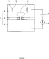
- the sensing chamber, 1, includes a cis compartment, 2, and a trans compartment, 3, which are divided by a partition, 4. Both compartments are filled with a pre-selected recording solution such as 1 M KCl.
- the partition, 4, has an opening, 5, in its center region, over which a lipid bilayer is formed, and the nanopore, 6, is plugged through the lipid bilayer.
- the power, 7, provides a voltage that is loaded through a pair of electrodes in the two compartments; the current detector, such as a pico-Ampere amplifier, 8, is connected to monitor the current changes.
- a mixture sample of the target oligonucleotide, 9, and its complementary probe, 10 is loaded into the cis compartment, 2.
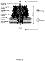
- FIG. 2 is a schematic amplified illustration of the nanopore, 6.
- the nanopore, 6, is in conical or funnel shape with two openings, the cis opening, 11, at the wide end and the trans opening, 12, down the narrow end.
- the paired oligonucleotides, 9/10 is captured into the nanocavity, 13.
- the voltage then drive the oligonucleotides, 9/10, to unzip at the constriction, 14, with the probe, 10, first traversing through the ⁇ -barrel, 15, and out off the trans opening, 12, and followed by the traversing of the target oligonucleotide, 9.
- the nanopore may be any ion channel of cone-shape or any asymmetrical shape with a wide and a narrow opening plugged into the planar lipid bilayer that has a wider cavity followed by a narrow channel that can facilitate unzipping translocation events.
- the nanopore may be any existing protein ion channels, such as the ⁇ -hemolysin transmembrane protein pore adopted in the examples below, or various synthetic pores fabricated using fashion nanotechnologies with abiotic materials such as silicon.
- the probe is a multi-domain single strand molecule, which comprises a central domain fully complementary to the target oligonucleotide and at least one terminal extension, i.e., signal tag, at its 3' or 5' terminal, with signal tags at both terminals as preferred.
- the 3'-tagged probe is preferred over the 5'-tagged probe.
- the probe directionality-dependence of the capture rate is possibly due to that the bases of ssDNA tilt collectively toward the 5' end of the strand 38 , and this asymmetric base orientation makes DNA move more easily from 3'-end than 5'-end.
- the terminal extension may be of any charged single chain molecule with sufficient length to assist the unzipping translocation through the nanopore driven by the voltage.
- the signal tag may be a charged polymer chain, which can be an oligonucleotide such as poly(dC) n , poly(dA) n , and or poly(dT) n , or a charged polypeptide.
- the poly(dC) tag is more preferred over poly(dA) or poly(dT) tags; furthermore, the poly(dC) 30 is much more efficient in generating signature events (discussed below) than that with a shorter tag such as poly(dC) 8 .
- the capture rate can be further enhanced once combined with other effective approaches, including detection at high voltage, use of engineered pores with designed charge profile in the lumen 33 , and detection in asymmetrical salt concentrations between both sides of the pore 39 .
- the current change induced by the unzipping and translocation of the hybrid of the target oligonucleotide and its complementary probe through the nanopore is a unique signature event, which is used to detect and differentiate the target oligonucleotide.
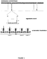
- FIG. 3 which includes an exemplary current trace recorded during an exemplary detection, an amplified electrical mark of the signature event, and a schematic illustration of the unzipping-translocation event.
- ROC curve refers to a Receiver Operating Characteristic Curve. An ROC curve used to analyze the relationship between selectivity and sensitivity. An ROC curve separates the plot into up and lower regions.
- AUC refers to the Area under the ROC curve.
- An AUC can range between 0.5-1.0. The higher the AUC value, the better the separation result.
- OCP refers to an Optimized Cutoff Point.
- an OCP can be calculated from ROC curves.
- an OCP is a cutoff duration at the maximal value of a Youden index.
- Youden index is defined as ⁇ sensitivity+selectivity-1 ⁇ .
- a Youden index is calculated from the ROC curve, and can range between 0 and 1.
- OCP optical cutoff point
- the instant disclosure is directed to probes, nanopores, kits comprising the probes and nanopores, and associated methods of use, that provide for "signature" current blockage events that distinguish those events arising from interactions with the probe and target from other events.
- the other events are referred to as "background” events. Background events include, but are not limited to, interactions of a probe with nucleic acid that is not a target, interactions of a probe with other components present in a nanopore detection system, free nucleic acids present in the nanopore detection system, and the like.
- Such features of such signature events include, but are not limited to, at least one of a: i) a current block of different duration than a background current block; ii) a different number of distinct current blockade levels than a background current block; iii) a different order of occurrence of current blockade levels than a background current block; iv) a different current amplitude at a blockade level than a background current block; v) a different current amplitude of each blockade level than a background current block; or any combination of (i), (ii), (ii), (iv), or (v).
- a signature blockage event can be distinguished from a background blockage event by differences in a characteristic background noise of each blockage event.
- the distinct durations, numbers, or amplitude(s) in the signature event are greater than those observed in the background event. In certain embodiments, the distinct durations, numbers, or amplitude(s) in the signature event are less than those observed in the background event. In certain embodiments, the distinct durations, numbers, orders, or amplitude(s) in a signature event are statistically distinguishable from those of a background event.
- the signature events are provided in nanopore systems comprising a protein nanopore formed by alpha-hemolysin ( ⁇ HL) or engineered variants thereof in a planar lipid bilayer system.
- the signature events can be provided in a biochip formed by hydrogel-encapsulated lipid bilayer with a single protein nanopore embedded therein or a micro-droplet bilayer system.
- Biochips and micro-droplet bilayer systems have been described ( Shim and Gu; Stochastic Sensing on a Modular Chip Containing a Single-Ion Channel Anal. Chem. 2007, 79, 2207-2213 ; Bayley,H. et al. Droplet interface bilayers. Mol. Biosyst. 4, 1191-1208 (2008 ).
- the signature events can be provided in a synthetic nanopore.
- Synthetic nanopores include, but are not limited to, nanopores comprising silicon nitride or graphene.
- Probe molecules provided herein comprise terminal extensions at one or both of their 5' and/or 3' termini. Without seeking to be limited by theory, it is believed that these terminal extensions provide useful functions that include, but are not limited to, trapping of the probe/target complex into the nanopore at a high rate (i.e. the number of signature events per unit target concentration per unit recording time). The trapping rate directly determines the sensitivity. In the same target concentration and the same recording time, a higher trapping rate gives a more precise sensing result. Without seeking to be limited by theory, it is also believed that these terminal extensions provide useful functions that include, but are not limited to, inducing the voltage-driven dissociation of the probe/target complex. This dissociation function generates a signature event that can be used to discriminate interactions of the probe with the target from other components in the mixture, thereby ensuring the selectivity or specificity.
- Probe terminal extensions can comprise a charged polymer of any length.
- the polymer can be a negatively charged single-stranded nucleic acid.
- Advantages of such nucleic acid terminal extensions include, but are not limited to, extremely low cost of synthesis and controllable charge by pH, salt concentration and temperature.
- Such nucleic acid extensions can comprise homopolymers, heteropolymers, copolymers or combinations thereof.
- the lengths of such nucleic acid terminal extensions can range from about 1 or 2 nucleotides to about 50 nucleotides.
- the nucleic acid extensions can range in length from about 5 to about 40 nucleotides, about 15 to about 35 nucleotides, or from about 20 to about 35 nucleotides.
- An exemplary terminal extension provided herewith is homopolymer poly(dC) 30 .
- a heteropolymeric sequence including but not limited to, di- or tri-nucleotide heteropolymers such as CTCTCTCT..., or CATCATCAT..., can also be used.
- co-polymers comprising abases or polyethylene glycol (PEG) can be used in the terminal extension. These co-polymers, or domains thereof in a terminal extension, can confer new functions on the terminal extension of the probe.
- An abase is a nucleotide without the base, but carries a negative charge provided by the phosphate.
- abase As the dimension of abase is narrower than normal nucleotides, it may generate a signature event signal different from that formed by the neighbor nucleotides. PEG is not charged. Without seeking to be limited by theory, it is believed that when the PEG domain in a nucleic acid sequence is trapped in the pore, it can reduce the driving force, thus precisely regulating the dissociation of the probe/target complex.
- Probe terminal extensions can also comprise a polypeptide.
- the richer choice of amino acids makes the sequence and functionality of the polypeptide terminal extension more programmable than an oligonucleotide terminal extension.
- polypeptide terminal extensions allow insertion of charged amino acids in the optimized positions to generate more distinguishable probe/target signature events. While not seeking to be limited by theory, it is believed that the probe/target complex can be selectively trapped using a probe comprising a positively charged polypeptide terminal extension under an appropriate voltage while all other negatively charged non-target oligonucleotides in the mixture are prevented from entering into the pore, resulting in ultra-selective detection.
- the polypeptide terminal extensions can comprise two, three, four, or more amino acid residues that can carry a positive charge (i.e. lysine and/or arginine and/or histidine).
- a positive charge i.e. lysine and/or arginine and/or histidine.
- sufficient numbers of positively charged residues are included in the polypeptide terminal extension to provide a net positive charge when said probe is hybridized to a target oligonucleotide.
- performance of the associated nanopore based detection methods can be enhanced under acidic conditions (i.e. when the pH value is less than 7) or conditions where the residue will be protonated.
- the lengths of such polypeptide terminal extensions can range from about 1 or 2 residues to about 30 residues.
- the polypeptide extensions can range in length from about 5 to about 20 residues, about 8 to about 20 residues, or from about 8 to about 15 residues.
- an HIV-TAT polypeptide comprising positively charged arginine and lysine residues can be used as the terminal extension.
- the center domain of the probe that is complementary to the target oligonucleotide can comprise a peptide nucleic acid that is covalently linked to a terminal extension comprising amino acids that carry a positive charge.
- a center domain comprising a peptide nucleic acid is used in conjunction with a terminal extension comprising amino acids that carry a positive charge to provide a net positive charge when said probe is hybridized to a target oligonucleotide.
- polypeptide terminal extensions comprising amino acids with aromatic side chains including, but not limited to, phenylalanine, tryptophan, tyrosine, thyroxine, and the like, can be incorporated into the polypeptide terminal extensions. While not seeking to be limited by theory, it is believed that such aromatic amino acids can interact with the pore through aromatic stacking and provide for useful changes in the signature obtained in nanopore based detection methods.
- any other free nucleic acids components will be repulsed from entering the pore due to the negative charge that is carried by the free, unhybridized nucleic acids. This significantly reduces signals by free nucleic acid components, such that the majority of the observed current blockage events are either due to the trapping of the oligonucleotide/probe complex or to the translocation of the probe.
- the oligonucleotide/probe complexes with a net positive charge can be directed to a nanopore with a negatively-charged ring at the trans- opening of the pore.
- a trans opening of a pore is understood to be that portion of the pore from which a molecule would emerge whereas a cis opening of a pore from which a molecule would enter.
- a negative charged ring at the trans- opening of the pore can be obtained by using any type of nanopore that has been suitably synthesize and/or derivatized so as to have a negative charged ring at the trans- opening of the pore.
- Such nanopores with a negatively charged ring at the trans opening of the pore include, but are not limited to, protein nanopores and synthetic nanopores.
- Protein nanopores with a negatively charged ring at the trans opening of the pore include, but are not limited to, engineered variants of an alpha-hemolysin protein.
- the engineered alpha hemolysin variant can comprise a Staphylococcus aureus alpha hemolysin containing a K131D, a K131E, or a K131H amino acid substitution.
- Exemplary and non-limiting Staphylococcus aureus alpha hemolysin wild type sequences are provided herein (SEQ ID NO:20, nucleic acid coding region; SEQ ID NO:21: protein coding region) and available elsewhere (National Center for Bioinformatics or GenBank Accession Numbers M90536 and AAA26598).
- An exemplary and non-limiting Staphylococcus aureus alpha hemolysin variant comprising a K131D substitution is provided as SEQ ID NO:22.
- the engineered alpha hemolysin variant can comprise a suitably derivatized variant that is derivatized with moieties that provide for a negatively charged ring at the trans opening of the pore.
- aureus alpha hemolysin protein that can be substituted or derivatized to provide for a protein nanopore with a negative charged ring at the trans- opening of the pore is provided herewith as SEQ ID NO: 21.
- variants of other hemolysins capable of forming pores can be substituted or derivatized to provide for a protein nanopore with a negative charged ring at the trans- opening of the pore.
- Synthetic nanopores with a negatively charged ring at the trans opening of the pore are also provided.
- such synthetic nanopores with a negatively charged ring at the trans opening of the pore include, but are not limited to, silicon nitride or graphene nanopores that have been suitably derivatized with moieties that provide for a negatively charged ring at the trans opening of the pore.
- the center domain of probes provided herein is used to capture the target molecule.
- the center domain can be fully complementary or partially complementary to the target sequence.
- a center domain can comprise an oligonucleotide comprising natural nucleotides (A, T, G, C (DNA) or a, u, g, c (RNA)), and/or artificial nucleotides including, but not limited to, nucleosides such as inosine, xanthosine, 7-methylguanosine, Dihydrouridine, and 5-methylcytidine.
- the center domain can comprise a locked nucleic acid (LNA) or a peptide nucleic acid (PNA).
- Locked nucleic acids comprise RNA derivatives where the ribose ring contains a methylene linkage between the 2'-oxygen and the 4'-carbon.
- Peptide nucleic acids comprise a peptide backbone with nucleobase side chains.
- a LNA or a PNA center domain can comprise natural nucleobases (adenine, guanine, thymine, cytosine or uracil) and/or artificial nucleobases including, but not limited to, hypoxanthine, xanthosine, 7-methylguanine, 5,6-dihydrouracil, and 5-methyl cytosine.
- probe center domains comprising co-polymers of oligonucleotides, LNA, or PNA are provided.
- a center domain of a probe will have at least about 4, 6, 8, 10, 12, 14, 15, 16, 17, 18, 19, 20, 21, 22, 23, 24, or 25 nucleotide or nucleobase residues that are complementary to the target nucleic acid.
- a central region of a probe will have at least about 4, 6, 8, 10, 12, 14, 15, 16, 17, 18, 19, 20, 21, 22, 23, 24, or 25 to any of about 30, 35, 40, or 50 nucleotide or nucleobase residues that are complementary to the target nucleic acid.
- synthetic nucleotides or nucleobases inserted in the sequence can precisely adjust the hybridization energy with the target, such that one can distinguish the characters of targets such as single-nucleotide polymorphism, methylation, or interaction between miRNA and its target messenger RNA.
- target nucleic acids or oligonucleotides that can be detected and distinguished from non-target nucleic acids by the probes, nanopores, kits comprising the probes and nanopores, and associated methods of use probes, provided herein.
- the target can be a nucleic acid or a fragment thereof from cells, body fluid, tissues, bacteria, or a virus.
- the target can be a PCR products or a synthetic oligonucleotide.
- a target can comprise a genomic DNA, an mRNA, a pre-mature or mature miRNA, an artificial miRNA, non-coding DNA or RNA, a nucleic acid biomarker, or a synthetic aptamer.
- a miRNA targets may come from the RNA extraction from bio-fluid from any tissues such as plasma and formalin-fixed and paraffin-embedded tissues.
- a target nucleic acid can comprise be a nucleic acid fragment complexed with any of a nucleic acid binding protein, an antibody, or an aptamer bound with a target protein.
- a target nucleic acid can comprise be a nucleic acid fragment complexed with a low molecule weight compound, including, but not limited to, a drug.
- targets can include sequences with mutations, with single-nucleotide polymorphisms, or with chemical modifications such as methylation and phosphorylation.
- Example 1 Detection of miR-155 , a lung cancer biomarker
- the nanopore sensing system includes an ⁇ -hemolysin transmembrane protein pore and a pre-designed probe for miR-155.
- the probe is a DNA multiple-block copolymer with its central domain complementary to the target miR-155 , and at least one poly(dC) 30 extension at 3'- , 5'-, or both terminals functioning as signal tags.
- Table 1 lists the sequences of miR-155 and the exemplary probes with the tri-block copolymer, P 155 as preferred. Table 1.
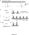
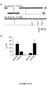
- a mixture of miR-155 /P 155 is added to the cis side of the pore, a current trace with a series of short- and long-lived current blocks can be recorded, as shown in FIG. 4a , while being monitored in 1 M KCl at +100 mV.
- the long blocks in the recording persist for 250 ⁇ 58 ms.
- This conductance level is consistent with a configuration that miR-155 •P 155 is trapped in the pore from the wider opening ( cis ), with either 3'- or 5'-signal tag of P 155 occupying the narrowest ⁇ -barrel ( Fig. 4c' level 1).
- the signal tag in the ⁇ -barrel can induce unzipping of miR-155 •P 155 , driven by voltage.
- the unzipping time, or the duration of Level 1 is comparable to previously reported time scales for DNA unzipping in the pore, e.g. ⁇ 435 ms for unzipping a 50 bps dsDNA at +140 mV, and ⁇ 40 ms for a 10 bps hairpin DNA at +90 mV.
- the duration of Level 3 is 270 ⁇ 30 ⁇ s, close to the 220 ⁇ s for short blocks by mir-155 alone, and consistent with the time scale of ⁇ 400 ⁇ s for translocation of a 75 bases RNA at +120 mV, 35 and 800 ⁇ s for a 210 bases RNA at +120 mV. 36
- the duration of Level 3 becomes shortened as the voltage increases, further supporting the translocation of a single-stranded oligonucleotide for this conductance level.
- This type of long blocks may occur when the arrested miR-155 •P 155 exits the pore from the cis entry without unzipping.
- the characteristic long blocks can serve as signature events for identifying single molecules of target miRNAs.
- [ miR ] 0 and [P] 0 are the initial concentrations of miRNA and the probe
- k on is the occurrence rate of signature events
- K d is the dissociation constant for miR •P in the solution.
- let-7a and let-7b are members of the Let-7 tumor suppressing miRNA family 4-6 ; and the two Let-7 members only contain different nucleotides at the position 17 and 19, which are adenines in let-7a and guanines in let-7b.
- the probes P a and P b are designed for let-7a and let-7b respectively with sequences listed in Table 2. Table 2.
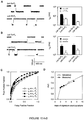
- the nanopore sensing system is able to differentiate single mismatches based on the unzipping time, thus demonstrating the potential to detect miRNAs with similar sequences and SNPs.
- RNAs containing miRNAs were extracted from 350 ⁇ l of each plasma sample using miRVana PARIS Kit (Ambion), with a final elution volume of 100 ⁇ l, which were than divided into two aliquots (50 ⁇ l) for the nanopore and RT-PCR assay 44 .
- One aliquot was pre-mixed with P 155 and directly added to the 2-ml recording solution in the nanopore chamber.
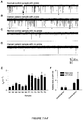
- the nanopore current retain a low level of noise even in the presence of plasma samples, and distinct short and long blocks (marked with red arrows) can be indentified in both the control group ( FIG. 7a ) and lung cancer group ( FIG. 7b ).
- the characteristic long blocks including both with multiple conductance and single conductance, features the same conductance profiles and similar properties to that for synthetic miR-155 RNA in Fig.1a .
- no such types of long blocks can be observed ( FIGs. 7c and d ), but short blocks were found for translocation of single-stranded oligonucleotides such as free miRNAs ( FIGs. 7c and d).
- the characteristic long blocks could be attributed to miR-155 •P 155 hybrids and serve as signature events for single miRNA molecules detection.
- the frequency of miR-155 signature events f sig for all samples in the lung cancer patient group varies between 1.15-1.51 min -1 , with a mean of 1.40 ⁇ 0.16 min -1 ( FIG. 7e ). This level was significantly higher than f sig in the control group that ranges between 0.32-0.70 min -1 with a mean of 0.48 ⁇ 0.14 min -1 ( FIG. 7e ). Since all samples were prepared following a standard procedure (Methods in Supplementary Materials), it should be valid to compare relative miRNA levels in two groups. When the mean f sig value in normal plasma was set as 1, the folds of miR-155 in lung cancer plasma were compared with the two methods.
- Fig.7f showed that the relative mir-155 level in lung cancer patients was 2.79 with the nanopore sensor ( p ⁇ 0.001). By comparison, the relative miR-155 level was 4.72 with RT-PCR method ( p ⁇ 0.02) with greater variability. Therefore, both nanopore and RT-PCR assay indicated a significant elevation of miR-155 in lung cancer patient plasma although there is a 1.69 fold difference. As the nanopore method does not require labeling and amplification, this may be a reason for smaller variability in the nanopore assay ( FIG. 7f ). Overall the nanopore sensor with engineered probes demonstrates the ability to detect circulating miRNAs in clinical lung cancer patients, which is verified by the independent RT-PCR method.
- Oligonucleotides including miRNAs and DNA probes were synthesized and electrophoresis-purified by Integrated DNA Technologies (Coralville, IA). Before testing, the mixtures of miRNA and DNA probe were heated to 90 °C for 5 minutes, then gradually cooled down to room temperature and stored at 4 °C. The RNase-free water was used to prepare RNA solution.
- the recording apparatus was composed of two chambers ( cis and trans ) that were partitioned with a Teflon film.
- the planar lipid bilayer of 1,2-diphytanoyl-sn-glycerophosphatidylcholine (Avanti Polar Lipids) was formed spanning a 100-150 nm hole in the center of the partition.
- Both cis and trans chambers were filled with symmetrical 1 M salt solutions (KCl) buffered with 10 mM Tris and titrated to pH 8.0. All solutions are filtered before use. Single ⁇ -hemolysin proteins were inserted into the bilayer from the cis side to form molecular pores in the membrane. All the oligonucleotides including miRNAs and DNA probes and clinical RNA samples were also added to the cis solution. To record the pore current, the cis solution was grounded and the voltage was given from the trans solution. In this convention, a positive voltage can drive the translocation of a negatively charged DNA through the pore from cis to trans. Single-channel currents were recorded with an Axopatch 200A amplifier (Molecular Device Inc.
- RNA extraction from plasma and miRNA quantification by qRT-PCR Peripheral blood samples were obtained at the University of Missouri Ellis Fischel Cancer Center with an IRB approval. Whole blood with EDTA preservative was centrifuged at 1,600 g for 10 min at room temperature and the plasma was transferred to new tubes. Total RNAs containing miRNAs was extracted from 350 ⁇ l of plasma using miRVana PARIS Kit (Ambion, Austin, TX, USA) according to the manufacturer's protocol. The final elution volume was 100 ⁇ l.
- RNA sample containing miRNAs was polyadenylated by poly(A) polymerase (Ambion) and reverse transcribed to cDNA using SuperScript III Reverse Transcriptase (Invitrogen) according to the manufacturer's instructions with a poly(T) adapter primer (5'-GCGAGCACAGAATTAATACGACTCACTATAGGTTTTTTTTTTTTTTTVN-3'; SEQ ID NO: 10)).
- Real-time PCR was performed using iQ SYBR Green Supermix (Bio-Rad, Hercules, CA, USA) with the miR-155 specific forward primer (5'-TTAATGCTAATCGTGATAGGGGT-3'; SEQ ID NO:11) and the sequence complementary to the poly(T) adapter as the reverse primer (5'-GCGAGCACAGAATTAATACGAC-3'; SEQ ID NO:12) in iQ5 Real-time PCR system (Bio-Rad, USA). The PCR was carried out as follows: after initial denaturation at 95 °C for 3 min, 40 cycles of 95 °C for 15 s and 60 °C for 1 min were followed.
- the relative level of miR-155 was calculated using 2 -delta Ct method where the level of normal plasma was normalized as 1. Data was presented as mean ⁇ SD of three independent experiments, and the differences were considered statistically significant at p ⁇ 0.05 by using the Student's t -test.
- the Denaturing Solution prevents RNAs from undergoing degradation by inhibiting endogenous plasma RNAases.
- both miR-155 and spiked-in miR-39 were measured using the nanopore sensor and SYBR green-based qRT-PCR.
- the nanopore data and normalization result were shown in Table 9.
- the probes for miR-155 and miR-39 were P 155 and P 39 .
- the variability of f 39 reflected the difference in miR-39 concentrations among samples after RNA extraction.
- miR represents miRNA
- P probe
- K d equilibrium dissociation constant for miR •P
- k on occurrence rate constant of miR •P signature events
- f sig frequency of signature events.
- Eq.S4 can be simplified as f sig ⁇ k on miR 0 In this condition, f sig is proportional to [ miR ] 0 . Eq.S4 also suggested that f sig will ultimately become saturated. This is because f sig measures the capture frequency of miR •P, and the maximal concentration of miR •P ([ miR •P]) can not be higher than that of the probe ([P] 0 ).
- the cis and trans miR-155 were measured separately.
- the peaks of melting curves for trans RNA samples were the same as synthesized miR-155.
- the miR-155 concentrations in trans solutions were 14, 34 and 63 aM (10 -18 M), indicating that a trace amount of miR-155 transported to the trans side of the pore.
- Precursor miRNAs are stem-loop RNAs of ⁇ 70 nucleotides bearing the 2 nucleotides 3'-overhang as a signature of RNase III-mediated cleavage ( Lee,Y., Jeon,K., Lee,J.T., Kim,S., & Kim,V.N. MicroRNA maturation: stepwise processing and subcellular localization. EMBO J 21, 4663-4670 (2002 )). It is not known whether the plasma total RNA extract contains pre-miRNAs. However, we have verified that the capture rate for a miR •P is very low if the signal tag in the probe is very short ( Fig.9 , Fig.16 and unpublished data). Therefore, we expected that, even though pre-miRNAs exist, the short overhang will prevent them from trapping in the nanopore.
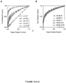
- AUC Areas under ROC curves (AUC) for separation of miRNAs with one nucleotide difference (let-7a and let-7c) and with two nucleotide difference (let-7a and let-7b)a let-7a • P a let-7b • P b let-7a • P a let-7c • P c let-7b • P a 0.75 n.a. let-7c • P a 0.73 n.a. let-7a • P b n.a. 0.83 let-7a • P c n.a.
- the receiver operating characteristic (ROC) curve is a plot of the true positive rate (sensitivity) against the false positive rate (1-selectivity) for the different possible cutoff points that separate the entire duration distribution into the positive and negative components.
- ROC receiver operating characteristic
- the separation accuracy was measured by the area under the ROC curve (AUC).
- An AUC of 1 represents a perfect separation; an area of 0.5 represents no separation ability.
- AUC was analyzed online using free software on the world wide web (internet address) rad.jhmi.edu/jeng/javarad/roc/JROCFITi.html. Table 8.
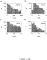
- AUC Areas under ROC curves (AUC) and optimal cutoff point (OCP) at various duration ratio and event number ratio a ⁇ P / ⁇ N (s/s) b 1/1 2/1 3/1 4/1 5/1 10/1 AUC 0.51 0.72 0.73 0.76 0.78 0.93 OCP n.a. 1.33 1.74 1.88 1.98 2.18 N P / N N c 200:800 200:400 200:200 200:150 200:100 200:50 AUC 0.83 0.81 0.76 0.76 0.79 0.78 OCP 1.88 1.88 1.85 1.79 1.81 1.97 a : Both AUC and OCP were calculated from the ROC curves shown in Fig.16A-B . OCP is a cutoff duration at the maximal value of Youden index.
- OCP optical cutoff point
- Example 3 Peptide-guided selective detection of microRNAs.
- RNA extraction contains numerous and complicated nucleic acids components, at least including miRNAs (both pre-mature and mature miRNAs), mRNAs, tRNAs, other RNAs. All components commonly carry negative charges. Thus, if the target miRNA can be trapped in the pore at an applied voltage, any other component may also be driven to interact with the nanopore, generating non-specific current signals that interfere with the recognition of signature events generated by the target miRNA/probe complex.
- peptide-PNA peptide nucleic acid
- the PNA sequence in “PNA” bracket in Figure 18A ) has a peptide backbone with side chain nucleobases that are complementary to the entire or partial sequence of the target miRNA ("miRNA ( Let-7b, -7c )" in Figure 18A bracket), and thus serves as the center domain for capturing the target miRNA in the solution.
- NH2-AACCACACAA-COOH where the molecule comprises a peptide backbone with the indicated AACCACACAA nucleobases; (SEQ ID NO: 18).
- HIV-TAT HIV-TAT
- the reporter (or terminal extension) of the new probe is a peptide that carries a series of positively-charged amino acids ("Peptide reporter" in Figure 18A bracket) and the center domain is a peptide nucleic acid comprising nucleotides that are complementary to the target nucleic acid.
- Peptide reporter in Figure 18A bracket
- the center domain is a peptide nucleic acid comprising nucleotides that are complementary to the target nucleic acid.
- S. aureus alpha-hemolysin comprising a K131D mutation.
- the wild-type S. aureus alpha-hemolysin peptide sequence (National Center for Bioinformatics Accession NO. AAA26598.1); is:
- the variant S. aureus K131D alpha-hemolysin peptide sequence is:
- the positively-charged peptide domain of the probe dipole will be both pushed by the positive voltage (cis grounded) and attracted by the negative ring at the trans opening, guiding the trapping of the miRNA/complex into the ⁇ -barrel of the pore.
- any other free nucleic acids components will be repulsed from entering the pore due to the negative charge carried. This significantly reduces signals by free RNA components, and most observed events are either due to the trapping of the miRNA/probe complex or the translocation of the probe.
- the use of peptide-PNA probe enables selective detection of the target miRNA.
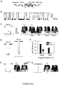
- Figure 18A shows the diagram of the miRNA/probe complex.
- the bracketed miRNA is target miRNA Let-7b.
- the bracketed probe P7b has a bracketed "Peptide Reporter” part and a bracketed "PNA” (peptide nucleic acid) part.
- the PNA is for capturing Let-7b
- the bracketed "Peptide reporter” is apositively-charged peptide corresponding the sequence of HIV-TAT, which contains +8e contributed by arginines and lysines.
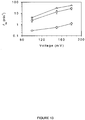
- Figure 18B shows events for translocation of the peptide-PNA probe, P7b. The characteristic events last for 3 ms and reduce the current to 10 pA at +180 mV.
- Figure 18C shows no block events can be observed with free miRNA let-7b (without probe) in the solution at +180 mV.
- Figure 18D shows signature events for the trapping of the let-7b/P7b complex. These events characteristically last for 100 ms and reduce the current to 57 pA at +180 mV, completely different that for the probe.
- Figure 18E shows that Let-7c , which has two different nucleotides from Let-7b , cannot bind to PNA of the probe P7b, therefore does not generate signature events as in Figure 18C . Almost all observed events are due to the probe itself.
- Figure 18F compares the duration-amplitude property for P7b binding to Let-7b (fully match, two separate clusters without overlay) and Let-7c (2 mismatches, two clusters fully overlay). This suggests an accuracy of almost 100% in differentiating sequence-similar miRNAs with two different nucleotides.
- Level 1' can be interpreted by that although the short strand of SA-C30 moves into the ⁇ -barrel after unzipping, its translocation is prevented by the attached large streptavidin. This result suggested the potential of using signature events for protein detection.
- Figure 19C we demonstrated that the complex can be sequentially unzipped in the nanopore in two steps when using a short oligonucleotide to link two DNAs. The unzipping of the two DNAs can be clearly revealed by the two Level 2 states.
- the peptide-PNA probe enables 1) selective detection of the target miRNA, 2) greatly enhanced accuracy in differentiating sequence-similar miRNAs.
Landscapes
- Chemical & Material Sciences (AREA)
- Life Sciences & Earth Sciences (AREA)
- Health & Medical Sciences (AREA)
- Engineering & Computer Science (AREA)
- Organic Chemistry (AREA)
- Analytical Chemistry (AREA)
- Proteomics, Peptides & Aminoacids (AREA)
- Physics & Mathematics (AREA)
- Zoology (AREA)
- Wood Science & Technology (AREA)
- Immunology (AREA)
- Biophysics (AREA)
- Molecular Biology (AREA)
- Biochemistry (AREA)
- General Health & Medical Sciences (AREA)
- Genetics & Genomics (AREA)
- Biomedical Technology (AREA)
- Pathology (AREA)
- General Engineering & Computer Science (AREA)
- Microbiology (AREA)
- Bioinformatics & Cheminformatics (AREA)
- Biotechnology (AREA)
- Food Science & Technology (AREA)
- Urology & Nephrology (AREA)
- Spectroscopy & Molecular Physics (AREA)
- Nanotechnology (AREA)
- Hematology (AREA)
- General Physics & Mathematics (AREA)
- Medicinal Chemistry (AREA)
- Hospice & Palliative Care (AREA)
- Oncology (AREA)
- Measuring Or Testing Involving Enzymes Or Micro-Organisms (AREA)
- Investigating Or Analyzing Materials By The Use Of Electric Means (AREA)
- Apparatus Associated With Microorganisms And Enzymes (AREA)
- Investigating Or Analysing Biological Materials (AREA)
Priority Applications (1)
| Application Number | Priority Date | Filing Date | Title |
|---|---|---|---|
| EP17157476.7A EP3190193A1 (en) | 2010-07-14 | 2011-07-14 | Nanopore-facilitated single molecule detection of nucleic acids |
Applications Claiming Priority (2)
| Application Number | Priority Date | Filing Date | Title |
|---|---|---|---|
| US39957810P | 2010-07-14 | 2010-07-14 | |
| PCT/US2011/044082 WO2012009578A2 (en) | 2010-07-14 | 2011-07-14 | Nanopore-facilitated single molecule detection of nucleic acids |
Related Child Applications (2)
| Application Number | Title | Priority Date | Filing Date |
|---|---|---|---|
| EP17157476.7A Division-Into EP3190193A1 (en) | 2010-07-14 | 2011-07-14 | Nanopore-facilitated single molecule detection of nucleic acids |
| EP17157476.7A Division EP3190193A1 (en) | 2010-07-14 | 2011-07-14 | Nanopore-facilitated single molecule detection of nucleic acids |
Publications (3)
| Publication Number | Publication Date |
|---|---|
| EP2593568A2 EP2593568A2 (en) | 2013-05-22 |
| EP2593568A4 EP2593568A4 (en) | 2014-03-05 |
| EP2593568B1 true EP2593568B1 (en) | 2017-04-19 |
Family
ID=45470088
Family Applications (2)
| Application Number | Title | Priority Date | Filing Date |
|---|---|---|---|
| EP11807539.9A Active EP2593568B1 (en) | 2010-07-14 | 2011-07-14 | Nanopore-facilitated single molecule detection of nucleic acids |
| EP17157476.7A Withdrawn EP3190193A1 (en) | 2010-07-14 | 2011-07-14 | Nanopore-facilitated single molecule detection of nucleic acids |
Family Applications After (1)
| Application Number | Title | Priority Date | Filing Date |
|---|---|---|---|
| EP17157476.7A Withdrawn EP3190193A1 (en) | 2010-07-14 | 2011-07-14 | Nanopore-facilitated single molecule detection of nucleic acids |
Country Status (6)
| Country | Link |
|---|---|
| US (3) | US9395353B2 (enExample) |
| EP (2) | EP2593568B1 (enExample) |
| JP (2) | JP6016792B2 (enExample) |
| AU (1) | AU2011279083B2 (enExample) |
| CA (1) | CA2805247C (enExample) |
| WO (1) | WO2012009578A2 (enExample) |
Families Citing this family (65)
| Publication number | Priority date | Publication date | Assignee | Title |
|---|---|---|---|---|
| WO2007146158A1 (en) | 2006-06-07 | 2007-12-21 | The Trustees Of Columbia University In The City Of New York | Dna sequencing by nanopore using modified nucleotides |
| US9678055B2 (en) | 2010-02-08 | 2017-06-13 | Genia Technologies, Inc. | Methods for forming a nanopore in a lipid bilayer |
| US9605307B2 (en) | 2010-02-08 | 2017-03-28 | Genia Technologies, Inc. | Systems and methods for forming a nanopore in a lipid bilayer |
| US8324914B2 (en) | 2010-02-08 | 2012-12-04 | Genia Technologies, Inc. | Systems and methods for characterizing a molecule |
| US9194838B2 (en) | 2010-03-03 | 2015-11-24 | Osaka University | Method and device for identifying nucleotide, and method and device for determining nucleotide sequence of polynucleotide |
| WO2012009578A2 (en) | 2010-07-14 | 2012-01-19 | The Curators Of The University Of Missouri | Nanopore-facilitated single molecule detection of nucleic acids |
| WO2012083249A2 (en) | 2010-12-17 | 2012-06-21 | The Trustees Of Columbia University In The City Of New York | Dna sequencing by synthesis using modified nucleotides and nanopore detection |
| GB2500360B (en) | 2010-12-22 | 2019-10-23 | Genia Tech Inc | Nanopore-based single DNA molecule characterization, identification and isolation using speed bumps |
| US8962242B2 (en) | 2011-01-24 | 2015-02-24 | Genia Technologies, Inc. | System for detecting electrical properties of a molecular complex |
| US9110478B2 (en) | 2011-01-27 | 2015-08-18 | Genia Technologies, Inc. | Temperature regulation of measurement arrays |
| CN107828877A (zh) * | 2012-01-20 | 2018-03-23 | 吉尼亚科技公司 | 基于纳米孔的分子检测与测序 |
| IN2014DN06796A (enExample) | 2012-02-15 | 2015-05-22 | Oxford Nanopore Tech Ltd | |
| CN104254619B (zh) | 2012-02-16 | 2018-08-24 | 吉尼亚科技公司 | 产生用于纳米孔传感器的双层的方法 |
| US8986629B2 (en) | 2012-02-27 | 2015-03-24 | Genia Technologies, Inc. | Sensor circuit for controlling, detecting, and measuring a molecular complex |
| WO2013154999A2 (en) | 2012-04-09 | 2013-10-17 | The Trustees Of Columbia University In The City Of New York | Method of preparation of nanopore and uses thereof |
| WO2013188841A1 (en) | 2012-06-15 | 2013-12-19 | Genia Technologies, Inc. | Chip set-up and high-accuracy nucleic acid sequencing |
| EP2880186B1 (en) | 2012-08-03 | 2019-02-06 | University Of Washington Through Its Center For Commercialization | Compositions and methods for improving nanopore sequencing |
| US9551023B2 (en) | 2012-09-14 | 2017-01-24 | Oxford Nanopore Technologies Ltd. | Sample preparation method |
| WO2014072703A1 (en) | 2012-11-06 | 2014-05-15 | Oxford Nanopore Technologies Limited | Quadruplex method |
| US9605309B2 (en) | 2012-11-09 | 2017-03-28 | Genia Technologies, Inc. | Nucleic acid sequencing using tags |
| US9759711B2 (en) | 2013-02-05 | 2017-09-12 | Genia Technologies, Inc. | Nanopore arrays |
| GB201318465D0 (en) | 2013-10-18 | 2013-12-04 | Oxford Nanopore Tech Ltd | Method |
| WO2014144883A1 (en) | 2013-03-15 | 2014-09-18 | The Trustees Of Columbia University In The City Of New York | Raman cluster tagged molecules for biological imaging |
| US10732183B2 (en) | 2013-03-15 | 2020-08-04 | The Trustees Of Columbia University In The City Of New York | Method for detecting multiple predetermined compounds in a sample |
| AU2014227754B2 (en) | 2013-03-15 | 2018-03-01 | The Curators Of The University Of Missouri | Encoded nanopore sensor for multiplex nucleic acids detection |
| EP3014264A4 (en) * | 2013-06-25 | 2016-11-16 | Two Pore Guys Inc | QUANTIFICATION OF MULTIPLEXED BIOMARKERS BY NANOPORENT ANALYSIS OF BIOMARKER POLYMER COMPLEXES |
| JP6334118B2 (ja) * | 2013-09-24 | 2018-05-30 | クオンタムバイオシステムズ株式会社 | 単分子識別方法、装置、及びプログラム |
| EP3047282B1 (en) | 2013-09-18 | 2019-05-15 | Quantum Biosystems Inc. | Biomolecule sequencing devices, systems and methods |
| JP2015077652A (ja) | 2013-10-16 | 2015-04-23 | クオンタムバイオシステムズ株式会社 | ナノギャップ電極およびその製造方法 |
| US9551697B2 (en) | 2013-10-17 | 2017-01-24 | Genia Technologies, Inc. | Non-faradaic, capacitively coupled measurement in a nanopore cell array |
| US9322062B2 (en) | 2013-10-23 | 2016-04-26 | Genia Technologies, Inc. | Process for biosensor well formation |
| JP6461943B2 (ja) | 2013-10-23 | 2019-01-30 | ジェニア・テクノロジーズ・インコーポレイテッド | ナノポアを備えた高速分子検知 |
| MA39774A (fr) | 2014-03-24 | 2021-05-12 | Roche Sequencing Solutions Inc | Procédés chimiques pour produire des nucléotides étiquetés |
| US10438811B1 (en) | 2014-04-15 | 2019-10-08 | Quantum Biosystems Inc. | Methods for forming nano-gap electrodes for use in nanosensors |
| WO2015170782A1 (en) | 2014-05-08 | 2015-11-12 | Osaka University | Devices, systems and methods for linearization of polymers |
| CA3163156C (en) * | 2014-07-31 | 2025-07-08 | Illumina, Inc. | HYBRID NANOPORE SENSORS |
| EP4397972A3 (en) * | 2014-10-16 | 2024-10-09 | Oxford Nanopore Technologies PLC | Alignment mapping estimation |
| US10480026B2 (en) | 2014-10-17 | 2019-11-19 | Oxford Nanopore Technologies Ltd. | Method for nanopore RNA characterisation |
| CN105842453B (zh) * | 2015-01-12 | 2018-12-25 | 南京理工大学 | 一种基于末端延伸、金纳米粒子和酶三级放大电化学传感器检测dna的方法 |
| WO2016196625A1 (en) | 2015-06-02 | 2016-12-08 | Nanopore Diagnostics, Llc | Nucleic acid detection |
| JP6889701B2 (ja) * | 2015-09-24 | 2021-06-18 | エフ.ホフマン−ラ ロシュ アーゲーF. Hoffmann−La Roche Aktiengesellschaft | アルファ溶血素バリアント |
| WO2017167811A1 (en) * | 2016-03-31 | 2017-10-05 | Genia Technologies, Inc. | Nanopore protein conjugates and uses thereof |
| KR20190075010A (ko) | 2016-04-27 | 2019-06-28 | 퀀텀 바이오시스템즈 가부시키가이샤 | 생체분자의 측정 및 시퀀싱을 위한 시스템 및 방법 |
| WO2017223515A1 (en) | 2016-06-23 | 2017-12-28 | F. Hoffman-La Roche Ag | Formation and calibration of nanopore sequencing cells |
| US11124827B2 (en) | 2016-06-23 | 2021-09-21 | Roche Sequencing Solutions, Inc. | Period-to-period analysis of AC signals from nanopore sequencing |
| EP3828280A1 (en) * | 2016-07-12 | 2021-06-02 | Rijksuniversiteit Groningen | Biological nanopores for biopolymer sensing and sequencing based on frac actinoporin |
| WO2018093976A1 (en) | 2016-11-16 | 2018-05-24 | Nanopore Diagnostics, Llc | Analysis of nucleic acids using probe with non-linear tag |
| WO2019018389A1 (en) * | 2017-07-17 | 2019-01-24 | President And Fellows Of Harvard College | PROTEIN SHUTTLE WITH NANOPORE MATCHING FOR MOLECULAR CHARACTERIZATION AND METHODOLOGY FOR DATA ANALYSIS THEREOF |
| CN111512155B (zh) | 2017-12-28 | 2022-07-05 | 豪夫迈·罗氏有限公司 | 测量和去除来自交流信号驱动的纳米孔dna测序系统的随机信号中的噪声 |
| US11034996B2 (en) | 2018-03-02 | 2021-06-15 | The Curators Of The University Of Missouri | Nucleic acid sequence and capture by formation of an abasic site-derived cross-link |
| WO2019173799A1 (en) * | 2018-03-08 | 2019-09-12 | Caris Science, Inc. | Oligonucleotide probes and uses thereof |
| JP7239965B2 (ja) * | 2018-06-26 | 2023-03-15 | 国立大学法人東京農工大学 | 2種以上の特定オリゴヌクレオチドの有無を判定する判定方法、並びに、前記判定方法を用いて検査対象が小細胞肺がん、胆管がんに罹患している又はそのリスクを有することを判定する方法 |
| US20220162692A1 (en) * | 2018-07-30 | 2022-05-26 | Oxford University Innovation Limited | Assemblies |
| US11162935B2 (en) | 2018-08-24 | 2021-11-02 | University Of Notre Dame Du Lac | Systems and methods for separating, detecting, and quantifying a target polynucleotide |
| WO2020055246A1 (en) | 2018-09-11 | 2020-03-19 | Rijksuniversiteit Groningen | Biological nanopores having tunable pore diameters and uses thereof as analytical tools |
| CN116814818A (zh) * | 2019-07-22 | 2023-09-29 | 四川大学华西医院 | 一种区分抗生素耐药的肺炎克雷伯菌的装置及方法 |
| KR102390925B1 (ko) * | 2019-11-01 | 2022-04-27 | 한국생명공학연구원 | 나노포어를 이용한 병원성 미생물의 검출 방법 및 검출키트 |
| US20230230636A1 (en) * | 2020-05-15 | 2023-07-20 | The Curator's of the University of Missouri | Nanopore unzipping-sequencing for dna data storage |
| JP7679055B2 (ja) * | 2020-08-24 | 2025-05-19 | 国立大学法人東京農工大学 | 極低濃度のオリゴヌクレオチドの存在の有無を判定する方法、及び検査対象が特定疾患に罹患しているかどうか又は特定疾患罹患のリスクを有するかどうかを判定する方法 |
| CN112961906B (zh) * | 2021-02-05 | 2024-01-23 | 南京邮电大学 | 金纳米棒检测探针、制备方法、检测方法及其应用 |
| CN112795628B (zh) * | 2021-02-05 | 2024-02-23 | 南京邮电大学 | 基于固态纳米孔传感器的miRNA检测方法、检测探针及试剂盒 |
| CN113189070B (zh) * | 2021-04-28 | 2024-01-16 | 香港理工大学 | 一种纳米探针及其制备方法和应用、循环肿瘤细胞的miRNAs的检测系统 |
| CN117229363A (zh) * | 2022-06-06 | 2023-12-15 | 天津赛德生物制药有限公司 | 用于肺癌早期诊断的肽核酸功能化核孔膜及制备方法与应用 |
| KR20250116014A (ko) | 2022-10-28 | 2025-07-31 | 리엑스유니버시테이트 그로닝겐 | 나노기공-기반 단백질 분석 |
| US20250137033A1 (en) * | 2023-11-01 | 2025-05-01 | Robert Bosch Gmbh | Dna unfolding using a free-end tag flow modifier |
Family Cites Families (39)
| Publication number | Priority date | Publication date | Assignee | Title |
|---|---|---|---|---|
| US6362002B1 (en) | 1995-03-17 | 2002-03-26 | President And Fellows Of Harvard College | Characterization of individual polymer molecules based on monomer-interface interactions |
| AU8586298A (en) | 1997-07-25 | 1999-02-16 | University Of Massachusetts | Designed protein pores as components for biosensors |
| WO2000056937A2 (en) | 1999-03-25 | 2000-09-28 | Hyseq, Inc. | Solution-based methods and materials for sequence analysis by hybridization |
| EP1212462B1 (en) | 1999-09-07 | 2006-01-25 | The Regents of the University of California | Methods of determining the presence of double stranded nucleic acids in a sample |
| WO2001059453A2 (en) | 2000-02-11 | 2001-08-16 | The Texas A & M University System | Biosensor compositions and methods of use |
| EP1478771A4 (en) | 2001-06-21 | 2005-06-15 | Harvard College | METHODS OF CHARACTERIZING NUCLEIC ACID MOLECULES |
| EP1415156B1 (en) | 2001-07-10 | 2009-09-02 | The Board Of Trustees Of The Leland Stanford Junior University | Methods and compositions for detecting the activation state of multiple proteins in single cells |
| US20050019784A1 (en) | 2002-05-20 | 2005-01-27 | Xing Su | Method and apparatus for nucleic acid sequencing and identification |
| US7683036B2 (en) * | 2003-07-31 | 2010-03-23 | Regulus Therapeutics Inc. | Oligomeric compounds and compositions for use in modulation of small non-coding RNAs |
| WO2005017025A2 (en) | 2003-08-15 | 2005-02-24 | The President And Fellows Of Harvard College | Study of polymer molecules and conformations with a nanopore |
| US20050136408A1 (en) | 2003-12-19 | 2005-06-23 | May Tom-Moy | Methods and systems for characterizing a polymer |
| US7972858B2 (en) | 2004-08-13 | 2011-07-05 | President And Fellows Of Harvard College | Ultra high-throughput opti-nanopore DNA readout platform |
| KR101159072B1 (ko) | 2005-01-20 | 2012-06-25 | 삼성전자주식회사 | 나노포어를 이용한 생분자의 분리방법 |
| KR100707198B1 (ko) | 2005-06-27 | 2007-04-13 | 삼성전자주식회사 | 나노포어 및 핵산에 비특이적으로 결합하는 물질을 이용한고감도 핵산 검출 방법 |
| WO2007041621A2 (en) | 2005-10-03 | 2007-04-12 | Xingsheng Sean Ling | Hybridization assisted nanopore sequencing |
| KR100730350B1 (ko) | 2005-10-17 | 2007-06-19 | 삼성전자주식회사 | 표면처리된 나노포어를 이용한 dna 검출방법 및검출장치 |
| WO2007120265A2 (en) | 2005-11-14 | 2007-10-25 | Applera Corporation | Coded molecules for detecting target analytes |
| US20100291548A1 (en) | 2006-03-12 | 2010-11-18 | Applera Corporation | Methods of Detecting Target Nucleic Acids |
| US8314220B2 (en) * | 2007-01-26 | 2012-11-20 | Agilent Technologies, Inc. | Methods compositions, and kits for detection of microRNA |
| WO2008097190A1 (en) * | 2007-02-05 | 2008-08-14 | Agency For Science, Technology And Research | Methods of electrically detecting a nucleic acid by means of an electrode pair |
| EP2156179B1 (en) | 2007-04-04 | 2021-08-18 | The Regents of The University of California | Methods for using a nanopore |
| WO2009020682A2 (en) | 2007-05-08 | 2009-02-12 | The Trustees Of Boston University | Chemical functionalization of solid-state nanopores and nanopore arrays and applications thereof |
| GB0713143D0 (en) | 2007-07-06 | 2007-08-15 | Ucl Business Plc | Nucleic acid detection method |
| US8273532B2 (en) | 2007-10-02 | 2012-09-25 | President And Fellows Of Harvard College | Capture, recapture, and trapping of molecules with a nanopore |
| US20090181390A1 (en) | 2008-01-11 | 2009-07-16 | Signosis, Inc. A California Corporation | High throughput detection of micrornas and use for disease diagnosis |
| US8852864B2 (en) * | 2008-01-17 | 2014-10-07 | Sequenom Inc. | Methods and compositions for the analysis of nucleic acids |
| EP2107040B1 (en) | 2008-03-31 | 2011-10-26 | Sony Deutschland Gmbh | A method of fabricating a membrane having a tapered pore |
| EP2310534B1 (en) | 2008-07-07 | 2018-09-05 | Oxford Nanopore Technologies Limited | Base-detecting pore |
| US20100099198A1 (en) | 2008-07-11 | 2010-04-22 | Board Of Regents, The University Of Texas System | Apparatus and system for pattern recognition sensing for biomolecules |
| WO2010062903A2 (en) | 2008-11-26 | 2010-06-03 | Board Of Regents, The University Of Texas System | Genomic sequencing using modified protein pores and ionic liquids |
| US20110003703A1 (en) | 2009-07-01 | 2011-01-06 | Charles Ma | Nucleic Acid Hybridization and Detection Using Enzymatic Reactions on a Microarray |
| WO2011014811A1 (en) | 2009-07-31 | 2011-02-03 | Ibis Biosciences, Inc. | Capture primers and capture sequence linked solid supports for molecular diagnostic tests |
| US20120276530A1 (en) | 2009-08-24 | 2012-11-01 | Trustees Of Boston University | Label-free sensing of pna-dna complexes using nanopores |
| US8324914B2 (en) | 2010-02-08 | 2012-12-04 | Genia Technologies, Inc. | Systems and methods for characterizing a molecule |
| WO2011103424A2 (en) | 2010-02-19 | 2011-08-25 | The Trustees Of The University Of Pennsylvania | High-resolution analysis devices and related methods |
| CN102918166A (zh) | 2010-03-30 | 2013-02-06 | 波士顿大学董事会 | 用于纳米孔解链依赖性核酸测序的工具和方法 |
| WO2012009578A2 (en) | 2010-07-14 | 2012-01-19 | The Curators Of The University Of Missouri | Nanopore-facilitated single molecule detection of nucleic acids |
| WO2012083249A2 (en) | 2010-12-17 | 2012-06-21 | The Trustees Of Columbia University In The City Of New York | Dna sequencing by synthesis using modified nucleotides and nanopore detection |
| IN2014DN06796A (enExample) | 2012-02-15 | 2015-05-22 | Oxford Nanopore Tech Ltd |
-
2011
- 2011-07-14 WO PCT/US2011/044082 patent/WO2012009578A2/en not_active Ceased
- 2011-07-14 CA CA2805247A patent/CA2805247C/en active Active
- 2011-07-14 EP EP11807539.9A patent/EP2593568B1/en active Active
- 2011-07-14 AU AU2011279083A patent/AU2011279083B2/en active Active
- 2011-07-14 JP JP2013519840A patent/JP6016792B2/ja active Active
- 2011-07-14 EP EP17157476.7A patent/EP3190193A1/en not_active Withdrawn
- 2011-07-14 US US13/810,105 patent/US9395353B2/en active Active
-
2016
- 2016-06-15 US US15/183,152 patent/US9574228B2/en active Active
- 2016-07-13 US US15/209,443 patent/US10273527B2/en active Active
- 2016-08-04 JP JP2016154002A patent/JP6521916B2/ja active Active
Non-Patent Citations (1)
| Title |
|---|
| None * |
Also Published As
| Publication number | Publication date |
|---|---|
| JP2013540423A (ja) | 2013-11-07 |
| US9574228B2 (en) | 2017-02-21 |
| EP2593568A2 (en) | 2013-05-22 |
| AU2011279083A1 (en) | 2013-01-31 |
| US9395353B2 (en) | 2016-07-19 |
| JP6521916B2 (ja) | 2019-05-29 |
| US20170002403A1 (en) | 2017-01-05 |
| EP3190193A1 (en) | 2017-07-12 |
| US10273527B2 (en) | 2019-04-30 |
| WO2012009578A2 (en) | 2012-01-19 |
| CA2805247A1 (en) | 2012-01-19 |
| JP2017006135A (ja) | 2017-01-12 |
| US20130220809A1 (en) | 2013-08-29 |
| EP2593568A4 (en) | 2014-03-05 |
| AU2011279083B2 (en) | 2015-03-26 |
| JP6016792B2 (ja) | 2016-10-26 |
| WO2012009578A3 (en) | 2012-04-05 |
| CA2805247C (en) | 2021-08-10 |
| US20160319333A1 (en) | 2016-11-03 |
Similar Documents
| Publication | Publication Date | Title |
|---|---|---|
| EP2593568B1 (en) | Nanopore-facilitated single molecule detection of nucleic acids | |
| US20170096704A1 (en) | Dna sequencing by nanopore using modified nucleotides | |
| EP2971118B1 (en) | Encoded nanopore sensor for multiplex nucleic acids detection | |
| JP2021129581A (ja) | 核酸の検知 | |
| EP4105338A1 (en) | Detection of nucleic acids | |
| Tian et al. | Single locked nucleic acid-enhanced nanopore genetic discrimination of pathogenic serotypes and cancer driver mutations | |
| Hu et al. | Overhang molecular beacons encapsulated in tethered cationic lipoplex nanoparticles for detection of single-point mutation in extracellular vesicle-associated RNAs | |
| WO2015021055A1 (en) | Base-pair specific inter-strand locks for genetic and epigenetic detection | |
| HK1185633A (en) | Nanopore-facilitated single molecule detection of nucleic acids | |
| HK1185633B (en) | Nanopore-facilitated single molecule detection of nucleic acids | |
| EP3845666B1 (en) | A method of detecting small rna | |
| EP3216866B1 (en) | Clamping probe | |
| Luchian et al. | Single‐Molecule Detection and Characterization of Non‐Canonical DNA Structures With α‐Hemolysin‐Based Protein Nanopore Technology | |
| Mussi et al. | Solid state nanopores for gene expression profiling | |
| Manrao et al. | Determining the Resolution of Biological Porin Mspa for Nanopore Sequencing | |
| Wang | Characterization nucleic acids unwinding and exploring its application in miRNA detection in the nanopore |
Legal Events
| Date | Code | Title | Description |
|---|---|---|---|
| PUAI | Public reference made under article 153(3) epc to a published international application that has entered the european phase |
Free format text: ORIGINAL CODE: 0009012 |
|
| 17P | Request for examination filed |
Effective date: 20130214 |
|
| AK | Designated contracting states |
Kind code of ref document: A2 Designated state(s): AL AT BE BG CH CY CZ DE DK EE ES FI FR GB GR HR HU IE IS IT LI LT LU LV MC MK MT NL NO PL PT RO RS SE SI SK SM TR |
|
| DAX | Request for extension of the european patent (deleted) | ||
| REG | Reference to a national code |
Ref country code: HK Ref legal event code: DE Ref document number: 1185633 Country of ref document: HK |
|
| A4 | Supplementary search report drawn up and despatched |
Effective date: 20140131 |
|
| RIC1 | Information provided on ipc code assigned before grant |
Ipc: C12Q 1/68 20060101AFI20140127BHEP Ipc: G01N 33/487 20060101ALI20140127BHEP |
|
| 17Q | First examination report despatched |
Effective date: 20151104 |
|
| GRAP | Despatch of communication of intention to grant a patent |
Free format text: ORIGINAL CODE: EPIDOSNIGR1 |
|
| INTG | Intention to grant announced |
Effective date: 20160930 |
|
| GRAJ | Information related to disapproval of communication of intention to grant by the applicant or resumption of examination proceedings by the epo deleted |
Free format text: ORIGINAL CODE: EPIDOSDIGR1 |
|
| INTC | Intention to grant announced (deleted) | ||
| GRAR | Information related to intention to grant a patent recorded |
Free format text: ORIGINAL CODE: EPIDOSNIGR71 |
|
| GRAS | Grant fee paid |
Free format text: ORIGINAL CODE: EPIDOSNIGR3 |
|
| GRAA | (expected) grant |
Free format text: ORIGINAL CODE: 0009210 |
|
| RAP1 | Party data changed (applicant data changed or rights of an application transferred) |
Owner name: THE CURATORS OF THE UNIVERSITY OF MISSOURI |
|
| AK | Designated contracting states |
Kind code of ref document: B1 Designated state(s): AL AT BE BG CH CY CZ DE DK EE ES FI FR GB GR HR HU IE IS IT LI LT LU LV MC MK MT NL NO PL PT RO RS SE SI SK SM TR |
|
| INTG | Intention to grant announced |
Effective date: 20170310 |
|
| REG | Reference to a national code |
Ref country code: GB Ref legal event code: FG4D |
|
| REG | Reference to a national code |
Ref country code: CH Ref legal event code: EP |
|
| REG | Reference to a national code |
Ref country code: AT Ref legal event code: REF Ref document number: 886027 Country of ref document: AT Kind code of ref document: T Effective date: 20170515 |
|
| REG | Reference to a national code |
Ref country code: IE Ref legal event code: FG4D |
|
| REG | Reference to a national code |
Ref country code: DE Ref legal event code: R096 Ref document number: 602011037199 Country of ref document: DE |
|
| REG | Reference to a national code |
Ref country code: FR Ref legal event code: PLFP Year of fee payment: 7 |
|
| REG | Reference to a national code |
Ref country code: NL Ref legal event code: MP Effective date: 20170419 |
|
| REG | Reference to a national code |
Ref country code: LT Ref legal event code: MG4D |
|
| REG | Reference to a national code |
Ref country code: AT Ref legal event code: MK05 Ref document number: 886027 Country of ref document: AT Kind code of ref document: T Effective date: 20170419 |
|
| REG | Reference to a national code |
Ref country code: NO Ref legal event code: T2 Effective date: 20170419 |
|
| PG25 | Lapsed in a contracting state [announced via postgrant information from national office to epo] |
Ref country code: NL Free format text: LAPSE BECAUSE OF FAILURE TO SUBMIT A TRANSLATION OF THE DESCRIPTION OR TO PAY THE FEE WITHIN THE PRESCRIBED TIME-LIMIT Effective date: 20170419 |
|
| PG25 | Lapsed in a contracting state [announced via postgrant information from national office to epo] |
Ref country code: LT Free format text: LAPSE BECAUSE OF FAILURE TO SUBMIT A TRANSLATION OF THE DESCRIPTION OR TO PAY THE FEE WITHIN THE PRESCRIBED TIME-LIMIT Effective date: 20170419 Ref country code: GR Free format text: LAPSE BECAUSE OF FAILURE TO SUBMIT A TRANSLATION OF THE DESCRIPTION OR TO PAY THE FEE WITHIN THE PRESCRIBED TIME-LIMIT Effective date: 20170720 Ref country code: HR Free format text: LAPSE BECAUSE OF FAILURE TO SUBMIT A TRANSLATION OF THE DESCRIPTION OR TO PAY THE FEE WITHIN THE PRESCRIBED TIME-LIMIT Effective date: 20170419 Ref country code: ES Free format text: LAPSE BECAUSE OF FAILURE TO SUBMIT A TRANSLATION OF THE DESCRIPTION OR TO PAY THE FEE WITHIN THE PRESCRIBED TIME-LIMIT Effective date: 20170419 Ref country code: FI Free format text: LAPSE BECAUSE OF FAILURE TO SUBMIT A TRANSLATION OF THE DESCRIPTION OR TO PAY THE FEE WITHIN THE PRESCRIBED TIME-LIMIT Effective date: 20170419 Ref country code: AT Free format text: LAPSE BECAUSE OF FAILURE TO SUBMIT A TRANSLATION OF THE DESCRIPTION OR TO PAY THE FEE WITHIN THE PRESCRIBED TIME-LIMIT Effective date: 20170419 |
|
| PG25 | Lapsed in a contracting state [announced via postgrant information from national office to epo] |
Ref country code: RS Free format text: LAPSE BECAUSE OF FAILURE TO SUBMIT A TRANSLATION OF THE DESCRIPTION OR TO PAY THE FEE WITHIN THE PRESCRIBED TIME-LIMIT Effective date: 20170419 Ref country code: LV Free format text: LAPSE BECAUSE OF FAILURE TO SUBMIT A TRANSLATION OF THE DESCRIPTION OR TO PAY THE FEE WITHIN THE PRESCRIBED TIME-LIMIT Effective date: 20170419 Ref country code: SE Free format text: LAPSE BECAUSE OF FAILURE TO SUBMIT A TRANSLATION OF THE DESCRIPTION OR TO PAY THE FEE WITHIN THE PRESCRIBED TIME-LIMIT Effective date: 20170419 Ref country code: BG Free format text: LAPSE BECAUSE OF FAILURE TO SUBMIT A TRANSLATION OF THE DESCRIPTION OR TO PAY THE FEE WITHIN THE PRESCRIBED TIME-LIMIT Effective date: 20170719 Ref country code: PL Free format text: LAPSE BECAUSE OF FAILURE TO SUBMIT A TRANSLATION OF THE DESCRIPTION OR TO PAY THE FEE WITHIN THE PRESCRIBED TIME-LIMIT Effective date: 20170419 Ref country code: IS Free format text: LAPSE BECAUSE OF FAILURE TO SUBMIT A TRANSLATION OF THE DESCRIPTION OR TO PAY THE FEE WITHIN THE PRESCRIBED TIME-LIMIT Effective date: 20170819 |
|
| REG | Reference to a national code |
Ref country code: HK Ref legal event code: GR Ref document number: 1185633 Country of ref document: HK |
|
| REG | Reference to a national code |
Ref country code: DE Ref legal event code: R097 Ref document number: 602011037199 Country of ref document: DE |
|
| PG25 | Lapsed in a contracting state [announced via postgrant information from national office to epo] |
Ref country code: SK Free format text: LAPSE BECAUSE OF FAILURE TO SUBMIT A TRANSLATION OF THE DESCRIPTION OR TO PAY THE FEE WITHIN THE PRESCRIBED TIME-LIMIT Effective date: 20170419 Ref country code: CZ Free format text: LAPSE BECAUSE OF FAILURE TO SUBMIT A TRANSLATION OF THE DESCRIPTION OR TO PAY THE FEE WITHIN THE PRESCRIBED TIME-LIMIT Effective date: 20170419 Ref country code: EE Free format text: LAPSE BECAUSE OF FAILURE TO SUBMIT A TRANSLATION OF THE DESCRIPTION OR TO PAY THE FEE WITHIN THE PRESCRIBED TIME-LIMIT Effective date: 20170419 Ref country code: DK Free format text: LAPSE BECAUSE OF FAILURE TO SUBMIT A TRANSLATION OF THE DESCRIPTION OR TO PAY THE FEE WITHIN THE PRESCRIBED TIME-LIMIT Effective date: 20170419 Ref country code: RO Free format text: LAPSE BECAUSE OF FAILURE TO SUBMIT A TRANSLATION OF THE DESCRIPTION OR TO PAY THE FEE WITHIN THE PRESCRIBED TIME-LIMIT Effective date: 20170419 |
|
| PLBE | No opposition filed within time limit |
Free format text: ORIGINAL CODE: 0009261 |
|
| STAA | Information on the status of an ep patent application or granted ep patent |
Free format text: STATUS: NO OPPOSITION FILED WITHIN TIME LIMIT |
|
| PG25 | Lapsed in a contracting state [announced via postgrant information from national office to epo] |
Ref country code: SM Free format text: LAPSE BECAUSE OF FAILURE TO SUBMIT A TRANSLATION OF THE DESCRIPTION OR TO PAY THE FEE WITHIN THE PRESCRIBED TIME-LIMIT Effective date: 20170419 Ref country code: IT Free format text: LAPSE BECAUSE OF FAILURE TO SUBMIT A TRANSLATION OF THE DESCRIPTION OR TO PAY THE FEE WITHIN THE PRESCRIBED TIME-LIMIT Effective date: 20170419 |
|
| REG | Reference to a national code |
Ref country code: CH Ref legal event code: PL |
|
| 26N | No opposition filed |
Effective date: 20180122 |
|
| PG25 | Lapsed in a contracting state [announced via postgrant information from national office to epo] |
Ref country code: LI Free format text: LAPSE BECAUSE OF NON-PAYMENT OF DUE FEES Effective date: 20170731 Ref country code: CH Free format text: LAPSE BECAUSE OF NON-PAYMENT OF DUE FEES Effective date: 20170731 |
|
| PG25 | Lapsed in a contracting state [announced via postgrant information from national office to epo] |
Ref country code: SI Free format text: LAPSE BECAUSE OF FAILURE TO SUBMIT A TRANSLATION OF THE DESCRIPTION OR TO PAY THE FEE WITHIN THE PRESCRIBED TIME-LIMIT Effective date: 20170419 |
|
| REG | Reference to a national code |
Ref country code: BE Ref legal event code: MM Effective date: 20170731 |
|
| PG25 | Lapsed in a contracting state [announced via postgrant information from national office to epo] |
Ref country code: LU Free format text: LAPSE BECAUSE OF NON-PAYMENT OF DUE FEES Effective date: 20170714 |
|
| REG | Reference to a national code |
Ref country code: FR Ref legal event code: PLFP Year of fee payment: 8 |
|
| PG25 | Lapsed in a contracting state [announced via postgrant information from national office to epo] |
Ref country code: BE Free format text: LAPSE BECAUSE OF NON-PAYMENT OF DUE FEES Effective date: 20170731 |
|
| PG25 | Lapsed in a contracting state [announced via postgrant information from national office to epo] |
Ref country code: MT Free format text: LAPSE BECAUSE OF NON-PAYMENT OF DUE FEES Effective date: 20170714 |
|
| PG25 | Lapsed in a contracting state [announced via postgrant information from national office to epo] |
Ref country code: MC Free format text: LAPSE BECAUSE OF FAILURE TO SUBMIT A TRANSLATION OF THE DESCRIPTION OR TO PAY THE FEE WITHIN THE PRESCRIBED TIME-LIMIT Effective date: 20170419 Ref country code: HU Free format text: LAPSE BECAUSE OF FAILURE TO SUBMIT A TRANSLATION OF THE DESCRIPTION OR TO PAY THE FEE WITHIN THE PRESCRIBED TIME-LIMIT; INVALID AB INITIO Effective date: 20110714 |
|
| PG25 | Lapsed in a contracting state [announced via postgrant information from national office to epo] |
Ref country code: CY Free format text: LAPSE BECAUSE OF NON-PAYMENT OF DUE FEES Effective date: 20170419 |
|
| PG25 | Lapsed in a contracting state [announced via postgrant information from national office to epo] |
Ref country code: MK Free format text: LAPSE BECAUSE OF FAILURE TO SUBMIT A TRANSLATION OF THE DESCRIPTION OR TO PAY THE FEE WITHIN THE PRESCRIBED TIME-LIMIT Effective date: 20170419 |
|
| PG25 | Lapsed in a contracting state [announced via postgrant information from national office to epo] |
Ref country code: TR Free format text: LAPSE BECAUSE OF FAILURE TO SUBMIT A TRANSLATION OF THE DESCRIPTION OR TO PAY THE FEE WITHIN THE PRESCRIBED TIME-LIMIT Effective date: 20170419 |
|
| PG25 | Lapsed in a contracting state [announced via postgrant information from national office to epo] |
Ref country code: PT Free format text: LAPSE BECAUSE OF FAILURE TO SUBMIT A TRANSLATION OF THE DESCRIPTION OR TO PAY THE FEE WITHIN THE PRESCRIBED TIME-LIMIT Effective date: 20170419 |
|
| PG25 | Lapsed in a contracting state [announced via postgrant information from national office to epo] |
Ref country code: AL Free format text: LAPSE BECAUSE OF FAILURE TO SUBMIT A TRANSLATION OF THE DESCRIPTION OR TO PAY THE FEE WITHIN THE PRESCRIBED TIME-LIMIT Effective date: 20170419 |
|
| P01 | Opt-out of the competence of the unified patent court (upc) registered |
Effective date: 20230529 |
|
| PGFP | Annual fee paid to national office [announced via postgrant information from national office to epo] |
Ref country code: GB Payment date: 20250619 Year of fee payment: 15 |
|
| PGFP | Annual fee paid to national office [announced via postgrant information from national office to epo] |
Ref country code: FR Payment date: 20250623 Year of fee payment: 15 |
|
| PGFP | Annual fee paid to national office [announced via postgrant information from national office to epo] |
Ref country code: IE Payment date: 20250624 Year of fee payment: 15 |
|
| PGFP | Annual fee paid to national office [announced via postgrant information from national office to epo] |
Ref country code: DE Payment date: 20250618 Year of fee payment: 15 |
|
| PGFP | Annual fee paid to national office [announced via postgrant information from national office to epo] |
Ref country code: NO Payment date: 20250709 Year of fee payment: 15 |