EP2563370B1 - Use of a composition containing phospholipids and glycyrrhizinic acid for removing subcutaneous fat accumulations by means of subcutaneous lipolysis - Google Patents
Use of a composition containing phospholipids and glycyrrhizinic acid for removing subcutaneous fat accumulations by means of subcutaneous lipolysis Download PDFInfo
- Publication number
- EP2563370B1 EP2563370B1 EP11716415.2A EP11716415A EP2563370B1 EP 2563370 B1 EP2563370 B1 EP 2563370B1 EP 11716415 A EP11716415 A EP 11716415A EP 2563370 B1 EP2563370 B1 EP 2563370B1
- Authority
- EP
- European Patent Office
- Prior art keywords
- cells
- use according
- phosphatidylcholine
- glycyrrhizic acid
- composition
- Prior art date
- Legal status (The legal status is an assumption and is not a legal conclusion. Google has not performed a legal analysis and makes no representation as to the accuracy of the status listed.)
- Active
Links
Images


Classifications
-
- A—HUMAN NECESSITIES
- A61—MEDICAL OR VETERINARY SCIENCE; HYGIENE
- A61K—PREPARATIONS FOR MEDICAL, DENTAL OR TOILETRY PURPOSES
- A61K31/00—Medicinal preparations containing organic active ingredients
- A61K31/70—Carbohydrates; Sugars; Derivatives thereof
- A61K31/7028—Compounds having saccharide radicals attached to non-saccharide compounds by glycosidic linkages
- A61K31/7034—Compounds having saccharide radicals attached to non-saccharide compounds by glycosidic linkages attached to a carbocyclic compound, e.g. phloridzin
- A61K31/704—Compounds having saccharide radicals attached to non-saccharide compounds by glycosidic linkages attached to a carbocyclic compound, e.g. phloridzin attached to a condensed carbocyclic ring system, e.g. sennosides, thiocolchicosides, escin, daunorubicin
-
- A—HUMAN NECESSITIES
- A61—MEDICAL OR VETERINARY SCIENCE; HYGIENE
- A61K—PREPARATIONS FOR MEDICAL, DENTAL OR TOILETRY PURPOSES
- A61K31/00—Medicinal preparations containing organic active ingredients
- A61K31/66—Phosphorus compounds
-
- A—HUMAN NECESSITIES
- A61—MEDICAL OR VETERINARY SCIENCE; HYGIENE
- A61K—PREPARATIONS FOR MEDICAL, DENTAL OR TOILETRY PURPOSES
- A61K31/00—Medicinal preparations containing organic active ingredients
- A61K31/66—Phosphorus compounds
- A61K31/683—Diesters of a phosphorus acid with two hydroxy compounds, e.g. phosphatidylinositols
- A61K31/685—Diesters of a phosphorus acid with two hydroxy compounds, e.g. phosphatidylinositols one of the hydroxy compounds having nitrogen atoms, e.g. phosphatidylserine, lecithin
-
- A—HUMAN NECESSITIES
- A61—MEDICAL OR VETERINARY SCIENCE; HYGIENE
- A61P—SPECIFIC THERAPEUTIC ACTIVITY OF CHEMICAL COMPOUNDS OR MEDICINAL PREPARATIONS
- A61P17/00—Drugs for dermatological disorders
-
- A—HUMAN NECESSITIES
- A61—MEDICAL OR VETERINARY SCIENCE; HYGIENE
- A61P—SPECIFIC THERAPEUTIC ACTIVITY OF CHEMICAL COMPOUNDS OR MEDICINAL PREPARATIONS
- A61P3/00—Drugs for disorders of the metabolism
-
- A—HUMAN NECESSITIES
- A61—MEDICAL OR VETERINARY SCIENCE; HYGIENE
- A61P—SPECIFIC THERAPEUTIC ACTIVITY OF CHEMICAL COMPOUNDS OR MEDICINAL PREPARATIONS
- A61P3/00—Drugs for disorders of the metabolism
- A61P3/04—Anorexiants; Antiobesity agents
-
- A—HUMAN NECESSITIES
- A61—MEDICAL OR VETERINARY SCIENCE; HYGIENE
- A61P—SPECIFIC THERAPEUTIC ACTIVITY OF CHEMICAL COMPOUNDS OR MEDICINAL PREPARATIONS
- A61P3/00—Drugs for disorders of the metabolism
- A61P3/06—Antihyperlipidemics
-
- A—HUMAN NECESSITIES
- A61—MEDICAL OR VETERINARY SCIENCE; HYGIENE
- A61P—SPECIFIC THERAPEUTIC ACTIVITY OF CHEMICAL COMPOUNDS OR MEDICINAL PREPARATIONS
- A61P35/00—Antineoplastic agents
-
- A—HUMAN NECESSITIES
- A61—MEDICAL OR VETERINARY SCIENCE; HYGIENE
- A61P—SPECIFIC THERAPEUTIC ACTIVITY OF CHEMICAL COMPOUNDS OR MEDICINAL PREPARATIONS
- A61P43/00—Drugs for specific purposes, not provided for in groups A61P1/00-A61P41/00
-
- A—HUMAN NECESSITIES
- A61—MEDICAL OR VETERINARY SCIENCE; HYGIENE
- A61K—PREPARATIONS FOR MEDICAL, DENTAL OR TOILETRY PURPOSES
- A61K31/00—Medicinal preparations containing organic active ingredients
- A61K31/66—Phosphorus compounds
- A61K31/683—Diesters of a phosphorus acid with two hydroxy compounds, e.g. phosphatidylinositols
Definitions
- the invention relates to the use of a composition based on phospholipids and glycyrrhizic acid or a salt of glycyrrhizic acid, and their forms which can be used in medicine, for the treatment of adipose deposits under the skin of various forms, such as e.g. subcutaneous fat distribution disorders, and for the regression of dietary fat deposits.
- Aqueous preparations containing at least one phospholipid are known for various applications. These systems are used, for example, in the cosmetic sector or for the production of pharmaceutical products. Frequently, these systems form micelles or liposomes that contain an aqueous phase in their interior.
- the US 2005/143347 A1 and the associated priority document DE 103 61 067 A1 disclose an aqueous preparation containing at least one phospholipid and / or at least one bile acid and a fat-degrading component such as riboflavin and water which are useful for the preparation of medicaments for the removal of subcutaneous fat accumulation and for the regression of fat deposits.
- a fat-degrading component such as riboflavin and water
- Essentiale ® N iV is in the EP 0 615 746 A1 as an aqueous preparation containing phospholipids from soybean, bile acid, riboflavin, alpha-tocopherol, ethanol and water.
- the active ingredient composition is processed in a micellar system, the micelles having a diameter of 30 nm to 100 nm.
- the EP 0 615 746 A1 is the use of Glycyrrhizinklad not described.
- This preparation is administered intravenously to treat, inter alia, obesity of the liver, which is an excessive fat content of the liver parenchyma (fat deposition in droplet form).
- the aqueous preparation of the dosage form Essentiale ® N iV containing at least one phospholipid and / or at least one bile acid and a fat-degrading component and water for the manufacture of a medicament for the removal of subcutaneous fat accumulation is disclosed. Again, the preparation is added no Glycyrrhizincicre.
- Lipostabil ® N iV contains soybean phospholipids, DL-alpha-tocopherol, 7-deoxycholic acid, alcohols, other excipients and water. However, the active ingredient composition of this preparation does not contain glycyrrhizic acid. Subcutaneous injection of Lipostabil ® N iV removes fat deposits such as those found in overweight people under the eyes, abdomen or hips.
- this composition which comprises at least one phospholipid, preferably a phosphatidylcholine, and glycyrrhizic acid or its salt, those described above are Disadvantages of the prior art preparations used for subcutaneous lipolysis are reduced and / or eliminated altogether.
- the particular advantage of the use according to the invention of the claimed composition for the subcutaneous lipolysis of fat accumulations is the clear reduction to the complete prevention of the damage to the cell structure and the membrane integrity of the adipose tissue cells (adipocytes).
- adipocytes adipose tissue cells
- the use according to the invention leads to the degradation of depot fat in the body of the treated adipose tissue by lipolysis, without destruction of the cell membrane occurring.
- composition used according to the invention comprises as main constituents a phospholipid, glycyrrhizic acid or its salt and optionally at least one adjuvant.
- Glycyrrhizinklad which can be obtained from an extract of plants of the genus Glycyrrhiza, the sweet woods (eg Glycyrrhiza glabra), or their salts, especially sodium or Ammoniumglycyrrhizinat, in the art as an absorption enhancer for the transport of eg peptide hormones across the mucous membrane in of the EP 03 27 756 A described.
- the enhancement of the absorption of polypeptides by glycyrrhizic acid in transvaginal preparations is also disclosed in US Pat US 5 238 917 A described.
- glycyrrhizic acid in the transport of molecules across the cell membrane supports the particular advantage of the rapid mode of action of the inventive use of the claimed composition.
- Glycyrrhizin yarn the phospholipid pass particularly well and quickly into the adipose tissue cells of the subcutis, whereby the disadvantages described above such as necrosis are reduced or do not occur.
- the described swellings, hematomas, pain and discomfort after treatment with a preparation of the prior art, in particular those containing bile acids or salts thereof, are alleviated or not even occur by the combination with the glycyrrhizic acid.
- This improved compatibility is supported by the anti-inflammatory and antibacterial effect of glycyrrhizic acid.
- the present invention is characterized in that the claimed combination of at least one phospholipid, preferably a phosphatidylcholine and Glycyrrhizinklad or its salt compared to the compositions of the preparations Essentiale ® and Lipostabil ® from the prior art much lower to no cell damaging effect and has an improved compatibility.
- the invention further relates to the use for the decomposition and regression of fatty tissue tumors.
- the invention further relates to the use for the treatment of subcutaneous fatty tissue diseases such as lipedema, lipomas, Dercum's disease, Madelung's glandular fat or lipomatosis of the abdominal wall.
- subcutaneous fatty tissue diseases such as lipedema, lipomas, Dercum's disease, Madelung's glandular fat or lipomatosis of the abdominal wall.
- the invention further relates to the non-therapeutic use for the removal of subcutaneous fat accumulations, in particular with local disturbance of the fat distribution.
- the invention further relates to the non-therapeutic use for the treatment of Dermatopanniculosis deformans, pseudogynaecomastia, buffalo hump in HIV patients, cellulite, xanthelasma or nonspecific subcutaneous fat deposits.
- the invention further relates to the use of the above composition containing phosphatidylcholine as a phospholipid.
- the invention further relates to the use, wherein the composition contains phosphatidylcholine of animal or vegetable origin.
- the invention further relates to the use wherein the composition contains glycyrrhizic acid or potassium, sodium, ammonium or magnesium salts of glycyrrhizic acid.
- the invention further relates to the use wherein the composition contains a sugar, in particular glucose and maltose and / or their derivatives, mannitol, sorbitol or lactose as adjuvant.
- a sugar in particular glucose and maltose and / or their derivatives, mannitol, sorbitol or lactose as adjuvant.
- the invention further relates to the use wherein the phospholipid 15 to 98 wt .-%, preferably, 30 to 98 wt .-%, more preferably 50 to 98 wt .-%, particularly preferably 75 to 98 wt .-%, most preferably Contains 75 to 90 wt .-% phosphatidylcholine.
- the invention further relates to the use wherein the composition is dissolved in dry form in a suitable solvent.
- the invention further relates to the use wherein the composition in dry form is preferably used as lyophilisate obtained by freeze-drying.
- the invention further relates to the use wherein the composition is used in the form of a solution.
- the invention further relates to the use of the composition physiologically suitable solvents comprising water, physiological saline, glucose, a monohydroxy alcohol such as ethanol, 2-propanol, n-propanol, polyhydroxy alcohols such as glycerol and / or propanediol, polyglycol such as polyethylene glycol and / or Miglyol, glycerol formal, dimethylisosorbitol, natural and synthetic oils and / or ether.
- physiologically suitable solvents comprising water, physiological saline, glucose, a monohydroxy alcohol such as ethanol, 2-propanol, n-propanol, polyhydroxy alcohols such as glycerol and / or propanediol, polyglycol such as polyethylene glycol and / or Miglyol, glycerol formal, dimethylisosorbitol, natural and synthetic oils and / or ether.
- the invention further relates to the use wherein the administration of the composition by subcutaneous, intraperitoneal, intramuscular or intravenous injection.
- the invention further relates to the use of a method selected from the group comprising iontophoresis, electroporation, microporation or phonophoresis is used for applying the composition.
- the application of the preparation according to the invention takes place in the form of creams, ointments, gels, hydrogels, lotions, pastes, lyophilisate and solutions.
- the aqueous preparation is in the form of various solutions.
- composition according to the invention By the use of the composition according to the invention, the above-mentioned risks and side effects of surgical treatment or subcutaneous treatment with a preparation of the prior art, in particular those containing deoxycholic acid and its salts, can be circumvented become.
- the outpatient treatment is more comfortable for the patient and also more cost-effective compared to the surgical treatment.
- the high therapeutic efficiency described above which is manifested by the rapid degradation of the fatty tissue of the composition, is associated with the synergistic effect of the combination of the combination of a phospholipid and / or a glycyrrhizic acid or one of its salts according to the invention.
- the use according to the invention of the composition claimed here is characterized by markedly weakened side effects or, in part, by the absence of some of the side effects described above.
- Subcutaneous fat distribution disorders are variations in adipose tissue in the body of humans and mammals, which occur as genetically or nutritionally dependent depot fat in the form of localized fat deposits and are considered as aesthetically disturbing critical zones, especially the abdomen, buttocks, hips, knees, calves, thighs, upper arms, chin, cheeks can be. It may also be benign growths of fat cells such as lipomas (dystopic proliferation).
- Lipomatosis dolorosa is a special form of hypertrophic proliferation of adipose tissue located between the dermal fat fascia (Kampa's fatty fascia) and the underside of the dermis. Hormonal influences increase the water-binding capacity of these fat cells, which in turn induce lymphatic pathway congestion in the area of the initial fern-weed-like lymphatics through pressure phenomena additional compression and irritation effects are exerted on the peripheral sensory nerves, so that these patients have an extremely painful touch sensitivity.
- irregular under the dermis thinning during the aging process disseminated localized fat nodules, which sometimes have a painful and strongly dysaesthetic character.
- Madelung's Fetthals (Lanois Bensaude syndrome) is a fatty tissue-proliferating adipose tissue inflammation, which in addition to a dystrophic formation of adipose tissue tumors also leads to a scar-like connective tissue compression in the hypodermic space.
- Surgical procedures can often only achieve partial success because essential anatomical structures are included in this process, and the disease manifests itself essentially in the head, neck and shoulder area.
- Lipoedema is a painful swelling of the adipose tissue, which occurs particularly on the lower legs of women and has a progressive course or character with increasing age.
- a xanthelasma is a yellowish build-up of fat under the eyes.
- HIV patients often have disorders of adipose tissue accumulation that occur due to the prior art medication, for example the buffalo hump called "bullskin" in this group of patients.
- Immune system-weakened patients are usually not tolerated surgical removal, which is why the fat accumulation remains, and stigmatize the patient externally.
- adipose tissue disorders show pathologically distinct tissue states or entities characterized by histological scarring and inflammatory parameters, as well as connective tissue encapsulations and alterations in the histological adipose tissue morphology itself.
- Another object of the invention is the use of the composition for the preparation of a medicament for the treatment of cellulite / -is.
- Cellulitis is a special form of hypertrophic proliferation of adipose tissue, which is located between the dermal fat fascia (Kampa fat fascia) and the underside of the dermis. Hormonal influences increase the water-binding capacity of these fat cells, which in turn cause lymphatic blockage in the area of the initial fern-weed-like lymphatics by pressure phenomena.
- irregular under the dermis becoming thinner during the aging process disseminated localized fat nodules, some of which have a painful, sometimes dysaesthetic character.
- Another object of the invention is the use of the composition for the preparation of a medicament for the treatment of pseudogynecomastia and lipomastia.
- Pseudo-gynecomastia are fat accumulations in males (breasts) around the breast, which increase the size of the breast and, above all, are aesthetically indicated.
- Lipomastia is a form of pseudogynacomastia without enlargement of the mammary gland body.
- the phospholipids contained in the pharmaceutical composition described herein are prepared from any animal or vegetable matter, in particular chicken eggs, oil seeds and fruits such as dried coconut, palm seed, peanut, rapeseed, sunflower seed, linseed, palm and / or Olive oil. But the most suitable is the phospholipid made from soybeans by the methods described in the European patents EP 0 054 770 B1 and EP 0 054 769 B1 described is won.
- This phospholipid is highly purified and contains 15 to 98% by weight, preferably 30 to 98% by weight, more preferably 50 to 98% by weight, more preferably 75 to 98% by weight, most preferably 75 to 90% by weight % Phosphatidylcholine.
- Such highly purified phospholipids may contain other components of the phospholipids, in particular up to 15%, more preferably up to 12% phosphatidylethanolamine, up to 8% phosphatidic acid, up to 10% phosphatidylinositol, up to 6% lysophosphatidylcholine or lysophosphatidylethanolamine, traces of phosphatidylserine and other lipids in small amounts.
- the invention also relates to the use of glycyrrhizic acid, wherein the glycyrrhizic acid as physiological acceptable salt may be present.
- physiologically acceptable salts in particular mono-, di- or trisodium salts or potassium salts, magnesium or ammonium salts are used.
- Mono-, di- or trisodium salts or potassium salts and ammonium salts are preferred and mono-, di- or trisodium salts or potassium salts are particularly preferred.
- the mass ratio of phospholipid to glycyrrhizic acid is 30: 1 up to 0.5: 1, preferably 15: 1 up to 0.5: 1, more preferably 4: 1 up to 1: 1, particularly preferably 3: 1 up to 2: 1.
- the phospholipid concentration in the composition is from 0.5% to 30% by weight, preferably from 5% to 25% by weight.
- the recommended total content of the phospholipids and glycyrrhizic acid or its salt is the content of 2-80 wt .-%.
- a weight ratio between the phospholipids and glycyrrhizic acid or its salt of 3: 1 or 4: 1 is preferred.
- the weight ratio between the phospholipids and glycyrrhizic acid or its salt of 2: 1 to 3: 1 is preferably the content of 2-45 wt .-% of the phospholipids and Glycyrrhizinklad or their salt.
- the pH of the drug is in the range from pH 6.0 to pH 9.0, preferably from pH 7.5 to pH 8.5, more preferably from pH 6.5 to pH 7.5 and in particular pH 6, 5 to pH 7.0.
- auxiliaries are added.
- auxiliaries are sugars, in particular maltose, glucose, lactose, sorbitol and mannitol, colloidal silicic acid, silicon gel, talc, lactose, starch, gelatin, water, alcohols with one or more hydroxy groups, especially ethanol, glycerol and propylene glycol, natural or synthetic Oils, in particular petroleum, mineral oil, peanut oil, soybean oil, sesame oil and ether.
- suitable excipients are cellulose, sucrose, malt, rice, fluorine, lime, magnesium stearate, sodium stearate, glycerol monostearate, NaCl and dried skimmed milk.
- Preferred excipients, especially for subcutaneous lipolysis include water, alcohol, especially ethanol, saline, maltose and aqueous dextrose
- the pharmaceutical preparation described herein can be used in dry or liquid form.
- Liquid forms include drops, solutions, suspensions, emulsions, suspending suspensions or injection emulsions and liposome-micelle systems as well as liposomal-micelle-water systems.
- liquid preparations such as solutions, suspensions, emulsions, suspension for injection or injection emulsions.
- injection suspensions or injection emulsions are preferred.
- liquid preparations such as solutions, suspensions, emulsions, suspending suspensions or injection emulsions of the above-mentioned ingredients, excipients or solid substances which immediately become liquid after the addition of water, another solvent or a suitable buffer such as a Tris buffer may be injected easy to use.
- a lyophilizate is used as a base to achieve a liquid preparation.
- a suitable solvent which has no undesirable side effects, e.g. Water, physiological saline, glucose, monohydroxy alcohols such as ethanol, 2-propanol, n-propanol, polyhydric alcohols such as glycerol and / or propanediol, polyglycols such as polyethylene glycol and / or miglyol, glycerol formal, dimethylisosorbitol, natural and synthetic oils and / or Ether, each alone or in a mixture, with the use of liposome-micellar systems is recommended in injections.
- a suitable solvent which has no undesirable side effects, e.g. Water, physiological saline, glucose, monohydroxy alcohols such as ethanol, 2-propanol, n-propanol, polyhydric alcohols such as glycerol and / or propanediol, polyglycols such as polyethylene glycol and / or miglyol, glycerol formal, dimethylisosorbitol
- the residual volumes of alcohols after concentration should be from 0% by volume (% by volume) to 20% by volume, preferably from 0% by volume to 10% by volume.
- the most suitable form of application of the preparation described herein is a mixture of active ingredients containing a phospholipid and glycyrrhizic acid or its salt in the form of a liposome-micelle-water system.
- a liposomal-micelle-water system intended for injection preferably has the pH of 6.0 to 7.5.
- the dry form of the pharmaceutical preparation described herein comprises a lyophilisate, tablets, in particular film-coated tablets and pills, powders, capsules, granules and dragees.
- a lyophilisate is preferred.
- composition When the composition is prepared in the form of a lyophilisate, the addition of water or Tris buffer results in a water-liposome system which can be used for injection.
- the water-liposome system can also be sterile filtered and contains liposomes and micelles in high concentration with a relatively small size of the particles from 30 nm to 180 nm, preferably 30 to 130 nm, more preferably 30 to 90 nm Using a filter with a pore diameter of 0.2 microns are sterile filtered.
- the preparation described herein which is prepared in the form of an aqueous solution, preferably takes the form of a liposome-micelle system which is highly transparent, has a very long shelf life and can be sterile filtered.
- the system can be dried, e.g. by lyophilization to give a stable lyophilized mixture.
- the preparations according to the invention are prepared, for example, by dissolving or dispersing at least one phospholipid and at least one glycyrrhizic acid in the abovementioned ratio to one another in a suitable solvent. Subsequently, this solution or dispersion is concentrated and then water is added.
- antioxidants such as ascorbic acid, sodium bisulfite or sodium pyrosulfite, alpha-tocopherol, preservatives such as benzyl alcohol or p-hydroxybenzoates, or suspending aids such as sodium carboxymethyl cellulose can also be added to the preparations used according to the invention.
- the preparations may also contain colloidal structures such as micelles or mixed micelles. These structures have a particle diameter of 10 to 500 angstroms. They consist of glycyrrhizic acid and phospholipid. The mass ratio of glycyrrhizic acid to phospholipid is in weight% of 0.1: 2 up to 2: 1, preferably 1: 2. The phospholipid concentration in the colloidal structures in the drug is from 5 wt .-% to 15 wt. -%, preferably 10 wt .-%. The preparation of the colloidal structures takes place, for example, by dissolving the glycyrrhizic acid in water, making the solution slightly alkaline. Therein, the phospholipid is then dispersed. Finally, it is filtered.
- colloidal structures such as micelles or mixed micelles. These structures have a particle diameter of 10 to 500 angstroms. They consist of glycyrrhizic acid and phospholipid. The mass ratio of glycyr
- the application of the preparation used according to the invention and comparable dosage forms is carried out by subcutaneous, intraperitoneal, intramuscular or intravenous injection. Preference is given to subcutaneous injection.
- the percutaneous application is claimed in various carrier media and using various tools, such as iontophoresis.
- the uniform introduction of the preparations and dosage forms used according to the invention can also be carried out in certain applications via a tumescent process, which uses the hydrostatic pressure to ensure a uniform distribution.
- the percutaneous application is possible, which can be carried out in various carrier media such as creams, ointments, gels, hydrogels, lotions or pastes and using various adjuvants, in particular iontophoresis, microporation, electroporation or phonophoresis.
- carrier media such as creams, ointments, gels, hydrogels, lotions or pastes and using various adjuvants, in particular iontophoresis, microporation, electroporation or phonophoresis.
- Suitable preparations and dosage forms are, for example, suspensions, emulsions or injectable solutions, as well as preparations with protracted release of active ingredient, in the preparation of which customary auxiliaries are used.
- the preparations may also be present as a concentrate, dry substance or lyophilisate, for example to increase the stability.
- the pharmaceutical preparations are prepared and administered in dosage units, each unit containing as active ingredient a particular dose of the preparation.
- this dose per ml may be from about 10 mg to about 2000 mg, preferably from about 50 mg to about 2000 mg, preferably from about 250 mg to 500 mg, based on the phospholipid.
- injection solutions For the treatment of an adult patient depending on the size of the adipose tissue to be treated in the application of injection solutions daily doses of 5 mg to 2500 mg, preferably 250 mg to 2500 mg per injection session with max. 200 injections based on the phospholipid necessary.
- the injection solutions may also be diluted before administration, preferably with saline. However, higher or lower daily doses may be appropriate.
- the dose is also dependent on the size of the fat accumulation, rich in small lipomas Quantities of 125 mg to 500 mg, preferably 250 mg to 500 mg per lipoma based on the phospholipid completely off.
- the administration of the daily dose can be carried out by single administration in the form of a single unit dose or several smaller dosage units as well as by multiple subdivided doses at specific intervals.
- Example 1 Preparation of a solution for injection for subcutaneous use
- the total content of the phospholipid and the salt of glycyrrhizic acid was 28%.
- the ratio between the phospholipid and the salt of glycyrrhizic acid was 2.5: 1.
- the emulsion was dispersed by ultrasound (MSE Souiprep 150 disintegrator, England) below 4 ° C for 30 seconds with a one-minute break. After about 10 minutes, a liposome suspension was formed.
- the liposome suspension was filtered with a 0.2 ⁇ m filter 0.2 and then lyophilized.
- the liposome suspension (5.0 ml) was freeze-dried (lyophilized) within 5 hours. This resulted in about 2.5 g of loose, slightly yellowish powder.
- Example 2 Preparation of a liposome-water system for subcutaneous use
- a solution of 50 g of purified phospholipid from soybean (82.5% by weight of phospholipid + 3.5% by weight of phosphatidylcholine, up to 10% by weight of phosphatidylethanolamine, 0.6% by weight of lysophosphatidylcholine and a maximum of 10% by weight) was obtained % of other lipids) and 0.25 g of sodium salt of phosphatidylglycerol in 250 ml of ethanol.
- the prepared solution was reduced in vacuo.
- the resulting mixture of phospholipids (about 20%) was mixed in an inert gas stream by mixing with 500 ml of water which already contained 20.0 g (7.99%) trisodium salt of glycyrrhizic acid and 180.0 g (71.92%) of isomaltose dispersed.
- the total content of the phospholipid and the salt of glycyrrhizic acid was 28.07%.
- the ratio between the phospholipid and the salt of glycyrrhizic acid was 2.5: 1.
- differentiated 3T3-L1 cells were identified with different Concentrations of phosphatidylcholine incubated for 4 and 24 hours and then analyzed by light microscopy.
- the murine pre-adipocyte cell line 3T3-L1 was used for the investigations.
- 3T3-L1 cells were stimulated for adipogenesis by a conventional hormone cocktail (corticosterone, isobutylmethylxanthine, indomethacin, insulin) and differentiated into mature adipocytes for a further 8 days.
- hormone cocktail corticosterone, isobutylmethylxanthine, indomethacin, insulin
- the mature adipocytes of 3T3-L1 cells were treated with various concentrations of phosphatidylcholine.
- a phosphatidylcholine stock solution was prepared: 500 mg / ml phosphatidylcholine dissolved in 70% ethanol
- the concentrations of phosphatidylcholine used were: 1 mg / ml, 5 mg / ml, 10 mg / ml, 15 mg / ml, 20 mg / ml
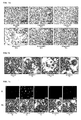
- adipocytes were stained with 5 ⁇ g / ml PI and analyzed under CLSM.
- the fluorescence images of the PI-stained cells are shown.
- phosphatidylcholine-treated cells show no morphological differences to the untreated control. Consequently, phosphatidylcholine has no cell damaging effect. Comparable concentrations (20 mg / ml) of Na-deoxycholate caused a marked destruction of the cell membrane, which did not occur at the concentrations of phosphatidylcholine used.
- That phosphatidylcholine has no cell damaging effect was confirmed by the PI staining of the treated cells.
- differentiated 3T3-L1 were treated with 5 mg / ml, 10 mg / ml and 15 mg / ml phosphatidylcholine for 4 hours.
- 5 ⁇ g / ml PI was added to the medium for 5 minutes and the cells were analyzed under CLSM ( Fig. 1c ).
- Fig. 1c (a) ethanol was added so that the same concentration was achieved as in the solutions for the treatment of the cells.
- Fig. 1c (a) shows that no cells are stained with PI. It follows that ethanol in the concentration used has no cell damaging effect.
- Fig. 1c (b) the 1% Triton positive control shows numerous PI red stained cells. This indicates damage to the cell membrane. Here the membrane stability and integrity of the cell is damaged.
- Example 4 Obtaining Adipose-Derived Stem Cells (ADSC) from human subcutaneous adipose tissue.
- ADSC Adipose-Derived Stem Cells
- the human subcutaneous adipose tissue can serve as a potential source of adult stem cells.
- ADSC Adipose-Derived Stem Cells
- These so-called “Adipose-Derived Stem Cells” (ADSC) are multipotent and can be differentiated by appropriate stimuli to different cell types, eg osteoblasts, chondrocytes, adipocytes.
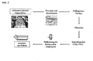
- the isolation of ADSCs from human subcutaneous adipose tissue, which is removed in plastic reductive procedures, is in Figure 2 shown schematically.
- the fat was washed with buffer to remove hematopoietic cells and then minced.
- the resulting adipose tissue pieces were digested with collagenase.
- the stromal-vascular fraction was separated and the floating, mature adipocytes were discarded.
- the stromal-vascular fraction in the pellet is composed of a heterogeneous cell population of blood cells, fibroblasts, pericytes, endothelial cells and preadipocytes.
- This cell population was transferred to a culture flask with media, with a portion of the cells adhering.
- a suitable cocktail consisting of insulin, dexamethasone, indomethacin and isobutylmethylxanthine or consisting of insulin, cortisol, troglitazone, triiodothyronine and isobutylmethylxanthine
- the cells were stimulated for adipogenesis and an improved adipogenic differentiation is effected.
- Na-deoxycholate Na-DC
- the ADSCs of Example 4 were stimulated for adipogenesis by a hormone cocktail (insulin, cortisol, troglitazone, triiodothyronine and isobutylmethylxanthine) and then differentiated into mature adipocytes for a further 21 days.
- a hormone cocktail insulin, cortisol, troglitazone, triiodothyronine and isobutylmethylxanthine
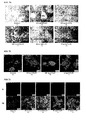
- ADSCs Further analysis of a cell damaging effect of Na-deoxycholate on ADSCs was made by PI staining of the treated cells followed by analysis under CLSM.
- the differentiated, mature adipocytes were treated with: 0.05 mg / ml, 0.1 mg / ml and 0.5 mg / ml Na-deoxycholate for 4 hours.
- untreated ADSCs were used as a control.
- adipocytes were stained with 5 ⁇ g / ml PI and analyzed under CLSM.
- the fluorescence images of the PI-stained cells are shown.
- a very low Na deoxycholate concentration of 0.01 mg / ml did not affect the cells ( FIG. 3a (B)). There were no differences to the control group ( FIG. 3a (a)).
- the adipocytes were vital and had an intact cell membrane. Increasing the concentration from 0.05 mg / ml already caused slight damage to the cell membrane ( Fig. 3a (C)). This was reinforced at the next higher concentrations ( Fig. 3a (D, e)).
- the highest dose of 0.5 mg / ml exerted a marked toxic effect on the cells ( Fig. 3a (F)).
- the adipocytes were dead, the cell membranes completely destroyed, so that essentially only free lipid droplets and cell fragments were present.
- PC Phosphatidylcholine
- the incubation period was 4 hours.
- adipocytes were stained with 5 ⁇ g / ml PI and analyzed under CLSM.
- the fluorescence images of the PI-stained cells are shown.
- PI staining of phosphatidylcholine-treated ADSCs confirmed that phosphatidylcholine has no cell damaging effects in vitro .
- the murine pre-adipocyte cell line 3T3-L1 was used for the investigations.
- 3T3-L1 cells were stimulated by a hormone cocktail (corticosterone, isobutylmethylxanthine, indomethacin, insulin) for adipogenesis and differentiated into mature adipocytes for another 8 days.
- hormone cocktail corticosterone, isobutylmethylxanthine, indomethacin, insulin
- the mature adipocytes of 3T3-L1 cells were treated with various concentrations of phosphatidylcholine between 0.1 mg / ml and 50 mg / ml.
- the phosphatidylcholine was combined with 2-20 mg / ml glycyrrhizinate and 18-180 mg / ml maltose.
- the incubation of the mature 3T3-L1 cells was carried out for a period of 4 hours with the substance combination.
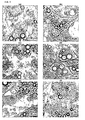
- the treated cells were examined at a 63-fold magnification by light microscopy for a membrane destabilizing and cytotoxic effect.
- the adipocytes treated with the substance combination did not differ from the control cells (control Fig. 5a ).
- Light microscopy showed the same morphology as the cells of the control, were vital and had an intact cell membrane.
- the substance combination exerted no visible membrane-damaging effects on the cell regardless of the concentration ( Fig. 5 ).
- the incubation period was 4 hours. Subsequently, the treated cells and two controls were incubated with 5 ⁇ g / ml PI for 5 minutes. The evaluation was carried out by means of the CLSM ( Fig. 6 ).
- the with 50 mg / ml PC ( Fig. 6e ) treated cells showed a weak red coloration of the cells ( Fig. 6e ). These cells were treated in high doses with the substance combination. As a result, the cells were completely deprived of the medium and a lack of nutrients resulted, which leads to cell damage after prolonged incubation.
- the weak staining of the cells treated with 50 mg / ml PC can thus be attributed to the lack of nutrients and not to a damage of the cells by phosphatidylcholine.
- in vivo / in vitro correlation The relationship between in vivo and in vitro effects is referred to as in vivo / in vitro correlation.
- the determination of in vivo / in vitro correlation is not trivial and behaves differently under different conditions. In many cases, an approximate in vivo / in vitro correlation by a factor of 100 can be assumed. This means that concentrations used in vitro are approximately two orders of magnitude below the doses used in vivo to achieve comparable effects. On this basis, one would expect for example Lipostabil ® N for injection lipolysis listed in Table 1 in vitro concentrations.
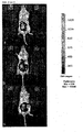
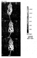
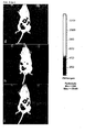
- Example 7 In vivo studies on the inflammatory response of the treated cells to the mouse
- Balb / c mice were either blended into the right subcutaneous fat of Balb / c mice ( Fig. 8d-f containing phosphatidylcholine (PC), glycyrrhizic acid (GR) and maltose (MAL) (25 mg / ml PC, 10 mg / ml GR, 90 mg / ml MAL) PBS buffer ( Fig. 8g-l ) or E. coli cells ( Fig. 8a-c ). The incubation was for 5 days.
- PC phosphatidylcholine
- GR glycyrrhizic acid
- MAL maltose
- ADSC fat-derived stem cells
- luciferase-labeled ADSC cells were injected intraperitoneally and the migration of ASC cells was monitored by bioluminescence.
- the ASC cells migrated to the liver and spleen.
- the ASC cells migrated to and accumulated in the E.coli injected region.
- mice with PBS Fig. 8g-l
- mice with the mixture containing PC Fig. 8d-f
- no difference in migration of ADSC cells was observed.
- mice injected with E. coli migration and accumulation of the ADSC cells in the inflamed region triggered by the E. coli injection were observed.
- ADSC cells luciferase-labeled liposome-derived stem cells
- PMC phosphatidylcholine
- maltose does not elicit an inflammatory response.
- 3T3-L1 cells were stimulated by a hormone cocktail for adipogenesis and differentiated into mature adipocytes for another 8 days.
- the lipolytic activity was determined by means of a lipolysis assay as described below:
- the first bar in Figure 7 shows that the basal lipolysis activity of untreated cells in 3% BSA / PBS is 8 ⁇ g glycerol / ⁇ g DNA. Starting from this basal level, the lipolytic activity of the further samples was determined.
- the positive control ( Fig. 7 ) stimulation of lipolysis was induced by 10 ⁇ M of the ⁇ -adrenoreceptor agonist isoproterenol.
- the positive control shows a lipolytic activity that increases to eight times the basal level. All cells treated with the substance combination (10 mg / ml, 25 mg / ml and 50 mg / ml of PC) showed an increased lipolytic activity compared to the basal level ( Fig. 7 ).
- Treatment of the cells with the substance combination containing 10 or 25 mg / ml PC caused an increase in the lipolytic activity by 5-fold compared to the basal level.
- Treatment of the cells with the substance combination containing 50 mg / ml PC resulted in a 3-fold increase in lipolysis activity compared to the basal level ( Fig. 7 ).
- the decrease in lipolytic activity at the highest concentration is likely, as explained above, due to the lack of nutrient deprivation of the cells required for survival or maintenance of normal functions such as lipolytic activity.
- the substance combination according to the invention shows in vitro a pronounced lipolytic effect.
- Na-deoxycholate concentrations of 0.005 - 0.5 mg / ml (effect as a single substance from 0.05 mg / ml) and phosphatidylcholine concentrations of 0.01 - 1 mg / ml (no effect as a single substance) used.
- the analysis is carried out by means of the light microscope. Cells with damaged membrane or cell fragments are exemplified by black arrows, while free lipid droplets are marked by white arrows.
- Example 9 Application of phosphogliv ® and lipostable ® in female subjects and determination of efficacy by subcutaneous lipolysis
- the ampoules used contained 0.5 g of PPC, 0.2 g of glycyrrhizinate and 1.8 g of maltose as lyophilisate, to be dissolved in 10 ml of water per injection. Depending on the extent of the area to be injected, up to 60 subcutaneous punctures were made on each upper arm, always 0.5 ml at a distance of 1.5 cm.
- the circumference of the upper arm [cm] was measured by caliper and tape measure (Myo-tape) before treatment and after eight and 16 weeks.
- the blood lipids total cholesterol, LDL cholesterol and HDL cholesterol in mg / dl and the atherogenic index from LDL to HDL cholesterol were determined.
- Example 9 For this purpose, the subjects were treated as described in Example 9 and examined the effectiveness. In order to document the side effects, photographs were taken before and after the treatment with Phosphogliv ® and Lipostabil ® of the upper arms of the test persons ( Fig. 10 and 11 ).
- Fig. 10 Photographic documentation Having considered the left (Phosphogliv ®) and right (Lipostabil ®) upper arm of the subject no 01. The pictures were taken 3 minutes after the application of the preparations Lipostabil ® and Phosphogliv ®..
- Fig. 11 Photographic documentation after treatment of the left (Phosphogliv ®) and right (Lipostabil ®) upper arm of the subject no 02. The recordings were made 3 minutes after the application of the preparations Lipostabil ® and Phosphogliv ®
- Example 11 Determination of cholesterol levels before and after subcutaneous lipolysis
- Drl / DrlP Total cholesterol from 237.0 mg / dl to 222.0 mg / dl LDL cholesterol from 136.0 mg / dl to 129.0 mg / dl HDL cholesterol from 75.4 mg / dl to 78.0 mg / dl 16 2.0 2.0 28 28.5
- Drl / DrlP Total cholesterol from 222 mg / dl to 218 mg / dl LDL cholesterol from 129.0 mg / dl to 128.0 mg / dl HDL cholesterol from 78.0 mg / dl to 79.0 mg / dl Left almost no pain, right for about 1 week skin firmness from the beginning to good 3 M.
Description
Die Erfindung betrifft die Verwendung einer Zusammensetzung auf der Grundlage von Phospholipiden und Glycyrrhizinsäure oder einem Salz der Glycyrrhizinsäure, und deren in der Medizin anwendbaren Formen, zur Behandlung von adipösen Einlagerungen unter der Haut verschiedenster Erscheinungsformen, wie z.B. subkutane Fettverteilungsstörungen, und zur Regression von diätresistenten Fettpolstern.The invention relates to the use of a composition based on phospholipids and glycyrrhizic acid or a salt of glycyrrhizic acid, and their forms which can be used in medicine, for the treatment of adipose deposits under the skin of various forms, such as e.g. subcutaneous fat distribution disorders, and for the regression of dietary fat deposits.
Im Stand der Technik werden bisher chirurgische Methoden verwendet, um subkutane Fettansammlungen oder Wucherungen der Fettzellen wie Lipome oder Lipödeme zu behandeln. Derartige Behandlungsmaßnahmen führen zu den bekannten Komplikationen oder Risiken, verursacht durch Narkose, lokale Reaktionen und mögliche Infektionen. Stationäre Klinik-Aufenthalte sind unter solchen Umständen häufig nicht zu vermeiden. Um solche Eingriffe zu vermeiden, wird nach nicht-operativen Alternativen zur Entfernung von subkutanen Fettansammlungen gesucht.Surgical methods have hitherto been used in the art to treat subcutaneous fat accumulations or growths of fat cells such as lipomas or lipoedema. Such treatments lead to the known complications or risks caused by anesthesia, local reactions and possible infections. Inpatient hospital stays are often unavoidable in such circumstances. In order to avoid such interventions, non-operative alternatives are sought to remove subcutaneous fat accumulations.
Es wurden bereits verschiedene phospholipidhaltige Präparate entwickelt, die als Injektion an Patienten verabreicht werden. Wässrige Zubereitungen, enthaltend mindestens ein Phospholipid, sind für verschiedene Anwendungen bekannt. Diese Systeme werden beispielsweise im kosmetischen Bereich oder für die Herstellung von pharmazeutischen Produkten eingesetzt. Häufig bilden diese Systeme Mizellen oder Liposome aus, die in ihrem Inneren eine wässrige Phase enthalten.Various phospholipid-containing preparations have already been developed, which are administered as an injection to patients. Aqueous preparations containing at least one phospholipid are known for various applications. These systems are used, for example, in the cosmetic sector or for the production of pharmaceutical products. Frequently, these systems form micelles or liposomes that contain an aqueous phase in their interior.
Die
Essentiale® N i.V. ist in der
In der
Lipostabil® N i.V. enthält Phospholipide aus der Sojabohne, DL-alpha-Tocopherol, 7-Desoxycholsäure, Alkohole, weitere Hilfsstoffe und Wasser. Allerdings enthält die Wirkstoffzusammensetzung dieses Präparates keine Glycyrrhizinsäure. Durch die subkutane Injektion von Lipostabil® N i.V. werden Fetteinlagerungen, wie sie bei übergewichtigen Menschen unter den Augen, am Bauch oder an Hüften auftreten, entfernt.Lipostabil ® N iV contains soybean phospholipids, DL-alpha-tocopherol, 7-deoxycholic acid, alcohols, other excipients and water. However, the active ingredient composition of this preparation does not contain glycyrrhizic acid. Subcutaneous injection of Lipostabil ® N iV removes fat deposits such as those found in overweight people under the eyes, abdomen or hips.
Es gibt noch weitere Zubereitungen, die auf einem Phospholipid basieren und zur subkutanen Injektion zwecks Lipolyse der Fettansammlungen verwendet werden, welche in der
Nachteile der Verwendung dieser Präparate des Standes der Technik für eine subkutane Lipolyse sind unter anderem Schwellungen, Hämatome (Blutergüsse), Schmerzen in der Behandlungsregion und Missempfinden wie ein brennendes Gefühl und Juckreiz an der Injektionsstelle nach der Behandlung, insbesondere aber ein Absterben von Zellen durch Schädigung der Zellstruktur und der Membranintegrität (Zellnekrose).Disadvantages of using these prior art preparations for subcutaneous lipolysis include swelling, hematomas (bruises), pain in the treatment area, and discomfort such as burning sensation and injection site itch after treatment, but especially cell death by injury cell structure and membrane integrity (cell necrosis).
In dem Bestreben, wirksame Verbindungen zur nicht-operativen Entfernung von subkutanen Fettansammlungen ohne die oben genannten Nachteile zu finden, wurde nun überraschenderweise gefunden, dass die Zusammensetzung, welche in der
Bei der erfindungsgemäßen Verwendung dieser Zusammensetzung, die mindestens ein Phospholipid, bevorzugt ein Phosphatidylcholin, und Glycyrrhizinsäure oder deren Salz enthält, werden die zuvor beschriebenen Nachteile der Präparate des Standes der Technik, die für eine subkutane Lipolyse verwendet werden, vermindert und/oder gänzlich behoben.In the use according to the invention of this composition which comprises at least one phospholipid, preferably a phosphatidylcholine, and glycyrrhizic acid or its salt, those described above are Disadvantages of the prior art preparations used for subcutaneous lipolysis are reduced and / or eliminated altogether.
Der besondere Vorteil der erfindungsgemäßen Verwendung der beanspruchten Zusammensetzung zur subkutanen Lipolyse von Fettansammlungen ist die deutliche Reduzierung bis zur vollständigen Verhinderung der Schädigung der Zellstruktur sowie der Membranintegrität der adipösen Gewebezellen (Adipozyten). Bei der erfindungsgemäßen Behandlung von subkutanen Fettgewebsstörungen mit der beanspruchten Zusammensetzung wird das Auftreten von Nekrose deutlich vermindert oder gänzlich verhindert.The particular advantage of the use according to the invention of the claimed composition for the subcutaneous lipolysis of fat accumulations is the clear reduction to the complete prevention of the damage to the cell structure and the membrane integrity of the adipose tissue cells (adipocytes). In the treatment according to the invention of subcutaneous adipose tissue disorders with the claimed composition, the occurrence of necrosis is significantly reduced or completely prevented.
Durch die erfindungsgemäße Verwendung kommt es zum Abbau von Depotfett im Körper des behandelten Fettgewebes durch Lipolyse, ohne dass eine Destruktion der Zellmembran eintritt.The use according to the invention leads to the degradation of depot fat in the body of the treated adipose tissue by lipolysis, without destruction of the cell membrane occurring.
Es werden deutlich geringere Nebenwirkungen bis zu einem vollständigen Ausbleiben von Beschwerden wie Schwellungen, Rötungen, Brennen, Juckreiz und allgemeine Schmerzen und Überempfindlichkeit beobachtet.Significantly lower side effects are observed until complete absence of symptoms such as swelling, redness, burning sensation, itching and generalized pain and hypersensitivity.
Die erfindungsgemäß verwendete Zusammensetzung beinhaltet als Hauptbestandteile ein Phospholipid, Glycyrrhizinsäure oder deren Salz und gegebenenfalls mindestens einen Hilfsstoff.The composition used according to the invention comprises as main constituents a phospholipid, glycyrrhizic acid or its salt and optionally at least one adjuvant.
Glycyrrhizinsäure, die aus einem Extrakt von Pflanzen der Gattung Glycyrrhiza, den Süßhölzern (z.B. Glycyrrhiza glabra) gewonnen werden kann, oder ihre Salze, insbesondere Natrium- oder Ammoniumglycyrrhizinat, werden im Stand der Technik als Absorptionsverstärker für den Transport von z.B. Peptidhormonen über die Schleimhautmembran in der
Darüber hinaus ist die Glycyrrhizinsäure als antibakterieller und entzündungshemmender Zusatzstoff und Emulgator im Stand der Technik aus der
Die besondere Funktion der Glycyrrhizinsäure beim Transport von Molekülen über die Zellmembran unterstützt den besonderen Vorteil der schnellen Wirkungsweise der erfindungsgemäßen Verwendung der beanspruchten Zusammensetzung. Durch die Glycyrrhizinsäure kann das Phospholipid besonders gut und schnell in die adipösen Gewebszellen der Subkutis gelangen, wodurch die zuvor beschriebenen Nachteile wie Nekrose vermindert werden oder gar nicht auftreten. Die beschriebenen Schwellungen, Hämatome, Schmerzen und Missempfindungen nach einer Behandlung mit einem Präparat des Standes der Technik, insbesondere solcher, die Gallensäuren oder Salze dieser enthalten, werden durch die Kombination mit der Glycyrrhizinsäure gelindert oder treten gar nicht auf. Diese verbesserte Verträglichkeit wird durch die entzündungshemmende und antibakterielle Wirkung der Glycyrrhizinsäure unterstützt.The particular function of glycyrrhizic acid in the transport of molecules across the cell membrane supports the particular advantage of the rapid mode of action of the inventive use of the claimed composition. By Glycyrrhizinsäure the phospholipid pass particularly well and quickly into the adipose tissue cells of the subcutis, whereby the disadvantages described above such as necrosis are reduced or do not occur. The described swellings, hematomas, pain and discomfort after treatment with a preparation of the prior art, in particular those containing bile acids or salts thereof, are alleviated or not even occur by the combination with the glycyrrhizic acid. This improved compatibility is supported by the anti-inflammatory and antibacterial effect of glycyrrhizic acid.
Folglich zeichnet sich die vorliegende Erfindung dadurch aus, dass die beanspruchte Kombination aus mindestens einem Phospholipid, bevorzugt einem Phosphatidylcholin und Glycyrrhizinsäure oder ihres Salzes im Vergleich zu den Zusammensetzungen der Präparate Essentiale® und Lipostabil® aus dem Stand der Technik wesentlich geringere bis keine zellschädigende Wirkung und eine verbesserte Verträglichkeit hat.Consequently, the present invention is characterized in that the claimed combination of at least one phospholipid, preferably a phosphatidylcholine and Glycyrrhizinsäure or its salt compared to the compositions of the preparations Essentiale ® and Lipostabil ® from the prior art much lower to no cell damaging effect and has an improved compatibility.
Die Erfindung betrifft daher die Verwendung einer Zusammensetzung, enthaltend
- a) mindestens ein Phospholipid;
- b) Glycyrrhizinsäure, oder
ein Salz der Glycyrrhizinsäure und - c) ggf. Hilfsstoffe
wobei- der Gesamtgehalt an den Phospholipiden und der Glycyrrhizinsäure oder deren Salzen 2-80 Gewichtsprozent aufweist, und
- das Gewichtsverhältnis zwischen Phospholipiden und der Glycyrrhizinsäure oder deren Salze von 30:1 bis 0,5:1 beträgt,
zur Herstellung eines Arzneimittels zur therapeutischen Behandlung von subkutanen Fettgewebserkrankungen.
- a) at least one phospholipid;
- b) glycyrrhizic acid, or
a salt of glycyrrhizic acid and - c) optionally adjuvants
in which- the total content of the phospholipids and the glycyrrhizic acid or salts thereof is 2-80% by weight, and
- the weight ratio between phospholipids and the glycyrrhizic acid or salts thereof is from 30: 1 to 0.5: 1,
for the manufacture of a medicament for the therapeutic treatment of subcutaneous fatty tissue diseases.
Die Erfindung betrifft ferner die Verwendung zur Zersetzung und Rückbildung von Fettgewebsgeschwülsten.The invention further relates to the use for the decomposition and regression of fatty tissue tumors.
Die Erfindung betrifft ferner die Verwendung zur Behandlung von subkutanen Fettgewebserkrankungen wie Lipödeme, Lipome, Morbus Dercum, Madelung'schen Fetthals oder Lipomatosis der Bauchdecke.The invention further relates to the use for the treatment of subcutaneous fatty tissue diseases such as lipedema, lipomas, Dercum's disease, Madelung's glandular fat or lipomatosis of the abdominal wall.
Die Erfindung betrifft ferner die nicht-therapeutische Verwendung einer Zusammensetzung, enthaltend
- a) mindestens ein Phospholipid;
- b) Glycyrrhizinsäure, oder
ein Salz der Glycyrrhizinsäure und - c) ggf. Hilfsstoffe
wobei- der Gesamtgehalt an den Phospholipiden und der Glycyrrhizinsäure oder deren Salzen 2-80 Gewichtsprozent aufweist, und
- das Gewichtsverhältnis zwischen Phospholipiden und der Glycyrrhizinsäure oder deren Salze von 30:1
bis 0,5:1 beträgt,
zur Behandlung von subkutanen Fettansammlungen.
- a) at least one phospholipid;
- b) glycyrrhizic acid, or
a salt of glycyrrhizic acid and - c) optionally adjuvants
in which- the total content of the phospholipids and the glycyrrhizic acid or salts thereof is 2-80% by weight, and
- the weight ratio between phospholipids and the glycyrrhizic acid or salts thereof is from 30: 1 to 0.5: 1,
for the treatment of subcutaneous fat accumulation.
Die Erfindung betrifft ferner die nicht-therapeutische Verwendung zur Entfernung von subkutanen Fettansammlungen, insbesondere mit lokaler Störung der Fettverteilung.The invention further relates to the non-therapeutic use for the removal of subcutaneous fat accumulations, in particular with local disturbance of the fat distribution.
Die Erfindung betrifft ferner die nicht-therapeutische Verwendung zur Behandlung von Dermatopanniculosis deformans, Pseudogynäkomastie, Buffalo Hump bei HIV Patienten, Cellulite, Xanthelasmen oder unspezifische subkutane Fettdepots.The invention further relates to the non-therapeutic use for the treatment of Dermatopanniculosis deformans, pseudogynaecomastia, buffalo hump in HIV patients, cellulite, xanthelasma or nonspecific subcutaneous fat deposits.
Die Erfindung betrifft ferner die Verwendung der obigen Zusammensetzung welche Phosphatidylcholin als Phospholipid enthält.The invention further relates to the use of the above composition containing phosphatidylcholine as a phospholipid.
Die Erfindung betrifft ferner die Verwendung, wobei die Zusammensetzung Phosphatidylcholin tierischen oder pflanzlichen Ursprungs enthält.The invention further relates to the use, wherein the composition contains phosphatidylcholine of animal or vegetable origin.
Die Erfindung betrifft ferner die Verwendung wobei die Zusammensetzung Glycyrrhizinsäure oder Kalium-, Natrium-, Ammonium- oder Magnesiumsalze der Glycyrrhizinsäure enthält.The invention further relates to the use wherein the composition contains glycyrrhizic acid or potassium, sodium, ammonium or magnesium salts of glycyrrhizic acid.
Die Erfindung betrifft ferner die Verwendung wobei die Zusammensetzung einen Zucker, insbesondere Glukose und Maltose und/oder ihre Derivate, Mannit, Sorbit oder Milchzucker als Hilfsstoff enthält.The invention further relates to the use wherein the composition contains a sugar, in particular glucose and maltose and / or their derivatives, mannitol, sorbitol or lactose as adjuvant.
Die Erfindung betrifft ferner die Verwendung wobei das Phospholipid 15 bis 98 Gew.-%, bevorzugt, 30 bis 98 Gew.-%, mehr bevorzugt 50 bis 98 Gew.-%, besonders bevorzugt 75 bis 98 Gew.-%, am meisten bevorzugt 75 bis 90 Gew.-% Phosphatidylcholin enthält.The invention further relates to the use wherein the
Die Erfindung betrifft ferner die Verwendung wobei die Zusammensetzung in trockener Form in einem geeigneten Lösungsmittel gelöst wird.The invention further relates to the use wherein the composition is dissolved in dry form in a suitable solvent.
Die Erfindung betrifft ferner die Verwendung wobei die Zusammensetzung in trockener Form bevorzugt als Lyophilisat, erzielt durch Gefriertrocknung, eingesetzt wird.The invention further relates to the use wherein the composition in dry form is preferably used as lyophilisate obtained by freeze-drying.
Die Erfindung betrifft ferner die Verwendung wobei die Zusammensetzung in Form von einer Lösung eingesetzt wird.The invention further relates to the use wherein the composition is used in the form of a solution.
Die Erfindung betrifft ferner die Verwendung wobei die Zusammensetzung physiologisch geeignete Lösungsmittel umfassend Wasser, physiologische Kochsalzlösung, Glucose, ein Monohydroxy-Alkohol wie Ethanol, 2-Propanol, n-Propanol, Polyhydroxy-Alkohole wie Glyzerol und/oder Propandiol, Polyglykol wie Polyethylenglykol und/oder Miglyol, Glycerinformal, Dimethylisosorbitol, natürliche und synthetische Öle und/oder Äther enthält.The invention further relates to the use of the composition physiologically suitable solvents comprising water, physiological saline, glucose, a monohydroxy alcohol such as ethanol, 2-propanol, n-propanol, polyhydroxy alcohols such as glycerol and / or propanediol, polyglycol such as polyethylene glycol and / or Miglyol, glycerol formal, dimethylisosorbitol, natural and synthetic oils and / or ether.
Die Erfindung betrifft ferner die Verwendung wobei die Applikation der Zusammensetzung durch subkutane, intraperitoneale, intramuskuläre oder intravenöse Injektion erfolgt.The invention further relates to the use wherein the administration of the composition by subcutaneous, intraperitoneal, intramuscular or intravenous injection.
Die Erfindung betrifft ferner die Verwendung wobei zur Applikation der Zusammensetzung ein Verfahren ausgewählt aus der Gruppe umfassend lontophorese, Elektroporation, Microporation oder Phonophorese eingesetzt wird.The invention further relates to the use of a method selected from the group comprising iontophoresis, electroporation, microporation or phonophoresis is used for applying the composition.
Die Applikation der erfindungsgemäßen Zubereitung erfolgt in der Form von Cremes, Salben, Gelen, Hydrogelen, Lotionen, Pasten, Lyophilisat und Lösungen. Bevorzugt ist die wässrige Zubereitung in Form von verschiedenen Lösungen.The application of the preparation according to the invention takes place in the form of creams, ointments, gels, hydrogels, lotions, pastes, lyophilisate and solutions. Preferably, the aqueous preparation is in the form of various solutions.
Durch die erfindungsgemäße Verwendung der Zusammensetzung können die oben genannten Risiken und Nebenwirkungen einer operativen Behandlung oder einer subkutanen Behandlung mit einem Präparat aus dem Stand der Technik, insbesondere solcher enthaltend Deoxycholsäure und ihre Salze, umgangen werden. Zudem ist die ambulante Behandlung für den Patienten angenehmer und auch kostengünstiger im Vergleich zur operativen Behandlung.By the use of the composition according to the invention, the above-mentioned risks and side effects of surgical treatment or subcutaneous treatment with a preparation of the prior art, in particular those containing deoxycholic acid and its salts, can be circumvented become. In addition, the outpatient treatment is more comfortable for the patient and also more cost-effective compared to the surgical treatment.
Die oben beschriebene hohe therapeutische Effizienz, die sich durch den schnellen Abbau des Fettgewebes der Zusammensetzung zeigt, ist mit dem Synergieeffekt des Zusammenspiels der erfindungsgemäßen Kombination aus einem Phospholipid und/oder einer Glycyrrhizinsäure oder eines ihrer Salze verbunden. Die erfindungsgemäße Verwendung der hier beanspruchten Zusammensetzung zeichnet sich durch deutlich abgeschwächte Nebenwirkungen oder zum Teil durch Ausbleiben einiger der zuvor beschriebenen Nebenwirkungen aus.The high therapeutic efficiency described above, which is manifested by the rapid degradation of the fatty tissue of the composition, is associated with the synergistic effect of the combination of the combination of a phospholipid and / or a glycyrrhizic acid or one of its salts according to the invention. The use according to the invention of the composition claimed here is characterized by markedly weakened side effects or, in part, by the absence of some of the side effects described above.
Subkutane Fettverteilungsstörungen sind Abweichungen im Fettgewebe im Körper von Menschen und Säugetieren, die als genetisch oder ernährungsbedingtes Depotfett in Form lokalisierter Fettpolster auftreten und als ästhetisch störende kritische Zonen, insbesondere Bauch, Gesäß, Hüften, Knie, Waden, Oberschenkel, Oberarme, Kinn, Wangen angesehen werden können. Es kann sich dabei auch um gutartige Wucherungen der Fettzellen wie Lipome (dystopische Proliferation) handeln.Subcutaneous fat distribution disorders are variations in adipose tissue in the body of humans and mammals, which occur as genetically or nutritionally dependent depot fat in the form of localized fat deposits and are considered as aesthetically disturbing critical zones, especially the abdomen, buttocks, hips, knees, calves, thighs, upper arms, chin, cheeks can be. It may also be benign growths of fat cells such as lipomas (dystopic proliferation).
Unter Fettgewebserkrankungen im Sinne der Erfindung werden beispielsweise folgende Erkrankungen verstanden: Lipome (Fettgewebsgeschwülste), dies sind gutartige, langsam wachsende, meist kugelige, eventuell gestielte (= L. pendulum) oder gar zottige (= L. arborescens, beispielsweise der Gelenkzotten) mesenchymale Geschwülste aus - vergrößerten - Fettgewebszellen, bevorzugt im Unterhautzellgewebe, eventuell zentral verknöchernd (= L. ossificans), verschleimend (= L. myxomatodes) oder verkalkend (= L. petrificans), auch mit vermehrter Bindegewebs- und Kapselbildung (= L. fibrosum), Blutgefässneubildung (= L. teleangiectodes), selten maligne entartend (= L. sarcomatodes, Liposarkom). Sie sind als krankhaft einzustufen, da sie wachsen und ihre bindegewebige Hülle an sich schmerzhaft sein kann, ebenso wie die von ihnen ausgehende Kompression auf Blutgefäße, die Nervenschmerzen verursachen kann. Hierunter fällt auch die multiple Lipomatosis, die zu einer gehäuften Ansammlung von Lipomen beim Patienten führt.For the purposes of the invention, the term "adipose tissue disorders" is understood to mean, for example, lipomas (adipose tissue tumors), which are benign, slowly growing, usually spherical, possibly pedunculated (= L. pendulum) or even shaggy (= L. arborescens, for example the joint villi) mesenchymal tumors from - enlarged - adipose tissue cells, preferably in the Unterhautzellgewebe, possibly central ossifying (= L. ossificans), verschleimend (= L. myxomatodes) or calcifying (= L. petrificans), with increased connective tissue and capsule formation (= L. fibrosum), Blood vessel neoplasm (= L. teleangiectodes), rarely malignant degenerating (= L. sarcomatodes, liposarcoma). They are classified as pathological, as they grow and their connective tissue envelope can be painful in itself, as well as the compression of them on blood vessels, which can cause nerve pain. This includes the multiple lipomatosis, which leads to a frequent accumulation of lipomas in the patient.
Morbus Dercum, genannt Lipomatosis dolorosa ist eine Sonderform der hypertrophen Proliferation von Fettgewebe, welches sich zwischen der dermalen Fettfaszie (Kampa'sche Fettfaszie) und der Unterseite der Dermis befindet. Durch hormonelle Einflüsse kommt es zur verstärkten Wasserbindungskapazität dieser Fettzellen, die selbst wiederum durch Druckphänomene Lymphbahnstauungen im Bereich der initialen farnkrautartigen Lymphgefäße herbeiführen, wodurch zusätzliche Kompressions- und Irritationseinflüsse auf die peripheren sensiblen Nerven ausgeübt werden, so dass diese Patienten eine extrem schmerzhafte Berührungsempfindlichkeit aufweisen. Im Verlaufe von mehreren Jahren bis Jahrzehnten bilden sich unregelmäßige, unter der während des Alterungsprozesses dünner werdenden Dermis disseminiert lokalisierte Fettknötchen, welche teils schmerzhaften und stark dysästhetischen Charakter haben.Dercum's disease, called Lipomatosis dolorosa, is a special form of hypertrophic proliferation of adipose tissue located between the dermal fat fascia (Kampa's fatty fascia) and the underside of the dermis. Hormonal influences increase the water-binding capacity of these fat cells, which in turn induce lymphatic pathway congestion in the area of the initial fern-weed-like lymphatics through pressure phenomena additional compression and irritation effects are exerted on the peripheral sensory nerves, so that these patients have an extremely painful touch sensitivity. Over the course of several years to decades, irregular, under the dermis thinning during the aging process disseminated localized fat nodules, which sometimes have a painful and strongly dysaesthetic character.
Der Madelung'sche Fetthals (Lanois-Bensaude-Syndrom) ist eine fettgewebsproliferierende Fettgewebsentzündung, bei der es neben einer dystrophen Fettgewebstumorausbildung auch zu einer narbenartigen Bindegewebsverdichtung im Subkutanraum kommt. Hierbei können operative Vorgehensweisen oft nur Teilerfolge erzielen, da essentielle anatomische Strukturen in diesem Prozess mit eingeschlossen sind, und die Erkrankung sich im wesentlichen im Kopf-, Hals- und Schulterbereich manifestiert.Madelung's Fetthals (Lanois Bensaude syndrome) is a fatty tissue-proliferating adipose tissue inflammation, which in addition to a dystrophic formation of adipose tissue tumors also leads to a scar-like connective tissue compression in the hypodermic space. Surgical procedures can often only achieve partial success because essential anatomical structures are included in this process, and the disease manifests itself essentially in the head, neck and shoulder area.
Das Lipödem ist eine schmerzhafte Schwellung des Fettgewebes, die besonders an den Unterschenkeln von Frauen auftritt und mit zunehmendem Alter einen fortschreitenden Verlauf bzw. Charakter aufweist.Lipoedema is a painful swelling of the adipose tissue, which occurs particularly on the lower legs of women and has a progressive course or character with increasing age.
Als Regression der Lipolyse wird also die hydrolytische Spaltung des Fettgewebes und Rückbildung durch Mobilisierung des proliferierten Fettbereichs verstanden.As a regression of lipolysis so the hydrolytic cleavage of adipose tissue and regression by mobilizing the proliferated fat area is understood.
Ein Xanthelasma ist eine gelbliche Ansammlung von Fett unter den Augen.A xanthelasma is a yellowish build-up of fat under the eyes.
HIV Patienten haben häufig Störungen von Fettgewebsansammlungen, die durch die Medikamentierung nach dem Stand der Technik auftreten, beispielsweise der Buffalo Hump genannte Stiernacken bei dieser Patientengruppe. Immunsystem geschwächten Patienten ist in der Regel eine operative Entfernung nicht zuzumuten, weshalb die Fettansammlungen verbleiben, und die Patienten äußerlich stigmatisieren.HIV patients often have disorders of adipose tissue accumulation that occur due to the prior art medication, for example the buffalo hump called "bullskin" in this group of patients. Immune system-weakened patients are usually not tolerated surgical removal, which is why the fat accumulation remains, and stigmatize the patient externally.
Die oben genannten Fettgewebserkrankungen zeigen im Gegensatz zu der ernährungsbedingten Lipohypertrophie (die auch eine Fett-Ablagerung im Sinne der Fettverteilungsstörung zur Folge hat) pathologisch eindeutig abzugrenzende Gewebszustände oder Entitäten, die durch histologische Vernarbungs- und Entzündungsparameter gekennzeichnet sind, aber auch durch Bindegewebsabkapselungen und durch Veränderungen in der histologischen Fettgewebsmorphologie selbst.In contrast to dietary lipohypertrophy (which also results in fat deposition in terms of fat distribution disorder), the above-mentioned adipose tissue disorders show pathologically distinct tissue states or entities characterized by histological scarring and inflammatory parameters, as well as connective tissue encapsulations and alterations in the histological adipose tissue morphology itself.
Ein weiterer Gegenstand der Erfindung ist die Verwendung der Zusammensetzung zur Herstellung eines Arzneimittels zur Behandlung von Cellulite/-is. Die Cellulitis ist eine Sonderform der hypertrophen Proliferation von Fettgewebe, welches sich zwischen der dermalen Fettfaszie (Kampa'sche Fettfaszie) und der Unterseite der Dermis befindet. Durch hormonelle Einflüsse kommt es zur verstärkten Wasserbindungskapazität dieser Fettzellen, die selbst wiederum durch Druckphänomene Lymphbahnstauungen im Bereich der initialen farnkrautartigen Lymphgefäßen herbeiführen. Im Verlaufe von mehreren Jahren bis Jahrzehnten bilden sich unregelmäßige, unter der während des Alterungsprozesses dünner werdenden Dermis disseminiert lokalisierte Fettknötchen, die teils schmerzhaften, teils dysästhetischen Charakter haben.Another object of the invention is the use of the composition for the preparation of a medicament for the treatment of cellulite / -is. Cellulitis is a special form of hypertrophic proliferation of adipose tissue, which is located between the dermal fat fascia (Kampa fat fascia) and the underside of the dermis. Hormonal influences increase the water-binding capacity of these fat cells, which in turn cause lymphatic blockage in the area of the initial fern-weed-like lymphatics by pressure phenomena. Over the course of several years to decades, irregular, under the dermis becoming thinner during the aging process disseminated localized fat nodules, some of which have a painful, sometimes dysaesthetic character.
Ein weiterer Gegenstand der Erfindung ist die Verwendung der Zusammensetzung zur Herstellung eines Arzneimittels zur Behandlung von Pseudogynäkomastien und Lipomastie. Pseudogynäkomastien sind Fettansammlungen bei Männern rund um die Mamma (Brust), die zur Vergrößerung der Brust führen und vor allem ästhetisch indiziert sind. Die Lipomastie ist eine Form der Pseudogynäkomastie ohne Vergrößerung des Brustdrüsenkörpers.Another object of the invention is the use of the composition for the preparation of a medicament for the treatment of pseudogynecomastia and lipomastia. Pseudo-gynecomastia are fat accumulations in males (breasts) around the breast, which increase the size of the breast and, above all, are aesthetically indicated. Lipomastia is a form of pseudogynacomastia without enlargement of the mammary gland body.
Die Phospholipide, die in der hier beschriebenen pharmazeutischen Komposition enthalten sind, werden aus beliebigen tierischen oder pflanzlichen Stoffen hergestellt, insbesondere aus Hühnereiern, ölhaltigen Samen und Früchten, wie getrocknete Kokosnuss, Palmensamen, Erdnuss, Rapssamen, Sonnenblumenkerne, Leinsamen, Palm- und/oder Olivenöl. Am besten eignet sich dazu aber das Phospholipid, das aus Sojabohnen nach den Verfahren, die in den Europäischen Patenten
Dieses Phospholipid wird hochgereinigt und enthält 15 bis 98 Gew.-%, bevorzugt 30 bis 98 Gew.-%, mehr bevorzugt 50 bis 98 Gew.-%, besonders bevorzugt 75 bis 98 Gew.-%, am meisten bevorzugt 75 bis 90 Gew.-% Phosphatidylcholin. Solche hochgereinigte Phospholipide können andere Komponenten der Phospholipide, insbesondere bis 15 Gew.-%, mehr bevorzugt bis 12 Gew.-% Phosphatidylethanolamin, bis 8 Gew.-% Phosphatidsäure, bis 10 Gew.-% Phosphatidylinositol, bis 6 Gew.-% Lysophosphatidylcholin oder Lysophosphatidylethanolamin, Spuren von Phosphatidylserin sowie andere Lipide in geringen Mengen enthalten.This phospholipid is highly purified and contains 15 to 98% by weight, preferably 30 to 98% by weight, more preferably 50 to 98% by weight, more preferably 75 to 98% by weight, most preferably 75 to 90% by weight % Phosphatidylcholine. Such highly purified phospholipids may contain other components of the phospholipids, in particular up to 15%, more preferably up to 12% phosphatidylethanolamine, up to 8% phosphatidic acid, up to 10% phosphatidylinositol, up to 6% lysophosphatidylcholine or lysophosphatidylethanolamine, traces of phosphatidylserine and other lipids in small amounts.
Die Erfindung betrifft auch den Einsatz der Glycyrrhizinsäure, worin die Glycyrrhizinsäure als physiologisch verträgliches Salz vorliegen kann. Als Salze der Glycyrrhizinsäure werden physiologische verträgliche Salze, insbesondere Mono-, Di- oder Trinatriumsalze bzw. Kaliumsalze, Magnesium- oder Ammoniumsalze verwendet. Bevorzugt werden Mono-, Di- oder Trinatriumsalze bzw. Kaliumsalze und Ammoniumsalze und besonders bevorzugt werden Mono-, Di- oder Trinatriumsalze bzw. Kaliumsalze.The invention also relates to the use of glycyrrhizic acid, wherein the glycyrrhizic acid as physiological acceptable salt may be present. As salts of Glycyrrhizinsäure physiologically acceptable salts, in particular mono-, di- or trisodium salts or potassium salts, magnesium or ammonium salts are used. Mono-, di- or trisodium salts or potassium salts and ammonium salts are preferred and mono-, di- or trisodium salts or potassium salts are particularly preferred.
Das Massenverhältnis von Phospholipid zur Glycyrrhizinsäure beträgt 30 : 1 bis zu 0,5 : 1, bevorzugt 15 : 1 bis zu 0,5 : 1, mehr bevorzugt 4 : 1 bis zu 1 : 1, besonders bevorzugt 3 : 1 bis zu 2 : 1.The mass ratio of phospholipid to glycyrrhizic acid is 30: 1 up to 0.5: 1, preferably 15: 1 up to 0.5: 1, more preferably 4: 1 up to 1: 1, particularly preferably 3: 1 up to 2: 1.
Die Phospholipid-Konzentration in der Zusammensetzung beträgt von 0,5 Gew.-% bis 30 Gew.-%, bevorzugt von 5 Gew.-% bis 25 Gew.-%.The phospholipid concentration in the composition is from 0.5% to 30% by weight, preferably from 5% to 25% by weight.
Als empfohlener Gesamtgehalt an den Phospholipiden und Glycyrrhizinsäure oder deren Salz gilt der Gehalt von 2-80 Gew.-%. Bei diesem wird ein Gewichtsverhältnis zwischen den Phospholipiden und Glycyrrhizinsäure oder deren Salz von 3 : 1 bzw. 4 : 1 bevorzugt. Beim Gewichtsverhältnis zwischen den Phospholipiden und Glycyrrhizinsäure oder deren Salz von 2 : 1 bis 3 : 1 gilt bevorzugt der Gehalt von 2-45 Gew.-% an den Phospholipiden und Glycyrrhizinsäure oder deren Salz.The recommended total content of the phospholipids and glycyrrhizic acid or its salt is the content of 2-80 wt .-%. In this, a weight ratio between the phospholipids and glycyrrhizic acid or its salt of 3: 1 or 4: 1 is preferred. The weight ratio between the phospholipids and glycyrrhizic acid or its salt of 2: 1 to 3: 1 is preferably the content of 2-45 wt .-% of the phospholipids and Glycyrrhizinsäure or their salt.
Der pH-Wert des Arzneimittels liegt im Bereich von pH 6,0 bis pH 9,0, vorzugsweise von pH 7,5 bis pH 8,5, besonders bevorzugt von pH 6,5 bis pH 7,5 und im speziellen pH 6,5 bis pH 7,0.The pH of the drug is in the range from pH 6.0 to pH 9.0, preferably from pH 7.5 to pH 8.5, more preferably from pH 6.5 to pH 7.5 and in
Gegebenenfalls werden geeignete Hilfsstoffe hinzugefügt. Als pharmakologisch zulässige Hilfsmittel dienen Zucker, insbesondere Maltose, Glukose, Milchzucker, Sorbit und Mannit, kolloidale Siliziurnsäure, Siliziumgel, Talkum, Laktose, Stärke, Gelatine, Wasser, Alkohole mit einer oder mehr Hydroxygruppen, insbesondere Ethanol, Glycerol und Propylenglycol, natürliche oder synthetische Öle insbesondere Petroleum, Mineralöl, Erdnussöl, Sojaöl, Sesamöl und Äther. Weitere geeignete Hilfsstoffe sind Cellulose, Sucrose, Malz, Reis, Fluor, Kalk, Magnesiumstearat, Natriumstearat, Glycerolmonostearat, NaCl und getrocknete Magermilch. Bevorzugte Hilfsstoffe vor allem für die subkutane Lipolyse umfassen Wasser, Alkohol, besonders Ethanol, Kochsalzlösung, Maltose und wässrige DextroseIf necessary, suitable auxiliaries are added. As pharmacologically acceptable auxiliaries are sugars, in particular maltose, glucose, lactose, sorbitol and mannitol, colloidal silicic acid, silicon gel, talc, lactose, starch, gelatin, water, alcohols with one or more hydroxy groups, especially ethanol, glycerol and propylene glycol, natural or synthetic Oils, in particular petroleum, mineral oil, peanut oil, soybean oil, sesame oil and ether. Other suitable excipients are cellulose, sucrose, malt, rice, fluorine, lime, magnesium stearate, sodium stearate, glycerol monostearate, NaCl and dried skimmed milk. Preferred excipients, especially for subcutaneous lipolysis, include water, alcohol, especially ethanol, saline, maltose and aqueous dextrose
Die hier beschriebene pharmazeutische Zubereitung kann in trockener oder flüssiger Form verwendet werden.The pharmaceutical preparation described herein can be used in dry or liquid form.
Flüssige Formen umfassen Tropfen, Lösungen, Suspensionen, Emulsionen, Injektionssuspensionen oder Injektionsemulsionen und Liposomen-Mizellen-Systeme sowie Liposomen-Mizellen-Wasser-Systeme.Liquid forms include drops, solutions, suspensions, emulsions, suspending suspensions or injection emulsions and liposome-micelle systems as well as liposomal-micelle-water systems.
Bevorzugt werden flüssige Präparate wie Lösungen, Suspensionen, Emulsionen, Injektionssuspensionen oder Injektionsemulsionen. Insbesondere werden Injektionssuspensionen oder Injektionsemulsionen bevorzugt.Preference is given to liquid preparations such as solutions, suspensions, emulsions, suspension for injection or injection emulsions. In particular, injection suspensions or injection emulsions are preferred.
Insbesondere flüssige Zubereitungen wie Lösungen, Suspensionen, Emulsionen, Injektionssuspensionen oder Injektionsemulsionen aus den oben genannten Inhaltsstoffen, Hilfsstoffen oder festen Substanzen, die nach der Zugabe von Wasser, eines anderen Lösungsmittels oder eines geeigneten Puffers, wie eines Trispuffers, sofort flüssig werden, können zum Injizieren leicht verwendet werden. Bevorzugt wird ein Lyophilisat als Grundlage verwendet, um eine flüssige Zubereitung zu erzielen.In particular, liquid preparations such as solutions, suspensions, emulsions, suspending suspensions or injection emulsions of the above-mentioned ingredients, excipients or solid substances which immediately become liquid after the addition of water, another solvent or a suitable buffer such as a Tris buffer may be injected easy to use. Preferably, a lyophilizate is used as a base to achieve a liquid preparation.
Bei Verwendung der hier beschriebenen pharmazeutischen Zubereitung als Injektion wird empfohlen, ein geeignetes Lösungsmittel zu verwenden, das keine unerwünschten Nebenwirkungen aufweist, z.B. Wasser, physiologische Kochsalzlösung, Glucose, Monohydroxy-Alkohole wie Ethanol, 2-Propanol, n-Propanol, Polyhydoxy-Alkohole wie Glyzerol und/oder Propandiol, Polyglykol wie Polyethylenglykol und/oder Miglyol, Glycerinformal, Dimethylisosorbitol, natürliche und synthetische Öle und/oder Äther, jeweils allein oder in Mischung, wobei bei Injektionen die Anwendung von Liposomen-Mizellen-Systemen empfohlen wird.When using the pharmaceutical preparation described herein as an injection, it is recommended to use a suitable solvent which has no undesirable side effects, e.g. Water, physiological saline, glucose, monohydroxy alcohols such as ethanol, 2-propanol, n-propanol, polyhydric alcohols such as glycerol and / or propanediol, polyglycols such as polyethylene glycol and / or miglyol, glycerol formal, dimethylisosorbitol, natural and synthetic oils and / or Ether, each alone or in a mixture, with the use of liposome-micellar systems is recommended in injections.
Die Restvolumina an Alkoholen nach Einengung sollten von 0 Volumenprozent (Vol. %) bis zu 20 Vol. %, bevorzugt von 0 Vol % bis 10 Vol. %, betragen.The residual volumes of alcohols after concentration should be from 0% by volume (% by volume) to 20% by volume, preferably from 0% by volume to 10% by volume.
Die am besten geeignete Anwendungsform des hier beschriebenen Präparats ist eine Mischung aus Wirkstoffen, die ein Phospholipid und die Glycyrrhizinsäure oder deren Salz in Form eines Liposomen-Mizellen-Wasser-Systems enthält. Ein solches Liposomen-Mizellen-Wasser-System, das zum Injizieren bestimmt ist, hat vorzugsweise den pH-Wert von 6,0 bis 7,5.The most suitable form of application of the preparation described herein is a mixture of active ingredients containing a phospholipid and glycyrrhizic acid or its salt in the form of a liposome-micelle-water system. Such a liposomal-micelle-water system intended for injection preferably has the pH of 6.0 to 7.5.
Die trockene Form der hier beschriebenen pharmazeutischen Zubereitung umfasst ein Lyophilisat, Tabletten, insbesondere Filmtabletten und Pillen, Pulver, Kapseln, Granulat und Dragees. Ein Lyophilisat wird bevorzugt.The dry form of the pharmaceutical preparation described herein comprises a lyophilisate, tablets, in particular film-coated tablets and pills, powders, capsules, granules and dragees. A lyophilisate is preferred.
Wird die Zusammensetzung in Form eines Lyophilisats hergestellt, entsteht bei der Zugabe von Wasser oder Trispuffer ein Wasser-Liposomen-System, das zum Injizieren verwendet werden kann.When the composition is prepared in the form of a lyophilisate, the addition of water or Tris buffer results in a water-liposome system which can be used for injection.
Das Wasser-Liposomen-System kann auch steril filtriert werden und enthält Liposome und Mizellen in hoher Konzentration bei einer relativ geringen Größe der Teilchen von 30 nm bis 180 nm, bevorzugt 30 bis 130 nm, besonders bevorzugt 30 bis 90 nm. Diese Liposomen können unter Verwendung eines Filters mit einem Porendurchmesser von 0,2 µm steril filtriert werden.The water-liposome system can also be sterile filtered and contains liposomes and micelles in high concentration with a relatively small size of the particles from 30 nm to 180 nm, preferably 30 to 130 nm, more preferably 30 to 90 nm Using a filter with a pore diameter of 0.2 microns are sterile filtered.
Das hier beschriebene Präparat, das in Form einer wässrigen Lösung hergestellt wird, nimmt vorzugsweise die Form eines Liposomen-Mizellen-Systems, das hochtransparent ist, eine sehr lange Haltbarkeitsdauer hat und steril gefiltert werden kann, an. Das System kann getrocknet werden, z.B. durch Lyophilisation, wobei ein stabiles lyophilisiertes Gemisch entsteht.The preparation described herein, which is prepared in the form of an aqueous solution, preferably takes the form of a liposome-micelle system which is highly transparent, has a very long shelf life and can be sterile filtered. The system can be dried, e.g. by lyophilization to give a stable lyophilized mixture.
Wird das hier beschriebene pharmazeutische Präparat als Injektionslösung verwendet, wird die Anwendung einer Kombination aus Wirkstoffen, welche Phospholipide und das Trinatriumsalz der Glycyrrhizinsäure enthält, wobei das Gewichtsverhältnis zwischen dem Phosphatidylcholin und dem Trinatriumsalz der Glycyrrhizinsäure 1 : 1 bis 4 : 1 und vorzugsweise 2:1 bis 3 : 1 beträgt, besonders empfohlen.When the pharmaceutical preparation described herein is used as an injection solution, the use of a combination of drugs containing phospholipids and the trisodium salt of glycyrrhizic acid, wherein the weight ratio between the phosphatidylcholine and the trisodium salt of glycyrrhizic acid is 1: 1 to 4: 1 and preferably 2: 1 to 3: 1, especially recommended.
Die Herstellung der erfindungsgemäßen Zubereitungen erfolgt beispielsweise dadurch, dass man mindestens ein Phospholipid und mindestens eine Glycyrrhizinsäure im oben genannten Verhältnis zueinander in einem geeigneten Lösungsmittel löst oder dispergiert. Anschließend wird diese Lösung oder Dispersion eingeengt, und danach wird Wasser zugegeben.The preparations according to the invention are prepared, for example, by dissolving or dispersing at least one phospholipid and at least one glycyrrhizic acid in the abovementioned ratio to one another in a suitable solvent. Subsequently, this solution or dispersion is concentrated and then water is added.
Verfahren zur Herstellung der Zubereitungen werden auch in den Europäischen Patentanmeldungen
Gegebenenfalls können den erfindungsgemäß verwendeten Zubereitungen auch Antioxidationsmittel wie Ascorbinsäure, Natriumhydrogensulfit oder Natriumpyrosulfit, alpha-Tocopherol, Konservierungsmittel wie Benzylalkohol oder p-Hydroxybenzoate, oder Suspendierhilfen wie Natriumcarboxymethylcellulose zugesetzt werden.Optionally, antioxidants such as ascorbic acid, sodium bisulfite or sodium pyrosulfite, alpha-tocopherol, preservatives such as benzyl alcohol or p-hydroxybenzoates, or suspending aids such as sodium carboxymethyl cellulose can also be added to the preparations used according to the invention.
Die Zubereitungen können gegebenenfalls auch kolloidale Strukturen wie Mizellen oder Mischmizellen enthalten. Diese Strukturen haben einen Partikeldurchmesser von 10 bis 500 Angström. Sie bestehen aus Glycyrrhizinsäure und Phospholipid. Das Massenverhältnis von Glycyrrhizinsäure zu Phospholipid beträgt in Gew.-% von 0,1 : 2 bis zu 2 : 1, bevorzugt von 1 : 2. Die Phospholipidkonzentration bei den kolloidalen Strukturen in dem Arzneimittel beträgt von 5 Gew.-% bis 15 Gew.-%, bevorzugt 10 Gew.-%. Die Herstellung der kolloidalen Strukturen erfolgt beispielsweise durch Lösung der Glycyrrhizinsäure in Wasser, wobei die Lösung etwas alkalisiert wird. Darin wird dann das Phospholipid dispergiert. Zum Abschluss wird filtriert.Optionally, the preparations may also contain colloidal structures such as micelles or mixed micelles. These structures have a particle diameter of 10 to 500 angstroms. They consist of glycyrrhizic acid and phospholipid. The mass ratio of glycyrrhizic acid to phospholipid is in weight% of 0.1: 2 up to 2: 1, preferably 1: 2. The phospholipid concentration in the colloidal structures in the drug is from 5 wt .-% to 15 wt. -%, preferably 10 wt .-%. The preparation of the colloidal structures takes place, for example, by dissolving the glycyrrhizic acid in water, making the solution slightly alkaline. Therein, the phospholipid is then dispersed. Finally, it is filtered.
Die Applikation der erfindungsgemäß eingesetzten Zubereitung und vergleichbarer Arzneiformen erfolgt durch subkutane, intraperitoneale, intramuskuläre oder intravenöse Injektion. Bevorzugt ist die subkutane Injektion.The application of the preparation used according to the invention and comparable dosage forms is carried out by subcutaneous, intraperitoneal, intramuscular or intravenous injection. Preference is given to subcutaneous injection.
Weiterhin wird die perkutane Applikation beansprucht, in verschiedenen Trägermedien und unter Einsatz verschiedener Hilfsmittel, beispielsweise lontophorese.Furthermore, the percutaneous application is claimed in various carrier media and using various tools, such as iontophoresis.
Die gleichmäßige Einbringung der erfindungsgemäß eingesetzten Zubereitungen und Arzneiformen kann auch bei bestimmten Anwendungen über ein Tumeszenzverfahren erfolgen, welches sich des hydrostatischen Drucks bedient, um eine gleichmäßige Verteilung zu gewährleisten.The uniform introduction of the preparations and dosage forms used according to the invention can also be carried out in certain applications via a tumescent process, which uses the hydrostatic pressure to ensure a uniform distribution.
Weiterhin wird die perkutane Applikation möglich, die in verschiedenen Trägermedien wie Cremes, Salben, Gele, Hydrogele, Lotionen oder Pasten erfolgen kann und unter Einsatz verschiedener Hilfsmittel, insbesondere der lontophorese, Microporation, Elektroporation oder Phonophorese.Furthermore, the percutaneous application is possible, which can be carried out in various carrier media such as creams, ointments, gels, hydrogels, lotions or pastes and using various adjuvants, in particular iontophoresis, microporation, electroporation or phonophoresis.
Geeignete Zubereitungen und Arzneiformen sind beispielsweise Suspensionen, Emulsionen oder injizierbare Lösungen sowie Präparate mit protrahierter Wirkstöfffreigabe, bei deren Herstellung übliche Hilfsmittel Verwendung finden. Die Zubereitungen können auch als Konzentrat, Trockensubstanz oder Lyophilisat vorliegen, um beispielsweise die Stabilität zu erhöhen.Suitable preparations and dosage forms are, for example, suspensions, emulsions or injectable solutions, as well as preparations with protracted release of active ingredient, in the preparation of which customary auxiliaries are used. The preparations may also be present as a concentrate, dry substance or lyophilisate, for example to increase the stability.
Vorzugsweise werden die pharmazeutischen Präparate in Dosierungseinheiten hergestellt und verabreicht, wobei jede Einheit als aktiven Bestandteil eine bestimmte Dosis der Zubereitung enthält. Bei Injektionslösungen in Ampullenform, kann diese Dosis je ml von etwa 10 mg bis zu etwa 2000 mg, bevorzugt von etwa 50 mg bis zu etwa 2000 mg, vorzugsweise von etwa 250 mg bis 500 mg bezogen auf das Phospholipid, betragen.Preferably, the pharmaceutical preparations are prepared and administered in dosage units, each unit containing as active ingredient a particular dose of the preparation. In the case of injection solutions in ampoule form, this dose per ml may be from about 10 mg to about 2000 mg, preferably from about 50 mg to about 2000 mg, preferably from about 250 mg to 500 mg, based on the phospholipid.
Für die Behandlung eines erwachsenen Patienten sind je nach Größe des zu behandelnden Fettgewebes bei der Applikation von Injektionslösungen Tagesdosen von 5 mg bis 2500 mg, bevorzugt 250 mg bis 2500 mg pro Injektionssitzung mit max. 200 Injektionen bezogen auf das Phospholipid notwendig. Die Injektionslösungen können vor Applikation auch noch verdünnt werden, vorzugsweise mit Kochsalzlösung. Unter Umständen können jedoch auch höhere oder niedrigere Tagesdosen angebracht sein. Die Dosis ist auch abhängig von der Größe der Fettansammlungen, bei kleinen Lipomen reichen Mengen von 125 mg bis 500 mg, bevorzugt 250 mg bis 500 mg pro Lipom bezogen auf das Phospholipid völlig aus. Die Verabreichung der Tagesdosis kann sowohl durch Einmalgabe in Form einer einzelnen Dosierungseinheit oder aber mehrerer kleinerer Dosierungseinheiten als auch durch Mehrfachgabe unterteilter Dosen in bestimmten Intervallen erfolgen.For the treatment of an adult patient depending on the size of the adipose tissue to be treated in the application of injection solutions daily doses of 5 mg to 2500 mg, preferably 250 mg to 2500 mg per injection session with max. 200 injections based on the phospholipid necessary. The injection solutions may also be diluted before administration, preferably with saline. However, higher or lower daily doses may be appropriate. The dose is also dependent on the size of the fat accumulation, rich in small lipomas Quantities of 125 mg to 500 mg, preferably 250 mg to 500 mg per lipoma based on the phospholipid completely off. The administration of the daily dose can be carried out by single administration in the form of a single unit dose or several smaller dosage units as well as by multiple subdivided doses at specific intervals.
Nachfolgend ist die Erfindung an Hand von Beispielen näher erläutert.The invention is explained in more detail with reference to examples.
Eine Lösung aus:
- 0,2 g (8%) Trinatriumsalz der Glycyrrhizinsäure,
- 1,8 g (72%) Maltose und
- 4,5 ml Wasser
- wurden mit einer Dispersion aus
- 0,5 g (20%) Phospholipid aus Soja und
- 0,5 ml (20%) 96-prozentigem Ethanol
- unter Inertgasatmosphäre intensiv durchmischt.
- 0.2 g (8%) trisodium salt of glycyrrhizic acid,
- 1.8 g (72%) of maltose and
- 4.5 ml of water
- were made with a dispersion
- 0.5 g (20%) of soy and phospholipid
- 0.5 ml (20%) of 96% ethanol
- intensively mixed under an inert gas atmosphere.
Der Gesamtgehalt am Phospholipid und dem Salz der Glycyrrhizinsäure betrug 28 %. Das Verhältnis zwischen dem Phospholipid und dem Salz der Glycyrrhizinsäure betrug 2,5 : 1.The total content of the phospholipid and the salt of glycyrrhizic acid was 28%. The ratio between the phospholipid and the salt of glycyrrhizic acid was 2.5: 1.
Die Emulsion wurde mittels Ultraschall (MSE Souiprep 150 disintegrator, England) unter 4°C für 30 Sekunden mit einminütiger Unterbrechung dispergiert. Nach ca. 10 Minuten entstand eine Liposomensuspension.The emulsion was dispersed by ultrasound (MSE Souiprep 150 disintegrator, England) below 4 ° C for 30 seconds with a one-minute break. After about 10 minutes, a liposome suspension was formed.
Die Liposomensuspension wurde mit einem 0,2 µm Filter 0,2 filtriert und anschließend lyophilisiert.The liposome suspension was filtered with a 0.2 μm filter 0.2 and then lyophilized.
Die Liposomensuspension (5,0 ml) wurde innerhalb von 5 Stunden gefriergetrocknet (lyophilisiert). Dabei entstanden ca. 2,5 g lockeres, leicht gelbliches Pulver.The liposome suspension (5.0 ml) was freeze-dried (lyophilized) within 5 hours. This resulted in about 2.5 g of loose, slightly yellowish powder.
Für eine subkutanen Injektionslösung wurden 2,5 g des in Ampullen abgefüllten Präparats in 9,0 ml Lösungsmittel (Wasser oder Trispuffer) gelöst.For a subcutaneous injection solution, 2.5 g of the drug-filled preparation was dissolved in 9.0 ml of solvent (water or Tris buffer).
Es wurde eine Lösung von 50 g gereinigtem Phospholipid aus Soja (82,5 Gew.-% Phospholipid + 3,5 Gew.-% Phosphatidylchölin, bis 10 Gew.-% Phosphatidylethanolamin, 0,6 Gew.-% Lysophosphatidylcholin und maximal 10 Gew.-% sonstige Lipide) und 0,25 g Natriumsalz des Phosphatidylglyzerols in 250 ml Ethanol hergestellt. Die hergestellte Lösung wurde im Vakuum reduziert.A solution of 50 g of purified phospholipid from soybean (82.5% by weight of phospholipid + 3.5% by weight of phosphatidylcholine, up to 10% by weight of phosphatidylethanolamine, 0.6% by weight of lysophosphatidylcholine and a maximum of 10% by weight) was obtained % of other lipids) and 0.25 g of sodium salt of phosphatidylglycerol in 250 ml of ethanol. The prepared solution was reduced in vacuo.
Die erhaltene Mischung aus Phospholipiden (ca. 20 %) wurde durch Vermischen mit 500 ml Wasser, das bereits 20,0 g (7,99 %) Trinatriumsalz der Glycyrrhizinsäure und 180,0 g (71,92 %) Isomaltose enthielt, im Inertgasstrom dispergiert.The resulting mixture of phospholipids (about 20%) was mixed in an inert gas stream by mixing with 500 ml of water which already contained 20.0 g (7.99%) trisodium salt of glycyrrhizic acid and 180.0 g (71.92%) of isomaltose dispersed.
Der Gesamtgehalt an dem Phospholipid und dem Salz der Glycyrrhizinsäure betrug 28,07 %. Das Verhältnis zwischen dem Phospholipid und dem Salz der Glycyrrhizinsäure betrug 2,5 : 1.The total content of the phospholipid and the salt of glycyrrhizic acid was 28.07%. The ratio between the phospholipid and the salt of glycyrrhizic acid was 2.5: 1.
Diese Dispersion wurde einer Hochdruckhomogenisierung bei 600 bar fünfmal unterzogen. Das erhaltene Liposomensystem wurde mit einem Filter von 0,2 µm gefiltert und unter Inertgasatmosphäre in Ampullen von 10,0 ml abgefüllt. Das Präparat wies folgende Eigenschaften auf:
- Aussehen: durchsichtige, leicht opaleszierende Flüssigkeit
- pH-Wert: 6,5
- Lichtdurchsichtigkeit (660 nm): 85 %.
- Durchschnittsgröße der Teilchen (Laserstreuung): 75 nm.
- Sterilität: den Anforderungen entsprechend
- Mikroskopische Eigenschaften (Kryofixierung): 30 - 90 nm größtenteils monolamellare Liposome, einzelne bilamellare Liposome.
- Appearance: transparent, slightly opalescent liquid
- pH value: 6.5
- Translucency (660 nm): 85%.
- Average size of the particles (laser scattering): 75 nm.
- Sterility: according to requirements
- Microscopic properties (cryofixation): 30 - 90 nm mostly monolamellar liposomes, single bilamellar liposomes.
Die Durchsichtigkeit des in Ampullen abgefüllten Präparats wurde nach 2, 6, 9 und 12 Monaten Lagerung geprüft. Es wurden keine Unterschiede zur ursprünglichen Durchsichtigkeit des Präparats 1 festgestellt.The transparency of the preparation filled in ampoules was checked after 2, 6, 9 and 12 months of storage. There were no differences from the original transparency of
Um Effekte von Phosphatidylcholin auf die Membranintegrität und -stabilität in vitro zu testen, wurden differenzierte 3T3-L1-Zellen mit unterschiedlichen Konzentrationen an Phosphatidylcholin für 4 und 24 Stunden inkubiert und anschließend lichtmikroskopisch analysiert.In order to test effects of phosphatidylcholine on membrane integrity and stability in vitro, differentiated 3T3-L1 cells were identified with different Concentrations of phosphatidylcholine incubated for 4 and 24 hours and then analyzed by light microscopy.
Als Modellsystem wurde die murine Präadipozyten-Zelllinie 3T3-L1 für die Untersuchungen verwendet. 3T3-L1-Zellen wurden durch einen üblichen Hormon-Cocktail (Corticosteron, Isobutylmethylxanthin, Indomethacin, Insulin) zur Adipogenese stimuliert und für weitere 8 Tage zu reifen Adipozyten differenziert.As a model system, the murine pre-adipocyte cell line 3T3-L1 was used for the investigations. 3T3-L1 cells were stimulated for adipogenesis by a conventional hormone cocktail (corticosterone, isobutylmethylxanthine, indomethacin, insulin) and differentiated into mature adipocytes for a further 8 days.
Die reifen Adipozyten der 3T3-L1-Zellen wurden mit verschiedenen Konzentrationen an Phosphatidylcholin behandelt.The mature adipocytes of 3T3-L1 cells were treated with various concentrations of phosphatidylcholine.
Zunächst wurde eine Phosphatidylcholin-Stammlösung hergestellt: 500 mg/ml Phosphatidylcholin gelöst in 70% EthanolFirst, a phosphatidylcholine stock solution was prepared: 500 mg / ml phosphatidylcholine dissolved in 70% ethanol
Die verwendeten Konzentrationen an Phosphatidylcholin betrugen: 1 mg/ml, 5 mg/ml, 10 mg/ml, 15 mg/ml, 20 mg/mlThe concentrations of phosphatidylcholine used were: 1 mg / ml, 5 mg / ml, 10 mg / ml, 15 mg / ml, 20 mg / ml
In
-
Abb. 1 a: Phasenkontrastaufnahmen von behandelten differenzierten 3T3-L1 Zellen in 2D-Zellkultur. Die Behandlung war wie folgt:- (a) Kontrolle = unbehandelte Zellen in Differenzierungsmedium
- (b) 1 mg/
ml Phosphatidylcholin nach 24 Stunden - (c) 5 mg/
ml Phosphatidylcholin nach 24 Stunden - (d) 10 mg/
ml Phosphatidylcholin nach 24 Stunden - (e) 15 mg/
ml Phosphatidylcholin nach 24 Stunden - (f) 20 mg/
ml Phosphatidylcholin nach 24 Stunden
(Größenbalken = 100 µm).
-
Abb. 1b : Phasenkontrastaufnahmen mittels eines CLSM bei 63-facher Vergrößerung von behandelten differenzierten 3T3-L1 Zellen in 2D-Zellkultur. Die Behandlung war wie folgt:- (a) Kontrolle = unbehandelte Zellen in
Differenzierungsmedium nach 4 Stunden - (b) 1 mg/
ml Phosphatidylcholin nach 4 Stunden - (c) 5 mg/
ml Phosphatidylcholin nach 4 Stunden - (d) 10 mg/
ml Phosphatidylcholin nach 4 Stunden
(Größenbalken = 20 µm).
- (a) Kontrolle = unbehandelte Zellen in
-
Abb. 1c : Phasenkontrast-(PK)-aufnahmen mittels eines CLSM bei 63-facher Vergrößerung von behandelten differenzierten 3T3-L1 Zellen in 2D-Zellkultur nach einer PI-Färbung. Die Behandlung war wie folgt:- (a) Kontrolle = unbehandelte Zellen in Ethanol
- (b) Positivkontrolle = unbehandelte Zellen in Differenzierungsmedium + 1% Triton
- (c) 5 mg/
ml Phosphatidylcholin nach 4 Stunden - (d) 10 mg/
ml Phosphatidylcholin nach 4 Stunden - (e) 15 mg/
ml Phosphatidylcholin nach 4 Stunden
-
Fig. 1 a: Phase contrast images of treated differentiated 3T3-L1 cells in 2D cell culture. The treatment was as follows:- (a) Control = untreated cells in differentiation medium
- (b) 1 mg / ml phosphatidylcholine after 24 hours
- (c) 5 mg / ml phosphatidylcholine after 24 hours
- (d) 10 mg / ml phosphatidylcholine after 24 hours
- (e) 15 mg / ml phosphatidylcholine after 24 hours
- (f) 20 mg / ml phosphatidylcholine after 24 hours
(Size bar = 100 μm).
-
Fig. 1b : Phase-contrast imaging using a CLSM at 63x magnification of treated differentiated 3T3-L1 cells in 2D cell culture. The treatment was as follows:- (a) Control = untreated cells in differentiation medium after 4 hours
- (b) 1 mg / ml phosphatidylcholine after 4 hours
- (c) 5 mg / ml phosphatidylcholine after 4 hours
- (d) 10 mg / ml phosphatidylcholine after 4 hours
(Size bar = 20 μm).
-
Fig. 1c : Phase-contrast (PK) imaging using a CLSM at 63x magnification of treated differentiated 3T3-L1 cells in 2D cell culture after PI staining. The treatment was as follows:- (a) Control = untreated cells in ethanol
- (b) Positive control = untreated cells in differentiation medium + 1% Triton
- (c) 5 mg / ml phosphatidylcholine after 4 hours
- (d) 10 mg / ml phosphatidylcholine after 4 hours
- (e) 15 mg / ml phosphatidylcholine after 4 hours
Nach der Inkubationszeit (4 Std.) wurden die Adipozyten mit 5 µg/ml PI gefärbt und unter dem CLSM analysiert. In der oberen PI-Reihe (a-e) sind die Fluoreszenzaufnahmen der PI-gefärbten Zellen gezeigt. In der unteren PK-Reihe (a-e) sind die korrespondierenden Phasenkontrastaufnahmen mit den Fluoreszenzaufnahmen übereinandergelagert (Größenbalken = 20 µm).After the incubation period (4 hours), adipocytes were stained with 5 μg / ml PI and analyzed under CLSM. In the upper PI row (a-e) the fluorescence images of the PI-stained cells are shown. In the lower PK series (a-e), the corresponding phase-contrast images are superimposed with the fluorescence images (size bar = 20 μm).
Die mit Phosphatidylcholin behandelten Zellen (
Dass Phosphatidylcholin keine zellschädigende Wirkung hat, wurde durch die PI-Färbung der behandelten Zellen bestätigt.That phosphatidylcholine has no cell damaging effect was confirmed by the PI staining of the treated cells.
Hierfür wurden differenzierte 3T3-L1 mit 5 mg/ml, 10 mg/ml und 15 mg/ml Phosphatidylcholin für 4 Stunden behandelt. Im Anschluss wurden dem Medium 5 µg/ml PI für 5 Minuten zugegeben und die Zellen unter dem CLSM analysiert (
Der Kontrolle (
In
Im Vergleich dazu sind unter den behandelten Zellen (
Dieses Ergebnis korreliert gut mit der lichtmikroskopischen Analyse (vgl.
Um sich der klinischen Anwendung anzunähern, wurde in weiteren Versuchen ein anderes Modellsystem mit humanen mesenchymalen Stammzellen verwendet.In order to approach the clinical application, a different model system with human mesenchymal stem cells was used in further experiments.
Das humane subkutane Fettgewebe kann als potentielle Quelle für adulte Stammzellen dienen. Diese sogenannten "Adipose-Derived Stem Cells" (ADSC) sind multipotent und können durch entsprechende Stimuli zu verschiedenen Zellarten, z.B. Osteoblasten, Chondrozyten, Adipozyten differenziert werden. Die Isolation von ADSCs aus humanem subkutanem Fettgewebe, das bei plastisch reduktiven Eingriffen entfernt wird, ist in
Zunächst wurde das Fett mit Puffer gewaschen, um hämatopoetische Zellen zu entfernen, und anschließend zerkleinert. Die entstandenen Fettgewebsstücke wurden mit Collagenase verdaut. Durch Zentrifugieren des verdauten Gewebes wurde die stromal-vaskuläre Fraktion abgetrennt und die flotierenden, reifen Adipozyten verworfen. Die stromal-vaskuläre Fraktion im Pellet setzt sich aus einer heterogenen Zellpopulation von Blutzellen, Fibroblasten, Perizyten, Endothelzellen sowie Präadipozyten zusammen.First, the fat was washed with buffer to remove hematopoietic cells and then minced. The resulting adipose tissue pieces were digested with collagenase. By centrifuging the digested tissue, the stromal-vascular fraction was separated and the floating, mature adipocytes were discarded. The stromal-vascular fraction in the pellet is composed of a heterogeneous cell population of blood cells, fibroblasts, pericytes, endothelial cells and preadipocytes.
Diese Zellpopulation wurde in eine Kulturflasche mit Medium überführt, wobei ein Teil der Zellen adhäriert. Durch Zugabe eines geeigneten Cocktails bestehend aus Insulin, Dexamethason, Indomethacin und Isobutylmethylxanthin oder bestehend aus Insulin, Cortisol, Troglitazon, Triiodothyronin und Isobutylmethylxanthin wurden die Zellen zur Adipogenese stimuliert und eine verbesserte adipogene Differenzierung bewirkt.This cell population was transferred to a culture flask with media, with a portion of the cells adhering. By adding a suitable cocktail consisting of insulin, dexamethasone, indomethacin and isobutylmethylxanthine or consisting of insulin, cortisol, troglitazone, triiodothyronine and isobutylmethylxanthine, the cells were stimulated for adipogenesis and an improved adipogenic differentiation is effected.
Die ADSCs aus Beispiel 4 wurden durch einen Hormon-Cocktail (Insulin, Cortisol, Troglitazon, Triiodothyronin und Isobutylmethylxanthin) zur Adipogenese stimuliert und anschließend weitere 21 Tage lang zu reifen Adipozyten differenziert.The ADSCs of Example 4 were stimulated for adipogenesis by a hormone cocktail (insulin, cortisol, troglitazone, triiodothyronine and isobutylmethylxanthine) and then differentiated into mature adipocytes for a further 21 days.
Anschließend erfolgte die Behandlung mit Na-Deoxycholat. Dafür wurden folgende Konzentrationen eingesetzt:
- 0,01 mg/ml, 0,05 mg/ml, 0,075 mg/ml, 0,1 mg/
ml und - Als Kontrolle wurden unbehandelte ADSCs verwendet.
Die Inkubationszeit betrug 4 Stunden.
- 0.01 mg / ml, 0.05 mg / ml, 0.075 mg / ml, 0.1 mg / ml and 0.5 mg / ml.
- As a control, untreated ADSCs were used.
- The incubation period was 4 hours.
Dieser Konzentrationsbereich hat sich bei den 3T3-L1 bereits als effektiv herausgestellt. Nach der Inkubationszeit von 4 Stunden wurden die differenzierten, behandelten Zellen anschließend lichtmikroskopisch betrachtet (
Zusätzlich wurden mit Na-Deoxycholat behandelte ADSCs im CLSM untersucht. Die differenzierten, reifen Adipozyten wurden mit:
- 0,01 mg/ml, 0,05 mg/
ml und Deoxycholat für 4 Stunden behandelt. Als Kontrolle wurden unbehandelte ADSCs verwendet. - Anschließend erfolgte die mikroskopische Analyse im CLSM bei 63-facher Vergrößerung (
Abb. 3b ).
- 0.01 mg / ml, 0.05 mg / ml and 0.1 mg / ml Na-deoxycholate treated for 4 hours. As a control, untreated ADSCs were used.
- Subsequently, the microscopic analysis in the CLSM at 63x magnification (
Fig. 3b ).
Eine weitere Analyse einer zellschädigenden Wirkung von Na-Deoxycholat auf ADSCs erfolgte durch die PI-Färbung der behandelten Zellen und anschließende Analyse unter dem CLSM. Die differenzierten, reifen Adipozyten wurden mit: 0,05 mg/ml, 0,1 mg/ml und 0,5 mg/mi Na-Deoxycholat für 4 Stunden behandelt. Als Kontrolle wurden unbehandelte ADSCs verwendet.Further analysis of a cell damaging effect of Na-deoxycholate on ADSCs was made by PI staining of the treated cells followed by analysis under CLSM. The differentiated, mature adipocytes were treated with: 0.05 mg / ml, 0.1 mg / ml and 0.5 mg / ml Na-deoxycholate for 4 hours. As a control, untreated ADSCs were used.
Anschließend wurden die Zellen in einem Medium mit 5 µg/ml Propidiumiodid für 5 Minuten inkubiert. Danach erfolgte die mikroskopische Analyse im CLSM bei 63-facher Vergrößerung (
-
Abb. 3a : Lichtmikroskopische Aufnahmen differenzierter ADSCs nach Behandlung mit verschiedenen Konzentrationen an Na-Deoxycholat (Na-DC) in 2D-Zellkultur. Die Behandlung war wie folgt:- (a) Kontrolle = unbehandelte Zellen in
Differenzierungsmedium 4 Stunden - (b) 0,01 mg/ml Na-
DC nach 4 Stunden - (c) 0,05 mg/ml Na-
DC nach 4 Stunden - (d) 0,075 mg/ml Na-
DC nach 4 Stunden - (e) 0,1 mg/ml Na-
DC nach 4 Stunden - (f) 0,5 mg/ml Na-
DC nach 4 Stunden
Schwarze Pfeile deuten auf Zellen mit geschädigter Membran bzw. auf Zellfragmente. Weiße Pfeile kennzeichnen freie Lipidtropfen. (Größenbalken = 100 µm).
- (a) Kontrolle = unbehandelte Zellen in
-
Abb. 3b : CLSM-Aufnahmen differenzierter ADSCs nach Behandlung mit verschiedenen Konzentrationen Na-Deoxycholat (Na-DC) in 2D-Zellkultur. Die Behandlung war wie folgt:- (a) Kontrolle = unbehandelte Zellen in
Differenzierungsmedium 4 Stunden - (b) 0,01 mg/ml Na-
DC nach 4 Stunden - (c) 0,05 mg/ml Na-
DC nach 4 Stunden - (d) 0,1 mg/ml Na-
DC nach 4 Stunden
(Größenbalken = 20 µm). - (a) Kontrolle = unbehandelte Zellen in
-
Abb. 3c : Konfokale Laserscanning mikroskopische Aufnahmen differenzierter ADSCs nach Behandlung mit verschiedenen Konzentrationen Na-Deoxycholat (Na-DC) in 2D-Zellkultur. Die Behandlung war wie folgt:- (a) Kontrolle = unbehandelte Zellen in
Differenzierungsmedium 4 Stunden - (b) Positivkontrolle = unbehandelte Zellen in Differenzierungsmedium + 1 % Triton
- (c) 0,05 mg/ml Na-
DC nach 4 Stunden - (d) 0,1 mg/ml Na-
DC nach 4 Stunden - (e) 0,5 mg/ml Na-
DC nach 4 Stunden
- (a) Kontrolle = unbehandelte Zellen in
-
Fig. 3a : Photomicrographs of differentiated ADSCs after treatment with various concentrations of Na-deoxycholate (Na-DC) in 2D cell culture. The treatment was as follows:- (a) Control = untreated cells in
differentiation medium 4 hours - (b) 0.01 mg / ml Na-DC after 4 hours
- (c) 0.05 mg / ml Na-DC after 4 hours
- (d) 0.075 mg / ml Na-DC after 4 hours
- (e) 0.1 mg / ml Na-DC after 4 hours
- (f) 0.5 mg / ml Na-DC after 4 hours
Black arrows indicate cells with damaged membrane or on cell fragments. White arrows indicate free lipid drops. (Size bar = 100 μm).
- (a) Control = untreated cells in
-
Fig. 3b : CLSM images of differentiated ADSCs after treatment with various concentrations of Na-deoxycholate (Na-DC) in 2D cell culture. The treatment was as follows:- (a) Control = untreated cells in
differentiation medium 4 hours - (b) 0.01 mg / ml Na-DC after 4 hours
- (c) 0.05 mg / ml Na-DC after 4 hours
- (d) 0.1 mg / ml Na-DC after 4 hours
(Size bar = 20 μm). - (a) Control = untreated cells in
-
Fig. 3c : Confocal laser scanning micrographs of differentiated ADSCs after treatment with different concentrations of Na-deoxycholate (Na-DC) in 2D cell culture. The treatment was as follows:- (a) Control = untreated cells in
differentiation medium 4 hours - (b) Positive control = untreated cells in differentiation medium + 1% Triton
- (c) 0.05 mg / ml Na-DC after 4 hours
- (d) 0.1 mg / ml Na-DC after 4 hours
- (e) 0.5 mg / ml Na-DC after 4 hours
- (a) Control = untreated cells in
Nach der Inkubationszeit (4 Std.) wurden die Adipozyten mit 5 µg/ml PI gefärbt und unter dem CLSM analysiert. In der oberen PI-Reihe (a-e) sind die Fluoreszenzaufnahmen der PI-gefärbten Zellen gezeigt. In der unteren PK-Reihe (a-e) sind die korrespondierenden Phasenkontrastaufnahmen mit den Fluoreszenzaufnahmen übereinandergelagert. (Größenbalken = 20 µm).After the incubation period (4 hours), adipocytes were stained with 5 μg / ml PI and analyzed under CLSM. In the upper PI row (a-e) the fluorescence images of the PI-stained cells are shown. In the lower PK series (a-e), the corresponding phase contrast images are superimposed with the fluorescence images. (Size bar = 20 μm).
Eine sehr geringe Na-Deoxycholat-Konzentration von 0,01 mg/ml hatte keine Auswirkungen auf die Zellen (
Durch die genauere Betrachtung der behandelten Zellen unter dem konfokalen Mikroskop konnten die zuvor gemachten Beobachtungen bestätigt werden. Während eine geringe Konzentration von 0,01 mg/ml Na-Deoxycholat (
Dieser Effekt wurde durch Färbung der ADSCs Zellen nach Behandlung mit Na-Deoxycholat mit Propidiumiodid erneut bestätigt.This effect was re-confirmed by staining the ADSCs cells after treatment with Na-deoxycholate with propidium iodide.
Die Positivkontrolle mit 1% Triton zeigte deutlich durch PI rot gefärbte Zellen, was auf membrangeschädigte Zellen hinweist. (
Nach einer Behandlung mit 0,05 mg/ml Na-Deoxycholat waren keine rot gefärbten Zellen und damit keine Zellen mit geschädigter Membranintegrität und - stabilität nachweisbar (
Dies zeigt deutlich einen membranschädigenden Effekt der Na-Deoxycholat-Konzentration von 0,5 mg/ml auf die ADSCs nach.This clearly shows a membrane-damaging effect of the Na-deoxycholate concentration of 0.5 mg / ml on the ADSCs.
Analog zu den Versuchen mit Na-Deoxycholat wurde für Phosphatidylcholin ebenfalls ein membranschädigender Effekt untersucht.Analogous to the experiments with Na-deoxycholate, a membrane damaging effect was also investigated for phosphatidylcholine.
Hierzu wurden analog zu den Versuchen mit Na-Deoxycholat ADSCs aus Beispiel 4 verwendet und durch einen Hormon-Cocktail (Insulin, Cortisol, Troglitazon, Triiodothyronin und Isobutylmethylxanthin) zu reifen Adipozyten differenziert.For this purpose, analogous to the experiments with Na-deoxycholate ADSCs from Example 4 were used and differentiated by a hormone cocktail (insulin, cortisol, troglitazone, triiodothyronine and isobutylmethylxanthine) to mature adipocytes.
Anschließend erfolgte die Behandlung mit Phosphatidylcholin. Dafür wurden folgende Konzentrationen eingesetzt:
- 1 mg/ml, 5 mg/ml, 10 mg/ml, 15 mg/
ml und 20 mg/ml - Als Kontrolle wurden unbehandelte ADSCs verwendet.
- 1 mg / ml, 5 mg / ml, 10 mg / ml, 15 mg / ml and 20 mg / ml
- As a control, untreated ADSCs were used.
Die Inkubationszeit betrug 4 Stunden.The incubation period was 4 hours.
Dieser Konzentrationsbereich hat sich bei den 3T3-L1 bereits als effektiv herausgestellt. Nach der Inkubationszeit von 4 Stunden wurden die differenzierten, behandelten Zellen anschließend lichtmikroskopisch betrachtet (
Zusätzlich wurden mit Phosphatidylcholin behandelte ADSCs im CLSM untersucht. Die differenzierten, reifen Adipozyten wurden mit:
- 1 mg/ml, 5 mg/
ml und 15 mg/ml Phosphatidylcholin für 4 Stunden behandelt. Als Kontrolle wurden unbehandelte ADSCs verwendet.
- 1 mg / ml, 5 mg / ml and 15 mg / ml phosphatidylcholine treated for 4 hours. As a control, untreated ADSCs were used.
Anschließend erfolgte die mikroskopische Analyse im CLSM bei 63-facher Vergrößerung (
Eine weitere Analyse einer zellschädigenden Wirkung von Phosphatidylcholin auf ADSCs erfolgte durch die PI-Färbung der behandelten Zellen und anschließender Analyse unter dem CLSM. Die differenzierten, reifen Adipozyten wurden mit:
- 5 mg/ml, 10 mg/
ml und 15 mg/ml Phosphatidylcholin für 4 Stunden behandelt. Als Kontrolle wurden unbehandelte ADSCs verwendet.
- 5 mg / ml, 10 mg / ml and 15 mg / ml phosphatidylcholine treated for 4 hours. As a control, untreated ADSCs were used.
Anschließend wurden die Zellen in einem Medium mit 5 µg/ml Propidiümiodid für 5 Minuten inkubiert. Danach erfolgte die mikroskopische Analyse im CLSM bei 63-facher Vergrößerung (
-
Abb. 4a : Lichtmikroskopische Aufnahmen differenzierter ADSCs nach Behandlung mit verschiedenen Konzentrationen an Phosphatidylcholin (PC) in 2D-Zellkultur. Die Behandlung war wie folgt:- (a) Kontrolle = unbehandelte Zellen in
Differenzierungsmedium 4 Stunden - (b) 1 mg/
ml PC nach 4 Stunden - (c) 5 mg/
ml PC nach 4 Stunden - (d) 10 mg/
ml PCnach 4 Stunden - (e) 15 mg/
ml PC nach 4 Stunden - (f) 20 mg/
ml PC nach 4 Stunden
- (a) Kontrolle = unbehandelte Zellen in
-
Abb. 4b : CLSM-Aufnahmen differenzierter ADSCs nach Behandlung mit verschiedenen Konzentrationen Phosphatidylcholin (PC) in 2D-Zellkultur. Die Behandlung war wie folgt:- (a) Kontrolle = unbehandelte Zellen in
Differenzierungsmedium 4 Stunden - (b) 1 mg/
ml PC nach 4 Stunden - (c) 5 mg/
ml PC nach 4 Stunden - (d) 15 mg/
ml PC nach 4 Stunden
- (a) Kontrolle = unbehandelte Zellen in
-
Abb. 4c : CLSM-Aufnahmen differenzierter ADSCs nach Behandlung mit verschiedenen Konzentrationen Phosphatidylcholin (PC) in 2D-Zellkultur. Die Behandlung war wie folgt:- (a) Kontrolle = unbehandelte Zellen in
Differenzierungsmedium 4 Stunden - (b) Positivkontrolle = unbehandelte Zellen in Differenzierungsmedium + 1 % Triton
- (c) 5 mg/
ml PC nach 4 Stunden - (d) 10 mg/
ml PC nach 4 Stunden - (e) 15 mg/
ml PC nach 4 Stunden
- (a) Kontrolle = unbehandelte Zellen in
-
Fig. 4a : Photomicrographs of differentiated ADSCs after treatment with different concentrations of phosphatidylcholine (PC) in 2D cell culture. The treatment was as follows:- (a) Control = untreated cells in
differentiation medium 4 hours - (b) 1 mg / ml PC after 4 hours
- (c) 5 mg / ml PC after 4 hours
- (d) 10 mg / ml PC after 4 hours
- (e) 15 mg / ml PC after 4 hours
- (f) 20 mg / ml PC after 4 hours
- (a) Control = untreated cells in
-
Fig. 4b : CLSM images of differentiated ADSCs after treatment with different concentrations of phosphatidylcholine (PC) in 2D cell culture. The treatment was as follows:- (a) Control = untreated cells in
differentiation medium 4 hours - (b) 1 mg / ml PC after 4 hours
- (c) 5 mg / ml PC after 4 hours
- (d) 15 mg / ml PC after 4 hours
- (a) Control = untreated cells in
-
Fig. 4c : CLSM images of differentiated ADSCs after treatment with different concentrations of phosphatidylcholine (PC) in 2D cell culture. The treatment was as follows:- (a) Control = untreated cells in
differentiation medium 4 hours - (b) Positive control = untreated cells in differentiation medium + 1% Triton
- (c) 5 mg / ml PC after 4 hours
- (d) 10 mg / ml PC after 4 hours
- (e) 15 mg / ml PC after 4 hours
- (a) Control = untreated cells in
Nach der Inkubationszeit (4 Std.) wurden die Adipozyten mit 5 µg/ml PI gefärbt und unter dem CLSM analysiert. In der oberen PI-Reihe (a-e) sind die Fluoreszenzaufnahmen der PI-gefärbten Zellen gezeigt. In der unteren PK-Reihe (a-e) sind die korrespondierenden Phasenkontrastaufnahmen mit den Fluoreszenzaufnahmen übereinandergelagert. (Größenbalken = 20 µm).After the incubation period (4 hours), adipocytes were stained with 5 μg / ml PI and analyzed under CLSM. In the upper PI row (a-e) the fluorescence images of the PI-stained cells are shown. In the lower PK series (a-e), the corresponding phase contrast images are superimposed with the fluorescence images. (Size bar = 20 μm).
Entsprechend zu der Beobachtung bei 3T3-L1 (Beispiel 3) wurde in dem ADSC-Zellmodell für Phosphatidylcholin keine zellschädigende Wirkung nachgewiesen (
Im Vergleich zu Na-DC wurde bereits bei eine Konzentration von 0,05 mg/ml Na-DC eine Schädigung nachgewiesen und bei einer Konzentration von 0,5 mg/ml waren die ADSCs tot (
Eine Betrachtung der ADSCs nach Behandlung mit Phosphatidylcholin im CLSM bestätigt, dass keine Schädigung der Zellen vorliegt (4b). Sowohl die unbehandelte Kontrolle wie auch die mit 15 mg/ml PC behandelten ADSCs wiesen eine intakte Morphologie auf. Folglich übt Phosphatidylcholin keinen zellschädigenden Effekt aus.Consideration of the ADSCs after treatment with phosphatidylcholine in the CLSM confirms that there is no damage to the cells (FIG. 4b). Both the untreated control and the 15 mg / ml PC treated ADSCs had intact morphology. Consequently, phosphatidylcholine exerts no cell damaging effect.
Eine PI-Färbung von mit Phosphatidylcholin behandelten ADSCs bestätigte, dass Phosphatidylcholin in vitro keine zellschädigenden Effekte hat.PI staining of phosphatidylcholine-treated ADSCs confirmed that phosphatidylcholine has no cell damaging effects in vitro .
Die Positivkontrolle mit 1% Triton zeigte deutlich durch PI rot gefärbte Zellen, was auf membrangeschädigte Zellen hinweist. (
Nach einer Behandlung mit 5, 10 und 15 mg/ml Na-Deoxycholat waren nach PI-Färbung jeweils keine rot gefärbten Zellen und damit keine Zellen mit geschädigter Membranintegrität und -stabilität nachweisbar (
Dies zeigt deutlich, dass durch die Behandlung mit Phosphatidylcholin kein membranschädigender Effekt auf die ADSCs ausgeübt wurde.This clearly shows that treatment with phosphatidylcholine exerted no membrane-damaging effect on the ADSCs.
Im Gegensatz dazu wurde die Membranintegrität und -stabilität der ADSCs durch Na-Deoxycholat deutlich geschädigt (
Die Versuche zu Na-Deoxycholat und Phosphatidylcholin zeigen, dass nur Na-Deoxycholat eine zellschädigende Wirkung ausübt, aber nicht Phosphatidylcholin. Phosphatidylcholin ruft keine Schädigung der Zellmembranen bzw. Zellen hervor. Diese Beobachtungen wurden durch Einsatz mehrerer Methoden wie lichtmikroskopische Untersuchungen, Propidiumiodidfärbung mit anschließender Fluoreszenzmikroskopie bestätigt.The experiments on Na-deoxycholate and phosphatidylcholine show that only Na-deoxycholate exerts a cell damaging effect, but not phosphatidylcholine. Phosphatidylcholine does not cause damage to cell membranes or cells. These observations were confirmed by using several methods such as light microscopy, propidium iodide staining followed by fluorescence microscopy.
Die Wirkung der Substanzen veränderte sich im untersuchten Zeitraum über die Zeit nicht; eine Inkubation von 2 Stunden zeigte bereits die gleichen Effekte wie eine 24-stündige Inkubation. Die Versuche mit Na-Deoxycholat haben ergeben, dass dieses Gallensalz membrandestabilisierend und nekrotisch wirkt. Nach Beurteilung der in vitro benötigten Dosen ist es wahrscheinlich, dass auch die in vivo eingesetzten Mengen an Na-Deoxycholat zellschädigend wirken.The effect of the substances did not change over time in the investigated period; an incubation of 2 hours already showed the same effects as a 24-hour incubation. The experiments with Na-deoxycholate have shown that this bile salt acts membrane destabilizing and necrotic. After assessing the doses required in vitro , it is likely that the amounts of Na-deoxycholate used in vivo also have a cell-damaging effect.
Für Phosphatidylcholin konnte in vitro keine zellschädigende Wirkung nachgewiesen werden, was auch in vivo wahrscheinlich der Fall sein wird. Dieses Phospholipid wirkte nicht zellschädigend.For phosphatidylcholine, no cytotoxic effect could be detected in vitro , which is likely to be the case in vivo . This phospholipid did not damage the cell.
Es wurde die Wirkung einer neuen Substanzkombination untersucht. Diese Kombination setzt sich zusammen aus
- 50 mg/ml Phosphatidylcholin (PC),
- 20 mg/ml Trinatrium Glycyrrhizinat, und
- 180 mg/ml Maltose.
- 50 mg / ml phosphatidylcholine (PC),
- 20 mg / ml trisodium glycyrrhizinate, and
- 180 mg / ml maltose.
Es wurden zunächst Dosis-Wirkungs-Beziehungen hinsichtlich membranschädigender bzw. zytotoxischer Effekte ermittelt, und auf lipolytische Aktivität untersucht.First, dose-response relationships with respect to membrane-damaging or cytotoxic effects were determined and examined for lipolytic activity.
Als Modellsystem wurde die murine Präadipozyten-Zelllinie 3T3-L1 für die Untersuchungen verwendet. 3T3-L1-Zellen wurden durch einen Hormon-Cocktail (Corticosteron, Isobutylmethylxanthin, Indomethacin, Insulin) zur Adipogenese stimuliert und für weitere 8 Tage zu reifen Adipozyten differenziert.As a model system, the murine pre-adipocyte cell line 3T3-L1 was used for the investigations. 3T3-L1 cells were stimulated by a hormone cocktail (corticosterone, isobutylmethylxanthine, indomethacin, insulin) for adipogenesis and differentiated into mature adipocytes for another 8 days.
Die reifen Adipozyten der 3T3-L1-Zellen wurden mit verschiedenen Konzentrationen an Phosphatidylcholin zwischen 0,1 mg/ml und 50 mg/ml behandelt. Das Phosphatidylcholin wurde mit 2 - 20 mg/ml Glycyrrhizinat und 18 - 180 mg/ml Maltose kombiniert.The mature adipocytes of 3T3-L1 cells were treated with various concentrations of phosphatidylcholine between 0.1 mg / ml and 50 mg / ml. The phosphatidylcholine was combined with 2-20 mg / ml glycyrrhizinate and 18-180 mg / ml maltose.
Die Inkubation der reifen 3T3-L1-Zellen erfolgte für eine Dauer von 4 Stunden mit der Substanzkombination.The incubation of the mature 3T3-L1 cells was carried out for a period of 4 hours with the substance combination.
Anschließend wurden die behandelten Zellen bei einer 63-fachen Vergrößerung lichtmikroskopisch auf einen membrandestabilisierenden und zytotoxischen Effekt hin untersucht.Subsequently, the treated cells were examined at a 63-fold magnification by light microscopy for a membrane destabilizing and cytotoxic effect.
Die Ergebnisse sind in den Abbildungen 5 und 6 graphisch dargestellt:
-
Abb. 5 : Phasenkontrast-Aufnahmen mittels eines CLSM von behandelten differenzierten 3T3-L1 Zellen in 2D-Zellkultur. Die Behandlung war wie folgt:- (a) Kontrolle = unbehandelte Zellen in Differenzierungsmedium
- (b) 5 mg/ml PC + 2 mg/ml Glycyrrhizinat + 18 mg/ml Maltose
- (c) 10 mg/ml PC + 4 mg/ml Glycyrrhizinat + 36 mg/ml Maltose
- (d) 20 mg/ml PC + 8 mg/ml Glycyrrhizinat + 72 mg/ml Maltose
- (e) 30 mg/ml PC + 12 mg/ml Glycyrrhizinat + 108 mg/ml Maltose
- (f) 50 mg/ml PC + 20 mg/ml Glycyrrhizinat + 180 mg/ml Maltose
-
Abb.6 : Aufnahmen mittels eines CLSM von behandelten differenzierten 3T3-L1 Zellen in 2D-Zellkultur. Die Behandlung war wie folgt:- (a) Negativkontrolle = unbehandelte Zellen in Differenzierungsmedium
- (b) Positivkontrolle = unbehandelte Zellen in Differenzierungsmedium + 1 % Triton X
- (c) 20 mg/ml PC + 8 mg/ml Glycyrrhizinat + 72 mg/ml Maltose
- (d) 30 mg/ml PC + 12 mg/ml Glycyrrhizinat + 108 mg/ml Maltose
- (e) 50 mg/ml PC + 20 mg/ml Glycyrrhizinat + 180 mg/ml Maltose Nach der Inkubationszeit (4 Std.) wurden die
Adipozyten mit 5 µg/ml PI gefärbt und unter dem CLSM analysiert. In der linken PI-Spalte (a-e) sind die Fluoreszenzaufnahmen der PI-gefärbten Zellen gezeigt. In der rechten PK-Spalte (a-e) sind die korrespondierenden Phasenkontrastaufnahmen mit den Fluoreszenzaufnahmen übereinandergelagert. (Größenbalken = 20 µm).
-
Fig. 5 : Phase-contrast imaging using a CLSM of treated differentiated 3T3-L1 cells in 2D cell culture. The treatment was as follows:- (a) Control = untreated cells in differentiation medium
- (b) 5 mg / ml PC + 2 mg / ml glycyrrhizinate + 18 mg / ml maltose
- (c) 10 mg / ml PC + 4 mg / ml glycyrrhizinate + 36 mg / ml maltose
- (d) 20 mg / ml PC + 8 mg / ml glycyrrhizinate + 72 mg / ml maltose
- (e) 30 mg / ml PC + 12 mg / ml glycyrrhizinate + 108 mg / ml maltose
- (f) 50 mg / ml PC + 20 mg / ml glycyrrhizinate + 180 mg / ml maltose
-
Fig.6 : Recorded by a CLSM of treated differentiated 3T3-L1 cells in 2D cell culture. The treatment was as follows:- (a) Negative control = untreated cells in differentiation medium
- (b) Positive control = untreated cells in differentiation medium + 1% Triton X.
- (c) 20 mg / ml PC + 8 mg / ml glycyrrhizinate + 72 mg / ml maltose
- (d) 30 mg / ml PC + 12 mg / ml glycyrrhizinate + 108 mg / ml maltose
- (e) 50 mg / ml PC + 20 mg / ml glycyrrhizinate + 180 mg / ml maltose After the incubation period (4 hours), adipocytes were stained with 5 μg / ml PI and analyzed under CLSM. In the left PI column (ae) the fluorescence images of the PI-stained cells are shown. In the right-hand PK column (ae), the corresponding phase-contrast images are superimposed with the fluorescence images. (Size bar = 20 μm).
Die mit der Substanzkombination behandelten Adipozyten unterschieden sich nicht von den Kontrollzellen (Kontrolle
Um die intakte Membranintegrität zu bestätigen, wurde eine PI-Färbung der behandelten 3T3-L1-Zellen und der Kontrolle durchgeführt. Dazu wurden Substanzkombinationen mit folgenden Konzentrationen verwendet:
- 20 mg/ml, 30 mg/
ml und 50 mg/ml Phosphatidylcholin; - 8 mg/ml, 12 mg/
ml und 20 mg/ml Glycyrrhizinat; und - 72 mg/ml, 108 mg/ml und 180 mg/ml Maltose.
- 20 mg / ml, 30 mg / ml and 50 mg / ml phosphatidylcholine;
- 8 mg / ml, 12 mg / ml and 20 mg / ml glycyrrhizinate; and
- 72 mg / ml, 108 mg / ml and 180 mg / ml maltose.
Die Inkubationszeit betrug 4 Stunden. Anschließend wurden die behandelten Zellen sowie zwei Kontrollen mit 5 µg/ml PI für 5 Minuten inkubiert. Die Auswertung erfolgte mittels des CLSM (
Die Behandlung der Zellen mit Triton X führte zu einer Schädigung der Membranintegrität. Dadurch konnte das Propidiumiodid in die Zellen eindringen und die membrandestabilisierte Zelle anfärben. Die Positivkontrolle (
Die mit 20 mg/ml PC (
Die mit 50 mg/ml PC (
Die Substanzkombination aus Phosphatidylcholin, Glycyrrhizinat und Maltose wies in den durchgeführten in-vitro-Experimenten keinerlei membranschädigende Wirkung auf. Dabei ist zu beachten, dass für den Nachweis einer potentiellen Membranschädigung durch die erfindungsgemäße Substanzkombinationen sehr hohe Konzentrationen verwendet wurden, die deutlich höher sind als die therapeutisch verwendeten Konzentrationen in vivo. The substance combination of phosphatidylcholine, glycyrrhizinate and maltose had no membrane-damaging effect in the in-vitro experiments carried out. It should be noted that for the detection of a potential Membrane damage by the substance combinations according to the invention very high concentrations were used, which are significantly higher than the therapeutically used concentrations in vivo.
Die Beziehung zwischen einer In-vivo- und In-vitro-Wirkung wird als In vivo/in vitro-Korrelation bezeichnet. Die Ermittlung der In vivo/in vitro-Korrelation ist jedoch nicht trivial und verhält sich bei verschiedenen Bedingungen unterschiedlich. In vielen Fällen kann annäherungsweise von einer In vivo/in vitro-Korrelation um den Faktor 100 ausgegangen werden. Das bedeutet, dass Konzentrationen, die in vitro eingesetzt werden, in etwa um zwei Zehnerpotenzen unter den in vivo eingesetzten Dosen liegen, um vergleichbare Effekte zu erzielen. Auf dieser Grundlage würde man z.B. für Lipostabil® N für die Injektions-Lipolyse die in Tabelle 1 aufgeführten In-vitro-Konzentrationen erwarten.
Im Falle des Na-Deoxycholat, sowohl als Einzelsubstanz als auch als Substanz im Lipostabil®, stimmt die nach Berechnung erwartete In-vitro-Konzentration von der Größenordnung her mit den in den Versuchen ermittelten Konzentrationen gut überein (Tab. 1). Daraus kann man schließen, dass diese Substanz bei der Injektions-Lipolyse in der verwendeten Konzentration sehr wahrscheinlich einen zellschädigenden Effekt hat.In the case of Na-deoxycholate, both as a single agent as well as a substance in Lipostabil ®, expected by calculation in vitro concentration in magnitude agrees well with the values determined in the experiments concentrations (Tab. 1). It can be concluded that this substance is very likely to have a cell damaging effect on injection lipolysis at the concentration used.
Folglich ist es äußerst unwahrscheinlich, dass die für eine In vivo -Therapie verwendeten Konzentrationen der erfindungsgemäßen Substanzkombinationen eine gleich schwere Membranschädigung der Zellen (und folgender Nekrose) hervorrufen werden wie die aus dem Stand der Technik. Gleiche Umrechnungsfaktoren können deshalb auch für die Kombination von Phosphatidylcholin und Glycyrrhizinsäure angenommen werden.Consequently, it is extremely unlikely that the concentrations of the substance combinations according to the invention used for in vivo therapy will cause an equally severe membrane damage of the cells (and subsequent necrosis) as those of the prior art. Equal conversion factors can therefore also be assumed for the combination of phosphatidylcholine and glycyrrhizic acid.
Da eine Entzündung ein Auslöser für die Migration von fettabgeleiteten Stammzellen (ADSC) ist, wurde in das rechte subkutane Fettgewebe von Balb/c Mäusen entweder eine Mischung (
Anschließend wurde auf CD4, CD8, CD19 und CD20 hin gefärbt, um die Entzündungsreaktion der Zellen zu untersuchen.It was then stained for CD4, CD8, CD19 and CD20 to examine the inflammatory response of the cells.
Darüber hinaus wurden fettabgeleitete Stammzellen (ADSC) mittels eines lentiviralen Expressionsvektors eGFP/Luciferase markiert und 48 Stunden nach der Initialinjektion in das gegenüberliegende Fettgewebe der Balb/c Mäuse injiziert.In addition, fat-derived stem cells (ADSC) were labeled by a lentiviral expression vector eGFP / luciferase and injected 48 hours after the initial injection into the opposite fatty tissue of the Balb / c mice.
Die Migration der ADSC-Zellen wurden mittels Biolumineszenz beobachtet. Die Ergebnisse von Beispiel 7 sind in
-
Abb. 8 : Biolumineszenzaufnahmen von Luciferase-markierten ADSC-Zellen nach subkutaner Injektion in das rechte Fettgewebe von Balb/C Mäusen mit:- (a-c) E. coli-Zellen
- (d-f) Phosphatidylcholin + Maltose + Glycyrrhizinat (PMC)
- (g-i) PBS -Puffer
-
Fig. 8 : Biolumineszenzaufnahmen of luciferase-labeled ADSC cells by subcutaneous injection into the right adipose tissue of Balb / C mice:- (ac) E. coli cells
- (df) phosphatidylcholine + maltose + glycyrrhizinate (PMC)
- (gi) PBS buffer
Nach 48 Stunden wurden jeweils Luciferase-markierte ADSC-Zellen intraperitoneal injiziert und die Migration der ASC-Zellen mittels Biolumineszenz beobachtet. In den Mäusen (d-i) wanderten die ASC-Zellen in die Leber und die Milz. In den Mäusen (a-c) wanderten die ASC-Zellen in die durch die E.coli-Injektion entzündete Region und akkumulierten dort.After 48 hours, luciferase-labeled ADSC cells were injected intraperitoneally and the migration of ASC cells was monitored by bioluminescence. In the mice (di), the ASC cells migrated to the liver and spleen. In the mice (ac), the ASC cells migrated to and accumulated in the E.coli injected region.
In Mäusen mit PBS (
Folglich wurde hier mittels Luciferase-markierten fettabgeleiteten Stammzellen (ADSC-Zellen) gezeigt, dass eine Injektion in-vivo einer Mischung enthaltend Phosphatidylcholin (PMC), Trinatrium-Glycyrrhizinat, und Maltose keine Entzündungsreaktion auslöst. Dies wurde daran gezeigt, dass die Luciferase-markierten ADSC-Zellen nicht in das subkutane Fettgewebe, in das die Mischung injiziert wurde, migrierten oder dort akkumulierten.Thus, it has been shown here by luciferase-labeled liposome-derived stem cells (ADSC cells) that in vivo injection of a mixture containing phosphatidylcholine (PMC), trisodium glycyrrhizinate, and maltose does not elicit an inflammatory response. This was shown to be luciferase-labeled ADSC cells did not migrate or accumulate in the subcutaneous adipose tissue into which the mixture was injected.
Diese Ergebnisse bestätigen, dass Phosphatidylcholin als lipolytisch aktive Substanz verwendet werden kann, ohne eine schwere Entzündungsreaktion auszulösen, wenn sie mit Glycyrrhizinsäure kombiniert wird.These results confirm that phosphatidylcholine can be used as a lipolytic active substance without causing a severe inflammatory response when combined with glycyrrhizic acid.
3T3-L1-Zellen wurden durch einen Hormon-Cocktail zur Adipogenese stimuliert und für weitere 8 Tage zu reifen Adipozyten differenziert.3T3-L1 cells were stimulated by a hormone cocktail for adipogenesis and differentiated into mature adipocytes for another 8 days.
- 10 mg/ml, 25 mg/ml und 50 mg/ml Phosphatidylcholin und10 mg / ml, 25 mg / ml and 50 mg / ml phosphatidylcholine and
- 4 mg/ml, 10 mg/ml und 20 mg/ml Glycyrrhizinat und4 mg / ml, 10 mg / ml and 20 mg / ml glycyrrhizinate and
- 36 mg/ml, 90 mg/ml und 180 mg/ml Maltose36 mg / ml, 90 mg / ml and 180 mg / ml maltose
Die lipolytische Aktivität wurde mittels eines Lipolyse-Assays, wie nachfolgend beschrieben, bestimmt:The lipolytic activity was determined by means of a lipolysis assay as described below:
- Die Spaltung von Triglyceriden in Glycerol und die drei Fettsäuren bezeichnet man als Lipolyse. Dieser Vorgang wird hauptsächlich von der hormonsensitiven Lipase sowie von der Adipose-Triglycerid-Lipase katalysiert. Anhand eines Lipolyse-Assays kann man die lipolytische Aktivität von Zellen nachweisen.The cleavage of triglycerides into glycerol and the three fatty acids is called lipolysis. This process is catalyzed mainly by the hormone-sensitive lipase as well as the adipose triglyceride lipase. A lipolysis assay can be used to detect the lipolytic activity of cells.
- Dieser Assay beruht auf der Messung des bei der Lipolyse entstehenden Glycerols, welches von den Zellen ins Medium sezerniert wird. Das Glycerol wird durch enzymatische Reaktionen zum Glycerol-1-phosphat und dann zum Dihydroxyacetonphosphat metabolisiert. Dabei entsteht Wasserstoffperoxid,This assay is based on the measurement of the glycerol produced during lipolysis, which is secreted by the cells into the medium. The glycerol is metabolized by enzymatic reactions to glycerol-1-phosphate and then to dihydroxyacetone phosphate. This produces hydrogen peroxide,
- welches über eine Peroxidase-Farbreaktion photometrisch quantifiziert wird, Um Aussagen über die lipolytische Wirkung von Substanzen machen zu können,which is photometrically quantified via a peroxidase color reaction, in order to be able to make statements about the lipolytic effect of substances,
- vergleicht man sie zum einen mit der basalen Lipolyse-Aktivität der Zellen sowie mit einer stimulierten Lipolyse über den betaadrenergen Rezeptor. Solch eine Stimulation kann durch Isoproterenol hervorgerufen werden.On the one hand, they are compared with the basal lipolysis activity of the cells and with stimulated lipolysis via the betaadrenergic receptor. Such stimulation can be caused by isoproterenol.
Die Ergebnisse von Beispiel 8 sind in
-
Abb. 7 : Graphische Darstellung der Auswertung des Lipolyse-Assays (µg Glycerol/ µg DNA) mit behandelten differenzierten 3T3-L1 Zellen. 3T3-L1 wurden durch einen hormonellen Induktionscocktail zur Adipogenese stimuliert und anschließend 8 Tage differenziert. Die Behandlung der reifen Adipozyten erfolgte jeweils für 4 Stunden in 3% BSA/PBS mit: - 10 µM Isoproterenol (Positivkontrolle für eine stimulierte Lipolyse)
- 10 mg/ml Phosphatidylcholin + 4 mg/ml Glycyrrhizinat + 36 mg/ml Maltose
- 25 mg/ml Phosphatidylcholin + 10 mg/ml Glycyrrhizinat+ 90 mg/ml Maltose
- 50 mg/ml Phosphatidylcholin + 20 mg/ml Glycyrrhizinat + 180 mg/ml Maltose
-
Fig. 7 : Graphical representation of the evaluation of the lipolysis assay (μg glycerol / μg DNA) with treated differentiated 3T3-L1 cells. 3T3-L1 were stimulated by a hormonal induction cocktail for adipogenesis and then differentiated 8 days. The treatment of the mature adipocytes was carried out for 4 hours in 3% BSA / PBS with: - 10 μM isoproterenol (positive control for stimulated lipolysis)
- 10 mg / ml phosphatidylcholine + 4 mg / ml glycyrrhizinate + 36 mg / ml maltose
- 25 mg / ml phosphatidylcholine + 10 mg / ml glycyrrhizinate + 90 mg / ml maltose
- 50 mg / ml phosphatidylcholine + 20 mg / ml glycyrrhizinate + 180 mg / ml maltose
Als Kontrolle für die basale Lipolyse-Aktivität dienten unbehandelte Zellen in 3% BSA/PBS. Die Balken geben die Standardabweichung aus n=3 an. Der Versuch wurde zweimal durchgeführt. (PC = Phosphatidylcholin)As a control for basal lipolysis activity, untreated cells served in 3% BSA / PBS. The bars indicate the standard deviation from n = 3. The experiment was carried out twice. (PC = phosphatidylcholine)
Der erste Balken in
Die erfindungsgemäße Substanzkombination zeigt in vitro einen ausgeprägten lipolytischen Effekt.The substance combination according to the invention shows in vitro a pronounced lipolytic effect.
Es wurde Na-Deoxycholat-Konzentrationen von 0,005 - 0,5 mg/ml (Wirkung als Einzelsubstanz ab 0,05 mg/ml) und Phosphatidylcholin-Konzentrationen von 0,01 - 1 mg/ml (keine Wirkung als Einzelsubstanz) eingesetzt.It was Na-deoxycholate concentrations of 0.005 - 0.5 mg / ml (effect as a single substance from 0.05 mg / ml) and phosphatidylcholine concentrations of 0.01 - 1 mg / ml (no effect as a single substance) used.
Die Durchführung erfolgte analog zu Beispiel 8.The procedure was carried out analogously to Example 8.
Die Ergebnisse von Vergleichsbeispiel 1 sind in
-
Abb. 9 : Phasenkontrastaufnahmen von behandelten differenzierten 3T3-L1-Zellen. 3T3-L1 wurden durch einen hormonellen Induktionscocktail zur Adipogenese stimuliert und anschließend 8 Tage differenziert. Die reifen Adipozyten wurden für 24 Stunden mit Phosphatidylcholin und Na-Deoxycholat aus Lipostabil® mit folgenden Konzentrationen behandelt:- (a) unbehandelte Zellen in Differenzierungsmedium
- (b) 0,01 mg/ml Phosphatidylcholin + 0,005 mg/ml Na-Deoxycholat
- (c) 0,1 mg/ml Phosphatidylcholin + 0,05 mg/ml Na-Deoxycholat
- (d) 0,25 mg/ml Phosphatidylcholin + 0,125 mg/ml Na-Deoxycholat
- (e) 0,5 mg/ml Phosphatidylcholin + 0,25 mg/ml Na-Deoxycholat
- (f) 1 mg/ml Phosphatidylcholin + 0,5 mg/ml Na-Deoxycholat
-
Fig. 9 : Phase contrast imaging of treated differentiated 3T3-L1 cells. 3T3-L1 were stimulated by a hormonal induction cocktail for adipogenesis and then differentiated for 8 days. The mature adipocytes were treated for 24 hours with phosphatidylcholine and sodium deoxycholate from Lipostabil ® with the following concentrations:- (a) untreated cells in differentiation medium
- (b) 0.01 mg / ml phosphatidylcholine + 0.005 mg / ml Na-deoxycholate
- (c) 0.1 mg / ml phosphatidylcholine + 0.05 mg / ml Na-deoxycholate
- (d) 0.25 mg / ml phosphatidylcholine + 0.125 mg / ml Na-deoxycholate
- (e) 0.5 mg / ml phosphatidylcholine + 0.25 mg / ml Na-deoxycholate
- (f) 1 mg / ml phosphatidylcholine + 0.5 mg / ml Na-deoxycholate
Die Analyse erfolgt mittels des Lichtmikroskops. Zellen mit geschädigter Membran bzw. Zellfragmente sind mit schwarzen Pfeilen beispielhaft gekennzeichnet, während freie Lipidtropfen durch weiße Pfeile markiert sind.The analysis is carried out by means of the light microscope. Cells with damaged membrane or cell fragments are exemplified by black arrows, while free lipid droplets are marked by white arrows.
Während die geringste Konzentration an Phosphatidylcholin und Na-Deoxycholat noch keine Wirkung zeigte (
Das bisher bei Lebererkrankungen eingesetzte Präparat Phosphogliv® i.v. enthält anstelle der DC (bei Lipostabil® N i.v) Glycyrrhizinsäure. Erste experimentelle Ergebnisse und Untersuchungen an zwei Probanden deuteten auf eine gleiche Wirksamkeit des Präparates wie bei Lipostabil® N i.v. bei deutlich besserer Verträglichkeit hin.The formerly used in liver diseases Phosphogliv ® iv contains instead of DC (in Lipostabil ® N iv) Glycyrrhizinsäure. Initial experimental results and studies on two volunteers indicated that the drug was as effective as Lipostabil ® N iv with significantly better tolerability.
Die hierin beschriebenen Versuche weisen eine vergleichbare Wirkung von Phosphogliv® zu Lipostabil® bei besserer Verträglichkeit von Phosphogliv® nach.The experiments described herein demonstrate a comparable effect of Phosphogliv ® to Lipostabil ® with a better tolerability of Phosphogliv ®.
Es wurden in einer prospektiven, kontrollierten Studie sechs weibliche Probanden subkutan am linken Oberarm mit Phosphogliv® und am rechten Oberarm mit Lipostabil® behandelt.Six female subjects were treated subcutaneously on the left arm with Phosphogliv ® and on the right upper arm with Lipostabil ® in a prospective, controlled study.
Die-verwendeten Ampullen enthielten 0,5 g PPC, 0,2 g Glycyrrhizinat und 1,8 g Maltose als Lyophilisat, aufzulösen in 10 ml Wasser pro Injektion. Je nach Ausdehnung des zu injizierenden Areals erfolgten bis zu 60 subkutane Einstiche an jedem Oberarm, immer 0,5 ml im Abstand von 1,5 cm.The ampoules used contained 0.5 g of PPC, 0.2 g of glycyrrhizinate and 1.8 g of maltose as lyophilisate, to be dissolved in 10 ml of water per injection. Depending on the extent of the area to be injected, up to 60 subcutaneous punctures were made on each upper arm, always 0.5 ml at a distance of 1.5 cm.
- Die Behandlung dauerte insgesamt 16 Wochen. Die Untersuchungen erfolgten einen Tag vor Behandlungsbeginn (t = -1), am Tag der ersten Behandlung (t = 0),The treatment lasted a total of 16 weeks. The investigations were carried out one day before the start of treatment (t = -1), on the day of the first treatment (t = 0),
- acht Wochen nach Behandlungsbeginn (t = 8) und 16 Wochen nach Behandlungsbeginn (t = 16).eight weeks after the start of treatment (t = 8) and 16 weeks after the start of treatment (t = 16).
Zur Feststellung einer Wirkung wurde der Oberarmumfang der Probanden vermessen. Der Oberarmumfang [cm] wurde mittels eines Messschiebers (Caliper) und Maßbandes (Myo-tape) vor Behandlungsbeginn und nach acht und 16 Wochen gemessen.To determine an effect of the upper arm circumference of the subjects was measured. The circumference of the upper arm [cm] was measured by caliper and tape measure (Myo-tape) before treatment and after eight and 16 weeks.
Zusätzlich wurden die Blutfette Gesamtcholesterol, LDL-Cholesterol und HDL-Cholesterol in mg/dl und der atherogene Index aus LDL- zu HDL-Cholesterol bestimmt.In addition, the blood lipids total cholesterol, LDL cholesterol and HDL cholesterol in mg / dl and the atherogenic index from LDL to HDL cholesterol were determined.
Aus den Messwerten, den Messwertänderungen als Differenz zum Ausgangswert und der Änderung in Prozent vom Ausgangswert wurden Mittelwerte und Standardabweichung bestimmt.
Der Umfang der Oberarme der Probanden unterschied sich abhängig von der gewählten Messmethode Caliper oder Myo-type. Unabhängig von der gewählten Methode konnte aber jeweils eine Abnahme des Oberarmumfangs festgestellt werden. Zwischen der Wirkung von Phosphogliv® im Vergleich zu Lipostabil® war kein Unterscheid erkennbar. Beide Präparate führten zu einer Reduzierung des Oberarmumfangs im gleichen Ausmaß (siehe Tabelle 3-1 und 3-2).
Aus Tabelle 3-2 wird ersichtlich, dass durch Phosphogliv® und Lipostabil® mittels Caliper nach 8 Wochen eine Reduzierung des Oberarmumfangs von 19,21 % bzw. 18,29 % erfolgte. Nach 16 Wochen wurde eine Reduzierung des Oberarmumfangs von 21,22 % bzw. 21,04 % mittels Caliper festgestellt. Folglich haben beide Präparate Phosphogliv® und Lipostabil® die gleiche Wirksamkeit.
Aus Tabelle 4-2 wird ersichtlich, dass durch Phosphogliv® und Lipostabil® gemäß der Methode Myo-tape nach 8 Wochen eine Reduzierung des Oberarmumfangs von 7,62 % bzw. 6,97 % erfolgte. Nach 16 Wochen wurde eine Reduzierung des Oberarmumfangs von 9,81 % bzw. 9,89 % mittels Myo-tape festgestellt. Folglich haben beide Präparate Phosphogliv® und Lipostabil® die gleiche Wirksamkeit.From Table 4-2, it is seen that by Phosphogliv ® and Lipostabil ® according to the method Myo-tape after 8 weeks a reduction in the circumference of the upper arm of 7.62% and 6.97% was carried out. After 16 weeks, a reduction in the upper arm circumference of 9.81% and 9.89%, respectively, was detected by Myo-tape. Consequently, both preparations Phosphogliv ® and Lipostabil ® have the same activity.
Auch wenn die beiden Methoden Caliper und Myo-tape unterschiedliche Reduzierungen der Präparate nachweisen, so ist das Ausmaß der Wirksamkeit bei den beiden Präparaten Lipostabil® und Phosphogliv® gleichwertig.Although the two methods Caliper and Myo-tape show different reductions in the preparations, the degree of effectiveness of the two preparations Lipostabil ® and Phosphogliv ® is equivalent.
Hierzu wurden die Probandinnen wie in Beispiel 9 beschrieben behandelt und die Wirksamkeit untersucht. Um die Nebenwirkungen zu dokumentieren, wurden Aufnahmen vor und nach der Behandlung mit Phosphogliv® und Lipostabil® der Oberarme der Probandinnen gemacht (
Nach 3 Minuten wurde bei Probandin Nr. 01 am rechten Oberarm, der mit Lipostabil® behandelt wurde, eine deutliche Rötung und Schwellung festgestellt. Im Vergleich dazu wurde am linken Oberarm, der mit Phosphogliv® behandelt wurde, nur eine leichte Rötung und Schwellung festgestellt. Dieser Phänotyp bestätigt die Ergebnisse der in-vitro Versuche zu Phosphatidylcholin, das keine membranschädigende Wirkung auf die Zellen hat (Beispiel 5,
Desweiteren wurden die Nebenwirkungen in der nachfolgenden Tabelle 5 zusammengefasst.
Arzt und Proband sprachen nach der ersten und zweiten Behandlung in allen Fällen einheitlich sowohl von deutlicher bzw. dramatischer Verbesserung (Reduktion) der Oberarmfettpolster unter beiden Präparaten wie von deutlich besserer Verträglichkeit unter Phosphogliv®. Die Hautfestigkeit war in allen Fällen nach Behandlung gut.After the first and second treatment, the doctor and the subject consistently spoke in both cases of a clear and dramatic improvement (reduction) in the upper arm fat pad under both preparations as well as significantly better tolerability under Phosphogliv ® . Skin firmness was good in all cases after treatment.
Die Daten aus Tabelle 5 zeigen, dass die Beschwerden der Probandinnen nach einer Behandlung mit Lipostabil® mit Schwellungen, Rötungen und Schmerzen an den Injektionsstellen einhergehen. Solche Nebenwirkungen traten bei einer Behandlung mit Phosohogliv® kaum bis gar nicht auf. Damit ist Phosphogliv® bei den befragten Probandinnen wesentlich besser verträglich gewesen als das Präparat Lipostabil®. Wie zuvor schon erwähnt, ist die bessere Verträglichkeit auf die reduzierte bzw. nicht vorkommende Zellschädigung zurückzuführen. Wie in Beispiel 5 gezeigt, schädigt Phosphatidylcholin die Zellmembran nicht, während das Na-Deoxycholat aus Lipostabil® die Zellen schädigt.The data in Table 5 show that the complaints of the subjects after treatment with Lipostabil ® are accompanied by swelling, redness and pain at the injection sites. Such side effects were hardly or not at all when using Phosohogliv ® . Thus, Phosphogliv ® was much better tolerated by the interviewees than by Lipostabil ® . As already mentioned, the better compatibility is due to the reduced or non-occurring cell damage. As shown in Example 5, phosphatidylcholine does not damage the cell membrane, while the Na-deoxycholate from Lipostabil ® damages the cells.
Hierzu wurden die Bluttfette der Probandinnen über den Therapiezeitraum beobachtet. Es wurden Gesamcholesterol, LDL-(Low-Density-Lipoprotein)-Cholesterol und HDL-(High-Density Lipoprotein)-Cholesterol bestimmt. Tabelle 6 fasst die Werte zum Zeitpunkt t = -1, t = 8 Wochen und t = 16 Wochen zusammen.For this purpose, the blood lipids of the test persons were observed over the therapy period. Total cholesterol, LDL (low-density lipoprotein) cholesterol and HDL (high-density lipoprotein) cholesterol were determined. Table 6 summarizes the values at time t = -1, t = 8 weeks and t = 16 weeks.
Die Blutfette veränderten sich im Mittel über den Therapiezeitraum gleichgerichtet. Einzig bei den zwei Probandinnen (I.B. und Ch.K.) mit deutlich erhöhtem Gesamtcholesterol und LDL-Cholesterol fielen die Werte ab, während das HDL-Cholesterol anstieg (Tab.6). Eine Zusammenfassung der Studie ist in Tabelle 7 dargestellt.The blood lipids changed on average over the therapy period rectified. Only in the two subjects (I.B. and Ch.K.) with markedly increased total cholesterol and LDL-cholesterol did the values decrease while HDL-cholesterol increased (Table 6). A summary of the study is shown in Table 7.
Die weiteren biochemischen Kontrollvariablen AST, ALT, y-GT, Bilirubin, Glukose und Kreatinin blieben mit Ausnahme eines leichten erhöhten y-GT-Wertes bei Probandin S.A. vor Therapiebeginn im Normbereich.
(Zielwert < 3) 8 Wochen 1,41
16 Wochen 1,65
LDL-Cholesterol = Low-Density-Lipoprotein-Cholesterol HDL-Cholesterol = High-Density-Lipoprotein-Cholesterol
(Target value <3) 8 weeks 1.41
16 weeks 1.65
LDL cholesterol = low-density lipoprotein cholesterol HDL cholesterol = high-density lipoprotein cholesterol
Claims (17)
- Use of a composition, containinga) at least one phospholipid;b) glycyrrhizic acid or a salt of glycyrrhizic acid andc) optionally excipients,
wherein- the total content of phospholipids and glycyrrhizic acid or salts thereof is 2 - 80 percent by weight, and- the weight ratio of phospholipids and glycyrrhizic acid or salts thereof is 30:1 to 0.5:1,for manufacturing a medicament for therapeutic treatment of subcutaneous adipose tissue diseases. - Use according to claim 1 for degradation and regression of adipose tissue growth.
- Use according to claim 1, characterized in that the subcutaneous adipose tissue diseases are lipoedema, lipoma, Morbus Dercum, Madelung's fat neck or lipomatosis of the abdominal wall.
- Non-therapeutic use of a composition, comprisinga) at least one phospholipid;b) glycyrrhizic acid or a salt of glycyrrhizic acid andc) optionally excipients,
wherein- the total content of phospholipids and glycyrrhizic acid or salts thereof is 2 - 80 percent by weight, and- the weight ratio of phospholipids and glycyrrhizic acid or salts thereof is 30:1 to 0.5:1,for treatment of subcutaneous fat accumulations. - Use according to claim 4 for removal of subcutaneous fat accumulations, in particular with local disorders of fat distribution.
- Use according to claim 5, characterized in that the local disorder of fat distribution are dermatopanniculosis deformans, pseudogynecomastia, buffalo hump in HIV patients, cellulitis, xanthelasma or unspecific subcutaneous fat deposits.
- Use according to any of claims 1 to 6 wherein the composition contains phosphatidylcholine as phospholipid.
- Use according to any of claims 1 to 7 wherein the composition contains phosphatidylcholine of animal or vegetable origin.
- Use according to any of claims 1 to 8 wherein the composition comprises potassium, sodium, ammonium or magnesium salts of glycyrrhizic acid.
- Use according to any of claims 1 to 9 wherein the composition contains a sugar, in particular glucose and maltose, mannit, sorbit or lactose as excipient.
- Use according to any of claims 1 to 10 wherein the phospholipid contains 15 to 98 percent by weight, preferably 30 to 98 percent by weight, more preferably 50 to 98 percent by weight, particularly preferred 75 to 98 percent by weight and most preferably 75 to 90 percent by weight of phosphatidylcholin.
- Use according to any of claims 1 to 11 wherein the composition in dry form is dissolved in a suitable solvent.
- Use according to one or more of claims 1 to 12 wherein the composition is used in dry form, preferably a lyophilisate obtained by freeze drying.
- Use according to one or more of claims 1 to 13 wherein the composition is used in form of a solution.
- Use according to one or more of claims 1 to 14 wherein the composition contains physiologically acceptable solvents comprising water, physiological saline, glucose-solution, a monohydroxy alcohol such as ethanol, 2-propanol, n-propanol, polyhydroxy alcohols such as glycerin and/or propandiol, polyglycol such as polyethylenglycol and/or Miglyol, glycerinformal, dimethylisosorbitol, natural or synthetic oils and/or ethers.
- Use according to one or more of claims 1 to 15, characterized in that the application of the composition is carried out by subcutaneous, intraperitoneal, intramuscular or intravenous injection.
- Use according to one or more of claims 1 to 15, characterized in that for application of the composition a method selected from the group comprising iontophoresis, electroporation, microporation or phonophoresis is used.
Applications Claiming Priority (2)
| Application Number | Priority Date | Filing Date | Title |
|---|---|---|---|
| DE102010028365A DE102010028365A1 (en) | 2010-04-29 | 2010-04-29 | Use of a phospholipid-containing composition for the removal of subcutaneous fat accumulations |
| PCT/EP2011/056721 WO2011135020A1 (en) | 2010-04-29 | 2011-04-28 | Use of a composition containing phospholipids and glycyrrhizinic acid for removing subcutaneous fat accumulations by means of subcutaneous lipolysis |
Publications (2)
| Publication Number | Publication Date |
|---|---|
| EP2563370A1 EP2563370A1 (en) | 2013-03-06 |
| EP2563370B1 true EP2563370B1 (en) | 2014-12-17 |
Family
ID=44065304
Family Applications (1)
| Application Number | Title | Priority Date | Filing Date |
|---|---|---|---|
| EP11716415.2A Active EP2563370B1 (en) | 2010-04-29 | 2011-04-28 | Use of a composition containing phospholipids and glycyrrhizinic acid for removing subcutaneous fat accumulations by means of subcutaneous lipolysis |
Country Status (13)
| Country | Link |
|---|---|
| US (1) | US9399042B2 (en) |
| EP (1) | EP2563370B1 (en) |
| JP (1) | JP2013525407A (en) |
| KR (1) | KR20130118733A (en) |
| CN (1) | CN102970993A (en) |
| AR (1) | AR080970A1 (en) |
| BR (1) | BR112012027550A2 (en) |
| DE (1) | DE102010028365A1 (en) |
| ES (1) | ES2533208T3 (en) |
| MX (1) | MX2012012604A (en) |
| RU (1) | RU2549966C2 (en) |
| TW (1) | TW201143778A (en) |
| WO (1) | WO2011135020A1 (en) |
Families Citing this family (8)
| Publication number | Priority date | Publication date | Assignee | Title |
|---|---|---|---|---|
| KR101545706B1 (en) * | 2013-05-10 | 2015-08-26 | 주식회사 아미팜 | Composition comprising phosphatidylcholine for lipolysis and manufacturing method thereof |
| SG11201610228RA (en) | 2014-06-27 | 2017-01-27 | Medytox Inc | Methods and compositions of bile acids and salts for reduction of fat |
| KR101760685B1 (en) * | 2014-09-26 | 2017-08-04 | 연천군 | Health functional food for preventing stomach cancer |
| US9855216B2 (en) | 2015-05-27 | 2018-01-02 | Ghasem Amoabediny | Targeted nano-liposome co-entrapping anti-cancer drugs |
| EP3124047A1 (en) | 2015-07-28 | 2017-02-01 | Merz Pharma GmbH & Co. KGaA | Pentacyclic triterpenoids for injection lipolysis |
| RU2725622C1 (en) * | 2019-10-30 | 2020-07-03 | Открытое акционерное общество "Фармстандарт-Лексредства" (ОАО "Фармстандарт-Лексредства") | Pharmaceutical composition for treating and/or preventing liver diseases |
| EA202090511A1 (en) | 2020-03-13 | 2021-09-30 | ДИКОВСКИЙ, Александр Владимирович | COMPOSITION FOR NORMALIZING BLOOD LIPID LEVELS |
| KR102543754B1 (en) * | 2021-09-15 | 2023-06-14 | 김민경 | Injection composition for lipolysis |
Family Cites Families (29)
| Publication number | Priority date | Publication date | Assignee | Title |
|---|---|---|---|---|
| DE3047011A1 (en) | 1980-12-13 | 1982-07-22 | A. Nattermann & Cie GmbH, 5000 Köln | METHOD FOR SEPARATING OIL AND / OR PHOSPHATIDYLETHANOLAMINE FROM THESE ALCOHOL-SOLUBLE PHOSPHATIDYLCHOLINE PRODUCTS CONTAINING IT |
| DE3047048A1 (en) | 1980-12-13 | 1982-07-29 | A. Nattermann & Cie GmbH, 5000 Köln | METHOD FOR SEPARATING OIL AND / OR PHOSPHATIDYLETHANOLAMINE FROM THESE ALCOHOL-SOLUBLE PHOSPHATIDYLCHOLINE PRODUCTS CONTAINING IT |
| EP0327756B1 (en) | 1987-11-13 | 1994-01-26 | Smithkline Beecham Farmaceutici S.p.A. | Pharmaceutical compositions comprising a calcitonin and a glycyrrhizinate as absorption enhancer |
| ES2076203T3 (en) * | 1988-12-22 | 1995-11-01 | Kanebo Ltd | COSMETIC COMPOSITION. |
| JP2911496B2 (en) | 1989-09-11 | 1999-06-23 | 帝國製薬株式会社 | Highly absorbable vaginal agent containing bioactive polypeptide |
| DE4122744C2 (en) | 1990-08-06 | 1994-02-03 | Nattermann A & Cie | Aqueous liposome system and process for its preparation |
| DE4308121A1 (en) | 1993-03-15 | 1994-09-22 | Rhone Poulenc Rorer Gmbh | Aqueous liposome system and method for producing such a liposome system |
| RU2133122C1 (en) | 1998-10-14 | 1999-07-20 | Научно-исследовательский институт биомедицинской химии РАМН | Composition exhibiting property for biological membrane repairing |
| DE10062610A1 (en) | 2000-12-15 | 2002-06-27 | Merz & Co Gmbh & Co | Vesicle-forming skin oils from oil-soluble components and W / O emulsifiers with an HLB value of 2-6 and vesicle-forming lipids and optionally one or more customary additives, processes for their preparation and their use |
| JP3615510B2 (en) * | 2001-03-27 | 2005-02-02 | 日華化学株式会社 | Lipase activity enhancer, slimming skin external preparation and slimming bath preparation |
| DE10324256B4 (en) | 2003-05-28 | 2010-04-22 | Merz Pharma Gmbh & Co. Kgaa | Lipid transfer protein systems and their cosmetic / dermatological application |
| DE10348554A1 (en) | 2003-10-20 | 2005-05-19 | Symrise Gmbh & Co. Kg | Use of a compound from the group of heterodiazoles or a salt of the compound, the compound or the salt-containing cosmetic or therapeutic formulation, the compound or the salt as a therapeutic agent and the salt itself |
| US20050089555A1 (en) | 2003-10-24 | 2005-04-28 | Aventis Pharma Deutschland Gmbh | Medicinal targeted local lipolysis |
| DE10349979B4 (en) * | 2003-10-24 | 2006-05-18 | Sanofi-Aventis Deutschland Gmbh | Drug targeted local lipolysis |
| US20050143347A1 (en) | 2003-12-22 | 2005-06-30 | Aventis Pharma Deutschland Gmbh | Medicinal lipolysis of accumulations of fat |
| DE10361067A1 (en) | 2003-12-22 | 2005-07-14 | Aventis Pharma Deutschland Gmbh | Medicinal lipolysis of fat accumulations |
| JP2006137670A (en) | 2004-11-10 | 2006-06-01 | Pola Chem Ind Inc | Hair tonic |
| DE102004057150A1 (en) | 2004-11-26 | 2006-06-14 | Merz Pharma Gmbh & Co. Kgaa | Topical preparation, useful for e.g. treating inflammatory processes e.g. psoriasis, comprises biocatalytic multiphase system, capillary active system, solvent and optionally additives and/or auxiliary active substances |
| RU2283114C1 (en) * | 2005-06-07 | 2006-09-10 | Открытое акционерное общество "Нижегородский химико-фармацевтический завод" | Composition with hepatoprotective and metabolism normalizing activity |
| RU2304430C2 (en) | 2005-08-12 | 2007-08-20 | Александр Иванович Арчаков | Method for preparing injection medicinal formulation of phospholipid preparation "fosfogliv" for treatment and prophylaxis of acute and chronic hepatic diseases |
| RU2304431C2 (en) * | 2005-08-12 | 2007-08-20 | Александр Иванович Арчаков | Method for preparing capsulated medicinal formulation of phospholipid preparation "fosfogliv" for treatment and prophylaxis of acute and chronic hepatic diseases |
| JP5047507B2 (en) * | 2005-09-30 | 2012-10-10 | 昭和電工株式会社 | Carnitine derivatives and salts thereof, skin external preparations and cosmetics |
| DE102005056538A1 (en) | 2005-11-28 | 2007-05-31 | Merz Pharma Gmbh & Co. Kgaa | Composition, useful for e.g. preparing an agent for preventive, dermatological, therapeutic regenerative treatment of skin, comprises unmodified lipids, transferable proteins, non-transferable active substances and additives |
| EP1915982A1 (en) | 2006-10-20 | 2008-04-30 | Symrise GmbH & Co. KG | Use of 1,2-decanediol for reducing sebum concentration and/or for enhancing penetration of actives into skin areas, and cosmetic and/or dermatological compositions comprising 1,2-decanediol |
| EP1923041A1 (en) | 2006-10-20 | 2008-05-21 | Symrise GmbH & Co. KG | Use of C10-C14 alkane diols for the preparation of a composition for the prophylaxis and/or treatment of Malassezia-induced dandruff, and compositions comprising C10-C14 alkane diols |
| DE102007015701A1 (en) | 2007-03-19 | 2008-09-25 | Maria Clementine Martin Klosterfrau Vertriebsgesellschaft Mbh | New pharmaceutical compositions for lipolysis and process for their preparation |
| WO2008143182A1 (en) * | 2007-05-17 | 2008-11-27 | Kaneka Corporation | Composition containing licorice-derived polyphenol |
| JP2009242354A (en) * | 2008-03-31 | 2009-10-22 | Kobayashi Pharmaceut Co Ltd | Pharmaceutical formulation |
| RU2373924C1 (en) * | 2008-04-14 | 2009-11-27 | Учреждение Российской академии медицинских наук Научно-исследовательский институт биомедицинской химии им.В.Н.Ореховича РАМН (ИБМХ РАМН), RU | Nanoform of phospholipid preparation for oral application and method for making thereof |
-
2010
- 2010-04-29 DE DE102010028365A patent/DE102010028365A1/en not_active Withdrawn
-
2011
- 2011-04-26 AR ARP110101436A patent/AR080970A1/en unknown
- 2011-04-28 KR KR1020127031378A patent/KR20130118733A/en not_active Application Discontinuation
- 2011-04-28 ES ES11716415.2T patent/ES2533208T3/en active Active
- 2011-04-28 US US13/642,916 patent/US9399042B2/en active Active
- 2011-04-28 WO PCT/EP2011/056721 patent/WO2011135020A1/en active Application Filing
- 2011-04-28 TW TW100114826A patent/TW201143778A/en unknown
- 2011-04-28 MX MX2012012604A patent/MX2012012604A/en active IP Right Grant
- 2011-04-28 JP JP2013506657A patent/JP2013525407A/en active Pending
- 2011-04-28 CN CN2011800237863A patent/CN102970993A/en active Pending
- 2011-04-28 BR BR112012027550-0A patent/BR112012027550A2/en not_active Application Discontinuation
- 2011-04-28 RU RU2012151147/15A patent/RU2549966C2/en active
- 2011-04-28 EP EP11716415.2A patent/EP2563370B1/en active Active
Also Published As
| Publication number | Publication date |
|---|---|
| JP2013525407A (en) | 2013-06-20 |
| ES2533208T3 (en) | 2015-04-08 |
| WO2011135020A1 (en) | 2011-11-03 |
| AR080970A1 (en) | 2012-05-23 |
| CN102970993A (en) | 2013-03-13 |
| MX2012012604A (en) | 2013-02-26 |
| US9399042B2 (en) | 2016-07-26 |
| RU2012151147A (en) | 2014-06-20 |
| DE102010028365A1 (en) | 2011-11-03 |
| TW201143778A (en) | 2011-12-16 |
| US20130157967A1 (en) | 2013-06-20 |
| RU2549966C2 (en) | 2015-05-10 |
| EP2563370A1 (en) | 2013-03-06 |
| KR20130118733A (en) | 2013-10-30 |
| BR112012027550A2 (en) | 2018-02-06 |
Similar Documents
| Publication | Publication Date | Title |
|---|---|---|
| EP2563370B1 (en) | Use of a composition containing phospholipids and glycyrrhizinic acid for removing subcutaneous fat accumulations by means of subcutaneous lipolysis | |
| WO2005063169A2 (en) | Medical lipolysis of fat accumulations | |
| WO1987003480A2 (en) | Medicament with anti-tumoral action containing hexadecylphosphocholine | |
| EP1684722B1 (en) | Medicamentously targeted local lipolysis | |
| DE60034898T2 (en) | Antiallergic composition containing OLIGOSACCHARIDs and a saponin or basic amino acid for topical administration or inhalation | |
| EP0945136B9 (en) | Topical pharmaceutical preparation comprising ciclosporin | |
| US9173940B1 (en) | Mixture of betamethasone and tranilast with a transdermal gel for scar treatment | |
| KR20180092256A (en) | Moisturizing cosmetic composition for sensitive skin | |
| JPH10175868A (en) | Water (of chinese medicine idea) improving composition | |
| CA3223022A1 (en) | Use of mangosteen fruit shell extract in the preparation of a medicament for treating psoriasis | |
| DE2401450A1 (en) | PHARMACEUTICAL COMPOSITION FOR THE RELIEF OF SKIN PROLIFERATIONAL DISEASES | |
| WO2008113421A1 (en) | New pharmaceutical compositions for lipolysis and method for the production thereof | |
| EP1131101B1 (en) | Khellin preparation and the use thereof for topical therapy | |
| EP0527979A1 (en) | Pharmaceutical for treating viral diseases | |
| DE60216453T2 (en) | USE OF ANTI-DIABETICS FOR THE PREPARATION OF A TREATMENT-DRIVING MEDICAMENT | |
| EP2244693B1 (en) | Agent for intra-articular injection | |
| EP1517691B1 (en) | Use of dopamine receptor agonists for the treatment of cutaneous tumors, warts, and scars | |
| DE60213230T2 (en) | Use of NF-Kappa.B inhibitors to treat dry eye | |
| DE202007004776U1 (en) | New pharmaceutical compositions for lipolysis | |
| TW202406540A (en) | Use of flavonoids in combating snake venom-induced toxicity | |
| DE60305592T2 (en) | Liposome-containing agent for the treatment of vascular diseases |
Legal Events
| Date | Code | Title | Description |
|---|---|---|---|
| PUAI | Public reference made under article 153(3) epc to a published international application that has entered the european phase |
Free format text: ORIGINAL CODE: 0009012 |
|
| 17P | Request for examination filed |
Effective date: 20121114 |
|
| AK | Designated contracting states |
Kind code of ref document: A1 Designated state(s): AL AT BE BG CH CY CZ DE DK EE ES FI FR GB GR HR HU IE IS IT LI LT LU LV MC MK MT NL NO PL PT RO RS SE SI SK SM TR |
|
| AX | Request for extension of the european patent |
Extension state: BA |
|
| RAX | Requested extension states of the european patent have changed |
Extension state: BA Payment date: 20121114 |
|
| 17Q | First examination report despatched |
Effective date: 20130829 |
|
| GRAP | Despatch of communication of intention to grant a patent |
Free format text: ORIGINAL CODE: EPIDOSNIGR1 |
|
| RIC1 | Information provided on ipc code assigned before grant |
Ipc: A61K 31/685 20060101ALI20131205BHEP Ipc: A61Q 19/06 20060101ALI20131205BHEP Ipc: A61P 43/00 20060101ALI20131205BHEP Ipc: A61K 31/66 20060101AFI20131205BHEP Ipc: A61K 8/55 20060101ALI20131205BHEP Ipc: A61K 31/704 20060101ALI20131205BHEP Ipc: A61K 8/63 20060101ALI20131205BHEP Ipc: A61P 3/04 20060101ALI20131205BHEP |
|
| INTG | Intention to grant announced |
Effective date: 20131218 |
|
| GRAS | Grant fee paid |
Free format text: ORIGINAL CODE: EPIDOSNIGR3 |
|
| GRAP | Despatch of communication of intention to grant a patent |
Free format text: ORIGINAL CODE: EPIDOSNIGR1 |
|
| INTG | Intention to grant announced |
Effective date: 20140624 |
|
| GRAA | (expected) grant |
Free format text: ORIGINAL CODE: 0009210 |
|
| AK | Designated contracting states |
Kind code of ref document: B1 Designated state(s): AL AT BE BG CH CY CZ DE DK EE ES FI FR GB GR HR HU IE IS IT LI LT LU LV MC MK MT NL NO PL PT RO RS SE SI SK SM TR |
|
| AX | Request for extension of the european patent |
Extension state: BA |
|
| REG | Reference to a national code |
Ref country code: GB Ref legal event code: FG4D Free format text: NOT ENGLISH |
|
| REG | Reference to a national code |
Ref country code: CH Ref legal event code: EP |
|
| REG | Reference to a national code |
Ref country code: IE Ref legal event code: FG4D Free format text: LANGUAGE OF EP DOCUMENT: GERMAN |
|
| REG | Reference to a national code |
Ref country code: AT Ref legal event code: REF Ref document number: 701375 Country of ref document: AT Kind code of ref document: T Effective date: 20150115 |
|
| REG | Reference to a national code |
Ref country code: DE Ref legal event code: R096 Ref document number: 502011005306 Country of ref document: DE Effective date: 20150129 |
|
| REG | Reference to a national code |
Ref country code: ES Ref legal event code: FG2A Ref document number: 2533208 Country of ref document: ES Kind code of ref document: T3 Effective date: 20150408 |
|
| PG25 | Lapsed in a contracting state [announced via postgrant information from national office to epo] |
Ref country code: FI Free format text: LAPSE BECAUSE OF FAILURE TO SUBMIT A TRANSLATION OF THE DESCRIPTION OR TO PAY THE FEE WITHIN THE PRESCRIBED TIME-LIMIT Effective date: 20141217 Ref country code: NO Free format text: LAPSE BECAUSE OF FAILURE TO SUBMIT A TRANSLATION OF THE DESCRIPTION OR TO PAY THE FEE WITHIN THE PRESCRIBED TIME-LIMIT Effective date: 20150317 Ref country code: LT Free format text: LAPSE BECAUSE OF FAILURE TO SUBMIT A TRANSLATION OF THE DESCRIPTION OR TO PAY THE FEE WITHIN THE PRESCRIBED TIME-LIMIT Effective date: 20141217 |
|
| REG | Reference to a national code |
Ref country code: LT Ref legal event code: MG4D |
|
| PG25 | Lapsed in a contracting state [announced via postgrant information from national office to epo] |
Ref country code: LV Free format text: LAPSE BECAUSE OF FAILURE TO SUBMIT A TRANSLATION OF THE DESCRIPTION OR TO PAY THE FEE WITHIN THE PRESCRIBED TIME-LIMIT Effective date: 20141217 Ref country code: RS Free format text: LAPSE BECAUSE OF FAILURE TO SUBMIT A TRANSLATION OF THE DESCRIPTION OR TO PAY THE FEE WITHIN THE PRESCRIBED TIME-LIMIT Effective date: 20141217 Ref country code: GR Free format text: LAPSE BECAUSE OF FAILURE TO SUBMIT A TRANSLATION OF THE DESCRIPTION OR TO PAY THE FEE WITHIN THE PRESCRIBED TIME-LIMIT Effective date: 20150318 Ref country code: HR Free format text: LAPSE BECAUSE OF FAILURE TO SUBMIT A TRANSLATION OF THE DESCRIPTION OR TO PAY THE FEE WITHIN THE PRESCRIBED TIME-LIMIT Effective date: 20141217 Ref country code: SE Free format text: LAPSE BECAUSE OF FAILURE TO SUBMIT A TRANSLATION OF THE DESCRIPTION OR TO PAY THE FEE WITHIN THE PRESCRIBED TIME-LIMIT Effective date: 20141217 |
|
| PG25 | Lapsed in a contracting state [announced via postgrant information from national office to epo] |
Ref country code: NL Free format text: LAPSE BECAUSE OF FAILURE TO SUBMIT A TRANSLATION OF THE DESCRIPTION OR TO PAY THE FEE WITHIN THE PRESCRIBED TIME-LIMIT Effective date: 20141217 |
|
| PG25 | Lapsed in a contracting state [announced via postgrant information from national office to epo] |
Ref country code: RO Free format text: LAPSE BECAUSE OF FAILURE TO SUBMIT A TRANSLATION OF THE DESCRIPTION OR TO PAY THE FEE WITHIN THE PRESCRIBED TIME-LIMIT Effective date: 20141217 Ref country code: SK Free format text: LAPSE BECAUSE OF FAILURE TO SUBMIT A TRANSLATION OF THE DESCRIPTION OR TO PAY THE FEE WITHIN THE PRESCRIBED TIME-LIMIT Effective date: 20141217 Ref country code: CZ Free format text: LAPSE BECAUSE OF FAILURE TO SUBMIT A TRANSLATION OF THE DESCRIPTION OR TO PAY THE FEE WITHIN THE PRESCRIBED TIME-LIMIT Effective date: 20141217 Ref country code: EE Free format text: LAPSE BECAUSE OF FAILURE TO SUBMIT A TRANSLATION OF THE DESCRIPTION OR TO PAY THE FEE WITHIN THE PRESCRIBED TIME-LIMIT Effective date: 20141217 |
|
| PG25 | Lapsed in a contracting state [announced via postgrant information from national office to epo] |
Ref country code: PL Free format text: LAPSE BECAUSE OF FAILURE TO SUBMIT A TRANSLATION OF THE DESCRIPTION OR TO PAY THE FEE WITHIN THE PRESCRIBED TIME-LIMIT Effective date: 20141217 Ref country code: IS Free format text: LAPSE BECAUSE OF FAILURE TO SUBMIT A TRANSLATION OF THE DESCRIPTION OR TO PAY THE FEE WITHIN THE PRESCRIBED TIME-LIMIT Effective date: 20150417 |
|
| REG | Reference to a national code |
Ref country code: DE Ref legal event code: R097 Ref document number: 502011005306 Country of ref document: DE |
|
| PLBE | No opposition filed within time limit |
Free format text: ORIGINAL CODE: 0009261 |
|
| STAA | Information on the status of an ep patent application or granted ep patent |
Free format text: STATUS: NO OPPOSITION FILED WITHIN TIME LIMIT |
|
| PG25 | Lapsed in a contracting state [announced via postgrant information from national office to epo] |
Ref country code: DK Free format text: LAPSE BECAUSE OF FAILURE TO SUBMIT A TRANSLATION OF THE DESCRIPTION OR TO PAY THE FEE WITHIN THE PRESCRIBED TIME-LIMIT Effective date: 20141217 |
|
| 26N | No opposition filed |
Effective date: 20150918 |
|
| PG25 | Lapsed in a contracting state [announced via postgrant information from national office to epo] |
Ref country code: LU Free format text: LAPSE BECAUSE OF FAILURE TO SUBMIT A TRANSLATION OF THE DESCRIPTION OR TO PAY THE FEE WITHIN THE PRESCRIBED TIME-LIMIT Effective date: 20150428 Ref country code: MC Free format text: LAPSE BECAUSE OF FAILURE TO SUBMIT A TRANSLATION OF THE DESCRIPTION OR TO PAY THE FEE WITHIN THE PRESCRIBED TIME-LIMIT Effective date: 20141217 |
|
| REG | Reference to a national code |
Ref country code: IE Ref legal event code: MM4A |
|
| PG25 | Lapsed in a contracting state [announced via postgrant information from national office to epo] |
Ref country code: SI Free format text: LAPSE BECAUSE OF FAILURE TO SUBMIT A TRANSLATION OF THE DESCRIPTION OR TO PAY THE FEE WITHIN THE PRESCRIBED TIME-LIMIT Effective date: 20141217 |
|
| REG | Reference to a national code |
Ref country code: FR Ref legal event code: PLFP Year of fee payment: 6 |
|
| PG25 | Lapsed in a contracting state [announced via postgrant information from national office to epo] |
Ref country code: IE Free format text: LAPSE BECAUSE OF NON-PAYMENT OF DUE FEES Effective date: 20150428 |
|
| PG25 | Lapsed in a contracting state [announced via postgrant information from national office to epo] |
Ref country code: MT Free format text: LAPSE BECAUSE OF FAILURE TO SUBMIT A TRANSLATION OF THE DESCRIPTION OR TO PAY THE FEE WITHIN THE PRESCRIBED TIME-LIMIT Effective date: 20141217 |
|
| REG | Reference to a national code |
Ref country code: FR Ref legal event code: PLFP Year of fee payment: 7 |
|
| PG25 | Lapsed in a contracting state [announced via postgrant information from national office to epo] |
Ref country code: BG Free format text: LAPSE BECAUSE OF FAILURE TO SUBMIT A TRANSLATION OF THE DESCRIPTION OR TO PAY THE FEE WITHIN THE PRESCRIBED TIME-LIMIT Effective date: 20141217 Ref country code: HU Free format text: LAPSE BECAUSE OF FAILURE TO SUBMIT A TRANSLATION OF THE DESCRIPTION OR TO PAY THE FEE WITHIN THE PRESCRIBED TIME-LIMIT; INVALID AB INITIO Effective date: 20110428 Ref country code: SM Free format text: LAPSE BECAUSE OF FAILURE TO SUBMIT A TRANSLATION OF THE DESCRIPTION OR TO PAY THE FEE WITHIN THE PRESCRIBED TIME-LIMIT Effective date: 20141217 |
|
| PG25 | Lapsed in a contracting state [announced via postgrant information from national office to epo] |
Ref country code: CY Free format text: LAPSE BECAUSE OF FAILURE TO SUBMIT A TRANSLATION OF THE DESCRIPTION OR TO PAY THE FEE WITHIN THE PRESCRIBED TIME-LIMIT Effective date: 20141217 |
|
| PG25 | Lapsed in a contracting state [announced via postgrant information from national office to epo] |
Ref country code: PT Free format text: LAPSE BECAUSE OF FAILURE TO SUBMIT A TRANSLATION OF THE DESCRIPTION OR TO PAY THE FEE WITHIN THE PRESCRIBED TIME-LIMIT Effective date: 20150417 |
|
| PG25 | Lapsed in a contracting state [announced via postgrant information from national office to epo] |
Ref country code: TR Free format text: LAPSE BECAUSE OF FAILURE TO SUBMIT A TRANSLATION OF THE DESCRIPTION OR TO PAY THE FEE WITHIN THE PRESCRIBED TIME-LIMIT Effective date: 20141217 |
|
| REG | Reference to a national code |
Ref country code: FR Ref legal event code: PLFP Year of fee payment: 8 |
|
| PG25 | Lapsed in a contracting state [announced via postgrant information from national office to epo] |
Ref country code: MK Free format text: LAPSE BECAUSE OF FAILURE TO SUBMIT A TRANSLATION OF THE DESCRIPTION OR TO PAY THE FEE WITHIN THE PRESCRIBED TIME-LIMIT Effective date: 20141217 |
|
| PG25 | Lapsed in a contracting state [announced via postgrant information from national office to epo] |
Ref country code: AL Free format text: LAPSE BECAUSE OF FAILURE TO SUBMIT A TRANSLATION OF THE DESCRIPTION OR TO PAY THE FEE WITHIN THE PRESCRIBED TIME-LIMIT Effective date: 20141217 |
|
| REG | Reference to a national code |
Ref country code: FR Ref legal event code: PLFP Year of fee payment: 13 |
|
| PGFP | Annual fee paid to national office [announced via postgrant information from national office to epo] |
Ref country code: IT Payment date: 20230421 Year of fee payment: 13 Ref country code: FR Payment date: 20230413 Year of fee payment: 13 Ref country code: ES Payment date: 20230515 Year of fee payment: 13 Ref country code: DE Payment date: 20230414 Year of fee payment: 13 Ref country code: CH Payment date: 20230502 Year of fee payment: 13 |
|
| PGFP | Annual fee paid to national office [announced via postgrant information from national office to epo] |
Ref country code: AT Payment date: 20230413 Year of fee payment: 13 |
|
| PGFP | Annual fee paid to national office [announced via postgrant information from national office to epo] |
Ref country code: BE Payment date: 20230413 Year of fee payment: 13 |
|
| PGFP | Annual fee paid to national office [announced via postgrant information from national office to epo] |
Ref country code: GB Payment date: 20230413 Year of fee payment: 13 |