EP2539443B1 - Erythropoietin for use in enhancing fat graft survival - Google Patents
Erythropoietin for use in enhancing fat graft survival Download PDFInfo
- Publication number
- EP2539443B1 EP2539443B1 EP11746961.9A EP11746961A EP2539443B1 EP 2539443 B1 EP2539443 B1 EP 2539443B1 EP 11746961 A EP11746961 A EP 11746961A EP 2539443 B1 EP2539443 B1 EP 2539443B1
- Authority
- EP
- European Patent Office
- Prior art keywords
- fat
- erythropoietin
- epo
- cells
- treated
- Prior art date
- Legal status (The legal status is an assumption and is not a legal conclusion. Google has not performed a legal analysis and makes no representation as to the accuracy of the status listed.)
- Active
Links
- 102000003951 Erythropoietin Human genes 0.000 title claims description 236
- 108090000394 Erythropoietin Proteins 0.000 title claims description 236
- 229940105423 erythropoietin Drugs 0.000 title claims description 235
- OXCMYAYHXIHQOA-UHFFFAOYSA-N potassium;[2-butyl-5-chloro-3-[[4-[2-(1,2,4-triaza-3-azanidacyclopenta-1,4-dien-5-yl)phenyl]phenyl]methyl]imidazol-4-yl]methanol Chemical compound [K+].CCCCC1=NC(Cl)=C(CO)N1CC1=CC=C(C=2C(=CC=CC=2)C2=N[N-]N=N2)C=C1 OXCMYAYHXIHQOA-UHFFFAOYSA-N 0.000 title claims description 234
- 230000004083 survival effect Effects 0.000 title claims description 21
- 230000002708 enhancing effect Effects 0.000 title claims description 12
- 210000001789 adipocyte Anatomy 0.000 claims description 117
- 210000004027 cell Anatomy 0.000 claims description 72
- 238000002347 injection Methods 0.000 claims description 34
- 239000007924 injection Substances 0.000 claims description 34
- 239000008194 pharmaceutical composition Substances 0.000 claims description 20
- 239000002870 angiogenesis inducing agent Substances 0.000 claims description 17
- 238000002513 implantation Methods 0.000 claims description 16
- 238000001356 surgical procedure Methods 0.000 claims description 13
- 239000003102 growth factor Substances 0.000 claims description 12
- 238000007910 systemic administration Methods 0.000 claims description 9
- 102000004190 Enzymes Human genes 0.000 claims description 8
- 108090000790 Enzymes Proteins 0.000 claims description 8
- 239000003242 anti bacterial agent Substances 0.000 claims description 8
- 229940088597 hormone Drugs 0.000 claims description 8
- 239000005556 hormone Substances 0.000 claims description 8
- 102000004127 Cytokines Human genes 0.000 claims description 7
- 108090000695 Cytokines Proteins 0.000 claims description 7
- 230000037396 body weight Effects 0.000 claims description 7
- 235000014113 dietary fatty acids Nutrition 0.000 claims description 7
- 239000000194 fatty acid Substances 0.000 claims description 7
- 229930195729 fatty acid Natural products 0.000 claims description 7
- 229940088594 vitamin Drugs 0.000 claims description 7
- 229930003231 vitamin Natural products 0.000 claims description 7
- 235000013343 vitamin Nutrition 0.000 claims description 7
- 239000011782 vitamin Substances 0.000 claims description 7
- 102000019034 Chemokines Human genes 0.000 claims description 6
- 108010012236 Chemokines Proteins 0.000 claims description 6
- 102000010834 Extracellular Matrix Proteins Human genes 0.000 claims description 6
- 108010037362 Extracellular Matrix Proteins Proteins 0.000 claims description 6
- 150000001413 amino acids Chemical class 0.000 claims description 6
- 150000001720 carbohydrates Chemical class 0.000 claims description 6
- 210000002744 extracellular matrix Anatomy 0.000 claims description 6
- 150000004665 fatty acids Chemical class 0.000 claims description 6
- 150000002500 ions Chemical class 0.000 claims description 6
- 239000002858 neurotransmitter agent Substances 0.000 claims description 6
- 150000003722 vitamin derivatives Chemical class 0.000 claims description 6
- 102000015081 Blood Coagulation Factors Human genes 0.000 claims description 5
- 108010039209 Blood Coagulation Factors Proteins 0.000 claims description 5
- 230000003115 biocidal effect Effects 0.000 claims description 5
- 239000003114 blood coagulation factor Substances 0.000 claims description 5
- OEUUFNIKLCFNLN-LLVKDONJSA-N chembl432481 Chemical compound OC(=O)[C@@]1(C)CSC(C=2C(=CC(O)=CC=2)O)=N1 OEUUFNIKLCFNLN-LLVKDONJSA-N 0.000 claims description 5
- 230000001225 therapeutic effect Effects 0.000 claims description 5
- 230000000735 allogeneic effect Effects 0.000 claims description 4
- 230000037303 wrinkles Effects 0.000 claims description 3
- 239000003925 fat Substances 0.000 description 170
- 235000019197 fats Nutrition 0.000 description 105
- 108010073929 Vascular Endothelial Growth Factor A Proteins 0.000 description 71
- 102000005789 Vascular Endothelial Growth Factors Human genes 0.000 description 71
- 108010019530 Vascular Endothelial Growth Factors Proteins 0.000 description 71
- 238000000034 method Methods 0.000 description 53
- 210000001519 tissue Anatomy 0.000 description 43
- 239000000203 mixture Substances 0.000 description 40
- 238000011282 treatment Methods 0.000 description 36
- 238000002054 transplantation Methods 0.000 description 35
- 241000699670 Mus sp. Species 0.000 description 29
- 238000002474 experimental method Methods 0.000 description 26
- 230000000694 effects Effects 0.000 description 25
- 241000282414 Homo sapiens Species 0.000 description 24
- 230000015572 biosynthetic process Effects 0.000 description 23
- 230000006907 apoptotic process Effects 0.000 description 22
- 108010082117 matrigel Proteins 0.000 description 21
- -1 IGF-1 Proteins 0.000 description 19
- 239000004480 active ingredient Substances 0.000 description 19
- 229960000397 bevacizumab Drugs 0.000 description 18
- 238000002360 preparation method Methods 0.000 description 16
- 108091008611 Protein Kinase B Proteins 0.000 description 13
- 102100033810 RAC-alpha serine/threonine-protein kinase Human genes 0.000 description 13
- 239000003795 chemical substances by application Substances 0.000 description 13
- 210000002889 endothelial cell Anatomy 0.000 description 13
- 108090000379 Fibroblast growth factor 2 Proteins 0.000 description 12
- 102000003974 Fibroblast growth factor 2 Human genes 0.000 description 12
- 230000028709 inflammatory response Effects 0.000 description 11
- 239000000463 material Substances 0.000 description 11
- 230000001965 increasing effect Effects 0.000 description 10
- VTYYLEPIZMXCLO-UHFFFAOYSA-L Calcium carbonate Chemical compound [Ca+2].[O-]C([O-])=O VTYYLEPIZMXCLO-UHFFFAOYSA-L 0.000 description 9
- 108010038512 Platelet-Derived Growth Factor Proteins 0.000 description 9
- 102000010780 Platelet-Derived Growth Factor Human genes 0.000 description 9
- 238000003556 assay Methods 0.000 description 9
- 230000008595 infiltration Effects 0.000 description 9
- 238000001764 infiltration Methods 0.000 description 9
- 102100030497 Cytochrome c Human genes 0.000 description 8
- 108010075031 Cytochromes c Proteins 0.000 description 8
- 206010016654 Fibrosis Diseases 0.000 description 8
- WZUVPPKBWHMQCE-UHFFFAOYSA-N Haematoxylin Chemical compound C12=CC(O)=C(O)C=C2CC2(O)C1C1=CC=C(O)C(O)=C1OC2 WZUVPPKBWHMQCE-UHFFFAOYSA-N 0.000 description 8
- 210000000577 adipose tissue Anatomy 0.000 description 8
- 230000033115 angiogenesis Effects 0.000 description 8
- 239000002775 capsule Substances 0.000 description 8
- 239000000306 component Substances 0.000 description 8
- 150000001875 compounds Chemical class 0.000 description 8
- 238000005538 encapsulation Methods 0.000 description 8
- 230000004761 fibrosis Effects 0.000 description 8
- 239000003112 inhibitor Substances 0.000 description 8
- 210000003491 skin Anatomy 0.000 description 8
- 108010081589 Becaplermin Proteins 0.000 description 7
- 102000003952 Caspase 3 Human genes 0.000 description 7
- 108090000397 Caspase 3 Proteins 0.000 description 7
- 102100024616 Platelet endothelial cell adhesion molecule Human genes 0.000 description 7
- 238000013461 design Methods 0.000 description 7
- 231100000673 dose–response relationship Toxicity 0.000 description 7
- 210000003743 erythrocyte Anatomy 0.000 description 7
- 238000000338 in vitro Methods 0.000 description 7
- 238000002955 isolation Methods 0.000 description 7
- 210000002540 macrophage Anatomy 0.000 description 7
- 238000007747 plating Methods 0.000 description 7
- 230000035755 proliferation Effects 0.000 description 7
- 210000004872 soft tissue Anatomy 0.000 description 7
- 239000000243 solution Substances 0.000 description 7
- 239000000126 substance Substances 0.000 description 7
- 239000000725 suspension Substances 0.000 description 7
- 102100026802 72 kDa type IV collagenase Human genes 0.000 description 6
- 101710151806 72 kDa type IV collagenase Proteins 0.000 description 6
- 230000004071 biological effect Effects 0.000 description 6
- 238000009472 formulation Methods 0.000 description 6
- 230000001506 immunosuppresive effect Effects 0.000 description 6
- NOESYZHRGYRDHS-UHFFFAOYSA-N insulin Chemical compound N1C(=O)C(NC(=O)C(CCC(N)=O)NC(=O)C(CCC(O)=O)NC(=O)C(C(C)C)NC(=O)C(NC(=O)CN)C(C)CC)CSSCC(C(NC(CO)C(=O)NC(CC(C)C)C(=O)NC(CC=2C=CC(O)=CC=2)C(=O)NC(CCC(N)=O)C(=O)NC(CC(C)C)C(=O)NC(CCC(O)=O)C(=O)NC(CC(N)=O)C(=O)NC(CC=2C=CC(O)=CC=2)C(=O)NC(CSSCC(NC(=O)C(C(C)C)NC(=O)C(CC(C)C)NC(=O)C(CC=2C=CC(O)=CC=2)NC(=O)C(CC(C)C)NC(=O)C(C)NC(=O)C(CCC(O)=O)NC(=O)C(C(C)C)NC(=O)C(CC(C)C)NC(=O)C(CC=2NC=NC=2)NC(=O)C(CO)NC(=O)CNC2=O)C(=O)NCC(=O)NC(CCC(O)=O)C(=O)NC(CCCNC(N)=N)C(=O)NCC(=O)NC(CC=3C=CC=CC=3)C(=O)NC(CC=3C=CC=CC=3)C(=O)NC(CC=3C=CC(O)=CC=3)C(=O)NC(C(C)O)C(=O)N3C(CCC3)C(=O)NC(CCCCN)C(=O)NC(C)C(O)=O)C(=O)NC(CC(N)=O)C(O)=O)=O)NC(=O)C(C(C)CC)NC(=O)C(CO)NC(=O)C(C(C)O)NC(=O)C1CSSCC2NC(=O)C(CC(C)C)NC(=O)C(NC(=O)C(CCC(N)=O)NC(=O)C(CC(N)=O)NC(=O)C(NC(=O)C(N)CC=1C=CC=CC=1)C(C)C)CC1=CN=CN1 NOESYZHRGYRDHS-UHFFFAOYSA-N 0.000 description 6
- 230000010354 integration Effects 0.000 description 6
- 239000003094 microcapsule Substances 0.000 description 6
- 210000004761 scalp Anatomy 0.000 description 6
- 210000003606 umbilical vein Anatomy 0.000 description 6
- 238000001262 western blot Methods 0.000 description 6
- 108010035532 Collagen Proteins 0.000 description 5
- 102000008186 Collagen Human genes 0.000 description 5
- 101000934372 Homo sapiens Macrosialin Proteins 0.000 description 5
- VEXZGXHMUGYJMC-UHFFFAOYSA-N Hydrochloric acid Chemical compound Cl VEXZGXHMUGYJMC-UHFFFAOYSA-N 0.000 description 5
- 102100025136 Macrosialin Human genes 0.000 description 5
- 241001465754 Metazoa Species 0.000 description 5
- 230000002491 angiogenic effect Effects 0.000 description 5
- 229920001436 collagen Polymers 0.000 description 5
- 239000003814 drug Substances 0.000 description 5
- 229940088598 enzyme Drugs 0.000 description 5
- 239000007788 liquid Substances 0.000 description 5
- 230000002829 reductive effect Effects 0.000 description 5
- QDLHCMPXEPAAMD-QAIWCSMKSA-N wortmannin Chemical compound C1([C@]2(C)C3=C(C4=O)OC=C3C(=O)O[C@@H]2COC)=C4[C@@H]2CCC(=O)[C@@]2(C)C[C@H]1OC(C)=O QDLHCMPXEPAAMD-QAIWCSMKSA-N 0.000 description 5
- QDLHCMPXEPAAMD-UHFFFAOYSA-N wortmannin Natural products COCC1OC(=O)C2=COC(C3=O)=C2C1(C)C1=C3C2CCC(=O)C2(C)CC1OC(C)=O QDLHCMPXEPAAMD-UHFFFAOYSA-N 0.000 description 5
- GVJHHUAWPYXKBD-UHFFFAOYSA-N (±)-α-Tocopherol Chemical compound OC1=C(C)C(C)=C2OC(CCCC(C)CCCC(C)CCCC(C)C)(C)CCC2=C1C GVJHHUAWPYXKBD-UHFFFAOYSA-N 0.000 description 4
- 108010010803 Gelatin Proteins 0.000 description 4
- 108010054147 Hemoglobins Proteins 0.000 description 4
- 102000001554 Hemoglobins Human genes 0.000 description 4
- 241000699666 Mus <mouse, genus> Species 0.000 description 4
- 229920002472 Starch Polymers 0.000 description 4
- IQFYYKKMVGJFEH-XLPZGREQSA-N Thymidine Chemical compound O=C1NC(=O)C(C)=CN1[C@@H]1O[C@H](CO)[C@@H](O)C1 IQFYYKKMVGJFEH-XLPZGREQSA-N 0.000 description 4
- 208000027418 Wounds and injury Diseases 0.000 description 4
- 238000004458 analytical method Methods 0.000 description 4
- 238000004113 cell culture Methods 0.000 description 4
- 230000030833 cell death Effects 0.000 description 4
- 230000007547 defect Effects 0.000 description 4
- 239000008298 dragée Substances 0.000 description 4
- 230000003511 endothelial effect Effects 0.000 description 4
- 238000005516 engineering process Methods 0.000 description 4
- 238000011156 evaluation Methods 0.000 description 4
- 229920000159 gelatin Polymers 0.000 description 4
- 239000008273 gelatin Substances 0.000 description 4
- 235000019322 gelatine Nutrition 0.000 description 4
- 235000011852 gelatine desserts Nutrition 0.000 description 4
- IXCSERBJSXMMFS-UHFFFAOYSA-N hydrogen chloride Substances Cl.Cl IXCSERBJSXMMFS-UHFFFAOYSA-N 0.000 description 4
- 238000011534 incubation Methods 0.000 description 4
- 208000014674 injury Diseases 0.000 description 4
- 210000000265 leukocyte Anatomy 0.000 description 4
- 230000000670 limiting effect Effects 0.000 description 4
- 239000012528 membrane Substances 0.000 description 4
- 229940021182 non-steroidal anti-inflammatory drug Drugs 0.000 description 4
- 239000000546 pharmaceutical excipient Substances 0.000 description 4
- 229920001223 polyethylene glycol Polymers 0.000 description 4
- 235000013855 polyvinylpyrrolidone Nutrition 0.000 description 4
- 239000001267 polyvinylpyrrolidone Substances 0.000 description 4
- 229920000036 polyvinylpyrrolidone Polymers 0.000 description 4
- 230000008569 process Effects 0.000 description 4
- 238000012545 processing Methods 0.000 description 4
- 230000001172 regenerating effect Effects 0.000 description 4
- 150000003839 salts Chemical class 0.000 description 4
- 239000003381 stabilizer Substances 0.000 description 4
- 239000003826 tablet Substances 0.000 description 4
- 229940121358 tyrosine kinase inhibitor Drugs 0.000 description 4
- 230000035899 viability Effects 0.000 description 4
- FBPFZTCFMRRESA-FSIIMWSLSA-N D-Glucitol Natural products OC[C@H](O)[C@H](O)[C@@H](O)[C@H](O)CO FBPFZTCFMRRESA-FSIIMWSLSA-N 0.000 description 3
- FBPFZTCFMRRESA-JGWLITMVSA-N D-glucitol Chemical compound OC[C@H](O)[C@@H](O)[C@H](O)[C@H](O)CO FBPFZTCFMRRESA-JGWLITMVSA-N 0.000 description 3
- 108020004414 DNA Proteins 0.000 description 3
- 102100037362 Fibronectin Human genes 0.000 description 3
- 108010067306 Fibronectins Proteins 0.000 description 3
- PEDCQBHIVMGVHV-UHFFFAOYSA-N Glycerine Chemical compound OCC(O)CO PEDCQBHIVMGVHV-UHFFFAOYSA-N 0.000 description 3
- 108010051696 Growth Hormone Proteins 0.000 description 3
- 102000004877 Insulin Human genes 0.000 description 3
- 108090001061 Insulin Proteins 0.000 description 3
- GUBGYTABKSRVRQ-QKKXKWKRSA-N Lactose Natural products OC[C@H]1O[C@@H](O[C@H]2[C@H](O)[C@@H](O)C(O)O[C@@H]2CO)[C@H](O)[C@@H](O)[C@H]1O GUBGYTABKSRVRQ-QKKXKWKRSA-N 0.000 description 3
- 241000124008 Mammalia Species 0.000 description 3
- 241000699660 Mus musculus Species 0.000 description 3
- 206010028980 Neoplasm Diseases 0.000 description 3
- 102000035195 Peptidases Human genes 0.000 description 3
- 108091005804 Peptidases Proteins 0.000 description 3
- 108020004511 Recombinant DNA Proteins 0.000 description 3
- HEMHJVSKTPXQMS-UHFFFAOYSA-M Sodium hydroxide Chemical compound [OH-].[Na+] HEMHJVSKTPXQMS-UHFFFAOYSA-M 0.000 description 3
- 102100038803 Somatotropin Human genes 0.000 description 3
- 235000010443 alginic acid Nutrition 0.000 description 3
- 229920000615 alginic acid Polymers 0.000 description 3
- 238000010171 animal model Methods 0.000 description 3
- 230000003416 augmentation Effects 0.000 description 3
- 210000004369 blood Anatomy 0.000 description 3
- 239000008280 blood Substances 0.000 description 3
- 238000004820 blood count Methods 0.000 description 3
- 239000000872 buffer Substances 0.000 description 3
- 229910000019 calcium carbonate Inorganic materials 0.000 description 3
- 235000010216 calcium carbonate Nutrition 0.000 description 3
- 235000014633 carbohydrates Nutrition 0.000 description 3
- 239000000969 carrier Substances 0.000 description 3
- 230000001413 cellular effect Effects 0.000 description 3
- 239000002738 chelating agent Substances 0.000 description 3
- 208000031513 cyst Diseases 0.000 description 3
- 230000037416 cystogenesis Effects 0.000 description 3
- 230000007423 decrease Effects 0.000 description 3
- 230000001419 dependent effect Effects 0.000 description 3
- 238000011161 development Methods 0.000 description 3
- 230000018109 developmental process Effects 0.000 description 3
- 239000002552 dosage form Substances 0.000 description 3
- 229940079593 drug Drugs 0.000 description 3
- 239000003937 drug carrier Substances 0.000 description 3
- 239000000945 filler Substances 0.000 description 3
- 239000000122 growth hormone Substances 0.000 description 3
- 239000003018 immunosuppressive agent Substances 0.000 description 3
- 230000001976 improved effect Effects 0.000 description 3
- 239000004615 ingredient Substances 0.000 description 3
- 229940125396 insulin Drugs 0.000 description 3
- 238000007918 intramuscular administration Methods 0.000 description 3
- 238000002372 labelling Methods 0.000 description 3
- 239000008101 lactose Substances 0.000 description 3
- 238000005259 measurement Methods 0.000 description 3
- 239000002609 medium Substances 0.000 description 3
- 238000010369 molecular cloning Methods 0.000 description 3
- 210000003205 muscle Anatomy 0.000 description 3
- 239000000041 non-steroidal anti-inflammatory agent Substances 0.000 description 3
- 238000011580 nude mouse model Methods 0.000 description 3
- 230000000144 pharmacologic effect Effects 0.000 description 3
- 229920000642 polymer Polymers 0.000 description 3
- 239000000843 powder Substances 0.000 description 3
- 230000001023 pro-angiogenic effect Effects 0.000 description 3
- 239000000600 sorbitol Substances 0.000 description 3
- 235000019698 starch Nutrition 0.000 description 3
- 230000004936 stimulating effect Effects 0.000 description 3
- 235000000346 sugar Nutrition 0.000 description 3
- 150000003626 triacylglycerols Chemical class 0.000 description 3
- 210000003934 vacuole Anatomy 0.000 description 3
- 239000003981 vehicle Substances 0.000 description 3
- XLYOFNOQVPJJNP-UHFFFAOYSA-N water Substances O XLYOFNOQVPJJNP-UHFFFAOYSA-N 0.000 description 3
- YBJHBAHKTGYVGT-ZKWXMUAHSA-N (+)-Biotin Chemical compound N1C(=O)N[C@@H]2[C@H](CCCCC(=O)O)SC[C@@H]21 YBJHBAHKTGYVGT-ZKWXMUAHSA-N 0.000 description 2
- IXPNQXFRVYWDDI-UHFFFAOYSA-N 1-methyl-2,4-dioxo-1,3-diazinane-5-carboximidamide Chemical compound CN1CC(C(N)=N)C(=O)NC1=O IXPNQXFRVYWDDI-UHFFFAOYSA-N 0.000 description 2
- FHVDTGUDJYJELY-UHFFFAOYSA-N 6-{[2-carboxy-4,5-dihydroxy-6-(phosphanyloxy)oxan-3-yl]oxy}-4,5-dihydroxy-3-phosphanyloxane-2-carboxylic acid Chemical compound O1C(C(O)=O)C(P)C(O)C(O)C1OC1C(C(O)=O)OC(OP)C(O)C1O FHVDTGUDJYJELY-UHFFFAOYSA-N 0.000 description 2
- 208000030507 AIDS Diseases 0.000 description 2
- RZVAJINKPMORJF-UHFFFAOYSA-N Acetaminophen Chemical compound CC(=O)NC1=CC=C(O)C=C1 RZVAJINKPMORJF-UHFFFAOYSA-N 0.000 description 2
- VVJKKWFAADXIJK-UHFFFAOYSA-N Allylamine Chemical compound NCC=C VVJKKWFAADXIJK-UHFFFAOYSA-N 0.000 description 2
- CIWBSHSKHKDKBQ-JLAZNSOCSA-N Ascorbic acid Chemical compound OC[C@H](O)[C@H]1OC(=O)C(O)=C1O CIWBSHSKHKDKBQ-JLAZNSOCSA-N 0.000 description 2
- IJGRMHOSHXDMSA-UHFFFAOYSA-N Atomic nitrogen Chemical compound N#N IJGRMHOSHXDMSA-UHFFFAOYSA-N 0.000 description 2
- 108091003079 Bovine Serum Albumin Proteins 0.000 description 2
- 102100021943 C-C motif chemokine 2 Human genes 0.000 description 2
- 241000283707 Capra Species 0.000 description 2
- CURLTUGMZLYLDI-UHFFFAOYSA-N Carbon dioxide Chemical compound O=C=O CURLTUGMZLYLDI-UHFFFAOYSA-N 0.000 description 2
- 206010010356 Congenital anomaly Diseases 0.000 description 2
- PMATZTZNYRCHOR-CGLBZJNRSA-N Cyclosporin A Chemical compound CC[C@@H]1NC(=O)[C@H]([C@H](O)[C@H](C)C\C=C\C)N(C)C(=O)[C@H](C(C)C)N(C)C(=O)[C@H](CC(C)C)N(C)C(=O)[C@H](CC(C)C)N(C)C(=O)[C@@H](C)NC(=O)[C@H](C)NC(=O)[C@H](CC(C)C)N(C)C(=O)[C@H](C(C)C)NC(=O)[C@H](CC(C)C)N(C)C(=O)CN(C)C1=O PMATZTZNYRCHOR-CGLBZJNRSA-N 0.000 description 2
- 108010036949 Cyclosporine Proteins 0.000 description 2
- AHCYMLUZIRLXAA-SHYZEUOFSA-N Deoxyuridine 5'-triphosphate Chemical group O1[C@H](COP(O)(=O)OP(O)(=O)OP(O)(O)=O)[C@@H](O)C[C@@H]1N1C(=O)NC(=O)C=C1 AHCYMLUZIRLXAA-SHYZEUOFSA-N 0.000 description 2
- 102000018233 Fibroblast Growth Factor Human genes 0.000 description 2
- 108050007372 Fibroblast Growth Factor Proteins 0.000 description 2
- WSFSSNUMVMOOMR-UHFFFAOYSA-N Formaldehyde Chemical compound O=C WSFSSNUMVMOOMR-UHFFFAOYSA-N 0.000 description 2
- AEMRFAOFKBGASW-UHFFFAOYSA-N Glycolic acid Chemical compound OCC(O)=O AEMRFAOFKBGASW-UHFFFAOYSA-N 0.000 description 2
- 102000003886 Glycoproteins Human genes 0.000 description 2
- 108090000288 Glycoproteins Proteins 0.000 description 2
- 102000005744 Glycoside Hydrolases Human genes 0.000 description 2
- 108010031186 Glycoside Hydrolases Proteins 0.000 description 2
- 208000032843 Hemorrhage Diseases 0.000 description 2
- 102100024025 Heparanase Human genes 0.000 description 2
- 108010000487 High-Molecular-Weight Kininogen Proteins 0.000 description 2
- 108010001336 Horseradish Peroxidase Proteins 0.000 description 2
- WOBHKFSMXKNTIM-UHFFFAOYSA-N Hydroxyethyl methacrylate Chemical compound CC(=C)C(=O)OCCO WOBHKFSMXKNTIM-UHFFFAOYSA-N 0.000 description 2
- DGAQECJNVWCQMB-PUAWFVPOSA-M Ilexoside XXIX Chemical compound C[C@@H]1CC[C@@]2(CC[C@@]3(C(=CC[C@H]4[C@]3(CC[C@@H]5[C@@]4(CC[C@@H](C5(C)C)OS(=O)(=O)[O-])C)C)[C@@H]2[C@]1(C)O)C)C(=O)O[C@H]6[C@@H]([C@H]([C@@H]([C@H](O6)CO)O)O)O.[Na+] DGAQECJNVWCQMB-PUAWFVPOSA-M 0.000 description 2
- 102100035792 Kininogen-1 Human genes 0.000 description 2
- 102000002274 Matrix Metalloproteinases Human genes 0.000 description 2
- 108010000684 Matrix Metalloproteinases Proteins 0.000 description 2
- CERQOIWHTDAKMF-UHFFFAOYSA-N Methacrylic acid Chemical compound CC(=C)C(O)=O CERQOIWHTDAKMF-UHFFFAOYSA-N 0.000 description 2
- DFPAKSUCGFBDDF-UHFFFAOYSA-N Nicotinamide Chemical compound NC(=O)C1=CC=CN=C1 DFPAKSUCGFBDDF-UHFFFAOYSA-N 0.000 description 2
- 108010022233 Plasminogen Activator Inhibitor 1 Proteins 0.000 description 2
- 102000004179 Plasminogen Activator Inhibitor 2 Human genes 0.000 description 2
- 108090000614 Plasminogen Activator Inhibitor 2 Proteins 0.000 description 2
- 102100039418 Plasminogen activator inhibitor 1 Human genes 0.000 description 2
- 239000004365 Protease Substances 0.000 description 2
- RADKZDMFGJYCBB-UHFFFAOYSA-N Pyridoxal Chemical compound CC1=NC=C(CO)C(C=O)=C1O RADKZDMFGJYCBB-UHFFFAOYSA-N 0.000 description 2
- AUNGANRZJHBGPY-SCRDCRAPSA-N Riboflavin Chemical compound OC[C@@H](O)[C@@H](O)[C@@H](O)CN1C=2C=C(C)C(C)=CC=2N=C2C1=NC(=O)NC2=O AUNGANRZJHBGPY-SCRDCRAPSA-N 0.000 description 2
- 206010040954 Skin wrinkling Diseases 0.000 description 2
- UIIMBOGNXHQVGW-UHFFFAOYSA-M Sodium bicarbonate Chemical compound [Na+].OC([O-])=O UIIMBOGNXHQVGW-UHFFFAOYSA-M 0.000 description 2
- 102000019197 Superoxide Dismutase Human genes 0.000 description 2
- 108010012715 Superoxide dismutase Proteins 0.000 description 2
- 238000012288 TUNEL assay Methods 0.000 description 2
- 102000003978 Tissue Plasminogen Activator Human genes 0.000 description 2
- 108090000373 Tissue Plasminogen Activator Proteins 0.000 description 2
- GWEVSGVZZGPLCZ-UHFFFAOYSA-N Titan oxide Chemical compound O=[Ti]=O GWEVSGVZZGPLCZ-UHFFFAOYSA-N 0.000 description 2
- 102000004142 Trypsin Human genes 0.000 description 2
- 108090000631 Trypsin Proteins 0.000 description 2
- 229930003427 Vitamin E Natural products 0.000 description 2
- 206010052428 Wound Diseases 0.000 description 2
- 208000037919 acquired disease Diseases 0.000 description 2
- 230000009471 action Effects 0.000 description 2
- 230000011759 adipose tissue development Effects 0.000 description 2
- 239000000443 aerosol Substances 0.000 description 2
- 230000032683 aging Effects 0.000 description 2
- 229940072056 alginate Drugs 0.000 description 2
- 229940035676 analgesics Drugs 0.000 description 2
- 239000000730 antalgic agent Substances 0.000 description 2
- 230000002424 anti-apoptotic effect Effects 0.000 description 2
- 230000003110 anti-inflammatory effect Effects 0.000 description 2
- 229940088710 antibiotic agent Drugs 0.000 description 2
- 238000013459 approach Methods 0.000 description 2
- 239000007864 aqueous solution Substances 0.000 description 2
- 210000000601 blood cell Anatomy 0.000 description 2
- 210000004204 blood vessel Anatomy 0.000 description 2
- 210000000988 bone and bone Anatomy 0.000 description 2
- 210000000481 breast Anatomy 0.000 description 2
- 239000011575 calcium Substances 0.000 description 2
- OSGAYBCDTDRGGQ-UHFFFAOYSA-L calcium sulfate Chemical compound [Ca+2].[O-]S([O-])(=O)=O OSGAYBCDTDRGGQ-UHFFFAOYSA-L 0.000 description 2
- 239000001913 cellulose Substances 0.000 description 2
- 229920002678 cellulose Polymers 0.000 description 2
- 229960001265 ciclosporin Drugs 0.000 description 2
- 238000000576 coating method Methods 0.000 description 2
- 210000002808 connective tissue Anatomy 0.000 description 2
- 230000002596 correlated effect Effects 0.000 description 2
- 230000006378 damage Effects 0.000 description 2
- 230000006735 deficit Effects 0.000 description 2
- 206010012601 diabetes mellitus Diseases 0.000 description 2
- YQGOJNYOYNNSMM-UHFFFAOYSA-N eosin Chemical compound [Na+].OC(=O)C1=CC=CC=C1C1=C2C=C(Br)C(=O)C(Br)=C2OC2=C(Br)C(O)=C(Br)C=C21 YQGOJNYOYNNSMM-UHFFFAOYSA-N 0.000 description 2
- 239000000284 extract Substances 0.000 description 2
- 239000010685 fatty oil Substances 0.000 description 2
- 229940126864 fibroblast growth factor Drugs 0.000 description 2
- 238000001943 fluorescence-activated cell sorting Methods 0.000 description 2
- OVBPIULPVIDEAO-LBPRGKRZSA-N folic acid Chemical compound C=1N=C2NC(N)=NC(=O)C2=NC=1CNC1=CC=C(C(=O)N[C@@H](CCC(O)=O)C(O)=O)C=C1 OVBPIULPVIDEAO-LBPRGKRZSA-N 0.000 description 2
- 239000012634 fragment Substances 0.000 description 2
- 235000021588 free fatty acids Nutrition 0.000 description 2
- WIGCFUFOHFEKBI-UHFFFAOYSA-N gamma-tocopherol Natural products CC(C)CCCC(C)CCCC(C)CCCC1CCC2C(C)C(O)C(C)C(C)C2O1 WIGCFUFOHFEKBI-UHFFFAOYSA-N 0.000 description 2
- 239000000499 gel Substances 0.000 description 2
- 238000003306 harvesting Methods 0.000 description 2
- 108010037536 heparanase Proteins 0.000 description 2
- 238000003018 immunoassay Methods 0.000 description 2
- 239000007943 implant Substances 0.000 description 2
- 238000001727 in vivo Methods 0.000 description 2
- 238000010348 incorporation Methods 0.000 description 2
- CGIGDMFJXJATDK-UHFFFAOYSA-N indomethacin Chemical compound CC1=C(CC(O)=O)C2=CC(OC)=CC=C2N1C(=O)C1=CC=C(Cl)C=C1 CGIGDMFJXJATDK-UHFFFAOYSA-N 0.000 description 2
- 238000007912 intraperitoneal administration Methods 0.000 description 2
- 238000001990 intravenous administration Methods 0.000 description 2
- XEEYBQQBJWHFJM-UHFFFAOYSA-N iron Substances [Fe] XEEYBQQBJWHFJM-UHFFFAOYSA-N 0.000 description 2
- JVTAAEKCZFNVCJ-UHFFFAOYSA-N lactic acid Chemical compound CC(O)C(O)=O JVTAAEKCZFNVCJ-UHFFFAOYSA-N 0.000 description 2
- 230000004807 localization Effects 0.000 description 2
- 210000004698 lymphocyte Anatomy 0.000 description 2
- HQKMJHAJHXVSDF-UHFFFAOYSA-L magnesium stearate Chemical compound [Mg+2].CCCCCCCCCCCCCCCCCC([O-])=O.CCCCCCCCCCCCCCCCCC([O-])=O HQKMJHAJHXVSDF-UHFFFAOYSA-L 0.000 description 2
- 230000036244 malformation Effects 0.000 description 2
- 238000004519 manufacturing process Methods 0.000 description 2
- 238000001000 micrograph Methods 0.000 description 2
- 239000011859 microparticle Substances 0.000 description 2
- 238000002156 mixing Methods 0.000 description 2
- 239000002105 nanoparticle Substances 0.000 description 2
- 239000013642 negative control Substances 0.000 description 2
- BDJRBEYXGGNYIS-UHFFFAOYSA-N nonanedioic acid Chemical compound OC(=O)CCCCCCCC(O)=O BDJRBEYXGGNYIS-UHFFFAOYSA-N 0.000 description 2
- 230000008520 organization Effects 0.000 description 2
- GHOKWGTUZJEAQD-UHFFFAOYSA-N pantothenic acid Chemical compound OCC(C)(C)C(O)C(=O)NCCC(O)=O GHOKWGTUZJEAQD-UHFFFAOYSA-N 0.000 description 2
- 238000007911 parenteral administration Methods 0.000 description 2
- 108010025221 plasma protein Z Proteins 0.000 description 2
- 230000000861 pro-apoptotic effect Effects 0.000 description 2
- 230000001737 promoting effect Effects 0.000 description 2
- 108090000623 proteins and genes Proteins 0.000 description 2
- 102000004169 proteins and genes Human genes 0.000 description 2
- 238000000746 purification Methods 0.000 description 2
- YGSDEFSMJLZEOE-UHFFFAOYSA-N salicylic acid Chemical compound OC(=O)C1=CC=CC=C1O YGSDEFSMJLZEOE-UHFFFAOYSA-N 0.000 description 2
- WVYADZUPLLSGPU-UHFFFAOYSA-N salsalate Chemical compound OC(=O)C1=CC=CC=C1OC(=O)C1=CC=CC=C1O WVYADZUPLLSGPU-UHFFFAOYSA-N 0.000 description 2
- 230000028327 secretion Effects 0.000 description 2
- 239000011734 sodium Substances 0.000 description 2
- 229940083542 sodium Drugs 0.000 description 2
- 229910052708 sodium Inorganic materials 0.000 description 2
- 235000015424 sodium Nutrition 0.000 description 2
- 235000010413 sodium alginate Nutrition 0.000 description 2
- 239000000661 sodium alginate Substances 0.000 description 2
- 229940005550 sodium alginate Drugs 0.000 description 2
- 241000894007 species Species 0.000 description 2
- 239000008107 starch Substances 0.000 description 2
- 210000000130 stem cell Anatomy 0.000 description 2
- 150000003431 steroids Chemical class 0.000 description 2
- 238000007920 subcutaneous administration Methods 0.000 description 2
- 208000024891 symptom Diseases 0.000 description 2
- 238000003786 synthesis reaction Methods 0.000 description 2
- 230000009885 systemic effect Effects 0.000 description 2
- 239000000454 talc Substances 0.000 description 2
- 229910052623 talc Inorganic materials 0.000 description 2
- 229960000187 tissue plasminogen activator Drugs 0.000 description 2
- 238000013519 translation Methods 0.000 description 2
- 230000008733 trauma Effects 0.000 description 2
- YNJBWRMUSHSURL-UHFFFAOYSA-N trichloroacetic acid Chemical compound OC(=O)C(Cl)(Cl)Cl YNJBWRMUSHSURL-UHFFFAOYSA-N 0.000 description 2
- 239000012588 trypsin Substances 0.000 description 2
- 238000002604 ultrasonography Methods 0.000 description 2
- 230000007998 vessel formation Effects 0.000 description 2
- 235000019165 vitamin E Nutrition 0.000 description 2
- 229940046009 vitamin E Drugs 0.000 description 2
- 239000011709 vitamin E Substances 0.000 description 2
- SFLSHLFXELFNJZ-QMMMGPOBSA-N (-)-norepinephrine Chemical compound NC[C@H](O)C1=CC=C(O)C(O)=C1 SFLSHLFXELFNJZ-QMMMGPOBSA-N 0.000 description 1
- QDZOEBFLNHCSSF-PFFBOGFISA-N (2S)-2-[[(2R)-2-[[(2S)-1-[(2S)-6-amino-2-[[(2S)-1-[(2R)-2-amino-5-carbamimidamidopentanoyl]pyrrolidine-2-carbonyl]amino]hexanoyl]pyrrolidine-2-carbonyl]amino]-3-(1H-indol-3-yl)propanoyl]amino]-N-[(2R)-1-[[(2S)-1-[[(2R)-1-[[(2S)-1-[[(2S)-1-amino-4-methyl-1-oxopentan-2-yl]amino]-4-methyl-1-oxopentan-2-yl]amino]-3-(1H-indol-3-yl)-1-oxopropan-2-yl]amino]-1-oxo-3-phenylpropan-2-yl]amino]-3-(1H-indol-3-yl)-1-oxopropan-2-yl]pentanediamide Chemical compound C([C@@H](C(=O)N[C@H](CC=1C2=CC=CC=C2NC=1)C(=O)N[C@@H](CC(C)C)C(=O)N[C@@H](CC(C)C)C(N)=O)NC(=O)[C@@H](CC=1C2=CC=CC=C2NC=1)NC(=O)[C@H](CCC(N)=O)NC(=O)[C@@H](CC=1C2=CC=CC=C2NC=1)NC(=O)[C@H]1N(CCC1)C(=O)[C@H](CCCCN)NC(=O)[C@H]1N(CCC1)C(=O)[C@H](N)CCCNC(N)=N)C1=CC=CC=C1 QDZOEBFLNHCSSF-PFFBOGFISA-N 0.000 description 1
- LNAZSHAWQACDHT-XIYTZBAFSA-N (2r,3r,4s,5r,6s)-4,5-dimethoxy-2-(methoxymethyl)-3-[(2s,3r,4s,5r,6r)-3,4,5-trimethoxy-6-(methoxymethyl)oxan-2-yl]oxy-6-[(2r,3r,4s,5r,6r)-4,5,6-trimethoxy-2-(methoxymethyl)oxan-3-yl]oxyoxane Chemical compound CO[C@@H]1[C@@H](OC)[C@H](OC)[C@@H](COC)O[C@H]1O[C@H]1[C@H](OC)[C@@H](OC)[C@H](O[C@H]2[C@@H]([C@@H](OC)[C@H](OC)O[C@@H]2COC)OC)O[C@@H]1COC LNAZSHAWQACDHT-XIYTZBAFSA-N 0.000 description 1
- RDJGLLICXDHJDY-NSHDSACASA-N (2s)-2-(3-phenoxyphenyl)propanoic acid Chemical compound OC(=O)[C@@H](C)C1=CC=CC(OC=2C=CC=CC=2)=C1 RDJGLLICXDHJDY-NSHDSACASA-N 0.000 description 1
- HMLGSIZOMSVISS-ONJSNURVSA-N (7r)-7-[[(2z)-2-(2-amino-1,3-thiazol-4-yl)-2-(2,2-dimethylpropanoyloxymethoxyimino)acetyl]amino]-3-ethenyl-8-oxo-5-thia-1-azabicyclo[4.2.0]oct-2-ene-2-carboxylic acid Chemical compound N([C@@H]1C(N2C(=C(C=C)CSC21)C(O)=O)=O)C(=O)\C(=N/OCOC(=O)C(C)(C)C)C1=CSC(N)=N1 HMLGSIZOMSVISS-ONJSNURVSA-N 0.000 description 1
- OYHQOLUKZRVURQ-NTGFUMLPSA-N (9Z,12Z)-9,10,12,13-tetratritiooctadeca-9,12-dienoic acid Chemical compound C(CCCCCCC\C(=C(/C\C(=C(/CCCCC)\[3H])\[3H])\[3H])\[3H])(=O)O OYHQOLUKZRVURQ-NTGFUMLPSA-N 0.000 description 1
- WRIDQFICGBMAFQ-UHFFFAOYSA-N (E)-8-Octadecenoic acid Natural products CCCCCCCCCC=CCCCCCCC(O)=O WRIDQFICGBMAFQ-UHFFFAOYSA-N 0.000 description 1
- UCTWMZQNUQWSLP-VIFPVBQESA-N (R)-adrenaline Chemical compound CNC[C@H](O)C1=CC=C(O)C(O)=C1 UCTWMZQNUQWSLP-VIFPVBQESA-N 0.000 description 1
- 229930182837 (R)-adrenaline Natural products 0.000 description 1
- TVYLLZQTGLZFBW-ZBFHGGJFSA-N (R,R)-tramadol Chemical compound COC1=CC=CC([C@]2(O)[C@H](CCCC2)CN(C)C)=C1 TVYLLZQTGLZFBW-ZBFHGGJFSA-N 0.000 description 1
- WHTVZRBIWZFKQO-AWEZNQCLSA-N (S)-chloroquine Chemical compound ClC1=CC=C2C(N[C@@H](C)CCCN(CC)CC)=CC=NC2=C1 WHTVZRBIWZFKQO-AWEZNQCLSA-N 0.000 description 1
- DDMOUSALMHHKOS-UHFFFAOYSA-N 1,2-dichloro-1,1,2,2-tetrafluoroethane Chemical compound FC(F)(Cl)C(F)(F)Cl DDMOUSALMHHKOS-UHFFFAOYSA-N 0.000 description 1
- OWEGMIWEEQEYGQ-UHFFFAOYSA-N 100676-05-9 Natural products OC1C(O)C(O)C(CO)OC1OCC1C(O)C(O)C(O)C(OC2C(OC(O)C(O)C2O)CO)O1 OWEGMIWEEQEYGQ-UHFFFAOYSA-N 0.000 description 1
- FPIPGXGPPPQFEQ-UHFFFAOYSA-N 13-cis retinol Natural products OCC=C(C)C=CC=C(C)C=CC1=C(C)CCCC1(C)C FPIPGXGPPPQFEQ-UHFFFAOYSA-N 0.000 description 1
- JKMHFZQWWAIEOD-UHFFFAOYSA-N 2-[4-(2-hydroxyethyl)piperazin-1-yl]ethanesulfonic acid Chemical compound OCC[NH+]1CCN(CCS([O-])(=O)=O)CC1 JKMHFZQWWAIEOD-UHFFFAOYSA-N 0.000 description 1
- UBDXZYYYWGIRGN-UHFFFAOYSA-N 2-cyano-5-phenylpenta-2,4-dienoic acid Chemical compound OC(=O)C(C#N)=CC=CC1=CC=CC=C1 UBDXZYYYWGIRGN-UHFFFAOYSA-N 0.000 description 1
- 239000001763 2-hydroxyethyl(trimethyl)azanium Substances 0.000 description 1
- HZLCGUXUOFWCCN-UHFFFAOYSA-N 2-hydroxynonadecane-1,2,3-tricarboxylic acid Chemical compound CCCCCCCCCCCCCCCCC(C(O)=O)C(O)(C(O)=O)CC(O)=O HZLCGUXUOFWCCN-UHFFFAOYSA-N 0.000 description 1
- LQJBNNIYVWPHFW-UHFFFAOYSA-N 20:1omega9c fatty acid Natural products CCCCCCCCCCC=CCCCCCCCC(O)=O LQJBNNIYVWPHFW-UHFFFAOYSA-N 0.000 description 1
- BSYNRYMUTXBXSQ-FOQJRBATSA-N 59096-14-9 Chemical compound CC(=O)OC1=CC=CC=C1[14C](O)=O BSYNRYMUTXBXSQ-FOQJRBATSA-N 0.000 description 1
- QNOXYUNHIGOWNY-UHFFFAOYSA-N 6,7-dimethoxy-2-phenylquinoxaline Chemical compound N1=C2C=C(OC)C(OC)=CC2=NC=C1C1=CC=CC=C1 QNOXYUNHIGOWNY-UHFFFAOYSA-N 0.000 description 1
- KDCGOANMDULRCW-UHFFFAOYSA-N 7H-purine Chemical class N1=CNC2=NC=NC2=C1 KDCGOANMDULRCW-UHFFFAOYSA-N 0.000 description 1
- QSBYPNXLFMSGKH-UHFFFAOYSA-N 9-Heptadecensaeure Natural products CCCCCCCC=CCCCCCCCC(O)=O QSBYPNXLFMSGKH-UHFFFAOYSA-N 0.000 description 1
- 244000215068 Acacia senegal Species 0.000 description 1
- 102000007469 Actins Human genes 0.000 description 1
- 108010085238 Actins Proteins 0.000 description 1
- 229920001817 Agar Polymers 0.000 description 1
- GUBGYTABKSRVRQ-XLOQQCSPSA-N Alpha-Lactose Chemical compound O[C@@H]1[C@@H](O)[C@@H](O)[C@@H](CO)O[C@H]1O[C@@H]1[C@@H](CO)O[C@H](O)[C@H](O)[C@H]1O GUBGYTABKSRVRQ-XLOQQCSPSA-N 0.000 description 1
- 208000024827 Alzheimer disease Diseases 0.000 description 1
- 102100022987 Angiogenin Human genes 0.000 description 1
- 102000009840 Angiopoietins Human genes 0.000 description 1
- 108010009906 Angiopoietins Proteins 0.000 description 1
- 244000303258 Annona diversifolia Species 0.000 description 1
- 235000002198 Annona diversifolia Nutrition 0.000 description 1
- 102000010565 Apoptosis Regulatory Proteins Human genes 0.000 description 1
- 108010063104 Apoptosis Regulatory Proteins Proteins 0.000 description 1
- 239000004475 Arginine Substances 0.000 description 1
- 241000416162 Astragalus gummifer Species 0.000 description 1
- 241000894006 Bacteria Species 0.000 description 1
- OMPJBNCRMGITSC-UHFFFAOYSA-N Benzoylperoxide Chemical compound C=1C=CC=CC=1C(=O)OOC(=O)C1=CC=CC=C1 OMPJBNCRMGITSC-UHFFFAOYSA-N 0.000 description 1
- 102000004506 Blood Proteins Human genes 0.000 description 1
- 108010017384 Blood Proteins Proteins 0.000 description 1
- 241001416153 Bos grunniens Species 0.000 description 1
- 241000283690 Bos taurus Species 0.000 description 1
- 240000000385 Brassica napus var. napus Species 0.000 description 1
- 101710155857 C-C motif chemokine 2 Proteins 0.000 description 1
- 229910014497 Ca10(PO4)6(OH)2 Inorganic materials 0.000 description 1
- 206010006895 Cachexia Diseases 0.000 description 1
- 229910021532 Calcite Inorganic materials 0.000 description 1
- OYPRJOBELJOOCE-UHFFFAOYSA-N Calcium Chemical compound [Ca] OYPRJOBELJOOCE-UHFFFAOYSA-N 0.000 description 1
- UXVMQQNJUSDDNG-UHFFFAOYSA-L Calcium chloride Chemical compound [Cl-].[Cl-].[Ca+2] UXVMQQNJUSDDNG-UHFFFAOYSA-L 0.000 description 1
- 241000282836 Camelus dromedarius Species 0.000 description 1
- 102000004031 Carboxy-Lyases Human genes 0.000 description 1
- 108090000489 Carboxy-Lyases Proteins 0.000 description 1
- 102000011727 Caspases Human genes 0.000 description 1
- 108010076667 Caspases Proteins 0.000 description 1
- 108010053835 Catalase Proteins 0.000 description 1
- 102000016938 Catalase Human genes 0.000 description 1
- 229930186147 Cephalosporin Natural products 0.000 description 1
- 235000007866 Chamaemelum nobile Nutrition 0.000 description 1
- 235000019743 Choline chloride Nutrition 0.000 description 1
- 108090000819 Chondroitin-sulfate-ABC endolyases Proteins 0.000 description 1
- 102000037716 Chondroitin-sulfate-ABC endolyases Human genes 0.000 description 1
- 101710106625 Chondroitinase-AC Proteins 0.000 description 1
- 108010023736 Chondroitinases and Chondroitin Lyases Proteins 0.000 description 1
- 102000011413 Chondroitinases and Chondroitin Lyases Human genes 0.000 description 1
- 235000008733 Citrus aurantifolia Nutrition 0.000 description 1
- 102000029816 Collagenase Human genes 0.000 description 1
- 108060005980 Collagenase Proteins 0.000 description 1
- 229920002261 Corn starch Polymers 0.000 description 1
- 102100021752 Corticoliberin Human genes 0.000 description 1
- 240000008067 Cucumis sativus Species 0.000 description 1
- 235000010799 Cucumis sativus var sativus Nutrition 0.000 description 1
- CMSMOCZEIVJLDB-UHFFFAOYSA-N Cyclophosphamide Chemical compound ClCCN(CCCl)P1(=O)NCCCO1 CMSMOCZEIVJLDB-UHFFFAOYSA-N 0.000 description 1
- 229930105110 Cyclosporin A Natural products 0.000 description 1
- 206010011732 Cyst Diseases 0.000 description 1
- AUNGANRZJHBGPY-UHFFFAOYSA-N D-Lyxoflavin Natural products OCC(O)C(O)C(O)CN1C=2C=C(C)C(C)=CC=2N=C2C1=NC(=O)NC2=O AUNGANRZJHBGPY-UHFFFAOYSA-N 0.000 description 1
- FBPFZTCFMRRESA-KVTDHHQDSA-N D-Mannitol Chemical compound OC[C@@H](O)[C@@H](O)[C@H](O)[C@H](O)CO FBPFZTCFMRRESA-KVTDHHQDSA-N 0.000 description 1
- 239000011665 D-biotin Substances 0.000 description 1
- 235000000638 D-biotin Nutrition 0.000 description 1
- ZZZCUOFIHGPKAK-UHFFFAOYSA-N D-erythro-ascorbic acid Natural products OCC1OC(=O)C(O)=C1O ZZZCUOFIHGPKAK-UHFFFAOYSA-N 0.000 description 1
- WQZGKKKJIJFFOK-QTVWNMPRSA-N D-mannopyranose Chemical compound OC[C@H]1OC(O)[C@@H](O)[C@@H](O)[C@@H]1O WQZGKKKJIJFFOK-QTVWNMPRSA-N 0.000 description 1
- VVNCNSJFMMFHPL-VKHMYHEASA-N D-penicillamine Chemical compound CC(C)(S)[C@@H](N)C(O)=O VVNCNSJFMMFHPL-VKHMYHEASA-N 0.000 description 1
- XUIIKFGFIJCVMT-GFCCVEGCSA-N D-thyroxine Chemical compound IC1=CC(C[C@@H](N)C(O)=O)=CC(I)=C1OC1=CC(I)=C(O)C(I)=C1 XUIIKFGFIJCVMT-GFCCVEGCSA-N 0.000 description 1
- 108010019673 Darbepoetin alfa Proteins 0.000 description 1
- 201000004624 Dermatitis Diseases 0.000 description 1
- 229920002307 Dextran Polymers 0.000 description 1
- 239000004338 Dichlorodifluoromethane Substances 0.000 description 1
- 241000630627 Diodella Species 0.000 description 1
- 239000006144 Dulbecco’s modified Eagle's medium Substances 0.000 description 1
- KCXVZYZYPLLWCC-UHFFFAOYSA-N EDTA Chemical compound OC(=O)CN(CC(O)=O)CCN(CC(O)=O)CC(O)=O KCXVZYZYPLLWCC-UHFFFAOYSA-N 0.000 description 1
- 238000002965 ELISA Methods 0.000 description 1
- LVGKNOAMLMIIKO-UHFFFAOYSA-N Elaidinsaeure-aethylester Natural products CCCCCCCCC=CCCCCCCCC(=O)OCC LVGKNOAMLMIIKO-UHFFFAOYSA-N 0.000 description 1
- 108010014258 Elastin Proteins 0.000 description 1
- 102000016942 Elastin Human genes 0.000 description 1
- AFSDNFLWKVMVRB-UHFFFAOYSA-N Ellagic acid Chemical compound OC1=C(O)C(OC2=O)=C3C4=C2C=C(O)C(O)=C4OC(=O)C3=C1 AFSDNFLWKVMVRB-UHFFFAOYSA-N 0.000 description 1
- 102000004533 Endonucleases Human genes 0.000 description 1
- 108010042407 Endonucleases Proteins 0.000 description 1
- 241000792859 Enema Species 0.000 description 1
- 108010074604 Epoetin Alfa Proteins 0.000 description 1
- 241000283086 Equidae Species 0.000 description 1
- 108010008165 Etanercept Proteins 0.000 description 1
- 108060002716 Exonuclease Proteins 0.000 description 1
- 241001659457 Exophiala equina Species 0.000 description 1
- 101150081880 FGF1 gene Proteins 0.000 description 1
- 108010071289 Factor XIII Proteins 0.000 description 1
- 241000282326 Felis catus Species 0.000 description 1
- 108090000386 Fibroblast Growth Factor 1 Proteins 0.000 description 1
- 102100031706 Fibroblast growth factor 1 Human genes 0.000 description 1
- 102100028071 Fibroblast growth factor 7 Human genes 0.000 description 1
- 108090000385 Fibroblast growth factor 7 Proteins 0.000 description 1
- 238000012413 Fluorescence activated cell sorting analysis Methods 0.000 description 1
- 229930091371 Fructose Natural products 0.000 description 1
- 239000005715 Fructose Substances 0.000 description 1
- RFSUNEUAIZKAJO-ARQDHWQXSA-N Fructose Chemical compound OC[C@H]1O[C@](O)(CO)[C@@H](O)[C@@H]1O RFSUNEUAIZKAJO-ARQDHWQXSA-N 0.000 description 1
- WQZGKKKJIJFFOK-GASJEMHNSA-N Glucose Natural products OC[C@H]1OC(O)[C@H](O)[C@@H](O)[C@@H]1O WQZGKKKJIJFFOK-GASJEMHNSA-N 0.000 description 1
- 102000006587 Glutathione peroxidase Human genes 0.000 description 1
- 108700016172 Glutathione peroxidases Proteins 0.000 description 1
- 201000003200 Goldenhar Syndrome Diseases 0.000 description 1
- 102000004269 Granulocyte Colony-Stimulating Factor Human genes 0.000 description 1
- 108010017080 Granulocyte Colony-Stimulating Factor Proteins 0.000 description 1
- 108010017213 Granulocyte-Macrophage Colony-Stimulating Factor Proteins 0.000 description 1
- 102100039620 Granulocyte-macrophage colony-stimulating factor Human genes 0.000 description 1
- 229920000084 Gum arabic Polymers 0.000 description 1
- 239000007995 HEPES buffer Substances 0.000 description 1
- 239000012981 Hank's balanced salt solution Substances 0.000 description 1
- 102000014702 Haptoglobin Human genes 0.000 description 1
- 108050005077 Haptoglobin Proteins 0.000 description 1
- 229920002971 Heparan sulfate Polymers 0.000 description 1
- HTTJABKRGRZYRN-UHFFFAOYSA-N Heparin Chemical compound OC1C(NC(=O)C)C(O)OC(COS(O)(=O)=O)C1OC1C(OS(O)(=O)=O)C(O)C(OC2C(C(OS(O)(=O)=O)C(OC3C(C(O)C(O)C(O3)C(O)=O)OS(O)(=O)=O)C(CO)O2)NS(O)(=O)=O)C(C(O)=O)O1 HTTJABKRGRZYRN-UHFFFAOYSA-N 0.000 description 1
- 102100021866 Hepatocyte growth factor Human genes 0.000 description 1
- 102100032742 Histone-lysine N-methyltransferase SETD2 Human genes 0.000 description 1
- 241000282412 Homo Species 0.000 description 1
- 101000895481 Homo sapiens Corticoliberin Proteins 0.000 description 1
- 101000898034 Homo sapiens Hepatocyte growth factor Proteins 0.000 description 1
- 101000654725 Homo sapiens Histone-lysine N-methyltransferase SETD2 Proteins 0.000 description 1
- 101000599951 Homo sapiens Insulin-like growth factor I Proteins 0.000 description 1
- 101001076408 Homo sapiens Interleukin-6 Proteins 0.000 description 1
- 101000868152 Homo sapiens Son of sevenless homolog 1 Proteins 0.000 description 1
- 108010003272 Hyaluronate lyase Proteins 0.000 description 1
- 102000001974 Hyaluronidases Human genes 0.000 description 1
- 241000243328 Hydridae Species 0.000 description 1
- 102000004867 Hydro-Lyases Human genes 0.000 description 1
- 108090001042 Hydro-Lyases Proteins 0.000 description 1
- 206010020772 Hypertension Diseases 0.000 description 1
- 206010021143 Hypoxia Diseases 0.000 description 1
- 101150088952 IGF1 gene Proteins 0.000 description 1
- HEFNNWSXXWATRW-UHFFFAOYSA-N Ibuprofen Chemical compound CC(C)CC1=CC=C(C(C)C(O)=O)C=C1 HEFNNWSXXWATRW-UHFFFAOYSA-N 0.000 description 1
- 101150002416 Igf2 gene Proteins 0.000 description 1
- 206010061218 Inflammation Diseases 0.000 description 1
- 102100037852 Insulin-like growth factor I Human genes 0.000 description 1
- 102100020881 Interleukin-1 alpha Human genes 0.000 description 1
- 102000003777 Interleukin-1 beta Human genes 0.000 description 1
- 108090000193 Interleukin-1 beta Proteins 0.000 description 1
- 102000051628 Interleukin-1 receptor antagonist Human genes 0.000 description 1
- 108700021006 Interleukin-1 receptor antagonist Proteins 0.000 description 1
- 102000003814 Interleukin-10 Human genes 0.000 description 1
- 108090000174 Interleukin-10 Proteins 0.000 description 1
- 102000003815 Interleukin-11 Human genes 0.000 description 1
- 108090000177 Interleukin-11 Proteins 0.000 description 1
- 102000013462 Interleukin-12 Human genes 0.000 description 1
- 108010065805 Interleukin-12 Proteins 0.000 description 1
- 102000003816 Interleukin-13 Human genes 0.000 description 1
- 108090000176 Interleukin-13 Proteins 0.000 description 1
- 108010082786 Interleukin-1alpha Proteins 0.000 description 1
- 108010002350 Interleukin-2 Proteins 0.000 description 1
- 102000000588 Interleukin-2 Human genes 0.000 description 1
- 108010002386 Interleukin-3 Proteins 0.000 description 1
- 102000000646 Interleukin-3 Human genes 0.000 description 1
- 102000004388 Interleukin-4 Human genes 0.000 description 1
- 108090000978 Interleukin-4 Proteins 0.000 description 1
- 108010002616 Interleukin-5 Proteins 0.000 description 1
- 102100039897 Interleukin-5 Human genes 0.000 description 1
- 102000004889 Interleukin-6 Human genes 0.000 description 1
- 108090001005 Interleukin-6 Proteins 0.000 description 1
- 108010002586 Interleukin-7 Proteins 0.000 description 1
- 102100021592 Interleukin-7 Human genes 0.000 description 1
- 108090001007 Interleukin-8 Proteins 0.000 description 1
- 102000004890 Interleukin-8 Human genes 0.000 description 1
- 108010002335 Interleukin-9 Proteins 0.000 description 1
- 102000000585 Interleukin-9 Human genes 0.000 description 1
- UETNIIAIRMUTSM-UHFFFAOYSA-N Jacareubin Natural products CC1(C)OC2=CC3Oc4c(O)c(O)ccc4C(=O)C3C(=C2C=C1)O UETNIIAIRMUTSM-UHFFFAOYSA-N 0.000 description 1
- 229920000288 Keratan sulfate Polymers 0.000 description 1
- 238000001276 Kolmogorov–Smirnov test Methods 0.000 description 1
- ODKSFYDXXFIFQN-BYPYZUCNSA-P L-argininium(2+) Chemical compound NC(=[NH2+])NCCC[C@H]([NH3+])C(O)=O ODKSFYDXXFIFQN-BYPYZUCNSA-P 0.000 description 1
- ZDXPYRJPNDTMRX-VKHMYHEASA-N L-glutamine Chemical compound OC(=O)[C@@H](N)CCC(N)=O ZDXPYRJPNDTMRX-VKHMYHEASA-N 0.000 description 1
- FBOZXECLQNJBKD-ZDUSSCGKSA-N L-methotrexate Chemical compound C=1N=C2N=C(N)N=C(N)C2=NC=1CN(C)C1=CC=C(C(=O)N[C@@H](CCC(O)=O)C(O)=O)C=C1 FBOZXECLQNJBKD-ZDUSSCGKSA-N 0.000 description 1
- 108010085895 Laminin Proteins 0.000 description 1
- 102000007547 Laminin Human genes 0.000 description 1
- 229920003266 Leaf® Polymers 0.000 description 1
- 102000004856 Lectins Human genes 0.000 description 1
- 108090001090 Lectins Proteins 0.000 description 1
- 102000016267 Leptin Human genes 0.000 description 1
- 108010092277 Leptin Proteins 0.000 description 1
- 235000019738 Limestone Nutrition 0.000 description 1
- 102000004882 Lipase Human genes 0.000 description 1
- 108090001060 Lipase Proteins 0.000 description 1
- 239000004367 Lipase Substances 0.000 description 1
- 206010049287 Lipodystrophy acquired Diseases 0.000 description 1
- MQHWFIOJQSCFNM-UHFFFAOYSA-L Magnesium salicylate Chemical compound [Mg+2].OC1=CC=CC=C1C([O-])=O.OC1=CC=CC=C1C([O-])=O MQHWFIOJQSCFNM-UHFFFAOYSA-L 0.000 description 1
- 235000019759 Maize starch Nutrition 0.000 description 1
- GUBGYTABKSRVRQ-PICCSMPSSA-N Maltose Natural products O[C@@H]1[C@@H](O)[C@H](O)[C@@H](CO)O[C@@H]1O[C@@H]1[C@@H](CO)OC(O)[C@H](O)[C@H]1O GUBGYTABKSRVRQ-PICCSMPSSA-N 0.000 description 1
- 229930195725 Mannitol Natural products 0.000 description 1
- 244000042664 Matricaria chamomilla Species 0.000 description 1
- 235000007232 Matricaria chamomilla Nutrition 0.000 description 1
- 102000000424 Matrix Metalloproteinase 2 Human genes 0.000 description 1
- 108010016165 Matrix Metalloproteinase 2 Proteins 0.000 description 1
- SBDNJUWAMKYJOX-UHFFFAOYSA-N Meclofenamic Acid Chemical compound CC1=CC=C(Cl)C(NC=2C(=CC=CC=2)C(O)=O)=C1Cl SBDNJUWAMKYJOX-UHFFFAOYSA-N 0.000 description 1
- 241000486186 Menticirrhus littoralis Species 0.000 description 1
- VVQNEPGJFQJSBK-UHFFFAOYSA-N Methyl methacrylate Chemical compound COC(=O)C(C)=C VVQNEPGJFQJSBK-UHFFFAOYSA-N 0.000 description 1
- OVBPIULPVIDEAO-UHFFFAOYSA-N N-Pteroyl-L-glutaminsaeure Natural products C=1N=C2NC(N)=NC(=O)C2=NC=1CNC1=CC=C(C(=O)NC(CCC(O)=O)C(O)=O)C=C1 OVBPIULPVIDEAO-UHFFFAOYSA-N 0.000 description 1
- BLXXJMDCKKHMKV-UHFFFAOYSA-N Nabumetone Chemical compound C1=C(CCC(C)=O)C=CC2=CC(OC)=CC=C21 BLXXJMDCKKHMKV-UHFFFAOYSA-N 0.000 description 1
- CMWTZPSULFXXJA-UHFFFAOYSA-N Naproxen Natural products C1=C(C(C)C(O)=O)C=CC2=CC(OC)=CC=C21 CMWTZPSULFXXJA-UHFFFAOYSA-N 0.000 description 1
- 208000012902 Nervous system disease Diseases 0.000 description 1
- 208000025966 Neurological disease Diseases 0.000 description 1
- 108090000189 Neuropeptides Proteins 0.000 description 1
- MWUXSHHQAYIFBG-UHFFFAOYSA-N Nitric oxide Chemical class O=[N] MWUXSHHQAYIFBG-UHFFFAOYSA-N 0.000 description 1
- 239000000020 Nitrocellulose Substances 0.000 description 1
- 206010030113 Oedema Diseases 0.000 description 1
- 239000005642 Oleic acid Substances 0.000 description 1
- ZQPPMHVWECSIRJ-UHFFFAOYSA-N Oleic acid Natural products CCCCCCCCC=CCCCCCCCC(O)=O ZQPPMHVWECSIRJ-UHFFFAOYSA-N 0.000 description 1
- 241000283973 Oryctolagus cuniculus Species 0.000 description 1
- 108090000854 Oxidoreductases Proteins 0.000 description 1
- 102000004316 Oxidoreductases Human genes 0.000 description 1
- 208000018737 Parkinson disease Diseases 0.000 description 1
- 241001494479 Pecora Species 0.000 description 1
- 229930182555 Penicillin Natural products 0.000 description 1
- 241000009328 Perro Species 0.000 description 1
- 108090000113 Plasma Kallikrein Proteins 0.000 description 1
- 102000013566 Plasminogen Human genes 0.000 description 1
- 108010051456 Plasminogen Proteins 0.000 description 1
- 239000002202 Polyethylene glycol Substances 0.000 description 1
- 102100024819 Prolactin Human genes 0.000 description 1
- 108010057464 Prolactin Proteins 0.000 description 1
- 229940124158 Protease/peptidase inhibitor Drugs 0.000 description 1
- 101800004937 Protein C Proteins 0.000 description 1
- 102000017975 Protein C Human genes 0.000 description 1
- 229940096437 Protein S Drugs 0.000 description 1
- 108010066124 Protein S Proteins 0.000 description 1
- 102000029301 Protein S Human genes 0.000 description 1
- 102000002067 Protein Subunits Human genes 0.000 description 1
- 108010001267 Protein Subunits Proteins 0.000 description 1
- 108010067787 Proteoglycans Proteins 0.000 description 1
- 102000016611 Proteoglycans Human genes 0.000 description 1
- 239000012083 RIPA buffer Substances 0.000 description 1
- 241000700159 Rattus Species 0.000 description 1
- 239000006146 Roswell Park Memorial Institute medium Substances 0.000 description 1
- 238000011579 SCID mouse model Methods 0.000 description 1
- 101800001700 Saposin-D Proteins 0.000 description 1
- 229940124639 Selective inhibitor Drugs 0.000 description 1
- FAPWRFPIFSIZLT-UHFFFAOYSA-M Sodium chloride Chemical compound [Na+].[Cl-] FAPWRFPIFSIZLT-UHFFFAOYSA-M 0.000 description 1
- ABBQHOQBGMUPJH-UHFFFAOYSA-M Sodium salicylate Chemical compound [Na+].OC1=CC=CC=C1C([O-])=O ABBQHOQBGMUPJH-UHFFFAOYSA-M 0.000 description 1
- 229920002125 Sokalan® Polymers 0.000 description 1
- 238000000692 Student's t-test Methods 0.000 description 1
- 102400000096 Substance P Human genes 0.000 description 1
- 101800003906 Substance P Proteins 0.000 description 1
- 229930006000 Sucrose Natural products 0.000 description 1
- CZMRCDWAGMRECN-UGDNZRGBSA-N Sucrose Chemical compound O[C@H]1[C@H](O)[C@@H](CO)O[C@@]1(CO)O[C@@H]1[C@H](O)[C@@H](O)[C@H](O)[C@@H](CO)O1 CZMRCDWAGMRECN-UGDNZRGBSA-N 0.000 description 1
- QAOWNCQODCNURD-UHFFFAOYSA-L Sulfate Chemical compound [O-]S([O-])(=O)=O QAOWNCQODCNURD-UHFFFAOYSA-L 0.000 description 1
- OUUQCZGPVNCOIJ-UHFFFAOYSA-M Superoxide Chemical compound [O-][O] OUUQCZGPVNCOIJ-UHFFFAOYSA-M 0.000 description 1
- 102000008221 Superoxide Dismutase-1 Human genes 0.000 description 1
- 108010021188 Superoxide Dismutase-1 Proteins 0.000 description 1
- 241000282898 Sus scrofa Species 0.000 description 1
- 239000004098 Tetracycline Substances 0.000 description 1
- 244000269722 Thea sinensis Species 0.000 description 1
- JZRWCGZRTZMZEH-UHFFFAOYSA-N Thiamine Natural products CC1=C(CCO)SC=[N+]1CC1=CN=C(C)N=C1N JZRWCGZRTZMZEH-UHFFFAOYSA-N 0.000 description 1
- 108010000499 Thromboplastin Proteins 0.000 description 1
- 102000002262 Thromboplastin Human genes 0.000 description 1
- 102000036693 Thrombopoietin Human genes 0.000 description 1
- 108010041111 Thrombopoietin Proteins 0.000 description 1
- AUYYCJSJGJYCDS-LBPRGKRZSA-N Thyrolar Chemical compound IC1=CC(C[C@H](N)C(O)=O)=CC(I)=C1OC1=CC=C(O)C(I)=C1 AUYYCJSJGJYCDS-LBPRGKRZSA-N 0.000 description 1
- 235000011941 Tilia x europaea Nutrition 0.000 description 1
- 229920001615 Tragacanth Polymers 0.000 description 1
- 206010046543 Urinary incontinence Diseases 0.000 description 1
- 102000003990 Urokinase-type plasminogen activator Human genes 0.000 description 1
- 108090000435 Urokinase-type plasminogen activator Proteins 0.000 description 1
- 108091008605 VEGF receptors Proteins 0.000 description 1
- 102000009484 Vascular Endothelial Growth Factor Receptors Human genes 0.000 description 1
- GXBMIBRIOWHPDT-UHFFFAOYSA-N Vasopressin Natural products N1C(=O)C(CC=2C=C(O)C=CC=2)NC(=O)C(N)CSSCC(C(=O)N2C(CCC2)C(=O)NC(CCCN=C(N)N)C(=O)NCC(N)=O)NC(=O)C(CC(N)=O)NC(=O)C(CCC(N)=O)NC(=O)C1CC1=CC=CC=C1 GXBMIBRIOWHPDT-UHFFFAOYSA-N 0.000 description 1
- 108010004977 Vasopressins Proteins 0.000 description 1
- 102000002852 Vasopressins Human genes 0.000 description 1
- 241001416177 Vicugna pacos Species 0.000 description 1
- FPIPGXGPPPQFEQ-BOOMUCAASA-N Vitamin A Natural products OC/C=C(/C)\C=C\C=C(\C)/C=C/C1=C(C)CCCC1(C)C FPIPGXGPPPQFEQ-BOOMUCAASA-N 0.000 description 1
- 229930003779 Vitamin B12 Natural products 0.000 description 1
- 229930003268 Vitamin C Natural products 0.000 description 1
- 229930003316 Vitamin D Natural products 0.000 description 1
- QYSXJUFSXHHAJI-XFEUOLMDSA-N Vitamin D3 Natural products C1(/[C@@H]2CC[C@@H]([C@]2(CCC1)C)[C@H](C)CCCC(C)C)=C/C=C1\C[C@@H](O)CCC1=C QYSXJUFSXHHAJI-XFEUOLMDSA-N 0.000 description 1
- 229930003448 Vitamin K Natural products 0.000 description 1
- 108010031318 Vitronectin Proteins 0.000 description 1
- 102100035140 Vitronectin Human genes 0.000 description 1
- 238000010521 absorption reaction Methods 0.000 description 1
- 235000010489 acacia gum Nutrition 0.000 description 1
- 239000000205 acacia gum Substances 0.000 description 1
- DPXJVFZANSGRMM-UHFFFAOYSA-N acetic acid;2,3,4,5,6-pentahydroxyhexanal;sodium Chemical compound [Na].CC(O)=O.OCC(O)C(O)C(O)C(O)C=O DPXJVFZANSGRMM-UHFFFAOYSA-N 0.000 description 1
- OIPILFWXSMYKGL-UHFFFAOYSA-N acetylcholine Chemical compound CC(=O)OCC[N+](C)(C)C OIPILFWXSMYKGL-UHFFFAOYSA-N 0.000 description 1
- 229960004373 acetylcholine Drugs 0.000 description 1
- 239000002253 acid Substances 0.000 description 1
- 125000002252 acyl group Chemical group 0.000 description 1
- 239000000654 additive Substances 0.000 description 1
- 239000002671 adjuvant Substances 0.000 description 1
- 230000001919 adrenal effect Effects 0.000 description 1
- 239000008272 agar Substances 0.000 description 1
- 235000010419 agar Nutrition 0.000 description 1
- 229940040563 agaric acid Drugs 0.000 description 1
- 239000000783 alginic acid Substances 0.000 description 1
- 229960001126 alginic acid Drugs 0.000 description 1
- 150000004781 alginic acids Chemical class 0.000 description 1
- 125000003545 alkoxy group Chemical group 0.000 description 1
- 125000003282 alkyl amino group Chemical group 0.000 description 1
- 125000000217 alkyl group Chemical group 0.000 description 1
- SHGAZHPCJJPHSC-YCNIQYBTSA-N all-trans-retinoic acid Chemical compound OC(=O)\C=C(/C)\C=C\C=C(/C)\C=C\C1=C(C)CCCC1(C)C SHGAZHPCJJPHSC-YCNIQYBTSA-N 0.000 description 1
- FPIPGXGPPPQFEQ-OVSJKPMPSA-N all-trans-retinol Chemical compound OC\C=C(/C)\C=C\C=C(/C)\C=C\C1=C(C)CCCC1(C)C FPIPGXGPPPQFEQ-OVSJKPMPSA-N 0.000 description 1
- 102000003801 alpha-2-Antiplasmin Human genes 0.000 description 1
- 108090000183 alpha-2-Antiplasmin Proteins 0.000 description 1
- 125000000266 alpha-aminoacyl group Chemical group 0.000 description 1
- 125000003368 amide group Chemical group 0.000 description 1
- 125000003277 amino group Chemical group 0.000 description 1
- 239000003708 ampul Substances 0.000 description 1
- 239000003263 anabolic agent Substances 0.000 description 1
- 229960004238 anakinra Drugs 0.000 description 1
- 238000000540 analysis of variance Methods 0.000 description 1
- 239000003098 androgen Substances 0.000 description 1
- 229940030486 androgens Drugs 0.000 description 1
- 229940035674 anesthetics Drugs 0.000 description 1
- 108010072788 angiogenin Proteins 0.000 description 1
- 210000004102 animal cell Anatomy 0.000 description 1
- 230000003474 anti-emetic effect Effects 0.000 description 1
- 239000002260 anti-inflammatory agent Substances 0.000 description 1
- 229940121363 anti-inflammatory agent Drugs 0.000 description 1
- 229940124599 anti-inflammatory drug Drugs 0.000 description 1
- 230000001166 anti-perspirative effect Effects 0.000 description 1
- 230000002682 anti-psoriatic effect Effects 0.000 description 1
- 230000000656 anti-yeast Effects 0.000 description 1
- 230000010100 anticoagulation Effects 0.000 description 1
- 239000002111 antiemetic agent Substances 0.000 description 1
- 229940125683 antiemetic agent Drugs 0.000 description 1
- 239000003429 antifungal agent Substances 0.000 description 1
- 229940121375 antifungal agent Drugs 0.000 description 1
- 239000000739 antihistaminic agent Substances 0.000 description 1
- 239000003963 antioxidant agent Substances 0.000 description 1
- 235000006708 antioxidants Nutrition 0.000 description 1
- 239000003213 antiperspirant Substances 0.000 description 1
- 239000003908 antipruritic agent Substances 0.000 description 1
- 239000004019 antithrombin Substances 0.000 description 1
- 239000003443 antiviral agent Substances 0.000 description 1
- 230000001640 apoptogenic effect Effects 0.000 description 1
- 239000008135 aqueous vehicle Substances 0.000 description 1
- 229940115115 aranesp Drugs 0.000 description 1
- ODKSFYDXXFIFQN-UHFFFAOYSA-N arginine Natural products OC(=O)C(N)CCCNC(N)=N ODKSFYDXXFIFQN-UHFFFAOYSA-N 0.000 description 1
- KBZOIRJILGZLEJ-LGYYRGKSSA-N argipressin Chemical compound C([C@H]1C(=O)N[C@@H](CCC(N)=O)C(=O)N[C@@H](CC(N)=O)C(=O)N[C@@H](CSSC[C@@H](C(N[C@@H](CC=2C=CC(O)=CC=2)C(=O)N1)=O)N)C(=O)N1[C@@H](CCC1)C(=O)N[C@@H](CCCN=C(N)N)C(=O)NCC(N)=O)C1=CC=CC=C1 KBZOIRJILGZLEJ-LGYYRGKSSA-N 0.000 description 1
- 125000003118 aryl group Chemical group 0.000 description 1
- 229940120638 avastin Drugs 0.000 description 1
- YEESUBCSWGVPCE-UHFFFAOYSA-N azanylidyneoxidanium iron(2+) pentacyanide Chemical compound [Fe++].[C-]#N.[C-]#N.[C-]#N.[C-]#N.[C-]#N.N#[O+] YEESUBCSWGVPCE-UHFFFAOYSA-N 0.000 description 1
- 229960002170 azathioprine Drugs 0.000 description 1
- LMEKQMALGUDUQG-UHFFFAOYSA-N azathioprine Chemical compound CN1C=NC([N+]([O-])=O)=C1SC1=NC=NC2=C1NC=N2 LMEKQMALGUDUQG-UHFFFAOYSA-N 0.000 description 1
- 229960002255 azelaic acid Drugs 0.000 description 1
- 230000001580 bacterial effect Effects 0.000 description 1
- 230000004888 barrier function Effects 0.000 description 1
- 239000011324 bead Substances 0.000 description 1
- 230000008901 benefit Effects 0.000 description 1
- WQZGKKKJIJFFOK-VFUOTHLCSA-N beta-D-glucose Chemical compound OC[C@H]1O[C@@H](O)[C@H](O)[C@@H](O)[C@@H]1O WQZGKKKJIJFFOK-VFUOTHLCSA-N 0.000 description 1
- GUBGYTABKSRVRQ-QUYVBRFLSA-N beta-maltose Chemical compound OC[C@H]1O[C@H](O[C@H]2[C@H](O)[C@@H](O)[C@H](O)O[C@@H]2CO)[C@H](O)[C@@H](O)[C@@H]1O GUBGYTABKSRVRQ-QUYVBRFLSA-N 0.000 description 1
- 239000011230 binding agent Substances 0.000 description 1
- 239000012867 bioactive agent Substances 0.000 description 1
- 239000003124 biologic agent Substances 0.000 description 1
- 239000013060 biological fluid Substances 0.000 description 1
- 239000012620 biological material Substances 0.000 description 1
- 230000017531 blood circulation Effects 0.000 description 1
- 239000012503 blood component Substances 0.000 description 1
- 210000002449 bone cell Anatomy 0.000 description 1
- 210000001217 buttock Anatomy 0.000 description 1
- 229910052791 calcium Inorganic materials 0.000 description 1
- 239000001110 calcium chloride Substances 0.000 description 1
- 229910001628 calcium chloride Inorganic materials 0.000 description 1
- 159000000007 calcium salts Chemical class 0.000 description 1
- 238000004364 calculation method Methods 0.000 description 1
- 244000309466 calf Species 0.000 description 1
- 201000011510 cancer Diseases 0.000 description 1
- 239000001569 carbon dioxide Substances 0.000 description 1
- 229960004424 carbon dioxide Drugs 0.000 description 1
- 229910002092 carbon dioxide Inorganic materials 0.000 description 1
- 125000003178 carboxy group Chemical group [H]OC(*)=O 0.000 description 1
- 239000001768 carboxy methyl cellulose Substances 0.000 description 1
- 210000000845 cartilage Anatomy 0.000 description 1
- 150000003943 catecholamines Chemical class 0.000 description 1
- 230000003833 cell viability Effects 0.000 description 1
- 238000003570 cell viability assay Methods 0.000 description 1
- 238000005119 centrifugation Methods 0.000 description 1
- 229940124587 cephalosporin Drugs 0.000 description 1
- 150000001780 cephalosporins Chemical class 0.000 description 1
- 230000002490 cerebral effect Effects 0.000 description 1
- 235000019993 champagne Nutrition 0.000 description 1
- 230000008859 change Effects 0.000 description 1
- WHTVZRBIWZFKQO-UHFFFAOYSA-N chloroquine Natural products ClC1=CC=C2C(NC(C)CCCN(CC)CC)=CC=NC2=C1 WHTVZRBIWZFKQO-UHFFFAOYSA-N 0.000 description 1
- 229960003677 chloroquine Drugs 0.000 description 1
- 235000012000 cholesterol Nutrition 0.000 description 1
- 150000001841 cholesterols Chemical class 0.000 description 1
- SGMZJAMFUVOLNK-UHFFFAOYSA-M choline chloride Chemical compound [Cl-].C[N+](C)(C)CCO SGMZJAMFUVOLNK-UHFFFAOYSA-M 0.000 description 1
- 229960003178 choline chloride Drugs 0.000 description 1
- 210000003737 chromaffin cell Anatomy 0.000 description 1
- 230000004087 circulation Effects 0.000 description 1
- 230000015271 coagulation Effects 0.000 description 1
- 238000005345 coagulation Methods 0.000 description 1
- AGVAZMGAQJOSFJ-WZHZPDAFSA-M cobalt(2+);[(2r,3s,4r,5s)-5-(5,6-dimethylbenzimidazol-1-yl)-4-hydroxy-2-(hydroxymethyl)oxolan-3-yl] [(2r)-1-[3-[(1r,2r,3r,4z,7s,9z,12s,13s,14z,17s,18s,19r)-2,13,18-tris(2-amino-2-oxoethyl)-7,12,17-tris(3-amino-3-oxopropyl)-3,5,8,8,13,15,18,19-octamethyl-2 Chemical compound [Co+2].N#[C-].[N-]([C@@H]1[C@H](CC(N)=O)[C@@]2(C)CCC(=O)NC[C@@H](C)OP(O)(=O)O[C@H]3[C@H]([C@H](O[C@@H]3CO)N3C4=CC(C)=C(C)C=C4N=C3)O)\C2=C(C)/C([C@H](C\2(C)C)CCC(N)=O)=N/C/2=C\C([C@H]([C@@]/2(CC(N)=O)C)CCC(N)=O)=N\C\2=C(C)/C2=N[C@]1(C)[C@@](C)(CC(N)=O)[C@@H]2CCC(N)=O AGVAZMGAQJOSFJ-WZHZPDAFSA-M 0.000 description 1
- 229940110456 cocoa butter Drugs 0.000 description 1
- 235000019868 cocoa butter Nutrition 0.000 description 1
- 229960002424 collagenase Drugs 0.000 description 1
- 230000000536 complexating effect Effects 0.000 description 1
- 238000010668 complexation reaction Methods 0.000 description 1
- 230000003750 conditioning effect Effects 0.000 description 1
- 239000000356 contaminant Substances 0.000 description 1
- 238000002316 cosmetic surgery Methods 0.000 description 1
- 229940111134 coxibs Drugs 0.000 description 1
- 125000000753 cycloalkyl group Chemical group 0.000 description 1
- 239000003255 cyclooxygenase 2 inhibitor Substances 0.000 description 1
- 229960004397 cyclophosphamide Drugs 0.000 description 1
- 229930182912 cyclosporin Natural products 0.000 description 1
- 238000013480 data collection Methods 0.000 description 1
- 230000003247 decreasing effect Effects 0.000 description 1
- 230000007850 degeneration Effects 0.000 description 1
- 238000000326 densiometry Methods 0.000 description 1
- 239000000645 desinfectant Substances 0.000 description 1
- 238000001514 detection method Methods 0.000 description 1
- 239000003599 detergent Substances 0.000 description 1
- 125000004663 dialkyl amino group Chemical group 0.000 description 1
- PXBRQCKWGAHEHS-UHFFFAOYSA-N dichlorodifluoromethane Chemical compound FC(F)(Cl)Cl PXBRQCKWGAHEHS-UHFFFAOYSA-N 0.000 description 1
- 229940042935 dichlorodifluoromethane Drugs 0.000 description 1
- 235000019404 dichlorodifluoromethane Nutrition 0.000 description 1
- 229940087091 dichlorotetrafluoroethane Drugs 0.000 description 1
- 229960001259 diclofenac Drugs 0.000 description 1
- DCOPUUMXTXDBNB-UHFFFAOYSA-N diclofenac Chemical compound OC(=O)CC1=CC=CC=C1NC1=C(Cl)C=CC=C1Cl DCOPUUMXTXDBNB-UHFFFAOYSA-N 0.000 description 1
- 238000009792 diffusion process Methods 0.000 description 1
- 229960000616 diflunisal Drugs 0.000 description 1
- HUPFGZXOMWLGNK-UHFFFAOYSA-N diflunisal Chemical compound C1=C(O)C(C(=O)O)=CC(C=2C(=CC(F)=CC=2)F)=C1 HUPFGZXOMWLGNK-UHFFFAOYSA-N 0.000 description 1
- 230000001079 digestive effect Effects 0.000 description 1
- 239000003085 diluting agent Substances 0.000 description 1
- XEYBRNLFEZDVAW-ARSRFYASSA-N dinoprostone Chemical compound CCCCC[C@H](O)\C=C\[C@H]1[C@H](O)CC(=O)[C@@H]1C\C=C/CCCC(O)=O XEYBRNLFEZDVAW-ARSRFYASSA-N 0.000 description 1
- 229960002986 dinoprostone Drugs 0.000 description 1
- 201000010099 disease Diseases 0.000 description 1
- 208000037265 diseases, disorders, signs and symptoms Diseases 0.000 description 1
- 239000002270 dispersing agent Substances 0.000 description 1
- 238000009826 distribution Methods 0.000 description 1
- 239000003596 drug target Substances 0.000 description 1
- 229920002549 elastin Polymers 0.000 description 1
- 230000001804 emulsifying effect Effects 0.000 description 1
- 239000000839 emulsion Substances 0.000 description 1
- 239000007920 enema Substances 0.000 description 1
- 229940079360 enema for constipation Drugs 0.000 description 1
- 229960005139 epinephrine Drugs 0.000 description 1
- 230000008472 epithelial growth Effects 0.000 description 1
- 229940089118 epogen Drugs 0.000 description 1
- 230000010437 erythropoiesis Effects 0.000 description 1
- 229940011871 estrogen Drugs 0.000 description 1
- 239000000262 estrogen Substances 0.000 description 1
- 229960000403 etanercept Drugs 0.000 description 1
- LVGKNOAMLMIIKO-QXMHVHEDSA-N ethyl oleate Chemical compound CCCCCCCC\C=C/CCCCCCCC(=O)OCC LVGKNOAMLMIIKO-QXMHVHEDSA-N 0.000 description 1
- 229940093471 ethyl oleate Drugs 0.000 description 1
- 229960005293 etodolac Drugs 0.000 description 1
- XFBVBWWRPKNWHW-UHFFFAOYSA-N etodolac Chemical compound C1COC(CC)(CC(O)=O)C2=N[C]3C(CC)=CC=CC3=C21 XFBVBWWRPKNWHW-UHFFFAOYSA-N 0.000 description 1
- 102000013165 exonuclease Human genes 0.000 description 1
- 229940012444 factor xiii Drugs 0.000 description 1
- 150000002191 fatty alcohols Chemical class 0.000 description 1
- 229960001419 fenoprofen Drugs 0.000 description 1
- 239000012091 fetal bovine serum Substances 0.000 description 1
- 239000012894 fetal calf serum Substances 0.000 description 1
- 210000002950 fibroblast Anatomy 0.000 description 1
- 238000011049 filling Methods 0.000 description 1
- 239000012530 fluid Substances 0.000 description 1
- GNBHRKFJIUUOQI-UHFFFAOYSA-N fluorescein Chemical compound O1C(=O)C2=CC=CC=C2C21C1=CC=C(O)C=C1OC1=CC(O)=CC=C21 GNBHRKFJIUUOQI-UHFFFAOYSA-N 0.000 description 1
- 229960002390 flurbiprofen Drugs 0.000 description 1
- SYTBZMRGLBWNTM-UHFFFAOYSA-N flurbiprofen Chemical compound FC1=CC(C(C(O)=O)C)=CC=C1C1=CC=CC=C1 SYTBZMRGLBWNTM-UHFFFAOYSA-N 0.000 description 1
- 239000011888 foil Substances 0.000 description 1
- 229960000304 folic acid Drugs 0.000 description 1
- 235000019152 folic acid Nutrition 0.000 description 1
- 239000011724 folic acid Substances 0.000 description 1
- 235000013305 food Nutrition 0.000 description 1
- 230000037406 food intake Effects 0.000 description 1
- 230000008014 freezing Effects 0.000 description 1
- 238000007710 freezing Methods 0.000 description 1
- 229940068517 fruit extracts Drugs 0.000 description 1
- BTCSSZJGUNDROE-UHFFFAOYSA-N gamma-aminobutyric acid Chemical compound NCCCC(O)=O BTCSSZJGUNDROE-UHFFFAOYSA-N 0.000 description 1
- 238000001415 gene therapy Methods 0.000 description 1
- 238000002695 general anesthesia Methods 0.000 description 1
- 239000003193 general anesthetic agent Substances 0.000 description 1
- 239000003862 glucocorticoid Substances 0.000 description 1
- 239000008103 glucose Substances 0.000 description 1
- ZDXPYRJPNDTMRX-UHFFFAOYSA-N glutamine Natural products OC(=O)C(N)CCC(N)=O ZDXPYRJPNDTMRX-UHFFFAOYSA-N 0.000 description 1
- 125000005456 glyceride group Chemical group 0.000 description 1
- 229960004275 glycolic acid Drugs 0.000 description 1
- 150000002343 gold Chemical class 0.000 description 1
- 239000008187 granular material Substances 0.000 description 1
- 235000009569 green tea Nutrition 0.000 description 1
- 238000000227 grinding Methods 0.000 description 1
- 230000012010 growth Effects 0.000 description 1
- 230000003394 haemopoietic effect Effects 0.000 description 1
- 125000005843 halogen group Chemical group 0.000 description 1
- 210000004247 hand Anatomy 0.000 description 1
- 208000017918 hemifacial microsomia Diseases 0.000 description 1
- 229960002897 heparin Drugs 0.000 description 1
- 229920000669 heparin Polymers 0.000 description 1
- 235000008216 herbs Nutrition 0.000 description 1
- 238000010562 histological examination Methods 0.000 description 1
- 239000012510 hollow fiber Substances 0.000 description 1
- 235000012907 honey Nutrition 0.000 description 1
- 229960002773 hyaluronidase Drugs 0.000 description 1
- 230000000887 hydrating effect Effects 0.000 description 1
- 229930195733 hydrocarbon Natural products 0.000 description 1
- 150000002430 hydrocarbons Chemical class 0.000 description 1
- 229910052739 hydrogen Inorganic materials 0.000 description 1
- 239000001257 hydrogen Substances 0.000 description 1
- 125000004435 hydrogen atom Chemical class [H]* 0.000 description 1
- 125000002887 hydroxy group Chemical group [H]O* 0.000 description 1
- XXSMGPRMXLTPCZ-UHFFFAOYSA-N hydroxychloroquine Chemical compound ClC1=CC=C2C(NC(C)CCCN(CCO)CC)=CC=NC2=C1 XXSMGPRMXLTPCZ-UHFFFAOYSA-N 0.000 description 1
- 229960004171 hydroxychloroquine Drugs 0.000 description 1
- 229910052588 hydroxylapatite Inorganic materials 0.000 description 1
- 239000001866 hydroxypropyl methyl cellulose Substances 0.000 description 1
- 235000010979 hydroxypropyl methyl cellulose Nutrition 0.000 description 1
- 229920003088 hydroxypropyl methyl cellulose Polymers 0.000 description 1
- UFVKGYZPFZQRLF-UHFFFAOYSA-N hydroxypropyl methyl cellulose Chemical compound OC1C(O)C(OC)OC(CO)C1OC1C(O)C(O)C(OC2C(C(O)C(OC3C(C(O)C(O)C(CO)O3)O)C(CO)O2)O)C(CO)O1 UFVKGYZPFZQRLF-UHFFFAOYSA-N 0.000 description 1
- 230000007954 hypoxia Effects 0.000 description 1
- 229960001680 ibuprofen Drugs 0.000 description 1
- 210000001822 immobilized cell Anatomy 0.000 description 1
- 230000008105 immune reaction Effects 0.000 description 1
- 210000000987 immune system Anatomy 0.000 description 1
- 230000002055 immunohistochemical effect Effects 0.000 description 1
- 238000003364 immunohistochemistry Methods 0.000 description 1
- 229960003444 immunosuppressant agent Drugs 0.000 description 1
- 229940125721 immunosuppressive agent Drugs 0.000 description 1
- 229940124589 immunosuppressive drug Drugs 0.000 description 1
- 229960000905 indomethacin Drugs 0.000 description 1
- 230000001939 inductive effect Effects 0.000 description 1
- 210000004969 inflammatory cell Anatomy 0.000 description 1
- 230000002757 inflammatory effect Effects 0.000 description 1
- 230000004054 inflammatory process Effects 0.000 description 1
- 229960000598 infliximab Drugs 0.000 description 1
- 238000001802 infusion Methods 0.000 description 1
- 230000002401 inhibitory effect Effects 0.000 description 1
- 229960000367 inositol Drugs 0.000 description 1
- CDAISMWEOUEBRE-GPIVLXJGSA-N inositol Chemical compound O[C@H]1[C@H](O)[C@@H](O)[C@H](O)[C@H](O)[C@@H]1O CDAISMWEOUEBRE-GPIVLXJGSA-N 0.000 description 1
- 230000000968 intestinal effect Effects 0.000 description 1
- 238000007913 intrathecal administration Methods 0.000 description 1
- 238000007914 intraventricular administration Methods 0.000 description 1
- 230000007794 irritation Effects 0.000 description 1
- 230000000302 ischemic effect Effects 0.000 description 1
- FZWBNHMXJMCXLU-BLAUPYHCSA-N isomaltotriose Chemical compound O[C@@H]1[C@@H](O)[C@H](O)[C@@H](CO)O[C@@H]1OC[C@@H]1[C@@H](O)[C@H](O)[C@@H](O)[C@@H](OC[C@@H](O)[C@@H](O)[C@H](O)[C@@H](O)C=O)O1 FZWBNHMXJMCXLU-BLAUPYHCSA-N 0.000 description 1
- QXJSBBXBKPUZAA-UHFFFAOYSA-N isooleic acid Natural products CCCCCCCC=CCCCCCCCCC(O)=O QXJSBBXBKPUZAA-UHFFFAOYSA-N 0.000 description 1
- KXCLCNHUUKTANI-RBIYJLQWSA-N keratan Chemical compound CC(=O)N[C@@H]1[C@@H](O)C[C@@H](COS(O)(=O)=O)O[C@H]1O[C@@H]1[C@@H](O)[C@H](O[C@@H]2[C@H](O[C@@H](O[C@H]3[C@H]([C@@H](COS(O)(=O)=O)O[C@@H](O)[C@@H]3O)O)[C@H](NC(C)=O)[C@H]2O)COS(O)(=O)=O)O[C@H](COS(O)(=O)=O)[C@@H]1O KXCLCNHUUKTANI-RBIYJLQWSA-N 0.000 description 1
- 108010011519 keratan-sulfate endo-1,4-beta-galactosidase Proteins 0.000 description 1
- DKYWVDODHFEZIM-UHFFFAOYSA-N ketoprofen Chemical compound OC(=O)C(C)C1=CC=CC(C(=O)C=2C=CC=CC=2)=C1 DKYWVDODHFEZIM-UHFFFAOYSA-N 0.000 description 1
- 229960000991 ketoprofen Drugs 0.000 description 1
- 229960004752 ketorolac Drugs 0.000 description 1
- OZWKMVRBQXNZKK-UHFFFAOYSA-N ketorolac Chemical compound OC(=O)C1CCN2C1=CC=C2C(=O)C1=CC=CC=C1 OZWKMVRBQXNZKK-UHFFFAOYSA-N 0.000 description 1
- 239000004922 lacquer Substances 0.000 description 1
- 239000004310 lactic acid Substances 0.000 description 1
- 235000014655 lactic acid Nutrition 0.000 description 1
- 229960000448 lactic acid Drugs 0.000 description 1
- 230000002045 lasting effect Effects 0.000 description 1
- 239000002523 lectin Substances 0.000 description 1
- 229960000681 leflunomide Drugs 0.000 description 1
- VHOGYURTWQBHIL-UHFFFAOYSA-N leflunomide Chemical compound O1N=CC(C(=O)NC=2C=CC(=CC=2)C(F)(F)F)=C1C VHOGYURTWQBHIL-UHFFFAOYSA-N 0.000 description 1
- 210000002414 leg Anatomy 0.000 description 1
- 229940039781 leptin Drugs 0.000 description 1
- NRYBAZVQPHGZNS-ZSOCWYAHSA-N leptin Chemical compound O=C([C@H](CO)NC(=O)[C@H](CC(C)C)NC(=O)[C@H](CC(O)=O)NC(=O)[C@H](CC(C)C)NC(=O)[C@H](CCC(N)=O)NC(=O)[C@H](CC=1C2=CC=CC=C2NC=1)NC(=O)[C@H](CC(C)C)NC(=O)[C@@H](NC(=O)[C@H](CC(O)=O)NC(=O)[C@H](CCC(N)=O)NC(=O)[C@H](CC(C)C)NC(=O)[C@H](CO)NC(=O)CNC(=O)[C@H](CCC(N)=O)NC(=O)[C@@H](N)CC(C)C)CCSC)N1CCC[C@H]1C(=O)NCC(=O)N[C@@H](CS)C(O)=O NRYBAZVQPHGZNS-ZSOCWYAHSA-N 0.000 description 1
- 239000004571 lime Substances 0.000 description 1
- 239000006028 limestone Substances 0.000 description 1
- 235000019421 lipase Nutrition 0.000 description 1
- AGBQKNBQESQNJD-UHFFFAOYSA-M lipoate Chemical compound [O-]C(=O)CCCCC1CCSS1 AGBQKNBQESQNJD-UHFFFAOYSA-M 0.000 description 1
- 208000006132 lipodystrophy Diseases 0.000 description 1
- 235000019136 lipoic acid Nutrition 0.000 description 1
- 239000002502 liposome Substances 0.000 description 1
- 238000007443 liposuction Methods 0.000 description 1
- 229940057995 liquid paraffin Drugs 0.000 description 1
- 238000011068 loading method Methods 0.000 description 1
- 238000002690 local anesthesia Methods 0.000 description 1
- 230000007774 longterm Effects 0.000 description 1
- 239000003055 low molecular weight heparin Substances 0.000 description 1
- 229940127215 low-molecular weight heparin Drugs 0.000 description 1
- 239000007937 lozenge Substances 0.000 description 1
- 239000000314 lubricant Substances 0.000 description 1
- 239000006166 lysate Substances 0.000 description 1
- 229940072082 magnesium salicylate Drugs 0.000 description 1
- 235000019359 magnesium stearate Nutrition 0.000 description 1
- 230000014759 maintenance of location Effects 0.000 description 1
- 210000004962 mammalian cell Anatomy 0.000 description 1
- 239000000594 mannitol Substances 0.000 description 1
- 235000010355 mannitol Nutrition 0.000 description 1
- 239000004579 marble Substances 0.000 description 1
- 230000007246 mechanism Effects 0.000 description 1
- 229940013798 meclofenamate Drugs 0.000 description 1
- 230000001404 mediated effect Effects 0.000 description 1
- 210000002901 mesenchymal stem cell Anatomy 0.000 description 1
- 229910052751 metal Inorganic materials 0.000 description 1
- 239000002184 metal Substances 0.000 description 1
- 229960000485 methotrexate Drugs 0.000 description 1
- 229920000609 methyl cellulose Polymers 0.000 description 1
- 235000010981 methylcellulose Nutrition 0.000 description 1
- 239000001923 methylcellulose Substances 0.000 description 1
- 239000000693 micelle Substances 0.000 description 1
- 230000002906 microbiologic effect Effects 0.000 description 1
- 244000005700 microbiome Species 0.000 description 1
- 238000002406 microsurgery Methods 0.000 description 1
- 239000002395 mineralocorticoid Substances 0.000 description 1
- 230000002438 mitochondrial effect Effects 0.000 description 1
- 238000012986 modification Methods 0.000 description 1
- 230000004048 modification Effects 0.000 description 1
- 230000005914 myocardial expression Effects 0.000 description 1
- 229960004270 nabumetone Drugs 0.000 description 1
- 229960002009 naproxen Drugs 0.000 description 1
- CMWTZPSULFXXJA-VIFPVBQESA-N naproxen Chemical compound C1=C([C@H](C)C(O)=O)C=CC2=CC(OC)=CC=C21 CMWTZPSULFXXJA-VIFPVBQESA-N 0.000 description 1
- 239000006199 nebulizer Substances 0.000 description 1
- 230000001537 neural effect Effects 0.000 description 1
- 210000005155 neural progenitor cell Anatomy 0.000 description 1
- 229960003966 nicotinamide Drugs 0.000 description 1
- 235000005152 nicotinamide Nutrition 0.000 description 1
- 239000011570 nicotinamide Substances 0.000 description 1
- 239000002840 nitric oxide donor Substances 0.000 description 1
- 125000000449 nitro group Chemical group [O-][N+](*)=O 0.000 description 1
- 229920001220 nitrocellulos Polymers 0.000 description 1
- 229910052757 nitrogen Inorganic materials 0.000 description 1
- 229960002460 nitroprusside Drugs 0.000 description 1
- 229960002748 norepinephrine Drugs 0.000 description 1
- SFLSHLFXELFNJZ-UHFFFAOYSA-N norepinephrine Natural products NCC(O)C1=CC=C(O)C(O)=C1 SFLSHLFXELFNJZ-UHFFFAOYSA-N 0.000 description 1
- 238000007899 nucleic acid hybridization Methods 0.000 description 1
- 239000002777 nucleoside Substances 0.000 description 1
- 125000003835 nucleoside group Chemical group 0.000 description 1
- 239000002773 nucleotide Substances 0.000 description 1
- 125000003729 nucleotide group Chemical group 0.000 description 1
- 235000015097 nutrients Nutrition 0.000 description 1
- ZQPPMHVWECSIRJ-KTKRTIGZSA-N oleic acid Chemical compound CCCCCCCC\C=C/CCCCCCCC(O)=O ZQPPMHVWECSIRJ-KTKRTIGZSA-N 0.000 description 1
- 235000021313 oleic acid Nutrition 0.000 description 1
- 238000002515 oligonucleotide synthesis Methods 0.000 description 1
- 239000003960 organic solvent Substances 0.000 description 1
- 239000003791 organic solvent mixture Substances 0.000 description 1
- 150000003957 organoselenium compounds Chemical class 0.000 description 1
- 229940094443 oxytocics prostaglandins Drugs 0.000 description 1
- FJKROLUGYXJWQN-UHFFFAOYSA-N papa-hydroxy-benzoic acid Natural products OC(=O)C1=CC=C(O)C=C1 FJKROLUGYXJWQN-UHFFFAOYSA-N 0.000 description 1
- 229960005489 paracetamol Drugs 0.000 description 1
- 229960001639 penicillamine Drugs 0.000 description 1
- 150000002960 penicillins Chemical class 0.000 description 1
- 210000003899 penis Anatomy 0.000 description 1
- XYJRXVWERLGGKC-UHFFFAOYSA-D pentacalcium;hydroxide;triphosphate Chemical compound [OH-].[Ca+2].[Ca+2].[Ca+2].[Ca+2].[Ca+2].[O-]P([O-])([O-])=O.[O-]P([O-])([O-])=O.[O-]P([O-])([O-])=O XYJRXVWERLGGKC-UHFFFAOYSA-D 0.000 description 1
- 239000000137 peptide hydrolase inhibitor Substances 0.000 description 1
- 229960002895 phenylbutazone Drugs 0.000 description 1
- VYMDGNCVAMGZFE-UHFFFAOYSA-N phenylbutazonum Chemical compound O=C1C(CCCC)C(=O)N(C=2C=CC=CC=2)N1C1=CC=CC=C1 VYMDGNCVAMGZFE-UHFFFAOYSA-N 0.000 description 1
- 239000008363 phosphate buffer Substances 0.000 description 1
- 150000003905 phosphatidylinositols Chemical class 0.000 description 1
- 150000003904 phospholipids Chemical class 0.000 description 1
- SHUZOJHMOBOZST-UHFFFAOYSA-N phylloquinone Natural products CC(C)CCCCC(C)CCC(C)CCCC(=CCC1=C(C)C(=O)c2ccccc2C1=O)C SHUZOJHMOBOZST-UHFFFAOYSA-N 0.000 description 1
- 239000000049 pigment Substances 0.000 description 1
- 239000006187 pill Substances 0.000 description 1
- 229960002702 piroxicam Drugs 0.000 description 1
- QYSPLQLAKJAUJT-UHFFFAOYSA-N piroxicam Chemical compound OC=1C2=CC=CC=C2S(=O)(=O)N(C)C=1C(=O)NC1=CC=CC=N1 QYSPLQLAKJAUJT-UHFFFAOYSA-N 0.000 description 1
- 229940037129 plain mineralocorticoids for systemic use Drugs 0.000 description 1
- 239000000419 plant extract Substances 0.000 description 1
- 230000036470 plasma concentration Effects 0.000 description 1
- 229920003023 plastic Polymers 0.000 description 1
- 239000004033 plastic Substances 0.000 description 1
- 239000004014 plasticizer Substances 0.000 description 1
- 229920000083 poly(allylamine) Polymers 0.000 description 1
- 229920000447 polyanionic polymer Polymers 0.000 description 1
- 229920000867 polyelectrolyte Polymers 0.000 description 1
- 229920000728 polyester Polymers 0.000 description 1
- 229920001184 polypeptide Polymers 0.000 description 1
- 150000008442 polyphenolic compounds Chemical class 0.000 description 1
- 235000013824 polyphenols Nutrition 0.000 description 1
- 229920001282 polysaccharide Polymers 0.000 description 1
- 239000005017 polysaccharide Substances 0.000 description 1
- 150000004804 polysaccharides Chemical class 0.000 description 1
- 229920001296 polysiloxane Polymers 0.000 description 1
- 229920000136 polysorbate Polymers 0.000 description 1
- 235000013525 pomegranate juice Nutrition 0.000 description 1
- 239000011148 porous material Substances 0.000 description 1
- 230000002980 postoperative effect Effects 0.000 description 1
- 229920001592 potato starch Polymers 0.000 description 1
- 210000000229 preadipocyte Anatomy 0.000 description 1
- 229940088417 precipitated calcium carbonate Drugs 0.000 description 1
- 239000002243 precursor Substances 0.000 description 1
- 239000000955 prescription drug Substances 0.000 description 1
- 239000003755 preservative agent Substances 0.000 description 1
- 230000002335 preservative effect Effects 0.000 description 1
- 230000002265 prevention Effects 0.000 description 1
- 239000006041 probiotic Substances 0.000 description 1
- 230000000529 probiotic effect Effects 0.000 description 1
- 235000018291 probiotics Nutrition 0.000 description 1
- 102000004196 processed proteins & peptides Human genes 0.000 description 1
- 108090000765 processed proteins & peptides Proteins 0.000 description 1
- 239000000583 progesterone congener Substances 0.000 description 1
- 229940097325 prolactin Drugs 0.000 description 1
- 230000002035 prolonged effect Effects 0.000 description 1
- 239000003380 propellant Substances 0.000 description 1
- XEYBRNLFEZDVAW-UHFFFAOYSA-N prostaglandin E2 Natural products CCCCCC(O)C=CC1C(O)CC(=O)C1CC=CCCCC(O)=O XEYBRNLFEZDVAW-UHFFFAOYSA-N 0.000 description 1
- 150000003180 prostaglandins Chemical class 0.000 description 1
- 235000019833 protease Nutrition 0.000 description 1
- 235000019419 proteases Nutrition 0.000 description 1
- 229960000856 protein c Drugs 0.000 description 1
- 238000012514 protein characterization Methods 0.000 description 1
- 238000001742 protein purification Methods 0.000 description 1
- 229960003581 pyridoxal Drugs 0.000 description 1
- 235000008164 pyridoxal Nutrition 0.000 description 1
- 239000011674 pyridoxal Substances 0.000 description 1
- 150000003230 pyrimidines Chemical class 0.000 description 1
- 238000003908 quality control method Methods 0.000 description 1
- 238000011002 quantification Methods 0.000 description 1
- 150000003254 radicals Chemical class 0.000 description 1
- 108020003175 receptors Proteins 0.000 description 1
- 102000005962 receptors Human genes 0.000 description 1
- 238000002278 reconstructive surgery Methods 0.000 description 1
- 230000009467 reduction Effects 0.000 description 1
- 230000001105 regulatory effect Effects 0.000 description 1
- 229940116176 remicade Drugs 0.000 description 1
- 230000004043 responsiveness Effects 0.000 description 1
- 229930002330 retinoic acid Natural products 0.000 description 1
- 235000019192 riboflavin Nutrition 0.000 description 1
- 239000002151 riboflavin Substances 0.000 description 1
- 229960002477 riboflavin Drugs 0.000 description 1
- 229940100486 rice starch Drugs 0.000 description 1
- 229960004889 salicylic acid Drugs 0.000 description 1
- 229960000953 salsalate Drugs 0.000 description 1
- CDAISMWEOUEBRE-UHFFFAOYSA-N scyllo-inosotol Natural products OC1C(O)C(O)C(O)C(O)C1O CDAISMWEOUEBRE-UHFFFAOYSA-N 0.000 description 1
- 210000002966 serum Anatomy 0.000 description 1
- 239000008159 sesame oil Substances 0.000 description 1
- 235000011803 sesame oil Nutrition 0.000 description 1
- 230000019491 signal transduction Effects 0.000 description 1
- 239000002002 slurry Substances 0.000 description 1
- 235000017557 sodium bicarbonate Nutrition 0.000 description 1
- 229910000030 sodium bicarbonate Inorganic materials 0.000 description 1
- 235000019812 sodium carboxymethyl cellulose Nutrition 0.000 description 1
- 229920001027 sodium carboxymethylcellulose Polymers 0.000 description 1
- 239000011780 sodium chloride Substances 0.000 description 1
- 238000002415 sodium dodecyl sulfate polyacrylamide gel electrophoresis Methods 0.000 description 1
- 229960004025 sodium salicylate Drugs 0.000 description 1
- 239000007901 soft capsule Substances 0.000 description 1
- 239000012439 solid excipient Substances 0.000 description 1
- 239000007790 solid phase Substances 0.000 description 1
- 238000007711 solidification Methods 0.000 description 1
- 230000008023 solidification Effects 0.000 description 1
- 239000002904 solvent Substances 0.000 description 1
- 235000013599 spices Nutrition 0.000 description 1
- 208000020431 spinal cord injury Diseases 0.000 description 1
- 239000007921 spray Substances 0.000 description 1
- 230000000087 stabilizing effect Effects 0.000 description 1
- 238000010561 standard procedure Methods 0.000 description 1
- 238000007619 statistical method Methods 0.000 description 1
- 230000003637 steroidlike Effects 0.000 description 1
- 210000002784 stomach Anatomy 0.000 description 1
- 210000004003 subcutaneous fat Anatomy 0.000 description 1
- 239000005720 sucrose Substances 0.000 description 1
- 150000008163 sugars Chemical class 0.000 description 1
- 229960001940 sulfasalazine Drugs 0.000 description 1
- NCEXYHBECQHGNR-QZQOTICOSA-N sulfasalazine Chemical compound C1=C(O)C(C(=O)O)=CC(\N=N\C=2C=CC(=CC=2)S(=O)(=O)NC=2N=CC=CC=2)=C1 NCEXYHBECQHGNR-QZQOTICOSA-N 0.000 description 1
- NCEXYHBECQHGNR-UHFFFAOYSA-N sulfasalazine Natural products C1=C(O)C(C(=O)O)=CC(N=NC=2C=CC(=CC=2)S(=O)(=O)NC=2N=CC=CC=2)=C1 NCEXYHBECQHGNR-UHFFFAOYSA-N 0.000 description 1
- 125000000565 sulfonamide group Chemical group 0.000 description 1
- 150000003467 sulfuric acid derivatives Chemical class 0.000 description 1
- 229960000894 sulindac Drugs 0.000 description 1
- MLKXDPUZXIRXEP-MFOYZWKCSA-N sulindac Chemical compound CC1=C(CC(O)=O)C2=CC(F)=CC=C2\C1=C/C1=CC=C(S(C)=O)C=C1 MLKXDPUZXIRXEP-MFOYZWKCSA-N 0.000 description 1
- 229910021653 sulphate ion Inorganic materials 0.000 description 1
- 239000000829 suppository Substances 0.000 description 1
- 239000002511 suppository base Substances 0.000 description 1
- 230000001629 suppression Effects 0.000 description 1
- 239000004094 surface-active agent Substances 0.000 description 1
- 239000000375 suspending agent Substances 0.000 description 1
- 230000002459 sustained effect Effects 0.000 description 1
- 208000011580 syndromic disease Diseases 0.000 description 1
- 239000006188 syrup Substances 0.000 description 1
- 235000020357 syrup Nutrition 0.000 description 1
- 229940037128 systemic glucocorticoids Drugs 0.000 description 1
- 235000012222 talc Nutrition 0.000 description 1
- 235000018553 tannin Nutrition 0.000 description 1
- 239000001648 tannin Substances 0.000 description 1
- 229920001864 tannin Polymers 0.000 description 1
- 238000012360 testing method Methods 0.000 description 1
- 235000019364 tetracycline Nutrition 0.000 description 1
- 150000003522 tetracyclines Chemical class 0.000 description 1
- 229940040944 tetracyclines Drugs 0.000 description 1
- ZRKFYGHZFMAOKI-QMGMOQQFSA-N tgfbeta Chemical compound C([C@H](NC(=O)[C@H](C(C)C)NC(=O)CNC(=O)[C@H](CCC(O)=O)NC(=O)[C@H](CCCNC(N)=N)NC(=O)[C@H](CC(N)=O)NC(=O)[C@H](CC(C)C)NC(=O)[C@H]([C@@H](C)O)NC(=O)[C@H](CCC(O)=O)NC(=O)[C@H]([C@@H](C)O)NC(=O)[C@H](CC(C)C)NC(=O)CNC(=O)[C@H](C)NC(=O)[C@H](CO)NC(=O)[C@H](CCC(N)=O)NC(=O)[C@@H](NC(=O)[C@H](C)NC(=O)[C@H](C)NC(=O)[C@@H](NC(=O)[C@H](CC(C)C)NC(=O)[C@@H](N)CCSC)C(C)C)[C@@H](C)CC)C(=O)N[C@@H]([C@@H](C)O)C(=O)N[C@@H](C(C)C)C(=O)N[C@@H](CC=1C=CC=CC=1)C(=O)N[C@@H](C)C(=O)N1[C@@H](CCC1)C(=O)N[C@@H]([C@@H](C)O)C(=O)N[C@@H](CC(N)=O)C(=O)N[C@@H](CCC(O)=O)C(=O)N[C@@H](C)C(=O)N[C@@H](CC=1C=CC=CC=1)C(=O)N[C@@H](CCCNC(N)=N)C(=O)N[C@@H](C)C(=O)N[C@@H](CC(C)C)C(=O)N1[C@@H](CCC1)C(=O)N1[C@@H](CCC1)C(=O)N[C@@H](CCCNC(N)=N)C(=O)N[C@@H](CCC(O)=O)C(=O)N[C@@H](CCCNC(N)=N)C(=O)N[C@@H](CO)C(=O)N[C@@H](CCCNC(N)=N)C(=O)N[C@@H](CC(C)C)C(=O)N[C@@H](CC(C)C)C(O)=O)C1=CC=C(O)C=C1 ZRKFYGHZFMAOKI-QMGMOQQFSA-N 0.000 description 1
- 238000002560 therapeutic procedure Methods 0.000 description 1
- 229960003495 thiamine Drugs 0.000 description 1
- 235000019157 thiamine Nutrition 0.000 description 1
- 239000011721 thiamine Substances 0.000 description 1
- KYMBYSLLVAOCFI-UHFFFAOYSA-N thiamine Chemical compound CC1=C(CCO)SCN1CC1=CN=C(C)N=C1N KYMBYSLLVAOCFI-UHFFFAOYSA-N 0.000 description 1
- 229960002663 thioctic acid Drugs 0.000 description 1
- 229940034208 thyroxine Drugs 0.000 description 1
- XUIIKFGFIJCVMT-UHFFFAOYSA-N thyroxine-binding globulin Natural products IC1=CC(CC([NH3+])C([O-])=O)=CC(I)=C1OC1=CC(I)=C(O)C(I)=C1 XUIIKFGFIJCVMT-UHFFFAOYSA-N 0.000 description 1
- 230000017423 tissue regeneration Effects 0.000 description 1
- 239000004408 titanium dioxide Substances 0.000 description 1
- 229960001017 tolmetin Drugs 0.000 description 1
- UPSPUYADGBWSHF-UHFFFAOYSA-N tolmetin Chemical compound C1=CC(C)=CC=C1C(=O)C1=CC=C(CC(O)=O)N1C UPSPUYADGBWSHF-UHFFFAOYSA-N 0.000 description 1
- 230000000699 topical effect Effects 0.000 description 1
- 231100000167 toxic agent Toxicity 0.000 description 1
- 239000003440 toxic substance Substances 0.000 description 1
- 231100000419 toxicity Toxicity 0.000 description 1
- 230000001988 toxicity Effects 0.000 description 1
- 229960004380 tramadol Drugs 0.000 description 1
- TVYLLZQTGLZFBW-GOEBONIOSA-N tramadol Natural products COC1=CC=CC([C@@]2(O)[C@@H](CCCC2)CN(C)C)=C1 TVYLLZQTGLZFBW-GOEBONIOSA-N 0.000 description 1
- 238000013518 transcription Methods 0.000 description 1
- 230000035897 transcription Effects 0.000 description 1
- 229960001727 tretinoin Drugs 0.000 description 1
- CYRMSUTZVYGINF-UHFFFAOYSA-N trichlorofluoromethane Chemical compound FC(Cl)(Cl)Cl CYRMSUTZVYGINF-UHFFFAOYSA-N 0.000 description 1
- 229940029284 trichlorofluoromethane Drugs 0.000 description 1
- 229940035722 triiodothyronine Drugs 0.000 description 1
- KNXVOGGZOFOROK-UHFFFAOYSA-N trimagnesium;dioxido(oxo)silane;hydroxy-oxido-oxosilane Chemical compound [Mg+2].[Mg+2].[Mg+2].O[Si]([O-])=O.O[Si]([O-])=O.[O-][Si]([O-])=O.[O-][Si]([O-])=O KNXVOGGZOFOROK-UHFFFAOYSA-N 0.000 description 1
- FQCQGOZEWWPOKI-UHFFFAOYSA-K trisalicylate-choline Chemical compound [Mg+2].C[N+](C)(C)CCO.OC1=CC=CC=C1C([O-])=O.OC1=CC=CC=C1C([O-])=O.OC1=CC=CC=C1C([O-])=O FQCQGOZEWWPOKI-UHFFFAOYSA-K 0.000 description 1
- 210000000689 upper leg Anatomy 0.000 description 1
- 230000003827 upregulation Effects 0.000 description 1
- 229960005356 urokinase Drugs 0.000 description 1
- 210000003556 vascular endothelial cell Anatomy 0.000 description 1
- 210000004509 vascular smooth muscle cell Anatomy 0.000 description 1
- 239000002525 vasculotropin inhibitor Substances 0.000 description 1
- 239000005526 vasoconstrictor agent Substances 0.000 description 1
- 239000003071 vasodilator agent Substances 0.000 description 1
- 229960003726 vasopressin Drugs 0.000 description 1
- 235000013311 vegetables Nutrition 0.000 description 1
- 210000003462 vein Anatomy 0.000 description 1
- 235000019155 vitamin A Nutrition 0.000 description 1
- 239000011719 vitamin A Substances 0.000 description 1
- 235000019163 vitamin B12 Nutrition 0.000 description 1
- 239000011715 vitamin B12 Substances 0.000 description 1
- 235000019160 vitamin B3 Nutrition 0.000 description 1
- 239000011708 vitamin B3 Substances 0.000 description 1
- 235000019154 vitamin C Nutrition 0.000 description 1
- 239000011718 vitamin C Substances 0.000 description 1
- 235000019166 vitamin D Nutrition 0.000 description 1
- 239000011710 vitamin D Substances 0.000 description 1
- 150000003710 vitamin D derivatives Chemical class 0.000 description 1
- 235000019168 vitamin K Nutrition 0.000 description 1
- 239000011712 vitamin K Substances 0.000 description 1
- 150000003721 vitamin K derivatives Chemical class 0.000 description 1
- 229940045997 vitamin a Drugs 0.000 description 1
- 229940046008 vitamin d Drugs 0.000 description 1
- 229940046010 vitamin k Drugs 0.000 description 1
- 108010047303 von Willebrand Factor Proteins 0.000 description 1
- 102100036537 von Willebrand factor Human genes 0.000 description 1
- 238000005406 washing Methods 0.000 description 1
- 229940100445 wheat starch Drugs 0.000 description 1
- 230000029663 wound healing Effects 0.000 description 1
Images
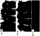
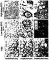

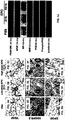
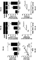


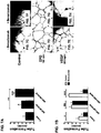
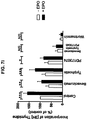
Classifications
-
- A—HUMAN NECESSITIES
- A61—MEDICAL OR VETERINARY SCIENCE; HYGIENE
- A61K—PREPARATIONS FOR MEDICAL, DENTAL OR TOILETRY PURPOSES
- A61K35/00—Medicinal preparations containing materials or reaction products thereof with undetermined constitution
- A61K35/12—Materials from mammals; Compositions comprising non-specified tissues or cells; Compositions comprising non-embryonic stem cells; Genetically modified cells
- A61K35/35—Fat tissue; Adipocytes; Stromal cells; Connective tissues
-
- A—HUMAN NECESSITIES
- A61—MEDICAL OR VETERINARY SCIENCE; HYGIENE
- A61K—PREPARATIONS FOR MEDICAL, DENTAL OR TOILETRY PURPOSES
- A61K38/00—Medicinal preparations containing peptides
- A61K38/16—Peptides having more than 20 amino acids; Gastrins; Somatostatins; Melanotropins; Derivatives thereof
- A61K38/17—Peptides having more than 20 amino acids; Gastrins; Somatostatins; Melanotropins; Derivatives thereof from animals; from humans
- A61K38/18—Growth factors; Growth regulators
- A61K38/1816—Erythropoietin [EPO]
-
- A—HUMAN NECESSITIES
- A61—MEDICAL OR VETERINARY SCIENCE; HYGIENE
- A61K—PREPARATIONS FOR MEDICAL, DENTAL OR TOILETRY PURPOSES
- A61K8/00—Cosmetics or similar toiletry preparations
- A61K8/18—Cosmetics or similar toiletry preparations characterised by the composition
- A61K8/30—Cosmetics or similar toiletry preparations characterised by the composition containing organic compounds
- A61K8/64—Proteins; Peptides; Derivatives or degradation products thereof
-
- A—HUMAN NECESSITIES
- A61—MEDICAL OR VETERINARY SCIENCE; HYGIENE
- A61P—SPECIFIC THERAPEUTIC ACTIVITY OF CHEMICAL COMPOUNDS OR MEDICINAL PREPARATIONS
- A61P17/00—Drugs for dermatological disorders
- A61P17/02—Drugs for dermatological disorders for treating wounds, ulcers, burns, scars, keloids, or the like
-
- A—HUMAN NECESSITIES
- A61—MEDICAL OR VETERINARY SCIENCE; HYGIENE
- A61P—SPECIFIC THERAPEUTIC ACTIVITY OF CHEMICAL COMPOUNDS OR MEDICINAL PREPARATIONS
- A61P41/00—Drugs used in surgical methods, e.g. surgery adjuvants for preventing adhesion or for vitreum substitution
-
- A—HUMAN NECESSITIES
- A61—MEDICAL OR VETERINARY SCIENCE; HYGIENE
- A61Q—SPECIFIC USE OF COSMETICS OR SIMILAR TOILETRY PREPARATIONS
- A61Q19/00—Preparations for care of the skin
- A61Q19/08—Anti-ageing preparations
-
- A—HUMAN NECESSITIES
- A61—MEDICAL OR VETERINARY SCIENCE; HYGIENE
- A61K—PREPARATIONS FOR MEDICAL, DENTAL OR TOILETRY PURPOSES
- A61K2800/00—Properties of cosmetic compositions or active ingredients thereof or formulation aids used therein and process related aspects
- A61K2800/80—Process related aspects concerning the preparation of the cosmetic composition or the storage or application thereof
- A61K2800/91—Injection
-
- A—HUMAN NECESSITIES
- A61—MEDICAL OR VETERINARY SCIENCE; HYGIENE
- A61K—PREPARATIONS FOR MEDICAL, DENTAL OR TOILETRY PURPOSES
- A61K9/00—Medicinal preparations characterised by special physical form
- A61K9/0012—Galenical forms characterised by the site of application
- A61K9/0019—Injectable compositions; Intramuscular, intravenous, arterial, subcutaneous administration; Compositions to be administered through the skin in an invasive manner
Definitions
- the present invention relates to Erythropoietin for use in enhancing implanted fat cell survival for reconstruction surgery in a subject in need thereof.
- endothelial cells change their phenotype to an angiogenic phenotype that includes the production of proteases, such as matrix metalloproteinases (MMPs), and the ability to migrate and proliferate.
- MMPs matrix metalloproteinases
- This process is dependent upon the activity of several growth factors, such as vascular endothelial growth factor (VEGF), basic fibroblast growth factor (bFGF) and platelet-derived growth factor (PDGF)-BB.
- VEGF vascular endothelial growth factor
- bFGF basic fibroblast growth factor
- PDGF platelet-derived growth factor
- EPO Erythropoietin
- Ribatti and colleagues demonstrated that EPO can induce a pro-angiogenic phenotype in cultured endothelial cells and stimulate angiogenesis in vivo [ Ribatti et al. (2003) Eur J Clin Invest 33:891-896 ].
- EPO has also been shown to indirectly stimulate angiogenesis in ischemic tissue by increasing the expression of VEGF protein and recruiting endothelial progenitor cells [ Nakano et al. (2007) Circ Res 100:662-669 ; Aicher et al. (2005) Hypertension 45:321- 325 ].
- EPO administration has also been shown to mobilize bone marrow- derived progenitor cells [ Hamed et al. (2006) Eur Heart J 27:1876-83 ] and to increase the myocardial expression of VEGF [ Westenbrink et al. (2007) Eur Heart J 28:2018- 2027 ].
- Wang and colleagues demonstrated that EPO can promote angiogenesis by stimulating VEGF secretion from neural progenitor cells and VEGF-receptor expression in cerebral endothelial cells [ Wang et al. (2008) J Cereb Blood Flow Metab 28:1361-8 ].
- EPO has also been reported to possess other non-hematopoietic effects, including cytoprotection of vascular endothelial cells [ Chong et al. (2003) Curr Drug Targets Cardiovasc Haematol Disord 3:141-154 ] and an anti-apoptotic action in vascular smooth muscle cells and in endothelial cells [ Somervaille et al. (2001) Blood 98:1374-1381 ].
- These anti-apoptotic actions include prevention of mitochondrial release of cytochrome c, suppression of caspase activity, upregulation of protein kinase B (PKB) signaling pathway activity and the expression of the antiapoptotic protein Bcl-xl.
- tissue repair e.g. bone, cartilage
- a tissue graft e.g. fat tissue, muscle tissue
- one or more bioactive agents e.g. erythropoietin
- the tissue is implanted into a subject.
- U.S. Pat. No. 7459152 discloses erythropoietin administration for improved graft survival.
- cells of a tissue graft e.g. cells of a neural or paraneural origin, such as adrenal chromaffin cells
- erythropoietin are treated with erythropoietin before, during or after delivery or administration into a subject for the treatment of neurological diseases (e.g. Parkinson's disease, Alzheimer's disease, spinal cord injury).
- neurological diseases e.g. Parkinson's disease, Alzheimer's disease, spinal cord injury.
- U.S. Pat. No. 5,681,561 discloses method and compositions for improving autologous fat grafting.
- autologous fat cells e.g. lipocytes
- a non-steroidal anabolic hormone e.g. insulin or triiodothyronine/thyroxine or both.
- the autologous fat cells may further be injected into a subject with a growth hormone [e.g. epithelial growth factor (EGF), platelet derived growth factor (PDGF)].
- EGF epithelial growth factor
- PDGF platelet derived growth factor
- the hormones are combined with a nutrient medium.
- PCT Publication No. 2008/019434 discloses use of agents to enhance adipogenesis and to promote fat graft survival.
- growth factors e.g. platelet-derived growth factor (PDGF), vascular endothelial growth factor (VEGF) and/or fibroblast growth factor (FGF)
- PDGF platelet-derived growth factor
- VEGF vascular endothelial growth factor
- FGF fibroblast growth factor
- A1 cells present in adipose tissue are used to promote wound healing in a patient.
- Methods of treating patients include processing adipose tissue to deliver a concentrated amount of regenerative cells obtained from the adipose tissue to a patient.
- the methods may be practiced in a closed system so that the regenerative cells are not exposed to an external environment prior to being administered to a patient. Accordingly, in one method, cells present in adipose tissue are placed directly into a recipient along with such additives necessary to promote, engender or support a therapeutic benefit.
- WO 2005/011569 A2 discloses methods of treating patients for conditions such as breast augmentation, soft tissue defects, and urinary incontinence.
- the methods include removing adipose tissue from a patient, processing a portion of the adipose tissue to obtain a substantially isolated population of regenerative cells, mixing the regenerative cells with another portion of adipose tissue to form a composition, and administering the composition to the patient from which the adipose tissue was removed.
- the present invention is directed to Erythropoietin for use in enhancing implanted fat cell survival for reconstruction surgery in a subject in need thereof, wherein a dose of Erythropoietin for local administration ranges between 1-1000 IU per injection per 1,000,000 fat cells or wherein the dose of Erythropoietin for systemic administration ranges between 10 - 7500 IU per kg body weight.
- the present invention is also directed to the use of Erythropoietin in enhancing implanted fat cell survival for treating wrinkles in a subject in need thereof, wherein a dose of Erythropoietin for local administration ranges between 1-1000 IU per injection per 1,000,000 fat cells or wherein the dose of Erythropoietin for systemic administration ranges between 10 - 7500 IU per kg body weight, wherein the use is non-therapeutic.
- a pharmaceutical composition comprising a population of fat cells and Erythropoietin at a dose of about 1-1000 IU per injection per 1,000,000 fat cells.
- fat cells are treated with Erythropoietin prior to the implanting.
- Erythropoietin may be directly injected into the population of fat cells.
- administering may be effected at least twice.
- the use further comprises administering to the subject at least one factor selected from the group consisting of an extracellular matrix component, a growth factor, a hormone, an angiogenic factor, a coagulation factor, a cytokine, a chemokine, an enzyme, a neurotransmitter, a vitamin, a carbohydrate, an ion, an iron chelator, a fatty acid, an antibiotic and an amino acid.
- at least one factor selected from the group consisting of an extracellular matrix component, a growth factor, a hormone, an angiogenic factor, a coagulation factor, a cytokine, a chemokine, an enzyme, a neurotransmitter, a vitamin, a carbohydrate, an ion, an iron chelator, a fatty acid, an antibiotic and an amino acid.
- the soft tissue defect may be selected from the group consisting of a skin condition, a skin malady, a wound, a burn, a cancer, a surgery, a reconstruction surgery, a skin depression, a congenital malformation and an acquired disease.
- the fat cell comprises an autologous cell.
- the fat cell comprises a non- autologous cell.
- the non-autologous cell is an allogeneic cell.
- the non-autologous cell is a xenogeneic cell.
- the non-autologous cell may be obtained from a mammal.
- the pharmaceutical composition comprises at least one factor selected from the group consisting of an extracellular matrix component, a growth factor, a hormone, an angiogenic factor, a coagulation factor, a cytokine, a chemokine, an enzyme, a neurotransmitter, a vitamin, a carbohydrate, an ion, an iron chelator, a fatty acid, an antibiotic, and an amino acid.
- FIGs. 1A-C are photographs depicting five representative mice with fat grafts at the end of the 15-week study period.
- Figure 1A shows five PBS-treated fat grafts with small lumps that vary in their size in the scalps.
- Figure 1B shows five high-dose erythropoietin (100 IU EPO)-treated fat grafts with large lumps that are similar in their size in the scalps.
- Figure 1C shows fat grafts which were dissected from the mice 15 weeks after transplantation. From left to right: a representative small fat graft from a PBS-treated fat graft, an intermediate-size low-dose EPO-treated fat graft, and a large high-dose EPO-treated fat graft respectively. Scale bar: 10 mm.
- FIGs. 2A-C are photographs depicting histological sections of fat grafts that were removed from the PBS-treated, low-dose EPO treated and high-dose EPO treated mice 15 week after fat transplantation. Sections were stained with hematoxylin and eosin, and were examined under light microscope for: (i) the extent of integration, as evidenced by the extent of organization of intact and nucleated fat cells in the grafted fat tissue architecture; (ii) the extent of fibrosis, as evidenced by the amount of collagen and elastic fibrils; (iii) the presence of cysts and vacuoles; and (iiii) the intensity of the inflammatory response, as evidenced by the extent of lymphocyte and macrophage infiltration.
- FIG. 1 a PBS-treated fat graft in which there is fat cell degeneration, fibrosis, and infiltration of nucleated inflammatory cells although some cells are still viable and intact
- Figure 2B a low dose erythropoietin (EPO)-treated fat graft in which the fat cells are well-defined in tissue in which there is a moderate amount of fibrosis
- Figure 2C a high dose EPO-treated fat graft in which there are viable, well-defined intact fat cells with modest amounts of connective tissue.
- Scale bar 200 ⁇ m.
- FIGs. 2D-F are photographs depicting the effect of erythropoietin (EPO) on inflammatory response in fat grafts after fat transplantation.
- EPO erythropoietin
- FIGs. 2G-I are photographs depicting the effect of erythropoietin (EPO) on new blood vessel formation in fat grafts after fat transplantation.
- EPO erythropoietin
- FIGs. 2G-I are photographs depicting the effect of erythropoietin (EPO) on new blood vessel formation in fat grafts after fat transplantation.
- PBS 100 ⁇ l, Figure 2G
- 20 IU EPO/100 ⁇ l PBS low-dose, Figure 2H
- 100 IU EPO/100 ⁇ l PBS high-dose, Figure 2I
- FIGs. 2J-L are graphs depicting the effect of EPO on inflammatory response and MVD in the fat grafts after transplantation.
- Figure 2J is a bar graph showing that EPO treatment decreases the severity of the inflammatory responses in the fat grafts.
- Figure 2K is a bar graph showing that EPO treatment increases microvascular density (MVD) in a dose-dependent manner.
- MVD microvascular density
- Each bar represents the mean MVD ⁇ SD from five regions of interest in each fat graft from each treatment group at the end of the 15-week study period.
- *P ⁇ 0.05, ***P ⁇ 0.001 is the significance of the difference between either the low dose or high dose EPO-treated fat grafts and the PBS-treated grafts.
- Scale bar 50 ⁇ m.
- Figure 2L is a line graph showing the negative correlation of MVD to the extent of macrophage infiltration in the fat grafts.
- FIGs 3A-J depict the effect of EPO on the expression levels of angiogenic growth factors in the fat grafts.
- the fat grafts from the three different groups of mice were treated with either PBS (100 ⁇ l), 20 IU EPO/100 ⁇ l PBS (low-dose), or 100 IU EPO/100 ⁇ l PBS (high-dose) on the day of the fat injection, and the treatments were repeated every three days for 18 days.
- Figures 3A-I are representative histological micrographs of PBS-, and low-dose- and high-dose-EPO treated fat grafts (as indicated) presenting VEGF expression ( Figures 3A-C ), VEGFR-2 expression ( Figures 3D-F ) and EPOR expression ( Figures 3G-I).
- Figure 3J is a photograph showing representative western blots of the expression levels of the angiogenic factors in the PBS- and EPO-treated fat grafts at the end of the 15-week study period.
- bFGF basic fibroblast growth factor
- IGF-1 insulin-like growth factor-1
- PDGF-BB platelet-derived growth factor-BB
- MMP-2 matrix metalloproteinase-2
- PKB protein kinase B
- phosphoPKB phosphorylated PKB.
- FIGs. 4A-F depict the effect of erythropoietin (EPO) on the expression levels of angiogenic growth factors in the fat grafts.
- EPO erythropoietin
- FIGs. 4A-F depict the effect of erythropoietin (EPO) on the expression levels of angiogenic growth factors in the fat grafts.
- PBS 100 ⁇ l
- 20 IU EPO/100 ⁇ l PBS low-dose
- 100 IU EPO/100 ⁇ l PBS high-dose
- the graphs represent the mean vascular endothelial growth factor (VEGF) content ( Figure 4A ), the mean VEGFR-2 expression ( Figure 4B ) and the mean EPOR expression ( Figure 4C ) ⁇ SD in the fat grafts in each treatment group.
- VEGF vascular endothelial growth factor
- Figures 4D-F show the correlation between VEGF and MVD ( Figure 4D ), and between mean VEGFR-2 ( Figure 4E ) and EPOR ( Figure 4F ) expression and mean MVD in each group.
- Scale bar 200 ⁇ m.
- FIGs. 5A-B depict the effect of erythropoietin (EPO) on the extent of apoptosis in the fat grafts.
- EPO erythropoietin
- PBS 100 ⁇ l
- 20 IU EPO/100 ⁇ l PBS low-dose
- 100 IU EPO/100 ⁇ l PBS high-dose
- Figure 5A shows the extent of apoptosis as was measured by TUNEL assay and is expressed as a percentage of the presence of apoptosis in the PBS-treated fat grafts.
- Each bar represents the mean extent of apoptosis ⁇ SD in the fat graft, in each treatment group, at the end of the 15-week study period.
- Figure 5B shows representative western blots of the expression levels of caspase 3 (Casp 3) and cytochrome c (Cyt c) in the PBS- and EPO-treated fat grafts at the end of the 15-week study period.
- FIGs. 6A-D depict the effect of vascular endothelial growth factor (VEGF) on microvascular density (MVD) and the extent of apoptosis in the fat grafts.
- PBS 100 ⁇ l
- VEGF vascular endothelial growth factor
- VEGF vascular endothelial growth factor
- 200 ng VEGF/100 ⁇ l PBS were injected into the fat grafts on the day of fat injection into two different groups of mice and then repeatedly every three days for 18 days.
- Figure 6A is a bar graph showing the mean microvascular density (MVD) ⁇ SD from five regions of interest in each slide (slides were prepared from the harvested fat grafts of each treatment group at the end of the 15-week study period).
- Figure 6B is a bar graph showing the mean VEGF content ⁇ SD in the harvested fat grafts in each treatment group at the end of the 15-week study period.
- Figure 6C is a bar graph showing the extent of apoptosis as was measured by TUNEL assay. The results are expressed as a percentage of the extent of apoptosis in the PBS-treated fat grafts.
- Each bar represents the mean extent of apoptosis ⁇ SD in the fat graft in each treatment group at the end of the 15-week study period. **P ⁇ 0.01, and is the significance of the difference between the VEGF-treated fat grafts and the PBS-treated grafts.
- Figure 6D is a photograph showing representative western blots of the expression levels of caspase 3 (Casp 3) and cytochrome c (Cyt c) in the PBS- and VEGF-treated fat grafts at the end of the 15-week study period.
- FIG. 7A depict the effect of erythropoietin (EPO) on human umbilical vein endothelial cells (HUVECs) tube formation in matrigel.
- EPO erythropoietin
- human umbilical vein endothelial cells (HUVECs) were treated with 20 IU/ml or 100 IU/ml EPO for 48 hours after plating the cells on matrigel.
- the extent of HUVEC tube formation on matrigel was assessed after 24 hours under a light microscope at 10 x magnification.
- 0 well separated individual cells
- 1 cells had begun to migrate and align themselves
- 2 visible capillary tubes and no sprouting
- 3 visible sprouting of new capillary tubes
- 4 early formation of closed polygons
- 5 development of complex mesh-like structures.
- Each bar represents the mean grade of tube formation ⁇ SD in the matrigel. *P ⁇ 0.05, **P ⁇ 0.01 and ***P ⁇ 0.001.
- FIGs. 7B-H depict the effect of EPO or VEGF on human umbilical vein endothelial cells (HUVECs) tube formation in matrigel.
- HUVECs were treated with 100 IU/ml EPO or 200 ng/100 ⁇ l VEGF in the absence or presence of 0.25 mg/ml bevacizumab for 48 hours after plating the cells on matrigel.
- the extent of HUVEC tube formation on matrigel was assessed after 24 hours under a light microscope at 10 x magnification.
- Figure 7B the white bars represent the mean grade of tube formation ⁇ SD in the matrigel of untreated HUVECs, VEGF- or EPO-treated HUVECs.
- Figure 7C depicts untreated HUVECs on matrigel
- Figure 7D depicts EPO-treated HUVECs after 24 hours of plating
- Figure 7E depicts VEGF-treated HUVECs after 24 hours of plating
- Figure 7F depicts untreated HUVECs with bevacizumab
- Figure 7G depicts EPO-treated HUVECs after 24 hours of plating with bevacizumab
- Figure 7H depicts VEGF-treated HUVECs after 24 hours of plating with bevacizumab.
- FIG. 7 I depicts the effect of EPO or VEGF on human umbilical vein endothelial cells (HUVECs) tube formation in matrigel.
- Cultured HUVECs were treated with or without 100 IU/ml EPO in the presence of either bevacizumab, PD173074, or tyrphostin, a combination of bevacizumab, PD173074 and tyrphostin, or in the presence of wortmannin.
- Proliferation of HUVECs was measured by incorporation of [ 3 H]- thymidine to DNA. Duplicate cell counts were averaged for 3 experiments and the data were expressed as the percentage of control.
- EPO Erythropoietin
- VEGF angiogenic factor
- the present inventor has shown that treating the fat grafts with EPO leads to long-term survival of the grafted fat cells.
- the present teachings portray a therapeutic value for Erythropoietin and suggest the use of same in transplantation of fat tissue.
- EPO treated fat tissues showed high microvascular density (MVD), well vascularized areas with increased expression of CD31 and numerous endothelial islets ( Figures 2G-I and 2K ) and showed a lower inflammatory response after transplantation ( Figures 2D-F and 2J ).
- EPO treatment also lead to a dose-dependent decrease in apoptosis of fat cells ( Figure 5 A) while increasing the expression of the angiogenic factors VEGF, bFGF, IGF-1, PDGF-BB, MMP-2 PKB and phosphoPKB ( Figures 3J and 4A ) and increasing both tissue VEGFR-2 and EPOR expression ( Figures 3D-I and 4B-C ) in these cells.
- Erythropoietin for use in enhancing implanted fat cell survival for reconstruction surgery in a subject in need thereof, wherein a dose of Erythropoietin for local administration ranges between 1-1000 IU per injection per 1,000,000 fat cells or wherein the dose of Erythropoietin for systemic administration ranges between 10 - 7500 IU per kg body weight.
- fat cell refers to any cell or group of cells composed in a fat tissue, including for example, lipocytes, adipocytes, adipocyte precursors including pre-adipocytes and mesenchymal stem cells. It will be appreciated that according to the present teachings, the fat cells may be dispersed or may be comprised in a tissue.
- the number of fat cells may vary over a wide range and one of ordinary skill in the art will recognize that this number will vary depending upon the type and size of the area to be treated according to the use of this invention, the relative degree of vascularization of the area to be treated, the age of the subject to be treated and the relative viability of the fat cells available for transplantation. It will be appreciated that the number of fat cells transplanted may be adjusted according to the procedure used, the site of injection and the relative vascularization of the site to be injected. One of ordinary skill in the art will recognize that certain conditions may necessitate the adjustment of the fat cell numbers outside of the below described ranges. According to some embodiments of the present invention, the number of fat cells for transplantation range from about 10,000 to about 10,000,000 fat cells per 1 ml.
- 0.01 - 2000 mls of fat tissue are transplanted.
- the subject may be administered a single transplantation or several transplantations (e.g. about 2, 5, 10, 20, 50, 100 or more transplantation procedures), as described in further detail hereinbelow.
- fat cell survival refers to the ability of the fat cells to remain viable and intact following engraftment thereof. Preferably, the fat cells survive for a period of a few days, a few weeks, a few months or a few years following engraftment thereof.
- enhancing in respect to fat cell survival refers to a process of increasing the life span of fat cells in the fat graft and/or decreasing the number of fat cells which undergo resorption, apoptosis or cell death within the fat graft.
- enhancing refers to at least about 10 %, 20 %, 50 %, 80 %, 90 % increase in viable fat cells and/or at least about 10 %, 20 %, 50 %, 80 %, 90 % arrest in fat cell death.
- EPO Erythropoietin
- Erythropoietin refers to a mammalian (e.g., human) Erythropoietin protein (interchangeably used with polypeptide) or mimetics thereof such as set forth in GenBank Accession No. NP_000790. Erythropoietin may be synthesized using recombinant DNA techniques or solid phase technology. Erythropoietin is also commercially available (e.g., Cytolab/Peprotech, Rehovot, Israel; Arenesp, Amgen, Thousand Oaks, CA, USA; and Epogen, Amgen, Thousand Oaks, CA, USA, Bristol-Myers Squibb, Roche and Sanofi-Aventis).
- Erythropoietin may be used as an entire glycoprotein or as only a protein subunit devoid of the bound sugar. Since the Erythropoietin of the present invention is used for clinical applications, it is preferably sterile or may be purified of possible contaminating factors (e.g., bacteria or bacterial components, such as by filter).
- possible contaminating factors e.g., bacteria or bacterial components, such as by filter.
- Typical subjects that may be treated according to this aspect of the present invention include mammals such as human beings or domesticated animals including, but not limited to, horses (i.e. equine), cattle, goat, sheep, pig, dog, cat, camel, alpaca, llama and yak, male or female, at any age that is in need of fat transplantation.
- horses i.e. equine
- cattle, goat, sheep, pig, dog, cat camel
- alpaca llama and yak
- male or female male or female
- fat transplantation may be used to treat any soft tissue defect, to fill any soft tissue deficit and for augmentation of external and internal surfaces and structures of the body which are missing due to surgery, as a result of aging of a tissue, or due to disease, trauma or an injury. Examples include, but are not limited to, urological surgeries, tumor removal surgeries, reconstructive surgeries and skin surgeries. Likewise, fat transplantation may be used as an alternative to silicone or collagen fillers. Fat transplantation may be used to fill depressions (i.e.
- Fat transplantation may also be used in numerous other applications, including urological procedures involving the buildup of weak or damaged structural tissue, in treatment of wrinkles, burns, skin conditions, skin maladies and wounds and to augment areas of the body, such as the buttocks, biceps, triceps muscles, calf muscles, breasts, hands and penis. Furthermore, fat transplantation may be used to treat congenital malformations such as Hemifacial microsomia and acquired diseases such as Romberg's lipodystrophy and Acquired immune deficiency syndrome (AIDS).
- AIDS Acquired immune deficiency syndrome
- the fat cells may be obtained from the body of a subject and used in an autologous fashion (i.e. transplanted into the same subject from which the fat cells were obtained).
- the autologous fat cells are typically taken from a subject to fill in depressions or soft tissue deficits in the body of the same subject in an area of the body other than that site from which the fat cells were removed.
- the fat cells may be obtained from one subject (a "donor") and transplanted into a different individual (a "recipient") in a non-autologous fashion.
- the fat cells may be obtained from a subject of the same species as the recipient subject (i.e. allogeneic fat cells as for example from a human donor to a human recipient) or from a different species (i.e. xenogeneic cells as for example from a porcine donor to a human recipient).
- the non-autologous cell is obtained from a mammal.
- fat cells are generally obtained by removing same (e.g. by suctioning) from subcutaneous fat layers in the area of the stomach, legs or other areas where significant fat cells may be found.
- the fat cells in the present invention are substantially free of unrelated cells such as erythrocytes, other blood cells, fibroblasts and other cells which may contaminate the fat cells.
- these cells are kept in a sterile environment until used for transplantation.
- the fat cells may be further separated from other components which may be found in the aspirated fat, such as, for example, triglycerides, lysozomes, other cellular fragments, blood components, blood cells and large connective tissue fragments, among other less desirable components, before use. Any methods known in the art may be used to separate the fat cells from these other components, but preferably, at least one centrifugation step is employed.
- Fat cells may be immediately implanted into a subject.
- the fat cells may be implanted within 30 minutes, within an hour, within two hours, within three hours, within four hours or within one day of collection (see e.g. Example 1, of the examples section which follows). It will be appreciated that the fat cells in the present invention may be preserved for longer periods of time prior to translation in, for example, by freezing in liquid nitrogen.
- Implanting the fat cells may be carried out by any method known in the art, such as for example, by injection thereof into the desired location (as described in detail in Example 1, hereinbelow), by microsurgery and by surgery in cases were a large amount of fat cells or fat tissue is being transplanted.
- the subject may be administered Erythropoietin.
- Erythropoietin may be administered via a systemic administration or via a local administration.
- systemic administration refers to oral, intravenous, intraperitoneal and intramuscular administration of Erythropoietin.
- local administering refers to applying the Erythropoietin directly to the implanted fat cells or in close proximity to the implanted fat cells. Erythropoietin may be directly administered to the transplanted fat cells via injection.
- the contemplated dose of Erythropoietin applied for local administration ranges between 1-1000 IU per injection per 1,000,000 fat cells for local administration.
- the dose of Erythropoietin for systemic administration ranges between 10 - 7500 IU per kg body weight for systemic administration.
- the dose of Erythropoietin selected for treatment depends on the number and concentration of fat cells, the subject being treated and the location of the graft.
- Erythropoietin is typically effected immediately following implantation of the fat cells.
- Erythropoietin is administered to the subject within a few minutes or within a few hours of implantation.
- Erythropoietin is administered to the subject starting from the first day of fat cell transplantation and is continuously administered until the fat cells have been integrated and vascularized in the subject (e.g. for at least 5- 50 days).
- the present invention contemplates treating fat cells with Erythropoietin prior to implantation thereof. This may be in addition to administration of Erythropoietin following implantation or instead of administration of Erythropoietin following implantation. Treatment of the fat cells may be carried out by any method known to one of ordinary skill in the art as for example by ex vivo contacting the fat cells with Erythropoietin in a tissue culture plate or by injection of Erythropoietin directly into the fat tissue. Alternatively, fat cells may be exposed to Erythropoietin prior to removal from the donor.
- Concentrations of Erythropoietin for treating fat cells prior to transplantation are in a dose between 1-1000 IU per injection per 1,000,000 fat cells.
- the subject being treated prior to implantation may continue to receive Erythropoietin following implantation of the fat cells as depicted in detail hereinabove.
- Erythropoietin can be administered to the subject per se or as a pharmaceutical composition.
- the fat cells in the present invention can be administered per se or as part of a pharmaceutical composition.
- a "pharmaceutical composition” refers to a preparation of the active ingredients described herein with other chemical components such as physiologically suitable carriers and excipients.
- the purpose of the composition is to facilitate administration of the active ingredients (e.g., Erythropoietin) to the subject.
- active ingredient refers to Erythropoietin or the fat cells themselves accountable for the intended biological effect (i.e., enhancing fat cell survival).
- physiologically acceptable carrier and “pharmaceutically acceptable carrier” which may be interchangeably used refer to a carrier or a diluent that does not cause significant irritation to the subject and does not abrogate the biological activity and properties of the administered active ingredients.
- An adjuvant is included under these phrases.
- excipient refers to an inert substance added to the composition (pharmaceutical composition) to further facilitate administration of an active ingredient in the present invention.
- suitable routes of administration of Erythropoietin may, for example, include a systemic manner including oral, rectal, transmucosal, especially transnasal, intestinal or parenteral delivery, including intramuscular, subcutaneous and intramedullary injections as well as intrathecal, direct intraventricular, intravenous, intraperitoneal, intramuscular, intranasal, or intraocular injections.
- Suitable routes of administration of the compositions may, for example, include topical (e.g., to a keratinous tissue, such as the skin, scalp) and mucosal (e.g., oral, vaginal, eye) administrations.
- compositions of the present invention may be manufactured by processes well known in the art, e.g., by means of conventional mixing, dissolving, granulating, dragee-making, levigating, emulsifying, encapsulating, entrapping or lyophilizing processes.
- compositions for use in accordance with the present invention thus may be formulated in conventional manner using one or more physiologically acceptable carriers comprising excipients and auxiliaries, which facilitate processing of the active ingredients into preparations which can be used pharmaceutically. Proper formulation is dependent upon the route of administration chosen.
- the active ingredients of the pharmaceutical composition may be formulated in aqueous solutions, preferably in physiologically compatible buffers such as Hank's solution, Ringer's solution, or physiological salt buffer.
- physiologically compatible buffers such as Hank's solution, Ringer's solution, or physiological salt buffer.
- penetrants appropriate to the barrier to be permeated are used in the formulation. Such penetrants are generally known in the art.
- the pharmaceutical composition can be formulated readily by combining the active compounds with pharmaceutically acceptable carriers well known in the art.
- Such carriers enable the pharmaceutical composition to be formulated as tablets, pills, dragees, capsules, liquids, gels, syrups, slurries, suspensions, and the like, for oral ingestion by a patient.
- Pharmacological preparations for oral use can be made using a solid excipient, optionally grinding the resulting mixture, and processing the mixture of granules, after adding suitable auxiliaries if desired, to obtain tablets or dragee cores.
- Suitable excipients are, in particular, fillers such as sugars, including lactose, sucrose, mannitol, or sorbitol; cellulose preparations such as, for example, maize starch, wheat starch, rice starch, potato starch, gelatin, gum tragacanth, methyl cellulose, hydroxypropylmethyl-cellulose, sodium carbomethylcellulose; and/or physiologically acceptable polymers such as polyvinylpyrrolidone (PVP).
- disintegrating agents may be added, such as cross-linked polyvinyl pyrrolidone, agar, or alginic acid or a salt thereof such as sodium alginate.
- Dragee cores are provided with suitable coatings.
- suitable coatings For this purpose, concentrated sugar solutions may be used which may optionally contain gum arabic, talc, polyvinyl pyrrolidone, carbopol gel, polyethylene glycol, titanium dioxide, lacquer solutions and suitable organic solvents or solvent mixtures.
- Dyestuffs or pigments may be added to the tablets or dragee coatings for identification or to characterize different combinations of active compound doses.
- compositions which can be used orally include push-fit capsules made of gelatin as well as soft, sealed capsules made of gelatin and a plasticizer, such as glycerol or sorbitol.
- the push-fit capsules may contain the active ingredients in admixture with filler such as lactose, binders such as starches, lubricants such as talc or magnesium stearate and, optionally, stabilizers.
- the active ingredients may be dissolved or suspended in suitable liquids, such as fatty oils, liquid paraffin, or liquid polyethylene glycols.
- stabilizers may be added. All formulations for oral administration should be in dosages suitable for the chosen route of administration.
- compositions may take the form of tablets or lozenges formulated in conventional manner.
- the active ingredients for use according to the present invention are conveniently delivered in the form of an aerosol spray presentation from a pressurized pack or a nebulizer with the use of a suitable propellant, e.g., dichlorodifluoromethane, trichlorofluoromethane, dichloro-tetrafluoroethane or carbon dioxide.
- a suitable propellant e.g., dichlorodifluoromethane, trichlorofluoromethane, dichloro-tetrafluoroethane or carbon dioxide.
- the dosage unit may be determined by providing a valve to deliver a metered amount.
- Capsules and cartridges of, e.g., gelatin for use in a dispenser may be formulated containing a powder mix of the compound and a suitable powder base such as lactose or starch.
- compositions described herein may be formulated for parenteral administration, e.g., by bolus injection or continues infusion.
- Formulations for injection may be presented in unit dosage form, e.g., in ampoules or in multidose containers with optionally, an added preservative.
- the compositions may be suspensions, solutions or emulsions in oily or aqueous vehicles, and may contain formulatory agents such as suspending, stabilizing and/or dispersing agents.
- compositions for parenteral administration include aqueous solutions of the active preparation in water-soluble form. Additionally, suspensions of the active ingredients may be prepared as appropriate oily or water based injection suspensions. Suitable lipophilic solvents or vehicles include fatty oils such as sesame oil, or synthetic fatty acids esters such as ethyl oleate, triglycerides or liposomes. Aqueous injection suspensions may contain substances, which increase the viscosity of the suspension, such as sodium carboxymethyl cellulose, sorbitol or dextran. Optionally, the suspension may also contain suitable stabilizers or agents which increase the solubility of the active ingredients to allow for the preparation of highly concentrated solutions.
- the active ingredient may be in powder form for constitution with a suitable vehicle, e.g., sterile, pyrogen-free water based solution, before use.
- a suitable vehicle e.g., sterile, pyrogen-free water based solution
- compositions of the present invention may also be formulated in rectal compositions such as suppositories or retention enemas, using, e.g., conventional suppository bases such as cocoa butter or other glycerides.
- compositions suitable for use in context of the present invention include compositions wherein the active ingredients are contained in an amount effective to achieve the intended purpose.
- a therapeutically effective amount means an amount of active ingredients (e.g. Erythropoietin) at a dose of about 1-1000 IU per injection per 1,000,000 fat cells, effective in enhancing fat cell survival.
- the therapeutically effective amount or dose can be estimated initially from in vitro and cell culture assays.
- a dose can be formulated in animal models to achieve a desired concentration or titer. Such information can be used to more accurately determine useful doses in humans.
- Toxicity and therapeutic efficacy of the active ingredients described herein can be determined by standard pharmaceutical procedures in vitro, in cell cultures or experimental animals.
- the data obtained from these in vitro and cell culture assays and animal studies can be used in formulating a range of dosage for use in human.
- the dosage may vary depending upon the dosage form employed and the route of administration utilized.
- the exact formulation, route of administration and dosage can be chosen by the individual physician in view of the patient's condition. (See e.g., Fingl, et al., 1975, in "The Pharmacological Basis of Therapeutics", Ch. 1 p.1).
- Dosage amount and interval may be adjusted individually to levels of the active ingredient which are sufficient to induce or suppress the biological effect (minimal effective concentration, MEC).
- MEC minimum effective concentration
- the MEC will vary for each preparation, but can be estimated from in vitro data. Dosages necessary to achieve the MEC will depend on individual characteristics and route of administration. Detection assays can be used to determine plasma concentrations.
- An animal model which can be used according to the present teachings to assess the biological effect of the compositions described herein includes SCID mice (as described in detail in the Examples section below).
- dosing can be of a single or a plurality of administrations, with course of treatment lasting from several days to several weeks or months or until cure is effected or until ample fat tissue has been endured.
- Erythropoietin compositions are administered once, administered twice, administered three times, administered four times, administered five times, administered six times, administered seven times, administered eight times, administered nine times or administered ten times to the subject in order to enhance fat cell survival. It will be appreciated that if multiple fat cell transplantations are carried out, the number of administrations of Erythropoietin may be vast and may be prolonged for as long as needed (as determined by one of ordinary skill in the art).
- the compositions of the present invention are administered at least once a day. It will be appreciated that the number of administrations can be determined by one of ordinary skill in the art.
- compositions to be administered will, of course, be dependent on the subject being treated, the severity of the affliction, the manner of administration, the judgment of the prescribing physician, etc.
- Determination of efficacy of treatment may be determined by measuring the number and viability of the engrafted fat cells (e.g. by ultrasound), measuring the number of apoptotic cells within the graft (e.g. by PCR), and evaluating the vascularization of the transplanted fat cells (e.g. by ultrasound).
- compositions of the present invention may, if desired, be presented in a pack or dispenser device, such as an FDA-approved kit, which may contain one or more unit dosage forms containing the active ingredient.
- the pack may, for example, comprise metal or plastic foil, such as a blister pack.
- the pack or dispenser device may be accompanied by instructions for administration.
- the pack or dispenser device may also be accompanied by a notice in a form prescribed by a governmental agency regulating the manufacture, use, or sale of pharmaceuticals, which notice is reflective of approval by the agency of the form of the compositions for human or veterinary administration. Such notice, for example, may include labeling approved by the U.S. Food and Drug Administration for prescription drugs or of an approved product insert.
- Compositions comprising a preparation of the invention formulated in a pharmaceutically acceptable carrier may also be prepared, placed in an appropriate container, and labeled for treatment of an indicated condition, as further detailed above.
- compositions of the present invention are utilized in vivo, the compositions are preferably of high purity and substantially free of potentially harmful contaminants, e.g., at least National Food (NF) grade, generally at least analytical grade, and preferably at least pharmaceutical grade.
- NF National Food
- synthesis or subsequent purification shall preferably result in a product that is substantially free of any potentially contaminating toxic agents that may have been used during the synthesis or purification procedures.
- Additional factors may be incorporated into the compositions of the present invention (i.e., Erythropoietin described hereinabove) to enhance fat cell survival.
- extracellular matrix components e.g. vitronectin, laminin, collagen, elastin
- growth factors e.g. FGF 1, FGF 2, IGF 1, IGF 2, PDGF, EGF, KGF, HGF, VEGF, GM-CSF, CSF, G-CSF, TGF alpha, TGF beta, NGF and ECGF
- hypoxia inducible factors e.g.
- HIF-1 alpha and beta and HIF-2 hormones
- hormones e.g., insulin, growth hormone (GH), CRH, Leptin, Prolactin and TSH
- angiogenic factors e.g., angiogenin and angiopoietin
- coagulation and anticoagulation factors e.g., Factor I, Factor XIII, tissue factor, calcium, vWF, protein C, protein S, protein Z, fibronectin, antithrombin, heparin, plasminogen, low molecular weight heparin (Clixan), high molecular weight kininogen (HMWK), prekallikrein, plasminogen activator inhibitor-1 (PAI1), plasminogen activator inhibitor-2 (PAI2), urokinase, thrombomoduline, tissue plasminogen activator (tPA), alpha 2-antiplasmin and Protein Z-related protease inhibitor (ZPI)], cytokines (IL-1 alpha, IL
- endoglycosidases exoglycosidases, endonucleases, exonucleases, peptidases, lipases, oxidases, decarboxylases, hydrases, chondroitinase, chondroitinase ABC, chondroitinase AC, hyaluronidase, keratanase, heparanases, heparanase splice variance, collagenase, trypsin, catalases), neurotransmitters (e.g., acetylcholine and monoamines), neuropeptides (e.g.
- substance P substance P
- vitamins e.g., D-biotin, Choline Chloride, Folic acid, Myo-inositol, Niacinamide, D-Pantothenic acid, Calcium salts, Pyridoxal.HCl, Pyrodixine.HCl, Riboflavin, Thiamine.HCl, Vitamin B12, vitamin E, vitamin C, vitamin D, vitamin B1-6, vitamin K, vitamin A and vitamin PP
- carbohydrates e.g. Mono/Di/Polysacharides including glucose, mannose, maltose and fructose
- ions e.g.
- Fe chelators Fe chelators, Ca chelators
- antioxidants e.g., Vitamin E, Quarcetin, superoxide scavengers, Superoxide dismutase
- H2O2 scavengers free radicals scavengers, Fe scavengers
- fatty acids e.g., Triglycerides, Phospholipids, Cholesterols, free fatty acids and non free fatty acids, fatty alcohol, Linoleic acid, oleic acid and lipoic acid
- antibiotics e.g., Penicillins, Cephalosporins and Tetracyclines
- analgesics anesthetics, antibacterial agents, anti-yeast agents, anti-fungal agents, antiviral agents, pro-biotic agents, anti-protozal agents, anti-pruritic agents, anti-dermatitis agents, anti-emetics, anti-inflammatory agents, anti-hyperkeratolyic agents, antiperspirants, anti-p
- Calcium Sulfate calcium Sulfate
- steroids e.g., androgens, estrogens, progestagens, glucocorticoids and mineralocorticoids
- catecholamines e.g., Epinephrine and Nor-epinephrine
- Nucleosides and Nucleotides e.g., Purins and Pyrimidines
- Prostaglandins e.g. Prostaglandin E2
- Leucotriens Erythropoietins (e.g. Thrombopoietin)
- Proteoglycans e.g. Heparan sulfate, keratan sulfate
- Hydroxyapatites e.g.
- Hydroxyapatite Ca 10 (PO 4 ) 6 (OH) 2 )]] Haptoglobins (Hp1-1, Hp2-2 and Hp1-2), Superoxide dismutases (e.g. SOD 1/2/3), Nitric Oxides, Nitric Oxide donors (e.g. nitroprusside, Sigma Aldrich, St. Louis, MO, USA, Glutathione peroxidases, Hydrating compounds (e.g. vasopressin), cells (e.g. Platelets), cell medium (e.g. M199, DMEM/F12, RPMI, Iscovs), serum (e.g.
- human serum fetal calf serum, , fetal bovine serum
- buffers e.g., HEPES, Sodium Bicarbonate
- detergents e.g., Tween
- disinfectants herbs, fruit extracts, vegetable extracts (e.g. cabbage, cucumber), flower extracts, plant extracts, flavinoids (e.g. pomegranate juice), spices, leafs (e.g. Green tea, Chamomile), Polyphenols (e.g. Red Wine), honey, lectins, microparticles, nanoparticles (lyposomes), micelles, calcium carbonate (CaCO3, e.g.
- CaCO3 calcium carbonate
- precipitated calcium carbonate ground/pulverized calcium carbonate, albacar, PCC, GCC
- calcite limestone, crushed marble, ground limestone, lime, chalk (e.g. whiting chalk, champagne chalk, french chalk) and co factors such as BH4 (tetrahydrobiobterine).
- the present composition may also contain ingredients, substances, elements and materials containing, hydrogen, alkyl groups, aryl groups, halo groups, hydroxy groups, alkoxy groups, alkylamino groups, dialkylamino groups, acyl groups, carboxyl groups, carboamido groups, sulfonamide groups, aminoacyl groups, amide groups, amine groups, nitro groups, organo selenium compounds, hydrocarbons, and cyclic hydrocarbons.
- ingredients, substances, elements and materials containing, hydrogen, alkyl groups, aryl groups, halo groups, hydroxy groups, alkoxy groups, alkylamino groups, dialkylamino groups, acyl groups, carboxyl groups, carboamido groups, sulfonamide groups, aminoacyl groups, amide groups, amine groups, nitro groups, organo selenium compounds, hydrocarbons, and cyclic hydrocarbons.
- the present composition may be combined with substances such as benzol peroxide, vasoconstrictors, vasodilatators, salicylic acid, retinoic acid, azelaic acid, lactic acid, glycolic acid, pyreuric acid, tannins, benzlidenecamphor and derivatives thereof, alpha hydroxyis, surfactants.
- substances such as benzol peroxide, vasoconstrictors, vasodilatators, salicylic acid, retinoic acid, azelaic acid, lactic acid, glycolic acid, pyreuric acid, tannins, benzlidenecamphor and derivatives thereof, alpha hydroxyis, surfactants.
- compositions of some embodiments of the present invention may be bioconjugated to polyethylenglycol (e.g. PEG, SE-PEG) which preserves the stability (e.g., against protease activities) and/or solubility (e.g., within a biological fluid such as blood, digestive fluid) of the active ingredients (e.g. Erythropoietin) while preserving their biological activity and prolonging its half-life.
- polyethylenglycol e.g. PEG, SE-PEG
- solubility e.g., within a biological fluid such as blood, digestive fluid
- active ingredients e.g. Erythropoietin
- compositions of the present invention can be used in combination with other currently practiced therapies for fat cell transplantation as, without being limited to, treatment of the subject with growth factors, transplantation of the fat cells on scaffolds or transplantation of the fat cells on polyester beads.
- fat cells of the present invention can be derived from either autologous sources or from non-autologous sources (e.g. allogeneic or xenogeneic). Since non-autologous cells are likely to induce an immune reaction when administered to the body several approaches have been developed to reduce the likelihood of rejection of non-autologous cells. These include either suppressing the recipient immune system or encapsulating the non-autologous cells or tissues in immunoisolating, semipermeable membranes before transplantation.
- non-autologous sources e.g. allogeneic or xenogeneic
- Encapsulation techniques are generally classified as microencapsulation, involving small spherical vehicles and macroencapsulation, involving larger flat-sheet and hollow-fiber membranes ( Uludag, H. et al. Technology of mammalian cell encapsulation. Adv Drug Deliv Rev. 2000; 42: 29-64 ).
- microcapsules Methods of preparing microcapsules are known in the arts and include for example those disclosed by Lu MZ, et al., Cell encapsulation with alginate and alpha-phenoxycinnamylidene-acetylated poly(allylamine). Biotechnol Bioeng. 2000, 70: 479-83 , Chang TM and Prakash S. Procedures for microencapsulation of enzymes, cells and genetically engineered microorganisms. Mol Biotechnol. 2001, 17: 249-60 , and Lu MZ, et al., A novel cell encapsulation method using photosensitive poly(allylamine alpha-cyanocinnamylideneacetate). J Microencapsul. 2000, 17: 245-51 .
- microcapsules are prepared by complexing modified collagen with a ter-polymer shell of 2-hydroxyethyl methylacrylate (HEMA), methacrylic acid (MAA) and methyl methacrylate (MMA), resulting in a capsule thickness of 2-5 ⁇ m.
- HEMA 2-hydroxyethyl methylacrylate
- MAA methacrylic acid
- MMA methyl methacrylate
- Such microcapsules can be further encapsulated with additional 2-5 ⁇ m ter-polymer shells in order to impart a negatively charged smooth surface and to minimize plasma protein absorption ( Chia, S.M. et al. Multi-layered microcapsules for cell encapsulation Biomaterials. 2002 23: 849-56 ).
- microcapsules are based on alginate, a marine polysaccharide ( Sambanis, A. Encapsulated islets in diabetes treatment. Diabetes Thechnol. Ther. 2003, 5: 665-8 ) or its derivatives.
- microcapsules can be prepared by the polyelectrolyte complexation between the polyanions sodium alginate and sodium cellulose sulphate with the polycation poly(methylene-co-guanidine) hydrochloride in the presence of calcium chloride.
- the method of the present invention may further advantageously comprise conditioning the subject with an immunosuppressive regimen prior to, concomitantly with, or following transplantation of the fat cells.
- the methods of the present invention require a reduced immunosuppressive regimen as compared to a subject not treated with Erythropoietin.
- immunosuppressive regimens include administration of immunosuppressive drugs and/or immunosuppressive irradiation.
- the immunosuppressive regimen consists of administering at least one immunosuppressant agent to the subject.
- immunosuppressive agents include, but are not limited to, methotrexate, cyclophosphamide, cyclosporine, cyclosporin A, chloroquine, hydroxychloroquine, sulfasalazine (sulphasalazopyrine), gold salts, D-penicillamine, leflunomide, azathioprine, anakinra, infliximab (REMICADE), etanercept, TNF.alpha. blockers, a biological agent that targets an inflammatory cytokine, and Non-Steroidal Anti-Inflammatory Drug (NSAIDs).
- methotrexate cyclophosphamide
- cyclosporine cyclosporin A
- chloroquine hydroxychloroquine
- sulfasalazine sulphasalazopyrine
- gold salts gold salts
- D-penicillamine leflunomide
- azathioprine anakin
- NSAIDs include, but are not limited to acetyl salicylic acid, choline magnesium salicylate, diflunisal, magnesium salicylate, salsalate, sodium salicylate, diclofenac, etodolac, fenoprofen, flurbiprofen, indomethacin, ketoprofen, ketorolac, meclofenamate, naproxen, nabumetone, phenylbutazone, piroxicam, sulindac, tolmetin, acetaminophen, ibuprofen, Cox-2 inhibitors and tramadol. These agents may be administered individually or in combination.
- the methods of the present invention require a reduced anti-inflammatory treatment [e.g. anti-inflammatory drugs such as steroids, non-steroidal anti-inflammatory drugs or immune selective anti-inflammatory derivatives (ImSAIDs)] as compared to a subject not treated with Erythropoietin.
- a reduced anti-inflammatory treatment e.g. anti-inflammatory drugs such as steroids, non-steroidal anti-inflammatory drugs or immune selective anti-inflammatory derivatives (ImSAIDs)
- compositions, method or structure may include additional ingredients, steps and/or parts, but only if the additional ingredients, steps and/or parts do not materially alter the basic and novel characteristics of the claimed composition, method or structure.
- a compound or “at least one compound” may include a plurality of compounds, including mixtures thereof.
- range format is merely for convenience and brevity and should not be construed as an inflexible limitation on the scope of the invention. Accordingly, the description of a range should be considered to have specifically disclosed all the possible subranges as well as individual numerical values within that range. For example, description of a range such as from 1 to 6 should be considered to have specifically disclosed subranges such as from 1 to 3, from 1 to 4, from 1 to 5, from 2 to 4, from 2 to 6, from 3 to 6 etc., as well as individual numbers within that range, for example, 1, 2, 3, 4, 5, and 6. This applies regardless of the breadth of the range.
- method refers to manners, means, techniques and procedures for accomplishing a given task including, but not limited to, those manners, means, techniques and procedures either known to, or readily developed from known manners, means, techniques and procedures by practitioners of the chemical, pharmacological, biological, biochemical and medical arts.
- treating includes abrogating, substantially inhibiting, slowing or reversing the progression of a condition, substantially ameliorating clinical or aesthetical symptoms of a condition or substantially preventing the appearance of clinical or aesthetical symptoms of a condition.
- Fat was harvested from the thigh of a 40-year-old woman by suction-assisted lipectomy under general anesthesia. The fat was aspirated under local anesthesia using a 14-gauge blunt cannula, and then processed under sterile conditions for subsequent grafting into nude mice within two hours of its collection according to previously published protocols [ Ullmann et al. (2005) Dermatol Surg 31:1304-7 ; Kurita et al. (2008) Plast Reconstr Surg 121:1033-1041 ].
- the first study comprised 30 seven-week-old female CD-1 nude mice (Harlan, Jerusalem, Israel). These mice were housed in cages in a room with an artificial 12-h light/dark cycle at a constant temperature range (24 ⁇ 2 °C) and relative humidity (55 ⁇ 10%). The mice were acclimated for one week prior to the study, and fed a standard laboratory chow and water ad libitum.
- the 30 mice were randomly divided into three equal groups and treated as follows: Group 1 mice were injected with 1 ml of human fat and were treated with sterile PBS (control group). Group 2 mice were injected with 1 ml human fat and were treated with 1000 IU/kg EPO (low-dose EPO group).
- mice were injected with 1 ml human fat and were treated with 5000 IU/kg EPO (high-dose EPO group).
- the fat was injected subcutaneously into the scalp using a 14G needle while the animals were manually restrained.
- the PBS-treated fat grafts were injected with 100 ⁇ l PBS (control group), and the EPO-treated fat grafts were injected with either 20 IU EPO/100 ⁇ l PBS (low-dose EPO group) or 100 IU EPO/100 ⁇ l PBS (high-dose EPO group) every three days for 18 days (a total of 6 injections).
- EPO was purchased as an injection ampoule (ARANESP®, Amgen AG, Switzerland) which contained 150 ⁇ g/ml (18,000 IU) of EPO.
- the second animal study comprised 20 seven-week-old female CD-1 nude mice, and differed from the first study in that the fat was treated with VEGF (2 ⁇ g/ml, Sigma Aldrich, MO, USA) following implantation. 10 mice were injected with fat followed by 200 ng VEGF/100 ⁇ l PBS injections every three days for a total of 18 days. The remaining 10 mice made up a second control group that was treated in an identical manner to the control group in the first study. Post-operative analgesics and antibiotics were not administered to the mice in the two experiments.
- the duration of the study period of both experiments was 15 weeks from the start of fat transplantation. During this period, each mouse was weighed; a tail vein blood sample was collected for determination of the red blood cell count, leukocyte count, and platelet count; and for measurement of plasma hemoglobin, VEGF, and EPO concentrations. These measurements were carried out at three different occasions: the day of fat injection, 18 days after the fat injection and at the end of the study period.
- VEGF and EPO concentrations were determined in homogenates of samples of the fat grafts using commercial enzyme-linked immunosorbent assays (Quantikine® VEGF immunoassay Kit and Quantikine® IVD® Erythropoietin Kit, R&D Systems, MN, USA) in accordance with the manufacturer's instructions.
- each fat graft was weighed and the volume of the fat graft was measured by the liquid overflow method as previously described [ Ayhan et al. (2001) Aesthetic Plast Surg 25:338-342 ]. After weight and volume determination, each fat graft was divided into two portions from the middle.
- One portion was stored at -80 °C for later determination of EPO concentrations, VEGF content, the extent of apoptosis, and the expression levels of the angiogenic factors, namely bFGF, insulin growth factor-1 (IGF-1), PDGF-BB, VEGF receptor-2 (VEGFR-2), EPO receptor (EPOR) and MMP-2, the survival factor PKB and phosphorylated PKB, and the pro-apoptotic factors, namely caspase 3 and cytochrome c.
- the second portion was placed in 4 % formalin and used for determination of macrophage infiltration, microvascular density (MVD), VEGFR-2 and EPOR localization and for histological examination.
- mice in all treatment groups in both experiments completed the 15-week study period. They appeared to be healthy during the course of the study and there was no evidence of cachexia at the end of the study period. There were no significant changes in red blood cell counts, leukocyte counts, platelet counts, plasma hemoglobin and EPO concentrations in mice with either phosphate buffer saline (PBS)-treated or low-dose EPO-treated fat grafts (Table 1, below). The red blood cell counts, leukocyte counts, platelet counts and plasma EPO concentrations, but not the plasma hemoglobin concentrations, were significantly increased in mice treated with high-dose EPO-treated fat grafts (Table 1, below).
- PBS phosphate buffer saline
- Plasma VEGF concentrations were significantly increased in the two groups of mice with EPO-treated fat grafts.
- the plasma VEGF concentrations in the two groups of mice with EPO-treated grafts were not significantly different from baseline values and those in mice with PBS-treated fat grafts.
- EPO concentrations in the PBS- and EPO-treated grafts were not different from the baseline values, however, 18 days after fat injection both the EPO and VEGF values were significantly higher in both the EPO-low and EPO-high treatment groups (Table 1, below).
- the slides were examined under a light microscope for (a) the extent of integration, as evidenced by the extent of organization of intact and nucleated fat cells, (b) the extent of fibrosis, as evidenced by the amount of collagen and elastic fibrils, (c) the presence of cysts and vacuoles, and (d) the intensity of the inflammatory response, as evidenced by the extent of lymphocyte and macrophage infiltration.
- Quantification of macrophage infiltration in the fat grafts was estimated by counting the number of CD68-positive cells in five fields per fat graft in all fat graft sections.
- Microvascular density (MVD) in fat grafts was determined in five regions of interest where the CD31 antibody signal was the most intense in each section in all of the fat graft sections.
- the number of macrophages and blood vessels in each region was counted under a light microscope at 400 x magnification.
- the assessment in each fat graft was made by calculating the mean result in two different sections per fat graft and five different fields of view per section.
- the histological criteria in the PBS-treated fat grafts in the first experiment were not different from those in the second experiment.
- the extent of integration was higher in the high-dose EPO-treated grafts compared to the low-dose EPO- or PBS-treated grafts, whereas, the extent of cyst formation and fibrosis was lower in the high-dose EPO-treated grafts compared to the low-dose EPO- or PBS-treated grafts ( Figures 2A-C ).
- microvascular density Assessment of microvascular density (MVD)
- the paraffin-embedded fat graft sections were incubated overnight at room temperature with a monoclonal antibody against tissue CD31 (R&D Systems, Minneapolis MN, USA) as previously described [ Li et al. (2005) J Cell Biochem 95:559-570 ]. Upon completion of the incubation, the specimens were counterstained with hematoxylin. Mouse IgG was used as a negative control. The microvascular density (MVD) was determined in five regions of interest where the CD31 antibody signal was most intense. The number of blood vessels in each region was counted under a light microscope at 40 x magnification.
- microvascular densities (MVDs) in the two EPO-treated fat grafts were significantly higher than that of the PBS-treated fat graft, and the effect of EPO on MVD was dose-dependent.
- In the EPO-treated grafts there were well-vascularized areas with increased expression of CD31, and numerous endothelial islets ( Figures 2G-I and 2K ).
- the extent of MVD was negatively correlated to the extent of macrophage infiltration in the fat grafts ( Figure 2L ).
- the expression levels of the angiogenic factors, bFGF, IGF-1, PDGF-BB VEGFR-2, EPOR and MMP-2, the cell survival factor PKB and phosphorylated PKB, and the pro-apoptotic factors caspase 3 and cytochrome c were determined in homogenates of the harvested fats grafts by Western blotting. Briefly, homogenates of samples of the fat grafts were lysed in RIPA buffer (R&D Systems, MN, USA). A 40 ⁇ g aliquot of each lysate was loaded onto SDS-PAGE, and then transferred onto nitrocellulose membranes.
- Membranes were then incubated with monoclonal antibodies against bFGF, IGF-1, PDGF-BB, MMP-2, PKB, phosphoPKB, caspase 3, and cytochrome c (all purchased from Santa Cruz, CA, USA), or with monoclonal antibodies against VEGFR-2 and EPOR (R&D systems), before a second incubation with a horseradish peroxidase (HRP)-conjugated IgG secondary antibody. An antibody against ⁇ -actin (Santa Cruz) was used to normalize protein loading. The resultant bands were quantified by densitometry.
- HRP horseradish peroxidase
- the VEGF content in the low-dose and high-dose EPO-treated fat grafts was significantly higher compared to the PBS-treated fat grafts.
- the VEGF content in the high-dose EPO-treated grafts was significantly higher than that in the low-dose EPO-treated graft ( Figures 3A-C and 4A ).
- EPO treatment lead to a dose-dependent increase in the expression levels of bFGF, IGF-1, PDGF-BB, MMP-2 PKB and phosphoPKB ( Figure 3J ).
- EPO increased both tissue VEGFR-2 and EPOR expression in a dose-dependent manner, as evidenced by immunohistochemical localization of both factors ( Figures 3D-I ) and by western blot analysis ( Figure 4B-C ).
- the VEGF content and the mean expression levels of both VEGFR-2 and EPOR were positively correlated with MVD ( Figures 4D-F ).
- TUNEL terminal deoxyuridine triphosphate nick end labeling
- the MVD and the extent of apoptosis in the PBS-treated fat grafts in the first experiment were similar to those in the second experiment.
- the MVD and the VEGF content in the VEGF-treated fat grafts were higher than, but not statistically different from, those in the PBS-treated fat grafts ( Figures 6A-B ).
- the extent of apoptosis in the VEGF-treated fat grafts was greater than that in the PBS-treated fat grafts ( Figure 6C ).
- There were no statistical differences in the expression levels of caspase 3 and cytochrome c in the PBS-treated and VEGF-treated fat grafts ( Figure 6D ).
- VEGF and EPO were measured by assessment of their ability to form tubes of endothelial cells on matrigel.
- human umbilical vein endothelial cells (HUVECs, LONZA, USA) were first cultured on fibronectin-coated 6-well plates in endothelial basal medium-2 (EBM-2, PromoCell, USA) until confluence and then the cells were treated with 0, 20 or 100 IU/ml EPO for 48 hours before their use in the assay ( Figure 7A ).
- HUVECs were exposed to 0, 100 IU/ml EPO or 200 ng/ml VEGF for 48 hours in EBM-2 with or without 0.25 mg/ml bevacizumab (Avastin®, Genentech, San Francisco, CA, USA), a humanized monoclonal antibody that antagonizes the actions of VEGF.
- bevacizumab Avastin®, Genentech, San Francisco, CA, USA
- the untreated HUVECs, the VEGF- and EPO-treated HUVECs, and the VEGF+bevacizumab- and EPO+bevacizumab-treated HUVECs were detached gently by 0.5 % trypsin/EDTA, and then suspended in EBM-2.
- frozen matrigel (Sigma Aldrich, St Louis MO, USA) was thawed, and spread onto 96-well plates (40 ⁇ l/well) at room temperature for 30 minutes to allow solidification.
- the detached untreated HUVECs, VEGF- and EPO-treated HUVECs, and VEGF+bevacizumab- and EPO+bevacizumab-treated HUVECs (5 x 10 4 cells/150 ⁇ l EBM-2/well) were placed on the matrigel surface, and then incubated at 37 °C for 24 hours in EBM-2.
- the VEGF- and EPO-treated HUVECs and VEGF+bevacizumab- and EPO+bevacizumab-treated HUVECs were treated again with identical concentrations of EPO, VEGF, and bevacizumab, respectively.
- the non-integrated cells were removed by washing and tube formation on the matrigel was assessed under a light microscope at 10 x magnification.
- HUVECs (2 x 10 5 cells/well) were cultured on fibronectin-coated 12-well plates in EBM-2.
- the cultured HUVECs were treated with or without 100 IU/ml EPO for 48 hours, and then exposed for 3 hours to (a) 0.25 mg/ml bevacizumab, (b) 100 nM of PD173074; an inhibitor of bFGF (Calbiochem, San Diego, CA), (c) 20 ⁇ M of tyrphostin AG 1296; a selective inhibitor of PDGF (Sigma), (d) a combination of bevacizumab, PD173074 and tyrphostin, and to (e) 100 nM wortmannin; a phosphatidylinositol 3-kinaz (PI 3-K) inhibitor (Sigma).
- PI 3-K phosphatidylinositol 3-kinaz
- the cells were washed with PBS and then incubated with 1 ⁇ Ci/ml medium [ 3 H]-thymidine (NEN, Boston, MA, USA) for 5 h at 37 °C. Thereafter, 0.5 ml cold 10 % Trichloroacetic acid (TCA) was added into each well for another 30 min at 4 °C. To extract the 3 H-thymidine labeled DNA, 0.5 ml 1N NaOH was added to each well for 10 min at room temperature, and then 0.5 ml IN HCl was added and mixed well.
- TCA Trichloroacetic acid
- EPO enhanced human umbilical vein endothelial cell (HUVEC) tube formation in a dose-dependent manner. Furthermore, both VEGF and EPO significantly enhanced HUVEC tube formation ( Figure 7B ). Tube formation was substantially reduced in VEGF + bevacizumab-treated HUVECs, but not in the EPO + bevacizumab-treated HUVECs ( Figures 7B-H ).
- HUVEC human umbilical vein endothelial cell
Landscapes
- Health & Medical Sciences (AREA)
- Life Sciences & Earth Sciences (AREA)
- Animal Behavior & Ethology (AREA)
- Veterinary Medicine (AREA)
- Public Health (AREA)
- General Health & Medical Sciences (AREA)
- Engineering & Computer Science (AREA)
- Chemical & Material Sciences (AREA)
- Medicinal Chemistry (AREA)
- Pharmacology & Pharmacy (AREA)
- Bioinformatics & Cheminformatics (AREA)
- Epidemiology (AREA)
- Dermatology (AREA)
- Cell Biology (AREA)
- Immunology (AREA)
- Biomedical Technology (AREA)
- Zoology (AREA)
- Chemical Kinetics & Catalysis (AREA)
- General Chemical & Material Sciences (AREA)
- Nuclear Medicine, Radiotherapy & Molecular Imaging (AREA)
- Organic Chemistry (AREA)
- Surgery (AREA)
- Gerontology & Geriatric Medicine (AREA)
- Birds (AREA)
- Gastroenterology & Hepatology (AREA)
- Proteomics, Peptides & Aminoacids (AREA)
- Biotechnology (AREA)
- Developmental Biology & Embryology (AREA)
- Virology (AREA)
- Medicines That Contain Protein Lipid Enzymes And Other Medicines (AREA)
- Medicines Containing Material From Animals Or Micro-Organisms (AREA)
- Materials For Medical Uses (AREA)
Applications Claiming Priority (2)
| Application Number | Priority Date | Filing Date | Title |
|---|---|---|---|
| US30699110P | 2010-02-23 | 2010-02-23 | |
| PCT/IL2011/000181 WO2011104707A1 (en) | 2010-02-23 | 2011-02-23 | Methods and compositions for enhancing fat graft survival |
Publications (3)
| Publication Number | Publication Date |
|---|---|
| EP2539443A1 EP2539443A1 (en) | 2013-01-02 |
| EP2539443A4 EP2539443A4 (en) | 2013-12-11 |
| EP2539443B1 true EP2539443B1 (en) | 2019-03-27 |
Family
ID=44506187
Family Applications (1)
| Application Number | Title | Priority Date | Filing Date |
|---|---|---|---|
| EP11746961.9A Active EP2539443B1 (en) | 2010-02-23 | 2011-02-23 | Erythropoietin for use in enhancing fat graft survival |
Country Status (12)
| Country | Link |
|---|---|
| US (1) | US20120315254A1 (enExample) |
| EP (1) | EP2539443B1 (enExample) |
| JP (2) | JP5823986B2 (enExample) |
| CN (1) | CN102906250A (enExample) |
| AU (1) | AU2011219425A1 (enExample) |
| BR (1) | BR112012021179A2 (enExample) |
| CA (1) | CA2790867A1 (enExample) |
| IL (1) | IL221619A (enExample) |
| IN (1) | IN2012MN02218A (enExample) |
| MX (1) | MX2012009768A (enExample) |
| RU (1) | RU2012140379A (enExample) |
| WO (1) | WO2011104707A1 (enExample) |
Families Citing this family (7)
| Publication number | Priority date | Publication date | Assignee | Title |
|---|---|---|---|---|
| AU2011219425A1 (en) * | 2010-02-23 | 2012-10-18 | Sebana Medical Ltd. | Methods and compositions for enhancing fat graft survival |
| JP5953035B2 (ja) * | 2011-11-28 | 2016-07-13 | 学校法人慶應義塾 | 病理診断支援装置、病理診断支援方法、及び病理診断支援プログラム |
| CN107456606A (zh) * | 2017-08-30 | 2017-12-12 | 上海新肌生物科技有限公司 | 一种脂肪细胞存活率提高91%的复活素配方及制备方法 |
| CN110229785A (zh) * | 2019-06-17 | 2019-09-13 | 白晋 | 一种利用血管内皮祖细胞提升脂肪移植成功率的方法 |
| CN111548988B (zh) * | 2020-05-14 | 2023-06-20 | 江苏芝海生物科技有限公司 | 一种医用漂洗液及其制备方法和应用 |
| CA3200227A1 (en) * | 2020-11-25 | 2022-06-02 | Alexandra Sophie DE BOER | Adipogenic cell compositions and methods |
| CN113413177B (zh) * | 2021-06-23 | 2022-08-02 | 美闺(长沙)医疗美容有限公司 | 一种保持自体脂肪活性的移植设备及控制方法 |
Family Cites Families (10)
| Publication number | Priority date | Publication date | Assignee | Title |
|---|---|---|---|---|
| US8105580B2 (en) | 2001-12-07 | 2012-01-31 | Cytori Therapeutics, Inc. | Methods of using adipose derived stem cells to promote wound healing |
| US7651684B2 (en) * | 2001-12-07 | 2010-01-26 | Cytori Therapeutics, Inc. | Methods of using adipose tissue-derived cells in augmenting autologous fat transfer |
| US7459152B2 (en) * | 2003-04-23 | 2008-12-02 | Rush University Medical Center | Erythropoietin administration to improve graft survival |
| BRPI0411621A (pt) * | 2003-06-18 | 2006-08-08 | Macropore Biosurgery Inc | métodos de uso de células derivados de tecido adiposo no aumento de transferência de gordura autóloga |
| PT1699915E (pt) * | 2003-12-30 | 2010-08-31 | Augustinus Bader | Utilização de eritropoietina para a regeneração de tecido hepático |
| ITPD20050168A1 (it) * | 2005-06-01 | 2006-12-02 | Fidia Advanced Biopolymers Srl | Formulazioni di acido lipoico ed acido ialuronico e o suoi derivati e loro impiego in campo farmaceutico e cosmetico |
| JP5039715B2 (ja) * | 2006-01-24 | 2012-10-03 | ブラウン ユニバーシティ | 細胞凝集及び封入デバイス及び方法 |
| WO2008019434A1 (en) | 2006-08-14 | 2008-02-21 | Victorian Tissue Engineering Pty Ltd | The use of growth factors in a method of improving fat-graft survival |
| EP2192907B1 (en) * | 2007-08-16 | 2018-04-18 | Remedor Biomed Ltd. | Erythropoietin and fibronectin compositions for therapeutic applications |
| AU2011219425A1 (en) * | 2010-02-23 | 2012-10-18 | Sebana Medical Ltd. | Methods and compositions for enhancing fat graft survival |
-
2011
- 2011-02-23 AU AU2011219425A patent/AU2011219425A1/en not_active Abandoned
- 2011-02-23 WO PCT/IL2011/000181 patent/WO2011104707A1/en not_active Ceased
- 2011-02-23 EP EP11746961.9A patent/EP2539443B1/en active Active
- 2011-02-23 RU RU2012140379/15A patent/RU2012140379A/ru unknown
- 2011-02-23 US US13/580,381 patent/US20120315254A1/en active Pending
- 2011-02-23 BR BR112012021179A patent/BR112012021179A2/pt not_active IP Right Cessation
- 2011-02-23 CA CA2790867A patent/CA2790867A1/en not_active Abandoned
- 2011-02-23 JP JP2012554466A patent/JP5823986B2/ja active Active
- 2011-02-23 CN CN2011800205877A patent/CN102906250A/zh active Pending
- 2011-02-23 MX MX2012009768A patent/MX2012009768A/es not_active Application Discontinuation
-
2012
- 2012-08-23 IL IL221619A patent/IL221619A/en active IP Right Grant
- 2012-09-20 IN IN2218MUN2012 patent/IN2012MN02218A/en unknown
-
2015
- 2015-10-08 JP JP2015200614A patent/JP6023864B2/ja active Active
Also Published As
| Publication number | Publication date |
|---|---|
| EP2539443A4 (en) | 2013-12-11 |
| RU2012140379A (ru) | 2014-03-27 |
| US20120315254A1 (en) | 2012-12-13 |
| BR112012021179A2 (pt) | 2015-09-15 |
| IN2012MN02218A (enExample) | 2015-06-12 |
| JP6023864B2 (ja) | 2016-11-09 |
| IL221619A (en) | 2017-09-28 |
| AU2011219425A1 (en) | 2012-10-18 |
| EP2539443A1 (en) | 2013-01-02 |
| CA2790867A1 (en) | 2011-09-01 |
| CN102906250A (zh) | 2013-01-30 |
| MX2012009768A (es) | 2013-02-27 |
| JP2013520271A (ja) | 2013-06-06 |
| JP5823986B2 (ja) | 2015-11-25 |
| JP2016041261A (ja) | 2016-03-31 |
| WO2011104707A1 (en) | 2011-09-01 |
Similar Documents
| Publication | Publication Date | Title |
|---|---|---|
| EP2539443B1 (en) | Erythropoietin for use in enhancing fat graft survival | |
| Kaisang et al. | Adipose-derived stem cells seeded in Pluronic F-127 hydrogel promotes diabetic wound healing | |
| CA2592840C (en) | Adipose-derived stem cells for tissue regeneration and wound healing | |
| US20240216572A1 (en) | Biomaterials for the prevention and the treatment of tissue disorders | |
| Wang et al. | Human acellular amniotic matrix with previously seeded umbilical cord mesenchymal stem cells restores endometrial function in a rat model of injury | |
| Hu et al. | Combination of mesenchymal stem cell-conditioned medium and botulinum toxin type A for treating human hypertrophic scars | |
| KR20160081994A (ko) | 지방 또는 태반 조직 유래의 부착 세포 및 이의 치료 용도 | |
| Peng et al. | Tissue engineering chamber promotes adipose tissue regeneration in adipose tissue engineering models through induced aseptic inflammation | |
| Rosochowicz et al. | Conditioned medium–is it an undervalued lab waste with the potential for osteoarthritis management? | |
| WO2015004609A2 (en) | Adherent cells from placenta and use thereof in treatment of injured tendons | |
| MX2014001580A (es) | Tratamiento de la enfermedad vascular periferica mediante el uso de celulas derivadas de tejido del cordon umbilical. | |
| Aizman et al. | Cell injury-induced release of fibroblast growth factor 2: relevance to intracerebral mesenchymal stromal cell transplantations | |
| He et al. | MSCs promote the development and improve the function of neonatal porcine islet grafts | |
| US20240399032A1 (en) | Hydrogel for promoting growth of mesenchymal stem cells, and preparation method and application method thereof | |
| CN105377275A (zh) | 用于静脉给药的干细胞组合物 | |
| WO2012025925A1 (en) | Methods of improving transplantation using sdf-1alpha | |
| Kemaloğlu et al. | The Optimal Effective Dose of Adipose-Derived Stem Cell Exosomes in Wound Healing | |
| KR101816964B1 (ko) | 피부 또는 혈관조직 손상 치료 보조용 약학 조성물 | |
| Cheng et al. | A new function of a previously isolated compound that stimulates activation and differentiation of myogenic precursor cells leading to efficient myofiber regeneration and muscle repair | |
| Xu et al. | Endometrial regenerative cells and endometrial cancer stem cells: new insights may provide novel therapeutic targets | |
| JP2023513370A (ja) | 線維芽細胞を用いた脳性麻痺の治療 | |
| KR20210137075A (ko) | 중간엽 줄기세포의 혈관형성 잠재력을 향상시키는 방법 | |
| Mahoney | Composite Adipose Derived Delivery Systems for Soft Tissue Restoration | |
| Vriend | Adipose tissue-derived components: therapeutic modalities to treat dermal damage | |
| Rezazadeh et al. | The effect of bone marrow derived mesenchymal stem cells on the survival of random skin flap on sterptozotocin-induced diabetic rats |
Legal Events
| Date | Code | Title | Description |
|---|---|---|---|
| PUAI | Public reference made under article 153(3) epc to a published international application that has entered the european phase |
Free format text: ORIGINAL CODE: 0009012 |
|
| 17P | Request for examination filed |
Effective date: 20120920 |
|
| AK | Designated contracting states |
Kind code of ref document: A1 Designated state(s): AL AT BE BG CH CY CZ DE DK EE ES FI FR GB GR HR HU IE IS IT LI LT LU LV MC MK MT NL NO PL PT RO RS SE SI SK SM TR |
|
| DAX | Request for extension of the european patent (deleted) | ||
| RIC1 | Information provided on ipc code assigned before grant |
Ipc: C12N 5/16 20060101AFI20131028BHEP Ipc: A61K 35/12 20060101ALI20131028BHEP Ipc: A61K 38/18 20060101ALI20131028BHEP |
|
| A4 | Supplementary search report drawn up and despatched |
Effective date: 20131112 |
|
| RIC1 | Information provided on ipc code assigned before grant |
Ipc: C12N 5/16 20060101AFI20131105BHEP Ipc: A61K 35/12 20060101ALI20131105BHEP Ipc: A61K 38/18 20060101ALI20131105BHEP |
|
| STAA | Information on the status of an ep patent application or granted ep patent |
Free format text: STATUS: EXAMINATION IS IN PROGRESS |
|
| 17Q | First examination report despatched |
Effective date: 20161020 |
|
| RIC1 | Information provided on ipc code assigned before grant |
Ipc: A61K 35/35 20150101ALI20180419BHEP Ipc: A61P 41/00 20060101ALI20180419BHEP Ipc: A61K 8/64 20060101ALI20180419BHEP Ipc: A61K 38/18 20060101AFI20180419BHEP Ipc: A61P 17/02 20060101ALI20180419BHEP Ipc: A61K 9/00 20060101ALI20180419BHEP |
|
| GRAP | Despatch of communication of intention to grant a patent |
Free format text: ORIGINAL CODE: EPIDOSNIGR1 |
|
| STAA | Information on the status of an ep patent application or granted ep patent |
Free format text: STATUS: GRANT OF PATENT IS INTENDED |
|
| INTG | Intention to grant announced |
Effective date: 20180621 |
|
| GRAJ | Information related to disapproval of communication of intention to grant by the applicant or resumption of examination proceedings by the epo deleted |
Free format text: ORIGINAL CODE: EPIDOSDIGR1 |
|
| STAA | Information on the status of an ep patent application or granted ep patent |
Free format text: STATUS: EXAMINATION IS IN PROGRESS |
|
| REG | Reference to a national code |
Ref country code: DE Ref legal event code: R079 Ref document number: 602011057527 Country of ref document: DE Free format text: PREVIOUS MAIN CLASS: C12N0005160000 Ipc: A61K0038180000 |
|
| INTC | Intention to grant announced (deleted) | ||
| RIC1 | Information provided on ipc code assigned before grant |
Ipc: A61K 35/35 20150101ALI20180806BHEP Ipc: A61K 9/00 20060101ALI20180806BHEP Ipc: A61K 8/64 20060101ALI20180806BHEP Ipc: A61Q 19/08 20060101ALI20180806BHEP Ipc: A61P 17/02 20060101ALI20180806BHEP Ipc: A61K 38/18 20060101AFI20180806BHEP Ipc: A61P 41/00 20060101ALI20180806BHEP |
|
| GRAP | Despatch of communication of intention to grant a patent |
Free format text: ORIGINAL CODE: EPIDOSNIGR1 |
|
| STAA | Information on the status of an ep patent application or granted ep patent |
Free format text: STATUS: GRANT OF PATENT IS INTENDED |
|
| INTG | Intention to grant announced |
Effective date: 20180920 |
|
| GRAS | Grant fee paid |
Free format text: ORIGINAL CODE: EPIDOSNIGR3 |
|
| GRAA | (expected) grant |
Free format text: ORIGINAL CODE: 0009210 |
|
| STAA | Information on the status of an ep patent application or granted ep patent |
Free format text: STATUS: THE PATENT HAS BEEN GRANTED |
|
| AK | Designated contracting states |
Kind code of ref document: B1 Designated state(s): AL AT BE BG CH CY CZ DE DK EE ES FI FR GB GR HR HU IE IS IT LI LT LU LV MC MK MT NL NO PL PT RO RS SE SI SK SM TR |
|
| REG | Reference to a national code |
Ref country code: GB Ref legal event code: FG4D |
|
| REG | Reference to a national code |
Ref country code: CH Ref legal event code: EP |
|
| REG | Reference to a national code |
Ref country code: DE Ref legal event code: R096 Ref document number: 602011057527 Country of ref document: DE |
|
| REG | Reference to a national code |
Ref country code: AT Ref legal event code: REF Ref document number: 1112216 Country of ref document: AT Kind code of ref document: T Effective date: 20190415 |
|
| REG | Reference to a national code |
Ref country code: IE Ref legal event code: FG4D |
|
| REG | Reference to a national code |
Ref country code: CH Ref legal event code: NV Representative=s name: PATENTANWALT DIPL.-ING. (UNI.) WOLFGANG HEISEL, CH |
|
| PG25 | Lapsed in a contracting state [announced via postgrant information from national office to epo] |
Ref country code: FI Free format text: LAPSE BECAUSE OF FAILURE TO SUBMIT A TRANSLATION OF THE DESCRIPTION OR TO PAY THE FEE WITHIN THE PRESCRIBED TIME-LIMIT Effective date: 20190327 Ref country code: NO Free format text: LAPSE BECAUSE OF FAILURE TO SUBMIT A TRANSLATION OF THE DESCRIPTION OR TO PAY THE FEE WITHIN THE PRESCRIBED TIME-LIMIT Effective date: 20190627 Ref country code: SE Free format text: LAPSE BECAUSE OF FAILURE TO SUBMIT A TRANSLATION OF THE DESCRIPTION OR TO PAY THE FEE WITHIN THE PRESCRIBED TIME-LIMIT Effective date: 20190327 Ref country code: LT Free format text: LAPSE BECAUSE OF FAILURE TO SUBMIT A TRANSLATION OF THE DESCRIPTION OR TO PAY THE FEE WITHIN THE PRESCRIBED TIME-LIMIT Effective date: 20190327 |
|
| REG | Reference to a national code |
Ref country code: NL Ref legal event code: MP Effective date: 20190327 |
|
| PG25 | Lapsed in a contracting state [announced via postgrant information from national office to epo] |
Ref country code: RS Free format text: LAPSE BECAUSE OF FAILURE TO SUBMIT A TRANSLATION OF THE DESCRIPTION OR TO PAY THE FEE WITHIN THE PRESCRIBED TIME-LIMIT Effective date: 20190327 Ref country code: HR Free format text: LAPSE BECAUSE OF FAILURE TO SUBMIT A TRANSLATION OF THE DESCRIPTION OR TO PAY THE FEE WITHIN THE PRESCRIBED TIME-LIMIT Effective date: 20190327 Ref country code: NL Free format text: LAPSE BECAUSE OF FAILURE TO SUBMIT A TRANSLATION OF THE DESCRIPTION OR TO PAY THE FEE WITHIN THE PRESCRIBED TIME-LIMIT Effective date: 20190327 Ref country code: GR Free format text: LAPSE BECAUSE OF FAILURE TO SUBMIT A TRANSLATION OF THE DESCRIPTION OR TO PAY THE FEE WITHIN THE PRESCRIBED TIME-LIMIT Effective date: 20190628 Ref country code: BG Free format text: LAPSE BECAUSE OF FAILURE TO SUBMIT A TRANSLATION OF THE DESCRIPTION OR TO PAY THE FEE WITHIN THE PRESCRIBED TIME-LIMIT Effective date: 20190627 Ref country code: LV Free format text: LAPSE BECAUSE OF FAILURE TO SUBMIT A TRANSLATION OF THE DESCRIPTION OR TO PAY THE FEE WITHIN THE PRESCRIBED TIME-LIMIT Effective date: 20190327 |
|
| REG | Reference to a national code |
Ref country code: AT Ref legal event code: MK05 Ref document number: 1112216 Country of ref document: AT Kind code of ref document: T Effective date: 20190327 |
|
| PG25 | Lapsed in a contracting state [announced via postgrant information from national office to epo] |
Ref country code: EE Free format text: LAPSE BECAUSE OF FAILURE TO SUBMIT A TRANSLATION OF THE DESCRIPTION OR TO PAY THE FEE WITHIN THE PRESCRIBED TIME-LIMIT Effective date: 20190327 Ref country code: IT Free format text: LAPSE BECAUSE OF FAILURE TO SUBMIT A TRANSLATION OF THE DESCRIPTION OR TO PAY THE FEE WITHIN THE PRESCRIBED TIME-LIMIT Effective date: 20190327 Ref country code: CZ Free format text: LAPSE BECAUSE OF FAILURE TO SUBMIT A TRANSLATION OF THE DESCRIPTION OR TO PAY THE FEE WITHIN THE PRESCRIBED TIME-LIMIT Effective date: 20190327 Ref country code: RO Free format text: LAPSE BECAUSE OF FAILURE TO SUBMIT A TRANSLATION OF THE DESCRIPTION OR TO PAY THE FEE WITHIN THE PRESCRIBED TIME-LIMIT Effective date: 20190327 Ref country code: PT Free format text: LAPSE BECAUSE OF FAILURE TO SUBMIT A TRANSLATION OF THE DESCRIPTION OR TO PAY THE FEE WITHIN THE PRESCRIBED TIME-LIMIT Effective date: 20190727 Ref country code: AL Free format text: LAPSE BECAUSE OF FAILURE TO SUBMIT A TRANSLATION OF THE DESCRIPTION OR TO PAY THE FEE WITHIN THE PRESCRIBED TIME-LIMIT Effective date: 20190327 Ref country code: SK Free format text: LAPSE BECAUSE OF FAILURE TO SUBMIT A TRANSLATION OF THE DESCRIPTION OR TO PAY THE FEE WITHIN THE PRESCRIBED TIME-LIMIT Effective date: 20190327 Ref country code: ES Free format text: LAPSE BECAUSE OF FAILURE TO SUBMIT A TRANSLATION OF THE DESCRIPTION OR TO PAY THE FEE WITHIN THE PRESCRIBED TIME-LIMIT Effective date: 20190327 |
|
| PG25 | Lapsed in a contracting state [announced via postgrant information from national office to epo] |
Ref country code: SM Free format text: LAPSE BECAUSE OF FAILURE TO SUBMIT A TRANSLATION OF THE DESCRIPTION OR TO PAY THE FEE WITHIN THE PRESCRIBED TIME-LIMIT Effective date: 20190327 Ref country code: PL Free format text: LAPSE BECAUSE OF FAILURE TO SUBMIT A TRANSLATION OF THE DESCRIPTION OR TO PAY THE FEE WITHIN THE PRESCRIBED TIME-LIMIT Effective date: 20190327 |
|
| PG25 | Lapsed in a contracting state [announced via postgrant information from national office to epo] |
Ref country code: AT Free format text: LAPSE BECAUSE OF FAILURE TO SUBMIT A TRANSLATION OF THE DESCRIPTION OR TO PAY THE FEE WITHIN THE PRESCRIBED TIME-LIMIT Effective date: 20190327 Ref country code: IS Free format text: LAPSE BECAUSE OF FAILURE TO SUBMIT A TRANSLATION OF THE DESCRIPTION OR TO PAY THE FEE WITHIN THE PRESCRIBED TIME-LIMIT Effective date: 20190727 |
|
| REG | Reference to a national code |
Ref country code: DE Ref legal event code: R097 Ref document number: 602011057527 Country of ref document: DE |
|
| PG25 | Lapsed in a contracting state [announced via postgrant information from national office to epo] |
Ref country code: DK Free format text: LAPSE BECAUSE OF FAILURE TO SUBMIT A TRANSLATION OF THE DESCRIPTION OR TO PAY THE FEE WITHIN THE PRESCRIBED TIME-LIMIT Effective date: 20190327 |
|
| PLBE | No opposition filed within time limit |
Free format text: ORIGINAL CODE: 0009261 |
|
| STAA | Information on the status of an ep patent application or granted ep patent |
Free format text: STATUS: NO OPPOSITION FILED WITHIN TIME LIMIT |
|
| PG25 | Lapsed in a contracting state [announced via postgrant information from national office to epo] |
Ref country code: SI Free format text: LAPSE BECAUSE OF FAILURE TO SUBMIT A TRANSLATION OF THE DESCRIPTION OR TO PAY THE FEE WITHIN THE PRESCRIBED TIME-LIMIT Effective date: 20190327 |
|
| 26N | No opposition filed |
Effective date: 20200103 |
|
| PG25 | Lapsed in a contracting state [announced via postgrant information from national office to epo] |
Ref country code: TR Free format text: LAPSE BECAUSE OF FAILURE TO SUBMIT A TRANSLATION OF THE DESCRIPTION OR TO PAY THE FEE WITHIN THE PRESCRIBED TIME-LIMIT Effective date: 20190327 |
|
| REG | Reference to a national code |
Ref country code: BE Ref legal event code: MM Effective date: 20200229 |
|
| PG25 | Lapsed in a contracting state [announced via postgrant information from national office to epo] |
Ref country code: MC Free format text: LAPSE BECAUSE OF FAILURE TO SUBMIT A TRANSLATION OF THE DESCRIPTION OR TO PAY THE FEE WITHIN THE PRESCRIBED TIME-LIMIT Effective date: 20190327 Ref country code: LU Free format text: LAPSE BECAUSE OF NON-PAYMENT OF DUE FEES Effective date: 20200223 |
|
| PG25 | Lapsed in a contracting state [announced via postgrant information from national office to epo] |
Ref country code: BE Free format text: LAPSE BECAUSE OF NON-PAYMENT OF DUE FEES Effective date: 20200229 |
|
| PG25 | Lapsed in a contracting state [announced via postgrant information from national office to epo] |
Ref country code: MT Free format text: LAPSE BECAUSE OF FAILURE TO SUBMIT A TRANSLATION OF THE DESCRIPTION OR TO PAY THE FEE WITHIN THE PRESCRIBED TIME-LIMIT Effective date: 20190327 Ref country code: CY Free format text: LAPSE BECAUSE OF FAILURE TO SUBMIT A TRANSLATION OF THE DESCRIPTION OR TO PAY THE FEE WITHIN THE PRESCRIBED TIME-LIMIT Effective date: 20190327 |
|
| PG25 | Lapsed in a contracting state [announced via postgrant information from national office to epo] |
Ref country code: MK Free format text: LAPSE BECAUSE OF FAILURE TO SUBMIT A TRANSLATION OF THE DESCRIPTION OR TO PAY THE FEE WITHIN THE PRESCRIBED TIME-LIMIT Effective date: 20190327 |
|
| P01 | Opt-out of the competence of the unified patent court (upc) registered |
Effective date: 20230530 |
|
| PGFP | Annual fee paid to national office [announced via postgrant information from national office to epo] |
Ref country code: DE Payment date: 20250121 Year of fee payment: 15 |
|
| PGFP | Annual fee paid to national office [announced via postgrant information from national office to epo] |
Ref country code: IE Payment date: 20250123 Year of fee payment: 15 |
|
| PGFP | Annual fee paid to national office [announced via postgrant information from national office to epo] |
Ref country code: CH Payment date: 20250301 Year of fee payment: 15 |
|
| PGFP | Annual fee paid to national office [announced via postgrant information from national office to epo] |
Ref country code: FR Payment date: 20250121 Year of fee payment: 15 |
|
| PGFP | Annual fee paid to national office [announced via postgrant information from national office to epo] |
Ref country code: GB Payment date: 20250116 Year of fee payment: 15 |