EP2531858B1 - Identification, assessment, and therapy of cancers with innate or acquired resistance to alk inhibitors - Google Patents
Identification, assessment, and therapy of cancers with innate or acquired resistance to alk inhibitors Download PDFInfo
- Publication number
- EP2531858B1 EP2531858B1 EP11739469.2A EP11739469A EP2531858B1 EP 2531858 B1 EP2531858 B1 EP 2531858B1 EP 11739469 A EP11739469 A EP 11739469A EP 2531858 B1 EP2531858 B1 EP 2531858B1
- Authority
- EP
- European Patent Office
- Prior art keywords
- alk
- cancer
- protein
- mutant
- nucleic acid
- Prior art date
- Legal status (The legal status is an assumption and is not a legal conclusion. Google has not performed a legal analysis and makes no representation as to the accuracy of the status listed.)
- Not-in-force
Links
- 206010028980 Neoplasm Diseases 0.000 title claims description 141
- 239000003112 inhibitor Substances 0.000 title description 25
- 238000002560 therapeutic procedure Methods 0.000 title description 4
- 238000000034 method Methods 0.000 claims description 222
- 102100033793 ALK tyrosine kinase receptor Human genes 0.000 claims description 218
- 210000004027 cell Anatomy 0.000 claims description 208
- 150000007523 nucleic acids Chemical class 0.000 claims description 177
- 102000039446 nucleic acids Human genes 0.000 claims description 148
- 108020004707 nucleic acids Proteins 0.000 claims description 148
- 230000035772 mutation Effects 0.000 claims description 143
- 108090000765 processed proteins & peptides Proteins 0.000 claims description 128
- 102000004196 processed proteins & peptides Human genes 0.000 claims description 118
- 229920001184 polypeptide Polymers 0.000 claims description 117
- 201000011510 cancer Diseases 0.000 claims description 89
- 230000000694 effects Effects 0.000 claims description 72
- 210000001519 tissue Anatomy 0.000 claims description 61
- 150000001875 compounds Chemical class 0.000 claims description 54
- 238000011282 treatment Methods 0.000 claims description 53
- 238000003556 assay Methods 0.000 claims description 51
- 230000027455 binding Effects 0.000 claims description 50
- 238000012360 testing method Methods 0.000 claims description 46
- 101000779641 Homo sapiens ALK tyrosine kinase receptor Proteins 0.000 claims description 44
- 238000003752 polymerase chain reaction Methods 0.000 claims description 44
- 102000040430 polynucleotide Human genes 0.000 claims description 37
- 108091033319 polynucleotide Proteins 0.000 claims description 37
- 239000002157 polynucleotide Substances 0.000 claims description 37
- 229960005061 crizotinib Drugs 0.000 claims description 33
- 206010027476 Metastases Diseases 0.000 claims description 32
- 229940122531 Anaplastic lymphoma kinase inhibitor Drugs 0.000 claims description 30
- 102220198074 rs1057519859 Human genes 0.000 claims description 25
- 102000004022 Protein-Tyrosine Kinases Human genes 0.000 claims description 24
- 108090000412 Protein-Tyrosine Kinases Proteins 0.000 claims description 24
- 230000009401 metastasis Effects 0.000 claims description 23
- 239000003153 chemical reaction reagent Substances 0.000 claims description 21
- 230000001965 increasing effect Effects 0.000 claims description 18
- 210000004962 mammalian cell Anatomy 0.000 claims description 16
- 230000006907 apoptotic process Effects 0.000 claims description 15
- 238000000338 in vitro Methods 0.000 claims description 14
- 208000002151 Pleural effusion Diseases 0.000 claims description 13
- 102000051965 human ALK Human genes 0.000 claims description 13
- 208000002154 non-small cell lung carcinoma Diseases 0.000 claims description 12
- 208000029729 tumor suppressor gene on chromosome 11 Diseases 0.000 claims description 11
- 210000004369 blood Anatomy 0.000 claims description 10
- 239000008280 blood Substances 0.000 claims description 10
- 238000002474 experimental method Methods 0.000 claims description 10
- 206010036790 Productive cough Diseases 0.000 claims description 9
- 210000004602 germ cell Anatomy 0.000 claims description 9
- 210000003802 sputum Anatomy 0.000 claims description 9
- 208000024794 sputum Diseases 0.000 claims description 9
- 238000003745 diagnosis Methods 0.000 claims description 8
- 230000036961 partial effect Effects 0.000 claims description 8
- 230000004614 tumor growth Effects 0.000 claims description 7
- 102000008394 Immunoglobulin Fragments Human genes 0.000 claims description 6
- 108010021625 Immunoglobulin Fragments Proteins 0.000 claims description 6
- 230000009702 cancer cell proliferation Effects 0.000 claims description 6
- 210000001175 cerebrospinal fluid Anatomy 0.000 claims description 6
- 210000002966 serum Anatomy 0.000 claims description 6
- 230000000392 somatic effect Effects 0.000 claims description 6
- QQWUGDVOUVUTOY-UHFFFAOYSA-N 5-chloro-N2-[2-methoxy-4-[4-(4-methyl-1-piperazinyl)-1-piperidinyl]phenyl]-N4-(2-propan-2-ylsulfonylphenyl)pyrimidine-2,4-diamine Chemical compound COC1=CC(N2CCC(CC2)N2CCN(C)CC2)=CC=C1NC(N=1)=NC=C(Cl)C=1NC1=CC=CC=C1S(=O)(=O)C(C)C QQWUGDVOUVUTOY-UHFFFAOYSA-N 0.000 claims description 5
- ZFKJDNACAGAYFJ-UHFFFAOYSA-N [19-methyl-3-(2-methylpropyl)-14-oxo-3,13,19,20-tetrazahexacyclo[14.7.0.02,10.04,9.011,15.017,21]tricosa-1(16),2(10),4(9),5,7,11(15),17,20-octaen-7-yl] N-[[4-(dimethylamino)phenyl]methyl]carbamate Chemical compound CN(C1=CC=C(CNC(OC=2C=C3C=4C5=C(C6=C(C=4N(C3=CC=2)CC(C)C)CCC2=NN(C=C26)C)C(NC5)=O)=O)C=C1)C ZFKJDNACAGAYFJ-UHFFFAOYSA-N 0.000 claims description 5
- 210000001185 bone marrow Anatomy 0.000 claims description 5
- 238000007899 nucleic acid hybridization Methods 0.000 claims description 5
- 210000002381 plasma Anatomy 0.000 claims description 5
- 210000003296 saliva Anatomy 0.000 claims description 5
- 210000002700 urine Anatomy 0.000 claims description 5
- 206010009944 Colon cancer Diseases 0.000 claims description 4
- 206010029260 Neuroblastoma Diseases 0.000 claims description 4
- GLGNXYJARSMNGJ-VKTIVEEGSA-N (1s,2s,3r,4r)-3-[[5-chloro-2-[(1-ethyl-6-methoxy-2-oxo-4,5-dihydro-3h-1-benzazepin-7-yl)amino]pyrimidin-4-yl]amino]bicyclo[2.2.1]hept-5-ene-2-carboxamide Chemical compound CCN1C(=O)CCCC2=C(OC)C(NC=3N=C(C(=CN=3)Cl)N[C@H]3[C@H]([C@@]4([H])C[C@@]3(C=C4)[H])C(N)=O)=CC=C21 GLGNXYJARSMNGJ-VKTIVEEGSA-N 0.000 claims description 3
- 206010006187 Breast cancer Diseases 0.000 claims description 3
- 208000026310 Breast neoplasm Diseases 0.000 claims description 3
- 208000001333 Colorectal Neoplasms Diseases 0.000 claims description 3
- 201000003803 Inflammatory myofibroblastic tumor Diseases 0.000 claims description 3
- 206010067917 Inflammatory myofibroblastic tumour Diseases 0.000 claims description 3
- 206010073478 Anaplastic large-cell lymphoma Diseases 0.000 claims description 2
- 208000005443 Circulating Neoplastic Cells Diseases 0.000 claims description 2
- 208000032004 Large-Cell Anaplastic Lymphoma Diseases 0.000 claims description 2
- 108090000623 proteins and genes Proteins 0.000 description 239
- 239000000523 sample Substances 0.000 description 206
- 101710168331 ALK tyrosine kinase receptor Proteins 0.000 description 187
- 239000003550 marker Substances 0.000 description 163
- 102000004169 proteins and genes Human genes 0.000 description 156
- 235000018102 proteins Nutrition 0.000 description 147
- 230000014509 gene expression Effects 0.000 description 113
- 108020004414 DNA Proteins 0.000 description 96
- 125000003729 nucleotide group Chemical group 0.000 description 95
- 239000002773 nucleotide Substances 0.000 description 86
- 230000000875 corresponding effect Effects 0.000 description 76
- 238000009396 hybridization Methods 0.000 description 58
- 241001465754 Metazoa Species 0.000 description 42
- 239000003795 chemical substances by application Substances 0.000 description 42
- 238000001514 detection method Methods 0.000 description 41
- 239000002299 complementary DNA Substances 0.000 description 38
- 239000000047 product Substances 0.000 description 38
- 108020004999 messenger RNA Proteins 0.000 description 37
- 238000003199 nucleic acid amplification method Methods 0.000 description 36
- 239000013615 primer Substances 0.000 description 34
- 108091028043 Nucleic acid sequence Proteins 0.000 description 33
- 235000001014 amino acid Nutrition 0.000 description 33
- 230000003321 amplification Effects 0.000 description 33
- 239000013598 vector Substances 0.000 description 33
- 241000282414 Homo sapiens Species 0.000 description 32
- 239000013604 expression vector Substances 0.000 description 32
- 230000000295 complement effect Effects 0.000 description 30
- 229940024606 amino acid Drugs 0.000 description 28
- 108091034117 Oligonucleotide Proteins 0.000 description 27
- 238000004458 analytical method Methods 0.000 description 26
- 206010071977 Anaplastic lymphoma kinase gene mutation Diseases 0.000 description 25
- 150000001413 amino acids Chemical class 0.000 description 24
- 210000000349 chromosome Anatomy 0.000 description 24
- 230000002401 inhibitory effect Effects 0.000 description 24
- 238000002372 labelling Methods 0.000 description 22
- 125000003275 alpha amino acid group Chemical group 0.000 description 21
- 239000000203 mixture Substances 0.000 description 21
- 230000004927 fusion Effects 0.000 description 20
- 102220197961 rs1057519784 Human genes 0.000 description 20
- 239000000758 substrate Substances 0.000 description 20
- 108091032973 (ribonucleotides)n+m Proteins 0.000 description 19
- 102000004190 Enzymes Human genes 0.000 description 19
- 108090000790 Enzymes Proteins 0.000 description 19
- 208000037265 diseases, disorders, signs and symptoms Diseases 0.000 description 19
- 238000005516 engineering process Methods 0.000 description 19
- 229940088598 enzyme Drugs 0.000 description 19
- 230000001105 regulatory effect Effects 0.000 description 19
- 230000009261 transgenic effect Effects 0.000 description 19
- YBJHBAHKTGYVGT-ZKWXMUAHSA-N (+)-Biotin Chemical compound N1C(=O)N[C@@H]2[C@H](CCCCC(=O)O)SC[C@@H]21 YBJHBAHKTGYVGT-ZKWXMUAHSA-N 0.000 description 18
- 201000010099 disease Diseases 0.000 description 18
- 229940121358 tyrosine kinase inhibitor Drugs 0.000 description 18
- 108020004635 Complementary DNA Proteins 0.000 description 17
- 108700019146 Transgenes Proteins 0.000 description 17
- 239000000463 material Substances 0.000 description 17
- 239000012634 fragment Substances 0.000 description 16
- 238000006243 chemical reaction Methods 0.000 description 15
- 239000007790 solid phase Substances 0.000 description 15
- 239000000126 substance Substances 0.000 description 15
- 210000004881 tumor cell Anatomy 0.000 description 15
- 239000005483 tyrosine kinase inhibitor Substances 0.000 description 15
- 239000000101 novel biomarker Substances 0.000 description 14
- 108091000080 Phosphotransferase Proteins 0.000 description 13
- JLCPHMBAVCMARE-UHFFFAOYSA-N [3-[[3-[[3-[[3-[[3-[[3-[[3-[[3-[[3-[[3-[[3-[[5-(2-amino-6-oxo-1H-purin-9-yl)-3-[[3-[[3-[[3-[[3-[[3-[[5-(2-amino-6-oxo-1H-purin-9-yl)-3-[[5-(2-amino-6-oxo-1H-purin-9-yl)-3-hydroxyoxolan-2-yl]methoxy-hydroxyphosphoryl]oxyoxolan-2-yl]methoxy-hydroxyphosphoryl]oxy-5-(5-methyl-2,4-dioxopyrimidin-1-yl)oxolan-2-yl]methoxy-hydroxyphosphoryl]oxy-5-(6-aminopurin-9-yl)oxolan-2-yl]methoxy-hydroxyphosphoryl]oxy-5-(6-aminopurin-9-yl)oxolan-2-yl]methoxy-hydroxyphosphoryl]oxy-5-(6-aminopurin-9-yl)oxolan-2-yl]methoxy-hydroxyphosphoryl]oxy-5-(6-aminopurin-9-yl)oxolan-2-yl]methoxy-hydroxyphosphoryl]oxyoxolan-2-yl]methoxy-hydroxyphosphoryl]oxy-5-(5-methyl-2,4-dioxopyrimidin-1-yl)oxolan-2-yl]methoxy-hydroxyphosphoryl]oxy-5-(4-amino-2-oxopyrimidin-1-yl)oxolan-2-yl]methoxy-hydroxyphosphoryl]oxy-5-(5-methyl-2,4-dioxopyrimidin-1-yl)oxolan-2-yl]methoxy-hydroxyphosphoryl]oxy-5-(5-methyl-2,4-dioxopyrimidin-1-yl)oxolan-2-yl]methoxy-hydroxyphosphoryl]oxy-5-(6-aminopurin-9-yl)oxolan-2-yl]methoxy-hydroxyphosphoryl]oxy-5-(6-aminopurin-9-yl)oxolan-2-yl]methoxy-hydroxyphosphoryl]oxy-5-(4-amino-2-oxopyrimidin-1-yl)oxolan-2-yl]methoxy-hydroxyphosphoryl]oxy-5-(4-amino-2-oxopyrimidin-1-yl)oxolan-2-yl]methoxy-hydroxyphosphoryl]oxy-5-(4-amino-2-oxopyrimidin-1-yl)oxolan-2-yl]methoxy-hydroxyphosphoryl]oxy-5-(6-aminopurin-9-yl)oxolan-2-yl]methoxy-hydroxyphosphoryl]oxy-5-(4-amino-2-oxopyrimidin-1-yl)oxolan-2-yl]methyl [5-(6-aminopurin-9-yl)-2-(hydroxymethyl)oxolan-3-yl] hydrogen phosphate Polymers Cc1cn(C2CC(OP(O)(=O)OCC3OC(CC3OP(O)(=O)OCC3OC(CC3O)n3cnc4c3nc(N)[nH]c4=O)n3cnc4c3nc(N)[nH]c4=O)C(COP(O)(=O)OC3CC(OC3COP(O)(=O)OC3CC(OC3COP(O)(=O)OC3CC(OC3COP(O)(=O)OC3CC(OC3COP(O)(=O)OC3CC(OC3COP(O)(=O)OC3CC(OC3COP(O)(=O)OC3CC(OC3COP(O)(=O)OC3CC(OC3COP(O)(=O)OC3CC(OC3COP(O)(=O)OC3CC(OC3COP(O)(=O)OC3CC(OC3COP(O)(=O)OC3CC(OC3COP(O)(=O)OC3CC(OC3COP(O)(=O)OC3CC(OC3COP(O)(=O)OC3CC(OC3COP(O)(=O)OC3CC(OC3COP(O)(=O)OC3CC(OC3CO)n3cnc4c(N)ncnc34)n3ccc(N)nc3=O)n3cnc4c(N)ncnc34)n3ccc(N)nc3=O)n3ccc(N)nc3=O)n3ccc(N)nc3=O)n3cnc4c(N)ncnc34)n3cnc4c(N)ncnc34)n3cc(C)c(=O)[nH]c3=O)n3cc(C)c(=O)[nH]c3=O)n3ccc(N)nc3=O)n3cc(C)c(=O)[nH]c3=O)n3cnc4c3nc(N)[nH]c4=O)n3cnc4c(N)ncnc34)n3cnc4c(N)ncnc34)n3cnc4c(N)ncnc34)n3cnc4c(N)ncnc34)O2)c(=O)[nH]c1=O JLCPHMBAVCMARE-UHFFFAOYSA-N 0.000 description 13
- 230000004075 alteration Effects 0.000 description 13
- 238000012217 deletion Methods 0.000 description 13
- 230000037430 deletion Effects 0.000 description 13
- 102000020233 phosphotransferase Human genes 0.000 description 13
- 238000012216 screening Methods 0.000 description 13
- 238000012163 sequencing technique Methods 0.000 description 13
- 239000012491 analyte Substances 0.000 description 12
- 239000003814 drug Substances 0.000 description 12
- 230000001413 cellular effect Effects 0.000 description 11
- 239000013068 control sample Substances 0.000 description 11
- 238000003018 immunoassay Methods 0.000 description 11
- 238000002360 preparation method Methods 0.000 description 11
- 230000004044 response Effects 0.000 description 11
- 239000007787 solid Substances 0.000 description 11
- -1 tyrosine amino acid Chemical class 0.000 description 11
- 108700028369 Alleles Proteins 0.000 description 10
- 239000000427 antigen Substances 0.000 description 10
- 108091007433 antigens Proteins 0.000 description 10
- 102000036639 antigens Human genes 0.000 description 10
- 239000000090 biomarker Substances 0.000 description 10
- 230000002759 chromosomal effect Effects 0.000 description 10
- 238000003259 recombinant expression Methods 0.000 description 10
- ZKHQWZAMYRWXGA-UHFFFAOYSA-N Adenosine triphosphate Natural products C1=NC=2C(N)=NC=NC=2N1C1OC(COP(O)(=O)OP(O)(=O)OP(O)(O)=O)C(O)C1O ZKHQWZAMYRWXGA-UHFFFAOYSA-N 0.000 description 9
- 108020004705 Codon Proteins 0.000 description 9
- 102000053602 DNA Human genes 0.000 description 9
- 208000034951 Genetic Translocation Diseases 0.000 description 9
- 102000007056 Recombinant Fusion Proteins Human genes 0.000 description 9
- 108010008281 Recombinant Fusion Proteins Proteins 0.000 description 9
- 229960002685 biotin Drugs 0.000 description 9
- 235000020958 biotin Nutrition 0.000 description 9
- 239000011616 biotin Substances 0.000 description 9
- 230000008859 change Effects 0.000 description 9
- 229940079593 drug Drugs 0.000 description 9
- 230000002068 genetic effect Effects 0.000 description 9
- 239000003446 ligand Substances 0.000 description 9
- 230000010076 replication Effects 0.000 description 9
- 238000010561 standard procedure Methods 0.000 description 9
- 238000006467 substitution reaction Methods 0.000 description 9
- 238000013518 transcription Methods 0.000 description 9
- 230000035897 transcription Effects 0.000 description 9
- 208000031404 Chromosome Aberrations Diseases 0.000 description 8
- 239000003298 DNA probe Substances 0.000 description 8
- OUYCCCASQSFEME-QMMMGPOBSA-N L-tyrosine Chemical compound OC(=O)[C@@H](N)CC1=CC=C(O)C=C1 OUYCCCASQSFEME-QMMMGPOBSA-N 0.000 description 8
- 241000699666 Mus <mouse, genus> Species 0.000 description 8
- 238000013459 approach Methods 0.000 description 8
- 230000006870 function Effects 0.000 description 8
- 230000006801 homologous recombination Effects 0.000 description 8
- 238000002744 homologous recombination Methods 0.000 description 8
- 210000004408 hybridoma Anatomy 0.000 description 8
- 102000001301 EGF receptor Human genes 0.000 description 7
- 108060006698 EGF receptor Proteins 0.000 description 7
- 108060003951 Immunoglobulin Proteins 0.000 description 7
- XUJNEKJLAYXESH-REOHCLBHSA-N L-Cysteine Chemical compound SC[C@H](N)C(O)=O XUJNEKJLAYXESH-REOHCLBHSA-N 0.000 description 7
- 102000037865 fusion proteins Human genes 0.000 description 7
- 108020001507 fusion proteins Proteins 0.000 description 7
- 239000000499 gel Substances 0.000 description 7
- 239000001963 growth medium Substances 0.000 description 7
- 102000018358 immunoglobulin Human genes 0.000 description 7
- 238000007901 in situ hybridization Methods 0.000 description 7
- 239000012528 membrane Substances 0.000 description 7
- 230000026731 phosphorylation Effects 0.000 description 7
- 238000006366 phosphorylation reaction Methods 0.000 description 7
- 239000013612 plasmid Substances 0.000 description 7
- 238000003753 real-time PCR Methods 0.000 description 7
- OUYCCCASQSFEME-UHFFFAOYSA-N tyrosine Natural products OC(=O)C(N)CC1=CC=C(O)C=C1 OUYCCCASQSFEME-UHFFFAOYSA-N 0.000 description 7
- 206010067477 Cytogenetic abnormality Diseases 0.000 description 6
- 238000001712 DNA sequencing Methods 0.000 description 6
- 238000002965 ELISA Methods 0.000 description 6
- 241000588724 Escherichia coli Species 0.000 description 6
- 108060002716 Exonuclease Proteins 0.000 description 6
- 241000124008 Mammalia Species 0.000 description 6
- 230000000692 anti-sense effect Effects 0.000 description 6
- 230000000890 antigenic effect Effects 0.000 description 6
- 239000012472 biological sample Substances 0.000 description 6
- 239000000872 buffer Substances 0.000 description 6
- XUJNEKJLAYXESH-UHFFFAOYSA-N cysteine Natural products SCC(N)C(O)=O XUJNEKJLAYXESH-UHFFFAOYSA-N 0.000 description 6
- 235000018417 cysteine Nutrition 0.000 description 6
- 230000002255 enzymatic effect Effects 0.000 description 6
- 102000013165 exonuclease Human genes 0.000 description 6
- 239000007850 fluorescent dye Substances 0.000 description 6
- 230000012010 growth Effects 0.000 description 6
- 230000005764 inhibitory process Effects 0.000 description 6
- 239000007924 injection Substances 0.000 description 6
- 238000002347 injection Methods 0.000 description 6
- 238000007834 ligase chain reaction Methods 0.000 description 6
- 230000001404 mediated effect Effects 0.000 description 6
- 230000001225 therapeutic effect Effects 0.000 description 6
- 238000001890 transfection Methods 0.000 description 6
- 238000013519 translation Methods 0.000 description 6
- 241000699660 Mus musculus Species 0.000 description 5
- 108010091086 Recombinases Proteins 0.000 description 5
- 102000018120 Recombinases Human genes 0.000 description 5
- 108010090804 Streptavidin Proteins 0.000 description 5
- 239000002253 acid Substances 0.000 description 5
- 239000000969 carrier Substances 0.000 description 5
- 239000012707 chemical precursor Substances 0.000 description 5
- 238000004587 chromatography analysis Methods 0.000 description 5
- 230000007423 decrease Effects 0.000 description 5
- 230000003247 decreasing effect Effects 0.000 description 5
- 238000011161 development Methods 0.000 description 5
- 230000018109 developmental process Effects 0.000 description 5
- 230000001900 immune effect Effects 0.000 description 5
- 238000003119 immunoblot Methods 0.000 description 5
- 230000002163 immunogen Effects 0.000 description 5
- 238000000021 kinase assay Methods 0.000 description 5
- 230000000670 limiting effect Effects 0.000 description 5
- 210000004072 lung Anatomy 0.000 description 5
- 238000004949 mass spectrometry Methods 0.000 description 5
- 206010061289 metastatic neoplasm Diseases 0.000 description 5
- 102000054765 polymorphisms of proteins Human genes 0.000 description 5
- 230000008569 process Effects 0.000 description 5
- 230000035945 sensitivity Effects 0.000 description 5
- 230000004083 survival effect Effects 0.000 description 5
- 238000005406 washing Methods 0.000 description 5
- CKTSBUTUHBMZGZ-SHYZEUOFSA-N 2'‐deoxycytidine Chemical class O=C1N=C(N)C=CN1[C@@H]1O[C@H](CO)[C@@H](O)C1 CKTSBUTUHBMZGZ-SHYZEUOFSA-N 0.000 description 4
- 108090001008 Avidin Proteins 0.000 description 4
- ROHFNLRQFUQHCH-YFKPBYRVSA-N L-leucine Chemical compound CC(C)C[C@H](N)C(O)=O ROHFNLRQFUQHCH-YFKPBYRVSA-N 0.000 description 4
- ROHFNLRQFUQHCH-UHFFFAOYSA-N Leucine Natural products CC(C)CC(N)C(O)=O ROHFNLRQFUQHCH-UHFFFAOYSA-N 0.000 description 4
- 108020004711 Nucleic Acid Probes Proteins 0.000 description 4
- 239000004677 Nylon Substances 0.000 description 4
- NQRYJNQNLNOLGT-UHFFFAOYSA-N Piperidine Chemical compound C1CCNCC1 NQRYJNQNLNOLGT-UHFFFAOYSA-N 0.000 description 4
- 108020004511 Recombinant DNA Proteins 0.000 description 4
- 240000004808 Saccharomyces cerevisiae Species 0.000 description 4
- 235000014680 Saccharomyces cerevisiae Nutrition 0.000 description 4
- 238000012300 Sequence Analysis Methods 0.000 description 4
- 150000007513 acids Chemical class 0.000 description 4
- 238000000137 annealing Methods 0.000 description 4
- 239000002246 antineoplastic agent Substances 0.000 description 4
- 238000003491 array Methods 0.000 description 4
- 230000008901 benefit Effects 0.000 description 4
- 230000030833 cell death Effects 0.000 description 4
- 230000024245 cell differentiation Effects 0.000 description 4
- 208000037516 chromosome inversion disease Diseases 0.000 description 4
- OPTASPLRGRRNAP-UHFFFAOYSA-N cytosine Chemical group NC=1C=CNC(=O)N=1 OPTASPLRGRRNAP-UHFFFAOYSA-N 0.000 description 4
- 230000034994 death Effects 0.000 description 4
- 238000013461 design Methods 0.000 description 4
- 210000001671 embryonic stem cell Anatomy 0.000 description 4
- 210000003527 eukaryotic cell Anatomy 0.000 description 4
- 239000012530 fluid Substances 0.000 description 4
- 239000011521 glass Substances 0.000 description 4
- UYTPUPDQBNUYGX-UHFFFAOYSA-N guanine Chemical group O=C1NC(N)=NC2=C1N=CN2 UYTPUPDQBNUYGX-UHFFFAOYSA-N 0.000 description 4
- 238000010348 incorporation Methods 0.000 description 4
- 238000011534 incubation Methods 0.000 description 4
- 230000003993 interaction Effects 0.000 description 4
- 210000001161 mammalian embryo Anatomy 0.000 description 4
- 238000004519 manufacturing process Methods 0.000 description 4
- 239000002853 nucleic acid probe Substances 0.000 description 4
- 229920001778 nylon Polymers 0.000 description 4
- 210000000056 organ Anatomy 0.000 description 4
- 238000002823 phage display Methods 0.000 description 4
- 229920002401 polyacrylamide Polymers 0.000 description 4
- 230000004952 protein activity Effects 0.000 description 4
- 238000001742 protein purification Methods 0.000 description 4
- 238000000746 purification Methods 0.000 description 4
- 239000011541 reaction mixture Substances 0.000 description 4
- 230000002829 reductive effect Effects 0.000 description 4
- 230000004043 responsiveness Effects 0.000 description 4
- 150000003839 salts Chemical class 0.000 description 4
- 241000894007 species Species 0.000 description 4
- 230000002459 sustained effect Effects 0.000 description 4
- 238000012546 transfer Methods 0.000 description 4
- 238000011830 transgenic mouse model Methods 0.000 description 4
- 230000003612 virological effect Effects 0.000 description 4
- 238000001262 western blot Methods 0.000 description 4
- 102000002260 Alkaline Phosphatase Human genes 0.000 description 3
- 108020004774 Alkaline Phosphatase Proteins 0.000 description 3
- 208000032791 BCR-ABL1 positive chronic myelogenous leukemia Diseases 0.000 description 3
- 241000894006 Bacteria Species 0.000 description 3
- 241000283707 Capra Species 0.000 description 3
- 208000005623 Carcinogenesis Diseases 0.000 description 3
- 208000010833 Chronic myeloid leukaemia Diseases 0.000 description 3
- 108091026890 Coding region Proteins 0.000 description 3
- 108020003215 DNA Probes Proteins 0.000 description 3
- 229920002307 Dextran Polymers 0.000 description 3
- 239000006144 Dulbecco’s modified Eagle's medium Substances 0.000 description 3
- 102100027100 Echinoderm microtubule-associated protein-like 4 Human genes 0.000 description 3
- DHMQDGOQFOQNFH-UHFFFAOYSA-N Glycine Chemical compound NCC(O)=O DHMQDGOQFOQNFH-UHFFFAOYSA-N 0.000 description 3
- 241000238631 Hexapoda Species 0.000 description 3
- 101001057929 Homo sapiens Echinoderm microtubule-associated protein-like 4 Proteins 0.000 description 3
- 108010001336 Horseradish Peroxidase Proteins 0.000 description 3
- 241000699670 Mus sp. Species 0.000 description 3
- 208000033761 Myelogenous Chronic BCR-ABL Positive Leukemia Diseases 0.000 description 3
- 239000000020 Nitrocellulose Substances 0.000 description 3
- 101710163270 Nuclease Proteins 0.000 description 3
- 108020005187 Oligonucleotide Probes Proteins 0.000 description 3
- 229910019142 PO4 Inorganic materials 0.000 description 3
- 241001494479 Pecora Species 0.000 description 3
- 108091093037 Peptide nucleic acid Proteins 0.000 description 3
- 206010035226 Plasma cell myeloma Diseases 0.000 description 3
- 102000001253 Protein Kinase Human genes 0.000 description 3
- FAPWRFPIFSIZLT-UHFFFAOYSA-M Sodium chloride Chemical compound [Na+].[Cl-] FAPWRFPIFSIZLT-UHFFFAOYSA-M 0.000 description 3
- 238000002105 Southern blotting Methods 0.000 description 3
- IQFYYKKMVGJFEH-XLPZGREQSA-N Thymidine Chemical group O=C1NC(=O)C(C)=CN1[C@@H]1O[C@H](CO)[C@@H](O)C1 IQFYYKKMVGJFEH-XLPZGREQSA-N 0.000 description 3
- 230000005856 abnormality Effects 0.000 description 3
- 230000003213 activating effect Effects 0.000 description 3
- 238000007792 addition Methods 0.000 description 3
- 208000009956 adenocarcinoma Diseases 0.000 description 3
- 125000000539 amino acid group Chemical group 0.000 description 3
- 230000002788 anti-peptide Effects 0.000 description 3
- 230000001640 apoptogenic effect Effects 0.000 description 3
- 239000012298 atmosphere Substances 0.000 description 3
- 210000003719 b-lymphocyte Anatomy 0.000 description 3
- 230000015572 biosynthetic process Effects 0.000 description 3
- 210000001124 body fluid Anatomy 0.000 description 3
- 230000036952 cancer formation Effects 0.000 description 3
- 231100000504 carcinogenesis Toxicity 0.000 description 3
- 239000003054 catalyst Substances 0.000 description 3
- 238000003776 cleavage reaction Methods 0.000 description 3
- 230000000052 comparative effect Effects 0.000 description 3
- 238000012350 deep sequencing Methods 0.000 description 3
- 230000002950 deficient Effects 0.000 description 3
- 238000003935 denaturing gradient gel electrophoresis Methods 0.000 description 3
- 230000001419 dependent effect Effects 0.000 description 3
- BFMYDTVEBKDAKJ-UHFFFAOYSA-L disodium;(2',7'-dibromo-3',6'-dioxido-3-oxospiro[2-benzofuran-1,9'-xanthene]-4'-yl)mercury;hydrate Chemical compound O.[Na+].[Na+].O1C(=O)C2=CC=CC=C2C21C1=CC(Br)=C([O-])C([Hg])=C1OC1=C2C=C(Br)C([O-])=C1 BFMYDTVEBKDAKJ-UHFFFAOYSA-L 0.000 description 3
- 238000001962 electrophoresis Methods 0.000 description 3
- 238000011156 evaluation Methods 0.000 description 3
- 210000003722 extracellular fluid Anatomy 0.000 description 3
- 238000007667 floating Methods 0.000 description 3
- GNBHRKFJIUUOQI-UHFFFAOYSA-N fluorescein Chemical compound O1C(=O)C2=CC=CC=C2C21C1=CC=C(O)C=C1OC1=CC(O)=CC=C21 GNBHRKFJIUUOQI-UHFFFAOYSA-N 0.000 description 3
- 208000025750 heavy chain disease Diseases 0.000 description 3
- 210000003917 human chromosome Anatomy 0.000 description 3
- 229910052739 hydrogen Inorganic materials 0.000 description 3
- 230000003053 immunization Effects 0.000 description 3
- 238000003364 immunohistochemistry Methods 0.000 description 3
- 238000001114 immunoprecipitation Methods 0.000 description 3
- 230000001939 inductive effect Effects 0.000 description 3
- 238000003780 insertion Methods 0.000 description 3
- 230000037431 insertion Effects 0.000 description 3
- 125000005647 linker group Chemical group 0.000 description 3
- 229930182817 methionine Natural products 0.000 description 3
- 238000007069 methylation reaction Methods 0.000 description 3
- 230000000877 morphologic effect Effects 0.000 description 3
- 230000017074 necrotic cell death Effects 0.000 description 3
- 229920001220 nitrocellulos Polymers 0.000 description 3
- 239000002751 oligonucleotide probe Substances 0.000 description 3
- 210000000287 oocyte Anatomy 0.000 description 3
- 239000013610 patient sample Substances 0.000 description 3
- 239000010452 phosphate Substances 0.000 description 3
- 230000035755 proliferation Effects 0.000 description 3
- 238000000159 protein binding assay Methods 0.000 description 3
- 108060006633 protein kinase Proteins 0.000 description 3
- 238000003127 radioimmunoassay Methods 0.000 description 3
- 238000011160 research Methods 0.000 description 3
- 238000003757 reverse transcription PCR Methods 0.000 description 3
- 230000002441 reversible effect Effects 0.000 description 3
- 230000007017 scission Effects 0.000 description 3
- 238000000926 separation method Methods 0.000 description 3
- 239000006228 supernatant Substances 0.000 description 3
- 238000002198 surface plasmon resonance spectroscopy Methods 0.000 description 3
- 241000701447 unidentified baculovirus Species 0.000 description 3
- KDCGOANMDULRCW-UHFFFAOYSA-N 7H-purine Chemical compound N1=CNC2=NC=NC2=C1 KDCGOANMDULRCW-UHFFFAOYSA-N 0.000 description 2
- 101150023956 ALK gene Proteins 0.000 description 2
- 208000024893 Acute lymphoblastic leukemia Diseases 0.000 description 2
- 208000014697 Acute lymphocytic leukaemia Diseases 0.000 description 2
- 208000031261 Acute myeloid leukaemia Diseases 0.000 description 2
- 102000013142 Amylases Human genes 0.000 description 2
- 108010065511 Amylases Proteins 0.000 description 2
- 108020005544 Antisense RNA Proteins 0.000 description 2
- IJGRMHOSHXDMSA-UHFFFAOYSA-N Atomic nitrogen Chemical compound N#N IJGRMHOSHXDMSA-UHFFFAOYSA-N 0.000 description 2
- 241000283690 Bos taurus Species 0.000 description 2
- 241000282472 Canis lupus familiaris Species 0.000 description 2
- 241000282693 Cercopithecidae Species 0.000 description 2
- 208000016718 Chromosome Inversion Diseases 0.000 description 2
- 229920000742 Cotton Polymers 0.000 description 2
- 102000004163 DNA-directed RNA polymerases Human genes 0.000 description 2
- 108700039887 Essential Genes Proteins 0.000 description 2
- 108091029865 Exogenous DNA Proteins 0.000 description 2
- ZHNUHDYFZUAESO-UHFFFAOYSA-N Formamide Chemical compound NC=O ZHNUHDYFZUAESO-UHFFFAOYSA-N 0.000 description 2
- WHUUTDBJXJRKMK-UHFFFAOYSA-N Glutamic acid Natural products OC(=O)C(N)CCC(O)=O WHUUTDBJXJRKMK-UHFFFAOYSA-N 0.000 description 2
- 108010070675 Glutathione transferase Proteins 0.000 description 2
- 102100029100 Hematopoietic prostaglandin D synthase Human genes 0.000 description 2
- 108091027305 Heteroduplex Proteins 0.000 description 2
- 101000823316 Homo sapiens Tyrosine-protein kinase ABL1 Proteins 0.000 description 2
- AVXURJPOCDRRFD-UHFFFAOYSA-N Hydroxylamine Chemical compound ON AVXURJPOCDRRFD-UHFFFAOYSA-N 0.000 description 2
- 206010061598 Immunodeficiency Diseases 0.000 description 2
- 102000009786 Immunoglobulin Constant Regions Human genes 0.000 description 2
- 108010009817 Immunoglobulin Constant Regions Proteins 0.000 description 2
- 102000003746 Insulin Receptor Human genes 0.000 description 2
- 108010001127 Insulin Receptor Proteins 0.000 description 2
- DCXYFEDJOCDNAF-REOHCLBHSA-N L-asparagine Chemical compound OC(=O)[C@@H](N)CC(N)=O DCXYFEDJOCDNAF-REOHCLBHSA-N 0.000 description 2
- AGPKZVBTJJNPAG-WHFBIAKZSA-N L-isoleucine Chemical compound CC[C@H](C)[C@H](N)C(O)=O AGPKZVBTJJNPAG-WHFBIAKZSA-N 0.000 description 2
- FFEARJCKVFRZRR-BYPYZUCNSA-N L-methionine Chemical compound CSCC[C@H](N)C(O)=O FFEARJCKVFRZRR-BYPYZUCNSA-N 0.000 description 2
- 239000005411 L01XE02 - Gefitinib Substances 0.000 description 2
- 239000005551 L01XE03 - Erlotinib Substances 0.000 description 2
- KDXKERNSBIXSRK-UHFFFAOYSA-N Lysine Natural products NCCCCC(N)C(O)=O KDXKERNSBIXSRK-UHFFFAOYSA-N 0.000 description 2
- 208000034578 Multiple myelomas Diseases 0.000 description 2
- 241001529936 Murinae Species 0.000 description 2
- 201000003793 Myelodysplastic syndrome Diseases 0.000 description 2
- 208000033776 Myeloid Acute Leukemia Diseases 0.000 description 2
- 208000014767 Myeloproliferative disease Diseases 0.000 description 2
- 201000007224 Myeloproliferative neoplasm Diseases 0.000 description 2
- 208000015914 Non-Hodgkin lymphomas Diseases 0.000 description 2
- 238000000636 Northern blotting Methods 0.000 description 2
- 102000007079 Peptide Fragments Human genes 0.000 description 2
- 108010033276 Peptide Fragments Proteins 0.000 description 2
- 239000004698 Polyethylene Substances 0.000 description 2
- 239000004743 Polypropylene Substances 0.000 description 2
- 239000004793 Polystyrene Substances 0.000 description 2
- 208000006664 Precursor Cell Lymphoblastic Leukemia-Lymphoma Diseases 0.000 description 2
- 241000288906 Primates Species 0.000 description 2
- 108010066717 Q beta Replicase Proteins 0.000 description 2
- 238000002123 RNA extraction Methods 0.000 description 2
- 241000283984 Rodentia Species 0.000 description 2
- 208000005718 Stomach Neoplasms Diseases 0.000 description 2
- 206010066901 Treatment failure Diseases 0.000 description 2
- 102100022596 Tyrosine-protein kinase ABL1 Human genes 0.000 description 2
- ISAKRJDGNUQOIC-UHFFFAOYSA-N Uracil Chemical compound O=C1C=CNC(=O)N1 ISAKRJDGNUQOIC-UHFFFAOYSA-N 0.000 description 2
- 230000002159 abnormal effect Effects 0.000 description 2
- DPKHZNPWBDQZCN-UHFFFAOYSA-N acridine orange free base Chemical compound C1=CC(N(C)C)=CC2=NC3=CC(N(C)C)=CC=C3C=C21 DPKHZNPWBDQZCN-UHFFFAOYSA-N 0.000 description 2
- 230000004913 activation Effects 0.000 description 2
- GFFGJBXGBJISGV-UHFFFAOYSA-N adenyl group Chemical group N1=CN=C2N=CNC2=C1N GFFGJBXGBJISGV-UHFFFAOYSA-N 0.000 description 2
- 239000002671 adjuvant Substances 0.000 description 2
- 235000019418 amylase Nutrition 0.000 description 2
- 229940025131 amylases Drugs 0.000 description 2
- 238000004873 anchoring Methods 0.000 description 2
- 239000003242 anti bacterial agent Substances 0.000 description 2
- 229940088710 antibiotic agent Drugs 0.000 description 2
- 230000001580 bacterial effect Effects 0.000 description 2
- DZBUGLKDJFMEHC-UHFFFAOYSA-N benzoquinolinylidene Natural products C1=CC=CC2=CC3=CC=CC=C3N=C21 DZBUGLKDJFMEHC-UHFFFAOYSA-N 0.000 description 2
- ZTUUVDYQBLRAAC-UHFFFAOYSA-N bicyclo[2.2.1]hept-2-ene-5-carboxamide Chemical compound C1C2C(C(=O)N)CC1C=C2 ZTUUVDYQBLRAAC-UHFFFAOYSA-N 0.000 description 2
- 230000004071 biological effect Effects 0.000 description 2
- 238000001574 biopsy Methods 0.000 description 2
- 230000000903 blocking effect Effects 0.000 description 2
- 239000010839 body fluid Substances 0.000 description 2
- 210000000988 bone and bone Anatomy 0.000 description 2
- 238000005251 capillar electrophoresis Methods 0.000 description 2
- 230000010261 cell growth Effects 0.000 description 2
- 239000013592 cell lysate Substances 0.000 description 2
- 210000000170 cell membrane Anatomy 0.000 description 2
- 229920002678 cellulose Polymers 0.000 description 2
- 235000010980 cellulose Nutrition 0.000 description 2
- 238000005119 centrifugation Methods 0.000 description 2
- 239000013611 chromosomal DNA Substances 0.000 description 2
- 230000004087 circulation Effects 0.000 description 2
- 238000012875 competitive assay Methods 0.000 description 2
- 230000002860 competitive effect Effects 0.000 description 2
- 239000003184 complementary RNA Substances 0.000 description 2
- 230000009918 complex formation Effects 0.000 description 2
- 239000000470 constituent Substances 0.000 description 2
- 230000008878 coupling Effects 0.000 description 2
- 238000010168 coupling process Methods 0.000 description 2
- 238000005859 coupling reaction Methods 0.000 description 2
- 239000013078 crystal Substances 0.000 description 2
- 230000002559 cytogenic effect Effects 0.000 description 2
- 230000001086 cytosolic effect Effects 0.000 description 2
- 238000002405 diagnostic procedure Methods 0.000 description 2
- 238000001085 differential centrifugation Methods 0.000 description 2
- 239000000975 dye Substances 0.000 description 2
- 238000004520 electroporation Methods 0.000 description 2
- 239000003623 enhancer Substances 0.000 description 2
- 210000002919 epithelial cell Anatomy 0.000 description 2
- 229960001433 erlotinib Drugs 0.000 description 2
- AAKJLRGGTJKAMG-UHFFFAOYSA-N erlotinib Chemical compound C=12C=C(OCCOC)C(OCCOC)=CC2=NC=NC=1NC1=CC=CC(C#C)=C1 AAKJLRGGTJKAMG-UHFFFAOYSA-N 0.000 description 2
- ZMMJGEGLRURXTF-UHFFFAOYSA-N ethidium bromide Chemical compound [Br-].C12=CC(N)=CC=C2C2=CC=C(N)C=C2[N+](CC)=C1C1=CC=CC=C1 ZMMJGEGLRURXTF-UHFFFAOYSA-N 0.000 description 2
- 229960005542 ethidium bromide Drugs 0.000 description 2
- 238000000684 flow cytometry Methods 0.000 description 2
- 230000005714 functional activity Effects 0.000 description 2
- 206010017758 gastric cancer Diseases 0.000 description 2
- 201000011243 gastrointestinal stromal tumor Diseases 0.000 description 2
- 229960002584 gefitinib Drugs 0.000 description 2
- XGALLCVXEZPNRQ-UHFFFAOYSA-N gefitinib Chemical compound C=12C=C(OCCCN3CCOCC3)C(OC)=CC2=NC=NC=1NC1=CC=C(F)C(Cl)=C1 XGALLCVXEZPNRQ-UHFFFAOYSA-N 0.000 description 2
- 238000001502 gel electrophoresis Methods 0.000 description 2
- 206010073071 hepatocellular carcinoma Diseases 0.000 description 2
- 238000004128 high performance liquid chromatography Methods 0.000 description 2
- 239000001257 hydrogen Substances 0.000 description 2
- 239000012133 immunoprecipitate Substances 0.000 description 2
- 238000001727 in vivo Methods 0.000 description 2
- 238000011065 in-situ storage Methods 0.000 description 2
- 229910052740 iodine Inorganic materials 0.000 description 2
- SZVJSHCCFOBDDC-UHFFFAOYSA-N iron(II,III) oxide Inorganic materials O=[Fe]O[Fe]O[Fe]=O SZVJSHCCFOBDDC-UHFFFAOYSA-N 0.000 description 2
- 230000003902 lesion Effects 0.000 description 2
- 210000004185 liver Anatomy 0.000 description 2
- 230000003211 malignant effect Effects 0.000 description 2
- 238000005259 measurement Methods 0.000 description 2
- 239000002609 medium Substances 0.000 description 2
- 230000001394 metastastic effect Effects 0.000 description 2
- 230000011987 methylation Effects 0.000 description 2
- 238000002493 microarray Methods 0.000 description 2
- 238000000520 microinjection Methods 0.000 description 2
- 238000007479 molecular analysis Methods 0.000 description 2
- 239000000178 monomer Substances 0.000 description 2
- 230000001613 neoplastic effect Effects 0.000 description 2
- 230000036963 noncompetitive effect Effects 0.000 description 2
- 230000009871 nonspecific binding Effects 0.000 description 2
- 238000010606 normalization Methods 0.000 description 2
- 229910000489 osmium tetroxide Inorganic materials 0.000 description 2
- 239000012285 osmium tetroxide Substances 0.000 description 2
- 201000008968 osteosarcoma Diseases 0.000 description 2
- NBIIXXVUZAFLBC-UHFFFAOYSA-K phosphate Chemical compound [O-]P([O-])([O-])=O NBIIXXVUZAFLBC-UHFFFAOYSA-K 0.000 description 2
- BASFCYQUMIYNBI-UHFFFAOYSA-N platinum Chemical compound [Pt] BASFCYQUMIYNBI-UHFFFAOYSA-N 0.000 description 2
- 230000008488 polyadenylation Effects 0.000 description 2
- 229920000573 polyethylene Polymers 0.000 description 2
- 229920001155 polypropylene Polymers 0.000 description 2
- 229920002223 polystyrene Polymers 0.000 description 2
- 239000003755 preservative agent Substances 0.000 description 2
- 125000002924 primary amino group Chemical group [H]N([H])* 0.000 description 2
- 238000012545 processing Methods 0.000 description 2
- 210000001236 prokaryotic cell Anatomy 0.000 description 2
- 238000011002 quantification Methods 0.000 description 2
- 230000002285 radioactive effect Effects 0.000 description 2
- 239000012857 radioactive material Substances 0.000 description 2
- 230000009257 reactivity Effects 0.000 description 2
- 230000008707 rearrangement Effects 0.000 description 2
- 238000010188 recombinant method Methods 0.000 description 2
- 239000011347 resin Substances 0.000 description 2
- 229920005989 resin Polymers 0.000 description 2
- 108091008146 restriction endonucleases Proteins 0.000 description 2
- 238000010839 reverse transcription Methods 0.000 description 2
- PYWVYCXTNDRMGF-UHFFFAOYSA-N rhodamine B Chemical compound [Cl-].C=12C=CC(=[N+](CC)CC)C=C2OC2=CC(N(CC)CC)=CC=C2C=1C1=CC=CC=C1C(O)=O PYWVYCXTNDRMGF-UHFFFAOYSA-N 0.000 description 2
- 238000007480 sanger sequencing Methods 0.000 description 2
- 239000011780 sodium chloride Substances 0.000 description 2
- 201000011549 stomach cancer Diseases 0.000 description 2
- 238000003860 storage Methods 0.000 description 2
- 229910052717 sulfur Inorganic materials 0.000 description 2
- 208000024891 symptom Diseases 0.000 description 2
- 238000003786 synthesis reaction Methods 0.000 description 2
- 238000004809 thin layer chromatography Methods 0.000 description 2
- RWQNBRDOKXIBIV-UHFFFAOYSA-N thymine Chemical group CC1=CNC(=O)NC1=O RWQNBRDOKXIBIV-UHFFFAOYSA-N 0.000 description 2
- 201000002510 thyroid cancer Diseases 0.000 description 2
- 230000002103 transcriptional effect Effects 0.000 description 2
- 230000009466 transformation Effects 0.000 description 2
- 230000005945 translocation Effects 0.000 description 2
- 238000002054 transplantation Methods 0.000 description 2
- 230000004565 tumor cell growth Effects 0.000 description 2
- 241001430294 unidentified retrovirus Species 0.000 description 2
- 238000011144 upstream manufacturing Methods 0.000 description 2
- 239000013603 viral vector Substances 0.000 description 2
- 210000005253 yeast cell Anatomy 0.000 description 2
- YMXHPSHLTSZXKH-RVBZMBCESA-N (2,5-dioxopyrrolidin-1-yl) 5-[(3as,4s,6ar)-2-oxo-1,3,3a,4,6,6a-hexahydrothieno[3,4-d]imidazol-4-yl]pentanoate Chemical compound C([C@H]1[C@H]2NC(=O)N[C@H]2CS1)CCCC(=O)ON1C(=O)CCC1=O YMXHPSHLTSZXKH-RVBZMBCESA-N 0.000 description 1
- QRXMUCSWCMTJGU-UHFFFAOYSA-L (5-bromo-4-chloro-1h-indol-3-yl) phosphate Chemical compound C1=C(Br)C(Cl)=C2C(OP([O-])(=O)[O-])=CNC2=C1 QRXMUCSWCMTJGU-UHFFFAOYSA-L 0.000 description 1
- 102000040650 (ribonucleotides)n+m Human genes 0.000 description 1
- UHDGCWIWMRVCDJ-UHFFFAOYSA-N 1-beta-D-Xylofuranosyl-NH-Cytosine Natural products O=C1N=C(N)C=CN1C1C(O)C(O)C(CO)O1 UHDGCWIWMRVCDJ-UHFFFAOYSA-N 0.000 description 1
- OWEGMIWEEQEYGQ-UHFFFAOYSA-N 100676-05-9 Natural products OC1C(O)C(O)C(CO)OC1OCC1C(O)C(O)C(O)C(OC2C(OC(O)C(O)C2O)CO)O1 OWEGMIWEEQEYGQ-UHFFFAOYSA-N 0.000 description 1
- KKTUQAYCCLMNOA-UHFFFAOYSA-N 2,3-diaminobenzoic acid Chemical compound NC1=CC=CC(C(O)=O)=C1N KKTUQAYCCLMNOA-UHFFFAOYSA-N 0.000 description 1
- 108020005345 3' Untranslated Regions Proteins 0.000 description 1
- FWBHETKCLVMNFS-UHFFFAOYSA-N 4',6-Diamino-2-phenylindol Chemical compound C1=CC(C(=N)N)=CC=C1C1=CC2=CC=C(C(N)=N)C=C2N1 FWBHETKCLVMNFS-UHFFFAOYSA-N 0.000 description 1
- 108020003589 5' Untranslated Regions Proteins 0.000 description 1
- CJIJXIFQYOPWTF-UHFFFAOYSA-N 7-hydroxycoumarin Natural products O1C(=O)C=CC2=CC(O)=CC=C21 CJIJXIFQYOPWTF-UHFFFAOYSA-N 0.000 description 1
- 208000030507 AIDS Diseases 0.000 description 1
- ZKHQWZAMYRWXGA-KQYNXXCUSA-J ATP(4-) Chemical compound C1=NC=2C(N)=NC=NC=2N1[C@@H]1O[C@H](COP([O-])(=O)OP([O-])(=O)OP([O-])([O-])=O)[C@@H](O)[C@H]1O ZKHQWZAMYRWXGA-KQYNXXCUSA-J 0.000 description 1
- 102000012440 Acetylcholinesterase Human genes 0.000 description 1
- 108010022752 Acetylcholinesterase Proteins 0.000 description 1
- 206010069754 Acquired gene mutation Diseases 0.000 description 1
- 108010085238 Actins Proteins 0.000 description 1
- 229930024421 Adenine Natural products 0.000 description 1
- 208000010507 Adenocarcinoma of Lung Diseases 0.000 description 1
- 108010000239 Aequorin Proteins 0.000 description 1
- 108010088751 Albumins Proteins 0.000 description 1
- 102000009027 Albumins Human genes 0.000 description 1
- 239000012099 Alexa Fluor family Substances 0.000 description 1
- 102100023635 Alpha-fetoprotein Human genes 0.000 description 1
- 208000012791 Alpha-heavy chain disease Diseases 0.000 description 1
- 208000024827 Alzheimer disease Diseases 0.000 description 1
- 201000003076 Angiosarcoma Diseases 0.000 description 1
- 206010073360 Appendix cancer Diseases 0.000 description 1
- 239000004475 Arginine Substances 0.000 description 1
- DCXYFEDJOCDNAF-UHFFFAOYSA-N Asparagine Natural products OC(=O)C(N)CC(N)=O DCXYFEDJOCDNAF-UHFFFAOYSA-N 0.000 description 1
- 206010003571 Astrocytoma Diseases 0.000 description 1
- 208000010839 B-cell chronic lymphocytic leukemia Diseases 0.000 description 1
- 206010004146 Basal cell carcinoma Diseases 0.000 description 1
- DWRXFEITVBNRMK-UHFFFAOYSA-N Beta-D-1-Arabinofuranosylthymine Natural products O=C1NC(=O)C(C)=CN1C1C(O)C(O)C(CO)O1 DWRXFEITVBNRMK-UHFFFAOYSA-N 0.000 description 1
- 102100026189 Beta-galactosidase Human genes 0.000 description 1
- 206010004593 Bile duct cancer Diseases 0.000 description 1
- 206010005003 Bladder cancer Diseases 0.000 description 1
- 108091003079 Bovine Serum Albumin Proteins 0.000 description 1
- UXVMQQNJUSDDNG-UHFFFAOYSA-L Calcium chloride Chemical compound [Cl-].[Cl-].[Ca+2] UXVMQQNJUSDDNG-UHFFFAOYSA-L 0.000 description 1
- 240000001432 Calendula officinalis Species 0.000 description 1
- 235000005881 Calendula officinalis Nutrition 0.000 description 1
- 241000282836 Camelus dromedarius Species 0.000 description 1
- OKTJSMMVPCPJKN-UHFFFAOYSA-N Carbon Chemical compound [C] OKTJSMMVPCPJKN-UHFFFAOYSA-N 0.000 description 1
- 201000009030 Carcinoma Diseases 0.000 description 1
- 241000700199 Cavia porcellus Species 0.000 description 1
- 206010008342 Cervix carcinoma Diseases 0.000 description 1
- 208000005243 Chondrosarcoma Diseases 0.000 description 1
- 201000009047 Chordoma Diseases 0.000 description 1
- 208000006332 Choriocarcinoma Diseases 0.000 description 1
- 206010061764 Chromosomal deletion Diseases 0.000 description 1
- 208000036086 Chromosome Duplication Diseases 0.000 description 1
- KRKNYBCHXYNGOX-UHFFFAOYSA-K Citrate Chemical compound [O-]C(=O)CC(O)(CC([O-])=O)C([O-])=O KRKNYBCHXYNGOX-UHFFFAOYSA-K 0.000 description 1
- 208000009798 Craniopharyngioma Diseases 0.000 description 1
- 108010051219 Cre recombinase Proteins 0.000 description 1
- UHDGCWIWMRVCDJ-PSQAKQOGSA-N Cytidine Natural products O=C1N=C(N)C=CN1[C@@H]1[C@@H](O)[C@@H](O)[C@H](CO)O1 UHDGCWIWMRVCDJ-PSQAKQOGSA-N 0.000 description 1
- 241000701022 Cytomegalovirus Species 0.000 description 1
- IGXWBGJHJZYPQS-SSDOTTSWSA-N D-Luciferin Chemical compound OC(=O)[C@H]1CSC(C=2SC3=CC=C(O)C=C3N=2)=N1 IGXWBGJHJZYPQS-SSDOTTSWSA-N 0.000 description 1
- 230000004544 DNA amplification Effects 0.000 description 1
- 239000003155 DNA primer Substances 0.000 description 1
- 230000006820 DNA synthesis Effects 0.000 description 1
- 108010014303 DNA-directed DNA polymerase Proteins 0.000 description 1
- 102000016928 DNA-directed DNA polymerase Human genes 0.000 description 1
- 108090000626 DNA-directed RNA polymerases Proteins 0.000 description 1
- XPDXVDYUQZHFPV-UHFFFAOYSA-N Dansyl Chloride Chemical compound C1=CC=C2C(N(C)C)=CC=CC2=C1S(Cl)(=O)=O XPDXVDYUQZHFPV-UHFFFAOYSA-N 0.000 description 1
- CYCGRDQQIOGCKX-UHFFFAOYSA-N Dehydro-luciferin Natural products OC(=O)C1=CSC(C=2SC3=CC(O)=CC=C3N=2)=N1 CYCGRDQQIOGCKX-UHFFFAOYSA-N 0.000 description 1
- 241000702421 Dependoparvovirus Species 0.000 description 1
- 238000009007 Diagnostic Kit Methods 0.000 description 1
- 206010061818 Disease progression Diseases 0.000 description 1
- 238000012286 ELISA Assay Methods 0.000 description 1
- 201000009051 Embryonal Carcinoma Diseases 0.000 description 1
- 206010014733 Endometrial cancer Diseases 0.000 description 1
- 206010014759 Endometrial neoplasm Diseases 0.000 description 1
- 108010042407 Endonucleases Proteins 0.000 description 1
- 102000004533 Endonucleases Human genes 0.000 description 1
- 108010013369 Enteropeptidase Proteins 0.000 description 1
- 102100029727 Enteropeptidase Human genes 0.000 description 1
- 206010014950 Eosinophilia Diseases 0.000 description 1
- 206010014967 Ependymoma Diseases 0.000 description 1
- YQYJSBFKSSDGFO-UHFFFAOYSA-N Epihygromycin Natural products OC1C(O)C(C(=O)C)OC1OC(C(=C1)O)=CC=C1C=C(C)C(=O)NC1C(O)C(O)C2OCOC2C1O YQYJSBFKSSDGFO-UHFFFAOYSA-N 0.000 description 1
- 241000283074 Equus asinus Species 0.000 description 1
- 241000283073 Equus caballus Species 0.000 description 1
- 241000702191 Escherichia virus P1 Species 0.000 description 1
- 208000000461 Esophageal Neoplasms Diseases 0.000 description 1
- 208000032027 Essential Thrombocythemia Diseases 0.000 description 1
- 208000006168 Ewing Sarcoma Diseases 0.000 description 1
- 108010046276 FLP recombinase Proteins 0.000 description 1
- 108010074860 Factor Xa Proteins 0.000 description 1
- 241000282326 Felis catus Species 0.000 description 1
- 201000008808 Fibrosarcoma Diseases 0.000 description 1
- BJGNCJDXODQBOB-UHFFFAOYSA-N Fivefly Luciferin Natural products OC(=O)C1CSC(C=2SC3=CC(O)=CC=C3N=2)=N1 BJGNCJDXODQBOB-UHFFFAOYSA-N 0.000 description 1
- 241000287828 Gallus gallus Species 0.000 description 1
- 206010051066 Gastrointestinal stromal tumour Diseases 0.000 description 1
- 208000032612 Glial tumor Diseases 0.000 description 1
- 206010018338 Glioma Diseases 0.000 description 1
- 239000004471 Glycine Substances 0.000 description 1
- 241000282575 Gorilla Species 0.000 description 1
- 241000288105 Grus Species 0.000 description 1
- 208000001258 Hemangiosarcoma Diseases 0.000 description 1
- 108010033040 Histones Proteins 0.000 description 1
- 208000017604 Hodgkin disease Diseases 0.000 description 1
- 208000021519 Hodgkin lymphoma Diseases 0.000 description 1
- 208000010747 Hodgkins lymphoma Diseases 0.000 description 1
- 241000282412 Homo Species 0.000 description 1
- 101001030232 Homo sapiens Myosin-9 Proteins 0.000 description 1
- 108090000144 Human Proteins Proteins 0.000 description 1
- 102000003839 Human Proteins Human genes 0.000 description 1
- 241000701109 Human adenovirus 2 Species 0.000 description 1
- 206010048643 Hypereosinophilic syndrome Diseases 0.000 description 1
- 208000007866 Immunoproliferative Small Intestinal Disease Diseases 0.000 description 1
- 208000026350 Inborn Genetic disease Diseases 0.000 description 1
- 208000008839 Kidney Neoplasms Diseases 0.000 description 1
- QNAYBMKLOCPYGJ-REOHCLBHSA-N L-alanine Chemical compound C[C@H](N)C(O)=O QNAYBMKLOCPYGJ-REOHCLBHSA-N 0.000 description 1
- CKLJMWTZIZZHCS-REOHCLBHSA-N L-aspartic acid Chemical compound OC(=O)[C@@H](N)CC(O)=O CKLJMWTZIZZHCS-REOHCLBHSA-N 0.000 description 1
- FBOZXECLQNJBKD-ZDUSSCGKSA-N L-methotrexate Chemical compound C=1N=C2N=C(N)N=C(N)C2=NC=1CN(C)C1=CC=C(C(=O)N[C@@H](CCC(O)=O)C(O)=O)C=C1 FBOZXECLQNJBKD-ZDUSSCGKSA-N 0.000 description 1
- COLNVLDHVKWLRT-QMMMGPOBSA-N L-phenylalanine Chemical compound OC(=O)[C@@H](N)CC1=CC=CC=C1 COLNVLDHVKWLRT-QMMMGPOBSA-N 0.000 description 1
- QIVBCDIJIAJPQS-VIFPVBQESA-N L-tryptophane Chemical compound C1=CC=C2C(C[C@H](N)C(O)=O)=CNC2=C1 QIVBCDIJIAJPQS-VIFPVBQESA-N 0.000 description 1
- 208000031671 Large B-Cell Diffuse Lymphoma Diseases 0.000 description 1
- 108090001090 Lectins Proteins 0.000 description 1
- 102000004856 Lectins Human genes 0.000 description 1
- 208000018142 Leiomyosarcoma Diseases 0.000 description 1
- 108060001084 Luciferase Proteins 0.000 description 1
- 239000005089 Luciferase Substances 0.000 description 1
- DDWFXDSYGUXRAY-UHFFFAOYSA-N Luciferin Natural products CCc1c(C)c(CC2NC(=O)C(=C2C=C)C)[nH]c1Cc3[nH]c4C(=C5/NC(CC(=O)O)C(C)C5CC(=O)O)CC(=O)c4c3C DDWFXDSYGUXRAY-UHFFFAOYSA-N 0.000 description 1
- 206010058467 Lung neoplasm malignant Diseases 0.000 description 1
- 206010025323 Lymphomas Diseases 0.000 description 1
- 239000004472 Lysine Substances 0.000 description 1
- 241000829100 Macaca mulatta polyomavirus 1 Species 0.000 description 1
- GUBGYTABKSRVRQ-PICCSMPSSA-N Maltose Natural products O[C@@H]1[C@@H](O)[C@H](O)[C@@H](CO)O[C@@H]1O[C@@H]1[C@@H](CO)OC(O)[C@H](O)[C@H]1O GUBGYTABKSRVRQ-PICCSMPSSA-N 0.000 description 1
- 208000025205 Mantle-Cell Lymphoma Diseases 0.000 description 1
- 108091027974 Mature messenger RNA Proteins 0.000 description 1
- 208000007054 Medullary Carcinoma Diseases 0.000 description 1
- 208000000172 Medulloblastoma Diseases 0.000 description 1
- 102000018697 Membrane Proteins Human genes 0.000 description 1
- 108010052285 Membrane Proteins Proteins 0.000 description 1
- 206010027406 Mesothelioma Diseases 0.000 description 1
- 206010027458 Metastases to lung Diseases 0.000 description 1
- 108091092878 Microsatellite Proteins 0.000 description 1
- 208000010190 Monoclonal Gammopathy of Undetermined Significance Diseases 0.000 description 1
- 208000012799 Mu-heavy chain disease Diseases 0.000 description 1
- 101001033276 Mus musculus Interleukin-3 Proteins 0.000 description 1
- 108010021466 Mutant Proteins Proteins 0.000 description 1
- 102000008300 Mutant Proteins Human genes 0.000 description 1
- 102100038938 Myosin-9 Human genes 0.000 description 1
- NQTADLQHYWFPDB-UHFFFAOYSA-N N-Hydroxysuccinimide Chemical compound ON1C(=O)CCC1=O NQTADLQHYWFPDB-UHFFFAOYSA-N 0.000 description 1
- 102000008763 Neurofilament Proteins Human genes 0.000 description 1
- 108010088373 Neurofilament Proteins Proteins 0.000 description 1
- 108091092724 Noncoding DNA Proteins 0.000 description 1
- 102000007999 Nuclear Proteins Human genes 0.000 description 1
- 108010089610 Nuclear Proteins Proteins 0.000 description 1
- 206010030155 Oesophageal carcinoma Diseases 0.000 description 1
- 201000010133 Oligodendroglioma Diseases 0.000 description 1
- 108700026244 Open Reading Frames Proteins 0.000 description 1
- 241000283973 Oryctolagus cuniculus Species 0.000 description 1
- 206010033128 Ovarian cancer Diseases 0.000 description 1
- 206010061535 Ovarian neoplasm Diseases 0.000 description 1
- 238000012408 PCR amplification Methods 0.000 description 1
- 238000010222 PCR analysis Methods 0.000 description 1
- 241000282577 Pan troglodytes Species 0.000 description 1
- 206010061902 Pancreatic neoplasm Diseases 0.000 description 1
- 102000057297 Pepsin A Human genes 0.000 description 1
- 108090000284 Pepsin A Proteins 0.000 description 1
- 108010067902 Peptide Library Proteins 0.000 description 1
- OAICVXFJPJFONN-UHFFFAOYSA-N Phosphorus Chemical compound [P] OAICVXFJPJFONN-UHFFFAOYSA-N 0.000 description 1
- 108010004729 Phycoerythrin Proteins 0.000 description 1
- 208000007641 Pinealoma Diseases 0.000 description 1
- 241000276498 Pollachius virens Species 0.000 description 1
- ONIBWKKTOPOVIA-UHFFFAOYSA-N Proline Natural products OC(=O)C1CCCN1 ONIBWKKTOPOVIA-UHFFFAOYSA-N 0.000 description 1
- 206010060862 Prostate cancer Diseases 0.000 description 1
- 208000000236 Prostatic Neoplasms Diseases 0.000 description 1
- 102000009516 Protein Serine-Threonine Kinases Human genes 0.000 description 1
- 108010009341 Protein Serine-Threonine Kinases Proteins 0.000 description 1
- CZPWVGJYEJSRLH-UHFFFAOYSA-N Pyrimidine Chemical compound C1=CN=CN=C1 CZPWVGJYEJSRLH-UHFFFAOYSA-N 0.000 description 1
- 241000700159 Rattus Species 0.000 description 1
- 206010038389 Renal cancer Diseases 0.000 description 1
- 208000006265 Renal cell carcinoma Diseases 0.000 description 1
- 201000000582 Retinoblastoma Diseases 0.000 description 1
- 206010038997 Retroviral infections Diseases 0.000 description 1
- 108010083644 Ribonucleases Proteins 0.000 description 1
- 102000006382 Ribonucleases Human genes 0.000 description 1
- 238000011579 SCID mouse model Methods 0.000 description 1
- 208000004337 Salivary Gland Neoplasms Diseases 0.000 description 1
- 206010061934 Salivary gland cancer Diseases 0.000 description 1
- 206010039491 Sarcoma Diseases 0.000 description 1
- 201000010208 Seminoma Diseases 0.000 description 1
- MTCFGRXMJLQNBG-UHFFFAOYSA-N Serine Natural products OCC(N)C(O)=O MTCFGRXMJLQNBG-UHFFFAOYSA-N 0.000 description 1
- 108020004682 Single-Stranded DNA Proteins 0.000 description 1
- 208000021712 Soft tissue sarcoma Diseases 0.000 description 1
- NINIDFKCEFEMDL-UHFFFAOYSA-N Sulfur Chemical compound [S] NINIDFKCEFEMDL-UHFFFAOYSA-N 0.000 description 1
- 241000282898 Sus scrofa Species 0.000 description 1
- 108700005078 Synthetic Genes Proteins 0.000 description 1
- 201000008736 Systemic mastocytosis Diseases 0.000 description 1
- 108091008874 T cell receptors Proteins 0.000 description 1
- 102000016266 T-Cell Antigen Receptors Human genes 0.000 description 1
- 108010006785 Taq Polymerase Proteins 0.000 description 1
- 208000024313 Testicular Neoplasms Diseases 0.000 description 1
- 206010057644 Testis cancer Diseases 0.000 description 1
- AYFVYJQAPQTCCC-UHFFFAOYSA-N Threonine Natural products CC(O)C(N)C(O)=O AYFVYJQAPQTCCC-UHFFFAOYSA-N 0.000 description 1
- 239000004473 Threonine Substances 0.000 description 1
- 108090000190 Thrombin Proteins 0.000 description 1
- 208000024770 Thyroid neoplasm Diseases 0.000 description 1
- 101710120037 Toxin CcdB Proteins 0.000 description 1
- QIVBCDIJIAJPQS-UHFFFAOYSA-N Tryptophan Natural products C1=CC=C2C(CC(N)C(O)=O)=CNC2=C1 QIVBCDIJIAJPQS-UHFFFAOYSA-N 0.000 description 1
- 206010071990 Tyrosine kinase mutation Diseases 0.000 description 1
- DRTQHJPVMGBUCF-XVFCMESISA-N Uridine Chemical group O[C@@H]1[C@H](O)[C@@H](CO)O[C@H]1N1C(=O)NC(=O)C=C1 DRTQHJPVMGBUCF-XVFCMESISA-N 0.000 description 1
- 208000007097 Urinary Bladder Neoplasms Diseases 0.000 description 1
- 208000006105 Uterine Cervical Neoplasms Diseases 0.000 description 1
- 208000002495 Uterine Neoplasms Diseases 0.000 description 1
- 241000251539 Vertebrata <Metazoa> Species 0.000 description 1
- 208000014070 Vestibular schwannoma Diseases 0.000 description 1
- 108020000999 Viral RNA Proteins 0.000 description 1
- 241000700605 Viruses Species 0.000 description 1
- 208000033559 Waldenström macroglobulinemia Diseases 0.000 description 1
- 239000005862 Whey Substances 0.000 description 1
- 102000007544 Whey Proteins Human genes 0.000 description 1
- 108010046377 Whey Proteins Proteins 0.000 description 1
- 208000008383 Wilms tumor Diseases 0.000 description 1
- ZKHQWZAMYRWXGA-KNYAHOBESA-N [[(2r,3s,4r,5r)-5-(6-aminopurin-9-yl)-3,4-dihydroxyoxolan-2-yl]methoxy-hydroxyphosphoryl] dihydroxyphosphoryl hydrogen phosphate Chemical compound C1=NC=2C(N)=NC=NC=2N1[C@@H]1O[C@H](COP(O)(=O)OP(O)(=O)O[32P](O)(O)=O)[C@@H](O)[C@H]1O ZKHQWZAMYRWXGA-KNYAHOBESA-N 0.000 description 1
- 230000001594 aberrant effect Effects 0.000 description 1
- 229940022698 acetylcholinesterase Drugs 0.000 description 1
- 208000004064 acoustic neuroma Diseases 0.000 description 1
- 208000017733 acquired polycythemia vera Diseases 0.000 description 1
- 230000000996 additive effect Effects 0.000 description 1
- 229960000643 adenine Drugs 0.000 description 1
- 201000005188 adrenal gland cancer Diseases 0.000 description 1
- 208000024447 adrenal gland neoplasm Diseases 0.000 description 1
- 238000001042 affinity chromatography Methods 0.000 description 1
- 238000001261 affinity purification Methods 0.000 description 1
- 239000011543 agarose gel Substances 0.000 description 1
- 230000002776 aggregation Effects 0.000 description 1
- 238000004220 aggregation Methods 0.000 description 1
- 235000004279 alanine Nutrition 0.000 description 1
- 230000008848 allosteric regulation Effects 0.000 description 1
- 208000025751 alpha chain disease Diseases 0.000 description 1
- 108010026331 alpha-Fetoproteins Proteins 0.000 description 1
- 210000004381 amniotic fluid Anatomy 0.000 description 1
- 206010002022 amyloidosis Diseases 0.000 description 1
- 238000010171 animal model Methods 0.000 description 1
- 230000009830 antibody antigen interaction Effects 0.000 description 1
- 210000000628 antibody-producing cell Anatomy 0.000 description 1
- 239000003146 anticoagulant agent Substances 0.000 description 1
- 229940127219 anticoagulant drug Drugs 0.000 description 1
- 229940041181 antineoplastic drug Drugs 0.000 description 1
- 208000021780 appendiceal neoplasm Diseases 0.000 description 1
- ODKSFYDXXFIFQN-UHFFFAOYSA-N arginine Natural products OC(=O)C(N)CCCNC(N)=N ODKSFYDXXFIFQN-UHFFFAOYSA-N 0.000 description 1
- 210000003567 ascitic fluid Anatomy 0.000 description 1
- 235000009582 asparagine Nutrition 0.000 description 1
- 229960001230 asparagine Drugs 0.000 description 1
- 235000003704 aspartic acid Nutrition 0.000 description 1
- 238000002820 assay format Methods 0.000 description 1
- 125000004429 atom Chemical group 0.000 description 1
- QVGXLLKOCUKJST-UHFFFAOYSA-N atomic oxygen Chemical compound [O] QVGXLLKOCUKJST-UHFFFAOYSA-N 0.000 description 1
- 230000002238 attenuated effect Effects 0.000 description 1
- 239000011324 bead Substances 0.000 description 1
- 108010005774 beta-Galactosidase Proteins 0.000 description 1
- IQFYYKKMVGJFEH-UHFFFAOYSA-N beta-L-thymidine Natural products O=C1NC(=O)C(C)=CN1C1OC(CO)C(O)C1 IQFYYKKMVGJFEH-UHFFFAOYSA-N 0.000 description 1
- OQFSQFPPLPISGP-UHFFFAOYSA-N beta-carboxyaspartic acid Natural products OC(=O)C(N)C(C(O)=O)C(O)=O OQFSQFPPLPISGP-UHFFFAOYSA-N 0.000 description 1
- 201000007180 bile duct carcinoma Diseases 0.000 description 1
- 201000009036 biliary tract cancer Diseases 0.000 description 1
- 208000020790 biliary tract neoplasm Diseases 0.000 description 1
- 102000023732 binding proteins Human genes 0.000 description 1
- 108091008324 binding proteins Proteins 0.000 description 1
- 238000002306 biochemical method Methods 0.000 description 1
- 239000013060 biological fluid Substances 0.000 description 1
- 230000005540 biological transmission Effects 0.000 description 1
- 230000006287 biotinylation Effects 0.000 description 1
- 238000007413 biotinylation Methods 0.000 description 1
- 201000001531 bladder carcinoma Diseases 0.000 description 1
- 210000002459 blastocyst Anatomy 0.000 description 1
- 210000004556 brain Anatomy 0.000 description 1
- 201000000220 brain stem cancer Diseases 0.000 description 1
- 208000003362 bronchogenic carcinoma Diseases 0.000 description 1
- 201000005200 bronchus cancer Diseases 0.000 description 1
- 239000006172 buffering agent Substances 0.000 description 1
- 238000010804 cDNA synthesis Methods 0.000 description 1
- 239000001110 calcium chloride Substances 0.000 description 1
- 229910001628 calcium chloride Inorganic materials 0.000 description 1
- 239000001506 calcium phosphate Substances 0.000 description 1
- 229910000389 calcium phosphate Inorganic materials 0.000 description 1
- 235000011010 calcium phosphates Nutrition 0.000 description 1
- 229910052799 carbon Inorganic materials 0.000 description 1
- 208000002458 carcinoid tumor Diseases 0.000 description 1
- 230000015556 catabolic process Effects 0.000 description 1
- 230000022131 cell cycle Effects 0.000 description 1
- 230000003915 cell function Effects 0.000 description 1
- 230000004663 cell proliferation Effects 0.000 description 1
- 210000002421 cell wall Anatomy 0.000 description 1
- 230000005754 cellular signaling Effects 0.000 description 1
- 201000007455 central nervous system cancer Diseases 0.000 description 1
- 201000010881 cervical cancer Diseases 0.000 description 1
- 239000003638 chemical reducing agent Substances 0.000 description 1
- 230000035572 chemosensitivity Effects 0.000 description 1
- 238000002512 chemotherapy Methods 0.000 description 1
- 229940044683 chemotherapy drug Drugs 0.000 description 1
- 235000013330 chicken meat Nutrition 0.000 description 1
- XFIOKOXROGCUQX-UHFFFAOYSA-N chloroform;guanidine;phenol Chemical compound NC(N)=N.ClC(Cl)Cl.OC1=CC=CC=C1 XFIOKOXROGCUQX-UHFFFAOYSA-N 0.000 description 1
- 208000021668 chronic eosinophilic leukemia Diseases 0.000 description 1
- 238000010367 cloning Methods 0.000 description 1
- 238000012411 cloning technique Methods 0.000 description 1
- 238000000975 co-precipitation Methods 0.000 description 1
- 208000029742 colonic neoplasm Diseases 0.000 description 1
- 238000009833 condensation Methods 0.000 description 1
- 230000005494 condensation Effects 0.000 description 1
- 239000003636 conditioned culture medium Substances 0.000 description 1
- 238000012790 confirmation Methods 0.000 description 1
- 230000021615 conjugation Effects 0.000 description 1
- 108091036078 conserved sequence Proteins 0.000 description 1
- 238000010276 construction Methods 0.000 description 1
- 238000011254 conventional chemotherapy Methods 0.000 description 1
- 230000002596 correlated effect Effects 0.000 description 1
- 238000004132 cross linking Methods 0.000 description 1
- 239000012228 culture supernatant Substances 0.000 description 1
- 238000012258 culturing Methods 0.000 description 1
- UHDGCWIWMRVCDJ-ZAKLUEHWSA-N cytidine Chemical compound O=C1N=C(N)C=CN1[C@H]1[C@H](O)[C@@H](O)[C@H](CO)O1 UHDGCWIWMRVCDJ-ZAKLUEHWSA-N 0.000 description 1
- 238000004163 cytometry Methods 0.000 description 1
- 210000000805 cytoplasm Anatomy 0.000 description 1
- 229940104302 cytosine Drugs 0.000 description 1
- 239000000824 cytostatic agent Substances 0.000 description 1
- 231100000433 cytotoxic Toxicity 0.000 description 1
- 210000001151 cytotoxic T lymphocyte Anatomy 0.000 description 1
- 239000002254 cytotoxic agent Substances 0.000 description 1
- 229940127089 cytotoxic agent Drugs 0.000 description 1
- 230000001472 cytotoxic effect Effects 0.000 description 1
- 238000006731 degradation reaction Methods 0.000 description 1
- 230000003111 delayed effect Effects 0.000 description 1
- 239000003398 denaturant Substances 0.000 description 1
- 239000003599 detergent Substances 0.000 description 1
- 239000010432 diamond Substances 0.000 description 1
- 239000005546 dideoxynucleotide Substances 0.000 description 1
- 206010012818 diffuse large B-cell lymphoma Diseases 0.000 description 1
- 230000029087 digestion Effects 0.000 description 1
- 238000006471 dimerization reaction Methods 0.000 description 1
- 230000005750 disease progression Effects 0.000 description 1
- 208000035475 disorder Diseases 0.000 description 1
- 238000009826 distribution Methods 0.000 description 1
- 230000007783 downstream signaling Effects 0.000 description 1
- 230000009977 dual effect Effects 0.000 description 1
- 230000013020 embryo development Effects 0.000 description 1
- 210000002308 embryonic cell Anatomy 0.000 description 1
- 230000007613 environmental effect Effects 0.000 description 1
- 238000009585 enzyme analysis Methods 0.000 description 1
- 208000037828 epithelial carcinoma Diseases 0.000 description 1
- 201000004101 esophageal cancer Diseases 0.000 description 1
- 238000001704 evaporation Methods 0.000 description 1
- 230000008020 evaporation Effects 0.000 description 1
- 230000005284 excitation Effects 0.000 description 1
- 239000012091 fetal bovine serum Substances 0.000 description 1
- 239000000835 fiber Substances 0.000 description 1
- 239000000834 fixative Substances 0.000 description 1
- MHMNJMPURVTYEJ-UHFFFAOYSA-N fluorescein-5-isothiocyanate Chemical compound O1C(=O)C2=CC(N=C=S)=CC=C2C21C1=CC=C(O)C=C1OC1=CC(O)=CC=C21 MHMNJMPURVTYEJ-UHFFFAOYSA-N 0.000 description 1
- 238000012921 fluorescence analysis Methods 0.000 description 1
- 201000003444 follicular lymphoma Diseases 0.000 description 1
- 238000013467 fragmentation Methods 0.000 description 1
- 238000006062 fragmentation reaction Methods 0.000 description 1
- 238000007710 freezing Methods 0.000 description 1
- 230000008014 freezing Effects 0.000 description 1
- 238000001641 gel filtration chromatography Methods 0.000 description 1
- 238000002523 gelfiltration Methods 0.000 description 1
- 230000004545 gene duplication Effects 0.000 description 1
- 208000016361 genetic disease Diseases 0.000 description 1
- 235000013922 glutamic acid Nutrition 0.000 description 1
- 239000004220 glutamic acid Substances 0.000 description 1
- ZDXPYRJPNDTMRX-UHFFFAOYSA-N glutamine Natural products OC(=O)C(N)CCC(N)=O ZDXPYRJPNDTMRX-UHFFFAOYSA-N 0.000 description 1
- 201000010536 head and neck cancer Diseases 0.000 description 1
- 208000014829 head and neck neoplasm Diseases 0.000 description 1
- 201000002222 hemangioblastoma Diseases 0.000 description 1
- 230000002489 hematologic effect Effects 0.000 description 1
- 231100000844 hepatocellular carcinoma Toxicity 0.000 description 1
- 238000003268 heterogeneous phase assay Methods 0.000 description 1
- HNDVDQJCIGZPNO-UHFFFAOYSA-N histidine Natural products OC(=O)C(N)CC1=CN=CN1 HNDVDQJCIGZPNO-UHFFFAOYSA-N 0.000 description 1
- 210000005260 human cell Anatomy 0.000 description 1
- 210000004754 hybrid cell Anatomy 0.000 description 1
- 125000004435 hydrogen atom Chemical class [H]* 0.000 description 1
- 238000003384 imaging method Methods 0.000 description 1
- 229960003685 imatinib mesylate Drugs 0.000 description 1
- YLMAHDNUQAMNNX-UHFFFAOYSA-N imatinib methanesulfonate Chemical compound CS(O)(=O)=O.C1CN(C)CCN1CC1=CC=C(C(=O)NC=2C=C(NC=3N=C(C=CN=3)C=3C=NC=CC=3)C(C)=CC=2)C=C1 YLMAHDNUQAMNNX-UHFFFAOYSA-N 0.000 description 1
- 238000002649 immunization Methods 0.000 description 1
- 230000000951 immunodiffusion Effects 0.000 description 1
- 238000000760 immunoelectrophoresis Methods 0.000 description 1
- 238000010166 immunofluorescence Methods 0.000 description 1
- 229940072221 immunoglobulins Drugs 0.000 description 1
- 238000011532 immunohistochemical staining Methods 0.000 description 1
- 230000003308 immunostimulating effect Effects 0.000 description 1
- 230000001771 impaired effect Effects 0.000 description 1
- 238000002513 implantation Methods 0.000 description 1
- 230000006872 improvement Effects 0.000 description 1
- 230000006882 induction of apoptosis Effects 0.000 description 1
- 238000011221 initial treatment Methods 0.000 description 1
- 238000012482 interaction analysis Methods 0.000 description 1
- 230000016507 interphase Effects 0.000 description 1
- 239000007925 intracardiac injection Substances 0.000 description 1
- 239000012540 ion exchange chromatography resin Substances 0.000 description 1
- 238000004190 ion pair chromatography Methods 0.000 description 1
- 230000005865 ionizing radiation Effects 0.000 description 1
- 238000002955 isolation Methods 0.000 description 1
- AGPKZVBTJJNPAG-UHFFFAOYSA-N isoleucine Natural products CCC(C)C(N)C(O)=O AGPKZVBTJJNPAG-UHFFFAOYSA-N 0.000 description 1
- 229960000310 isoleucine Drugs 0.000 description 1
- 201000010982 kidney cancer Diseases 0.000 description 1
- 239000002523 lectin Substances 0.000 description 1
- 208000032839 leukemia Diseases 0.000 description 1
- 238000001638 lipofection Methods 0.000 description 1
- 206010024627 liposarcoma Diseases 0.000 description 1
- 239000007791 liquid phase Substances 0.000 description 1
- 238000001459 lithography Methods 0.000 description 1
- 201000007270 liver cancer Diseases 0.000 description 1
- 208000014018 liver neoplasm Diseases 0.000 description 1
- 230000004807 localization Effects 0.000 description 1
- 230000033001 locomotion Effects 0.000 description 1
- HWYHZTIRURJOHG-UHFFFAOYSA-N luminol Chemical compound O=C1NNC(=O)C2=C1C(N)=CC=C2 HWYHZTIRURJOHG-UHFFFAOYSA-N 0.000 description 1
- 201000005249 lung adenocarcinoma Diseases 0.000 description 1
- 201000005202 lung cancer Diseases 0.000 description 1
- 208000020816 lung neoplasm Diseases 0.000 description 1
- 208000037829 lymphangioendotheliosarcoma Diseases 0.000 description 1
- 208000012804 lymphangiosarcoma Diseases 0.000 description 1
- 239000006166 lysate Substances 0.000 description 1
- 230000002934 lysing effect Effects 0.000 description 1
- 208000015486 malignant pancreatic neoplasm Diseases 0.000 description 1
- 210000005075 mammary gland Anatomy 0.000 description 1
- 238000013507 mapping Methods 0.000 description 1
- 230000013011 mating Effects 0.000 description 1
- 239000011159 matrix material Substances 0.000 description 1
- 208000023356 medullary thyroid gland carcinoma Diseases 0.000 description 1
- 201000001441 melanoma Diseases 0.000 description 1
- 238000002844 melting Methods 0.000 description 1
- 206010027191 meningioma Diseases 0.000 description 1
- 230000031864 metaphase Effects 0.000 description 1
- MYWUZJCMWCOHBA-VIFPVBQESA-N methamphetamine Chemical compound CN[C@@H](C)CC1=CC=CC=C1 MYWUZJCMWCOHBA-VIFPVBQESA-N 0.000 description 1
- 229960000485 methotrexate Drugs 0.000 description 1
- 238000001531 micro-dissection Methods 0.000 description 1
- 238000000386 microscopy Methods 0.000 description 1
- 235000013336 milk Nutrition 0.000 description 1
- 210000004080 milk Anatomy 0.000 description 1
- 239000008267 milk Substances 0.000 description 1
- 230000003278 mimic effect Effects 0.000 description 1
- 238000002156 mixing Methods 0.000 description 1
- 230000004048 modification Effects 0.000 description 1
- 238000012986 modification Methods 0.000 description 1
- 238000001823 molecular biology technique Methods 0.000 description 1
- 238000010369 molecular cloning Methods 0.000 description 1
- 239000003068 molecular probe Substances 0.000 description 1
- 238000012544 monitoring process Methods 0.000 description 1
- 201000005328 monoclonal gammopathy of uncertain significance Diseases 0.000 description 1
- 239000012120 mounting media Substances 0.000 description 1
- 210000000214 mouth Anatomy 0.000 description 1
- 208000026114 mu chain disease Diseases 0.000 description 1
- 238000007838 multiplex ligation-dependent probe amplification Methods 0.000 description 1
- 206010028537 myelofibrosis Diseases 0.000 description 1
- 201000000050 myeloid neoplasm Diseases 0.000 description 1
- 208000001611 myxosarcoma Diseases 0.000 description 1
- ZTLGJPIZUOVDMT-UHFFFAOYSA-N n,n-dichlorotriazin-4-amine Chemical compound ClN(Cl)C1=CC=NN=N1 ZTLGJPIZUOVDMT-UHFFFAOYSA-N 0.000 description 1
- 210000000822 natural killer cell Anatomy 0.000 description 1
- 239000013642 negative control Substances 0.000 description 1
- 230000017095 negative regulation of cell growth Effects 0.000 description 1
- 230000009826 neoplastic cell growth Effects 0.000 description 1
- 201000002120 neuroendocrine carcinoma Diseases 0.000 description 1
- 210000005044 neurofilament Anatomy 0.000 description 1
- 210000002569 neuron Anatomy 0.000 description 1
- JPXMTWWFLBLUCD-UHFFFAOYSA-N nitro blue tetrazolium(2+) Chemical compound COC1=CC(C=2C=C(OC)C(=CC=2)[N+]=2N(N=C(N=2)C=2C=CC=CC=2)C=2C=CC(=CC=2)[N+]([O-])=O)=CC=C1[N+]1=NC(C=2C=CC=CC=2)=NN1C1=CC=C([N+]([O-])=O)C=C1 JPXMTWWFLBLUCD-UHFFFAOYSA-N 0.000 description 1
- 229910052757 nitrogen Inorganic materials 0.000 description 1
- 231100001221 nontumorigenic Toxicity 0.000 description 1
- 238000011580 nude mouse model Methods 0.000 description 1
- 235000015097 nutrients Nutrition 0.000 description 1
- 230000002246 oncogenic effect Effects 0.000 description 1
- 238000011275 oncology therapy Methods 0.000 description 1
- 230000003287 optical effect Effects 0.000 description 1
- 230000001151 other effect Effects 0.000 description 1
- 230000002018 overexpression Effects 0.000 description 1
- 229910052760 oxygen Inorganic materials 0.000 description 1
- 239000001301 oxygen Substances 0.000 description 1
- 238000004806 packaging method and process Methods 0.000 description 1
- 210000000496 pancreas Anatomy 0.000 description 1
- 201000002528 pancreatic cancer Diseases 0.000 description 1
- 208000008443 pancreatic carcinoma Diseases 0.000 description 1
- 208000004019 papillary adenocarcinoma Diseases 0.000 description 1
- 201000010198 papillary carcinoma Diseases 0.000 description 1
- 230000001575 pathological effect Effects 0.000 description 1
- 230000037361 pathway Effects 0.000 description 1
- 239000008188 pellet Substances 0.000 description 1
- 229940111202 pepsin Drugs 0.000 description 1
- 238000010647 peptide synthesis reaction Methods 0.000 description 1
- 239000000816 peptidomimetic Substances 0.000 description 1
- 208000029255 peripheral nervous system cancer Diseases 0.000 description 1
- 230000035699 permeability Effects 0.000 description 1
- 102000013415 peroxidase activity proteins Human genes 0.000 description 1
- 108040007629 peroxidase activity proteins Proteins 0.000 description 1
- 239000008194 pharmaceutical composition Substances 0.000 description 1
- 230000002974 pharmacogenomic effect Effects 0.000 description 1
- 210000003800 pharynx Anatomy 0.000 description 1
- 239000012071 phase Substances 0.000 description 1
- 238000002135 phase contrast microscopy Methods 0.000 description 1
- 238000002205 phenol-chloroform extraction Methods 0.000 description 1
- COLNVLDHVKWLRT-UHFFFAOYSA-N phenylalanine Natural products OC(=O)C(N)CC1=CC=CC=C1 COLNVLDHVKWLRT-UHFFFAOYSA-N 0.000 description 1
- 125000002467 phosphate group Chemical group [H]OP(=O)(O[H])O[*] 0.000 description 1
- 229910052698 phosphorus Inorganic materials 0.000 description 1
- 239000011574 phosphorus Substances 0.000 description 1
- 230000010399 physical interaction Effects 0.000 description 1
- 208000024724 pineal body neoplasm Diseases 0.000 description 1
- 201000004123 pineal gland cancer Diseases 0.000 description 1
- 229910052697 platinum Inorganic materials 0.000 description 1
- 238000002264 polyacrylamide gel electrophoresis Methods 0.000 description 1
- 208000037244 polycythemia vera Diseases 0.000 description 1
- 229920000642 polymer Polymers 0.000 description 1
- 210000003240 portal vein Anatomy 0.000 description 1
- 230000004481 post-translational protein modification Effects 0.000 description 1
- 230000001124 posttranscriptional effect Effects 0.000 description 1
- 230000035935 pregnancy Effects 0.000 description 1
- 230000002335 preservative effect Effects 0.000 description 1
- 208000003476 primary myelofibrosis Diseases 0.000 description 1
- 239000002987 primer (paints) Substances 0.000 description 1
- 230000037452 priming Effects 0.000 description 1
- 238000000751 protein extraction Methods 0.000 description 1
- 230000004853 protein function Effects 0.000 description 1
- 238000000164 protein isolation Methods 0.000 description 1
- 230000019639 protein methylation Effects 0.000 description 1
- 230000006337 proteolytic cleavage Effects 0.000 description 1
- 238000001959 radiotherapy Methods 0.000 description 1
- 230000000306 recurrent effect Effects 0.000 description 1
- 230000009467 reduction Effects 0.000 description 1
- 230000025915 regulation of apoptotic process Effects 0.000 description 1
- 230000014493 regulation of gene expression Effects 0.000 description 1
- 230000022532 regulation of transcription, DNA-dependent Effects 0.000 description 1
- 238000009877 rendering Methods 0.000 description 1
- 230000008439 repair process Effects 0.000 description 1
- 230000003252 repetitive effect Effects 0.000 description 1
- 230000001177 retroviral effect Effects 0.000 description 1
- 238000012552 review Methods 0.000 description 1
- 201000009410 rhabdomyosarcoma Diseases 0.000 description 1
- 238000005096 rolling process Methods 0.000 description 1
- 102220139478 rs3795850 Human genes 0.000 description 1
- 201000008407 sebaceous adenocarcinoma Diseases 0.000 description 1
- 230000003248 secreting effect Effects 0.000 description 1
- 230000028327 secretion Effects 0.000 description 1
- 238000004062 sedimentation Methods 0.000 description 1
- 230000019491 signal transduction Effects 0.000 description 1
- 208000000649 small cell carcinoma Diseases 0.000 description 1
- 150000003384 small molecules Chemical class 0.000 description 1
- 230000000391 smoking effect Effects 0.000 description 1
- 239000001509 sodium citrate Substances 0.000 description 1
- NLJMYIDDQXHKNR-UHFFFAOYSA-K sodium citrate Chemical compound O.O.[Na+].[Na+].[Na+].[O-]C(=O)CC(O)(CC([O-])=O)C([O-])=O NLJMYIDDQXHKNR-UHFFFAOYSA-K 0.000 description 1
- 239000000243 solution Substances 0.000 description 1
- 230000037439 somatic mutation Effects 0.000 description 1
- 230000009870 specific binding Effects 0.000 description 1
- 230000002269 spontaneous effect Effects 0.000 description 1
- 206010041823 squamous cell carcinoma Diseases 0.000 description 1
- 239000003381 stabilizer Substances 0.000 description 1
- 238000003153 stable transfection Methods 0.000 description 1
- 238000012409 standard PCR amplification Methods 0.000 description 1
- 238000007619 statistical method Methods 0.000 description 1
- 238000013517 stratification Methods 0.000 description 1
- 239000011593 sulfur Substances 0.000 description 1
- 238000001356 surgical procedure Methods 0.000 description 1
- 201000010965 sweat gland carcinoma Diseases 0.000 description 1
- 206010042863 synovial sarcoma Diseases 0.000 description 1
- 230000001839 systemic circulation Effects 0.000 description 1
- 238000002626 targeted therapy Methods 0.000 description 1
- 208000001608 teratocarcinoma Diseases 0.000 description 1
- 201000003120 testicular cancer Diseases 0.000 description 1
- 238000012956 testing procedure Methods 0.000 description 1
- 229940124597 therapeutic agent Drugs 0.000 description 1
- 229960004072 thrombin Drugs 0.000 description 1
- 229940104230 thymidine Drugs 0.000 description 1
- 229940113082 thymine Drugs 0.000 description 1
- 208000013066 thyroid gland cancer Diseases 0.000 description 1
- 230000008427 tissue turnover Effects 0.000 description 1
- 230000001131 transforming effect Effects 0.000 description 1
- 238000011269 treatment regimen Methods 0.000 description 1
- QORWJWZARLRLPR-UHFFFAOYSA-H tricalcium bis(phosphate) Chemical compound [Ca+2].[Ca+2].[Ca+2].[O-]P([O-])([O-])=O.[O-]P([O-])([O-])=O QORWJWZARLRLPR-UHFFFAOYSA-H 0.000 description 1
- 125000000430 tryptophan group Chemical group [H]N([H])C(C(=O)O*)C([H])([H])C1=C([H])N([H])C2=C([H])C([H])=C([H])C([H])=C12 0.000 description 1
- 150000003668 tyrosines Chemical class 0.000 description 1
- 125000001493 tyrosinyl group Chemical group [H]OC1=C([H])C([H])=C(C([H])=C1[H])C([H])([H])C([H])(N([H])[H])C(*)=O 0.000 description 1
- 238000000108 ultra-filtration Methods 0.000 description 1
- ORHBXUUXSCNDEV-UHFFFAOYSA-N umbelliferone Chemical compound C1=CC(=O)OC2=CC(O)=CC=C21 ORHBXUUXSCNDEV-UHFFFAOYSA-N 0.000 description 1
- HFTAFOQKODTIJY-UHFFFAOYSA-N umbelliferone Natural products Cc1cc2C=CC(=O)Oc2cc1OCC=CC(C)(C)O HFTAFOQKODTIJY-UHFFFAOYSA-N 0.000 description 1
- 230000009452 underexpressoin Effects 0.000 description 1
- 241000701161 unidentified adenovirus Species 0.000 description 1
- 229940035893 uracil Drugs 0.000 description 1
- 201000005112 urinary bladder cancer Diseases 0.000 description 1
- 208000010570 urinary bladder carcinoma Diseases 0.000 description 1
- 206010046766 uterine cancer Diseases 0.000 description 1
- 210000003462 vein Anatomy 0.000 description 1
- 108700026220 vif Genes Proteins 0.000 description 1
- 238000012800 visualization Methods 0.000 description 1
Images
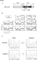
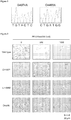

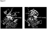
Classifications
-
- C—CHEMISTRY; METALLURGY
- C12—BIOCHEMISTRY; BEER; SPIRITS; WINE; VINEGAR; MICROBIOLOGY; ENZYMOLOGY; MUTATION OR GENETIC ENGINEERING
- C12Q—MEASURING OR TESTING PROCESSES INVOLVING ENZYMES, NUCLEIC ACIDS OR MICROORGANISMS; COMPOSITIONS OR TEST PAPERS THEREFOR; PROCESSES OF PREPARING SUCH COMPOSITIONS; CONDITION-RESPONSIVE CONTROL IN MICROBIOLOGICAL OR ENZYMOLOGICAL PROCESSES
- C12Q1/00—Measuring or testing processes involving enzymes, nucleic acids or microorganisms; Compositions therefor; Processes of preparing such compositions
- C12Q1/48—Measuring or testing processes involving enzymes, nucleic acids or microorganisms; Compositions therefor; Processes of preparing such compositions involving transferase
- C12Q1/485—Measuring or testing processes involving enzymes, nucleic acids or microorganisms; Compositions therefor; Processes of preparing such compositions involving transferase involving kinase
-
- C—CHEMISTRY; METALLURGY
- C12—BIOCHEMISTRY; BEER; SPIRITS; WINE; VINEGAR; MICROBIOLOGY; ENZYMOLOGY; MUTATION OR GENETIC ENGINEERING
- C12Q—MEASURING OR TESTING PROCESSES INVOLVING ENZYMES, NUCLEIC ACIDS OR MICROORGANISMS; COMPOSITIONS OR TEST PAPERS THEREFOR; PROCESSES OF PREPARING SUCH COMPOSITIONS; CONDITION-RESPONSIVE CONTROL IN MICROBIOLOGICAL OR ENZYMOLOGICAL PROCESSES
- C12Q1/00—Measuring or testing processes involving enzymes, nucleic acids or microorganisms; Compositions therefor; Processes of preparing such compositions
- C12Q1/68—Measuring or testing processes involving enzymes, nucleic acids or microorganisms; Compositions therefor; Processes of preparing such compositions involving nucleic acids
- C12Q1/6876—Nucleic acid products used in the analysis of nucleic acids, e.g. primers or probes
- C12Q1/6883—Nucleic acid products used in the analysis of nucleic acids, e.g. primers or probes for diseases caused by alterations of genetic material
- C12Q1/6886—Nucleic acid products used in the analysis of nucleic acids, e.g. primers or probes for diseases caused by alterations of genetic material for cancer
-
- A—HUMAN NECESSITIES
- A61—MEDICAL OR VETERINARY SCIENCE; HYGIENE
- A61P—SPECIFIC THERAPEUTIC ACTIVITY OF CHEMICAL COMPOUNDS OR MEDICINAL PREPARATIONS
- A61P35/00—Antineoplastic agents
-
- A—HUMAN NECESSITIES
- A61—MEDICAL OR VETERINARY SCIENCE; HYGIENE
- A61P—SPECIFIC THERAPEUTIC ACTIVITY OF CHEMICAL COMPOUNDS OR MEDICINAL PREPARATIONS
- A61P35/00—Antineoplastic agents
- A61P35/02—Antineoplastic agents specific for leukemia
-
- A—HUMAN NECESSITIES
- A61—MEDICAL OR VETERINARY SCIENCE; HYGIENE
- A61P—SPECIFIC THERAPEUTIC ACTIVITY OF CHEMICAL COMPOUNDS OR MEDICINAL PREPARATIONS
- A61P35/00—Antineoplastic agents
- A61P35/04—Antineoplastic agents specific for metastasis
-
- A—HUMAN NECESSITIES
- A61—MEDICAL OR VETERINARY SCIENCE; HYGIENE
- A61P—SPECIFIC THERAPEUTIC ACTIVITY OF CHEMICAL COMPOUNDS OR MEDICINAL PREPARATIONS
- A61P43/00—Drugs for specific purposes, not provided for in groups A61P1/00-A61P41/00
-
- G—PHYSICS
- G01—MEASURING; TESTING
- G01N—INVESTIGATING OR ANALYSING MATERIALS BY DETERMINING THEIR CHEMICAL OR PHYSICAL PROPERTIES
- G01N33/00—Investigating or analysing materials by specific methods not covered by groups G01N1/00 - G01N31/00
- G01N33/15—Medicinal preparations ; Physical properties thereof, e.g. dissolubility
-
- G—PHYSICS
- G01—MEASURING; TESTING
- G01N—INVESTIGATING OR ANALYSING MATERIALS BY DETERMINING THEIR CHEMICAL OR PHYSICAL PROPERTIES
- G01N33/00—Investigating or analysing materials by specific methods not covered by groups G01N1/00 - G01N31/00
- G01N33/48—Biological material, e.g. blood, urine; Haemocytometers
- G01N33/50—Chemical analysis of biological material, e.g. blood, urine; Testing involving biospecific ligand binding methods; Immunological testing
- G01N33/53—Immunoassay; Biospecific binding assay; Materials therefor
- G01N33/574—Immunoassay; Biospecific binding assay; Materials therefor for cancer
- G01N33/57484—Immunoassay; Biospecific binding assay; Materials therefor for cancer involving compounds serving as markers for tumor, cancer, neoplasia, e.g. cellular determinants, receptors, heat shock/stress proteins, A-protein, oligosaccharides, metabolites
-
- G—PHYSICS
- G01—MEASURING; TESTING
- G01N—INVESTIGATING OR ANALYSING MATERIALS BY DETERMINING THEIR CHEMICAL OR PHYSICAL PROPERTIES
- G01N33/00—Investigating or analysing materials by specific methods not covered by groups G01N1/00 - G01N31/00
- G01N33/48—Biological material, e.g. blood, urine; Haemocytometers
- G01N33/50—Chemical analysis of biological material, e.g. blood, urine; Testing involving biospecific ligand binding methods; Immunological testing
- G01N33/68—Chemical analysis of biological material, e.g. blood, urine; Testing involving biospecific ligand binding methods; Immunological testing involving proteins, peptides or amino acids
- G01N33/6893—Chemical analysis of biological material, e.g. blood, urine; Testing involving biospecific ligand binding methods; Immunological testing involving proteins, peptides or amino acids related to diseases not provided for elsewhere
-
- C—CHEMISTRY; METALLURGY
- C12—BIOCHEMISTRY; BEER; SPIRITS; WINE; VINEGAR; MICROBIOLOGY; ENZYMOLOGY; MUTATION OR GENETIC ENGINEERING
- C12Q—MEASURING OR TESTING PROCESSES INVOLVING ENZYMES, NUCLEIC ACIDS OR MICROORGANISMS; COMPOSITIONS OR TEST PAPERS THEREFOR; PROCESSES OF PREPARING SUCH COMPOSITIONS; CONDITION-RESPONSIVE CONTROL IN MICROBIOLOGICAL OR ENZYMOLOGICAL PROCESSES
- C12Q2600/00—Oligonucleotides characterized by their use
- C12Q2600/106—Pharmacogenomics, i.e. genetic variability in individual responses to drugs and drug metabolism
-
- C—CHEMISTRY; METALLURGY
- C12—BIOCHEMISTRY; BEER; SPIRITS; WINE; VINEGAR; MICROBIOLOGY; ENZYMOLOGY; MUTATION OR GENETIC ENGINEERING
- C12Q—MEASURING OR TESTING PROCESSES INVOLVING ENZYMES, NUCLEIC ACIDS OR MICROORGANISMS; COMPOSITIONS OR TEST PAPERS THEREFOR; PROCESSES OF PREPARING SUCH COMPOSITIONS; CONDITION-RESPONSIVE CONTROL IN MICROBIOLOGICAL OR ENZYMOLOGICAL PROCESSES
- C12Q2600/00—Oligonucleotides characterized by their use
- C12Q2600/136—Screening for pharmacological compounds
-
- C—CHEMISTRY; METALLURGY
- C12—BIOCHEMISTRY; BEER; SPIRITS; WINE; VINEGAR; MICROBIOLOGY; ENZYMOLOGY; MUTATION OR GENETIC ENGINEERING
- C12Q—MEASURING OR TESTING PROCESSES INVOLVING ENZYMES, NUCLEIC ACIDS OR MICROORGANISMS; COMPOSITIONS OR TEST PAPERS THEREFOR; PROCESSES OF PREPARING SUCH COMPOSITIONS; CONDITION-RESPONSIVE CONTROL IN MICROBIOLOGICAL OR ENZYMOLOGICAL PROCESSES
- C12Q2600/00—Oligonucleotides characterized by their use
- C12Q2600/154—Methylation markers
-
- C—CHEMISTRY; METALLURGY
- C12—BIOCHEMISTRY; BEER; SPIRITS; WINE; VINEGAR; MICROBIOLOGY; ENZYMOLOGY; MUTATION OR GENETIC ENGINEERING
- C12Q—MEASURING OR TESTING PROCESSES INVOLVING ENZYMES, NUCLEIC ACIDS OR MICROORGANISMS; COMPOSITIONS OR TEST PAPERS THEREFOR; PROCESSES OF PREPARING SUCH COMPOSITIONS; CONDITION-RESPONSIVE CONTROL IN MICROBIOLOGICAL OR ENZYMOLOGICAL PROCESSES
- C12Q2600/00—Oligonucleotides characterized by their use
- C12Q2600/156—Polymorphic or mutational markers
-
- G—PHYSICS
- G01—MEASURING; TESTING
- G01N—INVESTIGATING OR ANALYSING MATERIALS BY DETERMINING THEIR CHEMICAL OR PHYSICAL PROPERTIES
- G01N2333/00—Assays involving biological materials from specific organisms or of a specific nature
- G01N2333/90—Enzymes; Proenzymes
- G01N2333/91—Transferases (2.)
- G01N2333/912—Transferases (2.) transferring phosphorus containing groups, e.g. kinases (2.7)
- G01N2333/91205—Phosphotransferases in general
- G01N2333/9121—Phosphotransferases in general with an alcohol group as acceptor (2.7.1), e.g. general tyrosine, serine or threonine kinases
Definitions
- Tyrosine kinases are a class of enzymes that catalyze phopshorylation of tyrosine residues of protein substrates via a transfer of the terminal phosphate of adenosine triphosphate.
- tyrosine kinases play critical roles in signal transduction for a number of cell functions, including cell proliferation, carcinogenesis, and cell differentiation.
- EML4-ALK is a fusion-type protein tyrosine kinase that is present in ⁇ 5% of non-small cell lung cancer (NSCLC) cases and which is generated as a result of a small inversion within the short arm of human chromosome 2 ( Soda , M. et al. (2007) Nature 448:561-566 ; Mano, H. (2008) Cancer Sci. 99:2349-2355 ). EML4-ALK undergoes constitutive dimerization as a result of interaction between the coiled-coil domain within the EML4 region of each monomer and thereby acquires pronounced oncogenic activity.
- NSCLC non-small cell lung cancer
- TKIs tyrosine kinase inhibitors
- Imatinib mesylate a TKI for ABL1 and KIT, for instance, markedly improves the outcome of individuals with chronic myeloid leukemia positive for the BCR-ABL1 fusion kinase or with a gastrointestinal stromal tumor positive for activated KIT ( Druker, B.J. et al. (2001) N. Engl. J. Med. 344:1031-1037 ; Heinrich, M.C. et al. (2008) J. Clin. Oncol. 26:5360-5367 ).
- gefitinib and erlotinib both of which are TKIs for the epidermal growth factor receptor (EGFR) are effective in the treatment of NSCLC associated with EGFR activation ( Mok, T.S. et al. (2009) J. Clin. Oncol. 27:5080-2087 ; Mok, T.S. et al. (2009) N. Engl. J. Med. 361:947-957 ).
- EGFR epidermal growth factor receptor
- WO2010/132888 discloses methods and compositions for detecting the presence of a cancer in a subject and assessing the efficacy of treatments for the same.
- WO2009/103061 discloses methods and compositions for identifying, diagnosing and treating neuroblastoma.
- the present disclosure provides, at least, methods for the identification and, assessment of cancer based upon the identification of novel anaplastic lymphoma kinase (ALK) mutation(s) conferring resistance to known ALK inhibitors.
- ALK anaplastic lymphoma kinase
- Such ALK mutations are also clinically relevant for the identification of pharmaceutical compositions that are able to fit into the abnormal ATP-binding pocket of generated by the novel ALK mutation(s) and inhibit ALK activity.
- the present invention provides a method for identifying a subject having cancer or at risk for developing cancer as having an increased risk of unresponsiveness to treatment with an ALK inhibitor, comprising:
- the present invention provides an in vitro method of determining whether a test compound modulates activity of one or more mutant ALK polypeptides comprising:
- the present invention provides use of a mutant ALK polynucleotide molecule encoding a mutant ALK polypeptide comprising a mutation at a position corresponding to position 1156 of the wild type human ALK or the use of a mutant ALK polypeptide comprising a mutation at a position corresponding to position 1156 of the wild type human ALK in an in vitro method of diagnosis of a subject's increased risk of unresponsiveness to a treatment with an ALK inhibitor.
- the subject has not previously been treated with an ALK inhibitor, or has been previously treated with an ALK inhibitor and has developed at least partial resistance to the ALK inhibitor (e.g ., PF-02341066, PDD, 2-methyl-11-(2-methylpropyl)-4-oxo-4,5,6,11,12,13-hexahydro-2 H- indazolo[5,4- a ]pyrrolo[3,4- c ]carbazol-8-yl [4-(dimethylamino) benzyl]carbamate, (1 S ,2 S ,3 R ,4 R )-3-( ⁇ 5-chloro-2-[(1-ethyl-2,3,4,5-tetrahydro-6-methoxy-2-oxo-1 H -1-benzazepin-7-yl)amino]-4-pyrimidinyl ⁇ amino)bicyclo[2.2.1]hept-5-ene-2-carboxamide, and
- the cancer is selected from the group consisting of anaplastic large cell lymphoma, neuroblastoma, breast cancer, colorectal cancer, inflammatory myofibroblastic tumors, and non-small cell lung cancers.
- the sample is selected from the group consisting of sputum, bronchoalveolar lavage, pleural effusion, tissue, whole blood, serum, plasma, buccal scrape, saliva, cerebrospinal fluid, urine, stool, circulating tumor cells, circulating nucleic acids, and bone marrow.
- the sample comprises cells or tissue.
- the tissue is a tumor or cancer tissue.
- the one or more mutant ALK polynucleotide molecules or polypeptides are selected from the group consisting of the mutant ALK polynucleotide molecules or polypeptides listed in Table 1.
- the one or more ALK mutations are assessed by a nucleic acid hybridization assay.
- the one or more ALK mutations are assessed by polymerase chain reaction.
- the expression level of the one or more ALK polypeptides is detected using a reagent which specifically binds to one or more ALK polypeptides (e.g., antibody, an antibody derivative, and an antibody fragment).
- the amount, structure and/or activity of the one or more mutant ALK polypeptides is compared to a control sample.
- the one or more ALK mutations are assessed at a first point in time and at least one subsequent point in time.
- the sample comprises germline or somatic genomic DNA.
- the present disclosure provides a method of treating a patient having cancer, or at risk for developing cancer, comprising collecting a sample from the patient, analyzing the sample to detect the presence of one or more mutant ALK polynucleotide molecules set forth in Table 1, and administering to said patient a therapeutically effective amount of an ALK inhibitor.
- the ALK inhibitor is selected from the group consisting of PF-02341066, PDD, 2-methyl-11-(2-methylpropyl)-4-oxo-4,5,6,1 1,12,13-hexahydro-2 H -indazolo[5,4- a ]pyrrolo[3,4- c ]carbazol-8-yl [4-(dimethylamino) benzyl]carbamate, (1 S ,2 S ,3 R ,4 R )-3-( ⁇ 5-chloro-2-[(1-ethyl-2,3,4,5-tetrahydro-6-methoxy-2-oxo-1 H -1-benzazepin-7-yl)amino]-4-pyrimidinyl ⁇ amino)bicyclo[2.2.1]hept-5-ene-2-carboxamide, and NVP-TAE684.
- the subject has not previously been treated with an ALK inhibitor, or has been previously treated with an ALK inhibitor and has
- kits for determining the chemosensitivity of a cancer patient to treatment with an ALK inhibitor comprising: a reagent that specifically binds to one or more mutant ALK polynucleotide molecules or polypeptides; and instructions for use.
- the kit further comprises an ALK inhibitor.
- the reagent comprises one or more polynucleotide probes, each of which comprises a polynucleotide sequence which is complementary to a nucleotide sequence listed in Table 1 or complementary to a nucleotide sequence encoding a polypeptide listed in Table 1 (e.g., oligonucleotides, cDNA molecules, RNA molecules, and synthetic gene probes comprising nucleobases).
- the probes comprise polynucleotides from about 50 to 10 7 nucleotides in length.
- the reagent comprises an antibody, and antibody derivative, and an antibody fragment to a polypeptide encoded by one or more polynucleotide sequences listed in Table 1.
- the present disclosure provides a method of determining whether a test compound modulates activity of one or more mutant ALK polypeptides comprising contacting mammalian cells transfected with a construct encoding the one or more mutant ALK polypeptides with the test compound and assessing the mammalian cells for activity of the one or more mutant ALK polypeptides, wherein significantly modulated activity in the presence of the test compound relative to a control experiment identifies the test compound as a modulator of the one or more mutant ALK polypeptides.
- the one or more mutant ALK polynucleotide molecules or polypeptides are selected from the group consisting of the mutant ALK polynucleotide molecules or polypeptides listed in Table 1.
- control comprises mammalian cells expressing a wild type ALK polypeptide selected from the group consisting of polypeptides listed in Table 1.
- activity of the one or more mutant ALK polypeptides is selected from the group consisting of ATP binding, tyrosine kinase activity, cancer cell proliferation, tumor growth, tumor number, apoptosis, and tumor metastasis.
- control experiment comprises mammalian cells expressing the one or more mutant ALK polypeptides in the absence of the test compound as determined by, for example, activity of the one or more mutant ALK polypeptides (e.g., ATP binding, tyrosine kinase activity, cancer cell proliferation, tumor growth, tumor number, apoptosis, and tumor metastasis).
- activity of the one or more mutant ALK polypeptides e.g., ATP binding, tyrosine kinase activity, cancer cell proliferation, tumor growth, tumor number, apoptosis, and tumor metastasis.
- the present invention is based, at least in part, on the identification of specific regions of the genome, including for example, Anaplastic Lymphoma Kinase (ALK) mutations, associated with predicting efficacy of ALK inhibitors in treating cancer.
- ALK Anaplastic Lymphoma Kinase
- novel ALK gene mutations e.g., EML4-ALK polypeptide encoding mutations
- the present disclosure further provides methods for identifying such specific genomic regions using techniques known in the art, including, but not limited to, oligonucleotide-based microarrays ( Brennan, et al. (2004) Cancer Res. 64(14):4744-8 ; Lucito, et al.
- the present invention further provides diagnostic kits for use in the methods.
- an element means one element or more than one element.
- altered amount of a marker or “altered level” of a marker refers to increased or decreased copy number of a marker or chromosomal region, such as ALK gene mutations and/or gene products (e.g., the markers set forth in Table 1), and/or increased or decreased expression level of a particular marker gene or genes in a cancer sample, as compared to the expression level or copy number of the marker in a control sample.
- altered amount of a marker also includes an increased or decreased protein level of a marker in a sample, e.g., a cancer sample, as compared to the protein level of the marker in a normal, control sample.
- altered level of expression of ALK gene mutations and/or gene products refers to an expression level or copy number of a marker in a test sample such as a sample derived from a patient suffering from cancer, that is greater or less than the standard error of the assay employed to assess expression or copy number, and may be at least twice, at least twice three, at least twice four, at least twice five, or at least twice ten or more times the expression level or copy number of the ALK gene mutations and/or gene products (e.g ., the markers set forth in Table 1) in a control sample (e.g., a sample from a healthy subject not having the associated disease), or the average expression level or copy number of the ALK gene mutations and/or gene products (e.g., the markers set forth in Table 1) in several control samples.
- a control sample e.g., a sample from a healthy subject not having the associated disease
- the average expression level or copy number of the ALK gene mutations and/or gene products e.g., the markers set forth in Table 1
- the altered level of expression is greater or less than the standard error of the assay employed to assess expression or copy number, and is at least twice, at least three, at least four, at least five, at least ten or more times the expression level or copy number of the ALK gene mutations and/or gene products (e.g., the markers set forth in Table 1) in a control sample (e.g., a sample from a healthy subject not having the associated disease), or the average expression level or copy number of the ALK gene mutations and/or gene products (e.g., the markers set forth in Table 1) in several control samples.
- a control sample e.g., a sample from a healthy subject not having the associated disease
- the average expression level or copy number of the ALK gene mutations and/or gene products e.g., the markers set forth in Table 1
- altered activity of a marker refers to an activity of a marker which is increased or decreased in a disease state, e.g., in a cancer sample, as compared to the activity of the marker in a normal, control sample.
- Altered activity of a marker may be the result of, for example, altered expression of the marker, altered protein level of the marker, altered structure of the marker, or, e.g., an altered interaction with other proteins involved in the same or different pathway as the marker.
- altered structure of a marker refers to the presence of mutations or mutations within the marker gene or maker protein, e.g., mutations which affect expression or activity of the marker, as compared to the normal or wild-type gene or protein.
- mutations include, but are not limited to inter- and intra-chromosomal rearrangement, substitutions, deletions, and insertion mutations. Mutations may be present in the coding or non-coding region of the marker.
- ALK aplastic lymphoma kinase
- ALK protein is represented by NCBI Ref Seq identification number NP_004295. Unless indicated otherwise, the terms refer to the human protein.
- the gene encoding ALK may also be referred to herein as "ALK”.
- ALK nucleotide sequences are represented by NCBI Ref Seq identification number NM_004304.3 and GenBank accession number 29029631, relevant sequences therein (e.g ., the coding, 5' UTR, 3'UTR, transcription start, translation start, transcription stop, translation stop, etc. sequences) of which can readily be identified by a skilled artisan.
- ALK fusion kinases and variants thereof are also used herein to include ALK fusion kinases and variants thereof, which are well known to a skilled artisan.
- ALK fusion kinases and variants thereof comprise ALK kinase activity and can harbor mutations as described herein rendering the ALK kinase activity resistant to ALK inhibitors.
- EML4-ALK Variant 1 (AB274722.1; BAF73611.1), EML4-ALK Variant 2 (AB275889.1; BAF73612.1), EML4-ALK Variant 3a (AB374361.1; BAG55003.1), EML4-ALK Variant 3b (AB374362.1; BAG55004.1), EML4-ALK Variant 4 (AB374363.1; BAG75147.1), EML4-ALK Variant 5a (AB374364.1; BAG75148.1), EML4-ALK Variant 5b (AB374365.1; BAG75149.1), EML4-ALK Variant 6 (AB462411.1; BAH57335.1), EML4-ALK Variant 7 (AB462412.1; BAH57336.1), KIF5B-ALK (AB462413.1; BAH57337.1), NPM-ALK, TPM3-ALK, TFGXL-ALK, TFGL-ALK,
- ALK kinase variants can arise depending upon the particular fusion event between an ALK kinase and its fusion partner (e.g ., EML4 can fuse at least exon 2, 6a, 6b, 13, 14, and/or 15, as described, for example, in Horn and Pao, (2009) J. Clin. Oncol. 27:4247-4253 ).
- Wild Type ALK cDNA Sequence (NM_004304.3; GI:29029631): Wild Type cDNA sequence TGC (4373 to 4375) codon mutation(s) encoding an amino acid other than cysteine or a corresponding mutation in a homolog thereof Wild Type cDNA sequence CTG (4493 to 4495) codon mutation(s) encoding an amino acid other than leucine or a corresponding mutation in a homolog thereof Wild Type cDNA sequence G4374A mutation or a corresponding mutation in a homolog thereof Wild Type cDNA sequence C4493A mutation or a corresponding mutation in a homolog thereof Wild Type ALK Protein Sequence (NP_004295.2; GI:29029632): Wild Type protein sequence Cys1156Xaa mutation wherein Xaa is an amino acid other than cysteine or a corresponding mutation in a homolog thereof Wild Type protein sequence Leu1196Xaa mutation wherein Xaa
- ALK mutations generally refer to alterations in a nucleic acid and/or amino acid sequence relative to a reference anaplastic lymphoma kinase sequence. In some embodiments, however, “ALK mutations” can refer to specific anaplastic lymphomas kinase mutations predictive of response to treatment with ALK inhibiting agents (e.g., PF-02341066 and/or PDD). For example, mutations of the cysteine amino acid at position 1156 (C1156) and/or the leucine amino acid at position 1196 (L1196) of wild type ALK protein (NP_004295) to a different amino acid are described herein to confer resistance to ALK inhibiting agents.
- the C1156 position comprises a tyrosine amino acid and/or the L1196 position comprises a methionine amino acid.
- a skilled artisan will also recognize that amino acid positions corresponding to the "C1156" and "L1196" mutations of wild type ALK protein will have different numbers relative to different reference sequences (e.g., ALK homologs, ALK fusion proteins, etc.) without affecting the predictive value of response to treatment with ALK inhibiting agents (e.g., PF-02341066 and/or PDD).
- the present invention relates to mutations at the 1156 position of the wild type human ALK.
- nucleotide triplet An important and well known feature of the genetic code is its redundancy, whereby, for most of the amino acids used to make proteins, more than one coding nucleotide triplet may be employed (for example, illustrated above). Therefore, a number of different nucleotide sequences may code for a given amino acid sequence. Such nucleotide sequences are considered functionally equivalent since they result in the production of the same amino acid sequence in all organisms (although certain organisms may translate some sequences more efficiently than they do others). Moreover, occasionally, a methylated variant of a purine or pyrimidine may be found in a given nucleotide sequence. Such methylations do not affect the coding relationship between the trinucleotide codon and the corresponding amino acid.
- Binding compound shall refer to a binding composition, such as a small molecule, an antibody, a peptide, a peptide or non-peptide ligand, a protein, an oligonucleotide, an oligonucleotide analog, such as a peptide nucleic acid, a lectin, or any other molecular entity that is capable of specifically binding to a target protein or molecule or stable complex formation with an analyte of interest, such as a complex of proteins.
- Binding moiety means any molecule to which molecular tags can be directly or indirectly attached that is capable of specifically binding to an analyte. Binding moieties include, but are not limited to, antibodies, antibody binding compositions, peptides, proteins, nucleic acids and organic molecules having a molecular weight of up to about 1000 daltons and containing atoms selected from the group consisting of hydrogen, carbon, oxygen, nitrogen, sulfur and phosphorus.
- a “biomarker” or “marker” is a gene, mRNA, or protein which may be altered, wherein said alteration is associated with cancer.
- the alteration may be in amount, structure, and/or activity in a cancer tissue or cancer cell, as compared to its amount, structure, and/or activity, in a normal or healthy tissue or cell ( e.g., a control), and is associated with a disease state, such as cancer.
- a marker of the present invention which is associated with cancer or predictive of responsiveness to anti-cancer therapeutics may have an altered nucleotide sequence, amino acid sequence, chromosomal translocation, intra-chromosomal inversion, copy number, expression level, protein level, protein activity, or methylation status, in a cancer tissue or cancer cell as compared to a normal, healthy tissue or cell.
- a "marker" includes a molecule whose structure is altered, e.g., mutated (contains an mutation), e.g., differs from the wild type sequence at the nucleotide or amino acid level, e.g., by substitution, deletion, or insertion, when present in a tissue or cell associated with a disease state, such as cancer.
- cancer refers to the presence of cells possessing characteristics typical of cancer-causing cells, such as uncontrolled proliferation, immortality, metastatic potential, rapid growth and proliferation rate, and certain characteristic morphological features. Cancer cells are often in the form of a tumor, but such cells may exist alone within an animal, or may be a non-tumorigenic cancer cell, such as a leukemia cell. As used herein, the term “cancer” includes premalignant as well as malignant cancers.
- Cancers include, but are not limited to, B cell cancer, e.g., multiple myeloma, Waldenstrom's macroglobulinemia, the heavy chain diseases, such as, for example, alpha chain disease, gamma chain disease, mu chain disease, benign monoclonal gammopathy, immunocytic amyloidosis, melanomas, breast cancer, lung cancer (such as non-small cell lung carcinoma or NSCLC), bronchus cancer, colorectal cancer, prostate cancer, pancreatic cancer, stomach cancer, ovarian cancer, urinary bladder cancer, brain or central nervous system cancer, peripheral nervous system cancer, esophageal cancer, cervical cancer, uterine or endometrial cancer, cancer of the oral cavity or pharynx, liver cancer, kidney cancer, testicular cancer, biliary tract cancer, small bowel or appendix cancer, salivary gland cancer, thyroid gland cancer, adrenal gland cancer, osteosarcoma, chondrosarcoma, cancer of hematological tissues, aden
- “Chemotherapeutic agent” means a chemical substance, such as a cytotoxic or cytostatic agent, that is used to treat a condition, e.g., cancer.
- “Complementary” refers to the broad concept of sequence complementarity between regions of two nucleic acid strands or between two regions of the same nucleic acid strand. It is known that an adenine residue of a first nucleic acid region is capable of forming specific hydrogen bonds ("base pairing") with a residue of a second nucleic acid region which is antiparallel to the first region if the residue is thymine or uracil. Similarly, it is known that a cytosine residue of a first nucleic acid strand is capable of base pairing with a residue of a second nucleic acid strand which is antiparallel to the first strand if the residue is guanine.
- a first region of a nucleic acid is complementary to a second region of the same or a different nucleic acid if, when the two regions are arranged in an antiparallel fashion, at least one nucleotide residue of the first region is capable of base pairing with a residue of the second region.
- the first region comprises a first portion and the second region comprises a second portion, whereby, when the first and second portions are arranged in an antiparallel fashion, at least about 50%, at least about 75%, at least about 90%, or at least about 95% of the nucleotide residues of the first portion are capable of base pairing with nucleotide residues in the second portion.
- all nucleotide residues of the first portion are capable of base pairing with nucleotide residues in the second portion.
- the "copy number of a gene” or the “copy number of a marker” refers to the number of DNA sequences in a cell encoding a particular gene product. Generally, for a given gene, a mammal has two copies of each gene. The copy number can be increased, however, by gene amplification or duplication, or reduced by deletion.
- a marker is "fixed" to a substrate if it is covalently or non-covalently associated with the substrate such that the substrate can be rinsed with a fluid (e.g., standard saline citrate, pH 7.4) without a substantial fraction of the marker dissociating from the substrate.
- a fluid e.g., standard saline citrate, pH 7.4
- “Hazard ratio”, as used herein, refers to a statistical method used to generate an estimate for relative risk. "Hazard ratio” is the ratio between the predicted hazard of one group versus another group. For example, patient populations treated with an ALK inhibiting agent versus without an ALK inhibiting agent can be assessed for whether or not the ALK inhibiting agent is effective in increasing the time to distant recurrence of disease, particularly with regard to ALK mutation status. For example, treating subjects harboring ALK mutations in cancerous tissue, as described herein, results in increased therapeutic benefit from ALK inhibiting agents relative to subjects not having said ALK mutations in cancerous tissue.
- ALK inhibiting agent refers to a compound that can inhibit the biological activity of ALK. Biological activities can also include patient response as set forth in this application.
- Exemplary ALK inhibiting agents include, but are not limited to, PF-02341066, PDD, 2-methyl-11-(2-methylpropyl)-4-oxo-4,5,6,11,12,13-hexahydro-2 H -indazolo[5,4- a ]pyrrolo[3,4- c ]carbazol-8-yl [4-(dimethylamino) benzyl]carbamate,(1 S ,2 S ,3 R ,4 R )-3-( ⁇ 5-chloro-2-[(1-ethyl-2,3,4,5-tetrahydro-6-methoxy-2-oxo-1 H -1-benzazepin-7-yl)amino]-4-pyrimidinyl ⁇ amino)bicyclo[2.2.1]hept
- sequence similarity refers to sequence similarity between two polynucleotide sequences or between two polypeptide sequences, with identity being a more strict comparison.
- percent identity or homology and “% identity or homology” refer to the percentage of sequence similarity found in a comparison of two or more polynucleotide sequences or two or more polypeptide sequences.
- sequence similarity refers to the percent similarity in base pair sequence (as determined by any suitable method) between two or more polynucleotide sequences. Two or more sequences can be anywhere from 0-100% similar, or any integer value there between.
- Identity or similarity can be determined by comparing a position in each sequence that may be aligned for purposes of comparison. When a position in the compared sequence is occupied by the same nucleotide base or amino acid, then the molecules are identical at that position.
- a degree of similarity or identity between polynucleotide sequences is a function of the number of identical or matching nucleotides at positions shared by the polynucleotide sequences.
- a degree of identity of polypeptide sequences is a function of the number of identical amino acids at positions shared by the polypeptide sequences.
- a degree of homology or similarity of polypeptide sequences is a function of the number of amino acids at positions shared by the polypeptide sequences.
- the term "substantial homology,” as used herein, refers to homology of at least 50%, at least 60%, at least 65%, at least 70%, at least 75%, at least 80%, at least 85%, at least 90%, at least 95% or more.
- Cancer is "inhibited” if at least one symptom of the cancer is alleviated, terminated, slowed, or prevented. As used herein, cancer is also “inhibited” if recurrence or metastasis of the cancer is reduced, slowed, delayed, or prevented.
- a “marker nucleic acid” or “biomarker nucleic acid” is a nucleic acid (e.g., DNA, mRNA, cDNA) encoded by or corresponding to a marker of the present invention.
- marker nucleic acid molecules include DNA (e.g., genomic DNA and cDNA) comprising the entire or a partial sequence of any of the nucleic acid sequences set forth in Table 1 or the complement or hybridizing fragment of such a sequence.
- the marker nucleic acid molecules also include RNA comprising the entire or a partial sequence of any of the nucleic acid sequences set forth in Table 1 or the complement of such a sequence, wherein all thymidine residues are replaced with uridine residues.
- a “marker protein” is a protein encoded by or corresponding to a marker of the present invention.
- a marker protein comprises the entire or a partial sequence of a protein encoded by any of the sequences set forth in Table 1 or a fragment thereof.
- the terms “protein” and “polypeptide” are used interchangeably herein.
- the "normal" copy number of a marker or “normal” level of expression of a marker is the level of expression, copy number of the marker, in a biological sample, e.g., a sample containing sputum, bronchoalveolar lavage, pleural effusion, tissue, whole blood, serum, plasma, buccal scrape, saliva, cerebrospinal fluid, urine, stool, and bone marrow, from a subject, e.g., a human, not afflicted with cancer.
- a biological sample e.g., a sample containing sputum, bronchoalveolar lavage, pleural effusion, tissue, whole blood, serum, plasma, buccal scrape, saliva, cerebrospinal fluid, urine, stool, and bone marrow
- An "overexpression” or “significantly higher level of expression, copy number, and/or activity” of ALK gene mutations and/or gene products refers to an expression level, copy number, and/or activity in a test sample that is greater than the standard error of the assay employed to assess expression or copy number, and may be at least two, at least three, at least four, at least five, or at least ten or more times the expression level or copy number of the ALK gene mutations and/or gene products (e.g., the markers set forth in Table 1) in a control sample (e.g., a sample from a healthy subject not afflicted with cancer), or the average expression level or copy number of the ALK gene mutations and/or gene products (e.g., the markers set forth in Table 1) in several control samples.
- a control sample e.g., a sample from a healthy subject not afflicted with cancer
- the average expression level or copy number of the ALK gene mutations and/or gene products e.g., the markers set forth in Table
- probe refers to any molecule which is capable of selectively binding to a specifically intended target molecule, for example a marker of the present invention. Probes can be either synthesized by one skilled in the art, or derived from appropriate biological preparations. For purposes of detection of the target molecule, probes may be specifically designed to be labeled, as described herein. Examples of molecules that can be utilized as probes include, but are not limited to, RNA, DNA, proteins, antibodies, and organic monomers.
- RECIST shall mean an acronym that stands for "Response Evaluation Criteria in Solid Tumours” and is a set of published rules that define when cancer patients improve (“respond”), stay the same (“stable”) or worsen ("progression") during treatments. Response as defined by RECIST criteria have been published, for example, at Journal of the National Cancer Institute, Vol. 92, No. 3, Feb. 2, 2000 and RECIST criteria may include other similar published definitions and rule sets. One skilled in the art would understand definitions that go with RECIST criteria, as used herein, such as "PR,” “CR,” “SD” and "PD.”
- Responsiveness to “respond” to treatment, and other forms of this verb, as used herein, refer to the reaction of a subject to treatment with an ALK inhibiting agent.
- a subject responds to treatment with an ALK inhibiting agent if growth of a tumor in the subject is retarded about 10%, about 20%, about 30%, about 40%, about 50%, about 60%, about 70%, about 80%, about 90% or more.
- a subject responds to treatment with an ALK inhibiting agent if a tumor in the subject shrinks by about 5%, about 10%, about 20%, about 30%, about 40%, about 50% or more as determined by any appropriate measure, e.g., by mass or volume.
- a subject responds to treatment with an ALK inhibiting agent if the subject experiences a life expectancy extended by about 5%, about 10%, about 20%, about 30%, about 40%, about 50% or more beyond the life expectancy predicted if no treatment is administered.
- a subject responds to treatment with an ALK inhibiting agent if the subject has an increased disease-free survival, overall survival or increased time to progression.
- tissue sample each refer to a collection of similar cells obtained from a tissue of a subject or patient.
- the source of the tissue sample may be solid tissue as from a fresh, frozen and/or preserved organ, tissue sample, biopsy, or aspirate; blood or any blood constituents; bodily fluids such as cerebral spinal fluid, amniotic fluid, peritoneal fluid or interstitial fluid; or cells from any time in gestation or development of the subject.
- the tissue sample may contain compounds that are not naturally intermixed with the tissue in nature such as preservatives, anticoagulants, buffers, fixatives, nutrients, antibiotics or the like.
- the amount of a marker e.g., expression or copy number of ALK gene mutations and/or gene products (e.g., the markers set forth in Table 1), in a subject is "significantly" higher or lower than the normal amount of a marker, if the amount of the marker is greater or less, respectively, than the normal level by an amount greater than the standard error of the assay employed to assess amount, or at least two, at least three, at least four, at least five, at least ten or more times that amount.
- a marker e.g., expression or copy number of ALK gene mutations and/or gene products
- the amount of the marker in the subject can be considered "significantly" higher or lower than the normal amount if the amount is at least about two, at least about three, at least about four, or at least about five times, higher or lower, respectively, than the normal amount of the marker.
- significant event shall refer to an event in a patient's disease that is important as determined by one skilled in the art.
- significant events include, for example, without limitation, primary diagnosis, death, recurrence, the determination that a patient's disease is metastatic, relapse of a patient's disease or the progression of a patient's disease from any one of the above noted stages to another.
- a significant event may be any important event used to assess OS, TTP and/or using the RECIST or other response criteria, as determined by one skilled in the art.
- the terms “subject” and “patient” are used interchangeably.
- the terms “subject” and “subjects” refer to an animal, e.g., a mammal including a non-primate (e.g., a cow, pig, horse, donkey, goat, camel, cat, dog, guinea pig, rat, mouse, sheep) and a primate (e.g., a monkey, such as a cynomolgous monkey, gorilla, chimpanzee and a human).
- a non-primate e.g., a cow, pig, horse, donkey, goat, camel, cat, dog, guinea pig, rat, mouse, sheep
- a primate e.g., a monkey, such as a cynomolgous monkey, gorilla, chimpanzee and a human.
- time course shall refer to the amount of time between an initial event and a subsequent event.
- time course may relate to a patient's disease and may be measured by gauging significant events in the course of the disease, wherein the first event may be diagnosis and the subsequent event may be metastasis, for example.
- Time to progression refers to a time as measured from the start of the treatment to progression or a cancer or censor. Censoring may come from a study end or from a change in treatment. Time to progression can also be represented as a probability as, for example, in a Kaplein-Meier plot where time to progression may represent the probability of being progression free over a particular time, that time being the time between the start of the treatment to progression or censor.
- a “transcribed polynucleotide” is a polynucleotide (e.g., an RNA, a cDNA, or an analog of one of an RNA or cDNA) which is complementary to or homologous with all or a portion of a mature RNA made by transcription of a marker of the present invention and normal post-transcriptional processing (e.g., splicing), if any, of the transcript, and reverse transcription of the transcript.
- normal post-transcriptional processing e.g., splicing
- Treatment refers to the administration of an ALK inhibiting agent to impede growth of a cancer, to cause a cancer to shrink by weight or volume, to extend the expected survival time of the subject and or time to progression of the tumor or the like.
- An "underexpression” or “significantly lower level of expression, copy number, and/or activity" of ALK gene mutations and/or gene products refers to an expression level or copy number in a test sample that is greater than the standard error of the assay employed to assess expression or copy number, for example, at least twice, at least three, at least four, at least five, or at least ten or more times less than the expression level, copy number, and/or activity of the ALK gene mutations and/or gene products (e.g., the markers set forth in Table 1) in a control sample (e.g., a sample from a healthy subject not afflicted with cancer), or the average expression level, copy number, and/or activity of the ALK gene mutations and/or gene products (e.g ., the markers set forth in Table 1) in several control samples.
- a control sample e.g., a sample from a healthy subject not afflicted with cancer
- the present invention is based, at least in part, on the identification of specific regions of the genome, including, for example, ALK mutations, associated with predicting efficacy of ALK inhibitors in treating cancer.
- ALK polypeptides e.g., biomarkers listed in Table 1, including EML4-ALK polypeptides
- the presence and/or absence of one or more of such biomarkers in various methods described herein is within the scope of the present disclosure.
- Embodiments of the invention relate to mutations at the 1156 position of wild type human ALK.
- methods of the present invention may be used to monitor the progression of cancer in a subject, wherein if a sample in a subject presents one or more ALK mutations (e.g., EML4-ALK mutations) identified herein during the progression of cancer, e.g., at a first point in time and a subsequent point in time, then the cancer is less likely to respond to ALK inhibitor-mediated treatment and vice versa.
- the subject has undergone treatment, e.g., chemotherapy, radiation therapy, surgery, or any other therapeutic approach useful for inhibiting cancer, has completed treatment, or is in remission.
- one or more biomarkers of the present inventions can be specifically identified by the presence in genomic (e.g., germline and/or somatic) sequence when compared to a reference sequence, such as SEQ ID NO:1.
- genomic e.g., germline and/or somatic sequence
- the methods described herein can involve detecting biomarkers of the present invention by carrying out a target nucleic acid amplification reaction of a stretch of DNA comprising one or more mutations listed in Table 1 and analyzing the amplified target nucleic acid for the presence of the one or more mutations.
- PCR Polymerase Chain Reaction
- Detection of the presence of one or more of the mutations in the amplified product can be performed in various manners that are well known in the art, such as DNA sequencing methodologies like Sanger sequencing and deep sequencing, use of restriction enzymes, allele specific amplification, Peptide Nucleic Acid (PNA)-mediated PCR, detection of conformational differences, like Single Strand Conformation Polymorphism (SSCP) and Denaturing Gradient Gel Electrophoresis (DGGE) assays with detection steps on membranes (dot blot) using labeled oligonucleotide probes, assays with detection steps in microtiter plates, like Reverse Hybridization, Oligonucleotide Ligation Assay (OLA, MLPA), First Nucleotide Change (FNC) technology, Cross-linking technology, Rapid cycle PCR and simultaneous fluorescence analysis (e.g. 5'nuclease/Taqman), and PCR followed by mini-sequencing using mass spectrometry or capillar
- nucleic acid molecules that correspond to a biomarker of the present invention, including nucleic acids which encode a polypeptide corresponding to a marker of the present invention or a portion of such a polypeptide.
- the nucleic acid molecules of the present disclosure include those nucleic acid molecules which reside in ALK or ALK-related genomic (e.g., germline and/or somatic) regions identified herein and/or encode ALK or ALK-related (e.g., EML4-ALK) polypeptides.
- the nucleic acid molecules may comprise, consist essentially of, or consist of the nucleic sequences, or fragments thereof, presented in Table 1.
- Isolated nucleic acid molecules also include nucleic acid molecules sufficient for use as hybridization probes to identify nucleic acid molecules that correspond to a marker of the present invention, including nucleic acid molecules which encode a polypeptide corresponding to a marker of the present invention, and fragments of such nucleic acid molecules, e.g., those suitable for use as PCR primers for the amplification or mutation of nucleic acid molecules.
- nucleic acid molecule is intended to include DNA molecules (e.g., cDNA or genomic DNA) and RNA molecules (e.g., mRNA) and analogs of the DNA or RNA generated using nucleotide analogs.
- the nucleic acid molecule can be single-stranded or double-stranded; in certain embodiments the nucleic acid molecule is double-stranded DNA.
- an “isolated” nucleic acid molecule is one which is separated from other nucleic acid molecules which are present in the natural source of the nucleic acid molecule.
- an “isolated” nucleic acid molecule is free of sequences (such as protein-encoding sequences) which naturally flank the nucleic acid (i.e., sequences located at the 5' and 3' ends of the nucleic acid) in the genomic DNA of the organism from which the nucleic acid is derived.
- the isolated nucleic acid molecule can contain less than about 5 kB, less than about 4 kB, less than about 3 kB, less than about 2 kB, less than about 1 kB, less than about 0.5 kB or less than about 0.1 kB of nucleotide sequences which naturally flank the nucleic acid molecule in genomic DNA of the cell from which the nucleic acid is derived.
- an "isolated" nucleic acid molecule such as a cDNA molecule, can be substantially free of other cellular material or culture medium when produced by recombinant techniques, or substantially free of chemical precursors or other chemicals when chemically synthesized.
- nucleic acid molecule that is substantially free of cellular material includes preparations of nucleic acid molecule having less than about 30%, less than about 20%, less than about 10%, or less than about 5% (by dry weight) of other cellular material or culture medium.
- a nucleic acid molecule of the present disclosure can be isolated using standard molecular biology techniques and the sequence information in the database records described herein. Using all or a portion of such nucleic acid sequences, nucleic acid molecules of the present disclosure can be isolated using standard hybridization and cloning techniques (e.g., as described in Sambrook et al., ed., Molecular Cloning: A Laboratory Manual, 2nd ed., Cold Spring Harbor Laboratory Press, Cold Spring Harbor, NY, 1989 ).
- a nucleic acid molecule of the present disclosure can be amplified using cDNA, mRNA, or genomic DNA (e.g., germline and/or somatic genomic DNA) as a template and appropriate oligonucleotide primers according to standard PCR amplification techniques.
- the nucleic acid molecules so amplified can be cloned into an appropriate vector and characterized by DNA sequence analysis.
- oligonucleotides corresponding to all or a portion of a nucleic acid molecule of the present invention can be prepared by standard synthetic techniques, e.g., using an automated DNA synthesizer.
- an isolated nucleic acid molecule of the present disclosure comprises a nucleic acid molecule which has a nucleotide sequence complementary to the nucleotide sequence of a nucleic acid corresponding to a marker of the present invention or to the nucleotide sequence of a nucleic acid encoding a protein which corresponds to a marker of the present invention.
- a nucleic acid molecule which is complementary to a given nucleotide sequence is one which is sufficiently complementary to the given nucleotide sequence that it can hybridize to the given nucleotide sequence thereby forming a stable duplex.
- nucleic acid molecule of the present disclosure can comprise only a portion of a nucleic acid sequence, wherein the full length nucleic acid sequence comprises a marker of the present invention or which encodes a polypeptide corresponding to a marker of the present invention.
- nucleic acid molecules can be used, for example, as a probe or primer.
- the probe/primer typically is used as one or more substantially purified oligonucleotides.
- the oligonucleotide typically comprises a region of nucleotide sequence that hybridizes under stringent conditions to at least about 7, at least about 8, at least about 9, at least about 10, at least about 11, at least about 12, at least about 13, at least about 14, at least about 15, at least about 16, at least about 17, at least about 18, at least about 19, at least about 20, at least about 21, at least about 22, at least about 23, at least about 24, at least about 25, at least about 26, at least about 27, at least about 28, at least about 29, at least about 30, at least about 31, at least about 32, at least about 33, at least about 34, at least about 35, at least about 36, at least about 37, at least about 38, at least about 39, at least about 40, at least about 45, at least about 50, at least about 55 at least about 60, at least about 65, at least about 70, at least about 75, at least about 80, at least about 85 kb, at least about 90, at least about 95, at least about 100 or more consecutive nucleotides of a nucleic acid of the present
- Probes based on the sequence of a nucleic acid molecule of the present disclosure can be used to detect transcripts or genomic sequences corresponding to one or more markers of the present invention.
- the probe comprises a label group attached thereto, e.g., a radioisotope, a fluorescent compound, an enzyme, or an enzyme co-factor.
- Such probes can be used as part of a diagnostic test kit for identifying cells or tissues which mis-express the protein, such as by measuring levels of a nucleic acid molecule encoding the protein in a sample of cells from a subject, e.g., detecting mRNA levels or determining whether a gene encoding the protein has been mutated or deleted.
- the disclosure further encompasses nucleic acid molecules that are substantially homologous to the ALK gene mutations and/or gene products (e.g., the markers set forth in Table 1) such that they are at least 60%, at least 65%, at least 70%, at least 75%, at least 80%, at least 85%, at least 86%, at least 87%, at least 88%, at least 89%, at least 90%, at least 91%, at least 92%, at least 93%, at least 94%, at least 95%, at least 96%, at least 97%, at least 98%, at least 99%, at least 99.5% or greater.
- the markers set forth in Table 1 are at least 60%, at least 65%, at least 70%, at least 75%, at least 80%, at least 85%, at least 86%, at least 87%, at least 88%, at least 89%, at least 90%, at least 91%, at least 92%, at least 93%, at least 94%, at least 95%, at least 96%, at least 97%, at least 98%, at
- the invention further encompasses nucleic acid molecules that are substantially homologous to the ALK gene mutations and/or gene products (e.g., the markers set forth in Table 1) such that they differ by only or at least 1, at least 2, at least 3, at least 4, at least 5, at least 6, at least 7, at least 8, at least 9, at least 10, at least 11, at least 12, at least 13, at least 14, at least 15, at least 16, at least 17, at least 18, at least 19, at least 20, at least 21, at least 22, at least 23, at least 24, at least 25, at least 26, at least 27, at least 28, at least 29, at least 30, at least 31, at least 32, at least 33, at least 34, at least 35, at least 36, at least 37, at least 38, at least 39, at least 40, at least 45, at least 50, at least 55, at least 60, at least 65, at least 70, at least 75, at least 80, at least 85, at least 90, at least 95, at least 100 nucleotides or any range in between.
- the markers set forth in Table 1 such that they differ by only or at least
- single nucleotide polymorphism refers to a polymorphic site occupied by a single nucleotide, which is the site of variation between allelic sequences.
- the site is usually preceded by and followed by highly conserved sequences of the allele (e.g., sequences that vary in less than 1/100 or 1/1000 members of a population).
- a SNP usually arises due to substitution of one nucleotide for another at the polymorphic site.
- SNPs can also arise from a deletion of a nucleotide or an insertion of a nucleotide relative to a reference allele.
- the polymorphic site is occupied by a base other than the reference base.
- the altered allele can contain a "C” (cytidine), “G” (guanine), or "A” (adenine) at the polymorphic site.
- SNP's may occur in protein-coding nucleic acid sequences, in which case they may give rise to a defective or otherwise variant protein, or genetic disease. Such a SNP may alter the coding sequence of the gene and therefore specify another amino acid (a "missense” SNP) or a SNP may introduce a stop codon (a "nonsense” SNP).
- SNP When a SNP does not alter the amino acid sequence of a protein, the SNP is called "silent.” SNP's may also occur in noncoding regions of the nucleotide sequence. This may result in defective protein expression, e.g., as a result of alternative spicing, or it may have no effect on the function of the protein.
- an isolated nucleic acid molecule of the present disclosure is at least 7, at least 15, at least 20, at least 25, at least 30, at least 35, at least 40, at least 45, at least 50, at least 55, at least 60, at least 65, at least 70, at least 75, at least 80, at least 85, at least 90, at least 95, at least 100, at least 125, at least 150, at least 175, at least 200, at least 250, at least 300, at least 350, at least 400, at least 450, at least 550, at least 650, at least 700, at least 800, at least 900, at least 1000, at least 1200, at least 1400, at least 1600, at least 1800, at least 2000, at least 2200, at least 2400, at least 2600, at least 2800, at least 3000, at least 3500, at least 4000, at least 4500, or more nucleotides in length and hybridizes under stringent conditions to a nucleic acid molecule corresponding to a marker of the present invention or to a
- hybridizes under stringent conditions is intended to describe conditions for hybridization and washing under which nucleotide sequences at least 60%, at least 65%, at least 70%, at least 75%, at least 80%, or at least 85% identical to each other typically remain hybridized to each other.
- stringent conditions are known to those skilled in the art and can be found in sections 6.3.1-6.3.6 of Current Protocols in Molecular Biology, John Wiley & Sons, N.Y. (1989 ).
- Another, non-limiting example of stringent hybridization conditions are hybridization in 6X sodium chloride/sodium citrate (SSC) at about 45°C, followed by one or more washes in 0.2X SSC, 0.1% SDS at 50-65°C.
- the invention also includes molecular beacon nucleic acid molecules having at least one region which is complementary to a nucleic acid molecule of the present disclosure, such that the molecular beacon is useful for quantitating the presence of the nucleic acid molecule of the present invention in a sample.
- a "molecular beacon" nucleic acid is a nucleic acid molecule comprising a pair of complementary regions and having a fluorophore and a fluorescent quencher associated therewith. The fluorophore and quencher are associated with different portions of the nucleic acid in such an orientation that when the complementary regions are annealed with one another, fluorescence of the fluorophore is quenched by the quencher.
- One aspect of the present disclosure pertains to isolated proteins which correspond to individual markers of the present invention, and biologically active portions thereof.
- the native polypeptide corresponding to a marker can be isolated from cells or tissue sources by an appropriate purification scheme using standard protein purification techniques.
- polypeptides corresponding to a marker of the present invention are produced by recombinant DNA techniques.
- a polypeptide corresponding to a marker of the present invention can be synthesized chemically using standard peptide synthesis techniques.
- an “isolated” or “purified” protein or biologically active portion thereof is substantially free of cellular material or other contaminating proteins from the cell or tissue source from which the protein is derived, or substantially free of chemical precursors or other chemicals when chemically synthesized.
- the language “substantially free of cellular material” includes preparations of protein in which the protein is separated from cellular components of the cells from which it is isolated or recombinantly produced.
- protein that is substantially free of cellular material includes preparations of protein having less than about 30%, less than about 20%, less than about 10%, or less than about 5% (by dry weight) of heterologous protein (also referred to herein as a "contaminating protein").
- the protein or biologically active portion thereof When the protein or biologically active portion thereof is recombinantly produced, it may be substantially free of culture medium, i.e., culture medium represents less than about 20%, less than about 10%, or less than about 5% of the volume of the protein preparation.
- culture medium represents less than about 20%, less than about 10%, or less than about 5% of the volume of the protein preparation.
- the protein When the protein is produced by chemical synthesis, it may substantially be free of chemical precursors or other chemicals, i.e., it is separated from chemical precursors or other chemicals which are involved in the synthesis of the protein. Accordingly such preparations of the protein have less than about 30%, less than about 20%, less than about 10%, less than about 5% (by dry weight) of chemical precursors or compounds other than the polypeptide of interest.
- Bioly active portions of a polypeptide corresponding to a marker of the present invention include polypeptides comprising amino acid sequences sufficiently identical to or derived from the amino acid sequence of the protein corresponding to ALK gene mutations and/or gene products (e.g., the markers set forth in Table 1) of the present invention, which include fewer amino acids than the full length protein, and exhibit at least one activity of the corresponding full-length protein.
- biologically active portions comprise a domain or motif with at least one activity of the corresponding protein.
- a biologically active portion of a protein of the present invention can be a polypeptide which is, for example, 10, 11, 12, 13, 14, 15, 16, 17, 18, 19, 20, 21, 22, 23, 24, 25, 30, 35, 40, 45, 50, 55, 60, 65, 70, 75, 80, 85, 90, 95, 100 or more amino acids in length.
- other biologically active portions, in which other regions of the protein are deleted can be prepared by recombinant techniques and evaluated for one or more of the functional activities of the native form of a polypeptide of the present invention.
- the polypeptide has an amino acid sequence of a protein encoded by a nucleic acid molecule listed in Table 1.
- Other useful proteins are substantially identical (e.g., at least 60, at least 65, at least 70, at least 75, at least 80, at least 85, at least 86, at least 87, at least 88, at least 89, at least 90, at least 91, at least 92, at least 93, at least 94, at least 95, at least 96, at least 97, at least 98, at least 99, at least 99.5% or greater) to one of these sequences and retain the functional activity of the protein (e.g., conferring resistance or sensitivity to an ALK inhibitor) of the corresponding full-length protein yet differ in amino acid sequence.
- the sequences are aligned for optimal comparison purposes (e.g., gaps can be introduced in the sequence of a first amino acid or nucleic acid sequence for optimal alignment with a second amino or nucleic acid sequence).
- the amino acid residues or nucleotides at corresponding amino acid positions or nucleotide positions are then compared. When a position in the first sequence is occupied by the same amino acid residue or nucleotide as the corresponding position in the second sequence, then the molecules are identical at that position.
- the determination of percent identity between two sequences can be accomplished using a mathematical algorithm.
- Another, non-limiting example of a mathematical algorithm utilized for the comparison of two sequences is the algorithm of Karlin and Altschul (1990) Proc. Natl. Acad. Sci. USA 87:2264-2268 , modified as in Karlin and Altschul (1993) Proc. Natl. Acad. Sci. USA 90:5873-5877 .
- Such an algorithm is incorporated into the NBLAST and XBLAST programs of Altschul, et al. (1990) J. Mol. Biol. 215:403-410 .
- Gapped BLAST can be utilized as described in Altschul et al. (1997) Nucleic Acids Res. 25:3389-3402 .
- PSI-Blast can be used to perform an iterated search which detects distant relationships between molecules.
- a PAM120 weight residue table can, for example, be used with a k -tuple value of 2.
- the percent identity between two sequences can be determined using techniques similar to those described above, with or without allowing gaps. In calculating percent identity, only exact matches are counted.
- An isolated polypeptide corresponding to a marker of the present invention, or a fragment thereof, can be used as an immunogen to generate antibodies using standard techniques for polyclonal and monoclonal antibody preparation.
- the full-length polypeptide or protein can be used or, alternatively, the invention provides antigenic peptide fragments for use as immunogens.
- the antigenic peptide of a protein of the present invention comprises at least 8 (or at least 10, at least 15, at least 20, or at least 30 or more) amino acid residues of the amino acid sequence of one of the polypeptides of the present invention, and encompasses an epitope of the protein such that an antibody raised against the peptide forms a specific immune complex with a marker of the present invention to which the protein corresponds.
- Exemplary epitopes encompassed by the antigenic peptide are regions that are located on the surface of the protein, e.g., hydrophilic regions. Hydrophobicity sequence analysis, hydrophilicity sequence analysis, or similar analyses can be used to identify hydrophilic regions.
- An immunogen typically is used to prepare antibodies by immunizing a suitable (i.e., immunocompetent) subject such as a rabbit, goat, mouse, or other mammal or vertebrate.
- a suitable (i.e., immunocompetent) subject such as a rabbit, goat, mouse, or other mammal or vertebrate.
- An appropriate immunogenic preparation can contain, for example, recombinantly-expressed or chemically-synthesized polypeptide.
- the preparation can further include an adjuvant, such as Freund's complete or incomplete adjuvant, or a similar immunostimulatory agent.
- antibody and “antibody substance” as used interchangeably herein refer to immunoglobulin molecules and immunologically active portions of immunoglobulin molecules, i.e., molecules that contain an antigen binding site which specifically binds an antigen, such as a polypeptide of the present invention.
- a molecule which specifically binds to a given polypeptide of the present invention is a molecule which binds the polypeptide, but does not substantially bind other molecules in a sample, e.g., a biological sample, which naturally contains the polypeptide.
- immunologically active portions of immunoglobulin molecules include F(ab) and F(ab') 2 fragments which can be generated by treating the antibody with an enzyme such as pepsin.
- the disclosure provides polyclonal and monoclonal antibodies.
- the term "monoclonal antibody” or “monoclonal antibody composition”, as used herein, refers to a population of antibody molecules that contain only one species of an antigen binding site capable of immunoreacting with a particular epitope.
- Polyclonal antibodies can be prepared as described above by immunizing a suitable subject with a polypeptide of the present invention as an immunogen.
- the antibody titer in the immunized subject can be monitored over time by standard techniques, such as with an enzyme linked immunosorbent assay (ELISA) using immobilized polypeptide.
- ELISA enzyme linked immunosorbent assay
- the antibody molecules can be harvested or isolated from the subject (e.g., from the blood or serum of the subject) and further purified by well-known techniques, such as protein A chromatography to obtain the IgG fraction.
- antibody-producing cells can be obtained from the subject and used to prepare monoclonal antibodies by standard techniques, such as the hybridoma technique originally described by Kohler and Milstein (1975) Nature 256:495-497 , the human B cell hybridoma technique (see Kozbor et al., 1983, Immunol. Today 4:72 ), the EBV-hybridoma technique (see Cole et al., pp. 77-96 In Monoclonal Antibodies and Cancer Therapy, Alan R. Liss, Inc., 1985 ) or trioma techniques.
- Hybridoma cells producing a monoclonal antibody of the present invention are detected by screening the hybridoma culture supernatants for antibodies that bind the polypeptide of interest, e.g., using a standard ELISA assay.
- a monoclonal antibody directed against a polypeptide of the present disclosure can be identified and isolated by screening a recombinant combinatorial immunoglobulin library (e.g., an antibody phage display library) with the polypeptide of interest.
- Kits for generating and screening phage display libraries are commercially available (e.g., the Pharmacia Recombinant Phage Antibody System, Catalog No. 27-9400-01; and the Stratagene SurfZAP Phage Display Kit, Catalog No. 240612).
- examples of methods and reagents particularly amenable for use in generating and screening antibody display library can be found in, for example, U.S. Patent No.
- recombinant antibodies such as chimeric and humanized monoclonal antibodies, comprising both human and non-human portions, which can be made using standard recombinant DNA techniques, are within the scope of the present disclosure.
- Such chimeric and humanized monoclonal antibodies can be produced by recombinant DNA techniques known in the art, for example using methods described in PCT Publication No. WO 87/02671 ; European Patent Application 184,187 ; European Patent Application 171,496 ; European Patent Application 173,494 ; PCT Publication No. WO 86/01533 ; U.S. Patent No. 4,816,567 ; European Patent Application 125,023 ; Better et al.
- Fully human antibodies are particularly desirable for therapeutic treatment of human subjects.
- Such antibodies can be produced using transgenic mice which are incapable of expressing endogenous immunoglobulin heavy and light chains genes, but which can express human heavy and light chain genes.
- the transgenic mice are immunized in the normal fashion with a selected antigen, e.g., all or a portion of a polypeptide corresponding to a marker of the present disclosure.
- Monoclonal antibodies directed against the antigen can be obtained using conventional hybridoma technology.
- the human immunoglobulin transgenes harbored by the transgenic mice rearrange during B cell differentiation, and subsequently undergo class switching and somatic mutation. Thus, using such a technique, it is possible to produce therapeutically useful IgG, IgA and IgE antibodies.
- Completely human antibodies which recognize a selected epitope can be generated using a technique referred to as "guided selection.”
- a selected non-human monoclonal antibody e.g., a murine antibody
- Jespers et al., 1994, Bio/technology 12:899-903 is used to guide the selection of a completely human antibody recognizing the same epitope.
- An antibody directed against a polypeptide corresponding to a marker of the present invention can be used to isolate the polypeptide by standard techniques, such as affinity chromatography or immunoprecipitation. Moreover, such an antibody can be used to detect the marker ( e.g., in a cellular lysate or cell supernatant) in order to evaluate the level and pattern of expression of the marker.
- the antibodies can also be used diagnostically to monitor protein levels in tissues or body fluids (e.g., in a tumor cell-containing body fluid) as part of a clinical testing procedure, e.g., to, for example, determine the efficacy of a given treatment regimen.
- Detection can be facilitated by coupling the antibody to a detectable substance.
- detectable substances include, but are not limited to, various enzymes, prosthetic groups, fluorescent materials, luminescent materials, bioluminescent materials, and radioactive materials.
- suitable enzymes include, but are not limited to, horseradish peroxidase, alkaline phosphatase, ⁇ -galactosidase, or acetylcholinesterase
- suitable prosthetic group complexes include, but are not limited to, streptavidin/biotin and avidin/biotin
- suitable fluorescent materials include, but are not limited to, umbelliferone, fluorescein, fluorescein isothiocyanate, rhodamine, dichlorotriazinylamine fluorescein, dansyl chloride or phycoerythrin
- an example of a luminescent material includes, but is not limited to, luminol
- bioluminescent materials include, but are not limited to, lucid
- vectors such as expression vectors, containing a nucleic acid encoding a polypeptide corresponding to a marker of the present disclosure (or a portion of such a polypeptide).
- vector refers to a nucleic acid molecule capable of transporting another nucleic acid to which it has been linked.
- plasmid which refers to a circular double stranded DNA loop into which additional DNA segments can be ligated.
- viral vector Another type of vector, wherein additional DNA segments can be ligated into the viral genome.
- vectors are capable of autonomous replication in a host cell into which they are introduced (e.g., bacterial vectors having a bacterial origin of replication and episomal mammalian vectors).
- Other vectors e.g., non-episomal mammalian vectors
- certain vectors namely expression vectors, are capable of directing the expression of genes to which they are operably linked.
- expression vectors of utility in recombinant DNA techniques are often in the form of plasmids (vectors).
- the disclosure is intended to include such other forms of expression vectors, such as viral vectors (e.g., replication defective retroviruses, adenoviruses and adeno-associated viruses), which serve equivalent functions.
- the recombinant expression vectors of the present disclosure comprise a nucleic acid of the present invention in a form suitable for expression of the nucleic acid in a host cell.
- the recombinant expression vectors include one or more regulatory sequences, selected on the basis of the host cells to be used for expression, which is operably linked to the nucleic acid sequence to be expressed.
- "operably linked" is intended to mean that the nucleotide sequence of interest is linked to the regulatory sequence(s) in a manner which allows for expression of the nucleotide sequence (e.g., in an in vitro transcription/translation system or in a host cell when the vector is introduced into the host cell).
- regulatory sequence is intended to include promoters, enhancers and other expression control elements (e.g., polyadenylation signals). Such regulatory sequences are described, for example, in Goeddel, Methods in Enzymology: Gene Expression Technology vol.185, Academic Press, San Diego, CA (1991 ). Regulatory sequences include those which direct constitutive expression of a nucleotide sequence in many types of host cells and those which direct expression of the nucleotide sequence only in certain host cells (e.g., tissue-specific regulatory sequences). It will be appreciated by those skilled in the art that the design of the expression vector can depend on such factors as the choice of the host cell to be transformed, the level of expression of protein desired, and the like.
- the expression vectors of the present disclosure can be introduced into host cells to thereby produce proteins or peptides, including fusion proteins or peptides, encoded by nucleic acids as described herein.
- the recombinant expression vectors of the present disclosure can be designed for expression of a polypeptide corresponding to a marker of the present invention in prokaryotic (e.g., E. coli ) or eukaryotic cells (e.g., insect cells ⁇ using baculovirus expression vectors ⁇ , yeast cells or mammalian cells). Suitable host cells are discussed further in Goeddel, supra.
- the recombinant expression vector can be transcribed and translated in vitro, for example using T7 promoter regulatory sequences and T7 polymerase.
- Fusion vectors add a number of amino acids to a protein encoded therein, usually to the amino terminus of the recombinant protein.
- Such fusion vectors typically serve three purposes: 1) to increase expression of recombinant protein; 2) to increase the solubility of the recombinant protein; and 3) to aid in the purification of the recombinant protein by acting as a ligand in affinity purification.
- a proteolytic cleavage site is introduced at the junction of the fusion moiety and the recombinant protein to enable separation of the recombinant protein from the fusion moiety subsequent to purification of the fusion protein.
- enzymes, and their cognate recognition sequences include Factor Xa, thrombin and enterokinase.
- Typical fusion expression vectors include pGEX ( Pharmacia Biotech Inc; Smith and Johnson, 1988, Gene 67:31-40 ), pMAL (New England Biolabs, Beverly, MA) and pRIT5 (Pharmacia, Piscataway, NJ) which fuse glutathione S-transferase (GST), maltose E binding protein, or protein A, respectively, to the target recombinant protein.
- GST glutathione S-transferase
- maltose E binding protein or protein A, respectively
- Suitable inducible non-fusion E. coli expression vectors include pTrc ( Amann et al., 1988, Gene 69:301-315 ) and pET 11d ( Studier et al., p. 60-89, In Gene Expression Technology: Methods in Enzymology vol.185, Academic Press, San Diego, CA, 1991 ).
- Target gene expression from the pTrc vector relies on host RNA polymerase transcription from a hybrid trp-lac fusion promoter.
- Target gene expression from the pET 11d vector relies on transcription from a T7 gn10-lac fusion promoter mediated by a co-expressed viral RNA polymerase (T7 gn1). This viral polymerase is supplied by host strains BL21 (DE3) or HMS174(DE3) from a resident prophage harboring a T7 gn1 gene under the transcriptional control of the lacUV 5 promoter.
- One strategy to maximize recombinant protein expression in E. coli is to express the protein in a host bacterium with an impaired capacity to proteolytically cleave the recombinant protein ( Gottesman, p. 119-128, In Gene Expression Technology: Methods in Enzymology vol. 185, Academic Press, San Diego, CA, 1990 ).
- Another strategy is to alter the nucleic acid sequence of the nucleic acid to be inserted into an expression vector so that the individual codons for each amino acid may be among those utilized in E. coli ( Wada et al., 1992, Nucleic Acids Res. 20:2111-2118 ).
- Such alteration of nucleic acid sequences of the present invention can be carried out by standard DNA synthesis techniques.
- the expression vector is a yeast expression vector.
- yeast expression vectors for expression in yeast S. cerevisiae include pYepSec1 ( Baldari et al., 1987, EMBO J. 6:229-234 ), pMFa ( Kurjan and Herskowitz, 1982, Cell 30:933-943 ), pJRY88 ( Schultz et al., 1987, Gene 54:113-123 ), pYES2 (Invitrogen Corporation, San Diego, CA), and pPicZ (Invitrogen Corp, San Diego, CA).
- the expression vector is a baculovirus expression vector.
- Baculovirus vectors available for expression of proteins in cultured insect cells include the pAc series ( Smith et al., 1983, Mol. Cell Biol. 3:2156-2165 ) and the pVL series ( Lucklow and Summers, 1989, Virology 170:31-39 ).
- a nucleic acid of the present disclosure is expressed in mammalian cells using a mammalian expression vector.
- mammalian expression vectors include pCDM8 ( Seed, 1987, Nature 329:840 ) and pMT2PC ( Kaufman et al., 1987, EMBO J. 6:187-195 ).
- the expression vector's control functions are often provided by viral regulatory elements.
- commonly used promoters are derived from polyoma, Adenovirus 2, cytomegalovirus and Simian Virus 40.
- suitable expression systems for both prokaryotic and eukaryotic cells see chapters 16 and 17 of Sambrook et al., supra.
- the recombinant mammalian expression vector is capable of directing expression of the nucleic acid in a particular cell type (e.g., tissue-specific regulatory elements are used to express the nucleic acid).
- tissue-specific regulatory elements are known in the art.
- suitable tissue-specific promoters include the albumin promoter (liver-specific; Pinkert et al., 1987, Genes Dev. 1:268-277 ), lymphoid-specific promoters ( Calame and Eaton, 1988, Adv. Immunol. 43:235-275 ), such as promoters of T cell receptors ( Winoto and Baltimore, 1989, EMBO J.
- promoters are also encompassed, for example the murine hox promoters ( Kessel and Gruss, 1990, Science 249:374-379 ) and the ⁇ -fetoprotein promoter ( Camper and Tilghman, 1989, Genes Dev. 3:537-546 ).
- the disclosure further provides a recombinant expression vector comprising a DNA molecule of the present disclosure cloned into the expression vector in an antisense orientation. That is, the DNA molecule is operably linked to a regulatory sequence in a manner which allows for expression (by transcription of the DNA molecule) of an RNA molecule which is antisense to the mRNA encoding a polypeptide of the present disclosure.
- Regulatory sequences operably linked to a nucleic acid cloned in the antisense orientation can be chosen which direct the continuous expression of the antisense RNA molecule in a variety of cell types, for instance viral promoters and/or enhancers, or regulatory sequences can be chosen which direct constitutive, tissue-specific or cell type specific expression of antisense RNA.
- the antisense expression vector can be in the form of a recombinant plasmid, phagemid, or attenuated virus in which antisense nucleic acids are produced under the control of a high efficiency regulatory region, the activity of which can be determined by the cell type into which the vector is introduced.
- a high efficiency regulatory region the activity of which can be determined by the cell type into which the vector is introduced.
- host cell and “recombinant host cell” are used interchangeably herein. It is understood that such terms refer not only to the particular subject cell but to the progeny or potential progeny of such a cell. Because certain modifications may occur in succeeding generations due to either mutation or environmental influences, such progeny may not, in fact, be identical to the parent cell, but are still included within the scope of the term as used herein.
- a host cell can be any prokaryotic (e.g., E. coli ) or eukaryotic cell (e.g., insect cells, yeast or mammalian cells).
- prokaryotic e.g., E. coli
- eukaryotic cell e.g., insect cells, yeast or mammalian cells.
- Vector DNA can be introduced into prokaryotic or eukaryotic cells via conventional transformation or transfection techniques.
- transformation and “transfection” are intended to refer to a variety of art-recognized techniques for introducing foreign nucleic acid into a host cell, including calcium phosphate or calcium chloride coprecipitation, DEAE-dextran-mediated transfection, lipofection, or electroporation. Suitable methods for transforming or transfecting host cells can be found in Sambrook, et al. ( supra ), and other laboratory manuals.
- a gene that encodes a selectable marker e.g., for resistance to antibiotics
- exemplary selectable markers include those which confer resistance to drugs, such as G418, hygromycin and methotrexate.
- Cells stably transfected with the introduced nucleic acid can be identified by drug selection (e.g., cells that have incorporated the selectable marker gene will survive, while the other cells die).
- a host cell of the present disclosure such as a prokaryotic or eukaryotic host cell in culture, can be used to produce a polypeptide corresponding to a marker of the present invention. Accordingly, the disclosure further provides methods for producing a polypeptide corresponding to a marker of the present invention using the host cells of the present invention.
- the method comprises culturing the host cell of invention (into which a recombinant expression vector encoding a polypeptide of the present invention has been introduced) in a suitable medium such that the marker is produced.
- the method further comprises isolating the marker polypeptide from the medium or the host cell.
- a host cell of the present disclosure can also be used to produce nonhuman transgenic animals.
- a host cell of the present disclosure is a fertilized oocyte or an embryonic stem cell into which sequences encoding a polypeptide corresponding to a marker of the present invention have been introduced.
- Such host cells can then be used to create non-human transgenic animals in which exogenous sequences encoding a marker protein of the present invention have been introduced into their genome or homologous recombinant animals in which endogenous gene(s) encoding a polypeptide corresponding to a marker of the present invention sequences have been altered.
- Such animals are useful for studying the function and/or activity of the polypeptide corresponding to the marker, for identifying and/or evaluating modulators of polypeptide activity, as well as in pre-clinical testing of therapeutics or diagnostic molecules, for marker discovery or evaluation, e . g ., therapeutic and diagnostic marker discovery or evaluation, or as surrogates of drug efficacy and specificity.
- a "transgenic animal” is a non-human animal, e . g ., a mammal, such as a rodent, e.g., a rat or mouse, in which one or more of the cells of the animal includes a transgene.
- Other examples of transgenic animals include non-human primates, sheep, dogs, cows, goats, chickens, amphibians, etc.
- a transgene is exogenous DNA which is integrated into the genome of a cell from which a transgenic animal develops and which remains in the genome of the mature animal, thereby directing the expression of an encoded gene product in one or more cell types or tissues of the transgenic animal.
- an "homologous recombinant animal” is a non-human animal, such as a mammal, e . g ., a mouse, in which an endogenous gene has been altered by homologous recombination between the endogenous gene and an exogenous DNA molecule introduced into a cell of the animal, e.g ., an embryonic cell of the animal, prior to development of the animal.
- Transgenic animals also include inducible transgenic animals, such as those described in, for example, Chan I.T., et al. (2004) J Clin Invest. 113(4):528-38 and Chin L. et al (1999) Nature 400(6743):468-72 .
- a transgenic animal of the present disclosure can be created by introducing a nucleic acid encoding a polypeptide corresponding to a marker of the present invention into the male pronuclei of a fertilized oocyte, e.g., by microinjection, retroviral infection, and allowing the oocyte to develop in a pseudopregnant female foster animal.
- Intronic sequences and polyadenylation signals can also be included in the transgene to increase the efficiency of expression of the transgene.
- a tissue-specific regulatory sequence(s) can be operably linked to the transgene to direct expression of the polypeptide of the present invention to particular cells.
- transgenic founder animal can be identified based upon the presence of the transgene in its genome and/or expression of mRNA encoding the transgene in tissues or cells of the animals. A transgenic founder animal can then be used to breed additional animals carrying the transgene. Moreover, transgenic animals carrying the transgene can further be bred to other transgenic animals carrying other transgenes.
- a vector is prepared which contains at least a portion of a gene encoding a polypeptide corresponding to a marker of the present invention into which a deletion, addition or substitution has been introduced to thereby alter, e . g ., functionally disrupt, the gene.
- the vector is designed such that, upon homologous recombination, the endogenous gene is functionally disrupted ( i.e., no longer encodes a functional protein; also referred to as a "knock out" vector).
- the vector can be designed such that, upon homologous recombination, the endogenous gene is mutated or otherwise altered but still encodes functional protein (e.g., the upstream regulatory region can be altered to thereby alter the expression of the endogenous protein).
- the altered portion of the gene is flanked at its 5' and 3' ends by additional nucleic acid of the gene to allow for homologous recombination to occur between the exogenous gene carried by the vector and an endogenous gene in an embryonic stem cell.
- the additional flanking nucleic acid sequences are of sufficient length for successful homologous recombination with the endogenous gene.
- flanking DNA both at the 5' and 3' ends
- flanking DNA both at the 5' and 3' ends
- the vector is introduced into an embryonic stem cell line ( e.g., by electroporation) and cells in which the introduced gene has homologously recombined with the endogenous gene are selected (see, e.g., Li et al., 1992, Cell 69:915 ).
- the selected cells are then injected into a blastocyst of an animal (e.g., a mouse) to form aggregation chimeras (see, e.g., Bradley, Teratocarcinomas and Embryonic Stem Cells: A Practical Approach, Robertson, Ed., IRL, Oxford, 1987, pp. 113-152 ).
- a chimeric embryo can then be implanted into a suitable pseudopregnant female foster animal and the embryo brought to term.
- Progeny harboring the homologously recombined DNA in their germ cells can be used to breed animals in which all cells of the animal contain the homologously recombined DNA by germline transmission of the transgene.
- transgenic non-human animals can be produced which contain selected systems which allow for regulated expression of the transgene.
- a system is the cre / loxP recombinase system of bacteriophage P1.
- cre / loxP recombinase system of bacteriophage P1.
- a recombinase system is the FLP recombinase system of Saccharomyces cerevisiae ( O'Gorman et al., 1991, Science 251:1351-1355 ).
- mice containing transgenes encoding both the Cre recombinase and a selected protein are required.
- Such animals can be provided through the construction of "double" transgenic animals, e . g ., by mating two transgenic animals, one containing a transgene encoding a selected protein and the other containing a transgene encoding a recombinase.
- Clones of the non-human transgenic animals described herein can also be produced according to the methods described in Wilmut et al. (1997) Nature 385:810-813 and PCT Publication Nos. WO 97/07668 and WO 97/07669 .
- a kit is any manufacture (e.g., a package or container) comprising at least one reagent, e.g., a probe, for specifically detecting a marker of the present invention, the manufacture being promoted, distributed, or sold as a unit for performing the methods of the present invention.
- manufacture e.g., a package or container
- reagent e.g., a probe
- the ALK gene mutations and/or gene products (e.g., the markers set forth in Table 1) of the present invention may be selected such that a positive result is obtained in at least about 20%, at least about 40%, at least about 60%, at least about 80%, at least about 90%, at least about 95%, at least about 99% or in 100% of subjects afflicted with cancer, of the corresponding stage, grade, histological type, or benign/premaligant/malignant nature.
- the marker or panel of markers of the present invention may be selected such that a PPV (positive predictive value) of greater than about 10% is obtained for the general population (e.g., coupled with an assay specificity greater than 99.5%).
- the amount, structure, and/or activity of each marker or level of expression or copy number can be compared with the normal amount, structure, and/or activity of each of the plurality of markers or level of expression or copy number, in non-cancerous samples of the same type, either in a single reaction mixture ( i.e., using reagents, such as different fluorescent probes, for each marker) or in individual reaction mixtures corresponding to one or more of the ALK gene mutations and/or gene products ( e . g ., the markers set forth in Table 1). If a plurality of ALK gene mutations and/or gene products (e.g., the markers set forth in Table 1) is used, then 2, 3, 4, 5, 6, 7, 8, 9, 10, or more individual markers may be used or identified.
- the invention includes methods for assaying cancer cells in a sample (e.g., an archived tissue sample or a sample obtained from a subject). These methods are substantially the same as those described above, except that, where necessary, the methods are adapted for use with certain types of samples.
- a sample e.g., an archived tissue sample or a sample obtained from a subject.
- the disclosure includes a kit for assessing the presence of cancer cells having or likely to have reduced responsiveness to ALK inhibitors (e.g., in a sample such as a subject sample).
- the kit may comprise one or more reagents capable of identifying ALK gene mutations and/or gene products (e.g., the markers set forth in Table 1) of the present invention, e . g ., binding specifically with a nucleic acid or polypeptide corresponding to ALK gene mutations and/or gene products (e.g., the markers set forth in Table 1) of the present invention.
- Suitable reagents for binding with a polypeptide corresponding to a marker of the present invention include antibodies, antibody derivatives, antibody fragments, and the like.
- Suitable reagents for binding with a nucleic acid include complementary nucleic acids.
- the nucleic acid reagents may include oligonucleotides (labeled or non-labeled) fixed to a substrate, labeled oligonucleotides not bound with a substrate, pairs of PCR primers, molecular beacon probes, and the like.
- kits can comprise reagents useful for performing the methods described herein, such as comprising at least one pair of primers recognizing and hybridizing to stretches of nucleic acid surrounding at least one stretch of nucleic acid comprising at least one mutation listed in Table 1 and means for detecting the amplified-target nucleic acid for the presence of said mutation.
- the kit of the present disclosure may optionally comprise additional components useful for performing the methods of the present invention.
- the kit may comprise fluids (e.g., SSC buffer) suitable for annealing complementary nucleic acids or for binding an antibody with a protein with which it specifically binds, one or more sample compartments, an instructional material which describes performance of a method of the present invention, a sample of normal cells, a sample of cancer cells, and the like.
- kits of the present disclosure may comprise a reagent useful for determining protein level or protein activity of a marker.
- a kit of the present disclosure may comprise a reagent for determining methylation status of a marker, or may comprise a reagent for determining alteration of structure of a marker, e . g ., the presence of a mutation.
- the present invention also pertains to the field of predictive medicine in which diagnostic assays, pharmacogenomics, and monitoring clinical trials are used for predictive purposes to thereby treat an individual prophylactically. Accordingly, one aspect of the present invention relates to assays for determining the amount, structure, and/or activity of polypeptides or nucleic acids corresponding to one or more markers of the present invention, in order to determine whether an individual having cancer or at risk of developing cancer will be more likely to respond to ALK inhibitor-mediated therapy.
- the invention is drawn to a method for determining whether a subject with a cancer is likely to respond to treatment with an ALK inhibiting agent.
- the invention is drawn to a method for predicting a time course of disease.
- the method is drawn to a method for predicting a probability of a significant event in the time course of the disease.
- the method comprises detecting a biomarker or combination of biomarkers associated with responsiveness to treatment with an ALK inhibiting agent (e.g., ALK mutations) as described herein, and determining whether the subject is likely to respond to treatment with the ALK inhibiting agent.
- an ALK inhibiting agent e.g., ALK mutations
- the methods involve cytogenetic screening of biological tissue sample from a patient who has been diagnosed with or is suspected of having cancer (e.g., presents with symptoms of cancer) to detect ALK mutations (e.g., those listed in Table 1).
- results of the screening method and the interpretation thereof are predictive of the patient's response to treatment with ALK inhibiting agents (e.g., PF-02341066 and/or PDD).
- ALK inhibiting agents e.g., PF-02341066 and/or PDD
- the presence of an ALK mutation is indicative that treatment with ALK inhibiting agents (e.g., PF-02341066 and/or PDD) will provide enhanced therapeutic benefit against the cancer cells relative to those of patients not having an ALK mutation.
- the methods of the present invention comprise contacting a DNA sample, e.g., a sample containing germline and/or somatic DNA, such as a chromosomal sample, obtained from cells isolated from the patient to polynucleotide probes that are specific for and hybridize under stringent conditions with genomic DNA in chromosomal regions associated with cytogenetic abnormalities (e.g., ALK mutations described herein) to determine the presence or absence of one or more of the abnormalities (e.g., mutations) in the cells of the patient.
- cytogenetic abnormalities e.g., ALK mutations described herein
- the results of the analysis are predictive of the patient's likely response to treatment with therapeutic agents, particularly agents that inhibit ALK (e.g., PF-02341066 and/or PDD).
- a time course is measured by determining the time between significant events in the course of a patient's disease, wherein the measurement is predictive of whether a patient has a long time course.
- the significant event is the progression from primary diagnosis to death.
- the significant event is the progression from primary diagnosis to metastatic disease.
- the significant event is the progression from primary diagnosis to relapse.
- the significant event is the progression from metastatic disease to death.
- the significant event is the progression from metastatic disease to relapse.
- the significant event is the progression from relapse to death.
- the time course is measured with respect to overall survival rate, time to progression and/or using the RECIST or other response criteria.
- a predetermined measure is created by dividing patient samples into at least two patient subgroups.
- the number of subgroups is two so that the patient sample is divided into a subgroup of patients having an ALK mutation(s) and a subgroup not having an ALK mutation(s).
- the ALK mutation status in the subject is compared to either the subgroup having or not having an ALK mutation(s); if the patient has an ALK mutation(s), then the patient is unlikely to respond to an ALK inhibitor (e.g., PF-02341066 and/or PDD) and/or the patient is likely to have a long time course.
- an ALK inhibitor e.g., PF-02341066 and/or PDD
- the number of subgroups is greater than two, including, without limitation, three subgroups, four subgroups, five subgroups and six subgroups, depending on stratification of predicted ALK inhibitor efficacy as correlated with particular ALK mutations.
- likeliness to respond is measured with respect to overall survival rate, time to progression and/or using the RECIST criteria.
- the ALK inhibitor is PF-02341066 and/or PDD.
- the invention is drawn to a method for determining whether a subject with an ALK mutation positive cancer is likely to respond to treatment with an ALK inhibiting agent (e.g., PF-02341066 and/or PDD) and/or the time course of disease is long.
- an ALK inhibiting agent e.g., PF-02341066 and/or PDD
- the invention is drawn to a method for predicting a time course of disease in a subject with an ALK mutation positive cancer.
- the invention is drawn to a method for predicting the probability of a significant event in a subject with an ALK mutation positive cancer.
- ALK gene mutations and/or gene products are well known to those of skill in the art, including hybridization-based assays.
- one method for evaluating the copy number of encoding nucleic acid in a sample involves a Southern Blot.
- the genomic DNA typically fragmented and separated on an electrophoretic gel
- a probe specific for the target region is hybridized to a probe specific for the target region.
- Comparison of the intensity of the hybridization signal from the probe for the target region with control probe signal from analysis of normal genomic DNA provides an estimate of the presence/absence and relative copy number of the target nucleic acid.
- a Northern blot may be utilized for evaluating the copy number of encoding nucleic acid in a sample.
- mRNA is hybridized to a probe specific for the target region.
- Comparison of the intensity of the hybridization signal from the probe for the target region with control probe signal from analysis of normal mRNA provides an estimate of the presence/absence and relative copy number of the target nucleic acid.
- in situ hybridization comprises the following steps: (1) fixation of tissue or biological structure to be analyzed; (2) prehybridization treatment of the biological structure to increase accessibility of target DNA, and to reduce nonspecific binding; (3) hybridization of the mixture of nucleic acids to the nucleic acid in the biological structure or tissue; (4) post-hybridization washes to remove nucleic acid fragments not bound in the hybridization and (5) detection of the hybridized nucleic acid fragments.
- the reagent used in each of these steps and the conditions for use vary depending on the particular application.
- Exemplary hybridization-based assays include, but are not limited to, traditional "direct probe” methods such as Southern blots or in situ hybridization (e.g., FISH and FISH plus SKY), and "comparative probe” methods such as comparative genomic hybridization (CGH), e . g ., cDNA-based or oligonucleotide-based CGH.
- CGH comparative genomic hybridization
- the methods can be used in a wide variety of formats including, but not limited to, substrate ( e.g., membrane or glass) bound methods or array-based approaches.
- FISH analysis is used.
- Cell samples are obtained from patients according to methods well known in the art in order to be tested by an appropriate cytogenetic testing method known in the art, for example, the FISH method.
- FISH can be performed according to the VysisTM system (Abbott Molecular).
- Probes are used that contain DNA segments that are essentially complementary to DNA base sequences existing in different portions of chromosomes. Examples of probes useful according to the invention, and labeling and hybridization of probes to samples are described in two U.S. patents to Vysis, Inc. U.S. Patent Nos. 5,491,224 and 6,277,569 to Bittner, et al.
- Chromosomal probes are typically about 50 to about 10 5 nucleotides in length. Longer probes typically comprise smaller fragments of about 100 to about 500 nucleotides in length. Probes that hybridize with centromeric DNA and locus-specific DNA are available commercially, for example, from Vysis, Inc. (Downers Grove, Ill.), Molecular Probes, Inc. (Eugene, Oreg.) or from Cytocell (Oxfordshire, UK). Alternatively, probes can be made non-commercially from chromosomal or genomic DNA through standard techniques.
- sources of DNA that can be used include genomic DNA, cloned DNA sequences, somatic cell hybrids that contain one, or a part of one, chromosome (e.g., human chromosome) along with the normal chromosome complement of the host, and chromosomes purified by flow cytometry or microdissection.
- the region of interest can be isolated through cloning, or by site-specific amplification via the polymerase chain reaction (PCR). See, for example, Nath and Johnson, Biotechnic Histochem., 1998, 73(1):6-22 , Wheeless et al., Cytometry 1994, 17:319-326 , and U.S. Patent No. 5,491,224 .
- the probes to be used hybridize to a specific region of a chromosome to determine whether a cytogenetic abnormality is present in this region.
- cytogenetic abnormality is a deletion. Although deletions can be of one or more entire chromosomes, deletions normally involve loss of part of one or more chromosomes. If the entire region of a chromosome that is contained in a probe is deleted from a cell, hybridization of that probe to the DNA from the cell will normally not occur and no signal will be present on that chromosome. If the region of a chromosome that is partially contained within a probe is deleted from a cell, hybridization of that probe to the DNA from the cell may still occur, but less of a signal may be present.
- the loss of a signal is compared to probe hybridization to DNA from control cells that do not contain the genetic abnormalities which the probes are intended to detect.
- at least 1, 5, 10, 20, 30, 40, 50, 60, 70, 80, 90, 100, 110, 120, 130, 140, 150, 160, 170, 180, 190, 200, or more cells are enumerated for presence of the cytogenetic abnormality.
- Cytogenetic abnormalities to be detected can include, but are not limited to, non-reciprocal translocations, intra-chromosomal inversions, point mutations, deletions, gene copy number changes, gene expression level changes, and germ line mutations.
- one type of cytogenetic abnormality is a duplication.
- Duplications can be of entire chromosomes, or of regions smaller than an entire chromosome. If the region of a chromosome that is contained in a probe is duplicated in a cell, hybridization of that probe to the DNA from the cell will normally produce at least one additional signal as compared to the number of signals present in control cells with no abnormality of the chromosomal region contained in the probe.
- probes that detect human chromosome 2p23 or ortholog thereof or any chromosomal region comprising a translocation with the ALK gene of 2p23 or ortholog thereof can be used.
- Suitable probes are well known in the art (e . g ., available from Vysis, Inc. (Downers Grove, IL).
- Chromosomal probes are labeled so that the chromosomal region to which they hybridize can be detected.
- Probes typically are directly labeled with a fluorophore, an organic molecule that fluoresces after absorbing light of lower wavelength/higher energy. The fluorophore allows the probe to be visualized without a secondary detection molecule.
- the nucleotide can be directly incorporated into the probe with standard techniques such as nick translation, random priming, and PCR labeling.
- deoxycytidine nucleotides within the probe can be transaminated with a linker. The fluorophore then is covalently attached to the transaminated deoxycytidine nucleotides. See, U.S. Patent No. 5,491,224 (incorporated by reference).
- U.S. Patent No. 5,491,224 describes probe labeling as a number of the cytosine residues having a fluorescent label covalently bonded thereto.
- the number of fluorescently labeled cytosine bases is sufficient to generate a detectable fluorescent signal while the individual so labeled DNA segments essentially retain their specific complementary binding (hybridizing) properties with respect to the chromosome or chromosome region to be detected.
- Such probes are made by taking the unlabeled DNA probe segment, transaminating with a linking group a number of deoxycytidine nucleotides in the segment, covalently bonding a fluorescent label to at least a portion of the transaminated deoxycytidine bases.
- Probes can also be labeled by nick translation, random primer labeling or PCR labeling. Labeling is done using either fluorescent (direct)-or haptene (indirect)-labeled nucleotides.
- labels include: AMCA-6-dUTP, CascadeBlue-4-dUTP, Fluorescein-12-dUTP, Rhodamine-6-dUTP, TexasRed-6-dUTP, Cy3-6-dUTP, Cy5-dUTP, Biotin(BIO)-11-dUTP, Digoxygenin(DIG)-11-dUTP or Dinitrophenyl (DNP)-11-dUTP.
- Probes also can be indirectly labeled with biotin or digoxygenin, or labeled with radioactive isotopes such as 32 p and . 3 H, although secondary detection molecules or further processing then is required to visualize the probes.
- a probe labeled with biotin can be detected by avidin conjugated to a detectable marker.
- avidin can be conjugated to an enzymatic marker such as alkaline phosphatase or horseradish peroxidase. Enzymatic markers can be detected in standard colorimetric reactions using a substrate and/or a catalyst for the enzyme.
- Catalysts for alkaline phosphatase include 5-bromo-4-chloro-3-indolylphosphate and nitro blue tetrazolium.
- Diaminobenzoate can be used as a catalyst for horseradish peroxidase.
- Probes can also be prepared such that a fluorescent or other label is not part of the DNA before or during the hybridization, and is added after hybridization to detect the probe hybridized to a chromosome.
- probes can be used that have antigenic molecules incorporated into the DNA. After hybridization, these antigenic molecules are detected using specific antibodies reactive with the antigenic molecules. Such antibodies can themselves incorporate a fluorochrome, or can be detected using a second antibody with a bound fluorochrome.
- the probe DNA is commonly purified in order to remove unreacted, residual products (e.g., fluorochrome molecules not incorporated into the DNA) before use in hybridization.
- hybridization steps comprise adding an excess of blocking DNA to the labeled probe composition, contacting the blocked probe composition under hybridizing conditions with the chromosome region to be detected, e.g., on a slide where the DNA has been denatured, washing away unhybridized probe, and detecting the binding of the probe composition to the chromosome or chromosomal region.
- Probes are hybridized or annealed to the chromosomal DNA under hybridizing conditions.
- “Hybridizing conditions” are conditions that facilitate annealing between a probe and target chromosomal DNA. Since annealing of different probes will vary depending on probe length, base concentration and the like, annealing is facilitated by varying probe concentration, hybridization temperature, salt concentration and other factors well known in the art.
- Hybridization conditions are facilitated by varying the concentrations, base compositions, complexities, and lengths of the probes, as well as salt concentrations, temperatures, and length of incubation.
- in situ hybridizations are typically performed in hybridization buffer containing 1-2x SSC, 50-65% formamide and blocking DNA to suppress non-specific hybridization.
- hybridization conditions as described above, include temperatures of about 25° C to about 55° C, and incubation lengths of about 0.5 hours to about 96 hours.
- Non-specific binding of chromosomal probes to DNA outside of the target region can be removed by a series of washes. Temperature and concentration of salt in each wash are varied to control stringency of the washes. For example, for high stringency conditions, washes can be carried out at about 65° C to about 80° C, using 0.2x to about 2x SSC, and about 0.1% to about 1% of a non-ionic detergent such as Nonidet P-40 (NP40). Stringency can be lowered by decreasing the temperature of the washes or by increasing the concentration of salt in the washes. In some applications it is necessary to block the hybridization capacity of repetitive sequences. Thus, in some embodiments, tRNA, human genomic DNA, or Cot-I DNA is used to block non-specific hybridization.
- Slides can be viewed immediately or stored at -20° C. before examination.
- fluorescence in situ hybridization FISH
- fluorescence can be viewed with a fluorescence microscope equipped with an appropriate filter for each fluorophore, or by using dual or triple band-pass filter sets to observe multiple fluorophores. See, for example, U.S. Patent No. 5,776,688 .
- techniques such as flow cytometry can be used to examine the hybridization pattern of the chromosomal probes.
- FISH can be used to detect chromosome copy number or rearrangement of regions of chromosomes. These probes hybridize, or bind, to the complementary DNA and, because they are labeled with fluorescent tags, allow researchers to see the location of those sequences of DNA using a fluorescence microscope.
- FISH fluorescence in situ hybridization
- FISH results can be interpreted with reference to control cells that are known not to contain the specific cytogenetic abnormality the probe is designed to detect.
- the FISH hybridization pattern of the probe to DNA from the control cells is compared to hybridization of the same probe to the DNA from cells that are being tested or assayed for the specific cytogenetic abnormality.
- a probe is designed to detect a deletion of a chromosome or chromosomal region, there normally is less hybridization of the probe to DNA from the cells being tested than from the control cells. Normally, there is absence of a probe signal in the tested cells, indicative of loss of the region of a chromosome the probe normally hybridizes to.
- a probe When a probe is designed to detect a chromosomal duplication or addition, there normally is more hybridization of the probe to DNA from the cells being tested than from the control cells. Normally, there is addition of a probe signal in the tested cells, indicative of the presence of an additional chromosomal region to which the probe normally hybridizes.
- a first collection of nucleic acids e.g., from a sample, e.g., a possible tumor
- a second collection of nucleic acids e.g., a control, e . g ., from a healthy cell/tissue
- the ratio of hybridization of the nucleic acids is determined by the ratio of the two (first and second) labels binding to each fiber in the array. Where there are chromosomal deletions or multiplications, differences in the ratio of the signals from the two labels will be detected and the ratio will provide a measure of the copy number.
- Array-based CGH may also be performed with single-color labeling (as opposed to labeling the control and the possible tumor sample with two different dyes and mixing them prior to hybridization, which will yield a ratio due to competitive hybridization of probes on the arrays).
- the control is labeled and hybridized to one array and absolute signals are read
- the possible tumor sample is labeled and hybridized to a second array (with identical content) and absolute signals are read. Copy number difference is calculated based on absolute signals from the two arrays.
- Hybridization protocols suitable for use with the methods of the present invention are described, e.g., in Albertson (1984) EMBO J. 3: 1227-1234 ; Pinkel (1988) Proc. Natl. Acad. Sci.
- amplification-based assays can be used to measure presence/absence and copy number.
- the nucleic acid sequences act as a template in an amplification reaction (e . g ., Polymerase Chain Reaction (PCR).
- PCR Polymerase Chain Reaction
- the amount of amplification product will be proportional to the amount of template in the original sample.
- Comparison to appropriate controls, e . g ., healthy tissue, provides a measure of the copy number.
- Fluorogenic quantitative PCR may also be used in the methods of the present invention. In fluorogenic quantitative PCR, quantitation is based on amount of fluorescence signals, e . g ., TaqMan and sybr green.
- ligase chain reaction (LCR) (see Wu and Wallace (1989) Genomics 4: 560 , Landegren, et al. (1988) Science 241:1077 , and Barringer et al. (1990) Gene 89: 117 ), transcription amplification ( Kwoh, et al. (1989) Proc. Natl. Acad. Sci. USA 86: 1173 ), self-sustained sequence replication ( Guatelli, et al. (1990) Proc. Nat. Acad. Sci. USA 87: 1874 ), dot PCR, and linker adapter PCR, etc.
- LCR ligase chain reaction
- Loss of heterozygosity (LOH) mapping Wang, Z.C., et al. (2004) Cancer Res 64(1):64-71 ; Seymour, A. B., et al. (1994) Cancer Res 54, 2761-4 ; Hahn, S. A., et al. (1995) Cancer Res 55, 4670-5 ; Kimura, M., et al. (1996) Genes Chromosomes Cancer 17, 88-93 ) may also be used to identify regions of amplification or deletion.
- Marker expression level can also be assayed.
- Expression of a marker of the present invention may be assessed by any of a wide variety of well known methods for detecting expression of a transcribed molecule or protein. Non-limiting examples of such methods include immunological methods for detection of secreted, cell-surface, cytoplasmic, or nuclear proteins, protein purification methods, protein function or activity assays, nucleic acid hybridization methods, nucleic acid reverse transcription methods, and nucleic acid amplification methods.
- activity of a particular gene is characterized by a measure of gene transcript (e.g., mRNA), by a measure of the quantity of translated protein, or by a measure of gene product activity.
- Marker expression can be monitored in a variety of ways, including by detecting mRNA levels, protein levels, or protein activity, any of which can be measured using standard techniques. Detection can involve quantification of the level of gene expression (e.g., genomic DNA, cDNA, mRNA, protein, or enzyme activity), or, alternatively, can be a qualitative assessment of the level of gene expression, in particular in comparison with a control level. The type of level being detected will be clear from the context.
- mRNA or cDNA made therefrom Methods of detecting and/or quantifying the gene transcript (mRNA or cDNA made therefrom) using nucleic acid hybridization techniques are known to those of skill in the art (see Sambrook et al. supra ).
- one method for evaluating the presence, absence, or quantity of cDNA involves a Southern transfer as described above. Briefly, the mRNA is isolated ( e.g., using an acid guanidinium-phenol-chloroform extraction method, Sambrook et al. supra. ) and reverse transcribed to produce cDNA. The cDNA is then optionally digested and run on a gel in buffer and transferred to membranes. Hybridization is then carried out using the nucleic acid probes specific for the target cDNA.
- a general principle of such diagnostic and prognostic assays involves preparing a sample or reaction mixture that may contain a marker, and a probe, under appropriate conditions and for a time sufficient to allow the marker and probe to interact and bind, thus forming a complex that can be removed and/or detected in the reaction mixture.
- These assays can be conducted in a variety of ways.
- one method to conduct such an assay would involve anchoring the marker or probe onto a solid phase support, also referred to as a substrate, and detecting target marker/probe complexes anchored on the solid phase at the end of the reaction.
- a sample from a subject which is to be assayed for presence and/or concentration of marker, can be anchored onto a carrier or solid phase support.
- the reverse situation is possible, in which the probe can be anchored to a solid phase and a sample from a subject can be allowed to react as an unanchored component of the assay.
- biotinylated assay components can be prepared from biotin-NHS (N-hydroxy-succinimide) using techniques known in the art (e.g., biotinylation kit, Pierce Chemicals, Rockford, IL), and immobilized in the wells of streptavidin-coated 96 well plates (Pierce Chemical).
- biotinylation kit N-hydroxy-succinimide
- Pierce Chemicals Pierce Chemicals
- Rockford IL
- IL streptavidin-coated 96 well plates
- the surfaces with immobilized assay components can be prepared in advance and stored.
- Suitable carriers or solid phase supports for such assays include any material capable of binding the class of molecule to which the marker or probe belongs.
- Well-known supports or carriers include, but are not limited to, glass, polystyrene, nylon, polypropylene, polyethylene, dextran, amylases, natural and modified celluloses, polyacrylamides, gabbros, and magnetite.
- the non-immobilized component is added to the solid phase upon which the second component is anchored.
- uncomplexed components may be removed ( e.g., by washing) under conditions such that any complexes formed will remain immobilized upon the solid phase.
- the detection of marker/probe complexes anchored to the solid phase can be accomplished in a number of methods outlined herein.
- the probe when it is the unanchored assay component, can be labeled for the purpose of detection and readout of the assay, either directly or indirectly, with detectable labels discussed herein and which are well-known to one skilled in the art.
- marker/probe complex formation without further manipulation or labeling of either component (marker or probe), for example by utilizing the technique of fluorescence energy transfer (see, for example, Lakowicz et al., U.S. Patent No. 5,631,169 ; Stavrianopoulos, et al., U.S. Patent No. 4,868,103 ).
- a fluorophore label on the first, 'donor' molecule is selected such that, upon excitation with incident light of appropriate wavelength, its emitted fluorescent energy will be absorbed by a fluorescent label on a second 'acceptor' molecule, which in turn is able to fluoresce due to the absorbed energy.
- the 'donor' protein molecule may simply utilize the natural fluorescent energy of tryptophan residues. Labels are chosen that emit different wavelengths of light, such that the 'acceptor' molecule label may be differentiated from that of the 'donor'. Since the efficiency of energy transfer between the labels is related to the distance separating the molecules, spatial relationships between the molecules can be assessed. In a situation in which binding occurs between the molecules, the fluorescent emission of the 'acceptor' molecule label in the assay should be maximal. An FET binding event can be conveniently measured through standard fluorometric detection means well known in the art (e.g., using a fluorimeter).
- determination of the ability of a probe to recognize a marker can be accomplished without labeling either assay component (probe or marker) by utilizing a technology such as real-time Biomolecular Interaction Analysis (BIA) (see, e.g., Sjolander, S. and Urbaniczky, C., 1991, Anal. Chem. 63:2338-2345 and Szabo et al., 1995, Curr. Opin. Struct. Biol. 5:699-705 ).
- BIOA Biomolecular Interaction Analysis
- surface plasmon resonance is a technology for studying biospecific interactions in real time, without labeling any of the interactants (e.g., BIAcore).
- analogous diagnostic and prognostic assays can be conducted with marker and probe as solutes in a liquid phase.
- the complexed marker and probe are separated from uncomplexed components by any of a number of standard techniques, including but not limited to: differential centrifugation, chromatography, electrophoresis and immunoprecipitation.
- differential centrifugation marker/probe complexes may be separated from uncomplexed assay components through a series of centrifugal steps, due to the different sedimentation equilibria of complexes based on their different sizes and densities (see, for example, Rivas, G., and Minton, A.P., 1993, Trends Biochem Sci.
- Standard chromatographic techniques may also be utilized to separate complexed molecules from uncomplexed ones.
- gel filtration chromatography separates molecules based on size, and through the utilization of an appropriate gel filtration resin in a column format, for example, the relatively larger complex may be separated from the relatively smaller uncomplexed components.
- the relatively different charge properties of the marker/probe complex as compared to the uncomplexed components may be exploited to differentiate the complex from uncomplexed components, for example, through the utilization of ion-exchange chromatography resins.
- Such resins and chromatographic techniques are well known to one skilled in the art (see, e.g., Heegaard, N.H., 1998, J. Mol.
- Gel electrophoresis may also be employed to separate complexed assay components from unbound components (see, e.g., Ausubel et al., ed., Current Protocols in Molecular Biology, John Wiley & Sons, New York, 1987-1999 ). In this technique, protein or nucleic acid complexes are separated based on size or charge, for example. In order to maintain the binding interaction during the electrophoretic process, non-denaturing gel matrix materials and conditions in the absence of reducing agent are typical. Appropriate conditions to the particular assay and components thereof will be well known to one skilled in the art.
- the level of mRNA corresponding to the marker can be determined both by in situ and by in vitro formats in a biological sample using methods known in the art.
- biological sample is intended to include tissues, cells, biological fluids and isolates thereof, isolated from a subject, as well as tissues, cells and fluids present within a subject.
- Many expression detection methods use isolated RNA.
- any RNA isolation technique that does not select against the isolation of mRNA can be utilized for the purification of RNA from cells (see, e.g., Ausubel et al., ed., Current Protocols in Molecular Biology, John Wiley & Sons, New York 1987-1999 ).
- large numbers of tissue samples can readily be processed using techniques well known to those of skill in the art, such as, for example, the single-step RNA isolation process of Chomczynski (1989, U.S. Patent No. 4,843,155 ).
- the isolated nucleic acid can be used in hybridization or amplification assays that include, but are not limited to, Southern or Northern analyses, polymerase chain reaction analyses and probe arrays.
- One diagnostic method for the detection of mRNA levels involves contacting the isolated mRNA with a nucleic acid molecule (probe) that can hybridize to the mRNA encoded by the gene being detected.
- the nucleic acid probe can be, for example, a full-length cDNA, or a portion thereof, such as an oligonucleotide of at least 7, 15, 30, 50, 100, 250 or 500 nucleotides in length and sufficient to specifically hybridize under stringent conditions to a mRNA or genomic DNA encoding a marker of the present invention.
- Other suitable probes for use in the diagnostic assays of the present invention are described herein. Hybridization of an mRNA with the probe indicates that the marker in question is being expressed.
- the mRNA is immobilized on a solid surface and contacted with a probe, for example by running the isolated mRNA on an agarose gel and transferring the mRNA from the gel to a membrane, such as nitrocellulose.
- the probe(s) are immobilized on a solid surface and the mRNA is contacted with the probe(s), for example, in an Affymetrix gene chip array.
- a skilled artisan can readily adapt known mRNA detection methods for use in detecting the level of mRNA encoded by the markers of the present invention.
- the probes can be full length or less than the full length of the nucleic acid sequence encoding the protein. Shorter probes are empirically tested for specificity. Exemplary nucleic acid probes are 20 bases or longer in length (See, e.g., Sambrook et al. for methods of selecting nucleic acid probe sequences for use in nucleic acid hybridization). Visualization of the hybridized portions allows the qualitative determination of the presence or absence of cDNA.
- An alternative method for determining the level of a transcript corresponding to a marker of the present invention in a sample involves the process of nucleic acid amplification, e . g ., by rtPCR (the experimental embodiment set forth in Mullis, 1987, U.S. Patent No. 4,683,202 ), ligase chain reaction ( Barany, 1991, Proc. Natl. Acad. Sci. USA, 88:189-193 ), self sustained sequence replication ( Guatelli et al., 1990, Proc. Natl. Acad. Sci. USA 87:1874-1878 ), transcriptional amplification system ( Kwoh et al., 1989, Proc. Natl. Acad. Sci.
- rtPCR may also be used in the methods of the present invention. In fluorogenic rtPCR, quantitation is based on amount of fluorescence signals, e . g ., TaqMan and sybr green. These detection schemes are especially useful for the detection of nucleic acid molecules if such molecules are present in very low numbers.
- amplification primers are defined as being a pair of nucleic acid molecules that can anneal to 5' or 3' regions of a gene (plus and minus strands, respectively, or vice-versa ) and contain a short region in between.
- amplification primers are from about 10 to 30 nucleotides in length and flank a region from about 50 to 200 nucleotides in length. Under appropriate conditions and with appropriate reagents, such primers permit the amplification of a nucleic acid molecule comprising the nucleotide sequence flanked by the primers.
- mRNA does not need to be isolated from the cells prior to detection.
- a cell or tissue sample is prepared/processed using known histological methods. The sample is then immobilized on a support, typically a glass slide, and then contacted with a probe that can hybridize to mRNA that encodes the marker.
- determinations may be based on the normalized expression level of the marker.
- Expression levels are normalized by correcting the absolute expression level of a marker by comparing its expression to the expression of a gene that is not a marker, e.g., a housekeeping gene that is constitutively expressed. Suitable genes for normalization include housekeeping genes such as the actin gene, or epithelial cell-specific genes. This normalization allows the comparison of the expression level in one sample, e.g., a subject sample, to another sample, e.g., a non-cancerous sample, or between samples from different sources.
- the expression level can be provided as a relative expression level.
- the level of expression of the marker is determined for 10 or more samples of normal versus cancer cell isolates, or even 50 or more samples, prior to the determination of the expression level for the sample in question.
- the mean expression level of each of the genes assayed in the larger number of samples is determined and this is used as a baseline expression level for the marker.
- the expression level of the marker determined for the test sample (absolute level of expression) is then divided by the mean expression value obtained for that marker. This provides a relative expression level.
- the samples used in the baseline determination will be from cancer cells or normal cells of the same tissue type.
- the choice of the cell source is dependent on the use of the relative expression level. Using expression found in normal tissues as a mean expression score aids in validating whether the marker assayed is specific to the tissue from which the cell was derived ( versus normal cells).
- the mean expression value can be revised, providing improved relative expression values based on accumulated data. Expression data from normal cells provides a means for grading the severity of the cancer state.
- expression of a marker is assessed by preparing genomic DNA or mRNA/cDNA (i.e., a transcribed polynucleotide) from cells in a subject sample, and by hybridizing the genomic DNA or mRNA/cDNA with a reference polynucleotide which is a complement of a polynucleotide comprising the marker, and fragments thereof.
- cDNA can, optionally, be amplified using any of a variety of polymerase chain reaction methods prior to hybridization with the reference polynucleotide.
- Expression of one or more markers can likewise be detected using quantitative PCR (QPCR) to assess the level of expression of the marker(s).
- any of the many known methods of detecting mutations or variants (e.g., single nucleotide polymorphisms, deletions) of a marker of the present invention may be used to detect occurrence of a mutated marker in a subject.
- a mixture of transcribed polynucleotides obtained from the sample is contacted with a substrate having fixed thereto a polynucleotide complementary to or homologous with at least a portion ( e.g., at least 7, at least 10, at least 15, at least 20, at least 25, at least 30, at least 40, at least 50, at least 100, at least 500, or more nucleotide residues) of a marker of the present invention.
- a portion e.g., at least 7, at least 10, at least 15, at least 20, at least 25, at least 30, at least 40, at least 50, at least 100, at least 500, or more nucleotide residues
- polynucleotides complementary to or homologous with a marker of the present invention are differentially detectable on the substrate (e.g., detectable using different chromophores or fluorophores, or fixed to different selected positions), then the levels of expression of a plurality of markers can be assessed simultaneously using a single substrate (e.g., a "gene chip" microarray of polynucleotides fixed at selected positions).
- a method of assessing marker expression which involves hybridization of one nucleic acid with another, the hybridization may be performed under stringent hybridization conditions.
- a combination of methods to assess the expression of a marker is utilized.
- compositions, kits, and methods of the present invention rely on detection of a difference in expression levels or copy number of one or more markers of the present invention, in certain embodiments the level of expression or copy number of the marker is significantly greater than the minimum detection limit of the method used to assess expression or copy number in at least one of normal cells and cancerous cells.
- the activity or level of a marker protein can also be detected and/or quantified by detecting or quantifying the expressed polypeptide.
- the polypeptide can be detected and quantified by any of a number of means well known to those of skill in the art. These may include analytic biochemical methods such as electrophoresis, capillary electrophoresis, high performance liquid chromatography (HPLC), thin layer chromatography (TLC), hyperdiffusion chromatography, and the like, or various immunological methods such as fluid or gel precipitin reactions, immunodiffusion (single or double), immunoelectrophoresis, radioimmunoassay (RIA), enzyme-linked immunosorbent assays (ELISAs), immunofluorescent assays, Western blotting, immunohistochemistry and the like.
- HPLC high performance liquid chromatography
- TLC thin layer chromatography
- ELISAs enzyme-linked immunosorbent assays
- immunofluorescent assays Western blotting, immunohistochemistry and the like.
- Another agent for detecting a polypeptide of the present invention is an antibody capable of binding to a polypeptide corresponding to a marker of the present invention, e . g ., an antibody with a detectable label.
- Antibodies can be polyclonal or monoclonal. An intact antibody, or a fragment thereof ( e.g., Fab or F(ab') 2 ) can be used.
- the term "labeled", with regard to the probe or antibody is intended to encompass direct labeling of the probe or antibody by coupling ( i.e., physically linking) a detectable substance to the probe or antibody, as well as indirect labeling of the probe or antibody by reactivity with another reagent that is directly labeled. Examples of indirect labeling include detection of a primary antibody using a fluorescently labeled secondary antibody and end-labeling of a DNA probe with biotin such that it can be detected with fluorescently labeled streptavidin.
- the antibody is labeled, e.g., a radio-labeled, chromophore-labeled, fluorophore-labeled, or enzyme-labeled antibody.
- an antibody derivative e.g., an antibody conjugated with a substrate or with the protein or ligand of a protein-ligand pair ⁇ e.g ., biotin-streptavidin ⁇
- an antibody fragment e.g., a single-chain antibody, an isolated antibody hypervariable domain, etc .
- a protein corresponding to the marker such as the protein encoded by the open reading frame corresponding to the marker or such a protein which has undergone all or a portion of its normal post-translational modification, is used.
- Immunohistochemistry or IHC refers to the process of localizing antigens (e.g. proteins) in cells of a tissue section exploiting the principle of antibodies binding specifically to antigens in biological tissues. Immunohistochemical staining is widely used in the diagnosis of abnormal cells such as those found in cancerous tumors. Specific molecular markers are characteristic of particular cellular events such as proliferation or cell death (apoptosis). IHC is also widely used in research to understand the distribution and localization of biomarkers and differentially expressed proteins in different parts of a biological tissue. Visualizing an antibody-antigen interaction can be accomplished in a number of ways. In the most common instance, an antibody is conjugated to an enzyme, such as peroxidase, that can catalyse a colour-producing reaction. Alternatively, the antibody can also be tagged to a fluorophore, such as fluorescein, rhodamine, DyLight Fluor or Alexa Fluor.
- an enzyme such as peroxidase
- the antibody can also be tagged to
- Proteins from cells can be isolated using techniques that are well known to those of skill in the art.
- the protein isolation methods employed can, for example, be such as those described in Harlow and Lane ( Harlow and Lane, 1988, Antibodies: A Laboratory Manual, Cold Spring Harbor Laboratory Press, Cold Spring Harbor, New York ).
- antibodies, or antibody fragments can be used in methods such as Western blots or immunofluorescence techniques to detect the expressed proteins.
- a solid support Suitable solid phase supports or carriers include any support capable of binding an antigen or an antibody.
- Well-known supports or carriers include glass, polystyrene, polypropylene, polyethylene, dextran, nylon, amylases, natural and modified celluloses, polyacrylamides, gabbros, and magnetite.
- protein isolated from cells can be run on a polyacrylamide gel electrophoresis and immobilized onto a solid phase support, such as nitrocellulose.
- the support can then be washed with suitable buffers followed by treatment with the detectably labeled antibody.
- the solid phase support can then be washed with the buffer a second time to remove unbound antibody.
- the amount of bound label on the solid support can then be detected by conventional means.
- Means of detecting proteins using electrophoretic techniques are well known to those of skill in the art (see generally, R. Scopes (1982) Protein Purification, Springer-Verlag, N.Y .; Deutscher, (1990) Methods in Enzymology Vol. 182: Guide to Protein Purification, Academic Press, Inc., N.Y .).
- Western blot (immunoblot) analysis is used to detect and quantify the presence of a polypeptide in the sample.
- This technique generally comprises separating sample proteins by gel electrophoresis on the basis of molecular weight, transferring the separated proteins to a suitable solid support, (such as a nitrocellulose filter, a nylon filter, or derivatized nylon filter), and incubating the sample with the antibodies that specifically bind a polypeptide.
- the anti-polypeptide antibodies specifically bind to the polypeptide on the solid support.
- These antibodies may be directly labeled or alternatively may be subsequently detected using labeled antibodies (e.g., labeled sheep anti-human antibodies) that specifically bind to the anti-polypeptide.
- the polypeptide is detected using an immunoassay.
- an immunoassay is an assay that utilizes an antibody to specifically bind to the analyte. The immunoassay is thus characterized by detection of specific binding of a polypeptide to an anti-antibody as opposed to the use of other physical or chemical properties to isolate, target, and quantify the analyte.
- the polypeptide is detected and/or quantified using any of a number of well recognized immunological binding assays (see, e.g., U.S. Patent Nos. 4,366,241 ; 4,376,110 ; 4,517,288 ; and 4,837,168 ).
- immunological binding assays see, e.g., U.S. Patent Nos. 4,366,241 ; 4,376,110 ; 4,517,288 ; and 4,837,168 .
- Immunological binding assays typically utilize a "capture agent" to specifically bind to and often immobilize the analyte (polypeptide or subsequence).
- the capture agent is a moiety that specifically binds to the analyte.
- the capture agent is an antibody that specifically binds a polypeptide.
- the antibody (anti-peptide) may be produced by any of a number of means well known to those of skill in the art.
- Immunoassays also often utilize a labeling agent to specifically bind to and label the binding complex formed by the capture agent and the analyte.
- the labeling agent may itself be one of the moieties comprising the antibody/analyte complex.
- the labeling agent may be a labeled polypeptide or a labeled anti-antibody.
- the labeling agent may be a third moiety, such as another antibody, that specifically binds to the antibody/polypeptide complex.
- the labeling agent is a second human antibody bearing a label.
- the second antibody may lack a label, but it may, in turn, be bound by a labeled third antibody specific to antibodies of the species from which the second antibody is derived.
- the second can be modified with a detectable moiety, e . g ., as biotin, to which a third labeled molecule can specifically bind, such as enzyme-labeled streptavidin.
- proteins capable of specifically binding immunoglobulin constant regions such as protein A or protein G may also be used as the label agent. These proteins are normal constituents of the cell walls of streptococcal bacteria. They exhibit a strong non-immunogenic reactivity with immunoglobulin constant regions from a variety of species (see, generally Kronval, et al. (1973) J. Immunol., 111: 1401-1406 , and Akerstrom (1985) J. Immunol., 135: 2589-2542 ).
- immunoassays for the detection and/or quantification of a polypeptide can take a wide variety of formats well known to those of skill in the art.
- Exemplary immunoassays for detecting a polypeptide may be competitive or noncompetitive.
- Noncompetitive immunoassays are assays in which the amount of captured analyte is directly measured.
- the capture agent anti-peptide antibodies
- the capture agent can be bound directly to a solid substrate where they are immobilized. These immobilized antibodies then capture polypeptide present in the test sample.
- the polypeptide thus immobilized is then bound by a labeling agent, such as a second human antibody bearing a label.
- the amount of analyte (polypeptide) present in the sample is measured indirectly by measuring the amount of an added (exogenous) analyte (polypeptide) displaced (or competed away) from a capture agent (anti-peptide antibody) by the analyte present in the sample.
- a known amount of, in this case, a polypeptide is added to the sample and the sample is then contacted with a capture agent.
- the amount of polypeptide bound to the antibody is inversely proportional to the concentration of polypeptide present in the sample.
- the antibody is immobilized on a solid substrate.
- the amount of polypeptide bound to the antibody may be determined either by measuring the amount of polypeptide present in a polypeptide/antibody complex, or alternatively by measuring the amount of remaining uncomplexed polypeptide.
- the amount of polypeptide may be detected by providing a labeled polypeptide.
- the assays described herein are scored (as positive or negative or quantity of polypeptide) according to standard methods well known to those of skill in the art. The particular method of scoring will depend on the assay format and choice of label. For example, a Western Blot assay can be scored by visualizing the colored product produced by the enzymatic label. A clearly visible colored band or spot at the correct molecular weight is scored as a positive result, while the absence of a clearly visible spot or band is scored as a negative. The intensity of the band or spot can provide a quantitative measure of polypeptide.
- Antibodies for use in the various immunoassays described herein can be produced as described herein.
- level is assayed by measuring the enzymatic activity of the gene product.
- Methods of assaying the activity of an enzyme are well known to those of skill in the art.
- In vivo techniques for detection of a marker protein include introducing into a subject a labeled antibody directed against the protein.
- the antibody can be labeled with a radioactive marker whose presence and location in a subject can be detected by standard imaging techniques.
- markers identified by the methods of the present invention may be secreted proteins. It is a simple matter for the skilled artisan to determine whether any particular marker protein is a secreted protein. In order to make this determination, the marker protein is expressed in, for example, a mammalian cell, e.g., a human cell line, extracellular fluid is collected, and the presence or absence of the protein in the extracellular fluid is assessed ( e.g., using a labeled antibody which binds specifically with the protein).
- About 8 x 10 5 293T cells are incubated at 37°C in wells containing growth medium (Dulbecco's modified Eagle's medium ⁇ DMEM ⁇ supplemented with 10% fetal bovine serum) under a 5% (v/v) CO 2 , 95% air atmosphere to about 60-70% confluence.
- the cells are then transfected using a standard transfection mixture comprising 2 micrograms of DNA comprising an expression vector encoding the protein and 10 microliters of LipofectAMINETM (GIBCO/BRL Catalog no. 18342-012) per well.
- the transfection mixture is maintained for about 5 hours, and then replaced with fresh growth medium and maintained in an air atmosphere.
- DMEM-MC DMEM which does not contain methionine or cysteine
- ICN Catalog no. 16-424- 54 DMEM-MC
- About 1 milliliter of DMEM-MC and about 50 microcuries of Trans- 35 STM reagent (ICN Catalog no. 51006) are added to each well.
- the wells are maintained under the 5% CO 2 atmosphere described above and incubated at 37°C for a selected period. Following incubation, 150 microliters of conditioned medium is removed and centrifuged to remove floating cells and debris. The presence of the protein in the supernatant is an indication that the protein is secreted.
- subject samples e.g., a sample containing sputum, bronchoalveolar lavage, pleural effusion, tissue, whole blood, serum, plasma, buccal scrape, saliva, cerebrospinal fluid, urine, stool, and bone marrow, may contain cells therein, particularly when the cells are cancerous, and, more particularly, when the cancer is metastasizing, and thus may be used in the methods of the present invention.
- the cell sample can, of course, be subjected to a variety of well-known post-collection preparative and storage techniques (e.g., nucleic acid and/or protein extraction, fixation, storage, freezing, ultrafiltration, concentration, evaporation, centrifugation, etc .) prior to assessing the level of expression of the marker in the sample.
- post-collection preparative and storage techniques e.g., nucleic acid and/or protein extraction, fixation, storage, freezing, ultrafiltration, concentration, evaporation, centrifugation, etc .
- the compositions, kits, and methods of the present invention can be used to detect expression of markers corresponding to proteins having at least one portion which is displayed on the surface of cells which express it. It is a simple matter for the skilled artisan to determine whether the protein corresponding to any particular marker comprises a cell-surface protein.
- immunological methods may be used to detect such proteins on whole cells, or well known computer-based sequence analysis methods (e.g., the SIGNALP program; Nielsen et al., 1997, Protein Engineering 10:1-6 ) may be used to predict the presence of at least one extracellular domain (i . e ., including both secreted proteins and proteins having at least one cell-surface domain).
- SIGNALP program e.g., the SIGNALP program; Nielsen et al., 1997, Protein Engineering 10:1-6
- Expression of a marker corresponding to a protein having at least one portion which is displayed on the surface of a cell which expresses it may be detected without necessarily lysing the cell (e.g., using a labeled antibody which binds specifically with a cell-surface domain of the protein).
- kits for detecting the presence of a polypeptide or nucleic acid corresponding to a marker of the present invention in a biological sample e . g ., a sample containing sputum, bronchoalveolar lavage, pleural effusion, tissue, whole blood, serum, plasma, buccal scrape, saliva, cerebrospinal fluid, urine, stool, and bone marrow.
- a biological sample e . g ., a sample containing sputum, bronchoalveolar lavage, pleural effusion, tissue, whole blood, serum, plasma, buccal scrape, saliva, cerebrospinal fluid, urine, stool, and bone marrow.
- the kit can comprise a labeled compound or agent capable of detecting a polypeptide or an mRNA encoding a polypeptide corresponding to a marker of the present invention in a biological sample and means for determining the amount of the polypeptide or mRNA in the sample (e.g., an antibody which binds the polypeptide or an oligonucleotide probe which binds to DNA or mRNA encoding the polypeptide). Kits can also include instructions for interpreting the results obtained using the kit.
- a labeled compound or agent capable of detecting a polypeptide or an mRNA encoding a polypeptide corresponding to a marker of the present invention in a biological sample and means for determining the amount of the polypeptide or mRNA in the sample (e.g., an antibody which binds the polypeptide or an oligonucleotide probe which binds to DNA or mRNA encoding the polypeptide).
- Kits can also include instructions for interpreting the results obtained
- the kit can comprise, for example: (1) a first antibody (e.g., attached to a solid support) which binds to a polypeptide corresponding to a marker of the present invention; and, optionally, (2) a second, different antibody which binds to either the polypeptide or the first antibody and is conjugated to a detectable label.
- a first antibody e.g., attached to a solid support
- a second, different antibody which binds to either the polypeptide or the first antibody and is conjugated to a detectable label.
- the kit can comprise, for example: (1) an oligonucleotide, e . g ., a detectably labeled oligonucleotide, which hybridizes to a nucleic acid sequence encoding a polypeptide corresponding to a marker of the present invention or (2) a pair of primers useful for amplifying a nucleic acid molecule corresponding to a marker of the present invention.
- the kit can also comprise, e . g ., a buffering agent, a preservative, or a protein stabilizing agent.
- the kit can further comprise components necessary for detecting the detectable label (e.g., an enzyme or a substrate).
- the kit can also contain a control sample or a series of control samples which can be assayed and compared to the test sample.
- Each component of the kit can be enclosed within an individual container and all of the various containers can be within a single package, along with instructions for interpreting the results of the assays performed using the kit.
- the invention also provides a method for assessing the presence of a structural alteration, e . g ., mutation.
- Another detection method is allele specific hybridization using probes overlapping the polymorphic site and having about 5, about 10, about 20, about 25, or about 30 nucleotides around the polymorphic region.
- several probes capable of hybridizing specifically to mutations are attached to a solid phase support, e . g ., a "chip".
- Oligonucleotides can be bound to a solid support by a variety of processes, including lithography. For example a chip can hold up to 250,000 oligonucleotides (GeneChip, AffymetrixTM). Mutation detection analysis using these chips comprising oligonucleotides, also termed "DNA probe arrays" is described e . g ., in Cronin et al.
- a chip comprises all the mutations of at least one polymorphic region of a gene.
- the solid phase support is then contacted with a test nucleic acid and hybridization to the specific probes is detected. Accordingly, the identity of numerous mutations of one or more genes can be identified in a simple hybridization experiment. For example, the identity of the mutation of the nucleotide polymorphism in the 5' upstream regulatory element can be determined in a single hybridization experiment.
- genomic DNA of a cell is exposed to two PCR primers and amplification for a number of cycles sufficient to produce the required amount of amplified DNA.
- the primers are located between 150 and 350 base pairs apart.
- Alternative amplification methods include: self sustained sequence replication ( Guatelli, J.C. et al., (1990) Proc. Natl. Acad. Sci. USA 87:1874-1878 ), transcriptional amplification system ( Kwoh, D.Y. et al., (1989) Proc. Natl. Acad. Sci. USA 86:1173-1177 ), Q-Beta Replicase ( Lizardi, P.M. et al., (1988) Bio/Technology 6:1197 ), and self-sustained sequence replication ( Guatelli et al., (1989) Proc. Nat. Acad. Sci.
- nucleic acid based sequence amplification (NABSA), or any other nucleic acid amplification method, followed by the detection of the amplified molecules using techniques well known to those of skill in the art.
- NABSA nucleic acid based sequence amplification
- any of a variety of sequencing reactions known in the art can be used to directly sequence at least a portion of a marker and detect mutations by comparing the sequence of the sample sequence with the corresponding reference (control) sequence.
- Exemplary sequencing reactions include those based on techniques developed by Maxam and Gilbert (Proc. Natl Acad Sci USA (1977) 74:560 ) or Sanger ( Sanger et al. (1977) Proc. Nat. Acad. Sci 74:5463 ). It is also contemplated that any of a variety of automated sequencing procedures may be utilized when performing the subject assays ( Biotechniques (1995) 19:448 ), including sequencing by mass spectrometry (see, for example, U.S.
- Patent Number 5,547,835 and international patent application Publication Number WO 94/16101 entitled DNA Sequencing by Mass Spectrometry by H. Koster
- U.S. Patent Number 5,547,835 and international patent application Publication Number WO 94/21822 entitled DNA Sequencing by Mass Spectrometry Via Exonuclease Degradation by H. Koster
- U.S. Patent Number 5,605,798 and International Patent Application No. PCT/US96/03651 entitled DNA Diagnostics Based on Mass Spectrometry by H. Koster
- Cohen et al. (1996) Adv Chromatogr 36:127-162 ; and Griffin et al. (1993) Appl Biochem Biotechnol 38:147-159 ).
- the occurrence of only one, two or three of the nucleic acid bases need be determined in the sequencing reaction.
- A-track or the like e.g., where only one nucleotide is detected, can be carried out.
- a specific allele of a marker in DNA from a subject can be shown by restriction enzyme analysis.
- a specific nucleotide polymorphism can result in a nucleotide sequence comprising a restriction site which is absent from the nucleotide sequence of another mutation.
- protection from cleavage agents can be used to detect mismatched bases in RNA/RNA DNA/DNA, or RNA/DNA heteroduplexes ( Myers, et al. (1985) Science 230:1242 ).
- cleavage agents such as a nuclease, hydroxylamine or osmium tetroxide and with piperidine
- cleavage agents such as a nuclease, hydroxylamine or osmium tetroxide and with piperidine
- RNA/DNA heteroduplexes Myers, et al. (1985) Science 230:1242 .
- the technique of "mismatch cleavage” starts by providing heteroduplexes formed by hybridizing a control nucleic acid, which is optionally labeled, e.g., RNA or DNA, comprising a nucleotide sequence of a marker mutation with a sample nucleic acid, e.g., RNA or DNA, obtained from a tissue sample.
- RNA/DNA duplexes can be treated with RNase and DNA/DNA hybrids treated with S1 nuclease to enzymatically digest the mismatched regions.
- either DNA/DNA or RNA/DNA duplexes can be treated with hydroxylamine or osmium tetroxide and with piperidine in order to digest mismatched regions.
- control and sample nucleic acids After digestion of the mismatched regions, the resulting material is then separated by size on denaturing polyacrylamide gels to determine whether the control and sample nucleic acids have an identical nucleotide sequence or in which nucleotides they are different. See, for example, Cotton et al (1988) Proc. Natl Acad Sci USA 85:4397 ; Saleeba et al (1992) Methods Enzymol. 217:286-295 .
- the control or sample nucleic acid is labeled for detection.
- an mutation can be identified by denaturing high-performance liquid chromatography (DHPLC) ( Oefner and Underhill, (1995) Am. J. Human Gen. 57:Suppl. A266 ).
- DHPLC uses reverse-phase ion-pairing chromatography to detect the heteroduplexes that are generated during amplification of PCR fragments from individuals who are heterozygous at a particular nucleotide locus within that fragment ( Oefner and Underhill (1995) Am. J. Human Gen. 57:Suppl. A266 ).
- PCR products are produced using PCR primers flanking the DNA of interest.
- DHPLC analysis is carried out and the resulting chromatograms are analyzed to identify base pair alterations or deletions based on specific chromatographic profiles (see O'Donovan et al. (1998) Genomics 52:44-49 ).
- alterations in electrophoretic mobility are used to identify the type of marker mutation.
- single strand conformation polymorphism may be used to detect differences in electrophoretic mobility between mutant and wild type nucleic acids ( Orita et al. (1989) Proc Natl. Acad. Sci USA 86:2766 , see also Cotton (1993) Mutat Res 285:125-144 ; and Hayashi (1992) Genet Anal Tech Appl 9:73-79 ).
- Single-stranded DNA fragments of sample and control nucleic acids are denatured and allowed to renature.
- the secondary structure of single-stranded nucleic acids varies according to sequence and the resulting alteration in electrophoretic mobility enables the detection of even a single base change.
- the DNA fragments may be labeled or detected with labeled probes.
- the sensitivity of the assay may be enhanced by using RNA (rather than DNA), in which the secondary structure is more sensitive to a change in sequence.
- the subject method utilizes heteroduplex analysis to separate double stranded heteroduplex molecules on the basis of changes in electrophoretic mobility ( Keen et al. (1991) Trends Genet 7:5 ).
- the identity of a mutation of a polymorphic region is obtained by analyzing the movement of a nucleic acid comprising the polymorphic region in polyacrylamide gels containing a gradient of denaturant is assayed using denaturing gradient gel electrophoresis (DGGE) ( Myers et al. (1985) Nature 313:495 ).
- DGGE denaturing gradient gel electrophoresis
- DNA will be modified to insure that it does not completely denature, for example by adding a GC clamp of approximately 40 bp of high-melting GC-rich DNA by PCR.
- a temperature gradient is used in place of a denaturing agent gradient to identify differences in the mobility of control and sample DNA ( Rosenbaum and Reissner (1987) Biophys Chem 265:1275 ).
- oligonucleotide probes may be prepared in which the known polymorphic nucleotide is placed centrally (allele-specific probes) and then hybridized to target DNA under conditions which permit hybridization only if a perfect match is found ( Saiki et al. (1986) Nature 324:163 ); Saiki et al (1989) Proc. Natl Acad. Sci USA 86:6230 ; and Wallace et al. (1979) Nucl. Acids Res. 6:3543 ).
- Such allele specific oligonucleotide hybridization techniques may be used for the simultaneous detection of several nucleotide changes in different polymorphic regions of marker. For example, oligonucleotides having nucleotide sequences of specific mutations are attached to a hybridizing membrane and this membrane is then hybridized with labeled sample nucleic acid. Analysis of the hybridization signal will then reveal the identity of the nucleotides of the sample nucleic acid.
- Oligonucleotides used as primers for specific amplification may carry the mutation of interest in the center of the molecule (so that amplification depends on differential hybridization) ( Gibbs et al (1989) Nucleic Acids Res. 17:2437-2448 ) or at the extreme 3' end of one primer where, under appropriate conditions, mismatch can prevent, or reduce polymerase extension ( Prossner (1993) Tibtech 11:238 ; Newton et al. (1989) Nucl. Acids Res. 17:2503 ). This technique is also termed "PROBE" for Probe Oligo Base Extension.
- identification of the mutation is carried out using an oligonucleotide ligation assay (OLA), as described, e . g ., in U.S. Patent Number 4,998,617 and in Landegren, U. et al., (1988) Science 241:1077-1080 .
- OLA oligonucleotide ligation assay
- the OLA protocol uses two oligonucleotides which are designed to be capable of hybridizing to abutting sequences of a single strand of a target.
- One of the oligonucleotides is linked to a separation marker, e . g ., biotinylated, and the other is detectably labeled.
- oligonucleotides will hybridize such that their termini abut, and create a ligation substrate. Ligation then permits the labeled oligonucleotide to be recovered using avidin, or another biotin ligand.
- Nickerson, D. A. et al. have described a nucleic acid detection assay that combines attributes of PCR and OLA ( Nickerson, D. A. et al., (1990) Proc. Natl. Acad. Sci. (U.S.A.) 87:8923-8927 . In this method, PCR is used to achieve the exponential amplification of target DNA, which is then detected using OLA.
- the invention further provides methods for detecting single nucleotide polymorphisms in a marker. Because single nucleotide polymorphisms constitute sites of variation flanked by regions of invariant sequence, their analysis requires no more than the determination of the identity of the single nucleotide present at the site of variation and it is unnecessary to determine a complete gene sequence for each subject. Several methods have been developed to facilitate the analysis of such single nucleotide polymorphisms.
- the single base polymorphism can be detected by using a specialized exonuclease-resistant nucleotide, as disclosed, e.g., in Mundy, C. R. (U.S. Patent Number 4,656,127 ).
- a primer complementary to the allelic sequence immediately 3' to the polymorphic site is permitted to hybridize to a target molecule obtained from a particular animal or human. If the polymorphic site on the target molecule contains a nucleotide that is complementary to the particular exonuclease-resistant nucleotide derivative present, then that derivative will be incorporated onto the end of the hybridized primer. Such incorporation renders the primer resistant to exonuclease, and thereby permits its detection.
- a solution-based method is used for determining the identity of the nucleotide of a polymorphic site (Cohen, D. et al. French Patent 2,650,840 ; PCT Appln. No. WO91/02087 ).
- a primer is employed that is complementary to allelic sequences immediately 3' to a polymorphic site. The method determines the identity of the nucleotide of that site using labeled dideoxynucleotide derivatives, which, if complementary to the nucleotide of the polymorphic site will become incorporated onto the terminus of the primer.
- Goelet, P. et al. An alternative method, known as Genetic Bit Analysis or GBA is described by Goelet, P. et al. (PCT Appln. No. 92/15712 ).
- the method of Goelet, P. et al. uses mixtures of labeled terminators and a primer that is complementary to the sequence 3' to a polymorphic site.
- the labeled terminator that is incorporated is thus determined by, and complementary to, the nucleotide present in the polymorphic site of the target molecule being evaluated.
- the method of Goelet, P. et al. is a heterogeneous phase assay, in which the primer or the target molecule is immobilized to a solid phase.
- identification of a mutation which encodes a mutated marker can be performed by using an antibody specifically recognizing the mutant protein in, e.g., immunohistochemistry or immunoprecipitation.
- Antibodies to wild-type markers or mutated forms of markers can be prepared according to methods known in the art.
- Binding assays are known in the art and involve, e . g ., obtaining cells from a subject, and performing binding experiments with a labeled ligand, to determine whether binding to the mutated form of the protein differs from binding to the wild-type of the protein.
- the present invention further provides methods for identifying substances that inhibit ALK polypeptides (e.g., EML4-ALK polypeptides) to thereby inhibit cancer cell proliferation, growth, differentiation, apoptosis, and/or metastasis.
- the methods include contacting a test compound with an ALK polypeptide (e.g., polypeptides listed in Table 1).
- the ALK polypeptide comprises a variant (e.g., polypeptides listed in Table 1) that increases the risk of partial or nonresponsiveness to inhibition by one or more ALK inhibitors.
- a compound that is an inhibitor of tumor metastasis may be identified by determining the effect of a test compound on activity of the ALK polypeptide variant (including, for example, ligand binding such as ATP binding and/or tyrosine kinase activity).
- a test compound that inhibits tyrosine kinase activity as compared to activity in the absence of the test compound identifies the test compound as an inhibitor of tumor metastasis. If the compound inhibits activity of an ALK variant, it can further be evaluated for its ability to inhibit tumor growth or metastasis.
- activating tyrosine kinase mutants are useful to identify compounds that can be used to treat, ameliorate, or prevent neoplasms, for example by inhibiting or preventing cancer cell proliferation, growth, differentiation, apoptosis, and/or metastasis.
- Screening chemical libraries for molecules which modulate, e . g ., inhibit, antagonize, or agonize or mimic are known in the art.
- the chemical libraries for example, can be peptide libraries, peptidomimetic libraries, chemically synthesized libraries, recombinant, e . g ., phage display libraries, and in vitro translation-based libraries, other non-peptide synthetic organic libraries.
- ALK mutants can be accomplished by a variety of methods. Broadly speaking these may include, but are not limited to, two general approaches. One approach is to use structural knowledge about the target protein to design a candidate molecule with which it will precisely interact. An example would be computer assisted molecular design, particularly based on novel structure-function information disclosed herein as Figure 6. A second approach is to use combinatorial or other libraries of molecules, whereby a large library of molecules is screened for affinity with regard to the target enzyme, or ability to inhibit activity of the target enzyme. In a further example, a panel of antibodies may be screened for the ability to inhibit the target enzyme.
- Some embodiments provided herein involve determining the ability of a given compound to inhibit a novel biomarker of the present invention listed in Table 1 (e.g., ALK mutants).
- Test compounds can be assessed for their probable ability to treat neoplastic lesions either directly, or indirectly by comparing their activities against compounds known to be useful for treating neoplasia.
- the ability of test compounds to inhibit ligand binding such as ATP binding and/or tyrosine kinase activity against novel biomarkers of the present invention listed in Table 1 can be compared to that of known ALK inhibitors such as PF-02341066 and/or PDD.
- test compounds would have at least 100%, at least 99.9%, at least 99.8%, at least 99.7%, at least 99.6%, at least 99.5%, at least 99.4%, at least 99.3%, at least 99.2%, at least 99.1%, at least 99%, at least 98.5%, at least 98%, at least 97.5%, at least 97%, at least 96.5%, at least 96%, at least 95.5%, at least 94%, at least 93.5%, at least 93%, at least 92.5%, at least 92%, at least 91.5%, at least 91%, at least 90.5%, at least 90%, at least 89.5%, at least 89%, at least 88.5%, at least 88%, at least 87.5%, at least 87%, at least 86.5%, at least 86%, at least 85.5%, at least 85%, at least 84.5%, at least 84%, at least 83.5%, at least 83%, at least 82.5%,
- cells can be transfected with a construct encoding a novel biomarker of the present invention listed in Table 1 (e.g., ALK mutants), contacted with a test compound that is tagged or labelled with a detectable marker and analyzed for the presence bound test compound.
- the transfected cells are observed to bind the test compound as compared to cells that have not been transfected with a novel biomarker of the present invention listed in Table 1 ( e.g., ALK mutants), which is an indication that the test compound is binding to a novel biomarker of the present invention listed in Table 1 ( e.g., ALK mutants) expressed by those cells.
- the binding of the compound is typically determined by any one of a wide variety of assays known in the art such as ELISA, RIA, and/or BIAcore assays.
- Compounds can be screened for inhibitory or other effects on the activity of a novel biomarker of the present invention listed in Table 1 (e.g., ALK mutants) using an expressed recombinant version of the enzyme, or a homolog or ortholog isolated from another species.
- cells expressing one of these novel biomarker polypeptides can be treated with a test compound and the effect of the test compound on phosphorylation of a specific target can be determined, for instance using one of the techniques described herein.
- tyrosine kinase activity is determined. Methods for determining tyrosine kinase phosphorylation influencing activity (e.g., inhibition) are well known to one of skill in the art.
- tyrosine kinase activity may be determined by assessing incorporation of a labeled phosphate (such as 32 P-labeled phosphate) into a substrate which is capable of being phosphorylated by a novel biomarker of the present invention listed in Table 1 (e.g., ALK mutants) (e.g., a protein or a peptide fragment, especially those of downstream signaling components).
- a labeled phosphate such as 32 P-labeled phosphate
- ALK mutants e.g., a protein or a peptide fragment, especially those of downstream signaling components.
- tyrosine kinase activity can be measured using a universal tyrosine kinase activity kit (for example, Universal Tyrosine Kinase Assay Kit (Takara Bio, Inc., Madison, Wis.); Tyrosine Kinase Assay Kit (Millipore, Billerica, Mass.)).
- a universal tyrosine kinase activity kit for example, Universal Tyrosine Kinase Assay Kit (Takara Bio, Inc., Madison, Wis.); Tyrosine Kinase Assay Kit (Millipore, Billerica, Mass.)).
- screening methods involve further determining whether the compound reduces the growth of tumor cells, for instance, tumor cells known to express an activated tyrosine kinase mutation such as a novel biomarker of the present invention listed in Table 1 (e.g., ALK mutants).
- tumor cells for instance, tumor cells known to express an activated tyrosine kinase mutation such as a novel biomarker of the present invention listed in Table 1 (e.g., ALK mutants).
- Various cell lines can be used, which may be selected based on the tissue to be tested that are well known to a skilled artisan (e.g., BA/F3 cells). For example, many cell lines are well characterized, and are used for instance by the United States National Cancer Institute (NCI) in their screening program for new anti-cancer drugs.
- NCI National Cancer Institute
- tumor cell growth inhibition such as occurs for greater than about 50% at a dose of 100 ⁇ M, 90 ⁇ M, 80 ⁇ M, 70 ⁇ M, 60 ⁇ M, 50 ⁇ M, 40 ⁇ M, 30 ⁇ M, 20 ⁇ M, 10 ⁇ M, 9 ⁇ M, 8 ⁇ M, 7 ⁇ M, 6 ⁇ M, 5 ⁇ M, 4.5 ⁇ M, 4 ⁇ M, 3.5 ⁇ M, 3 ⁇ M, 2.5 ⁇ M, 2 ⁇ M, 1.5 ⁇ M, 1 ⁇ M, 900 nM, 850 nM, 800 nM, 750 nM, 700 nM, 650 nM, 600 nM, 550 nM, 500 nM, 450 nM, 400 nM, 350 nM, 300 nM, 250 nM, 200 nM, 150 nM, 100 nM, 95 nM, 90 nM, 85 nM, 80 nM, 75 nM
- screening methods provided herein further involve determining whether the test compound induces apoptosis in cultures of tumor cells.
- Two distinct forms of cell death may be described by morphological and biochemical criteria: necrosis and apoptosis. Necrosis is accompanied by increased permeability of the plasma membrane, whereby the cells swell and the plasma membrane ruptures within minutes. Apoptosis is characterized by membrane blebbing, condensation of cytoplasm, and the activation of endogenous endonucleases.
- Apoptosis occurs naturally during normal tissue turnover and during embryonic development of organs and limbs. Apoptosis also can be induced by various stimuli, including cytotoxic T-lymphocytes and natural killer cells, by ionizing radiation and by certain chemotherapeutic drugs. Inappropriate regulation of apoptosis is thought to play an important role in many pathological conditions including cancer, AIDS, or Alzheimer's disease, etc.
- Test compounds can be screened for induction of apoptosis using cultures of tumor cells maintained under conditions as described above.
- treatment of cells with test compounds involves either pre- or post-confluent cultures and treatment for one to seven days at various concentrations of the test compounds.
- Apoptotic cells can be measured in both the attached and "floating" portions of the cultures. Both are collected by removing the supernatant, trypsinizing the attached cells, and combining both preparations following a centrifugation wash step (10 minutes, 2000 rpm).
- cultures can be assayed for apoptosis and necrosis, for instance by florescent microscopy following labeling with acridine orange and ethidium bromide.
- Apoptosis also can be quantified by measuring an increase in DNA fragmentation in cells that have been treated with test compounds.
- Commercial photometric enzyme immunoassays EIA for the quantitative in vitro determination of cytoplasmic histone-associated-DNA-fragments (mono- and oligo-nucleosomes) are available ( e . g ., Cell Death Detection ELISA, Boehringer Mannheim).
- screening methods provided herein further include determining whether the test compound decreases tumor metastasis, for example in an animal model of metastasis.
- Methods for assessing tumor metastasis are known to one of skill in the art (see e.g. Khanna and Hunter, Carcinogenesis 26:513-523, 2005 ).
- One model of metastasis involves human-mouse xenografts, in which human cancer cell lines or tissues are transplanted into immunocompromised mice (such as SCID mice or nude mice).
- a cell line that has been engineered to express a novel biomarker of the present invention listed in Table 1 e.g., ALK mutants
- tumor cells or cell lines are injected directly into the systemic circulation.
- the site of injection largely defines the site to which metastases develop in these experimental systems.
- the most common site of tumor cell injection employed for experimental metastasis models is the lateral tail vein in mice, which results primarily in pulmonary metastases.
- intrasplenic or portal vein injection of tumor cells is the most common site employed for developing metastasis in the liver and intracardiac injection of cells may result in metastases to several sites, including bone.
- development of metastases at the site of interest is monitored over a period of days or weeks.
- Another model for assessing tumor metastasis utilizes orthotopic transplantation, wherein cancer cells are transplanted to the anatomic location or tissue from which a tumor was derived (for example by direct injection or surgical implantation of tumor fragments).
- Spontaneous metastases that arise from the orthotopic tumor can be assessed over a period of days or weeks.
- the ability of a test compound to decrease or prevent tumor metastasis may be assessed by administering a test compound to an animal following injection of tumor cells subcutaneously, intramuscularly, or into the circulation or by orthotopic transplantation. The number, size, or time of development of metastases may be assessed.
- a compound that inhibits tumor metastasis may decrease the number of metastases, for example by at least 10%, at least 20%, at least 30%, at least 40%, at least 50%, at least 55%, at least 60%, at least 65%, at least 70%, at least 75%, at least 80%, at least 85%, at least 90%, at least 95%, or even 100% as compared to a control sample.
- a compound that inhibits tumor metastasis may also decrease the size of metastases as compared to a control sample.
- a compound that inhibits tumor metastasis may delay the onset of development of metastases, for example by at least one week, two weeks, one month, six months, one year, or even indefinitely.
- the methods disclosed herein include identifying a subject as a candidate for treatment with an inhibitor of a novel biomarker of the present invention listed in Table 1 (e.g., ALK mutants) to induce tumor cell death, reduce tumor growth, or decrease risk of tumor metastasis.
- ALK mutants e.g., ALK mutants
- Inhibitors of ALK polypeptides are known to one of skill in the art.
- Example 1 Materials and Methods for Examples 2-4
- Oligo(dT)-primed cDNAs were generated from specimen RNAs extracted with the use of the EZ1 system (Qiagen, Valencia, CA) and were subjected to the polymerase chain reaction (PCR) of 30 cycles (consisting of 98°C for 10 s and 68°C for 1 min) with PrimeSTAR HS DNA polymerase (Takara Bio Inc., Shiga, Japan) and the primers ALK-TK-F (5'-TACAACCCCAACTACTGCTTTGCT-3') and ALK-TK-R1 (5'-AGGCACTTTCTCTTCCTCTTCCAC-3').
- PCR polymerase chain reaction
- the PCR products corresponding to the kinase domain of ALK were then fragmented and sequenced with an Illumina Genome Analyzer II (GAII) for 76 bases from both ends by the paired-end sequencing system (Illumina, San Diego, CA).
- Raw read data were quality-filtered on the basis of the presence of the PCR primer sequences and a Q value of ⁇ 20 for all bases.
- the filter-passed reads were then aligned to the ALK cDNA sequence with the use of the Bowtie algorithm (available on the world wide web at bowtie-bio.sourceforge.net/index.shtml).
- PCR products were prepared from cDNAs with the same primer set or with the combination of the EA-F-g-S (5'-CCACACCTGGGAAAGGACCTAAAG-3') and ALK-TK-R2 (5'-CCTCCAAATACTGACAGCCACAGG-3') primers.
- Nucleotide changes corresponding to the C1156Y and L1196M mutations of ALK were introduced into the plasmid individually or in combination for expression of EML4-ALK(C1156Y), EML4-ALK(L1196M), or EML4-ALK(C1156Y/L1196M).
- Recombinant retroviruses based on these plasmids were generated with the use of the packaging cell line, BOSC23 ( Pear, W.S. et al. (1993) Proc. Natl. Acad. Sci. USA 90:8392-8396 ), and were used to infect the mouse interleukin-3-dependent cell line BA/F3 ( Palacious, R. et al.
- CD8-positive cells were purified with the use of a miniMACS cell separation column and magnetic beads conjugated with antibodies to CD8 (both from Miltenyi Biotec, Gladbach, Germany).
- PF-02341066 was obtained from Selleck.
- EML4-ALK For examination of the tyrosine phosphorylation of EML4-ALK, BA/F3 cells expressing the fusion protein were exposed to ALK inhibitors for 15 h, after which EML4-ALK was immunoprecipitated from cell lysates with antibodies to FLAG (Sigma-Aldrich, St. Louis, MO) and subjected to immunoblot analysis with antibodies to Tyr 1604 -phosphorylated ALK (Cell Signaling Technology, Danvers, MA). An in vitro kinase assay was performed at room temperature for 30 min as described previously ( Donella-Deana, A. et al. (2005) Biochemistry 44:8533-8542 ) with the synthetic YFF peptide (Operon Biotechnologies, Huntsville, AL).
- Example 2 Novel ALK mutations associated with resistance to ALK tyrosine kinase inhibitors
- the patient was a 28-year-old man without a history of smoking, and was diagnosed with lung adenocarcinoma at a clinical stage of T4N3M1 in April 2008. Given that the tumor did not harbor any EGFR mutations, the patient was treated by conventional chemotherapy, which resulted in disease progression with the formation of multiple metastases in the brain and bone. In November 2008, the presence of mRNA for EML4-ALK variant 1 in the tumor was confirmed by reverse transcription-PCR analysis of sputum as well as by fluorescence in situ hybridization analysis of a biopsy specimen. The patient was thus enrolled in a trial of PF-02341066 and experienced a marked improvement in his performance status (reduction from level 4 to 2).
- Sputum (ID J-#1) and pleural effusion (ID J-#113) specimens were available for molecular analysis of the patient's tumor before and after treatment, respectively. Given that the proportion of tumor cells in the two specimens may have differed, a next-generation sequencer was used to perform deep sequencing of EML4-ALK cDNAs derived from these specimens. The cDNAs corresponding to the tyrosine kinase domain of ALK were thus amplified from both specimens ( Figure 1A ), fragmented, and subjected to nucleotide sequencing with the GAII system.
- Example 3 Novel ALK mutations confer resistance to ALK tyrosine kinase inhibitors
- Figure 5 shows the positions of Cys1156 and Leu1196 in a three-dimensional structural model of the kinase domain of ALK based on the crystal structure of a related kinase, the insulin receptor.
- the former residue is positioned adjacent to the amino-terminus of the predicted helix ⁇ C as well as close to the upper lid of the ATP-binding pocket.
- No activating mutations have been reported at this position in other tyrosine kinases.
- Leu1196 of ALK corresponds to Thr315 of ABL1 and Thr790 of EGFR, each of which is the site of the most frequent acquired mutations that confer resistance to TKIs in these kinases ( Deininger, M. et al.
Landscapes
- Health & Medical Sciences (AREA)
- Life Sciences & Earth Sciences (AREA)
- Chemical & Material Sciences (AREA)
- Engineering & Computer Science (AREA)
- Immunology (AREA)
- Organic Chemistry (AREA)
- Proteomics, Peptides & Aminoacids (AREA)
- Molecular Biology (AREA)
- Analytical Chemistry (AREA)
- General Health & Medical Sciences (AREA)
- Pathology (AREA)
- Wood Science & Technology (AREA)
- Zoology (AREA)
- Biochemistry (AREA)
- Physics & Mathematics (AREA)
- Microbiology (AREA)
- Biotechnology (AREA)
- Hematology (AREA)
- Genetics & Genomics (AREA)
- Oncology (AREA)
- Biomedical Technology (AREA)
- Urology & Nephrology (AREA)
- Medicinal Chemistry (AREA)
- Cell Biology (AREA)
- Bioinformatics & Cheminformatics (AREA)
- Hospice & Palliative Care (AREA)
- Biophysics (AREA)
- General Engineering & Computer Science (AREA)
- Food Science & Technology (AREA)
- General Physics & Mathematics (AREA)
- Pharmacology & Pharmacy (AREA)
- Nuclear Medicine, Radiotherapy & Molecular Imaging (AREA)
- Chemical Kinetics & Catalysis (AREA)
- General Chemical & Material Sciences (AREA)
- Veterinary Medicine (AREA)
- Animal Behavior & Ethology (AREA)
- Public Health (AREA)
- Pharmaceuticals Containing Other Organic And Inorganic Compounds (AREA)
- Measuring Or Testing Involving Enzymes Or Micro-Organisms (AREA)
- Medicines That Contain Protein Lipid Enzymes And Other Medicines (AREA)
Applications Claiming Priority (2)
| Application Number | Priority Date | Filing Date | Title |
|---|---|---|---|
| US33746510P | 2010-02-04 | 2010-02-04 | |
| PCT/IB2011/000382 WO2011095894A2 (en) | 2010-02-04 | 2011-02-04 | Identification, assessment, and therapy of cancers with innate or acquired resistance to alk inhibitors |
Publications (3)
| Publication Number | Publication Date |
|---|---|
| EP2531858A2 EP2531858A2 (en) | 2012-12-12 |
| EP2531858A4 EP2531858A4 (en) | 2013-08-07 |
| EP2531858B1 true EP2531858B1 (en) | 2018-01-10 |
Family
ID=44355876
Family Applications (1)
| Application Number | Title | Priority Date | Filing Date |
|---|---|---|---|
| EP11739469.2A Not-in-force EP2531858B1 (en) | 2010-02-04 | 2011-02-04 | Identification, assessment, and therapy of cancers with innate or acquired resistance to alk inhibitors |
Country Status (17)
| Country | Link |
|---|---|
| US (1) | US9018230B2 (enExample) |
| EP (1) | EP2531858B1 (enExample) |
| JP (1) | JP5937017B2 (enExample) |
| KR (1) | KR101843967B1 (enExample) |
| CN (2) | CN103937877B (enExample) |
| AR (1) | AR080361A1 (enExample) |
| AU (1) | AU2011212165B2 (enExample) |
| BR (1) | BR112012019215A2 (enExample) |
| CA (1) | CA2786974C (enExample) |
| ES (1) | ES2660149T3 (enExample) |
| MX (1) | MX336498B (enExample) |
| NZ (1) | NZ601606A (enExample) |
| RU (1) | RU2589834C2 (enExample) |
| SG (1) | SG182766A1 (enExample) |
| TW (1) | TWI518325B (enExample) |
| WO (1) | WO2011095894A2 (enExample) |
| ZA (1) | ZA201205831B (enExample) |
Families Citing this family (12)
| Publication number | Priority date | Publication date | Assignee | Title |
|---|---|---|---|---|
| US8383793B2 (en) * | 2010-04-15 | 2013-02-26 | St. Jude Children's Research Hospital | Methods and compositions for the diagnosis and treatment of cancer resistant to anaplastic lymphoma kinase (ALK) kinase inhibitors |
| WO2012019132A2 (en) * | 2010-08-06 | 2012-02-09 | Cell Signaling Technology, Inc. | Anaplastic lymphoma kinase in kidney cancer |
| JP6205216B2 (ja) * | 2012-09-24 | 2017-09-27 | アークレイ株式会社 | 変異検出用プローブ、変異検出方法、薬効判定方法及び変異検出用キット |
| US9428812B2 (en) * | 2014-04-28 | 2016-08-30 | Insight Genetics, Inc. | Kit comprising primers for amplifying ALK kinase domain nucleic acids |
| EP3186284B1 (en) * | 2014-08-28 | 2022-04-06 | BioAtla, Inc. | Conditionally active chimeric antigen receptors for modified t-cells |
| US20170349953A1 (en) * | 2016-06-01 | 2017-12-07 | Roche Sequencing Solutions, Inc. | Novel mutations in anaplastic lymphoma kinase predicting response to alk inhibitor therapy in lung cancer patients |
| KR101872624B1 (ko) * | 2016-12-27 | 2018-06-28 | (주) 바이오인프라생명과학 | Alk 표적 치료제에 대한 폐암의 치료 반응성을 예측하는 방법 |
| CN107828823A (zh) * | 2017-09-29 | 2018-03-23 | 暨南大学 | 一种评价eml4‑alk抑制剂对肺癌治疗效果的方法 |
| RU2717309C2 (ru) * | 2018-08-01 | 2020-03-20 | Общество с ограниченной ответственностью "ОНКОДИАГНОСТИКА АТЛАС" | Способ определения микросателлитной нестабильности |
| KR102890173B1 (ko) | 2018-11-01 | 2025-11-21 | 일루미나, 인코포레이티드 | 체세포 변이 검출을 위한 방법 및 조성물 |
| IL297442A (en) | 2020-04-22 | 2022-12-01 | Iovance Biotherapeutics Inc | Systems and methods for coordinating production of cells for patient-specific immunotherapy |
| WO2024017352A1 (zh) * | 2022-07-22 | 2024-01-25 | 四川大学 | 用于rdaa阳性疾病的诊断和治疗的方法和试剂盒 |
Citations (1)
| Publication number | Priority date | Publication date | Assignee | Title |
|---|---|---|---|---|
| WO2009103061A2 (en) * | 2008-02-15 | 2009-08-20 | The Children's Hospital Of Philadelphia | Methods and compositions for identifying, diagnosing, and treating neuroblastoma |
Family Cites Families (71)
| Publication number | Priority date | Publication date | Assignee | Title |
|---|---|---|---|---|
| US4376110A (en) | 1980-08-04 | 1983-03-08 | Hybritech, Incorporated | Immunometric assays using monoclonal antibodies |
| US4366241A (en) | 1980-08-07 | 1982-12-28 | Syva Company | Concentrating zone method in heterogeneous immunoassays |
| US4517288A (en) | 1981-01-23 | 1985-05-14 | American Hospital Supply Corp. | Solid phase system for ligand assay |
| US4873191A (en) | 1981-06-12 | 1989-10-10 | Ohio University | Genetic transformation of zygotes |
| US4870009A (en) | 1982-11-22 | 1989-09-26 | The Salk Institute For Biological Studies | Method of obtaining gene product through the generation of transgenic animals |
| US4816567A (en) | 1983-04-08 | 1989-03-28 | Genentech, Inc. | Recombinant immunoglobin preparations |
| GB8311018D0 (en) | 1983-04-22 | 1983-05-25 | Amersham Int Plc | Detecting mutations in dna |
| US4736866B1 (en) | 1984-06-22 | 1988-04-12 | Transgenic non-human mammals | |
| JPS6147500A (ja) | 1984-08-15 | 1986-03-07 | Res Dev Corp Of Japan | キメラモノクロ−ナル抗体及びその製造法 |
| EP0173494A3 (en) | 1984-08-27 | 1987-11-25 | The Board Of Trustees Of The Leland Stanford Junior University | Chimeric receptors by dna splicing and expression |
| GB8422238D0 (en) | 1984-09-03 | 1984-10-10 | Neuberger M S | Chimeric proteins |
| JPS61134325A (ja) | 1984-12-04 | 1986-06-21 | Teijin Ltd | ハイブリツド抗体遺伝子の発現方法 |
| US4683202A (en) | 1985-03-28 | 1987-07-28 | Cetus Corporation | Process for amplifying nucleic acid sequences |
| US4683195A (en) | 1986-01-30 | 1987-07-28 | Cetus Corporation | Process for amplifying, detecting, and/or-cloning nucleic acid sequences |
| WO1987002671A1 (en) | 1985-11-01 | 1987-05-07 | International Genetic Engineering, Inc. | Modular assembly of antibody genes, antibodies prepared thereby and use |
| CA1291031C (en) | 1985-12-23 | 1991-10-22 | Nikolaas C.J. De Jaeger | Method for the detection of specific binding agents and their correspondingbindable substances |
| US5447841A (en) | 1986-01-16 | 1995-09-05 | The Regents Of The Univ. Of California | Methods for chromosome-specific staining |
| US4800159A (en) | 1986-02-07 | 1989-01-24 | Cetus Corporation | Process for amplifying, detecting, and/or cloning nucleic acid sequences |
| US4868103A (en) | 1986-02-19 | 1989-09-19 | Enzo Biochem, Inc. | Analyte detection by means of energy transfer |
| US5225539A (en) | 1986-03-27 | 1993-07-06 | Medical Research Council | Recombinant altered antibodies and methods of making altered antibodies |
| JP2874751B2 (ja) | 1986-04-09 | 1999-03-24 | ジェンザイム・コーポレーション | 希望する蛋白質をミルク中へ分泌する遺伝子移植動物 |
| US4998617A (en) | 1986-09-15 | 1991-03-12 | Laura Lupton Inc | Facial cosmetic liquid make up kit |
| US4873316A (en) | 1987-06-23 | 1989-10-10 | Biogen, Inc. | Isolation of exogenous recombinant proteins from the milk of transgenic mammals |
| US4843155A (en) | 1987-11-19 | 1989-06-27 | Piotr Chomczynski | Product and process for isolating RNA |
| JP2955759B2 (ja) | 1988-07-20 | 1999-10-04 | セゲブ・ダイアグノスティックス・インコーポレイテッド | 核酸配列を増幅及び検出する方法 |
| US5223409A (en) | 1988-09-02 | 1993-06-29 | Protein Engineering Corp. | Directed evolution of novel binding proteins |
| EP1997891A1 (en) | 1988-09-02 | 2008-12-03 | Dyax Corporation | Generation and selection of recombinant varied binding proteins |
| KR0148265B1 (ko) | 1988-12-16 | 1998-10-15 | 에프.지이.엠 헤르만스 | 자가-지속 서열 복제 시스템 |
| FR2646438B1 (fr) | 1989-03-20 | 2007-11-02 | Pasteur Institut | Procede de remplacement specifique d'une copie d'un gene present dans le genome receveur par l'integration d'un gene different de celui ou se fait l'integration |
| IE66597B1 (en) | 1989-05-10 | 1996-01-24 | Akzo Nv | Method for the synthesis of ribonucleic acid (RNA) |
| CA2020958C (en) | 1989-07-11 | 2005-01-11 | Daniel L. Kacian | Nucleic acid sequence amplification methods |
| DE69033493D1 (de) | 1989-07-25 | 2004-08-12 | Cell Genesys Inc | Homologe rekombination für universelle donorzellen und chimerische säugetierzellen |
| FR2650840B1 (fr) | 1989-08-11 | 1991-11-29 | Bertin & Cie | Procede rapide de detection et/ou d'identification d'une seule base sur une sequence d'acide nucleique, et ses applications |
| AU647741B2 (en) | 1989-12-01 | 1994-03-31 | Regents Of The University Of California, The | Methods and compositions for chromosome-specific staining |
| US5427908A (en) | 1990-05-01 | 1995-06-27 | Affymax Technologies N.V. | Recombinant library screening methods |
| AU665190B2 (en) | 1990-07-10 | 1995-12-21 | Cambridge Antibody Technology Limited | Methods for producing members of specific binding pairs |
| IL98764A0 (en) | 1990-07-10 | 1992-07-15 | Smithkline Beecham Corp | Oxamides |
| GB9015198D0 (en) | 1990-07-10 | 1990-08-29 | Brien Caroline J O | Binding substance |
| KR100272077B1 (ko) | 1990-08-29 | 2000-11-15 | 젠팜인터내셔날,인코포레이티드 | 이종 항체를 생산할 수 있는 전이유전자를 가진 인간이외의 동물 |
| US5625126A (en) | 1990-08-29 | 1997-04-29 | Genpharm International, Inc. | Transgenic non-human animals for producing heterologous antibodies |
| US5633425A (en) | 1990-08-29 | 1997-05-27 | Genpharm International, Inc. | Transgenic non-human animals capable of producing heterologous antibodies |
| US5545806A (en) | 1990-08-29 | 1996-08-13 | Genpharm International, Inc. | Ransgenic non-human animals for producing heterologous antibodies |
| US5661016A (en) | 1990-08-29 | 1997-08-26 | Genpharm International Inc. | Transgenic non-human animals capable of producing heterologous antibodies of various isotypes |
| US6277569B1 (en) | 1990-09-20 | 2001-08-21 | Vysis, Inc. | Methods for multiple direct label probe detection of multiple chromosomes or regions thereof by in situ hybridization |
| US5491224A (en) | 1990-09-20 | 1996-02-13 | Bittner; Michael L. | Direct label transaminated DNA probe compositions for chromosome identification and methods for their manufacture |
| WO1992009690A2 (en) | 1990-12-03 | 1992-06-11 | Genentech, Inc. | Enrichment method for variant proteins with altered binding properties |
| ATE439435T1 (de) | 1991-03-01 | 2009-08-15 | Dyax Corp | Chimäres protein mit mikroprotein mit zwei oder mehr disulfidbindungen und ausgestaltungen davon |
| US6004744A (en) | 1991-03-05 | 1999-12-21 | Molecular Tool, Inc. | Method for determining nucleotide identity through extension of immobilized primer |
| DE69233750D1 (de) | 1991-04-10 | 2009-01-02 | Scripps Research Inst | Bibliotheken heterodimerer Rezeptoren mittels Phagemiden |
| DE4122599C2 (de) | 1991-07-08 | 1993-11-11 | Deutsches Krebsforsch | Phagemid zum Screenen von Antikörpern |
| WO1993004169A1 (en) | 1991-08-20 | 1993-03-04 | Genpharm International, Inc. | Gene targeting in animal cells using isogenic dna constructs |
| CA2087413A1 (en) | 1992-01-17 | 1993-07-18 | Joseph R. Lakowicz | Fluorescent energy transfer immunoassay |
| DE69322266T2 (de) | 1992-04-03 | 1999-06-02 | Perkin-Elmer Corp., Foster City, Calif. | Proben zusammensetzung und verfahren |
| FR2690691B1 (fr) | 1992-04-29 | 1999-02-12 | Bio Merieux | Procede d'amplification d'arn necessitant une seule etape de manipulation. |
| DE69433811T2 (de) | 1993-01-07 | 2005-06-23 | Sequenom, Inc., San Diego | Dns - sequenzierung durch massenspektronomie |
| US5605798A (en) | 1993-01-07 | 1997-02-25 | Sequenom, Inc. | DNA diagnostic based on mass spectrometry |
| DE69430909T2 (de) | 1993-03-19 | 2003-02-27 | Sequenom, Inc. | Dns-sequenzbestimmung durch massenspektrometrie auf dem weg des abbaus mit exonuklease |
| US5538848A (en) | 1994-11-16 | 1996-07-23 | Applied Biosystems Division, Perkin-Elmer Corp. | Method for detecting nucleic acid amplification using self-quenching fluorescence probe |
| US5571676A (en) | 1995-06-07 | 1996-11-05 | Ig Laboratories, Inc. | Method for mismatch-directed in vitro DNA sequencing |
| GB9517780D0 (en) | 1995-08-31 | 1995-11-01 | Roslin Inst Edinburgh | Biological manipulation |
| GB9517779D0 (en) | 1995-08-31 | 1995-11-01 | Roslin Inst Edinburgh | Biological manipulation |
| US5854033A (en) | 1995-11-21 | 1998-12-29 | Yale University | Rolling circle replication reporter systems |
| US6261775B1 (en) | 1999-04-09 | 2001-07-17 | The Regents Of The University Of California | Detection of chromosome copy number changes to distinguish melanocytic nevi from malignant melanoma |
| CA2379264A1 (en) * | 1999-07-09 | 2001-01-18 | The Burnham Institute | A method for determining the prognosis of cancer patients by measuring levels of bag expression |
| PL1713806T3 (pl) * | 2004-02-14 | 2013-09-30 | Irm Llc | Związki i kompozycje jako inhibitory kinaz białkowych |
| ES2561406T3 (es) * | 2006-04-14 | 2016-02-26 | Cell Signaling Technology, Inc. | Defectos de genes y quinasa ALK mutante en tumores sólidos humanos |
| JP5562640B2 (ja) | 2007-04-13 | 2014-07-30 | セル・シグナリング・テクノロジー・インコーポレイテツド | ヒト固形腫瘍における遺伝子欠失及び突然変異alkキナーゼ |
| US20110206691A1 (en) * | 2008-02-15 | 2011-08-25 | Mosse Yael P | Methods and Compositions for Treating Neuroblastoma |
| JP2011513427A (ja) | 2008-03-06 | 2011-04-28 | ジェネンテック, インコーポレイテッド | c−met及びEGFRアンタゴニストの併用療法 |
| EP3133167B1 (en) * | 2009-05-15 | 2018-09-12 | Insight Genetics, Inc. | Methods relating to fusions of alk for diagnosing cancer |
| US9031642B2 (en) | 2013-02-21 | 2015-05-12 | Medtronic, Inc. | Methods for simultaneous cardiac substrate mapping using spatial correlation maps between neighboring unipolar electrograms |
-
2011
- 2011-02-01 TW TW100103831A patent/TWI518325B/zh not_active IP Right Cessation
- 2011-02-02 AR ARP110100349A patent/AR080361A1/es active IP Right Grant
- 2011-02-04 MX MX2012008914A patent/MX336498B/es unknown
- 2011-02-04 ES ES11739469.2T patent/ES2660149T3/es active Active
- 2011-02-04 CA CA2786974A patent/CA2786974C/en active Active
- 2011-02-04 CN CN201410061691.6A patent/CN103937877B/zh not_active Expired - Fee Related
- 2011-02-04 BR BR112012019215A patent/BR112012019215A2/pt not_active IP Right Cessation
- 2011-02-04 RU RU2012133318/15A patent/RU2589834C2/ru active
- 2011-02-04 SG SG2012055885A patent/SG182766A1/en unknown
- 2011-02-04 WO PCT/IB2011/000382 patent/WO2011095894A2/en not_active Ceased
- 2011-02-04 KR KR1020127022771A patent/KR101843967B1/ko not_active Expired - Fee Related
- 2011-02-04 US US13/576,508 patent/US9018230B2/en active Active
- 2011-02-04 CN CN2011800058134A patent/CN102844664A/zh active Pending
- 2011-02-04 NZ NZ601606A patent/NZ601606A/en not_active IP Right Cessation
- 2011-02-04 JP JP2012551704A patent/JP5937017B2/ja not_active Expired - Fee Related
- 2011-02-04 AU AU2011212165A patent/AU2011212165B2/en not_active Ceased
- 2011-02-04 EP EP11739469.2A patent/EP2531858B1/en not_active Not-in-force
-
2012
- 2012-08-02 ZA ZA2012/05831A patent/ZA201205831B/en unknown
Patent Citations (1)
| Publication number | Priority date | Publication date | Assignee | Title |
|---|---|---|---|---|
| WO2009103061A2 (en) * | 2008-02-15 | 2009-08-20 | The Children's Hospital Of Philadelphia | Methods and compositions for identifying, diagnosing, and treating neuroblastoma |
Non-Patent Citations (2)
| Title |
|---|
| A. C. WOOD: "Inhibition of ALK mutated neuroblastomas by the selective inhibitor PF-02341066.", 2009, pages 1 - 4, Retrieved from the Internet <URL:http://meetinglibrary.asco.org/content/35242-65> [retrieved on 20140217] * |
| A. C. WOOD: "Tyrosine-protein kinase receptor - D1MAM2 (D1MAM2_HUMAN)", 10 February 2000 (2000-02-10), pages 1 - 5, Retrieved from the Internet <URL:http://www.uniprot.org/uniprot/D1MAM2> [retrieved on 20140217] * |
Also Published As
| Publication number | Publication date |
|---|---|
| AU2011212165B2 (en) | 2016-03-03 |
| AU2011212165A1 (en) | 2012-09-20 |
| AR080361A1 (es) | 2012-04-04 |
| CN103937877B (zh) | 2016-09-28 |
| WO2011095894A3 (en) | 2012-03-01 |
| TWI518325B (zh) | 2016-01-21 |
| SG182766A1 (en) | 2012-08-30 |
| CA2786974A1 (en) | 2011-08-11 |
| BR112012019215A2 (pt) | 2016-10-25 |
| EP2531858A4 (en) | 2013-08-07 |
| HK1197922A1 (zh) | 2015-02-27 |
| JP5937017B2 (ja) | 2016-06-22 |
| ZA201205831B (en) | 2013-05-29 |
| WO2011095894A2 (en) | 2011-08-11 |
| CN103937877A (zh) | 2014-07-23 |
| CN102844664A (zh) | 2012-12-26 |
| NZ601606A (en) | 2014-06-27 |
| US20130203810A1 (en) | 2013-08-08 |
| RU2589834C2 (ru) | 2016-07-10 |
| JP2013518579A (ja) | 2013-05-23 |
| MX2012008914A (es) | 2012-11-30 |
| RU2012133318A (ru) | 2014-03-10 |
| ES2660149T3 (es) | 2018-03-21 |
| KR20120130329A (ko) | 2012-11-30 |
| MX336498B (es) | 2016-01-21 |
| US9018230B2 (en) | 2015-04-28 |
| EP2531858A2 (en) | 2012-12-12 |
| CA2786974C (en) | 2018-05-01 |
| TW201144807A (en) | 2011-12-16 |
| KR101843967B1 (ko) | 2018-03-30 |
Similar Documents
| Publication | Publication Date | Title |
|---|---|---|
| EP2531858B1 (en) | Identification, assessment, and therapy of cancers with innate or acquired resistance to alk inhibitors | |
| US9309564B2 (en) | Compositions, kits, and methods for identification, assessment, prevention, and therapy of cancer | |
| JP4733644B2 (ja) | 膵臓腺癌の動物モデルおよびその使用 | |
| Perrone et al. | 9p21 locus analysis in high‐risk gastrointestinal stromal tumors characterized for c‐kit and platelet‐derived growth factor receptor α gene alterations | |
| EP1855727A2 (en) | Compositions, kits and methods for identification, assessment, prevention and therapy of cancer | |
| JP2008512984A (ja) | 癌の同定、評価、予防および治療のための組成物、キットおよび方法 | |
| US20100298158A1 (en) | Compositions, Kits, and Methods for Identification, Assessment, Prevention, and Therapy of Cancer | |
| US20080307537A1 (en) | Compositions and Methods for the Identification, Assessment, Prevention, and Therapy of Neurological Diseases, Disorders and Conditions | |
| WO2008049022A2 (en) | Methods for detection of cancer | |
| WO2007127461A2 (en) | Composition and methods for the detection of cripto-3 | |
| HK1197922B (en) | Identification, assessment, and therapy of cancers with innate or acquired resistance to alk inhibitors |
Legal Events
| Date | Code | Title | Description |
|---|---|---|---|
| PUAI | Public reference made under article 153(3) epc to a published international application that has entered the european phase |
Free format text: ORIGINAL CODE: 0009012 |
|
| 17P | Request for examination filed |
Effective date: 20120830 |
|
| AK | Designated contracting states |
Kind code of ref document: A2 Designated state(s): AL AT BE BG CH CY CZ DE DK EE ES FI FR GB GR HR HU IE IS IT LI LT LU LV MC MK MT NL NO PL PT RO RS SE SI SK SM TR |
|
| DAX | Request for extension of the european patent (deleted) | ||
| RIC1 | Information provided on ipc code assigned before grant |
Ipc: G01N 33/15 20060101ALI20130613BHEP Ipc: C12Q 1/68 20060101ALI20130613BHEP Ipc: G01N 33/68 20060101AFI20130613BHEP Ipc: G01N 33/574 20060101ALI20130613BHEP |
|
| A4 | Supplementary search report drawn up and despatched |
Effective date: 20130710 |
|
| 17Q | First examination report despatched |
Effective date: 20140228 |
|
| GRAP | Despatch of communication of intention to grant a patent |
Free format text: ORIGINAL CODE: EPIDOSNIGR1 |
|
| INTG | Intention to grant announced |
Effective date: 20170811 |
|
| GRAS | Grant fee paid |
Free format text: ORIGINAL CODE: EPIDOSNIGR3 |
|
| GRAA | (expected) grant |
Free format text: ORIGINAL CODE: 0009210 |
|
| AK | Designated contracting states |
Kind code of ref document: B1 Designated state(s): AL AT BE BG CH CY CZ DE DK EE ES FI FR GB GR HR HU IE IS IT LI LT LU LV MC MK MT NL NO PL PT RO RS SE SI SK SM TR |
|
| REG | Reference to a national code |
Ref country code: GB Ref legal event code: FG4D |
|
| REG | Reference to a national code |
Ref country code: CH Ref legal event code: EP Ref country code: AT Ref legal event code: REF Ref document number: 962975 Country of ref document: AT Kind code of ref document: T Effective date: 20180115 |
|
| REG | Reference to a national code |
Ref country code: IE Ref legal event code: FG4D |
|
| REG | Reference to a national code |
Ref country code: DE Ref legal event code: R096 Ref document number: 602011044925 Country of ref document: DE |
|
| REG | Reference to a national code |
Ref country code: ES Ref legal event code: FG2A Ref document number: 2660149 Country of ref document: ES Kind code of ref document: T3 Effective date: 20180321 |
|
| REG | Reference to a national code |
Ref country code: NL Ref legal event code: MP Effective date: 20180110 |
|
| REG | Reference to a national code |
Ref country code: AT Ref legal event code: MK05 Ref document number: 962975 Country of ref document: AT Kind code of ref document: T Effective date: 20180110 |
|
| PG25 | Lapsed in a contracting state [announced via postgrant information from national office to epo] |
Ref country code: NL Free format text: LAPSE BECAUSE OF FAILURE TO SUBMIT A TRANSLATION OF THE DESCRIPTION OR TO PAY THE FEE WITHIN THE PRESCRIBED TIME-LIMIT Effective date: 20180110 |
|
| REG | Reference to a national code |
Ref country code: FR Ref legal event code: PLFP Year of fee payment: 8 |
|
| PG25 | Lapsed in a contracting state [announced via postgrant information from national office to epo] |
Ref country code: CY Free format text: LAPSE BECAUSE OF FAILURE TO SUBMIT A TRANSLATION OF THE DESCRIPTION OR TO PAY THE FEE WITHIN THE PRESCRIBED TIME-LIMIT Effective date: 20180110 Ref country code: NO Free format text: LAPSE BECAUSE OF FAILURE TO SUBMIT A TRANSLATION OF THE DESCRIPTION OR TO PAY THE FEE WITHIN THE PRESCRIBED TIME-LIMIT Effective date: 20180410 Ref country code: FI Free format text: LAPSE BECAUSE OF FAILURE TO SUBMIT A TRANSLATION OF THE DESCRIPTION OR TO PAY THE FEE WITHIN THE PRESCRIBED TIME-LIMIT Effective date: 20180110 Ref country code: HR Free format text: LAPSE BECAUSE OF FAILURE TO SUBMIT A TRANSLATION OF THE DESCRIPTION OR TO PAY THE FEE WITHIN THE PRESCRIBED TIME-LIMIT Effective date: 20180110 Ref country code: LT Free format text: LAPSE BECAUSE OF FAILURE TO SUBMIT A TRANSLATION OF THE DESCRIPTION OR TO PAY THE FEE WITHIN THE PRESCRIBED TIME-LIMIT Effective date: 20180110 |
|
| PG25 | Lapsed in a contracting state [announced via postgrant information from national office to epo] |
Ref country code: PL Free format text: LAPSE BECAUSE OF FAILURE TO SUBMIT A TRANSLATION OF THE DESCRIPTION OR TO PAY THE FEE WITHIN THE PRESCRIBED TIME-LIMIT Effective date: 20180110 Ref country code: GR Free format text: LAPSE BECAUSE OF FAILURE TO SUBMIT A TRANSLATION OF THE DESCRIPTION OR TO PAY THE FEE WITHIN THE PRESCRIBED TIME-LIMIT Effective date: 20180411 Ref country code: LV Free format text: LAPSE BECAUSE OF FAILURE TO SUBMIT A TRANSLATION OF THE DESCRIPTION OR TO PAY THE FEE WITHIN THE PRESCRIBED TIME-LIMIT Effective date: 20180110 Ref country code: SE Free format text: LAPSE BECAUSE OF FAILURE TO SUBMIT A TRANSLATION OF THE DESCRIPTION OR TO PAY THE FEE WITHIN THE PRESCRIBED TIME-LIMIT Effective date: 20180110 Ref country code: AT Free format text: LAPSE BECAUSE OF FAILURE TO SUBMIT A TRANSLATION OF THE DESCRIPTION OR TO PAY THE FEE WITHIN THE PRESCRIBED TIME-LIMIT Effective date: 20180110 Ref country code: BG Free format text: LAPSE BECAUSE OF FAILURE TO SUBMIT A TRANSLATION OF THE DESCRIPTION OR TO PAY THE FEE WITHIN THE PRESCRIBED TIME-LIMIT Effective date: 20180410 Ref country code: RS Free format text: LAPSE BECAUSE OF FAILURE TO SUBMIT A TRANSLATION OF THE DESCRIPTION OR TO PAY THE FEE WITHIN THE PRESCRIBED TIME-LIMIT Effective date: 20180110 Ref country code: IS Free format text: LAPSE BECAUSE OF FAILURE TO SUBMIT A TRANSLATION OF THE DESCRIPTION OR TO PAY THE FEE WITHIN THE PRESCRIBED TIME-LIMIT Effective date: 20180510 |
|
| REG | Reference to a national code |
Ref country code: CH Ref legal event code: PL |
|
| REG | Reference to a national code |
Ref country code: DE Ref legal event code: R097 Ref document number: 602011044925 Country of ref document: DE |
|
| PG25 | Lapsed in a contracting state [announced via postgrant information from national office to epo] |
Ref country code: EE Free format text: LAPSE BECAUSE OF FAILURE TO SUBMIT A TRANSLATION OF THE DESCRIPTION OR TO PAY THE FEE WITHIN THE PRESCRIBED TIME-LIMIT Effective date: 20180110 Ref country code: RO Free format text: LAPSE BECAUSE OF FAILURE TO SUBMIT A TRANSLATION OF THE DESCRIPTION OR TO PAY THE FEE WITHIN THE PRESCRIBED TIME-LIMIT Effective date: 20180110 Ref country code: AL Free format text: LAPSE BECAUSE OF FAILURE TO SUBMIT A TRANSLATION OF THE DESCRIPTION OR TO PAY THE FEE WITHIN THE PRESCRIBED TIME-LIMIT Effective date: 20180110 Ref country code: MC Free format text: LAPSE BECAUSE OF FAILURE TO SUBMIT A TRANSLATION OF THE DESCRIPTION OR TO PAY THE FEE WITHIN THE PRESCRIBED TIME-LIMIT Effective date: 20180110 |
|
| PLBE | No opposition filed within time limit |
Free format text: ORIGINAL CODE: 0009261 |
|
| STAA | Information on the status of an ep patent application or granted ep patent |
Free format text: STATUS: NO OPPOSITION FILED WITHIN TIME LIMIT |
|
| REG | Reference to a national code |
Ref country code: BE Ref legal event code: MM Effective date: 20180228 |
|
| PG25 | Lapsed in a contracting state [announced via postgrant information from national office to epo] |
Ref country code: CZ Free format text: LAPSE BECAUSE OF FAILURE TO SUBMIT A TRANSLATION OF THE DESCRIPTION OR TO PAY THE FEE WITHIN THE PRESCRIBED TIME-LIMIT Effective date: 20180110 Ref country code: CH Free format text: LAPSE BECAUSE OF NON-PAYMENT OF DUE FEES Effective date: 20180228 Ref country code: SK Free format text: LAPSE BECAUSE OF FAILURE TO SUBMIT A TRANSLATION OF THE DESCRIPTION OR TO PAY THE FEE WITHIN THE PRESCRIBED TIME-LIMIT Effective date: 20180110 Ref country code: DK Free format text: LAPSE BECAUSE OF FAILURE TO SUBMIT A TRANSLATION OF THE DESCRIPTION OR TO PAY THE FEE WITHIN THE PRESCRIBED TIME-LIMIT Effective date: 20180110 Ref country code: SM Free format text: LAPSE BECAUSE OF FAILURE TO SUBMIT A TRANSLATION OF THE DESCRIPTION OR TO PAY THE FEE WITHIN THE PRESCRIBED TIME-LIMIT Effective date: 20180110 Ref country code: LI Free format text: LAPSE BECAUSE OF NON-PAYMENT OF DUE FEES Effective date: 20180228 Ref country code: LU Free format text: LAPSE BECAUSE OF NON-PAYMENT OF DUE FEES Effective date: 20180204 |
|
| 26N | No opposition filed |
Effective date: 20181011 |
|
| PG25 | Lapsed in a contracting state [announced via postgrant information from national office to epo] |
Ref country code: SI Free format text: LAPSE BECAUSE OF FAILURE TO SUBMIT A TRANSLATION OF THE DESCRIPTION OR TO PAY THE FEE WITHIN THE PRESCRIBED TIME-LIMIT Effective date: 20180110 Ref country code: BE Free format text: LAPSE BECAUSE OF NON-PAYMENT OF DUE FEES Effective date: 20180228 |
|
| PG25 | Lapsed in a contracting state [announced via postgrant information from national office to epo] |
Ref country code: MT Free format text: LAPSE BECAUSE OF NON-PAYMENT OF DUE FEES Effective date: 20180204 |
|
| PG25 | Lapsed in a contracting state [announced via postgrant information from national office to epo] |
Ref country code: TR Free format text: LAPSE BECAUSE OF FAILURE TO SUBMIT A TRANSLATION OF THE DESCRIPTION OR TO PAY THE FEE WITHIN THE PRESCRIBED TIME-LIMIT Effective date: 20180110 |
|
| PG25 | Lapsed in a contracting state [announced via postgrant information from national office to epo] |
Ref country code: HU Free format text: LAPSE BECAUSE OF FAILURE TO SUBMIT A TRANSLATION OF THE DESCRIPTION OR TO PAY THE FEE WITHIN THE PRESCRIBED TIME-LIMIT; INVALID AB INITIO Effective date: 20110204 Ref country code: PT Free format text: LAPSE BECAUSE OF FAILURE TO SUBMIT A TRANSLATION OF THE DESCRIPTION OR TO PAY THE FEE WITHIN THE PRESCRIBED TIME-LIMIT Effective date: 20180110 |
|
| PG25 | Lapsed in a contracting state [announced via postgrant information from national office to epo] |
Ref country code: MK Free format text: LAPSE BECAUSE OF NON-PAYMENT OF DUE FEES Effective date: 20180110 |
|
| PGFP | Annual fee paid to national office [announced via postgrant information from national office to epo] |
Ref country code: IE Payment date: 20220127 Year of fee payment: 12 Ref country code: GB Payment date: 20220125 Year of fee payment: 12 Ref country code: DE Payment date: 20220112 Year of fee payment: 12 |
|
| PGFP | Annual fee paid to national office [announced via postgrant information from national office to epo] |
Ref country code: IT Payment date: 20220210 Year of fee payment: 12 Ref country code: FR Payment date: 20220119 Year of fee payment: 12 Ref country code: ES Payment date: 20220310 Year of fee payment: 12 |
|
| REG | Reference to a national code |
Ref country code: DE Ref legal event code: R119 Ref document number: 602011044925 Country of ref document: DE |
|
| GBPC | Gb: european patent ceased through non-payment of renewal fee |
Effective date: 20230204 |
|
| REG | Reference to a national code |
Ref country code: IE Ref legal event code: MM4A |
|
| PG25 | Lapsed in a contracting state [announced via postgrant information from national office to epo] |
Ref country code: GB Free format text: LAPSE BECAUSE OF NON-PAYMENT OF DUE FEES Effective date: 20230204 |
|
| PG25 | Lapsed in a contracting state [announced via postgrant information from national office to epo] |
Ref country code: IT Free format text: LAPSE BECAUSE OF NON-PAYMENT OF DUE FEES Effective date: 20230204 Ref country code: IE Free format text: LAPSE BECAUSE OF NON-PAYMENT OF DUE FEES Effective date: 20230204 Ref country code: GB Free format text: LAPSE BECAUSE OF NON-PAYMENT OF DUE FEES Effective date: 20230204 Ref country code: FR Free format text: LAPSE BECAUSE OF NON-PAYMENT OF DUE FEES Effective date: 20230228 Ref country code: DE Free format text: LAPSE BECAUSE OF NON-PAYMENT OF DUE FEES Effective date: 20230901 |
|
| REG | Reference to a national code |
Ref country code: ES Ref legal event code: FD2A Effective date: 20240404 |
|
| PG25 | Lapsed in a contracting state [announced via postgrant information from national office to epo] |
Ref country code: ES Free format text: LAPSE BECAUSE OF NON-PAYMENT OF DUE FEES Effective date: 20230205 |
|
| PG25 | Lapsed in a contracting state [announced via postgrant information from national office to epo] |
Ref country code: ES Free format text: LAPSE BECAUSE OF NON-PAYMENT OF DUE FEES Effective date: 20230205 |