EP3186284B1 - Conditionally active chimeric antigen receptors for modified t-cells - Google Patents
Conditionally active chimeric antigen receptors for modified t-cells Download PDFInfo
- Publication number
- EP3186284B1 EP3186284B1 EP15836397.8A EP15836397A EP3186284B1 EP 3186284 B1 EP3186284 B1 EP 3186284B1 EP 15836397 A EP15836397 A EP 15836397A EP 3186284 B1 EP3186284 B1 EP 3186284B1
- Authority
- EP
- European Patent Office
- Prior art keywords
- cells
- antibody
- cell
- antibodies
- protein
- Prior art date
- Legal status (The legal status is an assumption and is not a legal conclusion. Google has not performed a legal analysis and makes no representation as to the accuracy of the status listed.)
- Active
Links
- 108010019670 Chimeric Antigen Receptors Proteins 0.000 title claims description 150
- 210000001744 T-lymphocyte Anatomy 0.000 title claims description 62
- 210000004027 cell Anatomy 0.000 claims description 306
- 108090000623 proteins and genes Proteins 0.000 claims description 272
- 102000004169 proteins and genes Human genes 0.000 claims description 215
- 206010028980 Neoplasm Diseases 0.000 claims description 173
- 239000000427 antigen Substances 0.000 claims description 153
- 108091007433 antigens Proteins 0.000 claims description 151
- 102000036639 antigens Human genes 0.000 claims description 151
- 238000000034 method Methods 0.000 claims description 141
- 230000027455 binding Effects 0.000 claims description 101
- 230000000694 effects Effects 0.000 claims description 101
- 102000040430 polynucleotide Human genes 0.000 claims description 97
- 108091033319 polynucleotide Proteins 0.000 claims description 97
- 239000002157 polynucleotide Substances 0.000 claims description 97
- 239000013598 vector Substances 0.000 claims description 65
- 108010021625 Immunoglobulin Fragments Proteins 0.000 claims description 59
- 102000008394 Immunoglobulin Fragments Human genes 0.000 claims description 59
- 241000282414 Homo sapiens Species 0.000 claims description 55
- 108020004414 DNA Proteins 0.000 claims description 49
- 210000001519 tissue Anatomy 0.000 claims description 49
- 239000013604 expression vector Substances 0.000 claims description 47
- 201000011510 cancer Diseases 0.000 claims description 46
- 210000002381 plasma Anatomy 0.000 claims description 46
- 230000014509 gene expression Effects 0.000 claims description 43
- 102000004022 Protein-Tyrosine Kinases Human genes 0.000 claims description 37
- 108090000412 Protein-Tyrosine Kinases Proteins 0.000 claims description 37
- 102000009465 Growth Factor Receptors Human genes 0.000 claims description 36
- 108010009202 Growth Factor Receptors Proteins 0.000 claims description 36
- 230000001594 aberrant effect Effects 0.000 claims description 34
- 231100000433 cytotoxic Toxicity 0.000 claims description 34
- 230000001472 cytotoxic effect Effects 0.000 claims description 34
- 238000003556 assay Methods 0.000 claims description 33
- -1 DaplO Proteins 0.000 claims description 29
- 239000003814 drug Substances 0.000 claims description 28
- 230000004068 intracellular signaling Effects 0.000 claims description 25
- 238000004519 manufacturing process Methods 0.000 claims description 22
- 125000006850 spacer group Chemical group 0.000 claims description 17
- 241000700605 Viruses Species 0.000 claims description 16
- 210000000822 natural killer cell Anatomy 0.000 claims description 16
- 210000003719 b-lymphocyte Anatomy 0.000 claims description 15
- 241000701161 unidentified adenovirus Species 0.000 claims description 15
- 230000009471 action Effects 0.000 claims description 14
- 241000713666 Lentivirus Species 0.000 claims description 13
- 239000008194 pharmaceutical composition Substances 0.000 claims description 13
- 230000011664 signaling Effects 0.000 claims description 13
- 230000007423 decrease Effects 0.000 claims description 11
- 230000001404 mediated effect Effects 0.000 claims description 11
- 239000000546 pharmaceutical excipient Substances 0.000 claims description 11
- 230000004913 activation Effects 0.000 claims description 9
- 210000002540 macrophage Anatomy 0.000 claims description 9
- 210000000056 organ Anatomy 0.000 claims description 9
- 101000851376 Homo sapiens Tumor necrosis factor receptor superfamily member 8 Proteins 0.000 claims description 7
- 102100036857 Tumor necrosis factor receptor superfamily member 8 Human genes 0.000 claims description 7
- 101150013553 CD40 gene Proteins 0.000 claims description 6
- 241000702421 Dependoparvovirus Species 0.000 claims description 6
- 101000914514 Homo sapiens T-cell-specific surface glycoprotein CD28 Proteins 0.000 claims description 6
- OUYCCCASQSFEME-QMMMGPOBSA-N L-tyrosine Chemical compound OC(=O)[C@@H](N)CC1=CC=C(O)C=C1 OUYCCCASQSFEME-QMMMGPOBSA-N 0.000 claims description 6
- 102100025244 T-cell surface glycoprotein CD5 Human genes 0.000 claims description 6
- 102100027213 T-cell-specific surface glycoprotein CD28 Human genes 0.000 claims description 6
- 102100040245 Tumor necrosis factor receptor superfamily member 5 Human genes 0.000 claims description 6
- 102100038080 B-cell receptor CD22 Human genes 0.000 claims description 5
- 102100038078 CD276 antigen Human genes 0.000 claims description 5
- 101000884305 Homo sapiens B-cell receptor CD22 Proteins 0.000 claims description 5
- 101000934346 Homo sapiens T-cell surface antigen CD2 Proteins 0.000 claims description 5
- 101000851370 Homo sapiens Tumor necrosis factor receptor superfamily member 9 Proteins 0.000 claims description 5
- 108010064548 Lymphocyte Function-Associated Antigen-1 Proteins 0.000 claims description 5
- 102100025237 T-cell surface antigen CD2 Human genes 0.000 claims description 5
- 102100022153 Tumor necrosis factor receptor superfamily member 4 Human genes 0.000 claims description 5
- 102100036856 Tumor necrosis factor receptor superfamily member 9 Human genes 0.000 claims description 5
- OUYCCCASQSFEME-UHFFFAOYSA-N tyrosine Natural products OC(=O)C(N)CC1=CC=C(O)C=C1 OUYCCCASQSFEME-UHFFFAOYSA-N 0.000 claims description 5
- 108060008683 Tumor Necrosis Factor Receptor Proteins 0.000 claims description 4
- 210000003651 basophil Anatomy 0.000 claims description 4
- 210000003979 eosinophil Anatomy 0.000 claims description 4
- 210000000440 neutrophil Anatomy 0.000 claims description 4
- 102000003298 tumor necrosis factor receptor Human genes 0.000 claims description 4
- 102100027207 CD27 antigen Human genes 0.000 claims description 3
- 241001663880 Gammaretrovirus Species 0.000 claims description 3
- 101000914511 Homo sapiens CD27 antigen Proteins 0.000 claims description 3
- 101001109503 Homo sapiens NKG2-C type II integral membrane protein Proteins 0.000 claims description 3
- 101000934341 Homo sapiens T-cell surface glycoprotein CD5 Proteins 0.000 claims description 3
- 241000714192 Human spumaretrovirus Species 0.000 claims description 3
- 101150028321 Lck gene Proteins 0.000 claims description 3
- 102100022683 NKG2-C type II integral membrane protein Human genes 0.000 claims description 3
- 230000001086 cytosolic effect Effects 0.000 claims description 3
- 241001529453 unidentified herpesvirus Species 0.000 claims description 3
- 102100025466 Carcinoembryonic antigen-related cell adhesion molecule 3 Human genes 0.000 claims description 2
- 101100044298 Drosophila melanogaster fand gene Proteins 0.000 claims description 2
- 101150064015 FAS gene Proteins 0.000 claims description 2
- 108010087819 Fc receptors Proteins 0.000 claims description 2
- 102000009109 Fc receptors Human genes 0.000 claims description 2
- 101000914337 Homo sapiens Carcinoembryonic antigen-related cell adhesion molecule 3 Proteins 0.000 claims description 2
- 108010064593 Intercellular Adhesion Molecule-1 Proteins 0.000 claims description 2
- 102100037877 Intercellular adhesion molecule 1 Human genes 0.000 claims description 2
- 102000007399 Nuclear hormone receptor Human genes 0.000 claims description 2
- 101100335198 Pneumocystis carinii fol1 gene Proteins 0.000 claims description 2
- 102100033732 Tumor necrosis factor receptor superfamily member 1A Human genes 0.000 claims description 2
- 101710187743 Tumor necrosis factor receptor superfamily member 1A Proteins 0.000 claims description 2
- 102100033733 Tumor necrosis factor receptor superfamily member 1B Human genes 0.000 claims description 2
- 101710187830 Tumor necrosis factor receptor superfamily member 1B Proteins 0.000 claims description 2
- 108091007930 cytoplasmic receptors Proteins 0.000 claims description 2
- 210000005220 cytoplasmic tail Anatomy 0.000 claims description 2
- 102100022339 Integrin alpha-L Human genes 0.000 claims 1
- 210000003810 lymphokine-activated killer cell Anatomy 0.000 claims 1
- 235000018102 proteins Nutrition 0.000 description 198
- 108090000765 processed proteins & peptides Proteins 0.000 description 110
- 102000004196 processed proteins & peptides Human genes 0.000 description 97
- 229920001184 polypeptide Polymers 0.000 description 93
- 150000007523 nucleic acids Chemical class 0.000 description 66
- 102000039446 nucleic acids Human genes 0.000 description 56
- 108020004707 nucleic acids Proteins 0.000 description 56
- 230000004962 physiological condition Effects 0.000 description 52
- 108090000790 Enzymes Proteins 0.000 description 46
- 102000004190 Enzymes Human genes 0.000 description 45
- 229940088598 enzyme Drugs 0.000 description 45
- 239000003446 ligand Substances 0.000 description 45
- 210000000130 stem cell Anatomy 0.000 description 43
- 210000004556 brain Anatomy 0.000 description 40
- 208000037265 diseases, disorders, signs and symptoms Diseases 0.000 description 37
- 239000012634 fragment Substances 0.000 description 37
- 102000005962 receptors Human genes 0.000 description 37
- 108020003175 receptors Proteins 0.000 description 37
- 210000001179 synovial fluid Anatomy 0.000 description 36
- 125000005647 linker group Chemical group 0.000 description 35
- 239000000203 mixture Substances 0.000 description 33
- 235000001014 amino acid Nutrition 0.000 description 31
- 230000008499 blood brain barrier function Effects 0.000 description 31
- 210000001218 blood-brain barrier Anatomy 0.000 description 31
- 238000002703 mutagenesis Methods 0.000 description 29
- 231100000350 mutagenesis Toxicity 0.000 description 29
- 150000001413 amino acids Chemical class 0.000 description 28
- 201000010099 disease Diseases 0.000 description 28
- 239000002245 particle Substances 0.000 description 28
- 238000012216 screening Methods 0.000 description 28
- 229940024606 amino acid Drugs 0.000 description 27
- 230000035772 mutation Effects 0.000 description 26
- 230000003612 virological effect Effects 0.000 description 25
- 210000000603 stem cell niche Anatomy 0.000 description 24
- 102000037982 Immune checkpoint proteins Human genes 0.000 description 22
- 108091008036 Immune checkpoint proteins Proteins 0.000 description 22
- 102000004127 Cytokines Human genes 0.000 description 21
- 108090000695 Cytokines Proteins 0.000 description 21
- 210000003722 extracellular fluid Anatomy 0.000 description 21
- 238000012360 testing method Methods 0.000 description 20
- 108091028043 Nucleic acid sequence Proteins 0.000 description 19
- 230000008569 process Effects 0.000 description 19
- 230000003834 intracellular effect Effects 0.000 description 18
- 125000003729 nucleotide group Chemical group 0.000 description 18
- 230000002829 reductive effect Effects 0.000 description 18
- 102000017420 CD3 protein, epsilon/gamma/delta subunit Human genes 0.000 description 17
- 108050005493 CD3 protein, epsilon/gamma/delta subunit Proteins 0.000 description 17
- 210000004369 blood Anatomy 0.000 description 17
- 239000008280 blood Substances 0.000 description 17
- 239000002773 nucleotide Substances 0.000 description 17
- 230000006870 function Effects 0.000 description 16
- 239000000523 sample Substances 0.000 description 16
- 235000002639 sodium chloride Nutrition 0.000 description 16
- 239000003795 chemical substances by application Substances 0.000 description 15
- 210000002966 serum Anatomy 0.000 description 15
- 230000001225 therapeutic effect Effects 0.000 description 15
- 238000013518 transcription Methods 0.000 description 15
- 230000035897 transcription Effects 0.000 description 15
- 210000004881 tumor cell Anatomy 0.000 description 15
- 108020004705 Codon Proteins 0.000 description 14
- 102000001301 EGF receptor Human genes 0.000 description 14
- 241001465754 Metazoa Species 0.000 description 14
- 229940079593 drug Drugs 0.000 description 14
- 239000000126 substance Substances 0.000 description 14
- 108060006698 EGF receptor Proteins 0.000 description 13
- 230000028993 immune response Effects 0.000 description 13
- 238000001727 in vivo Methods 0.000 description 13
- 230000008685 targeting Effects 0.000 description 13
- 238000011282 treatment Methods 0.000 description 13
- 102100039498 Cytotoxic T-lymphocyte protein 4 Human genes 0.000 description 12
- 230000004069 differentiation Effects 0.000 description 12
- 230000001965 increasing effect Effects 0.000 description 12
- 239000007788 liquid Substances 0.000 description 12
- 229940045513 CTLA4 antagonist Drugs 0.000 description 11
- FAPWRFPIFSIZLT-UHFFFAOYSA-M Sodium chloride Chemical compound [Na+].[Cl-] FAPWRFPIFSIZLT-UHFFFAOYSA-M 0.000 description 11
- 238000003491 array Methods 0.000 description 11
- 239000013612 plasmid Substances 0.000 description 11
- 239000000047 product Substances 0.000 description 11
- 102100026882 Alpha-synuclein Human genes 0.000 description 10
- DHMQDGOQFOQNFH-UHFFFAOYSA-N Glycine Chemical compound NCC(O)=O DHMQDGOQFOQNFH-UHFFFAOYSA-N 0.000 description 10
- 101000716102 Homo sapiens T-cell surface glycoprotein CD4 Proteins 0.000 description 10
- 108060003951 Immunoglobulin Proteins 0.000 description 10
- 108091008874 T cell receptors Proteins 0.000 description 10
- 102000016266 T-Cell Antigen Receptors Human genes 0.000 description 10
- 102100036011 T-cell surface glycoprotein CD4 Human genes 0.000 description 10
- 238000006243 chemical reaction Methods 0.000 description 10
- 230000006378 damage Effects 0.000 description 10
- 238000005516 engineering process Methods 0.000 description 10
- 239000003623 enhancer Substances 0.000 description 10
- 238000009472 formulation Methods 0.000 description 10
- 239000003102 growth factor Substances 0.000 description 10
- 102000018358 immunoglobulin Human genes 0.000 description 10
- 238000000338 in vitro Methods 0.000 description 10
- 210000004962 mammalian cell Anatomy 0.000 description 10
- 230000003204 osmotic effect Effects 0.000 description 10
- 241000894007 species Species 0.000 description 10
- 108010021064 CTLA-4 Antigen Proteins 0.000 description 9
- 108091026890 Coding region Proteins 0.000 description 9
- 101000834898 Homo sapiens Alpha-synuclein Proteins 0.000 description 9
- 101000611936 Homo sapiens Programmed cell death protein 1 Proteins 0.000 description 9
- 101000652359 Homo sapiens Spermatogenesis-associated protein 2 Proteins 0.000 description 9
- 206010021143 Hypoxia Diseases 0.000 description 9
- 108091034117 Oligonucleotide Proteins 0.000 description 9
- 240000004808 Saccharomyces cerevisiae Species 0.000 description 9
- 102100026144 Transferrin receptor protein 1 Human genes 0.000 description 9
- QVGXLLKOCUKJST-UHFFFAOYSA-N atomic oxygen Chemical compound [O] QVGXLLKOCUKJST-UHFFFAOYSA-N 0.000 description 9
- 208000035475 disorder Diseases 0.000 description 9
- 239000003792 electrolyte Substances 0.000 description 9
- 210000000987 immune system Anatomy 0.000 description 9
- 239000000463 material Substances 0.000 description 9
- 239000001301 oxygen Substances 0.000 description 9
- 229910052760 oxygen Inorganic materials 0.000 description 9
- 108091032973 (ribonucleotides)n+m Proteins 0.000 description 8
- 102100022005 B-lymphocyte antigen CD20 Human genes 0.000 description 8
- 101000897405 Homo sapiens B-lymphocyte antigen CD20 Proteins 0.000 description 8
- 208000012659 Joint disease Diseases 0.000 description 8
- 235000014680 Saccharomyces cerevisiae Nutrition 0.000 description 8
- 108010033576 Transferrin Receptors Proteins 0.000 description 8
- 108060008682 Tumor Necrosis Factor Proteins 0.000 description 8
- 230000002159 abnormal effect Effects 0.000 description 8
- 101150058049 car gene Proteins 0.000 description 8
- 230000000295 complement effect Effects 0.000 description 8
- 150000001875 compounds Chemical class 0.000 description 8
- 239000012530 fluid Substances 0.000 description 8
- 210000002865 immune cell Anatomy 0.000 description 8
- 230000001939 inductive effect Effects 0.000 description 8
- 230000002401 inhibitory effect Effects 0.000 description 8
- 210000001503 joint Anatomy 0.000 description 8
- 229920002521 macromolecule Polymers 0.000 description 8
- 230000006798 recombination Effects 0.000 description 8
- 238000005215 recombination Methods 0.000 description 8
- 150000003839 salts Chemical class 0.000 description 8
- 230000004936 stimulating effect Effects 0.000 description 8
- 241001430294 unidentified retrovirus Species 0.000 description 8
- 208000023275 Autoimmune disease Diseases 0.000 description 7
- 241000894006 Bacteria Species 0.000 description 7
- WQZGKKKJIJFFOK-GASJEMHNSA-N Glucose Natural products OC[C@H]1OC(O)[C@H](O)[C@@H](O)[C@@H]1O WQZGKKKJIJFFOK-GASJEMHNSA-N 0.000 description 7
- PEDCQBHIVMGVHV-UHFFFAOYSA-N Glycerine Chemical compound OCC(O)CO PEDCQBHIVMGVHV-UHFFFAOYSA-N 0.000 description 7
- 102000004264 Osteopontin Human genes 0.000 description 7
- 108010081689 Osteopontin Proteins 0.000 description 7
- 210000000612 antigen-presenting cell Anatomy 0.000 description 7
- 238000013459 approach Methods 0.000 description 7
- WQZGKKKJIJFFOK-VFUOTHLCSA-N beta-D-glucose Chemical compound OC[C@H]1O[C@@H](O)[C@H](O)[C@@H](O)[C@@H]1O WQZGKKKJIJFFOK-VFUOTHLCSA-N 0.000 description 7
- 239000003153 chemical reaction reagent Substances 0.000 description 7
- 238000003776 cleavage reaction Methods 0.000 description 7
- 239000002299 complementary DNA Substances 0.000 description 7
- 239000012636 effector Substances 0.000 description 7
- 239000008103 glucose Substances 0.000 description 7
- 230000003993 interaction Effects 0.000 description 7
- 230000036961 partial effect Effects 0.000 description 7
- 230000001105 regulatory effect Effects 0.000 description 7
- 230000004044 response Effects 0.000 description 7
- 238000012552 review Methods 0.000 description 7
- 230000007017 scission Effects 0.000 description 7
- 239000011780 sodium chloride Substances 0.000 description 7
- 230000032258 transport Effects 0.000 description 7
- 210000005253 yeast cell Anatomy 0.000 description 7
- 108091033409 CRISPR Proteins 0.000 description 6
- 241000588724 Escherichia coli Species 0.000 description 6
- 108010010803 Gelatin Proteins 0.000 description 6
- 241000725303 Human immunodeficiency virus Species 0.000 description 6
- WCUXLLCKKVVCTQ-UHFFFAOYSA-M Potassium chloride Chemical compound [Cl-].[K+] WCUXLLCKKVVCTQ-UHFFFAOYSA-M 0.000 description 6
- 102100030086 Receptor tyrosine-protein kinase erbB-2 Human genes 0.000 description 6
- 102100034922 T-cell surface glycoprotein CD8 alpha chain Human genes 0.000 description 6
- 102000000852 Tumor Necrosis Factor-alpha Human genes 0.000 description 6
- 238000007792 addition Methods 0.000 description 6
- 125000003275 alpha amino acid group Chemical group 0.000 description 6
- 230000004888 barrier function Effects 0.000 description 6
- 238000010367 cloning Methods 0.000 description 6
- 238000011161 development Methods 0.000 description 6
- 230000018109 developmental process Effects 0.000 description 6
- 238000004520 electroporation Methods 0.000 description 6
- 239000008273 gelatin Substances 0.000 description 6
- 229920000159 gelatin Polymers 0.000 description 6
- 235000019322 gelatine Nutrition 0.000 description 6
- 235000011852 gelatine desserts Nutrition 0.000 description 6
- 230000002068 genetic effect Effects 0.000 description 6
- 238000010362 genome editing Methods 0.000 description 6
- 230000012010 growth Effects 0.000 description 6
- 238000009396 hybridization Methods 0.000 description 6
- 230000005764 inhibitory process Effects 0.000 description 6
- JVTAAEKCZFNVCJ-UHFFFAOYSA-N lactic acid Chemical compound CC(O)C(O)=O JVTAAEKCZFNVCJ-UHFFFAOYSA-N 0.000 description 6
- 210000003071 memory t lymphocyte Anatomy 0.000 description 6
- 230000035790 physiological processes and functions Effects 0.000 description 6
- 238000002360 preparation method Methods 0.000 description 6
- 230000009467 reduction Effects 0.000 description 6
- 108091008146 restriction endonucleases Proteins 0.000 description 6
- 238000001890 transfection Methods 0.000 description 6
- XLYOFNOQVPJJNP-UHFFFAOYSA-N water Substances O XLYOFNOQVPJJNP-UHFFFAOYSA-N 0.000 description 6
- MTCFGRXMJLQNBG-REOHCLBHSA-N (2S)-2-Amino-3-hydroxypropansäure Chemical compound OC[C@H](N)C(O)=O MTCFGRXMJLQNBG-REOHCLBHSA-N 0.000 description 5
- BVKZGUZCCUSVTD-UHFFFAOYSA-M Bicarbonate Chemical compound OC([O-])=O BVKZGUZCCUSVTD-UHFFFAOYSA-M 0.000 description 5
- 201000009030 Carcinoma Diseases 0.000 description 5
- 102100027723 Endogenous retrovirus group K member 6 Rec protein Human genes 0.000 description 5
- 102000003886 Glycoproteins Human genes 0.000 description 5
- 108090000288 Glycoproteins Proteins 0.000 description 5
- 108020005004 Guide RNA Proteins 0.000 description 5
- 101001012157 Homo sapiens Receptor tyrosine-protein kinase erbB-2 Proteins 0.000 description 5
- DGAQECJNVWCQMB-PUAWFVPOSA-M Ilexoside XXIX Chemical compound C[C@@H]1CC[C@@]2(CC[C@@]3(C(=CC[C@H]4[C@]3(CC[C@@H]5[C@@]4(CC[C@@H](C5(C)C)OS(=O)(=O)[O-])C)C)[C@@H]2[C@]1(C)O)C)C(=O)O[C@H]6[C@@H]([C@H]([C@@H]([C@H](O6)CO)O)O)O.[Na+] DGAQECJNVWCQMB-PUAWFVPOSA-M 0.000 description 5
- 206010025323 Lymphomas Diseases 0.000 description 5
- 206010027476 Metastases Diseases 0.000 description 5
- 229930006000 Sucrose Natural products 0.000 description 5
- CZMRCDWAGMRECN-UGDNZRGBSA-N Sucrose Chemical compound O[C@H]1[C@H](O)[C@@H](CO)O[C@@]1(CO)O[C@@H]1[C@H](O)[C@@H](O)[C@H](O)[C@@H](CO)O1 CZMRCDWAGMRECN-UGDNZRGBSA-N 0.000 description 5
- 108010073929 Vascular Endothelial Growth Factor A Proteins 0.000 description 5
- 102100039037 Vascular endothelial growth factor A Human genes 0.000 description 5
- 239000004480 active ingredient Substances 0.000 description 5
- 230000006907 apoptotic process Effects 0.000 description 5
- 230000009286 beneficial effect Effects 0.000 description 5
- 230000008901 benefit Effects 0.000 description 5
- 239000012472 biological sample Substances 0.000 description 5
- 239000011575 calcium Substances 0.000 description 5
- 239000001506 calcium phosphate Substances 0.000 description 5
- 235000011010 calcium phosphates Nutrition 0.000 description 5
- 150000001720 carbohydrates Chemical class 0.000 description 5
- 235000014633 carbohydrates Nutrition 0.000 description 5
- 238000004113 cell culture Methods 0.000 description 5
- 210000003169 central nervous system Anatomy 0.000 description 5
- 238000012217 deletion Methods 0.000 description 5
- 230000037430 deletion Effects 0.000 description 5
- 206010012601 diabetes mellitus Diseases 0.000 description 5
- LOKCTEFSRHRXRJ-UHFFFAOYSA-I dipotassium trisodium dihydrogen phosphate hydrogen phosphate dichloride Chemical compound P(=O)(O)(O)[O-].[K+].P(=O)(O)([O-])[O-].[Na+].[Na+].[Cl-].[K+].[Cl-].[Na+] LOKCTEFSRHRXRJ-UHFFFAOYSA-I 0.000 description 5
- 239000000839 emulsion Substances 0.000 description 5
- 230000002255 enzymatic effect Effects 0.000 description 5
- 210000004408 hybridoma Anatomy 0.000 description 5
- 230000001146 hypoxic effect Effects 0.000 description 5
- 230000036039 immunity Effects 0.000 description 5
- 230000001976 improved effect Effects 0.000 description 5
- 238000001990 intravenous administration Methods 0.000 description 5
- 150000002500 ions Chemical class 0.000 description 5
- 239000002502 liposome Substances 0.000 description 5
- 230000007246 mechanism Effects 0.000 description 5
- 238000010369 molecular cloning Methods 0.000 description 5
- 238000011275 oncology therapy Methods 0.000 description 5
- 230000037361 pathway Effects 0.000 description 5
- 239000002953 phosphate buffered saline Substances 0.000 description 5
- 230000035755 proliferation Effects 0.000 description 5
- 238000002741 site-directed mutagenesis Methods 0.000 description 5
- 235000015424 sodium Nutrition 0.000 description 5
- 239000011734 sodium Substances 0.000 description 5
- 229910052708 sodium Inorganic materials 0.000 description 5
- 239000007787 solid Substances 0.000 description 5
- 239000005720 sucrose Substances 0.000 description 5
- 208000024891 symptom Diseases 0.000 description 5
- 238000002560 therapeutic procedure Methods 0.000 description 5
- 125000003396 thiol group Chemical group [H]S* 0.000 description 5
- QORWJWZARLRLPR-UHFFFAOYSA-H tricalcium bis(phosphate) Chemical compound [Ca+2].[Ca+2].[Ca+2].[O-]P([O-])([O-])=O.[O-]P([O-])([O-])=O QORWJWZARLRLPR-UHFFFAOYSA-H 0.000 description 5
- 239000003981 vehicle Substances 0.000 description 5
- GUBGYTABKSRVRQ-XLOQQCSPSA-N Alpha-Lactose Chemical compound O[C@@H]1[C@@H](O)[C@@H](O)[C@@H](CO)O[C@H]1O[C@@H]1[C@@H](CO)O[C@H](O)[C@H](O)[C@H]1O GUBGYTABKSRVRQ-XLOQQCSPSA-N 0.000 description 4
- 241000416162 Astragalus gummifer Species 0.000 description 4
- 238000010354 CRISPR gene editing Methods 0.000 description 4
- OYPRJOBELJOOCE-UHFFFAOYSA-N Calcium Chemical compound [Ca] OYPRJOBELJOOCE-UHFFFAOYSA-N 0.000 description 4
- 239000004971 Cross linker Substances 0.000 description 4
- 238000000018 DNA microarray Methods 0.000 description 4
- 102000004163 DNA-directed RNA polymerases Human genes 0.000 description 4
- 108090000626 DNA-directed RNA polymerases Proteins 0.000 description 4
- 241000196324 Embryophyta Species 0.000 description 4
- 102000010834 Extracellular Matrix Proteins Human genes 0.000 description 4
- 108010037362 Extracellular Matrix Proteins Proteins 0.000 description 4
- 239000004471 Glycine Substances 0.000 description 4
- 101000610604 Homo sapiens Tumor necrosis factor receptor superfamily member 10B Proteins 0.000 description 4
- 102100039688 Insulin-like growth factor 1 receptor Human genes 0.000 description 4
- XUJNEKJLAYXESH-REOHCLBHSA-N L-Cysteine Chemical compound SC[C@H](N)C(O)=O XUJNEKJLAYXESH-REOHCLBHSA-N 0.000 description 4
- ROHFNLRQFUQHCH-YFKPBYRVSA-N L-leucine Chemical compound CC(C)C[C@H](N)C(O)=O ROHFNLRQFUQHCH-YFKPBYRVSA-N 0.000 description 4
- COLNVLDHVKWLRT-QMMMGPOBSA-N L-phenylalanine Chemical compound OC(=O)[C@@H](N)CC1=CC=CC=C1 COLNVLDHVKWLRT-QMMMGPOBSA-N 0.000 description 4
- GUBGYTABKSRVRQ-QKKXKWKRSA-N Lactose Natural products OC[C@H]1O[C@@H](O[C@H]2[C@H](O)[C@@H](O)C(O)O[C@@H]2CO)[C@H](O)[C@@H](O)[C@H]1O GUBGYTABKSRVRQ-QKKXKWKRSA-N 0.000 description 4
- 240000007472 Leucaena leucocephala Species 0.000 description 4
- 235000010643 Leucaena leucocephala Nutrition 0.000 description 4
- 102100029193 Low affinity immunoglobulin gamma Fc region receptor III-A Human genes 0.000 description 4
- 241000829100 Macaca mulatta polyomavirus 1 Species 0.000 description 4
- 241000124008 Mammalia Species 0.000 description 4
- 108010061593 Member 14 Tumor Necrosis Factor Receptors Proteins 0.000 description 4
- 241000699666 Mus <mouse, genus> Species 0.000 description 4
- 108010038807 Oligopeptides Proteins 0.000 description 4
- 102000015636 Oligopeptides Human genes 0.000 description 4
- ZLMJMSJWJFRBEC-UHFFFAOYSA-N Potassium Chemical compound [K] ZLMJMSJWJFRBEC-UHFFFAOYSA-N 0.000 description 4
- 102100024216 Programmed cell death 1 ligand 1 Human genes 0.000 description 4
- 206010060862 Prostate cancer Diseases 0.000 description 4
- MTCFGRXMJLQNBG-UHFFFAOYSA-N Serine Natural products OCC(N)C(O)=O MTCFGRXMJLQNBG-UHFFFAOYSA-N 0.000 description 4
- 102000007000 Tenascin Human genes 0.000 description 4
- 108010008125 Tenascin Proteins 0.000 description 4
- 241001648840 Thosea asigna virus Species 0.000 description 4
- 229920001615 Tragacanth Polymers 0.000 description 4
- 102100040112 Tumor necrosis factor receptor superfamily member 10B Human genes 0.000 description 4
- 102100028785 Tumor necrosis factor receptor superfamily member 14 Human genes 0.000 description 4
- KZSNJWFQEVHDMF-UHFFFAOYSA-N Valine Natural products CC(C)C(N)C(O)=O KZSNJWFQEVHDMF-UHFFFAOYSA-N 0.000 description 4
- JLCPHMBAVCMARE-UHFFFAOYSA-N [3-[[3-[[3-[[3-[[3-[[3-[[3-[[3-[[3-[[3-[[3-[[5-(2-amino-6-oxo-1H-purin-9-yl)-3-[[3-[[3-[[3-[[3-[[3-[[5-(2-amino-6-oxo-1H-purin-9-yl)-3-[[5-(2-amino-6-oxo-1H-purin-9-yl)-3-hydroxyoxolan-2-yl]methoxy-hydroxyphosphoryl]oxyoxolan-2-yl]methoxy-hydroxyphosphoryl]oxy-5-(5-methyl-2,4-dioxopyrimidin-1-yl)oxolan-2-yl]methoxy-hydroxyphosphoryl]oxy-5-(6-aminopurin-9-yl)oxolan-2-yl]methoxy-hydroxyphosphoryl]oxy-5-(6-aminopurin-9-yl)oxolan-2-yl]methoxy-hydroxyphosphoryl]oxy-5-(6-aminopurin-9-yl)oxolan-2-yl]methoxy-hydroxyphosphoryl]oxy-5-(6-aminopurin-9-yl)oxolan-2-yl]methoxy-hydroxyphosphoryl]oxyoxolan-2-yl]methoxy-hydroxyphosphoryl]oxy-5-(5-methyl-2,4-dioxopyrimidin-1-yl)oxolan-2-yl]methoxy-hydroxyphosphoryl]oxy-5-(4-amino-2-oxopyrimidin-1-yl)oxolan-2-yl]methoxy-hydroxyphosphoryl]oxy-5-(5-methyl-2,4-dioxopyrimidin-1-yl)oxolan-2-yl]methoxy-hydroxyphosphoryl]oxy-5-(5-methyl-2,4-dioxopyrimidin-1-yl)oxolan-2-yl]methoxy-hydroxyphosphoryl]oxy-5-(6-aminopurin-9-yl)oxolan-2-yl]methoxy-hydroxyphosphoryl]oxy-5-(6-aminopurin-9-yl)oxolan-2-yl]methoxy-hydroxyphosphoryl]oxy-5-(4-amino-2-oxopyrimidin-1-yl)oxolan-2-yl]methoxy-hydroxyphosphoryl]oxy-5-(4-amino-2-oxopyrimidin-1-yl)oxolan-2-yl]methoxy-hydroxyphosphoryl]oxy-5-(4-amino-2-oxopyrimidin-1-yl)oxolan-2-yl]methoxy-hydroxyphosphoryl]oxy-5-(6-aminopurin-9-yl)oxolan-2-yl]methoxy-hydroxyphosphoryl]oxy-5-(4-amino-2-oxopyrimidin-1-yl)oxolan-2-yl]methyl [5-(6-aminopurin-9-yl)-2-(hydroxymethyl)oxolan-3-yl] hydrogen phosphate Polymers Cc1cn(C2CC(OP(O)(=O)OCC3OC(CC3OP(O)(=O)OCC3OC(CC3O)n3cnc4c3nc(N)[nH]c4=O)n3cnc4c3nc(N)[nH]c4=O)C(COP(O)(=O)OC3CC(OC3COP(O)(=O)OC3CC(OC3COP(O)(=O)OC3CC(OC3COP(O)(=O)OC3CC(OC3COP(O)(=O)OC3CC(OC3COP(O)(=O)OC3CC(OC3COP(O)(=O)OC3CC(OC3COP(O)(=O)OC3CC(OC3COP(O)(=O)OC3CC(OC3COP(O)(=O)OC3CC(OC3COP(O)(=O)OC3CC(OC3COP(O)(=O)OC3CC(OC3COP(O)(=O)OC3CC(OC3COP(O)(=O)OC3CC(OC3COP(O)(=O)OC3CC(OC3COP(O)(=O)OC3CC(OC3COP(O)(=O)OC3CC(OC3CO)n3cnc4c(N)ncnc34)n3ccc(N)nc3=O)n3cnc4c(N)ncnc34)n3ccc(N)nc3=O)n3ccc(N)nc3=O)n3ccc(N)nc3=O)n3cnc4c(N)ncnc34)n3cnc4c(N)ncnc34)n3cc(C)c(=O)[nH]c3=O)n3cc(C)c(=O)[nH]c3=O)n3ccc(N)nc3=O)n3cc(C)c(=O)[nH]c3=O)n3cnc4c3nc(N)[nH]c4=O)n3cnc4c(N)ncnc34)n3cnc4c(N)ncnc34)n3cnc4c(N)ncnc34)n3cnc4c(N)ncnc34)O2)c(=O)[nH]c1=O JLCPHMBAVCMARE-UHFFFAOYSA-N 0.000 description 4
- 239000002253 acid Substances 0.000 description 4
- 239000000443 aerosol Substances 0.000 description 4
- 230000001580 bacterial effect Effects 0.000 description 4
- 239000011324 bead Substances 0.000 description 4
- 230000004071 biological effect Effects 0.000 description 4
- 210000001124 body fluid Anatomy 0.000 description 4
- 239000010839 body fluid Substances 0.000 description 4
- 229910052791 calcium Inorganic materials 0.000 description 4
- 229910000389 calcium phosphate Inorganic materials 0.000 description 4
- 239000002775 capsule Substances 0.000 description 4
- 210000000845 cartilage Anatomy 0.000 description 4
- 238000012219 cassette mutagenesis Methods 0.000 description 4
- 230000024245 cell differentiation Effects 0.000 description 4
- 210000000170 cell membrane Anatomy 0.000 description 4
- 238000004587 chromatography analysis Methods 0.000 description 4
- 210000000349 chromosome Anatomy 0.000 description 4
- 238000002648 combination therapy Methods 0.000 description 4
- 239000007859 condensation product Substances 0.000 description 4
- 238000007796 conventional method Methods 0.000 description 4
- 230000003247 decreasing effect Effects 0.000 description 4
- 210000004443 dendritic cell Anatomy 0.000 description 4
- 239000003937 drug carrier Substances 0.000 description 4
- 210000001671 embryonic stem cell Anatomy 0.000 description 4
- 210000003527 eukaryotic cell Anatomy 0.000 description 4
- 210000002744 extracellular matrix Anatomy 0.000 description 4
- 108020001507 fusion proteins Proteins 0.000 description 4
- 102000037865 fusion proteins Human genes 0.000 description 4
- 230000003394 haemopoietic effect Effects 0.000 description 4
- 208000027866 inflammatory disease Diseases 0.000 description 4
- 238000002347 injection Methods 0.000 description 4
- 239000007924 injection Substances 0.000 description 4
- 230000010354 integration Effects 0.000 description 4
- 102000006495 integrins Human genes 0.000 description 4
- 108010044426 integrins Proteins 0.000 description 4
- 239000008101 lactose Substances 0.000 description 4
- 210000001365 lymphatic vessel Anatomy 0.000 description 4
- 239000011777 magnesium Substances 0.000 description 4
- HQKMJHAJHXVSDF-UHFFFAOYSA-L magnesium stearate Chemical compound [Mg+2].CCCCCCCCCCCCCCCCCC([O-])=O.CCCCCCCCCCCCCCCCCC([O-])=O HQKMJHAJHXVSDF-UHFFFAOYSA-L 0.000 description 4
- 239000003550 marker Substances 0.000 description 4
- 230000009401 metastasis Effects 0.000 description 4
- 210000000581 natural killer T-cell Anatomy 0.000 description 4
- 230000007935 neutral effect Effects 0.000 description 4
- 201000008482 osteoarthritis Diseases 0.000 description 4
- 230000036542 oxidative stress Effects 0.000 description 4
- 239000011591 potassium Substances 0.000 description 4
- 229910052700 potassium Inorganic materials 0.000 description 4
- 239000002243 precursor Substances 0.000 description 4
- 238000011160 research Methods 0.000 description 4
- 206010039073 rheumatoid arthritis Diseases 0.000 description 4
- 150000003384 small molecules Chemical class 0.000 description 4
- 230000000638 stimulation Effects 0.000 description 4
- 238000006467 substitution reaction Methods 0.000 description 4
- 239000000758 substrate Substances 0.000 description 4
- 239000006188 syrup Substances 0.000 description 4
- 235000020357 syrup Nutrition 0.000 description 4
- 201000000596 systemic lupus erythematosus Diseases 0.000 description 4
- 239000003826 tablet Substances 0.000 description 4
- 238000012546 transfer Methods 0.000 description 4
- 239000004474 valine Substances 0.000 description 4
- XQUPVDVFXZDTLT-UHFFFAOYSA-N 1-[4-[[4-(2,5-dioxopyrrol-1-yl)phenyl]methyl]phenyl]pyrrole-2,5-dione Chemical compound O=C1C=CC(=O)N1C(C=C1)=CC=C1CC1=CC=C(N2C(C=CC2=O)=O)C=C1 XQUPVDVFXZDTLT-UHFFFAOYSA-N 0.000 description 3
- 239000004475 Arginine Substances 0.000 description 3
- 102100029822 B- and T-lymphocyte attenuator Human genes 0.000 description 3
- 101710144268 B- and T-lymphocyte attenuator Proteins 0.000 description 3
- 102100021257 Beta-secretase 1 Human genes 0.000 description 3
- 101710150192 Beta-secretase 1 Proteins 0.000 description 3
- 108091003079 Bovine Serum Albumin Proteins 0.000 description 3
- 208000026310 Breast neoplasm Diseases 0.000 description 3
- 102100032937 CD40 ligand Human genes 0.000 description 3
- 208000024172 Cardiovascular disease Diseases 0.000 description 3
- 108010067225 Cell Adhesion Molecules Proteins 0.000 description 3
- 102000016289 Cell Adhesion Molecules Human genes 0.000 description 3
- VEXZGXHMUGYJMC-UHFFFAOYSA-M Chloride anion Chemical compound [Cl-] VEXZGXHMUGYJMC-UHFFFAOYSA-M 0.000 description 3
- 241000701022 Cytomegalovirus Species 0.000 description 3
- IAZDPXIOMUYVGZ-UHFFFAOYSA-N Dimethylsulphoxide Chemical compound CS(C)=O IAZDPXIOMUYVGZ-UHFFFAOYSA-N 0.000 description 3
- 238000012286 ELISA Assay Methods 0.000 description 3
- 101710091045 Envelope protein Proteins 0.000 description 3
- IAYPIBMASNFSPL-UHFFFAOYSA-N Ethylene oxide Chemical compound C1CO1 IAYPIBMASNFSPL-UHFFFAOYSA-N 0.000 description 3
- 102100037907 High mobility group protein B1 Human genes 0.000 description 3
- 208000017604 Hodgkin disease Diseases 0.000 description 3
- 208000021519 Hodgkin lymphoma Diseases 0.000 description 3
- 208000010747 Hodgkins lymphoma Diseases 0.000 description 3
- 101000889276 Homo sapiens Cytotoxic T-lymphocyte protein 4 Proteins 0.000 description 3
- 101001025337 Homo sapiens High mobility group protein B1 Proteins 0.000 description 3
- 101000934338 Homo sapiens Myeloid cell surface antigen CD33 Proteins 0.000 description 3
- 108010054477 Immunoglobulin Fab Fragments Proteins 0.000 description 3
- 102000001706 Immunoglobulin Fab Fragments Human genes 0.000 description 3
- 108700005091 Immunoglobulin Genes Proteins 0.000 description 3
- 102000000589 Interleukin-1 Human genes 0.000 description 3
- 108010002352 Interleukin-1 Proteins 0.000 description 3
- DCXYFEDJOCDNAF-REOHCLBHSA-N L-asparagine Chemical compound OC(=O)[C@@H](N)CC(N)=O DCXYFEDJOCDNAF-REOHCLBHSA-N 0.000 description 3
- AGPKZVBTJJNPAG-WHFBIAKZSA-N L-isoleucine Chemical compound CC[C@H](C)[C@H](N)C(O)=O AGPKZVBTJJNPAG-WHFBIAKZSA-N 0.000 description 3
- KDXKERNSBIXSRK-YFKPBYRVSA-N L-lysine Chemical compound NCCCC[C@H](N)C(O)=O KDXKERNSBIXSRK-YFKPBYRVSA-N 0.000 description 3
- AYFVYJQAPQTCCC-GBXIJSLDSA-N L-threonine Chemical compound C[C@@H](O)[C@H](N)C(O)=O AYFVYJQAPQTCCC-GBXIJSLDSA-N 0.000 description 3
- QIVBCDIJIAJPQS-VIFPVBQESA-N L-tryptophane Chemical compound C1=CC=C2C(C[C@H](N)C(O)=O)=CNC2=C1 QIVBCDIJIAJPQS-VIFPVBQESA-N 0.000 description 3
- KZSNJWFQEVHDMF-BYPYZUCNSA-N L-valine Chemical compound CC(C)[C@H](N)C(O)=O KZSNJWFQEVHDMF-BYPYZUCNSA-N 0.000 description 3
- ROHFNLRQFUQHCH-UHFFFAOYSA-N Leucine Natural products CC(C)CC(N)C(O)=O ROHFNLRQFUQHCH-UHFFFAOYSA-N 0.000 description 3
- KDXKERNSBIXSRK-UHFFFAOYSA-N Lysine Natural products NCCCCC(N)C(O)=O KDXKERNSBIXSRK-UHFFFAOYSA-N 0.000 description 3
- FYYHWMGAXLPEAU-UHFFFAOYSA-N Magnesium Chemical compound [Mg] FYYHWMGAXLPEAU-UHFFFAOYSA-N 0.000 description 3
- 102000002274 Matrix Metalloproteinases Human genes 0.000 description 3
- 108010000684 Matrix Metalloproteinases Proteins 0.000 description 3
- 108010021466 Mutant Proteins Proteins 0.000 description 3
- 102000008300 Mutant Proteins Human genes 0.000 description 3
- 102100025243 Myeloid cell surface antigen CD33 Human genes 0.000 description 3
- 229930193140 Neomycin Natural products 0.000 description 3
- 229910019142 PO4 Inorganic materials 0.000 description 3
- 102000057297 Pepsin A Human genes 0.000 description 3
- 108090000284 Pepsin A Proteins 0.000 description 3
- 229920001213 Polysorbate 20 Polymers 0.000 description 3
- 102100024213 Programmed cell death 1 ligand 2 Human genes 0.000 description 3
- 208000000236 Prostatic Neoplasms Diseases 0.000 description 3
- 108010076504 Protein Sorting Signals Proteins 0.000 description 3
- 101710188315 Protein X Proteins 0.000 description 3
- 102000014128 RANK Ligand Human genes 0.000 description 3
- 108010025832 RANK Ligand Proteins 0.000 description 3
- 108020005067 RNA Splice Sites Proteins 0.000 description 3
- 241000713311 Simian immunodeficiency virus Species 0.000 description 3
- 241000700584 Simplexvirus Species 0.000 description 3
- 230000006044 T cell activation Effects 0.000 description 3
- 230000006052 T cell proliferation Effects 0.000 description 3
- 102100027208 T-cell antigen CD7 Human genes 0.000 description 3
- 108020004440 Thymidine kinase Proteins 0.000 description 3
- QIVBCDIJIAJPQS-UHFFFAOYSA-N Tryptophan Natural products C1=CC=C2C(CC(N)C(O)=O)=CNC2=C1 QIVBCDIJIAJPQS-UHFFFAOYSA-N 0.000 description 3
- 102100040113 Tumor necrosis factor receptor superfamily member 10A Human genes 0.000 description 3
- 108091008605 VEGF receptors Proteins 0.000 description 3
- 102000009484 Vascular Endothelial Growth Factor Receptors Human genes 0.000 description 3
- 108010019530 Vascular Endothelial Growth Factors Proteins 0.000 description 3
- 230000002378 acidificating effect Effects 0.000 description 3
- 239000000556 agonist Substances 0.000 description 3
- 230000000890 antigenic effect Effects 0.000 description 3
- 230000015572 biosynthetic process Effects 0.000 description 3
- 210000004204 blood vessel Anatomy 0.000 description 3
- 229940098773 bovine serum albumin Drugs 0.000 description 3
- 239000000872 buffer Substances 0.000 description 3
- 210000004899 c-terminal region Anatomy 0.000 description 3
- 239000000969 carrier Substances 0.000 description 3
- 150000001768 cations Chemical class 0.000 description 3
- 230000004663 cell proliferation Effects 0.000 description 3
- 238000002659 cell therapy Methods 0.000 description 3
- 230000001413 cellular effect Effects 0.000 description 3
- 238000005119 centrifugation Methods 0.000 description 3
- 210000001175 cerebrospinal fluid Anatomy 0.000 description 3
- 239000013599 cloning vector Substances 0.000 description 3
- 230000002860 competitive effect Effects 0.000 description 3
- XUJNEKJLAYXESH-UHFFFAOYSA-N cysteine Natural products SCC(N)C(O)=O XUJNEKJLAYXESH-UHFFFAOYSA-N 0.000 description 3
- 235000018417 cysteine Nutrition 0.000 description 3
- 230000001419 dependent effect Effects 0.000 description 3
- 238000013461 design Methods 0.000 description 3
- 238000001514 detection method Methods 0.000 description 3
- 239000003599 detergent Substances 0.000 description 3
- 235000014113 dietary fatty acids Nutrition 0.000 description 3
- 230000029087 digestion Effects 0.000 description 3
- 230000003828 downregulation Effects 0.000 description 3
- 239000000194 fatty acid Substances 0.000 description 3
- 229930195729 fatty acid Natural products 0.000 description 3
- 150000004665 fatty acids Chemical class 0.000 description 3
- 239000000796 flavoring agent Substances 0.000 description 3
- 235000011187 glycerol Nutrition 0.000 description 3
- 210000003714 granulocyte Anatomy 0.000 description 3
- 230000007954 hypoxia Effects 0.000 description 3
- 230000002163 immunogen Effects 0.000 description 3
- 229940072221 immunoglobulins Drugs 0.000 description 3
- 238000009169 immunotherapy Methods 0.000 description 3
- 210000004263 induced pluripotent stem cell Anatomy 0.000 description 3
- 238000003780 insertion Methods 0.000 description 3
- 230000037431 insertion Effects 0.000 description 3
- 238000007918 intramuscular administration Methods 0.000 description 3
- 238000007912 intraperitoneal administration Methods 0.000 description 3
- 238000002955 isolation Methods 0.000 description 3
- 229960000310 isoleucine Drugs 0.000 description 3
- 210000003734 kidney Anatomy 0.000 description 3
- 239000004310 lactic acid Substances 0.000 description 3
- 235000014655 lactic acid Nutrition 0.000 description 3
- 208000032839 leukemia Diseases 0.000 description 3
- 230000000670 limiting effect Effects 0.000 description 3
- 238000001638 lipofection Methods 0.000 description 3
- 229910052749 magnesium Inorganic materials 0.000 description 3
- 229940091250 magnesium supplement Drugs 0.000 description 3
- 230000003211 malignant effect Effects 0.000 description 3
- 238000005259 measurement Methods 0.000 description 3
- 201000001441 melanoma Diseases 0.000 description 3
- 239000012528 membrane Substances 0.000 description 3
- 210000004379 membrane Anatomy 0.000 description 3
- 108020004999 messenger RNA Proteins 0.000 description 3
- 201000006417 multiple sclerosis Diseases 0.000 description 3
- 239000003471 mutagenic agent Substances 0.000 description 3
- 229960004927 neomycin Drugs 0.000 description 3
- 230000009826 neoplastic cell growth Effects 0.000 description 3
- 238000005457 optimization Methods 0.000 description 3
- 210000003819 peripheral blood mononuclear cell Anatomy 0.000 description 3
- 238000002823 phage display Methods 0.000 description 3
- COLNVLDHVKWLRT-UHFFFAOYSA-N phenylalanine Natural products OC(=O)C(N)CC1=CC=CC=C1 COLNVLDHVKWLRT-UHFFFAOYSA-N 0.000 description 3
- NBIIXXVUZAFLBC-UHFFFAOYSA-K phosphate Chemical compound [O-]P([O-])([O-])=O NBIIXXVUZAFLBC-UHFFFAOYSA-K 0.000 description 3
- 239000010452 phosphate Substances 0.000 description 3
- 235000021317 phosphate Nutrition 0.000 description 3
- 230000035479 physiological effects, processes and functions Effects 0.000 description 3
- 235000010486 polyoxyethylene sorbitan monolaurate Nutrition 0.000 description 3
- 239000000256 polyoxyethylene sorbitan monolaurate Substances 0.000 description 3
- 239000001103 potassium chloride Substances 0.000 description 3
- 235000011164 potassium chloride Nutrition 0.000 description 3
- 239000003755 preservative agent Substances 0.000 description 3
- 229940002612 prodrug Drugs 0.000 description 3
- 239000000651 prodrug Substances 0.000 description 3
- 230000004952 protein activity Effects 0.000 description 3
- 230000006337 proteolytic cleavage Effects 0.000 description 3
- 238000002708 random mutagenesis Methods 0.000 description 3
- 230000010076 replication Effects 0.000 description 3
- 230000009870 specific binding Effects 0.000 description 3
- 238000009168 stem cell therapy Methods 0.000 description 3
- 238000009580 stem-cell therapy Methods 0.000 description 3
- 239000006228 supernatant Substances 0.000 description 3
- 238000002198 surface plasmon resonance spectroscopy Methods 0.000 description 3
- 239000000725 suspension Substances 0.000 description 3
- 210000001258 synovial membrane Anatomy 0.000 description 3
- ZRKFYGHZFMAOKI-QMGMOQQFSA-N tgfbeta Chemical compound C([C@H](NC(=O)[C@H](C(C)C)NC(=O)CNC(=O)[C@H](CCC(O)=O)NC(=O)[C@H](CCCNC(N)=N)NC(=O)[C@H](CC(N)=O)NC(=O)[C@H](CC(C)C)NC(=O)[C@H]([C@@H](C)O)NC(=O)[C@H](CCC(O)=O)NC(=O)[C@H]([C@@H](C)O)NC(=O)[C@H](CC(C)C)NC(=O)CNC(=O)[C@H](C)NC(=O)[C@H](CO)NC(=O)[C@H](CCC(N)=O)NC(=O)[C@@H](NC(=O)[C@H](C)NC(=O)[C@H](C)NC(=O)[C@@H](NC(=O)[C@H](CC(C)C)NC(=O)[C@@H](N)CCSC)C(C)C)[C@@H](C)CC)C(=O)N[C@@H]([C@@H](C)O)C(=O)N[C@@H](C(C)C)C(=O)N[C@@H](CC=1C=CC=CC=1)C(=O)N[C@@H](C)C(=O)N1[C@@H](CCC1)C(=O)N[C@@H]([C@@H](C)O)C(=O)N[C@@H](CC(N)=O)C(=O)N[C@@H](CCC(O)=O)C(=O)N[C@@H](C)C(=O)N[C@@H](CC=1C=CC=CC=1)C(=O)N[C@@H](CCCNC(N)=N)C(=O)N[C@@H](C)C(=O)N[C@@H](CC(C)C)C(=O)N1[C@@H](CCC1)C(=O)N1[C@@H](CCC1)C(=O)N[C@@H](CCCNC(N)=N)C(=O)N[C@@H](CCC(O)=O)C(=O)N[C@@H](CCCNC(N)=N)C(=O)N[C@@H](CO)C(=O)N[C@@H](CCCNC(N)=N)C(=O)N[C@@H](CC(C)C)C(=O)N[C@@H](CC(C)C)C(O)=O)C1=CC=C(O)C=C1 ZRKFYGHZFMAOKI-QMGMOQQFSA-N 0.000 description 3
- 229940124597 therapeutic agent Drugs 0.000 description 3
- 230000001988 toxicity Effects 0.000 description 3
- 231100000419 toxicity Toxicity 0.000 description 3
- 235000010487 tragacanth Nutrition 0.000 description 3
- 239000000196 tragacanth Substances 0.000 description 3
- 229940116362 tragacanth Drugs 0.000 description 3
- 230000004614 tumor growth Effects 0.000 description 3
- 230000003827 upregulation Effects 0.000 description 3
- 239000013603 viral vector Substances 0.000 description 3
- MZOFCQQQCNRIBI-VMXHOPILSA-N (3s)-4-[[(2s)-1-[[(2s)-1-[[(1s)-1-carboxy-2-hydroxyethyl]amino]-4-methyl-1-oxopentan-2-yl]amino]-5-(diaminomethylideneamino)-1-oxopentan-2-yl]amino]-3-[[2-[[(2s)-2,6-diaminohexanoyl]amino]acetyl]amino]-4-oxobutanoic acid Chemical compound OC[C@@H](C(O)=O)NC(=O)[C@H](CC(C)C)NC(=O)[C@H](CCCN=C(N)N)NC(=O)[C@H](CC(O)=O)NC(=O)CNC(=O)[C@@H](N)CCCCN MZOFCQQQCNRIBI-VMXHOPILSA-N 0.000 description 2
- IXPNQXFRVYWDDI-UHFFFAOYSA-N 1-methyl-2,4-dioxo-1,3-diazinane-5-carboximidamide Chemical compound CN1CC(C(N)=N)C(=O)NC1=O IXPNQXFRVYWDDI-UHFFFAOYSA-N 0.000 description 2
- VBICKXHEKHSIBG-UHFFFAOYSA-N 1-monostearoylglycerol Chemical compound CCCCCCCCCCCCCCCCCC(=O)OCC(O)CO VBICKXHEKHSIBG-UHFFFAOYSA-N 0.000 description 2
- HZAXFHJVJLSVMW-UHFFFAOYSA-N 2-Aminoethan-1-ol Chemical class NCCO HZAXFHJVJLSVMW-UHFFFAOYSA-N 0.000 description 2
- IZHVBANLECCAGF-UHFFFAOYSA-N 2-hydroxy-3-(octadecanoyloxy)propyl octadecanoate Chemical compound CCCCCCCCCCCCCCCCCC(=O)OCC(O)COC(=O)CCCCCCCCCCCCCCCCC IZHVBANLECCAGF-UHFFFAOYSA-N 0.000 description 2
- 239000013607 AAV vector Substances 0.000 description 2
- 229920001817 Agar Polymers 0.000 description 2
- 102100034452 Alternative prion protein Human genes 0.000 description 2
- 102000013455 Amyloid beta-Peptides Human genes 0.000 description 2
- 108010090849 Amyloid beta-Peptides Proteins 0.000 description 2
- 101710137189 Amyloid-beta A4 protein Proteins 0.000 description 2
- 102100022704 Amyloid-beta precursor protein Human genes 0.000 description 2
- 101710151993 Amyloid-beta precursor protein Proteins 0.000 description 2
- 108010032595 Antibody Binding Sites Proteins 0.000 description 2
- 108020005544 Antisense RNA Proteins 0.000 description 2
- 108010060159 Apolipoprotein E4 Proteins 0.000 description 2
- DCXYFEDJOCDNAF-UHFFFAOYSA-N Asparagine Natural products OC(=O)C(N)CC(N)=O DCXYFEDJOCDNAF-UHFFFAOYSA-N 0.000 description 2
- IJGRMHOSHXDMSA-UHFFFAOYSA-N Atomic nitrogen Chemical compound N#N IJGRMHOSHXDMSA-UHFFFAOYSA-N 0.000 description 2
- 208000003950 B-cell lymphoma Diseases 0.000 description 2
- 102100024222 B-lymphocyte antigen CD19 Human genes 0.000 description 2
- 108010074708 B7-H1 Antigen Proteins 0.000 description 2
- 102100032412 Basigin Human genes 0.000 description 2
- 108010049955 Bone Morphogenetic Protein 4 Proteins 0.000 description 2
- 102100024505 Bone morphogenetic protein 4 Human genes 0.000 description 2
- 208000003174 Brain Neoplasms Diseases 0.000 description 2
- 206010006187 Breast cancer Diseases 0.000 description 2
- 108700012439 CA9 Proteins 0.000 description 2
- 102100024217 CAMPATH-1 antigen Human genes 0.000 description 2
- 102100032912 CD44 antigen Human genes 0.000 description 2
- 108010065524 CD52 Antigen Proteins 0.000 description 2
- 102100035793 CD83 antigen Human genes 0.000 description 2
- UXVMQQNJUSDDNG-UHFFFAOYSA-L Calcium chloride Chemical compound [Cl-].[Cl-].[Ca+2] UXVMQQNJUSDDNG-UHFFFAOYSA-L 0.000 description 2
- 102100024423 Carbonic anhydrase 9 Human genes 0.000 description 2
- 206010009900 Colitis ulcerative Diseases 0.000 description 2
- 108010035532 Collagen Proteins 0.000 description 2
- 102000008186 Collagen Human genes 0.000 description 2
- 206010009944 Colon cancer Diseases 0.000 description 2
- 208000035473 Communicable disease Diseases 0.000 description 2
- 208000011231 Crohn disease Diseases 0.000 description 2
- FBPFZTCFMRRESA-FSIIMWSLSA-N D-Glucitol Natural products OC[C@H](O)[C@H](O)[C@@H](O)[C@H](O)CO FBPFZTCFMRRESA-FSIIMWSLSA-N 0.000 description 2
- FBPFZTCFMRRESA-KVTDHHQDSA-N D-Mannitol Chemical compound OC[C@@H](O)[C@@H](O)[C@H](O)[C@H](O)CO FBPFZTCFMRRESA-KVTDHHQDSA-N 0.000 description 2
- FBPFZTCFMRRESA-JGWLITMVSA-N D-glucitol Chemical compound OC[C@H](O)[C@@H](O)[C@H](O)[C@H](O)CO FBPFZTCFMRRESA-JGWLITMVSA-N 0.000 description 2
- 102000052510 DNA-Binding Proteins Human genes 0.000 description 2
- 101710096438 DNA-binding protein Proteins 0.000 description 2
- 206010059866 Drug resistance Diseases 0.000 description 2
- 238000002965 ELISA Methods 0.000 description 2
- 101710121417 Envelope glycoprotein Proteins 0.000 description 2
- 102100023688 Eotaxin Human genes 0.000 description 2
- YQYJSBFKSSDGFO-UHFFFAOYSA-N Epihygromycin Natural products OC1C(O)C(C(=O)C)OC1OC(C(=C1)O)=CC=C1C=C(C)C(=O)NC1C(O)C(O)C2OCOC2C1O YQYJSBFKSSDGFO-UHFFFAOYSA-N 0.000 description 2
- 102100024785 Fibroblast growth factor 2 Human genes 0.000 description 2
- 108090000379 Fibroblast growth factor 2 Proteins 0.000 description 2
- 102100028072 Fibroblast growth factor 4 Human genes 0.000 description 2
- 108700028146 Genetic Enhancer Elements Proteins 0.000 description 2
- 229930186217 Glycolipid Natural products 0.000 description 2
- 108060003393 Granulin Proteins 0.000 description 2
- 241001326189 Gyrodactylus prostae Species 0.000 description 2
- 102100026122 High affinity immunoglobulin gamma Fc receptor I Human genes 0.000 description 2
- 101000980825 Homo sapiens B-lymphocyte antigen CD19 Proteins 0.000 description 2
- 101000868273 Homo sapiens CD44 antigen Proteins 0.000 description 2
- 101000946856 Homo sapiens CD83 antigen Proteins 0.000 description 2
- 101001060274 Homo sapiens Fibroblast growth factor 4 Proteins 0.000 description 2
- 101000972946 Homo sapiens Hepatocyte growth factor receptor Proteins 0.000 description 2
- 101000913074 Homo sapiens High affinity immunoglobulin gamma Fc receptor I Proteins 0.000 description 2
- 101001034652 Homo sapiens Insulin-like growth factor 1 receptor Proteins 0.000 description 2
- 101001018097 Homo sapiens L-selectin Proteins 0.000 description 2
- 101000941879 Homo sapiens Leucine-rich repeat serine/threonine-protein kinase 2 Proteins 0.000 description 2
- 101000878605 Homo sapiens Low affinity immunoglobulin epsilon Fc receptor Proteins 0.000 description 2
- 101000917858 Homo sapiens Low affinity immunoglobulin gamma Fc region receptor III-A Proteins 0.000 description 2
- 101001137987 Homo sapiens Lymphocyte activation gene 3 protein Proteins 0.000 description 2
- 101001117317 Homo sapiens Programmed cell death 1 ligand 1 Proteins 0.000 description 2
- 101001117312 Homo sapiens Programmed cell death 1 ligand 2 Proteins 0.000 description 2
- 101000809875 Homo sapiens TYRO protein tyrosine kinase-binding protein Proteins 0.000 description 2
- 101000610605 Homo sapiens Tumor necrosis factor receptor superfamily member 10A Proteins 0.000 description 2
- 241000713772 Human immunodeficiency virus 1 Species 0.000 description 2
- OAKJQQAXSVQMHS-UHFFFAOYSA-N Hydrazine Chemical compound NN OAKJQQAXSVQMHS-UHFFFAOYSA-N 0.000 description 2
- 102100022875 Hypoxia-inducible factor 1-alpha Human genes 0.000 description 2
- 108050009527 Hypoxia-inducible factor-1 alpha Proteins 0.000 description 2
- 108010073816 IgE Receptors Proteins 0.000 description 2
- 102000009438 IgE Receptors Human genes 0.000 description 2
- 206010061598 Immunodeficiency Diseases 0.000 description 2
- 208000029462 Immunodeficiency disease Diseases 0.000 description 2
- 108010042918 Integrin alpha5beta1 Proteins 0.000 description 2
- 102100037850 Interferon gamma Human genes 0.000 description 2
- 108010074328 Interferon-gamma Proteins 0.000 description 2
- 102000003816 Interleukin-13 Human genes 0.000 description 2
- 108090000176 Interleukin-13 Proteins 0.000 description 2
- 108050003558 Interleukin-17 Proteins 0.000 description 2
- 102000013691 Interleukin-17 Human genes 0.000 description 2
- 108010002616 Interleukin-5 Proteins 0.000 description 2
- 102100039897 Interleukin-5 Human genes 0.000 description 2
- 102000004889 Interleukin-6 Human genes 0.000 description 2
- 108090001005 Interleukin-6 Proteins 0.000 description 2
- XEEYBQQBJWHFJM-UHFFFAOYSA-N Iron Chemical compound [Fe] XEEYBQQBJWHFJM-UHFFFAOYSA-N 0.000 description 2
- 102000002698 KIR Receptors Human genes 0.000 description 2
- 108010043610 KIR Receptors Proteins 0.000 description 2
- QNAYBMKLOCPYGJ-REOHCLBHSA-N L-alanine Chemical compound C[C@H](N)C(O)=O QNAYBMKLOCPYGJ-REOHCLBHSA-N 0.000 description 2
- CKLJMWTZIZZHCS-REOHCLBHSA-N L-aspartic acid Chemical compound OC(=O)[C@@H](N)CC(O)=O CKLJMWTZIZZHCS-REOHCLBHSA-N 0.000 description 2
- ZDXPYRJPNDTMRX-VKHMYHEASA-N L-glutamine Chemical compound OC(=O)[C@@H](N)CCC(N)=O ZDXPYRJPNDTMRX-VKHMYHEASA-N 0.000 description 2
- HNDVDQJCIGZPNO-YFKPBYRVSA-N L-histidine Chemical compound OC(=O)[C@@H](N)CC1=CN=CN1 HNDVDQJCIGZPNO-YFKPBYRVSA-N 0.000 description 2
- FFEARJCKVFRZRR-BYPYZUCNSA-N L-methionine Chemical compound CSCC[C@H](N)C(O)=O FFEARJCKVFRZRR-BYPYZUCNSA-N 0.000 description 2
- 102100033467 L-selectin Human genes 0.000 description 2
- JVTAAEKCZFNVCJ-UHFFFAOYSA-M Lactate Chemical compound CC(O)C([O-])=O JVTAAEKCZFNVCJ-UHFFFAOYSA-M 0.000 description 2
- 102100032693 Leucine-rich repeat serine/threonine-protein kinase 2 Human genes 0.000 description 2
- 102100038007 Low affinity immunoglobulin epsilon Fc receptor Human genes 0.000 description 2
- 206010058467 Lung neoplasm malignant Diseases 0.000 description 2
- 102100020862 Lymphocyte activation gene 3 protein Human genes 0.000 description 2
- 239000004472 Lysine Substances 0.000 description 2
- 229930195725 Mannitol Natural products 0.000 description 2
- 102000018697 Membrane Proteins Human genes 0.000 description 2
- 108010052285 Membrane Proteins Proteins 0.000 description 2
- 102100027159 Membrane primary amine oxidase Human genes 0.000 description 2
- 108010063954 Mucins Proteins 0.000 description 2
- 102000015728 Mucins Human genes 0.000 description 2
- 108010004222 Natural Cytotoxicity Triggering Receptor 3 Proteins 0.000 description 2
- 102100032852 Natural cytotoxicity triggering receptor 3 Human genes 0.000 description 2
- 208000034176 Neoplasms, Germ Cell and Embryonal Diseases 0.000 description 2
- 102000007339 Nerve Growth Factor Receptors Human genes 0.000 description 2
- 108010032605 Nerve Growth Factor Receptors Proteins 0.000 description 2
- 208000012902 Nervous system disease Diseases 0.000 description 2
- 208000015914 Non-Hodgkin lymphomas Diseases 0.000 description 2
- 241000283973 Oryctolagus cuniculus Species 0.000 description 2
- 206010033128 Ovarian cancer Diseases 0.000 description 2
- 108091008606 PDGF receptors Proteins 0.000 description 2
- 108090000526 Papain Proteins 0.000 description 2
- 201000011152 Pemphigus Diseases 0.000 description 2
- OAICVXFJPJFONN-UHFFFAOYSA-N Phosphorus Chemical compound [P] OAICVXFJPJFONN-UHFFFAOYSA-N 0.000 description 2
- 108091000054 Prion Proteins 0.000 description 2
- 101710089372 Programmed cell death protein 1 Proteins 0.000 description 2
- 102100040678 Programmed cell death protein 1 Human genes 0.000 description 2
- ATUOYWHBWRKTHZ-UHFFFAOYSA-N Propane Chemical compound CCC ATUOYWHBWRKTHZ-UHFFFAOYSA-N 0.000 description 2
- 101000702488 Rattus norvegicus High affinity cationic amino acid transporter 1 Proteins 0.000 description 2
- 101710100969 Receptor tyrosine-protein kinase erbB-3 Proteins 0.000 description 2
- 108020004511 Recombinant DNA Proteins 0.000 description 2
- 108091081062 Repeated sequence (DNA) Proteins 0.000 description 2
- 206010039491 Sarcoma Diseases 0.000 description 2
- 241000710960 Sindbis virus Species 0.000 description 2
- 108010017622 Somatomedin Receptors Proteins 0.000 description 2
- 102000004584 Somatomedin Receptors Human genes 0.000 description 2
- 229920002472 Starch Polymers 0.000 description 2
- 108091081024 Start codon Proteins 0.000 description 2
- 235000021355 Stearic acid Nutrition 0.000 description 2
- 241000187747 Streptomyces Species 0.000 description 2
- 102100038717 TYRO protein tyrosine kinase-binding protein Human genes 0.000 description 2
- 239000004098 Tetracycline Substances 0.000 description 2
- AYFVYJQAPQTCCC-UHFFFAOYSA-N Threonine Natural products CC(O)C(N)C(O)=O AYFVYJQAPQTCCC-UHFFFAOYSA-N 0.000 description 2
- 239000004473 Threonine Substances 0.000 description 2
- 108010022394 Threonine synthase Proteins 0.000 description 2
- IQFYYKKMVGJFEH-XLPZGREQSA-N Thymidine Chemical compound O=C1NC(=O)C(C)=CN1[C@@H]1O[C@H](CO)[C@@H](O)C1 IQFYYKKMVGJFEH-XLPZGREQSA-N 0.000 description 2
- 102000006601 Thymidine Kinase Human genes 0.000 description 2
- 102000004887 Transforming Growth Factor beta Human genes 0.000 description 2
- 108090001012 Transforming Growth Factor beta Proteins 0.000 description 2
- 102100023935 Transmembrane glycoprotein NMB Human genes 0.000 description 2
- 102100040247 Tumor necrosis factor Human genes 0.000 description 2
- 102100022205 Tumor necrosis factor receptor superfamily member 21 Human genes 0.000 description 2
- 101710187751 Tumor necrosis factor receptor superfamily member 21 Proteins 0.000 description 2
- 101710165473 Tumor necrosis factor receptor superfamily member 4 Proteins 0.000 description 2
- 201000006704 Ulcerative Colitis Diseases 0.000 description 2
- XSQUKJJJFZCRTK-UHFFFAOYSA-N Urea Chemical compound NC(N)=O XSQUKJJJFZCRTK-UHFFFAOYSA-N 0.000 description 2
- 241000251539 Vertebrata <Metazoa> Species 0.000 description 2
- DPXJVFZANSGRMM-UHFFFAOYSA-N acetic acid;2,3,4,5,6-pentahydroxyhexanal;sodium Chemical compound [Na].CC(O)=O.OCC(O)C(O)C(O)C(O)C=O DPXJVFZANSGRMM-UHFFFAOYSA-N 0.000 description 2
- DZBUGLKDJFMEHC-UHFFFAOYSA-N acridine Chemical compound C1=CC=CC2=CC3=CC=CC=C3N=C21 DZBUGLKDJFMEHC-UHFFFAOYSA-N 0.000 description 2
- 230000003213 activating effect Effects 0.000 description 2
- 239000012190 activator Substances 0.000 description 2
- 210000000577 adipose tissue Anatomy 0.000 description 2
- 239000008272 agar Substances 0.000 description 2
- 235000010419 agar Nutrition 0.000 description 2
- 235000004279 alanine Nutrition 0.000 description 2
- 108010050122 alpha 1-Antitrypsin Proteins 0.000 description 2
- 102000015395 alpha 1-Antitrypsin Human genes 0.000 description 2
- 229940024142 alpha 1-antitrypsin Drugs 0.000 description 2
- 235000008206 alpha-amino acids Nutrition 0.000 description 2
- 125000000539 amino acid group Chemical group 0.000 description 2
- 230000003321 amplification Effects 0.000 description 2
- DZHSAHHDTRWUTF-SIQRNXPUSA-N amyloid-beta polypeptide 42 Chemical compound C([C@@H](C(=O)N[C@@H](C)C(=O)N[C@@H](CCC(O)=O)C(=O)N[C@@H](CC(O)=O)C(=O)N[C@H](C(=O)NCC(=O)N[C@@H](CO)C(=O)N[C@@H](CC(N)=O)C(=O)N[C@@H](CCCCN)C(=O)NCC(=O)N[C@@H](C)C(=O)N[C@H](C(=O)N[C@@H]([C@@H](C)CC)C(=O)NCC(=O)N[C@@H](CC(C)C)C(=O)N[C@@H](CCSC)C(=O)N[C@@H](C(C)C)C(=O)NCC(=O)NCC(=O)N[C@@H](C(C)C)C(=O)N[C@@H](C(C)C)C(=O)N[C@@H]([C@@H](C)CC)C(=O)N[C@@H](C)C(O)=O)[C@@H](C)CC)C(C)C)NC(=O)[C@H](CC=1C=CC=CC=1)NC(=O)[C@@H](NC(=O)[C@H](CC(C)C)NC(=O)[C@H](CCCCN)NC(=O)[C@H](CCC(N)=O)NC(=O)[C@H](CC=1N=CNC=1)NC(=O)[C@H](CC=1N=CNC=1)NC(=O)[C@@H](NC(=O)[C@H](CCC(O)=O)NC(=O)[C@H](CC=1C=CC(O)=CC=1)NC(=O)CNC(=O)[C@H](CO)NC(=O)[C@H](CC(O)=O)NC(=O)[C@H](CC=1N=CNC=1)NC(=O)[C@H](CCCNC(N)=N)NC(=O)[C@H](CC=1C=CC=CC=1)NC(=O)[C@H](CCC(O)=O)NC(=O)[C@H](C)NC(=O)[C@@H](N)CC(O)=O)C(C)C)C(C)C)C1=CC=CC=C1 DZHSAHHDTRWUTF-SIQRNXPUSA-N 0.000 description 2
- 239000003098 androgen Substances 0.000 description 2
- 230000033115 angiogenesis Effects 0.000 description 2
- 210000004102 animal cell Anatomy 0.000 description 2
- 229940049595 antibody-drug conjugate Drugs 0.000 description 2
- 230000030741 antigen processing and presentation Effects 0.000 description 2
- 239000007900 aqueous suspension Substances 0.000 description 2
- ODKSFYDXXFIFQN-UHFFFAOYSA-N arginine Natural products OC(=O)C(N)CCCNC(N)=N ODKSFYDXXFIFQN-UHFFFAOYSA-N 0.000 description 2
- 235000009582 asparagine Nutrition 0.000 description 2
- 229960001230 asparagine Drugs 0.000 description 2
- 230000001363 autoimmune Effects 0.000 description 2
- 230000005784 autoimmunity Effects 0.000 description 2
- 230000008827 biological function Effects 0.000 description 2
- 201000000053 blastoma Diseases 0.000 description 2
- 230000000903 blocking effect Effects 0.000 description 2
- 210000000481 breast Anatomy 0.000 description 2
- 239000006172 buffering agent Substances 0.000 description 2
- 239000001110 calcium chloride Substances 0.000 description 2
- 229910001628 calcium chloride Inorganic materials 0.000 description 2
- 235000011148 calcium chloride Nutrition 0.000 description 2
- 239000001768 carboxy methyl cellulose Substances 0.000 description 2
- 230000007969 cellular immunity Effects 0.000 description 2
- 235000010980 cellulose Nutrition 0.000 description 2
- 239000001913 cellulose Substances 0.000 description 2
- 229920002678 cellulose Polymers 0.000 description 2
- 238000007385 chemical modification Methods 0.000 description 2
- 230000001684 chronic effect Effects 0.000 description 2
- 229920001436 collagen Polymers 0.000 description 2
- 239000003086 colorant Substances 0.000 description 2
- 239000003184 complementary RNA Substances 0.000 description 2
- 239000000562 conjugate Substances 0.000 description 2
- 230000000139 costimulatory effect Effects 0.000 description 2
- 230000007812 deficiency Effects 0.000 description 2
- 230000002950 deficient Effects 0.000 description 2
- 230000006735 deficit Effects 0.000 description 2
- 230000000368 destabilizing effect Effects 0.000 description 2
- 238000003745 diagnosis Methods 0.000 description 2
- 239000003085 diluting agent Substances 0.000 description 2
- 239000000539 dimer Substances 0.000 description 2
- 239000002552 dosage form Substances 0.000 description 2
- 239000003596 drug target Substances 0.000 description 2
- 201000008184 embryoma Diseases 0.000 description 2
- 210000002889 endothelial cell Anatomy 0.000 description 2
- 230000006862 enzymatic digestion Effects 0.000 description 2
- 238000001976 enzyme digestion Methods 0.000 description 2
- 150000002148 esters Chemical class 0.000 description 2
- 239000000284 extract Substances 0.000 description 2
- 238000000855 fermentation Methods 0.000 description 2
- 230000004151 fermentation Effects 0.000 description 2
- 210000002950 fibroblast Anatomy 0.000 description 2
- 239000000945 filler Substances 0.000 description 2
- 238000001914 filtration Methods 0.000 description 2
- 239000012467 final product Substances 0.000 description 2
- 238000001943 fluorescence-activated cell sorting Methods 0.000 description 2
- 235000013355 food flavoring agent Nutrition 0.000 description 2
- 230000004927 fusion Effects 0.000 description 2
- 239000000499 gel Substances 0.000 description 2
- 239000007903 gelatin capsule Substances 0.000 description 2
- 238000012239 gene modification Methods 0.000 description 2
- 230000000762 glandular Effects 0.000 description 2
- 239000011521 glass Substances 0.000 description 2
- ZDXPYRJPNDTMRX-UHFFFAOYSA-N glutamine Natural products OC(=O)C(N)CCC(N)=O ZDXPYRJPNDTMRX-UHFFFAOYSA-N 0.000 description 2
- 208000014951 hematologic disease Diseases 0.000 description 2
- 210000003958 hematopoietic stem cell Anatomy 0.000 description 2
- 208000002672 hepatitis B Diseases 0.000 description 2
- 206010073071 hepatocellular carcinoma Diseases 0.000 description 2
- 210000003630 histaminocyte Anatomy 0.000 description 2
- HNDVDQJCIGZPNO-UHFFFAOYSA-N histidine Natural products OC(=O)C(N)CC1=CN=CN1 HNDVDQJCIGZPNO-UHFFFAOYSA-N 0.000 description 2
- 238000002744 homologous recombination Methods 0.000 description 2
- 230000006801 homologous recombination Effects 0.000 description 2
- 229930195733 hydrocarbon Natural products 0.000 description 2
- 150000002430 hydrocarbons Chemical class 0.000 description 2
- 206010020718 hyperplasia Diseases 0.000 description 2
- 230000003053 immunization Effects 0.000 description 2
- 238000002649 immunization Methods 0.000 description 2
- 230000007813 immunodeficiency Effects 0.000 description 2
- 230000006872 improvement Effects 0.000 description 2
- 230000028709 inflammatory response Effects 0.000 description 2
- 238000001802 infusion Methods 0.000 description 2
- 239000003112 inhibitor Substances 0.000 description 2
- 230000000977 initiatory effect Effects 0.000 description 2
- 229910052500 inorganic mineral Inorganic materials 0.000 description 2
- 208000028867 ischemia Diseases 0.000 description 2
- AGPKZVBTJJNPAG-UHFFFAOYSA-N isoleucine Natural products CCC(C)C(N)C(O)=O AGPKZVBTJJNPAG-UHFFFAOYSA-N 0.000 description 2
- 229940001447 lactate Drugs 0.000 description 2
- 210000000265 leukocyte Anatomy 0.000 description 2
- 238000007834 ligase chain reaction Methods 0.000 description 2
- 239000006194 liquid suspension Substances 0.000 description 2
- 208000014018 liver neoplasm Diseases 0.000 description 2
- 239000007937 lozenge Substances 0.000 description 2
- 239000000314 lubricant Substances 0.000 description 2
- 210000004072 lung Anatomy 0.000 description 2
- 201000005202 lung cancer Diseases 0.000 description 2
- 208000020816 lung neoplasm Diseases 0.000 description 2
- 235000019359 magnesium stearate Nutrition 0.000 description 2
- 239000000594 mannitol Substances 0.000 description 2
- 235000010355 mannitol Nutrition 0.000 description 2
- 239000011159 matrix material Substances 0.000 description 2
- BDAGIHXWWSANSR-UHFFFAOYSA-N methanoic acid Natural products OC=O BDAGIHXWWSANSR-UHFFFAOYSA-N 0.000 description 2
- 229930182817 methionine Natural products 0.000 description 2
- 229920000609 methyl cellulose Polymers 0.000 description 2
- 235000010981 methylcellulose Nutrition 0.000 description 2
- 239000001923 methylcellulose Substances 0.000 description 2
- 239000000693 micelle Substances 0.000 description 2
- 238000002493 microarray Methods 0.000 description 2
- 230000000813 microbial effect Effects 0.000 description 2
- 244000005700 microbiome Species 0.000 description 2
- 238000000520 microinjection Methods 0.000 description 2
- 238000003801 milling Methods 0.000 description 2
- 235000010755 mineral Nutrition 0.000 description 2
- 239000011707 mineral Substances 0.000 description 2
- 230000033607 mismatch repair Effects 0.000 description 2
- 238000002156 mixing Methods 0.000 description 2
- 238000001823 molecular biology technique Methods 0.000 description 2
- 239000001788 mono and diglycerides of fatty acids Substances 0.000 description 2
- 230000001537 neural effect Effects 0.000 description 2
- 229910052757 nitrogen Inorganic materials 0.000 description 2
- 238000003199 nucleic acid amplification method Methods 0.000 description 2
- 238000003499 nucleic acid array Methods 0.000 description 2
- 235000021232 nutrient availability Nutrition 0.000 description 2
- 235000015097 nutrients Nutrition 0.000 description 2
- QIQXTHQIDYTFRH-UHFFFAOYSA-N octadecanoic acid Chemical compound CCCCCCCCCCCCCCCCCC(O)=O QIQXTHQIDYTFRH-UHFFFAOYSA-N 0.000 description 2
- OQCDKBAXFALNLD-UHFFFAOYSA-N octadecanoic acid Natural products CCCCCCCC(C)CCCCCCCCC(O)=O OQCDKBAXFALNLD-UHFFFAOYSA-N 0.000 description 2
- 238000004806 packaging method and process Methods 0.000 description 2
- 239000012188 paraffin wax Substances 0.000 description 2
- 230000007310 pathophysiology Effects 0.000 description 2
- 201000001976 pemphigus vulgaris Diseases 0.000 description 2
- 229940111202 pepsin Drugs 0.000 description 2
- 230000002093 peripheral effect Effects 0.000 description 2
- 239000012071 phase Substances 0.000 description 2
- 229910052698 phosphorus Inorganic materials 0.000 description 2
- 239000011574 phosphorus Substances 0.000 description 2
- 229920001223 polyethylene glycol Polymers 0.000 description 2
- 239000001267 polyvinylpyrrolidone Substances 0.000 description 2
- 235000013855 polyvinylpyrrolidone Nutrition 0.000 description 2
- 229920000036 polyvinylpyrrolidone Polymers 0.000 description 2
- 238000012545 processing Methods 0.000 description 2
- 230000001737 promoting effect Effects 0.000 description 2
- 230000000644 propagated effect Effects 0.000 description 2
- QELSKZZBTMNZEB-UHFFFAOYSA-N propylparaben Chemical compound CCCOC(=O)C1=CC=C(O)C=C1 QELSKZZBTMNZEB-UHFFFAOYSA-N 0.000 description 2
- 108020001580 protein domains Proteins 0.000 description 2
- 238000000746 purification Methods 0.000 description 2
- 239000012070 reactive reagent Substances 0.000 description 2
- 230000008439 repair process Effects 0.000 description 2
- 230000002441 reversible effect Effects 0.000 description 2
- 238000007423 screening assay Methods 0.000 description 2
- 230000003248 secreting effect Effects 0.000 description 2
- 230000035945 sensitivity Effects 0.000 description 2
- 230000019491 signal transduction Effects 0.000 description 2
- 210000003491 skin Anatomy 0.000 description 2
- 210000000329 smooth muscle myocyte Anatomy 0.000 description 2
- 235000010413 sodium alginate Nutrition 0.000 description 2
- 239000000661 sodium alginate Substances 0.000 description 2
- 229940005550 sodium alginate Drugs 0.000 description 2
- 235000019812 sodium carboxymethyl cellulose Nutrition 0.000 description 2
- 239000000243 solution Substances 0.000 description 2
- 239000002904 solvent Substances 0.000 description 2
- 239000000600 sorbitol Substances 0.000 description 2
- 235000010356 sorbitol Nutrition 0.000 description 2
- 210000000278 spinal cord Anatomy 0.000 description 2
- 239000008107 starch Substances 0.000 description 2
- 235000019698 starch Nutrition 0.000 description 2
- 239000008117 stearic acid Substances 0.000 description 2
- 238000007920 subcutaneous administration Methods 0.000 description 2
- 235000000346 sugar Nutrition 0.000 description 2
- 150000008163 sugars Chemical class 0.000 description 2
- 230000002483 superagonistic effect Effects 0.000 description 2
- 239000002511 suppository base Substances 0.000 description 2
- 230000004083 survival effect Effects 0.000 description 2
- 239000000375 suspending agent Substances 0.000 description 2
- 230000009885 systemic effect Effects 0.000 description 2
- 101150047061 tag-72 gene Proteins 0.000 description 2
- 239000000454 talc Substances 0.000 description 2
- 235000012222 talc Nutrition 0.000 description 2
- 229910052623 talc Inorganic materials 0.000 description 2
- XOAAWQZATWQOTB-UHFFFAOYSA-N taurine Chemical class NCCS(O)(=O)=O XOAAWQZATWQOTB-UHFFFAOYSA-N 0.000 description 2
- 229960002180 tetracycline Drugs 0.000 description 2
- 229930101283 tetracycline Natural products 0.000 description 2
- 235000019364 tetracycline Nutrition 0.000 description 2
- 150000003522 tetracyclines Chemical class 0.000 description 2
- 238000010361 transduction Methods 0.000 description 2
- 230000026683 transduction Effects 0.000 description 2
- 230000009466 transformation Effects 0.000 description 2
- 230000009261 transgenic effect Effects 0.000 description 2
- 238000013519 translation Methods 0.000 description 2
- 150000003626 triacylglycerols Chemical class 0.000 description 2
- 241001515965 unidentified phage Species 0.000 description 2
- SNICXCGAKADSCV-JTQLQIEISA-N (-)-Nicotine Chemical compound CN1CCC[C@H]1C1=CC=CN=C1 SNICXCGAKADSCV-JTQLQIEISA-N 0.000 description 1
- FLCQLSRLQIPNLM-UHFFFAOYSA-N (2,5-dioxopyrrolidin-1-yl) 2-acetylsulfanylacetate Chemical compound CC(=O)SCC(=O)ON1C(=O)CCC1=O FLCQLSRLQIPNLM-UHFFFAOYSA-N 0.000 description 1
- LNAZSHAWQACDHT-XIYTZBAFSA-N (2r,3r,4s,5r,6s)-4,5-dimethoxy-2-(methoxymethyl)-3-[(2s,3r,4s,5r,6r)-3,4,5-trimethoxy-6-(methoxymethyl)oxan-2-yl]oxy-6-[(2r,3r,4s,5r,6r)-4,5,6-trimethoxy-2-(methoxymethyl)oxan-3-yl]oxyoxane Chemical compound CO[C@@H]1[C@@H](OC)[C@H](OC)[C@@H](COC)O[C@H]1O[C@H]1[C@H](OC)[C@@H](OC)[C@H](O[C@H]2[C@@H]([C@@H](OC)[C@H](OC)O[C@@H]2COC)OC)O[C@@H]1COC LNAZSHAWQACDHT-XIYTZBAFSA-N 0.000 description 1
- BEJKOYIMCGMNRB-GRHHLOCNSA-N (2s)-2-amino-3-(4-hydroxyphenyl)propanoic acid;(2s)-2-amino-3-phenylpropanoic acid Chemical compound OC(=O)[C@@H](N)CC1=CC=CC=C1.OC(=O)[C@@H](N)CC1=CC=C(O)C=C1 BEJKOYIMCGMNRB-GRHHLOCNSA-N 0.000 description 1
- DNIAPMSPPWPWGF-GSVOUGTGSA-N (R)-(-)-Propylene glycol Chemical compound C[C@@H](O)CO DNIAPMSPPWPWGF-GSVOUGTGSA-N 0.000 description 1
- TZCPCKNHXULUIY-RGULYWFUSA-N 1,2-distearoyl-sn-glycero-3-phosphoserine Chemical compound CCCCCCCCCCCCCCCCCC(=O)OC[C@H](COP(O)(=O)OC[C@H](N)C(O)=O)OC(=O)CCCCCCCCCCCCCCCCC TZCPCKNHXULUIY-RGULYWFUSA-N 0.000 description 1
- IIZPXYDJLKNOIY-JXPKJXOSSA-N 1-palmitoyl-2-arachidonoyl-sn-glycero-3-phosphocholine Chemical compound CCCCCCCCCCCCCCCC(=O)OC[C@H](COP([O-])(=O)OCC[N+](C)(C)C)OC(=O)CCC\C=C/C\C=C/C\C=C/C\C=C/CCCCC IIZPXYDJLKNOIY-JXPKJXOSSA-N 0.000 description 1
- HFUVKGWJDWLFSP-UHFFFAOYSA-N 2,2-bis(2,5-dioxopyrrol-1-yl)-3-oxobutanethioic S-acid Chemical compound O=C1C=CC(=O)N1C(C(S)=O)(C(=O)C)N1C(=O)C=CC1=O HFUVKGWJDWLFSP-UHFFFAOYSA-N 0.000 description 1
- QKNYBSVHEMOAJP-UHFFFAOYSA-N 2-amino-2-(hydroxymethyl)propane-1,3-diol;hydron;chloride Chemical compound Cl.OCC(N)(CO)CO QKNYBSVHEMOAJP-UHFFFAOYSA-N 0.000 description 1
- MWBWWFOAEOYUST-UHFFFAOYSA-N 2-aminopurine Chemical compound NC1=NC=C2N=CNC2=N1 MWBWWFOAEOYUST-UHFFFAOYSA-N 0.000 description 1
- WTLKTXIHIHFSGU-UHFFFAOYSA-N 2-nitrosoguanidine Chemical compound NC(N)=NN=O WTLKTXIHIHFSGU-UHFFFAOYSA-N 0.000 description 1
- 108020005065 3' Flanking Region Proteins 0.000 description 1
- UAIUNKRWKOVEES-UHFFFAOYSA-N 3,3',5,5'-tetramethylbenzidine Chemical compound CC1=C(N)C(C)=CC(C=2C=C(C)C(N)=C(C)C=2)=C1 UAIUNKRWKOVEES-UHFFFAOYSA-N 0.000 description 1
- KKAJSJJFBSOMGS-UHFFFAOYSA-N 3,6-diamino-10-methylacridinium chloride Chemical compound [Cl-].C1=C(N)C=C2[N+](C)=C(C=C(N)C=C3)C3=CC2=C1 KKAJSJJFBSOMGS-UHFFFAOYSA-N 0.000 description 1
- OSWFIVFLDKOXQC-UHFFFAOYSA-N 4-(3-methoxyphenyl)aniline Chemical compound COC1=CC=CC(C=2C=CC(N)=CC=2)=C1 OSWFIVFLDKOXQC-UHFFFAOYSA-N 0.000 description 1
- CYDQOEWLBCCFJZ-UHFFFAOYSA-N 4-(4-fluorophenyl)oxane-4-carboxylic acid Chemical compound C=1C=C(F)C=CC=1C1(C(=O)O)CCOCC1 CYDQOEWLBCCFJZ-UHFFFAOYSA-N 0.000 description 1
- 102000002627 4-1BB Ligand Human genes 0.000 description 1
- 108010082808 4-1BB Ligand Proteins 0.000 description 1
- 108020005029 5' Flanking Region Proteins 0.000 description 1
- 102100030310 5,6-dihydroxyindole-2-carboxylic acid oxidase Human genes 0.000 description 1
- LQLQRFGHAALLLE-UHFFFAOYSA-N 5-bromouracil Chemical compound BrC1=CNC(=O)NC1=O LQLQRFGHAALLLE-UHFFFAOYSA-N 0.000 description 1
- 208000030507 AIDS Diseases 0.000 description 1
- 244000215068 Acacia senegal Species 0.000 description 1
- 235000006491 Acacia senegal Nutrition 0.000 description 1
- 102000007471 Adenosine A2A receptor Human genes 0.000 description 1
- 108010085277 Adenosine A2A receptor Proteins 0.000 description 1
- 101150051188 Adora2a gene Proteins 0.000 description 1
- 102100036601 Aggrecan core protein Human genes 0.000 description 1
- 108010067219 Aggrecans Proteins 0.000 description 1
- 108010088751 Albumins Proteins 0.000 description 1
- 102000009027 Albumins Human genes 0.000 description 1
- 201000004384 Alopecia Diseases 0.000 description 1
- 241000710929 Alphavirus Species 0.000 description 1
- 108010043324 Amyloid Precursor Protein Secretases Proteins 0.000 description 1
- 102000002659 Amyloid Precursor Protein Secretases Human genes 0.000 description 1
- 206010002556 Ankylosing Spondylitis Diseases 0.000 description 1
- 102000006306 Antigen Receptors Human genes 0.000 description 1
- 108010083359 Antigen Receptors Proteins 0.000 description 1
- IYMAXBFPHPZYIK-BQBZGAKWSA-N Arg-Gly-Asp Chemical compound NC(N)=NCCC[C@H](N)C(=O)NCC(=O)N[C@@H](CC(O)=O)C(O)=O IYMAXBFPHPZYIK-BQBZGAKWSA-N 0.000 description 1
- 206010003267 Arthritis reactive Diseases 0.000 description 1
- 239000000592 Artificial Cell Substances 0.000 description 1
- 108010011485 Aspartame Proteins 0.000 description 1
- 241000228212 Aspergillus Species 0.000 description 1
- 241000228245 Aspergillus niger Species 0.000 description 1
- 206010003827 Autoimmune hepatitis Diseases 0.000 description 1
- 241000271566 Aves Species 0.000 description 1
- 208000025324 B-cell acute lymphoblastic leukemia Diseases 0.000 description 1
- 208000010839 B-cell chronic lymphocytic leukemia Diseases 0.000 description 1
- 208000037914 B-cell disorder Diseases 0.000 description 1
- 241000193830 Bacillus <bacterium> Species 0.000 description 1
- 241000193738 Bacillus anthracis Species 0.000 description 1
- 244000063299 Bacillus subtilis Species 0.000 description 1
- 235000014469 Bacillus subtilis Nutrition 0.000 description 1
- 208000023328 Basedow disease Diseases 0.000 description 1
- 108010064528 Basigin Proteins 0.000 description 1
- DWRXFEITVBNRMK-UHFFFAOYSA-N Beta-D-1-Arabinofuranosylthymine Natural products O=C1NC(=O)C(C)=CN1C1C(O)C(O)C(CO)O1 DWRXFEITVBNRMK-UHFFFAOYSA-N 0.000 description 1
- 206010005003 Bladder cancer Diseases 0.000 description 1
- 102000015081 Blood Coagulation Factors Human genes 0.000 description 1
- 108010039209 Blood Coagulation Factors Proteins 0.000 description 1
- 102000004506 Blood Proteins Human genes 0.000 description 1
- 108010017384 Blood Proteins Proteins 0.000 description 1
- 241000283690 Bos taurus Species 0.000 description 1
- 241000701822 Bovine papillomavirus Species 0.000 description 1
- 101710149863 C-C chemokine receptor type 4 Proteins 0.000 description 1
- 102100035875 C-C chemokine receptor type 5 Human genes 0.000 description 1
- 101710149870 C-C chemokine receptor type 5 Proteins 0.000 description 1
- 102000007269 CA-125 Antigen Human genes 0.000 description 1
- 108010008629 CA-125 Antigen Proteins 0.000 description 1
- 102100032976 CCR4-NOT transcription complex subunit 6 Human genes 0.000 description 1
- 108010029697 CD40 Ligand Proteins 0.000 description 1
- 102100025221 CD70 antigen Human genes 0.000 description 1
- 102100037904 CD9 antigen Human genes 0.000 description 1
- 101150018129 CSF2 gene Proteins 0.000 description 1
- 101150069031 CSN2 gene Proteins 0.000 description 1
- 241000283707 Capra Species 0.000 description 1
- 102000013602 Cardiac Myosins Human genes 0.000 description 1
- 108010051609 Cardiac Myosins Proteins 0.000 description 1
- 208000004672 Cardiovascular Infections Diseases 0.000 description 1
- 102000004018 Caspase 6 Human genes 0.000 description 1
- 108090000425 Caspase 6 Proteins 0.000 description 1
- 108090000994 Catalytic RNA Proteins 0.000 description 1
- 102000053642 Catalytic RNA Human genes 0.000 description 1
- 206010008342 Cervix carcinoma Diseases 0.000 description 1
- 108010082548 Chemokine CCL11 Proteins 0.000 description 1
- 208000016216 Choristoma Diseases 0.000 description 1
- 101710198480 Clumping factor A Proteins 0.000 description 1
- 208000015943 Coeliac disease Diseases 0.000 description 1
- 208000001333 Colorectal Neoplasms Diseases 0.000 description 1
- 201000003874 Common Variable Immunodeficiency Diseases 0.000 description 1
- 108020004635 Complementary DNA Proteins 0.000 description 1
- 108091035707 Consensus sequence Proteins 0.000 description 1
- 206010010904 Convulsion Diseases 0.000 description 1
- 229920002261 Corn starch Polymers 0.000 description 1
- 241000699802 Cricetulus griseus Species 0.000 description 1
- 101150074775 Csf1 gene Proteins 0.000 description 1
- 102100025269 DENN domain-containing protein 2B Human genes 0.000 description 1
- 102000053602 DNA Human genes 0.000 description 1
- 102000012410 DNA Ligases Human genes 0.000 description 1
- 108010061982 DNA Ligases Proteins 0.000 description 1
- 230000005778 DNA damage Effects 0.000 description 1
- 231100000277 DNA damage Toxicity 0.000 description 1
- 229920002307 Dextran Polymers 0.000 description 1
- BWLUMTFWVZZZND-UHFFFAOYSA-N Dibenzylamine Chemical class C=1C=CC=CC=1CNCC1=CC=CC=C1 BWLUMTFWVZZZND-UHFFFAOYSA-N 0.000 description 1
- 239000004338 Dichlorodifluoromethane Substances 0.000 description 1
- 102100024746 Dihydrofolate reductase Human genes 0.000 description 1
- 102000016607 Diphtheria Toxin Human genes 0.000 description 1
- 108010053187 Diphtheria Toxin Proteins 0.000 description 1
- 241000255581 Drosophila <fruit fly, genus> Species 0.000 description 1
- 208000010975 Dystrophic epidermolysis bullosa Diseases 0.000 description 1
- 101150016325 EPHA3 gene Proteins 0.000 description 1
- 101150029707 ERBB2 gene Proteins 0.000 description 1
- 241000991587 Enterovirus C Species 0.000 description 1
- 102000050554 Eph Family Receptors Human genes 0.000 description 1
- 108091008815 Eph receptors Proteins 0.000 description 1
- 241001379910 Ephemera danica Species 0.000 description 1
- 102100030324 Ephrin type-A receptor 3 Human genes 0.000 description 1
- 206010066919 Epidemic polyarthritis Diseases 0.000 description 1
- 101800003838 Epidermal growth factor Proteins 0.000 description 1
- 102000018651 Epithelial Cell Adhesion Molecule Human genes 0.000 description 1
- 108010066687 Epithelial Cell Adhesion Molecule Proteins 0.000 description 1
- 102000056372 ErbB-3 Receptor Human genes 0.000 description 1
- 102000044591 ErbB-4 Receptor Human genes 0.000 description 1
- 101000943592 Escherichia coli (strain K12) Sodium-potassium/proton antiporter ChaA Proteins 0.000 description 1
- PIICEJLVQHRZGT-UHFFFAOYSA-N Ethylenediamine Chemical class NCCN PIICEJLVQHRZGT-UHFFFAOYSA-N 0.000 description 1
- 108700024394 Exon Proteins 0.000 description 1
- 108091008794 FGF receptors Proteins 0.000 description 1
- 108010021468 Fc gamma receptor IIA Proteins 0.000 description 1
- 102100031507 Fc receptor-like protein 5 Human genes 0.000 description 1
- 101710120217 Fc receptor-like protein 5 Proteins 0.000 description 1
- 241000713800 Feline immunodeficiency virus Species 0.000 description 1
- 102000009123 Fibrin Human genes 0.000 description 1
- 108010073385 Fibrin Proteins 0.000 description 1
- BWGVNKXGVNDBDI-UHFFFAOYSA-N Fibrin monomer Chemical compound CNC(=O)CNC(=O)CN BWGVNKXGVNDBDI-UHFFFAOYSA-N 0.000 description 1
- 102000044168 Fibroblast Growth Factor Receptor Human genes 0.000 description 1
- 102100037362 Fibronectin Human genes 0.000 description 1
- 108010067306 Fibronectins Proteins 0.000 description 1
- 102000010451 Folate receptor alpha Human genes 0.000 description 1
- 108050001931 Folate receptor alpha Proteins 0.000 description 1
- 241000233866 Fungi Species 0.000 description 1
- 101150074355 GS gene Proteins 0.000 description 1
- 102100031351 Galectin-9 Human genes 0.000 description 1
- 101100229077 Gallus gallus GAL9 gene Proteins 0.000 description 1
- 206010064571 Gene mutation Diseases 0.000 description 1
- 108700039691 Genetic Promoter Regions Proteins 0.000 description 1
- 108700007698 Genetic Terminator Regions Proteins 0.000 description 1
- 102000006395 Globulins Human genes 0.000 description 1
- 108010044091 Globulins Proteins 0.000 description 1
- 206010018364 Glomerulonephritis Diseases 0.000 description 1
- BCCRXDTUTZHDEU-VKHMYHEASA-N Gly-Ser Chemical compound NCC(=O)N[C@@H](CO)C(O)=O BCCRXDTUTZHDEU-VKHMYHEASA-N 0.000 description 1
- ZWZWYGMENQVNFU-UHFFFAOYSA-N Glycerophosphorylserin Natural products OC(=O)C(N)COP(O)(=O)OCC(O)CO ZWZWYGMENQVNFU-UHFFFAOYSA-N 0.000 description 1
- 108010017080 Granulocyte Colony-Stimulating Factor Proteins 0.000 description 1
- 108010054017 Granulocyte Colony-Stimulating Factor Receptors Proteins 0.000 description 1
- 102100039619 Granulocyte colony-stimulating factor Human genes 0.000 description 1
- 102100039622 Granulocyte colony-stimulating factor receptor Human genes 0.000 description 1
- 208000015023 Graves' disease Diseases 0.000 description 1
- 102100039939 Growth/differentiation factor 8 Human genes 0.000 description 1
- 229920000084 Gum arabic Polymers 0.000 description 1
- 208000031886 HIV Infections Diseases 0.000 description 1
- 102100028976 HLA class I histocompatibility antigen, B alpha chain Human genes 0.000 description 1
- 102100028967 HLA class I histocompatibility antigen, alpha chain G Human genes 0.000 description 1
- 102100030595 HLA class II histocompatibility antigen gamma chain Human genes 0.000 description 1
- 108010024164 HLA-G Antigens Proteins 0.000 description 1
- 206010066476 Haematological malignancy Diseases 0.000 description 1
- 208000001204 Hashimoto Disease Diseases 0.000 description 1
- 208000030836 Hashimoto thyroiditis Diseases 0.000 description 1
- 102100034051 Heat shock protein HSP 90-alpha Human genes 0.000 description 1
- 101710154606 Hemagglutinin Proteins 0.000 description 1
- 102100029360 Hematopoietic cell signal transducer Human genes 0.000 description 1
- 102100031573 Hematopoietic progenitor cell antigen CD34 Human genes 0.000 description 1
- 208000032843 Hemorrhage Diseases 0.000 description 1
- HTTJABKRGRZYRN-UHFFFAOYSA-N Heparin Chemical compound OC1C(NC(=O)C)C(O)OC(COS(O)(=O)=O)C1OC1C(OS(O)(=O)=O)C(O)C(OC2C(C(OS(O)(=O)=O)C(OC3C(C(O)C(O)C(O3)C(O)=O)OS(O)(=O)=O)C(CO)O2)NS(O)(=O)=O)C(C(O)=O)O1 HTTJABKRGRZYRN-UHFFFAOYSA-N 0.000 description 1
- 241000700721 Hepatitis B virus Species 0.000 description 1
- 208000005176 Hepatitis C Diseases 0.000 description 1
- 102100021866 Hepatocyte growth factor Human genes 0.000 description 1
- 102100022623 Hepatocyte growth factor receptor Human genes 0.000 description 1
- 229920000209 Hexadimethrine bromide Polymers 0.000 description 1
- 241000238631 Hexapoda Species 0.000 description 1
- 108010088652 Histocompatibility Antigens Class I Proteins 0.000 description 1
- 241000282412 Homo Species 0.000 description 1
- 101000773083 Homo sapiens 5,6-dihydroxyindole-2-carboxylic acid oxidase Proteins 0.000 description 1
- 101100005713 Homo sapiens CD4 gene Proteins 0.000 description 1
- 101000934356 Homo sapiens CD70 antigen Proteins 0.000 description 1
- 101000738354 Homo sapiens CD9 antigen Proteins 0.000 description 1
- 101000722264 Homo sapiens DENN domain-containing protein 2B Proteins 0.000 description 1
- 101000619542 Homo sapiens E3 ubiquitin-protein ligase parkin Proteins 0.000 description 1
- 101000978392 Homo sapiens Eotaxin Proteins 0.000 description 1
- 101000851181 Homo sapiens Epidermal growth factor receptor Proteins 0.000 description 1
- 101100334515 Homo sapiens FCGR3A gene Proteins 0.000 description 1
- 101001082627 Homo sapiens HLA class II histocompatibility antigen gamma chain Proteins 0.000 description 1
- 101001016865 Homo sapiens Heat shock protein HSP 90-alpha Proteins 0.000 description 1
- 101000990188 Homo sapiens Hematopoietic cell signal transducer Proteins 0.000 description 1
- 101000777663 Homo sapiens Hematopoietic progenitor cell antigen CD34 Proteins 0.000 description 1
- 101000898034 Homo sapiens Hepatocyte growth factor Proteins 0.000 description 1
- 101001103039 Homo sapiens Inactive tyrosine-protein kinase transmembrane receptor ROR1 Proteins 0.000 description 1
- 101000599951 Homo sapiens Insulin-like growth factor I Proteins 0.000 description 1
- 101001078143 Homo sapiens Integrin alpha-IIb Proteins 0.000 description 1
- 101001046677 Homo sapiens Integrin alpha-V Proteins 0.000 description 1
- 101000935040 Homo sapiens Integrin beta-2 Proteins 0.000 description 1
- 101001057504 Homo sapiens Interferon-stimulated gene 20 kDa protein Proteins 0.000 description 1
- 101001055144 Homo sapiens Interleukin-2 receptor subunit alpha Proteins 0.000 description 1
- 101001076408 Homo sapiens Interleukin-6 Proteins 0.000 description 1
- 101000777628 Homo sapiens Leukocyte antigen CD37 Proteins 0.000 description 1
- 101000984189 Homo sapiens Leukocyte immunoglobulin-like receptor subfamily B member 2 Proteins 0.000 description 1
- 101000984186 Homo sapiens Leukocyte immunoglobulin-like receptor subfamily B member 4 Proteins 0.000 description 1
- 101000917839 Homo sapiens Low affinity immunoglobulin gamma Fc region receptor III-B Proteins 0.000 description 1
- 101000984626 Homo sapiens Low-density lipoprotein receptor-related protein 12 Proteins 0.000 description 1
- 101000991061 Homo sapiens MHC class I polypeptide-related sequence B Proteins 0.000 description 1
- 101001106413 Homo sapiens Macrophage-stimulating protein receptor Proteins 0.000 description 1
- 101000694615 Homo sapiens Membrane primary amine oxidase Proteins 0.000 description 1
- 101001133056 Homo sapiens Mucin-1 Proteins 0.000 description 1
- 101001109501 Homo sapiens NKG2-D type II integral membrane protein Proteins 0.000 description 1
- 101000589305 Homo sapiens Natural cytotoxicity triggering receptor 2 Proteins 0.000 description 1
- 101000581981 Homo sapiens Neural cell adhesion molecule 1 Proteins 0.000 description 1
- 101001103036 Homo sapiens Nuclear receptor ROR-alpha Proteins 0.000 description 1
- 101001098352 Homo sapiens OX-2 membrane glycoprotein Proteins 0.000 description 1
- 101000769159 Homo sapiens Protein yippee-like 3 Proteins 0.000 description 1
- 101000738771 Homo sapiens Receptor-type tyrosine-protein phosphatase C Proteins 0.000 description 1
- 101000633784 Homo sapiens SLAM family member 7 Proteins 0.000 description 1
- 101000711796 Homo sapiens Sclerostin Proteins 0.000 description 1
- 101000868152 Homo sapiens Son of sevenless homolog 1 Proteins 0.000 description 1
- 101000661807 Homo sapiens Suppressor of tumorigenicity 14 protein Proteins 0.000 description 1
- 101000701411 Homo sapiens Suppressor of tumorigenicity 7 protein Proteins 0.000 description 1
- 101000874179 Homo sapiens Syndecan-1 Proteins 0.000 description 1
- 101000914496 Homo sapiens T-cell antigen CD7 Proteins 0.000 description 1
- 101000946843 Homo sapiens T-cell surface glycoprotein CD8 alpha chain Proteins 0.000 description 1
- 101100207070 Homo sapiens TNFSF8 gene Proteins 0.000 description 1
- 101000904724 Homo sapiens Transmembrane glycoprotein NMB Proteins 0.000 description 1
- 101000830603 Homo sapiens Tumor necrosis factor ligand superfamily member 11 Proteins 0.000 description 1
- 101000611023 Homo sapiens Tumor necrosis factor receptor superfamily member 6 Proteins 0.000 description 1
- 101000597785 Homo sapiens Tumor necrosis factor receptor superfamily member 6B Proteins 0.000 description 1
- 101000851007 Homo sapiens Vascular endothelial growth factor receptor 2 Proteins 0.000 description 1
- 101150003028 Hprt1 gene Proteins 0.000 description 1
- 241000701024 Human betaherpesvirus 5 Species 0.000 description 1
- 241000701044 Human gammaherpesvirus 4 Species 0.000 description 1
- 241001502974 Human gammaherpesvirus 8 Species 0.000 description 1
- 241000713340 Human immunodeficiency virus 2 Species 0.000 description 1
- 241000430519 Human rhinovirus sp. Species 0.000 description 1
- AVXURJPOCDRRFD-UHFFFAOYSA-N Hydroxylamine Chemical compound ON AVXURJPOCDRRFD-UHFFFAOYSA-N 0.000 description 1
- 208000003352 Hyper-IgM Immunodeficiency Syndrome Diseases 0.000 description 1
- 208000013016 Hypoglycemia Diseases 0.000 description 1
- 108010031794 IGF Type 1 Receptor Proteins 0.000 description 1
- 102000038455 IGF Type 1 Receptor Human genes 0.000 description 1
- 208000007924 IgA Deficiency Diseases 0.000 description 1
- 102000018071 Immunoglobulin Fc Fragments Human genes 0.000 description 1
- 108010091135 Immunoglobulin Fc Fragments Proteins 0.000 description 1
- 102000006496 Immunoglobulin Heavy Chains Human genes 0.000 description 1
- 108010019476 Immunoglobulin Heavy Chains Proteins 0.000 description 1
- 108010067060 Immunoglobulin Variable Region Proteins 0.000 description 1
- 102000017727 Immunoglobulin Variable Region Human genes 0.000 description 1
- 102100039615 Inactive tyrosine-protein kinase transmembrane receptor ROR1 Human genes 0.000 description 1
- 206010061218 Inflammation Diseases 0.000 description 1
- 208000022559 Inflammatory bowel disease Diseases 0.000 description 1
- 229930010555 Inosine Natural products 0.000 description 1
- UGQMRVRMYYASKQ-KQYNXXCUSA-N Inosine Chemical compound O[C@@H]1[C@H](O)[C@@H](CO)O[C@H]1N1C2=NC=NC(O)=C2N=C1 UGQMRVRMYYASKQ-KQYNXXCUSA-N 0.000 description 1
- 108010001127 Insulin Receptor Proteins 0.000 description 1
- 102100036721 Insulin receptor Human genes 0.000 description 1
- 108090000723 Insulin-Like Growth Factor I Proteins 0.000 description 1
- 102000004218 Insulin-Like Growth Factor I Human genes 0.000 description 1
- 101710184277 Insulin-like growth factor 1 receptor Proteins 0.000 description 1
- 102100037852 Insulin-like growth factor I Human genes 0.000 description 1
- 102000012330 Integrases Human genes 0.000 description 1
- 108010061833 Integrases Proteins 0.000 description 1
- 102100025306 Integrin alpha-IIb Human genes 0.000 description 1
- 102100022337 Integrin alpha-V Human genes 0.000 description 1
- 108010047852 Integrin alphaVbeta3 Proteins 0.000 description 1
- 102100025390 Integrin beta-2 Human genes 0.000 description 1
- 102000012355 Integrin beta1 Human genes 0.000 description 1
- 108010022222 Integrin beta1 Proteins 0.000 description 1
- 102100027268 Interferon-stimulated gene 20 kDa protein Human genes 0.000 description 1
- 102000013462 Interleukin-12 Human genes 0.000 description 1
- 108010065805 Interleukin-12 Proteins 0.000 description 1
- 108010002350 Interleukin-2 Proteins 0.000 description 1
- 102000000588 Interleukin-2 Human genes 0.000 description 1
- 108010038453 Interleukin-2 Receptors Proteins 0.000 description 1
- 102000010789 Interleukin-2 Receptors Human genes 0.000 description 1
- 102100030703 Interleukin-22 Human genes 0.000 description 1
- 102000013264 Interleukin-23 Human genes 0.000 description 1
- 108010065637 Interleukin-23 Proteins 0.000 description 1
- 102000004388 Interleukin-4 Human genes 0.000 description 1
- 108090000978 Interleukin-4 Proteins 0.000 description 1
- 108010038501 Interleukin-6 Receptors Proteins 0.000 description 1
- 102000010781 Interleukin-6 Receptors Human genes 0.000 description 1
- 108091092195 Intron Proteins 0.000 description 1
- 108020003285 Isocitrate lyase Proteins 0.000 description 1
- 230000004163 JAK-STAT signaling pathway Effects 0.000 description 1
- 208000008839 Kidney Neoplasms Diseases 0.000 description 1
- 241001138401 Kluyveromyces lactis Species 0.000 description 1
- 241000235058 Komagataella pastoris Species 0.000 description 1
- ODKSFYDXXFIFQN-BYPYZUCNSA-P L-argininium(2+) Chemical compound NC(=[NH2+])NCCC[C@H]([NH3+])C(O)=O ODKSFYDXXFIFQN-BYPYZUCNSA-P 0.000 description 1
- 108010001831 LDL receptors Proteins 0.000 description 1
- 102000000853 LDL receptors Human genes 0.000 description 1
- 102100020870 La-related protein 6 Human genes 0.000 description 1
- 108050008265 La-related protein 6 Proteins 0.000 description 1
- 241000186606 Lactobacillus gasseri Species 0.000 description 1
- 241000194034 Lactococcus lactis subsp. cremoris Species 0.000 description 1
- 241000282842 Lama glama Species 0.000 description 1
- 102000004856 Lectins Human genes 0.000 description 1
- 108090001090 Lectins Proteins 0.000 description 1
- 102100031586 Leukocyte antigen CD37 Human genes 0.000 description 1
- 102100025583 Leukocyte immunoglobulin-like receptor subfamily B member 2 Human genes 0.000 description 1
- 102100025578 Leukocyte immunoglobulin-like receptor subfamily B member 4 Human genes 0.000 description 1
- 102000003960 Ligases Human genes 0.000 description 1
- 108090000364 Ligases Proteins 0.000 description 1
- 108010015340 Low Density Lipoprotein Receptor-Related Protein-1 Proteins 0.000 description 1
- 101710099301 Low affinity immunoglobulin gamma Fc region receptor III-A Proteins 0.000 description 1
- 102100027120 Low-density lipoprotein receptor-related protein 12 Human genes 0.000 description 1
- 102100040705 Low-density lipoprotein receptor-related protein 8 Human genes 0.000 description 1
- 208000031422 Lymphocytic Chronic B-Cell Leukemia Diseases 0.000 description 1
- 208000030289 Lymphoproliferative disease Diseases 0.000 description 1
- 102000018170 Lymphotoxin beta Receptor Human genes 0.000 description 1
- 108010091221 Lymphotoxin beta Receptor Proteins 0.000 description 1
- 101001018085 Lysobacter enzymogenes Lysyl endopeptidase Proteins 0.000 description 1
- 102100030301 MHC class I polypeptide-related sequence A Human genes 0.000 description 1
- 102100030300 MHC class I polypeptide-related sequence B Human genes 0.000 description 1
- 102100021435 Macrophage-stimulating protein receptor Human genes 0.000 description 1
- 101710132836 Membrane primary amine oxidase Proteins 0.000 description 1
- 101710127721 Membrane protein Proteins 0.000 description 1
- 229920000168 Microcrystalline cellulose Polymers 0.000 description 1
- 102100034256 Mucin-1 Human genes 0.000 description 1
- 102000016943 Muramidase Human genes 0.000 description 1
- 108010014251 Muramidase Proteins 0.000 description 1
- 241001529936 Murinae Species 0.000 description 1
- 241000699660 Mus musculus Species 0.000 description 1
- 101100219625 Mus musculus Casd1 gene Proteins 0.000 description 1
- 101000969137 Mus musculus Metallothionein-1 Proteins 0.000 description 1
- 101100407308 Mus musculus Pdcd1lg2 gene Proteins 0.000 description 1
- 101100207071 Mus musculus Tnfsf8 gene Proteins 0.000 description 1
- 108010056852 Myostatin Proteins 0.000 description 1
- 101001055320 Myxine glutinosa Insulin-like growth factor Proteins 0.000 description 1
- 206010028665 Myxoedema Diseases 0.000 description 1
- 108010062010 N-Acetylmuramoyl-L-alanine Amidase Proteins 0.000 description 1
- HDFGOPSGAURCEO-UHFFFAOYSA-N N-ethylmaleimide Chemical group CCN1C(=O)C=CC1=O HDFGOPSGAURCEO-UHFFFAOYSA-N 0.000 description 1
- SUHQNCLNRUAGOO-UHFFFAOYSA-N N-glycoloyl-neuraminic acid Natural products OCC(O)C(O)C(O)C(NC(=O)CO)C(O)CC(=O)C(O)=O SUHQNCLNRUAGOO-UHFFFAOYSA-N 0.000 description 1
- FDJKUWYYUZCUJX-UHFFFAOYSA-N N-glycolyl-beta-neuraminic acid Natural products OCC(O)C(O)C1OC(O)(C(O)=O)CC(O)C1NC(=O)CO FDJKUWYYUZCUJX-UHFFFAOYSA-N 0.000 description 1
- FDJKUWYYUZCUJX-KVNVFURPSA-N N-glycolylneuraminic acid Chemical compound OC[C@H](O)[C@H](O)[C@@H]1O[C@](O)(C(O)=O)C[C@H](O)[C@H]1NC(=O)CO FDJKUWYYUZCUJX-KVNVFURPSA-N 0.000 description 1
- MBBZMMPHUWSWHV-BDVNFPICSA-N N-methylglucamine Chemical class CNC[C@H](O)[C@@H](O)[C@H](O)[C@H](O)CO MBBZMMPHUWSWHV-BDVNFPICSA-N 0.000 description 1
- 102100022680 NKG2-D type II integral membrane protein Human genes 0.000 description 1
- 108091061960 Naked DNA Proteins 0.000 description 1
- 108010004217 Natural Cytotoxicity Triggering Receptor 1 Proteins 0.000 description 1
- 102100032870 Natural cytotoxicity triggering receptor 1 Human genes 0.000 description 1
- 102100032851 Natural cytotoxicity triggering receptor 2 Human genes 0.000 description 1
- 102100029527 Natural cytotoxicity triggering receptor 3 ligand 1 Human genes 0.000 description 1
- 206010061309 Neoplasm progression Diseases 0.000 description 1
- 102100027347 Neural cell adhesion molecule 1 Human genes 0.000 description 1
- 208000025966 Neurological disease Diseases 0.000 description 1
- 101100385413 Neurospora crassa (strain ATCC 24698 / 74-OR23-1A / CBS 708.71 / DSM 1257 / FGSC 987) csm-3 gene Proteins 0.000 description 1
- IOVCWXUNBOPUCH-UHFFFAOYSA-N Nitrous acid Chemical compound ON=O IOVCWXUNBOPUCH-UHFFFAOYSA-N 0.000 description 1
- 208000035823 Non-specific autoimmune cerebellar ataxia without characteristic antibodies Diseases 0.000 description 1
- 101710149086 Nuclease S1 Proteins 0.000 description 1
- 108091005461 Nucleic proteins Proteins 0.000 description 1
- 102100037589 OX-2 membrane glycoprotein Human genes 0.000 description 1
- 102000004473 OX40 Ligand Human genes 0.000 description 1
- 108010042215 OX40 Ligand Proteins 0.000 description 1
- 241000238413 Octopus Species 0.000 description 1
- 108700026244 Open Reading Frames Proteins 0.000 description 1
- 102000012404 Orosomucoid Human genes 0.000 description 1
- 108010061952 Orosomucoid Proteins 0.000 description 1
- 240000007594 Oryza sativa Species 0.000 description 1
- 235000007164 Oryza sativa Nutrition 0.000 description 1
- 101710093908 Outer capsid protein VP4 Proteins 0.000 description 1
- 101710135467 Outer capsid protein sigma-1 Proteins 0.000 description 1
- 101710160107 Outer membrane protein A Proteins 0.000 description 1
- 206010061535 Ovarian neoplasm Diseases 0.000 description 1
- 240000007019 Oxalis corniculata Species 0.000 description 1
- 238000012408 PCR amplification Methods 0.000 description 1
- 206010061902 Pancreatic neoplasm Diseases 0.000 description 1
- 206010034038 Parotitis Diseases 0.000 description 1
- 235000019483 Peanut oil Nutrition 0.000 description 1
- 108010090127 Periplasmic Proteins Proteins 0.000 description 1
- 208000031845 Pernicious anaemia Diseases 0.000 description 1
- 208000037581 Persistent Infection Diseases 0.000 description 1
- 108091000080 Phosphotransferase Proteins 0.000 description 1
- 241000235648 Pichia Species 0.000 description 1
- 241001144416 Picornavirales Species 0.000 description 1
- 206010035226 Plasma cell myeloma Diseases 0.000 description 1
- 102000011653 Platelet-Derived Growth Factor Receptors Human genes 0.000 description 1
- 241000276498 Pollachius virens Species 0.000 description 1
- 229920003171 Poly (ethylene oxide) Polymers 0.000 description 1
- 229920002565 Polyethylene Glycol 400 Polymers 0.000 description 1
- 239000002202 Polyethylene glycol Substances 0.000 description 1
- 239000004793 Polystyrene Substances 0.000 description 1
- 108010036933 Presenilin-1 Proteins 0.000 description 1
- 102000012412 Presenilin-1 Human genes 0.000 description 1
- 108010036908 Presenilin-2 Proteins 0.000 description 1
- 102000012419 Presenilin-2 Human genes 0.000 description 1
- 102100033237 Pro-epidermal growth factor Human genes 0.000 description 1
- WDVSHHCDHLJJJR-UHFFFAOYSA-N Proflavine Chemical compound C1=CC(N)=CC2=NC3=CC(N)=CC=C3C=C21 WDVSHHCDHLJJJR-UHFFFAOYSA-N 0.000 description 1
- 108700030875 Programmed Cell Death 1 Ligand 2 Proteins 0.000 description 1
- ONIBWKKTOPOVIA-UHFFFAOYSA-N Proline Natural products OC(=O)C1CCCN1 ONIBWKKTOPOVIA-UHFFFAOYSA-N 0.000 description 1
- 102100021923 Prolow-density lipoprotein receptor-related protein 1 Human genes 0.000 description 1
- 239000004365 Protease Substances 0.000 description 1
- 101710176177 Protein A56 Proteins 0.000 description 1
- 102000003923 Protein Kinase C Human genes 0.000 description 1
- 108090000315 Protein Kinase C Proteins 0.000 description 1
- 102100028368 Protein yippee-like 3 Human genes 0.000 description 1
- 102100028965 Proteoglycan 4 Human genes 0.000 description 1
- 241000589517 Pseudomonas aeruginosa Species 0.000 description 1
- 241000589540 Pseudomonas fluorescens Species 0.000 description 1
- 201000004681 Psoriasis Diseases 0.000 description 1
- 230000006819 RNA synthesis Effects 0.000 description 1
- 230000004570 RNA-binding Effects 0.000 description 1
- 101900083372 Rabies virus Glycoprotein Proteins 0.000 description 1
- 101001039269 Rattus norvegicus Glycine N-methyltransferase Proteins 0.000 description 1
- 101100047461 Rattus norvegicus Trpm8 gene Proteins 0.000 description 1
- 102000001218 Rec A Recombinases Human genes 0.000 description 1
- 108010055016 Rec A Recombinases Proteins 0.000 description 1
- 101710100968 Receptor tyrosine-protein kinase erbB-2 Proteins 0.000 description 1
- 102100029986 Receptor tyrosine-protein kinase erbB-3 Human genes 0.000 description 1
- 101710100963 Receptor tyrosine-protein kinase erbB-4 Proteins 0.000 description 1
- 102100037422 Receptor-type tyrosine-protein phosphatase C Human genes 0.000 description 1
- 102000007056 Recombinant Fusion Proteins Human genes 0.000 description 1
- 108010008281 Recombinant Fusion Proteins Proteins 0.000 description 1
- 206010038389 Renal cancer Diseases 0.000 description 1
- 241000725643 Respiratory syncytial virus Species 0.000 description 1
- 108050002653 Retinoblastoma protein Proteins 0.000 description 1
- 241000712907 Retroviridae Species 0.000 description 1
- 241000283984 Rodentia Species 0.000 description 1
- 241000710942 Ross River virus Species 0.000 description 1
- 102100029198 SLAM family member 7 Human genes 0.000 description 1
- 241000293869 Salmonella enterica subsp. enterica serovar Typhimurium Species 0.000 description 1
- 241000235347 Schizosaccharomyces pombe Species 0.000 description 1
- 206010039710 Scleroderma Diseases 0.000 description 1
- 102100034201 Sclerostin Human genes 0.000 description 1
- 206010039915 Selective IgA immunodeficiency Diseases 0.000 description 1
- 206010057863 Selective IgG subclass deficiency Diseases 0.000 description 1
- 241000710961 Semliki Forest virus Species 0.000 description 1
- 108010071390 Serum Albumin Proteins 0.000 description 1
- 102000007365 Sialoglycoproteins Human genes 0.000 description 1
- 108010032838 Sialoglycoproteins Proteins 0.000 description 1
- VYPSYNLAJGMNEJ-UHFFFAOYSA-N Silicium dioxide Chemical compound O=[Si]=O VYPSYNLAJGMNEJ-UHFFFAOYSA-N 0.000 description 1
- 108010029389 Simplexvirus glycoprotein B Proteins 0.000 description 1
- 108010003723 Single-Domain Antibodies Proteins 0.000 description 1
- 208000021386 Sjogren Syndrome Diseases 0.000 description 1
- VMHLLURERBWHNL-UHFFFAOYSA-M Sodium acetate Chemical compound [Na+].CC([O-])=O VMHLLURERBWHNL-UHFFFAOYSA-M 0.000 description 1
- DWAQJAXMDSEUJJ-UHFFFAOYSA-M Sodium bisulfite Chemical compound [Na+].OS([O-])=O DWAQJAXMDSEUJJ-UHFFFAOYSA-M 0.000 description 1
- 244000061456 Solanum tuberosum Species 0.000 description 1
- 235000002595 Solanum tuberosum Nutrition 0.000 description 1
- 101800001707 Spacer peptide Proteins 0.000 description 1
- 241000256248 Spodoptera Species 0.000 description 1
- 208000006045 Spondylarthropathies Diseases 0.000 description 1
- 208000005718 Stomach Neoplasms Diseases 0.000 description 1
- 235000014962 Streptococcus cremoris Nutrition 0.000 description 1
- 244000057717 Streptococcus lactis Species 0.000 description 1
- 235000014897 Streptococcus lactis Nutrition 0.000 description 1
- NINIDFKCEFEMDL-UHFFFAOYSA-N Sulfur Chemical compound [S] NINIDFKCEFEMDL-UHFFFAOYSA-N 0.000 description 1
- 102100037942 Suppressor of tumorigenicity 14 protein Human genes 0.000 description 1
- 102100035721 Syndecan-1 Human genes 0.000 description 1
- 230000005867 T cell response Effects 0.000 description 1
- 108091005735 TGF-beta receptors Proteins 0.000 description 1
- 108091007178 TNFRSF10A Proteins 0.000 description 1
- 241000249107 Teschovirus A Species 0.000 description 1
- 208000007536 Thrombosis Diseases 0.000 description 1
- 208000024770 Thyroid neoplasm Diseases 0.000 description 1
- 108700009124 Transcription Initiation Site Proteins 0.000 description 1
- 102000004357 Transferases Human genes 0.000 description 1
- 108090000992 Transferases Proteins 0.000 description 1
- 102000016715 Transforming Growth Factor beta Receptors Human genes 0.000 description 1
- 102000046299 Transforming Growth Factor beta1 Human genes 0.000 description 1
- 101800002279 Transforming growth factor beta-1 Proteins 0.000 description 1
- 101800001690 Transmembrane protein gp41 Proteins 0.000 description 1
- 235000021307 Triticum Nutrition 0.000 description 1
- 244000098338 Triticum aestivum Species 0.000 description 1
- 229920004890 Triton X-100 Polymers 0.000 description 1
- 239000013504 Triton X-100 Substances 0.000 description 1
- 102100024568 Tumor necrosis factor ligand superfamily member 11 Human genes 0.000 description 1
- 102100036922 Tumor necrosis factor ligand superfamily member 13B Human genes 0.000 description 1
- 101710181056 Tumor necrosis factor ligand superfamily member 13B Proteins 0.000 description 1
- 102100032100 Tumor necrosis factor ligand superfamily member 8 Human genes 0.000 description 1
- 102100040403 Tumor necrosis factor receptor superfamily member 6 Human genes 0.000 description 1
- 102100035284 Tumor necrosis factor receptor superfamily member 6B Human genes 0.000 description 1
- 108091005906 Type I transmembrane proteins Proteins 0.000 description 1
- 208000007097 Urinary Bladder Neoplasms Diseases 0.000 description 1
- 208000006593 Urologic Neoplasms Diseases 0.000 description 1
- 208000006105 Uterine Cervical Neoplasms Diseases 0.000 description 1
- 206010046865 Vaccinia virus infection Diseases 0.000 description 1
- 108010053096 Vascular Endothelial Growth Factor Receptor-1 Proteins 0.000 description 1
- 102100033178 Vascular endothelial growth factor receptor 1 Human genes 0.000 description 1
- 102100033177 Vascular endothelial growth factor receptor 2 Human genes 0.000 description 1
- 206010047115 Vasculitis Diseases 0.000 description 1
- 241000710959 Venezuelan equine encephalitis virus Species 0.000 description 1
- 108010065472 Vimentin Proteins 0.000 description 1
- 102100035071 Vimentin Human genes 0.000 description 1
- 108020005202 Viral DNA Proteins 0.000 description 1
- 108010067390 Viral Proteins Proteins 0.000 description 1
- 108070000030 Viral receptors Proteins 0.000 description 1
- 206010047642 Vitiligo Diseases 0.000 description 1
- 241000282485 Vulpes vulpes Species 0.000 description 1
- 208000027418 Wounds and injury Diseases 0.000 description 1
- 102000007624 ZAP-70 Protein-Tyrosine Kinase Human genes 0.000 description 1
- 108010046882 ZAP-70 Protein-Tyrosine Kinase Proteins 0.000 description 1
- 240000008042 Zea mays Species 0.000 description 1
- 235000005824 Zea mays ssp. parviglumis Nutrition 0.000 description 1
- 235000002017 Zea mays subsp mays Nutrition 0.000 description 1
- WERKSKAQRVDLDW-ANOHMWSOSA-N [(2s,3r,4r,5r)-2,3,4,5,6-pentahydroxyhexyl] (z)-octadec-9-enoate Chemical compound CCCCCCCC\C=C/CCCCCCCC(=O)OC[C@H](O)[C@@H](O)[C@H](O)[C@H](O)CO WERKSKAQRVDLDW-ANOHMWSOSA-N 0.000 description 1
- KYIKRXIYLAGAKQ-UHFFFAOYSA-N abcn Chemical compound C1CCCCC1(C#N)N=NC1(C#N)CCCCC1 KYIKRXIYLAGAKQ-UHFFFAOYSA-N 0.000 description 1
- 235000010489 acacia gum Nutrition 0.000 description 1
- SAZUGELZHZOXHB-UHFFFAOYSA-N acecarbromal Chemical compound CCC(Br)(CC)C(=O)NC(=O)NC(C)=O SAZUGELZHZOXHB-UHFFFAOYSA-N 0.000 description 1
- 238000005903 acid hydrolysis reaction Methods 0.000 description 1
- 229940023020 acriflavine Drugs 0.000 description 1
- 230000003044 adaptive effect Effects 0.000 description 1
- 208000009956 adenocarcinoma Diseases 0.000 description 1
- 210000004504 adult stem cell Anatomy 0.000 description 1
- 208000037844 advanced solid tumor Diseases 0.000 description 1
- 230000004103 aerobic respiration Effects 0.000 description 1
- 238000001042 affinity chromatography Methods 0.000 description 1
- 230000009824 affinity maturation Effects 0.000 description 1
- 239000011543 agarose gel Substances 0.000 description 1
- 108010003059 aggrecanase Proteins 0.000 description 1
- 230000032683 aging Effects 0.000 description 1
- 150000001298 alcohols Chemical class 0.000 description 1
- 235000010443 alginic acid Nutrition 0.000 description 1
- 239000000783 alginic acid Substances 0.000 description 1
- 229920000615 alginic acid Polymers 0.000 description 1
- 229960001126 alginic acid Drugs 0.000 description 1
- 150000004781 alginic acids Chemical class 0.000 description 1
- 125000001931 aliphatic group Chemical group 0.000 description 1
- 125000002947 alkylene group Chemical group 0.000 description 1
- 230000000735 allogeneic effect Effects 0.000 description 1
- 231100000360 alopecia Toxicity 0.000 description 1
- 102000013529 alpha-Fetoproteins Human genes 0.000 description 1
- 108010026331 alpha-Fetoproteins Proteins 0.000 description 1
- 108090000185 alpha-Synuclein Proteins 0.000 description 1
- 150000001371 alpha-amino acids Chemical class 0.000 description 1
- 229950001537 amatuximab Drugs 0.000 description 1
- 150000001408 amides Chemical class 0.000 description 1
- 125000003277 amino group Chemical group 0.000 description 1
- BFNBIHQBYMNNAN-UHFFFAOYSA-N ammonium sulfate Chemical compound N.N.OS(O)(=O)=O BFNBIHQBYMNNAN-UHFFFAOYSA-N 0.000 description 1
- 229910052921 ammonium sulfate Inorganic materials 0.000 description 1
- 238000012870 ammonium sulfate precipitation Methods 0.000 description 1
- 235000011130 ammonium sulphate Nutrition 0.000 description 1
- AVKUERGKIZMTKX-NJBDSQKTSA-N ampicillin Chemical compound C1([C@@H](N)C(=O)N[C@H]2[C@H]3SC([C@@H](N3C2=O)C(O)=O)(C)C)=CC=CC=C1 AVKUERGKIZMTKX-NJBDSQKTSA-N 0.000 description 1
- 229960000723 ampicillin Drugs 0.000 description 1
- 238000005571 anion exchange chromatography Methods 0.000 description 1
- 150000001450 anions Chemical class 0.000 description 1
- 230000003042 antagnostic effect Effects 0.000 description 1
- 238000011122 anti-angiogenic therapy Methods 0.000 description 1
- 230000000259 anti-tumor effect Effects 0.000 description 1
- 229940125644 antibody drug Drugs 0.000 description 1
- 239000000611 antibody drug conjugate Substances 0.000 description 1
- 238000009175 antibody therapy Methods 0.000 description 1
- 238000011230 antibody-based therapy Methods 0.000 description 1
- 239000002518 antifoaming agent Substances 0.000 description 1
- 230000007503 antigenic stimulation Effects 0.000 description 1
- 239000003963 antioxidant agent Substances 0.000 description 1
- 239000003443 antiviral agent Substances 0.000 description 1
- 230000009925 apoptotic mechanism Effects 0.000 description 1
- 239000008365 aqueous carrier Substances 0.000 description 1
- 108010072041 arginyl-glycyl-aspartic acid Proteins 0.000 description 1
- 206010003246 arthritis Diseases 0.000 description 1
- 210000004436 artificial bacterial chromosome Anatomy 0.000 description 1
- 210000004507 artificial chromosome Anatomy 0.000 description 1
- 210000001106 artificial yeast chromosome Anatomy 0.000 description 1
- 125000003118 aryl group Chemical group 0.000 description 1
- 239000000605 aspartame Substances 0.000 description 1
- IAOZJIPTCAWIRG-QWRGUYRKSA-N aspartame Chemical compound OC(=O)C[C@H](N)C(=O)N[C@H](C(=O)OC)CC1=CC=CC=C1 IAOZJIPTCAWIRG-QWRGUYRKSA-N 0.000 description 1
- 235000010357 aspartame Nutrition 0.000 description 1
- 229960003438 aspartame Drugs 0.000 description 1
- 235000003704 aspartic acid Nutrition 0.000 description 1
- 238000002820 assay format Methods 0.000 description 1
- 238000007845 assembly PCR Methods 0.000 description 1
- 230000003305 autocrine Effects 0.000 description 1
- 230000008267 autocrine signaling Effects 0.000 description 1
- 230000006472 autoimmune response Effects 0.000 description 1
- 208000010928 autoimmune thyroid disease Diseases 0.000 description 1
- IQFYYKKMVGJFEH-UHFFFAOYSA-N beta-L-thymidine Natural products O=C1NC(=O)C(C)=CN1C1OC(CO)C(O)C1 IQFYYKKMVGJFEH-UHFFFAOYSA-N 0.000 description 1
- OQFSQFPPLPISGP-UHFFFAOYSA-N beta-carboxyaspartic acid Natural products OC(=O)C(N)C(C(O)=O)C(O)=O OQFSQFPPLPISGP-UHFFFAOYSA-N 0.000 description 1
- 229960000397 bevacizumab Drugs 0.000 description 1
- 239000011230 binding agent Substances 0.000 description 1
- 239000013060 biological fluid Substances 0.000 description 1
- 239000012620 biological material Substances 0.000 description 1
- 230000033228 biological regulation Effects 0.000 description 1
- 230000000740 bleeding effect Effects 0.000 description 1
- 239000003114 blood coagulation factor Substances 0.000 description 1
- 230000036770 blood supply Effects 0.000 description 1
- 210000005098 blood-cerebrospinal fluid barrier Anatomy 0.000 description 1
- 210000004155 blood-retinal barrier Anatomy 0.000 description 1
- 230000004378 blood-retinal barrier Effects 0.000 description 1
- 230000036760 body temperature Effects 0.000 description 1
- 230000037396 body weight Effects 0.000 description 1
- 210000000988 bone and bone Anatomy 0.000 description 1
- 210000001185 bone marrow Anatomy 0.000 description 1
- 210000002798 bone marrow cell Anatomy 0.000 description 1
- 210000004781 brain capillary Anatomy 0.000 description 1
- 229960005520 bryostatin Drugs 0.000 description 1
- MJQUEDHRCUIRLF-TVIXENOKSA-N bryostatin 1 Chemical compound C([C@@H]1CC(/[C@@H]([C@@](C(C)(C)/C=C/2)(O)O1)OC(=O)/C=C/C=C/CCC)=C\C(=O)OC)[C@H]([C@@H](C)O)OC(=O)C[C@H](O)C[C@@H](O1)C[C@H](OC(C)=O)C(C)(C)[C@]1(O)C[C@@H]1C\C(=C\C(=O)OC)C[C@H]\2O1 MJQUEDHRCUIRLF-TVIXENOKSA-N 0.000 description 1
- MUIWQCKLQMOUAT-AKUNNTHJSA-N bryostatin 20 Natural products COC(=O)C=C1C[C@@]2(C)C[C@]3(O)O[C@](C)(C[C@@H](O)CC(=O)O[C@](C)(C[C@@]4(C)O[C@](O)(CC5=CC(=O)O[C@]45C)C(C)(C)C=C[C@@](C)(C1)O2)[C@@H](C)O)C[C@H](OC(=O)C(C)(C)C)C3(C)C MUIWQCKLQMOUAT-AKUNNTHJSA-N 0.000 description 1
- 239000007975 buffered saline Substances 0.000 description 1
- 238000010804 cDNA synthesis Methods 0.000 description 1
- 239000003710 calcium ionophore Substances 0.000 description 1
- OSGAYBCDTDRGGQ-UHFFFAOYSA-L calcium sulfate Chemical compound [Ca+2].[O-]S([O-])(=O)=O OSGAYBCDTDRGGQ-UHFFFAOYSA-L 0.000 description 1
- PASHVRUKOFIRIK-UHFFFAOYSA-L calcium sulfate dihydrate Chemical compound O.O.[Ca+2].[O-]S([O-])(=O)=O PASHVRUKOFIRIK-UHFFFAOYSA-L 0.000 description 1
- 239000003560 cancer drug Substances 0.000 description 1
- 238000002619 cancer immunotherapy Methods 0.000 description 1
- 230000009400 cancer invasion Effects 0.000 description 1
- 230000004858 capillary barrier Effects 0.000 description 1
- 210000001043 capillary endothelial cell Anatomy 0.000 description 1
- 239000004202 carbamide Substances 0.000 description 1
- 125000003178 carboxy group Chemical group [H]OC(*)=O 0.000 description 1
- 231100000504 carcinogenesis Toxicity 0.000 description 1
- 101150055766 cat gene Proteins 0.000 description 1
- 230000003197 catalytic effect Effects 0.000 description 1
- 238000005277 cation exchange chromatography Methods 0.000 description 1
- 229920006317 cationic polymer Polymers 0.000 description 1
- 230000003915 cell function Effects 0.000 description 1
- 230000010261 cell growth Effects 0.000 description 1
- 230000009134 cell regulation Effects 0.000 description 1
- 230000005754 cellular signaling Effects 0.000 description 1
- 230000003727 cerebral blood flow Effects 0.000 description 1
- 201000010881 cervical cancer Diseases 0.000 description 1
- 230000008859 change Effects 0.000 description 1
- 238000012512 characterization method Methods 0.000 description 1
- 239000002738 chelating agent Substances 0.000 description 1
- 239000002962 chemical mutagen Substances 0.000 description 1
- 229960005091 chloramphenicol Drugs 0.000 description 1
- 210000002987 choroid plexus Anatomy 0.000 description 1
- 239000013611 chromosomal DNA Substances 0.000 description 1
- 230000002759 chromosomal effect Effects 0.000 description 1
- 208000032852 chronic lymphocytic leukemia Diseases 0.000 description 1
- 230000004186 co-expression Effects 0.000 description 1
- 238000001246 colloidal dispersion Methods 0.000 description 1
- 229940075614 colloidal silicon dioxide Drugs 0.000 description 1
- 208000029742 colonic neoplasm Diseases 0.000 description 1
- 230000000052 comparative effect Effects 0.000 description 1
- 238000012875 competitive assay Methods 0.000 description 1
- 230000000536 complexating effect Effects 0.000 description 1
- 238000003271 compound fluorescence assay Methods 0.000 description 1
- 238000007906 compression Methods 0.000 description 1
- 230000006835 compression Effects 0.000 description 1
- 235000008504 concentrate Nutrition 0.000 description 1
- 239000012141 concentrate Substances 0.000 description 1
- 238000009833 condensation Methods 0.000 description 1
- 230000005494 condensation Effects 0.000 description 1
- 210000002808 connective tissue Anatomy 0.000 description 1
- 238000010276 construction Methods 0.000 description 1
- 101150055601 cops2 gene Proteins 0.000 description 1
- 235000005822 corn Nutrition 0.000 description 1
- 239000008120 corn starch Substances 0.000 description 1
- 229940099112 cornstarch Drugs 0.000 description 1
- 230000004940 costimulation Effects 0.000 description 1
- 108091008034 costimulatory receptors Proteins 0.000 description 1
- 238000004132 cross linking Methods 0.000 description 1
- 239000000287 crude extract Substances 0.000 description 1
- 208000035250 cutaneous malignant susceptibility to 1 melanoma Diseases 0.000 description 1
- 238000005520 cutting process Methods 0.000 description 1
- 230000001351 cycling effect Effects 0.000 description 1
- 230000016396 cytokine production Effects 0.000 description 1
- 102000003675 cytokine receptors Human genes 0.000 description 1
- 108010057085 cytokine receptors Proteins 0.000 description 1
- 230000003436 cytoskeletal effect Effects 0.000 description 1
- 210000001151 cytotoxic T lymphocyte Anatomy 0.000 description 1
- 239000002254 cytotoxic agent Substances 0.000 description 1
- 229940127089 cytotoxic agent Drugs 0.000 description 1
- 231100000599 cytotoxic agent Toxicity 0.000 description 1
- 230000000254 damaging effect Effects 0.000 description 1
- 230000022811 deglycosylation Effects 0.000 description 1
- 230000000593 degrading effect Effects 0.000 description 1
- 239000000412 dendrimer Substances 0.000 description 1
- 229920000736 dendritic polymer Polymers 0.000 description 1
- 238000010511 deprotection reaction Methods 0.000 description 1
- 230000001687 destabilization Effects 0.000 description 1
- PXBRQCKWGAHEHS-UHFFFAOYSA-N dichlorodifluoromethane Chemical compound FC(F)(Cl)Cl PXBRQCKWGAHEHS-UHFFFAOYSA-N 0.000 description 1
- 235000019404 dichlorodifluoromethane Nutrition 0.000 description 1
- 229940057307 dihydrate calcium sulfate Drugs 0.000 description 1
- 102000004419 dihydrofolate reductase Human genes 0.000 description 1
- 238000006471 dimerization reaction Methods 0.000 description 1
- 239000002270 dispersing agent Substances 0.000 description 1
- 230000002222 downregulating effect Effects 0.000 description 1
- 239000008298 dragée Substances 0.000 description 1
- 239000000975 dye Substances 0.000 description 1
- 230000002526 effect on cardiovascular system Effects 0.000 description 1
- 230000003511 endothelial effect Effects 0.000 description 1
- 239000002158 endotoxin Substances 0.000 description 1
- 230000002708 enhancing effect Effects 0.000 description 1
- 229950010640 ensituximab Drugs 0.000 description 1
- 230000007071 enzymatic hydrolysis Effects 0.000 description 1
- 238000006047 enzymatic hydrolysis reaction Methods 0.000 description 1
- 229940116977 epidermal growth factor Drugs 0.000 description 1
- 208000004298 epidermolysis bullosa dystrophica Diseases 0.000 description 1
- 201000010063 epididymitis Diseases 0.000 description 1
- 235000019441 ethanol Nutrition 0.000 description 1
- 238000012869 ethanol precipitation Methods 0.000 description 1
- CJAONIOAQZUHPN-KKLWWLSJSA-N ethyl 12-[[2-[(2r,3r)-3-[2-[(12-ethoxy-12-oxododecyl)-methylamino]-2-oxoethoxy]butan-2-yl]oxyacetyl]-methylamino]dodecanoate Chemical compound CCOC(=O)CCCCCCCCCCCN(C)C(=O)CO[C@H](C)[C@@H](C)OCC(=O)N(C)CCCCCCCCCCCC(=O)OCC CJAONIOAQZUHPN-KKLWWLSJSA-N 0.000 description 1
- 125000001495 ethyl group Chemical group [H]C([H])([H])C([H])([H])* 0.000 description 1
- 230000017188 evasion or tolerance of host immune response Effects 0.000 description 1
- 230000005284 excitation Effects 0.000 description 1
- 238000000605 extraction Methods 0.000 description 1
- 210000004700 fetal blood Anatomy 0.000 description 1
- 229950003499 fibrin Drugs 0.000 description 1
- 238000011049 filling Methods 0.000 description 1
- 235000019634 flavors Nutrition 0.000 description 1
- 239000003269 fluorescent indicator Substances 0.000 description 1
- 235000003599 food sweetener Nutrition 0.000 description 1
- 235000019253 formic acid Nutrition 0.000 description 1
- 125000000524 functional group Chemical group 0.000 description 1
- 230000002538 fungal effect Effects 0.000 description 1
- 206010017758 gastric cancer Diseases 0.000 description 1
- 210000001035 gastrointestinal tract Anatomy 0.000 description 1
- 238000012224 gene deletion Methods 0.000 description 1
- 238000001476 gene delivery Methods 0.000 description 1
- 230000005017 genetic modification Effects 0.000 description 1
- 235000013617 genetically modified food Nutrition 0.000 description 1
- BRZYSWJRSDMWLG-CAXSIQPQSA-N geneticin Natural products O1C[C@@](O)(C)[C@H](NC)[C@@H](O)[C@H]1O[C@@H]1[C@@H](O)[C@H](O[C@@H]2[C@@H]([C@@H](O)[C@H](O)[C@@H](C(C)O)O2)N)[C@@H](N)C[C@H]1N BRZYSWJRSDMWLG-CAXSIQPQSA-N 0.000 description 1
- 210000001368 germline stem cell Anatomy 0.000 description 1
- 229940074045 glyceryl distearate Drugs 0.000 description 1
- 229940075507 glyceryl monostearate Drugs 0.000 description 1
- 150000002333 glycines Chemical class 0.000 description 1
- 150000002337 glycosamines Chemical group 0.000 description 1
- 230000001279 glycosylating effect Effects 0.000 description 1
- 230000013595 glycosylation Effects 0.000 description 1
- 238000006206 glycosylation reaction Methods 0.000 description 1
- 239000008187 granular material Substances 0.000 description 1
- 238000005469 granulation Methods 0.000 description 1
- 230000003179 granulation Effects 0.000 description 1
- 239000001963 growth medium Substances 0.000 description 1
- 230000010005 growth-factor like effect Effects 0.000 description 1
- 210000003780 hair follicle Anatomy 0.000 description 1
- 201000010536 head and neck cancer Diseases 0.000 description 1
- 208000014829 head and neck neoplasm Diseases 0.000 description 1
- 208000019622 heart disease Diseases 0.000 description 1
- 210000002443 helper t lymphocyte Anatomy 0.000 description 1
- 239000000185 hemagglutinin Substances 0.000 description 1
- 208000007475 hemolytic anemia Diseases 0.000 description 1
- 229960002897 heparin Drugs 0.000 description 1
- 229920000669 heparin Polymers 0.000 description 1
- 239000000833 heterodimer Substances 0.000 description 1
- FBPFZTCFMRRESA-UHFFFAOYSA-N hexane-1,2,3,4,5,6-hexol Chemical compound OCC(O)C(O)C(O)C(O)CO FBPFZTCFMRRESA-UHFFFAOYSA-N 0.000 description 1
- 238000004128 high performance liquid chromatography Methods 0.000 description 1
- 238000013537 high throughput screening Methods 0.000 description 1
- 231100000086 high toxicity Toxicity 0.000 description 1
- 230000003284 homeostatic effect Effects 0.000 description 1
- 229940088597 hormone Drugs 0.000 description 1
- 239000005556 hormone Substances 0.000 description 1
- 108091008039 hormone receptors Proteins 0.000 description 1
- 230000007236 host immunity Effects 0.000 description 1
- 210000005260 human cell Anatomy 0.000 description 1
- 230000004727 humoral immunity Effects 0.000 description 1
- 150000003840 hydrochlorides Chemical class 0.000 description 1
- GPRLSGONYQIRFK-UHFFFAOYSA-N hydron Chemical compound [H+] GPRLSGONYQIRFK-UHFFFAOYSA-N 0.000 description 1
- 230000002209 hydrophobic effect Effects 0.000 description 1
- 238000004191 hydrophobic interaction chromatography Methods 0.000 description 1
- 238000012872 hydroxylapatite chromatography Methods 0.000 description 1
- 235000010979 hydroxypropyl methyl cellulose Nutrition 0.000 description 1
- 239000001866 hydroxypropyl methyl cellulose Substances 0.000 description 1
- 229920003088 hydroxypropyl methyl cellulose Polymers 0.000 description 1
- UFVKGYZPFZQRLF-UHFFFAOYSA-N hydroxypropyl methyl cellulose Chemical compound OC1C(O)C(OC)OC(CO)C1OC1C(O)C(O)C(OC2C(C(O)C(OC3C(C(O)C(O)C(CO)O3)O)C(CO)O2)O)C(CO)O1 UFVKGYZPFZQRLF-UHFFFAOYSA-N 0.000 description 1
- 206010066130 hyper-IgM syndrome Diseases 0.000 description 1
- 201000001421 hyperglycemia Diseases 0.000 description 1
- 230000002218 hypoglycaemic effect Effects 0.000 description 1
- 210000001822 immobilized cell Anatomy 0.000 description 1
- 230000001900 immune effect Effects 0.000 description 1
- 239000012642 immune effector Substances 0.000 description 1
- 229940124644 immune regulator Drugs 0.000 description 1
- 230000006058 immune tolerance Effects 0.000 description 1
- 230000000984 immunochemical effect Effects 0.000 description 1
- 201000007156 immunoglobulin alpha deficiency Diseases 0.000 description 1
- 230000016784 immunoglobulin production Effects 0.000 description 1
- 229940121354 immunomodulator Drugs 0.000 description 1
- 238000001114 immunoprecipitation Methods 0.000 description 1
- 230000002637 immunotoxin Effects 0.000 description 1
- 239000002596 immunotoxin Substances 0.000 description 1
- 229940051026 immunotoxin Drugs 0.000 description 1
- 231100000608 immunotoxin Toxicity 0.000 description 1
- 230000001771 impaired effect Effects 0.000 description 1
- 238000000530 impalefection Methods 0.000 description 1
- 230000006698 induction Effects 0.000 description 1
- 208000015181 infectious disease Diseases 0.000 description 1
- 230000002757 inflammatory effect Effects 0.000 description 1
- 230000004054 inflammatory process Effects 0.000 description 1
- 208000037797 influenza A Diseases 0.000 description 1
- 208000014674 injury Diseases 0.000 description 1
- 230000015788 innate immune response Effects 0.000 description 1
- 229960003786 inosine Drugs 0.000 description 1
- 108010043603 integrin alpha4beta7 Proteins 0.000 description 1
- 239000000138 intercalating agent Substances 0.000 description 1
- 108010052754 interleukin-14 receptor Proteins 0.000 description 1
- 108010074109 interleukin-22 Proteins 0.000 description 1
- 210000004966 intestinal stem cell Anatomy 0.000 description 1
- 238000000185 intracerebroventricular administration Methods 0.000 description 1
- 238000007919 intrasynovial administration Methods 0.000 description 1
- 230000009545 invasion Effects 0.000 description 1
- 229910052742 iron Inorganic materials 0.000 description 1
- 238000005304 joining Methods 0.000 description 1
- 201000010982 kidney cancer Diseases 0.000 description 1
- 210000003127 knee Anatomy 0.000 description 1
- 238000002372 labelling Methods 0.000 description 1
- 101150066555 lacZ gene Proteins 0.000 description 1
- 150000002605 large molecules Chemical class 0.000 description 1
- 235000010445 lecithin Nutrition 0.000 description 1
- 239000000787 lecithin Substances 0.000 description 1
- 229940067606 lecithin Drugs 0.000 description 1
- 239000002523 lectin Substances 0.000 description 1
- 108020001756 ligand binding domains Proteins 0.000 description 1
- 150000002632 lipids Chemical class 0.000 description 1
- 239000012669 liquid formulation Substances 0.000 description 1
- 239000006193 liquid solution Substances 0.000 description 1
- 201000007270 liver cancer Diseases 0.000 description 1
- 230000004807 localization Effects 0.000 description 1
- 230000007774 longterm Effects 0.000 description 1
- 108010031117 low density lipoprotein receptor-related protein 8 Proteins 0.000 description 1
- 230000001050 lubricating effect Effects 0.000 description 1
- 108010009030 lubricin Proteins 0.000 description 1
- 210000004324 lymphatic system Anatomy 0.000 description 1
- 210000004698 lymphocyte Anatomy 0.000 description 1
- 230000002934 lysing effect Effects 0.000 description 1
- 239000004325 lysozyme Substances 0.000 description 1
- 235000010335 lysozyme Nutrition 0.000 description 1
- 229960000274 lysozyme Drugs 0.000 description 1
- 230000014759 maintenance of location Effects 0.000 description 1
- 238000011418 maintenance treatment Methods 0.000 description 1
- 230000036210 malignancy Effects 0.000 description 1
- 208000015486 malignant pancreatic neoplasm Diseases 0.000 description 1
- 230000005741 malignant process Effects 0.000 description 1
- 210000001161 mammalian embryo Anatomy 0.000 description 1
- 238000013507 mapping Methods 0.000 description 1
- 239000002609 medium Substances 0.000 description 1
- 102000006240 membrane receptors Human genes 0.000 description 1
- 108020004084 membrane receptors Proteins 0.000 description 1
- 229960000901 mepacrine Drugs 0.000 description 1
- 230000004060 metabolic process Effects 0.000 description 1
- 239000002207 metabolite Substances 0.000 description 1
- 229910052751 metal Inorganic materials 0.000 description 1
- 239000002184 metal Substances 0.000 description 1
- 206010061289 metastatic neoplasm Diseases 0.000 description 1
- MYWUZJCMWCOHBA-VIFPVBQESA-N methamphetamine Chemical compound CN[C@@H](C)CC1=CC=CC=C1 MYWUZJCMWCOHBA-VIFPVBQESA-N 0.000 description 1
- 239000010445 mica Substances 0.000 description 1
- 229910052618 mica group Inorganic materials 0.000 description 1
- HPNSFSBZBAHARI-UHFFFAOYSA-N micophenolic acid Natural products OC1=C(CC=C(C)CCC(O)=O)C(OC)=C(C)C2=C1C(=O)OC2 HPNSFSBZBAHARI-UHFFFAOYSA-N 0.000 description 1
- 235000019813 microcrystalline cellulose Nutrition 0.000 description 1
- 239000008108 microcrystalline cellulose Substances 0.000 description 1
- 229940016286 microcrystalline cellulose Drugs 0.000 description 1
- 239000004005 microsphere Substances 0.000 description 1
- 238000002406 microsurgery Methods 0.000 description 1
- 230000003278 mimic effect Effects 0.000 description 1
- 210000003470 mitochondria Anatomy 0.000 description 1
- 238000007799 mixed lymphocyte reaction assay Methods 0.000 description 1
- 230000004048 modification Effects 0.000 description 1
- 238000012986 modification Methods 0.000 description 1
- 238000012806 monitoring device Methods 0.000 description 1
- 210000001616 monocyte Anatomy 0.000 description 1
- 101150049514 mutL gene Proteins 0.000 description 1
- 231100000219 mutagenic Toxicity 0.000 description 1
- 230000003505 mutagenic effect Effects 0.000 description 1
- 206010028417 myasthenia gravis Diseases 0.000 description 1
- HPNSFSBZBAHARI-RUDMXATFSA-N mycophenolic acid Chemical compound OC1=C(C\C=C(/C)CCC(O)=O)C(OC)=C(C)C2=C1C(=O)OC2 HPNSFSBZBAHARI-RUDMXATFSA-N 0.000 description 1
- 229960000951 mycophenolic acid Drugs 0.000 description 1
- 201000000050 myeloid neoplasm Diseases 0.000 description 1
- 208000003786 myxedema Diseases 0.000 description 1
- 239000002088 nanocapsule Substances 0.000 description 1
- 150000002790 naphthalenes Chemical class 0.000 description 1
- 230000010309 neoplastic transformation Effects 0.000 description 1
- 210000005036 nerve Anatomy 0.000 description 1
- 210000000653 nervous system Anatomy 0.000 description 1
- 210000001178 neural stem cell Anatomy 0.000 description 1
- 230000000626 neurodegenerative effect Effects 0.000 description 1
- 239000002547 new drug Substances 0.000 description 1
- 229960002715 nicotine Drugs 0.000 description 1
- SNICXCGAKADSCV-UHFFFAOYSA-N nicotine Natural products CN1CCCC1C1=CC=CN=C1 SNICXCGAKADSCV-UHFFFAOYSA-N 0.000 description 1
- 231100000252 nontoxic Toxicity 0.000 description 1
- 230000003000 nontoxic effect Effects 0.000 description 1
- 102000044158 nucleic acid binding protein Human genes 0.000 description 1
- 108700020942 nucleic acid binding protein Proteins 0.000 description 1
- 238000007899 nucleic acid hybridization Methods 0.000 description 1
- 230000020520 nucleotide-excision repair Effects 0.000 description 1
- GYCKQBWUSACYIF-UHFFFAOYSA-N o-hydroxybenzoic acid ethyl ester Natural products CCOC(=O)C1=CC=CC=C1O GYCKQBWUSACYIF-UHFFFAOYSA-N 0.000 description 1
- 239000003921 oil Substances 0.000 description 1
- 235000019198 oils Nutrition 0.000 description 1
- 238000006384 oligomerization reaction Methods 0.000 description 1
- 238000002515 oligonucleotide synthesis Methods 0.000 description 1
- 235000008390 olive oil Nutrition 0.000 description 1
- 239000004006 olive oil Substances 0.000 description 1
- 230000003287 optical effect Effects 0.000 description 1
- 210000003463 organelle Anatomy 0.000 description 1
- 150000002894 organic compounds Chemical class 0.000 description 1
- 230000008520 organization Effects 0.000 description 1
- 210000000963 osteoblast Anatomy 0.000 description 1
- 230000008789 oxidative DNA damage Effects 0.000 description 1
- 238000010979 pH adjustment Methods 0.000 description 1
- 230000000242 pagocytic effect Effects 0.000 description 1
- 201000002528 pancreatic cancer Diseases 0.000 description 1
- 208000008443 pancreatic carcinoma Diseases 0.000 description 1
- 229940055729 papain Drugs 0.000 description 1
- 235000019834 papain Nutrition 0.000 description 1
- 238000007911 parenteral administration Methods 0.000 description 1
- 102000045222 parkin Human genes 0.000 description 1
- 235000010603 pastilles Nutrition 0.000 description 1
- 244000052769 pathogen Species 0.000 description 1
- 230000001575 pathological effect Effects 0.000 description 1
- 230000007170 pathology Effects 0.000 description 1
- 239000000312 peanut oil Substances 0.000 description 1
- 235000010987 pectin Nutrition 0.000 description 1
- 239000001814 pectin Substances 0.000 description 1
- 229920001277 pectin Polymers 0.000 description 1
- 230000006320 pegylation Effects 0.000 description 1
- 210000003668 pericyte Anatomy 0.000 description 1
- 230000003836 peripheral circulation Effects 0.000 description 1
- 210000001539 phagocyte Anatomy 0.000 description 1
- 229940080469 phosphocellulose Drugs 0.000 description 1
- 102000020233 phosphotransferase Human genes 0.000 description 1
- 238000000053 physical method Methods 0.000 description 1
- 239000006187 pill Substances 0.000 description 1
- 239000013600 plasmid vector Substances 0.000 description 1
- 210000004180 plasmocyte Anatomy 0.000 description 1
- 230000008488 polyadenylation Effects 0.000 description 1
- 229920001515 polyalkylene glycol Polymers 0.000 description 1
- 238000003752 polymerase chain reaction Methods 0.000 description 1
- 238000006116 polymerization reaction Methods 0.000 description 1
- 102000054765 polymorphisms of proteins Human genes 0.000 description 1
- 235000010482 polyoxyethylene sorbitan monooleate Nutrition 0.000 description 1
- 239000000244 polyoxyethylene sorbitan monooleate Substances 0.000 description 1
- 210000002729 polyribosome Anatomy 0.000 description 1
- 229920000136 polysorbate Polymers 0.000 description 1
- 229920002223 polystyrene Polymers 0.000 description 1
- 229920001592 potato starch Polymers 0.000 description 1
- 229940116317 potato starch Drugs 0.000 description 1
- 230000003334 potential effect Effects 0.000 description 1
- 230000003389 potentiating effect Effects 0.000 description 1
- 239000000843 powder Substances 0.000 description 1
- 238000001556 precipitation Methods 0.000 description 1
- 125000002924 primary amino group Chemical group [H]N([H])* 0.000 description 1
- MFDFERRIHVXMIY-UHFFFAOYSA-N procaine Chemical class CCN(CC)CCOC(=O)C1=CC=C(N)C=C1 MFDFERRIHVXMIY-UHFFFAOYSA-N 0.000 description 1
- 229960004919 procaine Drugs 0.000 description 1
- 229960000286 proflavine Drugs 0.000 description 1
- 238000004393 prognosis Methods 0.000 description 1
- 210000001236 prokaryotic cell Anatomy 0.000 description 1
- 230000002062 proliferating effect Effects 0.000 description 1
- 230000001915 proofreading effect Effects 0.000 description 1
- 239000001294 propane Substances 0.000 description 1
- 239000003380 propellant Substances 0.000 description 1
- 238000002818 protein evolution Methods 0.000 description 1
- 230000012846 protein folding Effects 0.000 description 1
- 230000004853 protein function Effects 0.000 description 1
- 230000030788 protein refolding Effects 0.000 description 1
- 230000002797 proteolythic effect Effects 0.000 description 1
- 210000001938 protoplast Anatomy 0.000 description 1
- 208000009305 pseudorabies Diseases 0.000 description 1
- 230000002685 pulmonary effect Effects 0.000 description 1
- GPKJTRJOBQGKQK-UHFFFAOYSA-N quinacrine Chemical compound C1=C(OC)C=C2C(NC(C)CCCN(CC)CC)=C(C=CC(Cl)=C3)C3=NC2=C1 GPKJTRJOBQGKQK-UHFFFAOYSA-N 0.000 description 1
- 230000002285 radioactive effect Effects 0.000 description 1
- 238000003127 radioimmunoassay Methods 0.000 description 1
- 208000002574 reactive arthritis Diseases 0.000 description 1
- 230000009257 reactivity Effects 0.000 description 1
- 230000008707 rearrangement Effects 0.000 description 1
- 101150056906 recJ gene Proteins 0.000 description 1
- 230000008929 regeneration Effects 0.000 description 1
- 238000011069 regeneration method Methods 0.000 description 1
- 230000008521 reorganization Effects 0.000 description 1
- 230000000717 retained effect Effects 0.000 description 1
- 210000001525 retina Anatomy 0.000 description 1
- 230000001177 retroviral effect Effects 0.000 description 1
- 201000003068 rheumatic fever Diseases 0.000 description 1
- 108020004418 ribosomal RNA Proteins 0.000 description 1
- 108091092562 ribozyme Proteins 0.000 description 1
- 235000009566 rice Nutrition 0.000 description 1
- CVHZOJJKTDOEJC-UHFFFAOYSA-N saccharin Chemical compound C1=CC=C2C(=O)NS(=O)(=O)C2=C1 CVHZOJJKTDOEJC-UHFFFAOYSA-N 0.000 description 1
- 235000019204 saccharin Nutrition 0.000 description 1
- 229940081974 saccharin Drugs 0.000 description 1
- 239000000901 saccharin and its Na,K and Ca salt Substances 0.000 description 1
- 201000000306 sarcoidosis Diseases 0.000 description 1
- 238000002804 saturated mutagenesis Methods 0.000 description 1
- 101150072534 sbcB gene Proteins 0.000 description 1
- 230000028327 secretion Effects 0.000 description 1
- 208000029138 selective IgA deficiency disease Diseases 0.000 description 1
- 238000013207 serial dilution Methods 0.000 description 1
- 230000001568 sexual effect Effects 0.000 description 1
- 208000007056 sickle cell anemia Diseases 0.000 description 1
- 108091006024 signal transducing proteins Proteins 0.000 description 1
- 102000034285 signal transducing proteins Human genes 0.000 description 1
- 108091005475 signaling receptors Proteins 0.000 description 1
- 102000035025 signaling receptors Human genes 0.000 description 1
- 210000002027 skeletal muscle Anatomy 0.000 description 1
- 239000002002 slurry Substances 0.000 description 1
- 238000003373 small molecule array Methods 0.000 description 1
- 239000001632 sodium acetate Substances 0.000 description 1
- 235000017281 sodium acetate Nutrition 0.000 description 1
- 229920001027 sodium carboxymethylcellulose Polymers 0.000 description 1
- 235000010267 sodium hydrogen sulphite Nutrition 0.000 description 1
- 239000001540 sodium lactate Substances 0.000 description 1
- 235000011088 sodium lactate Nutrition 0.000 description 1
- 229940005581 sodium lactate Drugs 0.000 description 1
- 239000012439 solid excipient Substances 0.000 description 1
- 239000007790 solid phase Substances 0.000 description 1
- 238000000527 sonication Methods 0.000 description 1
- DUYSYHSSBDVJSM-KRWOKUGFSA-N sphingosine 1-phosphate Chemical compound CCCCCCCCCCCCC\C=C\[C@@H](O)[C@@H](N)COP(O)(O)=O DUYSYHSSBDVJSM-KRWOKUGFSA-N 0.000 description 1
- 210000004988 splenocyte Anatomy 0.000 description 1
- 201000005671 spondyloarthropathy Diseases 0.000 description 1
- 102000009076 src-Family Kinases Human genes 0.000 description 1
- 108010087686 src-Family Kinases Proteins 0.000 description 1
- 239000003381 stabilizer Substances 0.000 description 1
- 230000010473 stable expression Effects 0.000 description 1
- 238000010186 staining Methods 0.000 description 1
- 238000010561 standard procedure Methods 0.000 description 1
- 239000008223 sterile water Substances 0.000 description 1
- 230000001954 sterilising effect Effects 0.000 description 1
- 238000004659 sterilization and disinfection Methods 0.000 description 1
- 201000011549 stomach cancer Diseases 0.000 description 1
- 238000003860 storage Methods 0.000 description 1
- 229960004793 sucrose Drugs 0.000 description 1
- 229910052717 sulfur Inorganic materials 0.000 description 1
- 239000011593 sulfur Substances 0.000 description 1
- 239000000829 suppository Substances 0.000 description 1
- 238000001356 surgical procedure Methods 0.000 description 1
- 238000013268 sustained release Methods 0.000 description 1
- 239000012730 sustained-release form Substances 0.000 description 1
- 239000003765 sweetening agent Substances 0.000 description 1
- 208000011580 syndromic disease Diseases 0.000 description 1
- 210000002437 synoviocyte Anatomy 0.000 description 1
- 238000003786 synthesis reaction Methods 0.000 description 1
- 229960003080 taurine Drugs 0.000 description 1
- 230000002123 temporal effect Effects 0.000 description 1
- 239000002562 thickening agent Substances 0.000 description 1
- 229940104230 thymidine Drugs 0.000 description 1
- 208000008732 thymoma Diseases 0.000 description 1
- 201000002510 thyroid cancer Diseases 0.000 description 1
- 206010043778 thyroiditis Diseases 0.000 description 1
- 210000001578 tight junction Anatomy 0.000 description 1
- 238000004448 titration Methods 0.000 description 1
- 230000000699 topical effect Effects 0.000 description 1
- 239000003053 toxin Substances 0.000 description 1
- 231100000765 toxin Toxicity 0.000 description 1
- 108700012359 toxins Proteins 0.000 description 1
- 230000002103 transcriptional effect Effects 0.000 description 1
- 238000011830 transgenic mouse model Methods 0.000 description 1
- 230000001052 transient effect Effects 0.000 description 1
- 201000002389 transient hypogammaglobulinemia Diseases 0.000 description 1
- 208000016367 transient hypogammaglobulinemia of infancy Diseases 0.000 description 1
- 230000007704 transition Effects 0.000 description 1
- 230000014621 translational initiation Effects 0.000 description 1
- 108091007466 transmembrane glycoproteins Proteins 0.000 description 1
- 102000035160 transmembrane proteins Human genes 0.000 description 1
- 108091005703 transmembrane proteins Proteins 0.000 description 1
- 102000027257 transmembrane receptors Human genes 0.000 description 1
- 108091008578 transmembrane receptors Proteins 0.000 description 1
- 239000013638 trimer Substances 0.000 description 1
- 230000005747 tumor angiogenesis Effects 0.000 description 1
- 230000005748 tumor development Effects 0.000 description 1
- 230000005751 tumor progression Effects 0.000 description 1
- 230000002476 tumorcidal effect Effects 0.000 description 1
- 230000007306 turnover Effects 0.000 description 1
- 101150115617 umuC gene Proteins 0.000 description 1
- 101150046028 umuD gene Proteins 0.000 description 1
- 241000701447 unidentified baculovirus Species 0.000 description 1
- 201000005112 urinary bladder cancer Diseases 0.000 description 1
- VBEQCZHXXJYVRD-GACYYNSASA-N uroanthelone Chemical compound C([C@@H](C(=O)N[C@H](C(=O)N[C@@H](CS)C(=O)N[C@@H](CC(N)=O)C(=O)N[C@@H](CS)C(=O)N[C@H](C(=O)N[C@@H]([C@@H](C)CC)C(=O)NCC(=O)N[C@@H](CC=1C=CC(O)=CC=1)C(=O)N[C@@H](CO)C(=O)NCC(=O)N[C@@H](CC(O)=O)C(=O)N[C@@H](CCCNC(N)=N)C(=O)N[C@@H](CS)C(=O)N[C@@H](CCC(N)=O)C(=O)N[C@@H]([C@@H](C)O)C(=O)N[C@@H](CCCNC(N)=N)C(=O)N[C@@H](CC(O)=O)C(=O)N[C@@H](CC(C)C)C(=O)N[C@@H](CCCNC(N)=N)C(=O)N[C@@H](CC=1C2=CC=CC=C2NC=1)C(=O)N[C@@H](CC=1C2=CC=CC=C2NC=1)C(=O)N[C@@H](CCC(O)=O)C(=O)N[C@@H](CC(C)C)C(=O)N[C@@H](CCCNC(N)=N)C(O)=O)C(C)C)[C@@H](C)O)NC(=O)[C@H](CO)NC(=O)[C@H](CC(O)=O)NC(=O)[C@H](CC(C)C)NC(=O)[C@H](CO)NC(=O)[C@H](CCC(O)=O)NC(=O)[C@@H](NC(=O)[C@H](CC=1NC=NC=1)NC(=O)[C@H](CCSC)NC(=O)[C@H](CS)NC(=O)[C@@H](NC(=O)CNC(=O)CNC(=O)[C@H](CC(N)=O)NC(=O)[C@H](CC(C)C)NC(=O)[C@H](CS)NC(=O)[C@H](CC=1C=CC(O)=CC=1)NC(=O)CNC(=O)[C@H](CC(O)=O)NC(=O)[C@H](CC=1C=CC(O)=CC=1)NC(=O)[C@H](CO)NC(=O)[C@H](CO)NC(=O)[C@H]1N(CCC1)C(=O)[C@H](CS)NC(=O)CNC(=O)[C@H]1N(CCC1)C(=O)[C@H](CC=1C=CC(O)=CC=1)NC(=O)[C@H](CO)NC(=O)[C@@H](N)CC(N)=O)C(C)C)[C@@H](C)CC)C1=CC=C(O)C=C1 VBEQCZHXXJYVRD-GACYYNSASA-N 0.000 description 1
- 208000007089 vaccinia Diseases 0.000 description 1
- 235000013311 vegetables Nutrition 0.000 description 1
- 210000005048 vimentin Anatomy 0.000 description 1
- 230000001018 virulence Effects 0.000 description 1
- 102100035070 von Hippel-Lindau disease tumor suppressor Human genes 0.000 description 1
- 101150100239 vsr gene Proteins 0.000 description 1
- 239000001993 wax Substances 0.000 description 1
- 238000001262 western blot Methods 0.000 description 1
- 239000000080 wetting agent Substances 0.000 description 1
- 230000029663 wound healing Effects 0.000 description 1
Images
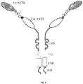


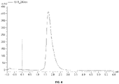


Classifications
-
- C—CHEMISTRY; METALLURGY
- C12—BIOCHEMISTRY; BEER; SPIRITS; WINE; VINEGAR; MICROBIOLOGY; ENZYMOLOGY; MUTATION OR GENETIC ENGINEERING
- C12N—MICROORGANISMS OR ENZYMES; COMPOSITIONS THEREOF; PROPAGATING, PRESERVING, OR MAINTAINING MICROORGANISMS; MUTATION OR GENETIC ENGINEERING; CULTURE MEDIA
- C12N15/00—Mutation or genetic engineering; DNA or RNA concerning genetic engineering, vectors, e.g. plasmids, or their isolation, preparation or purification; Use of hosts therefor
- C12N15/09—Recombinant DNA-technology
- C12N15/10—Processes for the isolation, preparation or purification of DNA or RNA
- C12N15/1034—Isolating an individual clone by screening libraries
- C12N15/1058—Directional evolution of libraries, e.g. evolution of libraries is achieved by mutagenesis and screening or selection of mixed population of organisms
-
- C—CHEMISTRY; METALLURGY
- C07—ORGANIC CHEMISTRY
- C07K—PEPTIDES
- C07K14/00—Peptides having more than 20 amino acids; Gastrins; Somatostatins; Melanotropins; Derivatives thereof
- C07K14/435—Peptides having more than 20 amino acids; Gastrins; Somatostatins; Melanotropins; Derivatives thereof from animals; from humans
- C07K14/705—Receptors; Cell surface antigens; Cell surface determinants
- C07K14/70503—Immunoglobulin superfamily
-
- A—HUMAN NECESSITIES
- A61—MEDICAL OR VETERINARY SCIENCE; HYGIENE
- A61K—PREPARATIONS FOR MEDICAL, DENTAL OR TOILETRY PURPOSES
- A61K38/00—Medicinal preparations containing peptides
-
- A—HUMAN NECESSITIES
- A61—MEDICAL OR VETERINARY SCIENCE; HYGIENE
- A61K—PREPARATIONS FOR MEDICAL, DENTAL OR TOILETRY PURPOSES
- A61K39/00—Medicinal preparations containing antigens or antibodies
- A61K39/395—Antibodies; Immunoglobulins; Immune serum, e.g. antilymphocytic serum
-
- A—HUMAN NECESSITIES
- A61—MEDICAL OR VETERINARY SCIENCE; HYGIENE
- A61K—PREPARATIONS FOR MEDICAL, DENTAL OR TOILETRY PURPOSES
- A61K39/00—Medicinal preparations containing antigens or antibodies
- A61K39/395—Antibodies; Immunoglobulins; Immune serum, e.g. antilymphocytic serum
- A61K39/39533—Antibodies; Immunoglobulins; Immune serum, e.g. antilymphocytic serum against materials from animals
- A61K39/3955—Antibodies; Immunoglobulins; Immune serum, e.g. antilymphocytic serum against materials from animals against proteinaceous materials, e.g. enzymes, hormones, lymphokines
-
- A—HUMAN NECESSITIES
- A61—MEDICAL OR VETERINARY SCIENCE; HYGIENE
- A61K—PREPARATIONS FOR MEDICAL, DENTAL OR TOILETRY PURPOSES
- A61K39/00—Medicinal preparations containing antigens or antibodies
- A61K39/395—Antibodies; Immunoglobulins; Immune serum, e.g. antilymphocytic serum
- A61K39/39533—Antibodies; Immunoglobulins; Immune serum, e.g. antilymphocytic serum against materials from animals
- A61K39/39558—Antibodies; Immunoglobulins; Immune serum, e.g. antilymphocytic serum against materials from animals against tumor tissues, cells, antigens
-
- A—HUMAN NECESSITIES
- A61—MEDICAL OR VETERINARY SCIENCE; HYGIENE
- A61K—PREPARATIONS FOR MEDICAL, DENTAL OR TOILETRY PURPOSES
- A61K39/00—Medicinal preparations containing antigens or antibodies
- A61K39/46—Cellular immunotherapy
- A61K39/461—Cellular immunotherapy characterised by the cell type used
- A61K39/4611—T-cells, e.g. tumor infiltrating lymphocytes [TIL], lymphokine-activated killer cells [LAK] or regulatory T cells [Treg]
-
- A—HUMAN NECESSITIES
- A61—MEDICAL OR VETERINARY SCIENCE; HYGIENE
- A61K—PREPARATIONS FOR MEDICAL, DENTAL OR TOILETRY PURPOSES
- A61K39/00—Medicinal preparations containing antigens or antibodies
- A61K39/46—Cellular immunotherapy
- A61K39/463—Cellular immunotherapy characterised by recombinant expression
- A61K39/4631—Chimeric Antigen Receptors [CAR]
-
- A—HUMAN NECESSITIES
- A61—MEDICAL OR VETERINARY SCIENCE; HYGIENE
- A61K—PREPARATIONS FOR MEDICAL, DENTAL OR TOILETRY PURPOSES
- A61K39/00—Medicinal preparations containing antigens or antibodies
- A61K39/46—Cellular immunotherapy
- A61K39/464—Cellular immunotherapy characterised by the antigen targeted or presented
- A61K39/4643—Vertebrate antigens
- A61K39/4644—Cancer antigens
- A61K39/464402—Receptors, cell surface antigens or cell surface determinants
- A61K39/464403—Receptors for growth factors
-
- A—HUMAN NECESSITIES
- A61—MEDICAL OR VETERINARY SCIENCE; HYGIENE
- A61P—SPECIFIC THERAPEUTIC ACTIVITY OF CHEMICAL COMPOUNDS OR MEDICINAL PREPARATIONS
- A61P35/00—Antineoplastic agents
-
- A—HUMAN NECESSITIES
- A61—MEDICAL OR VETERINARY SCIENCE; HYGIENE
- A61P—SPECIFIC THERAPEUTIC ACTIVITY OF CHEMICAL COMPOUNDS OR MEDICINAL PREPARATIONS
- A61P35/00—Antineoplastic agents
- A61P35/02—Antineoplastic agents specific for leukemia
-
- A—HUMAN NECESSITIES
- A61—MEDICAL OR VETERINARY SCIENCE; HYGIENE
- A61P—SPECIFIC THERAPEUTIC ACTIVITY OF CHEMICAL COMPOUNDS OR MEDICINAL PREPARATIONS
- A61P35/00—Antineoplastic agents
- A61P35/04—Antineoplastic agents specific for metastasis
-
- C—CHEMISTRY; METALLURGY
- C07—ORGANIC CHEMISTRY
- C07K—PEPTIDES
- C07K14/00—Peptides having more than 20 amino acids; Gastrins; Somatostatins; Melanotropins; Derivatives thereof
- C07K14/435—Peptides having more than 20 amino acids; Gastrins; Somatostatins; Melanotropins; Derivatives thereof from animals; from humans
- C07K14/705—Receptors; Cell surface antigens; Cell surface determinants
- C07K14/70503—Immunoglobulin superfamily
- C07K14/7051—T-cell receptor (TcR)-CD3 complex
-
- C—CHEMISTRY; METALLURGY
- C07—ORGANIC CHEMISTRY
- C07K—PEPTIDES
- C07K14/00—Peptides having more than 20 amino acids; Gastrins; Somatostatins; Melanotropins; Derivatives thereof
- C07K14/435—Peptides having more than 20 amino acids; Gastrins; Somatostatins; Melanotropins; Derivatives thereof from animals; from humans
- C07K14/705—Receptors; Cell surface antigens; Cell surface determinants
- C07K14/70503—Immunoglobulin superfamily
- C07K14/70521—CD28, CD152
-
- C—CHEMISTRY; METALLURGY
- C07—ORGANIC CHEMISTRY
- C07K—PEPTIDES
- C07K14/00—Peptides having more than 20 amino acids; Gastrins; Somatostatins; Melanotropins; Derivatives thereof
- C07K14/435—Peptides having more than 20 amino acids; Gastrins; Somatostatins; Melanotropins; Derivatives thereof from animals; from humans
- C07K14/705—Receptors; Cell surface antigens; Cell surface determinants
- C07K14/70503—Immunoglobulin superfamily
- C07K14/70535—Fc-receptors, e.g. CD16, CD32, CD64 (CD2314/705F)
-
- C—CHEMISTRY; METALLURGY
- C07—ORGANIC CHEMISTRY
- C07K—PEPTIDES
- C07K16/00—Immunoglobulins [IGs], e.g. monoclonal or polyclonal antibodies
- C07K16/18—Immunoglobulins [IGs], e.g. monoclonal or polyclonal antibodies against material from animals or humans
-
- C—CHEMISTRY; METALLURGY
- C07—ORGANIC CHEMISTRY
- C07K—PEPTIDES
- C07K16/00—Immunoglobulins [IGs], e.g. monoclonal or polyclonal antibodies
- C07K16/18—Immunoglobulins [IGs], e.g. monoclonal or polyclonal antibodies against material from animals or humans
- C07K16/28—Immunoglobulins [IGs], e.g. monoclonal or polyclonal antibodies against material from animals or humans against receptors, cell surface antigens or cell surface determinants
- C07K16/2863—Immunoglobulins [IGs], e.g. monoclonal or polyclonal antibodies against material from animals or humans against receptors, cell surface antigens or cell surface determinants against receptors for growth factors, growth regulators
-
- C—CHEMISTRY; METALLURGY
- C07—ORGANIC CHEMISTRY
- C07K—PEPTIDES
- C07K19/00—Hybrid peptides, i.e. peptides covalently bound to nucleic acids, or non-covalently bound protein-protein complexes
-
- C—CHEMISTRY; METALLURGY
- C12—BIOCHEMISTRY; BEER; SPIRITS; WINE; VINEGAR; MICROBIOLOGY; ENZYMOLOGY; MUTATION OR GENETIC ENGINEERING
- C12N—MICROORGANISMS OR ENZYMES; COMPOSITIONS THEREOF; PROPAGATING, PRESERVING, OR MAINTAINING MICROORGANISMS; MUTATION OR GENETIC ENGINEERING; CULTURE MEDIA
- C12N15/00—Mutation or genetic engineering; DNA or RNA concerning genetic engineering, vectors, e.g. plasmids, or their isolation, preparation or purification; Use of hosts therefor
- C12N15/09—Recombinant DNA-technology
- C12N15/10—Processes for the isolation, preparation or purification of DNA or RNA
- C12N15/102—Mutagenizing nucleic acids
-
- C—CHEMISTRY; METALLURGY
- C12—BIOCHEMISTRY; BEER; SPIRITS; WINE; VINEGAR; MICROBIOLOGY; ENZYMOLOGY; MUTATION OR GENETIC ENGINEERING
- C12N—MICROORGANISMS OR ENZYMES; COMPOSITIONS THEREOF; PROPAGATING, PRESERVING, OR MAINTAINING MICROORGANISMS; MUTATION OR GENETIC ENGINEERING; CULTURE MEDIA
- C12N5/00—Undifferentiated human, animal or plant cells, e.g. cell lines; Tissues; Cultivation or maintenance thereof; Culture media therefor
- C12N5/06—Animal cells or tissues; Human cells or tissues
- C12N5/0602—Vertebrate cells
- C12N5/0634—Cells from the blood or the immune system
- C12N5/0636—T lymphocytes
-
- A—HUMAN NECESSITIES
- A61—MEDICAL OR VETERINARY SCIENCE; HYGIENE
- A61K—PREPARATIONS FOR MEDICAL, DENTAL OR TOILETRY PURPOSES
- A61K39/00—Medicinal preparations containing antigens or antibodies
- A61K2039/51—Medicinal preparations containing antigens or antibodies comprising whole cells, viruses or DNA/RNA
- A61K2039/515—Animal cells
- A61K2039/5156—Animal cells expressing foreign proteins
-
- A—HUMAN NECESSITIES
- A61—MEDICAL OR VETERINARY SCIENCE; HYGIENE
- A61K—PREPARATIONS FOR MEDICAL, DENTAL OR TOILETRY PURPOSES
- A61K39/00—Medicinal preparations containing antigens or antibodies
- A61K2039/57—Medicinal preparations containing antigens or antibodies characterised by the type of response, e.g. Th1, Th2
- A61K2039/572—Medicinal preparations containing antigens or antibodies characterised by the type of response, e.g. Th1, Th2 cytotoxic response
-
- A—HUMAN NECESSITIES
- A61—MEDICAL OR VETERINARY SCIENCE; HYGIENE
- A61K—PREPARATIONS FOR MEDICAL, DENTAL OR TOILETRY PURPOSES
- A61K35/00—Medicinal preparations containing materials or reaction products thereof with undetermined constitution
- A61K35/12—Materials from mammals; Compositions comprising non-specified tissues or cells; Compositions comprising non-embryonic stem cells; Genetically modified cells
- A61K35/14—Blood; Artificial blood
- A61K35/17—Lymphocytes; B-cells; T-cells; Natural killer cells; Interferon-activated or cytokine-activated lymphocytes
-
- C—CHEMISTRY; METALLURGY
- C07—ORGANIC CHEMISTRY
- C07K—PEPTIDES
- C07K2317/00—Immunoglobulins specific features
- C07K2317/30—Immunoglobulins specific features characterized by aspects of specificity or valency
- C07K2317/31—Immunoglobulins specific features characterized by aspects of specificity or valency multispecific
-
- C—CHEMISTRY; METALLURGY
- C07—ORGANIC CHEMISTRY
- C07K—PEPTIDES
- C07K2317/00—Immunoglobulins specific features
- C07K2317/60—Immunoglobulins specific features characterized by non-natural combinations of immunoglobulin fragments
- C07K2317/62—Immunoglobulins specific features characterized by non-natural combinations of immunoglobulin fragments comprising only variable region components
- C07K2317/622—Single chain antibody (scFv)
-
- C—CHEMISTRY; METALLURGY
- C07—ORGANIC CHEMISTRY
- C07K—PEPTIDES
- C07K2317/00—Immunoglobulins specific features
- C07K2317/90—Immunoglobulins specific features characterized by (pharmaco)kinetic aspects or by stability of the immunoglobulin
- C07K2317/92—Affinity (KD), association rate (Ka), dissociation rate (Kd) or EC50 value
-
- C—CHEMISTRY; METALLURGY
- C07—ORGANIC CHEMISTRY
- C07K—PEPTIDES
- C07K2317/00—Immunoglobulins specific features
- C07K2317/90—Immunoglobulins specific features characterized by (pharmaco)kinetic aspects or by stability of the immunoglobulin
- C07K2317/94—Stability, e.g. half-life, pH, temperature or enzyme-resistance
-
- C—CHEMISTRY; METALLURGY
- C07—ORGANIC CHEMISTRY
- C07K—PEPTIDES
- C07K2319/00—Fusion polypeptide
- C07K2319/01—Fusion polypeptide containing a localisation/targetting motif
- C07K2319/02—Fusion polypeptide containing a localisation/targetting motif containing a signal sequence
-
- C—CHEMISTRY; METALLURGY
- C07—ORGANIC CHEMISTRY
- C07K—PEPTIDES
- C07K2319/00—Fusion polypeptide
- C07K2319/01—Fusion polypeptide containing a localisation/targetting motif
- C07K2319/03—Fusion polypeptide containing a localisation/targetting motif containing a transmembrane segment
-
- C—CHEMISTRY; METALLURGY
- C12—BIOCHEMISTRY; BEER; SPIRITS; WINE; VINEGAR; MICROBIOLOGY; ENZYMOLOGY; MUTATION OR GENETIC ENGINEERING
- C12N—MICROORGANISMS OR ENZYMES; COMPOSITIONS THEREOF; PROPAGATING, PRESERVING, OR MAINTAINING MICROORGANISMS; MUTATION OR GENETIC ENGINEERING; CULTURE MEDIA
- C12N2510/00—Genetically modified cells
Definitions
- enzymes may be evolved to be stabilized for operation at different conditions such as at an elevated temperature.
- an activity improvement at the elevated temperature a substantial portion of the improvement can be attributed to the higher kinetic activity commonly described by the Q10 rule where it is estimated that in the case of an enzyme the turnover doubles for every increase of 10 degrees Celsius.
- Chimeric antigen receptors have been used in treating cancers.
- US 2013/0280220 discloses methods and compositions providing improved cells encoding a chimeric antigen receptor that is specific for two or more antigens, including tumor antigens.
- Cells expressing the chimeric antigen receptor may be used in cell therapy.
- Such cell therapy may be suitable for any medical condition, although in specific embodiments the cell therapy is for cancer, including cancer involving solid tumors.
- An Fv fragment is defined as a genetically engineered fragment containing the variable region of a light chain and the variable region of a heavy chain expressed as two chains.
- autologous refers to any material derived from the same individual to which it is later to be reintroduced.
- T cells from a patient may be isolated, genetically engineered to express a CAR and then reintroduced into the patient.
- chimeric antigen receptor or “CAR” or “CARs” as used herein refers to engineered receptors, which graft an antigen specificity onto cytotoxic cell, for example T cells, NK cells and macrophages.
- the CARs of the invention may comprise at least one antigen specific targeting region (ASTR), an extracellular spacer domain (ESD), a transmembrane domain (TM), one or more co-stimulatory domains (CSD), and an intracellular signaling domain (ISD).
- ASTR antigen specific targeting region
- ESD extracellular spacer domain
- TM transmembrane domain
- CSD co-stimulatory domain
- ISD intracellular signaling domain
- the ESD and/or CSD are optional.
- the CAR is a bispecific CAR, which is specific to two different antigens or epitopes.
- the ISD activates intracellular signaling.
- the ISD can redirect T cell specificity and reactivity toward a selected target in a non-MHC-restricted manner, exploiting the antigen-binding properties of antibodies.
- the non-MHC-restricted antigen recognition gives T cells expressing the CAR the ability to recognize an antigen independent of antigen processing, thus bypassing a major mechanism of tumor escape.
- CARs advantageously do not dimerize with endogenous T cell receptor (TCR) alpha and beta chains.
- co-express refers to simultaneous expression of two or more genes.
- Genes may be nucleic acids encoding, for example, a single protein or a chimeric protein as a single polypeptide chain.
- the CARs of the invention may be co- expressed with a therapeutic control (for example truncated epidermal growth factor (EGFRt)), wherein the CAR is encoded by a first polynucleotide chain and the therapeutic control is encoded by a second polynucleotide chain.
- the first and second polynucleotide chains are linked by a nucleic acid sequence that encodes a cleavable linker.
- the CAR and the therapeutic control are encoded by two different polynucleotides that are not linked via a linker but are instead encoded by, for example, two different vectors.
- conditionally active biologic protein refers to a variant, or mutant, of a wild-type protein which is more or less active than the parent wild-type protein under one or more normal physiological conditions. This conditionally active protein also exhibits activity in selected regions of the body and/or exhibits increased or decreased activity under aberrant, or permissive, physiological conditions.
- Normal physiological conditions are those of temperature, pH, osmotic pressure, osmolality, oxidative stress and electrolyte concentration which would be considered within a normal range at the site of administration, or at the tissue or organ at the site of action, to a subject.
- An aberrant condition is that which deviates from the normally acceptable range for that condition.
- conditionally active biologic protein is virtually inactive at wild-type conditions but is active at other than wild-type conditions at a level that is equal or better than at wild-type conditions.
- an evolved conditionally active biologic protein is virtually inactive at body temperature, but is active at lower temperatures.
- conditionally active biologic protein is reversibly or irreversibly inactivated at the wild type conditions.
- wild-type protein is a therapeutic protein.
- conditionally active biologic protein is used as a drug, or therapeutic agent.
- the protein is more or less active in highly oxygenated blood, such as, for example, after passage through the lung or in the lower pH environments found in the kidney.
- Constant amino acid substitutions refer to the interchangeability of residues having similar side chains.
- a group of amino acids having aliphatic side chains is glycine, alanine, valine, leucine, and isoleucine
- a group of amino acids having aliphatic-hydroxyl side chains is serine and threonine
- a group of amino acids having amide-containing side chains is asparagine and glutamine
- a group of amino acids having aromatic side chains is phenylalanine, tyrosine, and tryptophan
- a group of amino acids having basic side chains is lysine, arginine, and histidine
- a group of amino acids having sulfur-containing side chains is cysteine and methionine.
- Preferred conservative amino acids substitution groups are: valine-leucine-isoleucine, phenylalanine-tyrosine, lysine-arginine, alanine-valine, and asparagine-glutamine.
- a co-stimulatory ligand can include, but is not limited to, CD7, B7-1 (CD80), B7-2 (CD86), PD-L1, PD-L2, 4-1BBL, OX40L, an inducible costimulatory ligand (ICOS-L), an intercellular adhesion molecule (ICAM), CD30L, CD40, CD70, CD83, HLA-G, MICA, MICB, HVEM, a lymphotoxin beta receptor, 3/TR6, ILT3, ILT4, HVEM, an agonist or an antibody that binds to a Toll ligand receptor and a ligand that specifically binds with B7-H3.
- a co-stimulatory ligand also encompasses, inter alia, an antibody that specifically binds with a co-stimulatory molecule present on a T cell, such as, but not limited to, CD27, CD28, 4-1BB, OX40, CD30, CD40, PD-1, ICOS, a lymphocyte function-associated antigen-1 (LFA-1), CD2, CD7, LIGHT, NKG2C, B7-H3, and a ligand that specifically binds with CD83.
- an antibody that specifically binds with a co-stimulatory molecule present on a T cell such as, but not limited to, CD27, CD28, 4-1BB, OX40, CD30, CD40, PD-1, ICOS, a lymphocyte function-associated antigen-1 (LFA-1), CD2, CD7, LIGHT, NKG2C, B7-H3, and a ligand that specifically binds with CD83.
- LFA-1 lymphocyte function-associated antigen-1
- co-stimulatory signal refers to a signal, which in combination with a primary signal, such as TCR/CD3 ligation, leads to T cell proliferation and/or upregulation or down regulation of key molecules.
- Cytotoxic cells may also include other immune cells with tumor lytic capabilities including but not limited to natural killer T cells ( Heczey et al., "Invariant NKT cells with chimeric antigen receptor provide a novel platform for safe and effective cancer immunotherapy," Blood, vol. 124, pp.2824-2833, 2014 ) and granulocytes.
- degradation effective amount refers to the amount of enzyme which is required to process at least 50% of the substrate, as compared to substrate not contacted with the enzyme.
- directional ligation refers to a ligation in which a 5' end and a 3' end of a polynucleotide are different enough to specify a preferred ligation orientation.
- an otherwise untreated and undigested PCR product that has two blunt ends will typically not have a preferred ligation orientation when ligated into a cloning vector digested to produce blunt ends in its multiple cloning site; thus, directional ligation will typically not be displayed under these circumstances.
- directional ligation will typically be displayed when a digested PCR product having a 5' EcoR I-treated end and a 3' BamH I is ligated into a cloning vector that has a multiple cloning site digested with EcoR I and BamH I.
- disease targeted by genetically modified cytotoxic cells encompasses the targeting of any cell involved in any manner in any disease by the genetically modified cells of the invention, irrespective of whether the genetically modified cells target diseased cells or healthy cells to effectuate a therapeutically beneficial result.
- the genetically modified cells include but are not limited to genetically modified T cells, NK cells, and macrophages.
- the genetically modified cells express the CARs of the invention, which CARs may target any of the antigens expressed on the surface of target cells.
- antigens which may be targeted include but are not limited to antigens expressed on B-cells; antigens expressed on carcinomas, sarcomas, lymphomas, leukemia, germ cell tumors, and blastomas; antigens expressed on various immune cells; and antigens expressed on cells associated with various hematologic diseases, autoimmune diseases, and/or inflammatory diseases.
- antigens expressed on B-cells include but are not limited to antigens expressed on B-cells; antigens expressed on carcinomas, sarcomas, lymphomas, leukemia, germ cell tumors, and blastomas; antigens expressed on various immune cells; and antigens expressed on cells associated with various hematologic diseases, autoimmune diseases, and/or inflammatory diseases.
- Other antigens that may be targeted will be apparent to those of skill in the art and may be targeted by the CARs of the invention in connection with alternate embodiments thereof.
- DNA shuffling is used herein to indicate recombination between substantially homologous but non-identical sequences, in some embodiments DNA shuffling may involve crossover via non-homologous recombination, such as via cer/lox and/or flp/frt systems and the like. DNA shuffling can be random or non-random.
- drug or “drug molecule” refers to a therapeutic agent including a substance having a beneficial effect on a human or animal body when it is administered to the human or animal body.
- the therapeutic agent includes a substance that can treat, cure or relieve one or more symptoms, illnesses, or abnormal conditions in a human or animal body or enhance the wellness of a human or animal body.
- an “effective amount” is an amount of a conditionally active biologic protein or fragment which is effective to treat or prevent a condition in a living organism to whom it is administered over some period of time, e.g., provides a therapeutic effect during a desired dosing interval.
- the normal physiological condition and aberrant condition can be conditions of "electrolyte concentration".
- the electrolyte concentration to be tested is selected from one or more of ionized calcium, sodium, potassium, magnesium, chloride, bicarbonate, and phosphate concentration.
- normal range of serum calcium is 8.5 to 10.2 mg/dL.
- aberrant serum calcium concentration may be selected from either above or below the normal range, m another example, in one aspect, normal range of serum chloride is 96-106 milliequivalents per liter (mEq/L).
- aberrant serum chloride concentration may be selected from either above or below the normal range, in another example, in one aspect, a normal range of serum magnesium is from 1.7-2.2 mg/dL. In this aspect, an aberrant serum magnesium concentration may be selected from either above or below the normal range, in another example, in one aspect, a normal range of serum phosphorus is from 2.4 to 4.1 mg/dL. In this aspect, aberrant serum phosphorus concentration may be selected from either above or below the normal range. In another example, in one aspect, a normal range of serum, or blood, sodium is from 135 to 145 mEq/L. In this aspect, aberrant serum, or blood, sodium concentration may be selected from either above or below the normal range.
- a normal range of serum, or blood, potassium is from 3.7 to 5.2 mEq/L.
- aberrant serum, or blood, potassium concentration maybe selected from either above or below the normal range.
- a normal range of serum bicarbonate is from 20 to 29 mEq/L.
- aberrant serum, or blood, bicarbonate concentration may be selected from either above or below the normal range.
- bicarbonate levels can be used to indicate normal levels of acidity (pH), in the blood.
- electrolyte concentration may also be used to define the condition of a particular electrolyte in a tissue or body fluid other than blood or plasma. In this case, the normal physiological condition is considered to be the clinically normal range for that tissue or fluid.
- aberrant tissue or fluid electrolyte concentration may be selected from either above or below the normal range.
- epitope refers to an antigenic determinant on an antigen, such as an enzyme polypeptide, to which the paratope of an antibody, such as an enzyme-specific antibody, binds.
- Antigenic determinants usually consist of chemically active surface groupings of molecules, such as amino acids or sugar side chains, and can have specific three-dimensional structural characteristics, as well as specific charge characteristics.
- epipe refers to that portion of an antigen or other macromolecule capable of forming a binding interaction that interacts with the variable region binding body of an antibody. Typically, such binding interaction is manifested as an intermolecular contact with one or more amino acid residues of a CDR.
- the term “evolution”, or “evolving”, refers to using one or more methods of mutagenesis to generate a novel polynucleotide encoding a novel polypeptide, which novel polypeptide is itself an improved biological molecule &/or contributes to the generation of another improved biological molecule.
- the present disclosure relates to evolution of conditionally active biologic proteins from a parent wild type protein.
- evolution relates to a method of performing both non-stochastic polynucleotide chimerization and non-stochastic site-directed point mutagenesis disclosed in U.S. patent application publication 2009/0130718 .
- the present disclosure provides methods for evolution of conditionally active biologic enzymes which exhibit reduced activity at normal physiological conditions compared to a wild-type enzyme parent molecule, but enhanced activity under one or more aberrant conditions compared to the antigen specific targeting region of the wild-type enzyme.
- fragment when referring to a reference polypeptide comprise a polypeptide which retains at least one biological function or activity that is at least essentially same as that of the reference polypeptide. Furthermore, the terms “fragment”, “derivative” or “analog” are exemplified by a "pro-form” molecule, such as a low activity proprotein that can be modified by cleavage to produce a mature enzyme with significantly higher activity.
- immune response refers to immunities including but not limited to innate immunity, humoral immunity, cellular immunity, immunity, inflammatory response, acquired (adaptive) immunity, autoimmunity and/or overactive immunity
- lentivirus refers to a genus of the Retroviridae family. Lentiviruses are unique among the retroviruses in being able to infect non-dividing cells; they can deliver a significant amount of genetic information into the DNA of the host cell, so they are one of the most efficient ways to deliver a gene delivery vector. HIV, SIV, and FIV are all examples of lentiviruses. Vectors derived from lentiviruses offer the means to achieve significant levels of gene transfer in vivo.
- mammalian cell surface display refers to a technique whereby a protein or antibody, or a portion of an antibody, is expressed and displayed on a mammalian host cell surface for screening purposes; for example, by screening for specific antigen binding by a combination of magnetic beads and fluorescence-activated cell sorting.
- mammalian expression vectors are used for simultaneous expression of immunoglobulins as both a secreted and cell surface bound form as in DuBridge et al., US 2009/0136950 .
- the techniques are employed for screening a viral vector encoding for a library of antibodies or antibody fragments that are displayed on the cell membranes when expressed in a cell as in Gao et al., US 2007/0111260 .
- Whole IgG surface display on mammalian cells is known.
- Akamatsuu et al. developed a mammalian cell surface display vector, suitable for directly isolating IgG molecules based on their antigen-binding affinity and biological activity.
- Epstein-Barr virus-derived episomal vector antibody libraries were displayed as whole IgG molecules on the cell surface and screened for specific antigen binding by a combination of magnetic beads and fluorescence-activated cell sorting.
- B cells specific for an antigen may also be used. Such B cells may be directly isolated from peripheral blood mononuclear cells (PBMC) of human donors. Recombinant, antigen-specific single-chain Fv (scFv) libraries are generated from this pool of B cells and screened by mammalian cell surface display by using a Sindbis virus expression system.
- the variable regions (VRs) of the heavy chains (HCs) and light chains (LCs) can be isolated from positive clones and recombinant fully human antibodies produced as whole IgG or Fab fragments. In this manner, several hypermutated high-affinity antibodies binding the Q ⁇ virus like particle (VLP), a model viral antigen, as well as antibodies specific for nicotine can be isolated.
- VLP Q ⁇ virus like particle
- manufacturing refers to production of a protein in a sufficient quantity to permit at least Phase I clinical testing of a therapeutic protein, or sufficient quantity for regulatory approval of a diagnostic protein.
- molecular property to be evolved includes reference to molecules comprised of a polynucleotide sequence, molecules comprised of a polypeptide sequence, and molecules comprised in part of a polynucleotide sequence and in part of a polypeptide sequence.
- Particularly relevant- but by no means limiting- examples of molecular properties to be evolved include protein activities at specified conditions, such as related to temperature; salinity; osmotic pressure; pH; oxidative stress, and concentration of glycerol, DMSO, detergent, and/or any other molecular species with which contact is made in a reaction environment.
- Additional particularly relevant-but by no means limiting-examples of molecular properties to be evolved include stabilities- e.g. the amount of a residual molecular property that is present after a specified exposure time to a specified environment, such as may be encountered during storage.
- mutants as used herein means changes in the sequence of a wild-type nucleic acid sequence or changes in the sequence of a peptide. Such mutations may be point mutations such as transitions or transversions. The mutations may be deletions, insertions or duplications.
- multispecific antibody as used herein is an antibody having binding affinities for at least two different epitopes.
- Multispecific antibodies can be prepared as full-length antibodies or antibody fragments (e.g. F(ab') 2 bispecific antibodies).
- Engineered antibodies may bind to two, three or more (e.g. four) antigens (see, e.g., US 2002/0004587 A1 ).
- One conditionally active antibody may be engineered to be multispecific, or two antibodies may be engineered to comprise a hetero-dimer that binds to two antigens.
- Multispecific antibodies can also be multifunctional.
- nucleic acid sequence coding for or a "DNA coding sequence of or a “nucleotide sequence encoding” as used herein refer to a DNA sequence which is transcribed and translated into an enzyme when placed under the control of appropriate regulatory sequences such as promoters.
- a “promotor” is a DNA regulatory region capable of binding RNA polymerase in a cell and initiating transcription of a downstream (3' direction) coding sequence.
- the promoter is part of the DNA sequence. This sequence region has a start codon at its 3' terminus.
- the promoter sequence does include the minimum number of bases where elements necessary to initiate transcription at levels detectable above background.
- RNA polymerase binds the sequence and transcription is initiated at the start codon (3' terminus with a promoter)
- transcription proceeds downstream in the 3' direction.
- a transcription initiation site (conveniently defined by mapping with nuclease S1) as well as protein binding domains (consensus sequences) responsible for the binding of RNA polymerase.
- oligonucleotide refers to either a single stranded polydeoxynucleotide or two complementary polydeoxynucleotide strands which may be chemically synthesized. Such synthetic oligonucleotides may or may not have a 5' phosphate. Those that do not will not ligate to another oligonucleotide without adding a phosphate with an ATP in the presence of a kinase. A synthetic oligonucleotide will ligate to a fragment that has not been dephosphorylated.
- a coding sequence is "operably linked to" another coding sequence when RNA polymerase will transcribe the two coding sequences into a single mRNA, which is then translated into a single polypeptide having amino acids derived from both coding sequences.
- the coding sequences need not be contiguous to one another so long as the expressed sequences are ultimately processed to produce the desired protein.
- in vitro physiological conditions comprise 50-200 mM NaCl or KCl, pH 6.5-8.5, 20-45 degrees C and 0.001-10 mM divalent cation (e.g., Mg ++" , Ca ++ ); preferably about 150 mM NaCl or KCl, pH 7.2-7.6, 5 mM divalent cation, and often include 0.01-1.0 percent nonspecific protein (e.g., bovine serum albumin (BSA)).
- BSA bovine serum albumin
- a non-ionic detergent Teween, NP-40, Triton X-100
- Particular aqueous conditions may be selected by the practitioner according to conventional methods.
- buffered aqueous conditions may be applicable: 10-250 mM NaCl, 5-50 mM Tris HCl, pH 5-8, with optional addition of divalent cation(s) and/or metal chelators and/or non- ionic detergents and/or membrane fractions and/or anti-foam agents and/or scintillants.
- Normal physiological conditions refer to conditions of temperature, pH, osmotic pressure, osmolality, oxidative stress and electrolyte concentration in vivo in a patient or subject at the site of administration, or the site of action, which would be considered within the normal range in a patient.
- Standard convention (5' to 3') is used herein to describe the sequence of double stranded polynucleotides.
- the reference precursor molecule may be termed a"pre-pro-form" molecule.
- a functional single-chain antibody generally contains a sufficient portion of an immunoglobulin superfamily gene product so as to retain the property of binding to a specific target molecule, typically a receptor or antigen (epitope).
- stimulatory ligand means a ligand that when present on an antigen presenting cell (e.g, a dendritic cell, a B-cell, and the like) can specifically bind with a cognate binding partner (referred to herein as a "stimulatory molecule") on a T cell, thereby mediating a primary response by the T cell, including, but not limited to, activation, initiation of an immune response, proliferation, and the like.
- Stimulatory ligands are well-known in the art and encompass, inter alia, an MHC Class I molecule loaded with a peptide, an anti-CD3 antibody, a superagonist anti-CD28 antibody, and a superagonist anti-CD2 antibody.
- Tumor refers to all neoplastic cell growth and proliferation, whether malignant or benign, and all pre-cancerous and cancerous cells and tissues
- variable segment refers to a portion of a nascent peptide which comprises a random, pseudorandom, or defined kernal sequence.
- a variable segment refers to a portion of a nascent peptide which comprises a random pseudorandom, or defined kernal sequence.
- a variable segment can comprise both variant and invariant residue positions, and the degree of residue variation at a variant residue position may be limited: both options are selected at the discretion of the practitioner.
- Vector refers to the vehicle by which a polynucleotide sequence (e.g. a foreign gene) can be introduced into a host cell, so as to transform the host and promote expression (e.g. transcription and translation) of the introduced sequence.
- Vectors include plasmids, phages, viruses, etc.
- wild-type means that the polynucleotide does not comprise any mutations.
- a wild type protein refers to a protein which can be isolated from nature that will be active at a level of activity found in nature and will comprise the amino acid sequence found in nature.
- the terms “parent molecule” and “target protein” also refer to the wild-type protein.
- the "wild-type protein” preferably has some desired properties, such as higher binding affinity, or enzymatic activity, which may be obtained by screening of a library of proteins for a desired properties, including better stability in different temperature or pH environments, or improved selectivity and/or solubility.
- the antibody or antibody fragment evolved from a parent antibody or a domain thereof that binds to the tyrosine kinase growth factor receptor has a decrease in activity in binding to the tyrosine kinase growth factor receptor in an assay at a normal physiological pH at a site of administration to a subject, or at the issue or organ at a site of action of the chimeric antigen receptor, compared to the same activity of the antibody or antibody fragment in binding to the tyrosine kinase growth factor receptor in an assay under an aberrant pH in an tumor microenvironment that deviates from the normal physiological pH.
- the present invention provides a method to generate the conditionally active at least one antibody fragment from a parent antibody or its binding domain (wild-type ASTR).
- in vivo site specific recombination systems have been utilized to generate hybrids of genes, as well as random methods of in vivo recombination, and recombination between homologous but truncated genes on a plasmid. Mutagenesis has also been reported by overlapping extension and PCR.
- Non-random methods have been used to achieve larger numbers of point mutations and/or chimerizations, for example comprehensive or exhaustive approaches have been used to generate all the molecular species within a particular grouping of mutations, for attributing functionality to specific structural groups in a template molecule (e.g. a specific single amino acid position or a sequence comprised of two or more amino acids positions), and for categorizing and comparing specific grouping of mutations.
- a template molecule e.g. a specific single amino acid position or a sequence comprised of two or more amino acids positions
- the mutant polynucleotides generated from the evolving step may, or may not be size fractionated on an agarose gel according to published protocols, inserted into an expression vector, and transfected into an appropriate host cell to produce the mutant polypeptides (expression).
- the expression may use routine molecular biology techniques.
- the expression step can use various known methods.
- the DNA may be included in any one of a variety of expression vectors for expressing a polypeptide.
- Such vectors include chromosomal, nonchromosomal and synthetic DNA sequences.
- suitable vectors are known to those of skill in the art, and are commercially available.
- the following vectors are provided by way of example; Bacterial: pQE vectors (Qiagen), pBluescript plasmids, pNH vectors, (lambda-ZAP vectors (Stratagene); ptrc99a, pKK223-3, pDR540, pRIT2T (Pharmacia); Eukaryotic: pXTl, pSG5 (Stratagene), pSVK3, pBPV, pMSG, pSVLSV40 (Pharmacia).
- any other plasmid or other vector may be used so long as they are replicable and viable in the host.
- Low copy number or high copy number vectors may be employed with the present disclosure.
- Enhancers are cis-acting sequences of between 10 to 300 bp that increase transcription by a promoter. Enhancers can effectively increase transcription when either 5' or 3' to the transcription unit. They are also effective if located within an intron or within the coding sequence itself.
- viral enhancers including SV40 enhancers, cytomegalovirus enhancers, polyoma enhancers, and adenovirus enhancers. Enhancer sequences from mammalian systems are also commonly used, such as the mouse immunoglobulin heavy chain enhancer.
- mutant polynucleotides can be generated by the process of reductive reassortment.
- the method involves the generation of constructs containing consecutive sequences (original encoding sequences), their insertion into an appropriate vector, and their subsequent introduction into an appropriate host cell.
- the reassortment of the individual molecular identities occurs by combinatorial processes between the consecutive sequences in the construct possessing regions of homology, or between quasi-repeated units.
- the reassortment process recombines and/or reduces the complexity and extent of the repeated sequences, and results in the production of novel molecular species.
- Various treatments may be applied to enhance the rate of reassortment.
- Preferred expression control sequences are promoters derived from immunoglobulin genes, cytomegalovirus, SV40, Adenovirus, Bovine Papilloma Virus, and the like.
- mammalian host cells may be created commercially by a contract research or custom manufacturing organization.
- Lonza Longza Group Ltd, Basel, Switzerland
- Lonza Group Ltd can create vectors to express these products using the GS Gene Expression System TM technology with either CHOK1SV or NS0 cell production hosts.
- Host cells containing the polynucleotides of interest can be cultured in conventional nutrient media modified as appropriate for activating promoters, selecting transformants or amplifying genes.
- the culture conditions such as temperature, pH and the like, are those previously used with the host cell selected for expression, and will be apparent to the ordinarily skilled artisan.
- Protein refolding steps can be used, as necessary, in completing configuration of the polypeptide. If desired, high performance liquid chromatography (HPLC) can be employed for final purification steps.
- HPLC high performance liquid chromatography
- the screening of a conditionally active ASTR can be aided by the availability of a convenient high throughput screening or selection process.
- Cell surface display expression and screening technology (for example, as defined above) can be employed to screen mutant proteins for conditionally active ASTR.
- the method further comprises modifying at least one of the nucleic acids or polypeptides prior to testing the candidates for conditional biologic activity, in another aspect, the testing of step (c) further comprises testing for improved expression of the polypeptide in a host cell or host organism, in a further aspect, the testing of step (c) further comprises testing for enzyme activity within a pH range from about pH 3 to about pH 12. In a further aspect, the testing of step (c) further comprises testing for enzyme activity within a pH range from about pH 5 to about pH 10. In a further aspect, the testing of step (c) further comprises testing for enzyme activity within a pH range from about pH 6 to about pH 8. In a further aspect, the testing of step (c) further comprises testing for enzyme activity at pH 6.7 and pH 7.5.
- the disclosure provides isolated or recombinant antibodies that specifically bind to an enzyme of the disclosure. These antibodies can be used to isolate, identify or quantify the enzymes of the disclosure or related polypeptides. These antibodies can be used to isolate other polypeptides within the scope the disclosure or other related enzymes. The antibodies can be designed to bind to an active site of an enzyme. Thus, the disclosure provides methods of inhibiting enzymes using the antibodies of the disclosure.
- the antibodies can be used in immunoprecipitation, staining, immunoaffinity columns, and the like.
- nucleic acid sequences encoding for specific antigens can be generated by immunization followed by isolation of polypeptide or nucleic acid, amplification or cloning and immobilization of polypeptide onto an array of the disclosure.
- the methods of the disclosure can be used to modify the structure of an antibody produced by a cell to be modified, e.g., an antibody's affinity can be increased or decreased.
- the ability to make or modify antibodies can be a phenotype engineered into a cell by the methods of the disclosure.
- Polypeptides or peptides can be used to generate antibodies which bind specifically to the polypeptides, e.g., the enzymes, of the disclosure.
- the resulting antibodies may be used in immunoaffinity chromatography procedures to isolate or purify the polypeptide or to determine whether the polypeptide is present in a biological sample.
- a protein preparation such as an extract, or a biological sample is contacted with an antibody capable of specifically binding to one of the polypeptides of the disclosure.
- Polyclonal antibodies generated against the polypeptides of the disclosure can be obtained by direct injection of the polypeptides into an animal or by administering the polypeptides to a non-human animal. The antibody so obtained will then bind the polypeptide itself. In this manner, even a sequence encoding only a fragment of the polypeptide can be used to generate antibodies which may bind to the whole native polypeptide. Such antibodies can then be used to isolate the polypeptide from cells expressing that polypeptide.
- transgenic mice may be used to express humanized antibodies to these polypeptides or fragments thereof.
- Antibodies generated against the polypeptides of the disclosure may be used in screening for similar polypeptides (e.g., enzymes) from other organisms and samples. In such techniques, polypeptides from the organism are contacted with the antibody and those polypeptides which specifically bind the antibody are detected. Any of the procedures described above may be used to detect antibody binding.
- Nucleic acids or polypeptides of the disclosure can be immobilized to or applied to an array.
- Arrays can be used to screen for or monitor libraries of compositions (e.g., small molecules, antibodies, nucleic acids, etc.) for their ability to bind to or modulate the activity of a nucleic acid or a polypeptide of the disclosure.
- a monitored parameter is transcript expression of an enzyme gene.
- Capillary arrays such as the GIGAMATRIX TM Diversa Corporation, San Diego, Calif., can be used in the methods of the disclosure.
- Nucleic acids or polypeptides of the disclosure can be immobilized to or applied to an array, including capillary arrays.
- Arrays can be used to screen for or monitor libraries of compositions (e.g., small molecules, antibodies, nucleic acids, etc.) for their ability to bind to or modulate the activity of a nucleic acid or a polypeptide of the disclosure.
- Capillary arrays provide another system for holding and screening samples.
- a sample of interest can be introduced as a first liquid labeled with a detectable particle into a capillary of a capillary array, wherein each capillary of the capillary array comprises at least one wall defining a lumen for retaining the first liquid and the detectable particle, and wherein the at least one wall is coated with a binding material for binding the detectable particle to the at least one wall.
- the method can further include removing the first liquid from the capillary tube, wherein the bound detectable particle is maintained within the capillary, and introducing a second liquid into the capillary tube.
- the capillary array can include a plurality of individual capillaries comprising at least one outer wall defining a lumen.
- the outer wall of the capillary can be one or more walls fused together.
- the wall can define a lumen that is cylindrical, square, hexagonal or any other geometric shape so long as the walls form a lumen for retention of a liquid or sample.
- the capillaries of the capillary array can be held together in close proximity to form a planar structure.
- the capillaries can be bound together, by being fused (e.g., where the capillaries are made of glass), glued, bonded, or clamped side-by-side.
- the capillary array can be formed of any number of individual capillaries, for example, a range from 100 to 4,000,000 capillaries.
- a capillary array can form a micro titer plate having about 100,000 or more individual capillaries bound together.
- Conditionally active antibodies may be engineered to generate multispecific conditionally active antibodies.
- the multispecific antibody may be an antibody with polyepitopic specificity, as described in WO 2013/170168 .
- Multispecific antibodies include, but are not limited to, an antibody comprising a heavy chain variable domain (V H ) and a light chain variable domain (V L ), where the V H V L unit has polyepitopic specificity, antibodies having two or more V L and V H domains where each V H V L unit binds to a different epitope, antibodies having two or more single variable domains with each single variable domain binding to a different epitope, and antibodies comprising one or more antibody fragments as well as antibodies comprising antibody fragments that have been linked covalently or non-covalently.
- antibody fragments having at least one free sulfhydryl group are obtained.
- the antibody fragments may be obtained from full-length conditionally active antibodies.
- the conditionally active antibodies may be digested enzymatically to produce antibody fragments.
- Exemplary enzymatic digestion methods include, but are not limited to, pepsin, papain and Lys-C.
- Exemplary antibody fragments include, but are not limited to, Fab, Fab', F(ab')2, Fv, diabodies (Db); tandem diabodies (taDb), linear antibodies (see U.S. Patent No. 5,641,870 , Example 2; Zapata et al., Protein Eng., vol.
- Exemplary thiol-reactive reagents include a multifunctional linker reagent, a capture, i.e. an affinity, label reagent (e.g. a biotin-linker reagent), a detection label (e.g. a fluorophore reagent), a solid phase immobilization reagent (e.g. SEPHAROSE TM , polystyrene, or glass), or a drug-linker intermediate.
- a thiol-reactive reagent is N-ethyl maleimide (NEM).
- NEM N-ethyl maleimide
- Such multispecific antibodies or antibody analogs having modified cross-linkers may be further reacted with a drug moiety reagent or other label. Reaction of a multispecific antibody or antibody analog with a drug-linker intermediate provides a multispecific antibody-drug conjugate or antibody analog-drug conjugate, respectively.
- a BBB-R is a transmembrane receptor protein expressed on brain endothelial cells which is capable of transporting molecules across the blood-brain barrier.
- BBB-R include transferrin receptor (TfR), insulin receptor, insulin-like growth factor receptor (IGF-R), low density lipoprotein receptors including without limitation low density lipoprotein receptor-related protein 1 (LRP1) and low density lipoprotein receptor-related protein 8 (LRP8), and heparin-binding epidermal growth factor-like growth factor (HB-EGF).
- TfR transferrin receptor
- IGF-R insulin-like growth factor receptor
- LRP1 low density lipoprotein receptor-related protein 1
- LRP8 low density lipoprotein receptor-related protein 8
- HB-EGF heparin-binding epidermal growth factor-like growth factor
- An exemplary BBB-R herein is a transferrin receptor (TfR).
- the TfR is a transmembrane glycoprotein (with a molecular weight of about 180,000) composed of two disulphide-bonded sub-units (each of apparent molecular weight of about 90,000) involved in iron uptake in vertebrates.
- the present invention provides a conditionally active antibody generated from a parent or wild-type antibody against a BBB-R.
- the conditionally active antibody binds the BBB-R on the blood side of the BBB, and has a lower affinity to the BBB-R than the parent or wild-type antibody on the brain side of the BBB.
- the conditionally active antibody has affinity to the BBB-R than the wild type or parent antibody on the blood side of the BBB, and has no affinity to the BBB-R on the brain side of the BBB.
- Blood plasma is a body fluid that is very different from brain extracellular fluid (ECF).
- ECF brain extracellular fluid
- Somjen Irons in the Brain: Normal Function, Seizures, and Stroke
- Redzic Molecular biology of the blood-brain and the blood-cerebrospinal fluid barriers: similarities and differences
- Fluids and Barriers of the CNS, vol. 8:3, 2011 the brain extracellular fluid has significantly less K + , more Mg 2+ and H + than blood plasma.
- the differences in ion concentrations between blood plasma and brain ECF lead to significant differences in osmotic pressure and osmolality between the two fluids.
- Table 1 shows the concentrations of common ions in millimoles for both blood plasma and brain ECF.
- Table 1 shows the concentrations of common ions in millimoles for both blood plasma and brain ECF.
- Table 1 shows the concentrations of common ions in millimoles for both blood plasma and brain ECF.
- Table 1 shows the concentrations of common ions in millimoles for both blood plasma and brain ECF.
- Table 1 shows the concentrations of common ions in plasma (arterial plasma) and brain extracellular fluid (CSF) ARTERIAL PLASMA CSF HUMAN RAT HUMAN RAT Na + 150 148 147 152 K + 4.6 5,3 2.9 3.4 Ca, total 2.4 3.1 1.14 1.1 Ca 2+ , free 1.4 1.5 1.0 1 .0 pCa Mg, total 0,86 0.8 1.15 1.3 Mg 2+ , free 0.47 0.44 0.7 0.88 H + 0.000039 0.000032 0000047 0.00005 pH 7.41 7.5 7.3 7.3 Cl - 99
- Brain ECF also contains significantly more lactate than blood plasma and significantly less glucose than blood plasma ( Abi-Saab et al., "Striking Differences in Glucose and Lactate Levels Between Brain Extracellular Fluid and Plasma in Conscious Human Subjects: Effects of Hyperglycemia and Hypoglycemia," Journal of Cerebral Blood Flow & Metabolism, vol. 22, pages 271-279, 2002 ).
- the present invention thus provides a method for evolving a DNA that encodes a template antibody against a BBB-R to create a mutant DNA library.
- the mutant DNA library is then expressed to obtain mutant antibodies.
- the mutant antibodies are screened for a conditionally active antibody that has binds to the BBB-R under at least one blood plasma physiological condition and has a low or no affinity to the BBB-R under at least one brain physiological condition in the brain ECF compared to the template antibody.
- the selected mutant antibody has a low or high affinity to the BBB-R at the blood plasma side and a low or no affinity to the BBB-R at the brain ECF side.
- This selected mutant antibody is useful as a conditionally active antibody for transport across the BBB.
- conditionally active antibody is advantageous for crossing the BBB and remaining in the brain ECF.
- the low affinity to the BBB-R at the brain side lowers the rate (or removes) the conditionally active antibody is transported back across the BBB out of the brain and back into the blood relative to the template antibody.
- the present invention provides a method for evolving a DNA that encodes a template antibody against a BBB-R to create a mutant DNA library.
- the mutant DNA library is then expressed to obtain mutant antibodies.
- the mutant antibodies are screened for a conditionally active antibody that binds to the BBB-R under at least one blood plasma physiological condition and little or no affinity to the BBB-R under at least one brain physiological condition.
- the selected mutant antibody has affinity to the BBB-R at the plasma side and little or no affinity to the BBB-R at the brain ECF side.
- This selected mutant antibody is a conditionally active antibody.
- conditionally active antibody is advantageous in crossing the BBB and remaining in the brain ECF. After binding to the BBB-R at the blood plasma side, the conditionally active antibody is transported across the BBB, and the little to no affinity to the BBB-R at the brain ECF side means that the conditionally active antibody is unlikely to be transported out of the brain.
- Plates are washed with PBS/0.05% Tween 20, and HRP-streptavidin (Southern Biotech, Birmingham) is added to the plate and incubated for 1 hour at room temperature. Plates are washed with PBS/0.05% Tween 20, and biotinylated anti-TfR A bound to the plate is detected using TMB substrate (BioFX Laboratories, Owings Mills).
- a high IC50 indicates that more of the conditionally active antibody is required to inhibit binding of the known ligand of a BBB-R, and thus that the antibody's affinity for that BBB-R is relatively low. Conversely, a low IC50 indicates that less of the conditionally active antibody is required to inhibit binding of the known ligand, and thus that the antibody's affinity for that BBB-R is relatively high.
- the synovial fluid in an injured joint is known to contain many factors which have an influence on the progression of osteoarthritis (see, for example, Fernandes, et al., "The Role of Cytokines in Osteoarthritis Pathophysiology", Biorheology, vol. 39, pages 237-246, 2002 ).
- Cytokines such as Interleukin-1 (IL-I) and Tumor Necrosis Factor- ⁇ (TNF- ⁇ ), which are produced by activated synoviocytes, are known to upregulate matrix metalloproteinase (MMP) gene expression. Upregulation of MMP leads to degredation of the matrix and non-matrix proteins in the joints.
- Antibodies that neutralize cytokines may stop the progression of osteoarthritis.
- antibodies as drug is a promising strategy for the treatment of joint diseases.
- antibodies such as antibody against aggrecan or aggrecanase
- HMGB1 acetylated high-mobility group box 1
- This antibody may be used to detect the acetylated form of HMGB1 in synovial fluid ( WO 2011/157905A1 ).
- Another antibody (CD20 antibody) has also been developed to treat damage to connective tissue and cartilage of the joints.
- antigens of these antibodies are often expressed in other parts of the body carrying important physiological functions. Antibodies against these antigens, though efficacious in treating joint diseases, may also significantly interfere with the normal physiological functions of these antigens in other parts of the body. Therefore, severe side effects may be experienced by patients. It is thus desirable to develop therapeutics, such as antibodies against cytokines or other antigens that can preferentially bind to their antigens (proteins or other macromolecules) at higher affinity in the synovial fluid, while not binding or only weakly binding to the same antigens in other parts of the body in order to reduce side effects.
- therapeutics such as antibodies against cytokines or other antigens that can preferentially bind to their antigens (proteins or other macromolecules) at higher affinity in the synovial fluid, while not binding or only weakly binding to the same antigens in other parts of the body in order to reduce side effects.
- conditionally active biological proteins may be conditionally active suppressors of cytokine signaling (SOCS). Many of these SOCS are involved in inhibiting the JAK-STAT signaling pathway.
- SOCS conditionally active suppressors of cytokine signaling
- the conditionally active suppressors of cytokine signaling can preferentially suppress the cytokine signaling in the synovial fluid, while not or to a lesser extent suppressing the cytokine signaling in other parts of the body.
- the present invention provides a conditionally active biological protein derived from a wild-type biological protein.
- the conditionally active biological protein has a lower activity under at least one physiological condition in certain parts of the body such as in blood plasma than the wild-type biological protein, and has a higher activity than the wild-type biological protein under at least one physiological condition in the synovial fluid.
- conditionally active biological proteins can preferentially function in the synovial fluid, but not or to a lesser extent act upon other parts of the body. Consequently, such conditionally active biological proteins may have reduced side effects.
- conditionally active biological proteins are antibodies against an antigen in or exposed to synovial fluid.
- antigens may be any proteins involved in immune response/inflammation in a joint disease, though the antigen is often a cytokine.
- the conditionally active antibody has a lower affinity to the antigen than the wild-type antibody for the same antigen under at least one physiological condition in other parts of the body (such as blood plasma), while has higher affinity for the antigen than the wild-type antibody under at least one physiological condition of synovial fluid.
- conditionally active antibodies can bind weakly or not at all to the antigen in other parts of the body, but bind, for example bind strongly and tightly or bind stronger to the antigen in synovial fluid.
- Cancer cells in a solid tumor are able to form a tumor microenvironment in their surroundings to support the growth and metastasis of the cancer cells.
- a tumor microenvironment is the cellular environment in which the tumor exists, including surrounding blood vessels, immune cells, fibroblasts, other cells, soluble factors, signaling molecules, an extracellular matrix, and mechanical cues that can promote neoplastic transformation, support tumor growth and invasion, protect the tumor from host immunity, foster therapeutic resistance, and provide niches for dormant metastases to thrive.
- the tumor and its surrounding microenvironment are closely related and interact constantly. Tumors can influence their microenvironment by releasing extracellular signals, promoting tumor angiogenesis and inducing peripheral immune tolerance, while the immune cells in the microenvironment can affect the growth and evolution of cancerous cells. See Swarts et al. "Tumor Microenvironment Complexity: Emerging Roles in Cancer Therapy," Cancer Res, vol., 72, pages 2473-2480, 2012 .
- the tumor microenvironment is often hypoxic. As the tumor mass increases, the interior of the tumor grows farther away from existing blood supply, which leads to difficulties in fully supplying oxygen to the tumor microenvironment.
- the partial oxygen pressure in the tumor environment is below 5 mm Hg in more than 50% of locally advanced solid tumors, in comparison with a partial oxygen pressure at about 40 mm Hg in blood plasma.
- other parts of the body are not hypoxic.
- the hypoxic environment leads to genetic instability, which is associated with cancer progression, via downregulating nucleotide excision repair and mismatch repair pathways.
- hypoxia-inducible factor 1 alpha HIF1- ⁇
- hypoxia-inducible factor 1 alpha HIF1- ⁇
- the tumor microenvironment also contains many distinct cell types not commonly found in other parts of the body. These cell types include endothelial cells and their precursors, pericytes, smooth muscle cells, Wbroblasts, carcinoma-associated Wbroblasts, myoWbroblasts, neutrophils, eosinophils, basophils, mast cells, T and B lymphocytes, natural killer cells and antigen presenting cells (APC) such as macrophages and dendritic cells ( Lorusso et al., "The tumor microenvironment and its contribution to tumor evolution toward metastasis," Histochem Cell Biol, vol. 130, pages 1091-1103, 2008 ).
- APC antigen presenting cells
- Some cancer drugs include antibodies that can penetrate into the tumor microenvironment and act upon the cancer cells therein.
- Antibody-based therapy for cancer is well established and has become one of the most successful and important strategies for treating patients with haematological malignancies and solid tumors.
- MET also known as HGFR19, insulin-like growth factor 1 receptor (IGF1R)20, ephrin receptor A3 (EPHA3)21, tumor necrosis factor (TNF)-related apoptosis-inducing ligand receptor 1 (TRAILR1; also known as TNFRSF10A), TRAILR2 (also known as TNFRSF10B) and receptor activator of nuclear factor- ⁇ B ligand (RANKL; also known as TNFSF11)22.
- IGF1R insulin-like growth factor 1 receptor
- EPHA3 ephrin receptor A3
- TNF tumor necrosis factor
- TRAILR1 tumor necrosis factor-related apoptosis-inducing ligand receptor 1
- TRAILR2 also known as TNFRSF10B
- RNKL receptor activator of nuclear factor- ⁇ B ligand
- Antigens involved in angiogenesis are usually proteins or growth factors that support the formation of new microvasculature, including vascular endothelial growth factor (VEGF), VEGF receptor (VEGFR), integrin ⁇ V ⁇ 3 and integrin ⁇ 5 ⁇ 1 (REF. 10).
- VEGF vascular endothelial growth factor
- VFR VEGF receptor
- integrin ⁇ V ⁇ 3 integrin ⁇ 5 ⁇ 1
- Tumor stroma and the extracellular matrix are indispensable support structures for a tumor.
- Stromal and extracellular matrix antigens that are therapeutic targets include fibroblast activation protein (FAP) and tenascin. See Scott et al., "Antibody therapy of cancer," Nature Reviews Cancer, vol. 12, pages 278-287, 2012 .
- FAP fibroblast activation protein
- tumor suppressors such as Retinoblastoma protein (pRb), p53, pVHL, APC, CD95, ST5, YPEL3, ST7, and ST14.
- Some proteins that induce apoptosis in cancer cells may also be introduced into tumors for shrinking the size of tumors.
- the present invention provides a conditionally active biological protein generated from a wild-type biological protein that may be a candidate for tumor treatment.
- the conditionally active biological protein has lower activity under at least one physiological condition in parts of the body other than the tumor microenvironment such as blood plasma than the wild-type biological protein, while it has higher activity under at least one physiological condition in the tumor microenvironment than the wild-type biological protein.
- Such conditionally active biological proteins can preferentially act upon cancer cells in the tumor microenvironment for treating tumors, and thus will be less likely to cause side effects.
- the conditionally active antibody has lower affinity to the antigen than the wild-type antibody in other parts of the body, e.g. a non-tumor microenvironment, while it has higher affinity to the antigen than the wild-type antibody in the tumor microenvironment.
- conditionally active antibodies can bind weakly or not at all to the antigen in other parts of the body, but have greater binding, or bind strongly and tightly, to the antigen in the tumor microenvironment.
- the immune checkpoints function as endogenous inhibitory pathways for the immune system to maintain self-tolerance and modulate the duration and extent of immune response to antigenic stimulation, i.e., foreign molecules, cells and tissues See Pardoll, Nature Reviews Cancer, vol. 12, pages 252-264, 2012 . Inhibition of immune checkpoints by suppressing one or more checkpoint proteins can cause super-activation of the immune system, especially T-cells, thus inducing the immune system to attack tumors.
- Checkpoint proteins suitable for the present invention include CTLA4 and its ligands CD80 and CD86, PD1 and its ligands PDL1 and PDL2, T cell immunoglobulin and mucin protein-3 (TIM3) and its ligand GAL9, B and T lymphocyte attenuator (BTLA) and its ligand HVEM (herpesvirus entry mediator), receptors such as killer cell immunoglobulin-like receptor (KIR), lymphocyte activation gene-3 (LAG3) and adenosine A2a receptor (A2aR), as well as ligands B7-H3 and B7-H4. Additional suitable immune checkpoint proteins are described in Pardoll, Nature Reviews Cancer, vol. 12, pages 252-264, 2012 and Nirschl & Drake, Clin Cancer Res, vol. 19, pages 4917-4924, 2013 .
- CTLA-4 and PD1 are two of the best known immune checkpoint proteins.
- CTLA-4 can down-regulate pathways of T-cell activation ( Fong et al., Cancer Res. 69(2):609- 615, 2009 ; and Weber, Cancer Immunol. Immunother, 58:823-830, 2009 ). Blockading CTLA-4 has been shown to augment T-cell activation and proliferation.
- Inhibitors of CTLA-4 include anti-CTLA-4 antibodies.
- Anti-CTLA-4 antibodies bind to CTLA-4 and block the interaction of CTLA-4 with its ligands CD80 or CD86 thereby blocking the down-regulation of the immune responses elicited by the interaction of CTLA-4 with its ligand.
- the checkpoint protein PD1 is known to suppress the activity of T cells in peripheral tissues at the time of an inflammatory response to infection and to limit autoimmunity.
- An in vitro PD1 blockade can enhance T-cell proliferation and cytokine production in response to stimulation by specific antigen targets or by allogeneic cells in mixed lymphocyte reactions.
- a strong correlation between PD1 expression and reduced immune response was shown to be caused by the inhibitory function of PD1, i.e., by inducing immune checkpoints ( Pardoll, Nature Reviews Cancer, 12: 252-264, 2012 ).
- a PD1 blockade can be accomplished by a variety of mechanisms including antibodies that bind PD1 or its ligands, PDL1 or PDL2.
- CTLA4, PD1, PD-L1 checkpoint proteins
- These antibodies are effective in treating tumors by inhibiting the immune checkpoints thereby super-activating the immune system, especially the T-cells, for attacking tumors ( Pardoll, Nature Reviews Cancer, vol. 12, pages 252-264, 2012 ).
- the super-activated T-cells may also attack host cells and/or tissues, resulting in collateral damage to a patient's body.
- therapy based on use of these known antibodies for inhibition of immune checkpoints is difficult to manage and the risk to the patient is a serious concern.
- an FDA approved antibody against CTLA-4 carries a black box warning due to its high toxicity.
- the present invention addresses the problem of collateral damage by super-activated T-cells by providing conditionally active antibodies against immune checkpoint proteins.
- conditionally active antibodies preferentially activate the immune checkpoints in a tumor-microenvironment.
- the immune checkpoints in the non-tumor-microenvironment(s) e.g. normal body tissue, are not inhibited or are less inhibited by the conditionally active antibodies such that in the non-tumor microenvironment the potential for collateral damage to the body is reduced.
- This goal is achieved by engineering the conditionally active antibod to be more active in the tumor microenvironment than in the non-tumor microenvironment.
- Highly immunogenic tumors such as malignant melanoma
- the conditionally active antibodies against immune checkpoint proteins may be especially effective for treating such highly immunogenic tumors.
- other types of tumors are also vulnerable to a super-activated immune system.
- the conditionally active antibodies against the immune checkpoint proteins may be used in combination therapy.
- combination therapy may include a conditionally active antibody against a tumor cell surface molecule (tumor specific antigen) and a conditionally active antibody against an immune checkpoint protein.
- both the binding activity of the conditionally active antibody to the tumor cell surface molecule and the binding activity of the conditionally active antibody to the immune checkpoint protein may reside in a single protein, i.e., a bispecific conditionally active antibody as disclosed herein.
- combination therapy may include a conditionally active antibody against a tumor cell surface molecule (tumor specific antigen) and two or more conditionally active antibodies against two or more edifferent immune checkpoint proteins. In one embodiment, all of these binding activities may reside in a single protein, i.e., a multispecific antibody as disclosed herein.
- conditionally active antibodies are more active in a tumor microenvironment in comparison with the activity of the wild-type antibody against the same tumor cell surface molecule or checkpoint protein from which the conditionally active antibody is derived, these combination therapies can provide both an enhanced efficacy and a significant reduction in toxicity.
- the redcued toxicity of these conditionally active antibodies, especially the antibodies against the immune checkpoint proteins, can allow safe use of potent antibodies, such as ADC antibodies as described herein, as well as a higher dose of the antibodies.
- conditionally active antibodies against the checkpoint proteins may be in a prodrug form.
- the conditionally active antibodies may be prodrugs that have no desired drug activity before being cleaved and turned into a drug form.
- the prodrugs may be cleaved preferentially in a tumor-microenvironment, either because the enzyme that catalyzes such cleavage exists preferentially in the tumor-microenvironment or because the conditionally active antibodies make the cleavage site more accessible in a tumor microevironment, in comparison with the accessibility of the cleavage site in a non-tumor microenvironment.
- Stem cells exist in an environment called stem cell niche in the body, which constitutes a basic unit of tissue physiology, integrating signals that mediate the response of stem cells to the needs of organisms. Yet the niche may also induce pathologies by imposing aberrant functions on stem cells or other targets. The interplay between stem cells and their niches creates the dynamic system necessary for sustaining tissues, and for the ultimate design of stem-cell therapeutics ( Scadden, "The stem-cell niche as an entity of action," Nature, vol. 441, pages 1075-1079, 2006 ).
- Common stem cell niches in vertebrates include the germline stem cell niche, the hematopoietic stem cell niche, the hair follicle stem cell niche, the intestinal stem cell niche, and the cardiovascular stem cell niche.
- the stem cell niche is a specialized environment that is different from other parts of the body (e.g. blood plasma) ( Drummond-Barbosa, "Stem Cells, Their Niches and the Systemic Environment: An Aging Network,” Genetics, vol. 180, pages 1787-1797, 2008 ; Fuchs, “Socializing with the Neighbors: Stem Cells and Their Niche,” Cell, vol. 116, pages 769-778, 2004 ).
- the stem cell niche is hypoxic where oxidative DNA damage is reduced.
- bone marrow is, in general, quite hypoxic ( ⁇ 1%-2% O2), in comparison to blood plasma ( Keith et al., "Hypoxia-Inducible Factors, Stem Cells, and Cancer," Cell, vol. 129, pages 465-472, 2007 ; Mohyeldin et al., "Oxygen in Stem Cell Biology: A Critical Component of the Stem Cell Niche,” Cell Stem Cell, vol. 7, pages 150-161, 2010 ).
- the stem cell niches need to have several other factors to regulate stem cell characteristics within the niches: extracellular matrix components, growth factors, cytokines, and factors of the physiochemical nature of the environment including the pH, ionic strength (e.g. Ca 2+ concentration) and metabolites.
- stem cell niches influence the renewal and/or differentiation of the stem cells in mammals.
- the first is in the skin, where the ⁇ -1 integrin is known to be differentially expressed on primitive cells and to participate in constrained localization of a stem-cell population through interaction with matrix glycoprotein ligands.
- FGF2 fibroblast growth factor 2
- BMP4 bone morphogenetic protein 4
- OPN osteopontin
- Xie et al. "Autocrine signaling based selection of combinatorial antibodies that transdifferentiate human stem cells,” Proc Natl Acad Sci U S A, vol. 110, pages 8099-8104, 2013 ) discloses a method of using antibodies to influence stem cell differentiation.
- the antibodies are agonists for a granulocyte colony stimulating factor receptor. Unlike the natural granulocyte-colony stimulating factor that activates cells to differentiate along a predetermined pathway, the isolated agonist antibodies transdifferentiated human myeloid lineage CD34+ bone marrow cells into neural progenitors.
- Melidoni et al. Selecting antagonistic antibodies that control differentiation through inducible expression in embryonic stem cells," Proc Natl Acad Sci U S A, vol. 110, pages 17802-17807, 2013 ) also discloses a method of using an antibody to interfere the interaction between FGF4 and its receptor FGFR1 ⁇ , therefore block the autocrine FGF4-mediated embryonic stem cell differentiation.
- the present invention provides a conditionally active biological protein generated from a wild-type biological protein that is capable of entering the stem cell niches, including cancer stem cells, to regulate stem cell or tumor development.
- conditionally active biological protein has lower activity than the wild-type biological protein under at least one physiological condition in other parts of the body, while it has higher activity than the wild-type biological protein under at least one physiological condition in the stem cell niche, for example the cancer stem cell environment.
- conditionally active biological proteins will be less likely to cause side effects and preferentially act in the stem cell niche to regulate renewal and differentiation of stem cells.
- the conditionally active biological proteins are antibodies. Such conditionally active antibodies can bind weakly or not at all to their antigens in other parts of the body, but bind strongly and tightly to the antigens in the stem cell niche.
- the conditionally active proteins for the synovial fluid, tumor microenvironment and stem cell niches of the present invention are generated by a method for evolving a DNA that encodes a wild-type biological protein to create a mutant DNA library.
- the mutant DNA library is then expressed to obtain mutant proteins.
- the mutant proteins are screened for a conditionally active biological protein that has a higher activity than the wild-type biological protein under at least one physiological condition of a first part of the body selected from the group consisting of synovial fluid, tumor microenvironment, and stem cell niches, and has lower activity than the wild-type biological protein under at least one physiological condition at a second part of the body that is different from the first part of the body.
- the second part of the body may be the blood plasma.
- Such selected mutant biological proteins are conditionally active biological proteins that have high activity in the first part of the body but low activity in the second parts of the body.
- conditionally active biological proteins are advantageous in lowering side effects of the wild-type protein, since the conditionally active biological protein has lower activity in the other parts of the body where the conditionally active biological protein is not intended to act.
- the conditionally active biological protein is intended to be introduced into the tumor microenvironment, the fact that the conditionally active biological protein has low activity in parts of the body other than the tumor microenvironment means such conditionally active biological protein will be less likely to interfere with normal physiological functions in parts of the body other than the tumor microenvironment.
- the conditionally active biological protein has high activity in the tumor microenvironment, which gives the conditionally active biological protein a higher efficacy in treating tumors.
- conditionally active biological protein will allow a significantly higher dose of the protein to be safely used, in comparison with the wild-type biological protein. This is especially beneficial for an antibody against a cytokine or a growth factor, because antibodies against the cytokine or growth factor may interfere with normal physiological functions of the cytokine or growth factor in other parts of the body.
- higher doses may be used to achieve higher efficacy.
- conditionally active biological proteins for acting in one of a synovial fluid, tumor microenvironment, or stem cell niche can also enable new drug targets to be used.
- Using traditional biological proteins as therapeutics may cause unacceptable side effects.
- EGFR epidermal growth factor receptor
- a drug inhibiting EGFR will also suppress growth at the skin and gastrointestinal (GI) tract.
- GI gastrointestinal
- the side effects render EGFR unsuitable as a tumor drug target.
- a conditionally active antibody that binds to EGFR at high affinity in only the tumor microenvironment, but not or at very low affinity at any other parts of the body will significantly reduce the side effects and at the same time suppress tumor growth.
- EGFR may become an effective new tumor drug target by using conditionally active antibodies.
- alphavirus e.g., Sindbis virus vectors, Semliki forest virus (ATCC VR-67; ATCC VR-1247), Ross River virus (ATCC VR-373; ATCC VR-1246), Venezuelan equine encephalitis virus (ATCC VR-923; ATCC VR-1250; ATCC VR 1249; ATCC VR-532)
- adeno-associated viruses see, e.g., WO 94/12649 , WO 93/03769 ; WO 93/19191 ; WO 94/28938 ; WO 95/11984 and WO 95/00655 ).
- the present invention provides a method of evolving a parent antibody and screening for a conditionally active recognition protein.
- the conditionally active recognition protein has a decrease in activity in binding to the tyrosine kinase growth factor receptor in an assay at a normal physiological pH compared to the same activity of the antibody or antibody fragment in binding to the tyrosine kinase growth factor receptor in an assay under an aberrant pH.
- Such a conditionally active recognition protein may be inserted into a viral particle by well-known recombinant technology to generate a conditionally active viral particle.
- the present invention provides a conditionally active viral particle comprising a conditionally active recognition protein, which allows the conditionally active viral particle to recognize and bind with the target cells of diseased tissue or at a disease site, but not the cells of normal tissue.
- a conditionally active viral particle can preferentially deliver therapeutics within the viral particle to the disease tissue or disease site, while the conditionally active viral particle delivers less or does not deliver the therapeutics to the cells of normal tissue.
- the target cells at a disease site are inside a zone or microenvironment with an abnormal pH (e.g., pH 6.5) or an abnormal temperature, in comparison with the pH or temperature in other parts of the body that are healthy or not suffering from the particular disease or disease state.
- the conditionally active recognition protein is less active than a wild-type recognition protein in binding with a target protein of a target cell at a normal physiological pH or temperature, and more active than a wild-type recognition protein in binding with the target protein of a target cell at an abnormal pH or temperature. In this manner, the recognition protein will preferentially bind at a site where an abnormal pH or temperature is encountered thereby delivering a treatment to the site of a disease.
- the viral particle may comprise a conditionally active antibody of the present invention, and especially the variable region of an antibody (e.g., Fab, Fab', Fv).
- a conditionally active antibody can bind to the target protein (as antigen) of a target cell with lower affinity than a wild-type antibody under a normal physiological condition which may be encountered at a location with normal tissue, and a higher affinity than the wild-type antibody under aberrant condition which may be encountered at a disease site or diseased tissue.
- the conditionally active antibody may be derived from the wild-type antibody according to the method of the present invention.
- the target protein on the target cell includes tyrosine kinase growth factor receptors which are overexpressed on the cell surfaces in, for example, many tumors.
- tyrosine kinase growth factors are VEGF receptors, FGF receptors, PDGF receptors, IGF receptors, EGF receptors, TGF-alpha receptors, TGF-beta receptors, HB-EGF receptors, ErbB2 receptors, ErbB3 receptors, and ErbB4 receptors.
- DNA/RNA modifying proteins have been discovered as a form of new genome-engineering tools, particularly one called CRISPR, which can allow researchers to perform microsurgery on genes, precisely and easily changing a DNA sequence at exact locations on a chromosome (genome editing, Mali et al., "Cas9 as a versatile tool for engineering biology," Nature Methods, vol. 10, pages 957-963, 2013 ).
- CRISPR genome editing
- Mali et al. "Cas9 as a versatile tool for engineering biology”
- sickle-cell anemia is caused by a single base mutation, which can potentially be corrected using DNA/RNA modifying proteins.
- the technology may precisely delete or edit bits of a chromosome, even by changing a single base pair ( Makarova et al., "Evolution and classification of the CRISPR-Cas systems,” Nature Reviews Microbiology, vol. 9, pages 467-477,2011 ).
- Genome editing with CRISPR has the ability to quickly and simultaneously make multiple genetic changes to a cell. Many human illnesses, including heart disease, diabetes, and neurological diseases, are affected by mutations in multiple genes. This CRISPR-based technology has the potential to reverse the disease causing mutations and cure these diseases or at least reduce the severity of these diseases. Genome editing relies on CRISPR associated (Cas) proteins (a family of enzymes) for cutting the genomic DNA. Typically, the Cas protein is guided by a small guide RNA to a targeted region in the genome, where the guide RNA matches the target region. Because the Cas protein has little or no sequence specificity, the guide RNA serves as a pointer for the Cas protein to achieve precise genome editing. In one embodiment, one Cas protein may be used with multiple guide RNAs to simultaneously correct multiple gene mutations.
- Cas CRISPR associated proteins
- Cas proteins There are many Cas proteins. Examples include Cas1, Cas2, Cas3', Cas3", Cas4, Cas5, Cas6, Cas6e, Cas6f, Cas7, Cas8a1, Cas8a2, Cas8b, Cas8c, Cas9, Cas10, Cas10d, Csy1, Csy2, Csy3, Csel, Cse2, Csc1, Csc2, Csa5, Csn2, Csm2, Csm3, Csm4, Csm5, Csm6, Cmr1, Cmr3, Cmr4, Cmr5, Cmr6, Csb1, Csb2, Csb3, Csx17, Csx14, Csx10, Csx16, CsaX, Csx3, Csx1, Csxl5, Csf1, Csf2, Csf3, and Csf4 (( Makarova et al., "Evolution and classification of the CRISPR-Cas systems,"
- the cells in diseased tissue have an intracellular pH of about 7.2-7.5, also higher than the intracellular pH of normal tissue ( Rios et al., "Chronic hypoxia elevates intracellular pH and activates Na+/H+ exchange in pulmonary arterial smooth muscle cells," American Journal of Physiology - Lung Cellular and Molecular Physiology, vol. 289, pages L867-L874, 2005 ).
- the intracellular pH is typicall in a range of 6.55-6.65, which is lower than the intracellular pH of normal tissue ( Haqberg, "Intracellular pH during ischemia in skeletal muscle: relationship to membrane potential, extracellular pH, tissue lactic acid and ATP,” Pflugers Arch., vol.
- the present invention provides a method for producing a conditionally active Cas protein from a wild-type Cas protein, where the conditionally active Cas protein has at least one of (1) a decreased enzymatic activity relative to the activity of the wild-type Cas protein under a normal physiological condition inside a normal cell, and (2) an increased enzymatic activity relative to the activity of the wild-type Cas protein under an aberrant condition inside a target cell such as one of the diseased cells discussed above.
- the normal physiological condition is an intracellular pH about neutral
- the aberrant condition is a different intracellular pH that is above or below neutral.
- the aberrant condition is an intracellular pH of from 7.2 to 7.65 or an intracellurlar pH of from 6.5-6.8.
- conditionally active Cas protein may be delivered to a target cell using the conditionally active viral particle of the present invention.
- the conditionally active viral particle comprises the conditionally active Cas protein and at least one guide RNA for directing the Cas protein to the location at which Cas protein will edit the genomic DNA.
- Multispecific antibodies have high selectivity at preferentially targeting tissues containing all or most of the targets (antigens) that a multispecific antibody can bind to.
- a bispecific antibody provides selectivity for target cells by displaying greater preference to target cells that express both of the antigens recognized by the bispecific antibody, in comparison with non-target cells that may express only one of the antigens. Therefore, due to the dynamism of the system, there are more bispecific antibodies being bound to the target cells than non-target cells at equilibrium.
- the chimeric antigen receptor may be assembled by ligating the polynucleotide sequences encoding the individual domains to form a single polynucleotide sequence (the CAR gene, which encodes the conditionally active CAR).
- the individual domains include a conditionally active ASTR, a TM, and an ISD.
- other domains may also be introduced in the CARs, including an ab ESD and a CSD ( Figure 1 ).
- the CAR gene may be, for example, in the following configuration in the N-terminal to C-terminal direction: N-terminal signal sequence - ASTR 1 - linker - ASTR 2 - extracellular spacer domain - transmembrane domain - co-stimulatory domain - intracellular signaling domain.
- such a CAR gene may comprise two or more co-stimulatory domains.
- the polynucleotide sequence encoding the conditionally active CAR may be in the following configuration in the N-terminal to C-terminal direction: N-terminal signal sequence - ASTR 1 - linker - ASTR 2 - transmembrane domain - co-stimulatory domain - intracellular signaling domain.
- such a CAR may comprise two or more co-stimulatory domains.
- the polynucleotide sequence encoding the CAR may be in the following configuration in the N-terminal to C-terminal direction: N-terminal signal sequence - ASTR 1 - linker - ASTR 2 - linker - (antigen-specific targeting region) n - transmembrane domain - co-stimulatory domain - intracellular signaling domain.
- Such a CAR may further comprise an extracellular spacer domain.
- Each ASTR may be separated by a linker.
- such a CAR may comprise two or more co-stimulatory domains.
- the conditionally active CAR is introduced into the cytotoxic cells by an expression vector.
- Expression vectors comprising a polynucleotide sequence encoding a conditionally active CAR of the invention are also provided herein.
- Suitable expression vectors include lentivirus vectors, gamma retrovirus vectors, foamy virus vectors, adeno associated virus (AAV) vectors, adenovirus vectors, engineered hybrid viruses, naked DNA, including but not limited to transposon mediated vectors, such as Sleeping Beauty, Piggybak, and Integrases such as Phi31.
- Some other suitable expression vectors include Herpes simplex virus (HSV) and retrovirus expression vectors.
- Adenovirus expression vectors are based on adenoviruses, which have a low capacity for integration into genomic DNA but a high efficiency for transfecting host cells.
- Adenovirus expression vectors contain adenovirus sequences sufficient to: (a) support packaging of the expression vector and (b) to ultimately express the CAR gene in the host cell.
- the adenovirus genome is a 36 kb, linear, double stranded DNA, where a foreign DNA sequence (such as CAR genes) may be inserted to substitute large pieces of adenoviral DNA in order to make the expression vector of the present invention ( Grunhaus and Horwitz, "Adenoviruses as cloning vectors," Seminars Virol., vol. 3, pages 237-252, 1992 ).
- Retrovirus expression vectors are capable of integrating into the host genome, delivering a large amount of foreign genetic material, infecting a broad spectrum of species and cell types and being packaged in special cell lines.
- the retrovirus vector is constructed by inserting a nucleic acid (e.g., one encoding the CAR) into the viral genome at certain locations to produce a virus that is replication defective.
- a nucleic acid e.g., one encoding the CAR
- the retrovirus vectors are able to infect a broad variety of cell types, integration and stable expression of the CAR gene requires the division of host cells.
- Lentivirus vectors are derived from lentiviruses, which are complex retroviruses that, in addition to the common retroviral genes gag, pol, and env, contain other genes with regulatory or structural function ( U.S. Patent Nos. 6,013,516 and 5,994,136 ).
- Some examples of lentiviruses include the Human Immunodeficiency Viruses (HIV-1, HIV-2) and the Simian Immunodeficiency Virus (SIV).
- Lentivirus vectors have been generated by multiply attenuating the HIV virulence genes, for example, the genes env, vif, vpr, vpu and nef are deleted making the vector biologically safe. Lentivirus vectors are capable of infecting nondividing cells and can be used for both in vivo and ex vivo gene transfer and expression of the CAR gene ( U.S. Patent No. 5,994,136 ).
- Expression vectors comprising the conditionally active CAR gene can be introduced into a host cell by any means known to person skilled in the art.
- the expression vectors may include viral sequences for transfection, if desired.
- the expression vectors may be introduced by fusion, electroporation, biolistics, transfection, lipofection, or the like.
- the host cell may be grown and expanded in culture before introduction of the expression vectors, followed by the appropriate treatment for introduction and integration of the vectors.
- the host cells are then expanded and screened by virtue of a marker present in the vectors.
- markers that may be used include hprt, neomycin resistance, thymidine kinase, hygromycin resistance, etc.
- the terms "cell,” “cell line,” and “cell culture” may be used interchangeably.
- the host cell is a T cell, NK cell and NKT cell.
- the T cells may be a mixed population of CD4 + /CD8 - and CD4 - /CD8 + cells or a population of a single clone.
- CD4 + T cells of the invention may also produce IL-2, IFN-gamma, TNF-alpha and other T cell effector cytokines when co-cultured in vitro with cells expressing the target antigens (for example CD20 + and/or CD 19 + tumor cells).
- CD8 + T cells of the invention may lyse cells expressing the target antigen.
- T cells may be any one or more of CD45RA + CD62L + naive cells, CD45RO CD62I7 central memory cells, CD62L" effector memory cells or a combination thereof ( Berger et al., "Adoptive transfer of virus-specific and tumor-specific T cell immunity,” Curr. Opin. Immunol., vol. 21, pages 224-232, 2009 ).
- Genetically engineered cytotoxic cells may be produced by stably transfecting cells with an expression vector comprising the CAR gene of the invention. Additional methods to genetically engineer cells using the expression vector include chemical transformation methods (e.g., using calcium phosphate, dendrimers, liposomes and/or cationic polymers), non-chemical transformation methods (e.g., electroporation, optical transformation, gene electrotransfer and/or hydrodynamic delivery) and/or particle-based methods (e.g., impalefection, using a gene gun and/or magnetofection). Transfected cells demonstrating the presence of a single integrated un-rearranged vector and expressing the conditionally active CAR may be expanded ex vivo.
- chemical transformation methods e.g., using calcium phosphate, dendrimers, liposomes and/or cationic polymers
- non-chemical transformation methods e.g., electroporation, optical transformation, gene electrotransfer and/or hydrodynamic delivery
- particle-based methods e.g., impalefection
- the cells can be activated and expanded in number using methods as described, for example, in U.S. Patent Nos. 6,352,694 ; 6,534,055 ; 6,905,680 ; 6,692,964 ; 5,858,358 ; 6,887,466 ; 6,905,681 ; 7,144,575 ; 7,067,318 ; 7,172,869 ; 7,232,566 ; 7,175,843 ; 5,883,223 ; 6,905,874 ; 6,797,514 ; 6,867,041 ; and US 20060121005 .
- Pharmaceutically acceptable salts refers to salts which can be used as salts of a therapeutic protein in the pharmaceutical industry, including for example, salts of sodium, potassium, calcium and the like, and amine salts of procaine, dibenzylamine, ethylenediamine, ethanolamine, methylglucamine, taurine, and the like, as well as acid addition salts such as hydrochlorides, and basic amino acids and the like.
- the pharmaceutically acceptable excipient may include any excipient that is useful in preparing a pharmaceutical composition that is generally safe, non-toxic, and desirable, and includes excipients that are acceptable for veterinary use as well as for human pharmaceutical use. Such excipients may be solid, liquid, semisolid, or, in the case of an aerosol composition, gaseous.
- excipients may be solid, liquid, semisolid, or, in the case of an aerosol composition, gaseous.
- One type of excipient includes pharmaceutically acceptable carriers, which may be added to enhance or stabilize the composition, or to facilitate preparation of the composition.
- Liquid carriers include syrup, peanut oil, olive oil, glycerin, saline, alcohols and water.
- Solid carriers include starch, lactose, calcium sulfate, dihydrate, terra alba, magnesium stearate or stearic acid, talc, pectin, acacia, agar and gelatin.
- the carrier may also include a sustained release material such as glyceryl monostearate or glyceryl distearate, alone or with a wax.
- the pharmaceutically acceptable carriers are determined in part by the particular composition being administered, as well as by the particular method used to administer the composition. Accordingly, there are a wide variety of suitable formulations of pharmaceutical compositions of the present invention.
- a variety of aqueous carriers can be used, e.g., buffered saline and the like. These solutions are sterile and generally free of undesirable matter.
- These compositions may be sterilized by conventional, well known sterilization techniques.
- the compositions may contain pharmaceutically acceptable auxiliary substances as required to approximate physiological conditions such as pH adjustment agents and buffering agents, toxicity adjusting agents and the like, for example, sodium acetate, sodium chloride, potassium chloride, calcium chloride, sodium lactate and the like.
- concentration of CAR in these formulations can vary widely, and will be selected primarily based on fluid volumes, viscosities, and body weight in accordance with the particular mode of administration selected and the patient's needs.
- compositions according to the invention may be formulated for delivery via any suitable route of administration.
- Route of administration may refer to any administration pathway known in the art, including but not limited to aerosol, nasal, oral, intravenous, intramuscular, intraperitoneal, inhalation, transmucosal, transdermal, parenteral, implantable pump, continuous infusion, topical application, capsules and/or injections.
- compositions according to the invention can be encapsulated, tableted or prepared in an emulsion or syrup for oral administration.
- the pharmaceutical compositions are made following the conventional techniques of pharmacy involving milling, mixing, granulation, and compression, when necessary, for tablet forms; or milling, mixing and filling for hard gelatin capsule forms.
- a liquid carrier When a liquid carrier is used, the preparation may be in the form of syrup, elixir, emulsion or an aqueous or non-aqueous suspension.
- Such a liquid formulation may be administered directly p.o. or filled into a soft gelatin capsule.
- the pharmaceutical compositions may be formulated as: (a) liquid solutions, such as an effective amount of the packaged nucleic acid suspended in diluents, such as water, saline or PEG 400; (b) capsules, sachets or tablets, each containing a predetermined amount of the active ingredient, as liquids, solids, granules or gelatin; (c) suspensions in an appropriate liquid; and (d) suitable emulsions.
- suitable dosage forms include, but are not limited to, tablets, pills, powder, dragees, capsules, liquids, lozenges, gels, syrups, slurries, suspensions, etc.
- Tablet forms can include one or more of lactose, sucrose, mannitol, sorbitol, calcium phosphates, corn starch, potato starch, tragacanth, microcrystalline cellulose, acacia, gelatin, colloidal silicon dioxide, croscannellose sodium, talc, magnesium stearate, stearic acid, and other excipients, colorants, fillers, binders, diluents, buffering agents, moistening agents, preservatives, flavoring agents, dyes, disintegrating agents, and pharmaceutically acceptable carriers.
- the liquid suspensions comprise a conditionally active CAR, in admixture with excipients suitable for the manufacture of aqueous suspensions.
- excipients include a suspending agent, such as sodium carboxymethylcellulose, methylcellulose, hydroxypropylmethylcellulose, sodium alginate, polyvinylpyrrolidone, gum tragacanth and gum acacia, and dispersing or wetting agents such as a naturally occurring phosphatide (e.g., lecithin), a condensation product of an alkylene oxide with a fatty acid (e.g., polyoxyethylene stearate), a condensation product of ethylene oxide with a long chain aliphatic alcohol (e.g., heptadecaethylene oxycetanol), a condensation product of ethylene oxide with a partial ester derived from a fatty acid and a hexitol (e.g., polyoxyethylene sorbitol mono-oleate), or a condensation product of ethylene oxide with
- the lozenge forms can comprise the active ingredient in a flavor, usually sucrose and acacia or tragacanth, as well as pastilles comprising the active ingredient in an inert base, such as gelatin and glycerin or sucrose and acacia emulsions, gels, and the like containing, in addition to the active ingredient, carriers known in the art.
- an inert base such as gelatin and glycerin or sucrose and acacia emulsions, gels, and the like containing, in addition to the active ingredient, carriers known in the art.
- the conditionally active CAR when administered orally, must be protected from digestion. This is typically accomplished either by complexing the conditionally active CAR with a composition to render it resistant to acidic and enzymatic hydrolysis or by packaging the conditionally active CAR in an appropriately resistant carrier such as a liposome. Means of protecting proteins from digestion are well known in the art.
- the pharmaceutical compositions can be encapsulated, e.g., in lip
- the pharmaceutical composition may be formulated as aerosol formulations (e.g., they can be "nebulized") to be administered via inhalation. Aerosol formulations can be placed into pressurized acceptable propellants, such as dichlorodifluoromethane, propane, nitrogen, and the like. Suitable formulations for rectal administration include, for example, suppositories, which consist of the packaged nucleic acid with a suppository base.
- Suitable suppository bases include natural or synthetic triglycerides or paraffin hydrocarbons, in addition, it is also possible to use gelatin rectal capsules which consist of a combination of the packaged nucleic acid with a base, including, for example, liquid triglycerides, polyethylene glycols, and paraffin hydrocarbons.
- parenteral modes of administration are preferred methods of administration for compositions comprising the CAR protein or genetically engineered cytotoxic cells.
- the compositions may conveniently be administered in unit dosage form and may be prepared by any of the methods well-known in the pharmaceutical art, for example as described in Remington's Pharmaceutical Sciences, Mack Publishing Co. Easton Pa., 18th Ed., 1990 .
- Formulations for intravenous administration may contain a pharmaceutically acceptable carrier such as sterile water or saline, polyalkylene glycols such as polyethylene glycol, oils of vegetable origin, hydrogenated naphthalenes and the like.
- the pharmaceutical composition may be administered by at least one mode selected from parenteral, subcutaneous, intramuscular, intravenous, intrarticular, intrabronchial, intraabdominal, intracapsular, intracartilaginous, intracavitary, intracelial, intracelebellar, intracerebroventricular, intracolic, intracervical, intragastric, intrahepatic, intramyocardial, intraosteal, intrapelvic, intrapericardiac, intraperitoneal, intrapleural, intraprostatic, intrapulmonary, intrarectal, intrarenal, intraretinal, intraspinal, intrasynovial, intrathoracic, intrauterine, intravesical, bolus, vaginal, rectal, buccal, sublingual, intranasal, or transdermal.
- parenteral subcutaneous, intramuscular, intravenous, intrarticular, intrabronchial, intraabdominal, intracapsular, intracartilaginous, intracavitary, intracelial, intracelebell
- the method can optionally further comprise administering, prior to, concurrently, or after the conditionally active CAR at least one composition comprising an effective amount of at least one compound or protein selected from at least one of a detectable label or reporter, a TNF antagonist, an antirheumatic, a muscle relaxant, a narcotic, a non-steroid anti-inflammatory drug (NSAK)), an analgesic, an anesthetic, a sedative, a local anesthetic, a neuromuscular blocker, an antimicrobial, an antipsoriatic, a corticosteriod, an anabolic steroid, an erythropoietin, an immunization, an immunoglobulin, an immunosuppressive, a growth hormone, a hormone replacement drug, a radiopharmaceutical, an antidepressant, an antipsychotic, a stimulant, an asthma medication, a beta agonist, an inhaled steroid, an epinephrine or analog thereof, a
- the types of cancers to be treated with the genetically engineered cytotoxic cells or pharmaceutical compositions of the invention include, carcinoma, blastoma, and sarcoma, and certain leukemia or lymphoid malignancies, benign and malignant tumors, and malignancies e.g., sarcomas, carcinomas, and melanomas.
- the cancers may be non-solid tumors (such as hematological tumors) or solid tumors.
- Adult tumors/cancers and pediatric tumors/cancers are also included.
- Hematologic cancers are cancers of the blood or bone marrow.
- hematological (or hematogenous) cancers include leukemias, including acute leukemias (such as acute lymphocytic leukemia, acute myelocytic leukemia, acute myelogenous leukemia and myeloblastic, promyelocytic, myelomonocytic, monocytic and erythroleukemia), chronic leukemias (such as chronic myelocytic (granulocytic) leukemia, chronic myelogenous leukemia, and chronic lymphocytic leukemia), polycythemia vera, lymphoma, Hodgkin's disease, non-Hodgkin's lymphoma (indolent and high grade forms), multiple myeloma, Waldenstrom's macroglobulinemia, heavy chain disease, myelodysplastic syndrome, hairy cell leukemia and myelodysplasia.
- Solid tumors are abnormal masses of tissue that usually do not contain cysts or liquid areas. Solid tumors can be benign or malignant. Different types of solid tumors are named for the type of cells that form them (such as sarcomas, carcinomas, and lymphomas). Examples of solid tumors, such as sarcomas and carcinomas, include fibrosarcoma, myxosarcoma, liposarcoma, chondrosarcoma, osteosarcoma, and other sarcomas, synovioma, mesothelioma, Ewing's tumor, leiomyosarcoma, rhabdomyosarcoma, colon carcinoma, lymphoid malignancy, pancreatic cancer, breast cancer, lung cancers, ovarian cancer, prostate cancer, hepatocellular carcinoma, squamous cell carcinoma, basal cell carcinoma, adenocarcinoma, sweat gland carcinoma, medullary thyroid carcinoma, papillary thyroid carcinoma, pheochromocytomas se
- the invention provides a kit comprising at least one CAR protein, a polynucleotide sequence encoding a CAR, or a host cell expressing a CAR, in lyophilized form in a first container, and an optional second container comprising sterile water, sterile buffered water, or at least one preservative selected from the group consisting of phenol, m-cresol, p-cresol, o-cresol, chlorocresol, benzyl alcohol, phenylmercuric nitrite, phenoxyethanol, formaldehyde, chlorobutanol, magnesium chloride, alkylparaben, benzalkonium chloride, benzethonium chloride, sodium dehydroacetate and thimerosal, or mixtures thereof in an aqueous diluent.
- the concentration of conditionally active CAR or a specified portion or variant in the first container is reconstituted to a concentration of about 0.1 mg/ml to about 500 mg/ml with the contents of the second container
- the second container further comprises an isotonic agent.
- the second container further comprises a physiologically acceptable buffer.
- the disclosure provides a method of treating at least one wild-type protein mediated condition, comprising administering to a patient in need thereof a formulation provided in a kit and reconstituted prior to administration.
- an article of manufacture for human pharmaceutical or diagnostic use comprising a packaging material and a container comprising a solution or a lyophilized form of at least one CAR protein, polynucleotide sequence encoding a CAR, or a host cell expression a CAR.
- the article of manufacture can optionally comprise having the container as a component of a parenteral, subcutaneous, intramuscular, intravenous, intrarticular, intrabronchial, intraabdominal, intracapsular, intracartilaginous, intracavitary, intracelial, intracelebellar, intracerebroventricular, intracolic, intracervical, intragastric, intrahepatic, intramyocardial, intraosteal, intrapelvic, intrapericardiac, intraperitoneal, intrapleural, intraprostatic, intrapulmonary, intrarectal, intrarenal, intraretinal, intraspinal, intrasynovial, intrathoracic, intrauterine, intravesical, bolus, vaginal, rectal, buccal, sublingual, intranasal, or transdermal delivery device or system.
- the present invention provides a method comprising retrieving cytotoxic cells from a subject, genetically engineering the cytotoxic cells by introducing a CAR gene of the present invention into the cytotoxic cells, and administering the genetically engineered cytotoxic cells to the subject.
- the cytotoxic cells are selected from T cells, naive T cells, memory T cells, effector T cells, natural killer cells, and macrophages. In one embodiment, the cytotoxic cells are T cells.
- the T cells are obtained from a subject.
- T cells can be obtained from a number of sources, including peripheral blood mononuclear cells, bone marrow, lymph node tissue, cord blood, thymus tissue, tissue from a site of infection, ascites, pleural effusion, spleen tissue, and tumors.
- any number of T cell lines available in the art may be used.
- T cells can be obtained from blood collected from a subject using any number of techniques known to the skilled artisan, such as Ficoll TM separation.
- cells from the circulating blood of an individual are obtained by apheresis.
- the apheresis product typically contains lymphocytes, including T cells, monocytes, granulocytes, B cells, other nucleated white blood cells, red blood cells, and platelets.
- the cells collected by apheresis may be washed to remove the plasma fraction and to place the cells in an appropriate buffer or media for subsequent processing steps.
- the cells are washed with phosphate buffered saline (PBS).
- PBS phosphate buffered saline
- the wash solution lacks calcium and may lack magnesium or may lack many if not all divalent cations. Again, surprisingly, initial activation steps in the absence of calcium lead to magnified activation.
- a washing step may be accomplished by methods known to those in the art, such as by using a semi-automated "flow-through” centrifuge (for example, the Cobe 2991 cell processor, the Baxter CytoMate, or the Haemonetics Cell Saver 5) according to the manufacturer's instructions.
- a semi-automated "flow-through” centrifuge for example, the Cobe 2991 cell processor, the Baxter CytoMate, or the Haemonetics Cell Saver 5
- the cells may be resuspended in a variety of biocompatible buffers, such as, for example, Ca 2+ -free, Mg 2+ -free PBS, PlasmaLyte A, or another saline solution with or without buffer.
- the undesirable components of the apheresis sample may be removed and the cells directly resuspended in culture media.
- T cells are isolated from peripheral blood by lysing the red blood cells and depleting the monocytes, for example, by centrifugation through a PERCOLL TM gradient or by counterflow centrifugal elutriation.
- a specific subpopulation of T cells such as CD3 + , CD28 + , CD4 + , CD8 + , CD45RA + , and CD45RO + T cells, can be further isolated by positive or negative selection techniques. For example, enrichment of a T cell population by negative selection can be accomplished with a combination of antibodies directed to surface markers unique to the negatively selected cells.
- One method is cell sorting and/or selection via negative magnetic immunoadherence or flow cytometry that uses a cocktail of monoclonal antibodies directed to cell surface markers present on the cells negatively selected.
- a monoclonal antibody cocktail typically includes antibodies to CD 14, CD20, CD11b, CD 16, HLA-DR, and CD8.
- T cells are isolated by incubation with anti-CD3/anti-CD28 (i.e., 3 ⁇ 28)-conjugated beads, such as DYNABEADS ® M-450 CD3/CD28 T, for a time period sufficient for positive selection of the desired T cells.
- the time period is about 30 minutes.
- the time period ranges from 30 minutes to 36 hours or longer and all integer values there between.
- the time period is at least 1, 2, 3, 4, 5, or 6 hours.
- the time period is 10 to 24 hours.
- the incubation time period is 24 hours.
- TIL tumor infiltrating lymphocytes
- subpopulations of T cells can be preferentially selected for or against at culture initiation or at other desired time points.
- multiple rounds of selection can also be used in the context of this invention.
- the obtained cytotoxic cells are then genetically engineered as described herein.
- a polynucleotide encoding the CAR is introduced into the cytotoxic cells such that the cytotoxic cells will express, preferably stably, the CAR.
- the polynucleotide encoding the CAR is typically integrated into the cytotoxic cell host genome.
- the polynucleotide introduction need not result in integration but rather only transient maintenance of the polynucleotide introduced may be sufficient. In this way, one could have a short term effect, where cytotoxic cells could be introduced into the host and then turned on after a predetermined time, for example, after the cells have been able to migrate to a particular site for treatment.
- the genetically engineered cytotoxic cells may be introduced into the subject, e.g. a mammal, in a wide variety of ways.
- the genetically engineered cytotoxic cells may be introduced at the site of the tumor.
- the genetically engineered cytotoxic cells navigate to the cancer or are modified to navigate to the cancer.
- the number of genetically engineered cytotoxic cells that are employed will depend upon a number of factors such as the circumstances, the purpose for the introduction, the lifetime of the cells, the protocol to be used. For example, the number of administrations, the ability of the cells to multiply, and the stability of the recombinant construct.
- the genetically engineered cytotoxic cells may be applied as a dispersion, generally being injected at or near the site of interest.
- the cells may be in a physiologically-acceptable medium.
- the treatment method is subject to many variables, such as the cellular response to the CAR, the efficiency of expression of the CAR by the cytotoxic cells and, as appropriate, the level of secretion, the activity of the expressed CAR, the particular need of the subject, which may vary with time and circumstances, the rate of loss of the cellular activity as a result of loss of genetically engineered cytotoxic cells or the expression activity of individual cells, and the like. Therefore, it is expected that for each individual patient, even if there were universal cells which could be administered to the population at large, each patient would be monitored for the proper dosage for the individual, and such practices of monitoring a patient are routine in the art.
- Example 1 General Description of a Multiwall Assay (for example, 96- well assay) for Temperature Mutants:
- Fluorescent substrate is added to each well of a multiwall plate, at both wild-type and new, lower reaction temperatures (for example, either 37°C or 25°C as mentioned above) for an appropriate time period. Fluorescence is detected by measuring fluorescence in a fluorescent plate reader at appropriate excitation and emission spectra (for example, 320 nm exitation/405 nm emission). Relative fluorescence units (RFU) are determined. Supernatant from wild type molecule and plasmid/vector transformed cells are used as positive and negative controls. Duplicate reactions are performed for each sample, reaction temperature, and positive and negative control.
- Mutants that are active at the lower temperature for example, the mutants active at 25°C and that have a decrease in activity at the wild type temperature (for example, a 10%, 20%, 30%, 40% or more decrease in activity at 37°C), thus having a ratio of activities greater than or equal to about 1.1 or more (e.g., the ratio of the activities at 25°C or 37°C (25°C/37°C) is greater than or equal to 1.1 or more), can be deemed to be putative primary temperature sensitive hits. These putative primary temperature sensitive hits can then be rescreened, using the same assay, to confirm any primary hits.
- Example 2 General Description of a Different Assay Format for Confirmation of Activity (for example, a 14-mL assay) for Temperature Mutants:
- Mutants that are identified as temperature sensitive primary hits are expressed in 14 ml culture tubes and their enzymatic activity is measured at wild type (for example, 37°C) and the lower temperature (for example, 25°C). Protein is expressed and purified as described above for the multiwall format, with the exception that the expression is performed in different format (14 ml tubes) rather than the multiwall (96-well plate) format.
- Each mutant supernatant is transferred to a multiwall plate, for example a 96-well microplate.
- Fluorescent substrate is added to each tube at the indicated reaction temperatures (wild-type, lower temperature) for a required period of time. Wild-type molecules are used as a positive control and supernatant from cells transformed with only vector is used as a negative control. Fluorescence is detected by measuring fluorescence in a fluorescent plate reader at the appropriate emission spectra (for example, 320 nm exitation/405 ran emission). Relative fluorescence units (RFU) are determined. Duplicate reactions can are performed for each sample, reaction temperature, and positive and negative control.
- Mutants that are active at the lower temperatures (for example, 25°C) but that demonstrate at least a 30% or more decreased activity at wild type (for example, 37°C), thus have a ratio of activity at lower temperature (for example, 25°C) to wild type temperature (for example, 37°C) equal to or greater than 1.5, are identified as temperature sensitive hits.
- mutants at the lower temperature are compared to the activity of the wild-type molecule at the wild-type temperature (for example 37°C). If molecules are more active than the wild-type molecules at the lower temperature (for example 25°C), as indicated by a residual activity >1, preferably 2 or greater than 2, and if the mutants demonstrate an overall decrease in activity when compared to the wild-type molecule at the wild-type temperature (37°C), the phenotype of the mutants as temperature sensitive mutants can be confirmed.
- a new, combinatorial variant library is generated from all or selected mutant hits previously identified.
- the new library can be designed to contain every possible combination of amino acid variants for each of the selected mutants, and rescreened as described for new hits.
- Temperature sensitive, evolved mutants can be further assayed to determine whether enzymatic activity at lower temperatures (for example, 25°C) is reversible or irreversible by exposing the mutants to elevated temperatures followed by a return to the lower temperature (for example, 25°C).
- the temperature sensitive mutants are expressed in any desired format, for example in 14 ml culture tubes, as briefly described. The mutants are tested for their activities under several conditions, including the wild-type temperature (for example, 37°C) as well as other temperatures, and subsequently re- exposure to the requisite lower temperature of (25°C for example).
- Mutants that are active at lower temperatures show decreased activity when raised to higher or wild-type temperatures (i.e., the ratio of the activities at lower to higher temperatures is equal to or greater than 1, 1.5, or 2 or higher), and exhibit a baseline activity when lowered again to the lower temperature are scored as "Reversible Hits”.
- Mutants that are active at the lower temperature show decreased activity when raised to higher or wild-type temperatures (i.e., the ratio of the activities at the lower to higher temperatures is equal to or greater than 1, 1.5 or 2 or higher), and exhibit at least the same amount of decreased activity when lowered again to the lower temperature are scored as "Irreversible Hits”.
- Example 5 Materials and methods to screen for conditionally active angiostatin
Landscapes
- Health & Medical Sciences (AREA)
- Life Sciences & Earth Sciences (AREA)
- Chemical & Material Sciences (AREA)
- Organic Chemistry (AREA)
- Immunology (AREA)
- General Health & Medical Sciences (AREA)
- Medicinal Chemistry (AREA)
- Genetics & Genomics (AREA)
- Biochemistry (AREA)
- Zoology (AREA)
- Engineering & Computer Science (AREA)
- Molecular Biology (AREA)
- Biophysics (AREA)
- Cell Biology (AREA)
- Proteomics, Peptides & Aminoacids (AREA)
- Public Health (AREA)
- Veterinary Medicine (AREA)
- Pharmacology & Pharmacy (AREA)
- Animal Behavior & Ethology (AREA)
- Microbiology (AREA)
- Bioinformatics & Cheminformatics (AREA)
- Biomedical Technology (AREA)
- Epidemiology (AREA)
- Toxicology (AREA)
- Gastroenterology & Hepatology (AREA)
- Mycology (AREA)
- Biotechnology (AREA)
- Wood Science & Technology (AREA)
- General Engineering & Computer Science (AREA)
- Oncology (AREA)
- General Chemical & Material Sciences (AREA)
- Chemical Kinetics & Catalysis (AREA)
- Nuclear Medicine, Radiotherapy & Molecular Imaging (AREA)
- Physics & Mathematics (AREA)
- Plant Pathology (AREA)
- Crystallography & Structural Chemistry (AREA)
- Hematology (AREA)
- Bioinformatics & Computational Biology (AREA)
- Ecology (AREA)
- Endocrinology (AREA)
Description
- This disclosure relates to the field of protein evolution. Specifically, this disclosure relates to a method of generating a conditionally active chimeric antigen receptor from a wild type protein. The conditionally active chimeric antigen receptor is reversibly or irreversibly inactivated at a wild type normal physiological condition, but is active at an aberrant condition.
- There is a considerable body of literature describing the potential for evolving proteins for a variety of characteristics, especially enzymes. For example, enzymes may be evolved to be stabilized for operation at different conditions such as at an elevated temperature. In situations where there is an activity improvement at the elevated temperature, a substantial portion of the improvement can be attributed to the higher kinetic activity commonly described by the Q10 rule where it is estimated that in the case of an enzyme the turnover doubles for every increase of 10 degrees Celsius.
- In addition, there exist examples of natural mutations that destabilize proteins at their normal operating conditions. Certain mutants can be active at a lower temperature, but at a reduced level compared to the wild type proteins. This is also typically described by a reduction in activity as guided by the Q10 or similar rules.
- It is desirable to generate useful molecules that are conditionally activated. For example, it is desirable to generate molecules that are virtually inactive at wild-type operating conditions but are active at other than wild-type operating conditions at a level that is equal to or better than at wild-type operating conditions, or that are activated or inactivated in certain microenvironments, or that are activated or inactivated over time. Besides temperature, other conditions for which the proteins can be evolved or optimized include pH, osmotic pressure, osmolality, oxidative stress and electrolyte concentration. Other desirable properties that can be optimized during evolution include chemical resistance, and proteolytic resistance.
- Many strategies for evolving or engineering molecules have been published. However, engineering or evolving a protein to be inactive or virtually inactive (less than 10% activity and preferably less than 1% activity) at a wild type operating condition, while maintaining activity equivalent or better than its corresponding wild type protein at a condition other than a wild-type operating condition, requires that destabilizing mutation(s) co-exist with activity increasing mutations that do not counter the destabilizing effect. It is expected that destabilization would reduce the protein's activity greater than the effects predicted by standard rules such as Q10. Therefore, the ability to evolve proteins that work efficiently at lower temperature, for example, while being inactivated under the normal operating condition for the corresponding wild-type protein, creates an unexpected new class of proteins.
- Chimeric antigen receptors have been used in treating cancers.
US 2013/0280220 discloses methods and compositions providing improved cells encoding a chimeric antigen receptor that is specific for two or more antigens, including tumor antigens. Cells expressing the chimeric antigen receptor may be used in cell therapy. Such cell therapy may be suitable for any medical condition, although in specific embodiments the cell therapy is for cancer, including cancer involving solid tumors. - The present invention provides engineered conditionally active chimeric antigen receptors that are inactive or less active at a normal physiological condition but active at an aberrant physiological condition.
- Throughout this application, various publications are referenced by author and date. The disclosures of these publications more fully describe the state of the art as known to those skilled therein as of the date of the disclosure described and claimed herein.
-
-
Figure 1 depicts a schematic representation of a chimeric antigen receptor in accordance with one embodiment of the present invention. ASTR is an antigen-specific targeting region, L is a linker, ESD is an extracellular spacer domain, TM is a transmembrane domain, CSD is a co-stimulatory domain, and ISD is an intracellular signaling domain. -
Figures 2 and3 show that expressing the conditionally active antibodies of Example 6 as bivalent or monovalent antibodies does not significantly alter that selectivity of these antibodies under pH 6.0 and over pH 7.4. -
Figure 4 is a profile of a size exclusive chromatograph indicating that the conditionally active antibodies of Example 7 do not aggregate. -
Figure 5 shows on and off rates for the conditionally active antibodies of Example 7 as measured by a surface plasmon resonance (SPR) assay. -
Figures 6A-6B show the selectivity of the conditionally active antibodies as measured by the SPR assay of Example 7. - In one aspect, the present invention provides a chimeric antigen receptor (CAR) for binding with a tyrosine kinase growth factor receptor that is overexpressed on cell surfaces of tumors, comprising at least one antibody or antibody fragment evolved from a parent antibody or a domain thereof that binds to the tyrosine kinase growth factor receptor and having a decrease in activity in binding to the tyrosine kinase growth factor in an assay at the normal physiological pH at a site of administration to a subject, or at the tissue or organ at a site of action of the chimeric antigen receptor, compared to the same activity of the antibody or antibody fragment in binding to the tyrosine kinase growth factor receptor in an assay under an aberrant pH in a tumor microenvironment that deviates from the normal physiological pH;; a transmembrane domain; and an intracellular signaling domain. In some embodiments, the chimeric antigen receptor further comprises an extracellular spacer domain or at least one co-stimulatory domain.
- Any of the foregoing chimeric antigen receptors may be configured such that a protein containing the antigen receptor has an increase in expression level compared to the parent antibody or a domain thereof.
- In another aspect, the present invention provides an expression vector, comprising a polynucleotide sequence encoding the chimeric antigen receptor of the invention. The expression vector is selected from lentivirus vectors, gamma retrovirus vectors, foamy virus vectors, adeno associated virus vectors, adenovirus vectors, pox virus vectors, herpes virus vectors, engineered hybrid viruses, and transposon mediated vectors.
- In yet another aspect, the present invention provides a genetically engineered cytotoxic cell that comprises a polynucleotide sequence encoding the chimeric antigen receptor of the invention. The cytotoxic cell may be a T cell and may be selected from a naive T cell, a central memory T cell, and an effector memory T cell.
- In yet another aspect, the present invention provides a pharmaceutical composition, comprising the chimeric antigen receptor, the expression vector, and/or the genetically engineered cytotoxic cell of the invention, and a pharmaceutically acceptable excipient.
- In yet another aspect, the present invention provides a method for producing the chimeric antigen receptor for binding with a tyrosine kinase growth factor receptor that is overexpressed on cell surfaces of tumors, comprising at least one antibody or antibody fragment evolved from a parent antibody or a domain thereof that binds to the tyrosine kinase growth factor receptor, said antibody or antibody fragment having a decrease in activity in binding to the tyrosine kinase growth factor receptor in an assay at a normal physiological pH at a site of administration to a subject, or at the tissue or organ at a site of action of the chimeric antigen receptor, compared to the same activity of the antibody or antibody fragment in binding to the tyrosine kinase growth factor receptor in an assay under an aberrant pH in a tumor microenvironment that deviates from the normal physiological pH, a transmembrane domain and an intracellular signaling domain. The method includes steps of generating the at least one antibody or antibody fragment from the parent antibody or a domain thereof that binds specifically with the tyrosine kinase growth factor receptor, by evolving the DNA which encodes the parent antibody or a domain thereof using one or more evolutionary techniques to create mutant DNAs; expressing the mutant DNAs to obtain mutant antibodies or antibody fragments; subjecting the mutant antibodies or antibody fragments to the assay under an aberrant pH of the tumor microenvironment that deviates from the normal physiological pH; selecting a conditionally active antibody or antibody fragment from the mutant antibodies or antibody fragments expressed in the previous step which exhibits a decrease in activity in activity in binding to the tyrosine kinase growth factor receptor in the assay at the normal physiological pH compared to the same activity in the assay under the aberrant pH; ligating the polynucleotide sequences encoding the individual domains to form a polynucleotide sequence; and expressing the chimeric antigen receptor.
- In yet another aspect, the present invention provides a cytotoxic cell obtained from a subject including an expression vector having a polynucleotide sequence encoding the chimeric antigen receptor of the foregoing embodiments for use as a medicament.
- In order to facilitate understanding of the examples provided herein, certain frequently occurring methods and/or terms will be defined herein.
- As used herein in connection with a measured quantity, the term "about" refers to the normal variation in that measured quantity that would be expected by the skilled artisan making the measurement and exercising a level of care commensurate with the objective of the measurement and the precision of the measuring equipment used. Unless otherwise indicated, "about" refers to a variation of +/- 10% of the value provided.
- The term "agent" is used herein to denote a chemical compound, a mixture of chemical compounds, an array of spatially localized compounds (e.g., a VLSIPS peptide array, polynucleotide array, and/or combinatorial small molecule array), biological macromolecule, a bacteriophage peptide display library, a bacteriophage antibody (e.g., scFv) display library, a polysome peptide display library, or an extract made from biological materials such as bacteria, plants, fungi, or animal (particular mammalian) cells or tissues. Agents are evaluated for potential enzyme activity by inclusion in screening assays described herein below. Agents are evaluated for potential activity as conditionally active biologic therapeutic enzymes by inclusion in screening assays described herein below.
- The term "amino acid" as used herein refers to any organic compound that contains an amino group (--NH2) and a carboxyl group (--COOH); preferably either as free groups or alternatively after condensation as part of peptide bonds. The "twenty naturally encoded polypeptide-forming alpha-amino acids" are understood in the art and refer to: alanine (ala or A), arginine (arg or R), asparagine (asn or N), aspartic acid (asp or D), cysteine (cys or C), gluatamic acid (glu or E), glutamine (gin or Q), glycine (gly or G), histidine (his or H), isoleucine (ile or I), leucine (leu or L), lysine (lys or K), methionine (met or M), phenylalanine (phe or F), proline (pro or P), serine (ser or S), threonine (thr or T), tryptophan (tip or W), tyrosine (tyr or Y), and valine (val or V).
- The term "amplification" as used herein means that the number of copies of a polynucleotide is increased.
- The term "antibody" as used herein refers to intact immunoglobulin molecules, as well as fragments of immunoglobulin molecules, such as Fab, Fab', (Fab')2, Fv, and SCA fragments, that are capable of binding to an epitope of an antigen. These antibody fragments, which retain some ability to selectively bind to an antigen (e.g., a polypeptide antigen) of the antibody from which they are derived, can be made using well known methods in the art (see, e.g., Harlow and Lane, supra), and are described further, as follows. Antibodies can be used to isolate preparative quantities of the antigen by immunoaffinity chromatography. Various other uses of such antibodies are to diagnose and/or stage disease (e.g., neoplasia) and for therapeutic application to treat disease, such as for example: neoplasia, autoimmune disease, AIDS, cardiovascular disease, infections, and the like. Chimeric, human-like, humanized or fully human antibodies are particularly useful for administration to human patients.
- An Fab fragment consists of a monovalent antigen-binding fragment of an antibody molecule, and can be produced by digestion of a whole antibody molecule with the enzyme papain, to yield a fragment consisting of an intact light chain and a portion of a heavy chain.
- An Fab' fragment of an antibody molecule can be obtained by treating a whole antibody molecule with pepsin, followed by reduction, to yield a molecule consisting of an intact light chain and a portion of a heavy chain. Two Fab' fragments are obtained per antibody molecule treated in this manner.
- An (Fab')2 fragment of an antibody can be obtained by treating a whole antibody molecule with the enzyme pepsin, without subsequent reduction. A (Fab')2 fragment is a dimer of two Fab' fragments, held together by two disulfide bonds.
- An Fv fragment is defined as a genetically engineered fragment containing the variable region of a light chain and the variable region of a heavy chain expressed as two chains.
- The term "antigen" or "Ag" as used herein is defined as a molecule that provokes an immune response. This immune response may involve either antibody production, or the activation of specific immunologically-competent cells, or both. A person skilled in the art will understand that any macromolecule, including virtually all proteins or peptides, can serve as an antigen. Furthermore, antigens can be derived from recombinant or genomic DNA. A person skilled in the art will understand that any DNA, which comprises a nucleotide sequence or a partial nucleotide sequence encoding a protein that elicits an immune response therefore encodes an "antigen" as that term is used herein. Furthermore, one skilled in the art will understand that an antigen need not be encoded solely by a full length nucleotide sequence of a gene. It is readily apparent that the present invention includes, but is not limited to, the use of partial nucleotide sequences of more than one gene and that these nucleotide sequences are arranged in various combinations to elicit the desired immune response. Moreover, a skilled person will understand that an antigen need not be encoded by a "gene" at all. It is readily apparent that an antigen can be generated, synthesized or can be derived from a biological sample. Such a biological sample can include, but is not limited to a tissue sample, a tumor sample, a cell or a biological fluid.
- "Antigen loss escape variants" as used herein refer to cells which exhibit reduced or loss of expression of the target antigen, which antigens are targeted by the CARs of the invention.
- The term "autoimmune disease" as used herein is defined as a disorder that results from an autoimmune response. An autoimmune disease is the result of an inappropriate and excessive response to a self-antigen. Examples of autoimmune diseases include but are not limited to, Addision's disease, alopecia greata, ankylosing spondylitis, autoimmune hepatitis, autoimmune parotitis, Crohn's disease, diabetes (Type 1), dystrophic epidermolysis bullosa, epididymitis, glomerulonephritis, Graves' disease, Guillain-Barr syndrome, Hashimoto's disease, hemolytic anemia, systemic lupus erythematosus, multiple sclerosis, myasthenia gravis, pemphigus vulgaris, psoriasis, rheumatic fever, rheumatoid arthritis, sarcoidosis, scleroderma, Sjogren's syndrome, spondyloarthropathies, thyroiditis, vasculitis, vitiligo, myxedema, pernicious anemia, ulcerative colitis, among others.
- The term "autologous," as used herein refers to any material derived from the same individual to which it is later to be reintroduced. For example, T cells from a patient may be isolated, genetically engineered to express a CAR and then reintroduced into the patient.
- The term "B-cell associated diseases" as used herein include B-cell immunodeficiencies, autoimmune diseases and/or excessive/uncontrolled cell proliferation associated with B- cells (including lymphomas and/or leukemias). Examples of such diseases, wherein bispecific CARs of the invention may be used for therapeutic approaches include but are not limited to systemic lupus erythematosus (SLE), diabetes, rheumatoid arthritis (RA), reactive arthritis, multiple sclerosis (MS), pemphigus vulgaris, celiac disease, Crohn's disease, inflammatory bowel disease, ulcerative colitis, autoimmune thyroid disease, X-linked agammaglobulinaemis, pre-B acute lymphoblastic leukemia, systemic lupus erythematosus, common variable immunodeficiency, chronic lymphocytic leukemia, diseases associated with selective IgA deficiency and/or IgG subclass deficiency, B lineage lymphomas (Hodgkin's lymphoma and/or non-Hodgkin's lymphoma), immunodeficiency with thymoma, transient hypogammaglobulinemia and/or hyper IgM syndrome, as well as virally-mediated B-cell diseases such as EBV mediated lymphoproliferative disease, and chronic infections in which B-cells participate in the pathophysiology.
- The term "blood-brain barrier" or "BBB" refers to the physiological barrier between the peripheral circulation and the brain and spinal cord which is formed by tight junctions within the brain capillary endothelial plasma membranes, creating a tight barrier that restricts the transport of molecules into the brain, even very small molecules such as urea (60 Daltons). The blood-brain barrier within the brain, the blood-spinal cord barrier within the spinal cord, and the blood-retinal barrier within the retina are contiguous capillary barriers within the central nerve system (CNS), and are herein collectively referred to as the "blood-brain barrier" or "BBB." The BBB also encompasses the blood-cerebral spinal fluid barrier (choroid plexus) where the barrier is comprised of ependymal cells rather than capillary endothelial cells.
- The terms "cancer" and "cancerous" as used herein refer to or describe the physiological condition in mammals that is typically characterized by unregulated cell growth. Examples of cancer include, but are not limited to B-cell lymphomas (Hodgkin's lymphomas and/or non-Hodgkins lymphomas), brain tumor, breast cancer, colon cancer, lung cancer, hepatocellular cancer, gastric cancer, pancreatic cancer, cervical cancer, ovarian cancer, liver cancer, bladder cancer, cancer of the urinary tract, thyroid cancer, renal cancer, carcinoma, melanoma, head and neck cancer, brain cancer, and prostate cancer, including but not limited to androgen-dependent prostate cancer and androgen-independent prostate cancer.
- The term "chimeric antigen receptor" or "CAR" or "CARs" as used herein refers to engineered receptors, which graft an antigen specificity onto cytotoxic cell, for example T cells, NK cells and macrophages. The CARs of the invention may comprise at least one antigen specific targeting region (ASTR), an extracellular spacer domain (ESD), a transmembrane domain (TM), one or more co-stimulatory domains (CSD), and an intracellular signaling domain (ISD). In an embodiment, the ESD and/or CSD are optional. In another embodiment, the CAR is a bispecific CAR, which is specific to two different antigens or epitopes. After the ASTR binds specifically to a target antigen, the ISD activates intracellular signaling. For example, the ISD can redirect T cell specificity and reactivity toward a selected target in a non-MHC-restricted manner, exploiting the antigen-binding properties of antibodies. The non-MHC-restricted antigen recognition gives T cells expressing the CAR the ability to recognize an antigen independent of antigen processing, thus bypassing a major mechanism of tumor escape. Moreover, when expressed in T cells, CARs advantageously do not dimerize with endogenous T cell receptor (TCR) alpha and beta chains.
- The term "co-express" as used herein refers to simultaneous expression of two or more genes. Genes may be nucleic acids encoding, for example, a single protein or a chimeric protein as a single polypeptide chain. For example, the CARs of the invention may be co- expressed with a therapeutic control (for example truncated epidermal growth factor (EGFRt)), wherein the CAR is encoded by a first polynucleotide chain and the therapeutic control is encoded by a second polynucleotide chain. In an embodiment, the first and second polynucleotide chains are linked by a nucleic acid sequence that encodes a cleavable linker. Alternately, the CAR and the therapeutic control are encoded by two different polynucleotides that are not linked via a linker but are instead encoded by, for example, two different vectors.
- The term "cognate" as used herein refers to a gene sequence that is evolutionarily and functionally related between species. For example, but without limitation, in the human genome the human CD4 gene is the cognate gene to the mouse 3d4 gene, since the sequences and structures of these two genes indicate that they are highly homologous and both genes encode a protein which functions in signaling T cell activation through MHC class II-restricted antigen recognition.
- The term "conditionally active biologic protein" refers to a variant, or mutant, of a wild-type protein which is more or less active than the parent wild-type protein under one or more normal physiological conditions. This conditionally active protein also exhibits activity in selected regions of the body and/or exhibits increased or decreased activity under aberrant, or permissive, physiological conditions. Normal physiological conditions are those of temperature, pH, osmotic pressure, osmolality, oxidative stress and electrolyte concentration which would be considered within a normal range at the site of administration, or at the tissue or organ at the site of action, to a subject. An aberrant condition is that which deviates from the normally acceptable range for that condition. In one aspect, the conditionally active biologic protein is virtually inactive at wild-type conditions but is active at other than wild-type conditions at a level that is equal or better than at wild-type conditions. For example, in one aspect, an evolved conditionally active biologic protein is virtually inactive at body temperature, but is active at lower temperatures. In another aspect, the conditionally active biologic protein is reversibly or irreversibly inactivated at the wild type conditions. In a further aspect, the wild-type protein is a therapeutic protein. In another aspect, the conditionally active biologic protein is used as a drug, or therapeutic agent. In yet another aspect, the protein is more or less active in highly oxygenated blood, such as, for example, after passage through the lung or in the lower pH environments found in the kidney.
- "Conservative amino acid substitutions" refer to the interchangeability of residues having similar side chains. For example, a group of amino acids having aliphatic side chains is glycine, alanine, valine, leucine, and isoleucine; a group of amino acids having aliphatic-hydroxyl side chains is serine and threonine; a group of amino acids having amide-containing side chains is asparagine and glutamine; a group of amino acids having aromatic side chains is phenylalanine, tyrosine, and tryptophan; a group of amino acids having basic side chains is lysine, arginine, and histidine; and a group of amino acids having sulfur-containing side chains is cysteine and methionine. Preferred conservative amino acids substitution groups are: valine-leucine-isoleucine, phenylalanine-tyrosine, lysine-arginine, alanine-valine, and asparagine-glutamine.
- The term "corresponds to" is used herein to mean that a polynucleotide sequence is homologous (i.e., is identical, not strictly evolutionarily related) to all or a portion of a reference polynucleotide sequence, or that a polypeptide sequence is identical to a reference polypeptide sequence. In contradistinction, the term "complementary to" is used herein to mean that the complementary sequence is homologous to all or a portion of a reference polynucleotide sequence. For illustration, the nucleotide sequence "TATAC" corresponds to a reference "TATAC" and is complementary to a reference sequence "GTATA."
- The term "co-stimulatory ligand" as used herein includes a molecule on an antigen presenting cell (e.g., dendritic cell, B cell, and the like) that specifically binds a cognate co-stimulatory molecule on a T cell, thereby providing a signal which, in addition to the primary signal provided by, for instance, by the binding of a TCR/CD3 complex with an MHC molecule loaded with peptide, mediates a T cell response, including, but not limited to, proliferation, activation, differentiation, and the like. A co-stimulatory ligand can include, but is not limited to, CD7, B7-1 (CD80), B7-2 (CD86), PD-L1, PD-L2, 4-1BBL, OX40L, an inducible costimulatory ligand (ICOS-L), an intercellular adhesion molecule (ICAM), CD30L, CD40, CD70, CD83, HLA-G, MICA, MICB, HVEM, a lymphotoxin beta receptor, 3/TR6, ILT3, ILT4, HVEM, an agonist or an antibody that binds to a Toll ligand receptor and a ligand that specifically binds with B7-H3. A co-stimulatory ligand also encompasses, inter alia, an antibody that specifically binds with a co-stimulatory molecule present on a T cell, such as, but not limited to, CD27, CD28, 4-1BB, OX40, CD30, CD40, PD-1, ICOS, a lymphocyte function-associated antigen-1 (LFA-1), CD2, CD7, LIGHT, NKG2C, B7-H3, and a ligand that specifically binds with CD83.
- The term "co-stimulatory molecule" as used herein refers to the cognate binding partner on a T cell that specifically binds with a co-stimulatory ligand, thereby mediating a co-stimulatory response by the T cell, such as, but not limited to, proliferation. Co-stimulatory molecules include, but are not limited to an MHC class 1 molecule, BTLA and a Toll ligand receptor.
- The term "co-stimulatory signal" as used herein refers to a signal, which in combination with a primary signal, such as TCR/CD3 ligation, leads to T cell proliferation and/or upregulation or down regulation of key molecules.
- The term "cytotoxic cell" as used herein means a cell which can injure or destroy invading microorganisms, tumor cells or other diseased tissue cells. This term is meant to include natural killer (NK) cells, activated NK cells, neutrophils, T cells, eosinophils, basophils, B- cells, macrophages and lymphokine-activated killer (LAK) cells among other cell types. The cytotoxic cell, through an antibody, receptor, ligand or fragments/derivatives thereof, is bound to a target cell to form a stable complex, and stimulates the cytotoxic cell to destroy the target cell.
- Cytotoxic cells may also include other immune cells with tumor lytic capabilities including but not limited to natural killer T cells (Heczey et al., "Invariant NKT cells with chimeric antigen receptor provide a novel platform for safe and effective cancer immunotherapy," Blood, vol. 124, pp.2824-2833, 2014) and granulocytes. Further ,cytotoxic cells may include immune cells with phagocytic capability including but not limited to macrophages and granulocytes, cells with stem cell and/or progenitor cell properties including, but not limited to, hematopoietic stem/progenitor cells (Zhen et al., "HIV-specific Immunity Derived From Chimeric Antigen Receptor-engineered Stem Cells," Mol Ther., vol. 23, pp. 1358-1367, 2015), embryonic stem cells (ESCs), cord blood stem cells, and induced pluripotent stem cells (iPSCs) (Themeli et al., "New cell sources for T cell engineering and adoptive immunotherapy," Cell Stem Cell., vol. 16, pp.357-366, 2015). Additionally, cytotoxic cells include "synthetic cells" such as iPSC-derived T cells (TiPSCs) (Themeli et al., "Generation of tumor-targeted human T lymphocytes from induced pluripotent stem cells for cancer therapy," Nat Biotechnol., vol.31, pp.928-933, 2013) or iPSC-derived NK cells.
- The term "degrading effective" amount refers to the amount of enzyme which is required to process at least 50% of the substrate, as compared to substrate not contacted with the enzyme.
- The term "directional ligation" refers to a ligation in which a 5' end and a 3' end of a polynucleotide are different enough to specify a preferred ligation orientation. For example, an otherwise untreated and undigested PCR product that has two blunt ends will typically not have a preferred ligation orientation when ligated into a cloning vector digested to produce blunt ends in its multiple cloning site; thus, directional ligation will typically not be displayed under these circumstances. In contrast, directional ligation will typically be displayed when a digested PCR product having a 5' EcoR I-treated end and a 3' BamH I is ligated into a cloning vector that has a multiple cloning site digested with EcoR I and BamH I.
- The term "disease targeted by genetically modified cytotoxic cells" as used herein encompasses the targeting of any cell involved in any manner in any disease by the genetically modified cells of the invention, irrespective of whether the genetically modified cells target diseased cells or healthy cells to effectuate a therapeutically beneficial result. The genetically modified cells include but are not limited to genetically modified T cells, NK cells, and macrophages. The genetically modified cells express the CARs of the invention, which CARs may target any of the antigens expressed on the surface of target cells. Examples of antigens which may be targeted include but are not limited to antigens expressed on B-cells; antigens expressed on carcinomas, sarcomas, lymphomas, leukemia, germ cell tumors, and blastomas; antigens expressed on various immune cells; and antigens expressed on cells associated with various hematologic diseases, autoimmune diseases, and/or inflammatory diseases. Other antigens that may be targeted will be apparent to those of skill in the art and may be targeted by the CARs of the invention in connection with alternate embodiments thereof.
- The terms "genetically modified cells", "redirected cells", "genetically engineered cells" or "modified cells" as used herein refer to cells that express the CARs of the invention.
- The term "DNA shuffling" is used herein to indicate recombination between substantially homologous but non-identical sequences, in some embodiments DNA shuffling may involve crossover via non-homologous recombination, such as via cer/lox and/or flp/frt systems and the like. DNA shuffling can be random or non-random.
- The term "drug" or "drug molecule" refers to a therapeutic agent including a substance having a beneficial effect on a human or animal body when it is administered to the human or animal body. Preferably, the therapeutic agent includes a substance that can treat, cure or relieve one or more symptoms, illnesses, or abnormal conditions in a human or animal body or enhance the wellness of a human or animal body.
- An "effective amount" is an amount of a conditionally active biologic protein or fragment which is effective to treat or prevent a condition in a living organism to whom it is administered over some period of time, e.g., provides a therapeutic effect during a desired dosing interval.
- The term "electrolyte" as used herein defines a mineral in the blood or other body fluids that carries a charge. For example, in one aspect, the normal physiological condition and aberrant condition can be conditions of "electrolyte concentration". In one aspect, the electrolyte concentration to be tested is selected from one or more of ionized calcium, sodium, potassium, magnesium, chloride, bicarbonate, and phosphate concentration. For example, in one aspect, normal range of serum calcium is 8.5 to 10.2 mg/dL. In this aspect, aberrant serum calcium concentration may be selected from either above or below the normal range, m another example, in one aspect, normal range of serum chloride is 96-106 milliequivalents per liter (mEq/L). In this aspect, aberrant serum chloride concentration may be selected from either above or below the normal range, in another example, in one aspect, a normal range of serum magnesium is from 1.7-2.2 mg/dL. In this aspect, an aberrant serum magnesium concentration may be selected from either above or below the normal range, in another example, in one aspect, a normal range of serum phosphorus is from 2.4 to 4.1 mg/dL. In this aspect, aberrant serum phosphorus concentration may be selected from either above or below the normal range. In another example, in one aspect, a normal range of serum, or blood, sodium is from 135 to 145 mEq/L. In this aspect, aberrant serum, or blood, sodium concentration may be selected from either above or below the normal range. In another example, in one aspect, a normal range of serum, or blood, potassium is from 3.7 to 5.2 mEq/L. In this aspect, aberrant serum, or blood, potassium concentration maybe selected from either above or below the normal range. In a further aspect, a normal range of serum bicarbonate is from 20 to 29 mEq/L. In this aspect, aberrant serum, or blood, bicarbonate concentration may be selected from either above or below the normal range. In a different aspect, bicarbonate levels can be used to indicate normal levels of acidity (pH), in the blood. The term "electrolyte concentration" may also be used to define the condition of a particular electrolyte in a tissue or body fluid other than blood or plasma. In this case, the normal physiological condition is considered to be the clinically normal range for that tissue or fluid. In this aspect, aberrant tissue or fluid electrolyte concentration may be selected from either above or below the normal range.
- The term "epitope" as used herein refers to an antigenic determinant on an antigen, such as an enzyme polypeptide, to which the paratope of an antibody, such as an enzyme-specific antibody, binds. Antigenic determinants usually consist of chemically active surface groupings of molecules, such as amino acids or sugar side chains, and can have specific three-dimensional structural characteristics, as well as specific charge characteristics. As used herein "epitope" refers to that portion of an antigen or other macromolecule capable of forming a binding interaction that interacts with the variable region binding body of an antibody. Typically, such binding interaction is manifested as an intermolecular contact with one or more amino acid residues of a CDR.
- As used herein, the term "evolution", or "evolving", refers to using one or more methods of mutagenesis to generate a novel polynucleotide encoding a novel polypeptide, which novel polypeptide is itself an improved biological molecule &/or contributes to the generation of another improved biological molecule. In a particular non-limiting aspect, the present disclosure relates to evolution of conditionally active biologic proteins from a parent wild type protein. In one aspect, for example, evolution relates to a method of performing both non-stochastic polynucleotide chimerization and non-stochastic site-directed point mutagenesis disclosed in
U.S. patent application publication 2009/0130718 . More particularly, the present disclosure provides methods for evolution of conditionally active biologic enzymes which exhibit reduced activity at normal physiological conditions compared to a wild-type enzyme parent molecule, but enhanced activity under one or more aberrant conditions compared to the antigen specific targeting region of the wild-type enzyme. - The terms "fragment", "derivative" and "analog" when referring to a reference polypeptide comprise a polypeptide which retains at least one biological function or activity that is at least essentially same as that of the reference polypeptide. Furthermore, the terms "fragment", "derivative" or "analog" are exemplified by a "pro-form" molecule, such as a low activity proprotein that can be modified by cleavage to produce a mature enzyme with significantly higher activity.
- The term "gene" as used herein means the segment of DNA involved in producing a polypeptide chain; it includes regions preceding and following the coding region (leader and trailer) as well as intervening sequences (nitrons) between individual coding segments (exons).
- The term "heterologous" as used herein means that one single-stranded nucleic acid sequence is unable to hybridize to another single-stranded nucleic acid sequence or its complement. Thus areas of heterology means that areas of polynucleotides or polynucleotides have areas or regions within their sequence which are unable to hybridize to another nucleic acid or polynucleotide. Such regions or areas are for example areas of mutations.
- The term "homologous" or "homeologous" as used herein means that one single-stranded nucleic acid sequence may hybridize to a complementary single-stranded nucleic acid sequence. The degree of hybridization may depend on a number of factors including the amount of identity between the sequences and the hybridization conditions such as temperature and salt concentrations as discussed later. Preferably the region of identity is greater than about 5 bp, more preferably the region of identity is greater than 10 bp.
- The benefits of this disclosure extend to "industrial applications" (or industrial processes), which term is used to include applications in commercial industry proper (or simply industry) as well as non-commercial industrial applications (e.g. biomedical research at a non-profit institution). Relevant applications include those in areas of diagnosis, medicine, agriculture, manufacturing, and academia.
- The term "immune cell" as used herein refers to cells of the mammalian immune system including but not limited to antigen presenting cells, B-cells, basophils, cytotoxic T cells, dendritic cells, eosinophils, granulocytes, helper T cells, leukocytes, lymphocytes, macrophages, mast cells, memory cells, monocytes, natural killer cells, neutrophils, phagocytes, plasma cells and T cells.
- The term "immune response" as used herein refers to immunities including but not limited to innate immunity, humoral immunity, cellular immunity, immunity, inflammatory response, acquired (adaptive) immunity, autoimmunity and/or overactive immunity
- The term "isolated" as used herein means that the material is removed from its original environment (e.g., the natural environment if it is naturally occurring). For example, a naturally-occurring polynucleotide or enzyme present in a living animal is not isolated, but the same polynucleotide or enzyme, separated from some or all of the coexisting materials in the natural system, is isolated. Such polynucleotides could be part of a vector and/or such polynucleotides or enzymes could be part of a composition, and still be isolated in that such vector or composition is not part of its natural environment.
- The term "isolated nucleic acid" as used herein to define a nucleic acid, e.g., a DNA or RNA molecule, that is not immediately contiguous with the 5' and 3' flanking sequences with which it normally is immediately contiguous when present in the naturally occurring genome of the organism from which it is derived. The term thus describes, for example, a nucleic acid that is incorporated into a vector, such as a plasmid or viral vector; a nucleic acid that is incorporated into the genome of a heterologous cell (or the genome of a homologous cell, but at a site different from that at which it naturally occurs); and a nucleic acid that exists as a separate molecule, e.g., a DNA fragment produced by PCR amplification or restriction enzyme digestion, or an RNA molecule produced by in vitro transcription. The term also describes a recombinant nucleic acid that forms part of a hybrid gene encoding additional polypeptide sequences that can be used, for example, in the production of a fusion protein.
- The term "lentivirus" as used herein refers to a genus of the Retroviridae family. Lentiviruses are unique among the retroviruses in being able to infect non-dividing cells; they can deliver a significant amount of genetic information into the DNA of the host cell, so they are one of the most efficient ways to deliver a gene delivery vector. HIV, SIV, and FIV are all examples of lentiviruses. Vectors derived from lentiviruses offer the means to achieve significant levels of gene transfer in vivo.
- The term "ligand" as used herein refers to a molecule, such as a random peptide or variable segment sequence that is recognized by a particular receptor. As a person skilled in the art will recognize, a molecule (or macromolecular complex) can be both a receptor and a ligand. In general, the binding partner having a smaller molecular weight is referred to as the ligand and the binding partner having a greater molecular weight is referred to as a receptor.
- The term "ligation" as used herein refers to the process of forming phosphodiester bonds between two double stranded nucleic acid fragments (Sambrook et al., (1982). Molecular Cloning: A Laboratory Manual. Cold Spring Harbour Laboratory, Cold Spring Harbor, NY., p. 146; Sambrook et al., Molecular Cloning: a laboratory manual, 2nd Ed., Cold Spring Harbor Laboratory Press, 1989). Unless otherwise provided, ligation may be accomplished using known buffers and conditions with 10 units of T4 DNA ligase ("ligase") per 0.5 micrograms of approximately equimolar amounts of the DNA fragments to be ligated.
- The terms "linker" or "spacer" as used herein refer to a molecule or group of molecules that connects two molecules, such as a DNA binding protein and a random peptide, and serves to place the two molecules in a preferred configuration, e.g., so that the random peptide can bind to a receptor with minimal steric hindrance from the DNA binding protein. "Linker" (L) or "linker domain" or "linker region" as used herein refers to an oligo-or polypeptide region of from about 1 to 100 amino acids in length, which links together any of the domains/regions of the CARs of the invention. Linkers may be composed of flexible residues like glycine and serine so that the adjacent protein domains are free to move relative to one another. Longer linkers may be used when it is desirable to ensure that two adjacent domains do not sterically interfere with one another. Linkers may be cleavable or non-cleavable. Examples of cleavable linkers include 2A linkers (for example T2A), 2A-like linkers or functional equivalents thereof and combinations thereof. In some embodiments, the linkers include the picornaviral 2A-like linker, CHYSEL sequences of porcine teschovirus (P2A), Thosea asigna virus (T2A) or combinations, variants and functional equivalents thereof. Other linkers will be apparent to those skilled in the art and may be used in connection with alternate embodiments of the invention.
- The term "mammalian cell surface display" as used herein refers to a technique whereby a protein or antibody, or a portion of an antibody, is expressed and displayed on a mammalian host cell surface for screening purposes; for example, by screening for specific antigen binding by a combination of magnetic beads and fluorescence-activated cell sorting. In one aspect, mammalian expression vectors are used for simultaneous expression of immunoglobulins as both a secreted and cell surface bound form as in
DuBridge et al., US 2009/0136950 . In another aspect, the techniques are employed for screening a viral vector encoding for a library of antibodies or antibody fragments that are displayed on the cell membranes when expressed in a cell as inGao et al., US 2007/0111260 . Whole IgG surface display on mammalian cells is known. For example, Akamatsuu et al. developed a mammalian cell surface display vector, suitable for directly isolating IgG molecules based on their antigen-binding affinity and biological activity. Using an Epstein-Barr virus-derived episomal vector, antibody libraries were displayed as whole IgG molecules on the cell surface and screened for specific antigen binding by a combination of magnetic beads and fluorescence-activated cell sorting. Plasmids encoding antibodies with desired binding characteristics were recovered from sorted cells and converted to a form suitable for production of soluble IgG. See Akamatsuu et al. J. Immunol. Methods, vol. 327, pages 40-52, 2007. Ho et al. used human embryonic kidney 293T cells that are widely used for transient protein expression for cell surface display of single-chain Fv antibodies for affinity maturation. Cells expressing a rare mutant antibody with higher affinity were enriched 240-fold by a single-pass cell sorting from a large excess of cells expressing WT antibody with a slightly lower affinity. Furthermore, a highly enriched mutant was obtained with increased binding affinity for CD22 after a single selection of a combinatory library randomizing an intrinsic antibody hotspot. See Ho et al., "Isolation of anti-CD22 Fv with high affinity by Fv display on human cells," Proc Natl Acad Sci U S A, vol. 103, pages 9637-9642, 2006. - B cells specific for an antigen may also be used. Such B cells may be directly isolated from peripheral blood mononuclear cells (PBMC) of human donors. Recombinant, antigen-specific single-chain Fv (scFv) libraries are generated from this pool of B cells and screened by mammalian cell surface display by using a Sindbis virus expression system. The variable regions (VRs) of the heavy chains (HCs) and light chains (LCs) can be isolated from positive clones and recombinant fully human antibodies produced as whole IgG or Fab fragments. In this manner, several hypermutated high-affinity antibodies binding the Qβ virus like particle (VLP), a model viral antigen, as well as antibodies specific for nicotine can be isolated. See Beerli et al., "Isolation of human monoclonal antibodies by mammalian cell display," Proc Natl Acad Sci U S A, vol. 105, pages 14336-14341, 2008.
- Yeast cell surface display may also be used in the present invention, for example, see Kondo and Ueda, "Yeast cell-surface display-applications of molecular display," Appl. Microbiol. Biotechnol., vol. 64, pages 28-40, 2004, which describes for example, a cell-surface engineering system using the yeast Saccharomyces cerevisiae. Several representative display systems for the expression in yeast S. cerevisiae are described in Lee et al, "Microbial cell-surface display," TRENDS in Bitechnol., vol. 21, pages 45-52, 2003. Also Boder and Wittrup, "Yeast surface display for screening combinatorial polypeptide libraries," Nature Biotechnol., vol. 15, pages 553, 1997.
- The term "manufacturing" as used herein refers to production of a protein in a sufficient quantity to permit at least Phase I clinical testing of a therapeutic protein, or sufficient quantity for regulatory approval of a diagnostic protein.
- As used herein, the term "microenvironment" means any portion or region of a tissue or body that has a constant or temporal, physical or chemical difference from other regions of the tissue or other regions of the body.
- As used herein, the term "molecular property to be evolved" includes reference to molecules comprised of a polynucleotide sequence, molecules comprised of a polypeptide sequence, and molecules comprised in part of a polynucleotide sequence and in part of a polypeptide sequence. Particularly relevant- but by no means limiting- examples of molecular properties to be evolved include protein activities at specified conditions, such as related to temperature; salinity; osmotic pressure; pH; oxidative stress, and concentration of glycerol, DMSO, detergent, and/or any other molecular species with which contact is made in a reaction environment. Additional particularly relevant-but by no means limiting-examples of molecular properties to be evolved include stabilities- e.g. the amount of a residual molecular property that is present after a specified exposure time to a specified environment, such as may be encountered during storage.
- The term "mutations" as used herein means changes in the sequence of a wild-type nucleic acid sequence or changes in the sequence of a peptide. Such mutations may be point mutations such as transitions or transversions. The mutations may be deletions, insertions or duplications.
- The term "multispecific antibody" as used herein is an antibody having binding affinities for at least two different epitopes. Multispecific antibodies can be prepared as full-length antibodies or antibody fragments (e.g. F(ab')2 bispecific antibodies). Engineered antibodies may bind to two, three or more (e.g. four) antigens (see, e.g.,
US 2002/0004587 A1 ). One conditionally active antibody may be engineered to be multispecific, or two antibodies may be engineered to comprise a hetero-dimer that binds to two antigens. Multispecific antibodies can also be multifunctional. - As used herein, the degenerate "N,N,G/T" nucleotide sequence represents 32 possible triplets, where "N" can be A, C, G or T.
- The term "naturally-occurring" as used herein as applied to the object refers to the fact that an object can be found in nature. For example, a polypeptide or polynucleotide sequence that is present in an organism (including viruses) that can be isolated from a source in nature and which has not been intentionally modified by man in the laboratory is naturally occurring. Generally, the term naturally occurring refers to an object as present in a non-pathological (un-diseased) individual, such as would be typical for the species.
- As used herein, "normal physiological conditions", or "wild type operating conditions", are those conditions of pH which would be considered within a normal range at the site of administration, or the site of action, in a subject.
- As used herein, the term "nucleic acid molecule" is comprised of at least one base or one base pair, depending on whether it is single-stranded or double-stranded, respectively. Furthermore, a nucleic acid molecule may belong exclusively or chimerically to any group of nucleotide-containing molecules, as exemplified by, but not limited to, the following groups of nucleic acid molecules: RNA, DNA, genomic nucleic acids, non-genomic nucleic acids, naturally occurring and not naturally occurring nucleic acids, and synthetic nucleic acids. This includes, by way of non-limiting example, nucleic acids associated with any organelle, such as the mitochondria, ribosomal RNA, and nucleic acid molecules comprised chimerically of one or more components that are not naturally occurring along with naturally occurring components.
- Additionally, a "nucleic acid molecule" may contain in part one or more non-nucleotide-based components as exemplified by, but not limited to, amino acids and sugars. Thus, by way of example, but not limitation, a ribozyme that is in part nucleotide-based and in part protein-based is considered a "nucleic acid molecule".
- The terms "nucleic acid sequence coding for" or a "DNA coding sequence of or a "nucleotide sequence encoding" as used herein refer to a DNA sequence which is transcribed and translated into an enzyme when placed under the control of appropriate regulatory sequences such as promoters. A "promotor" is a DNA regulatory region capable of binding RNA polymerase in a cell and initiating transcription of a downstream (3' direction) coding sequence. The promoter is part of the DNA sequence. This sequence region has a start codon at its 3' terminus. The promoter sequence does include the minimum number of bases where elements necessary to initiate transcription at levels detectable above background. However, after the RNA polymerase binds the sequence and transcription is initiated at the start codon (3' terminus with a promoter), transcription proceeds downstream in the 3' direction. Within the promotor sequence will be found a transcription initiation site (conveniently defined by mapping with nuclease S1) as well as protein binding domains (consensus sequences) responsible for the binding of RNA polymerase.
- The term "oligonucleotide" (or synonymously an "oligo") refers to either a single stranded polydeoxynucleotide or two complementary polydeoxynucleotide strands which may be chemically synthesized. Such synthetic oligonucleotides may or may not have a 5' phosphate. Those that do not will not ligate to another oligonucleotide without adding a phosphate with an ATP in the presence of a kinase. A synthetic oligonucleotide will ligate to a fragment that has not been dephosphorylated.
- As used herein, the term "operably linked" refers to a linkage of polynucleotide elements in a functional relationship. A nucleic acid is "operably linked" when it is placed into a functional relationship with another nucleic acid sequence. For instance, a promoter or enhancer is operably linked to a coding sequence if it affects the transcription of the coding sequence. Operably linked means that the DNA sequences being linked are typically contiguous and, where necessary to join two protein coding regions, contiguous and in reading frame.
- A coding sequence is "operably linked to" another coding sequence when RNA polymerase will transcribe the two coding sequences into a single mRNA, which is then translated into a single polypeptide having amino acids derived from both coding sequences. The coding sequences need not be contiguous to one another so long as the expressed sequences are ultimately processed to produce the desired protein.
- As used herein the term "parental polynucleotide set" is a set comprised of one or more distinct polynucleotide species. Usually this term is used in reference to a progeny polynucleotide set which is preferably obtained by mutagenization of the parental set, in which case the terms "parental", "starting" and "template" are used interchangeably.
- The term "patient", or "subject", refers to an animal, for example a mammal, such as a human, who is the object of treatment. The subject, or patient, may be either male or female.
- As used herein the term "physiological conditions" refers to temperature, pH, osmotic pressure, ionic strength, viscosity, and like biochemical parameters which are compatible with a viable organism, and/or which typically exist intracellularly in a viable cultured yeast cell or mammalian cell. For example, the intracellular conditions in a yeast cell grown under typical laboratory culture conditions are physiological conditions. Suitable in vitro reaction conditions for in vitro transcription cocktails are generally physiological conditions. In general, in vitro physiological conditions comprise 50-200 mM NaCl or KCl, pH 6.5-8.5, 20-45 degrees C and 0.001-10 mM divalent cation (e.g., Mg++", Ca++); preferably about 150 mM NaCl or KCl, pH 7.2-7.6, 5 mM divalent cation, and often include 0.01-1.0 percent nonspecific protein (e.g., bovine serum albumin (BSA)). A non-ionic detergent (Tween, NP-40, Triton X-100) can often be present, usually at about 0.001 to 2%, typically 0.05-0.2% (v/v). Particular aqueous conditions may be selected by the practitioner according to conventional methods. For general guidance, the following buffered aqueous conditions may be applicable: 10-250 mM NaCl, 5-50 mM Tris HCl, pH 5-8, with optional addition of divalent cation(s) and/or metal chelators and/or non- ionic detergents and/or membrane fractions and/or anti-foam agents and/or scintillants. Normal physiological conditions refer to conditions of temperature, pH, osmotic pressure, osmolality, oxidative stress and electrolyte concentration in vivo in a patient or subject at the site of administration, or the site of action, which would be considered within the normal range in a patient.
- Standard convention (5' to 3') is used herein to describe the sequence of double stranded polynucleotides.
- The term "population" as used herein means a collection of components such as polynucleotides, portions or polynucleotides or proteins. A "mixed population" means a collection of components which belong to the same family of nucleic acids or proteins (i.e., are related) but which differ in their sequence (i.e., are not identical) and hence in their biological activity.
- A molecule having a "pro-form" refers to a molecule that undergoes any combination of one or more covalent and noncovalent chemical modifications (e.g. glycosylation, proteolytic cleavage, dimerization or oligomerization, temperature- induced or pH-induced conformational change, association with a co-factor, etc.) en route to attain a more mature molecular form having a property difference (e.g. an increase in activity) in comparison with the reference pro-form molecule. When two or more chemical modifications (e.g. two proteolytic cleavages, or a proteolytic cleavage and a deglycosylation) can be distinguished en route to the production of a mature molecule, the reference precursor molecule may be termed a"pre-pro-form" molecule.
- As used herein, the term "receptor" refers to a molecule that has an affinity for a given ligand. Receptors can be naturally occurring or synthetic molecules. Receptors can be employed in an unaltered state or as aggregates with other species. Receptors can be attached, covalently or non-covalently, to a binding member, either directly or via a specific binding substance. Examples of receptors include, but are not limited to, antibodies, including monoclonal antibodies and antisera reactive with specific antigenic determinants (such as on viruses, cells, or other materials), cell membrane receptors, complex carbohydrates and glycoproteins, enzymes, and hormone receptors.
- The term "reductive reassortment", as used herein, refers to the increase in molecular diversity that is accrued through deletion (and/or insertion) events that are mediated by repeated sequences.
- The term "restriction site" as used herein refers to a recognition sequence that is necessary for the manifestation of the action of a restriction enzyme, and includes a site of catalytic cleavage. It is appreciated that a site of cleavage may or may not be contained within a portion of a restriction site that comprises a low ambiguity sequence (i.e. a sequence containing the principal determinant of the frequency of occurrence of the restriction site). When an enzyme (e.g. a restriction enzyme) is said to "cleave" a polynucleotide, it is understood to mean that the restriction enzyme catalyzes or facilitates a cleavage of a polynucleotide.
- As used herein, the term "single-chain antibody" refers to a polypeptide comprising a VH domain and a VL domain in polypeptide linkage, generally liked via a spacer peptide, and which may comprise additional amino acid sequences at the amino- and/or carboxy- termini. For example, a single-chain antibody may comprise a tether segment for linking to the encoding polynucleotide. As an example a scFv is a single-chain antibody. Single-chain antibodies are generally proteins consisting of one or more polypeptide segments of at least 10 contiguous amino substantially encoded by genes of the immunoglobulin superfamily (e.g, see The Immunoglobulin Gene Superfamily, A. F. Williams and A. N. Barclay, in Immunoglobulin Genes, T. Honjo, F. W. Alt, and THE. Rabbits, eds., (1989) Academic press: San Diego, Calif., pp. 361-368), most frequently encoded by a rodent, non-human primate, avian, porcine bovine, ovine, goat, or human heavy chain or light chain gene sequence. A functional single-chain antibody generally contains a sufficient portion of an immunoglobulin superfamily gene product so as to retain the property of binding to a specific target molecule, typically a receptor or antigen (epitope).
- The members of a pair of molecules (e.g., an antibody-antigen pair and ligand-receptor pair) are said to "specifically bind" to each other if they bind to each other with greater affinity than to other, non-specific molecules. For example, an antibody raised against an antigen to which it binds more efficiently than to a non-specific protein can be described as specifically binding to the antigen.
- The term "stimulation" as used herein means a primary response induced by binding of a stimulatory molecule (e.g., a TCR/CD3 complex) with its cognate ligand thereby mediating a signal transduction event, such as, but not limited to, signal transduction via the TCR/CD3 complex. Stimulation can mediate altered expression of certain molecules, such as downregulation of TGF-β, and/or reorganization of cytoskeletal structures, and the like.
- The term "stimulatory molecule" as used herein means a molecule on a T cell that specifically binds with a cognate stimulatory ligand present on an antigen presenting cell.
- The term "stimulatory ligand" as used herein means a ligand that when present on an antigen presenting cell (e.g, a dendritic cell, a B-cell, and the like) can specifically bind with a cognate binding partner (referred to herein as a "stimulatory molecule") on a T cell, thereby mediating a primary response by the T cell, including, but not limited to, activation, initiation of an immune response, proliferation, and the like. Stimulatory ligands are well-known in the art and encompass, inter alia, an MHC Class I molecule loaded with a peptide, an anti-CD3 antibody, a superagonist anti-CD28 antibody, and a superagonist anti-CD2 antibody.
- The term "target cell" as used herein refers to cells which are involved in a disease and can be targeted by the genetically modified cytotoxic cells of the invention (including but not limited to genetically modified T cells, NK cells, and macrophages). Other target cells will be apparent to those skilled in the art and may be used in connection with alternate embodiments of the invention.
- The terms "T cell" and "T-lymphocyte" are interchangeable and used synonymously herein. Examples include, but are not limited to, naive T cells, central memory T cells, effector memory T cells and combinations thereof.
- The term "transduction" as used herein refers to the introduction of a foreign nucleic acid into a cell using a viral vector. "Transfection" as used herein refers to the introduction of a foreign nucleic acid into a cell using recombinant DNA technology. The term "transformation" means the introduction of a "foreign" (i.e. extrinsic or extracellular) gene, DNA or RNA sequence to a host cell, so that the host cell will express the introduced gene or sequence to produce a desired substance, such as a protein or enzyme coded by the introduced gene or sequence. The introduced gene or sequence may also be called a "cloned" or "foreign" gene or sequence, may include regulatory or control sequences, such as start, stop, promoter, signal, secretion, or other sequences used by a cell's genetic machinery. The gene or sequence may include nonfunctional sequences or sequences with no known function. A host cell that receives and expresses introduced DNA or RNA has been "transformed" and is a "transformant" or a "clone." The DNA or RNA introduced to a host cell can come from any source, including cells of the same genus or species as the host cell, or cells of a different genus or species
- The term "treating" includes: (1) preventing or delaying the appearance of clinical symptoms of the state, disorder or condition developing in an animal that may be afflicted with or predisposed to the state, disorder or condition but does not yet experience or display clinical or subclinical symptoms of the state, disorder or condition; (2) inhibiting the state, disorder or condition (i.e., arresting, reducing or delaying the development of the disease, or a relapse thereof in case of maintenance treatment, of at least one clinical or subclinical symptom thereof); and/or (3) relieving the condition (i.e., causing regression of the state, disorder or condition or at least one of its clinical or subclinical symptoms). The benefit to a patient to be treated is either statistically significant or at least perceptible to the patient or to the physician.
- "Tumor," as used herein refers to all neoplastic cell growth and proliferation, whether malignant or benign, and all pre-cancerous and cancerous cells and tissues
- As used herein, the term "tumor microenvironment" refers to any and all elements of the tumor milieu including elements that create a structural and or functional environment for the malignant process to survive and/or expand and/or spread.
- As used herein, the term "variable segment" refers to a portion of a nascent peptide which comprises a random, pseudorandom, or defined kernal sequence. A "variable segment" refers to a portion of a nascent peptide which comprises a random pseudorandom, or defined kernal sequence. A variable segment can comprise both variant and invariant residue positions, and the degree of residue variation at a variant residue position may be limited: both options are selected at the discretion of the practitioner. Typically, variable segments are about 5 to 20 amino acid residues in length (e.g., 8 to 10), although variable segments may be longer and may comprise antibody portions or receptor proteins, such as an antibody fragment, a nucleic acid binding protein, a receptor protein, and the like.
- "Vector", "cloning vector" and "expression vector" as used herein refer to the vehicle by which a polynucleotide sequence (e.g. a foreign gene) can be introduced into a host cell, so as to transform the host and promote expression (e.g. transcription and translation) of the introduced sequence. Vectors include plasmids, phages, viruses, etc.
- As used herein, the term "wild-type" means that the polynucleotide does not comprise any mutations. A "wild type protein", "wild-type protein", "wild-type biologic protein", or "wild type biologic protein", refers to a protein which can be isolated from nature that will be active at a level of activity found in nature and will comprise the amino acid sequence found in nature. The terms "parent molecule" and "target protein" also refer to the wild-type protein. The "wild-type protein" preferably has some desired properties, such as higher binding affinity, or enzymatic activity, which may be obtained by screening of a library of proteins for a desired properties, including better stability in different temperature or pH environments, or improved selectivity and/or solubility.
- The term "working", as in "working sample", for example, is simply a sample with which one is working. Likewise, a "working molecule", for example is a molecule with which one is working.
- For illustrative purposes, the principles of the present invention are described by referencing various exemplary embodiments. Although certain embodiments of the invention are specifically described herein, one of ordinary skill in the art will readily recognize that the same principles are equally applicable to, and can be employed in other systems and methods. Before explaining the disclosed embodiments of the present invention in detail, it is to be understood that the invention is not limited in its application to the details of any particular embodiment shown. Additionally, the terminology used herein is for the purpose of description and not of limitation. Furthermore, although certain methods are described with reference to steps that are presented herein in a certain order, in many instances, these steps may be performed in any order as may be appreciated by one skilled in the art; the novel method is therefore not limited to the particular arrangement of steps disclosed herein.
- It must be noted that as used herein and in the appended claims, the singular forms "a", "an", and "the" include plural references unless the context clearly dictates otherwise. Furthermore, the terms "a" (or "an"), "one or more" and "at least one" can be used interchangeably herein. The terms "comprising", "including", "having" and "constructed from" can also be used interchangeably.
- The present disclosure is directed to a chimeric antigen receptor (CAR) for binding with a tyrosine kinase growth factor receptor that is overexpressed on cell surfaces of tumors, comprising at least one antibody or antibody fragment evolved from a parent antibody or a domain thereof that binds to the tyrosine kinase growth factor receptor, said antibody or antibody fragment having a decrease in activity in binding to the tyrosine kinase growth factor receptor in an assay at a normal physiological pH at a site of administration to a subject, or at the tissue or organ at a site of action of the chimeric antigen receptor compared to the same activity of the antibody or antibody fragment in binding to the tyrosine kinase growth factor receptor in an assay under an aberrant pH in a tumor microenvironment that deviates from the normal physiological pH; a transmembrane domain; and an intracellular signaling domain. In some embodiments, the chimeric antigen receptor further comprises an extracellular spacer domain or at least one co-stimulatory domain.
- The CARs of the present invention have a decrease in activity in binding to the tyrosine kinase growth factor receptor in an assay at a normal physiological pH at the site of administration to a subject, or at the issue or organ at a site of action of the chimeric antigen receptor, compared to the same activity of the antibody or antibody fragment in binding to the tyrosine kinase growth factor receptor in an assay under an aberrant pH in a tumor microenvironment that deviates from the normal physiological pH. These CARs can direct cytotoxic cells to a disease site where an aberrant pH is present, such as a tumor microenvironment. As a result of these properties, the CARs can preferentially direct the cytotoxic cells to a disease site while because of their low affinity for normal tissue. Such CARs can dramatically reduce side-effects and allow higher doses of therapeutics to be used to increase therapeutic efficacy. The CARs are particularly valuable for development of novel therapeutics that are required for short or limited periods of time within a subject. Examples of beneficial applications include systemic treatments at high dosages, as well as localized treatments at high concentrations.
- Any of the foregoing chimeric antigen receptors may be configured such that a protein containing the chimeric antigen receptor has an increase in expression level compared to the parent antibody or a domain thereof.
- The present disclosure is also directed to methods of producing a chimeric antigen receptor of the forgoing embodiments, including the steps of generating the at least one antibody or antibody fragment from the parent antibody or a domain thereof that binds specifically with the tyrosine kinase growth factor receptor, by evolving DNA which encodes the parent antibody or a domain thereofusing one or more evolutionary techniques to create mutant DNAs; expressing the mutant DNAs to obtain mutant antibodies or antibody fragments; subjecting the mutant antibodies or antibody fragments to the assay under a normal physiological pH at a site of administration to a subject, or at the tissue or organ at a site of action of the chimeric antigen receptor, and to an assay under an aberrant pH of the tumor microenvironment that deviates from the normal physiological pH; selecting a conditionally active antibody or antibody fragment from the mutant antibodies or antibody fragments expressed in the previous step which exhibit a decrease in activity in binding to the tyrosine kinase growth factor receptor in the assay at the normal physiological pH compared to the same activity in the assay under the aberrant pH; ligating the polynucleotide sequences encoding the individual domains to form a polynucleotide sequence; and expressing the chimeric antigen
- Any of the foregoing chimeric antigen receptors produced by the method may be configured such that a protein containing the chimeric antigen receptor has an increase in expression level compared to the parent protein or a domain thereof.
- In some embodiments, the chimeric antigen receptor further comprises an extracellular spacer domain or at least one co-stimulatory domain.
- The immune system of mammals, especially humans, has cytotoxic cells for targeting and destroying diseased tissue and/or pathogens. Using these cytotoxic cells to remove unwanted tissue (i.e. target tissue) such as tumors is a promising therapeutic approach. Other tissues that may be targeted for removal include glandular (e.g. prostate) hyperplasia, warts, and unwanted fatty tissue. However, this relatively new therapeutic approach has achieved only limited success so far. For example, using T cells to target and destroy tumors has relatively low long term benefits because the cancer cells may adapted to the new therapy by reducing expression of surface antigens to reduce the effectiveness of this therapy. Cancer cells can even dedifferentiate to evade detection in response to tumor-specific T cells. See Maher, "Immunotherapy of Malignant Disease Using Chimeric Antigen Receptor Engrafted T Cells," ISRN Oncology, vol. 2012, article ID 278093, 2012.
- Cytotoxic cells expressing chimeric antigen receptors can significantly improve the specificity and sensitivity of these cytotoxic cells. For example, T cells expressing a CAR (CAR-T cells) are capable of using the CAR to direct the T cells to target tumor cells expressing a cell surface antigen that specifically binds to the CAR. Such CAR-T cells can deliver the cytotoxic agent more selectively to the tumor cells. CAR-T cells can directly recognize a target molecule and thus are typically not restricted by polymorphic presenting elements such as human leukocyte antigens (HLAs). Advantages of this CAR targeting strategy are threefold. First, since the CAR-T cell function is not dependent upon HLA status, the same CAR-based approach can in principle be used in all patients with tumors that express the same target surface antigen. Second, corruption of antigen processing and presenting machinery is a common attribute of tumor cells and may facilitate immune escape. However, this affords no protection against CAR-T cells. Third, a range of macromolecules can be targeted using this system, including proteins, carbohydrates, and glycolipids.
- A chimeric antigen receptor of the present invention is a chimeric artificial protein comprising at least one antigen specific targeting region (ASTR), specifically, an antibody or antibody fragment evolved from a parent antibody or a domain thereof that binds to a tyrosine kinase growth factor receptor, a transmembrane domain (TM), and an intracellular signaling domain (ISD). In some embodiments, the CAR may further comprise an extracellular spacer domain (ESD) and/or a co-stimulatory domain (CSD). See
Figure 1 . - The ASTR is an extracellular region of the CAR for binding to a specific target antigen including proteins, carbohydrates, and glycolipids. In the present invention, the ASTR comprises an antibody, especially a single-chain antibody, or a fragment thereof. The ASTR may comprise a full length heavy chain, an Fab fragment, a single chain Fv (scFv) fragment, a divalent single chain antibody or a diabody, each of which are specific to the target antigen.
- The ASTR may also comprise another protein functional domain to recognize and bind to the target antigen. Because the target antigen may have other biological functions, such as acting as a receptor or a ligand, the ASTR may alternatively comprise a functional domain for specifically binding with the antigen. Some examples of proteins with functional domains include linked cytokines (which leads to recognition of cells bearing the cytokine receptor), affibodies, ligand binding domains from naturally occurring receptors, soluble protein/peptide ligands for a receptor, for example on a tumor cell. In fact, almost any molecule that is capable of binding to a given antigen with high affinity can be used in the ASTR, as will be appreciated by those skilled in the art.
- In one embodiment, the CAR of the invention comprises at least two antibodies or antibody fragments which target at least two different antigens or two epitopes on the same antigen. In an embodiment, the CAR comprises three or more antibodies or antibody fragments which target at least three or more different antigens or epitopes. When a plurality of antibodies or antibody fragments is present in the CAR, the ASTRs may be arranged in tandem and may be separated by linker peptides (
Figure 1 ). - In one embodiment, the ASTR comprises a full-length IgG heavy chain that is specific for the target antigen and having the VH, CH1, hinge, and the CH2 and CH3 (Fc) Ig domains, if the VH domain alone is sufficient to confer antigen-specificity ("single-domain antibodies"). If both, the VH and the VL domains are necessary to generate a fully active ASTR, the VH-containing CAR and the full-length lambda light chain (IgL) are both introduced into the same cytotoxic cell to generate an active ASTR. In another embodiment, each ASTR of the CAR comprises at least two single chain antibody variable fragments (scFv), each specific for a different target antigen. scFvs, in which the C-terminus of one variable domain (VH or VL) is tethered to the N-terminus of the other variable domain (VL or VH, respectively) via a polypeptide linker, have been developed without significantly disrupting antigen binding or specificity of the binding (Chaudhary et al., "A recombinant single-chain immunotoxin composed of anti-Tac variable regions and a truncated diphtheria toxin," Proc. Natl. Acad. Sci., vol. 87, page 9491, 1990; Bedzyk et al.," "Immunological and structural characterization of a high affinity anti-fluorescein single-chain antibody, " J. Biol. Chem., vol. 265, page 18615, 1990). These scFvs lack the constant regions (Fc) present in the heavy and light chains of a native antibody. The scFvs, specific for at least two different antigens, are arranged in tandem. In an embodiment, an extracelluar spacer domain may be linked between the ASTR and the transmembrane domain.
- In another embodiment, an scFv fragment may be fused to all or a portion of the constant domains of the heavy chain. In a further embodiment, an ASTR of the CAR comprises a divalent (or bivalent) single-chain variable fragment (di-scFvs, bi-scFvs). In CARs comprising di-scFVs, two scFvs each specific for an antigen are linked together to form a single peptide chain with two VH and two VL regions (Xiong et al., "Development of tumor targeting anti-MUC-1 multimer: effects of di-scFv unpaired cysteine location on PEGylation and tumor binding," Protein Engineering Design and Selection, vol. 19, pages 359-367, 2006; Kufer et al., "A revival of bispecific antibodies," Trends in Biotechnology, vol. 22, pages 238-244, 2004).
- In yet another embodiment, an ASTR comprises a diabody. In a diabody, the scFvs are created with linker peptides that are too short for the two variable regions to fold together, driving the scFvs to dimerize. Still shorter linkers (one or two amino acids) lead to the formation of trimers, the so-called triabodies or tribodies. Tetrabodies may also be used in the ASTR.
- When two or more ASTRs are present in a CAR, the ASTRs are connected to each other covalently on a single polypeptide chain, through an oligo-or polypeptide linker, an Fc hinge or a membrane hinge region.
- The antigens targeted, specifically, the tyrosine kinase growth factor receptor, by the CAR are present on the surface or inside of cells in a tissue that targeted for removal, such as tumors, glandular (e.g. prostate) hyperplasia, warts, and unwanted fatty tissue. While the surface antigens are more efficiently recognized and bound by the ASTR of CARs, intracellular antigens may also be targeted by the CARs. In some embodiments, the target antigens are preferably specific for cancer, inflammatory disease, neuronal-disorders, diabetes, cardiovascular disease, or infectious diseases. Examples of target antigens include antigens expressed by various immune cells, carcinomas, sarcomas, lymphomas, leukemia, germ cell tumors, blastomas, and cells associated with various hematologic diseases, autoimmune diseases, and/or inflammatory diseases.
- Antigens specific for cancer which may be targeted by the ASTR include one or more of 4-IBB, 5T4, adenocarcinoma antigen, alpha-fetoprotein, BAFF, B-lymphoma cell, C242 antigen, CA- 125, carbonic anhydrase 9 (CA-IX), C-MET, CCR4, CD152, CD19, CD20, CD200, CD22, CD221, CD23 (IgE receptor), CD28, CD30 (TNFRSF8), CD33, CD4, CD40, CD44 v6, CD51, CD52, CD56, CD74, CD80, CEA, CNT0888, CTLA-4, DR5, EGFR, EpCAM, CD3, FAP, fibronectin extra domain-B, folate receptor 1, GD2, GD3 ganglioside, glycoprotein 75, GPNMB, HER2/neu, HGF, human scatter factor receptor kinase, IGF-1 receptor, IGF-I, IgGl, LI -CAM, IL-13, IL-6, insulin- like growth factor I receptor, integrin α5β1, integrin ανβ3, MORAb-009, MS4A1, MUC1, mucin CanAg, N-glycolylneuraminic acid, NPC-1C, PDGF-R a, PDL192, phosphatidylserine, prostatic carcinoma cells, RANKL, RON, ROR1, SCH 900105, SDC1, SLAMF7, TAG-72, tenascin C, TGF beta 2, TGF-β, TRAIL-R1, TRAIL-R2, tumor antigen CTAA16.88, VEGF-A, VEGFR-1, VEGFR2 or vimentin.
- Antigens specific for inflammatory diseases which may be targeted by the ASTR include one or more of AOC3 (VAP-1), CAM-3001, CCL11 (eotaxin-1), CD125, CD147 (basigin), CD154 (CD40L), CD2, CD20, CD23 (IgE receptor), CD25 (a chain of IL-2 receptor), CD3, CD4, CD5, IFN-a, IFN-γ, IgE, IgE Fc region, IL-1, IL-12, IL-23, IL-13, IL-17, IL-17A, IL-22, IL-4, IL-5, IL-5, IL-6, IL-6 receptor, integrin a4, integrin α4β7, Lama glama, LFA-1 (CD1 la), MEDI-528, myostatin, OX-40, rhuMAb β7, scleroscin, SOST, TGF beta 1, TNF-a or VEGF-A.
- Antigens specific for neuronal disorders which may be targeted by the ASTR of the invention include one or more of beta amyloid or MABT5102A. Antigens specific for diabetes which may be targeted by the ASTR of the invention include one or more of L-Iβ or CD3. Antigens specific for cardiovascular diseases which may be targeted by the ASTR of the invention include one or more of C5, cardiac myosin, CD41 (integrin alpha-lib), fibrin II, beta chain, ITGB2 (CD 18) and sphingosine-1 -phosphate.
- Antigens specific for infectious diseases which may be targeted by the ASTR of the invention include one or more of anthrax toxin, CCR5, CD4, clumping factor A, cytomegalovirus, cytomegalovirus glycoprotein B, endotoxin, Escherichia coli, hepatitis B surface antigen, hepatitis B virus, HIV-1 , Hsp90, Influenza A hemagglutinin, lipoteichoic acid, Pseudomonas aeruginosa, rabies virus glycoprotein, respiratory syncytial virus and TNF-a.
- Further examples of target antigens include surface proteins found on cancer cells in a specific or amplified fashion, e.g. the IL-14 receptor, CD19, CD20 and CD40 for B-cell lymphoma, the Lewis Y and CEA antigens for a variety of carcinomas, the Tag72 antigen for breast and colorectal cancer, EGF-R for lung cancer, folate binding protein and the HER-2 protein which is often amplified in human breast and ovarian carcinomas, or viral proteins, e.g. gpl20 and gp41 envelope proteins of HIV, envelope proteins from the Hepatitis B and C viruses, glycoprotein B and other envelope glycoproteins of human cytomegalovirus, and the envelope proteins from oncoviruses such as Kaposi's sarcoma-associated Herpes virus. Other potential target antigens include CD4, where the ligand is the HIV gpl20 envelope glycoprotein, and other viral receptors, for example ICAM, which is the receptor for the human rhinovirus, and the related receptor molecule for poliovirus.
- In another embodiment, the CAR may target antigens that engage cancer-treating cells, such as NK cells and other cells mentioned herein, to activate the cancert-treating cells by acting as immune effector cells. One example of this is a CAR that targets the CD16A antigen to engage NK cells to fight CD30-expressing malignancies. The bispecific, tetravalent AFM13 antibody is an example of an antibody that can deliver this effect. Further details of this type of embodiment can be found, for example, in Rothe, A., et al., "A phase 1 study of the bispecific anti-CD30/CD16A antibody construct AFM13 in patients with relapsed or refractory Hodgkin lymphoma," Blood, 25 June 2015, VI. 125, no. 26, pp. 4024-4031.
- The extracellular spacer domain of the CAR is a hydrophilic region which is located between the ASTR and the transmembrane domain. In some embodiments, this domain facilitates proper protein folding for the CAR. The extracellular spacer domain is an optional component for the CAR. The extracellular spacer domain may comprise a domain selected from Fc fragments of antibodies, hinge regions of antibodies, CH2 regions of antibodies, CH3 regions of antibodies, artificial spacer sequences or combinations thereof. Examples of extracellular spacer domains include CD8a hinge, artificial spacers made of polypeptides which may be as small as, three glycines (Gly), as well as CH1 and CH3 domains of IgGs (such as human IgG4).
- The transmembrane domain of the CAR is a region that is capable of spanning the plasma membrane of the cytotoxic cells. The transmembrane domain is selected from a transmembrane region of a transmembrane protein such as, for example, Type I transmembrane proteins, an artificial hydrophobic sequence or a combination thereof. Examples of the transmembrane domain include the transmembrane regions of the alpha, beta or zeta chain of the T cell receptor, CD28, CD3 epsilon, CD45, CD4, CD5, CD8, CD9, CD16, CD22, CD33, CD37, CD64, CD80, CD86, CD134, CD137, CD154. Synthetic transmembrane domains may comprise a triplet of phenylalanine, tryptophan and valine. Optionally, a short oligo- or polypeptide linker, preferably between 2 and 10 amino acids in length, may form the linkage between the transmembrane domain and the intracellular signaling domain of the CAR. A glycine-serine doublet provides a particularly suitable linker between the transmembrane domain and the intracellular signaling domain.
- The CAR of the invention also comprises an intracellular signaling domain. The intracellular signaling domain transduces the effector function signal and directs the cytotoxic cell to perform its specialized function, i.e., harming and/or destroying the target cells. Examples of the intracellular signaling domain include the ζ chain of the T cell receptor complex or any of its homologs, e.g., η chain, FcsRly and β chains, MB 1 (Iga) chain, B29 (Ig ) chain, etc., human CD3 zeta chain, CD3 polypeptides (Δ, δ and ε), syk family tyrosine kinases (Syk, ZAP 70, etc.), src family tyrosine kinases (Lck, Fyn, Lyn, etc.) and other molecules involved in T cell transduction, such as CD2, CD5 and CD28. Specifically, the intracellular signaling domain may be human CD3 zeta chain, FcyRIII, FcsRI, cytoplasmic tails of Fc receptors, an immunoreceptor tyrosine-based activation motif (ITAM) bearing cytoplasmic receptors and combinations thereof.
- The intracellular signaling domains used in the CAR may include intracellular signaling domains of several types of various other immune signaling receptors, including, but not limited to, first, second, and third generation T cell signaling proteins including CD3, B7 family costimulatory, and Tumor Necrosis Factor Receptor (TNFR) superfamily receptors (Park et al., "Are all chimeric antigen receptors created equal?" J Clin Oncol., vol. 33, pp.651-653, 2015). Additionally intracellular signaling domains include signaling domains used by NK and NKT cells (Hermanson, et al., "Utilizing chimeric antigen receptors to direct natural killer cell activity," Front Immunol., vol. 6, p.195, 2015) such as signaling domains of NKp30 (B7-H6) (Zhang et al., "An NKp30-based chimeric antigen receptor promotes T cell effector functions and antitumor efficacy in vivo," J Immunol., vol. 189, pp.2290-2299, 2012),and DAP12 (Topfer et al., "DAP12-based activating chimeric antigen receptor for NK cell tumor immunotherapy," J Immunol., vol. 194, pp.3201-3212, 2015), NKG2D, NKp44, NKp46, DAP10, and CD3z. Additionally intracellular signaling domains also includes signaling domains of human Immunoglobulin receptors that contain immunoreceptor tyrosine based activation motif (ITAM) such as FcgammaRI, FcgammaRIIA, FcgammaRIIC, FcgammaRIIIA, FcRL5 (Gillis et al., "Contribution of Human FcyRs to Disease with Evidence from Human Polymorphisms and Transgenic Animal Studies," Front Immunol., vol. 5, p.254, 2014).
- In some embodiments, the intracellular signaling domain comprises a cytoplasmic signaling domain of TCR zeta, FcR gamma, FcR beta, CD3 gamma, CD3 delta, CD3 epsilon, CD5, CD22, CD79a, CD79b, or CD66d. It is particularly preferred that the intracellular signaling domain in the CAR comprises a cytoplasmic signaling domain of human CD3 zeta.
- The CAR of the present invention may comprise a co-stimulatory domain, which has the function of enhancing cell proliferation, cell survival and development of memory cells for the cytotoxic cells that express the CAR. The CAR of the invention may comprise one or more co-stimulatory domains selected from co-stimulatory domains of proteins in the TNFR superfamily, CD28, CD137 (4-1BB), CD134 (OX40), DaplO, CD27, CD2, CD7, CD5, ICAM-1, LFA-1(CD1 la/CD 18), Lck, TNFR-I, PD-1, TNFR-II, Fas, CD30, CD40, ICOS LIGHT, NKG2C, B7-H3, or combinations thereof. If the CAR comprises more than one co-stimulatory domain, these domains may be arranged in tandem, optionally separated by a linker. The co-stimulatory domain is an intracellular domain that may locate between the transmembrane domain and the intracellular signaling domain in the CAR.
- In some embodiments, two or more components of the CAR of the invention are separated by one or more linkers. For example, in a CAR comprising at least two ASTRs, the two ASTRs may be separated by a linker. Linkers are oligo- or polypeptide regions of from about 1 to 100 amino acids in length. In some embodiments, the linkers may be, for example, 5-12 amino acids in length, 5-15 amino acids in length or 5 to 20 amino acids in length. Linkers may be composed of flexible residues like glycine and serine so that the adjacent protein domains are free to move relative to one another. Longer linkers, for example those longer than 100 amino acids, may be used in connection with alternate embodiments of the invention, and may be selected to, for example, ensure that two adjacent domains do not sterically interfere with one another. Examples of linkers which may be used in the instant invention include but are not limited to 2A linkers (for example T2A), 2A-like linkers or functional equivalents thereof.
- The CARs are chimeric proteins that are generated by fusing all the different domains discussed above together to form a fusion protein. The CAR is typically generated by an expression vector comprising polynucleotide sequences that encode the different domains of the CAR. The ASTR of the present invention, which functions to recognize and bind with an antigen on target cells, is conditionally active. Specifically, the antibody or antibody fragment evolved from a parent antibody or a domain thereof that binds to the tyrosine kinase growth factor receptor has a decrease in activity in binding to the tyrosine kinase growth factor receptor in an assay at a normal physiological pH at a site of administration to a subject, or at the issue or organ at a site of action of the chimeric antigen receptor, compared to the same activity of the antibody or antibody fragment in binding to the tyrosine kinase growth factor receptor in an assay under an aberrant pH in an tumor microenvironment that deviates from the normal physiological pH. The present invention provides a method to generate the conditionally active at least one antibody fragment from a parent antibody or its binding domain (wild-type ASTR).
- The parent antibody that is suitable to be used in whole or in part for at least its binding domain for the tyrosine kinase growth factor receptor, as an antibody or antibody fragment in the present invention may be discovered by generating a protein library and screening the library for a protein with a desired activity in binding to the tyrosine kinase growth factor receptor. The parent antibody may be discovered by screening a cDNA library. A cDNA library is a combination of cloned cDNA (complementary DNA) fragments inserted into a collection of host cells, which together constitute some portion of the transcriptome of the organism. cDNA is produced from fully transcribed mRNA and therefore contains the coding sequence for expressed proteins of an organism. The information in cDNA libraries is a powerful and useful tool for discovery of proteins with desired properties by screening the libraries for proteins with the desired activity in binding to the tyrosine kinase growth factor receptor.
- In some embodiments where the wild-type proteins are antibodies, the wild-type antibodies can be discovered by generating and screening antibody libraries. The antibody libraries can be either polyclonal antibody libraries or monoclonal antibody libraries. A polyclonal antibody library against a target antigen can be generated by direct injection of the antigen into an animal or by administering the antigen to a non-human animal. The antibodies so obtained represent a library of polyclonal antibodies that bind to the antigen. For preparation of monoclonal antibody libraries, any technique which provides antibodies produced by continuous cell line cultures can be used. Examples include the hybridoma technique, the trioma technique, the human B-cell hybridoma technique, and the EBV-hybridoma technique (see, e.g., Cole (1985) in Monoclonal Antibodies and Cancer Therapy, Alan R. Liss, Inc., pp. 77-96). Techniques described for the generating single chain antibodies (see, e.g.,
U.S. Patent No. 4,946,778 ) can be adapted to produce single chain antibody library. - There are other methods for generation and screening of antibody libraries for discovery of the wild-type antibody. For example, fully human antibody display libraries can be utilized. Such a library is a population of antibodies displayed on the surface of host cell(s). Preferably, the antibody library is representative of the human repertoire of antibodies in that they have the capability of binding to a wide range of antigens. Because the antibodies are displayed on the surface of cells, the effective affinity (due to avidity) of each antibody in the library is increased. Unlike other popular library types, such as phage display libraries, where avidity of the antibodies for screening and identification purposes is less desirable, the super avidity provided by cell surface display in the present invention, is desirable. Cell surface display libraries enable the identification of low, medium and high binding affinity antibodies, as well as the identification of non-immunogenic and weak epitopes in the screening or selection step.
- The wild-type protein, or its binding domain (wild-type ASTR) undergoes a process of mutagenesis to produce a population of mutant polypeptides, which can then be screened to identify a mutant ASTR with an enhanced binding affinity to the target antigen at an aberrant condition, and optionally, substantially the same or a reduction in binding affinity to the target antigen at a normal physiological condition, in comparison with the wild-type ASTR.
- Any chemical synthetic or recombinant mutagenic method may be used to generate the population of mutant polypeptides. The practice of the present invention may employ, unless otherwise indicated, conventional techniques of cell biology, cell culture, molecular biology, transgenic biology, microbiology, recombinant DNA, and immunology, which are within the skill of the art. Such techniques are explained fully in the literature. See, for example, Molecular Cloning A Laboratory Manual, 2nd Ed., ed. by Sambrook, Fritsch and Maniatis (Cold Spring Harbor Laboratory Press: 1989); DNA Cloning, Volumes I and II (D. N. Glover ed., 1985); Oligonucleotide Synthesis (M. J. Gait ed., 1984);
Mullis et al. U.S. Patent No: 4,683, 195 ; Nucleic Acid Hybridization (B. D. Hames & S. J. Higgins eds. 1984); Transcription And Translation (B. D. Hames & S. J. Higgins eds. 1984); Culture Of Animal Cells (R. I. Freshney, Alan R. Liss, Inc., 1987); Immobilized Cells And Enzymes (IRL Press, 1986); B. Perbal, A Practical Guide To Molecular Cloning (1984); the treatise, Methods In Enzymology (Academic Press, Inc., N.Y.); Gene Transfer Vectors For Mammalian Cells (J. H. Miller and M. P. Cabs eds., 1987, Cold Spring Harbor Laboratory); Methods In Enzymnology, Vols. 154 and 155 (Wu et al. eds.), Immunochemical Methods In Cell And Molecular Biology (Mayer and Walker, eds., Academic Press, London, 1987); Handbook Of Experimental Immunology, Volumes 1-IV (D. M. Weir and C. C. Blackwell, eds., 1986); Manipulating the Mouse Embryo, (Cold Spring Harbor Laboratory Press, Cold Spring Harbor, N.Y., 1986). - The disclosure provides for a method for generating a nucleic acid mutant encoding a mutant polypeptide being conditionally active, the method comprising modifying the nucleic acid by (i) substituting one or more nucleotides for a different nucleotide, wherein the nucleotide comprises a natural or non-natural nucleotide; (ii) deleting one or more nucleotides, (iii) adding one or more nucleotides, or (iv) any combination thereof. In one aspect, the non-natural nucleotide comprises inosine. In another aspect, the method further comprises assaying the polypeptides encoded by the modified nucleic acids for altered enzyme activity, thereby identifying the modified nucleic acid(s) encoding a polypeptide having altered enzyme activity. In one aspect, the modifications of step (a) are made by PCR, error-prone PCR, shuffling, oligonucleotide- directed mutagenesis, assembly PCR, sexual PCR mutagenesis, in vivo mutagenesis, cassette mutagenesis, recursive ensemble mutagenesis, exponential ensemble mutagenesis, site-specific mutagenesis, gene reassembly, gene site saturated mutagenesis, ligase chain reaction, in vitro mutagenesis, ligase chain reaction, oligonuclteotide synthesis, any DNA-generating technique and any combination thereof. In another aspect, the method further comprises at least one repetition of the modifying step.
- The disclosure further provides a method for making a polynucleotide from two or more nucleic acids, the method comprising: (a) identifying regions of identity and regions of diversity between two or more nucleic acids, wherein at least one of the nucleic acids comprises a nucleic acid of the disclosure; (b) providing a set of oligonucleotides which correspond in sequence to at least two of the two or more nucleic acids; and, (c) extending the oligonucleotides with a polymerase, thereby making the polynucleotide.
- Any technique of mutagenesis can be employed in various embodiments of the disclosure. Stochastic or random mutagenesis is exemplified by a situation in which a parent molecule is mutated (modified or changed) to yield a set of progeny molecules having mutation(s) that are not predetermined. Thus, in an in vitro stochastic mutagenesis reaction, for example, there is not a particular predetermined product whose production is intended; rather there is an uncertainty- hence randomness-regarding the exact nature of the mutations achieved, and thus also regarding the products generated. Stochastic mutagenesis is manifested in processes such as error-prone PCR and stochastic shuffling, where the mutation(s) achieved are random or not predetermined. The variant forms can be generated by error-prone transcription, such as an error-prone PCR or use of a polymerase which lacks proof-reading activity (see, Liao (1990) Gene 88: 107-111), of the first variant form, or, by replication of the first form in a mutator strain (mutator host cells are discussed in further detail below, and are generally well known). A mutator strain can include any mutants in any organism impaired in the functions of mismatch repair. These include mutant gene products of mutS, mutT, mutH, mutL, ovrD, dcm, vsr, umuC, umuD, sbcB, recJ, etc. The impairment is achieved by genetic mutation, allelic replacement, selective inhibition by an added reagent such as a small compound or an expressed antisense RNA, or other techniques. Impairment can be of the genes noted, or of homologous genes in any organism.
- Other mutagenesis methods include oligonucleotide-directed mutagenesis technologies, error-prone polymerase chain reactions (error-prone PCR) and cassette mutagenesis, in which a specific region of the parental polynucleotide is replaced with a synthetically mutagenized oligonucleotide. In these cases, a number of mutant sites are generated around certain sites in the parental sequence.
- In oligonucleotide-directed mutagenesis, a short sequence is replaced with a synthetically mutagenized oligonucleotide. In oligonucleotide-directed mutagenesis, a short sequence of the polynucleotide is removed from the polynucleotide using restriction enzyme digestion and is replaced with a synthetic polynucleotide in which various bases have been altered from the original sequence. The polynucleotide sequence can also be altered by chemical mutagenesis. Chemical mutagens include, for example, sodium bisulfite, nitrous acid, hydroxylamine, hydrazine or formic acid. Other agents which are analogues of nucleotide precursors include nitrosoguanidine, 5-bromouracil, 2- aminopurine, or acridine. Generally, these agents are added to the PCR reaction in place of the nucleotide precursor thereby mutating the sequence. Intercalating agents such as proflavine, acriflavine, quinacrine and the like can also be used. Random mutagenesis of the polynucleotide sequence can also be achieved by irradiation with X-rays or ultraviolet light. Generally, plasmid polynucleotides so mutagenized are introduced into E. coli and propagated as a pool or library of hybrid plasmids.
- Error-prone PCR uses low-fidelity polymerization conditions to introduce a low level of point mutations randomly over a long sequence. In a mixture of fragments of unknown sequence, error-prone PCR can be used to mutagenize the mixture.
- In cassette mutagenesis, a sequence block of a single template is typically replaced by a (partially) randomized sequence. Reidhaar-Olson J F and Sauer R T: Combinatorial cassette mutagenesis as a probe of the informational content of protein sequences. Science 241(4861):53-57, 1988.
- Alternatively, any technique of non-stochastic or non-random mutagenesis can be employed in various embodiments of the disclosure. Non-stochastic mutagenesis is exemplified by a situation in which a parent molecule is mutated (modified or changed) to yield a progeny molecule having one or more predetermined mutations. It is appreciated that the presence of background products in some quantity is a reality in many reactions where molecular processing occurs, and the presence of these background products does not detract from the non-stochastic nature of a mutagenesis process having a predetermined product. Site-saturation mutagenesis and synthetic ligation reassembly, are examples of mutagenesis techniques where the exact chemical structure(s) of the intended product(s) are predetermined.
- One method of site-saturation mutagenesis is disclosed in
U.S. patent application publication 2009/0130718 . This method provides a set of degenerate primers corresponding to codons of a template polynucleotide, and performs polymerase elongation to produce progeny polynucleotides, which contain sequences corresponding to the degenerate primers. The progeny polynucleotides can be expressed and screened for directed evolution. Specifically, this is a method for producing a set of progeny polynucleotides, comprising the steps of (a) providing copies of a template polynucleotide, each comprising a plurality of codons that encode a template polypeptide sequence; and (b) for each codon of the template polynucleotide, performing the steps of (1) providing a set of degenerate primers, where each primer comprises a degenerate codon corresponding to the codon of the template polynucleotide and at least one adjacent sequence that is homologous to a sequence adjacent to the codon of the template polynucleotide; (2) providing conditions allowing the primers to anneal to the copies of the template polynucleotides; and (3) performing a polymerase elongation reaction from the primers along the template; thereby producing progeny polynucleotides, each of which contains a sequence corresponding to the degenerate codon of the annealed primer; thereby producing a set of progeny polynucleotides. Site-saturation mutagenesis relates to the directed evolution of nucleic acids and screening of clones containing the evolved nucleic acids for resultant binding activity of interest. - Site saturation mutagenesis relates generally to a method of: 1) preparing a progeny generation of molecule(s) (including a molecule that is comprised of a polynucleotide sequence, a molecule that is comprised of a polypeptide sequence, and a molecule that is comprised in part of a polynucleotide sequence and in part of a polypeptide sequence), that is mutagenized to achieve at least one point mutation, addition, deletion, and/or chimerization, from one or more ancestral or parental generation template(s); 2) screening the progeny generation molecule(s)- preferably using a high throughput method-for desired binding affinity to the target antigen; 3) optionally obtaining &/or cataloguing structural &/or and functional information regarding the parental &/or progeny generation molecules; and 4) optionally repeating any of steps 1) to 3).
- In site saturation mutagenesis, there is generated (e.g. from a parent polynucleotide template)-in what is termed "codon site-saturation mutagenesis"-a progeny generation of polynucleotides, each having at least one set of up to three contiguous point mutations (i.e. different bases comprising a new codon), such that every codon (or every family of degenerate codons encoding the same amino acid) is represented at each codon position. Corresponding to--and encoded by--this progeny generation of polynucleotides, there is also generated a set of progeny polypeptides, each having at least one single amino acid point mutation. In a preferred aspect, there is generated- in what is termed "amino acid site-saturation mutagenesis"-one such mutant polypeptide for each of the 19 naturally encoded polypeptide-forming alpha-amino acid substitutions at each and every amino acid position along the polypeptide. This yields--for each and every amino acid position along the parental polypeptide-- a total of 20 distinct progeny polypeptides including the original amino acid, or potentially more than 21 distinct progeny polypeptides if additional amino acids are used either instead of or in addition to the 20 naturally encoded amino acids.
- Other mutagenesis techniques can also be employed which involve recombination and more specifically a method for preparing polynucleotides encoding a polypeptide by a method of in vivo re-assortment of polynucleotide sequences containing regions of partial homology, assembling the polynucleotides to form at least one polynucleotide and screening the polynucleotides for the production of polypeptide(s) having a useful property.
- In another aspect, mutagenesis techniques exploit the natural property of cells to recombine molecules and/or to mediate reductive processes that reduce the complexity of sequences and extent of repeated or consecutive sequences possessing regions of homology.
- Various mutagenesis techniques can be used alone or in combination to provide a method for generating hybrid polynucleotides encoding biologically active hybrid polypeptides. In accomplishing these and other objects, there has been provided, in accordance with one aspect of the disclosure, a method for introducing polynucleotides into a suitable host cell and growing the host cell under conditions that produce hybrid polypeptides.
- Chimeric genes have been made by joining 2 polynucleotide fragments using compatible sticky ends generated by restriction enzyme(s), where each fragment is derived from a separate progenitor (or parental) molecule. Another example is the mutagenesis of a single codon position (i.e. to achieve a codon substitution, addition, or deletion) in a parental polynucleotide to generate a single progeny polynucleotide encoding for a single site-mutagenized polypeptide.
- Further, in vivo site specific recombination systems have been utilized to generate hybrids of genes, as well as random methods of in vivo recombination, and recombination between homologous but truncated genes on a plasmid. Mutagenesis has also been reported by overlapping extension and PCR.
- Non-random methods have been used to achieve larger numbers of point mutations and/or chimerizations, for example comprehensive or exhaustive approaches have been used to generate all the molecular species within a particular grouping of mutations, for attributing functionality to specific structural groups in a template molecule (e.g. a specific single amino acid position or a sequence comprised of two or more amino acids positions), and for categorizing and comparing specific grouping of mutations.
- Any of these or other methods of evolving can be employed in the present disclosure to generate a new population of mutant polypeptides (library) from the wild-type protein.
- The mutant polynucleotides generated from the evolving step may, or may not be size fractionated on an agarose gel according to published protocols, inserted into an expression vector, and transfected into an appropriate host cell to produce the mutant polypeptides (expression). The expression may use routine molecular biology techniques. Thus, the expression step can use various known methods.
- For example, briefly, mutant polynucleotides generated from the evolving step are then digested and ligated into an expression vector, such as plasmid DNA using standard molecular biology techniques. The vector is then transformed into bacteria or other cells using standard protocols. This can be done in an individual well of a multi-well tray, such as a 96-well tray for high throughput expression and screening. The process is repeated for each mutant polynucleotide.
- Polynucleotides selected and isolated as described are introduced into a suitable host cell. A suitable host cell is any cell which is capable of promoting recombination and/or reductive reassortment. The selected polynucleotides are preferably already in a vector which includes appropriate control sequences. The host cell can be a higher eukaryotic cell, such as a mammalian cell, or a lower eukaryotic cell, such as a yeast cell, or preferably, the host cell can be a prokaryotic cell, such as a bacterial cell. Introduction of the construct into the host cell can be effected by calcium phosphate transfection, DEAE-Dextran mediated transfection, or electroporation (e.g. Ecker and Davis, 1986, Inhibition of gene expression in plant cells by expression of antisense RNA, Proc Natl Acad Sci USA, 83:5372-5376).
- As representative examples of expression vectors which may be used, there may be mentioned viral particles, baculovirus, phage, plasmids, phagemids, cosmids, fosmids, bacterial artificial chromosomes, viral DNA (e.g., vaccinia, adenovirus, foul pox virus, pseudorabies and derivatives of SV40), PI -based artificial chromosomes, yeast plasmids, yeast artificial chromosomes, and any other vectors specific for specific hosts of interest (such as bacillus, aspergillus and yeast). Thus, for example, the DNA may be included in any one of a variety of expression vectors for expressing a polypeptide. Such vectors include chromosomal, nonchromosomal and synthetic DNA sequences. Large numbers of suitable vectors are known to those of skill in the art, and are commercially available. The following vectors are provided by way of example; Bacterial: pQE vectors (Qiagen), pBluescript plasmids, pNH vectors, (lambda-ZAP vectors (Stratagene); ptrc99a, pKK223-3, pDR540, pRIT2T (Pharmacia); Eukaryotic: pXTl, pSG5 (Stratagene), pSVK3, pBPV, pMSG, pSVLSV40 (Pharmacia). However, any other plasmid or other vector may be used so long as they are replicable and viable in the host. Low copy number or high copy number vectors may be employed with the present disclosure.
- The mutant polynucleotide sequence in the expression vector is operatively linked to an appropriate expression control sequence(s) (promoter) to direct RNA synthesis. Particular named bacterial promoters include lad, lacZ, T3, T7, gpt, lambda PR, PL and trp. Eukaryotic promoters include CMV immediate early, HSV thymidine kinase, early and late SV40, LTRs from retrovirus, and mouse metallothionein-1. Selection of the appropriate vector and promoter is well within the level of ordinary skill in the art. The expression vector also contains a ribosome binding site for translation initiation and a transcription terminator. The vector may also include appropriate sequences for amplifying expression. Promoter regions can be selected from any desired gene using chloramphenicol transferase (CAT) vectors or other vectors with selectable markers. In addition, the expression vectors preferably contain one or more selectable marker genes to provide a phenotypic trait for selection of transformed host cells such as dihydrofolate reductase or neomycin resistance for eukaryotic cell culture, or such as tetracycline or ampicillin resistance in E. coli.
- Eukaryotic DNA transcription can be increased by inserting an enhancer sequence into the expression vector. Enhancers are cis-acting sequences of between 10 to 300 bp that increase transcription by a promoter. Enhancers can effectively increase transcription when either 5' or 3' to the transcription unit. They are also effective if located within an intron or within the coding sequence itself. Typically, viral enhancers are used, including SV40 enhancers, cytomegalovirus enhancers, polyoma enhancers, and adenovirus enhancers. Enhancer sequences from mammalian systems are also commonly used, such as the mouse immunoglobulin heavy chain enhancer.
- Mammalian expression vector systems also typically include a selectable marker gene. Examples of suitable markers include, the dihydrofolate reductase gene (DHFR), the thymidine kinase gene (TK), or prokaryotic genes conferring drug resistance. The first two marker genes prefer the use of mutant cell lines that lack the ability to grow without the addition of thymidine to the growth medium. Transformed cells can then be identified by their ability to grow on non-supplemented media. Examples of prokaryotic drug resistance genes useful as markers include genes conferring resistance to G418, mycophenolic acid and hygromycin.
- The expression vectors containing the DNA segments of interest can be transferred into host cells by well-known methods, depending on the type of cell production hosts. For example, calcium chloride transfection is commonly utilized for prokaryotic host cells, whereas calcium phosphate treatment, lipofection, or electroporation may be used for eukaryotic host cells. Other methods used to transform mammalian cell production hosts include the use of polybrene, protoplast fusion, liposomes, electroporation, and microinjection (see, generally, Sambrook et al., supra).
- Once the expression vector has been introduced into an appropriate host, the host is maintained under conditions suitable for high level expression of the introduced mutant polynucleotide sequences to produce the mutant polypeptides. The expression vector is typically replicable in the host organisms either as episomes or as an integral part of the host chromosomal DNA. Commonly, expression vectors will contain selection markers, e.g., tetracycline or neomycin, to permit detection of those cells transformed with the desired DNA sequences (see, e.g.,
U.S. Patent No. 4,704,362 ). - Therefore, in another aspect of the disclosure, mutant polynucleotides can be generated by the process of reductive reassortment. The method involves the generation of constructs containing consecutive sequences (original encoding sequences), their insertion into an appropriate vector, and their subsequent introduction into an appropriate host cell. The reassortment of the individual molecular identities occurs by combinatorial processes between the consecutive sequences in the construct possessing regions of homology, or between quasi-repeated units. The reassortment process recombines and/or reduces the complexity and extent of the repeated sequences, and results in the production of novel molecular species. Various treatments may be applied to enhance the rate of reassortment. These could include treatment with ultra-violet light, or DNA damaging chemicals, and/or the use of host cell lines displaying enhanced levels of "genetic instability". Thus the reassortment process may involve homologous recombination or the natural property of quasi-repeated sequences to direct their own evolution.
- The cells are then propagated and "reductive reassortment" is effected. The rate of the reductive reassortment process may be stimulated by the introduction of DNA damage if desired, in vivo reassortment is focused on "inter-molecular" processes collectively referred to as "recombination" which in bacteria, is generally viewed as a "RecA-dependent" phenomenon. The disclosure can rely on recombination processes of a host cell to recombine and re-assort sequences, or the cells' ability to mediate reductive processes to decrease the complexity of quasi-repeated sequences in the cell by deletion. This process of "reductive reassortment" occurs by an "intra-molecular", RecA- independent process. The end result is a reassortment of the molecules into all possible combinations.
- In one aspect, the host organism or cell comprises a gram negative bacterium, a gram positive bacterium or a eukaryotic organism. In another aspect of the disclosure, the gram negative bacterium comprises Escherichia coli, or Pseudomonas fluorescens. In another aspect of the disclosure, the gram positive bacterium comprise Streptomyces diversa, Lactobacillus gasseri, Lactococcus lactis, Lactococcus cremoris, or Bacillus subtilis. In another aspect of the disclosure, the eukaryotic organism comprises Saccharomyces cerevisiae, Schizosaccharomyces pombe, Pichia pastoris, Kluyveromyces lactis, Hansenula plymorpha, or Aspergillus niger. As representative examples of appropriate hosts, there may be mentioned: bacterial cells, such as E. coli, Streptomyces, Salmonella typhimurium; fungal cells, such as yeast; insect cells such as Drosophila S2 and Spodoptera Sf9; animal cells such as CHO, COS or Bowes melanoma; adenoviruses; and plant cells. The selection of an appropriate host is deemed to be within the scope of those skilled in the art from the teachings herein.
- In addition to eukaryotic microorganisms such as yeast, mammalian tissue cell culture may also be used to express the mutant polypeptides of the present invention (see, Winnacker, "From Genes to Clones," VCH Publishers, N.Y., N.Y. (1987)). Eukaryotic cells are preferred, because a number of suitable host cell lines capable of secreting intact immunoglobulins have been developed in the art, and include the CHO cell lines, various COS cell lines, HeLa cells, myeloma cell lines, B-cells or hybridomas. Expression vectors for these cells can include expression control sequences, such as an origin of replication, a promoter, an enhancer (Queen et al., Immunol. Rev., vol. 89, page 49, 1986), and necessary processing information sites, such as ribosome binding sites, RNA splice sites, polyadenylation sites, and transcriptional terminator sequences. Preferred expression control sequences are promoters derived from immunoglobulin genes, cytomegalovirus, SV40, Adenovirus, Bovine Papilloma Virus, and the like.
- In one embodiment, the eukaryotic host cells are selected from CHO, HEK293, IM9, DS-1, THP-1, Hep G2, COS, NIH 3T3, C33a, A549, A375, SK-MEL-28, DU 145, PC-3, HCT 116, Mia PACA-2, ACHN, Jurkat, MM1, Ovcar 3, HT 1080, Panc-1, U266, 769P, BT-474, Caco-2, HCC 1954, MDA-MB-468, LnCAP, NRK-49F, and SP2/0 cell lines; and mouse splenocytes and rabbit PBMC. In one aspect, the mammalian hoist cell is selected from a CHO or HEK293 cell line. In one specific aspect, the mammalian host cell is a CHO-S cell line. In another specific aspect, the mammalian system is a HEK293 cell line. In another embodiment, the eukaryotic host is a yeast cell system. In one aspect, the eukaryotic host is selected from S. cerevisiae yeast cells or picchia yeast cells.
- In another embodiment, mammalian host cells may be created commercially by a contract research or custom manufacturing organization. For example, for recombinant antibodies or other proteins, Lonza (Lonza Group Ltd, Basel, Switzerland) can create vectors to express these products using the GS Gene Expression System™ technology with either CHOK1SV or NS0 cell production hosts.Host cells containing the polynucleotides of interest can be cultured in conventional nutrient media modified as appropriate for activating promoters, selecting transformants or amplifying genes. The culture conditions, such as temperature, pH and the like, are those previously used with the host cell selected for expression, and will be apparent to the ordinarily skilled artisan.
- As discussed above, expression optimization for the conditionally active ASTR can be achieved by optimization of vectors used (vector components, such as promoters, splice sites, 5' and 3' termini and flanking sequences), gene modification of host cells to reduce gene deletions and rearrangements, evolution of host cell gene activities by in vivo or in vitro methods of evolving relevant genes, optimization of host glycosylating enzymes by evolution of relevant genes, and/or by chromosome wide host cell mutagenesis and selection strategies to select for cells with enhanced expression capabilities.
- Protein expression can be induced by a variety of known methods, and many genetic systems have been published for induction of protein expression. For example, with appropriate systems, the addition of an inducing agent will induce protein expression. Cells are then pelleted by centrifugation and the supernatant removed. Periplasmic protein can be enriched by incubating the cells with DNAse, RNAse, and lysozyme. After centrifugation, the supernatant, containing the new protein, is transferred to a new multi-well tray and stored prior to assay.
- Cells are typically harvested by centrifugation, disrupted by physical or chemical means, and the resulting crude extract is retained for further purification. Microbial cells employed for expression of proteins can be disrupted by any convenient method, including freeze-thaw cycling, sonication, mechanical disruption, or use of cell lysing agents. Such methods are well known to those skilled in the art. The expressed polypeptide or fragment thereof can be recovered and purified from recombinant cell cultures by methods including ammonium sulfate or ethanol precipitation, acid extraction, anion or cation exchange chromatography, phosphocellulose chromatography, hydrophobic interaction chromatography, affinity chromatography, hydroxylapatite chromatography and lectin chromatography. Protein refolding steps can be used, as necessary, in completing configuration of the polypeptide. If desired, high performance liquid chromatography (HPLC) can be employed for final purification steps. The screening of a conditionally active ASTR can be aided by the availability of a convenient high throughput screening or selection process. Cell surface display expression and screening technology (for example, as defined above) can be employed to screen mutant proteins for conditionally active ASTR.
- Identifying desirable molecules is most directly accomplished by measuring protein activity at the permissive condition and the wild type condition. The mutants with the largest ratio of activity (permissive/wild type) can then be selected and permutations of the point mutations are generated by combining the individual mutations using standard methods. The combined permutation protein library is then screened for those proteins displaying the largest differential activity between the permissive and wild type condition.
- Activity of supernatants can be screened using a variety of methods, for example using high throughput activity assays, such as fluorescence assays, to identify protein mutants that are sensitive at whatever characteristic one desires (temperature, pH, etc). For example, to screen for temporally sensitive mutants, the enzymatic or antibody activity of each individual mutant is determined at lower temperatures (such as 25 degrees Celsius), and at temperatures which the original protein functions (such as 37 degrees Celsius), using commercially available substrates. Screening can be carried out in a variety of media such as serum and BSA, among others. Reactions can initially be performed in a multi well assay format, such as a 96-well assay, and confirmed using a different format, such as a 14 ml tube format.
- In one aspect, the method further comprises modifying at least one of the nucleic acids or polypeptides prior to testing the candidates for conditional biologic activity, in another aspect, the testing of step (c) further comprises testing for improved expression of the polypeptide in a host cell or host organism, in a further aspect, the testing of step (c) further comprises testing for enzyme activity within a pH range from about pH 3 to about pH 12. In a further aspect, the testing of step (c) further comprises testing for enzyme activity within a pH range from about pH 5 to about
pH 10. In a further aspect, the testing of step (c) further comprises testing for enzyme activity within a pH range from about pH 6 to aboutpH 8. In a further aspect, the testing of step (c) further comprises testing for enzyme activity at pH 6.7 and pH 7.5. In another aspect, the testing of step (c) further comprises testing for enzyme activity within a temperature range from about 4 degrees C to about 55 degrees C. In another aspect, the testing of step (c) further comprises testing for enzyme activity within a temperature range from about 15 degrees C to about 47 degrees C. In another aspect, the testing of step (c) further comprises testing for enzyme activity within a temperature range from about 20 degrees C to about 40 degrees C. In another aspect, the testing of step (c) further comprises testing for enzyme activity at the temperatures of 25 degrees C and 37 degrees C. In another aspect, the disclosure provides for a purified antibody that specifically binds to the polypeptide of the disclosure or a fragment thereof, having enzyme activity. In one aspect, the disclosure provides for a fragment of the antibody that specifically binds to a polypeptide having enzyme activity. - The disclosure provides isolated or recombinant antibodies that specifically bind to an enzyme of the disclosure. These antibodies can be used to isolate, identify or quantify the enzymes of the disclosure or related polypeptides. These antibodies can be used to isolate other polypeptides within the scope the disclosure or other related enzymes. The antibodies can be designed to bind to an active site of an enzyme. Thus, the disclosure provides methods of inhibiting enzymes using the antibodies of the disclosure.
- The antibodies can be used in immunoprecipitation, staining, immunoaffinity columns, and the like. If desired, nucleic acid sequences encoding for specific antigens can be generated by immunization followed by isolation of polypeptide or nucleic acid, amplification or cloning and immobilization of polypeptide onto an array of the disclosure. Alternatively, the methods of the disclosure can be used to modify the structure of an antibody produced by a cell to be modified, e.g., an antibody's affinity can be increased or decreased. Furthermore, the ability to make or modify antibodies can be a phenotype engineered into a cell by the methods of the disclosure.
- Methods of immunization, producing and isolating antibodies (polyclonal and monoclonal) are known to those of skill in the art and described in the scientific and patent literature, see, e.g., Coligan, CURRENT PROTOCOLS IN IMMUNOLOGY, Wiley/Greene, NY (1991); Stites (eds.) BASIC AND CLINICAL IMMUNOLOGY (7th ed.) Lange Medical Publications, Los Altos, Calif. ("Stites"); Goding, MONOCLONAL ANTIBODIES: PRINCIPLES AND PRACTICE (2d ed.) Academic Press, New York, N. Y. (1986); Kohler (1975) "Continuous cultures of fused cells secreting antibody of predefined specificity", Nature 256:495; Harlow (1988) ANTIBODIES, A LABORATORY MANUAL, Cold Spring Harbor Publications, New York. Antibodies also can be generated in vitro, e.g., using recombinant antibody binding site expressing phage display libraries, in addition to the traditional in vivo methods using animals. See, e.g., Hoogenboom (1997) "Designing and optimizing library selection strategies for generating high-affinity antibodies", Trends Biotechnol. 15:62-70; and Katz (1997) "Structural and mechanistic determinants of affinity and specificity of ligands discovered or engineered by phage display", Annu. Rev. Biophys. Biomol. Struct. 26:27-45.
- Polypeptides or peptides can be used to generate antibodies which bind specifically to the polypeptides, e.g., the enzymes, of the disclosure. The resulting antibodies may be used in immunoaffinity chromatography procedures to isolate or purify the polypeptide or to determine whether the polypeptide is present in a biological sample. In such procedures, a protein preparation, such as an extract, or a biological sample is contacted with an antibody capable of specifically binding to one of the polypeptides of the disclosure.
- In immunoaffinity procedures, the antibody is attached to a solid support, such as a bead or other column matrix. The protein preparation is placed in contact with the antibody under conditions in which the antibody specifically binds to one of the polypeptides of the disclosure. After a wash to remove non-specifically bound proteins, the specifically bound polypeptides are eluted.
- The ability of proteins in a biological sample to bind to the antibody may be determined using any of a variety of procedures familiar to those skilled in the art. For example, binding may be determined by labeling the antibody with a detectable label such as a fluorescent agent, an enzymatic label, or a radioisotope. Alternatively, binding of the antibody to the sample may be detected using a secondary antibody having such a detectable label thereon. Particular assays include ELISA assays, sandwich assays, radioimmunoassays, and Western Blots.
- Polyclonal antibodies generated against the polypeptides of the disclosure can be obtained by direct injection of the polypeptides into an animal or by administering the polypeptides to a non-human animal. The antibody so obtained will then bind the polypeptide itself. In this manner, even a sequence encoding only a fragment of the polypeptide can be used to generate antibodies which may bind to the whole native polypeptide. Such antibodies can then be used to isolate the polypeptide from cells expressing that polypeptide.
- For preparation of monoclonal antibodies, any technique which provides antibodies produced by continuous cell line cultures can be used. Examples include the hybridoma technique, the trioma technique, the human B-cell hybridoma technique, and the EBV-hybridoma technique (see, e.g., Cole (1985) in Monoclonal Antibodies and Cancer Therapy, Alan R. Liss, Inc., pp. 77-96).
- Techniques described for the production of single chain antibodies (see, e.g.,
U.S. Pat. No. 4,946,778 ) can be adapted to produce single chain antibodies to the polypeptides of the disclosure. Alternatively, transgenic mice may be used to express humanized antibodies to these polypeptides or fragments thereof. Antibodies generated against the polypeptides of the disclosure may be used in screening for similar polypeptides (e.g., enzymes) from other organisms and samples. In such techniques, polypeptides from the organism are contacted with the antibody and those polypeptides which specifically bind the antibody are detected. Any of the procedures described above may be used to detect antibody binding. - In practicing the methods of the disclosure, a variety of apparatus and methodologies can be used to in conjunction with the polypeptides and nucleic acids of the disclosure, e.g., to screen polypeptides for enzyme activity, to screen compounds as potential modulators, e.g., activators or inhibitors, of an enzyme activity, for antibodies that bind to a polypeptide of the disclosure, for nucleic acids that hybridize to a nucleic acid of the disclosure, to screen for cells expressing a polypeptide of the disclosure and the like.
- Nucleic acids or polypeptides of the disclosure can be immobilized to or applied to an array. Arrays can be used to screen for or monitor libraries of compositions (e.g., small molecules, antibodies, nucleic acids, etc.) for their ability to bind to or modulate the activity of a nucleic acid or a polypeptide of the disclosure. For example, in one aspect of the disclosure, a monitored parameter is transcript expression of an enzyme gene. One or more, or, all the transcripts of a cell can be measured by hybridization of a sample comprising transcripts of the cell, or, nucleic acids representative of or complementary to transcripts of a cell, by hybridization to immobilized nucleic acids on an array, or "biochip." By using an "array" of nucleic acids on a microchip, some or all of the transcripts of a cell can be simultaneously quantified. Alternatively, arrays comprising genomic nucleic acid can also be used to determine the genotype of a newly engineered strain made by the methods of the disclosure. Polypeptide arrays" can also be used to simultaneously quantify a plurality of proteins. The present disclosure can be practiced with any known "array," also referred to as a "microarray" or "nucleic acid array" or "polypeptide array" or "antibody array" or "biochip," or variation thereof. Arrays are generically a plurality of "spots" or "target elements," each target element comprising a defined amount of one or more biological molecules, e.g., oligonucleotides, immobilized onto a defined area of a substrate surface for specific binding to a sample molecule, e.g., mRNA transcripts.
- In practicing the methods of the disclosure, any known array and/or method of making and using arrays can be incorporated in whole or in part, or variations thereof, as described, for example, in
U.S. Pat. Nos. 6,277,628 ;6,277,489 ;6,261,776 ;6,258,606 ;6,054,270 ;6,048,695 ;6,045,996 ;6,022,963 ;6,013,440 ;5,965,452 ;5,959,098 ;5,856,174 ;5,830,645 ;5,770,456 ;5,632,957 ;5,556,752 ;5,143,854 ;5,807,522 ;5,800,992 ;5,744,305 ;5,700,637 ;5,556,752 ;5,434,049 ; see also, e.g.,WO 99/51773 WO 99/09217 WO 97/46313 WO 96/17958 U.S. patent applications Nos. 20010018642 ;20010019827 ;20010016322 ;20010014449 ;20010014448 ;20010012537 ;20010008765 . - Capillary arrays, such as the GIGAMATRIX™ Diversa Corporation, San Diego, Calif., can be used in the methods of the disclosure. Nucleic acids or polypeptides of the disclosure can be immobilized to or applied to an array, including capillary arrays. Arrays can be used to screen for or monitor libraries of compositions (e.g., small molecules, antibodies, nucleic acids, etc.) for their ability to bind to or modulate the activity of a nucleic acid or a polypeptide of the disclosure. Capillary arrays provide another system for holding and screening samples. For example, a sample screening apparatus can include a plurality of capillaries formed into an array of adjacent capillaries, wherein each capillary comprises at least one wall defining a lumen for retaining a sample. The apparatus can further include interstitial material disposed between adjacent capillaries in the array, and one or more reference indicia formed within of the interstitial material. A capillary for screening a sample, wherein the capillary is adapted for being bound in an array of capillaries, can include a first wall defining a lumen for retaining the sample, and a second wall formed of a filtering material, for filtering excitation energy provided to the lumen to excite the sample. A polypeptide or nucleic acid, e.g., a ligand, can be introduced into a first component into at least a portion of a capillary of a capillary array. Each capillary of the capillary array can comprise at least one wall defining a lumen for retaining the first component. An air bubble can be introduced into the capillary behind the first component. A second component can be introduced into the capillary, wherein the second component is separated from the first component by the air bubble. A sample of interest can be introduced as a first liquid labeled with a detectable particle into a capillary of a capillary array, wherein each capillary of the capillary array comprises at least one wall defining a lumen for retaining the first liquid and the detectable particle, and wherein the at least one wall is coated with a binding material for binding the detectable particle to the at least one wall. The method can further include removing the first liquid from the capillary tube, wherein the bound detectable particle is maintained within the capillary, and introducing a second liquid into the capillary tube. The capillary array can include a plurality of individual capillaries comprising at least one outer wall defining a lumen. The outer wall of the capillary can be one or more walls fused together. Similarly, the wall can define a lumen that is cylindrical, square, hexagonal or any other geometric shape so long as the walls form a lumen for retention of a liquid or sample. The capillaries of the capillary array can be held together in close proximity to form a planar structure. The capillaries can be bound together, by being fused (e.g., where the capillaries are made of glass), glued, bonded, or clamped side-by-side. The capillary array can be formed of any number of individual capillaries, for example, a range from 100 to 4,000,000 capillaries. A capillary array can form a micro titer plate having about 100,000 or more individual capillaries bound together.
- Conditionally active antibodies may be engineered to generate multispecific conditionally active antibodies. The multispecific antibody may be an antibody with polyepitopic specificity, as described in
WO 2013/170168 . Multispecific antibodies include, but are not limited to, an antibody comprising a heavy chain variable domain (VH) and a light chain variable domain (VL), where the VHVL unit has polyepitopic specificity, antibodies having two or more VL and VH domains where each VHVL unit binds to a different epitope, antibodies having two or more single variable domains with each single variable domain binding to a different epitope, and antibodies comprising one or more antibody fragments as well as antibodies comprising antibody fragments that have been linked covalently or non-covalently. - To construct multispecific antibodies, including bispecific antibodies, antibody fragments having at least one free sulfhydryl group are obtained. The antibody fragments may be obtained from full-length conditionally active antibodies. The conditionally active antibodies may be digested enzymatically to produce antibody fragments. Exemplary enzymatic digestion methods include, but are not limited to, pepsin, papain and Lys-C. Exemplary antibody fragments include, but are not limited to, Fab, Fab', F(ab')2, Fv, diabodies (Db); tandem diabodies (taDb), linear antibodies (see
U.S. Patent No. 5,641,870 , Example 2; Zapata et al., Protein Eng., vol. 8, pages 1057-1062 (1995)); one-armed antibodies, single variable domain antibodies, minibodies (Olafsen et al (2004) Protein Eng. Design & Sel., vol. 17, pages 315-323), single-chain antibody molecules, fragments produced by a Fab expression library, anti-idiotypic (anti-Id) antibodies, complementary determining regions (CDRs), and epitope-binding fragments. Antibody fragments may also be produced using DNA recombinant technology. The DNA encoding the antibody fragments may be cloned into plasmid expression vectors or phagemid vectors and expressed directly in E. Coli. Antibody enzymatic digestion methods, DNA cloning and recombinant protein expression methods are well known to those skilled in the art. - Antibody fragments may be purified using conventional techniques and may be subjected to reduction to generate a free thiol group. Antibody fragments having a free thiol group may be reacted with a cross-linker, for example, bis-maleimide. Such crosslinked antibody fragments are purified and then reacted with a second antibody fragment having a free thiol group. The final product in which two antibody fragments are crosslinked is purified. In certain embodiments, each antibody fragment is a Fab and the final product, in which the two Fabs are linked through bis-maleimide, is referred to herein as bismaleimido-(thio-Fab)2, or bis-Fab. Such multispecific antibodies and antibody analogs, including bis-Fabs, can be exploited to quickly synthesize a large number of antibody fragment combinations, or structural variants of native antibodies or particular antibody/fragment combinations.
- Multispecific antibodies can be synthesized with modified cross-linkers such that additional functional moieties may be attached to the multispecific antibodies. Modified cross-linkers allow for attachment of any sulfhydryl-reactive moiety. In one embodiment, N-succinimidyl-S-acetylthioacetate (SATA) is attached to bis-maleimide to form bis-maleimido-acetylthioacetate (BMata). After deprotection of the masked thiol group, any functional group having a sulfhydryl-reactive (or thiol-reactive) moiety may be attached to the multispecific antibodies.
- Exemplary thiol-reactive reagents include a multifunctional linker reagent, a capture, i.e. an affinity, label reagent (e.g. a biotin-linker reagent), a detection label (e.g. a fluorophore reagent), a solid phase immobilization reagent (e.g. SEPHAROSE™, polystyrene, or glass), or a drug-linker intermediate. One example of a thiol-reactive reagent is N-ethyl maleimide (NEM). Such multispecific antibodies or antibody analogs having modified cross-linkers may be further reacted with a drug moiety reagent or other label. Reaction of a multispecific antibody or antibody analog with a drug-linker intermediate provides a multispecific antibody-drug conjugate or antibody analog-drug conjugate, respectively.
- Other techniques for making multispecific antibodies may also be used in the present invention. References describing these techniques include: (1) Milstein and Cuello, Nature, vol. 305, page 537 (1983)),
WO 93/08829 U.S. Pat. No. 5,731,168 on "knob-in-hole" engineering; (3)WO 2009/089004A1 on engineering electrostatic steering effects for making antibody Fc-heterodimeric molecules; (4)U.S. Pat. No. 4,676,980 , and Brennan et al., Science, vol. 229, page 81 (1985) on cross-linking two or more antibodies or fragments; (5) Kostelny et al., J. Immunol., vol. 148, pages 1547-1553 (1992) on using leucine zippers to produce bi-specific antibodies; (6) Hollinger et al., Proc. Natl. Acad. Sci. USA, vol. 90, pages 6444-6448 (1993) on using "diabody" technology for making bispecific antibody fragments; (7) Gruber et al., J. Immunol., vol. 152, page 5368 (1994) on using single-chain Fv (sFv) dimers; (8) Tutt et al. J. Immunol. 147: 60 (1991) on preparing trispecific antibodies; and (9)US 2006/0025576A1 - Multispecific antibodies of the present invention may also be generated as described in
WO/2011/109726 . - In one embodiment, a conditionally active antibody for crossing the blood-brain barrier (BBB) is engineered to make a multispecific antibody (e.g. a bispecific antibody). This multispecific antibody comprises a first antigen binding site which binds a BBB-R and a second antigen binding site which binds a brain antigen. At least the first antigen binding site for BBB-R is conditionally active. A brain antigen is an antigen expressed in the brain, which can be targeted with an antibody or small molecule. Examples of such antigens include, without limitation: beta-secretase 1 (BACE1), amyloid beta (Abeta), epidermal growth factor receptor (EGFR), human epidermal growth factor receptor 2 (HER2), Tau, apolipoprotein E4 (ApoE4), alpha-synuclein, CD20, huntingtin, prion protein (PrP), leucine rich repeat kinase 2 (LRRK2), parkin, presenilin 1,
presenilin 2, gamma secretase, death receptor 6 (DR6), amyloid precursor protein (APP), p75 neurotrophin receptor (p75NTR), and caspase 6. In one embodiment, the antigen is BACE1. - The BBB has endogenous transport systems that are mediated by a BBB receptor (BBB-R), which is a specific receptor that allows transport of macromolecules across the BBB. For example, an antibody that can bind to a BBB-R may be transported across BBB using the endogenous transport systems. Such an antibody may serve as a vehicle for transport of drugs or other agents across BBB by using the endogenous BBB receptor mediated transport system that traverses the BBB. Such antibodies need not have high affinity to a BBB-R. Antibodies that are not conditionally active antibodies with low affinities for BBB-R have been described as crossing the BBB more efficiently than a high affinity antibody, as described in
US 2012/0171120 . - Another method for engineering antibodies to enter the brain is to engineer antibodies to be delivered to the brain via the central nervous system lymphatic vessels. Thus, the antibodies can be engineered to bind to or mimic immune cells such as T-cells, or synovial or cerebrospinal fluids that travel to the central nervous system via lymphatic vessels. Details of the lymphatic vessels of the central nervous system are described in, for example, Louveau, A., et al., "Structural and functional features of central nervous system lymphatic vessels," Nature 523, pp. 337-341, 16 July 2015 and the articles citing this article that are publicly available as of the date of filing of this application, providing details of the central nervous system lymphatic system and the fluids and cells that travel in this system.
- Unlike traditional antibodies, conditionally active antibodies are not required to have low affinity for BBB-R to cross the BBB and remain inside the brain. Conditionally active antibodies can have high affinity for the BBB-R on the blood side of the BBB, and little or no affinity on the brain side of the BBB. Drugs, such as drug conjugates, may be coupled to a conditionally active antibody to be transported with the antibody across the BBB into the brain.
- A BBB-R is a transmembrane receptor protein expressed on brain endothelial cells which is capable of transporting molecules across the blood-brain barrier. Examples of BBB-R include transferrin receptor (TfR), insulin receptor, insulin-like growth factor receptor (IGF-R), low density lipoprotein receptors including without limitation low density lipoprotein receptor-related protein 1 (LRP1) and low density lipoprotein receptor-related protein 8 (LRP8), and heparin-binding epidermal growth factor-like growth factor (HB-EGF). An exemplary BBB-R herein is a transferrin receptor (TfR). The TfR is a transmembrane glycoprotein (with a molecular weight of about 180,000) composed of two disulphide-bonded sub-units (each of apparent molecular weight of about 90,000) involved in iron uptake in vertebrates.
- In some embodiments, the present invention provides a conditionally active antibody generated from a parent or wild-type antibody against a BBB-R. The conditionally active antibody binds the BBB-R on the blood side of the BBB, and has a lower affinity to the BBB-R than the parent or wild-type antibody on the brain side of the BBB. In some other embodiments, the conditionally active antibody has affinity to the BBB-R than the wild type or parent antibody on the blood side of the BBB, and has no affinity to the BBB-R on the brain side of the BBB.
- Blood plasma is a body fluid that is very different from brain extracellular fluid (ECF). As discussed by Somjen ("Ions in the Brain: Normal Function, Seizures, and Stroke," Oxford University Press, 2004, pages 16 and 33) and Redzic ("Molecular biology of the blood-brain and the blood-cerebrospinal fluid barriers: similarities and differences," Fluids and Barriers of the CNS, vol. 8:3, 2011), the brain extracellular fluid has significantly less K+, more Mg2+ and H+ than blood plasma. The differences in ion concentrations between blood plasma and brain ECF lead to significant differences in osmotic pressure and osmolality between the two fluids. Table 1 shows the concentrations of common ions in millimoles for both blood plasma and brain ECF.
Table 1. Common ions in plasma (arterial plasma) and brain extracellular fluid (CSF) ARTERIAL PLASMA CSF HUMAN RAT HUMAN RAT Na + 150 148 147 152 K+ 4.6 5,3 2.9 3.4 Ca, total 2.4 3.1 1.14 1.1 Ca2+, free 1.4 1.5 1.0 1 .0 pCa Mg, total 0,86 0.8 1.15 1.3 Mg2+, free 0.47 0.44 0.7 0.88 H+ 0.000039 0.000032 0000047 0.00005 pH 7.41 7.5 7.3 7.3 Cl- 99 119 HCO3 - 26.8 31 23.3 28 - Brain ECF also contains significantly more lactate than blood plasma and significantly less glucose than blood plasma (Abi-Saab et al., "Striking Differences in Glucose and Lactate Levels Between Brain Extracellular Fluid and Plasma in Conscious Human Subjects: Effects of Hyperglycemia and Hypoglycemia," Journal of Cerebral Blood Flow & Metabolism, vol. 22, pages 271-279, 2002).
- Thus, there are several physiological conditions that are different between the two sides of the BBB, such as pH, concentrations of various substances (such as lactose, glucose, K+, Mg2+), osmotic pressure and osmolality. For the physiological condition of pH, human blood plasma has a higher pH than human brain ECF. For the physiological condition of K+ concentration, brain ECF has a lower K+ concentration than human blood plasma. For the physiological condition of Mg2+ concentration, the human brain ECF has significantly more Mg2+ than human blood plasma. For the physiological condition of osmotic pressure, the human brain ECF has an osmotic pressure that is different from that of human blood plasma. In some embodiments, the physiological conditions of brain ECF may be the composition, pH, osmotic pressure and osmolality of brain ECF of patients with a particular neurological disorder, which may be different from the physiological condition of the brain ECF of the general population.
- The present invention thus provides a method for evolving a DNA that encodes a template antibody against a BBB-R to create a mutant DNA library. The mutant DNA library is then expressed to obtain mutant antibodies. The mutant antibodies are screened for a conditionally active antibody that has binds to the BBB-R under at least one blood plasma physiological condition and has a low or no affinity to the BBB-R under at least one brain physiological condition in the brain ECF compared to the template antibody. Thus, the selected mutant antibody has a low or high affinity to the BBB-R at the blood plasma side and a low or no affinity to the BBB-R at the brain ECF side. This selected mutant antibody is useful as a conditionally active antibody for transport across the BBB.
- Such a conditionally active antibody is advantageous for crossing the BBB and remaining in the brain ECF. The low affinity to the BBB-R at the brain side lowers the rate (or removes) the conditionally active antibody is transported back across the BBB out of the brain and back into the blood relative to the template antibody.
- In some other embodiments, the present invention provides a method for evolving a DNA that encodes a template antibody against a BBB-R to create a mutant DNA library. The mutant DNA library is then expressed to obtain mutant antibodies. The mutant antibodies are screened for a conditionally active antibody that binds to the BBB-R under at least one blood plasma physiological condition and little or no affinity to the BBB-R under at least one brain physiological condition. Thus, the selected mutant antibody has affinity to the BBB-R at the plasma side and little or no affinity to the BBB-R at the brain ECF side. This selected mutant antibody is a conditionally active antibody.
- Such a conditionally active antibody is advantageous in crossing the BBB and remaining in the brain ECF. After binding to the BBB-R at the blood plasma side, the conditionally active antibody is transported across the BBB, and the little to no affinity to the BBB-R at the brain ECF side means that the conditionally active antibody is unlikely to be transported out of the brain.
- The affinity of the conditionally active antibody to a BBB-R may be measured by its half maximal inhibitory concentration (IC50), which is a measure of how much of the antibody is needed to inhibit the binding of a known BBB-R ligand to the BBB-R by 50%. A common approach is to perform a competitive binding assay, such as competitive ELISA assy. An exemplary competitive ELISA assay to measure IC50 on TfR (a BBB-R) is one in which increasing concentrations of anti-TfR antibody compete against biotinylated TfRA for binding to TfR. The anti-TfR antibody competitive ELISA may be performed in Maxisorp plates (Neptune, N.J.) coated with 2.5 µg/ml of purified murine TfR extracellular domain in PBS at 4 °C overnight. Plates are washed with PBS/0.05% Tween 20 and blocked using Superblock blocking buffer in PBS (Thermo Scientific, Hudson, N.H.). A titration of each individual anti-TfR antibody (1:3 serial dilution) is combined with biotinylated anti-TfRA (0.5 nM final concentration) and added to the plate for 1 hour at room temperature. Plates are washed with PBS/0.05% Tween 20, and HRP-streptavidin (Southern Biotech, Birmingham) is added to the plate and incubated for 1 hour at room temperature. Plates are washed with PBS/0.05% Tween 20, and biotinylated anti-TfRA bound to the plate is detected using TMB substrate (BioFX Laboratories, Owings Mills).
- A high IC50 indicates that more of the conditionally active antibody is required to inhibit binding of the known ligand of a BBB-R, and thus that the antibody's affinity for that BBB-R is relatively low. Conversely, a low IC50 indicates that less of the conditionally active antibody is required to inhibit binding of the known ligand, and thus that the antibody's affinity for that BBB-R is relatively high.
- In some embodiments, the IC50 of the conditionally active antibodies from a BBB-R in the blood plasma may be from about 1 nM to about 100 µM, or from about 5 nM to about 100 µM, or from about 50 nM to about 100 µM, or from about 100 nM to about 100 µM, or from about 5 nM to about 10 µM, or from about 30 nM to about 1 µM, or from about 50 nM to about 1 µM.
- Joint diseases are a major cause of disability and early retirement in the industrialized countries. Joint diseases often lead to damage at a joint which is difficult to repair. Synovial fluid is a body fluid that is found in the synovial cavity of the joints (e.g., knee, hip, shoulder) of a human or animal body between the cartilage and synovium of facing articulating surfaces. Synovial fluid provides nourishment to the cartilage and also serves as a lubricant for the joints. The cells of the cartilage and synovium secrete fluid that serve as a lubricant between the articulating surfaces. Human synovial fluid comprises approximately 85% water. It is derived from the dialysate of blood plasma, which itself is made up of water, dissolved proteins, glucose, clotting factors, mineral ions, hormones, etc. Proteins such as albumin and globulins are present in synovial fluid and are believed to play an important role in the lubricating the joint area. Some other proteins are also found in human synovial fluid, including the glycoproteins such as alpha-1-acid glycoprotein (AGP), alpha-1-antitrypsin (A1AT) and lubricin.
- Synovial fluid has a composition that is very different from other parts of the body. Thus, synovial fluid has physiological conditions that are different from other parts of the body, such as the blood plasma. For example, synovial fluid has less than about 10 mg/dL of glucose whereas the mean normal glucose level in human blood plasma is about 100 mg/dL, fluctuating within a range between 70 and 100 mg/dL throughout the day. In addition, the total protein level in the synovial fluid is about one third of the blood plasma protein level since large molecules such as proteins do not easily pass through the synovial membrane into the synovial fluid. It has also been found that the pH of human synovial fluid is higher than the pH in human plasma (Jebens et al., "On the viscosity and pH of synovial fluid and the pH of blood," The Journal of Bone and Joint Surgery, vol. 41 B, pages 388-400, 1959; Farr et al., "Significance of the hydrogen ion concentration in synovial fluid in Rheumatoid Arthritis," Clinical and Experimental Rheumatology, vol. 3, pages 99-104, 1985).
- Thus, the synovial fluid has several physiological conditions that are different from those of the other parts of body, such as the physiological conditions in the blood plasma. The synovial fluid has a pH that is higher than other parts of the body, especially the blood plasma. The synovial fluid has a lower concentration of glucose than other parts of the body, such as blood plasma. The synovial fluid also has a lower concentration of protein than other parts of the body, such as blood plasma.
- Several antibodies have been used to treat joint disease by introducing the antibodies into the synovial fluid. For example, the synovial fluid in an injured joint is known to contain many factors which have an influence on the progression of osteoarthritis (see, for example, Fernandes, et al., "The Role of Cytokines in Osteoarthritis Pathophysiology", Biorheology, vol. 39, pages 237-246, 2002). Cytokines, such as Interleukin-1 (IL-I) and Tumor Necrosis Factor-α (TNF-α), which are produced by activated synoviocytes, are known to upregulate matrix metalloproteinase (MMP) gene expression. Upregulation of MMP leads to degredation of the matrix and non-matrix proteins in the joints. Antibodies that neutralize cytokines may stop the progression of osteoarthritis.
- Using antibodies as drug is a promising strategy for the treatment of joint diseases. For example, antibodies (such as antibody against aggrecan or aggrecanase) have been developed to treat osteoarthritis, which has by far the greatest prevalence among joint diseases (
WO1993/022429A1 ). An antibody against acetylated high-mobility group box 1 (HMGB1) has been developed for diagnosis or treatment of joint diseases that are inflammatory, autoimmune, neurodegenerative or malignant diseases/disorders, such as arthritis. This antibody may be used to detect the acetylated form of HMGB1 in synovial fluid (WO 2011/157905A1 ). Another antibody (CD20 antibody) has also been developed to treat damage to connective tissue and cartilage of the joints. - However, the antigens of these antibodies are often expressed in other parts of the body carrying important physiological functions. Antibodies against these antigens, though efficacious in treating joint diseases, may also significantly interfere with the normal physiological functions of these antigens in other parts of the body. Therefore, severe side effects may be experienced by patients. It is thus desirable to develop therapeutics, such as antibodies against cytokines or other antigens that can preferentially bind to their antigens (proteins or other macromolecules) at higher affinity in the synovial fluid, while not binding or only weakly binding to the same antigens in other parts of the body in order to reduce side effects.
- Such conditionally active biological proteins may be conditionally active antibodies. In some embodiments, the present invention also provides conditionally active biological proteins that are proteins other than antibodies. For example, a conditionally active immune regulator may be developed by the present invention for preferentially regulating the immune response in the synovial fluid, which may less or no effect on the immune response at other parts of the body.
- The conditionally active biological proteins may be conditionally active suppressors of cytokine signaling (SOCS). Many of these SOCS are involved in inhibiting the JAK-STAT signaling pathway. The conditionally active suppressors of cytokine signaling can preferentially suppress the cytokine signaling in the synovial fluid, while not or to a lesser extent suppressing the cytokine signaling in other parts of the body.
- In some embodiments, the present invention provides a conditionally active biological protein derived from a wild-type biological protein. The conditionally active biological protein has a lower activity under at least one physiological condition in certain parts of the body such as in blood plasma than the wild-type biological protein, and has a higher activity than the wild-type biological protein under at least one physiological condition in the synovial fluid. Such conditionally active biological proteins can preferentially function in the synovial fluid, but not or to a lesser extent act upon other parts of the body. Consequently, such conditionally active biological proteins may have reduced side effects.
- In some embodiments, the conditionally active biological proteins are antibodies against an antigen in or exposed to synovial fluid. Such antigens may be any proteins involved in immune response/inflammation in a joint disease, though the antigen is often a cytokine. The conditionally active antibody has a lower affinity to the antigen than the wild-type antibody for the same antigen under at least one physiological condition in other parts of the body (such as blood plasma), while has higher affinity for the antigen than the wild-type antibody under at least one physiological condition of synovial fluid. Such conditionally active antibodies can bind weakly or not at all to the antigen in other parts of the body, but bind, for example bind strongly and tightly or bind stronger to the antigen in synovial fluid.
- Cancer cells in a solid tumor are able to form a tumor microenvironment in their surroundings to support the growth and metastasis of the cancer cells. A tumor microenvironment is the cellular environment in which the tumor exists, including surrounding blood vessels, immune cells, fibroblasts, other cells, soluble factors, signaling molecules, an extracellular matrix, and mechanical cues that can promote neoplastic transformation, support tumor growth and invasion, protect the tumor from host immunity, foster therapeutic resistance, and provide niches for dormant metastases to thrive. The tumor and its surrounding microenvironment are closely related and interact constantly. Tumors can influence their microenvironment by releasing extracellular signals, promoting tumor angiogenesis and inducing peripheral immune tolerance, while the immune cells in the microenvironment can affect the growth and evolution of cancerous cells. See Swarts et al. "Tumor Microenvironment Complexity: Emerging Roles in Cancer Therapy," Cancer Res, vol., 72, pages 2473-2480, 2012.
- The tumor microenvironment is often hypoxic. As the tumor mass increases, the interior of the tumor grows farther away from existing blood supply, which leads to difficulties in fully supplying oxygen to the tumor microenvironment. The partial oxygen pressure in the tumor environment is below 5 mm Hg in more than 50% of locally advanced solid tumors, in comparison with a partial oxygen pressure at about 40 mm Hg in blood plasma. In contrast, other parts of the body are not hypoxic. The hypoxic environment leads to genetic instability, which is associated with cancer progression, via downregulating nucleotide excision repair and mismatch repair pathways. Hypoxia also causes the upregulation of hypoxia-inducible factor 1 alpha (HIF1-α), which induces angiogenesis, and is associated with poorer prognosis and the activation of genes associated with metastasis. See Weber et al., "The tumor microenvironment," Surgical Oncology, vol. 21, pages 172-177, 2012 and Blagosklonny, "Antiangiogenic therapy and tumor progression," Cancer Cell, vol. 5, pages 13-17, 2004.
- In addition, tumor cells tend to rely on energy generated from lactic acid fermentation, which does not require oxygen. So tumor cells are less likely to use normal aerobic respiration that does require oxygen. A consequence of using lactic acid fermentation is that the tumor microenvironment is acidic (pH 6.5-6.9), in contrast to other parts of the body which are typically either neutral or slightly basic. For example, human blood plasma has a pH of about 7.4. See Estrella et al., "Acidity Generated by the Tumor Microenvironment Drives Local Invasion," Cancer Research, vol. 73, pages 1524-1535, 2013. The nutrient availability in the tumor microenvironment is also low due to the relatively high nutrient demand of the proliferating cancer cells, in comparison with cells located in other parts of the body.
- Further, the tumor microenvironment also contains many distinct cell types not commonly found in other parts of the body. These cell types include endothelial cells and their precursors, pericytes, smooth muscle cells, Wbroblasts, carcinoma-associated Wbroblasts, myoWbroblasts, neutrophils, eosinophils, basophils, mast cells, T and B lymphocytes, natural killer cells and antigen presenting cells (APC) such as macrophages and dendritic cells (Lorusso et al., "The tumor microenvironment and its contribution to tumor evolution toward metastasis," Histochem Cell Biol, vol. 130, pages 1091-1103, 2008).
- Accordingly, the tumor microenvironment has at least several physiological conditions that are different from those of other parts of body, such as the physiological conditions in blood plasma. The tumor microenvironment has a pH (acidic) that is lower than other parts of the body, especially the blood plasma (pH 7.4). The tumor microenvironment has a lower concentration of oxygen than other parts of the body, such as blood plasma. Also, the tumor microenvironment has a lower nutrient availability than other parts of the body, especially the blood plasma. The tumor microenvironment also has some distinct cell types that are not commonly found in other parts of the body, especially the blood plasma.
- Some cancer drugs include antibodies that can penetrate into the tumor microenvironment and act upon the cancer cells therein. Antibody-based therapy for cancer is well established and has become one of the most successful and important strategies for treating patients with haematological malignancies and solid tumors. There is a broad array of cell surface antigens that are expressed by human cancer cells that are overexpressed, mutated or selectively expressed in cancer cells compared with normal tissues. These cell surface antigens are excellent targets for antibody cancer therapy.
- Cancer cell surface antigens that may be targeted by antibodies fall into several different categories. Haematopoietic differentiation antigens are glycoproteins that are usually associated with clusters of differentiation (CD) groupings and include CD20, CD30, CD33 and CD52. Cell surface differentiation antigens are a diverse group of glycoproteins and carbohydrates that are found on the surface of both normal and tumor cells. Antigens that are involved in growth and differentiation signaling are often growth factors and growth factor receptors. Growth factors that are targets for antibodies in cancer patients include CEA2, epidermal growth factor receptor (EGFR; also known as ERBB1)12, ERBB2 (also known as HER2)13, ERBB3 (REF. 18), MET (also known as HGFR)19, insulin-like growth factor 1 receptor (IGF1R)20, ephrin receptor A3 (EPHA3)21, tumor necrosis factor (TNF)-related apoptosis-inducing ligand receptor 1 (TRAILR1; also known as TNFRSF10A), TRAILR2 (also known as TNFRSF10B) and receptor activator of nuclear factor-κB ligand (RANKL; also known as TNFSF11)22. Antigens involved in angiogenesis are usually proteins or growth factors that support the formation of new microvasculature, including vascular endothelial growth factor (VEGF), VEGF receptor (VEGFR), integrin αVβ3 and integrin α5β1 (REF. 10). Tumor stroma and the extracellular matrix are indispensable support structures for a tumor. Stromal and extracellular matrix antigens that are therapeutic targets include fibroblast activation protein (FAP) and tenascin. See Scott et al., "Antibody therapy of cancer," Nature Reviews Cancer, vol. 12, pages 278-287, 2012.
- In addition to antibodies, other biological proteins have also shown promise in treating cancers. Examples include tumor suppressors such as Retinoblastoma protein (pRb), p53, pVHL, APC, CD95, ST5, YPEL3, ST7, and ST14. Some proteins that induce apoptosis in cancer cells may also be introduced into tumors for shrinking the size of tumors. There are at least two mechanisms that can induce apoptosis in tumors: the tumor necrosis factor-induced mechanism and the Fas-Fas ligand-mediated mechanism. At least some of the proteins involved in either of the two apoptotic mechanisms may be introduced to tumors for treatment.
- Cancer stem cells are cancer cells that have the ability to give rise to all cell types found in a particular cancer sample, and are therefore tumor-forming. They may generate tumors through the stem cell processes of self-renewal and differentiation into multiple cell types. It is believed that cancer stem cells persist in tumors as a distinct population and cause relapse and metastasis by giving rise to new tumors. Development of specific therapies targeted at cancer stem cells may improve the survival and quality of life of cancer patients, especially for sufferers of metastatic disease.
- These drugs for treating tumors often interfere with normal physiological functions in other parts of the body besides tumors. For example, proteins inducing apoptosis in tumors may also induce apoptosis in some other parts of the body thus causing side effects. In embodiments where an antibody is used to treat tumors, the antigen of the antibody may also be expressed in other parts of the body where they perform normal physiological functions. For example, monoclonal antibody bevacizumab (targeting vascular endothelial growth factor) to stop tumor blood vessel growth. This antibody can also prevent blood vessel growth or repair in other parts of the body, thus causing bleeding, poor wound healing, blood clots, and kidney damage. Development of a conditionally active biological protein that concentrates on targeting mainly or solely tumors is highly desirable for more effective tumor therapies.
- In some embodiments, the present invention provides a conditionally active biological protein generated from a wild-type biological protein that may be a candidate for tumor treatment. The conditionally active biological protein has lower activity under at least one physiological condition in parts of the body other than the tumor microenvironment such as blood plasma than the wild-type biological protein, while it has higher activity under at least one physiological condition in the tumor microenvironment than the wild-type biological protein. Such conditionally active biological proteins can preferentially act upon cancer cells in the tumor microenvironment for treating tumors, and thus will be less likely to cause side effects. In the embodiment where the biological protein is an antibody against an antigen on the surface of the tumor cells where the antigen is exposed to the tumor microenvironment, the conditionally active antibody has lower affinity to the antigen than the wild-type antibody in other parts of the body, e.g. a non-tumor microenvironment, while it has higher affinity to the antigen than the wild-type antibody in the tumor microenvironment. Such conditionally active antibodies can bind weakly or not at all to the antigen in other parts of the body, but have greater binding, or bind strongly and tightly, to the antigen in the tumor microenvironment.
- In some embodiments, the conditionally active antibody is an antibody against an immune checkpoint protein, resulting in inhibition of the immune checkpoints. Such conditionally active antibodies have at least one of (1) an increased binding affinity to the immune checkpoint protein in a tumor microenvironment in comparison to the wild-type antibody from which the conditionally active antibody is derived, and, (2) a decreased binding affinity to the immune checkpoint protein in a non-tumor microenvironment in comparison to the wild-type antibody from which the conditionally active antibody is derived.
- The immune checkpoints function as endogenous inhibitory pathways for the immune system to maintain self-tolerance and modulate the duration and extent of immune response to antigenic stimulation, i.e., foreign molecules, cells and tissues See Pardoll, Nature Reviews Cancer, vol. 12, pages 252-264, 2012. Inhibition of immune checkpoints by suppressing one or more checkpoint proteins can cause super-activation of the immune system, especially T-cells, thus inducing the immune system to attack tumors. Checkpoint proteins suitable for the present invention include CTLA4 and its ligands CD80 and CD86, PD1 and its ligands PDL1 and PDL2, T cell immunoglobulin and mucin protein-3 (TIM3) and its ligand GAL9, B and T lymphocyte attenuator (BTLA) and its ligand HVEM (herpesvirus entry mediator), receptors such as killer cell immunoglobulin-like receptor (KIR), lymphocyte activation gene-3 (LAG3) and adenosine A2a receptor (A2aR), as well as ligands B7-H3 and B7-H4. Additional suitable immune checkpoint proteins are described in Pardoll, Nature Reviews Cancer, vol. 12, pages 252-264, 2012 and Nirschl & Drake, Clin Cancer Res, vol. 19, pages 4917-4924, 2013.
- CTLA-4 and PD1 are two of the best known immune checkpoint proteins. CTLA-4 can down-regulate pathways of T-cell activation (Fong et al., Cancer Res. 69(2):609- 615, 2009; and Weber, Cancer Immunol. Immunother, 58:823-830, 2009). Blockading CTLA-4 has been shown to augment T-cell activation and proliferation. Inhibitors of CTLA-4 include anti-CTLA-4 antibodies. Anti-CTLA-4 antibodies bind to CTLA-4 and block the interaction of CTLA-4 with its ligands CD80 or CD86 thereby blocking the down-regulation of the immune responses elicited by the interaction of CTLA-4 with its ligand.
- The checkpoint protein PD1 is known to suppress the activity of T cells in peripheral tissues at the time of an inflammatory response to infection and to limit autoimmunity. An in vitro PD1 blockade can enhance T-cell proliferation and cytokine production in response to stimulation by specific antigen targets or by allogeneic cells in mixed lymphocyte reactions. A strong correlation between PD1 expression and reduced immune response was shown to be caused by the inhibitory function of PD1, i.e., by inducing immune checkpoints (Pardoll, Nature Reviews Cancer, 12: 252-264, 2012). A PD1 blockade can be accomplished by a variety of mechanisms including antibodies that bind PD1 or its ligands, PDL1 or PDL2.
- Past research has discovered antibodies against several checkpoint proteins (CTLA4, PD1, PD-L1). These antibodies are effective in treating tumors by inhibiting the immune checkpoints thereby super-activating the immune system, especially the T-cells, for attacking tumors (Pardoll, Nature Reviews Cancer, vol. 12, pages 252-264, 2012). However, the super-activated T-cells may also attack host cells and/or tissues, resulting in collateral damage to a patient's body. Thus, therapy based on use of these known antibodies for inhibition of immune checkpoints is difficult to manage and the risk to the patient is a serious concern. For example, an FDA approved antibody against CTLA-4 carries a black box warning due to its high toxicity.
- The present invention addresses the problem of collateral damage by super-activated T-cells by providing conditionally active antibodies against immune checkpoint proteins. These conditionally active antibodies preferentially activate the immune checkpoints in a tumor-microenvironment. At the same time, the immune checkpoints in the non-tumor-microenvironment(s), e.g. normal body tissue, are not inhibited or are less inhibited by the conditionally active antibodies such that in the non-tumor microenvironment the potential for collateral damage to the body is reduced. This goal is achieved by engineering the conditionally active antibod to be more active in the tumor microenvironment than in the non-tumor microenvironment.
- In some embodiments, the conditionally active antibody against an immune checkpoint protein may have a ratio of binding activity to an immune checkpoint protein in the tumor-microenvironment to the binding activity to the same immune checkpoing protein in a non-tumor microenvironment of at least about 1.1, or at least about 1.2, or at least about 1.4, or at least about 1.6, or at least about 1.8, or at least about 2, or at least about 2.5, or at least about 3, or at least about 5, or at least about 7, or at least about 8, or at least about 9, or at least about 10, or at least about 15, or at least about 20. A typical assay for measuring the binding activity of an antibody is an ELISA assay.
- Highly immunogenic tumors, such as malignant melanoma, are most vulnerable to a super-activated immune system achieved by immune system manipulation. Thus the conditionally active antibodies against immune checkpoint proteins may be especially effective for treating such highly immunogenic tumors. However, other types of tumors are also vulnerable to a super-activated immune system.
- In some embodiments, the conditionally active antibodies against the immune checkpoint proteins may be used in combination therapy. For example, combination therapy may include a conditionally active antibody against a tumor cell surface molecule (tumor specific antigen) and a conditionally active antibody against an immune checkpoint protein. In one embodiment, both the binding activity of the conditionally active antibody to the tumor cell surface molecule and the binding activity of the conditionally active antibody to the immune checkpoint protein may reside in a single protein, i.e., a bispecific conditionally active antibody as disclosed herein. In some further embodiments, combination therapy may include a conditionally active antibody against a tumor cell surface molecule (tumor specific antigen) and two or more conditionally active antibodies against two or more edifferent immune checkpoint proteins. In one embodiment, all of these binding activities may reside in a single protein, i.e., a multispecific antibody as disclosed herein.
- Since the conditionally active antibodies are more active in a tumor microenvironment in comparison with the activity of the wild-type antibody against the same tumor cell surface molecule or checkpoint protein from which the conditionally active antibody is derived, these combination therapies can provide both an enhanced efficacy and a significant reduction in toxicity. The redcued toxicity of these conditionally active antibodies, especially the antibodies against the immune checkpoint proteins, can allow safe use of potent antibodies, such as ADC antibodies as described herein, as well as a higher dose of the antibodies.
- In some embodiments, the conditionally active antibodies against the checkpoint proteins may be in a prodrug form. For example, the conditionally active antibodies may be prodrugs that have no desired drug activity before being cleaved and turned into a drug form. The prodrugs may be cleaved preferentially in a tumor-microenvironment, either because the enzyme that catalyzes such cleavage exists preferentially in the tumor-microenvironment or because the conditionally active antibodies make the cleavage site more accessible in a tumor microevironment, in comparison with the accessibility of the cleavage site in a non-tumor microenvironment.
- Stem cells exist in an environment called stem cell niche in the body, which constitutes a basic unit of tissue physiology, integrating signals that mediate the response of stem cells to the needs of organisms. Yet the niche may also induce pathologies by imposing aberrant functions on stem cells or other targets. The interplay between stem cells and their niches creates the dynamic system necessary for sustaining tissues, and for the ultimate design of stem-cell therapeutics (Scadden, "The stem-cell niche as an entity of action," Nature, vol. 441, pages 1075-1079, 2006). Common stem cell niches in vertebrates include the germline stem cell niche, the hematopoietic stem cell niche, the hair follicle stem cell niche, the intestinal stem cell niche, and the cardiovascular stem cell niche.
- The stem cell niche is a specialized environment that is different from other parts of the body (e.g. blood plasma) (Drummond-Barbosa, "Stem Cells, Their Niches and the Systemic Environment: An Aging Network," Genetics, vol. 180, pages 1787-1797, 2008; Fuchs, "Socializing with the Neighbors: Stem Cells and Their Niche," Cell, vol. 116, pages 769-778, 2004). The stem cell niche is hypoxic where oxidative DNA damage is reduced. Direct measurements of oxygen levels have revealed that bone marrow is, in general, quite hypoxic (~ 1%-2% O2), in comparison to blood plasma (Keith et al., "Hypoxia-Inducible Factors, Stem Cells, and Cancer," Cell, vol. 129, pages 465-472, 2007; Mohyeldin et al., "Oxygen in Stem Cell Biology: A Critical Component of the Stem Cell Niche," Cell Stem Cell, vol. 7, pages 150-161, 2010). In addition, the stem cell niches need to have several other factors to regulate stem cell characteristics within the niches: extracellular matrix components, growth factors, cytokines, and factors of the physiochemical nature of the environment including the pH, ionic strength (e.g. Ca2+ concentration) and metabolites.
- Accordingly, the stem cell niche has at least several physiological conditions that are different from those of the other parts of body, such as the physiological conditions in the blood plasma. The stem cell niche has a lower oxygen concentration (1-2%) than other parts of the body, especially the blood plasma. Other physiological conditions for the stem cell niche including pH and ionic strength, may also be different from other parts of the body.
- Stem cell therapy is an interventional strategy that introduces new adult stem cells into damaged tissue in order to treat disease or injury. This strategy depends on the ability of stem cells to self-renew and give rise to subsequent offspring with variable degrees of differentiation capacities. Stem cell therapy offers significant potential for regeneration of tissues that can potentially replace diseased and damaged areas in the body, with minimal risk of rejection and side effects. Therefore, delivering a drug (biological protein (e.g. antibody) or chemical compound) to the stem cell niche for influencing the renewal and differentiation of stem cells is an important part of stem cell therapy.
- There are several examples on how the stem cell niches influence the renewal and/or differentiation of the stem cells in mammals. The first is in the skin, where the β-1 integrin is known to be differentially expressed on primitive cells and to participate in constrained localization of a stem-cell population through interaction with matrix glycoprotein ligands. Second, in the nervous system, the absence of tenascin C alters neural stem-cell number and function in the subventricular zone. Tenascin C seems to modulate stem-cell sensitivity to fibroblast growth factor 2 (FGF2) and bone morphogenetic protein 4 (BMP4), resulting in increased stem-cell propensity. Third, another matrix protein, the Arg-Gly-Asp-containing sialoprotein, osteopontin (OPN), has now been demonstrated to contribute to haematopoietic stem cell regulation. OPN interacts with several receptors known to be on haematopoietic stem cells, CD44, and α4 and α5β1 integrins. OPN production can vary markedly, particularly with osteoblast activation. Animals deficient in OPN have an increased HS-cell number, because a lack of OPN leads to superphysiologic stem-cell expansion under stimulatory conditions. Therefore, OPN seems to serve as a constraint on haematopoietic stem cell numbers, limiting the number of stem cells under homeostatic conditions or with stimulation. See Scadden, "The stem-cell niche as an entity of action," Nature, vol. 441, pages 1075-1079, 2006.
- Xie et al. "Autocrine signaling based selection of combinatorial antibodies that transdifferentiate human stem cells," Proc Natl Acad Sci U S A, vol. 110, pages 8099-8104, 2013) discloses a method of using antibodies to influence stem cell differentiation. The antibodies are agonists for a granulocyte colony stimulating factor receptor. Unlike the natural granulocyte-colony stimulating factor that activates cells to differentiate along a predetermined pathway, the isolated agonist antibodies transdifferentiated human myeloid lineage CD34+ bone marrow cells into neural progenitors. Melidoni et al. ("Selecting antagonistic antibodies that control differentiation through inducible expression in embryonic stem cells," Proc Natl Acad Sci U S A, vol. 110, pages 17802-17807, 2013) also discloses a method of using an antibody to interfere the interaction between FGF4 and its receptor FGFR1β, therefore block the autocrine FGF4-mediated embryonic stem cell differentiation.
- Knowledge of the functions of ligands/receptors in stem cell differentiation has enabled the strategy of applying biological proteins to interfere with these ligands/receptors for the purpose of regulating or even directing stem cell differentiation. The ability to control differentiation of genetically unmodified human stem cells through the administration of antibodies into the stem cell niche can provide new ex vivo or in vivo approaches to stem cell-based therapeutics. In some embodiments, the present invention provides a conditionally active biological protein generated from a wild-type biological protein that is capable of entering the stem cell niches, including cancer stem cells, to regulate stem cell or tumor development. The conditionally active biological protein has lower activity than the wild-type biological protein under at least one physiological condition in other parts of the body, while it has higher activity than the wild-type biological protein under at least one physiological condition in the stem cell niche, for example the cancer stem cell environment. Such conditionally active biological proteins will be less likely to cause side effects and preferentially act in the stem cell niche to regulate renewal and differentiation of stem cells. In some embodiments, the conditionally active biological proteins are antibodies. Such conditionally active antibodies can bind weakly or not at all to their antigens in other parts of the body, but bind strongly and tightly to the antigens in the stem cell niche.
- The conditionally active proteins for the synovial fluid, tumor microenvironment and stem cell niches of the present invention are generated by a method for evolving a DNA that encodes a wild-type biological protein to create a mutant DNA library. The mutant DNA library is then expressed to obtain mutant proteins. The mutant proteins are screened for a conditionally active biological protein that has a higher activity than the wild-type biological protein under at least one physiological condition of a first part of the body selected from the group consisting of synovial fluid, tumor microenvironment, and stem cell niches, and has lower activity than the wild-type biological protein under at least one physiological condition at a second part of the body that is different from the first part of the body. The second part of the body may be the blood plasma. Such selected mutant biological proteins are conditionally active biological proteins that have high activity in the first part of the body but low activity in the second parts of the body.
- Such conditionally active biological proteins are advantageous in lowering side effects of the wild-type protein, since the conditionally active biological protein has lower activity in the other parts of the body where the conditionally active biological protein is not intended to act. For instance, if the conditionally active biological protein is intended to be introduced into the tumor microenvironment, the fact that the conditionally active biological protein has low activity in parts of the body other than the tumor microenvironment means such conditionally active biological protein will be less likely to interfere with normal physiological functions in parts of the body other than the tumor microenvironment. At the same time, the conditionally active biological protein has high activity in the tumor microenvironment, which gives the conditionally active biological protein a higher efficacy in treating tumors.
- Because of the reduced side effects, the conditionally active biological protein will allow a significantly higher dose of the protein to be safely used, in comparison with the wild-type biological protein. This is especially beneficial for an antibody against a cytokine or a growth factor, because antibodies against the cytokine or growth factor may interfere with normal physiological functions of the cytokine or growth factor in other parts of the body. By using a conditionally active biological protein, with reduced side effects, higher doses may be used to achieve higher efficacy.
- The conditionally active biological proteins for acting in one of a synovial fluid, tumor microenvironment, or stem cell niche can also enable new drug targets to be used. Using traditional biological proteins as therapeutics may cause unacceptable side effects. For example, inhibition of an epidermal growth factor receptor (EGFR) can very effectively suppress tumor growth. However, a drug inhibiting EGFR will also suppress growth at the skin and gastrointestinal (GI) tract. The side effects render EGFR unsuitable as a tumor drug target. Using a conditionally active antibody that binds to EGFR at high affinity in only the tumor microenvironment, but not or at very low affinity at any other parts of the body, will significantly reduce the side effects and at the same time suppress tumor growth. In this case, EGFR may become an effective new tumor drug target by using conditionally active antibodies.
- In another example, suppressing cytokines is often beneficial in repairing joint damage. However, suppressing cytokines in other parts of the body also may suppress the immune response of the body, causing an immune deficiency. Thus, cytokines in synovial fluid are not ideal targets for developing traditional antibody drugs for treatment of joint damage. However, by using conditionally active antibodies that preferentially bind to cytokines in the synovial fluid, while not or only weakly to the same cytokines in other parts of the body, the side effect of immune deficiency can be dramatically reduced. Therefore, cytokines in synovial fluid may become suitable targets for repairing joint damage by using conditionally active antibodies.
- Viral particles have long been used as delivery vehicles for transporting proteins, nucleic acid molecules, chemical compounds or radioactive isotopes to a target cell or tissue. Viral particles that are commonly used as delivery vehicles include retoviruses, adenoviruses, lentivirus, herpes virus, and adeno-associated viruses. The viral particles recognize their target cells through a surface protein that serves as a recognition protein for specific binding to a cellular protein that serves as target protein of the target cells, often in a ligand-receptor binding system (Lentz, "The reognition event between virus and host cell receptor: a target for antiviral agents," J. of Gen. Virol., vol. 71, pages 751-765, 1990). For example, the viral recognition protein may be a ligand for a receptor on the target cells. The specificity between a ligand and a receptor allows the viral particles to specifically recognize and deliver their content to a target cell.
- Techniques for developing artificial viral particles from wild-type viruses are well known to a person skilled in the art. Known artificial viral particles as delivery vehicles include these based on retroviruses (see, e.g.,
WO 90/07936 WO 94/03622 WO 93/25698 WO 93/25234 U.S. Pat. No. 5,219,740 ;WO 93/11230 WO 93/10218 U.S. Pat. No. 4,777,127 ;GB Patent No. 2,200,651 EP 0 345 242 ; andWO 91/02805 WO 94/12649 WO 93/03769 WO 93/19191 WO 94/28938 WO 95/11984 WO 95/00655 - Generally, the artificial viral particles are constructed by inserting a foreign recognition protein into a virus particle, often replacing the native recognition protein by recombinant technology. The foreign recognition protein may be, for example, an antibody, a receptor, a ligand or a collagen binding domain. The present invention provides a conditionally active recognition protein that is inactive or less active for binding to a cell at a normal physiological condition, and that is active or more active for binding to a cell at an aberrant condition. The conditionally active recognition protein can thereby preferentially bind to target cells of diseased tissue and/or at a disease site based on the presence of an abnormal condition at that site and avoid or only minimally bind to the cells of normal tissue where a normal physiological condition exists. The conditionally active recognition protein may be expressed and displayed on the surface of a viral particle.
- In some embodiments, the present invention provides a method of evolving a parent antibody and screening for a conditionally active recognition protein. The conditionally active recognition protein has a decrease in activity in binding to the tyrosine kinase growth factor receptor in an assay at a normal physiological pH compared to the same activity of the antibody or antibody fragment in binding to the tyrosine kinase growth factor receptor in an assay under an aberrant pH. Such a conditionally active recognition protein may be inserted into a viral particle by well-known recombinant technology to generate a conditionally active viral particle.
- In another embodiment, the present invention provides a conditionally active viral particle comprising a conditionally active recognition protein, which allows the conditionally active viral particle to recognize and bind with the target cells of diseased tissue or at a disease site, but not the cells of normal tissue. Such a conditionally active viral particle can preferentially deliver therapeutics within the viral particle to the disease tissue or disease site, while the conditionally active viral particle delivers less or does not deliver the therapeutics to the cells of normal tissue.
- In some embodiments, the target cells at a disease site are inside a zone or microenvironment with an abnormal pH (e.g., pH 6.5) or an abnormal temperature, in comparison with the pH or temperature in other parts of the body that are healthy or not suffering from the particular disease or disease state. In this embodiment, the conditionally active recognition protein is less active than a wild-type recognition protein in binding with a target protein of a target cell at a normal physiological pH or temperature, and more active than a wild-type recognition protein in binding with the target protein of a target cell at an abnormal pH or temperature. In this manner, the recognition protein will preferentially bind at a site where an abnormal pH or temperature is encountered thereby delivering a treatment to the site of a disease.
- In one embodiment, the viral particle may comprise a conditionally active antibody of the present invention, and especially the variable region of an antibody (e.g., Fab, Fab', Fv). Such a conditionally active antibody can bind to the target protein (as antigen) of a target cell with lower affinity than a wild-type antibody under a normal physiological condition which may be encountered at a location with normal tissue, and a higher affinity than the wild-type antibody under aberrant condition which may be encountered at a disease site or diseased tissue. The conditionally active antibody may be derived from the wild-type antibody according to the method of the present invention.
- The target protein on the target cell includes tyrosine kinase growth factor receptors which are overexpressed on the cell surfaces in, for example, many tumors. Exemplary tyrosine kinase growth factors are VEGF receptors, FGF receptors, PDGF receptors, IGF receptors, EGF receptors, TGF-alpha receptors, TGF-beta receptors, HB-EGF receptors, ErbB2 receptors, ErbB3 receptors, and ErbB4 receptors.
- DNA/RNA modifying proteins have been discovered as a form of new genome-engineering tools, particularly one called CRISPR, which can allow researchers to perform microsurgery on genes, precisely and easily changing a DNA sequence at exact locations on a chromosome (genome editing, Mali et al., "Cas9 as a versatile tool for engineering biology," Nature Methods, vol. 10, pages 957-963, 2013). For example, sickle-cell anemia is caused by a single base mutation, which can potentially be corrected using DNA/RNA modifying proteins. The technology may precisely delete or edit bits of a chromosome, even by changing a single base pair (Makarova et al., "Evolution and classification of the CRISPR-Cas systems," Nature Reviews Microbiology, vol. 9, pages 467-477,2011).
- Genome editing with CRISPR has the ability to quickly and simultaneously make multiple genetic changes to a cell. Many human illnesses, including heart disease, diabetes, and neurological diseases, are affected by mutations in multiple genes. This CRISPR-based technology has the potential to reverse the disease causing mutations and cure these diseases or at least reduce the severity of these diseases. Genome editing relies on CRISPR associated (Cas) proteins (a family of enzymes) for cutting the genomic DNA. Typically, the Cas protein is guided by a small guide RNA to a targeted region in the genome, where the guide RNA matches the target region. Because the Cas protein has little or no sequence specificity, the guide RNA serves as a pointer for the Cas protein to achieve precise genome editing. In one embodiment, one Cas protein may be used with multiple guide RNAs to simultaneously correct multiple gene mutations.
- There are many Cas proteins. Examples include Cas1, Cas2, Cas3', Cas3", Cas4, Cas5, Cas6, Cas6e, Cas6f, Cas7, Cas8a1, Cas8a2, Cas8b, Cas8c, Cas9, Cas10, Cas10d, Csy1, Csy2, Csy3, Csel, Cse2, Csc1, Csc2, Csa5, Csn2, Csm2, Csm3, Csm4, Csm5, Csm6, Cmr1, Cmr3, Cmr4, Cmr5, Cmr6, Csb1, Csb2, Csb3, Csx17, Csx14, Csx10, Csx16, CsaX, Csx3, Csx1, Csxl5, Csf1, Csf2, Csf3, and Csf4 ((Makarova et al., "Evolution and classification of the CRISPR-Cas systems," Nature Reviews Microbiology, vol. 9, pages 467-477,2011).
- To conduct genome editing, the Cas protein has to enter the target cell. Cells in a subject may have a different intracellular pH inside of the cells. Some cells in diseased tissue have an abnormal intracellular pH. For example, some tumor cells tend to have an alkaline intracellular pH of about 7.12-7.65, while cells in normal tissue have a neutral intracellular pH ranging from 6.99-7.20. See Cardone et al., "The role of disturbed pH dynamics and the Na(+)/H(+) exchanger in metatasis," Nat. Rev. Cancer, vol. 5, pages 786-795, 2005. In chronic hypoxia, the cells in diseased tissue have an intracellular pH of about 7.2-7.5, also higher than the intracellular pH of normal tissue (Rios et al., "Chronic hypoxia elevates intracellular pH and activates Na+/H+ exchange in pulmonary arterial smooth muscle cells," American Journal of Physiology - Lung Cellular and Molecular Physiology, vol. 289, pages L867-L874, 2005). Further, in ischemia cells, the intracellular pH is typicall in a range of 6.55-6.65, which is lower than the intracellular pH of normal tissue (Haqberg, "Intracellular pH during ischemia in skeletal muscle: relationship to membrane potential, extracellular pH, tissue lactic acid and ATP," Pflugers Arch., vol. 404, pages 342-347, 1985). More examples of abnormal intracellular pH in diseased tissue are discussed in Han et al., "Fluorescent Indicators for Intracellular pH," Chem Rev., vol. 110, pages 2709-2728, 2010.
- The present invention provides a method for producing a conditionally active Cas protein from a wild-type Cas protein, where the conditionally active Cas protein has at least one of (1) a decreased enzymatic activity relative to the activity of the wild-type Cas protein under a normal physiological condition inside a normal cell, and (2) an increased enzymatic activity relative to the activity of the wild-type Cas protein under an aberrant condition inside a target cell such as one of the diseased cells discussed above. In some embodiments, the normal physiological condition is an intracellular pH about neutral, and the aberrant condition is a different intracellular pH that is above or below neutral. In an embodiment, the aberrant condition is an intracellular pH of from 7.2 to 7.65 or an intracellurlar pH of from 6.5-6.8.
- In some embodiments, the conditionally active Cas protein may be delivered to a target cell using the conditionally active viral particle of the present invention. The conditionally active viral particle comprises the conditionally active Cas protein and at least one guide RNA for directing the Cas protein to the location at which Cas protein will edit the genomic DNA.
- Multispecific antibodies have high selectivity at preferentially targeting tissues containing all or most of the targets (antigens) that a multispecific antibody can bind to. For example, a bispecific antibody provides selectivity for target cells by displaying greater preference to target cells that express both of the antigens recognized by the bispecific antibody, in comparison with non-target cells that may express only one of the antigens. Therefore, due to the dynamism of the system, there are more bispecific antibodies being bound to the target cells than non-target cells at equilibrium.
- The multispecific antibodies engineered herein, or their antigen-recognition fragments, may be used as ASTR in the chimeric antigen receptor of the present invention.
- Once a conditionally active ASTR is identified by the screening step, the chimeric antigen receptor may be assembled by ligating the polynucleotide sequences encoding the individual domains to form a single polynucleotide sequence (the CAR gene, which encodes the conditionally active CAR). The individual domains include a conditionally active ASTR, a TM, and an ISD. In some embodiments, other domains may also be introduced in the CARs, including an ab ESD and a CSD (
Figure 1 ). If the conditionally active CAR is a bispecific CAR, the CAR gene may be, for example, in the following configuration in the N-terminal to C-terminal direction: N-terminal signal sequence - ASTR 1 - linker - ASTR 2 - extracellular spacer domain - transmembrane domain - co-stimulatory domain - intracellular signaling domain. In one embodiment, such a CAR gene may comprise two or more co-stimulatory domains. - Alternatively, the polynucleotide sequence encoding the conditionally active CAR may be in the following configuration in the N-terminal to C-terminal direction: N-terminal signal sequence - ASTR 1 - linker - ASTR 2 - transmembrane domain - co-stimulatory domain - intracellular signaling domain. In an embodiment, such a CAR may comprise two or more co-stimulatory domains. If a CAR comprises more than two ASTRs, the polynucleotide sequence encoding the CAR may be in the following configuration in the N-terminal to C-terminal direction: N-terminal signal sequence - ASTR 1 - linker - ASTR 2 - linker - (antigen-specific targeting region)n - transmembrane domain - co-stimulatory domain - intracellular signaling domain. Such a CAR may further comprise an extracellular spacer domain. Each ASTR may be separated by a linker. In an embodiment, such a CAR may comprise two or more co-stimulatory domains.
- The conditionally active CAR is introduced into the cytotoxic cells by an expression vector. Expression vectors comprising a polynucleotide sequence encoding a conditionally active CAR of the invention are also provided herein. Suitable expression vectors include lentivirus vectors, gamma retrovirus vectors, foamy virus vectors, adeno associated virus (AAV) vectors, adenovirus vectors, engineered hybrid viruses, naked DNA, including but not limited to transposon mediated vectors, such as Sleeping Beauty, Piggybak, and Integrases such as Phi31. Some other suitable expression vectors include Herpes simplex virus (HSV) and retrovirus expression vectors.
- Adenovirus expression vectors are based on adenoviruses, which have a low capacity for integration into genomic DNA but a high efficiency for transfecting host cells. Adenovirus expression vectors contain adenovirus sequences sufficient to: (a) support packaging of the expression vector and (b) to ultimately express the CAR gene in the host cell. The adenovirus genome is a 36 kb, linear, double stranded DNA, where a foreign DNA sequence (such as CAR genes) may be inserted to substitute large pieces of adenoviral DNA in order to make the expression vector of the present invention (Grunhaus and Horwitz, "Adenoviruses as cloning vectors," Seminars Virol., vol. 3, pages 237-252, 1992).
- Another expression vector is based on an adeno associated virus, which takes advantage of the adenovirus coupled systems. This AAV expression vector has a high frequency of integration into the host genome. It can even infect nondividing cells, thus making it useful for delivery of genes into mammalian cells, for example, in tissue cultures or in vivo. The AAV vector has a broad host range for infectivity. Details concerning the generation and use of AAV vectors are described in
U.S. Patent Nos. 5,139,941 and4,797,368 . - Retrovirus expression vectors are capable of integrating into the host genome, delivering a large amount of foreign genetic material, infecting a broad spectrum of species and cell types and being packaged in special cell lines. The retrovirus vector is constructed by inserting a nucleic acid (e.g., one encoding the CAR) into the viral genome at certain locations to produce a virus that is replication defective. Though the retrovirus vectors are able to infect a broad variety of cell types, integration and stable expression of the CAR gene requires the division of host cells.
- Lentivirus vectors are derived from lentiviruses, which are complex retroviruses that, in addition to the common retroviral genes gag, pol, and env, contain other genes with regulatory or structural function (
U.S. Patent Nos. 6,013,516 and5,994,136 ). Some examples of lentiviruses include the Human Immunodeficiency Viruses (HIV-1, HIV-2) and the Simian Immunodeficiency Virus (SIV). Lentivirus vectors have been generated by multiply attenuating the HIV virulence genes, for example, the genes env, vif, vpr, vpu and nef are deleted making the vector biologically safe. Lentivirus vectors are capable of infecting nondividing cells and can be used for both in vivo and ex vivo gene transfer and expression of the CAR gene (U.S. Patent No. 5,994,136 ). - Expression vectors comprising the conditionally active CAR gene can be introduced into a host cell by any means known to person skilled in the art. The expression vectors may include viral sequences for transfection, if desired. Alternatively, the expression vectors may be introduced by fusion, electroporation, biolistics, transfection, lipofection, or the like. The host cell may be grown and expanded in culture before introduction of the expression vectors, followed by the appropriate treatment for introduction and integration of the vectors. The host cells are then expanded and screened by virtue of a marker present in the vectors. Various markers that may be used include hprt, neomycin resistance, thymidine kinase, hygromycin resistance, etc. As used herein, the terms "cell," "cell line," and "cell culture" may be used interchangeably. In some embodiments, the host cell is a T cell, NK cell and NKT cell.
- In another aspect, the present invention also provides genetically engineered cytotoxic cells which comprise and stably express the conditionally active CAR of the invention. In one embodiment, the genetically engineered cells include T-lymphocytes (T cells), naive T cells (TN), memory T cells (for example, central memory T cells (TCM), effector memory cells (TEM)), natural killer cells, and macrophages capable of giving rise to therapeutically relevant progeny. In another embodiment, the genetically engineered cells are autologous cells. Examples of suitable T cells include CD4+/CD8-, CD4-/CD8+, CD4-/CD8-or CD4+/CD8+ T cells. The T cells may be a mixed population of CD4+/CD8- and CD4-/CD8+ cells or a population of a single clone. CD4+ T cells of the invention may also produce IL-2, IFN-gamma, TNF-alpha and other T cell effector cytokines when co-cultured in vitro with cells expressing the target antigens (for example CD20+ and/or CD 19+ tumor cells). CD8+ T cells of the invention may lyse cells expressing the target antigen. In some embodiments, T cells may be any one or more of CD45RA+ CD62L+ naive cells, CD45RO CD62I7 central memory cells, CD62L" effector memory cells or a combination thereof (Berger et al., "Adoptive transfer of virus-specific and tumor-specific T cell immunity," Curr. Opin. Immunol., vol. 21, pages 224-232, 2009).
- Genetically engineered cytotoxic cells may be produced by stably transfecting cells with an expression vector comprising the CAR gene of the invention. Additional methods to genetically engineer cells using the expression vector include chemical transformation methods (e.g., using calcium phosphate, dendrimers, liposomes and/or cationic polymers), non-chemical transformation methods (e.g., electroporation, optical transformation, gene electrotransfer and/or hydrodynamic delivery) and/or particle-based methods (e.g., impalefection, using a gene gun and/or magnetofection). Transfected cells demonstrating the presence of a single integrated un-rearranged vector and expressing the conditionally active CAR may be expanded ex vivo.
- Physical methods for introducing an expression vector into a host cell include calcium phosphate precipitation, lipofection, particle bombardment, microinjection, electroporation, and the like. Methods for producing cells comprising vectors and/or exogenous nucleic acids are well-known in the art. See, for example, Sambrook et al. (2001, Molecular Cloning: A Laboratory Manual, Cold Spring Harbor Laboratory, New York). Chemical methods for introducing an expression vector into a host cell include colloidal dispersion systems, such as macromolecule complexes, nanocapsules, microspheres, beads, and lipid-based systems including oil-in-water emulsions, micelles, mixed micelles, and liposomes.
- Whether prior to or after genetic modification of the cytotoxic cells to express a desirable conditionally active CAR, the cells can be activated and expanded in number using methods as described, for example, in
U.S. Patent Nos. 6,352,694 ;6,534,055 ;6,905,680 ;6,692,964 ;5,858,358 ;6,887,466 ;6,905,681 ;7,144,575 ;7,067,318 ;7,172,869 ;7,232,566 ;7,175,843 ;5,883,223 ;6,905,874 ;6,797,514 ;6,867,041 ; andUS 20060121005 . For example, the T cells of the invention may be expanded by contact with a surface having attached thereto an agent that stimulates a CD3/TCR complex associated signal and a ligand that stimulates a co-stimulatory molecule on the surface of the T cells. In particular, T cell populations may be stimulated by contact with an anti-CD3 antibody, or antigen-binding fragment thereof, or au anti-CD2 antibody immobilized on a surface, or by contact with a protein kinase C activator (e.g., bryostatin) in conjunction with a calcium ionophore. For costimulation of an accessory molecule on the surface of the T cells, a ligand that binds the accessory molecule is used. For example, T cells can be contacted with an anti-CD3 antibody and an anti-CD28 antibody, under conditions appropriate for stimulating proliferation of the T cells. To stimulate proliferation of either CD4+ T cells or CD8+ T cells, an anti-CD3 antibody and an anti-CD28 antibody. Examples of an anti-CD28 antibody include 9.3, B-T3, XR-CD28 (Diaclone, Besançon, France) and these can be used in the invention, as can other methods commonly known in the art (Berg et al., Transplant Proc. 30(8):3975-3977, 1998; Haanen et al., J. Exp. Med. 190(9):13191328, 1999; Garland et al., J. Immunol. Meth. 227(1-2):53-63, 1999). - In various embodiments, the present invention provides pharmaceutical compositions comprising a pharmaceutically acceptable excipient and a therapeutically effective amount of the conditionally active CAR of the invention. The conditionally active CAR in the composition may be any one or more of a polynucleotide encoding the CAR, a protein comprising the CAR or genetically modified cells expressing the CAR protein. The CAR protein may be in the form of a pharmaceutically acceptable salt. Pharmaceutically acceptable salts refers to salts which can be used as salts of a therapeutic protein in the pharmaceutical industry, including for example, salts of sodium, potassium, calcium and the like, and amine salts of procaine, dibenzylamine, ethylenediamine, ethanolamine, methylglucamine, taurine, and the like, as well as acid addition salts such as hydrochlorides, and basic amino acids and the like.
- The pharmaceutically acceptable excipient may include any excipient that is useful in preparing a pharmaceutical composition that is generally safe, non-toxic, and desirable, and includes excipients that are acceptable for veterinary use as well as for human pharmaceutical use. Such excipients may be solid, liquid, semisolid, or, in the case of an aerosol composition, gaseous. One type of excipient includes pharmaceutically acceptable carriers, which may be added to enhance or stabilize the composition, or to facilitate preparation of the composition. Liquid carriers include syrup, peanut oil, olive oil, glycerin, saline, alcohols and water. Solid carriers include starch, lactose, calcium sulfate, dihydrate, terra alba, magnesium stearate or stearic acid, talc, pectin, acacia, agar and gelatin. The carrier may also include a sustained release material such as glyceryl monostearate or glyceryl distearate, alone or with a wax.
- The pharmaceutically acceptable carriers are determined in part by the particular composition being administered, as well as by the particular method used to administer the composition. Accordingly, there are a wide variety of suitable formulations of pharmaceutical compositions of the present invention. A variety of aqueous carriers can be used, e.g., buffered saline and the like. These solutions are sterile and generally free of undesirable matter. These compositions may be sterilized by conventional, well known sterilization techniques. The compositions may contain pharmaceutically acceptable auxiliary substances as required to approximate physiological conditions such as pH adjustment agents and buffering agents, toxicity adjusting agents and the like, for example, sodium acetate, sodium chloride, potassium chloride, calcium chloride, sodium lactate and the like. The concentration of CAR in these formulations can vary widely, and will be selected primarily based on fluid volumes, viscosities, and body weight in accordance with the particular mode of administration selected and the patient's needs.
- In various embodiments, the pharmaceutical compositions according to the invention may be formulated for delivery via any suitable route of administration. "Route of administration" may refer to any administration pathway known in the art, including but not limited to aerosol, nasal, oral, intravenous, intramuscular, intraperitoneal, inhalation, transmucosal, transdermal, parenteral, implantable pump, continuous infusion, topical application, capsules and/or injections.
- The pharmaceutical compositions according to the invention can be encapsulated, tableted or prepared in an emulsion or syrup for oral administration. The pharmaceutical compositions are made following the conventional techniques of pharmacy involving milling, mixing, granulation, and compression, when necessary, for tablet forms; or milling, mixing and filling for hard gelatin capsule forms. When a liquid carrier is used, the preparation may be in the form of syrup, elixir, emulsion or an aqueous or non-aqueous suspension. Such a liquid formulation may be administered directly p.o. or filled into a soft gelatin capsule.
- The pharmaceutical compositions may be formulated as: (a) liquid solutions, such as an effective amount of the packaged nucleic acid suspended in diluents, such as water, saline or
PEG 400; (b) capsules, sachets or tablets, each containing a predetermined amount of the active ingredient, as liquids, solids, granules or gelatin; (c) suspensions in an appropriate liquid; and (d) suitable emulsions. Particularly, suitable dosage forms include, but are not limited to, tablets, pills, powder, dragees, capsules, liquids, lozenges, gels, syrups, slurries, suspensions, etc. - The solid formulations comprise suitable solid excipients such as carbohydrates or protein fillers including, e.g., sugars such as lactose, sucrose, mannitol, or sorbitol; starch from corn, wheat, rice, potato, or other plants; cellulose such as methyl cellulose, hydroxypropyhnethyl cellulose, and sodium carboxy-methylcellulose; and gums including arabic and tragacanth; and proteins, e.g., gelatin and collagen. Disintegrating or solubilizing agents may be added, such as cross-linked polyvinyl pyrrolidone, agar, alginic acid, or a salt thereof, such as sodium alginate. Tablet forms can include one or more of lactose, sucrose, mannitol, sorbitol, calcium phosphates, corn starch, potato starch, tragacanth, microcrystalline cellulose, acacia, gelatin, colloidal silicon dioxide, croscannellose sodium, talc, magnesium stearate, stearic acid, and other excipients, colorants, fillers, binders, diluents, buffering agents, moistening agents, preservatives, flavoring agents, dyes, disintegrating agents, and pharmaceutically acceptable carriers.
- The liquid suspensions comprise a conditionally active CAR, in admixture with excipients suitable for the manufacture of aqueous suspensions. Such excipients include a suspending agent, such as sodium carboxymethylcellulose, methylcellulose, hydroxypropylmethylcellulose, sodium alginate, polyvinylpyrrolidone, gum tragacanth and gum acacia, and dispersing or wetting agents such as a naturally occurring phosphatide (e.g., lecithin), a condensation product of an alkylene oxide with a fatty acid (e.g., polyoxyethylene stearate), a condensation product of ethylene oxide with a long chain aliphatic alcohol (e.g., heptadecaethylene oxycetanol), a condensation product of ethylene oxide with a partial ester derived from a fatty acid and a hexitol (e.g., polyoxyethylene sorbitol mono-oleate), or a condensation product of ethylene oxide with a partial ester derived from fatty acid and a hexitol anhydride (e.g., polyoxyethylene sorbitan mono-oleate). The liquid suspension can also contain one or more preservatives such as ethyl or n-propyl p-hydroxybenzoate, one or more coloring agents, one or more flavoring agents and one or more sweetening agents, such as sucrose, aspartame or saccharin. Formulations can be adjusted for osmolality.
- The lozenge forms can comprise the active ingredient in a flavor, usually sucrose and acacia or tragacanth, as well as pastilles comprising the active ingredient in an inert base, such as gelatin and glycerin or sucrose and acacia emulsions, gels, and the like containing, in addition to the active ingredient, carriers known in the art. It is recognized that the conditionally active CAR, when administered orally, must be protected from digestion. This is typically accomplished either by complexing the conditionally active CAR with a composition to render it resistant to acidic and enzymatic hydrolysis or by packaging the conditionally active CAR in an appropriately resistant carrier such as a liposome. Means of protecting proteins from digestion are well known in the art. The pharmaceutical compositions can be encapsulated, e.g., in liposomes, or in a formulation that provides for slow release of the active ingredient.
- The pharmaceutical composition may be formulated as aerosol formulations (e.g., they can be "nebulized") to be administered via inhalation. Aerosol formulations can be placed into pressurized acceptable propellants, such as dichlorodifluoromethane, propane, nitrogen, and the like. Suitable formulations for rectal administration include, for example, suppositories, which consist of the packaged nucleic acid with a suppository base. Suitable suppository bases include natural or synthetic triglycerides or paraffin hydrocarbons, in addition, it is also possible to use gelatin rectal capsules which consist of a combination of the packaged nucleic acid with a base, including, for example, liquid triglycerides, polyethylene glycols, and paraffin hydrocarbons.
- The pharmaceutical composition may be formulated for parenteral administration, such as, for example, by intra-articular (in the joints), intravenous, intramuscular, intradermal, intraperitoneal, and subcutaneous routes, include aqueous and non-aqueous, isotonic sterile injection solutions, which can contain antioxidants, buffers, bacteriostats, and solutes that render the formulation isotonic with the blood of the intended recipient, and aqueous and nonaqueous sterile suspensions that can include suspending agents, solubilizers, thickening agents, stabilizers, and preservatives, in the practice of this invention, compositions can be administered, for example, by intravenous infusion, orally, topically, intraperitoneally, intravesically or intrathecally. In one aspect, parenteral modes of administration are preferred methods of administration for compositions comprising the CAR protein or genetically engineered cytotoxic cells. The compositions may conveniently be administered in unit dosage form and may be prepared by any of the methods well-known in the pharmaceutical art, for example as described in Remington's Pharmaceutical Sciences, Mack Publishing Co. Easton Pa., 18th Ed., 1990. Formulations for intravenous administration may contain a pharmaceutically acceptable carrier such as sterile water or saline, polyalkylene glycols such as polyethylene glycol, oils of vegetable origin, hydrogenated naphthalenes and the like.
- The pharmaceutical composition may be administered by at least one mode selected from parenteral, subcutaneous, intramuscular, intravenous, intrarticular, intrabronchial, intraabdominal, intracapsular, intracartilaginous, intracavitary, intracelial, intracelebellar, intracerebroventricular, intracolic, intracervical, intragastric, intrahepatic, intramyocardial, intraosteal, intrapelvic, intrapericardiac, intraperitoneal, intrapleural, intraprostatic, intrapulmonary, intrarectal, intrarenal, intraretinal, intraspinal, intrasynovial, intrathoracic, intrauterine, intravesical, bolus, vaginal, rectal, buccal, sublingual, intranasal, or transdermal. The method can optionally further comprise administering, prior to, concurrently, or after the conditionally active CAR at least one composition comprising an effective amount of at least one compound or protein selected from at least one of a detectable label or reporter, a TNF antagonist, an antirheumatic, a muscle relaxant, a narcotic, a non-steroid anti-inflammatory drug (NSAK)), an analgesic, an anesthetic, a sedative, a local anesthetic, a neuromuscular blocker, an antimicrobial, an antipsoriatic, a corticosteriod, an anabolic steroid, an erythropoietin, an immunization, an immunoglobulin, an immunosuppressive, a growth hormone, a hormone replacement drug, a radiopharmaceutical, an antidepressant, an antipsychotic, a stimulant, an asthma medication, a beta agonist, an inhaled steroid, an epinephrine or analog thereof, a cytotoxic or other anti-cancer agent, an anti-metabolite such as methotrexate, or an antiproliferative agent.
- The types of cancers to be treated with the genetically engineered cytotoxic cells or pharmaceutical compositions of the invention include, carcinoma, blastoma, and sarcoma, and certain leukemia or lymphoid malignancies, benign and malignant tumors, and malignancies e.g., sarcomas, carcinomas, and melanomas. The cancers may be non-solid tumors (such as hematological tumors) or solid tumors. Adult tumors/cancers and pediatric tumors/cancers are also included.
- Hematologic cancers are cancers of the blood or bone marrow. Examples of hematological (or hematogenous) cancers include leukemias, including acute leukemias (such as acute lymphocytic leukemia, acute myelocytic leukemia, acute myelogenous leukemia and myeloblastic, promyelocytic, myelomonocytic, monocytic and erythroleukemia), chronic leukemias (such as chronic myelocytic (granulocytic) leukemia, chronic myelogenous leukemia, and chronic lymphocytic leukemia), polycythemia vera, lymphoma, Hodgkin's disease, non-Hodgkin's lymphoma (indolent and high grade forms), multiple myeloma, Waldenstrom's macroglobulinemia, heavy chain disease, myelodysplastic syndrome, hairy cell leukemia and myelodysplasia.
- Solid tumors are abnormal masses of tissue that usually do not contain cysts or liquid areas. Solid tumors can be benign or malignant. Different types of solid tumors are named for the type of cells that form them (such as sarcomas, carcinomas, and lymphomas). Examples of solid tumors, such as sarcomas and carcinomas, include fibrosarcoma, myxosarcoma, liposarcoma, chondrosarcoma, osteosarcoma, and other sarcomas, synovioma, mesothelioma, Ewing's tumor, leiomyosarcoma, rhabdomyosarcoma, colon carcinoma, lymphoid malignancy, pancreatic cancer, breast cancer, lung cancers, ovarian cancer, prostate cancer, hepatocellular carcinoma, squamous cell carcinoma, basal cell carcinoma, adenocarcinoma, sweat gland carcinoma, medullary thyroid carcinoma, papillary thyroid carcinoma, pheochromocytomas sebaceous gland carcinoma, papillary carcinoma, papillary adenocarcinomas, medullary carcinoma, bronchogenic carcinoma, renal cell carcinoma, hepatoma, bile duct carcinoma, choriocarcinoma, Wilms' tumor, cervical cancer, testicular tumor, seminoma, bladder carcinoma, melanoma, and CNS tumors (such as a glioma (such as brainstem glioma and mixed gliomas), glioblastoma (also known as glioblastoma multiforme) astrocytoma, CNS lymphoma, germinoma, medulloblastoma, Schwannoma craniopharyogioma, ependymoma, pinealoma, hemangioblastoma, acoustic neuroma, oligodendroglioma, menangioma, neuroblastoma, retinoblastoma and brain metastases).
- The present invention also provides a medical device, comprising at least one CAR protein, a polynucleotide sequence encoding a CAR, or a host cell expressing a CAR, wherein the device is suitable for administering the at least one conditionally active CAR by at least one mode selected from parenteral, subcutaneous, intramuscular, intravenous, intrarticular, intrabronchial, intraabdominal, intracapsular, intracartilaginous, intracavitary, intracelial, intracelebellar, intracerebroventricular, intracolic, intracervical, intragastric, intrahepatic, intramyocardial, intraosteal, intrapelvic, intrapericardiac, intraperitoneal, intrapleural, intraprostatic, intrapulmonary, intrarectal, intrarenal, intraretinal, intraspinal, intrasynovial, intrathoracic, intrauterine, intravesical, bolus, vaginal, rectal, buccal, sublingual, intranasal, or transdermal.
- In a further aspect, the invention provides a kit comprising at least one CAR protein, a polynucleotide sequence encoding a CAR, or a host cell expressing a CAR, in lyophilized form in a first container, and an optional second container comprising sterile water, sterile buffered water, or at least one preservative selected from the group consisting of phenol, m-cresol, p-cresol, o-cresol, chlorocresol, benzyl alcohol, phenylmercuric nitrite, phenoxyethanol, formaldehyde, chlorobutanol, magnesium chloride, alkylparaben, benzalkonium chloride, benzethonium chloride, sodium dehydroacetate and thimerosal, or mixtures thereof in an aqueous diluent. In one aspect, in the kit, the concentration of conditionally active CAR or a specified portion or variant in the first container is reconstituted to a concentration of about 0.1 mg/ml to about 500 mg/ml with the contents of the second container, in another aspect, the second container further comprises an isotonic agent. In another aspect, the second container further comprises a physiologically acceptable buffer. In one aspect, the disclosure provides a method of treating at least one wild-type protein mediated condition, comprising administering to a patient in need thereof a formulation provided in a kit and reconstituted prior to administration.
- Also provided is an article of manufacture for human pharmaceutical or diagnostic use comprising a packaging material and a container comprising a solution or a lyophilized form of at least one CAR protein, polynucleotide sequence encoding a CAR, or a host cell expression a CAR. The article of manufacture can optionally comprise having the container as a component of a parenteral, subcutaneous, intramuscular, intravenous, intrarticular, intrabronchial, intraabdominal, intracapsular, intracartilaginous, intracavitary, intracelial, intracelebellar, intracerebroventricular, intracolic, intracervical, intragastric, intrahepatic, intramyocardial, intraosteal, intrapelvic, intrapericardiac, intraperitoneal, intrapleural, intraprostatic, intrapulmonary, intrarectal, intrarenal, intraretinal, intraspinal, intrasynovial, intrathoracic, intrauterine, intravesical, bolus, vaginal, rectal, buccal, sublingual, intranasal, or transdermal delivery device or system.
- In some embodiments, the present invention provides a method comprising retrieving cytotoxic cells from a subject, genetically engineering the cytotoxic cells by introducing a CAR gene of the present invention into the cytotoxic cells, and administering the genetically engineered cytotoxic cells to the subject. In some embodiments, the cytotoxic cells are selected from T cells, naive T cells, memory T cells, effector T cells, natural killer cells, and macrophages. In one embodiment, the cytotoxic cells are T cells.
- In one embodiment, the T cells are obtained from a subject. T cells can be obtained from a number of sources, including peripheral blood mononuclear cells, bone marrow, lymph node tissue, cord blood, thymus tissue, tissue from a site of infection, ascites, pleural effusion, spleen tissue, and tumors. In certain embodiments of the present invention, any number of T cell lines available in the art, may be used. In certain embodiments of the present invention, T cells can be obtained from blood collected from a subject using any number of techniques known to the skilled artisan, such as Ficoll™ separation.
- In one preferred embodiment, cells from the circulating blood of an individual are obtained by apheresis. The apheresis product typically contains lymphocytes, including T cells, monocytes, granulocytes, B cells, other nucleated white blood cells, red blood cells, and platelets. In one embodiment, the cells collected by apheresis may be washed to remove the plasma fraction and to place the cells in an appropriate buffer or media for subsequent processing steps. In one embodiment of the invention, the cells are washed with phosphate buffered saline (PBS). In an alternative embodiment, the wash solution lacks calcium and may lack magnesium or may lack many if not all divalent cations. Again, surprisingly, initial activation steps in the absence of calcium lead to magnified activation. As those of ordinary skill in the art would readily appreciate a washing step may be accomplished by methods known to those in the art, such as by using a semi-automated "flow-through" centrifuge (for example, the Cobe 2991 cell processor, the Baxter CytoMate, or the Haemonetics Cell Saver 5) according to the manufacturer's instructions. After washing, the cells may be resuspended in a variety of biocompatible buffers, such as, for example, Ca2+-free, Mg2+-free PBS, PlasmaLyte A, or another saline solution with or without buffer. Alternatively, the undesirable components of the apheresis sample may be removed and the cells directly resuspended in culture media.
- In another embodiment, T cells are isolated from peripheral blood by lysing the red blood cells and depleting the monocytes, for example, by centrifugation through a PERCOLL™ gradient or by counterflow centrifugal elutriation. A specific subpopulation of T cells, such as CD3+, CD28+, CD4+, CD8+, CD45RA+, and CD45RO+ T cells, can be further isolated by positive or negative selection techniques. For example, enrichment of a T cell population by negative selection can be accomplished with a combination of antibodies directed to surface markers unique to the negatively selected cells. One method is cell sorting and/or selection via negative magnetic immunoadherence or flow cytometry that uses a cocktail of monoclonal antibodies directed to cell surface markers present on the cells negatively selected. To enrich CD4+ cells by negative selection, a monoclonal antibody cocktail typically includes antibodies to CD 14, CD20, CD11b, CD 16, HLA-DR, and CD8. In certain embodiments, it may be desirable to enrich for or positively select for regulatory T cells which typically express CD4+, CD25+, CD62Lhi, GITR+, and FoxP3+.
- For example, in one embodiment, T cells are isolated by incubation with anti-CD3/anti-CD28 (i.e., 3×28)-conjugated beads, such as DYNABEADS® M-450 CD3/CD28 T, for a time period sufficient for positive selection of the desired T cells. In one embodiment, the time period is about 30 minutes. In a further embodiment, the time period ranges from 30 minutes to 36 hours or longer and all integer values there between. In a further embodiment, the time period is at least 1, 2, 3, 4, 5, or 6 hours. In yet another preferred embodiment, the time period is 10 to 24 hours. In one preferred embodiment, the incubation time period is 24 hours. For isolation of T cells from patients with leukemia, use of longer incubation times, such as 24 hours, can increase cell yield. Longer incubation times may be used to isolate T cells in any situation where there are few T cells as compared to other cell types, such in isolating tumor infiltrating lymphocytes (TIL) from tumor tissue or from immune-compromised individuals. Further, use of longer incubation times can increase the efficiency of capture of CD8+ T cells. Thus, by simply shortening or lengthening the time T cells are allowed to bind to the CD3/CD28 beads and/or by increasing or decreasing the ratio of beads to T cells (as described further herein), subpopulations of T cells can be preferentially selected for or against at culture initiation or at other time points during the process. Additionally, by increasing or decreasing the ratio of anti-CD3 and/or anti-CD28 antibodies on the beads or other surface, subpopulations of T cells can be preferentially selected for or against at culture initiation or at other desired time points. The skilled person would recognize that multiple rounds of selection can also be used in the context of this invention. In certain embodiments, it may be desirable to perform the selection procedure and use the "unselected" cells in the activation and expansion process. "Unselected" cells can also be subjected to further rounds of selection.
- The obtained cytotoxic cells are then genetically engineered as described herein. A polynucleotide encoding the CAR, typically located in an expression vector, is introduced into the cytotoxic cells such that the cytotoxic cells will express, preferably stably, the CAR. The polynucleotide encoding the CAR is typically integrated into the cytotoxic cell host genome. In some embodiments, the polynucleotide introduction need not result in integration but rather only transient maintenance of the polynucleotide introduced may be sufficient. In this way, one could have a short term effect, where cytotoxic cells could be introduced into the host and then turned on after a predetermined time, for example, after the cells have been able to migrate to a particular site for treatment.
- Depending upon the nature of the cytotoxic cells and the diseases to be treated, the genetically engineered cytotoxic cells may be introduced into the subject, e.g. a mammal, in a wide variety of ways. The genetically engineered cytotoxic cells may be introduced at the site of the tumor. In one embodiment, the genetically engineered cytotoxic cells navigate to the cancer or are modified to navigate to the cancer. The number of genetically engineered cytotoxic cells that are employed will depend upon a number of factors such as the circumstances, the purpose for the introduction, the lifetime of the cells, the protocol to be used. For example, the number of administrations, the ability of the cells to multiply, and the stability of the recombinant construct. The genetically engineered cytotoxic cells may be applied as a dispersion, generally being injected at or near the site of interest. The cells may be in a physiologically-acceptable medium.
- It should be appreciated that the treatment method is subject to many variables, such as the cellular response to the CAR, the efficiency of expression of the CAR by the cytotoxic cells and, as appropriate, the level of secretion, the activity of the expressed CAR, the particular need of the subject, which may vary with time and circumstances, the rate of loss of the cellular activity as a result of loss of genetically engineered cytotoxic cells or the expression activity of individual cells, and the like. Therefore, it is expected that for each individual patient, even if there were universal cells which could be administered to the population at large, each patient would be monitored for the proper dosage for the individual, and such practices of monitoring a patient are routine in the art.
- The following examples are illustrative, but not limiting, of the methods of the present disclosure. Other suitable modifications and adaptations of the variety of conditions and parameters normally encountered in the field, and which are obvious to those skilled in the art, are within the scope of this disclosure.
- Fluorescent substrate is added to each well of a multiwall plate, at both wild-type and new, lower reaction temperatures (for example, either 37°C or 25°C as mentioned above) for an appropriate time period. Fluorescence is detected by measuring fluorescence in a fluorescent plate reader at appropriate excitation and emission spectra (for example, 320 nm exitation/405 nm emission). Relative fluorescence units (RFU) are determined. Supernatant from wild type molecule and plasmid/vector transformed cells are used as positive and negative controls. Duplicate reactions are performed for each sample, reaction temperature, and positive and negative control.
- Mutants that are active at the lower temperature (for example, the mutants active at 25°C) and that have a decrease in activity at the wild type temperature (for example, a 10%, 20%, 30%, 40% or more decrease in activity at 37°C), thus having a ratio of activities greater than or equal to about 1.1 or more (e.g., the ratio of the activities at 25°C or 37°C (25°C/37°C) is greater than or equal to 1.1 or more), can be deemed to be putative primary temperature sensitive hits. These putative primary temperature sensitive hits can then be rescreened, using the same assay, to confirm any primary hits.
- Mutants that are identified as temperature sensitive primary hits are expressed in 14 ml culture tubes and their enzymatic activity is measured at wild type (for example, 37°C) and the lower temperature (for example, 25°C). Protein is expressed and purified as described above for the multiwall format, with the exception that the expression is performed in different format (14 ml tubes) rather than the multiwall (96-well plate) format.
- Each mutant supernatant is transferred to a multiwall plate, for example a 96-well microplate. Fluorescent substrate is added to each tube at the indicated reaction temperatures (wild-type, lower temperature) for a required period of time. Wild-type molecules are used as a positive control and supernatant from cells transformed with only vector is used as a negative control. Fluorescence is detected by measuring fluorescence in a fluorescent plate reader at the appropriate emission spectra (for example, 320 nm exitation/405 ran emission). Relative fluorescence units (RFU) are determined. Duplicate reactions can are performed for each sample, reaction temperature, and positive and negative control.
- Mutants that are active at the lower temperatures (for example, 25°C) but that demonstrate at least a 30% or more decreased activity at wild type (for example, 37°C), thus have a ratio of activity at lower temperature (for example, 25°C) to wild type temperature (for example, 37°C) equal to or greater than 1.5, are identified as temperature sensitive hits.
- The activities of mutants at the lower temperature (for example 25°C) are compared to the activity of the wild-type molecule at the wild-type temperature (for example 37°C). If molecules are more active than the wild-type molecules at the lower temperature (for example 25°C), as indicated by a residual activity >1, preferably 2 or greater than 2, and if the mutants demonstrate an overall decrease in activity when compared to the wild-type molecule at the wild-type temperature (37°C), the phenotype of the mutants as temperature sensitive mutants can be confirmed.
- If desired, a new, combinatorial variant library is generated from all or selected mutant hits previously identified. The new library can be designed to contain every possible combination of amino acid variants for each of the selected mutants, and rescreened as described for new hits.
- Temperature sensitive, evolved mutants can be further assayed to determine whether enzymatic activity at lower temperatures (for example, 25°C) is reversible or irreversible by exposing the mutants to elevated temperatures followed by a return to the lower temperature (for example, 25°C). The temperature sensitive mutants are expressed in any desired format, for example in 14 ml culture tubes, as briefly described. The mutants are tested for their activities under several conditions, including the wild-type temperature (for example, 37°C) as well as other temperatures, and subsequently re- exposure to the requisite lower temperature of (25°C for example). Mutants that are active at lower temperatures, show decreased activity when raised to higher or wild-type temperatures (i.e., the ratio of the activities at lower to higher temperatures is equal to or greater than 1, 1.5, or 2 or higher), and exhibit a baseline activity when lowered again to the lower temperature are scored as "Reversible Hits". Mutants that are active at the lower temperature, show decreased activity when raised to higher or wild-type temperatures (i.e., the ratio of the activities at the lower to higher temperatures is equal to or greater than 1, 1.5 or 2 or higher), and exhibit at least the same amount of decreased activity when lowered again to the lower temperature are scored as "Irreversible Hits".
- variants. Materials and methods to screen for conditionally active angiostatin variants can be adapted from Chi and Pizzo, "Angiosatin is directly cytotoxic to tumor cells at low extracellular pH: a mechanism dependent on cell surface-associated ATP synthase", Cancer Res. 2006; 66(2):875-882.
- Wild-type angiostatin kringles 1 to 3, derived from human plasminogen, can be obtained from Calbiochem (Darmstadt, Germany) and reconstituted in sterile PBS. Polyclonal antibodies directed against the catalytic beta-subunit of ATP synthase can be generated and bovine Fl ATP synthase subunit can be purified as previously described (Moser et al., "Angiostatin binds ATP synthase on the surface of human endothelial cells", Proc Natl Acad Sci U S A 1999;96:2811-6; Moser et al. "Endothelial cell surface Fl-FO ATP synthase is active in ATP synthesis and is inhibited by angiostatin", Proc Natl Acad Sci U S A;2001;98:6656-ól). Cariporide can be solubilized in sterile water and sterile filtered.
- A549 (human epithelial cell line derived from a lung carcinoma tissue), or an alternative cancer cell line (DU145, LNCaP, or PC-3 cells) can be obtained from, for example, the ATCC. Human umbilical vein endothelial cells (HUVEC) can be isolated from human umbilical veins as described. (Grant et al., "Matrigel induces .). HUVEC cells can be used as a positive control as a cell line that express ATP synthase on the cell surface. Cells can be cultured in DMEM (Life Technologies, Carlsbad, CA) with 1% penicillin streptomycin and 10% serum replacement medium 3 (Sigma, St. Louis, MO) to minimize the presence of plasminogen. Low-pH (6.7) medium can be prepared by reducing bicarbonate to 10 mmol/L at 5% CO2 and supplementing with 34 mmol/L NaCl to maintain osmolality or incubation of 22 mmol/L bicarbonate medium under 17% CO2 conditions. The method of lowering pH used can be varied by experimental constraints and assay.
- To assure ATP synthase is functional on the cell surface of the tumor cell line, flow cytometry experiments can be performed. For example, A549 Cell lines can be cultured in varying pH medium (10, 22, and 44 mmol/L bicarbonate DMEM), under hypoxia (0.5% 02, 5% CO2, N2 balanced) versus normoxia (21% O2, 5% CO2) for 0, 12, 24, 48, and 72 hours. Live cells can be blocked, incubated with anti- β-subunit antibody, washed, blocked, incubated with a secondary goat anti-rabbit antibody-FITC (Southern Biotech, Birmingham, AL), and again washed, with all steps performed at 4 degrees C. Propidium iodide (BD Biosciences, San Jose, CA) can be included with all samples to discriminate cells with compromised membranes. The mean fluorescent intensity of FITC in 10,000 cells can be quantified by FACSCalibur flow cytometer (Becton Dickinson, Franklin Lakes, NJ) and cells with propidium iodide uptake can be excluded to eliminate detection of mitochondrial ATP synthase on CELLQuest software (BD Biosciences).
- A549 or 1-LN cells (60,000 per well) in 96-well plates can be refreshed with medium and treated with angiostatin, angiostatin variant, anti-beta-subunit antibody, rabbit IgG raised to bovine serum albumin (Organon Teknika, West Chester, PA), piceatannol (a known inhibitor of ATP synthase F1 used as a positive control, Sigma), or medium alone for 30 minutes at 37 degrees C, 5% CO2. Cells can be then incubated with 0.05 mmol/L ADP for 20 seconds. Supernatants can be removed and assayed for ATP production by CellTiterGlo luminescence assay (Promega, Madison, WI) as described (23). Cell lysates can be similarly analyzed to confirm that intracellular pools of ATP did not vary under any conditions. Recordings can be made on the Luminoskan Ascent (Thermo Labsystems, Helsinki, Finland). Data are expressed in moles of ATP per cell based on standards determined for each independent experiment.
- The effect of angiostatin on cancer cell lines can be assessed with a 3-(4,5-dimethylthiazol-2-yl)-5-(3-carboxymethoxyphenyl)- 2-(4- sulfophenyl)-2H-tetrazolium, inner salt (MTS) proliferation assay in serum- free medium. Relative cell numbers in each well of a 96-well microplate after incubation for 20 hours, 37 degrees C, and 5% CO2 in the presence or absence of angiostatin can be determined using the AQueous One Cell Proliferation Assay (Promega) per protocol of the manufacturer. Medium pH can be regulated at 5% CO2 through bicarbonate concentration.
- To quantify cell death and cell lysis, the activity of lactate dehydrogenase (LDH) released from the cytosol into supernatant can be measured with the Cytotoxicity Detection kit (Roche, Indianapolis, IN). Cancer cells (e.g. A549 cells ) (5,000 per well) treated with angiostatin, angiostatin variant, anti-beta- subunit antibody, rabbit IgG, cariporide, and Triton X (a detergent used to permeabilize cells as a positive control) can be incubated at 37 degrees C and 5% CO2 or 17% CO2 for 15 hours at neutral and low pH conditions, respectively. An index of cytotoxicity can be calculated by dividing the average absorbance from treated samples in quadruplicate by the average absorbance from untreated samples in quadruplicate corresponding to the same pH medium. Assessment of cellular necrosis and apoptosis. To determine the mode of angiostatin induced cell death a histone-DNA ELISA can be performed. The effects of angiostatin, angiostatin variants, anti-beta-subunit antibody, rabbit IgG, and cariporide on A549 cells (5,000 per well) can be determined using an ELISA apoptosis and necrosis assay (Roche) that is dependent on detection of extranuclear histone-DNA fragments. Apoptosis or necrosis can be determined from, respectively, the cell lysates or supernatants of quadruplicate samples after 15 hours of incubation at 37 degrees C, in the presence or absence of agents. The apoptotic or necrotic indices can be calculated by dividing the average absorbance from treated samples in quadruplicate by the average absorbance from untreated samples in quadruplicate corresponding to the same pH medium. Medium pH can be regulated by incubation at 5% CO2 or 17% CO2.
- pHi can be measured by fluorescence in cells plated on 35-mm microwell dishes with glass coverslips (MatTek, Ashland, MA). Cells can be plated on growth factor-reduced, phenol-red free Matrigel (BD Biosciences). After overnight growth, medium can be changed and cells can be loaded with the pH-sensitive fluorescent dye cSNARF (Molecular Probes, Eugene, OR) for 15 minutes followed by 20 minutes recovery in fresh medium. Cells can then be mounted on a microscope stage at 37 degrees C, 5% CO2 for 1 hour-long collection of emission spectra from which pHi can be calculated as described from fields containing between 7 and 15 cells each (Wahl ML, Grant DS. "Effects of microenvironmental extracellular pH and extracellular matrix proteins on angiostatin's activity and on intracellular pH", Gen Pharmacol 2002;35:277-85). At the start of spectra collection, medium can be removed from the dish and cells can be challenged with 1 mL of fresh medium in the presence or absence of pH inhibitors angiostatin, anti-beta-subunit, rabbit IgG, or cariporide, a sodium-proton exchange inhibitor. Medium pH can be regulated by bicarbonate concentration, as described above, with fixed % CO2.
- Two conditionally active antibodies (CAB-scFv-63.9-4 and CAB-scFv-63.9-6) for a drug target X were expressed as homodimers with wild type human IgG1 Fc (resulting in bivalent antibodies CAB-scFv-63.9-4-01 and CAB-scFv-63.9-6-01 in
FIGS. 2-3 ), as well as heterodimers in the knob-in-hole system resulting in a monovalent scFv (resulting in monovalent antibodies scFv CAB-scFv-63.9-4-02 and CAB-scFv-63.9-6-02 inFIGS. 2-3 ). - The binding affinities of these antibodies to the drug target X at pH 6.0 and pH 7.4 were measured by the ELISA assay. As show in
FIG. 2 , the scFv antibodies showed affinities to drug target X at both pH 6.0 and pH 7.4, which were comparable to the full bivalent antibodies. Further, the selectivity of these scFv antibodies at pH 6.0 over pH 7.4 as shown inFIG. 3 was also comparable to the full bivalent antibodies. This example demonstrated that the conditionally active antibodies of the present invention have comparable affinity and selectivity either as scFv antibodies or full bivalent antibodies. Thus, the conditionally active antibodies of the present invention may be inserted as a single DNA chain in a DNA molecule that encodes CAR in the CAR-T platform of the present invention. - Conditionally active antibodies for the drug target X were generated by simultaneously screening for selectivity and affinity, as well as expression level at both pH 6.0 and pH 7.4, in accordance with one embodiment of the present invention. The screening was done in serum using a FLAG tag because there were human antibodies in the serum which might cause false positives for the screening. The screening buffer was a carbonate buffer (krebs buffer with ringer - standard buffer but different from PBS). The generated conditionally active antibodies were found to have a higher affinity to the drug target X at pH 6.0 but lower affinity to the same drug target X at pH 7.4, both in comparison with the wild-type antibody. Further, these conditionally active antibodies all have high expression levels as shown in Table 2 below, with column "Clone" showing the antibodies and the expression level "mg/ml" being shown in the second column.
- The clones of these antibodies were sent to a service provider with a requested expression level ("amount ordered", expected expression levels). However, the actual expression levels of these antibodies ("amount delivered") were very high and exceeded the expected expression levels.
Table 2. Conditionally active antibodies with high expression levels Clone mg/ml amount ordered amount delivered BAP063.6-hum10F10-FLAG 7 150 294 BAP063.6-HC-H100Y-FLAG 6.6 150 238 BAP063.8-LC046HC04-FLAG 7 200 332.5 BAP063.8-LC062HC02-FLAG 5.8 200 220.4 BAP063.9-13-1-FLAG 5.3 50 123 BAP063.9-29-2-FLAG 4.9 50 102 BAP063.9-45-2-FLAG 5.4 50 129 BAP063.9-13-3-FLAG 5.9 50 130 BAP063.9-21-3-FLAG 5.3 50 117 BAP063.9-21-4-FLAG 7 50 176 BAP063.9-29-4-FLAG 8.2 50 196 BAP063.9-48-3-FLAG 7 50 125 BAP063.9-49-4-FLAG 5.3 50 126 BAP063.9-61-1-FLAG 5.1 50 97 BAP063.9-61-2-FLAG 5 50 92 - The conditionally active antibodies did not show aggregation in a buffer as demonstrated in
FIG. 4 , using BAP063.9-13-1 antibody as an example. The BAP063.9-13-1 antibody was analyzed by size exclusion chromatography. InFIG. 4 , only one peak was detected, demonstrating little or no aggregation of the antibody. - The conditionally active antibodies were also assayed using surface plasmon resonance (SPR) to measure their on and off rates to the drug target X. The SPR assay has been known to measure on and off rates for the conditionally active antibodies. The SPR assay was performed in the presence of bicarbonate. The in vivo on and off rate (in animals and humans) of the conditionally active antibodies is a very important feature for the conditionally active antibodies.
- It was observed that the conditionally active antibodies have quick on-rates at pH 6.0 and slower on-rates at pH 7.4, in comparison with the negative control (BAP063 10F10 which has similar on-rates at both pH 6.0 and pH 7.4) (
FIG. 5 ). In addition, raising the temperature from room temperature to 60 °C does not significantly alter the SPR assay results (FIG. 5 ). The SPR assay also showed that these conditionally active antibodies were highly selective at pH 6.0 as compared to pH 7.4 (FIGS. 6A-6B show one antibody as an example). - The conditionally active biological antibodies are summarized in Table 3. Two of the antibodies were expressed as scFv (BAP063.9-13.3 and BAP063.9-48.3), which were ready to be inserted into a CAR in the CAR-T platform. Incubating the antibodies at 60 °C for one hour did not change the affinities of most of the antibodies ("Thermostability"). In the two columns reporting data using SPR to measure binding activity at pH 6.0 and pH 7.4 (the last two columns of Table 3), a comparison was made to "BAP063.6-hum10F10-FLAG" (a negative control, second row in Table 3). The selectivity of these antibodies may be determined by the differences between the data in the two last columns. The two scFv antibodies had very high selectivity (75% and 50% at pH 6 over 0% at pH 7.4).

- It is to be understood, however, that even though numerous characteristics and advantages of the present invention have been set forth in the foregoing description, together with details of the structure and function of the invention, the disclosure is illustrative only, and changes may be made in detail, especially in matters of shape, size and arrangement of parts within the principles of the invention to the full extent indicated by the broad general meanings of the terms in which the appended claims are expressed.
Claims (14)
- A chimeric antigen receptor for binding with a tyrosine kinase growth factor receptor that is overexpressed on cell surfaces of tumors, comprising:i. at least one antibody or antibody fragment evolved from a parent antibody or a domain thereof that binds to the tyrosine kinase growth factor receptor, said antibody or antibody fragment having a decrease in activity in binding to the tyrosine kinase growth factor receptor in an assay at a normal physiological pH at a site of administration to a subject, or at the tissue or organ at a site of action of the chimeric antigen receptor, compared to the same activity of the antibody or antibody fragment in binding to the tyrosine kinase growth factor receptor in an assay under an aberrant pH in a tumor microenvironment that deviates from the normal physiological pH;ii. a transmembrane domain; andiii. an intracellular signaling domain.
- The chimeric antigen receptor of claim 1, configured such that a protein containing the chimeric antigen receptor has an increase in expression level compared to the parent antibody or a domain thereof.
- The chimeric antigen receptor of any one of claims 1-2, further comprising an extracellular spacer domain or at least one co-stimulatory domain.
- The chimeric antigen receptor of any one of claims 1-3, wherein the at least one antibody or antibody fragment comprises two antibodies or antibody fragments and each antibody or antibody fragment binds with a different target antigen located on a surface of cancer cells or a different epitope of the same target antigen located on a surface of cancer cells.
- The chimeric antigen receptor of any one of claims 3-4, wherein the co-stimulatory domain is selected from co-stimulatory domains of proteins in the TNFR superfamily, CD28, CD137, CD134, DaplO, CD27, CD2, CD5, ICAM-1, LFA-1, Lck, TNFR-I, TNFR-II, Fas, CD30, CD40, ICOS LIGHT, NKG2C, and B7-H3.
- The chimeric antigen receptor of any one of claims 1-5, wherein the intracellular signaling domain is selected from cytoplasmic signaling domains of a human CD3 zeta chain, FcyRIII, FcsRI, a cytoplasmic tail of a Fc receptor, an immunoreceptor tyrosine-based activation motif (ITAM) bearing cytoplasmic receptors, TCR zeta, FcR gamma, FcR beta, CD3 gamma, CD3 delta, CD3 epsilon, CD5, CD22, CD79a, CD79b, and CD66d.
- The chimeric antigen receptor of claim 1, wherein the normal physiological pH is a pH of human blood plasma.
- An expression vector, comprising a polynucleotide sequence encoding the chimeric antigen receptor of any one of claims 1-7.
- The expression vector of claim 8, wherein the expression vector is selected from lentivirus vectors, gamma retrovirus vectors, foamy virus vectors, adeno associated virus vectors, adenovirus vectors, pox virus vectors, herpes virus vectors, engineered hybrid viruses, and transposon mediated vectors.
- A genetically engineered cytotoxic cell, comprising a polynucleotide sequence encoding the chimeric antigen receptor of any one of claims 1-7.
- The genetically engineered cytotoxic cell of claim 10, wherein the cytotoxic cell is selected from a T cell, a natural killer cell, an activated NK cells, a neutrophil, an eosinophil, a basophil, a B-cell, a macrophage and a lymphokine-activated killer cell.
- A pharmaceutical composition, comprising:a. one or more of:i) the chimeric antigen receptor of any one of claims 1-7,ii) the expression vector of any one of claims 8-9, andiii) the genetically engineered cytotoxic cell of any one of claims 10-11; andb. a pharmaceutically acceptable excipient.
- A method for producing a chimeric antigen receptor as claimed in any one of claims 1-7, said method comprising the steps of
generating the at least one antibody or antibody fragment from the parent antibody or a domain thereof that binds specifically with the tyrosine kinase growth factor receptor, by:i. evolving DNA which encodes the parent antibody or a domain thereof using one or more evolutionary techniques to create mutant DNAs;ii. expressing the mutant DNAs to obtain mutant antibodies or antibody fragments;iii. subjecting the mutant antibodies or antibody fragments to the assay under a normal physiological pH at a site of administration to a subject, or at the tissue or organ at a site of action of the chimeric antigen receptor, and to the assay under an aberrant pH of the tumor microenvironment that deviates from the normal physiological pH;iv. selecting a conditionally active antibody or antibody fragment from the mutant antibodies or antibody fragments expressed in step (iii) which exhibit a decrease in activity in binding to the tyrosine kinase growth factor receptor in the assay at the normal physiological pH compared to the same activity in the assay under the aberrant pHv. ligating the polynucleotide sequences encoding the individual domains to form a polynucleotide sequence; andv. expressing the chimeric antigen receptor. - A cytotoxic cell obtained from a subject comprising an expression vector having a polynucleotide sequence encoding the chimeric antigen receptor of any one of claims 1-7 for use as a medicament.
Priority Applications (1)
| Application Number | Priority Date | Filing Date | Title |
|---|---|---|---|
| EP22164162.4A EP4074735A1 (en) | 2014-08-28 | 2015-08-27 | Conditionally active chimeric antigen receptors for modified t-cells |
Applications Claiming Priority (2)
| Application Number | Priority Date | Filing Date | Title |
|---|---|---|---|
| US201462043067P | 2014-08-28 | 2014-08-28 | |
| PCT/US2015/047197 WO2016033331A1 (en) | 2014-08-28 | 2015-08-27 | Conditionally active chimeric antigen receptors for modified t-cells |
Related Child Applications (1)
| Application Number | Title | Priority Date | Filing Date |
|---|---|---|---|
| EP22164162.4A Division EP4074735A1 (en) | 2014-08-28 | 2015-08-27 | Conditionally active chimeric antigen receptors for modified t-cells |
Publications (3)
| Publication Number | Publication Date |
|---|---|
| EP3186284A1 EP3186284A1 (en) | 2017-07-05 |
| EP3186284A4 EP3186284A4 (en) | 2018-01-24 |
| EP3186284B1 true EP3186284B1 (en) | 2022-04-06 |
Family
ID=55400578
Family Applications (3)
| Application Number | Title | Priority Date | Filing Date |
|---|---|---|---|
| EP15836397.8A Active EP3186284B1 (en) | 2014-08-28 | 2015-08-27 | Conditionally active chimeric antigen receptors for modified t-cells |
| EP22164162.4A Pending EP4074735A1 (en) | 2014-08-28 | 2015-08-27 | Conditionally active chimeric antigen receptors for modified t-cells |
| EP16839728.9A Pending EP3341406A4 (en) | 2014-08-28 | 2016-02-24 | Conditionally active chimeric antigen receptors for modified t-cells |
Family Applications After (2)
| Application Number | Title | Priority Date | Filing Date |
|---|---|---|---|
| EP22164162.4A Pending EP4074735A1 (en) | 2014-08-28 | 2015-08-27 | Conditionally active chimeric antigen receptors for modified t-cells |
| EP16839728.9A Pending EP3341406A4 (en) | 2014-08-28 | 2016-02-24 | Conditionally active chimeric antigen receptors for modified t-cells |
Country Status (14)
| Country | Link |
|---|---|
| US (5) | US20170260261A1 (en) |
| EP (3) | EP3186284B1 (en) |
| JP (3) | JP7286267B2 (en) |
| KR (4) | KR102615681B1 (en) |
| CN (2) | CN107074975A (en) |
| AU (3) | AU2015308818B2 (en) |
| BR (1) | BR112018003535A2 (en) |
| CA (2) | CA2959141A1 (en) |
| DK (1) | DK3186284T3 (en) |
| ES (1) | ES2913865T3 (en) |
| HK (1) | HK1246803A1 (en) |
| MX (4) | MX2017002605A (en) |
| RU (2) | RU2764074C2 (en) |
| WO (2) | WO2016033331A1 (en) |
Families Citing this family (99)
| Publication number | Priority date | Publication date | Assignee | Title |
|---|---|---|---|---|
| WO2014117121A1 (en) | 2013-01-28 | 2014-07-31 | St. Jude Children's Research Hospital, Inc. | A chimeric receptor with nkg2d specificity for use in cell therapy against cancer and infectious disease |
| DK3143134T3 (en) | 2014-05-15 | 2021-01-04 | Nat Univ Singapore | Modified, natural killer cells and their uses |
| RU2764074C2 (en) * | 2014-08-28 | 2022-01-13 | Байоатла, Ллк | Conditionally active chimeric antigen receptors for modified t-cells |
| US11111288B2 (en) | 2014-08-28 | 2021-09-07 | Bioatla, Inc. | Conditionally active chimeric antigen receptors for modified t-cells |
| US20170151281A1 (en) | 2015-02-19 | 2017-06-01 | Batu Biologics, Inc. | Chimeric antigen receptor dendritic cell (car-dc) for treatment of cancer |
| CA2973890A1 (en) | 2015-02-24 | 2016-09-01 | The Regents Of The University Of California | Binding-triggered transcriptional switches and methods of use thereof |
| SG10201914069SA (en) | 2015-05-18 | 2020-03-30 | Tcr2 Therapeutics Inc | Compositions and methods for tcr reprogramming using fusion proteins |
| US10434153B1 (en) | 2015-05-20 | 2019-10-08 | Kim Leslie O'Neill | Use of car and bite technology coupled with an scFv from an antibody against human thymidine kinase 1 to specifically target tumors |
| US11173179B2 (en) | 2015-06-25 | 2021-11-16 | Icell Gene Therapeutics Llc | Chimeric antigen receptor (CAR) targeting multiple antigens, compositions and methods of use thereof |
| WO2016210293A1 (en) | 2015-06-25 | 2016-12-29 | Icell Gene Therapeutics Llc | CHIMERIC ANTIGEN RECEPTORS (CARs), COMPOSITIONS AND METHODS OF USE THEREOF |
| MX2018001182A (en) | 2015-07-28 | 2018-04-20 | Univ Pennsylvania | Modified monocytes/macrophage expressing chimeric antigen receptors and uses thereof. |
| US11352439B2 (en) * | 2015-08-13 | 2022-06-07 | Kim Leslie O'Neill | Macrophage CAR (MOTO-CAR) in immunotherapy |
| CU24649B1 (en) * | 2016-03-19 | 2023-02-13 | Exuma Biotech Corp | RECOMBINANT RETROVIRUSES INCOMPETENT OF REPLICATION FOR THE TRANSDUCTION OF LYMPHOCYTES AND REGULATED EXPANSION THEREOF |
| WO2022047417A1 (en) * | 2020-08-31 | 2022-03-03 | Exuma Biotech Corp. | Anti-idiotype compositions and methods of use thereof |
| US11325948B2 (en) | 2016-03-19 | 2022-05-10 | Exuma Biotech Corp. | Methods and compositions for genetically modifying lymphocytes to express polypeptides comprising the intracellular domain of MPL |
| WO2019055946A1 (en) * | 2017-09-18 | 2019-03-21 | F1 Oncology, Inc. | Methods and compositions for genetically modifying and expanding lymphocytes and regulating the activity thereof |
| WO2020047527A2 (en) | 2018-09-02 | 2020-03-05 | F1 Bioventures, Llc | Methods and compositions for genetically modifying lymphocytes in blood or in enriched pbmcs |
| US11111505B2 (en) | 2016-03-19 | 2021-09-07 | Exuma Biotech, Corp. | Methods and compositions for transducing lymphocytes and regulating the activity thereof |
| WO2017180587A2 (en) | 2016-04-11 | 2017-10-19 | Obsidian Therapeutics, Inc. | Regulated biocircuit systems |
| US11149088B2 (en) | 2016-04-15 | 2021-10-19 | Bioatla, Inc. | Anti-Axl antibodies, antibody fragments and their immunoconjugates and uses thereof |
| WO2017192536A1 (en) | 2016-05-02 | 2017-11-09 | University Of Kansas | Eliminating mhc restriction from the t cell receptor as a strategy for immunotherapy |
| WO2017193059A1 (en) * | 2016-05-06 | 2017-11-09 | The Regents Of The University Of California | Systems and methods for targeting cancer cells |
| IL262404B2 (en) | 2016-05-13 | 2024-04-01 | Bioatla Llc | Anti-ror2 antibodies, antibody fragments, their immunoconjugates and uses thereof |
| JP7114490B2 (en) | 2016-06-24 | 2022-08-08 | アイセル・ジーン・セラピューティクス・エルエルシー | Construction of chimeric antibody receptors (CARs) and methods of their use |
| AU2017292936C1 (en) | 2016-07-08 | 2024-05-02 | Exuma Biotech, Corp. | Methods and compositions for transducing lymphocytes and regulating the activity thereof |
| KR102382895B1 (en) | 2016-07-12 | 2022-04-08 | 카이트 파마 인코포레이티드 | Antigen binding molecules and methods of thereof |
| US11242376B2 (en) | 2016-08-02 | 2022-02-08 | TCR2 Therapeutics Inc. | Compositions and methods for TCR reprogramming using fusion proteins |
| WO2018039247A1 (en) | 2016-08-23 | 2018-03-01 | The Regents Of The University Of California | Proteolytically cleavable chimeric polypeptides and methods of use thereof |
| CN106279438B (en) * | 2016-08-24 | 2019-10-22 | 北京领柯生物科技有限公司 | Novel chimeric antigen receptor and application thereof |
| US11279924B2 (en) | 2016-08-31 | 2022-03-22 | Bioatla, Inc. | Conditionally active polypeptides and methods of generating them |
| CN110087657A (en) | 2016-09-28 | 2019-08-02 | 阿托莎遗传股份有限公司 | The method of adoptive cellular treatment |
| IL302917A (en) | 2016-10-07 | 2023-07-01 | Tcr2 Therapeutics Inc | Compositions and methods for t-cell receptors reprogramming using fusion proteins |
| EP3315511A1 (en) * | 2016-10-29 | 2018-05-02 | Miltenyi Biotec GmbH | Adapter chimeric antigen receptor expressing cells for targeting of multiple antigens |
| US11851491B2 (en) | 2016-11-22 | 2023-12-26 | TCR2 Therapeutics Inc. | Compositions and methods for TCR reprogramming using fusion proteins |
| WO2018102547A1 (en) * | 2016-12-02 | 2018-06-07 | H. Lee Moffitt Cancer Center And Research Institute Inc. | Tag-72-binding chimeric antigen receptors |
| WO2018129007A1 (en) * | 2017-01-03 | 2018-07-12 | Bioatla Llc | Protein therapeutics for treatment of senescent cells |
| EP3571229A1 (en) * | 2017-01-18 | 2019-11-27 | F1 Oncology, Inc. | Chimeric antigen receptors against axl or ror2 and methods of use thereof |
| SG11201907814SA (en) | 2017-03-03 | 2019-09-27 | F1 Oncology Inc | Methods and compositions for transducing and expanding lymphocytes and regulating the activity thereof |
| KR20240057444A (en) | 2017-03-27 | 2024-05-02 | 내셔널 유니버시티 오브 싱가포르 | Truncated nkg2d chimeric receptors and uses thereof in natural killer cell immunotherapy |
| SG11201908337VA (en) | 2017-03-27 | 2019-10-30 | Nat Univ Singapore | Stimulatory cell lines for ex vivo expansion and activation of natural killer cells |
| AU2018256436A1 (en) * | 2017-04-19 | 2019-11-07 | Board Of Regents, The University Of Texas System | Immune cells expressing engineered antigen receptors |
| US11596655B2 (en) | 2017-04-27 | 2023-03-07 | Washington University | Activation of natural cytotoxicity receptor 2 (NCR2) |
| US10415017B2 (en) | 2017-05-17 | 2019-09-17 | Thunder Biotech, Inc. | Transgenic macrophages, chimeric antigen receptors, and associated methods |
| EP3638262A4 (en) * | 2017-06-16 | 2021-03-24 | Mayo Foundation for Medical Education and Research | Materials and methods for increasing immune responses |
| US11059813B2 (en) | 2017-07-07 | 2021-07-13 | Biocon Limited | Polymorphic forms of Dasatinib |
| JP7369113B2 (en) | 2017-07-20 | 2023-10-25 | アプティーボ リサーチ アンド デベロップメント エルエルシー | Carcinoembryonic antigen binding protein, related compounds and methods |
| IL302613A (en) | 2017-09-08 | 2023-07-01 | Maverick Therapeutics Inc | Constrained conditionally activated binding proteins |
| CN109776671B (en) * | 2017-11-14 | 2022-05-27 | 杭州康万达医药科技有限公司 | Isolated T cell receptor, modified cell thereof, encoding nucleic acid, expression vector, preparation method, pharmaceutical composition and application |
| CN109796532B (en) | 2017-11-17 | 2024-09-10 | 恺兴生命科技(上海)有限公司 | Binding unit targeting fibroblast activation protein alpha and application thereof |
| CN109957019A (en) * | 2017-12-25 | 2019-07-02 | 深圳宾德生物技术有限公司 | It is a kind of to target the single-chain antibody of DR5, Chimeric antigen receptor T cell and its preparation method and application |
| CN109957025A (en) * | 2017-12-25 | 2019-07-02 | 深圳宾德生物技术有限公司 | It is a kind of to target the single-chain antibody of DR5, Chimeric antigen receptor T cell and its preparation method and application |
| WO2019127215A1 (en) * | 2017-12-28 | 2019-07-04 | Nanjing Legend Biotech Co., Ltd. | Multispecific chimeric receptors comprising an nkg2d domain and methods of use thereof |
| WO2019152781A1 (en) | 2018-02-02 | 2019-08-08 | The Trustees Of The University Of Pennsylvania | Modified monocytes/macrophages/dendritic cells expressing chimeric antigen receptors and uses in diseases and disorders associated with protein aggregates |
| CN108314739B (en) * | 2018-02-05 | 2020-07-14 | 深圳市默赛尔生物医学科技发展有限公司 | Multi-signal chimeric antigen receptor, expression gene thereof, NK cell modified by multi-signal chimeric antigen receptor and application of NK cell |
| GB201801831D0 (en) * | 2018-02-05 | 2018-03-21 | Autolus Ltd | Chimeric receptor |
| EA202091918A1 (en) * | 2018-02-11 | 2020-12-17 | Дзе Риджентс Оф Дзе Юниверсити Оф Калифорния | CAR-T CELLS AND AUTOIMMUNE DISEASES |
| EP3783026A4 (en) | 2018-04-18 | 2021-08-11 | AbClon Inc. | Switch molecule and switchable chimeric antigen receptor |
| US20220403001A1 (en) | 2018-06-12 | 2022-12-22 | Obsidian Therapeutics, Inc. | Pde5 derived regulatory constructs and methods of use in immunotherapy |
| CN117866095A (en) * | 2018-07-09 | 2024-04-12 | 普众发现医药科技(上海)有限公司 | Folic acid receptor alpha-specific antibodies |
| US20210275589A1 (en) * | 2018-07-13 | 2021-09-09 | Nanjing Legend Biotech Co. Ltd. | Co-receptor systems for treating infectious diseases |
| TW202016293A (en) * | 2018-08-10 | 2020-05-01 | 國立大學法人京都大學 | Method for producing cd3-positive cells |
| AU2019334864A1 (en) * | 2018-09-07 | 2021-03-11 | Bioatla, Inc. | Conditionally active chimeric antigen receptors for modified T-cells |
| CN109266618B (en) * | 2018-10-18 | 2021-04-23 | 赛元生物科技(杭州)有限公司 | Macrophage capable of targeting tumor cells and preparation method thereof |
| WO2020086742A1 (en) | 2018-10-24 | 2020-04-30 | Obsidian Therapeutics, Inc. | Er tunable protein regulation |
| WO2020123716A1 (en) | 2018-12-11 | 2020-06-18 | Obsidian Therapeutics, Inc. | Membrane bound il12 compositions and methods for tunable regulation |
| WO2020146267A1 (en) * | 2019-01-11 | 2020-07-16 | Promab Biotechnologies, Inc. | Cd37-antibody and cd37-car-t cells |
| WO2020168254A1 (en) * | 2019-02-14 | 2020-08-20 | Research Institute At Nationwide Children's Hospital | Use of plasma membrane particles, liposomes, and exosomes to assay immune cell potency |
| BR112021017537A2 (en) | 2019-03-05 | 2021-12-14 | Nkarta Inc | Chimeric antigen receptors targeting cd19 and their uses in immunotherapy |
| EP3934762A1 (en) | 2019-03-05 | 2022-01-12 | Takeda Pharmaceutical Company Limited | Constrained conditionally activated binding proteins |
| CN111825769B (en) * | 2019-04-16 | 2022-03-25 | 上海科技大学 | Ubiquitination-deleted chimeric antigen receptor and application thereof |
| BR112021019571A2 (en) * | 2019-04-19 | 2021-12-07 | Chugai Pharmaceutical Co Ltd | Chimeric receptor that recognizes the antibody modification site |
| US11026973B2 (en) | 2019-04-30 | 2021-06-08 | Myeloid Therapeutics, Inc. | Engineered phagocytic receptor compositions and methods of use thereof |
| EP3966227A1 (en) | 2019-05-07 | 2022-03-16 | Voyager Therapeutics, Inc. | Compositions and methods for the vectored augmentation of protein destruction, expression and/or regulation |
| CA3140172A1 (en) * | 2019-05-16 | 2020-11-19 | University Of Washington | Ultraspecific cell targeting using de novo designed co-localization dependent protein switches |
| US20220298225A1 (en) * | 2019-06-03 | 2022-09-22 | The University Of Chicago | Methods and compositions for treating cancer with collagen binding drug carriers |
| MX2021015356A (en) * | 2019-06-11 | 2022-04-06 | Bioatla Inc | Conditionally active anti-epcam antibodies, antibody fragments, their immunoconjugates and uses thereof. |
| EP4013780A4 (en) * | 2019-08-14 | 2024-04-10 | The Regents Of The University Of California | Car-t cells specific for modified proteins in extracellular spaces |
| GB2628935A (en) | 2019-09-03 | 2024-10-09 | Myeloid Therapeutics Inc | Methods and compositions for genomic integration |
| WO2021046451A1 (en) | 2019-09-06 | 2021-03-11 | Obsidian Therapeutics, Inc. | Compositions and methods for dhfr tunable protein regulation |
| TW202134278A (en) * | 2019-11-08 | 2021-09-16 | 美商胡曼尼根公司 | Epha3 directed car-t cells for treatment of tumors |
| CN110964750A (en) * | 2019-11-13 | 2020-04-07 | 沣潮医药科技(上海)有限公司 | Chimeric antigen receptor cell library carrying gene element combination, preparation and screening methods and application |
| EP4106802A1 (en) * | 2020-02-17 | 2022-12-28 | Miltenyi Biotec B.V. & Co. KG | Method for providing personalized cells with chimeric antigen receptors (car) against tumor microenvironment cells |
| KR20230042691A (en) | 2020-06-04 | 2023-03-29 | 카리스마 테라퓨틱스 인코포레이티드 | Novel constructs for chimeric antigen receptors |
| CN111848797B (en) * | 2020-07-20 | 2021-02-09 | 北京鼎成肽源生物技术有限公司 | Single-chain antibody targeting c-Met, chimeric antigen receptor, recombinant vector, CAR-T cell and application |
| WO2022036495A1 (en) | 2020-08-17 | 2022-02-24 | Utc Therapeutics Inc. | Lymphocytes-antigen presenting cells co-stimulators and uses thereof |
| KR20230133837A (en) | 2020-11-04 | 2023-09-19 | 마이얼로이드 테라퓨틱스, 인크. | Engineered chimeric fusion protein compositions and methods of using the same |
| AU2021377224A1 (en) * | 2020-11-12 | 2023-06-22 | Bioatla, Inc. | Methods of treating axl-expressing cancers with anti-axl antibodies, antibody fragments and their immunoconjugates |
| CN118146395A (en) * | 2021-02-08 | 2024-06-07 | 浙江大学 | Chimeric antigen receptor using endogenous protein molecules to replace single domain antibodies |
| WO2022187289A1 (en) | 2021-03-01 | 2022-09-09 | Exuma Biotech Corp. | Methods and compositions for the delivery of retroviral particles |
| EP4308133A2 (en) | 2021-03-17 | 2024-01-24 | Myeloid Therapeutics, Inc. | Engineered chimeric fusion protein compositions and methods of use thereof |
| WO2022203227A1 (en) * | 2021-03-23 | 2022-09-29 | 주식회사 이뮤노로지컬디자이닝랩 | Transformed antigen-specific professional antigen-presenting cell comprising chimeric antigen receptor (car) and use thereof |
| CA3226798A1 (en) | 2021-07-29 | 2023-02-02 | James MATTHAEI | Synovial extracellular matrix-specific chimeric antigen receptor for targeting regulatory t cells to treat autoimmune diseases |
| WO2023044483A2 (en) | 2021-09-20 | 2023-03-23 | Voyager Therapeutics, Inc. | Compositions and methods for the treatment of her2 positive cancer |
| WO2023092004A1 (en) | 2021-11-17 | 2023-05-25 | Voyager Therapeutics, Inc. | Compositions and methods for the treatment of tau-related disorders |
| WO2023168305A1 (en) | 2022-03-01 | 2023-09-07 | Exuma Biotech Corp. | Viral particles with membrane-bound hyaluronidase |
| WO2023220695A2 (en) | 2022-05-13 | 2023-11-16 | Voyager Therapeutics, Inc. | Compositions and methods for the treatment of her2 positive cancer |
| WO2024030976A2 (en) | 2022-08-03 | 2024-02-08 | Voyager Therapeutics, Inc. | Compositions and methods for crossing the blood brain barrier |
| WO2024128219A1 (en) * | 2022-12-13 | 2024-06-20 | 愛知県 | Chimeric antigen receptor |
| WO2024182645A1 (en) * | 2023-03-02 | 2024-09-06 | Bioatla, Inc. | Conditionally active anti-epcam antibodies, antibody fragments and constructs incorporating same |
Family Cites Families (246)
| Publication number | Priority date | Publication date | Assignee | Title |
|---|---|---|---|---|
| US4683A (en) | 1846-08-08 | waring and richard e | ||
| US195A (en) | 1837-05-15 | Machine fob cutting and dressing stone | ||
| US3896111A (en) | 1973-02-20 | 1975-07-22 | Research Corp | Ansa macrolides |
| US4151042A (en) | 1977-03-31 | 1979-04-24 | Takeda Chemical Industries, Ltd. | Method for producing maytansinol and its derivatives |
| US4704362A (en) | 1977-11-08 | 1987-11-03 | Genentech, Inc. | Recombinant cloning vehicle microbial polypeptide expression |
| US4137230A (en) | 1977-11-14 | 1979-01-30 | Takeda Chemical Industries, Ltd. | Method for the production of maytansinoids |
| US4265814A (en) | 1978-03-24 | 1981-05-05 | Takeda Chemical Industries | Matansinol 3-n-hexadecanoate |
| US4307016A (en) | 1978-03-24 | 1981-12-22 | Takeda Chemical Industries, Ltd. | Demethyl maytansinoids |
| JPS5562090A (en) | 1978-10-27 | 1980-05-10 | Takeda Chem Ind Ltd | Novel maytansinoid compound and its preparation |
| US4256746A (en) | 1978-11-14 | 1981-03-17 | Takeda Chemical Industries | Dechloromaytansinoids, their pharmaceutical compositions and method of use |
| JPS55164687A (en) | 1979-06-11 | 1980-12-22 | Takeda Chem Ind Ltd | Novel maytansinoid compound and its preparation |
| JPS5566585A (en) | 1978-11-14 | 1980-05-20 | Takeda Chem Ind Ltd | Novel maytansinoid compound and its preparation |
| JPS55102583A (en) | 1979-01-31 | 1980-08-05 | Takeda Chem Ind Ltd | 20-acyloxy-20-demethylmaytansinoid compound |
| JPS55162791A (en) | 1979-06-05 | 1980-12-18 | Takeda Chem Ind Ltd | Antibiotic c-15003pnd and its preparation |
| JPS55164685A (en) | 1979-06-08 | 1980-12-22 | Takeda Chem Ind Ltd | Novel maytansinoid compound and its preparation |
| JPS55164686A (en) | 1979-06-11 | 1980-12-22 | Takeda Chem Ind Ltd | Novel maytansinoid compound and its preparation |
| US4309428A (en) | 1979-07-30 | 1982-01-05 | Takeda Chemical Industries, Ltd. | Maytansinoids |
| JPS5645483A (en) | 1979-09-19 | 1981-04-25 | Takeda Chem Ind Ltd | C-15003phm and its preparation |
| JPS5645485A (en) | 1979-09-21 | 1981-04-25 | Takeda Chem Ind Ltd | Production of c-15003pnd |
| EP0028683A1 (en) | 1979-09-21 | 1981-05-20 | Takeda Chemical Industries, Ltd. | Antibiotic C-15003 PHO and production thereof |
| WO1981001145A1 (en) | 1979-10-18 | 1981-04-30 | Univ Illinois | Hydrolytic enzyme-activatible pro-drugs |
| WO1982001188A1 (en) | 1980-10-08 | 1982-04-15 | Takeda Chemical Industries Ltd | 4,5-deoxymaytansinoide compounds and process for preparing same |
| US4450254A (en) | 1980-11-03 | 1984-05-22 | Standard Oil Company | Impact improvement of high nitrile resins |
| US4313946A (en) | 1981-01-27 | 1982-02-02 | The United States Of America As Represented By The Secretary Of Agriculture | Chemotherapeutically active maytansinoids from Trewia nudiflora |
| US4315929A (en) | 1981-01-27 | 1982-02-16 | The United States Of America As Represented By The Secretary Of Agriculture | Method of controlling the European corn borer with trewiasine |
| JPS57192389A (en) | 1981-05-20 | 1982-11-26 | Takeda Chem Ind Ltd | Novel maytansinoid |
| US4816567A (en) | 1983-04-08 | 1989-03-28 | Genentech, Inc. | Recombinant immunoglobin preparations |
| US4797368A (en) | 1985-03-15 | 1989-01-10 | The United States Of America As Represented By The Department Of Health And Human Services | Adeno-associated virus as eukaryotic expression vector |
| US4683195A (en) | 1986-01-30 | 1987-07-28 | Cetus Corporation | Process for amplifying, detecting, and/or-cloning nucleic acid sequences |
| US4737456A (en) | 1985-05-09 | 1988-04-12 | Syntex (U.S.A.) Inc. | Reducing interference in ligand-receptor binding assays |
| US4676980A (en) | 1985-09-23 | 1987-06-30 | The United States Of America As Represented By The Secretary Of The Department Of Health And Human Services | Target specific cross-linked heteroantibodies |
| US4777127A (en) | 1985-09-30 | 1988-10-11 | Labsystems Oy | Human retrovirus-related products and methods of diagnosing and treating conditions associated with said retrovirus |
| US5139941A (en) | 1985-10-31 | 1992-08-18 | University Of Florida Research Foundation, Inc. | AAV transduction vectors |
| US4946778A (en) | 1987-09-21 | 1990-08-07 | Genex Corporation | Single polypeptide chain binding molecules |
| IL85035A0 (en) | 1987-01-08 | 1988-06-30 | Int Genetic Eng | Polynucleotide molecule,a chimeric antibody with specificity for human b cell surface antigen,a process for the preparation and methods utilizing the same |
| GB8702816D0 (en) | 1987-02-07 | 1987-03-11 | Al Sumidaie A M K | Obtaining retrovirus-containing fraction |
| US5219740A (en) | 1987-02-13 | 1993-06-15 | Fred Hutchinson Cancer Research Center | Retroviral gene transfer into diploid fibroblasts for gene therapy |
| DE3883899T3 (en) | 1987-03-18 | 1999-04-22 | Sb2, Inc., Danville, Calif. | CHANGED ANTIBODIES. |
| US5770701A (en) | 1987-10-30 | 1998-06-23 | American Cyanamid Company | Process for preparing targeted forms of methyltrithio antitumor agents |
| US5606040A (en) | 1987-10-30 | 1997-02-25 | American Cyanamid Company | Antitumor and antibacterial substituted disulfide derivatives prepared from compounds possessing a methyl-trithio group |
| US6054270A (en) | 1988-05-03 | 2000-04-25 | Oxford Gene Technology Limited | Analying polynucleotide sequences |
| US5700637A (en) | 1988-05-03 | 1997-12-23 | Isis Innovation Limited | Apparatus and method for analyzing polynucleotide sequences and method of generating oligonucleotide arrays |
| AP129A (en) | 1988-06-03 | 1991-04-17 | Smithkline Biologicals S A | Expression of retrovirus gag protein eukaryotic cells |
| WO1990005144A1 (en) | 1988-11-11 | 1990-05-17 | Medical Research Council | Single domain ligands, receptors comprising said ligands, methods for their production, and use of said ligands and receptors |
| US6534055B1 (en) | 1988-11-23 | 2003-03-18 | Genetics Institute, Inc. | Methods for selectively stimulating proliferation of T cells |
| US6905680B2 (en) | 1988-11-23 | 2005-06-14 | Genetics Institute, Inc. | Methods of treating HIV infected subjects |
| US6352694B1 (en) | 1994-06-03 | 2002-03-05 | Genetics Institute, Inc. | Methods for inducing a population of T cells to proliferate using agents which recognize TCR/CD3 and ligands which stimulate an accessory molecule on the surface of the T cells |
| US5858358A (en) | 1992-04-07 | 1999-01-12 | The United States Of America As Represented By The Secretary Of The Navy | Methods for selectively stimulating proliferation of T cells |
| JP2752788B2 (en) | 1989-01-23 | 1998-05-18 | カイロン コーポレイション | Recombinant therapy for infection and hyperproliferative disorders |
| US5143854A (en) | 1989-06-07 | 1992-09-01 | Affymax Technologies N.V. | Large scale photolithographic solid phase synthesis of polypeptides and receptor binding screening thereof |
| US5527681A (en) | 1989-06-07 | 1996-06-18 | Affymax Technologies N.V. | Immobilized molecular synthesis of systematically substituted compounds |
| US5800992A (en) | 1989-06-07 | 1998-09-01 | Fodor; Stephen P.A. | Method of detecting nucleic acids |
| US5744101A (en) | 1989-06-07 | 1998-04-28 | Affymax Technologies N.V. | Photolabile nucleoside protecting groups |
| DE3920358A1 (en) | 1989-06-22 | 1991-01-17 | Behringwerke Ag | BISPECIFIC AND OLIGO-SPECIFIC, MONO- AND OLIGOVALENT ANTI-BODY CONSTRUCTS, THEIR PRODUCTION AND USE |
| JP4041535B2 (en) | 1989-08-18 | 2008-01-30 | オックスフォード バイオメディカ(ユーケー)リミテッド | Recombinant retroviruses that carry vector constructs to target cells |
| US5585362A (en) | 1989-08-22 | 1996-12-17 | The Regents Of The University Of Michigan | Adenovirus vectors for gene therapy |
| US5208020A (en) | 1989-10-25 | 1993-05-04 | Immunogen Inc. | Cytotoxic agents comprising maytansinoids and their therapeutic use |
| CA2026147C (en) | 1989-10-25 | 2006-02-07 | Ravi J. Chari | Cytotoxic agents comprising maytansinoids and their therapeutic use |
| US5571894A (en) | 1991-02-05 | 1996-11-05 | Ciba-Geigy Corporation | Recombinant antibodies specific for a growth factor receptor |
| EP1400536A1 (en) | 1991-06-14 | 2004-03-24 | Genentech Inc. | Method for making humanized antibodies |
| GB9114948D0 (en) | 1991-07-11 | 1991-08-28 | Pfizer Ltd | Process for preparing sertraline intermediates |
| DE69233013T2 (en) | 1991-08-20 | 2004-03-04 | The Government Of The United States Of America As Represented By The Secretary Of National Institute Of Health, Office Of Technology Transfer | ADENOVIRUS MEDIATED GENTRANSFER INTO THE GASTROINTESTINAL TRACT |
| US5362852A (en) | 1991-09-27 | 1994-11-08 | Pfizer Inc. | Modified peptide derivatives conjugated at 2-hydroxyethylamine moieties |
| US5587458A (en) | 1991-10-07 | 1996-12-24 | Aronex Pharmaceuticals, Inc. | Anti-erbB-2 antibodies, combinations thereof, and therapeutic and diagnostic uses thereof |
| WO1993008829A1 (en) | 1991-11-04 | 1993-05-13 | The Regents Of The University Of California | Compositions that mediate killing of hiv-infected cells |
| US5632957A (en) | 1993-11-01 | 1997-05-27 | Nanogen | Molecular biological diagnostic systems including electrodes |
| WO1993010218A1 (en) | 1991-11-14 | 1993-05-27 | The United States Government As Represented By The Secretary Of The Department Of Health And Human Services | Vectors including foreign genes and negative selective markers |
| GB9125623D0 (en) | 1991-12-02 | 1992-01-29 | Dynal As | Cell modification |
| AU675929B2 (en) | 1992-02-06 | 1997-02-27 | Curis, Inc. | Biosynthetic binding protein for cancer marker |
| JPH05236997A (en) | 1992-02-28 | 1993-09-17 | Hitachi Ltd | Chip for catching polynucleotide |
| FR2688514A1 (en) | 1992-03-16 | 1993-09-17 | Centre Nat Rech Scient | Defective recombinant adenoviruses expressing cytokines and antitumour drugs containing them |
| ZA932522B (en) | 1992-04-10 | 1993-12-20 | Res Dev Foundation | Immunotoxins directed against c-erbB-2(HER/neu) related surface antigens |
| US5427954A (en) | 1992-04-29 | 1995-06-27 | Shriner's Hospitals For Crippled Children | Compositions and methods for detection and treatment of human osteoarthritis |
| JPH07507689A (en) | 1992-06-08 | 1995-08-31 | ザ リージェンツ オブ ザ ユニバーシティ オブ カリフォルニア | Specific tissue targeting methods and compositions |
| EP0644946A4 (en) | 1992-06-10 | 1997-03-12 | Us Health | Vector particles resistant to inactivation by human serum. |
| GB2269175A (en) | 1992-07-31 | 1994-02-02 | Imperial College | Retroviral vectors |
| WO1994011026A2 (en) | 1992-11-13 | 1994-05-26 | Idec Pharmaceuticals Corporation | Therapeutic application of chimeric and radiolabeled antibodies to human b lymphocyte restricted differentiation antigen for treatment of b cell lymphoma |
| EP0911413A3 (en) | 1992-12-03 | 2000-11-15 | Genzyme Corporation | Minimal adenovirus-based gene therapy vector |
| US5635483A (en) | 1992-12-03 | 1997-06-03 | Arizona Board Of Regents Acting On Behalf Of Arizona State University | Tumor inhibiting tetrapeptide bearing modified phenethyl amides |
| US5780588A (en) | 1993-01-26 | 1998-07-14 | Arizona Board Of Regents | Elucidation and synthesis of selected pentapeptides |
| US6214345B1 (en) | 1993-05-14 | 2001-04-10 | Bristol-Myers Squibb Co. | Lysosomal enzyme-cleavable antitumor drug conjugates |
| WO1994029351A2 (en) | 1993-06-16 | 1994-12-22 | Celltech Limited | Antibodies |
| DE69434486T2 (en) | 1993-06-24 | 2006-07-06 | Advec Inc. | ADENOVIRUS VECTORS FOR GENE THERAPY |
| ES2233928T3 (en) | 1993-10-01 | 2005-06-16 | Teikoku Hormone Mfg. Co., Ltd. | DOLASTATIN DERIVATIVES |
| WO1995011984A2 (en) | 1993-10-25 | 1995-05-04 | Canji, Inc. | Recombinant adenoviral vector and methods of use |
| US6045996A (en) | 1993-10-26 | 2000-04-04 | Affymetrix, Inc. | Hybridization assays on oligonucleotide arrays |
| US5965452A (en) | 1996-07-09 | 1999-10-12 | Nanogen, Inc. | Multiplexed active biologic array |
| US6468742B2 (en) | 1993-11-01 | 2002-10-22 | Nanogen, Inc. | Methods for determination of single nucleic acid polymorphisms using bioelectronic microchip |
| US5773001A (en) | 1994-06-03 | 1998-06-30 | American Cyanamid Company | Conjugates of methyltrithio antitumor agents and intermediates for their synthesis |
| US7175843B2 (en) | 1994-06-03 | 2007-02-13 | Genetics Institute, Llc | Methods for selectively stimulating proliferation of T cells |
| US5807522A (en) | 1994-06-17 | 1998-09-15 | The Board Of Trustees Of The Leland Stanford Junior University | Methods for fabricating microarrays of biological samples |
| US5556752A (en) | 1994-10-24 | 1996-09-17 | Affymetrix, Inc. | Surface-bound, unimolecular, double-stranded DNA |
| US5830645A (en) | 1994-12-09 | 1998-11-03 | The Regents Of The University Of California | Comparative fluorescence hybridization to nucleic acid arrays |
| US5663149A (en) | 1994-12-13 | 1997-09-02 | Arizona Board Of Regents Acting On Behalf Of Arizona State University | Human cancer inhibitory pentapeptide heterocyclic and halophenyl amides |
| US5959098A (en) | 1996-04-17 | 1999-09-28 | Affymetrix, Inc. | Substrate preparation process |
| US5731168A (en) | 1995-03-01 | 1998-03-24 | Genentech, Inc. | Method for making heteromultimeric polypeptides |
| US5641870A (en) | 1995-04-20 | 1997-06-24 | Genentech, Inc. | Low pH hydrophobic interaction chromatography for antibody purification |
| US5869046A (en) | 1995-04-14 | 1999-02-09 | Genentech, Inc. | Altered polypeptides with increased half-life |
| US6692964B1 (en) | 1995-05-04 | 2004-02-17 | The United States Of America As Represented By The Secretary Of The Navy | Methods for transfecting T cells |
| US7067318B2 (en) | 1995-06-07 | 2006-06-27 | The Regents Of The University Of Michigan | Methods for transfecting T cells |
| US5714586A (en) | 1995-06-07 | 1998-02-03 | American Cyanamid Company | Methods for the preparation of monomeric calicheamicin derivative/carrier conjugates |
| US5712374A (en) | 1995-06-07 | 1998-01-27 | American Cyanamid Company | Method for the preparation of substantiallly monomeric calicheamicin derivative/carrier conjugates |
| US5856174A (en) | 1995-06-29 | 1999-01-05 | Affymetrix, Inc. | Integrated nucleic acid diagnostic device |
| US6267958B1 (en) | 1995-07-27 | 2001-07-31 | Genentech, Inc. | Protein formulation |
| US6013516A (en) | 1995-10-06 | 2000-01-11 | The Salk Institute For Biological Studies | Vector and method of use for nucleic acid delivery to non-dividing cells |
| US6022963A (en) | 1995-12-15 | 2000-02-08 | Affymetrix, Inc. | Synthesis of oligonucleotide arrays using photocleavable protecting groups |
| SG83659A1 (en) | 1996-01-10 | 2001-10-16 | Kibun Shokuhin Kk | Cylindrical food consisting of plurality of concentric cylindrical layers of food material and method and apparatus for making the same |
| GB9603256D0 (en) | 1996-02-16 | 1996-04-17 | Wellcome Found | Antibodies |
| US6013440A (en) | 1996-03-11 | 2000-01-11 | Affymetrix, Inc. | Nucleic acid affinity columns |
| US6300065B1 (en) * | 1996-05-31 | 2001-10-09 | Board Of Trustees Of The University Of Illinois | Yeast cell surface display of proteins and uses thereof |
| AU3568897A (en) | 1996-06-07 | 1998-01-05 | Eos Biotechnology, Inc. | Immobilised linear oligonucleotide arrays |
| US6171586B1 (en) | 1997-06-13 | 2001-01-09 | Genentech, Inc. | Antibody formulation |
| PT994903E (en) | 1997-06-24 | 2005-10-31 | Genentech Inc | METHODS AND COMPOSITIONS FOR GALACTOSILED GLICOPROTEINS |
| US6826296B2 (en) | 1997-07-25 | 2004-11-30 | Affymetrix, Inc. | Method and system for providing a probe array chip design database |
| JP2001514906A (en) | 1997-08-15 | 2001-09-18 | ハイセック,インコーポレーテッド | Methods and compositions for detecting or quantifying nucleic acid species |
| ES2190591T3 (en) | 1997-09-11 | 2003-08-01 | Bioventures Inc | METHOD OF PRODUCING ORDERED PROVISIONS OF HIGH DENSITY. |
| US6465178B2 (en) | 1997-09-30 | 2002-10-15 | Surmodics, Inc. | Target molecule attachment to surfaces |
| AU759779B2 (en) | 1997-10-31 | 2003-05-01 | Genentech Inc. | Methods and compositions comprising glycoprotein glycoforms |
| WO1999029888A1 (en) | 1997-12-05 | 1999-06-17 | The Scripps Research Institute | Humanization of murine antibody |
| US5994136A (en) | 1997-12-12 | 1999-11-30 | Cell Genesys, Inc. | Method and means for producing high titer, safe, recombinant lentivirus vectors |
| ES2532910T3 (en) | 1998-04-02 | 2015-04-01 | Genentech, Inc. | Antibody variants and fragments thereof |
| US6194551B1 (en) | 1998-04-02 | 2001-02-27 | Genentech, Inc. | Polypeptide variants |
| DE69933369D1 (en) | 1998-04-03 | 2006-11-09 | Compound Therapeutics Inc | Adressierbare protein arrays |
| DE69942021D1 (en) | 1998-04-20 | 2010-04-01 | Glycart Biotechnology Ag | GLYCOSYLATION ENGINEERING OF ANTIBODIES TO IMPROVE ANTIBODY-DEPENDENT CELL-EMITTED CYTOTOXICITY |
| US6048695A (en) | 1998-05-04 | 2000-04-11 | Baylor College Of Medicine | Chemically modified nucleic acids and methods for coupling nucleic acids to solid support |
| US7049311B1 (en) | 1998-08-27 | 2006-05-23 | Spirogen Limited | Pyrrolbenzodiazepines |
| US6277628B1 (en) | 1998-10-02 | 2001-08-21 | Incyte Genomics, Inc. | Linear microarrays |
| US6277489B1 (en) | 1998-12-04 | 2001-08-21 | The Regents Of The University Of California | Support for high performance affinity chromatography and other uses |
| PL209392B1 (en) | 1999-01-15 | 2011-08-31 | Genentech Inc | Polypeptide variants with altered effector function |
| US6737056B1 (en) | 1999-01-15 | 2004-05-18 | Genentech, Inc. | Polypeptide variants with altered effector function |
| US20090130718A1 (en) | 1999-02-04 | 2009-05-21 | Diversa Corporation | Gene site saturation mutagenesis |
| EP2278003B2 (en) | 1999-04-09 | 2020-08-05 | Kyowa Kirin Co., Ltd. | Method for controlling the activity of immunologically functional molecule |
| US6221653B1 (en) | 1999-04-27 | 2001-04-24 | Agilent Technologies, Inc. | Method of performing array-based hybridization assays using thermal inkjet deposition of sample fluids |
| US6653151B2 (en) | 1999-07-30 | 2003-11-25 | Large Scale Proteomics Corporation | Dry deposition of materials for microarrays using matrix displacement |
| JP4668498B2 (en) | 1999-10-19 | 2011-04-13 | 協和発酵キリン株式会社 | Method for producing polypeptide |
| EP1106603A3 (en) | 1999-12-06 | 2003-11-19 | Fuji Photo Film Co., Ltd. | DNA chip and reactive solid carrier |
| NZ518764A (en) | 1999-12-29 | 2004-02-27 | Immunogen Inc | Cytotoxic agents comprising modified doxorubicins and daunorubicins and their therapeutic use |
| MXPA02008265A (en) | 2000-02-24 | 2004-09-10 | Xcyte Therapies Inc | Simultaneous stimulation and concentration of cells. |
| US7572631B2 (en) | 2000-02-24 | 2009-08-11 | Invitrogen Corporation | Activation and expansion of T cells |
| US6797514B2 (en) | 2000-02-24 | 2004-09-28 | Xcyte Therapies, Inc. | Simultaneous stimulation and concentration of cells |
| US6867041B2 (en) | 2000-02-24 | 2005-03-15 | Xcyte Therapies, Inc. | Simultaneous stimulation and concentration of cells |
| AU4761601A (en) | 2000-04-11 | 2001-10-23 | Genentech Inc | Multivalent antibodies and uses therefor |
| JP3924113B2 (en) | 2000-05-15 | 2007-06-06 | 株式会社日立グローバルストレージテクノロジーズ | Head carriage assembly having a plurality of head suspension assemblies and magnetic disk apparatus using the head carriage assembly |
| US6333410B1 (en) | 2000-08-18 | 2001-12-25 | Immunogen, Inc. | Process for the preparation and purification of thiol-containing maytansinoids |
| US6946292B2 (en) | 2000-10-06 | 2005-09-20 | Kyowa Hakko Kogyo Co., Ltd. | Cells producing antibody compositions with increased antibody dependent cytotoxic activity |
| BR0114475A (en) | 2000-10-06 | 2003-12-23 | Kyowa Hakko Kogyo Kk | Cell for the production of antibody composition |
| US7064191B2 (en) | 2000-10-06 | 2006-06-20 | Kyowa Hakko Kogyo Co., Ltd. | Process for purifying antibody |
| US6884869B2 (en) | 2001-04-30 | 2005-04-26 | Seattle Genetics, Inc. | Pentapeptide compounds and uses related thereto |
| US6441163B1 (en) | 2001-05-31 | 2002-08-27 | Immunogen, Inc. | Methods for preparation of cytotoxic conjugates of maytansinoids and cell binding agents |
| JP2005524379A (en) | 2001-08-03 | 2005-08-18 | グリカート バイオテクノロジー アクチェンゲゼルシャフト | Antibody glycosylation variants with enhanced antibody-dependent cytotoxicity |
| WO2003026577A2 (en) | 2001-09-24 | 2003-04-03 | Seattle Genetics, Inc. | P-amidobenzylethers in drug delivery agents |
| US7091186B2 (en) | 2001-09-24 | 2006-08-15 | Seattle Genetics, Inc. | p-Amidobenzylethers in drug delivery agents |
| CN100423777C (en) | 2001-10-25 | 2008-10-08 | 杰南技术公司 | Glycoprotein compositions |
| US20050123536A1 (en) | 2001-11-20 | 2005-06-09 | Che-Leung Law | Treatment of immunological disorders using anti-dc30 antibodies |
| US20040093621A1 (en) | 2001-12-25 | 2004-05-13 | Kyowa Hakko Kogyo Co., Ltd | Antibody composition which specifically binds to CD20 |
| US7691568B2 (en) | 2002-04-09 | 2010-04-06 | Kyowa Hakko Kirin Co., Ltd | Antibody composition-containing medicament |
| WO2003085107A1 (en) | 2002-04-09 | 2003-10-16 | Kyowa Hakko Kogyo Co., Ltd. | Cells with modified genome |
| EP1498490A4 (en) | 2002-04-09 | 2006-11-29 | Kyowa Hakko Kogyo Kk | Process for producing antibody composition |
| AU2003236019A1 (en) | 2002-04-09 | 2003-10-20 | Kyowa Hakko Kirin Co., Ltd. | Drug containing antibody composition appropriate for patient suffering from Fc Gamma RIIIa polymorphism |
| EP1498491A4 (en) | 2002-04-09 | 2006-12-13 | Kyowa Hakko Kogyo Kk | METHOD OF ENHANCING ACTIVITY OF ANTIBODY COMPOSITION OF BINDING TO Fc GAMMA RECEPTOR IIIa |
| EP1500698B1 (en) | 2002-04-09 | 2011-03-30 | Kyowa Hakko Kirin Co., Ltd. | Cell with depression or deletion of the activity of protein participating in gdp-fucose transport |
| EP1382969A1 (en) | 2002-07-17 | 2004-01-21 | Max-Planck-Gesellschaft zur Förderung der Wissenschaften e.V. | Diagnosis and prevention of cancer cell invasion |
| AU2003263964C1 (en) | 2002-07-31 | 2010-08-19 | Seagen Inc. | Drug conjugates and their use for treating cancer, an autoimmune disease or an infectious disease |
| AU2003294210A1 (en) | 2002-07-31 | 2004-05-04 | Seattle Genetics, Inc | Anti-cd20 antibody-drug conjugates for the treatment of cancer and immune disorders |
| JP2006500364A (en) | 2002-08-16 | 2006-01-05 | イムノージェン インコーポレーテッド | Crosslinkers with high reactivity and solubility and their use in the preparation of conjugates for targeted delivery of small molecule drugs |
| US7361740B2 (en) | 2002-10-15 | 2008-04-22 | Pdl Biopharma, Inc. | Alteration of FcRn binding affinities or serum half-lives of antibodies by mutagenesis |
| ES2633311T3 (en) | 2002-12-16 | 2017-09-20 | Genentech, Inc. | Immunoglobulin variants and uses thereof |
| JP4148883B2 (en) | 2002-12-18 | 2008-09-10 | 株式会社パイオラックス | Lid lock device for glove box |
| US20060104968A1 (en) | 2003-03-05 | 2006-05-18 | Halozyme, Inc. | Soluble glycosaminoglycanases and methods of preparing and using soluble glycosaminogly ycanases |
| US7871607B2 (en) | 2003-03-05 | 2011-01-18 | Halozyme, Inc. | Soluble glycosaminoglycanases and methods of preparing and using soluble glycosaminoglycanases |
| AU2003215821B2 (en) | 2003-03-31 | 2009-04-23 | Council Of Scientific And Industrial Research | Non-cross-linking pyrrolo(2,1-c)(1,4)benzodiazepines as potential antitumour agents and process thereof |
| US7276497B2 (en) | 2003-05-20 | 2007-10-02 | Immunogen Inc. | Cytotoxic agents comprising new maytansinoids |
| AU2004279742A1 (en) | 2003-10-08 | 2005-04-21 | Kyowa Hakko Kirin Co., Ltd. | Fused protein composition |
| AU2004280065A1 (en) | 2003-10-09 | 2005-04-21 | Kyowa Hakko Kirin Co., Ltd. | Process for producing antibody composition by using RNA inhibiting the function of alpha1,6-fucosyltransferase |
| US7511032B2 (en) | 2003-10-22 | 2009-03-31 | The United States Of America As Represented By The Secretary, Department Of Health And Human Services | Pyrrolobenzodiazepine derivatives, compositions comprising the same and methods related thereto |
| PL2380911T3 (en) | 2003-11-05 | 2018-10-31 | Roche Glycart Ag | Antigen binding molecules with increased Fc receptor binding affinity and effector function |
| ES2579836T3 (en) | 2003-11-06 | 2016-08-17 | Seattle Genetics, Inc. | Monomethylvaline compounds capable of conjugation with ligands |
| JPWO2005053742A1 (en) | 2003-12-04 | 2007-06-28 | 協和醗酵工業株式会社 | Medicament containing antibody composition |
| AU2005216251B2 (en) | 2004-02-23 | 2011-03-10 | Genentech, Inc. | Heterocyclic self-immolative linkers and conjugates |
| GB0404577D0 (en) | 2004-03-01 | 2004-04-07 | Spirogen Ltd | Pyrrolobenzodiazepines |
| EP1723152B1 (en) | 2004-03-09 | 2015-02-11 | Spirogen Sàrl | Pyrrolobenzodiazepines |
| US20050260711A1 (en) | 2004-03-30 | 2005-11-24 | Deepshikha Datta | Modulating pH-sensitive binding using non-natural amino acids |
| ZA200608130B (en) | 2004-03-31 | 2008-12-31 | Genentech Inc | Humanized anti-TGF-beta antibodies |
| PT1737891E (en) | 2004-04-13 | 2013-04-16 | Hoffmann La Roche | Anti-p-selectin antibodies |
| AU2005249490B2 (en) | 2004-06-01 | 2010-07-29 | Genentech, Inc. | Antibody drug conjugates and methods |
| WO2006023957A1 (en) * | 2004-08-23 | 2006-03-02 | Emory University | Improved selection of-ph dependent compounds for in vivo therapy |
| TWI380996B (en) | 2004-09-17 | 2013-01-01 | Hoffmann La Roche | Anti-ox40l antibodies |
| CA2580141C (en) | 2004-09-23 | 2013-12-10 | Genentech, Inc. | Cysteine engineered antibodies and conjugates |
| US20100111856A1 (en) | 2004-09-23 | 2010-05-06 | Herman Gill | Zirconium-radiolabeled, cysteine engineered antibody conjugates |
| JO3000B1 (en) | 2004-10-20 | 2016-09-05 | Genentech Inc | Antibody Formulations. |
| US8871720B2 (en) | 2005-07-07 | 2014-10-28 | Seattle Genetics, Inc. | Monomethylvaline compounds having phenylalanine carboxy modifications at the C-terminus |
| EP3498289A1 (en) | 2005-07-07 | 2019-06-19 | Seattle Genetics, Inc. | Monomethylvaline compounds having phenylalanine side-chain modifications at the c-terminus |
| AU2006304387A1 (en) | 2005-10-14 | 2007-04-26 | Medimmune, Llc | Cell display of antibody libraries |
| ATE527262T1 (en) | 2006-01-25 | 2011-10-15 | Sanofi Sa | NEW CYTOTOXIC AGENTS CONTAINING TOMAYMYCIN DERIVATIVES |
| JP5622390B2 (en) | 2006-07-18 | 2014-11-12 | サノフイ | Anti-EPHA2 antagonist antibody for cancer treatment |
| EP1914242A1 (en) | 2006-10-19 | 2008-04-23 | Sanofi-Aventis | Novel anti-CD38 antibodies for the treatment of cancer |
| US20080226635A1 (en) | 2006-12-22 | 2008-09-18 | Hans Koll | Antibodies against insulin-like growth factor I receptor and uses thereof |
| WO2008127707A1 (en) | 2007-04-13 | 2008-10-23 | Dana Farber Cancer Institute, Inc. | Receptor tyrosine kinase profiling |
| PT2019104E (en) | 2007-07-19 | 2013-12-03 | Sanofi Sa | Cytotoxic agents comprising new tomaymycin derivatives and their therapeutic use |
| US7947495B2 (en) | 2007-11-01 | 2011-05-24 | Abbott Biotherapeutics Corp. | Immunoglobulin display vectors |
| US8933202B2 (en) | 2007-11-12 | 2015-01-13 | U3 Pharma Gmbh | AXL antibodies |
| PE20091024A1 (en) | 2007-11-15 | 2009-08-12 | Chugai Pharmaceutical Co Ltd | MONOCLONAL ANTIBODIES BINDING ANEXELEKTO AND ITS USES |
| WO2009089004A1 (en) | 2008-01-07 | 2009-07-16 | Amgen Inc. | Method for making antibody fc-heterodimeric molecules using electrostatic steering effects |
| EP2240495B1 (en) | 2008-02-01 | 2015-07-15 | Genentech, Inc. | Nemorubicin metabolite and analog reagents, antibody-drug conjugates and methods |
| ES2532635T3 (en) | 2008-07-15 | 2015-03-30 | Genentech, Inc. | Anthracycline conjugates, process for its preparation and its use as antitumor compounds |
| CA2735456C (en) * | 2008-08-26 | 2021-11-16 | City Of Hope | Method and compositions for enhanced anti-tumor effector functioning of t cells |
| CN114835812A (en) | 2008-12-09 | 2022-08-02 | 霍夫曼-拉罗奇有限公司 | anti-PD-L1 antibodies and their use for enhancing T cell function |
| HUE036081T2 (en) | 2009-03-09 | 2018-06-28 | Bioatla Llc | Mirac proteins |
| RU2562865C2 (en) * | 2009-07-10 | 2015-09-10 | Иннэйт Фарма | Tlr3-binding agents |
| MY160472A (en) | 2009-07-17 | 2017-03-15 | Bioatla Llc | Simultaneous, integrated selection and evolution of antibody/protein performance and expression in production hosts |
| FR2948364B1 (en) | 2009-07-21 | 2011-07-15 | Rhodia Operations | PROCESS FOR PREPARING A HALOGENOACETYL FLUORIDE AND ITS DERIVATIVES |
| TW201106972A (en) | 2009-07-27 | 2011-03-01 | Genentech Inc | Combination treatments |
| TWI518325B (en) * | 2010-02-04 | 2016-01-21 | 自治醫科大學 | Identification, assessment, and therapy of cancers with innate or acquired resistance to alk inhibitors |
| WO2011109726A2 (en) | 2010-03-05 | 2011-09-09 | Bioatla Llc | Homologous multi-specific antibodies |
| MX2012011901A (en) | 2010-04-15 | 2012-11-29 | Spirogen Sarl | Pyrrolobenzodiazepines and conjugates thereof. |
| CN103080136B (en) | 2010-06-18 | 2015-08-12 | 霍夫曼-拉罗奇有限公司 | Anti-Axl antibody and using method |
| FI20105715A0 (en) | 2010-06-18 | 2010-06-18 | Helsingin Yliopisto | A polyclonal antibody that binds to acetylated HMGB1 |
| CA2810668A1 (en) * | 2010-09-08 | 2012-03-15 | Halozyme, Inc. | Methods for assessing and identifying or evolving conditionally active therapeutic proteins |
| PT2614077T (en) * | 2010-09-08 | 2016-11-21 | Chemotherapeutisches Forschungsinstitut Georg-Speyer-Haus | Chimeric antigen receptors with an optimized hinge region |
| EP2622074B1 (en) | 2010-09-30 | 2014-11-12 | Board Of Trustees Of Northern Illinois University | Library-based methods and compositions for introducing molecular switch functionality into protein affinity reagents |
| NZ706751A (en) | 2010-11-30 | 2016-10-28 | Genentech Inc | Low affinity blood brain barrier receptor antibodies and uses therefor |
| KR20140004174A (en) * | 2011-01-18 | 2014-01-10 | 더 트러스티스 오브 더 유니버시티 오브 펜실바니아 | Compositions and methods for treating cancer |
| GB201108236D0 (en) * | 2011-05-17 | 2011-06-29 | Ucl Business Plc | Method |
| WO2013040371A2 (en) * | 2011-09-16 | 2013-03-21 | Baylor College Of Medicine | Targeting the tumor microenvironment using manipulated nkt cells |
| JP6284766B2 (en) * | 2011-09-30 | 2018-02-28 | 中外製薬株式会社 | Ion concentration-dependent binding molecule library |
| WO2013103637A1 (en) | 2012-01-03 | 2013-07-11 | The Board Of Trustees Of The Leland Stanford Junior University | Analysis and targeting of ror2 in cancer |
| EP2814846B1 (en) * | 2012-02-13 | 2020-01-08 | Seattle Children's Hospital d/b/a Seattle Children's Research Institute | Bispecific chimeric antigen receptors and therapeutic uses thereof |
| KR20140127829A (en) | 2012-02-22 | 2014-11-04 | 더 트러스티스 오브 더 유니버시티 오브 펜실바니아 | Use of the CD2 Signaling Domain in Second-Generation Chimeric Antigen Receptors |
| IN2014DN07414A (en) * | 2012-02-22 | 2015-04-24 | Univ Pennsylvania | |
| KR101721678B1 (en) | 2012-03-08 | 2017-03-31 | 할로자임, 아이엔씨 | Conditionally active anti-epidermal growth factor receptor antibodies and methods of use thereof |
| US9511152B2 (en) | 2012-04-05 | 2016-12-06 | The Board Of Regents Of The University Of Texas System | Multicolored pH-activatable fluorescence nanoplatform |
| US20130280220A1 (en) | 2012-04-20 | 2013-10-24 | Nabil Ahmed | Chimeric antigen receptor for bispecific activation and targeting of t lymphocytes |
| KR102023401B1 (en) | 2012-05-10 | 2019-11-04 | 바이오아트라, 엘엘씨 | Multi-specific monoclonal antibodies |
| WO2014011993A2 (en) * | 2012-07-13 | 2014-01-16 | The Trustees Of The University Of Pennsylvania | Epitope spreading associated with car t-cells |
| US9758591B2 (en) | 2012-08-24 | 2017-09-12 | The Regents Of The University Of California | Antibodies and vaccines for use in treating ROR1 cancers and inhibiting metastasis |
| EP2893004B1 (en) | 2012-09-04 | 2018-10-24 | Cellectis | Multi-chain chimeric antigen receptor and uses thereof |
| EP3300745B9 (en) * | 2013-02-15 | 2020-04-15 | The Regents of the University of California | Chimeric antigen receptor and methods of use thereof |
| WO2014130657A1 (en) | 2013-02-20 | 2014-08-28 | The Trustees Of The University Of Pennsylvania | Treatment of cancer using humanized anti-egfrviii chimeric antigen receptor |
| EP3995512A1 (en) * | 2013-08-26 | 2022-05-11 | Hinrich Abken | Anti cd30 chimeric antigen receptor and its use |
| EA201600252A1 (en) | 2013-09-12 | 2017-05-31 | Галозим, Инк. | MODIFIED ANTIBODIES TO RECEPTORS OF ANTIEPIDERMAL GROWTH FACTOR AND METHODS FOR THEIR USE |
| GB201402631D0 (en) | 2014-02-14 | 2014-04-02 | Alligator Bioscience Ab | Library |
| US11111288B2 (en) | 2014-08-28 | 2021-09-07 | Bioatla, Inc. | Conditionally active chimeric antigen receptors for modified t-cells |
| RU2764074C2 (en) | 2014-08-28 | 2022-01-13 | Байоатла, Ллк | Conditionally active chimeric antigen receptors for modified t-cells |
| BR112017004131A2 (en) * | 2014-09-03 | 2017-12-12 | Bioatla Llc | method of producing a conditionally active biological protein, conditionally active biological protein, chimeric antigen receptor, and cytotoxic cell. |
| US10563194B2 (en) | 2015-02-24 | 2020-02-18 | BioAlta, LLC | Conditionally active biological proteins |
| EP3571229A1 (en) | 2017-01-18 | 2019-11-27 | F1 Oncology, Inc. | Chimeric antigen receptors against axl or ror2 and methods of use thereof |
-
2015
- 2015-08-27 RU RU2017109966A patent/RU2764074C2/en active
- 2015-08-27 KR KR1020177006675A patent/KR102615681B1/en active IP Right Grant
- 2015-08-27 CA CA2959141A patent/CA2959141A1/en active Pending
- 2015-08-27 MX MX2017002605A patent/MX2017002605A/en unknown
- 2015-08-27 US US15/504,470 patent/US20170260261A1/en not_active Abandoned
- 2015-08-27 EP EP15836397.8A patent/EP3186284B1/en active Active
- 2015-08-27 EP EP22164162.4A patent/EP4074735A1/en active Pending
- 2015-08-27 WO PCT/US2015/047197 patent/WO2016033331A1/en active Application Filing
- 2015-08-27 KR KR1020237043017A patent/KR20230172625A/en not_active Application Discontinuation
- 2015-08-27 JP JP2017511696A patent/JP7286267B2/en active Active
- 2015-08-27 AU AU2015308818A patent/AU2015308818B2/en active Active
- 2015-08-27 ES ES15836397T patent/ES2913865T3/en active Active
- 2015-08-27 DK DK15836397.8T patent/DK3186284T3/en active
- 2015-08-27 CN CN201580057728.0A patent/CN107074975A/en active Pending
-
2016
- 2016-02-24 US US15/052,487 patent/US20160207989A1/en not_active Abandoned
- 2016-02-24 AU AU2016313107A patent/AU2016313107A1/en not_active Abandoned
- 2016-02-24 BR BR112018003535A patent/BR112018003535A2/en active Search and Examination
- 2016-02-24 CN CN201680049877.7A patent/CN107922473A/en active Pending
- 2016-02-24 RU RU2018110553A patent/RU2759957C2/en active
- 2016-02-24 WO PCT/US2016/019255 patent/WO2017034615A1/en active Application Filing
- 2016-02-24 MX MX2018002400A patent/MX2018002400A/en unknown
- 2016-02-24 US US15/753,872 patent/US20190010219A1/en not_active Abandoned
- 2016-02-24 EP EP16839728.9A patent/EP3341406A4/en active Pending
- 2016-02-24 KR KR1020247007173A patent/KR20240034265A/en active Application Filing
- 2016-02-24 KR KR1020187006771A patent/KR102646445B1/en active IP Right Grant
- 2016-02-24 CA CA2996514A patent/CA2996514A1/en active Pending
- 2016-02-24 JP JP2018530483A patent/JP7048494B2/en active Active
-
2017
- 2017-02-27 MX MX2022007894A patent/MX2022007894A/en unknown
-
2018
- 2018-02-26 MX MX2022009625A patent/MX2022009625A/en unknown
- 2018-05-10 HK HK18106062.0A patent/HK1246803A1/en unknown
- 2018-08-02 US US16/053,166 patent/US11584927B2/en active Active
-
2021
- 2021-05-14 JP JP2021082062A patent/JP7405444B2/en active Active
- 2021-06-16 AU AU2021203997A patent/AU2021203997B2/en active Active
-
2022
- 2022-12-02 US US18/061,197 patent/US20230183679A1/en active Pending
Also Published As
Similar Documents
| Publication | Publication Date | Title |
|---|---|---|
| AU2021203997B2 (en) | Conditionally active chimeric antigen receptors for modified T-cells | |
| US11111288B2 (en) | Conditionally active chimeric antigen receptors for modified t-cells | |
| JP2021137013A5 (en) | ||
| KR20210043623A (en) | Chimeric antigen receptor and CAR-T cells that bind to HLA-DR | |
| JP7432250B2 (en) | Conditionally activated chimeric antigen receptor for modified T cells | |
| SHORT et al. | Patent 2996514 Summary | |
| BR112017003835B1 (en) | METHOD FOR PRODUCTION OF A CHIMERIC ANTIGEN RECEPTOR |
Legal Events
| Date | Code | Title | Description |
|---|---|---|---|
| STAA | Information on the status of an ep patent application or granted ep patent |
Free format text: STATUS: THE INTERNATIONAL PUBLICATION HAS BEEN MADE |
|
| PUAI | Public reference made under article 153(3) epc to a published international application that has entered the european phase |
Free format text: ORIGINAL CODE: 0009012 |
|
| STAA | Information on the status of an ep patent application or granted ep patent |
Free format text: STATUS: REQUEST FOR EXAMINATION WAS MADE |
|
| 17P | Request for examination filed |
Effective date: 20170222 |
|
| AK | Designated contracting states |
Kind code of ref document: A1 Designated state(s): AL AT BE BG CH CY CZ DE DK EE ES FI FR GB GR HR HU IE IS IT LI LT LU LV MC MK MT NL NO PL PT RO RS SE SI SK SM TR |
|
| AX | Request for extension of the european patent |
Extension state: BA ME |
|
| DAV | Request for validation of the european patent (deleted) | ||
| DAX | Request for extension of the european patent (deleted) | ||
| A4 | Supplementary search report drawn up and despatched |
Effective date: 20180102 |
|
| RIC1 | Information provided on ipc code assigned before grant |
Ipc: C07K 16/46 20060101ALI20171219BHEP Ipc: C07K 16/18 20060101ALN20171219BHEP Ipc: C07K 14/735 20060101ALN20171219BHEP Ipc: A61K 39/395 20060101ALI20171219BHEP Ipc: C07K 14/725 20060101ALN20171219BHEP Ipc: C07K 19/00 20060101AFI20171219BHEP Ipc: A61K 35/17 20150101ALI20171219BHEP Ipc: A61K 38/16 20060101ALI20171219BHEP Ipc: A61P 35/00 20060101ALI20171219BHEP Ipc: C07K 14/705 20060101ALI20171219BHEP Ipc: A61K 38/00 20060101ALN20171219BHEP Ipc: C12N 5/0783 20100101ALI20171219BHEP Ipc: A61K 39/00 20060101ALN20171219BHEP |
|
| REG | Reference to a national code |
Ref country code: HK Ref legal event code: DE Ref document number: 1235798 Country of ref document: HK |
|
| RAP1 | Party data changed (applicant data changed or rights of an application transferred) |
Owner name: BIOATLA, LLC |
|
| STAA | Information on the status of an ep patent application or granted ep patent |
Free format text: STATUS: EXAMINATION IS IN PROGRESS |
|
| 17Q | First examination report despatched |
Effective date: 20181112 |
|
| STAA | Information on the status of an ep patent application or granted ep patent |
Free format text: STATUS: EXAMINATION IS IN PROGRESS |
|
| RIC1 | Information provided on ipc code assigned before grant |
Ipc: C07K 14/705 20060101ALI20210216BHEP Ipc: A61K 39/395 20060101ALI20210216BHEP Ipc: C07K 19/00 20060101AFI20210216BHEP Ipc: A61K 39/00 20060101ALN20210216BHEP Ipc: C07K 14/735 20060101ALN20210216BHEP Ipc: C12N 5/0783 20100101ALI20210216BHEP Ipc: C07K 16/18 20060101ALN20210216BHEP Ipc: A61P 35/00 20060101ALI20210216BHEP Ipc: C07K 14/725 20060101ALN20210216BHEP Ipc: A61K 35/17 20150101ALI20210216BHEP Ipc: C07K 16/46 20060101ALI20210216BHEP Ipc: A61K 38/16 20060101ALI20210216BHEP Ipc: A61K 38/00 20060101ALN20210216BHEP |
|
| RAP3 | Party data changed (applicant data changed or rights of an application transferred) |
Owner name: BIOATLA, INC. |
|
| GRAP | Despatch of communication of intention to grant a patent |
Free format text: ORIGINAL CODE: EPIDOSNIGR1 |
|
| STAA | Information on the status of an ep patent application or granted ep patent |
Free format text: STATUS: GRANT OF PATENT IS INTENDED |
|
| GRAJ | Information related to disapproval of communication of intention to grant by the applicant or resumption of examination proceedings by the epo deleted |
Free format text: ORIGINAL CODE: EPIDOSDIGR1 |
|
| GRAP | Despatch of communication of intention to grant a patent |
Free format text: ORIGINAL CODE: EPIDOSNIGR1 |
|
| STAA | Information on the status of an ep patent application or granted ep patent |
Free format text: STATUS: GRANT OF PATENT IS INTENDED |
|
| RIC1 | Information provided on ipc code assigned before grant |
Ipc: A61K 39/00 20060101ALN20210924BHEP Ipc: C07K 16/18 20060101ALN20210924BHEP Ipc: C07K 14/735 20060101ALN20210924BHEP Ipc: C07K 14/725 20060101ALN20210924BHEP Ipc: A61K 38/00 20060101ALN20210924BHEP Ipc: A61P 35/00 20060101ALI20210924BHEP Ipc: A61K 39/395 20060101ALI20210924BHEP Ipc: A61K 38/16 20060101ALI20210924BHEP Ipc: A61K 35/17 20150101ALI20210924BHEP Ipc: C12N 5/0783 20100101ALI20210924BHEP Ipc: C07K 16/46 20060101ALI20210924BHEP Ipc: C07K 14/705 20060101ALI20210924BHEP Ipc: C07K 19/00 20060101AFI20210924BHEP |
|
| INTG | Intention to grant announced |
Effective date: 20211013 |
|
| INTG | Intention to grant announced |
Effective date: 20211027 |
|
| GRAS | Grant fee paid |
Free format text: ORIGINAL CODE: EPIDOSNIGR3 |
|
| GRAA | (expected) grant |
Free format text: ORIGINAL CODE: 0009210 |
|
| STAA | Information on the status of an ep patent application or granted ep patent |
Free format text: STATUS: THE PATENT HAS BEEN GRANTED |
|
| RIN1 | Information on inventor provided before grant (corrected) |
Inventor name: SHORT, JAY M. |
|
| AK | Designated contracting states |
Kind code of ref document: B1 Designated state(s): AL AT BE BG CH CY CZ DE DK EE ES FI FR GB GR HR HU IE IS IT LI LT LU LV MC MK MT NL NO PL PT RO RS SE SI SK SM TR |
|
| REG | Reference to a national code |
Ref country code: GB Ref legal event code: FG4D |
|
| REG | Reference to a national code |
Ref country code: CH Ref legal event code: EP |
|
| REG | Reference to a national code |
Ref country code: AT Ref legal event code: REF Ref document number: 1481280 Country of ref document: AT Kind code of ref document: T Effective date: 20220415 |
|
| REG | Reference to a national code |
Ref country code: DE Ref legal event code: R096 Ref document number: 602015078092 Country of ref document: DE |
|
| REG | Reference to a national code |
Ref country code: IE Ref legal event code: FG4D |
|
| REG | Reference to a national code |
Ref country code: DK Ref legal event code: T3 Effective date: 20220505 |
|
| REG | Reference to a national code |
Ref country code: SE Ref legal event code: TRGR |
|
| REG | Reference to a national code |
Ref country code: NL Ref legal event code: FP |
|
| REG | Reference to a national code |
Ref country code: ES Ref legal event code: FG2A Ref document number: 2913865 Country of ref document: ES Kind code of ref document: T3 Effective date: 20220606 |
|
| REG | Reference to a national code |
Ref country code: NO Ref legal event code: T2 Effective date: 20220406 |
|
| REG | Reference to a national code |
Ref country code: LT Ref legal event code: MG9D |
|
| REG | Reference to a national code |
Ref country code: AT Ref legal event code: MK05 Ref document number: 1481280 Country of ref document: AT Kind code of ref document: T Effective date: 20220406 |
|
| PG25 | Lapsed in a contracting state [announced via postgrant information from national office to epo] |
Ref country code: PT Free format text: LAPSE BECAUSE OF FAILURE TO SUBMIT A TRANSLATION OF THE DESCRIPTION OR TO PAY THE FEE WITHIN THE PRESCRIBED TIME-LIMIT Effective date: 20220808 Ref country code: LT Free format text: LAPSE BECAUSE OF FAILURE TO SUBMIT A TRANSLATION OF THE DESCRIPTION OR TO PAY THE FEE WITHIN THE PRESCRIBED TIME-LIMIT Effective date: 20220406 Ref country code: HR Free format text: LAPSE BECAUSE OF FAILURE TO SUBMIT A TRANSLATION OF THE DESCRIPTION OR TO PAY THE FEE WITHIN THE PRESCRIBED TIME-LIMIT Effective date: 20220406 Ref country code: GR Free format text: LAPSE BECAUSE OF FAILURE TO SUBMIT A TRANSLATION OF THE DESCRIPTION OR TO PAY THE FEE WITHIN THE PRESCRIBED TIME-LIMIT Effective date: 20220707 Ref country code: FI Free format text: LAPSE BECAUSE OF FAILURE TO SUBMIT A TRANSLATION OF THE DESCRIPTION OR TO PAY THE FEE WITHIN THE PRESCRIBED TIME-LIMIT Effective date: 20220406 Ref country code: BG Free format text: LAPSE BECAUSE OF FAILURE TO SUBMIT A TRANSLATION OF THE DESCRIPTION OR TO PAY THE FEE WITHIN THE PRESCRIBED TIME-LIMIT Effective date: 20220706 Ref country code: AT Free format text: LAPSE BECAUSE OF FAILURE TO SUBMIT A TRANSLATION OF THE DESCRIPTION OR TO PAY THE FEE WITHIN THE PRESCRIBED TIME-LIMIT Effective date: 20220406 |
|
| PG25 | Lapsed in a contracting state [announced via postgrant information from national office to epo] |
Ref country code: RS Free format text: LAPSE BECAUSE OF FAILURE TO SUBMIT A TRANSLATION OF THE DESCRIPTION OR TO PAY THE FEE WITHIN THE PRESCRIBED TIME-LIMIT Effective date: 20220406 Ref country code: PL Free format text: LAPSE BECAUSE OF FAILURE TO SUBMIT A TRANSLATION OF THE DESCRIPTION OR TO PAY THE FEE WITHIN THE PRESCRIBED TIME-LIMIT Effective date: 20220406 Ref country code: LV Free format text: LAPSE BECAUSE OF FAILURE TO SUBMIT A TRANSLATION OF THE DESCRIPTION OR TO PAY THE FEE WITHIN THE PRESCRIBED TIME-LIMIT Effective date: 20220406 Ref country code: IS Free format text: LAPSE BECAUSE OF FAILURE TO SUBMIT A TRANSLATION OF THE DESCRIPTION OR TO PAY THE FEE WITHIN THE PRESCRIBED TIME-LIMIT Effective date: 20220806 |
|
| REG | Reference to a national code |
Ref country code: DE Ref legal event code: R097 Ref document number: 602015078092 Country of ref document: DE |
|
| PG25 | Lapsed in a contracting state [announced via postgrant information from national office to epo] |
Ref country code: SM Free format text: LAPSE BECAUSE OF FAILURE TO SUBMIT A TRANSLATION OF THE DESCRIPTION OR TO PAY THE FEE WITHIN THE PRESCRIBED TIME-LIMIT Effective date: 20220406 Ref country code: SK Free format text: LAPSE BECAUSE OF FAILURE TO SUBMIT A TRANSLATION OF THE DESCRIPTION OR TO PAY THE FEE WITHIN THE PRESCRIBED TIME-LIMIT Effective date: 20220406 Ref country code: RO Free format text: LAPSE BECAUSE OF FAILURE TO SUBMIT A TRANSLATION OF THE DESCRIPTION OR TO PAY THE FEE WITHIN THE PRESCRIBED TIME-LIMIT Effective date: 20220406 Ref country code: EE Free format text: LAPSE BECAUSE OF FAILURE TO SUBMIT A TRANSLATION OF THE DESCRIPTION OR TO PAY THE FEE WITHIN THE PRESCRIBED TIME-LIMIT Effective date: 20220406 Ref country code: CZ Free format text: LAPSE BECAUSE OF FAILURE TO SUBMIT A TRANSLATION OF THE DESCRIPTION OR TO PAY THE FEE WITHIN THE PRESCRIBED TIME-LIMIT Effective date: 20220406 |
|
| PLBE | No opposition filed within time limit |
Free format text: ORIGINAL CODE: 0009261 |
|
| STAA | Information on the status of an ep patent application or granted ep patent |
Free format text: STATUS: NO OPPOSITION FILED WITHIN TIME LIMIT |
|
| 26N | No opposition filed |
Effective date: 20230110 |
|
| PG25 | Lapsed in a contracting state [announced via postgrant information from national office to epo] |
Ref country code: AL Free format text: LAPSE BECAUSE OF FAILURE TO SUBMIT A TRANSLATION OF THE DESCRIPTION OR TO PAY THE FEE WITHIN THE PRESCRIBED TIME-LIMIT Effective date: 20220406 |
|
| PG25 | Lapsed in a contracting state [announced via postgrant information from national office to epo] |
Ref country code: SI Free format text: LAPSE BECAUSE OF FAILURE TO SUBMIT A TRANSLATION OF THE DESCRIPTION OR TO PAY THE FEE WITHIN THE PRESCRIBED TIME-LIMIT Effective date: 20220406 |
|
| P01 | Opt-out of the competence of the unified patent court (upc) registered |
Effective date: 20230526 |
|
| PGFP | Annual fee paid to national office [announced via postgrant information from national office to epo] |
Ref country code: NO Payment date: 20230829 Year of fee payment: 9 Ref country code: MC Payment date: 20230803 Year of fee payment: 9 Ref country code: IT Payment date: 20230822 Year of fee payment: 9 Ref country code: IE Payment date: 20230828 Year of fee payment: 9 Ref country code: GB Payment date: 20230828 Year of fee payment: 9 Ref country code: ES Payment date: 20230901 Year of fee payment: 9 Ref country code: CH Payment date: 20230903 Year of fee payment: 9 |
|
| PGFP | Annual fee paid to national office [announced via postgrant information from national office to epo] |
Ref country code: SE Payment date: 20230827 Year of fee payment: 9 Ref country code: FR Payment date: 20230825 Year of fee payment: 9 Ref country code: DK Payment date: 20230829 Year of fee payment: 9 Ref country code: DE Payment date: 20230829 Year of fee payment: 9 Ref country code: BE Payment date: 20230828 Year of fee payment: 9 |
|
| PG25 | Lapsed in a contracting state [announced via postgrant information from national office to epo] |
Ref country code: HU Free format text: LAPSE BECAUSE OF FAILURE TO SUBMIT A TRANSLATION OF THE DESCRIPTION OR TO PAY THE FEE WITHIN THE PRESCRIBED TIME-LIMIT; INVALID AB INITIO Effective date: 20150827 |
|
| PG25 | Lapsed in a contracting state [announced via postgrant information from national office to epo] |
Ref country code: CY Free format text: LAPSE BECAUSE OF FAILURE TO SUBMIT A TRANSLATION OF THE DESCRIPTION OR TO PAY THE FEE WITHIN THE PRESCRIBED TIME-LIMIT Effective date: 20220406 |
|
| PG25 | Lapsed in a contracting state [announced via postgrant information from national office to epo] |
Ref country code: MK Free format text: LAPSE BECAUSE OF FAILURE TO SUBMIT A TRANSLATION OF THE DESCRIPTION OR TO PAY THE FEE WITHIN THE PRESCRIBED TIME-LIMIT Effective date: 20220406 |
|
| PG25 | Lapsed in a contracting state [announced via postgrant information from national office to epo] |
Ref country code: TR Free format text: LAPSE BECAUSE OF FAILURE TO SUBMIT A TRANSLATION OF THE DESCRIPTION OR TO PAY THE FEE WITHIN THE PRESCRIBED TIME-LIMIT Effective date: 20220406 |
|
| PGFP | Annual fee paid to national office [announced via postgrant information from national office to epo] |
Ref country code: LU Payment date: 20240827 Year of fee payment: 10 |
|
| PGFP | Annual fee paid to national office [announced via postgrant information from national office to epo] |
Ref country code: NL Payment date: 20240826 Year of fee payment: 10 |
|
| PG25 | Lapsed in a contracting state [announced via postgrant information from national office to epo] |
Ref country code: MT Free format text: LAPSE BECAUSE OF FAILURE TO SUBMIT A TRANSLATION OF THE DESCRIPTION OR TO PAY THE FEE WITHIN THE PRESCRIBED TIME-LIMIT Effective date: 20220406 |