EP2517738A1 - Base composite de collagène/hydroxyapatite et processus de production de celle-ci - Google Patents
Base composite de collagène/hydroxyapatite et processus de production de celle-ci Download PDFInfo
- Publication number
- EP2517738A1 EP2517738A1 EP20120178368 EP12178368A EP2517738A1 EP 2517738 A1 EP2517738 A1 EP 2517738A1 EP 20120178368 EP20120178368 EP 20120178368 EP 12178368 A EP12178368 A EP 12178368A EP 2517738 A1 EP2517738 A1 EP 2517738A1
- Authority
- EP
- European Patent Office
- Prior art keywords
- collagen
- scaffold
- hydroxyapatite
- composite scaffold
- composite
- Prior art date
- Legal status (The legal status is an assumption and is not a legal conclusion. Google has not performed a legal analysis and makes no representation as to the accuracy of the status listed.)
- Granted
Links
Images


Classifications
-
- A—HUMAN NECESSITIES
- A61—MEDICAL OR VETERINARY SCIENCE; HYGIENE
- A61K—PREPARATIONS FOR MEDICAL, DENTAL OR TOILETRY PURPOSES
- A61K47/00—Medicinal preparations characterised by the non-active ingredients used, e.g. carriers or inert additives; Targeting or modifying agents chemically bound to the active ingredient
- A61K47/30—Macromolecular organic or inorganic compounds, e.g. inorganic polyphosphates
- A61K47/42—Proteins; Polypeptides; Degradation products thereof; Derivatives thereof, e.g. albumin, gelatin or zein
-
- A—HUMAN NECESSITIES
- A61—MEDICAL OR VETERINARY SCIENCE; HYGIENE
- A61K—PREPARATIONS FOR MEDICAL, DENTAL OR TOILETRY PURPOSES
- A61K47/00—Medicinal preparations characterised by the non-active ingredients used, e.g. carriers or inert additives; Targeting or modifying agents chemically bound to the active ingredient
- A61K47/02—Inorganic compounds
-
- A—HUMAN NECESSITIES
- A61—MEDICAL OR VETERINARY SCIENCE; HYGIENE
- A61L—METHODS OR APPARATUS FOR STERILISING MATERIALS OR OBJECTS IN GENERAL; DISINFECTION, STERILISATION OR DEODORISATION OF AIR; CHEMICAL ASPECTS OF BANDAGES, DRESSINGS, ABSORBENT PADS OR SURGICAL ARTICLES; MATERIALS FOR BANDAGES, DRESSINGS, ABSORBENT PADS OR SURGICAL ARTICLES
- A61L27/00—Materials for grafts or prostheses or for coating grafts or prostheses
- A61L27/40—Composite materials, i.e. containing one material dispersed in a matrix of the same or different material
- A61L27/44—Composite materials, i.e. containing one material dispersed in a matrix of the same or different material having a macromolecular matrix
- A61L27/46—Composite materials, i.e. containing one material dispersed in a matrix of the same or different material having a macromolecular matrix with phosphorus-containing inorganic fillers
-
- A—HUMAN NECESSITIES
- A61—MEDICAL OR VETERINARY SCIENCE; HYGIENE
- A61L—METHODS OR APPARATUS FOR STERILISING MATERIALS OR OBJECTS IN GENERAL; DISINFECTION, STERILISATION OR DEODORISATION OF AIR; CHEMICAL ASPECTS OF BANDAGES, DRESSINGS, ABSORBENT PADS OR SURGICAL ARTICLES; MATERIALS FOR BANDAGES, DRESSINGS, ABSORBENT PADS OR SURGICAL ARTICLES
- A61L27/00—Materials for grafts or prostheses or for coating grafts or prostheses
- A61L27/50—Materials characterised by their function or physical properties, e.g. injectable or lubricating compositions, shape-memory materials, surface modified materials
- A61L27/56—Porous materials, e.g. foams or sponges
-
- A—HUMAN NECESSITIES
- A61—MEDICAL OR VETERINARY SCIENCE; HYGIENE
- A61P—SPECIFIC THERAPEUTIC ACTIVITY OF CHEMICAL COMPOUNDS OR MEDICINAL PREPARATIONS
- A61P19/00—Drugs for skeletal disorders
-
- A—HUMAN NECESSITIES
- A61—MEDICAL OR VETERINARY SCIENCE; HYGIENE
- A61L—METHODS OR APPARATUS FOR STERILISING MATERIALS OR OBJECTS IN GENERAL; DISINFECTION, STERILISATION OR DEODORISATION OF AIR; CHEMICAL ASPECTS OF BANDAGES, DRESSINGS, ABSORBENT PADS OR SURGICAL ARTICLES; MATERIALS FOR BANDAGES, DRESSINGS, ABSORBENT PADS OR SURGICAL ARTICLES
- A61L2430/00—Materials or treatment for tissue regeneration
- A61L2430/02—Materials or treatment for tissue regeneration for reconstruction of bones; weight-bearing implants
Definitions
- the invention relates to a process for producing a collagen/hydroxyapatite (HA) composite scaffold, and collagen/HA composite scaffolds obtainable by the process of the invention.
- HA collagen/hydroxyapatite
- Such scaffolds may be used in bone regeneration and tissue engineering applications.
- Bone grafts are second only to blood transfusions on the list of transplanted materials worldwide.
- the estimated worldwide market for bone graft material is about €650 million each year. Every year, up to 4 million bone replacement procedures are performed worldwide which require the use of a bone graft or scaffold.
- the most common clinical treatment is an autograft whereby bone is taken from the patient's own body and reimplanted. However there is a limited amount of bone which can be removed from a particular donor site and additional invasive surgery is required for reimplantation.
- Another option is the use of an allograft whereby bone is removed from an organ donor. Problems with this approach stem from the origin of the bone from a separate donor. A higher risk of infectious disease transmission is associated with such material.
- a process for producing a collagen/hydroxyapatite (HA) composite scaffold comprising the steps of forming a homogenous suspension of collagen and HA in an acidic solution, lyophilising the suspension until a desired final freezing temperature is reached to produce the composite scaffold, and optionally cross-linking the composite scaffold, wherein the ratio of HA to collagen in the suspension is at least 1:10 (w/w).
- the acidic solution has a molarity of at least 0.05M.
- the ratio of HA to collagen in the suspension is greater than 1:10 (w/w), and wherein the molarity of the acidic solution is greater than 0.05M.
- the ratio of HA to collagen in the suspension is at least 2:10 (w/w), 3:10 (w/w), 4:10 (w/w), 5:10 (w/w).
- the ratio of HA to collagen is from 1:10 (w/w) to 50:10 (w/w), suitably from 5:10 (w/w) to 30:10 (w/w).
- the molarity of the acidic solution is at least 0.06M, 0.07M, 0.08M, 0.09M, 0.10M, 0.20M, 0.30M, 0.40M, or 0.50M.
- the molarity of the acidic solution is between 0.4M and 0.6M.
- the ratio of HA to collagen in the suspension is at least 5:10 (w/w), and wherein the molarity of the acidic solution is at least 0.10M. Typically, the molarity of the acidic solution is at least 0.50M.
- the ratio of HA to collagen in the suspension is at least 6:10 (w/w), 7:10 (w/w), 8:10 (w/w), 9:10 (w/w), or 1:1 (w/w). In one embodiment of the invention, the ratio of HA to collagen in the suspension is greater than 1:1 (w/w). Generally, when such levels of HA are employed in the suspension, the molarity of the acidic solution will be at least 0.5M.
- the amount of collagen in the suspension can vary from 0.5 g/L up to 50 g/L of acid solution (1/10 and 10 times standard collagen concentration respectively).
- the amount of collagen in the suspension is between 1.0g/L and 10.0g/L, preferably between 3.0g/L and 8.0g/L, and more preferably between 4.0g/L and 6.0g/L.
- the acidic solution comprises an acetic acid solution.
- other organic acids may be employed to form the acidic solution.
- the homogenous suspension of collagen/HA is formed in conditions suitable for minimising gelatinisation of the collagen.
- One method of ensuring minimal gelatinisation of collagen during the production of the homogenous suspension is to maintain the suspension at a sufficiently low temperature, generally between 1° and 5°C, suitably about 4°C.
- lyophilisation is carried out at a constant cooling rate.
- the rate of cooling during the lyophilisation does not vary by more than +/- 10% of the target cooling rate, i.e. if the desired rate of cooling is 1.0°C/min, and the actual rate of cooling varied between 0.9°C/min and 1.1°C/min, this would nonetheless still be considered to be a constant cooling rate.
- the constant cooling rate is between 0.1°C/min to 10°C/min.
- lyophilisation is carried out at a constant cooling rate of between 0.5°C/min to 1.5°C/min.
- lyophilisation is carried out at a constant cooling rate of between 0.8°C/min to 1.1°C/min. Typically, lyophilisation is carried at a constant cooling rate of about 0.9°C/min.
- the temperature of the lyophilisation chamber at a start of the lyophilisation process is usually greater than 0°C, preferably at about ambient temperature.
- the desired final freezing temperature is between -10°C and -70°C.
- the desired final freezing temperature is between -30°C and -50°C.
- the desired final freezing temperature is between -35°C and -45°C, ideally about -40°C.
- the lyophilisation process generally includes a drying stage, which is carried out after the final freezing temperature is reached. This step involves heating the lyophilisation chamber to a sublimation temperature (generally about 0°C), preferably at a constant heating rate.
- the process typically includes a final sublimation step where an ice phase in the formed scaffold is sublimated under vacuum for a suitable period of time.
- the lyophilisation process comprises an annealing step.
- this step involves increasing the temperature in the lyophilisation chamber after the final freezing temperature has been reached, and typically holding the increased temperature for a period of time before initiating the drying stage.
- the annealing step may be carried out by ramping up the temperature to -10°C, and holding at that temperature for a time sufficient to allow existing ice crystals grow, before finally drying the scaffold.
- the annealing time may be varied according to the pore characteristics desired, however annealing times of between 15 minutes and 120 hours are preferred.
- the HA employed in the present invention is in powder form.
- the HA powder is selected from the group comprising: sintered HA powder; and unsintered HA powder.
- suitable sintered, and unsintered, HA powders suitable for the present invention will be known to the person skilled in the art, and are provided below.
- the HA powder has a particle size of between 10nm and 100 ⁇ m.
- the collagen employed in the present invention comprises collagen fibres.
- the collagen fibres comprise microfibrillar collagen, preferably microfibrillar bovine tendon collagen.
- the homogenous suspension of collagen/HA is formed by the steps of:
- the HA is provided in the form of an acidic HA suspension.
- the collagen suspension is centrifugally mixed, and wherein the HA is added to the vortex of the suspension during centrifugal mixing.
- the HA is added in aliquots.
- the aliquots are added to the collagen suspension at intervals of between 30 and 240 minutes.
- the HA is added to the collagen suspension in between 2 and 5 aliquots.
- the composite scaffold is cross-linked.
- the composite scaffold is cross-linked by a means selected from the group comprising: dehydrothermal cross-linking; and chemical cross-linking.
- Suitable chemical cross-linking agents and methods will be well known to those skilled in the art and include 1-Ethyl-3-[3-dimethylaminopropyl]carbodiimide hydrochloride (EDAC).
- EDAC 1-Ethyl-3-[3-dimethylaminopropyl]carbodiimide hydrochloride
- the cross-linking temperature is between 105°C and 180°C.
- the cross-linking process is carried for at least 24 hours, 48 hours, 72 hours, 96 hours, or 120 hours.
- EDAC crosslinking the molarity of the EDAC solution is 6mmol per gram of collagen/HA composite
- the invention also relates to a collagen/hydroxyapatite (HA) composite scaffold obtainable by the method of the invention.
- HA collagen/hydroxyapatite
- the invention also relates to a collagen/hydroxyapatite (HA) composite scaffold comprising a homogenous distribution of hydroxyapatite within a porous, collagen matrix, wherein the ratio of HA to collagen is at least about 1:10 (w/w).
- HA collagen/hydroxyapatite
- the composite scaffold of the invention has a porosity of at least 95% (v/v), 96% (v/v), 97% (v/v), 98% (v/v), 99% (v/v, 99.1% (v/v), 99.2% (v/v), 99.3% (v/v).
- the composite scaffold of the invention has a porosity of from 97% to 99.5% (v/v), preferably from 98 to 99.5% (v/v), and more preferably from 98.5 to 99.5% (v/v). The method of determining % porosity is described below.
- composite scaffold of the invention has a compressive stiffness of at least 0.2KPa, 0.3KPa, 0.4KPa, 0.5KPa, 0.6KPa.
- the composite scaffold of the invention has a compressive stiffness of from 1 to 5 KPa, preferably from 1 to 4 KPa.
- EDAC crosslinked composite scaffolds have a compressive stiffness of at least 1kPa, 1.5kPa, 2kPa, 2.5kPa, 3kPa, 3.5kPa, 4kPa. The method of determining compressive stiffness is described below.
- the ratio of HA to collagen in the composite scaffold is from 1:10 to 50:10 (w/w), and preferably at least 2:10 (w/w), 3:10 (w/w), 4:10 (w/w), 5:10 (w/w), 6:10 (w/w), 7:10 (w/w), 8:10 (w/w), 9:10 (w/w), or 1:1 (w/w).
- the ratio of HA to collagen in the composite scaffold is from 5:10 to 30:10 (w/w).
- the in vitro bioactivity of the composite scaffold of the invention may be characterised by monitoring the activity of MC3T3 osteoblasts in the scaffold after 1 day (to monitor the degree of initial cellular attachment) 7 day, 21 day and 28 days incubation (to monitor cellular proliferation).
- the composite scaffold of the invention is characterised by a level of proliferation of MC3T3 osteoblasts in the scaffold after 7 days incubation of greater than the initial number of cells seeded onto the scaffold. Typically this is at least 1 x 10 6 cells per 500mm 3 volume of scaffold.
- the composite scaffold is characterised by a level of proliferation of MC3T3 osteoblasts in the scaffold at 28 days incubation minus the level at 7 days of at least 0.5 x 10 6 cells, and suitably from 0.5 x 10 6 and 1.5 x 10 6 cells
- a method for determining the level of proliferation of MC3T3 osteoblasts is as follows: A cylindrical sample of composite scaffold of diameter 12.7mm is seeded with 2 x 10 6 MC3T3 cells. After 7 days of incubation, the number of cells present per scaffold is monitored using the Hoechst 33258 DNA assay. This gives an assessment of initial cell attachment.
- the number of cells present per scaffold is monitored using the Hoechst 33258 DNA assay.
- the change in number of cells present per scaffold over time (number of cells at day 28 minus number of cells at day 7) is used to assess cellular proliferation.
- the composite scaffold of the invention is characterised by having a flow conductivity under pressure through the scaffold of at least 1 x 10 -10 m 4 /Ns., suitably from 6 x 10 -10 m 4 /Ns to 1.4 x 10 -9 m 4 /Ns, preferably from 8 x 10 -10 m 4 /Ns to 1.2 x 10 -9 m 4 /Ns.
- the flow conductivity under pressure through the scaffold is at least 10 x 10 -10 m 4 /Ns .
- the composite scaffold of the invention has a high degree of pore interconnectivity.
- the scaffold has a homogenous pore distribution.
- the scaffold has a homogenous pore size.
- the scaffold is produced in the form of a sheet.
- the sheet has an average thickness of at least 1mm, 2mm, 3mm, 4mm, 5mm, 6mm, 7mm, 8mm, 9mm or 10mm.
- the invention also relates to a composite scaffold of the invention, or obtainable by a method of the invention, that is seeded with cells.
- the cells are stem cells that are undifferentiated, partially differentiated, or fully differentiated.
- the cells are selected from the group consisting of: osteoblasts; and mesenchymal stem cells.
- the invention also relates to a osteoconductive bone implant comprising a composite scaffold according to the invention.
- the invention also relates to a tissue engineering implant comprising a composite scaffold according to the invention.
- the scaffold of the invention may form a base upon which tissue may be engineered.
- tissue Various forms of tissue are envisaged for this application, including but not limited to, cartilage, ligaments, muscle, and organs.
- the invention also relates to a maxillofacial bone graft substitute comprising a composite scaffold according to the invention.
- the invention also relates to a dental bone graft substitute comprising a composite scaffold according to the invention.
- the invention also relates to a cartilage defect repair implant comprising a composite scaffold according to the invention.
- the invention also relates to an osteochondral defect repair implant comprising a composite scaffold according to the invention.
- the proposed invention is produced from the two primary constituents of bone, namely the mineral phase, hydroxyapatite (HA) and the organic phase, collagen. As such, it is a more natural substrate than any of the materials previously described that promotes bone formation. Furthermore, by combining the high mechanical stiffness of HA with the biocompatability, biodegradability and pore architecture of a collagen scaffold manufactured using the specific process of the invention, a product which has all the criteria required for use as an osteoconductive scaffold has been developed, including excellent compressive stiffness (to facilitate handling and in vivo loading), and a high degree of porosity, pore interconnectivity, and permeability.
- Hydroxyapatite is a ceramic material. Ceramics are inorganic, nonmetallic compounds that form ionic and covalent bonds. They are characterised by high mechanical stiffness, very low elasticity and a hard, brittle surface. In living tissue, HA combines with collagen to form the primary constituents of bone. As a material it also exhibits both a chemical and crystal resemblance to bone mineral. However, pure HA constructs are unattractive for a number of reasons, most notably the rigidity, brittle nature and poor resorbability of the material [1]. Consequently, stability and control of construct degradation rate is problematic [2], severely inhibiting optimal resorption, subsequent tissue ingrowth and restoration of the mechanical integrity of the defect site, all of which are significant determinants of successful implantation.
- collagen In contrast to HA, the second constituent of the present invention, collagen, already fulfills all the biological determinants required for successful implantation. It is a natural polymer present in numerous tissues throughout the human body, therefore exhibiting excellent biocompatability. As a result collagen promotes cell adhesion, proliferation and extracellular matrix (ECM) formation. Its degradation rate can be controlled in vivo by varying the crosslink density. Crosslinks are chemical bonds between collagen molecules. They provide the mechanical strength of collagen and stabilise the collagen fibres by preventing the long rodlike collagen molecules from sliding past each other under stress [3]. Crosslinking is also an effective means of controlling the degradation rate of collagen scaffolds, as the crosslinks have to be broken down before the scaffold can be degraded.
- the scaffold fabrication process and subsequent morphology of the construct is vital in determining in vivo success of the scaffold implant.
- Manufacture of the present invention involves the use of a specialised collagen scaffold fabrication technique that typically includes lyophilisation/freezedrying.
- a specialised collagen scaffold fabrication technique typically includes lyophilisation/freezedrying.
- the manufacture of porous scaffolds using lyophilisation involved the rapid freezing or quenching of the constituent scaffold materials blended together in a slurry. This results in an extremely irregular pore distribution and large degree of pore size variation. Additionally, quenching alters the aspect ratio of the created pores, leading to nonequiaxed pore shapes throughout.
- the freezedrying manufacturing process of the present invention facilitates comprehensive control of all main morphological determinants of scaffold viability.
- porous, reproducible and homogenous collagen based scaffolds can be repeatedly manufactured with high pore interconnectivity, porosity and surface area, all of which are vital for successful mass transport of cells within the scaffold and surrounding host tissue and provide space for vascularisation and ingrowth of new tissue. Additionally, the method of the invention allows extensive control of construct pore size, facilitating cell-specific functionality.
- the present disclosure describes the fabrication of a composite scaffold, through the use of the process of the invention that results in a porous, three dimensional scaffold having high porosity, high pore interconnectivity, and a homogenous distribution of HA within the collagen matrix.
- a WK1250 water cooling system (Lauda, Westbury, NY, USA) was used to cool a glass reaction vessel to a constant temperature of 4° C for one hour. This reaction vessel was used to blend the scaffold constituents while maintaining the slurry at a constant temperature of 4° C. This prevented denaturation of the collagen fibres as a result of heat generation during the blending process.
- 1.8 gm of microfibrillar bovine tendon collagen (Collagen Matrix Inc., NJ, USA) was added to 320 ml of the 0.05 M acetic acid solution. This suspension was blended using an IKA Ultra Turrax T18 overhead blender (IKA Works Inc., Wilmington, NC) at 15,000 rpm for 90 minutes at 4° C.
- acetic acid solution 40 ml of the acetic acid solution was mixed with hydroxyapatite (HA) powder (Biotal, UK), specifically 10% by weight of collagen (0.18 gm HA).
- HA hydroxyapatite
- a 10 ml aliquot of this acetic acid/HA solution was added to the collagen/acetic acid slurry in the cooled reaction vessel at 90 minutes.
- the method of HA suspension delivery involved a vigourous shaking of the suspension immediately prior to injection (ensuring a homogenous suspension of the mineral particles) into the blender vortex centre via syringe.
- a flexible rubber tube was attached to the syringe nozzle to facilitate injection directly into the blender vortex centre.
- 10 ml aliquots (three in total) were added subsequently to the slurry every hour. After the final aliquot of acetic acid HA solution was added, the slurry was blended for a subsequent 60 minutes, leading to a total blend time of 330 minutes (five and a half hours
- the scaffold was produced using a lyophilisation (freezedrying) process.
- a 67.5 ml aliquot of the collagen/HA slurry was placed in a walled freezedryer sample tray supplied by the freezedryer manufacturer (VirTis Co., Gardiner, NY, USA) and made from 304 grade stainless steel.
- the inner sample tray dimensions were 127 mm wide x 127 mm length x 38 mm height.
- the tray base plate thickness is 3 mm. The sample tray was placed into the freezedryer chamber and placed on the freezedryer cooling shelf at a temperature of 20° C.
- the freezedrying process involved the cooling of the freezedryer chamber and cooling shelf at a constant cooling rate (0.9° C/min), based on a previous study, to the final temperature of freezing (40° C).
- the primary determinant of ice crystal morphology during the freezedrying process is the final temperature of freezing.
- the shelf and chamber temperature was then held constant at the final temperature of freezing for 60 minutes to complete the freezing process.
- the shelf temperature was then ramped up to 0° C over 160 minutes.
- the ice phase was then sublimated under a vacuum of approximately 200 mTorr at 0° C for 17 hours to produce the porous collagen/HA scaffold.
- the porous collagen/HA construct was then placed in a vacuum oven (Fisher IsoTemp 201, Fisher Scientific, Boston, MA) to crosslink the collagen via a dehydrothermal crosslinking process.
- the scaffolds were placed in the vacuum oven at a temperature of 120° C under a vacuum of 50 mTorr for 24 hours.
- acetic acid solution 40 ml of the acetic acid solution was mixed with hydroxyapatite (HA) powder (Biotal, UK), specifically 50% by weight of collagen (0.9 gm HA).
- HA hydroxyapatite
- a 10 ml aliquot of this acetic acid/HA solution was added to the collagen/acetic acid slurry in the cooled reaction vessel at 90 minutes. 10 ml aliquots (three in total) were added subsequently to the slurry every hour. After the final aliquot of acetic acid HA solution was added, the slurry was blended for a subsequent 60 minutes, leading to a total blend time of 330 minutes (five and a half hours). Once the blending stage was completed, the slurry was transferred to a clean, widenecked beaker and vacuum degassed at a pressure of approximately 4000 mTorr for an additional 60 minutes.
- HA hydroxyapatite
- the scaffold was produced using a lyophilisation (freezedrying) process.
- a 67.5 ml aliquot of the collagen/HA slurry was placed in a walled freezedryer sample tray supplied by the freezedryer manufacturer (VirTis Co., Gardiner, NY, USA) and made from 304 grade stainless steel.
- the inner sample tray dimensions were 127 mm wide x 127 mm length x 38 mm height.
- the tray base plate thickness is 3 mm.
- the sample tray was placed into the freezedryer chamber and placed on the freezedryer cooling shelf at a temperature of 20° C.
- the freezedrying process involved the cooling of the freezedryer chamber and cooling shelf at a constant cooling rate (0.9° C/min), to the final temperature of freezing (40° C).
- the shelf and chamber temperature was then held constant at the final temperature of freezing for 60 minutes.
- the shelf temperature was then ramped up to 0° C over 160 minutes.
- the ice phase was then sublimated under a vacuum of approximately 200 mTorr at 0° C for 17 hours.
- the porous collagen/HA construct was then placed in a vacuum oven (Fisher IsoTemp 201, Fisher Scientific, Boston, MA) to crosslink the collagen via a dehydrothermal crosslinking process.
- the scaffolds were placed in the vacuum oven at a temperature of 120° C under a vacuum of 50 mTorr for 24 hours.
- a 0.5 M acetic acid solution 400 ml of a 0.5 M acetic acid solution (pH 2.55) was prepared using distilled, deionised water (11.6 ml glacial acetic acid was added to 388.4 ml of distilled, deionized water).
- a WK1250 water cooling system (Lauda, Westbury, NY, USA) was used to cool a glass reaction vessel to a constant temperature of 4° C for one hour.
- 1.8 gm of microfibrillar bovine tendon collagen Cold Matrix Inc., NJ, USA
- This suspension was blended using an IKA Ultra Turrax T18 overhead blender (IKA Works Inc., Wilmington, NC) at 15,000 rpm for 90 minutes at 4° C.
- acetic acid solution 40 ml of the acetic acid solution was mixed with hydroxyapatite (HA) powder (Biotal, UK), specifically 50%, 100%, and 200%, by weight of collagen (0.9, 1.8, and 3.6 gm HA).
- HA hydroxyapatite
- a 10 ml aliquot of this acetic acid/HA solution was added to the collagen/acetic acid slurry in the cooled reaction vessel at 90 minutes. 10 ml aliquots (three in total) were subsequently added to the slurry every hour.
- the slurry was blended for a subsequent 60 minutes, leading to a total blend time of 330 minutes (five and a half hours). Once the blending stage was completed, the slurry was transferred to a clean, widenecked beaker and vacuum degassed at a pressure of approximately 4000 mTorr for an additional 60 minutes.
- the scaffold was produced using a lyophilisation (freezedrying) process.
- a 67.5 ml aliquot of the collagen/HA slurry was placed in a walled freezedryer sample tray supplied by the freezedryer manufacturer (VirTis Co., Gardiner, NY, USA) and made from 304 grade stainless steel.
- the inner sample tray dimensions were 127 mm wide x 127 mm length x 38 mm height.
- the tray base plate thickness is 3 mm.
- the sample tray was placed into a freezedryer chamber and placed on the freezedryer cooling shelf at a temperature of 20° C.
- the freezedrying process involved the cooling of the freezedryer chamber and cooling shelf at a constant cooling rate (0.9° C/min), to the final temperature of freezing (40° C).
- the shelf and chamber temperature was then held constant at the final temperature of freezing for 60 minutes.
- the shelf temperature was then ramped up to 0° C over 160 minutes.
- the ice phase was then sublimated under a vacuum of approximately 200 mTorr at 0° C for 17 hours to produce the porous collagen/HA scaffold.
- the porous collagen/HA construct was then placed in a vacuum oven (Fisher IsoTemp 201, Fisher Scientific, Boston, MA) to crosslink the collagen via a dehydrothermal crosslinking process.
- the scaffolds were placed in the vacuum oven at a temperature of 120° C under a vacuum of 50 mTorr for 24 hours.
- the scaffolds were chemically crosslinked using Ethyl-3-[3-dimethylaminopropyl]carbodiimide hydrochloride (EDAC) as the crosslinking agent.
- EDAC Ethyl-3-[3-dimethylaminopropyl]carbodiimide hydrochloride
- EDAC Ethyl-3-[3-dimethylaminopropyl]carbodiimide hydrochloride
- a bone graft substitute To ensure survival once implanted into a bone defect, a bone graft substitute must possess sufficient intrinsic strength to withstand the forces it is subjected to through load bearing within the affected defect site.
- the ability to custom fabricate an osteoconductive bone graft substitute with sufficient intrinsic strength to allow implantation into a load-bearing defect was the primary goal of this study.
- an extremely biocompatable collagen-based construct is combined with the stronger ceramic hydroxyapatite to develop a bone graft substitute with all of the advantages of both materials with none of their disadvantages. All tests were conducted on scaffolds hydrated with phosphate buffered saline (PBS) solution. Compression testing of scaffold samples was carried out using a Zwick mechanical testing machine fitted with a 5-N load cell.
- PBS phosphate buffered saline
- Samples of 8 mm diameter (4 mm high) were cut from the sheets using a leather punch sharpened by a round metal file.
- the samples were then pre-hydrated with phosphate buffered saline (PBS) one hour prior to testing in a 24-well culture plate.
- PBS phosphate buffered saline
- the testing protocol consisted of two cycles: a precondition cycle and a test cycle. For both cycles a preload of 0.15 mN was applied and the position was held for one minute. This force was selected as it was low enough (0.5% of the load at 10% strain) to ensure contact with the sample without compressing the sample before the test. The position of the upper platen at this preload was used to measure the height of the scaffold. Hydrated scaffolds were placed on a dry platen which was then submerged prior to lowering the upper platen.
- the samples were loaded to 5%.
- the scaffolds were loaded to 10% and unloaded. A strain rate of 10% per minute was used. After testing the diameter of the samples was measured at three separate locations using a Vernier calipers. The modulus was defined as the slope of a linear fit to the stress-strain curve over 2-5% strain
- Figure 1 shows the effect of adding HA to the non-EDAC crosslinked scaffold on compressive stiffness.
- the addition of 50 wt% HA was found to significantly increase the compressive stiffness measured by mechanical testing in unconfined compression. An increase in compressive stiffness of nearly 300% was seen relative to collagen scaffold product controls.
- Of particular interest was the effect of acetic acid concentration on the efficiency of HA incorporation within the construct. This is believed to explain the relatively small increase in stiffness by incorporating 10wt% HA in the standard collagen slurry without increasing the acetic acid concentration accordingly.
- Figure 2 shows the effect of adding HA to the EDAC crosslinked scaffold on compressive stiffness.
- the baseline scaffold, collagen increases in stiffness from 0.2 kPa in Figure 1 to about 1.5 kPa in Figure 2 .
- Addition of HA still increases the stiffness of the scaffolds as intended but at lower amounts of HA, such as 50 wt% HA, the effect of the EDAC crosslinking overshadows this.
- 50 wt% HA a significant increase in stiffness is seen as before.
- the addition of all amounts of HA was shown to significantly improve biocompatibility aspects of the crosslinked scaffolds and these are discussed in sections 5 and 7 below.
- the porosity of a porous scaffold is a measure of the proportion of the scaffold volume composed of open, porous space expressed as a percentage. In simpler terms, it is the percentage pore volume of a porous construct.
- a high scaffold porosity is required for diffusion of nutrients/waste materials to/from cells both in vitro and in vivo.
- One of the main constraints in the development of engineered tissue scaffolds has been the issue of core degradation, arising from lack of nutrient delivery and waste removal from the centre of the construct. As a result, constructs often fail once implanted due to avascular necrosis in the centre of the scaffold.
- One of the big advantages of the collagen-based scaffolds of the present invention is their high porosity.
- the scaffolds of the invention incorporate HA into the construct while retaining a very high porosity to improve cellular migration into the centre of the scaffold to encourage subsequent cell proliferation. This was shown to be true in the in-vivo animal study data shown below.
- our range of porosities within scaffolds actually produced ranges from 99.5% for pure collagen down to 99% for the 200wt% HA scaffolds.
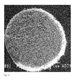
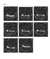
- the distribution of mineral particles throughout the scaffolds is a difficult parameter/attribute to quantify. It is an attribute that is much easier to visualize and therefore, a range of values to protect is difficult to define. It was visualized using two different methods. The first was microCT which is shown in Figure 6 below. The microCT scanner used in this analysis uses X-rays to detect mineralised tissue. Consequently, Figure 6 shows only the mineral particles within a 100 wt% HA scaffold. Knowing that the collagen is not visible, one can see that the mineral particles are completely and evenly distributed throughout the scaffold. Given that the scaffold is 99% empty, this image shows conclusive evidence that the HA is intimately associated with the collagen fibres. Figure 7 shows a 2-dimensional slice of the same scaffold illustrating the distribution from another viewpoint.
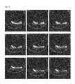
- Figures 8 and 9 illustrate the distribution of mineral particles throughout a 50 wt% HA scaffold using a distinct imaging tool, Scanning Electron Microscopy (SEM). Both images show the same region of interest within a 50wt% HA scaffold.
- Figure 8 shows both phases of collagen and HA.
- the mineral particles are indistinguishable to the naked eye.
- the mineral particle can be detected for the identical region of interest (ROI). This is shown in Figure 9 as white pixels representing mineral particles.
- ROI region of interest
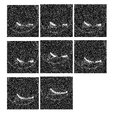
- Pore interconnectivity is another important scaffold attribute that is very difficult to define quantitatively in scaffolds composed primarily of biological material.
- pore interconnectivity is strongly related to scaffold permeability. Permeability is discussed below but the conductivity of flow is dependant on porosity, pore size and pore interconnectivity together. Consequently, permeability gives an indication of pore interconnectivity when pore size and porosity are defined.
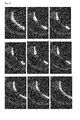
- FIG. 10 shows a 50wt% HA scaffold at 10 times magnification and the obviously interconnected pore structure at the surface. This has been shown to be identical using thin sections taken from such samples.
- Figure 11 shows the same 50wt% scaffold at 100 times magnification. This image illustrates the pore interconnectivity. At this magnification, the pore structure is conclusively interconnected.
- Figure 4 shows the net mean cell number left within the scaffold types at 28 days.
- a net decrease of 0.5 million in cell proliferation is seen in the pure collagen constructs (approximately 20% decrease), which is not unsurprising given that the pure collagen construct is favourable but not optimized for osteoblast cells.
- the addition of 50wt%, 100wt% and 200wt% HA results in a increased cellular proliferation of approximately 500%, 50% and 30% respectively ( Figure 4 ).
- the permeability of a porous scaffold is essentially the flow conductivity under pressure through that porous medium.
- a high scaffold permeability is essential for long term viability of the scaffold in vivo as it allows cells to migrate to the scaffold centre and facilitates vascularisation in vivo.
- the 50wt%, 100 wt% and 200 HA scaffolds exhibited a significantly increased mean tissue permeability relative to the control collagen scaffolds. This was a surprising but positive finding of this study as it shows that the addition of HA actually aids the flow of fluid throughout the scaffold. It is believed that this increase in flow conductivity through the porous scaffold is due to the increased scaffold rigidity.
- the empirical protocol used to quantify scaffold permeability is described in detail in reference [5] (O'Brien et al, 2007, Section 2.2, pages 7-10).
- a small animal trial was carried out to determine the potential of this invention to encourage osteogenesis and mineralisation of a critically sized defect in bone.
- Nine Wistar rats were used during the trial.
- a critically sized defect was created in the rat calvaria.
- One animal was left with an empty defect as a control.
- the other eight animals were split into groups. Specifically, four of the defects were filled with 50wt% HA scaffolds, two of which were seeded with rat mesenchymal stem cells (MSC) cells and two of which that were left unseeded. This was to investigate the potential of tissue engineered versus off the shelf scaffold types.
- the remaining four sample defects were filled with 200wt% HA scaffolds and once again, these four were split evenly between seeded and unseeded scaffolds.
- the empty defect animal data showed the defect was filled in with soft fibrous tissue as part of the healing process. This was expected and has been seen in previously conducted animal trials within our tissue engineering group. At some points throughout the defect after the 28 day trial, small particles of dense material were observed within the empty defect but these were seldom observed and not sufficiently dense to indicate significant healing within the empty defect sample. Examples of these are shown in the microCT X-ray images shown below.
- the 50wt% HA cell-seeded scaffolds showed more promising results. These scaffolds were seeded with rat mesenchymal stem cells prior to implantation. As can be seen from the representative microCT x-ray images below, small heterogeneous pockets of mildly mineralized material were seen not only at the periphery of the defect-bone interface but also in the centre of the scaffolds. There were a significantly higher number of instances of these pockets of mineralization seen relative to the empty defect data and they appeared to be brighter in their intensity relative to the instances seen in the empty defect.
- the first cell-seeded 200wt% HA scaffold showed significantly improved results compared with the 50wt% HA scaffolds.
- significant levels of mineralisation were seen from the periphery of the defect all the way into the centre of the scaffold-filled defect.
- the mineralisation level was not as high as that of the surrounding bone tissue indicated by the relative difference in the image intensity of the mineralised particles within the scaffold-filled defect.
- the second cell-seeded 200wt% HA scaffold showed significantly improved results compared with all previous scaffolds. At a significant number of areas examined throughout the defect, significant levels of mineralisation were seen from the periphery of the defect all the way into the centre of the scaffold-filled defect. Unlike the first 200wt% HA cell-seeded scaffolds, the mineralised tissue formed was not particulate in nature but was continuous across the scaffold-filled defect. Most interestingly, this continuous mineralisation was seen at the widest part of the defect. In this sample, the mineralisation level was nearly identical to that of the surrounding bone tissue indicated by the similar image intensity of the mineralised material within the scaffold-filled defect.
- the cell-free scaffold-filled defect results for the 50wt% HA scaffolds were very similar to the cell-seeded 50wt% HA scaffolds. Significantly mineralised tissue was seen throughout the scaffold-filled defects but was not continuous in nature. However, the intensity of the mineralised tissue was marginally greater than that seen in the cell-seeded samples. This was seen in all cell unseeded samples that follow and indicated that a cell-free construct may perform better in vivo.
- the unseeded 200wt% HA sample showed significant levels of mineralisation at the periphery and the centre of the scaffold-filled defect at a significant number of areas throughout the defect. This mineralised tissue was continuous in nature unlike that seen in the 50wt% HA samples.
- this unseeded 200wt% HA sample showed significant levels of mineralisation at the periphery and the centre of the scaffold-filled defect at a significant number of areas throughout the defect. This mineralised tissue was continuous in nature unlike that seen in the 50wt% HA samples.
- the invention is not limited to the embodiments herein described. As such, the three embodiments of the invention described herein represent a small proportion of the total number of scaffold variants possible using the same core fabrication protocol. Either the constituents themselves or specific embodiment stages or process steps can be varied to produce a varied range of constructs, optimised for application specific use. The variations possible include;
- Acetic acid concentration The concentration of the acetic acid within the initial collagen slurry can be altered to suit specific applications. Increasing the concentration encourages a more rapid and homogenous integration of the HA particles within the blending slurry. Additionally, this concentration also has a significant effect on both the mechanical properties and the biocompatability of the scaffold. These effects are discussed in detail in the "Invention Characterisation" Section above.
- the acetic acid concentration can be varied between 0.05M and 5M.
- the collagen quantity can be varied within the initial collagen slurry. Increasing the collagen quantity results in an increased mechanical stiffness of the resulting scaffold. This also has a significant effect on scaffold biocompatability.
- the collagen quantity can vary from 0.5 g/L up to 50 g/L of acetic acid solution (1/10 and 10 times standard collagen concentration respectively).
- the quantity of HA can be varied within a specified range relative to the proportion of collagen within the scaffold slurry prior to manufacture. Specifically, the quantity of HA can suitably vary from 10 to 1000 weight percent of the quantity of collagen used. Increasing the HA content was found to significantly increase the mechanical stiffness of the manufactured scaffold.
- Hydroxyapatite Type The present invention can be manufactured using both sintered, unsintered, and other forms of the HA powder.
- Both the HA aliquot volume and injection interval can be varied to facilitate mixing of the two primary constituent of the slurry.
- the injection interval can be varied from 30 minutes up to 240 minutes.
- the aliquot volume can suitably be varied from 1 ml up to 100 ml. Such freedom facilitates optimisation of any particular invention embodiment.
- Hydroxyapatite Particle Size Typically, the HA particle size can be varied from 10 nm up to 100 ⁇ m to suit specific applications.
- the final freezing temperature reached during the freezedrying process determines the mean pore size within the manufactured scaffolds.
- This final freezing temperature can be varied to produce scaffolds with various mean pore sizes specific to a specific application or cell type.
- the final freezing temperature can be varied from -10° C down to -70° C.
- the freezing interface placed between the embodiment slurry and the freezedryer cooling shelf can be varied.
- the type of freezing interface affects heat energy transmission to/from the slurry/scaffold and can alter the pore structure of the final scaffold.
- Four main options are available, specifically a walled vessel of defined interface made of either metal(1), plastic(2), a thin polymeric membrane(3) or no interface (4).
- the freezing rate determines the rate of ice crystal nucleation within the collagen/HA slurry during the freezedrying process and controls the homogeneity of the pore formation process.
- the cooling rate is varied to optimise the freezing process for the various types of interfaces available between the slurry and the freezedryer cooling shelf (e.g. metal, plastic, none).
- the freezing rate can be varied from 0.01° C/min up to 10° C/min.
- Annealing stage An annealing stage an be employed during the freezedrying process and allows the creation of pores with an average diameter significantly greater than pore sizes attainable by varying the final freezing temperature alone.
- the annealing time can be varied from 15 minutes to 36 hours. The longer the annealing time, the larger the final average pore size.
- the crosslinking method can be one of a possible number of techniques, either dehydrothermal or chemical in nature. Additionally, both techniques can be employed consecutively. Specific crosslinking options include glutaraldehyde, carbodiimides (EDAC), microbial transglutaminase (mTgase), dehydrothermal crosslinking (DHT) and ultraviolet radiation (UV).
- EDAC glutaraldehyde
- mTgase microbial transglutaminase
- DHT dehydrothermal crosslinking
- UV ultraviolet radiation
- the freezedried scaffold collagen can be crosslinked via dehydrothermal means to increase the scaffold mechanical stiffness.
- the crosslinking temperature can be varied from 105° C up to 180° C with a corresponding increase in embodiment stiffness. Additionally, when using chemical crosslinking methods, the concentration of the crosslinking solution can be varied to alter the extent of chemical crosslinking.
- the crosslink exposure time can also be varied to alter the extent of the crosslinking process throughout the scaffold. This can be varied between 24 and 120 hours to alter the final mechanical specification of the scaffold.
Priority Applications (3)
| Application Number | Priority Date | Filing Date | Title |
|---|---|---|---|
| NO12178368A NO2517738T3 (fr) | 2007-02-09 | 2008-02-11 | |
| PL12178368T PL2517738T3 (pl) | 2007-02-09 | 2008-02-11 | Rusztowanie kompozytowe z kolagenu/hydroksyapatytu |
| EP12178368.2A EP2517738B1 (fr) | 2007-02-09 | 2008-02-11 | Base composite de collagène/hydroxyapatite |
Applications Claiming Priority (3)
| Application Number | Priority Date | Filing Date | Title |
|---|---|---|---|
| EP20070394001 EP1964583A1 (fr) | 2007-02-09 | 2007-02-09 | Processus de production d'une base composite de collagène/hydroxyapatite |
| EP20080710143 EP2117617B1 (fr) | 2007-02-09 | 2008-02-11 | Processus de production d'une base composite de collagène/hydroxyapatite |
| EP12178368.2A EP2517738B1 (fr) | 2007-02-09 | 2008-02-11 | Base composite de collagène/hydroxyapatite |
Related Parent Applications (2)
| Application Number | Title | Priority Date | Filing Date |
|---|---|---|---|
| EP08710143.2 Division | 2008-02-11 | ||
| EP20080710143 Division EP2117617B1 (fr) | 2007-02-09 | 2008-02-11 | Processus de production d'une base composite de collagène/hydroxyapatite |
Publications (2)
| Publication Number | Publication Date |
|---|---|
| EP2517738A1 true EP2517738A1 (fr) | 2012-10-31 |
| EP2517738B1 EP2517738B1 (fr) | 2017-08-23 |
Family
ID=38196643
Family Applications (3)
| Application Number | Title | Priority Date | Filing Date |
|---|---|---|---|
| EP20070394001 Ceased EP1964583A1 (fr) | 2007-02-09 | 2007-02-09 | Processus de production d'une base composite de collagène/hydroxyapatite |
| EP12178368.2A Active EP2517738B1 (fr) | 2007-02-09 | 2008-02-11 | Base composite de collagène/hydroxyapatite |
| EP20080710143 Active EP2117617B1 (fr) | 2007-02-09 | 2008-02-11 | Processus de production d'une base composite de collagène/hydroxyapatite |
Family Applications Before (1)
| Application Number | Title | Priority Date | Filing Date |
|---|---|---|---|
| EP20070394001 Ceased EP1964583A1 (fr) | 2007-02-09 | 2007-02-09 | Processus de production d'une base composite de collagène/hydroxyapatite |
Family Applications After (1)
| Application Number | Title | Priority Date | Filing Date |
|---|---|---|---|
| EP20080710143 Active EP2117617B1 (fr) | 2007-02-09 | 2008-02-11 | Processus de production d'une base composite de collagène/hydroxyapatite |
Country Status (13)
| Country | Link |
|---|---|
| US (2) | US8435552B2 (fr) |
| EP (3) | EP1964583A1 (fr) |
| JP (2) | JP5527760B2 (fr) |
| AU (1) | AU2008212526B2 (fr) |
| CA (1) | CA2677992C (fr) |
| DK (2) | DK2517738T3 (fr) |
| ES (2) | ES2649091T3 (fr) |
| IL (1) | IL200295A (fr) |
| NO (1) | NO2517738T3 (fr) |
| NZ (1) | NZ579466A (fr) |
| PL (2) | PL2117617T3 (fr) |
| PT (2) | PT2517738T (fr) |
| WO (1) | WO2008096334A2 (fr) |
Families Citing this family (33)
| Publication number | Priority date | Publication date | Assignee | Title |
|---|---|---|---|---|
| US9485917B2 (en) | 2006-12-15 | 2016-11-08 | Ecovative Design, LLC | Method for producing grown materials and products made thereby |
| JP2009254547A (ja) * | 2008-04-16 | 2009-11-05 | Tohoku Univ | 骨再生材料およびその製造方法 |
| WO2010084481A1 (fr) | 2009-01-23 | 2010-07-29 | Royal College Of Surgeons In Ireland | Échafaudage stratifié adapté à la réparation ostéochondrale |
| JP2010273847A (ja) * | 2009-05-28 | 2010-12-09 | Tokyo Institute Of Technology | 高密度多孔質複合体 |
| GB0912399D0 (en) * | 2009-07-16 | 2009-08-26 | Ucl Business Plc | Polymeric collagen biomaterials |
| CN101897994B (zh) * | 2010-07-23 | 2013-01-09 | 山东大学 | 一种修复骨缺损的生物复合支架及其制备方法 |
| CN101966348B (zh) * | 2010-09-21 | 2014-03-26 | 中国科学院深圳先进技术研究院 | 掺锶羟基磷灰石胶原复合材料及其应用和制备方法 |
| WO2014060443A2 (fr) * | 2012-10-15 | 2014-04-24 | Royal College Of Surgeons In Ireland | Échafaudage composite pouvant être utilisé à titre d'implant d'ingénierie tissulaire |
| US10150258B2 (en) | 2013-07-29 | 2018-12-11 | Carnegie Mellon University | Additive manufacturing of embedded materials |
| US11277979B2 (en) | 2013-07-31 | 2022-03-22 | Ecovative Design Llc | Mycological biopolymers grown in void space tooling |
| US20150045885A1 (en) | 2013-08-06 | 2015-02-12 | University Of Limerick | Seedless group iv nanowires, and methods for the production thereof |
| CN105636598B (zh) | 2013-09-02 | 2021-07-23 | 玛芬股份有限公司 | 包含胞外基质组织材料和成骨蛋白的产品 |
| US20150101509A1 (en) | 2013-10-14 | 2015-04-16 | Gavin R. McIntyre | Method of Manufacturing a Stiff Engineered Composite |
| CN103920191B (zh) * | 2014-04-21 | 2016-02-17 | 陕西巨子生物技术有限公司 | 一种增强成骨活性的复合人工骨及其制备方法 |
| WO2016084413A1 (fr) * | 2014-11-27 | 2016-06-02 | 東洋紡株式会社 | Corps composite poreux, matériau de régénération osseuse et procédé de production de corps composite poreux |
| FR3038896B1 (fr) * | 2015-07-17 | 2020-03-06 | Centre National De La Recherche Scientifique | Procede de fabrication d’un materiau monolithique poreux |
| CN105311681B (zh) * | 2015-12-07 | 2018-12-25 | 杭州华迈医疗器械有限公司 | 一种可注射的骨修复用复合材料及其制备方法 |
| MY192441A (en) | 2016-03-01 | 2022-08-21 | The Fynder Group Inc | Filamentous fungal biomats, methods of their production and methods of their use |
| US10273549B2 (en) * | 2016-04-21 | 2019-04-30 | Vitrolabs Inc. | Engineered skin equivalent, method of manufacture thereof and products derived therefrom |
| JP7177588B2 (ja) * | 2016-12-12 | 2022-11-24 | オリンパステルモバイオマテリアル株式会社 | 骨補填材および骨補填材の製造方法 |
| EP3599832A4 (fr) | 2017-03-31 | 2021-01-27 | Ecovative Design LLC | Procédés de post-traitement à base de solution(s) pour matériau biopolymère mycologique et produit mycologique ainsi obtenu |
| WO2019077806A1 (fr) * | 2017-10-20 | 2019-04-25 | オリンパステルモバイオマテリアル株式会社 | Matériau de greffe osseuse et son procédé de fabrication |
| US11266085B2 (en) | 2017-11-14 | 2022-03-08 | Ecovative Design Llc | Increased homogeneity of mycological biopolymer grown into void space |
| US11920126B2 (en) | 2018-03-28 | 2024-03-05 | Ecovative Design Llc | Bio-manufacturing process |
| US11293005B2 (en) | 2018-05-07 | 2022-04-05 | Ecovative Design Llc | Process for making mineralized mycelium scaffolding and product made thereby |
| US20190359931A1 (en) | 2018-05-24 | 2019-11-28 | Ecovative Design Llc | Process and Apparatus for Producing Mycelium Biomaterial |
| WO2020030572A1 (fr) * | 2018-08-07 | 2020-02-13 | Glaxosmithkline Biologicals Sa | Processus et vaccins |
| CA3113935A1 (fr) | 2018-10-02 | 2020-04-09 | Ecovative Design Llc | Paradigme de bioreacteur servant a la production de matrices hyphales extraparticulaires secondaires |
| JP7315943B2 (ja) * | 2018-11-28 | 2023-07-27 | 株式会社バイオアパタイト | ハイドロキシアパタイト含有多孔性支持体 |
| US11357891B2 (en) * | 2019-06-14 | 2022-06-14 | Geistlich Pharma Ag | Collagen matrix or granulate blend of bone substitute material |
| FR3101773B1 (fr) | 2019-10-10 | 2023-04-21 | Regeska | Procede de mineralisation d’une membrane de biopolymere |
| US20220202990A1 (en) * | 2020-12-29 | 2022-06-30 | Industrial Technology Research Institute | Tissue scaffold for use in tendon and/or ligament |
| CN114344569B (zh) * | 2021-12-21 | 2023-03-21 | 无锡贝迪生物工程股份有限公司 | 一种胶原蛋白/生物陶瓷多孔骨植入物及其制备方法 |
Citations (8)
| Publication number | Priority date | Publication date | Assignee | Title |
|---|---|---|---|---|
| EP1437148A1 (fr) * | 2001-10-19 | 2004-07-14 | Japan Science and Technology Agency | Biomateriaux composites organiques/inorganiques et procedes permettant de produire ces biomateriaux |
| CN1526765A (zh) * | 2003-09-23 | 2004-09-08 | 中国医学科学院生物医学工程研究所 | 复合骨组织工程支架材料及其制备方法 |
| WO2005004928A2 (fr) * | 2003-04-04 | 2005-01-20 | W.R. Grace & Co.-Conn. | Eponges de collagene poreuses en particules |
| EP1500405A1 (fr) * | 2002-05-01 | 2005-01-26 | Japan Science and Technology Agency | Procede de preparation d'un materiau composite poreux |
| EP1566186A1 (fr) * | 2002-11-06 | 2005-08-24 | National Institute for Materials Science | Materiau d'apatite/collagene poreux reticule contenant un composite d'apatite/collagene auto-organise et son procede de production |
| WO2006031196A1 (fr) * | 2004-09-14 | 2006-03-23 | Agency For Science, Technology And Research | Composite de matiere de charge biologique poreuse et procede de fabrication correspondant |
| US20060172918A1 (en) * | 2001-10-25 | 2006-08-03 | Shinichi Sotome | Composite biomaterials |
| WO2006095154A2 (fr) * | 2005-03-07 | 2006-09-14 | Cambridge Enterprise Limited | Biomateriau |
Family Cites Families (49)
| Publication number | Priority date | Publication date | Assignee | Title |
|---|---|---|---|---|
| US4207306A (en) * | 1974-08-02 | 1980-06-10 | Sterling Drug Inc. | Process for producing polycrystalline ceramic oxides |
| FR2585576B1 (fr) * | 1985-07-30 | 1992-01-03 | Bioetica Sa | Produit de remplacement de la matrice osseuse favorisant l'osteogenese |
| WO1995008304A1 (fr) * | 1993-09-21 | 1995-03-30 | The Penn State Research Foundation | Composition de remplacement osseux comprenant de l'hydroxyapatite et procede de production de ladite composition |
| US6180606B1 (en) * | 1994-09-28 | 2001-01-30 | Gensci Orthobiologics, Inc. | Compositions with enhanced osteogenic potential, methods for making the same and uses thereof |
| JPH08276003A (ja) * | 1995-04-07 | 1996-10-22 | Terumo Corp | 硬組織修復材料および埋入型医療用具 |
| US5776193A (en) | 1995-10-16 | 1998-07-07 | Orquest, Inc. | Bone grafting matrix |
| FR2794649B1 (fr) * | 1999-06-11 | 2003-04-11 | Solutions | Biomateriau a base d'un derive de dextrane insolubilise et d'un facteur de croissance, son procede de preparation et ses applications |
| US6730252B1 (en) * | 2000-09-20 | 2004-05-04 | Swee Hin Teoh | Methods for fabricating a filament for use in tissue engineering |
| US20030003127A1 (en) | 2001-06-27 | 2003-01-02 | Ethicon, Inc. | Porous ceramic/porous polymer layered scaffolds for the repair and regeneration of tissue |
| US6626950B2 (en) * | 2001-06-28 | 2003-09-30 | Ethicon, Inc. | Composite scaffold with post anchor for the repair and regeneration of tissue |
| DE10135275A1 (de) | 2001-07-13 | 2003-01-30 | Jotec Gmbh | Implantat und Verfahren zu seiner Herstellung |
| JP3646167B2 (ja) * | 2002-02-19 | 2005-05-11 | 独立行政法人産業技術総合研究所 | フォスフォフォリンを含む複合生体材料 |
| WO2003072128A2 (fr) * | 2002-02-22 | 2003-09-04 | Ebi, L.P. | Procedes et composes destines a traiter des defauts osseux ou du cartilage |
| ITMI20030186A1 (it) | 2003-02-05 | 2004-08-06 | Consiglio Nazionale Ricerche | Procedimento di sintesi di tessuto osseo artificiale, |
| JP3999746B2 (ja) * | 2003-05-19 | 2007-10-31 | ローム アンド ハース カンパニー | ポリマーナノ粒子の高固形分調製方法 |
| JP4680771B2 (ja) * | 2003-05-26 | 2011-05-11 | Hoya株式会社 | リン酸カルシウム含有複合多孔体及びその製造方法 |
| AU2004247143B2 (en) * | 2003-06-11 | 2010-09-23 | Warsaw Orthopedic, Inc. | Osteoimplants and methods for their manufacture |
| US7563748B2 (en) | 2003-06-23 | 2009-07-21 | Cognis Ip Management Gmbh | Alcohol alkoxylate carriers for pesticide active ingredients |
| JP2005152551A (ja) | 2003-11-20 | 2005-06-16 | Masaki Terada | ワンウエーコーヒーメーカー |
| JP4643166B2 (ja) * | 2004-03-30 | 2011-03-02 | 独立行政法人物質・材料研究機構 | アパタイト/コラーゲン複合体繊維を含む多孔体の平均気孔径制御方法 |
| WO2006000020A1 (fr) | 2004-06-29 | 2006-01-05 | European Nickel Plc | Lixiviation amelioree de metaux de base |
| US7732573B2 (en) * | 2004-10-28 | 2010-06-08 | National Institute For Materials Science | Method for producing porous body comprising apatite/collagen composite fibers |
| DE102005034420A1 (de) * | 2004-12-23 | 2006-07-06 | Ossacur Ag | Gelartiges Material zur Füllung von Knochen- und/oder Knorpeldefekten |
| ITMI20050343A1 (it) | 2005-03-04 | 2006-09-05 | Fin Ceramica Faenza S R L | Sostituto cartilagineo e osteocindrale comprendente una struttura multistrato e relativo impiego |
| US9132208B2 (en) * | 2008-08-07 | 2015-09-15 | Lifenet Health | Composition for a tissue repair implant and methods of making the same |
| US8920827B2 (en) * | 2005-10-21 | 2014-12-30 | Wake Forest University Health Sciences | Keratin bioceramic compositions |
| US20120114763A1 (en) * | 2005-11-14 | 2012-05-10 | Genoss Ltd | Method for Producing Collagen/Apatite Composite Membrane for Guided Bone Regeneration |
| KR100706759B1 (ko) * | 2006-03-28 | 2007-04-12 | 한국원자력연구소 | 인장 강도가 강한 키토산 지지체를 제조하는 방법 및 그에의하여 제조되는 키토산 지지체 |
| TW200801513A (en) | 2006-06-29 | 2008-01-01 | Fermiscan Australia Pty Ltd | Improved process |
| GB2440721A (en) | 2006-08-11 | 2008-02-13 | Univ Cambridge Tech | Composite biomaterial formed by cooling a fluid composition on a porous solid and removing solidified crystals of the liquid carrier |
| ITMI20070458A1 (it) * | 2007-03-07 | 2008-09-08 | Roberto Buda | Composizione contenente cellule mononucleate autologhe matrice in collagene suino sotto forma di pasta e suo uso per la preparazione di un medicamento per trattamento chirurgico |
| US20080241211A1 (en) * | 2007-03-27 | 2008-10-02 | University Of Southern California | Device which enhances the biological activity of locally applied growth factors with particular emphasis on those used for bone repair |
| WO2008130068A1 (fr) | 2007-04-23 | 2008-10-30 | Modern Cell & Tissue Technologies Inc. | Procede de preparation d'un echafaudage polymere poreux a partir de glace seche |
| US20080281431A1 (en) * | 2007-05-10 | 2008-11-13 | Biomet Manufacturing Corp. | Resorbable bone graft materials |
| WO2008157608A1 (fr) | 2007-06-18 | 2008-12-24 | Cartlix, Inc. | Echafaudages composites pour la régénération tissulaire |
| WO2008156221A1 (fr) * | 2007-06-20 | 2008-12-24 | Hoya Corporation | Réparation e traitement d'une défectuosité osseuse au moyen de cellules produites par un facteur produit lui-même par un chondrocyte à capacité hypertrophique et structure |
| WO2009122902A1 (fr) * | 2008-04-02 | 2009-10-08 | Hoya株式会社 | Matériau poreux expansible comprenant un complexe apatite/collagène, et procédé de production de celui-ci |
| JP5467554B2 (ja) * | 2008-04-25 | 2014-04-09 | HOYA Technosurgical株式会社 | 粉末状のアパタイト/コラーゲン複合体、形状賦形型の人工骨ペースト、及びそれらの製造方法 |
| WO2010084481A1 (fr) * | 2009-01-23 | 2010-07-29 | Royal College Of Surgeons In Ireland | Échafaudage stratifié adapté à la réparation ostéochondrale |
| US20100267143A1 (en) * | 2009-04-16 | 2010-10-21 | Snu R&Db Foundation | Method for Surface Modification of Polymeric Scaffold for Stem Cell Transplantation Using Decellularized Extracellular Matrix |
| WO2011000020A1 (fr) | 2009-06-12 | 2011-01-06 | Sbc Research Pty Ltd | Procédé amélioré de détection |
| WO2011031637A2 (fr) * | 2009-09-08 | 2011-03-17 | Musculoskeletal Transplant Foundation Inc. | Composition pour réparation de ménisque à base d'une technique de tissu |
| WO2011030185A1 (fr) | 2009-09-12 | 2011-03-17 | Inanc Buelend | Échafaudages fibro-inducteurs et angiogènes de guidage de cellules pour le modelage de tissu parodontal |
| AU2011223652B2 (en) * | 2010-03-03 | 2015-01-29 | Novabone Products, Llc | Devices and methods for the regeneration of bony defects |
| US8734831B2 (en) * | 2010-04-16 | 2014-05-27 | Snu R&Db Foundation | Method for manufacturing a porous ceramic scaffold having an organic/inorganic hybrid coating layer containing a bioactive factor |
| US20110262486A1 (en) * | 2010-04-22 | 2011-10-27 | Taipei Medical University | Bone implant and manufacturing method thereof |
| WO2011150482A1 (fr) | 2010-05-31 | 2011-12-08 | Fundação De Amparo À Pesquisa Do Estado de São Paulo | Procédés permettant d'obtenir des composites réabsorbables, composites, membrane, échafaudage et applications associées |
| WO2012000020A1 (fr) | 2010-06-28 | 2012-01-05 | Wood, Stephen Raymond | Base de clôture provisoire |
| WO2012068376A2 (fr) | 2010-11-17 | 2012-05-24 | Wake Forest University Health Sciences | Compositions kératiniques pour le traitement d'une déficience ou lésion osseuse |
-
2007
- 2007-02-09 EP EP20070394001 patent/EP1964583A1/fr not_active Ceased
-
2008
- 2008-02-11 DK DK12178368.2T patent/DK2517738T3/en active
- 2008-02-11 JP JP2009548796A patent/JP5527760B2/ja not_active Expired - Fee Related
- 2008-02-11 NO NO12178368A patent/NO2517738T3/no unknown
- 2008-02-11 PT PT121783682T patent/PT2517738T/pt unknown
- 2008-02-11 EP EP12178368.2A patent/EP2517738B1/fr active Active
- 2008-02-11 NZ NZ57946608A patent/NZ579466A/xx active IP Right Revival
- 2008-02-11 PL PL08710143T patent/PL2117617T3/pl unknown
- 2008-02-11 WO PCT/IE2008/000010 patent/WO2008096334A2/fr active Application Filing
- 2008-02-11 ES ES12178368.2T patent/ES2649091T3/es active Active
- 2008-02-11 DK DK08710143T patent/DK2117617T3/da active
- 2008-02-11 ES ES08710143T patent/ES2435192T3/es active Active
- 2008-02-11 US US12/526,353 patent/US8435552B2/en active Active
- 2008-02-11 PT PT87101432T patent/PT2117617E/pt unknown
- 2008-02-11 EP EP20080710143 patent/EP2117617B1/fr active Active
- 2008-02-11 PL PL12178368T patent/PL2517738T3/pl unknown
- 2008-02-11 CA CA2677992A patent/CA2677992C/fr not_active Expired - Fee Related
- 2008-02-11 AU AU2008212526A patent/AU2008212526B2/en active Active
-
2009
- 2009-08-09 IL IL200295A patent/IL200295A/en active IP Right Grant
-
2013
- 2013-03-06 US US13/786,891 patent/US9138483B2/en not_active Expired - Fee Related
- 2013-12-16 JP JP2013259131A patent/JP5881669B2/ja active Active
Patent Citations (8)
| Publication number | Priority date | Publication date | Assignee | Title |
|---|---|---|---|---|
| EP1437148A1 (fr) * | 2001-10-19 | 2004-07-14 | Japan Science and Technology Agency | Biomateriaux composites organiques/inorganiques et procedes permettant de produire ces biomateriaux |
| US20060172918A1 (en) * | 2001-10-25 | 2006-08-03 | Shinichi Sotome | Composite biomaterials |
| EP1500405A1 (fr) * | 2002-05-01 | 2005-01-26 | Japan Science and Technology Agency | Procede de preparation d'un materiau composite poreux |
| EP1566186A1 (fr) * | 2002-11-06 | 2005-08-24 | National Institute for Materials Science | Materiau d'apatite/collagene poreux reticule contenant un composite d'apatite/collagene auto-organise et son procede de production |
| WO2005004928A2 (fr) * | 2003-04-04 | 2005-01-20 | W.R. Grace & Co.-Conn. | Eponges de collagene poreuses en particules |
| CN1526765A (zh) * | 2003-09-23 | 2004-09-08 | 中国医学科学院生物医学工程研究所 | 复合骨组织工程支架材料及其制备方法 |
| WO2006031196A1 (fr) * | 2004-09-14 | 2006-03-23 | Agency For Science, Technology And Research | Composite de matiere de charge biologique poreuse et procede de fabrication correspondant |
| WO2006095154A2 (fr) * | 2005-03-07 | 2006-09-14 | Cambridge Enterprise Limited | Biomateriau |
Non-Patent Citations (7)
| Title |
|---|
| BAILEY A.J.; LIGHT N.D.; ATKINS E.D.T.: "Chemical crosslinking restrictions on models for the molecular organisation of the collagen fibre", NATURE, vol. 288, 1980, pages 408 - 410 |
| DATABASE WPI Week 200480, Derwent World Patents Index; AN 2004-805630, XP002440997 * |
| DONG J.K.; LUTHY H.; WOHLWEND A.; SCHARER P: "Heatpressed ceramics: technology and strength", INTERNATIONAL JOURNAL OF PROSTHODONTICS, vol. 5, no. 1, 1992, pages 9 - 16 |
| O'BRIEN F.J.; HARLEY B.A.; YANNAS LV.; GIBSON L.J.: "The effect of pore size on cell adhesion in collagen gag scaffolds", BIOMATERIALS, vol. 26, 2005, pages 433 - 441 |
| O'BRIEN, F.J.; HARLEY, B.A.; WALLER, M.A.; YANNAS, LV; GIBSON, L.J; PRENDERGAST, P.J.: "The effect of pore size on permeability and cell attachment in collagen scaffolds for tissue engineering", TECHNOLOGY AND HEALTHCARE INVITED ARTICLE, vol. 15, 2007, pages 3 - 17 |
| TANCRED D.C.; CARR A.J.; MCCORMACK B.A.: "Development of a new synthetic bone graft", JOURNAL OF MATERIALS SCIENCE: MATERIALS IN MEDICINE, vol. 9, no. 12, 1998, pages 819 - 823 |
| YANNAS LV: "Tissue and Organ Regeneration in Adults", 2001, SPRINGER |
Also Published As
| Publication number | Publication date |
|---|---|
| EP2117617B1 (fr) | 2013-08-14 |
| PT2117617E (pt) | 2013-11-20 |
| DK2517738T3 (en) | 2017-12-04 |
| US20130177648A1 (en) | 2013-07-11 |
| DK2117617T3 (da) | 2013-11-18 |
| EP1964583A1 (fr) | 2008-09-03 |
| AU2008212526B2 (en) | 2013-06-06 |
| AU2008212526A1 (en) | 2008-08-14 |
| PL2517738T3 (pl) | 2018-03-30 |
| WO2008096334A3 (fr) | 2008-10-30 |
| PT2517738T (pt) | 2017-11-28 |
| NZ579466A (en) | 2012-08-31 |
| JP2010517659A (ja) | 2010-05-27 |
| US8435552B2 (en) | 2013-05-07 |
| ES2649091T3 (es) | 2018-01-10 |
| IL200295A0 (en) | 2010-04-29 |
| EP2517738B1 (fr) | 2017-08-23 |
| JP5527760B2 (ja) | 2014-06-25 |
| PL2117617T3 (pl) | 2014-01-31 |
| IL200295A (en) | 2015-06-30 |
| JP2014076387A (ja) | 2014-05-01 |
| CA2677992A1 (fr) | 2008-08-14 |
| WO2008096334B1 (fr) | 2008-12-18 |
| CA2677992C (fr) | 2017-03-21 |
| WO2008096334A2 (fr) | 2008-08-14 |
| ES2435192T3 (es) | 2013-12-16 |
| US9138483B2 (en) | 2015-09-22 |
| NO2517738T3 (fr) | 2018-01-20 |
| US20100158976A1 (en) | 2010-06-24 |
| JP5881669B2 (ja) | 2016-03-09 |
| EP2117617A2 (fr) | 2009-11-18 |
Similar Documents
| Publication | Publication Date | Title |
|---|---|---|
| EP2117617B1 (fr) | Processus de production d'une base composite de collagène/hydroxyapatite | |
| Diao et al. | 3D‐plotted beta‐tricalcium phosphate scaffolds with smaller pore sizes improve in vivo bone regeneration and biomechanical properties in a critical‐sized calvarial defect rat model | |
| Volkov et al. | Poly (3-hydroxybutyrate)/hydroxyapatite/alginate scaffolds seeded with mesenchymal stem cells enhance the regeneration of critical-sized bone defect | |
| Lyons et al. | The healing of bony defects by cell-free collagen-based scaffolds compared to stem cell-seeded tissue engineered constructs | |
| Köse et al. | Tissue engineered cartilage on collagen and PHBV matrices | |
| Jiang et al. | Chitosan–poly (lactide-co-glycolide) microsphere-based scaffolds for bone tissue engineering: In vitro degradation and in vivo bone regeneration studies | |
| US8784499B2 (en) | Preparation of regenerative tissue scaffolds | |
| EP2603248B1 (fr) | Echafaudages tissulaires régénérateurs | |
| Sadiasa et al. | In vitro and in vivo evaluation of porous PCL-PLLA 3D polymer scaffolds fabricated via salt leaching method for bone tissue engineering applications | |
| Tang et al. | A fast degradable citrate-based bone scaffold promotes spinal fusion | |
| CN105688274A (zh) | 一种聚己内酯/明胶电纺复合支架的制备工艺 | |
| Zhang et al. | A study on a tissue-engineered bone using rhBMP-2 induced periosteal cells with a porous nano-hydroxyapatite/collagen/poly (L-lactic acid) scaffold | |
| Meskinfam | Polymer scaffolds for bone regeneration | |
| JP5578499B2 (ja) | リン酸カルシウム/生分解性ポリマーハイブリッド材料並びにその製法及びハイブリッド材料を用いたインプラント | |
| JP2000116681A (ja) | 骨相当物を組織エンジニアリングするためのデバイス | |
| Sahvieh et al. | Role of bone 1stem cell–seeded 3D polylactic acid/polycaprolactone/hydroxyapatite scaffold on a critical-sized radial bone defect in rat | |
| Chen et al. | Reconstruction of calvarial defect using a tricalcium phosphate-oligomeric proanthocyanidins cross-linked gelatin composite | |
| KR102014248B1 (ko) | 이상 인산 칼슘이 탑재된 탈세포화된 돼지 피부 유래 주입형 세포외 기질 기반 하이드로겔의 제조방법 | |
| Más et al. | Biological evaluation of PLDLA polymer synthesized as construct on bone tissue engineering application | |
| Asano et al. | Preparation of thermoplastic poly (L-lactic acid) membranes for guided bone regeneration. | |
| Hu et al. | Surface‐modified pliable PDLLA/PCL/β‐TCP scaffolds as a promising delivery system for bone regeneration | |
| Kumar et al. | Polymeric Nano-Composite Scaffolds for Bone Tissue Engineering | |
| Hixon | The Development of a Cryogel Scaffold for the Treatment of Cleft-Craniofacial Defects to Promote Bone Regeneration | |
| Dvora | Designing a Whey Protein Based Material as a Scaffold for Bone Regeneration |
Legal Events
| Date | Code | Title | Description |
|---|---|---|---|
| PUAI | Public reference made under article 153(3) epc to a published international application that has entered the european phase |
Free format text: ORIGINAL CODE: 0009012 |
|
| AC | Divisional application: reference to earlier application |
Ref document number: 2117617 Country of ref document: EP Kind code of ref document: P |
|
| AK | Designated contracting states |
Kind code of ref document: A1 Designated state(s): AT BE BG CH CY CZ DE DK EE ES FI FR GB GR HR HU IE IS IT LI LT LU LV MC MT NL NO PL PT RO SE SI SK TR |
|
| 17P | Request for examination filed |
Effective date: 20130429 |
|
| 17Q | First examination report despatched |
Effective date: 20151204 |
|
| GRAP | Despatch of communication of intention to grant a patent |
Free format text: ORIGINAL CODE: EPIDOSNIGR1 |
|
| INTG | Intention to grant announced |
Effective date: 20170303 |
|
| GRAS | Grant fee paid |
Free format text: ORIGINAL CODE: EPIDOSNIGR3 |
|
| GRAA | (expected) grant |
Free format text: ORIGINAL CODE: 0009210 |
|
| AC | Divisional application: reference to earlier application |
Ref document number: 2117617 Country of ref document: EP Kind code of ref document: P |
|
| AK | Designated contracting states |
Kind code of ref document: B1 Designated state(s): AT BE BG CH CY CZ DE DK EE ES FI FR GB GR HR HU IE IS IT LI LT LU LV MC MT NL NO PL PT RO SE SI SK TR |
|
| REG | Reference to a national code |
Ref country code: GB Ref legal event code: FG4D |
|
| REG | Reference to a national code |
Ref country code: CH Ref legal event code: EP |
|
| REG | Reference to a national code |
Ref country code: AT Ref legal event code: REF Ref document number: 920649 Country of ref document: AT Kind code of ref document: T Effective date: 20170915 |
|
| REG | Reference to a national code |
Ref country code: IE Ref legal event code: FG4D |
|
| REG | Reference to a national code |
Ref country code: DE Ref legal event code: R096 Ref document number: 602008051833 Country of ref document: DE |
|
| REG | Reference to a national code |
Ref country code: PT Ref legal event code: SC4A Ref document number: 2517738 Country of ref document: PT Date of ref document: 20171128 Kind code of ref document: T Free format text: AVAILABILITY OF NATIONAL TRANSLATION Effective date: 20171121 |
|
| REG | Reference to a national code |
Ref country code: DK Ref legal event code: T3 Effective date: 20171127 |
|
| REG | Reference to a national code |
Ref country code: SE Ref legal event code: TRGR |
|
| REG | Reference to a national code |
Ref country code: NL Ref legal event code: FP |
|
| REG | Reference to a national code |
Ref country code: ES Ref legal event code: FG2A Ref document number: 2649091 Country of ref document: ES Kind code of ref document: T3 Effective date: 20180110 Ref country code: LT Ref legal event code: MG4D |
|
| PG25 | Lapsed in a contracting state [announced via postgrant information from national office to epo] |
Ref country code: LT Free format text: LAPSE BECAUSE OF FAILURE TO SUBMIT A TRANSLATION OF THE DESCRIPTION OR TO PAY THE FEE WITHIN THE PRESCRIBED TIME-LIMIT Effective date: 20170823 Ref country code: HR Free format text: LAPSE BECAUSE OF FAILURE TO SUBMIT A TRANSLATION OF THE DESCRIPTION OR TO PAY THE FEE WITHIN THE PRESCRIBED TIME-LIMIT Effective date: 20170823 |
|
| REG | Reference to a national code |
Ref country code: NO Ref legal event code: T2 Effective date: 20170823 |
|
| REG | Reference to a national code |
Ref country code: FR Ref legal event code: PLFP Year of fee payment: 11 |
|
| PG25 | Lapsed in a contracting state [announced via postgrant information from national office to epo] |
Ref country code: BG Free format text: LAPSE BECAUSE OF FAILURE TO SUBMIT A TRANSLATION OF THE DESCRIPTION OR TO PAY THE FEE WITHIN THE PRESCRIBED TIME-LIMIT Effective date: 20171123 Ref country code: IS Free format text: LAPSE BECAUSE OF FAILURE TO SUBMIT A TRANSLATION OF THE DESCRIPTION OR TO PAY THE FEE WITHIN THE PRESCRIBED TIME-LIMIT Effective date: 20171223 Ref country code: LV Free format text: LAPSE BECAUSE OF FAILURE TO SUBMIT A TRANSLATION OF THE DESCRIPTION OR TO PAY THE FEE WITHIN THE PRESCRIBED TIME-LIMIT Effective date: 20170823 Ref country code: GR Free format text: LAPSE BECAUSE OF FAILURE TO SUBMIT A TRANSLATION OF THE DESCRIPTION OR TO PAY THE FEE WITHIN THE PRESCRIBED TIME-LIMIT Effective date: 20171124 |
|
| PG25 | Lapsed in a contracting state [announced via postgrant information from national office to epo] |
Ref country code: RO Free format text: LAPSE BECAUSE OF FAILURE TO SUBMIT A TRANSLATION OF THE DESCRIPTION OR TO PAY THE FEE WITHIN THE PRESCRIBED TIME-LIMIT Effective date: 20170823 |
|
| REG | Reference to a national code |
Ref country code: DE Ref legal event code: R097 Ref document number: 602008051833 Country of ref document: DE |
|
| PG25 | Lapsed in a contracting state [announced via postgrant information from national office to epo] |
Ref country code: SK Free format text: LAPSE BECAUSE OF FAILURE TO SUBMIT A TRANSLATION OF THE DESCRIPTION OR TO PAY THE FEE WITHIN THE PRESCRIBED TIME-LIMIT Effective date: 20170823 Ref country code: EE Free format text: LAPSE BECAUSE OF FAILURE TO SUBMIT A TRANSLATION OF THE DESCRIPTION OR TO PAY THE FEE WITHIN THE PRESCRIBED TIME-LIMIT Effective date: 20170823 |
|
| PLBE | No opposition filed within time limit |
Free format text: ORIGINAL CODE: 0009261 |
|
| STAA | Information on the status of an ep patent application or granted ep patent |
Free format text: STATUS: NO OPPOSITION FILED WITHIN TIME LIMIT |
|
| 26N | No opposition filed |
Effective date: 20180524 |
|
| PG25 | Lapsed in a contracting state [announced via postgrant information from national office to epo] |
Ref country code: SI Free format text: LAPSE BECAUSE OF FAILURE TO SUBMIT A TRANSLATION OF THE DESCRIPTION OR TO PAY THE FEE WITHIN THE PRESCRIBED TIME-LIMIT Effective date: 20170823 |
|
| PG25 | Lapsed in a contracting state [announced via postgrant information from national office to epo] |
Ref country code: MC Free format text: LAPSE BECAUSE OF FAILURE TO SUBMIT A TRANSLATION OF THE DESCRIPTION OR TO PAY THE FEE WITHIN THE PRESCRIBED TIME-LIMIT Effective date: 20170823 |
|
| PG25 | Lapsed in a contracting state [announced via postgrant information from national office to epo] |
Ref country code: LU Free format text: LAPSE BECAUSE OF NON-PAYMENT OF DUE FEES Effective date: 20180211 |
|
| PG25 | Lapsed in a contracting state [announced via postgrant information from national office to epo] |
Ref country code: MT Free format text: LAPSE BECAUSE OF NON-PAYMENT OF DUE FEES Effective date: 20180211 |
|
| PG25 | Lapsed in a contracting state [announced via postgrant information from national office to epo] |
Ref country code: HU Free format text: LAPSE BECAUSE OF FAILURE TO SUBMIT A TRANSLATION OF THE DESCRIPTION OR TO PAY THE FEE WITHIN THE PRESCRIBED TIME-LIMIT; INVALID AB INITIO Effective date: 20080211 |
|
| PG25 | Lapsed in a contracting state [announced via postgrant information from national office to epo] |
Ref country code: CY Free format text: LAPSE BECAUSE OF FAILURE TO SUBMIT A TRANSLATION OF THE DESCRIPTION OR TO PAY THE FEE WITHIN THE PRESCRIBED TIME-LIMIT Effective date: 20170823 |
|
| REG | Reference to a national code |
Ref country code: AT Ref legal event code: UEP Ref document number: 920649 Country of ref document: AT Kind code of ref document: T Effective date: 20170823 |
|
| PGFP | Annual fee paid to national office [announced via postgrant information from national office to epo] |
Ref country code: NL Payment date: 20230228 Year of fee payment: 16 |
|
| PGFP | Annual fee paid to national office [announced via postgrant information from national office to epo] |
Ref country code: NO Payment date: 20230301 Year of fee payment: 16 Ref country code: IE Payment date: 20230214 Year of fee payment: 16 Ref country code: FR Payment date: 20230224 Year of fee payment: 16 Ref country code: FI Payment date: 20230227 Year of fee payment: 16 Ref country code: ES Payment date: 20230309 Year of fee payment: 16 Ref country code: DK Payment date: 20230224 Year of fee payment: 16 Ref country code: CZ Payment date: 20230213 Year of fee payment: 16 Ref country code: CH Payment date: 20230307 Year of fee payment: 16 Ref country code: AT Payment date: 20230228 Year of fee payment: 16 |
|
| PGFP | Annual fee paid to national office [announced via postgrant information from national office to epo] |
Ref country code: TR Payment date: 20230209 Year of fee payment: 16 Ref country code: SE Payment date: 20230224 Year of fee payment: 16 Ref country code: PT Payment date: 20230209 Year of fee payment: 16 Ref country code: PL Payment date: 20230210 Year of fee payment: 16 Ref country code: IT Payment date: 20230224 Year of fee payment: 16 Ref country code: GB Payment date: 20230213 Year of fee payment: 16 Ref country code: DE Payment date: 20230227 Year of fee payment: 16 Ref country code: BE Payment date: 20230228 Year of fee payment: 16 |