EP2514761B1 - Cristal d'interféron de recombinaison, structure tridimensionnelle et utilisations - Google Patents
Cristal d'interféron de recombinaison, structure tridimensionnelle et utilisations Download PDFInfo
- Publication number
- EP2514761B1 EP2514761B1 EP10836926.5A EP10836926A EP2514761B1 EP 2514761 B1 EP2514761 B1 EP 2514761B1 EP 10836926 A EP10836926 A EP 10836926A EP 2514761 B1 EP2514761 B1 EP 2514761B1
- Authority
- EP
- European Patent Office
- Prior art keywords
- atom
- interferon
- ala
- leu
- recombinant interferon
- Prior art date
- Legal status (The legal status is an assumption and is not a legal conclusion. Google has not performed a legal analysis and makes no representation as to the accuracy of the status listed.)
- Active
Links
- 102000014150 Interferons Human genes 0.000 title claims description 246
- 108010050904 Interferons Proteins 0.000 title claims description 246
- 229940079322 interferon Drugs 0.000 title claims description 238
- 239000013078 crystal Substances 0.000 title claims description 75
- 238000000034 method Methods 0.000 claims description 78
- 125000000539 amino acid group Chemical group 0.000 claims description 41
- 150000001875 compounds Chemical class 0.000 claims description 34
- 239000012634 fragment Substances 0.000 claims description 26
- 239000003814 drug Substances 0.000 claims description 23
- 229940079593 drug Drugs 0.000 claims description 21
- 239000002773 nucleotide Substances 0.000 claims description 14
- 125000003729 nucleotide group Chemical group 0.000 claims description 14
- 238000002425 crystallisation Methods 0.000 claims description 12
- FWMNVWWHGCHHJJ-SKKKGAJSSA-N 4-amino-1-[(2r)-6-amino-2-[[(2r)-2-[[(2r)-2-[[(2r)-2-amino-3-phenylpropanoyl]amino]-3-phenylpropanoyl]amino]-4-methylpentanoyl]amino]hexanoyl]piperidine-4-carboxylic acid Chemical compound C([C@H](C(=O)N[C@H](CC(C)C)C(=O)N[C@H](CCCCN)C(=O)N1CCC(N)(CC1)C(O)=O)NC(=O)[C@H](N)CC=1C=CC=CC=1)C1=CC=CC=C1 FWMNVWWHGCHHJJ-SKKKGAJSSA-N 0.000 claims description 10
- 241000588724 Escherichia coli Species 0.000 claims description 9
- 238000009510 drug design Methods 0.000 claims description 9
- 239000008194 pharmaceutical composition Substances 0.000 claims description 9
- 230000003993 interaction Effects 0.000 claims description 8
- 230000008025 crystallization Effects 0.000 claims description 7
- 230000008569 process Effects 0.000 claims description 7
- 239000003937 drug carrier Substances 0.000 claims description 6
- 229910021645 metal ion Inorganic materials 0.000 claims description 3
- 230000002349 favourable effect Effects 0.000 claims description 2
- 125000003275 alpha amino acid group Chemical group 0.000 claims 1
- XLYOFNOQVPJJNP-UHFFFAOYSA-N water Chemical compound O XLYOFNOQVPJJNP-UHFFFAOYSA-N 0.000 description 170
- 229910052717 sulfur Inorganic materials 0.000 description 157
- DHMQDGOQFOQNFH-UHFFFAOYSA-N Glycine Chemical compound NCC(O)=O DHMQDGOQFOQNFH-UHFFFAOYSA-N 0.000 description 140
- 210000004027 cell Anatomy 0.000 description 79
- 108010010648 interferon alfacon-1 Proteins 0.000 description 77
- 229940090438 infergen Drugs 0.000 description 75
- 238000001142 circular dichroism spectrum Methods 0.000 description 65
- 150000001413 amino acids Chemical group 0.000 description 58
- 108090000623 proteins and genes Proteins 0.000 description 39
- 102000004169 proteins and genes Human genes 0.000 description 35
- 235000018102 proteins Nutrition 0.000 description 34
- 239000003446 ligand Substances 0.000 description 33
- PJWWRFATQTVXHA-UHFFFAOYSA-N Cyclohexylaminopropanesulfonic acid Chemical compound OS(=O)(=O)CCCNC1CCCCC1 PJWWRFATQTVXHA-UHFFFAOYSA-N 0.000 description 32
- 108010079944 Interferon-alpha2b Proteins 0.000 description 28
- 230000000694 effects Effects 0.000 description 28
- 238000012360 testing method Methods 0.000 description 28
- 238000002360 preparation method Methods 0.000 description 26
- FAPWRFPIFSIZLT-UHFFFAOYSA-M Sodium chloride Chemical compound [Na+].[Cl-] FAPWRFPIFSIZLT-UHFFFAOYSA-M 0.000 description 22
- 101000701051 Legionella pneumophila Zinc metalloproteinase Proteins 0.000 description 21
- 125000004429 atom Chemical group 0.000 description 20
- 230000014509 gene expression Effects 0.000 description 19
- 108090000765 processed proteins & peptides Proteins 0.000 description 18
- 206010028980 Neoplasm Diseases 0.000 description 17
- 241000282414 Homo sapiens Species 0.000 description 16
- 239000000539 dimer Substances 0.000 description 16
- 239000013612 plasmid Substances 0.000 description 16
- 239000000872 buffer Substances 0.000 description 15
- 108010047761 Interferon-alpha Proteins 0.000 description 14
- 102000006992 Interferon-alpha Human genes 0.000 description 14
- 239000000203 mixture Substances 0.000 description 14
- 238000004458 analytical method Methods 0.000 description 13
- 230000006907 apoptotic process Effects 0.000 description 13
- 238000013461 design Methods 0.000 description 13
- 238000002347 injection Methods 0.000 description 13
- 239000007924 injection Substances 0.000 description 13
- 239000000178 monomer Substances 0.000 description 13
- 239000000243 solution Substances 0.000 description 13
- 238000011282 treatment Methods 0.000 description 13
- 230000004071 biological effect Effects 0.000 description 12
- 230000000875 corresponding effect Effects 0.000 description 11
- 230000005764 inhibitory process Effects 0.000 description 11
- 229940047124 interferons Drugs 0.000 description 11
- 239000011780 sodium chloride Substances 0.000 description 11
- TWRXJAOTZQYOKJ-UHFFFAOYSA-L Magnesium chloride Chemical compound [Mg+2].[Cl-].[Cl-] TWRXJAOTZQYOKJ-UHFFFAOYSA-L 0.000 description 10
- 239000012980 RPMI-1640 medium Substances 0.000 description 10
- 239000002299 complementary DNA Substances 0.000 description 10
- 201000010099 disease Diseases 0.000 description 10
- 208000037265 diseases, disorders, signs and symptoms Diseases 0.000 description 10
- 102000005962 receptors Human genes 0.000 description 10
- 108020003175 receptors Proteins 0.000 description 10
- 210000002966 serum Anatomy 0.000 description 10
- 238000002441 X-ray diffraction Methods 0.000 description 9
- 150000005829 chemical entities Chemical class 0.000 description 9
- 238000002447 crystallographic data Methods 0.000 description 9
- 230000002401 inhibitory effect Effects 0.000 description 9
- 102100025064 Cellular tumor antigen p53 Human genes 0.000 description 8
- -1 aliphatic amino acid Chemical class 0.000 description 8
- 230000000840 anti-viral effect Effects 0.000 description 8
- 238000002474 experimental method Methods 0.000 description 8
- 230000006870 function Effects 0.000 description 8
- 208000002672 hepatitis B Diseases 0.000 description 8
- 238000000338 in vitro Methods 0.000 description 8
- 102000004196 processed proteins & peptides Human genes 0.000 description 8
- 239000000047 product Substances 0.000 description 8
- 238000000746 purification Methods 0.000 description 8
- 238000012216 screening Methods 0.000 description 8
- 230000003612 virological effect Effects 0.000 description 8
- 102100021569 Apoptosis regulator Bcl-2 Human genes 0.000 description 7
- 108091003079 Bovine Serum Albumin Proteins 0.000 description 7
- 206010008342 Cervix carcinoma Diseases 0.000 description 7
- 108020004414 DNA Proteins 0.000 description 7
- 101000971171 Homo sapiens Apoptosis regulator Bcl-2 Proteins 0.000 description 7
- 208000006105 Uterine Cervical Neoplasms Diseases 0.000 description 7
- 230000010261 cell growth Effects 0.000 description 7
- 201000010881 cervical cancer Diseases 0.000 description 7
- 239000011549 crystallization solution Substances 0.000 description 7
- 239000012091 fetal bovine serum Substances 0.000 description 7
- OHDXDNUPVVYWOV-UHFFFAOYSA-N n-methyl-1-(2-naphthalen-1-ylsulfanylphenyl)methanamine Chemical compound CNCC1=CC=CC=C1SC1=CC=CC2=CC=CC=C12 OHDXDNUPVVYWOV-UHFFFAOYSA-N 0.000 description 7
- 230000036961 partial effect Effects 0.000 description 7
- 239000008363 phosphate buffer Substances 0.000 description 7
- 238000010254 subcutaneous injection Methods 0.000 description 7
- 206010006187 Breast cancer Diseases 0.000 description 6
- 208000026310 Breast neoplasm Diseases 0.000 description 6
- 241000700605 Viruses Species 0.000 description 6
- 230000000259 anti-tumor effect Effects 0.000 description 6
- 239000000427 antigen Substances 0.000 description 6
- 102000036639 antigens Human genes 0.000 description 6
- 108091007433 antigens Proteins 0.000 description 6
- 210000004369 blood Anatomy 0.000 description 6
- 239000008280 blood Substances 0.000 description 6
- 229910052799 carbon Inorganic materials 0.000 description 6
- 239000003153 chemical reaction reagent Substances 0.000 description 6
- 238000012258 culturing Methods 0.000 description 6
- 239000012153 distilled water Substances 0.000 description 6
- 238000000684 flow cytometry Methods 0.000 description 6
- INHCSSUBVCNVSK-UHFFFAOYSA-L lithium sulfate Chemical compound [Li+].[Li+].[O-]S([O-])(=O)=O INHCSSUBVCNVSK-UHFFFAOYSA-L 0.000 description 6
- 239000000463 material Substances 0.000 description 6
- 239000002609 medium Substances 0.000 description 6
- 230000001105 regulatory effect Effects 0.000 description 6
- 239000007929 subcutaneous injection Substances 0.000 description 6
- 102000004190 Enzymes Human genes 0.000 description 5
- 108090000790 Enzymes Proteins 0.000 description 5
- PEDCQBHIVMGVHV-UHFFFAOYSA-N Glycerine Chemical compound OCC(O)CO PEDCQBHIVMGVHV-UHFFFAOYSA-N 0.000 description 5
- 101000852865 Homo sapiens Interferon alpha/beta receptor 2 Proteins 0.000 description 5
- 102100036718 Interferon alpha/beta receptor 2 Human genes 0.000 description 5
- 230000015572 biosynthetic process Effects 0.000 description 5
- 239000000969 carrier Substances 0.000 description 5
- 238000004113 cell culture Methods 0.000 description 5
- 238000010586 diagram Methods 0.000 description 5
- BNIILDVGGAEEIG-UHFFFAOYSA-L disodium hydrogen phosphate Chemical compound [Na+].[Na+].OP([O-])([O-])=O BNIILDVGGAEEIG-UHFFFAOYSA-L 0.000 description 5
- 229910000397 disodium phosphate Inorganic materials 0.000 description 5
- 238000009826 distribution Methods 0.000 description 5
- 230000009036 growth inhibition Effects 0.000 description 5
- 239000001963 growth medium Substances 0.000 description 5
- 230000002209 hydrophobic effect Effects 0.000 description 5
- 229910001629 magnesium chloride Inorganic materials 0.000 description 5
- 238000012986 modification Methods 0.000 description 5
- 230000004048 modification Effects 0.000 description 5
- 239000003607 modifier Substances 0.000 description 5
- 230000008520 organization Effects 0.000 description 5
- BHZOKUMUHVTPBX-UHFFFAOYSA-M sodium acetic acid acetate Chemical compound [Na+].CC(O)=O.CC([O-])=O BHZOKUMUHVTPBX-UHFFFAOYSA-M 0.000 description 5
- 239000002904 solvent Substances 0.000 description 5
- 238000010186 staining Methods 0.000 description 5
- 238000007619 statistical method Methods 0.000 description 5
- 238000003786 synthesis reaction Methods 0.000 description 5
- QKNYBSVHEMOAJP-UHFFFAOYSA-N 2-amino-2-(hydroxymethyl)propane-1,3-diol;hydron;chloride Chemical compound Cl.OCC(N)(CO)CO QKNYBSVHEMOAJP-UHFFFAOYSA-N 0.000 description 4
- KCXVZYZYPLLWCC-UHFFFAOYSA-N EDTA Chemical compound OC(=O)CN(CC(O)=O)CCN(CC(O)=O)CC(O)=O KCXVZYZYPLLWCC-UHFFFAOYSA-N 0.000 description 4
- 101000852870 Homo sapiens Interferon alpha/beta receptor 1 Proteins 0.000 description 4
- 102100036714 Interferon alpha/beta receptor 1 Human genes 0.000 description 4
- 238000002835 absorbance Methods 0.000 description 4
- 238000013459 approach Methods 0.000 description 4
- PYMYPHUHKUWMLA-WDCZJNDASA-N arabinose Chemical compound OC[C@@H](O)[C@@H](O)[C@H](O)C=O PYMYPHUHKUWMLA-WDCZJNDASA-N 0.000 description 4
- PYMYPHUHKUWMLA-UHFFFAOYSA-N arabinose Natural products OCC(O)C(O)C(O)C=O PYMYPHUHKUWMLA-UHFFFAOYSA-N 0.000 description 4
- SRBFZHDQGSBBOR-UHFFFAOYSA-N beta-D-Pyranose-Lyxose Natural products OC1COC(O)C(O)C1O SRBFZHDQGSBBOR-UHFFFAOYSA-N 0.000 description 4
- 201000011510 cancer Diseases 0.000 description 4
- 239000006285 cell suspension Substances 0.000 description 4
- 238000012512 characterization method Methods 0.000 description 4
- 238000005094 computer simulation Methods 0.000 description 4
- 239000013604 expression vector Substances 0.000 description 4
- 230000012010 growth Effects 0.000 description 4
- 210000003000 inclusion body Anatomy 0.000 description 4
- 230000001965 increasing effect Effects 0.000 description 4
- 229940046166 oligodeoxynucleotide Drugs 0.000 description 4
- 239000008188 pellet Substances 0.000 description 4
- 230000000144 pharmacologic effect Effects 0.000 description 4
- 239000012460 protein solution Substances 0.000 description 4
- 238000004153 renaturation Methods 0.000 description 4
- 238000011160 research Methods 0.000 description 4
- 238000002864 sequence alignment Methods 0.000 description 4
- 239000011550 stock solution Substances 0.000 description 4
- 239000000126 substance Substances 0.000 description 4
- 230000002194 synthesizing effect Effects 0.000 description 4
- 206010067484 Adverse reaction Diseases 0.000 description 3
- 102000004127 Cytokines Human genes 0.000 description 3
- 108090000695 Cytokines Proteins 0.000 description 3
- 230000004543 DNA replication Effects 0.000 description 3
- IAZDPXIOMUYVGZ-UHFFFAOYSA-N Dimethylsulphoxide Chemical compound CS(C)=O IAZDPXIOMUYVGZ-UHFFFAOYSA-N 0.000 description 3
- LFQSCWFLJHTTHZ-UHFFFAOYSA-N Ethanol Chemical compound CCO LFQSCWFLJHTTHZ-UHFFFAOYSA-N 0.000 description 3
- 101000959820 Homo sapiens Interferon alpha-1/13 Proteins 0.000 description 3
- 102100040019 Interferon alpha-1/13 Human genes 0.000 description 3
- 101710163270 Nuclease Proteins 0.000 description 3
- 108010002747 Pfu DNA polymerase Proteins 0.000 description 3
- 102100036789 Protein TBATA Human genes 0.000 description 3
- 101710118245 Protein TBATA Proteins 0.000 description 3
- 208000000389 T-cell leukemia Diseases 0.000 description 3
- 208000028530 T-cell lymphoblastic leukemia/lymphoma Diseases 0.000 description 3
- XSQUKJJJFZCRTK-UHFFFAOYSA-N Urea Chemical compound NC(N)=O XSQUKJJJFZCRTK-UHFFFAOYSA-N 0.000 description 3
- 230000006838 adverse reaction Effects 0.000 description 3
- 239000000556 agonist Substances 0.000 description 3
- 230000003698 anagen phase Effects 0.000 description 3
- 239000005557 antagonist Substances 0.000 description 3
- 239000004202 carbamide Substances 0.000 description 3
- 230000004663 cell proliferation Effects 0.000 description 3
- 238000006243 chemical reaction Methods 0.000 description 3
- 238000010276 construction Methods 0.000 description 3
- 238000013480 data collection Methods 0.000 description 3
- 230000003247 decreasing effect Effects 0.000 description 3
- 238000004925 denaturation Methods 0.000 description 3
- 230000036425 denaturation Effects 0.000 description 3
- 238000001514 detection method Methods 0.000 description 3
- 238000000855 fermentation Methods 0.000 description 3
- 230000004151 fermentation Effects 0.000 description 3
- 238000003306 harvesting Methods 0.000 description 3
- 238000001727 in vivo Methods 0.000 description 3
- 230000001939 inductive effect Effects 0.000 description 3
- 239000008176 lyophilized powder Substances 0.000 description 3
- 239000012528 membrane Substances 0.000 description 3
- 108010092948 oligonucleotidase Proteins 0.000 description 3
- 229920001184 polypeptide Polymers 0.000 description 3
- 230000035755 proliferation Effects 0.000 description 3
- XJMOSONTPMZWPB-UHFFFAOYSA-M propidium iodide Chemical compound [I-].[I-].C12=CC(N)=CC=C2C2=CC=C(N)C=C2[N+](CCC[N+](C)(CC)CC)=C1C1=CC=CC=C1 XJMOSONTPMZWPB-UHFFFAOYSA-M 0.000 description 3
- 239000011541 reaction mixture Substances 0.000 description 3
- 230000028327 secretion Effects 0.000 description 3
- 238000000926 separation method Methods 0.000 description 3
- 239000006228 supernatant Substances 0.000 description 3
- 238000013518 transcription Methods 0.000 description 3
- 230000035897 transcription Effects 0.000 description 3
- IJGRMHOSHXDMSA-UHFFFAOYSA-N Atomic nitrogen Chemical compound N#N IJGRMHOSHXDMSA-UHFFFAOYSA-N 0.000 description 2
- 108010075254 C-Peptide Proteins 0.000 description 2
- 101710132601 Capsid protein Proteins 0.000 description 2
- 102000012410 DNA Ligases Human genes 0.000 description 2
- 108010061982 DNA Ligases Proteins 0.000 description 2
- 238000001712 DNA sequencing Methods 0.000 description 2
- BWGNESOTFCXPMA-UHFFFAOYSA-N Dihydrogen disulfide Chemical compound SS BWGNESOTFCXPMA-UHFFFAOYSA-N 0.000 description 2
- AOJJSUZBOXZQNB-TZSSRYMLSA-N Doxorubicin Chemical compound O([C@H]1C[C@@](O)(CC=2C(O)=C3C(=O)C=4C=CC=C(C=4C(=O)C3=C(O)C=21)OC)C(=O)CO)[C@H]1C[C@H](N)[C@H](O)[C@H](C)O1 AOJJSUZBOXZQNB-TZSSRYMLSA-N 0.000 description 2
- 238000002965 ELISA Methods 0.000 description 2
- LYCAIKOWRPUZTN-UHFFFAOYSA-N Ethylene glycol Chemical compound OCCO LYCAIKOWRPUZTN-UHFFFAOYSA-N 0.000 description 2
- 239000004471 Glycine Substances 0.000 description 2
- 208000005176 Hepatitis C Diseases 0.000 description 2
- 101100450591 Human adenovirus B serotype 3 PVIII gene Proteins 0.000 description 2
- 241000725303 Human immunodeficiency virus Species 0.000 description 2
- 102000002227 Interferon Type I Human genes 0.000 description 2
- 108010014726 Interferon Type I Proteins 0.000 description 2
- 108010078049 Interferon alpha-2 Proteins 0.000 description 2
- 102100026720 Interferon beta Human genes 0.000 description 2
- 108090000467 Interferon-beta Proteins 0.000 description 2
- 238000005481 NMR spectroscopy Methods 0.000 description 2
- 108700020796 Oncogene Proteins 0.000 description 2
- 229920004890 Triton X-100 Polymers 0.000 description 2
- 239000013504 Triton X-100 Substances 0.000 description 2
- 102000004142 Trypsin Human genes 0.000 description 2
- 108090000631 Trypsin Proteins 0.000 description 2
- 230000002378 acidificating effect Effects 0.000 description 2
- 239000004480 active ingredient Substances 0.000 description 2
- 238000007792 addition Methods 0.000 description 2
- 239000000654 additive Substances 0.000 description 2
- 239000011543 agarose gel Substances 0.000 description 2
- 238000000246 agarose gel electrophoresis Methods 0.000 description 2
- 230000001028 anti-proliverative effect Effects 0.000 description 2
- 230000002155 anti-virotic effect Effects 0.000 description 2
- 239000002775 capsule Substances 0.000 description 2
- 125000004432 carbon atom Chemical group C* 0.000 description 2
- 210000000170 cell membrane Anatomy 0.000 description 2
- 125000003636 chemical group Chemical group 0.000 description 2
- 238000004440 column chromatography Methods 0.000 description 2
- 230000000052 comparative effect Effects 0.000 description 2
- 239000000470 constituent Substances 0.000 description 2
- 230000002596 correlated effect Effects 0.000 description 2
- 238000012217 deletion Methods 0.000 description 2
- 230000037430 deletion Effects 0.000 description 2
- 238000002050 diffraction method Methods 0.000 description 2
- 238000009792 diffusion process Methods 0.000 description 2
- 230000029087 digestion Effects 0.000 description 2
- 239000002552 dosage form Substances 0.000 description 2
- 238000005516 engineering process Methods 0.000 description 2
- 238000001914 filtration Methods 0.000 description 2
- 238000012215 gene cloning Methods 0.000 description 2
- 235000011187 glycerol Nutrition 0.000 description 2
- 238000012835 hanging drop method Methods 0.000 description 2
- 229910052739 hydrogen Inorganic materials 0.000 description 2
- 239000001257 hydrogen Substances 0.000 description 2
- 238000013115 immunohistochemical detection Methods 0.000 description 2
- 230000004957 immunoregulator effect Effects 0.000 description 2
- 238000011534 incubation Methods 0.000 description 2
- 230000036512 infertility Effects 0.000 description 2
- 239000003112 inhibitor Substances 0.000 description 2
- 108010018844 interferon type III Proteins 0.000 description 2
- 238000010255 intramuscular injection Methods 0.000 description 2
- 239000007927 intramuscular injection Substances 0.000 description 2
- 150000002611 lead compounds Chemical class 0.000 description 2
- HQKMJHAJHXVSDF-UHFFFAOYSA-L magnesium stearate Chemical compound [Mg+2].CCCCCCCCCCCCCCCCCC([O-])=O.CCCCCCCCCCCCCCCCCC([O-])=O HQKMJHAJHXVSDF-UHFFFAOYSA-L 0.000 description 2
- 238000004519 manufacturing process Methods 0.000 description 2
- 239000003550 marker Substances 0.000 description 2
- 230000007935 neutral effect Effects 0.000 description 2
- 238000005457 optimization Methods 0.000 description 2
- 150000002894 organic compounds Chemical class 0.000 description 2
- 230000010355 oscillation Effects 0.000 description 2
- 239000000546 pharmaceutical excipient Substances 0.000 description 2
- 230000001766 physiological effect Effects 0.000 description 2
- 239000011148 porous material Substances 0.000 description 2
- 238000011046 pyrogen test Methods 0.000 description 2
- 238000007670 refining Methods 0.000 description 2
- 230000009466 transformation Effects 0.000 description 2
- 230000001131 transforming effect Effects 0.000 description 2
- 239000012588 trypsin Substances 0.000 description 2
- 239000008215 water for injection Substances 0.000 description 2
- 108091032973 (ribonucleotides)n+m Proteins 0.000 description 1
- 102000040650 (ribonucleotides)n+m Human genes 0.000 description 1
- 206010000830 Acute leukaemia Diseases 0.000 description 1
- 229920001817 Agar Polymers 0.000 description 1
- NLXLAEXVIDQMFP-UHFFFAOYSA-N Ammonia chloride Chemical compound [NH4+].[Cl-] NLXLAEXVIDQMFP-UHFFFAOYSA-N 0.000 description 1
- 108090000672 Annexin A5 Proteins 0.000 description 1
- 102000004121 Annexin A5 Human genes 0.000 description 1
- 208000032791 BCR-ABL1 positive chronic myelogenous leukemia Diseases 0.000 description 1
- 108010077805 Bacterial Proteins Proteins 0.000 description 1
- 101100518633 Caenorhabditis elegans dpy-18 gene Proteins 0.000 description 1
- 208000010833 Chronic myeloid leukaemia Diseases 0.000 description 1
- 108700010070 Codon Usage Proteins 0.000 description 1
- 102000003712 Complement factor B Human genes 0.000 description 1
- 108090000056 Complement factor B Proteins 0.000 description 1
- 108020004635 Complementary DNA Proteins 0.000 description 1
- JPVYNHNXODAKFH-UHFFFAOYSA-N Cu2+ Chemical compound [Cu+2] JPVYNHNXODAKFH-UHFFFAOYSA-N 0.000 description 1
- 241000701022 Cytomegalovirus Species 0.000 description 1
- 102100025698 Cytosolic carboxypeptidase 4 Human genes 0.000 description 1
- 208000035859 Drug effect increased Diseases 0.000 description 1
- 241001115402 Ebolavirus Species 0.000 description 1
- 108010042407 Endonucleases Proteins 0.000 description 1
- 102000004533 Endonucleases Human genes 0.000 description 1
- 241000709661 Enterovirus Species 0.000 description 1
- 208000000461 Esophageal Neoplasms Diseases 0.000 description 1
- 108010010803 Gelatin Proteins 0.000 description 1
- 241000700721 Hepatitis B virus Species 0.000 description 1
- 241000701044 Human gammaherpesvirus 4 Species 0.000 description 1
- 241000341655 Human papillomavirus type 16 Species 0.000 description 1
- 206010020751 Hypersensitivity Diseases 0.000 description 1
- 108010074328 Interferon-gamma Proteins 0.000 description 1
- 102000008070 Interferon-gamma Human genes 0.000 description 1
- 239000007836 KH2PO4 Substances 0.000 description 1
- 208000007766 Kaposi sarcoma Diseases 0.000 description 1
- 125000000174 L-prolyl group Chemical group [H]N1C([H])([H])C([H])([H])C([H])([H])[C@@]1([H])C(*)=O 0.000 description 1
- 231100000002 MTT assay Toxicity 0.000 description 1
- 238000000134 MTT assay Methods 0.000 description 1
- JLVVSXFLKOJNIY-UHFFFAOYSA-N Magnesium ion Chemical compound [Mg+2] JLVVSXFLKOJNIY-UHFFFAOYSA-N 0.000 description 1
- 208000034578 Multiple myelomas Diseases 0.000 description 1
- 208000033761 Myelogenous Chronic BCR-ABL Positive Leukemia Diseases 0.000 description 1
- 230000006051 NK cell activation Effects 0.000 description 1
- 208000001894 Nasopharyngeal Neoplasms Diseases 0.000 description 1
- 206010061306 Nasopharyngeal cancer Diseases 0.000 description 1
- 108091028043 Nucleic acid sequence Proteins 0.000 description 1
- 206010030155 Oesophageal carcinoma Diseases 0.000 description 1
- 102000043276 Oncogene Human genes 0.000 description 1
- 206010033128 Ovarian cancer Diseases 0.000 description 1
- 206010061535 Ovarian neoplasm Diseases 0.000 description 1
- 238000010222 PCR analysis Methods 0.000 description 1
- 206010061902 Pancreatic neoplasm Diseases 0.000 description 1
- 239000005662 Paraffin oil Substances 0.000 description 1
- 241001494479 Pecora Species 0.000 description 1
- 108091005804 Peptidases Proteins 0.000 description 1
- 102000035195 Peptidases Human genes 0.000 description 1
- 241000709664 Picornaviridae Species 0.000 description 1
- 206010035226 Plasma cell myeloma Diseases 0.000 description 1
- 208000008601 Polycythemia Diseases 0.000 description 1
- 206010060862 Prostate cancer Diseases 0.000 description 1
- 208000000236 Prostatic Neoplasms Diseases 0.000 description 1
- 239000004365 Protease Substances 0.000 description 1
- 241001510071 Pyrrhocoridae Species 0.000 description 1
- 208000015634 Rectal Neoplasms Diseases 0.000 description 1
- 208000006265 Renal cell carcinoma Diseases 0.000 description 1
- 241000315672 SARS coronavirus Species 0.000 description 1
- 244000082988 Secale cereale Species 0.000 description 1
- 238000012300 Sequence Analysis Methods 0.000 description 1
- 241000700584 Simplexvirus Species 0.000 description 1
- 208000000453 Skin Neoplasms Diseases 0.000 description 1
- 206010041067 Small cell lung cancer Diseases 0.000 description 1
- 229920002472 Starch Polymers 0.000 description 1
- 235000021355 Stearic acid Nutrition 0.000 description 1
- 210000001744 T-lymphocyte Anatomy 0.000 description 1
- 244000269722 Thea sinensis Species 0.000 description 1
- 208000024770 Thyroid neoplasm Diseases 0.000 description 1
- GLNADSQYFUSGOU-GPTZEZBUSA-J Trypan blue Chemical compound [Na+].[Na+].[Na+].[Na+].C1=C(S([O-])(=O)=O)C=C2C=C(S([O-])(=O)=O)C(/N=N/C3=CC=C(C=C3C)C=3C=C(C(=CC=3)\N=N\C=3C(=CC4=CC(=CC(N)=C4C=3O)S([O-])(=O)=O)S([O-])(=O)=O)C)=C(O)C2=C1N GLNADSQYFUSGOU-GPTZEZBUSA-J 0.000 description 1
- 238000005411 Van der Waals force Methods 0.000 description 1
- 208000036142 Viral infection Diseases 0.000 description 1
- 238000005162 X-ray Laue diffraction Methods 0.000 description 1
- PTFCDOFLOPIGGS-UHFFFAOYSA-N Zinc dication Chemical compound [Zn+2] PTFCDOFLOPIGGS-UHFFFAOYSA-N 0.000 description 1
- SIIZPVYVXNXXQG-KGXOGWRBSA-N [(2r,3r,4r,5r)-5-(6-aminopurin-9-yl)-4-[[(3s,4r)-5-(6-aminopurin-9-yl)-3,4-dihydroxyoxolan-2-yl]methoxy-hydroxyphosphoryl]oxy-3-hydroxyoxolan-2-yl]methyl [(2r,4r,5r)-2-(6-aminopurin-9-yl)-4-hydroxy-5-(phosphonooxymethyl)oxolan-3-yl] hydrogen phosphate Chemical compound C1=NC2=C(N)N=CN=C2N1[C@@H]1O[C@H](COP(O)(=O)OC2[C@@H](O[C@H](COP(O)(O)=O)[C@H]2O)N2C3=NC=NC(N)=C3N=C2)[C@@H](O)[C@H]1OP(O)(=O)OCC([C@@H](O)[C@H]1O)OC1N1C(N=CN=C2N)=C2N=C1 SIIZPVYVXNXXQG-KGXOGWRBSA-N 0.000 description 1
- 230000003187 abdominal effect Effects 0.000 description 1
- 239000002253 acid Substances 0.000 description 1
- 239000013543 active substance Substances 0.000 description 1
- 239000002671 adjuvant Substances 0.000 description 1
- 229940009456 adriamycin Drugs 0.000 description 1
- 238000001042 affinity chromatography Methods 0.000 description 1
- 239000008272 agar Substances 0.000 description 1
- 208000026935 allergic disease Diseases 0.000 description 1
- 230000007815 allergy Effects 0.000 description 1
- 238000000540 analysis of variance Methods 0.000 description 1
- 230000002528 anti-freeze Effects 0.000 description 1
- 230000000890 antigenic effect Effects 0.000 description 1
- 230000001640 apoptogenic effect Effects 0.000 description 1
- 238000003556 assay Methods 0.000 description 1
- 230000009286 beneficial effect Effects 0.000 description 1
- 230000008901 benefit Effects 0.000 description 1
- 239000011230 binding agent Substances 0.000 description 1
- 230000008827 biological function Effects 0.000 description 1
- 230000008512 biological response Effects 0.000 description 1
- 230000037396 body weight Effects 0.000 description 1
- CJZGTCYPCWQAJB-UHFFFAOYSA-L calcium stearate Chemical compound [Ca+2].CCCCCCCCCCCCCCCCCC([O-])=O.CCCCCCCCCCCCCCCCCC([O-])=O CJZGTCYPCWQAJB-UHFFFAOYSA-L 0.000 description 1
- 239000008116 calcium stearate Substances 0.000 description 1
- 235000013539 calcium stearate Nutrition 0.000 description 1
- 238000004364 calculation method Methods 0.000 description 1
- 244000309466 calf Species 0.000 description 1
- 125000003178 carboxy group Chemical group [H]OC(*)=O 0.000 description 1
- 239000005018 casein Substances 0.000 description 1
- BECPQYXYKAMYBN-UHFFFAOYSA-N casein, tech. Chemical compound NCCCCC(C(O)=O)N=C(O)C(CC(O)=O)N=C(O)C(CCC(O)=N)N=C(O)C(CC(C)C)N=C(O)C(CCC(O)=O)N=C(O)C(CC(O)=O)N=C(O)C(CCC(O)=O)N=C(O)C(C(C)O)N=C(O)C(CCC(O)=N)N=C(O)C(CCC(O)=N)N=C(O)C(CCC(O)=N)N=C(O)C(CCC(O)=O)N=C(O)C(CCC(O)=O)N=C(O)C(COP(O)(O)=O)N=C(O)C(CCC(O)=N)N=C(O)C(N)CC1=CC=CC=C1 BECPQYXYKAMYBN-UHFFFAOYSA-N 0.000 description 1
- 235000021240 caseins Nutrition 0.000 description 1
- 150000001768 cations Chemical class 0.000 description 1
- 210000003855 cell nucleus Anatomy 0.000 description 1
- 230000003833 cell viability Effects 0.000 description 1
- 230000001413 cellular effect Effects 0.000 description 1
- 239000001913 cellulose Substances 0.000 description 1
- 229920002678 cellulose Polymers 0.000 description 1
- 230000008859 change Effects 0.000 description 1
- 239000002738 chelating agent Substances 0.000 description 1
- 239000003795 chemical substances by application Substances 0.000 description 1
- 238000004587 chromatography analysis Methods 0.000 description 1
- 208000024207 chronic leukemia Diseases 0.000 description 1
- 239000004927 clay Substances 0.000 description 1
- 238000010367 cloning Methods 0.000 description 1
- 235000013353 coffee beverage Nutrition 0.000 description 1
- 238000007398 colorimetric assay Methods 0.000 description 1
- 238000004040 coloring Methods 0.000 description 1
- 238000010835 comparative analysis Methods 0.000 description 1
- 230000002860 competitive effect Effects 0.000 description 1
- 230000000295 complement effect Effects 0.000 description 1
- 238000010205 computational analysis Methods 0.000 description 1
- 238000010402 computational modelling Methods 0.000 description 1
- 238000001816 cooling Methods 0.000 description 1
- 229910001431 copper ion Inorganic materials 0.000 description 1
- 238000010219 correlation analysis Methods 0.000 description 1
- 238000005564 crystal structure determination Methods 0.000 description 1
- 208000035250 cutaneous malignant susceptibility to 1 melanoma Diseases 0.000 description 1
- 230000000120 cytopathologic effect Effects 0.000 description 1
- 210000000805 cytoplasm Anatomy 0.000 description 1
- 231100000135 cytotoxicity Toxicity 0.000 description 1
- 230000003013 cytotoxicity Effects 0.000 description 1
- 210000000852 deltoid muscle Anatomy 0.000 description 1
- 238000000502 dialysis Methods 0.000 description 1
- 238000006471 dimerization reaction Methods 0.000 description 1
- LOKCTEFSRHRXRJ-UHFFFAOYSA-I dipotassium trisodium dihydrogen phosphate hydrogen phosphate dichloride Chemical compound P(=O)(O)(O)[O-].[K+].P(=O)(O)([O-])[O-].[Na+].[Na+].[Cl-].[K+].[Cl-].[Na+] LOKCTEFSRHRXRJ-UHFFFAOYSA-I 0.000 description 1
- 229940000406 drug candidate Drugs 0.000 description 1
- 238000009509 drug development Methods 0.000 description 1
- 238000007876 drug discovery Methods 0.000 description 1
- 230000000547 effect on apoptosis Effects 0.000 description 1
- 238000001962 electrophoresis Methods 0.000 description 1
- 230000009881 electrostatic interaction Effects 0.000 description 1
- 239000000839 emulsion Substances 0.000 description 1
- RDYMFSUJUZBWLH-UHFFFAOYSA-N endosulfan Chemical compound C12COS(=O)OCC2C2(Cl)C(Cl)=C(Cl)C1(Cl)C2(Cl)Cl RDYMFSUJUZBWLH-UHFFFAOYSA-N 0.000 description 1
- 238000001976 enzyme digestion Methods 0.000 description 1
- 201000004101 esophageal cancer Diseases 0.000 description 1
- DNJIEGIFACGWOD-UHFFFAOYSA-N ethyl mercaptane Natural products CCS DNJIEGIFACGWOD-UHFFFAOYSA-N 0.000 description 1
- 238000011156 evaluation Methods 0.000 description 1
- 238000013401 experimental design Methods 0.000 description 1
- 238000000605 extraction Methods 0.000 description 1
- 239000000945 filler Substances 0.000 description 1
- 238000011049 filling Methods 0.000 description 1
- 235000013305 food Nutrition 0.000 description 1
- 238000004108 freeze drying Methods 0.000 description 1
- 239000012595 freezing medium Substances 0.000 description 1
- 125000000524 functional group Chemical group 0.000 description 1
- 239000008273 gelatin Substances 0.000 description 1
- 229920000159 gelatin Polymers 0.000 description 1
- 235000019322 gelatine Nutrition 0.000 description 1
- 235000011852 gelatine desserts Nutrition 0.000 description 1
- 230000002518 glial effect Effects 0.000 description 1
- 125000003630 glycyl group Chemical group [H]N([H])C([H])([H])C(*)=O 0.000 description 1
- 239000008187 granular material Substances 0.000 description 1
- 229960004198 guanidine Drugs 0.000 description 1
- PJJJBBJSCAKJQF-UHFFFAOYSA-N guanidinium chloride Chemical compound [Cl-].NC(N)=[NH2+] PJJJBBJSCAKJQF-UHFFFAOYSA-N 0.000 description 1
- 201000009277 hairy cell leukemia Diseases 0.000 description 1
- 201000011066 hemangioma Diseases 0.000 description 1
- 208000006454 hepatitis Diseases 0.000 description 1
- 231100000283 hepatitis Toxicity 0.000 description 1
- 208000005252 hepatitis A Diseases 0.000 description 1
- 125000004435 hydrogen atom Chemical group [H]* 0.000 description 1
- WGCNASOHLSPBMP-UHFFFAOYSA-N hydroxyacetaldehyde Natural products OCC=O WGCNASOHLSPBMP-UHFFFAOYSA-N 0.000 description 1
- 230000002519 immonomodulatory effect Effects 0.000 description 1
- 230000002055 immunohistochemical effect Effects 0.000 description 1
- 230000006698 induction Effects 0.000 description 1
- 239000004615 ingredient Substances 0.000 description 1
- 238000003780 insertion Methods 0.000 description 1
- 230000037431 insertion Effects 0.000 description 1
- 230000010354 integration Effects 0.000 description 1
- 229960003358 interferon alfacon-1 Drugs 0.000 description 1
- 108700027921 interferon tau Proteins 0.000 description 1
- 238000001990 intravenous administration Methods 0.000 description 1
- 238000010253 intravenous injection Methods 0.000 description 1
- 150000002500 ions Chemical class 0.000 description 1
- 230000003907 kidney function Effects 0.000 description 1
- 238000009533 lab test Methods 0.000 description 1
- 230000002045 lasting effect Effects 0.000 description 1
- 208000032839 leukemia Diseases 0.000 description 1
- 239000007788 liquid Substances 0.000 description 1
- 210000004185 liver Anatomy 0.000 description 1
- 201000007270 liver cancer Diseases 0.000 description 1
- 230000003908 liver function Effects 0.000 description 1
- 208000014018 liver neoplasm Diseases 0.000 description 1
- 238000011068 loading method Methods 0.000 description 1
- 239000000314 lubricant Substances 0.000 description 1
- 229910001425 magnesium ion Inorganic materials 0.000 description 1
- 235000019359 magnesium stearate Nutrition 0.000 description 1
- 208000015486 malignant pancreatic neoplasm Diseases 0.000 description 1
- 238000013507 mapping Methods 0.000 description 1
- 235000012054 meals Nutrition 0.000 description 1
- 201000001441 melanoma Diseases 0.000 description 1
- 230000003340 mental effect Effects 0.000 description 1
- 230000003278 mimic effect Effects 0.000 description 1
- 238000002156 mixing Methods 0.000 description 1
- 238000000302 molecular modelling Methods 0.000 description 1
- 229910000402 monopotassium phosphate Inorganic materials 0.000 description 1
- 239000012452 mother liquor Substances 0.000 description 1
- 230000035772 mutation Effects 0.000 description 1
- 210000000822 natural killer cell Anatomy 0.000 description 1
- 229910052757 nitrogen Inorganic materials 0.000 description 1
- 208000002154 non-small cell lung carcinoma Diseases 0.000 description 1
- 230000036963 noncompetitive effect Effects 0.000 description 1
- 231100000252 nontoxic Toxicity 0.000 description 1
- 230000003000 nontoxic effect Effects 0.000 description 1
- 108020004707 nucleic acids Proteins 0.000 description 1
- 102000039446 nucleic acids Human genes 0.000 description 1
- 150000007523 nucleic acids Chemical class 0.000 description 1
- QIQXTHQIDYTFRH-UHFFFAOYSA-N octadecanoic acid Chemical compound CCCCCCCCCCCCCCCCCC(O)=O QIQXTHQIDYTFRH-UHFFFAOYSA-N 0.000 description 1
- OQCDKBAXFALNLD-UHFFFAOYSA-N octadecanoic acid Natural products CCCCCCCC(C)CCCCCCCCC(O)=O OQCDKBAXFALNLD-UHFFFAOYSA-N 0.000 description 1
- 210000000056 organ Anatomy 0.000 description 1
- 229910052760 oxygen Inorganic materials 0.000 description 1
- 238000012856 packing Methods 0.000 description 1
- 238000007427 paired t-test Methods 0.000 description 1
- 201000002528 pancreatic cancer Diseases 0.000 description 1
- 208000008443 pancreatic carcinoma Diseases 0.000 description 1
- 239000002245 particle Substances 0.000 description 1
- 230000003285 pharmacodynamic effect Effects 0.000 description 1
- 239000002953 phosphate buffered saline Substances 0.000 description 1
- 230000000704 physical effect Effects 0.000 description 1
- 239000010773 plant oil Substances 0.000 description 1
- 210000002381 plasma Anatomy 0.000 description 1
- GNSKLFRGEWLPPA-UHFFFAOYSA-M potassium dihydrogen phosphate Chemical compound [K+].OP(O)([O-])=O GNSKLFRGEWLPPA-UHFFFAOYSA-M 0.000 description 1
- 230000003389 potentiating effect Effects 0.000 description 1
- 239000003755 preservative agent Substances 0.000 description 1
- 230000002265 prevention Effects 0.000 description 1
- 125000002924 primary amino group Chemical group [H]N([H])* 0.000 description 1
- 238000012545 processing Methods 0.000 description 1
- 125000001500 prolyl group Chemical group [H]N1C([H])(C(=O)[*])C([H])([H])C([H])([H])C1([H])[H] 0.000 description 1
- 238000011321 prophylaxis Methods 0.000 description 1
- 238000001303 quality assessment method Methods 0.000 description 1
- 238000003908 quality control method Methods 0.000 description 1
- 238000004445 quantitative analysis Methods 0.000 description 1
- 238000003127 radioimmunoassay Methods 0.000 description 1
- 238000011084 recovery Methods 0.000 description 1
- 206010038038 rectal cancer Diseases 0.000 description 1
- 201000001275 rectum cancer Diseases 0.000 description 1
- 230000002829 reductive effect Effects 0.000 description 1
- 238000007634 remodeling Methods 0.000 description 1
- 108091008146 restriction endonucleases Proteins 0.000 description 1
- 150000003839 salts Chemical class 0.000 description 1
- 238000012163 sequencing technique Methods 0.000 description 1
- 230000019491 signal transduction Effects 0.000 description 1
- 238000002741 site-directed mutagenesis Methods 0.000 description 1
- 208000020352 skin basal cell carcinoma Diseases 0.000 description 1
- 201000000849 skin cancer Diseases 0.000 description 1
- 208000000587 small cell lung carcinoma Diseases 0.000 description 1
- 150000003384 small molecules Chemical class 0.000 description 1
- 239000000779 smoke Substances 0.000 description 1
- 238000002791 soaking Methods 0.000 description 1
- 238000012306 spectroscopic technique Methods 0.000 description 1
- 239000007921 spray Substances 0.000 description 1
- 239000003381 stabilizer Substances 0.000 description 1
- 239000008107 starch Substances 0.000 description 1
- 235000019698 starch Nutrition 0.000 description 1
- 239000008117 stearic acid Substances 0.000 description 1
- 210000002784 stomach Anatomy 0.000 description 1
- 238000012916 structural analysis Methods 0.000 description 1
- 238000007920 subcutaneous administration Methods 0.000 description 1
- 238000006467 substitution reaction Methods 0.000 description 1
- 208000014794 superficial urinary bladder carcinoma Diseases 0.000 description 1
- 239000000829 suppository Substances 0.000 description 1
- 230000005469 synchrotron radiation Effects 0.000 description 1
- 230000009897 systematic effect Effects 0.000 description 1
- 238000012353 t test Methods 0.000 description 1
- 239000003826 tablet Substances 0.000 description 1
- 239000000454 talc Substances 0.000 description 1
- 229910052623 talc Inorganic materials 0.000 description 1
- 235000012222 talc Nutrition 0.000 description 1
- 230000001225 therapeutic effect Effects 0.000 description 1
- 238000002560 therapeutic procedure Methods 0.000 description 1
- 239000002562 thickening agent Substances 0.000 description 1
- 201000002510 thyroid cancer Diseases 0.000 description 1
- 231100000331 toxic Toxicity 0.000 description 1
- 230000002588 toxic effect Effects 0.000 description 1
- 238000013519 translation Methods 0.000 description 1
- 239000012137 tryptone Substances 0.000 description 1
- 208000029729 tumor suppressor gene on chromosome 11 Diseases 0.000 description 1
- 230000036967 uncompetitive effect Effects 0.000 description 1
- 241000701161 unidentified adenovirus Species 0.000 description 1
- 241001529453 unidentified herpesvirus Species 0.000 description 1
- 241000712461 unidentified influenza virus Species 0.000 description 1
- 210000002700 urine Anatomy 0.000 description 1
- 239000013598 vector Substances 0.000 description 1
- 210000003462 vein Anatomy 0.000 description 1
- 230000009385 viral infection Effects 0.000 description 1
- 239000000080 wetting agent Substances 0.000 description 1
- 239000012138 yeast extract Substances 0.000 description 1
- DGVVWUTYPXICAM-UHFFFAOYSA-N β‐Mercaptoethanol Chemical compound OCCS DGVVWUTYPXICAM-UHFFFAOYSA-N 0.000 description 1
Images


Classifications
-
- C—CHEMISTRY; METALLURGY
- C07—ORGANIC CHEMISTRY
- C07K—PEPTIDES
- C07K14/00—Peptides having more than 20 amino acids; Gastrins; Somatostatins; Melanotropins; Derivatives thereof
- C07K14/435—Peptides having more than 20 amino acids; Gastrins; Somatostatins; Melanotropins; Derivatives thereof from animals; from humans
- C07K14/52—Cytokines; Lymphokines; Interferons
- C07K14/555—Interferons [IFN]
-
- G—PHYSICS
- G16—INFORMATION AND COMMUNICATION TECHNOLOGY [ICT] SPECIALLY ADAPTED FOR SPECIFIC APPLICATION FIELDS
- G16B—BIOINFORMATICS, i.e. INFORMATION AND COMMUNICATION TECHNOLOGY [ICT] SPECIALLY ADAPTED FOR GENETIC OR PROTEIN-RELATED DATA PROCESSING IN COMPUTATIONAL MOLECULAR BIOLOGY
- G16B15/00—ICT specially adapted for analysing two-dimensional or three-dimensional molecular structures, e.g. structural or functional relations or structure alignment
- G16B15/30—Drug targeting using structural data; Docking or binding prediction
-
- A—HUMAN NECESSITIES
- A61—MEDICAL OR VETERINARY SCIENCE; HYGIENE
- A61P—SPECIFIC THERAPEUTIC ACTIVITY OF CHEMICAL COMPOUNDS OR MEDICINAL PREPARATIONS
- A61P1/00—Drugs for disorders of the alimentary tract or the digestive system
- A61P1/04—Drugs for disorders of the alimentary tract or the digestive system for ulcers, gastritis or reflux esophagitis, e.g. antacids, inhibitors of acid secretion, mucosal protectants
-
- A—HUMAN NECESSITIES
- A61—MEDICAL OR VETERINARY SCIENCE; HYGIENE
- A61P—SPECIFIC THERAPEUTIC ACTIVITY OF CHEMICAL COMPOUNDS OR MEDICINAL PREPARATIONS
- A61P1/00—Drugs for disorders of the alimentary tract or the digestive system
- A61P1/16—Drugs for disorders of the alimentary tract or the digestive system for liver or gallbladder disorders, e.g. hepatoprotective agents, cholagogues, litholytics
-
- A—HUMAN NECESSITIES
- A61—MEDICAL OR VETERINARY SCIENCE; HYGIENE
- A61P—SPECIFIC THERAPEUTIC ACTIVITY OF CHEMICAL COMPOUNDS OR MEDICINAL PREPARATIONS
- A61P1/00—Drugs for disorders of the alimentary tract or the digestive system
- A61P1/18—Drugs for disorders of the alimentary tract or the digestive system for pancreatic disorders, e.g. pancreatic enzymes
-
- A—HUMAN NECESSITIES
- A61—MEDICAL OR VETERINARY SCIENCE; HYGIENE
- A61P—SPECIFIC THERAPEUTIC ACTIVITY OF CHEMICAL COMPOUNDS OR MEDICINAL PREPARATIONS
- A61P11/00—Drugs for disorders of the respiratory system
-
- A—HUMAN NECESSITIES
- A61—MEDICAL OR VETERINARY SCIENCE; HYGIENE
- A61P—SPECIFIC THERAPEUTIC ACTIVITY OF CHEMICAL COMPOUNDS OR MEDICINAL PREPARATIONS
- A61P11/00—Drugs for disorders of the respiratory system
- A61P11/02—Nasal agents, e.g. decongestants
-
- A—HUMAN NECESSITIES
- A61—MEDICAL OR VETERINARY SCIENCE; HYGIENE
- A61P—SPECIFIC THERAPEUTIC ACTIVITY OF CHEMICAL COMPOUNDS OR MEDICINAL PREPARATIONS
- A61P13/00—Drugs for disorders of the urinary system
- A61P13/08—Drugs for disorders of the urinary system of the prostate
-
- A—HUMAN NECESSITIES
- A61—MEDICAL OR VETERINARY SCIENCE; HYGIENE
- A61P—SPECIFIC THERAPEUTIC ACTIVITY OF CHEMICAL COMPOUNDS OR MEDICINAL PREPARATIONS
- A61P13/00—Drugs for disorders of the urinary system
- A61P13/10—Drugs for disorders of the urinary system of the bladder
-
- A—HUMAN NECESSITIES
- A61—MEDICAL OR VETERINARY SCIENCE; HYGIENE
- A61P—SPECIFIC THERAPEUTIC ACTIVITY OF CHEMICAL COMPOUNDS OR MEDICINAL PREPARATIONS
- A61P13/00—Drugs for disorders of the urinary system
- A61P13/12—Drugs for disorders of the urinary system of the kidneys
-
- A—HUMAN NECESSITIES
- A61—MEDICAL OR VETERINARY SCIENCE; HYGIENE
- A61P—SPECIFIC THERAPEUTIC ACTIVITY OF CHEMICAL COMPOUNDS OR MEDICINAL PREPARATIONS
- A61P15/00—Drugs for genital or sexual disorders; Contraceptives
-
- A—HUMAN NECESSITIES
- A61—MEDICAL OR VETERINARY SCIENCE; HYGIENE
- A61P—SPECIFIC THERAPEUTIC ACTIVITY OF CHEMICAL COMPOUNDS OR MEDICINAL PREPARATIONS
- A61P17/00—Drugs for dermatological disorders
-
- A—HUMAN NECESSITIES
- A61—MEDICAL OR VETERINARY SCIENCE; HYGIENE
- A61P—SPECIFIC THERAPEUTIC ACTIVITY OF CHEMICAL COMPOUNDS OR MEDICINAL PREPARATIONS
- A61P19/00—Drugs for skeletal disorders
-
- A—HUMAN NECESSITIES
- A61—MEDICAL OR VETERINARY SCIENCE; HYGIENE
- A61P—SPECIFIC THERAPEUTIC ACTIVITY OF CHEMICAL COMPOUNDS OR MEDICINAL PREPARATIONS
- A61P21/00—Drugs for disorders of the muscular or neuromuscular system
-
- A—HUMAN NECESSITIES
- A61—MEDICAL OR VETERINARY SCIENCE; HYGIENE
- A61P—SPECIFIC THERAPEUTIC ACTIVITY OF CHEMICAL COMPOUNDS OR MEDICINAL PREPARATIONS
- A61P25/00—Drugs for disorders of the nervous system
-
- A—HUMAN NECESSITIES
- A61—MEDICAL OR VETERINARY SCIENCE; HYGIENE
- A61P—SPECIFIC THERAPEUTIC ACTIVITY OF CHEMICAL COMPOUNDS OR MEDICINAL PREPARATIONS
- A61P31/00—Antiinfectives, i.e. antibiotics, antiseptics, chemotherapeutics
- A61P31/12—Antivirals
-
- A—HUMAN NECESSITIES
- A61—MEDICAL OR VETERINARY SCIENCE; HYGIENE
- A61P—SPECIFIC THERAPEUTIC ACTIVITY OF CHEMICAL COMPOUNDS OR MEDICINAL PREPARATIONS
- A61P31/00—Antiinfectives, i.e. antibiotics, antiseptics, chemotherapeutics
- A61P31/12—Antivirals
- A61P31/14—Antivirals for RNA viruses
-
- A—HUMAN NECESSITIES
- A61—MEDICAL OR VETERINARY SCIENCE; HYGIENE
- A61P—SPECIFIC THERAPEUTIC ACTIVITY OF CHEMICAL COMPOUNDS OR MEDICINAL PREPARATIONS
- A61P31/00—Antiinfectives, i.e. antibiotics, antiseptics, chemotherapeutics
- A61P31/12—Antivirals
- A61P31/14—Antivirals for RNA viruses
- A61P31/16—Antivirals for RNA viruses for influenza or rhinoviruses
-
- A—HUMAN NECESSITIES
- A61—MEDICAL OR VETERINARY SCIENCE; HYGIENE
- A61P—SPECIFIC THERAPEUTIC ACTIVITY OF CHEMICAL COMPOUNDS OR MEDICINAL PREPARATIONS
- A61P31/00—Antiinfectives, i.e. antibiotics, antiseptics, chemotherapeutics
- A61P31/12—Antivirals
- A61P31/14—Antivirals for RNA viruses
- A61P31/18—Antivirals for RNA viruses for HIV
-
- A—HUMAN NECESSITIES
- A61—MEDICAL OR VETERINARY SCIENCE; HYGIENE
- A61P—SPECIFIC THERAPEUTIC ACTIVITY OF CHEMICAL COMPOUNDS OR MEDICINAL PREPARATIONS
- A61P31/00—Antiinfectives, i.e. antibiotics, antiseptics, chemotherapeutics
- A61P31/12—Antivirals
- A61P31/20—Antivirals for DNA viruses
-
- A—HUMAN NECESSITIES
- A61—MEDICAL OR VETERINARY SCIENCE; HYGIENE
- A61P—SPECIFIC THERAPEUTIC ACTIVITY OF CHEMICAL COMPOUNDS OR MEDICINAL PREPARATIONS
- A61P31/00—Antiinfectives, i.e. antibiotics, antiseptics, chemotherapeutics
- A61P31/12—Antivirals
- A61P31/20—Antivirals for DNA viruses
- A61P31/22—Antivirals for DNA viruses for herpes viruses
-
- A—HUMAN NECESSITIES
- A61—MEDICAL OR VETERINARY SCIENCE; HYGIENE
- A61P—SPECIFIC THERAPEUTIC ACTIVITY OF CHEMICAL COMPOUNDS OR MEDICINAL PREPARATIONS
- A61P35/00—Antineoplastic agents
-
- A—HUMAN NECESSITIES
- A61—MEDICAL OR VETERINARY SCIENCE; HYGIENE
- A61P—SPECIFIC THERAPEUTIC ACTIVITY OF CHEMICAL COMPOUNDS OR MEDICINAL PREPARATIONS
- A61P35/00—Antineoplastic agents
- A61P35/02—Antineoplastic agents specific for leukemia
-
- A—HUMAN NECESSITIES
- A61—MEDICAL OR VETERINARY SCIENCE; HYGIENE
- A61P—SPECIFIC THERAPEUTIC ACTIVITY OF CHEMICAL COMPOUNDS OR MEDICINAL PREPARATIONS
- A61P37/00—Drugs for immunological or allergic disorders
- A61P37/02—Immunomodulators
-
- A—HUMAN NECESSITIES
- A61—MEDICAL OR VETERINARY SCIENCE; HYGIENE
- A61P—SPECIFIC THERAPEUTIC ACTIVITY OF CHEMICAL COMPOUNDS OR MEDICINAL PREPARATIONS
- A61P37/00—Drugs for immunological or allergic disorders
- A61P37/02—Immunomodulators
- A61P37/04—Immunostimulants
-
- A—HUMAN NECESSITIES
- A61—MEDICAL OR VETERINARY SCIENCE; HYGIENE
- A61P—SPECIFIC THERAPEUTIC ACTIVITY OF CHEMICAL COMPOUNDS OR MEDICINAL PREPARATIONS
- A61P5/00—Drugs for disorders of the endocrine system
-
- A—HUMAN NECESSITIES
- A61—MEDICAL OR VETERINARY SCIENCE; HYGIENE
- A61P—SPECIFIC THERAPEUTIC ACTIVITY OF CHEMICAL COMPOUNDS OR MEDICINAL PREPARATIONS
- A61P7/00—Drugs for disorders of the blood or the extracellular fluid
-
- A—HUMAN NECESSITIES
- A61—MEDICAL OR VETERINARY SCIENCE; HYGIENE
- A61P—SPECIFIC THERAPEUTIC ACTIVITY OF CHEMICAL COMPOUNDS OR MEDICINAL PREPARATIONS
- A61P9/00—Drugs for disorders of the cardiovascular system
-
- C—CHEMISTRY; METALLURGY
- C07—ORGANIC CHEMISTRY
- C07K—PEPTIDES
- C07K14/00—Peptides having more than 20 amino acids; Gastrins; Somatostatins; Melanotropins; Derivatives thereof
- C07K14/435—Peptides having more than 20 amino acids; Gastrins; Somatostatins; Melanotropins; Derivatives thereof from animals; from humans
- C07K14/52—Cytokines; Lymphokines; Interferons
- C07K14/555—Interferons [IFN]
- C07K14/56—IFN-alpha
-
- G—PHYSICS
- G16—INFORMATION AND COMMUNICATION TECHNOLOGY [ICT] SPECIALLY ADAPTED FOR SPECIFIC APPLICATION FIELDS
- G16C—COMPUTATIONAL CHEMISTRY; CHEMOINFORMATICS; COMPUTATIONAL MATERIALS SCIENCE
- G16C20/00—Chemoinformatics, i.e. ICT specially adapted for the handling of physicochemical or structural data of chemical particles, elements, compounds or mixtures
- G16C20/20—Identification of molecular entities, parts thereof or of chemical compositions
-
- A—HUMAN NECESSITIES
- A61—MEDICAL OR VETERINARY SCIENCE; HYGIENE
- A61K—PREPARATIONS FOR MEDICAL, DENTAL OR TOILETRY PURPOSES
- A61K38/00—Medicinal preparations containing peptides
-
- C—CHEMISTRY; METALLURGY
- C07—ORGANIC CHEMISTRY
- C07K—PEPTIDES
- C07K2299/00—Coordinates from 3D structures of peptides, e.g. proteins or enzymes
-
- G—PHYSICS
- G01—MEASURING; TESTING
- G01N—INVESTIGATING OR ANALYSING MATERIALS BY DETERMINING THEIR CHEMICAL OR PHYSICAL PROPERTIES
- G01N2500/00—Screening for compounds of potential therapeutic value
- G01N2500/04—Screening involving studying the effect of compounds C directly on molecule A (e.g. C are potential ligands for a receptor A, or potential substrates for an enzyme A)
-
- G—PHYSICS
- G16—INFORMATION AND COMMUNICATION TECHNOLOGY [ICT] SPECIALLY ADAPTED FOR SPECIFIC APPLICATION FIELDS
- G16B—BIOINFORMATICS, i.e. INFORMATION AND COMMUNICATION TECHNOLOGY [ICT] SPECIALLY ADAPTED FOR GENETIC OR PROTEIN-RELATED DATA PROCESSING IN COMPUTATIONAL MOLECULAR BIOLOGY
- G16B15/00—ICT specially adapted for analysing two-dimensional or three-dimensional molecular structures, e.g. structural or functional relations or structure alignment
-
- G—PHYSICS
- G16—INFORMATION AND COMMUNICATION TECHNOLOGY [ICT] SPECIALLY ADAPTED FOR SPECIFIC APPLICATION FIELDS
- G16C—COMPUTATIONAL CHEMISTRY; CHEMOINFORMATICS; COMPUTATIONAL MATERIALS SCIENCE
- G16C20/00—Chemoinformatics, i.e. ICT specially adapted for the handling of physicochemical or structural data of chemical particles, elements, compounds or mixtures
- G16C20/50—Molecular design, e.g. of drugs
-
- Y—GENERAL TAGGING OF NEW TECHNOLOGICAL DEVELOPMENTS; GENERAL TAGGING OF CROSS-SECTIONAL TECHNOLOGIES SPANNING OVER SEVERAL SECTIONS OF THE IPC; TECHNICAL SUBJECTS COVERED BY FORMER USPC CROSS-REFERENCE ART COLLECTIONS [XRACs] AND DIGESTS
- Y02—TECHNOLOGIES OR APPLICATIONS FOR MITIGATION OR ADAPTATION AGAINST CLIMATE CHANGE
- Y02A—TECHNOLOGIES FOR ADAPTATION TO CLIMATE CHANGE
- Y02A50/00—TECHNOLOGIES FOR ADAPTATION TO CLIMATE CHANGE in human health protection, e.g. against extreme weather
- Y02A50/30—Against vector-borne diseases, e.g. mosquito-borne, fly-borne, tick-borne or waterborne diseases whose impact is exacerbated by climate change
-
- Y—GENERAL TAGGING OF NEW TECHNOLOGICAL DEVELOPMENTS; GENERAL TAGGING OF CROSS-SECTIONAL TECHNOLOGIES SPANNING OVER SEVERAL SECTIONS OF THE IPC; TECHNICAL SUBJECTS COVERED BY FORMER USPC CROSS-REFERENCE ART COLLECTIONS [XRACs] AND DIGESTS
- Y02—TECHNOLOGIES OR APPLICATIONS FOR MITIGATION OR ADAPTATION AGAINST CLIMATE CHANGE
- Y02A—TECHNOLOGIES FOR ADAPTATION TO CLIMATE CHANGE
- Y02A90/00—Technologies having an indirect contribution to adaptation to climate change
- Y02A90/10—Information and communication technologies [ICT] supporting adaptation to climate change, e.g. for weather forecasting or climate simulation
Definitions
- This invention relates in general to crystalline recombinant interferon with altered spatial configuration, its crystallization method and three-dimensional structure thereof, uses of said crystal and its three-dimensional structure, and mimetics of said recombinant interferon.
- Interferon is a kind of soluble protein produced by a variety of cells which has many important biological functions, including anti-viral, anti-tumor, and immunoregulatory functions. Interferons can be divided into type I, type II, and type III interferons according to the differences in the types of producing cells, receptors and biological activities etc. Type I IFNs, which are mostly induced by viruses and synthetic double-stranded RNA, are also known as anti-viral interferons. There are three forms of type I interferons: IFN ⁇ , INF ⁇ , IFN ⁇ . Type II IFN, also known as immune interferon or IFN ⁇ , is produced by the T cells, and is an important immunoregulatory factor in vivo. Type III interferon is made up of IFN- ⁇ molecules.
- U.S. Patent No. 5,372,808 disclosed the use of human IFN-con in the treatment of diseases. Compared with previous clinically approved ⁇ -interferon such as Intron®A (IFN- ⁇ 2b, SGP) produced by Schering-Plough, recombinant human IFN-con has been shown to have lower side-effects. By the end of 1997, the FDA had approved the use of human IFN-con, which was produced by Amgen and sold under the brand name Infergen® (interferon alfacon-1), for clinical treatment of hepatitis C.
- Intron®A IFN- ⁇ 2b, SGP
- Infergen® interferon alfacon-1
- CN1740197A also disclosed the crystal form of said recombinant interferon and its crystallization method thereof; however, the crystals were of poor quality, had loose internal structures and an X-ray diffraction resolution as low as 5 ⁇ such that they were not suitable for obtaining useful structural information from further analysis of the protein spatial structure. It is of great interest to obtain good quality crystals of the said recombinant interferon with altered structure and functions at high X-ray diffraction resolution so as to determine the three-dimensional structure of said recombinant interferon, establish its model, and take advantage of said structure and model to perform drug design and to improve the efficacy of known interferons.
- This disclosure relates to the crystal of the recombinant interferon disclosed by U.S. Patent No. 7364724 and Chinese Patent Publication No. CN1740197A , and this recombinant interferon comprises the amino acid sequence of SEQ ID NO: 1. Further, this disclosure provides the crystallization method of this recombinant interferon and the composition comprising said crystal. In addition, this disclosure provides the three-dimensional structure of this recombinant interferon, which is different from the three-dimensional structure of IFN- ⁇ 2b published in the art and the three-dimensional structure of Infergen® from Amgen (U.S.) based on computational modelling.
- compositions comprising the crystalline interferon of the invention and a pharmaceutically acceptable carrier. Also provided are computer-based methods for identifying a candidate compound that could interact with recombinant interferon, for designing a mimetic of a recombinant interferon and for rational drug design, as defined in claims 6 to 8.
- Figure 1 shows a monocrystal of the recombinant interferon (rSIFN-co) of the present invention usedin crystal structure analysis.
- Figure 2 shows an X-ray diffractogram of the rSIFN-co crystal (2.6 ⁇ resolution).
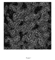
- Figure 3 shows a partial 1.0 ⁇ electron-density map of 2Fo-Fc format within the crystal structure of rSIFN-co.
- Figure 4 shows a distribution map of the average temperature factors along the amino acid residues for all the atoms of rSIFN-co. (a) A chain; (b) B chain.
- Figure 5 shows the ( ⁇ , ⁇ ) value distribution on the Ramachandran plot of all the amino acid residues in the model of the rSIFN-co protein molecular structure.
- Figure 6 shows a unit cell packing diagram of rSIFN-co.
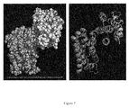
- Figure 7 shows the assembled structure of the rSIFN-co dimers.
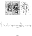
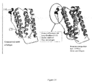
- Figure 8 shows the organization of rSIFN-co crystallographic dimers ( Fig. 8a, Fig. 8b ) and the root-mean square deviation (RMSD) of ⁇ carbon atoms (the boxes represent missing residues) ( Fig. 8c ).
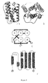
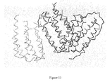
- Figure 9 shows the monomolecular structure of rSIFN-co (main chain demonstrated only); (A) Side view; (B) Top view; (C) Topology diagram; (D) Topological organization of the secondary structures.
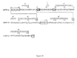
- Figure 10 shows the sequence alignment between the secondary structures of rSIFN-co and its amino acid sequence; the gray boxes represent amino acid residues that were not set up in the structure; the blue boxes represent amino acid residues which were set up as Ala or Gly. The solid lines represent two pairs of disulfide linkages and the green subscripts represent one disulfide linkage that has been constructed in the structure.
- Figure 11 shows the sequence alignment of rSIFN-co protein and homologous IFN polypeptides.
- Figure 12 shows a comparative diagram of the three-dimensional structure of rSIFN-co and IFN- ⁇ 2b.
- Figure 13 shows the superimposed image of rSIFN-co (in red) and IFN- ⁇ 2b (in yellow).
- Figure 14 shows the comparative differences between the three-dimensional structure of rSIFN-co and the computational model of Infergen® from Amgen (U.S.).
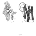
- Figure 15 shows (a) the combined model of protein IFN- ⁇ and its receptor; (b) the diagram of the functional domain of protein IFN- ⁇ (the important functional domain is illustrated by blue ring).
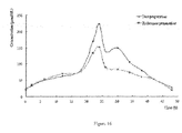
- Figure 16 shows the mean enzyme concentration in blood-time curve after subcutaneous injection of 9 ⁇ g rSIFN-co and 9 ⁇ g IInfergen® to 18 subjects.
- the purified recombinant interferon which has been crystallized in this invention, is obtained from the method disclosed by the examples 1 and 2 of the specification of the U.S. Patent No. 7364724 and/or pages 11-17 of the specification of the Chinese Patent Publication No. CN1740197A .
- the characterization of this recombinant interferon is disclosed in the U.S. Patent No. 7364724 and/or the Chinese Patent Publication No. CN1740197A .
- the amino acid sequence of the present recombinant interferon, as well as the nucleotide sequence encoding the same are shown below:
- CD circular dichroism spectrum
- the three-dimensional structure of the present recombinant interferon is also different from the three-dimensional structure of IFN- ⁇ 2b published in the art (see Fig. 12 ) and the three-dimensional structure of INFERGEN® based on computational modeling (see KORN, AP et al., Journal of Interferon Research 1994, 14: 1-9 ). There are obvious differences between the AB loops of the two, and their BC loops also cannot overlap completely (see Fig. 14 ).
- the time of blood sample collection was plotted against the concentration of 2-5A oligonucleotidase (also referred to as 2', 5'-OAS) in the serum of the subjects.
- the chart generally shows a two-peak pattern, and the resulting area under the curve of this chart is significantly greater than that of INFERGEN® after injection under the same conditions.
- the half-life period of this recombinant interferon is longer than that of INFERGEN® after injection into the body.
- the present recombinant interferon is more effective than any interferon used clinically at present (including INFERGEN®).
- the recombinant interferon from this invention is capable of not only inhibiting DNA replication of HBV, but also inhibiting secretion of both hepatitis B surface antigen (HBsAg) and hepatitis B e antigen (HBeAg).
- HBsAg hepatitis B surface antigen
- HBeAg hepatitis B e antigen
- the efficiency of inhibiting DNA replication of hepatitis B core antigen (HBcAg) by this interferon is about twice that of INFERGEN®.
- the in vitro pharmacodynamics of the present recombinant interferon shows that it is capable of not only inhibiting the DNA replication of HBV, but also inhibiting secretion of both hepatitis B surface antigen and hepatitis B e antigen.
- the cytotoxicity of the present recombinant interferon is only 1/8 that of the current clinically used interferons, but its antiviral activity is as much as 5-20 times greater; meanwhile, the biological responses of the present recombinant interferon is more effective, more broad-spectrum and longer lasting in the human body.
- the present recombinant interferon shows higher antiviral activity and less side effects compared with any other interferons (including INFERGEN®).
- this recombinant interferon possesses not only an antiviral activity 20 times as great as that of the interferons currently in clinical use, but also a more effective anti-tumor (such as breast cancer and cervical cancer) function compared with recombinant human interferon ⁇ (including INFERGEN®). It also shows greatly reduced toxic side effects and can be safely used in large dosages (each dose > 10 million IU), making it possibleto treat viral diseases or tumors which require large dosages of interferon.
- the present recombinant interferon has a different spatial configuration, enhanced biologic activities and different pharmacokinetics characteristics as compared with INFERGEN®.
- the terms 'spatial configuration', 'spatial structure', 'three-dimensional structure' and 'three-dimensional configuration' can be used interchangeably.
- the present recombinant interferon comprises the amino acid sequence of SEQ ID NO: 1 and is encoded by the nucleotide sequence comprising SEQ ID NO: 2. Further, the present recombinant interferon has the amino acid sequence of SEQ ID NO: 1, and is encoded by the nucleotide sequence of SEQ ID NO: 2. In comparison with interferons such as INFERGEN®, which has the amino acid sequence of SEQ ID NO: 1 or the same amino acid sequence as the present recombinant interferon, but is not encoded by the nucleotide sequence of SEQ ID NO: 2, , the present recombinant interferon has a different spatial configuration and/or enhanced biologic activities and/or different pharmacokinetics characteristics.
- the present recombinant interferon has a different spatial configuration and enhanced biologic activities, different spatial configuration and different pharmacokinetics characteristics, or enhanced biologic activities and different pharmacokinetics characteristics.
- said different spatial configuration includes: the circular dichroism spectrum (CD) of the present recombinant interferon at 190-250nm and/or 250-320 nm is significantly different from the corresponding CD of INFERGEN® when determined under the same conditions.
- the enhanced biological activities include: enhanced antiviral activity, enhanced anti-tumor activity, less side effects and/or could be used in large dosages (e.g. each dose > 10 million IU).
- said enhanced biological activities can be enhanced antiviral activity and enhanced anti-tumor activity and the like.
- said tumors can be breast cancer and cervical cancer.
- the different pharmacokinetics characteristics include: after intramuscular injection of the recombinant interferon in subjects whose BMI ranged from 18 to 23, the time of blood sample collection was plotted against the concentration of 2-5A oligonucleotidase in the serum of the subjects, and the resulting area under the curve of this chart is significantly greater and/or the half-life of this recombinant interferon in the body is longer than those of INFERGEN® after injection under the same conditions
- the present recombinant interferon can be produced by the method comprising the following steps: introducing a nucleotide sequence comprising SEQ ID NO: 2 that encodes the recombinant interferon into an isolated host cell; culturing the host cell under appropriate condition for expression of the recombinant interferon; and harvesting the recombinant interferon, wherein the recombinant interferon has an amino acid sequence of SEQ ID NO: 1, and the recombinant interferon inhibits secretion of hepatitis B surface antigen (HBsAg) and hepatitis B e antigen (HBeAg) of Hepatitis B Virus.
- HBsAg hepatitis B surface antigen
- HBeAg hepatitis B e antigen
- said host cell is Escherichia coli, such as Escherichia coli LGM 194.
- the nucleotide sequence comprising SEQ ID NO: 2 is under the control of the promoter P BAD .
- the harvesting step comprises extraction of the interferon from the fermentation broth, collection of the inclusion bodies, denaturation and renaturation of the harvested interferon. Still further, the harvesting step also comprises separation and purification of the recombinant interferon (see the claims of U.S. Patent No. 7364724 ).
- This invention provides a crystalline recombinant interferon.
- This disclosure provides a crystalline recombinant interferon comprising the amino acid sequence of SEQ ID NO: 1. Further, this crystal belongs to the trigonal system. In one embodiment, the space group of this crystal is P3 1 21.
- said crystal contains two molecules in one asymmetric unit.
- said crystal comprises covalently or non-covalently bound metal ions.
- said mental ions can be magnesium ion, zinc ion and the like, these metal ions can mediate the formation of the interferon dimers in the crystal.
- said recombinant interferon is encoded by the nucleotide sequence comprising SEQ ID NO: 2.
- such recombinant interferon is encoded by the nucleotide sequence comprising SEQ ID NO: 2, preferably encoded by the nucleotide sequence of SEQ ID NO: 2.
- This disclosure provides a method for preparing or culturing the present crystalline recombinant interferon.
- this disclosure provides a method for preparing or culturing the present crystalline recombinant interferon, comprising the steps of: concentrating the recombinant interferon to about 3 - 3.5 mg/ml, and leaving it in the crystallization solution containing Li 2 SO 4 , CAPS (3-(cyclohexylamino)-1-propanesulfonic acid) and MgCl 2 for an appropriate period of time to obtain the crystal. Further, said method for culturing crystal is performed at room temperature such as 293K. In some embodiments, this crystal can be cultured by the hanging drop method or the sitting drop method, preferably the hanging drop method (also referred to as hanging drop vapor diffusion method).
- said crystallization solution contains about 1.0 - about 1.5M Li 2 SO 4 , about 0.05 - about 0.15M CAPS (3-(cyclohexylamino)-1-propanesulfonic acid) and about 0.01 - about 0.03 M MgCl 2 .
- the pH value of the crystallization solution is in the range of about 10.5 - about 12.0, preferably about 11.1.
- said crystallization solution contains 1.2M Li 2 SO 4 , 0.1M CAPS (3-(cyclohexylamino)-1-propanesulfonic acid), pH 11.1, 0.02 M MgCl 2 .
- the method for culturing the crystal includes leaving the crystallization solution containing said recombinant interferon to stand for about 1 day to about 2 weeks, preferably about 2 days to about 10 days, more preferably about 3 days to about 1 week, such as 3 days to 1 week.
- structural coordinates also known as “atomic coordinates”
- structural coordinates refers to Cartesian coordinates derived from mathematical equations related to the patterns obtained by the diffraction of a monochromatic beam of x-rays by the atoms (scattering centers) of the present interferon in crystalline form.
- the diffraction data are used to calculate an electron density map of the repeating unit of the crystal.
- the electron density maps are then used to establish the positions of the individual atoms of the interferon protein or protein/ligand complex.
- Slight variations in structural coordinates can be generated by mathematically manipulating the interferon or interferon/ligand structural coordinates.
- the structural coordinates disclosed herein could be manipulated by crystallographic permutation, fractionalization, addition or subtraction of the entire set, inversion, or any combination of the above.
- modifications in the crystal structure due to mutations, additions, substitutions, and/or deletions of amino acids, or other changes in any of the components that make up the crystal could also yield variations in structural coordinates.
- Such slight variations in the individual coordinates will have little effect on the overall configuration. If such variations are within an acceptable standard error as compared to the original coordinates, the resulting three-dimensional shape is considered to be structurally equivalent.
- the "AB loop" of the present recombinant interferon means the amino acid residues 25-33 of the present recombinant interferon having the amino acid sequence of SEQ ID NO: 1; namely, the AB loop has the amino acid sequence SPFSCLKDR as shown in SEQ ID NO: 4; and the "BC loop" of the present recombinant interferon means the amino acid residues 44-52 of the present recombinant interferon having the amino acid sequence of SEQ ID NO: 1; namely, the BC loop has the amino acid sequence DGNQFQKAQ as shown in SEQ ID NO: 5.
- association refers to a condition of proximity between a ligand, or portions thereof, and an interferon molecule or portions thereof.
- the association may be non-covalent, wherein the juxtaposition is energetically favored by hydrogen bonding, van der Waals forces, or electrostatic interactions, or it may be covalent.
- a ligand that binds to the binding pocket or region of an interferon would also be expected to bind to or interact with a structurally equivalent binding pocket or region.
- any molecule or molecular complex, or any portion thereof, that has a root mean square deviation of conserved residue backbone atoms (e.g. N, C ⁇ , C, O, preferably C ⁇ ) of less than about 0.65 ⁇ , when superimposed on the relevant backbone atoms described herein, is considered “structurally equivalent”. That is to say, the crystal structures of those portions of the two molecules are substantially identical, within acceptable error.
- Particularly preferred structurally equivalent molecules or molecular complexes are those that are defined by the entire set of structural coordinates disclosed herein ⁇ a root mean square deviation from the conserved backbone atoms of those amino acids of less than about 0.65 ⁇ .
- the root mean square deviation is at most about 0.5 ⁇ , and even more preferably, at most about 0.35 ⁇ .
- Other embodiments of this invention include a molecular complex defined by the structural coordinates for the AB or the BC loop disclosed herein ⁇ a root mean square deviation of less than about 0.65 ⁇ , preferably at most about 0.5 1 ⁇ , and more preferably at most about 0.35 ⁇ .
- root mean square deviation means the square root of the arithmetic mean of the squares of the deviations. It is a way to express the deviation or variation from a trend or object.
- the "root mean square deviation” defines the variation in the backbone of a protein from the backbone of interferon or a portion thereof as defined by the structural coordinates described herein.
- X-ray structural coordinates define a unique configuration of points in space.
- a set of structural coordinates for a protein or a protein/ligand complex, or a portion thereof defines a relative set of points that, in turn, defines a configuration in three dimensions.
- a similar or identical configuration can be defined by an entirely different set of coordinates, provided that the distances and angles between coordinates remain essentially the same.
- a scalable configuration of points can be defined by increasing or decreasing the distances between coordinates by a scalar factor while keeping the angles essentially the same.
- Various computational analyses can be used to determine whether a molecule or a portion thereof is "structurally equivalent", defined in terms of its three-dimensional structure, to the interferon disclosed herein, or part of it. For example, comparisons between different structures, different conformations of the same structure, or different parts of the same structure can be made by various computational analyses. In one embodiment, such analysis can be divided into four steps: (1) load the structures to be compared; (2) define the atom equivalences in these structures; (3) perform a fitting operation; and (4) analyze the results.
- This disclosure provides the three-dimensional structure of the present recombinant interferon.
- This three-dimensional structure is different from the three-dimensional structure of IFN- ⁇ 2b published in the art (see Fig. 12 ) and the structure of the computational model of INFERGEN® of U.S. Amgen (see Fig. 14 ), especially in the AB and BC loops.
- the three-dimensional structure of said recombinant interferon contains the atomic coordinates of recombinant interferon as shown in table 7, said atomic coordinates optionally have a variability of root mean square deviation from the conserved backbone atoms, preferably C ⁇ (also referred to as ' ⁇ carbon atom'), of less than about 0.65 ⁇ , preferablyor about 0.5 ⁇ , and more preferably about 0.35 ⁇ .
- each monomer of said recombinant interferon is composed of 6 segments of ⁇ -helix, a segment of 3 10 helix, and the connecting peptides between them.
- the corresponding amino acid residue locations of said 6 segments of the ⁇ -helices are 13-20, 50-68, 70-76, 79-100, 114-133, and 138-160; the corresponding amino acid residue location of said segment of 3 10 helix is 40-43.
- the folding of the monomer structure belongs to the helical cytokine type, having the following characteristics: after superimposition of the C ⁇ -backbone of said recombinant interferon and the C ⁇ -backbone of IFN- ⁇ 2b protein using least squares method, the location root-mean-square deviation of C ⁇ in the 25-33 residues (AB loop) of said recombinant interferon and C ⁇ in the corresponding residues of IFN- ⁇ 2b protein is 3.63 ⁇ 5%.
- the location root-mean-square deviation of C ⁇ at residue 25 of said recombinant interferon and IFN- ⁇ 2b protein is 3.291 ⁇ 5%
- the location root-mean-square deviation of C ⁇ at residue 26 is 4.779 ⁇ 5%
- the location root-mean-square deviation of C ⁇ at residue 27 is 5.090 ⁇ 5%
- the location root-mean-square deviation of C ⁇ in the 28 residue is 3.588 ⁇ 5%
- the location root-mean-square deviation of C ⁇ at residue 29 is 2.567 ⁇ 5%
- the location root-mean-square deviation of C ⁇ at residue 30 is 2.437 ⁇ 5%
- the location root-mean-square deviation of C ⁇ at residue 31 is 3.526 ⁇ 5%
- the location root-mean-square deviation of C ⁇ at residue 32 is 4.820 ⁇ 5%
- the location root-mean-square deviation of C ⁇ at residue 33 is 2.756 ⁇ 5%.
- the location root-mean-square deviation of C ⁇ at residues 44-52 (BC loop) of said recombinant interferon and C ⁇ in the corresponding residues of IFN- ⁇ 2b protein is 2.90 ⁇ 5%.
- the location root-mean-square deviation of C ⁇ at residue 44 of both said recombinant interferon and IFN- ⁇ 2b protein is 1.614 ⁇ 5%;
- the location root-mean-square deviation of C ⁇ at residue 45 is 1.383 ⁇ 5%;
- the location root-mean-square deviation of C ⁇ at residue 46 is 2.735 ⁇ 5%;
- the location root-mean-square deviation of C ⁇ at residue 47 is 2.709 ⁇ 5%;
- the location root-mean-square deviation of C ⁇ at residue 48 is 5.018 ⁇ 5%;
- the location root-mean-square deviation of C ⁇ at residue 49 is 4.140 ⁇ 5%;
- the location root-mean-square deviation of C ⁇ at residue 50 is 3.809 ⁇ 5%;
- this disclosure provides a selected portion of the three-dimensional structure of the present recombinant interferon, which contains atomic coordinates of one or more amino acid residues from amino acid residues 25-33 and/or 45-52 in table 7.
- the "one or more amino acid residues" described herein include 1, 2, 3, 4, 5, 6, 7, 8, 9, 10, 11, 12, 13, 14, 15, 16, 17, 18 amino acid residues.
- the "selected portion of said three-dimensional structure" contains the atomic coordinates of the amino acid residues 25-33 and/or 44-52 in table 7.
- the "selected portion of the three-dimensional structure" contains the atomic coordinates of at least 5, 6, 7, 8, 9, 10, 11, 12, 13, 14, 15, 16, 17, 18, 19, 20, 21, 22, 23, 24, 25, 26, 27, 28, 29, 30, 31, 32, 33, 34, 35, 36, 37, 38, 39, 40, 41, 42, 43, 44, 45, 46, 47, 48, 49, 50, 51, 52, 53, 54, 55, 56, 57, 58, 59, 60, 61, 62, 63, 64, 65, 66, 67, 68, 69, 70, 71, 72, 73, 74, 75, 76, 77, 78, 79, 80, 81, 82, 83, 84, 85, 86, 87, 88, 89, 90, 91, 92, 93, 94, 95, 96, 97, 98, 99, 100 amino acid residues in table 7.
- said atomic coordinates have a variability of root mean square deviation from the conserved backbone
- this disclosure provides the protein spatial structure model comprising the three-dimensional structure of the present recombinant interferon.
- said protein spatial structure model could be an electron density map, a wire-frame model, a chicken-wire model, a space-filling model, a stick-model, a ribbon model and a molecular surface model and the like.
- the present disclosure provides a scalable three-dimensional configuration of points, wherein at least a portion of said points are derived from the structural coordinates disclosed herein, or from peptides comprising the AB loop or the BC loop of the present recombinant interferon.
- the scalable three-dimensional configuration of points is displayed as a holographic image, a stereo diagram, a model, or a computer-displayed image.
- this invention provides a method for screening/designing candidate compounds that could interact with the present recombinant interferon. Further, said method utilizes the three-dimensional structure of the present recombinant interferon. Still further, said method is based on a computer.
- this invention provides a computer-based method for identifying candidate compounds that could interact with recombinant interferon, said method comprises the steps of: (a) providing a three-dimensional structure comprising the atomic coordinates of the recombinant interferon as shown in table 7, said atomic coordinates optionally have a variability of root mean square deviation from the conserved backbone atoms (preferably C ⁇ ) of less than about 0.65 ⁇ , preferably about 0.5 ⁇ , and more preferably about 0.35 ⁇ ; and (b) selecting a candidate compound that comprises structural features capable of interacting with said three-dimensional structure or selected portion thereof, thereby identifying a candidate compound that could interact with said recombinant interferon.
- said structural features are selected from the group consisting of antigenic sites, hydrophilic properties, surface accessibility, and structural motifs.
- the selection and identification of candidate compounds in step (b) comprises: (i) generating three-dimensional structures for a plurality of candidate compounds; and (ii) fitting each of the three-dimensional structures of step (i) against the three-dimensional structure of step (a) or selected portion thereof to find the most energetically favorable interaction, thereby identifying a candidate compound that could interact with the recombinant interferon.
- said method further comprises the steps of: (c) obtaining or synthesizing the candidate compound; and (d) contacting the candidate compound with said recombinant interferon to determine the ability of the candidate compound to interact with said recombinant interferon.
- the step of determining the ability of the candidate compound to interact with said recombinant interferon may further comprise measuring the activity of said recombinant interferon when contacted with the candidate compound. Interferon activities to be measured include, for example, antivirus activity, anti-tumor activity, anti-proliferation activity, natural killer cell activation, and immunomodulatory activity.
- said candidate compound is a ligand bound to said recombinant interferon or selected portion thereof.
- said ligand is selected from the group consisting of receptor, modifier, agonist and antagonist, said receptor could be IFNAR1, IFNAR2 or their complex, and said selected portion comprises one or more amino acid residues from the amino acid residues 25-33 (AB loop) and/or 45-52 (BC loop) of said recombinant interferon. Further, said selected portion comprises the amino acid residues 25-33 and/or 44-52 of said recombinant interferon.
- the present disclosure provides a method for determining potential ligands that bind to the present recombinant interferon.
- the method includes exposing a crystal disclosed herein to one or more samples comprising potential ligands, and determining whether a ligand-interferon molecular complex is formed.
- the present disclosure provides a method for acquiring structural information to design potential ligands that can form molecular complexes with interferon.
- the method includes exposing a crystal disclosed herein to one or more samples comprising potential ligands, and determining whether a ligand-interferon molecular complex is formed.
- the present disclosure provides a computer-assisted method for determining, designing, or making potential modifiers of interferon activity.
- the method includes screening a library of chemical or biological entities.
- a preferred method may include obtaining a crystal of unliganded interferon; exposing the unliganded interferon to one or more test samples that contain potential ligands of the interferon; and determining whether a ligand-interferon molecular complex is formed.
- the interferon may be exposed to potential ligands by various methods including, but not limited to, soaking an interferon crystal in a solution of one or more potential ligands or co-crystallizing interferon in the presence of one or more potential ligands.
- Structural information from said ligand-interferon complexes may preferably be used to design new ligands that bind tighter and more specifically, have desired special biological activities, have better safety profiles or combinations thereof than known ligands.
- the calculated electron density map directly reveals the binding event, identifies the bound chemical or biological entities, and provides a detailed three-dimensional structure of the ligand-interferon complex.
- a hit Once a hit is found, a series of analogs or derivatives of the hit may be screened for tighter binding or desired biological activity by traditional screening methods.
- the ligand-interferon complex may be iteratively exposed to additional potential ligands so that two or more hits may preferably be linked together to identify or design a more potent ligand.
- the structural coordinates disclosed herein can be used to aid in obtaining structural information about another crystallized molecule or molecular complex.
- the method of this invention allows determination of at least a portion of the three-dimensional structure of molecules or molecular complexes which contain one or more structural features that are similar to the structural features of the interferon disclosed herein. These molecules are referred to herein as "structurally homologous". Similar structural features can include, for example, regions of amino acid identity, conserved active site or binding site motifs and similarly arranged secondary structural elements (e.g., ⁇ helices and ⁇ sheets).
- structural homology is determined by aligning the residues of two amino acid sequences to optimize the number of identical amino acids along the lengths of their sequences; gaps in either or both sequences are permitted in making the alignment in order to optimize the number of identical amino acids; however, the amino acids in each sequence must remain in their proper order.
- a structurally homologous molecule is a protein that has an amino acid sequence sharing at least 65% identity with SEQ ID NO:1. More preferably, a protein that is structurally homologous to the interferon of the present invention includes a contiguous stretch of at least 50 amino acids that shares at least 80% amino acid sequence identity with the analogous portion of SEQ ID NO:1. Methods for generating structural information about the structurally homologous molecule or molecular complex are well-known in the art.
- the structural coordinates disclosed herein are also useful for solving the crystallographic structures of related interferons, interferon mutants or interferon homologs complexed with a variety of ligands.
- This approach enables the determination of the optimal sites for interaction between a ligand and an interferon, e.g. between candidate interferon modifiers and interferon. Potential sites for modification within the various binding sites of the molecules can also be identified. This information provides an additional tool for determining the most efficient binding interactions, for example, increased hydrophobic interactions between an interferon and a ligand.
- the present disclosure also provides a computer-based method for designing a mimetic of the recombinant interferon, comprising the steps of: (a) generating three-dimensional structures for a plurality of mimetics; and (b) fitting each of the three-dimensional structures of step (a) against the three-dimensional structure comprising the atomic coordinates of the recombinant interferon as shown in table 7 or selected portion thereof which selected portion contains the atomic coordinates of the amino acid residues 25-33 and/or 45-52 in table 7, to find the best fitted mimetic of said recombinant interferon, said atomic coordinates optionally have a variability of root mean square deviation from the conserved backbone atoms (preferably C ⁇ ) of less than about 0.65 ⁇ , preferably about 0.5 ⁇ , and more preferably about 0.35 ⁇ .
- Computational techniques can be used to screen, identify, select and/or design chemical entities or ligands capable of associating with interferons or structurally homologous molecules. Knowledge of the structural coordinates of the interferon disclosed herein permits the design and/or identification of synthetic compounds and/or other molecules which have a shape complementary to the conformation of the interferon disclosed herein.
- computational techniques can be used to identify or design chemical entities or ligands, such as receptors, modifiers, agonists and antagonists, that associate with the interferon or a portion thereof (e.g. the AB or the BC loop).
- Potential modifiers may bind to or interfere with all or a portion of an active site of interferon, and can be competitive, non-competitive, or uncompetitive inhibitors; or interfere with dimerization by binding at the interface between the two monomers. Once identified or screened for biological activity, these inhibitors/agonists/antagonists may be used therapeutically or prophylactically to block or enhance interferon activity. Structure-activity data for analogues of ligands that bind to or interfere with interferon can also be obtained computationally.
- chemical entity refers to chemical compounds, complexes of two or more chemical compounds, and fragments of such compounds or complexes.
- Chemical entities that are determined to associate with the interferon of the present invention are potential drug candidates.
- a graphical three-dimensional representation of the structure of the present interferon or a structurally homologous molecule, as identified herein, or portions thereof may thus be advantageously used for drug discovery.
- the structural coordinates of the chemical entity are used to generate a three-dimensional image that can be computationally fitted to the three-dimensional image of an interferon or a structurally homologous molecule by one of many computation methods and techniques available in the art.
- One embodiment of the method of drug design involves evaluating the potential association of a known chemical entity or ligand with the interferon or a structurally homologous molecule.
- the method of drug design thus includes computationally evaluating the potential of a selected chemical entity or ligand to associate with any of the molecules or molecular complexes set forth herein.
- the method of drug design involves computer-assisted design of chemical entities or ligands that associate with the present interferon, its homologs, or portions thereof. Chemical entities or ligands can be designed in a stepwise fashion, one fragment at a time, or may be designed as a whole or "de novo".
- the present invention provides a computer-based method of rational drug design, comprising the steps of: (a) providing the three-dimensional structure comprising atomic coordinates of the recombinant interferon as shown in table 7, said atomic coordinates optionally have a variability of root mean square deviation from the conserved backbone atoms (preferably C ⁇ ) of less than about 0.65 ⁇ , preferably about 0.5 ⁇ , and more preferably about 0.35 ⁇ ; (b) providing a plurality of molecular fragments, and generating three-dimensional structures thereof; (c) fitting each of the three-dimensional structures of step (b) against the three-dimensional structure of step (a) or selected portion thereof which selected portion contains the atomic coordinates of the amino acid residues 25-33 and/or 45-52 in table 7; and (d) assembling the selected molecular fragments into a molecule to form a candidate drug.
- said method may further comprise the steps of: (e) obtaining or synthesizing the candidate drug; and (f) contacting the candidate drug with said recombinant interferon to determine the ability of the candidate drug to interact with said recombinant interferon.
- the selected portion of said three-dimensional structure comprises the atomic coordinates of one or more amino acid residues from amino acid residues 25-33 (amino acid sequence as shown in SEQ ID NO: 4) and/or 45-52 (amino acid sequence as shown in SEQ ID NO: 5) in table 7.
- the selected portion of said three-dimensional structure comprises the atomic coordinates of the amino acid residues 25-33 (amino acid sequence as shown in SEQ ID NO: 4) and/or 45-52 (amino acid sequence as shown in SEQ ID NO: 5) in table 7, said atomic coordinates optionally have a variability of root mean square deviation from the conserved backbone atoms (preferably C ⁇ ) of less than about 0.65 ⁇ , preferably about 0.5 ⁇ , and more preferably about 0.35 ⁇ .
- a computer model of an interferon homolog can be built or refined without crystallizing the homolog.
- a preliminary model of an interferon homolog is created by sequence alignment, secondary structure prediction, screening of structural libraries, or any combination of these techniques.
- Computational software may be used to carry out the sequence alignments and secondary structure predictions.
- Structural incoherencies e.g., structural fragments around insertions and deletions, can be modeled by screening a structural library for peptides of the desired length and suitable conformation. If the interferon homolog has been crystallized, the final homology model can be used to solve the crystal structure of the homolog by techniques known in the art.
- the energy minimized model may contain regions where stereochemical restraints are violated; in such cases, these regions are remodeled to obtain a final homology model using one of many techniques known in the art.
- the present disclosure provides a method for obtaining structural information about a molecule or a molecular complex of unknown structure.
- the method includes crystallizing the molecule or molecular complex; generating an x-ray diffraction pattern from the crystallized molecule or molecular complex; and applying the x-ray diffraction pattern to at least a portion of the structural coordinates of the interferon disclosed herein to generate a three-dimensional electron density map of at least a portion of said molecule or molecular complex of unknown structure.
- the present disclosure provides a method for modeling an interferon homolog.
- the method includes aligning the amino acid sequence of a putative interferon homolog with the amino acid sequence of the present interferon and incorporating the sequence of the putative homolog into a model of interferon formed from the structural coordinates disclosed herein to yield a preliminary model of interferon homolog; subjecting the preliminary model to energy minimization to yield an energy minimized model; and remodeling regions of the energy minimized model where stereochemical restraints are violated to yield a final model of the interferon homolog.
- the present disclosure provides interferon mimetics.
- the present disclosure provides a peptide comprising a sequence as disclosed herein, or a derivative, active portion, analogue, variant or mimetic, and uses thereof.
- the present disclosure provides a mimetic of the interferon which comprises the amino acid sequence as shown in SEQ ID NO: 4 and/or SEQ ID NO: 5.
- the location root-mean-square deviation of C ⁇ at residues 25-33 of said recombinant interferon and C ⁇ in the corresponding residues of IFN- ⁇ 2b protein is 3.63 ⁇ 5%.
- the deviations of ⁇ carbons of residues 25-33 of said recombinant interferon are 3.291 ⁇ 5%, 4.779 ⁇ 5%, 5.090 ⁇ 5%, 3.588 ⁇ 5%, 2.567 ⁇ 5%, 2.437 ⁇ 5%, 3.526 ⁇ 5%, 4.820 ⁇ 5% and 2.756 ⁇ 5% respectively.
- the location root-mean-square deviation of C ⁇ at residues 44-52 of said recombinant interferon and C ⁇ in the corresponding residues of IFN- ⁇ 2b protein is 2.90 ⁇ 5%.
- the deviations of ⁇ carbons of residues 44-52 of said recombinant interferon are 1.614 ⁇ 5%, 1.383 ⁇ 5%, 2.735 ⁇ 5%, 2.709 ⁇ 5%, 5.018 ⁇ 5%, 4.140 ⁇ 5%, 3.809 ⁇ 5%, 2.970 ⁇ 5%, and 0.881 ⁇ 5% respectively.
- the mimetic is a functional mimetic or a structural mimetic.
- the mimetic is a mimetic of the present recombinant interferon (rSIFN-co). Further, the mimetics do not comprise INFERGEN®.
- the three-dimensional structure of said interferon mimetic is similar to that of the present recombinant interferon (rSIFN-co).
- both three-dimensional structures can be the same or essentially the same at the AB and BC loops.
- the three-dimensional structure of said interferon mimetic comprises the atomic coordinates of amino acid residues 25-33 (AB loop) and/or 44-52 (BC loop) in table 7, said atomic coordinates optionally have a variability of root mean square deviation from the conserved backbone atoms, preferably C ⁇ , of less than about 0.65 ⁇ , preferably about 0.5 ⁇ , and more preferably about 0.35 ⁇ .
- the present invention comprises variant peptides in which individual amino acids can be replaced by other closely related amino acids as is understood in the art.
- individual amino acid may be replaced as follows: any hydrophobic aliphatic amino acid may be replaced by any other hydrophobic aliphatic amino acids; any hydrophobic aromatic amino acid may be replaced by any other hydrophobic aromatic amino acids; any neutral amino acid with a polar side chain may be replaced by any other neutral amino acids with a polar side chain; an acidic amino acid may be replaced by any other acidic amino acids; and a basic amino acid may be replaced by any other basic amino acids.
- mimetic As used herein, “mimetic”, “functional/structural mimetic” relate to peptide variants or organic compounds having the same functional/structural activity as the polypeptide disclosed herein. Examples of such mimetic or analogues include chemical compounds or peptides which are modeled to resemble the three-dimensional structure of the interferon disclosed herein (the three-dimensional structure comprise the atomic coordinates of recombinant interferon as shown in table 7), particularly compounds and peptides having the above arrangement of amino acid residues.
- mimetic of the present recombinant interferon refers to a peptide variant or organic compound which has the same function/structure-activity as the present recombinant interferon (rSIFN-co), especially those having the same AB loop and/or BC loop spatial structure as the present recombinant interferon, but is not the present recombinant interferon
- the "mimetic" is a peptide variant, the length of its amino acid sequence is generally similar to that of the present recombinant interferon.
- said amino acid sequence of the mimetic can comprise about 120-200 amino acid residues, preferably about 140-180 amino acid residues, more preferably about 150-175 amino acid residues, still more preferably about 160-170 amino acid residues; for example, about 164, 165, 166 or 167 amino acid residues.
- a "mimetic" can be a peptide variant having a shorter amino acid sequence than the present recombinant interferon but comprising the AB loop and/or BC loop.
- it can comprise about 10-100 amino acid residues, preferably about 15-80 amino acid residues.
- Suitable mimetics or analogues can be generated by modeling techniques generally known in the art. This includes the design of "mimetics” which involves the study of the functional interactions and the design of compounds which contain functional groups arranged in such a manner that they could reproduce those interactions.
- the design of mimetics of compounds with known pharmaceutical activity is a known approach based on lead compounds for drug development.. This might be desirable where the active compound is difficult or expensive to synthesize or where it is unsuitable for common methods of administration; e.g. polypeptides are not well suited as active agents for oral compositions as they tend to be quickly degraded by proteases in the alimentary canal. Mimetic design, synthesis and testing may be used to avoid randomly screening a large number of molecules for a target property.
- the pharmacophore Once the pharmacophore has been identified, its structure can be modeled according to its physical properties, e.g. stereochemistry, bonding, size and/or charge, using data from a range of sources, e.g. spectroscopic techniques, X-ray diffraction and NMR data. Computational analysis, similarity mapping (which models the charge and/or volume of a pharmacophore, rather than the bonding between atoms) and other techniques can be used in this modeling process. In a variant of this approach, the three-dimensional structures of the ligand and its binding partner are modeled. This can be especially useful where the ligand and/or binding partner change conformation on binding, allowing further consideration of the model while designing the mimetic.
- the physical properties e.g. stereochemistry, bonding, size and/or charge
- data from a range of sources e.g. spectroscopic techniques, X-ray diffraction and NMR data.
- Computational analysis, similarity mapping which models the
- a template molecule onto which chemical groups that mimic the pharmacophore can be grafted can be conveniently selected so that the mimetic, besides maintaining the biological activities of the lead compound, would be easy to synthesize, likely be pharmacologically acceptable, and not degrade in vivo.
- the mimetics found by this approach can then be screened to see whether they have the target property, or to what extent they exhibit it. Further optimization or modification can then be carried out to arrive at one or more final mimetics for in vivo or clinical testing.
- the present disclosure provides an unliganded molecule including at least a portion of the interferon disclosed herein, e.g. the unliganded molecule may comprise SEQ ID NO:4 or SEQ ID NO:5 (the sequence of the AB loop and the BC loop respectively of the interferon described herein). Further, the unliganded molecule has sequence as shown in SEQ ID NO:4 or SEQ ID NO:5.
- the present disclosure provides a composition comprising a crystalline form of the present recombinant interferon or a mimetic of the present recombinant interferon.
- the composition may be a pharmaceutical composition.
- Said pharmaceutical composition may further comprise a pharmaceutically acceptable carrier.
- the preferred dosage is a "prophylactically effective amount” or a "therapeutically effective amount” (although prophylaxis may be considered a therapy), this dosage being sufficient to provide its beneficial effects to the individual.
- the actual amount, frequency and time-course of administration will depend on the nature and severity of the disease being treated. Prescription of treatment, e.g. decisions on dosage etc., is within the responsibility of medical doctors and other medical workers. Depending on the circumstances, pharmaceutical compositions may be administered alone or in combinations.
- compositions according to the present invention may include, in addition to the active ingredient, a pharmaceutically acceptable excipient, carrier, buffer, stabilizer or other materials well known to those skilled in the art. Such materials should be non-toxic and should not interfere with the efficacy of the active ingredient.
- a pharmaceutically acceptable excipient such materials should be non-toxic and should not interfere with the efficacy of the active ingredient.
- the exact nature of the carrier or other materials will depend on the route of administration, which may be oral or by injection, e.g. cutaneous, subcutaneous or intravenous. Examples of techniques and protocols mentioned above can be found in Remington's Pharmaceutical Sciences, 16th edition, Osol, A. (ed.), 1980 .
- said pharmaceutical composition can be formulated into the following dosage forms, including: tablets, capsules, oral liquids, patches, injections, sprays, suppositories, and solution preparations.
- the recommended dosage form is injection, such as subcutaneous or intravenous injection, and the carrier in the pharmaceutical composition may be any acceptable drug carrier, including binders, disintegrating agents, lubricants, fillers, solubilizers, buffers, preservatives, thickeners, chelating agents and other adjuvants.
- “pharmaceutically acceptable carriers” may be any of the standard pharmaceutical carriers.
- known appropriate carriers include, but are not limited to, phosphate buffered saline and various wetting agents.
- Other carriers may include additives used for tablets, granules, and capsules.
- Typical carriers often contain: starch, emulsion, sugar, cellulose, certain types of clay, gelatin, stearic acid and its salts such as magnesium stearate or calcium stearate, talc, plant oils, gums, glycol or other known excipients.
- Such carriers may also include flavorings and color additives or other ingredients.
- the composition of these carriers can be formulated using known methods.
- the mimetics of the present recombinant interferon have the AB loop and/or BC loop structures (such as the above specific AB loop and/or BC loop space structures) of the present recombinant interferon, they are expected to be capable of treating viral diseases and/or tumor similar to the present recombinant interferon.
- the present disclosure provides a use of the crystal of the present recombinant interferon, an interferon mimetic or a composition comprising said crystal or mimetic for the preparation of medicament for treating viral diseases and/or tumors.
- the present disclosure provides a method for the treatment of viral diseases and/or tumors, said method comprises administering to a subject an effective amount of the crystal of the present recombinant interferon, an interferon mimetic or a composition comprising said crystal or mimetic.
- the present disclosure also provides a pharmaceutical composition for the treatment of viral diseases and/or tumors, comprising an effective amount of the crystal of the present recombinant interferon, an interferon mimetic or a composition comprising said crystal or mimetic.
- said viral diseases may include: hepatitis A, hepatitis B, hepatitis C, other types of hepatitis, viral infections caused by Epstein-Barr virus, human immunodeficiency virus (HIV), Ebola virus, severe acute respiratory syndrome (SARS) virus, influenza virus, cytomegalovirus, herpes simplex virus, or other type of herpes virus, papovavirus, pox virus, picornavirus, adenovirus, rhinovirus, human T-cell leukemia viruses type I, or human T-cell leukemia viruses type II, or human T-cell leukemia virus type III.
- HIV human immunodeficiency virus
- SARS severe acute respiratory syndrome
- said tumor is cancer or solid tumors
- said tumors may include: skin cancer, basal cell carcinoma and malignant melanoma, renal cell carcinoma, liver cancer, thyroid cancer, nasopharyngeal cancer, solid tumors, prostate cancer, stomach/abdominal cancer, esophageal cancer, rectal cancer, pancreatic cancer, breast cancer, ovarian cancer, superficial bladder cancer, hemangioma, epidermoid cancer, cervical cancer, non-small cell lung cancer, small cell lung cancer, glial stromal tumors, leukemia, acute leukemia, chronic leukemia, chronic myelogenous leukemia, hairy cell leukemia, lymphadenoma, multiple myeloma, polycythemia, Kaposi's sarcoma.
- PCR was used to directly synthesize the 5'-terminus 280bp (fragment I) and 3'-terminus 268bp (fragment II) partial molecules of the rSIFN-co cDNA. There was a 41-bp overlap of the complementary nucleotide sequences between the 3' end of fragment I and the 5' end offragment II.
- PCR I for synthesizing rSIFN-co 5'-terminus partial molecule: using oligodcoxynucleotidc fragment B as a template, oligodeoxynucleotide fragments A and C as primers, the rSIFN-co 5'-terminus partial molecule with a length of 280bp was synthesized by PCR.
- the PCR I reaction mixture is as follows: (units: ⁇ l) (Total volume: 50 ⁇ l) sterilized distilled water without nuclease 39 10 ⁇ Pfu buffer (Stratagene, La Jolla, CA, USA) 5 dNTP mixture (2.5 mmol/L for each dNTP) 2 Oligomer A primer (25 ⁇ mol/L) 1 Oligomer C primer (25 ⁇ mol/L) 1 Oligomer B template (1 ⁇ mol/L) 1 Pfu DNA polymerase (Stratagene, La Jolla, CA, USA) (25 U/ ⁇ l) 1 PCR I reaction cycle: 95°C 2 min ⁇ (95°C 45s ⁇ 65°C 1 min ⁇ 72°C 1 min) ⁇ 25 cycles ⁇ 72°C 10min ⁇ 4°C
- PCR II for synthesizing rSIFN-co 3'-terminus partial molecule using oligodeoxynucleotide fragment E as a template, oligodeoxynucleotide fragments D and F as primers, the rSIFN-co 3'-terminus partial molecule with a length of 268bp was synthesized by PCR.
- the PCR II reaction mixture is as follows: (units: ⁇ l) (Total volume: 50 ⁇ l) sterilized distilled water without nuclease 39 10 ⁇ Pfu buffer (Stratagene, La Jolla, CA, USA) 5 dNTP mixture (2.5 mmol/L for each dNTP) 2 Oligomer D primer (25 ⁇ mol/L) 1 Oligomer E primer (25 ⁇ mol/L) 1 Oligomer F template (1 ⁇ mol/L) 1 Pfu DNA polymerase (Stratagene, La Jolla, CA, USA) (25 U/ ⁇ l) 1 PCR II reaction condition and cycle: same as PCR I
- Fragments I and II were assembled together to give the complete full-length cDNA sequence of rSIFN-co using the overlapping and extending PCR method. Restriction enzyme sites Nde I and Pst I were introduced to the 5'-terminus and 3'-terminus of the sequence respectively, so that the rSIFN-co cDNA sequence can be cloned into the plasmid.
- PCR reaction mixture (units: ⁇ l) (Total volume: 50 ⁇ l) sterilized distilled water without nuclease 38 10 ⁇ Pfu buffer (Stratagene, La Jolla, CA, USA) 5 dNTP mixture (2.5 mmol/L for each dNTP) 2 primer G (25 ⁇ mol/L) 1 primer H (25 ⁇ mol/L) 1 *fragment I PCR product (1 ⁇ mol/L) 1 * fragment II PCR product (1 ⁇ mol/L) 1 Pfu DNApolymerase (Stratagene, La Jolla, CA,USA) (25 U/ ⁇ l) 1 *Separating and purifying the PCR product with STRATAPREP PCR purification kit produced by Stratagene (La Jolla, CA), then dissolving the PCR product into sterilized distilled water.
- PCR reaction condition and cycle same as PCR I.
- the pLac T7 plasmid was used as vector for cloning rSIFN-co cDNA.
- the pLac T7 plasmid was reconstructed from the pBLUESCRIPT II KS(+) plasmid produced by Stratagene (La Jolla, CA, USA).
- PCR product containing rSIFN-co full-length cDNA was purified with STRATAPREP PCR purification kit produced by Stratagene (La Jolla, CA), followed by digestion with Nde I and Pst I.
- the pLac T7 plasmid was double digested with Nde I and Pst I.
- These double-digested DNA fragments were separated using 1% agarose gel electrophoresis followed by recovery and purification of a 507-bp rSIFN-co DNA fragment and a 2.9-kb plasmid DNA fragment with Wizard DNA purification kit produced by Promega (Fitchburg, WI, USA).
- DNA sequencing was performed with SEQUITHERM TM Cycle Sequencing Kit following instruction provided by the manufacturer (Epicentre Technologies Ltd, Madison, WI, USA) using the universal primer T7 and T3. The DNA sequencing result showed that the sequence was consistent with the theoretical design.
- E. Coli expression vector pBAD18 was digested with Nde I and linearized, then fully digested with Xba I. Electrophoresis with 1% agarose gel and purification with QIAEX II kit (QIAGEN) were performed to give a 4.8-kb fragment from pBAD18 having been digested with Nde I and Xba I.
- the pH-1 plasmid was double digested with Ndel and Xba I and, after separation with 1% agarose gel electrophoresis, a 715-bp fragment was purified. This fragment was ligated with the above 4.8-kb fragment from pBAD18 using T4 DNA ligase to produce the recombinant plasmid.
- the recombinant plasmid was used to transform DH5 ⁇ -competent cells. The transformed cells were spread on LB-Amp agar plate, and then cultured overnight at 37°C.
- E. Coli. colonies from the above LB-plate were randomly chosen, and clones containing recombinant plasmid with full length rSIFN-co cDNA were screened using endonuclease digestion and PCR analysis.
- One of the PCR positive recombinant plasmid was named pHY-5, and the strain containing pHY-5 plasmid was named PVIII. PVIII was amplified and stored at -80°C with glycerol freezing medium for future use.
- rSIFN-co gene was under the control of the strong promoter P BAD which is regulated by the AraC protein.
- AraC is a protein encoded by the AraC gene located in the same plasmid.
- the dimer of AraC binds to O 2 and I 2 forming a 210-bp loop. This conformation leads to a complete inhibition of transcription.
- the dimer of AraC is released from O 2 and binds to I 1 and I 2 eliminating the inhibition on transcription.
- rSIFN-co expression level is more than 50% of the total bacterial protein.
- the E.coli strain LMG194 with expression vector pHY-5 was inoculated in LB culture medium, then shaken at 200rpm overnight (about 18h) at 37 °C. To the medium was added 50% of 30% glycerine. After mixing, the medium was stored at -20°C in 1 ml aliquots for use as the producing strain;
- the producing strain was inoculated in LB culture medium (1 L containing Tryptone 10g, Yeast extracts 5g and NaCl 10g) at a ratio of 1%, then shaken at 200 rpm overnight (about 18h) at 37°C, for use as grade-I seed strain;
- Grade-I seed strain was added to RM media (1 L containing Casein 20 g, MgCl 2 1 mmol/L (0.203 g), Na 2 HPO 4 4g, KH 2 PO 4 3 g, NaCl 0.5 g and NH 4 Cl 1g) at a ratio of 10% and cultured at 37°C, pH 7.0. Fermentation was carried out until OD 600 reached about 2.0, then arabinose (20% solution) was added until a final concentration of 0.02% as an inductor; after 4 hours, the strain was collected and centrifuged to give a pellet;
- the strain pellet was re-suspended with an appropriate amount of buffer A (100 mmol/L Tris-HCl, pH 7.5, 10 mmol/L EDTA, 100 mmol/L NaCl), and kept at -20°C overnight. The strain was thawed and broken by a homogenizer, then centrifuged.
- buffer A 100 mmol/L Tris-HCl, pH 7.5, 10 mmol/L EDTA, 100 mmol/L NaCl
- the pellet was washed with buffer B (50 mmol/L Tris-HCl, pH 7.5, 1 mol/L Urea,10 mmol/L EDTA, 0.5% Triton X-100), buffer C (50 mmol/L Tris-HCl, pH 7.5, 2 mol/L Urea, 10 mmol/L EDTA, 0.5% Triton X-100) and then precipitated; this was repeated once, and the pellet was then washed once with distilled water to give inclusion bodies.
- buffer B 50 mmol/L Tris-HCl, pH 7.5, 1 mol/L Urea,10 mmol/L EDTA, 0.5% Triton X-100
- buffer C 50 mmol/L Tris-HCl, pH 7.5, 2 mol/L Urea, 10 mmol/L EDTA, 0.5% Triton X-100
- the inclusion body was dissolved in 6 mol/L Guanidine-HCl (or urea) to obtain a slightly cloudy denaturation solution, which was then centrifuged at a speed of 10000 rpm. The supernatant was collected and used to determine the protein concentration.
- the denaturation solution was added in three portions into a renaturation buffer (0.5 mol/L Arg, 150 mmol/L Tris-HCl, pH 7.5, 0.2 mmol/L EDTA) and then stirred continuously at 4°C overnight (about 18 h).
- the solution was dialyzed sequentially with ten times its volume of 10 mol/L phosphate buffer (PB), 5 mol/L PB buffer and distilled water; After dialysis, the pH was adjusted with 2 mol/L HAc-NaAc (pH 5.0). The solution was left to stand and then filtered.
- a column was prepared with 20 mmol/L HOAc-NaOAc (pH 5.0), loaded with the renaturation product obtained from step (5) at a speed of 30 ml/min, washed with 20 column volumes (CV) of 20 mmol/L HOAc-NaOAc (pH 5.0) to remove other proteins; washed with 5 CV of 20 mmol/L HOAc-NaOAc (pH 5.0) containing 0.15 mol/L NaCl to remove other proteins; then washed with 3 CV of 20 mmol/L HOAc-NaOAc (pH 5.0) containing 0.18 mol/L NaCl to remove other proteins. Finally, 20 mmol/L HOAc-NaOAc (pH 5.0) containing 0.25 mol/L NaCl was used to elute the target protein, thereby obtaining an HS-eluted protein solution.
- CV column volumes
- the HS-eluted protein solution was added into PB buffer of 0.2 mol/L (pH 6.6). 4 mol/L NaCl was added to adjust the NaCl concentration to 1 mol/L and pH to 6.0, and the solution was ready for loading.
- a column was prepared with 50 mmol/L Na 2 HPO 4 (pH 5.5) containing 1 mol/L NaCl, and loaded at a rate of 1 ml/min. The column was washed with 50 mmol/L Na 2 HPO 4 (pH 5.0) to remove other proteins, then washed with 50 mmol/L Na 2 HPO 4 (pH 4.0) to remove other proteins. Finally, 50 mmol/L Na 2 HPO 4 (pH 3.6) was used to elute the target protein to obtain the chelating column-eluted target protein solution.
- the protein solution eluted from the chelating column was diluted 30 folds and its pH adjusted to 5.0, then loaded onto an HS column which was eluted with PB buffer, pH 7.0, containing 0.5 mol/L NaCl to give the recombinant interferon (Protein Stock Solution).
- rSIFN-co stock solution of the present invention 34.5 ⁇ g/ml phosphate buffer, pH 7.0 25 mmol/L NaCl 0.4 mol/L
- This example describes the in vitro study of rSIFN-co and INFERGEN® against human breast cancer cells.
- the present recombinant interferon (rSIFN-co) and INFERGEN® produced by Amgen (U.S.) were used as test drugs to study their effects on cell proliferation, apoptosis and expression of oncogenes in MCF-7 and resistant strain MCF-7/ADR.
- MCF-7 Human breast cancer cell line MCF-7 and adriamycin resistant strain MCF-7 (MCF-7/ADR) were cultured in 25 cm 2 or 75 cm 2 flasks respectively. After the cells covered the bottom of the flasks, they were trypsinized with 0.25% trypsin. Cells in the logarithmic growth phase were harvested for experiments.
- each cell strain was divided into 3 groups (with 11 small groups in total): rSIFN-co group (0.02, 0.078, 0.313, 1.25, 5.0 ⁇ g/ml), INFERGEN® group (0.02, 0.078, 0.313, 1.25, 5.0 ⁇ g/ml) and blank control group (RPMI1640 medium containing 10% fetal bovine serum (Sigma, America), also known as RPMI1640 complete medium).
- RPMI1640 medium containing 10% fetal bovine serum (Sigma, America), also known as RPMI1640 complete medium.
- rSIFN-co and INFERGEN® were diluted into the desired concentrations (final ethanol concentration ⁇ 1%) with the RPMI1640 complete medium, and stored at 4°C.
- MCF-7 cells and MCF27/ADR cells in the logarithmic growth phase were diluted with RPMI1640 medium containing 10% fetal bovine serum to 1.25 ⁇ 10 5 /ml cell suspension. Trypan blue method was used to ensure >95% cell viability.
- FCM flow cytometry
- each cell strain was divided into 3 groups: rSIFN-co group (5 ⁇ g/mL), INFERGEN® group (5 ⁇ g/mL), and blank control group (containing 10% calf serum RPMI1640 culture medium).
- FCM detection the cells were collected 48h after drugs were added, then the cells were suspended as single cells and dyed with propidium iodide (PI). The apoptosis rate was assayed with the Elite Esp-based flow cytometer (Coulter, USA), and the results were analyzed with the software supplied with the equipment. These experiments were repeated 3 times.
- rSIFN-co 5 ⁇ g/mL
- INFERGEN® 5 ⁇ g/mL
- RPMI1640 containing 10% fetal bovine serum
- rSIFN-co 5 ⁇ g/mL
- INFERGEN® 5 ⁇ g/mL
- RPMI1640 containing 10% fetal bovine serum were also added to the medium of MCF-7/ADR cell cultures.
- the coverslips were treated with acid, washed and sterilized under high pressure, and then placed in 6-well culture plates.
- the MCF-7 and MCF-7/ADR cells in logarithmic growth phase were digested into single cell suspensions with 0.25% trypsin.
- the cells were inoculated into 6-well plates, each well 1 ⁇ 10 5 , and cultured at 37°C in a CO 2 incubator for 24h. After the cells adhered to the walls, drugs were added to each group. After 48 h, the coverslips were removed. Conventional immunohistochemical SABC staining was performed, all concentrations at 1:100.
- Staining results were determined according to the methods of Volm ( Volm M, et al., European Journal of Cancer, 1997, 33 (3), 691-693 ), wherein yellow or brown particles appearing in cell nucleus (P53), cytoplasm (Bcl-2) or membrane (CerbB-2) were taken as positive results.
- Five fields of view (FOV) on each slide under high magnification (400 ⁇ ) were randomly selected, counting 200 cells per field. Two factors determined if there was expression in each group of cells. Scoring was done according to the intensity of staining for each cell, 0 point for no coloring, 1 point for light yellow, 2 points for brown, 3 points for tan. The average would be the average staining intensity for a group of cells.
- rSIFN-co could inhibit the proliferation of MCF-7 cells.
- Each cell group treated with 0.02, 0.078, 0.313, 1.25, 5.0 ⁇ g/mL of rSIFN-co and INFERGEN® showed a significant decrease in its absorbance (OA) compared with the blank control groups.
- the inhibitory effects of rSIFN-co and INFERGEN® showed no significant differences at the early stages (24h, 48h) (P>0.05). After over 72 h of treatment, the % inhibition of rSIFN-co was higher than that of INFERGEN® at the same concentrations except at the lowest concentration of 0.02 ⁇ g/mL, the differences were statistically significant (P ⁇ 0.05) (shown in Table 1-1).
- rSIFN-co could inhibit the proliferation of MCF-7/ADR cells.
- Each cell group treated with 0.02, 0.078, 0.313, 1.25, 5.0 ⁇ g/mL of rSIFN-co and INFERGEN® showed a significant decrease in its absorbance (OA) compared with the control groups.
- the inhibitory effect of rSIFN-co was higher than that of INFERGEN® at the same concentrations except at the lowest concentration of 0.02 ⁇ g/mL as shown by analysis of variance, the differences were statistically significant (P ⁇ 0.05) (shown in Table 1-2).
- This example describes the in vitro study of rSIFN-co and INFERGEN® in inhibiting the growth and inducing apoptosis of cervical cancer cell.
- the present recombinant interferon (rSIFN-co) and INFERGEN® produced by Amgen (U.S.) were used as test drugs to study their effects on growth inhibition and apoptosis induction of cervical cancer Caski cells (HPV16 +).
- Drug samples were diluted with RPMI-1640 culture medium containing 10% fetal bovine serum.
- Cervical cancer Caski cells were cultured in a 96-well plate. Cells were prepared as single cell suspension using culture medium with a cell concentration of 1 ⁇ 10 5 /ml. To each well was added 100 ⁇ l of cell suspension. rSIFN-co and INFERGEN® were added to the plate at a concentration gradient of 0.156 ⁇ g/ml, 0.625 ⁇ g/ml, 2.5 ⁇ g/ml and 10 ⁇ g/ml. RPMI-1640 medium containing 10% fetal bovine serum was used as control group. Each concentration was triplicated. The cells were cultured at 37°C with 5% CO 2 in an incubator for 72h.
- MTT reagent (Sigma Company, U.S.) was prepared at 5 mg/ml, and 10 ⁇ l MTT reagent was added to each well. The plate was shaken gently to homogenize the reagent, incubated at 37°C with 5% CO 2 for 4h, whereupon blue crystals could be seen at the bottom of the wells. The supernatant was removed, and 100 ⁇ l of DMSO were added to each well, then the absorbance at 570 nm was measured with a microplate reader after the blue crystals dissolved at room temperature.
- the Caski cells were divided into 7 groups and cultured in RPMI-1640 medium containing 10% inactivated fetal bovine serum in a 96-well plate.
- Group 1 was cultured for 72h as control group.
- Groups 2-4 were cultured with different concentrations of rSIFN-co: 0.156 ⁇ g/ml, 0.625 ⁇ g/ml, 2.5 ⁇ g/ml.
- Groups 5-7 were cultured with different concentrations of INFERGEN®: 0.156 ⁇ g/ml, 0.625 ⁇ g/ml, 2.5 ⁇ g/ml.
- FCM flow cytometry
- Each group of cells were centrifuged at 1000 r/min for 5min. The supernatant was removed, and the cells were tested for apoptosis with Annexin V/PI double dying method.
- Each specimen containing 1 ⁇ 10 6 viable cells was washed once with incubation buffer and centrifuged at 1000 r/min for 5 min.
- the cells were re-suspended with 100 ⁇ l marker solution, incubated at room temperature for 15 min in the dark, and centrifuged at 1000 r/min for 5 min to precipitate the cells.
- the cells were washed once with an incubation buffer, triturated with a fluorescent solution, then incubated at 4°C for 20 min. in the dark, while shaken frequently, before being tested with FCM.
- rSIFN-co and INFERGEN® inhibited the growth of Caski cells, and this effect increased with increasing concentrations of interferons.
- the effect of rSIFN-co was greater than that of INFERGEN® in groups of 0.625, 2.5 and 10 ⁇ g /ml.
- Table 2-1 The differences displayed in Table 2-1 showed statistical significance ( P ⁇ 0.01): Table 2-1 Inhibitory effect on Caski cells ( x ⁇ s ) Drug concentration ( ⁇ g/ml) Cell growth inhibition rate rSIFN-co INFERGEN® 0.156 8.6 ⁇ 2.1 7.3 ⁇ 2.2 0.625 17.6 ⁇ 3.31 7.4 ⁇ 4.0 2.5 42.7 ⁇ 1.51 9.7 ⁇ 1.6 10 67.3 ⁇ 4.41 53.0 ⁇ 5.8 1Compared with INFERGEN ® at the same concentration, P ⁇ 0.01
- Table 2-2 The difference displayed in Table 2-2, showed statistical significance ( P ⁇ 0.01): Table 2-2 Apoptotic effect on Caski cells ( x ⁇ s ) Drug concentration ⁇ g/ml) Cell growth inhibition rate rSIFN-co INFERGEN ® 0 21.3 ⁇ 3.9 21.3 ⁇ 3.9 0.156 53.5 ⁇ 4.6 1,2 47.6 ⁇ 3.1 1 0.625 64.9 ⁇ 3.3 1 67.1 ⁇ 3.6 1 2.5 74.4 ⁇ 1.3 1 72.0 ⁇ 2.6 1 1 Compared with controls, P ⁇ 0.01. 2 Compared with INFERGEN ® at the same concentration, P ⁇ 0.01.
- This example describes the research work on the pharmacokinetics and bioequivalence between rSIFN-co and INFERGEN ® .
- the present recombinant interferon rSIFN-co and INFERGEN ® produced by Amgen (U.S.) were taken as test drugs to compare their pharmacokinetics and bioequivalence.
- ELISA enzyme-linked immunosorbent assay
- virus cytopathic inhibition assay can hardly measure it directly in the serum of healthy adults.
- detection marker for pharmacokinetics study of interferon is generally 2', 5'-OAS (2-5A oligonucleotidase), which is a product induced by interferon, and also an indicator of its efficacy.
- 2-5 Kit Eiken' Radioimmunoassay Kit was supplied by Eiken Chemical Co., LTD.
- the Kit includes: (1) I 125 -labelled 2',5'-OAS, (2) Anti-2',5'-OAS serum, (3) 2',5'-OAS Standard vial (each contains 0, 10, 30, 90, 270 or 810 pmol/dL 2',5'-OAS), (4) Buffer, (5) Blank serum, (6) Poly(I)-poly(C) agarose gel, (7) ATP, (8) Mercaptoethanol, and (9) Quality control serum.
- test Test preparation was compared by the paired t test using the statistical software DAS ver1.0.
- the INFERGEN ® group was more severe than the rSIFN-co group in terms of the incidence, extent and duration of the adverse reactions that were compared.
- the preparation of high-quality rSIFN-co protein monocrystal is a prerequisite for determining its crystal structure.
- the rSIFN-co used for crystal growth was derived from the said rSIFN-co of the present invention.
- the preparation method, technical process, crystallization conditions and crystallographic parameters of the rSIFN-co monocrystal were as follow.
- lyophilized powder of the rSIFN-co in the present invention was dissolved in pure water and stored under -20°C at an initial protein concentration of 0.42mg/ml. Prior to crystallization, the rSIFN-co protein samples were concentrated to 3-3.5mg/ml and immediately used for the crystal growth experiments. The hanging drop vapor diffusion method was used for the crystallization process held at room temperature (293K).
- the monocrystal was of the tripartite crystal type, and had a size of0.42 ⁇ 0.08 ⁇ 0.08mm.
- the rSIFN-co protein crystal used in the X-ray diffraction analysis of the crystal structure is shown in Figure 1 .
- This process consisted of: 1) indexing: transforming the diffraction data to crystallography index (h, k, 1), and calculating unit cell parameters and space group; 2) parameter modification: refining parameters such as the unit cell parameters, crystal-to-detector distance and angle, and degree of mosaicity etc; 3) integration: obtaining the intensity information from the diffraction spots; 4) merging data: merging all the diffraction spots that arose due to symmetry or are duplicated to generate a complete set of data with only independent diffraction spots; 5) transforming the intensity data into structure amplitudes.
- Table 3 The details on the collection of rSIFN-co crystal diffraction data and results of the analysis are shown in Table 3.
- the molecular replacement method was adopted to solve for the rSIFN-co crystal structure; the crystal structure (PDB number 1B5L) of sheep INF- ⁇ (54% sequence homology to rSIFN-co) was selected as the homologous structural model.
- the software program PHASER was used for computing its rotation function and translation function which was then used to presume the location and orientation of the rSIFN-co molecule in a unit cell. Based on the Laue groups and the systematic absence law, its space group was determined to be P3 1 21 and the molecular model was correspondingly modified (viz.
- the coordinates and temperature factors of all the non-hydrogen atoms in the rSIFN-co initial molecular structural model underwent iterative refinement by using molecular modeling techniques and a computerized optimization program.
- the major indices for the rectification were a R work value of 0.250 and a R free value of 0.286.
- the major indices of the final rSIFN-co structure rectification are listed in Table 4.
- the resulting atomic coordinates of rSIFN-co are shown in Table 7.
- Figure 3 is a typical electron density map matching to the structure of the amino acid residues in a rSIFN-co molecule; the spatial location and orientation of each amino acid residue could be clearly identified.
- Figure 6 shows the stacking manner of the rSIFN-co molecule in an unit cell.
- An asymmetric unit in the rSIFN-co crystal structure was made of two protein molecules (called crystallographic dimers) ( Fig. 7 ).
- the embedding area between the dimers was 1033.3A 2 with each monomer contributing 516.6 ⁇ 2 .This only accounted for 6.4% of the total area in the monomer.
- the A, B, F sides of the A chain in the dimer corresponded to the C, D, E sides of the B chain (see Fig. 9 ).
- the folding free energies of the monomer and dimer were calculated as -126.9 and -257.1 respectively, which meant that the folding free energy of the dimer was quite close to the free energy of the two isolated monomers (-126.9 ⁇ 2). This demonstrated that the interaction between the dimers was relatively weakand there were only two weak hydrogen bonds between them A12(ARG) NH2...NH2 B71(Arg), 3.05 ⁇ ; A145(Arg) NH1...OH B90 (Tyr), 3.14 ⁇ .
- Chain A consisted of residues 11-103 and 111-163 (residues 1-10, 104-110 and 164-166, were not involved in building of this crystal structure since they were not shown in the electron density map); chain B consisted of residues 11-103 and 110-163 (residues 1-10, 104-109, and 164-166, were not involved in building of this crystal structure since they were not shown in the electron density map).
- the structures of the two monomers were roughly the same and were linked by non-crystallographic symmetry (from B to A, polar angles Omega, Phi, Kappa were 170.64, 94.56, 118.35, respectively; tx, ty, tz were -1.061, -0.225, 0.155 respectively.).
- the two monomers were superimposed and compared; apart from the regional flexibility of a few loops on the molecular surface, most of the residues superimposed completely.
- the distribution of the RMSD of all the C ⁇ associated with the amino acid residues are shown in Figure 8c ; 127 residues (13-30, 34-44, 53-101, 115-163) had a RMSD of 0.64 ⁇ for all C ⁇ .
- the difference in the local structure might be a result of the comparatively large flexibility of this protein and the differences in the environment where the crystal stacked.
- Each monomer was made up of six ⁇ -helices (A, C, C', D, E, F) and one 310 helix (B), which were connected to each other by the connecting peptides between them; the fold of the monomer structure belonged to the helical cytokines ( Fig. 9 ).
- the amino-acid residues which corresponded to the six ⁇ -helices (A, C, C', D, E, F) were 13-20, 50-68, 70-76, 79-100, 114-133, and 138-160, respectively.
- Residues 40-43 corresponded to the 310 helix (B).
- the distribution and organization of these secondary structures are shown clearly in Figure 9 .
- the corresponding relationship between the secondary structures and the amino acid sequence is shown in Figure 10 .
- IFN can broadly be divided into two types: type I and type II.
- Type I can further be sub-divided into ⁇ , ⁇ , co, etc.
- IFN- ⁇ in turn, contains approximately fifteen different sub-types; the different IFN- ⁇ subtypes have sequence homologies of above 80% yet they exhibit diversity in their functions.
- rSIFN-co is considered to be an unnatural and artificially designed protein.
- the crystal structure of IFN- ⁇ 2b had been determined at a resolution of 2.9 ⁇ ; however, except for the C ⁇ , the coordinates of all other atoms were absent in the Protein Data Bank (PDB code: 1RH2) such that structural comparison between rSIFN-co and IFN- ⁇ 2b was carried out only at the C ⁇ level.
- the overall RMSD of all the C ⁇ of the two molecules was 1.577 ⁇ ; but in the AB loop and BC loop, the RMSD was 3.63 ⁇ and 2.9 ⁇ , which were 2.5 times and 2 times that of the total average, respectively.
- rSIFN-co contained two molecules in the asymmetric unit of its crystal structure while IFN- ⁇ 2b had six protein molecules, composed of 3 dimers, in its asymmetric unit.
- IFN as a cytokine, first binds with specific receptors on the cell membrane to activate several signal transduction pathways that will generate biological effects in the body, such as antivirus and antitumor effects.
- rSIFN-co is a type of IFN- ⁇ . Since its receptor on the cell membrane is made up of IFNAR1 and IFNAR2, a 3D model of receptor binding with IFN- ⁇ was constructed ( Fig. 15a ). A series of molecular biology experiments were conducted based on this model and the results suggested that IFN- ⁇ -like proteins interacted with IFNAR1 and IFNAR2 in a sandwich structure ( Fig.
- rSIFN-co was similar to that of IFN- ⁇ 2b, they differed markedly in the structure of their active sites. Therefore, judging from the local structure closely related to the pharmacological activities of the molecules, it was found that rSIFN-co was a new type of IFN different from IFN- ⁇ 2b, and their structural differences had led to distinctly different biological and pharmacological characteristics. Based on the differences in the specific key region of its three dimension structure, rSIFN-co might produce unique physiological and pharmacological effects.
Claims (8)
- Interféron recombinant cristallin comprenant la séquence d'acides aminés de SEQ ID NO: 1, dans lequel les paramètres de cellule unitaire du cristal sont a = b = 77,92 Å, c = 125,935 Å, α = β = 90°, γ = 120°, avec une variabilité d'au plus 5 % pour tous les paramètres de cellule.
- Interféron cristallin de la revendication 1, dans lequel le cristal comprend en outre des ions métalliques liés de façon covalente ou non covalente.
- Interféron cristallin de l'une quelconque des revendications 1 à 2, dans lequel ledit interféron recombinant est codé par la séquence nucléotidique comprenant SEQ ID NO: 2.
- Interféron cristallin de l'une quelconque des revendications 1 à 3, dans lequel ledit interféron recombinant, avant cristallisation, peut être obtenu par un procédé comprenant l'introduction dans une cellule hôte de E. coli isolée de la séquence nucléotidique comprenant SEQ ID NO: 2.
- Composition pharmaceutique comprenant l'interféron cristallin de l'une quelconque des revendications 1 à 4 et un véhicule pharmaceutiquement acceptable.
- Procédé informatique d'identification d'un composé candidat qui pourrait interagir avec un interféron recombinant, ledit procédé comprenant les étapes de :(a) fourniture d'une structure tridimensionnelle comprenant les coordonnées atomiques d'interféron recombinant telles que décrites dans le tableau 7, lesdites coordonnées atomiques ayant facultativement une variabilité d'écart quadratique moyen par rapport aux atomes de squelette conservés (Cα) inférieure à environ 0,65 Å, ou environ 0,5 Å, ou environ 0,35 Å ; et(b) sélection d'un composé candidat qui comprend des éléments structuraux capables d'interagir avec ladite structure tridimensionnelle ou une partie sélectionnée de celle-ci, ladite partie sélectionnée contenant les coordonnées atomiques des résidus d'acide aminé 25 à 33 et/ou 45 à 52 dans le tableau 7, de manière à identifier un composé candidat qui pourrait interagir avec ledit interféron recombinant ; dans lequel la sélection de composé candidat dans l'étape (b) comprend : (i) la génération de structures tridimensionnelles pour une pluralité de composés candidats ; et (ii) l'ajustement de chacune des structures tridimensionnelles de l'étape (i) par rapport à la structure tridimensionnelle de l'étape (a) ou une partie sélectionnée de celle-ci pour trouver l'interaction la plus énergétiquement favorable, de manière à identifier un composé candidat qui pourrait interagir avec l'interféron recombinant.
- Procédé informatique de conception d'un mimétique d'un interféron recombinant, comprenant les étapes de :(a) génération de structures tridimensionnelles pour une pluralité de mimétiques ; et(b) ajustement de chacune des structures tridimensionnelles de l'étape (a) par rapport à une structure tridimensionnelle comprenant les coordonnées atomiques d'interféron recombinant telles que décrites dans le tableau 7 ou une partie sélectionnée de celle-ci, ladite partie sélectionnée contenant les coordonnées atomiques des résidus d'acide aminé 25 à 33 et/ou 45 à 52 dans le tableau 7, pour obtenir le mimétique présentant le meilleur ajustement dudit interféron recombinant, lesdites coordonnées atomiques ayant facultativement une variabilité d'écart quadratique moyen par rapport aux atomes de squelette conservés (Cα) inférieure à environ 0,65 Å, ou environ 0,5 Å, ou environ 0,35 Å.
- Procédé informatique de conception rationnelle de médicament, comprenant les étapes de :(a) fourniture d'une structure tridimensionnelle comprenant les coordonnées atomiques de l'interféron recombinant telles que présentées dans le tableau 7, lesdites coordonnées atomiques ayant facultativement une variabilité d'écart quadratique moyen par rapport aux atomes de squelette conservés (Cα) inférieure à environ 0,65 Å, ou environ 0,5 Å, ou environ 0,35 Å ;(b) fourniture de structures tridimensionnelles pour une pluralité de fragments moléculaires ;(c) ajustement de chacune des structures tridimensionnelles de l'étape (b) par rapport à la structure tridimensionnelle de l'étape (a) ou une partie sélectionnée de celle-ci, ladite partie sélectionnée contenant les coordonnées atomiques des résidus d'acide aminé 25 à 33 et/ou 45 à 52 dans le tableau 7 ; et(d) assemblage des fragments moléculaires sélectionnés dans une molécule pour former un médicament candidat.
Applications Claiming Priority (2)
| Application Number | Priority Date | Filing Date | Title |
|---|---|---|---|
| CN2009102593392A CN102101886A (zh) | 2009-12-18 | 2009-12-18 | 构象改变的重组干扰素晶体、其三维结构及应用 |
| PCT/CN2010/002055 WO2011072487A1 (fr) | 2009-12-18 | 2010-12-16 | Cristal d'interféron de recombinaison, structure tridimensionnelle et utilisations |
Publications (4)
| Publication Number | Publication Date |
|---|---|
| EP2514761A1 EP2514761A1 (fr) | 2012-10-24 |
| EP2514761A9 EP2514761A9 (fr) | 2013-02-06 |
| EP2514761A4 EP2514761A4 (fr) | 2013-08-28 |
| EP2514761B1 true EP2514761B1 (fr) | 2016-11-23 |
Family
ID=44154975
Family Applications (1)
| Application Number | Title | Priority Date | Filing Date |
|---|---|---|---|
| EP10836926.5A Active EP2514761B1 (fr) | 2009-12-18 | 2010-12-16 | Cristal d'interféron de recombinaison, structure tridimensionnelle et utilisations |
Country Status (12)
| Country | Link |
|---|---|
| US (3) | US8846025B2 (fr) |
| EP (1) | EP2514761B1 (fr) |
| JP (2) | JP2013514279A (fr) |
| KR (1) | KR20120108985A (fr) |
| CN (2) | CN102101886A (fr) |
| AU (1) | AU2010333574B2 (fr) |
| CA (1) | CA2784624C (fr) |
| ES (1) | ES2631028T3 (fr) |
| HK (1) | HK1177749A1 (fr) |
| MY (1) | MY169650A (fr) |
| SG (1) | SG181785A1 (fr) |
| WO (1) | WO2011072487A1 (fr) |
Families Citing this family (8)
| Publication number | Priority date | Publication date | Assignee | Title |
|---|---|---|---|---|
| CN102101886A (zh) | 2009-12-18 | 2011-06-22 | 四川辉阳生命工程股份有限公司 | 构象改变的重组干扰素晶体、其三维结构及应用 |
| TWI726291B (zh) | 2013-01-07 | 2021-05-01 | 英屬維爾京群島商遠東超級實驗室有限公司 | 通過干擾素的經皮和/或經粘膜給藥治療骨癌、皮膚癌、皮下癌、粘膜癌和/或粘膜下癌的方法和組合物 |
| US9830422B1 (en) * | 2013-07-22 | 2017-11-28 | The Curators Of The University Of Missouri | In silico biological and pharmaceutical modeling |
| WO2015103643A2 (fr) | 2014-01-06 | 2015-07-09 | The General Hospital Corporation | Antagonistes de l'intégrine |
| JP7007914B2 (ja) * | 2015-05-12 | 2022-02-10 | スーパーラブ ファー イースト リミテッド | 腫瘍に対して直接阻害作用を有するインターフェロンの同定方法、及びその使用 |
| WO2019134318A1 (fr) * | 2018-05-09 | 2019-07-11 | 深圳晶泰科技有限公司 | Système d'analyse panoramique de la structure de cristaux de médicament et procédé d'analyse panoramique pour celui-ci |
| CN111508568B (zh) * | 2020-04-20 | 2023-08-29 | 腾讯科技(深圳)有限公司 | 分子生成方法、装置及计算机可读存储介质和终端设备 |
| CN113223631A (zh) * | 2021-05-06 | 2021-08-06 | 吉林大学 | 一种晶体结构解析方法及系统 |
Family Cites Families (28)
| Publication number | Priority date | Publication date | Assignee | Title |
|---|---|---|---|---|
| US4672108A (en) | 1981-12-07 | 1987-06-09 | Hoffmann-La Roche Inc. | Crystalline human leukocyte interferon |
| US6936694B1 (en) | 1982-05-06 | 2005-08-30 | Intermune, Inc. | Manufacture and expression of large structural genes |
| US5372808A (en) | 1990-10-17 | 1994-12-13 | Amgen Inc. | Methods and compositions for the treatment of diseases with consensus interferon while reducing side effect |
| US5441734A (en) | 1993-02-25 | 1995-08-15 | Schering Corporation | Metal-interferon-alpha crystals |
| JP2758154B2 (ja) | 1995-04-06 | 1998-05-28 | エフ・ホフマン−ラ ロシユ アーゲー | インターフェロンを含む液体製剤 |
| US5480640A (en) | 1995-05-02 | 1996-01-02 | Schering Corporation | Alpha interferon for treating prostate cancer |
| US5972331A (en) | 1995-12-22 | 1999-10-26 | Schering Corporation | Crystalline interferon alpha for pulmonary delivery and method for producing the same |
| AU2887099A (en) | 1998-03-06 | 1999-09-20 | Abbott Laboratories | Ligand screening and design by x-ray crystallography |
| CN1062565C (zh) | 1998-06-29 | 2001-02-28 | 深圳九先生物工程有限公司 | 重组人α型复合干扰素及其制备方法和用途 |
| US6833256B1 (en) * | 1999-06-22 | 2004-12-21 | University Of Maryland | Interferon tau mutants and methods for making them |
| AU2888602A (en) | 2000-11-03 | 2002-05-15 | Pbl Biomedical Lab | Interferons, uses and compositions related thereto |
| AU2002223350A1 (en) * | 2000-12-01 | 2002-06-11 | Eleanor N. Fish | Cytokine receptor binding peptides |
| CN1245215C (zh) | 2001-02-28 | 2006-03-15 | 四川省生物工程研究中心 | 重组高效复合干扰素用作乙型肝炎表面抗原和e抗原抑制剂 |
| US20060035327A1 (en) | 2001-02-28 | 2006-02-16 | Guangwen Wei | Recombinant super-compound interferon and uses thereof |
| FR2823764B1 (fr) * | 2001-04-24 | 2003-12-12 | Genodyssee | Nouveaux polynucleotides et polypeptides du gene ifn alpha-17 |
| US7585647B2 (en) * | 2003-08-28 | 2009-09-08 | Guangwen Wei | Nucleic acid encoding recombinant interferon |
| CN101001644B (zh) * | 2003-08-28 | 2011-09-21 | 辉阳科技美国公司 | 空间构象改变的干扰素及其应用 |
| CA2535982C (fr) | 2003-08-28 | 2016-11-29 | Huiyangtech (Usa), Inc. | Utilisations d'interferons a structure spatiale modifiee |
| PT1663110E (pt) | 2003-08-28 | 2014-03-13 | Superlab Far East Ltd | Utilizações de interferões com a estrutura espacial alterada |
| WO2005067963A1 (fr) | 2003-12-23 | 2005-07-28 | Intermune, Inc. | Utilisation d'un interferon modifie par du polyethylene glycol dans des schemas posologiques de dosage therapeutique |
| CN1740197B (zh) * | 2004-08-26 | 2010-05-12 | 辉阳科技美国公司 | 具有全新空间构象且功效增强的重组干扰素、其制备方法及应用 |
| JP2006083126A (ja) * | 2004-09-17 | 2006-03-30 | Institute Of Physical & Chemical Research | タンパク質の結晶化試薬キット及び試薬キットを利用した結晶化スクリーニング方法 |
| AU2006257286B2 (en) * | 2005-03-09 | 2012-08-16 | Superlab Far East Limited | Uses of recombinant super-compound interferons |
| CN1710197B (zh) * | 2005-06-17 | 2011-06-22 | 徐州师范大学 | 可变色彩色纤维防伪纸 |
| JP4744979B2 (ja) * | 2005-08-24 | 2011-08-10 | 独立行政法人理化学研究所 | ゼオライトを用いたタンパク質結晶化方法 |
| JP2008050225A (ja) * | 2006-08-25 | 2008-03-06 | National Institute Of Advanced Industrial & Technology | たんぱく質の合理的な結晶化条件探索法 |
| CN102101886A (zh) | 2009-12-18 | 2011-06-22 | 四川辉阳生命工程股份有限公司 | 构象改变的重组干扰素晶体、其三维结构及应用 |
| US20130184437A9 (en) | 2011-06-08 | 2013-07-18 | Guangwen Wei | Design and use of new recombinant interferons with altered spatial configuration and three-dimensional structure |
-
2009
- 2009-12-18 CN CN2009102593392A patent/CN102101886A/zh active Pending
-
2010
- 2010-12-16 SG SG2012044889A patent/SG181785A1/en unknown
- 2010-12-16 CA CA2784624A patent/CA2784624C/fr active Active
- 2010-12-16 EP EP10836926.5A patent/EP2514761B1/fr active Active
- 2010-12-16 AU AU2010333574A patent/AU2010333574B2/en active Active
- 2010-12-16 JP JP2012543445A patent/JP2013514279A/ja active Pending
- 2010-12-16 WO PCT/CN2010/002055 patent/WO2011072487A1/fr active Application Filing
- 2010-12-16 CN CN201080057897.1A patent/CN102770447B/zh active Active
- 2010-12-16 US US13/516,947 patent/US8846025B2/en active Active
- 2010-12-16 ES ES10836926.5T patent/ES2631028T3/es active Active
- 2010-12-16 MY MYPI2012002756A patent/MY169650A/en unknown
- 2010-12-16 KR KR1020127017945A patent/KR20120108985A/ko not_active Application Discontinuation
- 2010-12-17 US US12/971,956 patent/US8415151B2/en active Active
-
2013
- 2013-04-18 HK HK13104725.9A patent/HK1177749A1/xx unknown
-
2014
- 2014-08-15 US US14/461,360 patent/US9273109B2/en active Active
-
2016
- 2016-03-23 JP JP2016058155A patent/JP2016128507A/ja active Pending
Non-Patent Citations (1)
| Title |
|---|
| None * |
Also Published As
| Publication number | Publication date |
|---|---|
| JP2016128507A (ja) | 2016-07-14 |
| US20110158941A1 (en) | 2011-06-30 |
| EP2514761A1 (fr) | 2012-10-24 |
| WO2011072487A1 (fr) | 2011-06-23 |
| CA2784624C (fr) | 2021-05-04 |
| US8415151B2 (en) | 2013-04-09 |
| SG181785A1 (en) | 2012-07-30 |
| ES2631028T3 (es) | 2017-08-25 |
| KR20120108985A (ko) | 2012-10-05 |
| HK1177749A1 (en) | 2013-08-30 |
| CN102770447A (zh) | 2012-11-07 |
| US20120308520A1 (en) | 2012-12-06 |
| AU2010333574A1 (en) | 2012-07-19 |
| AU2010333574B2 (en) | 2017-03-09 |
| MY169650A (en) | 2019-04-25 |
| CN102770447B (zh) | 2015-03-11 |
| US20140356324A1 (en) | 2014-12-04 |
| EP2514761A4 (fr) | 2013-08-28 |
| JP2013514279A (ja) | 2013-04-25 |
| CN102101886A (zh) | 2011-06-22 |
| EP2514761A9 (fr) | 2013-02-06 |
| CA2784624A1 (fr) | 2011-06-23 |
| US9273109B2 (en) | 2016-03-01 |
| US8846025B2 (en) | 2014-09-30 |
Similar Documents
| Publication | Publication Date | Title |
|---|---|---|
| US9273109B2 (en) | Crystal of recombinant interferon with altered spatial configuration, three-dimensional structure and uses thereof | |
| US8114395B2 (en) | Treatment of viral diseases with recombinant interferon α | |
| Deng et al. | The X-ray structure of Plasmodium falciparum dihydroorotate dehydrogenase bound to a potent and selective N-phenylbenzamide inhibitor reveals novel binding-site interactions | |
| US8287852B2 (en) | Treatment of viral diseases with recombinant interferon α | |
| US20150174206A1 (en) | Uses of interferons with altered spatial structure | |
| US20150031859A1 (en) | Use of new recombinant interferons with altered spatial configuration and three-dimensional structure | |
| AU2004279350B2 (en) | Uses of interferons with altered spatial structure | |
| TWI513706B (zh) | The spatial conformation changes the reconstructed interferon crystal, its three - dimensional structure and its application | |
| US20180258151A1 (en) | Recombinant super-compound interferon and uses thereof | |
| EP1360201A1 (fr) | Facteur de croissance des keratinocytes modifie (kgf) a immunogenicite reduite | |
| AU2002249180A1 (en) | Modified keratinocyte growth factor (KGF) with reduced immunogenicity | |
| AU2002254910A1 (en) | Modified ciliary neurotrophic factor (CNTF) with reduced immunogenicity |
Legal Events
| Date | Code | Title | Description |
|---|---|---|---|
| PUAI | Public reference made under article 153(3) epc to a published international application that has entered the european phase |
Free format text: ORIGINAL CODE: 0009012 |
|
| 17P | Request for examination filed |
Effective date: 20120718 |
|
| AK | Designated contracting states |
Kind code of ref document: A1 Designated state(s): AL AT BE BG CH CY CZ DE DK EE ES FI FR GB GR HR HU IE IS IT LI LT LU LV MC MK MT NL NO PL PT RO RS SE SI SK SM TR |
|
| DAX | Request for extension of the european patent (deleted) | ||
| A4 | Supplementary search report drawn up and despatched |
Effective date: 20130731 |
|
| RIC1 | Information provided on ipc code assigned before grant |
Ipc: A61K 38/00 20060101ALN20130725BHEP Ipc: G06F 19/16 20110101ALN20130725BHEP Ipc: C07K 14/56 20060101AFI20130725BHEP Ipc: A61P 35/00 20060101ALI20130725BHEP Ipc: A61K 38/08 20060101ALI20130725BHEP Ipc: A61K 38/21 20060101ALI20130725BHEP Ipc: A61P 31/12 20060101ALI20130725BHEP Ipc: C40B 30/02 20060101ALI20130725BHEP Ipc: C07K 7/06 20060101ALI20130725BHEP |
|
| 17Q | First examination report despatched |
Effective date: 20140610 |
|
| RAP1 | Party data changed (applicant data changed or rights of an application transferred) |
Owner name: SUPERLAB FAR EAST LIMITED |
|
| GRAP | Despatch of communication of intention to grant a patent |
Free format text: ORIGINAL CODE: EPIDOSNIGR1 |
|
| RIC1 | Information provided on ipc code assigned before grant |
Ipc: C07K 14/555 20060101ALN20160321BHEP Ipc: G06F 19/16 20110101ALN20160321BHEP Ipc: C07K 7/06 20060101ALI20160321BHEP Ipc: A61P 31/12 20060101ALI20160321BHEP Ipc: A61K 38/08 20060101ALI20160321BHEP Ipc: A61K 38/00 20060101ALN20160321BHEP Ipc: A61K 38/21 20060101ALI20160321BHEP Ipc: G06F 19/00 20110101ALN20160321BHEP Ipc: C40B 30/02 20060101ALI20160321BHEP Ipc: A61P 35/00 20060101ALI20160321BHEP Ipc: C07K 14/56 20060101AFI20160321BHEP |
|
| INTG | Intention to grant announced |
Effective date: 20160419 |
|
| GRAS | Grant fee paid |
Free format text: ORIGINAL CODE: EPIDOSNIGR3 |
|
| GRAJ | Information related to disapproval of communication of intention to grant by the applicant or resumption of examination proceedings by the epo deleted |
Free format text: ORIGINAL CODE: EPIDOSDIGR1 |
|
| GRAL | Information related to payment of fee for publishing/printing deleted |
Free format text: ORIGINAL CODE: EPIDOSDIGR3 |
|
| INTC | Intention to grant announced (deleted) | ||
| RIC1 | Information provided on ipc code assigned before grant |
Ipc: A61P 31/12 20060101ALI20160907BHEP Ipc: C07K 14/555 20060101ALN20160907BHEP Ipc: A61P 35/00 20060101ALI20160907BHEP Ipc: G06F 19/00 20110101ALN20160907BHEP Ipc: G06F 19/16 20110101ALN20160907BHEP Ipc: C07K 14/56 20060101AFI20160907BHEP Ipc: A61K 38/08 20060101ALI20160907BHEP Ipc: A61K 38/21 20060101ALI20160907BHEP Ipc: C07K 7/06 20060101ALI20160907BHEP Ipc: A61K 38/00 20060101ALN20160907BHEP Ipc: C40B 30/02 20060101ALI20160907BHEP |
|
| GRAR | Information related to intention to grant a patent recorded |
Free format text: ORIGINAL CODE: EPIDOSNIGR71 |
|
| GRAA | (expected) grant |
Free format text: ORIGINAL CODE: 0009210 |
|
| AK | Designated contracting states |
Kind code of ref document: B1 Designated state(s): AL AT BE BG CH CY CZ DE DK EE ES FI FR GB GR HR HU IE IS IT LI LT LU LV MC MK MT NL NO PL PT RO RS SE SI SK SM TR |
|
| INTG | Intention to grant announced |
Effective date: 20161019 |
|
| REG | Reference to a national code |
Ref country code: GB Ref legal event code: FG4D |
|
| REG | Reference to a national code |
Ref country code: CH Ref legal event code: EP |
|
| REG | Reference to a national code |
Ref country code: IE Ref legal event code: FG4D |
|
| REG | Reference to a national code |
Ref country code: FR Ref legal event code: PLFP Year of fee payment: 7 Ref country code: AT Ref legal event code: REF Ref document number: 847824 Country of ref document: AT Kind code of ref document: T Effective date: 20161215 |
|
| REG | Reference to a national code |
Ref country code: DE Ref legal event code: R096 Ref document number: 602010038335 Country of ref document: DE |
|
| REG | Reference to a national code |
Ref country code: CH Ref legal event code: NV Representative=s name: E. BLUM AND CO. AG PATENT- UND MARKENANWAELTE , CH |
|
| PG25 | Lapsed in a contracting state [announced via postgrant information from national office to epo] |
Ref country code: LV Free format text: LAPSE BECAUSE OF FAILURE TO SUBMIT A TRANSLATION OF THE DESCRIPTION OR TO PAY THE FEE WITHIN THE PRESCRIBED TIME-LIMIT Effective date: 20161123 |
|
| REG | Reference to a national code |
Ref country code: LT Ref legal event code: MG4D |
|
| REG | Reference to a national code |
Ref country code: NL Ref legal event code: MP Effective date: 20161123 |
|
| REG | Reference to a national code |
Ref country code: AT Ref legal event code: MK05 Ref document number: 847824 Country of ref document: AT Kind code of ref document: T Effective date: 20161123 |
|
| PG25 | Lapsed in a contracting state [announced via postgrant information from national office to epo] |
Ref country code: GR Free format text: LAPSE BECAUSE OF FAILURE TO SUBMIT A TRANSLATION OF THE DESCRIPTION OR TO PAY THE FEE WITHIN THE PRESCRIBED TIME-LIMIT Effective date: 20170224 Ref country code: NL Free format text: LAPSE BECAUSE OF FAILURE TO SUBMIT A TRANSLATION OF THE DESCRIPTION OR TO PAY THE FEE WITHIN THE PRESCRIBED TIME-LIMIT Effective date: 20161123 Ref country code: NO Free format text: LAPSE BECAUSE OF FAILURE TO SUBMIT A TRANSLATION OF THE DESCRIPTION OR TO PAY THE FEE WITHIN THE PRESCRIBED TIME-LIMIT Effective date: 20170223 Ref country code: LT Free format text: LAPSE BECAUSE OF FAILURE TO SUBMIT A TRANSLATION OF THE DESCRIPTION OR TO PAY THE FEE WITHIN THE PRESCRIBED TIME-LIMIT Effective date: 20161123 Ref country code: SE Free format text: LAPSE BECAUSE OF FAILURE TO SUBMIT A TRANSLATION OF THE DESCRIPTION OR TO PAY THE FEE WITHIN THE PRESCRIBED TIME-LIMIT Effective date: 20161123 |
|
| PG25 | Lapsed in a contracting state [announced via postgrant information from national office to epo] |
Ref country code: FI Free format text: LAPSE BECAUSE OF FAILURE TO SUBMIT A TRANSLATION OF THE DESCRIPTION OR TO PAY THE FEE WITHIN THE PRESCRIBED TIME-LIMIT Effective date: 20161123 Ref country code: HR Free format text: LAPSE BECAUSE OF FAILURE TO SUBMIT A TRANSLATION OF THE DESCRIPTION OR TO PAY THE FEE WITHIN THE PRESCRIBED TIME-LIMIT Effective date: 20161123 Ref country code: PL Free format text: LAPSE BECAUSE OF FAILURE TO SUBMIT A TRANSLATION OF THE DESCRIPTION OR TO PAY THE FEE WITHIN THE PRESCRIBED TIME-LIMIT Effective date: 20161123 Ref country code: AT Free format text: LAPSE BECAUSE OF FAILURE TO SUBMIT A TRANSLATION OF THE DESCRIPTION OR TO PAY THE FEE WITHIN THE PRESCRIBED TIME-LIMIT Effective date: 20161123 Ref country code: PT Free format text: LAPSE BECAUSE OF FAILURE TO SUBMIT A TRANSLATION OF THE DESCRIPTION OR TO PAY THE FEE WITHIN THE PRESCRIBED TIME-LIMIT Effective date: 20170323 Ref country code: RS Free format text: LAPSE BECAUSE OF FAILURE TO SUBMIT A TRANSLATION OF THE DESCRIPTION OR TO PAY THE FEE WITHIN THE PRESCRIBED TIME-LIMIT Effective date: 20161123 |
|
| PG25 | Lapsed in a contracting state [announced via postgrant information from national office to epo] |
Ref country code: DK Free format text: LAPSE BECAUSE OF FAILURE TO SUBMIT A TRANSLATION OF THE DESCRIPTION OR TO PAY THE FEE WITHIN THE PRESCRIBED TIME-LIMIT Effective date: 20161123 Ref country code: SK Free format text: LAPSE BECAUSE OF FAILURE TO SUBMIT A TRANSLATION OF THE DESCRIPTION OR TO PAY THE FEE WITHIN THE PRESCRIBED TIME-LIMIT Effective date: 20161123 Ref country code: RO Free format text: LAPSE BECAUSE OF FAILURE TO SUBMIT A TRANSLATION OF THE DESCRIPTION OR TO PAY THE FEE WITHIN THE PRESCRIBED TIME-LIMIT Effective date: 20161123 Ref country code: CZ Free format text: LAPSE BECAUSE OF FAILURE TO SUBMIT A TRANSLATION OF THE DESCRIPTION OR TO PAY THE FEE WITHIN THE PRESCRIBED TIME-LIMIT Effective date: 20161123 Ref country code: EE Free format text: LAPSE BECAUSE OF FAILURE TO SUBMIT A TRANSLATION OF THE DESCRIPTION OR TO PAY THE FEE WITHIN THE PRESCRIBED TIME-LIMIT Effective date: 20161123 |
|
| REG | Reference to a national code |
Ref country code: DE Ref legal event code: R097 Ref document number: 602010038335 Country of ref document: DE |
|
| REG | Reference to a national code |
Ref country code: ES Ref legal event code: FG2A Ref document number: 2631028 Country of ref document: ES Kind code of ref document: T3 Effective date: 20170825 |
|
| PG25 | Lapsed in a contracting state [announced via postgrant information from national office to epo] |
Ref country code: BG Free format text: LAPSE BECAUSE OF FAILURE TO SUBMIT A TRANSLATION OF THE DESCRIPTION OR TO PAY THE FEE WITHIN THE PRESCRIBED TIME-LIMIT Effective date: 20170223 Ref country code: SM Free format text: LAPSE BECAUSE OF FAILURE TO SUBMIT A TRANSLATION OF THE DESCRIPTION OR TO PAY THE FEE WITHIN THE PRESCRIBED TIME-LIMIT Effective date: 20161123 |
|
| PG25 | Lapsed in a contracting state [announced via postgrant information from national office to epo] |
Ref country code: MC Free format text: LAPSE BECAUSE OF FAILURE TO SUBMIT A TRANSLATION OF THE DESCRIPTION OR TO PAY THE FEE WITHIN THE PRESCRIBED TIME-LIMIT Effective date: 20161123 |
|
| PLBE | No opposition filed within time limit |
Free format text: ORIGINAL CODE: 0009261 |
|
| STAA | Information on the status of an ep patent application or granted ep patent |
Free format text: STATUS: NO OPPOSITION FILED WITHIN TIME LIMIT |
|
| REG | Reference to a national code |
Ref country code: IE Ref legal event code: MM4A |
|
| PG25 | Lapsed in a contracting state [announced via postgrant information from national office to epo] |
Ref country code: LU Free format text: LAPSE BECAUSE OF NON-PAYMENT OF DUE FEES Effective date: 20161216 |
|
| 26N | No opposition filed |
Effective date: 20170824 |
|
| PG25 | Lapsed in a contracting state [announced via postgrant information from national office to epo] |
Ref country code: SI Free format text: LAPSE BECAUSE OF FAILURE TO SUBMIT A TRANSLATION OF THE DESCRIPTION OR TO PAY THE FEE WITHIN THE PRESCRIBED TIME-LIMIT Effective date: 20161123 Ref country code: IE Free format text: LAPSE BECAUSE OF NON-PAYMENT OF DUE FEES Effective date: 20161216 |
|
| REG | Reference to a national code |
Ref country code: FR Ref legal event code: PLFP Year of fee payment: 8 |
|
| PG25 | Lapsed in a contracting state [announced via postgrant information from national office to epo] |
Ref country code: HU Free format text: LAPSE BECAUSE OF FAILURE TO SUBMIT A TRANSLATION OF THE DESCRIPTION OR TO PAY THE FEE WITHIN THE PRESCRIBED TIME-LIMIT; INVALID AB INITIO Effective date: 20101216 Ref country code: CY Free format text: LAPSE BECAUSE OF FAILURE TO SUBMIT A TRANSLATION OF THE DESCRIPTION OR TO PAY THE FEE WITHIN THE PRESCRIBED TIME-LIMIT Effective date: 20161123 |
|
| PG25 | Lapsed in a contracting state [announced via postgrant information from national office to epo] |
Ref country code: TR Free format text: LAPSE BECAUSE OF FAILURE TO SUBMIT A TRANSLATION OF THE DESCRIPTION OR TO PAY THE FEE WITHIN THE PRESCRIBED TIME-LIMIT Effective date: 20161123 Ref country code: MK Free format text: LAPSE BECAUSE OF FAILURE TO SUBMIT A TRANSLATION OF THE DESCRIPTION OR TO PAY THE FEE WITHIN THE PRESCRIBED TIME-LIMIT Effective date: 20161123 Ref country code: IS Free format text: LAPSE BECAUSE OF FAILURE TO SUBMIT A TRANSLATION OF THE DESCRIPTION OR TO PAY THE FEE WITHIN THE PRESCRIBED TIME-LIMIT Effective date: 20161123 |
|
| PG25 | Lapsed in a contracting state [announced via postgrant information from national office to epo] |
Ref country code: MT Free format text: LAPSE BECAUSE OF NON-PAYMENT OF DUE FEES Effective date: 20161216 |
|
| PG25 | Lapsed in a contracting state [announced via postgrant information from national office to epo] |
Ref country code: AL Free format text: LAPSE BECAUSE OF FAILURE TO SUBMIT A TRANSLATION OF THE DESCRIPTION OR TO PAY THE FEE WITHIN THE PRESCRIBED TIME-LIMIT Effective date: 20161123 |
|
| PGFP | Annual fee paid to national office [announced via postgrant information from national office to epo] |
Ref country code: IT Payment date: 20211213 Year of fee payment: 12 |
|
| PGFP | Annual fee paid to national office [announced via postgrant information from national office to epo] |
Ref country code: ES Payment date: 20220104 Year of fee payment: 12 |
|
| PGFP | Annual fee paid to national office [announced via postgrant information from national office to epo] |
Ref country code: CH Payment date: 20230106 Year of fee payment: 13 |
|
| PGFP | Annual fee paid to national office [announced via postgrant information from national office to epo] |
Ref country code: GB Payment date: 20231207 Year of fee payment: 14 |
|
| REG | Reference to a national code |
Ref country code: ES Ref legal event code: FD2A Effective date: 20240129 |
|
| PG25 | Lapsed in a contracting state [announced via postgrant information from national office to epo] |
Ref country code: IT Free format text: LAPSE BECAUSE OF NON-PAYMENT OF DUE FEES Effective date: 20221216 |
|
| PGFP | Annual fee paid to national office [announced via postgrant information from national office to epo] |
Ref country code: FR Payment date: 20231212 Year of fee payment: 14 Ref country code: DE Payment date: 20231205 Year of fee payment: 14 |
|
| PGFP | Annual fee paid to national office [announced via postgrant information from national office to epo] |
Ref country code: BE Payment date: 20231219 Year of fee payment: 14 |
|
| PG25 | Lapsed in a contracting state [announced via postgrant information from national office to epo] |
Ref country code: ES Free format text: LAPSE BECAUSE OF NON-PAYMENT OF DUE FEES Effective date: 20221217 |
|
| PG25 | Lapsed in a contracting state [announced via postgrant information from national office to epo] |
Ref country code: ES Free format text: LAPSE BECAUSE OF NON-PAYMENT OF DUE FEES Effective date: 20221217 |
|
| PGFP | Annual fee paid to national office [announced via postgrant information from national office to epo] |
Ref country code: CH Payment date: 20240102 Year of fee payment: 14 |