EP2510012B1 - Anticorps contre c4.4a et leur utilisation - Google Patents
Anticorps contre c4.4a et leur utilisation Download PDFInfo
- Publication number
- EP2510012B1 EP2510012B1 EP10798996.4A EP10798996A EP2510012B1 EP 2510012 B1 EP2510012 B1 EP 2510012B1 EP 10798996 A EP10798996 A EP 10798996A EP 2510012 B1 EP2510012 B1 EP 2510012B1
- Authority
- EP
- European Patent Office
- Prior art keywords
- antibody
- binder
- artificial sequence
- antigen
- prt
- Prior art date
- Legal status (The legal status is an assumption and is not a legal conclusion. Google has not performed a legal analysis and makes no representation as to the accuracy of the status listed.)
- Active
Links
Images


Classifications
-
- C—CHEMISTRY; METALLURGY
- C07—ORGANIC CHEMISTRY
- C07K—PEPTIDES
- C07K16/00—Immunoglobulins [IGs], e.g. monoclonal or polyclonal antibodies
- C07K16/18—Immunoglobulins [IGs], e.g. monoclonal or polyclonal antibodies against material from animals or humans
- C07K16/28—Immunoglobulins [IGs], e.g. monoclonal or polyclonal antibodies against material from animals or humans against receptors, cell surface antigens or cell surface determinants
- C07K16/30—Immunoglobulins [IGs], e.g. monoclonal or polyclonal antibodies against material from animals or humans against receptors, cell surface antigens or cell surface determinants from tumour cells
-
- A—HUMAN NECESSITIES
- A61—MEDICAL OR VETERINARY SCIENCE; HYGIENE
- A61K—PREPARATIONS FOR MEDICAL, DENTAL OR TOILETRY PURPOSES
- A61K38/00—Medicinal preparations containing peptides
- A61K38/16—Peptides having more than 20 amino acids; Gastrins; Somatostatins; Melanotropins; Derivatives thereof
- A61K38/17—Peptides having more than 20 amino acids; Gastrins; Somatostatins; Melanotropins; Derivatives thereof from animals; from humans
-
- A—HUMAN NECESSITIES
- A61—MEDICAL OR VETERINARY SCIENCE; HYGIENE
- A61K—PREPARATIONS FOR MEDICAL, DENTAL OR TOILETRY PURPOSES
- A61K39/00—Medicinal preparations containing antigens or antibodies
- A61K39/395—Antibodies; Immunoglobulins; Immune serum, e.g. antilymphocytic serum
-
- A—HUMAN NECESSITIES
- A61—MEDICAL OR VETERINARY SCIENCE; HYGIENE
- A61K—PREPARATIONS FOR MEDICAL, DENTAL OR TOILETRY PURPOSES
- A61K39/00—Medicinal preparations containing antigens or antibodies
- A61K39/395—Antibodies; Immunoglobulins; Immune serum, e.g. antilymphocytic serum
- A61K39/39533—Antibodies; Immunoglobulins; Immune serum, e.g. antilymphocytic serum against materials from animals
- A61K39/39558—Antibodies; Immunoglobulins; Immune serum, e.g. antilymphocytic serum against materials from animals against tumor tissues, cells, antigens
-
- A—HUMAN NECESSITIES
- A61—MEDICAL OR VETERINARY SCIENCE; HYGIENE
- A61K—PREPARATIONS FOR MEDICAL, DENTAL OR TOILETRY PURPOSES
- A61K47/00—Medicinal preparations characterised by the non-active ingredients used, e.g. carriers or inert additives; Targeting or modifying agents chemically bound to the active ingredient
- A61K47/30—Macromolecular organic or inorganic compounds, e.g. inorganic polyphosphates
- A61K47/42—Proteins; Polypeptides; Degradation products thereof; Derivatives thereof, e.g. albumin, gelatin or zein
-
- A—HUMAN NECESSITIES
- A61—MEDICAL OR VETERINARY SCIENCE; HYGIENE
- A61K—PREPARATIONS FOR MEDICAL, DENTAL OR TOILETRY PURPOSES
- A61K48/00—Medicinal preparations containing genetic material which is inserted into cells of the living body to treat genetic diseases; Gene therapy
-
- A—HUMAN NECESSITIES
- A61—MEDICAL OR VETERINARY SCIENCE; HYGIENE
- A61K—PREPARATIONS FOR MEDICAL, DENTAL OR TOILETRY PURPOSES
- A61K49/00—Preparations for testing in vivo
- A61K49/06—Nuclear magnetic resonance [NMR] contrast preparations; Magnetic resonance imaging [MRI] contrast preparations
- A61K49/08—Nuclear magnetic resonance [NMR] contrast preparations; Magnetic resonance imaging [MRI] contrast preparations characterised by the carrier
- A61K49/10—Organic compounds
- A61K49/14—Peptides, e.g. proteins
- A61K49/16—Antibodies; Immunoglobulins; Fragments thereof
-
- A—HUMAN NECESSITIES
- A61—MEDICAL OR VETERINARY SCIENCE; HYGIENE
- A61P—SPECIFIC THERAPEUTIC ACTIVITY OF CHEMICAL COMPOUNDS OR MEDICINAL PREPARATIONS
- A61P35/00—Antineoplastic agents
-
- A—HUMAN NECESSITIES
- A61—MEDICAL OR VETERINARY SCIENCE; HYGIENE
- A61P—SPECIFIC THERAPEUTIC ACTIVITY OF CHEMICAL COMPOUNDS OR MEDICINAL PREPARATIONS
- A61P35/00—Antineoplastic agents
- A61P35/04—Antineoplastic agents specific for metastasis
-
- C—CHEMISTRY; METALLURGY
- C07—ORGANIC CHEMISTRY
- C07K—PEPTIDES
- C07K14/00—Peptides having more than 20 amino acids; Gastrins; Somatostatins; Melanotropins; Derivatives thereof
- C07K14/435—Peptides having more than 20 amino acids; Gastrins; Somatostatins; Melanotropins; Derivatives thereof from animals; from humans
- C07K14/475—Growth factors; Growth regulators
-
- C—CHEMISTRY; METALLURGY
- C07—ORGANIC CHEMISTRY
- C07K—PEPTIDES
- C07K16/00—Immunoglobulins [IGs], e.g. monoclonal or polyclonal antibodies
- C07K16/18—Immunoglobulins [IGs], e.g. monoclonal or polyclonal antibodies against material from animals or humans
-
- C—CHEMISTRY; METALLURGY
- C07—ORGANIC CHEMISTRY
- C07K—PEPTIDES
- C07K16/00—Immunoglobulins [IGs], e.g. monoclonal or polyclonal antibodies
- C07K16/18—Immunoglobulins [IGs], e.g. monoclonal or polyclonal antibodies against material from animals or humans
- C07K16/22—Immunoglobulins [IGs], e.g. monoclonal or polyclonal antibodies against material from animals or humans against growth factors ; against growth regulators
-
- C—CHEMISTRY; METALLURGY
- C07—ORGANIC CHEMISTRY
- C07K—PEPTIDES
- C07K16/00—Immunoglobulins [IGs], e.g. monoclonal or polyclonal antibodies
- C07K16/18—Immunoglobulins [IGs], e.g. monoclonal or polyclonal antibodies against material from animals or humans
- C07K16/28—Immunoglobulins [IGs], e.g. monoclonal or polyclonal antibodies against material from animals or humans against receptors, cell surface antigens or cell surface determinants
-
- A—HUMAN NECESSITIES
- A61—MEDICAL OR VETERINARY SCIENCE; HYGIENE
- A61K—PREPARATIONS FOR MEDICAL, DENTAL OR TOILETRY PURPOSES
- A61K39/00—Medicinal preparations containing antigens or antibodies
- A61K2039/505—Medicinal preparations containing antigens or antibodies comprising antibodies
-
- C—CHEMISTRY; METALLURGY
- C07—ORGANIC CHEMISTRY
- C07K—PEPTIDES
- C07K2317/00—Immunoglobulins specific features
- C07K2317/20—Immunoglobulins specific features characterized by taxonomic origin
- C07K2317/21—Immunoglobulins specific features characterized by taxonomic origin from primates, e.g. man
-
- C—CHEMISTRY; METALLURGY
- C07—ORGANIC CHEMISTRY
- C07K—PEPTIDES
- C07K2317/00—Immunoglobulins specific features
- C07K2317/30—Immunoglobulins specific features characterized by aspects of specificity or valency
- C07K2317/33—Crossreactivity, e.g. for species or epitope, or lack of said crossreactivity
-
- C—CHEMISTRY; METALLURGY
- C07—ORGANIC CHEMISTRY
- C07K—PEPTIDES
- C07K2317/00—Immunoglobulins specific features
- C07K2317/50—Immunoglobulins specific features characterized by immunoglobulin fragments
- C07K2317/55—Fab or Fab'
-
- C—CHEMISTRY; METALLURGY
- C07—ORGANIC CHEMISTRY
- C07K—PEPTIDES
- C07K2317/00—Immunoglobulins specific features
- C07K2317/50—Immunoglobulins specific features characterized by immunoglobulin fragments
- C07K2317/56—Immunoglobulins specific features characterized by immunoglobulin fragments variable (Fv) region, i.e. VH and/or VL
- C07K2317/565—Complementarity determining region [CDR]
-
- C—CHEMISTRY; METALLURGY
- C07—ORGANIC CHEMISTRY
- C07K—PEPTIDES
- C07K2317/00—Immunoglobulins specific features
- C07K2317/70—Immunoglobulins specific features characterized by effect upon binding to a cell or to an antigen
- C07K2317/73—Inducing cell death, e.g. apoptosis, necrosis or inhibition of cell proliferation
- C07K2317/732—Antibody-dependent cellular cytotoxicity [ADCC]
-
- C—CHEMISTRY; METALLURGY
- C07—ORGANIC CHEMISTRY
- C07K—PEPTIDES
- C07K2317/00—Immunoglobulins specific features
- C07K2317/70—Immunoglobulins specific features characterized by effect upon binding to a cell or to an antigen
- C07K2317/77—Internalization into the cell
-
- C—CHEMISTRY; METALLURGY
- C07—ORGANIC CHEMISTRY
- C07K—PEPTIDES
- C07K2317/00—Immunoglobulins specific features
- C07K2317/90—Immunoglobulins specific features characterized by (pharmaco)kinetic aspects or by stability of the immunoglobulin
- C07K2317/92—Affinity (KD), association rate (Ka), dissociation rate (Kd) or EC50 value
Definitions
- the present invention provides recombinant antigen-binding regions and antibodies and functional fragments containing such antigen-binding regions that are specific for the membrane-anchored, 29 kDa polypeptide named C4.4a , which is over expressed in tumors. Furthermore, it has a high abundance in metastases of these cancer types.
- the antibodies accordingly, can be used to treat these and other disorders and conditions.
- Antibodies of the invention also can be used in the diagnostics field, as well as for further investigating the role of C4.4a in the progression of disorders associated with cancer.
- the invention also provides nucleic acid sequences encoding the foregoing antibodies, vectors containing the same, pharmaceutical compositions and kits with instructions for use.
- Antibody-based therapy is proving very effective in the treatment of various cancers, including solid tumors.
- HERCEPTIN® has been used successfully to treat breast cancer and RITUXAN® is effective in B-cell related cancer types.
- Central to the development of a successful antibody-based therapy is isolation of antibodies against cell-surface proteins found to be preferentially expressed on tumor cells.
- the C4.4a (gene name: LYPD3) polypeptide is a glycophosphatidylinositol (GPI)-anchored, highly glycosylated cell surface protein.
- Rat C4.4a was first described as a metastasis-associated, cell surface protein in metastasizing rat pancreatic tumor cells ( Rteill M.
- C4.4a Human C4.4a was cloned form a placental cDNA library ( Würfel, J. et. al. Gene 2001,262:35-41 ).
- C4.4a displays structural homology to the uPAR receptor and contains two LY6 domains, exhibiting the typical three finger protein fold ( Jacobsen B. & Ploug M., Current Medicinal Chemistry 2008, 15:2559-2573 ).
- the protein is highly glycosylated and contains 6 predicted N-glycosylation sites and several O-glycosylation sites.
- C4.4a contains in total 9 disulfide bridges located in the two Ly6 domains ( Hansen L.
- C4.4a shows a strong expression in tumor cells like lung cancer, colorectal cancer, breast cancer, Cervix cancer, pancreatic cancer, renal cancer, Head and Neck cancer and melanomas.
- Northern blot analysis demonstrated C4.4a expression in ⁇ 50 % of primary lung tumors and ⁇ 75 % of lung tumor metastases, while expression in non-diseased lung tissue was undetectable ( Würfel J. et. al., Gene 2001, 262:35-41 ).
- C4.4a can be used as a prognostic marker.
- clinical data clearly show that high C4.4a expression correlates with poor prognosis ( Hansen L.
- C4.4a is not expressed in melanocytes and nevi but is expressed in ⁇ 60 % of primary malignant melanomas and in 100 % of lymph node and skin metastases ( Seiter S. et al., J Invest Dermatol. 2001, 116(2):344-347 ). Furthermore up regulation of C4.4a gene expression was found in breast cancer tissue compared to matched adjacent normal breast tissue ( Fletcher G.C., Br. J. Cancer 2003, 88(4):579-585 ), in various breast cancer cell lines and in urothelial cancer compared to normal urothelium ( Smith B. A.
- C4.4a expression was demonstrated by FACS with a polyclonal antibody in various tumor cell lines of colorectal cancer, pancreatic cancer, breast cancer and prostate cancer.
- pancreatic cancer and breast cancer samples variable glycosylation of C4.4a on human tumor cell lines interferes with binding of these antibodies. Therefore, C4.4a has to be at least partially deglycosylated to allow for binding of these polyclonal antibodies.
- C4.4a expression is highly prevalent and C4.4a is shed from the cell surface, making it a prognostic serum tumor marker.
- C4.4a at invasive front is a novel prognostic marker for disease recurrence of colorectal cancer ( K. Konishi et al., Cancer Science 2010 ) Diagnostic antibodies against soluble serum C4.4a have not been described ( Paret C. et al., British Journal of Cancer 2007, 97:1146-1156 ). In normal tissue C4.4a expression is limited to skin keratinocytes, esophagus endothelial cells and placental cells ( Würfel J. et. al., Gene 2001, 262:35-41 ), making it an ideal target for tumor therapy.
- WO01/23553 suggests the use of a C4.4a inhibitor (e.g. an anti-C4.4a antibody) which decreases or inhibits C4.4a expression or activity for the treatment of cancer.
- C4.a The exact function of C4.a is unknown; however it is up regulated in migrating keratinocytes in wound healing ( Hansen L. et al., Biochem J. 2004, 380:845-857 ).
- this molecule is involved in tumor cell invasion probably through interaction with the extra cellular matrix ( Rteill M. et al., Oncogene 1998, 17(15):1989-2002 ; Paret C. et al., British Journal of Cancer 2007, 97:1146-1156 ).
- Potential ligands are Laminin1 and 5, Galectin 3 ( Paret C., Int. J. Cancer 2005, 115:724-733 ) as well as agr2 and agr3 ( Fletcher GC., Brit. J. Cancer 2003, 88:579-585 ).
- xenograft murine cancer models for clinical outcome of immunotoxin cancer therapy is often limited by a lack of cross-reactivity of the therapeutic antibodies with their murine orthologues, which leads to reduced unspecific binding to normal tissue.
- neutralizing anti-mouse Fv antibodies which are formed in patients being treated with murine or chimeric antibodies may result in either dose-limiting toxicity or diminished therapeutic potency.
- targeting antibodies are required which combine the advantages of high affinity C4.4a binding with a fully human or humanized antibody format, and with murine cross-reactivity.
- a further necessary feature of novel antibodies is high affinity binding to different cancer cell lines expressing C4.4a on their surface.
- C4.4a is differently glycosylated on tumor cells ( Paret C. et al., British Journal of Cancer 2007 97:1146-1156 ).
- effective anti-C4.4a antibodies must bind to an epitope presented by tumor cells from different patients, independently of individual variance including, but not restricted to, variances in glycosylation patterns, which leads to the expression of different forms of C4.4a.
- antibodies, antigen-binding antibody fragments thereof, or variants thereof that bind to C4.4a with high affinity, internalize efficiently, and that are preferably cross-reactive to C4.4a from another species.
- antibody-based therapies for cancer in particular for C4.4a expressing tumors, such as cancers of the breast, respiratory tract, brain, reproductive organs, digestive tract, urinary tract, eye, liver, skin, head and neck, thyroid, parathyroid, and their distant metastases and also including lymphomas, sarcomas and leukemias.
- These Therapies are using antibodies, antigen-binding antibody fragments thereof, or variants thereof, that facilitate delivery of therapeutically active agents to cancer cells.
- SEQ ID 1 278 amino acids
- different 'forms' of C4.4a include, but are not restricted to, different glycoforms, different isoforms or C4.4a polypeptides which undergo different translational and posttranslational modifications.
- said other species is a rodent, such as for example mouse or rat.
- the antibodies, or antigen-binding antibody fragments thereof, or variants thereof bind to human C4.4a and are cross-reactive to murine C4.4a.
- An antibody disclosed herein might be co-administered with known medicaments, and in some instances the antibody might itself be modified.
- an antibody could be conjugated to a cytotoxic agent, immunotoxin, toxophore or radioisotope to potentially further increase efficacy.
- anti-C4.4a antibodies conjugated to a detectable marker are a radiolabel, an enzyme, a chromophore or a fluorescer.
- the invention is also related to polynucleotides encoding the antibodies of the invention, or antigen-binding fragments thereof, cells expressing the antibodies of the invention, or antigen-binding fragments thereof, methods for producing the antibodies of the invention, or antigen-binding fragments thereof, methods for inhibiting the growth of dysplastic cells using the antibodies of the invention, or antigen-binding fragments thereof, and methods for treating and detecting cancer using the antibodies of the invention, or antigen-binding fragments thereof.
- the invention provides antibodies that are distinguished from existing C4.4a antibodies described by Paret et al. ( Paret C. et al., British Journal of Cancer 2007 97:1146-1156 ) in that they a) bind to native, cell surface expressed and fully glycosylated C4.4a, to domain S1 of native, cell surface expressed and fully glycosylated C4.4a, b) are cross-reactive to murine C4.4a and c) internalize efficiently into C4.4a-expressing cells. Hansen et al. (Thrombosis and Haemostasis, Vol. 93, No. 4, 2005page A33 ) discloses in a a short meeting abstract two monoclonal antibodies binding to domain 1 (S1) of C4.4a.
- This disclosure provides an isolated antibody or antigen-binding fragment thereof that contains an antigen-binding region that binds specifically to native, cell surface expressed and fully glycosylated C4.4a, preferably binds specifically to domain S1 (amino acids 1-85 of C4.4a; SEQ ID NO: 1) of native, cell surface expressed and fully glycosylated C4.4a polypeptide.
- the antibodies or antigen-binding fragments are internalized into a C4.4a expressing cell upon binding of the antibody or antigen-binding fragment to the aforementioned cell.
- the antibodies or antigen-binding fragments compete in binding to C4.4a with the antibodies M31-B01 or M20-D02 S-A.
- the antibodies or antigen-binding fragments compete in binding to human C4.4a with the antibodies M31-B01 or M20-D02 S-A. In a further preferred embodiment of this disclosure the antibodies or antigen-binding fragments compete in binding to human and rodent C4.4a with the antibodies M31-B01 or M20-D02 S-A, a further preferred embodiment is wherein the rodent C4.4a is mouse C4.4a.
- An antibody of the invention or antigen-binding fragment thereof comprises a heavy chain antigen-binding region that comprises SEQ ID NO:5 (H-CDR1), SEQ ID NO:9 (H-CDR2) and SEQ ID NO: 13 (H-CDR3) and comprises a light chain antigen-binding region that comprises SEQ ID NO:17 (L-CDR1), SEQ ID NO:21 (L-CDR2) and SEQ ID NO:25 (L-CDR3).
- the antibody of the disclosure or antigen-binding fragment thereof comprises a heavy chain antigen-binding region that comprises SEQ ID NO:6 (H-CDR1), SEQ ID NO:10 (H-CDR2) and SEQ ID NO:14 (H-CDR3) and comprises a light chain antigen-binding region that comprises SEQ ID NO:18 (L-CDR1), SEQ ID NO:22 (L-CDR2) and SEQ ID NO:26 (L-CDR3).
- the antibody of the disclosure or antigen-binding fragment thereof comprises a heavy chain antigen-binding region that comprises SEQ ID NO:7 (H-CDR1), SEQ ID NO:11 (H-CDR2) and SEQ ID NO:15 (H-CDR3) and comprises a light chain antigen-binding region that comprises SEQ ID NO:19 (L-CDR1), SEQ ID NO:23 (L-CDR2) and SEQ ID NO:27 (L-CDR3).
- the antibody of the disclosure or antigen-binding fragment thereof comprises a heavy chain antigen-binding region that comprises SEQ ID NO:8 (H-CDR1), SEQ ID NO:12 (H-CDR2) and SEQ ID NO:16 (H-CDR3) and comprises a light chain antigen-binding region that comprises SEQ ID NO:20 (L-CDR1), SEQ ID NO:24 (L-CDR2) and SEQ ID NO:28 (L-CDR3).
- An antibody of the invention or antigen-binding fragment thereof comprises a heavy chain antigen-binding region that comprises SEQ ID NO:45 (H-CDR1), SEQ ID NO:46 (H-CDR2) and SEQ ID NO:47 (H-CDR3) and comprises a light chain antigen-binding region that comprises SEQ ID NO:48 (L-CDR1), SEQ ID NO:49 (L-CDR2) and SEQ ID NO:50 (L-CDR3).
- the antibody of the disclosure or antigen-binding fragment thereof comprises a heavy chain antigen-binding region that comprises SEQ ID NO:55 (H-CDR1), SEQ ID NO:56 (H-CDR2) and SEQ ID NO:57 (H-CDR3) and comprises a light chain antigen-binding region that comprises SEQ ID NO:58 (L-CDR1), SEQ ID NO:59 (L-CDR2) and SEQ ID NO:60 (L-CDR3).
- the antibody of the disclosure or antigen-binding fragment thereof comprises a heavy chain antigen-binding region that comprises SEQ ID NO:65 (H-CDR1), SEQ ID NO:66 (H-CDR2) and SEQ ID NO:67 (H-CDR3) and comprises a light chain antigen-binding region that comprises SEQ ID NO:68 (L-CDR1), SEQ ID NO:69 (L-CDR2) and SEQ ID NO:70 (L-CDR3).
- the antibody of the disclosure or antigen-binding fragment thereof comprises a heavy chain antigen-binding region that comprises SEQ ID NO:75 (H-CDR1), SEQ ID NO:76 (H-CDR2) and SEQ ID NO:77 (H-CDR3) and comprises a light chain antigen-binding region that comprises SEQ ID NO:78 (L-CDR1), SEQ ID NO:79 (L-CDR2) and SEQ ID NO:80 (L-CDR3).
- the antibody of the disclosure or antigen-binding fragment thereof comprises a heavy chain antigen-binding region that comprises SEQ ID NO:85 (H-CDR1), SEQ ID NO:86 (H-CDR2) and SEQ ID NO:87 (H-CDR3) and comprises a light chain antigen-binding region that comprises SEQ ID NO:88 (L-CDR1), SEQ ID NO:89 (L-CDR2) and SEQ ID NO:90 (L-CDR3).
- the antibody of the disclosure or antigen-binding fragment thereof comprises a heavy chain antigen-binding region that comprises SEQ ID NO:95 (H-CDR1), SEQ ID NO:96 (H-CDR2) and SEQ ID NO:97 (H-CDR3) and comprises a light chain antigen-binding region that comprises SEQ ID NO:98 (L-CDR1), SEQ ID NO:99 (L-CDR2) and SEQ ID NO:100 (L-CDR3).
- the antibody of the disclosure or antigen-binding fragment thereof comprises a heavy chain antigen-binding region that comprises SEQ ID NO:105 (H-CDR1), SEQ ID NO:106 (H-CDR2) and SEQ ID NO:107 (H-CDR3) and comprises a light chain antigen-binding region that comprises SEQ ID NO:108 (L-CDR1), SEQ ID NO:109 (L-CDR2) and SEQ ID NO:110 (L-CDR3).
- the antibody of the disclosure or antigen-binding fragment thereof comprises a heavy chain antigen-binding region that comprises SEQ ID NO:115 (H-CDR1), SEQ ID NO:116 (H-CDR2) and SEQ ID NO: 117 (H-CDR3) and comprises a light chain antigen-binding region that comprises SEQ ID NO:118 (L-CDR1), SEQ ID NO:119 (L-CDR2) and SEQ ID NO:120 (L-CDR3).
- the antibody of the disclosure or antigen-binding fragment thereof comprises a heavy chain antigen-binding region that comprises SEQ ID NO:125 (H-CDR1), SEQ ID NO:126 (H-CDR2) and SEQ ID NO: 127 (H-CDR3) and comprises a light chain antigen-binding region that comprises SEQ ID NO:128 (L-CDR1), SEQ ID NO:129 (L-CDR2) and SEQ ID NO:130 (L-CDR3).
- the antibody of the disclosure or antigen-binding fragment thereof comprises a heavy chain antigen-binding region that comprises SEQ ID NO:135 (H-CDR1), SEQ ID NO:136 (H-CDR2) and SEQ ID NO:137 (H-CDR3) and comprises a light chain antigen-binding region that comprises SEQ ID NO:138 (L-CDR1), SEQ ID NO:139 (L-CDR2) and SEQ ID NO:140 (L-CDR3).
- An antibody of the invention may be an IgG (e.g., IgG 1 IgG 2 , IgG 3 ,IgG 4 ), while an antibody fragment may be a Fab, Fab', F(ab') 2 or scFv, for example.
- An inventive antibody fragment accordingly, may be, or may contain, an antigen-binding region that behaves in one or more ways as described herein.
- the invention also is related to isolated nucleic acid sequences, each of which can encode an aforementioned antibody or antigen-binding fragment thereof that is specific for an epitope of C4.4a. Nucleic acids of the invention are suitable for recombinant production of antibodies or antigen-binding antibody fragments. Thus, the invention also relates to vectors and host cells containing a nucleic acid sequence of the invention.
- compositions of the invention may be used for therapeutic or prophylactic applications.
- the invention therefore, includes a pharmaceutical composition comprising an inventive antibody (or antigen-binding fragment thereof) and a pharmaceutically acceptable carrier or excipient therefore.
- the invention provides a method for treating a disorder or condition associated with the undesired presence of C4.4a expressing cells.
- the aforementioned disorder is cancer.
- Such method contains the steps of administering to a subject in need thereof an effective amount of the pharmaceutical composition that contains an inventive antibody as described or contemplated herein.
- the disclosure also provides instructions for using an antibody library to isolate one or more members of such library that binds specifically to C4.4a.
- the present invention is based on the discovery of novel antibodies that are specific to or have a high affinity for C4.4a and can deliver a therapeutic benefit to a subject.
- the antibodies of the invention which may be human, humanized or chimeric, can be used in many contexts, which are more fully described herein.
- a "human” antibody or antigen-binding fragment thereof is hereby defined as one that is not chimeric ( e.g. , not “humanized”) and not from (either in whole or in part) a non-human species.
- a human antibody or antigen-binding fragment thereof can be derived from a human or can be a synthetic human antibody.
- a "synthetic human antibody” is defined herein as an antibody having a sequence derived, in whole or in part, in silico from synthetic sequences that are based on the analysis of known human antibody sequences. In silico design of a human antibody sequence or fragment thereof can be achieved, for example, by analyzing a database of human antibody or antibody fragment sequences and devising a polypeptide sequence utilizing the data obtained there from.
- human antibody or antigen-binding fragment thereof is one that is encoded by a nucleic acid isolated from a library of antibody sequences of human origin (e.g. ., such library being based on antibodies taken from a human natural source).
- libraries of antibody sequences of human origin e.g. ., such library being based on antibodies taken from a human natural source.
- human antibodies include antibodies as described in Söderlind et al., Nature Biotech. 2000, 18:853-856 .
- a “humanized antibody” or humanized antigen-binding fragment thereof is defined herein as one that is (i) derived from a non-human source (e.g., a transgenic mouse which bears a heterologous immune system), which antibody is based on a human germline sequence; (ii) where amino acids of the framework regions of a non human antibody are partially exchanged to human amino acid sequences by genetic engineering or (iii) CDR-grafted, wherein the CDRs of the variable domain are from a non-human origin, while one or more frameworks of the variable domain are of human origin and the constant domain (if any) is of human origin.
- a non-human source e.g., a transgenic mouse which bears a heterologous immune system
- CDR-grafted wherein the CDRs of the variable domain are from a non-human origin, while one or more frameworks of the variable domain are of human origin and the constant domain (if any) is of human origin.
- variable domains are derived from a non-human origin and some or all constant domains are derived from a human origin.
- the term "monoclonal antibody” as used herein refers to an antibody obtained from a population of substantially homogeneous antibodies, i.e., the individual antibodies comprising the population are identical except for possible mutations, e.g., naturally occurring mutations, that may be present in minor amounts. Thus, the term “monoclonal” indicates the character of the antibody as not being a mixture of discrete antibodies. In contrast to polyclonal antibody preparations, which typically include different antibodies directed against different determinants (epitopes), each monoclonal antibody of a monoclonal antibody preparation is directed against a single determinant on an antigen. In addition to their specificity, monoclonal antibody preparations are advantageous in that they are typically uncontaminated by other immunoglobulins. The term “monoclonal” is not to be construed as to require production of the antibody by any particular method. The term monoclonal antibody specifically includes chimeric, humanized and human antibodies.
- an antibody binds specifically to”, is “specific to/for” or “specifically recognizes” an antigen of interest, e.g. a tumor-associated polypeptide antigen target (here, C4.4a), is one that binds the antigen with sufficient affinity such that the antibody is useful as a therapeutic agent in targeting a cell or tissue expressing the antigen, and does not significantly cross-react with other proteins or does not significantly cross-react with proteins other than orthologs and variants (e.g. mutant forms, splice variants, or proteolytically truncated forms) of the aforementioned antigen target.
- an antigen of interest e.g. a tumor-associated polypeptide antigen target (here, C4.4a)
- C4.4a tumor-associated polypeptide antigen target
- the term “specifically recognizes” or “binds specifically to” or is “specific to/for” a particular polypeptide or an epitope on a particular polypeptide target as used herein can be exhibited, for example, by an antibody, or antigen-binding fragment thereof, having a monovalent K D for the antigen of less than about 10 -4 M, alternatively less than about 10 -5 M, alternatively less than about 10 -6 M, alternatively less than about 10 -7 M, alternatively less than about 10 -8 M, alternatively less than about 10 -9 M, alternatively less than about 10 -10 M, alternatively less than about 10 -11 M, alternatively less than about 10 -12 M, or less.
- “specific binding”. "binds specifically to”, is “specific to/for” or “specifically recognizes” is referring to the ability of the antibody to discriminate between the antigen of interest and an unrelated antigen, as determined, for example, in accordance with one of the following methods.
- Such methods comprise, but are not limited to Western blots, ELISA-, RIA-, ECL-, IRMA-tests and peptide scans.
- a standard ELISA assay can be carried out.
- the scoring may be carried out by standard color development (e.g.
- the reaction in certain wells is scored by the optical density, for example, at 450 nm.
- determination of binding specificity is performed by using not a single reference antigen, but a set of about three to five unrelated antigens, such as milk powder, BSA, transferrin or the like.
- Binding affinity refers to the strength of the sum total of noncovalent interactions between a single binding site of a molecule and its binding partner. Unless indicated otherwise, as used herein, “binding affinity” refers to intrinsic binding affinity which reflects a 1 : 1 interaction between members of a binding pair (e.g. an antibody and an antigen).
- the dissociation constant "K D " is commonly used to describe the affinity between a molecule (such as an antibody) and its binding partner (such as an antigen) i.e. how tightly a ligand binds to a particular protein.
- Ligand-protein affinities are influenced by non-covalent intermolecular interactions between the two molecules Affinity can be measured by common methods known in the art, including those described herein.
- the "K D " or "K D value” according to this invention is measured by using surface plasmon resonance assays using a Biacore T100 instrument (GE Healthcare Biacore, Inc.) according to Example 3.
- a Biacore T100 instrument GE Healthcare Biacore, Inc.
- antibodies were immobilized onto a CM5 sensor chip through an indirect capturing reagent, anti-human IgG Fc.
- Reagents from the "Human Antibody Capture Kit” (BR-1008-39, GE Healthcare Biacore, Inc. ) were used as described by the manufacturer.
- Approximately 5000 resonance units (RU) monoclonal mouse anti-human IgG (Fc) antibody were immobilized per cell.
- Anti C4.4 antibodies were injected to reach a capturing level of approximately 200 to 600 RU.
- antibody is intended to refer to immunglobulin molecules, preferably comprised of four polypeptide chains, two heavy (H) chains and two light (L) chains which are typically inter-connected by disulfide bonds.
- Each heavy chain is comprised of a heavy chain variable region (abbreviated herein as VH) and a heavy chain constant region.
- the heavy chain constant region can comprise e.g. three domains CH1, CH2 and CH3.
- Each light chain is comprised of a light chain variable region (abbreviated herein as VL) and a light chain constant region.
- the light chain constant region is comprised of one domain (CL).
- VH and VL regions can be further subdivided into regions of hypervariability, termed complementarity determining regions (CDR), interspersed with regions that are more conserved, termed framework regions (FR).
- CDR complementarity determining regions
- FR framework regions
- Each VH and VL is typically composed of three CDRs and up to four FRs. arranged from amino terminus to carboxy-terminus e.g. in the following order: FR1, CDR1, FR2, CDR2, FR3, CDR3, FR4.
- CDRs complementarity Determining Regions
- CDRs refers to the amino acid residues of an antibody variable domain the presence of which are necessary for antigen binding.
- Each variable domain typically has three CDR regions identified as CDR1, CDR2 and CDR3.
- Each complementarity determining region may comprise amino acid residues from a "complementarity determining region" as defined by Kabat (e.g. about residues 24-34 (L1), 50-56 (L2) and 89-97 (L3) in the light chain variable domain and 31-35 (H1), 50-65 (H2) and 95-102 (H3) in the heavy chain variable domain; ( Kabat et al., Sequences of Proteins of Immulological Interest, 5th Ed. Public Health Service, National Institutes of Health, Bethesda, MD. (1991 )) and/or those residues from a "hypervariable loop" (e.g.
- a complementarity determining region can include amino acids from both a CDR region defined according to Kabat and a hypervariable loop. Depending on the amino acid sequence of the constant domain of their heavy chains, intact antibodies can be assigned to different "classes".
- IgA immunoglobulin A
- IgD immunoglobulin D
- IgE immunoglobulin G
- IgG immunoglobulin M
- IgM immunoglobulin M
- subclasses immunoglobulins
- the heavy-chain constant domains that correspond to the different classes of antibodies are called [alpha], [delta], [epsilon], [gamma], and [mu], respectively.
- the subunit structures and three-dimensional configurations of different classes of immunglobulins are well known.
- antibodies are conventionally known antibodies and functional fragments thereof.
- a “functional fragment” or "antigen-binding antibody fragment” of an antibody/immunoglobulin hereby is defined as a fragment of an antibody/immunoglobulin (e.g., a variable region of an IgG) that retains the antigen-binding region.
- An "antigen-binding region" of an antibody typically is found in one or more hyper variable region(s) of an antibody, e.g., the CDR1, -2, and/or -3 regions; however, the variable "framework” regions can also play an important role in antigen binding, such as by providing a scaffold for the CDRs.
- the "antigen-binding region” comprises at least amino acid residues 4 to 103 of the variable light (VL) chain and 5 to 109 of the variable heavy (VH) chain, more preferably amino acid residues 3 to 107 of VL and 4 to 111 of VH, and particularly preferred are the complete VL and VH chains (amino acid positions 1 to 109 of VL and 1 to 113 of VH; numbering according to WO 97/08320 ).
- a preferred class of immunoglobulins for use in the present invention is IgG.
- “Functional fragments” or “antigen-binding antibody fragments” of the invention include Fab, Fab', F(ab') 2 , and Fv fragments; diabodies; single domain antibodies (DAbs), linear antibodies; single-chain antibody molecules (scFv); and multispecific, such as bi- and tri-specific, antibodies formed from antibody fragments ( C. A. K Borrebaeck, editor (1995) Antibody Engineering (Breakthroughs in Molecular Biology), Oxford University Press ; R. Kontermann & S. Duebel, editors (2001) Antibody Engineering (Springer Laboratory Manual), Springer Verlag ).
- An antibody other than a "multi-specific” or “multi-functional” antibody is understood to have each of its binding sites identical.
- the F(ab') 2 or Fab may be engineered to minimize or completely remove the intermolecular disulphide interactions that occur between the C H1 and C L domains.
- An antibody of the invention may be derived from a recombinant antibody library that is based on amino acid sequences that have been isolated from the antibodies of a large number of healthy volunteers. Using the n-CoDeR ® technology the fully human CDRs are recombined into new antibody molecules. The unique recombination process allows the library to contain a wider variety of antibodies than could have been created naturally by the human immune system.
- different 'forms' of antigen e.g. C4.4a are hereby defined as different protein molecules resulting from different translational and posttranslational modifications, such as, but not limited to, differences in splicing of the primary C4.4a transcript, differences in glycosylation, and differences in posttranslational proteolytic cleavage.
- the term 'epitope' includes any protein determinant capable of specific binding to an immunoglobulin or T-cell receptors. Epitopic determinants usually consist of chemically active surface groupings of molecules such as amino acids or sugar side chains, or combinations thereof and usually have specific three dimensional structural characteristics, as well as specific charge characteristics. Two antibodies are said to 'bind the same epitope' if one antibody is shown to compete with the second antibody in a competitive binding assay, by any of the methods well known to those of skill in the art.
- an “isolated” antibody is one that has been identified and separated from a component of the cell that expressed it. Contaminant components of the cell are materials that would interfere with diagnostic or therapeutic uses of the antibody, and may include enzymes, hormones, and other proteinaceous or nonproteinaceous solutes.
- the antibody is purified (1) to greater than 95% by weight of antibody as determined e.g.
- Isolated naturally occurring antibody includes the antibody in situ within recombinant cells since at least one component of the antibody's natural environment will not be present. Ordinarily, however, isolated antibody will be prepared by at least one purification step.
- ADCC antibody-dependent cell-mediated cytotoxicity
- Fc ⁇ Rs Fc gamma receptors
- cytotoxic cells e.g. NK cells, neutrophils, and macrophages
- an in vitro ADCC assay such as that described in US Patent No. 5,500,362 or 5,821,337 or U.S. Patent No. 6,737,056 (Presta ), may be performed.
- Useful effector cells for such assays include PBMC and NK cells.
- “Complement dependent cytotoxicity” or “CDC” refers to the lysis of a target cell in the presence of complement. Activation of the classical complement pathway is initiated by the binding of the first component of the complement system (C1q) to antibodies (of the appropriate subclass), which are bound to their cognate antigen.
- C1q first component of the complement system
- a CDC assay e.g., as described in Gazzano-Santoro et al., J. Immunol. Methods 202: 163 (1996 ), may be performed.
- Polypeptide variants with altered Fc region amino acid sequences polypeptides with a variant Fc region
- increased or decreased C1q binding are described, e.g., in US Patent No. 6,194,551 B1 and WO 1999/51642 .
- immunoconjugate refers to an antibody conjugated to one or more cytotoxic agents, such as a chemotherapeutic agent, a drug, a growth inhibitory agent, a toxin (e.g., a protein toxin, a enzymatically active toxin of bacterial, fungal, plant, or animal origin, or fragments thereof), or a radioactive isotope (i.e., a radioconjugate).
- cytotoxic agents such as a chemotherapeutic agent, a drug, a growth inhibitory agent, a toxin (e.g., a protein toxin, a enzymatically active toxin of bacterial, fungal, plant, or animal origin, or fragments thereof), or a radioactive isotope (i.e., a radioconjugate).
- cytotoxic agents such as a chemotherapeutic agent, a drug, a growth inhibitory agent, a toxin (e.g., a protein toxin
- Immunoconjugates allow for the targeted delivery of a drug moiety to a tumor, and intracellular accumulation therein, where systemic administration of unconjugated drugs may result in unacceptable levels of toxicity to normal cells and/or tissues.
- Toxins used in antibody-toxin conjugates include bacterial toxins such as diphtheria toxin, plant toxins such as ricin, small molecule toxins such as geldanamycin. The toxins may exert their cytotoxic effects by mechanisms including tubulin binding, DNA binding, or topoisomerase inhibition.
- Percent (%) sequence identity with respect to a reference polynucleotide or polypeptide sequence, respectively, is defined as the percentage of nucleic acid or amino acid residues, respectively, in a candidate sequence that are identical with the nucleic acid or amino acid residues, respectively, in the reference polynucleotide or polypeptide sequence, respectively, after aligning the sequences and introducing gaps, if necessary, to achieve the maximum percent sequence identity. Conservative substitutions are not considered as part of the sequence identity. Preferred are un-gapped alignments.
- Alignment for purposes of determining percent amino acid sequence identity can be achieved in various ways that are within the skill in the art, for instance, using publicly available computer software such as BLAST, BLAST-2, ALIGN or Megalign (DNASTAR) software. Those skilled in the art can determine appropriate parameters for aligning sequences, including any algorithms needed to achieve maximal alignment over the full length of the sequences being compared.
- the present invention relates to methods to inhibit growth of C4.4a-positive cancer cells and the progression of neoplastic disease by providing anti-C4.4a antibodies.
- Provided are antibodies, antigen-binding antibody fragments thereof, and variants of the antibodies and fragments, that specifically bind to the 29 kDa, human C4.4a polypeptide, (SEQ ID NO: 1 or fragments thereof).
- the antibodies, antigen-binding fragments or variants thereof bind specifically to the extracellular domain S1 of the C4.4a polypeptide.
- the C4.4a polypeptide is named 'C4.4a' herein.
- the antibodies or antigen-binding fragments thereof are internalized into a C4.4a expressing cell upon binding of the antibody or antigen-binding fragment thereof to the aforementioned cell.
- the antibodies or antigen-binding fragments thereof compete in binding to C4.4a with the antibodies M31-B01 or M20-D02 S-A.
- the antibodies or antigen-binding fragments thereof compete in binding to human C4.4a with the antibodies M31-B01 or M20-D02 S-A.
- the antibodies or antigen-binding fragments thereof compete in binding to human and rodent C4.4a with the antibodies M31-B01 or M20-D02 S-A, a further preferred embodiment is wherein the rodent C4.4a is mouse C4.4a.
- the antibodies or antigen-binding fragments thereof comprise heavy or light chain CDR sequences which are at least 50%, 55%, 60% 70%, 80%, 90%, or 95% identical to at least one, preferably corresponding, CDR sequence as depicted in table 7, or which comprise variable heavy or light chain sequences which are at least 50%, 60%, 70%, 80%, 90%, 92% or 95% identical to a VH or VL sequence depicted in table 7, respectively.
- the antibodies or antigen-binding fragments thereof comprise heavy and/or light chain CDR sequences which are at least 50%, 55%, 60% 70%, 80%, 90%, or 95% identical to at least one, preferably corresponding, CDR sequence of the antibodies M31-B01 or M20-D02 S-A, respectively.
- the antibodies or antigen-binding fragments thereof comprise heavy and/or light chain CDR sequences which are at least 50%, 55%, 60% 70%, 80%, 90%, or 95% identical to the, preferably corresponding, heavy and/or light chain CDR sequences of the antibodies M31-B01 or M20-D02 S-A, respectively.
- the antibodies or antigen-binding fragments thereof comprise heavy chain CDR2 and -3 sequences which are at least 50%, 55%, 60% 70%, 80%, 90%, or 95% identical to the heavy chain CDR2 and -3 sequences and light chain CDR1 and -3 sequences which are at least 50%, 55%, 60% 70%, 80%, 90%, or 95% identical to the light chain CDR1 and -3 sequences of the antibodies M31-B01.
- the antibodies or antigen-binding fragments thereof comprise heavy chain CDR2 and -3 sequences which are at least 50%, 55%, 60% 70%, 80%, 90%, or 95% identical to the heavy chain CDR2 and -3 sequences and light chain CDR1 and -3 sequences which are at least 50%, 55%, 60% 70%, 80%, 90%, or 95% identical to the light chain CDR1 and -3 sequences of the antibodies M20-D02 S-A.
- the antibodies or antigen-binding fragments thereof comprise a variable heavy chain sequence which is at least 50%, 60%, 70%, 80%, 90%, 92% or 95% identical to a VH sequence disclosed in table 7 or table 4, preferably of the antibodies M31-B01 or M20-D02 S-A,.
- the antibodies or antigen-binding fragments thereof comprise a variable light chain sequence which is at least 50%, 60%, 70%, 80%, 90%, 92% or 95% identical to a VL sequence disclosed in table 7 or table 3, preferably of the antibodies M31-B01 or M20-D02 S-A.
- the antibodies or antigen-binding fragments thereof comprise variable heavy and light chain sequences that are at least 50%, 60%, 70%, 80%, 90%, 92% or 95% identical to the VH and VL sequence of the antibodies M31-B01 or M20-D02 S-A, respectively.
- the antibodies or antigen-binding fragments thereof comprise heavy and light chain CDR sequences which conform to the M31-B01 or M20-D02 S-A derived, preferably corresponding, CDR consensus sequences as depicted in table 15.
- a further preferred embodiment of this disclosure are antibodies or antigen-binding fragments thereof comprising heavy chain CDR sequences conforming to the corresponding heavy chain CDR sequences as represented by the consensus sequences SEQ ID NO: 297 (CDR H1), SEQ ID NO: 298 (CDR H2) and SEQ ID NO: 299 (CDR H3), and light chain CDR sequences conforming to the corresponding light chain CDR sequences as represented by the consensus sequences SEQ ID NO: 300 (CDR L1), SEQ ID NO: 22 (CDR L2) and SEQ ID NO: 301 (CDR L3), or comprising heavy chain CDR sequences conforming to the corresponding heavy chain CDR sequences as represented by the consensus sequences SEQ ID NO: 302 (CDR H1), SEQ ID NO: 303 (CDR H2) and SEQ ID NO: 304 (CDR H3), and light chain CDR sequences conforming to the corresponding light chain CDR sequences as represented by the consensus sequences SEQ ID NO: 305 (CDR L1), S
- the antibodies or antigen-binding antibody fragments comprise at least one, preferably corresponding, heavy and/or light chain CDR sequence as disclosed in table 7 or table 3 and 4, or preferably of an antibody as depicted in table 7 or table 3 and 4.
- the antibodies or antigen-binding antibody fragments comprise at least one, two, three, four, five or six, preferably corresponding, heavy and light chain CDR sequences as disclosed in table 7 or table 3 and 4, or preferably of an antibody as depicted in table 7 or table 3 and 4.
- the antibodies or antigen-binding antibody fragments comprise the heavy or light chain CDR1, CDR2 or CDR3 sequences of an antibody as depicted in table 7 or table 3 and 4, the heavy or light chain CDR1 and CDR2 sequences of an antibody as depicted in table 7 or table 3 and 4, the heavy or light chain CDR1 and CDR3 sequences of an antibody as depicted in table 7 or table 3 and 4, the heavy or light chain CDR2 and CDR3 sequences of an antibody as depicted in table or table 3 and 4, the heavy or light chain CDR1, CDR2 and CDR3 sequences of an antibody as depicted in table or table 3 and 4.
- the antibodies or antigen-binding antibody fragments comprise the heavy chain CDR sequences CDR1 and CDR2 and the light chain CDR sequences CDR1, CDR2, CDR3 of an antibody as depicted in table 7 or table 3 and 4.
- the antibodies or antigen-binding antibody fragments comprise the heavy and light chain CDR1, CDR2 or CDR3 sequences of an antibody as depicted in table 7 or table 3 and 4, the heavy and light chain CDR1 and CDR2 sequences of an antibody as depicted in table or table 3 and 4, the heavy and light chain CDR1 and CDR3 sequences of an antibody as depicted in table 7 or table 3 and 4, the heavy and light chain CDR2 and CDR3 sequences of an antibody as depicted in table 7 or table 3 and 4, the heavy and light chain CDR1, CDR2 and CDR3 sequences of an antibody as depicted in table 7 or table 3 and 4.
- the antibodies or antigen-binding antibody fragments comprise the heavy and light chain CDR sequences of an antibody as depicted in table 7 or table 3 and 4.
- the antibodies or antigen-binding antibody fragments comprise a VH and/or VL sequence disclosed in table 7 or table 3 and 4,. In a further preferred embodiment of this disclosure the antibodies or antigen-binding antibody fragments comprise the VH and VL sequence of an antibody depicted in table 7 or table 3 and 4.
- the antibodies or antigen-binding antibody fragments of the invention are monoclonal. In a further preferred embodiment the antibodies or antigen-binding antibody fragments of the invention are human, humanized or chimeric.
- Variants of the antibodies or antigen-binding antibody fragments contemplated in the disclosure are molecules in which the binding activity of the antibody or antigen-binding antibody fragment for C4.4a is maintained.
- the variants compete in binding to C4.4a with an antibody depicted in table 7, preferably with antibody M31-B01 or M20-D02 S-A.
- M31-B01 represents an antibody comprising a variable heavy chain region corresponding to SEQ ID NO: 41 (DNA)/SEQ ID NO: 33 (protein) and a variable light chain region corresponding to SEQ ID NO: 37 (DNA)/SEQ ID NO: 29 (protein).
- M20-D02 S-A represents an antibody comprising a variable heavy chain region corresponding to SEQ ID NO: 42 (DNA)/SEQ ID NO: 34 (protein) and a variable light chain region corresponding to SEQ ID NO: 38 (DNA)/SEQ ID NO: 30 (protein).
- M60-DG03 represents an antibody comprising a variable heavy chain region corresponding to SEQ ID NO: 43 (DNA)/SEQ ID NO: 35 (protein) and a variable light chain region corresponding to SEQ ID NO: 39 (DNA)/SEQ ID NO: 31 (protein).
- M36-H02 represents an antibody comprising a variable heavy chain region corresponding to SEQ ID NO: 44 (DNA)/SEQ ID NO: 36 (protein) and a variable light chain region corresponding to SEQ ID NO: 40 (DNA)/SEQ ID NO: 32 (protein).
- B01-3 represents an antibody comprising a variable heavy chain region corresponding to SEQ ID NO: 53 (DNA)/SEQ ID NO: 51 (protein) and a variable light chain region corresponding to SEQ ID NO: 54 (DNA)/SEQ ID NO: 52 (protein).
- B01-5 represents an antibody comprising a variable heavy chain region corresponding to SEQ ID NO: 63 (DNA)/SEQ ID NO: 61 (protein) and a variable light chain region corresponding to SEQ ID NO: 64 (DNA)/SEQ ID NO: 62 (protein).
- B01-7 represents an antibody comprising a variable heavy chain region corresponding to SEQ ID NO: 73 (DNA)/SEQ ID NO: 71 (protein) and a variable light chain region corresponding to SEQ ID NO: 74 (DNA)/SEQ ID NO: 72 (protein).
- B01-10 represents an antibody comprising a variable heavy chain region corresponding to SEQ ID NO: 83 (DNA)/SEQ ID NO: 81 (protein) and a variable light chain region corresponding to SEQ ID NO: 84 (DNA)/SEQ ID NO: 82 (protein).
- B01-12 represents an antibody comprising a variable heavy chain region corresponding to SEQ ID NO: 93 (DNA)/SEQ ID NO: 91 (protein) and a variable light chain region corresponding to SEQ ID NO: 94 (DNA)/SEQ ID NO: 92 (protein).
- D02-4 represents an antibody comprising a variable heavy chain region corresponding to SEQ ID NO: 103 (DNA)/SEQ ID NO: 101 (protein) and a variable light chain region corresponding to SEQ ID NO: 104 (DNA)/SEQ ID NO: 102 (protein).
- D02-6 represents an antibody comprising a variable heavy chain region corresponding to SEQ ID NO: 113 (DNA)/SEQ ID NO: 111 (protein) and a variable light chain region corresponding to SEQ ID NO: 114 (DNA)/SEQ ID NO: 112 (protein).
- D02-7 represents an antibody comprising a variable heavy chain region corresponding to SEQ ID NO: 123 (DNA)/SEQ ID NO: 121 (protein) and a variable light chain region corresponding to SEQ ID NO: 124 (DNA)/SEQ ID NO: 122 (protein).
- D02-11 represents an antibody comprising a variable heavy chain region corresponding to SEQ ID NO: 133 (DNA)/SEQ ID NO: 131 (protein) and a variable light chain region corresponding to SEQ ID NO: 134 (DNA)/SEQ ID NO: 132 (protein).
- D02-13 represents an antibody comprising a variable heavy chain region corresponding to SEQ ID NO: 143 (DNA)/SEQ ID NO: 141 (protein) and a variable light chain region corresponding to SEQ ID NO: 144 (DNA)/SEQ ID NO: 142 (protein).
- the disclosure provides antibodies or antigen-binding fragments having an antigen-binding region that bind specifically to and/or has a high affinity for one or more regions of C4.4a, whose amino acid sequence is depicted by SEQ ID NO: 1.
- An antibody or antigen-binding fragment is said to have a "high affinity" for an antigen if the affinity measurement is less than 250 nM (monovalent affinity of the antibody or antigen-binding fragment).
- a disclosed antibody or antigen-binding region preferably can bind to human C4.4a with an affinity of less than 250 nM, preferably less than 100 nM, more preferably less than 25 nM and even more preferable with less than 11 nM determined as monovalent affinity to human C4.4a.
- the affinity of an antibody of the disclosure against C4.4a may be about 220.0 nM or 1 nM (monovalent affinity of the antibody or antigen-binding fragment).
- Table 1 provides a summary of dissociation constants of representative antibodies of the disclosure, as determined by surface plasmon resonance (Biacore) on directly immobilized human or murine C4.4a.
- Table 1 Monovalent dissociation constants determined for anti-C4.4a IgG1 by surface plasmon resonance Human C4.4a Mouse C4.4a Antibody (IgG1) K D [M] K D [M] M31-B01 7.0 x 10 -8 1.3 x 10 -7 M20-D02 S-A 2.2 x 10 -7 1.8 x 10 -7 B01-3 6.0 x 10 -8 1.2 x 10 -7 B01-5 4 x 10 -9 1 x 10 -8 B01-7 7 x 10 -9 9 x 10 -9 B01-10 4 x 10 -9 1 x 10 -9 B01-12 1 x 10 -9 2 x 10 -9 D02-4 2.9 x 10 -8 5.6 x 10 -8 D02-6 6 x 10 -9 2.1 x 10 -8 D02-7 9
- the IgG1 format was used for the cell-based affinity determination by fluorescence-activated cell sorting (FACS) combined with Scatchard analysis.
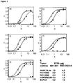
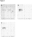
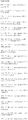
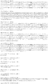
- figure 3 f denotes the binding strength of representative IgG antibodies on transfected C4.4a-expressing A549 tumor cells and endogenously C4.4a expressing tumor cells.
- Table 8 provides a summary of the binding strength (EC 50 ) of representative IgG antibodies on transfected murine CHO-S:mC4.4a cells and endogenously C4.4a expressing NCI H292 tumor cells.
- An IgG1 is said to have a "high affinity" for an antigen if the affinity measurement measured by FACS is less than 100 nM (apparent affinity of IgG).
- a disclosed bivalent antibody or antigen-binding fragment preferably can bind to human C4.4a with an affinity of less than 100 nM, more preferably less than 50 nM, and still more preferably less than 10 nM.
- Further preferred are bivalent antibodies that bind to C4.4a with an affinity of less than 5 nM, and more preferably less than 1 nM determined as aparent affinity of an IgG to human C4.4a.
- the apparent affinity of an antibody of the disclosure against C4.4a may be about 4.3 nM or 0.03 nM on different tumor cell lines as determined by FACS analysis as depicted in figure 3 f.
- An antibody or antigen-binding fragment of the disclosure internalizes "efficiently" when its time of half maximal internalization (t 1 ⁇ 2) into C4.4a expressing tumor cells is shorter than 180 min or more preferably shorter than 120 min and still more preferably shorter than 90 min. Further preferred are antibodies or antigen-binding fragments with half maximal internalization times (t 1 ⁇ 2) of 60 minutes or less as determined by the protocol described in example 6, further preferred are less than 50 minutes, or less than 35 minutes. Table 9 provides a summary of internalization times of representative antibodies of the disclosure, as determined by the protocol described in example 6. Internalizable antibodies or antigen-binding fragments of the disclosure are suitable as targeting moiety of an antibody-drug conjugate (ADC).
- ADC antibody-drug conjugate
- An antibody or antigen-binding fragment is suitable in an in vitro or in vivo method to deliver a compound, preferably a cytotoxic agent, into a C4.4a expressing cell.
- Table 9 interalization Antibody (IgG1) t 1 ⁇ 2 [min] M31-B01 49 M20-D02 S-A 60 B01-3 55 B01-7 33 B01-10 30 D02-6 39 D02-7 33 D02-11 22
- the antibody, antigen-binding fragment thereof, or derivative thereof or nucleic acid encoding the same is isolated.
- An isolated biological component (such as a nucleic acid molecule or protein such as an antibody) is one that has been substantially separated or purified away from other biological components in the cell of the organism in which the component naturally occurs, e.g., other chromosomal and extra-chromosomal DNA and RNA, proteins and organelles.
- Nucleic acids and proteins that have been "isolated” include nucleic acids and proteins purified by standard purification methods Sambrook et al., 1989 ( Sambrook, J., Fritsch, E. F. and Maniatis, T.
- a fully human N-CoDeR antibody phage display library was used to isolate high affinity, C4.4a-specific, human monoclonal antibodies by a combination of whole cell and protein panning and through the development of specific tools.
- These tools and methods include a human C4.4a-expressing recombinant cell-line, a murine C4.4a expressing cell line, recombinant human and murine C4.4a and the development of panning procedures and screening assays capable of identifying antibodies that preferentially bind to C4.4a displayed on the cell surface and that are cross-reactive to murine C4.4a.
- Antibodies to the cancer cell-surface marker C4.4a were developed by a combination of three non-conventional approaches in phage-display technology (PDT).
- PDT phage-display technology
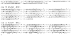
- recombinant cell lines expressing the membrane-bound form of human and mouse C4.4a were constructed by stable transfection of CHO-S cells and A549 tumor cells with a plasmid encoding the full length GPI-anchored form of the human or mouse protein (SEQ ID NO:3) and (SEQ ID NO:4) respectively, to give the human CHO-S:hC4.4a, the murine CHO-S:mC4.4a and the human A549:hC4.4a cell lines, respectively.
- cell-surface selections were performed with the latter recombinant cell lines and the breast cancer cell line MCF-7.
- Pre-adsorption with CHO-S cells or non transfected A549 cells was included to avoid the selection of Fab fragments binding to epitopes of the parental cells. Additional selections were performed with recombinant, soluble, purified human C4.4a, with recombinant, soluble, purified murine C4.4a. Third, screening methods were developed which allowed for successive screening of the phage outputs obtained in panning on whole A549:hC4.4a cells as well as CHO-S:hC4.4a cells. The combination of these specific methods allowed the isolation of the unique antibodies "M31-B01", “M20-D02 S-A", "M60-G03", and, "M36-H02".
- Antibodies or antigen-binding fragments of the disclosure are not limited to the specific peptide sequences provided herein. Rather, the disclosure also embodies variants of these polypeptides. With reference to the instant disclosure and conventionally available technologies and references, the skilled worker will be able to prepare, test and utilize functional variants of the antibodies disclosed herein.
- a variant can include, for example, an antibody that has at least one altered complementary determining region (CDR) (hyper-variable) and/or framework (FR) (variable) domain/position, vis-à-vis a peptide sequence disclosed herein.
- CDR complementary determining region
- FR framework
- An antibody is composed of two peptide chains, each containing one (light chain) or three (heavy chain) constant domains and a variable region (VL, VH), the latter of which is in each case made up of four FR regions and three interspaced CDRs.
- the antigen-binding site is formed by one or more CDRs, yet the FR regions provide the structural framework for the CDRs and, hence, play an important role in antigen binding.
- the skilled worker routinely can generate mutated or diversified antibody sequences, which can be screened against the antigen, for new or improved properties, for example.
- Tables 3 (VL) and 4 (VH) delineate the CDR and FR regions for certain antibodies of the disclosure and compare amino acids at a given position to each other and to corresponding consensus sequences.
- a further preferred embodiment of the disclosure is an antibody or antigen binding fragment thereof in which the CDR sequences are selected as shown in table 7.
- a further preferred embodiment of the disclosure is an antibody or antigen-binding fragment in which the VH and VL sequences are selected as shown in table 7.
- the skilled worker can use the data in Tables 3, 4 and 7 to design peptide variants. It is preferred that variants are constructed by changing amino acids within one or more CDR regions; a variant might also have one or more altered framework regions. Alterations also may be made in the framework regions. For example, a peptide FR domain might be altered where there is a deviation in a residue compared to a germline sequence.
- candidate residues that can be changed include e.g. residue 42 of the variable heavy chain of M20-D02 S-A compared to VHIII of Gene DP47.
- residue 42 of the variable heavy chain of M20-D02 S-A compared to VHIII of Gene DP47.
- skilled worker could make the same analysis by comparing the amino acid sequences disclosed herein to known sequences of the same class of such antibodies, using, for example, the procedure described by Knappik A., et al., JMB 2000, 296:57-86 .
- variants may be obtained by using one antibody as starting point for optimization by diversifying one or more amino acid residues in the antibody, preferably amino acid residues in one or more CDRs, and by screening the resulting collection of antibody variants for variants with improved properties. Particularly preferred is diversification of one or more amino acid residues in CDR3 of VL and/or VH.. Diversification can be done by synthesizing a collection of DNA molecules using trinucleotide mutagenesis (TRIM) technology ( Virneklas B. et al., Nucl. Acids Res. 1994, 22: 5600 .).
- TAM trinucleotide mutagenesis
- Antibodies or antigen-binding fragments thereof include molecules with modifications/variations including but not limited to e.g. modifications leading to altered half-life (e.g. modification of the Fc part or attachment of further molecules such as PEG) or altered ADCC or CDC activity.
- Polypeptide variants may be made that conserve the overall molecular structure of an antibody peptide sequence described herein. Given the properties of the individual amino acids, some rational substitutions will be recognized by the skilled worker. Amino acid substitutions, i.e., "conservative substitutions,” may be made, for instance, on the basis of similarity in polarity, charge, solubility, hydrophobicity, hydrophilicity, and/or the amphipathic nature of the residues involved.
- nonpolar (hydrophobic) amino acids include alanine, leucine, isoleucine, valine, proline, phenylalanine, tryptophane, and methionine;
- polar neutral amino acids include glycine, serine, threonine, cysteine, tyrosine, asparagine, and glutamine;
- positively charged (basic) amino acids include arginine, lysine, and histidine; and
- negatively charged (acidic) amino acids include aspartic acid and glutamic acid. Substitutions typically may be made within groups (a)-(d).
- glycine and proline may be substituted for one another based on their ability to disrupt ⁇ -helices.
- certain amino acids such as alanine, cysteine, leucine, methionine, glutamic acid, glutamine, histidine and lysine are more commonly found in ⁇ -helices
- valine, isoleucine, phenylalanine, tyrosine, tryptophan and threonine are more commonly found in ⁇ -pleated sheets.
- Glycine, serine, aspartic acid, asparagine, and proline are commonly found in turns.
- substitutions may be made among the following groups: (i) S and T; (ii) P and G; and (iii) A, V, L and I.
- S and T S and T
- P and G P and G
- A, V, L and I A, V, L and I.
- amino acid position 3 in SEQ ID NOS: 33 -36 can be changed from a Q to an E.
- sequence identity between two polypeptide sequences, indicates the percentage of amino acids that are identical between the sequences.
- sequence homology indicates the percentage of amino acids that either is identical or that represent conservative amino acid substitutions.
- the present invention also relates to the DNA molecules that encode an antibody of the invention or antigen-binding fragment thereof.
- DNA molecules of the invention are not limited to the sequences disclosed herein, but also include variants thereof. DNA variants within the invention may be described by reference to their physical properties in hybridization. The skilled worker will recognize that DNA can be used to identify its complement and, since DNA is double stranded, its equivalent or homolog, using nucleic acid hybridization techniques. It also will be recognized that hybridization can occur with less than 100% complementarity. However, given appropriate choice of conditions, hybridization techniques can be used to differentiate among DNA sequences based on their structural relatedness to a particular probe. For guidance regarding such conditions see, Sambrook et al., 1989 supra and Ausubel et al., 1995 ( Ausubel, F. M., Brent, R., guitarist, R. E., Moore, D. D., Sedman, J. G., Smith, J. A., & Struhl, K. eds. (1995). Current Protocols in Molecular Biology. New York: John Wiley and Sons ).
- Structural similarity between two polynucleotide sequences can be expressed as a function of "stringency" of the conditions under which the two sequences will hybridize with one another.
- stringency refers to the extent that the conditions disfavor hybridization. Stringent conditions strongly disfavor hybridization, and only the most structurally related molecules will hybridize to one another under such conditions. Conversely, non-stringent conditions favor hybridization of molecules displaying a lesser degree of structural relatedness. Hybridization stringency, therefore, directly correlates with the structural relationships of two nucleic acid sequences. The following relationships are useful in correlating hybridization and relatedness (where T m is the melting temperature of a nucleic acid duplex):
- Hybridization stringency is a function of many factors, including overall DNA concentration, ionic strength, temperature, probe size and the presence of agents which disrupt hydrogen bonding. Factors promoting hybridization include high DNA concentrations, high ionic strengths, low temperatures, longer probe size and the absence of agents that disrupt hydrogen bonding. Hybridization typically is performed in two phases: the "binding" phase and the “washing” phase.
- the probe is bound to the target under conditions favoring hybridization.
- Stringency is usually controlled at this stage by altering the temperature.
- the temperature is usually between 65°C and 70°C, unless short ( ⁇ 20 nt) oligonucleotide probes are used.
- a representative hybridization solution comprises 6X SSC, 0.5% SDS, 5X Denhardt's solution and 100 ⁇ g of nonspecific carrier DNA. See Ausubel et al., section 2.9, supplement 27 (1994). Of course, many different, yet functionally equivalent, buffer conditions are known. Where the degree of relatedness is lower, a lower temperature may be chosen.
- Low stringency binding temperatures are between about 25°C and 40°C.
- Medium stringency is between at least about 40°C to less than about 65°C.
- High stringency is at least about 65°C.
- washing solutions typically contain lower salt concentrations.
- One exemplary medium stringency solution contains 2X SSC and 0.1% SDS.
- a high stringency wash solution contains the equivalent (in ionic strength) of less than about 0.2X SSC, with a preferred stringent solution containing about O.1X SSC.
- the temperatures associated with various stringencies are the same as discussed above for "binding.”
- the washing solution also typically is replaced a number of times during washing. For example, typical high stringency washing conditions comprise washing twice for 30 minutes at 55° C. and three times for 15 minutes at 60° C.
- An embodiment of the disclosure is an isolated nucleic acid sequence that encodes (i) the antibody or antigen-binding fragment of the disclosure, the CDR sequences as depicted in table 7, or (ii) the variable light and heavy chain sequences as depicted in table 7, or (iii) which comprises a nucleic acid sequence that encodes an antibody or antigen-binding fragment of the disclosure, the CDR sequences as depicted in table 7, or the variable light and heavy chain sequences as depicted in table 7.
- DNA variants within the scope of the disclosure may be described with reference to the product they encode.
- These functionally equivalent polynucleotides are characterized by the fact that they encode the same peptide sequences found in SEQ ID NOS: 5-36, 45-50, 55-60, 65-70, 75-80, 85-90, 95-100, 105-110, 115-120, 125-130, 135-140, due to the degeneracy of the genetic code.
- variants of DNA molecules provided herein can be constructed in several different ways. For example, they may be constructed as completely synthetic DNAs. Methods of efficiently synthesizing oligonucleotides in the range of 20 to about 150 nucleotides are widely available. See Ausubel et al., section 2.11, Supplement 21 (1993). Overlapping oligonucleotides may be synthesized and assembled in a fashion first reported by Khorana et al., J. Mol. Biol. 72:209-217 (1971 ); see also Ausubel et al., supra, Section 8.2. Synthetic DNAs preferably are designed with convenient restriction sites engineered at the 5' and 3' ends of the gene to facilitate cloning into an appropriate vector.
- a method of generating variants is to start with one of the DNAs disclosed herein and then to conduct site-directed mutagenesis. See Ausubel et al., supra, chapter 8, Supplement 37 (1997).
- a target DNA is cloned into a single-stranded DNA bacteriophage vehicle.
- Single-stranded DNA is isolated and hybridized with an oligonucleotide containing the desired nucleotide alteration(s).
- the complementary strand is synthesized and the double stranded phage is introduced into a host.
- Some of the resulting progeny will contain the desired mutant, which can be confirmed using DNA sequencing.
- various methods are available that increase the probability that the progeny phage will be the desired mutant. These methods are well known to those in the field and kits are commercially available for generating such mutants.
- the disclosure provides recombinant DNA constructs comprising one or more of the nucleotide sequences of the present invention.
- the recombinant constructs of the disclosire are used in connection with a vector, such as a plasmid, phagemid, phage or viral vector, into which a DNA molecule encoding an antibody of the invention or antigen-binding fragment thereof is inserted.
- An antibody, antigen binding portion, or derivative thereof provided herein can be prepared by recombinant expression of nucleic acid sequences encoding light and heavy chains or portions thereof in a host cell.
- a host cell can be transfected with one or more recombinant expression vectors carrying DNA fragments encoding the light and/or heavy chains or portions thereof such that the light and heavy chains are expressed in the host cell.
- Standard recombinant DNA methodologies are used prepare and/or obtain nucleic acids encoding the heavy and light chains, incorporate these nucleic acids into recombinant expression vectors and introduce the vectors into host cells, such as those described in Sambrook, Fritsch and Maniatis (eds.), Molecular Cloning; A Laboratory Manual, Second Edition, Cold Spring Harbor, N.Y., (1989 ), Ausubel, F. M. et al. (eds.) Current Protocols in Molecular Biology, Greene Publishing Associates, (1989 ) and in U.S. Pat. No. 4,816,397 by Boss et al.
- nucleic acid sequences encoding variable regions of the heavy and/or light chains can be converted, for example, to nucleic acid sequences encoding full-length antibody chains, Fab fragments, or to scFv.
- the VL- or VH-encoding DNA fragment can be operatively linked, (such that the amino acid sequences encoded by the two DNA fragments are in-frame) to another DNA fragment encoding, for example, an antibody constant region or a flexible linker.
- sequences of human heavy chain and light chain constant regions are known in the art (see e.g., Kabat, E. A., el al. (1991) Sequences of Proteins of Immunological Interest, Fifth Edition, U.S. Department of Health and Human Services, NIH Publication No. 91-3242 ) and DNA fragments encompassing these regions can be obtained by standard PCR amplification.
- the VH- and VL-encoding nucleic acids can be operatively linked to another fragment encoding a flexible linker such that the VH and VL sequences can be expressed as a contiguous single-chain protein, with the VL and VH regions joined by the flexible linker (see e.g., Bird et al. (1988) Science 242:423-426 ; Huston et al. (1988) Proc. Natl. Acad. Sci. USA 85:5879-5883 ; McCafferty et al., Nature (1990) 348:552-554 ).
- DNA encoding the desired polypeptide can be inserted into an expression vector which is then transfected into a suitable host cell.
- suitable host cells are prokaryotic and eukaryotic cells. Examples for prokaryotic host cells are e.g. bacteria, examples for eukaryotic host cells are yeast, insect or mammalian cells.
- the DNAs encoding the heavy and light chains are inserted into separate vectors.
- the DNA encoding the heavy and light chains are inserted into the same vector. It is understood that the design of the expression vector, including the selection of regulatory sequences is affected by factors such as the choice of the host cell, the level of expression of protein desired and whether expression is constitutive or inducible.
- Useful expression vectors for bacterial use are constructed by inserting a structural DNA sequence encoding a desired protein together with suitable translation initiation and termination signals in operable reading phase with a functional promoter.
- the vector will comprise one or more phenotypic selectable markers and an origin of replication to ensure maintenance of the vector and, if desirable, to provide amplification within the host.
- Suitable prokaryotic hosts for transformation include E. coli, Bacillus subtilis, Salmonella typhimurium and various species within the genera Pseudomonas, Streptomyces, and Staphylococcus.
- Bacterial vectors may be, for example, bacteriophage-, plasmid- or phagemid-based. These vectors can contain a selectable marker and bacterial origin of replication derived from commercially available plasmids typically containing elements of the well known cloning vector pBR322 (ATCC 37017). Following transformation of a suitable host strain and growth of the host strain to an appropriate cell density, the selected promoter is de-repressed/induced by appropriate means (e.g ., temperature shift or chemical induction) and cells are cultured for an additional period. Cells are typically harvested by centrifugation, disrupted by physical or chemical means, and the resulting crude extract retained for further purification.
- appropriate means e.g ., temperature shift or chemical induction
- a number of expression vectors may be advantageously selected depending upon the use intended for the protein being expressed. For example, when a large quantity of such a protein is to be produced, for the generation of antibodies or to screen peptide libraries, for example, vectors which direct the expression of high levels of fusion protein products that are readily purified may be desirable.
- Antibodies of the present invention or antigen-binding fragment thereof include naturally purified products, products of chemical synthetic procedures, and products produced by recombinant techniques from a prokaryotic host, including, for example, E. coli, Bacillus subtilis, Salmonella typhimurium and various species within the genera Pseudomonas, Streptomyces, and Staphylococcus, preferably, from E. coli cells.
- Preferred regulatory sequences for mammalian host cell expression include viral elements that direct high levels of protein expression in mammalian cells, such as promoters and/or enhancers derived from cytomegalovirus (CMV) (such as the CMV promoter/enhancer), Simian Virus 40 (SV40) (such as the SV40 promoter/enhancer), adenovirus, (e.g., the adenovirus major late promoter (AdMLP)) and polyoma.
- CMV cytomegalovirus
- SV40 Simian Virus 40
- AdMLP adenovirus major late promoter
- the recombinant expression vectors can also include origins of replication and selectable markers (see e.g., U.S. 4,399,216 , 4,634,665 and U.S. 5,179,017, by Axel et al. ).
- Suitable selectable markers include genes that confer resistance to drugs such as G418, hygromycin or methotrexate, on a host cell into which the vector has been introduced.
- drugs such as G418, hygromycin or methotrexate
- the dihydrofolate reductase (DHFR) gene confers resistance to methotrexate
- the neo gene confers resistance to G418.
- Transfection of the expression vector into a host cell can be carried out using standard techniques such as electroporation, calcium-phosphate precipitation, and DEAE-dextran, lipofection or polycation-mediated transfection.
- Suitable mammalian host cells for expressing the antibodies, antigen binding portions, or derivatives thereof provided herein include Chinese Hamster Ovary (CHO cells) (including dhfr- CHO cells, described in Urlaub and Chasin, (1980) Proc. Natl. Acad. Sci. USA 77:4216-4220 , used with a DHFR selectable marker, e.g., as described in R. J. Kaufman and P. A. Sharp (1982) Mol. Biol. 159:601-621 , NSO myeloma cells, COS cells and SP2 cells.
- the expression vector is designed such that the expressed protein is secreted into the culture medium in which the host cells are grown.
- Transient transfection/epression of antibodies can for example be achieved following the protocols by Durocher et al (2002) Nucl.Acids Res. Vol 30 e9 .
- Stable transfection/expression of antibodies can for example be achieved following the protocols of the UCOE system ( T. Benton et al. (2002) Cytotechnology 38: 43-46 ).
- the antibodies, antigen binding portions, or derivatives thereof can be recovered from the culture medium using standard protein purification methods.
- Antibodies of the invention or an antigen-binding fragment thereof can be recovered and purified from recombinant cell cultures by well-known methods including, but not limited to ammonium sulfate or ethanol precipitation, acid extraction, Protein A chromatography, Protein G chromatography, anion or cation exchange chromatography, phospho-cellulose chromatography, hydrophobic interaction chromatography, affinity chromatography, hydroxylapatite chromatography and lectin chromatography. High performance liquid chromatography (“HPLC”) can also be employed for purification.
- HPLC high performance liquid chromatography
- Antibodies of the disclosure or antigen-binding fragment thereof include naturally purified products, products of chemical synthetic procedures, and products produced by recombinant techniques from a eukaryotic host, including, for example, yeast, higher plant, insect and mammalian cells, preferably from mammalian cells.
- a eukaryotic host including, for example, yeast, higher plant, insect and mammalian cells, preferably from mammalian cells.
- the antibody of the present invention can be glycosylated or can be non-glycosylated, with glycosylated preferred.
- Such methods are described in many standard laboratory manuals, such as Sambrook, supra, Sections 17.37-17.42; Ausubel, supra, Chapters 10, 12, 13, 16, 18 and 20.
- Therapeutic methods involve administering to a subject in need of treatment a therapeutically effective amount of an antibody or antigen-binding fragment thereof contemplated by the invention.
- a "therapeutically effective" amount hereby is defined as the amount of an antibody or antigen-binding fragment that is of sufficient quantity to deplete C4.4a-positive cells in a treated area of a subject - either as a single dose or according to a multiple dose regimen, alone or in combination with other agents, which leads to the alleviation of an adverse condition, yet which amount is toxicologically tolerable.
- the subject may be a human or non-human animal (e.g ., rabbit, rat, mouse, dog, monkey or other lower-order primate).
- an antibody of the invention or antigen-binding fragment thereof might be co-administered with known medicaments, and in some instances the antibody might itself be modified.
- an antibody could be conjugated to a cytotoxic agent or radioisotope to potentially further increase efficacy.
- Antibodies of the present invention may be administered as the sole pharmaceutical agent or in combination with one or more additional therapeutic agents where the combination causes no unacceptable adverse effects.
- This combination therapy includes administration of a single pharmaceutical dosage formulation which contains an antibody of the invention and one or more additional therapeutic agents, as well as administration of an antibody of the invention and each additional therapeutic agent in its own separate pharmaceutical dosage formulation.
- an antibody of the invention and a therapeutic agent may be administered to the patient together in a single oral dosage composition such as a tablet or capsule, or each agent may be administered in separate dosage formulations.
- an antibody of the invention and one or more additional therapeutic agents may be administered at essentially the same time (e.g., concurrently) or at separately staggered times ( e.g., sequentially).
- an antibodies of the present invention may be used in fixed or separate combination with other anti-tumor agents such as alkylating agents, anti-metabolites, plant-derived anti-tumor agents, hormonal therapy agents, topoisomerase inhibitors, camptothecin derivatives, kinase inhibitors, targeted drugs, antibodies, interferons and/or biological response modifiers, anti-angiogenic compounds, and other anti-tumor drugs.
- anti-tumor agents such as alkylating agents, anti-metabolites, plant-derived anti-tumor agents, hormonal therapy agents, topoisomerase inhibitors, camptothecin derivatives, kinase inhibitors, targeted drugs, antibodies, interferons and/or biological response modifiers, anti-angiogenic compounds, and other anti-tumor drugs.
- the antibodies of the present invention may be used in combination with chemotherapy (i.e. cytotoxic agents), anti-hormones and/or targeted therapies such as other kinase inhibitors (for example, EGFR inhibitors), mTOR inhibitors and angiogenesis inhibitors.
- chemotherapy i.e. cytotoxic agents
- anti-hormones and/or targeted therapies such as other kinase inhibitors (for example, EGFR inhibitors), mTOR inhibitors and angiogenesis inhibitors.
- the compounds of the present invention may also be employed in cancer treatment in conjunction with radiation therapy and/or surgical intervention.
- an antibody of the invention or antigen-binding fragment thereof might in some instances itself be modified.
- an antibody could be conjugated to any of but not limited to the compounds mentioned above or any radioisotope to potentially further increase efficacy.
- the antibodies of the invention may be utilized, as such or in compositions, in research and diagnostics, or as analytical reference standards, and the like, which are well known in the art.
- inventive antibodies or antigen-binding fragments thereof can be used as a therapeutic or a diagnostic tool in a variety of situations where C4.4a is undesirably expressed or found, e.g. cell proliferative disorders such as cancer.
- disorders and conditions particularly suitable for treatment with an antibody of the inventions are solid tumors, such as cancers of the breast, respiratory tract, brain, reproductive organs, digestive tract, urinary tract, eye, liver, skin, head and neck, thyroid, parathyroid, and their distant metastases. Those disorders also include lymphomas, sarcomas and leukemias.
- breast cancer examples include, but are not limited to invasive ductal carcinoma, invasive lobular carcinoma, ductal carcinoma in situ, and lobular carcinoma in situ.
- cancers of the respiratory tract include, but are not limited to small-cell and non-small-cell lung carcinoma, as well as bronchial adenoma and pleuropulmonary blastoma.
- brain cancers include, but are not limited to brain stem and hypophtalmic glioma, cerebellar and cerebral astrocytoma, glioblastoma, medulloblastoma, ependymoma, as well as neuroectodermal and pineal tumor.
- Tumors of the male reproductive organs include, but are not limited to prostate and testicular cancer.
- Tumors of the female reproductive organs include, but are not limited to endometrial, cervical, ovarian, vaginal and vulvar cancer, as well as sarcoma of the uterus.
- Tumors of the digestive tract include, but are not limited to anal, colon, colorectal, esophageal, gallbladder, gastric, pancreatic, rectal, small-intestine, and salivary gland cancers.
- Tumors of the urinary tract include, but are not limited to bladder, penile, kidney, renal pelvis, ureter, urethral, and hereditary and sporadic papillary renal cancers.
- Eye cancers include, but are not limited to intraocular melanoma and retinoblastoma.
- liver cancers include, but are not limited to hepatocellular carcinoma (liver cell carcinomas with or without fibrolamellar variant), cholangiocarcinoma (intrahepatic bile duct carcinoma), and mixed hepatocellular cho langiocarcinoma.
- Skin cancers include, but are not limited to squamous cell carcinoma, Kaposi's sarcoma, malignant melanoma, Merkel cell skin cancer, and non-melanoma skin cancer.
- Head-and-neck cancers include, but are not limited to laryngeal, hypopharyngeal, nasopharyngeal, oropharyngeal cancer, lip and oral cavity cancer, and squamous cell cancer.
- Lymphomas include, but are not limited to AIDS-related lymphoma, non-Hodgkin's lymphoma, cutaneous T-cell lymphoma, Burkitt lymphoma, Hodgkin's disease, and lymphoma of the central nervous system.
- Sarcomas include, but are not limited to sarcoma of the soft tissue, osteosarcoma, malignant fibrous histiocytoma, lymphosarcoma, and rhabdomyosarcoma.
- Leukemias include, but are not limited to acute myeloid leukemia, acute lymphoblastic leukemia, chronic lymphocytic leukemia, chronic myelogenous leukemia, and hairy cell leukemia.
- compositions for use in accordance with the present invention may be formulated in a conventional manner using one or more physiologically acceptable carriers or excipients.
- An antibody of the invention or antigen-binding fragment thereof can be administered by any suitable means, which can vary, depending on the type of disorder being treated. Possible administration routes include parenteral (e.g., intramuscular, intravenous, intra-arterial, intraperitoneal, or subcutaneous), intrapulmonary and intranasal, and, if desired for local immunosuppressive treatment, intralesional administration.
- an antibody of the invention might be administered by pulse infusion, with, e.g., declining doses of the antibody.
- the dosing is given by injections, most preferably intravenous or subcutaneous injections, depending in part on whether the administration is brief or chronic.
- the amount to be administered will depend on a variety of factors such as the clinical symptoms, weight of the individual, whether other drugs are administered. The skilled artisan will recognize that the route of administration will vary depending on the disorder or condition to be treated.
- Determining a therapeutically effective amount of the novel polypeptide largely will depend on particular patient characteristics, route of administration, and the nature of the disorder being treated. General guidance can be found, for example, in the publications of the International Conference on Harmonization and in REMINGTON'S PHARMACEUTICAL SCIENCES, chapters 27 and 28, pp. 484-528 (18th ed., Alfonso R. Gennaro, Ed., Easton, Pa.: Mack Pub. Co., 1990 ). More specifically, determining a therapeutically effective amount will depend on such factors as toxicity and efficacy of the medicament. Toxicity may be determined using methods well known in the art and found in the foregoing references. Efficacy may be determined utilizing the same guidance in conjunction with the methods described below in the Examples.
- C4.4a antibodies or antigen-binding fragments thereof can be used for detecting the presence of C4.4a-expressing tumors.
- the presence of C4.4a-containing cells or shed C4.4a within various biological samples, including serum, and tissue biopsy specimens, may be detected with C4.4a antibodies.
- C4.4a antibodies may be used in various imaging methodologies such as immunoscintigraphy with a 99 Tc (or other isotope) conjugated antibody.
- an imaging protocol similar to the one recently described using a 111 In conjugated anti-PSMA antibody may be used to detect pancreatic or ovarian carcinomas ( Sodee et al., Clin. Nuc. Med. 21: 759-766, 1997 ).
- Another method of detection that can be used is positron emitting tomography by conjugating the antibodies of the invention with a suitable isotope (see Herzog et al., J. Nucl. Med. 34:2222-2226, 1993 ).
- compositions which comprise C4.4a antibodies or antigen-binding fragment thereof, alone or in combination with at least one other agent, such as stabilizing compound, which may be administered in any sterile, biocompatible pharmaceutical carrier, including, but not limited to, saline, buffered saline, dextrose, and water.
- a further embodiment are pharmaceutical compositions comprising a C4.4a binding antibody or antigen-binding fragment therof and a further pharmaceutically active compound that is suitable to treat C4.4a related diseases such as cancer. Any of these molecules can be administered to a patient alone, or in combination with other agents, drugs or hormones, in pharmaceutical compositions where it is mixed with excipient(s) or pharmaceutically acceptable carriers.
- the pharmaceutically acceptable carrier is pharmaceutically inert.
- the present disclosure also relates to the administration of pharmaceutical compositions. Such administration is accomplished orally or parenterally.
- Methods of parenteral delivery include topical, intra-arterial (directly to the tumor), intramuscular, subcutaneous, intramedullary, intrathecal, intraventricular, intravenous, intraperitoneal, or intranasal administration.
- these pharmaceutical compositions may contain suitable pharmaceutically acceptable carriers comprising excipients and auxiliaries which facilitate processing of the active compounds into preparations which can be used pharmaceutically. Further details on techniques for formulation and administration may be found in the latest edition of Remington's Pharmaceutical Sciences (Ed. Maack Publishing Co, Easton, Pa .).
- compositions for oral administration can be formulated using pharmaceutically acceptable carriers well known in the art in dosages suitable for oral administration.
- Such carriers enable the pharmaceutical compositions to be formulated as tablets, pills, dragees, capsules, liquids, gels, syrups, slurries, suspensions and the like, for ingestion by the patient.
- compositions for oral use can be obtained through combination of active compounds with solid excipient, optionally grinding a resulting mixture, and processing the mixture of granules, after adding suitable auxiliaries, if desired, to obtain tablets or dragee cores.
- Suitable excipients are carbohydrate or protein fillers such as sugars, including lactose, sucrose, mannitol, or sorbitol; starch from corn, wheat, rice, potato, or other plants; cellulose such as methyl, cellulose, hydroxypropylmethylcellulose, or sodium carboxymethyl cellulose; and gums including arabic and tragacanth; and proteins such as gelatin and collagen.
- disintegrating or solubilizing agents may be added, such as the cross-linked polyvinyl pyrrolidone, agar, alginic acid, or a salt thereof, such as sodium alginate.
- Dragee cores are provided with suitable coatings such as concentrated sugar solutions, which may also contain gum arabic, talc, polyvinyl pyrrolidone, carbopol gel, polyethylene glycol and/or titanium dioxide, lacquer solutions, and suitable organic solvents or solvent mixtures.
- Dyestuffs or pigments may be added to the tablets or dragee coatings for product identification or to characterize the quantity of active compound, i.e. dosage.
- Push-fit capsules made of gelatin, as well as soft, sealed capsules made of gelatin and a coating such as glycerol or sorbitol.
- Push-fit capsules can contain active ingredients mixed with a filler or binders such as lactose or starches, lubricants such as talc or magnesium stearate, and optionally, stabilizers.
- the active compounds may be dissolved or suspended in suitable liquids, such as fatty oils, liquid paraffin, or liquid polyethylene glycol with or without stabilizers.
- compositions for parenteral administration include aqueous solutions of active compounds.
- the pharmaceutical compositions of the invention may be formulated in aqueous solutions, preferably in physiologically compatible buffers such as Hank's solution, Ringer's solution, or physiologically buffered saline.
- Aqueous injection suspensions may contain substances that increase viscosity of the suspension, such as sodium carboxymethyl cellulose, sorbitol, or dextran.
- suspensions of the active compounds may be prepared as appropriate oily injection suspensions.
- Suitable lipophilic solvents or vehicles include fatty oils such as sesame oil, or synthetic fatty acid esters, such as ethyl oleate or triglycerides, or liposomes.
- the suspension may also contain suitable stabilizers or agents which increase the solubility of the compounds to allow for the preparation of highly concentrated solutions.
- penetrants appropriate to the particular barrier to be permeated are used in the formulation.
- penetrants are generally known in the art.
- the invention further relates to pharmaceutical packs and kits comprising one or more containers filled with one or more of the ingredients of the aforementioned compositions of the invention.
- Associated with such container(s) can be a notice in the form prescribed by a governmental agency regulating the manufacture, use or sale of pharmaceuticals or biological products, reflecting approval by the agency of the manufacture, use or sale of the product for human administration.
- kits may contain DNA sequences encoding the antibodies of the disclosure.
- the DNA sequences encoding these antibodies are provided in a plasmid suitable for transfection into and expression by a host cell.
- the plasmid may contain a promoter (often an inducible promoter) to regulate expression of the DNA in the host cell.
- the plasmid may also contain appropriate restriction sites to facilitate the insertion of other DNA sequences into the plasmid to produce various antibodies.
- the plasmids may also contain numerous other elements to facilitate cloning and expression of the encoded proteins. Such elements are well known to those of skill in the art and include, for example, selectable markers, initiation codons, termination codons, and the like.
- compositions of the present invention may be manufactured in a manner that is known in the art, e.g., by means of conventional mixing, dissolving, granulating, dragee-making, levigating, emulsifying, encapsulating, entrapping or lyophilizing processes.
- the pharmaceutical composition may be provided as a salt and can be formed with acids, including by not limited to hydrochloric, sulfuric, acetic, lactic, tartaric, malic, succinic, etc. Salts tend to be more soluble in aqueous or other protonic solvents that are the corresponding free base forms.
- the preferred preparation may be a lyophilized powder in 1 mM-50 mM histidine, 0.1%-2% sucrose, 2%-7% mannitol at a pH range of 4.5 to 5.5 that is combined with buffer prior to use.
- compositions comprising a compound of the invention formulated in an acceptable carrier
- they can be placed in an appropriate container and labeled for treatment of an indicated condition.
- labeling would include amount, frequency and method of administration.
- compositions suitable for use in the present invention include compositions wherein the active ingredients are contained in an effective amount to achieve the intended purpose, i.e. treatment of a particular disease state characterized by C4.4a expression.
- the determination of an effective dose is well within the capability of those skilled in the art.
- the therapeutically effective dose can be estimated initially either in cell culture assays, e.g., neoplastic cells, or in animal models, usually mice, rabbits, dogs, pigs or monkeys.
- the animal model is also used to achieve a desirable concentration range and route of administration. Such information can then be used to determine useful doses and routes for administration in humans.
- a therapeutically effective dose refers to that amount of antibody or antigen-binding fragment thereof, that ameliorate the symptoms or condition.
- Therapeutic efficacy and toxicity of such compounds can be determined by standard pharmaceutical procedures in cell cultures or experimental animals, e.g., ED 50 (the dose therapeutically effective in 50% of the population) and LD 50 (the dose lethal to 50% of the population). The dose ratio between therapeutic and toxic effects is the therapeutic index, and it can be expressed as the ratio, ED 50 /LD 50 . Pharmaceutical compositions that exhibit large therapeutic indices are preferred. The data obtained from cell culture assays and animal studies are used in formulating a range of dosage for human use. The dosage of such compounds lies preferably within a range of circulating concentrations what include the ED 50 with little or no toxicity. The dosage varies within this range depending upon the dosage form employed, sensitivity of the patient, and the route of administration.
- the exact dosage is chosen by the individual physician in view of the patient to be treated. Dosage and administration are adjusted to provide sufficient levels of the active moiety or to maintain the desired effect. Additional factors that may be taken into account include the severity of the disease state, e.g., tumor size and location; age, weight and gender of the patient; diet, time and frequency of administration, drug combination(s), reaction sensitivities, and tolerance/response to therapy. Long acting pharmaceutical compositions might be administered every 3 to 4 days, every week, or once every two weeks depending on half-life and clearance rate of the particular formulation.
- Normal dosage amounts may vary from 0.1 to 100,000 micrograms, up to a total dose of about 1 g, depending upon the route of administration.
- Guidance as to particular dosages and methods of delivery is provided in the literature. See U.S. Pat. No. 4,657,760 ; 5,206,344 ; or 5,225,212 .
- Those skilled in the art will employ different formulations for polynucleotides than for proteins or their inhibitors.
- delivery of polynucleotides or polypeptides will be specific to particular cells, conditions, locations, etc.
- Preferred specific activities for a radiolabelled antibody may range from 0.1 to 10 mCi/mg of protein ( Riva et al., Clin. Cancer Res. 5:3275-3280, 1999 ; Ulaner et al., 2008 Radiology 246(3):895-902 )
- n-CoDeR is a Fab library in which all six CDRs are diversified. CDR sequences were initially obtained from healthy human donors.
- Fab antibody library was depleted for unwanted surface binders by pre-incubation with 10 7 non-C4.4a expressing parental CHO-S cells in PBS / 3% FCS / 0.01 % NaN3 (buffer A) at 4°C by end-over-end rotation for 15 min. After that cells were removed by centrifugation, the supernatant was used for 2 additional rounds of pre-incubation on CHO-S cells. Sub sequential panning on target cells (10 7 CHO-S:hC4.4a cells) was done by incubation with the phage preparation for 45 min at 4°C in buffer A followed by 10 times washing with buffer A (4°C).
- Bound phages were eluted by treating the cells during 5 min end-over-end rotation with 76 mM citric acid (4°C).
- the preparation containing eluted phages was neutralized by addition of 1M Tris/HCl, pH 7.5 and 2 additional rounds of cell panning were performed.
- eluted phages were propagated and phage titers determined as previously described ( Cicortas Gunnarsson et al., PEDS 2004, 17(3) 213-221 ). Briefly, aliquots of the eluate solution were saved for titration experiments while the rest was used to transform exponentially growing E. coli HB101' for preparation of new phage stocks. For each selection round, both input and output phages were titrated on exponentially growing E. coli HB101' and clones were picked from round 2 and 3 for analysis in Phage ELISA.
- Enzyme - linked immunosorbent assay (ELISA):
- phage expression was performed by adding 10 ⁇ l of overnight culture (in LB-medium supplemented with 100 ⁇ g/ml ampicillin and 15 ⁇ g/ml tetracyclin) to 100 ⁇ l fresh medium (LB-medium supplemented with 100 ⁇ g/ml ampicillin, 15 ⁇ g/ml tetracyclin and 0,1% glucose) and shaking at 250 rpm and 37°C in 96-well MTP until an OD600 of 0.5 was reached. Subsequently helper phage M13KO7 (Invitrogen) was added and samples were incubated for another 15 min at 37°C without shaking. After addition of IPTG (f.c. of 0.25 mM) cells were incubated for 16 h at 30°C while shaking at 200 rpm.
- IPTG f.c. of 0.25 mM
- 96-well MTP (Nunc-maxisorb) were coated for 16 h at 4°C with recombinant 50 ⁇ l C4.4a (5 ⁇ g/ml human or mouse protein). The next day plates were washed 3 times with PBS/0.05% Tween 20 (buffer B), treated with blocking reagent (3% milk powder in buffer B), and washed again 3 times with buffer B. After that 50 ⁇ l aliquots from phage expressions were transferred per well and incubated for 1 h at 20 °C. After washing 3 times with buffer B anti M13 antibody coupled to HRP (GE Healthcare, 27-9421-01; 1:2500 diluted in buffer B) was added and incubated for 1 h at 20 °C. Color reaction was developed by addition of 50 ⁇ l TMB (Invitrogen) and stopped after 5-15 min at 20°C by adding 50 ⁇ l stopping solution (Invitrogen). Colorimetric reaction was recorded at 450 nM in a Tecan reader.
- 50 ⁇ l C4.4a
- sFabs soluble Fabs
- Phagemid DNA from the selection rounds 2 and 3 was isolated and digested with restriction enzymes EagI and EcoRI according to the providers instructions in order to remove the gene III product.
- the resulting fragment was re-ligated and constructs were transformed into chemically competent E. coli Top10 using standard methods. A total of ⁇ 1500 clones were picked, transferred to 96-well plates containing LB-media (100 ⁇ g/ml, 0.1% glucose) and shaken at 250 rpm and 37°C until an OD600 of 0.5 was reached.
- sFabs Cell binding of sFabs to target cells was analyzed on a FACSarray from BD. Briefly 50 ⁇ l cell suspension (2x10 6 cells/ml) were mixed with 50 ⁇ l E. coli supernatant for 1 h at 4°C and 300 rpm. Subsequently 100 ⁇ l buffer A was added, cells were centrifuged and washed another 2 times with buffer A. For the detection of bound Fabs R-Phycoerythrin conjugate AffiniPure F(ab') 2 fragment goat anti human F(ab') 2 fragment specific; Jackson Immuno Research; Product No. 109-116-097, diluted 1:100 in buffer A was added to the cells and incubated for 1 h at 4°C. After 3times washing with buffer A the final pellet was resuspended in 150 ⁇ l buffer A and 5000 FACS events were recorded per sample. Data analysis was performed using the BD FACSArray system software.
- Epitope grouping experiments were performed using surface plasmon resonance analysis on a Biacore T100 instrument (GE Healthcare Biacore, Inc.) by monitoring simultaneous binding of pairs of anti-C4.4a antibodies to C4.4a. All following steps were performed at 20°C. Approximately 2000 RU (one RU (resonance unit) represents the binding of 1 pg of protein per square mm) of the first antibody was covalently immobilized onto a CM5 sensor chip through primary amine coupling.
- the chip was activated with an 1:1 ration of 0,4 M N-ethyl-N-(3-diethylaminopropyl) carbodiimide (EDC) and 0.1 M n-hydroxysuccinamide (NHC) at a flow rate of 10 ⁇ l/min.for 5-15 min.. Unoccupied binding sites on the surface were then blocked with an excess of 1 M ethanolamine pH 8.5. Soluble C4.4a (concentration of 400 nM in HEPES-EP buffer (GE Healthcare Biacore, Inc.) at a flow rate of 10 ⁇ l/min for 3 minutes) was captured on the surface via the immobilized antibody,. Therefore, the epitope of the capture antibody is blocked for all bound C4.4a molecules.
- EDC N-ethyl-N-(3-diethylaminopropyl) carbodiimide
- NHS n-hydroxysuccinamide
- a second antibody (at concentration of 200 nM in HEPES-EP buffer at a flow rate of 10 ⁇ l/min for 3 minutes) was immediately passed over the surface to bind to the captured C4.4a.
- Two antibodies recognizing the same or overlapping epitopes cannot bind to the C4.4a, whereas antibodies with distinct epitopes are able to bind.
- the antibody surface was regenerated with 3M MgCl2 (at a flow rate of 10 ⁇ l/min for 30 seconds), to remove all bound proteins and then the process was repeated with other antibodies.
- Antibodies M31-B01 and M20-D02 S-A in IgG1 format were tested. Both antibodies were unable to bind simultaneously to C4.4a and therefore compete in binding (see Fig 1 ).
- Binding affinities of anti C4.4 antibodies were determined by surface plasmon resonance analysis on a Biacore T100 instrument (GE Healthcare Biacore, Inc.). Antibodies were immobilized onto a CM5 sensor chip through an indirect capturing reagent, anti-human IgG Fc. Reagents from the "Human Antibody Capture Kit” (BR-1008-39, GE Healthcare Biacore, Inc.) were used as described by the manufacturer. Approximately 5000 RU monoclonal mouse anti-human IgG (Fc) antibody were immobilized per cell. Anti C4.4 antibodies were injected at a concentration of 5 ⁇ g/ml at 10 ⁇ l/min for 10 sec to reach a capturing level of approximately 200 to 600 RU.
- Figure 2 depicts binding curves of anti-C4.4 antibody M31-B01 and M20 D02 S-A on recombinant human C4.4a (A) and murine C4.4a (B) respectively.
- anti C4.4a antibodies of the disclosure were added in concentrations of 0.39 ng to 400 ng /per well and incubated for one hour at 20°C.
- Figure 3 depicts binding curves and EC 50 values of antibodies M31-B01 and M20-D02 S-A to different tumor cells. Analyses were done by FACS using the transfected cell line A549:hC4.4a and natively C4.4a expressing tumor cell lines NCI H292, NCI H332, H1975/BCRP and BxPC3. Figure 3 f) shows that both antibodies of the disclosure bound specifically and with high affinities to this wide range of tumor cells. EC 50 values of further antibodies of the disclosure on mC4.4a : CHO cells and NCI H292 cells are depicted in Table 8 .
- FACS titration was performed in a 96 well microtiter plate, in which serial dilutions of the primary antibody in a volume of 50 ⁇ l of FACS buffer (3% FCS, in PBS) were mixed with 50 ⁇ l of a cell suspension consisting of 2x10⁵ cells/ml which had been detached with Cell Dissociation Solution (1x) Non-enzymatic (Sigma), and resuspended in FACS buffer. Incubation was performed at 4 °C for 1 hour with agitation. Cells were pelleted, washed with FACS buffer and resuspended in 100 ⁇ l/well of Phycoerythrin labeled goat anti human IgG (Dianova) solution in FACS buffer. Incubation and washing was performed as before. Analysis of cell-bound antibodies was done at the respective detection wavelength using the FACS Array device. EC 50 values were determined from fluorescence medians of duplicates using Swift 8.0 software.
- the corresponding non-C4.4a expressing vector cells were used as controls as well as A549 wild type (A549 wt) cells.
- a human IgG1 was selected as isotype control and was labeled in parallel. Three independent experiments were carried out; data points represent triplicate determinations. M31-B01 and M20-D02 S-A were internalized efficiently. The time of half maximal internalization was 31 min for M31-B01 and 49 min for M20-D02 S-A, respectively.
- VH and VL were amplified using PCR primers. Flanking restriction enzyme cleavage sites were introduced at both 5' and 3' ends of VH and VL, respectively. These restriction sites were used for cloning of VH and VL into an expression vector containing i.e. an IgG backbone.
- E. coli cells were added to 100 ⁇ l water in a test tube and incubated at 95°C for 10 min and then kept on ice for 5 min. After vortexing, bacterial debris was pelleted by centrifugation. The supernatant was used for DNA amplification.
- PCR reactions were prepared separately for VH and VL using specific primer pairs with BamHI and HpaI restriction sites for VL and Blp1 and Mfe1 sites for VH respectively. PCR reactions were performed with AccuPrime Pfx polymerase (Invitrogen # 12344-024) according to the manufacturers instructions. PCR products were quality checked on an 1 % agarose gel.
- Expression vectors and PCR products were digested for 2 h at 37°C with the respective restriction endo-nucleases to create compatible ends, according to the providers instructions. The digestion reaction was stopped by incubation at 70°C for 15 min. Resulting fragments were ligated into an expression vector and constructs were transformed into E. coli or mammalian cells using standard methods.
- EXAMPLE 8 Subcutaneous Xenograft cancer Model:
- Antitumor effects of anti C4.4a antibodies will be evaluated using subcutaneous xenograft models in immmunodeficient mice. A431 cells are maintained as adherent cultures in DMEM supplemented with 10 % FBS. SCID mice of 6-7 weeks age will be inoculated subcutaneously in the right flank with 1 x 10e7 cells in 0.1 ml of medium. Monoclonal antibodies will be administered i.p. 3x every 3 days at a dose of 5-60 mg/kg. Control mice will be treated with PBS or an irrelevant monoclonal antibody. Tumor size will be measured every 2 days with a sliding caliper. Anti tumor efficacy will be evaluated by comparing tumor size of anti C4.4a antibody treatment versus control treatment.
- Anti C4.4a antibodies can be conjugated to cytotoxic small molecules using protocols that are known in the art (e.g. Liu et al., Proc Natl. Acad. Sci. (1996), 93, 8618-8623 ).
- A431 cells are maintained as adherent cultures in DMEM supplemented with 10 % FBS.
- SCID mice of 6-7 weeks age will be inoculated subcutaneously in the right flank with 1 x 10e7 cells in 0.1 ml of medium. When tumor sizes reach 50 mm 3 antibody drug conjugates will be administered i.p. 3x every 3 days at a dose of 1-60 mg/kg.
- Control mice will be treated with PBS, an irrelevant monoclonal antibody, or a free unconjugated drug Tumor size will be measured every 2 days with a sliding caliper. Anti tumor efficacy will be evaluated by comparing tumor size of anti C4.4a antibody drug conjugate treatment versus control treatment.
- EXAMPLE 10 Cloning, expression and quantification of Fab antibody fragment expression levels for affinity maturation.
- the heavy and light chain of the wild-type Fabs M31-B01 and M20-D02 S-A carrying a 3xHA-tag and a hexa-histidine tag at the C-terminus of the heavy chain were subcloned into the pET28a bacterial expression vector (Novagen/Merck Chemicals Ltd., Nottingham, UK) and transformed into Top10F' cells (Invitrogen GmbH, Düsseldorf, Germany).
- pQE vector system e.g. pQE vector system, Qiagen GmbH, Hilden, Germany
- strains e.g. DH5 ⁇ , Invitrogen GmbH, Düsseldorf, Germany
- Variants were generated by standard oligo-based site-directed mutagenesis and confirmed by DNA sequencing. In particular, amino acid residues within or surrounding complementary determining regions were modified within the heavy and/or the light chain.
- variants were transformed into the BL21starDE3 Escherichia coli strain (Invitrogen, C6010-03), inoculated into an overnight culture in LB medium including kanamycin (30 ⁇ g/ml) and incubated at 37 °C for 18 hours.
- Expression cultures were generated by inoculating the culture 1:20 into fresh LB medium with kanamycin (30 ⁇ g/ml). After 6 hours at 37°C, 1 mM IPTG was added to induce antibody expression and cultures were incubated for additional 18 hours at 30 °C.
- MTP plates Nunc maxisorp black, 4605178 were incubated with a HA epitope tag specific mAb (Covance, MMS-101P) diluted in coating buffer (Candor Bioscience GmbH, 121125) at 4°C for 16h, washed with PBST (phosphate buffered saline containing: 137mM NaCl, 2.7mM KCl, 10mM Na2HPO4, 2mM KH2PO4, pH 7.4, 0.05% Tween 20), blocked with 100% Smart Block (Candor Bioscience GmbH, 113500) for 1h at 20°C and washed again.
- PBST phosphate buffered saline containing: 137mM NaCl, 2.7mM KCl, 10mM Na2HPO4, 2mM KH2PO4, pH 7.4, 0.05% Tween 20
- EXAMPLE 11 Determination of activity and interspecies cross reactivity of generated Fab antibody fragment variants
- a direct ELISA assay format was used. Briefly, MTP plates (Nunc maxisorp black, 460518) were coated with 2 ⁇ g/ml either human or mouse C4.4a diluted in coating buffer (Candor Bioscience GmbH, 121125) and incubated for 15 h at 4 °C. After washing 3 times with PBST, plates were blocked with 100% Smart Block (Candor Bioscience GmbH, 113500) for 1h at 20°C and the washing steps were repeated. For binding of the Fab antibody fragments 20 ⁇ l of culturing supernatants were added to the plates for 1h at 20°C followed by washing.
- the washing stringency was increased by an additional incubation step with 10% Smart Block in PBST for 90min followed by washing 3 times with PBST ("process B" in Table 10b).
- Bound parental Fabs and variants were then detected by a HA-epitope tag specific antibody_HRP conjugate (Bethyl, A190-108P).
- 10 ⁇ M amplex red substrate (Invitrogen, A12222) was added to the plates and the fluorescence signal was detected using a common fluorescence reader, e.g. Tecan Ultra.
- Quantity normalized affinity signals were calculated as ratios of background corrected affinity and quantification signals in the respective ELISA: (Signalaffinity - Backgroundaffinity) / (Signalquantity - Backgroundquantity). The resulting values correlate mainly with the affinity in a positive manner, whereas not normalized values correlate additionally with the concentration of the binding Fab in the sample. Hence, the quantity normalized affinity signals serve as very good guidance for the determination of variants improved in affinity.
- Table 10a and 10b Provided in Table 10a and 10b are several examples of single amino acid substitutions generated in the heavy and light chains of M20-D02 S-A.
- HC D101K, LC A90Q, LC V97A and LC V97G showed the strongest improvement regarding the normalized affinity signals on human C4.4a, with a corresponding signal on mouse C4.4a of at least 1/8 compared with the signal on human C4.4a (Table 10a).
- HC D31S and HC V51I showed the strongest improvement regarding the normalized affinity signals on human C4.4a, with a corresponding signal on mouse C4.4a of 30% in the case of HC V51I (Table 10b).
- Table 10a Analysis of single amino acid substitutions within the CDR3 of heavy and light chains of M20-D02 S-A. Normalized affinity signals on human and mouse C4.4a the average and respective standard deviation of two measurements in at least quadruplets are listed.
- Tabel 10a human mouse pos in SEQ ID NO: pos in SEQ ID NO: Name average stdev average stdev 9 14 105 34 HC S100aR 0.43 0.13 0.02 0.01 9 14 105 34 HC S100aK 0.45 0.21 0.06 0.01 10 14 106 34 HC A100bR 0.87 0.21 0.01 0.01 10 14 106 34 HC A100bS 0.34 0.17 0.02 0.00 14 14 110 34 HC D101E 0.31 0.24 0.05 0.00 14 14 110 34 HC D101K 1.65 0.48 0.21 0.09 14 14 110 34 HC D101R 0.61 0.18 0.03 0.01 3 26 91 30 LC A90Q 1.41 0.67 0.29 0.15 3 26 91 30 LC A90R 0.94 0.47
- Table 10b process A process B pos in SEQ ID NO: pos in SEQ ID NO: name human mouse Human mouse 3 6 31 34 HC D31S 0.84 0.04 4 10 51 34 HC V51I 0.52 0.16 9 10 56 34 HC A55G 0.15 0.04 10 10 57 34 HC R56S 0.21 0.06 34 - VH M20-D02 S-A 0.15 0.04 0.06 0.00 30 - VL
- Table 11a and 11b Provided in Table 11a and 11b are several examples of single amino acid substitutions generated in the heavy and light chains of B01.
- LC W91E, LC W91F, LC W91Y and LC G95bR showed the strongest improvement regarding the normalized affinity signals on human C4.4a, with a corresponding signal on mouse C4.4a of at least 1/8 compared with the signal on human C4.4a, the latter three of 40% and more (Table 11a).
- the substitution HC A32Y showed the strongest improvement regarding the normalized affinity signals on human C4.4a (Table 11b).
- Table 11a Analysis of single amino acid substitutions within the CDR3 of heavy and light chains of M31-B01. Normalized affinity signals on human and mouse C4.4a are listed; the average and respective standard deviation of two measurements result from at least quadruplets.
- Table 11 a human mouse pos in SEQ ID NO: pos in SEQ ID NO: name average stdev averag e stdev 10 13 106 33 HC Y102K 1.49 0.12 0.21 0.02 10 13 106 33 HC Y102W 1.65 0.15 0.13 0.03 4 25 93 29 LC W91E 3.17 0.44 0.40 0.02 4 25 93 29 LC W91F 4.30 0.78 2.53 0.39 4 25 93 29 LC W91Y 4.97 0.91 2.79 0.68 7 25 96 29 LC R94M 1.30 0.20 0.13 0.02 9 25 98 29 LC N95aK 1.11 0.01 0.12 0.04 10 25 99 29 LC G95bR 2.45 0.49 1.01 0.12 11 25 100 29 LC P96A
- Table 11b Analysis of single amino acid substitutions beyond the CDR3 of heavy and light chains of B01. Normalized affinity signals on human and mouse C4.4a are listed; given numbers are medians of a measurement in at least triplicates.
- Table 12a and 12b Provided in Table 12a and 12b are some examples of combined amino acid substitutions within M20-D02 S-A anti-C4.4a antibodies. Some substitutions were analyzed as single amino acid substitution (Table 10a and 10b), further were only analyzed as part of this combined substitutions. While not every combination is provided in Table 12a and 12b, it is contemplated that the anti-C4.4a antibody may comprise any combination of modifications provided in Table 10a and 10b.
- Table 13a and 13b Provided in Table 13a and 13b are some examples of combined amino acid substitutions within M31-B01 anti-C4.4a antibodies. Some substitutions were analyzed as single amino acid substitution (Table 11a and 11b), further were only analyzed as part of this combined substitutions. While not every combination is provided in Table 13a and 13b, it is contemplated that the anti-C4.4a antibody may comprise any combination of modifications provided in Table 11a and 11b.
- CDR sequence stretches with potential deamidation sites specifically Asn-Gly ("NG”) and Asn-Ala ("NA”) in M20-D02 S-A and M31-B01, as well as the respective sequences of some corresponding variants with multiple amino acid substitutions.
- NG Asn-Gly
- NA Asn-Ala
- D02-S-A there are two potential deamidation sites in CDRs, one in CDR-L3 and one in CDR-H2.
- D02-11, -10, -06, -07 and -13 the site in CDR-L3 is removed by an N>S substitution.
- M31-B01 there are two potential deamidation sites in CDRs, one in CDR-L3 and one in CDR-H1.
- B01-nn3, -05, -10, -nn4 and -12 the site in CDR-L3 is removed by a N>S substitution
- B01-nn1 by a N>K substitution
- B01-06 by a G>R substitution
- B01-02, -09, -10, -06, -nn4, -12, -nn5 and -11 the site in CDR-H1 is removed by a N>S substitution.
- B01-10, -06, -nn4 and -12 show none of these both potential deamidations sites.
- examples 10-12 were used to generate affinity matured antibodies by single mutations and combinations of single mutations thereof. These methods are well suitable for generation of competing antibodies, derived from a parental antibody.
- the antibodies of the above mentioned examples are depicted in table 7.
- These examples provide further C4.4a binding antibodies or antigen-fragment-binding fragments which comprise CDR H1 sequences that are at least 77% identical to the CDR H1 of M31 B01, CDR H2 sequences that are at least 90% identical to the CDR H2 of M31 B01, CDR H3 sequences that are at least 90% identical to the CDR H3 of M31 B01, CDR L1 sequences that are at least 92% identical to the CDR L1 of M31 B01, CDR L2 sequences that are at least 85% identical to the CDR L2 of M31 B01 and CDR L3 sequences that are at least 58% identical to the CDR L3 of M31 B01 (those antibody variants compete in binding with M31 B01) and antibodies or antigen-fragment-bind
- Table 15a This table summarizes the amino acid variations of CDRs 1-3 of heavy and light chains for M20 D02 S-A as described in method 12.
- CDR H1 is depicted in Table 15b (i) (SEQ ID NO: 302)
- CDR H2 is depicted in Table 15b (ii) (SEQ ID NO: 303)
- CDR H3 is depicted in Table 15b (iii) (SEQ ID NO: 304)
- CDR L1 is depicted in Table 15b (iv) (SEQ ID NO: 305)
- CDR L2 is depicted in Table 15b (v) (SEQ ID NO: 306)
- CDR L3 is depicted in Table 15b (v) (SEQ ID NO: 307), respectively.
- S-A constructs consisting of C4.4a domain S1 amino acids 1-85 and C4.4 domain S2 108-193 of human C4.4a SEQ ID NO: 1 respectively, were generated as C-terminal human IgG1 Fc-6xHisTag fusions, cloned into a mammalian expression vector containing a CMV5 promotor and expressed transiently in HEK293 6E cells by standard methods.
- the expressed S1 and S2 proteins were then purified from cell supernatants by metal chelate chromatography using a 5 ml NiNTA superflow column (Qiagen) at 20°C.
- the samples were pH adjusted to pH 8.0 with 1M NaOH and then applied to the column previously equilibrated with 50mM NaH2PO4 + 300mM NaCl pH 8.0 at 1ml /min.
- the column was washed with 15 CV of equilibration buffer and bound HisTagged proteins were eluted with 50mM NaH2PO4 + 300mM NaCl + 250mM Imidazol pH 8.0.
- the eluted proteins were further purified on a Tricorn Superdex 75 10/300 GL (GE Healthcare) by size exclusion chromatography.
- the SDS gel was then blotted to a iblot gel transfer membrane (Invitrogen IB3010-02) using an iBlot device (Invitrogen).
- the membrane was blocked in DPBS pH 7.4 + 4 % milk powder + 0.1 % Tween 20 for 16 h at 4°C, followed by three washing steps for 5 min at 20°C in DPBS pH 7.4 + 0.1 % Tween 20.
- M31-B01 (5.76 mg/ml) or M20-D02 S-A (3.49mg/ml) were applied in dilutions of 1/5000 and 1/2000 respectively and incubated for 1.5 h at 20°C, followed by 2x washes in DPBS pH 7.4 + 0.1 % Tween 20.
- the second antibody (alk. Phosphatase. Conjugate Goat anti human IgG, Fab specific; 109-055-097 Jackson ImmunoResearch) was incubated at a dilution of 1/1000 for 1.5 h at 20°C.
- 3 washing steps for 5 min at 20°C in DPBS pH 7.4 + 0.1 % Tween 20.
- Staining was performed with Sigma Fast BCIP/NBT / 1 tablet in 10 ml water at 20°C till the bands became visible. Staining reaction was stopped by washing with water.
- Both antibodies M31-B01 and M20-D02 S-A bind to full length human and mouse C4.4a. in addition they bind to domain S1 of C4.4a but not to domain S2 of C4.4a as depicted in Fig. 6 . Binding can be exclusively detected in non DTT reduced samples, strongly indicating presence of a conformation dependent epitope which is stabilized by one or more disulfide bridges.
- EXAMPLE 14 Antibody dependent cell mediated cytotoxicity assays (ADCC assays)
- Anti tumor activity of anti-C4.4a IgGs can be mediated by ADCC.
- C4.4a expressing A549:hC4.4a cells and non C4.4a expressing parental cells are incubated with 250 ng/ml, 1000 ng/ml or 2000 ng/ml human anti-C4.4a or control IgG1 antibody (e.g. anti-digoxin antibody).
- Human PBMCs are added to theses cells at effector-target ratios of 50:1, 25:1 and 5:1 ratios.
- a chromium-51 release assay is performed to determine the level of target lysis. If the rate of lysis in the presence of anti-C4.4a antibody and PBMCs is higher than the rate of (spontaneous) lysis in the presence of mock antibody or no antibody and PBMCs this indicates ADCC is effective.
- EXAMPLE 15 Antibody dependent inhibition of proliferation - Cell Proliferation measured by the xCELLigence System
- the xCELLigence System (RTCA analyzer CatNo.05228972001, Roche) monitors cellular events in real time without the incorporation of labels.
- the system measures electrical impedance across interdigitated micro-electrodes integrated on the bottom of tissue culture E-Plates.
- Thee impedance measurement provides quantitative information about the biological status of the cells, including cell number, viability and morphology.
- the E-Plate 96 was removed again and 100 ⁇ l of antibodies (100nM of B01-3 or non binding control hIgG1) in cell culture medium were added. All samples were run in triplicates. After reinsertion of the E-plate the measurement was conducted for 92 hours at 37°C + 5 % CO 2 . Results were analyzed with the built-in software package. Results are depicted in figure 6 .
- Antibody B01-3 clearly decreases cell proliferation compared to a non-binding control antibody. This shows that the antibodies of the invention effectively inhibit cellular proliferation of C4.4a expressing cells.
Landscapes
- Health & Medical Sciences (AREA)
- Chemical & Material Sciences (AREA)
- Life Sciences & Earth Sciences (AREA)
- Organic Chemistry (AREA)
- Medicinal Chemistry (AREA)
- Immunology (AREA)
- General Health & Medical Sciences (AREA)
- Proteomics, Peptides & Aminoacids (AREA)
- Genetics & Genomics (AREA)
- Molecular Biology (AREA)
- Biophysics (AREA)
- Biochemistry (AREA)
- Animal Behavior & Ethology (AREA)
- Public Health (AREA)
- Veterinary Medicine (AREA)
- Pharmacology & Pharmacy (AREA)
- Epidemiology (AREA)
- Engineering & Computer Science (AREA)
- Cell Biology (AREA)
- Bioinformatics & Cheminformatics (AREA)
- Zoology (AREA)
- Nuclear Medicine, Radiotherapy & Molecular Imaging (AREA)
- Gastroenterology & Hepatology (AREA)
- Microbiology (AREA)
- Mycology (AREA)
- Oncology (AREA)
- General Chemical & Material Sciences (AREA)
- Toxicology (AREA)
- Chemical Kinetics & Catalysis (AREA)
- Biomedical Technology (AREA)
- Inorganic Chemistry (AREA)
- Biotechnology (AREA)
- Radiology & Medical Imaging (AREA)
- Peptides Or Proteins (AREA)
- Preparation Of Compounds By Using Micro-Organisms (AREA)
- Medicines Containing Antibodies Or Antigens For Use As Internal Diagnostic Agents (AREA)
- Micro-Organisms Or Cultivation Processes Thereof (AREA)
- Medicines That Contain Protein Lipid Enzymes And Other Medicines (AREA)
Claims (17)
- Anticorps isolé ou fragment de celui-ci de liaison à l'antigène se liant spécifiquement aux acides aminés 1 à 85 de la SEQ ID n° : 1,
dans lequel ledit anticorps ou fragment de celui-ci de liaison à l'antigène est internalisé après une liaison à des cellules exprimant un C4.4a,
et dans lequel ledit anticorps ou fragment de celui-ci de liaison à l'antigène comprend les séquences variables de CDR de chaîne lourde :H-CDR1 comprenant la SEQ ID n° : 45, H-CDR2 comprenant la SEQ ID n° : 46 et H-CDR3 comprenant la SEQ ID n° : 47,et dans lequel ledit anticorps ou fragment de celui-ci de liaison à l'antigène comprend les séquences variables de CDR de chaîne légère :L-CDR1 comprenant la SEQ ID n° : 48, L-CDR2 comprenant la SEQ ID n° : 49 et L-CDR3 comprenant la SEQ ID n° : 50. - Anticorps ou fragment de liaison à l'antigène, selon la revendication 1, comprenant une séquence variable de chaîne lourde, telle que présentée par la SEQ ID n° : 33, et une séquence variable de chaîne légère telle que présentée par la SEQ ID n° : 29,
ou une séquence variable de chaîne lourde, telle que présentée par la SEQ ID n° : 51, et une séquence variable de chaîne légère telle que présentée par la SEQ ID n° : 52. - Anticorps, selon la revendication 1 ou la 2, qui est un anticorps IgG.
- Fragment de liaison à l'antigène, selon la revendication 1 ou la 2, qui est un fragment scFv, Fab, Fab' ou un fragment F(ab')2.
- Anticorps ou fragment de liaison à l'antigène, selon l'une quelconque des revendications précédentes, qui est un anticorps monoclonal ou un fragment de liaison à l'antigène.
- Anticorps ou fragment de liaison à l'antigène, selon l'une quelconque des revendications précédentes, qui est un anticorps humain, humanisé ou chimère ou un fragment de liaison à l'antigène.
- Conjugué d'anticorps-substance médicamenteuse, comprenant un anticorps ou un fragment de celui-ci de liaison à l'antigène selon les revendications 1 à 6.
- Séquence d'acide nucléique isolé qui code pour l'anticorps ou un fragment de liaison à l'antigène selon les revendications 1 à 6.
- Vecteur comprenant une séquence d'acide nucléique selon la revendication 8.
- Cellule isolée exprimant un anticorps ou un fragment de liaison à l'antigène, selon l'une quelconque des revendications 1 à 6, et/ou comprenant un acide nucléique, selon la revendication 8, ou un vecteur selon la revendication 9.
- Cellule isolée selon la revendication 10, dans laquelle ladite cellule est une cellule procaryote ou eucaryote.
- Procédé de production d'un anticorps ou d'un fragment de liaison à l'antigène, selon l'une quelconque des revendications 1 à 6, comprenant la culture d'une cellule, selon la revendication 11, et la purification de l'anticorps ou d'un fragment de liaison à l'antigène.
- Anticorps ou fragment de liaison à l'antigène, selon les revendications 1 à 6, ou conjugué d'anticorps-substance médicamenteuse, selon la revendication 7, destiné à être utilisé en tant que médicament.
- Anticorps ou fragment de liaison à l'antigène, selon les revendications 1 à 6, destiné à être utilisé comme agent diagnostique.
- Anticorps ou fragment de liaison à l'antigène, selon les revendications 1 à 6, ou conjugué d'anticorps-substance médicamenteuse, selon la revendication 7, destiné à être utilisé en tant que médicament pour le traitement d'un cancer.
- Composition pharmaceutique comprenant un anticorps ou un fragment de liaison à l'antigène, selon les revendications 1 à 6, ou un conjugué d'anticorps-substance médicamenteuse selon la revendication 7.
- Combinaison d'une composition pharmaceutique, selon la revendication 16, et d'un ou de plusieurs composé(s) actif(s) d'un point de vue thérapeutique.
Priority Applications (6)
| Application Number | Priority Date | Filing Date | Title |
|---|---|---|---|
| SI201031493T SI2510012T1 (sl) | 2009-12-09 | 2010-12-08 | Protitelesa anti-C4.4A in njihova uporaba |
| EP10798996.4A EP2510012B1 (fr) | 2009-12-09 | 2010-12-08 | Anticorps contre c4.4a et leur utilisation |
| RS20170650A RS56161B1 (sr) | 2009-12-09 | 2010-12-08 | Antitela protiv c4.4a i njihove primene |
| EP16191498.1A EP3144323A3 (fr) | 2009-12-09 | 2010-12-08 | Anticorps contre c4.4a et leur utilisation |
| HRP20170973TT HRP20170973T1 (hr) | 2009-12-09 | 2017-06-27 | Anti-c4.4a protutijela i njihova uporaba |
| CY20171100712T CY1119051T1 (el) | 2009-12-09 | 2017-07-04 | Αντισωματα anti-c4.4a και χρησεις αυτων |
Applications Claiming Priority (4)
| Application Number | Priority Date | Filing Date | Title |
|---|---|---|---|
| EP09178474 | 2009-12-09 | ||
| EP10170797 | 2010-07-26 | ||
| EP10798996.4A EP2510012B1 (fr) | 2009-12-09 | 2010-12-08 | Anticorps contre c4.4a et leur utilisation |
| PCT/EP2010/069216 WO2011070088A1 (fr) | 2009-12-09 | 2010-12-08 | Anticorps anti-c4.4a et utilisations de ceux-ci |
Related Child Applications (2)
| Application Number | Title | Priority Date | Filing Date |
|---|---|---|---|
| EP16191498.1A Division EP3144323A3 (fr) | 2009-12-09 | 2010-12-08 | Anticorps contre c4.4a et leur utilisation |
| EP16191498.1A Division-Into EP3144323A3 (fr) | 2009-12-09 | 2010-12-08 | Anticorps contre c4.4a et leur utilisation |
Publications (2)
| Publication Number | Publication Date |
|---|---|
| EP2510012A1 EP2510012A1 (fr) | 2012-10-17 |
| EP2510012B1 true EP2510012B1 (fr) | 2017-04-19 |
Family
ID=43629588
Family Applications (2)
| Application Number | Title | Priority Date | Filing Date |
|---|---|---|---|
| EP10798996.4A Active EP2510012B1 (fr) | 2009-12-09 | 2010-12-08 | Anticorps contre c4.4a et leur utilisation |
| EP16191498.1A Withdrawn EP3144323A3 (fr) | 2009-12-09 | 2010-12-08 | Anticorps contre c4.4a et leur utilisation |
Family Applications After (1)
| Application Number | Title | Priority Date | Filing Date |
|---|---|---|---|
| EP16191498.1A Withdrawn EP3144323A3 (fr) | 2009-12-09 | 2010-12-08 | Anticorps contre c4.4a et leur utilisation |
Country Status (28)
| Country | Link |
|---|---|
| US (2) | US20120321619A1 (fr) |
| EP (2) | EP2510012B1 (fr) |
| JP (3) | JP5989544B2 (fr) |
| KR (1) | KR101931820B1 (fr) |
| CN (1) | CN102812047B (fr) |
| AR (1) | AR079340A1 (fr) |
| AU (1) | AU2010329904B2 (fr) |
| BR (1) | BR112012013915A8 (fr) |
| CA (1) | CA2783338A1 (fr) |
| CY (1) | CY1119051T1 (fr) |
| DK (1) | DK2510012T3 (fr) |
| ES (1) | ES2632748T3 (fr) |
| HK (1) | HK1178920A1 (fr) |
| HR (1) | HRP20170973T1 (fr) |
| HU (1) | HUE034847T2 (fr) |
| IL (1) | IL219801A (fr) |
| IN (1) | IN2012DN05175A (fr) |
| LT (1) | LT2510012T (fr) |
| MX (1) | MX2012006593A (fr) |
| NZ (1) | NZ600470A (fr) |
| PL (1) | PL2510012T3 (fr) |
| PT (1) | PT2510012T (fr) |
| RS (1) | RS56161B1 (fr) |
| RU (1) | RU2577977C2 (fr) |
| SI (1) | SI2510012T1 (fr) |
| TW (1) | TWI488642B (fr) |
| WO (1) | WO2011070088A1 (fr) |
| ZA (1) | ZA201203498B (fr) |
Families Citing this family (15)
| Publication number | Priority date | Publication date | Assignee | Title |
|---|---|---|---|---|
| WO2011158883A1 (fr) * | 2010-06-15 | 2011-12-22 | 国立大学法人大阪大学 | Anticorps capable de détermination d'un pronostic du cancer, et méthode de détermination d'un pronostic du cancer |
| TWI636793B (zh) * | 2011-04-21 | 2018-10-01 | 西雅圖遺傳學公司 | 新穎結合劑-藥物接合物(ADCs)及其用途(二) |
| JP2014117191A (ja) * | 2012-12-13 | 2014-06-30 | Fisheries Research Agency | リン酸化中性スフィンゴミエリナーゼ1、抗リン酸化中性スフィンゴミエリナーゼ1抗体、中性スフィンゴミエリナーゼ1変異体、及びそれらの用途 |
| KR20170058960A (ko) | 2014-09-09 | 2017-05-29 | 보드 오브 리전츠 더 유니버시티 오브 텍사스 시스템 | Agr2 및 그의 수용체 c4.4a 에 대한 차단 모노클로날 항체 |
| AU2016249839B2 (en) | 2015-04-17 | 2021-09-09 | Ventana Medical Systems, Inc. | Antibodies, compositions, and immunohistochemistry methods for detecting C4.4a |
| EP3383497A4 (fr) * | 2015-12-04 | 2019-06-12 | The Regents of The University of California | Nouveaux anticorps pour le traitement de cancers |
| WO2017156280A1 (fr) * | 2016-03-09 | 2017-09-14 | Viba Therapeutics, Inc. | Méthodes de traitement du cancer à l'aide d'anticorps monoclonaux dirigés contre agr2 et c4.4a |
| CN110234662A (zh) | 2017-01-26 | 2019-09-13 | 瑟罗泽恩公司 | 组织特异性wnt信号增强分子和其用途 |
| JP7052389B2 (ja) * | 2018-02-07 | 2022-04-12 | 株式会社三洋物産 | 遊技機 |
| SG11202100571TA (en) * | 2018-07-20 | 2021-02-25 | Univ Duke | Anti-lypd3 car t-cell therapy for the treatment of cancer |
| EP3878966A4 (fr) * | 2018-11-08 | 2022-10-26 | National University Corporation Kagawa University | Procédé de production d'une composition contenant des sucres rare et composition correspondante |
| CN111349164B (zh) * | 2018-12-21 | 2023-05-26 | 艾比玛特医药科技(上海)有限公司 | 糖基磷脂酰肌醇锚定糖蛋白(c4.4a)的单克隆抗体及其应用 |
| AU2020298326A1 (en) * | 2019-06-21 | 2022-01-27 | Children's National Medical Center | Methods and composition for a binding molecule targeting cancer cells expressing SSX2 peptide 41-49 in HLA-A*0201 context |
| WO2023175117A1 (fr) | 2022-03-16 | 2023-09-21 | Glycotope Gmbh | Anticorps dirigés contre lypd3 |
| WO2023240135A2 (fr) | 2022-06-07 | 2023-12-14 | Actinium Pharmaceuticals, Inc. | Chélateurs et conjugués bifonctionnels |
Family Cites Families (22)
| Publication number | Priority date | Publication date | Assignee | Title |
|---|---|---|---|---|
| US4657760A (en) | 1979-03-20 | 1987-04-14 | Ortho Pharmaceutical Corporation | Methods and compositions using monoclonal antibody to human T cells |
| US4399216A (en) | 1980-02-25 | 1983-08-16 | The Trustees Of Columbia University | Processes for inserting DNA into eucaryotic cells and for producing proteinaceous materials |
| US5179017A (en) | 1980-02-25 | 1993-01-12 | The Trustees Of Columbia University In The City Of New York | Processes for inserting DNA into eucaryotic cells and for producing proteinaceous materials |
| US4634665A (en) | 1980-02-25 | 1987-01-06 | The Trustees Of Columbia University In The City Of New York | Processes for inserting DNA into eucaryotic cells and for producing proteinaceous materials |
| US4510245A (en) | 1982-11-18 | 1985-04-09 | Chiron Corporation | Adenovirus promoter system |
| GB8308235D0 (en) | 1983-03-25 | 1983-05-05 | Celltech Ltd | Polypeptides |
| US5168062A (en) | 1985-01-30 | 1992-12-01 | University Of Iowa Research Foundation | Transfer vectors and microorganisms containing human cytomegalovirus immediate-early promoter-regulatory DNA sequence |
| US5206344A (en) | 1985-06-26 | 1993-04-27 | Cetus Oncology Corporation | Interleukin-2 muteins and polymer conjugation thereof |
| US4968615A (en) | 1985-12-18 | 1990-11-06 | Ciba-Geigy Corporation | Deoxyribonucleic acid segment from a virus |
| IL85035A0 (en) | 1987-01-08 | 1988-06-30 | Int Genetic Eng | Polynucleotide molecule,a chimeric antibody with specificity for human b cell surface antigen,a process for the preparation and methods utilizing the same |
| US5225212A (en) | 1989-10-20 | 1993-07-06 | Liposome Technology, Inc. | Microreservoir liposome composition and method |
| CA2103059C (fr) | 1991-06-14 | 2005-03-22 | Paul J. Carter | Methode de production d'anticorps humanises |
| ATE219517T1 (de) | 1995-08-18 | 2002-07-15 | Morphosys Ag | Protein-/(poly)peptidbibliotheken |
| DE69710911T2 (de) * | 1996-05-31 | 2002-09-19 | Health Research Inc | Monoklonale Antikörper gegen Endoglin und ihre Verwendung in der anti-Angiogenese-Therapie |
| PT1068241E (pt) | 1998-04-02 | 2007-11-19 | Genentech Inc | Variantes de anticorpos e respectivos fragmentos |
| US6194551B1 (en) | 1998-04-02 | 2001-02-27 | Genentech, Inc. | Polypeptide variants |
| US6737056B1 (en) | 1999-01-15 | 2004-05-18 | Genentech, Inc. | Polypeptide variants with altered effector function |
| WO2001023553A2 (fr) * | 1999-09-29 | 2001-04-05 | Deutsches Krebsforschungszentrum Stiftung des öffentlichen Rechts | C4.4a, un nouvel antigène associée à la métastase |
| WO2002102972A2 (fr) * | 2001-06-20 | 2002-12-27 | Prochon Biotech Ltd. | Anticorps bloquant l'activation d'un recepteur a activite proteine tyrosine kinase, procedes de criblage et utilisations associes |
| US20050276812A1 (en) * | 2004-06-01 | 2005-12-15 | Genentech, Inc. | Antibody-drug conjugates and methods |
| PL2117571T3 (pl) * | 2006-12-08 | 2017-08-31 | Monopar Therapeutics Inc. | Epitop receptora aktywatora plazminogenu typu urokinazy |
| TWI636793B (zh) * | 2011-04-21 | 2018-10-01 | 西雅圖遺傳學公司 | 新穎結合劑-藥物接合物(ADCs)及其用途(二) |
-
2010
- 2010-12-08 DK DK10798996.4T patent/DK2510012T3/en active
- 2010-12-08 EP EP10798996.4A patent/EP2510012B1/fr active Active
- 2010-12-08 BR BR112012013915A patent/BR112012013915A8/pt not_active Application Discontinuation
- 2010-12-08 AU AU2010329904A patent/AU2010329904B2/en not_active Ceased
- 2010-12-08 MX MX2012006593A patent/MX2012006593A/es active IP Right Grant
- 2010-12-08 ES ES10798996.4T patent/ES2632748T3/es active Active
- 2010-12-08 RU RU2012128343/10A patent/RU2577977C2/ru not_active IP Right Cessation
- 2010-12-08 IN IN5175DEN2012 patent/IN2012DN05175A/en unknown
- 2010-12-08 RS RS20170650A patent/RS56161B1/sr unknown
- 2010-12-08 SI SI201031493T patent/SI2510012T1/sl unknown
- 2010-12-08 CN CN201080063444.XA patent/CN102812047B/zh not_active Expired - Fee Related
- 2010-12-08 CA CA2783338A patent/CA2783338A1/fr not_active Abandoned
- 2010-12-08 TW TW099142735A patent/TWI488642B/zh not_active IP Right Cessation
- 2010-12-08 NZ NZ600470A patent/NZ600470A/en not_active IP Right Cessation
- 2010-12-08 JP JP2012542543A patent/JP5989544B2/ja not_active Expired - Fee Related
- 2010-12-08 LT LTEP10798996.4T patent/LT2510012T/lt unknown
- 2010-12-08 KR KR1020127017608A patent/KR101931820B1/ko active IP Right Grant
- 2010-12-08 WO PCT/EP2010/069216 patent/WO2011070088A1/fr active Application Filing
- 2010-12-08 US US13/514,789 patent/US20120321619A1/en not_active Abandoned
- 2010-12-08 HU HUE10798996A patent/HUE034847T2/en unknown
- 2010-12-08 PL PL10798996T patent/PL2510012T3/pl unknown
- 2010-12-08 EP EP16191498.1A patent/EP3144323A3/fr not_active Withdrawn
- 2010-12-08 PT PT107989964T patent/PT2510012T/pt unknown
- 2010-12-09 AR ARP100104549A patent/AR079340A1/es unknown
-
2012
- 2012-05-14 ZA ZA2012/03498A patent/ZA201203498B/en unknown
- 2012-05-15 IL IL219801A patent/IL219801A/en not_active IP Right Cessation
-
2013
- 2013-06-03 HK HK13106500.5A patent/HK1178920A1/xx not_active IP Right Cessation
-
2016
- 2016-03-03 JP JP2016040961A patent/JP6240696B2/ja not_active Expired - Fee Related
-
2017
- 2017-02-06 US US15/425,397 patent/US20170158775A1/en not_active Abandoned
- 2017-06-27 HR HRP20170973TT patent/HRP20170973T1/hr unknown
- 2017-07-04 CY CY20171100712T patent/CY1119051T1/el unknown
- 2017-11-02 JP JP2017212592A patent/JP2018019721A/ja active Pending
Non-Patent Citations (2)
| Title |
|---|
| DATABASE MEDLINE [online] US NATIONAL LIBRARY OF MEDICINE (NLM), BETHESDA, MD, US; 10 January 2001 (2001-01-10), WÜRFEL J ET AL: "Cloning of the human homologue of the metastasis-associated rat C4.4A.", Database accession no. NLM11179665 * |
| WÜRFEL J ET AL: "Cloning of the human homologue of the metastasis-associated rat C4.4A.", GENE, vol. 262, no. 1-2, 10 January 2001 (2001-01-10), pages 35 - 41, ISSN: 0378-1119 * |
Also Published As
Similar Documents
| Publication | Publication Date | Title |
|---|---|---|
| EP2510012B1 (fr) | Anticorps contre c4.4a et leur utilisation | |
| US11866495B2 (en) | Anti-CEACAM6 antibodies and uses thereof | |
| US20140322220A1 (en) | Anti-FGFR2 Antibodies and Uses Thereof | |
| US20160237160A1 (en) | Anti-tweakr antibodies and uses thereof | |
| WO2015189143A1 (fr) | Anticorps anti-tweakr aglycosylés et leurs utilisations | |
| NZ624534B2 (en) | Anti-fgfr2 antibodies and uses thereof | |
| TW201335185A (zh) | 抗-fgfr2抗體及其用途 |
Legal Events
| Date | Code | Title | Description |
|---|---|---|---|
| PUAI | Public reference made under article 153(3) epc to a published international application that has entered the european phase |
Free format text: ORIGINAL CODE: 0009012 |
|
| 17P | Request for examination filed |
Effective date: 20120709 |
|
| AK | Designated contracting states |
Kind code of ref document: A1 Designated state(s): AL AT BE BG CH CY CZ DE DK EE ES FI FR GB GR HR HU IE IS IT LI LT LU LV MC MK MT NL NO PL PT RO RS SE SI SK SM TR |
|
| DAX | Request for extension of the european patent (deleted) | ||
| 17Q | First examination report despatched |
Effective date: 20140731 |
|
| GRAP | Despatch of communication of intention to grant a patent |
Free format text: ORIGINAL CODE: EPIDOSNIGR1 |
|
| INTG | Intention to grant announced |
Effective date: 20160602 |
|
| GRAJ | Information related to disapproval of communication of intention to grant by the applicant or resumption of examination proceedings by the epo deleted |
Free format text: ORIGINAL CODE: EPIDOSDIGR1 |
|
| INTC | Intention to grant announced (deleted) | ||
| GRAP | Despatch of communication of intention to grant a patent |
Free format text: ORIGINAL CODE: EPIDOSNIGR1 |
|
| STAA | Information on the status of an ep patent application or granted ep patent |
Free format text: STATUS: GRANT OF PATENT IS INTENDED |
|
| INTG | Intention to grant announced |
Effective date: 20161125 |
|
| GRAS | Grant fee paid |
Free format text: ORIGINAL CODE: EPIDOSNIGR3 |
|
| GRAA | (expected) grant |
Free format text: ORIGINAL CODE: 0009210 |
|
| STAA | Information on the status of an ep patent application or granted ep patent |
Free format text: STATUS: THE PATENT HAS BEEN GRANTED |
|
| AK | Designated contracting states |
Kind code of ref document: B1 Designated state(s): AL AT BE BG CH CY CZ DE DK EE ES FI FR GB GR HR HU IE IS IT LI LT LU LV MC MK MT NL NO PL PT RO RS SE SI SK SM TR |
|
| REG | Reference to a national code |
Ref country code: GB Ref legal event code: FG4D |
|
| REG | Reference to a national code |
Ref country code: CH Ref legal event code: EP |
|
| REG | Reference to a national code |
Ref country code: AT Ref legal event code: REF Ref document number: 885871 Country of ref document: AT Kind code of ref document: T Effective date: 20170515 |
|
| REG | Reference to a national code |
Ref country code: IE Ref legal event code: FG4D |
|
| REG | Reference to a national code |
Ref country code: DE Ref legal event code: R096 Ref document number: 602010041721 Country of ref document: DE |
|
| REG | Reference to a national code |
Ref country code: HR Ref legal event code: TUEP Ref document number: P20170973 Country of ref document: HR |
|
| REG | Reference to a national code |
Ref country code: RO Ref legal event code: EPE |
|
| REG | Reference to a national code |
Ref country code: PT Ref legal event code: SC4A Ref document number: 2510012 Country of ref document: PT Date of ref document: 20170713 Kind code of ref document: T Free format text: AVAILABILITY OF NATIONAL TRANSLATION Effective date: 20170707 |
|
| REG | Reference to a national code |
Ref country code: NL Ref legal event code: FP |
|
| REG | Reference to a national code |
Ref country code: DK Ref legal event code: T3 Effective date: 20170726 |
|
| REG | Reference to a national code |
Ref country code: SE Ref legal event code: TRGR |
|
| REG | Reference to a national code |
Ref country code: EE Ref legal event code: FG4A Ref document number: E013945 Country of ref document: EE Effective date: 20170627 |
|
| REG | Reference to a national code |
Ref country code: ES Ref legal event code: FG2A Ref document number: 2632748 Country of ref document: ES Kind code of ref document: T3 Effective date: 20170915 |
|
| REG | Reference to a national code |
Ref country code: HR Ref legal event code: T1PR Ref document number: P20170973 Country of ref document: HR |
|
| REG | Reference to a national code |
Ref country code: NO Ref legal event code: T2 Effective date: 20170419 |
|
| REG | Reference to a national code |
Ref country code: SK Ref legal event code: T3 Ref document number: E 24612 Country of ref document: SK |
|
| REG | Reference to a national code |
Ref country code: FR Ref legal event code: PLFP Year of fee payment: 8 |
|
| REG | Reference to a national code |
Ref country code: DE Ref legal event code: R097 Ref document number: 602010041721 Country of ref document: DE |
|
| REG | Reference to a national code |
Ref country code: GR Ref legal event code: EP Ref document number: 20170401982 Country of ref document: GR Effective date: 20180119 |
|
| PLBE | No opposition filed within time limit |
Free format text: ORIGINAL CODE: 0009261 |
|
| STAA | Information on the status of an ep patent application or granted ep patent |
Free format text: STATUS: NO OPPOSITION FILED WITHIN TIME LIMIT |
|
| PG25 | Lapsed in a contracting state [announced via postgrant information from national office to epo] |
Ref country code: SM Free format text: LAPSE BECAUSE OF FAILURE TO SUBMIT A TRANSLATION OF THE DESCRIPTION OR TO PAY THE FEE WITHIN THE PRESCRIBED TIME-LIMIT Effective date: 20170419 |
|
| 26N | No opposition filed |
Effective date: 20180122 |
|
| REG | Reference to a national code |
Ref country code: HU Ref legal event code: AG4A Ref document number: E034847 Country of ref document: HU |
|
| PGFP | Annual fee paid to national office [announced via postgrant information from national office to epo] |
Ref country code: MT Payment date: 20171218 Year of fee payment: 8 |
|
| REG | Reference to a national code |
Ref country code: HR Ref legal event code: ODRP Ref document number: P20170973 Country of ref document: HR Payment date: 20181127 Year of fee payment: 9 |
|
| PGFP | Annual fee paid to national office [announced via postgrant information from national office to epo] |
Ref country code: LU Payment date: 20181126 Year of fee payment: 9 |
|
| PGFP | Annual fee paid to national office [announced via postgrant information from national office to epo] |
Ref country code: PL Payment date: 20181204 Year of fee payment: 9 Ref country code: DE Payment date: 20181127 Year of fee payment: 9 Ref country code: PT Payment date: 20181207 Year of fee payment: 9 Ref country code: FI Payment date: 20181210 Year of fee payment: 9 Ref country code: AT Payment date: 20181126 Year of fee payment: 9 Ref country code: EE Payment date: 20181129 Year of fee payment: 9 Ref country code: HU Payment date: 20181206 Year of fee payment: 9 Ref country code: GR Payment date: 20181129 Year of fee payment: 9 Ref country code: NO Payment date: 20181211 Year of fee payment: 9 Ref country code: SE Payment date: 20181211 Year of fee payment: 9 Ref country code: NL Payment date: 20181213 Year of fee payment: 9 Ref country code: RO Payment date: 20181128 Year of fee payment: 9 Ref country code: SK Payment date: 20181129 Year of fee payment: 9 Ref country code: CZ Payment date: 20181128 Year of fee payment: 9 Ref country code: LT Payment date: 20181204 Year of fee payment: 9 Ref country code: IS Payment date: 20181126 Year of fee payment: 9 Ref country code: DK Payment date: 20181211 Year of fee payment: 9 Ref country code: IE Payment date: 20181210 Year of fee payment: 9 Ref country code: MC Payment date: 20181128 Year of fee payment: 9 |
|
| PGFP | Annual fee paid to national office [announced via postgrant information from national office to epo] |
Ref country code: FR Payment date: 20181127 Year of fee payment: 9 Ref country code: SI Payment date: 20181130 Year of fee payment: 9 Ref country code: GB Payment date: 20181205 Year of fee payment: 9 Ref country code: BG Payment date: 20181130 Year of fee payment: 9 Ref country code: CH Payment date: 20181217 Year of fee payment: 9 Ref country code: BE Payment date: 20181126 Year of fee payment: 9 Ref country code: TR Payment date: 20181128 Year of fee payment: 9 Ref country code: RS Payment date: 20181205 Year of fee payment: 9 Ref country code: AL Payment date: 20181219 Year of fee payment: 9 Ref country code: LV Payment date: 20181128 Year of fee payment: 9 Ref country code: HR Payment date: 20181127 Year of fee payment: 9 |
|
| PGFP | Annual fee paid to national office [announced via postgrant information from national office to epo] |
Ref country code: CY Payment date: 20181207 Year of fee payment: 9 Ref country code: ES Payment date: 20190102 Year of fee payment: 9 Ref country code: IT Payment date: 20181220 Year of fee payment: 9 |
|
| PGFP | Annual fee paid to national office [announced via postgrant information from national office to epo] |
Ref country code: MK Payment date: 20181127 Year of fee payment: 9 |
|
| REG | Reference to a national code |
Ref country code: HR Ref legal event code: PBON Ref document number: P20170973 Country of ref document: HR Effective date: 20191208 |
|
| REG | Reference to a national code |
Ref country code: DE Ref legal event code: R119 Ref document number: 602010041721 Country of ref document: DE |
|
| REG | Reference to a national code |
Ref country code: EE Ref legal event code: MM4A Ref document number: E013945 Country of ref document: EE Effective date: 20191231 Ref country code: FI Ref legal event code: MAE |
|
| REG | Reference to a national code |
Ref country code: DK Ref legal event code: EBP Effective date: 20191231 Ref country code: NO Ref legal event code: MMEP |
|
| REG | Reference to a national code |
Ref country code: SE Ref legal event code: EUG |
|
| PG25 | Lapsed in a contracting state [announced via postgrant information from national office to epo] |
Ref country code: CZ Free format text: LAPSE BECAUSE OF NON-PAYMENT OF DUE FEES Effective date: 20191208 Ref country code: FI Free format text: LAPSE BECAUSE OF NON-PAYMENT OF DUE FEES Effective date: 20191208 Ref country code: RS Free format text: LAPSE BECAUSE OF NON-PAYMENT OF DUE FEES Effective date: 20191208 Ref country code: CY Free format text: LAPSE BECAUSE OF NON-PAYMENT OF DUE FEES Effective date: 20191208 Ref country code: RO Free format text: LAPSE BECAUSE OF NON-PAYMENT OF DUE FEES Effective date: 20191208 |
|
| REG | Reference to a national code |
Ref country code: CH Ref legal event code: PL |
|
| REG | Reference to a national code |
Ref country code: NL Ref legal event code: MM Effective date: 20200101 |
|
| REG | Reference to a national code |
Ref country code: AT Ref legal event code: MM01 Ref document number: 885871 Country of ref document: AT Kind code of ref document: T Effective date: 20191208 |
|
| REG | Reference to a national code |
Ref country code: BE Ref legal event code: MM Effective date: 20191231 |
|
| PG25 | Lapsed in a contracting state [announced via postgrant information from national office to epo] |
Ref country code: MC Free format text: LAPSE BECAUSE OF NON-PAYMENT OF DUE FEES Effective date: 20200102 Ref country code: SI Free format text: LAPSE BECAUSE OF NON-PAYMENT OF DUE FEES Effective date: 20191209 Ref country code: LV Free format text: LAPSE BECAUSE OF NON-PAYMENT OF DUE FEES Effective date: 20191208 Ref country code: MT Free format text: LAPSE BECAUSE OF NON-PAYMENT OF DUE FEES Effective date: 20181208 |
|
| REG | Reference to a national code |
Ref country code: SI Ref legal event code: KO00 Effective date: 20200724 |
|
| GBPC | Gb: european patent ceased through non-payment of renewal fee |
Effective date: 20191208 |
|
| REG | Reference to a national code |
Ref country code: SK Ref legal event code: MM4A Ref document number: E 24612 Country of ref document: SK Effective date: 20191208 |
|
| REG | Reference to a national code |
Ref country code: LT Ref legal event code: MM4D Effective date: 20191208 |
|
| PG25 | Lapsed in a contracting state [announced via postgrant information from national office to epo] |
Ref country code: NL Free format text: LAPSE BECAUSE OF NON-PAYMENT OF DUE FEES Effective date: 20200101 |
|
| PG25 | Lapsed in a contracting state [announced via postgrant information from national office to epo] |
Ref country code: GR Free format text: LAPSE BECAUSE OF NON-PAYMENT OF DUE FEES Effective date: 20200707 Ref country code: IT Free format text: LAPSE BECAUSE OF NON-PAYMENT OF DUE FEES Effective date: 20191208 Ref country code: LU Free format text: LAPSE BECAUSE OF NON-PAYMENT OF DUE FEES Effective date: 20191208 Ref country code: NO Free format text: LAPSE BECAUSE OF NON-PAYMENT OF DUE FEES Effective date: 20191231 Ref country code: SK Free format text: LAPSE BECAUSE OF NON-PAYMENT OF DUE FEES Effective date: 20191208 Ref country code: IE Free format text: LAPSE BECAUSE OF NON-PAYMENT OF DUE FEES Effective date: 20191208 Ref country code: LT Free format text: LAPSE BECAUSE OF NON-PAYMENT OF DUE FEES Effective date: 20191208 Ref country code: DE Free format text: LAPSE BECAUSE OF NON-PAYMENT OF DUE FEES Effective date: 20200701 Ref country code: FR Free format text: LAPSE BECAUSE OF NON-PAYMENT OF DUE FEES Effective date: 20191231 Ref country code: SE Free format text: LAPSE BECAUSE OF NON-PAYMENT OF DUE FEES Effective date: 20191209 Ref country code: PT Free format text: LAPSE BECAUSE OF NON-PAYMENT OF DUE FEES Effective date: 20200710 Ref country code: EE Free format text: LAPSE BECAUSE OF NON-PAYMENT OF DUE FEES Effective date: 20191231 Ref country code: GB Free format text: LAPSE BECAUSE OF NON-PAYMENT OF DUE FEES Effective date: 20191208 |
|
| PG25 | Lapsed in a contracting state [announced via postgrant information from national office to epo] |
Ref country code: LI Free format text: LAPSE BECAUSE OF NON-PAYMENT OF DUE FEES Effective date: 20191231 Ref country code: BE Free format text: LAPSE BECAUSE OF NON-PAYMENT OF DUE FEES Effective date: 20191231 Ref country code: AT Free format text: LAPSE BECAUSE OF NON-PAYMENT OF DUE FEES Effective date: 20191208 Ref country code: CH Free format text: LAPSE BECAUSE OF NON-PAYMENT OF DUE FEES Effective date: 20191231 Ref country code: HR Free format text: LAPSE BECAUSE OF NON-PAYMENT OF DUE FEES Effective date: 20191208 Ref country code: HU Free format text: LAPSE BECAUSE OF NON-PAYMENT OF DUE FEES Effective date: 20191209 Ref country code: BG Free format text: LAPSE BECAUSE OF NON-PAYMENT OF DUE FEES Effective date: 20200630 |
|
| PG25 | Lapsed in a contracting state [announced via postgrant information from national office to epo] |
Ref country code: DK Free format text: LAPSE BECAUSE OF NON-PAYMENT OF DUE FEES Effective date: 20191231 |
|
| REG | Reference to a national code |
Ref country code: ES Ref legal event code: FD2A Effective date: 20210601 |
|
| REG | Reference to a national code |
Ref country code: AT Ref legal event code: UEP Ref document number: 885871 Country of ref document: AT Kind code of ref document: T Effective date: 20170419 |
|
| PG25 | Lapsed in a contracting state [announced via postgrant information from national office to epo] |
Ref country code: IS Free format text: LAPSE BECAUSE OF NON-PAYMENT OF DUE FEES Effective date: 20200101 |
|
| PG25 | Lapsed in a contracting state [announced via postgrant information from national office to epo] |
Ref country code: ES Free format text: LAPSE BECAUSE OF NON-PAYMENT OF DUE FEES Effective date: 20191209 |
|
| PG25 | Lapsed in a contracting state [announced via postgrant information from national office to epo] |
Ref country code: AL Free format text: LAPSE BECAUSE OF NON-PAYMENT OF DUE FEES Effective date: 20191208 |
|
| PG25 | Lapsed in a contracting state [announced via postgrant information from national office to epo] |
Ref country code: PL Free format text: LAPSE BECAUSE OF NON-PAYMENT OF DUE FEES Effective date: 20191208 |
|
| PG25 | Lapsed in a contracting state [announced via postgrant information from national office to epo] |
Ref country code: TR Free format text: LAPSE BECAUSE OF NON-PAYMENT OF DUE FEES Effective date: 20191208 |