EP2467149B1 - Flüssigkeit aus haut und omentum zur medizinischen verwendung - Google Patents
Flüssigkeit aus haut und omentum zur medizinischen verwendung Download PDFInfo
- Publication number
- EP2467149B1 EP2467149B1 EP10751726.0A EP10751726A EP2467149B1 EP 2467149 B1 EP2467149 B1 EP 2467149B1 EP 10751726 A EP10751726 A EP 10751726A EP 2467149 B1 EP2467149 B1 EP 2467149B1
- Authority
- EP
- European Patent Office
- Prior art keywords
- fluid
- cells
- tissue
- granuloma
- stem cells
- Prior art date
- Legal status (The legal status is an assumption and is not a legal conclusion. Google has not performed a legal analysis and makes no representation as to the accuracy of the status listed.)
- Not-in-force
Links
- 239000012530 fluid Substances 0.000 title claims description 138
- 210000002747 omentum Anatomy 0.000 title claims description 39
- 206010018691 Granuloma Diseases 0.000 claims description 69
- 210000000130 stem cell Anatomy 0.000 claims description 68
- 210000003734 kidney Anatomy 0.000 claims description 24
- 108010073929 Vascular Endothelial Growth Factor A Proteins 0.000 claims description 17
- 102000005789 Vascular Endothelial Growth Factors Human genes 0.000 claims description 17
- 108010019530 Vascular Endothelial Growth Factors Proteins 0.000 claims description 17
- 208000027418 Wounds and injury Diseases 0.000 claims description 14
- 210000004504 adult stem cell Anatomy 0.000 claims description 14
- 210000000056 organ Anatomy 0.000 claims description 13
- 230000012010 growth Effects 0.000 claims description 12
- 206010063560 Excessive granulation tissue Diseases 0.000 claims description 11
- 210000001126 granulation tissue Anatomy 0.000 claims description 11
- 210000003584 mesangial cell Anatomy 0.000 claims description 11
- 239000000203 mixture Substances 0.000 claims description 10
- 230000006378 damage Effects 0.000 claims description 8
- 208000014674 injury Diseases 0.000 claims description 8
- 241000283073 Equus caballus Species 0.000 claims description 7
- 201000010099 disease Diseases 0.000 claims description 6
- 208000037265 diseases, disorders, signs and symptoms Diseases 0.000 claims description 6
- 238000009472 formulation Methods 0.000 claims description 6
- 239000001963 growth medium Substances 0.000 claims description 6
- 210000002966 serum Anatomy 0.000 claims description 6
- 230000009885 systemic effect Effects 0.000 claims description 6
- 108010088751 Albumins Proteins 0.000 claims description 5
- 102000009027 Albumins Human genes 0.000 claims description 5
- 238000005119 centrifugation Methods 0.000 claims description 5
- 210000000629 knee joint Anatomy 0.000 claims description 5
- 231100000252 nontoxic Toxicity 0.000 claims description 5
- 230000003000 nontoxic effect Effects 0.000 claims description 5
- 239000000725 suspension Substances 0.000 claims description 4
- 208000028990 Skin injury Diseases 0.000 claims description 3
- 208000002847 Surgical Wound Diseases 0.000 claims description 3
- 206010003246 arthritis Diseases 0.000 claims description 3
- 238000010253 intravenous injection Methods 0.000 claims description 3
- 230000001737 promoting effect Effects 0.000 claims description 3
- 210000000278 spinal cord Anatomy 0.000 claims description 3
- 102000006395 Globulins Human genes 0.000 claims description 2
- 108010044091 Globulins Proteins 0.000 claims description 2
- 241000283973 Oryctolagus cuniculus Species 0.000 claims description 2
- 241001494479 Pecora Species 0.000 claims description 2
- 210000002216 heart Anatomy 0.000 claims description 2
- 238000010254 subcutaneous injection Methods 0.000 claims 2
- 239000007929 subcutaneous injection Substances 0.000 claims 2
- 239000012049 topical pharmaceutical composition Substances 0.000 claims 2
- 239000003814 drug Substances 0.000 claims 1
- 210000004027 cell Anatomy 0.000 description 113
- 210000001519 tissue Anatomy 0.000 description 84
- 241000700159 Rattus Species 0.000 description 52
- 108091003079 Bovine Serum Albumin Proteins 0.000 description 39
- 239000012091 fetal bovine serum Substances 0.000 description 39
- 239000003102 growth factor Substances 0.000 description 32
- 238000001000 micrograph Methods 0.000 description 27
- 239000002245 particle Substances 0.000 description 27
- 210000004185 liver Anatomy 0.000 description 20
- 238000002347 injection Methods 0.000 description 19
- 239000007924 injection Substances 0.000 description 19
- 238000000034 method Methods 0.000 description 15
- NOESYZHRGYRDHS-UHFFFAOYSA-N insulin Chemical compound N1C(=O)C(NC(=O)C(CCC(N)=O)NC(=O)C(CCC(O)=O)NC(=O)C(C(C)C)NC(=O)C(NC(=O)CN)C(C)CC)CSSCC(C(NC(CO)C(=O)NC(CC(C)C)C(=O)NC(CC=2C=CC(O)=CC=2)C(=O)NC(CCC(N)=O)C(=O)NC(CC(C)C)C(=O)NC(CCC(O)=O)C(=O)NC(CC(N)=O)C(=O)NC(CC=2C=CC(O)=CC=2)C(=O)NC(CSSCC(NC(=O)C(C(C)C)NC(=O)C(CC(C)C)NC(=O)C(CC=2C=CC(O)=CC=2)NC(=O)C(CC(C)C)NC(=O)C(C)NC(=O)C(CCC(O)=O)NC(=O)C(C(C)C)NC(=O)C(CC(C)C)NC(=O)C(CC=2NC=NC=2)NC(=O)C(CO)NC(=O)CNC2=O)C(=O)NCC(=O)NC(CCC(O)=O)C(=O)NC(CCCNC(N)=N)C(=O)NCC(=O)NC(CC=3C=CC=CC=3)C(=O)NC(CC=3C=CC=CC=3)C(=O)NC(CC=3C=CC(O)=CC=3)C(=O)NC(C(C)O)C(=O)N3C(CCC3)C(=O)NC(CCCCN)C(=O)NC(C)C(O)=O)C(=O)NC(CC(N)=O)C(O)=O)=O)NC(=O)C(C(C)CC)NC(=O)C(CO)NC(=O)C(C(C)O)NC(=O)C1CSSCC2NC(=O)C(CC(C)C)NC(=O)C(NC(=O)C(CCC(N)=O)NC(=O)C(CC(N)=O)NC(=O)C(NC(=O)C(N)CC=1C=CC=CC=1)C(C)C)CC1=CN=CN1 NOESYZHRGYRDHS-UHFFFAOYSA-N 0.000 description 14
- 210000003491 skin Anatomy 0.000 description 14
- 241001465754 Metazoa Species 0.000 description 13
- 238000012258 culturing Methods 0.000 description 13
- 238000002360 preparation method Methods 0.000 description 11
- 238000010186 staining Methods 0.000 description 11
- 210000004748 cultured cell Anatomy 0.000 description 10
- 210000002889 endothelial cell Anatomy 0.000 description 10
- 238000002513 implantation Methods 0.000 description 10
- 206010012601 diabetes mellitus Diseases 0.000 description 9
- 230000001434 glomerular Effects 0.000 description 9
- 238000005469 granulation Methods 0.000 description 9
- 230000003179 granulation Effects 0.000 description 9
- 238000007920 subcutaneous administration Methods 0.000 description 9
- XLYOFNOQVPJJNP-UHFFFAOYSA-N water Chemical compound O XLYOFNOQVPJJNP-UHFFFAOYSA-N 0.000 description 9
- 210000001185 bone marrow Anatomy 0.000 description 8
- 210000002919 epithelial cell Anatomy 0.000 description 8
- 206010033675 panniculitis Diseases 0.000 description 8
- 210000004304 subcutaneous tissue Anatomy 0.000 description 8
- 102000004877 Insulin Human genes 0.000 description 7
- 108090001061 Insulin Proteins 0.000 description 7
- 239000006260 foam Substances 0.000 description 7
- 238000003306 harvesting Methods 0.000 description 7
- 238000000338 in vitro Methods 0.000 description 7
- 229940125396 insulin Drugs 0.000 description 7
- 239000000843 powder Substances 0.000 description 7
- FVAUCKIRQBBSSJ-UHFFFAOYSA-M sodium iodide Chemical compound [Na+].[I-] FVAUCKIRQBBSSJ-UHFFFAOYSA-M 0.000 description 7
- 239000007787 solid Substances 0.000 description 7
- 206010052428 Wound Diseases 0.000 description 6
- 230000015572 biosynthetic process Effects 0.000 description 6
- 239000002537 cosmetic Substances 0.000 description 6
- 210000004962 mammalian cell Anatomy 0.000 description 6
- 229920000573 polyethylene Polymers 0.000 description 6
- 230000008093 supporting effect Effects 0.000 description 6
- LAQPKDLYOBZWBT-NYLDSJSYSA-N (2s,4s,5r,6r)-5-acetamido-2-{[(2s,3r,4s,5s,6r)-2-{[(2r,3r,4r,5r)-5-acetamido-1,2-dihydroxy-6-oxo-4-{[(2s,3s,4r,5s,6s)-3,4,5-trihydroxy-6-methyloxan-2-yl]oxy}hexan-3-yl]oxy}-3,5-dihydroxy-6-(hydroxymethyl)oxan-4-yl]oxy}-4-hydroxy-6-[(1r,2r)-1,2,3-trihydrox Chemical compound O[C@H]1[C@H](O)[C@H](O)[C@H](C)O[C@H]1O[C@H]([C@@H](NC(C)=O)C=O)[C@@H]([C@H](O)CO)O[C@H]1[C@H](O)[C@@H](O[C@]2(O[C@H]([C@H](NC(C)=O)[C@@H](O)C2)[C@H](O)[C@H](O)CO)C(O)=O)[C@@H](O)[C@@H](CO)O1 LAQPKDLYOBZWBT-NYLDSJSYSA-N 0.000 description 5
- 241000283690 Bos taurus Species 0.000 description 5
- 230000002293 adipogenic effect Effects 0.000 description 5
- 210000004204 blood vessel Anatomy 0.000 description 5
- 230000002648 chondrogenic effect Effects 0.000 description 5
- 239000007943 implant Substances 0.000 description 5
- 210000002901 mesenchymal stem cell Anatomy 0.000 description 5
- 230000005012 migration Effects 0.000 description 5
- 238000013508 migration Methods 0.000 description 5
- 230000008569 process Effects 0.000 description 5
- 230000001172 regenerating effect Effects 0.000 description 5
- 230000008439 repair process Effects 0.000 description 5
- 239000002002 slurry Substances 0.000 description 5
- 210000003606 umbilical vein Anatomy 0.000 description 5
- 102000006573 Chemokine CXCL12 Human genes 0.000 description 4
- 108010008951 Chemokine CXCL12 Proteins 0.000 description 4
- 230000003213 activating effect Effects 0.000 description 4
- 230000037396 body weight Effects 0.000 description 4
- 210000003855 cell nucleus Anatomy 0.000 description 4
- 230000004069 differentiation Effects 0.000 description 4
- 230000001605 fetal effect Effects 0.000 description 4
- 210000003754 fetus Anatomy 0.000 description 4
- 230000035876 healing Effects 0.000 description 4
- 230000000302 ischemic effect Effects 0.000 description 4
- 239000000463 material Substances 0.000 description 4
- 108010082117 matrigel Proteins 0.000 description 4
- 239000011159 matrix material Substances 0.000 description 4
- 239000002609 medium Substances 0.000 description 4
- 102000004169 proteins and genes Human genes 0.000 description 4
- 108090000623 proteins and genes Proteins 0.000 description 4
- 238000011069 regeneration method Methods 0.000 description 4
- 229920002554 vinyl polymer Polymers 0.000 description 4
- 239000008215 water for injection Substances 0.000 description 4
- 208000030090 Acute Disease Diseases 0.000 description 3
- 102100031650 C-X-C chemokine receptor type 4 Human genes 0.000 description 3
- 241000282472 Canis lupus familiaris Species 0.000 description 3
- 208000017667 Chronic Disease Diseases 0.000 description 3
- 102000004190 Enzymes Human genes 0.000 description 3
- 108090000790 Enzymes Proteins 0.000 description 3
- 206010070245 Foreign body Diseases 0.000 description 3
- 101000922348 Homo sapiens C-X-C chemokine receptor type 4 Proteins 0.000 description 3
- FAPWRFPIFSIZLT-UHFFFAOYSA-M Sodium chloride Chemical compound [Na+].[Cl-] FAPWRFPIFSIZLT-UHFFFAOYSA-M 0.000 description 3
- 230000001154 acute effect Effects 0.000 description 3
- 230000008901 benefit Effects 0.000 description 3
- 230000010261 cell growth Effects 0.000 description 3
- 230000001086 cytosolic effect Effects 0.000 description 3
- 229940088598 enzyme Drugs 0.000 description 3
- ZMMJGEGLRURXTF-UHFFFAOYSA-N ethidium bromide Chemical compound [Br-].C12=CC(N)=CC=C2C2=CC=C(N)C=C2[N+](CC)=C1C1=CC=CC=C1 ZMMJGEGLRURXTF-UHFFFAOYSA-N 0.000 description 3
- 229960005542 ethidium bromide Drugs 0.000 description 3
- 238000004108 freeze drying Methods 0.000 description 3
- 230000004927 fusion Effects 0.000 description 3
- 210000005260 human cell Anatomy 0.000 description 3
- 230000001939 inductive effect Effects 0.000 description 3
- 239000004615 ingredient Substances 0.000 description 3
- 210000005228 liver tissue Anatomy 0.000 description 3
- 230000002188 osteogenic effect Effects 0.000 description 3
- 210000000496 pancreas Anatomy 0.000 description 3
- 210000003200 peritoneal cavity Anatomy 0.000 description 3
- 229920003023 plastic Polymers 0.000 description 3
- 239000004033 plastic Substances 0.000 description 3
- 230000008929 regeneration Effects 0.000 description 3
- 238000003307 slaughter Methods 0.000 description 3
- 230000029663 wound healing Effects 0.000 description 3
- 102000029816 Collagenase Human genes 0.000 description 2
- 108060005980 Collagenase Proteins 0.000 description 2
- LFQSCWFLJHTTHZ-UHFFFAOYSA-N Ethanol Chemical compound CCO LFQSCWFLJHTTHZ-UHFFFAOYSA-N 0.000 description 2
- 101000800116 Homo sapiens Thy-1 membrane glycoprotein Proteins 0.000 description 2
- 102000018317 Keratin-19 Human genes 0.000 description 2
- 108010066302 Keratin-19 Proteins 0.000 description 2
- 206010028980 Neoplasm Diseases 0.000 description 2
- 239000006146 Roswell Park Memorial Institute medium Substances 0.000 description 2
- 206010072170 Skin wound Diseases 0.000 description 2
- 241000519995 Stachys sylvatica Species 0.000 description 2
- 102100033523 Thy-1 membrane glycoprotein Human genes 0.000 description 2
- 210000001789 adipocyte Anatomy 0.000 description 2
- 210000000577 adipose tissue Anatomy 0.000 description 2
- 238000004458 analytical method Methods 0.000 description 2
- 230000033115 angiogenesis Effects 0.000 description 2
- 230000003712 anti-aging effect Effects 0.000 description 2
- 239000007640 basal medium Substances 0.000 description 2
- 210000000013 bile duct Anatomy 0.000 description 2
- 230000004071 biological effect Effects 0.000 description 2
- 238000004113 cell culture Methods 0.000 description 2
- 238000012512 characterization method Methods 0.000 description 2
- 238000006243 chemical reaction Methods 0.000 description 2
- 239000006071 cream Substances 0.000 description 2
- ZXJXZNDDNMQXFV-UHFFFAOYSA-M crystal violet Chemical compound [Cl-].C1=CC(N(C)C)=CC=C1[C+](C=1C=CC(=CC=1)N(C)C)C1=CC=C(N(C)C)C=C1 ZXJXZNDDNMQXFV-UHFFFAOYSA-M 0.000 description 2
- 238000009792 diffusion process Methods 0.000 description 2
- 238000001962 electrophoresis Methods 0.000 description 2
- 210000001671 embryonic stem cell Anatomy 0.000 description 2
- 210000002257 embryonic structure Anatomy 0.000 description 2
- 239000007850 fluorescent dye Substances 0.000 description 2
- 229960001235 gentian violet Drugs 0.000 description 2
- 238000003018 immunoassay Methods 0.000 description 2
- 238000002955 isolation Methods 0.000 description 2
- 150000002632 lipids Chemical class 0.000 description 2
- 239000006210 lotion Substances 0.000 description 2
- 238000004519 manufacturing process Methods 0.000 description 2
- 230000001019 normoglycemic effect Effects 0.000 description 2
- 210000005009 osteogenic cell Anatomy 0.000 description 2
- 229920002401 polyacrylamide Polymers 0.000 description 2
- -1 polyethylene Polymers 0.000 description 2
- 239000004800 polyvinyl chloride Substances 0.000 description 2
- 229920000915 polyvinyl chloride Polymers 0.000 description 2
- 210000005234 proximal tubule cell Anatomy 0.000 description 2
- 230000002285 radioactive effect Effects 0.000 description 2
- 238000011160 research Methods 0.000 description 2
- 238000002271 resection Methods 0.000 description 2
- 239000011780 sodium chloride Substances 0.000 description 2
- 238000002415 sodium dodecyl sulfate polyacrylamide gel electrophoresis Methods 0.000 description 2
- 235000009518 sodium iodide Nutrition 0.000 description 2
- 239000000243 solution Substances 0.000 description 2
- 241000894007 species Species 0.000 description 2
- 238000012453 sprague-dawley rat model Methods 0.000 description 2
- 239000007858 starting material Substances 0.000 description 2
- 239000000126 substance Substances 0.000 description 2
- 238000003786 synthesis reaction Methods 0.000 description 2
- 239000003104 tissue culture media Substances 0.000 description 2
- 230000008467 tissue growth Effects 0.000 description 2
- 230000017423 tissue regeneration Effects 0.000 description 2
- 230000000699 topical effect Effects 0.000 description 2
- 238000012546 transfer Methods 0.000 description 2
- 210000005239 tubule Anatomy 0.000 description 2
- 230000002792 vascular Effects 0.000 description 2
- 101100263579 Bos taurus VEGFA gene Proteins 0.000 description 1
- 102100032912 CD44 antigen Human genes 0.000 description 1
- 102100022002 CD59 glycoprotein Human genes 0.000 description 1
- 102000004266 Collagen Type IV Human genes 0.000 description 1
- 108010042086 Collagen Type IV Proteins 0.000 description 1
- 238000002965 ELISA Methods 0.000 description 1
- 108010037362 Extracellular Matrix Proteins Proteins 0.000 description 1
- 102000010834 Extracellular Matrix Proteins Human genes 0.000 description 1
- 208000005422 Foreign-Body reaction Diseases 0.000 description 1
- 108010043121 Green Fluorescent Proteins Proteins 0.000 description 1
- 101000868273 Homo sapiens CD44 antigen Proteins 0.000 description 1
- 101000897400 Homo sapiens CD59 glycoprotein Proteins 0.000 description 1
- 101000738771 Homo sapiens Receptor-type tyrosine-protein phosphatase C Proteins 0.000 description 1
- 101000621309 Homo sapiens Wilms tumor protein Proteins 0.000 description 1
- 241000124008 Mammalia Species 0.000 description 1
- WHNWPMSKXPGLAX-UHFFFAOYSA-N N-Vinyl-2-pyrrolidone Chemical compound C=CN1CCCC1=O WHNWPMSKXPGLAX-UHFFFAOYSA-N 0.000 description 1
- 102100041030 Pancreas/duodenum homeobox protein 1 Human genes 0.000 description 1
- 101710144033 Pancreas/duodenum homeobox protein 1 Proteins 0.000 description 1
- 239000004698 Polyethylene Substances 0.000 description 1
- 108010067787 Proteoglycans Proteins 0.000 description 1
- 102000016611 Proteoglycans Human genes 0.000 description 1
- 241000700157 Rattus norvegicus Species 0.000 description 1
- 102100037422 Receptor-type tyrosine-protein phosphatase C Human genes 0.000 description 1
- 241000282898 Sus scrofa Species 0.000 description 1
- 102000004142 Trypsin Human genes 0.000 description 1
- 108090000631 Trypsin Proteins 0.000 description 1
- 102100022748 Wilms tumor protein Human genes 0.000 description 1
- 238000009825 accumulation Methods 0.000 description 1
- NOSIYYJFMPDDSA-UHFFFAOYSA-N acepromazine Chemical compound C1=C(C(C)=O)C=C2N(CCCN(C)C)C3=CC=CC=C3SC2=C1 NOSIYYJFMPDDSA-UHFFFAOYSA-N 0.000 description 1
- 229960005054 acepromazine Drugs 0.000 description 1
- 230000004913 activation Effects 0.000 description 1
- 239000000654 additive Substances 0.000 description 1
- 230000000996 additive effect Effects 0.000 description 1
- RGCKGOZRHPZPFP-UHFFFAOYSA-N alizarin Chemical compound C1=CC=C2C(=O)C3=C(O)C(O)=CC=C3C(=O)C2=C1 RGCKGOZRHPZPFP-UHFFFAOYSA-N 0.000 description 1
- 230000004075 alteration Effects 0.000 description 1
- 239000003242 anti bacterial agent Substances 0.000 description 1
- 229940088710 antibiotic agent Drugs 0.000 description 1
- 238000003556 assay Methods 0.000 description 1
- 239000012620 biological material Substances 0.000 description 1
- 230000005540 biological transmission Effects 0.000 description 1
- 210000002459 blastocyst Anatomy 0.000 description 1
- 210000004369 blood Anatomy 0.000 description 1
- 239000008280 blood Substances 0.000 description 1
- 210000000988 bone and bone Anatomy 0.000 description 1
- 244000309466 calf Species 0.000 description 1
- 210000000845 cartilage Anatomy 0.000 description 1
- 239000003153 chemical reaction reagent Substances 0.000 description 1
- 210000001612 chondrocyte Anatomy 0.000 description 1
- 229960002424 collagenase Drugs 0.000 description 1
- 239000000470 constituent Substances 0.000 description 1
- 238000010276 construction Methods 0.000 description 1
- 238000001514 detection method Methods 0.000 description 1
- 238000002224 dissection Methods 0.000 description 1
- 238000004090 dissolution Methods 0.000 description 1
- 230000000694 effects Effects 0.000 description 1
- 230000003511 endothelial effect Effects 0.000 description 1
- 230000002255 enzymatic effect Effects 0.000 description 1
- 210000000918 epididymis Anatomy 0.000 description 1
- 201000010063 epididymitis Diseases 0.000 description 1
- 210000002744 extracellular matrix Anatomy 0.000 description 1
- 210000000630 fibrocyte Anatomy 0.000 description 1
- 239000000945 filler Substances 0.000 description 1
- 238000011049 filling Methods 0.000 description 1
- GNBHRKFJIUUOQI-UHFFFAOYSA-N fluorescein Chemical compound O1C(=O)C2=CC=CC=C2C21C1=CC=C(O)C=C1OC1=CC(O)=CC=C21 GNBHRKFJIUUOQI-UHFFFAOYSA-N 0.000 description 1
- 238000001502 gel electrophoresis Methods 0.000 description 1
- ZDXPYRJPNDTMRX-UHFFFAOYSA-N glutamine Natural products OC(=O)C(N)CCC(N)=O ZDXPYRJPNDTMRX-UHFFFAOYSA-N 0.000 description 1
- PCHJSUWPFVWCPO-UHFFFAOYSA-N gold Chemical compound [Au] PCHJSUWPFVWCPO-UHFFFAOYSA-N 0.000 description 1
- 230000033687 granuloma formation Effects 0.000 description 1
- 230000005484 gravity Effects 0.000 description 1
- 239000001046 green dye Substances 0.000 description 1
- 230000003394 haemopoietic effect Effects 0.000 description 1
- 230000009931 harmful effect Effects 0.000 description 1
- 230000036541 health Effects 0.000 description 1
- 210000003709 heart valve Anatomy 0.000 description 1
- 210000003958 hematopoietic stem cell Anatomy 0.000 description 1
- 238000012760 immunocytochemical staining Methods 0.000 description 1
- 238000003125 immunofluorescent labeling Methods 0.000 description 1
- 238000011534 incubation Methods 0.000 description 1
- 230000036512 infertility Effects 0.000 description 1
- 229910052500 inorganic mineral Inorganic materials 0.000 description 1
- 210000003292 kidney cell Anatomy 0.000 description 1
- 238000012332 laboratory investigation Methods 0.000 description 1
- 230000007774 longterm Effects 0.000 description 1
- 210000002540 macrophage Anatomy 0.000 description 1
- 230000035800 maturation Effects 0.000 description 1
- 239000007769 metal material Substances 0.000 description 1
- 239000011707 mineral Substances 0.000 description 1
- 235000010755 mineral Nutrition 0.000 description 1
- 210000000885 nephron Anatomy 0.000 description 1
- 210000004409 osteocyte Anatomy 0.000 description 1
- 239000011236 particulate material Substances 0.000 description 1
- 210000003668 pericyte Anatomy 0.000 description 1
- 229920000642 polymer Polymers 0.000 description 1
- 229920000036 polyvinylpyrrolidone Polymers 0.000 description 1
- 235000013855 polyvinylpyrrolidone Nutrition 0.000 description 1
- 239000011148 porous material Substances 0.000 description 1
- 230000003389 potentiating effect Effects 0.000 description 1
- 229940069328 povidone Drugs 0.000 description 1
- 238000012545 processing Methods 0.000 description 1
- 230000035755 proliferation Effects 0.000 description 1
- 239000000941 radioactive substance Substances 0.000 description 1
- 230000009257 reactivity Effects 0.000 description 1
- 210000005084 renal tissue Anatomy 0.000 description 1
- 230000000717 retained effect Effects 0.000 description 1
- 238000007789 sealing Methods 0.000 description 1
- 230000028327 secretion Effects 0.000 description 1
- 230000035945 sensitivity Effects 0.000 description 1
- 238000000926 separation method Methods 0.000 description 1
- 210000000329 smooth muscle myocyte Anatomy 0.000 description 1
- 229910001220 stainless steel Inorganic materials 0.000 description 1
- 239000010935 stainless steel Substances 0.000 description 1
- 239000008223 sterile water Substances 0.000 description 1
- 238000001356 surgical procedure Methods 0.000 description 1
- 238000012360 testing method Methods 0.000 description 1
- 229940100611 topical cream Drugs 0.000 description 1
- 238000002054 transplantation Methods 0.000 description 1
- 239000012588 trypsin Substances 0.000 description 1
- 229960005486 vaccine Drugs 0.000 description 1
- 210000003462 vein Anatomy 0.000 description 1
- 230000007998 vessel formation Effects 0.000 description 1
- 229960004854 viral vaccine Drugs 0.000 description 1
- 238000005303 weighing Methods 0.000 description 1
Images


Classifications
-
- A—HUMAN NECESSITIES
- A61—MEDICAL OR VETERINARY SCIENCE; HYGIENE
- A61K—PREPARATIONS FOR MEDICAL, DENTAL OR TOILETRY PURPOSES
- A61K35/00—Medicinal preparations containing materials or reaction products thereof with undetermined constitution
- A61K35/12—Materials from mammals; Compositions comprising non-specified tissues or cells; Compositions comprising non-embryonic stem cells; Genetically modified cells
- A61K35/24—Mucus; Mucous glands; Bursa; Synovial fluid; Arthral fluid; Excreta; Spinal fluid
-
- A—HUMAN NECESSITIES
- A61—MEDICAL OR VETERINARY SCIENCE; HYGIENE
- A61P—SPECIFIC THERAPEUTIC ACTIVITY OF CHEMICAL COMPOUNDS OR MEDICINAL PREPARATIONS
- A61P13/00—Drugs for disorders of the urinary system
-
- A—HUMAN NECESSITIES
- A61—MEDICAL OR VETERINARY SCIENCE; HYGIENE
- A61P—SPECIFIC THERAPEUTIC ACTIVITY OF CHEMICAL COMPOUNDS OR MEDICINAL PREPARATIONS
- A61P17/00—Drugs for dermatological disorders
-
- A—HUMAN NECESSITIES
- A61—MEDICAL OR VETERINARY SCIENCE; HYGIENE
- A61P—SPECIFIC THERAPEUTIC ACTIVITY OF CHEMICAL COMPOUNDS OR MEDICINAL PREPARATIONS
- A61P17/00—Drugs for dermatological disorders
- A61P17/02—Drugs for dermatological disorders for treating wounds, ulcers, burns, scars, keloids, or the like
-
- A—HUMAN NECESSITIES
- A61—MEDICAL OR VETERINARY SCIENCE; HYGIENE
- A61P—SPECIFIC THERAPEUTIC ACTIVITY OF CHEMICAL COMPOUNDS OR MEDICINAL PREPARATIONS
- A61P19/00—Drugs for skeletal disorders
-
- A—HUMAN NECESSITIES
- A61—MEDICAL OR VETERINARY SCIENCE; HYGIENE
- A61P—SPECIFIC THERAPEUTIC ACTIVITY OF CHEMICAL COMPOUNDS OR MEDICINAL PREPARATIONS
- A61P25/00—Drugs for disorders of the nervous system
-
- A—HUMAN NECESSITIES
- A61—MEDICAL OR VETERINARY SCIENCE; HYGIENE
- A61P—SPECIFIC THERAPEUTIC ACTIVITY OF CHEMICAL COMPOUNDS OR MEDICINAL PREPARATIONS
- A61P29/00—Non-central analgesic, antipyretic or antiinflammatory agents, e.g. antirheumatic agents; Non-steroidal antiinflammatory drugs [NSAID]
-
- A—HUMAN NECESSITIES
- A61—MEDICAL OR VETERINARY SCIENCE; HYGIENE
- A61P—SPECIFIC THERAPEUTIC ACTIVITY OF CHEMICAL COMPOUNDS OR MEDICINAL PREPARATIONS
- A61P3/00—Drugs for disorders of the metabolism
- A61P3/08—Drugs for disorders of the metabolism for glucose homeostasis
- A61P3/10—Drugs for disorders of the metabolism for glucose homeostasis for hyperglycaemia, e.g. antidiabetics
-
- A—HUMAN NECESSITIES
- A61—MEDICAL OR VETERINARY SCIENCE; HYGIENE
- A61P—SPECIFIC THERAPEUTIC ACTIVITY OF CHEMICAL COMPOUNDS OR MEDICINAL PREPARATIONS
- A61P37/00—Drugs for immunological or allergic disorders
- A61P37/02—Immunomodulators
- A61P37/04—Immunostimulants
-
- A—HUMAN NECESSITIES
- A61—MEDICAL OR VETERINARY SCIENCE; HYGIENE
- A61P—SPECIFIC THERAPEUTIC ACTIVITY OF CHEMICAL COMPOUNDS OR MEDICINAL PREPARATIONS
- A61P9/00—Drugs for disorders of the cardiovascular system
Definitions
- This invention relates to a fluid associated with stem cells rich in natural growth factors that is producible by an implant introduced under the skin or in the omentum and that could be harvested and collected within the implant. Disclosed herein is furthermore a tissue abundant in adult stem cells enveloping the exterior of the implant.
- Fetal bovine serum is currently the gold standard in regard to a natural fluid that is rich in a mixture of growth factors.
- FBS Fetal bovine serum
- the most important commercial uses of FBS are in research and manufacture of viral vaccines for human and animal health. It is obtained from the fetuses of pregnant cows at the time of slaughter. Since the serum is derived from a rapidly growing tissue it is unique in its high content of tissue growth factors and a host of differentiation factors which are responsible for maintaining the natural properties of cells in culture. Even though a reagent of choice, the use of FBS involves the risk of transmission of the mad cow disease via the FBS harvested from infected cows during times of disease outbreak. Further, the method of obtaining FBS by slaughtering unborn calves has raised serious ethical problems, restricting the supply of this essential biomaterial. It is important that a substitute be found that could be less expensive, and avoid slaughtering unborn fetuses.
- stem cells are 'mother' cells that can multiply and differentiate to many different types of cells that make up the many organs and tissues.
- Such stem cells are derived from the blastocyst, a small tissue formed days after conception.
- blastocyst a small tissue formed days after conception.
- the use of these cells because of their fetal origin, is opposed by many societies in the world on ethical grounds.
- Even though their potential power to regenerate all tissues is exciting to researchers their clinical use is at present fraught with safety concerns.
- the main safety problem with these cells is that when injected these cells become tumors because, at present, the natural cues which exist in a developing fetus cannot be replicated in a patient.
- Adult stem cells are derived from adult organs such as bone marrow, fat, hair and other tissues in which they lie in a dormant state. This is currently an active area of research and investigators are finding such cells in more and more tissues. These cells are safer to use because they do not form tumors when injected. Their potency to form other tissues is at present limited to fat, cartilage and bone. Whether they can form other tissue is debatable. However, they have been shown to improve healing and repair damaged and diseased organs. Such knowledge of their potency has mainly come from studies of the bone marrow derived stem cells. It is unclear how the different stem cells obtainable from the various organs compare in their potency to heal and repair damaged tissue.
- a reaction that results in new tissue growth around a foreign body is induced when an adult tissue encounters the foreign body.
- Foreign body, implant, and device are interchangeably used in this application to mean apparatus of the present disclosure. This reaction occurs to protect the organism from the harmful effects of the foreign substance.
- the body brings or creates stem cells at the site for construction of the tissue. Such stem cells then differentiate into different cell types including epitheloid cells (immediately surrounding the foreign body), fibrocytes (to produce extracellular matrix), blood vessels (to supply blood), pericytes (to support the blood vessels) and many other cell types that are required to build the encapsulating tissue.
- the differentiated cells are mostly on the outside of the device in the form of the encapsulating tissue.
- Foreign body reactions occur in the adult skin (subcutaneous), the omentum (also an adipose tissue) and other fatty tissues such as the epididymis.
- foreign bodies used for activating these tissues are inert substances such as polydextran particles, polyacrylamide particles, polyethylene or polyvinyl or other plastic tubes and solid objects made of plastic or foam, and suture threads.
- the foreign material is particulate, inert and larger than ⁇ 120 ⁇ M in size, it readily creates a new tissue.
- Particulate material that is smaller than 120 ⁇ M in size results in a tissue that is rich in macrophages and giant cells and poor in stem cells.
- the quality of stem cells from the foreign body activated adult non-human mammal tissue produced in the present disclosure can be different from the stem cells isolated from non-activated native tissues such as bone marrow, fat and other adult non-human mammal organs. Since the stem cells from the foreign body induced granulation tissue participate in creating a new tissue, such newly-formed tissues are expected to have more relevant stem cell activity.
- stem cells are two types of stem cells; cells formed as new solid tissue on outside the tube or implant and free-floating cells suspended in the fluid collected inside of the tube.
- Adult non-human mammal stem cells can be collected in the form of a fluid according to the present invention and used in various applications: direct use of the collected fluid (no further processing), freeze drying collected fluid into powdered form, separating stem cells from solid tissue, and separating free-floating stem cells suspended in the fluid, culturing the solid tissue, either individually or in different combinations could be used for medical, cosmetic and veterinary purposes.
- the invention is as defined in the present claims.
- One embodiment of the present invention is a fluid which is secreted upon implanting a device under the skin or in the omentum of an adult non-human mammal by the surrounding stem cells, called the granuloma fluid.
- Granuloma fluid which is rich in growth factors, can be potentially obtained, using the technique described here, from any adult animal species (pig, cow, horse, sheep and small animals such as dogs, rat, rabbit etc).
- the generation of granuloma fluid in an adult animal by implanting a foreign-body in the animal is induced. Soon after implantation in the body the foreign body device is rapidly covered by a new tissue.
- a granuloma fluid accumulates in the device that can be continuously harvested for commercial use.
- the present invention is directed to growth factor-rich fluid from adult animals, an advantage over FBS that is harvested from unborn cow fetuses.
- FBS growth factor-rich fluid
- granuloma fluid as a substitute for FBS in tissue culture it can be used for many other medical applications where a concentrated mixture of growth factors is required as for treating arthritis, kidney, heart, spinal cord, other organ and systemic diseases / injuries and as well for cosmetic applications.
- tissue created by a foreign body of the present disclosure are also sites of high angiogenesis, high levels of growth factors and abundance of stem cells making them excellent sites for implanting in vitro engineered organ for cellularization, vascularization, growth, and maturation of the engineered tissue for subsequent transplantation to the appropriate site in the body.
- the tissue created by a foreign body can also be deliberately brought in contact with injured organs to induce repair and regeneration, such as a paste or topical cream with or without further preparation.
- one embodiment of the present invention is a granuloma fluid producible by a device that is implanted under the skin of adult non-human mammalian species to induce and trap a plasma-like granuloma fluid that can be continuously harvested for commercial use.
- the granuloma fluid is a fluid rich in a mix of growth factors and stem cells equals or exceeds in potency compared to fetal bovine serum and is an adequate substitute for FBS.
- the granuloma fluid collected in this manner can be used for:
- Stem cells obtained from a granulation tissue are sources of adult stem cells. Since they are derived from a newly-formed tissue, the cells have a greater relevance as stem cells, unlike other adult stem cells derived from either the bone marrow, fat tissue or other adult organs in which the stem cells are in a dormant state.
- Figs. 1a and 1b illustrate one embodiment of an granuloma fluid collection apparatus 10 of the present disclosure to entrap a fluid rich in natural growth factors when implanted under the skin of a donor mammal (such as a rat) to become a foreign body in the donor, as well as a fluid collection device.
- Apparatus 10 can be made of a piece of non-toxic material, such as a polymer like polythene, polyvinyl or other soft, flexible or hard plastic or metallic material.
- Apparatus 10 can be tubing 12 having walls 14 include a plurality of holes 16 through walls 14. Ends 18 are sealed to form an enclosed chamber. Any commercially available sealing technique is acceptable, for example by heat application.
- An example of apparatus 10 made for a rat has length 20 being about 20 mm, diameter 22 being about 7 mm, enclosed chamber volume being about 0.5 ml, and eight holes 16 with a diameter 24 being about 0.5 mm drilled around the tube 12 to allow for steady diffusion and accumulation of surrounding tissue fluid (granuloma fluid) into the enclosed chamber.
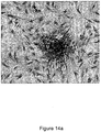
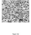
- Granuloma 26 is formed about exterior surface 28 of wall 14.
- Granuloma 26 forms with new tissue and blood vessels.
- Granuloma fluid rich in natural growth factors steadily diffuse from granuloma 26 through the plurality of holes 16 for entrapment within the enclosed chamber for harvesting and collecting, either continuously or periodically (semi-continuous).
- apparatus 10 Prior to implantation, apparatus 10 can be stored in 70% alcohol for sterility. Before implantation in the donor, apparatus 10 is washed vigorously with sterile saline and air-dried.
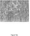
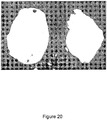
- FIG. 20 is a microphotograph showing the bladder-like tissue obtained in the rat one week after injection of 5 mL of the polydextran slurry. The bladder is spherical in shape and the fluid is trapped inside. Granulation tissue formed after one week following injection of 5 mL of polydextran slurry in the subcutaneous tissue.
- the left image shows the tissue soon after harvesting from the rat.
- the tissue is like a balloon with the injected polydextran particles and fluid (granulation fluid) enclosed in it. Granulation fluid can be separated after centrifugation and used for various medical applications.
- the image on the right shows the tissue after the balloon is perforated and the particles and fluid that oozed out have been collected.
- the tissue is abundant in stem cells and can be processed for use as single cells or for culture.
- biodegradable or non-biodegradable materials are also contemplated within the scope of the present disclosure and can also be used with similar results.
- a piece of foam placed inside the peritoneal cavity also acts as a foreign body and activates the omentum.
- the omentum expands and grows to occupy the interstices of the foam. After one week the foam can be retrieved.
- the granuloma fluid gets absorbed by the foam and can be harvested by squeezing the foam.
- Stem cells from this preparation can be obtained by enzyme treatment (collagenase) of the foam.
- apparatus 10 is surgically implanted into a donor.
- the donor is a Sprague- Dawley rat (males, approx. 300 g) anesthetized with acepromazine prior to the surgical procedure.
- the implantation location can be shaved and cleaned with alcohol and povidone.
- two 1 cm incisions can be made and using blunt dissection to form a subcutaneous pocket around the incision into which apparatus 10 is inserted.
- One or more apparatus 10 can be implanted into the donor depending on the size of the donor relative to the size of apparatus 10.
- two apparatuses 10 are implanted into a donor.
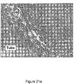
- the incision was closed with appropriate sutures (such as silk) and the donor is allowed to recover for a period (for example, one week) to form granuloma 26 around apparatus 10 ( Fig. 1b ).
- the granuloma 26 is a rapidly growing new tissue with an abundant supply of new blood vessels as seen in Figs. 21a-b .
- apparatus 10 of the present disclosure are perforated bags and sheets made of polythene or other non-toxic material may also produce an enclosed pocket (after granuloma formation) for trapping and harvesting of tissue fluid.
- a further embodiment of the present disclosure injects inert particles in the peritoneal cavity that induce a growth factor-rich fluid by the same principle.
- One method of harvesting or collecting granuloma fluid from apparatus 10 in a donor is performed aseptically by a syringe needle having an appropriate gauge (such as gauge #25) by piercing the skin and hole 16 of wall 14 of apparatus 10 and aspirating fluid from the enclosed chamber.
- the harvesting or collecting of granuloma fluid can occur at any time. For illustration purposes, a period of 4-7 days can be allowed for the formation of granuloma 26 about the exterior surface 28 of wall 14 and diffusion before the granuloma fluid accumulates in the enclosed chamber.
- Fluid yield can be increased by implanting an appropriately sized apparatus 10 in large animal that is constructed in a manner that fluid can be continuously directed and collected at a steady rate through aspiration tube 30 into a sterile bag or suitable container 32 placed outside the donor's body either by gravity (as shown in Fig. 1b ) or by mild vacuum generated by a pump (not shown) disposed in tube 30 between apparatus 10 and container 32.
- Valve 34 can be disposed in aspiration tube 30 or operably connected to container 32 to stop the continuous flow (semi-continuous flow) of granuloma fluid for replacement or emptying of the granuloma fluid from container 32.
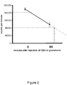
- An example of how to determine the maximum amount of granuloma fluid that can be harvested or collected is to experimentally perform clearance studies of a radioactive substance injected in the granuloma fluid. For example, two weeks after implantation of apparatus 10 (here a polythene tube), the apparatus 10 contents of fluid were aspirated completely using a syringe and the apparatus 10 was injected with 0.5 ml of sterile saline containing radioactive (125-1) sodium iodide (internal volume of the implanted tube was 0.5 ml).
- Fig. 2 shows the decrease in radioactivity in the granuloma fluid between 0 and 90 minutes. The extrapolated time it took for the radioactivity to decrease to half its earlier value was considered to be the time that 0.25 ml of fluid was made in the granuloma (half of original volume of 0.5 ml).
- the fluid harvested from the granuloma 26 resembles plasma. Electrophoresis of the fluid by SDS-PAGE (gel electrophoresis) clearly showed that the fluid was similar in composition to plasma with respect to the major proteins present in the plasma such as albumin and the globulins (electrophoretogram not presented). Growth factors present in the granuloma fluid are not expected to show up as protein bands on SDS-PAGE because their concentrations are below the sensitivity of the technique. Growth factors are usually assayed by enzyme immunoassay (ELISA) or other immune techniques.
- ELISA enzyme immunoassay
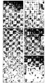
- granuloma fluid With regards to the biological properties of the granuloma fluid, especially with respect to its abundance in growth factors, four mammalian cells were tested for their growth rates in the granuloma fluid and compared with that observed with the standard FBS containing medium.
- the cells tested included two derived from the rat (glomerular epithelial cells, GEC and primary rat mesangial cells) and two from human sources (human umbilical vein endothelial cells, HUVEC and human kidney proximal tubular cells, HK).
- Fig. 3 shows that the basal medium (RPMI) by itself was poor in supporting the growth of these cells.
- the two media containing either 10% FBS or 10% granuloma fluid were better and equally able to sustain the rat glomerular epithelial cells.
- the specialized media EGM made by Biowhitaker, NJ, USA
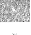
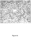
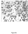
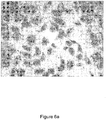
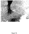
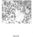
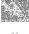
- the granuloma fluid was better in supporting the HK and primary mesangial cells ( Figs 4a-b , 5a-b , 6a-b , 7a-b ).
- the granuloma fluid better supported the primary mesangial cells, which being immediately derived from an organ are known to be more fastidious in their growth requirements than other cells.
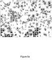
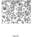
- granuloma fluid was equal to FBS in differentiating the HUVEC to form capillaries in vitro when grown on a solid matrix (Matrigel®) ( Figs. 8a-b ).
- a solid matrix Motrigel®
- FIG. 3 shows a comparison of the growth promoting effect of media containing 10% granuloma fluid with conventional media containing either 10% FBS or EGM (a specialized media made by Biowhitaker Inc. for endothelial cells) on four different rat and human cells.
- Cell number was determined by gentian violet staining and measuring the color of gentian violet in a spectrophotometer after de-staining the cells.
- the white bars (B) is a Basal medium, RPMI.
- the grey bars (F) are a Basal + 10% FBS.
- HUVEC (F) media consisted of basal EBM + unknown growth factors + 2% FBS as recommended by Biowhitaker, Inc. (the combination of the three constituents above is called EGM).
- the black bars (G) are Basal + 10% granuloma fluid.
- Figs. 4a-b show a comparison between granuloma fluid and FBS in culturing rat glomerular epithelial cells, noting that FBS and granuloma media are similar in supporting the growth of GEC (250 X).
- Figs. 5a-b are microphotographs comparing between granuloma fluid and EGM in culturing human umbilical vein endothelial cells, noting that the commercial media EGM (most optimum media as per supplier) is better than granuloma fluid containing media in supporting HUVEC (250 X).
- Figs. 6a-b are microphotographs comparing between granuloma fluid and FBS in culturing human proximal tubule cells (HK), noting that granuloma fluid media is better than FBS media in supporting HK cells (250 X).
- Figs. 7a-b are microphotographs comparing between granuloma fluid and FBS in culturing primary mesangial cells from outgrowths of rat glomeruli, noting that granuloma fluid media is better than FBS media in supporting primary mesangial cells (100 X).
- the solid red staining bodies are remnants of glomeruli.
- Figs. 8a-b are microphotographs comparing between granuloma fluid and EBM in culturing human umbilical vein endothelial cells on Matrigel matrix to induce capillary formation, noting that both granuloma fluid and EGM media (most optimum media as per supplier) equally induced capillary formation by HUVEC when grown on Matrigel (250 X).
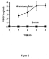
- VEGF Vascular endothelial growth factor
- VEGF in granuloma fluid is measured by enzyme immunoassay.
- Fig. 9 shows VEGF concentration in the granuloma fluid and serum at different times after the implantation of the polythene tube in the rat. The fluid could be successfully collected for up to 6 weeks after tube implantation and the VEGF concentration remained at the level seen at week 4 (data of weeks 5 and 6 are not presented).
- Each point represents the mean of four granuloma fluid or serum samples (limit bars denote standard errors) noting that granuloma fluid is 50 times higher in VEGF concentration than the serum showing the granuloma fluid is a unique fluid which is rich in growth factors.
- VEGF concentration in FBS has never been measured because there are no available assays for bovine VEGF and therefore a direct comparison with FBS is not possible.
- the omentum as well as the subcutaneous tissue activated by a foreign body as described above are rich in adult stem cells. Usually, a 3-7 day old activated tissue has a higher number of stem cells than older activated tissue.
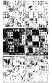
- the stem cells in the activated tissue were recognized by the presence of stem cell markers (SDF-1 ⁇ , CXCR4, WT-1, Nanog, Oct-4, PDX-1) ( Fig 13a-f ) on the cells, by fluorescence activated cells sorting analysis (FACS) showing CD90, CD59, and CD44 positivity and CD 45 negativity Table 1 (see below), suggesting similarity to mesenchymal stem cells of the bone marrow (in the rat), their ability to produce high amounts of growth factors in culture (VEGF and other growth factors) ( Fig.
- FIGs. 10a-b detection of insulin positive cells (by immunocytochemical staining) in the omenta of diabetic rats auto-transplanted with their own dispersed pancreas in the peritoneal cavity after activating the omentum by polydextran particles.
- Fig. 10a illustrates omenta from the autotransplanted diabetic rats which became normoglycemic showed niches of insulin positive cells (appearing green) in the cluster of cells surrounding the polydextran particles.
- Fig. 10b illustrates omenta from diabetic control rats who did not receive the auto-transplant were negative for insulin producing cells as were omenta from normal control rats and activated omenta alone. Tissues were counterstained with ethidium bromide to highlight cell nuclei. These results showed that diabetic pancreas when placed in activated omentum gave rise to new insulin-producing cells.
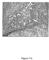
- liver mass (as a ratio to body weight) at different times after wounding and fusing of the activated omentum to the wound is shown.
- the regeneration following wounding and fusion of activated omentum was rapid as by day 3 the liver grew to 110% of original mass.
- no differences were seen at days 3, 7, 14 and 28 compared to Normal (only day 14 data is shown in the figure; n 3).
- Fig. 11b the wedge of omental tissue is shown adhered to the cut edge of the liver and in which bile ducts containing cytokeratin-19 positive oval cells (stem cells of the liver) are extended.
- the extra-hepatic proliferation of the oval cells is believed to be responsible for the liver regeneration seen in this model.
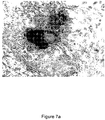
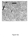
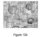
- Fig. 12a WT-1 immune stained injured kidney two weeks after injury and omentum activation shows the regenerating zone (area of fusion between omentum and kidney).
- the individual WT-1 positive cells seen at 3-7 days seemed to have organized into structures that were suggestive of embryonic bodies like renal vesicles (bottom left arrow), comma (top center arrow) and S-shaped (top left arrow) structures, and also glomerular-appearing structures with WT-1 positivity concentrated in the outer margin of the glomerular tuft in the shape of a crown (bottom center arrow), typical of early fetal glomeruli.
- a fetal rat kidney immune-stained for WT-1 is shown for comparison of embryonic structures with those found in the adult regenerating kidney.
- the cells from the activated omentum as well as the subcutaneous tissue could be cultured using Mesenchymal Stem Cell Growth Media with 10% fetal bovine serum.
- the cells could also be subcultured for several passages without loosing their stem cell characteristics (stem cell markers, secretion of growth factors etc.). Maintaining the cells in culture allows one to freeze the cells for future use.
- One example of a method to obtain stem cells from foreign body-activated adult tissues includes the following steps:
- FIGs. 13a-f microphotographs are shown of rat omentum activated with polydextran particles and immunostained for adult stem cell and embryonic pluripotent markers shown by the arrows of strong immunoreactivity for WT-1 ( Fig. 13a ), SDF-1 ⁇ ( Fig. 13b ), and CXCR 4 (adult stem cell markers) ( Fig. 13c ) and to Nanog ( Fig. 13d ), Oct-4 ( Fig. 13e ) and SSEA-1 (embryonic markers) ( Fig. 13f ).
- a primary culture of omental cells cultured from omentum tissue activated by polydextran particles for 7 days is shown. After 4-5 days in culture, cells that originally clustered around the polydextran particles started to attach to the dish and multiply (one such particle surrounded by attached cells is shown in the middle of the field). The cell morphology and phenotype was like those of smooth muscle cells and the bone-marrow derived human mesenchymal stem cells. Similar cultures were obtained from 1-day and 4-day activated omenta (not shown). Activated omentum older than 2 weeks was difficult to culture as the cells did not attach or multiply as readily as the cells from younger omenta (not shown).
- passage 3 cells show robust growth. The cells could be maintained in culture for more than 10 passages.
- Characterization of cultured stem cells from activated omentum (and also subcutaneous tissue ⁇ can be determined by 1) expression of pluripotent markers and mesenchymal stem cell surface markers ( Fig 15a-f ), 2) their ability to transform to adipogenic, chondrogenic and osteogenic cell phenotypes in vitro ( Fig 16a-f )) and 3) their ability to secrete high levels of vascular endothelial growth factor (VEGF) ( Fig. 9 ).
- pluripotent markers and mesenchymal stem cell surface markers Fig 15a-f
- Fig 16a-f their ability to transform to adipogenic, chondrogenic and osteogenic cell phenotypes in vitro
- VEGF vascular endothelial growth factor
- the stem cell markers present in the original tissue continue to be expressed in cultured cells for up to 10 passages showing that the cultured cells were as potent as the original tissue.
- fluoroscein-activated cell sorting FACS analyses for cluster of differentiation markers (CD) showed that the cells isolated from the foreign body activated adult tissues (omentum and subcutaneous) were of bone marrow mesenchymal type (CD90, 44, 59+ and CD 45-; see Table 1 below) and not hematopoietic type. Hematopoietic cells are always CD 45 positive.
- Table 1 Cells CD 44 CD45 CD 59 CD 90 Omental high+ neg high+ high+ Subcut. low+ neg low+ high++ BM low+ neg low+ high++ BM: bone marrow mesenchymal cells
- omental cells stained for adult and embryonic stem cell markers Primary cultures of omental cells stained for adult and embryonic stem cell markers. Cultured omental cells stained positive for adult stem markers WT-1 (nuclear), CXCR4 (nuclear), and SDF- 1 ⁇ (cytoplasmic) as seen in the intact tissue previously ( Figs. 15a-c ; compare with intact omentum tissue staining in Figs. 13a-c ) . Also, as seen in the intact omental tissue (see Figs. 13d-e ), the cultured cells stained strongly positive for Nanog ( Fig. 15d ; nuclear) and Oct-4 ( Fig. 15e ; cytoplasmic). Cell staining was negative in the absence of first antibody ( Fig. 15f ; control).
- FIGs. 16a-f cultured subcutaneous-tissue derived stem cells when placed in specialized medium differentiated in vitro to adipogenic, chondrogenic and osteogenic cell types.
- Adipogenic (Figs. 16a, 16d ), osteogenic (Figs. 16b, 16e ), and chondrogenic (Figs. 16c, 16f ) differentiation of subcutaneous derived stem cell cultures by incubation of cells in specialized (tissue-specific) medium.
- arrows show differentiated adipocytes, osteocytes, chondrocytes stained with oil Red O, alizarin red and alcian blue respectively containing either lipid droplets stained red ( Fig.
- FIG. 16a shows adipocytes containing lipid droplets stained red at higher magnification.
- Figs. 16d-f show control cultures grown with normal growth medium stained negative for respective markers.
- Table 2 illustrates that the cultured omental stem cells and those cells obtained from the subcutaneous granulation tissue produced up to 10-20 fold higher amounts of VEGF than commonly cultured cells from other adult tissues (glomerular epithelial cells and primary mesangial cells), which are non-stem cell lines obtained from rat kidneys.
- Table 2 Cultured cell VEGF synthesis rate (pg / hr / million cells) Omental stem cells 322 ⁇ 22
- Subcutaneous granulation tissue stem cells 230 ⁇ 15 Glomerular epithelial cells* 17.6 ⁇ 2.4
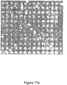
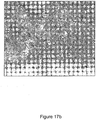
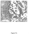
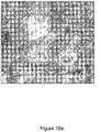
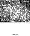
- FIGs. 17a-c , 18a-b , and 19 illustrate a test for 'stemcellness' of cultured omental stem cells by their ability to engraft to injured sites in animals.
- fluorescence labeled cultured omental (or subcutaneous) stem cells or other adult tissue cells similarly labeled
- they engrafted to injured skin, kidney and liver of rats and not to uninjured sites of the rat.
- Cells similarly prepared from normal adult tissues (non-stem cells) did not engraft to injured sites, confirming the 'stemcellness' of cells isolated from foreign body activated tissues.
- FIGs. 17a-c illustrating the migration of cultured omental cells to a skin wound healing site.
- Fig. 17a are cultured omental cells metabolically labeled with a vital fluorescent dye before injection in rats, noting a suspension of cultured omental cells (light spots) uniformly labeled with the fluorescent dye before injection.
- Figs. 17b and 17c are cryo-sections of the wound tissue 24 hours after injection of either fluorescein labeled cultured omental cells ( Fig. 17b ) or labeled adult kidney cells (control, non-stem cell) ( Fig. 17c ) in the vicinity of the granulation tissue.
- Fig. 17b shows the migration and engraftment of the injected omental stem cells in the wound tissue.
- Fig. 17c show the labeled adult kidney control cells remained at the injection site without migrating to the wound tissue.
- FIG. 18a shows an ischemic left kidney after labeled omental cells (light contrast areas illustrated by the arrows) were injected systemically in the rat with unilateral ischemic injured kidney (3 days after injury). The cells migrated to the injured tubules by 24 hours, appearing to attach to the injured tubules. Also the cells appeared to have altered from their original round shape to more elongated forms suggesting their participation in the healing process.
- Fig. 18b shows that the non-ischemic contralateral kidney did not contain fluorescent cells suggesting that omental cells specifically recognized injured sites in the body. For contrast, tissues were counterstained with ethidium bromide to stain cell nuclei red.
- FIG. 19 illustrating a liver tissue 3 days after a resection injury and 24 hours after injection of fluorescent labeled cultured omental cells showing engraftment of the injected cells (lighter contrast) to the growing liver.
- a white line drawn in the figure represents the border between the native and regenerating sections of the liver. No green fluorescent cells were visible in the native liver. The section was counterstained with ethidium bromide to highlight cell nuclei.
- One embodiment of the present disclosure is a product of the above process wherein the product is a regenerated tissue produced either in the skin or in the omentum by a foreign body, offering a source of stem cells.
- the product can be used in:
- Another embodiment of the present invention is a product of the above process wherein the product is a fluid that is harvested from the device.
- the product can be used in:
- Another product of the present invention is the granulation fluid being packaged in a powdered form.
- the advantage of formulating the fluid in the powdered form is that it is biologically stable at ambient temperature and so it does not have to be stored frozen to maintain its biological activity.
- the powdered form permits the use of the fluid in small doctor's and veterinarian's office as well in remote field situations where access to a freezer may not be available.
- the powdered form of the granulation fluid is prepared by freeze drying the fluid (process called lyophilization) in, for example, a lyophilizer, at the ratio of 1 mL granulation fluid yields approximately 50 milligrams of granulation powder (containing mostly albumin and growth factors).
- the doctor or other attending personnel will re-constitute the fluid by adding a measured amount of sterile water (water grade used will be 'water for injection' (WFI)) with the help of a syringe and a needle from a separate vial (supplied with the preparation) to the vial containing the powdered preparation, shaking the vial to re-suspend the preparation, waiting for a few minutes to allow complete dissolution and finally taking the preparation in the injection syringe for injection in the patient.
- sterile water water grade used will be 'water for injection' (WFI)
- One example of the powdered formed is used for an injection prepared for a dog knee joint.
- the veterinarian will be supplied with two vials, one containing 50 milligrams of proteins including albumin and growth factors obtained from freeze dried granulation fluid and the other containing 2 mL of WFI water.
- the veterinarian will obtain 1 mL of water from the water vial using a syringe and needle and transfer to the vial containing the powder.
- the vial will be gently shaken (to avoid frothing) and let stand for 1-2 minutes to allow the powder to be completely dissolved.
- the dissolved powder should be free of turbidity.
- the clear solution will then be taken in a 1 mL syringe and injected into the knee joint of the dog patient.
- Another example of the powdered form is for an injection prepared for a horse knee joint.
- the veterinarian will be supplied with two vials, one containing 250 milligrams of proteins including albumin and growth factors obtained from freeze dried granulation fluid and the other containing 10 mL of WFI water.
- the veterinarian will obtain 5 mL of water from the water vial using a syringe and needle and transfer to the vial containing the powder.
- the vial will be gently shaken (to avoid frothing) and let stand for 1-2 minutes to allow the powder to be completely dissolved.
- the dissolved powder should be a free of turbidity.
- the clear solution will then be taken in a 5 mL syringe and injected into the knee joint of the horse patient.
Landscapes
- Health & Medical Sciences (AREA)
- Life Sciences & Earth Sciences (AREA)
- Engineering & Computer Science (AREA)
- Pharmacology & Pharmacy (AREA)
- Veterinary Medicine (AREA)
- Public Health (AREA)
- Chemical & Material Sciences (AREA)
- General Health & Medical Sciences (AREA)
- Animal Behavior & Ethology (AREA)
- Medicinal Chemistry (AREA)
- Chemical Kinetics & Catalysis (AREA)
- Nuclear Medicine, Radiotherapy & Molecular Imaging (AREA)
- General Chemical & Material Sciences (AREA)
- Organic Chemistry (AREA)
- Bioinformatics & Cheminformatics (AREA)
- Biomedical Technology (AREA)
- Immunology (AREA)
- Neurology (AREA)
- Diabetes (AREA)
- Developmental Biology & Embryology (AREA)
- Cell Biology (AREA)
- Biotechnology (AREA)
- Virology (AREA)
- Orthopedic Medicine & Surgery (AREA)
- Epidemiology (AREA)
- Zoology (AREA)
- Dermatology (AREA)
- Neurosurgery (AREA)
- Physical Education & Sports Medicine (AREA)
- Urology & Nephrology (AREA)
- Rheumatology (AREA)
- Pain & Pain Management (AREA)
- Obesity (AREA)
- Hematology (AREA)
- Endocrinology (AREA)
- Emergency Medicine (AREA)
- Heart & Thoracic Surgery (AREA)
- Cardiology (AREA)
- Medicines Containing Material From Animals Or Micro-Organisms (AREA)
- Micro-Organisms Or Cultivation Processes Thereof (AREA)
Claims (14)
- Flüssigkeit assoziiert mit adulten Stammzellen für die Verwendung als Medikament, wobei die Flüssigkeit herstellbar ist durch:Implantieren einer nicht toxischen Vorrichtung unter die Haut oder in das Omentum eines adulten nichtmenschlichen Säugetiers, wobei die Vorrichtung eine umschlossene Kammer aufweist, die von Wänden mit Durchgangslöchern gebildet wird, so dass sich ein Granulationsgewebe um eine äußere Oberfläche der nicht toxischen Vorrichtung herum bildet und sich eine flüssige Suspension enthaltend frei schwebende Stammzellen in der umschlossenen Kammer der nicht toxischen Vorrichtung sammelt,Trennen der Flüssigkeit von der Suspension durch Entfernen der frei schwebenden Stammzellen, und Umsetzen der Flüssigkeit in eine Pulverform zur Wiederherstellung, wobei die Flüssigkeit Albumin, Globuline und mindestens das 50-fache der vaskulären endothelialen Wachstumsfaktor (VEGF)-Konzentration von normalem Serum enthält, und FBS in der Wirksamkeit zur Förderung des Wachstums von HK und primären mesangialen Zellen in Kulturmedium übertrifft.
- Flüssigkeit gemäß Anspruch 1 zur Verwendung bei der Behandlung von Arthritis.
- Flüssigkeit gemäß Anspruch 1 zur Verwendung bei der Behandlung von Krankheiten der Niere, des Herzens, des Rückenmarks oder anderen Organ- und Systemkrankheiten.
- Flüssigkeit gemäß Anspruch 1 zur Verwendung bei der Behandlung von Hautverletzungen und operativen Wunden.
- Flüssigkeit gemäß Anspruch 1 zur Verwendung bei der Behandlung von einer Krankheit oder einer Verletzung in Verbindung mit dem Kniegelenk.
- Flüssigkeit zu Verwendung gemäß einem der vorhergehenden Ansprüche, wobei die Flüssigkeit als topische Rezeptur oder als systemische Rezeptur formuliert ist.
- Flüssigkeit zur Verwendung gemäß einem der vorhergehenden Ansprüche, wobei die Flüssigkeit die für intravenöse oder subcutane Injektion formulierte Granulomflüssigkeit ist.
- Flüssigkeit zur Verwendung gemäß einem der vorhergehenden Ansprüche, wobei die Flüssigkeit von einem Schwein, einer Kuh, einem Pferd, einem Schaf, einem Hund, einer Ratte oder einem Hasen erhalten wird.
- Flüssigkeit zur Verwendung gemäß Anspruch 8, wobei die Flüssigkeit von einem Pferd erhalten wird.
- Flüssigkeit zur Verwendung gemäß einem der vorhergehenden Ansprüche, wobei die Flüssigkeit aus einer Pulverform wiederhergestellt wird.
- Flüssigkeit zur Verwendung gemäß einem der vorhergehenden Ansprüche, wobei die Flüssigkeit durch Zentrifugation isoliert wird.
- Flüssigkeit zur Verwendung gemäß einem der vorhergehenden Ansprüche, wobei die Flüssigkeit durch Wiederherstellung einer gefriergetrockneten Form erhalten wird.
- Flüssigkeit zur Verwendung gemäß einem der vorhergehenden Ansprüche, wobei die Granulomflüssigkeit zur subcutanen Injektion formuliert ist.
- Flüssigkeit zur Verwendung gemäß einem der vorhergehenden Ansprüche, wobei die Granulomflüssigkeit als topische Rezeptur formuliert ist.
Applications Claiming Priority (3)
| Application Number | Priority Date | Filing Date | Title |
|---|---|---|---|
| US27437209P | 2009-08-17 | 2009-08-17 | |
| US34215410P | 2010-04-12 | 2010-04-12 | |
| PCT/US2010/002241 WO2011022045A2 (en) | 2009-08-17 | 2010-08-13 | Apparatus and process for generating and harvesting adult stem cells and fluid associated with it from skin and omentum for medical, cosmetic, and veterinary use |
Publications (2)
| Publication Number | Publication Date |
|---|---|
| EP2467149A2 EP2467149A2 (de) | 2012-06-27 |
| EP2467149B1 true EP2467149B1 (de) | 2018-04-04 |
Family
ID=43304987
Family Applications (1)
| Application Number | Title | Priority Date | Filing Date |
|---|---|---|---|
| EP10751726.0A Not-in-force EP2467149B1 (de) | 2009-08-17 | 2010-08-13 | Flüssigkeit aus haut und omentum zur medizinischen verwendung |
Country Status (5)
| Country | Link |
|---|---|
| US (2) | US9173903B2 (de) |
| EP (1) | EP2467149B1 (de) |
| AU (1) | AU2010284704B2 (de) |
| CA (1) | CA2771261A1 (de) |
| WO (1) | WO2011022045A2 (de) |
Families Citing this family (9)
| Publication number | Priority date | Publication date | Assignee | Title |
|---|---|---|---|---|
| US9352003B1 (en) | 2010-05-14 | 2016-05-31 | Musculoskeletal Transplant Foundation | Tissue-derived tissuegenic implants, and methods of fabricating and using same |
| US10130736B1 (en) | 2010-05-14 | 2018-11-20 | Musculoskeletal Transplant Foundation | Tissue-derived tissuegenic implants, and methods of fabricating and using same |
| US8883210B1 (en) | 2010-05-14 | 2014-11-11 | Musculoskeletal Transplant Foundation | Tissue-derived tissuegenic implants, and methods of fabricating and using same |
| US20150037436A1 (en) | 2013-07-30 | 2015-02-05 | Musculoskeletal Transplant Foundation | Acellular soft tissue-derived matrices and methods for preparing same |
| US20170021149A9 (en) * | 2014-05-02 | 2017-01-26 | National Institute Of Standards And Technology | Biological sampling platform and processes for making and using same |
| US10918669B2 (en) | 2014-07-25 | 2021-02-16 | Recellerate, Inc. | Methods of treating exercise-induced pulmonary hemorrhage |
| CA2986702C (en) | 2015-05-21 | 2023-04-04 | David Wang | Modified demineralized cortical bone fibers |
| US10912864B2 (en) | 2015-07-24 | 2021-02-09 | Musculoskeletal Transplant Foundation | Acellular soft tissue-derived matrices and methods for preparing same |
| US11052175B2 (en) | 2015-08-19 | 2021-07-06 | Musculoskeletal Transplant Foundation | Cartilage-derived implants and methods of making and using same |
Family Cites Families (23)
| Publication number | Priority date | Publication date | Assignee | Title |
|---|---|---|---|---|
| US4520107A (en) * | 1982-09-28 | 1985-05-28 | Polydex Chemicals Ltd. | Tissue culture and cell growth-promoting material and its method of manufacture |
| WO1992015670A1 (en) * | 1991-02-28 | 1992-09-17 | Hyclone Laboratories, Inc. | Blended bovine sera cellular growth media and their methods of production and use |
| US5426045A (en) * | 1993-12-16 | 1995-06-20 | Sea Run Holdings, Inc. | Method for culturing mammalian cells in a medium containing fish serum |
| EP0759070A4 (de) * | 1994-03-16 | 1998-03-11 | Picower Inst Med Res | Mesenchymzellen aus blut |
| US5695998A (en) * | 1995-02-10 | 1997-12-09 | Purdue Research Foundation | Submucosa as a growth substrate for islet cells |
| US6043092A (en) * | 1996-03-18 | 2000-03-28 | University Of Pittsburgh | Cell culture media for mammalian cells |
| WO1999043786A2 (en) * | 1998-02-27 | 1999-09-02 | Purdue Research Foundation | Submucosa gel compositions |
| US20050153442A1 (en) * | 1999-03-10 | 2005-07-14 | Adam Katz | Adipose-derived stem cells and lattices |
| US20050076396A1 (en) * | 1999-03-10 | 2005-04-07 | Katz Adam J. | Adipose-derived stem cells and lattices |
| US20030082152A1 (en) * | 1999-03-10 | 2003-05-01 | Hedrick Marc H. | Adipose-derived stem cells and lattices |
| CA2365434A1 (en) * | 1999-04-15 | 2000-10-26 | St. Elizabeth's Medical Center Of Boston | Angiogenic growth factors for treatment of peripheral neuropathy |
| US7078232B2 (en) * | 1999-08-19 | 2006-07-18 | Artecel, Inc. | Adipose tissue-derived adult stem or stromal cells for the repair of articular cartilage fractures and uses thereof |
| US6429013B1 (en) | 1999-08-19 | 2002-08-06 | Artecel Science, Inc. | Use of adipose tissue-derived stromal cells for chondrocyte differentiation and cartilage repair |
| US20030161817A1 (en) * | 2001-03-28 | 2003-08-28 | Young Henry E. | Pluripotent embryonic-like stem cells, compositions, methods and uses thereof |
| US6562621B1 (en) * | 1999-10-28 | 2003-05-13 | Sea Run Holdings, Inc. | Method of using fish ovarian fluid for culture and preservation of mammalian cells |
| KR100394430B1 (ko) * | 1999-12-14 | 2003-08-09 | (주)이노테크 메디칼 | 인간 혈청을 포함하는 인간 세포 배양용 배지 및 이를이용한 인간 세포의 배양 방법 |
| EP1261694B1 (de) * | 2000-02-26 | 2008-01-16 | Artecel, Inc. | Pluripotente stammzellen erzeugt aus stromalzellen aus dem fettgewebe und deren verwendungen |
| US6459917B1 (en) * | 2000-05-22 | 2002-10-01 | Ashok Gowda | Apparatus for access to interstitial fluid, blood, or blood plasma components |
| US6599740B2 (en) * | 2000-12-15 | 2003-07-29 | Sea Run Holdings, Inc. | Method of using fish plasma components for tissue culture |
| US20070141101A1 (en) * | 2003-04-10 | 2007-06-21 | The Trustees Of Boston University | Method for stimulating angiogenesis and wound healing |
| US20050013870A1 (en) * | 2003-07-17 | 2005-01-20 | Toby Freyman | Decellularized extracellular matrix of conditioned body tissues and uses thereof |
| US7785582B2 (en) * | 2004-09-07 | 2010-08-31 | Johnson Lanny L | Use of synovium and omentum for tissue engineering |
| WO2009089110A2 (en) * | 2008-01-04 | 2009-07-16 | Boston Scientific Scimed, Inc. | Methods for repair and regeneration of bone marrow |
-
2010
- 2010-08-13 WO PCT/US2010/002241 patent/WO2011022045A2/en not_active Ceased
- 2010-08-13 US US12/855,760 patent/US9173903B2/en active Active
- 2010-08-13 EP EP10751726.0A patent/EP2467149B1/de not_active Not-in-force
- 2010-08-13 CA CA2771261A patent/CA2771261A1/en active Pending
- 2010-08-13 AU AU2010284704A patent/AU2010284704B2/en not_active Ceased
-
2015
- 2015-08-18 US US14/829,301 patent/US20150352152A1/en not_active Abandoned
Non-Patent Citations (2)
| Title |
|---|
| NATALIA O LITBARG ET AL: "Activated omentum becomes rich in factors that promote healing and tissue regeneration", CELL AND TISSUE RESEARCH, SPRINGER, BERLIN, DE, vol. 328, no. 3, 14 February 2007 (2007-02-14), pages 487 - 497, XP019517927, ISSN: 1432-0878, DOI: 10.1007/S00441-006-0356-4 * |
| PATEL J ET AL: "Foreign body-induced granulation tissue is a source of adult stem cells", TRANSLATIONAL RESEARCH, ELSEVIER, AMSTERDAM, NL, vol. 155, no. 4, 1 April 2010 (2010-04-01), pages 191 - 199, XP026975237, ISSN: 1931-5244, [retrieved on 20090922] * |
Also Published As
| Publication number | Publication date |
|---|---|
| WO2011022045A2 (en) | 2011-02-24 |
| US20150352152A1 (en) | 2015-12-10 |
| WO2011022045A3 (en) | 2011-04-14 |
| US9173903B2 (en) | 2015-11-03 |
| US20110038903A1 (en) | 2011-02-17 |
| AU2010284704A1 (en) | 2012-02-16 |
| EP2467149A2 (de) | 2012-06-27 |
| AU2010284704B2 (en) | 2015-07-16 |
| CA2771261A1 (en) | 2011-02-24 |
Similar Documents
| Publication | Publication Date | Title |
|---|---|---|
| EP2467149B1 (de) | Flüssigkeit aus haut und omentum zur medizinischen verwendung | |
| EP2316463B1 (de) | Gewebematrizen umfassend plazentare Stammzellen und Verfahren zu deren Herstellung | |
| WO2019161591A1 (zh) | 间充质干细胞的分离培养方法及冻存、复苏方法 | |
| WO2014053418A9 (en) | Method for obtaining mesenchymal stem cells and use thereof | |
| CN108685948A (zh) | 一种新型医用细胞修复剂的制备方法 | |
| CN109893541A (zh) | 经血干细胞来源的外泌体在制备治疗宫腔粘连的药物中的应用 | |
| US12234478B2 (en) | Method of autologous primary hair follicles preparation in 3D culture | |
| US9434923B2 (en) | Preparation of parental cell bank from foetal tissue | |
| US11760976B2 (en) | Stem cells and decellularization of tissue matrix from cord tissue | |
| CN111172106B (zh) | 一种从人胎盘或脐带中提取间充质干细胞及提取其分泌物的方法 | |
| AU2015215979B2 (en) | Apparatus and process for generating and harvesting adult stem cells and fluid associated with it from skin and omentum for medical, cosmetic, and veterinary use | |
| US20160287749A1 (en) | Reconstituted amniotic membrane-amniotic fluid combination tissue graft | |
| US20160287752A1 (en) | Reconstituted amniotic membrane-amniotic fluid combination tissue graft with standardized stem cell component and method of forming same | |
| Zhang et al. | In vivo osteo-organoid approach for harvesting therapeutic hematopoietic stem/progenitor cells | |
| US9670457B2 (en) | Stem cells and matrix from cord tissue | |
| CN110237026A (zh) | 一种透明质酸钠复合脂肪干细胞条件培养基冻干制剂 | |
| US20240026299A1 (en) | Stem cell and tissue cultures and their products | |
| JP7672647B2 (ja) | 間葉系幹細胞を含む細胞集団を含有する軟部組織再生用医薬組成物 | |
| US20160287640A1 (en) | Denuded amnion flowable tissue graft and method of forming same | |
| CN100389194C (zh) | 胰岛素分泌细胞提取物及其在干细胞诱导分化中的应用 | |
| RU2695364C1 (ru) | Способ лечения нерубцовой алопеции | |
| CA3205553A1 (en) | Methods for optimizing reproductive tissue derived cell yield and viability for clinical applications | |
| Li et al. | The Efficacy and Mechanism of Autologous Fat Stem Cells Combined with Microcarrier 6 Transplantation for Anal Fistula Treatment | |
| CN112843338A (zh) | 一种制备含胶原蛋白的间充质干细胞注射剂的方法 | |
| CN118995595A (zh) | 一种间充质干细胞的培养方法 |
Legal Events
| Date | Code | Title | Description |
|---|---|---|---|
| PUAI | Public reference made under article 153(3) epc to a published international application that has entered the european phase |
Free format text: ORIGINAL CODE: 0009012 |
|
| 17P | Request for examination filed |
Effective date: 20120316 |
|
| AK | Designated contracting states |
Kind code of ref document: A2 Designated state(s): AL AT BE BG CH CY CZ DE DK EE ES FI FR GB GR HR HU IE IS IT LI LT LU LV MC MK MT NL NO PL PT RO SE SI SK SM TR |
|
| DAX | Request for extension of the european patent (deleted) | ||
| 17Q | First examination report despatched |
Effective date: 20140805 |
|
| GRAP | Despatch of communication of intention to grant a patent |
Free format text: ORIGINAL CODE: EPIDOSNIGR1 |
|
| RIC1 | Information provided on ipc code assigned before grant |
Ipc: A61K 35/24 20150101AFI20170927BHEP Ipc: A61P 13/00 20060101ALI20170927BHEP Ipc: A61P 9/00 20060101ALI20170927BHEP Ipc: A61P 19/00 20060101ALI20170927BHEP Ipc: A61P 17/00 20060101ALI20170927BHEP Ipc: A61P 25/00 20060101ALI20170927BHEP |
|
| INTG | Intention to grant announced |
Effective date: 20171027 |
|
| GRAS | Grant fee paid |
Free format text: ORIGINAL CODE: EPIDOSNIGR3 |
|
| GRAA | (expected) grant |
Free format text: ORIGINAL CODE: 0009210 |
|
| AK | Designated contracting states |
Kind code of ref document: B1 Designated state(s): AL AT BE BG CH CY CZ DE DK EE ES FI FR GB GR HR HU IE IS IT LI LT LU LV MC MK MT NL NO PL PT RO SE SI SK SM TR |
|
| REG | Reference to a national code |
Ref country code: GB Ref legal event code: FG4D |
|
| REG | Reference to a national code |
Ref country code: CH Ref legal event code: EP |
|
| REG | Reference to a national code |
Ref country code: AT Ref legal event code: REF Ref document number: 984845 Country of ref document: AT Kind code of ref document: T Effective date: 20180415 |
|
| REG | Reference to a national code |
Ref country code: IE Ref legal event code: FG4D |
|
| REG | Reference to a national code |
Ref country code: DE Ref legal event code: R096 Ref document number: 602010049673 Country of ref document: DE |
|
| REG | Reference to a national code |
Ref country code: NL Ref legal event code: MP Effective date: 20180404 |
|
| REG | Reference to a national code |
Ref country code: LT Ref legal event code: MG4D |
|
| PG25 | Lapsed in a contracting state [announced via postgrant information from national office to epo] |
Ref country code: NL Free format text: LAPSE BECAUSE OF FAILURE TO SUBMIT A TRANSLATION OF THE DESCRIPTION OR TO PAY THE FEE WITHIN THE PRESCRIBED TIME-LIMIT Effective date: 20180404 |
|
| PG25 | Lapsed in a contracting state [announced via postgrant information from national office to epo] |
Ref country code: AL Free format text: LAPSE BECAUSE OF FAILURE TO SUBMIT A TRANSLATION OF THE DESCRIPTION OR TO PAY THE FEE WITHIN THE PRESCRIBED TIME-LIMIT Effective date: 20180404 Ref country code: SE Free format text: LAPSE BECAUSE OF FAILURE TO SUBMIT A TRANSLATION OF THE DESCRIPTION OR TO PAY THE FEE WITHIN THE PRESCRIBED TIME-LIMIT Effective date: 20180404 Ref country code: BG Free format text: LAPSE BECAUSE OF FAILURE TO SUBMIT A TRANSLATION OF THE DESCRIPTION OR TO PAY THE FEE WITHIN THE PRESCRIBED TIME-LIMIT Effective date: 20180704 Ref country code: FI Free format text: LAPSE BECAUSE OF FAILURE TO SUBMIT A TRANSLATION OF THE DESCRIPTION OR TO PAY THE FEE WITHIN THE PRESCRIBED TIME-LIMIT Effective date: 20180404 Ref country code: NO Free format text: LAPSE BECAUSE OF FAILURE TO SUBMIT A TRANSLATION OF THE DESCRIPTION OR TO PAY THE FEE WITHIN THE PRESCRIBED TIME-LIMIT Effective date: 20180704 Ref country code: PL Free format text: LAPSE BECAUSE OF FAILURE TO SUBMIT A TRANSLATION OF THE DESCRIPTION OR TO PAY THE FEE WITHIN THE PRESCRIBED TIME-LIMIT Effective date: 20180404 Ref country code: LT Free format text: LAPSE BECAUSE OF FAILURE TO SUBMIT A TRANSLATION OF THE DESCRIPTION OR TO PAY THE FEE WITHIN THE PRESCRIBED TIME-LIMIT Effective date: 20180404 Ref country code: ES Free format text: LAPSE BECAUSE OF FAILURE TO SUBMIT A TRANSLATION OF THE DESCRIPTION OR TO PAY THE FEE WITHIN THE PRESCRIBED TIME-LIMIT Effective date: 20180404 |
|
| PG25 | Lapsed in a contracting state [announced via postgrant information from national office to epo] |
Ref country code: GR Free format text: LAPSE BECAUSE OF FAILURE TO SUBMIT A TRANSLATION OF THE DESCRIPTION OR TO PAY THE FEE WITHIN THE PRESCRIBED TIME-LIMIT Effective date: 20180705 Ref country code: HR Free format text: LAPSE BECAUSE OF FAILURE TO SUBMIT A TRANSLATION OF THE DESCRIPTION OR TO PAY THE FEE WITHIN THE PRESCRIBED TIME-LIMIT Effective date: 20180404 Ref country code: LV Free format text: LAPSE BECAUSE OF FAILURE TO SUBMIT A TRANSLATION OF THE DESCRIPTION OR TO PAY THE FEE WITHIN THE PRESCRIBED TIME-LIMIT Effective date: 20180404 |
|
| REG | Reference to a national code |
Ref country code: AT Ref legal event code: MK05 Ref document number: 984845 Country of ref document: AT Kind code of ref document: T Effective date: 20180404 |
|
| PG25 | Lapsed in a contracting state [announced via postgrant information from national office to epo] |
Ref country code: PT Free format text: LAPSE BECAUSE OF FAILURE TO SUBMIT A TRANSLATION OF THE DESCRIPTION OR TO PAY THE FEE WITHIN THE PRESCRIBED TIME-LIMIT Effective date: 20180806 |
|
| REG | Reference to a national code |
Ref country code: DE Ref legal event code: R097 Ref document number: 602010049673 Country of ref document: DE |
|
| PG25 | Lapsed in a contracting state [announced via postgrant information from national office to epo] |
Ref country code: RO Free format text: LAPSE BECAUSE OF FAILURE TO SUBMIT A TRANSLATION OF THE DESCRIPTION OR TO PAY THE FEE WITHIN THE PRESCRIBED TIME-LIMIT Effective date: 20180404 Ref country code: CZ Free format text: LAPSE BECAUSE OF FAILURE TO SUBMIT A TRANSLATION OF THE DESCRIPTION OR TO PAY THE FEE WITHIN THE PRESCRIBED TIME-LIMIT Effective date: 20180404 Ref country code: AT Free format text: LAPSE BECAUSE OF FAILURE TO SUBMIT A TRANSLATION OF THE DESCRIPTION OR TO PAY THE FEE WITHIN THE PRESCRIBED TIME-LIMIT Effective date: 20180404 Ref country code: EE Free format text: LAPSE BECAUSE OF FAILURE TO SUBMIT A TRANSLATION OF THE DESCRIPTION OR TO PAY THE FEE WITHIN THE PRESCRIBED TIME-LIMIT Effective date: 20180404 Ref country code: SK Free format text: LAPSE BECAUSE OF FAILURE TO SUBMIT A TRANSLATION OF THE DESCRIPTION OR TO PAY THE FEE WITHIN THE PRESCRIBED TIME-LIMIT Effective date: 20180404 Ref country code: DK Free format text: LAPSE BECAUSE OF FAILURE TO SUBMIT A TRANSLATION OF THE DESCRIPTION OR TO PAY THE FEE WITHIN THE PRESCRIBED TIME-LIMIT Effective date: 20180404 |
|
| PLBE | No opposition filed within time limit |
Free format text: ORIGINAL CODE: 0009261 |
|
| STAA | Information on the status of an ep patent application or granted ep patent |
Free format text: STATUS: NO OPPOSITION FILED WITHIN TIME LIMIT |
|
| PG25 | Lapsed in a contracting state [announced via postgrant information from national office to epo] |
Ref country code: IT Free format text: LAPSE BECAUSE OF FAILURE TO SUBMIT A TRANSLATION OF THE DESCRIPTION OR TO PAY THE FEE WITHIN THE PRESCRIBED TIME-LIMIT Effective date: 20180404 Ref country code: SM Free format text: LAPSE BECAUSE OF FAILURE TO SUBMIT A TRANSLATION OF THE DESCRIPTION OR TO PAY THE FEE WITHIN THE PRESCRIBED TIME-LIMIT Effective date: 20180404 |
|
| REG | Reference to a national code |
Ref country code: DE Ref legal event code: R119 Ref document number: 602010049673 Country of ref document: DE |
|
| 26N | No opposition filed |
Effective date: 20190107 |
|
| PG25 | Lapsed in a contracting state [announced via postgrant information from national office to epo] |
Ref country code: MC Free format text: LAPSE BECAUSE OF FAILURE TO SUBMIT A TRANSLATION OF THE DESCRIPTION OR TO PAY THE FEE WITHIN THE PRESCRIBED TIME-LIMIT Effective date: 20180404 |
|
| REG | Reference to a national code |
Ref country code: CH Ref legal event code: PL |
|
| GBPC | Gb: european patent ceased through non-payment of renewal fee |
Effective date: 20180813 |
|
| PG25 | Lapsed in a contracting state [announced via postgrant information from national office to epo] |
Ref country code: LU Free format text: LAPSE BECAUSE OF NON-PAYMENT OF DUE FEES Effective date: 20180813 Ref country code: LI Free format text: LAPSE BECAUSE OF NON-PAYMENT OF DUE FEES Effective date: 20180831 Ref country code: CH Free format text: LAPSE BECAUSE OF NON-PAYMENT OF DUE FEES Effective date: 20180831 |
|
| REG | Reference to a national code |
Ref country code: BE Ref legal event code: MM Effective date: 20180831 |
|
| REG | Reference to a national code |
Ref country code: IE Ref legal event code: MM4A |
|
| PG25 | Lapsed in a contracting state [announced via postgrant information from national office to epo] |
Ref country code: SI Free format text: LAPSE BECAUSE OF FAILURE TO SUBMIT A TRANSLATION OF THE DESCRIPTION OR TO PAY THE FEE WITHIN THE PRESCRIBED TIME-LIMIT Effective date: 20180404 |
|
| PG25 | Lapsed in a contracting state [announced via postgrant information from national office to epo] |
Ref country code: DE Free format text: LAPSE BECAUSE OF NON-PAYMENT OF DUE FEES Effective date: 20190301 Ref country code: IE Free format text: LAPSE BECAUSE OF NON-PAYMENT OF DUE FEES Effective date: 20180813 |
|
| PG25 | Lapsed in a contracting state [announced via postgrant information from national office to epo] |
Ref country code: BE Free format text: LAPSE BECAUSE OF NON-PAYMENT OF DUE FEES Effective date: 20180831 Ref country code: FR Free format text: LAPSE BECAUSE OF NON-PAYMENT OF DUE FEES Effective date: 20180831 |
|
| PG25 | Lapsed in a contracting state [announced via postgrant information from national office to epo] |
Ref country code: GB Free format text: LAPSE BECAUSE OF NON-PAYMENT OF DUE FEES Effective date: 20180813 |
|
| PG25 | Lapsed in a contracting state [announced via postgrant information from national office to epo] |
Ref country code: MT Free format text: LAPSE BECAUSE OF NON-PAYMENT OF DUE FEES Effective date: 20180813 |
|
| PG25 | Lapsed in a contracting state [announced via postgrant information from national office to epo] |
Ref country code: TR Free format text: LAPSE BECAUSE OF FAILURE TO SUBMIT A TRANSLATION OF THE DESCRIPTION OR TO PAY THE FEE WITHIN THE PRESCRIBED TIME-LIMIT Effective date: 20180404 |
|
| PG25 | Lapsed in a contracting state [announced via postgrant information from national office to epo] |
Ref country code: HU Free format text: LAPSE BECAUSE OF FAILURE TO SUBMIT A TRANSLATION OF THE DESCRIPTION OR TO PAY THE FEE WITHIN THE PRESCRIBED TIME-LIMIT; INVALID AB INITIO Effective date: 20100813 |
|
| PG25 | Lapsed in a contracting state [announced via postgrant information from national office to epo] |
Ref country code: CY Free format text: LAPSE BECAUSE OF FAILURE TO SUBMIT A TRANSLATION OF THE DESCRIPTION OR TO PAY THE FEE WITHIN THE PRESCRIBED TIME-LIMIT Effective date: 20180404 Ref country code: MK Free format text: LAPSE BECAUSE OF NON-PAYMENT OF DUE FEES Effective date: 20180404 |
|
| PG25 | Lapsed in a contracting state [announced via postgrant information from national office to epo] |
Ref country code: IS Free format text: LAPSE BECAUSE OF FAILURE TO SUBMIT A TRANSLATION OF THE DESCRIPTION OR TO PAY THE FEE WITHIN THE PRESCRIBED TIME-LIMIT Effective date: 20180804 |