EP2464743B1 - Method for detecting chromosomal aneuploidy - Google Patents
Method for detecting chromosomal aneuploidy Download PDFInfo
- Publication number
- EP2464743B1 EP2464743B1 EP20100733024 EP10733024A EP2464743B1 EP 2464743 B1 EP2464743 B1 EP 2464743B1 EP 20100733024 EP20100733024 EP 20100733024 EP 10733024 A EP10733024 A EP 10733024A EP 2464743 B1 EP2464743 B1 EP 2464743B1
- Authority
- EP
- European Patent Office
- Prior art keywords
- dna
- marker
- chromosome
- methylation
- fetal
- Prior art date
- Legal status (The legal status is an assumption and is not a legal conclusion. Google has not performed a legal analysis and makes no representation as to the accuracy of the status listed.)
- Active
Links
Images


Classifications
-
- C—CHEMISTRY; METALLURGY
- C12—BIOCHEMISTRY; BEER; SPIRITS; WINE; VINEGAR; MICROBIOLOGY; ENZYMOLOGY; MUTATION OR GENETIC ENGINEERING
- C12Q—MEASURING OR TESTING PROCESSES INVOLVING ENZYMES, NUCLEIC ACIDS OR MICROORGANISMS; COMPOSITIONS OR TEST PAPERS THEREFOR; PROCESSES OF PREPARING SUCH COMPOSITIONS; CONDITION-RESPONSIVE CONTROL IN MICROBIOLOGICAL OR ENZYMOLOGICAL PROCESSES
- C12Q1/00—Measuring or testing processes involving enzymes, nucleic acids or microorganisms; Compositions therefor; Processes of preparing such compositions
- C12Q1/68—Measuring or testing processes involving enzymes, nucleic acids or microorganisms; Compositions therefor; Processes of preparing such compositions involving nucleic acids
- C12Q1/6813—Hybridisation assays
- C12Q1/6827—Hybridisation assays for detection of mutation or polymorphism
-
- Y—GENERAL TAGGING OF NEW TECHNOLOGICAL DEVELOPMENTS; GENERAL TAGGING OF CROSS-SECTIONAL TECHNOLOGIES SPANNING OVER SEVERAL SECTIONS OF THE IPC; TECHNICAL SUBJECTS COVERED BY FORMER USPC CROSS-REFERENCE ART COLLECTIONS [XRACs] AND DIGESTS
- Y10—TECHNICAL SUBJECTS COVERED BY FORMER USPC
- Y10T—TECHNICAL SUBJECTS COVERED BY FORMER US CLASSIFICATION
- Y10T436/00—Chemistry: analytical and immunological testing
- Y10T436/14—Heterocyclic carbon compound [i.e., O, S, N, Se, Te, as only ring hetero atom]
- Y10T436/142222—Hetero-O [e.g., ascorbic acid, etc.]
- Y10T436/143333—Saccharide [e.g., DNA, etc.]
Definitions
- Various genetic disorders such as chromosomal aneuploidies
- chromosomal aneuploidies are detectable during pregnancy according to a variety of methods currently in use by hospitals and medical professionals. Most of these methods, however, are invasive and carry a risk of unintended loss of fetus or miscarriage.
- WO2007/132167 describes a method for detecting a pregnancy-associated disorder (including various fetal chromosomal aneuploidies) that requires the user to measure the level of a methylation marker (selected from a list of specific markers) in a biological sample taken from the pregnant woman and then compare the level directly with a standard control to determine whether any pregnancy-associated disorder is present.
- WO2005/035725 describes a method for diagnosing fetal aneuploidy by determining, in a maternal plasma sample, the paternal or maternal allele frequency in the fetal DNA.
- This method requires the DNA of the target analysis region located on the chromosome relevant to the aneuploidy to meet two criteria: first, there must be at least one polymorphic marker within this region. The second criterion is that there must be a significant difference in the methylation status of this region between the maternal and fetal DNA.
- Noninvasive prenatal diagnosis of chromosomal aneuploidies using fetal DNA in maternal plasma is an actively researched area, and there exists a clear need for new and more reliable methods for early detection.
- This invention provides a novel method for detecting chromosomal aneuploidies such as trisomy 21 by the combination of fetal-specific epigenetic and genetic markers.
- the present inventors searched for fetal DNA markers on chromosomes 21, 18, and 13 that were differentially methylated in the placenta and maternal blood cells using methods including combined bisulfite restriction analysis and immunoprecipitation of methylated DNA followed by tiling array hybridization (MeDIP-chip), with confirmation of any target locus with bisulfite sequencing.
- the resultant markers were analyzed using methylation-sensitive restriction endonuclease digestion followed by real-time polymerase chain reaction (PCR) or microfluidics digital PCR analysis.
- Chromosome dosage analysis was performed by comparing the dosage of these epigenetic markers to a genetic marker, a DNA sequence that is derived from a fetus and can be distinguished from a maternal DNA sequence by virtue of its distinct polynucleotide sequence.
- a genetic marker a DNA sequence that is derived from a fetus and can be distinguished from a maternal DNA sequence by virtue of its distinct polynucleotide sequence.
- ZFY zinc finger protein
- HLCS holocarboxylase synthetase
- This invention provides a method for detecting a chromosomal aneuploidy in a fetus carried by a pregnant woman.
- the method comprises the steps of: (a) determining in a biological sample taken from the pregnant woman the amount of a methylation marker of fetal origin, wherein the methylation marker is located on a chromosome relevant to the chromosomal aneuploidy or within a section of a chromosome relevant to the chromosomal aneuploidy, and wherein the methylation marker of fetal origin is distinguished from its counterpart of maternal origin due to differential DNA methylation; (b) determining the amount of a genetic marker of fetal origin in the sample, wherein the genetic marker is located on a reference chromosome, and wherein the genetic marker of fetal origin is distinguished from its counterpart of maternal origin in the sample due to difference in polynucleotide sequence, or the genetic marker does not exist in the maternal genome; (c) determining the ratio of the amounts from
- the standard control value approximates the expected gene or chromosome dosage or ratio in the human genome, although slight variations may exist depending on the specific methodology used in the detection method. For example, there are two copies of chromosome 21 and one copy of chromosome Y in a euploid male genome. Hence, the ratio of dosage between an epigenetic marker on chromosome 21 to a genetic marker on chromosome Y would be approximately 2.
- the methylation marker of fetal origin is from the placenta.
- the counterpart of maternal origin is from the pregnant woman's blood cells.
- the methylation marker of fetal origin may be more methylated than its counterpart of maternal origin, or it may be less methylated than its counterpart of maternal origin.
- the sample is maternal whole blood, serum, plasma, placental-tissue sample, or chorionic villus sample. In other cases, the sample is any sample that contains fetal nucleic acids.
- the methylation marker is part of, or in proximity to, the Holocarboxylase Synthetase (HLCS) gene.
- HLCS Holocarboxylase Synthetase
- several regions of the HLCS gene including the putative promoter region) as shown in Table 2 may be used as methylation markers.
- Other methylation markers include Marker 18A and Marker 13A.
- Additional methylation markers are defined as follows: a region of about 15 to 450 nucleotides and comprises one cytosine, the region being (1) a genomic locus selected from the group consisting of MAT.18.0094, MAT.13.0023, MAT.13.0020, MAT.13.0038, MAT.18.0071, MAT.18.0097, MAT.21.0178, and TAS.21.1175 ; or (2) no more than 10 kb upstream and/or downstream from the locus.
- markers on chromosome 21 are defined as follows: a region of about 15 to 450 nucleotides on chromosome 21 and comprises one cytosine, the region being (1) a genomic locus selected from the group consisting of CGI137 , phosphodiesterase 9A (PDE9A), Homo sapiens protein phosphatase 1, regulatory (inhibitory) subunit 2 pseudogene 2 ( PPP1R2P2 ), and Similarity to Fem1A ( Caenorhabditis elegans), or (2) no more than 10 kb upstream and/or downstream from the locus.
- PDE9A phosphodiesterase 9A
- PPP1R2P2 Homo sapiens protein phosphatase 1, regulatory (inhibitory) subunit 2 pseudogene 2
- Similarity to Fem1A Caenorhabditis elegans
- step (a) comprises treating the sample with a reagent that differentially modifies methylated and unmethylated DNA.
- a reagent that differentially modifies methylated and unmethylated DNA.
- Such reagent may comprise bisulfite or a protein or chemical that binds to DNA based on methylation status; or the reagent may comprise an enzyme that either preferentially cleaves methylated DNA or preferentially cleaves unmethylated DNA.
- the genetic marker does not exist in the maternal genome. In other cases, the genetic marker is located on the Y chromosome. Frequently, the genetic marker of fetal origin is distinguished from the genetic marker of maternal origin based on genetic polymorphism, which includes a single nucleotide polymorphism (SNP), simple tandem repeat polymorphism, and insertion-deletion polymorphism.
- the genetic marker is the Zinc-Finger protein, Y-linked (ZFY) gene.
- the genetic marker is a genomic sequence comprising SNP rs6636 in TMED8 gene, e.g., SEQ ID NO:1 or SEQ ID NO:2.
- step (a) or step (b) may include the process of amplification of the methylation marker and/or the genetic marker, especially the methylation marker and the genetic marker of the fetal origin.
- the amplification is by a polymerase chain reaction (PCR), such as a methylation-specific PCR; or the amplification may be a nucleic acid sequence-specific amplification.
- PCR polymerase chain reaction
- step (a) or (b) is performed by molecular counting.
- step (a) or (b) comprises digital polymerase chain reaction, real-time quantitative polymerase chain reaction, array capture, a nucleic acid sequence-based detection, massively parallel genomic sequencing, single molecule sequencing, or multiplexed detection of polynucleotide with color-coded probe pairs.
- step (a) or (b) comprises mass spectrometry or hybridization to a microarray, fluorescence probe, or molecular beacon.
- the chromosome relevant to the chromosomal aneuploidy is chromosome 13, 18, 21, or X.
- the ratio is higher or lower than the standard control by at least 1 standard deviation, it indicates the presence of the chromosomal aneuploidy in the fetus, whereas in other cases, the presence of the chromosomal aneuploidy in the fetus is indicated when the ratio is higher or lower than the standard control by at least 2 or even 3 standard deviations.
- the above described methods can be used in a non-diagnostic context for assessing any potential change in the amount of a particular chromosome of interest in relation to another chromosome, a reference chromosome that is not suspected to have any change in its relative amount.
- the present invention can also be embodied in a device or a system comprising one or more such devices, which is capable of carrying out all or some of the method steps described herein.
- the device or system performs the following steps upon receiving a biological sample taken from the pregnant woman: (a) determining in sample the amount of a fetus derived methylation marker relevant to a chromosomal aneuploidy; (b) determining in the sample the amount of a fetus derived genetic marker on a reference chromosome; (c) determining the ratio of the amounts from (a) and (b); and (d) comparing the ratio with a standard control and providing an output indicating whether the chromosomal aneuploidy is present in the fetus.
- the device or system of the invention performs the task of steps (c) and (d), after steps (a) and (b) have been performed and the amounts from (a) and (b) have been entered into the device.
- the device or system is partially or fully automated.
- pregnancy-associated disorder refers to any condition or disease that may affect a pregnant woman, the fetus the woman is carrying, or both the woman and the fetus. Such a condition or disease may manifest its symptoms during a limited time period, e.g ., during pregnancy or delivery, or may last the entire life span of the fetus following its birth.
- a "pregnancy-associated disorder” is necessarily accompanied by pregnancy and is not merely a condition that is incidentally occurring to a pregnant woman. In other words, the disorder is not one that can occur to a non-pregnant woman.
- Some examples of a pregnancy-associated disorder include ectopic pregnancy, preeclampsia, preterm labor, and fetal chromosomal abnormalities such as trisomy 13, 18, or 21.
- chromosomal aneuploidy encompasses any genetic defect exhibiting an abnormal number of chromosomes, including having more or fewer than normal number of any one chromosome, as well as having an extra portion of any one chromosome in addition to the normal pair, or missing a portion of any one chromosome in the normal pair.
- the abnormality can involve more than one chromosome, or more than one portion of one or more chromosomes.
- the most common chromosome aneuploidy is trisomy, e.g ., trisomy 21, where the genome of an afflicted patient has three rather than the normal two ( i.e., a pair) chromosome 21.
- chromosome 21 is referred as the "chromosome relevant to the chromosomal aneuploidy" and a second, irrelevant chromosome, i.e., one that is present in the normal pair in the patient's genome, for example chromosome 1, is a "reference chromosome.”
- chromosome 21 is referred as the "chromosome relevant to the chromosomal aneuploidy" and a second, irrelevant chromosome, i.e., one that is present in the normal pair in the patient's genome, for example chromosome 1, is a "reference chromosome.”
- Turner syndrome is one example of a chromosomal aneuploidy where the number of X chromosome in a female subject has been reduced from two to one.
- alleles e.g ., alleles from two different individuals, such as alleles from a fetus v. alleles from the pregnant woman
- difference in the polynucleotide sequence e.g ., polymorphis
- a "methylation marker" located on a chromosome relevant to the chromosomal aneuploidy refers to a genomic polynucleotide sequence on a chromosome having an abnormal number; or in the case where there is an extra piece of the chromosome or a portion of the chromosome is missing, the "methylation marker” is located within the piece or portion of the relevant chromosome. Difference in methylation profiles of the methylation marker allows distinction of the corresponding methylation marker from two different individuals, e.g ., a fetus and the pregnant woman.
- epigenetic state refers to any structural feature at the molecular level of a nucleic acid (e.g ., DNA or RNA) other than the primary nucleotide sequence.
- the epigenetic state of a genomic DNA may include its secondary or tertiary structure determined or influenced by, e.g., its methylation pattern or its association with cellular proteins, e.g., histones and the modifications of such proteins, e.g., acetylation, deacetylation, and methylation.
- methylation profile or "methylation status,” when used in this application to describe the state of methylation of a genomic sequence, refers to the characteristics of a DNA fragment at a particular genetic locus relevant to methylation. Such characteristics include, but are not limited to, whether any of the cytosine (C) residues within this DNA sequence are methylated, location of methylated C residue(s), percentage of methylated C at any particular stretch of residues, and allelic differences in methylation due to, e.g. , difference in the origin of the alleles.
- methylation profile or “methylation status” may also refer to the relative or absolute concentration of methylated C or unmethylated C at any particular stretch of residues in a biological sample.
- single nucleotide polymorphism refers to the polynucleotide sequence variation present at a single nucleotide residue among different alleles of the same gene, which may be the same gene located on the two copies of the same chromosome from the same individual ( e.g ., two alleles from a fetus) or may be the same gene from two different individuals ( e.g ., fetus and pregnant woman). This variation may occur within the coding region or non-coding region (e.g., the promoter region or its proximity, or the intron) of a gene, or in the intergenic region. Detection of one or more SNP allows differentiation of different alleles of a single gene.
- tandem repeat polymorphism refers to the polynucleotide sequence variation demonstrated in the varying number of tandem repeats of a nucleotide sequence (e.g ., a tandem repeat of 1 or more nucleotides) among different alleles of the same gene, which may be the same gene located on two copies of the same chromosome from the same individual ( e.g ., fetus) or may be the same gene from two different individuals (e.g., fetus and pregnant woman).
- This variation often occurs within the non-coding region (e.g., the promoter region or its proximity, or intron) of a gene, or in the intergenic region. Detection of difference in tandem repeat numbers allows differentiation of different alleles of a single gene.
- insertion-deletion polymorphism refers to the polynucleotide sequence variation demonstrated in the presence or absence of a short nucleotide sequence (e.g. , 1-3 nucleotides) among different alleles of the same gene, which may be the same gene located on two copies of the same chromosome from the same individual ( e.g. , fetus) or may be the same gene from two different individuals ( e.g ., fetus and pregnant woman).
- This variation can occurs within both the coding region and the non-coding region (e.g ., the promoter region or its proximity, or intron) of a gene, or in the intergenic region. Detection of whether a short nucleotide sequence is present allows differentiation of different alleles of a single gene.
- blood refers to a blood sample or preparation from a pregnant woman or a woman being tested for possible pregnancy.
- the term encompasses whole blood or any fractions of blood, such as serum and plasma as conventionally defined.
- bisulfite encompasses all types of bisulfites, such as sodium bisulfite, that are capable of chemically converting a cytosine (C) to a uracil (U) without chemically modifying a methylated cytosine and therefore can be used to differentially modify a DNA sequence based on the methylation status of the DNA.
- a reagent that "differentially modifies" methylated or non-methylated DNA encompasses any reagent that reacts differentially with methylated and unmethylated DNA in a process through which distinguishable products or quantitatively distinguishable results (e.g. degree of binding or precipitation) are generated from methylated and non-methylated DNA, thereby allowing the identification of the DNA methylation status.
- Such processes may include, but are not limited to, chemical reactions (such as an unmethylated C ⁇ U conversion by bisulfite), enzymatic treatment (such as cleavage by a methylation-dependent endonuclease), binding, and precipitation.
- an enzyme that preferentially cleaves methylated DNA is one capable of cleaving a DNA molecule at a much higher efficiency when the DNA is methylated, whereas an enzyme that preferentially cleaves unmethylated DNA exhibits a significantly higher efficiency when the DNA is not methylated.
- a reagent that "differentially modifies" methylated and unmethylated DNA also refers to any reagent that exhibit differential ability in its binding to DNA sequences or precipitation of DNA sequences depending on their methylation status.
- One class of such reagents consists of methylated DNA binding proteins.
- nucleic acid or “polynucleotide” refers to deoxyribonucleic acids (DNA) or ribonucleic acids (RNA) and polymers thereof in either single- or double-stranded form. Unless specifically limited, the term encompasses nucleic acids containing known analogs of natural nucleotides that have similar binding properties as the reference nucleic acid and are metabolized in a manner similar to naturally occurring nucleotides.
- nucleic acid sequence also implicitly encompasses conservatively modified variants thereof (e.g ., degenerate codon substitutions), alleles, orthologs, single nucleotide polymorphisms (SNPs), and complementary sequences as well as the sequence explicitly indicated.
- degenerate codon substitutions may be achieved by generating sequences in which the third position of one or more selected (or all) codons is substituted with mixed-base and/or deoxyinosine residues ( Batzer et al., Nucleic Acid Res. 19:5081 (1991 ); Ohtsuka et al., J. Biol. Chem.
- nucleic acid is used interchangeably with gene, cDNA, and mRNA encoded by a gene.
- gene means the segment of DNA involved in producing a polypeptide chain; it includes regions preceding and following the coding region (leader and trailer) involved in the transcription/translation of the gene product and the regulation of the transcription/translation, as well as intervening sequences (introns) between individual coding segments (exons).
- locus means the segment of DNA defined by a start nucleotide position to an end nucleotide position on a chromosome (i.e., a genomic location, or a chromosomal location) of a reference genome assembly (e.g ., the Human Genome March 2006 assembly (hg18) on the UCSC Genome Browser).
- a locus may or may not overlap with the genomic location of a gene, a CpG island, or any product of transcription/translation.
- a locus usually refers to a continuous segment of DNA identified by experimental data (e.g., a MeDIP-chip dataset) and the subsequent data analysis (e.g ., MAT, TAS) to contain different DNA methylation levels.
- a locus may contain one or more CpG sites.
- a locus may be subdivided into shorter segments (e.g. , CpG-containing genomic sequences, fragments or regions) that are amenable to analysis (e.g ., Epityper assay, bisulfite sequencing, polynucleotide amplification and determination).
- a locus may be developed into one or more fetal epigenetic markers.
- a locus also refers to a continuous segment of DNA identified by certain bioinformatics criteria.
- polypeptide polypeptide
- peptide protein
- protein polymer of amino acid residues.
- the terms apply to amino acid polymers in which one or more amino acid residue is an artificial chemical mimetic of a corresponding naturally occurring amino acid, as well as to naturally occurring amino acid polymers and non-naturally occurring amino acid polymers.
- the terms encompass amino acid chains of any length, including full-length proteins (i.e., antigens), wherein the amino acid residues are linked by covalent peptide bonds.
- amino acid refers to naturally occurring and synthetic amino acids, as well as amino acid analogs and amino acid mimetics that function in a manner similar to the naturally occurring amino acids.
- Naturally occurring amino acids are those encoded by the genetic code, as well as those amino acids that are later modified, e.g ., hydroxyproline, ⁇ -carboxyglutamate, and O-phosphoserine.
- Amino acids may be referred to herein by either the commonly known three letter symbols or by the one-letter symbols recommended by the IUPAC-IUB Biochemical Nomenclature Commission. Nucleotides, likewise, may be referred to by their commonly accepted single-letter codes.
- an “increase” or a “decrease” refers to a detectable positive or negative change in quantity from an established standard control.
- An increase is a positive change by at least 10% or 20% or by at least 50%, 80%, or 100% of the standard control value; in some case, an increase can be at least about 1.5-fold, at least about 2-fold, or at least about 3-fold of the control value.
- a decrease is a negative change by at least 1/10, 1/6, 1/5, or by at least 1/3 or even 1/2 of the control value.
- Other terms indicating quantitative changes or differences from a comparative basis, such as “more,” “less,” “higher,” and “lower,” are used in this application in the same fashion as described above.
- a "polynucleotide hybridization method" as used herein refers to a method for detecting the presence and/or quantity of a polynucleotide based on its ability to form Watson-Crick base-pairing, under appropriate hybridization conditions, with a polynucleotide probe of a known sequence. Examples of such hybridization methods include Southern blotting and Northern blotting.
- Primers refer to oligonucleotides that can be used in an amplification method, such as a polymerase chain reaction (PCR), to amplify a nucleotide sequence based on the polynucleotide sequence corresponding to a gene of interest, e.g., the HLCS gene in various methylation states. At least one of the PCR primers for amplification of a polynucleotide sequence is sequence-specific for the sequence.
- PCR polymerase chain reaction
- digital polymerase chain reaction refers to a refined version of conventional polymerase chain reaction (PCR) methods that can be used to directly quantify and clonally amplify nucleic acids including DNA, cDNA or RNA, such that the amount of target nucleic acid can be directly quantitatively measured.
- Digital PCR achieves this direct quantitative measurement by capturing or isolating each individual nucleic acid molecule present in a sample within many separate reaction chambers that are able to localize and concentrate the amplification product to detectable levels. After PCR amplification, a count of chambers containing the PCR end-product is a direct measure of the absolute nucleic acid quantity.
- the capture or isolation of individual nucleic acid molecules may be effected in capillaries, microemulsions, arrays of miniaturized chambers, or on nucleic acid binding surfaces.
- the basic methodology of digital PCR is described in, e.g., Sykes et al., Biotechniques 13 (3): 444-449, 1992 ; and Vogelstein and Kinzler, Proc Natl Acad Sci U S A 1999;96:9236-41 .
- molecular counting refers to any method that allows quantitative measurement of the number of a molecule or molecular complex, often the relative number in the context of other co-existing molecules or complexes of distinct characteristics.
- Various methods of molecular counting are described in, e.g., Leaner et al., Analytical Chemistry 69:2115-2121, 1997 ; Hirano and Fukami, Nucleic Acids Symposium Series No. 44:157-158, 2000 ; Chiu et al., Trends in Genetics 25:324-331, 2009 ; and U.S. Patent No. 7,537,897 .
- Standard control refers to a value reflective of the ratio, or the amount or concentration of a fetal genomic sequence located on a chromosome relevant to a particular chromosomal aneuploidy (e.g ., trisomy 13, 18, or 21) over the amount or concentration of a fetal genetic marker located on a reference chromosome, as the amounts or concentrations are found in a biological sample (e.g ., blood, plasma, or serum) from an average, healthy pregnant woman carrying a chromosomally normal fetus.
- a "standard control” may be determined differently and represent different value depending on the context in which it is used.
- the "standard control” is a value reflective of the ratio, or the amount or concentration of a fetal genomic sequence located on a chromosome relevant to a particular chromosomal aneuploidy (e.g ., trisomy 13, 18, or 21) over the amount or concentration of a fetal genetic marker located on a reference chromosome, as the amounts or concentrations are found in a biological sample (e.g. , blood, plasma, or serum) from an average, healthy pregnant woman carrying a chromosomally normal fetus.
- a standard control is determined based on an average healthy pregnant woman at a certain gestational age, whereas in other cases, no distinction is made with regard to the gestational age.
- the selected group of women generally have a similar gestational age to that of a woman whose blood is tested for indication of a potential pregnancy-associated disorder.
- the preferred gestational age for practicing the present invention may vary depending on the disorder that is being screened for. For example, fetal chromosomal aneuploidy is preferably screened for and diagnosed as early as possible.
- the preferred gestational age for testing may also depend on the gene of interest in testing.
- amount refers to the quantity of a polynucleotide sequence of interest, e.g ., a genetic marker or an epigenetic/methylation marker of fetal origin, present in a sample.
- a polynucleotide sequence of interest e.g ., a genetic marker or an epigenetic/methylation marker of fetal origin
- Such quantity may be expressed in the absolute terms, i.e., the total quantity (e.g. in mass, or in number of molecules) of the polynucleotide sequence in the sample, or in the relative terms ( e.g. a ratio, or relative to other markers), or as the concentration ( e.g . mass per unit volume, or number of molecules per unit volume) of the polynucleotide sequence in the sample.
- T21 trisomy 21
- the screening for trisomies constitutes an important component in modern obstetrics care in many countries.
- the common screening methods that are routinely available in prenatal clinics target the epiphenomena associated with the chromosomal aneuploidy, rather than directly targeting the actual chromosome dosage in the fetus ( Wapner et al., N Engl J Med 2003;349:1405-13 ; Malone et al., N Engl J Med 2005;353:2001-11 ).
- invasive procedures such as chorionic villus sampling and amniocentesis, are needed in conventional screening programs. These methods, however, can lead to procedure-related fetal loss ( Tabor et al., Lancet 1986;1:1287-93 ).
- the first group involves allelic ratio analysis of single nucleotide polymorphisms (SNPs) present in a fetal-specific nucleic acid marker ( Lo and Chiu, Nat Rev Genet 2007;8:71-7 ).
- SNPs single nucleotide polymorphisms
- Examples of the latter include circulating placental mRNA (the so-called RNA-SNP approach) ( Lo et al., Nat Med 2007;13:218-23 ) and DNA methylation markers (the so-called epigenetic allelic ratio approach) ( Tong et al., Clin Chem 2006;52:2194-202 ).
- the main disadvantage of this approach is that these methods are only applicable to fetuses that are heterozygous for the analyzed SNP. Furthermore, there is a finite number of SNPs with sufficiently high heterozygosity rates within a particular locus so population coverage is a major challenge for this approach.
- the second group of approaches involves the use of single molecule counting approaches such as digital PCR ( Lo et al., Proc Natl Acad Sci U S A 2007;104:13116-21 ) and massively parallel genomic sequencing ( Chiu et al., Proc Natl Acad Sci U S A 2008;105:20458-63 ; Fan et al., Proc Natl Acad Sci U S A 2008;105:16266-71 ).
- This application describes a new approach for the noninvasive prenatal detection of chromosomal aneuploidies (e.g ., trisomy 21) from maternal plasma that is based on the determination of the ratio of the concentrations of a fetal-specific DNA methylation marker on chromosome potentially involved in the aneuploidy (e.g ., chromosome 21) and a fetal-specific DNA marker on a reference chromosome. This is called the epigenetic-genetic (EGG) chromosome dosage approach.
- ESG epigenetic-genetic
- the EGG approach does not require the fetal-specific DNA methylation marker and the SNP to be present within a short stretch of DNA and thus population coverage would be much easier to be achieved.
- the EGG approach is more precise than an approach based purely on epigenetic markers, the latter being typically performed with one on chromosome 21 and one on a reference chromosome.
- a good fetal-specific DNA methylation marker on the relevant chromosome such as chromosome 21, 18, or 13.
- One preferred type of markers would be one that is hypermethylated in the fetus and hypomethylated in maternal blood cells, so that one can use methylation-sensitive restriction enzyme to digest away the maternal sequences.
- the promoter of the RASSF1A gene is one such marker, except that it is on chromosome 3 ( Chan et al., Clin Chem 2006;52:2211-8 ; Chiu et al., Am J Pathol 2007;170:941-50 ).
- nucleic acids sizes are given in either kilobases (kb) or base pairs (bp). These are estimates derived from agarose or acrylamide gel electrophoresis, from sequenced nucleic acids, or from published DNA sequences.
- kb kilobases
- bp base pairs
- proteins sizes are given in kilodaltons (kDa) or amino acid residue numbers. Protein sizes are estimated from gel electrophoresis, from sequenced proteins, from derived amino acid sequences, or from published protein sequences.
- Oligonucleotides that are not commercially available can be chemically synthesized, e.g., according to the solid phase phosphoramidite triester method first described by Beaucage and Caruthers, Tetrahedron Lett. 22:1859-1862 (1981 ), using an automated synthesizer, as described in Van Devanter et. al., Nucleic Acids Res. 12:6159-6168 (1984 ). Purification of oligonucleotides is performed using any art-recognized strategy, e.g ., native acrylamide gel electrophoresis or anion-exchange high performance liquid chromatography (HPLC) as described in Pearson and Reanier, J. Chrom. 255: 137-149 (1983 ).
- HPLC high performance liquid chromatography
- sequence of a genetic marker or genomic sequence used in this invention e.g ., the polynucleotide sequence of HLCS or ZFY gene, and synthetic oligonucleotides (e.g., primers) can be verified using, e.g ., the chain termination method for sequencing double-stranded templates of Wallace et al., Gene 16: 21-26 (1981 ).
- the present invention relates to analyzing the epigenetic-genetic chromosome dosage of appropriate fetal chromosomal DNA found in maternal blood as a non-invasive means to detect the presence and/or to monitor the progress of a pregnancy-associated condition or disorder.
- the first steps of practicing this invention are to obtain a blood sample from a pregnant woman and extract DNA from the sample.
- a blood sample is obtained from a pregnant woman at a gestational age suitable for testing using a method of the present invention.
- the suitable gestational age may vary depending on the disorder tested, as discussed below. Collection of blood from a woman is performed in accordance with the standard protocol hospitals or clinics generally follow. An appropriate amount of peripheral blood, e.g., typically between 5-50 mL, is collected and maybe stored according to standard procedure prior to further preparation.
- the analysis of fetal DNA found in maternal blood may be performed using, e.g., whole blood, serum, or plasma.
- the methods for preparing serum or plasma from maternal blood are well known among those of skill in the art.
- a pregnant woman's blood can be placed in a tube containing EDTA or a specialized commercial product such as Vacutainer SST (Becton Dickinson, Franklin Lakes, NJ) to prevent blood clotting, and plasma can then be obtained from whole blood through centrifugation.
- EDTA heparin or citrate can be used as anticoagulants.
- serum may be obtained with or without centrifugation-following blood clotting.
- centrifugation is typically, though not exclusively, conducted at an appropriate speed, e.g., 1,500-3,000 x g.
- Plasma or serum may be subjected to additional centrifugation steps before being transferred to a fresh tube for DNA extraction.
- additional centrifugation steps it is also possible to use one or more filtration steps to remove particulate matters from plasma or serum.
- DNA may also be recovered from the cellular fraction, enriched in the buffy coat portion, which can be obtained following centrifugation of a whole blood sample from the woman and removal of the plasma.
- the cellular fraction can also be previously enriched for fetal nucleated cells circulating in maternal blood.
- enrichment procedures may include a sorting or selection procedure involving one or more antibodies against fetal cells or a procedure targeting physical, chemical, biochemical or other characteristics which can differentiate fetal from maternal cells.
- the sorting procedure can involve technologies such as the fluorescence activated cell sorter, magnetic activated cell sorting or microfluidics.
- the selection procedure can involve micromanipulation, or laser-captured microdissection, manipulation with optical tweezers or any other single cell manipulation procedure.
- Fetal cell types that can be targeted include nucleated red blood cells, lymphocytes, mononuclear cells, trophoblasts, or cellular remnants, e.g ., apoptotic fetal cells.
- DNA isolated from the last step must be analyzed for their methylation status so as to distinguish the fetal and maternal DNA.
- the DNA is treated with a reagent capable of chemically modifying DNA in a methylation differential manner, i.e., different and distinguishable chemical structures will result from a methylated cytosine (C) residue and an unmethylated C residue following the treatment.
- Such a reagent reacts with the unmethylated C residue(s) in a DNA molecule and converts each unmethylated C residue to a uracil (U) residue, whereas the methylated C residues remain unchanged.
- This C ⁇ U conversion allows detection and comparison of methylation status based on changes in the primary sequence of the nucleic acid.
- An exemplary reagent suitable for this purpose is bisulfite, such as sodium bisulfite. Methods for using bisulfite for chemical modification of DNA are well known in the art (see, e.g ., Herman et al., Proc. Natl. Acad. Sci. USA 93:9821-9826, 1996 ) and will not be discussed in detail here.
- methylation-specific modification of DNA may also be accomplished by methylation-sensitive restriction enzymes, some of which typically cleave an unmethylated DNA fragment but not a methylated DNA fragment, while others (e.g ., methylation-dependent endonuclease McrBC) cleave DNA containing methylated cytosines but not unmethylated DNA.
- methylation-sensitive restriction enzymes some of which typically cleave an unmethylated DNA fragment but not a methylated DNA fragment, while others (e.g ., methylation-dependent endonuclease McrBC) cleave DNA containing methylated cytosines but not unmethylated DNA.
- McrBC methylation-dependent endonuclease
- a combination of chemical modification and restriction enzyme treatment e.g ., combined bisulfite restriction analysis (COBRA) may be used for practicing the present invention.
- the treated DNA is then subjected to sequence-based analysis, such that the genomic sequence acting as the epigenetic marker (e.g., the HLCS gene) derived from the fetal DNA of a chromosome relevant to an aneuploidy may be distinguished from the same gene from the maternal DNA, and that the amount or concentration of the fetal gene in the sample may be quantitatively determined and compared to the amount or concentration of a genetic marker from a reference chromosome of fetal origin, and the ratio of the two is then compared to a standard control.
- the genomic sequence acting as the epigenetic marker e.g., the HLCS gene
- An amplification reaction is optional prior to the epigenetic marker's sequence analysis after methylation specific modification.
- the amplification is performed to preferentially amplify a portion of the marker that has a particular methylation pattern, such that only the marker from one particular source, e.g ., from the placenta or other tissues of the fetus, is detected and analyzed for its quantity or concentration.
- the amplification of a genetic marker on a reference chromosome that allows determination of fetal or maternal origin based on differences in polynucleotide sequence is carried out using known amplification methods that selectively amplify the fetal version of the marker sequence.
- preferential amplification is achieved by careful primer design.
- PCR polymerase chain reaction
- PCR is most usually carried out as an automated process with a thermostable enzyme. In this process, the temperature of the reaction mixture is cycled through a denaturing region, a primer annealing region, and an extension reaction region automatically. Machines specifically adapted for this purpose are commercially available. Improved and more sensitive PCR methods such as real-time PCR and digital PCR are also useful in certain embodiments of the present invention.
- PCR amplification of a target polynucleotide sequence e.g ., a portion of the epigenetic marker where the fetal and maternal sequence is differentially methylated
- amplification of the HLCS gene sequence found in a maternal blood sample may be accomplished by any known method, such as the ligase chain reaction (LCR), transcription-mediated amplification, and self-sustained sequence replication or nucleic acid sequence-based amplification (NASBA), each of which provides sufficient amplification.
- LCR ligase chain reaction
- transcription-mediated amplification transcription-mediated amplification
- NASBA nucleic acid sequence-based amplification
- Branched-DNA technology may also be used to qualitatively demonstrate the presence of a particular marker sequence (which represents a particular methylation pattern or a particular polynucleotide sequence), or to quantitatively determine the amount or concentration of a particular marker sequence in the maternal blood.
- a particular marker sequence which represents a particular methylation pattern or a particular polynucleotide sequence
- Branched-DNA signal amplification for direct quantitation of nucleic acid sequences in clinical samples, see Nolte, Adv. Clin. Chem. 33:201-235, 1998 .
- Additional means suitable for detecting changes (e.g ., C ⁇ U) in a polynucleotide sequence for practicing the methods of the present invention include but are not limited to mass spectrometry, primer extension, polynucleotide hybridization, real-time PCR, melting curve analysis, high resolution melting analysis, heteroduplex analysis, pyrosequencing, and electrophoresis.
- a group of healthy pregnant women carrying healthy fetuses is first selected. These women are within the appropriate time period of pregnancy for the purpose of screening for pregnancy-associated conditions such as fetal chromosomal aneuploidy and others using the methods of the present invention. Optionally, the women are of similar gestational age, e.g., within the same trimester of pregnancy, such as in the first or second trimester.
- the healthy status of the selected pregnant women and the fetuses they are carrying are confirmed by well established, routinely employed methods including but not limited to general physical examination of the women, genetic analysis of the women, and fetal genetic analysis using CVS and amniocentesis.
- the selected number of healthy pregnant women carrying healthy fetuses must be of a reasonable size, such that the average amount/concentration of fetal genetic and epigenetic markers in the maternal blood, the amount/concentration ratio, and the methylation profile of one or more of the epigenetic markers in the maternal blood obtained from the group can be reasonably regarded as representative of the normal or average level or methylation profile among the general population of healthy women carrying healthy fetuses.
- the selected group comprises at least 10 women.
- a fetal epigenetic marker methylation profile may reflect multiple different and separable aspects of the methylation status of this gene. For example, one aspect of a methylation profile is whether the C residue is methylated or not; another aspect is the number of methylated C bases within a particular region of the marker; a further aspect of the profile is the percentage(s) of methylated C at any given locations in a number of analyzed DNA molecules. Additional aspects of a methylation profile may include, but are not limited to, the allelic difference in methylation, the ratio of differentially methylated alleles, and the like. Fetal epigenetic marker methylation profile may also vary depending on the tissue type, e.g., placental or other fetal tissue. Thus, separate standard controls may be established for different fetal tissues used in testing.
- this average or median or representative value or profile is considered a standard control.
- the samples taken from these healthy control women should ideally be taken prior to an invasive procedure.
- a standard deviation is also determined during the same process.
- separate standard controls may be established for different aspects of the methylation profile of the epigenetic marker, such as based on different regions of the epigenetic marker sequence.
- COBRA Xiong and Laird, Nucleic Acids Res 1997;25:2532-4 .
- PCR products from the COBRA analysis were used for cloning and bisulfite sequencing.
- the PCR product was TA-cloned into a plasmid vector using the pGEM-T Easy Vector System (Promega, Madison, WI).
- the inserts from the positive recombinant clones were PCR amplified using vector primers T7 and SP6, and were then analyzed by cycle sequencing using the BigDye Terminator Cycle Sequencing v1.1 kit (Applied Biosystems, Foster City, CA) according to the manufacturer's instructions.
- methylated site frequency was calculated by dividing the total number of methylated CpG sites over all cloned CpG sites ( Chiu et al., Am J Pathol 2007;170:941-50 ).
- a methylation-sensitive restriction endonuclease (MSRE) digestion assay followed by real-time quantitative PCR analysis targeting the HLCS locus region B2, which is hypermethylated in the placenta and unmethylated in maternal blood cells.
- MSRE methylation-sensitive restriction endonuclease
- the use of enzyme digestion allowed the analysis of the methylation pattern of the HLCS locus in different genomic DNA samples.
- the background maternal DNA molecules in maternal plasma samples could be eliminated.
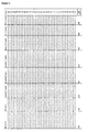
- the sequences for primers and probe for the real-time PCR assay are listed in Table 1.
- the loci for an epigenetic-genetic comparison were HLCS on chromosome 21 and ZFY on chromosome Y from pregnancies with a male fetus.
- the sequences of primers and probes with the PCR thermal cycle conditions for all the assays are listed in Table 1.
- Chorionic villus samples were collected during conventional prenatal diagnosis sessions in the first trimester of pregnancy. Placental tissue samples were collected from euploid third-trimester pregnancies after delivery and from euploid and trisomy 21 pregnancies after termination of pregnancy. The fetal chromosomal status was confirmed by full karyotyping. Maternal peripheral blood samples (12 - 20 mL EDTA) were collected from all subjects. An additional 12 mL of blood was collected into EDTA tubes from the third trimester pregnancies after delivery. Gestational ages of the first-, second- and third-trimester samples were 12 - 14 weeks, 17 - 21 weeks and 38 - 40 weeks, respectively.
- Maternal peripheral blood samples were processed by a double centrifugation protocol as previously described ( Chiu et al., Clin Chem 2001;47:1607-13 ). The blood cell portion was recentrifuged at 2,500 g, and any residual plasma was removed. DNA from the peripheral blood cells and that from maternal plasma was extracted with the blood and body fluid protocol of the QIAamp DNA Blood Mini Kit and the QIAamp DSP DNA Blood Mini Kit, respectively (Qiagen, Hilden, Germany). DNA from the CVS and placentas was extracted with the QIAamp DNA Mini Kit (Qiagen) according to the manufacturer's tissue protocol.
- the COBRA procedure is described as follows. Bisulfite conversion was performed on one microgram of each DNA sample using the EZ DNA Methylation Kit (Zymo Research, Orange, CA) according to manufacturer's instructions. Forty nanograms of bisulfite-converted DNA (calculation based on the original DNA input) were then subjected to PCR amplification with reagents supplied in the HotStar Taq DNA Polymerase Kit (Qiagen, Hilden, Germany). Reagent compositions for each PCR are listed in Table 2.
- PCR was performed in a 20 ⁇ L reaction with 1x PCR Buffer, MgCl 2 , 50 ⁇ M of each dNTP, forward and reverse primers, HotStar Taq polymerase, and with or without 2X PCRx Enhancer (Invitrogen, Carlsbad, CA).
- the thermal profile was 95 °C for 15 min, followed by 45-55 cycles of 95 °C for 20 s, appropriate annealing temperature for 30 s, 72 °C for 1.5 min, and a final extension of 72 °C for 3 min.
- PCR products were then subjected to restriction enzyme digestion.
- the restriction enzyme to be used for each respective locus was chosen for its ability to distinguish between the methylated and unmethylated sequences after bisulfite conversion.
- restriction sites were only present in either the methylated or unmethylated sequence but not both, so that one of the sequences would be digested while the other would remain intact. Restriction enzyme digestions were performed in 20 ⁇ L reactions with 5 ⁇ L PCR products, 1x appropriate buffer, and 10 U restriction enzyme (or none for mock digestion) under the manufacturer's recommended temperatures for 2 h. All enzymes were purchased from New England Biolabs (Ipswich, MA). Digested products were then analyzed by agarose gel electrophoresis.
- MSRE digestion For each placental and maternal blood cell DNA sample, 100 ng DNA was subjected to MSRE digestion. Restriction enzyme digestion was performed in a 50 ⁇ L reaction with 1x appropriate buffer, 25 U of Hpa II and 50 U of Bst UI (or none for mock digestion) (New England Biolabs) under the manufacturer's recommended temperatures for at least 16 h. For each maternal plasma sample, 1.6 mL plasma was used for DNA extraction, and was eluted in 50 ⁇ L of deionized water, 21 ⁇ L of which was subjected to restriction enzyme digestion.
- Enzyme digestion was performed in a 30 ⁇ L reaction with 1x appropriate buffer, 20 U of Hpa II and 30 U of Bst UI (or none for mock digestion) under the manufacturer's recommended temperatures for at least 16 h. Digested products were then analyzed by real-time quantitative PCR. The selected restriction enzymes only digested the unmethylated DNA but not the methylated DNA. Since the data from the COBRA analysis have shown that HLCS is hypermethylated in placental tissues and unmethylated in maternal blood cells, it was expected that a proportion of DNA from placental tissues would remain detectable while most DNA from maternal blood cells would be digested, thus becoming undetectable after restriction enzyme treatment.
- Real-time quantitative PCR analysis was developed for quantitative analysis of HLCS genomic DNA with and without restriction enzyme digestion. Four microliters of restriction enzyme treated DNA or mock digestion sample were used in the real time PCR assay. Each reaction contained 1x TaqMan Universal PCR Master Mix (Applied Biosystems), 300 nM of each of the forward and reverse primers (Integrated DNA Technologies, Coralville, IA), and 100 nM of the TaqMan probe (Applied Biosystems). The sequences of the primers and probes are listed in Table 1. The thermal profile was 50 °C for 2 min, 95 °C for 10 min, followed by 50 cycles of 95 °C for 15 s, and 60 °C for 1 min. All reactions were run in duplicates, and the mean quantity was taken.
- MSRE digestion The MSRE, Bst UI (New England Biolabs), was used to digest the hypomethylated DNA. Extracted DNA was digested with the Bst UI enzyme at 60 °C for 16 h. For CVS, placental tissues and maternal blood cells, 40 U of Bst UI enzyme was used to digest 200 ng of DNA for the microfluidics digital PCR assays. A mock-digested aliquot was included as the digestion control. For mock-digestion, an equal amount of DNA was subjected to the same digestion condition without the addition of enzyme. For the plasma samples, 20 U of the Bst UI enzyme was used to digest the DNA from 3.4 - 4.8 mL plasma in the third-trimester samples.
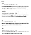
- Microfluidics digital PCR analysis Microfluidics digital PCR analysis. Microfluidics digital PCR assays were designed for the HLCS, RASSF1A and ZFY loci ( Figure 5 ), representing the dosage of chromosome 21, chromosome 3 and chromosome Y, respectively. The ZFX / Y assay and the basis of the digital PCR analysis have been described previously ( Lun et al., Clin Chem 2008;54:1664-72 ). The sequences of the primers and probes are listed in Table 1. The digital experiments were carried out on the BioMark System (Fluidigm, South San Francisco, CA) using the 12.765 Digital Arrays (Fluidigm). The Digital Array consists of 12 panels, and each panel is further partitioned into 765 reaction wells.
- the reaction was set up as a 10 ⁇ L mixture at a final concentration of 1x TaqMan Universal PCR Master Mix (Applied Biosystems), 125 nM TaqMan probe (Applied Biosystems), and 900 nM forward and reverse primers (Integrated DNA Technologies) diluted with the assay loading buffer and sample loading buffer according to the manufacturer's protocol.
- the input DNA volume was 3.5 ⁇ L for each of the 10 ⁇ L reaction mixture.
- the thermal profile was 50 °C for 2 min, 95 °C for 10 min, followed by 50 cycles of 95 °C for 15 or 30 s, and 57 °C or 60 °C for 1 min.
- the thermal cycle condition for each assay is specified in Table 1.
- the HLCS and RASSF1A assays were performed as a monoplex assay.
- the ZFX / Y assays were performed as a duplex reaction.
- DNA samples from the CVS, placenta, and maternal blood cells were subjected to the HLCS, RASSF1A , and ZFX / Y digital PCR analysis after Bst UI digestion. After enzyme digestion, the DNA was diluted to a concentration of 1 - 2 ng/ ⁇ L for loading into the reaction mixture for digital PCR analysis.
- DNA samples from the CVS and placenta constituted enzyme digestion-resistant HLCS and RASSF1A molecules, thus signals should be detected after restriction enzyme digestion. On the other hand, we would expect no or low level of detection in the blood cells as they are hypomethylated at these two loci.
- the ZFX / Y loci did not contain any Bst UI enzyme digestion sites, thus restriction enzyme treatment should not confer any effects on the DNA molecules.
- the ZFY molecules constituted the fetal-derived sequences from a male fetus for the ratio comparison.
- CGIs CpG islands
- the criteria to define a CGI were as follows: length > 400 bp, GC content > 50%, and observed/expected CpG ratio > 0.6 ( Yamada et al., Genome Res 2004;14:247-66 ).
- DNA sequences proximal to the transcription start sites (-1 kb to +500 bp; transcription start site being 0) of the 35 promoters were obtained from the March 2006 human reference sequence of the UCSC Genome Bioinformatics Database (website: www.genome.ucsc.edu/) (NCBI Build 36.1), and designed 51 COBRA assays to compare the methylation patterns between placental tissues and maternal blood cells (Table 2).
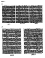
- the methylation profiles of the promoter regions between placental tissues collected from the first- and third-trimesters and maternal blood cells were compared. At least one placental tissue and one maternal blood cell sample from normal pregnancies were used for the COBRA screening (Table 2). Among the screened regions, the putative promoter regions of HLCS and C21orf81 (GenBank accession AF326257) were identified by COBRA to be differentially-methylated between placental tissues and maternal blood cells.
- COBRA results are illustrated in Figure 6A for HLCS region B2, in which the placenta was hypermethylated when compared to maternal blood cells; and 6B for C21orf81, in which the maternal blood cells were slightly methylated when compared to placental tissues.
- an MSRE digestion assay was developed followed by real-time quantitative PCR analysis to analyze the differential methylation of genomic DNA extracted from placentas and maternal blood cells.
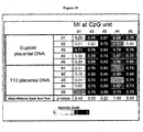
- methylation profiles of the putative promoter region of HLCS from eight maternal blood cell samples were compared with those from two first-trimester and two third-trimester placental tissue samples collected from euploid pregnancies. Restriction enzyme digestion followed by real-time quantitative PCR analysis was performed and the results are shown in Figure 7 . DNA from all maternal blood cell samples were mostly digested by restriction enzymes, resulting in methylation indices approaching 0 (Median: 0.0178, interquartile range (IQR): 0.0121 - 0.0281). Placental tissue DNA samples were partially digested, resulting in methylation indices ranging from 0.567 to 0.966.
- the gene dosage comparison of the chromosome 21 and reference chromosome markers was performed on a microfluidics digital PCR platform.
- the chromosome 21 marker was the hypermethylated HLCS
- the reference markers on chromosome 3 and chromosome Y were the hypermethylated RASSF1A and male-specific ZFY , respectively.
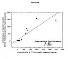
- T ⁇ arg ⁇ e ⁇ t - I ⁇ n E / N x N , where Target is the Poisson-corrected counts of the target molecules, ln is the natural logarithm, E is the number of negative (empty) wells, and N is the total number of digital PCR wells in the reaction.
- Results from the Bst UI-digested placental DNA samples showed that about 60% - 70% of the HLCS and RASSF1A molecules remained detectable when compared to the mock-digested samples.
- the hypomethylated RASSF1A molecules were completely digested by Bst UI enzyme treatment, while a few HLCS molecules remained detectable in the samples.
- ZFY and ZFX assays there was no change in the copy number counted from the mock- or Bst UI-digested DNA samples (Table 3A).
- the heterogeneity in the methylation density of the two loci across different samples may contribute to the large inter-individual variation in the HLCS to RASSF1A ratio. To solve this precision problem, a more stable baseline would be required for the dosage comparison between the euploid and T21 DNA samples.
- the EGG technique was then applied to maternal plasma DNA samples from euploid and T21 pregnancies to determine whether the EGG approach can be applied for noninvasive prenatal detection of fetal trisomy 21. It was reasoned that while the maternal HLCS molecules in the maternal plasma was restriction digested by the Bst UI enzyme, leaving the fetal-derived hypermethylated HLCS molecules intact for analysis, the fetal-derived ZFY locus would not be affected as there was no Bst UI enzyme recognition site located within the PCR amplicon, thus providing a stable baseline for gene dosage comparison.
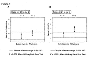
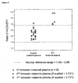
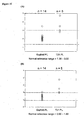
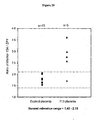
- a normal reference range of 1.49 - 2.88 was calculated from the 24 maternal plasma samples with euploid pregnancies. The ratio of one euploid DNA sample fell out of the normal reference range. This sample dried up during Speedvac concentration, and was reconstituted with water during sample preparation, which might explain the inaccuracy. All five maternal plasma samples from T21 pregnancies had a higher ratio of HLCS to ZFY than the reference range ( Figure 4 ). The data showed that with the EGG approach, fetal trisomy 21 can be detected noninvasively in maternal plasma samples.
- the present inventors have successfully validated a hypermethylated fetal DNA marker on chromosome 21, namely, the promoter region of the HLCS gene ( US Patent Application Publication No. 2007/0275402 ).
- the hypermethylated nature of this marker allows it to be detected relatively easily in maternal plasma through the use of methylation-sensitive restriction enzyme digestion and PCR amplification.
- HLCS HLCS was then used to demonstrate that the novel EGG approach is a feasible method for fetal chromosome dosage analysis.
- the main advantage of the EGG method over the previously epigenetic allelic ratio analysis is that using this method one has bypassed the requirement of the latter method for the epigenetic target and the genetic target (i.e., the SNP) to be present in the same locus and within a short distance from one another.
- the ZFY gene on the Y chromosome was used as a model for the fetal-specific genetic target.
- any fetal-specific genetic target e.g., an SNP allele that the fetus has inherited from the father but absent in the pregnant mother, can be used.
- a panel of such markers would be relatively easily developed to ensure a broad population coverage of this approach.
- the EGG approach has a higher discrimination power for T21 than an approach based purely on epigenetic markers, using the ratio of the hypermethylated HLCS to hypermethylated RASSF1A as an example.
- One possible explanation of the suboptimal performance of the latter approach is because of the fact that there is a variability in the level of DNA methylation of individual fetal-derived HLCS and RASSF1A molecules, as can be seen in Figure 1 and in previously published bisulfite sequencing results ( Chiu et al., Am J Pathol 2007;170:941-50 ).
- a ratio determined from these two potentially varying parameters would have a wider reference interval than if one of the parameter is a relatively stable genetic marker (as in the case for the EGG approach).
- microfluidics digital PCR was used as the detection and measurement platform because previous results have shown it to be a highly precise analytical method ( Lun et al., Clin Chem 2008;54:1664-72 ; Lun et al., Proc Natl Acad Sci U S A 2008;105:19920-5 ).
- the EGG approach can also be implemented in other variation of this molecular counting theme, e . g ., by targeted sequencing using a 'next-generation' sequencer.
- fetal epigenetic markers on the other chromosomes important for prenatal screening e.g ., chromosomes 18 and 13
- a fetal epigenetic marker is the SERPINB5 gene, coding for maspin, on chromosome 18 ( Chim et al., Proc Natl Acad Sci USA 2005;102:14753-8 ).
- Other examples of fetal epigenetic markers on chromosome 18 and 13 are described by Papageorgiou et al. ( Papageorgiou et al., Am J Pathol 2009;174:1609-18 ).
- a novel epigenetic-genetic chromosome dosage approach is recently developed for fetal trisomy 21 detection using a fetal epigenetic marker, the putative promoter of holocarboxylase synthetase (HLCS) on chromosome 21, and a fetal genetic marker, the zinc finger protein, Y-linked (ZFY) gene present on chromosome Y ( Tong et al., Clin Chem 2010;56:90-8 ).
- HLCS holocarboxylase synthetase
- T21 Women with euploid and trisomy 21 (T21) pregnancies who attended the Department of Obstetrics and Gynaecology, Prince of Wales Hospital were recruited between October 2009 and March 2010. Informed consent was obtained from individuals who joined the study, and ethics approval was obtained from the Joint Chinese University of Hong Kong - New Territories East Cluster Clinical Research Ethical Committee.
- CVS Chorionic villus samples
- Placental tissue samples were collected from euploid third-trimester pregnancies after delivery and from T21 pregnancies after termination of pregnancy (TOP). The chromosome status of each T21 case was confirmed by full karyotyping.
- Maternal peripheral blood samples (12 mL in EDTA tubes) were collected from all subjects.
- Peripheral blood samples were processed by a double centrifugation protocol as previously described ( Chiu et al. Clin Chem 2001;47:1607-13 ). The blood cell portion was recentrifuged at 2,500 g, and any residual plasma was removed. DNA from the peripheral blood cells and that from maternal plasma was extracted with the blood and body fluid protocol of the QIAamp DNA Blood Mini Kit and the QIAamp DSP DNA Blood Mini Kit, respectively (Qiagen).
- DNA from the CVS and placentas was extracted with the QIAamp DNA Mini Kit (Qiagen) according to the manufacturer's tissue protocol.
- Chromosome dosage comparison of the chromosome 21 and the reference marker in placental tissue and maternal plasma DNA samples was analyzed by real-time quantitative polymerase chain reaction (qPCR) and digital PCR ( Vogelstein and Kinzler Proc Natl Acad Sci U S A 1999;96:9236-41 ), respectively.
- Chromosome dosage analysis was performed by comparing the amount of hypermethylated HLCS to that of a SNP allele (rs6636, a C/G SNP) that the fetus has inherited from the father but absent in the pregnant mother.
- SNP rs6636 is located within the transmembrane emp24 protein transport domain containing 8 ( TMED8 ) gene on chromosome 14 with an average heterozygosity of 0.451 +/- 0.149 (dbSNP build 130). rs6636 is one of the SNPs that have been previously described ( Chow et al. Clin Chem 2007;53:141-2 ). The rs6636 SNP assays are denoted as TMED8- C/G SNP assays in this document.
- MSRE Methylation-sensitive restriction endonuclease
- BstUI A methylation-sensitive restriction endonuclease, BstUI (New England Biolabs), was used to digest the hypomethylated DNA. Extracted DNA was digested with the Bst UI enzyme at 60 °C for 16 hours. For CVS, placental tissues and maternal and normal control blood cells, 40 U of Bst UI enzyme was used to digest 100 ng of DNA for the PCR assays. A mock-digested aliquot was included as the digestion control. For mock-digestion, an equal amount of DNA was subjected to the same digestion condition without the addition of enzyme. For the plasma samples, 20 U to 40 U of the Bst UI enzyme was used to digest the DNA from 1.6 - 5.2 mL plasma.
- Each reaction was set up as a 25 ⁇ L mixture at a final concentration of 1x TaqMan® Universal PCR Master Mix (Applied Biosystems), 100 nM TaqMan® probe (Applied Biosystems), and 300 nM of each of the forward and reverse primers (Integrated DNA Technologies) with 25 ng DNA input.
- the reaction was initiated at 50°C for 2 min and continued at 95°C for 10 min and followed by 40 cycles of 95°C for 15 s and 60°C for 1 min.
- the experiments were carried out on the 7300 Real-time PCR System (Applied Biosystems) and the fluorescence data were collected and analyzed by the SDS v1.3.0 software (Applied Biosystems). All reactions were run in duplicates with the mean quantity taken.
- a calibration curve was constructed by serially-diluted genomic DNA extracted from adult male blood cells with concentration ranging from 10000 genome equivalents (GE) to 3 GE per reaction.
- HLCS HLCS
- TMED8 -C/G SNP digital PCR analysis Table 5
- the HLCS assay was performed as a duplex reaction with each of the TMED8 -C/G SNP assays.
- the fluorescent probes were labeled as HLCS (VIC) duplex with TMED8 -C(FAM) and HLCS (FAM) duplex with TMED8 -G(VIC).
- the basis of the digital PCR analysis have been described previously ( Lo et al. Proc Natl Acad Sci U S A 2007;104:13116-21 ; Lun et al. Clin Chem 2008;54:1664-72 ).
- the total reaction volume was 5 ⁇ L per well in a 384-well plate at a final concentration of 1x TaqMan® Universal PCR Master Mix (Applied Biosystems), 100 nM TaqMan® probe (Applied Biosystems), and 300 nM of each of the forward and reverse primers (Integrated DNA Technologies).
- the reaction was initiated at 50°C for 2 min and continued at 95°C for 10 min and followed by 50 cycles of 95°C for 15 s and 60°C for 1 min.
- the experiments were carried out on the 7900HT Sequence Detection System (Applied Biosystems) in a 384-well format, and the fluorescence data were collected by the "Absolute Quantification" application of SDS 2.3 software (Applied Biosystems).
- Genomic DNA extracted from placental tissues with known TMED8 genotypes were subjected to the HLCS and TMED8 duplex assays. Samples that were homozygous for one allele were tested with the TMED8 -C/G SNP assay for the other allele. A sample homozygous for the C allele should not show any signals for the TMED8 assay detecting the G allele, and vice versa.
- Beta - actin assay as a digestion control
- beta-actin region that is unmethylated in both the placenta and maternal blood cells was used to determine the efficiency of Bst UI digestion ( Chan et al. Clin Chem 2006;52:2211-8 ).
- the beta-actin assay was modified to contain two Bst UI enzyme recognition sites to match the number of BstUI enzyme recognition sites of the current HLCS assay.
- the sequences of the primers and probes are listed in Table 5. The same sequences were used for both the real-time qPCR and digital PCR analyses.
- the HLCS and TMED8 -C/G SNP assays were applied to a total of 20 euploid and nine T21 placental tissue samples with the heterozygous C/G genotype. Six euploid and four T21 samples were analyzed by using the HLCS to TMED8 -C ratio, and the remaining 14 euploid and five T21 samples were analyzed by the HLCS to TMED8 -G ratio.
- a calibration curve constructed by serially-diluted genomic DNA extracted from adult male blood cells with concentration ranging from 10000 GE to 3 GE per reaction was used to determine the absolute copy number of the two loci in the tested samples.
- the HLCS to TMED8 -C ratio was first determined in mock-digested placental DNA samples.
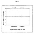
- a normal reference range defined as the mean HLCS to TMED8 -C ratio ⁇ 1.96SD, was calculated from the six euploid samples as 1.78 - 2.66. All four T21 samples showed a higher ratio than the normal reference range (Table 6A) ( Figure 9A ).
- the HLCS to TMED8 -G ratio was first determined in mock-digested placental DNA samples. A normal reference range was calculated from the 14 euploid samples as 1.36 - 3.03. One sample from each of the euploid and T21 groups was misclassified (Table 7A) ( Figure 10A ).
- Beta-actin as a digestion control
- the Bst UI enzyme digestion efficiency was evaluated by applying the beta-actin assay to the same mock- and Bst UI-digested placental DNA samples as those used for HLCS to TMED8 chromosome dosage analysis.
- the same calibration standard was used to quantify the amount of beta-actin sequences in the samples.
- the slope of the calibration curve was -3.41, whereas the y-intercept showed threshold cycle value of 37.97. Results showed that over 96% of the DNA was digested in all tested samples (Table 8A and 8B).
- Placental DNA samples of the heterozygous C/G, homozygous C/C and homozygous G/G genotypes were tested with the HLCS and TMED8 duplex assay to ascertain the specificity for detecting either one of the SNP alleles. Two samples were used for each of the three groups. A sample homozygous for the C allele should not show any signals for the TMED8 assay detecting the G allele, and vice versa.
- Maternal plasma DNA analysis was performed by comparing the ratio of digestion-resistant HLCS to fetal-specific TMED8 SNP allele in informative samples.
- An informative sample was defined as one in which the fetus was heterozygous and the mother was homozygous, being the TMED8 -C/G SNP in the current example.
- the extra allele that the fetus had inherited from the father was used as the reference baseline for chromosome 21 dosage determination.
- the HLCS and TMED8 -C duplex assay was applied to a total of 16 euploid and two T21 pregnancies.
- the fetal and maternal genotypes for the TMED8 SNP were C/G and G/G, respectively.
- the fetal-specific SNP allele was the C allele.
- Maternal plasma DNA samples were analyzed by digital PCR after Bst UI enzyme digestion. Each sample was analyzed in at least one 384-well plate. The total number of positive wells was used to calculate the ratio of HLCS to TMED8-C allele in each sample.
- a normal reference range of 2.12 - 3.65 was calculated from the euploid maternal plasma samples. Eight samples were collected from the third-trimester, and four samples were collected from each of the second- and first-trimester pregnancies. Among these 16 euploid samples, ten were from pregnancies carrying a female fetus, and six from those carrying a male fetus. The HLCS to TMED8 -C allele ratios of all euploid samples fell within the reference range. The T21 samples collected from the second- and first-trimester pregnancies showed a higher ratio than the reference range ( Figure 7A ). Both T21 cases carried a female fetus.
- the fetal and maternal genotypes for the TMED8 SNP were C/G and C/C, respectively.
- the fetal-specific SNP allele was the G allele.
- the HLCS and TMED8 -G duplex digital PCR assay was applied to Bst UI-digested maternal plasma DNA samples from nine euploid pregnancies and one T21 pregnancy. Each sample was analyzed in at least one 384-well plate. The total number of positive wells was used to calculate the ratio of HLCS to TMED8 -G allele in each sample.
- a normal reference range of 2.06 - 3.68 was calculated from euploid maternal plasma samples.
- nine euploid samples five, three and one cases were collected from the third-, second- and first-trimester pregnancies, respectively. They included four pregnancies with a female fetus, and five with a male fetus.
- the HLCS to TMED8 -G allele ratios of all euploid samples fell within the reference range.
- the first-trimester T21 pregnancy with a female fetus showed a higher HLCS to TMED8 -G ratio than the reference range ( Figure 7B ).
- Beta-actin as a digestion control
- the Bst UI enzyme digestion efficiency was evaluated by applying the beta-actin digital PCR assay to the same Bst UI-digested maternal plasma DNA samples as those used for HLCS to TMED8 chromosome dosage analysis. An aliquot of the digested DNA samples (1/50 of the total digestion mixture) was confirmed to show no more than one positive well for the beta-actin assay before subjected to chromosome dosage analysis.
- the inventors have demonstrated in principle that the EGG approach was a feasible method for fetal chromosome dosage analysis in maternal plasma DNA samples.
- the hypermethylated HLCS locus was used as the epigenetic component, which represented a class of fetal-specific molecules, and the ZFY locus was a genetic marker which was specific to a male fetus.
- the chromosome 21 dosage could be deduced.
- epigenetic-genetic chromosome dosage approach can be applied to the prenatal diagnosis of trisomy 21 using a fetal-specific SNP allele as a genetic reference baseline in place of a chromosome Y marker derived from a male fetus.
- SNP rs6636 located within the TMED8 locus on chromosome 14 was used as an example. By comparing the ratio between the hypermethylated HLCS and the fetal-specific TMED8 SNP allele, the chromosome 21 dosage could be deduced. It was demonstrated that both SNP alleles could serve as such a genetic reference baseline.
- the EGG chromosome dosage approach can be applied to the prenatal diagnosis of trisomy 21 for both male and female fetuses. Furthermore, development of a panel of such markers will ensure a broad population coverage of this approach.
- a maternal buffy coat sample which comprises of mostly maternal DNA, to ascertain the maternal genotypes for the panel of SNPs. For SNPs where the mother was shown to be homozygous, one could then aim to detect the allele that is not represented in the maternal genotype in maternal plasma.
- the plasma sample is positive for the non-maternal allele, it suggests that the fetus has inherited that allele from the father.
- the quantification of that paternally inherited fetal-specific allele in maternal plasma could then be used as the reference for gene dosage assessment using the peigenetic-genetic approach.
- epigenetic markers for the other chromosomes involved in other aneuploidies important for prenatal testing e.g., chromosomes 18 and 13, will further expand the clinical utility of the EGG approach.
- EXAMPLE 3 Epigenetic-genetic chromosome dosage approach for the detection of fetal trisomy 18
- This example demonstrates the application of epigenetic-genetic (EGG) chromosome dosage approach for the detection of fetal trisomy 18 (T18).
- EGG epigenetic-genetic
- This example of the EGG approach involved a fetal epigenetic marker located on chromosome 18 and a fetal genetic marker located on a reference chromosome, respectively, in maternal plasma.
- the fetal epigenetic marker is, preferably, Marker 18A [genomic location chr18:10022533-10022724, defined according to the Human Genome March 2006 Assembly (hg18)], which is a 146-bp intergenic region located 75 kb downstream of the gene VAPA (vesicle-associated membrane protein)-associated protein A) and 421 kb upstream of the gene APCDD1 ( adenomatosis polyposis coli down-regulated 1 ), as identified by an methylated DNA immunoprecipitation and tiling array analysis (described in USSN 61/308,578 ).
- VAPA vesicle-associated membrane protein-associated protein A
- APCDD1 adenomatosis polyposis coli down-regulated 1
- this fetal epigenetic marker can also be any cytosine-containing DNA genomic region located within 100 kb upstream or downstream of the above locus, i.e., chr18:10022533-10022724. Furthermore, this fetal epigenetic marker can also be any cytosine-containing DNA genomic region with different epigenetic signature (DNA methylation levels) that distinguishes the fetal from the maternal chromosome 18 in maternal plasma.
- epigenetic signature DNA methylation levels
- the fetal genetic marker in maternal plasma is, preferably, a region located in the zinc-finger Y-linked ( ZFY ) gene on chromosome Y.
- this fetal genetic marker can also be any genetic differences between the fetus and its mother, including single-nucleotide polymorphism (SNP) and insertion/deletion (indel) polymorphism.
- the inventors adopted the EGG approach to compare the concentrations of Marker 18A on chromosome 18 with those of a fetal genetic marker, the zinc finger protein, Y-linked (ZFY) gene on chromosome Y for the noninvasive prenatal detection of T18.
- CVS Chorionic villus samples
- Peripheral blood samples were processed by a double centrifugation protocol as previously described ( Chiu et al. Clin Chem 2001; 47:1607-13 ). The blood cell portion was recentrifuged at 2,500 g, and any residual plasma was removed.
- DNA from the peripheral blood cells and that from maternal plasma was extracted with the blood and body fluid protocol of the QIAamp DNA Blood Mini Kit and the QIAamp DSP DNA Blood Mini Kit, respectively (Qiagen).
- DNA from the CVS and placentas was extracted with the QIAamp DNA Mini Kit (Qiagen) according to the manufacturer's tissue protocol.
- the methylation status of Marker 18A were analyzed in six pairs of placental tissues and maternal blood cells by cloning and bisulfite sequencing. Briefly, extracted DNA was bisulfite-converted using the EZ DNA Methylation Kit (ZymoResearch) according to the manufacturer's instructions. Bisulfite-converted DNA was subjected to PCR amplification by primers targeting the Marker 18A region. The primer sequences for the PCR are listed in Table 10. The PCR conditions are summarized in Table 11A. The PCR product was subsequently TA-cloned into a plasmid vector with the pGEM T-Easy Cloning Kit (Promega) according to manufacturer's instructions.
- the MI at each CpG site was calculated by the number of methylated clones divided by the total number of clones for each sample. The average MI of the two biological replicates was calculated for each CpG site. The methylated site frequency was given by the number of methylated clones across each sample over the total number of clones scored.
- the Epityper assay was performed with the standard MassCLEAVE protocol (Sequenom) ( Ehrich et al. Proc Natl Acad Sci U S A 2005; 102:15785-90 ) (Details of the conditions for individual reactions involved in the protocol are summarized in Table 11B). Briefly, the bisulfite-converted DNA was subjected to PCR amplification with the Epityper assay targeting Marker 18A. The primer sequences were listed in Table 10. A T7 promoter tag was added to the 3' end of the reverse primer, and a 10-base tag was added to the 5' end of the primer to equalize the melting temperature between the forward and reverse primers.
- Extracted DNA was subjected to bisulfite conversion with an EZ DNA Methylation Kit (ZymoResearch) according to the manufacturer's instruction and amplified with PCR.
- the PCR products were then in vitro transcribed as RNA using the T7 DNA polymerase and T7 RNA polymerase, and cleaved specifically at bases with A- or G-residues by RNase A.
- the cleavage reaction generated CpG-containing fragments, or CpG unit, whose sizes would be dependent on the methylation status of the CpG sites ( i.e., CpG or TpG for methylated and unmethylated CpGs, respectively, after bisulfite conversion).
- the products were then cleaned up, and resolved with a matrix-assisted laser desorption/ionization time-of-flight (MALDI-TOF) mass spectrophotometer (MassARRAY Analyzer Compact).
- MALDI-TOF matrix-assisted laser desorption/ionization time-of-flight
- MassARRAY Analyzer Compact MassARRAY Analyzer Compact
- Quantitative real-time PCR (qPCR) assays were designed to target Marker 18A.
- the primer and probes were strategically positioned such that the unmethylated CpG sites in maternal DNA, but not the methylated CpG sites in the fetal DNA, would be specifically cleaved by the relevant restriction endonucleases.
- MSRE Methylation-sensitive restriction endonuclesae
- Hpa II and HinP 1I Two types of MSRE, namely Hpa II and HinP 1I (New England Biolabs), were used to digest the plasma DNA.
- DNA was extracted from 0.8 mL - 3.2 mL plasma and eluted in 50 ⁇ L water. 35 ⁇ L of the eluted DNA was then incubated with 20 U each of HpaII and HinPlI (New England Biolabs) in 1X NEBuffer 1 at 37 °C for 2 hours.
- hypermethylated DNA sequences of Marker 18A were amplified by conventional qPCR on the ABI 7300 HT sequence detection system (Applied Biosystems).
- the reaction volume of each qPCR assay was 50 ⁇ L, which contained 1X TaqMan® Universal PCR Master Mix (Applied Biosystems), 1500 nmol/L of each forward and reverse PCR primers (Integrated DNA Technologies), and 250 nmol/L TaqMan® probe (Integrated DNA Technologies).
- the probe was labeled with FAM on its 5' end as reporter.
- the thermal profile was 50 °C for 2 min, 95 °C for 10 min, 40 cycles of 95 °C for 15 s, and 60 °C for 1 minute.
- the primer and probe sequences are listed in Table 12.
- the concentrations of ZFY were determined by qPCR on the ABI7300 HT sequence detection system (Applied Biosystems) with a previously established assay ( Lun et al. Clin Chem 2008; 54:1664-72 ).
- the reaction volume of each qPCR assay was 50 ⁇ L, which contained 1X TaqMan® Universal PCR Master Mix (Applied Biosystems), 300 nmol/L of each forward and reverse PCR primers, and 100 nmol/L TaqMan® probe (Applied Biosystems).
- the probe was labeled with VIC on its 5' end as reporter.
- the thermal profile was 50 °C for 2 min, 95 °C for 10 min, 40 cycles of 95 °C for 15 s, and 60 °C for 1 minute.
- the primer and probe sequences are listed in Table 12.
- qPCR analysis For quantification by qPCR analysis "a calibration curve was derived by serial dilutions of known concentrations of genomic DNA extracted from a male peripheral blood cell sample, which was quantified by NanoDrop ND-1000 (Thermo Fisher Scientific Waltham) using the DNA-50 setting. It was then used to construct the quantitative standards with a linear dynamic range from 3 copies per reaction to 10,000 copies per reaction. The limit of detection (LOD) was 3 copies per reaction. Twenty duplicated reactions (40 replicates) were performed at the LOD concentration and 100% of the wells were positive with a median quantification cycle of 39.1 (range: 38.2 - 40.8). Any signals detected beyond that limit were considered undetectable. All reactions were duplicated and the mean quantity was taken. Multiple water blanks (no template control) were included in every PCR run.
- the inventors adopted the EGG approach to compare the relative dosage of the fetal epigenetic marker, Marker 18A , on chromosome 18, and that of a fetal genetic marker, ZFY , on chromosome Y, in placental tissues (CVS) and maternal plasma samples obtained from euploid and T18 pregnancies.
- MSRE Methylation-sensitive restriction endonuclesae
- the digestion protocol for the EGG analysis involving Marker 18A was the same as that used for qPCR analysis.
- 50 ng of DNA was subjected to digestion. All of the placental tissues were obtained from the first trimester.
- DNA was extracted from 1.6 mL - 3.2 mL plasma obtained from first, second and third trimester of euploid or T18 pregnancies. Extracted DNA from each case was eluted into 50 ⁇ L water, of which 35 ⁇ L was subjected to digestion.
- a duplex digital PCR assay was developed to amplify digestion-resistant Marker 18A and ZFY sequences.
- the basis of the digital PCR analysis have been described previously ( Lo et al. Proc Natl Acad Sci U S A 2007; 104:13116-21 ; Tong et al. Clin Chem 2010; 56: 90-8 ).
- the reaction volume was 5 ⁇ L per well at a final concentration of 1x TaqMan® Universal PCR Master Mix (Applied Biosystems) with the respective primers and probes concentrations for each target.
- Marker 18A the reaction involved 250 nM TaqMan® probe (Integrated DNA Technologies) and 1500 nM of each of the forward and reverse primers (Integrated DNA Technologies).
- ZFY the reaction involved 100 nM TaqMan® probe (Applied Biosystems), and 300 nM of each of the forward and reverse primers (Integrated DNA Technologies).
- the probe for Marker 18A was labeled with the reporter FAM while that for ZFY was labeled with the reported VIC (Table 12).
- the total number of digital PCR reactions for each target was 384.
- the primer and probe sequences are the same as those for qPCR.
- the reaction volume was 5 ⁇ L per well at a final concentration of 1x TaqMan® Universal PCR Master Mix (Applied Biosystems) with 250 nM TaqMan® probe (Applied Biosystems) and 900 nM of each of the forward and reverse primers (Integrated DNA Technologies).
- the probe was labeled with the reporter VIC at its 5' end (Table 10).
- the assay was carried out on the 7900HT Sequence Detection System (Applied Biosystems). The reaction was initiated at 50°C for 2 min and continued at 95°C for 10 min, followed by 55 cycles of 95°C for 15 s, and 60°C for 1 min. The total number of digital PCR reactions for each target was 96.
- Marker 18A is a potential fetal epigenetic marker
- First-trimester placental DNA samples (5 for T18 and 5 for euploid) from pregnancies with male fetuses were analyzed with the duplex digital PCR assays for Marker 18A and ZFY.
- the normal reference range, defined as the mean ratio of Marker 18A to ZFY ⁇ 1.96SD, for the euploid samples was 1.20 - 1.66. All of the T18 cases had a ratio greater than the upper reference limit ( Figure 15 ).
- the inventors analyzed 27 maternal plasma samples obtained from pregnancies with euploid male fetuses and 9 from pregnancies with T18 male fetuses.
- 27 euploid cases 18, 4 and 9 cases were obtained from the first, second and third trimesters, respectively. All T18 cases were obtained from the first trimester.
- the median gestational age of the euploid and T18 groups were 14.1 and 13.3, respectively.
- the normal reference range was 0.34 - 3.04 ( Figure 16 ). Based on this reference range, 26 out of 27 euploid cases and 8 out of 9 T18 were correctly classified, giving a sensitivity of 88.9% and a specificity of 96.3%.
- Beta-actin as a digestion control
- the enzyme digestion efficiency was evaluated by analyzing the enzyme-digested maternal plasma samples with the beta-actin assay that targeted at a region completely unmethylated in placentas and maternal blood cells while containing similar MSRE restriction sites as the Marker 18A amplicon ( Figure 17 ). Beta-actin sequences were not detectable in all the digested plasma samples.
- the noninvasive prenatal detection of trisomy 18 using this EGG approach could be extended to cover more fetuses in the general population by replacing the Y-specific fetal genetic marker, which was used in this example as an illustration, with other fetal genetic marker, such as paternally-inherited SNP on a reference chromosome, to specifically measure the dosage of the fetal reference chromosome in maternal plasma.
- EXAMPLE 4 Epigenetic-genetic chromosome dosage approach for the detection of fetal trisomy 13
- This example demonstrates the application of epigenetic-genetic (EGG) chromosome dosage approach for the detection of fetal trisomy 13 (T13).
- EGG epigenetic-genetic
- This EGG approach involved a fetal epigenetic marker located on chromosome 13 and a fetal genetic marker located on a reference chromosome, respectively.
- the fetal epigenetic marker is, preferably, Marker 13A [genomic location chr13:105941431-105941818, defined according to the Human Genome March 2006 Assembly (hg18)].
- this fetal epigenetic marker can also be any cytosine-containing DNA genomic region located within 100 kb upstream or downstream of the above locus, i.e., chr13:105941431-105941818. Furthermore, this fetal epigenetic marker can also be any cytosine-containing DNA genomic region with different epigenetic signature (DNA methylation levels) that distinguishes the fetal from the maternal chromosome 13 in maternal plasma.
- the fetal genetic marker in maternal plasma is, preferably, a region located in the zinc-finger Y-linked ( ZFY ) gene on chromosome Y.
- this fetal genetic marker can also be any genetic differences between the fetus and its mother, including single-nucleotide polymorphism (SNP) and insertion/deletion (indel) polymorphism.
- CVS Chorionic villus samples
- Peripheral blood samples were processed by a double centrifugation protocol as previously described ( Chiu et al. Clin Chem 2001; 47:1607-13 ).
- the blood cell portion was recentrifuged at 2,500 g, and any residual plasma was removed.
- DNA from the peripheral blood cells was extracted with the blood and body fluid protocol of the QIAamp DNA Blood Mini Kit (Qiagen).
- DNA from the CVS and placentas was extracted with the QIAamp DNA Mini Kit (Qiagen) according to the manufacturer's tissue protocol.
- the methylation status of Marker 13A were analyzed in six pairs of placental tissues and maternal blood cells by cloning and bisulfite sequencing. Extracted DNA was bisulfite converted using the EZ DNA Methylation Kit (ZymoResearch) according to the manufacturer's instructions. Bisulfite-converted DNA was subjected to PCR amplification with the forward primer '5 - aggaagagagGGAGGATAGGAGGAGGGAGTTATAGT - 3' and reverse primer '5-cagtaatacgactcactatagggagaaggctAAAATTACACACAATACACACCAAAAAAT - 3' targeting Marker 13A region.
- This set of primers were designed for analysis by the Epityper assay, but was also applied in the PCR amplification of bisulfite-converted DNA for bisulfite sequencing analysis.
- the PCR conditions are summarized in Table 13A.
- the PCR product was subsequently TA-cloned into a plasmid vector with the pGEM T-Easy Cloning Kit (Promega) according to manufacturer's instructions. Cloning was done with Escherichia coli strain JM109 (Promega).
- the inserts from the clones were then amplified using T7 and SP6 promoter primers (Promega) according to the manufacturer's instructions (Reaction conditions are summarized under the sub-title colony PCR assays in Table 13A).
- the PCR product was then subjected to sequencing reaction with the BigDye Terminator Cycle Sequencing v1.1 kit (Applied Biosystems) according to the manufacturer's instructions (Reaction conditions are summarized under the sub-title sequencing reaction in Table 13A).
- DNA was then precipitated with ethanol and resuspended in 10 ⁇ L of Hi-Di formamide and sequenced on a 3100 DNA Analyzer (Applied Biosystems).
- the sequencing data were analyzed using the SeqScape v2.5 software (Applied Biosystems). The inventors ensured > 99% bisulfite conversion of each clone by examining the conversion rate at non-CpG cytosine residues.
- the MI at each CpG site was calculated by the number of methylated clones divided by the total number of clones for each sample, which was 8 in our experiments. The average MI of the two biological replicates was calculated for each CpG site. The methylated site frequency was given by the number of methylated clones across each sample over the total number of clones scored.
- the Epityper assay was performed with the standard MassCLEAVE protocol (Sequenom) ( Ehrich et al. Proc Natl Acad Sci U S A 2005; 102:15785-90 ) (Details of the conditions for individual reactions involved in the protocol are summarized in Table 13B). Briefly, the bisulfite-converted DNA was subjected to PCR amplification with the forward primer '5 - aggaagagagGGAGGATAGGAGGAGGGAGTTATAGT - 3' and reverse primer '5 - cagtaatacgactcactatagggagaaggctAAAATTACACACAATACACACCAAAAAAT - 3' targeting Marker 13A region.
- a T7 promoter tag was added to the 3' end of the reverse primer, and a 10-base tag was added to the 5' end of the primer to equalize the melting temperature between the forward and reverse primers.
- Extracted DNA was subjected to bisulfite conversion with an EZ DNA Methylation Kit (ZymoResearch) according to the manufacturer's instruction and amplified with PCR.
- the PCR products were then in vitro transcribed as RNA using the T7 DNA polymerase and T7 RNA polymerase, and cleaved specifically at bases with A- or G-residues by RNase A.
- the cleavage reaction generated CpG-containing fragments, or CpG unit, whose sizes would be dependent on the methylation status of the CpG sites (i.e., CpG or TpG for methylated and unmethylated CpGs, respectively, after bisulfite conversion).
- the products were then cleaned up, and resolved with a matrix-assisted laser desorption/ionization time-of-flight (MALDI-TOF) mass spectrophotometer (MassARRAY Analyzer Compact).
- the methylation index (MI) of each CpG unit were calculated by dividing the peak height of the methylated product by the sum of peak heights of both the methylated and unmethylated products.
- the inventors adopted the EGG approach to compare the relative dosage of the fetal epigenetic marker, Marker 13A on chromosome 13, and that of a fetal genetic marker, ZFY on chromosome Y, in placental tissues (CVS) obtained from euploid and T13 pregnancies.
- MSRE Methylation-sensitive restriction endonuclesae
- MSRE Fermentas Life Sciences
- AccI, NlaIV and HpaII Fermentas Life Sciences
- 50 ng of placental DNA were incubated with 2 FDU each of the above enzymes, in 1X FastDigest® Buffer at 37 °C for 2 hours, followed by enzyme inactivation at 65 °C for 20 minutes.
- the reaction volume was 5 ⁇ L per well at a final concentration of 1x TaqMan® Universal PCR Master Mix (Applied Biosystems) with the respective primers and probes concentrations for each target.
- Marker 13A the reaction involved 250 nM TaqMan® probe (Integrated DNA Technologies) and 900 nM of each of the forward and reverse primers (Integrated DNA Technologies).
- ZFY the reaction involved 100 nM TaqMan® probe, and 300 nM of each of the forward and reverse primers (Integrated DNA Technologies).
- the probe for Marker 13A was labeled with the reporter FAM while that for ZFY was labeled with the reporter VIC (Table 14).
- the total number of digital PCR reactions for each target was 192.
- the experiments were carried out on the 7900HT Sequence Detection System (Applied Biosystems). The reaction was initiated at 50°C for 2 min and continued at 95°C for 10 min, followed by 55 and 45 cycles of 95°C for 15 s for Marker 13A and ZFY , respectively, and 60°C for 1 min. The fluorescence data were collected by the "Absolute Quantification" application of SDS 2.3 software (Applied Biosystems).
- Marker 13A is a potential fetal epigenetic marker
- Placental DNA samples (5 for T13 and 10 for euploid) from pregnancies with male fetuses were analyzed with the respective monoplex digital PCR assays for Marker 13A and ZFY.
- a normal reference range, defined as the mean ratio of Marker 13A to ZFY ⁇ 1.96SD, of the euploid samples was 1.40 - 2.10. All except one of the T13 cases had a ratio greater than the upper reference limit ( Figure 20 ).
- the EGG approach can be applied to infer the dosage of fetal chromosome 13 in placental tissues obtained during the first and second trimesters. It is feasible to detect trisomy 13 by comparing such relative chromosome dosage in euploid and T13 placental tissues. This approach has the potential to be applied to infer the relative chromosome dosage of fetal chromosome 13 in maternal plasma and to detect trisomy 13 in an noninvasive manner.
Landscapes
- Chemical & Material Sciences (AREA)
- Organic Chemistry (AREA)
- Life Sciences & Earth Sciences (AREA)
- Zoology (AREA)
- Wood Science & Technology (AREA)
- Proteomics, Peptides & Aminoacids (AREA)
- Health & Medical Sciences (AREA)
- Engineering & Computer Science (AREA)
- Microbiology (AREA)
- Immunology (AREA)
- Physics & Mathematics (AREA)
- Molecular Biology (AREA)
- Biotechnology (AREA)
- Biophysics (AREA)
- Analytical Chemistry (AREA)
- Biochemistry (AREA)
- Bioinformatics & Cheminformatics (AREA)
- General Engineering & Computer Science (AREA)
- General Health & Medical Sciences (AREA)
- Genetics & Genomics (AREA)
- Measuring Or Testing Involving Enzymes Or Micro-Organisms (AREA)
Applications Claiming Priority (3)
| Application Number | Priority Date | Filing Date | Title |
|---|---|---|---|
| US23304209P | 2009-08-11 | 2009-08-11 | |
| US30857810P | 2010-02-26 | 2010-02-26 | |
| PCT/GB2010/001327 WO2011018600A1 (en) | 2009-08-11 | 2010-07-12 | Method for detecting chromosomal aneuploidy |
Publications (2)
| Publication Number | Publication Date |
|---|---|
| EP2464743A1 EP2464743A1 (en) | 2012-06-20 |
| EP2464743B1 true EP2464743B1 (en) | 2015-04-29 |
Family
ID=42797276
Family Applications (1)
| Application Number | Title | Priority Date | Filing Date |
|---|---|---|---|
| EP20100733024 Active EP2464743B1 (en) | 2009-08-11 | 2010-07-12 | Method for detecting chromosomal aneuploidy |
Country Status (7)
| Country | Link |
|---|---|
| US (1) | US8563242B2 (enExample) |
| EP (1) | EP2464743B1 (enExample) |
| JP (1) | JP5789605B2 (enExample) |
| CN (1) | CN102625854B (enExample) |
| AU (1) | AU2010283621B2 (enExample) |
| CA (1) | CA2770256C (enExample) |
| WO (1) | WO2011018600A1 (enExample) |
Families Citing this family (104)
| Publication number | Priority date | Publication date | Assignee | Title |
|---|---|---|---|---|
| US11111543B2 (en) | 2005-07-29 | 2021-09-07 | Natera, Inc. | System and method for cleaning noisy genetic data and determining chromosome copy number |
| US8532930B2 (en) | 2005-11-26 | 2013-09-10 | Natera, Inc. | Method for determining the number of copies of a chromosome in the genome of a target individual using genetic data from genetically related individuals |
| US10081839B2 (en) | 2005-07-29 | 2018-09-25 | Natera, Inc | System and method for cleaning noisy genetic data and determining chromosome copy number |
| US11111544B2 (en) | 2005-07-29 | 2021-09-07 | Natera, Inc. | System and method for cleaning noisy genetic data and determining chromosome copy number |
| US9424392B2 (en) | 2005-11-26 | 2016-08-23 | Natera, Inc. | System and method for cleaning noisy genetic data from target individuals using genetic data from genetically related individuals |
| US10083273B2 (en) | 2005-07-29 | 2018-09-25 | Natera, Inc. | System and method for cleaning noisy genetic data and determining chromosome copy number |
| WO2009105531A1 (en) * | 2008-02-19 | 2009-08-27 | Gene Security Network, Inc. | Methods for cell genotyping |
| WO2009146335A1 (en) * | 2008-05-27 | 2009-12-03 | Gene Security Network, Inc. | Methods for embryo characterization and comparison |
| EP2321642B1 (en) * | 2008-08-04 | 2017-01-11 | Natera, Inc. | Methods for allele calling and ploidy calling |
| US8962247B2 (en) | 2008-09-16 | 2015-02-24 | Sequenom, Inc. | Processes and compositions for methylation-based enrichment of fetal nucleic acid from a maternal sample useful for non invasive prenatal diagnoses |
| US8476013B2 (en) | 2008-09-16 | 2013-07-02 | Sequenom, Inc. | Processes and compositions for methylation-based acid enrichment of fetal nucleic acid from a maternal sample useful for non-invasive prenatal diagnoses |
| US20120252015A1 (en) * | 2011-02-18 | 2012-10-04 | Bio-Rad Laboratories | Methods and compositions for detecting genetic material |
| WO2011041485A1 (en) | 2009-09-30 | 2011-04-07 | Gene Security Network, Inc. | Methods for non-invasive prenatal ploidy calling |
| JP2013511991A (ja) * | 2009-11-25 | 2013-04-11 | クアンタライフ, インコーポレイテッド | 遺伝子材料を検出する方法および組成物 |
| US12545960B2 (en) | 2010-05-18 | 2026-02-10 | Natera, Inc. | Methods for simultaneous amplification of target loci |
| US11408031B2 (en) | 2010-05-18 | 2022-08-09 | Natera, Inc. | Methods for non-invasive prenatal paternity testing |
| US11322224B2 (en) | 2010-05-18 | 2022-05-03 | Natera, Inc. | Methods for non-invasive prenatal ploidy calling |
| US20190010543A1 (en) | 2010-05-18 | 2019-01-10 | Natera, Inc. | Methods for simultaneous amplification of target loci |
| US11939634B2 (en) | 2010-05-18 | 2024-03-26 | Natera, Inc. | Methods for simultaneous amplification of target loci |
| WO2011146632A1 (en) | 2010-05-18 | 2011-11-24 | Gene Security Network Inc. | Methods for non-invasive prenatal ploidy calling |
| US11339429B2 (en) | 2010-05-18 | 2022-05-24 | Natera, Inc. | Methods for non-invasive prenatal ploidy calling |
| US12221653B2 (en) | 2010-05-18 | 2025-02-11 | Natera, Inc. | Methods for simultaneous amplification of target loci |
| US12152275B2 (en) | 2010-05-18 | 2024-11-26 | Natera, Inc. | Methods for non-invasive prenatal ploidy calling |
| US9677118B2 (en) | 2014-04-21 | 2017-06-13 | Natera, Inc. | Methods for simultaneous amplification of target loci |
| US11326208B2 (en) | 2010-05-18 | 2022-05-10 | Natera, Inc. | Methods for nested PCR amplification of cell-free DNA |
| US11332793B2 (en) | 2010-05-18 | 2022-05-17 | Natera, Inc. | Methods for simultaneous amplification of target loci |
| US10316362B2 (en) | 2010-05-18 | 2019-06-11 | Natera, Inc. | Methods for simultaneous amplification of target loci |
| US11332785B2 (en) | 2010-05-18 | 2022-05-17 | Natera, Inc. | Methods for non-invasive prenatal ploidy calling |
| US8603742B2 (en) | 2010-07-06 | 2013-12-10 | University of Pittsburgh—of the Commonwealth System of Higher Education | Methods for the diagnosis of fetal disease |
| US8700338B2 (en) | 2011-01-25 | 2014-04-15 | Ariosa Diagnosis, Inc. | Risk calculation for evaluation of fetal aneuploidy |
| US20140342940A1 (en) | 2011-01-25 | 2014-11-20 | Ariosa Diagnostics, Inc. | Detection of Target Nucleic Acids using Hybridization |
| US11203786B2 (en) | 2010-08-06 | 2021-12-21 | Ariosa Diagnostics, Inc. | Detection of target nucleic acids using hybridization |
| US20130040375A1 (en) | 2011-08-08 | 2013-02-14 | Tandem Diagnotics, Inc. | Assay systems for genetic analysis |
| US10533223B2 (en) | 2010-08-06 | 2020-01-14 | Ariosa Diagnostics, Inc. | Detection of target nucleic acids using hybridization |
| US20130261003A1 (en) | 2010-08-06 | 2013-10-03 | Ariosa Diagnostics, In. | Ligation-based detection of genetic variants |
| CN103608466B (zh) | 2010-12-22 | 2020-09-18 | 纳特拉公司 | 非侵入性产前亲子鉴定方法 |
| US8756020B2 (en) | 2011-01-25 | 2014-06-17 | Ariosa Diagnostics, Inc. | Enhanced risk probabilities using biomolecule estimations |
| AU2012214312A1 (en) | 2011-02-09 | 2013-08-22 | Bio-Rad Laboratories, Inc. | Analysis of nucleic acids |
| RU2671980C2 (ru) | 2011-02-09 | 2018-11-08 | Натера, Инк. | Способы неинвазивного пренатального установления плоидности |
| US12097495B2 (en) * | 2011-02-18 | 2024-09-24 | Bio-Rad Laboratories, Inc. | Methods and compositions for detecting genetic material |
| AU2012236200B2 (en) * | 2011-03-30 | 2015-05-14 | Verinata Health, Inc. | Method for verifying bioassay samples |
| US8712697B2 (en) | 2011-09-07 | 2014-04-29 | Ariosa Diagnostics, Inc. | Determination of copy number variations using binomial probability calculations |
| WO2013130857A1 (en) * | 2012-02-29 | 2013-09-06 | Bio Dx, Inc. | Defining diagnostic and therapeutic targets of conserved fetal dna in maternal circulating blood |
| EP2820129A1 (en) | 2012-03-02 | 2015-01-07 | Sequenom, Inc. | Methods and processes for non-invasive assessment of genetic variations |
| CA2866324C (en) | 2012-03-13 | 2019-01-15 | The Chinese University Of Hong Kong | Methods for analyzing massively parallel sequencing data for noninvasive prenatal diagnosis |
| CA2868836C (en) | 2012-03-26 | 2019-08-06 | The Johns Hopkins University | Rapid aneuploidy detection |
| US10504613B2 (en) | 2012-12-20 | 2019-12-10 | Sequenom, Inc. | Methods and processes for non-invasive assessment of genetic variations |
| US9920361B2 (en) | 2012-05-21 | 2018-03-20 | Sequenom, Inc. | Methods and compositions for analyzing nucleic acid |
| JP2015522260A (ja) * | 2012-06-15 | 2015-08-06 | ハリー スティリ, | 疾患または状態を検出する方法 |
| US20140093873A1 (en) | 2012-07-13 | 2014-04-03 | Sequenom, Inc. | Processes and compositions for methylation-based enrichment of fetal nucleic acid from a maternal sample useful for non-invasive prenatal diagnoses |
| EP2875156A4 (en) | 2012-07-19 | 2016-02-24 | Ariosa Diagnostics Inc | SEQUENTIAL LIGATION-BASED MULTIPLEX DETECTION OF GENETIC VARIANTS |
| US20140100126A1 (en) | 2012-08-17 | 2014-04-10 | Natera, Inc. | Method for Non-Invasive Prenatal Testing Using Parental Mosaicism Data |
| SI4056712T1 (sl) * | 2012-09-20 | 2024-10-30 | The Chinese University Of Hong Kong | Neinvazivno določanje metiloma tumorja iz plazme |
| US9732390B2 (en) | 2012-09-20 | 2017-08-15 | The Chinese University Of Hong Kong | Non-invasive determination of methylome of fetus or tumor from plasma |
| US10706957B2 (en) | 2012-09-20 | 2020-07-07 | The Chinese University Of Hong Kong | Non-invasive determination of methylome of tumor from plasma |
| WO2014051522A1 (en) * | 2012-09-26 | 2014-04-03 | Agency For Science, Technology And Research | Biomarkers for down syndrome prenatal diagnosis |
| US20130189684A1 (en) * | 2013-03-12 | 2013-07-25 | Sequenom, Inc. | Quantification of cell-specific nucleic acid markers |
| US11060145B2 (en) | 2013-03-13 | 2021-07-13 | Sequenom, Inc. | Methods and compositions for identifying presence or absence of hypermethylation or hypomethylation locus |
| WO2014200579A1 (en) * | 2013-06-13 | 2014-12-18 | Ariosa Diagnostics, Inc. | Statistical analysis for non-invasive sex chromosome aneuploidy determination |
| US20160369339A1 (en) * | 2013-07-01 | 2016-12-22 | Cindy Anderson | Biomarker for preeclampsia |
| CN105555965B (zh) * | 2013-07-30 | 2020-06-05 | 深圳华大基因股份有限公司 | 确定核酸混合物中核酸组成的方法 |
| US10262755B2 (en) | 2014-04-21 | 2019-04-16 | Natera, Inc. | Detecting cancer mutations and aneuploidy in chromosomal segments |
| US10577655B2 (en) | 2013-09-27 | 2020-03-03 | Natera, Inc. | Cell free DNA diagnostic testing standards |
| US9499870B2 (en) | 2013-09-27 | 2016-11-22 | Natera, Inc. | Cell free DNA diagnostic testing standards |
| KR102384620B1 (ko) * | 2013-10-04 | 2022-04-11 | 시쿼넘, 인코포레이티드 | 유전적 변이의 비침습 평가를 위한 방법 및 프로세스 |
| US20160340740A1 (en) * | 2014-01-30 | 2016-11-24 | The Regents Of The University Of California | Methylation haplotyping for non-invasive diagnosis (monod) |
| EP3736344A1 (en) | 2014-03-13 | 2020-11-11 | Sequenom, Inc. | Methods and processes for non-invasive assessment of genetic variations |
| CA2945962C (en) | 2014-04-21 | 2023-08-29 | Natera, Inc. | Detecting mutations and ploidy in chromosomal segments |
| US12492429B2 (en) | 2014-04-21 | 2025-12-09 | Natera, Inc. | Detecting mutations and ploidy in chromosomal segments |
| EP2942400A1 (en) | 2014-05-09 | 2015-11-11 | Lifecodexx AG | Multiplex detection of DNA that originates from a specific cell-type |
| JP6681841B2 (ja) * | 2014-05-09 | 2020-04-15 | ライフコーデックス アーゲー | 特別な細胞タイプに由来するdnaの検出とそれに関連する方法 |
| EP2942401A1 (en) | 2014-05-09 | 2015-11-11 | Lifecodexx AG | Detection of DNA that originates from a specific cell-type |
| JP2017521091A (ja) * | 2014-05-15 | 2017-08-03 | ケルベンクス インコーポレイテッド | 診断試験用の胎児有核赤血球(nrbc)の調製 |
| US20180173846A1 (en) | 2014-06-05 | 2018-06-21 | Natera, Inc. | Systems and Methods for Detection of Aneuploidy |
| WO2016052405A1 (ja) * | 2014-09-29 | 2016-04-07 | 富士フイルム株式会社 | 胎児の染色体異数性の非侵襲的判別方法および判別システム |
| CN104789686B (zh) * | 2015-05-06 | 2018-09-07 | 浙江安诺优达生物科技有限公司 | 检测染色体非整倍性的试剂盒和装置 |
| WO2016176846A1 (zh) * | 2015-05-06 | 2016-11-10 | 安诺优达基因科技(北京)有限公司 | 检测染色体非整倍性的试剂盒、装置和方法 |
| US11479812B2 (en) | 2015-05-11 | 2022-10-25 | Natera, Inc. | Methods and compositions for determining ploidy |
| US10689706B2 (en) | 2015-07-20 | 2020-06-23 | The Chinese University Of Hong Kong | Methylation pattern analysis of haplotypes in tissues in a DNA mixture |
| CN105132572B (zh) * | 2015-09-25 | 2018-03-02 | 邯郸市康业生物科技有限公司 | 一种无创产前筛查21‑三体综合征试剂盒 |
| KR101848438B1 (ko) * | 2015-10-29 | 2018-04-13 | 바이오코아 주식회사 | 디지털 pcr을 이용한 산전진단 방법 |
| US20180327825A1 (en) * | 2015-11-09 | 2018-11-15 | Progenity, Inc. | Methods for determining the origin of dna molecules |
| HUE050491T2 (hu) * | 2015-11-10 | 2020-12-28 | Eurofins Lifecodexx Gmbh | Magzati kromoszomális aneuploidiák kimutatása olyan DNS régiókat alkalmazva, amelyek különbözõképpen vannak metilezve a magzat és a terhes nõstény között |
| US10982286B2 (en) | 2016-01-22 | 2021-04-20 | Mayo Foundation For Medical Education And Research | Algorithmic approach for determining the plasma genome abnormality PGA and the urine genome abnormality UGA scores based on cell free cfDNA copy number variations in plasma and urine |
| RU2760913C2 (ru) | 2016-04-15 | 2021-12-01 | Натера, Инк. | Способы выявления рака легкого |
| CN106086243A (zh) * | 2016-08-01 | 2016-11-09 | 王燕芸 | 一种无创式检测怀孕中胎儿是否健康的系统及其应用 |
| WO2018064486A1 (en) | 2016-09-29 | 2018-04-05 | Counsyl, Inc. | Noninvasive prenatal screening using dynamic iterative depth optimization |
| WO2018067517A1 (en) | 2016-10-04 | 2018-04-12 | Natera, Inc. | Methods for characterizing copy number variation using proximity-litigation sequencing |
| EP3524687A4 (en) * | 2016-10-05 | 2019-11-06 | FUJIFILM Corporation | METHOD FOR DETERMINING THE NUMBER OF REQUIRED LOCI AND METHOD FOR DETERMINING THE NUMBER OF REQUIRED SNP LOCI |
| GB201618485D0 (en) | 2016-11-02 | 2016-12-14 | Ucl Business Plc | Method of detecting tumour recurrence |
| US10011870B2 (en) | 2016-12-07 | 2018-07-03 | Natera, Inc. | Compositions and methods for identifying nucleic acid molecules |
| CN106916893A (zh) * | 2016-12-26 | 2017-07-04 | 中国科学院上海微系统与信息技术研究所 | 一种基于数字pcr芯片的基因甲基化程度定量方法 |
| WO2018137141A1 (zh) * | 2017-01-24 | 2018-08-02 | 深圳华大基因研究院 | 基于外泌体dna进行无创产前诊断的方法及其应用 |
| WO2018156418A1 (en) | 2017-02-21 | 2018-08-30 | Natera, Inc. | Compositions, methods, and kits for isolating nucleic acids |
| US12084720B2 (en) | 2017-12-14 | 2024-09-10 | Natera, Inc. | Assessing graft suitability for transplantation |
| US12398389B2 (en) | 2018-02-15 | 2025-08-26 | Natera, Inc. | Methods for isolating nucleic acids with size selection |
| US12024738B2 (en) | 2018-04-14 | 2024-07-02 | Natera, Inc. | Methods for cancer detection and monitoring |
| US12234509B2 (en) | 2018-07-03 | 2025-02-25 | Natera, Inc. | Methods for detection of donor-derived cell-free DNA |
| CN109022541A (zh) * | 2018-07-16 | 2018-12-18 | 苏州大学附属第二医院 | 一种浓集胎儿游离dna的试剂盒、方法及其应用 |
| CN109136363A (zh) * | 2018-08-31 | 2019-01-04 | 领航基因科技(杭州)有限公司 | 一种检测胎儿染色体非整数倍的试剂盒和检测胎儿甲基化dna拷贝数的方法 |
| US12305235B2 (en) | 2019-06-06 | 2025-05-20 | Natera, Inc. | Methods for detecting immune cell DNA and monitoring immune system |
| EP3987056A4 (en) * | 2019-06-24 | 2023-07-26 | Chromacode, Inc. | METHODS FOR THE DIFFERENTIAL QUANTIFICATION OF AMINO ACIDS |
| CN110964073B (zh) * | 2019-11-28 | 2023-06-02 | 上海市第一妇婴保健院 | 用于捕获胎儿特有dna甲基化修饰结合蛋白的探针及方法 |
| CN113046435B (zh) * | 2021-05-14 | 2023-10-03 | 吉林大学第一医院 | 一种用于制备产前胎儿21-三体综合征检测的pcr反应体系的特异性引物 |
Family Cites Families (6)
| Publication number | Priority date | Publication date | Assignee | Title |
|---|---|---|---|---|
| EP1689884A4 (en) * | 2003-10-08 | 2007-04-04 | Univ Boston | PROCESS FOR THE PRENATAL DIAGNOSIS OF CHROMOSOMAL ABNORMALITIES |
| US7645576B2 (en) | 2005-03-18 | 2010-01-12 | The Chinese University Of Hong Kong | Method for the detection of chromosomal aneuploidies |
| WO2007087312A2 (en) | 2006-01-23 | 2007-08-02 | Population Genetics Technologies Ltd. | Molecular counting |
| CA2645045A1 (en) | 2006-03-06 | 2007-09-13 | The Trustees Of Columbia University In The City Of New York | Specific amplification of fetal dna sequences from a mixed, fetal-maternal source |
| US7901884B2 (en) | 2006-05-03 | 2011-03-08 | The Chinese University Of Hong Kong | Markers for prenatal diagnosis and monitoring |
| PL2557520T3 (pl) * | 2007-07-23 | 2021-10-11 | The Chinese University Of Hong Kong | Określanie zaburzenia równowagi sekwencji kwasu nukleinowego |
-
2010
- 2010-07-09 US US12/833,460 patent/US8563242B2/en active Active
- 2010-07-12 CA CA2770256A patent/CA2770256C/en active Active
- 2010-07-12 WO PCT/GB2010/001327 patent/WO2011018600A1/en not_active Ceased
- 2010-07-12 AU AU2010283621A patent/AU2010283621B2/en active Active
- 2010-07-12 CN CN201080045399.5A patent/CN102625854B/zh active Active
- 2010-07-12 EP EP20100733024 patent/EP2464743B1/en active Active
- 2010-07-12 JP JP2012524268A patent/JP5789605B2/ja active Active
Also Published As
| Publication number | Publication date |
|---|---|
| AU2010283621B2 (en) | 2014-06-26 |
| US8563242B2 (en) | 2013-10-22 |
| JP2013501514A (ja) | 2013-01-17 |
| CA2770256C (en) | 2018-09-25 |
| CA2770256A1 (en) | 2011-02-17 |
| AU2010283621A1 (en) | 2012-03-01 |
| EP2464743A1 (en) | 2012-06-20 |
| JP5789605B2 (ja) | 2015-10-07 |
| CN102625854A (zh) | 2012-08-01 |
| HK1166354A1 (en) | 2012-10-26 |
| CN102625854B (zh) | 2017-10-20 |
| WO2011018600A1 (en) | 2011-02-17 |
| US20110039724A1 (en) | 2011-02-17 |
Similar Documents
| Publication | Publication Date | Title |
|---|---|---|
| EP2464743B1 (en) | Method for detecting chromosomal aneuploidy | |
| CN101535502B (zh) | 用于产前诊断和监测的新的胎儿标志物 | |
| WO2005118852A2 (en) | A marker for prenatal diagnosis and monitoring | |
| HK1166354B (en) | Method for detecting chromosomal aneuploidy | |
| HK1126250B (en) | Novel fetal markers for prenatal diagnosis and monitoring | |
| HK1161748A (en) | Novel markers for prenatal diagnosis and monitoring | |
| HK1102196B (en) | A marker for prenatal diagnosis and monitoring |
Legal Events
| Date | Code | Title | Description |
|---|---|---|---|
| PUAI | Public reference made under article 153(3) epc to a published international application that has entered the european phase |
Free format text: ORIGINAL CODE: 0009012 |
|
| 17P | Request for examination filed |
Effective date: 20120207 |
|
| AK | Designated contracting states |
Kind code of ref document: A1 Designated state(s): AL AT BE BG CH CY CZ DE DK EE ES FI FR GB GR HR HU IE IS IT LI LT LU LV MC MK MT NL NO PL PT RO SE SI SK SM TR |
|
| REG | Reference to a national code |
Ref country code: HK Ref legal event code: DE Ref document number: 1166354 Country of ref document: HK |
|
| DAX | Request for extension of the european patent (deleted) | ||
| 17Q | First examination report despatched |
Effective date: 20121128 |
|
| GRAP | Despatch of communication of intention to grant a patent |
Free format text: ORIGINAL CODE: EPIDOSNIGR1 |
|
| INTG | Intention to grant announced |
Effective date: 20141126 |
|
| GRAS | Grant fee paid |
Free format text: ORIGINAL CODE: EPIDOSNIGR3 |
|
| GRAA | (expected) grant |
Free format text: ORIGINAL CODE: 0009210 |
|
| AK | Designated contracting states |
Kind code of ref document: B1 Designated state(s): AL AT BE BG CH CY CZ DE DK EE ES FI FR GB GR HR HU IE IS IT LI LT LU LV MC MK MT NL NO PL PT RO SE SI SK SM TR |
|
| REG | Reference to a national code |
Ref country code: GB Ref legal event code: FG4D |
|
| REG | Reference to a national code |
Ref country code: CH Ref legal event code: EP |
|
| REG | Reference to a national code |
Ref country code: AT Ref legal event code: REF Ref document number: 724477 Country of ref document: AT Kind code of ref document: T Effective date: 20150515 |
|
| REG | Reference to a national code |
Ref country code: IE Ref legal event code: FG4D |
|
| REG | Reference to a national code |
Ref country code: DE Ref legal event code: R096 Ref document number: 602010024297 Country of ref document: DE Effective date: 20150611 |
|
| REG | Reference to a national code |
Ref country code: NL Ref legal event code: VDEP Effective date: 20150429 |
|
| REG | Reference to a national code |
Ref country code: AT Ref legal event code: MK05 Ref document number: 724477 Country of ref document: AT Kind code of ref document: T Effective date: 20150429 |
|
| REG | Reference to a national code |
Ref country code: LT Ref legal event code: MG4D Ref country code: HK Ref legal event code: GR Ref document number: 1166354 Country of ref document: HK |
|
| PG25 | Lapsed in a contracting state [announced via postgrant information from national office to epo] |
Ref country code: NL Free format text: LAPSE BECAUSE OF FAILURE TO SUBMIT A TRANSLATION OF THE DESCRIPTION OR TO PAY THE FEE WITHIN THE PRESCRIBED TIME-LIMIT Effective date: 20150429 |
|
| PG25 | Lapsed in a contracting state [announced via postgrant information from national office to epo] |
Ref country code: FI Free format text: LAPSE BECAUSE OF FAILURE TO SUBMIT A TRANSLATION OF THE DESCRIPTION OR TO PAY THE FEE WITHIN THE PRESCRIBED TIME-LIMIT Effective date: 20150429 Ref country code: NO Free format text: LAPSE BECAUSE OF FAILURE TO SUBMIT A TRANSLATION OF THE DESCRIPTION OR TO PAY THE FEE WITHIN THE PRESCRIBED TIME-LIMIT Effective date: 20150729 Ref country code: PT Free format text: LAPSE BECAUSE OF FAILURE TO SUBMIT A TRANSLATION OF THE DESCRIPTION OR TO PAY THE FEE WITHIN THE PRESCRIBED TIME-LIMIT Effective date: 20150831 Ref country code: LT Free format text: LAPSE BECAUSE OF FAILURE TO SUBMIT A TRANSLATION OF THE DESCRIPTION OR TO PAY THE FEE WITHIN THE PRESCRIBED TIME-LIMIT Effective date: 20150429 Ref country code: HR Free format text: LAPSE BECAUSE OF FAILURE TO SUBMIT A TRANSLATION OF THE DESCRIPTION OR TO PAY THE FEE WITHIN THE PRESCRIBED TIME-LIMIT Effective date: 20150429 Ref country code: ES Free format text: LAPSE BECAUSE OF FAILURE TO SUBMIT A TRANSLATION OF THE DESCRIPTION OR TO PAY THE FEE WITHIN THE PRESCRIBED TIME-LIMIT Effective date: 20150429 |
|
| PG25 | Lapsed in a contracting state [announced via postgrant information from national office to epo] |
Ref country code: LV Free format text: LAPSE BECAUSE OF FAILURE TO SUBMIT A TRANSLATION OF THE DESCRIPTION OR TO PAY THE FEE WITHIN THE PRESCRIBED TIME-LIMIT Effective date: 20150429 Ref country code: GR Free format text: LAPSE BECAUSE OF FAILURE TO SUBMIT A TRANSLATION OF THE DESCRIPTION OR TO PAY THE FEE WITHIN THE PRESCRIBED TIME-LIMIT Effective date: 20150730 Ref country code: AT Free format text: LAPSE BECAUSE OF FAILURE TO SUBMIT A TRANSLATION OF THE DESCRIPTION OR TO PAY THE FEE WITHIN THE PRESCRIBED TIME-LIMIT Effective date: 20150429 Ref country code: IS Free format text: LAPSE BECAUSE OF FAILURE TO SUBMIT A TRANSLATION OF THE DESCRIPTION OR TO PAY THE FEE WITHIN THE PRESCRIBED TIME-LIMIT Effective date: 20150829 |
|
| PG25 | Lapsed in a contracting state [announced via postgrant information from national office to epo] |
Ref country code: DK Free format text: LAPSE BECAUSE OF FAILURE TO SUBMIT A TRANSLATION OF THE DESCRIPTION OR TO PAY THE FEE WITHIN THE PRESCRIBED TIME-LIMIT Effective date: 20150429 Ref country code: EE Free format text: LAPSE BECAUSE OF FAILURE TO SUBMIT A TRANSLATION OF THE DESCRIPTION OR TO PAY THE FEE WITHIN THE PRESCRIBED TIME-LIMIT Effective date: 20150429 |
|
| REG | Reference to a national code |
Ref country code: DE Ref legal event code: R097 Ref document number: 602010024297 Country of ref document: DE |
|
| PG25 | Lapsed in a contracting state [announced via postgrant information from national office to epo] |
Ref country code: PL Free format text: LAPSE BECAUSE OF FAILURE TO SUBMIT A TRANSLATION OF THE DESCRIPTION OR TO PAY THE FEE WITHIN THE PRESCRIBED TIME-LIMIT Effective date: 20150429 Ref country code: CZ Free format text: LAPSE BECAUSE OF FAILURE TO SUBMIT A TRANSLATION OF THE DESCRIPTION OR TO PAY THE FEE WITHIN THE PRESCRIBED TIME-LIMIT Effective date: 20150429 Ref country code: SK Free format text: LAPSE BECAUSE OF FAILURE TO SUBMIT A TRANSLATION OF THE DESCRIPTION OR TO PAY THE FEE WITHIN THE PRESCRIBED TIME-LIMIT Effective date: 20150429 Ref country code: MC Free format text: LAPSE BECAUSE OF FAILURE TO SUBMIT A TRANSLATION OF THE DESCRIPTION OR TO PAY THE FEE WITHIN THE PRESCRIBED TIME-LIMIT Effective date: 20150429 Ref country code: RO Free format text: LAPSE BECAUSE OF NON-PAYMENT OF DUE FEES Effective date: 20150429 |
|
| REG | Reference to a national code |
Ref country code: CH Ref legal event code: PL |
|
| PLBE | No opposition filed within time limit |
Free format text: ORIGINAL CODE: 0009261 |
|
| STAA | Information on the status of an ep patent application or granted ep patent |
Free format text: STATUS: NO OPPOSITION FILED WITHIN TIME LIMIT |
|
| PG25 | Lapsed in a contracting state [announced via postgrant information from national office to epo] |
Ref country code: LU Free format text: LAPSE BECAUSE OF FAILURE TO SUBMIT A TRANSLATION OF THE DESCRIPTION OR TO PAY THE FEE WITHIN THE PRESCRIBED TIME-LIMIT Effective date: 20150712 |
|
| 26N | No opposition filed |
Effective date: 20160201 |
|
| REG | Reference to a national code |
Ref country code: IE Ref legal event code: MM4A |
|
| PG25 | Lapsed in a contracting state [announced via postgrant information from national office to epo] |
Ref country code: CH Free format text: LAPSE BECAUSE OF NON-PAYMENT OF DUE FEES Effective date: 20150731 Ref country code: LI Free format text: LAPSE BECAUSE OF NON-PAYMENT OF DUE FEES Effective date: 20150731 Ref country code: IT Free format text: LAPSE BECAUSE OF FAILURE TO SUBMIT A TRANSLATION OF THE DESCRIPTION OR TO PAY THE FEE WITHIN THE PRESCRIBED TIME-LIMIT Effective date: 20150429 |
|
| PG25 | Lapsed in a contracting state [announced via postgrant information from national office to epo] |
Ref country code: SI Free format text: LAPSE BECAUSE OF FAILURE TO SUBMIT A TRANSLATION OF THE DESCRIPTION OR TO PAY THE FEE WITHIN THE PRESCRIBED TIME-LIMIT Effective date: 20150429 |
|
| REG | Reference to a national code |
Ref country code: FR Ref legal event code: PLFP Year of fee payment: 7 |
|
| PG25 | Lapsed in a contracting state [announced via postgrant information from national office to epo] |
Ref country code: IE Free format text: LAPSE BECAUSE OF NON-PAYMENT OF DUE FEES Effective date: 20150712 |
|
| PG25 | Lapsed in a contracting state [announced via postgrant information from national office to epo] |
Ref country code: BE Free format text: LAPSE BECAUSE OF FAILURE TO SUBMIT A TRANSLATION OF THE DESCRIPTION OR TO PAY THE FEE WITHIN THE PRESCRIBED TIME-LIMIT Effective date: 20150429 |
|
| PG25 | Lapsed in a contracting state [announced via postgrant information from national office to epo] |
Ref country code: MT Free format text: LAPSE BECAUSE OF FAILURE TO SUBMIT A TRANSLATION OF THE DESCRIPTION OR TO PAY THE FEE WITHIN THE PRESCRIBED TIME-LIMIT Effective date: 20150429 |
|
| PG25 | Lapsed in a contracting state [announced via postgrant information from national office to epo] |
Ref country code: SM Free format text: LAPSE BECAUSE OF FAILURE TO SUBMIT A TRANSLATION OF THE DESCRIPTION OR TO PAY THE FEE WITHIN THE PRESCRIBED TIME-LIMIT Effective date: 20150429 Ref country code: BG Free format text: LAPSE BECAUSE OF FAILURE TO SUBMIT A TRANSLATION OF THE DESCRIPTION OR TO PAY THE FEE WITHIN THE PRESCRIBED TIME-LIMIT Effective date: 20150429 Ref country code: HU Free format text: LAPSE BECAUSE OF FAILURE TO SUBMIT A TRANSLATION OF THE DESCRIPTION OR TO PAY THE FEE WITHIN THE PRESCRIBED TIME-LIMIT; INVALID AB INITIO Effective date: 20100712 |
|
| REG | Reference to a national code |
Ref country code: FR Ref legal event code: PLFP Year of fee payment: 8 |
|
| PG25 | Lapsed in a contracting state [announced via postgrant information from national office to epo] |
Ref country code: CY Free format text: LAPSE BECAUSE OF FAILURE TO SUBMIT A TRANSLATION OF THE DESCRIPTION OR TO PAY THE FEE WITHIN THE PRESCRIBED TIME-LIMIT Effective date: 20150429 Ref country code: SE Free format text: LAPSE BECAUSE OF FAILURE TO SUBMIT A TRANSLATION OF THE DESCRIPTION OR TO PAY THE FEE WITHIN THE PRESCRIBED TIME-LIMIT Effective date: 20150429 |
|
| PG25 | Lapsed in a contracting state [announced via postgrant information from national office to epo] |
Ref country code: TR Free format text: LAPSE BECAUSE OF FAILURE TO SUBMIT A TRANSLATION OF THE DESCRIPTION OR TO PAY THE FEE WITHIN THE PRESCRIBED TIME-LIMIT Effective date: 20150429 |
|
| REG | Reference to a national code |
Ref country code: FR Ref legal event code: PLFP Year of fee payment: 9 |
|
| PG25 | Lapsed in a contracting state [announced via postgrant information from national office to epo] |
Ref country code: MK Free format text: LAPSE BECAUSE OF FAILURE TO SUBMIT A TRANSLATION OF THE DESCRIPTION OR TO PAY THE FEE WITHIN THE PRESCRIBED TIME-LIMIT Effective date: 20150429 |
|
| PG25 | Lapsed in a contracting state [announced via postgrant information from national office to epo] |
Ref country code: AL Free format text: LAPSE BECAUSE OF FAILURE TO SUBMIT A TRANSLATION OF THE DESCRIPTION OR TO PAY THE FEE WITHIN THE PRESCRIBED TIME-LIMIT Effective date: 20150429 |
|
| P01 | Opt-out of the competence of the unified patent court (upc) registered |
Effective date: 20230526 |
|
| PGFP | Annual fee paid to national office [announced via postgrant information from national office to epo] |
Ref country code: GB Payment date: 20250522 Year of fee payment: 16 |
|
| PGFP | Annual fee paid to national office [announced via postgrant information from national office to epo] |
Ref country code: FR Payment date: 20250508 Year of fee payment: 16 |
|
| PGFP | Annual fee paid to national office [announced via postgrant information from national office to epo] |
Ref country code: DE Payment date: 20250521 Year of fee payment: 16 |