EP2397148A2 - Compositions et procédés pour le traitement d'une maladie ophtalmique - Google Patents
Compositions et procédés pour le traitement d'une maladie ophtalmique Download PDFInfo
- Publication number
- EP2397148A2 EP2397148A2 EP11177757A EP11177757A EP2397148A2 EP 2397148 A2 EP2397148 A2 EP 2397148A2 EP 11177757 A EP11177757 A EP 11177757A EP 11177757 A EP11177757 A EP 11177757A EP 2397148 A2 EP2397148 A2 EP 2397148A2
- Authority
- EP
- European Patent Office
- Prior art keywords
- seq
- cxcr4
- sdf
- amino
- azepanyl
- Prior art date
- Legal status (The legal status is an assumption and is not a legal conclusion. Google has not performed a legal analysis and makes no representation as to the accuracy of the status listed.)
- Withdrawn
Links
- 0 CN(*)C(*)(*)* Chemical compound CN(*)C(*)(*)* 0.000 description 1
Images


Classifications
-
- A—HUMAN NECESSITIES
- A61—MEDICAL OR VETERINARY SCIENCE; HYGIENE
- A61K—PREPARATIONS FOR MEDICAL, DENTAL OR TOILETRY PURPOSES
- A61K31/00—Medicinal preparations containing organic active ingredients
- A61K31/33—Heterocyclic compounds
- A61K31/395—Heterocyclic compounds having nitrogen as a ring hetero atom, e.g. guanethidine or rifamycins
- A61K31/435—Heterocyclic compounds having nitrogen as a ring hetero atom, e.g. guanethidine or rifamycins having six-membered rings with one nitrogen as the only ring hetero atom
- A61K31/44—Non condensed pyridines; Hydrogenated derivatives thereof
- A61K31/4427—Non condensed pyridines; Hydrogenated derivatives thereof containing further heterocyclic ring systems
- A61K31/4439—Non condensed pyridines; Hydrogenated derivatives thereof containing further heterocyclic ring systems containing a five-membered ring with nitrogen as a ring hetero atom, e.g. omeprazole
-
- A—HUMAN NECESSITIES
- A61—MEDICAL OR VETERINARY SCIENCE; HYGIENE
- A61K—PREPARATIONS FOR MEDICAL, DENTAL OR TOILETRY PURPOSES
- A61K38/00—Medicinal preparations containing peptides
- A61K38/16—Peptides having more than 20 amino acids; Gastrins; Somatostatins; Melanotropins; Derivatives thereof
- A61K38/17—Peptides having more than 20 amino acids; Gastrins; Somatostatins; Melanotropins; Derivatives thereof from animals; from humans
- A61K38/177—Receptors; Cell surface antigens; Cell surface determinants
- A61K38/1793—Receptors; Cell surface antigens; Cell surface determinants for cytokines; for lymphokines; for interferons
-
- A—HUMAN NECESSITIES
- A61—MEDICAL OR VETERINARY SCIENCE; HYGIENE
- A61K—PREPARATIONS FOR MEDICAL, DENTAL OR TOILETRY PURPOSES
- A61K38/00—Medicinal preparations containing peptides
- A61K38/16—Peptides having more than 20 amino acids; Gastrins; Somatostatins; Melanotropins; Derivatives thereof
- A61K38/17—Peptides having more than 20 amino acids; Gastrins; Somatostatins; Melanotropins; Derivatives thereof from animals; from humans
- A61K38/19—Cytokines; Lymphokines; Interferons
- A61K38/195—Chemokines, e.g. RANTES
-
- A—HUMAN NECESSITIES
- A61—MEDICAL OR VETERINARY SCIENCE; HYGIENE
- A61P—SPECIFIC THERAPEUTIC ACTIVITY OF CHEMICAL COMPOUNDS OR MEDICINAL PREPARATIONS
- A61P27/00—Drugs for disorders of the senses
-
- A—HUMAN NECESSITIES
- A61—MEDICAL OR VETERINARY SCIENCE; HYGIENE
- A61P—SPECIFIC THERAPEUTIC ACTIVITY OF CHEMICAL COMPOUNDS OR MEDICINAL PREPARATIONS
- A61P27/00—Drugs for disorders of the senses
- A61P27/02—Ophthalmic agents
-
- A—HUMAN NECESSITIES
- A61—MEDICAL OR VETERINARY SCIENCE; HYGIENE
- A61P—SPECIFIC THERAPEUTIC ACTIVITY OF CHEMICAL COMPOUNDS OR MEDICINAL PREPARATIONS
- A61P27/00—Drugs for disorders of the senses
- A61P27/02—Ophthalmic agents
- A61P27/06—Antiglaucoma agents or miotics
-
- A—HUMAN NECESSITIES
- A61—MEDICAL OR VETERINARY SCIENCE; HYGIENE
- A61P—SPECIFIC THERAPEUTIC ACTIVITY OF CHEMICAL COMPOUNDS OR MEDICINAL PREPARATIONS
- A61P31/00—Antiinfectives, i.e. antibiotics, antiseptics, chemotherapeutics
-
- A—HUMAN NECESSITIES
- A61—MEDICAL OR VETERINARY SCIENCE; HYGIENE
- A61P—SPECIFIC THERAPEUTIC ACTIVITY OF CHEMICAL COMPOUNDS OR MEDICINAL PREPARATIONS
- A61P43/00—Drugs for specific purposes, not provided for in groups A61P1/00-A61P41/00
-
- C—CHEMISTRY; METALLURGY
- C07—ORGANIC CHEMISTRY
- C07K—PEPTIDES
- C07K16/00—Immunoglobulins [IGs], e.g. monoclonal or polyclonal antibodies
- C07K16/18—Immunoglobulins [IGs], e.g. monoclonal or polyclonal antibodies against material from animals or humans
- C07K16/28—Immunoglobulins [IGs], e.g. monoclonal or polyclonal antibodies against material from animals or humans against receptors, cell surface antigens or cell surface determinants
- C07K16/2866—Immunoglobulins [IGs], e.g. monoclonal or polyclonal antibodies against material from animals or humans against receptors, cell surface antigens or cell surface determinants against receptors for cytokines, lymphokines, interferons
-
- C—CHEMISTRY; METALLURGY
- C12—BIOCHEMISTRY; BEER; SPIRITS; WINE; VINEGAR; MICROBIOLOGY; ENZYMOLOGY; MUTATION OR GENETIC ENGINEERING
- C12N—MICROORGANISMS OR ENZYMES; COMPOSITIONS THEREOF; PROPAGATING, PRESERVING, OR MAINTAINING MICROORGANISMS; MUTATION OR GENETIC ENGINEERING; CULTURE MEDIA
- C12N15/00—Mutation or genetic engineering; DNA or RNA concerning genetic engineering, vectors, e.g. plasmids, or their isolation, preparation or purification; Use of hosts therefor
- C12N15/09—Recombinant DNA-technology
- C12N15/11—DNA or RNA fragments; Modified forms thereof; Non-coding nucleic acids having a biological activity
- C12N15/113—Non-coding nucleic acids modulating the expression of genes, e.g. antisense oligonucleotides; Antisense DNA or RNA; Triplex- forming oligonucleotides; Catalytic nucleic acids, e.g. ribozymes; Nucleic acids used in co-suppression or gene silencing
- C12N15/1136—Non-coding nucleic acids modulating the expression of genes, e.g. antisense oligonucleotides; Antisense DNA or RNA; Triplex- forming oligonucleotides; Catalytic nucleic acids, e.g. ribozymes; Nucleic acids used in co-suppression or gene silencing against growth factors, growth regulators, cytokines, lymphokines or hormones
-
- C—CHEMISTRY; METALLURGY
- C12—BIOCHEMISTRY; BEER; SPIRITS; WINE; VINEGAR; MICROBIOLOGY; ENZYMOLOGY; MUTATION OR GENETIC ENGINEERING
- C12N—MICROORGANISMS OR ENZYMES; COMPOSITIONS THEREOF; PROPAGATING, PRESERVING, OR MAINTAINING MICROORGANISMS; MUTATION OR GENETIC ENGINEERING; CULTURE MEDIA
- C12N15/00—Mutation or genetic engineering; DNA or RNA concerning genetic engineering, vectors, e.g. plasmids, or their isolation, preparation or purification; Use of hosts therefor
- C12N15/09—Recombinant DNA-technology
- C12N15/11—DNA or RNA fragments; Modified forms thereof; Non-coding nucleic acids having a biological activity
- C12N15/113—Non-coding nucleic acids modulating the expression of genes, e.g. antisense oligonucleotides; Antisense DNA or RNA; Triplex- forming oligonucleotides; Catalytic nucleic acids, e.g. ribozymes; Nucleic acids used in co-suppression or gene silencing
- C12N15/1138—Non-coding nucleic acids modulating the expression of genes, e.g. antisense oligonucleotides; Antisense DNA or RNA; Triplex- forming oligonucleotides; Catalytic nucleic acids, e.g. ribozymes; Nucleic acids used in co-suppression or gene silencing against receptors or cell surface proteins
-
- A—HUMAN NECESSITIES
- A61—MEDICAL OR VETERINARY SCIENCE; HYGIENE
- A61K—PREPARATIONS FOR MEDICAL, DENTAL OR TOILETRY PURPOSES
- A61K39/00—Medicinal preparations containing antigens or antibodies
- A61K2039/505—Medicinal preparations containing antigens or antibodies comprising antibodies
-
- A—HUMAN NECESSITIES
- A61—MEDICAL OR VETERINARY SCIENCE; HYGIENE
- A61K—PREPARATIONS FOR MEDICAL, DENTAL OR TOILETRY PURPOSES
- A61K48/00—Medicinal preparations containing genetic material which is inserted into cells of the living body to treat genetic diseases; Gene therapy
-
- C—CHEMISTRY; METALLURGY
- C12—BIOCHEMISTRY; BEER; SPIRITS; WINE; VINEGAR; MICROBIOLOGY; ENZYMOLOGY; MUTATION OR GENETIC ENGINEERING
- C12N—MICROORGANISMS OR ENZYMES; COMPOSITIONS THEREOF; PROPAGATING, PRESERVING, OR MAINTAINING MICROORGANISMS; MUTATION OR GENETIC ENGINEERING; CULTURE MEDIA
- C12N2310/00—Structure or type of the nucleic acid
- C12N2310/10—Type of nucleic acid
- C12N2310/14—Type of nucleic acid interfering N.A.
Definitions
- blindness in the developing world is often preventable. For example, a study of blindness in India reveals that 62% is caused by cataracts, 19% by refractive error, 5.8% by untreated glaucoma.
- retinal disorders including without limitation, diabetic retinopathy, retinitis pigmentosa (RP), wet and dry age-related macular degeneration (ARMD), inflammatory disease including macular edema, central vein occlusion, uveitis affecting the retina, and proliferative vitreoretinopathy are much more prevalent causes of blindness in the Western world.
- RP retinitis pigmentosa
- ARMD wet and dry age-related macular degeneration
- inflammatory disease including macular edema, central vein occlusion, uveitis affecting the retina
- proliferative vitreoretinopathy are much more prevalent causes of blindness in the Western world.
- Diabetic retinopathy is another common form of retinal disease. While diet, exercise, and drug therapy can do much to lessen the ocular effects of diabetes on the retina, there is no specific cure or prophylactic for diabetic retinopathy.
- glaucoma is a condition that is most commonly (though not exclusively) characterized by high intraocular pressure and which also involves degeneration of the retinal and optic nerve. While high intraocular pressure is susceptible to management with, for example, ⁇ adrenergic receptor antagonists such as timolol and ⁇ adrenergic receptor agonists such as brimonidine, the neural degeneration that accompanies glaucoma is neither reversible nor can it be definitively halted by lowering intraocular pressure alone.
- ⁇ adrenergic receptor antagonists such as timolol
- ⁇ adrenergic receptor agonists such as brimonidine
- AMD age related macular degeneration
- AMD progressively decreases the function of specific neural and epithelial layers of the retinal macula.
- the clinical presentation of the condition includes the presence of drusen, hyperplasia of the retinal pigmented epithelium (RPE), geographic atrophy, and choroidal neavascularization (CNV).
- RPE retinal pigmented epithelium
- CNV choroidal neavascularization
- Atrophic AMD is characterized by outer retinal and RPE atrophy and subadjacent choriocapillaris degeneration, and accounts for about 25% of cases with severe central visual loss.
- Exudative AMD is characterized by CNV growth under the RPE and retina, and subsequent hemorrhage, exudive retinal detachment, diciform scarring, and retinal atrophy. Pigment epithelial detachment can also occur. Exudative AMD accounts for about 75% of AMD cases with severe central vision loss.
- Drusen the presence of which is one of the hallmarks of AMD, comprise protein and cellular components including immunoglobulin and components of the complement pathways that are involved in immune complex deposition, molecules involved in acute response to inflammation such as ⁇ 1-antitrypsin and amyloid P component; major histocompatibility complex class II antigens.
- drusen include RPE fragments, melanin and lipofuscin.
- Such an attack may result in the elimination of surface-associated membrane attack complexes (such as by shedding or endocytosis of cell membrane) and the formation of extracellular deposits of immune complexes and complement components, activated macrophages and other inflammatory cells secrete enzymes that can damage cells and degrade the Bruch membrane (the inner layer of the choroid in contact with the RPE).
- cytokines By releasing cytokines, inflammatory cells may encourage CNV growth in the sub-RPE space.
- complement activation and associated inflammatory events occur in other diseases that exhibit cellular degeneration and accumulation of abnormal tissue deposits, such as arthrosclerosis and Alzheimer disease. Indeed, the Alzheimer ⁇ -amyloid peptide is found together with activated complement components in a sub structural vesicular component with drusen.
- Intravitreal administration of corticosteroid appears to reduce the incidence of CNS infiltration in primates. This may be due to the known anti-inflammatory activity of steroids, which may alter inflammatory cell activity in the choroid. However, chronic use of steroids can have serious side effects, including glucose intolerance, diabetes and weight gain.
- Immune cell trafficking involves circulation, homing and adhesion, extravasation (entry of the leukocyte through the endothelial barrier), and movement of particular populations of leukocytes between the blood vessels, lymph and lymphatic organs and the tissues.
- Trafficking is regulated by a complex interaction of cellular adhesion molecules ((such as integrins and selectins) and of a family of cytokines, termed chemokines, and their receptors.
- cellular adhesion molecules (such as integrins and selectins)
- chemokines a family of cytokines, termed chemokines, and their receptors.
- Chemokines comprise a large family of chemoattractant molecules that function in part to guide phagocytotic leukocytes of the immune system to injured or infected tissue.
- Two groups of chemoattractants have been identified to date; the first group comprises "classical" chemoattractants including bacterially-derived N-formyl peptides, complement fragment peptides C5a and C3a, and lipids such as leukotriene B4 and platelet-activating factor.
- the second, more recently characterized group of chemoattractants comprises a superfamily of chemotactic cytokines having molecular weights of from about 8 to about 17 KDa.
- chemokines are secreted proteins that function in leukocyte trafficking, recruiting, and recirculation, They have also been discovered to play a critical role in many pathophysiological processes such as allergic responses, infectious and autoimmune diseases, angiogenesis, inflammation, tumor growth, and hematopoietic development. Approximately 80 percent of these proteins have from 66 to 78 amino acids in their mature form comprising a core region of relative homogeneity.
- the remaining chemokines are of larger molecular weight, with additional amino acids occurring upstream of the protein "core", or as part of an extended C-terminal segment.
- GPCRs transmembrane domain G-protein coupled receptors
- GPCRs constitute the single largest family of signal detectors at the cell surface. Activation of GPCRs by selective or specific ligands triggers signal propagation via the G proteins, which subsequently regulate the activities of downstream effector molecules within the target cell. G proteins are so named because they can bind to and are activated by guanidine triphosphate (GTP).
- GTP guanidine triphosphate
- GPCR-mediated signal transduction The fidelity of GPCR-mediated signal transduction is maintained at several levels. Firstly, the ligand-receptor interaction is highly selective where discrimination of ligand stereoisomers is commonly observed. Secondly, each GPCR can generally only interact with a small subset of G proteins, which in turn regulate a limited number of effectors. The G proteins are classified into four subfamilies termed Gs, Gi, Gq and G12, according to their sequence homologies.
- the intact G holoproteins are heterotrimeric polypeptides.
- the guanidine diphosphate (GDP)-bound form of the heterotrimeric G protein is inactive, while the GTP-bound form is active.
- GDP guanidine diphosphate
- the receptor Upon ligand binding to the GPCR, the receptor undergoes a change in conformation that results in the recruitment of the inactive heterotrimeric G protein to the ligand-bound GPCR.
- the ⁇ subunit of the G protein expels the bound GDP, replaces the GDP with GTP and, so activated, the ⁇ subunit of the G protein now dissociates from the tightly associated G ⁇ and G ⁇ subunit (or " ⁇ ") dimer. The ⁇ dimer is then free to interact with and regulate various effectors.
- the activated ⁇ subunit can then, for example, bind to and stimulate adenylyl cyclase, that in turn regulates the catalytic production of cAMP.
- the G protein is a Gq trimer
- the activated ⁇ subunit can bind to and regulate PLC.
- the primary structures of all the Gq family ⁇ subunits share high percentages of identity with each other and they also share common functional properties. They can regulate the activity of phospholipase C isoforms (PLC) through selective activation by GPCRs. This leads to an increase in the intracellular level of inositol phosphates (IP).
- PLC phospholipase C isoforms
- the GPCRs are members of the class of receptors known as "serpentine" receptors. Helical domains of these structurally related receptors cross the plasma membrane seven times and possess an extracellular amino terminus and intracellular carboxyl terminus.
- G protein-coupled receptors are estimated to occur in more than 1000 variations in mammals and regulate some activity in nearly every human cell. Members of the G protein-coupled receptor superfamily include, without limitation, the alpha adrenergic, beta-adrenergic, dopamine, muscarinic, acetylcholine, nicotinic acetylcholine, rhodopsin, opioid, somatostatin, and serotonin receptors.
- the chemokine receptors are approximately 350 amino acids in length and can be aligned with each other only if gaps are introduced into the primary "universal" sequence.
- the N terminus is acidic and extracellular can be sulfated and contain N-linked glycosylation sites.
- the C terminus is intracellular and comprises serine and threonine residues capable of being phosphorylated for receptor regulation.
- the seven transmembrane domains are linked by three intracellular and three extracellular loops of hydrophilic residues.
- the highly conserved cysteines in the 1 st and 2 nd extracellular loops are joined in a disulfide bond.
- the G proteins couple to the receptors by way of the C-terminus and perhaps the third intracellular loop.
- chemokine receptor ligands are divided into subfamilies based on conserved amino acid sequence motifs. Most chemokine family members have at least four conserved cysteine residues that form two intramolecular disulfide bonds. The subfamilies are defined by the position of the first two cysteine residues, Thus:
- chemokines A variety of approaches have been used to identify chemokines. The earliest discoveries of chemokines were made as a result of their biological activity or through studies that sought to identify proteins that are upregulated following cell activation or differentially expressed in selected cell types. Most of the recently reported chemokines, however, were identified through bioinformatics. EST (Expressed Sequence Tags) databases contain the sequences of a large number of cDNA fragments from a variety of tissues and organisms. Translation of ESTs can provide partial amino acid sequences of the proteome. Because the chemokines are comparatively small and contain signature amino acid motifs, many novel family members have been identified through searches of EST databases.
- the stromal cell-derived factors SDF-1 ⁇ and SDF-1 ⁇ are CXC chemokines encoded by alternatively spliced mRNAs.
- the mature ⁇ and ⁇ forms differ only in that the ⁇ form has four additional amino acids at its C-terminus. These proteins are highly conserved between species.
- Most functional studies have been performed with SDF-1 ⁇ and suggest a variety of roles for this molecule. It is necessary for normal development of B cells and brain. It is a potent chemoattractant for CD34 bone marrow progenitor cells and dendritic cells. It also appears to play a role in trafficking and adhesion of lymphocytes and megakaryocytes.
- SDF-1 ⁇ An additional alternatively-spliced product from rat, designated SDF-1 ⁇ , was reported recently.
- the SDF-1 ⁇ mRNA is similar to the SDF-1 ⁇ message but with an additional exon inserted near the C-terminal end of the coding region.
- the four amino acids of SDF-1 ⁇ that are normally appended to the C-terminus of SDF-1 ⁇ are replaced in SDF-1 ⁇ by a 30 amino acid segment containing 17 positively charged residues.
- the SDF-1 ⁇ and 1 ⁇ transcripts display different patterns of expression in a number of tissues. They are also reciprocally expressed in developing rat brain. SDF-1 ⁇ is expressed in embryonic and neonatal brain, whereas SDF-1 ⁇ is expressed in adult brain. The function of this variant is still currently unknown.
- CD34 is a cell-surface marker that correlates in humans with bone marrow progenitors having a high proliferative response to hematopoietic cytokines.
- CD34 + hematopoietic progenitor cells have been observed to migrate both in vitro and in vivo toward a gradient of SDF-1 produced by structural bone marrow cells called stromal cells. In the in vivo experiments, the SDF-1 was administered to the spleen. See Aiuti et al.,185 J. Exp. Med. 111 (January 6, 1997 ); this and all other references cited in this patent application are hereby incorporated by reference herein in their entirety.
- concentrations of SDF-1 that stimulate a chemotactic response also cause a transient elevation of Ca ++ in CD34 + cells.
- CD34 + cells have been demonstrated to be able to reconstitute blood cells in lethally irradiated baboons and in humans.
- Pertussis toxin completely inhibited the SDF-1 induced chemotaxis, indicating that the chemokine receptor present in CD34 + cells processes SDF-1 signals through Gi.
- SDF-1 attracts CFU-GM (granulocyte/macrophage colony forming units), CFU-MIX (colony forming units of mixed lineage and BFU-E (erythroid burst-forming units) progenitor cells from human bone marrow (BM), umbilical cord blood (CB) and mobilized peripheral blood (PB).
- CFU-GM granulocyte/macrophage colony forming units
- CFU-MIX colony forming units of mixed lineage
- BFU-E erythroid burst-forming units
- BM bone marrow
- CB umbilical cord blood
- PB mobilized peripheral blood
- HPCs have been identified as being capable of differentiating into liver cells.
- BM-derived hepatocytes have been found. While the number of HPCs that engraft irradiated liver and develop into hepatocyte-like, albumin-producing cells is extremely low, when the liver is injured or challenged by viral inflammation, the number of such cells increases in response to the stress. In mice there is a very large amplification of HPCs that have at least some of the hallmarks of hepatic morphology and function.
- SDF-1 which is also known as CXCL12
- CXCL12 CXCL12
- Stress caused by injury, infection or insult, can facilitate tissue-specific differentiation and induce secretion of signaling mediators that increase migration and guide transplanted pluripotent stem cells to the injured tissue.
- SDF-1 there is increased expression of SDF-1 in the liver after the entire body is irradiated. In humans, those patients injected with HCV, expression of SDF-1 is extended to bile ductile tissue, canal of Hering, and oval cells.
- U.S. Patent Application Publication 2006/0019917 discloses siRNA interference of a stromal cell derived factor-1 isoform.
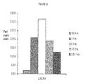
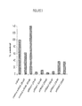
- RPE cells in which oxidative stress was induced displayed a significant increase in transcription of the CXCR4 gene, as judged from gene array analysis, when ARPE-19 cells were exposed to the chemical stressor (tertiary butyl hydroperoxide or t-BH) for 6 hours, experienced cell death within 24 hours. Cells exposed to the stressor for only 3 hours, by contrast, have a 70% survival rate of at least 24 hours. The degree of CXCR4 expression of these latter cells approached that of naive cells not exposed to the stressor at all.
- chemical stressor tertiary butyl hydroperoxide or t-BH
- the present invention is drawn to a method of reducing the rate of progression of an ophthalmic condition, such as an inflammatory retinal condition in a mammal, comprising: administering to the ophthalmic tissue of said mammal a composition comprising an inhibitor of CXCR4 activity.
- the present invention is drawn to a method ofinhibiting or reducing the rate of progression of retinal cell death (including RPE cell death), such as by apoptosis or necrosis, in inhibiting or reducing the rate of progression of apoptotic or necrotic retinal cell death in a mammal, comprising: administering to the retinal tissue of said mammal a composition comprising an inhibitor of CXCR4 activity.
- the present invention is drawn to a method of inhibiting or reducing the rate of progression of retinal or choroidal angiogenesis in a mammal, comprising: administering to the retinal tissue of said mammal a composition comprising an inhibitor of CXCR4 activity.
- a composition comprising an inhibitor of CXCR4 activity.
- CXCR4 (along with CCR5) has been implicated in the etiology of infection of macrophages and T-cells by human immunodeficiency virus (HIV). Accordingly, has been the subject of much study.
- HCV human immunodeficiency virus
- small molecule is meant a chemical compound other than a polynucleotide or polypeptide.
- CXCR4 inhibitory small molecules are disclosed in, among other sources, Bridger et al. U.S. Publication No. 2002/0077339 A1 ; Bridger et al. U.S. Publication No. 2002/0147192 A1 ; Tudan et al. U.S. Publication No. 2002/0156034 A1 ; Bridger et al. U.S. Publication No. 2003/0220341 A1 ; Bridger et al. U.S. Patent No. 6,667,320 B2 ; Bridger et al. U.S. Publication No.
- CXCR4-binding modulators comprising heterocyclic compounds of the basic structure: V-CR 2 -Ar 1 -CR 2 NR-(CR 2 ) x -Ar 2 including the pharmaceutically acceptable salts and protected forms thereof, wherein V is a substituted heterocycle compound of 9-24 members containing 2-4 optionally substituted amine nitrogen atoms spaced from each other by 2 or more optionally substituted carbon atoms, and which heterocycle may optionally comprise a fused aromatic or heteroaromatic ring, and wherein
- Ring A in this structure represents a 5-10 membered nitrogen-containing heterocyclic ring that may have a substituent
- ring B represents a 5-10 membered unsaturated nitrogen-containing heterocyclic ring that may have a substituent
- ring D represents a 4-15 membered nitrogen-containing heterocyclic ring that may have a substituent
- X represents N or C
- Y is C 1-6 substituted or unsubstituted alkyl
- R1 represents H, a hydrocarbon group that may have a substituent, or a ring group that may have a substituent
- R2 and R3 each independently represents H, a hydrocarbon group that may have a substituent, or a ring group that may have a substituent, or may form, together with nitrogen atom bonded to any of these, a nitrogen-containing heterocyclic ring that may have a substituent
- the antagonist is a compound of formula (10): where ring B1 represents a pyridine or pyrimidine ring that may have a substituent; ring D1 represents a 4-8 membered saturated monocyclic nitrogen-containing heterocyclic ring that may have a substituent; Y1 represents a C 1 - 4 substituted or unsubstituted alkyl; and A, X, R1, R2, and R3 have the meanings given to them above.
- Y1 is -(CR5R6)n-, wherein R5 and R6 each represents a hydrogen atom, or R5 and R6 together represent an oxo group; n represents a whole number between 1 and 4, and when n represents a whole number between 2 and 4 each of the CR5R6 may be identical or different.
- R2 represents -(CO)-R2A, wherein R2A is (i) a hydrocarbon group that may be substituted by a basic group and may further have a substituent; (ii) a ring group that may be substituted by a basic group and may further have a substituent; or (iii) a 5-8 membered monocyclic nitrogen-containing heterocyclic ring, such as pyrrolidine, piperidine or morpholine; and R3 is a hydrocarbon group that may have a substituent or a ring group that may have a substituent.
- the ring formed by R2 and R3 together with nitrogen atom(s) they bond to is a 5-8 membered nitrogen-containing heterocyclic ring that may have a substituent.
- D1 is pyrrolidine or piperidine.
- the antagonist is a compound of formula (11):
- the antagonist is a compound of formula (15), wherein R is C 1 - 6 alkyl-NH 2 .
- a substituent of alkyl, aryl, or heteroaryl should be stable and may have up to 20 non-hydrogen atoms each and as many hydrogen atoms as necessary, wherein the non-hydrogen atoms are C, N, O, S, P, F, Cl, Br, and/or l in any stable combination. However, the total number of non-hydrogen atoms on all of the substituents combined must also be 20 or less.
- a substituent is stable if it is sufficiently stable for the compound to be isolated for at least 12 hours at room temperature under normal atmospheric conditions, or if it is sufficiently stable to be useful for at least one use disclosed herein.
- CXCR4 antagonists falling under the scope of one or more of formulas 9-15 include N-[(2R, 3S)-2-(aminomethyl)-1-cyclohexyl-3-pyrrolidinyl]-4-(1-azepanyl)-2-pyrimidine amine, N-[(2R, 3S)-2-(2-aminoethyl)-1-cyclohexyl-3-pyrrolidinyl]-4-(1-azepanyl)-2-pyrimidine amine, N-[(2R, 3S)-2-(3-aminopropyl)-1-cyclohexyl-3-pyrrolidinyl]-4-(1-azepanyl)-2-pyrimidine amine, N-[(2R, 3S)-2-(5-aminopentyl)-1-cyclohexyl-3-pyrrolidinyl]-4-(1-azepanyl)-2-pyrimidine amine, N-[(2R
- CXCR4 activity include, without limitation, at least a CXCR4-binding peptide domain derived from SDF-1, wherein the SDF-1 derivative does not have the CXCR4 stimulatory activity of wild-type SDF-1; and antibodies or antibody mimics (such as those described under the name AdnectinsTM).
- nucleic acid inhibitors of SDF-1 activity have been described, These nucleic acid-based inhibitors may function at either the receptor binding level or the gene expression and translational levels.
- the nucleic acid inhibitors of CXCR4 activity include, without limitations, nucleic acid enzymes (such as ribozymes), nucleic acid aptamers, antisense nucleic acids, and RNAi, such as siRNA.
- nucleic acid enzymes such as ribozymes
- nucleic acid aptamers such as ribozymes
- RNAi such as siRNA.
- Certain specific embodiments of exemplary nucleic acid CXCR4 inhibitors are contained in the following references: lijima et al. U.S. Patent No. 6,429,308 B1 ; Guerciolini et al. U.S. Publication No. 2005/0124569 A1 ; Eagles et al. U.S. Patent No. 6,916,653 B2 ; Watson et
- a "retinal disorder” means an inflammatory, apoptotic, necrotic or angiogenic condition affecting the retina of the eye. Such a condition may involve other structures or tissues as well, including, without limitation, the choroid and retinal pigmented epithelium (RPE).
- RPE retinal pigmented epithelium
- an "ocular condition" is a disease, ailment or condition which affects or involves the eye or one of the parts or regions of the eye.
- the eye includes the eyeball and the tissues and fluids which constitute the eyeball, the periocular muscles (such as the oblique and rectus muscles) and the portion of the optic nerve which is within or adjacent to the eyeball.
- An anterior ocular condition is a disease, ailment or condition which affects or which involves an anterior (i.e. front of the eye) ocular region or site, such as a periocular muscle, an eye lid or an eye ball tissue or fluid which is located anterior to the posterior wall of the lens capsule or ciliary muscles.
- an anterior ocular condition primarily affects or involves the conjunctiva, the cornea, the anterior chamber, the iris, the posterior chamber (behind the iris but in front of the posterior wall of the lens capsule), the lens or the lens capsule and blood vessels and nerve which vascularize or innervate an anterior ocular region or site.
- an anterior ocular condition can include a disease, ailment or condition, such as for example, aphakia; pseudophakia; astigmatism; blepharospasm; cataract; conjunctival diseases; conjunctivitis; corneal diseases;, corneal ulcer; dry eye syndromes; eyelid diseases; lacrimal apparatus diseases; lacrimal duct obstruction; myopia; presbyopia; pupil disorders; refractive disorders and strabismus.
- Glaucoma can also be considered to be an anterior ocular condition because a clinical goal of glaucoma treatment can be to reduce a hypertension of aqueous fluid in the anterior chamber of the eye (i.e. reduce intraocular pressure).
- a posterior ocular condition is a disease, ailment or condition which primarily affects or involves a posterior ocular region or site such as choroid or sclera (in a position posterior to a plane through the posterior wall of the lens capsule), vitreous, vitreous chamber, retina, retinal pigmented epithelium, Bruch's membrane, optic nerve (i.e. the optic disc), and blood vessels and nerves which vascularize or innervate a posterior ocular region or site.
- a posterior ocular region or site such as choroid or sclera (in a position posterior to a plane through the posterior wall of the lens capsule), vitreous, vitreous chamber, retina, retinal pigmented epithelium, Bruch's membrane, optic nerve (i.e. the optic disc), and blood vessels and nerves which vascularize or innervate a posterior ocular region or site.
- a posterior ocular condition can include a disease, ailment or condition, such as for example, acute macular neuroretinopathy; Behcet's disease; choroidal neovascularization; diabetic uveitis; histoplasmosis; infections, such as fungal or viral-caused infections; macular degeneration, such as acute macular degeneration, non-exudative age related macular degeneration and exudative age related macular degeneration; edema, such as macular edema, cystoid macular edema and diabetic macular edema; multifocal choroiditis; ocular trauma which affects a posterior ocular site or location; ocular tumors; retinal disorders, such as central retinal vein occlusion, diabetic retinopathy (including proliferative diabetic retinopathy), proliferative vitreoretinopathy (PVR), retinal arterial occlusive disease, retinal detachment, uveitic retinal
- the present invention is drawn to compositions for use in the methods described above, and the use of such compositions.
- an "inhibitor of CXCR4 activity” or a “C ⁇ CR4 inhibitor” means an agent capable of blocking or otherwise preventing the generation or propagation of an intracellular CXCR4-selective signal that would, but for the presence of said agent, be capable of being generated or propagated under physiological conditions.
- an inhibitor may comprise, without limitation, an antagonist of the CXCR4 receptor or of C ⁇ CR gene expression, a compound or complex able to bind SDF-1 and therefore to prevent and hinder SDF-1 from activating the CXCR4 receptor, or an agent able to reduce SDF-1 gene expression.
- Such agents may comprise, for example and without limitation, peptide or oligonucleotide macromolecular components.
- peptide or polypeptide means a compound that may include a chain of two or more modified or non-modified naturally occurring or non-naturally occurring amino acids linked by at least one peptide bond.
- a peptide according to the present invention may also consist of, consist essentially of, or comprise a peptidomimetic.
- peptidomimetic is used broadly to mean a peptide-like molecule that is able to serve as a model for a peptide substrate upon which it is structurally based.
- Such peptidomimetics include chemically modified peptides, peptide-like molecules containing non-naturally occurring amino acids, and peptoids, which are peptide-like molecules resulting from oligomeric assembly of N-substituted glycines (see, for example, Goodman and Ro, Peptidomimetics for Drug Design, in BURGER'S MEDICINAL CHEMISTRY AND DRUG DISCOVERY Vol. 1 (ed. M.E. Wolff; John Wiley & Sons 1995), pages 803-861 ), all volumes of which are hereby incorporated by reference herein.
- a variety of peptidomimetics are known in the art including, for example, peptide-like molecules which contain a constrained amino acid, a non-peptide component that mimics peptide secondary structure, or an amide bond isostere.
- a peptidomimetic that contains a constrained, non-naturally occurring amino acid can include, for example, an ⁇ -methylated amino acid; an ⁇ , ⁇ -dialkyl-glycine or ⁇ -aminocycloalkane carboxylic acid; an N ⁇ -C ⁇ cyclized amino acid; an N ⁇ -methylated amino acid; a ⁇ - or ⁇ - amino cycloalkane carboxylic acid; an ⁇ , ⁇ -unsaturated amino acid; a ⁇ , ⁇ -dimethyl or ⁇ -methyl amino acid; a ⁇ -substituted-2,3-methano amino acid; an NC ⁇ or C ⁇ -C ⁇ cyclized amino acid; or a substituted proline or another amino acid mimetic.
- a peptidomimetic which mimics peptide secondary structure can contain, for example, a nonpeptidic ⁇ -turn mimic; ⁇ -turn mimic; mimic of ⁇ -sheet structure; or mimic of helical structure, each of which is well known in the art.
- a peptidomimetic also can be a peptide-like molecule which contains, for example, an amide bond isostere such as a retro-inverso modification; reduced amide bond; methylenethioether or methylenesulfoxide bond; methylene ether bond; ethylene bond; thioamide bond; trans-olefin or fluoroolefin bond; 1,5-disubstituted tetrazole ring; ketomethylene or fluoroketomethylene bond or another amide isostere.
- an amide bond isostere such as a retro-inverso modification
- reduced amide bond such as a retro-inverso modification
- methylenethioether or methylenesulfoxide bond such as a amide bond
- trans-olefin or fluoroolefin bond such as a retro-inverso modification
- trans-olefin or fluoroolefin bond such as a retro-inverso modification
- oligonucleotide or “nucleic acid” according to the present invention may comprise two or more naturally occurring or non-naturally occurring deoxyribonucleotides or ribonucleotides linked by a phosphodiester linkage, or by a linkage that mimics a phosphodiester linkage to a therapeutically useful degree.
- an oligonucleotide will normally be considered to be single-stranded unless stated to the contrary or otherwise obvious from the context, and a nucleic acid may be single stranded or double stranded.
- an oligonucleotide or nucleic acid may contain one or more modified nucleotide; such modification may be made in order to improve the nuclease resistance of the oligonucleotide, to improve the hybridization ability (e.g., raise the melting temperature or Tm) of the resulting oligonucleotide, to aid in the targeting or immobilization of the oligonucleotide or nucleic acid, for a mixture of such purposes, or for some other purpose.
- modified nucleotide such modification may be made in order to improve the nuclease resistance of the oligonucleotide, to improve the hybridization ability (e.g., raise the melting temperature or Tm) of the resulting oligonucleotide, to aid in the targeting or immobilization of the oligonucleotide or nucleic acid, for a mixture of such purposes, or for some other purpose.
- Such modifications may include oligonucleotide derivatives having modifications at the nitrogenous base, including replacement of the amino group at the 6 position of adenosine by hydrogen to yield purine; substitution of the 6-keto oxygen of guanosine with hydrogen to yield 2-amino purine, or with sulphur to yield 6-thioguanosine, and replacement of the 4-keto oxygen of thymidine with either sulphur or hydrogen to yield, respectively, 4-thiothymidine or 4-hydrothymidine. All these nucleotide analogues can be used as reactants for the synthesis of oligonucleotides.
- Other substituted bases are known in the art. See, e.g., Cook et al., International Publication No.
- WO 92/02258 entitled “Nuclease Resistant, Pyrimidine Modified Oligonucleotides that Detect and Modulate Gene Expression,” which is hereby incorporated by reference herein.
- Base-modified nucleotide derivatives can be commercially obtained for oligonucleotide synthesis.
- WO 93/13121 entitled “Gapped 2'-Modified Oligonucleotides.”
- 2'amino, 2'-C-allyl, 2'-fluoro, 2'-O-methyl, and 2'H groups tend to confer nuclease resistance and to permit hybridization between the modified nucleotide and an unaltered nucleotide in the annealed strand.
- 2'amino, 2'-C-allyl, 2'-fluoro, 2'-O-methyl, and 2'H groups tend to confer nuclease resistance and to permit hybridization between the modified nucleotide and an unaltered nucleotide in the annealed strand.
- oligonucleotides comprising such modified bases have been formulated with increased cellular uptake, nuclease resistance, and/or increased substrate binding in mind. In other words, such oligonucleotides are described as therapeutic gene-modulating agents.
- modified bases in RNA can include methylated or dimethylated bases, deaminated bases, carboxylated bases, thiolated bases and bases having various combinations of these modifications.
- 2'-O-alkylated bases are known to be present in naturally occurring nucleic acids. See e.g., Adams et al., The Biochemistry of the Nucleic Acids (11th ed 1992 ), hereby incorporated by reference herein.
- Human CXCR4 amino acid sequences have been determined and are a matter of public record. Human C ⁇ CR4 amino acid sequences have been determined and is a matter of public record. The following human CXCR4 sequence has NCBl accession number P61073. All amino acid sequences shown are from N terminus to C terminus, and all nucleotide sequences shown are from 5' to 3', unless otherwise indicated.
- Human CXCR4 cDNA sequences are as follows.
- This cDNA encodes a CXCR4 protein having a longer amino acid sequence than the CXCR4 shown in SEQ ID No: 1.
- start codons are found at positions 131 and 152, with a stop codon at position 220.
- An additional start codon is found at position 305, at which point the nucleotide sequence remains identical with that of SEQ ID No. 3, shown below, which encodes SEQ ID No. 1.
- Other in-frame start codons are found at positions 362, 386, 506, 530, and 929.
- the presently preferred stop codon in this portion of the frame is located at position 1372. Given these features, a genus of possible isoforms are disclosed and suggested by this disclosure, having various 5' termini but with each with a C terminus ending in the amino acids SSFHSS.
- the ATG start codon encoding SEQ ID NO: 1 begins at residue 96 and proceeds to residue 1151.
- the 5' untranslated region and N terminus of the translated protein are different from that of the DNA sequence of, and respective protein encoded by, SEQ ID No: 2, but beginning with residue 6 of the protein encoded by this cDNA (corresponding to nucleotide residue 108), the amino acid sequences are again identical.
- Genbank Accession Number MN_003467 (December 9, 2005).
- NCBl National Center For Biotechnology Information
- the first residue of mature SDF-1 is the lysine residue, corresponding to position 21 of SEQ ID NO: 4, 5, and 6 of the following SDF-1 amino acid sequences.
- the following proline residue has been shown to be important for the activity of SDF-1, as a substitution of this reside with, for example, glycine preserves CXCR4 binding activity while converting SDF-1 into a CXCR4 antagonist. See also Crump et al., The EMBO J. 16:6996-7007 (1997 ), hereby incorporated by reference herein in its entirely.
- NCBl accession number NP_000600 (November 25, 2005)
- GenBank accession number MN _ 000609 (December 9, 2005)
- inhibitors can be directed in whole or in part to the 5' and 3' untranslated regions (UTRs) of one or more cDNA variant.
- UTRs 5' and 3' untranslated regions
- RNAi and antisense oligonucleotides are capable of inhibiting translation of mRNA in the coding region, in the 5' UTR and in the 3' UTR, and RNAi templates may be made that are complementary to these regions, or to regions bridging two such regions.
- more than one RNAi template may be used, with various loci in the mRNA being targeted for RNA-induced mRNA digestion and inhibition of translation.
- Polypeptide inhibitors of CXCR4 activity may include peptides, polypeptides, and proteins.
- the therapeutic agent may comprise a protein, a modified SDF-1 derivative comprising a CXCR4 binding region (but lacking CXCR4 stimulatory activity) a polyclonal or monoclonal antibody, an antibody fragment, such as, without limitation, a monovalent fraction antigen-binding papain fragment (Fab) or a bivalent fraction antigen binding pepsin fragment (F'ab 2 ).
- Fab monovalent fraction antigen-binding papain fragment
- F'ab 2 bivalent fraction antigen binding pepsin fragment
- the antibodies or antibody fragments may be naturally occurring or genetically engineered.
- antibodies may include chimeric antibodies comprising human L c and H c regions and L v and H v regions from another species, for example, from mouse, goat, rabbit or sheep cells.
- the non-human species is a mouse.
- Chimeric antibodies are useful in the design of antibody-based drugs, since the use of unaltered mouse antibodies induces the production of human anti-mouse immunoglobulins and resultant clearance and reduction of efficacy.
- chimeric antibodies while having reduced immunogenicity as compared to the rodent antibody, do not solve all the problems that exist in the use of antibodies as drugs.
- a human consensus sequence can be used representing the most common allotype in the general population.
- a further refinement has been used, called complimentarily determining region (CDR) grafting.
- CDR complimentarily determining region
- reshaping the rodent variable region is compared with the consensus sequence of the protein sequence subgroup to which it belongs, as is the human framework compared with a consensus of the framework sequence for the antibody family to which it belongs.
- This analysis can identify amino acid residues that may be the result of mutation during the affinity maturation process; these residues are called “idiosyncratic”. By incorporating the more common human residues in these positions, immunogenicity problems resulting from the idiosyncratic residues can be minimized.
- Humanization by hyperchimerization involves a comparison of the human and murine non-CDR variable region sequences and the one with the highest homology is selected as the acceptor framework. Again, idiosyncratic residues are replaced with more highly conserved human ones. Those non-CDR residues that may interact with the CDR residues are identified and inserted into the framework sequence.
- Veneering involves determining the three dimensional conformation of a humanized murine antibody and replacing the expose surface amino acids with those commonly found in human antibodies.
- the most homologous human variable regions are selected and compared to the corresponding mouse variable regions.
- the mouse framework residues differing from the human framework are replaced with the human residues; only those residues fully or partially exposed at the surface of the antibody are changed.
- the desired inhibitor may be directed to bind, for example, an SDF-1 molecule or a C ⁇ CR4 receptor molecule.
- an anti-SDF—1 binding polypeptide when permitted to contact SDF-1 in a tissue or cell will prevent the SDF-1 molecule from activating a CXCR4 receptor in such tissue or cell.
- the inhibitor may be an anti-CXCR4 polypeptide, such as a CXCR4 antagonist or inverse agonist.
- an example of such an inhibitor may be, for example, the anti-CXCL1 2/SDF-1 ⁇ antibody sold by R&D Systems (614 McKinley Place NE, ⁇ Minneapolis, MN 55413) under catalog number AF-351-NA and described as a goat anti-human CXCL12/SDF-1 ⁇ lgG.
- This polyclonal antibody preparation was prepared in goats immunized with purified, E. coli derived, recombinant human CXCL12/SDF-1 ⁇ . This antibody was first purified by passing the goat sera over an SDF-1 ⁇ affinity column, and then the unbound fraction was further purified by binding to an SDF-1 ⁇ affinity column.
- This antibody preparation has a neutralization dose (ND 50 ) (that concentration required to yield 1 ⁇ 2maximal inhibition of the cytokine activity in a responsive cell line, when that cytokine is present at a concentration just high enough to elicit a maximum response) of approximately 10-30 ⁇ g/ml in the presence of 10 ng/ml CXCL12, as assay measuring chemotaxis of BaF/3 cells transfected with human CXCR4.
- ND 50 neutralization dose
- variable region of antibodies having greater than a threshold level of avidity for the CXCR4 or SDF-1 mRNA can be sequenced and used to provide the basis for a therapeutic antibody or antibody derivative. See e.g., Hodxie et al., U.S. Patent No. 5,994,515 (disclosing antibodies directed towards CXCR4); Mueller et al, U.S. Patent No. 6,949,243 (disclosing antibody preparations having anti-CXCR4 and anti SDF-1 activity) ; Plaskin et Al., U.S. Patent Publication No. 2005/0266009 (disclosing antibodies directed against CXCR4.)
- C ⁇ CR4 inhibitors are known in the art. Examples are described, for example, in Tudan, et al. U.S. Publication No. 2006/0014682 ; Clark-Lewis et al. U.S. Publication No. 2005/0265969 .
- Inhibitors of CXCR4 activity may also comprise compositions containing a nucleic acid component.
- nucleic acids may include without exception RNAi molecules, such as dsRNA and siRNA.
- Inhibitory nucleic acids may comprise, for example, a single-stranded oligonucleotide sufficiently complementary to a region of a target mRNA such that said oligonucleotide prevents translation of said mRNA.
- said region comprises the 5' UTR, the ribosome binding site of said mRNA, the coding region of said mRNA, and may include the 5'start codon of said mRNA.
- Other targets may include the 3'UTR
- Said oligonucleotide may comprise one or more modified nucleotide residue (such as, without limitation, a 2'O alkyl nucleotide); preferably said modified nucleotide residue confers at least one of a nuclease resistance or a higher binding avidity.
- an oligonucleotide may comprise a peptide nucleic acid, or other nucleic acid mimic.
- the target mRNA may be a CXCR4 mRNA; according to another aspect of the present invention the target mRNA may be an SDF-1 mRNA.
- RNAi is inhibitory RNA. Restriction of foreign nucleic acids such as viruses and transposable elements (transposons) by RNAi mechanisms is an inherent and naturally occurring phenomenon in many plants, vertebrates and invertebrates, and constitutes an "immune system" at the genetic level. RNAi is a phenomenon in which a double stranded RNA (dsRNA) that is related in sequence to a specific mRNA can cause a selective degradation of that RNA.
- dsRNA double stranded RNA
- RNAi mechanism involves a double stranded RNA (dsRNA) molecule which is cleaved within the cell by an enzyme termed "Dicer", resulting in fragments of the original dsRNA comprising about 21 to about 23 nucleotides in length, or consisting of a double stranded RNA fragment 21 to 23 nucleotides in length.
- dsRNA double stranded RNA
- Dier an enzyme termed "Dicer”
- fragments of the original dsRNA comprising about 21 to about 23 nucleotides in length, or consisting of a double stranded RNA fragment 21 to 23 nucleotides in length.
- siRNA short interfering RNA fragments
- the siRNA is designed and used without a dsRNA precursor. Certain aspects may have longer overhangs, including 3 or 4 nucleotides.
- the siRNA then associates with a protein complex called the RNA induced silencing complex (RISC).
- RISC RNA induced silencing complex
- This complex comprises a multiple components, and is activated by ATP.
- the activities associated with the complex include a helicase, a dsRNA nuclease and an RNA-dependent RNA polymerase activity; all these activities have been found to be essential for RNAi in Drosophila, C. elegans, and Neurospora.
- the siRNA permits the complex to be targeted to a particular mRNA. Once the RISC has bound to the target mRNA, there is an ATP-independent cleavage of the target RNA by an RISC-associated RNAse activity termed Slicer.
- RNA-dependent RNAse activity appears to produce secondary siRNAs through an amplification process in which the original siRNA is primer extended using the target mRNA as a template in a manner similar to PCR.
- dsRNA long (dsRNA) or short (siRNA) RNA species
- siRNA short RNA species
- RNAi expression systems such as those disclosed in Lee et al., 19 NATURE BIOTECHNOLOGY 500 (May, 2002 ); Miyagishi et al., 19 NATURE BIOTECHNOLOGY 497 (May 2002 ) and Tuschl, 20 NATURE BIOTECHNOLOGY 446 (May 2002 ) have been demonstrated as capable of producing siRNA for downregulating gene expression in mammalian cells. These references are hereby incorporated herein by reference in their entirety.
- RNAi may be quite useful as a therapeutic agent (an inhibitor of CXCR4 activity).
- RNAi may, without limitation, be administered by injection as naked dRNA or siRNA or in a liposome, implant, expression vector, viral vector, and the like (or a combination of one or more of these methods) directed into the in vitreous of the eye.
- the drug is not given systemically, there would be less toxicity expected than for systemically administered drugs.
- the catalytic or amplified nature of the gene-silencing phenomenon described above make RNAi a very attractive therapeutic method generally. For example, relatively few dsRNA and/or siRNA molecules might be required to induce a gene-silencing phenomenon in any tissue to which the RNA is applied.
- the inhibitors of CXCR4 activity have a desired ocular therapeutic effect.
- a desired ocular therapeutic effect includes the ability to prevent or reduce the extent of ocular cell death; for example, without limitation, neural cell death or RPE cell death by, for example, necrosis or apoptosis.
- the therapeutic compounds of the present invention may be used to prevent or lessen the extent or rate of neovascularization in the posterior segment of the eye. Further, the therapeutic compounds of the present invention may be used to treat conditions, particularly, though not exclusively, retinal conditions, implicating acute or chronic inflammation in the eye.
- Such conditions may include, without limitation, acute macular neuroretinopathy; Behcet's disease; choroidal neovascularization; diabetic uveitis; macular degeneration, such as acute macular degeneration, non-exudative age related macular degeneration and exudative age related macular degeneration; edema, such as macular edema, cystoid macular edema and diabetic macular edema; multifocal choroiditis; ocular tumors; retinal disorders, such as central retinal vein occlusion, diabetic retinopathy (including proliferative diabetic retinopathy), proliferative vitreoretinopathy (PVR), retinal arterial occlusive disease, uveitic retinal disease; branch retinal vein occlusion.
- PVR proliferative vitreoretinopathy
- the therapeutic compounds of, and used in, the present invention can be administered in any way suitable to effectively achieve a desired ocular therapeutic effect.
- methods of administration may include without limitation, topical, intraocular (including intravitreal), transdermal, oral, intravenous, subconjunctival, subretinal, or peritoneal.
- topical and intraocular (e.g., intravitreal, subconjunctival and subretinal) methods of administration of the CXCR4 inhibitors of the present invention may often be preferred; as such means of administration avoid many of the possible disadvantages of systemic administration, such as undesired side effects in tissues other than the eye.
- topical administration may generally be initially preferable from the point of view of ease of use, a comparably low degree of ocular trauma, and relatively few risks associated with the means of administration itself.
- agents that are applied topically to the ocular surface topically may have a very short residence time on the cornea.
- topical drug delivery can deliver therapeutic concentrations of the drug to anterior segment features such as the cornea, anterior chamber, iris, lens and cilary body of the eye, but drug delivery to posterior segment features such as the vitreous humor, retinal pigmented epithelium, retina and choroid is less effective.
- drug applied topically to the eye can diffuse through the conjunctiva and sclera, and then penetrate the eye through the iris route or the retinal pigmented epithelium (RPE).
- RPE retinal pigmented epithelium
- topically applied ophthalmic drugs often do not achieve therapeutic concentrations in the posterior segment tissues. Additionally, topically applied macromolecular CXCR4 inhibitors may, in many cases, be too large to quickly diffuse into the tissue of the posterior segment.
- the therapeutic compositions of the present invention will be administered in a pharmaceutically acceptable vehicle component.
- the therapeutic agent may also be combined with a pharmaceutically acceptable vehicle component in the manufacture of a composition,
- a composition as disclosed herein, may comprise a CXCR4 inhibitor and an effective amount of a pharmaceutically acceptable vehicle component.
- the vehicle component is aqueous-based.
- the composition may comprise water.
- the CXCR4 inhibitor is administered in a vehicle component, and may also include an effective amount of at least one of a viscosity inducing component, a resuspension component, a preservative component, a tonicity component and a buffer component.
- the compositions disclosed herein include no added preservative component.
- a composition may optionally include an added preservative component.
- the composition may be included with no resuspension component.
- Subconjunctival delivery is a very attractive possibility, as retinal diffusion appears to be greater than seen with topical delivery, and the administration of the drug is not as traumatic as intraocular delivery (such as intravitreal and sub retinal placement or injection).
- Subconjunctival delivery may include injection of a solution or suspension containing the CXCR4 inhibitor, and/or the administration of an implant, such as a biodegradable matrix comprising, for example, a poly lactoside poly glycoside co-polymer designed to release the active drug over a pre-determined period of time.
- intravitreal and subretinal administration means may not be effectively used too.
- intraocular means are the most direct way of contacting the affected retinal tissue with the therapeutic agent.
- liquid solutions or suspensions usually having a viscosity and a refractive index greater than that of water (to prevent adversely affecting the viscosity of the vitreous humor and thus the vision of the patient).
- Additional delivery means may include injection to the muscular tissues of the orbit or to the upper or lower eyelid. While these means may indeed be effective in delivering the CXCR4 inhibitors of the present invention (particularly in the case of small and/or lipophilic compounds), these means are not now currently preferred.
- Formulations for topical, subconjunctival, subretinal or intraocular administration of the therapeutic agents will preferably include a major amount of liquid water.
- Such compositions are preferably formulated in a sterile form, for example, prior to being used in the eye.
- the above-mentioned buffer component if present in the formulations, is present in an amount effective to control the pH of the composition.
- the formulations may contain, either in addition to, or instead of the buffer component at least one tonicity component in an amount effective to control the tonicity or osmolality of the compositions. Indeed, the same component may serve as both a buffer component and a tonicity component. More preferably, the present compositions may include both a buffer component and a tonicity component.
- the buffer component and/or tonicity component may be chosen from those that are conventional and well known in the ophthalmic art.
- buffer components include, but are not limited to, acetate buffers, citrate buffers, phosphate buffers, borate buffers and the like and mixtures thereof. Phosphate buffers are particularly useful.
- Useful tonicity components include, but are not limited to, salts, particularly sodium chloride, potassium chloride, any other suitable ophthalmically acceptably tonicity component and mixtures thereof.
- Non-ionic tonicity components may comprise polyols derived from sugars, such as xylitol, sorbitol, mannitol, glycerol and the like.
- the amount of buffer component employed preferably is sufficient to maintain the pH of the composition in a range of about 6 to about 8, more preferably about 7 to about 7.5.
- the amount of tonicity component employed preferably is sufficient to provide an osmolality to the present compositions in a range of about 200 to about 400, more preferably about 250 to about 350, mOsmol/kg respectively.
- the present compositions are substantially isotonic.
- compositions of, or used in, the present invention may include one or more other components in amounts effective to provide one or more useful properties and/or benefits to the present compositions.
- present compositions may be substantially free of added preservative components, in other embodiments, the present compositions include effective amounts of preservative components, preferably such components that are more compatible with or friendly to the tissue in the posterior segment of the eye into which the composition is placed than benzyl alcohol.
- preservative components include, without limitation, quaternary ammonium preservatives such as benzalkonium chloride ("BAC” or “BAK”) and polyoxamer; biguanidebigunanide preservatives such as polyhexamethylene biguanidebiguandide (PHMB); methyl and ethyl parabens; hexetidine; chlorite components, such as stabilized chlorine dioxide, metal chlorites and the like; other ophthalmically acceptable preservatives and the like and mixtures thereof.
- BAC benzalkonium chloride
- BAK polyoxamer
- biguanidebigunanide preservatives such as polyhexamethylene biguanidebiguandide (PHMB)
- PHMB polyhexamethylene biguanidebiguandide
- chlorite components such as stabilized chlorine dioxide, metal chlorites and the like
- other ophthalmically acceptable preservatives and the like and mixtures thereof include, without limitation, quaternary ammonium
- the concentration of the preservative component, if any, in the present compositions is a concentration effective to preserve the composition, and (depending on the nature of the particular preservative used) is often and generally used in a range of about 0.00001 % to about 0.05% (w/v) or about 0.1% (w/v) of the composition.
- lntravitreal delivery of therapeutic agents can be achieved by injecting a liquid-containing composition into the vitreous, or by placing polymeric drug delivery systems, such as implants and microparticles comprising the therapeutic agent, such as microspheres, or a rod, wafer of other shaped implant, into the vitreous.
- polymeric drug delivery systems such as implants and microparticles comprising the therapeutic agent, such as microspheres, or a rod, wafer of other shaped implant
- Similar methods may be used for subconjunctival or subretinal delivery of the drug.
- the The implant may be placed (e.g., injected) under the conjunctiva at a location proximal to the back of the eye.
- a suspension or liquid based formulation would be administered so as to remain under the conjunctival membrane, effectively using it as a depot for sustained release of the CXCR4 inhibitor.
- releaseRelease of the CXCR4 inhibitor will occur over a period of time and will not have to travel as far around the circumference of the scleraschlera or through the anterior segment as topically applied drug would need to travel.
- An advantage to this mode of delivery is that no puncture of the eye itself is required.
- Subretinal placement of an implant involves some trauma to the eye, but does nor require puncture of the eyeball, as does intravitreal injection. The risk of severe consequences of infection is not as great when the vitreous is not punctured.
- Formulations containing the therapeutic agents of the present invention can be produced using conventional techniques routinely known by persons of ordinary skill in the art.
- a CXCR4 inhibitor can be combined with a liquid carrier.
- the composition can be sterilized or sterile components mixed.
- the compositions can be sterilized and packaged in single-dose amounts.
- the compositions may be prepackaged in dispensers that can be disposed of after a single administration of the unit dose of the compositions.
- compositions comprising the inhibitors of CXCR4 activity can be prepared using suitable blending/processing techniques, for example, one or more conventional blending techniques.
- the preparation processing should be chosen to provide such inhibitors in implants having as form useful for intravitreal, subconjunctival, subretinal or periocular placement or injection into eyes of humans or animals.
- a concentrated therapeutic component dispersion is made by combining the therapeutic component with water, and the excipients (other than a viscosity inducing component) to be included in the final composition.
- the ingredients are mixed to disperse the therapeutic component and then autoclaved.
- Viscosity inducing component may be purchased sterile or sterilized by conventional processing, for example, by filtering a dilute solution followed by lyophylization to yield a sterile powder.
- the sterile viscosity-inducing component is combined with water to make an aqueous concentrate.
- the concentrated therapeutic component dispersion is mixed and added as a slurry to the viscosity inducing component concentrate. Water is added in a quantity sufficient (q.s.) to provide the desired composition and the composition is mixed until homogenous.
- a sterile, viscous suspension suitable for administration is made using an inhibitor of CXCR4 activity.
- a process for producing such a composition may comprise sterile suspension bulk compounding and aseptic filling.
- inventions of the present materials are in the form of a polymeric drug delivery system that is capable of providing sustained drug delivery for extended periods of time after a single administration.
- the present drug delivery systems can release the inhibitor of CXCR4 activity for at least about two weeks, or about three weeks, or 1 month, or about 3 months, or about 6 months, or about 1 year, or about 5 years or more.
- such embodiments of the present materials may comprise a polymeric component associated with the therapeutic component in the form of a polymeric drug delivery system suitable for administration to a patient by at least one of intravitreal, subconjunctival, subretinal and periocular administration.
- the polymeric drug delivery system may be in the form of biodegradable polymeric implants, non-biodegradable polymeric implants, biodegradable polymeric microparticles, and combinations thereof implants may be in the form of rods, wafers, sheets, filaments, spheres, and the like. Particles are generally smaller than the implants disclosed herein, and may vary in shape. For example, certain embodiments of the present invention utilize substantially spherical particles. These particles may be in the form of microspheres. Other embodiments may utilize randomly configured particles, such as particles that have one or more flat or planar surfaces.
- the drug delivery system may comprise a population of such particles with a predetermined size distribution. For example, a major portion of the population may comprise particles having a desired diameter measurement.
- the polymeric component of the implantable drug delivery systems can comprise a polymer selected from the group consisting of biodegradable polymers, non-biodegradable polymers, biodegradable copolymers, non-biodegradable copolymers, and combinations thereof.
- the polymeric component comprises a poly (lactide-co-glycolide) polymer (PLGA).
- the polymeric component comprises a polymer selected from the group consisting of poly-lactic acid (PLA), poly-glycolic acid (PGA), poly-lactide-co-glycolide (PLGA), polyesters, poly (ortho ester), poly(phosphazine), poly (phosphate ester), polycaprolactones, gelatin, collagen, derivatives thereof, and combinations thereof.
- the polymeric component may be associated with the therapeutic component to form an implant selected from the group consisting of solid implants, semisolid implants, and viscoelastic implants.
- the polymeric component may comprise any polymeric material useful in a body of a mammal, whether derived from a natural source or synthetic.
- useful polymeric materials for the purposes of this invention include carbohydrate based polymers such as methylcellulose, carboxymethylcellulose, hydroxymethylcellulose hydroxypropylcellulose, hydroxyethylcellulose, ethyl cellulose, dextrin, cyclodextrins, alginate, hyaluronic acid and chitosan, protein based polymers such as gelatin, collagen and glycolproteins, and hydroxy acid polyesters such as polyglycolide, polyhydroxybutyric acid, polycaprolactone, polyvalerolactone, polyphosphazene, and polyorthoesters.
- Polymers can also be crosslinked, blended or used as copolymers in the implant.
- Other polymer carriers include albumin, polyanhydrides, polyethylene glycols, polyvinyl polyhydroxyalkyl methacrylates, pyrrolidone and polyvinyl alcohol.
- non-erodible polymers include silicone, polycarbonates, polyvinyl chlorides, polyamides, polysulfones, polyvinyl acetates, polyurethane, ethylvinyl acetate derivatives, acrylic resins, crosslinked polyvinyl alcohol and crosslinked polyvinylpyrrolidone, polystyrene and cellulose acetate derivatives.
- polymeric materials may be useful with any of the CXCR4 inhibitors disclosed herein.
- particles of PLA or PLGA may be coupled to a CXCR4 inhibitor. This insoluble tripartite conjugate will slowly erode over time, thereby continuously releasing the CXCR4 inhibitor.
- the inhibitor of CXCR4 activity may be wholly or partly in a particulate or powder form and entrapped by a biodegradable polymer matrix. If in a particulate form (for example, a crystalline or microcrystalline form), particles in intraocular, subconjunctival, or subretinal implants will generally have an effective average size measuring less than about 3000 nanometers. However, in other embodiments, the particles may have an average maximum size greater than about 3000 nanometers. In certain implants, the particles may have an effective average particle size about an order of magnitude smaller than 3000 nanometers. For example, the particles may have an effective average particle size of less than about 500 nanometers.
- the particles may have an effective average particle size of less than about 400 nanometers, and in still further embodiments, a size less than about 200 nanometers.
- the resulting polymeric particles may be used to provide a desired therapeutic effect.
- the inhibitor of CXCR4 activity may be preferably from about 1% to 90% by weight of the drug delivery system. More preferably, such an inhibitor of CXCR4 activity is from about 20% to about 80% by weight of the system. In a preferred embodiment, the CXCR4 inhibitorGD comprises about 40% by weight of the system (e.g., 30%-50%). In another embodiment, the CXCR4 inhibitorGD comprises about 60% by weight of the system.
- Suitable polymeric materials or compositions for use in the drug delivery systems include those materials that are compatible, e.g., biocompatible, with the eye so as to cause no substantial interference with the functioning or physiology of the eye.
- Such materials preferably include polymers that are at least partially and more preferably substantially completely biodegradable or bioerodible.
- examples of useful polymeric materials include, without limitation, such materials derived from and/or including organic esters and organic ethers, which when degraded result in physiologically acceptable degradation products, including the monomers.
- polymeric materials derived from and/or including, anhydrides, amides, orthoesters and the like, by themselves or in combination with other monomers may also find use.
- the polymeric materials may be addition or condensation polymers, advantageously condensation polymers.
- the polymeric materials may be cross-linked or non-cross-linked, for example not more than lightly cross-linked, such as less than about 5%, or less than about 1% of the polymeric material being cross-linked.
- the polymers will include at least one of oxygen and nitrogen, advantageously oxygen.
- the oxygen may be present as oxy, e.g. hydroxy or ether, carbonyl, e.g. non-oxo-carbonyl, such as carboxylic acid ester, and the like.
- the nitrogen may be present as amide, cyano and amino.
- Polyesters of interest include polymers of D-lactic acid, L-lactic acid, racemic lactic acid, glycolic acid, polycaprolactone, and combinations thereof.
- L-lactate or D-lactate a slowly eroding polymer or polymeric material is achieved, while erosion is substantially enhanced with the lactate racemate.
- polysaccharides are, without limitation, calcium alginate, and functionalized celluloses, particularly carboxymethylcellulose esters characterized by being water insoluble, a molecular weight of about 5 kD to 500 kD, for example.
- polymers of interest include, without limitation, polyesters, polyethers and combinations thereof which are biocompatible and may be biodegradable and/or bioerodible.
- Some preferred characteristics of the polymers or polymeric materials for use in the present systems may include biocompatibility, compatibility with the therapeutic component, ease of use of the polymer in making the drug delivery systems of the present invention, a half-life in the physiological environment of at least about 6 hours, preferably greater than about one day, not significantly increasing the viscosity of the vitreous, and water insolubility.
- the biodegradable polymeric materials which are included to form the matrix are desirably subject to enzymatic or hydrolytic instability.
- Water soluble polymers may be cross-linked with hydrolytic or biodegradable unstable cross-links to provide useful water insoluble polymers.
- the degree of stability can be varied widely, depending upon the choice of monomer, whether a homopolymer or copolymer is employed, employing mixtures of polymers, and whether the polymer includes terminal acid groups.
- the relative average molecular weight of the polymeric composition employed in the present systems is important to controlling the biodegradation of the polymer and hence the extended release profile of the drug delivery systems. Different molecular weights of the same or different polymeric compositions may be included in the systems to modulate the release profile. In certain systems, the relative average molecular weight of the polymer will range from about 9 to about 64 kD, usually from about 10 to about 54 kD, and more usually from about 12 to about 45 kD.
- copolymers of glycolic acid and lactic acid are used, where the rate of biodegradation is controlled by the ratio of glycolic acid to lactic acid.
- the most rapidly degraded copolymer has roughly equal amounts of glycolic acid and lactic acid.
- Homopolymers, or copolymers having ratios other than equal, are more resistant to degradation.
- the ratio of glycolic acid to lactic acid will also affect the brittleness of the system, where a more flexible system or implant is desirable for larger geometries.
- the % of polylactic acid in the polylactic acid polyglycolic acid (PLGA) copolymer can be 0-100%, preferably about 15-85%, more preferably about 35-65%. In some systems, a 50/50 PLGA copolymer is used.
- the biodegradable polymer matrix may comprise a mixture of two or more biodegradable polymers.
- the system may comprise a mixture of a first biodegradable polymer and a different second biodegradable polymer.
- One or more of the biodegradable polymers may have terminal acid groups.
- Release of a drug from an erodible polymer is the consequence of several mechanisms or combinations of mechanisms. Some of these mechanisms include desorption from the implants surface, dissolution, diffusion through porous channels of the hydrated polymer and erosion. Erosion can be bulk or surface or a combination of both. It may be understood that the polymeric component in this aspect of the present invention is associated with the CXCR4 inhibitor so that the release of the therapeutic component into the eye is by one or more of diffusion, erosion, dissolution, and osmosis. As discussed herein, the matrix of an intraocular, subconjunctival, periocular or subretinal drug delivery system may release drug at a rate effective to sustain release of an amount of the therapeutic agent for more than one week after implantation into an eye.
- therapeutic amounts of the CXCR4 inhibitor are released for more than about one month, and even for about twelve months or more.
- the therapeutic component can be released into the eye for a time period from about ninety days to about one year after the system is placed in the interior of an eye.
- the release of the CXCR4 inhibitor from the drug delivery systems comprising a biodegradable polymer matrix may include an initial burst of release (for example, about 20%, or about 30%, or about 40%, or about 50%, or about 60%, or about 70%, or about 80% of the CXCR4 inhibitor is released in the first 3 day after implantation), followed by a gradual increase in the amount of the inhibitor released, or the release may include an initial delay in release of the CXCR4 inhibitor followed by an increase in release.
- the percent of the therapeutic agent that has been released is about one hundred.
- the CXCR4 inhibitor may be desirable to provide a relatively constant rate of release of the therapeutic agent from the drug delivery system over the life of the system.
- the CXCR4 inhibitor may be desirable for the CXCR4 inhibitor to be released in amounts from about 0.01 ⁇ g to about 2 ⁇ g per day for the life of the system.
- the release rate may change to either increase or decrease depending on the formulation of the biodegradable polymer matrix.
- the release profile of the CXCR4 inhibitor may include one or more linear portions and/or one or more non-linear portions.
- the release rate is greater than zero once the system has begun to degrade or erode.
- the drug delivery systems such as the intraocular implants, may be monolithic, i.e. having the active agent or agents homogenously distributed through the polymeric matrix, or encapsulated, where a reservoir of active agent is encapsulated by the polymeric matrix. Due to ease of manufacture, monolithic implants are usually preferred over encapsulated forms. However, the greater control afforded by the encapsulated, reservoir-type implant may be of benefit in some circumstances, where the therapeutic level of the CXCR4 inhibitor falls within a narrow window.
- the therapeutic component including the therapeutic agent(s) described herein, may be distributed in a non-homogenous pattern in the matrix.
- the drug delivery system may include a portion that has a greater concentration of the CXCR4 inhibitor relative to a second portion of the system.
- the polymeric implants disclosed herein may have a size of between about 5 ⁇ m and about 2 mm, or between about 10 ⁇ m and about 1 mm for administration with a needle, greater than 1 mm, or greater than 2 mm, such as 3 mm or up to 10 mm, for administration by implantation.
- the vitreous chamber (and the subconjunctiva) in humans is able to accommodate relatively large implants of varying geometries, having lengths of, for example, 1 to 10 mm.
- the implant may be a cylindrical pellet (e. g., rod) with dimensions of about 2 mm ⁇ 0.75 mm diameter.
- the implant may be a cylindrical pellet with a length of about 7 mm to about 10 mm, and a diameter of about 0.75 mm to about 1.5 mm.
- the implants may also be at least somewhat flexible so as to facilitate both insertion of the implant in the eye, such as in the vitreous, under the conjunctiva, or under the retina, and accommodation of the implant.
- the total weight of the implant is usually about 250-5000 ⁇ g, more preferably about 500-1000 ⁇ g.
- an implant may be about 500 ⁇ g, or about 1000 ⁇ g.
- larger implants may also be formed and further processed before administration to an eye.
- larger implants may be desirable where relatively greater amounts of the CXCR4 inhibitor are provided in the implant,
- Drug delivery systems can be prepared where the center may be of one material and the surface may have one or more layers of the same or a different composition, where the layers may be cross-linked, or of a different molecular weight, different density or porosity, or the like.
- the center may be a polylactate coated with a polylactate-polyglycolate copolymer, so as to enhance the rate of initial degradation.
- the center may be polyvinyl alcohol coated with polylactate, so that upon degradation of the polylactate exterior the center would dissolve and be rapidly washed out of the eye.
- the drug delivery systems may be of any geometry suitable to the delivery means including fibers, sheets, films, microspheres, spheres, circular discs, plaques and the like.
- the upper limit for the system size will be determined by factors such as toleration for the system, size limitations on insertion, ease of handling, etc.
- the sheets or films will be in the range of at least about 0.5 mm ⁇ 0.5 mm, usually about 3-10 mm ⁇ 5-10 mm with a thickness of about 0.1-1.0 mm for ease of handling.
- the fiber diameter will generally be in the range of about 0.05 to 3 mm and the fiber length will generally be in the range of about 0.5-10 mm.
- Spheres may be in the range of about 0.5 ⁇ m to 4 mm in diameter, with comparable volumes for other shaped particles.
- the size and form of the system can also be used to control the rate of release.
- RPE retinal pigment epithelium
- Tight junctions join the cells of this tissue.
- the RPE is a tight ion-transporting barrier that restricts paracellular transport of solutes across the RPE.
- the permeability of most compounds across the blood-retinal barriers is very low.
- the RPE itself possesses the singular ability to quickly take up extracellular agents by phagocytosis, thus this fact vitiates in favor of extrasystemic transport to the retina.
- the CXCR4 inhibitors (including vectors, oligonucleotides and polypeptides and/or conjugates thereof) of the present invention may be suspended in a viscous formulation having a relatively high viscosity, such as one approximating that of the vitreous humor.
- a viscous formulation comprises a viscosity-inducing component.
- the CXCR4 inhibitors of the present invention may be administered subconjunctivally, subretinally, or intravitreally as, without limitation, an aqueous injection, a suspension, an emulsion, a solution, a gel.
- the viscosity-inducing component preferably comprises a polymeric component and/or at least one viscoelastic agent, such as those materials that are useful in ophthalmic surgical procedures.
- useful viscosity inducing components include, but are not limited to, hyaluronic acid, carbomers, polyacrylic acid, cellulosic derivatives, polycarbophil, polyvinylpyrrolidone, gelatin, dextrin, polysaccharides, polyacrylamide, polyvinyl alcohol, polyvinyl acetate, derivatives thereof and mixtures thereof.
- the molecular weight of the presently useful viscosity inducing components may be in a range up to about 2 million Daltons, such as of about 10,000 Daltons or less to about 2 million Daltons or more. In one particularly useful embodiment, the molecular weight of the viscosity-inducing component is in a range of about 100,000 Daltons or about 200,000 Daltons to about 1 million Daltons or about 1.5 million Daltons.
- a viscosity inducing component is a polymeric hyaluronate component, for example, a metal hyaluronate component, preferably selected from alkali metal hyaluronates, alkaline earth metal hyaluronates and mixtures thereof, and still more preferably selected from sodium hyaluronates, and mixtures thereof.
- the molecular weight of such hyaluronate component preferably is in a range of about 50,000 Daltons or about 100,000 Daltons to about 1.3 million Daltons or about 2 million Daltons.
- the present compositions are comprised in, or comprise, a polymeric hyaluronate component in an amount in a range about 0,05% to about 0.5% (w/v).
- the hyaluronate component is present in an amount in a range of about 1% to about 4% (w/v) of the composition.
- the very high polymer viscosity forms a gel that slows the sedimentation rate of any suspended drug, and prevents pooling of injected drug product.
- the CXCR4 inhibitors of this aspect of the claimed invention may, depending on their physiochemical characteristics, include any or all salts, prodrugs, conjugates, or precursors of therapeutically useful agents, including those specifically identified herein.
- the CXCR4 inhibitors of the composition may be comprised in a composition comprising more than one therapeutic agent, so long as at least one such therapeutic agent is able to inhibit CXCR4 activity.
- the therapeutic component of the composition may include a first CXCR4 inhibitorinhibitior, a second CXCR4 inhibitor, or a combination of therapeutic agents at least one of which is a CXCR4 inhibitor.
- the viscosity-inducing component is present in an effective amount in increasing, advantageously substantially increasing, and the viscosity of the composition.
- increasing the viscosity of the compositions to values well in excess of the viscosity of water, for example, at least about 100 cps at a shear rate of 0.1/second compositions which are highly effective for placement, e.g., injection, into the posterior segment of an eye of a human or animal are obtained.
- the relatively high viscosity of the present compositions are believed to enhance the ability of the present compositions of the present invention.
- compositions of this aspect of the invention have viscosities of at least about 10 cps or at least about 100 cps or at least about 1000 cps, more preferably at least about 10,000 cps and still more preferably at least about 70,000 cps or more, for example up to about 200,000 cps or about 250,000 cps, or about 300,000 cps or more, at a shear rate of 0.1/second.
- the present compositions not only have the relatively high viscosity noted above but also have the ability or are structured or made up so as to be effectively able to be placed, e.g., injected, into a posterior segment of an eye of a human or animal, preferably through a 27 gauge needle, or even through a 30 gauge needle.
- the viscosity inducing components preferably are shear thinning components such that as the viscous formulation is passed through or injected into the posterior segment of an eye, for example, through a narrow aperture, such as 27 gauge needle, under high shear conditions the viscosity of the composition is substantially reduced during such passage.
- any ophthalmically acceptable viscosity-inducing component may be employed in accordance with the present invention. Many such viscosity-inducing components have been proposed and/or used in ophthalmic compositions used on or in the eye.
- the viscosity-inducing component is present in an amount effective in providing the desired viscosity to the composition.
- the viscosity-inducing component is present in an amount in a range of about 0.5% or about 1.0% to about 5% or about 10% or about 20% (w/v) of the composition.
- the specific amount of the viscosity inducing component employed depends upon a number of factors including, for example and without limitation, the specific viscosity inducing component being employed, the molecular weight of the viscosity inducing component being employed, the viscosity desired for the present composition being produced and/or used and similar factors.
- the CXCR4 inhibitor may be delivered intraocularly or subconjunctivally in a composition that comprises, consists essentially of, or consists of, a therapeutic agent comprising a bioactive agent (the CXCR4 inhibitor), a transporter substrate, and a biocompatible polymer suitable for administration to the posterior segment of an eye.
- the composition may, without limitation, comprise a subconjunctival, subretinal, or intraocular implant.
- the present invention is generally drawn to methods for treating the posterior segment of the eye.
- the posterior segment of the eye comprises, without limitation, the uveal tract, vitreous, retina, choroid, optic nerve, and the retinal pigmented epithelium (RPE).
- the disease or condition related to this invention may comprise any disease or condition associated with cell death, inflammation, or angiogenesis which is potentiated by the stimulation of the CXCR4 receptor.
- some examples of diseases or conditions that can be prevented or treated by the action of an active drug upon the posterior part of the eye in accordance with the present invention include maculopathies/retinal degeneration such as macular edema, non-exudative age related macular degeneration (ARMD), exudative age related macular degeneration (ARMD), choroidal neovascularization, diabetic retinopathy, acute macular neuraretinopathy, central serous chorioretinopathy, cystoid macular edema, and diabetic macular edema; uveitis/retinitis/choroiditis such as acute multifocal placoid pigment epitheliopathy, Behcet's disease, birdshot retinochoroidopathy, inflammation of the retina, intermediate uveitis (pars planitis), multifocal choroiditis, subretinal fibrosis and uveitis syndrome, vascularvasuclar diseases/
- maculopathies/retinal degeneration such as
- the disease or condition is proliferative vitreal retinopathy (PVR), age-related macular degeneration (ARMD), diabetic retinopathy, diabetic macular edema, retinal detachment, macular edema, or uveitus.
- PVR proliferative vitreal retinopathy
- ARMD age-related macular degeneration
- diabetic retinopathy diabetic macular edema
- retinal detachment macular edema
- macular edema or uveitus.
- ARPE 19 cells (American Type Tissue Collection (ATTC) Number CRL2302) were plated at a cell density of 6 ⁇ 10 4 cells per 10 cm tissue culture dish, in Delbecco's Modified Eagle's Medium (DMEM) F12 containing 10% fetal bovine serum (FBS) and 1% antibiotic/antimycotic solution (10,000 units of penicillin G (sodium salt), 10,000 ⁇ g/ml of streptomycin sulfate, and 25 ⁇ g/ml of amphotericin B in 0.85% saline) incubated at 37°C overnight.
- DMEM Delbecco's Modified Eagle's Medium
- FBS fetal bovine serum
- antibiotic/antimycotic solution 10,000 units of penicillin G (sodium salt), 10,000 ⁇ g/ml of streptomycin sulfate, and 25 ⁇ g/ml of amphotericin B in 0.85% saline
- the anti-CXCR4 RNAi oligonucleotides designed to make a double-stranded siRNA, comprise the following sequences:
- oligonucleotides or control oligonucleotides were added in 1.5 ml of OptiMem ® media (Gibco BRL)to a final concentration of 25nM.
- OptiMem ® media Gel BRL
- lipofectamine reagent Lipofectamine ® 2000, Invitrogen, Inc.
- ARPE-19 cells were maintained in DMEM F12 medium containing 10% FBS, and 1% antibiotic/antimycotic solution (10,000 units of penicillin G (sodium salt), 10,000 ⁇ g/ml of streptomycin sulfate, and 25 ⁇ g/ml of amphotericin B in 0.85% saline) for 48 hrs.
- the cells were plated at a density of 1 ⁇ 10 4 cells per 100 ⁇ l well in a 96 well plate and incubated overnight at 37°C and 5% CO 2 .
- DMEM F12 containing 0.1% FBS and 1% antibiotic/antimycotic solution (10,000 units of penicillin G (sodium salt), 10,000 ⁇ g/ml of streptomycin sulfate, and 25 ⁇ g/ml of amphotericin B in 0.85% saline).
- antibiotic/antimycotic solution 10,000 units of penicillin G (sodium salt), 10,000 ⁇ g/ml of streptomycin sulfate, and 25 ⁇ g/ml of amphotericin B in 0.85% saline).
- the cells were starved for 24 hours in the same media, at 37°C for 24 hours. Following this period of time, the cells were exposed to tertiary-butylhydroperoxide (t-BH) at a final concentration of 300 ⁇ M for 6 hours. The cells were then washed twice with DMEM F12 medium containing 0.1% FBS and 1% antibiotic/antimycotic solution (10,000 units of penicillin G (sodium salt), 10,000 ⁇ g/ml of streptomycin sulfate, and 25 ⁇ g/ml of amphotericin B in 0.85% saline). Fresh media was added and the cells incubated further in a volume of 100 ⁇ l at 37°C for a total of 24 hours from the initiation of t-BH treatment.
- t-BH tertiary-butylhydroperoxide
- the WST assay is based on the cleavage of the tetrazolium salt WST-1 to formazan by cellular mitochondrial dehydrogenases. Expansion in the number of viable cells results in an increase in the overall activity of the mitochondrial dehydrogenases in the sample. An increase in enzyme activity due to greater numbers of cells leads to an increase in the amount of formazan dye formed.
- the formazan dye produced by viable cells can be quantified by a multiwell spectrophotometer (microplate reader) by measuring the absorbance of the dye solution at 440 nm.
- RNA from cells 1ml Qiazol ® (a phenol/guenidinium isothiocyanate-based RNAse inhibitory lysis buffer sold by Qiagen, Inc.) was added to the ARPE-19 cells and the lysates then scraped into 2 ml tubes. 200 ⁇ l of chloroform per ml of Qiazol® was then added, the tubes mixed and centrifuged at 12,000 x g for 15 minutes at 4°C. The aqueous phase was removed and applied to an RNeasy® mini column (Qiagen), washed and the column eluted with water. Five micrograms of the eluate were used for subsequent RT-PCR reactions.
- Qiazol ® a phenol/guenidinium isothiocyanate-based RNAse inhibitory lysis buffer sold by Qiagen, Inc.
- RT-PCR reactions were carried out using the Stratascript® First Strand Synthesis System (Stratagene). The RNA was denatured at 65 °C for 5 minutes, cooled at RT for 10 minutes then incubated at 42 °C for 60 minutes with reverse transcriptase.
- Quantitative PCR was conducted using 5 ul of cDNA generated by RT-PCR. Platinum qPCR Supermix@- UDG assay (Invitrogen) was used for the amplification. The PCR reaction was carried out for 45 cycles using the following conditions: 50°C for 2 minutes, 95°C for 2 minutes, 95°C for 15 seconds, 60°C for 45 seconds. GAPDH was used to normalize expression levels. Reactions and analysis were carried out using an ABI Prism 7700 Sequence Detector.
- a whole human genome DNA oligonucleotide array (Agilent Technnologies, G4312A array) was used to hybridize against RNA samples prepared from ARPE-19 cells treated with t-BH.
- This 44K slide format contains a collection of 60-mer oligonucleotides with sequences representing over 41 K human genes and transcripts.
- RNA labeling was performed using the Low RNA Input Fluorescent Linear Amplification kit (LIK ® ) from Agilent Technologies (#5184-3523) following the manufacturer's recommendations. Briefly, 200ng of total RNA from the ARPE-19 cells isolated as described in Example 2 were reverse transcribed in a 20 ⁇ L volume using with 1 ⁇ L Moloney Murine Leukemia Virus reverse transcriptase (MMLV-RT) and 1.2 ⁇ L of T7 promoter primer in 1x First Strand Buffer at 40°C for 2 hours.
- LIK ® Low RNA Input Fluorescent Linear Amplification kit
- transcription and direct labeling were prepared in 80 ⁇ L at 40°C for 2 hours using 0.8 ⁇ L of T7 RNA polymerase, 0.6 ⁇ L inorganic pyrrophosphatase, 2.4 ⁇ L of each Cy-dye-CTP at 10mM (PerkinElmer/NEN Life Sciences Cyanine-3-CTP #NEL580 and Gyanine-5-CTP #NEL581) in 4% PEG solution and 1X Transcription buffer. Cyanine dyes are highly sensitive fluorescent dyes.
- cRNA was purified on Rneasy ® mini columns (Qiagen, Inc.) according to manufacturer's protocol. cRNA was quantified by absorbance at 260nm, 550nm and 650nm. Once labeling is complete, both Cy3 and Cy5 RNA samples are combined and hybridized to the microarray using the Agilent In Situ Hybridization kit (#5184-3568) following Agilent's recommendations.
- 750 ng of cyanine 3-labeled linearly amplified cRNA and 750 ng of cyanine 5-labeled linearly amplified cRNA are combined to 1X control targets, 0.5X fragmentation buffer.
- This 2X target solution is denaturated at 60°C for 30 minutes and 1x hybridization buffer in a 500 ⁇ L final volume is added to terminate the fragmentation reaction.
- the target solution is then dispensed onto the 44K microarray.
- Incubation time was 17 hours at 60°C in a hybridization oven with rotation at 4 rpm (Agilent Technologies #G2545A). Washes were performed for 5 minutes in 6X SSPE, 0.005% N-lauroyl sarcosine at room temperature, then for 5 minutes in 0.06X SSPE, 0.005% N-lauroyl sarcosine at room temperature, and finally for 30 seconds in Agilent Stabilization and Drying Solution ® . The dried slides were scanned using a microarray scanner.
- Figure 1 demonstrates the expression of CXCR4 mRNA in ARPE4-19 cells in oxidative stress model exposure to t-BH for 30 minutes, 3 hours, and 6 hours.
- RNA was harvested immediately following t-BH exposure, except aliquots of cells exposed to t-BH for 3 hours were also incubated for a total of three additional hours (3/3) or 24 hours (3/24) after their exposure to t-BH, then RNA was harvested.
- Whole human genome arrays were permitted to hybridize the isolated RNA, using untreated cells as a baseline.
- Figure 2 shows that siRNA inhibition of CXCR4 expression in ARPE-19 cells inhibits the cell death induced by oxidative stress (exposure to 300uM t-BH) in a dose-dependent fashion.
- a WST cell viability assay was performed using naive cells or cells exposed to t-BH for 6 hours and transfected either with control oligonucleotides or with siRNA oligonucleotides of SEQ ID NO: 10 and SEQ ID NO: 11 at 10 nM or 25 nM.
- FIG. 3 shows the results of an experiment in which expression of cells transfected with siRNA Pair 1 (siRNA1; SEQ ID NO: 10, SEQ ID NO: 11), Pair 2 (siRNA2; SEQ ID NO: 12, SEQ ID NO: 13), and Pair 3 (siRNA3; SEQ ID NO: 14, SEQ ID NO: 15) was examined.
- Pair 2 and Pair 3 siRNA oligonucleotides are shown below:
- oligonucleotide pairs are incubated with the CXCR4-containing ARPE4-19 cells are transfected with oligonucleotides of each pair listed above, as described above.
- siRNA inhibition of CXCR4 expression in ARPE-19 cells inhibits the cell death induced by oxidative stress (exposure to 300uM t-BH) in a dose-dependent fashion.
- a WST cell viability assay is performed using naive cells or cells exposed to t-BH for 6 hours and transfected either with control oligonucleotides or with siRNA oligonucleotides at 10 nM or 25 nM.
- siRNA pairs tended to lessen or substantially inhibit CXCR4 expression in treated cells as opposed to control cells not given the siRNA oligonucleotides. This suggests that the anti-CXCR4 siRNA oligonucleotides will be a useful agent in preventing RPE and retinal neurodegeneration.
- Human breast cancer cell line MDA-MB-435s (which expresses SDF-1) is cultured in microtiter dishes and aliquots transfected with one of the following anti SDF-1 siRNA oligonucleotide pairs.
- the cells are prepared for analysis of total SDF-1 mRNA by DNA array.
- the results show that SDF-1 specific mRNA is substantially reduced in anti-SDF-1 siRNA transfectants.
- a patient suffering from exudative age related macular degeneration in the right eye is given a subretinal injection of a biodegradable implant containing 1 ⁇ g total of equimolar amounts of oligonucleotide pair 4 (SEQ ID NO: 36 and SEQ ID NO: 37) in a volume of 100 ⁇ l,
- the eye is monitored weekly for 16 weeks.
- week 3 the patient displays a two line improvement in visual acuity; this increase is maintained throughout the testing period.
- no increase in the extent of macular degeneration is observed.
- ARPE-19 cells were cultured as described in Example 1 above. The cells were exposed to recombinant human SDF-1 ⁇ at 10, 100, and 500 ng/ml for 1 hour before addition of the stressor. t-BH was then added, for 0, 30 minutes, 3 hours and 6 hours. The media was the changed and the cells incubated further for a total of 24 hrs from the initiation of t-BH treatment following which a WST cell viability assay was conducted. As shown in Figure 4 , the addition of recombinant human SDF-1 ⁇ had no effect on the t-BH-stimulated decrease in ARPE-19 cell viability.
- a 72 year old male patient is diagnosed with exudative age-related macular degeneration. With monitoring visits spaced three months apart, the progression of the disease increases, with the left eye showing a statistically significant loss in visual acuity between visits, and the right eye showing no change.
- the patient is given an subconjunctival injection of a solution containing 10 mg of 1,1'-[1,4,phenylene-bis(methylene)]-bis-1,4,8,11-tetraazacyclotetradecane (AMD31 00), see Bridger et al., US2004/0209921 , incorporated by reference herein) in each eye once every two weeks for 6 months.
- the patient is monitored bimonthly and visual acuity tested.
- the patient is tested, and displays an increase in visual acuity of two lines in the right eye and one line in the left eye.
- Fluorescein angiography reveals no detectable leakage of blood into retinal tissues from surrounding blood vessels, and there is no increase in the visibly apparent extent of retinal degeneration from the beginning of the study.
- a 69 year old female complains of vision loss in the left eye.
- the patient reports an onset of Type II diabetes at age 40, and has been under treatment with metformin hydrochloride for 10 years.
- Retinal examination of the left eye under dilation reveals exudative neovascularization and apparent macular edema consistent with a diagnosis of proliferative diabetic retinopathy.
- Examination of the right eye shows small hemorrhages and microaneurisms in the retinal blood vessels, consistent with a diagnosis of non-proliferative diabetic retinopathy.
- the patient is given an intravitreal injection comprising 150 ⁇ l of a solution of hyaluronic acid in which biodegradable PLGA microspheres (mean particle diameter of about 40 ⁇ m; lactide to glycolide ratio 75:25) are suspended.
- biodegradable PLGA microspheres mean particle diameter of about 40 ⁇ m; lactide to glycolide ratio 75:25
- Approximate weight of microspheres per dose was 20 mg.
- the microspheres comprise approximately 5% by weight of an SDF-1 ⁇ derivative that comprises a disulfide-linked dimer comprising two identical peptides, with each peptide comprising amino acids 1-9 of SDF-1 (these positions correspond to amino acids 22-47 of SEQ ID NO: 4, 5, and 6), with glycine substituted for proline in each position 2 (corresponding to position 23 of SEQ ID NO: 4, 5, and 6).
- This dimer is designated SDF-1 (1-9[P2G] 2 (see e.g., Clark-Lewis U.S. Patent Publication No. US 2005/0164935 ), which is incorporated by reference herein.
- the substitution of proline for glycine in SDF-1 ⁇ or ⁇ appears sufficient to convert an CXCR4 agonist into a CXCR4 antagonist.
- microparticles are formulated to release up to about 40% of SDF-1 (1-9[P2G] 2 in the first few days, with a subsequent release of from about 1% to about 2% over the remaining life of the microspheres.
- the patient's visual acuity (and retinal condition under dilation) are monitored every two weeks for 8 weeks. At the end of this period of time, the patient's macular edema is seen to have largely subsided, while no new neovascularization is observed in the left eye. Fluorescein angiography reveals that hemorrhaging has been halted, and visual acuity has increased by three lines in the left eye and 1 line in the right eye.
- Example 9 The patient of Example 9 is treated by subconjunctival injection in each eye (100 ⁇ l volume in hyaluronic acid solution) of a single fibrous monolithic PLGA intraocular implant (approximately 120 ⁇ g; diameter approximately 3mm) comprising 50% by weight of a CXCR4-inhibitory compound: N-[(S)-1-(1-naphthyl)ethyl]-5-(2-methylbenzyl)amino-2-(S)-[4-[N-(imidazole-2-ylmethyl)aminomethyl]benzoyl]aminopentanoylamide (see Yamzaki et al., U.S. Publication No. 2004/0254221 .
- Example 9 The patient of Example 9 is administered an intravitreal injection in each eye comprising 150 ⁇ l of a solution of hyaluronic acid in which biodegradable PLGA microspheres (mean particle diameter of about 40 ⁇ m; lactide to glycolide ratio 75:25) are suspended. Approximate weight of microspheres per dose was 20 mg.
- the microspheres comprise approximately 5% by weight of a CXCR4 inhibitory monoclonal antibody (termed 12G5; see Hoxie, U.S. Patent No. 5,994,515 , incorporated by reference herein.) Approximately 40% of this antibody is released in the first 3 days after injection, and about 1% per day thereafter.
- Fluorescein angiography shows no leakage of blood into the retina, and the extent of neovascularization has decreased. Visual acuity has increased 3 lines in the left eye and 2 lines in the right eye.
- oligonucleotides disclosed herein, such as stabilization against nuclease activity using, for example, phosphorothioate, methylphosphonate, 2'O-methyl sugar modifications, peptide nucleic acids and the like. All such variations fall within the scope of the present invention.
- peptide and polypeptide agents may comprise naturally occurring amino acids, optical isomers thereof, or non-naturally occurring or rare amino acids or peptidomemetics.
Applications Claiming Priority (2)
| Application Number | Priority Date | Filing Date | Title |
|---|---|---|---|
| US76489206P | 2006-02-02 | 2006-02-02 | |
| EP07848839A EP1993584B1 (fr) | 2006-02-02 | 2007-02-02 | Inhibiteurs de l'activité de CXCR4 pour son utilisation dans le traitement des maladies oculaires |
Related Parent Applications (1)
| Application Number | Title | Priority Date | Filing Date |
|---|---|---|---|
| EP07848839.2 Division | 2007-02-02 |
Publications (2)
| Publication Number | Publication Date |
|---|---|
| EP2397148A2 true EP2397148A2 (fr) | 2011-12-21 |
| EP2397148A3 EP2397148A3 (fr) | 2012-04-25 |
Family
ID=39157630
Family Applications (3)
| Application Number | Title | Priority Date | Filing Date |
|---|---|---|---|
| EP07848839A Active EP1993584B1 (fr) | 2006-02-02 | 2007-02-02 | Inhibiteurs de l'activité de CXCR4 pour son utilisation dans le traitement des maladies oculaires |
| EP11177757A Withdrawn EP2397148A3 (fr) | 2006-02-02 | 2007-02-02 | Compositions et procédés pour le traitement d'une maladie ophtalmique |
| EP11177758A Ceased EP2407171A3 (fr) | 2006-02-02 | 2007-02-02 | Compositions et procédés pour le traitement d'une maladie ophtalmique |
Family Applications Before (1)
| Application Number | Title | Priority Date | Filing Date |
|---|---|---|---|
| EP07848839A Active EP1993584B1 (fr) | 2006-02-02 | 2007-02-02 | Inhibiteurs de l'activité de CXCR4 pour son utilisation dans le traitement des maladies oculaires |
Family Applications After (1)
| Application Number | Title | Priority Date | Filing Date |
|---|---|---|---|
| EP11177758A Ceased EP2407171A3 (fr) | 2006-02-02 | 2007-02-02 | Compositions et procédés pour le traitement d'une maladie ophtalmique |
Country Status (8)
| Country | Link |
|---|---|
| US (2) | US7964191B2 (fr) |
| EP (3) | EP1993584B1 (fr) |
| JP (1) | JP2009531283A (fr) |
| AU (1) | AU2007293169A1 (fr) |
| BR (1) | BRPI0707446A2 (fr) |
| CA (1) | CA2640080A1 (fr) |
| ES (1) | ES2385924T3 (fr) |
| WO (1) | WO2008029276A2 (fr) |
Cited By (1)
| Publication number | Priority date | Publication date | Assignee | Title |
|---|---|---|---|---|
| CN109053697A (zh) * | 2018-09-27 | 2018-12-21 | 张淑华 | 一种用于抗癌的嘧啶类化合物及其制备方法 |
Families Citing this family (25)
| Publication number | Priority date | Publication date | Assignee | Title |
|---|---|---|---|---|
| US20080220049A1 (en) * | 2003-12-05 | 2008-09-11 | Adnexus, A Bristol-Myers Squibb R&D Company | Compositions and methods for intraocular delivery of fibronectin scaffold domain proteins |
| CA2552435A1 (fr) | 2003-12-05 | 2005-06-23 | Compound Therapeutics, Inc. | Inhibiteurs des recepteurs du facteur de croissance endothelial vasculaire de type 2 |
| JP5272408B2 (ja) | 2005-02-25 | 2013-08-28 | 小野薬品工業株式会社 | 含窒素複素環化合物およびその医薬用途 |
| US20090105182A1 (en) * | 2006-06-02 | 2009-04-23 | Alcon Research, Ltd. | RNAi-MEDIATED INHIBITION OF STROMAL CELL-DERIVED FACTOR 1-RELATED TARGETS FOR TREATMENT OF NEOVASCULARIZATION-RELATED CONDITIONS |
| CN103555725B (zh) | 2006-07-18 | 2017-05-24 | 诺松制药股份公司 | Sdf‑1结合性核酸 |
| WO2008066752A2 (fr) | 2006-11-22 | 2008-06-05 | Adnexus, A Bristol-Myers Squibb R & D Company | Substances thérapeutiques à base de protéines modifiées ciblées sur des récepteurs de tyrosine kinases, dont l'igf-ir |
| US11078262B2 (en) * | 2007-04-30 | 2021-08-03 | Allergan, Inc. | High viscosity macromolecular compositions for treating ocular conditions |
| SG183682A1 (en) | 2007-08-06 | 2012-09-27 | Noxxon Pharma Ag | Sdf-1 binding nucleic acids and the use thereof |
| US8524244B2 (en) | 2008-02-14 | 2013-09-03 | Bristol-Myers Squibb Company | Targeted therapeutics based on engineered proteins that bind EGFR |
| WO2009142773A2 (fr) | 2008-05-22 | 2009-11-26 | Bristol-Myers Squibb Company | Protéines à domaines d’échafaudage à base de fibronectine multivalente |
| WO2010037042A2 (fr) | 2008-09-26 | 2010-04-01 | The General Hospital Corporation | Procédés pour détecter et traiter le cancer |
| US11029313B2 (en) | 2008-09-26 | 2021-06-08 | The General Hospital Corporation | Method of treating cervical neoplasia in patients infected with human papilloma virus |
| TWI496582B (zh) | 2008-11-24 | 2015-08-21 | 必治妥美雅史谷比公司 | 雙重專一性之egfr/igfir結合分子 |
| US20100204325A1 (en) * | 2009-02-11 | 2010-08-12 | Allergan, Inc. | Valproic acid drug delivery systems and intraocular therapeutic uses thereof |
| EP2523669B1 (fr) | 2010-01-11 | 2016-12-07 | Inotek Pharmaceuticals Corporation | Combinaison, nécessaire et méthode utilisés pour réduire la pression intraoculaire |
| MX2012010724A (es) | 2010-03-26 | 2012-11-12 | Inotek Pharmaceuticals Corp | Metodo para reducir la presion intraocular en seres humanos empleando n6-ciclopentiladenosina (cpa), derivados de cpa o profarmacos de los mismos. |
| EP2576615B1 (fr) | 2010-05-26 | 2016-03-30 | Bristol-Myers Squibb Company | Protéines d'échafaudage à base de fibronectine ayant une stabilité améliorée |
| JP6059220B2 (ja) | 2011-07-06 | 2017-01-18 | ノバルティス アーゲー | 核酸を含む水中油型エマルジョン |
| RU2649133C2 (ru) | 2011-07-06 | 2018-03-29 | Новартис Аг | Катионные эмульсии масло-в-воде |
| NZ627778A (en) | 2012-01-26 | 2017-01-27 | Inotek Pharmaceuticals Corp | Anhydrous polymorphs of (2r,3s,4r,5r)-5-(6-(cyclopentylamino)-9h-purin-9-yl)-3,4-dihydroxytetrahydrofuran-2-yl)} methyl nitrate and processes of preparation thereof |
| SG11201506882YA (en) | 2013-03-15 | 2015-09-29 | Inotek Pharmaceuticals Corp | Ophthalmic formulations |
| KR101809072B1 (ko) | 2013-08-02 | 2017-12-14 | 화이자 인코포레이티드 | 항-cxcr4 항체 및 항체-약물 접합체 |
| US20160206592A1 (en) | 2013-08-30 | 2016-07-21 | Ramot At Tel-Aviv University Ltd. | Method for treating amyotrophic lateral sclerosis by inhibition of cxcr4/cxcl12 signaling |
| US11253540B2 (en) | 2015-05-29 | 2022-02-22 | Seikagaku Corporation | Composition including glycosaminoglycan derivative and chemokine receptor activity regulator |
| CA3004738A1 (fr) * | 2015-11-09 | 2017-05-18 | Aperisys, Inc. | Cellules immunitaires modifiees et leurs utilisations |
Citations (53)
| Publication number | Priority date | Publication date | Assignee | Title |
|---|---|---|---|---|
| US4521210A (en) | 1982-12-27 | 1985-06-04 | Wong Vernon G | Eye implant for relieving glaucoma, and device and method for use therewith |
| US4853224A (en) | 1987-12-22 | 1989-08-01 | Visionex | Biodegradable ocular implants |
| US4997652A (en) | 1987-12-22 | 1991-03-05 | Visionex | Biodegradable ocular implants |
| WO1992002258A1 (fr) | 1990-07-27 | 1992-02-20 | Isis Pharmaceuticals, Inc. | Oligonucleotides, a pyrimidine modifiee et resistants a la nuclease, detectant et modulant l'expression de genes |
| US5164188A (en) | 1989-11-22 | 1992-11-17 | Visionex, Inc. | Biodegradable ocular implants |
| WO1993013121A1 (fr) | 1991-12-24 | 1993-07-08 | Isis Pharmaceuticals, Inc. | Oligonucleotides modifies en 2', a ouverture |
| WO1994002501A1 (fr) | 1992-07-23 | 1994-02-03 | Isis Pharmaceuticals, Inc. | Nouveaux 2'-o-alkyle nucleosides et phosporamidites, leurs procedes de preparation et d'utilisation |
| WO1994019023A1 (fr) | 1993-02-19 | 1994-09-01 | Isis Pharmaceuticals, Inc. | Oligonucleotides anti-sens contenant des cycles cyclobutyle, leurs procedes de preparation et d'utilisation |
| US5443505A (en) | 1993-11-15 | 1995-08-22 | Oculex Pharmaceuticals, Inc. | Biocompatible ocular implants |
| US5501856A (en) | 1990-11-30 | 1996-03-26 | Senju Pharmaceutical Co., Ltd. | Controlled-release pharmaceutical preparation for intra-ocular implant |
| US5632984A (en) | 1993-07-22 | 1997-05-27 | Oculex Pharmaceuticals, Inc. | Method of treatment of macular degeneration |
| US5869079A (en) | 1995-06-02 | 1999-02-09 | Oculex Pharmaceuticals, Inc. | Formulation for controlled release of drugs by combining hydrophilic and hydrophobic agents |
| US5994515A (en) | 1996-06-25 | 1999-11-30 | Trustees Of The University Of Pennsylvania | Antibodies directed against cellular coreceptors for human immunodeficiency virus and methods of using the same |
| US6074661A (en) | 1997-08-11 | 2000-06-13 | Allergan Sales, Inc. | Sterile bioerodible occular implant device with a retinoid for improved biocompatability |
| US6331313B1 (en) | 1999-10-22 | 2001-12-18 | Oculex Pharmaceticals, Inc. | Controlled-release biocompatible ocular drug delivery implant devices and methods |
| US20020039993A1 (en) | 1998-07-31 | 2002-04-04 | Winchester Robert J. | Use of inhibitors of the activation of CXCR4 receptor by SDF-1 in treating rheumatoid arthritis |
| US6369116B1 (en) | 1995-06-02 | 2002-04-09 | Oculex Pharmaceuticals, Inc. | Composition and method for treating glaucoma |
| US6399078B1 (en) | 1998-06-01 | 2002-06-04 | University Of Maryland Biotechnology Institute | Chemokine—glycosaminoglycan complexes and their use in treating or preventing receptor mediated diseases |
| US20020077339A1 (en) | 1999-12-17 | 2002-06-20 | Gary Bridger | Chemokine receptor binding heterocyclic compounds |
| US6429308B1 (en) | 1998-11-24 | 2002-08-06 | Hisamitsu Pharmaceutical Co., Inc. | HIV infection inhibitors |
| US20020147192A1 (en) | 2000-09-15 | 2002-10-10 | Gary Bridger | Chemokine receptor binding heterocyclic compounds |
| US20020156034A1 (en) | 2000-05-09 | 2002-10-24 | Tudan Christopher R. | CXCR4 antagonist treatment of hematopoietic cells |
| US20030099645A1 (en) | 2000-10-10 | 2003-05-29 | Lobo Peter Isaac | Naturally occuring IgM antibodies that bind to membrane receptors on lymphocytes |
| US6610834B1 (en) | 1998-11-18 | 2003-08-26 | Peter I. Lobo | Human IgM antibodies to chemokine receptors |
| US20030165988A1 (en) | 2002-02-08 | 2003-09-04 | Shaobing Hua | High throughput generation of human monoclonal antibody against peptide fragments derived from membrane proteins |
| US20030220341A1 (en) | 2001-12-21 | 2003-11-27 | Gary Bridger | Chemokine receptor binding heterocyclic compounds with enhanced efficacy |
| US20030220482A1 (en) | 2000-02-03 | 2003-11-27 | Ziwei Huang | Novel peptide antagonist of CXCR4 derived from the N-terminus of the viral chemokine vMIP-II |
| US20040019058A1 (en) | 2001-12-21 | 2004-01-29 | Gary Bridger | Chemokine receptor binding heterocyclic compounds with enhanced efficacy |
| US6699493B2 (en) | 2000-11-29 | 2004-03-02 | Oculex Pharmaceuticals, Inc. | Method for reducing or preventing transplant rejection in the eye and intraocular implants for use therefor |
| US6726918B1 (en) | 2000-07-05 | 2004-04-27 | Oculex Pharmaceuticals, Inc. | Methods for treating inflammation-mediated conditions of the eye |
| US20040157818A1 (en) | 2001-05-24 | 2004-08-12 | Mikiro Yanaka | Cxcr4-antagonistic drugs composed of nitrogen-containing compound |
| US20040209837A1 (en) | 1998-03-24 | 2004-10-21 | Chugai Seiyaku Kabushiki Kaisha | Vascularization inhibitors |
| US20040209921A1 (en) | 2003-04-11 | 2004-10-21 | Gary Bridger | CXCR4 chemokine receptor binding comounds |
| US6808877B2 (en) | 2000-02-04 | 2004-10-26 | The United States Of America As Represented By The Department Of Health And Human Services | Ligands for FPR class receptors that induce a host immune response to a pathogen or inhibit HIV infection |
| US20040235814A1 (en) | 1996-06-06 | 2004-11-25 | Gary Bridger | Methods to treat conditions mediated by chemokine receptors |
| US20040254221A1 (en) | 2001-09-28 | 2004-12-16 | Toru Yamazaki | Novel Nitrogenous Compound and use thereof |
| US20050002939A1 (en) | 2002-12-23 | 2005-01-06 | Albert Zlotnik | Tumor killing/tumor regression using CXCR4 antagonists |
| US6875738B1 (en) | 1998-08-14 | 2005-04-05 | The University Of British Columbia Of Industry Liaison Office | Therapeutic chemokine receptor antagonists |
| US20050124569A1 (en) | 2001-05-18 | 2005-06-09 | Sirna Therapeutics, Inc. | RNA interference mediated inhibition of CXCR4 gene expression using short interfering nucleic acid (siNA) |
| US6916653B2 (en) | 1998-01-15 | 2005-07-12 | King's College London | Ribozymal nucleic acid |
| US20050202077A1 (en) | 2004-02-20 | 2005-09-15 | Genesis Research And Development Corporation Ltd. | Targeted delivery of RNA interference molecules |
| US20050202005A1 (en) | 1998-07-31 | 2005-09-15 | The Trustees Of Columbia University In The City Of New York | Uses of inhibitors for the activation of CXCR4 receptor by SDF-1 in treating rheumatoid arthritis |
| US6946445B1 (en) | 1998-03-13 | 2005-09-20 | The University Of British Columbia | Therapeutic chemokine receptor antagonists |
| US6949243B1 (en) | 1999-11-24 | 2005-09-27 | Schering Corporation | Methods of inhibiting metastasis |
| US20050221287A1 (en) | 2001-12-31 | 2005-10-06 | Consejo Superior De Investigaciones Cientificas Pharmacia Spain, S.A. | Functional inactivation of cxcr4-mediated responses in growth hormone transgenic mice through socs53 upregulation |
| US20050220787A1 (en) | 2002-11-07 | 2005-10-06 | Lobo Peter I | Naturally occuring IgM antibodies that bind to lymphocytes |
| US20050239898A1 (en) | 2004-03-26 | 2005-10-27 | Eliahu Kaplan | Compounds for use in the treatment of aids and other viral diseases and HIV-related infections and compositions containing such compounds, methods of treating such diseases and infections and methods of making such compounds and compositions |
| US20050266009A1 (en) | 2003-06-30 | 2005-12-01 | Savient Pharmaceuticals, Inc. | Antibodies and uses thereof |
| US20050276805A1 (en) | 1999-04-09 | 2005-12-15 | Kyowa Hakko Kogyo Co., Ltd. | Method of modulating the activity of functional immune molecules |
| US20060019917A1 (en) | 2001-05-18 | 2006-01-26 | Sirna Therapeutics, Inc. | RNA interference mediated inhibition of stromal cell-derived factor-1 (SDF-1) gene expression using short interfering nucleic acid (siNA) |
| US20060194776A1 (en) | 2003-06-30 | 2006-08-31 | Caritas St. Elizabeth Medical Center Of Boston, Inc. | Compositions and methods for treating tissue ischemia |
| WO2006090853A1 (fr) | 2005-02-25 | 2006-08-31 | Ono Pharmaceutical Co., Ltd. | Composé hétérocyclique azoté et utilisation médicale de celui-ci |
| US20060264451A1 (en) | 2005-01-07 | 2006-11-23 | Hyunsuk Shim | CXCR4 antagonists for the treatment of HIV infection |
Family Cites Families (11)
| Publication number | Priority date | Publication date | Assignee | Title |
|---|---|---|---|---|
| US5874082A (en) * | 1992-07-09 | 1999-02-23 | Chiron Corporation | Humanized anti-CD40 monoclonal antibodies and fragments capable of blocking B cell proliferation |
| AU2003228667A1 (en) * | 2002-04-22 | 2003-12-19 | Sirna Therapeutics Inc. | Nucleic acid mediated disruption of hiv fusogenic peptide interactions |
| WO2003097854A2 (fr) * | 2002-05-17 | 2003-11-27 | Sugen, Inc. | Nouveaux biomarqueurs d'exposition a un inhibiteur de tyrosine kinase et d'activite chez les mammiferes |
| US7148342B2 (en) * | 2002-07-24 | 2006-12-12 | The Trustees Of The University Of Pennyslvania | Compositions and methods for sirna inhibition of angiogenesis |
| EP1608318A4 (fr) * | 2003-03-27 | 2009-07-29 | Univ Emory | Antagonistes cxcr4 et leurs procedes d'utilisation |
| US20050019927A1 (en) * | 2003-07-13 | 2005-01-27 | Markus Hildinger | DECREASING GENE EXPRESSION IN A MAMMALIAN SUBJECT IN VIVO VIA AAV-MEDIATED RNAi EXPRESSION CASSETTE TRANSFER |
| CA2582324A1 (fr) | 2004-10-01 | 2006-04-13 | Merck & Co., Inc. | Compositions et methodes de traitement de maladies ophtalmiques |
| JP2008521489A (ja) * | 2004-11-24 | 2008-06-26 | テラカイン コーポレイション | 眼球内薬物送達のための移植物 |
| JP5038905B2 (ja) * | 2005-01-07 | 2012-10-03 | エモリー・ユニバーシテイ | 医学的障害の治療用のcxcr4拮抗薬 |
| US20060257359A1 (en) * | 2005-02-28 | 2006-11-16 | Cedric Francois | Modifying macrophage phenotype for treatment of disease |
| TWI335352B (en) * | 2005-03-31 | 2011-01-01 | Calando Pharmaceuticals Inc | Inhibitors of ribonucleotide reductase subunit 2 and uses thereof |
-
2007
- 2007-02-02 WO PCT/IB2007/003262 patent/WO2008029276A2/fr active Application Filing
- 2007-02-02 AU AU2007293169A patent/AU2007293169A1/en not_active Abandoned
- 2007-02-02 ES ES07848839T patent/ES2385924T3/es active Active
- 2007-02-02 EP EP07848839A patent/EP1993584B1/fr active Active
- 2007-02-02 US US11/670,883 patent/US7964191B2/en active Active
- 2007-02-02 EP EP11177757A patent/EP2397148A3/fr not_active Withdrawn
- 2007-02-02 CA CA002640080A patent/CA2640080A1/fr not_active Abandoned
- 2007-02-02 BR BRPI0707446-8A patent/BRPI0707446A2/pt not_active Application Discontinuation
- 2007-02-02 JP JP2008552923A patent/JP2009531283A/ja active Pending
- 2007-02-02 EP EP11177758A patent/EP2407171A3/fr not_active Ceased
-
2011
- 2011-06-10 US US13/158,084 patent/US20120003275A1/en not_active Abandoned
Patent Citations (65)
| Publication number | Priority date | Publication date | Assignee | Title |
|---|---|---|---|---|
| US4521210A (en) | 1982-12-27 | 1985-06-04 | Wong Vernon G | Eye implant for relieving glaucoma, and device and method for use therewith |
| US4853224A (en) | 1987-12-22 | 1989-08-01 | Visionex | Biodegradable ocular implants |
| US4997652A (en) | 1987-12-22 | 1991-03-05 | Visionex | Biodegradable ocular implants |
| US5164188A (en) | 1989-11-22 | 1992-11-17 | Visionex, Inc. | Biodegradable ocular implants |
| WO1992002258A1 (fr) | 1990-07-27 | 1992-02-20 | Isis Pharmaceuticals, Inc. | Oligonucleotides, a pyrimidine modifiee et resistants a la nuclease, detectant et modulant l'expression de genes |
| US5501856A (en) | 1990-11-30 | 1996-03-26 | Senju Pharmaceutical Co., Ltd. | Controlled-release pharmaceutical preparation for intra-ocular implant |
| WO1993013121A1 (fr) | 1991-12-24 | 1993-07-08 | Isis Pharmaceuticals, Inc. | Oligonucleotides modifies en 2', a ouverture |
| WO1994002501A1 (fr) | 1992-07-23 | 1994-02-03 | Isis Pharmaceuticals, Inc. | Nouveaux 2'-o-alkyle nucleosides et phosporamidites, leurs procedes de preparation et d'utilisation |
| WO1994019023A1 (fr) | 1993-02-19 | 1994-09-01 | Isis Pharmaceuticals, Inc. | Oligonucleotides anti-sens contenant des cycles cyclobutyle, leurs procedes de preparation et d'utilisation |
| US5632984A (en) | 1993-07-22 | 1997-05-27 | Oculex Pharmaceuticals, Inc. | Method of treatment of macular degeneration |
| US5824072A (en) | 1993-11-15 | 1998-10-20 | Oculex Pharmaceuticals, Inc. | Biocompatible ocular implants |
| US5443505A (en) | 1993-11-15 | 1995-08-22 | Oculex Pharmaceuticals, Inc. | Biocompatible ocular implants |
| US5766242A (en) | 1993-11-15 | 1998-06-16 | Oculex Pharmaceuticals, Inc. | Biocompatible ocular implants |
| US5869079A (en) | 1995-06-02 | 1999-02-09 | Oculex Pharmaceuticals, Inc. | Formulation for controlled release of drugs by combining hydrophilic and hydrophobic agents |
| US6369116B1 (en) | 1995-06-02 | 2002-04-09 | Oculex Pharmaceuticals, Inc. | Composition and method for treating glaucoma |
| US20040235814A1 (en) | 1996-06-06 | 2004-11-25 | Gary Bridger | Methods to treat conditions mediated by chemokine receptors |
| US5994515A (en) | 1996-06-25 | 1999-11-30 | Trustees Of The University Of Pennsylvania | Antibodies directed against cellular coreceptors for human immunodeficiency virus and methods of using the same |
| US6074661A (en) | 1997-08-11 | 2000-06-13 | Allergan Sales, Inc. | Sterile bioerodible occular implant device with a retinoid for improved biocompatability |
| US6916653B2 (en) | 1998-01-15 | 2005-07-12 | King's College London | Ribozymal nucleic acid |
| US20050265969A1 (en) | 1998-03-13 | 2005-12-01 | The University Of British Columbia | Therapeutic chemokine receptor antagonists |
| US6946445B1 (en) | 1998-03-13 | 2005-09-20 | The University Of British Columbia | Therapeutic chemokine receptor antagonists |
| US20040209837A1 (en) | 1998-03-24 | 2004-10-21 | Chugai Seiyaku Kabushiki Kaisha | Vascularization inhibitors |
| US6399078B1 (en) | 1998-06-01 | 2002-06-04 | University Of Maryland Biotechnology Institute | Chemokine—glycosaminoglycan complexes and their use in treating or preventing receptor mediated diseases |
| US20050154005A1 (en) | 1998-07-08 | 2005-07-14 | Bridger Gary J. | Methods to modulate conditions mediated by the CXCR4 receptor |
| US6872714B1 (en) | 1998-07-08 | 2005-03-29 | Anormed Inc. | Methods to modulate conditions mediated by the CXCR4 receptor |
| US20050202005A1 (en) | 1998-07-31 | 2005-09-15 | The Trustees Of Columbia University In The City Of New York | Uses of inhibitors for the activation of CXCR4 receptor by SDF-1 in treating rheumatoid arthritis |
| US20020039993A1 (en) | 1998-07-31 | 2002-04-04 | Winchester Robert J. | Use of inhibitors of the activation of CXCR4 receptor by SDF-1 in treating rheumatoid arthritis |
| US20050164935A1 (en) | 1998-08-14 | 2005-07-28 | The University Of British Columbia Of Industry Liaison Office | Therapeutic chemokine receptor antagonists |
| US6875738B1 (en) | 1998-08-14 | 2005-04-05 | The University Of British Columbia Of Industry Liaison Office | Therapeutic chemokine receptor antagonists |
| US6610834B1 (en) | 1998-11-18 | 2003-08-26 | Peter I. Lobo | Human IgM antibodies to chemokine receptors |
| US6429308B1 (en) | 1998-11-24 | 2002-08-06 | Hisamitsu Pharmaceutical Co., Inc. | HIV infection inhibitors |
| US20050276805A1 (en) | 1999-04-09 | 2005-12-15 | Kyowa Hakko Kogyo Co., Ltd. | Method of modulating the activity of functional immune molecules |
| US6331313B1 (en) | 1999-10-22 | 2001-12-18 | Oculex Pharmaceticals, Inc. | Controlled-release biocompatible ocular drug delivery implant devices and methods |
| US20050271665A1 (en) | 1999-11-24 | 2005-12-08 | Schering Corporation | Methods of inhibiting metastasis |
| US6949243B1 (en) | 1999-11-24 | 2005-09-27 | Schering Corporation | Methods of inhibiting metastasis |
| US20040102428A1 (en) | 1999-12-17 | 2004-05-27 | Bridger Gary James | Chemokine receptor binding heterocyclic compounds |
| US20020077339A1 (en) | 1999-12-17 | 2002-06-20 | Gary Bridger | Chemokine receptor binding heterocyclic compounds |
| US6667320B2 (en) | 1999-12-17 | 2003-12-23 | Anormed | Chemokine receptor binding heterocyclic compounds |
| US20030220482A1 (en) | 2000-02-03 | 2003-11-27 | Ziwei Huang | Novel peptide antagonist of CXCR4 derived from the N-terminus of the viral chemokine vMIP-II |
| US6808877B2 (en) | 2000-02-04 | 2004-10-26 | The United States Of America As Represented By The Department Of Health And Human Services | Ligands for FPR class receptors that induce a host immune response to a pathogen or inhibit HIV infection |
| US20060014682A1 (en) | 2000-05-09 | 2006-01-19 | Chemokine Therapeutics Corporation | CXCR4 antagonist treatment of hematopoietic cells |
| US20020156034A1 (en) | 2000-05-09 | 2002-10-24 | Tudan Christopher R. | CXCR4 antagonist treatment of hematopoietic cells |
| US6726918B1 (en) | 2000-07-05 | 2004-04-27 | Oculex Pharmaceuticals, Inc. | Methods for treating inflammation-mediated conditions of the eye |
| US20050026942A1 (en) | 2000-09-15 | 2005-02-03 | Anormed | Chemokine receptor binding heterocyclic compounds |
| US20020147192A1 (en) | 2000-09-15 | 2002-10-10 | Gary Bridger | Chemokine receptor binding heterocyclic compounds |
| US6835731B2 (en) | 2000-09-15 | 2004-12-28 | Anormed, Inc. | Chemokine receptor binding heterocyclic compounds |
| US20030099645A1 (en) | 2000-10-10 | 2003-05-29 | Lobo Peter Isaac | Naturally occuring IgM antibodies that bind to membrane receptors on lymphocytes |
| US6699493B2 (en) | 2000-11-29 | 2004-03-02 | Oculex Pharmaceuticals, Inc. | Method for reducing or preventing transplant rejection in the eye and intraocular implants for use therefor |
| US20060019917A1 (en) | 2001-05-18 | 2006-01-26 | Sirna Therapeutics, Inc. | RNA interference mediated inhibition of stromal cell-derived factor-1 (SDF-1) gene expression using short interfering nucleic acid (siNA) |
| US20050124569A1 (en) | 2001-05-18 | 2005-06-09 | Sirna Therapeutics, Inc. | RNA interference mediated inhibition of CXCR4 gene expression using short interfering nucleic acid (siNA) |
| US20040157818A1 (en) | 2001-05-24 | 2004-08-12 | Mikiro Yanaka | Cxcr4-antagonistic drugs composed of nitrogen-containing compound |
| US20040254221A1 (en) | 2001-09-28 | 2004-12-16 | Toru Yamazaki | Novel Nitrogenous Compound and use thereof |
| US20030220341A1 (en) | 2001-12-21 | 2003-11-27 | Gary Bridger | Chemokine receptor binding heterocyclic compounds with enhanced efficacy |
| US20040019058A1 (en) | 2001-12-21 | 2004-01-29 | Gary Bridger | Chemokine receptor binding heterocyclic compounds with enhanced efficacy |
| US20050221287A1 (en) | 2001-12-31 | 2005-10-06 | Consejo Superior De Investigaciones Cientificas Pharmacia Spain, S.A. | Functional inactivation of cxcr4-mediated responses in growth hormone transgenic mice through socs53 upregulation |
| US20030165988A1 (en) | 2002-02-08 | 2003-09-04 | Shaobing Hua | High throughput generation of human monoclonal antibody against peptide fragments derived from membrane proteins |
| US20050220787A1 (en) | 2002-11-07 | 2005-10-06 | Lobo Peter I | Naturally occuring IgM antibodies that bind to lymphocytes |
| US20050002939A1 (en) | 2002-12-23 | 2005-01-06 | Albert Zlotnik | Tumor killing/tumor regression using CXCR4 antagonists |
| US20040209921A1 (en) | 2003-04-11 | 2004-10-21 | Gary Bridger | CXCR4 chemokine receptor binding comounds |
| US20050266009A1 (en) | 2003-06-30 | 2005-12-01 | Savient Pharmaceuticals, Inc. | Antibodies and uses thereof |
| US20060194776A1 (en) | 2003-06-30 | 2006-08-31 | Caritas St. Elizabeth Medical Center Of Boston, Inc. | Compositions and methods for treating tissue ischemia |
| US20050202077A1 (en) | 2004-02-20 | 2005-09-15 | Genesis Research And Development Corporation Ltd. | Targeted delivery of RNA interference molecules |
| US20050239898A1 (en) | 2004-03-26 | 2005-10-27 | Eliahu Kaplan | Compounds for use in the treatment of aids and other viral diseases and HIV-related infections and compositions containing such compounds, methods of treating such diseases and infections and methods of making such compounds and compositions |
| US20060264451A1 (en) | 2005-01-07 | 2006-11-23 | Hyunsuk Shim | CXCR4 antagonists for the treatment of HIV infection |
| WO2006090853A1 (fr) | 2005-02-25 | 2006-08-31 | Ono Pharmaceutical Co., Ltd. | Composé hétérocyclique azoté et utilisation médicale de celui-ci |
Non-Patent Citations (19)
| Title |
|---|
| ADAMS ET AL.: "The Biochemistry of the Nucleic Acids", 1992 |
| AIUTI ET AL., J. EXP. MED., vol. 185, 6 January 1997 (1997-01-06), pages 111 |
| BUTLER ET AL., J. CLIN, INVEST., January 2005 (2005-01-01), pages 11586 |
| CRUMP ET AL., THE EMBO J., vol. 16, 1997, pages 6996 - 7007 |
| GOODMAN, RO: "BURGER'S MEDICINAL CHEMISTRY AND DRUG DisCOVERY", vol. 1, 1995, JOHN WILEY & SONS, article "Pepiidomimetics for Drug Design", pages: 803 - 861 |
| GRUNEWALD ET AL., CELL, vol. 124, 13 January 2006 (2006-01-13), pages 174 |
| GUPTA ET AL., J. BIOL. CHEM., vol. 273, 13 February 1998 (1998-02-13), pages 4282 |
| HELLER: "CRC CRITICAL REVIEWS IN THERAPEUTIC DRUG CARRIER SYSTEMS", vol. 1, 1987, CRC PRESS, article "Biodegradable Polymers in Controlled Drug Delivery", pages: 39 - 90 |
| HORUK, NATURE, vol. 393, June 1998 (1998-06-01), pages 525 |
| KRYCZEK ET AL., CANCER RES, vol. 65, 15 January 2005 (2005-01-15), pages 465 |
| LEE ET AL., NATURE BIOTECHNOLOGY, vol. 19, no. 500, May 2002 (2002-05-01) |
| MIYAGISHI ET AL., NATURE BIOTECHNOLOGY, vol. 19, no. 497, May 2002 (2002-05-01) |
| ORIMO ET AL., CELL, vol. 121, 6 May 2005 (2005-05-06), pages 335 |
| RUIZ DE ALMODOVAR ET AL., CELL, vol. 124, 13 January 2006 (2006-01-13), pages 174 |
| SAICEDO ET AL., AM. J. PATHOLOGY, vol. 154, April 1999 (1999-04-01), pages 1125 |
| SCHIOPPA ET AL., J. EXP. MED., vol. 198, 3 November 2003 (2003-11-03), pages 1391 |
| TACHIBANA ET AL., NATURE, vol. 393, June 1998 (1998-06-01), pages 591 |
| TUSCHL, NATURE BIOTECHNOLOGY, vol. 20, no. 446, May 2002 (2002-05-01) |
| ZOU ET AL., NATURE, vol. 393, June 1998 (1998-06-01), pages 595 |
Cited By (1)
| Publication number | Priority date | Publication date | Assignee | Title |
|---|---|---|---|---|
| CN109053697A (zh) * | 2018-09-27 | 2018-12-21 | 张淑华 | 一种用于抗癌的嘧啶类化合物及其制备方法 |
Also Published As
| Publication number | Publication date |
|---|---|
| US20120003275A1 (en) | 2012-01-05 |
| BRPI0707446A2 (pt) | 2011-05-03 |
| EP1993584B1 (fr) | 2012-05-30 |
| WO2008029276A3 (fr) | 2008-12-04 |
| EP2407171A3 (fr) | 2012-04-11 |
| ES2385924T3 (es) | 2012-08-03 |
| WO2008029276A8 (fr) | 2009-08-06 |
| EP2397148A3 (fr) | 2012-04-25 |
| AU2007293169A1 (en) | 2008-03-13 |
| WO2008029276A2 (fr) | 2008-03-13 |
| JP2009531283A (ja) | 2009-09-03 |
| US20070203089A1 (en) | 2007-08-30 |
| CA2640080A1 (fr) | 2008-03-13 |
| EP2407171A2 (fr) | 2012-01-18 |
| US7964191B2 (en) | 2011-06-21 |
| EP1993584A2 (fr) | 2008-11-26 |
Similar Documents
| Publication | Publication Date | Title |
|---|---|---|
| EP1993584B1 (fr) | Inhibiteurs de l'activité de CXCR4 pour son utilisation dans le traitement des maladies oculaires | |
| JP6683686B2 (ja) | チャネル調節剤 | |
| JP7194594B2 (ja) | 免疫調節ポリペプチドをコードするmRNAの組み合わせ及びその使用 | |
| JP7140739B2 (ja) | 線維性癌の治療方法 | |
| JP2019508405A (ja) | 改良された硝子体内半減期を有する組成物およびその使用 | |
| JP2020500157A (ja) | C3阻害のための組み合わせ治療 | |
| KR20150103239A (ko) | 폴리뉴클레오티드 형질감염을 위한 조성물과 방법 | |
| TW200914052A (en) | RNAi-related inhibition of TNFα signaling pathway for treatment of glaucoma | |
| WO2017173327A1 (fr) | Inhibiteur de l'endomucine utilisé comme agent anti-angiogénique | |
| JP7185655B2 (ja) | チャネル調節剤 | |
| JP5874124B2 (ja) | 緑内障の処置に使用するための薬学的組成物 | |
| US20230122801A1 (en) | Usp10 targeted self-deliverable sirna compositions and methods for preventing or inhibiting fibrosis and/or scarring | |
| WO2024029597A1 (fr) | REMÈDE POUR L'ŒIL SEC CONTENANT UN OLIGONUCLÉOTIDE D'ADN SE LIANT SÉLECTIVEMENT À L'IFN-γ | |
| AU2017202760B2 (en) | High viscosity macromolecular compositions for treating ocular conditions | |
| JP2022534990A (ja) | 非感染性免疫炎症性眼障害を処置するための治療アプローチ |
Legal Events
| Date | Code | Title | Description |
|---|---|---|---|
| AC | Divisional application: reference to earlier application |
Ref document number: 1993584 Country of ref document: EP Kind code of ref document: P |
|
| AK | Designated contracting states |
Kind code of ref document: A2 Designated state(s): AT BE BG CH CY CZ DE DK EE ES FI FR GB GR HU IE IS IT LI LT LU LV MC NL PL PT RO SE SI SK TR |
|
| AX | Request for extension of the european patent |
Extension state: AL BA HR MK RS |
|
| PUAI | Public reference made under article 153(3) epc to a published international application that has entered the european phase |
Free format text: ORIGINAL CODE: 0009012 |
|
| PUAL | Search report despatched |
Free format text: ORIGINAL CODE: 0009013 |
|
| AK | Designated contracting states |
Kind code of ref document: A3 Designated state(s): AT BE BG CH CY CZ DE DK EE ES FI FR GB GR HU IE IS IT LI LT LU LV MC NL PL PT RO SE SI SK TR |
|
| AX | Request for extension of the european patent |
Extension state: AL BA HR MK RS |
|
| RIC1 | Information provided on ipc code assigned before grant |
Ipc: A61P 27/02 20060101ALI20120320BHEP Ipc: C12N 15/11 20060101ALI20120320BHEP Ipc: A61K 48/00 20060101ALI20120320BHEP Ipc: A61K 38/00 20060101AFI20120320BHEP |
|
| 17P | Request for examination filed |
Effective date: 20121022 |
|
| STAA | Information on the status of an ep patent application or granted ep patent |
Free format text: STATUS: THE APPLICATION IS DEEMED TO BE WITHDRAWN |
|
| 18D | Application deemed to be withdrawn |
Effective date: 20121026 |