EP2349212B1 - Microsphères à revêtement minéral - Google Patents
Microsphères à revêtement minéral Download PDFInfo
- Publication number
- EP2349212B1 EP2349212B1 EP09816920.4A EP09816920A EP2349212B1 EP 2349212 B1 EP2349212 B1 EP 2349212B1 EP 09816920 A EP09816920 A EP 09816920A EP 2349212 B1 EP2349212 B1 EP 2349212B1
- Authority
- EP
- European Patent Office
- Prior art keywords
- microsphere
- mineral
- calcium
- compound
- microspheres
- Prior art date
- Legal status (The legal status is an assumption and is not a legal conclusion. Google has not performed a legal analysis and makes no representation as to the accuracy of the status listed.)
- Active
Links
- 239000004005 microsphere Substances 0.000 title claims description 274
- 229910052500 inorganic mineral Inorganic materials 0.000 title claims description 265
- 239000011707 mineral Substances 0.000 title claims description 262
- 235000010755 mineral Nutrition 0.000 claims description 260
- 150000001875 compounds Chemical class 0.000 claims description 108
- 239000011575 calcium Substances 0.000 claims description 87
- 229910052791 calcium Inorganic materials 0.000 claims description 84
- OYPRJOBELJOOCE-UHFFFAOYSA-N Calcium Chemical compound [Ca] OYPRJOBELJOOCE-UHFFFAOYSA-N 0.000 claims description 79
- 238000000576 coating method Methods 0.000 claims description 76
- 229910052588 hydroxylapatite Inorganic materials 0.000 claims description 63
- XYJRXVWERLGGKC-UHFFFAOYSA-D pentacalcium;hydroxide;triphosphate Chemical compound [OH-].[Ca+2].[Ca+2].[Ca+2].[Ca+2].[Ca+2].[O-]P([O-])([O-])=O.[O-]P([O-])([O-])=O.[O-]P([O-])([O-])=O XYJRXVWERLGGKC-UHFFFAOYSA-D 0.000 claims description 63
- 238000000034 method Methods 0.000 claims description 49
- 239000000243 solution Substances 0.000 claims description 41
- 239000011248 coating agent Substances 0.000 claims description 40
- 239000011324 bead Substances 0.000 claims description 36
- 229910019142 PO4 Inorganic materials 0.000 claims description 31
- QORWJWZARLRLPR-UHFFFAOYSA-H tricalcium bis(phosphate) Chemical compound [Ca+2].[Ca+2].[Ca+2].[O-]P([O-])([O-])=O.[O-]P([O-])([O-])=O QORWJWZARLRLPR-UHFFFAOYSA-H 0.000 claims description 29
- 108090000765 processed proteins & peptides Proteins 0.000 claims description 26
- NBIIXXVUZAFLBC-UHFFFAOYSA-K phosphate Chemical compound [O-]P([O-])([O-])=O NBIIXXVUZAFLBC-UHFFFAOYSA-K 0.000 claims description 24
- -1 poly(aspartic acid) Polymers 0.000 claims description 23
- 239000010452 phosphate Substances 0.000 claims description 21
- 239000001506 calcium phosphate Substances 0.000 claims description 19
- 239000000256 polyoxyethylene sorbitan monolaurate Substances 0.000 claims description 17
- 210000004027 cell Anatomy 0.000 claims description 16
- 125000000524 functional group Chemical group 0.000 claims description 16
- 239000004094 surface-active agent Substances 0.000 claims description 16
- FAPWRFPIFSIZLT-UHFFFAOYSA-M Sodium chloride Chemical compound [Na+].[Cl-] FAPWRFPIFSIZLT-UHFFFAOYSA-M 0.000 claims description 15
- 102000004196 processed proteins & peptides Human genes 0.000 claims description 15
- BVKZGUZCCUSVTD-UHFFFAOYSA-L Carbonate Chemical compound [O-]C([O-])=O BVKZGUZCCUSVTD-UHFFFAOYSA-L 0.000 claims description 13
- 235000011010 calcium phosphates Nutrition 0.000 claims description 12
- 229920001184 polypeptide Polymers 0.000 claims description 12
- 229910000389 calcium phosphate Inorganic materials 0.000 claims description 11
- 229920000642 polymer Polymers 0.000 claims description 11
- TWRXJAOTZQYOKJ-UHFFFAOYSA-L Magnesium chloride Chemical compound [Mg+2].[Cl-].[Cl-] TWRXJAOTZQYOKJ-UHFFFAOYSA-L 0.000 claims description 10
- CSNNHWWHGAXBCP-UHFFFAOYSA-L Magnesium sulfate Chemical compound [Mg+2].[O-][S+2]([O-])([O-])[O-] CSNNHWWHGAXBCP-UHFFFAOYSA-L 0.000 claims description 10
- UIIMBOGNXHQVGW-UHFFFAOYSA-M Sodium bicarbonate Chemical compound [Na+].OC([O-])=O UIIMBOGNXHQVGW-UHFFFAOYSA-M 0.000 claims description 10
- 230000021164 cell adhesion Effects 0.000 claims description 10
- 108020004707 nucleic acids Proteins 0.000 claims description 10
- 102000039446 nucleic acids Human genes 0.000 claims description 10
- 150000007523 nucleic acids Chemical class 0.000 claims description 10
- 229910052586 apatite Inorganic materials 0.000 claims description 9
- VSIIXMUUUJUKCM-UHFFFAOYSA-D pentacalcium;fluoride;triphosphate Chemical compound [F-].[Ca+2].[Ca+2].[Ca+2].[Ca+2].[Ca+2].[O-]P([O-])([O-])=O.[O-]P([O-])([O-])=O.[O-]P([O-])([O-])=O VSIIXMUUUJUKCM-UHFFFAOYSA-D 0.000 claims description 9
- 108010038807 Oligopeptides Proteins 0.000 claims description 8
- 102000015636 Oligopeptides Human genes 0.000 claims description 8
- 239000011230 binding agent Substances 0.000 claims description 8
- 230000002950 deficient Effects 0.000 claims description 8
- 229910000392 octacalcium phosphate Inorganic materials 0.000 claims description 8
- YIGWVOWKHUSYER-UHFFFAOYSA-F tetracalcium;hydrogen phosphate;diphosphate Chemical compound [Ca+2].[Ca+2].[Ca+2].[Ca+2].OP([O-])([O-])=O.[O-]P([O-])([O-])=O.[O-]P([O-])([O-])=O YIGWVOWKHUSYER-UHFFFAOYSA-F 0.000 claims description 8
- 102000004127 Cytokines Human genes 0.000 claims description 7
- 235000019739 Dicalciumphosphate Nutrition 0.000 claims description 7
- 150000007942 carboxylates Chemical group 0.000 claims description 7
- NEFBYIFKOOEVPA-UHFFFAOYSA-K dicalcium phosphate Chemical compound [Ca+2].[Ca+2].[O-]P([O-])([O-])=O NEFBYIFKOOEVPA-UHFFFAOYSA-K 0.000 claims description 7
- 229910000390 dicalcium phosphate Inorganic materials 0.000 claims description 7
- 229940038472 dicalcium phosphate Drugs 0.000 claims description 7
- 239000002504 physiological saline solution Substances 0.000 claims description 7
- 239000011780 sodium chloride Substances 0.000 claims description 7
- VTYYLEPIZMXCLO-UHFFFAOYSA-L Calcium carbonate Chemical compound [Ca+2].[O-]C([O-])=O VTYYLEPIZMXCLO-UHFFFAOYSA-L 0.000 claims description 6
- 108090000695 Cytokines Proteins 0.000 claims description 6
- UXVMQQNJUSDDNG-UHFFFAOYSA-L Calcium chloride Chemical compound [Cl-].[Cl-].[Ca+2] UXVMQQNJUSDDNG-UHFFFAOYSA-L 0.000 claims description 5
- 239000007836 KH2PO4 Substances 0.000 claims description 5
- 150000001669 calcium Chemical class 0.000 claims description 5
- 239000001110 calcium chloride Substances 0.000 claims description 5
- 229910001628 calcium chloride Inorganic materials 0.000 claims description 5
- UHBYWPGGCSDKFX-VKHMYHEASA-N gamma-carboxy-L-glutamic acid Chemical group OC(=O)[C@@H](N)CC(C(O)=O)C(O)=O UHBYWPGGCSDKFX-VKHMYHEASA-N 0.000 claims description 5
- 229910001629 magnesium chloride Inorganic materials 0.000 claims description 5
- 229910052943 magnesium sulfate Inorganic materials 0.000 claims description 5
- 229910000402 monopotassium phosphate Inorganic materials 0.000 claims description 5
- 239000001103 potassium chloride Substances 0.000 claims description 5
- GNSKLFRGEWLPPA-UHFFFAOYSA-M potassium dihydrogen phosphate Chemical compound [K+].OP(O)([O-])=O GNSKLFRGEWLPPA-UHFFFAOYSA-M 0.000 claims description 5
- 229910000030 sodium bicarbonate Inorganic materials 0.000 claims description 5
- 229920000805 Polyaspartic acid Polymers 0.000 claims description 4
- 239000007983 Tris buffer Substances 0.000 claims description 4
- 239000003446 ligand Substances 0.000 claims description 4
- 229920002643 polyglutamic acid Polymers 0.000 claims description 4
- LENZDBCJOHFCAS-UHFFFAOYSA-N tris Chemical compound OCC(N)(CO)CO LENZDBCJOHFCAS-UHFFFAOYSA-N 0.000 claims description 4
- 229910000019 calcium carbonate Inorganic materials 0.000 claims description 3
- 230000008859 change Effects 0.000 claims description 3
- 125000002887 hydroxy group Chemical group [H]O* 0.000 claims description 3
- 229940122361 Bisphosphonate Drugs 0.000 claims description 2
- 150000001412 amines Chemical class 0.000 claims description 2
- 150000004663 bisphosphonates Chemical class 0.000 claims description 2
- 125000002915 carbonyl group Chemical group [*:2]C([*:1])=O 0.000 claims description 2
- 230000000295 complement effect Effects 0.000 claims description 2
- 150000002148 esters Chemical class 0.000 claims description 2
- 210000004962 mammalian cell Anatomy 0.000 claims description 2
- 125000000449 nitro group Chemical group [O-][N+](*)=O 0.000 claims description 2
- 125000002485 formyl group Chemical class [H]C(*)=O 0.000 claims 1
- 229920001606 poly(lactic acid-co-glycolic acid) Polymers 0.000 description 69
- 229960005069 calcium Drugs 0.000 description 66
- 235000018102 proteins Nutrition 0.000 description 57
- 108090000623 proteins and genes Proteins 0.000 description 57
- 102000004169 proteins and genes Human genes 0.000 description 57
- 230000012010 growth Effects 0.000 description 29
- 238000011534 incubation Methods 0.000 description 29
- 238000004090 dissolution Methods 0.000 description 27
- 108091003079 Bovine Serum Albumin Proteins 0.000 description 26
- 229940098773 bovine serum albumin Drugs 0.000 description 25
- 239000000203 mixture Substances 0.000 description 25
- 239000000463 material Substances 0.000 description 23
- 235000021317 phosphate Nutrition 0.000 description 21
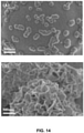
- 238000001878 scanning electron micrograph Methods 0.000 description 20
- 230000006911 nucleation Effects 0.000 description 18
- 238000010899 nucleation Methods 0.000 description 18
- 230000002776 aggregation Effects 0.000 description 17
- 238000004220 aggregation Methods 0.000 description 17
- 230000015572 biosynthetic process Effects 0.000 description 17
- LOKCTEFSRHRXRJ-UHFFFAOYSA-I dipotassium trisodium dihydrogen phosphate hydrogen phosphate dichloride Chemical compound P(=O)(O)(O)[O-].[K+].P(=O)(O)([O-])[O-].[Na+].[Na+].[Cl-].[K+].[Cl-].[Na+] LOKCTEFSRHRXRJ-UHFFFAOYSA-I 0.000 description 17
- 239000002953 phosphate buffered saline Substances 0.000 description 17
- 230000008569 process Effects 0.000 description 17
- 239000003656 tris buffered saline Substances 0.000 description 14
- 241000251539 Vertebrata <Metazoa> Species 0.000 description 13
- 210000000988 bone and bone Anatomy 0.000 description 13
- 239000012890 simulated body fluid Substances 0.000 description 13
- 239000000872 buffer Substances 0.000 description 12
- 239000006144 Dulbecco’s modified Eagle's medium Substances 0.000 description 11
- 238000005033 Fourier transform infrared spectroscopy Methods 0.000 description 11
- 238000012377 drug delivery Methods 0.000 description 11
- 230000001225 therapeutic effect Effects 0.000 description 11
- 238000001157 Fourier transform infrared spectrum Methods 0.000 description 10
- 229960001714 calcium phosphate Drugs 0.000 description 10
- 210000001519 tissue Anatomy 0.000 description 10
- XLYOFNOQVPJJNP-UHFFFAOYSA-N water Substances O XLYOFNOQVPJJNP-UHFFFAOYSA-N 0.000 description 10
- XEKOWRVHYACXOJ-UHFFFAOYSA-N Ethyl acetate Chemical compound CCOC(C)=O XEKOWRVHYACXOJ-UHFFFAOYSA-N 0.000 description 9
- 229920002988 biodegradable polymer Polymers 0.000 description 9
- 239000004621 biodegradable polymer Substances 0.000 description 9
- 239000003814 drug Substances 0.000 description 9
- BHPQYMZQTOCNFJ-UHFFFAOYSA-N Calcium cation Chemical compound [Ca+2] BHPQYMZQTOCNFJ-UHFFFAOYSA-N 0.000 description 8
- PEDCQBHIVMGVHV-UHFFFAOYSA-N Glycerine Chemical compound OCC(O)CO PEDCQBHIVMGVHV-UHFFFAOYSA-N 0.000 description 8
- 229910001424 calcium ion Inorganic materials 0.000 description 8
- 150000002500 ions Chemical class 0.000 description 8
- 230000007246 mechanism Effects 0.000 description 8
- 241001441571 Hiodontidae Species 0.000 description 7
- 238000002441 X-ray diffraction Methods 0.000 description 7
- 230000015556 catabolic process Effects 0.000 description 7
- 230000001186 cumulative effect Effects 0.000 description 7
- 238000006731 degradation reaction Methods 0.000 description 7
- 229940079593 drug Drugs 0.000 description 7
- RKDVKSZUMVYZHH-UHFFFAOYSA-N 1,4-dioxane-2,5-dione Chemical compound O=C1COC(=O)CO1 RKDVKSZUMVYZHH-UHFFFAOYSA-N 0.000 description 6
- 239000004372 Polyvinyl alcohol Substances 0.000 description 6
- 230000002378 acidificating effect Effects 0.000 description 6
- 238000004458 analytical method Methods 0.000 description 6
- 230000007547 defect Effects 0.000 description 6
- 239000000839 emulsion Substances 0.000 description 6
- 239000010408 film Substances 0.000 description 6
- 230000003993 interaction Effects 0.000 description 6
- JJTUDXZGHPGLLC-UHFFFAOYSA-N lactide Chemical compound CC1OC(=O)C(C)OC1=O JJTUDXZGHPGLLC-UHFFFAOYSA-N 0.000 description 6
- 239000012071 phase Substances 0.000 description 6
- 229920002451 polyvinyl alcohol Polymers 0.000 description 6
- 238000012545 processing Methods 0.000 description 6
- 238000004626 scanning electron microscopy Methods 0.000 description 6
- 102000004190 Enzymes Human genes 0.000 description 5
- 108090000790 Enzymes Proteins 0.000 description 5
- 108010038512 Platelet-Derived Growth Factor Proteins 0.000 description 5
- 102000010780 Platelet-Derived Growth Factor Human genes 0.000 description 5
- 238000013459 approach Methods 0.000 description 5
- 239000013078 crystal Substances 0.000 description 5
- 230000007423 decrease Effects 0.000 description 5
- 230000000694 effects Effects 0.000 description 5
- 238000004945 emulsification Methods 0.000 description 5
- 238000002474 experimental method Methods 0.000 description 5
- 238000009472 formulation Methods 0.000 description 5
- 210000002381 plasma Anatomy 0.000 description 5
- 239000000725 suspension Substances 0.000 description 5
- 101800000263 Acidic protein Proteins 0.000 description 4
- 102100030497 Cytochrome c Human genes 0.000 description 4
- 108010075031 Cytochromes c Proteins 0.000 description 4
- 108090000379 Fibroblast growth factor 2 Proteins 0.000 description 4
- 102000003974 Fibroblast growth factor 2 Human genes 0.000 description 4
- 101710093543 Probable non-specific lipid-transfer protein Proteins 0.000 description 4
- 239000012620 biological material Substances 0.000 description 4
- 230000033558 biomineral tissue development Effects 0.000 description 4
- 239000000969 carrier Substances 0.000 description 4
- 230000001419 dependent effect Effects 0.000 description 4
- 238000000151 deposition Methods 0.000 description 4
- 230000008021 deposition Effects 0.000 description 4
- 239000012153 distilled water Substances 0.000 description 4
- 238000002149 energy-dispersive X-ray emission spectroscopy Methods 0.000 description 4
- 230000002349 favourable effect Effects 0.000 description 4
- 235000011187 glycerol Nutrition 0.000 description 4
- 238000004519 manufacturing process Methods 0.000 description 4
- 239000011159 matrix material Substances 0.000 description 4
- 238000001000 micrograph Methods 0.000 description 4
- 239000002245 particle Substances 0.000 description 4
- 238000001228 spectrum Methods 0.000 description 4
- 239000000126 substance Substances 0.000 description 4
- 239000000829 suppository Substances 0.000 description 4
- FHVDTGUDJYJELY-UHFFFAOYSA-N 6-{[2-carboxy-4,5-dihydroxy-6-(phosphanyloxy)oxan-3-yl]oxy}-4,5-dihydroxy-3-phosphanyloxane-2-carboxylic acid Chemical compound O1C(C(O)=O)C(P)C(O)C(O)C1OC1C(C(O)=O)OC(OP)C(O)C1O FHVDTGUDJYJELY-UHFFFAOYSA-N 0.000 description 3
- CSCPPACGZOOCGX-UHFFFAOYSA-N Acetone Chemical compound CC(C)=O CSCPPACGZOOCGX-UHFFFAOYSA-N 0.000 description 3
- WVDDGKGOMKODPV-UHFFFAOYSA-N Benzyl alcohol Chemical compound OCC1=CC=CC=C1 WVDDGKGOMKODPV-UHFFFAOYSA-N 0.000 description 3
- 108010049931 Bone Morphogenetic Protein 2 Proteins 0.000 description 3
- 102100024506 Bone morphogenetic protein 2 Human genes 0.000 description 3
- 108020004414 DNA Proteins 0.000 description 3
- 102000004269 Granulocyte Colony-Stimulating Factor Human genes 0.000 description 3
- 108010017080 Granulocyte Colony-Stimulating Factor Proteins 0.000 description 3
- 241000124008 Mammalia Species 0.000 description 3
- 206010028980 Neoplasm Diseases 0.000 description 3
- DNIAPMSPPWPWGF-UHFFFAOYSA-N Propylene glycol Chemical compound CC(O)CO DNIAPMSPPWPWGF-UHFFFAOYSA-N 0.000 description 3
- HEMHJVSKTPXQMS-UHFFFAOYSA-M Sodium hydroxide Chemical compound [OH-].[Na+] HEMHJVSKTPXQMS-UHFFFAOYSA-M 0.000 description 3
- 108010073929 Vascular Endothelial Growth Factor A Proteins 0.000 description 3
- 108010019530 Vascular Endothelial Growth Factors Proteins 0.000 description 3
- 102000005789 Vascular Endothelial Growth Factors Human genes 0.000 description 3
- 229940072056 alginate Drugs 0.000 description 3
- 235000010443 alginic acid Nutrition 0.000 description 3
- 229920000615 alginic acid Polymers 0.000 description 3
- 150000001413 amino acids Chemical class 0.000 description 3
- 238000003556 assay Methods 0.000 description 3
- 230000008901 benefit Effects 0.000 description 3
- 230000004071 biological effect Effects 0.000 description 3
- 238000004587 chromatography analysis Methods 0.000 description 3
- 239000000499 gel Substances 0.000 description 3
- 239000003102 growth factor Substances 0.000 description 3
- 230000007062 hydrolysis Effects 0.000 description 3
- 238000006460 hydrolysis reaction Methods 0.000 description 3
- 238000001727 in vivo Methods 0.000 description 3
- 238000010348 incorporation Methods 0.000 description 3
- 239000007924 injection Substances 0.000 description 3
- 238000002347 injection Methods 0.000 description 3
- 239000002609 medium Substances 0.000 description 3
- 239000002679 microRNA Substances 0.000 description 3
- 239000002086 nanomaterial Substances 0.000 description 3
- 239000002674 ointment Substances 0.000 description 3
- 150000002894 organic compounds Chemical class 0.000 description 3
- 102000005962 receptors Human genes 0.000 description 3
- 108020003175 receptors Proteins 0.000 description 3
- 238000001226 reprecipitation Methods 0.000 description 3
- 239000000758 substrate Substances 0.000 description 3
- 230000002459 sustained effect Effects 0.000 description 3
- 238000013268 sustained release Methods 0.000 description 3
- 239000012730 sustained-release form Substances 0.000 description 3
- 102000009088 Angiopoietin-1 Human genes 0.000 description 2
- 108010048154 Angiopoietin-1 Proteins 0.000 description 2
- 108010032595 Antibody Binding Sites Proteins 0.000 description 2
- CIWBSHSKHKDKBQ-JLAZNSOCSA-N Ascorbic acid Chemical compound OC[C@H](O)[C@H]1OC(=O)C(O)=C1O CIWBSHSKHKDKBQ-JLAZNSOCSA-N 0.000 description 2
- 108010081589 Becaplermin Proteins 0.000 description 2
- 108010049955 Bone Morphogenetic Protein 4 Proteins 0.000 description 2
- 108010049870 Bone Morphogenetic Protein 7 Proteins 0.000 description 2
- 108010007726 Bone Morphogenetic Proteins Proteins 0.000 description 2
- 102000007350 Bone Morphogenetic Proteins Human genes 0.000 description 2
- 102100024505 Bone morphogenetic protein 4 Human genes 0.000 description 2
- 102100022544 Bone morphogenetic protein 7 Human genes 0.000 description 2
- 102000018233 Fibroblast Growth Factor Human genes 0.000 description 2
- 108050007372 Fibroblast Growth Factor Proteins 0.000 description 2
- 108090000368 Fibroblast growth factor 8 Proteins 0.000 description 2
- 102100037362 Fibronectin Human genes 0.000 description 2
- 108010067306 Fibronectins Proteins 0.000 description 2
- 108010051696 Growth Hormone Proteins 0.000 description 2
- 108090000100 Hepatocyte Growth Factor Proteins 0.000 description 2
- 102100021866 Hepatocyte growth factor Human genes 0.000 description 2
- 108090000723 Insulin-Like Growth Factor I Proteins 0.000 description 2
- 102100026236 Interleukin-8 Human genes 0.000 description 2
- 108090001007 Interleukin-8 Proteins 0.000 description 2
- 102000015696 Interleukins Human genes 0.000 description 2
- CKLJMWTZIZZHCS-REOHCLBHSA-N L-aspartic acid Chemical compound OC(=O)[C@@H](N)CC(O)=O CKLJMWTZIZZHCS-REOHCLBHSA-N 0.000 description 2
- 102000004058 Leukemia inhibitory factor Human genes 0.000 description 2
- 108090000581 Leukemia inhibitory factor Proteins 0.000 description 2
- 102000002274 Matrix Metalloproteinases Human genes 0.000 description 2
- 108010000684 Matrix Metalloproteinases Proteins 0.000 description 2
- 108700011259 MicroRNAs Proteins 0.000 description 2
- 102000004067 Osteocalcin Human genes 0.000 description 2
- 108090000573 Osteocalcin Proteins 0.000 description 2
- 108090000445 Parathyroid hormone Proteins 0.000 description 2
- 102100039277 Pleiotrophin Human genes 0.000 description 2
- 229920002732 Polyanhydride Polymers 0.000 description 2
- 229920000954 Polyglycolide Polymers 0.000 description 2
- WCUXLLCKKVVCTQ-UHFFFAOYSA-M Potassium chloride Chemical compound [Cl-].[K+] WCUXLLCKKVVCTQ-UHFFFAOYSA-M 0.000 description 2
- 102000013275 Somatomedins Human genes 0.000 description 2
- 102100038803 Somatotropin Human genes 0.000 description 2
- QAOWNCQODCNURD-UHFFFAOYSA-N Sulfuric acid Chemical compound OS(O)(=O)=O QAOWNCQODCNURD-UHFFFAOYSA-N 0.000 description 2
- 102100031372 Thymidine phosphorylase Human genes 0.000 description 2
- 108700023160 Thymidine phosphorylases Proteins 0.000 description 2
- 102000004887 Transforming Growth Factor beta Human genes 0.000 description 2
- 108090001012 Transforming Growth Factor beta Proteins 0.000 description 2
- 238000002835 absorbance Methods 0.000 description 2
- 239000000654 additive Substances 0.000 description 2
- 230000000996 additive effect Effects 0.000 description 2
- 208000026935 allergic disease Diseases 0.000 description 2
- 150000001408 amides Chemical class 0.000 description 2
- 235000001014 amino acid Nutrition 0.000 description 2
- 239000003242 anti bacterial agent Substances 0.000 description 2
- 230000000692 anti-sense effect Effects 0.000 description 2
- 239000008346 aqueous phase Substances 0.000 description 2
- 229940112869 bone morphogenetic protein Drugs 0.000 description 2
- 125000005587 carbonate group Chemical group 0.000 description 2
- 238000012512 characterization method Methods 0.000 description 2
- 239000003153 chemical reaction reagent Substances 0.000 description 2
- 239000003795 chemical substances by application Substances 0.000 description 2
- 239000000470 constituent Substances 0.000 description 2
- 238000013270 controlled release Methods 0.000 description 2
- 239000006071 cream Substances 0.000 description 2
- 230000003247 decreasing effect Effects 0.000 description 2
- 239000008367 deionised water Substances 0.000 description 2
- 108700041286 delta Proteins 0.000 description 2
- 210000004268 dentin Anatomy 0.000 description 2
- 238000013461 design Methods 0.000 description 2
- 201000010099 disease Diseases 0.000 description 2
- 208000037265 diseases, disorders, signs and symptoms Diseases 0.000 description 2
- 239000003937 drug carrier Substances 0.000 description 2
- 238000000724 energy-dispersive X-ray spectrum Methods 0.000 description 2
- 239000000835 fiber Substances 0.000 description 2
- 229940126864 fibroblast growth factor Drugs 0.000 description 2
- 239000011521 glass Substances 0.000 description 2
- 239000000122 growth hormone Substances 0.000 description 2
- 238000007654 immersion Methods 0.000 description 2
- 239000007943 implant Substances 0.000 description 2
- 238000000338 in vitro Methods 0.000 description 2
- XKTZWUACRZHVAN-VADRZIEHSA-N interleukin-8 Chemical compound C([C@H](NC(=O)[C@H](CC(O)=O)NC(=O)[C@H](CC=1C2=CC=CC=C2NC=1)NC(=O)[C@@H](NC(C)=O)CCSC)C(=O)N[C@@H](CC(O)=O)C(=O)N[C@@H](CC(O)=O)C(=O)N[C@@H](CC(C)C)C(=O)N[C@@H](CC(N)=O)C(=O)N[C@@H](CC=1C=CC=CC=1)C(=O)N[C@@H]([C@@H](C)O)C(=O)NCC(=O)N[C@@H](CCSC)C(=O)N1[C@H](CCC1)C(=O)N1[C@H](CCC1)C(=O)N[C@@H](C)C(=O)N[C@H](CC(O)=O)C(=O)N[C@H](CCC(O)=O)C(=O)N[C@H](CC(O)=O)C(=O)N[C@H](CC=1C=CC(O)=CC=1)C(=O)N[C@H](CO)C(=O)N1[C@H](CCC1)C(N)=O)C1=CC=CC=C1 XKTZWUACRZHVAN-VADRZIEHSA-N 0.000 description 2
- 229940096397 interleukin-8 Drugs 0.000 description 2
- 229940074869 marquis Drugs 0.000 description 2
- 238000002156 mixing Methods 0.000 description 2
- 108091005601 modified peptides Proteins 0.000 description 2
- 239000002105 nanoparticle Substances 0.000 description 2
- 239000007764 o/w emulsion Substances 0.000 description 2
- 239000012074 organic phase Substances 0.000 description 2
- 230000000399 orthopedic effect Effects 0.000 description 2
- 239000008194 pharmaceutical composition Substances 0.000 description 2
- 230000004962 physiological condition Effects 0.000 description 2
- 229920000058 polyacrylate Polymers 0.000 description 2
- 229920002635 polyurethane Polymers 0.000 description 2
- 239000004814 polyurethane Substances 0.000 description 2
- VBUNOIXRZNJNAD-UHFFFAOYSA-N ponazuril Chemical compound CC1=CC(N2C(N(C)C(=O)NC2=O)=O)=CC=C1OC1=CC=C(S(=O)(=O)C(F)(F)F)C=C1 VBUNOIXRZNJNAD-UHFFFAOYSA-N 0.000 description 2
- 239000000843 powder Substances 0.000 description 2
- 238000002360 preparation method Methods 0.000 description 2
- 238000000746 purification Methods 0.000 description 2
- 238000000926 separation method Methods 0.000 description 2
- 239000002904 solvent Substances 0.000 description 2
- 238000001179 sorption measurement Methods 0.000 description 2
- 238000006467 substitution reaction Methods 0.000 description 2
- ZRKFYGHZFMAOKI-QMGMOQQFSA-N tgfbeta Chemical compound C([C@H](NC(=O)[C@H](C(C)C)NC(=O)CNC(=O)[C@H](CCC(O)=O)NC(=O)[C@H](CCCNC(N)=N)NC(=O)[C@H](CC(N)=O)NC(=O)[C@H](CC(C)C)NC(=O)[C@H]([C@@H](C)O)NC(=O)[C@H](CCC(O)=O)NC(=O)[C@H]([C@@H](C)O)NC(=O)[C@H](CC(C)C)NC(=O)CNC(=O)[C@H](C)NC(=O)[C@H](CO)NC(=O)[C@H](CCC(N)=O)NC(=O)[C@@H](NC(=O)[C@H](C)NC(=O)[C@H](C)NC(=O)[C@@H](NC(=O)[C@H](CC(C)C)NC(=O)[C@@H](N)CCSC)C(C)C)[C@@H](C)CC)C(=O)N[C@@H]([C@@H](C)O)C(=O)N[C@@H](C(C)C)C(=O)N[C@@H](CC=1C=CC=CC=1)C(=O)N[C@@H](C)C(=O)N1[C@@H](CCC1)C(=O)N[C@@H]([C@@H](C)O)C(=O)N[C@@H](CC(N)=O)C(=O)N[C@@H](CCC(O)=O)C(=O)N[C@@H](C)C(=O)N[C@@H](CC=1C=CC=CC=1)C(=O)N[C@@H](CCCNC(N)=N)C(=O)N[C@@H](C)C(=O)N[C@@H](CC(C)C)C(=O)N1[C@@H](CCC1)C(=O)N1[C@@H](CCC1)C(=O)N[C@@H](CCCNC(N)=N)C(=O)N[C@@H](CCC(O)=O)C(=O)N[C@@H](CCCNC(N)=N)C(=O)N[C@@H](CO)C(=O)N[C@@H](CCCNC(N)=N)C(=O)N[C@@H](CC(C)C)C(=O)N[C@@H](CC(C)C)C(O)=O)C1=CC=C(O)C=C1 ZRKFYGHZFMAOKI-QMGMOQQFSA-N 0.000 description 2
- 238000011282 treatment Methods 0.000 description 2
- SNICXCGAKADSCV-JTQLQIEISA-N (-)-Nicotine Chemical compound CN1CCC[C@H]1C1=CC=CN=C1 SNICXCGAKADSCV-JTQLQIEISA-N 0.000 description 1
- KIUKXJAPPMFGSW-DNGZLQJQSA-N (2S,3S,4S,5R,6R)-6-[(2S,3R,4R,5S,6R)-3-Acetamido-2-[(2S,3S,4R,5R,6R)-6-[(2R,3R,4R,5S,6R)-3-acetamido-2,5-dihydroxy-6-(hydroxymethyl)oxan-4-yl]oxy-2-carboxy-4,5-dihydroxyoxan-3-yl]oxy-5-hydroxy-6-(hydroxymethyl)oxan-4-yl]oxy-3,4,5-trihydroxyoxane-2-carboxylic acid Chemical compound CC(=O)N[C@H]1[C@H](O)O[C@H](CO)[C@@H](O)[C@@H]1O[C@H]1[C@H](O)[C@@H](O)[C@H](O[C@H]2[C@@H]([C@@H](O[C@H]3[C@@H]([C@@H](O)[C@H](O)[C@H](O3)C(O)=O)O)[C@H](O)[C@@H](CO)O2)NC(C)=O)[C@@H](C(O)=O)O1 KIUKXJAPPMFGSW-DNGZLQJQSA-N 0.000 description 1
- MZOFCQQQCNRIBI-VMXHOPILSA-N (3s)-4-[[(2s)-1-[[(2s)-1-[[(1s)-1-carboxy-2-hydroxyethyl]amino]-4-methyl-1-oxopentan-2-yl]amino]-5-(diaminomethylideneamino)-1-oxopentan-2-yl]amino]-3-[[2-[[(2s)-2,6-diaminohexanoyl]amino]acetyl]amino]-4-oxobutanoic acid Chemical compound OC[C@@H](C(O)=O)NC(=O)[C@H](CC(C)C)NC(=O)[C@H](CCCN=C(N)N)NC(=O)[C@H](CC(O)=O)NC(=O)CNC(=O)[C@@H](N)CCCCN MZOFCQQQCNRIBI-VMXHOPILSA-N 0.000 description 1
- 108010088751 Albumins Proteins 0.000 description 1
- 102000009027 Albumins Human genes 0.000 description 1
- 201000004384 Alopecia Diseases 0.000 description 1
- 208000024827 Alzheimer disease Diseases 0.000 description 1
- 102000008076 Angiogenic Proteins Human genes 0.000 description 1
- 108010074415 Angiogenic Proteins Proteins 0.000 description 1
- 102100022987 Angiogenin Human genes 0.000 description 1
- 241000203069 Archaea Species 0.000 description 1
- 208000023275 Autoimmune disease Diseases 0.000 description 1
- 241000894006 Bacteria Species 0.000 description 1
- 208000035143 Bacterial infection Diseases 0.000 description 1
- 108010049951 Bone Morphogenetic Protein 3 Proteins 0.000 description 1
- 108010049976 Bone Morphogenetic Protein 5 Proteins 0.000 description 1
- 108010049974 Bone Morphogenetic Protein 6 Proteins 0.000 description 1
- 208000020084 Bone disease Diseases 0.000 description 1
- 102100024504 Bone morphogenetic protein 3 Human genes 0.000 description 1
- 102100022526 Bone morphogenetic protein 5 Human genes 0.000 description 1
- 102100022525 Bone morphogenetic protein 6 Human genes 0.000 description 1
- 102100022545 Bone morphogenetic protein 8B Human genes 0.000 description 1
- 229910002974 CaO–SiO2 Inorganic materials 0.000 description 1
- 101100170173 Caenorhabditis elegans del-1 gene Proteins 0.000 description 1
- 238000003650 Calcium Assay Kit Methods 0.000 description 1
- OKTJSMMVPCPJKN-UHFFFAOYSA-N Carbon Chemical compound [C] OKTJSMMVPCPJKN-UHFFFAOYSA-N 0.000 description 1
- 102000014914 Carrier Proteins Human genes 0.000 description 1
- 108010078791 Carrier Proteins Proteins 0.000 description 1
- 108050000299 Chemokine receptor Proteins 0.000 description 1
- 102000019034 Chemokines Human genes 0.000 description 1
- 229920001661 Chitosan Polymers 0.000 description 1
- 102000008186 Collagen Human genes 0.000 description 1
- 108010035532 Collagen Proteins 0.000 description 1
- 102000012422 Collagen Type I Human genes 0.000 description 1
- 108010022452 Collagen Type I Proteins 0.000 description 1
- 108010003384 Colony-Stimulating Factor Receptors Proteins 0.000 description 1
- 208000032170 Congenital Abnormalities Diseases 0.000 description 1
- 206010010356 Congenital anomaly Diseases 0.000 description 1
- 208000027205 Congenital disease Diseases 0.000 description 1
- 230000033616 DNA repair Effects 0.000 description 1
- KCXVZYZYPLLWCC-UHFFFAOYSA-N EDTA Chemical compound OC(=O)CN(CC(O)=O)CCN(CC(O)=O)CC(O)=O KCXVZYZYPLLWCC-UHFFFAOYSA-N 0.000 description 1
- 241000792859 Enema Species 0.000 description 1
- 208000010228 Erectile Dysfunction Diseases 0.000 description 1
- 241000206602 Eukaryota Species 0.000 description 1
- 102000009123 Fibrin Human genes 0.000 description 1
- 108010073385 Fibrin Proteins 0.000 description 1
- BWGVNKXGVNDBDI-UHFFFAOYSA-N Fibrin monomer Chemical compound CNC(=O)CNC(=O)CN BWGVNKXGVNDBDI-UHFFFAOYSA-N 0.000 description 1
- 102000016970 Follistatin Human genes 0.000 description 1
- 108010014612 Follistatin Proteins 0.000 description 1
- 208000018522 Gastrointestinal disease Diseases 0.000 description 1
- WQZGKKKJIJFFOK-GASJEMHNSA-N Glucose Natural products OC[C@H]1OC(O)[C@H](O)[C@@H](O)[C@@H]1O WQZGKKKJIJFFOK-GASJEMHNSA-N 0.000 description 1
- WHUUTDBJXJRKMK-UHFFFAOYSA-N Glutamic acid Natural products OC(=O)C(N)CCC(O)=O WHUUTDBJXJRKMK-UHFFFAOYSA-N 0.000 description 1
- 108060003393 Granulin Proteins 0.000 description 1
- 102100039620 Granulocyte-macrophage colony-stimulating factor Human genes 0.000 description 1
- 229940121710 HMGCoA reductase inhibitor Drugs 0.000 description 1
- 102000003745 Hepatocyte Growth Factor Human genes 0.000 description 1
- 241000282412 Homo Species 0.000 description 1
- 101000899368 Homo sapiens Bone morphogenetic protein 8B Proteins 0.000 description 1
- 101001075374 Homo sapiens Gamma-glutamyl hydrolase Proteins 0.000 description 1
- 101000595923 Homo sapiens Placenta growth factor Proteins 0.000 description 1
- 101000635799 Homo sapiens Run domain Beclin-1-interacting and cysteine-rich domain-containing protein Proteins 0.000 description 1
- 101000664737 Homo sapiens Somatotropin Proteins 0.000 description 1
- 206010020751 Hypersensitivity Diseases 0.000 description 1
- DGAQECJNVWCQMB-PUAWFVPOSA-M Ilexoside XXIX Chemical compound C[C@@H]1CC[C@@]2(CC[C@@]3(C(=CC[C@H]4[C@]3(CC[C@@H]5[C@@]4(CC[C@@H](C5(C)C)OS(=O)(=O)[O-])C)C)[C@@H]2[C@]1(C)O)C)C(=O)O[C@H]6[C@@H]([C@H]([C@@H]([C@H](O6)CO)O)O)O.[Na+] DGAQECJNVWCQMB-PUAWFVPOSA-M 0.000 description 1
- 102000005755 Intercellular Signaling Peptides and Proteins Human genes 0.000 description 1
- 108010070716 Intercellular Signaling Peptides and Proteins Proteins 0.000 description 1
- 102000001617 Interferon Receptors Human genes 0.000 description 1
- 108010054267 Interferon Receptors Proteins 0.000 description 1
- 108010063738 Interleukins Proteins 0.000 description 1
- WHUUTDBJXJRKMK-VKHMYHEASA-N L-glutamic acid Chemical compound OC(=O)[C@@H](N)CCC(O)=O WHUUTDBJXJRKMK-VKHMYHEASA-N 0.000 description 1
- ZDXPYRJPNDTMRX-VKHMYHEASA-N L-glutamine Chemical compound OC(=O)[C@@H](N)CCC(N)=O ZDXPYRJPNDTMRX-VKHMYHEASA-N 0.000 description 1
- 229930182816 L-glutamine Natural products 0.000 description 1
- ODYCAZSSUVCHNU-XLAORIBOSA-N Laurencin Natural products CC[C@H]1C[C@H](CC=CC[C@@H]1Br)[C@@H](CC=CC#C)OC(=O)C ODYCAZSSUVCHNU-XLAORIBOSA-N 0.000 description 1
- 108010092277 Leptin Proteins 0.000 description 1
- 102000016267 Leptin Human genes 0.000 description 1
- 102000007651 Macrophage Colony-Stimulating Factor Human genes 0.000 description 1
- 108010046938 Macrophage Colony-Stimulating Factor Proteins 0.000 description 1
- 102000018697 Membrane Proteins Human genes 0.000 description 1
- 108010052285 Membrane Proteins Proteins 0.000 description 1
- 241001465754 Metazoa Species 0.000 description 1
- 102100030335 Midkine Human genes 0.000 description 1
- 108010092801 Midkine Proteins 0.000 description 1
- 239000004677 Nylon Substances 0.000 description 1
- BZQFBWGGLXLEPQ-UHFFFAOYSA-N O-phosphoryl-L-serine Natural products OC(=O)C(N)COP(O)(O)=O BZQFBWGGLXLEPQ-UHFFFAOYSA-N 0.000 description 1
- 108700020796 Oncogene Proteins 0.000 description 1
- 102000043276 Oncogene Human genes 0.000 description 1
- 208000030852 Parasitic disease Diseases 0.000 description 1
- 102000003982 Parathyroid hormone Human genes 0.000 description 1
- 102100036893 Parathyroid hormone Human genes 0.000 description 1
- 208000018737 Parkinson disease Diseases 0.000 description 1
- BELBBZDIHDAJOR-UHFFFAOYSA-N Phenolsulfonephthalein Chemical compound C1=CC(O)=CC=C1C1(C=2C=CC(O)=CC=2)C2=CC=CC=C2S(=O)(=O)O1 BELBBZDIHDAJOR-UHFFFAOYSA-N 0.000 description 1
- 108700019535 Phosphoprotein Phosphatases Proteins 0.000 description 1
- 102000045595 Phosphoprotein Phosphatases Human genes 0.000 description 1
- OAICVXFJPJFONN-UHFFFAOYSA-N Phosphorus Chemical compound [P] OAICVXFJPJFONN-UHFFFAOYSA-N 0.000 description 1
- 102100035194 Placenta growth factor Human genes 0.000 description 1
- 229920003171 Poly (ethylene oxide) Polymers 0.000 description 1
- 239000004952 Polyamide Substances 0.000 description 1
- 239000004698 Polyethylene Substances 0.000 description 1
- 229920001710 Polyorthoester Polymers 0.000 description 1
- 239000004793 Polystyrene Substances 0.000 description 1
- 208000024777 Prion disease Diseases 0.000 description 1
- 108010012809 Progranulins Proteins 0.000 description 1
- 102000019204 Progranulins Human genes 0.000 description 1
- 102000001253 Protein Kinase Human genes 0.000 description 1
- 102100030852 Run domain Beclin-1-interacting and cysteine-rich domain-containing protein Human genes 0.000 description 1
- 229910008051 Si-OH Inorganic materials 0.000 description 1
- 229910006358 Si—OH Inorganic materials 0.000 description 1
- DWAQJAXMDSEUJJ-UHFFFAOYSA-M Sodium bisulfite Chemical compound [Na+].OS([O-])=O DWAQJAXMDSEUJJ-UHFFFAOYSA-M 0.000 description 1
- 239000004809 Teflon Substances 0.000 description 1
- 229920006362 Teflon® Polymers 0.000 description 1
- IOYNQIMAUDJVEI-BMVIKAAMSA-N Tepraloxydim Chemical compound C1C(=O)C(C(=N/OC\C=C\Cl)/CC)=C(O)CC1C1CCOCC1 IOYNQIMAUDJVEI-BMVIKAAMSA-N 0.000 description 1
- 229910003089 Ti–OH Inorganic materials 0.000 description 1
- 102000046299 Transforming Growth Factor beta1 Human genes 0.000 description 1
- 102000011117 Transforming Growth Factor beta2 Human genes 0.000 description 1
- 102400001320 Transforming growth factor alpha Human genes 0.000 description 1
- 101800004564 Transforming growth factor alpha Proteins 0.000 description 1
- 101800002279 Transforming growth factor beta-1 Proteins 0.000 description 1
- 101800000304 Transforming growth factor beta-2 Proteins 0.000 description 1
- 108060008682 Tumor Necrosis Factor Proteins 0.000 description 1
- 102000000852 Tumor Necrosis Factor-alpha Human genes 0.000 description 1
- 206010054094 Tumour necrosis Diseases 0.000 description 1
- 108010059993 Vancomycin Proteins 0.000 description 1
- 235000018936 Vitellaria paradoxa Nutrition 0.000 description 1
- 206010052428 Wound Diseases 0.000 description 1
- 208000027418 Wounds and injury Diseases 0.000 description 1
- VRGWBRLULZUWAJ-XFFXIZSCSA-N [(2s)-2-[(1r,3z,5s,8z,12z,15s)-5,17-dihydroxy-4,8,12,15-tetramethyl-16-oxo-18-bicyclo[13.3.0]octadeca-3,8,12,17-tetraenyl]propyl] acetate Chemical compound C1\C=C(C)/CC\C=C(C)/CC[C@H](O)\C(C)=C/C[C@@H]2C([C@@H](COC(C)=O)C)=C(O)C(=O)[C@]21C VRGWBRLULZUWAJ-XFFXIZSCSA-N 0.000 description 1
- 150000001242 acetic acid derivatives Chemical class 0.000 description 1
- 125000002252 acyl group Chemical group 0.000 description 1
- 239000000853 adhesive Substances 0.000 description 1
- 230000001070 adhesive effect Effects 0.000 description 1
- 230000002411 adverse Effects 0.000 description 1
- 230000004931 aggregating effect Effects 0.000 description 1
- 150000001299 aldehydes Chemical class 0.000 description 1
- 230000007815 allergy Effects 0.000 description 1
- XAGFODPZIPBFFR-UHFFFAOYSA-N aluminium Chemical compound [Al] XAGFODPZIPBFFR-UHFFFAOYSA-N 0.000 description 1
- 229910052782 aluminium Inorganic materials 0.000 description 1
- 239000011609 ammonium molybdate Substances 0.000 description 1
- APUPEJJSWDHEBO-UHFFFAOYSA-P ammonium molybdate Chemical compound [NH4+].[NH4+].[O-][Mo]([O-])(=O)=O APUPEJJSWDHEBO-UHFFFAOYSA-P 0.000 description 1
- 235000018660 ammonium molybdate Nutrition 0.000 description 1
- 229940010552 ammonium molybdate Drugs 0.000 description 1
- 239000003708 ampul Substances 0.000 description 1
- 238000000540 analysis of variance Methods 0.000 description 1
- 108010072788 angiogenin Proteins 0.000 description 1
- 230000001772 anti-angiogenic effect Effects 0.000 description 1
- 230000000844 anti-bacterial effect Effects 0.000 description 1
- 230000000840 anti-viral effect Effects 0.000 description 1
- 229940088710 antibiotic agent Drugs 0.000 description 1
- 239000003963 antioxidant agent Substances 0.000 description 1
- 235000006708 antioxidants Nutrition 0.000 description 1
- 239000007864 aqueous solution Substances 0.000 description 1
- 235000010323 ascorbic acid Nutrition 0.000 description 1
- 229960005070 ascorbic acid Drugs 0.000 description 1
- 239000011668 ascorbic acid Substances 0.000 description 1
- 235000003704 aspartic acid Nutrition 0.000 description 1
- QVGXLLKOCUKJST-UHFFFAOYSA-N atomic oxygen Chemical group [O] QVGXLLKOCUKJST-UHFFFAOYSA-N 0.000 description 1
- 208000022362 bacterial infectious disease Diseases 0.000 description 1
- 235000019445 benzyl alcohol Nutrition 0.000 description 1
- WQZGKKKJIJFFOK-VFUOTHLCSA-N beta-D-glucose Chemical compound OC[C@H]1O[C@@H](O)[C@H](O)[C@@H](O)[C@@H]1O WQZGKKKJIJFFOK-VFUOTHLCSA-N 0.000 description 1
- OQFSQFPPLPISGP-UHFFFAOYSA-N beta-carboxyaspartic acid Natural products OC(=O)C(N)C(C(O)=O)C(O)=O OQFSQFPPLPISGP-UHFFFAOYSA-N 0.000 description 1
- 230000000975 bioactive effect Effects 0.000 description 1
- 238000004166 bioassay Methods 0.000 description 1
- 230000005540 biological transmission Effects 0.000 description 1
- 230000003592 biomimetic effect Effects 0.000 description 1
- 229920001400 block copolymer Polymers 0.000 description 1
- 210000004369 blood Anatomy 0.000 description 1
- 239000008280 blood Substances 0.000 description 1
- 230000013926 blood microparticle formation Effects 0.000 description 1
- 239000007975 buffered saline Substances 0.000 description 1
- 239000008366 buffered solution Substances 0.000 description 1
- DQXBYHZEEUGOBF-UHFFFAOYSA-N but-3-enoic acid;ethene Chemical compound C=C.OC(=O)CC=C DQXBYHZEEUGOBF-UHFFFAOYSA-N 0.000 description 1
- 239000006227 byproduct Substances 0.000 description 1
- 239000000648 calcium alginate Substances 0.000 description 1
- 235000010410 calcium alginate Nutrition 0.000 description 1
- 229960002681 calcium alginate Drugs 0.000 description 1
- OKHHGHGGPDJQHR-YMOPUZKJSA-L calcium;(2s,3s,4s,5s,6r)-6-[(2r,3s,4r,5s,6r)-2-carboxy-6-[(2r,3s,4r,5s,6r)-2-carboxylato-4,5,6-trihydroxyoxan-3-yl]oxy-4,5-dihydroxyoxan-3-yl]oxy-3,4,5-trihydroxyoxane-2-carboxylate Chemical compound [Ca+2].O[C@@H]1[C@H](O)[C@H](O)O[C@@H](C([O-])=O)[C@H]1O[C@H]1[C@@H](O)[C@@H](O)[C@H](O[C@H]2[C@H]([C@@H](O)[C@H](O)[C@H](O2)C([O-])=O)O)[C@H](C(O)=O)O1 OKHHGHGGPDJQHR-YMOPUZKJSA-L 0.000 description 1
- 201000011510 cancer Diseases 0.000 description 1
- 239000002775 capsule Substances 0.000 description 1
- 229910052799 carbon Inorganic materials 0.000 description 1
- MLYYVTUWGNIJIB-BXKDBHETSA-N cefazolin Chemical compound S1C(C)=NN=C1SCC1=C(C(O)=O)N2C(=O)[C@@H](NC(=O)CN3N=NN=C3)[C@H]2SC1 MLYYVTUWGNIJIB-BXKDBHETSA-N 0.000 description 1
- 229960001139 cefazolin Drugs 0.000 description 1
- 229960001668 cefuroxime Drugs 0.000 description 1
- JFPVXVDWJQMJEE-IZRZKJBUSA-N cefuroxime Chemical compound N([C@@H]1C(N2C(=C(COC(N)=O)CS[C@@H]21)C(O)=O)=O)C(=O)\C(=N/OC)C1=CC=CO1 JFPVXVDWJQMJEE-IZRZKJBUSA-N 0.000 description 1
- 230000024245 cell differentiation Effects 0.000 description 1
- 230000005754 cellular signaling Effects 0.000 description 1
- 229920002301 cellulose acetate Polymers 0.000 description 1
- 239000002738 chelating agent Substances 0.000 description 1
- 239000005482 chemotactic factor Substances 0.000 description 1
- 235000015218 chewing gum Nutrition 0.000 description 1
- 238000011097 chromatography purification Methods 0.000 description 1
- 230000001684 chronic effect Effects 0.000 description 1
- 150000001860 citric acid derivatives Chemical class 0.000 description 1
- 229960002227 clindamycin Drugs 0.000 description 1
- KDLRVYVGXIQJDK-AWPVFWJPSA-N clindamycin Chemical compound CN1C[C@H](CCC)C[C@H]1C(=O)N[C@H]([C@H](C)Cl)[C@@H]1[C@H](O)[C@H](O)[C@@H](O)[C@@H](SC)O1 KDLRVYVGXIQJDK-AWPVFWJPSA-N 0.000 description 1
- 229920001436 collagen Polymers 0.000 description 1
- 239000000512 collagen gel Substances 0.000 description 1
- 239000000599 controlled substance Substances 0.000 description 1
- 229920001577 copolymer Polymers 0.000 description 1
- 239000003246 corticosteroid Substances 0.000 description 1
- 229960001334 corticosteroids Drugs 0.000 description 1
- 108010057085 cytokine receptors Proteins 0.000 description 1
- 238000013480 data collection Methods 0.000 description 1
- 230000003111 delayed effect Effects 0.000 description 1
- 239000002274 desiccant Substances 0.000 description 1
- 229960003957 dexamethasone Drugs 0.000 description 1
- UREBDLICKHMUKA-CXSFZGCWSA-N dexamethasone Chemical compound C1CC2=CC(=O)C=C[C@]2(C)[C@]2(F)[C@@H]1[C@@H]1C[C@@H](C)[C@@](C(=O)CO)(O)[C@@]1(C)C[C@@H]2O UREBDLICKHMUKA-CXSFZGCWSA-N 0.000 description 1
- 229950006137 dexfosfoserine Drugs 0.000 description 1
- 239000008121 dextrose Substances 0.000 description 1
- 206010012601 diabetes mellitus Diseases 0.000 description 1
- 238000009792 diffusion process Methods 0.000 description 1
- 208000010643 digestive system disease Diseases 0.000 description 1
- 239000003085 diluting agent Substances 0.000 description 1
- 239000012738 dissolution medium Substances 0.000 description 1
- 231100000673 dose–response relationship Toxicity 0.000 description 1
- 230000009881 electrostatic interaction Effects 0.000 description 1
- 238000005538 encapsulation Methods 0.000 description 1
- 239000007920 enema Substances 0.000 description 1
- 229940079360 enema for constipation Drugs 0.000 description 1
- 239000005038 ethylene vinyl acetate Substances 0.000 description 1
- 210000002744 extracellular matrix Anatomy 0.000 description 1
- 229950003499 fibrin Drugs 0.000 description 1
- 238000011049 filling Methods 0.000 description 1
- 238000001914 filtration Methods 0.000 description 1
- VRGWBRLULZUWAJ-UHFFFAOYSA-N fusaproliferin Natural products C1C=C(C)CCC=C(C)CCC(O)C(C)=CCC2C(C(COC(C)=O)C)=C(O)C(=O)C21C VRGWBRLULZUWAJ-UHFFFAOYSA-N 0.000 description 1
- 208000018685 gastrointestinal system disease Diseases 0.000 description 1
- 239000007903 gelatin capsule Substances 0.000 description 1
- 235000013922 glutamic acid Nutrition 0.000 description 1
- 239000004220 glutamic acid Substances 0.000 description 1
- 125000000291 glutamic acid group Chemical group N[C@@H](CCC(O)=O)C(=O)* 0.000 description 1
- 150000004676 glycans Chemical class 0.000 description 1
- PCHJSUWPFVWCPO-UHFFFAOYSA-N gold Chemical compound [Au] PCHJSUWPFVWCPO-UHFFFAOYSA-N 0.000 description 1
- 239000010931 gold Substances 0.000 description 1
- 229910052737 gold Inorganic materials 0.000 description 1
- 210000003714 granulocyte Anatomy 0.000 description 1
- 230000003676 hair loss Effects 0.000 description 1
- 208000019622 heart disease Diseases 0.000 description 1
- 238000010438 heat treatment Methods 0.000 description 1
- 229940088597 hormone Drugs 0.000 description 1
- 239000005556 hormone Substances 0.000 description 1
- 229920002674 hyaluronan Polymers 0.000 description 1
- 229960003160 hyaluronic acid Drugs 0.000 description 1
- 239000000017 hydrogel Substances 0.000 description 1
- 239000002471 hydroxymethylglutaryl coenzyme A reductase inhibitor Substances 0.000 description 1
- 230000028993 immune response Effects 0.000 description 1
- 201000001881 impotence Diseases 0.000 description 1
- 230000001939 inductive effect Effects 0.000 description 1
- 239000004615 ingredient Substances 0.000 description 1
- 208000014674 injury Diseases 0.000 description 1
- 229910001410 inorganic ion Inorganic materials 0.000 description 1
- 108010093036 interleukin receptors Proteins 0.000 description 1
- 238000007918 intramuscular administration Methods 0.000 description 1
- 238000007913 intrathecal administration Methods 0.000 description 1
- 238000001990 intravenous administration Methods 0.000 description 1
- 238000005342 ion exchange Methods 0.000 description 1
- 230000007794 irritation Effects 0.000 description 1
- 208000017169 kidney disease Diseases 0.000 description 1
- 210000002429 large intestine Anatomy 0.000 description 1
- NRYBAZVQPHGZNS-ZSOCWYAHSA-N leptin Chemical compound O=C([C@H](CO)NC(=O)[C@H](CC(C)C)NC(=O)[C@H](CC(O)=O)NC(=O)[C@H](CC(C)C)NC(=O)[C@H](CCC(N)=O)NC(=O)[C@H](CC=1C2=CC=CC=C2NC=1)NC(=O)[C@H](CC(C)C)NC(=O)[C@@H](NC(=O)[C@H](CC(O)=O)NC(=O)[C@H](CCC(N)=O)NC(=O)[C@H](CC(C)C)NC(=O)[C@H](CO)NC(=O)CNC(=O)[C@H](CCC(N)=O)NC(=O)[C@@H](N)CC(C)C)CCSC)N1CCC[C@H]1C(=O)NCC(=O)N[C@@H](CS)C(O)=O NRYBAZVQPHGZNS-ZSOCWYAHSA-N 0.000 description 1
- 229940039781 leptin Drugs 0.000 description 1
- 230000000670 limiting effect Effects 0.000 description 1
- 208000019423 liver disease Diseases 0.000 description 1
- 238000005297 material degradation process Methods 0.000 description 1
- 238000005259 measurement Methods 0.000 description 1
- 230000001404 mediated effect Effects 0.000 description 1
- 239000002207 metabolite Substances 0.000 description 1
- 235000010270 methyl p-hydroxybenzoate Nutrition 0.000 description 1
- 108091070501 miRNA Proteins 0.000 description 1
- 239000004530 micro-emulsion Substances 0.000 description 1
- 230000003278 mimic effect Effects 0.000 description 1
- 230000000921 morphogenic effect Effects 0.000 description 1
- 210000004400 mucous membrane Anatomy 0.000 description 1
- 210000003928 nasal cavity Anatomy 0.000 description 1
- 239000007923 nasal drop Substances 0.000 description 1
- 229940097496 nasal spray Drugs 0.000 description 1
- 239000007922 nasal spray Substances 0.000 description 1
- 239000002858 neurotransmitter agent Substances 0.000 description 1
- 229960002715 nicotine Drugs 0.000 description 1
- SNICXCGAKADSCV-UHFFFAOYSA-N nicotine Natural products CN1CCCC1C1=CC=CN=C1 SNICXCGAKADSCV-UHFFFAOYSA-N 0.000 description 1
- 239000000346 nonvolatile oil Substances 0.000 description 1
- 229920001778 nylon Polymers 0.000 description 1
- 239000003921 oil Substances 0.000 description 1
- 229920000620 organic polymer Polymers 0.000 description 1
- 239000003960 organic solvent Substances 0.000 description 1
- 230000011164 ossification Effects 0.000 description 1
- 230000000278 osteoconductive effect Effects 0.000 description 1
- 239000000199 parathyroid hormone Substances 0.000 description 1
- 229960001319 parathyroid hormone Drugs 0.000 description 1
- 238000007911 parenteral administration Methods 0.000 description 1
- 230000001575 pathological effect Effects 0.000 description 1
- 239000000546 pharmaceutical excipient Substances 0.000 description 1
- 229960003531 phenolsulfonphthalein Drugs 0.000 description 1
- 239000008363 phosphate buffer Substances 0.000 description 1
- 239000008055 phosphate buffer solution Substances 0.000 description 1
- 239000007981 phosphate-citrate buffer Substances 0.000 description 1
- 150000003013 phosphoric acid derivatives Chemical class 0.000 description 1
- 229910052698 phosphorus Inorganic materials 0.000 description 1
- 239000011574 phosphorus Substances 0.000 description 1
- BZQFBWGGLXLEPQ-REOHCLBHSA-N phosphoserine Chemical compound OC(=O)[C@@H](N)COP(O)(O)=O BZQFBWGGLXLEPQ-REOHCLBHSA-N 0.000 description 1
- 238000007750 plasma spraying Methods 0.000 description 1
- 239000013612 plasmid Substances 0.000 description 1
- 229920003023 plastic Polymers 0.000 description 1
- 239000004033 plastic Substances 0.000 description 1
- 229920001432 poly(L-lactide) Polymers 0.000 description 1
- 229920002006 poly(N-vinylimidazole) polymer Polymers 0.000 description 1
- 229920001308 poly(aminoacid) Polymers 0.000 description 1
- 229920001200 poly(ethylene-vinyl acetate) Polymers 0.000 description 1
- 229920000747 poly(lactic acid) Polymers 0.000 description 1
- 229940065514 poly(lactide) Drugs 0.000 description 1
- 229920002627 poly(phosphazenes) Polymers 0.000 description 1
- 229920002647 polyamide Polymers 0.000 description 1
- 229920001610 polycaprolactone Polymers 0.000 description 1
- 239000004417 polycarbonate Substances 0.000 description 1
- 229920000515 polycarbonate Polymers 0.000 description 1
- 229920002721 polycyanoacrylate Polymers 0.000 description 1
- 229920000728 polyester Polymers 0.000 description 1
- 229920000573 polyethylene Polymers 0.000 description 1
- 229920001223 polyethylene glycol Polymers 0.000 description 1
- 239000004633 polyglycolic acid Substances 0.000 description 1
- 238000012667 polymer degradation Methods 0.000 description 1
- 229920006254 polymer film Polymers 0.000 description 1
- 108091033319 polynucleotide Proteins 0.000 description 1
- 102000040430 polynucleotide Human genes 0.000 description 1
- 239000002157 polynucleotide Substances 0.000 description 1
- 235000010486 polyoxyethylene sorbitan monolaurate Nutrition 0.000 description 1
- 229920006324 polyoxymethylene Polymers 0.000 description 1
- 229920001451 polypropylene glycol Polymers 0.000 description 1
- 229920001282 polysaccharide Polymers 0.000 description 1
- 239000005017 polysaccharide Substances 0.000 description 1
- 229920002223 polystyrene Polymers 0.000 description 1
- 229920000915 polyvinyl chloride Polymers 0.000 description 1
- 239000004800 polyvinyl chloride Substances 0.000 description 1
- 229920002620 polyvinyl fluoride Polymers 0.000 description 1
- 229920000036 polyvinylpyrrolidone Polymers 0.000 description 1
- 239000001267 polyvinylpyrrolidone Substances 0.000 description 1
- 235000013855 polyvinylpyrrolidone Nutrition 0.000 description 1
- 238000002203 pretreatment Methods 0.000 description 1
- 238000003672 processing method Methods 0.000 description 1
- 229930185346 proliferin Natural products 0.000 description 1
- 230000001737 promoting effect Effects 0.000 description 1
- 108060006633 protein kinase Proteins 0.000 description 1
- 239000012460 protein solution Substances 0.000 description 1
- 239000008213 purified water Substances 0.000 description 1
- 238000011002 quantification Methods 0.000 description 1
- 238000004445 quantitative analysis Methods 0.000 description 1
- 210000000664 rectum Anatomy 0.000 description 1
- 230000009467 reduction Effects 0.000 description 1
- 230000022983 regulation of cell cycle Effects 0.000 description 1
- 238000007634 remodeling Methods 0.000 description 1
- 238000009877 rendering Methods 0.000 description 1
- 229920005989 resin Polymers 0.000 description 1
- 239000011347 resin Substances 0.000 description 1
- 150000003839 salts Chemical class 0.000 description 1
- 239000013049 sediment Substances 0.000 description 1
- 230000011664 signaling Effects 0.000 description 1
- 208000017520 skin disease Diseases 0.000 description 1
- 150000003384 small molecules Chemical class 0.000 description 1
- 239000011734 sodium Substances 0.000 description 1
- 229910052708 sodium Inorganic materials 0.000 description 1
- 235000010267 sodium hydrogen sulphite Nutrition 0.000 description 1
- 210000004872 soft tissue Anatomy 0.000 description 1
- 238000000935 solvent evaporation Methods 0.000 description 1
- 241000894007 species Species 0.000 description 1
- 238000004544 sputter deposition Methods 0.000 description 1
- 239000003381 stabilizer Substances 0.000 description 1
- 238000007619 statistical method Methods 0.000 description 1
- 210000000130 stem cell Anatomy 0.000 description 1
- 230000004936 stimulating effect Effects 0.000 description 1
- 210000002784 stomach Anatomy 0.000 description 1
- 238000010254 subcutaneous injection Methods 0.000 description 1
- 239000007929 subcutaneous injection Substances 0.000 description 1
- 239000006228 supernatant Substances 0.000 description 1
- 229920001059 synthetic polymer Polymers 0.000 description 1
- 235000020357 syrup Nutrition 0.000 description 1
- 239000006188 syrup Substances 0.000 description 1
- 230000009885 systemic effect Effects 0.000 description 1
- 239000003826 tablet Substances 0.000 description 1
- 238000012360 testing method Methods 0.000 description 1
- 230000001988 toxicity Effects 0.000 description 1
- 231100000419 toxicity Toxicity 0.000 description 1
- 238000013518 transcription Methods 0.000 description 1
- 230000035897 transcription Effects 0.000 description 1
- 230000001131 transforming effect Effects 0.000 description 1
- 230000008733 trauma Effects 0.000 description 1
- VBEQCZHXXJYVRD-GACYYNSASA-N uroanthelone Chemical compound C([C@@H](C(=O)N[C@H](C(=O)N[C@@H](CS)C(=O)N[C@@H](CC(N)=O)C(=O)N[C@@H](CS)C(=O)N[C@H](C(=O)N[C@@H]([C@@H](C)CC)C(=O)NCC(=O)N[C@@H](CC=1C=CC(O)=CC=1)C(=O)N[C@@H](CO)C(=O)NCC(=O)N[C@@H](CC(O)=O)C(=O)N[C@@H](CCCNC(N)=N)C(=O)N[C@@H](CS)C(=O)N[C@@H](CCC(N)=O)C(=O)N[C@@H]([C@@H](C)O)C(=O)N[C@@H](CCCNC(N)=N)C(=O)N[C@@H](CC(O)=O)C(=O)N[C@@H](CC(C)C)C(=O)N[C@@H](CCCNC(N)=N)C(=O)N[C@@H](CC=1C2=CC=CC=C2NC=1)C(=O)N[C@@H](CC=1C2=CC=CC=C2NC=1)C(=O)N[C@@H](CCC(O)=O)C(=O)N[C@@H](CC(C)C)C(=O)N[C@@H](CCCNC(N)=N)C(O)=O)C(C)C)[C@@H](C)O)NC(=O)[C@H](CO)NC(=O)[C@H](CC(O)=O)NC(=O)[C@H](CC(C)C)NC(=O)[C@H](CO)NC(=O)[C@H](CCC(O)=O)NC(=O)[C@@H](NC(=O)[C@H](CC=1NC=NC=1)NC(=O)[C@H](CCSC)NC(=O)[C@H](CS)NC(=O)[C@@H](NC(=O)CNC(=O)CNC(=O)[C@H](CC(N)=O)NC(=O)[C@H](CC(C)C)NC(=O)[C@H](CS)NC(=O)[C@H](CC=1C=CC(O)=CC=1)NC(=O)CNC(=O)[C@H](CC(O)=O)NC(=O)[C@H](CC=1C=CC(O)=CC=1)NC(=O)[C@H](CO)NC(=O)[C@H](CO)NC(=O)[C@H]1N(CCC1)C(=O)[C@H](CS)NC(=O)CNC(=O)[C@H]1N(CCC1)C(=O)[C@H](CC=1C=CC(O)=CC=1)NC(=O)[C@H](CO)NC(=O)[C@@H](N)CC(N)=O)C(C)C)[C@@H](C)CC)C1=CC=C(O)C=C1 VBEQCZHXXJYVRD-GACYYNSASA-N 0.000 description 1
- MYPYJXKWCTUITO-LYRMYLQWSA-N vancomycin Chemical compound O([C@@H]1[C@@H](O)[C@H](O)[C@@H](CO)O[C@H]1OC1=C2C=C3C=C1OC1=CC=C(C=C1Cl)[C@@H](O)[C@H](C(N[C@@H](CC(N)=O)C(=O)N[C@H]3C(=O)N[C@H]1C(=O)N[C@H](C(N[C@@H](C3=CC(O)=CC(O)=C3C=3C(O)=CC=C1C=3)C(O)=O)=O)[C@H](O)C1=CC=C(C(=C1)Cl)O2)=O)NC(=O)[C@@H](CC(C)C)NC)[C@H]1C[C@](C)(N)[C@H](O)[C@H](C)O1 MYPYJXKWCTUITO-LYRMYLQWSA-N 0.000 description 1
- 229960003165 vancomycin Drugs 0.000 description 1
- MYPYJXKWCTUITO-UHFFFAOYSA-N vancomycin Natural products O1C(C(=C2)Cl)=CC=C2C(O)C(C(NC(C2=CC(O)=CC(O)=C2C=2C(O)=CC=C3C=2)C(O)=O)=O)NC(=O)C3NC(=O)C2NC(=O)C(CC(N)=O)NC(=O)C(NC(=O)C(CC(C)C)NC)C(O)C(C=C3Cl)=CC=C3OC3=CC2=CC1=C3OC1OC(CO)C(O)C(O)C1OC1CC(C)(N)C(O)C(C)O1 MYPYJXKWCTUITO-UHFFFAOYSA-N 0.000 description 1
- 230000009385 viral infection Effects 0.000 description 1
- 210000001835 viscera Anatomy 0.000 description 1
- 235000012431 wafers Nutrition 0.000 description 1
- 239000008215 water for injection Substances 0.000 description 1
- 239000012224 working solution Substances 0.000 description 1
Images


Classifications
-
- A—HUMAN NECESSITIES
- A61—MEDICAL OR VETERINARY SCIENCE; HYGIENE
- A61K—PREPARATIONS FOR MEDICAL, DENTAL OR TOILETRY PURPOSES
- A61K9/00—Medicinal preparations characterised by special physical form
- A61K9/48—Preparations in capsules, e.g. of gelatin, of chocolate
- A61K9/50—Microcapsules having a gas, liquid or semi-solid filling; Solid microparticles or pellets surrounded by a distinct coating layer, e.g. coated microspheres, coated drug crystals
- A61K9/5005—Wall or coating material
- A61K9/501—Inorganic compounds
-
- A—HUMAN NECESSITIES
- A61—MEDICAL OR VETERINARY SCIENCE; HYGIENE
- A61K—PREPARATIONS FOR MEDICAL, DENTAL OR TOILETRY PURPOSES
- A61K31/00—Medicinal preparations containing organic active ingredients
- A61K31/185—Acids; Anhydrides, halides or salts thereof, e.g. sulfur acids, imidic, hydrazonic or hydroximic acids
- A61K31/19—Carboxylic acids, e.g. valproic acid
-
- A—HUMAN NECESSITIES
- A61—MEDICAL OR VETERINARY SCIENCE; HYGIENE
- A61K—PREPARATIONS FOR MEDICAL, DENTAL OR TOILETRY PURPOSES
- A61K35/00—Medicinal preparations containing materials or reaction products thereof with undetermined constitution
- A61K35/02—Medicinal preparations containing materials or reaction products thereof with undetermined constitution from inanimate materials
-
- A—HUMAN NECESSITIES
- A61—MEDICAL OR VETERINARY SCIENCE; HYGIENE
- A61K—PREPARATIONS FOR MEDICAL, DENTAL OR TOILETRY PURPOSES
- A61K9/00—Medicinal preparations characterised by special physical form
- A61K9/48—Preparations in capsules, e.g. of gelatin, of chocolate
- A61K9/50—Microcapsules having a gas, liquid or semi-solid filling; Solid microparticles or pellets surrounded by a distinct coating layer, e.g. coated microspheres, coated drug crystals
- A61K9/5005—Wall or coating material
- A61K9/5021—Organic macromolecular compounds
- A61K9/5031—Organic macromolecular compounds obtained otherwise than by reactions only involving carbon-to-carbon unsaturated bonds, e.g. polyethylene glycol, poly(lactide-co-glycolide)
-
- A—HUMAN NECESSITIES
- A61—MEDICAL OR VETERINARY SCIENCE; HYGIENE
- A61P—SPECIFIC THERAPEUTIC ACTIVITY OF CHEMICAL COMPOUNDS OR MEDICINAL PREPARATIONS
- A61P19/00—Drugs for skeletal disorders
-
- C—CHEMISTRY; METALLURGY
- C12—BIOCHEMISTRY; BEER; SPIRITS; WINE; VINEGAR; MICROBIOLOGY; ENZYMOLOGY; MUTATION OR GENETIC ENGINEERING
- C12N—MICROORGANISMS OR ENZYMES; COMPOSITIONS THEREOF; PROPAGATING, PRESERVING, OR MAINTAINING MICROORGANISMS; MUTATION OR GENETIC ENGINEERING; CULTURE MEDIA
- C12N15/00—Mutation or genetic engineering; DNA or RNA concerning genetic engineering, vectors, e.g. plasmids, or their isolation, preparation or purification; Use of hosts therefor
- C12N15/09—Recombinant DNA-technology
- C12N15/87—Introduction of foreign genetic material using processes not otherwise provided for, e.g. co-transformation
-
- A—HUMAN NECESSITIES
- A61—MEDICAL OR VETERINARY SCIENCE; HYGIENE
- A61K—PREPARATIONS FOR MEDICAL, DENTAL OR TOILETRY PURPOSES
- A61K38/00—Medicinal preparations containing peptides
Definitions
- the present application generally relates to tissue engineering and administration of therapeutic compounds.
- Biodegradable microspheres have been widely used as carriers for controlled release of drug molecules, including small molecules (DeFail et al. 2006), DNA (Jang and Shea, 2003), and proteins (Yang et al. 2001). Although these carriers have become prevalent in biomedical applications ranging from injectable drug delivery (Pandy and Khuller, 2007) to manipulation of stem cell differentiation (Newman and McBurney, 2004; Ferreira et al.
- Hybrid materials composed of organic polymers coated with inorganic minerals have attracted much attention in biology and medicine due to their combination of advantageous properties.
- Polymeric materials are a desirable base material for biomedical applications, as they can be processed into a variety of sizes and geometries, and can be designed to bioresorb in a controllable timeframe. Therefore, polymeric biomaterials have been featured in a variety of applications, including medical devices, tissue engineering scaffolds, and drug delivery systems.
- Calcium phosphate based mineral coatings represent desirable surfaces for biomedical applications, as they are similar in composition to bone tissue, and have been shown to promote favorable interactions with natural bone, a property termed "bioactivity".
- bioactivity a property termed "bioactivity”.
- hydroxyapatite - the major inorganic component of bone mineral- is osteoconductive (Ducheyne et al., 1999), and may also be capable of inducing new bone formation in vivo (Habibovic et al., 2006).
- the mechanism for mineral nucleation and growth on these materials is based on the interaction of carboxylate and hydroxyl groups on the hydrolyzed surface with calcium- and phosphate-rich nuclei in solution, creating a driving force for heterogeneous nucleation and mineral growth (Murphy and Mooney, 2002).
- This coating process is particularly suitable for biodegradable polymers, as it can be carried out at physiological temperature and pH (Tanahashi et al., 1994), and the mild processing conditions also suggest that it is possible to incorporate biologically active molecules such as polypeptides and polynucleotides, during the coating process.
- WO 2004/085998 relates to a three-dimensional construct including a polymeric matrix and a nanoparticle having a diameter of about 5 nm to about 10 microns, wherein the nanoparticle is (a) coated with at least two monomolecular layers each carrying biological information and (b) dispersed in the polymeric matrix at a density of at least 0.01 vol%., and its use in a method of presenting biological information to a cell or a tissue.
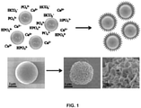
- microspheres can be produced that have a calcium-containing mineral coating. See Examples. These microspheres are useful, at least for providing slow release of therapeutic compounds combined therewith.
- the application is directed to a microsphere comprising a bead coated with a first calcium-containing mineral, as defined in the attached claims.
- the application is also directed to a method of producing a microsphere, said method comprising the steps set out in the attached claims.
- the method comprises incubating a bead in a physiological saline solution comprising carbonate, calcium, and phosphate such that a first calcium-containing mineral layer coating forms on the bead.
- the bead with the mineral layer coating is the microsphere.
- Also disclosed herein is a method of administering a compound to a vertebrate.
- the method comprises administering the above microsphere to the vertebrate.
- the inventors have developed methods for producing novel microspheres that have a calcium-containing mineral coating. They have also characterized these microspheres and established that they have advantageous properties useful for utilizing the microspheres to deliver therapeutic compounds to tissues. See Examples.
- the application is directed to a microsphere comprising a bead coated with a first calcium-containing mineral.
- the Examples describe exemplary methods for producing these microspheres using a modified simulated body fluid (mSBF).
- mSBF modified simulated body fluid
- the composition of the mineral precipitated on the microspheres can be manipulated. See also U.S. Patent Application Publication US 2008/0095817 A1 ; U.S. Patent No. 6,767,928 B1 ; U.S. Patent No. 6,541,022 B1 ; PCT Publication WO 2008/070355 A2 ; PCT Publication WO 2008/082766 A2 ; Murphy and Mooney, 2001; Murphy and Messersmith, 2000.
- Inorganic minerals suitable for producing a calcium-containing mineral coating include various bone mineral ions, such as, but not limited to calcium and phosphate and combinations of bone mineral ions, such as calcium-phosphates.
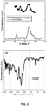
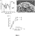
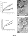
- the calcium-containing mineral coating can comprise, e.g., hydroxyapatite (HAP), ⁇ -tricalcium phosphate ( ⁇ -TCP), ⁇ -tricalcium phosphate ( ⁇ -TCP), amorphous calcium phosphate, dicalcium phosphate, octacalcium phosphate or calcium carbonate.
- the calcium-containing mineral coating can comprise a plurality of layers, e.g., separate layers having distinct dissolution profiles.
- solubility of calcium phosphate species adhere to the following trend: amorphous calcium phosphate>dicalcium phosphate>octacalcium phosphate> ⁇ -TCP>HAP.
- a dicalcium phosphate mineral will typically have a dissolution rate that is more than fifty times higher than that of HAP. Therefore, creation of a matrix with distinct calcium phosphate layers allows for a broad range of dissolution patterns.
- the bead is formed of the material according to claim 1.
- the bead comprises a negative charge, which can promote the deposition of the calcium containing material.
- the negative charge could be provided by any moiety present on the bead, for example a carboxylate group, as is present in poly(D,L-lactide-co-glycolide) (PLG).
- PLG poly(D,L-lactide-co-glycolide)
- the bead is made of a polymer, for example a synthetic polymer.
- the polymer is bioabsorbable.
- suitable bead materials include, for example, a collagen gel, polyvinyl alcohol, a marine adhesive protein, a PLG fiber matrix, a polyglactin fiber, a calcium alginate gel, a polyglycolic acid, polyester ( e .
- polysaccharide e . g . alginate
- chitosan polyphosphazene
- polyacrylate polyethylene oxide-polypropylene glycol block copolymer, fibrin, collagen, and fibronectin
- polyvinylpyrrolidone hyaluronic acid
- poly(lactide) poly(glycolic acid)
- poly(lactide-co-glycolide) poly(caprolactone)
- polycarbonates polyamides, polyanhydrides, polyamino acids, polyortho esters, polyacetals, polycyanoacrylates
- polyurethanes polyacrylates, ethylene-vinyl acetate polymers and other acyl substituted cellulose acetates and derivatives thereof), polyurethanes, polystyrenes, polyvinyl chloride, polyvinyl fluoride, poly(vinylimidazole), chloros
- the bead is made of a PLG, for example at a ratio of about 85:15 lactide:glycolide ("85:15 PLG").
- 85:15 PLG copolymers is advantageous as a decrease in the lactide/glycolide ratio of the copolymer is believed to increase the rate of surface hydrolysis.
- the first calcium-containing mineral is a carbonated-substituted calcium-deficient hydroxyapatite and the bead is PLG, wherein the PLG is about 85:15 lactide:glycolide.
- the microsphere further comprises a component adhering to the first calcium-containing mineral, wherein the component introduces a functional group to the first calcium-containing mineral. Introduction of such a functional group allows covalent binding of any additional materials (e . g . therapeutic compounds) to the microspheres.
- functional groups that can be introduced on the component is a carboxylate, an amine, a carbonyl, a nitro, a hydroxyl, an aldehyde, or an ester.
- the component comprises a poly(aspartic acid), a poly(glutamic acid), or a bisphosphonate. See e.g., Murphy et al., 2007.
- Other nonlimiting examples of components useful in these embodiments are the oligopeptides AAAAEPRREVAEL or AAAA ⁇ EPRR ⁇ EVA ⁇ EL, where ⁇ E is carboxyglutamate.
- the microsphere further comprises a first compound adhering to the first calcium-containing mineral or the component.
- the first calcium-containing mineral is a carbonated-substituted calcium-deficient hydroxyapatite and the bead is poly(D,L-lactide-co-glycolide) (PLG), wherein the PLG is about 85:15 lactide:glycolide.
- PLG poly(D,L-lactide-co-glycolide)
- first compounds that can be an organic compound less than 2000 MW, or less than 1000 MW, or less than 500 MW.
- Nonlimiting examples include antibiotics, corticosteroids and statins. More specific examples include cefazolin, cefuroxime, clindamycin, vancomycin and dexamethasone.
- the first compound is an oligopeptide or polypeptide.
- an oligopeptide comprises a linear chain of 30 or less amino acids.
- a polypeptide comprises more than 30 amino acids.
- examples of oligopeptides are GGRGDSP (a cell adhesion peptide derived from fibronectin), GGIKVAV (a cell adhesion peptide derived from laminin), GGYIGSR (a cell adhesion peptide derived from laminin), GGDGEA (a cell adhesion/signaling peptide derived from type I collagen), GGKIPKASSVPTELSAISTLYL (a peptide derived from bone morphogenetic protein-2), AAAAEPRREVAEL (a modified peptide derived from osteocalcin - some affinity for hydroxyapatite mineral), AAAA ⁇ EPPP ⁇ EYA ⁇ EE, where ⁇ E is carboxyglutamate (a modified peptide derived from osteocalcin - high affinity for hydroxyapatite mineral).
- the first compound is a polypeptide, for example a cytokine, an enzyme, or a protein comprising an antibody binding site (e.g., an antibody).
- polypeptides that could be included in the microspheres are virtually any hormone, neurotransmitter, growth factor, growth factor receptor, interferon, interleukin, chemokine, cytokine, colony stimulating factor and/or chemotactic factor protein or polypeptide.
- transcription or elongation factors include transcription or elongation factors, cell cycle control proteins, kinases, phosphatases, DNA repair proteins, oncogenes, tumor suppressors, angiogenic proteins, anti-angiogenic proteins, immune response stimulating proteins, cell surface receptors, accessory signaling molecules, transport proteins, enzymes, anti-bacterial and/or anti-viral proteins or polypeptides, and the like, depending on the intended use of the ultimate composition.
- growth hormone GH
- parathyroid hormone PTH, including PTH1-34
- bone morphogenetic proteins BMPs
- BMPs bone morphogenetic proteins
- TGF- ⁇ transforming growth factor-a
- FGF fibroblast growth factor
- GMCSF granulocyte/macrophage colony stimulating factor
- EGF epidermal growth factor
- PDGF platelet derived growth factor
- IGF insulin-like growth factor
- LIF leukemia inhibitory factor
- VEGF vascular endothelial growth factor
- bFGF basic fibroblast growth factor
- PDGF platelet derived growth factor
- angiogenin angiopoietin-1, del-1, follistatin
- G-CSF hepatocyte growth factor/scatter factor
- the polypeptide is a BMP-2, a BMP-7, a VEGF, an FGF-2, a PDGF, a TGF- ⁇ , an interleukin, or a human GH.
- first compounds that are a nucleic acid.
- Non-limiting examples include a microRNA, an antisense nucleic acid, and a vector.
- a first compound being a vector, any vector known or later discovered can be included here.
- the vector comprises a sequence encoding a therapeutic protein, such as any of the proteins discussed above.
- the first compound can noncovalently adhere to the microsphere.
- the microsphere can further comprise a component adhering to the first calcium-containing mineral and introducing a functional group to the microsphere, to which the compound is covalently attached. See, e.g., Murphy et al., 2007.
- the first compound is at more than one level of the first calcium-containing mineral. These microspheres generally release the compound over a longer period of time than where the compound is only at one level (for example the outer surface of the first calcium-containing mineral.
- the first compound is modified to change the rate at which the compound is released from the microsphere.
- the first compound further comprises a moiety that increases the strength of binding of the compound to the first calcium-containing mineral, the compound would be released more slowly than if the moiety is not present.
- moieties include amino acid sequences rich in glutamic acid, aspartic acid or phosphoserine, which interact directly with calcium ions in mineralized extracellular matrices.
- Other examples include AAAAEPRREVAEL or AAAA ⁇ EPRR ⁇ EVA ⁇ EL, where ⁇ E is carboxyglutamate.
- the microsphere further comprises a second compound adhering to the first calcium-containing mineral or the component.
- the microspheres are not limited as to the nature of the second compound.
- the second compound can be for example an organic compound less than 2000 MW or 1000 MW or 500 MW.
- the second compound can be an oligopeptide or polypeptide, or a nucleic acid.
- the first compound and the second compound can be on the same or different levels of the first calcium-containing minerals. Having the two compounds on different levels is useful when it is desired that the two compounds are released at different times.
- the microsphere can also further comprise a third, fourth, fifth, etc. compound as desired.
- the microsphere comprises a living cell.
- the living cell can be from any organism, including an Archaea, a prokaryote, or a eukaryote.
- the cell is a mammalian cell.
- the cell can be naturally occurring or, alternatively, can be transformed to express a recombinant molecule, e.g., a protein or nucleic acid (such as an miRNA).
- the first calcium-containing mineral is a carbonated-substituted calcium-deficient hydroxyapatite and the bead is poly(D,L-lactide-co-glycolide) (PLG), wherein the PLG is about 85:15 lactide:glycolide.
- PLG poly(D,L-lactide-co-glycolide)
- the microsphere comprises a first binding agent that binds to a second binding agent on the cell.
- a first binding agent that binds to a second binding agent on the cell.
- suitable cell adhesion peptides are GGRGDSP, GGIKVAV, GGYIGSR or GGDGEA.
- the microsphere comprises a cell as well as a compound (e.g., a cytokine) that interacts with the cell.
- a compound e.g., a cytokine
- the cytokine is advantageously close to the cell, such that the compound is likely to contact and thus interact with the cell.
- the microsphere further comprises a coating of a second calcium-containing mineral.
- the second coating can be a mineral that has a different degradation rate than the first calcium-containing mineral.
- the microsphere comprising the two coatings can further comprise one or more than one compound. Either of the two coatings can be, for example, any of hydroxyapatite (HAP), ⁇ -tricalcium phosphate ( ⁇ -TCP), ⁇ -tricalcium phosphate ( ⁇ -TCP), amorphous calcium phosphate, dicalcium phosphate, octacalcium phosphate or calcium carbonate.
- HAP hydroxyapatite
- ⁇ -TCP ⁇ -tricalcium phosphate
- ⁇ -TCP ⁇ -tricalcium phosphate
- amorphous calcium phosphate dicalcium phosphate
- octacalcium phosphate or calcium carbonate amorphous calcium phosphate
- the first calcium-containing mineral can be hydroxyapatite and further comprises a first therapeutic compound
- the second calcium-containing mineral can be ⁇ -TCP and is coated above ( i.e., closer to the surface of the microsphere) the first calcium containing mineral and further comprises a second therapeutic compound.
- the second calcium-containing mineral would degrade first, releasing the second therapeutic compound before the first therapeutic compound is released.
- the compound present in the microsphere its release depends on a number of factors, for example (a) how strongly the compound adheres to the calcium-containing mineral or component, (b) how far away from the surface of the microsphere the compound is (for example if a layer of calcium-containing mineral is deposited on the microsphere after the compound is added), (c) the degradation/bioabsorption rate of the calcium-containing mineral, (d) whether the compound is covalently bound to a component, and if so, (e) how strongly the component is bound to the mineral, (f) how strongly the covalent bond is between the compound and the component, and (g) whether there are enzymes present, e.g., from a tissue in which the microsphere is implanted, that will break the covalent bond and release the compound.
- each of these factors may have more or less influence on the release of the compound, depending on the configuration of the microsphere.
- the component binds to the calcium-containing material strongly, and there are no enzymes to break the covalent bond, then the main factor influencing the release of the component is the rate of degradation of the calcium-containing mineral.
- the compound is noncovalently adhering directly to the calcium-containing mineral, then all of the above factors (a) - (c) will have an influence in the rate of release of the compound.
- the compound is in the bead. In these embodiments, the compound would likely be released upon degradation of the bead, after the degradation of the calcium-containing mineral.
- the various microspheres discussed herein generally have a diameter between about 0.5 ⁇ m and about 500 ⁇ m. In other embodiments, the microsphere has a diameter between about 0.5 ⁇ m and about 100 ⁇ m. In additional embodiments, the microsphere has a diameter between about 2 ⁇ m and about 6 ⁇ m .
- the microspheres tend to aggregate, particularly if they are produced by incubation in mSBF at a high concentration of microspheres or for an extended period of time (see Examples).
- the application is directed to a plurality of any of the above-described microspheres, aggregated.
- the first calcium-containing mineral is a carbonated-substituted calcium-deficient hydroxyapatite and the bead is poly(D,L-lactide-co-glycolide) (PLG), wherein the PLG is about 85:15 lactide:glycolide.
- PLG poly(D,L-lactide-co-glycolide)
- a first compound, as described above adheres to the first calcium-containing mineral.
- the application is also directed to a method of producing a microsphere.
- the method comprises incubating a bead in a physiological saline solution comprising carbonate, calcium, and phosphate such that a first calcium-containing mineral layer coating forms on the bead, where the bead with the mineral layer coating is the microsphere, as defined in the attached claims. See Examples.
- the solution comprises NaCl, KCl, MgCl 2 , MgSO 4 , NaHCO 3 , Tris, CaCl 2 , and KH 2 PO 4 .
- the solution comprises about 100-200 mM NaCl, about 1-8 mM KCl, about 0.1-2 mM MgSO 4 , about 0.2-5 mM MgCl 2 , about 1-100 mM NaHCO 3 , about 2-20 mM CaCl 2 , and about 0.5-10 mM KH 2 PO 4 .
- the solution comprises about 141 mM NaCl, about 4.0 mM KCl, about 0.5 mM MgSO 4 , about 1.0 mM MgCl 2 , about 4.2 mM NaHCO 3 , about 5.0 mM CaCl 2 , and about 2.0 mM KH 2 PO 4 .
- the solution also comprises a surfactant, which can change the morphology of the calcium-containing mineral layer, and reduce aggregation of the microspheres. See Example 2. Any surfactant now known or later discovered may be used here. In some embodiments, the surfactant is Tween 20 TM .
- the mineral coating described herein is developed by incubating the constituents in the above solution, which can be called a "simulated body fluid” (SBF) or a “modified simulated body fluid” (mSBF), for five days or more at a pH of about 6.8 to about 7.4 and at a temperature of about 37 °C.
- SBF simulated body fluid
- mSBF modified simulated body fluid
- the SBF or mSBF may be refreshed daily.
- the procedure produces a calcium-deficient, carbonate-containing apatite material on alginate and on poly-( ⁇ -hydroxy esters). See U.S. Pat. No. 6,767,928 .
- mSBF includes elevated calcium and phosphate.
- an increase in pH favors hydroxyapatite growth, while a decrease in pH favors octacalcium phosphate mineral growth.
- conditions favorable for hydroxyapatite formation include a pH between about 5.0 and about 8.0 and a calcium concentration multiplied by a phosphate concentration between about 10 -5 and about 10 -8 M.
- conditions favorable for octacalcium phosphate formation include a pH between about 6.0 and about 8.0 and a calcium concentration multiplied by a phosphate concentration between about 10 -5 and about 10 -7.5 M.
- conditions favorable for dicalcium phosphate dehydrate formation include a pH between about 6.0 and about 8.0 and a calcium concentration multiplied by a phosphate concentration between about 10 -4 and about 10 -6 M.
- mSBF poly-(a-hydroxy esters) or alginate hydrogels
- pH of mSBF between about 5.0 and about 6.0 to promote hydroxyapatite formation.
- pH of mSBF between about 6.0 and about 6.5 to promote octacalcium phosphate and hydroxyapatite formation.
- pH of mSBF between about 6.5 and about 8.0 to promote dicalcium phosphate, octacalcium phosphate and hydroxyapatite formation.
- the bead Prior to deposition of the first calcium-containing mineral, the bead may be surface-functionalized to allow increased mineral deposition by utilizing chemical pre-treatment to achieve surface hydrolysis, e . g ., using an NaOH solution. Surface degradation by this technique causes an increase in the amount of polar oxygen functional groups on the surface of the material. The functionalized surface is then incubated in a mineral-containing solution.
- the method further comprises adding a component that adheres to the first calcium-containing mineral layer to the microsphere.
- the component introduces a functional group to the first calcium-containing mineral layer.
- nonlimiting examples of such components include peptides comprising a poly(aspartic acid) sequence, a poly(glutamic acid) sequence, AAAAEPRREVAEL and AAAA ⁇ EPRR ⁇ EVA ⁇ EL, where ⁇ E is carboxyglutamate.
- the method further comprises incubating the microsphere with a first compound such that the first compound adheres to the microsphere.
- the first compound is an oligopeptide or polypeptide (e.g., a cytokine, an enzyme, or a protein comprising an antibody binding site).
- a first compound that is any chemical compound, such as an organic compound less than 2000 MW, or a nucleic acid (e.g., a microRNA, an antisense nucleic acid, or a vector).
- the first compound is incubated with the microsphere such that the first compound is non-covalently bound to the microsphere.
- a component that introduces a functional group to the microsphere is adhered to the microsphere, the first compound is covalently attached to the functional group.
- the first compound is incubated with the bead in the physiological saline solution such that the first compound is deposited on the bead along with the mineral layer coating.
- Such microspheres generally have the first compound deposited throughout the mineral layer coating, whereas if the compound is bound to the bead after the mineral layer is coated onto the bead, the compound would only be present on the surface of the mineral layer. The first compound would be expected to be released over a longer period of time in the former case than in the latter case.
- the bead is incubated in the physiological saline solution both before and after the first compound is adhered to the microsphere, after which additional first compound is adhered to the microsphere.
- the first compound would be internal to the surface of the mineral layer and would be released after a first compound deposited on the surface.
- the method can further comprise incubating the microsphere with a second compound such that the second compound adheres to the microsphere.
- the bead is incubated in the physiological saline solution before and after the first compound is adhered, after which the second compound is adhered to the microsphere, such that the first compound and the second compound are on different layers of the first calcium-containing mineral layer. Such a microsphere would release the second compound before the first compound.
- the present application is further directed to any of the above-described microspheres made by any of the above-described methods.
- the method comprises administering any of the microspheres disclosed above to the vertebrate, where the microsphere further comprises the compound.
- the microsphere maybe in a pharmaceutically acceptable material.
- pharmaceutically acceptable it is meant a material that (i) is compatible with the other ingredients of the composition without rendering the composition unsuitable for its intended purpose, and (ii) is suitable for use with subjects as provided herein without undue adverse side effects (such as toxicity, irritation, and allergic response). Side effects are “undue” when their risk outweighs the benefit provided by the composition.
- pharmaceutically acceptable carriers include, without limitation, any of the standard pharmaceutical carriers such as phosphate buffered saline solutions, water, emulsions such as oil/water emulsions, microemulsions, and the like.
- microspheres can be formulated without undue experimentation for administration to a vertebrate, including humans, as appropriate for the particular application. Additionally, proper dosages of the microspheres can be determined without undue experimentation using standard dose-response protocols.
- the microspheres may be enclosed in gelatin capsules.
- the microspheres of the present invention may be incorporated with excipients and used in the form of tablets, troches, capsules, elixirs, suspensions, syrups, wafers, chewing gums and the like.
- the microspheres can alternatively be administered parenterally such as for example, by intravenous, intramuscular, intrathecal or subcutaneous injection.
- administration can be systemic, for example if the microspheres were administered by injection into the blood stream.
- the administration can be local, e.g., an injection of the microspheres directly onto an area where there is a tissue defect, where the compound is a cytokine that stimulates filling in the defect.
- Parenteral administration can be accomplished by incorporating the microspheres into a suspension.
- Such suspensions may also include sterile diluents such as water for injection, saline solution, fixed oils, polyethylene glycols, glycerin, propylene glycol or other synthetic solvents.
- Parenteral formulations may also include antibacterial agents such as for example, benzyl alcohol or methyl parabens, antioxidants such as for example, ascorbic acid or sodium bisulfite and chelating agents such as EDTA. Buffers such as acetates, citrates or phosphates and agents for the adjustment of osmolarity such as sodium chloride or dextrose may also be added.
- antibacterial agents such as for example, benzyl alcohol or methyl parabens
- antioxidants such as for example, ascorbic acid or sodium bisulfite
- chelating agents such as EDTA.
- Buffers such as acetates, citrates or phosphates and agents for the adjustment of osmolarity such as sodium chloride or dextrose may also be added.
- the parenteral preparation can be enclosed in ampules, disposable syringes or multiple dose vials made of glass or plastic.
- Rectal administration includes administering the microspheres, in a pharmaceutical composition, into the rectum or large intestine. This can be accomplished using suppositories or enemas.
- Suppository formulations can easily be made by methods known in the art. For example, suppository formulations can be prepared by heating glycerin to about 120 °C., dissolving the composition in the glycerin, mixing the heated glycerin after which purified water may be added, and pouring the hot mixture into a suppository mold.
- Transdermal administration includes percutaneous adsorption of the microspheres to the skin.
- Transdermal formulations include patches (such as the well-known nicotine patch), ointments, creams, gels, salves and the like.
- the claimed microspheres may be administered nasally to the vertebrate to provide a therapeutically effective amount of the microspheres.
- nasally administering or nasal administration includes administering the compound to the mucous membranes of the nasal passage or nasal cavity of the patient.
- pharmaceutical compositions for nasal administration of the compound include therapeutically effective amounts of the compound prepared by well-known methods to be administered, for example, as a nasal spray, nasal drop, suspension, gel, ointment, cream or powder. Administration of the compound may also take place by applying the microspheres in a nasal tampon or nasal sponge.
- the vertebrate may be a human.
- the vertebrate may have a condition that is treatable by administering the compound.
- Nonlimiting examples of such conditions include cancer, diabetes, Alzheimer's disease, Parkinson's disease, a heart disease, a virus infection, a bacterial infection, a parasitic infection, an autoimmune disease, an allergy, a prion disease, a gastrointestinal disease, a liver disease, a kidney disease, a skin disease, a bone disease, a congenital disease or defect, a disease characterized by insufficiency of a protein or a metabolite, erectile dysfunction or baldness.
- the condition may be a tissue defect.
- the tissue defect can be, for example, in a bone, a soft tissue, or an internal organ.
- the defect may be caused by disease or trauma, or it may be congenital.
- the application is also directed to the use of any of the above-described microspheres for the manufacture of a medicament for treating a vertebrate with a compound.
- the vertebrate is a mammal, e . g ., a human.
- the vertebrate may be a mammal, e . g ., a human.
- Example 1 Mineral-coated polymer microspheres for controlled protein binding and release.
- biodegradable micro-carriers for controlled protein binding and release.
- the calcium phosphate mineral hydroxyapatite is employed as a substrate for protein binding and release, as it has been used for over 50 years in chromatographic protein separations based on its ability to bind and release both acidic and basic proteins under particular solution conditions.
- biodegradable polymer microspheres can be coated with an inorganic hydroxyapatite layer, and that this biodegradable coating can be used as a substrate for binding and sustained release of acidic and basic proteins.
- This approach involves nucleation and growth of inorganic calcium phosphate mineral coatings on the surface of organic, biodegradable polymer microspheres at near physiologic temperature and pH.
- This mineral growth process mimics natural biomineralization processes (Mann, 2001), and results in a mineral coating that is similar in structure (platelike nanostructure) and composition (carbonated-substituted, calcium-deficient hydroxyapatite phase) to human bone mineral, as detailed previously on macroscopic polymer films (Murphy and Mooney, 2002).
- protein-releasing, mineral-coated polymer microspheres were prepared here via a two-step process involving: i) formation of biodegradable PLG microspheres using a standard water-oil-water double emulsion process (Meng et al., 2003); and ii) coating of PLG microspheres with an inorganic, bone-like mineral (BLM) film via incubation in a modified simulated body fluid (mSBF), an aqueous solution which contains the ionic constituents of blood plasma with 2-fold higher concentrations of calcium and phosphate ions (Murphy and Messersmith, 2000) ( FIG. 1 ).
- mSBF modified simulated body fluid
- X-ray diffraction and FTIR spectra indicate that the mineral grown on PLG microspheres is an apatite mineral ( FIG. 2 ).
- Scanning electron microscopy (SEM) ( FIG. 1 ) indicates that the mineral film is continuous on the microsphere surface and has a plate-like nanostructure. Therefore, the mineral layer grown on biodegradable polymer microspheres is similar in composition and morphology to bone mineral, as described previously Murphy and Mooney, 2002).
- BSA bovine serum albumin
- model proteins were chosen to illustrate the influence of protein characteristics on binding efficiency and release kinetics, and because of their biological relevance.
- albumin is one of the most abundant proteins found in blood plasma and has been shown to promote formation of bone tissue (Yamaguchi et al., 2003), while cytochrome c serves as a model protein for several basic growth factors, such as fibroblast growth factor-2, bone morphogenic proteins, and transforming growth factor ⁇ .
- Binding of BSA on mineral-coated PLG microspheres was first detected by FTIR analysis ( FIG. 3A ).
- the FTIR spectrum shows phosphate peaks (1087, 1035, 950, and 560 cm -1 ) and carbonate peaks (1456 and 1417 cm -1 ) corresponding to carbonate-substituted hydroxyapatite mineral coatings, consistent with previous studies of BLM coatings formed on biodegradable polymer substrata (Murphy and Mooney, 2002).
- the FTIR spectrum also shows amide peaks (1653 and 1558 cm -1 ) corresponding to the presence of BSA bound to the coating surface.
- Cyt c showed a binding curve similar to that of BSA, and it is noteworthy that Cyt c bound to the mineral more efficiently than BSA at the same solution protein concentrations.
- the release kinetics of BSA from mineral-coated PLG microspheres were investigated by incubating mineral-coated microspheres in phosphate-buffered saline (PBS) solution at pH 7.4 ( FIG. 4A ). BSA release was sustained over 30 days, and the release displayed near linear kinetics. In contrast, the cumulative release of BSA encapsulated in PLG microspheres via standard processing techniques (Meng et al., 2003) displayed a much lower level of cumulative release over 30 days, and the majority of the released protein represented a "burst" release during the initial 72 hours.
- PBS phosphate-buffered saline
- mineral-coated PLG microspheres can serve as effective carriers for protein binding and sustained release.
- the protein release profile from these minerals does not include the "burst" release that is typically observed in biodegradable microparticle release systems, and the protein release rate is dependent on protein characteristics and the local pH. It is noteworthy that previous studies using hydroxyapatite chromatography to purify proteins and DNA (Urist et al., 1984) suggest that this mechanism for protein binding and release may be adaptable to a broad range of acidic and basic biomolecules, and that the biological activity of molecules released from minerals in this manner is likely to be high.
- biodegradable polymer microspheres suggest that several biodegradable micro- or nano-scale materials can be used as templates for mineral growth, and that biological molecules can potentially be included into mineral coatings during the course of the coating process. Therefore, this approach may represent an adaptable mechanism for biomolecule binding and controlled release for biomedical applications.
- mSBF modified-simulated body fluid
- Bovine serum albumin BSA
- Cyt c cytochrome c
- Mineral-coated microspheres were prepared via a bioinspired, heterogeneous nucleation process at physiologic temperature.
- Poly(DL-lactide-co-glycolide) (PLG) microspheres were fabricated via a water-in-oil-in-water emulsion method and were mineral-coated via incubation in a modified simulated body fluid (mSBF).
- mSBF modified simulated body fluid
- the mineral grown on the PLG microsphere surface was a carbonate-containing hydroxyapatite, and the mineral shows a porous structure of plate-like mineral crystals at the nanometer scale. Aggregation of mineral-coated microspheres was observed when microsphere concentrations above 0.50 mg/mL were incubated in mSBF for 7 days, and the size of aggregates was dependent on the microsphere concentration in solution. In vitro mineral dissolution studies performed in Tris-buffered saline confirmed that the mineral formed was resorbable.
- a surfactant additive (Tween 20 TM [PEG(20)sorbitan monolaurate]) was incorporated into mSBF to prevent microsphere aggregation during the mineral growth process, and Tween 20 TM not only prevented aggregation, but also influenced the characteristics of the mineral formed on the surface of PLG microspheres.
- hydroxyapatite is the ability to bind to biological molecules.
- hydroxyapatite is commonly used as a resin for chromatographic purification of proteins and plasmid DNA (Colman et al., 1978; Schroder et al., 2003), as the mineral surface contains both positive (Ca 2+ ) and negative (PO 4 3- ) ions capable of interacting electrostatically with basic and acidic molecules, respectively.
- This ability to bind, and subsequently release, biological molecules has recently been used as a mechanism for sustained drug delivery (Example 1). Therefore, a large body of work has focused on creating bone-like hydroxyapatite coatings on polymeric biomaterials to simultaneously exploit both the bulk properties of biodegradable polymers and the bioactivity of hydroxyapatite coatings.
- biodegradable polymer microspheres which are commonly used in drug delivery applications, could be coated with a biodegradable, hydroxyapatite mineral using a bioinspired mineral nucleation and growth process.
- the resulting materials are designed to exploit the controllable properties of polymer microspheres (e.g. size, size range, degradability, drug incorporation), while also taking advantage of the biological properties of the mineral layer (e.g. bioactivity, biomolecule binding/incorporation).
- mineral-coated microspheres were fabricated using a two-step processing route.
- PLG microspheres were fabricated using a double emulsion method, then those microspheres were incubated in modified simulated body fluid, allowing for mineral nucleation and growth in near physiological conditions.
- the amount of mineral formed can be controlled by the incubation time and the concentration of microspheres in solution.
- a surfactant additive influenced microsphere aggregation and the morphology of the mineral formed.
- the results presented here illustrate that an inorganic mineral layer can be grown in a controllable manner on the surface of biodegradable microspheres, and these materials may find utility in a range of biomedical applications, most notable drug delivery and chromatography.
- PLG microspheres were fabricated by water-in-oil-in-water (W/O/W) double emulsion technique as reported elsewhere (Berchane et al., 2006). Briefly, the organic phase consisted of 5% (w/v) PLG in 1 ml ethyl acetate. The aqueous phase consisted of 0.1 ml phosphate buffered saline (PBS). The aqueous and organic phases were mixed and sonicated using Sonifier 250 (VWR International, Inc., West Chester, PA) for 15 s.
- Sonifier 250 VWR International, Inc., West Chester, PA
- the resulting first emulsion was added immediately into 1 ml of aqueous 1% (w/v) PVA in 7% (v/v) ethyl acetate that was being mechanically vortexed for 15 s to form a second emulsion.
- the resulting solution was then added to a beaker containing 200 ml of 0.3% PVA in 7% ethyl acetate and further rigorously stirred for 4 hr to allow for organic solvent evaporation.
- the resulting microspheres were collected by filtration through 0.22 ⁇ m filter, washed three times with de-ionized water, and resuspended in de-ionized water. The microspheres were lyophilized for a minimum of 48 hr and were stored at -20 °C in the presence of a desiccant.
- the ⁇ potential of PLG microspheres was first characterized in PBS and mSBF solutions.
- the surface charge of the microsphere particles was measured with a Zetasizer 3000HS (Malvern Instruments, Worcestershire, U.K.).
- the electrophoretic mobility of uncoated microspheres in three 6 mL aliquots was measured to determine the surface potential, with each injection having five measurements. Samples were syringe-loaded and measured at 25 °C in 1x PBS or mSBF, at a pH of 6.8 to mimic mineral coating conditions.
- Quantification of aggregated microspheres in various buffers was performed by incubating a 0.5% (w/v) PLG microsphere in a selected buffer (1x PBS, mSBF, and mSBF + 0.1% (v/v) Tween 20 TM ) for 1, 3, and 7 days.
- the suspension was held at 37 °C and rotated continuously for the duration of the study period, identical with the conditions used for mineral growth.
- aliquots of each condition were taken, diluted 1:8, and imaged under an Olympus Ix51 light microscope at 20X magnification. Four photographs were taken per sample per time point with a Hamamatsu 1394 ORCA-285 camera. The resultant images were viewed and counted using Image J software.
- mSBF was prepared by dissolving 141 mM NaCl, 4.0 mM KCl, 0.5 mM MgSO 4 , 1.0 mM MgCl 2 , 4.2 mM NaHCO 3 , 5.0 mM CaCl 2 , and 2.0 mM KH 2 PO 4 in distilled water, buffered to pH 6.8, and was held at 37 °C for the duration of the incubation period.
- 0.1% of Tween 20 TM (Sigma-Aldrich, St. Louis, MO) was added to the mSBF to prevent the aggregation of microspheres.
- FTIR Fourier transform infrared spectroscopy
- the morphology and composition of the coated mineral on the microsphere surface was analyzed using scanning electron microscopy (SEM) with energy-dispersive X-ray spectroscopy (EDS). Microspheres before and after mSBF incubation were mounted on aluminum stubs with double sided carbon tape, sputtered with gold for 30s at 45mA and characterized using a LEO DSM 1530 field emission SEM, operating at 2kV for SEM and 10kV for EDS.
- SEM scanning electron microscopy
- EDS energy-dispersive X-ray spectroscopy
- DMEM Dulbecco's Modified Eagle Medium
- the buffer was collected and refreshed daily. The study was performed in triplicate and held at 37 °C for the duration of dissolution study period.
- the amount of phosphate released from mineral-coated microspheres was analyzed colorimetrically using an assay previously reported (Heinonen and Lahti, 1981). Briefly, a working AAM (acetone-acid-molybdate) solution was prepared by mixing 2 parts acetone with 1 part 5 N sulfuric acid, and 1 part 10 mM ammonium molybdate solution. The assay was performed in a 96-well plate by adding 100/ ⁇ l a freshly made working solution to 100 ⁇ 1 sample.
- AAM acetone-acid-molybdate
- the amount of phosphate complex was quantitatively detected by measuring the absorbance at 405 nm on a Synergy TM HT Multidetection Microplate Readers (Bio-Tek Instruments, Inc., UK) and comparing to a set of standards with known phosphate concentrations.
- Ca 2+ release was direct measured using QuantiChrom TM Calcium Assay Kit (DICA-500) (BioAssay Systems, Hayward, CA).
- DICA-500 QuantiChrom TM Calcium Assay Kit
- a phenolsulphonephthalein dye forms a very stable blue color complex with free calcium.
- the intensity of the complex measured via absorbance at 612 nm, was used to measure released Ca 2+ by comparing to a set of standards with known calcium concentrations. Dissolution experiments were performed in triplicate and statistical analyses for calcium and phosphate release were carried out using ANOVA.
- Micrographs of the microspheres incubated in mSBF for 7 days show continuous mineral coatings on individual microspheres incubated at 0.25% and 0.50% (w/v) ( FIG. 6A, B ). Microsphere aggregation was observed as microspheres concentration increased, and mineral coatings were observed on the surface of microsphere aggregates ( FIG. 6B , C, D).
- the peak areas in the XRD spectrum of mineral-coated microspheres are broader than that of hydroxyapatite powder, and this may be due to the small crystal size of the mineral deposited on the PLG microsphere surfaces.
- FTIR peaks observed in the 1600-400 cm -1 region can be attributed to carbonate-substituted hydroxyapatite, including phosphate peaks at 570, 950, 1046, and 1098 cm -1 , and carbonate peaks at 870, 1410, and 1456 cm -1 ( FIG. 8B ).
- the EDS spectrum also confirms the presence of calcium and phosphorus on the mineral-coated microspheres ( FIG. 9 ).
- the Ca/P ratio of the mineral coating was 1.41 after 7 days of incubation in mSBF, which is consistent with that of previous studies of biological apatites (Elliott, 1994) and bioinspired mineral coatings (Jabbarzadeh et al., 2007).
- Time lapse SEM analyses of PLG microspheres at various times during mSBF incubation provide some insight into the mechanism of nucleation and growth of carbonate apatite mineral on aggregating microspheres ( FIG. 10 ).
- the nucleation process begins during the first three days of incubation in mSBF ( FIG. 10A, B ). During this stage the aggregation of microsphere begins to occur. As the microspheres begin to aggregate, small crystals ( ⁇ 2-10 nm) begin to form at the interface between microspheres ( FIG. 10A, B ), perhaps due in part to local supersaturation of surface functional groups and associated mineral ions at the interface.
- FIG. 10C After five days a highly porous structure of plate-like hydroxyapatite crystals appear on the surfaces of aggregated microspheres ( FIG. 10C ), ultimately growing into a continuous coating ( FIG. 10D ).
- the size of the aggregates depends on the initial concentration of microspheres in solution ( FIG. 6E ), suggesting a potential mechanism for control over the size of coated aggregates.
- Mineral coatings were also observed on the surface of microsphere aggregates.
- Other analyses not presented here suggest that the efficiency of microsphere mineralization increases in conditions that promote microsphere aggregation.
- Our time-course analysis of mineral formation in solutions with higher concentrations of microspheres also demonstrates that mineral nucleation can occur at the interface between aggregated microspheres ( e . g ., FIG. 10B ), which suggests that mineral formation can be facilitated by aggregation.
- microspheres were incubated in three different solutions: (1) a 1xPBS solution (calcium-deficient), (2) mSBF, and (3) mSBF in the presence of Tween 20 TM . Each incubation was performed at 37 °C with daily solution changes for 7 days. Results showed that 35% and 42% microspheres aggregated after the first day of incubation in PB S and mSBF, respectively. The percentage of aggregated microspheres increased to 87% in PBS and 90% in mSBF after 7 days of incubation.
- the formation of mineral described here is analogous to previous work by He et al. (2003) on bioinspired formation of mineral in the presence of dentin matrix protein.
- the morphology and composition of mineral described here is similar to the apatite crystals found in human woven bone and mineralized dentin (Su et al., 2003), specifically a platelike nanostructure ( FIGS. 5B and 14B ), a hydroxyapatite phase ( FIGS. 8A and 9 ), and carbonate substitution ( FIG. 8B ).
- Dissolution and re-precipitation are key characteristics of hydroxyapatite coatings, particularly in orthopedic implant design and drug delivery applications. Some forms of hydroxyapatite mineral have been shown to degrade slowly or incompletely over extended timeframes, and the permanent presence of these materials in vivo is undesirable in applications that call for material degradation over time (e.g. tissue engineering, drug delivery). In addition, mineral dissolution can influence release of biological molecules from these coatings in drug delivery applications, as described in Example 1. Therefore, in this study dissolution of mineral coatings in Tris-buffered saline (TBS) and in DMEM was characterized for 25 days. Ca 2+ and PO 4 3- were gradually released from mineral coatings over time in TBS ( FIG.
- the morphology of the mineral coating after incubation in DMEM was similar to the morphology of the mineral prior to incubation in DMEM, which indicates that if re-precipitation is occurring, it is resulting in growth of a new mineral phase that is similar to the mineral phase grown initially.
- Tween 20 TM was added to the mSBF solution.
- Tween 20 TM is a common surfactant used as a stabilizer in studies which measure drug release from PLG microspheres in vitro (He et al., 2005; Raman et al., 2005).
- the rate of mineral formation on the surface of microspheres in the presence of Tween 20 TM ( FIG. 10A-D ) is clearly slower than in the absence of surfactant (FIG. 5, 6).
- FTIR spectra of the mineral formed in the presence of surfactant FIG.
- a rough surface with a relatively well-distributed mineral coating has an increased surface area, and the corresponding increase in available binding sites could result in higher protein binding (Example 1; LeGeros, 2002).
- Laurencin et al. showed that mineral coating of a sintered PLG microsphere scaffold in mSBF increase the protein adsorption capacity and decreased the initial burst release of protein from the polymer scaffold when compare to non-mineralized scaffolds (Jabbarzadeh et al., 2007).
- Mineral-coated PLG microsphere have been fabricated by a simple and inexpensive two-step process involving microsphere fabrication via double emulsion and coating of those microspheres with mineral by immersing in mSBF solution.
- XRD and FTIR spectra indicated that the coatings comprised a carbonated-substituted hydroxyapatite mineral with a porous, plate-like nanoscale morphology.
- the size of the mineral-coated microspheres or microsphere aggregates can be controlled by varying the microsphere concentration in the mSBF solution.
- Hydroxyapatite coatings were degradable in tris-buffered saline, and the quantitative analysis of calcium and phosphate release from the coatings indicate that the Ca/P molar ratio in the mineral is consistent with that of carbonated-substituted hydroxyapatite.
- the presence of a surfactant during the mineral growth process delayed the formation of mineral, and also significantly affected the morphology of the mineral.
- these findings indicate the feasibility of processing mineral-coated PLG microspheres in a controlled fashion using a bioinspired process. This material may be useful in a variety of applications that may benefit from the bulk properties of polymer microspheres and the surface properties of hydroxyapatite minerals, including tissue engineering, drug delivery, and biomolecule purification.
Claims (14)
- Microsphère comprenant une bille revêtue d'un premier minéral contenant du calcium, dans laquelle la bille est constituée d'un polymère poly(D,L-lactide-co-glycolide) (PLG) et la microsphère comprend en outre soit :un composant adhérant au premier minéral contenant du calcium, dans lequel le composant introduit un groupe fonctionnel dans le premier minéral contenant du calcium, et un premier composé qui est lié de manière covalente au groupe fonctionnel ; soitun premier composé qui adhère de manière non covalente au premier minéral contenant du calcium ;dans lequel le premier composé est un oligopeptide ou un polypeptide.
- Microsphère selon la revendication 1, dans laquelle le premier minéral contenant du calcium est une apatite, hydroxyapatite, un phosphate d'a-tricalcium, un phosphate de β-tricaicium, un phosphate de calcium amorphe, un phosphate dicalcique, un phosphate octacalcique ou un carbonate de calcium, de préférence dans laquelle le premier minéral contenant du calcium est une hydroxyapatite substituée en carbone et déficiente en calcium.
- Microsphère selon la revendication 1 ou la revendication 2, dans laquelle la bille est chargée négativement, de préférence dans laquelle la charge négative est due à des groupes carboxylate.
- Microsphère selon l'une quelconque des revendications précédentes, dans laquelle :(i) le groupe fonctionnel est un carboxylate, une amine, un carbonyle, un nitro, un hydroxyle, un aldéhyde ou un ester ; ou(ii) le composant comprend un poly(acide aspartique), un poly(acide glutamique) ou un bisphosphonate.
- Microsphère selon l'une quelconque des revendications précédentes, dans laquelle le premier composé est :(i) à plus d'un niveau du premier minéral contenant du calcium ; ou(ii) est modifié pour changer la vitesse à laquelle le premier composé est libéré de la microsphère, de préférence le premier composé comprenant en outre un fragment qui augmente la force de liaison du premier composé au premier minéral contenant du calcium, de préférence le fragment étant un peptide comprenant un poly(acide aspartique), un poly(acide glutamique), AAAAEPRREVAEL ou AAAAγEPRRγEVAγEL, où γE est du carboxyglutamate.
- Microsphère selon l'une quelconque des revendications précédentes, comprenant en outre un second composé adhérant au premier minéral contenant du calcium ou au composant, de préférence dans laquelle le premier composé et le second composé sont à des niveaux différents du premier minéral contenant du calcium.
- Microsphère selon l'une quelconque des revendications précédentes, comprenant en outre une cellule vivante, de préférence dans laquelle la cellule vivante est une cellule de mammifère.
- Microsphère selon la revendication 7, dans laquelle la microsphère comprend un premier agent liant qui se lie à un second agent liant sur la cellule, de préférence dans laquelle(i) le premier agent liant est un récepteur, un premier acide nucléique ou un peptide d'adhésion cellulaire et le second agent liant est un ligand du récepteur, un second acide nucléique complémentaire au premier acide nucléique, ou un ligand du peptide d'adhésion cellulaire ; ou(ii) le premier agent liant est le peptide d'adhésion cellulaire GGRGDSP, GGIKVAV, GGYIGSR ou GGDGEA.
- Microsphère selon la revendication 7, comprenant en outre une cytokine qui interagit avec la cellule.
- Microsphère selon l'une quelconque des revendications précédentes, dans laquelle la bille est en outre revêtue d'un second minéral contenant du calcium.
- Microsphère selon l'une quelconque des revendications précédentes, ayant un diamètre compris entre 0,5 µm et 100 µm, de préférence entre 2 µm et 6 µm.
- Procédé de production d'une microsphère selon l'une quelconque des revendications 1 à 11, comprenant l'incubation d'une bille dans une solution saline physiologique comprenant du carbonate, du calcium et du phosphate de telle sorte qu'un premier revêtement de couche de minéral contenant du calcium se forme sur la bille, où la bille avec le revêtement de couche de minéral est la microsphère, comprenant de préférence en outre les étapes :(i) d'ajout d'un composant qui adhère à la première couche de minerai contenant du calcium de la microsphère, dans lequel le composant introduit un groupe fonctionnel dans la première couche de minerai contenant du calcium ; et(ii) d'incubation de la microsphère avec un premier composé de telle sorte que le premier composé se fixe de manière covalente au groupe fonctionnel ou adhère de manière non covalente au premier minéral contenant du calcium.
- Procédé selon la revendication 12, dans lequel la solution comprend :(i) NaCl de préférence à 141 mM, KCl de préférence à 4,0 mM, MgCl2 de préférence à 1 mM, MgSO4 de préférence à 0,5 mM, NaHCO3 de préférence à 4,2 mM, Tris, CaCl2 de préférence à 5 mM, KH2PO4 de préférence à 2 mM ; ou(ii) un tensio-actif, de préférence le Tween 20™.
- Procédé selon la revendication 12 (ii), dans lequel :(i) le premier composé est incubé avec la microsphère de telle sorte que le premier composé est lié de manière non covalente à la microsphère ;(ii) le procédé comprend en outre l'adhésion d'un composant qui introduit un groupe fonctionnel dans la microsphère, puis la fixation covalente du premier composé au groupe fonctionnel ; ou(iii)le premier composé est incubé avec la bille dans le sérum physiologique de telle sorte que le premier composé se dépose sur la bille avec la première couche de mineral contenant du calcium.
Priority Applications (1)
| Application Number | Priority Date | Filing Date | Title |
|---|---|---|---|
| EP21213679.0A EP4011363A1 (fr) | 2008-09-25 | 2009-09-25 | Microsphères à revêtement minéral |
Applications Claiming Priority (2)
| Application Number | Priority Date | Filing Date | Title |
|---|---|---|---|
| US10006208P | 2008-09-25 | 2008-09-25 | |
| PCT/US2009/058419 WO2010036919A1 (fr) | 2008-09-25 | 2009-09-25 | Microsphères à revêtement minéral |
Related Child Applications (2)
| Application Number | Title | Priority Date | Filing Date |
|---|---|---|---|
| EP21213679.0A Division EP4011363A1 (fr) | 2008-09-25 | 2009-09-25 | Microsphères à revêtement minéral |
| EP21213679.0A Division-Into EP4011363A1 (fr) | 2008-09-25 | 2009-09-25 | Microsphères à revêtement minéral |
Publications (3)
| Publication Number | Publication Date |
|---|---|
| EP2349212A1 EP2349212A1 (fr) | 2011-08-03 |
| EP2349212A4 EP2349212A4 (fr) | 2014-07-30 |
| EP2349212B1 true EP2349212B1 (fr) | 2022-01-19 |
Family
ID=42060103
Family Applications (2)
| Application Number | Title | Priority Date | Filing Date |
|---|---|---|---|
| EP09816920.4A Active EP2349212B1 (fr) | 2008-09-25 | 2009-09-25 | Microsphères à revêtement minéral |
| EP21213679.0A Pending EP4011363A1 (fr) | 2008-09-25 | 2009-09-25 | Microsphères à revêtement minéral |
Family Applications After (1)
| Application Number | Title | Priority Date | Filing Date |
|---|---|---|---|
| EP21213679.0A Pending EP4011363A1 (fr) | 2008-09-25 | 2009-09-25 | Microsphères à revêtement minéral |
Country Status (5)
| Country | Link |
|---|---|
| US (1) | US11607391B2 (fr) |
| EP (2) | EP2349212B1 (fr) |
| JP (1) | JP2012503673A (fr) |
| CA (1) | CA2740633C (fr) |
| WO (1) | WO2010036919A1 (fr) |
Families Citing this family (21)
| Publication number | Priority date | Publication date | Assignee | Title |
|---|---|---|---|---|
| SG190009A1 (en) | 2010-11-24 | 2013-06-28 | Univ Nanyang Tech | Method for encapsulating particles |
| US20130295186A1 (en) * | 2010-11-24 | 2013-11-07 | Say Chye Joachim Loo | Method for coating particles with calcium phosphate and particles, microparticles and nanoparticles formed thereof |
| US20140141047A1 (en) * | 2012-11-19 | 2014-05-22 | Wisconsin Alumni Research Foundation | Selection of platelet rich plasma components via mineral binding |
| EP3049438B1 (fr) * | 2013-09-24 | 2018-07-25 | The Feinstein Institute for Medical Research | Peptides inhibant l'activité de protéines de liaison à l'arn inductibles par le froid |
| CN103611193B (zh) * | 2013-11-26 | 2015-05-27 | 中山大学 | 一种碳酸钙微球-细胞外基质复合材料及其制备方法和应用 |
| US10682442B2 (en) | 2014-04-04 | 2020-06-16 | University Of Kentucky Research Foundation | Small molecule drug release from in situ forming degradable scaffolds incorporating hydrogels and bioceramic microparticles |
| US20150306595A1 (en) * | 2014-04-25 | 2015-10-29 | The Trustees Of Princeton University | Nanoparticulate Apatite Coated Calcite/Limestone Filter Materials for Removing Contaminants from Contaminated Water |
| US11491261B2 (en) * | 2015-03-03 | 2022-11-08 | Warsaw Orthopedic, Inc. | Coating scaffolds |
| US10449155B2 (en) * | 2015-11-09 | 2019-10-22 | Medical Corporation Ijunkai | Drug introducing agent for administration into a living body and manufacturing method |
| AU2018249816A1 (en) * | 2017-04-03 | 2019-10-17 | Wisconsin Alumni Research Foundation | Mineral coated microparticles for sustained delivery of biologically active molecules |
| WO2019010304A1 (fr) * | 2017-07-05 | 2019-01-10 | Wisconsin Alumni Research Foundation | Microparticules enrobées de minéraux pour l'administration de gènes dans une thérapie de plaie chronique |
| CN107823189B (zh) * | 2017-11-25 | 2021-04-06 | 杭州高成生物营养技术有限公司 | 一种氯化钾缓释微丸及其制备方法与应用 |
| EP3773519A4 (fr) * | 2018-04-06 | 2021-12-29 | Wisconsin Alumni Research Foundation | Nanorevêtements structurés pour la stabilisation de constituants de milieux de cellules souches pluripotentes |
| AU2020274535A1 (en) * | 2019-05-10 | 2021-12-09 | Commonwealth Scientific And Industrial Research Organisation | Microcapsule |
| US20230241284A1 (en) * | 2020-05-25 | 2023-08-03 | Dhawan Shrey | Ion-substituted bovine hydroxyapatite for bone regeneration |
| CN113425896A (zh) * | 2021-05-08 | 2021-09-24 | 上海大学 | 用于牙槽骨缺损修复的聚氨基酸微载体及其制备方法 |
| CN113769162B (zh) * | 2021-09-14 | 2022-09-13 | 武汉大学 | 一种用于治疗感染性骨缺损的负载聚乙烯吡咯烷酮碘的仿生矿化微球的制备方法 |
| US20230142212A1 (en) | 2021-11-09 | 2023-05-11 | Warsaw Orthopedic, Inc. | Systems and methods of coating bone material |
| US11747269B2 (en) | 2021-11-09 | 2023-09-05 | Warsaw Orthopedic, Inc. | Systems and methods for identifying a coating on an implant |
| WO2023086816A1 (fr) * | 2021-11-09 | 2023-05-19 | Wisconsin Alumni Research Foundation | Substrats revêtus de minéraux pour la stabilisation de compositions thérapeutiques à base d'arn |
| US20230390070A1 (en) | 2022-06-07 | 2023-12-07 | Warsaw Orthopedic, Inc. | Methods and apparatus for coating bone particles using a mesh |
Family Cites Families (16)
| Publication number | Priority date | Publication date | Assignee | Title |
|---|---|---|---|---|
| US6743779B1 (en) * | 1994-11-29 | 2004-06-01 | Imarx Pharmaceutical Corp. | Methods for delivering compounds into a cell |
| DE69840069D1 (de) * | 1997-04-01 | 2008-11-13 | Cap Biotechnology Inc | Kalziumphosphat-mikroträger und -mikrokugeln |
| WO1999009149A1 (fr) | 1997-08-01 | 1999-02-25 | Massachusetts Institute Of Technology | Matrices de polymeres tridimensionnelles |
| ES2199815T3 (es) | 1999-03-19 | 2004-03-01 | The Regents Of The University Of Michigan | Mineralizacion y desarrollo celular supeerficial sobre biomateriales. |
| US6767928B1 (en) | 1999-03-19 | 2004-07-27 | The Regents Of The University Of Michigan | Mineralization and biological modification of biomaterial surfaces |
| US7829074B2 (en) * | 2001-10-18 | 2010-11-09 | Nektar Therapeutics | Hydroxypatite-targeting poly(ethylene glycol) and related polymers |
| US6730324B2 (en) * | 2001-04-20 | 2004-05-04 | The University Of British Columbia | Biofunctional hydroxyapatite coatings and microspheres for in-situ drug encapsulation |
| US7776600B2 (en) * | 2002-04-18 | 2010-08-17 | Carnegie Mellon University | Method of manufacturing hydroxyapatite and uses therefor in delivery of nucleic acids |
| US7090868B2 (en) | 2002-09-13 | 2006-08-15 | University Of Florida | Materials and methods for drug delivery and uptake |
| US20070026069A1 (en) | 2003-03-28 | 2007-02-01 | Shastri Venkatram P | Biommetic hierarchies using functionalized nanoparticles as building blocks |
| US20050249697A1 (en) * | 2003-09-24 | 2005-11-10 | Uhrich Kathryn E | Compositions and methods for the inhibition of bone growth and resorption |
| EP1729675A4 (fr) * | 2004-03-05 | 2011-05-18 | Univ Columbia | Echafaudage composite biodegradable d'integration osseuse a phases multiples pour la fixation biologique du tissu mou musculo-squelettique a l'os |
| WO2007070372A2 (fr) | 2005-12-09 | 2007-06-21 | Entremed, Inc. | Compositions et procedes d'inhibition de la proliferation cellulaire |
| US9295755B2 (en) | 2006-10-18 | 2016-03-29 | Wisconsin Alumni Research Foundation | Multilayer tissue regeneration system |
| US9439948B2 (en) | 2006-10-30 | 2016-09-13 | The Regents Of The University Of Michigan | Degradable cage coated with mineral layers for spinal interbody fusion |
| US8275594B2 (en) | 2006-10-30 | 2012-09-25 | The Regents Of The University Of Michigan | Engineered scaffolds for intervertebral disc repair and regeneration and for articulating joint repair and regeneration |
-
2009
- 2009-09-25 EP EP09816920.4A patent/EP2349212B1/fr active Active
- 2009-09-25 US US13/879,178 patent/US11607391B2/en active Active
- 2009-09-25 JP JP2011529272A patent/JP2012503673A/ja active Pending
- 2009-09-25 CA CA2740633A patent/CA2740633C/fr active Active
- 2009-09-25 WO PCT/US2009/058419 patent/WO2010036919A1/fr active Application Filing
- 2009-09-25 EP EP21213679.0A patent/EP4011363A1/fr active Pending
Also Published As
| Publication number | Publication date |
|---|---|
| JP2012503673A (ja) | 2012-02-09 |
| WO2010036919A1 (fr) | 2010-04-01 |
| AU2009296439A1 (en) | 2010-04-01 |
| EP4011363A1 (fr) | 2022-06-15 |
| EP2349212A1 (fr) | 2011-08-03 |
| EP2349212A4 (fr) | 2014-07-30 |
| US20140161886A1 (en) | 2014-06-12 |
| CA2740633A1 (fr) | 2010-04-01 |
| CA2740633C (fr) | 2019-02-26 |
| US11607391B2 (en) | 2023-03-21 |
Similar Documents
| Publication | Publication Date | Title |
|---|---|---|
| EP2349212B1 (fr) | Microsphères à revêtement minéral | |
| KR101278204B1 (ko) | 다중 약물전달계를 갖는 생체의료용 금속합금 재료의 제조방법 | |
| Barati et al. | Spatiotemporal release of BMP-2 and VEGF enhances osteogenic and vasculogenic differentiation of human mesenchymal stem cells and endothelial colony-forming cells co-encapsulated in a patterned hydrogel | |
| Wu et al. | Enhanced osteogenic differentiation and bone regeneration of poly (lactic-co-glycolic acid) by graphene via activation of PI3K/Akt/GSK-3β/β-catenin signal circuit | |
| Lee et al. | Alginate: properties and biomedical applications | |
| EP3226977B1 (fr) | Composites de verre pour l'augmentation de tissus, et pour des applications biomédicales et cosmétiques | |
| Moncal et al. | Collagen-infilled 3D printed scaffolds loaded with miR-148b-transfected bone marrow stem cells improve calvarial bone regeneration in rats | |
| Ye et al. | Biomimetic mineralized hybrid scaffolds with antimicrobial peptides | |
| Lee et al. | Hydroxyapatite-containing gelatin/chitosan microspheres for controlled release of lysozyme and enhanced cytocompatibility | |
| JP2007514518A (ja) | 水素結合によって結合した多層中性ポリマーフィルムのカプセル | |
| WO2014128289A1 (fr) | Dispositif biomédical implantable dans l'os et/ou le tissu cartilagineux, et procédé correspondant pour fabriquer ledit dispositif biomédical | |
| Bozorgi et al. | Application of nanoparticles in bone tissue engineering; a review on the molecular mechanisms driving osteogenesis | |
| Wang et al. | Osteogenic growth peptide-loaded 3D-printed PCL scaffolds for the promotion of osteogenesis through the ERK pathway | |
| SG190798A1 (en) | Method for coating particles with calcium phosphate and particles, microparticles and nanoparticles formed thereof | |
| KR102220402B1 (ko) | 골 형성 촉진용 조성물 | |
| Li et al. | Fabrication of uniform casein/CaCO3 vaterite microspheres and investigation of its formation mechanism | |
| CN110882233A (zh) | 一种同时负载抗癌药物和活性因子的具有微纳米结构的可降解微球及其制备方法和应用 | |
| Yang et al. | Biomimetic synthesis of sericin and silica hybrid colloidosomes for stimuli-responsive anti-cancer drug delivery systems | |
| WH Lo et al. | Current patents on osteoinductive molecules for bone tissue engineering | |
| RU2692768C1 (ru) | Пористые биополимерные микросферы для контролируемого высвобождения положительно заряженных белков и способ получения микросфер | |
| Victor et al. | Poly methacrylic acid modified CDHA nanocomposites as potential pH responsive drug delivery vehicles | |
| AU2009296439B2 (en) | Mineral-coated microspheres | |
| Ling et al. | Improvement of drug elution in thin mineralized collagen coatings with PLGA‐PEG‐PLGA micelles | |
| Yasunaga et al. | The enhancing effects of heparin on the biological activity of FGF-2 in heparin− FGF-2− calcium phosphate composite layers | |
| US9314503B2 (en) | Sustained release systems and preparation method thereof |
Legal Events
| Date | Code | Title | Description |
|---|---|---|---|
| PUAI | Public reference made under article 153(3) epc to a published international application that has entered the european phase |
Free format text: ORIGINAL CODE: 0009012 |
|
| 17P | Request for examination filed |
Effective date: 20110421 |
|
| AK | Designated contracting states |
Kind code of ref document: A1 Designated state(s): AT BE BG CH CY CZ DE DK EE ES FI FR GB GR HR HU IE IS IT LI LT LU LV MC MK MT NL NO PL PT RO SE SI SK SM TR |
|
| RIN1 | Information on inventor provided before grant (corrected) |
Inventor name: JONGPAIBOONKIT, LEENAPORN Inventor name: MURPHY, WILLIAM, L. |
|
| DAX | Request for extension of the european patent (deleted) | ||
| REG | Reference to a national code |
Ref country code: HK Ref legal event code: DE Ref document number: 1160017 Country of ref document: HK |
|
| A4 | Supplementary search report drawn up and despatched |
Effective date: 20140702 |
|
| RIC1 | Information provided on ipc code assigned before grant |
Ipc: A61K 9/50 20060101ALI20140626BHEP Ipc: A61K 35/00 20060101ALI20140626BHEP Ipc: A61K 9/14 20060101AFI20140626BHEP |
|
| STAA | Information on the status of an ep patent application or granted ep patent |
Free format text: STATUS: EXAMINATION IS IN PROGRESS |
|
| 17Q | First examination report despatched |
Effective date: 20161026 |
|
| RAP1 | Party data changed (applicant data changed or rights of an application transferred) |
Owner name: TRS HOLDINGS LLC |
|
| STAA | Information on the status of an ep patent application or granted ep patent |
Free format text: STATUS: EXAMINATION IS IN PROGRESS |
|
| RIC1 | Information provided on ipc code assigned before grant |
Ipc: A61K 9/14 20060101AFI20210413BHEP Ipc: A61K 35/00 20060101ALI20210413BHEP Ipc: A61K 9/50 20060101ALI20210413BHEP Ipc: A61K 31/19 20060101ALI20210413BHEP Ipc: A61P 19/00 20060101ALI20210413BHEP Ipc: C12N 15/87 20060101ALI20210413BHEP |
|
| GRAP | Despatch of communication of intention to grant a patent |
Free format text: ORIGINAL CODE: EPIDOSNIGR1 |
|
| STAA | Information on the status of an ep patent application or granted ep patent |
Free format text: STATUS: GRANT OF PATENT IS INTENDED |
|
| INTG | Intention to grant announced |
Effective date: 20210804 |
|
| GRAS | Grant fee paid |
Free format text: ORIGINAL CODE: EPIDOSNIGR3 |
|
| GRAA | (expected) grant |
Free format text: ORIGINAL CODE: 0009210 |
|
| STAA | Information on the status of an ep patent application or granted ep patent |
Free format text: STATUS: THE PATENT HAS BEEN GRANTED |
|
| AK | Designated contracting states |
Kind code of ref document: B1 Designated state(s): AT BE BG CH CY CZ DE DK EE ES FI FR GB GR HR HU IE IS IT LI LT LU LV MC MK MT NL NO PL PT RO SE SI SK SM TR |
|
| REG | Reference to a national code |
Ref country code: GB Ref legal event code: FG4D |
|
| REG | Reference to a national code |
Ref country code: CH Ref legal event code: EP |
|
| REG | Reference to a national code |
Ref country code: DE Ref legal event code: R096 Ref document number: 602009064268 Country of ref document: DE |
|
| REG | Reference to a national code |
Ref country code: AT Ref legal event code: REF Ref document number: 1463383 Country of ref document: AT Kind code of ref document: T Effective date: 20220215 |
|
| REG | Reference to a national code |
Ref country code: IE Ref legal event code: FG4D |
|
| REG | Reference to a national code |
Ref country code: LT Ref legal event code: MG9D |
|
| REG | Reference to a national code |
Ref country code: NL Ref legal event code: MP Effective date: 20220119 |
|
| REG | Reference to a national code |
Ref country code: AT Ref legal event code: MK05 Ref document number: 1463383 Country of ref document: AT Kind code of ref document: T Effective date: 20220119 |
|
| PG25 | Lapsed in a contracting state [announced via postgrant information from national office to epo] |
Ref country code: NL Free format text: LAPSE BECAUSE OF FAILURE TO SUBMIT A TRANSLATION OF THE DESCRIPTION OR TO PAY THE FEE WITHIN THE PRESCRIBED TIME-LIMIT Effective date: 20220119 |
|
| PG25 | Lapsed in a contracting state [announced via postgrant information from national office to epo] |
Ref country code: SE Free format text: LAPSE BECAUSE OF FAILURE TO SUBMIT A TRANSLATION OF THE DESCRIPTION OR TO PAY THE FEE WITHIN THE PRESCRIBED TIME-LIMIT Effective date: 20220119 Ref country code: PT Free format text: LAPSE BECAUSE OF FAILURE TO SUBMIT A TRANSLATION OF THE DESCRIPTION OR TO PAY THE FEE WITHIN THE PRESCRIBED TIME-LIMIT Effective date: 20220519 Ref country code: NO Free format text: LAPSE BECAUSE OF FAILURE TO SUBMIT A TRANSLATION OF THE DESCRIPTION OR TO PAY THE FEE WITHIN THE PRESCRIBED TIME-LIMIT Effective date: 20220419 Ref country code: LT Free format text: LAPSE BECAUSE OF FAILURE TO SUBMIT A TRANSLATION OF THE DESCRIPTION OR TO PAY THE FEE WITHIN THE PRESCRIBED TIME-LIMIT Effective date: 20220119 Ref country code: HR Free format text: LAPSE BECAUSE OF FAILURE TO SUBMIT A TRANSLATION OF THE DESCRIPTION OR TO PAY THE FEE WITHIN THE PRESCRIBED TIME-LIMIT Effective date: 20220119 Ref country code: ES Free format text: LAPSE BECAUSE OF FAILURE TO SUBMIT A TRANSLATION OF THE DESCRIPTION OR TO PAY THE FEE WITHIN THE PRESCRIBED TIME-LIMIT Effective date: 20220119 Ref country code: BG Free format text: LAPSE BECAUSE OF FAILURE TO SUBMIT A TRANSLATION OF THE DESCRIPTION OR TO PAY THE FEE WITHIN THE PRESCRIBED TIME-LIMIT Effective date: 20220419 |
|
| PG25 | Lapsed in a contracting state [announced via postgrant information from national office to epo] |
Ref country code: PL Free format text: LAPSE BECAUSE OF FAILURE TO SUBMIT A TRANSLATION OF THE DESCRIPTION OR TO PAY THE FEE WITHIN THE PRESCRIBED TIME-LIMIT Effective date: 20220119 Ref country code: LV Free format text: LAPSE BECAUSE OF FAILURE TO SUBMIT A TRANSLATION OF THE DESCRIPTION OR TO PAY THE FEE WITHIN THE PRESCRIBED TIME-LIMIT Effective date: 20220119 Ref country code: GR Free format text: LAPSE BECAUSE OF FAILURE TO SUBMIT A TRANSLATION OF THE DESCRIPTION OR TO PAY THE FEE WITHIN THE PRESCRIBED TIME-LIMIT Effective date: 20220420 Ref country code: FI Free format text: LAPSE BECAUSE OF FAILURE TO SUBMIT A TRANSLATION OF THE DESCRIPTION OR TO PAY THE FEE WITHIN THE PRESCRIBED TIME-LIMIT Effective date: 20220119 Ref country code: AT Free format text: LAPSE BECAUSE OF FAILURE TO SUBMIT A TRANSLATION OF THE DESCRIPTION OR TO PAY THE FEE WITHIN THE PRESCRIBED TIME-LIMIT Effective date: 20220119 |
|
| PG25 | Lapsed in a contracting state [announced via postgrant information from national office to epo] |
Ref country code: IS Free format text: LAPSE BECAUSE OF FAILURE TO SUBMIT A TRANSLATION OF THE DESCRIPTION OR TO PAY THE FEE WITHIN THE PRESCRIBED TIME-LIMIT Effective date: 20220519 |
|
| REG | Reference to a national code |
Ref country code: DE Ref legal event code: R097 Ref document number: 602009064268 Country of ref document: DE |
|
| PG25 | Lapsed in a contracting state [announced via postgrant information from national office to epo] |
Ref country code: SM Free format text: LAPSE BECAUSE OF FAILURE TO SUBMIT A TRANSLATION OF THE DESCRIPTION OR TO PAY THE FEE WITHIN THE PRESCRIBED TIME-LIMIT Effective date: 20220119 Ref country code: SK Free format text: LAPSE BECAUSE OF FAILURE TO SUBMIT A TRANSLATION OF THE DESCRIPTION OR TO PAY THE FEE WITHIN THE PRESCRIBED TIME-LIMIT Effective date: 20220119 Ref country code: RO Free format text: LAPSE BECAUSE OF FAILURE TO SUBMIT A TRANSLATION OF THE DESCRIPTION OR TO PAY THE FEE WITHIN THE PRESCRIBED TIME-LIMIT Effective date: 20220119 Ref country code: EE Free format text: LAPSE BECAUSE OF FAILURE TO SUBMIT A TRANSLATION OF THE DESCRIPTION OR TO PAY THE FEE WITHIN THE PRESCRIBED TIME-LIMIT Effective date: 20220119 Ref country code: DK Free format text: LAPSE BECAUSE OF FAILURE TO SUBMIT A TRANSLATION OF THE DESCRIPTION OR TO PAY THE FEE WITHIN THE PRESCRIBED TIME-LIMIT Effective date: 20220119 Ref country code: CZ Free format text: LAPSE BECAUSE OF FAILURE TO SUBMIT A TRANSLATION OF THE DESCRIPTION OR TO PAY THE FEE WITHIN THE PRESCRIBED TIME-LIMIT Effective date: 20220119 |
|
| PLBE | No opposition filed within time limit |
Free format text: ORIGINAL CODE: 0009261 |
|
| STAA | Information on the status of an ep patent application or granted ep patent |
Free format text: STATUS: NO OPPOSITION FILED WITHIN TIME LIMIT |
|
| 26N | No opposition filed |
Effective date: 20221020 |
|
| PG25 | Lapsed in a contracting state [announced via postgrant information from national office to epo] |
Ref country code: SI Free format text: LAPSE BECAUSE OF FAILURE TO SUBMIT A TRANSLATION OF THE DESCRIPTION OR TO PAY THE FEE WITHIN THE PRESCRIBED TIME-LIMIT Effective date: 20220119 |
|
| PG25 | Lapsed in a contracting state [announced via postgrant information from national office to epo] |
Ref country code: MC Free format text: LAPSE BECAUSE OF FAILURE TO SUBMIT A TRANSLATION OF THE DESCRIPTION OR TO PAY THE FEE WITHIN THE PRESCRIBED TIME-LIMIT Effective date: 20220119 |
|
| REG | Reference to a national code |
Ref country code: CH Ref legal event code: PL |
|
| GBPC | Gb: european patent ceased through non-payment of renewal fee |
Effective date: 20220925 |
|
| REG | Reference to a national code |
Ref country code: BE Ref legal event code: MM Effective date: 20220930 |
|
| PG25 | Lapsed in a contracting state [announced via postgrant information from national office to epo] |
Ref country code: LU Free format text: LAPSE BECAUSE OF NON-PAYMENT OF DUE FEES Effective date: 20220925 |
|
| PG25 | Lapsed in a contracting state [announced via postgrant information from national office to epo] |
Ref country code: LI Free format text: LAPSE BECAUSE OF NON-PAYMENT OF DUE FEES Effective date: 20220930 Ref country code: IT Free format text: LAPSE BECAUSE OF FAILURE TO SUBMIT A TRANSLATION OF THE DESCRIPTION OR TO PAY THE FEE WITHIN THE PRESCRIBED TIME-LIMIT Effective date: 20220119 Ref country code: IE Free format text: LAPSE BECAUSE OF NON-PAYMENT OF DUE FEES Effective date: 20220925 Ref country code: CH Free format text: LAPSE BECAUSE OF NON-PAYMENT OF DUE FEES Effective date: 20220930 |
|
| PG25 | Lapsed in a contracting state [announced via postgrant information from national office to epo] |
Ref country code: BE Free format text: LAPSE BECAUSE OF NON-PAYMENT OF DUE FEES Effective date: 20220930 |
|
| PG25 | Lapsed in a contracting state [announced via postgrant information from national office to epo] |
Ref country code: GB Free format text: LAPSE BECAUSE OF NON-PAYMENT OF DUE FEES Effective date: 20220925 |
|
| PGFP | Annual fee paid to national office [announced via postgrant information from national office to epo] |
Ref country code: FR Payment date: 20230822 Year of fee payment: 15 Ref country code: DE Payment date: 20230822 Year of fee payment: 15 |
|
| PG25 | Lapsed in a contracting state [announced via postgrant information from national office to epo] |
Ref country code: HU Free format text: LAPSE BECAUSE OF FAILURE TO SUBMIT A TRANSLATION OF THE DESCRIPTION OR TO PAY THE FEE WITHIN THE PRESCRIBED TIME-LIMIT; INVALID AB INITIO Effective date: 20090925 |