EP2336175B1 - Monoclonal antibodies to human immunodeficiency virus and uses thereof - Google Patents
Monoclonal antibodies to human immunodeficiency virus and uses thereof Download PDFInfo
- Publication number
- EP2336175B1 EP2336175B1 EP10179020.2A EP10179020A EP2336175B1 EP 2336175 B1 EP2336175 B1 EP 2336175B1 EP 10179020 A EP10179020 A EP 10179020A EP 2336175 B1 EP2336175 B1 EP 2336175B1
- Authority
- EP
- European Patent Office
- Prior art keywords
- hiv
- antibody
- antigen
- test sample
- signal
- Prior art date
- Legal status (The legal status is an assumption and is not a legal conclusion. Google has not performed a legal analysis and makes no representation as to the accuracy of the status listed.)
- Expired - Lifetime
Links
Images


Classifications
-
- C—CHEMISTRY; METALLURGY
- C07—ORGANIC CHEMISTRY
- C07K—PEPTIDES
- C07K16/00—Immunoglobulins [IG], e.g. monoclonal or polyclonal antibodies
- C07K16/08—Immunoglobulins [IG], e.g. monoclonal or polyclonal antibodies against material from viruses
- C07K16/10—RNA viruses
- C07K16/112—Retroviridae (F), e.g. leukemia viruses
- C07K16/114—Lentivirus (G), e.g. human immunodeficiency virus [HIV], feline immunodeficiency virus [FIV] or simian immunodeficiency virus [SIV]
- C07K16/1143—Gag-pol proteins, e.g. p17or p24
-
- C—CHEMISTRY; METALLURGY
- C07—ORGANIC CHEMISTRY
- C07K—PEPTIDES
- C07K14/00—Peptides having more than 20 amino acids; Gastrins; Somatostatins; Melanotropins; Derivatives thereof
- C07K14/005—Peptides having more than 20 amino acids; Gastrins; Somatostatins; Melanotropins; Derivatives thereof from viruses
- C07K14/08—RNA viruses
- C07K14/15—Retroviridae, e.g. bovine leukaemia virus, feline leukaemia virus human T-cell leukaemia-lymphoma virus
- C07K14/155—Lentiviridae, e.g. human immunodeficiency virus [HIV], visna-maedi virus or equine infectious anaemia virus
- C07K14/16—HIV-1 ; HIV-2
- C07K14/161—HIV-1 ; HIV-2 gag-pol, e.g. p55, p24/25, p17/18, p7, p6, p66/68, p51/52, p31/34, p32, p40
-
- C—CHEMISTRY; METALLURGY
- C07—ORGANIC CHEMISTRY
- C07K—PEPTIDES
- C07K7/00—Peptides having 5 to 20 amino acids in a fully defined sequence; Derivatives thereof
- C07K7/04—Linear peptides containing only normal peptide links
- C07K7/08—Linear peptides containing only normal peptide links having 12 to 20 amino acids
-
- G—PHYSICS
- G01—MEASURING; TESTING
- G01N—INVESTIGATING OR ANALYSING MATERIALS BY DETERMINING THEIR CHEMICAL OR PHYSICAL PROPERTIES
- G01N33/00—Investigating or analysing materials by specific methods not covered by groups G01N1/00 - G01N31/00
- G01N33/48—Biological material, e.g. blood, urine; Haemocytometers
- G01N33/50—Chemical analysis of biological material, e.g. blood, urine; Testing involving biospecific ligand binding methods; Immunological testing
- G01N33/53—Immunoassay; Biospecific binding assay; Materials therefor
- G01N33/569—Immunoassay; Biospecific binding assay; Materials therefor for microorganisms, e.g. protozoa, bacteria, viruses
- G01N33/56983—Viruses
-
- G—PHYSICS
- G01—MEASURING; TESTING
- G01N—INVESTIGATING OR ANALYSING MATERIALS BY DETERMINING THEIR CHEMICAL OR PHYSICAL PROPERTIES
- G01N33/00—Investigating or analysing materials by specific methods not covered by groups G01N1/00 - G01N31/00
- G01N33/48—Biological material, e.g. blood, urine; Haemocytometers
- G01N33/50—Chemical analysis of biological material, e.g. blood, urine; Testing involving biospecific ligand binding methods; Immunological testing
- G01N33/68—Chemical analysis of biological material, e.g. blood, urine; Testing involving biospecific ligand binding methods; Immunological testing involving proteins, peptides or amino acids
-
- C—CHEMISTRY; METALLURGY
- C07—ORGANIC CHEMISTRY
- C07K—PEPTIDES
- C07K2317/00—Immunoglobulins specific features
- C07K2317/30—Immunoglobulins specific features characterized by aspects of specificity or valency
- C07K2317/34—Identification of a linear epitope shorter than 20 amino acid residues or of a conformational epitope defined by amino acid residues
-
- C—CHEMISTRY; METALLURGY
- C12—BIOCHEMISTRY; BEER; SPIRITS; WINE; VINEGAR; MICROBIOLOGY; ENZYMOLOGY; MUTATION OR GENETIC ENGINEERING
- C12N—MICROORGANISMS OR ENZYMES; COMPOSITIONS THEREOF; PROPAGATING, PRESERVING, OR MAINTAINING MICROORGANISMS; MUTATION OR GENETIC ENGINEERING; CULTURE MEDIA
- C12N2740/00—Reverse transcribing RNA viruses
- C12N2740/00011—Details
- C12N2740/10011—Retroviridae
- C12N2740/16011—Human Immunodeficiency Virus, HIV
- C12N2740/16211—Human Immunodeficiency Virus, HIV concerning HIV gagpol
- C12N2740/16222—New viral proteins or individual genes, new structural or functional aspects of known viral proteins or genes
Definitions
- the present disclosure relates to novel monoclonal antibodies which may be used in the detection of Human Immunodeficiency Virus (HIV). These antibodies exhibit an unusually high degree of sensitivity, a remarkably broad range of specificity, and bind to novel shared, non-cross-reactive epitopes.
- the monoclonal antibodies of the present invention may be utilized to detect HIV-1 antigen and HIV-2 core antigen in a patient sample.
- HIV-1 includes the formerly named viruses Human T-cell Lymphotrophic Virus Type III (HTLV III), Lymphadenopathy Associated Virus (LAV), and AIDS Associated Retrovirus (ARV).
- HIV is a retrovirus related to a group of cytopathic retroviruses, namely lentiviruses, on the basis of morphologic features, genomic organization, and nucleotide sequence ( Gonda et al., Science (1985) 277:177-179 ; Stephan et al., Science (1986) 231:589-594 ; Korber, B.
- HIV is an enveloped virus containing several structural proteins.
- the core of the virus is formed by condensation of cleavage products from a highly processed gag-pol polyprotein precursor (Pr180gag-pol) which is cleaved into a pol precursor and a gag precursor (Pr55gag).
- the core precursor Pr55gag is cleaved into p17 (myristilated gag protein), p24 (major structural protein), p7 (nucleic acid binding protein), and p9 (proline-rich protein).
- the envelope contains two structural proteins, gp120 (envelope glycoprotein) and gp41 (transmembrane protein) which are cleavage products of the envelope polyprotein precursor, gp160.
- HIV p24 core antigen can be detected transiently in serum or plasma specimens (antigenemia) ( Devare, et al., (1990) In, Human Immunodeficiency Virus: innovative Techniques. Monograph in Virology, J.L. Melnick (ed.), Basel, Karger, vol 18: 105-121 ; Kessler, et al. JAMA (1987 258: 1196-1199 ; Phair, J.P., JAMA (1987) 258: p1218 ; Allain, et al.
- the core protein After seroconversion, the core protein apparently is bound up by antibodies in circulating immune complexes, making core protein detection difficult and requiring immune complex disruption techniques ( Schupbach, et al., AIDS (1996) 10:1085-1090 ; Kageyama, et al., J. Virol. Methods (1988) 22: 125-131 ; Mathiesen, et al., J. Virol. Methods (1988) 22: 143-148 ; Steindl, et al., J. Immunol. Methods (1998) 217: 143-151 ; Euler, et al., Clin. Exp. Immunol. (1985) 59: 267-275 ; Gupta, et al., New Eng. J. Med.
- Core proteins from isolates of HIV-1 group O, HIV-1 group M, and HIV-2 are antigenically similar because they share regions of amino acid sequence homology.
- Human (or mouse) immune polyclonal sera i.e., immunoglobulin
- elicited against the core protein of one group or type will cross react against the core protein of a different group or type ( Clavel, et al., Science (1986) 233; 343-346 ; Guyader, et al., Nature (1987) 326: 662-669 ; Barin, et al., Lancet (1985) 2: 1387-1389 ; Kanki, et al., Science (1986) 232: 238-243 ; Kanki, et al., Science (1987) 236: 827-831 ; Clavel, et al., Nature (1986) 324: 691-695 ; Hunt, et al., AIDS Res.
- mouse or human monoclonal antibodies raised or elicited against the core protein of one HIV group or type may ( Mehta, et al., U.S. Patent No. 5,173,399 ; Butman, et al., U.S. Patent No. 5,210,181 ; Butman, et al., U.S. Patent No.
- HIV-1 Group M HIV-1 Group M
- HIV-1 Group O HIV-2
- HIV-2 are related but not identical (Korber, ibid)
- cross-reactive epitopes exist between HIV core proteins, it is neither certain nor taught that shared epitopes are present.
- monoclonal antibodies Only by combining two unpredictable features of monoclonal antibodies, affinity and shared reactivity, can one reasonably expect to obtain monoclonal antibodies which can be used to detect equivalent amounts of related but non identical HIV core proteins. Simple cross-reactivity of monoclonal antibodies is likely to be insufficient to achieve equivalent quantitative detection of HIV core proteins. Rather, shared reactivity in combination with high affinity is required to achieve the desired result.
- the affinity of a monoclonal for a related core protein may be substantially lower than that determined with the immunizing core protein.
- the epitope is most likely cross-reactive and the affinity of the antibody for the cross-reactive epitope may severely limit the utility of the antibody for detection of diagnostically relevant (i.e., 25pg p24/ml serum or plasma, Couroucé, et al., La Gazette de la Transfusion (1999) N° 155-Mars-Avril ) concentrations of the cross reactive core protein.
- diagnostically relevant i.e., 25pg p24/ml serum or plasma, Couroucé, et al., La Gazette de la Transfusion (1999) N° 155-Mars-Avril
- Patent Nol 4,888,290 Kortright, et al., U.S. Patent No. 4,886,742 ; Gallarda, et al. WO93/21346 ).
- previous literature fails to (a) describe or teach immunoassay restricted to two monoclonals for equivalent quantitative detection of HIV-1 Group M and HIV-2 core proteins, (b) describe or teach immunoassays restricted to two monoclonal antibodies for equivalent quantitative detections of HIV-1 group M, HIV-1 group O, and HIV-2 core proteins, (c) teach methods to overcome monoclonal affinity barriers recognizing cross-reactive antigens leading to non-equivalent detection of HIV-1 group M, O, and HIV core proteins, and (d) high affinity monoclonal antibodies against shared-epitopes as the methods and means to detect diagnostically relevant and equivalent amounts of non-identical core proteins from HIV-1 group M, HIV-2 group O, and HIV-2.
- WO90/14358 relates to monoclonal antibodies, the cell lines producing those antibodies, the peptides that comprise the epitopes of those antibodies and assays using those antibodies and peptides for the detection of HIV-1 and HIV-2 gene products.
- the antibodies react with the p24/p26 capsid protein, the nonapeptide that comprises an HIV-1/HIV-2 conserved epitope is disclosed and a capture ELISA using a combination of three monoclonal antibodies that can detect simultaneously HIV-1 and HIV-2 is disclosed.
- EP0345461 provides monoclonal antibodies demonstrating specific reactivity with HIV-I p24.
- One monoclonal antibody designated 31-42-19 recognizes an unique epitope on HIV-I p24 that is not immunogenic in humans. 31-42-19 also reacts with an antigenically cross reactive epitope on HIV-2 p24.
- Another monoclonal antibody designated 31-90-25 recognizes an epitope within a highly immunogenic region of HIV-I p24.
- the disclosure also provides cell lines capable of producing these monoclonal antibodies.
- the disclosure also includes a highly sensitive enzyme immunoassay for the detection of HIV-I p24 in biological fluids, using a monoclonal antibody mixture.
- the disclosure further provides methods for the use of these monoclonal antibodies for the detection of anti-HIV-I p24 antibodies and HIV-2 p24 antigen in biological samples.
- DE19727943 relates to a method for diagnosing an HIV infection by means of an immunoassay specifically detecting p24 antigen of HIV1, HIV1-subO and/or p26 antigen of HIV2, at least one antibody against the env range of HIV1, HIV1-subO and/or HIV2 and at least one antibody against the pol and/or gag range from HIV1, HIV1-subO and/or HIV2.
- the disclosure further relates to reagent kits and test strips suitable for said diagnosis method, and to monoclonal antibodies against p24 and the use thereof.
- MAbs monoclonal antibodies raised against the core proteins of human immunodeficiency virus type 1 (HIV-1; laboratory strain HTLV-IIIB) and HIV-2 (strain ROD) which were investigated in a variety of tests, e.g., enzyme-linked immunosorbent assay (ELISA), immunostaining of Western immunoblots, immunofluorescence, immunoprecipitation, and alkaline phosphatase anti-alkaline phosphatase assay.
- ELISA enzyme-linked immunosorbent assay
- the MAbs were grouped according to their cross-reactions.
- the instant invention provides a monoclonal antibody which binds to an Human Immunodeficiency Virus 1 (HIV 1) antigen and an Human Immunodeficiency Virus 2 (HIV 2) antigen, wherein the antibody is designated 115B-151 and is secreted by a hybridoma cell line having A.T.C.C. Deposit No. PTA-2809.
- HIV 1 Human Immunodeficiency Virus 1
- HIV 2 Human Immunodeficiency Virus 2
- the instant invention further provides a diagnostic reagent comprising the said monoclonal antibody, which is designated 115B-151 and is secreted by a hybridoma cell line having A.T.C.C. Deposit No. PTA-2809.
- the present disclosure relates to monoclonal antibodies and methods of using these antibodies in the detection of Human Immunodeficiency Virus Type 1 (Groups M and O) and Type 2, the etiologic agents of Acquired Immunodeficiency Syndrome (AIDS), in serum, plasma, or other bodily fluids.
- the disclosure encompasses diagnostic methods that employ compatible, high affinity, unique mouse monoclonal antibodies identifying non-cross-reactive, shared epitopes in order to detect equivalent amounts of HIV-1 core protein (p24) and HIV-2 core protein (p26).
- Such antibodies also may be used in assays which detect HIV antigen and in combination assays that simultaneously detect HIV antigen and HIV antibody.
- only two complementary, high affinity, broadly specific mouse monoclonal antibodies are required to detect equivalent amounts of core proteins from HIV-1 Group M, HIV-1 Group O, and HIV-2.
- the monoclonal antibodies of the present disclosure have high affinities (Keq values) sufficient to detect diagnostically relevant femtomolar quantities of HIV core protein; however, they also possess broad specificity (i.e., shared-reactivity) for detection of equivalent quantities of related, but nonidentical, core proteins from HIV-1 Group M, HIV-1 Group O, and HIV-2.
- the present disclosure encompasses monoclonal antibodies which specifically bind to Human Immunodeficiency Virus-1 groups O and M protein p24 and Human Immunodeficiency Virus-2 protein p26.
- These monoclonal antibodies are, for example, 120A-270, 115B-151 (according to the instant invention), 103-350, 115B-303, 117-289, and 108-394.
- the present disclosure also includes the hybridomas that produce these antibodies.
- the present disclosure also encompasses a method for detecting the presence of one or more antigens selected from the group consisting of HIV-1 antigen and HIV-2 antigen, in a test sample suspected of containing one or more of the antigens.
- the method comprises the steps of: a) contacting the test sample with at least one monoclonal antibody (e.g., 120A-270) which specifically binds to shared epitopes on Human Immunodeficiency Virus-1 protein p24 and Human Immunodeficiency Virus-2 protein p26 for a time and under conditions sufficient for the formation of antibody/antigen complexes; and b) detecting the complexes, presence of the complexes indicating presence of at least one antigen selected from the group consisting of HIV-1 antigen and HIV-2 antigen, in the test sample.
- the monoclonal of step (a) may be, for example, any one of the monoclonal antibodies described herein. It may or may not be labeled. Preferably, only one monoclonal
- the present disclosure also includes a method for simultaneously detecting the presence of one or more antigens selected from the group consisting of HIV-1 antigen and HIV-2 antigen, in a test sample suspected of containing one or more of the antigens.
- the method comprises the steps of: a) contacting the test sample with at least one monoclonal antibody which specifically binds to Human Immunodeficiency Virus-1 protein 24 and Human Immunodeficiency Virus-2 protein p26 for a time and under conditions sufficient for the formation of antibody/antigen complexes; b) adding a conjugate to the resulting antibody/antigen complexes for a time and under conditions sufficient to allow the conjugate to bind to the bound antigen, wherein the conjugate comprises an antibody attached to a signal generating compound capable of generating a detectable signal; and c) detecting the presence of antigen which may be present in the test sample by detecting a signal generated by the signal-generating compound, presence of the signal indicating presence of at least one antigen selected from the group consisting
- the at least one monoclonal antibody of step (a) may be, for example, 120A-270, 115B-151 (according to the instant invention), 117-289, 103-350, 108-394 or 115B-303.
- one monoclonal antibody is used, in particular, 120A-270.
- the antibody of step (b) of the conjugate may be, for example, 120A-270, 115B-151 (according to the instant invention) , 117-289, 103-350, 108-394 or 115B-303, and is preferably 115B-151 (according to the instant invention).
- monoclonal antibody 120A-270 (or 117-289) and monoclonal antibody 115B-151 (according to the instant invention) are used as a pair, whether 120A-270 (or 117-289) is on the solid phase or is present in the conjugate, or whether 115B-151 (according to the instant invention) is on the solid phase or is present in the conjugate.
- the present disclosure also encompasses a method for detecting the presence of one or more antigens selected from the group consisting of HIV-1 antigen and HIV-2 antigen, in a test sample suspected of containing one or more of these antigens, comprising the steps of: (a) simultaneously contacting: 1) at least one monoclonal antibody, which specifically binds to HIV-1 p24 antigen and HIV-2 p26 antigen, bound to a solid support, 2) the test sample, and 3) an indicator reagent comprising an antibody which specifically binds to HIV-1 antigen and HIV-2 antigen to which a signal generating compound is attached, to form a mixture; (b) incubating the mixture for a time and under conditions sufficient to form antibody/antigen/antibody complexes; (c) detecting the presence Qf a measurable signal generated by the signal-generating compound, presence of the signal indicating presence of one or more antigens in said test sample selected from the group consisting of HIV-1 antigen and HIV-2 antigen.
- the at least one monoclonal antibody of step (a) may be, for example, 120A-270, 115B-151 (according to the instant invention) , 117-289, 108-394, 115B-303 or 103-350, and is preferably 120A-270.
- the antibody of the conjugate of step (b) may be, for example, 120A-270, 115B-151 (according to the instant invention) , 117-289, 108-394, 115B-303 or 103-350, and is preferably 115B-151 (according to the instant invention) .
- any one or more monoclonal antibodies of the present disclosure may be used on the solid phase in connection with any other monoclonal antibody of the disclosure (in the conjugate or solution phase). Certain pairs of monoclonal antibodies are preferred, however, and it is preferable to have only one monoclonal antibody on the solid phase.
- the present disclosure also includes a kit for determining the presence of one or more antigens selected from the group consisting of HIV-1 antigen and HIV-2 antigen in a test sample comprising: (a) at least one monoclonal antibody which which specifically binds to Human Immunodeficiency Virus-1 protein p24 and Human Immunodeficiency Virus-2 protein p26; and (b) a conjugate comprising an antibody attached to a signal-generating compound capable of generating a detectable signal.
- the at least one monoclonal antibody of (a) may be, for example, 120A-270, 115B-151 (according to the present invention) , 117-289, 108-394, 115B-303, or 103-350, and is preferably 120A-270.
- the antibody of (b) may be, for example, 120A-270, 115B-151 (according to the present invention) , 117-289, 108-394, 115B-303, or 103-350, and is preferably 115B-151 (according to the present invention) .
- the present disclosure also includes a diagnostic reagent comprising at least one monoclonal antibody selected from the group consisting of 120A-270, 115B-151 (according to the present invention) , 117-289, 103-350, 108-394 and 115B-303.
- the present disclosure encompasses isolated epitopes or peptides having the amino acid sequences shown in SEQ ID Nos: 1-6.
- the present disclosure also includes methods of simultaneously detecting both antigen and antibody to HIV-1 and/or HIV-2 in a patient sample.
- One such method involves detecting 1) one or more antibodies selected from the group consisting of HIV-1 antibody and HIV-2 antibody, and 2) one or more antigens selected from the group consisting of HIV-1 antigen and HIV-2 antigen, in a test sample suspected of containing one or more of the antibodies and one or more of said antigens, comprising the steps of: a) contacting the test sample with at least one HIV-1 antigen which binds to HIV-1 antibody for a time and under conditions sufficient for the formation of HIV-1 antigen/HIV-1 antibody complexes; b) detecting the HIV-1 antigen/HIV-1 antibody complexes, presence of the complexes indicating presence of HIV-1 antibody in the test sample; c) contacting the test sample with at least one HIV-2 antigen which binds to HIV-2 antibody for a time and under conditions sufficient for the formation of HIV-2 antigen/HIV-2 antibody complexes; d) detecting the
- Another method enocompassed by the present disclosure involves detecting 1) one or more antibodies selected from the group consisting of HIV-1 antibody and HIV-2 antibody, and 2) one or more antigens selected from the group consisting of HIV-1 antigen and HIV-2 antigen, in a test sample suspected of containing one or more of the antibodies and one or more of the antigens, comprising the steps of: a) contacting the test sample with at least one HIV-1 antigen which specifically binds to HIV-1 antibody for a time and under conditions sufficient for the formation of HIV-1 antigen/HIV-1 antibody complexes; b) adding a conjugate to the resulting HIV-1 antigen/HIV-1 antibody complexes for a time and under conditions sufficient to allow the conjugate to bind to the bound antibody, wherein the conjugate comprises an antigen attached to a signal- generating compound capable of generating a detectable signal; c) detecting HIV-1 antibody which may be present in the test sample by detecting a signal generated by the signal-generating compound, presence of the signal indicating presence of HIV-1
- Immunodeficiency Virus-1 protein 24 and Human Immunodeficiency Virus-2 protein p26 for a time and under conditions sufficient for the formation of antibody/antigen complexes; h) adding a conjugate to the resulting antibody/antigen complexes for a time and under conditions sufficient to allow the conjugate to bind to the bound antigen, wherein the conjugate comprises an antibody attached to a signal-generating compound capable of generating a detectable signal; and i) detecting presence of antigen which may be present in said sample by detecting a signal generated by the signal-generating compound, presence of the signal indicating presence of at least one antigen selected from the group consisting of HIV-1 antigen and HIV-2 antigen in the test sample.
- the preferred pairs of monoclonal antibodies which may be used in the assay are described above; however, other pairs may also be utilized.
- the present disclosure relates to novel monoclonal antibodies to HIV-1 protein p24 and HIV-2 protein p26, methods for using these monoclonal antibodies, and kits containing these antibodies. More specifically, the present disclosure relates to monoclonal antibodies referred to herein as 120A-270 (e.g., clone 108), 115B-151 (e.g., clone 423) (according to the invention) , and 117-289 (e.g., clone 555).
- 120A-270 e.g., clone 108
- 115B-151 e.g., clone 423
- 117-289 e.g., clone 555
- the present invention includes monoclonal antibodies referred to herein as 103-350 (e.g., clone 474), 108-394 (e.g., clone 470) and 115B-303 (e.g., clone 620).
- the present disclosure not only includes the monoclonal antibodies referred to above but also includes the novel hybridoma cell lines which produce these antibodies. More specifically, the cell line _ produces monoclonal antibody 120A-270, the cell line PTA-2809 produces monoclonal antibody 115B-151 (according to the invention) , the cell line PTA-2806 produces monoclonal antibody 117-289, the cell line PTA-2808 produces monoclonal antibody 103-350, the cell line PTA-2807 produces monoclonal antibody 108-394, and the cell line PTA-2810 produces monoclonal antibody 115B-303.
- the cell lines producing the antibodies were deposited with the American Type Culture Collection (ATCC), 10801 University Boulevard, Manassas, Virginia 20110 under the terms of the Budapest Treaty on December 13, 2000 and were accorded the ATCC numbers noted above.
- ATCC American Type Culture Collection
- the monoclonal antibodies of the present disclosure or fragments thereof may be used in immunoassays for the detection of HIV-1 (Groups M and O) and HIV-2, simultaneously.
- a "fragment" is defined as a subunit of the monoclonal antibody which reacts in the same manner, functionally, as the full antibody with respect to binding properties.
- monoclonal antibodies 120A-270 and 115B-151 (according to the invention)
- monoclonal antibodies 117-289 and 115B-151 are used in combination in an immunoassay, for example, in a sandwich assay, one may minimally detect core antigen (p24) from subtypes A, B, C, D, E, F, G and 0 of HIV-1 groups M and O, and HIV-2 core antigen (p26) in a patient sample.
- the monoclonal antibodies of the present disclosure have a high degree of sensitivity as well as broad specificity.
- the unique property of these antibodies is that they recognize related, but non-identical, core antigens with approximately equivalent affinity (i.e., equivalent quantitative sensitivity), indicating that they recognize unpredictable shared epitopes, and thus exhibit shared reactivity, rather than typical and expected cross-reactive epitopes and thus exhibiting cross-reactivity.
- cross-reactivity is defined as the binding of an antibody to structurally different determinants on different antigens.
- Antibody affinity for a cross-reactive epitope i.e., antigen
- immunogenic epitope i.e., antigen
- shared epitope is defined as the binding of an antibody to structurally identical determinants on different antigens.
- Antibody affinity for a shared epitope is equivalent to the affinity for the immunogenic epitope (i.e., immunogen).
- immunogenic epitope i.e., immunogen
- the pairs of monoclonal antibodies are compatible, that is, each monoclonal antibody of the pair maps to a different epitope or antigenic determinant on the core protein(s). Binding of one antibody of the pair does not interfere with binding of the second antibody of the pair.
- monoclonal antibody 120A-270 or a fragment thereof is coated onto a solid phase (e.g., a microparticle, a microtiter well, a bead, etc.); however, 115B-151 (according to the invention) or 117-289 may also be used or fragments thereof.
- the test sample is then contacted with the monoclonal antibody or fragment thereof such that, if p24 antigen or p26 antigen is present in the patient sample, antibody/antigen complexes are then formed as a first mixture.
- both monoclonal antibody/p24 antigen and monoclonal antibody/p26 antigen complexes may be formed if the patient has both HIV-1 and HIV-2.
- a conjugate comprising (a) a probe antibody, for example, monoclonal antibody 115B-151 (according to the invention, which binds an epitope distinct from and compatible with the epitope bound by 120-270) attached to (b) a signal-generating compound.
- Antibody/antigen/antibody probe complexes are then formed as a second mixture.
- HIV-1 and/or HIV-2 antigen is then detected in the sample by detecting the presence of the signal generated and thus the antibody/antigen/antibody probe complexes.
- the amount of antigen(s) in the test sample may also be calculated, as the signal generated is proportional to the amount of antigen in the sample.
- Another manner of detecting the complexes formed is to utilize a conjugate comprising a third antibody attached to a signal-generating compound.
- a conjugate comprising a third antibody attached to a signal-generating compound.
- the conjugate may comprise, for example, an antigen or anti-antibody capable of binding to the bound second antibody (e.g., anti-115B-151 antibody or an antibody to the probe antibody) attached to a signal-generating compound capable of generating a detectable signal. Detection of the signal thus indicates presence of the complexes and thus presence of the antigen in the sample.
- the signal generated is actually proportional to the amount of antigen present in the sample.
- the design of the assay is dependent upon the affinities and specificities of the antibodies used, accuracy of results obtained, convenience, the nature of the solid phase, etc. (See U.S. Patent No. 5,104,790 for a discussion of different antigen assay formats.)
- the initial capture antibody used in the immunoassay may be covalently or non-covalently (e.g., ionic, hydrophobic, etc.) attached to the solid phase.
- Linking agents for covalent attachment are known in the art and may be part of the solid phase or derivatized to it prior to coating.
- solid phases used in immunoassays are porous and non-porous materials, latex particles, magnetic particles, microparticles, beads, membranes, microtiter wells and plastic tubes. The choice of solid phase material and method of labeling the antigen or antibody present in the conjugate, if desired, is determined based upon desired assay format performance characteristics.
- the conjugate (or indicator reagent) will comprise an antibody (or perhaps anti-antibody, depending upon the assay), attached to a signal-generating compound or label.
- This signal-generating compound or label is in itself detectable or may be reacted with one or more additional compounds to generate a detectable product.
- signal-generating compounds include chromogens, radioisotopes (e.g., 125I, 131I, 32P, 3H, 35S and 14C), chemiluminescent compounds (e.g., acridinium), particles (visible or fluorescent), nucleic acids, complexing agents, or catalysts such as enzymes (e.g., alkaline phosphatase, acid phosphatase, horseradish peroxidase, beta-galactosidase and ribonuclease).
- radioisotopes e.g., 125I, 131I, 32P, 3H, 35S and 14C
- chemiluminescent compounds e.g., acridinium
- particles visible or fluorescent
- nucleic acids e.g., alkaline phosphatase, acid phosphatase, horseradish peroxidase, beta-galactosidase and ribonuclease.
- enzymes
- chromo-, fluro-, or lumo-genic substrate results in generation of a detectable signal.
- detection systems such as time-resolved fluorescence, internal-reflection fluorescence, amplification (e.g., polymerase chain reaction) and Raman spectroscopy are also useful.
- Another type of assay in which the present monoclonal antibodies may be utilized involves simultaneously contacting: 1) one monoclonal antibody (bound to a solid support), 2) the test sample and 3) an indicator reagent comprising a monoclonal antibody or fragment thereof (e.g., 115B-151, according to the invention , which specifically binds to HIV-1 and HIV-2 antigen) to which a signal generating compound is attached, to form a mixture.
- the mixture is then incubated for a time and under conditions sufficient to form antibody/antigen/antibody complexes.
- the presence, if any, of HIV-1 and/or HIV-2,antigen present in the test sample and captured on the solid phase is determined by detecting the measurable signal generated by the signal-generating compound.
- the amount of antigen present in the test sample is proportional to the signal generated.
- the monoclonal antibodies of the present disclosure may be used either as the capture phase or as part of the indicator reagent in solution (i.e., the reagent comprising an antibody and a signal-generating compound).
- Such diagnostic procedures including those described above and below, are well-known in the art (see Immunological Methods, Vols.
- the monoclonal antibodies of the present disclosure preferably may be used either alone, as a single capture antibody, or alone as a single probe and/or conjugated antibody. However, they may also be used in pairs or in trios in the assays described above. Further, combinations of the monoclonal antibodies of the present disclosure (and fragments thereof) may be used with other monoclonal antibodies that have specificities for epitopes of HIV-1 and/or HIV-2, other than the epitope specificities of the monoclonal antibodies of the present disclosure. Thus, the present monoclonal antibodies may act as components in a mixture or "cocktail" of HIV-1 and/or HIV-2 antibodies.
- this cocktail can include a monoclonal antibody of the present disclosure which detects p24 of HIV-1 and p26 of HIV-2 (e.g., 120A-270) and a monoclonal antibody which detects a HIV envelope antigenic determinant in the transmembrane protein or extracellular glycoprotein.
- a monoclonal antibody of the present disclosure which detects p24 of HIV-1 and p26 of HIV-2 (e.g., 120A-270) and a monoclonal antibody which detects a HIV envelope antigenic determinant in the transmembrane protein or extracellular glycoprotein.
- the monoclonal antibodies of the present disclosure may be utilized in a combination assay which detects: 1) antigens, such as those described above (e.g., p24 and p26) and 2) antibodies to HIV (by use of, for example, envelope antigens (e.g., HIV-1 group M and O gp41 and HIV-2 gp36). Any such combination assay, which utilizes the monoclonal antibodies of the present disclosure, is considered to be within the scope of the disclosure.
- test samples examples include plama, serum, cerebrospinal fluid, saliva, tears, nasal washes or aqueous extracts of tissues and cells.
- the test samples may also comprise inactivated whole virus or partially purified or recombinant p24 or p26 antigen.
- monoclonal antibodies may be used, when appropriately labeled, as competitive probes against HIV-1 and -2 core antibodies in serum samples for binding to recombinantly-derived HIV-1 p24 and HIV-2 p26.
- the monoclonal antibodies of the present disclosure or fragments thereof may be used in detection systems using fixed cells or fixed tissues, with appropriate labeling of each monoclonal antibody.
- the tissue sample is contacted with a conjugate comprising a signal-generating compound attached to one of the monoclonal antibodies of the present invention in order to form a mixture.
- the mixture is then incubated for a time and under conditions sufficient for antigen/antibody complexes to form.
- the presence of antigen present in the sample is determined by detecting the signal generated.
- the antibodies may also be utilized for purifying HIV-1 p24 antigen and HIV-2 p26 antigen by, for example, affinity chromatography.
- the antibodies of the disclosure may be bound to matrices and used for the affinity purification of specific HIV-1 and/or HIV-2 antigens from, for example, cell cultures, or biological tissues such as blood and liver.
- the monoclonal antibodies may be attached to or immobilized on a substrate or support.
- the solution containing the HIV antigenic determinants is then contacted with the immobilized antibody for a time and under conditions suitable for the formation of immune complexes between the antibody and polypeptides containing the p24 and p26 determinants. Unbound material is separated from the bound immune complexes.
- the complexes or antigenic fragments are then separated from the support.
- kits may comprise one or more containers such as vials or bottles, with each container containing a pair of the monoclonal antibodies, or as cocktails of monoclonal antibodies. These kits may also contain vials or containers of other reagents needed for performing the assay, such as washing, processing and indicator reagents.
- the present disclosure also includes a vaccine comprising one or more of the monoclonal antibodies of the present disclosure and a pharmaceutically acceptable adjuvant (e.g., Freund's adjuvant) which can be administered to HIV-infected individuals (i.e., passive immunization).
- a pharmaceutically acceptable adjuvant e.g., Freund's adjuvant
- the monoclonal antibodies of the present disclosure can serve prophylactically for administration to non-infected, high-risk individuals, such as health care workers.
- the monoclonal antibodies of the present disclosure may also serve as research tools for epitope mapping of HIV proteins p24 and p26. Further, it should be noted that not only do the monoclonal antibodies of the present disclosure bind to proteins and protein precursors of HIV clinical isolates which contain the targeted region or regions of antigenic determinants, in addition, the antibodies bind to recombinant proteins and synthetic analogues of the proteins which contain the antigenic determinant(s). Thus, for example, the monoclonal antibodies of the present disclosure may be used in binding experiments involving recombinant proteins and synthetic analogues of_p24 of HIV-1 and p26 of HIV-2.
- antibodies of the present disclosure which are unlabeled may be used in agglutination assays or can be used in combinantion with labeled antibodies that are reactive with the monoclonal antibody, such as antibodies specific for immunoglobulin.
- the present disclosure also comprises a method for treating a mammal infected with HIV-1 and/or HIV-2 comprising administering to a mammal, in need of such treatment, an effective amount of one of more of the monoclonal antibodies of the present disclosure in the form of a pharmaceutical composition, as described directly below.
- a pharmaceutically effective amount means any amount of the compound which, when incorporated in the pharmaceutical composition, will be effective to inhibit HIV replication and thereby treat Acquired Immunodeficiency Syndrome (AIDS) but less than an amount which would be toxic to the subject.
- AIDS Acquired Immunodeficiency Syndrome
- the present disclosure encompasses pharmaceutical compositions comprising one or more of the monoclonal antibodies of the present disclosure and a pharmaceutically acceptable carrier.
- a pharmaceutical carrier is any compatible, non-toxic substance suitable to deliver one or more monoclonal antibodies to the patient.
- sterile water, alchohol, fats, waxes and inert solids may be used as carriers.
- the composition may also contain monoclonal antibodies which bind to proteins or glycoproteins of HIV other than p24 and/or p26.
- the pharmaceutical composition may be administered alone or in conjunction with other anti-retroviral agents. (See Mitsuya et al., Nature 325:773-778 (1987 ).)
- the pharmaceutical compositions of the present disclosure may be administered either orally or parenterally (i.e., subcutaneously, intramuscularly or intravenously).
- one or more of the monoclonal antibodies of the present disclosure may be used to generate chimeric antibodies for therapeutic use, for example, or as assay controls or calibrators.
- any one of more of the monoclonal antibodies may be used in the diagnostic assays, kits, compositions and methods described above. Certainly those with the strongest binding specificities and capabilities (with respect to p24 and p26) are preferred.
- the immunization strategies included HIV-1 group O and HIV-1 group M antigens to drive the immune response toward recognition of both shared epitopes within the core antigens of both groups of HIV-1.
- Three different HIV-1 immunogens were used in various combinations to develop an anti-HIV-1 p24 response in the animal host.
- Two HIV-1 group M antigens manufactured at Abbott Laboratories (Abbott Park, IL) were derived from denatured whole viral lysates while native HIV-1 group M p24 (p24M) protein was purified from the viral lysates.
- the third immunogen was a recombinant p24 antigen (rp24-O) derived from the gag gene of HIV-1 group O isolate HAM112.
- the p24 gene from HAM112 was cloned into the lambda PL vector and expressed in E. coli .
- the construction, scale up and purification of the recombinant antigen were performed according to published methods for recombinant proteins made in E. coli . ( Seetharam, R.and Sharma, S.K., (eds), 1991. 'Purification and Analysis of Recombinant Proteins', Marcel Dekker. New York, NY .) Verification of the amino acid sequence against published results confirmed integrity of the product. ( Van den Haesevelde et al., 1994. Genomic cloning and complete sequence analysis of a highly divergent African human immunodeficiency virus isolate. J. Virol. 68:1586 .)
- mice The animal models selected for hybridoma development were three strains of mice, the CAf1, the RBf/dn, and the BALB/c.
- the mice were females, age 6-8 weeks old, purchased from Jackson Laboratory (Bar Harbor, Maine).
- Jackson Laboratory Bar Harbor, Maine.
- two different immunization strategies were utilized.
- Hybridomas secreting anti-p24 monoclonal antibodies (Mab) 103-350-474, 108-394-470, 115B-303-620, 115B-151-423 (according to the invention) , and 117-289-555 were produced from mice which were immunized twice with rp24-O or p24-M or mixture of both rp240 and p24M subcutaneously (s.c.) or intramuscularly (i.m.). The mice were rested for 4-12 months for affinity maturation and boosted intrasplenically (i.s.) with immunogen three days prior to fusion. 120A-270-108 was produced from a BALB/c mouse that was hyperimmunized weekly six times with low dosage of purified native p24-M given alternatively between intraperitoneal (i.p.) administration and subcutaneous administration.
- Hybridoma 108-394-470 was produced from cell fusion #108.
- CAF1 mouse #1556 received 10 ug of rp24O antigen in 0.2 ml of CFA given s.c.
- the mouse received 10 ug of rp24O antigen in 0.2 ml of IFA given i.m.
- the mouse was bled for assessment of anti-HIV-1 antibody titer by indirect EIA.
- the mouse was boosted i.s. with 45 ug of purified native HIV-1 group M p24 in normal saline.
- Hybridomas 115B-151-423 (according to the invention) and 115B-303-620 were produced from cell fusion #115B.
- CAF1 mouse #1563 received 10 ug of rp24O antigen in 0.2 ml of CFA given s.c.
- the mouse received mixture of 10 ug of rp24O and 10 ug of p24M in 0.2 ml of IFA given s.c.
- the mouse was bled for assessment of anti-HIV-1 antibody titer by indirect EIA.
- the mouse was boosted i.s. with a mixture of 10 ug of rp24O and 10 ug of p24M in normal saline.
- Hybridoma 117-289-555 was produced from cell fusion #117.
- RBf/dn mouse #1545 received 10 ug of rp24O antigen in 0.2 ml of CFA given s.c.
- the mouse received mixture of 10 ug of rp24O in 0.2 ml of IFA given i.m.
- the mouse was bled for assessment of anti-HIV-1 antibody titer by indirect EIA.
- the mouse was boosted i.s. with a mixture of 45 ug of rp24O and purified native p24M in normal saline.
- Hybridoma 120A-270-108 was produced from cell fusion #120A of a hyperimmunized BALB/c mouse #7 which received 0.2 ml of immunogen containing 10 ug of purified native p24M antigen and 4 ug of S. typhimurium extract (RIBI Immunochemicals, RiBi Immuno Chem Research, Hamilton, Montana) given i.p. on day 1, s.c. on day 7, and i.p. on day 14. On day 21, the mouse was bled for assessment of anti-HIV-1 p24 antibody titer by indirect EIA.
- the mouse received 5 ug of purified native p24M antigen in 0.2 ml of RIBI adjuvant given s.c., i.p., and s.c., respectively.
- the mouse was bled a second time for assessment of anti-HIV-1 antibody titer by indirect EIA.
- the mouse was boosted i.s. with 50 ug of purified native p24M antigen in normal saline.
- Indirect binding and direct sandwich enzyme-linked immunoassays were used to assess anti-HIV-1 antibody titers from the immunized mice.
- Direct sandwich EIA was performed with a limited amount of core antigen to detect only high affinity antibodies.
- Sera from naive or immunized mice were serially diluted in 10 mM sodium phosphate buffer (PBS), pH 7.4, containing 5% bovine serum albumin (BSA) and 0.03% sodium azide as preservative. The detailed assay procedures are described below.
- the assessment of anti-p24 antibody titers from the immunized mice is shown in Table 1a from the indirect binding EIA and Table 1b from the direct sandwich EIA. Table 1a.
- the diluted sera were reacted with microtiter wells directly coated with 100 ul of 3 ug/ml in PBS of p24M (i.e., p24 from group M) or rp24O (i.e., recombinant p24 from group) antigen or mixture of p24M and rp24O and then blocked with 2% bovine serum albumin (BSA) in PBS.
- BSA bovine serum albumin
- a microtiter wells were coated with 100 ul per well of 10 ug/ml in PBS of goat anti-mouse IgG+M antibodies (KPL) overnight at 2-8C.
- the plate was washed 3 times with distilled water using a plate washer (Skanwash, Skatron Instruments, Sterling, VA) and then blocked with 2% BSA in PBS for 30 minutes at RT.
- One hundred ul portions of culture fluids were added to the wells, and the plate was incubated for 1 hour at RT on a plate shaker.
- Anti-p24 antibodies secreted in culture fluids were captured by goat anti-mouse IgG+M coated on solid phase.
- mice were sacrificed and their spleens were disrupted to single cells.
- the single cell suspensions were treated with 0.83% NH 4 Cl to remove red blood cells, and then mixed with SP2/0 cells at a 10:1 ratio of SP2/0:spleen cells.
- the mixed cells were centrifuged, washed once with serum-free medium, then centrifuged again. The supernatant was removed from the cell pellet.
- the fusogen, polyethylene glycol (PEG), was used to form hybrids of immune spleen cells with myeloma cell line SP2/0 (HPRT neg.) [ Kohler and Milstein, Nature (1975) 256:494 , and reviewed in Monoclonal Hybridoma Antibodies: Techniques and Applications ed. Hurrell (CRC Press, Inc., 19820 ]. Briefly, fusion of spleen cells and SP2/0 cells was accomplished by exposing the pellet to 40% PEG (M.W. 1450, American Type Culture Collection, Manassas, VA) in serum-free Iscoe's Modified Dulbecco's Medium (IMDM) for two minutes.
- PEG polyethylene glycol
- the PEG and cell suspension was diluted slowly by the addition of 20 ml of serum free IMDM over a period of five minutes, followed by collection of the cells by centrifugation. The supernatant was decanted and replaced with 30 ml IMDM containing 20% fetal bovine serum (Hyclone, Logan, Utah) with HAT (hypozanthine, aminopterin, and thymidine) to select for hybridomas.
- Spleen cells from one nonimmunized BALB/c mouse also were added as a feeder layer. The cells were plated at 0.1 ml/well in three 96 well tissue culture plates. Three days later an additional 0.1 ml of HAT media was added to each well. At weekly intervals thereafter, one half of the media was replaced with IMDM containing 20% fetal bovine serum with HAT, and hybrids were allowed to grow an additional 7-14 days.
- hybrids were composed of spleen cells, making antibody to HIV-1, fused with SP2/0 cells. Briefly, the fusogen promotes fusion of spleen cell and SP2/0 cell membranes, forming a heterokaryon containing nuclei of both cells. Eventually, the dissimilar nuclei fuse producing a single nucleus capable of synchronous mitosis. As the fused cells divide, the hybrid stabilizes by losing chromosomes of each nucleus. The fused cells were plated into multiple 96 well plates at 10 5 to 10 6 cells per well. Hybrid cells formed from SP2/0:spleen cell fusions were selectively propagated by culturing in HAT medium.
- SP2/0 or SP2/0:SP2/0 fused cells All unfused SP2/0 or SP2/0:SP2/0 fused cells were prevented from growing by aminopterin, and unfused spleen cells or spleen:spleen fused cells died off in culture. Only spleen cell:SP2/0 hybrids will grow in the HAT selection medium.
- Hybridomas showing strong positive signal in the primary screening EIAs were transferred into 24-well plates for cell expansion. Culture fluids were again assayed for the presence of anti-p24 antibody. Anti-p24 positive hybrids were further expanded in T25 flask for cloning by limiting dilution. Each expanded hybrid was plated in a 96-well plate at a dilution of 10 5 to 10 6 and allowed to grow 10-21 days. Culture fluids from limiting dilution were assayed for the presence of anti-p24 antibody. The hybridoma designation is based on a numbering system using 3 numbers: the first being the fusion number, the second is the parental hybrid number and the third is the subclone number.
- hybridoma # 103-350-474 originates from the 103 rd fusion.
- the parental hybrid is #350 as it derives from the 3 rd fusion plate in well #50.
- the subclone is #474 since it is from the 4 th cloning plate, well #74.
- the clones were obtained by limiting dilution using the guidelines outlined by J. W. Goding in Monoclonal Antibodies: Principles and Practice (Academic Press, N.Y., 1983 ).
- the primary cloning data for the hybridomas described in this application are shown in Table 3a and 3b. Table 3a.
- the isotypes of anti-p24 Mabs were determined with the SBA Clonotyping System (Southern Biotechnology Associates, Inc., Birmingham, AL). Briefly, microtiter wells plate were coated with 100 ul portions of goat anti-mouse IgG+M antibodies (KPL) for 18-24 hours at 2-8C. The wells were washed and blocked with 2% BSA in PBS for 30 minutes at RT. After washing, 100 ul portions of culture fluids were added into the wells and incubated for 2 hour at RT on a plate shaker. After washing, 100 ul portions of rabbit anti-mouse isotype-specific antibodies were added to the wells and incubated for one hour at RT on a plate shaker.
- Monoclonal antibody isotypes Mab ID Isotype Light chain 103-350-474 IgG2a kappa 108-394-470 IgG2b kappa 115B-151-423 IgG1 kappa 115B-303-620 IgG2b kappa 117-289-555 IgG1 kappa 120A-270-108 IgG1 kappa
- HIV-1 group O and HIV-2 NIH-Z viral lysates were purchased from ABI (Gaithersburg, MD).
- anti-p24 hybridoma cell lines were further expanded in T250 flasks and weaned to serum free media, H-SFM (Life Technologies, Grand Island, NY).
- H-SFM serum free media
- the hybridoma cell lines were adapted to H-SFM, they were seeded in roller bottles for large scale antibody production. Culture fluids were harvested from the roller bottles and concentrated by a filtration system. The roller bottle derived antibody was purified on a Protein A column from PerSeptive Biosystems (Cambridge, MA).
- Affinities of purified anti-p24 Mabs 103-350-474, 108-294-470, 115B-303-620, 115B-151-423 (according to the invention) , 117-289-555, and 120A-270-108 were measured by a surface plasma resonance (SPR) based BIAcore immunosensor instrument (Pharmacia, Uppsala, Sweden). Briefly, goat anti-mouse IgG (Fc) antibodies were covalently coupled to amino-sensor chips by EDAC chemistry. Each mouse IgG monoclonal antibody was injected into the sensor chip and captured by the immobilzed goat anti-mouse IgG antibodies. The unbound mouse monoclonal antibody was washed away from the chip.
- SPR surface plasma resonance
- SPR surface plasma resonance
- association Ka
- dissociation Kd
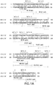
- Epitopes on HIV-1 p24 recognized by mouse monoclonal antibodies were identified using two sets of thirteen p24 synthetic peptides (Figure la). Peptide design was based on the three dimensional structure of p24 antigen ( Gitti, et al., Science 273: 231 (1996 ); Gamble, et al., Science 278: 849 (1997 )) in order to present selected, uninterrupted regions of helical structure (in vivo) that might be unique to shared epitopes. These peptides covered all helical regions (A-J) on the core proteins. Monoclonal antibodies were reacted against both group M (clade B) and group O (Ham112) peptides.
- Peptides were designated M1 to M13 representing HIV-1 group M clade B p24, or O1 to 013 representing HIV-1 group 0 (Ham 112 isolate) p24.
- Each peptide also contained an additional cysteine on its C-terminus which was reacted with maleimide-modified Keyhole Lympet Hemocyanin (KLH) to form two series of KLH conjugated peptides in addition to unconjugated peptides.
- KLH conjugated peptides were generated in order to help stabilize and present conformational structures that might be essential for epitope presentation and recognition (monoclonal binding).
- KLH conjugated peptides were designated as KM1 to KM13 for group M peptides or KO1 to KO13 for group O peptides.
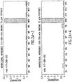
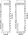
- Binding of monoclonal antibodies (Mabs) to sets of synthetic peptides was determined by an indirect ELISA assay. Briefly, free or KLH conjugated synthetic peptides were coated on the wells of microtiter plates. The peptide coated wells were incubated with Mabs prepared to a concentration of approximately 1 ug/ml. Bound Mabs were detected by enzyme or acridinium-labeled goat anti-mouse IgG antibodies. Representative data are depicted in Figures 2a and b .
- Monoclonal antibody 117-289-555 specifically bound to both group M and group O M10/O10 peptides, which correspond to the helix H region, which is part of the major homology region (MHR).
- the epitope appears to be linear because the antibody readily bints free (un-conjugated) M10/O10 peptides. Binding to both group M and group O p24 is expected. The residues in bold are likely key to forming the epitope.
- Monoclonal antibody 115B-303-620 (according to the invention) bound to M12 and O12 peptides, which corresponds to the helix J-K region of p24.
- the epitope appears to be linear based on the strong (high S/N) binding to free (un-conjugated) peptides of M12/O12.
- the residues in bold are likely key to forming the epitope, but secondary structure involving or requiring neighboring amino acids cannot be excluded.
- Monoclonal antibody 120A-270-108 mapped to the helix H and MHR region of p24 Similar to monoclonal antibody 117-289-555. However, 120A-270-108 recognizes an epitope distinct from an epitope recognized by 117-289-555. The significant difference between 117-289-555 and 120A-270-108 is that 120A-270-108 only moderately bound to the KLH conjugated M10 peptide. No binding was detected when 120A-270-108 was reacted against free (un-conjugated) peptides. Thus, the optimal epitope of 120A-270-108 requires specific secondary or tertiary structures.
- 117-289-555 and 120A-270-108 belong to different compatibility groups because they bind simultaneously to core proteins without interference or competition from each other (see Table 7 below).
- Table 7 Sandwich formation of mAbs with p24proteins (both group M and group O) signal labeled mAbs in solution phase mAbs in solid phase 103-350-474 117-289-555 115B-303-620 115B-151-423 108-394-470 120A-270-108 103-350-474 - + + + + - - 117-289-555 + - + + + + + + + + + + + + + + + + + + + + + 115B-303-620 + + - + + + 115B-151-423 + + + + - + + 108-394-470 - + + + + - - 120A-270-108 - + + + + + - - 120B-580-106 helix A + + + - +/- +/- The + sign indicates compatibility of
- Mabs 115B-151 (according to the invention) and 108-394, fail to bind free synthetic peptides, and.115B-151 (according to the invention) weakly bound to one KLH coupled peptide ( Figure 2B ). Failure to bind synthetic peptides indicated that these Mabs recognize conformational epitopes on core antigen. Conformational epitopes are formed by contiguous amino acids brought together by the tertiary or quaternary folding of p24 antigen. A tertiary or quaternary structure dependent conformational epitope generally cannot be mimicked by small synthetic peptides.
- a set of large overlapping p24 polypeptides were used to locate conformational epitopes recognized by the 115B-151 (according to the invention) and 108-394.
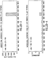
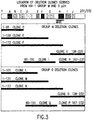
- Overlapping p24 polypeptides were expressed in E. coli. (rproteins) from plasmids carrying unique portions of p24 nucleotide sequence (deletion clones). Two sets of six deletion clones were designed based on the structure of p24. Specific binding of monoclonal antibodies to p24 polypeptides was determined using the Western blot method. Briefly, the expressed (recombinant) p24 polypeptides in extracts of E. coli were subjected to SDS-PAGE and transferred to nitrocellulose membranes.
- FIG. 4 illustrates Western blot results of Mabs 115B-151 (according to the invention) and 108-394.
- Monoclonal antibody 115B-151 (according to the invention) specifically bound to group M polypeptides F(1-172) and G(137-231) and group O polypeptides N(1-173) and 0(138-232).
- the overlapping region (amino acids 137-172) between F and G and N and O, may contribute to epitope recognized by 115B-151 (according to the invention).
- This data is supported by weak but consistent binding of 115B-151 (according to the invention) against the KLH conjugated synthetic peptides KM10 and KO10, which contain amino acids 137-172 ( Figure 2B ).
- Mab 115B-151 differed from 117-289 and 120A-270 because (a) 115B-151 (according to the invention) apparently required the epitope to be in a specific secondary or tertiary conformation, (b) it was compatible with 117-289/120A-270 to form a sandwich with p24 (Table 7), and (c) monoclonal antibodies against helix A were incompatible and strongly competed with 115B-151 (according to the invention) , but were compatible with 117-289.
- the strong competition of helix A directed monoclonal with 115B-151 (according to the invention) in addition to the weak binding against KLH conjugated peptides, may indicate that the optimal epitope recognized by 115B-151 (according to the invention) includes a portion of helix A in addition to the minimal linear epitope identified within helix H.
- Monoclonal 108-394 mapped to a conformational epitope formed within the first 172/173 amino acids of M/O p24.
- the epitope is non-linear because HIV-1 M and O synthetic peptides, free or conjugated to carrier protein KLH, were unreactive with 108-394.
- 108-394 reacted only with the largest of the polypeptides (F 1-172/ N 1-173) derived from M/O p24.
- M polypeptides C (1-65), E_(1-130), and I (60-150), and polypeptides L (1-65), M (1-131), and Q (60-151) contain large segments of the same sequence found in polypeptides F (1-172) and N (1-173), an epitope recognized by 108-394 apparently was not formed from the shorter polypeptides. These data are consistent with a conformational epitope formed by a major portion of p24 comprising at least the first 172/173 amino acids.
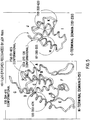
- an epitope map of the six mAbs on the three dimensional structure of p24 antigen is illustrated in Figure 5 .
- the structure of the p24 molecule is represented by two domains, the N-terminal domain (amino acids 1-151) and the C-terminal domain (amino acids 151-231).
- the structure of the intact p24 molecule has not been determined so the exact structural relationship between the two domains is not fully characterized.
- Monoclonal antibody 117-289-555 binds a linear epitope located in the MHR/helix H region of p24.
- the epitope is most broadly defined as comprising amino acids 151-172 (M)/ 152-173 (O), and is most narrowly defined as amino acids 162-172(M)/163-173(O) of p24 antigen.
- Monoclonal antibody 115B-303-620 binds a linear epitope located in the helical J-K regions of p24 antigen.
- the epitope is defined as amino acids 198-217(M)/199-218(O) of the p24 antigen.
- Monoclonal antibody 115B-151-423 (according to the invention) binds a conformational epitope which is most likely near the junction part of helix A and MHR/helix H regions of p24.
- Monoclonal antibody 108-394-470 binds a conformational epitope formed within the N-terminal domain (amino acids 1-151) of p24.
- the conformational epitope is estimated to be near the junction part of helix D and helix A.
- Monoclonal antibody 120A-270-108 binds a conformational epitope which is estimated to be near the junction part of helix D, helix A and MHR/helix H regions of p24.
- EDC 1-ethyl-3-3-dimethylaminopropyl carbodiimide hydrochloride from Sigma Chemicals, St. Louis, MO
- a molar ratio of EDC: carboxyl groups 10:1 in 50 mM MES [2-(N-morpholino)ethane sulfonic acid] buffer pH 6.1
- Anti-core Mab was added to EDC pre-activated CML microparticles at a ratio of 200 ug antibody / per ml of 1% solid microparticles for 4 hours at room temperature on an end-over-end rotator. Free reactants were washed away using Abbott diafiltration system (Abbott Laboratories, Abbott Park, IL) with a crossflow syringe membrane (0.2 um pore size and 12 cm 2 surface area, obtained from Spectrum, Madison Hills, CA). Mab-coated microparticles were overcoated with buffer containing 10 mM PBS and 5% BSA, 0.03% sodium azide for 1 hour at RT on a rotator. Mab-coated CML microparticles were heat-stressed in a 45 C oven incubator for 20 hours and then tested by an Abbott Prism Stand-alone instrument (Abbott Laboratories, Abbott Park, IL).
- ACR-NHS acridinium-N-hydroxysuciniimide
- ACR-labeled-Mab conjugate was separated from free reactants by a G-25 Sephardex column (15 cm x 1.5 cm) which was pre-equilibrated in PBS pH 6.3 containing 1% CHAPS.
- the elution peak of ACR-labeled-Mab was monitored by following absorbance at 280nm using a spectrophotometer (Shimadzu UV-2101PC).
- Anti-p24 mAb-microparticles (concentrate stock) were diluted in uParticle diluent (10 mM PBS, pH 6.5 containing 5% calf serum, 7.5% sucrose, 50 mM EDTA, 0.1% Tween 20, and 0.1% proclin).
- Anti-p24 mAb-ACR conjugates were diluted in conjugate diluent (10 mM PBS, pH 6.3 containing 40 mM EDTA, 5% calf serum, 0.5% Triton, and 0.1% proclin). Two wash buffers were used in the assay.
- Transfer wash buffer contained 25 mM MES pH 5.7, 150 mM NaCl, 4% Triton X-100, 1% Tween 20, 0.001% PEG, 0.1% proclin, and 0.001% antifoam.
- Conjugate wash buffer contained 10mM CAPS, pH 9.9, 150 mm NaCl, 5% Triton X-100, 0.1% proclin, and 0.001% antifoam.
- HIV-1 p24-M, rp24-O, HIV-2 rp26, and HIV-2 viral lysate were diluted in HIV-1/2 negative human plasma. Normal human plasma was also used as a negative control.
- the sample tray was moved to the activator station. Fifty ul of activator (mixture of hydrogen peroxide and sodium hydroxide) were applied to the matrix. The chemiluminescence light signal was read by a photomultiplier tube detector.
- Affinity of these Mabs against core antigens must be nearly equal because of the near equivalent binding kinetics against all three core antigens.
- Keq decreases when Mabs are reacted against cross-reactive epitopes, indicated by markedly lower quantitative sensitivity for the cross-reactive antigen compared to the native (immunogen) antigen.
- the small differences between quantitation of HIV-1 and HIV-2 core proteins related herein may relate more to the methods and (error around the methods) used to quantitate the proteins for the studies.
Landscapes
- Health & Medical Sciences (AREA)
- Life Sciences & Earth Sciences (AREA)
- Chemical & Material Sciences (AREA)
- Organic Chemistry (AREA)
- Virology (AREA)
- Molecular Biology (AREA)
- Immunology (AREA)
- Medicinal Chemistry (AREA)
- General Health & Medical Sciences (AREA)
- Biochemistry (AREA)
- Proteomics, Peptides & Aminoacids (AREA)
- Engineering & Computer Science (AREA)
- Genetics & Genomics (AREA)
- Biophysics (AREA)
- Hematology (AREA)
- Biomedical Technology (AREA)
- Urology & Nephrology (AREA)
- Gastroenterology & Hepatology (AREA)
- Analytical Chemistry (AREA)
- General Physics & Mathematics (AREA)
- Microbiology (AREA)
- Biotechnology (AREA)
- Food Science & Technology (AREA)
- Physics & Mathematics (AREA)
- Pathology (AREA)
- Cell Biology (AREA)
- Tropical Medicine & Parasitology (AREA)
- AIDS & HIV (AREA)
- Communicable Diseases (AREA)
- Oncology (AREA)
- Peptides Or Proteins (AREA)
- Preparation Of Compounds By Using Micro-Organisms (AREA)
- Micro-Organisms Or Cultivation Processes Thereof (AREA)
Applications Claiming Priority (2)
| Application Number | Priority Date | Filing Date | Title |
|---|---|---|---|
| US09/731,126 US6818392B2 (en) | 2000-12-06 | 2000-12-06 | Monoclonal antibodies to human immunodeficiency virus and uses thereof |
| EP01273730A EP1341820A2 (en) | 2000-12-06 | 2001-12-05 | Monoclonal antibodies to human immunodeficiency virus and uses thereof |
Related Parent Applications (2)
| Application Number | Title | Priority Date | Filing Date |
|---|---|---|---|
| EP01273730.0 Division | 2001-12-05 | ||
| EP01273730A Division EP1341820A2 (en) | 2000-12-06 | 2001-12-05 | Monoclonal antibodies to human immunodeficiency virus and uses thereof |
Publications (2)
| Publication Number | Publication Date |
|---|---|
| EP2336175A1 EP2336175A1 (en) | 2011-06-22 |
| EP2336175B1 true EP2336175B1 (en) | 2017-01-18 |
Family
ID=24938175
Family Applications (2)
| Application Number | Title | Priority Date | Filing Date |
|---|---|---|---|
| EP10179020.2A Expired - Lifetime EP2336175B1 (en) | 2000-12-06 | 2001-12-05 | Monoclonal antibodies to human immunodeficiency virus and uses thereof |
| EP01273730A Ceased EP1341820A2 (en) | 2000-12-06 | 2001-12-05 | Monoclonal antibodies to human immunodeficiency virus and uses thereof |
Family Applications After (1)
| Application Number | Title | Priority Date | Filing Date |
|---|---|---|---|
| EP01273730A Ceased EP1341820A2 (en) | 2000-12-06 | 2001-12-05 | Monoclonal antibodies to human immunodeficiency virus and uses thereof |
Country Status (6)
| Country | Link |
|---|---|
| US (10) | US6818392B2 (https=) |
| EP (2) | EP2336175B1 (https=) |
| JP (1) | JP4489351B2 (https=) |
| CA (1) | CA2431321C (https=) |
| ES (1) | ES2619370T3 (https=) |
| WO (1) | WO2002064615A2 (https=) |
Families Citing this family (44)
| Publication number | Priority date | Publication date | Assignee | Title |
|---|---|---|---|---|
| US6818392B2 (en) | 2000-12-06 | 2004-11-16 | Abbott Laboratories | Monoclonal antibodies to human immunodeficiency virus and uses thereof |
| US20050054118A1 (en) * | 2002-02-27 | 2005-03-10 | Lebrun Stewart J. | High throughput screening method |
| WO2003072752A2 (en) * | 2002-02-27 | 2003-09-04 | Miragene, Inc. | Improved substrate chemistry for protein immobilization on a rigid support |
| US8101431B2 (en) | 2004-02-27 | 2012-01-24 | Board Of Regents, The University Of Texas System | Integration of fluids and reagents into self-contained cartridges containing sensor elements and reagent delivery systems |
| US8105849B2 (en) | 2004-02-27 | 2012-01-31 | Board Of Regents, The University Of Texas System | Integration of fluids and reagents into self-contained cartridges containing sensor elements |
| WO2007053186A2 (en) | 2005-05-31 | 2007-05-10 | Labnow, Inc. | Methods and compositions related to determination and use of white blood cell counts |
| WO2008059553A1 (en) * | 2006-11-13 | 2008-05-22 | Masami Moriyama | Method for detecting hiv-1 without being affected by anti-mouse protein antibody existing in human and kit to be used for the same |
| FI120376B (fi) * | 2007-01-17 | 2009-09-30 | Next Biomed Technologies Nbt O | Menetelmä biomuokatun, korkean affiniteetin polypeptidin valmistamiseksi |
| US20090181359A1 (en) | 2007-10-25 | 2009-07-16 | Lou Sheng C | Method of performing ultra-sensitive immunoassays |
| US8222048B2 (en) | 2007-11-05 | 2012-07-17 | Abbott Laboratories | Automated analyzer for clinical laboratory |
| SG190572A1 (en) | 2008-04-29 | 2013-06-28 | Abbott Lab | Dual variable domain immunoglobulins and uses thereof |
| TW201006485A (en) | 2008-06-03 | 2010-02-16 | Abbott Lab | Dual variable domain immunoglobulins and uses thereof |
| CA2726087A1 (en) | 2008-06-03 | 2009-12-10 | Tariq Ghayur | Dual variable domain immunoglobulins and uses thereof |
| CN102149825B (zh) | 2008-07-08 | 2015-07-22 | Abbvie公司 | 前列腺素e2双重可变结构域免疫球蛋白及其用途 |
| EP2335069A4 (en) * | 2008-09-25 | 2012-08-01 | Univ Duke | METHOD FOR DETECTING ANTIBODIES AND ANTIBODY HIV VIRON COMPLEXES |
| US8916154B2 (en) | 2009-04-27 | 2014-12-23 | Abbott Laboratories | Antibodies against delta-5 desaturase and uses thereof |
| US20110008766A1 (en) * | 2009-05-01 | 2011-01-13 | Abbott Laboratories | Dual Variable Domain Immunoglobulins and Uses Thereof |
| US20100297778A1 (en) * | 2009-05-20 | 2010-11-25 | Abbott Laboratories | Conjugate Having Cleavable Linking Agent |
| EP2478364B1 (en) | 2009-09-15 | 2017-08-02 | Gemini Research Limited | Detection of auto-antibodies to nuclear antigens |
| BR112012008833A2 (pt) * | 2009-10-15 | 2015-09-08 | Abbott Lab | imunoglobulinas de dominio variavel duplo e usos das mesmas |
| UY32979A (es) | 2009-10-28 | 2011-02-28 | Abbott Lab | Inmunoglobulinas con dominio variable dual y usos de las mismas |
| AU2011230619C1 (en) | 2010-03-25 | 2016-06-23 | Oregon Health & Science University | CMV glycoproteins and recombinant vectors |
| EP3252072A3 (en) | 2010-08-03 | 2018-03-14 | AbbVie Inc. | Dual variable domain immunoglobulins and uses thereof |
| ES2678145T3 (es) * | 2010-08-24 | 2018-08-09 | Abbott Laboratories | Anticuerpos específicos contra la proteína del núcleo del VIH y usos de los mismos |
| JP2013539364A (ja) | 2010-08-26 | 2013-10-24 | アッヴィ・インコーポレイテッド | 二重可変ドメイン免疫グロブリンおよびその使用 |
| HUE037408T2 (hu) | 2011-06-10 | 2018-08-28 | Univ Oregon Health & Science | CMV glikoproteinek és rekombináns vektorok |
| US20130064811A1 (en) * | 2011-09-09 | 2013-03-14 | International Business Machines Corporation | Methods to Enhance Cancer Treatment |
| CA2789539A1 (en) | 2011-09-12 | 2013-03-12 | International Aids Vaccine Initiative | Immunoselection of recombinant vesicular stomatitis virus expressing hiv-1 proteins by broadly neutralizing antibodies |
| US9402894B2 (en) | 2011-10-27 | 2016-08-02 | International Aids Vaccine Initiative | Viral particles derived from an enveloped virus |
| TW201333035A (zh) | 2011-12-30 | 2013-08-16 | Abbvie Inc | 針對il-13及/或il-17之雙特異性結合蛋白 |
| ES2631608T3 (es) | 2012-06-27 | 2017-09-01 | International Aids Vaccine Initiative | Variante de la glicoproteína Env del VIH-1 |
| TW202037609A (zh) | 2012-11-01 | 2020-10-16 | 美商艾伯維有限公司 | 抗-vegf/dll4雙重可變區域免疫球蛋白及其用途 |
| WO2014144280A2 (en) | 2013-03-15 | 2014-09-18 | Abbvie Inc. | DUAL SPECIFIC BINDING PROTEINS DIRECTED AGAINST IL-1β AND / OR IL-17 |
| EP2848937A1 (en) | 2013-09-05 | 2015-03-18 | International Aids Vaccine Initiative | Methods of identifying novel HIV-1 immunogens |
| US10058604B2 (en) | 2013-10-07 | 2018-08-28 | International Aids Vaccine Initiative | Soluble HIV-1 envelope glycoprotein trimers |
| US10093733B2 (en) | 2014-12-11 | 2018-10-09 | Abbvie Inc. | LRP-8 binding dual variable domain immunoglobulin proteins |
| EP3069730A3 (en) | 2015-03-20 | 2017-03-15 | International Aids Vaccine Initiative | Soluble hiv-1 envelope glycoprotein trimers |
| US9931394B2 (en) | 2015-03-23 | 2018-04-03 | International Aids Vaccine Initiative | Soluble HIV-1 envelope glycoprotein trimers |
| TW201710286A (zh) | 2015-06-15 | 2017-03-16 | 艾伯維有限公司 | 抗vegf、pdgf及/或其受體之結合蛋白 |
| CN105929157A (zh) * | 2016-05-25 | 2016-09-07 | 苏州新波生物技术有限公司 | 一种联合检测hiv抗原抗体的诊断试剂盒及其制备方法 |
| US20200209226A1 (en) * | 2017-01-20 | 2020-07-02 | Shenzhen New Industries Biomedical Engineering Co. | Labeled complex, preparation method thereof, kit containing the same, application of kit and detection system comprising kit |
| CN119264211A (zh) | 2018-08-27 | 2025-01-07 | 瑞泽恩制药公司 | 拉曼光谱在下游纯化中的应用 |
| JP7810694B2 (ja) * | 2020-07-13 | 2026-02-03 | グリフォルス ダイアグノステック ソリューションズ インコーポレーテッド | 抗ヒト免疫不全ウイルス-1抗体及びその使用方法 |
| CN119390828A (zh) * | 2024-12-31 | 2025-02-07 | 天津龙晟生物科技有限公司 | 人类免疫缺陷病毒hiv抗体及其应用和其检测产品 |
Citations (1)
| Publication number | Priority date | Publication date | Assignee | Title |
|---|---|---|---|---|
| WO2012027440A1 (en) * | 2010-08-24 | 2012-03-01 | Abbott Laboratories | Hiv core protein specific antibodies and uses thereof |
Family Cites Families (35)
| Publication number | Priority date | Publication date | Assignee | Title |
|---|---|---|---|---|
| DE3006207C2 (de) | 1980-02-15 | 1982-10-21 | Siemens AG, 1000 Berlin und 8000 München | Elektrische Maschine mit einem Ständerblechpaket aus kornorientierten Blechen |
| CA1341423C (en) * | 1984-10-31 | 2003-03-04 | Paul A. Luciw | Recombinant proteins of viruses associated with lymphadenopathy syndrome and/or acquired immune deficiency syndrome |
| DE3445816C1 (de) | 1984-12-15 | 1986-06-12 | Behringwerke Ag, 3550 Marburg | Flaechenfoermiges diagnostisches Mittel |
| US4839288A (en) * | 1986-01-22 | 1989-06-13 | Institut Pasteur | Retrovirus capable of causing AIDS, antigens obtained from this retrovirus and corresponding antibodies and their application for diagnostic purposes |
| NZ220200A (en) | 1986-05-19 | 1990-08-28 | Viral Technologies Inc | Hiv related peptides, antibodies to the peptides and vaccines therefrom |
| DE3636540A1 (de) | 1986-10-27 | 1988-04-28 | Behringwerke Ag | Verfahren zur bestimmung von anti-hiv, mittel dazu und ihre verwendung in diesem verfahren |
| DE3705686C2 (de) * | 1987-02-23 | 1995-11-30 | Boehringer Mannheim Gmbh | Verfahren zur Bestimmung von Antikörpern |
| US5104790A (en) | 1987-06-29 | 1992-04-14 | Genetic Systems Corporation | Monoclonal antibodies to specific antigenic regions of the human immunodeficiency virus and methods for use |
| US4886742A (en) | 1987-06-15 | 1989-12-12 | Coulter Corporation | Enzyme immunoassay for detecting HIV antigens in human sera |
| US4888290A (en) | 1987-11-06 | 1989-12-19 | Coulter Corporation | Monoclonal antibody specific to HIV antigens |
| DE3819079A1 (de) | 1988-06-04 | 1989-12-07 | Hoechst Ag | Hirudin-derivate mit verzoegerter wirkung |
| DE3853422T2 (de) | 1988-06-09 | 1995-10-26 | Innogenetics Nv | HIV-3-Retrovirus und seine Verwendung. |
| US5173399A (en) | 1988-06-10 | 1992-12-22 | Abbott Laboratories | Mouse monoclonal antibodies to hiv-1p24 and their use in diagnostic tests |
| CA1335880C (en) * | 1988-07-14 | 1995-06-13 | Thomas P. O'connor | Detection of an antibody and antigen in an immunoassay |
| US5731189A (en) | 1989-02-28 | 1998-03-24 | New York University | Human monoclonal antibodies to human immunodeficiency virus |
| ES2094124T3 (es) | 1989-03-09 | 1997-01-16 | Abbott Lab | Ensayo-inmunologico de anticuerpos contra hiv. |
| ZA903492B (en) | 1989-05-15 | 1991-02-27 | Akzo Nv | T-lymphotropic retrovirus monoclonal antibodies |
| US5210181A (en) * | 1989-05-15 | 1993-05-11 | Akzo N.V. | T-lymphotropic retrovirus peptide |
| JPH0831392B2 (ja) * | 1990-04-26 | 1996-03-27 | 株式会社村田製作所 | 積層コンデンサ |
| DE4039925A1 (de) | 1990-12-14 | 1992-06-17 | Behringwerke Ag | Ausgewaehlte peptide des gruppenspezifischen antigens (gag) von humanem immundefizienzvirus (hiv), ihre herstellung und verwendung |
| EP0519866A1 (en) | 1991-06-18 | 1992-12-23 | Ciba-Geigy Ag | Novel anti-HIV antibodies |
| JP2001504572A (ja) | 1992-04-09 | 2001-04-03 | アボツト・ラボラトリーズ | Hiv抗原及びhiv抗体を検出するためのアッセイ |
| DE4236189A1 (de) | 1992-10-27 | 1994-04-28 | Boehringer Mannheim Gmbh | Verfahren zur simultanen Bestimmung von Antigenen und Antikörpern |
| EP0627625A1 (en) | 1993-06-04 | 1994-12-07 | Abbott Laboratories | Immunoassay for differential diagnosis |
| US5611253A (en) * | 1993-09-07 | 1997-03-18 | Tohoku Ricoh Co., Ltd. | Cutting device |
| CA2161872A1 (en) | 1994-03-02 | 1995-09-08 | James L. Gallarda | Hiv immunoassay utilizing recombinant protein and synthetic peptide reagents |
| DE59511054D1 (de) | 1994-07-25 | 2006-08-10 | Roche Diagnostics Gmbh | Bestimmung von spezifischem immunglobulin unter verwendung multipler antigene |
| CN1114106C (zh) | 1994-07-25 | 2003-07-09 | 罗切诊断学有限公司 | 金属螯合物标记的肽及其制备方法 |
| ATE214073T1 (de) | 1994-07-25 | 2002-03-15 | Roche Diagnostics Gmbh | Hapten-markierte peptide |
| ATE374253T1 (de) | 1994-10-20 | 2007-10-15 | Pasteur Institut | Nukleotiden-sequenzen aus retroviren-antigenen hiv-i gruppe (oder untergruppe) o |
| US6015662A (en) | 1996-01-23 | 2000-01-18 | Abbott Laboratories | Reagents for use as calibrators and controls |
| ATE228248T1 (de) * | 1997-03-10 | 2002-12-15 | Roche Diagnostics Gmbh | Verfahren zur simultanen bestimmung von hiv- antigenen und hiv-antikörpern |
| DE19727943A1 (de) | 1997-03-10 | 1998-09-24 | Boehringer Mannheim Gmbh | Verfahren zur simultanen Bestimmung von HIV-Antigenen und HIV-Antikörpern |
| US6495599B2 (en) | 2000-04-13 | 2002-12-17 | Abbott Laboratories | Infant formulas containing long-chain polyunsaturated fatty acids and uses therof |
| US6818392B2 (en) * | 2000-12-06 | 2004-11-16 | Abbott Laboratories | Monoclonal antibodies to human immunodeficiency virus and uses thereof |
-
2000
- 2000-12-06 US US09/731,126 patent/US6818392B2/en not_active Expired - Lifetime
-
2001
- 2001-12-05 EP EP10179020.2A patent/EP2336175B1/en not_active Expired - Lifetime
- 2001-12-05 JP JP2002564544A patent/JP4489351B2/ja not_active Expired - Lifetime
- 2001-12-05 WO PCT/US2001/043179 patent/WO2002064615A2/en not_active Ceased
- 2001-12-05 CA CA2431321A patent/CA2431321C/en not_active Expired - Lifetime
- 2001-12-05 ES ES10179020.2T patent/ES2619370T3/es not_active Expired - Lifetime
- 2001-12-05 EP EP01273730A patent/EP1341820A2/en not_active Ceased
-
2003
- 2003-11-17 US US10/714,689 patent/US7897332B2/en not_active Expired - Fee Related
-
2004
- 2004-09-14 US US10/940,237 patent/US7531638B2/en not_active Expired - Fee Related
- 2004-09-14 US US10/940,262 patent/US7531640B2/en not_active Expired - Fee Related
- 2004-09-14 US US10/940,261 patent/US7531639B2/en not_active Expired - Lifetime
- 2004-09-14 US US10/940,162 patent/US7528238B2/en not_active Expired - Fee Related
- 2004-09-14 US US10/940,344 patent/US7531641B2/en not_active Expired - Fee Related
- 2004-09-14 US US10/940,392 patent/US7531642B2/en not_active Expired - Fee Related
-
2011
- 2011-02-09 US US13/024,093 patent/US20120100632A1/en not_active Abandoned
-
2012
- 2012-10-09 US US13/647,735 patent/US9115189B2/en not_active Expired - Fee Related
Patent Citations (1)
| Publication number | Priority date | Publication date | Assignee | Title |
|---|---|---|---|---|
| WO2012027440A1 (en) * | 2010-08-24 | 2012-03-01 | Abbott Laboratories | Hiv core protein specific antibodies and uses thereof |
Also Published As
| Publication number | Publication date |
|---|---|
| US7531638B2 (en) | 2009-05-12 |
| WO2002064615A3 (en) | 2003-04-17 |
| US7531640B2 (en) | 2009-05-12 |
| US20050053925A1 (en) | 2005-03-10 |
| US20050053924A1 (en) | 2005-03-10 |
| US7531642B2 (en) | 2009-05-12 |
| US7897332B2 (en) | 2011-03-01 |
| JP2004536568A (ja) | 2004-12-09 |
| US20050031624A1 (en) | 2005-02-10 |
| US20040101831A1 (en) | 2004-05-27 |
| US7528238B2 (en) | 2009-05-05 |
| US20050053610A1 (en) | 2005-03-10 |
| US6818392B2 (en) | 2004-11-16 |
| US9115189B2 (en) | 2015-08-25 |
| US20050025772A1 (en) | 2005-02-03 |
| EP2336175A1 (en) | 2011-06-22 |
| WO2002064615A2 (en) | 2002-08-22 |
| JP4489351B2 (ja) | 2010-06-23 |
| US20050058994A1 (en) | 2005-03-17 |
| US7531639B2 (en) | 2009-05-12 |
| US20120100632A1 (en) | 2012-04-26 |
| CA2431321C (en) | 2012-05-29 |
| ES2619370T3 (es) | 2017-06-26 |
| US7531641B2 (en) | 2009-05-12 |
| US20130130233A1 (en) | 2013-05-23 |
| EP1341820A2 (en) | 2003-09-10 |
| US20020106636A1 (en) | 2002-08-08 |
| CA2431321A1 (en) | 2002-08-22 |
Similar Documents
| Publication | Publication Date | Title |
|---|---|---|
| EP2336175B1 (en) | Monoclonal antibodies to human immunodeficiency virus and uses thereof | |
| Moore et al. | A human monoclonal antibody to a complex epitope in the V3 region of gp120 of human immunodeficiency virus type 1 has broad reactivity within and outside clade B | |
| AU2009213045B2 (en) | Nucleotide sequences of HIV-1 type (or subtype) O retrovirus antigens | |
| EP2448593B1 (en) | Hiv related peptides combination or fusion for use in hiv vaccine composition or as diagnostic means | |
| Laman et al. | Variant-specific monoclonal and group-specific polyclonal human immunodeficiency virus type 1 neutralizing antibodies raised with synthetic peptides from the gp120 third variable domain | |
| JP2958346B2 (ja) | 抗HIV―2モノクローナル抗体およびそれを用いた抗HIV―2gp41抗体の検出方法 | |
| US5459060A (en) | Human monoclonal antibodies directed against the transmembrane glycoprotein (gp41) of human immunodeficiency virus-1 (HIV-1) | |
| McKnight et al. | Location, exposure, and conservation of neutralizing and nonneutralizing epitopes on human immunodeficiency virus type 2 SU glycoprotein | |
| EP0641357A1 (en) | Hiv protein epitopes immunologically homologous to hla | |
| US6322964B1 (en) | Synthetic HIV-2 gag and env oligopeptides reactive with HIV-2 specific antibodies | |
| Walther et al. | Evaluation of HIV-1/HIV-2 immunoblots for detection of HIV-2 antibodies | |
| US8980546B2 (en) | Method for the detection of HIV-1-specific antibodies utilizing a Vpu polypeptide | |
| AU2005335203A1 (en) | Compositions and methods for the detection of HIV-1/HIV-2 infection | |
| WO1990014358A1 (en) | T-lymphotropic retrovirus monoclonal antibodies | |
| AU646460B2 (en) | Monoclonal antibody for differentiating HIV-2 from HIV-1 seropositive individuals | |
| TERSMETTE et al. | Variant-Specific Monoclonal and Group-Specific Polyclonal Human Immunodeficiency Virus Type 1 Neutralizing Antibodies Raised with Synthetic Peptides from the gpl20 Third Variable Domain |
Legal Events
| Date | Code | Title | Description |
|---|---|---|---|
| PUAI | Public reference made under article 153(3) epc to a published international application that has entered the european phase |
Free format text: ORIGINAL CODE: 0009012 |
|
| AC | Divisional application: reference to earlier application |
Ref document number: 1341820 Country of ref document: EP Kind code of ref document: P |
|
| AK | Designated contracting states |
Kind code of ref document: A1 Designated state(s): AT BE CH CY DE DK ES FI FR GB GR IE IT LI LU MC NL PT SE TR |
|
| 17P | Request for examination filed |
Effective date: 20111222 |
|
| 17Q | First examination report despatched |
Effective date: 20121105 |
|
| GRAP | Despatch of communication of intention to grant a patent |
Free format text: ORIGINAL CODE: EPIDOSNIGR1 |
|
| RIC1 | Information provided on ipc code assigned before grant |
Ipc: C07K 7/08 20060101ALI20160714BHEP Ipc: G01N 33/577 20060101ALI20160714BHEP Ipc: C12N 5/20 20060101ALI20160714BHEP Ipc: C07K 14/005 20060101ALI20160714BHEP Ipc: G01N 33/569 20060101ALI20160714BHEP Ipc: C07K 14/16 20060101ALI20160714BHEP Ipc: G01N 33/68 20060101ALI20160714BHEP Ipc: C07K 16/10 20060101AFI20160714BHEP |
|
| INTG | Intention to grant announced |
Effective date: 20160803 |
|
| GRAS | Grant fee paid |
Free format text: ORIGINAL CODE: EPIDOSNIGR3 |
|
| GRAA | (expected) grant |
Free format text: ORIGINAL CODE: 0009210 |
|
| AC | Divisional application: reference to earlier application |
Ref document number: 1341820 Country of ref document: EP Kind code of ref document: P |
|
| AK | Designated contracting states |
Kind code of ref document: B1 Designated state(s): AT BE CH CY DE DK ES FI FR GB GR IE IT LI LU MC NL PT SE TR |
|
| REG | Reference to a national code |
Ref country code: GB Ref legal event code: FG4D |
|
| REG | Reference to a national code |
Ref country code: CH Ref legal event code: EP |
|
| REG | Reference to a national code |
Ref country code: AT Ref legal event code: REF Ref document number: 862851 Country of ref document: AT Kind code of ref document: T Effective date: 20170215 |
|
| REG | Reference to a national code |
Ref country code: IE Ref legal event code: FG4D |
|
| REG | Reference to a national code |
Ref country code: DE Ref legal event code: R096 Ref document number: 60150298 Country of ref document: DE |
|
| REG | Reference to a national code |
Ref country code: NL Ref legal event code: MP Effective date: 20170118 |
|
| REG | Reference to a national code |
Ref country code: AT Ref legal event code: MK05 Ref document number: 862851 Country of ref document: AT Kind code of ref document: T Effective date: 20170118 |
|
| REG | Reference to a national code |
Ref country code: ES Ref legal event code: FG2A Ref document number: 2619370 Country of ref document: ES Kind code of ref document: T3 Effective date: 20170626 |
|
| PG25 | Lapsed in a contracting state [announced via postgrant information from national office to epo] |
Ref country code: NL Free format text: LAPSE BECAUSE OF FAILURE TO SUBMIT A TRANSLATION OF THE DESCRIPTION OR TO PAY THE FEE WITHIN THE PRESCRIBED TIME-LIMIT Effective date: 20170118 |
|
| PG25 | Lapsed in a contracting state [announced via postgrant information from national office to epo] |
Ref country code: GR Free format text: LAPSE BECAUSE OF FAILURE TO SUBMIT A TRANSLATION OF THE DESCRIPTION OR TO PAY THE FEE WITHIN THE PRESCRIBED TIME-LIMIT Effective date: 20170419 Ref country code: FI Free format text: LAPSE BECAUSE OF FAILURE TO SUBMIT A TRANSLATION OF THE DESCRIPTION OR TO PAY THE FEE WITHIN THE PRESCRIBED TIME-LIMIT Effective date: 20170118 |
|
| PG25 | Lapsed in a contracting state [announced via postgrant information from national office to epo] |
Ref country code: PT Free format text: LAPSE BECAUSE OF FAILURE TO SUBMIT A TRANSLATION OF THE DESCRIPTION OR TO PAY THE FEE WITHIN THE PRESCRIBED TIME-LIMIT Effective date: 20170518 Ref country code: SE Free format text: LAPSE BECAUSE OF FAILURE TO SUBMIT A TRANSLATION OF THE DESCRIPTION OR TO PAY THE FEE WITHIN THE PRESCRIBED TIME-LIMIT Effective date: 20170118 Ref country code: AT Free format text: LAPSE BECAUSE OF FAILURE TO SUBMIT A TRANSLATION OF THE DESCRIPTION OR TO PAY THE FEE WITHIN THE PRESCRIBED TIME-LIMIT Effective date: 20170118 |
|
| REG | Reference to a national code |
Ref country code: DE Ref legal event code: R097 Ref document number: 60150298 Country of ref document: DE |
|
| REG | Reference to a national code |
Ref country code: FR Ref legal event code: PLFP Year of fee payment: 17 |
|
| PLBE | No opposition filed within time limit |
Free format text: ORIGINAL CODE: 0009261 |
|
| STAA | Information on the status of an ep patent application or granted ep patent |
Free format text: STATUS: NO OPPOSITION FILED WITHIN TIME LIMIT |
|
| PG25 | Lapsed in a contracting state [announced via postgrant information from national office to epo] |
Ref country code: DK Free format text: LAPSE BECAUSE OF FAILURE TO SUBMIT A TRANSLATION OF THE DESCRIPTION OR TO PAY THE FEE WITHIN THE PRESCRIBED TIME-LIMIT Effective date: 20170118 |
|
| 26N | No opposition filed |
Effective date: 20171019 |
|
| REG | Reference to a national code |
Ref country code: CH Ref legal event code: PL |
|
| REG | Reference to a national code |
Ref country code: IE Ref legal event code: MM4A |
|
| PG25 | Lapsed in a contracting state [announced via postgrant information from national office to epo] |
Ref country code: LU Free format text: LAPSE BECAUSE OF NON-PAYMENT OF DUE FEES Effective date: 20171205 |
|
| REG | Reference to a national code |
Ref country code: BE Ref legal event code: MM Effective date: 20171231 |
|
| PG25 | Lapsed in a contracting state [announced via postgrant information from national office to epo] |
Ref country code: IE Free format text: LAPSE BECAUSE OF NON-PAYMENT OF DUE FEES Effective date: 20171205 |
|
| PG25 | Lapsed in a contracting state [announced via postgrant information from national office to epo] |
Ref country code: LI Free format text: LAPSE BECAUSE OF NON-PAYMENT OF DUE FEES Effective date: 20171231 Ref country code: CH Free format text: LAPSE BECAUSE OF NON-PAYMENT OF DUE FEES Effective date: 20171231 Ref country code: BE Free format text: LAPSE BECAUSE OF NON-PAYMENT OF DUE FEES Effective date: 20171231 |
|
| PG25 | Lapsed in a contracting state [announced via postgrant information from national office to epo] |
Ref country code: MC Free format text: LAPSE BECAUSE OF FAILURE TO SUBMIT A TRANSLATION OF THE DESCRIPTION OR TO PAY THE FEE WITHIN THE PRESCRIBED TIME-LIMIT Effective date: 20170118 |
|
| PG25 | Lapsed in a contracting state [announced via postgrant information from national office to epo] |
Ref country code: CY Free format text: LAPSE BECAUSE OF NON-PAYMENT OF DUE FEES Effective date: 20170118 |
|
| PGFP | Annual fee paid to national office [announced via postgrant information from national office to epo] |
Ref country code: DE Payment date: 20191114 Year of fee payment: 19 |
|
| PGFP | Annual fee paid to national office [announced via postgrant information from national office to epo] |
Ref country code: FR Payment date: 20191122 Year of fee payment: 19 Ref country code: IT Payment date: 20191219 Year of fee payment: 19 |
|
| PG25 | Lapsed in a contracting state [announced via postgrant information from national office to epo] |
Ref country code: TR Free format text: LAPSE BECAUSE OF FAILURE TO SUBMIT A TRANSLATION OF THE DESCRIPTION OR TO PAY THE FEE WITHIN THE PRESCRIBED TIME-LIMIT Effective date: 20170118 |
|
| PGFP | Annual fee paid to national office [announced via postgrant information from national office to epo] |
Ref country code: GB Payment date: 20191126 Year of fee payment: 19 Ref country code: ES Payment date: 20200102 Year of fee payment: 19 |
|
| REG | Reference to a national code |
Ref country code: DE Ref legal event code: R119 Ref document number: 60150298 Country of ref document: DE |
|
| GBPC | Gb: european patent ceased through non-payment of renewal fee |
Effective date: 20201205 |
|
| PG25 | Lapsed in a contracting state [announced via postgrant information from national office to epo] |
Ref country code: IT Free format text: LAPSE BECAUSE OF NON-PAYMENT OF DUE FEES Effective date: 20201205 Ref country code: FR Free format text: LAPSE BECAUSE OF NON-PAYMENT OF DUE FEES Effective date: 20201231 |
|
| PG25 | Lapsed in a contracting state [announced via postgrant information from national office to epo] |
Ref country code: DE Free format text: LAPSE BECAUSE OF NON-PAYMENT OF DUE FEES Effective date: 20210701 Ref country code: GB Free format text: LAPSE BECAUSE OF NON-PAYMENT OF DUE FEES Effective date: 20201205 |
|
| REG | Reference to a national code |
Ref country code: ES Ref legal event code: FD2A Effective date: 20220207 |
|
| PG25 | Lapsed in a contracting state [announced via postgrant information from national office to epo] |
Ref country code: ES Free format text: LAPSE BECAUSE OF NON-PAYMENT OF DUE FEES Effective date: 20201206 |