EP2331969B1 - Detection of hiv-related proteins in urine - Google Patents
Detection of hiv-related proteins in urine Download PDFInfo
- Publication number
- EP2331969B1 EP2331969B1 EP09819705.6A EP09819705A EP2331969B1 EP 2331969 B1 EP2331969 B1 EP 2331969B1 EP 09819705 A EP09819705 A EP 09819705A EP 2331969 B1 EP2331969 B1 EP 2331969B1
- Authority
- EP
- European Patent Office
- Prior art keywords
- hiv
- mammal
- exosomes
- urine
- associated biomarker
- Prior art date
- Legal status (The legal status is an assumption and is not a legal conclusion. Google has not performed a legal analysis and makes no representation as to the accuracy of the status listed.)
- Active
Links
- 210000002700 urine Anatomy 0.000 title claims description 58
- 108090000623 proteins and genes Proteins 0.000 title claims description 30
- 102000004169 proteins and genes Human genes 0.000 title claims description 30
- 238000001514 detection method Methods 0.000 title description 9
- 210000001808 exosome Anatomy 0.000 claims description 85
- 239000000090 biomarker Substances 0.000 claims description 57
- 241000124008 Mammalia Species 0.000 claims description 55
- 238000000034 method Methods 0.000 claims description 50
- 206010070737 HIV associated nephropathy Diseases 0.000 claims description 47
- 208000031886 HIV Infections Diseases 0.000 claims description 24
- 208000037357 HIV infectious disease Diseases 0.000 claims description 22
- 208000033519 human immunodeficiency virus infectious disease Diseases 0.000 claims description 22
- 208000007122 AIDS-Associated Nephropathy Diseases 0.000 claims description 15
- 238000012544 monitoring process Methods 0.000 claims description 15
- 238000001262 western blot Methods 0.000 claims description 15
- 241000282414 Homo sapiens Species 0.000 claims description 10
- 102100034343 Integrase Human genes 0.000 claims description 10
- 108010092799 RNA-directed DNA polymerase Proteins 0.000 claims description 10
- 101710201961 Virion infectivity factor Proteins 0.000 claims description 9
- 238000005119 centrifugation Methods 0.000 claims description 9
- 238000001294 liquid chromatography-tandem mass spectrometry Methods 0.000 claims description 8
- 108010010369 HIV Protease Proteins 0.000 claims description 7
- 239000006228 supernatant Substances 0.000 claims description 7
- 238000005199 ultracentrifugation Methods 0.000 claims description 7
- 101710104359 F protein Proteins 0.000 claims description 6
- 239000002259 anti human immunodeficiency virus agent Substances 0.000 claims description 6
- 229940124411 anti-hiv antiviral agent Drugs 0.000 claims description 6
- 238000004949 mass spectrometry Methods 0.000 claims description 6
- 108010014303 DNA-directed DNA polymerase Proteins 0.000 claims description 5
- 102000016928 DNA-directed DNA polymerase Human genes 0.000 claims description 5
- 101000650854 Homo sapiens Small glutamine-rich tetratricopeptide repeat-containing protein alpha Proteins 0.000 claims description 5
- 101710149136 Protein Vpr Proteins 0.000 claims description 5
- 101800001690 Transmembrane protein gp41 Proteins 0.000 claims description 5
- 101710090322 Truncated surface protein Proteins 0.000 claims description 5
- 108010027225 gag-pol Fusion Proteins Proteins 0.000 claims description 5
- 238000004128 high performance liquid chromatography Methods 0.000 claims description 5
- 241000282693 Cercopithecidae Species 0.000 claims description 4
- 241000282575 Gorilla Species 0.000 claims description 4
- 241001504519 Papio ursinus Species 0.000 claims description 4
- 238000001914 filtration Methods 0.000 claims description 3
- 238000000734 protein sequencing Methods 0.000 claims description 3
- 238000002965 ELISA Methods 0.000 claims description 2
- 238000009987 spinning Methods 0.000 claims 5
- 238000000756 surface-enhanced laser desorption--ionisation time-of-flight mass spectrometry Methods 0.000 claims 1
- 241000725303 Human immunodeficiency virus Species 0.000 description 133
- 235000018102 proteins Nutrition 0.000 description 26
- 201000010099 disease Diseases 0.000 description 15
- 208000037265 diseases, disorders, signs and symptoms Diseases 0.000 description 15
- 239000003795 chemical substances by application Substances 0.000 description 12
- 230000000670 limiting effect Effects 0.000 description 12
- 230000002485 urinary effect Effects 0.000 description 11
- 201000005206 focal segmental glomerulosclerosis Diseases 0.000 description 10
- 210000004027 cell Anatomy 0.000 description 9
- 239000007788 liquid Substances 0.000 description 9
- 239000003381 stabilizer Substances 0.000 description 9
- 101710147327 Calcineurin B homologous protein 1 Proteins 0.000 description 8
- 101710205625 Capsid protein p24 Proteins 0.000 description 8
- 101710177166 Phosphoprotein Proteins 0.000 description 8
- 101710149279 Small delta antigen Proteins 0.000 description 8
- 238000004458 analytical method Methods 0.000 description 8
- 108010048209 Human Immunodeficiency Virus Proteins Proteins 0.000 description 7
- 239000002131 composite material Substances 0.000 description 7
- 108090000765 processed proteins & peptides Proteins 0.000 description 7
- 238000001228 spectrum Methods 0.000 description 7
- 238000000108 ultra-filtration Methods 0.000 description 7
- WEVYAHXRMPXWCK-UHFFFAOYSA-N Acetonitrile Chemical compound CC#N WEVYAHXRMPXWCK-UHFFFAOYSA-N 0.000 description 6
- -1 Rev Proteins 0.000 description 6
- 210000003734 kidney Anatomy 0.000 description 6
- 239000013049 sediment Substances 0.000 description 6
- 208000030507 AIDS Diseases 0.000 description 5
- 239000000872 buffer Substances 0.000 description 5
- 210000000170 cell membrane Anatomy 0.000 description 5
- 239000003153 chemical reaction reagent Substances 0.000 description 5
- 238000003745 diagnosis Methods 0.000 description 5
- 239000000499 gel Substances 0.000 description 5
- 239000000137 peptide hydrolase inhibitor Substances 0.000 description 5
- 238000012360 testing method Methods 0.000 description 5
- IHPYMWDTONKSCO-UHFFFAOYSA-N 2,2'-piperazine-1,4-diylbisethanesulfonic acid Chemical compound OS(=O)(=O)CCN1CCN(CCS(O)(=O)=O)CC1 IHPYMWDTONKSCO-UHFFFAOYSA-N 0.000 description 4
- SEQKRHFRPICQDD-UHFFFAOYSA-N N-tris(hydroxymethyl)methylglycine Chemical compound OCC(CO)(CO)[NH2+]CC([O-])=O SEQKRHFRPICQDD-UHFFFAOYSA-N 0.000 description 4
- 239000007978 cacodylate buffer Substances 0.000 description 4
- 239000002738 chelating agent Substances 0.000 description 4
- 229940042399 direct acting antivirals protease inhibitors Drugs 0.000 description 4
- 238000001962 electrophoresis Methods 0.000 description 4
- DEFVIWRASFVYLL-UHFFFAOYSA-N ethylene glycol bis(2-aminoethyl)tetraacetic acid Chemical compound OC(=O)CN(CC(O)=O)CCOCCOCCN(CC(O)=O)CC(O)=O DEFVIWRASFVYLL-UHFFFAOYSA-N 0.000 description 4
- 238000002955 isolation Methods 0.000 description 4
- 208000017169 kidney disease Diseases 0.000 description 4
- YBYRMVIVWMBXKQ-UHFFFAOYSA-N phenylmethanesulfonyl fluoride Chemical compound FS(=O)(=O)CC1=CC=CC=C1 YBYRMVIVWMBXKQ-UHFFFAOYSA-N 0.000 description 4
- 239000003755 preservative agent Substances 0.000 description 4
- 238000012552 review Methods 0.000 description 4
- XLYOFNOQVPJJNP-UHFFFAOYSA-N water Substances O XLYOFNOQVPJJNP-UHFFFAOYSA-N 0.000 description 4
- CSCPPACGZOOCGX-UHFFFAOYSA-N Acetone Chemical compound CC(C)=O CSCPPACGZOOCGX-UHFFFAOYSA-N 0.000 description 3
- LZZYPRNAOMGNLH-UHFFFAOYSA-M Cetrimonium bromide Chemical compound [Br-].CCCCCCCCCCCCCCCC[N+](C)(C)C LZZYPRNAOMGNLH-UHFFFAOYSA-M 0.000 description 3
- FSVCELGFZIQNCK-UHFFFAOYSA-N N,N-bis(2-hydroxyethyl)glycine Chemical compound OCCN(CCO)CC(O)=O FSVCELGFZIQNCK-UHFFFAOYSA-N 0.000 description 3
- 108091005804 Peptidases Proteins 0.000 description 3
- 239000004365 Protease Substances 0.000 description 3
- 102100037486 Reverse transcriptase/ribonuclease H Human genes 0.000 description 3
- DBMJMQXJHONAFJ-UHFFFAOYSA-M Sodium laurylsulphate Chemical compound [Na+].CCCCCCCCCCCCOS([O-])(=O)=O DBMJMQXJHONAFJ-UHFFFAOYSA-M 0.000 description 3
- 235000012538 ammonium bicarbonate Nutrition 0.000 description 3
- 230000015556 catabolic process Effects 0.000 description 3
- KRKNYBCHXYNGOX-UHFFFAOYSA-N citric acid Chemical compound OC(=O)CC(O)(C(O)=O)CC(O)=O KRKNYBCHXYNGOX-UHFFFAOYSA-N 0.000 description 3
- 230000006378 damage Effects 0.000 description 3
- 238000006731 degradation reaction Methods 0.000 description 3
- 239000003599 detergent Substances 0.000 description 3
- 238000011161 development Methods 0.000 description 3
- LOKCTEFSRHRXRJ-UHFFFAOYSA-I dipotassium trisodium dihydrogen phosphate hydrogen phosphate dichloride Chemical compound P(=O)(O)(O)[O-].[K+].P(=O)(O)([O-])[O-].[Na+].[Na+].[Cl-].[K+].[Cl-].[Na+] LOKCTEFSRHRXRJ-UHFFFAOYSA-I 0.000 description 3
- VHJLVAABSRFDPM-QWWZWVQMSA-N dithiothreitol Chemical compound SC[C@@H](O)[C@H](O)CS VHJLVAABSRFDPM-QWWZWVQMSA-N 0.000 description 3
- 229940079593 drug Drugs 0.000 description 3
- 239000003814 drug Substances 0.000 description 3
- 238000005516 engineering process Methods 0.000 description 3
- RAXXELZNTBOGNW-UHFFFAOYSA-N imidazole Natural products C1=CNC=N1 RAXXELZNTBOGNW-UHFFFAOYSA-N 0.000 description 3
- 208000015181 infectious disease Diseases 0.000 description 3
- 239000012528 membrane Substances 0.000 description 3
- 239000008188 pellet Substances 0.000 description 3
- 239000002953 phosphate buffered saline Substances 0.000 description 3
- 102000004196 processed proteins & peptides Human genes 0.000 description 3
- 235000019333 sodium laurylsulphate Nutrition 0.000 description 3
- 239000000243 solution Substances 0.000 description 3
- 239000000758 substrate Substances 0.000 description 3
- 238000004627 transmission electron microscopy Methods 0.000 description 3
- GVJHHUAWPYXKBD-UHFFFAOYSA-N (±)-α-Tocopherol Chemical compound OC1=C(C)C(C)=C2OC(CCCC(C)CCCC(C)CCCC(C)C)(C)CCC2=C1C GVJHHUAWPYXKBD-UHFFFAOYSA-N 0.000 description 2
- QZTKDVCDBIDYMD-UHFFFAOYSA-N 2,2'-[(2-amino-2-oxoethyl)imino]diacetic acid Chemical compound NC(=O)CN(CC(O)=O)CC(O)=O QZTKDVCDBIDYMD-UHFFFAOYSA-N 0.000 description 2
- HZAXFHJVJLSVMW-UHFFFAOYSA-N 2-Aminoethan-1-ol Chemical compound NCCO HZAXFHJVJLSVMW-UHFFFAOYSA-N 0.000 description 2
- URDCARMUOSMFFI-UHFFFAOYSA-N 2-[2-[bis(carboxymethyl)amino]ethyl-(2-hydroxyethyl)amino]acetic acid Chemical compound OCCN(CC(O)=O)CCN(CC(O)=O)CC(O)=O URDCARMUOSMFFI-UHFFFAOYSA-N 0.000 description 2
- AJTVSSFTXWNIRG-UHFFFAOYSA-N 2-[bis(2-hydroxyethyl)amino]ethanesulfonic acid Chemical compound OCC[NH+](CCO)CCS([O-])(=O)=O AJTVSSFTXWNIRG-UHFFFAOYSA-N 0.000 description 2
- QKNYBSVHEMOAJP-UHFFFAOYSA-N 2-amino-2-(hydroxymethyl)propane-1,3-diol;hydron;chloride Chemical compound Cl.OCC(N)(CO)CO QKNYBSVHEMOAJP-UHFFFAOYSA-N 0.000 description 2
- 229940058020 2-amino-2-methyl-1-propanol Drugs 0.000 description 2
- DVLFYONBTKHTER-UHFFFAOYSA-N 3-(N-morpholino)propanesulfonic acid Chemical compound OS(=O)(=O)CCCN1CCOCC1 DVLFYONBTKHTER-UHFFFAOYSA-N 0.000 description 2
- XNPKNHHFCKSMRV-UHFFFAOYSA-N 4-(cyclohexylamino)butane-1-sulfonic acid Chemical compound OS(=O)(=O)CCCCNC1CCCCC1 XNPKNHHFCKSMRV-UHFFFAOYSA-N 0.000 description 2
- WRDABNWSWOHGMS-UHFFFAOYSA-N AEBSF hydrochloride Chemical compound Cl.NCCC1=CC=C(S(F)(=O)=O)C=C1 WRDABNWSWOHGMS-UHFFFAOYSA-N 0.000 description 2
- ATRRKUHOCOJYRX-UHFFFAOYSA-N Ammonium bicarbonate Chemical compound [NH4+].OC([O-])=O ATRRKUHOCOJYRX-UHFFFAOYSA-N 0.000 description 2
- 229910000013 Ammonium bicarbonate Inorganic materials 0.000 description 2
- CIWBSHSKHKDKBQ-JLAZNSOCSA-N Ascorbic acid Chemical compound OC[C@H](O)[C@H]1OC(=O)C(O)=C1O CIWBSHSKHKDKBQ-JLAZNSOCSA-N 0.000 description 2
- 241000283707 Capra Species 0.000 description 2
- KCXVZYZYPLLWCC-UHFFFAOYSA-N EDTA Chemical compound OC(=O)CN(CC(O)=O)CCN(CC(O)=O)CC(O)=O KCXVZYZYPLLWCC-UHFFFAOYSA-N 0.000 description 2
- OWXMKDGYPWMGEB-UHFFFAOYSA-N HEPPS Chemical compound OCCN1CCN(CCCS(O)(=O)=O)CC1 OWXMKDGYPWMGEB-UHFFFAOYSA-N 0.000 description 2
- 241000713340 Human immunodeficiency virus 2 Species 0.000 description 2
- 108010052285 Membrane Proteins Proteins 0.000 description 2
- 241000699660 Mus musculus Species 0.000 description 2
- MKWKNSIESPFAQN-UHFFFAOYSA-N N-cyclohexyl-2-aminoethanesulfonic acid Chemical compound OS(=O)(=O)CCNC1CCCCC1 MKWKNSIESPFAQN-UHFFFAOYSA-N 0.000 description 2
- JOCBASBOOFNAJA-UHFFFAOYSA-N N-tris(hydroxymethyl)methyl-2-aminoethanesulfonic acid Chemical compound OCC(CO)(CO)NCCS(O)(=O)=O JOCBASBOOFNAJA-UHFFFAOYSA-N 0.000 description 2
- VFTZCDVTMZWNBF-UHFFFAOYSA-N N-tris(hydroxymethyl)methyl-4-aminobutanesulfonic acid Chemical compound OCC(CO)(CO)NCCCCS(O)(=O)=O VFTZCDVTMZWNBF-UHFFFAOYSA-N 0.000 description 2
- 239000002202 Polyethylene glycol Substances 0.000 description 2
- WCUXLLCKKVVCTQ-UHFFFAOYSA-M Potassium chloride Chemical compound [Cl-].[K+] WCUXLLCKKVVCTQ-UHFFFAOYSA-M 0.000 description 2
- CDBYLPFSWZWCQE-UHFFFAOYSA-L Sodium Carbonate Chemical compound [Na+].[Na+].[O-]C([O-])=O CDBYLPFSWZWCQE-UHFFFAOYSA-L 0.000 description 2
- PXIPVTKHYLBLMZ-UHFFFAOYSA-N Sodium azide Chemical compound [Na+].[N-]=[N+]=[N-] PXIPVTKHYLBLMZ-UHFFFAOYSA-N 0.000 description 2
- DTQVDTLACAAQTR-UHFFFAOYSA-N Trifluoroacetic acid Chemical compound OC(=O)C(F)(F)F DTQVDTLACAAQTR-UHFFFAOYSA-N 0.000 description 2
- 102000004142 Trypsin Human genes 0.000 description 2
- 108090000631 Trypsin Proteins 0.000 description 2
- XSQUKJJJFZCRTK-UHFFFAOYSA-N Urea Chemical compound NC(N)=O XSQUKJJJFZCRTK-UHFFFAOYSA-N 0.000 description 2
- 241000700605 Viruses Species 0.000 description 2
- 239000002253 acid Substances 0.000 description 2
- CBTVGIZVANVGBH-UHFFFAOYSA-N aminomethyl propanol Chemical compound CC(C)(N)CO CBTVGIZVANVGBH-UHFFFAOYSA-N 0.000 description 2
- 239000001099 ammonium carbonate Substances 0.000 description 2
- 230000036436 anti-hiv Effects 0.000 description 2
- 239000000427 antigen Substances 0.000 description 2
- 108091007433 antigens Proteins 0.000 description 2
- 102000036639 antigens Human genes 0.000 description 2
- 239000003963 antioxidant agent Substances 0.000 description 2
- 235000006708 antioxidants Nutrition 0.000 description 2
- 239000007998 bicine buffer Substances 0.000 description 2
- 230000005540 biological transmission Effects 0.000 description 2
- 239000003638 chemical reducing agent Substances 0.000 description 2
- CVSVTCORWBXHQV-UHFFFAOYSA-N creatine Chemical compound NC(=[NH2+])N(C)CC([O-])=O CVSVTCORWBXHQV-UHFFFAOYSA-N 0.000 description 2
- 210000004443 dendritic cell Anatomy 0.000 description 2
- 230000001419 dependent effect Effects 0.000 description 2
- 238000003795 desorption Methods 0.000 description 2
- 238000002405 diagnostic procedure Methods 0.000 description 2
- OGGXGZAMXPVRFZ-UHFFFAOYSA-N dimethylarsinic acid Chemical compound C[As](C)(O)=O OGGXGZAMXPVRFZ-UHFFFAOYSA-N 0.000 description 2
- 229960001484 edetic acid Drugs 0.000 description 2
- 230000002209 hydrophobic effect Effects 0.000 description 2
- 210000002865 immune cell Anatomy 0.000 description 2
- 150000002500 ions Chemical class 0.000 description 2
- 239000003446 ligand Substances 0.000 description 2
- 229910052751 metal Inorganic materials 0.000 description 2
- 239000002184 metal Substances 0.000 description 2
- BDAGIHXWWSANSR-UHFFFAOYSA-N methanoic acid Natural products OC=O BDAGIHXWWSANSR-UHFFFAOYSA-N 0.000 description 2
- 230000000813 microbial effect Effects 0.000 description 2
- 239000000203 mixture Substances 0.000 description 2
- 238000006384 oligomerization reaction Methods 0.000 description 2
- 230000007170 pathology Effects 0.000 description 2
- 230000037361 pathway Effects 0.000 description 2
- 102000013415 peroxidase activity proteins Human genes 0.000 description 2
- 108040007629 peroxidase activity proteins Proteins 0.000 description 2
- 229920001223 polyethylene glycol Polymers 0.000 description 2
- YGSDEFSMJLZEOE-UHFFFAOYSA-N salicylic acid Chemical compound OC(=O)C1=CC=CC=C1O YGSDEFSMJLZEOE-UHFFFAOYSA-N 0.000 description 2
- 150000003839 salts Chemical class 0.000 description 2
- 239000003001 serine protease inhibitor Substances 0.000 description 2
- 210000002966 serum Anatomy 0.000 description 2
- 239000000126 substance Substances 0.000 description 2
- 238000001920 surface-enhanced laser desorption--ionisation mass spectrometry Methods 0.000 description 2
- CWERGRDVMFNCDR-UHFFFAOYSA-N thioglycolic acid Chemical compound OC(=O)CS CWERGRDVMFNCDR-UHFFFAOYSA-N 0.000 description 2
- 238000001269 time-of-flight mass spectrometry Methods 0.000 description 2
- 238000011830 transgenic mouse model Methods 0.000 description 2
- 239000012588 trypsin Substances 0.000 description 2
- 230000003612 virological effect Effects 0.000 description 2
- DGVVWUTYPXICAM-UHFFFAOYSA-N β‐Mercaptoethanol Chemical compound OCCS DGVVWUTYPXICAM-UHFFFAOYSA-N 0.000 description 2
- MTCFGRXMJLQNBG-REOHCLBHSA-N (2S)-2-Amino-3-hydroxypropansäure Chemical compound OC[C@H](N)C(O)=O MTCFGRXMJLQNBG-REOHCLBHSA-N 0.000 description 1
- MRXDGVXSWIXTQL-HYHFHBMOSA-N (2s)-2-[[(1s)-1-(2-amino-1,4,5,6-tetrahydropyrimidin-6-yl)-2-[[(2s)-4-methyl-1-oxo-1-[[(2s)-1-oxo-3-phenylpropan-2-yl]amino]pentan-2-yl]amino]-2-oxoethyl]carbamoylamino]-3-phenylpropanoic acid Chemical compound C([C@H](NC(=O)N[C@H](C(=O)N[C@@H](CC(C)C)C(=O)N[C@@H](CC=1C=CC=CC=1)C=O)C1NC(N)=NCC1)C(O)=O)C1=CC=CC=C1 MRXDGVXSWIXTQL-HYHFHBMOSA-N 0.000 description 1
- IJWCGVPEDDQUDE-YGJAXBLXSA-N (2s)-2-[[(1s)-2-[[(2s)-5-amino-1,5-dioxo-1-[[(2s)-1-oxopropan-2-yl]amino]pentan-2-yl]amino]-1-[(6s)-2-amino-1,4,5,6-tetrahydropyrimidin-6-yl]-2-oxoethyl]carbamoylamino]-4-methylpentanoic acid Chemical compound O=C[C@H](C)NC(=O)[C@H](CCC(N)=O)NC(=O)[C@@H](NC(=O)N[C@@H](CC(C)C)C(O)=O)[C@@H]1CCN=C(N)N1 IJWCGVPEDDQUDE-YGJAXBLXSA-N 0.000 description 1
- LZXHHNKULPHARO-UHFFFAOYSA-M (3,4-dichlorophenyl)methyl-triphenylphosphanium;chloride Chemical compound [Cl-].C1=C(Cl)C(Cl)=CC=C1C[P+](C=1C=CC=CC=1)(C=1C=CC=CC=1)C1=CC=CC=C1 LZXHHNKULPHARO-UHFFFAOYSA-M 0.000 description 1
- PMHUSCHKTSTQEP-UHFFFAOYSA-N (4-carbamimidoylphenyl)methanesulfonyl fluoride Chemical compound NC(=N)C1=CC=C(CS(F)(=O)=O)C=C1 PMHUSCHKTSTQEP-UHFFFAOYSA-N 0.000 description 1
- LLQJZSGEOMASGH-UHFFFAOYSA-N 2-(2-sulfoethylamino)acetic acid Chemical compound OC(=O)CNCCS(O)(=O)=O LLQJZSGEOMASGH-UHFFFAOYSA-N 0.000 description 1
- SXGZJKUKBWWHRA-UHFFFAOYSA-N 2-(N-morpholiniumyl)ethanesulfonate Chemical compound [O-]S(=O)(=O)CC[NH+]1CCOCC1 SXGZJKUKBWWHRA-UHFFFAOYSA-N 0.000 description 1
- JKMHFZQWWAIEOD-UHFFFAOYSA-N 2-[4-(2-hydroxyethyl)piperazin-1-yl]ethanesulfonic acid Chemical compound OCC[NH+]1CCN(CCS([O-])(=O)=O)CC1 JKMHFZQWWAIEOD-UHFFFAOYSA-N 0.000 description 1
- AXAVXPMQTGXXJZ-UHFFFAOYSA-N 2-aminoacetic acid;2-amino-2-(hydroxymethyl)propane-1,3-diol Chemical compound NCC(O)=O.OCC(N)(CO)CO AXAVXPMQTGXXJZ-UHFFFAOYSA-N 0.000 description 1
- ACERFIHBIWMFOR-UHFFFAOYSA-N 2-hydroxy-3-[(1-hydroxy-2-methylpropan-2-yl)azaniumyl]propane-1-sulfonate Chemical compound OCC(C)(C)NCC(O)CS(O)(=O)=O ACERFIHBIWMFOR-UHFFFAOYSA-N 0.000 description 1
- 125000000954 2-hydroxyethyl group Chemical group [H]C([*])([H])C([H])([H])O[H] 0.000 description 1
- SUGXUUGGLDCZKB-UHFFFAOYSA-N 3,4-dichloroisocoumarin Chemical compound C1=CC=C2C(Cl)=C(Cl)OC(=O)C2=C1 SUGXUUGGLDCZKB-UHFFFAOYSA-N 0.000 description 1
- INEWUCPYEUEQTN-UHFFFAOYSA-N 3-(cyclohexylamino)-2-hydroxy-1-propanesulfonic acid Chemical compound OS(=O)(=O)CC(O)CNC1CCCCC1 INEWUCPYEUEQTN-UHFFFAOYSA-N 0.000 description 1
- SNKZJIOFVMKAOJ-UHFFFAOYSA-N 3-Amino-1-propanesulfonic acid Natural products NCCCS(O)(=O)=O SNKZJIOFVMKAOJ-UHFFFAOYSA-N 0.000 description 1
- OSWFIVFLDKOXQC-UHFFFAOYSA-N 4-(3-methoxyphenyl)aniline Chemical compound COC1=CC=CC(C=2C=CC(N)=CC=2)=C1 OSWFIVFLDKOXQC-UHFFFAOYSA-N 0.000 description 1
- QCVGEOXPDFCNHA-UHFFFAOYSA-N 5,5-dimethyl-2,4-dioxo-1,3-oxazolidine-3-carboxamide Chemical compound CC1(C)OC(=O)N(C(N)=O)C1=O QCVGEOXPDFCNHA-UHFFFAOYSA-N 0.000 description 1
- HRPVXLWXLXDGHG-UHFFFAOYSA-N Acrylamide Chemical compound NC(=O)C=C HRPVXLWXLXDGHG-UHFFFAOYSA-N 0.000 description 1
- 102100027211 Albumin Human genes 0.000 description 1
- 108010088751 Albumins Proteins 0.000 description 1
- 102100033312 Alpha-2-macroglobulin Human genes 0.000 description 1
- 108010087765 Antipain Proteins 0.000 description 1
- 108090000935 Antithrombin III Proteins 0.000 description 1
- 102100022977 Antithrombin-III Human genes 0.000 description 1
- 108010039627 Aprotinin Proteins 0.000 description 1
- VGGGPCQERPFHOB-MCIONIFRSA-N Bestatin Chemical compound CC(C)C[C@H](C(O)=O)NC(=O)[C@@H](O)[C@H](N)CC1=CC=CC=C1 VGGGPCQERPFHOB-MCIONIFRSA-N 0.000 description 1
- VGGGPCQERPFHOB-UHFFFAOYSA-N Bestatin Natural products CC(C)CC(C(O)=O)NC(=O)C(O)C(N)CC1=CC=CC=C1 VGGGPCQERPFHOB-UHFFFAOYSA-N 0.000 description 1
- 239000008000 CHES buffer Substances 0.000 description 1
- 108010032088 Calpain Proteins 0.000 description 1
- OLVPQBGMUGIKIW-UHFFFAOYSA-N Chymostatin Natural products C=1C=CC=CC=1CC(C=O)NC(=O)C(C(C)CC)NC(=O)C(C1NC(N)=NCC1)NC(=O)NC(C(O)=O)CC1=CC=CC=C1 OLVPQBGMUGIKIW-UHFFFAOYSA-N 0.000 description 1
- PJWWRFATQTVXHA-UHFFFAOYSA-N Cyclohexylaminopropanesulfonic acid Chemical compound OS(=O)(=O)CCCNC1CCCCC1 PJWWRFATQTVXHA-UHFFFAOYSA-N 0.000 description 1
- 102000004127 Cytokines Human genes 0.000 description 1
- 108090000695 Cytokines Proteins 0.000 description 1
- ZZZCUOFIHGPKAK-UHFFFAOYSA-N D-erythro-ascorbic acid Natural products OCC1OC(=O)C(O)=C1O ZZZCUOFIHGPKAK-UHFFFAOYSA-N 0.000 description 1
- 238000009007 Diagnostic Kit Methods 0.000 description 1
- 108010000912 Egg Proteins Proteins 0.000 description 1
- 102000002322 Egg Proteins Human genes 0.000 description 1
- IJWCGVPEDDQUDE-UHFFFAOYSA-N Elastatinal Natural products O=CC(C)NC(=O)C(CCC(N)=O)NC(=O)C(NC(=O)NC(CC(C)C)C(O)=O)C1CCN=C(N)N1 IJWCGVPEDDQUDE-UHFFFAOYSA-N 0.000 description 1
- 208000034826 Genetic Predisposition to Disease Diseases 0.000 description 1
- SXRSQZLOMIGNAQ-UHFFFAOYSA-N Glutaraldehyde Chemical compound O=CCCCC=O SXRSQZLOMIGNAQ-UHFFFAOYSA-N 0.000 description 1
- 235000010469 Glycine max Nutrition 0.000 description 1
- 244000068988 Glycine max Species 0.000 description 1
- 239000007995 HEPES buffer Substances 0.000 description 1
- GIZQLVPDAOBAFN-UHFFFAOYSA-N HEPPSO Chemical compound OCCN1CCN(CC(O)CS(O)(=O)=O)CC1 GIZQLVPDAOBAFN-UHFFFAOYSA-N 0.000 description 1
- 101710163816 Hirustasin Proteins 0.000 description 1
- 108090000144 Human Proteins Proteins 0.000 description 1
- 241000713772 Human immunodeficiency virus 1 Species 0.000 description 1
- 206010061218 Inflammation Diseases 0.000 description 1
- GDBQQVLCIARPGH-UHFFFAOYSA-N Leupeptin Natural products CC(C)CC(NC(C)=O)C(=O)NC(CC(C)C)C(=O)NC(C=O)CCCN=C(N)N GDBQQVLCIARPGH-UHFFFAOYSA-N 0.000 description 1
- 239000007993 MOPS buffer Substances 0.000 description 1
- 102100026262 Metalloproteinase inhibitor 2 Human genes 0.000 description 1
- 108700011259 MicroRNAs Proteins 0.000 description 1
- 241000699666 Mus <mouse, genus> Species 0.000 description 1
- YNLCVAQJIKOXER-UHFFFAOYSA-N N-[tris(hydroxymethyl)methyl]-3-aminopropanesulfonic acid Chemical compound OCC(CO)(CO)NCCCS(O)(=O)=O YNLCVAQJIKOXER-UHFFFAOYSA-N 0.000 description 1
- MQUQNUAYKLCRME-INIZCTEOSA-N N-tosyl-L-phenylalanyl chloromethyl ketone Chemical compound C1=CC(C)=CC=C1S(=O)(=O)N[C@H](C(=O)CCl)CC1=CC=CC=C1 MQUQNUAYKLCRME-INIZCTEOSA-N 0.000 description 1
- 206010028980 Neoplasm Diseases 0.000 description 1
- 239000002033 PVDF binder Substances 0.000 description 1
- ZPHBZEQOLSRPAK-UHFFFAOYSA-N Phosphoramidon Natural products C=1NC2=CC=CC=C2C=1CC(C(O)=O)NC(=O)C(CC(C)C)NP(O)(=O)OC1OC(C)C(O)C(O)C1O ZPHBZEQOLSRPAK-UHFFFAOYSA-N 0.000 description 1
- 108010015078 Pregnancy-Associated alpha 2-Macroglobulins Proteins 0.000 description 1
- 229940124158 Protease/peptidase inhibitor Drugs 0.000 description 1
- 208000001647 Renal Insufficiency Diseases 0.000 description 1
- MTCFGRXMJLQNBG-UHFFFAOYSA-N Serine Natural products OCC(N)C(O)=O MTCFGRXMJLQNBG-UHFFFAOYSA-N 0.000 description 1
- VMHLLURERBWHNL-UHFFFAOYSA-M Sodium acetate Chemical compound [Na+].CC([O-])=O VMHLLURERBWHNL-UHFFFAOYSA-M 0.000 description 1
- UZMAPBJVXOGOFT-UHFFFAOYSA-N Syringetin Natural products COC1=C(O)C(OC)=CC(C2=C(C(=O)C3=C(O)C=C(O)C=C3O2)O)=C1 UZMAPBJVXOGOFT-UHFFFAOYSA-N 0.000 description 1
- 210000001744 T-lymphocyte Anatomy 0.000 description 1
- 108010031372 Tissue Inhibitor of Metalloproteinase-2 Proteins 0.000 description 1
- 239000007997 Tricine buffer Substances 0.000 description 1
- GSEJCLTVZPLZKY-UHFFFAOYSA-N Triethanolamine Chemical compound OCCN(CCO)CCO GSEJCLTVZPLZKY-UHFFFAOYSA-N 0.000 description 1
- 239000013504 Triton X-100 Substances 0.000 description 1
- 229920004890 Triton X-100 Polymers 0.000 description 1
- 229940122618 Trypsin inhibitor Drugs 0.000 description 1
- 101710162629 Trypsin inhibitor Proteins 0.000 description 1
- 208000034953 Twin anemia-polycythemia sequence Diseases 0.000 description 1
- COQLPRJCUIATTQ-UHFFFAOYSA-N Uranyl acetate Chemical compound O.O.O=[U]=O.CC(O)=O.CC(O)=O COQLPRJCUIATTQ-UHFFFAOYSA-N 0.000 description 1
- LEHOTFFKMJEONL-UHFFFAOYSA-N Uric Acid Chemical compound N1C(=O)NC(=O)C2=C1NC(=O)N2 LEHOTFFKMJEONL-UHFFFAOYSA-N 0.000 description 1
- TVWHNULVHGKJHS-UHFFFAOYSA-N Uric acid Natural products N1C(=O)NC(=O)C2NC(=O)NC21 TVWHNULVHGKJHS-UHFFFAOYSA-N 0.000 description 1
- 108010067390 Viral Proteins Proteins 0.000 description 1
- 108020000999 Viral RNA Proteins 0.000 description 1
- 229930003268 Vitamin C Natural products 0.000 description 1
- 229930003427 Vitamin E Natural products 0.000 description 1
- DGEZNRSVGBDHLK-UHFFFAOYSA-N [1,10]phenanthroline Chemical compound C1=CN=C2C3=NC=CC=C3C=CC2=C1 DGEZNRSVGBDHLK-UHFFFAOYSA-N 0.000 description 1
- 108010091545 acetylleucyl-leucyl-norleucinal Proteins 0.000 description 1
- 230000033289 adaptive immune response Effects 0.000 description 1
- QFAADIRHLBXJJS-ZAZJUGBXSA-N amastatin Chemical compound CC(C)C[C@@H](N)[C@H](O)C(=O)N[C@@H](C(C)C)C(=O)N[C@@H](C(C)C)C(=O)N[C@H](C(O)=O)CC(O)=O QFAADIRHLBXJJS-ZAZJUGBXSA-N 0.000 description 1
- 108010052590 amastatin Proteins 0.000 description 1
- 239000003945 anionic surfactant Substances 0.000 description 1
- SDNYTAYICBFYFH-TUFLPTIASA-N antipain Chemical compound NC(N)=NCCC[C@@H](C=O)NC(=O)[C@H](C(C)C)NC(=O)[C@H](CCCN=C(N)N)NC(=O)N[C@H](C(O)=O)CC1=CC=CC=C1 SDNYTAYICBFYFH-TUFLPTIASA-N 0.000 description 1
- 238000011225 antiretroviral therapy Methods 0.000 description 1
- 229960005348 antithrombin iii Drugs 0.000 description 1
- 229960004405 aprotinin Drugs 0.000 description 1
- 210000003719 b-lymphocyte Anatomy 0.000 description 1
- PXXJHWLDUBFPOL-UHFFFAOYSA-N benzamidine Chemical compound NC(=N)C1=CC=CC=C1 PXXJHWLDUBFPOL-UHFFFAOYSA-N 0.000 description 1
- UREZNYTWGJKWBI-UHFFFAOYSA-M benzethonium chloride Chemical compound [Cl-].C1=CC(C(C)(C)CC(C)(C)C)=CC=C1OCCOCC[N+](C)(C)CC1=CC=CC=C1 UREZNYTWGJKWBI-UHFFFAOYSA-M 0.000 description 1
- 229960001950 benzethonium chloride Drugs 0.000 description 1
- AFYNADDZULBEJA-UHFFFAOYSA-N bicinchoninic acid Chemical compound C1=CC=CC2=NC(C=3C=C(C4=CC=CC=C4N=3)C(=O)O)=CC(C(O)=O)=C21 AFYNADDZULBEJA-UHFFFAOYSA-N 0.000 description 1
- 230000008436 biogenesis Effects 0.000 description 1
- OWMVSZAMULFTJU-UHFFFAOYSA-N bis-tris Chemical compound OCCN(CCO)C(CO)(CO)CO OWMVSZAMULFTJU-UHFFFAOYSA-N 0.000 description 1
- 238000009534 blood test Methods 0.000 description 1
- 229910021538 borax Inorganic materials 0.000 description 1
- KGBXLFKZBHKPEV-UHFFFAOYSA-N boric acid Chemical compound OB(O)O KGBXLFKZBHKPEV-UHFFFAOYSA-N 0.000 description 1
- 239000004327 boric acid Substances 0.000 description 1
- 229950004243 cacodylic acid Drugs 0.000 description 1
- 239000004202 carbamide Substances 0.000 description 1
- 239000003093 cationic surfactant Substances 0.000 description 1
- 238000012512 characterization method Methods 0.000 description 1
- 238000006243 chemical reaction Methods 0.000 description 1
- 210000000991 chicken egg Anatomy 0.000 description 1
- 108010086192 chymostatin Proteins 0.000 description 1
- 101150005346 coaBC gene Proteins 0.000 description 1
- 238000007796 conventional method Methods 0.000 description 1
- 229960003624 creatine Drugs 0.000 description 1
- 239000006046 creatine Substances 0.000 description 1
- 239000002852 cysteine proteinase inhibitor Substances 0.000 description 1
- ZRKZFNZPJKEWPC-UHFFFAOYSA-N decylamine-N,N-dimethyl-N-oxide Chemical compound CCCCCCCCCC[N+](C)(C)[O-] ZRKZFNZPJKEWPC-UHFFFAOYSA-N 0.000 description 1
- 239000008367 deionised water Substances 0.000 description 1
- 229910021641 deionized water Inorganic materials 0.000 description 1
- 229960003964 deoxycholic acid Drugs 0.000 description 1
- 230000010460 detection of virus Effects 0.000 description 1
- 239000000104 diagnostic biomarker Substances 0.000 description 1
- 238000000502 dialysis Methods 0.000 description 1
- ZBCBWPMODOFKDW-UHFFFAOYSA-N diethanolamine Chemical compound OCCNCCO ZBCBWPMODOFKDW-UHFFFAOYSA-N 0.000 description 1
- KCFYHBSOLOXZIF-UHFFFAOYSA-N dihydrochrysin Natural products COC1=C(O)C(OC)=CC(C2OC3=CC(O)=CC(O)=C3C(=O)C2)=C1 KCFYHBSOLOXZIF-UHFFFAOYSA-N 0.000 description 1
- MUCZHBLJLSDCSD-UHFFFAOYSA-N diisopropyl fluorophosphate Chemical compound CC(C)OP(F)(=O)OC(C)C MUCZHBLJLSDCSD-UHFFFAOYSA-N 0.000 description 1
- 230000010339 dilation Effects 0.000 description 1
- UQGFMSUEHSUPRD-UHFFFAOYSA-N disodium;3,7-dioxido-2,4,6,8,9-pentaoxa-1,3,5,7-tetraborabicyclo[3.3.1]nonane Chemical compound [Na+].[Na+].O1B([O-])OB2OB([O-])OB1O2 UQGFMSUEHSUPRD-UHFFFAOYSA-N 0.000 description 1
- VHJLVAABSRFDPM-ZXZARUISSA-N dithioerythritol Chemical compound SC[C@H](O)[C@H](O)CS VHJLVAABSRFDPM-ZXZARUISSA-N 0.000 description 1
- GRWZHXKQBITJKP-UHFFFAOYSA-L dithionite(2-) Chemical compound [O-]S(=O)S([O-])=O GRWZHXKQBITJKP-UHFFFAOYSA-L 0.000 description 1
- 230000000694 effects Effects 0.000 description 1
- 235000014103 egg white Nutrition 0.000 description 1
- 108010039262 elastatinal Proteins 0.000 description 1
- 210000001163 endosome Anatomy 0.000 description 1
- CCIVGXIOQKPBKL-UHFFFAOYSA-N ethanesulfonic acid Chemical compound CCS(O)(=O)=O CCIVGXIOQKPBKL-UHFFFAOYSA-N 0.000 description 1
- 230000029142 excretion Effects 0.000 description 1
- 231100000854 focal segmental glomerulosclerosis Toxicity 0.000 description 1
- 235000019253 formic acid Nutrition 0.000 description 1
- 239000012634 fragment Substances 0.000 description 1
- 230000007760 free radical scavenging Effects 0.000 description 1
- WIGCFUFOHFEKBI-UHFFFAOYSA-N gamma-tocopherol Natural products CC(C)CCCC(C)CCCC(C)CCCC1CCC2C(C)C(O)C(C)C(C)C2O1 WIGCFUFOHFEKBI-UHFFFAOYSA-N 0.000 description 1
- 230000002068 genetic effect Effects 0.000 description 1
- 210000005086 glomerual capillary Anatomy 0.000 description 1
- YMAWOPBAYDPSLA-UHFFFAOYSA-N glycylglycine Chemical compound [NH3+]CC(=O)NCC([O-])=O YMAWOPBAYDPSLA-UHFFFAOYSA-N 0.000 description 1
- PCHJSUWPFVWCPO-UHFFFAOYSA-N gold Chemical compound [Au] PCHJSUWPFVWCPO-UHFFFAOYSA-N 0.000 description 1
- 239000010931 gold Substances 0.000 description 1
- 229910052737 gold Inorganic materials 0.000 description 1
- ZJYYHGLJYGJLLN-UHFFFAOYSA-N guanidinium thiocyanate Chemical compound SC#N.NC(N)=N ZJYYHGLJYGJLLN-UHFFFAOYSA-N 0.000 description 1
- 238000003306 harvesting Methods 0.000 description 1
- 230000036737 immune function Effects 0.000 description 1
- 238000000338 in vitro Methods 0.000 description 1
- 230000008595 infiltration Effects 0.000 description 1
- 238000001764 infiltration Methods 0.000 description 1
- 230000004054 inflammatory process Effects 0.000 description 1
- 239000003112 inhibitor Substances 0.000 description 1
- ZPNFWUPYTFPOJU-LPYSRVMUSA-N iniprol Chemical compound C([C@H]1C(=O)NCC(=O)NCC(=O)N[C@H]2CSSC[C@H]3C(=O)N[C@@H](CCCCN)C(=O)N[C@@H](C)C(=O)N[C@@H](CCCNC(N)=N)C(=O)N[C@H](C(N[C@H](C(=O)N[C@@H](CCCNC(N)=N)C(=O)N[C@@H](CC=4C=CC(O)=CC=4)C(=O)N[C@@H](CC=4C=CC=CC=4)C(=O)N[C@@H](CC=4C=CC(O)=CC=4)C(=O)N[C@@H](CC(N)=O)C(=O)N[C@@H](C)C(=O)N[C@@H](CCCCN)C(=O)N[C@@H](C)C(=O)NCC(=O)N[C@@H](CC(C)C)C(=O)N[C@@H](CSSC[C@H](NC(=O)[C@H](CC(O)=O)NC(=O)[C@H](CCC(O)=O)NC(=O)[C@H](C)NC(=O)[C@H](CO)NC(=O)[C@H](CCCCN)NC(=O)[C@H](CC=4C=CC=CC=4)NC(=O)[C@H](CC(N)=O)NC(=O)[C@H](CC(N)=O)NC(=O)[C@H](CCCNC(N)=N)NC(=O)[C@H](CCCCN)NC(=O)[C@H](C)NC(=O)[C@H](CCCNC(N)=N)NC2=O)C(=O)N[C@@H](CCSC)C(=O)N[C@@H](CCCNC(N)=N)C(=O)N[C@@H]([C@@H](C)O)C(=O)N[C@@H](CSSC[C@H](NC(=O)[C@H](CC=2C=CC=CC=2)NC(=O)[C@H](CC(O)=O)NC(=O)[C@H]2N(CCC2)C(=O)[C@@H](N)CCCNC(N)=N)C(=O)N[C@@H](CC(C)C)C(=O)N[C@@H](CCC(O)=O)C(=O)N2[C@@H](CCC2)C(=O)N2[C@@H](CCC2)C(=O)N[C@@H](CC=2C=CC(O)=CC=2)C(=O)N[C@@H]([C@@H](C)O)C(=O)NCC(=O)N2[C@@H](CCC2)C(=O)N3)C(=O)NCC(=O)NCC(=O)N[C@@H](C)C(O)=O)C(=O)N[C@@H](CCC(N)=O)C(=O)N[C@H](C(=O)N[C@@H](CC=2C=CC=CC=2)C(=O)N[C@H](C(=O)N1)C(C)C)[C@@H](C)O)[C@@H](C)CC)=O)[C@@H](C)CC)C1=CC=C(O)C=C1 ZPNFWUPYTFPOJU-LPYSRVMUSA-N 0.000 description 1
- 239000002563 ionic surfactant Substances 0.000 description 1
- 230000002427 irreversible effect Effects 0.000 description 1
- 238000011862 kidney biopsy Methods 0.000 description 1
- 201000006370 kidney failure Diseases 0.000 description 1
- 201000008627 kidney hypertrophy Diseases 0.000 description 1
- GDBQQVLCIARPGH-ULQDDVLXSA-N leupeptin Chemical compound CC(C)C[C@H](NC(C)=O)C(=O)N[C@@H](CC(C)C)C(=O)N[C@H](C=O)CCCN=C(N)N GDBQQVLCIARPGH-ULQDDVLXSA-N 0.000 description 1
- 108010052968 leupeptin Proteins 0.000 description 1
- 150000002632 lipids Chemical class 0.000 description 1
- 238000004895 liquid chromatography mass spectrometry Methods 0.000 description 1
- 239000012160 loading buffer Substances 0.000 description 1
- 210000004962 mammalian cell Anatomy 0.000 description 1
- 239000000463 material Substances 0.000 description 1
- 239000011159 matrix material Substances 0.000 description 1
- 230000007246 mechanism Effects 0.000 description 1
- 229910021645 metal ion Inorganic materials 0.000 description 1
- 150000002739 metals Chemical class 0.000 description 1
- 239000002679 microRNA Substances 0.000 description 1
- 238000012986 modification Methods 0.000 description 1
- 230000004048 modification Effects 0.000 description 1
- 238000010369 molecular cloning Methods 0.000 description 1
- 229910000402 monopotassium phosphate Inorganic materials 0.000 description 1
- 235000019796 monopotassium phosphate Nutrition 0.000 description 1
- 229910000403 monosodium phosphate Inorganic materials 0.000 description 1
- 235000019799 monosodium phosphate Nutrition 0.000 description 1
- YFCUZWYIPBUQBD-ZOWNYOTGSA-N n-[(3s)-7-amino-1-chloro-2-oxoheptan-3-yl]-4-methylbenzenesulfonamide;hydron;chloride Chemical compound Cl.CC1=CC=C(S(=O)(=O)N[C@@H](CCCCN)C(=O)CCl)C=C1 YFCUZWYIPBUQBD-ZOWNYOTGSA-N 0.000 description 1
- MGFYIUFZLHCRTH-UHFFFAOYSA-N nitrilotriacetic acid Chemical compound OC(=O)CN(CC(O)=O)CC(O)=O MGFYIUFZLHCRTH-UHFFFAOYSA-N 0.000 description 1
- 239000002736 nonionic surfactant Substances 0.000 description 1
- 108020004707 nucleic acids Proteins 0.000 description 1
- 102000039446 nucleic acids Human genes 0.000 description 1
- 150000007523 nucleic acids Chemical class 0.000 description 1
- 239000012285 osmium tetroxide Substances 0.000 description 1
- 229910000489 osmium tetroxide Inorganic materials 0.000 description 1
- 230000003647 oxidation Effects 0.000 description 1
- 238000007254 oxidation reaction Methods 0.000 description 1
- 238000004806 packaging method and process Methods 0.000 description 1
- FJKROLUGYXJWQN-UHFFFAOYSA-N papa-hydroxy-benzoic acid Natural products OC(=O)C1=CC=C(O)C=C1 FJKROLUGYXJWQN-UHFFFAOYSA-N 0.000 description 1
- 239000002245 particle Substances 0.000 description 1
- 244000052769 pathogen Species 0.000 description 1
- 230000008506 pathogenesis Effects 0.000 description 1
- 229950000964 pepstatin Drugs 0.000 description 1
- 108010091212 pepstatin Proteins 0.000 description 1
- FAXGPCHRFPCXOO-LXTPJMTPSA-N pepstatin A Chemical compound OC(=O)C[C@H](O)[C@H](CC(C)C)NC(=O)[C@H](C)NC(=O)C[C@H](O)[C@H](CC(C)C)NC(=O)[C@H](C(C)C)NC(=O)[C@H](C(C)C)NC(=O)CC(C)C FAXGPCHRFPCXOO-LXTPJMTPSA-N 0.000 description 1
- 230000009120 phenotypic response Effects 0.000 description 1
- 239000008363 phosphate buffer Substances 0.000 description 1
- BWSDNRQVTFZQQD-AYVHNPTNSA-N phosphoramidon Chemical compound O([P@@](O)(=O)N[C@H](CC(C)C)C(=O)N[C@H](CC=1[C]2C=CC=CC2=NC=1)C(O)=O)[C@H]1O[C@@H](C)[C@H](O)[C@@H](O)[C@@H]1O BWSDNRQVTFZQQD-AYVHNPTNSA-N 0.000 description 1
- 108010072906 phosphoramidon Proteins 0.000 description 1
- PJNZPQUBCPKICU-UHFFFAOYSA-N phosphoric acid;potassium Chemical compound [K].OP(O)(O)=O PJNZPQUBCPKICU-UHFFFAOYSA-N 0.000 description 1
- 230000000704 physical effect Effects 0.000 description 1
- 230000006461 physiological response Effects 0.000 description 1
- 108010089520 pol Gene Products Proteins 0.000 description 1
- 239000004417 polycarbonate Substances 0.000 description 1
- 229920000515 polycarbonate Polymers 0.000 description 1
- 229920000136 polysorbate Polymers 0.000 description 1
- 229920002981 polyvinylidene fluoride Polymers 0.000 description 1
- 239000011148 porous material Substances 0.000 description 1
- 239000001103 potassium chloride Substances 0.000 description 1
- 235000011164 potassium chloride Nutrition 0.000 description 1
- IWZKICVEHNUQTL-UHFFFAOYSA-M potassium hydrogen phthalate Chemical compound [K+].OC(=O)C1=CC=CC=C1C([O-])=O IWZKICVEHNUQTL-UHFFFAOYSA-M 0.000 description 1
- ASHGTUMKRVIOLH-UHFFFAOYSA-L potassium;sodium;hydrogen phosphate Chemical compound [Na+].[K+].OP([O-])([O-])=O ASHGTUMKRVIOLH-UHFFFAOYSA-L 0.000 description 1
- 239000002244 precipitate Substances 0.000 description 1
- 239000002243 precursor Substances 0.000 description 1
- 238000002360 preparation method Methods 0.000 description 1
- 238000004321 preservation Methods 0.000 description 1
- 230000002335 preservative effect Effects 0.000 description 1
- 238000002731 protein assay Methods 0.000 description 1
- 239000012460 protein solution Substances 0.000 description 1
- 201000001474 proteinuria Diseases 0.000 description 1
- 239000002516 radical scavenger Substances 0.000 description 1
- 230000002829 reductive effect Effects 0.000 description 1
- 210000005084 renal tissue Anatomy 0.000 description 1
- 238000011160 research Methods 0.000 description 1
- 239000012465 retentate Substances 0.000 description 1
- 210000001995 reticulocyte Anatomy 0.000 description 1
- 238000003757 reverse transcription PCR Methods 0.000 description 1
- 230000002441 reversible effect Effects 0.000 description 1
- 229960004889 salicylic acid Drugs 0.000 description 1
- 210000003296 saliva Anatomy 0.000 description 1
- 230000035945 sensitivity Effects 0.000 description 1
- 230000009919 sequestration Effects 0.000 description 1
- PCMORTLOPMLEFB-ONEGZZNKSA-N sinapic acid Chemical class COC1=CC(\C=C\C(O)=O)=CC(OC)=C1O PCMORTLOPMLEFB-ONEGZZNKSA-N 0.000 description 1
- 239000001632 sodium acetate Substances 0.000 description 1
- 235000017281 sodium acetate Nutrition 0.000 description 1
- 229910000029 sodium carbonate Inorganic materials 0.000 description 1
- 235000017550 sodium carbonate Nutrition 0.000 description 1
- NRHMKIHPTBHXPF-TUJRSCDTSA-M sodium cholate Chemical compound [Na+].C([C@H]1C[C@H]2O)[C@H](O)CC[C@]1(C)[C@@H]1[C@@H]2[C@@H]2CC[C@H]([C@@H](CCC([O-])=O)C)[C@@]2(C)[C@@H](O)C1 NRHMKIHPTBHXPF-TUJRSCDTSA-M 0.000 description 1
- 239000001509 sodium citrate Substances 0.000 description 1
- NLJMYIDDQXHKNR-UHFFFAOYSA-K sodium citrate Chemical compound O.O.[Na+].[Na+].[Na+].[O-]C(=O)CC(O)(CC([O-])=O)C([O-])=O NLJMYIDDQXHKNR-UHFFFAOYSA-K 0.000 description 1
- 235000011083 sodium citrates Nutrition 0.000 description 1
- FHHPUSMSKHSNKW-SMOYURAASA-M sodium deoxycholate Chemical compound [Na+].C([C@H]1CC2)[C@H](O)CC[C@]1(C)[C@@H]1[C@@H]2[C@@H]2CC[C@H]([C@@H](CCC([O-])=O)C)[C@@]2(C)[C@@H](O)C1 FHHPUSMSKHSNKW-SMOYURAASA-M 0.000 description 1
- AJPJDKMHJJGVTQ-UHFFFAOYSA-M sodium dihydrogen phosphate Chemical compound [Na+].OP(O)([O-])=O AJPJDKMHJJGVTQ-UHFFFAOYSA-M 0.000 description 1
- 238000002415 sodium dodecyl sulfate polyacrylamide gel electrophoresis Methods 0.000 description 1
- 239000001488 sodium phosphate Substances 0.000 description 1
- 229910000162 sodium phosphate Inorganic materials 0.000 description 1
- 235000011008 sodium phosphates Nutrition 0.000 description 1
- 239000004328 sodium tetraborate Substances 0.000 description 1
- 235000010339 sodium tetraborate Nutrition 0.000 description 1
- 238000003860 storage Methods 0.000 description 1
- 208000010648 susceptibility to HIV infection Diseases 0.000 description 1
- 230000008685 targeting Effects 0.000 description 1
- RYFMWSXOAZQYPI-UHFFFAOYSA-K trisodium phosphate Chemical compound [Na+].[Na+].[Na+].[O-]P([O-])([O-])=O RYFMWSXOAZQYPI-UHFFFAOYSA-K 0.000 description 1
- 239000002753 trypsin inhibitor Substances 0.000 description 1
- 210000005239 tubule Anatomy 0.000 description 1
- 229950009811 ubenimex Drugs 0.000 description 1
- 241001430294 unidentified retrovirus Species 0.000 description 1
- 229940116269 uric acid Drugs 0.000 description 1
- 238000002562 urinalysis Methods 0.000 description 1
- 238000010200 validation analysis Methods 0.000 description 1
- 238000012800 visualization Methods 0.000 description 1
- 235000019154 vitamin C Nutrition 0.000 description 1
- 239000011718 vitamin C Substances 0.000 description 1
- 235000019165 vitamin E Nutrition 0.000 description 1
- 229940046009 vitamin E Drugs 0.000 description 1
- 239000011709 vitamin E Substances 0.000 description 1
Images


Classifications
-
- G—PHYSICS
- G01—MEASURING; TESTING
- G01N—INVESTIGATING OR ANALYSING MATERIALS BY DETERMINING THEIR CHEMICAL OR PHYSICAL PROPERTIES
- G01N33/00—Investigating or analysing materials by specific methods not covered by groups G01N1/00 - G01N31/00
- G01N33/48—Biological material, e.g. blood, urine; Haemocytometers
- G01N33/50—Chemical analysis of biological material, e.g. blood, urine; Testing involving biospecific ligand binding methods; Immunological testing
- G01N33/53—Immunoassay; Biospecific binding assay; Materials therefor
- G01N33/569—Immunoassay; Biospecific binding assay; Materials therefor for microorganisms, e.g. protozoa, bacteria, viruses
- G01N33/56983—Viruses
- G01N33/56988—HIV or HTLV
-
- G—PHYSICS
- G01—MEASURING; TESTING
- G01N—INVESTIGATING OR ANALYSING MATERIALS BY DETERMINING THEIR CHEMICAL OR PHYSICAL PROPERTIES
- G01N33/00—Investigating or analysing materials by specific methods not covered by groups G01N1/00 - G01N31/00
- G01N33/48—Biological material, e.g. blood, urine; Haemocytometers
- G01N33/483—Physical analysis of biological material
- G01N33/487—Physical analysis of biological material of liquid biological material
- G01N33/493—Physical analysis of biological material of liquid biological material urine
-
- G—PHYSICS
- G01—MEASURING; TESTING
- G01N—INVESTIGATING OR ANALYSING MATERIALS BY DETERMINING THEIR CHEMICAL OR PHYSICAL PROPERTIES
- G01N33/00—Investigating or analysing materials by specific methods not covered by groups G01N1/00 - G01N31/00
- G01N33/48—Biological material, e.g. blood, urine; Haemocytometers
- G01N33/50—Chemical analysis of biological material, e.g. blood, urine; Testing involving biospecific ligand binding methods; Immunological testing
- G01N33/68—Chemical analysis of biological material, e.g. blood, urine; Testing involving biospecific ligand binding methods; Immunological testing involving proteins, peptides or amino acids
- G01N33/6893—Chemical analysis of biological material, e.g. blood, urine; Testing involving biospecific ligand binding methods; Immunological testing involving proteins, peptides or amino acids related to diseases not provided for elsewhere
-
- C—CHEMISTRY; METALLURGY
- C07—ORGANIC CHEMISTRY
- C07K—PEPTIDES
- C07K14/00—Peptides having more than 20 amino acids; Gastrins; Somatostatins; Melanotropins; Derivatives thereof
- C07K14/005—Peptides having more than 20 amino acids; Gastrins; Somatostatins; Melanotropins; Derivatives thereof from viruses
-
- C—CHEMISTRY; METALLURGY
- C07—ORGANIC CHEMISTRY
- C07K—PEPTIDES
- C07K16/00—Immunoglobulins [IGs], e.g. monoclonal or polyclonal antibodies
- C07K16/08—Immunoglobulins [IGs], e.g. monoclonal or polyclonal antibodies against material from viruses
- C07K16/10—Immunoglobulins [IGs], e.g. monoclonal or polyclonal antibodies against material from viruses from RNA viruses
- C07K16/1036—Retroviridae, e.g. leukemia viruses
- C07K16/1045—Lentiviridae, e.g. HIV, FIV, SIV
- C07K16/1054—Lentiviridae, e.g. HIV, FIV, SIV gag-pol, e.g. p17, p24
-
- C—CHEMISTRY; METALLURGY
- C07—ORGANIC CHEMISTRY
- C07K—PEPTIDES
- C07K16/00—Immunoglobulins [IGs], e.g. monoclonal or polyclonal antibodies
- C07K16/08—Immunoglobulins [IGs], e.g. monoclonal or polyclonal antibodies against material from viruses
- C07K16/10—Immunoglobulins [IGs], e.g. monoclonal or polyclonal antibodies against material from viruses from RNA viruses
- C07K16/1036—Retroviridae, e.g. leukemia viruses
- C07K16/1045—Lentiviridae, e.g. HIV, FIV, SIV
- C07K16/1063—Lentiviridae, e.g. HIV, FIV, SIV env, e.g. gp41, gp110/120, gp160, V3, PND, CD4 binding site
-
- C—CHEMISTRY; METALLURGY
- C07—ORGANIC CHEMISTRY
- C07K—PEPTIDES
- C07K16/00—Immunoglobulins [IGs], e.g. monoclonal or polyclonal antibodies
- C07K16/08—Immunoglobulins [IGs], e.g. monoclonal or polyclonal antibodies against material from viruses
- C07K16/10—Immunoglobulins [IGs], e.g. monoclonal or polyclonal antibodies against material from viruses from RNA viruses
- C07K16/1036—Retroviridae, e.g. leukemia viruses
- C07K16/1045—Lentiviridae, e.g. HIV, FIV, SIV
- C07K16/1072—Regulatory proteins, e.g. tat, rev, vpt
-
- C—CHEMISTRY; METALLURGY
- C12—BIOCHEMISTRY; BEER; SPIRITS; WINE; VINEGAR; MICROBIOLOGY; ENZYMOLOGY; MUTATION OR GENETIC ENGINEERING
- C12N—MICROORGANISMS OR ENZYMES; COMPOSITIONS THEREOF; PROPAGATING, PRESERVING, OR MAINTAINING MICROORGANISMS; MUTATION OR GENETIC ENGINEERING; CULTURE MEDIA
- C12N2740/00—Reverse transcribing RNA viruses
- C12N2740/00011—Details
- C12N2740/10011—Retroviridae
- C12N2740/16011—Human Immunodeficiency Virus, HIV
- C12N2740/16111—Human Immunodeficiency Virus, HIV concerning HIV env
-
- C—CHEMISTRY; METALLURGY
- C12—BIOCHEMISTRY; BEER; SPIRITS; WINE; VINEGAR; MICROBIOLOGY; ENZYMOLOGY; MUTATION OR GENETIC ENGINEERING
- C12N—MICROORGANISMS OR ENZYMES; COMPOSITIONS THEREOF; PROPAGATING, PRESERVING, OR MAINTAINING MICROORGANISMS; MUTATION OR GENETIC ENGINEERING; CULTURE MEDIA
- C12N2740/00—Reverse transcribing RNA viruses
- C12N2740/00011—Details
- C12N2740/10011—Retroviridae
- C12N2740/16011—Human Immunodeficiency Virus, HIV
- C12N2740/16211—Human Immunodeficiency Virus, HIV concerning HIV gagpol
-
- C—CHEMISTRY; METALLURGY
- C12—BIOCHEMISTRY; BEER; SPIRITS; WINE; VINEGAR; MICROBIOLOGY; ENZYMOLOGY; MUTATION OR GENETIC ENGINEERING
- C12N—MICROORGANISMS OR ENZYMES; COMPOSITIONS THEREOF; PROPAGATING, PRESERVING, OR MAINTAINING MICROORGANISMS; MUTATION OR GENETIC ENGINEERING; CULTURE MEDIA
- C12N2740/00—Reverse transcribing RNA viruses
- C12N2740/00011—Details
- C12N2740/10011—Retroviridae
- C12N2740/16011—Human Immunodeficiency Virus, HIV
- C12N2740/16311—Human Immunodeficiency Virus, HIV concerning HIV regulatory proteins
-
- G—PHYSICS
- G01—MEASURING; TESTING
- G01N—INVESTIGATING OR ANALYSING MATERIALS BY DETERMINING THEIR CHEMICAL OR PHYSICAL PROPERTIES
- G01N1/00—Sampling; Preparing specimens for investigation
- G01N1/28—Preparing specimens for investigation including physical details of (bio-)chemical methods covered elsewhere, e.g. G01N33/50, C12Q
- G01N1/40—Concentrating samples
-
- G—PHYSICS
- G01—MEASURING; TESTING
- G01N—INVESTIGATING OR ANALYSING MATERIALS BY DETERMINING THEIR CHEMICAL OR PHYSICAL PROPERTIES
- G01N2333/00—Assays involving biological materials from specific organisms or of a specific nature
- G01N2333/005—Assays involving biological materials from specific organisms or of a specific nature from viruses
- G01N2333/08—RNA viruses
- G01N2333/15—Retroviridae, e.g. bovine leukaemia virus, feline leukaemia virus, feline leukaemia virus, human T-cell leukaemia-lymphoma virus
- G01N2333/155—Lentiviridae, e.g. visna-maedi virus, equine infectious virus, FIV, SIV
- G01N2333/16—HIV-1, HIV-2
- G01N2333/161—HIV-1, HIV-2 gag-pol, e.g. p55, p24/25, p17/18, p.7, p6, p66/68, p51/52, p31/34, p32, p40
-
- G—PHYSICS
- G01—MEASURING; TESTING
- G01N—INVESTIGATING OR ANALYSING MATERIALS BY DETERMINING THEIR CHEMICAL OR PHYSICAL PROPERTIES
- G01N2333/00—Assays involving biological materials from specific organisms or of a specific nature
- G01N2333/005—Assays involving biological materials from specific organisms or of a specific nature from viruses
- G01N2333/08—RNA viruses
- G01N2333/15—Retroviridae, e.g. bovine leukaemia virus, feline leukaemia virus, feline leukaemia virus, human T-cell leukaemia-lymphoma virus
- G01N2333/155—Lentiviridae, e.g. visna-maedi virus, equine infectious virus, FIV, SIV
- G01N2333/16—HIV-1, HIV-2
- G01N2333/162—HIV-1, HIV-2 env, e.g. gp160, gp110/120, gp41, V3, peptid T, DC4-Binding site
-
- G—PHYSICS
- G01—MEASURING; TESTING
- G01N—INVESTIGATING OR ANALYSING MATERIALS BY DETERMINING THEIR CHEMICAL OR PHYSICAL PROPERTIES
- G01N2333/00—Assays involving biological materials from specific organisms or of a specific nature
- G01N2333/005—Assays involving biological materials from specific organisms or of a specific nature from viruses
- G01N2333/08—RNA viruses
- G01N2333/15—Retroviridae, e.g. bovine leukaemia virus, feline leukaemia virus, feline leukaemia virus, human T-cell leukaemia-lymphoma virus
- G01N2333/155—Lentiviridae, e.g. visna-maedi virus, equine infectious virus, FIV, SIV
- G01N2333/16—HIV-1, HIV-2
- G01N2333/163—Regulatory proteins, e.g. tat, nef, rev, vif, vpu, vpr, vpt, vpx
-
- G—PHYSICS
- G01—MEASURING; TESTING
- G01N—INVESTIGATING OR ANALYSING MATERIALS BY DETERMINING THEIR CHEMICAL OR PHYSICAL PROPERTIES
- G01N2800/00—Detection or diagnosis of diseases
- G01N2800/34—Genitourinary disorders
- G01N2800/347—Renal failures; Glomerular diseases; Tubulointerstitial diseases, e.g. nephritic syndrome, glomerulonephritis; Renovascular diseases, e.g. renal artery occlusion, nephropathy
-
- G—PHYSICS
- G01—MEASURING; TESTING
- G01N—INVESTIGATING OR ANALYSING MATERIALS BY DETERMINING THEIR CHEMICAL OR PHYSICAL PROPERTIES
- G01N2800/00—Detection or diagnosis of diseases
- G01N2800/56—Staging of a disease; Further complications associated with the disease
Definitions
- the present invention generally relates to methods for diagnosis and, in particular, to methods for detecting HIV infection, diagnosing HIV or HIV-associated diseases using biomarkers in the urine.
- HIV tests are generally performed on serum or plasma.
- the detection of a HIV antibody is presumptive evidence of HIV-1 infection, and is typically confirmed by the Western blot procedure. Detection of virus by p24 antigen determination or detection of viral RNA by RT-PCR is also used to determine the amount of virus in circulation.
- CD4/CD8 T cell ratios and other immune function tests are often used to monitor immune status and progression to AIDS. More recently, HIV tests using saliva or epithelia cells in the mouth have also been developed. However, currently there is no test available to measure antigen or antibody in urine. The detection of HIV proteins in the urine may provide a more rapid method to detect HIV infection or progression of disease, particularly renal complications.
- HIV-Associated Nephropathy a renal disease that disproportionately afflicts people of African descent, is characterized by kidney hypertrophy and rapid progression and stage renal disease. HIVAN is caused by direct infection of the renal cells with the HIV-1 virus and leads to renal damage through the viral gene products. It could also be caused by changes in the release of cytokines during HIV infection. The etiology of HIVAN is still unknown. It is estimated that 90% of HIVAN sufferers are people of African descent, suggesting a genetic predisposition to the disease ( Wyatt, C.M., Klotman, P.E. HIV-Associated Nephropathy in the Era of Antiretroviral Therapy. American Journal of Medicine Review 2007 ).
- Renal biopsies of patients showing focal segmental glomerulosclerosis with tubular dilation and inflammation, microcystic tubules, degenerating glomerular capillaries in conjunction with marked proteinuria is diagnostic for HIVAN ( supra ) .
- This method is an invasive procedure and is sometimes rejected by patients because of its invasiveness. Therefore, there exists a need for diagnostic test from HIV or an HIV associated disease that is reliable, rapid, cost-effective and less invasive.
- US6048685 discloses the molecular cloning and characterization of novel human immunodeficiency virus type 2 and its gene products, and their use in in vitro diagnostic methods and kits for the detection of HIV-2-specific antisera.
- One aspect of the present invention relates to a method for detecting HIV-infection in a mammal.
- the method contains the steps of isolating exosomes from a urine sample of said mammal and detecting a HIV- associated biomarker from said isolated exosomes.
- the method further comprises the step of determining whether the mammal is infected by HIV- based on the presence or absence of HN-associated biomarkers in the isolated exosomes.
- the exosomes are isolated by centrifugation.
- the exosomes are isolated by filtration.
- the exosomes are detected by one or more techniques selected from the group consisting of: electrophoresis, Western blot, HPLC, FPLC, MS and protein sequencing.
- the exosomes are detected by SELDI-TOF-MS and LC-MS/MS.
- the HIV-associated biomarker is selected from the group consisting of Nef, HIV envelope gp120, HIV protease, Vif, Gag-Pol, Gag, Pol, p24, Rev, reverse transcriptase (RT), Tat, p1, p17, Vpu, Vpr, gp41 and DNA polymerase.
- the HIV-associated biomarker is Nef, HIV protease, Vif, Pol or Gag.
- the HIV-associated biomarker is Nef.
- the mammal is a human, monkey, gorilla or baboon.
- the mammal is a human.
- Another aspect of the present invention relates to a method for diagnosing HIV-associated disease in a mammal.
- the method contains the steps of isolating exosomes from a urine sample of said mammal and detecting a HIV-associated biomarker from said isolated exosomes.
- Said HIV-associated disease is HIV-associated nephropathy.
- the method further comprises the step of determining whether the mammal is suffering from HIV-associated nephropathy based on the presence or absence of said HIV-associated biomarker in the isolated exosomes.
- the mammal is a human, monkey, gorilla or baboon.
- the mammal is a human.
- Another aspect of the present invention relates to a method for monitoring the progress of HIV infection in a mammal.
- the method contains the steps of isolating exosomes from a urine sample of the mammal and detecting said a HIV-associated biomarker from said isolated exosomes.
- the method further comprises the step of determining the progress of HIV infection in said mammal based on the presence or absence of said HIV-associated biomarker in the isolated exosomes.
- Another aspect of the present invention relates to a method for monitoring the progress of HIV-associated nephropathy in a mammal.
- the method contains the steps of isolating exosomes from a urine sample from the mammal and detecting said a HIV-associated biomarker from said isolated exosomes.
- the method further comprises the step of determining the progress of HIV-associated nephropathy in said mammal based on the presence or absence of said HIV-associated biomarker in the isolated exosomes.
- Another aspect of the present invention relates to a method for monitoring the effectiveness of treatment to a mammal with an anti-HIV agent.
- the method optionally includes the steps of determining a HIV-associated biomarker profile in urine exosomes in the urine sample obtained from a mammal prior to administration of an agent; determining a HIV-associated biomarker profile in urine exosomes in a one or more post-administration urine samples of said mammal; comparing the HIV-associated biomarker profile in the pre-administration sample with the HIV-associated biomarker profile in the post administration sample or samples; and determining the effectiveness of the agent.
- the method further contains the step of altering the administration of the agent to said mammal.
- kits for detecting HIV infection or monitoring the progress of HIV infection in a mammal contains one or more reagents for preparing exosomes sample for detection and at least one HIV-associated biomarker as a standard.
- the exosomes are detected by Western blot.
- the kit further includes a label.
- the kit further includes a label with instruction.
- kits for diagnosing a HIV-associated disease or monitoring the progress of a HIV-associated disease in a mammal contains one or more reagents for preparing exosomes sample for detection and at least one HIV-associated biomarker as a standard.
- Said HIV-associated disease is HIV-associated nephropathy.
- the HIV-associated biomarker is selected from the group consisting of Nef, HIV envelope gp120, HIV- protease, Vif, Gag-Pol, Gag, Pol, p24, Rev, reverse transcriptase (RT), Tat, pi1 p17, Vpu, Vpr, gp41 and DNA polymerase.
- the HIV-associated biomarker is Nef, HIV protease, Vif, Pol, or Gag.
- the HIV-associated biomarker is Nef.
- an embodiment of the method 100 includes the steps of: isolating (110) exosomes from a urine sample of a mammal and detecting (120) the presence of an HIV-associated biomarker from the isolated exosomes.
- the method further comprises the step of determining (130) whether the mammal is infected with HIV based on the presence or absence of HIV-associated biomarkers in the isolated exosomes.
- Exosomes are 50-90 nm vesicles secreted by a wide range of mammalian cell types. First discovered in maturing mammalian reticulocytes, they were shown to be a mechanism for selective removal of many plasma membrane proteins. An exosome is created intracellularly when a segment of the cell membrane spontaneously invaginates and is endocytosed. The internalized segment is broken into smaller vesicles that are subsequently expelled from the cell. The latter stage occurs when the late endosome, containing many small vesicles, fuses with the cell membrane, triggering the release of the vesicles from the cell. The vesicles (once released are called exosomes) consist of a lipid raft embedded with ligands common to the original cell membrane.
- exosomal protein composition varies with the cell of origin, most exosomes contain the soluble protein Hsc 70.
- Certain immune cells such as dendritic cells and B cells, secrete exosomes that many scientists believe play a functional role in mediating adaptive immune responses to pathogens and tumors. It has been reported that immature dendritic cell-derived exosomes can mediate HIV trans infection ( Wiley RD et al., Proc Natl Acad Sci U. S. A. 2006 January 17; 103(3): 738-743 ).
- the isolating step 110 can be accomplished by centrifugation or filtration.
- exosomes in a urine sample are sedimented by centrifugation.
- the sedimented exosomes are washed and resuspended at a proper concentration for further analysis.
- the urine sample is centrifuged at 100,000 x g or above for 10-120 minutes to sediment the exosomes.
- the urine sample is centrifuged at 100,000 x g for 60-120 minutes to sediment the exosomes.
- the exosomes in the urine sample are precipitated by a two-step centrifugation process that includes a low g force centrifugation to remove calls and other large particles in the urine and a high g force centrifugation to precipitate the exosomes.
- the urine sample is first centrifuged at 5,000-25,000 x g for 5-30 minutes. The supernatant is then transferred to another tube and is centrifuged again at 100,000 x g or above for 30-120 minutes to sediment the exosomes.
- the urine sample is first centrifuged at 20,000-22,000 x g for 10-20 minutes. The supernatant is then transferred to another tube and is centrifuged again at 100,000 x g for 30-90 minutes to sediment the exosomes. The sedimented exosomes are then resuspended in a liquid medium for further analysis.
- the liquid medium can be isotonic, hypotonic, or hypertonic.
- the liquid medium contains a buffer and/or at least one salt or a combination of salts.
- Buffers can maintain pH within a particular range, for example, between 1 and 12, and are also referred to as pH stabilizing agents. More typically, pH will range within about pH 5.0 to about pH 12.0.
- a particular example of a pH stabilizing agent is a zwitterion.
- pH stabilizing agents include Tris (hydroxymethyl) aminomethane hydrochloride (TRIS), N-(2-hydroxyethyl)piperazine-N'-2-ethanesulfonic acid (HEPES), 3-(N-morpholino) propanesulfonic acid (MOPS), 2-(N-morpholino) ethanesulfonic acid (MES), N-tris[hydroxymethyl]methyl-2-aminoethanesulfonic acid (TES), N-[carboxymethyl]-2-aminoethanesulfonic acid (ACES), N-[2-acetamido]-2-iminodiacetic acid (ADA), N,N-bis[2-hydroxyethyl]-2-aminoethanesulfonic acid (BES), N-[2-hydroxyethyl]piperazine-N'-[2-hydroxypropoanesulfonic acid] (HEPPSO), N-tris[hydroxymethyl]methylglycine (TRICINE), N,N-bis[
- pH stabilizing agents include potassium chloride, citric acid, potassium hydrogenphthalate, boric acid, potassium dihydrogenphosphate, diethanolamine, sodium citrate, sodium dihydrogenphosphate, sodium acetate, sodium carbonate, sodium tetraborate, cacodylic acid, imidazole, 2-Amino-2-methyl-1-propanediol, tricine, Gly-Gly, bicine, and a phosphate buffer (e.g., sodium phosphate or sodium-potassium phosphate, among others).
- a phosphate buffer e.g., sodium phosphate or sodium-potassium phosphate, among others.
- Buffers or pH stabilizing agents are typically used in a range of about 0.1 mM to about 500 mM, in a range of about 0.5 mM to about 100 mM, in a range of about 0.5 mM to about 50 mM, in a range of about 1 mM to about 25 mM, or in a range of about 1 mM to about 10 mM. More particularly, buffers can have a concentration of about (i.e., within 10% of) 1 mM, 2 mM, 5 mM, 10 mM, 15 mM, 20 mM, 25 mM, 30 mM, 40 mM, or 50 mM.
- the liquid medium may further contain a chelating agent.
- Chelating agents typically form multiple bonds with metal ions, and are multidentate ligands that can sequester metals. Metal sequestration can in turn reduce or prevent microbial growth or degradation of biomolecules (e.g., peptide or nucleic acid), which in turn can improve preservation of biomolecules absorbed to a substrate.
- chelating agents include EDTA (Ethylenediamine-tetraacetic acid), EGTA (Ethyleneglycol-O,O'-bis(2-aminoethyl)-N,N,N',N'-tetraacetic acid), GEDTA (Glycoletherdiaminetetraacetic acid), HEDTA (N-(2-Hydroxyethyl)ethylenediamine-N,N',N'-triacetic acid), NTA (Nitrilotriacetic acid), Salicylic acid, Triethanolamine and porphines.
- EDTA Ethylenediamine-tetraacetic acid
- EGTA Ethyleneglycol-O,O'-bis(2-aminoethyl)-N,N,N',N'-tetraacetic acid
- GEDTA Glycoletherdiaminetetraacetic acid
- HEDTA N-(2-Hydroxyethyl)ethylene
- Typical concentrations of chelating agents are in a range of about 0.1 mM to about 100 mM, in a range of about 0.5 mM to about 50 mM, or in a range of about 1 mM to about 10 mM.
- the liquid medium may also contain a denaturing agent.
- Denaturing agents and detergents typically form a chemical bridge between hydrophobic and hydrophilic environments, which in turn disrupt or diminish the hydrophobic forces required to maintain native protein structure.
- Particular non-limiting chemical classes of denaturing agents and detergents include anionic surfactants, nonionic surfactants, cationic surfactants and ampholytic surfactants.
- detergents include guanidinium thiocyanate, SDS, Sodium lauryl sulfate, NP40, triton X-100, Tween, Sodium cholate, Sodium deoxycholate, Benzethonium chloride, CTAB (Cetyltrimethylammonium bromide), Hexadecyltrimethylammonium bromide, and N,N-Dimethyldecylamine-N-oxide.
- CTAB Cetyltrimethylammonium bromide
- Hexadecyltrimethylammonium bromide Hexadecyltrimethylammonium bromide
- N,N-Dimethyldecylamine-N-oxide N,N-Dimethyldecylamine-N-oxide.
- the liquid medium may further contain a denaturing agent.
- Reducing agents and antioxidants typically inhibit microbial growth and reduce biomolecule oxidation.
- Particular non-limiting classes of such agents include free radical scavenging agents.
- Specific non-limiting examples of reducing agents and anti-oxidants include DTT (dithiothreitol), dithioerythritol, urea, uric acid, 2-mercaptoethanol, dysteine, vitamin E, vitamin C, dithionite, thioglycolic acid and pyrosulfite.
- the liquid medium may further contain a preservative or stabilizing agent.
- Preservatives or stabilizing agents can be used if it is desired to inhibit or delay degradation of an HIV-associated biomarker.
- Specific non-limiting examples of preservatives and stabilizing agents include sodium azide and polyethylene glycol (PEG). Typical concentrations of preservatives and stabilizing agents range from about 0.05% to about 1%.
- the liquid medium may further contain a protease inhibitor.
- protease inhibitors inhibit peptide degradation.
- Particular non-limiting classes of protease inhibitors include reversible or irreversible inhibitors of substrate (e.g., peptide) binding to the protease.
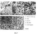
- Particular non-limiting classes of protease inhibitors include serine and cysteine protease inhibitors.
- protease inhibitors include PMSF, PMSF Plus, APMSF, antithrombin III, Amastatin, Antipain, aprotinin, Bestatin, Benzamidine, Chymostatin, calpain inhibitor I and II, E-64,3,4-dichloroisocoumarin, DFP, Elastatinal, Leupeptin, Pepstatin, 1,10-Phenanthroline, Phosphoramidon, TIMP-2, TLCK, TPCK, trypsin inhibitor (soybean or chicken egg white), hirustasin, alpha-2-macroglobulin, 4-(2-aminoethyl)-benzenesulfonyl fluoride hydrochloride (AEBSF) and Kunitz-type protease inhibitors.
- AEBSF 4-(2-aminoethyl)-benzenesulfonyl fluoride hydrochloride
- exosomes in a urine sample are collected by passing the urine sample through a filter having a pore size that is smaller than the average size of exosomes. The exosomes are then removed from the filter and resuspended at a proper concentration for further analysis.
- exosomes in the urine samples are collected using centrifuge filters with a molecular weight cutoff of 500 kd-50 kd. In one embodiment, exosomes in the urine samples are collected using centrifuge filters with a molecular weight cutoff of 100 kd.
- the detecting step 120 can be performed using any technology that is capable of identifying a HIV-associated biomarker.
- HIV-associated biomarker refers to proteins or fragments of proteins that are associated with HIV- infection, the progress of HIV infection, and HIV-related diseases such as HIV-associated nephropathy.
- the HIV-associated biomarker is selected from the group consisting of Nef, HIV envelope gp120, HIV protease, Vif, Gag-Pol, Gag, Pol, p24, Rev, reverse transcriptase (RT), Tat, p1, p17, Vpu, Vpr, gp41 and DNA polymerase.
- a number of technologies can be used to identify a HIV-associated biomarker. Examples of such technologies include, but are not limited to, electrophoresis such as one-dimensional and two-dimensional gel analysis, Western blot, ELISA, HPLC, FPLC, mass spectrometry, protein sequencing, antibody array and combinations thereof.
- the biomarkers are identified by Western blot. In another embodiment, the biomarkers are identified by Surface Enhanced Laser Desorption/Ionization Time of Flight Mass Spectrometry (SELDI-TOF-MS) and LC-MS/MS.
- the determining step is carried out by comparing the HIV-associated biomarker profile in a urine sample to HIV-associated biomarker profiles stored in a database. A diagnosis is made based on the result of the comparison.
- a HIV-associated biomarker profile may contain from zero to multiple HIV-associated biomarkers. For example, the HIV-associated biomarker profile of a healthy mammal may contain no HIV-associated biomarkers. On the other hand, the HIV-associated biomarker profile of a patient with HIVAN may contain a plurality of HIV-associated biomarkers.
- the method 100 is used to monitor the progress of HIV infection in the mammal.
- pertinent information from the medical record of the mammal may also be used in the determining step 130 to make a diagnosis.
- the method 100 is used for diagnosing HIV-associated nephropathy in a mammal.
- the genetic background and pertinent information from the medical record of the mammal may also be used in the determining step 130 to make a diagnosis.
- the method 100 may also be used to monitor the progress of HIV-associated nephropathy in the mammal.
- the method 100 is used to monitor effects during clinical trials of HIV-treatment.
- the HIV-associated biomarker profile in urine exosomes can be used as a readout, indicative of the physiological response of a mammal to the treatment.
- a mammal is a human, monkey, gorilla or baboon.
- the mammal is a human.
- Another aspect of the present invention provides a method for monitoring the effectiveness of treatment of a mammal with an anti-HIV agent.
- the method includes the steps of: detecting the HIV-associated biomarker profile in urine exosomes in the sample obtained from a mammal prior to administration of the agent; detecting the HIV-associated biomarker profile in urine exosomes in one or more post-administration samples obtained from the mammal; comparing the HIV-associated biomarker profile in the pre-administration sample with the HIV-associated biomarker profile in the post administration sample or samples.
- the method further comprises the step of determining the effectiveness of the agent and optionally altering the administration of the agent to the mammal.
- the HIV-associated biomarker profile may be used as an indicator of the effectiveness of an agent, even in the absence of an observable phenotypic response.
- kits for detecting HIV infection or monitoring the progress of HIV infection in a mammal contains one or more reagents for preparing exosomes sample for detection and at least one HIV-associated biomarker as a standard.
- the exosomes can be detected by Western blot.
- kits for diagnosing a HIV-associated disease or monitoring the progress of a HIV-associated disease in a mammal contains one or more reagents for preparing exosomes sample for detection and at least one HIV-associated biomarker as a standard.
- the HIV-associated disease is HIV-associated nephropathy.
- the HIV-associated biomarker is selected from the group consisting of Nef, HIV envelope gp120, HIV protease, Vif, Gag-Pol, Gag, Pol, p24, Rev, reverse transcriptase (RT), Tat, p1, p17, Vpu, Vpr, gp41 and DNA polymerase.
- kits described above typically include a label or packaging insert including a description of the components or instructions for use.
- Exemplary instructions include, instructions for collecting a urine sample, for harvesting exosomes from the urine, and for detecting an HIV-associated biomarker.
- the kits described above may additionally include a liquid suitable for resuspending the exosomes isolated from the urine sample.
- the kits described above may contain a container for collecting a urine sample and/or a centrifuge filter for isolating exosomes from the urine sample.
- HIV+ patients at various stages of disease, were recruited for this study from four clinical sites in the Atlanta metropolitan area. Only those patients on dialysis were excluded from this study. All samples were collected in accordance with protocols approved by the Institutional Review Board and the Human Subjects Research Committee at Morehouse School of Medicine, and informed consent was obtained from all patients and healthy volunteers according to the guidelines instituted by the Institutional Review Board. Patients were divided into five groups: African American patients with HIV (AA HIV+), white patients with HIV (White HIV+), patients with HIVAN (HIVAN), African American patients with no HIV- but FSGS, and healthy controls. Pertinent information was also collected from the medical record of the patients.
- Urine samples were collected from patients during routine clinical visits. Clinical data were obtained from the medical record of the patients. Urine was collected in sterile containers and transported back to the laboratory. Urinalysis was performed on each specimen using a Multistix 10 SG Reagent Strip (Bayer Corporation, Elkhart, IN) and the albumin to creatine ratio determined by a Siemens Clinitek Microalbumin dipstick (Bayer Corp.). The strips were read on a Siemens Clinitek Status instrument (Bayer Corp.). Samples were centrifuged at 2,000 x g for 10 minutes to remove whole cells and sediment. The remaining urine samples were aliquoted into 4 ml volumes and stored at -80°C until they were analyzed.
- SPA matrix solution
- detector sensitivity 10 high mass 300Kda, optimized from 3Kda to 50 Kda.
- the data acquisition method was set to automatic laser adjustment and peaks were auto identified from 3Kda and 50Kda.
- ThermoFinnigan Collected exosomes were analyzed by LC/MS using an LTQ mass spectrometer (ThermoFinnigan).
- the pelleted exosomes were first extracted with 2 D gel loading buffer (Q-biosciences) made fresh the day of analysis.
- the solubilized pellet was then precipitated using four volumes of ice-cold (-20°C) acetone and incubated overnight at -20°C.
- the precipitate was collected by centrifugation at 19,200 x g.
- the pellet was dried and redissolved into 50mM ammonium bicarbonate (AmBIC).
- the protein solution was first reduced using 2 ⁇ l of a 500 mM stock of DTT (Q biosciences, single use) at 56°C for 30 minutes.
- the solution was then alkylated by adding 2 ⁇ l of a 1M stock of iodacetic acid (IAA; Q-biosciences, single use) and incubating at room temperature for 30 minutes in the dark.
- IAA iodacetic acid
- a fresh vial of trypsin Promega Gold mass spec grade
- Ten microliters of the diluted trypsin was then added to the reaction and it was incubated at 37°C for 4 hours with shaking. Then 50ul of 0.5% formic acid was added and the mixture was either directly analyzed or stored at -20°C for analysis.
- the gels were stained using Gel Code Blue (Pierce, Inc.) or the proteins were transferred to PVDF membrane (Immobilon-P, Millipore Corp, Billerica, MA) for western blot analysis.
- the SNAP ID system (Millipore, Corp) was used for the western blot analysis for the presence of either HIV Nef or HIV proteins. HIV Nef identification was performed using a monoclonal mouse anti-HIV Nef monoclonal antibody (1:1500, Chemicon Int., CA) and a secondary antibody, goat anti-mouse IgG (H+L) peroxidase conjugated antibody (1:15,000, Jackson Immunoresearch, West Grove, PA).
- HIV proteins were detected using pooled human HIV+ serum (1:15,000) as the primary antibody and a goat anti-human IgG (H+L) peroxidase conjugated antibody (1:15,000, Jackson Immunoresearch).
- the membrane was incubated with a chemiluminescent substrate (SuperSignal West Femto Maximum, Pierce, Inc.) and exposed to X-ray film (CL-Xposure, Kodak) and developed.
- EXAMPLE 2 ISOLATION OF URINARY VESICLES. ULTRACENTRIFUGATION VERSUS ULTRAFILTRATION:
- Vesicles from the urine of six (6) different HIV+ patients were isolated by ultracentrifugation or ultrafiltration to determine which of the two methods yielded the greatest amount of protein.
- the ultrafiltration method consistently isolated more protein, 2930 ⁇ g (median), than the ultracentrifugation method, 591 ⁇ g (median).
- EXAMPLE 3 SELDI-TOF-MS SPECTRUM OF URINARY EXOSOMES FROM PATIENTS
- Urinary exosomes from patients of various groups are analyzed by SELDI-TOF-MS. The results are confirmed by LC-MS/MS. Spectrum of SELDI-TOF-MS from representative patients is shown in Figures 2-6 . Table 1 summarizes proteins that were detected by SELDI-MS and confirmed by LC-MS/MS in different test groups.
- Table 1 Proteins detected in urine samples by SELDI-MS Patient MW Protein HIVAN 10,585 HIV envelope gp; HIV Protease 23,546 HIV envelope gp; HIV Nef; HIV Vif 33,464 HIV protein gp; mu A03009B12Rik Protein 45,632 HIV envelope gp; HIV pol protein 66,587 HIV envelope gp; HIV Nef; PgD synthase 78,942 Unknown AA HIV 23,684 HIV envelope gp; HIV Nef; PgD synthase 83,256 Unknown FSGS 66,533 Unknown White HIV 23,935
- Table 2 summarizes the urine protein profiles in individual patient.
- Urine protein profiles in individual patient ID Diagnosis Race CD4 VL Nef Gag Pol Protease Rev RT Tat Vif p1 p24 p17 poly 22 HIVAN AA X X X X X X X X X 27 HIV/ AIDS AA 134 ⁇ 50 X 28 HIV/ AIDS AA 134 19,800 X X X X X 30 HIV/ AIDS AA ⁇ 20 >10,000 X X X X X X X X X 41 HIV AA 440 29,187 X X 46 HIV AA 689 ⁇ 50 X X X X 62 HIV AA 232 ⁇ 50 X 63 HIV/ AIDS AA 83 2,023 X 70 HIV AA 990 ⁇ 50 X 104 HIV AA 313 77 X 111 HIV/ AIDS AA 182 ⁇ 50 X X 112 HIV AA 584
- TEM was used to visualize the patients' vesicles from urine. Exosomes were isolated from 4 ml of urine, fixed and embedded for TEM. The figure shows distribution of vesicles in: A) HIVAN; B) focal segmental glomerusclerosis; C) AA HIV+; and D) white HIV+; E) AA HIV negative. HIVAN, FSGS and AA HIV+ patients clearly have a higher population of vesicles compared to white HIV+ patients and AA normal patients.
- the SELDI-TOF-MS peaks of AA HIV+ patients exhibited exceedingly similar protein patterns to those of HIVAN patients and slightly similar patterns to FSGS patients, suggesting that the AA HIV+ patients whose peaks were similar to those of HIVAN may be predisposed to developing HIVAN.
- the baseline protein value (30- 2000 mg/dl) for the FSGS patients and AA HIV+ patients was in the same range. Similar to the protein values for HIVANb, but unlike FSGS patients, the proteins detected in AA HIV + patients were analogous to those of HIVAN patients. This underscores the significance that HIV infection, with or without the presence of renal disease, is still largely responsible for development of HIVAN; and a prior condition of renal insufficiency before HIV infection is not a necessary prerequisite for the development of HIVAN.
- Urinary vesicle samples from fourteen (14) HIV+ AA and nine (9) HIV+ white patients were isolated using ultrafiltration and analyzed for the presence of HIV Nef and other HIV proteins using western blot analysis. All the HIV+ AA samples were positive for HIV Nef by western blot, although HIV Nef was not detected in sample 41 by mass spectrometry ( Figure 8 ). This discrepancy could be caused by the isolation method utilized for the mass spectrometry analysis, which was ultracentrifugation, and yields less protein. HIV Nef was only identified in four (4) HIV+ white patients while mass spectrometry identified three (3) samples without HIV Nef. All HIV+ patients had detectable HIV proteins by western blots, but had varying kinds and amounts ( Figure 8 ).
Landscapes
- Health & Medical Sciences (AREA)
- Life Sciences & Earth Sciences (AREA)
- Engineering & Computer Science (AREA)
- Biomedical Technology (AREA)
- Immunology (AREA)
- Chemical & Material Sciences (AREA)
- Urology & Nephrology (AREA)
- Molecular Biology (AREA)
- Hematology (AREA)
- Physics & Mathematics (AREA)
- General Health & Medical Sciences (AREA)
- Biochemistry (AREA)
- Pathology (AREA)
- Food Science & Technology (AREA)
- Medicinal Chemistry (AREA)
- General Physics & Mathematics (AREA)
- Analytical Chemistry (AREA)
- Biotechnology (AREA)
- Microbiology (AREA)
- Cell Biology (AREA)
- Virology (AREA)
- Tropical Medicine & Parasitology (AREA)
- Proteomics, Peptides & Aminoacids (AREA)
- Biophysics (AREA)
- AIDS & HIV (AREA)
- Investigating Or Analysing Biological Materials (AREA)
- Measuring Or Testing Involving Enzymes Or Micro-Organisms (AREA)
Applications Claiming Priority (3)
| Application Number | Priority Date | Filing Date | Title |
|---|---|---|---|
| US10294108P | 2008-10-06 | 2008-10-06 | |
| US12/572,652 US10545149B2 (en) | 2008-10-06 | 2009-10-02 | Detection of HIV-related proteins in urine |
| PCT/US2009/059500 WO2010042425A2 (en) | 2008-10-06 | 2009-10-05 | Detection of hiv-related proteins in urine |
Publications (3)
| Publication Number | Publication Date |
|---|---|
| EP2331969A2 EP2331969A2 (en) | 2011-06-15 |
| EP2331969A4 EP2331969A4 (en) | 2012-05-16 |
| EP2331969B1 true EP2331969B1 (en) | 2014-11-26 |
Family
ID=42076105
Family Applications (1)
| Application Number | Title | Priority Date | Filing Date |
|---|---|---|---|
| EP09819705.6A Active EP2331969B1 (en) | 2008-10-06 | 2009-10-05 | Detection of hiv-related proteins in urine |
Country Status (7)
| Country | Link |
|---|---|
| US (3) | US10545149B2 (zh) |
| EP (1) | EP2331969B1 (zh) |
| CN (2) | CN102171574B (zh) |
| AP (1) | AP2011005665A0 (zh) |
| AU (2) | AU2009302612B2 (zh) |
| CA (1) | CA2738666C (zh) |
| WO (1) | WO2010042425A2 (zh) |
Families Citing this family (22)
| Publication number | Priority date | Publication date | Assignee | Title |
|---|---|---|---|---|
| CA2742324A1 (en) * | 2008-10-30 | 2010-06-03 | Caris Life Sciences Luxembourg Holdings, S.A.R.L. | Methods for assessing rna patterns |
| BRPI0921043A2 (pt) * | 2008-11-12 | 2018-08-07 | Caris Life Sciences Luxembourg Holdings | métodos e sistemas para usar exossomas para determinar fenótipos |
| JP5808349B2 (ja) | 2010-03-01 | 2015-11-10 | カリス ライフ サイエンシズ スウィッツァーランド ホールディングスゲーエムベーハー | セラノーシスのためのバイオマーカー |
| WO2011127219A1 (en) | 2010-04-06 | 2011-10-13 | Caris Life Sciences Luxembourg Holdings | Circulating biomarkers for disease |
| EP2596369A4 (en) * | 2010-07-22 | 2014-01-22 | Univ Georgetown | MASS SPECTROMETRY METHODS FOR QUANTIFICATION OF NPY 1-36 AND NPY 3-36 |
| CN102323281B (zh) * | 2011-08-22 | 2013-09-18 | 爱威科技股份有限公司 | 液体性质检测方法和系统 |
| CA3113972C (en) | 2011-12-14 | 2023-08-01 | The Board Of Trustees Of The University Of Arkansas | Delivery of therapeutic agents using bacterial collagen-binding polypeptide segments |
| WO2014108480A1 (en) * | 2013-01-09 | 2014-07-17 | Friedrich-Alexander-Universitaet Erlangen-Nuernberg | Method for in vitro detection and monitoring of a disease by measuring disease-associated protease activity in extracellular vesicles |
| PT2972193T (pt) | 2013-03-13 | 2020-04-23 | Univ Miami | Método para isolamento e purificação de microvesículas de sobrenadantes de cultura celular e fluidos biológicos |
| WO2014191480A1 (en) * | 2013-05-28 | 2014-12-04 | INSERM (Institut National de la Santé et de la Recherche Médicale) | Use of urinary hiv nucleic acids to diagnose and monitor hiv-associated nephropathy (hivan) |
| CN103468642A (zh) * | 2013-09-23 | 2013-12-25 | 山西大学 | 一种分离细胞培养基中外泌体的方法 |
| EP3060911A4 (en) * | 2013-10-24 | 2017-06-21 | Agency For Science, Technology And Research | Exosome recovery methods with low molecular weight organic zwitterions |
| FR3014198B1 (fr) * | 2013-12-03 | 2017-03-03 | Biomerieux Sa | Procede d'isolement d'exosomes |
| CN106029898B (zh) * | 2014-01-21 | 2020-02-28 | 莫尔豪斯医学院 | 外来体介导的感染和疾病的检测 |
| CN103900890A (zh) * | 2014-03-07 | 2014-07-02 | 南方医科大学第三附属医院 | 一种采用纳米膜浓缩法提取尿微囊泡的方法 |
| CN105505854B (zh) * | 2016-01-14 | 2019-07-12 | 上海市第六人民医院 | 来源于人尿液细胞的外泌体的获取方法与应用 |
| CN107153023A (zh) * | 2016-03-02 | 2017-09-12 | 上海润腾生物科技有限公司 | 一种分离尿液中外泌体的方法 |
| US11614388B2 (en) * | 2016-10-13 | 2023-03-28 | H.U. Group Research Institute G.K. | Method for recovering extracellular vesicles |
| CN108333263A (zh) * | 2017-01-20 | 2018-07-27 | 北京蛋白质组研究中心 | 一种尿蛋白制备方法及尿蛋白质组的检测方法 |
| CN106929467B (zh) * | 2017-02-16 | 2021-05-18 | 上海交通大学 | 一种分离高纯度尿液外泌体的方法和试剂盒 |
| CN112867802B (zh) * | 2018-09-20 | 2024-06-04 | 塔微核酸有限责任公司 | 用于预测肝功能障碍的微小rna特征 |
| CN114397388B (zh) * | 2022-01-11 | 2024-03-19 | 杭州昱鼎生物科技有限公司 | 一种基于peg沉淀法与sec柱法相结合的尿液外泌体提取试剂盒及应用 |
Family Cites Families (22)
| Publication number | Priority date | Publication date | Assignee | Title |
|---|---|---|---|---|
| US5364933A (en) * | 1986-03-03 | 1994-11-15 | Institut Pasteur | Methods of immunopurification of antigens of human immunodeficiency virus type 2 (HIV-2) |
| US5830641A (en) * | 1986-01-22 | 1998-11-03 | Institut Pasteur | In vitro diagnostic assays for the detection of HIV-1 or HIV-2 employing viral-specific antigens and antibodies |
| US5580739A (en) * | 1986-01-22 | 1996-12-03 | Institut Pasteur | Peptides of human immunodeficiency virus type 2 (HIV-2) and in vitro diagnostic methods and kits employing the peptides for the detection of HIV-2 |
| US5208321A (en) * | 1988-06-09 | 1993-05-04 | Institut Pasteur | HIV-2 transmembrane glycoprotein homodimer (GP 80) |
| CA2585164C (fr) * | 1989-06-02 | 2010-02-02 | Institut Pasteur | Sequences nucleotidiques issues du genome des retrovirus du type hiv-1, hiv-2 et siv, et leurs applications notamment pour l'amplification des genomes de ces retrovirus et pour lediagnostic in vitro des infections dues a ces virus |
| DE69021640T2 (de) * | 1989-06-02 | 1996-04-04 | Abbott Lab | Standard-Reagenzien zur Quantifizierung von HIV p24. |
| US5256561A (en) * | 1991-12-20 | 1993-10-26 | Abbott Laboratories | Monoclonal antibody to HIV-2 and uses thereof |
| US5462852A (en) * | 1992-10-28 | 1995-10-31 | The Government Of The United States Of America, As Represented By The Secretary, Dhhs | HIV Nucleocapsid protein capture assay and method of use |
| US6261764B1 (en) * | 1997-09-22 | 2001-07-17 | Chiron Corporation | Buffers for stabilizing antigens |
| US20060252075A1 (en) * | 1998-08-21 | 2006-11-09 | Neovacs | Method for monitoring anti-HIV treatment in HIV infected individuals |
| US20040106136A1 (en) * | 1999-01-25 | 2004-06-03 | Musc Foundation For Research Development | Method for testing drug susceptibility of HIV |
| US6383740B2 (en) * | 1999-07-30 | 2002-05-07 | Bioergonomics, Inc. | Methods for simultaneously detecting both members of a binding pair |
| GB9927320D0 (en) * | 1999-11-18 | 2000-01-12 | Chiron Spa | Exosome separation |
| US20040241176A1 (en) * | 2000-04-27 | 2004-12-02 | Ap Cells. Inc. | Method of producing membrane vesicles |
| US7211659B2 (en) * | 2001-07-05 | 2007-05-01 | Chiron Corporation | Polynucleotides encoding antigenic HIV type C polypeptides, polypeptides and uses thereof |
| CA2532084A1 (en) * | 2003-07-11 | 2005-02-03 | Oregon Health And Science University | Methods of treatment and diagnosis using modulators of virus-induced cellular gene sequences |
| CA2544972C (en) * | 2003-11-05 | 2013-04-23 | Institut De Recherche Pour Le Developpement (Ird) | Use of interleukin-22 as a biomarker of resistance to hiv infections in humans and biological applications thereof |
| JP2008501336A (ja) * | 2004-06-02 | 2008-01-24 | ソースフアーム・インコーポレイテツド | Rnaを含有する微小胞およびそのための方法 |
| CA2644855C (en) | 2006-03-09 | 2019-11-19 | Aethlon Medical, Inc. | Extracorporeal removal of microvesicular particles |
| US8357296B2 (en) * | 2007-09-24 | 2013-01-22 | Emd Millipore Corporation | Centrifugal filter |
| US8617806B2 (en) * | 2008-01-25 | 2013-12-31 | Hansabiomed Ou | Method to measure and characterize microvesicles in the human body fluids |
| BRPI0921043A2 (pt) * | 2008-11-12 | 2018-08-07 | Caris Life Sciences Luxembourg Holdings | métodos e sistemas para usar exossomas para determinar fenótipos |
-
2009
- 2009-10-02 US US12/572,652 patent/US10545149B2/en active Active
- 2009-10-05 EP EP09819705.6A patent/EP2331969B1/en active Active
- 2009-10-05 WO PCT/US2009/059500 patent/WO2010042425A2/en active Application Filing
- 2009-10-05 CA CA2738666A patent/CA2738666C/en active Active
- 2009-10-05 CN CN200980139248.3A patent/CN102171574B/zh not_active Expired - Fee Related
- 2009-10-05 AP AP2011005665A patent/AP2011005665A0/xx unknown
- 2009-10-05 CN CN201610701916.9A patent/CN106383227B/zh not_active Expired - Fee Related
- 2009-10-05 AU AU2009302612A patent/AU2009302612B2/en active Active
-
2012
- 2012-01-11 US US13/348,389 patent/US9541554B2/en active Active
- 2012-01-11 US US13/348,406 patent/US9581596B2/en active Active
-
2016
- 2016-10-11 AU AU2016244203A patent/AU2016244203B2/en active Active
Also Published As
| Publication number | Publication date |
|---|---|
| US10545149B2 (en) | 2020-01-28 |
| CA2738666A1 (en) | 2010-04-15 |
| EP2331969A2 (en) | 2011-06-15 |
| CA2738666C (en) | 2018-05-15 |
| EP2331969A4 (en) | 2012-05-16 |
| WO2010042425A2 (en) | 2010-04-15 |
| CN102171574B (zh) | 2016-09-21 |
| CN102171574A (zh) | 2011-08-31 |
| AU2009302612B2 (en) | 2016-07-14 |
| US9581596B2 (en) | 2017-02-28 |
| AU2016244203B2 (en) | 2018-06-21 |
| US20100086956A1 (en) | 2010-04-08 |
| CN106383227A (zh) | 2017-02-08 |
| US20120214151A1 (en) | 2012-08-23 |
| CN106383227B (zh) | 2019-02-01 |
| US20120208175A1 (en) | 2012-08-16 |
| WO2010042425A3 (en) | 2010-07-22 |
| AU2016244203A1 (en) | 2016-11-03 |
| US9541554B2 (en) | 2017-01-10 |
| AU2009302612A1 (en) | 2010-04-15 |
| AP2011005665A0 (en) | 2011-04-30 |
Similar Documents
| Publication | Publication Date | Title |
|---|---|---|
| EP2331969B1 (en) | Detection of hiv-related proteins in urine | |
| US11300565B2 (en) | Exosome-mediated diagnosis of human immunodeficiency virus infection | |
| Raux et al. | Comparison of the distribution of IgG and IgA antibodies in serum and various mucosal fluids of HIV type 1-infected subjects | |
| JP2012524883A (ja) | 腎臓障害生物マーカとしてのwnt1 | |
| Tong et al. | Monitoring the progress of BK virus associated nephropathy in renal transplant recipients | |
| JP5736597B2 (ja) | 急性腎不全又は急性腎不全発症リスクのマーカーとしての尿中gm2活性化タンパク質 | |
| Bakoush et al. | Renal function in proteinuric glomerular diseases correlates to the changes in urine IgM excretion but not to the changes in the degree of albuminuria | |
| US10809265B2 (en) | Method for estimating number of podocytes in urine | |
| EP3279660B1 (en) | Preservative solution for heme protein, and method for stabilizing heme protein | |
| WO2022008858A1 (en) | Kit and method | |
| KR20180011571A (ko) | 숙신산을 포함하는 장기이식 급성거부반응 진단용 조성물 및 이를 이용한 진단방법 | |
| Gijsen et al. | Urinary NGAL levels early after pediatric kidney and liver transplantation: a pilot study | |
| Praseeda et al. | Review on Diabetic Nephropathy and the Biomarkers for Its Early Detection | |
| JPH11211724A (ja) | 切迫早産妊婦または頚管無力症妊婦の頚管粘液の鑑別法 | |
| WO2012005563A1 (en) | Uses of biological compounds from toxoplasma sp and related compounds |
Legal Events
| Date | Code | Title | Description |
|---|---|---|---|
| PUAI | Public reference made under article 153(3) epc to a published international application that has entered the european phase |
Free format text: ORIGINAL CODE: 0009012 |
|
| 17P | Request for examination filed |
Effective date: 20110406 |
|
| AK | Designated contracting states |
Kind code of ref document: A2 Designated state(s): AT BE BG CH CY CZ DE DK EE ES FI FR GB GR HR HU IE IS IT LI LT LU LV MC MK MT NL NO PL PT RO SE SI SK SM TR |
|
| AX | Request for extension of the european patent |
Extension state: AL BA RS |
|
| RIN1 | Information on inventor provided before grant (corrected) |
Inventor name: DOHERTY, AKINS Inventor name: NEWMAN, GALE, W. Inventor name: POWELL, MIKE Inventor name: MITCHELL-ALI, CLAUDETTE Inventor name: OBIALO, CHAMBERLAIN Inventor name: KHAN, MAFUZ Inventor name: BASHIR, KHALID |
|
| DAX | Request for extension of the european patent (deleted) | ||
| RIC1 | Information provided on ipc code assigned before grant |
Ipc: G01N 33/569 20060101ALI20120404BHEP Ipc: G01N 33/68 20060101AFI20120404BHEP Ipc: G01N 33/493 20060101ALI20120404BHEP |
|
| A4 | Supplementary search report drawn up and despatched |
Effective date: 20120417 |
|
| 17Q | First examination report despatched |
Effective date: 20131007 |
|
| RIC1 | Information provided on ipc code assigned before grant |
Ipc: G01N 33/569 20060101ALI20140225BHEP Ipc: G01N 1/40 20060101ALN20140225BHEP Ipc: G01N 33/493 20060101ALI20140225BHEP Ipc: G01N 33/68 20060101AFI20140225BHEP |
|
| GRAP | Despatch of communication of intention to grant a patent |
Free format text: ORIGINAL CODE: EPIDOSNIGR1 |
|
| INTG | Intention to grant announced |
Effective date: 20140804 |
|
| GRAS | Grant fee paid |
Free format text: ORIGINAL CODE: EPIDOSNIGR3 |
|
| GRAA | (expected) grant |
Free format text: ORIGINAL CODE: 0009210 |
|
| AK | Designated contracting states |
Kind code of ref document: B1 Designated state(s): AT BE BG CH CY CZ DE DK EE ES FI FR GB GR HR HU IE IS IT LI LT LU LV MC MK MT NL NO PL PT RO SE SI SK SM TR |
|
| REG | Reference to a national code |
Ref country code: GB Ref legal event code: FG4D |
|
| REG | Reference to a national code |
Ref country code: CH Ref legal event code: EP |
|
| REG | Reference to a national code |
Ref country code: AT Ref legal event code: REF Ref document number: 698492 Country of ref document: AT Kind code of ref document: T Effective date: 20141215 |
|
| REG | Reference to a national code |
Ref country code: IE Ref legal event code: FG4D |
|
| REG | Reference to a national code |
Ref country code: DE Ref legal event code: R096 Ref document number: 602009028029 Country of ref document: DE Effective date: 20150108 |
|
| REG | Reference to a national code |
Ref country code: CH Ref legal event code: NV Representative=s name: ISLER AND PEDRAZZINI AG, CH |
|
| REG | Reference to a national code |
Ref country code: NL Ref legal event code: VDEP Effective date: 20141126 |
|
| REG | Reference to a national code |
Ref country code: AT Ref legal event code: MK05 Ref document number: 698492 Country of ref document: AT Kind code of ref document: T Effective date: 20141126 |
|
| REG | Reference to a national code |
Ref country code: LT Ref legal event code: MG4D |
|
| PG25 | Lapsed in a contracting state [announced via postgrant information from national office to epo] |
Ref country code: NO Free format text: LAPSE BECAUSE OF FAILURE TO SUBMIT A TRANSLATION OF THE DESCRIPTION OR TO PAY THE FEE WITHIN THE PRESCRIBED TIME-LIMIT Effective date: 20150226 Ref country code: FI Free format text: LAPSE BECAUSE OF FAILURE TO SUBMIT A TRANSLATION OF THE DESCRIPTION OR TO PAY THE FEE WITHIN THE PRESCRIBED TIME-LIMIT Effective date: 20141126 Ref country code: LT Free format text: LAPSE BECAUSE OF FAILURE TO SUBMIT A TRANSLATION OF THE DESCRIPTION OR TO PAY THE FEE WITHIN THE PRESCRIBED TIME-LIMIT Effective date: 20141126 Ref country code: NL Free format text: LAPSE BECAUSE OF FAILURE TO SUBMIT A TRANSLATION OF THE DESCRIPTION OR TO PAY THE FEE WITHIN THE PRESCRIBED TIME-LIMIT Effective date: 20141126 Ref country code: ES Free format text: LAPSE BECAUSE OF FAILURE TO SUBMIT A TRANSLATION OF THE DESCRIPTION OR TO PAY THE FEE WITHIN THE PRESCRIBED TIME-LIMIT Effective date: 20141126 Ref country code: IS Free format text: LAPSE BECAUSE OF FAILURE TO SUBMIT A TRANSLATION OF THE DESCRIPTION OR TO PAY THE FEE WITHIN THE PRESCRIBED TIME-LIMIT Effective date: 20150326 Ref country code: PT Free format text: LAPSE BECAUSE OF FAILURE TO SUBMIT A TRANSLATION OF THE DESCRIPTION OR TO PAY THE FEE WITHIN THE PRESCRIBED TIME-LIMIT Effective date: 20150326 |
|
| PG25 | Lapsed in a contracting state [announced via postgrant information from national office to epo] |
Ref country code: HR Free format text: LAPSE BECAUSE OF FAILURE TO SUBMIT A TRANSLATION OF THE DESCRIPTION OR TO PAY THE FEE WITHIN THE PRESCRIBED TIME-LIMIT Effective date: 20141126 Ref country code: LV Free format text: LAPSE BECAUSE OF FAILURE TO SUBMIT A TRANSLATION OF THE DESCRIPTION OR TO PAY THE FEE WITHIN THE PRESCRIBED TIME-LIMIT Effective date: 20141126 Ref country code: AT Free format text: LAPSE BECAUSE OF FAILURE TO SUBMIT A TRANSLATION OF THE DESCRIPTION OR TO PAY THE FEE WITHIN THE PRESCRIBED TIME-LIMIT Effective date: 20141126 Ref country code: GR Free format text: LAPSE BECAUSE OF FAILURE TO SUBMIT A TRANSLATION OF THE DESCRIPTION OR TO PAY THE FEE WITHIN THE PRESCRIBED TIME-LIMIT Effective date: 20150227 Ref country code: CY Free format text: LAPSE BECAUSE OF FAILURE TO SUBMIT A TRANSLATION OF THE DESCRIPTION OR TO PAY THE FEE WITHIN THE PRESCRIBED TIME-LIMIT Effective date: 20141126 Ref country code: SE Free format text: LAPSE BECAUSE OF FAILURE TO SUBMIT A TRANSLATION OF THE DESCRIPTION OR TO PAY THE FEE WITHIN THE PRESCRIBED TIME-LIMIT Effective date: 20141126 |
|
| PG25 | Lapsed in a contracting state [announced via postgrant information from national office to epo] |
Ref country code: EE Free format text: LAPSE BECAUSE OF FAILURE TO SUBMIT A TRANSLATION OF THE DESCRIPTION OR TO PAY THE FEE WITHIN THE PRESCRIBED TIME-LIMIT Effective date: 20141126 Ref country code: DK Free format text: LAPSE BECAUSE OF FAILURE TO SUBMIT A TRANSLATION OF THE DESCRIPTION OR TO PAY THE FEE WITHIN THE PRESCRIBED TIME-LIMIT Effective date: 20141126 Ref country code: SK Free format text: LAPSE BECAUSE OF FAILURE TO SUBMIT A TRANSLATION OF THE DESCRIPTION OR TO PAY THE FEE WITHIN THE PRESCRIBED TIME-LIMIT Effective date: 20141126 Ref country code: CZ Free format text: LAPSE BECAUSE OF FAILURE TO SUBMIT A TRANSLATION OF THE DESCRIPTION OR TO PAY THE FEE WITHIN THE PRESCRIBED TIME-LIMIT Effective date: 20141126 Ref country code: RO Free format text: LAPSE BECAUSE OF FAILURE TO SUBMIT A TRANSLATION OF THE DESCRIPTION OR TO PAY THE FEE WITHIN THE PRESCRIBED TIME-LIMIT Effective date: 20141126 |
|
| REG | Reference to a national code |
Ref country code: DE Ref legal event code: R097 Ref document number: 602009028029 Country of ref document: DE |
|
| PG25 | Lapsed in a contracting state [announced via postgrant information from national office to epo] |
Ref country code: PL Free format text: LAPSE BECAUSE OF FAILURE TO SUBMIT A TRANSLATION OF THE DESCRIPTION OR TO PAY THE FEE WITHIN THE PRESCRIBED TIME-LIMIT Effective date: 20141126 |
|
| PLBE | No opposition filed within time limit |
Free format text: ORIGINAL CODE: 0009261 |
|
| STAA | Information on the status of an ep patent application or granted ep patent |
Free format text: STATUS: NO OPPOSITION FILED WITHIN TIME LIMIT |
|
| 26N | No opposition filed |
Effective date: 20150827 |
|
| PG25 | Lapsed in a contracting state [announced via postgrant information from national office to epo] |
Ref country code: IT Free format text: LAPSE BECAUSE OF FAILURE TO SUBMIT A TRANSLATION OF THE DESCRIPTION OR TO PAY THE FEE WITHIN THE PRESCRIBED TIME-LIMIT Effective date: 20141126 |
|
| PG25 | Lapsed in a contracting state [announced via postgrant information from national office to epo] |
Ref country code: SI Free format text: LAPSE BECAUSE OF FAILURE TO SUBMIT A TRANSLATION OF THE DESCRIPTION OR TO PAY THE FEE WITHIN THE PRESCRIBED TIME-LIMIT Effective date: 20141126 |
|
| PG25 | Lapsed in a contracting state [announced via postgrant information from national office to epo] |
Ref country code: LU Free format text: LAPSE BECAUSE OF FAILURE TO SUBMIT A TRANSLATION OF THE DESCRIPTION OR TO PAY THE FEE WITHIN THE PRESCRIBED TIME-LIMIT Effective date: 20151005 |
|
| PG25 | Lapsed in a contracting state [announced via postgrant information from national office to epo] |
Ref country code: MC Free format text: LAPSE BECAUSE OF FAILURE TO SUBMIT A TRANSLATION OF THE DESCRIPTION OR TO PAY THE FEE WITHIN THE PRESCRIBED TIME-LIMIT Effective date: 20141126 |
|
| REG | Reference to a national code |
Ref country code: IE Ref legal event code: MM4A |
|
| REG | Reference to a national code |
Ref country code: FR Ref legal event code: PLFP Year of fee payment: 8 |
|
| PG25 | Lapsed in a contracting state [announced via postgrant information from national office to epo] |
Ref country code: IE Free format text: LAPSE BECAUSE OF NON-PAYMENT OF DUE FEES Effective date: 20151005 |
|
| PG25 | Lapsed in a contracting state [announced via postgrant information from national office to epo] |
Ref country code: BG Free format text: LAPSE BECAUSE OF FAILURE TO SUBMIT A TRANSLATION OF THE DESCRIPTION OR TO PAY THE FEE WITHIN THE PRESCRIBED TIME-LIMIT Effective date: 20141126 Ref country code: SM Free format text: LAPSE BECAUSE OF FAILURE TO SUBMIT A TRANSLATION OF THE DESCRIPTION OR TO PAY THE FEE WITHIN THE PRESCRIBED TIME-LIMIT Effective date: 20141126 Ref country code: HU Free format text: LAPSE BECAUSE OF FAILURE TO SUBMIT A TRANSLATION OF THE DESCRIPTION OR TO PAY THE FEE WITHIN THE PRESCRIBED TIME-LIMIT; INVALID AB INITIO Effective date: 20091005 |
|
| PG25 | Lapsed in a contracting state [announced via postgrant information from national office to epo] |
Ref country code: TR Free format text: LAPSE BECAUSE OF FAILURE TO SUBMIT A TRANSLATION OF THE DESCRIPTION OR TO PAY THE FEE WITHIN THE PRESCRIBED TIME-LIMIT Effective date: 20141126 Ref country code: MT Free format text: LAPSE BECAUSE OF FAILURE TO SUBMIT A TRANSLATION OF THE DESCRIPTION OR TO PAY THE FEE WITHIN THE PRESCRIBED TIME-LIMIT Effective date: 20141126 |
|
| REG | Reference to a national code |
Ref country code: FR Ref legal event code: PLFP Year of fee payment: 9 |
|
| PG25 | Lapsed in a contracting state [announced via postgrant information from national office to epo] |
Ref country code: BE Free format text: LAPSE BECAUSE OF FAILURE TO SUBMIT A TRANSLATION OF THE DESCRIPTION OR TO PAY THE FEE WITHIN THE PRESCRIBED TIME-LIMIT Effective date: 20141126 |
|
| PG25 | Lapsed in a contracting state [announced via postgrant information from national office to epo] |
Ref country code: MK Free format text: LAPSE BECAUSE OF FAILURE TO SUBMIT A TRANSLATION OF THE DESCRIPTION OR TO PAY THE FEE WITHIN THE PRESCRIBED TIME-LIMIT Effective date: 20141126 |
|
| REG | Reference to a national code |
Ref country code: FR Ref legal event code: PLFP Year of fee payment: 10 |
|
| P01 | Opt-out of the competence of the unified patent court (upc) registered |
Effective date: 20230523 |
|
| PGFP | Annual fee paid to national office [announced via postgrant information from national office to epo] |
Ref country code: GB Payment date: 20231025 Year of fee payment: 15 |
|
| PGFP | Annual fee paid to national office [announced via postgrant information from national office to epo] |
Ref country code: FR Payment date: 20231023 Year of fee payment: 15 Ref country code: DE Payment date: 20231018 Year of fee payment: 15 Ref country code: CH Payment date: 20231102 Year of fee payment: 15 |