EP2331577B1 - Zusammensetzungen rekombinanter antikörper gegen den rezeptor des epidermalen wachstumsfaktors - Google Patents
Zusammensetzungen rekombinanter antikörper gegen den rezeptor des epidermalen wachstumsfaktors Download PDFInfo
- Publication number
- EP2331577B1 EP2331577B1 EP09776272.8A EP09776272A EP2331577B1 EP 2331577 B1 EP2331577 B1 EP 2331577B1 EP 09776272 A EP09776272 A EP 09776272A EP 2331577 B1 EP2331577 B1 EP 2331577B1
- Authority
- EP
- European Patent Office
- Prior art keywords
- antibody
- antibodies
- egfr
- seq
- composition
- Prior art date
- Legal status (The legal status is an assumption and is not a legal conclusion. Google has not performed a legal analysis and makes no representation as to the accuracy of the status listed.)
- Active
Links
- 239000000203 mixture Substances 0.000 title claims description 312
- 102000009465 Growth Factor Receptors Human genes 0.000 title description 3
- 108010009202 Growth Factor Receptors Proteins 0.000 title description 3
- 229940116977 epidermal growth factor Drugs 0.000 title description 2
- 230000027455 binding Effects 0.000 claims description 228
- 206010028980 Neoplasm Diseases 0.000 claims description 224
- 241000282414 Homo sapiens Species 0.000 claims description 153
- 238000011282 treatment Methods 0.000 claims description 110
- 150000001413 amino acids Chemical class 0.000 claims description 94
- 201000011510 cancer Diseases 0.000 claims description 93
- 239000000427 antigen Substances 0.000 claims description 86
- 108091007433 antigens Proteins 0.000 claims description 86
- 102000036639 antigens Human genes 0.000 claims description 86
- 229960001972 panitumumab Drugs 0.000 claims description 71
- 101000851181 Homo sapiens Epidermal growth factor receptor Proteins 0.000 claims description 37
- 102000045108 human EGFR Human genes 0.000 claims description 37
- 238000011269 treatment regimen Methods 0.000 claims description 27
- 229960005395 cetuximab Drugs 0.000 claims description 23
- 230000001965 increasing effect Effects 0.000 claims description 23
- 230000001939 inductive effect Effects 0.000 claims description 19
- 229950008250 zalutumumab Drugs 0.000 claims description 19
- 230000004069 differentiation Effects 0.000 claims description 15
- 229940121358 tyrosine kinase inhibitor Drugs 0.000 claims description 15
- 229950008001 matuzumab Drugs 0.000 claims description 14
- 229950010203 nimotuzumab Drugs 0.000 claims description 14
- 239000005483 tyrosine kinase inhibitor Substances 0.000 claims description 14
- 238000002512 chemotherapy Methods 0.000 claims description 12
- 230000036961 partial effect Effects 0.000 claims description 12
- 238000009093 first-line therapy Methods 0.000 claims description 11
- 239000003795 chemical substances by application Substances 0.000 claims description 10
- 238000001959 radiotherapy Methods 0.000 claims description 10
- VEEGZPWAAPPXRB-BJMVGYQFSA-N (3e)-3-(1h-imidazol-5-ylmethylidene)-1h-indol-2-one Chemical compound O=C1NC2=CC=CC=C2\C1=C/C1=CN=CN1 VEEGZPWAAPPXRB-BJMVGYQFSA-N 0.000 claims description 8
- 201000009030 Carcinoma Diseases 0.000 claims description 8
- 239000004037 angiogenesis inhibitor Substances 0.000 claims description 8
- 229940121369 angiogenesis inhibitor Drugs 0.000 claims description 8
- 230000009870 specific binding Effects 0.000 claims description 8
- 239000005556 hormone Substances 0.000 claims description 7
- 229940088597 hormone Drugs 0.000 claims description 7
- 206010041823 squamous cell carcinoma Diseases 0.000 claims description 7
- 238000001356 surgical procedure Methods 0.000 claims description 7
- 208000002154 non-small cell lung carcinoma Diseases 0.000 claims description 6
- 238000009094 second-line therapy Methods 0.000 claims description 6
- 206010006187 Breast cancer Diseases 0.000 claims description 5
- 208000026310 Breast neoplasm Diseases 0.000 claims description 5
- 206010058467 Lung neoplasm malignant Diseases 0.000 claims description 5
- 208000006265 Renal cell carcinoma Diseases 0.000 claims description 5
- 238000009098 adjuvant therapy Methods 0.000 claims description 5
- 208000014829 head and neck neoplasm Diseases 0.000 claims description 5
- 238000009095 third-line therapy Methods 0.000 claims description 5
- 206010009944 Colon cancer Diseases 0.000 claims description 4
- 208000008839 Kidney Neoplasms Diseases 0.000 claims description 4
- 206010060862 Prostate cancer Diseases 0.000 claims description 4
- 208000000236 Prostatic Neoplasms Diseases 0.000 claims description 4
- 206010038389 Renal cancer Diseases 0.000 claims description 4
- 208000000102 Squamous Cell Carcinoma of Head and Neck Diseases 0.000 claims description 4
- 238000002648 combination therapy Methods 0.000 claims description 4
- 208000005017 glioblastoma Diseases 0.000 claims description 4
- 201000000459 head and neck squamous cell carcinoma Diseases 0.000 claims description 4
- 201000010982 kidney cancer Diseases 0.000 claims description 4
- 201000005202 lung cancer Diseases 0.000 claims description 4
- 208000020816 lung neoplasm Diseases 0.000 claims description 4
- 201000001441 melanoma Diseases 0.000 claims description 4
- 208000032612 Glial tumor Diseases 0.000 claims description 3
- 201000010915 Glioblastoma multiforme Diseases 0.000 claims description 3
- 206010018338 Glioma Diseases 0.000 claims description 3
- 206010061902 Pancreatic neoplasm Diseases 0.000 claims description 3
- 208000005718 Stomach Neoplasms Diseases 0.000 claims description 3
- 208000007097 Urinary Bladder Neoplasms Diseases 0.000 claims description 3
- 206010017758 gastric cancer Diseases 0.000 claims description 3
- 206010073071 hepatocellular carcinoma Diseases 0.000 claims description 3
- 201000002528 pancreatic cancer Diseases 0.000 claims description 3
- 201000011549 stomach cancer Diseases 0.000 claims description 3
- 206010005003 Bladder cancer Diseases 0.000 claims description 2
- 206010008342 Cervix carcinoma Diseases 0.000 claims description 2
- 208000001333 Colorectal Neoplasms Diseases 0.000 claims description 2
- 208000000461 Esophageal Neoplasms Diseases 0.000 claims description 2
- 206010030155 Oesophageal carcinoma Diseases 0.000 claims description 2
- 206010033128 Ovarian cancer Diseases 0.000 claims description 2
- 206010061535 Ovarian neoplasm Diseases 0.000 claims description 2
- 208000015634 Rectal Neoplasms Diseases 0.000 claims description 2
- 208000006105 Uterine Cervical Neoplasms Diseases 0.000 claims description 2
- 201000010881 cervical cancer Diseases 0.000 claims description 2
- 208000029742 colonic neoplasm Diseases 0.000 claims description 2
- 201000004101 esophageal cancer Diseases 0.000 claims description 2
- 201000010536 head and neck cancer Diseases 0.000 claims description 2
- 208000014018 liver neoplasm Diseases 0.000 claims description 2
- 208000015486 malignant pancreatic neoplasm Diseases 0.000 claims description 2
- 208000008443 pancreatic carcinoma Diseases 0.000 claims description 2
- 206010038038 rectal cancer Diseases 0.000 claims description 2
- 201000001275 rectum cancer Diseases 0.000 claims description 2
- 201000005112 urinary bladder cancer Diseases 0.000 claims description 2
- 102000052116 epidermal growth factor receptor activity proteins Human genes 0.000 claims 21
- 108700015053 epidermal growth factor receptor activity proteins Proteins 0.000 claims 21
- YOHYSYJDKVYCJI-UHFFFAOYSA-N n-[3-[[6-[3-(trifluoromethyl)anilino]pyrimidin-4-yl]amino]phenyl]cyclopropanecarboxamide Chemical compound FC(F)(F)C1=CC=CC(NC=2N=CN=C(NC=3C=C(NC(=O)C4CC4)C=CC=3)C=2)=C1 YOHYSYJDKVYCJI-UHFFFAOYSA-N 0.000 claims 21
- 125000003275 alpha amino acid group Chemical group 0.000 claims 6
- 150000004917 tyrosine kinase inhibitor derivatives Chemical class 0.000 claims 3
- 229960000397 bevacizumab Drugs 0.000 claims 1
- 201000000062 kidney sarcoma Diseases 0.000 claims 1
- 210000004027 cell Anatomy 0.000 description 453
- 102000001301 EGF receptor Human genes 0.000 description 329
- 108060006698 EGF receptor Proteins 0.000 description 329
- 229940082789 erbitux Drugs 0.000 description 163
- 108020004414 DNA Proteins 0.000 description 100
- 238000000034 method Methods 0.000 description 97
- 108090000623 proteins and genes Proteins 0.000 description 83
- 238000002965 ELISA Methods 0.000 description 77
- 235000001014 amino acid Nutrition 0.000 description 66
- 229940024606 amino acid Drugs 0.000 description 64
- 102000004169 proteins and genes Human genes 0.000 description 53
- 235000018102 proteins Nutrition 0.000 description 52
- 102000009024 Epidermal Growth Factor Human genes 0.000 description 50
- 241000699670 Mus sp. Species 0.000 description 50
- 230000005764 inhibitory process Effects 0.000 description 48
- 230000000694 effects Effects 0.000 description 46
- 239000003446 ligand Substances 0.000 description 45
- 102000005962 receptors Human genes 0.000 description 39
- 108020003175 receptors Proteins 0.000 description 39
- 238000012216 screening Methods 0.000 description 39
- 241001529936 Murinae Species 0.000 description 36
- 239000006144 Dulbecco’s modified Eagle's medium Substances 0.000 description 35
- 238000003556 assay Methods 0.000 description 35
- 239000013598 vector Substances 0.000 description 35
- 239000013604 expression vector Substances 0.000 description 32
- 239000012634 fragment Substances 0.000 description 32
- 238000002474 experimental method Methods 0.000 description 31
- 239000000872 buffer Substances 0.000 description 30
- 239000003153 chemical reaction reagent Substances 0.000 description 29
- 238000011534 incubation Methods 0.000 description 28
- 239000007924 injection Substances 0.000 description 28
- 238000002347 injection Methods 0.000 description 28
- 238000001727 in vivo Methods 0.000 description 27
- 238000004458 analytical method Methods 0.000 description 26
- 239000000243 solution Substances 0.000 description 26
- 241001465754 Metazoa Species 0.000 description 25
- 230000012010 growth Effects 0.000 description 25
- 108090000765 processed proteins & peptides Proteins 0.000 description 25
- 108091003079 Bovine Serum Albumin Proteins 0.000 description 24
- 239000012091 fetal bovine serum Substances 0.000 description 24
- 239000006228 supernatant Substances 0.000 description 24
- 102000004196 processed proteins & peptides Human genes 0.000 description 23
- 108010054477 Immunoglobulin Fab Fragments Proteins 0.000 description 22
- 102000001706 Immunoglobulin Fab Fragments Human genes 0.000 description 22
- 101100112922 Candida albicans CDR3 gene Proteins 0.000 description 21
- 230000000903 blocking effect Effects 0.000 description 21
- 238000006243 chemical reaction Methods 0.000 description 21
- 238000007792 addition Methods 0.000 description 20
- 238000010790 dilution Methods 0.000 description 20
- 239000012895 dilution Substances 0.000 description 20
- 230000014509 gene expression Effects 0.000 description 20
- 239000000047 product Substances 0.000 description 20
- JUJBNYBVVQSIOU-UHFFFAOYSA-M sodium;4-[2-(4-iodophenyl)-3-(4-nitrophenyl)tetrazol-2-ium-5-yl]benzene-1,3-disulfonate Chemical compound [Na+].C1=CC([N+](=O)[O-])=CC=C1N1[N+](C=2C=CC(I)=CC=2)=NC(C=2C(=CC(=CC=2)S([O-])(=O)=O)S([O-])(=O)=O)=N1 JUJBNYBVVQSIOU-UHFFFAOYSA-M 0.000 description 20
- 102100035360 Cerebellar degeneration-related antigen 1 Human genes 0.000 description 19
- 238000002649 immunization Methods 0.000 description 19
- 230000002401 inhibitory effect Effects 0.000 description 19
- 238000001943 fluorescence-activated cell sorting Methods 0.000 description 18
- 101710117290 Aldo-keto reductase family 1 member C4 Proteins 0.000 description 17
- 241000699666 Mus <mouse, genus> Species 0.000 description 17
- 102100024952 Protein CBFA2T1 Human genes 0.000 description 17
- 230000011712 cell development Effects 0.000 description 16
- 239000013642 negative control Substances 0.000 description 16
- 230000009257 reactivity Effects 0.000 description 16
- 108060003951 Immunoglobulin Proteins 0.000 description 15
- 102000018358 immunoglobulin Human genes 0.000 description 15
- 230000002503 metabolic effect Effects 0.000 description 15
- 238000007857 nested PCR Methods 0.000 description 15
- 102000004190 Enzymes Human genes 0.000 description 14
- 108090000790 Enzymes Proteins 0.000 description 14
- 241000699660 Mus musculus Species 0.000 description 14
- 102000018120 Recombinases Human genes 0.000 description 14
- 108010091086 Recombinases Proteins 0.000 description 14
- 229940088598 enzyme Drugs 0.000 description 14
- 230000003053 immunization Effects 0.000 description 14
- 230000035772 mutation Effects 0.000 description 14
- 229920001184 polypeptide Polymers 0.000 description 14
- 239000000523 sample Substances 0.000 description 14
- 238000007920 subcutaneous administration Methods 0.000 description 14
- 108010076876 Keratins Proteins 0.000 description 13
- 102000011782 Keratins Human genes 0.000 description 13
- QAOWNCQODCNURD-UHFFFAOYSA-N Sulfuric acid Chemical compound OS(O)(=O)=O QAOWNCQODCNURD-UHFFFAOYSA-N 0.000 description 13
- 238000002835 absorbance Methods 0.000 description 13
- 230000003247 decreasing effect Effects 0.000 description 13
- 239000011049 pearl Substances 0.000 description 13
- 239000013612 plasmid Substances 0.000 description 13
- 230000001225 therapeutic effect Effects 0.000 description 13
- 238000002560 therapeutic procedure Methods 0.000 description 13
- 108010047041 Complementarity Determining Regions Proteins 0.000 description 12
- AOJJSUZBOXZQNB-TZSSRYMLSA-N Doxorubicin Chemical compound O([C@H]1C[C@@](O)(CC=2C(O)=C3C(=O)C=4C=CC=C(C=4C(=O)C3=C(O)C=21)OC)C(=O)CO)[C@H]1C[C@H](N)[C@H](O)[C@H](C)O1 AOJJSUZBOXZQNB-TZSSRYMLSA-N 0.000 description 12
- 230000006907 apoptotic process Effects 0.000 description 12
- 238000004519 manufacturing process Methods 0.000 description 12
- 238000005259 measurement Methods 0.000 description 12
- 239000008194 pharmaceutical composition Substances 0.000 description 12
- 239000000758 substrate Substances 0.000 description 12
- 238000012360 testing method Methods 0.000 description 12
- 238000001890 transfection Methods 0.000 description 12
- 210000004881 tumor cell Anatomy 0.000 description 12
- UAIUNKRWKOVEES-UHFFFAOYSA-N 3,3',5,5'-tetramethylbenzidine Chemical compound CC1=C(N)C(C)=CC(C=2C=C(C)C(N)=C(C)C=2)=C1 UAIUNKRWKOVEES-UHFFFAOYSA-N 0.000 description 11
- 230000007423 decrease Effects 0.000 description 11
- 208000037265 diseases, disorders, signs and symptoms Diseases 0.000 description 11
- 238000007912 intraperitoneal administration Methods 0.000 description 11
- 102000007236 involucrin Human genes 0.000 description 11
- 108010033564 involucrin Proteins 0.000 description 11
- 210000004180 plasmocyte Anatomy 0.000 description 11
- 230000008569 process Effects 0.000 description 11
- 230000035755 proliferation Effects 0.000 description 11
- 229940036185 synagis Drugs 0.000 description 11
- 241000282567 Macaca fascicularis Species 0.000 description 10
- 238000010367 cloning Methods 0.000 description 10
- 230000000875 corresponding effect Effects 0.000 description 10
- 201000010099 disease Diseases 0.000 description 10
- 230000001404 mediated effect Effects 0.000 description 10
- 150000007523 nucleic acids Chemical class 0.000 description 10
- 230000026731 phosphorylation Effects 0.000 description 10
- 238000006366 phosphorylation reaction Methods 0.000 description 10
- 238000002198 surface plasmon resonance spectroscopy Methods 0.000 description 10
- 230000004614 tumor growth Effects 0.000 description 10
- 230000003442 weekly effect Effects 0.000 description 10
- 229910052727 yttrium Inorganic materials 0.000 description 10
- 108091026890 Coding region Proteins 0.000 description 9
- 230000003321 amplification Effects 0.000 description 9
- 230000000890 antigenic effect Effects 0.000 description 9
- 238000013459 approach Methods 0.000 description 9
- 238000013461 design Methods 0.000 description 9
- 230000009977 dual effect Effects 0.000 description 9
- 108010087914 epidermal growth factor receptor VIII Proteins 0.000 description 9
- 230000004899 motility Effects 0.000 description 9
- 238000003199 nucleic acid amplification method Methods 0.000 description 9
- 238000000746 purification Methods 0.000 description 9
- 230000004083 survival effect Effects 0.000 description 9
- 238000004448 titration Methods 0.000 description 9
- 238000012546 transfer Methods 0.000 description 9
- 102220616904 CCAAT/enhancer-binding protein alpha_Y67F_mutation Human genes 0.000 description 8
- 102000008394 Immunoglobulin Fragments Human genes 0.000 description 8
- 108010021625 Immunoglobulin Fragments Proteins 0.000 description 8
- 102220472408 Pantetheinase_T55S_mutation Human genes 0.000 description 8
- 102220488580 Phosphopantothenoylcysteine decarboxylase_I78M_mutation Human genes 0.000 description 8
- 102220503735 Replication factor C subunit 3_G59V_mutation Human genes 0.000 description 8
- 210000004978 chinese hamster ovary cell Anatomy 0.000 description 8
- 230000001419 dependent effect Effects 0.000 description 8
- 210000003128 head Anatomy 0.000 description 8
- 230000010354 integration Effects 0.000 description 8
- 210000004698 lymphocyte Anatomy 0.000 description 8
- 210000003739 neck Anatomy 0.000 description 8
- 102000039446 nucleic acids Human genes 0.000 description 8
- 108020004707 nucleic acids Proteins 0.000 description 8
- 230000003389 potentiating effect Effects 0.000 description 8
- 238000011160 research Methods 0.000 description 8
- 230000004044 response Effects 0.000 description 8
- 238000003757 reverse transcription PCR Methods 0.000 description 8
- 102200065346 rs104894070 Human genes 0.000 description 8
- 102220223203 rs1060502131 Human genes 0.000 description 8
- 102200163844 rs17127898 Human genes 0.000 description 8
- 102200082919 rs35857380 Human genes 0.000 description 8
- 102220075284 rs374402088 Human genes 0.000 description 8
- 102220295916 rs774932586 Human genes 0.000 description 8
- 102220097520 rs876658986 Human genes 0.000 description 8
- 102220335306 rs924843423 Human genes 0.000 description 8
- 241000894007 species Species 0.000 description 8
- 241000588724 Escherichia coli Species 0.000 description 7
- 241000282326 Felis catus Species 0.000 description 7
- 238000012413 Fluorescence activated cell sorting analysis Methods 0.000 description 7
- 101000840257 Homo sapiens Immunoglobulin kappa constant Proteins 0.000 description 7
- 102100029572 Immunoglobulin kappa constant Human genes 0.000 description 7
- 239000002246 antineoplastic agent Substances 0.000 description 7
- 239000006285 cell suspension Substances 0.000 description 7
- 239000002299 complementary DNA Substances 0.000 description 7
- 150000001875 compounds Chemical class 0.000 description 7
- 238000011161 development Methods 0.000 description 7
- 230000018109 developmental process Effects 0.000 description 7
- 239000013024 dilution buffer Substances 0.000 description 7
- 238000013507 mapping Methods 0.000 description 7
- 239000013641 positive control Substances 0.000 description 7
- 210000001519 tissue Anatomy 0.000 description 7
- NHBKXEKEPDILRR-UHFFFAOYSA-N 2,3-bis(butanoylsulfanyl)propyl butanoate Chemical compound CCCC(=O)OCC(SC(=O)CCC)CSC(=O)CCC NHBKXEKEPDILRR-UHFFFAOYSA-N 0.000 description 6
- 241000282693 Cercopithecidae Species 0.000 description 6
- 241000124008 Mammalia Species 0.000 description 6
- 102220479332 Pantetheinase_G76A_mutation Human genes 0.000 description 6
- 229920001213 Polysorbate 20 Polymers 0.000 description 6
- 108010076504 Protein Sorting Signals Proteins 0.000 description 6
- 102220468699 Protein arginine N-methyltransferase 3_Y87F_mutation Human genes 0.000 description 6
- FAPWRFPIFSIZLT-UHFFFAOYSA-M Sodium chloride Chemical compound [Na+].[Cl-] FAPWRFPIFSIZLT-UHFFFAOYSA-M 0.000 description 6
- 230000030741 antigen processing and presentation Effects 0.000 description 6
- 238000001516 cell proliferation assay Methods 0.000 description 6
- 230000001413 cellular effect Effects 0.000 description 6
- 230000000973 chemotherapeutic effect Effects 0.000 description 6
- 230000009260 cross reactivity Effects 0.000 description 6
- 238000001514 detection method Methods 0.000 description 6
- 239000003814 drug Substances 0.000 description 6
- 230000006870 function Effects 0.000 description 6
- 230000009036 growth inhibition Effects 0.000 description 6
- 238000011835 investigation Methods 0.000 description 6
- 238000002955 isolation Methods 0.000 description 6
- 238000012933 kinetic analysis Methods 0.000 description 6
- 238000002156 mixing Methods 0.000 description 6
- 238000002823 phage display Methods 0.000 description 6
- 239000000256 polyoxyethylene sorbitan monolaurate Substances 0.000 description 6
- 235000010486 polyoxyethylene sorbitan monolaurate Nutrition 0.000 description 6
- 239000011535 reaction buffer Substances 0.000 description 6
- 102200061512 rs1064039 Human genes 0.000 description 6
- 102220055135 rs149428656 Human genes 0.000 description 6
- 102200136369 rs17843776 Human genes 0.000 description 6
- 102220005538 rs28928883 Human genes 0.000 description 6
- 102200140998 rs797044460 Human genes 0.000 description 6
- 238000013207 serial dilution Methods 0.000 description 6
- 239000000725 suspension Substances 0.000 description 6
- 230000002459 sustained effect Effects 0.000 description 6
- 230000002195 synergetic effect Effects 0.000 description 6
- 230000008685 targeting Effects 0.000 description 6
- 231100000041 toxicology testing Toxicity 0.000 description 6
- 239000011534 wash buffer Substances 0.000 description 6
- 208000003200 Adenoma Diseases 0.000 description 5
- 102220503634 Chymotrypsin-C_G61R_mutation Human genes 0.000 description 5
- IAZDPXIOMUYVGZ-UHFFFAOYSA-N Dimethylsulphoxide Chemical compound CS(C)=O IAZDPXIOMUYVGZ-UHFFFAOYSA-N 0.000 description 5
- 238000012286 ELISA Assay Methods 0.000 description 5
- 101001091242 Homo sapiens Immunoglobulin kappa joining 1 Proteins 0.000 description 5
- 102100034892 Immunoglobulin kappa joining 1 Human genes 0.000 description 5
- 241000283973 Oryctolagus cuniculus Species 0.000 description 5
- 206010039491 Sarcoma Diseases 0.000 description 5
- 102000004142 Trypsin Human genes 0.000 description 5
- 108090000631 Trypsin Proteins 0.000 description 5
- 239000002671 adjuvant Substances 0.000 description 5
- 238000000246 agarose gel electrophoresis Methods 0.000 description 5
- 230000014102 antigen processing and presentation of exogenous peptide antigen via MHC class I Effects 0.000 description 5
- 239000011324 bead Substances 0.000 description 5
- 210000004369 blood Anatomy 0.000 description 5
- 239000008280 blood Substances 0.000 description 5
- 230000037396 body weight Effects 0.000 description 5
- 238000004113 cell culture Methods 0.000 description 5
- 230000004663 cell proliferation Effects 0.000 description 5
- 238000003776 cleavage reaction Methods 0.000 description 5
- 239000011248 coating agent Substances 0.000 description 5
- 238000000576 coating method Methods 0.000 description 5
- 210000002615 epidermis Anatomy 0.000 description 5
- 238000001704 evaporation Methods 0.000 description 5
- 230000008020 evaporation Effects 0.000 description 5
- 239000001963 growth medium Substances 0.000 description 5
- 230000028993 immune response Effects 0.000 description 5
- 238000000338 in vitro Methods 0.000 description 5
- 238000011221 initial treatment Methods 0.000 description 5
- 238000003780 insertion Methods 0.000 description 5
- 230000037431 insertion Effects 0.000 description 5
- 230000003834 intracellular effect Effects 0.000 description 5
- 238000001990 intravenous administration Methods 0.000 description 5
- 238000004255 ion exchange chromatography Methods 0.000 description 5
- 210000004962 mammalian cell Anatomy 0.000 description 5
- 230000007246 mechanism Effects 0.000 description 5
- 230000010534 mechanism of action Effects 0.000 description 5
- 230000001394 metastastic effect Effects 0.000 description 5
- 206010061289 metastatic neoplasm Diseases 0.000 description 5
- 238000011275 oncology therapy Methods 0.000 description 5
- 238000002360 preparation method Methods 0.000 description 5
- 230000002035 prolonged effect Effects 0.000 description 5
- 208000016691 refractory malignant neoplasm Diseases 0.000 description 5
- 102220276885 rs1330621664 Human genes 0.000 description 5
- 102220139813 rs372921526 Human genes 0.000 description 5
- 229920006395 saturated elastomer Polymers 0.000 description 5
- 238000010186 staining Methods 0.000 description 5
- 238000010561 standard procedure Methods 0.000 description 5
- 239000012588 trypsin Substances 0.000 description 5
- 238000001262 western blot Methods 0.000 description 5
- HZAXFHJVJLSVMW-UHFFFAOYSA-N 2-Aminoethan-1-ol Chemical compound NCCO HZAXFHJVJLSVMW-UHFFFAOYSA-N 0.000 description 4
- 241000283707 Capra Species 0.000 description 4
- 238000008157 ELISA kit Methods 0.000 description 4
- 241000829100 Macaca mulatta polyomavirus 1 Species 0.000 description 4
- 102220500398 Neutral and basic amino acid transport protein rBAT_K43Q_mutation Human genes 0.000 description 4
- 108091028043 Nucleic acid sequence Proteins 0.000 description 4
- 102220578171 Rho-related GTP-binding protein RhoU_T63N_mutation Human genes 0.000 description 4
- PXIPVTKHYLBLMZ-UHFFFAOYSA-N Sodium azide Chemical compound [Na+].[N-]=[N+]=[N-] PXIPVTKHYLBLMZ-UHFFFAOYSA-N 0.000 description 4
- 102220601470 Succinate dehydrogenase [ubiquinone] cytochrome b small subunit, mitochondrial_S84P_mutation Human genes 0.000 description 4
- SHGAZHPCJJPHSC-YCNIQYBTSA-N all-trans-retinoic acid Chemical compound OC(=O)\C=C(/C)\C=C\C=C(/C)\C=C\C1=C(C)CCCC1(C)C SHGAZHPCJJPHSC-YCNIQYBTSA-N 0.000 description 4
- 230000001028 anti-proliverative effect Effects 0.000 description 4
- 210000003719 b-lymphocyte Anatomy 0.000 description 4
- 230000008901 benefit Effects 0.000 description 4
- 230000015572 biosynthetic process Effects 0.000 description 4
- 238000004364 calculation method Methods 0.000 description 4
- 230000010261 cell growth Effects 0.000 description 4
- 229940127089 cytotoxic agent Drugs 0.000 description 4
- 238000012217 deletion Methods 0.000 description 4
- 230000037430 deletion Effects 0.000 description 4
- 238000010494 dissociation reaction Methods 0.000 description 4
- 230000005593 dissociations Effects 0.000 description 4
- 239000000839 emulsion Substances 0.000 description 4
- 238000005516 engineering process Methods 0.000 description 4
- 238000009472 formulation Methods 0.000 description 4
- 230000004927 fusion Effects 0.000 description 4
- 230000028996 humoral immune response Effects 0.000 description 4
- 239000012216 imaging agent Substances 0.000 description 4
- 230000001976 improved effect Effects 0.000 description 4
- 238000001802 infusion Methods 0.000 description 4
- 239000003112 inhibitor Substances 0.000 description 4
- 230000003993 interaction Effects 0.000 description 4
- 230000002147 killing effect Effects 0.000 description 4
- 230000000670 limiting effect Effects 0.000 description 4
- 239000006166 lysate Substances 0.000 description 4
- 238000012423 maintenance Methods 0.000 description 4
- 230000003211 malignant effect Effects 0.000 description 4
- 238000013411 master cell bank Methods 0.000 description 4
- 239000002609 medium Substances 0.000 description 4
- 239000000546 pharmaceutical excipient Substances 0.000 description 4
- 230000002265 prevention Effects 0.000 description 4
- XJMOSONTPMZWPB-UHFFFAOYSA-M propidium iodide Chemical compound [I-].[I-].C12=CC(N)=CC=C2C2=CC=C(N)C=C2[N+](CCC[N+](C)(CC)CC)=C1C1=CC=CC=C1 XJMOSONTPMZWPB-UHFFFAOYSA-M 0.000 description 4
- 230000009467 reduction Effects 0.000 description 4
- 108091008146 restriction endonucleases Proteins 0.000 description 4
- 229930002330 retinoic acid Natural products 0.000 description 4
- 102220219923 rs1060503389 Human genes 0.000 description 4
- 102220334797 rs1555593653 Human genes 0.000 description 4
- 102220075471 rs536738656 Human genes 0.000 description 4
- 102220197765 rs754206331 Human genes 0.000 description 4
- 102220061211 rs786202029 Human genes 0.000 description 4
- 102200067663 rs80358463 Human genes 0.000 description 4
- 230000007017 scission Effects 0.000 description 4
- 210000002966 serum Anatomy 0.000 description 4
- 231100000419 toxicity Toxicity 0.000 description 4
- 230000001988 toxicity Effects 0.000 description 4
- IVLXQGJVBGMLRR-UHFFFAOYSA-N 2-aminoacetic acid;hydron;chloride Chemical compound Cl.NCC(O)=O IVLXQGJVBGMLRR-UHFFFAOYSA-N 0.000 description 3
- OBKXEAXTFZPCHS-UHFFFAOYSA-N 4-phenylbutyric acid Chemical compound OC(=O)CCCC1=CC=CC=C1 OBKXEAXTFZPCHS-UHFFFAOYSA-N 0.000 description 3
- 206010001233 Adenoma benign Diseases 0.000 description 3
- 230000004544 DNA amplification Effects 0.000 description 3
- 208000010201 Exanthema Diseases 0.000 description 3
- WSFSSNUMVMOOMR-UHFFFAOYSA-N Formaldehyde Chemical compound O=C WSFSSNUMVMOOMR-UHFFFAOYSA-N 0.000 description 3
- PEDCQBHIVMGVHV-UHFFFAOYSA-N Glycerine Chemical compound OCC(O)CO PEDCQBHIVMGVHV-UHFFFAOYSA-N 0.000 description 3
- 101000608935 Homo sapiens Leukosialin Proteins 0.000 description 3
- 102100039564 Leukosialin Human genes 0.000 description 3
- 241000282560 Macaca mulatta Species 0.000 description 3
- OKKJLVBELUTLKV-UHFFFAOYSA-N Methanol Chemical compound OC OKKJLVBELUTLKV-UHFFFAOYSA-N 0.000 description 3
- 229930012538 Paclitaxel Natural products 0.000 description 3
- 108091008874 T cell receptors Proteins 0.000 description 3
- 102000016266 T-Cell Antigen Receptors Human genes 0.000 description 3
- 101150117115 V gene Proteins 0.000 description 3
- 230000004913 activation Effects 0.000 description 3
- 239000004480 active ingredient Substances 0.000 description 3
- 229940009456 adriamycin Drugs 0.000 description 3
- 229940034982 antineoplastic agent Drugs 0.000 description 3
- 230000015556 catabolic process Effects 0.000 description 3
- 238000012512 characterization method Methods 0.000 description 3
- DQLATGHUWYMOKM-UHFFFAOYSA-L cisplatin Chemical compound N[Pt](N)(Cl)Cl DQLATGHUWYMOKM-UHFFFAOYSA-L 0.000 description 3
- 229960004316 cisplatin Drugs 0.000 description 3
- 210000001072 colon Anatomy 0.000 description 3
- 230000002860 competitive effect Effects 0.000 description 3
- 238000010276 construction Methods 0.000 description 3
- 238000012937 correction Methods 0.000 description 3
- 239000012228 culture supernatant Substances 0.000 description 3
- 230000006378 damage Effects 0.000 description 3
- 230000034994 death Effects 0.000 description 3
- 231100000517 death Toxicity 0.000 description 3
- 238000006731 degradation reaction Methods 0.000 description 3
- 230000029087 digestion Effects 0.000 description 3
- 230000003828 downregulation Effects 0.000 description 3
- 229960004679 doxorubicin Drugs 0.000 description 3
- 239000006196 drop Substances 0.000 description 3
- 229940079593 drug Drugs 0.000 description 3
- 201000005884 exanthem Diseases 0.000 description 3
- 230000001747 exhibiting effect Effects 0.000 description 3
- 235000012631 food intake Nutrition 0.000 description 3
- XGALLCVXEZPNRQ-UHFFFAOYSA-N gefitinib Chemical compound C=12C=C(OCCCN3CCOCC3)C(OC)=CC2=NC=NC=1NC1=CC=C(F)C(Cl)=C1 XGALLCVXEZPNRQ-UHFFFAOYSA-N 0.000 description 3
- 239000000499 gel Substances 0.000 description 3
- 238000003018 immunoassay Methods 0.000 description 3
- 230000002163 immunogen Effects 0.000 description 3
- 230000005847 immunogenicity Effects 0.000 description 3
- 230000006698 induction Effects 0.000 description 3
- 229960004768 irinotecan Drugs 0.000 description 3
- UWKQSNNFCGGAFS-XIFFEERXSA-N irinotecan Chemical compound C1=C2C(CC)=C3CN(C(C4=C([C@@](C(=O)OC4)(O)CC)C=4)=O)C=4C3=NC2=CC=C1OC(=O)N(CC1)CCC1N1CCCCC1 UWKQSNNFCGGAFS-XIFFEERXSA-N 0.000 description 3
- BCFGMOOMADDAQU-UHFFFAOYSA-N lapatinib Chemical compound O1C(CNCCS(=O)(=O)C)=CC=C1C1=CC=C(N=CN=C2NC=3C=C(Cl)C(OCC=4C=C(F)C=CC=4)=CC=3)C2=C1 BCFGMOOMADDAQU-UHFFFAOYSA-N 0.000 description 3
- 230000003902 lesion Effects 0.000 description 3
- 238000011068 loading method Methods 0.000 description 3
- 230000007774 longterm Effects 0.000 description 3
- 208000026037 malignant tumor of neck Diseases 0.000 description 3
- 238000011580 nude mouse model Methods 0.000 description 3
- 210000000056 organ Anatomy 0.000 description 3
- 210000001672 ovary Anatomy 0.000 description 3
- 229960001592 paclitaxel Drugs 0.000 description 3
- 239000008188 pellet Substances 0.000 description 3
- 230000010412 perfusion Effects 0.000 description 3
- 210000003720 plasmablast Anatomy 0.000 description 3
- 239000002243 precursor Substances 0.000 description 3
- 230000001681 protective effect Effects 0.000 description 3
- 206010037844 rash Diseases 0.000 description 3
- 230000008439 repair process Effects 0.000 description 3
- 238000013391 scatchard analysis Methods 0.000 description 3
- 230000011664 signaling Effects 0.000 description 3
- 231100000046 skin rash Toxicity 0.000 description 3
- 239000011780 sodium chloride Substances 0.000 description 3
- 210000000952 spleen Anatomy 0.000 description 3
- 208000017572 squamous cell neoplasm Diseases 0.000 description 3
- RCINICONZNJXQF-MZXODVADSA-N taxol Chemical compound O([C@@H]1[C@@]2(C[C@@H](C(C)=C(C2(C)C)[C@H](C([C@]2(C)[C@@H](O)C[C@H]3OC[C@]3([C@H]21)OC(C)=O)=O)OC(=O)C)OC(=O)[C@H](O)[C@@H](NC(=O)C=1C=CC=CC=1)C=1C=CC=CC=1)O)C(=O)C1=CC=CC=C1 RCINICONZNJXQF-MZXODVADSA-N 0.000 description 3
- 229960001727 tretinoin Drugs 0.000 description 3
- 238000005406 washing Methods 0.000 description 3
- 108091032973 (ribonucleotides)n+m Proteins 0.000 description 2
- JKMHFZQWWAIEOD-UHFFFAOYSA-N 2-[4-(2-hydroxyethyl)piperazin-1-yl]ethanesulfonic acid Chemical compound OCC[NH+]1CCN(CCS([O-])(=O)=O)CC1 JKMHFZQWWAIEOD-UHFFFAOYSA-N 0.000 description 2
- WAVYAFBQOXCGSZ-UHFFFAOYSA-N 2-fluoropyrimidine Chemical compound FC1=NC=CC=N1 WAVYAFBQOXCGSZ-UHFFFAOYSA-N 0.000 description 2
- 206010069754 Acquired gene mutation Diseases 0.000 description 2
- 102100038778 Amphiregulin Human genes 0.000 description 2
- 108010033760 Amphiregulin Proteins 0.000 description 2
- 206010003571 Astrocytoma Diseases 0.000 description 2
- 208000023275 Autoimmune disease Diseases 0.000 description 2
- 241000894006 Bacteria Species 0.000 description 2
- FERIUCNNQQJTOY-UHFFFAOYSA-N Butyric acid Chemical compound CCCC(O)=O FERIUCNNQQJTOY-UHFFFAOYSA-N 0.000 description 2
- BVKZGUZCCUSVTD-UHFFFAOYSA-L Carbonate Chemical compound [O-]C([O-])=O BVKZGUZCCUSVTD-UHFFFAOYSA-L 0.000 description 2
- 208000005243 Chondrosarcoma Diseases 0.000 description 2
- 206010052358 Colorectal cancer metastatic Diseases 0.000 description 2
- 108091035707 Consensus sequence Proteins 0.000 description 2
- 102000016928 DNA-directed DNA polymerase Human genes 0.000 description 2
- 108010014303 DNA-directed DNA polymerase Proteins 0.000 description 2
- 102100024746 Dihydrofolate reductase Human genes 0.000 description 2
- 206010061818 Disease progression Diseases 0.000 description 2
- KCXVZYZYPLLWCC-UHFFFAOYSA-N EDTA Chemical compound OC(=O)CN(CC(O)=O)CCN(CC(O)=O)CC(O)=O KCXVZYZYPLLWCC-UHFFFAOYSA-N 0.000 description 2
- 102400001329 Epiregulin Human genes 0.000 description 2
- 101800000155 Epiregulin Proteins 0.000 description 2
- 241000287828 Gallus gallus Species 0.000 description 2
- DHMQDGOQFOQNFH-UHFFFAOYSA-N Glycine Chemical compound NCC(O)=O DHMQDGOQFOQNFH-UHFFFAOYSA-N 0.000 description 2
- 239000007995 HEPES buffer Substances 0.000 description 2
- WZUVPPKBWHMQCE-UHFFFAOYSA-N Haematoxylin Chemical compound C12=CC(O)=C(O)C=C2CC2(O)C1C1=CC=C(O)C(O)=C1OC2 WZUVPPKBWHMQCE-UHFFFAOYSA-N 0.000 description 2
- 241000238631 Hexapoda Species 0.000 description 2
- 101000961156 Homo sapiens Immunoglobulin heavy constant gamma 1 Proteins 0.000 description 2
- 101001024703 Homo sapiens Nck-associated protein 5 Proteins 0.000 description 2
- 108010000521 Human Growth Hormone Proteins 0.000 description 2
- 102000002265 Human Growth Hormone Human genes 0.000 description 2
- 239000000854 Human Growth Hormone Substances 0.000 description 2
- 206010061598 Immunodeficiency Diseases 0.000 description 2
- 102100039345 Immunoglobulin heavy constant gamma 1 Human genes 0.000 description 2
- 239000005411 L01XE02 - Gefitinib Substances 0.000 description 2
- 239000002136 L01XE07 - Lapatinib Substances 0.000 description 2
- 208000018142 Leiomyosarcoma Diseases 0.000 description 2
- 125000001429 N-terminal alpha-amino-acid group Chemical group 0.000 description 2
- 102100036946 Nck-associated protein 5 Human genes 0.000 description 2
- 229930193140 Neomycin Natural products 0.000 description 2
- 208000034179 Neoplasms, Glandular and Epithelial Diseases 0.000 description 2
- PXHVJJICTQNCMI-UHFFFAOYSA-N Nickel Chemical compound [Ni] PXHVJJICTQNCMI-UHFFFAOYSA-N 0.000 description 2
- 239000012807 PCR reagent Substances 0.000 description 2
- 241000282577 Pan troglodytes Species 0.000 description 2
- 108090000526 Papain Proteins 0.000 description 2
- 229930040373 Paraformaldehyde Natural products 0.000 description 2
- 241001494479 Pecora Species 0.000 description 2
- 206010057249 Phagocytosis Diseases 0.000 description 2
- NBIIXXVUZAFLBC-UHFFFAOYSA-N Phosphoric acid Chemical compound OP(O)(O)=O NBIIXXVUZAFLBC-UHFFFAOYSA-N 0.000 description 2
- 206010035226 Plasma cell myeloma Diseases 0.000 description 2
- 241000288906 Primates Species 0.000 description 2
- 239000004365 Protease Substances 0.000 description 2
- 102000004022 Protein-Tyrosine Kinases Human genes 0.000 description 2
- 108090000412 Protein-Tyrosine Kinases Proteins 0.000 description 2
- 239000012980 RPMI-1640 medium Substances 0.000 description 2
- 238000012300 Sequence Analysis Methods 0.000 description 2
- 102100023085 Serine/threonine-protein kinase mTOR Human genes 0.000 description 2
- 208000000097 Sertoli-Leydig cell tumor Diseases 0.000 description 2
- 208000002669 Sex Cord-Gonadal Stromal Tumors Diseases 0.000 description 2
- 241000282898 Sus scrofa Species 0.000 description 2
- 108010065917 TOR Serine-Threonine Kinases Proteins 0.000 description 2
- 241000723792 Tobacco etch virus Species 0.000 description 2
- 102400001320 Transforming growth factor alpha Human genes 0.000 description 2
- 101800004564 Transforming growth factor alpha Proteins 0.000 description 2
- 241000700605 Viruses Species 0.000 description 2
- 229930003316 Vitamin D Natural products 0.000 description 2
- QYSXJUFSXHHAJI-XFEUOLMDSA-N Vitamin D3 Natural products C1(/[C@@H]2CC[C@@H]([C@]2(CCC1)C)[C@H](C)CCCC(C)C)=C/C=C1\C[C@@H](O)CCC1=C QYSXJUFSXHHAJI-XFEUOLMDSA-N 0.000 description 2
- 239000008351 acetate buffer Substances 0.000 description 2
- 239000002253 acid Substances 0.000 description 2
- 208000009956 adenocarcinoma Diseases 0.000 description 2
- 238000011360 adjunctive therapy Methods 0.000 description 2
- 230000002411 adverse Effects 0.000 description 2
- 239000000443 aerosol Substances 0.000 description 2
- 238000001042 affinity chromatography Methods 0.000 description 2
- 230000001668 ameliorated effect Effects 0.000 description 2
- 239000003708 ampul Substances 0.000 description 2
- 230000033115 angiogenesis Effects 0.000 description 2
- 238000010171 animal model Methods 0.000 description 2
- 238000000137 annealing Methods 0.000 description 2
- 238000009175 antibody therapy Methods 0.000 description 2
- 230000001640 apoptogenic effect Effects 0.000 description 2
- 230000003305 autocrine Effects 0.000 description 2
- 230000001580 bacterial effect Effects 0.000 description 2
- 210000000481 breast Anatomy 0.000 description 2
- 239000001768 carboxy methyl cellulose Substances 0.000 description 2
- -1 carrier Substances 0.000 description 2
- 238000000423 cell based assay Methods 0.000 description 2
- 230000005779 cell damage Effects 0.000 description 2
- 230000024245 cell differentiation Effects 0.000 description 2
- 230000003915 cell function Effects 0.000 description 2
- 230000009087 cell motility Effects 0.000 description 2
- 238000005119 centrifugation Methods 0.000 description 2
- 238000007621 cluster analysis Methods 0.000 description 2
- 238000011284 combination treatment Methods 0.000 description 2
- 230000002596 correlated effect Effects 0.000 description 2
- 238000012258 culturing Methods 0.000 description 2
- 235000018417 cysteine Nutrition 0.000 description 2
- XUJNEKJLAYXESH-UHFFFAOYSA-N cysteine Natural products SCC(N)C(O)=O XUJNEKJLAYXESH-UHFFFAOYSA-N 0.000 description 2
- 230000003292 diminished effect Effects 0.000 description 2
- 230000005750 disease progression Effects 0.000 description 2
- 239000012636 effector Substances 0.000 description 2
- 229940121647 egfr inhibitor Drugs 0.000 description 2
- 230000002124 endocrine Effects 0.000 description 2
- 230000008029 eradication Effects 0.000 description 2
- AAKJLRGGTJKAMG-UHFFFAOYSA-N erlotinib Chemical compound C=12C=C(OCCOC)C(OCCOC)=CC2=NC=NC=1NC1=CC=CC(C#C)=C1 AAKJLRGGTJKAMG-UHFFFAOYSA-N 0.000 description 2
- 230000005284 excitation Effects 0.000 description 2
- 229960002584 gefitinib Drugs 0.000 description 2
- 230000013595 glycosylation Effects 0.000 description 2
- 238000006206 glycosylation reaction Methods 0.000 description 2
- 230000009422 growth inhibiting effect Effects 0.000 description 2
- 201000003911 head and neck carcinoma Diseases 0.000 description 2
- 238000007417 hierarchical cluster analysis Methods 0.000 description 2
- 210000005260 human cell Anatomy 0.000 description 2
- 229940072221 immunoglobulins Drugs 0.000 description 2
- 230000009545 invasion Effects 0.000 description 2
- 108020001756 ligand binding domains Proteins 0.000 description 2
- 210000004185 liver Anatomy 0.000 description 2
- 230000000873 masking effect Effects 0.000 description 2
- 238000002844 melting Methods 0.000 description 2
- 230000008018 melting Effects 0.000 description 2
- 210000004379 membrane Anatomy 0.000 description 2
- 239000012528 membrane Substances 0.000 description 2
- 230000002438 mitochondrial effect Effects 0.000 description 2
- 201000000050 myeloid neoplasm Diseases 0.000 description 2
- 229960004927 neomycin Drugs 0.000 description 2
- 230000037311 normal skin Effects 0.000 description 2
- 238000010606 normalization Methods 0.000 description 2
- 230000002018 overexpression Effects 0.000 description 2
- 229960001756 oxaliplatin Drugs 0.000 description 2
- DWAFYCQODLXJNR-BNTLRKBRSA-L oxaliplatin Chemical compound O1C(=O)C(=O)O[Pt]11N[C@@H]2CCCC[C@H]2N1 DWAFYCQODLXJNR-BNTLRKBRSA-L 0.000 description 2
- 229960000402 palivizumab Drugs 0.000 description 2
- 210000000496 pancreas Anatomy 0.000 description 2
- 229940055729 papain Drugs 0.000 description 2
- 235000019834 papain Nutrition 0.000 description 2
- 229920002866 paraformaldehyde Polymers 0.000 description 2
- 208000007312 paraganglioma Diseases 0.000 description 2
- 239000002245 particle Substances 0.000 description 2
- 230000037361 pathway Effects 0.000 description 2
- 230000008782 phagocytosis Effects 0.000 description 2
- 229950009215 phenylbutanoic acid Drugs 0.000 description 2
- BASFCYQUMIYNBI-UHFFFAOYSA-N platinum Chemical compound [Pt] BASFCYQUMIYNBI-UHFFFAOYSA-N 0.000 description 2
- 229920002338 polyhydroxyethylmethacrylate Polymers 0.000 description 2
- 238000012545 processing Methods 0.000 description 2
- 230000002062 proliferating effect Effects 0.000 description 2
- 210000002307 prostate Anatomy 0.000 description 2
- 238000002818 protein evolution Methods 0.000 description 2
- 230000012846 protein folding Effects 0.000 description 2
- 230000008707 rearrangement Effects 0.000 description 2
- 230000033300 receptor internalization Effects 0.000 description 2
- 102000027426 receptor tyrosine kinases Human genes 0.000 description 2
- 108091008598 receptor tyrosine kinases Proteins 0.000 description 2
- 210000000664 rectum Anatomy 0.000 description 2
- 230000002829 reductive effect Effects 0.000 description 2
- 230000008261 resistance mechanism Effects 0.000 description 2
- 238000007894 restriction fragment length polymorphism technique Methods 0.000 description 2
- 230000000717 retained effect Effects 0.000 description 2
- 238000012552 review Methods 0.000 description 2
- 102200133898 rs2241493 Human genes 0.000 description 2
- 239000012146 running buffer Substances 0.000 description 2
- 238000009738 saturating Methods 0.000 description 2
- 238000012163 sequencing technique Methods 0.000 description 2
- 208000028467 sex cord-stromal tumor Diseases 0.000 description 2
- 238000009097 single-agent therapy Methods 0.000 description 2
- 238000002741 site-directed mutagenesis Methods 0.000 description 2
- DAEPDZWVDSPTHF-UHFFFAOYSA-M sodium pyruvate Chemical compound [Na+].CC(=O)C([O-])=O DAEPDZWVDSPTHF-UHFFFAOYSA-M 0.000 description 2
- 230000037439 somatic mutation Effects 0.000 description 2
- 210000004988 splenocyte Anatomy 0.000 description 2
- 230000000638 stimulation Effects 0.000 description 2
- 210000002784 stomach Anatomy 0.000 description 2
- UCSJYZPVAKXKNQ-HZYVHMACSA-N streptomycin Chemical compound CN[C@H]1[C@H](O)[C@@H](O)[C@H](CO)O[C@H]1O[C@@H]1[C@](C=O)(O)[C@H](C)O[C@H]1O[C@@H]1[C@@H](NC(N)=N)[C@H](O)[C@@H](NC(N)=N)[C@H](O)[C@H]1O UCSJYZPVAKXKNQ-HZYVHMACSA-N 0.000 description 2
- 239000000126 substance Substances 0.000 description 2
- 239000000829 suppository Substances 0.000 description 2
- 239000004094 surface-active agent Substances 0.000 description 2
- 208000024891 symptom Diseases 0.000 description 2
- 238000000954 titration curve Methods 0.000 description 2
- 229960000303 topotecan Drugs 0.000 description 2
- UCFGDBYHRUNTLO-QHCPKHFHSA-N topotecan Chemical compound C1=C(O)C(CN(C)C)=C2C=C(CN3C4=CC5=C(C3=O)COC(=O)[C@]5(O)CC)C4=NC2=C1 UCFGDBYHRUNTLO-QHCPKHFHSA-N 0.000 description 2
- 230000002110 toxicologic effect Effects 0.000 description 2
- 231100000027 toxicology Toxicity 0.000 description 2
- 238000013518 transcription Methods 0.000 description 2
- 230000035897 transcription Effects 0.000 description 2
- 239000012096 transfection reagent Substances 0.000 description 2
- 230000009261 transgenic effect Effects 0.000 description 2
- 208000029729 tumor suppressor gene on chromosome 11 Diseases 0.000 description 2
- 230000003827 upregulation Effects 0.000 description 2
- 238000010200 validation analysis Methods 0.000 description 2
- 231100000747 viability assay Toxicity 0.000 description 2
- 238000003026 viability measurement method Methods 0.000 description 2
- 235000019166 vitamin D Nutrition 0.000 description 2
- 239000011710 vitamin D Substances 0.000 description 2
- 150000003710 vitamin D derivatives Chemical class 0.000 description 2
- 229940046008 vitamin d Drugs 0.000 description 2
- XLYOFNOQVPJJNP-UHFFFAOYSA-N water Substances O XLYOFNOQVPJJNP-UHFFFAOYSA-N 0.000 description 2
- BQIMPGFMMOZASS-CLZZGJSISA-N (6r,7r)-7-amino-3-(hydroxymethyl)-8-oxo-5-thia-1-azabicyclo[4.2.0]oct-2-ene-2-carboxylic acid Chemical compound S1CC(CO)=C(C(O)=O)N2C(=O)[C@@H](N)[C@H]21 BQIMPGFMMOZASS-CLZZGJSISA-N 0.000 description 1
- FXEDIXLHKQINFP-UHFFFAOYSA-N 12-O-tetradecanoylphorbol-13-acetate Natural products CCCCCCCCCCCCCC(=O)OC1CC2(O)C(C=C(CO)CC3(O)C2C=C(C)C3=O)C4C(C)(C)C14OC(=O)C FXEDIXLHKQINFP-UHFFFAOYSA-N 0.000 description 1
- PENWAFASUFITRC-UHFFFAOYSA-N 2-(4-chlorophenyl)imidazo[2,1-a]isoquinoline Chemical compound C1=CC(Cl)=CC=C1C1=CN(C=CC=2C3=CC=CC=2)C3=N1 PENWAFASUFITRC-UHFFFAOYSA-N 0.000 description 1
- SYYMNUFXRFAELA-BTQNPOSSSA-N 4-[4-[[(1r)-1-phenylethyl]amino]-7h-pyrrolo[2,3-d]pyrimidin-6-yl]phenol;hydrobromide Chemical compound Br.N([C@H](C)C=1C=CC=CC=1)C(C=1C=2)=NC=NC=1NC=2C1=CC=C(O)C=C1 SYYMNUFXRFAELA-BTQNPOSSSA-N 0.000 description 1
- OBKXEAXTFZPCHS-UHFFFAOYSA-M 4-phenylbutyrate Chemical compound [O-]C(=O)CCCC1=CC=CC=C1 OBKXEAXTFZPCHS-UHFFFAOYSA-M 0.000 description 1
- 102100036475 Alanine aminotransferase 1 Human genes 0.000 description 1
- 108010082126 Alanine transaminase Proteins 0.000 description 1
- 108010003415 Aspartate Aminotransferases Proteins 0.000 description 1
- 102000004625 Aspartate Aminotransferases Human genes 0.000 description 1
- 108091008875 B cell receptors Proteins 0.000 description 1
- 238000009020 BCA Protein Assay Kit Methods 0.000 description 1
- 208000005440 Basal Cell Neoplasms Diseases 0.000 description 1
- 206010004146 Basal cell carcinoma Diseases 0.000 description 1
- 206010004453 Benign salivary gland neoplasm Diseases 0.000 description 1
- 102400001242 Betacellulin Human genes 0.000 description 1
- 101800001382 Betacellulin Proteins 0.000 description 1
- 208000006274 Brain Stem Neoplasms Diseases 0.000 description 1
- 229920002134 Carboxymethyl cellulose Polymers 0.000 description 1
- 206010007275 Carcinoid tumour Diseases 0.000 description 1
- 206010039499 Cartilage sarcomas Diseases 0.000 description 1
- 208000006332 Choriocarcinoma Diseases 0.000 description 1
- 102220503625 Chymotrypsin-C_Q48R_mutation Human genes 0.000 description 1
- 208000030808 Clear cell renal carcinoma Diseases 0.000 description 1
- 108020004705 Codon Proteins 0.000 description 1
- 241000699802 Cricetulus griseus Species 0.000 description 1
- IVOMOUWHDPKRLL-KQYNXXCUSA-N Cyclic adenosine monophosphate Chemical compound C([C@H]1O2)OP(O)(=O)O[C@H]1[C@@H](O)[C@@H]2N1C(N=CN=C2N)=C2N=C1 IVOMOUWHDPKRLL-KQYNXXCUSA-N 0.000 description 1
- 201000005171 Cystadenoma Diseases 0.000 description 1
- 102220477379 Cytidine deaminase_K27Q_mutation Human genes 0.000 description 1
- 238000001712 DNA sequencing Methods 0.000 description 1
- 102000007260 Deoxyribonuclease I Human genes 0.000 description 1
- 108010008532 Deoxyribonuclease I Proteins 0.000 description 1
- 229920002307 Dextran Polymers 0.000 description 1
- 238000009007 Diagnostic Kit Methods 0.000 description 1
- 235000017274 Diospyros sandwicensis Nutrition 0.000 description 1
- 208000000471 Dysplastic Nevus Syndrome Diseases 0.000 description 1
- 206010062805 Dysplastic naevus Diseases 0.000 description 1
- 201000009051 Embryonal Carcinoma Diseases 0.000 description 1
- 241000196324 Embryophyta Species 0.000 description 1
- HKVAMNSJSFKALM-GKUWKFKPSA-N Everolimus Chemical compound C1C[C@@H](OCCO)[C@H](OC)C[C@@H]1C[C@@H](C)[C@H]1OC(=O)[C@@H]2CCCCN2C(=O)C(=O)[C@](O)(O2)[C@H](C)CC[C@H]2C[C@H](OC)/C(C)=C/C=C/C=C/[C@@H](C)C[C@@H](C)C(=O)[C@H](OC)[C@H](O)/C(C)=C/[C@@H](C)C(=O)C1 HKVAMNSJSFKALM-GKUWKFKPSA-N 0.000 description 1
- 241000233866 Fungi Species 0.000 description 1
- 101710160621 Fusion glycoprotein F0 Proteins 0.000 description 1
- 108010010803 Gelatin Proteins 0.000 description 1
- 108700007698 Genetic Terminator Regions Proteins 0.000 description 1
- 208000032320 Germ cell tumor of testis Diseases 0.000 description 1
- 201000005618 Glomus Tumor Diseases 0.000 description 1
- 206010018381 Glomus tumour Diseases 0.000 description 1
- 206010018404 Glucagonoma Diseases 0.000 description 1
- 239000004471 Glycine Substances 0.000 description 1
- 208000005234 Granulosa Cell Tumor Diseases 0.000 description 1
- 241000989913 Gunnera petaloidea Species 0.000 description 1
- 102100021519 Hemoglobin subunit beta Human genes 0.000 description 1
- 108091005904 Hemoglobin subunit beta Proteins 0.000 description 1
- HTTJABKRGRZYRN-UHFFFAOYSA-N Heparin Chemical compound OC1C(NC(=O)C)C(O)OC(COS(O)(=O)=O)C1OC1C(OS(O)(=O)=O)C(O)C(OC2C(C(OS(O)(=O)=O)C(OC3C(C(O)C(O)C(O3)C(O)=O)OS(O)(=O)=O)C(CO)O2)NS(O)(=O)=O)C(C(O)=O)O1 HTTJABKRGRZYRN-UHFFFAOYSA-N 0.000 description 1
- 101000690301 Homo sapiens Aldo-keto reductase family 1 member C4 Proteins 0.000 description 1
- 101000998953 Homo sapiens Immunoglobulin heavy variable 1-2 Proteins 0.000 description 1
- 101000998947 Homo sapiens Immunoglobulin heavy variable 1-46 Proteins 0.000 description 1
- 101001138123 Homo sapiens Immunoglobulin kappa variable 1-27 Proteins 0.000 description 1
- 101001047627 Homo sapiens Immunoglobulin kappa variable 2-28 Proteins 0.000 description 1
- 101001116548 Homo sapiens Protein CBFA2T1 Proteins 0.000 description 1
- 101001012157 Homo sapiens Receptor tyrosine-protein kinase erbB-2 Proteins 0.000 description 1
- 101000851007 Homo sapiens Vascular endothelial growth factor receptor 2 Proteins 0.000 description 1
- 208000001953 Hypotension Diseases 0.000 description 1
- 210000005131 Hürthle cell Anatomy 0.000 description 1
- 101150107391 IGKC gene Proteins 0.000 description 1
- 108700005091 Immunoglobulin Genes Proteins 0.000 description 1
- 108010067060 Immunoglobulin Variable Region Proteins 0.000 description 1
- 102000017727 Immunoglobulin Variable Region Human genes 0.000 description 1
- 102100036887 Immunoglobulin heavy variable 1-2 Human genes 0.000 description 1
- 102100036888 Immunoglobulin heavy variable 1-46 Human genes 0.000 description 1
- 102100020902 Immunoglobulin kappa variable 1-27 Human genes 0.000 description 1
- 102100022950 Immunoglobulin kappa variable 2-28 Human genes 0.000 description 1
- 102100034343 Integrase Human genes 0.000 description 1
- 108010061833 Integrases Proteins 0.000 description 1
- 108010002350 Interleukin-2 Proteins 0.000 description 1
- 102000000588 Interleukin-2 Human genes 0.000 description 1
- 102000010789 Interleukin-2 Receptors Human genes 0.000 description 1
- 108010038453 Interleukin-2 Receptors Proteins 0.000 description 1
- 102000015696 Interleukins Human genes 0.000 description 1
- 108010063738 Interleukins Proteins 0.000 description 1
- 108091092195 Intron Proteins 0.000 description 1
- OUYCCCASQSFEME-QMMMGPOBSA-N L-tyrosine Chemical compound OC(=O)[C@@H](N)CC1=CC=C(O)C=C1 OUYCCCASQSFEME-QMMMGPOBSA-N 0.000 description 1
- 239000005551 L01XE03 - Erlotinib Substances 0.000 description 1
- 239000002147 L01XE04 - Sunitinib Substances 0.000 description 1
- 238000011050 LAL assay Methods 0.000 description 1
- 241000282838 Lama Species 0.000 description 1
- 206010024218 Lentigo maligna Diseases 0.000 description 1
- 239000012515 MabSelect SuRe Substances 0.000 description 1
- 101710141452 Major surface glycoprotein G Proteins 0.000 description 1
- 208000035771 Malignant Sertoli-Leydig cell tumor of the ovary Diseases 0.000 description 1
- 206010027145 Melanocytic naevus Diseases 0.000 description 1
- 206010027476 Metastases Diseases 0.000 description 1
- 241000186359 Mycobacterium Species 0.000 description 1
- BNQSTAOJRULKNX-UHFFFAOYSA-N N-(6-acetamidohexyl)acetamide Chemical compound CC(=O)NCCCCCCNC(C)=O BNQSTAOJRULKNX-UHFFFAOYSA-N 0.000 description 1
- 230000004988 N-glycosylation Effects 0.000 description 1
- 238000011887 Necropsy Methods 0.000 description 1
- 206010061309 Neoplasm progression Diseases 0.000 description 1
- 208000031675 Neoplasms, Adnexal and Skin Appendage Diseases 0.000 description 1
- 108010025020 Nerve Growth Factor Proteins 0.000 description 1
- 102000015336 Nerve Growth Factor Human genes 0.000 description 1
- 102400000058 Neuregulin-1 Human genes 0.000 description 1
- 108090000556 Neuregulin-1 Proteins 0.000 description 1
- 206010029260 Neuroblastoma Diseases 0.000 description 1
- 208000006964 Nevi and Melanomas Diseases 0.000 description 1
- 206010067482 No adverse event Diseases 0.000 description 1
- 206010029488 Nodular melanoma Diseases 0.000 description 1
- 102000015636 Oligopeptides Human genes 0.000 description 1
- 108010038807 Oligopeptides Proteins 0.000 description 1
- 206010048757 Oncocytoma Diseases 0.000 description 1
- 108700026244 Open Reading Frames Proteins 0.000 description 1
- 101710160107 Outer membrane protein A Proteins 0.000 description 1
- 206010073261 Ovarian theca cell tumour Diseases 0.000 description 1
- 208000002063 Oxyphilic Adenoma Diseases 0.000 description 1
- 108010016731 PPAR gamma Proteins 0.000 description 1
- 206010061332 Paraganglion neoplasm Diseases 0.000 description 1
- 229930182555 Penicillin Natural products 0.000 description 1
- JGSARLDLIJGVTE-MBNYWOFBSA-N Penicillin G Chemical compound N([C@H]1[C@H]2SC([C@@H](N2C1=O)C(O)=O)(C)C)C(=O)CC1=CC=CC=C1 JGSARLDLIJGVTE-MBNYWOFBSA-N 0.000 description 1
- 102000012132 Peroxisome proliferator-activated receptor gamma Human genes 0.000 description 1
- 208000009077 Pigmented Nevus Diseases 0.000 description 1
- 206010036832 Prolactinoma Diseases 0.000 description 1
- 108010029485 Protein Isoforms Proteins 0.000 description 1
- 102000001708 Protein Isoforms Human genes 0.000 description 1
- 208000006930 Pseudomyxoma Peritonei Diseases 0.000 description 1
- 201000004681 Psoriasis Diseases 0.000 description 1
- 206010037660 Pyrexia Diseases 0.000 description 1
- 239000012083 RIPA buffer Substances 0.000 description 1
- 241000700159 Rattus Species 0.000 description 1
- 102100030086 Receptor tyrosine-protein kinase erbB-2 Human genes 0.000 description 1
- 206010070308 Refractory cancer Diseases 0.000 description 1
- 239000006146 Roswell Park Memorial Institute medium Substances 0.000 description 1
- 240000004808 Saccharomyces cerevisiae Species 0.000 description 1
- 206010061934 Salivary gland cancer Diseases 0.000 description 1
- 201000010208 Seminoma Diseases 0.000 description 1
- 229920002684 Sepharose Polymers 0.000 description 1
- VMHLLURERBWHNL-UHFFFAOYSA-M Sodium acetate Chemical compound [Na+].CC([O-])=O VMHLLURERBWHNL-UHFFFAOYSA-M 0.000 description 1
- 102220466828 Splicing factor U2AF 65 kDa subunit_P96G_mutation Human genes 0.000 description 1
- 108010090804 Streptavidin Proteins 0.000 description 1
- 241000701955 Streptomyces virus phiC31 Species 0.000 description 1
- 206010042553 Superficial spreading melanoma stage unspecified Diseases 0.000 description 1
- 108700005078 Synthetic Genes Proteins 0.000 description 1
- 210000001744 T-lymphocyte Anatomy 0.000 description 1
- 108010076818 TEV protease Proteins 0.000 description 1
- CBPNZQVSJQDFBE-FUXHJELOSA-N Temsirolimus Chemical compound C1C[C@@H](OC(=O)C(C)(CO)CO)[C@H](OC)C[C@@H]1C[C@@H](C)[C@H]1OC(=O)[C@@H]2CCCCN2C(=O)C(=O)[C@](O)(O2)[C@H](C)CC[C@H]2C[C@H](OC)/C(C)=C/C=C/C=C/[C@@H](C)C[C@@H](C)C(=O)[C@H](OC)[C@H](O)/C(C)=C/[C@@H](C)C(=O)C1 CBPNZQVSJQDFBE-FUXHJELOSA-N 0.000 description 1
- 206010043276 Teratoma Diseases 0.000 description 1
- 241000906446 Theraps Species 0.000 description 1
- 108010022394 Threonine synthase Proteins 0.000 description 1
- 102000004887 Transforming Growth Factor beta Human genes 0.000 description 1
- 108090001012 Transforming Growth Factor beta Proteins 0.000 description 1
- 239000013504 Triton X-100 Substances 0.000 description 1
- 229920004890 Triton X-100 Polymers 0.000 description 1
- IVOMOUWHDPKRLL-UHFFFAOYSA-N UNPD107823 Natural products O1C2COP(O)(=O)OC2C(O)C1N1C(N=CN=C2N)=C2N=C1 IVOMOUWHDPKRLL-UHFFFAOYSA-N 0.000 description 1
- 208000009311 VIPoma Diseases 0.000 description 1
- 102000005789 Vascular Endothelial Growth Factors Human genes 0.000 description 1
- 108010019530 Vascular Endothelial Growth Factors Proteins 0.000 description 1
- 102100033177 Vascular endothelial growth factor receptor 2 Human genes 0.000 description 1
- ZVNYJIZDIRKMBF-UHFFFAOYSA-N Vesnarinone Chemical compound C1=C(OC)C(OC)=CC=C1C(=O)N1CCN(C=2C=C3CCC(=O)NC3=CC=2)CC1 ZVNYJIZDIRKMBF-UHFFFAOYSA-N 0.000 description 1
- 208000021146 Warthin tumor Diseases 0.000 description 1
- 208000012018 Yolk sac tumor Diseases 0.000 description 1
- VWQVUPCCIRVNHF-OUBTZVSYSA-N Yttrium-90 Chemical compound [90Y] VWQVUPCCIRVNHF-OUBTZVSYSA-N 0.000 description 1
- 238000011481 absorbance measurement Methods 0.000 description 1
- 238000009825 accumulation Methods 0.000 description 1
- DPXJVFZANSGRMM-UHFFFAOYSA-N acetic acid;2,3,4,5,6-pentahydroxyhexanal;sodium Chemical compound [Na].CC(O)=O.OCC(O)C(O)C(O)C(O)C=O DPXJVFZANSGRMM-UHFFFAOYSA-N 0.000 description 1
- 206010000583 acral lentiginous melanoma Diseases 0.000 description 1
- 239000000654 additive Substances 0.000 description 1
- 230000000996 additive effect Effects 0.000 description 1
- 208000002517 adenoid cystic carcinoma Diseases 0.000 description 1
- 230000009824 affinity maturation Effects 0.000 description 1
- 239000011543 agarose gel Substances 0.000 description 1
- 239000005030 aluminium foil Substances 0.000 description 1
- 208000010029 ameloblastoma Diseases 0.000 description 1
- 229960000723 ampicillin Drugs 0.000 description 1
- AVKUERGKIZMTKX-NJBDSQKTSA-N ampicillin Chemical compound C1([C@@H](N)C(=O)N[C@H]2[C@H]3SC([C@@H](N3C2=O)C(O)=O)(C)C)=CC=CC=C1 AVKUERGKIZMTKX-NJBDSQKTSA-N 0.000 description 1
- 239000002269 analeptic agent Substances 0.000 description 1
- 210000003484 anatomy Anatomy 0.000 description 1
- 239000003242 anti bacterial agent Substances 0.000 description 1
- 230000001093 anti-cancer Effects 0.000 description 1
- 230000000118 anti-neoplastic effect Effects 0.000 description 1
- 229940088710 antibiotic agent Drugs 0.000 description 1
- 230000005875 antibody response Effects 0.000 description 1
- 229940124691 antibody therapeutics Drugs 0.000 description 1
- 210000000628 antibody-producing cell Anatomy 0.000 description 1
- 238000011319 anticancer therapy Methods 0.000 description 1
- 238000003491 array Methods 0.000 description 1
- 238000002820 assay format Methods 0.000 description 1
- 238000011717 athymic nude mouse Methods 0.000 description 1
- 229940120638 avastin Drugs 0.000 description 1
- 230000009286 beneficial effect Effects 0.000 description 1
- 230000002457 bidirectional effect Effects 0.000 description 1
- 230000003115 biocidal effect Effects 0.000 description 1
- 230000007321 biological mechanism Effects 0.000 description 1
- 238000001815 biotherapy Methods 0.000 description 1
- OWMVSZAMULFTJU-UHFFFAOYSA-N bis-tris Chemical compound OCCN(CCO)C(CO)(CO)CO OWMVSZAMULFTJU-UHFFFAOYSA-N 0.000 description 1
- 210000004204 blood vessel Anatomy 0.000 description 1
- 210000000988 bone and bone Anatomy 0.000 description 1
- 210000001185 bone marrow Anatomy 0.000 description 1
- 210000004899 c-terminal region Anatomy 0.000 description 1
- 102220359783 c.234_235invCA Human genes 0.000 description 1
- OMZCMEYTWSXEPZ-UHFFFAOYSA-N canertinib Chemical compound C1=C(Cl)C(F)=CC=C1NC1=NC=NC2=CC(OCCCN3CCOCC3)=C(NC(=O)C=C)C=C12 OMZCMEYTWSXEPZ-UHFFFAOYSA-N 0.000 description 1
- 239000002775 capsule Substances 0.000 description 1
- 235000010948 carboxy methyl cellulose Nutrition 0.000 description 1
- 239000008112 carboxymethyl-cellulose Substances 0.000 description 1
- 208000002458 carcinoid tumor Diseases 0.000 description 1
- 210000000845 cartilage Anatomy 0.000 description 1
- 239000006143 cell culture medium Substances 0.000 description 1
- 239000013553 cell monolayer Substances 0.000 description 1
- 239000002458 cell surface marker Substances 0.000 description 1
- 230000010001 cellular homeostasis Effects 0.000 description 1
- 230000005754 cellular signaling Effects 0.000 description 1
- 238000009104 chemotherapy regimen Methods 0.000 description 1
- 208000006990 cholangiocarcinoma Diseases 0.000 description 1
- 238000013377 clone selection method Methods 0.000 description 1
- 238000012761 co-transfection Methods 0.000 description 1
- 230000000295 complement effect Effects 0.000 description 1
- 230000001143 conditioned effect Effects 0.000 description 1
- 238000012790 confirmation Methods 0.000 description 1
- 238000004624 confocal microscopy Methods 0.000 description 1
- 230000001276 controlling effect Effects 0.000 description 1
- 239000013078 crystal Substances 0.000 description 1
- 208000035250 cutaneous malignant susceptibility to 1 melanoma Diseases 0.000 description 1
- 229940095074 cyclic amp Drugs 0.000 description 1
- 230000001351 cycling effect Effects 0.000 description 1
- 208000012106 cystic neoplasm Diseases 0.000 description 1
- 206010052015 cytokine release syndrome Diseases 0.000 description 1
- 230000001086 cytosolic effect Effects 0.000 description 1
- 238000011033 desalting Methods 0.000 description 1
- 238000010586 diagram Methods 0.000 description 1
- 238000007865 diluting Methods 0.000 description 1
- 239000003085 diluting agent Substances 0.000 description 1
- 238000006471 dimerization reaction Methods 0.000 description 1
- 229960001760 dimethyl sulfoxide Drugs 0.000 description 1
- 230000006806 disease prevention Effects 0.000 description 1
- 208000035475 disorder Diseases 0.000 description 1
- 239000002552 dosage form Substances 0.000 description 1
- 229940000406 drug candidate Drugs 0.000 description 1
- 230000008030 elimination Effects 0.000 description 1
- 238000003379 elimination reaction Methods 0.000 description 1
- 238000004836 empirical method Methods 0.000 description 1
- 239000003995 emulsifying agent Substances 0.000 description 1
- 108010068047 endodeoxyribonuclease AscI Proteins 0.000 description 1
- 108010015866 endodeoxyribonuclease NheI Proteins 0.000 description 1
- 208000001991 endodermal sinus tumor Diseases 0.000 description 1
- 210000002472 endoplasmic reticulum Anatomy 0.000 description 1
- 239000003623 enhancer Substances 0.000 description 1
- 230000002708 enhancing effect Effects 0.000 description 1
- 238000001976 enzyme digestion Methods 0.000 description 1
- 238000003114 enzyme-linked immunosorbent spot assay Methods 0.000 description 1
- YQGOJNYOYNNSMM-UHFFFAOYSA-N eosin Chemical compound [Na+].OC(=O)C1=CC=CC=C1C1=C2C=C(Br)C(=O)C(Br)=C2OC2=C(Br)C(O)=C(Br)C=C21 YQGOJNYOYNNSMM-UHFFFAOYSA-N 0.000 description 1
- 229960001433 erlotinib Drugs 0.000 description 1
- 210000003527 eukaryotic cell Anatomy 0.000 description 1
- 229960005167 everolimus Drugs 0.000 description 1
- 238000002270 exclusion chromatography Methods 0.000 description 1
- 239000013613 expression plasmid Substances 0.000 description 1
- 230000002349 favourable effect Effects 0.000 description 1
- 239000004222 ferrous gluconate Substances 0.000 description 1
- 210000002950 fibroblast Anatomy 0.000 description 1
- 238000001914 filtration Methods 0.000 description 1
- 239000012909 foetal bovine serum Substances 0.000 description 1
- 208000009553 follicular dendritic cell sarcoma Diseases 0.000 description 1
- 235000011389 fruit/vegetable juice Nutrition 0.000 description 1
- 238000002825 functional assay Methods 0.000 description 1
- 108020001507 fusion proteins Proteins 0.000 description 1
- 102000037865 fusion proteins Human genes 0.000 description 1
- 208000015419 gastrin-producing neuroendocrine tumor Diseases 0.000 description 1
- 201000000052 gastrinoma Diseases 0.000 description 1
- 201000011243 gastrointestinal stromal tumor Diseases 0.000 description 1
- 210000001035 gastrointestinal tract Anatomy 0.000 description 1
- 239000008273 gelatin Substances 0.000 description 1
- 229920000159 gelatin Polymers 0.000 description 1
- 235000019322 gelatine Nutrition 0.000 description 1
- 235000011852 gelatine desserts Nutrition 0.000 description 1
- 238000002523 gelfiltration Methods 0.000 description 1
- 230000002068 genetic effect Effects 0.000 description 1
- 210000004602 germ cell Anatomy 0.000 description 1
- 210000004907 gland Anatomy 0.000 description 1
- 239000011521 glass Substances 0.000 description 1
- 108020002326 glutamine synthetase Proteins 0.000 description 1
- 244000144993 groups of animals Species 0.000 description 1
- 230000036541 health Effects 0.000 description 1
- 230000003862 health status Effects 0.000 description 1
- 230000002489 hematologic effect Effects 0.000 description 1
- 230000002949 hemolytic effect Effects 0.000 description 1
- 229960002897 heparin Drugs 0.000 description 1
- 229920000669 heparin Polymers 0.000 description 1
- 231100000844 hepatocellular carcinoma Toxicity 0.000 description 1
- 210000003494 hepatocyte Anatomy 0.000 description 1
- 229940022353 herceptin Drugs 0.000 description 1
- 230000003284 homeostatic effect Effects 0.000 description 1
- 238000001794 hormone therapy Methods 0.000 description 1
- 102000054751 human RUNX1T1 Human genes 0.000 description 1
- 230000008348 humoral response Effects 0.000 description 1
- 235000003642 hunger Nutrition 0.000 description 1
- 230000002209 hydrophobic effect Effects 0.000 description 1
- 230000003463 hyperproliferative effect Effects 0.000 description 1
- 230000036543 hypotension Effects 0.000 description 1
- 230000001900 immune effect Effects 0.000 description 1
- 210000000987 immune system Anatomy 0.000 description 1
- 230000009851 immunogenic response Effects 0.000 description 1
- 230000016784 immunoglobulin production Effects 0.000 description 1
- 238000010324 immunological assay Methods 0.000 description 1
- 239000003547 immunosorbent Substances 0.000 description 1
- 238000000126 in silico method Methods 0.000 description 1
- 238000000099 in vitro assay Methods 0.000 description 1
- 230000002779 inactivation Effects 0.000 description 1
- 230000006882 induction of apoptosis Effects 0.000 description 1
- 208000015181 infectious disease Diseases 0.000 description 1
- 238000013101 initial test Methods 0.000 description 1
- 238000011283 initial treatment period Methods 0.000 description 1
- 230000000977 initiatory effect Effects 0.000 description 1
- 238000011081 inoculation Methods 0.000 description 1
- 206010022498 insulinoma Diseases 0.000 description 1
- 229940047122 interleukins Drugs 0.000 description 1
- 238000001361 intraarterial administration Methods 0.000 description 1
- 230000031146 intracellular signal transduction Effects 0.000 description 1
- 238000007918 intramuscular administration Methods 0.000 description 1
- 239000007928 intraperitoneal injection Substances 0.000 description 1
- 229940084651 iressa Drugs 0.000 description 1
- 230000002427 irreversible effect Effects 0.000 description 1
- 210000003734 kidney Anatomy 0.000 description 1
- 229960004891 lapatinib Drugs 0.000 description 1
- 239000010410 layer Substances 0.000 description 1
- 208000029805 leather-bottle stomach Diseases 0.000 description 1
- 208000011080 lentigo maligna melanoma Diseases 0.000 description 1
- 208000032839 leukemia Diseases 0.000 description 1
- 206010024520 linitis plastica Diseases 0.000 description 1
- 239000006193 liquid solution Substances 0.000 description 1
- 239000006194 liquid suspension Substances 0.000 description 1
- 230000003908 liver function Effects 0.000 description 1
- 230000033001 locomotion Effects 0.000 description 1
- 210000004072 lung Anatomy 0.000 description 1
- 201000005296 lung carcinoma Diseases 0.000 description 1
- 210000005265 lung cell Anatomy 0.000 description 1
- 201000005243 lung squamous cell carcinoma Diseases 0.000 description 1
- 210000002751 lymph Anatomy 0.000 description 1
- 238000007403 mPCR Methods 0.000 description 1
- 239000001755 magnesium gluconate Substances 0.000 description 1
- 239000003550 marker Substances 0.000 description 1
- 239000000463 material Substances 0.000 description 1
- 108010082117 matrigel Proteins 0.000 description 1
- 230000004060 metabolic process Effects 0.000 description 1
- 230000009401 metastasis Effects 0.000 description 1
- 238000001000 micrograph Methods 0.000 description 1
- 230000001617 migratory effect Effects 0.000 description 1
- 239000002480 mineral oil Substances 0.000 description 1
- 230000004048 modification Effects 0.000 description 1
- 238000012986 modification Methods 0.000 description 1
- 239000003147 molecular marker Substances 0.000 description 1
- 239000003068 molecular probe Substances 0.000 description 1
- 238000002625 monoclonal antibody therapy Methods 0.000 description 1
- 239000000178 monomer Substances 0.000 description 1
- 208000022669 mucinous neoplasm Diseases 0.000 description 1
- 239000007923 nasal drop Substances 0.000 description 1
- 229940100662 nasal drops Drugs 0.000 description 1
- 230000010807 negative regulation of binding Effects 0.000 description 1
- 230000017066 negative regulation of growth Effects 0.000 description 1
- 230000027405 negative regulation of phosphorylation Effects 0.000 description 1
- 229940053128 nerve growth factor Drugs 0.000 description 1
- 201000011519 neuroendocrine tumor Diseases 0.000 description 1
- 230000003472 neutralizing effect Effects 0.000 description 1
- 208000004649 neutrophil actin dysfunction Diseases 0.000 description 1
- 229910052759 nickel Inorganic materials 0.000 description 1
- 201000000032 nodular malignant melanoma Diseases 0.000 description 1
- 239000002773 nucleotide Substances 0.000 description 1
- 125000003729 nucleotide group Chemical group 0.000 description 1
- 238000013392 nude mouse xenograft model Methods 0.000 description 1
- 235000015097 nutrients Nutrition 0.000 description 1
- 239000007764 o/w emulsion Substances 0.000 description 1
- 238000005457 optimization Methods 0.000 description 1
- 230000003204 osmotic effect Effects 0.000 description 1
- 201000008968 osteosarcoma Diseases 0.000 description 1
- 230000002611 ovarian Effects 0.000 description 1
- 208000012221 ovarian Sertoli-Leydig cell tumor Diseases 0.000 description 1
- 208000021255 pancreatic insulinoma Diseases 0.000 description 1
- 230000001575 pathological effect Effects 0.000 description 1
- WVUNYSQLFKLYNI-AATRIKPKSA-N pelitinib Chemical compound C=12C=C(NC(=O)\C=C\CN(C)C)C(OCC)=CC2=NC=C(C#N)C=1NC1=CC=C(F)C(Cl)=C1 WVUNYSQLFKLYNI-AATRIKPKSA-N 0.000 description 1
- 229940049954 penicillin Drugs 0.000 description 1
- 210000000578 peripheral nerve Anatomy 0.000 description 1
- 210000003200 peritoneal cavity Anatomy 0.000 description 1
- 230000002688 persistence Effects 0.000 description 1
- 208000028591 pheochromocytoma Diseases 0.000 description 1
- PHEDXBVPIONUQT-RGYGYFBISA-N phorbol 13-acetate 12-myristate Chemical compound C([C@]1(O)C(=O)C(C)=C[C@H]1[C@@]1(O)[C@H](C)[C@H]2OC(=O)CCCCCCCCCCCCC)C(CO)=C[C@H]1[C@H]1[C@]2(OC(C)=O)C1(C)C PHEDXBVPIONUQT-RGYGYFBISA-N 0.000 description 1
- 239000006187 pill Substances 0.000 description 1
- 238000013492 plasmid preparation Methods 0.000 description 1
- 229910052697 platinum Inorganic materials 0.000 description 1
- 239000001267 polyvinylpyrrolidone Substances 0.000 description 1
- 229920000036 polyvinylpyrrolidone Polymers 0.000 description 1
- 235000013855 polyvinylpyrrolidone Nutrition 0.000 description 1
- 238000011176 pooling Methods 0.000 description 1
- 238000010837 poor prognosis Methods 0.000 description 1
- 230000004481 post-translational protein modification Effects 0.000 description 1
- 230000002980 postoperative effect Effects 0.000 description 1
- 239000000843 powder Substances 0.000 description 1
- 231100001271 preclinical toxicology Toxicity 0.000 description 1
- 239000003755 preservative agent Substances 0.000 description 1
- 230000037452 priming Effects 0.000 description 1
- 230000000750 progressive effect Effects 0.000 description 1
- 210000001236 prokaryotic cell Anatomy 0.000 description 1
- 108010067988 prolactin-binding protein Proteins 0.000 description 1
- 208000030153 prolactin-producing pituitary gland adenoma Diseases 0.000 description 1
- 230000001737 promoting effect Effects 0.000 description 1
- 239000003531 protein hydrolysate Substances 0.000 description 1
- 238000004445 quantitative analysis Methods 0.000 description 1
- JWVCLYRUEFBMGU-UHFFFAOYSA-N quinazoline Chemical compound N1=CN=CC2=CC=CC=C21 JWVCLYRUEFBMGU-UHFFFAOYSA-N 0.000 description 1
- 230000005855 radiation Effects 0.000 description 1
- 230000006798 recombination Effects 0.000 description 1
- 238000005215 recombination Methods 0.000 description 1
- 230000000306 recurrent effect Effects 0.000 description 1
- 208000019465 refractory cytopenia of childhood Diseases 0.000 description 1
- 230000008929 regeneration Effects 0.000 description 1
- 238000011069 regeneration method Methods 0.000 description 1
- 230000001105 regulatory effect Effects 0.000 description 1
- 230000010076 replication Effects 0.000 description 1
- 150000004508 retinoic acid derivatives Chemical class 0.000 description 1
- 238000010839 reverse transcription Methods 0.000 description 1
- 239000003161 ribonuclease inhibitor Substances 0.000 description 1
- 238000002702 ribosome display Methods 0.000 description 1
- 229960004641 rituximab Drugs 0.000 description 1
- 102220242597 rs1005062028 Human genes 0.000 description 1
- 102220053992 rs104894404 Human genes 0.000 description 1
- 102200118914 rs120074145 Human genes 0.000 description 1
- 102200012888 rs121918644 Human genes 0.000 description 1
- 102220271314 rs1555462506 Human genes 0.000 description 1
- 102200051507 rs1555689937 Human genes 0.000 description 1
- 102200133015 rs2234916 Human genes 0.000 description 1
- 102200118176 rs33930385 Human genes 0.000 description 1
- 102200096040 rs34030778 Human genes 0.000 description 1
- 102220005293 rs35960772 Human genes 0.000 description 1
- 102200093795 rs63750818 Human genes 0.000 description 1
- 102220222910 rs771332058 Human genes 0.000 description 1
- 201000007416 salivary gland adenoid cystic carcinoma Diseases 0.000 description 1
- 201000003804 salivary gland carcinoma Diseases 0.000 description 1
- 150000003839 salts Chemical class 0.000 description 1
- 238000007789 sealing Methods 0.000 description 1
- 238000011519 second-line treatment Methods 0.000 description 1
- 208000016596 serous neoplasm Diseases 0.000 description 1
- 239000012679 serum free medium Substances 0.000 description 1
- 239000004017 serum-free culture medium Substances 0.000 description 1
- 210000003491 skin Anatomy 0.000 description 1
- 239000002002 slurry Substances 0.000 description 1
- 229940126586 small molecule drug Drugs 0.000 description 1
- 150000003384 small molecules Chemical class 0.000 description 1
- 210000002460 smooth muscle Anatomy 0.000 description 1
- 239000001632 sodium acetate Substances 0.000 description 1
- 235000017281 sodium acetate Nutrition 0.000 description 1
- 235000019812 sodium carboxymethyl cellulose Nutrition 0.000 description 1
- 229920001027 sodium carboxymethylcellulose Polymers 0.000 description 1
- 238000002415 sodium dodecyl sulfate polyacrylamide gel electrophoresis Methods 0.000 description 1
- 229940054269 sodium pyruvate Drugs 0.000 description 1
- 239000002904 solvent Substances 0.000 description 1
- 230000000392 somatic effect Effects 0.000 description 1
- 230000007480 spreading Effects 0.000 description 1
- 238000003892 spreading Methods 0.000 description 1
- 239000003381 stabilizer Substances 0.000 description 1
- 230000000087 stabilizing effect Effects 0.000 description 1
- 238000003153 stable transfection Methods 0.000 description 1
- 238000011255 standard chemotherapy Methods 0.000 description 1
- 239000007858 starting material Substances 0.000 description 1
- 230000037351 starvation Effects 0.000 description 1
- 230000004936 stimulating effect Effects 0.000 description 1
- 229960005322 streptomycin Drugs 0.000 description 1
- 238000000547 structure data Methods 0.000 description 1
- 108020001568 subdomains Proteins 0.000 description 1
- 238000006467 substitution reaction Methods 0.000 description 1
- 229960001796 sunitinib Drugs 0.000 description 1
- WINHZLLDWRZWRT-ATVHPVEESA-N sunitinib Chemical compound CCN(CC)CCNC(=O)C1=C(C)NC(\C=C/2C3=CC(F)=CC=C3NC\2=O)=C1C WINHZLLDWRZWRT-ATVHPVEESA-N 0.000 description 1
- 208000030457 superficial spreading melanoma Diseases 0.000 description 1
- 230000008093 supporting effect Effects 0.000 description 1
- 239000003826 tablet Substances 0.000 description 1
- 229940120982 tarceva Drugs 0.000 description 1
- 229960000235 temsirolimus Drugs 0.000 description 1
- QFJCIRLUMZQUOT-UHFFFAOYSA-N temsirolimus Natural products C1CC(O)C(OC)CC1CC(C)C1OC(=O)C2CCCCN2C(=O)C(=O)C(O)(O2)C(C)CCC2CC(OC)C(C)=CC=CC=CC(C)CC(C)C(=O)C(OC)C(O)C(C)=CC(C)C(=O)C1 QFJCIRLUMZQUOT-UHFFFAOYSA-N 0.000 description 1
- 208000002918 testicular germ cell tumor Diseases 0.000 description 1
- ZRKFYGHZFMAOKI-QMGMOQQFSA-N tgfbeta Chemical compound C([C@H](NC(=O)[C@H](C(C)C)NC(=O)CNC(=O)[C@H](CCC(O)=O)NC(=O)[C@H](CCCNC(N)=N)NC(=O)[C@H](CC(N)=O)NC(=O)[C@H](CC(C)C)NC(=O)[C@H]([C@@H](C)O)NC(=O)[C@H](CCC(O)=O)NC(=O)[C@H]([C@@H](C)O)NC(=O)[C@H](CC(C)C)NC(=O)CNC(=O)[C@H](C)NC(=O)[C@H](CO)NC(=O)[C@H](CCC(N)=O)NC(=O)[C@@H](NC(=O)[C@H](C)NC(=O)[C@H](C)NC(=O)[C@@H](NC(=O)[C@H](CC(C)C)NC(=O)[C@@H](N)CCSC)C(C)C)[C@@H](C)CC)C(=O)N[C@@H]([C@@H](C)O)C(=O)N[C@@H](C(C)C)C(=O)N[C@@H](CC=1C=CC=CC=1)C(=O)N[C@@H](C)C(=O)N1[C@@H](CCC1)C(=O)N[C@@H]([C@@H](C)O)C(=O)N[C@@H](CC(N)=O)C(=O)N[C@@H](CCC(O)=O)C(=O)N[C@@H](C)C(=O)N[C@@H](CC=1C=CC=CC=1)C(=O)N[C@@H](CCCNC(N)=N)C(=O)N[C@@H](C)C(=O)N[C@@H](CC(C)C)C(=O)N1[C@@H](CCC1)C(=O)N1[C@@H](CCC1)C(=O)N[C@@H](CCCNC(N)=N)C(=O)N[C@@H](CCC(O)=O)C(=O)N[C@@H](CCCNC(N)=N)C(=O)N[C@@H](CO)C(=O)N[C@@H](CCCNC(N)=N)C(=O)N[C@@H](CC(C)C)C(=O)N[C@@H](CC(C)C)C(O)=O)C1=CC=C(O)C=C1 ZRKFYGHZFMAOKI-QMGMOQQFSA-N 0.000 description 1
- 208000001644 thecoma Diseases 0.000 description 1
- 229940124597 therapeutic agent Drugs 0.000 description 1
- 208000008732 thymoma Diseases 0.000 description 1
- 231100000331 toxic Toxicity 0.000 description 1
- 230000002588 toxic effect Effects 0.000 description 1
- 239000003053 toxin Substances 0.000 description 1
- 231100000765 toxin Toxicity 0.000 description 1
- 238000010361 transduction Methods 0.000 description 1
- 230000026683 transduction Effects 0.000 description 1
- 230000009466 transformation Effects 0.000 description 1
- 230000010474 transient expression Effects 0.000 description 1
- 206010044412 transitional cell carcinoma Diseases 0.000 description 1
- 208000010556 transitional cell papilloma Diseases 0.000 description 1
- 201000004420 transitional papilloma Diseases 0.000 description 1
- 230000032258 transport Effects 0.000 description 1
- 230000005760 tumorsuppression Effects 0.000 description 1
- 229940094060 tykerb Drugs 0.000 description 1
- OUYCCCASQSFEME-UHFFFAOYSA-N tyrosine Natural products OC(=O)C(N)CC1=CC=C(O)C=C1 OUYCCCASQSFEME-UHFFFAOYSA-N 0.000 description 1
- 230000004222 uncontrolled growth Effects 0.000 description 1
- 210000003932 urinary bladder Anatomy 0.000 description 1
- 230000006711 vascular endothelial growth factor production Effects 0.000 description 1
- 210000005166 vasculature Anatomy 0.000 description 1
- 229950005577 vesnarinone Drugs 0.000 description 1
- 238000009736 wetting Methods 0.000 description 1
- 239000000080 wetting agent Substances 0.000 description 1
- 210000005253 yeast cell Anatomy 0.000 description 1
Images
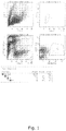
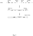
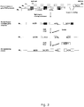


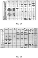

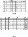

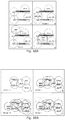


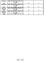


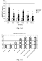

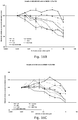

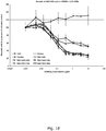

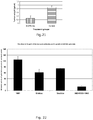

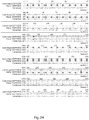


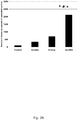
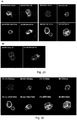
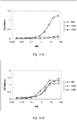
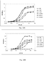

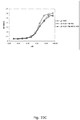


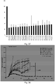


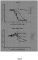

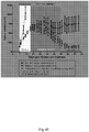
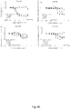
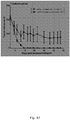
Classifications
-
- A—HUMAN NECESSITIES
- A61—MEDICAL OR VETERINARY SCIENCE; HYGIENE
- A61K—PREPARATIONS FOR MEDICAL, DENTAL OR TOILETRY PURPOSES
- A61K39/00—Medicinal preparations containing antigens or antibodies
- A61K39/395—Antibodies; Immunoglobulins; Immune serum, e.g. antilymphocytic serum
-
- C—CHEMISTRY; METALLURGY
- C07—ORGANIC CHEMISTRY
- C07K—PEPTIDES
- C07K16/00—Immunoglobulins [IGs], e.g. monoclonal or polyclonal antibodies
- C07K16/18—Immunoglobulins [IGs], e.g. monoclonal or polyclonal antibodies against material from animals or humans
- C07K16/28—Immunoglobulins [IGs], e.g. monoclonal or polyclonal antibodies against material from animals or humans against receptors, cell surface antigens or cell surface determinants
- C07K16/2863—Immunoglobulins [IGs], e.g. monoclonal or polyclonal antibodies against material from animals or humans against receptors, cell surface antigens or cell surface determinants against receptors for growth factors, growth regulators
-
- A—HUMAN NECESSITIES
- A61—MEDICAL OR VETERINARY SCIENCE; HYGIENE
- A61P—SPECIFIC THERAPEUTIC ACTIVITY OF CHEMICAL COMPOUNDS OR MEDICINAL PREPARATIONS
- A61P35/00—Antineoplastic agents
-
- C—CHEMISTRY; METALLURGY
- C07—ORGANIC CHEMISTRY
- C07K—PEPTIDES
- C07K14/00—Peptides having more than 20 amino acids; Gastrins; Somatostatins; Melanotropins; Derivatives thereof
-
- C—CHEMISTRY; METALLURGY
- C07—ORGANIC CHEMISTRY
- C07K—PEPTIDES
- C07K14/00—Peptides having more than 20 amino acids; Gastrins; Somatostatins; Melanotropins; Derivatives thereof
- C07K14/435—Peptides having more than 20 amino acids; Gastrins; Somatostatins; Melanotropins; Derivatives thereof from animals; from humans
- C07K14/46—Peptides having more than 20 amino acids; Gastrins; Somatostatins; Melanotropins; Derivatives thereof from animals; from humans from vertebrates
- C07K14/47—Peptides having more than 20 amino acids; Gastrins; Somatostatins; Melanotropins; Derivatives thereof from animals; from humans from vertebrates from mammals
-
- C—CHEMISTRY; METALLURGY
- C07—ORGANIC CHEMISTRY
- C07K—PEPTIDES
- C07K14/00—Peptides having more than 20 amino acids; Gastrins; Somatostatins; Melanotropins; Derivatives thereof
- C07K14/435—Peptides having more than 20 amino acids; Gastrins; Somatostatins; Melanotropins; Derivatives thereof from animals; from humans
- C07K14/705—Receptors; Cell surface antigens; Cell surface determinants
- C07K14/71—Receptors; Cell surface antigens; Cell surface determinants for growth factors; for growth regulators
-
- C—CHEMISTRY; METALLURGY
- C07—ORGANIC CHEMISTRY
- C07K—PEPTIDES
- C07K16/00—Immunoglobulins [IGs], e.g. monoclonal or polyclonal antibodies
- C07K16/18—Immunoglobulins [IGs], e.g. monoclonal or polyclonal antibodies against material from animals or humans
- C07K16/28—Immunoglobulins [IGs], e.g. monoclonal or polyclonal antibodies against material from animals or humans against receptors, cell surface antigens or cell surface determinants
-
- A—HUMAN NECESSITIES
- A61—MEDICAL OR VETERINARY SCIENCE; HYGIENE
- A61K—PREPARATIONS FOR MEDICAL, DENTAL OR TOILETRY PURPOSES
- A61K39/00—Medicinal preparations containing antigens or antibodies
- A61K2039/505—Medicinal preparations containing antigens or antibodies comprising antibodies
- A61K2039/507—Comprising a combination of two or more separate antibodies
-
- C—CHEMISTRY; METALLURGY
- C07—ORGANIC CHEMISTRY
- C07K—PEPTIDES
- C07K2317/00—Immunoglobulins specific features
- C07K2317/20—Immunoglobulins specific features characterized by taxonomic origin
- C07K2317/24—Immunoglobulins specific features characterized by taxonomic origin containing regions, domains or residues from different species, e.g. chimeric, humanized or veneered
-
- C—CHEMISTRY; METALLURGY
- C07—ORGANIC CHEMISTRY
- C07K—PEPTIDES
- C07K2317/00—Immunoglobulins specific features
- C07K2317/50—Immunoglobulins specific features characterized by immunoglobulin fragments
- C07K2317/52—Constant or Fc region; Isotype
-
- C—CHEMISTRY; METALLURGY
- C07—ORGANIC CHEMISTRY
- C07K—PEPTIDES
- C07K2317/00—Immunoglobulins specific features
- C07K2317/50—Immunoglobulins specific features characterized by immunoglobulin fragments
- C07K2317/55—Fab or Fab'
-
- C—CHEMISTRY; METALLURGY
- C07—ORGANIC CHEMISTRY
- C07K—PEPTIDES
- C07K2317/00—Immunoglobulins specific features
- C07K2317/50—Immunoglobulins specific features characterized by immunoglobulin fragments
- C07K2317/56—Immunoglobulins specific features characterized by immunoglobulin fragments variable (Fv) region, i.e. VH and/or VL
- C07K2317/569—Single domain, e.g. dAb, sdAb, VHH, VNAR or nanobody®
-
- C—CHEMISTRY; METALLURGY
- C07—ORGANIC CHEMISTRY
- C07K—PEPTIDES
- C07K2317/00—Immunoglobulins specific features
- C07K2317/70—Immunoglobulins specific features characterized by effect upon binding to a cell or to an antigen
- C07K2317/73—Inducing cell death, e.g. apoptosis, necrosis or inhibition of cell proliferation
-
- C—CHEMISTRY; METALLURGY
- C07—ORGANIC CHEMISTRY
- C07K—PEPTIDES
- C07K2317/00—Immunoglobulins specific features
- C07K2317/70—Immunoglobulins specific features characterized by effect upon binding to a cell or to an antigen
- C07K2317/76—Antagonist effect on antigen, e.g. neutralization or inhibition of binding
-
- C—CHEMISTRY; METALLURGY
- C07—ORGANIC CHEMISTRY
- C07K—PEPTIDES
- C07K2317/00—Immunoglobulins specific features
- C07K2317/70—Immunoglobulins specific features characterized by effect upon binding to a cell or to an antigen
- C07K2317/77—Internalization into the cell
-
- C—CHEMISTRY; METALLURGY
- C07—ORGANIC CHEMISTRY
- C07K—PEPTIDES
- C07K2317/00—Immunoglobulins specific features
- C07K2317/90—Immunoglobulins specific features characterized by (pharmaco)kinetic aspects or by stability of the immunoglobulin
- C07K2317/92—Affinity (KD), association rate (Ka), dissociation rate (Kd) or EC50 value
Definitions
- the invention relates to the field of recombinant antibodies for use in human cancer therapy.
- EGFR Epidermal Growth Factor Receptor
- EGFR expression is frequently accompanied by the production of EGFR-ligands, TGF-alpha and EGF among others, by EGFR-expressing tumour cells which suggests that an autocrine loop participates in the progression of these cells ( Baselga, etal.(1994) Pharmac. Therapeut. 64: 127-154 ; Modjtahedi, et al. (1994) Int. J. Oncology. 4: 277-296 ). Blocking the interaction between such EGFR ligands and EGFR therefore can inhibit tumor growth and survival ( Baselga, et al. (1994) Pharmac. Therapeut. 64: 127-154 ).
- the EGFR is a membrane bound glycoprotein with a molecular weight of approximately 170 kDa.
- EGFR consists of a glycosylated external ligand-binding domain (621 residues) and a cytoplasmic domain (542 residues) connected by a short 23 amino acid transmembrane linker.
- the extracellular part of EGFR contains 25 disulfide bonds and 12 N-linked glycosylation sites, and is generally considered to consist of four sub-domains.
- X-ray crystal structures of the EGFR suggest that the receptor adopts both an autoinhibited tethered - conformation that cannot bind EGF ( Ferguson et al, Mol Cell, 2003, vol 11: 507-517 ) and an active conformation that may mediate EGF ligand binding and receptor dimerisation ( Garret et al, Cell 2002, vol 110:763-773 ; Ogiso et al, Cell, 2002, vol 110:775-787 ).
- domain I and domain III have been suggested to provide additive contributions for formation of a high-affinity ligand binding site.
- Domains II and IV are cysteine-rich laminin-like regions that stabilise protein folding and contain a possible EGFR dimerisation interface.
- EGFR is known to exist in a number of different conformations on the cell surface, where the tethered or locked confirmation is the most frequent.
- the tethered conformation cannot dimerise and hence is inactive.
- the therapeutic antibody Erbitux is known to stabilise the tethered conformation by binding to domain III and sterically hampering the receptor in reaching the untethered state.
- some receptors may still be able to adopt the untethered conformation, bind ligand and dimerise.
- a monoclonal antibody (mAb) will typically only be effective in binding against one of the conformations and therefore cannot effectively target cancer cells exhibiting other conformations or cancer cells exhibiting a variety of conformations.
- Monoclonal antibodies (mAbs) directed to the ligand-binding domain of EGFR can block the interaction with EGFR ligands and, concomitantly, the resultant intracellular signaling pathway.
- ErbituxTM (Cetuximab) is a recombinant, human/mouse chimeric monoclonal antibody that binds specifically to the extracellular domain of the human (EGFR).
- Erbitux is composed of the Fv regions of a murine anti-EGFR antibody with human IgG1 heavy and kappa light chain constant regions and has an approximate molecular weight of 152 kDa.
- Erbitux is produced in mammalian cell culture (murine myeloma). Erbitux is approved for the treatment of patients with metastatic colorectal cancer and whose tumor expresses EGFR.
- Erbitux is used in combination with radiation therapy to treat patients with squamous cell cancer of the head and neck that cannot be removed by surgery or as second line treatment of squamous cell cancer of the head and neck that have failed standard platinum-based therapy.
- VectibixTM is a recombinant, human IgG2 kappa monoclonal antibody that binds specifically to the human EGFR.
- Vectibix has an approximate molecular weight of 147 kDa.
- Panitumumab is produced in genetically engineered mammalian cells (Chinese Hamster Ovary).
- Vectibix is approved for the treatment of patients with metastatic colorectal cancer and whose tumor expresses EGFR with disease progression on or following fluoropyrimidine-, oxaliplatin-, and irinotecan-containing chemotherapy regimens.
- Cetuximab marketed by Imclone under the trademane Erbitux, is described in US 4,943,533 and WO 96/40210 .
- Panitumumab marketed by Abgenix under the tradename Vectibix, is described in US 6,235,883 .
- Zalutuzumab (Humax-EGFR) is another anti-EGFR antibody currently undergoing clinical development. The antibody has been developed by Genmab and is described in WO 02/100348 and WO 2004/056847 .
- Cetuximab, Panitumumab, and Zalutumumab bind the same epitope on EGFR.
- Nimotuzumab (TheraCIM hR3) described in US 5,891,996 and US 6,506,883 is approved for treatment of cancer in a number of countries around the world but not in Europe or the US.
- tumours being resistant to any of the other monoclonal anti-EGFR antibodies that are currently in clinical testing: Nimotuzumab (YM Biosciences, Cuba), Matuzumab (Merck KGaA), mAb806 (Ludwig Institute), and ICR62 (Institute of Cancer Research).
- Complete or partial tumour resistance to any of these monoclonal antibodies may be assayed using a sample isolated from a patient, so that it can be known a priori whether the tumour is resistant or not.
- tumour recurrence or progression following surgery Apart from resistance to monoclonal antibody therapy (or refractory tumours) another problem in treating the EGFR expressing cancers is tumour recurrence or progression following surgery, radiation therapy and/or medical treatment with chemotherapeutics, tyrosine Kinase inhibitors (TKIs) and/or monoclonal antibodies.
- chemotherapeutics e.g., a chemotherapeutics, tyrosine Kinase inhibitors (TKIs) and/or monoclonal antibodies.
- TKIs tyrosine Kinase inhibitors
- monoclonal antibodies tyrosine Kinase inhibitors
- the present invention relates to an antibody composition for use in treating cancer in a subject that has been subjected to a prior treatment regimen involving an anti-human EGFR antibody, said antibody composition comprising a first anti-human EGFR antibody molecule and a second anti-human EGFR antibody molecule distinct from the first molecule,
- the invention provides an antibody composition for use in treating cancer in a subject that has been subjected to a prior treatment regimen using an anti-human EGFR antibody, said antibody composition comprising a first anti-human EGFR antibody molecule and a second anti-human EGFR antibody molecule distinct from the first molecule,
- the present invention also relates to an antibody composition for use in treating cancer in a subject that has been subjected to a prior treatment regimen using an anti-human EGFR antibody, said antibody composition comprising a first anti-human EGFR antibody molecule and a second anti-human EGFR antibody molecule distinct from the first molecule,
- an antibody composition for use in treating cancer in a subject, wherein said cancer is resistant or partially resistant to treatment with at least one other anti-EGFR antibody selected from the group consisting of cetuximab, panitumumab, zalutumumab, nimotuzumab, ICR62, mAb806, matuzumab, and antibodies capable of binding the same epitope as any of these; said antibody composition comprising a first anti-human EGFR antibody molecule and a second anti-human EGFR antibody molecule distinct from the first molecule,
- the invention also relates to a bi-specific binding molecule for use in treating cancer in a subject that has been subjected to a prior treatment regimen using an anti-human EGFR antibody, wherein the bi-specific binding molecule comprises a first antigen-binding domain comprising the light chain CDR1, CDR2, and CDR3 in SEQ ID NO: 72 and the heavy chain CDR1, CDR2, and CDR3 in SEQ ID NO: 40; and a second antigen-binding domain comprising the light chain CDR1, CDR2, and CDR3 in SEQ ID NO: 73 and the heavy chain CDR1, CDR2, and CDR3 in SEQ ID NO: 41.
- the bi-specific binding molecule comprises a first antigen-binding domain comprising the light chain CDR1, CDR2, and CDR3 in SEQ ID NO: 72 and the heavy chain CDR1, CDR2, and CDR3 in SEQ ID NO: 40; and a second antigen-binding domain comprising the light chain CDR1, CDR2, and CDR3 in SEQ
- the present inventors have discovered that a cancer cell line being resistant to Erbitux (Cetixumab) can be treated effectively in vitro with an antibody composition of the present invention, whereas exposure of the resistant cell line to Vetibix (Panitumumab) is as ineffective as is treatment with Erbitux. It is expected that Zalutumumab will also be ineffective against this cell line.
- an antibody composition of the present invention is effective against Erbitux, Vectibix and Zalutumumab resistant tumours.
- an antibody composition of the present invention can be used to treat patients that do not respond to either of these products.
- an antibody composition of the present invention can be used to treat tumours that are from the beginning resistant to either of these monoclonal antibodies.
- Resistance to a monoclonal antibody such as Erbitux can be assayed in vitro using methods described in Example 21.
- a cancer cell line proliferates in medium containing 10 ⁇ g/mL of Erbitux, it is considered partially resistant to Erbitux.
- Resistance to Panitumumab and Zalutumumab can be assayed in the same way.
- an antibody composition of the present invention for the treatment of cancer that is resistant or partially resistant to any of the other anti-EGFR antibodies that are currently under development, including but not limited to Cetuximab, panitumumab, Zalutumumab, nimotuzumab, ICR62, mAb806, Matuzumab, and antibodies capable of binding the same epitope as any of these.
- Example 23 The results have been confirmed by an in vivo study (Example 23), where an aggressive cancer cell line has been implanted into mice. Following initial treatment with Erbitux, partial responders were selected and exposed either to prolonged treatment with Erbitux or to treatment with an antibody composition of the present invention. The latter resulted in rapid reduction in the size of tumours, whereas continued Erbitux treatment resulted in maintained tumour size. The preclinical efficacy is achieved even though there is a partial overlap in binding between Erbitux and antibodies 1024 and 992 of the present invention.
- an antibody composition of the invention can be used to treat cancer that is resistant or partially resistant to a monoclonal anti-EGFR antibody such as Erbitux, and to treat cancer in a subject that has received treatment with a monoclonal anti-EGFR antibody such as Erbitux in a previous treatment regimen.
- a monoclonal anti-EGFR antibody such as Erbitux
- an antibody composition of the present invention in the treatment of cancer that has previously been treated with another monoclonal anti-EGFR antibody including but not limited to Cetuximab, panitumumab, Zalutumumab, nimotuzumab, ICR62, mAb806, Matuzumab, and antibodies capable of binding the same epitope as any of these.
- Another monoclonal anti-EGFR antibody including but not limited to Cetuximab, panitumumab, Zalutumumab, nimotuzumab, ICR62, mAb806, Matuzumab, and antibodies capable of binding the same epitope as any of these.
- the efficacy of such treatment can be verified in a pre-clinical study similar to the study described in Example 23.
- the present inventors have determined that recurring tumour growth following treatment with an antibody composition of the present invention can be successfully eliminated using an antibody composition of the present invention. This has been demonstrated in the preclinical study described in Example 22. Recurring tumours were eliminated as efficiently as the originally implanted tumours clearly indicating that these tumours were not resistant to treatment with an antibody composition of the invention.
- the invention relates to an antibody composition for use in the treatment of cancer in a subject that has been subjected to a prior treatment regimen involving an anti human EGFR antibody, said antibody composition comprising at least 2 distinct anti-human EGFR antibody molecules,
- said prior treatment regimen involved an antibody composition identical to said antibody composition.
- said prior treatment regimen involved an anti-human-EGFR antibody selected from the group consisting of Cetuximab, panitumumab, Zalutumumab, nimotuzumab, ICR62, mAb806, Matuzumab, and antibodies capable of binding the same epitope as any of these.
- said anti-human EGFR antibody is selected from the group consisting of Cetuximab, Panitumumab, and Zalutumumab and antibodies capable of binding the same epitope as any of these.
- said anti-human EGFR antibody is selected from the group consisting of Cetuximab and Panitumumab and antibodies capable of binding the same epitope as any of these.
- said anti-human EGFR antibody is Cetuximab or an antibody capable of biding the same epitope as Cetuximab.
- the cancer may be selected from the group consisting of head-and-neck cancer, colon cancer, breast cancer, renal cancer, lung cancer, ovarian cancer, prostate cancer, glioma, pancreatic cancer, bladder cancer, non-small-cell-lung-carcinoma (NSCLC), gastric cancer, cervical cancer, hepatocellular cancer, gastrophageal cancer, colorectal cancer, rectal cancer, epithelioid carcinoma, RCC, squamous cell carcinoma of the head and neck (SCCHN), esophageal cancer, glioblastoma multiforme, squamous cell carcinoma, and kidney cancer, melanoma, carcinoma and sarcoma as described herein.
- NSCLC non-small-cell-lung-carcinoma
- gastric cancer cervical cancer
- hepatocellular cancer gastrophageal cancer
- colorectal cancer colorectal cancer
- rectal cancer epithelioid carcinoma
- RCC squamous cell carcinoma of the head and neck
- the antibody treatment may be adjuvant therapy, following surgery and/or radiation therapy.
- the treatment may be a combination therapy involving treatment with chemotherapy and/or at least one tyrosine kinase inhibitors and/or at least one angiogenesis inhibitor and/or at least one hormone and/or at least one differentiation inducing agent.
- the prior treatment regimen may be a first-line therapy, a second-line therapy, or a third-line therapy.
- the first-line therapy may additionally involve a treatment regimen with chemotherapy and/or at least one tyrosine kinase inhibitors and/or at least one angiogenesis inhibitor and/or at least one hormone and/or at least one differentiation inducing agent.
- Chemotherapy preferably includes administration of a chemotherapeutic compound selected from the group consisting of adriamycin, cisplatin, taxol, doxorubicin, topotecan, fluoropyrimidine, oxaliplatin, and irinotecan.
- a chemotherapeutic compound selected from the group consisting of adriamycin, cisplatin, taxol, doxorubicin, topotecan, fluoropyrimidine, oxaliplatin, and irinotecan.
- the subject has progressed on or following the prior treatment regimen. In other embodiments, the subject has progressed following said prior treatment regimen.
- the cancer may be resistant or partially resistant to the prior treatment regimen.
- the invention relates to an antibody composition for use in the treatment of cancer, wherein said cancer is resistant or partially resistant to treatment with at least one other anti-EGFR antibody selected from the group consisting of Cetuximab, panitumumab, Zalutumumab, nimotuzumab, ICR62, mAb806, Matuzumab, and antibodies capable of binding the same epitope as any of these; said antibody composition comprising at least 2 distinct anti-human EGFR antibody molecules,
- the composition may be used for first-line therapy.
- the composition is used for second-line therapy following a treatment regimen involving chemotherapy and/or at least one tyrosine kinase inhibitors and/or at least one angiogenesis inhibitor and/or at least one hormone and/or at least one differentiation inducing agent.
- the composition may also be used for third-line therapy.
- composition may be used for combination therapy together with chemotherapy and/or at least one tyrosine kinase inhibitors and/or at least one angiogenesis inhibitor and/or at least one hormone and/or at least one differentiation inducing agent.
- the composition is used for as adjuvant therapy following surgery and/or radiation therapy.
- the complete or partial resistance is preferably determined by assaying a sample of cancer cells isolated from said subject.
- This assay may include measuring binding of Cetuximab, panitumumab, Zalutumumab, nimotuzumab, ICR62, mAb806, Matuzumab, and antibodies capable of binding the same epitope as any of these to cancer cells from said subject. The absence of binding indicates resistance to the antibody.
- partial or complete resistance can be determined in a proliferation assay similar to the assay in Example 21.
- the antibody composition can be any of the compositions described herein.
- the antibody composition of the invention is as described in the section headed A preferred antibody composition, i.e. an antibody composition based on antibodies 1024 and 992 as described herein.
- antibody describes a functional component of serum and is often referred to either as a collection of molecules (antibodies or immunoglobulin) or as one molecule (the antibody molecule or immunoglobulin molecule).
- An antibody molecule is capable of binding to or reacting with a specific antigenic determinant (the antigen or the antigenic epitope), which in turn may lead to induction of immunological effector mechanisms.
- An individual antibody molecule is usually regarded as monospecific, and a composition of antibody molecules may be monoclonal (i.e., consisting of identical antibody molecules) or polyclonal (i.e., consisting of two or more different antibody molecules reacting with the same or different epitopes on the same antigen or even on distinct, different antigens).
- Each antibody molecule has a unique structure that enables it to bind specifically to its corresponding antigen, and all natural antibody molecules have the same overall basic structure of two identical light chains and two identical heavy chains.
- Antibodies are also known collectively as immunoglobulins.
- the terms antibody or antibodies as used herein are also intended to include chimeric and single chain antibodies, as well as binding fragments of antibodies, such as Fab, Fv fragments or scFv fragments, as well as multimeric forms such as dimeric IgA molecules or pentavalent IgM.
- An antibody may be human, murine, chimeric, humanised, or reshaped.
- cognate V H and V L coding pair describes an original pair of V H and V L coding sequences contained within or derived from the same antibody producing cell.
- a cognate V H and V L pair represents the V H and V L pairing originally present in the donor from which such a cell is derived.
- an antibody expressed from a V H and V L coding pair indicates that an antibody or an antibody fragment is produced from a vector, plasmid or similar containing the V H and V L coding sequence.
- a library of cognate pairs is also termed a repertoire or collection of cognate pairs, and may be kept individually or pooled.
- CDR complementarity determining region
- a distinct member of a recombinant polyclonal protein denotes one protein molecule of a protein composition comprising different, but homologous protein molecules, where each protein molecule is homologous to the other molecules of the composition, but also contains one or more stretches of variable polypeptide sequence, which is/are characterized by differences in the amino acid sequence between the individual members of the polyclonal protein.
- head-to-head promoters refers to a promoter pair being placed in close proximity so that transcription of two gene fragments driven by the promoters occurs in opposite directions.
- a head-to-head promoter can also be constructed with a stuffer composed of irrelevant nucleic acids between the two promoters. Such a stuffer fragment can easily contain more than 500 nucleotides.
- Head-to-head promoters can also be termed bi-directional promoters.
- immunoglobulin commonly is used as a collective designation of the mixture of antibodies found in blood or serum, but may also be used to designate a mixture of antibodies derived from other sources.
- immunoglobulin molecule denotes an individual antibody molecule, e.g., as being a part of immunoglobulin, or part of any polyclonal or monoclonal antibody composition.
- a library of variant nucleic acid molecules of interest is used to describe the collection of nucleic acid molecules, which collectively encode a "recombinant polyclonal protein of interest".
- the library of variant nucleic acid molecules of interest is contained in a library of expression vectors.
- Such a library typically have at least 2, 3, 5, 10, 20, 50, 1000, 10 4 , 10 5 or 10 6 distinct members.
- the term "mass transfer” is used to describe the transfer of nucleic acid sequences of interest from one population of vectors to another population of vectors and doing so for each DNA simultaneously without resorting to isolation of the individual DNA's of interest.
- populations of vectors can be libraries containing for example variable regions, promoters, leaders or enhancing elements of interest. These sequences can then be moved without prior isolation from for example a phage vector to a mammalian expression vector.
- this technique ensures that the linkage between V H and V L diversity is not lost while moving libraries from, for example, a selection vector (e.g., a phage display vector) to a mammalian expression vector.
- a selection vector e.g., a phage display vector
- operably linked refers to a segment being linked to another segment when placed into a functional relationship with the other segment.
- DNA encoding a signal sequence is operably linked to DNA encoding a polypeptide if it is expressed as a leader that participates in the transfer of the polypeptide to the endoplasmic reticulum.
- a promoter or enhancer is operably linked to a coding sequence if it stimulates the transcription of the sequence.
- polyclonal antibody describes a composition of different antibody molecules which is capable of binding to or reacting with several different specific antigenic determinants on the same or on different antigens. Usually, the variability of a polyclonal antibody is thought to be located in the so-called variable regions of the polyclonal antibody.
- polyclonality can also be understood to describe differences between the individual antibody molecules residing in so-called constant regions, e.g., as in the case of mixtures of antibodies containing two or more antibody isotypes such as the human isotypes IgG1, IgG2, IgG3, IgG4, IgA1, and IgA2, or the murine isotypes IgG1, IgG2a, IgG2b, IgG3, and IgA.
- an antibody composition may also be termed "an antibody composition”.
- epitope is commonly used to describe a proportion of a larger molecule or a part of a larger molecule (e.g. antigen or antigenic site) having antigenic or immunogenic activity in an animal, preferably a mammal, and most preferably in a human.
- An epitope having immunogenic activity is a portion of a larger molecule that elicits an antibody response in an animal.
- An epitope having antigenic activity is a portion of a larger molecule to which an antibody immunospecifically binds as determined by any method well known in the art, for example, by the immunoassays described herein.
- Antigenic epitopes need not necessarily be immunogenic.
- An antigen is a substance to which an antibody or antibody fragment immunospecifically binds, e.g. toxin, virus, bacteria, proteins or DNA.
- An antigen or antigenic site often has more than one epitope, unless they are very small, and is often capable of stimulating an immune response.
- Epitopes may be linear or conformational.
- a linear epitope consists of about 6 to 10 adjacent amino acids on a protein molecule that is recognized by an antibody.
- conformational epitope consists of amino acids that are not arranged sequentially.
- the antibody recognizes only the 3-dimensional structure. When a protein molecule folds into a three dimensional structure the amino acids forming the epitope are juxtaposed enabling the antibody to recognize the sequence.
- a conformational epitope In a denatured protein only the linear epitope may be recognized.
- a conformational epitope by definition, must be on the outside of the folded protein.
- An antibody that recognizes the conformational epitope may only bind under mild, non-denaturing procedures.
- Antibodies binding to different epitopes on the same antigen can have varying effects on the activity of the antigen they bind depending on the location of the epitope.
- An antibody binding to an epitope in an active site of the antigen may block the function of the antigen completely, whereas another antibody binding at a different epitope may have no or little effect on the activity of the antigen alone.
- the epitope is preferably a proportion of the extracellular domain of EGFR.
- Antigens of the present invention are preferably extracellular domain EGFR proteins, polypeptides or fragments thereof to which an antibody or antibody fragment immunospecifically binds.
- An EGFR associated antigen may also be an analog or derivative of the extracellular domain of EGFR polypeptide or fragment thereof to which an antibody or antibody fragment immunospecifically binds.
- Antibodies capable of competing with each other for binding to the same antigen may bind the same or overlapping epitopes or may have a binding site in the close vicinity of one another, so that competition is mainly caused by steric hindrance. Methods for determining competition between antibodies are described in the examples.
- polyclonal protein refers to a protein composition comprising different, but homologous protein molecules, preferably selected from the immunoglobulin superfamily. Thus, each protein molecule is homologous to the other molecules of the composition, but also contains one or more stretches of variable polypeptide sequence, which is/are characterized by differences in the amino acid sequence between the individual members of the polyclonal protein.
- polyclonal proteins include antibody or immunoglobulin molecules, T cell receptors and B cell receptors.
- a polyclonal protein may consist of a defined subset of protein molecules, which has been defined by a common feature such as the shared binding activity towards a desired target, e.g., in the case of a polyclonal antibody against the desired target antigen.
- protein or “polypeptide” is meant any chain of amino acids, regardless of length or post-translational modification. Proteins can exist as monomers or multimers, comprising two or more assembled polypeptide chains, fragments of proteins, polypeptides, oligopeptides, or peptides.
- RFLP refers to "restriction fragment length polymorphism", a method whereby the migratory gel pattern of nucleic acid molecule fragments are analyzed after cleavage with restriction enzymes.
- the term "scrambling” describes situations where two or more distinct members of a polyclonal protein comprised of two different polypeptide chains, e.g. from the immunoglobulin superfamily, are expressed from an individual cell. This situation may arise when the individual cell has integrated, into the genome, more than one pair of gene segments, where each pair of gene segments encode a distinct member of the polyclonal protein. In such situations unintended combinations of the polypeptide chains expressed from the gene segments can be made. These unintended combinations of polypeptide chains might not have any therapeutic effect.
- V H -V L chain scrambling is an example of the scrambling defined above.
- the V H and V L encoding gene segments constitute a pair of gene segments.
- the scrambling occurs when unintended combinations of V H and V L polypeptides are produced from a cell where two different V H and V L encoding gene segment pairs are integrated into the same cell. Such a scrambled antibody molecule is not likely to retain the original specificity, and thus might not have any therapeutic effect.
- transfection is herein used as a broad term for introducing foreign DNA into a cell.
- the term is also meant to cover other functional equivalent methods for introducing foreign DNA into a cell, such as e.g., transformation, infection, transduction or fusion of a donor cell and an acceptor cell.
- variable polypeptide sequence and “variable region” are used interchangeably.
- distinct epitopes means that when two different antibodies bind distinct epitopes, there is less than 100% competition for antigen binding, preferably less than 50% competition for antigen binding, more preferably essentially no competition for antigen binding.
- An analysis for "distinct epitopes" of antibody pairs is typically determined by binding experiments under saturating antibody conditions with either FACS analysis on cells expressing EGFR and individually fluorescent labelled antibodies, or Surface Plasmon Resonance using EGFR antigen captured or conjugated to a flow cell surface as described in the examples.
- the term being capable of "inhibiting EGF binding" when applied to one antibody molecule means that the antibody molecule exhibits an IC 50 value with respect to EGF binding to EGFR of less than 10 nM, preferably less than 8 nM, more preferably less than 7 nM, more preferably less than 5 nM, more preferably less than 4 nM, more preferably less than 3 nM, more preferably less than 2 nM, more preferably less than 2 nM, more preferably less than 1 nM.
- EGFR epidermal growth factor receptor
- EGFR antigen epidermal growth factor receptor
- binding of an antibody of the invention to the EGFR-antigen inhibits the growth of cells expressing EGFR (e. g., a tumor cell) by inhibiting or blocking binding of EGFR ligand to EGFR.
- EGFR ligand encompasses all (e.
- physiological ligands for EGFR including but nor limited to EGF, TGF-alpha, heparin binding EGF (HB-EGF), amphiregulin (AR), heregulin, beta-cellulin, and epiregulin (EPI).
- EGF epidermal growth factor
- TGF-alpha heparin binding EGF
- AR amphiregulin
- AS amphiregulin
- beta-cellulin beta-cellulin
- EPI epiregulin
- binding of an antibody of the invention to the EGFR-antigen mediates effector cell phagocytosis and/or killing of cells expressing EGFR.
- EGFR domain structure The extracellular part of the mature EGFR (SwissProt acc.#P00533) consists of 621 amino acids and four receptor domains: Domain I encompasses residues 1-165, domain II residues 166-312, domain III residues 313-481 and domain IV 482-621 ( Cochran et al. 2004 J immunol. Methods 287, 147-158 ). Domains I and III have been suggested to contribute to the formation of high affinity binding sites for ligands. Domains II and IV are cysteine rich, laminin-like regions that stabilize protein folding and contain a possible EGFR dimerization interface.
- the term "inhibits growth" is intended to include any measurable decrease in the proliferation (increase in number of cells) or metabolism of a cell when contacted with an anti-EGFR antibody as compared to the growth of the same cells not in contact with an anti-EGFR antibody, e. g, the inhibition of growth of a cell culture by at least about 10%, 20%, 30%, 40%, 50%, 60%, 70%, 80%, 90%, 99%, or 100%.
- inhibits binding and “blocks binding” (e. g., referring to inhibition/blocking of binding of EGFR ligand to EGFR) are used interchangeably and encompass both partial and complete inhibition/blocking.
- the inhibition/blocking of EGFR ligand to EGFR preferably reduces or alters the normal level or type of cell signaling that occurs when EGFR ligand binds to EGFR without inhibition or blocking.
- Inhibition and blocking are also intended to include any measurable decrease in the binding affinity of EGFR ligand to EGFR when in contact with an anti-EGFR antibody as compared to the ligand not in contact with an anti-EGFR antibody, e. g., the blocking of EGFR ligands to EGFR by at least about 10%, 20%, 30%, 40%,50%, 60%, 70%, 80%, 90%, 99%, or 100%.
- recombinant antibody is used to describe an antibody molecule or several molecules that is/are expressed from a cell or cell line transfected with an expression vector comprising the coding sequence of the antibody which is not naturally associated with the cell.
- Cancer - Cancer (medical term: malignant neoplasm) is a class of diseases in which a group of cells display uncontrolled growth (division beyond the normal limits), invasion (intrusion on and destruction of adjacent tissues), and sometimes metastasis (spread to other locations in the body via lymph or blood). These three malignant properties of cancers differentiate them from benign tumors, which are self-limited, do not invade or metastasize. Most cancers form a tumor but some, like leukemia, do not. Cancer may also be termed neoplastic growth, and hyperproliferative disorders.
- Resistance or partial resistance to an anti-EGFR antibody selected from the group consisting of Cetuximab, panitumumab, Zalutumumab, nimotuzumab, ICR62, mAb425, Matuzumab, and antibodies capable of binding the same epitope as any of these can be observed in a patient receiving one or more of the antibodies, or be measured in an in vitro assay, e.g. by determining expression of EGFR and non-binding or low binding of the monoclonal antibody, or in a proliferation assay such as described in Example 21.
- the invention relates to an antibody composition for use in the treatment of cancer in a subject that has been subjected to prior treatment regimen involving an anti human EGFR antibody or wherein said cancer is resistant or partially resistant to treatment with at least one other anti-EGFR antibody, said antibody composition comprising at least 2 distinct anti-human EGFR antibody molecules.
- said at least 2 distinct anti-human EGFR antibodies binds to non-overlapping epitopes.
- the non-overlapping nature of the antibodies is preferably determined using differently labelled antibodies in a FACS analysis with EGFR expressing cells or by using Surface Plasmon Resonance using EGFR antigen captured or conjugated to a flow cell surface.
- ELISA based methods as described in the examples may also be used.
- a composition binding two or more non-overlapping EGFR epitopes can be used against a wider range of EGFR dependent cancer types as it may be less vulnerable to differences in EGFR conformation and less vulnerable to mutations compared to composition of monoclonal antibodies targeting one or two epitopes.
- the antibody composition binding two or more non-overlapping EGFR epitopes may provide superior efficacy compared to composition targeting only one epitope.
- the antibody composition may provide superior efficacy with respect to terminal differentiation of cancer cells in vivo. For a monoclonal anti-EGFR antibody therapy a certain proportion of patients will not respond effectively to the antibody treatment. For some of the patients, this may be due to rapid clearing of the antibody or because the antibody generates an immune response in the patient against the antibody.
- the lack of response may be because their particular EGFR dependent cancer expresses EGFR in a conformation where the monoclonal antibody cannot bind its epitope. This could be because of differences in glycosylation, because of domain deletion, or because of mutations and/or SNP(s).
- the autocrine EGFR-stimulation caused by the cancer cells' production of ligand is of importance, while in other cases the EGFR expressed by the cancer cells does not need ligand stimulation.
- an antibody capable of inhibiting ligand binding may not be effective.
- An antibody composition wherein the antibodies are capable of binding at least two distinct epitopes on EGFR will be more broadly applicable, since the likelihood that both epitopes are changed compared to the epitope(s) recognised by the antibodies is diminished. Furthermore, the likelihood that all antibodies are either cleared by the patient is much smaller. Superiority has been shown most clearly in terms of induction of terminal differentiation of the cancer cells using two Domain III antibodies with non-overlapping epitopes. Such efficient antibody-induced terminal differentiation of cancer cells has not been reported before and represents a significant step forward in designing efficient antibody-based cancer therapies. Later results have shown that similar or even superior results can be obtained with a particular combination of two antibodies.
- the composition may comprise antibodies capable of binding three non-overlapping epitopes.
- the composition may comprise antibodies capable of binding four non-overlapping epitopes.
- the composition may comprise antibodies capable of binding five non-overlapping epitopes.
- the composition may comprise antibodies capable of binding six non-overlapping epitopes.
- the examples of the present application show that at least six distinct antibodies can bind to EGFR at one time (Example 3). This does not exclude that it is possible or even advantageous to design a composition comprising antibodies capable of binding more than six, such as seven or eight non-overlapping epitopes by carefully selecting antibodies.
- the antibodies of the composition may be chimeric antibodies with non-human variable chains and human constant chains.
- the non-human variable chains may be from mouse, rat, sheep, pig, chicken, non-human primate or other suitable animal.
- the antibodies can be generated in a transgenic animal with human antibody genes.
- the antibodies may also be so-called humanised antibodies, where the non-human CDR sequences have been grafted into human framework sequences.
- the human constant chain is IgG1 or IgG2 isotype. More preferably all antibodies in the composition have the same isotype for ease of manufacturing. However, it may be advantageous to include in the composition antibodies of different isotype.
- the antibody compositions of the invention comprise antibodies capable of binding to EGFR selected from the group consisting of human EGFR, mutated human EGFR, and deletion variants of human EGFR.
- the antibodies are capable of binding both human and non-human primate EGFR, so that they can be tested in relevant toxicology studies prior to clinical experiments.
- the non-human primate is cynomolgous monkey ( Macaca fascicularis ) .
- Antibody no is a reference number used throughout the present application. Specificity is the EGFR domain to which the antibody binds as evidenced in Example 3. deltaEGFR is the ability of the antibody to bind to EGFR mutant (EGFRvIII) as described in example 1. Cynomolgous EGFR is the ability of the antibody to bind cynomolgous EGFR (example 10). EGF inhib is the ability of the antibody to inhibit EGF binding (Example 4) Proliferation is the ability of the antibody to inhibit proliferation of cancer cell lines, A431 and HN-5 (Example 6). Table 1. Antibodies Antibody no.
- the chimeric mouse/human antibodies of the present invention provide better results when used in combination than when used alone. This is exemplified in several experiments (see e.g. Example 6)), where antibodies when tested alone show only moderate antiproliferative effects on a cancer cell line (A431-NS), but when used in either combination, show remarkably superior results. These results have been confirmed with numerous combinations of the chimeric antibodies of the present invention. Particularly superior results have been obtained with a composition comprising antibodies 992 and 1024.
- antibodies For example several of the antibodies have been tested in an antiproliferation assay with A431-NS and HN-5 together with either of antibodies 992, 1208, 1254, and 1277.
- antibodies with non-overlapping epitopes are preferably used as these provide a higher synergistic effect.
- Domain III of EGFR is of importance for ligand binding to the receptor. Furthermore, antibody binding to Domain III may stabilise EGFR in the tethered monomeric conformation, which does not lead to receptor signalling. For these reasons it is preferable that the antibody composition contains at least two antibodies with specificity for Domain III.
- Preferred Domain III antibodies include antibodies 992, 1024, 1030, 1208, 1254, 1277, and 1320.
- the antibody composition may comprise more than two Domain III antibody such as at least 3 domain III antibodies, for example at least 4 domain III antibodies, such as at least 5 domain III antibodies, for example at least 6 domain III antibodies.
- the antibody composition may comprise at least one Domain I antibody.
- the at least one Domain I antibody may be selected from the group consisting of antibodies 1284, 1308, 1344, and 1347.
- the at least one Domain I antibody may be selected from the group consisting of antibodies 1284, and 1347.
- the antibody composition may comprise at least one Domain I/II antibody.
- the at least one Domain I/II antibody may be selected from the group consisting of antibodies 1257, 1260, 1261, 1428, and 1434.
- the at least one Domain I/II antibody may be selected from the group consisting of antibodies 1261 and 1260.
- Preferred mixes with three antibodies include: Antibodies 992+1320+1024; 992+1024+1030; 992+1255+1024;992+1024+1214;992+1024+1284;992+1024+1211;992+1024+1030.
- Preferred mixes with four antibodies include: Antibodies 992+1320+1024+1030; 992+1024+1030+1284.
- Preferred mixes with five antibodies include: 992+1030+1024+1260+1347; 992+1030+1024+1261+1347;992+1030+1024+1261+1284.
- One preferred mix with eight antibodies includes: 992+1030+1024+1277+1254+1320+1260+1261+1284+1347.
- the composition binds to human as well as to at least one further primate EGFR, such as EGFR from chimpanzee, Macaca mulatta, Rhesus monkey and other monkeys, or cynomolgous monkey.
- EGFR EGFR from chimpanzee
- Macaca mulatta a relatively small animal
- Rhesus monkey and other monkeys a relatively small animal
- cynomolgous monkey is a relatively small animal, and very well suited for toxicology studies, Therefore, the further primate EGFR is preferably cynomolgous EGFR.
- the antibodies bind with approximately the same affinity to human and non-human primate EGFR.
- the present invention has shown superior results in one or more functional assays when combining 2, 3, 4, 5, 6, 7, and 8 antibodies in one composition. While these data provide guidance on selection of the number of antibodies in the composition, they are in now way to be interpreted in a limiting way.
- the composition may comprise more than 8 antibodies, even though the experimental data only show simultaneous binding of 6 antibodies. There may be other reasons for including more than 6 antibodies in the composition, such as e.g. differences in clearing rate of the antibody members.
- a further preferred feature of the antibodies of the compositions is protein homogeneity, so that the antibodies can be purified easily.
- an ion exchange chromatography profile with one distinct peak is preferred for ease of characterisation.
- a clear ion exchange chromatography profile is also preferred for ease of characterisation of the final antibody composition. It is also preferable when combining the antibodies that they can be distinguished using ion exchange chromatography, so that the composition with all the antibodies can be characterised in one run.
- the antibodies may be of any origin such as human, murine, rabbit, chicken, pig, lama, sheep.
- the antibodies may also be chimeric as described in the examples or may be humanised, superhumanised or reshaped versions thereof using well-known methods described in the art.
- the anti-EGFR composition based on antibodies 992 and 1024 has unique and distinct properties.
- the binding of antibody 992 is enhanced by binding of other antibodies including 1024.
- both 992 and 1024 bind preferentially to conformational epitopes presented on cells (Examples 14 and 15).
- the epitopes of 992 and 1024 both overlap with but are distinct from the Erbitux and Vectibix epitope(s).
- the composition based on the binding specificities of antibodies 992 and 1024 triggers receptor internalization rapidly and effectively.
- a novel mechanism of action involving terminal differentiation accompanied with increased involucrin expression and the appearance of keratin pearls is observed in an animal model after treatment with antibody compositions based on antibodies 992 and 1024.
- This unique mechanism of action leads to more effective and sustained growth inhibition in vitro and in vivo. This is most clearly seen in the in vivo examples where the tumours continue to diminish after termination of treatment. In the control group receiving Erbitux, tumours start growing soon after termination of treatment. This clearly indicates a different mechanism of action.
- an antibody composition comprising at least 2 distinct anti-human EGFR antibody molecules, wherein a first distinct anti-EGFR antibody molecule is selected from the group consisting of antibody 992, an antibody comprising the VL (amino acids 3-109 of SEQ ID NO 72) and VH (amino acids 3- 124 of SEQ ID NO 40) sequences of antibody 992, an antibody having the CDR3s of antibody 992 (SEQ ID NO 116 and 111), an antibody binding to the same epitope as antibody 992, and an antibody capable of inhibiting the binding of antibody 992 to human EGFR; and wherein a second distinct anti-EGFR antibody molecule is selected from the group consisting of antibody 1024, an antibody comprising the VL (amino acids 3-114 of SEQ ID NO 73) and VH (amino acids 3-120 of SEQ ID NO 41) sequences of antibody 1024, an antibody having the CDR3s of antibody 1024 (SEQ ID NO 120 and 114), an antibody binding to the same epitope as antibody
- Said first distinct anti-EGFR antibody molecule may be selected from the group consisting of antibody 992, an antibody comprising the VL and VH sequences of antibody 992, an antibody having the CDR3s of antibody 992, and an antibody binding to the same epitope as antibody 992; and said second distinct anti-EGFR antibody molecule may be selected from the group consisting of antibody 1024, an antibody comprising the VL and VH sequences of antibody 1024, an antibody having the CDR3s of antibody 1024, and an antibody binding to the same epitope as antibody 1024.
- an antibody having the same binding specificity as antibody 992 may comprise a CDRH3 having the following formula: CTX 1 X 2 X 3 X 4 X 5 X 6 X 7 X 8 X 9 X 10 X 11 X 12 X 13 X 14 X 15 W where X 1 to X 15 are selected individually from the groups of amino acids listed below:
- an antibody having the same binding specificity as antibody 1024 comprises a CDRH3 having the following formula: CVX 1 X 2 X 3 X 4 X 5 X 6 X 7 X 8 X 9 X 10 X 11 W where X 1 to X 11 are selected individually from the groups of amino acids listed below:
- Antibodies with mutated CDR3s can be made using standard techniques and be expressed and tested for binding using methods described herein.
- the antibodies according to this aspect may be chimeric, human, humanised, reshaped or superhumanised. This may be done by using methods known in the art.
- antibodies 992 and 1024 may be humanised using methods described in Example 18. Methods for "superhumanisation” are described in US 6,881,557 .
- said first distinct anti-EGFR antibody molecule is selected from the group consisting of antibody 992, an antibody comprising the VL and VH sequences of antibody 992, and an antibody having the CDRs of antibody 992; and said second distinct anti-EGFR antibody molecule is selected from the group consisting of antibody 1024, an antibody comprising the VL and VH sequences of antibody 1024, and an antibody having the CDRs of antibody 1024.
- said first distinct anti-EGFR antibody molecule is selected from the group consisting of antibody 992, and an antibody comprising the VL and VH sequences of antibody 992; and said second distinct anti-EGFR antibody molecule is selected from the group consisting of antibody 1024, and an antibody comprising the VL and VH sequences of antibody 1024.
- composition comprises antibodies 992 and 1024.
- the first and second anti-EGFR antibodies preferably do not inhibit the binding to human EGFR of each other. Even more preferably, at least one of the antibodies is capable of increasing the maximum binding capacity of the other antibody with respect to human EGFR. This effect is observed for antibodies 992 and 1024 (Example 16).
- the ratio between the two antibodies need not be exactly a 1:1 ration. Consequently, the proportion of the first antibody relative to the second antibody in the composition may be between 5 and 95%, such as between 10 and 90%, preferably between 20 and 80%, more preferably between 30 and 70, more preferably between 40 and 60, such as between 45 and 55, such as approximately 50%.
- the first and second antibodies are of isotype IgG1, or IgG2.
- antibodies binding to the same epitope as antibody 992 identified by the present inventors are antibodies from the antibody cluster comprising clones 1209, 1204, 992, 996, 1033, and 1220.
- antibodies binding to the same epitope as antibody 1024 identified by the present inventors are antibodies from the antibody cluster comprising clones 1031, 1036, 1042, 984, 1024, 1210, 1217, 1221, and 1218.
- the CDR3 determines the binding specificity of the antibodies.
- the antibody comprising the CDR3s of antibody 992 may additionally comprise the CDR1 and CDR2 of VH and VL of antibody 992.
- the antibody comprising the CDR3s of antibody 1024 may additionally comprise the CDR1 and CDR2 of VH and VL of antibody 1024.
- CDR sequences of the antibodies can be found in Table 12, example 17.
- the antibody competing with antibody 992 may be selected from the group consisting of antibodies 1208, 1254, and 1277.
- the antibody competing with antibody 1024 may be selected from the group consisting of antibodies 1042 and 1320.
- the composition does not contain further antibodies in addition to said first and second antibodies, more preferably not further anti-EGFR antibodies.
- the composition further comprises a third distinct anti-EGFR antibody, wherein said third distinct anti-EGFR antibody molecule is selected from the group consisting of antibody 1030, an antibody comprising the VL (amino acids 3-113 of SEO ID NO 74) and VH (amino acids 3-120 of SEQ ID NO 42) sequences of antibody 1030, an antibody having the CDR3s of antibody 1030 (SEQ ID NOs 112 and 119), an antibody binding to the same epitope as antibody 1030, and an antibody capable of inhibiting the binding of antibody 1030 to human EGFR.
- Said third antibody preferably results in an enhanced binding to human EGFR of said first and/or second antibody.
- the composition does not contain further antibodies in addition to said first, second, and third antibodies, more preferably not further anti-EGFR antibodies.
- the antibody binding to the same epitope as antibody 1030 may be selected from the antibody cluster consisting of clones 1195, 1030, 1034, 1194, 980, 981, 1246, and 1223.
- the antibody comprising the CDR3 of antibody 1030 may additionally comprise the CDR1 and CDR2 of VH and VL of antibody 1030.
- the antibodies may be formulated in one container for administration. However, they may be manufactured, purified and characterised individually and be provided in two or three separate containers as a kit of parts, with one antibody in each container. As such they may be administered simultaneously, successively or separately.
- the two binding specificities of antibodies 992 and 1024 are combined in one bi-specific binding molecule.
- the bispecific binding molecule comprises the CDRs of antibodies 992 and 1024, more preferably the VH and VL sequences of antibodies 992 and 1024.
- the bi-specific binding molecule may be a dual-variable-domain antibody as described in example 19.
- a bi-specific binding molecule may also be designed in the form of a bispecific Fab-fragment, a bispecific scFV, or a diabody as described in literature.
- Antibody compositions based on the binding specificities pf antibodies 992 and 1024 preferably leads to one or more of receptor internalisation, to regression of A431 NS tumours in vivo, to induction of terminal differentiation in A431 NS cells in vivo, and to up-regulation of tumour involucrin expression in vivo.
- the present application discloses several examples of antibodies having the same or similar effects as the combination of antibodies 992 and 1024.
- Examples of these include antibodies obtained from the same immunisation and belonging to the same clusters and antibodies competing individually with one of the two antibodies.
- Antibody compositions with the same or similar effect may be designed based on the VL and VH sequences of antibodies 992 and 1024 and also based on the CDRs of these antibodies, in particular the CDR 3s of the two antibodies.
- antibody compositions with the same or similar effects may be made by carrying out immunisation and screening essentially as described in the examples.
- Antibodies with the same binding specificity as antibody 992 and 1024 may be identified in two separate competition assays as described herein.
- antibody compositions where one antibody enhances the binding of the other antibody may be identified by carrying out binding experiments essentially as described in Example 16.
- the antibody compositions may be screened further as described in the examples for effects on receptor internalisation, in vitro and in vivo efficacy, binding affinity etc.
- antibodies of the invention are administered to patients (e. g., human subjects) at therapeutically effective dosages (e. g., dosages which result in growth inhibition, phagocytosis, reduction of motility, terminal differentiation, and/or killing of tumour cells expressing EGFR) using any suitable route of administration, such as injection and other routes of administration known in the art for antibody-based clinical products.
- therapeutically effective dosages e. g., dosages which result in growth inhibition, phagocytosis, reduction of motility, terminal differentiation, and/or killing of tumour cells expressing EGFR
- Typical EGFR-related diseases which can be treated, ameliorated, and/or prevented using the antibodies of the invention include, but are not limited to, autoimmune diseases and cancers.
- cancers which can be treated ameliorated, and/or prevented include cancer of the bladder, breast, uterine/cervical, colon, kidney, ovary, prostate, renal cell, pancreas, colon, rectum, stomach, squamous cell, lung (non-small cell), esophageal, head and neck, skin.
- Autoimmune diseases which may be treated include, for example, psoriasis.
- the invention relates to a method for the treatment, amelioration, and/or prevention of glioblastoma, including glioblastoma multiforme; astrocytoma, including childhood astrocytoma; glioma; neuroblastoma; neuroendocrine tumors of the gastrointestinal tract; bronchoalveolar carcinoma; follicular dendritic cell sarcoma; salivary gland carcinoma; ameloblastoma; malignant peripheral nerve sheet tumor; endocrine pancreatic tumors; or testicular germ cell tumors, including seminoma, embryonal carcinoma, yolk sac tumor, teratoma and choriocarcinoma.
- the process of generating an anti-EGFR recombinant antibody composition involves the isolation of sequences coding for variable heavy chains (V H ) and variable light chains (V L ) from a suitable source, thereby generating a repertoire of V H and V L coding pairs.
- a suitable source for obtaining V H and V L coding sequences are lymphocyte containing cell fractions such as blood, spleen or bone marrow samples from a non-human animal immunized/vaccinated with a human EGFR polypeptide or peptide or with EGFR proteins derived from a cell expressing human EGFR or with cells expressing human EGFR or fractions of such cells.
- lymphocyte containing fractions are collected from non-human mammals or transgenic animals with human immunoglobulin genes.
- the collected lymphocyte containing cell fraction may be enriched further to obtain a particular lymphocyte population, e.g. cells from the B lymphocyte linage.
- the enrichment is performed using magnetic bead cell sorting (MACS) and/or fluorescence activated cell sorting (FACS), taking advantage of lineage-specific cell surface marker proteins for example for B cells, plasma blast and/or plasma cells.
- the lymphocyte containing cell fraction is enriched or sorted with respect to B cells, plasma blasts and/or plasma cells.
- cells with high expression of CD43 and CD138 are isolated from spleen or blood. These cells are sometimes termed circulating plasma cells, early plasma cells or plasma blasts. For ease, they are just termed plasma cells, although the other terms may be used interchangeably.
- V H and V L coding sequences can either be performed in the classical way where the V H and V L coding sequences are combined randomly in a vector to generate a combinatorial library of V H and V L coding sequences pairs. However, it is preferred to mirror the diversity, affinity and specificity of the antibodies produced in a humoral immune response upon EGFR immunisation. This involves the maintenance of the V H and V L pairing originally present in the donor, thereby generating a repertoire of sequence pairs where each pair encodes a variable heavy chain (V H ) and a variable light chain (V L ) corresponding to a V H and V L pair originally present in an antibody produced by the donor from which the sequences are isolated.
- V H variable heavy chain
- V L variable light chain
- V H and V L coding pairs of the present invention are obtained from mice donors, and therefore the sequences are murine.
- V H and V L encoding sequences There are several different approaches for the generation of cognate pairs of V H and V L encoding sequences, one approach involves the amplification and isolation of V H and V L encoding sequences from single cells sorted out from a lymphocyte-containing cell fraction.
- V H and V L encoding sequences may be amplified separately and paired in a second step or they may be paired during the amplification ( Coronella et al. 2000. Nucleic Acids Res. 28: E85 ; Babcook et al 1996. PNAS 93: 7843-7848 and WO 2005/042774 ).
- a second approach involves in-cell amplification and pairing of the V H and V L encoding sequences ( Embleton et al. 1992. Nucleic Acids Res. 20: 3831-3837 ; Chapal et al. 1997. BioTechniques 23: 518-524 ).
- a third approach is selected lymphocyte antibody method (SLAM) which combines a hemolytic plaque assay with cloning of V H and V L cDNA ( Babcook et al. 1996. PNAS 93:7843-7848 ).
- Another method that can be used with mice is standard hybridome technique, followed by screening and selection of lead candidates and subsequent cloning of the encoded antibodies.
- V H and V L coding pairs where the member pairs mirror the gene pairs responsible for the humoral immune response resulting from a EGFR immunisation, is generated according to a method comprising the steps i) providing a lymphocyte-containing cell fraction from an animal donor immunized with human EGFR; ii) optionally enriching B cells or plasma cells from said cell fraction; iii) obtaining a population of isolated single cells, comprising distributing cells from said cell fraction individually into a plurality of vessels; iv) amplifying and effecting linkage of the V H and V L coding pairs, in a multiplex overlap extension RT-PCR procedure, using a template derived from said isolated single cells and v) optionally performing a nested PCR of the linked V H and V L coding pairs.
- the isolated cognate V H and V L coding pairs are subjected to a screening procedure as described below.
- the EGFR associated antigen is comprises an extracellular part of EGFR such as domain III, II, I, and/or IV, fragments of the domains or the complete extracellular domain.
- Other antigens include mutants such as deletion mutants of EGFR or SNPs, or fragments thereof. If the V H and V L sequence pairs are combinatorial, a phage display procedure can be applied to enrich for V H and V L pairs coding for antibody fragments binding to EGFR prior to screening.
- the repertoire of cognate V H and V L coding pairs are expressed individually either as antibody fragments (e.g. scFv or Fab) or as full-length antibodies using either a bacterial or mammalian screening vector transfected into a suitable host cell.
- the repertoire of Fabs/antibodies may be screened - without limitation - for reactivity to EGFR, for antiproliferative activity against a cancer cell line expressing EGFR, and for the ability to inhibit ligand (e.g. EGF) binding to EGFR, for inhibition of phosphorylation, induction of apoptosis, EGFR internalisation.
- ligand e.g. EGF
- the repertoire of Fabs/antibodies is screened against selected antigens such as human and optionally cynomolgous or chimpanzee or rhesus monkey EGFR peptides.
- the antigenic peptides can for example be selected from human EGFR extracellular domain, human mutant EGFR extracellular domain, and cynomolgous EGFR extracellular domain or fragments thereof.
- the peptides may be biotinylated to facilitate immobilization onto beads or plates during screening. Alternative immobilization means may be used as well.
- the antigens are selected based on the knowledge of the EGFR biology and the expected neutralizing and/or protective effect antibodies capable of binding to these antigens potentially can provide. This screening procedure can likewise be applied to a combinatorial phage display library.
- the recombinant EGFR proteins used for screening may be expressed in bacteria, insect cells, mammalian cells or another suitable expression system. For correct processing (including glycosylation) the proteins are expressed in mammalian cells.
- the EGFR-ECD protein may either be expressed as a soluble protein (without the transmembrane and intracellular region) or they may be fused to a third protein, to increase stability. If the EGFR protein is expressed with a fusion tag, the fusion partner may be cleaved off prior to screening.
- a secondary screening may be performed, in order to ensure that none of the selected sequences encode false positives.
- immunological assays are suitable for the screening performed in the present invention.
- Such assays are well know in the art and constitute for example ELISPOT, ELISA, FLISA, membrane assays (e.g. Western blots), arrays on filters, and FACS.
- the assays can either be performed without any prior enrichment steps, utilizing polypeptides produced from the sequences encoding the V H and V L pairs. In the event that the repertoire of V H and V L coding pairs are cognate pairs, no enrichment by e.g. phage display is needed prior to the screening.
- the immunoassays are preferably performed in combination with or following enrichment methods such as phage display, ribosome display, bacterial surface display, yeast display, eukaryotic virus display, RNA display or covalent display (reviewed in FitzGerald, K., 2000. Drug Discov. Today 5, 253-258 ).
- V H and V L pair encoding sequences selected in the screening are generally subjected to sequencing, and analyzed with respect to diversity of the variable regions.
- the diversity in the CDR regions is of interest, but also the V H and V L family representation is of interest.
- sequences encoding V H and V L pairs representing the overall diversity of the EGFR binding antibodies isolated from one or more animal donors are selected.
- sequences with differences in all the CDR regions are selected. If there are sequences with one or more identical or very similar CDR regions which belong to different V H or V L families, these are also selected.
- At least the CDR3 region of the variable heavy chain (CDRH3) differs among the selected sequence pairs.
- the selection of V H and V L sequence pairs can be based solemnly on the variability of the CDRH3 region.
- mutations may occur in the framework regions of the variable region, in particular in the first framework region.
- the errors occurring in the first framework region are corrected in order to ensure that the sequences correspond completely or at least 98% to those of the germline origin, e.g. such that the V H and V L sequences are fully murine.
- the specificity of the antibodies expressed from a collection of selected V H and V L coding pairs can be analyzed further.
- the degree of specificity correlates with the number of different antigens towards which binding reactivity can be detected.
- the specificity of the individual antibodies expressed from a collection of selected V H and V L coding pairs is analyzed by epitope mapping.
- Epitope mapping may be performed by a number of methodologies, which do not necessarily exclude each other.
- One way to map the epitope-specificity of an antibody molecule is to assess the binding to peptides of varying lengths derived from the primary structure of the target antigen.
- Such peptides may be both linear and conformational and may be used in a number of assay formats, including ELISA, FLISA and surface plasmon resonance (SPR, Biacore, FACS).
- the peptides may be rationally selected using available sequence and structure data to represent e.g.
- extracellular regions or conserved regions of the target antigen may be designed as a panel of overlapping peptides representing a selected part or all of the antigen ( Meloen RH, Puijk WC, Schaaper WMM. Epitope mapping by PEPSCAN. In: Immunology Methods Manual. Ed Iwan Lefkovits 1997, Academic Press, pp 982-988 ). Specific reactivity of an antibody clone with one or more such peptides will generally be an indication of the epitope specificity.
- peptides are in many cases poor mimics of the epitopes recognized by antibodies raised against proteinaceous antigens, both due to a lack of natural or specific conformation and due to the generally larger buried surface area of interaction between an antibody and a protein antigen as compared to an antibody and a peptide.
- a second method for epitope mapping which allows for the definition of specificities directly on the protein antigen, is by selective epitope masking using existing, well defined antibodies. Reduced binding of a second, probing antibody to the antigen following blocking is generally indicative of shared or overlapping epitopes.
- Epitope mapping by selective masking may be performed by a number of immunoassays, including, but not restricted to, ELISA and Biacore, which are well known in the art (e.g. Ditzel et al. 1997. J. Mol. Biol. 267:684-695 ; Aldaz-Carroll et al. 2005. J. Virol. 79: 6260-6271 ).
- Another potential method for the determination of the epitope specificity of anti-EGFR antibodies is the selection of escape mutants in the presence of antibody. This can e.g. be performed using an alanine-scan. Sequencing of the gene(s) of interest from such escape mutants will generally reveal which amino acids in the antigen(s) that are important for the recognition by the antibody and thus constitute (part of) the epitope.
- An antibody composition of the present invention may be produced from a polyclonal expression cell line in one or a few bioreactors or equivalents thereof. Following this approach the anti-EGFR antibodies can be purified from the reactor as a single preparation without having to separate the individual members constituting the anti-EGFR antibody composition during the process. If the antibody composition is produced in more than one bioreactor, the purified anti-EGFR antibody composition can be obtained by pooling the antibodies obtained from individually purified supernatants from each bioreactor.
- WO 2004/061104 One way of producing a recombinant antibody composition is described in WO 2004/061104 and WO 2006/007850 .
- the method described therein is based on site-specific integration of the antibody coding sequence into the genome of the individual host cells, ensuring that the V H and V L protein chains are maintained in their original pairing during production. Furthermore, the site-specific integration minimises position effects and therefore the growth and expression properties of the individual cells in the polyclonal cell line are expected to be very similar.
- the method involves the following: i) a host cell with one or more recombinase recognition sites; ii) an expression vector with at least one recombinase recognition site compatible with that of the host cell; iii) generation of a collection of expression vectors by transferring the selected V H and V L coding pairs from the screening vector to an expression vector such that a full-length antibody or antibody fragment can be expressed from the vector (such a transfer may not be necessary if the screening vector is identical to the expression vector); iv) transfection of the host cell with the collection of expression vectors and a vector coding for a recombinase capable of combining the recombinase recognition sites in the genome of the host cell with that in the vector; v) obtaining/generating a polyclonal cell line from the transfected host cell and vi) expressing and collecting the antibody composition from the polyclonal cell line.
- antibodies When a small number (2-3 or more) of antibodies are used for one composition these may be expressed and purified individually in a way similar to manufacture of monoclonal antibodies, for example as described in WO 2004/085474 .
- the purified antibodies can be mixed after purification or be packaged in separate vials for mixing prior to administration or for separate administration.
- mammalian cells such as CHO cells, COS cells, BHK cells, myeloma cells (e.g., Sp2/0 or NS0 cells), fibroblasts such as NIH 3T3, and immortalized human cells, such as HeLa cells, HEK 293 cells, or PER.C6, are used.
- non-mammalian eukaryotic or prokaryotic cells such as plant cells, insect cells, yeast cells, fungi, E. coli etc., can also be employed.
- a suitable host cell comprises one or more suitable recombinase recognition sites in its genome.
- the host cell should also contain a mode of selection which is operably linked to the integration site, in order to be able to select for integrants, (i.e., cells having an integrated copy of an anti-EGFR Ab expression vector or expression vector fragment in the integration site).
- integrants i.e., cells having an integrated copy of an anti-EGFR Ab expression vector or expression vector fragment in the integration site.
- the preparation of cells having an FRT site at a pre-determined location in the genome was described in e.g. US 5,677,177 .
- a host cell only has a single integration site, which is located at a site allowing for high expression of the integrant (a so-called hot-spot).
- a suitable expression vector comprises a recombination recognition site matching the recombinase recognition site(s) of the host cell.
- the recombinase recognition site is linked to a suitable selection gene different from the selection gene used for construction of the host cell.
- Selection genes are well known in the art, and include glutamine synthetase gene (GS), dihydrofolate reductase gene (DHFR), and neomycin, where GS or DHFR may be used for gene amplification of the inserted V H and V L sequence.
- the vector may also contain two different recombinase recognition sites to allow for recombinase-mediated cassette exchange (RMCE) of the antibody coding sequence instead of complete integration of the vector.
- RMCE recombinase-mediated cassette exchange
- the integrating vector is an isotype-encoding vector, where the constant regions (preferably including introns) are present in the vector prior to transfer of the V H and V L coding pair from the screening vector (or the constant regions are already present in the screening vector if screening is performed on full-length antibodies).
- the constant regions present in the vector can either be the entire heavy chain constant region (CH 1 to CH 3 or to CH 4 ) or the constant region encoding the Fc part of the antibody (CH 2 to CH 3 or to CH 4 ).
- the light chain Kappa or Lambda constant region may also be present prior to transfer.
- the heavy chain constant regions can be selected from the isotypes IgG1, IgG2, IgG3, IgG4, IgA1, IgA2, IgM, IgD and IgE.
- Preferred isotypes are IgG1, IgG2, and/or IgG3.
- the expression vector for site-specific integration of the anti-EGFR antibody-encoding nucleic acid contains suitable promoters or equivalent sequences directing high levels of expression of each of the V H and V L chains.
- Figure 4 illustrates one possible way to design the expression vector, although numerous other designs are possible.
- the transfer of the selected V H and V L coding pairs from the screening vector can be performed by conventional restriction enzyme cleavage and ligation, such that each expression vector molecule contain one V H and V L coding pair.
- the V H and V L coding pairs are transferred individually, they may, however, also be transferred in-mass if desired.
- a collection or a library of expression vectors is obtained. Alternative ways of transfer may also be used if desired.
- the library of expression vectors is constituted of the V H and V L sequence pairs selected during screening, which are situated in the screening/expression vector.
- a suitable recombinase must be provided to the host cell as well. This is preferably accomplished by co-transfection of a plasmid encoding the recombinase.
- Suitable recombinases are for example Flp, Cre or phage ⁇ C31 integrase, used together with a host cell/vector system with the corresponding recombinase recognition sites.
- the host cell can either be transfected in bulk, meaning that the library of expression vectors is transfected into the cell line in one single reaction thereby obtaining a polyclonal cell line.
- the collection of expression vectors can be transfected individually into the host cell, thereby generating a collection of individual cell lines (each cell line produce an antibody with a particular specificity).
- the cell lines generated upon transfection are then selected for site specific integrants, and adapted to grow in suspension and serum free media, if they did not already have these properties prior to transfection. If the transfection was performed individually, the individual cell lines are analyzed further with respect to their grow properties and antibody production.
- cell lines with similar proliferation rates and antibody expression levels are selected for the generation of the polyclonal cell line.
- the polyclonal cell line is then generated by mixing the individual cell lines in a predefined ratio.
- a polyclonal master cell bank pMCB
- a polyclonal research cell bank pRCB
- a polyclonal working cell bank pWCB
- the polyclonal cell line is generated by mixing the individual cell lines in a predefined ratio.
- the polyclonal cell line is distributed into ampoules thereby generating a polyclonal research cell bank (pRCB) or master cell bank (pMCB) from which a polyclonal working cell bank (pWCB) can be generated by expanding cells from the research or master cell bank.
- the research cell bank is primarily for proof of concept studies, in which the polyclonal cell line may not comprise as many individual antibodies as the polyclonal cell line in the master cell bank.
- the pMCB is expanded further to lay down a pWCB for production purposes. Once the pWCB is exhausted a new ampoule from the pMCB can be expanded to lay down a new pWCB.
- Described herein is a polyclonal cell line capable of expressing a recombinant anti-EGFR antibody composition of the present invention.
- each individual cell is capable of expressing a single V H and V L coding pair
- the polyclonal cell line as a whole is capable of expressing a collection of V H and V L encoding pairs, where each V H and V L pair encodes an anti-EGFR antibody.
- the collection of V H and V L coding pairs are cognate pairs generated as described herein.
- a recombinant antibody composition of the present invention may be manufactured by culturing one ampoule from a pWCB in an appropriate medium for a period of time allowing for sufficient expression of antibody and where the polyclonal cell line remains stable (The window is approximately between 15 days and 50 days). Culturing methods such as fed batch or perfusion may be used.
- the recombinant antibody composition is obtained from the culture medium and purified by conventional purification techniques. Affinity chromatography combined with subsequent purification steps such as ion-exchange chromatography, hydrophobic interactions and gel filtration has frequently been used for the purification of IgG. Following purification, the presence of all the individual members in the polyclonal antibody composition is assessed, for example by ion-exchange chromatography. The characterization of such an antibody composition is described in detail in WO 2006/007853 .
- Another aspect of the invention is a pharmaceutical composition
- a pharmaceutical composition comprising as an active ingredient an anti-EGFR antibody composition or anti-EGFR recombinant Fab or another anti-EGFR recombinant antibody fragment composition, or a bi-specific binding molecule of the invention.
- the active ingredient of such a composition is an anti-EGFR recombinant antibody composition as described in the present invention.
- Such compositions are intended for amelioration and/or prevention and/or treatment of cancer.
- the pharmaceutical composition is administered to a human, a domestic animal, or a pet.
- the pharmaceutical composition further comprises a pharmaceutically acceptable excipient.
- Anti-EGFR antibody composition or fragments of the antibodies thereof may be administered within a pharmaceutically-acceptable diluent, carrier, or excipient, in unit dosage form.
- Conventional pharmaceutical practice may be employed to provide suitable formulations or compositions to administer to patients with cancer.
- the administration is therapeutic, meaning that it is administered after a cancer condition has been diagnosed.
- Any appropriate route of administration may be employed, for example, administration may be parenteral, intravenous, intra-arterial, subcutaneous, intramuscular, intraperitoneal, intranasal, aerosol, suppository, or oral administration.
- pharmaceutical formulations may be in the form of, liquid solutions or suspensions.
- For oral administration need to be protected against degradation in the stomach.
- antibodies may be administered in the form of powders, nasal drops, or aerosols.
- compositions of the present invention are prepared in a manner known per se, for example, by means of conventional dissolving, lyophilizing, mixing, granulating or confectioning processes.
- the pharmaceutical compositions may be formulated according to conventional pharmaceutical practice (see for example, in Remington: The Science and Practice of Pharmacy (20th ed.), ed. A.R. Gennaro, 2000, Lippincott Williams & Wilkins, Philadelphia, PA and Encyclopedia of Pharmaceutical Technology, eds. J. Swarbrick and J. C. Boylan, 1988-1999, Marcel Dekker, New York, NY ).
- solutions or suspensions of the active ingredient are used to prepare pharmaceutical compositions of the present invention.
- lyophilized compositions that comprise the active ingredient alone or together with a carrier, for example mannitol
- solutions or suspensions may, if possible, be produced prior to use.
- the pharmaceutical compositions may be sterilized and/or may comprise excipients, for example preservatives, stabilizers, wetting and/or emulsifying agents, solubilizers, salts for regulating the osmotic pressure and/or buffers, and are prepared in a manner known perse, for example by means of conventional dissolving or lyophilizing processes.
- the said solutions or suspensions may comprise viscosity-increasing substances, such as sodium carboxymethylcellulose, carboxymethylcellulose, dextran, polyvinylpyrrolidone or gelatin.
- the injection compositions are prepared in customary manner under sterile conditions; the same applies also to introducing the compositions into ampoules or vials and sealing of the containers.
- compositions comprise from approximately 1% to approximately 95%, preferably from approximately 20% to approximately 90%, active ingredient.
- Pharmaceutical compositions according to the invention may be, for example, in unit dose form, such as in the form of ampoules, vials, suppositories, tablets, pills, or capsules.
- the formulations can be administered to human individuals in therapeutically or prophylactically effective amounts (e.g., amounts which prevent, eliminate, or reduce a pathological condition) to provide therapy for a disease or condition.
- the preferred dosage of therapeutic agent to be administered is likely to depend on such variables as the severity of the cancer, the overall health status of the particular patient, the formulation of the compound excipients, and its route of administration.
- compositions according to the invention are therapeutically useful.
- compositions according to the present invention may be used for the treatment or amelioration of a disease in a mammal.
- Conditions that can be treated or prevented with the present pharmaceutical compositions include prevention, and treatment of patients cancer can preferably be subjected to therapeutic treatment with a pharmaceutical composition according to the present invention.
- One embodiment of the present invention is a method of preventing, treating or ameliorating one or more symptoms associated with cancer in a mammal, comprising administering an effective amount of an anti-EGFR recombinant antibody composition of the present invention to said mammal.
- a further embodiment of the present invention is the use of an anti-EGFR recombinant antibody composition of the present invention for the preparation of a composition for the treatment, amelioration or prevention of one or more symptoms associated with cancer in a mammal.
- the mammal in the embodiments above is a human, domestic animal or a pet.
- Antibodies in accordance with the present invention are indicated in the treatment of certain solid tumours. Based upon a number of factors, including EGFR expression levels, among others, the following tumour types appear to present preferred indications: breast, ovarian, colon, rectum, prostate, bladder, pancreas, head and neck, and non-small cell lung cancer.
- Carcinoma includes at least the following:
- sarcoma examples include.
- Sarcomas are given a number of different names, based on the type of tissue from which they arise. For example, osteosarcoma arises from bone, chondrosarcoma arises from cartilage, and leiomyosarcoma arises from smooth muscle.
- Soft tissue sarcomas such as leiomyosarcoma, chondrosarcoma, and gastrointestinal stromal tumor (GIST), are more common in adults than in children.
- the invention provides pharmaceutical articles comprising an antibody composition of the invention and at least one compound capable inducing differentiation of cancer cells as a combination for the simultaneous, separate or successive administration in cancer therapy.
- an antibody composition of the invention and at least one compound capable inducing differentiation of cancer cells as a combination for the simultaneous, separate or successive administration in cancer therapy.
- the at least one compound may be selected from the group consisting of retinoic acid, trans-retinoic acids, cis-retinoic acids, phenylbutyrate, nerve growth factor, dimethyl sulfoxide, active form vitamin D(3), peroxisome proliferator-activated receptorgamma, 12-O-tetradecanoylphorbol 13-acetate, hexamethylene-bis-acetamide, transforming growth factor-beta, butyric acid, cyclic AMP, and vesnarinone.
- the compound is selected from the group consisting of retinoic acid, phenylbutyrate, all-trans-retinoic acid, active form vitamin D.
- compositions of the invention comprising an antibody composition of the invention and at least one chemotherapeutic or antineoplastic compound may be used as a combination for the simultaneous, separate or successive administration in cancer therapy.
- the chemotherapeutic compound may be selected from the group consisting of adriamycin, cisplatin, taxol, doxorubicin, topotecan, fluoropyrimidine, oxaliplatin, and irinotecan.
- Tyrosine kinase inhibitors are synthetic, mainly quinazoline-derived, low molecular weight molecules that interact with the intracellular tyrosine kinase domain of receptors and inhibiting ligand-induced receptor phosphorylation by competing for the intracellular Mg-ATP binding site.
- TKIs in clinical development including Gefitinib (Iressa, ZD1839), Erlobtinib (Tarceva, OSI-774), Lapatinib, (Tykerb, GW572016), Canertinib (CI-1033), EKB-569 and PKI-166 are targeting the EGFR.
- TKIs and anti-EGFR have shown to be beneficial both in vivo and in vitro against EGFR-dependent cancer cells.
- Pharmaceutical articles comprising an antibody composition of the invention and at least one TKI targeting EGFR may be used as a combination for the simultaneous, separate or successive administration in cancer therapy.
- Further small molecule inhibitors include: Sorafinib (raf and multiple RTKs), Sunitinib (Multiple RTKs), Temsirolimus (mTOR), RAD001 (mTOR), and AZD217 (VEGFR2).
- the antibody compositions of the present invention are used in combination with other antibody therapeutics. Examples of these include e.g. antibodies against HER2 (Herceptin) and VEGF (avastin).
- the antibody compositions of the present invention are used in combination with an agent known to stimulate cells of the immune system, such combination treatment leading to enhanced immune-mediated enhancement of the efficacy of the antibody compositions of the invention. Examples of such immune-stimulating agents include but are not limited to recombinant interleukins (e.g. IL-21 and IL-2)
- the C225 antibody is typically being administered with doses in the range of 5 to 400 mg/m 2 , with the lower doses used only in connection with the safety studies. Accordingly, we would expect that dosing in patients with antibodies in accordance with the invention can be in this range or lower, perhaps in the range of 50 to 300 mg/m 2 , and still remain efficacious.
- Dosing in mg/m 2 is a measurement based on surface area and is a convenient dosing measurement that is designed to include patients of all sizes from infants to adults.
- the prescribing information available for Erbitux includes an initial 120 minutes IV infusion of 400 mg/m 2 , followed by weekly 60 min infusions of 250 mg/m 2 . These dosages are recommended for stand alone treatment as well as for combination with radiation therapy. For Vectibix (panitumumab) the recommended dose is 6 mg/kg administered over 60 minutes every 14 days.
- Genmab's HuMaxEGFr antibody is an initial dose of 8 mg/kg of HuMax-EGFr, followed by weekly infusions of a maintenance dose until disease progression.
- the maintenance dose will be adjusted as necessary until the patient develops a dose limiting skin rash, up to a maximum dose of 16 mg/kg of HuMax-EGFr (Dosages for pivotal Phase III study, available from Genmab's product description).
- antibody compositions of the present invention are likely to be limited by the extent of skin rash as observed with monoclonal anti-EGFR antibodies (Erbitux and Vectibix) used in the clinic today. Data from a six week toxicology study in Cynomolgus monkeys showed no signs of skin rash when an antibody composition of the invention was administered at a dose equivalent to what is used for treatment with one of the monoclonal antibodies used in the clinic (example 20).
- antibody compositions of the invention can be administrated intravenously and with a weekly dosing of 250 mg/m 2 which translates into 7.5 mg/kg for a human with body surface of 1.8 m 2 and 60 kg body weight.
- an initial loading dose of 400 mg/m 2 (translates into 12 mg/kg for a human with body surface of 1.8 m 2 and 60 kg body weight) may be given before the subsequent weekly dosing.
- safety concerns are related primarily to (i) cytokine release syndrome, i.e., hypotension, fever, shaking, chills, (ii) the development of an immunogenic response to the material (i.e., development of human antibodies by the patient to the antibody therapeutic, or HAHA or HACA response), and (iii) toxicity to normal cells that express the EGF receptor, e.g., hepatocytes which express EGFR. Standard tests and follow up will be utilised to monitor each of these safety concerns. In particular, liver function will be monitored frequently during clinical trails in order to assess damage to the liver, if any.
- Kits may comprise an anti-EGFR antibody composition prepared according to the invention which protein may be labeled with a detectable label or non-labeled for non-label detection.
- the kit may be used to identify individuals inflicted with cancer associated with overexpression of EGFR.
- mice Female BALB/c, strain A, or C57B16 mice (8-10 weeks old) were used for immunizations by injections with different purified proteins in addition to EGFR overexpressing cells.
- EGFR proteins R&D systems cat#1095-ER or Sigma # E3641
- EGFRvIII human growth hormone
- hGH human growth hormone
- TEV Tobacco Etch Virus
- the human head-and-neck cancer cell line, HN5 ( Easty DM, Easty GC, Carter RL, Monaghan P, Butler LJ. Br J Cancer. 1981 Jun;43(6):772-85 .
- Ten human carcinoma cell lines derived from squamous carcinomas of the head and neck.) expressing approximately 10 7 receptors/cell were used for cell based immunizations.
- Cells were cultured in DMEM medium supplemented with 10% FBS (Fetal Bovine Serum), 3mM Glycerol, 5mM Sodium Pyruvate and 1% Penicillin Streptomycin. Before each immunization the cells were washed in PBS, trypsinized with TrypLE and resuspended in growth medium. Subsequently the cell suspensions was washed twice in PBS by centrifugation at 250Xg for 5 min, dislodging and resuspension in 15 ml sterile PBS.
- CFA Complete Freund's Adjuvant
- IFA Incomplete Freund's Adjuvant
- IFA is an oil-in-water emulsion composed of mineral oils and CFA is IFA to which heat-killed, dried Mycobacterium species are added. Both adjuvants have a depot effect.
- CFA gives rise to long-term persistence of the immune response and is used for the first immunizations to boost the immune response and IFA is used for subsequent immunizations.
- the emulsions were tested by adding a drop on the surface of a glass with water. If the drop remains as one drop, the emulsion is stable and the injections can be performed. Only stable emulsions were administered to mice.
- mice received 4 injections. All mice were injected with either 300 ⁇ l or 200 ⁇ l emulsion. Depending on the schedule, injections were performed subcutaneously (s.c.), intraperitoneally (i.p.) or intravenous (i.v.).
- mice were sacrificed by cervical dislocation, and the spleens were removed and transferred to a 74 ⁇ m cell strainer (Corning#136350-3479).
- the cells were macerated through the filter, resuspended in cold RPMI 1640 with 10% FBS and centrifuged at 300Xg for 5 minutes.
- the cell pellet was resuspended in RPMI 1640 with 1% FBS, filtered through a 50 ⁇ m syringe filter (BD# 340603) and collected by centrifugation.
- the cell pellet was cryopreserved after resuspension in FCS with 10% DMSO and frozen cells stored at - 80°C until FACS sorting.
- Vials with frozen splenocytes were thawed at 37°C and transferred to 15 ml tube with ice still present.
- 10 ml Ice-cold RPMI, 10 % FBS (foetal bovine serum) was drop-wise added to the tube while swirling.
- 10 ml FACS PBS 5 ml FCS PBS is added before filtering the cells through 50 ⁇ m Filcon.
- Cells were then pelleted and resuspended in 1 ml PBS with 2% FBS (final volume) and stained with anti-CD43-FITC and anti-CD138-PE according to the specific dilution to a final concentration of app. 5 ⁇ g/ml.
- V H and V L coding sequences were performed on the single cells gated as plasma cells, facilitating cognate pairing of the V H and V L coding sequences.
- the procedure utilized a two step PCR procedure based on a one-step multiplex overlap-extension RT-PCR followed by a nested PCR.
- the primer mixes used in the present example only amplify Kappa light chains. Primers capable of amplifying Lambda light chains could, however, be added to the multiplex primer mix and nested PCR primer mix if desired. If Lambda primers are added, the sorting procedure should be adapted such that Lambda positive cells are not excluded.
- the principle for linkage of cognate V H and V L sequences is illustrated in Figure 2 .
- the 96-well PCR plates produced were thawed and the sorted cells served as template for the multiplex overlap-extension RT-PCR.
- the sorting buffer added to each well before the single-cell sorting contained reaction buffer (OneStep RT-PCR Buffer; Qiagen), primers for RT-PCR (see Table 3) and RNase inhibitor (RNasin, Promega). This was supplemented with OneStep RT-PCR Enzyme Mix (25x dilution; Qiagen) and dNTP mix (200 ⁇ M each) to obtain the given final concentration in a 20- ⁇ l reaction volume. The plates were incubated for 30 min at 55°C to allow for reverse transcription of the RNA from each cell. Following the RT, the plates were subjected to the following PCR cycle: 10 min at 94°C, 35x(40 sec at 94°C, 40 sec at 60°C, 5 min at 72°C), 10 min at 72°C.
- the PCR reactions were performed in H20BIT Thermal cycler with a Peel Seal Basket for 24 96-well plates (ABgene) to facilitate a high-throughput.
- the PCR plates were stored at -20°C after cycling.
- 96-well PCR plates were prepared with the following mixture in each well (20- ⁇ l reactions) to obtain the given final concentration: 1 ⁇ FastStart buffer (Roche), dNTP mix (200 ⁇ M each), nested primer mix (see Table 4 ), Phusion DNA Polymerase (0.08 U; Finnzymes) and FastStart High Fidelity Enzyme Blend (0.8 U; Roche).
- 1 ⁇ l was transferred from the multiplex overlap-extension PCR reactions.
- the nested PCR plates were subjected to the following thermocyling: 35x(30 sec at 95°C, 30 sec at 60°C, 90 sec at 72°C), 10 min at 72°C.
- the plates were stored at -20°C until further processing of the PCR fragments.
- the repertoires of linked V H and V L coding pairs from the nested PCR were pooled, without mixing pairs from different donors, and were purified by preparative 1% agarose gel electrophoresis.
- the human kappa constant light chain encoding sequence was spliced by overlap extension to the V L coding region of the pooled PCR products of linked V H and V L coding pairs ( Figure 3 ) .
- the human kappa constant light chain encoding sequence was amplified from a plasmid containing the coding sequence of a human antibody with a kappa light chain in a reaction containing: Phusion Enzyme (2 U; Finnzymes), 1x Phusion buffer, dNTP mix (200 ⁇ M each), hKCforw-v2 primer and Kappa3' primer (Table 5), and plasmid template pLL138 (10 ng/ ⁇ l) in a total volume of 50 ⁇ l.
- the reaction was subjected to the following thermocycling: 25 ⁇ (30 sec at 95°C, 30 sec at 55°C, 45 sec at 72°C), 10 min at 72°C.
- the resulting PCR fragment was purified by preparative 1% agarose gel electrophoresis.
- the purified pooled PCR fragments of each repertoire was spliced to the amplified and purified PCR fragment of the human kappa constant encoding region (Appendix 2) by the following splicing by overlap extension PCR (50 ⁇ l total volume) containing: human kappa constant encoding region fragment (1.4 ng/ ⁇ l), purified pooled PCR fragment (1.4 ng/ ⁇ l), Phusion DNA Polymerase (0.5 U; Finnzymes) and FastStart High Fidelity Enzyme Blend (0.2 U; Roche), 1x FastStart buffer (Roche), dNTP mix (200 ⁇ M each), mhKCrev primer and mJH set primers (see Table 5).
- the reaction was subjected to the following thermocycling: 2 min at 95°C, 25 ⁇ (30 sec at 95°C, 30 sec at 55°C, 1 min at 72°C), 10 min at 72°C.
- the resulting PCR fragment (approx. 1070 bp) was purified by preparative 1% agarose gel electrophoresis.
- V H and V L coding sequences obtained were expressed as full-length antibodies. This involved insertion of the repertoire of V H and V L coding pairs into an expression vector and transfection into a host cell.
- a two-step cloning procedure was employed for generation of a repertoire of expression vectors containing the linked V H and V L coding pairs.
- the repertoire of expression vectors contains ten times as many recombinant plasmids as the number of cognate paired V H and V L PCR products used for generation of the screening repertoire, there is 99% likelihood that all unique gene pairs are represented.
- a repertoire of at least 4000 clones was generated for screening.
- the purified PCR product of the repertoires of linked V H and V L coding pairs, spliced to the human kappa constant coding region, were cleaved with Xho I and Not I DNA endonucleases at the recognition sites introduced into the termini of PCR products.
- the cleaved and purified fragments were ligated into an Xho I/ Not I digested mammalian IgG expression vector, OO-VP-002 ( Figure 4 ) by standard ligation procedures.
- the ligation mix was electroporated into E . coli and added to 2 ⁇ YT plates containing the appropriate antibiotic and incubated at 37°C over night.
- the amplified repertoire of vectors was purified from cells recovered from the plates using standard DNA purification methods (Qiagen).
- the plasmids were prepared for insertion of promoter-leader fragments by cleavage using Asc I and Nhe I endonucleases. The restriction sites for these enzymes were located between the V H and V L coding gene pairs.
- an Asc I -Nhe I digested bidirectional mammalian promoter-leader fragment was inserted into the Asc I and Nhe I restriction sites by standard ligation procedures.
- the ligated vector was amplified in E . coli and the plasmid was purified using standard methods.
- the generated repertoire of screening vectors was transformed into E . coli by conventional procedures. Colonies obtained were consolidated into 384-well master plates and stored. The number of arrayed colonies exceeded the number of input PCR products by at least 3-fold, thus giving 95% percent likelihood for presence of all unique V-gene pairs obtained.
- the screening was made as a two step procedure.
- the antibody-libraries were screened for reactivity to recombinant EGFR protein in ELISA after which FMAT (FLISA) was used as a cell based approach, with the NR6wtEGFR cell line, for detection of EGFR-antibodies binding to cell-surface expressed EGFR.
- FMAT FLISA
- ELISA was performed with recombinant EGFR representing the extracellular domain of the EGFR.
- Nunc maxisorb plates (cat no 464718) were coated with 1 ⁇ g/ml protein (in house produced), diluted in PBS at 4C over night. Prior to blocking in 50 ⁇ l 2%-Milk-PBS-T the plates were washed once with PBS + 0.05 % Tween 20 (PBS-T). The plates were washed once with PBS-T, 20 ⁇ l of 2%- milk-PBS-T and 5 ⁇ l supernatants from FreeStyle CHO-S transfectants (see below) were added and incubated for 1 1 ⁇ 2 hour at R.T after which the plates were washed once with PBS-T 20 ⁇ l per well.
- SKBR-3 ATCC #HTB-30
- NR6wtEGFR NR6wtEGFR
- H-L Alexa-647 conjugated goat-anti-human IgG
- the data from the screening indicates that 221 (4.8%) of the total clones were positive in the ELISA. 93 (2.0%) of those clones were also positive in FMAT. In total 220 (4.8%) of the clones were positive in the FMAT and among those 127 (220-93) uniquely positive for the cell surface antigen.
- the 111 library was screened in a similar fashion, but since the immunization procedure was made to generate antibodies specific for the deletion mutant EGFR receptor EGFRvIII, the ELISA screenings included assays to detect both wild-type EGFR and EGFRvIII.
- the clones identified as EGFR-specific in ELISA were retrieved from the original master plates (384-well format) and consolidated into new plates. DNA was isolated from the clones and submitted for DNA sequencing of the V-genes. The sequences were aligned and all the unique clones were selected. Multiple alignments of obtained sequences revealed the uniqueness of each particular clone and allowed for identification of unique antibodies. Following sequence analysis of 220 clones, 70 genetically distinct antibody sequence clusters were identified. Each cluster of related sequences have probably been derived through somatic hypermutations of a common precursor clone. Overall, one to two clones from each cluster was chosen for validation of sequence and specificity. Sequences of selected antibody variable sequences are shown in Appendix 1.
- the nucleotide sequences include restriction sites in both terminals. Consequently, the corresponding translated amino acid sequences (using the third reading frame of the DNA sequence) include in the N-terminal, two amino acids which do not form part of the VH and VL sequences according to the IMGT definition ( Lefranc et al (2003) IMGT unique numbering for immunoglobulin and T cell receptor variable domains and Ig superfamily V-like domains. Dev. Comp Immunol 27, 55-77 ).
- the VL sequences shown all include the same human Kappa Constant region, which starts with amino acids -TVAAP- and ends at the C-terminal -NRGEC.
- VL sequence when referring to a specific antibody excludes the Kappa Constant region and the two N-terminal amino acids (LA-).
- VH sequence when referring to a specific antibody excludes the two N-terminal amino acids (RA-).
- DNA plasmid was prepared and transfection of FreeStyle CHO-S cells (Invitrogen) in 2-ml scale was performed for expression. The supernatant were harvested 96 hours after transfection. Expression levels were estimated with standard anti-IgG ELISA, and the specificity was determined by EGFR- and EGFRvIII-specific ELISA. 85% of the clones were shown to have the correct specificity and sequence.
- the Cell Proliferation Reagent WST-1 (Roche Cat. No. 11 644 807 001) is a ready-to-use substrate which measures the metabolic activity of viable cells. It is then assumed that the metabolic activity correlates with the number of viable cells. In this example the WST-1 assay was used to measure the number of metabolically active cells after treatment with cell culture supernatants containing different anti-EGFR antibodies.
- HN5 cells were then washed with 1xPBS and detached by trypsination with 3 ml trypsin solution. 17 ml of complete media were then added and the cells spun down at 300xg (1200 rcf) for 5 min. The supernatant was removed and cells re-suspended in DMEM + 0,5% FBS. Cells were counted and their concentration adjusted and 1500 cells were added to the wells with supernatants so that each well contained 200 ⁇ l media in total. The plates were incubated for 4 days in a humidified incubator at 37°C. Then 20 ⁇ l WST-1 reagent was added pr. well and the plates incubated for one hour at 37°C.
- MAC metabolically active cells
- cross-priming introduces amino acids that are not naturally occurring in the immunoglobulin framework with several potential consequences, e.g. structural changes and increased immunogenicity, all resulting in a decreased therapeutic activity.
- the V H sequence was PCR amplified with a primer set containing the sequence corresponding to the V H -gene the clone of interest originated from, thereby correcting any mutations introduced by cross-priming.
- the PCR fragment was digested with Xho I and Asc I and ligated back into the Xho I/ Asc I digested mammalian expression vector ( Figure 4 ) using conventional ligation procedures.
- the ligated vector was amplified in E . coli and the plasmid was purified by standard methods.
- the V H sequence was sequenced to verify the correction and the vector was digested with Nhe I/ Not I to prepare it for insertion of the light chain.
- the complete light chain was PCR amplified with a primer set containing the sequence corresponding to the V L -gene the clone of interest originated from, thereby correcting any mutations introduced by cross-priming.
- the PCR fragment was digested with Nhe I/ Not I and ligated into the V H containing vector prepared above.
- the ligation product was amplified in E . coli and the plasmid was purified by standard methods. Subsequently, the light chain was sequenced to verify the correction.
- the Kappa constant region of a selected clone contains mutations, introduced during the amplification of the genes, it is replaced by an unmutated constant region. This is done in an overlap PCR where the repaired V L -gene (amplified without the constant region) was fused to a constant region with correct sequence (obtained in a separate PCR). The whole sequence is amplified and cloned into the V H containing vector as described above and the repaired light chain is sequenced to verify the correction.
- the FreeStyle MAX CHO expression system (Invitrogen) was used for transient expression of anti-EGFR antibodies. Antibodies were expressed in 200 -2000 ml volume.
- Plasmid (1.25 ⁇ g per ml cell culture media) was diluted into OptiPro serum-free medium and mixed with a solution of FreeStyle MAX Transfection reagent as recommended by the supplier. The transfection reagents were transferred to the cell culture and supernatant were harvested 6 days later.
- the expressed antibodies were purified from the culture supernatant using an affinity chromatography step employing a Protein A-Sepharose column (MabSelect Sure, GE Health Care) for purification of IgG1 molecules.
- the antibodies were eluted from the column using 0.1 M Glycine, 2.7.
- the fractions containing antibodies, determined by absorbance measurements at 280 nm, were pooled and dialyzed against 5 mM sodium acetate, 150 mM NaCl, pH 5.
- the purified antibody samples were tested for the presence of endototoxin by the LAL assay.
- An epitope blocking ELISA was performed by diluting full length EGFR receptor antigen to 0.5 ⁇ g/ml in PBS; and coating 50 ⁇ l / ELISA well overnight at 4°C. The next morning wells were washed twice with PBS-T and blocked for one hour with PBS-T-1% BSA at room temperature followed by wash twice in PBS-T. Next 25 ⁇ l murine or Rat reference mAbs were added to independent ELISA wells in a dilution known from previous experiments to give 200 times maximal antigen binding. After 15 min, 25 ⁇ l Anti-EGFR antibodies were added in a concentration of 2 ⁇ g/ml to wells preincubated with reference antibodies or wells containing 25 ⁇ l PBS.
- ELISA competition assays were employed to rank Anti-EGFR antibody supernatants according to the domain specificity of used reference antibodies raised against the EGFR extra cellular domain. Inhibition values from 50 - 100 % were taken as an indication of significant competition between antibody pairs binding overlapping epitopes or epitopes in close proximity on the antigen, while inhibition values below 50% indicated that the recognized epitopes by the antibody pairs were not in close proximity resulting in decreased steric hindrance.
- the Anti-EGFR antibodies were found to bind a variety of epitopes on EGFR ECD including domain I, II & III. For some antibodies this analysis could not distinguish whether the specific mAb was directed against domain I or domain II.
- domain I/II Such specificities were labeled domain I/II. Further some antibodies appeared to bind unique epitopes which could not be further deduced in the employed competition ELISA (E.g. clones 1229 & 1320, figure 6 ). It is possible that some of these antibodies are directed against domain IV for which we did not have any reference antibody reactivities. Interestingly the domain III antibodies could further be divided in four subgroups based on the different competition patterns obtained with the tested murine reference antibodies against this domain. Group I consisted of only mAb 992 which was found to compete for binding with reference antibodies Ab1 & Ab2. Group II consisted of mAbs 1024 & 1042 which were both derived from the same Ig rearrangement and consequently showed very close sequence homology at the DNA and amino acid level.
- Group III consisted of mAbs 1030, 1208 & 1277 which competed for binding with reference antibodies Ab1, Ab5 & Ab10.
- group IV consisted of mAb 1254, which competed for binding with all the used domain II reference antibodies Ab1, Ab2, Ab5 & Ab10.
- the three domain III antibodies 992, 1024 & 1030 were found to bind simultaneously to the receptor without any inhibition of binding.
- the observed negative inhibition values increasing for each antibody added further suggested a synergy in binding for the next antibody added.
- other antibodies directed against non-overlapping epitopes on domain I/II (mAb 1261), domain I (1347) or an unknown specificity (1361) appeared to be binding without epitope blockage from the three mAb mixture.
- these tested antibodies had small negative inhibition values indicating that they were binding better after receptor saturation with the three mAb mixture. Consequently this experiment suggested that the six tested antibodies could bind to the ECD of EGFR simultaneously.
- an antibody mix consisting of all the tested antibodies (1261, 1347, 992, 1024, 1030 & 1361) was made and tested for inhibition of each individual sample antibody in the mix. Antibody mixes where the tested sample antibody had not been included were also tested to serve as a positive control. As presented in figure 11 B/C, all six tested antibodies were found to be inhibited from 80 - 116% when tested for binding to the EGF receptor incubated with the full mix of antibodies. However, when individual sample antibodies were removed from this mixture, no significant inhibition of the particular sample antibody was noted, illustrating that the antibodies in the mixture were only blocked for binding to the EGF receptor by themselves. This experiment clearly illustrated that at least six antibodies recognizing non-overlapping epitopes can bind to EGFR simultaneously.
- ELISA wells were coated with 80 ⁇ l/well of full length EGFR at a concentration of 0.5 ⁇ g/ml in PBS overnight at 4°C. The next morning wells were washed twice with PBS-T and blocked for one hour with 150 ⁇ l PBS-T-1% BSA at room temperature, followed by wash twice in PBS-T. Next 80 ⁇ l of serially diluted Anti-EGFR antibodies and control antibodies were added to wells and incubated 30 min at room temperature.
- biotinylated EGF ligand at a concentration of 0.5 ⁇ g/ml was added to all wells containing Anti-EGFR antibody dilutions or to wells containing only PBS-T 1% BSA, and incubated at room temperature for 1 hour. Subsequently wells were washed five times with PBS-T, followed by incubation with 100 ⁇ l/well Streptavidin-HRP secondary reagent diluted 1:1000 in blocking buffer and incubation at room temperature for 30 min. Finally wells were washed five times with PBS-T and plates were developed by adding 100 ⁇ L/well TMB substrate and incubated for 60 min. After incubation the reaction was stopped by addition of 1 M H 2 SO 4 ; 100 ⁇ l/well and plates were read at OD 450 nm.
- ELISA competition assays were employed to rank the ability of Anti-EGFR antibodies to inhibit the binding of biotinylated EGF ligand to full length EGFR receptor coated to ELISA wells.
- both Erbitux and Vectibix appeared to very potently block EGF ligand binding while the negative control antibody Synagis, which is not directed against EGFR did not inhibit EGF ligand binding.
- the three antibodies 992, 1030 and 1042 directed against domain III and recognizing non overlapping epitopes were tested alone or in an equimolar mixture for their ability do inhibit EGF ligand binding.
- mAb 1030 showed a modest EGF ligand inhibiting activity when compared to Erbitux and Vectibix.
- the equimolar mixture of mAbs 992, 1030 and 1042 appeared to be more efficient in inhibiting EGF ligand binding than the single antibodies tested alone.
- the equimolar mixture was found to inhibit EGF ligand binding approximately two times more efficiently than mAb 1030 and four times more efficiently than mAbs 992 & 1042 tested alone, showing a synergistic effect of mixing three domain III antibodies recognizing non overlapping epitopes.
- the Anti-EGFR clones 1208, 1260, 1277 & 1320 were also tested in this assay. These four clones were able to inhibit EGF ligand binding in a dose dependant manner that was more efficient than observed for clones 992, 1030 and 1042 when comparing to the Erbitux control. At concentrations above 0.33 ⁇ g/ml the Anti-EGFR clones 1208, 1260, 1277 & 1320 appeared to be just as efficient at blocking EGF ligand binding as Erbitux tested at same concentrations.
- Anti-EGFR antibodies were tested for reactivity on EGFR phosphorylation in an in cell western analysis.
- the in cell western procedure enables the detection of EGFR and phosphorylated EGFR (pEGFR) from the same sample, this in turn makes it possible to compare the ratio of EGFR to pEGFR expression for each antibody treatment and data set.
- HN5 cells were cultivated according to the instructions provided by ATCC in DMEM supplemented with 10% FCS and pen/strep. 43,000 HN5 cells were seeded in 96 well plates from Nunc (cat no 167008) 24 h before starvation. Cells were starved in DMEM 16 h before addition of the antibodies.
- Antibodies were added at a final concentration of 10 ⁇ g/ml in 200 ⁇ l DMEM and the mixture was pipetted up and down at least five times to mix. After 30 min of antibody treatment EGF was added at a concentration of 50 ⁇ g/ml to appropriate wells and left for 7.5 min. In cell westerns were performed essentially to the instructions provided by the manufacturer of the in-cell western kit (Odyssey, LI-COR biosciences).
- the cells were fixed in 3.7% formaldehyde (Sigma F-8775, lot 71 K500, containing ⁇ 1% methanol) for 20 min after EGF stimulation.
- Primary antibodies were added in concentrations corresponding to the instructions provided and incubated with gentle shaking at RT for 2.5 h (total EGFR mouse, 1:500 dilution biosource international, cat no AHR5062 and Phospho-EGFR Tyr1173, Rabbit 1:100 dilution, biosource, Cat no 44-794G).
- the cells were washed five times for five minutes in PBS-T (0.1 % tween-20) after which the secondary antibodies were added (goat-anti-rabbit IRDye 680, 1:200 dilution, LI-COR cat no 926-32221 and goat-anti-mouse, IRDye 800CW 1:800 dilution; LI-COR cat no 926-32210) and incubated for 1h at RT with gentle shaking of the plate covered in aluminium foil.
- PBS-T 0.1 % tween-20
- the fluorescent signal was measured for the 680 nm channel (excitation 675 nm and emission 705 nm, both 10 nm bandwidth) and for the 800 nm channel (excitation 762 nm and emission 798 nm, both 10 nm bandwidth).
- the anti-EGFR mix (992, 1030 and 1042) of anti-EGFR antibodies and the individual antibodies therein were tested for effect in an in cell western analysis of inhibition of EGF induced EGFR phosphorylation.
- 992 and 1030 and the anti-EGFR antibody mix significantly inhibited EGF induced EGFR phosphorylation (p ⁇ 0.05).
- A431 NS cells (ATCC# CRL-2592) were trypsinised from an 80-90% confluent T175 culture flask using TrypLE. Detached cells were washed in PBS and suspended in DMEM without serum. Cells were split into portions of 1-2 ml and incubated 30 min on ice with the antibodies examined. The antibody concentration were 10 ⁇ g/ml. Cells were washed three times in DMEM (250g, 4 min, 4°C) and re-suspended in 1.8 ml DMEM. Each portion were split into six FACS tubes containing each 300 ⁇ l cell suspension. Three tubes of each portion are placed in 37°C water bath in exactly 40 min and the other three are put on ice immediately.
- Results are shown in Figure 15 .
- Incubation with Erbitux and Vectibix showed an equal level of internalisation of receptor of around 30 % leaving 70 % of initial surface staining.
- Incubation with 992 alone leads to around 45 % receptor downregulation.
- Incubation with antibody mixtures containing two additional antibodies with non-overlapping epitopes leads to an increase in receptor downregulation: 992 + 1024, 74 %; 992 + 1024 + 1030, 83 %.
- the Cell Proliferation Reagent WST-1 (Roche Cat. No. 11 644 807 001) is a ready-to-use substrate which measures the metabolic activity of viable cells. It is then assumed that the metabolic activity correlates with the number of viable cells. In this example the WST-1 assay was used to measure the number of metabolically active cells after treatment with different antibodies in different concentrations.
- the appropriate antibodies and antibody mixes were diluted to a final total antibody concentration of 20 ⁇ g/ml in DMEM supplemented with 0.5 % of FBS and 1 % P/S yielding a final antibody concentration of 10 ⁇ g/ml in the well with the highest antibody concentration.
- 150 ⁇ l of these solutions were then added to wells in column 2 of a 96-well plate and a three-fold serial dilution were made down to column 9 so that each well contains 100 ⁇ l of antibody solution.
- 100 ⁇ l of media were added to column 11.
- 200 ⁇ l of media were added to Rows 1 and 8 as well as column 1 and 12 to the decrease effect of media evaporation in the experimental wells.
- A431-NS cells are then washed with 1xPBS and detached by trypsination with 3 ml trypsin solution. 17 ml of complete media are then added and the cells spun down at 300xg (1200 rcf) for 5 min. The supernatant is removed and cells re-suspended in DMEM + 0.5 % FBS. Cells are the counted and their concentration adjusted to 15,000 cells/ml. 100 ⁇ l of the cell suspension (1500 cells/well) are then added to experimental wells in columns 2-11. The plates are incubated for 4 days in a humidified incubator at 37°C. Then 20 ⁇ l WST-1 reagent is added pr. well and the plates incubated for one hour at 37°C.
- MAC metabolically active cells
- the ligand was diluted to concentration of 20 nM/ml in DMEM+0.5% FBS, yielding a final concentration of 10 nM/ml in the well with the highest EGF concentration.
- 150 ⁇ l of this solution was then added to wells in column 2 of a 96-well plate and a three-fold serial dilution were made down to column 9 so that each well contains 100 ⁇ l of EGF solution.
- 100 ⁇ l of media were added to column 11.
- 200 ⁇ l of media were added to Rows 1 and 8 as well as column 1 and 12 to the decrease effect of media evaporation in the experimental wells.
- the appropriate antibodies and antibody mixes were diluted to a final total antibody concentration of 40 ⁇ g/ml in DMEM supplemented with 0.5% of FBS and 1% P/S yielding a final antibody concentration of 10 ⁇ g/ml in the wells. 50 ⁇ l of these solutions were then added to wells in column 2-9 of the 96-well plate.
- A431-NS cells are then washing with 1xPBS and detached by trypsination with 3 ml trypsin solution. 17 ml of complete media are then added and the cells spun down at 300xg (1200 rcf) for 5 min. The supernatant is removed and cells re-suspended in DMEM + 0.5% FBS. Cells are the counted and their concentration adjusted to 40,000 cells/ml. 50 ⁇ l of the cell suspension (2000 cells/well) are then added to experimental wells in columns 2-11. The plates are incubated for 4 days in a humidified incubator at 37°C. Then 20 ⁇ l WST-1 reagent is added pr. well and the plates incubated for one hour at 37°C.
- Plates are then transferred to a orbital plate shaker and left another hour.
- the absorbance is measured at 450 and 620 nm (reference wavelength) on an ELISA reader.
- the amounts of metabolically active cells are indicated by the absorbance at 450 nm subtracted the absorbance at the reference wavelength of 620 nm.
- MAC metabolically active cells
- Example 7 Apoptosis.
- Apoptosis is a biological mechanism that leads to death of the cell. This mechanism has been reported previously by use of anti-EGFR antibodies, such as Erbitux ( Baselga J. The EGFR as a target for anticancer therapy - focus on cetuximab. Eur J Cancer. 2001 Sep:37, Suppl 4:S16-22 ). It was therefore investigated to which extent the individual anti-EGFR antibodies 992, 1042, and 1030 as well as the mix (992+1042+1030) were able to induce apoptosis.
- anti-EGFR antibodies such as Erbitux ( Baselga J. The EGFR as a target for anticancer therapy - focus on cetuximab. Eur J Cancer. 2001 Sep:37, Suppl 4:S16-22 ). It was therefore investigated to which extent the individual anti-EGFR antibodies 992, 1042, and 1030 as well as the mix (992+1042+1030) were able to induce apoptosis.
- A431NS cells were incubated in DMEM supplemented with 0.5 % of FBS and antibiotics in triple determinations in 96 wells culture plates in the presence of the EGFR mix (equal parts of 992,1030,1042), 992,1030,1042, Erbitux or Vectibix, in concentrations ranging from 0.01 ⁇ g/ml to 10 ⁇ g/ml.
- Cells and antibodies were incubated for 22 h. Then supernatants were harvested and measured in an ELISA-kit from Roche, Cat No: 11774425001 (Basel, Switzerland), for the presence of histone-DNA complexes.
- the effect of the mix was compared with each of the monoclonal antibodies alone as well as with the reference antibodies Vectibix and Erbitux using A431 NS cells (results in Figure 19 ).
- the antibodies were tested in 10-fold dilution.
- the mix is significantly (P ⁇ 0.05) more efficient compared to the individual monoclonal antibodies as well as Vectibix when tested at concentrations of 1 ⁇ g/ml and 10 ⁇ g/ml.
- the mix increased apoptosis statistically significant (p ⁇ 0.05) compared to Erbitux at 1 ⁇ g/ml.
- the mixture of 992+1024 as well as the mixture of 992+1024+1030 were investigated for apoptotic activity according to the same method as described in example 7 ( figure 35 ).
- the factual level of apoptosis was related to a maximum positive control.
- Both of the two mixtures were compared with Erbitux and the individual monoclonal antibodies 992, 1024 and 1030 as well as a control antibody in 1 ⁇ g/ml using A431 NS cells.
- the mixture of 992+1024 was significantly better than Erbitux and the individual monoclonal antibodies (all P ⁇ 0.05).
- Example 8 In vivo efficacy
- the anti-EGFR-mix consisting of the antibodies 992, 1030 and 1042 was investigated for in vivo efficacy in the nude mouse xenograft model using A431 NS cells. This is a widely used model for investigating the potency of monoclonal anti-cancer antibodies, including anti-EGFR antibodies. Nude mice are immunocompromised and lack T-cells. This allows growth of human cells in the mice.
- Tumour volume (mm 3 ) L x W 2 x 0.5, where L is the longest diameter and W is the shortest diameter ( Teicher BA, Tumor Models in Cancer Research. Humana Press, NJ, USA 2002, p596 ). By the end of the experiment, tumours were excised and weighted.
- Synagis was used as control antibody.
- the experiment also included treatment with Erbitux and Vectibix in the same amount an using the same schedule as for the anti-EGFR-mix (antibodies 992, 1030, and 1024).
- Example 8b In vivo efficacy
- mice received the first antibody injection.
- the three groups received either the mixture of 992+1024, 992+1024+1030, Erbitux or the control antibody, Synagis.
- mice received 17 injections of 0.5 mg 4 times a week. The first injection was given on day 8 and the last injection was given on day 34. Tumour sizes were measured for 56 days.
- tumours of the mice receiving Erbitux started expanding in size, whereas tumours continued to decreased in size for mice in the two groups receiving the mix of either 992+1024 or 992+1024+1030. No expansion in tumour size was observed for the 992+1024 group at day 91 (57 days following termination of treatment).
- the average tumour size for the combination of 992+1024 was significantly smaller (P ⁇ 0.01) at day 56 than the average tumor size for mice receiving Erbitux.
- mice were scored as dead when tumors reached the maximum allowed sizes.
- the table below shows the number of survived mice 56 days after inoculation of tumor cells. An improved survival is seen for both of the combinations compared to Erbitux.
- Group 992+1024 992+1024+1030 Erbitux Control Ab Initial number of mice 9 9 9 9 Mice remaining at day 56 9 9 2 0
- tumour lysates from the xenograft experiment described in example 8 shows that the combination of 992+1042+1030 induces potent down regulation of VEGF production by A431 NS, the former being an important mediator of angiogenesis.
- Increased formation of blood vessels is a phenomena seen in many solid tumours, a mechanism that participate in the sustained supply of nutrients etc., thereby affecting the survival conditions.
- Example 8c Enhanced in vivo tumor cell differentiation
- Terminal differentiation of cells is a complex process that includes activation of cell-type specific gene expression programs, leading in a multistep process to an irreversible loss of their proliferative capacity.
- cancer cells are often in a dedifferentiated state characterized by an increased rate of proliferation, and it has been suggested that drugs capable of inducing terminal differentiation of cancer cells would be able to eliminate the malignant cells and reestablish normal cellular homeostasis ( Pierce GB, Speers WC: Tumors as caricatures of the process of tissue renewal: prospects for therapy by directing differentiation. Cancer Res 48:1996-2004, 1988 ).
- anti-EGFR monoclonal antibodies have previously been reported to be able to increase the rate of terminal differentiation of human squamous cancer cells grown as xenograft tumors in immunocompromised mice ( Milas L, Mason K, Hunter N, Petersen S, Yamakawa M, Ang K, Mendelsohn J, Fan Z: In vivo enhancement of tumor radioresponse by C225 antiepidermal growth factor receptor antibody. Clin Cancer Res 6:701-8, 2000 ; Modjtahedi H, Eccles S, Sandle J, Box G, Titley J, Dean C: Differentiation or immune destruction: two pathways for therapy of squamous cell carcinomas with antibodies to the epidermal growth factor receptor. Cancer Res 54:1695-701, 1994 ).
- the tissues were dissected and snap frozen, then mounted with Tissue-Tek on a cryomicrotome (Leitz, model 1720), cut into 5 ⁇ m sections and sampled on superfrost plus slides, then processed for hematoxylin/eosin staining. Two independent observers then conducted a microscopic examination of all tissue sections in a blinded fashion, scoring keratinized areas ("keratin pearls") as a measure of the extent of terminal differention (Modjtahedi et al., 1994). Table 7 lists the result obtained.
- Representative microscope images were acquired using a microscope fitted with a digital camera, see figure 26 .
- mice were injected subcutaneously with 10 6 A431 NS cells into the flank. Tumor xenografts were allowed to grow to an average tumor size of 100 mm 3 (day 7) at which point mice were randomized into five groups of nine animals and antibody treatments were initiated. The five groups received either high (2 mg/week) or low (1 mg/week) dose of the 992+1024 mixture or reference antibody Erbitux, or high dose (2 mg/week) control antibody Synagis. All mice received a total of 9 injections of 0.5 or 1 mg antibody twice weekly starting on day 7 and ending on day 33.
- a round-bottomed 96-well plate is added 35 ⁇ l of 120 mg/ml Poly-HEMA solution and left to evaporate overnight in a flow-hood. Poly-HEMA prevents cell attachment.
- A431-NS cells are treated as above, counted and their concentration adjusted to 100,000 cells/ml.
- 50 ⁇ l of the cell suspension (5,000 cells/well) are then added to experimental wells in columns 2-11 together with 50 ⁇ l of a 5% matrigel solution. 200 ⁇ l of media were added to Rows 1 and 8 as well as column 1 and 12 to the decrease effect of media evaporation in the experimental wells.
- the plates are centrifuged at 300xg for 5 minuttes and left to form overnight in a humidified incubator at 37°C.
- the appropriate antibodies and antibody mixes were diluted to a final total antibody concentration of 20 ⁇ g/ml in an empty 96-well plate. This is done in DMEM supplemented with 0.5% of FBS and 1% P/S yielding a final antibody concentration of 10 ⁇ g/ml in the well with the highest antibody concentration.
- 150 ⁇ l of these solutions were then added to wells in column 2 of a 96-well plate and a three-fold serial dilution were made down to column 9 so that each well contains 100 ⁇ l of antibody solution. 100 ⁇ l of media were added to column 11.
- MAC metabolically active cells
- PCR reactions were performed with 1 ⁇ l template and 2 units Phusion Enzyme in a total volume of 50 ⁇ L reaction buffer containing 0.2 mM dNTP, 0.5 ⁇ M primer.
- a final PCR band with an apparent length of approximately 1800 -1900 Bp was obtained and cloned into expression vector.
- the DNA and protein sequence of the cloned extracellular domain of Cynomolgus EGFR is shown in figure 23 and the protein sequence of Cynomolgus EGFR ECD aligned to human EGFR ECD is shown in figure 24 .
- the alignment of the human EGFR ECD and Cynomolgus EGFR ECD DNA sequences showed 97.6 % sequence identity, while the alignment of the corresponding protein sequences showed 98.6% sequence identity.
- the described ELISA assay could discriminate between cross reactive Human and Cynomolgus anti-EGFR ECD antibodies ( Figure 25 A) and species specific antibodies only recognizing the Human EGFR ECD used for mice immunizations ( Figure 25B ).
- the effect of a mixture of the two antibodies 992 and 1024 on the motility of the head and neck carcinoma cell line were investigated.
- Spheroids consisting of 10,000 cells were prepared overnight as described in example 9. The spheroids were then transferred to NUNC T25 cell culture flasks and adhering allowed overnight. 10 ⁇ g/ml of the antibody mix 992+1024 or a negative control antibody were then added and the spheroids were incubated for another 24 hours. Images were then taken at 40x magnification and the area covered by cells measured using the software Image J.
- a combination of the antibodies 992 and 1024 potently inhibits EGFR mediated tumor cell motility, which indicates that combinations of anti EGFR antibodies could be used for the treatment of disseminated disease.
- Example 12 Upregulation of Involucrin by Sym004 antibody composition
- Involucrin is a marker of early squamous cell differentiation and a protein that is involved in formation of the cornified envelope. Involucrin levels can therefore be used as measure of the number of tumor cells that have differentiated.
- the levels of Involucrin was estimated in protein lysates from A431 NS xenograft tumors either untreated or treated with Erbitux, Vectibix or a mix of the antibodies 992+1030+1042 using a commercially available Involucrin ELISA kit (Biomedical Technologies). Tumor lysates were prepared by homogenizing 30-40 mg of tumor tissues in 1 ml of RIPA buffer using the TissueLyzer from Qiagen. The protein concentration in each cleared lysate was determined using the BCA protein assay kit from Pierce and the involucrin level estimated using the ELISA assay in 0.4 ⁇ g of protein from each sample.
- Some antibodies function by inducing internalization of their surface target.
- the EGFR is known to undergo internalization when activated by ligand such as EGF.
- A431 NS and HN5 cells were seeded in 8-well chamber slides from LabTek and incubated overnight in DMEM containing 0,5% FBS. Cells were then added 10 ⁇ g/ml of Alexa-488 labeled antibody mix of 992+1024 or the control antibody Erbitux and then incubated for different periods of time. Images were then taken at 60x magnification using a Biorad confocal microscope with either a large pin-hole or a small pin-hole.
- Example 14 Measurement of antibody affinities with surface plasmon resonance.
- the calculated monovalent affinities of the full length IgG's of the invention against soluble Human EGFR ECD are shown in Table 8 below.
- Table 8. Measured affinities of anti-EGFR IgG antibodies against soluble receptor. Antibody measurements were performed by Surface Plasmon Resonance on a BIAcore 2000 employing evaluation software provided by the manufacturer. * The affinity of 992 was determined by Scatchard Analysis. NA. Not applicable.
- Sym004 antibodies recognized soluble human EGFR ECD with affinities in the 10 - 20 nM range, except 1260, 1277, and 1284 which had higher affinities of 4.2 nM, 0.4 nM, and 4.6 nM respectively. Finally 992 was found to bind soluble EGFR ECD with a much lower affinity than the other tested antibodies. Consequently the kinetic analysis of this antibody had to be determined by Scatchard analysis which revealed an affinity of 170 nM against soluble human EGFR ECD.
- EGFR-Fc immobilized chimeric EGFR receptor antigen
- Fab fragments of the IgG antibodies 992, 1024 & 1030 were generated to allow measurement of monovalent affinities.
- Fab fragments of 992, 1024 and 1030 were produced by Papain digestion using a Fab preparation Kit from Pierce and following the manufactures instructions. Briefly 2 mg of each IgG antibody was buffer exchanged on NAP-5 columns (Amersham Biosciences) with freshly prepared digestion buffer containing 20 mM Cystein-HCl, pH 7.0 following the instructions of the manufacturer. Then a 350 ⁇ l slurry of Papain beads was washed twice in the same digestion buffer before the beads were spun down and the supernatant discarded. Antibodies were digested by adding 1 ml buffer exchanged IgG antibody to the beads and incubating overnight at 37°C with shaking at 1000 rpm.
- Table 9 The calculated affinities of the tested Fab fragments of the invention against immobilized Human EGFR ECD are shown in Table 9 below.
- Table 9 Measured affinities of anti-EGFR Fab antibodies against immobilized receptor. Antibody measurements were performed by Surface Plasmon Resonance on a BIAcore 2000 employing evaluation software provided by the manufacturer. * The affinity of 992 was determined by Scatchard Analysis. NA. Not applicable Fab k ON (M -1 s -1 ) koff (1/s) t 1/2 (min) K D (nM) Fab 992* N.A. N.A. 0.2 150.0 Fab 1024 1.9E+05 4.9E-03 2.3 25.6 Fab 1030 8.7E+04 2.0E-04 57.5 2.3
- Fab fragments of 992 and 1024 were found to have affinities of 150 nM and 26 nM respectively in agreement with the affinities presented in the previous example, illustrating minor differences in the antibody recognition against soluble and immobilized EGFR for these two antibodies.
- antibody 1030 exhibited a ten fold higher affinity of 2.3 nM against immobilized antigen as compared to soluble receptor and consequently preferentially recognized an epitope exposed on immobilized antigen.
- Example 15 Investigation of EGFR antigen presentation and ranking of functional affinities on A431-NS cells.
- Cell ELISA The relative binding affinities defined as the molar concentration giving the half maximal OD (ED50) were determined by antibody titrations on A431-NS cells. Briefly, 10,000 A431-NS cells were grown in 96 well ELISA plates containing DMEM with added 0.5 % FCS and 1 % P/S at 37°C, 5% CO 2 overnight. The next morning confluent cells (approximately 20,000/Well) were washed twice with PBS and fixed by incubation with a 1% paraformaldehyde solution for 15 min at room temperature followed by wash four times with PBS.
- ED50 half maximal OD
- tested EGFR antibodies and the negative control antibody Synagis were serially diluted in DMEM containing 1% BSA and 50 ⁇ l of each dilution added to the wells and incubated for 1 hour at room temperature, after which wells were washed four times with PBS. Then 50 ⁇ l of a secondary Goat-anti-Human (Fab specific) HRP conjugate diluted 1:5000 in DMEM containing 1% BSA was added and incubated on ice for 30 min. Finally wells were washed four times with PBS and plates developed by adding 50 ⁇ l / well TMB Plus substrate and read at 620 nm every 5-15-30 min.
- Fab specific HRP conjugate diluted 1:5000 in DMEM containing 1% BSA
- Cell ELISA The ELISA was performed essentially as described in example 15 with modifications. Cells were left unfixed to allow conformational EGFR flexibility after antibody additions. Briefly, 10,000 A431-NS cells were grown in 96 well ELISA plates containing DMEM with added 0.5 % FCS and 1 % P/S at 37°C, 5% CO 2 overnight. The next morning confluent cells (approximately 20,000 / Well) were washed twice with PBS, and wells for investigation of antibody induced binding enhancements were preincubated with 25 ⁇ l of 40 nM single Fab fragments of either 992, 1024 or 1030, or 12,5 ⁇ l of 80 nM of each single Fab in double combinations previously shown to give saturated binding.
- DMEM containing 1 % BSA 25 ⁇ l DMEM containing 1 % BSA was added to wells used for testing of IgG antibodies without added Fab fragments.
- ELISA wells were incubated for 30 min at room temperature, after which 25 ⁇ l of serial three fold dilutions of IgGs of the invention or Synagis negative control, beginning at a concentration of 360 nM were added to wells and incubated on ice for one hour. Next, wells were washed four times with PBS and 50 ⁇ l of a secondary monoclonal Mouse-anti-Human (Fc specific) HRP conjugate diluted 1:5000 in DMEM containing 1 % BSA was added and incubated on ice for 30 min.
- Fc specific Mouse-anti-Human
- IgG 992 showed a clear enhancement of binding upon prior receptor saturation with Fab fragments of either 1024 or 1030 or 1024 together with 1030.
- the incubation with Fab fragments resulted in decreased ED50 values of 0.5; 0.4 & 0.5 nM respectively compared to 0.6 nM when IgG 992 was tested alone.
- IgG 1024 and 1030 also showed increased binding when cells were first saturated with Fab 992 and only 1024 when both Fab 992 and 1030 were added to cells prior to IgG. This result clearly illustrated the benefit of having more than one antibody against nonoverlapping epitopes on the same target receptor.
- Example 16B Cloning of full length Cynomolgus EGFR.
- Cynomolgus EGFR including signal peptide was cloned from Cynomolgus cDNA isolated from epidermis by using nested PCR and sequence specific primers derived from the published sequence of full length human EGFR (GENBANK X00588, Ullrich,A. et. al. Nature 309(5967),418-425 (1984 )).
- PCR reactions were performed with 0.5 ⁇ l template and 0.1 ⁇ l Phusion Enzyme, 0.4 ⁇ l FastStart enzyme in a total volume of 50 ⁇ L reaction buffer with a final concentration of 1 x FastStart buffer, 0.2 mM dNTP and 0.2 ⁇ M of each primer.
- a PCR fragment with an apparent length of approximately 4000 bp was obtained and cloned using the TOPO TA cloning kit (Invitrogen, Part No. 4506-41) and sequenced.
- the DNA and protein sequence of the cloned Cynomolgus EGFR is shown in figure 34 .
- An alignment of the human EGFR and Cynomolgus EGFR protein sequences showed 99.2% sequence identity.
- Full length Human and Cynomolgus EGFR were expressed on the surface of CHO cells by stable transfection, and cells tested for binding to a panel of serially diluted EGFR antibodies by FACS analysis. Determinations were done under K D dependent conditions, by keeping a molar excess of antibody that was at least 5 times higher than the number of EGFR antigen molecules expressed on the cell surface of a fixed number of cells in all antibody dilution series. This setup permitted FACS analysis of antibody binding at full receptor saturation for all tested antibody concentrations.
- serial 5 fold dilutions of antibodies of the invention starting at 5 nM were compared by incubating with 10,000 EGFR transfected CHO cells in increasing volumes, permitting at least 5 fold molar excess of antibody over surface displayed EGFR antigen in each determination.
- Antibodies were incubated with cells for 14 hours on a shaker, to promote full antigen saturation at all antibody concentrations tested, while FACS buffer was added 0.02 % NaN 3 and temperature kept at 4°C to prevent receptor internalization. After incubation, cells were pelleted at 1200 RPM for 5 min at 4 °C and resuspended in 200 ul FACS buffer.
- Next cells were stained with a secondary Goat F(ab') 2 anti-Human IgG FcGamma PE diluted 1:500 and incubated for 30 min at 4°C on a shaker. Finaly cells were washed twice in FACS buffer and analyzed on a BD FACS array Bioanalyzer System with gating on EGFR expressing CHO cells displaying uniform forward / side scatter properties.
- the described FACS binding assay was used for determination of the cross reactivity of the EGFR antibodies IgG 992 and 1024 and compared to a control antibody IgG 1320, which did not cross react with Cynomolgus EGFR. As shown in Figure 40 below, the described FACS assay was very good at discriminating antibodies exhibiting good cross reactivity between Human and Cynomolgus full length EGFR ( Figure 40A , IgG 992 and Figure 40B , IgG 1024) and species specific antibodies only recognizing the full length Human EGFR ( Figure 40C , IgG 1320).
- Example 17 Clones homologous to 992, 1024 and 1030
- Clones belonging to the same cluster are expected to have the same binding specificity but may bind with different affinities. Therefore, clones within a cluster can replace one another in the antibody compositions of the present invention, provided that the binding affinities of the clones do not differ too much.
- All antibodies contain the potential for eliciting a human anti-antibody response.
- the response correlates to some extent with the degree of "humanness" of the applied therapeutic antibody. It is not possible to predict the immunogenicity and thereby the human anti-antibody but there is a tendency towards preferring antibodies with a high degree of humanness for clinic use.
- the humanness of the antibodies described in the present invention can be increased by a humanization process [ Reichert JM. Monoclonal antibodies in the clinic. Nature Biotechnol, 2001;19:819-822 ; Reichert JM, Rosensweig CJ, Faden LB and Dewitz MC. Monoclonal antibody successes in the clinic. Nature Biotechnol, 2005;23:1073-1078 ].
- CDRs complementarity determining regions
- FRs human framework regions
- CDR grafting Jones PT, Dear PH, Foote J, Neuberger MS and Winter G. Replacing the complementarity-determining regions in a human antibody with those from a mouse. Nature, 1986;321:522-525 ).
- Anti-EGFR antibodies described in the present invention may be humanised by CDR grafting into the human V regions.
- the human V region is selected based on the homology to the original murine V region.
- Human V gene regions with other desires features such as low immunogenicity may also be used.
- the present example describes a method to be used for humanization of 992 and 1024 anti-EGFR chimeric antibodies.
- the humanized sequences given in figure 41A have been generated by grafting the IMGT defined CDR regions from 992 IGHV into IGHV1-46/IGHJ4 and 992 IGKV into IGKV1-27/IGKJ1-01.
- mutants are in IMGT numbering for 992 IGHV amino acid number 13, 45, and 80; 992 IGKV amino acids 25; 1024 IGHV amino acids 13, 45, 80 and 82; 1024 IGKL amino acid 78.
- These mutants may be constructed by using PCR mediated site-directed mutagenesis using standard molecular biology methods. The constructed mutants will be tested as described above. It is expected that these sets of candidates will result in humanized antibodies with retained antigen binding properties. However additional back mutations or affinity maturation by introducing amino acid substitutions in the CDR regions by site directed mutagenesis cannot be excluded.
- a dual variable domain (DVD) antibody protein is engineered by fusing the IGHV domains of 992 and 1024 in tandem by a 6 amino acid linker (ASTKGP) and the IGKV domains of 992 and 1024 by a 5 amino acid linker (TVAAP)
- ASTKGP 6 amino acid linker
- TVAAP 5 amino acid linker
- the dual IGHV and IGKV domain fusions are followed by the IGHC and IGKC domains, respectively.
- the 992 IGHV and IGKV is N-terminal, followed by the linker and the 1024 IGHV and IGKV, respectively.
- the 1024 IGHV and IGKV is N-terminal, followed by the linker and the 992 IGHV and IGKV, respectively.
- Plasmid DNA encoding the 992 and the 1024 antibody is used as template for a two step PCR mediated construction of the DVD encoding genes.
- the two variable domain encoding regions of IGHV and IGKV are first amplified separately so that they contain overlap extension regions at the position of the linker encoding region (for template and primer combinations see Table 13 and Table 14).
- the IGKV gene encoding the C-terminus proximal variable domain is amplified so that the human light chain constant domain encoding gene (IGKC) is included in the coding sequence. Coding sequences and amino acids sequences of the subunits of the dual variable domain antibodies are shown in Appendix 3.
- the first PCR is prepared with the following mixture in each tube (50- ⁇ l reactions) to obtain the given final concentration: 1 ⁇ FastStart buffer (Roche), dNTP mix (200 ⁇ M each), primers (10 pmol each) (see Table 14), FastStart High Fidelity Enzyme Blend (2.2 U; Roche) and 100 ng plasmid template (see Table 14).
- the PCR were subjected to the following thermo cycle: 2 min. at 95°C, 20 ⁇ (30 sec. at 95°C, 30 sec. at 55°C, 1 min. at 72°C), 10 min. at 72°C.
- the resulting PCR products with the correct size from the first PCR reaction are purified by preparative agarose gel electrophoresis and used in a second step where the two variable domains are spliced by overlap extension PCR.
- the second PCR splicing of DNA fragments by overlap extension PCR, is prepared with the following mixture in each tube (50- ⁇ l reactions) to obtain the given final concentration: 1 ⁇ FastStart buffer (Roche), dNTP mix (200 ⁇ M each), primers (10 pmol each, see Table 15), FastStart High Fidelity Enzyme Blend (2.2 U; Roche) and template (100 ng PCR fragment, see Table 15).
- the PCR were subjected to the thermo cycle as defined above.
- the resulting products from the second PCR step are purified by preparative agarose gel electrophoresis and treated with restriction enzymes, Asc I and Xho I for the dual IGHV and Nhe I and Not I for the dual IGKV (IGKC included).
- the fragments are ligated consecutively into a mammalian IgG expression vector, 00-VP-002 ( Figure 4 ), by standard restriction enzyme digestion and ligation procedures.
- the resulting expression plasmid vector is amplified in E. coli and the plasmid preparation is purified by standard methods.
- the DVD antibodies are expressed and purified as in Example 2 and characterized for activity as in Example 3-13.
- the objective of study was to determine the toxicity of the test article, 992+1024, following once weekly intravenous administration to the cynomolgus monkey for 6 weeks.
- Groups of three female cynomolgus monkeys were treated with weekly IV doses of 992+1024 at 4/2.7 and 12/8 mg/kg and 12/8 mg/kg of Erbitux for 6 weeks.
- the first doses of 4 and 12 mg/kg being loading doses and the 2.7 and 8mg/kg being maintenance doses administered 5 times.
- the 12/8 mg/kg dose is equivalent to the human clinical dose of Erbitux administered in clinical practice.
- Example 21 Growth inhibition of lung cell cancer lines.
- Lung cancer cell lines are known to express EGFR with mutations in the tyrosine kinase domain ( Steiner et al. Clin Cancer Res 13.5 (2007): 1540-51 ).
- the ability of a combination of the two antibodies 992 and 1024 to inhibit the growth of the lung cancer cell lines HCC827 and H1975 having different EGFR mutations were investigated.
- Table 16 and Table 17 the combination of 992 and 1024 is able to inhibit the growth of both cell lines.
- the combination is superior to the monoclonal antibodies 992 and 1024 and to Vectibix.
- Table 16 IC50 values and maximum growth inhibition of the indicated antibodies against the HCC827 cell line HCC827 IC50 ( ⁇ g/ml) Max inhibition Erbitux 0.013 80% Vectibix 0.100 60% 992 0.050 80% 1024 0.034 40% 992+1024 0.031 80%
- Example 21 Efficacy on Erbitux reistant cells.
- Erbitux-resistant HN5 cells were generated by continued exposure of parental HN5 cells to increasing levels of Erbitux. Once an Erbitux resistant pool of cells was generated the inhibitory effects of Erbitux, Vectibix and an antibody composition with antibodies 992+1024 were tested using a WST-1 viability assay.
- Erbitux-resistant HN5 cells were generated from Erbitux (Cetuximab)-sensitive human head and neck cell line HN5 by prolonged exposure to increasing concentrations of Erbitux over a period of 6 months.
- the exposure dose was progressively increased until the cells were successfully proliferating in media containing 10 ⁇ g/ml of Erbitux.
- the cells were grown in DMEM, supplemented with 10% FBS and appropriate concentrations of Erbitux, and passaged twice weekly.
- the Cell Proliferation Reagent WST-1 is a ready-to-use substrate which measures the metabolic activity of viable cells, and it is assumed that the metabolic activity correlates with the number of viable cells.
- the WST-1 assay was used to measure the number of metabolically active cells after treatment with different antibodies in different concentrations.
- the appropriate antibodies and antibody mixes were diluted to a final total antibody concentration of 20 ⁇ g/ml in appropriate media supplemented with 0.5% of FBS and 1% P/S yielding a final antibody concentration of 10 ⁇ g/ml in the well containing the highest antibody concentration.
- 150 ⁇ l of these solutions were then added to wells in column 2 of a 96-well plate and a three-fold serial dilutions were made and added to subsequent columns of wells until column 9 so that each well contained 100 ⁇ l of antibody solution.
- 100 ⁇ l of media were added to column 11.
- 200 ⁇ l of media were added to Rows 1 and 8 as well as column 1 and 12 to decrease the effect of media evaporation in the experimental wells.
- HN5 parental and HN5 resistant cells were then washed with 1 ⁇ PBS and detached by trypsination with 3 ml trypsin solution. 17 ml of complete media were then added and the cells were spun down at 300 ⁇ g (1200 rcf) for 5 min. The supernatant was removed and cells re-suspended in DMEM + 0.5% FBS. Cells were counted and their concentration adjusted to 15000 cells/ml. 100 ⁇ l of the cell suspension (1500 cells/well) were then added to experimental wells in columns 2-11. The plates were incubated for 4 days in a humidified incubator at 37°C. Then 20 ⁇ l WST-1 reagent was added per well and the plates incubated for one hour at 37°C.
- MAC metabolically active cells
- IC50 values and efficacy of inhibition of the HN5wt and HN5 Erbitux resistant cells by the indicated antibodies are indicated antibodies.
- ND Not determined IC50 ( ⁇ g/ml) Efficacy (% of untreated) HN5 parental HN5 Erbitux Resistant HN5 parental HN5 Erbitux Resistant Erbitux 0.050 0.750 88.1% 34.4% Vectibix 0.035 0.500 88.3% 32.3% 992 0.420 ND 85.5% 1.7% 1024 0.048 ND 84.8% 1.3% 992+1024 (Sym004) 0.053 0.110 88.2% 45.0%
- Example 22 In vivo re-treatment using an antibody composition with antibodies 992+1024
- Example 23 In vivo treatment of partial Erbitux-responders using an antibody composition with antibodies 992+1024
- the initial Erbitux treatment partially inhibited the A431 NS tumor growth ( Figure 45 ).
- an antibody composition with antibodies 992+1024 (Sym004 in figure legend).
- a significant tumor regression was observed as compared to the group that continued on Erbitux treatment.
- This clear effect of an antibody composition with antibodies 992+1024 on large tumors pretreated with Erbitux indicates that an antibody composition with antibodies 992+1024 is more potent than Erbitux in the A431 NS model and that an antibody composition with antibodies 992+1024 may be a treatment option in Erbitux partial responders.
- Erbitux-resistant HN5 clones were generated from the Erbitux resistant HN5 cell pool. Clones were generated by limited-dilution and once Erbitux resistant clones were generated the inhibitory effects of Erbitux, Vectibix and Sym004 was tested using a WST-1 viability assay.
- Erbitux-resistant HN5 clones were generated from the Erbitux-resistant cell pool (see example 21) by limiting-dilution. Cloning by limiting dilution is a procedure for separating cells based on the assumption that if a suspension of cells is diluted with enough culture medium, a concentration of cells will be produced such that an accurately measured volume of the diluted suspension will contain 1 cell. When this volume of the diluted suspension is placed into separate wells of a 96-well plate, each well should receive 1 cell/well. If this cell remains viable (feeder cell layers and/or "conditioned" medium is/are usually needed because of the obviously low cell density of 1 cell/well) and proliferates, then an isolated clone of cells will have been established in the well. The cells were grown in DMEM, supplemented with 10% FBS and appropriate concentrations of Erbitux.
- the Cell Proliferation Reagent WST-1 is a ready-to-use substrate which measures the metabolic activity of viable cells, and it is assumed that the metabolic activity correlates with the number of viable cells.
- the WST-1 assay was used to measure the number of metabolically active cells after treatment with different antibodies in different concentrations.
- the appropriate antibodies and antibody mixes were diluted to a final total antibody concentration of 20 ⁇ g/ml in appropriate media supplemented with 0.5% of FBS and 1% P/S yielding a final antibody concentration of 10 ⁇ g/ml in the well containing the highest antibody concentration.
- 150 ⁇ l of these solutions were then added to wells in column 2 of a 96-well plate and a three-fold serial dilutions were made and added to subsequent columns of wells until column 9 so that each well contained 100 ⁇ l of antibody solution.
- 100 ⁇ l of media were added to column 11.
- 200 ⁇ l of media were added to Rows 1 and 8 as well as column 1 and 12 to decrease the effect of media evaporation in the experimental wells.
- HN5 parental and HN5 resistant cells were then washed with 1 ⁇ PBS and detached by trypsination with 3 ml trypsin solution. 17 ml of complete media were then added and the cells were spun down at 300 ⁇ g (1200 rcf) for 5 min. The supernatant was removed and cells re-suspended in DMEM + 0.5% FBS. Cells were counted and their concentration adjusted to 15000 cells/ml. 100 ⁇ l of the cell suspension (1500 cells/well) were then added to experimental wells in columns 2-11. The plates were incubated for 4 days in a humidified incubator at 37°C. Then 20 ⁇ l WST-1 reagent was added per well and the plates incubated for one hour at 37°C.
- MAC metabolically active cells
- Results from the titrations are shown in Figure 46 for four representative clones. It is evident that the clones have different levels of resistance to Erbitux. However, Sym004 is superior at inhibiting the growth of all four clones as compared to Erbitux and Vectibix. The superiority was evident either as an increased efficacy (clones #7, #11 and #14) and/or potency (clones #8, #11 and #14) (Table 19). Table 19. IC50 values and efficacy of inhibition of the four Erbitux resistant HN5 clones by the indicated antibodies. * IC50 values cannot be compared because of difference in the maximum level of inhibition.
- Example 25 In vivo treatment of Erbitux resistant HN5 cells using Erbitux and an antibody composition with antibodies 992+1024
- mice 5*10 6 Erbitux resistant HN5 clone #7 cells were injected subcutaneously into the right flank of six-eight week-old female athymic nude mice. Tumors were measured twice a week with calipers, and tumor volume in mm 3 was calculated according to the formula: (width) 2 x (length) x 0.5. Treatment was started sequentially when tumors reached an average size of -650 mm 3 . Mice were treated with 50 mg/kg Sym004 or Erbitux by intraperitoneal injections twice weekly for three weeks. After the three week treatment period the mice were followed for five weeks.
Claims (26)
- Antikörperzusammensetzung zur Verwendung beim Behandeln von Krebs in einem Individuum, das einem Vorbehandlungsregime mit einem Antikörper gegen den humanen EGFR unterzogen wurde, wobei die Antikörperzusammensetzung ein erstes Antikörpermolekül gegen den humanen EGFR und ein zweites Antikörpermolekül gegen den humanen EGFR umfasst, das sich von dem ersten Molekül unterscheidet,a) wobei das erste Antikörpermolekül gegen den humanen EGFR aus der Gruppe ausgewählt ist, bestehend aus:i) einem Antikörper, dessen leichte Kette die Aminosäuren 3 - 216 von SEQ ID NR. 72 umfasst und dessen schwere Kette die Aminosäuren 3 - 124 von SEQ ID NR. 40 und die Aminosäuresequenz von SEQ ID NR. 91 umfasst;ii) einem Antikörper, dessen leichte Kette die Aminosäuren 3 - 109 von SEQ ID NR. 72 umfasst und dessen schwere Kette die Aminosäuren 3 - 124 von SEQ ID NR. 40 umfasst; undiii) einem Antikörper mit der CDR1, CDR2 und CDR3 der leichten Kette in SEQ ID NR. 72 und der CDR1, CDR2 und CDR3 der schweren Kette in SEQ ID NR. 40; undb) wobei das zweite Antikörpermolekül gegen den humanen EGFR aus der Gruppe ausgewählt ist, bestehend aus:i) einem Antikörper, dessen leichte Kette die Aminosäuren 3 - 221 von SEQ ID NR. 73 umfasst und dessen schwere Kette die Aminosäuren 3 - 120 von SEQ ID NR. 41 und die Aminosäuresequenz von SEQ ID NR. 91 umfasst;ii) einem Antikörper, dessen leichte Kette die Aminosäuren 3 - 114 von SEQ ID NR. 73 umfasst und dessen schwere Kette die Aminosäuren 3 - 120 von SEQ ID NR. 41 umfasst; undiii) einem Antikörper mit der CDR1, CDR2 und CDR3 der leichten Kette in SEQ ID NR. 73 und der CDR1, CDR2 und CDR3 der schweren Kette in SEQ ID NR. 41.
- Antikörperzusammensetzung zur Verwendung beim Behandeln von Krebs in einem Individuum, das einem Vorbehandlungsregime mit einem Antikörper gegen den humanen EGFR unterzogen wurde, wobei die Antikörperzusammensetzung ein erstes Antikörpermolekül gegen den humanen EGFR und ein zweites Antikörpermolekül gegen den humanen EGFR umfasst, das sich von dem ersten Molekül unterscheidet,a) wobei das erste Antikörpermolekül gegen den humanen EGFR die CDR1, CDR2 und CDR3 der leichten Kette in SEQ ID NR. 72 und die CDR1, CDR2 und CDR3 der schweren Kette in SEQ ID NR. 40 umfasst; undb) wobei das zweite Antikörpermolekül gegen den humanen EGFR die CDR1, CDR2 und CDR3 der leichten Kette in SEQ ID NR. 73 und die CDR1, CDR2 und CDR3 der schweren Kette in SEQ ID NR. 41 umfasst.
- Antikörperzusammensetzung zur Verwendung beim Behandeln von Krebs in einem Individuum, das einem Vorbehandlungsregime mit einem Antikörper gegen den humanen EGFR unterzogen wurde, wobei die Antikörperzusammensetzung ein erstes Antikörpermolekül gegen den humanen EGFR und ein zweites Antikörpermolekül gegen den humanen EGFR umfasst, das sich von dem ersten Molekül unterscheidet,a) wobei das erste Antikörpermolekül gegen den humanen EGFR eine leichte Kette aufweist, welche die Aminosäuren 3 - 216 von SEQ ID NR. 72 umfasst, und eine schwere Kette, welche die Aminosäuren 3 - 124 von SEQ ID NR. 40 und die Aminosäuresequenz von SEQ ID NR. 91 umfasst; undb) wobei das zweite Antikörpermolekül gegen den humanen EGFR eine leichte Kette aufweist, welche die Aminosäuren 3 - 221 von SEQ ID NR. 73 umfasst, und eine schwere Kette, welche die Aminosäuren 3 - 120 von SEQ ID NR. 41 und die Aminosäuresequenz von SEQ ID NR. 91 umfasst.
- Zusammensetzung zur Verwendung nach einem der Ansprüche 1 - 3, wobei das Vorbehandlungsregime einen Antikörper gegen den humanen EGFR anwendete, der aus der Gruppe bestehend aus Cetuximab, Panitumumab, Zalutumumab, Nimotuzumab, ICR62, mAb806, Matuzumab und Antikörpern ausgewählt war, welche zur Bindung desselben Epitops wie jeder dieser Antikörper in der Lage sind.
- Zusammensetzung zur Verwendung nach einem der Ansprüche 1 - 4, wobei es sich bei dem Vorbehandlungsregime um eine Erstlinientherapie handelte.
- Zusammensetzung zur Verwendung nach einem der Ansprüche 1 - 4, wobei es sich bei dem Vorbehandlungsregime um eine Zweitlinientherapie handelte.
- Zusammensetzung zur Verwendung nach Anspruch 6, wobei die Erstlinientherapie ein Therapieregime umfasste mit:a) Chemotherapie;b) mindestens einem Tyrosinkinasehemmer;c) mindestens einem Angiogenesehemmer;d) mindestens einem Hormon;e) mindestens einem Differenzierung induzierenden Mittel; oderf) einer beliebigen Kombination aus a) - e).
- Zusammensetzung zur Verwendung nach einem der Ansprüche 1 - 7, wobei sich der Krebs des Individuums während des Vorbehandlungsregimes weiterentwickelt hatte.
- Zusammensetzung zur Verwendung nach einem der Ansprüche 1 - 7, wobei sich der Krebs des Individuums nach dem Vorbehandlungsregime weiterentwickelt hatte.
- Zusammensetzung zur Verwendung nach einem der Ansprüche 1 - 9, wobei der Krebs gegenüber dem Vorbehandlungsregime resistent oder teilweise resistent war.
- Antikörperzusammensetzung zur Verwendung beim Behandeln von Krebs in einem Individuum, wobei der Krebs gegenüber der Behandlung mit mindestens einem anderen Antikörper gegen den EGFR, ausgewählt aus der Gruppe bestehend aus Cetuximab, Panitumumab, Zalutumumab, Nimotuzumab, ICR62, mAb806, Matuzumab und Antikörpern, welche zur Bindung desselben Epitops wie jeder dieser Antikörper in der Lage sind, resistent oder teilweise resistent ist; wobei die Antikörperzusammensetzung ein erstes Antikörpermolekül gegen den humanen EGFR und ein zweites Antikörpermolekül gegen den humanen EGFR umfasst, das sich von dem ersten Molekül unterscheidet,a) wobei das erste Antikörpermolekül gegen den humanen EGFR aus der Gruppe ausgewählt ist, die besteht aus:i) einem Antikörper, dessen leichte Kette die Aminosäuren 3 - 216 von SEQ ID NR. 72 umfasst und dessen schwere Kette die Aminosäuren 3 - 124 von SEQ ID NR. 40 und die Aminosäuresequenz von SEQ ID NR. 91 umfasst;ii) einem Antikörper, dessen leichte Kette die Aminosäuren 3 - 109 von SEQ ID NR. 72 umfasst und dessen schwere Kette die Aminosäuren 3 - 124 von SEQ ID NR. 40 umfasst; undiii) einem Antikörper mit der CDR1, CDR2 und CDR3 der leichten Kette in SEQ ID NR. 72 und der CDR1, CDR2 und CDR3 der schweren Kette in SEQ ID NR. 40; undb) wobei das zweite Antikörpermolekül gegen den humanen EGFR aus der Gruppe ausgewählt ist, die besteht aus:i) einem Antikörper, dessen leichte Kette die Aminosäuren 3 - 221 von SEQ ID NR. 73 umfasst und dessen schwere Kette die Aminosäuren 3 - 120 von SEQ ID NR. 41 und die Aminosäuresequenz von SEQ ID NR. 91 umfasst;ii) einem Antikörper, dessen leichte Kette die Aminosäuren 3 - 114 von SEQ ID NR. 73 umfasst und dessen schwere Kette die Aminosäuren 3 - 120 von SEQ ID NR. 41 umfasst; undiii) einem Antikörper mit der CDR1, CDR2 und CDR3 der leichten Kette in SEQ ID NR. 73 und der CDR1, CDR2 und CDR3 der schweren Kette in SEQ ID NR. 41.
- Zusammensetzung zur Verwendung nach Anspruch 11, wobei der mindestens eine andere Antikörper gegen den EGFR zur Erstlinientherapie angewendet wurde.
- Zusammensetzung zur Verwendung nach Anspruch 11, wobei die Zusammensetzung zur Zweitlinientherapie angewendet wird, nach einem Behandlungsregime umfassend:a) Chemotherapie;b) mindestens einem Tyrosinkinasehemmer;c) mindestens einem Angiogenesehemmer;d) mindestens ein Hormon;e) mindestens ein Differenzierung induzierendes Mittel; oderf) eine beliebige Kombination aus a) - e).
- Zusammensetzung zur Verwendung nach Anspruch 11, wobei die Zusammensetzung zur Drittlinientherapie angewendet wird.
- Zusammensetzung zur Verwendung nach Anspruch 11, wobei die vollständige oder partielle Resistenz durch Testen einer Probe von Krebszellen, die aus dem Individuum isoliert wurden, festgestellt worden ist.
- Zusammensetzung zur Verwendung nach einem der Ansprüche 1 - 15, wobei der Krebs aus der Gruppe bestehend aus Kopf- und Halskrebs, Darmkrebs, Brustkrebs, Nierenkrebs, Lungenkrebs, Ovarialkrebs, Prostatakrebs, Gliom, Bauchspeicheldrüsenkrebs, Blasenkrebs, nicht-kleinzelligem Lungenkarzinom (NSCLC), Magenkrebs, Gebärmutterhalskrebs, Leberzellkrebs, Gastroösophagealkrebs, Kolorektalkrebs, Rektalkrebs, epithelioidem Karzinom, Nierenzellkarzinom (RCC), Plattenepithelkarzinom des Kopfes und Halses (SCCHN), Speiseröhrenkrebs, Glioblastoma multiforme, Plattenepithelkarzinom, Nierenkrebs, Sarkom und Melanom ausgewählt ist.
- Zusammensetzung zur Verwendung nach einem der Ansprüche 1 - 16, wobei die Antikörperzusammensetzung bei einer adjuvanten Therapie nach einer Operation und/oder Bestrahlungstherapie angewendet wird.
- Zusammensetzung zur Verwendung nach einem der Ansprüche 1 - 17, wobei die Antikörperzusammensetzung in einer Kombinationstherapie angewendet wird, zusammen mit:a) Chemotherapie;b) mindestens einem Tyrosinkinasehemmer;c) mindestens einem Angiogenesehemmer;d) mindestens einem Hormon;e) mindestens einem Differenzierung induzierenden Mittel; oderf) einer beliebigen Kombination aus a) - e).
- Zusammensetzung zur Verwendung nach einem der Ansprüche 7, 13 und 18, wobei es sich bei dem Angiogenesehemmer um Bevacizumab handelt.
- Zusammensetzung zur Verwendung nach einem der Ansprüche 1 - 19, wobei die ersten und zweiten Antikörpermoleküle gegen den humanen EGFR der Zusammensetzung ihre Bindung an den humanen EGFR nicht gegenseitig hemmen.
- Zusammensetzung zur Verwendung nach einem der Ansprüche 1 - 20, wobei mindestens eines der Antikörpermoleküle der Zusammensetzung zum Erhöhen der maximalen Bindungskapazität des anderen Antikörpers in Bezug auf den humanen EGFR in der Lage ist.
- Zusammensetzung zur Verwendung nach einem der Ansprüche 1 - 21, wobei die Zusammensetzung zusätzlich zu den ersten und zweiten Antikörpermolekülen keine weiteren Antikörpermoleküle enthält.
- Zusammensetzung zur Verwendung nach einem der Ansprüche 1 - 22, wobei das Verhältnis des ersten Antikörpermoleküls relativ zu dem zweiten Antikörpermolekül 1:1 beträgt.
- Zusammensetzung zur Verwendung nach einem der Ansprüche 1 - 23, wobei die ersten und zweiten Antikörpermoleküle der Zusammensetzung den Isotyp-Subtyp IgG1 oder IgG2 aufweisen.
- Zusammensetzung zur Verwendung nach einem der Ansprüche 1 - 24, wobei es sich bei dem Individuum um einen Menschen handelt.
- Bispezifisches Bindungsmolekül zur Verwendung beim Behandeln von Krebs in einem Individuum, das einem Vorbehandlungsregime mit einem Antikörper gegen den humanen EGFR unterzogen worden ist, wobei das bispezifische Bindungsmolekül eine erste Antigenbindungsdomäne, umfassend die CDR1, CDR2 und CDR3 der leichten Kette in SEQ ID NR. 72 und die CDR1, CDR2 und CDR3 der schweren Kette in SEQ ID NR. 40, umfasst; und eine zweite Antigenbindungsdomäne, umfassend die CDR1, CDR2 und CDR3 der leichten Kette in SEQ ID NR. 73 und die CDR1, CDR2 und CDR3 der schweren Kette in SEQ ID NR. 41.
Applications Claiming Priority (3)
| Application Number | Priority Date | Filing Date | Title |
|---|---|---|---|
| DKPA200801202 | 2008-08-29 | ||
| US13639108P | 2008-09-02 | 2008-09-02 | |
| PCT/DK2009/050217 WO2010022736A2 (en) | 2008-08-29 | 2009-08-27 | Recombinant anti-epidermal growth factor receptor antibody compositions |
Publications (2)
| Publication Number | Publication Date |
|---|---|
| EP2331577A2 EP2331577A2 (de) | 2011-06-15 |
| EP2331577B1 true EP2331577B1 (de) | 2017-06-07 |
Family
ID=41426867
Family Applications (1)
| Application Number | Title | Priority Date | Filing Date |
|---|---|---|---|
| EP09776272.8A Active EP2331577B1 (de) | 2008-08-29 | 2009-08-27 | Zusammensetzungen rekombinanter antikörper gegen den rezeptor des epidermalen wachstumsfaktors |
Country Status (15)
| Country | Link |
|---|---|
| US (1) | US8663640B2 (de) |
| EP (1) | EP2331577B1 (de) |
| JP (1) | JP5719298B2 (de) |
| KR (1) | KR101706255B1 (de) |
| CN (1) | CN102137874B (de) |
| AU (1) | AU2009287163B2 (de) |
| BR (1) | BRPI0918768B1 (de) |
| CA (1) | CA2732856C (de) |
| ES (1) | ES2638579T3 (de) |
| IL (1) | IL210348A (de) |
| MX (1) | MX2011001927A (de) |
| RU (1) | RU2540146C2 (de) |
| TW (1) | TWI481412B (de) |
| WO (1) | WO2010022736A2 (de) |
| ZA (1) | ZA201100301B (de) |
Families Citing this family (43)
| Publication number | Priority date | Publication date | Assignee | Title |
|---|---|---|---|---|
| US7612181B2 (en) | 2005-08-19 | 2009-11-03 | Abbott Laboratories | Dual variable domain immunoglobulin and uses thereof |
| KR101676622B1 (ko) | 2007-03-01 | 2016-11-17 | 심포젠 에이/에스 | 재조합 항-표피 성장 인자 수용체 항체 조성물 |
| WO2010022736A2 (en) | 2008-08-29 | 2010-03-04 | Symphogen A/S | Recombinant anti-epidermal growth factor receptor antibody compositions |
| US8883149B2 (en) | 2008-09-15 | 2014-11-11 | Yeda Research And Development Co. Ltd. | Antibody combinations and use of same for treating cancer |
| TW201141519A (en) * | 2010-03-04 | 2011-12-01 | Symphogen As | Anti-HER2 antibodies and compositions |
| US8609095B2 (en) | 2010-03-04 | 2013-12-17 | Symphogen A/S | Anti-HER2 antibodies and compositions |
| CA2798273A1 (en) * | 2010-05-04 | 2011-11-10 | Merrimack Pharmaceuticals, Inc. | Antibodies against epidermal growth factor receptor (egfr) and uses thereof |
| MX2013004761A (es) | 2010-10-29 | 2013-08-27 | Immunogen Inc | Nuevas moleculas de union al receptor del factor de crecimiento epidermico (egfr) e inmunoconjugados de estas. |
| BR112013010569A2 (pt) * | 2010-10-29 | 2017-07-04 | Immunogen Inc | moléculas de ligação de egfr não antagonísticas e imunoconjugados das mesma |
| EP2635604B1 (de) * | 2010-11-01 | 2016-11-30 | Symphogen A/S | Pan-her-antikörperzusammensetzung |
| US9155802B2 (en) | 2010-11-01 | 2015-10-13 | Symphogen A/S | Pan-HER antibody composition |
| AU2012244675B2 (en) | 2011-04-21 | 2017-06-29 | Seattle Genetics, Inc. | Novel binder-drug conjugates (ADCs) and their use |
| US8852592B2 (en) | 2011-05-10 | 2014-10-07 | Biocare Medical, Llc | Systems and methods for anti-PAX8 antibodies |
| US9040047B2 (en) | 2011-05-16 | 2015-05-26 | Yeda Research And Development Co. Ltd. | Combinations of anti ErbB antibodies for the treatment of cancer |
| KR20140105765A (ko) | 2011-11-21 | 2014-09-02 | 이뮤노젠 아이엔씨 | EGfr 항체 세포독성제 접합체에 의한 EGfr 치료에 내성을 나타내는 종양의 치료 방법 |
| CN102397544A (zh) * | 2011-11-25 | 2012-04-04 | 百泰生物药业有限公司 | 一种单克隆抗体用于治疗非小细胞肺癌的用途 |
| US10316103B1 (en) | 2012-03-30 | 2019-06-11 | Biocare Medical, Llc | Systems and methods for anti-Uroplakin III antibodies |
| CN104245701A (zh) | 2012-04-03 | 2014-12-24 | 诺华有限公司 | 有酪氨酸激酶抑制剂的组合产品和其应用 |
| KR101933990B1 (ko) * | 2012-05-02 | 2018-12-31 | 심포젠 에이/에스 | 인간화 panher 항체 조성물 |
| US9429577B2 (en) | 2012-09-27 | 2016-08-30 | Biocare Medical, Llc | Anti-uroplakin II antibodies systems and methods |
| US20150266970A1 (en) * | 2012-10-24 | 2015-09-24 | Yeda Research And Development Co. Ltd. | Combinations of epidermal growth factor receptor targeting antibodies for treating cancer |
| US10429390B2 (en) | 2012-12-18 | 2019-10-01 | Biocare Medical, Llc | Antibody cocktail systems and methods for classification of histologic subtypes in lung cancer |
| EP2962113B1 (de) | 2013-02-28 | 2019-04-03 | Biocare Medical, LLC | Systeme und verfahren mit anti-pd40-antikörpern |
| US9840560B2 (en) * | 2013-03-15 | 2017-12-12 | Alper Biotech Llc | Monoclonal antibodies to EGFR, and uses therefor |
| TWI777195B (zh) * | 2013-08-05 | 2022-09-11 | 德商伊瑪提克斯生物科技有限公司 | 新穎肽類,細胞及其用於治療多種腫瘤的用途,其製造方法及包含其等之醫藥組成物(三) |
| US9816997B2 (en) | 2013-10-03 | 2017-11-14 | Biocare Medical, Llc | Anti-SOX10 antibody systems and methods |
| DK3086814T3 (da) | 2013-12-23 | 2020-09-14 | Bayer Pharma AG | Binder-konjugater (adcs) med ksp-inhibitorer |
| ES2816624T3 (es) | 2014-02-28 | 2021-04-05 | Merus Nv | Anticuerpos que se unen a EGFR y ERBB3 |
| CN104530237B (zh) * | 2014-12-31 | 2017-09-05 | 百泰生物药业有限公司 | 抗Her1的治疗性抗体 |
| CN108025084A (zh) | 2015-06-22 | 2018-05-11 | 拜耳医药股份有限公司 | 具有酶可裂解基团的抗体药物缀合物(adc)和抗体前药缀合物(apdc) |
| WO2017060322A2 (en) | 2015-10-10 | 2017-04-13 | Bayer Pharma Aktiengesellschaft | Ptefb-inhibitor-adc |
| ES2865482T3 (es) | 2015-10-23 | 2021-10-15 | Merus Nv | Moléculas de unión que inhiben el crecimiento del cáncer |
| PE20181852A1 (es) | 2016-03-24 | 2018-12-03 | Bayer Pharma AG | Profarmacos de farmacos citotoxicos que tienen grupos enzimaticamente escindibles |
| CN109310781A (zh) | 2016-06-15 | 2019-02-05 | 拜耳制药股份公司 | 具有ksp抑制剂和抗-cd123-抗体的特异性抗体-药物-缀合物(adc) |
| AU2017380871A1 (en) | 2016-12-21 | 2019-07-11 | Bayer Aktiengesellschaft | Antibody drug conjugates (ADCs) having enzymatically cleavable groups |
| CN110072556B (zh) | 2016-12-21 | 2023-05-02 | 拜耳制药股份公司 | 具有ksp抑制剂的特异性抗体药物缀合物(adc) |
| WO2018114798A1 (de) | 2016-12-21 | 2018-06-28 | Bayer Aktiengesellschaft | Prodrugs von cytotoxischen wirkstoffen mit enzymatisch spaltbaren gruppen |
| EA201992063A1 (ru) | 2017-03-31 | 2020-03-26 | Мерус Н.В. | СВЯЗЫВАЮЩИЕ ErbB-2 И ErbB-3 БИСПЕЦИФИЧЕСКИЕ АНТИТЕЛА ДЛЯ ПРИМЕНЕНИЯ ДЛЯ ЛЕЧЕНИЯ КЛЕТОК, КОТОРЫЕ СОДЕРЖАТ СЛИТЫЙ ГЕН NRG-1 |
| CN111094351A (zh) | 2017-08-09 | 2020-05-01 | 美勒斯公司 | 结合EGFR和cMET的抗体 |
| EP3695013A1 (de) | 2017-08-30 | 2020-08-19 | Symphogen A/S | Zusammensetzungen und verfahren zur behandlung von krebs mit anti-egfr-antikörpern |
| TR201906951A2 (tr) * | 2019-05-09 | 2019-05-21 | Ankara Ueniversitesi Rektoerluegue | Enzimatik İşlemlerle Nar Suyu Kusurlarının Giderilmesi İçin Yöntem |
| EP4243870A1 (de) * | 2020-11-13 | 2023-09-20 | Sab, Llc | Von unglilat stammendes polyklonales immunglobulin spezifisch für egfr und verwendungen davon |
| CN116333120B (zh) * | 2021-12-16 | 2024-03-05 | 徕特康(苏州)生物制药有限公司 | 抗表皮生长因子受体抗体及其制备方法和用途 |
Family Cites Families (51)
| Publication number | Priority date | Publication date | Assignee | Title |
|---|---|---|---|---|
| CU22545A1 (es) * | 1994-11-18 | 1999-03-31 | Centro Inmunologia Molecular | Obtención de un anticuerpo quimérico y humanizado contra el receptor del factor de crecimiento epidérmico para uso diagnóstico y terapéutico |
| US4943533A (en) * | 1984-03-01 | 1990-07-24 | The Regents Of The University Of California | Hybrid cell lines that produce monoclonal antibodies to epidermal growth factor receptor |
| AU4128089A (en) | 1988-09-15 | 1990-03-22 | Rorer International (Overseas) Inc. | Monoclonal antibodies specific to human epidermal growth factor receptor and therapeutic methods employing same |
| US5459061A (en) | 1990-01-26 | 1995-10-17 | W. Alton Jones Cell Science Center, Inc. | Hybridomas producing monoclonal antibodies which specifically bind to continuous epitope on the human EGF receptor and compete with EGF for binding to the EGF receptor |
| HU219537B (hu) | 1991-03-06 | 2001-05-28 | Merck Patent Gmbh. | Humanizált és kiméra monoklonális antitestek, azokat tartalmazó gyógyászati készítmények, az antitesteket kódoló szekvenciát tartalmazó expressziós vektorok, valamint eljárás az antitestek előállítására |
| WO1994000136A1 (en) | 1992-06-30 | 1994-01-06 | Oncologix, Inc. | A COMBINATION OF ANTI-erbB-2 MONOCLONAL ANTIBODIES AND METHOD OF USING |
| US5864027A (en) * | 1993-06-07 | 1999-01-26 | Genentech, Inc. | HIV envelope polypeptides |
| GB9401182D0 (en) | 1994-01-21 | 1994-03-16 | Inst Of Cancer The Research | Antibodies to EGF receptor and their antitumour effect |
| US7060808B1 (en) | 1995-06-07 | 2006-06-13 | Imclone Systems Incorporated | Humanized anti-EGF receptor monoclonal antibody |
| AU6267896A (en) | 1995-06-07 | 1996-12-30 | Imclone Systems Incorporated | Antibody and antibody fragments for inhibiting the growth oftumors |
| US6235883B1 (en) * | 1997-05-05 | 2001-05-22 | Abgenix, Inc. | Human monoclonal antibodies to epidermal growth factor receptor |
| WO2004009618A2 (en) | 2002-07-18 | 2004-01-29 | Crucell Holland B.V. | Recombinant production of mixtures of antibodies |
| CN1314404A (zh) | 2000-03-22 | 2001-09-26 | 上海博德基因开发有限公司 | 一种新的多肽——人调控转录因子15和编码这种多肽的多核苷酸 |
| WO2001070985A2 (en) | 2000-03-23 | 2001-09-27 | Greenville Hospital System | Bi-functional cancer treatment agents |
| US6849259B2 (en) | 2000-06-16 | 2005-02-01 | Symphogen A/S | Polyclonal antibody composition for treating allergy |
| HUP0303173A2 (hu) * | 2001-02-19 | 2003-12-29 | Merck Patent Gmbh | Csökkent immunogenitású, módosított anti-EGFR-ellenanyagok |
| WO2002092771A2 (en) | 2001-05-11 | 2002-11-21 | Ludwig Institute For Cancer Research | Specific binding proteins and uses thereof |
| US7595378B2 (en) | 2001-06-13 | 2009-09-29 | Genmab A/S | Human monoclonal antibodies to epidermal growth factor receptor (EGFR) |
| EP1417232B1 (de) | 2001-06-13 | 2014-12-03 | Genmab A/S | Humane monoklonale antikörper gegen den epidermal-growth-factor-rezeptor (egfr) |
| WO2003012072A2 (en) | 2001-08-03 | 2003-02-13 | The Trustees Of The University Of Pennsylvania | Monoclonal antibodies to activated erbb family members and methods of use thereof |
| WO2003074705A1 (fr) | 2002-03-01 | 2003-09-12 | Japan Envirochemicals, Ltd. | Proteines capables de se fixer sur des hormones environnementales et procede permettant de les produire |
| US20060002934A1 (en) | 2002-05-15 | 2006-01-05 | Birgit Luber | Egf receptor antagonists in the treatment of gastric cancer |
| JP2006508899A (ja) | 2002-05-20 | 2006-03-16 | アブジエニツクス・インコーポレイテツド | EGFrに対する抗体を使用する腎癌の治療方法 |
| EP2263691B1 (de) | 2002-07-15 | 2012-08-29 | F.Hoffmann-La Roche Ag | Behandlung von Krebs mit dem rekombinanten, humanisierten, monoklonalen Anti-ERBB2-Antikörper 2C4 (rhuMAb 2C4) |
| CA2501818C (en) * | 2002-10-10 | 2012-03-27 | Merck Patent Gesellschaft Mit Beschraenkter Haftung | Pharmaceutical compositions directed to erb-b1 receptors |
| US20060275766A1 (en) | 2003-01-07 | 2006-12-07 | Haurum John S | Method for manufacturing recombinant polyclonal proteins |
| WO2004061104A2 (en) | 2003-01-07 | 2004-07-22 | Symphogen A/S | Method for manufacturing recombinant polyclonal proteins |
| CA2450289A1 (en) | 2003-03-20 | 2005-05-19 | Imclone Systems Incorporated | Method of producing an antibody to epidermal growth factor receptor |
| AT500651B9 (de) | 2003-05-27 | 2010-04-15 | Altropus Gmbh | Aktiv immunisierender antikörper |
| TWI333977B (en) * | 2003-09-18 | 2010-12-01 | Symphogen As | Method for linking sequences of interest |
| JP4579836B2 (ja) | 2004-01-23 | 2010-11-10 | 株式会社グリーンペプタイド | 上皮細胞増殖因子受容体(egfr)由来ペプチド |
| US20110142822A1 (en) | 2004-06-14 | 2011-06-16 | Kussie Paul H | Crystal of egfr extracellular domain and cetuximab fab fragment, and uses thereof |
| EA014182B1 (ru) | 2004-07-20 | 2010-10-29 | Симфоген А/С | КОМПОЗИЦИЯ ПОЛИКЛОНАЛЬНЫХ АНТИ-RhD АНТИТЕЛ, СПОСОБ ЕЁ ПОЛУЧЕНИЯ И ПРИМЕНЕНИЕ КОМПОЗИЦИИ |
| AU2005263334C1 (en) | 2004-07-20 | 2011-01-20 | Symphogen A/S | A procedure for structural characterization of a recombinant polyclonal protein or a polyclonal cell line |
| KR20200058588A (ko) | 2005-01-21 | 2020-05-27 | 제넨테크, 인크. | Her 항체의 고정 용량 투여법 |
| US20070009972A1 (en) | 2005-02-16 | 2007-01-11 | Ginger Chao | Epidermal growth factor receptor polypeptides and antibodies |
| ZA200707078B (en) | 2005-02-23 | 2008-11-26 | Genentech Inc | Extending time to disease progression or survival in cancer patients |
| JP2008535477A (ja) | 2005-02-24 | 2008-09-04 | アムジエン・インコーポレーテツド | 上皮成長因子受容体変異 |
| US20060216288A1 (en) | 2005-03-22 | 2006-09-28 | Amgen Inc | Combinations for the treatment of cancer |
| WO2006110478A2 (en) | 2005-04-11 | 2006-10-19 | Novartis Ag | Mutations and polymorphisms of epidermal growth factor receptor |
| WO2006108627A1 (en) | 2005-04-14 | 2006-10-19 | Merck Patent Gmbh | Anti-egfr antibody therapy based on an increased copy number of the egfr gene in tumor tissues |
| EP1877083A2 (de) | 2005-04-27 | 2008-01-16 | Welson Pharmaceuticals, Inc. | Antikörper zur behandlung von krebserkrankungen |
| JP2009518320A (ja) | 2005-12-05 | 2009-05-07 | シュムフォウエン アクティーゼルスカブ | 抗オルトポックスウイルス組換えポリクローナル抗体 |
| US7498142B2 (en) | 2006-01-31 | 2009-03-03 | Yeda Research And Development Co., Ltd. | Methods of identifying combinations of antibodies with an improved anti-tumor activity and compositions and methods using the antibodies |
| MX2008011280A (es) | 2006-03-06 | 2008-09-12 | Symphogen As | Anticuerpor policlonal recombinante para el tratamiento de infecciones por el virus sincitial respiratorio. |
| KR20090118993A (ko) * | 2007-03-01 | 2009-11-18 | 심포젠 에이/에스 | 동족체 항체 클로닝 방법 |
| KR101676622B1 (ko) * | 2007-03-01 | 2016-11-17 | 심포젠 에이/에스 | 재조합 항-표피 성장 인자 수용체 항체 조성물 |
| JP2008289483A (ja) * | 2007-05-25 | 2008-12-04 | Symphogen As | 真核生物系において発現可能な形質転換体のスクリーニング |
| WO2010022736A2 (en) | 2008-08-29 | 2010-03-04 | Symphogen A/S | Recombinant anti-epidermal growth factor receptor antibody compositions |
| US9155802B2 (en) | 2010-11-01 | 2015-10-13 | Symphogen A/S | Pan-HER antibody composition |
| EP2635604B1 (de) * | 2010-11-01 | 2016-11-30 | Symphogen A/S | Pan-her-antikörperzusammensetzung |
-
2009
- 2009-08-27 WO PCT/DK2009/050217 patent/WO2010022736A2/en active Application Filing
- 2009-08-27 ES ES09776272.8T patent/ES2638579T3/es active Active
- 2009-08-27 AU AU2009287163A patent/AU2009287163B2/en active Active
- 2009-08-27 RU RU2011111746/10A patent/RU2540146C2/ru active
- 2009-08-27 EP EP09776272.8A patent/EP2331577B1/de active Active
- 2009-08-27 JP JP2011524186A patent/JP5719298B2/ja active Active
- 2009-08-27 CN CN200980133687.3A patent/CN102137874B/zh active Active
- 2009-08-27 CA CA2732856A patent/CA2732856C/en active Active
- 2009-08-27 US US13/061,417 patent/US8663640B2/en active Active
- 2009-08-27 MX MX2011001927A patent/MX2011001927A/es active IP Right Grant
- 2009-08-27 KR KR1020117007247A patent/KR101706255B1/ko active IP Right Grant
- 2009-08-27 BR BRPI0918768-5A patent/BRPI0918768B1/pt active IP Right Grant
- 2009-08-31 TW TW098129193A patent/TWI481412B/zh active
-
2010
- 2010-12-29 IL IL210348A patent/IL210348A/en active IP Right Grant
-
2011
- 2011-01-11 ZA ZA2011/00301A patent/ZA201100301B/en unknown
Also Published As
| Publication number | Publication date |
|---|---|
| CN102137874B (zh) | 2015-02-18 |
| AU2009287163B2 (en) | 2014-11-13 |
| AU2009287163A1 (en) | 2010-03-04 |
| RU2540146C2 (ru) | 2015-02-10 |
| WO2010022736A3 (en) | 2010-04-22 |
| ZA201100301B (en) | 2011-10-26 |
| ES2638579T3 (es) | 2017-10-23 |
| JP5719298B2 (ja) | 2015-05-13 |
| WO2010022736A2 (en) | 2010-03-04 |
| KR20110050542A (ko) | 2011-05-13 |
| BRPI0918768B1 (pt) | 2022-08-30 |
| IL210348A (en) | 2016-04-21 |
| EP2331577A2 (de) | 2011-06-15 |
| RU2011111746A (ru) | 2012-10-10 |
| US8663640B2 (en) | 2014-03-04 |
| US20110229463A1 (en) | 2011-09-22 |
| CA2732856C (en) | 2018-08-28 |
| JP2012500814A (ja) | 2012-01-12 |
| TW201010726A (en) | 2010-03-16 |
| KR101706255B1 (ko) | 2017-02-14 |
| TWI481412B (zh) | 2015-04-21 |
| BRPI0918768A2 (pt) | 2015-12-01 |
| CN102137874A (zh) | 2011-07-27 |
| CA2732856A1 (en) | 2010-03-04 |
| MX2011001927A (es) | 2011-04-21 |
| IL210348A0 (en) | 2011-03-31 |
Similar Documents
| Publication | Publication Date | Title |
|---|---|---|
| EP2331577B1 (de) | Zusammensetzungen rekombinanter antikörper gegen den rezeptor des epidermalen wachstumsfaktors | |
| EP2132229B1 (de) | Zusammensetzungen rekombinanter antikörper gegen den rezeptor des epidermalen wachstumsfaktors | |
| CN114181310B (zh) | 抗tigit抗体、其药物组合物及用途 | |
| AU2013200209B2 (en) | Recombinant anti-epidermal growth factor receptor antibody compositions | |
| RU2488596C2 (ru) | Композиции рекомбинантных антител против рецептора эпидермального фактора роста | |
| AU2013200210A1 (en) | Recombinant anti-epidermal growth factor receptor antibody compositions |
Legal Events
| Date | Code | Title | Description |
|---|---|---|---|
| PUAI | Public reference made under article 153(3) epc to a published international application that has entered the european phase |
Free format text: ORIGINAL CODE: 0009012 |
|
| 17P | Request for examination filed |
Effective date: 20110329 |
|
| AK | Designated contracting states |
Kind code of ref document: A2 Designated state(s): AT BE BG CH CY CZ DE DK EE ES FI FR GB GR HR HU IE IS IT LI LT LU LV MC MK MT NL NO PL PT RO SE SI SK SM TR |
|
| AX | Request for extension of the european patent |
Extension state: AL BA RS |
|
| DAX | Request for extension of the european patent (deleted) | ||
| REG | Reference to a national code |
Ref country code: HK Ref legal event code: DE Ref document number: 1156330 Country of ref document: HK |
|
| RAP1 | Party data changed (applicant data changed or rights of an application transferred) |
Owner name: SYMPHOGEN A/S |
|
| 17Q | First examination report despatched |
Effective date: 20150213 |
|
| STAA | Information on the status of an ep patent application or granted ep patent |
Free format text: STATUS: EXAMINATION IS IN PROGRESS |
|
| GRAP | Despatch of communication of intention to grant a patent |
Free format text: ORIGINAL CODE: EPIDOSNIGR1 |
|
| STAA | Information on the status of an ep patent application or granted ep patent |
Free format text: STATUS: GRANT OF PATENT IS INTENDED |
|
| INTG | Intention to grant announced |
Effective date: 20170207 |
|
| GRAS | Grant fee paid |
Free format text: ORIGINAL CODE: EPIDOSNIGR3 |
|
| GRAA | (expected) grant |
Free format text: ORIGINAL CODE: 0009210 |
|
| STAA | Information on the status of an ep patent application or granted ep patent |
Free format text: STATUS: THE PATENT HAS BEEN GRANTED |
|
| AK | Designated contracting states |
Kind code of ref document: B1 Designated state(s): AT BE BG CH CY CZ DE DK EE ES FI FR GB GR HR HU IE IS IT LI LT LU LV MC MK MT NL NO PL PT RO SE SI SK SM TR |
|
| REG | Reference to a national code |
Ref country code: GB Ref legal event code: FG4D |
|
| GRAA | (expected) grant |
Free format text: ORIGINAL CODE: 0009210 |
|
| REG | Reference to a national code |
Ref country code: CH Ref legal event code: EP Ref country code: AT Ref legal event code: REF Ref document number: 899178 Country of ref document: AT Kind code of ref document: T Effective date: 20170615 |
|
| REG | Reference to a national code |
Ref country code: IE Ref legal event code: FG4D |
|
| REG | Reference to a national code |
Ref country code: DE Ref legal event code: R096 Ref document number: 602009046513 Country of ref document: DE |
|
| REG | Reference to a national code |
Ref country code: FR Ref legal event code: PLFP Year of fee payment: 9 |
|
| REG | Reference to a national code |
Ref country code: NL Ref legal event code: MP Effective date: 20170607 |
|
| REG | Reference to a national code |
Ref country code: ES Ref legal event code: FG2A Ref document number: 2638579 Country of ref document: ES Kind code of ref document: T3 Effective date: 20171023 |
|
| REG | Reference to a national code |
Ref country code: LT Ref legal event code: MG4D |
|
| PG25 | Lapsed in a contracting state [announced via postgrant information from national office to epo] |
Ref country code: GR Free format text: LAPSE BECAUSE OF FAILURE TO SUBMIT A TRANSLATION OF THE DESCRIPTION OR TO PAY THE FEE WITHIN THE PRESCRIBED TIME-LIMIT Effective date: 20170908 Ref country code: LT Free format text: LAPSE BECAUSE OF FAILURE TO SUBMIT A TRANSLATION OF THE DESCRIPTION OR TO PAY THE FEE WITHIN THE PRESCRIBED TIME-LIMIT Effective date: 20170607 Ref country code: HR Free format text: LAPSE BECAUSE OF FAILURE TO SUBMIT A TRANSLATION OF THE DESCRIPTION OR TO PAY THE FEE WITHIN THE PRESCRIBED TIME-LIMIT Effective date: 20170607 Ref country code: NO Free format text: LAPSE BECAUSE OF FAILURE TO SUBMIT A TRANSLATION OF THE DESCRIPTION OR TO PAY THE FEE WITHIN THE PRESCRIBED TIME-LIMIT Effective date: 20170907 Ref country code: FI Free format text: LAPSE BECAUSE OF FAILURE TO SUBMIT A TRANSLATION OF THE DESCRIPTION OR TO PAY THE FEE WITHIN THE PRESCRIBED TIME-LIMIT Effective date: 20170607 |
|
| REG | Reference to a national code |
Ref country code: AT Ref legal event code: MK05 Ref document number: 899178 Country of ref document: AT Kind code of ref document: T Effective date: 20170607 |
|
| PG25 | Lapsed in a contracting state [announced via postgrant information from national office to epo] |
Ref country code: LV Free format text: LAPSE BECAUSE OF FAILURE TO SUBMIT A TRANSLATION OF THE DESCRIPTION OR TO PAY THE FEE WITHIN THE PRESCRIBED TIME-LIMIT Effective date: 20170607 Ref country code: BG Free format text: LAPSE BECAUSE OF FAILURE TO SUBMIT A TRANSLATION OF THE DESCRIPTION OR TO PAY THE FEE WITHIN THE PRESCRIBED TIME-LIMIT Effective date: 20170907 Ref country code: NL Free format text: LAPSE BECAUSE OF FAILURE TO SUBMIT A TRANSLATION OF THE DESCRIPTION OR TO PAY THE FEE WITHIN THE PRESCRIBED TIME-LIMIT Effective date: 20170607 Ref country code: SE Free format text: LAPSE BECAUSE OF FAILURE TO SUBMIT A TRANSLATION OF THE DESCRIPTION OR TO PAY THE FEE WITHIN THE PRESCRIBED TIME-LIMIT Effective date: 20170607 |
|
| PG25 | Lapsed in a contracting state [announced via postgrant information from national office to epo] |
Ref country code: AT Free format text: LAPSE BECAUSE OF FAILURE TO SUBMIT A TRANSLATION OF THE DESCRIPTION OR TO PAY THE FEE WITHIN THE PRESCRIBED TIME-LIMIT Effective date: 20170607 Ref country code: EE Free format text: LAPSE BECAUSE OF FAILURE TO SUBMIT A TRANSLATION OF THE DESCRIPTION OR TO PAY THE FEE WITHIN THE PRESCRIBED TIME-LIMIT Effective date: 20170607 Ref country code: CZ Free format text: LAPSE BECAUSE OF FAILURE TO SUBMIT A TRANSLATION OF THE DESCRIPTION OR TO PAY THE FEE WITHIN THE PRESCRIBED TIME-LIMIT Effective date: 20170607 Ref country code: RO Free format text: LAPSE BECAUSE OF FAILURE TO SUBMIT A TRANSLATION OF THE DESCRIPTION OR TO PAY THE FEE WITHIN THE PRESCRIBED TIME-LIMIT Effective date: 20170607 Ref country code: SK Free format text: LAPSE BECAUSE OF FAILURE TO SUBMIT A TRANSLATION OF THE DESCRIPTION OR TO PAY THE FEE WITHIN THE PRESCRIBED TIME-LIMIT Effective date: 20170607 |
|
| PG25 | Lapsed in a contracting state [announced via postgrant information from national office to epo] |
Ref country code: SM Free format text: LAPSE BECAUSE OF FAILURE TO SUBMIT A TRANSLATION OF THE DESCRIPTION OR TO PAY THE FEE WITHIN THE PRESCRIBED TIME-LIMIT Effective date: 20170607 Ref country code: IS Free format text: LAPSE BECAUSE OF FAILURE TO SUBMIT A TRANSLATION OF THE DESCRIPTION OR TO PAY THE FEE WITHIN THE PRESCRIBED TIME-LIMIT Effective date: 20171007 Ref country code: PL Free format text: LAPSE BECAUSE OF FAILURE TO SUBMIT A TRANSLATION OF THE DESCRIPTION OR TO PAY THE FEE WITHIN THE PRESCRIBED TIME-LIMIT Effective date: 20170607 |
|
| REG | Reference to a national code |
Ref country code: DE Ref legal event code: R097 Ref document number: 602009046513 Country of ref document: DE |
|
| REG | Reference to a national code |
Ref country code: CH Ref legal event code: PL |
|
| PG25 | Lapsed in a contracting state [announced via postgrant information from national office to epo] |
Ref country code: MC Free format text: LAPSE BECAUSE OF FAILURE TO SUBMIT A TRANSLATION OF THE DESCRIPTION OR TO PAY THE FEE WITHIN THE PRESCRIBED TIME-LIMIT Effective date: 20170607 |
|
| PLBE | No opposition filed within time limit |
Free format text: ORIGINAL CODE: 0009261 |
|
| STAA | Information on the status of an ep patent application or granted ep patent |
Free format text: STATUS: NO OPPOSITION FILED WITHIN TIME LIMIT |
|
| PG25 | Lapsed in a contracting state [announced via postgrant information from national office to epo] |
Ref country code: DK Free format text: LAPSE BECAUSE OF FAILURE TO SUBMIT A TRANSLATION OF THE DESCRIPTION OR TO PAY THE FEE WITHIN THE PRESCRIBED TIME-LIMIT Effective date: 20170607 Ref country code: CH Free format text: LAPSE BECAUSE OF NON-PAYMENT OF DUE FEES Effective date: 20170831 Ref country code: LI Free format text: LAPSE BECAUSE OF NON-PAYMENT OF DUE FEES Effective date: 20170831 |
|
| 26N | No opposition filed |
Effective date: 20180308 |
|
| REG | Reference to a national code |
Ref country code: IE Ref legal event code: MM4A |
|
| PG25 | Lapsed in a contracting state [announced via postgrant information from national office to epo] |
Ref country code: SI Free format text: LAPSE BECAUSE OF FAILURE TO SUBMIT A TRANSLATION OF THE DESCRIPTION OR TO PAY THE FEE WITHIN THE PRESCRIBED TIME-LIMIT Effective date: 20170607 |
|
| REG | Reference to a national code |
Ref country code: BE Ref legal event code: MM Effective date: 20170831 |
|
| PG25 | Lapsed in a contracting state [announced via postgrant information from national office to epo] |
Ref country code: LU Free format text: LAPSE BECAUSE OF NON-PAYMENT OF DUE FEES Effective date: 20170827 |
|
| PG25 | Lapsed in a contracting state [announced via postgrant information from national office to epo] |
Ref country code: IE Free format text: LAPSE BECAUSE OF NON-PAYMENT OF DUE FEES Effective date: 20170827 |
|
| REG | Reference to a national code |
Ref country code: FR Ref legal event code: PLFP Year of fee payment: 10 |
|
| PG25 | Lapsed in a contracting state [announced via postgrant information from national office to epo] |
Ref country code: BE Free format text: LAPSE BECAUSE OF NON-PAYMENT OF DUE FEES Effective date: 20170831 |
|
| PG25 | Lapsed in a contracting state [announced via postgrant information from national office to epo] |
Ref country code: MT Free format text: LAPSE BECAUSE OF NON-PAYMENT OF DUE FEES Effective date: 20170827 |
|
| PG25 | Lapsed in a contracting state [announced via postgrant information from national office to epo] |
Ref country code: HU Free format text: LAPSE BECAUSE OF FAILURE TO SUBMIT A TRANSLATION OF THE DESCRIPTION OR TO PAY THE FEE WITHIN THE PRESCRIBED TIME-LIMIT; INVALID AB INITIO Effective date: 20090827 |
|
| PG25 | Lapsed in a contracting state [announced via postgrant information from national office to epo] |
Ref country code: CY Free format text: LAPSE BECAUSE OF NON-PAYMENT OF DUE FEES Effective date: 20170607 |
|
| PG25 | Lapsed in a contracting state [announced via postgrant information from national office to epo] |
Ref country code: MK Free format text: LAPSE BECAUSE OF FAILURE TO SUBMIT A TRANSLATION OF THE DESCRIPTION OR TO PAY THE FEE WITHIN THE PRESCRIBED TIME-LIMIT Effective date: 20170607 |
|
| PG25 | Lapsed in a contracting state [announced via postgrant information from national office to epo] |
Ref country code: TR Free format text: LAPSE BECAUSE OF FAILURE TO SUBMIT A TRANSLATION OF THE DESCRIPTION OR TO PAY THE FEE WITHIN THE PRESCRIBED TIME-LIMIT Effective date: 20170607 |
|
| REG | Reference to a national code |
Ref country code: HK Ref legal event code: WD Ref document number: 1156330 Country of ref document: HK |
|
| PG25 | Lapsed in a contracting state [announced via postgrant information from national office to epo] |
Ref country code: PT Free format text: LAPSE BECAUSE OF FAILURE TO SUBMIT A TRANSLATION OF THE DESCRIPTION OR TO PAY THE FEE WITHIN THE PRESCRIBED TIME-LIMIT Effective date: 20170607 |
|
| REG | Reference to a national code |
Ref country code: FR Ref legal event code: PLFP Year of fee payment: 14 |
|
| REG | Reference to a national code |
Ref country code: DE Ref legal event code: R081 Ref document number: 602009046513 Country of ref document: DE Owner name: LES LABORATOIRES SERVIER, FR Free format text: FORMER OWNER: SYMPHOGEN A/S, BALLERUP, DK |
|
| REG | Reference to a national code |
Ref country code: GB Ref legal event code: 732E Free format text: REGISTERED BETWEEN 20230216 AND 20230222 |
|
| REG | Reference to a national code |
Ref country code: ES Ref legal event code: PC2A Owner name: LES LABORATOIRES SERVIER Effective date: 20230323 |
|
| P01 | Opt-out of the competence of the unified patent court (upc) registered |
Effective date: 20230524 |
|
| PGFP | Annual fee paid to national office [announced via postgrant information from national office to epo] |
Ref country code: IT Payment date: 20230825 Year of fee payment: 15 Ref country code: GB Payment date: 20230822 Year of fee payment: 15 |
|
| PGFP | Annual fee paid to national office [announced via postgrant information from national office to epo] |
Ref country code: FR Payment date: 20230824 Year of fee payment: 15 Ref country code: DE Payment date: 20230821 Year of fee payment: 15 |
|
| PGFP | Annual fee paid to national office [announced via postgrant information from national office to epo] |
Ref country code: ES Payment date: 20231027 Year of fee payment: 15 |