EP2306202B1 - Method of detecting or monitoring a malignant plasma cell disease - Google Patents
Method of detecting or monitoring a malignant plasma cell disease Download PDFInfo
- Publication number
- EP2306202B1 EP2306202B1 EP10196249.6A EP10196249A EP2306202B1 EP 2306202 B1 EP2306202 B1 EP 2306202B1 EP 10196249 A EP10196249 A EP 10196249A EP 2306202 B1 EP2306202 B1 EP 2306202B1
- Authority
- EP
- European Patent Office
- Prior art keywords
- antibody
- immunoglobulins
- light chains
- free
- iga
- Prior art date
- Legal status (The legal status is an assumption and is not a legal conclusion. Google has not performed a legal analysis and makes no representation as to the accuracy of the status listed.)
- Active
Links
Images


Classifications
-
- G—PHYSICS
- G01—MEASURING; TESTING
- G01N—INVESTIGATING OR ANALYSING MATERIALS BY DETERMINING THEIR CHEMICAL OR PHYSICAL PROPERTIES
- G01N33/00—Investigating or analysing materials by specific methods not covered by groups G01N1/00 - G01N31/00
- G01N33/48—Biological material, e.g. blood, urine; Haemocytometers
- G01N33/50—Chemical analysis of biological material, e.g. blood, urine; Testing involving biospecific ligand binding methods; Immunological testing
- G01N33/68—Chemical analysis of biological material, e.g. blood, urine; Testing involving biospecific ligand binding methods; Immunological testing involving proteins, peptides or amino acids
- G01N33/6854—Immunoglobulins
-
- G—PHYSICS
- G01—MEASURING; TESTING
- G01N—INVESTIGATING OR ANALYSING MATERIALS BY DETERMINING THEIR CHEMICAL OR PHYSICAL PROPERTIES
- G01N33/00—Investigating or analysing materials by specific methods not covered by groups G01N1/00 - G01N31/00
- G01N33/48—Biological material, e.g. blood, urine; Haemocytometers
- G01N33/50—Chemical analysis of biological material, e.g. blood, urine; Testing involving biospecific ligand binding methods; Immunological testing
- G01N33/53—Immunoassay; Biospecific binding assay; Materials therefor
- G01N33/574—Immunoassay; Biospecific binding assay; Materials therefor for cancer
- G01N33/57407—Specifically defined cancers
- G01N33/57426—Specifically defined cancers leukemia
-
- G—PHYSICS
- G01—MEASURING; TESTING
- G01N—INVESTIGATING OR ANALYSING MATERIALS BY DETERMINING THEIR CHEMICAL OR PHYSICAL PROPERTIES
- G01N2800/00—Detection or diagnosis of diseases
- G01N2800/52—Predicting or monitoring the response to treatment, e.g. for selection of therapy based on assay results in personalised medicine; Prognosis
Definitions
- the invention relates to assays and methods for detecting or monitoring malignant plasma cell disease.
- Antibody molecules also known as immunoglobulins
- the variable domains of the heavy and light chains combine to form an antigen-binding site, so that both chains contribute to the antigen-binding specificity of the antibody molecule.
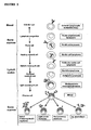
- the basic tetrameric structure of antibodies comprises two heavy chains covalently linked by a disulphide bond. Each heavy chain is in turn attached to a light chain, again via a disulphide bond. This produces a substantially "Y"-shaped molecule. This is shown schematically in Figure 1 .
- Heavy chains are the larger of the two types of chain found in antibodies, with typical molecular mass of 50,000-77,000 Da, compared with the smaller light chain (25,000 Da).
- IgG is the major immunoglobulin of normal human serum, accounting for 70-75% of the total immunoglobulin pool. This is the major antibody of secondary immune responses. It forms a single tetramer of two heavy chains plus two light chains.
- IgM accounts for approximately 10% of the immunoglobulin pool.
- the individual heavy chains have a molecular weight of approximately 65,000 and the whole molecule has a molecular weight of about 970,000.
- IgM is largely confined to the intravascular pool and is the predominant early antibody.
- IgA represents 15-20% of human serum immunoglobulin pool. More than 80% of IgA occurs as a monomer. However, some of the IgA (secretory IgA) exists as a dimeric form.
- IgD accounts for less than 1% of the total plasma immunoglobulin.
- IgE although scarce in normal serum, is found on the surface membrane of basophils and mast-cells. It is associated with allergic diseases such as asthma and hay-fever.
- IgG immunoglobulin G
- IgA immunoglobulin A
- ⁇ Lambda
- Kappa ⁇
- Each chain contains approximately 220 amino acids in a single polypeptide chain that is folded into one constant and one variable domain.
- Plasma cells produce one of the five heavy chain types together with either ⁇ or ⁇ molecules.
- the light chain molecules are not bound to heavy chain molecules, they are known as "free light chain molecules".
- the ⁇ light chains are usually found as monomers. The ⁇ light chains tend to form dimers.
- FIG. 2 shows the development of B-cell lineage and associated diseases. These diseases are known as malignant plasma cell diseases. They are summarised in detail in the book “ Serum-free Light Chain Analysis” A.R. Bradwell, available from The Binding Site Limited, Birmingham, UK (ISBN: 07044 24541 ).
- a plasma cell proliferates to form a monoclonal tumour of identical plasma cells. This results in production of large amounts of identical immunoglobulins and is known as a monoclonal gammopathy.
- myeloma and primary systemic amyloidosis account for approximately 1.5% and 0.3% respectively of cancer deaths in the United Kingdom.
- Multiple myeloma is the second-most common form of haematological malignancy after non-Hodgkin lymphoma. In Caucasian populations the incidence is approximately 40 per million per year.
- diagnosis of multiple myeloma is based on the presence of excess monoclonal plasma cells in the bone marrow, monoclonal immunoglobulins in the serum or urine and related organ or tissue impairment such as hypercalcaemia, renal insufficiency, anaemia or bone lesions.
- Normal plasma cell content of the bone marrow is about 1%, while in multiple myeloma the content is typically greater than 30%, but may be over 90%.
- AL amyloidosis is a protein conformation disorder characterised by the accumulation of monoclonal free light chain fragments as amyloid deposits. Typically, these patients present with heart or renal failure but peripheral nerves and other organs may also be involved.
- B-cell non-Hodgkin lymphomas cause approximately 2.6% of all cancer deaths in the UK and monoclonal immunoglobulins have been identified in the serum of about 10-15% of patients using standard electrophoresis methods. Initial reports indicate that monoclonal free light chains can be detected in the urine of 60-70% of patients. In B-cell chronic lymphocytic leukaemia monoclonal proteins have been identified by free light chain immunoassay.
- MGUS monoclonal gammopathy of undetermined significance. This term denotes the unexpected presence of a monoclonal intact immunoglobulin in individuals who have no evidence of multiple myeloma, AL amyloidosis, Waldenström's macroglobulinaemia, etc.
- MGUS may be found in 1% of the population over 50 years, 3% over 70 years and up to 10% over 80 years of age. Most of these are IgG- or IgM-related, although more rarely IgA-related or bi-clonal. Although most people with MGUS die from unrelated diseases, MGUS may transform into malignant monoclonal gammopathies.
- the diseases present abnormal concentrations of monoclonal immunoglobulins or free light chains. Where a disease produces the abnormal replication of a plasma cell, this often results in the production of more immunoglobulins by that type of cell as that "monoclone" multiplies and appears in the blood.
- Serum protein electrophoresis SPE
- IFE immunofixation electrophoresis
- Serum protein electrophoresis is the standard method for screening for intact immunoglobulin multiple myeloma and is based upon scanning gels in which serum proteins have been separated, fixed and stained. There are limitations associated with this method, including that some samples from patients with myelomas appear normal by electrophoresis. This results in the possibility of missing patients and misdiagnosis of the disease.
- Serum electrophoresis can be used to identify the presence of free light chains, but the detection limit is between 500 mg/L and 2,000 mg/L, depending upon whether or not the monoclonal protein migrates alongside ⁇ proteins. Serum protein electrophoresis is negative for free light chains in all patients with non-secretory myeloma.
- Immunofixation electrophoresis uses a precipitating antibody against the immunoglobulin molecules. Whilst this improves the sensitivity of the test it cannot be used to quantify monoclonal immunoglobulins because of the presence of the precipitating antibody. Immunofixation electrophoresis is also rather laborious to perform and interpretation may be difficult. Capillary zone electrophoresis is used in many clinical laboratories for serum protein separation and is able to detect most monoclonal immunoglobulins. However, when compared with immunofixation, capillary zone electrophoresis fails to detect monoclonal proteins in 5% of samples. These so-called "false negative" results encompass low-concentration monoclonal proteins.
- Total ⁇ and ⁇ assays have been produced. However, total ⁇ and total ⁇ assays are too insensitive for the detection of monoclonal immunoglobulin or free light chain. This is due to high background concentrations of polyclonal bound light chains which interfere with such assays.
- the free light chain assay uses the antibodies to bind to free ⁇ or free ⁇ light chains. The concentration of the free light chains is determined by nephelometry or turbidimetry.
- test sample This involves the addition of the test sample to a solution containing the appropriate antibody in a reaction vessel or cuvette.
- a beam of light is passed through the cuvette and as the antigen-antibody reaction proceeds, the light passing through the cuvette is scattered increasingly as insoluble immune complexes are formed.
- the light scatter is monitored by measuring the light intensity at an angle away from the incident light, whilst in turbidimetry light scatter is monitored by measuring the decrease in intensity of the incident beam of light.
- calibrators of known antigen (i.e. free ⁇ or free ⁇ ) concentration are assayed initially to produce a calibration curve of measured light scatter versus antigen concentration.
- the amount of the free ⁇ light chain and the amount of the free ⁇ light chain are known, it is possible to calculate the ratio between the free ⁇ and the free ⁇ light chains.
- An example of the results of plotting serum ⁇ versus serum ⁇ concentrations for patients with different diseases is shown in Figure 3 .
- the amount of free ⁇ and free ⁇ is skewed away from the normal concentrations because of the monoclonal nature of many of these diseases. Measuring the ⁇ : ⁇ ratio for free light chains assists in the diagnosis of the disease.
- the technique allows the disease to be monitored.
- the concentrations of free light chains which have a relatively short life span within the blood, will change and move more towards the normal concentrations observed for normal sera.
- the ⁇ : ⁇ ratio can be more sensitive than individual FLC measurements.
- Haraldsson A., et al. discloses ELISA assays for the determination of kappa and lambda ratios within total IgG, IgA and IgM.
- Figure 4 indicates that not all such diseases produce free light chains. The use of free light chains as a marker for the diseases is therefore not 100% successful.
- the inventors realised that it is possible to produce antibodies and assays that would be able to distinguish between, for example, IgG ⁇ and IgG ⁇ . They therefore tried to produce antibodies which are specific for immunoglobulins and which had specificity for both a heavy chain class and a light chain type. They have successfully been able to do this. They have also produced assays that allow the rapid quantitative measurement of, e.g. IgG ⁇ and IgG ⁇ ratios to allow the rapid identification and/or follow the progression of monoclonal diseases associated with production of a specific heavy chain class, or even heavy chain subclass, in conjunction with a bound ⁇ or ⁇ chain.
- the inventors By determining the composition of immunoglobulins using an antibody specific to a heavy chain class at the same time as a light chain type or by using a first antibody against a heavy chain class and a second antibody to determine the light chain type bound to the heavy chain the inventors have produced a sensitive assay for malignant plasma cell diseases.
- the assays developed allow more sensitive monitoring of the diseases than, for example, by SPE.
- the greater sensitivity allows the detection of the clone, for example, when concentrations of the monoclonal protein have fallen below SPE detection limits. Furthermore, this has the potential to identify some biclonal diseases, which may have normal light chain ratios.
- this assay should not be affected by renal function. Free ⁇ and free ⁇ are cleared by filtration through the kidneys and their concentration is affected by filtration rate. Due to its size, intact immunoglobulin is cleared by other mechanisms. Thus, rising levels of free light chain but no change in the amount of the immunoglobulins detected by the current invention may be used to indicate changes in renal clearance only, especially if the ⁇ : ⁇ ratio showed no changes.
- Assays used include ELISA, nephalometry, turbidimetry and flow cytometry. However, the invention is not limited to such assays.
- the method quantitatively measures the amounts of the two immunoglobulins in the sample.
- the sample is obtained from an animal, such as a mammal, preferably a human.
- the sample is assayed in vitro.
- the class detected may be selected from IgA, IgG, IgM, IgD and IgE.
- the antibodies may also be subclass specific.
- two different parts of the immunoglobulin to be detected are bound by the antibodies used in the assay.
- One antibody binds a part of the heavy chain responsible for heavy chain class determination.
- the second binds a part of the light chain responsible for identifying it as a ⁇ or ⁇ chain.
- the ratio is determined using:
- the presence of the specific antibodies bound to these immunoglobulins may be determined using a labelled second antibody.
- the binding antibody may be a sheep antibody.
- the immunoglobulins detected may be human immunoglobulins.
- sheep antibodies bound to the human immunoglobulin may be determined using anti-sheep antibodies, e.g. from rabbit or horse.
- results produced by the inventors indicate that some tumours which do not produce abnormal free ⁇ to free ⁇ ratios, can be identified because of the difference in the ratio of, for example, IgG ⁇ and IgG ⁇ or IgAk and IgA ⁇ observed.
- Measurement of the heavy chain-light chain specific pair is capable of being automated. Furthermore, the technique is more sensitive and allows the quantitative determination of the amount of different immunoglobulins. It can be used both to aid diagnosis of a disease and also to monitor the response of the disease to treatment.
- the antibodies used in the assay may be heavy chain subclass specific.
- anti-IgA IgA1 and IgA2
- anti-IgG such as IgG1, IgG2, IgG3 or IgG4 antibodies are made by The Binding Site, Birmingham, United Kingdom. This gives more detailed knowledge of the disease being detected.
- Polyclonal antibodies are preferably used. This allows an improved assay to be produced to monitor different immunoglobulins of, for example, the same class. Polyclonal antibodies allow some variability between different heavy chains of the same class to be detected because they are raised against a number of parts of the heavy chain.
- the method of the invention may also be used using one or more of the following methods wherein the binding of the antibodies to the immunoglobulins in the sample is determined by using a nephelometer, a turbidimeter, flow cytometry, ELISA or fluorescently labelled beads such as Luminex TM beads.
- a microarray assay may be produced using the antibodies.
- the ratio is determined by immunoassay, most preferably via an immunosorbent assay such as ELISA (Enzyme Linked Immunosorbent Assay).
- ELISA-type assays pe r se are well known in the art. They use antibodies, or fragments of antibodies, to detect blood groups, cell surface markers, drugs and toxins. In the case of the current invention, this type of assay has been used for the method of the invention.
- the inventors have found that it is possible to produce ELISA assays at least as sensitive as Serum Protein Electrophoresis and, in at least some cases, more sensitive than using Immunofixation Electrophoresis (IFE), FREELITE TM (The Binding Site, Birmingham, UK) or obtaining total heavy chain class concentration as nephelometry.
- IFE Immunofixation Electrophoresis
- FREELITE TM The Binding Site, Birmingham, UK
- ELISA uses antibodies to detect specific antigens.
- One or more of the antibodies used in the assay may be labelled with an enzyme capable of converting a substrate into a detectable analyte.
- enzymes include horseradish peroxidase, alkaline phosphatase and other enzymes known in the art.
- other detectable tags or labels may be used instead of, or together with, the enzymes.
- radioisotopes include radioisotopes, a wide range of coloured and fluorescent labels known in the art, including fluorescein, Alexa fluor, Oregon Green, BODIPY, rhodamine red, Cascade Blue, Marina Blue, Pacific Blue, Cascade Yellow, gold; and conjugates such as biotin (available from, for example, Invitrogen Ltd, United Kingdom).
- Dye sols, metallic sols or coloured latex may also be used.
- One or more of these labels may be used in the ELISA assays according to the various inventions described herein, or alternatively in the other assays, labelled antibodies or kits described herein.
- a "binding antibody” specific for the immunoglobulin is immobilised on a substrate.
- the immunoglobulin comprises a heavy chain of a particular class, or subclass, attached to either a ⁇ light chain or a ⁇ light chain.
- the "binding antibody” may be immobilised onto the substrate by methods which are well known in the art. Immunoglobulins in the sample are bound by the "binding antibody” which binds the immunoglobulin to the substrate via the "binding antibody”.
- the presence of bound immunoglobulins may be determined by using a labelled "detecting antibody” specific to a different part of the immunoglobulin of interest than the binding antibody.
- flow cytometry is used to detect the binding of the immunoglobulins of interest and measure the ratios.
- This technique is well known in the art for, e.g. cell sorting. However, it can also be used to detect labelled particles, such as beads, and to measure their size.
- Numerous text books describe flow cytometry, such as Practical Flow Cytometry, 3rd Ed. (1994), H. Shapiro, Alan R. Liss, New York , and Flow Cytometry, First Principles (2nd Ed.) 2001, A.L. Given, Wiley Liss .
- One of the binding antibodies such as the antibody specific for the heavy chain class, is bound to a bead, such as a polystyrene or latex bead.
- the beads are mixed with the sample and the second detecting antibody, such as antibody specific for ⁇ light chains.
- the detecting antibody is preferably labelled with a detectable label, which binds the immunoglobulin to be detected in the sample. This results in a labelled bead when the immunoglobulin to be assayed is present.
- Labelled beads may then be detected via flow cytometry.
- Different labels such as different fluorescent labels may be used for, for example, the anti- ⁇ and anti-K antibodies. This allows the amount of each type of immunoglobulin bound to be determined simultaneously and allows the rapid identification of the ⁇ : ⁇ ratio for a given heavy chain class.
- different sized beads may be used for different antibodies, for example for different class specific antibodies.
- Flow cytometry can distinguish between different sized beads and hence can rapidly determine the amount of each heavy chain class in a sample.
- Flow cytometry allows rapid identification of the ⁇ / ⁇ ratios for a given heavy chain class or subclass. This also reduces the need to do immunofixation tests.
- An alternative method uses the antibodies bound to, for example, fluorescently labelled beads such as commercially available Luminex TM beads. Different beads are used with different antibodies. Different beads are labelled with different fluorophore mixtures, thus allowing the ⁇ / ⁇ ratio for a particular heavy chain class or subclass to be determined by the fluorescent wavelength. Luminex beads are available from Luminex Corporation, Austin, Texas, United States of America.
- the monoclonal proteins in a sample may be further characterised by looking at the amount of free ⁇ or free ⁇ light chains in the sample. This is preferably carried out using antibodies specific for free ⁇ or free ⁇ light chains, such as those sold under the trade mark FREELITE by The Binding Site Ltd, Birmingham, UK.
- fragments of antibody used in the invention may also be Fab or F(ab') 2 fragments.
- the antibody or fragment is a polyclonal antibody.
- Polyclonal antibodies allow a plurality of different antibodies to be raised against different epitopes for the specific heavy chain-light chain combination. This allows for the slight variations between different immunoglobulins, but which nevertheless comprise the same heavy chain-light chain combination.
- polyclonal antibodies used in the various aspects of the invention may be capable of being produced by the method shown in WO 97/17372 . This allows the production of highly specific-polyclonal antibodies.
- the antibodies or fragments may be immobilised onto a substrate by techniques well-known in the art.
- the substrate may, for example, be a micro array or a microtiter plate.
- the substrate may be a polystyrene and/or latex bead. This allows the antibodies to be used in a number of different assays which are well-known in the art, for example as shown in EP 0291194 or ELISA assays.
- the antibody may also be labelled, for example, with a label described above for ELISA. This can be any entity, the presence of which can be readily detected.
- the label may be a visible label, that is an entity which, in its natural state, is readily visible either to the naked eye or with the aid of an optical filter and/or applied stimulation, such as UV light to produce fluorescence.
- minute coloured particles such as dye sols, metallic sols (e.g. gold) or coloured latex particles, may be used.
- Indirect labels such as enzymes (e.g. alkaline phosphatase and horseradish peroxidase) can be used, as can radioactive labels such as 35 S.
- enzymes e.g. alkaline phosphatase and horseradish peroxidase
- radioactive labels such as 35 S.
- the immunosorbent assays such as ELISA assays according to the various aspects of the invention comprise an antibody specific for ⁇ light chains and a second specific for ⁇ light chains, plus an antibody specific for the heavy chain class.
- Such assays may use, for example, a captive layer specific for the heavy chain class, such as IgG or IgA, plus detection antibodies (anti- ⁇ and anti- ⁇ ).
- anti light-chain e.g. anti- ⁇
- a class specific antibody e.g. anti-IgA
- the antibody or fragment thereof is immobilised on a solid support by techniques well-known in the art.
- the method may additionally provide the step of providing a labelling reagent capable of non-specific binding to the immunoglobulin molecule to be assayed and detecting the presence of the labelled immunoglobulin bound to the antibody or fragment.
- the labelling reagent may itself be another antibody directed against a different part of the immunoglobulin molecule, the separate antibody being labelled with a label, for example, of the sort discussed above.
- the presence of the label allows the production of, for example, a sandwich assay, and the identification of binding of the labelled immunoglobulin to the molecule to be assayed and its binding to the specific antibody according to the invention.
- the methods detect and quantify the presence of a second immunoglobulin molecule having the same specific heavy chain class as the first immunoglobulin molecule, but a different type of light chain is measured. This allows the ratio between the amounts of the first immunoglobulin molecule and the second immunoglobulin molecule to be determined, for example to identify the ratio between IgG ⁇ and IgG ⁇ . This allows the progression of a disease to be followed or alternatively the treatment of a disease to be followed.
- the invention further comprises a method of diagnosing a malignant plasma cell disease in a patient comprising the use of a method according to the invention. Blood, urine or serum is assayed.
- the malignant plasma cell disease is selected from: multiple myeloma, AL amyloidosis, solitary plasmacytoma, extramedullary plasmacytoma, multiple solitary plasmacytomas, plasma cell leukaemia, Waldenström's macroglobulinaemia, B-cell non-Hodgkin lymphomas, B-cell chronic lymphocytic leukaemia or MGUS.
- MM Multiple myeloma
- FLC free light chains
- the monoclonal immunoglobulin is observed in the serum and/or urine of all patients except 1-2 % with non-secretory myeloma. Some patients exhibit an increased frequency of monoclonal free light chains.
- the monoclonal immunoglobulin can be detected and used to monitor the disease.
- Various methods are currently used to identify and characterise monoclonal immunoglobulins.
- Serum protein electrophoresis (SPE) and immunofixation electrophoresis (IFE) are two such methods utilised.
- SPE allows quantitative analysis of monoclonal immunoglobulins
- IFE is a qualitative method.
- FREELITETM More recently FREELITETM has been developed that allows nephelometric analysis of free light chains.
- This assay system allows rapid testing of samples in comparison to SPE and IFE and in addition is quantitative, allowing FLC ratios to be calculated.
- This report describes a preliminary assay developed to assess the ability to detect abnormal ratios of monoclonal light chains attached to heavy chains.
- the assay described is an ELISA assay system for the detection of IgA ⁇ and IgA ⁇ , allowing the quantitation of IgA ⁇ / ⁇ ratios.
- FREELITE TM is a trademark of The Binding Site(TBS)Ltd, UK.
- Sheep anti-human IgA (TBS product code AU010, affinity purified) was diluted to 5 ⁇ g/ml in 1x PBS (pH 7.2) Microtitre plates (High bind, Greiner Bio-one) were coated by the addition of 100 ⁇ l of the diluted antiserum to each well. The plates were placed at 4° C in a humidified atmosphere for 18 hours. The contents of the wells were removed and 110 ⁇ l/well of 50 % [v/v] Stabilcoat (biomolecular stabiliser/blocking agent) added for 30 minutes to block non-coated regions of the wells. Following removal of the blocking solution, the plates were placed in a vacuum drier for 1 hour. The plates were sealed in foil bags containing desiccant and stored at 4° C.
- Stabilcoat biomolecular stabiliser/blocking agent
- HRP Sheep anti-human kappa-horseradish peroxidase
- affinity purified or sheep anti-human lambda-HRP, affinity purified were diluted to various dilutions in conjugate diluent (130 nM NaCl, 10 % [v/v] HRP conjugate stabiliser, 0.045 % [v/v] Proclin 300 (preservative)).
- IgA controls Two sheep anti-human IgA coated plates were incubated with IgA controls of known concentration (RID IgA NL control 3.963 mg/ml, RID IgA ML control 0.05 mg/ml and RID IgA UL control 0.18 mg/ml,) diluted 1/50 in Sample Diluent (1x PBS plus 2 % [v/v] Stabilguard (biomolecular stabiliser/blocking agent), 1 % [w/v] bovine serum albumin (BSA), 0.05% [v/v] Tween-20, 0.02 % [v/v] Kathon (biocide). pH 7.2) for 30 minutes.
- Stabilguard biomolecular stabiliser/blocking agent
- BSA bovine serum albumin
- Kathon biocide
- IgA ⁇ and IgA ⁇ sera were used to assess the sensitivity of the IgA ELISA assay in determining IgA ⁇ and IgA ⁇ concentrations.
- Purified human IgA ⁇ (5.14 mg/ml) and human IgA ⁇ (1.85 mg/ml) sera were serial diluted using tripling dilutions to allow the determination of the detectable concentration range.
- the ELISA assay method used was as described above. Briefly, IgA ⁇ or IgA ⁇ sera of various dilutions, or IgA controls (as described above) were added to wells at 100 ⁇ l/well in duplicate.
- anti-human kappa-HRP for IgA ⁇ serum plate
- anti-human lambda-HRP for IgA ⁇ serum plate
- IgA ⁇ and IgA ⁇ values were separately determined, from which ⁇ / ⁇ ratios were calculated.
- Two sheep anti-human IgA coated plates were incubated with 100 ⁇ l/well of IgA controls mentioned previously (diluted 1/800 in Sample Diluent), IgA ⁇ or IgA ⁇ sera of various dilutions, and serum samples from healthy adults diluted 1/4000 in sample diluent for 30 minutes.
- the plates were washed 3 times with Wash buffer and incubated with 100 ⁇ l/well of either anti-human kappa-HRP or anti-human lambda-HRP conjugate at 1/8000 for 30 minutes.
- the plates were washed 3 times with Wash buffer and incubated with 100 ⁇ l/well TMB substrate for 30 minutes.
- the reaction was terminated by the addition of 100 ⁇ l/well of 3 M phosphoric acid. Absorbances were measured at 450 nm using a Biotek ELISA plate reader. Results were analysed using KC4 software.
- the IgA ⁇ and IgA ⁇ assays were carried out as described above, with the addition of myeloma patient serum samples diluted 1/4000 in sample diluent.
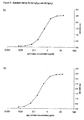
- the linear detection range of the assays were determined as 0.22-2.2 ⁇ g/ml for IgA ⁇ ( Figure 5a ) and 0.22-3.2 ⁇ g/ml for IgA ⁇ ( Figure 5b ).
- serial dilutions of the RID IgA NL control were also carried out and assayed.
- the detectable assay concentration range agreed with those obtained for IgA ⁇ and IgA ⁇ (data not shown). It was concluded that serum samples to be tested would require a 1/4000 dilution to fall within the linear range of the standardised IgA ⁇ and IgA ⁇ curves.
- IgA ⁇ caused interference to the IgA ⁇ standard curve (leading to a reduction in absorbance) at IgA ⁇ concentrations of 1.6 ⁇ g/ml and above.
- IgA ⁇ caused interference to the IgA ⁇ standard curve at 5.35 ⁇ g/ml and above.
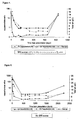
- IgA ⁇ / ⁇ ratios produced are in agreement with the trend line observed for FLC ⁇ / ⁇ ratios ( Figure 6 ). Most of the total IgA values also correlate except for the sample obtained 36 days after presentation, for which the total IgA value was within the normal range, but IgA and FLC ⁇ / ⁇ ratios were elevated, with a SPE score of +/-. IFE of this sample indicated the presence of an IgA ⁇ band. The results for this patient indicate the IgA ⁇ / ⁇ ELISA assays may have similar sensitivity to FREELITETM and IFE, and may have increased sensitivity with respect to SPE and total IgA values obtained via nephelometry.
- IgA ⁇ / ⁇ ratios calculated agree with trend lines observed for FLC ⁇ / ⁇ ratios and total IgA values ( Figure 7 ).
- SPE scores correspond to most values obtained by the other methods except for the sample obtained 501 days after presentation. For this sample the SPE score is +/- but values are within normal ranges for all other methods. IFE of this sample indicates no monoclonal bands.
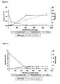
- the IgA ⁇ / ⁇ ratios trend line obtained for samples of this patient corresponds to that seen for FLC ⁇ / ⁇ ratios and agree with most of the SPE scores ( Figure 8 ).
- the total IgA value is suppressed for the sample obtained 23 days after presentation, although all other results are elevated.
- total IgA is suppressed, the SPE score is negative, yet the IgA ⁇ / ⁇ ratio is abnormal.
- the FLC ⁇ / ⁇ ratio is not known for this sample).
- IgA ⁇ / ⁇ ratios agree with most, but not all, of the data obtained using the other detection methods ( Figure 9 ). No SPE scores have been recorded for these samples. IgA ⁇ / ⁇ ratios are above the normal range for samples obtained 1493, 1576 and 2255 days after presentation. However, total IgA values are within normal range for the first two of these samples and FLC ⁇ / ⁇ ratios are within normal range for all three samples. IFE confirms the presence of a monoclonal IgA ⁇ band in all three samples. Therefore the IgA ⁇ / ⁇ ELISA assays may show greater sensitivity than FREELITETM for a number of samples.
- IgA ⁇ / ⁇ ratio and FLC ⁇ / ⁇ ratio trend lines are similar ( Figure 11 ). However, there are two samples (165 and 263 days after presentation) for which the FLC ⁇ / ⁇ ratios and IgA values are within normal range, SPE scores are negative, but the IgA ⁇ / ⁇ ratios are abnormal. IFE confirms the presence of an IgA ⁇ band. These results suggest in some cases the IgA ⁇ / ⁇ ELISA assays are more sensitive than FREELITETM, total IgA values obtained via nephelometry and SPE, and of equal sensitivity to IFE.
- IgA ⁇ / ⁇ ratio and FLC ⁇ / ⁇ ratio trend lines are similar ( Figure 14 ). However, FLC ⁇ / ⁇ ratios are only just above normal levels upon presentation, whereas IgA ⁇ / ⁇ ratios are highly elevated and agree with all other results.
- IgA ⁇ / ⁇ ratios are abnormal and agree with values for total IgA.
- SPE scores are unclear, and suggest negative results for all samples.
- all FLC ⁇ / ⁇ ratios are within the normal range ( Figure 15 ).
- IFE for the first four samples confirms the presence of an IgA ⁇ band in these samples.
- the IgA ⁇ / ⁇ ELISA assays have been shown to be as sensitive as IFE, and in some cases more sensitive than using SPE, FREELITETM and obtaining total IgA values via nephelometry.
- the majority of the results obtained using the current ELISA based assay system agree with those of the FREELITETM assay system, it suggests both methods are correctly measuring ⁇ / ⁇ ratios to allow investigation of monoclonal immunoglobulins characteristic of multiple myeloma.
- the anti-IgGK was purified by adsorbing with IgM ⁇ and IgG ⁇ . Excess immune complexes were removed by adding 3% w/v of polyethylene glycol (6,000) and the precipitate was removed by centrifugation.
- Anti-IgGK heavy chain antibodies are anti-IgGK heavy chain antibodies:
- the calibration curve shape corresponded to the one expected from similar experiments using Freelite.
- Anti-IgG ⁇ heavy chains antibodies are anti-IgG ⁇ heavy chains antibodies:
- the calibration curve shape corresponded to the one expected from previous experiments involving nephelometric assay.
- the quantity of antibodies was not sufficient enough to proceed with further tests at that time. However, the calibration curve was similar to the one involving the anti-IgGK antibodies, with expectation of similar results if tested against known concentration samples.
Landscapes
- Health & Medical Sciences (AREA)
- Life Sciences & Earth Sciences (AREA)
- Immunology (AREA)
- Engineering & Computer Science (AREA)
- Hematology (AREA)
- Molecular Biology (AREA)
- Chemical & Material Sciences (AREA)
- Biomedical Technology (AREA)
- Urology & Nephrology (AREA)
- Food Science & Technology (AREA)
- General Physics & Mathematics (AREA)
- Cell Biology (AREA)
- Biotechnology (AREA)
- Pathology (AREA)
- Microbiology (AREA)
- Medicinal Chemistry (AREA)
- Physics & Mathematics (AREA)
- Analytical Chemistry (AREA)
- Biochemistry (AREA)
- General Health & Medical Sciences (AREA)
- Hospice & Palliative Care (AREA)
- Oncology (AREA)
- Proteomics, Peptides & Aminoacids (AREA)
- Peptides Or Proteins (AREA)
- Investigating Or Analysing Biological Materials (AREA)
- Apparatus Associated With Microorganisms And Enzymes (AREA)
- Medicines Containing Antibodies Or Antigens For Use As Internal Diagnostic Agents (AREA)
- Preparation Of Compounds By Using Micro-Organisms (AREA)
Priority Applications (1)
| Application Number | Priority Date | Filing Date | Title |
|---|---|---|---|
| PL10196249T PL2306202T3 (pl) | 2005-01-27 | 2006-01-26 | Sposób wykrywania lub monitorowania złośliwej choroby nowotworowej komórek plazmatycznych |
Applications Claiming Priority (2)
| Application Number | Priority Date | Filing Date | Title |
|---|---|---|---|
| GBGB0501741.3A GB0501741D0 (en) | 2005-01-27 | 2005-01-27 | Antibody |
| EP06703629.3A EP1842071B3 (en) | 2005-01-27 | 2006-01-26 | Method of detecting or monitoring a malignant plasma cell disease |
Related Parent Applications (2)
| Application Number | Title | Priority Date | Filing Date |
|---|---|---|---|
| EP06703629.3 Division | 2006-01-26 | ||
| EP06703629.3A Division-Into EP1842071B3 (en) | 2005-01-27 | 2006-01-26 | Method of detecting or monitoring a malignant plasma cell disease |
Publications (2)
| Publication Number | Publication Date |
|---|---|
| EP2306202A1 EP2306202A1 (en) | 2011-04-06 |
| EP2306202B1 true EP2306202B1 (en) | 2013-12-18 |
Family
ID=34259791
Family Applications (2)
| Application Number | Title | Priority Date | Filing Date |
|---|---|---|---|
| EP10196249.6A Active EP2306202B1 (en) | 2005-01-27 | 2006-01-26 | Method of detecting or monitoring a malignant plasma cell disease |
| EP06703629.3A Active EP1842071B3 (en) | 2005-01-27 | 2006-01-26 | Method of detecting or monitoring a malignant plasma cell disease |
Family Applications After (1)
| Application Number | Title | Priority Date | Filing Date |
|---|---|---|---|
| EP06703629.3A Active EP1842071B3 (en) | 2005-01-27 | 2006-01-26 | Method of detecting or monitoring a malignant plasma cell disease |
Country Status (11)
| Country | Link |
|---|---|
| US (3) | US7897353B2 (enExample) |
| EP (2) | EP2306202B1 (enExample) |
| JP (3) | JP5431674B2 (enExample) |
| AT (1) | ATE502302T1 (enExample) |
| DE (1) | DE602006020688D1 (enExample) |
| DK (1) | DK2306202T3 (enExample) |
| ES (2) | ES2365626T7 (enExample) |
| GB (1) | GB0501741D0 (enExample) |
| PL (1) | PL2306202T3 (enExample) |
| PT (1) | PT2306202E (enExample) |
| WO (1) | WO2006079816A1 (enExample) |
Families Citing this family (15)
| Publication number | Priority date | Publication date | Assignee | Title |
|---|---|---|---|---|
| GB0608444D0 (en) | 2006-04-27 | 2006-06-07 | Binding Site The Ltd | Dialysis |
| GB0801609D0 (en) * | 2008-01-29 | 2008-03-05 | Binding Site The Ltd | Hevylite diagnostic stain |
| SG174992A1 (en) | 2009-04-01 | 2011-11-28 | Genentech Inc | Anti-fcrh5 antibodies and immunoconjugates and methods of use |
| GB0914535D0 (en) * | 2009-08-19 | 2009-09-30 | Binding Site Group The Ltd | Prognosis assay |
| GB201001946D0 (en) * | 2010-02-05 | 2010-03-24 | Binding Site Group The Ltd | Cancer prognostic assay |
| GB201001950D0 (en) * | 2010-02-05 | 2010-03-24 | Binding Site Group The Ltd | Infection prognostic assay |
| GB201012049D0 (en) | 2010-07-19 | 2010-09-01 | Binding Site Group The Ltd | Competition assay |
| GB201203938D0 (en) | 2012-03-06 | 2012-04-18 | Binding Site Group The Ltd | Assay system |
| US9550004B2 (en) | 2013-09-06 | 2017-01-24 | Sensor Electronic Technology, Inc. | Ultraviolet diffusive illumination |
| US10267806B2 (en) | 2014-04-04 | 2019-04-23 | Mayo Foundation For Medical Education And Research | Isotyping immunoglobulins using accurate molecular mass |
| GB201511364D0 (en) * | 2015-06-29 | 2015-08-12 | Binding Site Group The Ltd | Method of detecting or monitoring an IgG4-related disease |
| GB2541027A (en) * | 2015-08-07 | 2017-02-08 | The Binding Site Group Ltd | Treatment |
| US12153052B2 (en) | 2017-09-13 | 2024-11-26 | Mayo Foundation For Medical Education And Research | Identification and monitoring of immunoglobulin J chains |
| US11946937B2 (en) | 2017-09-13 | 2024-04-02 | Mayo Foundation For Medical Education And Research | Identification and monitoring of apoptosis inhibitor of macrophage |
| WO2021202992A2 (en) * | 2020-04-03 | 2021-10-07 | Lawrence Loomis | Rapid point of care assay for the detection of the asymptomatic carrier state of covid-19 |
Family Cites Families (23)
| Publication number | Priority date | Publication date | Assignee | Title |
|---|---|---|---|---|
| US3907502A (en) | 1973-12-11 | 1975-09-23 | Miless L Brink | Method for identifying Bence Jones proteins |
| EP0044219A1 (en) | 1980-07-16 | 1982-01-20 | Unilever Plc | Methods of immuno analysis using monoclonal antibodies |
| JPS63139199A (ja) * | 1986-12-01 | 1988-06-10 | Wakunaga Pharmaceut Co Ltd | 抗ヒトIgEモノクロ−ナル抗体及びその用途 |
| ES2150428T3 (es) | 1987-04-27 | 2000-12-01 | Unilever Nv | Ensayos de union especifica. |
| US4983530A (en) * | 1988-01-29 | 1991-01-08 | E. I. Du Pont De Nemours And Company | Sandwich immunoassay for determination of total monoclonal IGG |
| US5185066A (en) * | 1988-08-11 | 1993-02-09 | Helena Laboratories Corporation | Immunofixation electrophoresis control system |
| JPH03199966A (ja) * | 1989-12-27 | 1991-08-30 | Kazutomo Wakasugi | 遊離型l鎖の測定方法 |
| JPH04262792A (ja) * | 1990-01-12 | 1992-09-18 | Takeda Chem Ind Ltd | 蛋白質の精製法 |
| US5228960A (en) * | 1992-07-17 | 1993-07-20 | Beckman Instruments, Inc. | Analysis of samples by capillary electrophoretic immunosubtraction |
| US5552277A (en) * | 1994-07-19 | 1996-09-03 | The Johns Hopkins University School Of Medicine | Genetic diagnosis of prostate cancer |
| EP0862584B1 (en) | 1995-11-03 | 2004-12-22 | The Binding Site Limited | Production of antibodies |
| GB9718911D0 (en) * | 1997-09-05 | 1997-11-12 | Orion Yhtymae Oy | Monoclonal antibodies |
| ATE301131T1 (de) * | 1998-11-19 | 2005-08-15 | Elan Corp Plc | Gegen gastrointestinale transport rezeptoren gerichtete retro-invertierte peptide und deren verwendung |
| US20040163137A1 (en) * | 2001-02-20 | 2004-08-19 | Caroline Barry | PG-3 and biallelic markers thereof |
| US20020182748A1 (en) | 2001-03-30 | 2002-12-05 | Reardon Paul C. | Method and device for testing for Bence-Jones Protein |
| CA2468950A1 (en) * | 2001-11-27 | 2003-06-05 | Mochida Pharmaceutical Co., Ltd. | Anti-il13 receptor .alpha.1 neutralizing antibody |
| US20040018576A1 (en) * | 2002-07-24 | 2004-01-29 | Dematteo Todd M. | Bence Jones protein testing cassette |
| AU2004210975B2 (en) * | 2003-02-10 | 2008-04-10 | Agensys, Inc. | Nucleic acid and corresponding protein named 158P1D7 useful in the treatment and detection of bladder and other cancers |
| WO2004073656A2 (en) * | 2003-02-20 | 2004-09-02 | Seattle Genetics, Inc. | Anti-cd70 antibody-drug conjugates and their use for the treatment of cancer and immune disorders |
| JP4473257B2 (ja) * | 2003-04-02 | 2010-06-02 | エフ.ホフマン−ラ ロシュ アーゲー | インスリン様成長因子i受容体に対する抗体及びその使用 |
| JP4286256B2 (ja) * | 2003-11-19 | 2009-06-24 | ヤマサ醤油株式会社 | ベンスジョーンズ蛋白質の検定法 |
| JP4438455B2 (ja) * | 2004-03-04 | 2010-03-24 | ヤマサ醤油株式会社 | 遊離ヒトイムノグロブリン軽鎖の測定法及びキット |
| WO2005116651A2 (en) * | 2004-05-24 | 2005-12-08 | Diasys Corporation | Method and device for testing for bence-jones protein |
-
2005
- 2005-01-27 GB GBGB0501741.3A patent/GB0501741D0/en not_active Ceased
-
2006
- 2006-01-26 ES ES06703629.3T patent/ES2365626T7/es active Active
- 2006-01-26 US US11/883,003 patent/US7897353B2/en active Active
- 2006-01-26 AT AT06703629T patent/ATE502302T1/de not_active IP Right Cessation
- 2006-01-26 EP EP10196249.6A patent/EP2306202B1/en active Active
- 2006-01-26 EP EP06703629.3A patent/EP1842071B3/en active Active
- 2006-01-26 WO PCT/GB2006/000267 patent/WO2006079816A1/en not_active Ceased
- 2006-01-26 PL PL10196249T patent/PL2306202T3/pl unknown
- 2006-01-26 DK DK10196249.6T patent/DK2306202T3/da active
- 2006-01-26 PT PT101962496T patent/PT2306202E/pt unknown
- 2006-01-26 DE DE602006020688T patent/DE602006020688D1/de active Active
- 2006-01-26 JP JP2007552716A patent/JP5431674B2/ja not_active Expired - Fee Related
- 2006-01-26 ES ES10196249.6T patent/ES2455440T3/es active Active
-
2011
- 2011-01-21 US US13/011,135 patent/US8415110B2/en not_active Expired - Fee Related
- 2011-01-21 US US13/011,146 patent/US8592561B2/en active Active
-
2013
- 2013-03-06 JP JP2013044306A patent/JP6007134B2/ja not_active Expired - Fee Related
- 2013-10-25 JP JP2013221946A patent/JP5715671B2/ja active Active
Non-Patent Citations (28)
| Title |
|---|
| AYLIFFE MICHAEL JOHN ET AL: "Demonstration of changes in plasma cell subsets in multiple myeloma", HAEMATOLOGICA, FONDAZIONE FERRATA STORTI, ROME, IT, vol. 92, no. 8, 1 August 2007 (2007-08-01), pages 1135 - 1138, XP009118272, ISSN: 0390-6078 * |
| BALSYS J. ET AL.: "Total and monoclonal immunoglobulin detection and quantification in human sera by enzyme-linked immunosorbent assay", BIOLOGY, 1 January 1994 (1994-01-01), pages 65 - 72 * |
| BRADWELL A ET AL: "Prognostic utility of intact immunoglobulin Ig 'kappa/Ig 'lambda ratios in multiple myeloma patients", LEUKEMIA (BASINGSTOKE), vol. 27, no. 1, January 2013 (2013-01-01), pages 202 - 207, ISSN: 0887-6924(print) * |
| BRADWELL ARTHUR R ET AL: "Assessment of Monoclonal Gammopathies by Nephelometric Measurement of Individual Immunoglobulin kappa/lambda Ratios", CLINICAL CHEMISTRY, vol. 55, no. 9, September 2009 (2009-09-01), pages 1646 - 1655, ISSN: 0009-9147 * |
| BRADWELL ARTHUR R ET AL: "Highly sensitive, automated immunoassay for immunoglobulin free light chains in serum and urine", CLINICAL CHEMISTRY, vol. 47, no. 4, April 2001 (2001-04-01), pages 673 - 680, ISSN: 0009-9147 * |
| D SUTTON ET AL.: "Computational Algorithm Based on Serum Immunoassays for the Identification of Haematological Disease may Identify Patients with other Acute Disease", AMERICAN SOCIETY OF HEAMATOLOGY, 2012 * |
| DISPENZIERI, A.: "Plasma Cell Disorder: Atypical Plasma Cell Syndromes: POEMS Syndrome", AMERICAN SOCIETY OF HEMATOLOGY, 1 January 2005 (2005-01-01), pages 360 - 367 * |
| DRAYSON ET AL.: "Immunoglobulin Heavy/Light Chain Measurements During Monitoring Provide Prognostic Information of Relapse After Therapy in Multiple Myeloma Patients", AMERICAN SOCIETY OF HEAMATOLOGY, 2012 * |
| DRAYSON MARK ET AL: "Serum free light-chain measurements for identifying and monitoring patients with nonsecretory multiple myeloma", BLOOD, vol. 97, no. 9, 1 May 2001 (2001-05-01), pages 2900 - 2902, ISSN: 0006-4971 * |
| GIRKONTAITE IRUTE ET AL: "A rapid ELISA test for detection of human paraproteins", EUROPEAN JOURNAL OF CLINICAL CHEMISTRY AND CLINICAL BIOCHEMISTRY, vol. 34, no. 4, 1996, pages 349 - 353, ISSN: 0939-4974 * |
| GIRKONTAITÉ, IRUTÉ: "The Investigation of Antigenic Structure of Human Immunoglobulins using monoclonal antibodies (Summary of doctoral dissertation)", 1 January 1996, REPUBLIC OF LATVIA: INSTITUE OF IMMUNOLOGY, VILNIUS UNIVERSITY, Vilnius, pages: 1 - 20 * |
| HEINZ LUDWIG ET AL.: "Serum Heavy Light Chain and Free Light Chain Measurements Provide Prognostic Information", AMERICAN SOCIETY OF HEAMATOLOGY, 2011 * |
| JONES R G ET AL: "USE OF IMMUNOGLOBULIN HEAVY-CHAIN AND LIGHT-CHAIN MEASUREMENTS IN A MULTICENTER TRIAL TO INVESTIGATE MONOCLONAL COMPONENTS I. DETECTION", CLINICAL CHEMISTRY, vol. 37, no. 11, 1991, pages 1917 - 1921, ISSN: 0009-9147 * |
| JOSIE A. R. HOBBS ET AL: "Frequency of altered monoclonal protein production at relapse of multiple myeloma", BRITISH JOURNAL OF HAEMATOLOGY, vol. 148, no. 4, 1 February 2010 (2010-02-01), pages 659 - 661, XP055030098, ISSN: 0007-1048, DOI: 10.1111/j.1365-2141.2009.07952.x * |
| KATZMANN J A ET AL: "A window into immunoglobulin quantitation and plasma cell disease: Antigen epitopes defined by the junction of immunoglobulin heavy and light chains", LEUKEMIA 2013 NATURE PUBLISHING GROUP GBR, vol. 27, no. 1, January 2013 (2013-01-01), pages 1 - 2, ISSN: 0887-6924 * |
| KATZMANN J A ET AL: "Suppression of uninvolved immunoglobulins defined by heavy/light chain pair suppression is a risk factor for progression of MGUS", LEUKEMIA (BASINGSTOKE), vol. 27, no. 1, January 2013 (2013-01-01), pages 208 - 212, ISSN: 0887-6924(print) * |
| KEREN DAVID F ET AL: "Guidelines for clinical and laboratory evaluation of patients with monoclonal gammopathies", ARCHIVES OF PATHOLOGY AND LABORATORY MEDICINE, vol. 123, no. 2, February 1999 (1999-02-01), pages 106 - 107, ISSN: 0363-0153 * |
| KEREN DAVID F: "Heavy/Light-chain analysis of monoclonal gammopathies.", CLINICAL CHEMISTRY SEP 2009, vol. 55, no. 9, September 2009 (2009-09-01), pages 1606 - 1608, ISSN: 1530-8561 * |
| KEREN DAVID F: "Procedures for the evaluation of monoclonal immunoglobulins", ARCHIVES OF PATHOLOGY AND LABORATORY MEDICINE, vol. 123, no. 2, February 1999 (1999-02-01), pages 126 - 132, ISSN: 0363-0153 * |
| KYLE ROBERT A: "Sequence of testing for monoclonal gammopathies: Serum and urine assays", ARCHIVES OF PATHOLOGY AND LABORATORY MEDICINE, vol. 123, no. 2, February 1999 (1999-02-01), pages 114 - 118, ISSN: 0363-0153 * |
| LAM C W K ET AL: "LIGHT CHAIN RATIOS OF SERUM IMMUNOGLOBULINS IN DISEASE", CLINICAL BIOCHEMISTRY, vol. 24, no. 3, 1991, pages 283 - 287, ISSN: 0009-9120 * |
| LUDWIG H ET AL: "Immunoglobulin heavy/light chain ratios improve paraprotein detection and monitoring, identify residual disease and correlate with survival in multiple myeloma patients", LEUKEMIA (BASINGSTOKE), vol. 27, no. 1, January 2013 (2013-01-01), pages 213 - 219, ISSN: 0887-6924(print) * |
| LUDWIG HEINZ ET AL: "Serum Heavy/Light Chain and Free Light Chain Measurements Provide Prognostic Information, Allow Creation of a Prognostic Model and Identify Clonal Changes (clonal tiding) Through the Course of Multiple Myeloma (MM)", BLOOD, vol. 118, no. 21, November 2011 (2011-11-01), & 53RD ANNUAL MEETING AND EXPOSITION OF THE AMERICAN-SOCIETY-OF-HEMATOLOGY (ASH); SAN DIEGO, CA, USA; DECEMBER 10 -13, 2011, pages 1244, ISSN: 0006-4971(print) * |
| OSCAR BERLANGA ET AL.: "Potential utility of heavy/light chain ratios in patients with systemic AL amyloidosis", EUROMEDLAB, 2013, Milan * |
| P. YOUNG ET AL.: "Use of heavy/light chain (HLC) and free light chain (FLC) ratios for monitoring oligosecretory multiple myeloma patients", INTERNATIONAL MYELOMA WORKSHOP (IMW), 2013 * |
| PB 30 Normal heavy/light chain (HLC) and free light chain (FLC) ratios are associated with prolonged survival in patients with systemic AL amyloidosis Ashutosh et al. * |
| RENCKENS A L J M ET AL: "NEPHELOMETRY OF THE KAPPA-LAMBDA LIGHT-CHAIN RATIO IN SERUM OF NORMAL AND DISEASED CHILDREN", CLINICAL CHEMISTRY, AMERICAN ASSOCIATION FOR CLINICAL CHEMISTRY, WASHINGTON, DC, vol. 32, no. 12, 1 January 1986 (1986-01-01), pages 2147 - 2149, XP008143922, ISSN: 0009-9147 * |
| WHICHER J T ET AL: "USE OF IMMUNOGLOBULIN HEAVY AND LIGHT-CHAIN MEASUREMENTS COMPARED WITH EXISTING TECHNIQUES AS A MEANS OF TYPING MONOCLONAL IMMUNOGLOBULINS", CLINICAL CHEMISTRY, vol. 33, no. 10, 1987, pages 1771 - 1773, ISSN: 0009-9147 * |
Also Published As
| Publication number | Publication date |
|---|---|
| JP5715671B2 (ja) | 2015-05-13 |
| ES2365626T7 (es) | 2014-06-04 |
| JP2008528995A (ja) | 2008-07-31 |
| PT2306202E (pt) | 2014-02-27 |
| WO2006079816A1 (en) | 2006-08-03 |
| DE602006020688D1 (de) | 2011-04-28 |
| US20110177535A1 (en) | 2011-07-21 |
| US20110177977A1 (en) | 2011-07-21 |
| DK2306202T3 (da) | 2014-02-10 |
| EP2306202A1 (en) | 2011-04-06 |
| US8415110B2 (en) | 2013-04-09 |
| US7897353B2 (en) | 2011-03-01 |
| JP6007134B2 (ja) | 2016-10-12 |
| ES2455440T3 (es) | 2014-04-15 |
| GB0501741D0 (en) | 2005-03-02 |
| EP1842071B3 (en) | 2014-02-26 |
| JP2013147499A (ja) | 2013-08-01 |
| PL2306202T3 (pl) | 2014-05-30 |
| EP1842071B1 (en) | 2011-03-16 |
| JP2014025950A (ja) | 2014-02-06 |
| ES2365626T3 (es) | 2011-10-07 |
| EP1842071A1 (en) | 2007-10-10 |
| US20080166742A1 (en) | 2008-07-10 |
| ATE502302T1 (de) | 2011-04-15 |
| JP5431674B2 (ja) | 2014-03-05 |
| US8592561B2 (en) | 2013-11-26 |
Similar Documents
| Publication | Publication Date | Title |
|---|---|---|
| US8415110B2 (en) | Method of detecting or monitoring a malignant plasma cell disease | |
| JP2013527444A (ja) | 抗体を検出するための方法 | |
| KR20110022621A (ko) | IgA 신증의 검사 방법 및 검사 키트 | |
| EP2596367B1 (en) | Competition assay | |
| US12474351B2 (en) | Mass spectrometry calibrator | |
| US20150285804A1 (en) | Diagnostic method for colorectal cancer | |
| JP4918556B2 (ja) | サンプル中の被検体(抗原)および該被検体を標的とする治療用抗体を同時に免疫化学的に測定するためのイムノアッセイ(リカバリー・イムノアッセイ(RecoveryImmunoassay)) | |
| JP5818817B2 (ja) | がん予後診断アッセイ | |
| EP3420356B1 (en) | Improved assay | |
| EP2531854B1 (en) | Cancer prognosis assay | |
| WO2022202876A1 (ja) | I型コラーゲンc末端テロペプチドの免疫学的分析方法 | |
| US20250224393A1 (en) | Method and kit for reducing interferences in immunoassays | |
| CN118786347A (zh) | 丝氨酸蛋白酶的检测用或测定用试剂 |
Legal Events
| Date | Code | Title | Description |
|---|---|---|---|
| PUAI | Public reference made under article 153(3) epc to a published international application that has entered the european phase |
Free format text: ORIGINAL CODE: 0009012 |
|
| AC | Divisional application: reference to earlier application |
Ref document number: 1842071 Country of ref document: EP Kind code of ref document: P |
|
| AK | Designated contracting states |
Kind code of ref document: A1 Designated state(s): AT BE BG CH CY CZ DE DK EE ES FI FR GB GR HU IE IS IT LI LT LU LV MC NL PL PT RO SE SI SK TR |
|
| 17P | Request for examination filed |
Effective date: 20110527 |
|
| 17Q | First examination report despatched |
Effective date: 20111018 |
|
| TPAC | Observations filed by third parties |
Free format text: ORIGINAL CODE: EPIDOSNTIPA |
|
| TPAC | Observations filed by third parties |
Free format text: ORIGINAL CODE: EPIDOSNTIPA |
|
| GRAP | Despatch of communication of intention to grant a patent |
Free format text: ORIGINAL CODE: EPIDOSNIGR1 |
|
| GRAS | Grant fee paid |
Free format text: ORIGINAL CODE: EPIDOSNIGR3 |
|
| GRAA | (expected) grant |
Free format text: ORIGINAL CODE: 0009210 |
|
| INTG | Intention to grant announced |
Effective date: 20131104 |
|
| AC | Divisional application: reference to earlier application |
Ref document number: 1842071 Country of ref document: EP Kind code of ref document: P |
|
| AK | Designated contracting states |
Kind code of ref document: B1 Designated state(s): AT BE BG CH CY CZ DE DK EE ES FI FR GB GR HU IE IS IT LI LT LU LV MC NL PL PT RO SE SI SK TR |
|
| REG | Reference to a national code |
Ref country code: GB Ref legal event code: FG4D |
|
| REG | Reference to a national code |
Ref country code: CH Ref legal event code: EP |
|
| REG | Reference to a national code |
Ref country code: AT Ref legal event code: REF Ref document number: 645843 Country of ref document: AT Kind code of ref document: T Effective date: 20140115 |
|
| REG | Reference to a national code |
Ref country code: IE Ref legal event code: FG4D |
|
| REG | Reference to a national code |
Ref country code: DK Ref legal event code: T3 Effective date: 20140207 |
|
| REG | Reference to a national code |
Ref country code: DE Ref legal event code: R096 Ref document number: 602006039727 Country of ref document: DE Effective date: 20140213 |
|
| REG | Reference to a national code |
Ref country code: PT Ref legal event code: SC4A Free format text: AVAILABILITY OF NATIONAL TRANSLATION Effective date: 20140224 |
|
| REG | Reference to a national code |
Ref country code: CH Ref legal event code: NV Representative=s name: AMMANN PATENTANWAELTE AG BERN, CH |
|
| REG | Reference to a national code |
Ref country code: NL Ref legal event code: T3 |
|
| REG | Reference to a national code |
Ref country code: SE Ref legal event code: TRGR |
|
| REG | Reference to a national code |
Ref country code: ES Ref legal event code: FG2A Ref document number: 2455440 Country of ref document: ES Kind code of ref document: T3 Effective date: 20140415 |
|
| PG25 | Lapsed in a contracting state [announced via postgrant information from national office to epo] |
Ref country code: FI Free format text: LAPSE BECAUSE OF FAILURE TO SUBMIT A TRANSLATION OF THE DESCRIPTION OR TO PAY THE FEE WITHIN THE PRESCRIBED TIME-LIMIT Effective date: 20131218 Ref country code: LT Free format text: LAPSE BECAUSE OF FAILURE TO SUBMIT A TRANSLATION OF THE DESCRIPTION OR TO PAY THE FEE WITHIN THE PRESCRIBED TIME-LIMIT Effective date: 20131218 |
|
| REG | Reference to a national code |
Ref country code: LT Ref legal event code: MG4D |
|
| PG25 | Lapsed in a contracting state [announced via postgrant information from national office to epo] |
Ref country code: LV Free format text: LAPSE BECAUSE OF FAILURE TO SUBMIT A TRANSLATION OF THE DESCRIPTION OR TO PAY THE FEE WITHIN THE PRESCRIBED TIME-LIMIT Effective date: 20131218 |
|
| REG | Reference to a national code |
Ref country code: PL Ref legal event code: T3 |
|
| PG25 | Lapsed in a contracting state [announced via postgrant information from national office to epo] |
Ref country code: IS Free format text: LAPSE BECAUSE OF FAILURE TO SUBMIT A TRANSLATION OF THE DESCRIPTION OR TO PAY THE FEE WITHIN THE PRESCRIBED TIME-LIMIT Effective date: 20140418 Ref country code: EE Free format text: LAPSE BECAUSE OF FAILURE TO SUBMIT A TRANSLATION OF THE DESCRIPTION OR TO PAY THE FEE WITHIN THE PRESCRIBED TIME-LIMIT Effective date: 20131218 |
|
| PG25 | Lapsed in a contracting state [announced via postgrant information from national office to epo] |
Ref country code: RO Free format text: LAPSE BECAUSE OF FAILURE TO SUBMIT A TRANSLATION OF THE DESCRIPTION OR TO PAY THE FEE WITHIN THE PRESCRIBED TIME-LIMIT Effective date: 20131218 Ref country code: LU Free format text: LAPSE BECAUSE OF FAILURE TO SUBMIT A TRANSLATION OF THE DESCRIPTION OR TO PAY THE FEE WITHIN THE PRESCRIBED TIME-LIMIT Effective date: 20140126 Ref country code: CY Free format text: LAPSE BECAUSE OF FAILURE TO SUBMIT A TRANSLATION OF THE DESCRIPTION OR TO PAY THE FEE WITHIN THE PRESCRIBED TIME-LIMIT Effective date: 20131218 Ref country code: SK Free format text: LAPSE BECAUSE OF FAILURE TO SUBMIT A TRANSLATION OF THE DESCRIPTION OR TO PAY THE FEE WITHIN THE PRESCRIBED TIME-LIMIT Effective date: 20131218 |
|
| REG | Reference to a national code |
Ref country code: DE Ref legal event code: R097 Ref document number: 602006039727 Country of ref document: DE |
|
| PG25 | Lapsed in a contracting state [announced via postgrant information from national office to epo] |
Ref country code: MC Free format text: LAPSE BECAUSE OF FAILURE TO SUBMIT A TRANSLATION OF THE DESCRIPTION OR TO PAY THE FEE WITHIN THE PRESCRIBED TIME-LIMIT Effective date: 20131218 |
|
| REG | Reference to a national code |
Ref country code: CH Ref legal event code: PCOW Free format text: NEW ADDRESS: 8 CALTHORPE ROAD EDGBASTON, BIRMINGHAM B15 1QT (GB) |
|
| REG | Reference to a national code |
Ref country code: DE Ref legal event code: R082 Ref document number: 602006039727 Country of ref document: DE Representative=s name: FLEUCHAUS & GALLO PARTNERSCHAFT MBB, DE |
|
| PLBE | No opposition filed within time limit |
Free format text: ORIGINAL CODE: 0009261 |
|
| STAA | Information on the status of an ep patent application or granted ep patent |
Free format text: STATUS: NO OPPOSITION FILED WITHIN TIME LIMIT |
|
| RAP2 | Party data changed (patent owner data changed or rights of a patent transferred) |
Owner name: THE BINDING SITE GROUP LIMITED |
|
| 26N | No opposition filed |
Effective date: 20140919 |
|
| REG | Reference to a national code |
Ref country code: DE Ref legal event code: R082 Ref document number: 602006039727 Country of ref document: DE Representative=s name: FLEUCHAUS & GALLO PARTNERSCHAFT MBB, DE Effective date: 20141023 Ref country code: DE Ref legal event code: R081 Ref document number: 602006039727 Country of ref document: DE Owner name: THE BINDING SITE GROUP LTD., EDGBASTON, GB Free format text: FORMER OWNER: THE BINDING SITE GROUP LTD., BIRMINGHAM, GB Effective date: 20141023 |
|
| REG | Reference to a national code |
Ref country code: FR Ref legal event code: CA Effective date: 20141114 |
|
| REG | Reference to a national code |
Ref country code: DE Ref legal event code: R097 Ref document number: 602006039727 Country of ref document: DE Effective date: 20140919 |
|
| REG | Reference to a national code |
Ref country code: FR Ref legal event code: PLFP Year of fee payment: 11 |
|
| PG25 | Lapsed in a contracting state [announced via postgrant information from national office to epo] |
Ref country code: BG Free format text: LAPSE BECAUSE OF FAILURE TO SUBMIT A TRANSLATION OF THE DESCRIPTION OR TO PAY THE FEE WITHIN THE PRESCRIBED TIME-LIMIT Effective date: 20131218 |
|
| PG25 | Lapsed in a contracting state [announced via postgrant information from national office to epo] |
Ref country code: GR Free format text: LAPSE BECAUSE OF FAILURE TO SUBMIT A TRANSLATION OF THE DESCRIPTION OR TO PAY THE FEE WITHIN THE PRESCRIBED TIME-LIMIT Effective date: 20140319 |
|
| PG25 | Lapsed in a contracting state [announced via postgrant information from national office to epo] |
Ref country code: HU Free format text: LAPSE BECAUSE OF FAILURE TO SUBMIT A TRANSLATION OF THE DESCRIPTION OR TO PAY THE FEE WITHIN THE PRESCRIBED TIME-LIMIT; INVALID AB INITIO Effective date: 20060126 Ref country code: TR Free format text: LAPSE BECAUSE OF FAILURE TO SUBMIT A TRANSLATION OF THE DESCRIPTION OR TO PAY THE FEE WITHIN THE PRESCRIBED TIME-LIMIT Effective date: 20131218 Ref country code: SI Free format text: LAPSE BECAUSE OF FAILURE TO SUBMIT A TRANSLATION OF THE DESCRIPTION OR TO PAY THE FEE WITHIN THE PRESCRIBED TIME-LIMIT Effective date: 20131218 |
|
| REG | Reference to a national code |
Ref country code: FR Ref legal event code: PLFP Year of fee payment: 12 |
|
| REG | Reference to a national code |
Ref country code: FR Ref legal event code: PLFP Year of fee payment: 13 |
|
| P01 | Opt-out of the competence of the unified patent court (upc) registered |
Effective date: 20230529 |
|
| PGFP | Annual fee paid to national office [announced via postgrant information from national office to epo] |
Ref country code: CZ Payment date: 20231222 Year of fee payment: 19 |
|
| PGFP | Annual fee paid to national office [announced via postgrant information from national office to epo] |
Ref country code: NL Payment date: 20240123 Year of fee payment: 19 |
|
| PGFP | Annual fee paid to national office [announced via postgrant information from national office to epo] |
Ref country code: IE Payment date: 20240110 Year of fee payment: 19 Ref country code: ES Payment date: 20240215 Year of fee payment: 19 |
|
| PGFP | Annual fee paid to national office [announced via postgrant information from national office to epo] |
Ref country code: AT Payment date: 20240123 Year of fee payment: 19 |
|
| PGFP | Annual fee paid to national office [announced via postgrant information from national office to epo] |
Ref country code: DE Payment date: 20240110 Year of fee payment: 19 Ref country code: PT Payment date: 20240122 Year of fee payment: 19 Ref country code: CH Payment date: 20240202 Year of fee payment: 19 Ref country code: GB Payment date: 20240117 Year of fee payment: 19 |
|
| PGFP | Annual fee paid to national office [announced via postgrant information from national office to epo] |
Ref country code: SE Payment date: 20240129 Year of fee payment: 19 Ref country code: PL Payment date: 20240102 Year of fee payment: 19 Ref country code: IT Payment date: 20240123 Year of fee payment: 19 Ref country code: FR Payment date: 20240130 Year of fee payment: 19 Ref country code: DK Payment date: 20240102 Year of fee payment: 19 Ref country code: BE Payment date: 20240131 Year of fee payment: 19 |
|
| REG | Reference to a national code |
Ref country code: DE Ref legal event code: R082 Ref document number: 602006039727 Country of ref document: DE Representative=s name: WITHERS & ROGERS LLP, DE |
|
| REG | Reference to a national code |
Ref country code: DE Ref legal event code: R119 Ref document number: 602006039727 Country of ref document: DE |
|
| REG | Reference to a national code |
Ref country code: DK Ref legal event code: EBP Effective date: 20250131 |
|
| REG | Reference to a national code |
Ref country code: CH Ref legal event code: PL |
|
| REG | Reference to a national code |
Ref country code: SE Ref legal event code: EUG |
|
| REG | Reference to a national code |
Ref country code: NL Ref legal event code: MM Effective date: 20250201 |
|
| REG | Reference to a national code |
Ref country code: AT Ref legal event code: MM01 Ref document number: 645843 Country of ref document: AT Kind code of ref document: T Effective date: 20250126 |
|
| GBPC | Gb: european patent ceased through non-payment of renewal fee |
Effective date: 20250126 |
|
| PG25 | Lapsed in a contracting state [announced via postgrant information from national office to epo] |
Ref country code: PT Free format text: LAPSE BECAUSE OF NON-PAYMENT OF DUE FEES Effective date: 20250728 |
|
| PG25 | Lapsed in a contracting state [announced via postgrant information from national office to epo] |
Ref country code: DE Free format text: LAPSE BECAUSE OF NON-PAYMENT OF DUE FEES Effective date: 20250801 |
|
| PG25 | Lapsed in a contracting state [announced via postgrant information from national office to epo] |
Ref country code: NL Free format text: LAPSE BECAUSE OF NON-PAYMENT OF DUE FEES Effective date: 20250201 |
|
| PG25 | Lapsed in a contracting state [announced via postgrant information from national office to epo] |
Ref country code: BE Free format text: LAPSE BECAUSE OF NON-PAYMENT OF DUE FEES Effective date: 20250131 Ref country code: GB Free format text: LAPSE BECAUSE OF NON-PAYMENT OF DUE FEES Effective date: 20250126 |
|
| PG25 | Lapsed in a contracting state [announced via postgrant information from national office to epo] |
Ref country code: FR Free format text: LAPSE BECAUSE OF NON-PAYMENT OF DUE FEES Effective date: 20250131 Ref country code: AT Free format text: LAPSE BECAUSE OF NON-PAYMENT OF DUE FEES Effective date: 20250126 |
|
| PG25 | Lapsed in a contracting state [announced via postgrant information from national office to epo] |
Ref country code: CH Free format text: LAPSE BECAUSE OF NON-PAYMENT OF DUE FEES Effective date: 20250131 |
|
| PG25 | Lapsed in a contracting state [announced via postgrant information from national office to epo] |
Ref country code: CZ Free format text: LAPSE BECAUSE OF NON-PAYMENT OF DUE FEES Effective date: 20250126 |
|
| REG | Reference to a national code |
Ref country code: BE Ref legal event code: MM Effective date: 20250131 |