EP2265711B1 - Membrane - Google Patents
Membrane Download PDFInfo
- Publication number
- EP2265711B1 EP2265711B1 EP09732279.6A EP09732279A EP2265711B1 EP 2265711 B1 EP2265711 B1 EP 2265711B1 EP 09732279 A EP09732279 A EP 09732279A EP 2265711 B1 EP2265711 B1 EP 2265711B1
- Authority
- EP
- European Patent Office
- Prior art keywords
- cells
- membrane
- membrane according
- hesc
- pores
- Prior art date
- Legal status (The legal status is an assumption and is not a legal conclusion. Google has not performed a legal analysis and makes no representation as to the accuracy of the status listed.)
- Active
Links
Images
Classifications
-
- A—HUMAN NECESSITIES
- A61—MEDICAL OR VETERINARY SCIENCE; HYGIENE
- A61K—PREPARATIONS FOR MEDICAL, DENTAL OR TOILETRY PURPOSES
- A61K9/00—Medicinal preparations characterised by special physical form
- A61K9/0012—Galenical forms characterised by the site of application
- A61K9/0048—Eye, e.g. artificial tears
-
- A—HUMAN NECESSITIES
- A61—MEDICAL OR VETERINARY SCIENCE; HYGIENE
- A61F—FILTERS IMPLANTABLE INTO BLOOD VESSELS; PROSTHESES; DEVICES PROVIDING PATENCY TO, OR PREVENTING COLLAPSING OF, TUBULAR STRUCTURES OF THE BODY, e.g. STENTS; ORTHOPAEDIC, NURSING OR CONTRACEPTIVE DEVICES; FOMENTATION; TREATMENT OR PROTECTION OF EYES OR EARS; BANDAGES, DRESSINGS OR ABSORBENT PADS; FIRST-AID KITS
- A61F2/00—Filters implantable into blood vessels; Prostheses, i.e. artificial substitutes or replacements for parts of the body; Appliances for connecting them with the body; Devices providing patency to, or preventing collapsing of, tubular structures of the body, e.g. stents
- A61F2/02—Prostheses implantable into the body
- A61F2/14—Eye parts, e.g. lenses or corneal implants; Artificial eyes
-
- A—HUMAN NECESSITIES
- A61—MEDICAL OR VETERINARY SCIENCE; HYGIENE
- A61K—PREPARATIONS FOR MEDICAL, DENTAL OR TOILETRY PURPOSES
- A61K35/00—Medicinal preparations containing materials or reaction products thereof with undetermined constitution
- A61K35/12—Materials from mammals; Compositions comprising non-specified tissues or cells; Compositions comprising non-embryonic stem cells; Genetically modified cells
- A61K35/30—Nerves; Brain; Eyes; Corneal cells; Cerebrospinal fluid; Neuronal stem cells; Neuronal precursor cells; Glial cells; Oligodendrocytes; Schwann cells; Astroglia; Astrocytes; Choroid plexus; Spinal cord tissue
-
- A—HUMAN NECESSITIES
- A61—MEDICAL OR VETERINARY SCIENCE; HYGIENE
- A61L—METHODS OR APPARATUS FOR STERILISING MATERIALS OR OBJECTS IN GENERAL; DISINFECTION, STERILISATION OR DEODORISATION OF AIR; CHEMICAL ASPECTS OF BANDAGES, DRESSINGS, ABSORBENT PADS OR SURGICAL ARTICLES; MATERIALS FOR BANDAGES, DRESSINGS, ABSORBENT PADS OR SURGICAL ARTICLES
- A61L27/00—Materials for grafts or prostheses or for coating grafts or prostheses
- A61L27/36—Materials for grafts or prostheses or for coating grafts or prostheses containing ingredients of undetermined constitution or reaction products thereof, e.g. transplant tissue, natural bone, extracellular matrix
- A61L27/38—Materials for grafts or prostheses or for coating grafts or prostheses containing ingredients of undetermined constitution or reaction products thereof, e.g. transplant tissue, natural bone, extracellular matrix containing added animal cells
- A61L27/3804—Materials for grafts or prostheses or for coating grafts or prostheses containing ingredients of undetermined constitution or reaction products thereof, e.g. transplant tissue, natural bone, extracellular matrix containing added animal cells characterised by specific cells or progenitors thereof, e.g. fibroblasts, connective tissue cells, kidney cells
- A61L27/3813—Epithelial cells, e.g. keratinocytes, urothelial cells
-
- A—HUMAN NECESSITIES
- A61—MEDICAL OR VETERINARY SCIENCE; HYGIENE
- A61P—SPECIFIC THERAPEUTIC ACTIVITY OF CHEMICAL COMPOUNDS OR MEDICINAL PREPARATIONS
- A61P27/00—Drugs for disorders of the senses
- A61P27/02—Ophthalmic agents
-
- C—CHEMISTRY; METALLURGY
- C12—BIOCHEMISTRY; BEER; SPIRITS; WINE; VINEGAR; MICROBIOLOGY; ENZYMOLOGY; MUTATION OR GENETIC ENGINEERING
- C12N—MICROORGANISMS OR ENZYMES; COMPOSITIONS THEREOF; PROPAGATING, PRESERVING, OR MAINTAINING MICROORGANISMS; MUTATION OR GENETIC ENGINEERING; CULTURE MEDIA
- C12N11/00—Carrier-bound or immobilised enzymes; Carrier-bound or immobilised microbial cells; Preparation thereof
- C12N11/02—Enzymes or microbial cells immobilised on or in an organic carrier
- C12N11/08—Enzymes or microbial cells immobilised on or in an organic carrier the carrier being a synthetic polymer
-
- C—CHEMISTRY; METALLURGY
- C12—BIOCHEMISTRY; BEER; SPIRITS; WINE; VINEGAR; MICROBIOLOGY; ENZYMOLOGY; MUTATION OR GENETIC ENGINEERING
- C12N—MICROORGANISMS OR ENZYMES; COMPOSITIONS THEREOF; PROPAGATING, PRESERVING, OR MAINTAINING MICROORGANISMS; MUTATION OR GENETIC ENGINEERING; CULTURE MEDIA
- C12N5/00—Undifferentiated human, animal or plant cells, e.g. cell lines; Tissues; Cultivation or maintenance thereof; Culture media therefor
- C12N5/06—Animal cells or tissues; Human cells or tissues
- C12N5/0602—Vertebrate cells
- C12N5/0618—Cells of the nervous system
- C12N5/0621—Eye cells, e.g. cornea, iris pigmented cells
-
- A—HUMAN NECESSITIES
- A61—MEDICAL OR VETERINARY SCIENCE; HYGIENE
- A61K—PREPARATIONS FOR MEDICAL, DENTAL OR TOILETRY PURPOSES
- A61K35/00—Medicinal preparations containing materials or reaction products thereof with undetermined constitution
- A61K35/12—Materials from mammals; Compositions comprising non-specified tissues or cells; Compositions comprising non-embryonic stem cells; Genetically modified cells
-
- C—CHEMISTRY; METALLURGY
- C12—BIOCHEMISTRY; BEER; SPIRITS; WINE; VINEGAR; MICROBIOLOGY; ENZYMOLOGY; MUTATION OR GENETIC ENGINEERING
- C12N—MICROORGANISMS OR ENZYMES; COMPOSITIONS THEREOF; PROPAGATING, PRESERVING, OR MAINTAINING MICROORGANISMS; MUTATION OR GENETIC ENGINEERING; CULTURE MEDIA
- C12N2533/00—Supports or coatings for cell culture, characterised by material
- C12N2533/30—Synthetic polymers
Definitions
- the invention relates to a membrane for supporting the growth of cells. Also, the invention relates to methods for growing the cells. In particular the membrane may be used in the treatment of age related macular degeneration.
- Age related macular degeneration is a condition found in elderly adults in which the macula area of the retina suffers thinning, atrophy and bleeding. This results in the loss of vision in the central area of vision, particularly an inability to see fine details, to read or to recognise faces.
- AMD AMD is classified as either dry (non-neovascular) or wet (neovascular).
- Wet AMD involves the growth of new blood vessels in an area where they are not supposed to be.
- the dry form is more common than the wet, with about 85-90 percent of AMD patients diagnosed with dry AMD.
- the wet form of the disease usually leads to more serious vision loss.
- Dry AMD is an early stage of the disease, and may result from the aging and thinning of macular tissues, depositing of pigment in the macula, or a combination of the two processes. Dry macular degeneration is diagnosed when yellowish spots known as drusen begin to accumulate from deposits or debris from deteriorating tissue primarily in the area of the macula. Gradual central vision loss may occur with dry macular degeneration.
- Dry AMD can progress to wet AMD, in which new blood vessels grow beneath the retina and leak blood and fluid. This leakage causes permanent damage to light-sensitive retinal cells, which die off and create blind spots in central vision.
- Treatments for AMD are at present limited, but some treatments may delay its progression or improve vision.
- Treatments for macular degeneration depend on whether the disease is in its early stage or dry form or more advanced, wet form that can lead to serious vision loss.
- One treatment method is the transplantation of cells from the healthy periphery of the eye in patients with wet AMD into the affected area. Whilst this is effective, there is a limit to the size of the affected area that can be treated and the operation is long and inappropriate for most elderly patients. It would be advantageous to provide improved treatments for both forms of AMD.
- the inventors have investigated ways that replacement retinal epithelial cells may be obtained and transplanted, in particular using stem cells to produce the required cells.
- the inventors have developed a membrane on which such cells may be grown and which may be transplanted into the eye, along with the cells.
- the membrane is particularly useful for the growth of retinal pigmented epithelial (RPE) cells and derivatives thereof, but other cells types may also be grown.
- RPE retinal pigmented epithelial
- a membrane for supporting the growth of cells the membrane being substantially non-biodegradable and porous, the pores being between approximately 0.2 ⁇ m and 0.5 ⁇ m in diameter.
- the pore diameter is between 0.3 ⁇ m to 0.45 ⁇ m.
- the membrane is non-biodegradable to ensure that it remains to support the cells once transplanted into the eye.
- substantially non-biodegradable it is meant that the membrane does not degrade for at least 5 years following insertion into the body, more preferably at least 10 years, even more preferably at least 15 years.
- the pores are of this diameter to allow the diffusion of all nutrients and proteins whilst preventing migration of cells through the polymer.
- the pore density is between approximately 1 x 10 7 and 3 x 10 8 pores per cm, more preferably between 5 x 10 7 and 1 x 10 8 pores per cm. This density allows the desired permeability levels and also allows vascularisation. In particular the size and density of the pores is important in order to allow the movement of nutrients from one side of the membrane to the other and also allow vascularisation through the membrane. This is especially important post implantation.
- the polymer body can receive vascularisation from the rich choroidal bed. This has been shown in rich vascular beds outside the eye (Cassell et al, 2002; Patrick et al, 1999; Saxena et al 1999, Peter et al 1998) but can only occur if the porosity is sufficient enough (Menger et al, 1990).
- the membrane hydraulic conductance is more than 50 x 10 -10 m sec -1 Pa -1 .
- This surplus conductivity is useful since the artificial membrane relies entirely on passive processes.
- it must also not be a hindrance to fluid transport from the basal side of the RPE layer otherwise the RPE will detach from the polymer surface. This makes sense since reduced hydraulic conductivity of Bruch's membrane in the elderly has been hypothesised to cause pigment epithelial detachments in AMD (Bird & Marshall, 1986).
- the membrane may be sterilised by gamma irradiation, ethylene oxide, autoclaving or UV sterilization without degrading.
- the membrane may be sealed by ultrasonic sealing, radio frequency sealing or insert moulding.
- The allows other layers to be attached to the membrane, for example attaching pharmaceutical or coating layers to the membrane.
- a more rigid biodegradable layer such as PLGA
- layers may be attached which contain pharmacological or biological agents, or layers which support other cells.
- the membrane preferably has a maximum thickness of approximately 11 ⁇ m. More preferably the membrane thickness is between 9 ⁇ m and 11 ⁇ m. The thickness of the membrane is selected so as to allow diffusion of nutrients, to allow vascularisation and also to allow the membrane to be easily inserted into the eye..
- a membrane for supporting the growth of cells the membrane being substantially non-biodegradable and porous and having a maximum thickness of approximately 11 ⁇ m.
- the membrane is preferably substantially planar and its smallest dimension is preferably less than approximately 11 ⁇ m. It may vary in thickness in that dimension, but is preferably between 9 ⁇ m and 11 ⁇ m thick.
- the membrane preferably has a maximum weight of approximately 1.5mg/cm 2 . More preferably the weight of the membrane is between 1.0 mg/cm 2 and 1.4 mg/cm 2 .
- the minimum tensile strength of the membrane is preferably 100bars, to provide enough strength to allow properly during surgery.
- the maximum tensile strength is preferably 300bars, again to allow the membrane to be handled easily during surgery.
- the burst strength of the membrane is preferably at least 10psi.
- the membrane is hydrophilic. This gives the membrane good wetting capability and allows attachment of cells and other desirable coatings with ease.
- the membrane preferably has a pH of 4 to 8, that is a phyiologically acceptable pH.
- the membrane preferably comprises a coating on at least one side.
- the coating is preferably a protein or a glycoprotein, such as laminin, matrigel, collagen, fibronectin and PLGA poly(lactic-co-glycolic acid).
- the coating may also comprise a pharmacological or biological agent, bound to the coating component.
- the coating may include a neurotrophic agent, an anti-inflammatory agent, or an antiangiogenic agent.
- the coating preferably contains laminin, especially laminin-1 or a fragment thereof, such as IgVAV.
- the coating preferably contains more laminin-1 than other protein or glycoprotein.
- the coating comprises at least 30%, more preferably at least 40% laminin, especially laminin-1.
- the coating is preferably applied to produce a laminin-1 concentration on the membrane of approximately 40 - 45 ⁇ g/cm 2 .
- a membrane for supporting the growth of cells comprising a substantially non-biodegradable and porous support layer coated on at least one side with a coating comprising laminin-1.
- the membrane is preferably made from a hydrophilic polymer.
- hydrophobic polymers that have been made hydrophilic by shining UV light onto that polymer may be used.
- Particularly preferred polymers include polyesters such as polyethylene terephthalate, polybutylene terephthalate; polyurethanes and polyurea-urethanes, in particular those containing polycarbonate and polysiloxane, and those that are polyester based or polyether based; polyamides such as nylon; polyether-esters such as Sympatex; polycarbonates such as Makrolon; polyacrylates such as Perspex; poly(tetrafluoroethene) (PTFE); polysiloxanes; polyolefins such as polyethylene and polypropylene; and polyoxymethylene (POM), commonly known under DuPont's brand name Delrin.
- polyesters such as polyethylene terephthalate, polybutylene terephthalate
- the membrane is made from polyethylene terephthalate or polybutylene terephthalate. In another preferred embodiment, the membrane is made from polyester.
- the membrane is useful for growing a layer of cells.
- the membrane preferably comprises a layer of cells on the membrane.
- the cells are may be any cells selected according to the intended use of the membrane and cells.
- the cell types are any cells that can be grown as a monolayer and include retinal cells, skin cells and endothelial cells and induced pluripotent stem cells.
- the cells may originate from a variety of sources, for example, the cells may be autologous cells, taken from an individual for transplant back into that individual or may be cells grown specifically for the intended purpose.
- the cells may have originated from stem cells, particularly human embryonic stem cells.
- the cells have originated from embryonic stem cells, it is preferable that the cells are available from a non-embryonic source, such as a cell bank.
- human embryonic stem cells are embryonic pluripotent stem cells.
- the human embryonic stem cells of the invention are obtained by means other than the destruction of human embryos.
- the cells may be immortalised cells such as ARP-19 cells
- this invention is useful for the treatment of degenerative diseases, particularly of the retina.
- the cells may be retinal pigmented epithelial cells (RPE cells), or related cells, such as cells that differentiate to form RPE cells or which are formed from the differentiation of RPE cells (retinal derivatives) or precursors thereof.
- RPE cells retinal pigmented epithelial cells
- related cells such as cells that differentiate to form RPE cells or which are formed from the differentiation of RPE cells (retinal derivatives) or precursors thereof.
- Such cells may include photoreceptor cells, horizontal cells, amacrine cells or retinal ganglion cells. Other highly differentiated cells may also be used.
- the membrane and layer of cells are preferably at least 3mm x 5mm in length and width. Preferably the membrane and layer of cells are at least 4mm x 6mm.
- the cells When applying the cells to the membrane, it is advantageous to seed the cells densely, in order to reduce the likelihood of the cells de-differentiating.
- the cells should be seeded at a density of at least 200000 cells per cm 2 , more preferably at around 250000 cells per em 2, or a higher density such as 300000 or 350000 cells per cm 2 .
- a porous, non-biodegradable membrane for supporting a colony of cells, the membrane having a layer of cells on at least one side, the cells being seeded at a density of at least 200000 cells per cm 2 .
- a method of seeding cells onto a membrane comprising the step of seeding the cells onto the membrane at a density of 200000 cells per cm 2 or greater.
- the membrane is a membrane according to the invention.
- the cells are preferably highly differentiated cells, such as RPE cells.
- the use of a membrane according to the invention to support a colony of cells.
- a membrane according to the invention for use in therapy.
- the use of the membrane in the treatment of age related macular degeneration, retinal tears, macular distrophy, choroidemia, Leber Congenital Amarosis and Stargardt Disease is provided. Also provided is a method for cultivating cells comprising the steps of:
- the growing cells are preferably fed every two days. At ten days post passage, the feeding regime is preferably changed to a daily feeding routine, using a medium without human basic fibroblast growth factor.
- the growing cells are preferably cultured for at least 30 days, more preferably at least 35 days.
- the culture medium preferably comprises at least 15%, more preferably at least 18% or at least 20% KSR. Further, the medium preferably does not comprise plasmanate, human LIF and/or bFGF.
- the cells form pigmented foci.
- the foci are preferably removed and placed onto an extracellular matrix in order to allow attachment and expansion of the monolayer.
- Thin polyester film is exposed to collimated, charged particles from a nuclear reactor. As these particles pass through the polyester material, they leave sensitized tracks. Next, the polymer tracks are dissolved with an etching solution to form cylindrical pores. Varying the temperature and strength of the etching solution, and the exposure time to it, produces precisely controlled pore sizes.
- the resulting membrane is a thin, translucent, microporous polyester film with a smooth, flat surface containing pores of controlled diameter and number.
- hRPE human retinal pigmented epithelial cells
- hESC human embryonic stem cells
- the hESC are maintained in flasks coated with 0.1% Gelatine and seeded with mitomycin C inactivated mouse embryonic fibroblast (MEF) feeders (with seeding density of 1.2X10 4 /cm 2 ) or equivalent human fibroblast feeders.
- Cells are maintained in basic HESC-medium which consists of the following components: High glucose (4.8g/L) Knockout Dulbecco's Modified Eagle's Medium (DMEM, Invitrogen) with 20% Knockout serum replacement (Invitrogen), 1% non-essential amino acid solution, 1mM L-Glutamine (Invitrogen), 4ng/ml human bFGF (Invitrogen) and 0.1mM ⁇ -mercaptoethanol (Sigma).
- DMEM Knockout Dulbecco's Modified Eagle's Medium
- Invitrogen Knockout serum replacement
- 1% non-essential amino acid solution 1mM L-Glutamine (Invitrogen)
- hESC-hRPE are reliably formed when hESC colonies are allowed to become superconfluent on a MEFs.
- the media changing regime is changed from once every 2 days to once every day using the basic hESC media detailed above (minus bFGF). This factor was withdrawn from the media because of a documented link between bFGF and repression of RPE specification. Pigmented foci appear in superconfluent hESC cultures between 1-2 weeks following implementation of the daily feeding regime.
- pigmented foci are excised mechanically using the tip of a glass pasture pipette and microsurgical blades. This approach is only practical when the foci obtain at least 1mm in diameter.
- every effort is made to dissect away surrounding, non-pigmented material prior to placement of pigmented foci onto 35mm tissue culture dishes coated with growth factor reduced MatrigelTM (BD Biosciences, diluted 1:30) or laminin.
- a total of 10 pigmented foci are placed in each dish and the RPE cells allowed to expand on matrigel for a further 35 days (i.e. 5 weeks in basic HESC-media minus bFGF). During this phase the media changes are carried out every 2-3 days. This timeframe is sufficient to yield monolayer sheets of pigmented cells ranging from 2-3mm. Using this method, sheets of RPE (approaching 1 cm) have been maintained in vitro in our laboratory for up to 4 months.
- the pigmented clusters are then collected together with their growth media in an eppendorf tube. Following centrifugation at 12,000 - 13,500 rpm for 3-5 minutes ensuring the clusters are firmly settled, exchange of media with dissociating solution is performed.
- the dissociating solution is made up as follows: 90% Non-enzymatic Cell Dissociation Solution in PBS without calcium or magnesium (Sigma-Aldrich), and 0.25% Trypsin. The remainder is Dulbecco's Phosphate Buffered Saline without calcium or magnesium. Cell clusters are incubated in this dissociation solution at 37°C for 5-15 minutes.
- Rigourous trituration of the cell clumps is performed with a pipette until the pigment clusters are dissolved. Centrifugation is repeated at 12,000 - 13,500 rpm for 5 minutes. The dissociation solution is aspirated without disturbing the pellet. Growth media is now added and the cell pellet which is then re-suspended by trituration. Measurement of cell density is done with a haemocytometer at this stage and seeding density is accordingly calculated.
- Membranes are sterilised using a laminar flow hood UV lamp for 30 minutes on either side. They are then placed within a culture dish and a suitable insert is used to weigh them down. Using this setup, the membranes are then coated with 1:30 matrigel (BD Biosciences) at either 37°C for 30 minutes (thick gel method) or at 4°C overnight (thin gel method). Laminin has also been used successfully at surface concentration of 1-10 ⁇ g/cm 2 . The advantage of Laminin is that human Laminin is commercially available, allowing a xeno-free method which is important for achieving clinical grade standards (Lei et al, 2007).
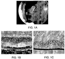
- FIG. 1 shows a biopolymer patch with HESC-RPE in situ in the pig eye one month after transplantation. This demonstrates the biocompatibility of the polymer in vivo and that the monolayer of RPE cells is essential to maintain viable photoreceptors
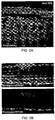
- Figure 2 shows an image of HESC-RPE on biopolymer in a pig eye, after 1 month survival. This demonstrates that transplanted HESC-RPE function as normal in vivo.
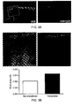
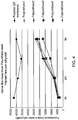
- Figure 3 shows dissociated HESC-RPE transplanted into 3 week-old dystrophic RCS rats after 5 weeks on cyclosporine. This demonstrates that transplanted cells were able to maintain a good level of visual function in the transplanted eye (compare to non-transplanted eye from the same animal). Visual acuity was measured using our optokinetic equipment and is reported in cycles per degree (c/d).
- RPE cells were seeded onto membranes at various densities to find the optimal density of seeding for highly differentiated cells.
- the cells were observed for the characteristics of RPE cells, such as pigmentation and cobblestone morphology.
- Morphology was assessed for the transparent polymers using live image capture on a phase contrast enabled inverted microscope. For non-transparent polymers morphology was assessed by immunostaining for junctional markers (Usually ZO-1)
- PLGA membranes were a kind gift from E. B. Lavik and R. Langer (Department of Chemical Engineering, MIT, Cambridge, Massachusetts). PLGA material was originally purchased as Resomer® 503H (Boehringer-Ingelheim, Ingelheim, Germany), and asymmetric PLGA membranes were synthesized as described by these authors (Lu et al, 2000a and 2000b; Lavik et al, 2001 and 2002). This asymmetric profile allows cell attachment to the smooth upper surface while also allowing cells to communicate with the basal environment through the polymer's lower porous side.
- PLGA asymmetric membrane was mounted 6.5mm Corning Transwell® inserts after having removed the original membrane. Mounting was achieved using household cyanoacrylate adhesive (Loctite, Henkel Corporation, Avon, Ohio). These inserts were sterilized by exposure of each side to a UV lamp in a laminar flow hood for 30-60 minutes. Passage 28 ARPE-19 cells were seeded onto the membranes at a density of 90,000 cell/cm 2 .
- asymmetric PLGA membrane was mounted onto empty inserts (as above).
- the inserts as well as tissue culture polystyrene dishes (for control) were coated with laminin at standard working concentration (Sigma-Aldrich, derived from the murine Engelbreth-Holm-Swarm tumour). Both dishes and inserts were then seeded with p30 ARPE19 at a higher density of 180,000 cell/cm 2 to reduce time to confluence. Cultures were maintained with DMEM High Glucose based RPE media twice per week.
- HESC lines Shefl and Shef7 were maintained in flasks coated with 0.1% Gelatine and seeded with inactivated mouse embryonic fibroblast (MEF) feeders (Draper et al, 2002).
- Cells were maintained in basic HESC-medium which is based in High glucose Knockout Dulbecco's Modified Eagle's Medium (DMEM, Invitrogen) with 20% Knockout serum replacement (Invitrogen), 1% non-essential amino acid solution, 1mM L-Glutamine (Invitrogen), 4ng/ml human bFGF (Invitrogen) and 0.1mM ⁇ -mercaptoethanol (Sigma).
- DMEM High glucose Knockout Dulbecco's Modified Eagle's Medium
- Invitrogen Knockout serum replacement
- 1mM L-Glutamine Invitrogen
- 4ng/ml human bFGF Invitrogen
- 0.1mM ⁇ -mercaptoethanol Sigma.
- HESC lines were maintained for up to 74 passages with media changes every 2 days. Cells were split regularly (1:4) in order to maintain colonies of undifferentiated HESC.
- the following coatings were applied according to their standard manufacturer protocol unless otherwise stated: matrigel (1:30), laminin, collagen IV, Human collagen I, Puramatrix®, plasmanate, poly-L-Lysine, and no coating as control. Briefly, each coating was thawed at 4°C and diluted in either PBS or serum-free media. Plates/inserts were coated either at 4°C, room temperature, or 37°C according to each manufacturer's guidelines. Matrix solution was then removed and plates were either washed or air-dried as per standard procedure. HESC-RPE colonies (primary colonies) were excised from culture flasks and placed into media following which they were seeded onto the plates/inserts.
- One batch of Human pepsinised placental laminins achieved immobilisation of the HESC-RPE colonies, whereas another batch failed. All the other coatings (Collagen IV, Human Collgen I, Poly-L-Lysine, Puramatrix®, Plasmanate) completely failed in this respect.
- the Shefl HESC are maintained in T25 flasks coated with 0.1% Gelatine and seeded with human fibroblast feeders (optimal seeding density of 2.25X10 5 cells per T25 (9X10 3 /cm 2 )).
- Cells are maintained in basic HESC-medium which consists of the following components: High glucose (4.8g/L) Knockout Dulbecco's Modified Eagle's Medium (DMEM, Invitrogen) with 20% Knockout serum replacement (Invitrogen), 1% non-essential amino acid solution, 1mM L-Glutamine (Invitrogen), 4ng/ml human bFGF (Invitrogen) and 0.1mM ⁇ -mercaptoethanol (Sigma).
- DMEM Knockout Dulbecco's Modified Eagle's Medium
- Invitrogen Knockout serum replacement
- 1mM L-Glutamine Invitrogen
- 4ng/ml human bFGF Invitrogen
- Shef 1 HESC undergo media changes every 2 days and are split regularly (1:4) in order to maintain colonies of undifferentiated HESC (assessed by staining for the markers SSEA3, SSEA4, TRA-1-60 and TRA-181).
- HESC-RPE are reliably formed when Shefl HESC colonies are allowed to become superconfluent on feeders.
- the media changing regime is altered from once every 2 days to once every day using the basic HESC media detailed above (minus bFGF). This factor was withdrawn from the media because of a documented link between bFGF and repression of RPE specification. Pigmented foci appear in superconfluent HESC cultures between 1-2 weeks following implementation of the daily feeding regime.
- HESC-RPE Pigmented HESC-RPE were harvested from either T25 flasks containing HESC-RPE colonies on feeders (passage 0), or from expanding HESC-RPE sheets on feeders (passage 1). Removal of the cells was by cutting around them with a sterile microblade and dislodging them with sterile pipette tip. HESC colonies/sheets were then aspirated and suspended in HES medium -bFGF (without bFGF) until further use.
- the desired amount of clusters is placed along with their growth media in a tube and centrifuged at 2400 to 3600 rpm for 5 minutes to enable the removal of supernatant media. Centrifugation was repeated as necessary to enable exchange of solutions as follows: Cells are washed twice with PBS and then incubated at 37°C for 20 minutes in 90% Non-enzymatic dissociation buffer in PBS (Sigma #C5914) and 10% Trypsin 10x in PBS (Trypsin from Porcine Pancreas, Sigma; final trypsin concentration 0.25%). Following this period of incubation, cells are triturated thoroughly until completely suspended i.e no visible cell clumps. They are then centrifuged again to enable removal of the dissociation buffer after which the cells are resuspended in HES medium -bFGF and placed in a 37°C incubator until further use.
- Sterilised inserts were coated with 1:30 diluted laminin-1 for 30 minutes at 37°C.
- Laminin (at a concentration of 43 ⁇ g/cm 2 and incubated at 37°C for 30 minutes). Laminin was aspirated immediately before cell seeding.
- Cell density of the HESC-RPE suspension was measured using a Neubauer® Haemocytometer with Trypan Blue 1:1 dilution. Trypan Blue staining was used to confirm cell viability which was greater than 93% in every case.
- Cells were seeded at an optimal density of 200,000 - 400,000 cell/cm 2 . Seeded cells were allowed to attach for at least 24 hours and typically 48 hours before the first media change. Media was changed 3 times a week thereafter.
Landscapes
- Health & Medical Sciences (AREA)
- Life Sciences & Earth Sciences (AREA)
- Engineering & Computer Science (AREA)
- Biomedical Technology (AREA)
- Chemical & Material Sciences (AREA)
- General Health & Medical Sciences (AREA)
- Cell Biology (AREA)
- Zoology (AREA)
- Animal Behavior & Ethology (AREA)
- Public Health (AREA)
- Veterinary Medicine (AREA)
- Medicinal Chemistry (AREA)
- Epidemiology (AREA)
- Ophthalmology & Optometry (AREA)
- Organic Chemistry (AREA)
- Bioinformatics & Cheminformatics (AREA)
- Biotechnology (AREA)
- Wood Science & Technology (AREA)
- Genetics & Genomics (AREA)
- Chemical Kinetics & Catalysis (AREA)
- Oral & Maxillofacial Surgery (AREA)
- Transplantation (AREA)
- Pharmacology & Pharmacy (AREA)
- Botany (AREA)
- Dermatology (AREA)
- Urology & Nephrology (AREA)
- Neurology (AREA)
- Neurosurgery (AREA)
- Developmental Biology & Embryology (AREA)
- Microbiology (AREA)
- Biochemistry (AREA)
- General Engineering & Computer Science (AREA)
- Virology (AREA)
- Immunology (AREA)
- General Chemical & Material Sciences (AREA)
- Nuclear Medicine, Radiotherapy & Molecular Imaging (AREA)
- Vascular Medicine (AREA)
- Heart & Thoracic Surgery (AREA)
- Cardiology (AREA)
- Micro-Organisms Or Cultivation Processes Thereof (AREA)
Applications Claiming Priority (2)
| Application Number | Priority Date | Filing Date | Title |
|---|---|---|---|
| GBGB0806746.4A GB0806746D0 (en) | 2008-04-14 | 2008-04-14 | Membrane |
| PCT/GB2009/000917 WO2009127809A1 (en) | 2008-04-14 | 2009-04-08 | Membrane |
Publications (2)
| Publication Number | Publication Date |
|---|---|
| EP2265711A1 EP2265711A1 (en) | 2010-12-29 |
| EP2265711B1 true EP2265711B1 (en) | 2018-03-07 |
Family
ID=39433610
Family Applications (1)
| Application Number | Title | Priority Date | Filing Date |
|---|---|---|---|
| EP09732279.6A Active EP2265711B1 (en) | 2008-04-14 | 2009-04-08 | Membrane |
Country Status (19)
| Country | Link |
|---|---|
| US (2) | US8642072B2 (enExample) |
| EP (1) | EP2265711B1 (enExample) |
| JP (1) | JP5552478B2 (enExample) |
| KR (2) | KR101317094B1 (enExample) |
| CN (1) | CN102016009B (enExample) |
| AU (1) | AU2009237462B2 (enExample) |
| BR (1) | BRPI0910936A2 (enExample) |
| CA (1) | CA2720264C (enExample) |
| CO (1) | CO6311115A2 (enExample) |
| DK (1) | DK2265711T3 (enExample) |
| ES (1) | ES2665284T3 (enExample) |
| GB (1) | GB0806746D0 (enExample) |
| IL (1) | IL208745A (enExample) |
| MX (1) | MX2010011232A (enExample) |
| MY (1) | MY159049A (enExample) |
| NZ (1) | NZ587753A (enExample) |
| RU (1) | RU2530169C2 (enExample) |
| WO (1) | WO2009127809A1 (enExample) |
| ZA (1) | ZA201006326B (enExample) |
Families Citing this family (24)
| Publication number | Priority date | Publication date | Assignee | Title |
|---|---|---|---|---|
| US20110112101A1 (en) * | 2009-03-05 | 2011-05-12 | Sanofi-Aventis | Treatment for ocular-related disorders |
| JP5867926B2 (ja) * | 2010-05-10 | 2016-02-24 | 公立大学法人名古屋市立大学 | 細胞シート作製方法 |
| EP4245370A3 (en) | 2010-07-12 | 2023-11-29 | University of Southern California | Biocompatible substrate for facilitating interconnections between stem cells and target tissues and methods for implanting same |
| CN103459593A (zh) * | 2011-02-25 | 2013-12-18 | 独立行政法人理化学研究所 | 视网膜色素上皮细胞片层的生产方法 |
| US8877489B2 (en) | 2011-12-05 | 2014-11-04 | California Institute Of Technology | Ultrathin parylene-C semipermeable membranes for biomedical applications |
| US10478206B2 (en) | 2011-04-29 | 2019-11-19 | University Of Southern California | Instruments and methods for the implantation of cell-seeded substrates |
| WO2012177968A1 (en) * | 2011-06-22 | 2012-12-27 | The Schepens Eye Research Institute, Inc. | A scaffold for subretinal cell transplantation and drug delivery |
| US9763777B2 (en) | 2011-07-11 | 2017-09-19 | Ear Science Institute Australia | Device for ear drum repair |
| CN103007355B (zh) * | 2011-09-20 | 2014-08-13 | 同济大学 | 一种水凝胶-纳米纤维膜及其制备方法和用途 |
| CA2792081C (en) * | 2011-10-11 | 2020-10-27 | Bond University Ltd | Layered compositions comprising 3d nanofibre webbing for tissue repair |
| CA2855941A1 (en) | 2011-11-14 | 2013-05-23 | Advanced Cell Technology, Inc. | Pharmaceutical preparations of human rpe cells and uses thereof |
| US9248013B2 (en) | 2011-12-05 | 2016-02-02 | California Institute Of Technology | 3-Dimensional parylene scaffold cage |
| US20130330825A1 (en) * | 2012-06-07 | 2013-12-12 | City Of Hope | Attachment substrates for directed differentiation of human embryonic stem cells in culture |
| US11033586B2 (en) | 2012-08-24 | 2021-06-15 | Riken | Method for producing retinal pigment epithelial cell sheet |
| RU2518972C1 (ru) * | 2012-12-24 | 2014-06-10 | Общество с ограниченной ответственностью "НАНО КАСКАД" | Трековая мембрана для фильтрации крови |
| EP3080248A1 (en) | 2013-12-11 | 2016-10-19 | Pfizer Limited | Method for producing retinal pigment epithelial cells |
| CN105013010B (zh) * | 2015-07-07 | 2018-01-12 | 中山大学中山眼科中心 | 一种辅助iPS‑RPE移植的层粘连蛋白膜 |
| CN105597145A (zh) * | 2016-02-22 | 2016-05-25 | 中山大学中山眼科中心 | 一种视网膜细胞支架生物手术粘合剂及其制备方法 |
| CN109790508A (zh) * | 2016-06-28 | 2019-05-21 | 国立研究开发法人农业·食品产业技术综合研究机构 | 细胞封入用设备及其用途 |
| WO2020047380A1 (en) | 2018-08-31 | 2020-03-05 | Wisconsin Alumni Research Foundation | Generating arterial endothelial cell-seeded vascular grafts |
| TWI820330B (zh) * | 2019-04-26 | 2023-11-01 | 國立研究開發法人理化學研究所 | 包含神經視網膜與視網膜色素上皮細胞與水凝膠之複合體及其製造方法 |
| RU2704094C1 (ru) * | 2019-05-16 | 2019-10-23 | федеральное государственное автономное учреждение "Национальный медицинский исследовательский центр "Межотраслевой научно-технический комплекс "Микрохирургия глаза" имени академика С.Н. Федорова" Министерства здравоохранения Российской Федерации | Способ трансплантации ретинального пигментного эпителия в форме многоклеточных 3D сфероидов в эксперименте |
| JP2021141877A (ja) * | 2020-03-13 | 2021-09-24 | 大日本印刷株式会社 | 細胞培養基材、細胞付細胞培養基材、細胞培養容器、細胞付細胞培養容器、上皮細胞シート及びその製造方法、被験物質の評価方法並びに評価キット |
| JP7676135B2 (ja) * | 2020-12-14 | 2025-05-14 | 日本バイリーン株式会社 | 細胞培養担体 |
Family Cites Families (4)
| Publication number | Priority date | Publication date | Assignee | Title |
|---|---|---|---|---|
| GB0118984D0 (en) * | 2001-08-03 | 2001-09-26 | Intercytex Ltd | Fusion of cells |
| CN1739813A (zh) * | 2005-09-15 | 2006-03-01 | 上海赐人合元企业发展有限公司 | 组织工程表皮替代物及其制备方法 |
| KR100901343B1 (ko) | 2007-07-23 | 2009-06-05 | (주)실리콘화일 | 결정질 반도체 박막 제조 방법 |
| RU2368359C1 (ru) * | 2008-07-22 | 2009-09-27 | Государственное учреждение "УФИМСКИЙ НАУЧНО-ИССЛЕДОВАТЕЛЬСКИЙ ИНСТИТУТ ГЛАЗНЫХ БОЛЕЗНЕЙ" Академии наук Республики Башкортостан (УфНИИ ГБ АН РБ) | Способ лечения возрастной макулярной дегенерации |
-
2008
- 2008-04-14 GB GBGB0806746.4A patent/GB0806746D0/en not_active Ceased
-
2009
- 2009-04-08 KR KR1020107022953A patent/KR101317094B1/ko not_active Expired - Fee Related
- 2009-04-08 CN CN200980113395.3A patent/CN102016009B/zh active Active
- 2009-04-08 DK DK09732279.6T patent/DK2265711T3/en active
- 2009-04-08 MX MX2010011232A patent/MX2010011232A/es active IP Right Grant
- 2009-04-08 US US12/933,694 patent/US8642072B2/en active Active
- 2009-04-08 KR KR1020137014211A patent/KR20130065737A/ko not_active Ceased
- 2009-04-08 RU RU2010144399/10A patent/RU2530169C2/ru not_active IP Right Cessation
- 2009-04-08 BR BRPI0910936A patent/BRPI0910936A2/pt not_active Application Discontinuation
- 2009-04-08 CA CA2720264A patent/CA2720264C/en active Active
- 2009-04-08 EP EP09732279.6A patent/EP2265711B1/en active Active
- 2009-04-08 NZ NZ587753A patent/NZ587753A/en unknown
- 2009-04-08 JP JP2011504521A patent/JP5552478B2/ja active Active
- 2009-04-08 AU AU2009237462A patent/AU2009237462B2/en active Active
- 2009-04-08 ES ES09732279.6T patent/ES2665284T3/es active Active
- 2009-04-08 WO PCT/GB2009/000917 patent/WO2009127809A1/en not_active Ceased
- 2009-04-08 MY MYPI2010004529A patent/MY159049A/en unknown
-
2010
- 2010-09-03 ZA ZA2010/06326A patent/ZA201006326B/en unknown
- 2010-10-14 IL IL208745A patent/IL208745A/en not_active IP Right Cessation
- 2010-11-12 CO CO10142317A patent/CO6311115A2/es active IP Right Grant
-
2014
- 2014-01-03 US US14/146,744 patent/US8889179B2/en active Active
Non-Patent Citations (1)
| Title |
|---|
| CHUNG YOUNG ET AL: "Human embryonic stem cell lines generated without embryo destruction", CELL STEM CELL, ELSEVIER, CELL PRESS, AMSTERDAM, NL, vol. 2, no. 2, 7 February 2008 (2008-02-07), pages 113 - 117, XP002604696, ISSN: 1934-5909, DOI: 10.1016/J.STEM.2007.12.013 * |
Also Published As
| Publication number | Publication date |
|---|---|
| GB0806746D0 (en) | 2008-05-14 |
| EP2265711A1 (en) | 2010-12-29 |
| IL208745A0 (en) | 2010-12-30 |
| DK2265711T3 (en) | 2018-04-30 |
| WO2009127809A1 (en) | 2009-10-22 |
| MX2010011232A (es) | 2011-02-23 |
| KR101317094B1 (ko) | 2013-10-16 |
| KR20100128329A (ko) | 2010-12-07 |
| ZA201006326B (en) | 2011-09-28 |
| BRPI0910936A2 (pt) | 2015-10-06 |
| US8889179B2 (en) | 2014-11-18 |
| CN102016009A (zh) | 2011-04-13 |
| ES2665284T3 (es) | 2018-04-25 |
| IL208745A (en) | 2015-04-30 |
| MY159049A (en) | 2016-12-15 |
| CN102016009B (zh) | 2015-05-20 |
| JP5552478B2 (ja) | 2014-07-16 |
| CO6311115A2 (es) | 2011-08-22 |
| US8642072B2 (en) | 2014-02-04 |
| JP2011517696A (ja) | 2011-06-16 |
| US20140127282A1 (en) | 2014-05-08 |
| RU2010144399A (ru) | 2012-05-20 |
| AU2009237462A1 (en) | 2009-10-22 |
| KR20130065737A (ko) | 2013-06-19 |
| RU2530169C2 (ru) | 2014-10-10 |
| CA2720264A1 (en) | 2009-10-22 |
| AU2009237462B2 (en) | 2012-12-20 |
| NZ587753A (en) | 2012-07-27 |
| CA2720264C (en) | 2016-09-27 |
| US20110236464A1 (en) | 2011-09-29 |
Similar Documents
| Publication | Publication Date | Title |
|---|---|---|
| EP2265711B1 (en) | Membrane | |
| US11154639B2 (en) | Biocompatible substrate for facilitating interconnections between stem cells and target tissues and methods for implanting same | |
| US5713957A (en) | Corneal onlays | |
| Koo et al. | Micro-and nanotopography with extracellular matrix coating modulate human corneal endothelial cell behavior | |
| WO2013074681A1 (en) | Pharmaceutical preparations of human rpe cells and uses thereof | |
| JP2020006207A (ja) | 組織再生培養細胞シート、製造方法及びその利用方法 | |
| HK1146092B (en) | Membrane | |
| HK1146092A (en) | Membrane | |
| Mishan et al. | Comparative culture of human corneal endothelial cells following treatment with human platelet lysate/fibrin hydrogel versus Y-27632 ROCK inhibitor: in vitro and ex vivo study |
Legal Events
| Date | Code | Title | Description |
|---|---|---|---|
| PUAI | Public reference made under article 153(3) epc to a published international application that has entered the european phase |
Free format text: ORIGINAL CODE: 0009012 |
|
| 17P | Request for examination filed |
Effective date: 20101006 |
|
| AK | Designated contracting states |
Kind code of ref document: A1 Designated state(s): AT BE BG CH CY CZ DE DK EE ES FI FR GB GR HR HU IE IS IT LI LT LU LV MC MK MT NL NO PL PT RO SE SI SK TR |
|
| AX | Request for extension of the european patent |
Extension state: AL BA RS |
|
| REG | Reference to a national code |
Ref country code: HK Ref legal event code: DE Ref document number: 1146092 Country of ref document: HK |
|
| DAX | Request for extension of the european patent (deleted) | ||
| 17Q | First examination report despatched |
Effective date: 20131009 |
|
| REG | Reference to a national code |
Ref country code: DE Ref legal event code: R079 Ref document number: 602009051104 Country of ref document: DE Free format text: PREVIOUS MAIN CLASS: C12N0005080000 Ipc: C12N0005079000 |
|
| RIC1 | Information provided on ipc code assigned before grant |
Ipc: A61L 27/38 20060101ALI20161207BHEP Ipc: C12N 5/079 20100101AFI20161207BHEP |
|
| GRAP | Despatch of communication of intention to grant a patent |
Free format text: ORIGINAL CODE: EPIDOSNIGR1 |
|
| STAA | Information on the status of an ep patent application or granted ep patent |
Free format text: STATUS: GRANT OF PATENT IS INTENDED |
|
| INTG | Intention to grant announced |
Effective date: 20170315 |
|
| GRAJ | Information related to disapproval of communication of intention to grant by the applicant or resumption of examination proceedings by the epo deleted |
Free format text: ORIGINAL CODE: EPIDOSDIGR1 |
|
| STAA | Information on the status of an ep patent application or granted ep patent |
Free format text: STATUS: EXAMINATION IS IN PROGRESS |
|
| INTC | Intention to grant announced (deleted) | ||
| GRAP | Despatch of communication of intention to grant a patent |
Free format text: ORIGINAL CODE: EPIDOSNIGR1 |
|
| STAA | Information on the status of an ep patent application or granted ep patent |
Free format text: STATUS: GRANT OF PATENT IS INTENDED |
|
| INTG | Intention to grant announced |
Effective date: 20170921 |
|
| REG | Reference to a national code |
Ref country code: HK Ref legal event code: WD Ref document number: 1146092 Country of ref document: HK |
|
| GRAS | Grant fee paid |
Free format text: ORIGINAL CODE: EPIDOSNIGR3 |
|
| GRAA | (expected) grant |
Free format text: ORIGINAL CODE: 0009210 |
|
| STAA | Information on the status of an ep patent application or granted ep patent |
Free format text: STATUS: THE PATENT HAS BEEN GRANTED |
|
| AK | Designated contracting states |
Kind code of ref document: B1 Designated state(s): AT BE BG CH CY CZ DE DK EE ES FI FR GB GR HR HU IE IS IT LI LT LU LV MC MK MT NL NO PL PT RO SE SI SK TR |
|
| REG | Reference to a national code |
Ref country code: GB Ref legal event code: FG4D |
|
| REG | Reference to a national code |
Ref country code: CH Ref legal event code: EP Ref country code: AT Ref legal event code: REF Ref document number: 976589 Country of ref document: AT Kind code of ref document: T Effective date: 20180315 |
|
| REG | Reference to a national code |
Ref country code: IE Ref legal event code: FG4D |
|
| REG | Reference to a national code |
Ref country code: DE Ref legal event code: R096 Ref document number: 602009051104 Country of ref document: DE |
|
| REG | Reference to a national code |
Ref country code: NL Ref legal event code: FP |
|
| REG | Reference to a national code |
Ref country code: FR Ref legal event code: PLFP Year of fee payment: 10 |
|
| REG | Reference to a national code |
Ref country code: ES Ref legal event code: FG2A Ref document number: 2665284 Country of ref document: ES Kind code of ref document: T3 Effective date: 20180425 |
|
| REG | Reference to a national code |
Ref country code: DK Ref legal event code: T3 Effective date: 20180423 |
|
| REG | Reference to a national code |
Ref country code: SE Ref legal event code: TRGR |
|
| REG | Reference to a national code |
Ref country code: NO Ref legal event code: T2 Effective date: 20180307 |
|
| REG | Reference to a national code |
Ref country code: LT Ref legal event code: MG4D |
|
| PG25 | Lapsed in a contracting state [announced via postgrant information from national office to epo] |
Ref country code: LT Free format text: LAPSE BECAUSE OF FAILURE TO SUBMIT A TRANSLATION OF THE DESCRIPTION OR TO PAY THE FEE WITHIN THE PRESCRIBED TIME-LIMIT Effective date: 20180307 Ref country code: HR Free format text: LAPSE BECAUSE OF FAILURE TO SUBMIT A TRANSLATION OF THE DESCRIPTION OR TO PAY THE FEE WITHIN THE PRESCRIBED TIME-LIMIT Effective date: 20180307 Ref country code: CY Free format text: LAPSE BECAUSE OF FAILURE TO SUBMIT A TRANSLATION OF THE DESCRIPTION OR TO PAY THE FEE WITHIN THE PRESCRIBED TIME-LIMIT Effective date: 20180307 |
|
| REG | Reference to a national code |
Ref country code: HK Ref legal event code: GR Ref document number: 1146092 Country of ref document: HK |
|
| PG25 | Lapsed in a contracting state [announced via postgrant information from national office to epo] |
Ref country code: BG Free format text: LAPSE BECAUSE OF FAILURE TO SUBMIT A TRANSLATION OF THE DESCRIPTION OR TO PAY THE FEE WITHIN THE PRESCRIBED TIME-LIMIT Effective date: 20180607 Ref country code: GR Free format text: LAPSE BECAUSE OF FAILURE TO SUBMIT A TRANSLATION OF THE DESCRIPTION OR TO PAY THE FEE WITHIN THE PRESCRIBED TIME-LIMIT Effective date: 20180608 Ref country code: LV Free format text: LAPSE BECAUSE OF FAILURE TO SUBMIT A TRANSLATION OF THE DESCRIPTION OR TO PAY THE FEE WITHIN THE PRESCRIBED TIME-LIMIT Effective date: 20180307 |
|
| PG25 | Lapsed in a contracting state [announced via postgrant information from national office to epo] |
Ref country code: PL Free format text: LAPSE BECAUSE OF FAILURE TO SUBMIT A TRANSLATION OF THE DESCRIPTION OR TO PAY THE FEE WITHIN THE PRESCRIBED TIME-LIMIT Effective date: 20180307 Ref country code: RO Free format text: LAPSE BECAUSE OF FAILURE TO SUBMIT A TRANSLATION OF THE DESCRIPTION OR TO PAY THE FEE WITHIN THE PRESCRIBED TIME-LIMIT Effective date: 20180307 Ref country code: EE Free format text: LAPSE BECAUSE OF FAILURE TO SUBMIT A TRANSLATION OF THE DESCRIPTION OR TO PAY THE FEE WITHIN THE PRESCRIBED TIME-LIMIT Effective date: 20180307 Ref country code: IT Free format text: LAPSE BECAUSE OF FAILURE TO SUBMIT A TRANSLATION OF THE DESCRIPTION OR TO PAY THE FEE WITHIN THE PRESCRIBED TIME-LIMIT Effective date: 20180307 |
|
| PG25 | Lapsed in a contracting state [announced via postgrant information from national office to epo] |
Ref country code: SK Free format text: LAPSE BECAUSE OF FAILURE TO SUBMIT A TRANSLATION OF THE DESCRIPTION OR TO PAY THE FEE WITHIN THE PRESCRIBED TIME-LIMIT Effective date: 20180307 Ref country code: CZ Free format text: LAPSE BECAUSE OF FAILURE TO SUBMIT A TRANSLATION OF THE DESCRIPTION OR TO PAY THE FEE WITHIN THE PRESCRIBED TIME-LIMIT Effective date: 20180307 |
|
| REG | Reference to a national code |
Ref country code: DE Ref legal event code: R097 Ref document number: 602009051104 Country of ref document: DE |
|
| PG25 | Lapsed in a contracting state [announced via postgrant information from national office to epo] |
Ref country code: PT Free format text: LAPSE BECAUSE OF FAILURE TO SUBMIT A TRANSLATION OF THE DESCRIPTION OR TO PAY THE FEE WITHIN THE PRESCRIBED TIME-LIMIT Effective date: 20180709 |
|
| PLBE | No opposition filed within time limit |
Free format text: ORIGINAL CODE: 0009261 |
|
| STAA | Information on the status of an ep patent application or granted ep patent |
Free format text: STATUS: NO OPPOSITION FILED WITHIN TIME LIMIT |
|
| PG25 | Lapsed in a contracting state [announced via postgrant information from national office to epo] |
Ref country code: MC Free format text: LAPSE BECAUSE OF FAILURE TO SUBMIT A TRANSLATION OF THE DESCRIPTION OR TO PAY THE FEE WITHIN THE PRESCRIBED TIME-LIMIT Effective date: 20180307 |
|
| 26N | No opposition filed |
Effective date: 20181210 |
|
| PG25 | Lapsed in a contracting state [announced via postgrant information from national office to epo] |
Ref country code: SI Free format text: LAPSE BECAUSE OF FAILURE TO SUBMIT A TRANSLATION OF THE DESCRIPTION OR TO PAY THE FEE WITHIN THE PRESCRIBED TIME-LIMIT Effective date: 20180307 |
|
| PG25 | Lapsed in a contracting state [announced via postgrant information from national office to epo] |
Ref country code: MT Free format text: LAPSE BECAUSE OF NON-PAYMENT OF DUE FEES Effective date: 20180408 |
|
| PG25 | Lapsed in a contracting state [announced via postgrant information from national office to epo] |
Ref country code: TR Free format text: LAPSE BECAUSE OF FAILURE TO SUBMIT A TRANSLATION OF THE DESCRIPTION OR TO PAY THE FEE WITHIN THE PRESCRIBED TIME-LIMIT Effective date: 20180307 |
|
| PG25 | Lapsed in a contracting state [announced via postgrant information from national office to epo] |
Ref country code: HU Free format text: LAPSE BECAUSE OF FAILURE TO SUBMIT A TRANSLATION OF THE DESCRIPTION OR TO PAY THE FEE WITHIN THE PRESCRIBED TIME-LIMIT; INVALID AB INITIO Effective date: 20090408 |
|
| REG | Reference to a national code |
Ref country code: AT Ref legal event code: UEP Ref document number: 976589 Country of ref document: AT Kind code of ref document: T Effective date: 20180307 |
|
| PG25 | Lapsed in a contracting state [announced via postgrant information from national office to epo] |
Ref country code: MK Free format text: LAPSE BECAUSE OF NON-PAYMENT OF DUE FEES Effective date: 20180307 |
|
| PG25 | Lapsed in a contracting state [announced via postgrant information from national office to epo] |
Ref country code: IS Free format text: LAPSE BECAUSE OF FAILURE TO SUBMIT A TRANSLATION OF THE DESCRIPTION OR TO PAY THE FEE WITHIN THE PRESCRIBED TIME-LIMIT Effective date: 20180707 |
|
| P01 | Opt-out of the competence of the unified patent court (upc) registered |
Effective date: 20230522 |
|
| P02 | Opt-out of the competence of the unified patent court (upc) changed |
Effective date: 20230526 |
|
| PGFP | Annual fee paid to national office [announced via postgrant information from national office to epo] |
Ref country code: NL Payment date: 20250427 Year of fee payment: 17 |
|
| PGFP | Annual fee paid to national office [announced via postgrant information from national office to epo] |
Ref country code: LU Payment date: 20250428 Year of fee payment: 17 |
|
| PGFP | Annual fee paid to national office [announced via postgrant information from national office to epo] |
Ref country code: FI Payment date: 20250425 Year of fee payment: 17 |
|
| PGFP | Annual fee paid to national office [announced via postgrant information from national office to epo] |
Ref country code: DE Payment date: 20250429 Year of fee payment: 17 |
|
| PGFP | Annual fee paid to national office [announced via postgrant information from national office to epo] |
Ref country code: GB Payment date: 20250428 Year of fee payment: 17 Ref country code: DK Payment date: 20250425 Year of fee payment: 17 Ref country code: ES Payment date: 20250505 Year of fee payment: 17 |
|
| PGFP | Annual fee paid to national office [announced via postgrant information from national office to epo] |
Ref country code: NO Payment date: 20250429 Year of fee payment: 17 |
|
| PGFP | Annual fee paid to national office [announced via postgrant information from national office to epo] |
Ref country code: BE Payment date: 20250428 Year of fee payment: 17 |
|
| PGFP | Annual fee paid to national office [announced via postgrant information from national office to epo] |
Ref country code: FR Payment date: 20250425 Year of fee payment: 17 |
|
| PGFP | Annual fee paid to national office [announced via postgrant information from national office to epo] |
Ref country code: CH Payment date: 20250501 Year of fee payment: 17 |
|
| PGFP | Annual fee paid to national office [announced via postgrant information from national office to epo] |
Ref country code: AT Payment date: 20250319 Year of fee payment: 17 |
|
| PGFP | Annual fee paid to national office [announced via postgrant information from national office to epo] |
Ref country code: IE Payment date: 20250428 Year of fee payment: 17 |
|
| PGFP | Annual fee paid to national office [announced via postgrant information from national office to epo] |
Ref country code: SE Payment date: 20250430 Year of fee payment: 17 |