EP2201038B1 - Materialien und methoden für die modifizierung der bindung von antikörpern - Google Patents
Materialien und methoden für die modifizierung der bindung von antikörpern Download PDFInfo
- Publication number
- EP2201038B1 EP2201038B1 EP08806434.0A EP08806434A EP2201038B1 EP 2201038 B1 EP2201038 B1 EP 2201038B1 EP 08806434 A EP08806434 A EP 08806434A EP 2201038 B1 EP2201038 B1 EP 2201038B1
- Authority
- EP
- European Patent Office
- Prior art keywords
- binding
- mfe
- antibody
- αvβ6
- cells
- Prior art date
- Legal status (The legal status is an assumption and is not a legal conclusion. Google has not performed a legal analysis and makes no representation as to the accuracy of the status listed.)
- Active
Links
Images








Classifications
-
- C—CHEMISTRY; METALLURGY
- C07—ORGANIC CHEMISTRY
- C07K—PEPTIDES
- C07K16/00—Immunoglobulins [IG], e.g. monoclonal or polyclonal antibodies
- C07K16/18—Immunoglobulins [IG], e.g. monoclonal or polyclonal antibodies against material from animals or humans
- C07K16/28—Immunoglobulins [IG], e.g. monoclonal or polyclonal antibodies against material from animals or humans against receptors, cell surface antigens or cell surface determinants
- C07K16/2839—Immunoglobulins [IG], e.g. monoclonal or polyclonal antibodies against material from animals or humans against receptors, cell surface antigens or cell surface determinants against the integrin superfamily
-
- A—HUMAN NECESSITIES
- A61—MEDICAL OR VETERINARY SCIENCE; HYGIENE
- A61K—PREPARATIONS FOR MEDICAL, DENTAL OR TOILETRY PURPOSES
- A61K39/00—Medicinal preparations containing antigens or antibodies
- A61K39/395—Antibodies; Immunoglobulins; Immune serum, e.g. antilymphocytic serum
- A61K39/39533—Antibodies; Immunoglobulins; Immune serum, e.g. antilymphocytic serum against materials from animals
- A61K39/39541—Antibodies; Immunoglobulins; Immune serum, e.g. antilymphocytic serum against materials from animals against normal tissues, cells
-
- A—HUMAN NECESSITIES
- A61—MEDICAL OR VETERINARY SCIENCE; HYGIENE
- A61K—PREPARATIONS FOR MEDICAL, DENTAL OR TOILETRY PURPOSES
- A61K39/00—Medicinal preparations containing antigens or antibodies
- A61K39/395—Antibodies; Immunoglobulins; Immune serum, e.g. antilymphocytic serum
- A61K39/39533—Antibodies; Immunoglobulins; Immune serum, e.g. antilymphocytic serum against materials from animals
- A61K39/39558—Antibodies; Immunoglobulins; Immune serum, e.g. antilymphocytic serum against materials from animals against tumor tissues, cells, antigens
-
- C—CHEMISTRY; METALLURGY
- C07—ORGANIC CHEMISTRY
- C07K—PEPTIDES
- C07K16/00—Immunoglobulins [IG], e.g. monoclonal or polyclonal antibodies
- C07K16/18—Immunoglobulins [IG], e.g. monoclonal or polyclonal antibodies against material from animals or humans
- C07K16/28—Immunoglobulins [IG], e.g. monoclonal or polyclonal antibodies against material from animals or humans against receptors, cell surface antigens or cell surface determinants
- C07K16/30—Immunoglobulins [IG], e.g. monoclonal or polyclonal antibodies against material from animals or humans against receptors, cell surface antigens or cell surface determinants from tumour cells
- C07K16/3007—Carcino-embryonic Antigens
-
- C—CHEMISTRY; METALLURGY
- C07—ORGANIC CHEMISTRY
- C07K—PEPTIDES
- C07K2317/00—Immunoglobulins specific features
- C07K2317/20—Immunoglobulins specific features characterized by taxonomic origin
- C07K2317/24—Immunoglobulins specific features characterized by taxonomic origin containing regions, domains or residues from different species, e.g. chimeric, humanized or veneered
-
- C—CHEMISTRY; METALLURGY
- C07—ORGANIC CHEMISTRY
- C07K—PEPTIDES
- C07K2317/00—Immunoglobulins specific features
- C07K2317/30—Immunoglobulins specific features characterized by aspects of specificity or valency
- C07K2317/31—Immunoglobulins specific features characterized by aspects of specificity or valency multispecific
-
- C—CHEMISTRY; METALLURGY
- C07—ORGANIC CHEMISTRY
- C07K—PEPTIDES
- C07K2317/00—Immunoglobulins specific features
- C07K2317/50—Immunoglobulins specific features characterized by immunoglobulin fragments
- C07K2317/56—Immunoglobulins specific features characterized by immunoglobulin fragments variable (Fv) region, i.e. VH and/or VL
- C07K2317/565—Complementarity determining region [CDR]
-
- C—CHEMISTRY; METALLURGY
- C07—ORGANIC CHEMISTRY
- C07K—PEPTIDES
- C07K2317/00—Immunoglobulins specific features
- C07K2317/60—Immunoglobulins specific features characterized by non-natural combinations of immunoglobulin fragments
- C07K2317/62—Immunoglobulins specific features characterized by non-natural combinations of immunoglobulin fragments comprising only variable region components
- C07K2317/622—Single chain antibody (scFv)
-
- C—CHEMISTRY; METALLURGY
- C07—ORGANIC CHEMISTRY
- C07K—PEPTIDES
- C07K2317/00—Immunoglobulins specific features
- C07K2317/70—Immunoglobulins specific features characterized by effect upon binding to a cell or to an antigen
- C07K2317/77—Internalization into the cell
-
- C—CHEMISTRY; METALLURGY
- C07—ORGANIC CHEMISTRY
- C07K—PEPTIDES
- C07K2318/00—Antibody mimetics or scaffolds
- C07K2318/10—Immunoglobulin or domain(s) thereof as scaffolds for inserted non-Ig peptide sequences, e.g. for vaccination purposes
-
- C—CHEMISTRY; METALLURGY
- C07—ORGANIC CHEMISTRY
- C07K—PEPTIDES
- C07K2319/00—Fusion polypeptide
Definitions
- the present invention relates to methods for modifying the binding of antibodies, and more particularly to obtaining antibodies by inserting an amino acid sequence capable of binding to a target into a complementarity determining region of a parent antibody so that the antibody thus obtained is capable of binding to the target.
- the present invention further relates to methods of producing the antibodies.
- Integrins are a family of heterodimeric class I transmembrane receptors. Individual integrins comprise an ⁇ and ⁇ subunit in non covalent association and there are known to exist at least 18 ⁇ subunits and 8 ⁇ subunits that can form 24 different heterodimers. They are involved in numerous cell-matrix and cell-cell interactions and facilitate cell adhesion, proliferation, migration and invasion. These processes occur in several normal and pathological processes, including wound healing, inflammation and tumour growth and metastasis.
- ⁇ v ⁇ 6 is an epithelial cell-restricted integrin and has been shown to be expressed in malignant but not in normal epithelium. De novo expression of this integrin has been reported in oral squamous cell carcinomas (SCC) and ovarian cancer tissues and cancer cell lines and over-expression in adenocarcinomas of the breast, and ovarian cancer, colon carcinoma, oral squamous cell carcinoma and in gastroenteropancreatic adenocarcinomas, in particular in pancreatic ductal adenocarcinomas.
- SCC oral squamous cell carcinomas
- ovarian cancer tissues and cancer cell lines over-expression in adenocarcinomas of the breast, and ovarian cancer, colon carcinoma, oral squamous cell carcinoma and in gastroenteropancreatic adenocarcinomas, in particular in pancreatic ductal adenocarcinomas.
- WO2007/008712 describes anti- ⁇ v ⁇ 6 antibodies and uses thereof.
- WO2007/039728 discloses experiments in which ⁇ v ⁇ 6 peptide ligands comprising the sequence motif RGDLXXL/I, wherein LXXL/I is contained within an alpha helical structure are investigated.
- the use of this peptide motif arose from studies in which ⁇ v ⁇ 6 expression was involved in activation of autocrine TGF- ⁇ in post-EMT cells.
- the Latency Associated Protein (LAP) of the latent LAP-TGF ⁇ 1 complex is a known ligand for ⁇ v ⁇ 6 and binding has a role in the activation of TGF- ⁇ 1.
- the LAP protein contains the arginine-glycine-aspartic acid (RGD) sequence, a known binding motif for most integrins.
- RGD arginine-glycine-aspartic acid
- a further ligand for ⁇ v ⁇ 6 is the viral protein 1 (VP1) of the foot-and-mouth disease virus (FMDV), which also contains the RGD motif.
- FMDV uses ⁇ v ⁇ 6 to attach to host cells and the integrin most likely also plays a role in virus uptake into endosomes.
- the binding of VP1 specifically to ⁇ v ⁇ 6 is mediated via residues immediately following and including the aspartic acid of the RGD motif; the DLXXL sequence has been identified as an additional ⁇ v ⁇ 6 binding motif from its ability to inhibit ⁇ v ⁇ 6-fibronectin interactions.
- the present invention concerns methods for obtaining antibodies that by inserting an amino acid sequence that is capable of binding to a target into the complementarity determining region (CDR) of a parent antibody so that the antibody thus obtained is capable of binding to the target.
- CDR complementarity determining region
- this approach may be used to introduce a new binding specificity to the antibody.
- the insertion is made in one of the CDRs of the parent antibody, CDR H3, the insertion often has the effect of reducing or abolishing the binding of the parent antibody for the antigen to which it initially bound.
- the antibodies disclosed herein are useful as they are able to bind to the target, such as ⁇ v ⁇ 6 integrin, while retaining one or more of the useful properties of the parent antibody, such as interacting with the immune system (e.g.
- the parent antibodies are based on MFE-23 antibodies and variants and derivatives thereof as discussed in more detail below.
- the present invention provides a method of modifying a parent antibody to an antibody capable of binding to ⁇ v ⁇ 6 integrin, wherein the parent antibody comprises the following complementary determining regions (CDRs):
- the method further comprises humanizing the antibody or the parent antibody by mutating one or more amino acid residues.
- the parent antibody is a scFv or a diabody.
- the scFv or diabody may comprise a linker having the sequence (Gly 4 Ser) n wherein n is between 1 and 4.
- the insertion reduces the binding of the parent antibody to human carcinoembryonic antigen (CEA).
- CEA human carcinoembryonic antigen
- the method further comprises conjugating the antibody to:
- a peptide sequence capable of binding to a target such as ⁇ v ⁇ 6 integrin
- a target such as ⁇ v ⁇ 6 integrin
- a target such as ⁇ v ⁇ 6 integrin
- a parent antibody in which a peptide sequence capable of binding to a target, such as ⁇ v ⁇ 6 integrin, is inserted into at least one complementarity determining region of a parent antibody in order to modify the parent antibody so that it is capable of binding to the target, while retaining other useful properties of the parent antibody, such as the antibody's properties in interacting with the immune system (e.g. recruitment of complement), stability and half-life when administered in vivo, especially when compared to the corresponding peptide, and ease of production.
- Multiple target-binding sequences may be inserted into different complementarity determining regions of the parent antibody.
- a target-binding sequence may be grafted onto one or more of the complementarity determining regions of the parent antibody.
- the presence of multiple target binding sequences may permit the formation of dimeric or multimeric forms of the antibody. Examples of targets and the peptide sequences that are capable of binding to them are discussed in more detail below.
- MFE-23 antibodies include the following examples from this family as parent antibody scaffolds that may be used in the present disclosure.
- MFE-23 was originally a scFv isolated from a murine phage display library and selected for specific binding to carcinoembryonic antigen (CEA) ( Chester et al., Lancet 343: 455-456, 1994 ). MFE-23 has been humanised in order to reduce the likelihood of immunogenicity (hMFE) and the humanised antibody has been affinity matured to produce the mutant scFv sm3E.
- CEA carcinoembryonic antigen
- MFE-23 A stabilised humanised form of MFE-23 is referred to as shMFE.
- MFE-23 antibodies, and derivatives thereof, are disclosed in WO95/15341 or EP 0733072A and US7232888 .
- the antibodies have also undergone successful pre-clinical, and in some cases clinical, studies support its potential for use in targeted cancer therapies (e.g., radioimmunotherapy, cellular immunotherapy, gene therapy, targeted cytokine therapy) and as an imaging agent.
- targeted cancer therapies e.g., radioimmunotherapy, cellular immunotherapy, gene therapy, targeted cytokine therapy
- imaging agent include Phase I studies of radiolabelled MFE-23 for use as an imaging agent, for radioimmunoguided surgery and as the tumour-targeting moiety of an antibody directed enzyme prodrug therapy.
- MFE-23 antibodies comprise the following six CDRs, as shown in SEQ ID NO: 6 of US7232888 (with the exception of CDR (d)(ii) disclosed elsewhere in the patent [and CDR (e) (i) disclosed elsewhere in the patent]):
- (i) is derived from the sequence of the CDR of the murine MFE-23 antibodies and (ii) is derived from the sequence of the CDR of humanised MFE-23.
- CDR loops are the accessible regions for interaction with the epitopes of the antigen to which the parent antibody was specific, it is generally desirable in the present disclosure to insert the targeting peptide sequences into these regions.
- the peptide sequence as defined in the claims, is inserted into the CDR H3 region of the parent antibody between residues Thr98 and Gly99. This is because the present inventors realised that in the parent MFE-23 antibodies, CDR H3 is the longest and most variable CDR and is essential for antigen binding.
- the site between Thr98 and Gly99 was selected as the sequence is at the end of a protruding loop structure and therefore means that the inserted targeting peptide should be available for binding to the target.
- the method of the present invention employs an antibody which is a scFv or a diabody which comprises a linker having the sequence (Gly 4 Ser) n , wherein n is between 1 and 4.
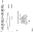
- a scFv or diabody having this general structure is represented in Figure 1 .
- the insertion may also be combined with mutation of one or more amino acid residues of the CDRs or framework regions of the parent antibody to modify or otherwise improve the properties of parent antibody, for example to reduce or eliminate the binding of the parent antibody to the antigen to which it was raised or was initially capable of specifically binding or to humanise the antibody or to increase affinity for the target.
- it may be useful to retain the specificity of the parent antibody so that the antibody of the present disclosure is bispecific.
- Examples of other changes made to the amino acid sequence of the parent antibody include mutation of G44 to C44 of the VH domain and A100 to C100 of the VL domain to introduce two cysteine residues to form an inter-molecular stabilising disulphide bridge in a diabody and Y 100b to P 100b of the VH domain to help to reduce or eliminate any remaining residual binding to CEA.
- Further examples of other changes to the amino acid sequence of the parent antibody include mutations to improve affinity, specificity or stability and methods for achieving this are well known in the art. For example, if affinity maturation or increased specificity is required, residues in the CDR H3 could be varied using PCR mutagenesis or site directed mutagenesis.
- the remaining CDRs could be varied using PCR mutagenesis, site directed mutagenesis or rational addition/replacement of peptides (Hoogenboom, (2005) and references therein).
- the resulting mutated genes would be cloned and expressed as scFvs in filamentous bacteriophage libraries.
- the libraries would be screened for phage displaying scFvs with improved binding and biological efficacy. If increased stability or yield is required the whole DNA could be varied using PCR mutagenesis, site directed mutagenesis or growth in mutator strains of bacteria (Hoogenboom, (2005) and references therein).
- the resulting mutated genes would be cloned and expressed as scFvs in filamentous bacteriophage libraries.
- the libraries would be screened for phage displaying scFvs with high expression levels and stability.
- it may also be useful to reduce the immunogenicity.
- Potential immunogenicity of the scFv for example HFEVP1 or affinity matured variant, could be addressed by identification and modification of T-cell epitopes and methods for this are well known in the art. This is based on the rationale that T cell help is required in order to mount a long-lived, isotype switched and high affinity antibody response [Chester KA, et al (2005)].
- the approach requires the identification of short peptides contained within the protein sequence that have a capacity to bind to the MHC class II binding groove and stimulate a subsequent T-cell response.
- Key residues in these peptides that contribute to T-cell epitope formation can be identified by determining whether the side chains interact with key binding pockets in the MHC class II binding groove or with the T-cell receptor (TCR). Subsequent amino acid substitutions at these key residues can inhibit T-cell epitope formation by reducing the peptide affinity for MHC class II or preventing TCR recognition of the peptide MHC complex.
- An experimental approach using a T-cell proliferation assays could be used as this takes into account factors that contribute to epitope formation, such as antigen processing and TCR binding.
- peptides spanning the whole of the scFv sequence for example, 15mer peptides overlapping by 12 amino acids.
- the peptides could be tested for ability to stimulate T-cells in T-cell proliferation assays.
- T-cells from healthy donors expressing a wide range of HLA alleles would be used in addition to T-cells obtained from any patients demonstrating an immune response to the scFv.
- the precise location of key residues responsible for T-cell activation could be determined by residue replacement (usually with alanine) and testing the resultant peptides in T-cell proliferation assays. In silico analysis could also be used to predict amino acid substitutions that should disrupt peptides binding to MHC class II.
- MFE-23 antibody includes antibodies which comprise the CDRs of the scFv MFE-23, fragments or derivatives thereof. It also includes the CDRs when incorporated into other antibody framework as described above, e.g. to produce a complete antibody structure, a diabody or another antibody. These include antibody fragments which comprise an antigen binding domain are such as Fab, scFv, Fv, dAb, Fd and diabodies. It is possible to take monoclonal and other antibodies and use techniques of recombinant DNA technology to produce other antibodies or chimeric molecules which retain the specificity of the original antibody.
- Such techniques may involve introducing DNA encoding the immunoglobulin variable region, or the complementarity determining regions (CDRs), of an antibody to the constant regions, or constant regions plus framework regions, of a different immunoglobulin. See, for instance, EP 0 184 187 A , GB 2,188,638 A or EP 0 239 400 A .
- Antibodies can be modified in a number of ways and the term "antibody” should be construed as covering any specific binding member or substance having an antibody antigen-binding domain with the required specificity. Thus, this term covers antibody fragments and derivatives, including any polypeptide comprising an immunoglobulin binding domain, whether natural or wholly or partially synthetic. Chimeric molecules comprising an immunoglobulin binding domain, or equivalent, fused to another polypeptide are therefore included. Cloning and expression of chimeric antibodies are described in EP 0 120 694 A and EP 0 125 023 A .
- binding fragments are (i) the Fab fragment consisting of VL, VH, CL and CH1 domains; (ii) the Fd fragment consisting of the VH and CH1 domains; (iii) the Fv fragment consisting of the VL and VH domains of a single antibody; (iv) the dAb fragment ( Ward, E.S.
- Fv, scFv or diabody molecules may be stabilised by the incorporation of disulphide bridges linking the VH and VL domains ( Reiter et al, Nature Biotech, 14: 1239-1245, 1996 ).
- Minibodies comprising a scFv joined to a CH3 domain may also be made ( Hu et al, Cancer Res., 56: 3055-3061, 1996 ).
- antibodies In terms of utility of these antibodies, this is primarily therapeutic with imaging/diagnosis being a secondary utility.
- antibodies have advantages over peptides as it is generally appreciated in the art that peptides tend to have short serum half-life in vivo. In contrast, antibody therapeutics tend to possess greater stability and longer half-life. Additional advantages of preferred antibodies of the present disclosure include combining high affinity binding properties of the ⁇ v ⁇ 6 binding peptides together with advantages of the MFE antibody scaffold, namely stability, good production yields, e.g. in Pichia pastoris, and for humanised versions of the MFE antibody scaffold, low predicted immunogenic potential.
- targeting peptides that are capable of binding to a target.
- the targeting peptide has the amino acid sequence AVPNLRGDLQVLAQKVA.
- the inserted amino acid sequences capable of binding to the target are between 8, 10 or 12 amino acids and 24 or 30 amino acids in length, and more preferably are between 12 and 20 amino acids in length.
- the target may be an antigen present on diseased cells, to which the antibodies of the present disclosure are capable of binding using the targeting peptide.
- the diseased cells may be surface antigen on, for example, cancer cells.
- the targeting peptide may be capable of binding to an integrin, such as ⁇ v ⁇ 6 integrin which is known to be involved in normal and pathological processes such as wound healing, inflammation and tumour growth and metastasis.
- the targets for the antibodies of the present disclosure may be targets that (i) are overexpressed or de novo expressed in diseased cells (and more specifically cancer cells) and/or (ii) are expressed in diseased cells and mediate the disease (and more specifically cancer).
- the present disclosure includes antibodies which can be used as tumour targeting antibodies, where target is overexpressed or de novo expressed in diseased cell, and/or function-modulating antibodies, e.g. whereby the target is expressed on the diseased cell and the antibody modulates the activity of the target and/or the targets ability to interact with its binding partners.
- tumour associated antigens and/or antigens involved in mediated disease such as MUC1, 5T4, VEGFR (Hofmeister V et al., 2007), Tie2, endoglin (CD105) (Hofmeister V et al., 2007; Munoz R et al., 2007), uPA receptor (uPAR) (Li Y et al., 2007), PSMA (Baccala A et al., 2007, Buhler P et al., 2007) 4,5 , and members of the ErbB (Her) family (Johnston JB et al., 2006).
- preferred peptides would include epitopes of the antigen-binding protein that are located on surface-exposed loops, making use of the three-dimensional structure of the antigen-binding protein.
- MFE/shMFE/VP1 constructs we used the knowledge that the inserted 17-mer VP1 peptide is part of a long highly mobile loop that forms a self contained unit in the VP1 protein of FMDV ( Logan, D., Abu-Ghazaleh, R., Blakemore, W., Curry, S., Jackson, T., King, A., Lea, S., Lewis, R., Newman, J., Parry, N., (1993) Structure of a major immunogenic site on foot-and-mouse disease virus Nature 362:566-568 ).
- preferred peptides could be rationally designed using three-dimensional structures of the ligand-antigen complex if these are available.
- the ligand residues that are involved in binding to the cell surface antigen are known from the detail of the ligand-antigen interaction.
- preferred peptides could be those which are potent in inhibiting antigen/antigen-binding protein interactions as shown in vitro in ELISA or cell-based assays.
- a 17-mer peptide that contained the 17-mer VP1 peptide sequence minus the N-terminal alanine and a 20-mer peptide that contained the 17-mer peptide sequence used to create MFEVP1 were potent inhibitors of FMDV binding to purified ⁇ v ⁇ 6 and ⁇ v ⁇ 6-expressing cells ( Burman, A., Clark, S., Abrescia, N. G., Fry, E. E., Stuart, D. I., and Jackson, T.
- preferred peptides could be those that are selected in vitro or in vivo from peptide libraries displayed on filamentous bacteriophage. This method has been used to obtain peptides with high affinity and specificity for tumour targets ( Landon, L.A., Deutscher, S.L. (2003) Combinatorial Discovery of Tumor Targeting Peptides Using Phage Display. J Cell Biochem. 90:509-517 ). Peptide libraries consist of random cyclic and linear peptide sequences. In vitro selection is performed using purified recombinant proteins or cells that express the target protein.
- the 17-mer peptide VP1 peptide sequence inserted into MFE-23 to create MFEVP1 contains the DLXXL sequence, which has specificity for ⁇ v ⁇ 6, and has also been identified from screening 12-mer peptide libraries displayed on phage using recombinant transmembrane truncated soluble receptor.
- a random phage library consisting of linear 12 amino acid peptides identified peptides that bound specifically to recombinant prostate-specific membrane antigen (PMSA), an attractive candidate for targeted therapy for prostate and other solid tumours ( Aggarwal, S., Singh, P., Topaloglu, O., Isaacs, J.T., Denmeade, S.R. (2006) A Dimeric Peptide That Binds Selectively to Prostate-Specific Membrane Antigen and Inhibits its Enzymatic Activity Cancer Res. 66:9171-9177 ).
- a pIII-displayed disulphide-constrained heptamer peptide library was screened on purified extracellular PSMA to identify PSMA binding peptides.
- the epidermal growth factor receptor, type 2 (ErbB-2) tyrosine kinase receptor that shows increasing promise as a target for cancer diagnosis and therapy has been used as bait.
- a random 6 amino acid peptide bacteriophage display library was selected against purified ErbB-2 extracellular domain to identify peptide binding sequences ( Karasseva, N.G., Glinsky, V.V., Chen, N.X., Komatireddy, R., Quinn, T.P. (2002) Identification and Characterization of Peptides that Bind Human ErbB-2 Selected from a Bacteriophage Display Library J. Protein Chem. 21:287-296 ).
- Peptide sequences can also be selected by display of peptide libraries on phage that bind to carbohydrate antigens, which show different composition in malignant transformations.
- an in vitro phage display was employed to find peptides that bind to the Thomsen-Friedenreich (TF) tumour-associated antigen (Peletskaya, E.N., Glinsky, V.V., Glinsky, G.V., et al. (1997) Characterisation of peptides that bind the tumour-associated Thomson-Friendenreich antigen selected from bacteriophage display libraries J. Mol. Biol. 270:374-384 ).
- TF Thomsen-Friedenreich
- Peptide library selection on cells was used to identify a further ⁇ v ⁇ 6 binding peptide sequence.
- a 20-mer peptide library fused to bacteriophage coat protein pIII selected a peptide that bound specifically to a lung adenocarcinoma cell line and was shown subsequently to bind to ⁇ v ⁇ 6 ( Elayadi, A.N., Samli, K.N., Prudkin, L., Liu, Y.-H., Bian, A., Xie, X.-J., Wistuba, I.I., Roth, J.A., McGuire, M.J., Brown, K.C. (2007) A Peptide Selected by Biopanning Identifies the Integrin avb6 as a Prognostic Biomarker for Nonsmall Cell Lung Cancer. Cancer Res. 67:5889-5895 ).
- the RIP1-Tag2 mouse model of multistage tumorigenesis involving the pancreatic islets of Langerhans allowed the identification of several peptides that discriminate between the vasculature of the premalignant angiogenic islets and the fully developed tumours ( Joyce, J.A., Laakkonen, P., Bernasconi, M., Bergers, G., Ruoslahti, E., Hanahan, D. (2003) Stage-specific vascular markers revealed by phage display in a mouse model of pancreatic islet tumorigenesis Cancer Cell 4:309-403 ).
- Peptides specific for human vasculature and human tissue have been derived from peptide phage library selection on human tissue transplanted into SCID mice ( George, A.J.T., Lee, L., Pitzalis, C. (2003) Isolating ligands specific for human vasculature using in vivo phage selection Trends Biotech. 21:199-203 ).
- These peptides include peptides which comprise the amino acid sequence motif RGDLX 5 X 6 X 7 , wherein X 5 , X 6 and X 7 independently represent any amino acid residue, and preferably independently selected from Glu, Ala, Leu, Met, Gln, Lys, Arg, Val, Ile, Trp, Phe, Asp, His and Thr.
- peptides disclosed in WO2007/039728 generally comprise an alpha helical structure induced by the amino acids following the RGD sequence, this is not a requirement of the peptides of the present disclosure, in particular as they are inserted into the CDR of a parent antibody and may not therefore be able to adopt this secondary structural feature.
- Preferred examples of peptides capable of binding to ⁇ v ⁇ 6 integrin include peptides which comprise amino acid sequences represented by the following general formulae, based on the peptides disclosed in WO2007/039728 : ... RGDLX 5 X 6 X 7 . wherein
- peptide sequence above may be part of a longer sequences, for example having one, two, three, four, five, ten or more additional amino acids linked to the N- and/or C-terminus of the peptide, while optionally conforming to the preferred ranges of lengths of peptide sequence set out above.
- the targeting peptide sequence employed in the present invention has the amino acid sequence AVPNLRGDLQVLAQKVA.
- the peptides may comprise an "alpha helical structure" as disclosed in WO2007/039728 , that is a sequential group of amino acids in a peptide that interact with a particular hydrogen bonding pattern and thus define a helical structure.
- the hydrogen bonding pattern in a standard alpha helix is between the carbonyl oxygen of residue n and the amide hydrogen of residue n+4.
- this hydrogen bonding pattern is between residues n and n+3 and for a pi-helix it is between residues n and n+5.
- the number of residues per turn in each alpha-helix is 3.6, 3.0 and 4.4 for the standard alpha-helix, 3 10 -helix and pi-helix respectively, see for example WO95/00534 .
- the method of the present invention may further comprise conjugating the antibody to a detectable moiety.
- detectable moiety relates to a group that, when located at the target site following administration of the antibodies to a patient, may be detected, typically non-invasively from outside the body and the site of the target located.
- the antibodies disclosed herein are useful in imaging and diagnosis.
- Detectable moiety are entities that are detectable by imaging techniques such as Magnetic Resonance Imaging (MRI), Magnetic Resonance Spectroscopy (MRS), Single Photon Emission Computed Tomography (SPECT) and Positron Emission Tomography (PET) and optical imaging.
- imaging moieties are stable, non-toxic entities that retain their properties under in vitro and in vivo conditions.
- moieties include but are not limited to radioactive moieties, for example radioactive isotopes. Suitable radioactive atoms include technetium-99m or iodine-123 for scintigraphic studies. Other readily detectable moieties include, for example, spin labels for MRI such as iodine-123 again, iodine-131, indium-111, fluorine-18, carbon-13, nitrogen-15, oxygen-17, gadolinium, manganese or iron and optical moieties which include Cy5.5 and quantum dots.
- radioactive moieties for example radioactive isotopes. Suitable radioactive atoms include technetium-99m or iodine-123 for scintigraphic studies.
- Other readily detectable moieties include, for example, spin labels for MRI such as iodine-123 again, iodine-131, indium-111, fluorine-18, carbon-13, nitrogen-15, oxygen-17, gadolinium, manganese or iron and optical moieties which include
- the antibodies of the present disclosure may be conjugated or linked to a therapeutically active moiety, for example a moiety that is cytotoxic.
- affinity tags that can be introduced into the antibodies to enable them to be manipulated or detected in one or more subsequent steps.
- affinity tags include members of specific binding pairs, antibodies and antigens, biotin which binds to streptavidin and avidin, polyhistidine (e.g.
- hexa-His or tri-His tags or amino di- or tri-carboxylates which bind to metal ions such as Ni 2+ or Co 2+ , Flag or Glu epitopes which bind to anti-Flag antibodies, S-tags which bind to streptavidin, calmodulin binding peptide which binds to calmodulin in the presence of Ca 2+ ; ribonuclease S which binds to aporibonuclease S; and c-Myc which recognises anti-c-Myc antibody.
- affinity tags that can be used in accordance with the present disclosure will be apparent to those skilled in the art. Antibodies including these affinity tags can be easily purified and manipulated.
- therapeutically active moiety encompasses a moiety having beneficial, prophylactic and/or therapeutic properties.
- the therapeutically active moiety is a cytotoxic chemotherapeutic agent.
- Cytotoxic chemotherapeutic agents are well known in the art and include anti-cancer agents such as:
- the antibodies of the present disclosure may be formulated using particle based delivery systems such as nanoparticles and lipid-based vesicles such as liposomes or other similar structures composed of lipids.
- Liposomes are a spherical vesicles comprising a phospholipid bilayer that may be used as agents to deliver materials such as drugs or genetic material.
- Liposomes can be composed of naturally-derived phospholipids with mixed lipid chains (egg phosphatidylethanolamine) or of pure components like DOPE (dioleolylphosphatidylethanolamine). The synthesis and use of liposomes is now well established in the art.
- Liposomes are generally created by sonication of phospholipids in a suitable medium such as water. Low shear rates create multilamellar liposomes having multi-layered structures. Continued high-shear sonication tends to form smaller unilamellar liposomes. Research has also been able to enable liposomes to avoid detection by the immune system, for examples by coating the lipsomes with polyethylene glycol (PEG). It is also possible to incorporate species in liposomes, such as the peptides of the disclosure to help to target them to a delivery site, e.g. in cells or in vivo.
- PEG polyethylene glycol
- nanoparticles as delivery agents for materials associated with or bound to the nanoparticles
- Some types of nanoparticle comprises a core, often of metal and/or semiconductor atoms, to which ligands of one or more different types may be linked, including, for example, one or more of the peptides of the present disclosure, see for example WO02/32404 , WO2005/10816 and WO2005/116226 .
- Other types of nanoparticle may be formed from materials such as liposomes.
- the nanoparticles may be derivatised or conjugated to other ligands may be present to provide the nanoparticles with different properties or functions.
- the nanoparticles may be quantum dots, that is nanocrystals of semiconducting materials which have the striking chemical and physical properties that differ markedly from those of the bulk solid (see Gleiter, Adv. Mater. 1992, 4, 474-481 ). Now that their quantum size effects are understood, fundamental and applied research on these systems has become increasingly popular.
- An interesting application is the use of nanocrystals as luminescent labels for biological systems, see for example Brucher et al, Science 1998, 281, 2013-2016 , Chan & Nie, Science, 1998, 281, 2016-2018 , Mattousi et al, J. Am. Chem. Soc., 2000, 122, 12142-12150 , and Alivisatos, Pure Appl. Chem. 2000, 72, 3-9 .
- the quantum dots have several advantages over conventional fluorescent dyes: quantum dots emit light at a variety of precise wavelengths depending on their size and have long luminescent lifetimes.
- the cytotoxic moiety is a cytotoxic peptide or polypeptide moiety by which we include any moiety which leads to cell death.
- Cytotoxic peptide and polypeptide moieties are well known in the art and include, for example, ricin, abrin, Pseudomonas exotoxin, RNase, tissue factor and the like.
- ricin as a cytotoxic agent is described in Burrows & Thorpe, P.N.A.S. USA 90: 8996-9000, 1993 ,
- tissue factor which leads to localised blood clotting and infarction of a tumour, has been described by Ran et al., Cancer Res. 58: 4646-4653, 1998 and Huang et al., Science 275: 25 547-550, 1997 . Tsai et al., Dis.
- Colon Rectum 38: 1067- 1074, 1995 describes the abrin A chain conjugated to a monoclonal antibody.
- Other ribosome inactivating proteins are described as cytotoxic agents in WO 96/06641 .
- Pseudomonas exotoxin may also be used as the cytotoxic polypeptide moiety (see, for example, Aiello et al, P.N.A.S. USA 92: 10457-10461, 1995 .
- cytokines such as TNF ⁇ and IL-2, may also be useful as cytotoxic and/or therapeutic agents.
- radioactive atoms may also be cytotoxic if delivered in sufficient doses.
- the cytotoxic moiety may comprise a radioactive atom which, in use, delivers a sufficient quantity of radioactivity to the target site so as to be cytotoxic.
- Suitable radioactive atoms include phosphorus-32, iodine-125, iodine-131, indium-111, rhenium-186, rhenium- 188 or yttrium-90, or any other isotope which emits enough energy to destroy neighbouring cells, organelles or nucleic acid.
- the isotopes and density of radioactive atoms in the antibody of the disclosure are such that a dose of more than 4000 cGy, and more preferably at least 6000, 8000 or 10000 cGy, is delivered to the target site and, preferably, to the cells at the target site and their organelles, particularly the nucleus.
- the radioactive atom may be attached to the binding moiety in known ways.
- EDTA or another chelating agent may be attached to the binding moiety and used to attach 111 In or 90 Y.
- Tyrosine residues may be labelled witch 125 I or 131 I.
- the present disclosure provides a polypeptide linked to viral coat protein other than FMDV to change the trophism of the virus for delivery of DNA encoding therapeutic genes.
- any of these systems can be incorporated into a prodrug system.
- prodrug systems are well known in the art and include ADEPT systems in which an antibody according to the present disclosure is conjugated or conjugatable or fused to an agent capable of converting a prodrug to a cytotoxic moiety is an enzyme for use in antibody directed enzyme prodrug therapy.
- composition comprising peptide and/or nucleic acid and/or expression vector as defined above and a pharmaceutical acceptable carrier.
- pharmaceutically acceptable carrier generally includes components that are compatible with the peptide, nucleic acid or vector and are not deleterious to the recipients thereof.
- the carriers will be water or saline which will be sterile and pyrogen free.
- other acceptable carriers may be used.
- the pharmaceutical compositions or formulations of the disclosure are for parenteral administration, more particularly for intravenous administration.
- an antibody as described herein, or nucleic acid encoding the antibody or a vector comprising the nucleic acid for the preparation of a medicament for the treatment of a condition characterised by diseased cells which express the target, for example a condition in which the cells overexpress the target and/or display the target on the cell surface and/or which is disease that is mediated by the target.
- Such conditions include cancer, for example by making use of the antigens expressed on the surface of cancer cells.
- the conditions may include ⁇ v ⁇ 6-mediated diseases or diseases in which cells overexpress ⁇ v ⁇ 6, such as cancer, chronic fibrosis, chronic obstructive pulmonary disease (COPD), lung emphysemia or chronic wounding skin disease, such as epidermolysis bullosa.
- COPD chronic obstructive pulmonary disease
- lung emphysemia or chronic wounding skin disease, such as epidermolysis bullosa.
- these conditions also include the treatment of wound healing and inflammation.
- the medicament or pharmaceutical composition of the present disclosure as defined above may usefully be administered to a patient who is also administered other medicaments, as it will be known to those skilled in the art.
- the medicament or pharmaceutical composition of the present disclosure may be administered to a patient before, after or during administration of the other anti-tumour agent(s), for example before, after or during chemotherapy.
- Treatment with the antibody after chemotherapy may be particularly useful in reducing or preventing recurrence of the tumour or metastasis.
- the anti-tumour agent can be covalently linked directly or indirectly (via liposomes/nanoparticles) to an antibody of the present disclosure.
- Also disclosed herein is a method of imaging epithelial cells overexpressing ⁇ v ⁇ 6 in the body of an individual, the method comprising administering to the individual an effective amount of an antibody as defined herein.
- the method is particularly useful for the imaging of chronic fibrosis, chronic obstructive pulmonary disease (COPD), lung emphysema, chronic wounding skin disease (e.g. epidermolysis bullosa) or epithelial tumour cells.
- the method of imaging may include linking the targeting antibody to a fluorescent probe and incorporate into a mouth-wash, chewing gum, spray or other emolument such that the antibody-probe conjugate may be visualised by its fluorescent tag.
- the parent antibody is a single-chain Fv antibody fragment (scFv), which consists of the variable heavy and variable light chain regions tethered by a flexible linker that retains the complete antibody's binding properties.
- scFv single-chain Fv antibody fragment
- the scFv may be based on a MFE-23 scFv, a scFv developed by phage technology that binds with high affinity to the carcinoembryonic antigen, CEA (14).
- CEA is a tumour selective marker that is highly expressed on most gastrointestinal carcinomas and on a number of breast, lung and ovarian carcinomas.
- Iodinated MFE has been used in patients for imaging (15) and radioimmunoguided surgery in colorectal cancer (16). MFE has also shown promise for cancer therapy when used as a fusion protein with carboxypeptidase G2 in antibody-directed enzyme prodrug therapy (ADEPT)(17);(18).
- ADPT antibody-directed enzyme prodrug therapy
- a scFv against ⁇ v ⁇ 6 was produced rationally by antibody engineering by inserting the peptide binding motifs of the known ⁇ v ⁇ 6 peptide ligands, such as VP1 proteins, into the loop region of MFE.
- the third complementarity-determining region (CRD3) of the variable heavy chain (VH) of MFE provides the major site of interaction with CEA, as assessed by mutagenesis, and accordingly was chosen as a preferred site for such an insertion.
- Murine monoclonal antibodies to ⁇ v ⁇ 3 (LM609), ⁇ v ⁇ 6 (10D5) and ⁇ 5 ⁇ 1 (P1D6) were purchased from Chemicon International, Harrow, UK whereas those to ⁇ v ⁇ 5 (P1F6) and ⁇ v ⁇ 8(14E5) were generous gifts from Drs. Dean Sheppard and Steve Nishimura (UCSF), respectively.
- the secondary antibody was Alexafluor-488 conjugated rabbit anti-mouse antisera (Molecular Probes) unless otherwise stated.
- MFE-RGD and MFE-RGE plasmid Construction of MFE-RGD and MFE-RGE plasmid, MFE-RGD / pUC119 and MFE-RGE / pUC119
- MFE-RGD and MFE23-RGE were constructed by site-directed mutagenesis using the MFE-RGD forward (5'_CTACTGCAACGAAGGGACA GCTAGAGGTGATTTGGCTACTTTG TTCGACTACTGGG GACAAG 3') and MFE-RGD reverse (5'_CTTGTCCCCAGTAGTCGAA CAAAGTAGCCAAATCACCTCTAGC TGTCCCTTCGTTG CAGTAG_3') primers or MFE-RGE forward ( 5'_GAAGGGACAGCTAGAGGTGA A TTGGCTACTTTGTTCGACTACTG_3') and MFE-RGE reverse ( 5'_CAGTAGTCGAACAAAGTAGCCAA T TCACCTCTAGCTGTCCCTTC_3') respectively.
- MFE-RGD forward 5'_CTACTGCAACGAAGGGACA GCTAGAGGTGATTTGGCTACTTTG TTCGACTACTGGG GACAAG 3'
- MFE-RGD reverse 5'_CTTGTCCC
- the PCR reactions used the phMFEhis_119 plasmid as template, which incorporated the humanised form of MFE-23 (hMFE) and a C-terminal 6xHis tag enabling purification by Immobilised Metal Ion Affinity Chromatography (IMAC).
- IMAC Immobilised Metal Ion Affinity Chromatography
- MFEVP1 and HFEVP1 which contain the MFE of HFE sequences respectively, and a 17-mer ⁇ v ⁇ 6-binding peptide of VP1 in the CRD3 loop of the heavy chain ( Figure 1 ), were constructed from three PCR reactions.
- the 5'end were constructed with the VH MFEVP1 (5' CATG CCATGG CCCAGGTGAAACTG) or VH HFEVP1 (5' CATG CCATGG CCCAAGTTAAACTGGAACAG TCC) sense primers and the MFEVP1 (5' GCGCCAGCACCTGCAGATCACCTCGCAG ATTCGGAACTGCAGTCGGAGTCCCCTCAT TAC) or HFEVP1 (5' GAGCCAGCACCTGCAGATCACCTCGCAGATTCGGAACTGC AGTTGGTGTCCCTTCGT TGC) anti-sense primers, respectively, that contained parts of the additional peptides of the ⁇ v ⁇ 6-binding motif of VP1 (shown underlined in the primes).
- the 3'ends were constructed with the VL MFEVP1 (5' ATAGTTTA GCGGCCGC CCGTTTCAGCTC) or VL HFEVP1 (5' ATAGTTTA GCGGCCGC AGCCTTGATTTC) anti-sense primers and the MFEVP1 (5' CTGCGAGGTGATCTGCAGGTGCTGGCGC AGAAAGTTGCAGGGCCGTACTACTTTGAC TACTG) or HFEVP1 (5' CTGCGAGGTGATCTGCAGGTGCTGGCTC AGAAAGTTGCAGGTCCTTAC CCT TTCGAC TACTGGGGACAAGG) sense primers, respectively which contain part of the additional ⁇ v ⁇ 6-binding motif and in the case of HFEVP1 also introduced the Y 100b to P 100b mutation (shown bold and underlined in the primer; amino acid numbering is given with the Kabat nomenclature).
- the PCR reactions for MFEVP1 used the MFE/puc119 as template and those for HFEVP1 the HFE/pCTCON plasmid (27) as templates.
- the PCR products from the first two reactions were used as templates and amplified with the VH MFEVP1 sense and VL MFEVP1 anti-sense or VH NFEVP1 sense and VL HFECP1 anti-sense primers to give the PCR product for MFEVP1 or HFEVP1 respectively.
- the VH sense and VL anti-sense primers introduced NcoI and NotI sites (shown in bold and underlined in the primers) into the PCR products, respectively.
- the third PCR products and the puC119 plasmid were treated with these restriction enzymes and ligated to yield either MFEVP1/pUC119 or HFEVP1 plasmids. Correctness of the DNA sequences was verified by DNA sequencing.
- the Y 100b to P 100b mutations of the VH domain of the MFEVP1 was introduced by site-directed mutagenesis.
- the Pro mutation was introduced in MFEVP1/pUC119 with 5'GTTGCAGGGCCGTAC CCG TTTGACTACTGGGGC 3' as the sense and 5'GCCCCAGTAGTCAAA CGG GTACGGCCCTGCAAC 3' as the anti-sense primers to give NFEVP1/pUC119 (the Pro nucleotide sequence is shown in bold). DNA sequencing verified these Pro mutations.
- the NFEVP1/pUC119 and HFEVP1/pUC119 plasmid were digested with SfiI and NotI and cloned into an equally digested pICZ ⁇ BHis or pPICZ ⁇ BCysHis vectors respectively, for expression in yeast.
- the modified pICZ ⁇ BHis and pICZ ⁇ BCysHis vectors when compared to the original pPICZ ⁇ B vector (Invitrogen, Düsseldorf, Germany), do not contain the myc-tag but the His tag is present.
- the pPICZ ⁇ BCysHis vector in addition contains a Cys immediately before the six His residues.
- the MFE-RGD/pUC119 and MFE-RGE/pUC119 plasmids were electroporated into competent E. coli TG1 cells and grown on 2xYT, containing ampicillin (100 ⁇ g/ml) and 1% glucose plates at 37°C. Single colonies were used to inoculate 10ml of 2xYT, containing ampicillin (as above) and 1%glucose media and after 1:500 dilution were grown in 50ml of 2xYT, ampicillin (as above) and 0.1% glucose at 37°C until the OD 600nm was 0.9.
- IPTG isopropyl- ⁇ -D-thiogalactoside
- MFE-RGD and MFE-RGE proteins were purified under native conditions from bacterial supernatant using the Qiagen Ni-NTA Spin Column kit broadly according to the manufacturer's instructions. Briefly, a spin column holding a nickel-containing resin was equilibrated with "Lysis Buffer" (see below) before supernatant containing the 6xHis-tagged protein was spun through and the column washed to remove non-specifically bound material. During this procedure exposed his-tags bind to the nickel resin, and are therefore retained specifically in the column. His-tagged proteins were then eluted by two spins with an imidazole buffer.
- bacterial supernatant was loaded directly onto the equilibrated column (thereby avoiding the lysis steps) and this step repeated until approximately 4.8ml bacterial supernatant had passed through each column.
- Buffer recipes were altered as follows: "Lysis Buffer”: PBS, 300mM sodium chloride; "Wash Buffer”: PBS, 300mM sodium chloride, 20mM imidazole; "Elution Buffer”: PBS, 300mM sodium chloride, 250mM imidazole.
- Bound protein was eluted with 2 x 200 ⁇ l Elution Buffer and the two fractions dialysed separately against PBS using Slide-a-lyser dialysis cassettes (Perbio Science UK Ltd, Cramlington, UK) according to the manufacturer's instructions. Dialysed protein was removed from the Slide-a-lyser and stored at -80°C.
- the proteins were eluted with 200mM Imidazole/PBS/1M NaCl. The matrix was then washed with 5 volumes of 0.1M EDTA. Each washing and elution step was done with 4.5 ml volumes. Fractions of interest were pooled and dialyzed against PBS. Protein yields in the 200 mM imidazole fraction measured after dialysis were as follows: hMFE, 0.181 mg/ml, hMFE23-RGD, 0.101 mg/ml and hMFE23-RGE, 0.108 mg/ml. Washing, eluted and dialysed samples revealed by Western Blotting are shown in Fig. 9 A-C .
- the MFEVP1/pUC119 and NFEVP1/pUC119 plasmids were electroporated into competent E. coli TG1 cells and grown on 2xYT, containing ampicillin (50 ⁇ g/ml) and 1% glucose plates at 37°C. Single colonies were used to inoculate 5ml of 2xYT, containing ampicillin (as above) and 1%glucose media and after 1:500 dilution were grown in 2x500ml of 2xYT, ampicillin (as above) and 0.05% glucose at 37°C until the OD 600nm was 0.9.
- IPTG isopropyl- ⁇ -D-thiogalactoside
- MFEVP1 and NFEVP1 were by immobilized metal-affinity chromatography (IMAC).
- IMAC immobilized metal-affinity chromatography
- Ten ml of Cu-charged StreamlineTM chelating resin (GE Healthcare) were incubated with the supernatant, after addition of 1M NaCl, at RT for 1h.
- the resin was collected, washed with 1M NaCl/PBS, 40mM imidazole and bound proteins were eluted with 200mM imidazole.
- the 200mM imidazole protein containing fractions were dialysed against TBS, concentrated by an Amicon stirred cell with a YM3 membrane (Millipore) and further purified by size-exclusion chromatography.
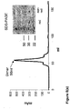
- Size-exclusion chromatography was performed on Superdex75 column in Tris buffered saline, pH 7.5 (TBS) at 1.5 ml/min. MFEVP1 and NFEVP1 eluted as two peaks, representing the monomeric (67 ml) and dimeric (56 ml) forms. Their molecular weights were estimated from molecular weight standards, Ovalbumin (44 kDa), Carbonic Anhydrase (29kDa), and Myoglobin (17kDa) and the monomeric and dimeric forms of MFE.
- NFEVP1 and HFEVP1 in P. pastoris were linearized with PmeI and transformed into electrocompetent X33 cells (Invitrogen) by electroporation. Transformants were grown on YPDS/100 ⁇ g/ml Zeocin (Invitrogen, Düsseldorf, Germany) plates. Single colonies were screened for protein expression and for inserts by PCR with the 5'AOX and 3'AOX primers. The colony with highest protein expression was stored in 20% glycerol at -80°C.
- NFEVP1 and HFEVP1 were produced by fermentation and initial purification involved expanded bed adsorption immobilized metal affinity chromatography (EBA-IMAC), which also concentrates the proteins, following previously described procedures.(22;23)
- EBA-IMAC expanded bed adsorption immobilized metal affinity chromatography
- the 200 mM imidazole EBA-IMAC eluate fraction, containing either NFEVP1 or HFEVP1 were dialysed into PBS.
- 1M NaCl was added and this was applied to a Ni2+ charged HiTrap Chelating HP 1ml affinity column (GE Healthcare) for further concentration. Elution in 1ml fractions was by 500mM imidazole/1M NaCl/PBS.
- the diabody containing eluate (2ml) was further purified by size-exclusion chromatography on a Superdex 75 column in PBS, with a flow rate of 1.5ml/min.
- Proteins were analysed by SDS/PAGE using Tris/Glycine Gels (Invitrogen) and stained with Coomassie brilliant blue.
- Proteins separated by SDS-PAGE were transferred to a PVDF membrane (Bio-Rad) at 125mA for 90min.
- the membrane was blocked with 5% milk proteins (Marvel)/PBS for 2-16h at RT. Detection was performed by incubation with mouse anti-His4 (1:1000 dilution) followed by incubation with HRP-conjugated sheep anti-mouse IgG (1:1000 dilution, GEHealthcare). Both antibodies were diluted in 1% milk proteins/PBS (w/v) and incubation was for 1h at RT.
- TBS solutions contained 1mM MgCl 2 , 1mM MnCl 2 and 1mM CaCl 2 (TBSM), and dilutions of MFE, MFEVP1, NFEVP1 and HFEVP1 and mouse anti- ⁇ v (1:1000, Chemicon International, Harrow, UK) in 1% Marvel in TBSM were added (100 ⁇ l/well).
- the plate was incubated for 1h at RT, washed, and incubated for 1h with 100 ⁇ l/well of rabbit anti-MFE and with anti- ⁇ v for mouse anti- ⁇ v wells (1:1000), washed and incubated for 1 h with 100 ⁇ l/well of HRP-conjugated goat anti-rabbit IgG (Sigma, 1:1000 dilution) and with sheep HRP-labelled anti-mouse IgG (GE Healthcare, 1:1000 dilution) for anti- ⁇ v wells.
- HRP-conjugated goat anti-rabbit IgG Sigma, 1:1000 dilution
- sheep HRP-labelled anti-mouse IgG GE Healthcare, 1:1000 dilution
- Bound samples were detected by applying 100 ⁇ l of the substrate o-phenylenediamine dihydrochloride (OPD, Sigma) in citrate buffer pH 5.0; the reaction was stopped with 100 ⁇ l of 4M HCl and the absorbance read at 490 nm on a Dynex Technologies Plate Reader.
- OPD substrate o-phenylenediamine dihydrochloride
- the diluent was TBS containing 10 mM EDTA (pH 7.5) and all washing steps included 10 mM EDTA.
- MFEVP1, NFEVP1, MFE, hMFE23-RGD, hMFE23-RGE and hMFE were diluted in 1% Marvel in PBS and 100 ⁇ l added to the wells in triplicate, washed as above, incubated with rabbit anti-MFE (1:1000 dilution) for MFEVP1, NFEVP1, MFE and with mouse anti-His4 (1:1000 dilution, Qiagen Ltd.) for hMFE23-RGD, hMFE23-RGE and hMFE washed once with 0.1% Tween 20/PBS and four times with H 2 O, and incubated with HRP-conjugated goat anti-rabbit IgG (1:1000 dilution) for MFEVP1, NFEVP1, MFE and with HRP-conjugated sheep anti-mouse IgG (1:1000 dilution, GE Healthcare) for hMFE23-RGD, hMFE23-RGE and hMFE.
- LS-174T cells were washed twice with PBS and detached with trypsin/EDTA (Cambrex). On average 5x10 5 cells were incubated with 50 ⁇ g/ml of MFEVP1, NFEVP1 and MFE and washed with PBS. Detection of binding was first by incubation with rabbit anti-MFE IgG (1:100 dilution), washing with PBS and second by incubation with 1 ⁇ g of R-Phycoerythrin (R-PE)-conjugated goat anti-rabbit IgG (Invitrogen, Düsseldorf, Germany) followed by washing with PBS.
- R-PE R-Phycoerythrin
- A375P ⁇ 6 and A375Ppuro cells were washed once in Dulbecco's modified Eagle's medium (DMEM) supplemented with 0.1% (w/v) Bovine Serum Albumin (BSA) and 0.1% (w/v) sodium azide (DMEM 0.1/0.1) and re-suspended in an appropriate volume. 50 ⁇ l of this suspension containing approximately 2 x 10 5 cells was transferred to individual wells of V-bottomed 96-well plates and mixed with 50 ⁇ l of MFEVP1, MFE and 10D5 (Chemicon International, Harrow, UK) at various concentrations.
- DMEM Dulbecco's modified Eagle's medium
- BSA Bovine Serum Albumin
- DMEM 0.1/0.1 sodium azide
- Flow cytometric analysis of anti- ⁇ v ⁇ 6 , anti- ⁇ v ⁇ 8 , anti- ⁇ v ⁇ 5, anti- ⁇ v ⁇ 3 and ⁇ v ⁇ 1 binding to the ⁇ v ⁇ 6 expressing cell line, A375P ⁇ 6 and the parent cell line, A375Ppuro Flow cytometric analysis of RGD-directed integrin expression - A375Ppuro and A375P ⁇ 6puro cells were detached with trypsin/EDTA, resuspended in DMEM 0.1/0.1 to 2.10 5 cells/50 ⁇ l and mixed with 50 ⁇ l of anti-integrin antibodies (at 10 ⁇ g/ml).
- A375P ⁇ 6 and A375Ppuro cells were trypsinized, re-suspended in DMEM, containing L-glutamine, supplemented with 10% heat-inactivated fetal bovine serum.
- Cells ( ⁇ 2x10 5 ) were seeded in 2ml of the above media in 24mm 2 dishes containing glass coverslips and allowed to attach for 48 hrs at 37°C.
- the media was removed and exchanged with DMEM containing 1% heat-inactivated fetal bovine serum and 50 ⁇ g/mL of NFEVP1 and either directly incubated at 10min, 30min, 1 hr and 3 hr at 37°C or first pre-incubated at 4°C for 1hr, upon which the scFv was removed, and the cells were shifted to 37°C.
- Haptotactic cell migration assays were performed using matrix coated polycarbonate filters (8 ⁇ m pore size, Transwell®, Becton Dickinson, Oxford, UK). The membrane undersurface was coated with LAP (0.5 ⁇ g/ml) in ⁇ -MEM for 1 hour at 37°C and blocked with migration buffer (0.1% BSA in ⁇ -MEM) for 30 minutes at 37°C. For blocking experiments, cells were incubated with MFEVP1, NFEVP1 and 10D5 antibody (at 10 ⁇ g/ml, Chemicon International, Harrow, UK) for 60 minutes at 4°C prior to seeding.
- LAP 0.5 ⁇ g/ml
- NFEVP1 and 10D5 antibody at 10 ⁇ g/ml, Chemicon International, Harrow, UK
- the lower chamber was filled with 500 ⁇ l of migration buffer, following which cells were plated in the upper chamber of quadruplicate wells, at a density of 5x10 4 in 50 ⁇ l of migration buffer and incubated at 37°C for 20 hours. Following incubation, the cells in the lower chamber (including those attached to the undersurface of the membrane) were trypsinised and counted on a Casy 1 counter (Sharfe System GmbH, Germany).
- FT-IR spectra were recorded using a Perkin-Elmer1750 FT-IR spectrometer equipped with a fast recovery TGS type detector.
- NFEVP1 at 0.47mg/ml, MFE at 0.59mg/ml and PBS control were dialysed with three buffer changes at 4°C into 20mM Phosphate buffer, pH7.5 and subsequently lyophilized.
- NFEVP1 and MFE were dissolved in 2 H 2 O to a final concentration of 10mg/ml and control at an equivalent volume. Eight ⁇ l of each protein and control were placed into the 6 ⁇ m recess on one of the two specialists-made CaF 2 windows (Feinoptische Jr, Berlin, Germany) that was mounted inside a Beckman FH-01 CFT micro-cell.
- the cell was exposed to temperatures from 25°C to 85°C in steps of 2-5°C using an attached waterbath. Before each spectrum acquisition samples were maintained at the desired temperature in order to stabilize the temperature inside the cell (10 min). A total of 200 scans were acquired at each temperature for the denaturation measurements, whereas for comparison of NFEVP1 and MFE secondary structural elements 1000 scans were acquired at 30°C each. The absorbance spectra were first subtracted by their respective buffer control followed by calculation of the second derivative spectra using a 13 data point Savitsky-Golay smoothing function.
- modified and unmodified scFv antibodies to inhibit the ⁇ v ⁇ 6-specific adhesion of [ 51 Cr]-labelled 3T3 ⁇ 6.19 fibroblast cells to LAP was performed as described previously(31).
- hMFE23-RGD also known as hMFE-RGD
- hMFE23-RGE also known as hMFE-RGE
- the MFE antibody has no ⁇ v ⁇ 6-binding capability.
- LAP Latency Associated Peptide
- an ARGELATL recombinant was also constructed as a control.
- the proteins were expressed in E. coli and secreted into the supernatant. Proteins were purified from the bacterial supernatants using Nickel spin columns and visualised by SDS-PAGE followed by Western blotting and detection with an anti-4xHis monoclonal antibody ( Figure 9 ).
- MFEVP1 the peptide sequence from A 140 to A 156 of the viral coat protein VP1 of the Foot-and-Mouth disease virus (FMDV) was inserted at the tip of the CDR3 loop of the VH chain of MFE, between T98 and G99 (using Kabat nomenclature as shown in Figure 1 ).
- This VH loop variant of MFE was named MFEVP1.
- the DNA sequence of MFEVP1 was generated by overlapping PCR as described in Material and Methods. The protein was expressed in E. coli and secreted into the supernatant at comparable levels and similar size to the parent molecule, MFE. Initial purification and concentration of the protein was obtained by IMAC chromatography.
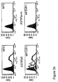
- the protein was further purified by size-exclusion chromatography ( FIG. 2a ). MFEVP1 eluted as two distinct peaks, 56 ml and 67 ml, representing the dimeric and monomeric forms, respectively.
- a further scFv MFE loop variant, NFEVP1 containing the ⁇ v ⁇ 6-binding sequence of VP1 and a Y 100b to P 100b mutation in the VH domain was constructed (see Fig. 1a ) from MFEVP1 by site-directed mutagenesis. Expression and purification in E. coli of this protein was similar to MFEVP1.
- the DNA sequence of NFEVP1 was also cloned into a yeast vector for expression in P. pastoris.
- NFEVP1 was obtained at 56mg/L after initial EDB-IMAC chromatography, using previously described procedures.(22, 23). NFEVP1 was concentrated and final purification was by size-exclusion chromatography ( Fig. 2b ). The chromatographic profile of the yeast expressed protein was virtually super imposable to that obtained from E. coli.
- a further MFE VH loop variant, shMFE(P)CDR2VP1 was constructed by inserting the A 140 - A 156 VP1 peptide between the DNA sequences of VH CDR2 residues E53 and N54 of the shMFE 2 sequence (which contains the Y 100b P mutation). This DNA sequence was cloned into a yeast vector and expressed in P. pastoris. The expressed protein was analysed by SDS-PAGE and Western blotting, which showed that the protein was stably expressed in yeast.
- MFEVP1 showed concentration dependent binding to ⁇ v ⁇ 6 immobilized plates in ELISA when probed with anti-MFE followed by anti-rabbit labelled HRP IgG ( Fig. 3a ).
- HRP IgG anti-rabbit labelled HRP IgG
- MFEVP's ability to bind to ⁇ v ⁇ 6 when expressed on cells was also investigated.
- MFEVP1 showed concentration dependent binding to ⁇ 6-transfected A375P ⁇ 6 cells (a retrovirally with ⁇ 6 cDNA transfected melanoma cell line(25)) by Flow Cytometry when monitored with mouse anti-His followed by Alexa488-conjugated anti-mouse Fc ( Fig. 3b ). Observed fluorescence shifts were similar for the 5 ⁇ g/ml and 0.5 ⁇ l/ml concentrations, indicating that MFEVP1 reached almost saturation levels of binding at 0.5 ⁇ g/ml.
- the ⁇ v ⁇ 6-specific antibody, 10D5 was included as a positive control whereas MFE, the negative control, was not shifted beyond background levels. Neither 10D5, MFEVP1 nor MFE showed binding to the non-transfected parent cell, A375Ppuro (transfected with empty vector), confirming that the observed binding of MFEVP1 to the ⁇ 6-transfected cell line was specifically to ⁇ v ⁇ 6.
- VB6 cells a well characterised retrovirally with ⁇ 6 cDNA transfected oral SCC cell line(27), were allowed to migrate towards the ⁇ v ⁇ 6-specifically binding ligand, LAP, which is coated underneath a Transwell filter. Addition of 50 ⁇ g/ml of MFEVP1 and the Y 100b to P 100b mutant, NFEVP1, to VB6 cells considerably inhibited their migration when compared to MFE ( Fig. 3c ).
- the function blocking ⁇ v ⁇ 6-specific 10D5 antibody added at 10 ⁇ g/ml showed similar levels of inhibition of cell migration of VB6 cells as the scFvs.
- the inhibition of migration was concentration dependent as shown for MFEVP1 ( Fig. 3d ).
- the A375Ppuro cells also expressed ⁇ v ⁇ 8, ⁇ v ⁇ 5 and ⁇ v ⁇ 1 at similar levels to those found on A375P ⁇ 6 cells ( Fig. 4b ) which showed that MFEVP1 and NFEVP1 did also not bind at detectable levels to these integrins.
- MFE The parent scFv, MFE, binds to CEA so it was important to establish whether MFEVP1 would remain some of this binding activity.
- Results showed that MFEVP1 showed residual binding to immobilized CEA in ELISA in a concentration-dependent manner, although considerably below that seen for MFE ( Fig. 5a ) when revealed with rabbit anti-MFE and HRP-labelled goat anti-rabbit IgG.
- the Tyr 100b to Pro 100 mutant, NFEVP1 showed no residual binding on ELISA to immobilized CEA. This same mutation when introduced into MFE had been shown previously to completely abolish binding to CEA(19). The ability of MFEVP1 to bind to CEA when expressed on cells was also investigated.
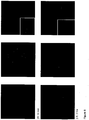
- the scFv was seen as a well defined thin line surrounding, at a distance, the nucleus (Hoechst staining and shown in blue) when revealed with rabbit anti-mouse IgG followed by Alexa Fluor® 546 labeled goat anti-rabbit IgG and shown in red; typifying its location in the plasma membrane.
- the scFv was diffused and speckled around the nucleus, identifying its location inside the cells. The scFv was also found inside the cells30 minutes and 1 hrs after incubation of the cells at 37°C (data not shown).
- FT-IR Fourier-transform infrared
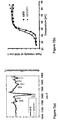
- NFEVP1 showed a strong band at 1635 cm-1 in agreement with a protein whose structure consists mainly of ⁇ -sheet and shown previously in the x-ray structure of MFE (25).
- the secondary structural elements associated with the FT-IR bands have been assigned previously for MFE.(26)
- the intensity of the ⁇ -sheet band at 1635 cm -1 was used to monitor stability of the protein; with increasing temperature this band will reduce in intensity while the protein denatures. Recording of the denaturing curve and fitting to a sigmoidal curve gave a midpoint of denaturation of 45°C for NFEVP1 ( Fig. 7b ). For MFE this temperature was 47°C, identical to the previously reported value(28). Therefore, insertion of the VP1 peptide did not affect the structure of the protein and NFEVP1 had very similar stability when compared to MFE.
- HFEVP1 humanized NFEVP1
- HFE stabilized humanized MFE
- the protein expressing vector was obtained by overlapping PCR identical to the murine analogue with the Y100b to P100b mutation in the heavy chain that eliminated CEA binding.
- Stabilized humanized NFEVP1 was expressed at 115mg/L by P. Pastoris, determined after initial EDB-IMAC chromatography. The protein formed almost exclusively a dimer as revealed by size-exclusion chromatography ( Figure 8(a) ).
- HFEVP1 bound to immobilized ⁇ v ⁇ 6 in ELISA when probed with anti-His followed by antimouse labelled HRP IgG ( Figure 8(b) . HFEVP1 also inhibited the adhesion of ⁇ v ⁇ 6-expressing 3T3 ⁇ 6.19 cells towards LAP ( Figure 8(c) ). HFEVP1 was the most potent inhibitor tested in the assay. IC50 values determined from the assay were 21.97 ⁇ g/ml (768 ⁇ M) for NFEVP1, 8.42 ⁇ g/ml (56.1 ⁇ M for 10D5 and 2.55 ⁇ g/ml (45.14 ⁇ M) for HFEVP1.
- MFEVP1 The resulting scFv loop variant, named MFEVP1
- MFEVP1 The resulting scFv loop variant, named MFEVP1
- Purification was via the His-tag followed by size-exclusion chromatography to obtain the monomeric form.
- MFEVP1 was specific for the integrin ⁇ v ⁇ 6 and did not show binding at detectable levels to immobilized ⁇ v ⁇ 3 in ELISA and to ⁇ v ⁇ 3, ⁇ v ⁇ 8, ⁇ v ⁇ 5 and ⁇ 5 ⁇ 1 expressed on cells.
- MFEVP1 had the desired specificity for ⁇ v ⁇ 6, however, also residual binding to the parent target, CEA. This binding could be eliminated when the VH residue Y 100b was mutated to P 100b , thus generating NFEVP1.
- Previous work has shown that binding of MFE to CEA was abolished when this mutation was introduced in MFE(19). MFE-CEA interactions were predicted from the way a MFE molecule interacts with another MFE molecule in the X-ray structure and this highlighted the importance of Y 100b (26). This was further elaborated by a subsequent modelling study of the interaction of MFE with CEA (35).
- the ⁇ v ⁇ 6-binding peptide did not affect the structure of the scFv and maintained a similar midpoint of denaturation when compared to MFE, NFEVP1, 45°C, MFE, 47°C, as determined by FT-IR spectroscopy.
- a link between ⁇ v ⁇ 6 expression and carcinoma progression has been suggested due to its ability to modulate invasion, inhibit apoptosis, regulate protease expression and activate TGF- ⁇ 1(36).
- ⁇ v ⁇ 6 has also been highlighted as a promising cancer target due to its de novo expression on various cancerous tissues(36).
- a 12-mer ⁇ v ⁇ 6-binding peptide has shown promise in mediating T cell killing of ⁇ v ⁇ 6 expressing ovarian tumour targets when fused to human IgG4 hinge-Fc extracellular domain and to the cytoplasmic tail of CD3- ⁇ (37).
- scFvs might not be in itself good targeting agents due to their rapid blood clearance they will allow genetic fusion to toxins, increasing the size and slower blood clearance, in particular when the toxin forms dimeric or multimeric arrangements, which will introduce avidity. Delivery of scFv-toxin fusions would benefit from their ability to internalize. NFEVP1 was able to internalize in ⁇ v ⁇ 6-expressing A375P ⁇ 6cells. scFvs can also be multimerically attached to drug carrying vehicles, such as liposomes and polymers. scFvs can be converted into whole antibodies for dimeric presentation, exploiting the intrinsic toxic functions, antibody-dependent cellular cytotoxicity (ADCC) and complement-dependent cytotoxicity (CDC).
- ADCC antibody-dependent cellular cytotoxicity
- CDC complement-dependent cytotoxicity
- a construct which provides good tumour penetration, high target retention, due to its dimeric binding, and rapid blood clearance is the diabody(38).
- the diabody is double the size of the scFv and considerable smaller than a whole antibody. It is generated by shortening the scFv (Gly 4 Ser) 3 VH-VL linker to a Gly 4 Ser linker, which forces the VH and VL domains from different chains to pair(39).
- NFEVP1 is of murine origin and is likely to result in immunogenetic reactions in humans.
- previous studies have addressed this problem by converting MFE into a humanized version.
- Comparison of the x-ray structure of MFE with a human analogue allowed identification of 28 surface residues for humanization of MFE.(25)
- These residues when introduced into MFE combined with three additional mutations for stabilization identified by yeast display expression maturation produced stabilized humanized MFE(27).
- the stabilized humanized MFE was used as a scaffold to insert the VP1 peptide and combined with the Y100b to P100b mutation gave HFEVP1.
- the protein was expressed almost exclusively as a dimer in P.
- HFEVP1 bound to ⁇ v ⁇ 6 in ELISA and inhibited the adhesion of ⁇ v ⁇ 6 expressing cells to LAP thus mimicking the behaviour of the murine analogue.
- HFEVP1 was a slightly better inhibitor than the commercially available whole antibody, 10D5; IC50 value for HFEVP1 was 2.55 ⁇ g/ml (45.14 ⁇ M) and for 10D5 was 8.42 ⁇ g/ml (56.1 ⁇ M).
- a ⁇ v ⁇ 6-binding scFv was generated by insertion of the RGD-containing VP1 peptide of FMDV into MFE that had no binding for MFE's target, CEA, when combined with the VHY 100b to VHP 100b mutation.
- MFE-23 including the humanised variants
- the MFE-23 antibody including the humanised variants
Landscapes
- Health & Medical Sciences (AREA)
- Chemical & Material Sciences (AREA)
- Immunology (AREA)
- Life Sciences & Earth Sciences (AREA)
- Medicinal Chemistry (AREA)
- Organic Chemistry (AREA)
- General Health & Medical Sciences (AREA)
- Engineering & Computer Science (AREA)
- Biophysics (AREA)
- Proteomics, Peptides & Aminoacids (AREA)
- Molecular Biology (AREA)
- Genetics & Genomics (AREA)
- Biochemistry (AREA)
- Oncology (AREA)
- Animal Behavior & Ethology (AREA)
- Bioinformatics & Cheminformatics (AREA)
- Public Health (AREA)
- Veterinary Medicine (AREA)
- Epidemiology (AREA)
- Pharmacology & Pharmacy (AREA)
- Mycology (AREA)
- Microbiology (AREA)
- Biomedical Technology (AREA)
- Cell Biology (AREA)
- Peptides Or Proteins (AREA)
Claims (7)
- Verfahren zum Modifizieren eines Ausgangsantikörpers zu einem Antikörper, der zur Bindung an αvβ6-Integrin in der Lage ist, wobei der Ausgangsantikörper folgende komplementaritätsbestimmenden Regionen (CDR) umfasst:(a) Schwerketten-CDR 1: Gly Phe Asn Ile Lys Asp Ser; und/oder(b) Schwerketten-CDR 2: Asp Pro Glu Asn Gly Asp; und/oder(c) Schwerketten-CDR 3: Thr Pro Thr Gly Pro Tyr Tyr Phe Asp; und/oder(d) Leichtketten-CDR 1: (i) Ser Ser Ser Val Pro; oder (ii) Ser Ser Ser Val Ser; und/oder(e) Leichtketten-CDR 2: (i) Ser Thr Ser; oder (ii) Leu Thr Ser; und/oder(f) Leichtketten-CDR 3: Arg Ser Ser Tyr Pro Leu;und wobei das Verfahren das Insertieren der Aminosäuresequenz AVPNLRGDLQVL AQKVA zwischen den benachbarten Aminosäureresten Thr98 und Gly99 von CDR H3 (Kabat-Nomenklatur) des Ausgangsantikörpers umfasst.
- Verfahren nach Anspruch 1, das ferner das Humanisieren des Antikörpers oder des Ausgangsantikörpers durch Mutieren eines oder mehrerer Aminosäurereste umfasst.
- Verfahren nach einem der vorangegangenen Ansprüche, wobei der Ausgangsantikörper ein scFv oder ein Diabody ist.
- Verfahren nach Anspruch 3, worin der scFv oder Diabody einen Linker mit der Sequenz (Gly4Ser)n umfasst, wobei n zwischen 1 und 4 beträgt.
- Verfahren nach einem der vorangegangenen Ansprüche, wobei die Insertion die Bindung des Ausgangsantikörpers an menschliches karzinoembryonales Antigen (CEA) reduziert.
- Verfahren nach einem der vorangegangenen Ansprüche, wobei das Verfahren weiters das Konjugieren des Antikörpers an(i) eine detektierbare Gruppierung und/oder(ii) eine therapeutisch aktive Gruppierung und/oder(iii) eine Markierungsgruppierung zur Verwendung bei der Reinigung oder Detektion und/oder(iv) eine oder mehrere, ausgewählt aus einer zytotoxischen Gruppierung, einem Mittel, das in der Lage ist, ein Prodrug in eine zytotoxische Gruppierung überzuführen, einem Strahlensensibilisierungsmittel und/oder einem radioaktiven Atom umfasst.
- Verfahren nach Anspruch 6, wobei
die detektierbare Gruppierung mittels Magnetresonanztomotgraphie (MRT), Magnetresonanzspektroskopie (MRS), Single-Photon-Emissionscomputertomographie (SPECT), Positronenemissionstomographie (PET) oder optischer Bildgebung detektierbar ist und/oder eine radioaktive Gruppierung ist;
die therapeutisch aktive Gruppierung ein Anti-Krebs-Mittel ist;
die Markierungsgruppe c-Myc oder eine Polyhistidingruppe ist und/oder
die zytotoxische Gruppierung ein chemotherapeutisches Mittel oder ein zytotoxisches Polypeptid ist und/oder
das Mittel ist, das in der Lage ist, ein Prodrug in eine zytotoxische Gruppierung überzuführen, ein Enzym zur Verwendung bei Antikörper-gerichteter Enzym-Prodrug-Therapie (ADEPT) ist.
Applications Claiming Priority (2)
| Application Number | Priority Date | Filing Date | Title |
|---|---|---|---|
| GBGB0718843.6A GB0718843D0 (en) | 2007-09-26 | 2007-09-26 | Materials and methods relating to modifying the binding of antibodies |
| PCT/GB2008/003283 WO2009040550A1 (en) | 2007-09-26 | 2008-09-26 | Materials and methods relating to modifying the binding of antibodies |
Publications (2)
| Publication Number | Publication Date |
|---|---|
| EP2201038A1 EP2201038A1 (de) | 2010-06-30 |
| EP2201038B1 true EP2201038B1 (de) | 2017-07-05 |
Family
ID=38701737
Family Applications (1)
| Application Number | Title | Priority Date | Filing Date |
|---|---|---|---|
| EP08806434.0A Active EP2201038B1 (de) | 2007-09-26 | 2008-09-26 | Materialien und methoden für die modifizierung der bindung von antikörpern |
Country Status (7)
| Country | Link |
|---|---|
| US (2) | US9050321B2 (de) |
| EP (1) | EP2201038B1 (de) |
| AU (1) | AU2008303393B2 (de) |
| CA (1) | CA2701072C (de) |
| ES (1) | ES2640340T3 (de) |
| GB (1) | GB0718843D0 (de) |
| WO (1) | WO2009040550A1 (de) |
Families Citing this family (11)
| Publication number | Priority date | Publication date | Assignee | Title |
|---|---|---|---|---|
| EP2064244B1 (de) | 2006-08-03 | 2019-06-05 | MedImmune Limited | GEGEN alphaVbeta6 GERICHTETE ANTIKÖRPER UND IHRE VERWENDUNG |
| US8691195B2 (en) * | 2009-04-14 | 2014-04-08 | Caregen Co., Ltd. | Peptide having activity of transforming growth factor and production method therefor |
| WO2014137931A1 (en) * | 2013-03-06 | 2014-09-12 | Imaginab, Inc. | Antigen binding constructs to 5t4 |
| EP3224275B1 (de) | 2014-11-14 | 2020-03-04 | F.Hoffmann-La Roche Ag | Antigenbindende moleküle mit einem ligandentrimer der tnf-familie |
| US11468969B2 (en) * | 2014-11-26 | 2022-10-11 | Biolojic Design Ltd. | Computer assisted antibody re-epitoping |
| EP3973980A1 (de) | 2015-03-31 | 2022-03-30 | F. Hoffmann-La Roche AG | Antigenbindende moleküle mit einem trimeren liganden der tnf-familie |
| AR106188A1 (es) | 2015-10-01 | 2017-12-20 | Hoffmann La Roche | Anticuerpos anti-cd19 humano humanizados y métodos de utilización |
| GB201711208D0 (en) | 2017-07-12 | 2017-08-23 | Iontas Ltd | Ion channel inhibitors |
| CN109091683B (zh) * | 2018-08-10 | 2019-10-18 | 北京大学 | 一种用于整合素αvβ6靶向的环状多肽放射性药物及其制备方法 |
| KR102396194B1 (ko) * | 2018-12-07 | 2022-05-10 | 서울대학교 산학협력단 | 항 c-Met 아고니스트 항체 및 이의 용도 |
| CN114921418B (zh) * | 2022-06-23 | 2023-08-18 | 金河佑本生物制品有限公司 | 一株o型口蹄疫类病毒颗粒单克隆抗体的杂交瘤细胞株1d3及试剂盒和检测方法 |
Family Cites Families (17)
| Publication number | Priority date | Publication date | Assignee | Title |
|---|---|---|---|---|
| GB8308235D0 (en) | 1983-03-25 | 1983-05-05 | Celltech Ltd | Polypeptides |
| US4816567A (en) | 1983-04-08 | 1989-03-28 | Genentech, Inc. | Recombinant immunoglobin preparations |
| JPS61134325A (ja) | 1984-12-04 | 1986-06-21 | Teijin Ltd | ハイブリツド抗体遺伝子の発現方法 |
| GB8607679D0 (en) * | 1986-03-27 | 1986-04-30 | Winter G P | Recombinant dna product |
| ATE207080T1 (de) | 1991-11-25 | 2001-11-15 | Enzon Inc | Multivalente antigen-bindende proteine |
| ES2156149T3 (es) | 1992-12-04 | 2001-06-16 | Medical Res Council | Proteinas de union multivalente y multiespecificas, su fabricacion y su uso. |
| US5446128A (en) | 1993-06-18 | 1995-08-29 | The Board Of Trustees Of The University Of Illinois | Alpha-helix mimetics and methods relating thereto |
| GB9324807D0 (en) | 1993-12-03 | 1994-01-19 | Cancer Res Campaign Tech | Tumour antibody |
| AU3374795A (en) | 1994-08-29 | 1996-03-22 | Prizm Pharmaceuticals, Inc. | Conjugates of vascular endothelial growth factor with targeted agents |
| US6774225B2 (en) | 1999-04-15 | 2004-08-10 | Hong Kong University Of Science & Technology | Antigenized antibody vaccine for foot-and-mouth disease |
| GB0025414D0 (en) | 2000-10-16 | 2000-11-29 | Consejo Superior Investigacion | Nanoparticles |
| US7232888B2 (en) | 2002-07-01 | 2007-06-19 | Massachusetts Institute Of Technology | Antibodies against tumor surface antigens |
| US7469107B2 (en) | 2003-07-23 | 2008-12-23 | Lexmark International, Inc. | Method for providing imaging substance for use in an imaging device via a virtual replenishment |
| JP5398982B2 (ja) | 2004-05-24 | 2014-01-29 | ミダテック リミテッド | Rnaリガンドを含むナノ粒子 |
| CN104072614B (zh) * | 2005-07-08 | 2017-04-26 | 生物基因Ma公司 | 抗-αvβ6 抗体及其用途 |
| GB0520068D0 (en) | 2005-10-03 | 2005-11-09 | Cancer Res Technology | av peptide ligand |
| AU2008286677A1 (en) | 2007-08-15 | 2009-02-19 | Christophe Alain Guex | Vessel transporting apparatus and method |
-
2007
- 2007-09-26 GB GBGB0718843.6A patent/GB0718843D0/en not_active Ceased
-
2008
- 2008-09-26 EP EP08806434.0A patent/EP2201038B1/de active Active
- 2008-09-26 US US12/680,320 patent/US9050321B2/en active Active
- 2008-09-26 AU AU2008303393A patent/AU2008303393B2/en not_active Ceased
- 2008-09-26 CA CA2701072A patent/CA2701072C/en not_active Expired - Fee Related
- 2008-09-26 ES ES08806434.0T patent/ES2640340T3/es active Active
- 2008-09-26 WO PCT/GB2008/003283 patent/WO2009040550A1/en not_active Ceased
-
2015
- 2015-05-13 US US14/710,661 patent/US20150291695A1/en not_active Abandoned
Non-Patent Citations (1)
| Title |
|---|
| RUDIKOFF S ET AL: "Single amino acid substitution altering antigen-binding specificity", PROCEEDINGS OF THE NATIONAL ACADEMY OF SCIENCES OF USA, NATIONAL ACADEMY OF SCIENCE, WASHINGTON, DC; US, vol. 79, 1 March 1982 (1982-03-01), pages 1979 - 1983, XP007901436, ISSN: 0027-8424, DOI: 10.1073/PNAS.79.6.1979 * |
Also Published As
| Publication number | Publication date |
|---|---|
| CA2701072C (en) | 2018-12-04 |
| AU2008303393A1 (en) | 2009-04-02 |
| AU2008303393B2 (en) | 2014-11-06 |
| US20150291695A1 (en) | 2015-10-15 |
| ES2640340T3 (es) | 2017-11-02 |
| US9050321B2 (en) | 2015-06-09 |
| GB0718843D0 (en) | 2007-11-07 |
| EP2201038A1 (de) | 2010-06-30 |
| US20110053254A1 (en) | 2011-03-03 |
| CA2701072A1 (en) | 2009-04-02 |
| WO2009040550A1 (en) | 2009-04-02 |
Similar Documents
| Publication | Publication Date | Title |
|---|---|---|
| EP2201038B1 (de) | Materialien und methoden für die modifizierung der bindung von antikörpern | |
| JP7341185B2 (ja) | 前立腺特異幹細胞抗原に対する抗体およびその使用 | |
| CN110770255B (zh) | 具有受限cd3结合的多特异性多肽构建体及其使用方法 | |
| KR102914586B1 (ko) | 메소텔린 및 cd3에 결합하는 이중특이적 항체 작제물 | |
| ES2582603T5 (es) | Anticuerpos biespecíficos de cadena sencilla con especificidad por antígenos diana de alto peso molecular | |
| KR101658247B1 (ko) | 모듈 인식 도메인을 통한 항체 표적화 | |
| CN112218686A (zh) | 具有受限cd3结合的多特异性多肽构建体以及相关方法和用途 | |
| KR20180120245A (ko) | 유도성 결합 단백질 및 사용 방법 | |
| KR20180033501A (ko) | Dll3 및 cd3에 결합하는 이중특이적인 항체 작제물 | |
| KR101971770B1 (ko) | 이중특이성 항체 및 이의 용도 | |
| KR102125032B1 (ko) | 인간 dlk1에 대한 항체 및 이의 용도 | |
| CN101370827A (zh) | 具有cea抗性的药物组合物 | |
| Luo et al. | Design and applications of bispecific heterodimers: molecular imaging and beyond | |
| AU2018210912A1 (en) | Endolysosomal targeting conjugates for improved delivery of cargo molecules to the endolysosomal compartment of target cells | |
| CN112204053B (zh) | 特异性结合claudin 3的ecl-2的抗体,其片段及其用途 | |
| EP3307784B1 (de) | Carboanhydrase-ix-spezifische antikörper und verwendungen davon | |
| Kogelberg et al. | Engineering a single-chain fv antibody to αvβ6 integrin using the specificity-determining loop of a foot-and-mouth disease virus | |
| Albrecht et al. | Update: Recombinant antibodies: From the laboratory to the clinic | |
| US20200330591A1 (en) | Treatment | |
| JP2023548345A (ja) | クリッピング率の低減した抗原結合ドメイン | |
| O. Acheampong et al. | Bispecific antibodies (bsAbs): promising immunotherapeutic agents for cancer therapy | |
| HK40104244A (zh) | 新型抗-hvegfr2抗体 | |
| WO2024233662A2 (en) | Molecules that bind to psca polypeptides | |
| Canevari et al. | Anti-FR Antibody Generation and Engineering: Development of New Therapeutic Tools | |
| HK1197419A (en) | Antibodies against prostate-specific stem cell antigen and use thereof |
Legal Events
| Date | Code | Title | Description |
|---|---|---|---|
| PUAI | Public reference made under article 153(3) epc to a published international application that has entered the european phase |
Free format text: ORIGINAL CODE: 0009012 |
|
| 17P | Request for examination filed |
Effective date: 20100422 |
|
| AK | Designated contracting states |
Kind code of ref document: A1 Designated state(s): AT BE BG CH CY CZ DE DK EE ES FI FR GB GR HR HU IE IS IT LI LT LU LV MC MT NL NO PL PT RO SE SI SK TR |
|
| AX | Request for extension of the european patent |
Extension state: AL BA MK RS |
|
| DAX | Request for extension of the european patent (deleted) | ||
| RAP1 | Party data changed (applicant data changed or rights of an application transferred) |
Owner name: CANCER RESEARCH TECHNOLOGY LIMITED |
|
| 17Q | First examination report despatched |
Effective date: 20120209 |
|
| RIC1 | Information provided on ipc code assigned before grant |
Ipc: C07K 16/30 20060101ALI20161212BHEP Ipc: C07K 16/28 20060101ALI20161212BHEP Ipc: A61K 39/395 20060101ALI20161212BHEP Ipc: C07K 16/00 20060101AFI20161212BHEP |
|
| GRAP | Despatch of communication of intention to grant a patent |
Free format text: ORIGINAL CODE: EPIDOSNIGR1 |
|
| INTG | Intention to grant announced |
Effective date: 20170120 |
|
| GRAS | Grant fee paid |
Free format text: ORIGINAL CODE: EPIDOSNIGR3 |
|
| GRAA | (expected) grant |
Free format text: ORIGINAL CODE: 0009210 |
|
| AK | Designated contracting states |
Kind code of ref document: B1 Designated state(s): AT BE BG CH CY CZ DE DK EE ES FI FR GB GR HR HU IE IS IT LI LT LU LV MC MT NL NO PL PT RO SE SI SK TR |
|
| REG | Reference to a national code |
Ref country code: GB Ref legal event code: FG4D |
|
| REG | Reference to a national code |
Ref country code: CH Ref legal event code: EP |
|
| REG | Reference to a national code |
Ref country code: AT Ref legal event code: REF Ref document number: 906547 Country of ref document: AT Kind code of ref document: T Effective date: 20170715 |
|
| REG | Reference to a national code |
Ref country code: IE Ref legal event code: FG4D |
|
| REG | Reference to a national code |
Ref country code: DE Ref legal event code: R096 Ref document number: 602008050989 Country of ref document: DE |
|
| REG | Reference to a national code |
Ref country code: FR Ref legal event code: PLFP Year of fee payment: 10 |
|
| REG | Reference to a national code |
Ref country code: ES Ref legal event code: FG2A Ref document number: 2640340 Country of ref document: ES Kind code of ref document: T3 Effective date: 20171102 |
|
| REG | Reference to a national code |
Ref country code: NL Ref legal event code: MP Effective date: 20170705 |
|
| REG | Reference to a national code |
Ref country code: AT Ref legal event code: MK05 Ref document number: 906547 Country of ref document: AT Kind code of ref document: T Effective date: 20170705 |
|
| REG | Reference to a national code |
Ref country code: LT Ref legal event code: MG4D |
|
| PG25 | Lapsed in a contracting state [announced via postgrant information from national office to epo] |
Ref country code: HR Free format text: LAPSE BECAUSE OF FAILURE TO SUBMIT A TRANSLATION OF THE DESCRIPTION OR TO PAY THE FEE WITHIN THE PRESCRIBED TIME-LIMIT Effective date: 20170705 Ref country code: LT Free format text: LAPSE BECAUSE OF FAILURE TO SUBMIT A TRANSLATION OF THE DESCRIPTION OR TO PAY THE FEE WITHIN THE PRESCRIBED TIME-LIMIT Effective date: 20170705 Ref country code: NO Free format text: LAPSE BECAUSE OF FAILURE TO SUBMIT A TRANSLATION OF THE DESCRIPTION OR TO PAY THE FEE WITHIN THE PRESCRIBED TIME-LIMIT Effective date: 20171005 Ref country code: SE Free format text: LAPSE BECAUSE OF FAILURE TO SUBMIT A TRANSLATION OF THE DESCRIPTION OR TO PAY THE FEE WITHIN THE PRESCRIBED TIME-LIMIT Effective date: 20170705 Ref country code: NL Free format text: LAPSE BECAUSE OF FAILURE TO SUBMIT A TRANSLATION OF THE DESCRIPTION OR TO PAY THE FEE WITHIN THE PRESCRIBED TIME-LIMIT Effective date: 20170705 Ref country code: AT Free format text: LAPSE BECAUSE OF FAILURE TO SUBMIT A TRANSLATION OF THE DESCRIPTION OR TO PAY THE FEE WITHIN THE PRESCRIBED TIME-LIMIT Effective date: 20170705 Ref country code: FI Free format text: LAPSE BECAUSE OF FAILURE TO SUBMIT A TRANSLATION OF THE DESCRIPTION OR TO PAY THE FEE WITHIN THE PRESCRIBED TIME-LIMIT Effective date: 20170705 |
|
| PG25 | Lapsed in a contracting state [announced via postgrant information from national office to epo] |
Ref country code: LV Free format text: LAPSE BECAUSE OF FAILURE TO SUBMIT A TRANSLATION OF THE DESCRIPTION OR TO PAY THE FEE WITHIN THE PRESCRIBED TIME-LIMIT Effective date: 20170705 Ref country code: BG Free format text: LAPSE BECAUSE OF FAILURE TO SUBMIT A TRANSLATION OF THE DESCRIPTION OR TO PAY THE FEE WITHIN THE PRESCRIBED TIME-LIMIT Effective date: 20171005 Ref country code: GR Free format text: LAPSE BECAUSE OF FAILURE TO SUBMIT A TRANSLATION OF THE DESCRIPTION OR TO PAY THE FEE WITHIN THE PRESCRIBED TIME-LIMIT Effective date: 20171006 Ref country code: IS Free format text: LAPSE BECAUSE OF FAILURE TO SUBMIT A TRANSLATION OF THE DESCRIPTION OR TO PAY THE FEE WITHIN THE PRESCRIBED TIME-LIMIT Effective date: 20171105 Ref country code: PL Free format text: LAPSE BECAUSE OF FAILURE TO SUBMIT A TRANSLATION OF THE DESCRIPTION OR TO PAY THE FEE WITHIN THE PRESCRIBED TIME-LIMIT Effective date: 20170705 |
|
| REG | Reference to a national code |
Ref country code: DE Ref legal event code: R097 Ref document number: 602008050989 Country of ref document: DE |
|
| PG25 | Lapsed in a contracting state [announced via postgrant information from national office to epo] |
Ref country code: RO Free format text: LAPSE BECAUSE OF FAILURE TO SUBMIT A TRANSLATION OF THE DESCRIPTION OR TO PAY THE FEE WITHIN THE PRESCRIBED TIME-LIMIT Effective date: 20170705 Ref country code: DK Free format text: LAPSE BECAUSE OF FAILURE TO SUBMIT A TRANSLATION OF THE DESCRIPTION OR TO PAY THE FEE WITHIN THE PRESCRIBED TIME-LIMIT Effective date: 20170705 Ref country code: CZ Free format text: LAPSE BECAUSE OF FAILURE TO SUBMIT A TRANSLATION OF THE DESCRIPTION OR TO PAY THE FEE WITHIN THE PRESCRIBED TIME-LIMIT Effective date: 20170705 |
|
| PLBE | No opposition filed within time limit |
Free format text: ORIGINAL CODE: 0009261 |
|
| STAA | Information on the status of an ep patent application or granted ep patent |
Free format text: STATUS: NO OPPOSITION FILED WITHIN TIME LIMIT |
|
| PG25 | Lapsed in a contracting state [announced via postgrant information from national office to epo] |
Ref country code: MC Free format text: LAPSE BECAUSE OF FAILURE TO SUBMIT A TRANSLATION OF THE DESCRIPTION OR TO PAY THE FEE WITHIN THE PRESCRIBED TIME-LIMIT Effective date: 20170705 Ref country code: EE Free format text: LAPSE BECAUSE OF FAILURE TO SUBMIT A TRANSLATION OF THE DESCRIPTION OR TO PAY THE FEE WITHIN THE PRESCRIBED TIME-LIMIT Effective date: 20170705 Ref country code: SK Free format text: LAPSE BECAUSE OF FAILURE TO SUBMIT A TRANSLATION OF THE DESCRIPTION OR TO PAY THE FEE WITHIN THE PRESCRIBED TIME-LIMIT Effective date: 20170705 |
|
| 26N | No opposition filed |
Effective date: 20180406 |
|
| REG | Reference to a national code |
Ref country code: IE Ref legal event code: MM4A |
|
| REG | Reference to a national code |
Ref country code: BE Ref legal event code: MM Effective date: 20170930 |
|
| PG25 | Lapsed in a contracting state [announced via postgrant information from national office to epo] |
Ref country code: LU Free format text: LAPSE BECAUSE OF NON-PAYMENT OF DUE FEES Effective date: 20170926 |
|
| PG25 | Lapsed in a contracting state [announced via postgrant information from national office to epo] |
Ref country code: IE Free format text: LAPSE BECAUSE OF NON-PAYMENT OF DUE FEES Effective date: 20170926 |
|
| PG25 | Lapsed in a contracting state [announced via postgrant information from national office to epo] |
Ref country code: BE Free format text: LAPSE BECAUSE OF NON-PAYMENT OF DUE FEES Effective date: 20170930 Ref country code: SI Free format text: LAPSE BECAUSE OF FAILURE TO SUBMIT A TRANSLATION OF THE DESCRIPTION OR TO PAY THE FEE WITHIN THE PRESCRIBED TIME-LIMIT Effective date: 20170705 |
|
| REG | Reference to a national code |
Ref country code: FR Ref legal event code: PLFP Year of fee payment: 11 |
|
| PG25 | Lapsed in a contracting state [announced via postgrant information from national office to epo] |
Ref country code: MT Free format text: LAPSE BECAUSE OF NON-PAYMENT OF DUE FEES Effective date: 20170926 |
|
| PG25 | Lapsed in a contracting state [announced via postgrant information from national office to epo] |
Ref country code: HU Free format text: LAPSE BECAUSE OF FAILURE TO SUBMIT A TRANSLATION OF THE DESCRIPTION OR TO PAY THE FEE WITHIN THE PRESCRIBED TIME-LIMIT; INVALID AB INITIO Effective date: 20080926 |
|
| PG25 | Lapsed in a contracting state [announced via postgrant information from national office to epo] |
Ref country code: CY Free format text: LAPSE BECAUSE OF NON-PAYMENT OF DUE FEES Effective date: 20170705 |
|
| PG25 | Lapsed in a contracting state [announced via postgrant information from national office to epo] |
Ref country code: TR Free format text: LAPSE BECAUSE OF FAILURE TO SUBMIT A TRANSLATION OF THE DESCRIPTION OR TO PAY THE FEE WITHIN THE PRESCRIBED TIME-LIMIT Effective date: 20170705 |
|
| PG25 | Lapsed in a contracting state [announced via postgrant information from national office to epo] |
Ref country code: PT Free format text: LAPSE BECAUSE OF FAILURE TO SUBMIT A TRANSLATION OF THE DESCRIPTION OR TO PAY THE FEE WITHIN THE PRESCRIBED TIME-LIMIT Effective date: 20170705 |
|
| P01 | Opt-out of the competence of the unified patent court (upc) registered |
Effective date: 20230511 |
|
| PGFP | Annual fee paid to national office [announced via postgrant information from national office to epo] |
Ref country code: GB Payment date: 20230927 Year of fee payment: 16 |
|
| PGFP | Annual fee paid to national office [announced via postgrant information from national office to epo] |
Ref country code: FR Payment date: 20230929 Year of fee payment: 16 Ref country code: DE Payment date: 20230928 Year of fee payment: 16 |
|
| PGFP | Annual fee paid to national office [announced via postgrant information from national office to epo] |
Ref country code: ES Payment date: 20231019 Year of fee payment: 16 |
|
| PGFP | Annual fee paid to national office [announced via postgrant information from national office to epo] |
Ref country code: IT Payment date: 20230928 Year of fee payment: 16 Ref country code: CH Payment date: 20231001 Year of fee payment: 16 |
|
| REG | Reference to a national code |
Ref country code: DE Ref legal event code: R119 Ref document number: 602008050989 Country of ref document: DE |
|
| REG | Reference to a national code |
Ref country code: CH Ref legal event code: PL |
|
| GBPC | Gb: european patent ceased through non-payment of renewal fee |
Effective date: 20240926 |
|
| PG25 | Lapsed in a contracting state [announced via postgrant information from national office to epo] |
Ref country code: DE Free format text: LAPSE BECAUSE OF NON-PAYMENT OF DUE FEES Effective date: 20250401 |
|
| PG25 | Lapsed in a contracting state [announced via postgrant information from national office to epo] |
Ref country code: GB Free format text: LAPSE BECAUSE OF NON-PAYMENT OF DUE FEES Effective date: 20240926 |
|
| PG25 | Lapsed in a contracting state [announced via postgrant information from national office to epo] |
Ref country code: IT Free format text: LAPSE BECAUSE OF NON-PAYMENT OF DUE FEES Effective date: 20240926 |
|
| PG25 | Lapsed in a contracting state [announced via postgrant information from national office to epo] |
Ref country code: FR Free format text: LAPSE BECAUSE OF NON-PAYMENT OF DUE FEES Effective date: 20240930 |
|
| PG25 | Lapsed in a contracting state [announced via postgrant information from national office to epo] |
Ref country code: CH Free format text: LAPSE BECAUSE OF NON-PAYMENT OF DUE FEES Effective date: 20240930 |
|
| REG | Reference to a national code |
Ref country code: ES Ref legal event code: FD2A Effective date: 20251103 |
|
| PG25 | Lapsed in a contracting state [announced via postgrant information from national office to epo] |
Ref country code: ES Free format text: LAPSE BECAUSE OF NON-PAYMENT OF DUE FEES Effective date: 20240927 |