EP2198012B1 - Von hbs-zellen abgeleitete kardiomyozytenähnliche zellhaufen - Google Patents
Von hbs-zellen abgeleitete kardiomyozytenähnliche zellhaufen Download PDFInfo
- Publication number
- EP2198012B1 EP2198012B1 EP08802355.1A EP08802355A EP2198012B1 EP 2198012 B1 EP2198012 B1 EP 2198012B1 EP 08802355 A EP08802355 A EP 08802355A EP 2198012 B1 EP2198012 B1 EP 2198012B1
- Authority
- EP
- European Patent Office
- Prior art keywords
- cells
- cmlc
- clusters
- genes
- medium
- Prior art date
- Legal status (The legal status is an assumption and is not a legal conclusion. Google has not performed a legal analysis and makes no representation as to the accuracy of the status listed.)
- Not-in-force
Links
- 210000004027 cell Anatomy 0.000 claims description 506
- 108090000623 proteins and genes Proteins 0.000 claims description 216
- 239000002609 medium Substances 0.000 claims description 113
- 230000014509 gene expression Effects 0.000 claims description 83
- 238000000034 method Methods 0.000 claims description 82
- 108090000379 Fibroblast growth factor 2 Proteins 0.000 claims description 79
- 108010023082 activin A Proteins 0.000 claims description 77
- 239000012091 fetal bovine serum Substances 0.000 claims description 67
- 230000001105 regulatory effect Effects 0.000 claims description 61
- 239000000203 mixture Substances 0.000 claims description 44
- 239000001963 growth medium Substances 0.000 claims description 41
- 230000015572 biosynthetic process Effects 0.000 claims description 37
- 230000000747 cardiac effect Effects 0.000 claims description 31
- 238000011534 incubation Methods 0.000 claims description 31
- 230000008859 change Effects 0.000 claims description 25
- 108091006146 Channels Proteins 0.000 claims description 18
- 108090000862 Ion Channels Proteins 0.000 claims description 18
- 102000004310 Ion Channels Human genes 0.000 claims description 18
- 229920000159 gelatin Polymers 0.000 claims description 17
- 235000019322 gelatine Nutrition 0.000 claims description 17
- 239000001828 Gelatine Substances 0.000 claims description 15
- 230000002776 aggregation Effects 0.000 claims description 15
- 238000004220 aggregation Methods 0.000 claims description 15
- 239000003102 growth factor Substances 0.000 claims description 15
- 238000005119 centrifugation Methods 0.000 claims description 12
- 239000003112 inhibitor Substances 0.000 claims description 12
- 102000002254 Glycogen Synthase Kinase 3 Human genes 0.000 claims description 11
- 108010014905 Glycogen Synthase Kinase 3 Proteins 0.000 claims description 11
- -1 TTR Proteins 0.000 claims description 11
- 102000004887 Transforming Growth Factor beta Human genes 0.000 claims description 10
- 108090001012 Transforming Growth Factor beta Proteins 0.000 claims description 10
- 239000003797 essential amino acid Substances 0.000 claims description 10
- 235000020776 essential amino acid Nutrition 0.000 claims description 10
- 239000002829 mitogen activated protein kinase inhibitor Substances 0.000 claims description 10
- 102000002574 p38 Mitogen-Activated Protein Kinases Human genes 0.000 claims description 10
- 108010068338 p38 Mitogen-Activated Protein Kinases Proteins 0.000 claims description 10
- 210000002966 serum Anatomy 0.000 claims description 10
- ZRKFYGHZFMAOKI-QMGMOQQFSA-N tgfbeta Chemical compound C([C@H](NC(=O)[C@H](C(C)C)NC(=O)CNC(=O)[C@H](CCC(O)=O)NC(=O)[C@H](CCCNC(N)=N)NC(=O)[C@H](CC(N)=O)NC(=O)[C@H](CC(C)C)NC(=O)[C@H]([C@@H](C)O)NC(=O)[C@H](CCC(O)=O)NC(=O)[C@H]([C@@H](C)O)NC(=O)[C@H](CC(C)C)NC(=O)CNC(=O)[C@H](C)NC(=O)[C@H](CO)NC(=O)[C@H](CCC(N)=O)NC(=O)[C@@H](NC(=O)[C@H](C)NC(=O)[C@H](C)NC(=O)[C@@H](NC(=O)[C@H](CC(C)C)NC(=O)[C@@H](N)CCSC)C(C)C)[C@@H](C)CC)C(=O)N[C@@H]([C@@H](C)O)C(=O)N[C@@H](C(C)C)C(=O)N[C@@H](CC=1C=CC=CC=1)C(=O)N[C@@H](C)C(=O)N1[C@@H](CCC1)C(=O)N[C@@H]([C@@H](C)O)C(=O)N[C@@H](CC(N)=O)C(=O)N[C@@H](CCC(O)=O)C(=O)N[C@@H](C)C(=O)N[C@@H](CC=1C=CC=CC=1)C(=O)N[C@@H](CCCNC(N)=N)C(=O)N[C@@H](C)C(=O)N[C@@H](CC(C)C)C(=O)N1[C@@H](CCC1)C(=O)N1[C@@H](CCC1)C(=O)N[C@@H](CCCNC(N)=N)C(=O)N[C@@H](CCC(O)=O)C(=O)N[C@@H](CCCNC(N)=N)C(=O)N[C@@H](CO)C(=O)N[C@@H](CCCNC(N)=N)C(=O)N[C@@H](CC(C)C)C(=O)N[C@@H](CC(C)C)C(O)=O)C1=CC=C(O)C=C1 ZRKFYGHZFMAOKI-QMGMOQQFSA-N 0.000 claims description 10
- 229940122696 MAP kinase inhibitor Drugs 0.000 claims description 9
- 229910001424 calcium ion Inorganic materials 0.000 claims description 9
- 238000004062 sedimentation Methods 0.000 claims description 9
- 102100022712 Alpha-1-antitrypsin Human genes 0.000 claims description 8
- 101000823116 Homo sapiens Alpha-1-antitrypsin Proteins 0.000 claims description 8
- 238000011161 development Methods 0.000 claims description 8
- 238000002360 preparation method Methods 0.000 claims description 8
- 229960005322 streptomycin Drugs 0.000 claims description 8
- 102100030942 Apolipoprotein A-II Human genes 0.000 claims description 7
- BHPQYMZQTOCNFJ-UHFFFAOYSA-N Calcium cation Chemical compound [Ca+2] BHPQYMZQTOCNFJ-UHFFFAOYSA-N 0.000 claims description 7
- 102000018233 Fibroblast Growth Factor Human genes 0.000 claims description 7
- 108050007372 Fibroblast Growth Factor Proteins 0.000 claims description 7
- 101000793406 Homo sapiens Apolipoprotein A-II Proteins 0.000 claims description 7
- 108010075843 alpha-2-HS-Glycoprotein Proteins 0.000 claims description 7
- 102000012005 alpha-2-HS-Glycoprotein Human genes 0.000 claims description 7
- 210000002064 heart cell Anatomy 0.000 claims description 7
- 108060003345 Adrenergic Receptor Proteins 0.000 claims description 6
- 102000017910 Adrenergic receptor Human genes 0.000 claims description 6
- 102100033715 Apolipoprotein A-I Human genes 0.000 claims description 6
- 102100040202 Apolipoprotein B-100 Human genes 0.000 claims description 6
- 102100030970 Apolipoprotein C-III Human genes 0.000 claims description 6
- 108091003079 Bovine Serum Albumin Proteins 0.000 claims description 6
- 101000733802 Homo sapiens Apolipoprotein A-I Proteins 0.000 claims description 6
- 101000889953 Homo sapiens Apolipoprotein B-100 Proteins 0.000 claims description 6
- 101000793223 Homo sapiens Apolipoprotein C-III Proteins 0.000 claims description 6
- 101000665882 Homo sapiens Retinol-binding protein 4 Proteins 0.000 claims description 6
- 102100038246 Retinol-binding protein 4 Human genes 0.000 claims description 6
- 238000004113 cell culture Methods 0.000 claims description 6
- 102000014415 Muscarinic acetylcholine receptor Human genes 0.000 claims description 5
- 108050003473 Muscarinic acetylcholine receptor Proteins 0.000 claims description 5
- 108090001027 Troponin Proteins 0.000 claims description 5
- 102000003505 Myosin Human genes 0.000 claims description 4
- 108060008487 Myosin Proteins 0.000 claims description 4
- 210000000349 chromosome Anatomy 0.000 claims description 3
- 238000010494 dissociation reaction Methods 0.000 claims description 3
- 230000005593 dissociations Effects 0.000 claims description 3
- 239000012894 fetal calf serum Substances 0.000 claims description 3
- 230000002068 genetic effect Effects 0.000 claims description 3
- 102100024785 Fibroblast growth factor 2 Human genes 0.000 claims 4
- 102000003974 Fibroblast growth factor 2 Human genes 0.000 description 75
- 238000010009 beating Methods 0.000 description 57
- IAZDPXIOMUYVGZ-UHFFFAOYSA-N Dimethylsulphoxide Chemical compound CS(C)=O IAZDPXIOMUYVGZ-UHFFFAOYSA-N 0.000 description 45
- 210000004413 cardiac myocyte Anatomy 0.000 description 43
- 238000007747 plating Methods 0.000 description 42
- 238000002474 experimental method Methods 0.000 description 41
- 102000004169 proteins and genes Human genes 0.000 description 28
- 230000036982 action potential Effects 0.000 description 27
- 235000018102 proteins Nutrition 0.000 description 27
- 239000000243 solution Substances 0.000 description 27
- 102100036859 Troponin I, cardiac muscle Human genes 0.000 description 26
- 230000004069 differentiation Effects 0.000 description 26
- 230000000694 effects Effects 0.000 description 26
- 239000006285 cell suspension Substances 0.000 description 23
- 101710128251 Troponin I, cardiac muscle Proteins 0.000 description 22
- 210000000130 stem cell Anatomy 0.000 description 22
- 102000029816 Collagenase Human genes 0.000 description 21
- 108060005980 Collagenase Proteins 0.000 description 21
- LYCAIKOWRPUZTN-UHFFFAOYSA-N Ethylene glycol Chemical compound OCCO LYCAIKOWRPUZTN-UHFFFAOYSA-N 0.000 description 21
- 229960002424 collagenase Drugs 0.000 description 21
- 239000002577 cryoprotective agent Substances 0.000 description 21
- 239000000725 suspension Substances 0.000 description 18
- 230000008602 contraction Effects 0.000 description 17
- 238000010186 staining Methods 0.000 description 17
- 238000004458 analytical method Methods 0.000 description 16
- FWBHETKCLVMNFS-UHFFFAOYSA-N 4',6-Diamino-2-phenylindol Chemical compound C1=CC(C(=N)N)=CC=C1C1=CC2=CC=C(C(N)=N)C=C2N1 FWBHETKCLVMNFS-UHFFFAOYSA-N 0.000 description 15
- 230000002269 spontaneous effect Effects 0.000 description 15
- KEDQCFRVSHYKLR-UHFFFAOYSA-N 3-[3-[2-(3,4-dimethoxyphenyl)ethyl-methylamino]propyl]-7,8-dimethoxy-2,5-dihydro-1H-3-benzazepin-4-one Chemical compound C1=C(OC)C(OC)=CC=C1CCN(C)CCCN1C(=O)CC2=CC(OC)=C(OC)C=C2CC1 KEDQCFRVSHYKLR-UHFFFAOYSA-N 0.000 description 14
- 238000012360 testing method Methods 0.000 description 14
- 229950003419 zatebradine Drugs 0.000 description 14
- 108091032973 (ribonucleotides)n+m Proteins 0.000 description 13
- YOELZIQOLWZLQC-UHFFFAOYSA-N 6-(4-fluorophenyl)-5-pyridin-4-yl-2,3-dihydroimidazo[2,1-b]thiazole Chemical compound C1=CC(F)=CC=C1C1=C(C=2C=CN=CC=2)N2CCSC2=N1 YOELZIQOLWZLQC-UHFFFAOYSA-N 0.000 description 13
- 230000037020 contractile activity Effects 0.000 description 13
- 238000004017 vitrification Methods 0.000 description 13
- HDTRYLNUVZCQOY-UHFFFAOYSA-N α-D-glucopyranosyl-α-D-glucopyranoside Natural products OC1C(O)C(O)C(CO)OC1OC1C(O)C(O)C(O)C(CO)O1 HDTRYLNUVZCQOY-UHFFFAOYSA-N 0.000 description 12
- PEDCQBHIVMGVHV-UHFFFAOYSA-N Glycerine Chemical compound OCC(O)CO PEDCQBHIVMGVHV-UHFFFAOYSA-N 0.000 description 12
- HDTRYLNUVZCQOY-WSWWMNSNSA-N Trehalose Natural products O[C@@H]1[C@@H](O)[C@@H](O)[C@@H](CO)O[C@@H]1O[C@@H]1[C@H](O)[C@@H](O)[C@@H](O)[C@@H](CO)O1 HDTRYLNUVZCQOY-WSWWMNSNSA-N 0.000 description 12
- 239000002771 cell marker Substances 0.000 description 12
- 210000004940 nucleus Anatomy 0.000 description 12
- 230000000638 stimulation Effects 0.000 description 12
- 238000012546 transfer Methods 0.000 description 12
- 108090001108 Troponin T Proteins 0.000 description 11
- 210000005003 heart tissue Anatomy 0.000 description 11
- 230000003993 interaction Effects 0.000 description 11
- 239000000523 sample Substances 0.000 description 11
- 239000010902 straw Substances 0.000 description 11
- OHCQJHSOBUTRHG-KGGHGJDLSA-N FORSKOLIN Chemical compound O=C([C@@]12O)C[C@](C)(C=C)O[C@]1(C)[C@@H](OC(=O)C)[C@@H](O)[C@@H]1[C@]2(C)[C@@H](O)CCC1(C)C OHCQJHSOBUTRHG-KGGHGJDLSA-N 0.000 description 10
- 101001032038 Homo sapiens Potassium/sodium hyperpolarization-activated cyclic nucleotide-gated channel 4 Proteins 0.000 description 10
- 102100038718 Potassium/sodium hyperpolarization-activated cyclic nucleotide-gated channel 4 Human genes 0.000 description 10
- 102000004987 Troponin T Human genes 0.000 description 10
- HDTRYLNUVZCQOY-LIZSDCNHSA-N alpha,alpha-trehalose Chemical compound O[C@@H]1[C@@H](O)[C@H](O)[C@@H](CO)O[C@@H]1O[C@@H]1[C@H](O)[C@@H](O)[C@H](O)[C@@H](CO)O1 HDTRYLNUVZCQOY-LIZSDCNHSA-N 0.000 description 10
- 239000007376 cm-medium Substances 0.000 description 10
- 238000000338 in vitro Methods 0.000 description 10
- 210000001519 tissue Anatomy 0.000 description 10
- 238000003491 array Methods 0.000 description 9
- 230000018109 developmental process Effects 0.000 description 9
- 238000009826 distribution Methods 0.000 description 9
- 238000005259 measurement Methods 0.000 description 9
- 230000004044 response Effects 0.000 description 9
- 239000000126 substance Substances 0.000 description 9
- 229930006000 Sucrose Natural products 0.000 description 8
- 238000005138 cryopreservation Methods 0.000 description 8
- 239000012528 membrane Substances 0.000 description 8
- 229960001802 phenylephrine Drugs 0.000 description 8
- SONNWYBIRXJNDC-VIFPVBQESA-N phenylephrine Chemical compound CNC[C@H](O)C1=CC=CC(O)=C1 SONNWYBIRXJNDC-VIFPVBQESA-N 0.000 description 8
- 239000000047 product Substances 0.000 description 8
- 230000006916 protein interaction Effects 0.000 description 8
- 239000005720 sucrose Substances 0.000 description 8
- 239000004034 viscosity adjusting agent Substances 0.000 description 8
- UCTWMZQNUQWSLP-UHFFFAOYSA-N Adrenaline Natural products CNCC(O)C1=CC=C(O)C(O)=C1 UCTWMZQNUQWSLP-UHFFFAOYSA-N 0.000 description 7
- 102100035423 POU domain, class 5, transcription factor 1 Human genes 0.000 description 7
- 231100000673 dose–response relationship Toxicity 0.000 description 7
- 229940079593 drug Drugs 0.000 description 7
- 239000003814 drug Substances 0.000 description 7
- 229940093476 ethylene glycol Drugs 0.000 description 7
- 230000001605 fetal effect Effects 0.000 description 7
- 229910052739 hydrogen Inorganic materials 0.000 description 7
- 238000010257 thawing Methods 0.000 description 7
- 102100023635 Alpha-fetoprotein Human genes 0.000 description 6
- 101000958741 Homo sapiens Myosin-6 Proteins 0.000 description 6
- 101000819074 Homo sapiens Transcription factor GATA-4 Proteins 0.000 description 6
- DNIAPMSPPWPWGF-UHFFFAOYSA-N Propylene glycol Chemical compound CC(O)CO DNIAPMSPPWPWGF-UHFFFAOYSA-N 0.000 description 6
- UIIMBOGNXHQVGW-UHFFFAOYSA-M Sodium bicarbonate Chemical compound [Na+].OC([O-])=O UIIMBOGNXHQVGW-UHFFFAOYSA-M 0.000 description 6
- FAPWRFPIFSIZLT-UHFFFAOYSA-M Sodium chloride Chemical compound [Na+].[Cl-] FAPWRFPIFSIZLT-UHFFFAOYSA-M 0.000 description 6
- CZMRCDWAGMRECN-UGDNZRGBSA-N Sucrose Chemical compound O[C@H]1[C@H](O)[C@@H](CO)O[C@@]1(CO)O[C@@H]1[C@H](O)[C@@H](O)[C@H](O)[C@@H](CO)O1 CZMRCDWAGMRECN-UGDNZRGBSA-N 0.000 description 6
- 102100021380 Transcription factor GATA-4 Human genes 0.000 description 6
- OIPILFWXSMYKGL-UHFFFAOYSA-N acetylcholine Chemical compound CC(=O)OCC[N+](C)(C)C OIPILFWXSMYKGL-UHFFFAOYSA-N 0.000 description 6
- 229960004373 acetylcholine Drugs 0.000 description 6
- 238000004422 calculation algorithm Methods 0.000 description 6
- 238000004364 calculation method Methods 0.000 description 6
- 238000007876 drug discovery Methods 0.000 description 6
- 210000002242 embryoid body Anatomy 0.000 description 6
- 230000005764 inhibitory process Effects 0.000 description 6
- 239000000463 material Substances 0.000 description 6
- HJCMDXDYPOUFDY-WHFBIAKZSA-N Ala-Gln Chemical compound C[C@H](N)C(=O)N[C@H](C(O)=O)CCC(N)=O HJCMDXDYPOUFDY-WHFBIAKZSA-N 0.000 description 5
- 206010048610 Cardiotoxicity Diseases 0.000 description 5
- SUZLHDUTVMZSEV-UHFFFAOYSA-N Deoxycoleonol Natural products C12C(=O)CC(C)(C=C)OC2(C)C(OC(=O)C)C(O)C2C1(C)C(O)CCC2(C)C SUZLHDUTVMZSEV-UHFFFAOYSA-N 0.000 description 5
- 239000006144 Dulbecco’s modified Eagle's medium Substances 0.000 description 5
- 229920001917 Ficoll Polymers 0.000 description 5
- 101001047090 Homo sapiens Potassium voltage-gated channel subfamily H member 2 Proteins 0.000 description 5
- 102100038319 Myosin-6 Human genes 0.000 description 5
- 101150114527 Nkx2-5 gene Proteins 0.000 description 5
- QZVCTJOXCFMACW-UHFFFAOYSA-N Phenoxybenzamine Chemical compound C=1C=CC=CC=1CN(CCCl)C(C)COC1=CC=CC=C1 QZVCTJOXCFMACW-UHFFFAOYSA-N 0.000 description 5
- 102100022807 Potassium voltage-gated channel subfamily H member 2 Human genes 0.000 description 5
- 238000002123 RNA extraction Methods 0.000 description 5
- 101100460507 Xenopus laevis nkx-2.5 gene Proteins 0.000 description 5
- GXDALQBWZGODGZ-UHFFFAOYSA-N astemizole Chemical compound C1=CC(OC)=CC=C1CCN1CCC(NC=2N(C3=CC=CC=C3N=2)CC=2C=CC(F)=CC=2)CC1 GXDALQBWZGODGZ-UHFFFAOYSA-N 0.000 description 5
- 210000002459 blastocyst Anatomy 0.000 description 5
- 231100000259 cardiotoxicity Toxicity 0.000 description 5
- 230000001413 cellular effect Effects 0.000 description 5
- OHCQJHSOBUTRHG-UHFFFAOYSA-N colforsin Natural products OC12C(=O)CC(C)(C=C)OC1(C)C(OC(=O)C)C(O)C1C2(C)C(O)CCC1(C)C OHCQJHSOBUTRHG-UHFFFAOYSA-N 0.000 description 5
- 150000001875 compounds Chemical class 0.000 description 5
- 230000000875 corresponding effect Effects 0.000 description 5
- 210000002950 fibroblast Anatomy 0.000 description 5
- 238000007710 freezing Methods 0.000 description 5
- 230000008014 freezing Effects 0.000 description 5
- 230000001976 improved effect Effects 0.000 description 5
- 239000003550 marker Substances 0.000 description 5
- 238000002493 microarray Methods 0.000 description 5
- 230000000144 pharmacologic effect Effects 0.000 description 5
- 229960003418 phenoxybenzamine Drugs 0.000 description 5
- 230000000284 resting effect Effects 0.000 description 5
- 230000011664 signaling Effects 0.000 description 5
- 230000004936 stimulating effect Effects 0.000 description 5
- 238000003860 storage Methods 0.000 description 5
- XLYOFNOQVPJJNP-UHFFFAOYSA-N water Substances O XLYOFNOQVPJJNP-UHFFFAOYSA-N 0.000 description 5
- 102100027211 Albumin Human genes 0.000 description 4
- 101001030243 Homo sapiens Myosin-7 Proteins 0.000 description 4
- 101001094700 Homo sapiens POU domain, class 5, transcription factor 1 Proteins 0.000 description 4
- 101000851334 Homo sapiens Troponin I, cardiac muscle Proteins 0.000 description 4
- RKUNBYITZUJHSG-UHFFFAOYSA-N Hyosciamin-hydrochlorid Natural products CN1C(C2)CCC1CC2OC(=O)C(CO)C1=CC=CC=C1 RKUNBYITZUJHSG-UHFFFAOYSA-N 0.000 description 4
- 102100038934 Myosin-7 Human genes 0.000 description 4
- 102000004903 Troponin Human genes 0.000 description 4
- 230000006978 adaptation Effects 0.000 description 4
- 230000001387 anti-histamine Effects 0.000 description 4
- 239000000739 antihistaminic agent Substances 0.000 description 4
- RKUNBYITZUJHSG-SPUOUPEWSA-N atropine Chemical compound O([C@H]1C[C@H]2CC[C@@H](C1)N2C)C(=O)C(CO)C1=CC=CC=C1 RKUNBYITZUJHSG-SPUOUPEWSA-N 0.000 description 4
- 229960000396 atropine Drugs 0.000 description 4
- 239000000872 buffer Substances 0.000 description 4
- 239000003795 chemical substances by application Substances 0.000 description 4
- 230000001419 dependent effect Effects 0.000 description 4
- IXTMWRCNAAVVAI-UHFFFAOYSA-N dofetilide Chemical compound C=1C=C(NS(C)(=O)=O)C=CC=1CCN(C)CCOC1=CC=C(NS(C)(=O)=O)C=C1 IXTMWRCNAAVVAI-UHFFFAOYSA-N 0.000 description 4
- 229960002994 dofetilide Drugs 0.000 description 4
- 210000002889 endothelial cell Anatomy 0.000 description 4
- 230000003834 intracellular effect Effects 0.000 description 4
- 108020004999 messenger RNA Proteins 0.000 description 4
- 230000000877 morphologic effect Effects 0.000 description 4
- 102000005962 receptors Human genes 0.000 description 4
- 108020003175 receptors Proteins 0.000 description 4
- 238000011084 recovery Methods 0.000 description 4
- 230000002336 repolarization Effects 0.000 description 4
- 230000002459 sustained effect Effects 0.000 description 4
- 231100000041 toxicology testing Toxicity 0.000 description 4
- DGVVWUTYPXICAM-UHFFFAOYSA-N β‐Mercaptoethanol Chemical compound OCCS DGVVWUTYPXICAM-UHFFFAOYSA-N 0.000 description 4
- SFLSHLFXELFNJZ-QMMMGPOBSA-N (-)-norepinephrine Chemical compound NC[C@H](O)C1=CC=C(O)C(O)=C1 SFLSHLFXELFNJZ-QMMMGPOBSA-N 0.000 description 3
- METKIMKYRPQLGS-GFCCVEGCSA-N (R)-atenolol Chemical compound CC(C)NC[C@@H](O)COC1=CC=C(CC(N)=O)C=C1 METKIMKYRPQLGS-GFCCVEGCSA-N 0.000 description 3
- SGTNSNPWRIOYBX-UHFFFAOYSA-N 2-(3,4-dimethoxyphenyl)-5-{[2-(3,4-dimethoxyphenyl)ethyl](methyl)amino}-2-(propan-2-yl)pentanenitrile Chemical compound C1=C(OC)C(OC)=CC=C1CCN(C)CCCC(C#N)(C(C)C)C1=CC=C(OC)C(OC)=C1 SGTNSNPWRIOYBX-UHFFFAOYSA-N 0.000 description 3
- 102100024506 Bone morphogenetic protein 2 Human genes 0.000 description 3
- 102100024505 Bone morphogenetic protein 4 Human genes 0.000 description 3
- KCXVZYZYPLLWCC-UHFFFAOYSA-N EDTA Chemical compound OC(=O)CN(CC(O)=O)CCN(CC(O)=O)CC(O)=O KCXVZYZYPLLWCC-UHFFFAOYSA-N 0.000 description 3
- 102100021337 Gap junction alpha-1 protein Human genes 0.000 description 3
- 101001023021 Homo sapiens LIM domain-binding protein 3 Proteins 0.000 description 3
- 101000629029 Homo sapiens Myosin regulatory light chain 2, ventricular/cardiac muscle isoform Proteins 0.000 description 3
- 101000694017 Homo sapiens Sodium channel protein type 5 subunit alpha Proteins 0.000 description 3
- 101000687905 Homo sapiens Transcription factor SOX-2 Proteins 0.000 description 3
- 102100035112 LIM domain-binding protein 3 Human genes 0.000 description 3
- 102100026925 Myosin regulatory light chain 2, ventricular/cardiac muscle isoform Human genes 0.000 description 3
- 241000283973 Oryctolagus cuniculus Species 0.000 description 3
- 101710126211 POU domain, class 5, transcription factor 1 Proteins 0.000 description 3
- 102100027198 Sodium channel protein type 5 subunit alpha Human genes 0.000 description 3
- 102100024755 T-box transcription factor TBX5 Human genes 0.000 description 3
- GUGOEEXESWIERI-UHFFFAOYSA-N Terfenadine Chemical compound C1=CC(C(C)(C)C)=CC=C1C(O)CCCN1CCC(C(O)(C=2C=CC=CC=2)C=2C=CC=CC=2)CC1 GUGOEEXESWIERI-UHFFFAOYSA-N 0.000 description 3
- 102100026260 Titin Human genes 0.000 description 3
- 108091023040 Transcription factor Proteins 0.000 description 3
- 102000040945 Transcription factor Human genes 0.000 description 3
- 102100024270 Transcription factor SOX-2 Human genes 0.000 description 3
- 102000046299 Transforming Growth Factor beta1 Human genes 0.000 description 3
- 101800002279 Transforming growth factor beta-1 Proteins 0.000 description 3
- 102000004142 Trypsin Human genes 0.000 description 3
- 108090000631 Trypsin Proteins 0.000 description 3
- 239000000783 alginic acid Substances 0.000 description 3
- 235000010443 alginic acid Nutrition 0.000 description 3
- 239000012736 aqueous medium Substances 0.000 description 3
- 229960002274 atenolol Drugs 0.000 description 3
- 230000035559 beat frequency Effects 0.000 description 3
- 230000031018 biological processes and functions Effects 0.000 description 3
- 230000033228 biological regulation Effects 0.000 description 3
- 238000006243 chemical reaction Methods 0.000 description 3
- 230000001186 cumulative effect Effects 0.000 description 3
- 230000003111 delayed effect Effects 0.000 description 3
- 210000003981 ectoderm Anatomy 0.000 description 3
- 210000001671 embryonic stem cell Anatomy 0.000 description 3
- 210000001900 endoderm Anatomy 0.000 description 3
- 230000001747 exhibiting effect Effects 0.000 description 3
- 210000000630 fibrocyte Anatomy 0.000 description 3
- 230000006870 function Effects 0.000 description 3
- 239000011521 glass Substances 0.000 description 3
- 238000002991 immunohistochemical analysis Methods 0.000 description 3
- 230000002055 immunohistochemical effect Effects 0.000 description 3
- 229910052500 inorganic mineral Inorganic materials 0.000 description 3
- 238000002955 isolation Methods 0.000 description 3
- 239000007788 liquid Substances 0.000 description 3
- 210000003716 mesoderm Anatomy 0.000 description 3
- 238000010208 microarray analysis Methods 0.000 description 3
- 235000010755 mineral Nutrition 0.000 description 3
- 239000011707 mineral Substances 0.000 description 3
- 230000004879 molecular function Effects 0.000 description 3
- 230000003551 muscarinic effect Effects 0.000 description 3
- 210000004165 myocardium Anatomy 0.000 description 3
- 229960002748 norepinephrine Drugs 0.000 description 3
- SFLSHLFXELFNJZ-UHFFFAOYSA-N norepinephrine Natural products NCC(O)C1=CC=C(O)C(O)=C1 SFLSHLFXELFNJZ-UHFFFAOYSA-N 0.000 description 3
- 230000037361 pathway Effects 0.000 description 3
- 230000008569 process Effects 0.000 description 3
- 230000000644 propagated effect Effects 0.000 description 3
- 238000003753 real-time PCR Methods 0.000 description 3
- 235000017557 sodium bicarbonate Nutrition 0.000 description 3
- 229910000030 sodium bicarbonate Inorganic materials 0.000 description 3
- 239000011780 sodium chloride Substances 0.000 description 3
- 239000006228 supernatant Substances 0.000 description 3
- 230000004083 survival effect Effects 0.000 description 3
- 239000012588 trypsin Substances 0.000 description 3
- 229960001722 verapamil Drugs 0.000 description 3
- YBJHBAHKTGYVGT-ZKWXMUAHSA-N (+)-Biotin Chemical compound N1C(=O)N[C@@H]2[C@H](CCCCC(=O)O)SC[C@@H]21 YBJHBAHKTGYVGT-ZKWXMUAHSA-N 0.000 description 2
- MTCFGRXMJLQNBG-REOHCLBHSA-N (2S)-2-Amino-3-hydroxypropansäure Chemical compound OC[C@H](N)C(O)=O MTCFGRXMJLQNBG-REOHCLBHSA-N 0.000 description 2
- KIUKXJAPPMFGSW-DNGZLQJQSA-N (2S,3S,4S,5R,6R)-6-[(2S,3R,4R,5S,6R)-3-Acetamido-2-[(2S,3S,4R,5R,6R)-6-[(2R,3R,4R,5S,6R)-3-acetamido-2,5-dihydroxy-6-(hydroxymethyl)oxan-4-yl]oxy-2-carboxy-4,5-dihydroxyoxan-3-yl]oxy-5-hydroxy-6-(hydroxymethyl)oxan-4-yl]oxy-3,4,5-trihydroxyoxane-2-carboxylic acid Chemical compound CC(=O)N[C@H]1[C@H](O)O[C@H](CO)[C@@H](O)[C@@H]1O[C@H]1[C@H](O)[C@@H](O)[C@H](O[C@H]2[C@@H]([C@@H](O[C@H]3[C@@H]([C@@H](O)[C@H](O)[C@H](O3)C(O)=O)O)[C@H](O)[C@@H](CO)O2)NC(C)=O)[C@@H](C(O)=O)O1 KIUKXJAPPMFGSW-DNGZLQJQSA-N 0.000 description 2
- UCTWMZQNUQWSLP-VIFPVBQESA-N (R)-adrenaline Chemical compound CNC[C@H](O)C1=CC=C(O)C(O)=C1 UCTWMZQNUQWSLP-VIFPVBQESA-N 0.000 description 2
- OWEGMIWEEQEYGQ-UHFFFAOYSA-N 100676-05-9 Natural products OC1C(O)C(O)C(CO)OC1OCC1C(O)C(O)C(O)C(OC2C(OC(O)C(O)C2O)CO)O1 OWEGMIWEEQEYGQ-UHFFFAOYSA-N 0.000 description 2
- SGUAFYQXFOLMHL-UHFFFAOYSA-N 2-hydroxy-5-{1-hydroxy-2-[(4-phenylbutan-2-yl)amino]ethyl}benzamide Chemical compound C=1C=C(O)C(C(N)=O)=CC=1C(O)CNC(C)CCC1=CC=CC=C1 SGUAFYQXFOLMHL-UHFFFAOYSA-N 0.000 description 2
- WBSMIPAMAXNXFS-UHFFFAOYSA-N 5-Nitro-2-(3-phenylpropylamino)benzoic acid Chemical compound OC(=O)C1=CC([N+]([O-])=O)=CC=C1NCCCC1=CC=CC=C1 WBSMIPAMAXNXFS-UHFFFAOYSA-N 0.000 description 2
- 102100024642 ATP-binding cassette sub-family C member 9 Human genes 0.000 description 2
- 102100039819 Actin, alpha cardiac muscle 1 Human genes 0.000 description 2
- 101710170648 Actin, alpha cardiac muscle 1 Proteins 0.000 description 2
- 108010088751 Albumins Proteins 0.000 description 2
- GUBGYTABKSRVRQ-XLOQQCSPSA-N Alpha-Lactose Chemical compound O[C@@H]1[C@@H](O)[C@@H](O)[C@@H](CO)O[C@H]1O[C@@H]1[C@@H](CO)O[C@H](O)[C@H](O)[C@H]1O GUBGYTABKSRVRQ-XLOQQCSPSA-N 0.000 description 2
- 102100032964 Alpha-actinin-2 Human genes 0.000 description 2
- 102100034605 Atrial natriuretic peptide receptor 3 Human genes 0.000 description 2
- 108010049931 Bone Morphogenetic Protein 2 Proteins 0.000 description 2
- 108010049955 Bone Morphogenetic Protein 4 Proteins 0.000 description 2
- 108010049976 Bone Morphogenetic Protein 5 Proteins 0.000 description 2
- 102100022526 Bone morphogenetic protein 5 Human genes 0.000 description 2
- 102000014814 CACNA1C Human genes 0.000 description 2
- 102000014835 CACNA1H Human genes 0.000 description 2
- 102100021975 CREB-binding protein Human genes 0.000 description 2
- 102100039316 Cadherin-like and PC-esterase domain-containing protein 1 Human genes 0.000 description 2
- UXVMQQNJUSDDNG-UHFFFAOYSA-L Calcium chloride Chemical compound [Cl-].[Cl-].[Ca+2] UXVMQQNJUSDDNG-UHFFFAOYSA-L 0.000 description 2
- 102100022344 Cardiac phospholamban Human genes 0.000 description 2
- 108010043471 Core Binding Factor Alpha 2 Subunit Proteins 0.000 description 2
- IVOMOUWHDPKRLL-KQYNXXCUSA-N Cyclic adenosine monophosphate Chemical compound C([C@H]1O2)OP(O)(=O)O[C@H]1[C@@H](O)[C@@H]2N1C(N=CN=C2N)=C2N=C1 IVOMOUWHDPKRLL-KQYNXXCUSA-N 0.000 description 2
- 102100031620 Cysteine and glycine-rich protein 3 Human genes 0.000 description 2
- 108010079245 Cystic Fibrosis Transmembrane Conductance Regulator Proteins 0.000 description 2
- 102100024810 DNA (cytosine-5)-methyltransferase 3B Human genes 0.000 description 2
- 101710123222 DNA (cytosine-5)-methyltransferase 3B Proteins 0.000 description 2
- 102100037126 Developmental pluripotency-associated protein 4 Human genes 0.000 description 2
- 241000283074 Equus asinus Species 0.000 description 2
- 102100024783 Fibrinogen gamma chain Human genes 0.000 description 2
- 102100033063 G protein-activated inward rectifier potassium channel 1 Human genes 0.000 description 2
- 102100021237 G protein-activated inward rectifier potassium channel 4 Human genes 0.000 description 2
- 102100030540 Gap junction alpha-5 protein Human genes 0.000 description 2
- 102100039290 Gap junction gamma-1 protein Human genes 0.000 description 2
- 108010010803 Gelatin Proteins 0.000 description 2
- 102100031181 Glyceraldehyde-3-phosphate dehydrogenase Human genes 0.000 description 2
- DHMQDGOQFOQNFH-UHFFFAOYSA-N Glycine Chemical compound NCC(O)=O DHMQDGOQFOQNFH-UHFFFAOYSA-N 0.000 description 2
- 108010051696 Growth Hormone Proteins 0.000 description 2
- 108010045100 HSP27 Heat-Shock Proteins Proteins 0.000 description 2
- 102100039165 Heat shock protein beta-1 Human genes 0.000 description 2
- 102100027875 Homeobox protein Nkx-2.5 Human genes 0.000 description 2
- 101000760581 Homo sapiens ATP-binding cassette sub-family C member 9 Proteins 0.000 description 2
- 101000614701 Homo sapiens ATP-sensitive inward rectifier potassium channel 11 Proteins 0.000 description 2
- 101000693913 Homo sapiens Albumin Proteins 0.000 description 2
- 101000797275 Homo sapiens Alpha-actinin-2 Proteins 0.000 description 2
- 101000924488 Homo sapiens Atrial natriuretic peptide receptor 3 Proteins 0.000 description 2
- 101000896987 Homo sapiens CREB-binding protein Proteins 0.000 description 2
- 101000745641 Homo sapiens Cadherin-like and PC-esterase domain-containing protein 1 Proteins 0.000 description 2
- 101000620629 Homo sapiens Cardiac phospholamban Proteins 0.000 description 2
- 101000940764 Homo sapiens Cysteine and glycine-rich protein 3 Proteins 0.000 description 2
- 101000881868 Homo sapiens Developmental pluripotency-associated protein 4 Proteins 0.000 description 2
- 101001065295 Homo sapiens Fas-binding factor 1 Proteins 0.000 description 2
- 101000944266 Homo sapiens G protein-activated inward rectifier potassium channel 1 Proteins 0.000 description 2
- 101000614712 Homo sapiens G protein-activated inward rectifier potassium channel 4 Proteins 0.000 description 2
- 101000894966 Homo sapiens Gap junction alpha-1 protein Proteins 0.000 description 2
- 101000726548 Homo sapiens Gap junction alpha-5 protein Proteins 0.000 description 2
- 101000746078 Homo sapiens Gap junction gamma-1 protein Proteins 0.000 description 2
- 101000632197 Homo sapiens Homeobox protein Nkx-2.5 Proteins 0.000 description 2
- 101000944277 Homo sapiens Inward rectifier potassium channel 2 Proteins 0.000 description 2
- 101000967920 Homo sapiens Left-right determination factor 1 Proteins 0.000 description 2
- 101000635878 Homo sapiens Myosin light chain 3 Proteins 0.000 description 2
- 101000635895 Homo sapiens Myosin light chain 4 Proteins 0.000 description 2
- 101001022780 Homo sapiens Myosin light chain kinase, smooth muscle Proteins 0.000 description 2
- 101001120813 Homo sapiens Myosin regulatory light chain 2, atrial isoform Proteins 0.000 description 2
- 101001030173 Homo sapiens Myozenin-2 Proteins 0.000 description 2
- 101000928278 Homo sapiens Natriuretic peptides B Proteins 0.000 description 2
- 101001060736 Homo sapiens Peptidyl-prolyl cis-trans isomerase FKBP1B Proteins 0.000 description 2
- 101001049841 Homo sapiens Potassium channel subfamily K member 1 Proteins 0.000 description 2
- 101001026209 Homo sapiens Potassium voltage-gated channel subfamily A member 4 Proteins 0.000 description 2
- 101001026214 Homo sapiens Potassium voltage-gated channel subfamily A member 5 Proteins 0.000 description 2
- 101001135471 Homo sapiens Potassium voltage-gated channel subfamily D member 3 Proteins 0.000 description 2
- 101000974726 Homo sapiens Potassium voltage-gated channel subfamily E member 1 Proteins 0.000 description 2
- 101000974720 Homo sapiens Potassium voltage-gated channel subfamily E member 2 Proteins 0.000 description 2
- 101000936922 Homo sapiens Sarcoplasmic/endoplasmic reticulum calcium ATPase 2 Proteins 0.000 description 2
- 101000684813 Homo sapiens Sodium channel subunit beta-1 Proteins 0.000 description 2
- 101000684822 Homo sapiens Sodium channel subunit beta-2 Proteins 0.000 description 2
- 101000835745 Homo sapiens Teratocarcinoma-derived growth factor 1 Proteins 0.000 description 2
- 101000645320 Homo sapiens Titin Proteins 0.000 description 2
- 101000819088 Homo sapiens Transcription factor GATA-6 Proteins 0.000 description 2
- 101000801260 Homo sapiens Troponin C, slow skeletal and cardiac muscles Proteins 0.000 description 2
- 101000764260 Homo sapiens Troponin T, cardiac muscle Proteins 0.000 description 2
- 101000867811 Homo sapiens Voltage-dependent L-type calcium channel subunit alpha-1C Proteins 0.000 description 2
- 101000932850 Homo sapiens Voltage-dependent L-type calcium channel subunit beta-1 Proteins 0.000 description 2
- 101000983956 Homo sapiens Voltage-dependent L-type calcium channel subunit beta-2 Proteins 0.000 description 2
- 101000932804 Homo sapiens Voltage-dependent T-type calcium channel subunit alpha-1H Proteins 0.000 description 2
- 101000740755 Homo sapiens Voltage-dependent calcium channel subunit alpha-2/delta-1 Proteins 0.000 description 2
- 101000997314 Homo sapiens Voltage-gated potassium channel subunit beta-1 Proteins 0.000 description 2
- 101000997307 Homo sapiens Voltage-gated potassium channel subunit beta-2 Proteins 0.000 description 2
- 102100025947 Insulin-like growth factor II Human genes 0.000 description 2
- 102100033114 Inward rectifier potassium channel 2 Human genes 0.000 description 2
- 102000017792 KCNJ11 Human genes 0.000 description 2
- 102000017791 KCNJ12 Human genes 0.000 description 2
- 101150037139 KCNJ12 gene Proteins 0.000 description 2
- 108010011185 KCNQ1 Potassium Channel Proteins 0.000 description 2
- 101150001241 Kcnip2 gene Proteins 0.000 description 2
- DCXYFEDJOCDNAF-REOHCLBHSA-N L-asparagine Chemical compound OC(=O)[C@@H](N)CC(N)=O DCXYFEDJOCDNAF-REOHCLBHSA-N 0.000 description 2
- WHUUTDBJXJRKMK-VKHMYHEASA-N L-glutamic acid Chemical compound OC(=O)[C@@H](N)CCC(O)=O WHUUTDBJXJRKMK-VKHMYHEASA-N 0.000 description 2
- ZDXPYRJPNDTMRX-VKHMYHEASA-N L-glutamine Chemical compound OC(=O)[C@@H](N)CCC(N)=O ZDXPYRJPNDTMRX-VKHMYHEASA-N 0.000 description 2
- HNDVDQJCIGZPNO-YFKPBYRVSA-N L-histidine Chemical compound OC(=O)[C@@H](N)CC1=CN=CN1 HNDVDQJCIGZPNO-YFKPBYRVSA-N 0.000 description 2
- ROHFNLRQFUQHCH-YFKPBYRVSA-N L-leucine Chemical compound CC(C)C[C@H](N)C(O)=O ROHFNLRQFUQHCH-YFKPBYRVSA-N 0.000 description 2
- AYFVYJQAPQTCCC-GBXIJSLDSA-N L-threonine Chemical compound C[C@@H](O)[C@H](N)C(O)=O AYFVYJQAPQTCCC-GBXIJSLDSA-N 0.000 description 2
- OUYCCCASQSFEME-QMMMGPOBSA-N L-tyrosine Chemical compound OC(=O)[C@@H](N)CC1=CC=C(O)C=C1 OUYCCCASQSFEME-QMMMGPOBSA-N 0.000 description 2
- KZSNJWFQEVHDMF-BYPYZUCNSA-N L-valine Chemical compound CC(C)[C@H](N)C(O)=O KZSNJWFQEVHDMF-BYPYZUCNSA-N 0.000 description 2
- GUBGYTABKSRVRQ-QKKXKWKRSA-N Lactose Natural products OC[C@H]1O[C@@H](O[C@H]2[C@H](O)[C@@H](O)C(O)O[C@@H]2CO)[C@H](O)[C@@H](O)[C@H]1O GUBGYTABKSRVRQ-QKKXKWKRSA-N 0.000 description 2
- 102100040508 Left-right determination factor 1 Human genes 0.000 description 2
- 108090000581 Leukemia inhibitory factor Proteins 0.000 description 2
- TWRXJAOTZQYOKJ-UHFFFAOYSA-L Magnesium chloride Chemical compound [Mg+2].[Cl-].[Cl-] TWRXJAOTZQYOKJ-UHFFFAOYSA-L 0.000 description 2
- CSNNHWWHGAXBCP-UHFFFAOYSA-L Magnesium sulfate Chemical compound [Mg+2].[O-][S+2]([O-])([O-])[O-] CSNNHWWHGAXBCP-UHFFFAOYSA-L 0.000 description 2
- GUBGYTABKSRVRQ-PICCSMPSSA-N Maltose Natural products O[C@@H]1[C@@H](O)[C@H](O)[C@@H](CO)O[C@@H]1O[C@@H]1[C@@H](CO)OC(O)[C@H](O)[C@H]1O GUBGYTABKSRVRQ-PICCSMPSSA-N 0.000 description 2
- 102000000380 Matrix Metalloproteinase 1 Human genes 0.000 description 2
- 102100030971 Myosin light chain 3 Human genes 0.000 description 2
- 102100030739 Myosin light chain 4 Human genes 0.000 description 2
- 102100035044 Myosin light chain kinase, smooth muscle Human genes 0.000 description 2
- 102100026057 Myosin regulatory light chain 2, atrial isoform Human genes 0.000 description 2
- 102100038900 Myozenin-2 Human genes 0.000 description 2
- 102100036836 Natriuretic peptides B Human genes 0.000 description 2
- 102100027914 Peptidyl-prolyl cis-trans isomerase FKBP1B Human genes 0.000 description 2
- 102100023242 Potassium channel subfamily K member 1 Human genes 0.000 description 2
- WCUXLLCKKVVCTQ-UHFFFAOYSA-M Potassium chloride Chemical compound [Cl-].[K+] WCUXLLCKKVVCTQ-UHFFFAOYSA-M 0.000 description 2
- 102100037449 Potassium voltage-gated channel subfamily A member 4 Human genes 0.000 description 2
- 102100037445 Potassium voltage-gated channel subfamily A member 5 Human genes 0.000 description 2
- 102100033184 Potassium voltage-gated channel subfamily D member 3 Human genes 0.000 description 2
- 102100022755 Potassium voltage-gated channel subfamily E member 1 Human genes 0.000 description 2
- 102100022752 Potassium voltage-gated channel subfamily E member 2 Human genes 0.000 description 2
- 102100037444 Potassium voltage-gated channel subfamily KQT member 1 Human genes 0.000 description 2
- 102000001708 Protein Isoforms Human genes 0.000 description 2
- 108010029485 Protein Isoforms Proteins 0.000 description 2
- 238000011529 RT qPCR Methods 0.000 description 2
- 108060007241 RYR2 Proteins 0.000 description 2
- 102000004912 RYR2 Human genes 0.000 description 2
- AUNGANRZJHBGPY-SCRDCRAPSA-N Riboflavin Chemical compound OC[C@@H](O)[C@@H](O)[C@@H](O)CN1C=2C=C(C)C(C)=CC=2N=C2C1=NC(=O)NC2=O AUNGANRZJHBGPY-SCRDCRAPSA-N 0.000 description 2
- 102100025373 Runt-related transcription factor 1 Human genes 0.000 description 2
- 108091006253 SLC8A1 Proteins 0.000 description 2
- 108091006647 SLC9A1 Proteins 0.000 description 2
- 102100027732 Sarcoplasmic/endoplasmic reticulum calcium ATPase 2 Human genes 0.000 description 2
- 102100023732 Sodium channel subunit beta-1 Human genes 0.000 description 2
- 102100023722 Sodium channel subunit beta-2 Human genes 0.000 description 2
- 102100035088 Sodium/calcium exchanger 1 Human genes 0.000 description 2
- 102100030980 Sodium/hydrogen exchanger 1 Human genes 0.000 description 2
- 102100038803 Somatotropin Human genes 0.000 description 2
- 108010014480 T-box transcription factor 5 Proteins 0.000 description 2
- 102100026404 Teratocarcinoma-derived growth factor 1 Human genes 0.000 description 2
- 102100021382 Transcription factor GATA-6 Human genes 0.000 description 2
- 102100029290 Transthyretin Human genes 0.000 description 2
- 102100026893 Troponin T, cardiac muscle Human genes 0.000 description 2
- 102100025568 Voltage-dependent L-type calcium channel subunit beta-1 Human genes 0.000 description 2
- 102100025807 Voltage-dependent L-type calcium channel subunit beta-2 Human genes 0.000 description 2
- 102100037059 Voltage-dependent calcium channel subunit alpha-2/delta-1 Human genes 0.000 description 2
- 102100034081 Voltage-gated potassium channel subunit beta-1 Human genes 0.000 description 2
- 102100034074 Voltage-gated potassium channel subunit beta-2 Human genes 0.000 description 2
- 230000004913 activation Effects 0.000 description 2
- 239000000654 additive Substances 0.000 description 2
- 239000000556 agonist Substances 0.000 description 2
- 229920000615 alginic acid Polymers 0.000 description 2
- 229960001126 alginic acid Drugs 0.000 description 2
- 150000004781 alginic acids Chemical class 0.000 description 2
- 239000003416 antiarrhythmic agent Substances 0.000 description 2
- 238000013459 approach Methods 0.000 description 2
- 230000008901 benefit Effects 0.000 description 2
- GUBGYTABKSRVRQ-QUYVBRFLSA-N beta-maltose Chemical compound OC[C@H]1O[C@H](O[C@H]2[C@H](O)[C@@H](O)[C@H](O)O[C@@H]2CO)[C@H](O)[C@@H](O)[C@@H]1O GUBGYTABKSRVRQ-QUYVBRFLSA-N 0.000 description 2
- 102000014992 beta1-adrenergic receptor activity proteins Human genes 0.000 description 2
- 108040006808 beta1-adrenergic receptor activity proteins Proteins 0.000 description 2
- 238000001574 biopsy Methods 0.000 description 2
- 230000000903 blocking effect Effects 0.000 description 2
- 230000024245 cell differentiation Effects 0.000 description 2
- 238000002659 cell therapy Methods 0.000 description 2
- 238000012512 characterization method Methods 0.000 description 2
- 239000003610 charcoal Substances 0.000 description 2
- 230000002057 chronotropic effect Effects 0.000 description 2
- DCSUBABJRXZOMT-IRLDBZIGSA-N cisapride Chemical compound C([C@@H]([C@@H](CC1)NC(=O)C=2C(=CC(N)=C(Cl)C=2)OC)OC)N1CCCOC1=CC=C(F)C=C1 DCSUBABJRXZOMT-IRLDBZIGSA-N 0.000 description 2
- 229960005132 cisapride Drugs 0.000 description 2
- DCSUBABJRXZOMT-UHFFFAOYSA-N cisapride Natural products C1CC(NC(=O)C=2C(=CC(N)=C(Cl)C=2)OC)C(OC)CN1CCCOC1=CC=C(F)C=C1 DCSUBABJRXZOMT-UHFFFAOYSA-N 0.000 description 2
- 239000002299 complementary DNA Substances 0.000 description 2
- 238000012258 culturing Methods 0.000 description 2
- 230000006378 damage Effects 0.000 description 2
- 238000007405 data analysis Methods 0.000 description 2
- 230000003247 decreasing effect Effects 0.000 description 2
- 238000004925 denaturation Methods 0.000 description 2
- 230000036425 denaturation Effects 0.000 description 2
- 238000009795 derivation Methods 0.000 description 2
- 201000010099 disease Diseases 0.000 description 2
- 208000037265 diseases, disorders, signs and symptoms Diseases 0.000 description 2
- 238000002224 dissection Methods 0.000 description 2
- 238000007877 drug screening Methods 0.000 description 2
- 238000003255 drug test Methods 0.000 description 2
- 239000003792 electrolyte Substances 0.000 description 2
- 230000002255 enzymatic effect Effects 0.000 description 2
- 210000002919 epithelial cell Anatomy 0.000 description 2
- 230000008234 focal adhesion pathway Effects 0.000 description 2
- 239000012909 foetal bovine serum Substances 0.000 description 2
- OVBPIULPVIDEAO-LBPRGKRZSA-N folic acid Chemical compound C=1N=C2NC(N)=NC(=O)C2=NC=1CNC1=CC=C(C(=O)N[C@@H](CCC(O)=O)C(O)=O)C=C1 OVBPIULPVIDEAO-LBPRGKRZSA-N 0.000 description 2
- 210000003976 gap junction Anatomy 0.000 description 2
- 239000008273 gelatin Substances 0.000 description 2
- 235000011852 gelatine desserts Nutrition 0.000 description 2
- 108020004445 glyceraldehyde-3-phosphate dehydrogenase Proteins 0.000 description 2
- 235000011187 glycerol Nutrition 0.000 description 2
- 239000000122 growth hormone Substances 0.000 description 2
- 238000003306 harvesting Methods 0.000 description 2
- 230000009067 heart development Effects 0.000 description 2
- 210000003494 hepatocyte Anatomy 0.000 description 2
- SAMYCKUDTNLASP-UHFFFAOYSA-N hexane-2,2-diol Chemical compound CCCCC(C)(O)O SAMYCKUDTNLASP-UHFFFAOYSA-N 0.000 description 2
- 239000005556 hormone Substances 0.000 description 2
- 229940088597 hormone Drugs 0.000 description 2
- 229920002674 hyaluronan Polymers 0.000 description 2
- 229960003160 hyaluronic acid Drugs 0.000 description 2
- 238000009396 hybridization Methods 0.000 description 2
- 238000001727 in vivo Methods 0.000 description 2
- NOESYZHRGYRDHS-UHFFFAOYSA-N insulin Chemical compound N1C(=O)C(NC(=O)C(CCC(N)=O)NC(=O)C(CCC(O)=O)NC(=O)C(C(C)C)NC(=O)C(NC(=O)CN)C(C)CC)CSSCC(C(NC(CO)C(=O)NC(CC(C)C)C(=O)NC(CC=2C=CC(O)=CC=2)C(=O)NC(CCC(N)=O)C(=O)NC(CC(C)C)C(=O)NC(CCC(O)=O)C(=O)NC(CC(N)=O)C(=O)NC(CC=2C=CC(O)=CC=2)C(=O)NC(CSSCC(NC(=O)C(C(C)C)NC(=O)C(CC(C)C)NC(=O)C(CC=2C=CC(O)=CC=2)NC(=O)C(CC(C)C)NC(=O)C(C)NC(=O)C(CCC(O)=O)NC(=O)C(C(C)C)NC(=O)C(CC(C)C)NC(=O)C(CC=2NC=NC=2)NC(=O)C(CO)NC(=O)CNC2=O)C(=O)NCC(=O)NC(CCC(O)=O)C(=O)NC(CCCNC(N)=N)C(=O)NCC(=O)NC(CC=3C=CC=CC=3)C(=O)NC(CC=3C=CC=CC=3)C(=O)NC(CC=3C=CC(O)=CC=3)C(=O)NC(C(C)O)C(=O)N3C(CCC3)C(=O)NC(CCCCN)C(=O)NC(C)C(O)=O)C(=O)NC(CC(N)=O)C(O)=O)=O)NC(=O)C(C(C)CC)NC(=O)C(CO)NC(=O)C(C(C)O)NC(=O)C1CSSCC2NC(=O)C(CC(C)C)NC(=O)C(NC(=O)C(CCC(N)=O)NC(=O)C(CC(N)=O)NC(=O)C(NC(=O)C(N)CC=1C=CC=CC=1)C(C)C)CC1=CN=CN1 NOESYZHRGYRDHS-UHFFFAOYSA-N 0.000 description 2
- 102000008371 intracellularly ATP-gated chloride channel activity proteins Human genes 0.000 description 2
- 239000008101 lactose Substances 0.000 description 2
- 230000028161 membrane depolarization Effects 0.000 description 2
- 238000000386 microscopy Methods 0.000 description 2
- 239000002831 pharmacologic agent Substances 0.000 description 2
- 230000003169 placental effect Effects 0.000 description 2
- 210000001778 pluripotent stem cell Anatomy 0.000 description 2
- 239000001267 polyvinylpyrrolidone Substances 0.000 description 2
- 235000013855 polyvinylpyrrolidone Nutrition 0.000 description 2
- 229920000036 polyvinylpyrrolidone Polymers 0.000 description 2
- 230000008092 positive effect Effects 0.000 description 2
- 230000003389 potentiating effect Effects 0.000 description 2
- 230000035755 proliferation Effects 0.000 description 2
- ULWHHBHJGPPBCO-UHFFFAOYSA-N propane-1,1-diol Chemical compound CCC(O)O ULWHHBHJGPPBCO-UHFFFAOYSA-N 0.000 description 2
- 230000002829 reductive effect Effects 0.000 description 2
- 150000003839 salts Chemical class 0.000 description 2
- 239000007858 starting material Substances 0.000 description 2
- 238000007619 statistical method Methods 0.000 description 2
- UCSJYZPVAKXKNQ-HZYVHMACSA-N streptomycin Chemical compound CN[C@H]1[C@H](O)[C@@H](O)[C@H](CO)O[C@H]1O[C@@H]1[C@](C=O)(O)[C@H](C)O[C@H]1O[C@@H]1[C@@H](NC(N)=N)[C@H](O)[C@@H](NC(N)=N)[C@H](O)[C@H]1O UCSJYZPVAKXKNQ-HZYVHMACSA-N 0.000 description 2
- 125000000185 sucrose group Chemical group 0.000 description 2
- 235000000346 sugar Nutrition 0.000 description 2
- 230000001502 supplementing effect Effects 0.000 description 2
- 229960000351 terfenadine Drugs 0.000 description 2
- 125000000647 trehalose group Chemical group 0.000 description 2
- 230000001960 triggered effect Effects 0.000 description 2
- 230000002861 ventricular Effects 0.000 description 2
- 238000011179 visual inspection Methods 0.000 description 2
- 238000005406 washing Methods 0.000 description 2
- JWZZKOKVBUJMES-UHFFFAOYSA-N (+-)-Isoprenaline Chemical compound CC(C)NCC(O)C1=CC=C(O)C(O)=C1 JWZZKOKVBUJMES-UHFFFAOYSA-N 0.000 description 1
- OZFAFGSSMRRTDW-UHFFFAOYSA-N (2,4-dichlorophenyl) benzenesulfonate Chemical compound ClC1=CC(Cl)=CC=C1OS(=O)(=O)C1=CC=CC=C1 OZFAFGSSMRRTDW-UHFFFAOYSA-N 0.000 description 1
- PWKSKIMOESPYIA-UHFFFAOYSA-N 2-acetamido-3-sulfanylpropanoic acid Chemical compound CC(=O)NC(CS)C(O)=O PWKSKIMOESPYIA-UHFFFAOYSA-N 0.000 description 1
- QDGAVODICPCDMU-UHFFFAOYSA-N 2-amino-3-[3-[bis(2-chloroethyl)amino]phenyl]propanoic acid Chemical compound OC(=O)C(N)CC1=CC=CC(N(CCCl)CCCl)=C1 QDGAVODICPCDMU-UHFFFAOYSA-N 0.000 description 1
- 239000001763 2-hydroxyethyl(trimethyl)azanium Substances 0.000 description 1
- 102100036614 ABC-type organic anion transporter ABCA8 Human genes 0.000 description 1
- 102100036732 Actin, aortic smooth muscle Human genes 0.000 description 1
- 108010085238 Actins Proteins 0.000 description 1
- 102000007469 Actins Human genes 0.000 description 1
- 108010059616 Activins Proteins 0.000 description 1
- 239000012103 Alexa Fluor 488 Substances 0.000 description 1
- 239000012110 Alexa Fluor 594 Substances 0.000 description 1
- 102100033312 Alpha-2-macroglobulin Human genes 0.000 description 1
- 102100040006 Annexin A1 Human genes 0.000 description 1
- 241001408699 Artia Species 0.000 description 1
- 102100026293 Asialoglycoprotein receptor 2 Human genes 0.000 description 1
- 102100021979 Asporin Human genes 0.000 description 1
- 229930003347 Atropine Natural products 0.000 description 1
- 102100030401 Biglycan Human genes 0.000 description 1
- 101000839022 Blattella germanica Hydroxymethylglutaryl-CoA synthase 2 Proteins 0.000 description 1
- 108010007726 Bone Morphogenetic Proteins Proteins 0.000 description 1
- 102000007350 Bone Morphogenetic Proteins Human genes 0.000 description 1
- 102100025250 C-X-C motif chemokine 14 Human genes 0.000 description 1
- 108090000312 Calcium Channels Proteins 0.000 description 1
- 102000003922 Calcium Channels Human genes 0.000 description 1
- 102100024316 Calcium/calmodulin-dependent 3',5'-cyclic nucleotide phosphodiesterase 1A Human genes 0.000 description 1
- 208000020446 Cardiac disease Diseases 0.000 description 1
- 102100023503 Chloride intracellular channel protein 5 Human genes 0.000 description 1
- 235000019743 Choline chloride Nutrition 0.000 description 1
- 206010008674 Cholinergic syndrome Diseases 0.000 description 1
- 102100031047 Coiled-coil domain-containing protein 3 Human genes 0.000 description 1
- 102100031611 Collagen alpha-1(III) chain Human genes 0.000 description 1
- 102100024338 Collagen alpha-3(VI) chain Human genes 0.000 description 1
- 102100034622 Complement factor B Human genes 0.000 description 1
- 102100035431 Complement factor I Human genes 0.000 description 1
- 108010069241 Connexin 43 Proteins 0.000 description 1
- AUNGANRZJHBGPY-UHFFFAOYSA-N D-Lyxoflavin Natural products OCC(O)C(O)C(O)CN1C=2C=C(C)C(C)=CC=2N=C2C1=NC(=O)NC2=O AUNGANRZJHBGPY-UHFFFAOYSA-N 0.000 description 1
- CKLJMWTZIZZHCS-UHFFFAOYSA-N D-OH-Asp Natural products OC(=O)C(N)CC(O)=O CKLJMWTZIZZHCS-UHFFFAOYSA-N 0.000 description 1
- QNAYBMKLOCPYGJ-UHFFFAOYSA-N D-alpha-Ala Natural products CC([NH3+])C([O-])=O QNAYBMKLOCPYGJ-UHFFFAOYSA-N 0.000 description 1
- 102100035784 Decorin Human genes 0.000 description 1
- 102000016911 Deoxyribonucleases Human genes 0.000 description 1
- 108010053770 Deoxyribonucleases Proteins 0.000 description 1
- 239000012591 Dulbecco’s Phosphate Buffered Saline Substances 0.000 description 1
- 102100021179 Dynamin-3 Human genes 0.000 description 1
- 241000196324 Embryophyta Species 0.000 description 1
- 102100026745 Fatty acid-binding protein, liver Human genes 0.000 description 1
- 102100028313 Fibrinogen beta chain Human genes 0.000 description 1
- 102100028072 Fibroblast growth factor 4 Human genes 0.000 description 1
- 102000016621 Focal Adhesion Protein-Tyrosine Kinases Human genes 0.000 description 1
- 108010067715 Focal Adhesion Protein-Tyrosine Kinases Proteins 0.000 description 1
- 102100020856 Forkhead box protein F1 Human genes 0.000 description 1
- 102100022506 Gamma-aminobutyric acid receptor subunit pi Human genes 0.000 description 1
- WQZGKKKJIJFFOK-GASJEMHNSA-N Glucose Natural products OC[C@H]1OC(O)[C@H](O)[C@@H](O)[C@@H]1O WQZGKKKJIJFFOK-GASJEMHNSA-N 0.000 description 1
- 239000004471 Glycine Substances 0.000 description 1
- 102100040754 Guanylate cyclase soluble subunit alpha-1 Human genes 0.000 description 1
- 102100035688 Guanylate-binding protein 1 Human genes 0.000 description 1
- 108091008113 H19 Proteins 0.000 description 1
- 102000003693 Hedgehog Proteins Human genes 0.000 description 1
- 108090000031 Hedgehog Proteins Proteins 0.000 description 1
- 102100032801 Histone-lysine N-methyltransferase SMYD1 Human genes 0.000 description 1
- 102100034862 Homeobox protein Hox-B2 Human genes 0.000 description 1
- 102100028798 Homeodomain-only protein Human genes 0.000 description 1
- 101000929669 Homo sapiens ABC-type organic anion transporter ABCA8 Proteins 0.000 description 1
- 101000929319 Homo sapiens Actin, aortic smooth muscle Proteins 0.000 description 1
- 101000959738 Homo sapiens Annexin A1 Proteins 0.000 description 1
- 101000785948 Homo sapiens Asialoglycoprotein receptor 2 Proteins 0.000 description 1
- 101000752724 Homo sapiens Asporin Proteins 0.000 description 1
- 101000762366 Homo sapiens Bone morphogenetic protein 2 Proteins 0.000 description 1
- 101000762379 Homo sapiens Bone morphogenetic protein 4 Proteins 0.000 description 1
- 101000858068 Homo sapiens C-X-C motif chemokine 14 Proteins 0.000 description 1
- 101001117044 Homo sapiens Calcium/calmodulin-dependent 3',5'-cyclic nucleotide phosphodiesterase 1A Proteins 0.000 description 1
- 101000906624 Homo sapiens Chloride intracellular channel protein 5 Proteins 0.000 description 1
- 101000777372 Homo sapiens Coiled-coil domain-containing protein 3 Proteins 0.000 description 1
- 101000993285 Homo sapiens Collagen alpha-1(III) chain Proteins 0.000 description 1
- 101000909506 Homo sapiens Collagen alpha-3(VI) chain Proteins 0.000 description 1
- 101001060274 Homo sapiens Fibroblast growth factor 4 Proteins 0.000 description 1
- 101000931494 Homo sapiens Forkhead box protein F1 Proteins 0.000 description 1
- 101000822394 Homo sapiens Gamma-aminobutyric acid receptor subunit pi Proteins 0.000 description 1
- 101001038755 Homo sapiens Guanylate cyclase soluble subunit alpha-1 Proteins 0.000 description 1
- 101001001336 Homo sapiens Guanylate-binding protein 1 Proteins 0.000 description 1
- 101000708638 Homo sapiens Histone-lysine N-methyltransferase SMYD1 Proteins 0.000 description 1
- 101001019752 Homo sapiens Homeobox protein Hox-B2 Proteins 0.000 description 1
- 101000839020 Homo sapiens Hydroxymethylglutaryl-CoA synthase, mitochondrial Proteins 0.000 description 1
- 101001076292 Homo sapiens Insulin-like growth factor II Proteins 0.000 description 1
- 101001081567 Homo sapiens Insulin-like growth factor-binding protein 1 Proteins 0.000 description 1
- 101001013150 Homo sapiens Interstitial collagenase Proteins 0.000 description 1
- 101001027192 Homo sapiens Kelch-like protein 41 Proteins 0.000 description 1
- 101001135076 Homo sapiens Leiomodin-2 Proteins 0.000 description 1
- 101000941869 Homo sapiens Leucine-rich repeat neuronal protein 4 Proteins 0.000 description 1
- 101100456626 Homo sapiens MEF2A gene Proteins 0.000 description 1
- 101000991619 Homo sapiens Meprin A subunit alpha Proteins 0.000 description 1
- 101000946889 Homo sapiens Monocyte differentiation antigen CD14 Proteins 0.000 description 1
- 101000586000 Homo sapiens Myocardin Proteins 0.000 description 1
- 101001128460 Homo sapiens Myosin light polypeptide 6 Proteins 0.000 description 1
- 101000780028 Homo sapiens Natriuretic peptides A Proteins 0.000 description 1
- 101000995832 Homo sapiens Nephronectin Proteins 0.000 description 1
- 101001122181 Homo sapiens Odontogenesis associated phosphoprotein Proteins 0.000 description 1
- 101001069727 Homo sapiens Paired mesoderm homeobox protein 1 Proteins 0.000 description 1
- 101001095308 Homo sapiens Periostin Proteins 0.000 description 1
- 101000621344 Homo sapiens Protein Wnt-2 Proteins 0.000 description 1
- 101000984042 Homo sapiens Protein lin-28 homolog A Proteins 0.000 description 1
- 101000962996 Homo sapiens Protein mab-21-like 2 Proteins 0.000 description 1
- 101001121506 Homo sapiens Protein odd-skipped-related 2 Proteins 0.000 description 1
- 101000644085 Homo sapiens Protein unc-45 homolog B Proteins 0.000 description 1
- 101001106523 Homo sapiens Regulator of G-protein signaling 1 Proteins 0.000 description 1
- 101001092147 Homo sapiens Regulator of G-protein signaling 13 Proteins 0.000 description 1
- 101001096529 Homo sapiens Regulator of G-protein signaling 4 Proteins 0.000 description 1
- 101001100101 Homo sapiens Retinoic acid-induced protein 3 Proteins 0.000 description 1
- 101000661463 Homo sapiens Serine/threonine/tyrosine-interacting-like protein 2 Proteins 0.000 description 1
- 101000703741 Homo sapiens Short stature homeobox protein 2 Proteins 0.000 description 1
- 101001094079 Homo sapiens Sodium- and chloride-dependent GABA transporter 2 Proteins 0.000 description 1
- 101000585344 Homo sapiens Sulfotransferase 1E1 Proteins 0.000 description 1
- 101001050288 Homo sapiens Transcription factor Jun Proteins 0.000 description 1
- 101000658581 Homo sapiens Transmembrane 4 L6 family member 4 Proteins 0.000 description 1
- 101000772194 Homo sapiens Transthyretin Proteins 0.000 description 1
- 101000680020 Homo sapiens Troponin I, slow skeletal muscle Proteins 0.000 description 1
- 101000892398 Homo sapiens Tryptophan 2,3-dioxygenase Proteins 0.000 description 1
- 101000868050 Homo sapiens Uncharacterized protein C1orf105 Proteins 0.000 description 1
- 101000955064 Homo sapiens WAP four-disulfide core domain protein 1 Proteins 0.000 description 1
- 101000804890 Homo sapiens Xin actin-binding repeat-containing protein 1 Proteins 0.000 description 1
- UFHFLCQGNIYNRP-UHFFFAOYSA-N Hydrogen Chemical compound [H][H] UFHFLCQGNIYNRP-UHFFFAOYSA-N 0.000 description 1
- 102100028889 Hydroxymethylglutaryl-CoA synthase, mitochondrial Human genes 0.000 description 1
- 102100026818 Inhibin beta E chain Human genes 0.000 description 1
- 108010004250 Inhibins Proteins 0.000 description 1
- 102000002746 Inhibins Human genes 0.000 description 1
- 102000004877 Insulin Human genes 0.000 description 1
- 108090001061 Insulin Proteins 0.000 description 1
- 102100024392 Insulin gene enhancer protein ISL-1 Human genes 0.000 description 1
- 108090001117 Insulin-Like Growth Factor II Proteins 0.000 description 1
- 102100027636 Insulin-like growth factor-binding protein 1 Human genes 0.000 description 1
- 102100029228 Insulin-like growth factor-binding protein 7 Human genes 0.000 description 1
- 101150088608 Kdr gene Proteins 0.000 description 1
- 102100037644 Kelch-like protein 41 Human genes 0.000 description 1
- QNAYBMKLOCPYGJ-UWTATZPHSA-N L-Alanine Natural products C[C@@H](N)C(O)=O QNAYBMKLOCPYGJ-UWTATZPHSA-N 0.000 description 1
- CKLJMWTZIZZHCS-UWTATZPHSA-N L-Aspartic acid Natural products OC(=O)[C@H](N)CC(O)=O CKLJMWTZIZZHCS-UWTATZPHSA-N 0.000 description 1
- FFEARJCKVFRZRR-UHFFFAOYSA-N L-Methionine Natural products CSCCC(N)C(O)=O FFEARJCKVFRZRR-UHFFFAOYSA-N 0.000 description 1
- ONIBWKKTOPOVIA-BYPYZUCNSA-N L-Proline Chemical compound OC(=O)[C@@H]1CCCN1 ONIBWKKTOPOVIA-BYPYZUCNSA-N 0.000 description 1
- QNAYBMKLOCPYGJ-REOHCLBHSA-N L-alanine Chemical compound C[C@H](N)C(O)=O QNAYBMKLOCPYGJ-REOHCLBHSA-N 0.000 description 1
- ODKSFYDXXFIFQN-BYPYZUCNSA-N L-arginine Chemical compound OC(=O)[C@@H](N)CCCN=C(N)N ODKSFYDXXFIFQN-BYPYZUCNSA-N 0.000 description 1
- 229930064664 L-arginine Natural products 0.000 description 1
- 235000014852 L-arginine Nutrition 0.000 description 1
- CKLJMWTZIZZHCS-REOHCLBHSA-N L-aspartic acid Chemical compound OC(=O)[C@@H](N)CC(O)=O CKLJMWTZIZZHCS-REOHCLBHSA-N 0.000 description 1
- 229930182816 L-glutamine Natural products 0.000 description 1
- AGPKZVBTJJNPAG-WHFBIAKZSA-N L-isoleucine Chemical compound CC[C@H](C)[C@H](N)C(O)=O AGPKZVBTJJNPAG-WHFBIAKZSA-N 0.000 description 1
- 229930182844 L-isoleucine Natural products 0.000 description 1
- 239000004395 L-leucine Substances 0.000 description 1
- 235000019454 L-leucine Nutrition 0.000 description 1
- FFEARJCKVFRZRR-BYPYZUCNSA-N L-methionine Chemical compound CSCC[C@H](N)C(O)=O FFEARJCKVFRZRR-BYPYZUCNSA-N 0.000 description 1
- 229930195722 L-methionine Natural products 0.000 description 1
- 229930182821 L-proline Natural products 0.000 description 1
- 102100033510 Leiomodin-2 Human genes 0.000 description 1
- 102100032654 Leucine-rich repeat neuronal protein 4 Human genes 0.000 description 1
- 102100032352 Leukemia inhibitory factor Human genes 0.000 description 1
- 102100032114 Lumican Human genes 0.000 description 1
- 102100039809 Matrix Gla protein Human genes 0.000 description 1
- 108010016113 Matrix Metalloproteinase 1 Proteins 0.000 description 1
- 108090000301 Membrane transport proteins Proteins 0.000 description 1
- 102000003939 Membrane transport proteins Human genes 0.000 description 1
- 102100030882 Meprin A subunit alpha Human genes 0.000 description 1
- 241001465754 Metazoa Species 0.000 description 1
- 102100035877 Monocyte differentiation antigen CD14 Human genes 0.000 description 1
- 241000699666 Mus <mouse, genus> Species 0.000 description 1
- 101100013973 Mus musculus Gata4 gene Proteins 0.000 description 1
- 101100079042 Mus musculus Myef2 gene Proteins 0.000 description 1
- 241000699670 Mus sp. Species 0.000 description 1
- 102100030217 Myocardin Human genes 0.000 description 1
- 102100021148 Myocyte-specific enhancer factor 2A Human genes 0.000 description 1
- 108091005975 Myofilaments Proteins 0.000 description 1
- 102100031829 Myosin light polypeptide 6 Human genes 0.000 description 1
- OVBPIULPVIDEAO-UHFFFAOYSA-N N-Pteroyl-L-glutaminsaeure Natural products C=1N=C2NC(N)=NC(=O)C2=NC=1CNC1=CC=C(C(=O)NC(CCC(O)=O)C(O)=O)C=C1 OVBPIULPVIDEAO-UHFFFAOYSA-N 0.000 description 1
- 102000002452 NPR3 Human genes 0.000 description 1
- 101150066297 NPR3 gene Proteins 0.000 description 1
- 102100034296 Natriuretic peptides A Human genes 0.000 description 1
- 102100034595 Nephronectin Human genes 0.000 description 1
- DFPAKSUCGFBDDF-UHFFFAOYSA-N Nicotinamide Chemical compound NC(=O)C1=CC=CN=C1 DFPAKSUCGFBDDF-UHFFFAOYSA-N 0.000 description 1
- 102100027061 Odontogenesis associated phosphoprotein Human genes 0.000 description 1
- 108700026244 Open Reading Frames Proteins 0.000 description 1
- 238000010222 PCR analysis Methods 0.000 description 1
- 102100033786 Paired mesoderm homeobox protein 1 Human genes 0.000 description 1
- 229930182555 Penicillin Natural products 0.000 description 1
- JGSARLDLIJGVTE-MBNYWOFBSA-N Penicillin G Chemical compound N([C@H]1[C@H]2SC([C@@H](N2C1=O)C(O)=O)(C)C)C(=O)CC1=CC=CC=C1 JGSARLDLIJGVTE-MBNYWOFBSA-N 0.000 description 1
- 229940122344 Peptidase inhibitor Drugs 0.000 description 1
- 102000035195 Peptidases Human genes 0.000 description 1
- 108091005804 Peptidases Proteins 0.000 description 1
- 102100037765 Periostin Human genes 0.000 description 1
- 102100030345 Pituitary homeobox 1 Human genes 0.000 description 1
- 102100024616 Platelet endothelial cell adhesion molecule Human genes 0.000 description 1
- ZLMJMSJWJFRBEC-UHFFFAOYSA-N Potassium Chemical compound [K] ZLMJMSJWJFRBEC-UHFFFAOYSA-N 0.000 description 1
- 108010071690 Prealbumin Proteins 0.000 description 1
- 102100022805 Protein Wnt-2 Human genes 0.000 description 1
- 102100025460 Protein lin-28 homolog A Human genes 0.000 description 1
- 102100039636 Protein mab-21-like 2 Human genes 0.000 description 1
- 102100025660 Protein odd-skipped-related 2 Human genes 0.000 description 1
- 102100021034 Protein unc-45 homolog B Human genes 0.000 description 1
- 102000016971 Proto-Oncogene Proteins c-kit Human genes 0.000 description 1
- 108010014608 Proto-Oncogene Proteins c-kit Proteins 0.000 description 1
- 239000013614 RNA sample Substances 0.000 description 1
- 102100021269 Regulator of G-protein signaling 1 Human genes 0.000 description 1
- 102100035776 Regulator of G-protein signaling 13 Human genes 0.000 description 1
- 102100037420 Regulator of G-protein signaling 4 Human genes 0.000 description 1
- 102100038453 Retinoic acid-induced protein 3 Human genes 0.000 description 1
- 239000006146 Roswell Park Memorial Institute medium Substances 0.000 description 1
- 108091006976 SLC40A1 Proteins 0.000 description 1
- 240000004808 Saccharomyces cerevisiae Species 0.000 description 1
- 102100031976 Short stature homeobox protein 2 Human genes 0.000 description 1
- XUIMIQQOPSSXEZ-UHFFFAOYSA-N Silicon Chemical compound [Si] XUIMIQQOPSSXEZ-UHFFFAOYSA-N 0.000 description 1
- 229910021607 Silver chloride Inorganic materials 0.000 description 1
- 102100035242 Sodium- and chloride-dependent GABA transporter 2 Human genes 0.000 description 1
- 102100032008 Solute carrier family 40 member 1 Human genes 0.000 description 1
- 101710172711 Structural protein Proteins 0.000 description 1
- 102100029862 Sulfotransferase 1E1 Human genes 0.000 description 1
- 102000043168 TGF-beta family Human genes 0.000 description 1
- 108091085018 TGF-beta family Proteins 0.000 description 1
- 239000004473 Threonine Substances 0.000 description 1
- 102000000591 Tight Junction Proteins Human genes 0.000 description 1
- 108010002321 Tight Junction Proteins Proteins 0.000 description 1
- 102100023132 Transcription factor Jun Human genes 0.000 description 1
- 102000004338 Transferrin Human genes 0.000 description 1
- 108090000901 Transferrin Proteins 0.000 description 1
- 102000011117 Transforming Growth Factor beta2 Human genes 0.000 description 1
- 101800000304 Transforming growth factor beta-2 Proteins 0.000 description 1
- 102000056172 Transforming growth factor beta-3 Human genes 0.000 description 1
- 108090000097 Transforming growth factor beta-3 Proteins 0.000 description 1
- 102100034897 Transmembrane 4 L6 family member 4 Human genes 0.000 description 1
- 229920004890 Triton X-100 Polymers 0.000 description 1
- 239000013504 Triton X-100 Substances 0.000 description 1
- 102100022171 Troponin I, slow skeletal muscle Human genes 0.000 description 1
- 102100040653 Tryptophan 2,3-dioxygenase Human genes 0.000 description 1
- 102100032989 Uncharacterized protein C1orf105 Human genes 0.000 description 1
- 102000016549 Vascular Endothelial Growth Factor Receptor-2 Human genes 0.000 description 1
- 229930003779 Vitamin B12 Natural products 0.000 description 1
- 102100035140 Vitronectin Human genes 0.000 description 1
- 102100038968 WAP four-disulfide core domain protein 1 Human genes 0.000 description 1
- 208000027418 Wounds and injury Diseases 0.000 description 1
- 102100036960 Xin actin-binding repeat-containing protein 1 Human genes 0.000 description 1
- 241000607479 Yersinia pestis Species 0.000 description 1
- 102100027904 Zinc finger protein basonuclin-1 Human genes 0.000 description 1
- 239000013543 active substance Substances 0.000 description 1
- 239000000488 activin Substances 0.000 description 1
- 230000000996 additive effect Effects 0.000 description 1
- 108060000200 adenylate cyclase Proteins 0.000 description 1
- 102000030621 adenylate cyclase Human genes 0.000 description 1
- 229940102884 adrenalin Drugs 0.000 description 1
- 230000001800 adrenalinergic effect Effects 0.000 description 1
- 239000000048 adrenergic agonist Substances 0.000 description 1
- 239000000533 adrenergic alpha-1 receptor agonist Substances 0.000 description 1
- 230000001270 agonistic effect Effects 0.000 description 1
- 229960003767 alanine Drugs 0.000 description 1
- 239000002163 alpha 1-adrenoceptor agonist Substances 0.000 description 1
- 102000015007 alpha-adrenergic receptor activity proteins Human genes 0.000 description 1
- 108040006816 alpha-adrenergic receptor activity proteins Proteins 0.000 description 1
- 239000007864 aqueous solution Substances 0.000 description 1
- 229960001230 asparagine Drugs 0.000 description 1
- 229960005261 aspartic acid Drugs 0.000 description 1
- 230000001746 atrial effect Effects 0.000 description 1
- 210000000227 basophil cell of anterior lobe of hypophysis Anatomy 0.000 description 1
- 239000002876 beta blocker Substances 0.000 description 1
- 239000012620 biological material Substances 0.000 description 1
- 229960002685 biotin Drugs 0.000 description 1
- 235000020958 biotin Nutrition 0.000 description 1
- 239000011616 biotin Substances 0.000 description 1
- 210000004703 blastocyst inner cell mass Anatomy 0.000 description 1
- 229940112869 bone morphogenetic protein Drugs 0.000 description 1
- 210000000481 breast Anatomy 0.000 description 1
- 238000010804 cDNA synthesis Methods 0.000 description 1
- 238000010805 cDNA synthesis kit Methods 0.000 description 1
- FAPWYRCQGJNNSJ-UBKPKTQASA-L calcium D-pantothenic acid Chemical compound [Ca+2].OCC(C)(C)[C@@H](O)C(=O)NCCC([O-])=O.OCC(C)(C)[C@@H](O)C(=O)NCCC([O-])=O FAPWYRCQGJNNSJ-UBKPKTQASA-L 0.000 description 1
- 239000001110 calcium chloride Substances 0.000 description 1
- 229910001628 calcium chloride Inorganic materials 0.000 description 1
- 235000011148 calcium chloride Nutrition 0.000 description 1
- 230000028956 calcium-mediated signaling Effects 0.000 description 1
- AIXAANGOTKPUOY-UHFFFAOYSA-N carbachol Chemical compound [Cl-].C[N+](C)(C)CCOC(N)=O AIXAANGOTKPUOY-UHFFFAOYSA-N 0.000 description 1
- 229960004484 carbachol Drugs 0.000 description 1
- 230000022131 cell cycle Effects 0.000 description 1
- 230000006369 cell cycle progression Effects 0.000 description 1
- 230000005779 cell damage Effects 0.000 description 1
- 230000012292 cell migration Effects 0.000 description 1
- 230000003833 cell viability Effects 0.000 description 1
- 230000004640 cellular pathway Effects 0.000 description 1
- 230000033077 cellular process Effects 0.000 description 1
- 239000003153 chemical reaction reagent Substances 0.000 description 1
- SGMZJAMFUVOLNK-UHFFFAOYSA-M choline chloride Chemical compound [Cl-].C[N+](C)(C)CCO SGMZJAMFUVOLNK-UHFFFAOYSA-M 0.000 description 1
- 229960003178 choline chloride Drugs 0.000 description 1
- 230000001713 cholinergic effect Effects 0.000 description 1
- 229940121657 clinical drug Drugs 0.000 description 1
- 238000003501 co-culture Methods 0.000 description 1
- 230000004186 co-expression Effects 0.000 description 1
- AGVAZMGAQJOSFJ-WZHZPDAFSA-M cobalt(2+);[(2r,3s,4r,5s)-5-(5,6-dimethylbenzimidazol-1-yl)-4-hydroxy-2-(hydroxymethyl)oxolan-3-yl] [(2r)-1-[3-[(1r,2r,3r,4z,7s,9z,12s,13s,14z,17s,18s,19r)-2,13,18-tris(2-amino-2-oxoethyl)-7,12,17-tris(3-amino-3-oxopropyl)-3,5,8,8,13,15,18,19-octamethyl-2 Chemical compound [Co+2].N#[C-].[N-]([C@@H]1[C@H](CC(N)=O)[C@@]2(C)CCC(=O)NC[C@@H](C)OP(O)(=O)O[C@H]3[C@H]([C@H](O[C@@H]3CO)N3C4=CC(C)=C(C)C=C4N=C3)O)\C2=C(C)/C([C@H](C\2(C)C)CCC(N)=O)=N/C/2=C\C([C@H]([C@@]/2(CC(N)=O)C)CCC(N)=O)=N\C\2=C(C)/C2=N[C@]1(C)[C@@](C)(CC(N)=O)[C@@H]2CCC(N)=O AGVAZMGAQJOSFJ-WZHZPDAFSA-M 0.000 description 1
- 238000004624 confocal microscopy Methods 0.000 description 1
- ARUVKPQLZAKDPS-UHFFFAOYSA-L copper(II) sulfate Chemical compound [Cu+2].[O-][S+2]([O-])([O-])[O-] ARUVKPQLZAKDPS-UHFFFAOYSA-L 0.000 description 1
- 229910000366 copper(II) sulfate Inorganic materials 0.000 description 1
- 230000002596 correlated effect Effects 0.000 description 1
- 230000008878 coupling Effects 0.000 description 1
- 238000010168 coupling process Methods 0.000 description 1
- 238000005859 coupling reaction Methods 0.000 description 1
- 239000006059 cover glass Substances 0.000 description 1
- 238000012136 culture method Methods 0.000 description 1
- 125000004122 cyclic group Chemical class 0.000 description 1
- 230000003436 cytoskeletal effect Effects 0.000 description 1
- 230000002999 depolarising effect Effects 0.000 description 1
- 150000002016 disaccharides Chemical class 0.000 description 1
- 238000009509 drug development Methods 0.000 description 1
- 230000005684 electric field Effects 0.000 description 1
- 230000013020 embryo development Effects 0.000 description 1
- 210000002253 embryonic cardiomyocyte Anatomy 0.000 description 1
- 210000004039 endoderm cell Anatomy 0.000 description 1
- 238000005516 engineering process Methods 0.000 description 1
- 210000000981 epithelium Anatomy 0.000 description 1
- 238000011156 evaluation Methods 0.000 description 1
- 230000007717 exclusion Effects 0.000 description 1
- 238000010195 expression analysis Methods 0.000 description 1
- 230000004720 fertilization Effects 0.000 description 1
- 229940126864 fibroblast growth factor Drugs 0.000 description 1
- 238000001914 filtration Methods 0.000 description 1
- 210000001650 focal adhesion Anatomy 0.000 description 1
- 229960000304 folic acid Drugs 0.000 description 1
- 235000019152 folic acid Nutrition 0.000 description 1
- 239000011724 folic acid Substances 0.000 description 1
- 210000003953 foreskin Anatomy 0.000 description 1
- 239000012595 freezing medium Substances 0.000 description 1
- 238000011223 gene expression profiling Methods 0.000 description 1
- 239000008103 glucose Substances 0.000 description 1
- 229960002989 glutamic acid Drugs 0.000 description 1
- ZDXPYRJPNDTMRX-UHFFFAOYSA-N glutamine Natural products OC(=O)C(N)CCC(N)=O ZDXPYRJPNDTMRX-UHFFFAOYSA-N 0.000 description 1
- 229960002743 glutamine Drugs 0.000 description 1
- 229960002449 glycine Drugs 0.000 description 1
- 230000005484 gravity Effects 0.000 description 1
- 230000012010 growth Effects 0.000 description 1
- 208000019622 heart disease Diseases 0.000 description 1
- 230000004217 heart function Effects 0.000 description 1
- 230000009459 hedgehog signaling Effects 0.000 description 1
- 210000003958 hematopoietic stem cell Anatomy 0.000 description 1
- 229960002885 histidine Drugs 0.000 description 1
- 239000001257 hydrogen Substances 0.000 description 1
- 230000002102 hyperpolarization Effects 0.000 description 1
- 230000001969 hypertrophic effect Effects 0.000 description 1
- 238000010191 image analysis Methods 0.000 description 1
- 230000001771 impaired effect Effects 0.000 description 1
- 238000010874 in vitro model Methods 0.000 description 1
- 230000006698 induction Effects 0.000 description 1
- 239000000893 inhibin Substances 0.000 description 1
- 230000000977 initiatory effect Effects 0.000 description 1
- 208000014674 injury Diseases 0.000 description 1
- CDAISMWEOUEBRE-GPIVLXJGSA-N inositol Chemical compound O[C@H]1[C@H](O)[C@@H](O)[C@H](O)[C@H](O)[C@@H]1O CDAISMWEOUEBRE-GPIVLXJGSA-N 0.000 description 1
- 229940125396 insulin Drugs 0.000 description 1
- 108010090448 insulin gene enhancer binding protein Isl-1 Proteins 0.000 description 1
- 230000005865 ionizing radiation Effects 0.000 description 1
- RUTXIHLAWFEWGM-UHFFFAOYSA-H iron(3+) sulfate Chemical compound [Fe+3].[Fe+3].[O-]S([O-])(=O)=O.[O-]S([O-])(=O)=O.[O-]S([O-])(=O)=O RUTXIHLAWFEWGM-UHFFFAOYSA-H 0.000 description 1
- 229910000360 iron(III) sulfate Inorganic materials 0.000 description 1
- 230000001788 irregular Effects 0.000 description 1
- 229960000310 isoleucine Drugs 0.000 description 1
- 229940039009 isoproterenol Drugs 0.000 description 1
- 230000000366 juvenile effect Effects 0.000 description 1
- 210000002510 keratinocyte Anatomy 0.000 description 1
- 238000002372 labelling Methods 0.000 description 1
- 229960001632 labetalol Drugs 0.000 description 1
- 238000011031 large-scale manufacturing process Methods 0.000 description 1
- 229960003136 leucine Drugs 0.000 description 1
- 230000000670 limiting effect Effects 0.000 description 1
- 238000012417 linear regression Methods 0.000 description 1
- 239000004973 liquid crystal related substance Substances 0.000 description 1
- 210000004185 liver Anatomy 0.000 description 1
- 210000004072 lung Anatomy 0.000 description 1
- 210000005265 lung cell Anatomy 0.000 description 1
- 229910001629 magnesium chloride Inorganic materials 0.000 description 1
- 229910052943 magnesium sulfate Inorganic materials 0.000 description 1
- 235000019341 magnesium sulphate Nutrition 0.000 description 1
- 230000007246 mechanism Effects 0.000 description 1
- 239000013028 medium composition Substances 0.000 description 1
- 239000012913 medium supplement Substances 0.000 description 1
- 101150014102 mef-2 gene Proteins 0.000 description 1
- 102000006240 membrane receptors Human genes 0.000 description 1
- 108020004084 membrane receptors Proteins 0.000 description 1
- 210000001704 mesoblast Anatomy 0.000 description 1
- OKKJLVBELUTLKV-UHFFFAOYSA-N methanol Substances OC OKKJLVBELUTLKV-UHFFFAOYSA-N 0.000 description 1
- 229960004452 methionine Drugs 0.000 description 1
- 238000001000 micrograph Methods 0.000 description 1
- 238000005065 mining Methods 0.000 description 1
- 239000003068 molecular probe Substances 0.000 description 1
- 239000012120 mounting media Substances 0.000 description 1
- 210000003205 muscle Anatomy 0.000 description 1
- 210000000663 muscle cell Anatomy 0.000 description 1
- 230000004118 muscle contraction Effects 0.000 description 1
- 210000000107 myocyte Anatomy 0.000 description 1
- 210000002569 neuron Anatomy 0.000 description 1
- 229960003966 nicotinamide Drugs 0.000 description 1
- 235000005152 nicotinamide Nutrition 0.000 description 1
- 239000011570 nicotinamide Substances 0.000 description 1
- 231100000252 nontoxic Toxicity 0.000 description 1
- 230000003000 nontoxic effect Effects 0.000 description 1
- 238000003199 nucleic acid amplification method Methods 0.000 description 1
- 102000039446 nucleic acids Human genes 0.000 description 1
- 108020004707 nucleic acids Proteins 0.000 description 1
- 150000007523 nucleic acids Chemical class 0.000 description 1
- 230000008520 organization Effects 0.000 description 1
- 210000003101 oviduct Anatomy 0.000 description 1
- 238000007427 paired t-test Methods 0.000 description 1
- 229940049954 penicillin Drugs 0.000 description 1
- 210000002826 placenta Anatomy 0.000 description 1
- 239000013641 positive control Substances 0.000 description 1
- 239000011591 potassium Substances 0.000 description 1
- 229910052700 potassium Inorganic materials 0.000 description 1
- 229940125422 potassium channel blocker Drugs 0.000 description 1
- 239000003450 potassium channel blocker Substances 0.000 description 1
- 239000001103 potassium chloride Substances 0.000 description 1
- 235000011164 potassium chloride Nutrition 0.000 description 1
- 230000003334 potential effect Effects 0.000 description 1
- 238000004321 preservation Methods 0.000 description 1
- 238000012545 processing Methods 0.000 description 1
- 230000002062 proliferating effect Effects 0.000 description 1
- 229960002429 proline Drugs 0.000 description 1
- 230000002035 prolonged effect Effects 0.000 description 1
- 230000004850 protein–protein interaction Effects 0.000 description 1
- 238000000746 purification Methods 0.000 description 1
- ZUFQODAHGAHPFQ-UHFFFAOYSA-N pyridoxine hydrochloride Chemical compound Cl.CC1=NC=C(CO)C(CO)=C1O ZUFQODAHGAHPFQ-UHFFFAOYSA-N 0.000 description 1
- 229960004172 pyridoxine hydrochloride Drugs 0.000 description 1
- 235000019171 pyridoxine hydrochloride Nutrition 0.000 description 1
- 239000011764 pyridoxine hydrochloride Substances 0.000 description 1
- 238000003908 quality control method Methods 0.000 description 1
- 239000003087 receptor blocking agent Substances 0.000 description 1
- 230000009467 reduction Effects 0.000 description 1
- 239000013074 reference sample Substances 0.000 description 1
- 230000001172 regenerating effect Effects 0.000 description 1
- 210000001567 regular cardiac muscle cell of ventricle Anatomy 0.000 description 1
- 230000016146 regulation of heart contraction Effects 0.000 description 1
- 230000013577 regulation of ventricular cardiomyocyte membrane repolarization Effects 0.000 description 1
- 238000011160 research Methods 0.000 description 1
- 229960002477 riboflavin Drugs 0.000 description 1
- 235000019192 riboflavin Nutrition 0.000 description 1
- 239000002151 riboflavin Substances 0.000 description 1
- 238000009781 safety test method Methods 0.000 description 1
- 238000005070 sampling Methods 0.000 description 1
- 238000012216 screening Methods 0.000 description 1
- CDAISMWEOUEBRE-UHFFFAOYSA-N scyllo-inosotol Natural products OC1C(O)C(O)C(O)C(O)C1O CDAISMWEOUEBRE-UHFFFAOYSA-N 0.000 description 1
- 239000013049 sediment Substances 0.000 description 1
- 238000010187 selection method Methods 0.000 description 1
- 229960001153 serine Drugs 0.000 description 1
- 239000000952 serotonin receptor agonist Substances 0.000 description 1
- 238000004904 shortening Methods 0.000 description 1
- 230000019491 signal transduction Effects 0.000 description 1
- 229910052710 silicon Inorganic materials 0.000 description 1
- 239000010703 silicon Substances 0.000 description 1
- HKZLPVFGJNLROG-UHFFFAOYSA-M silver monochloride Chemical compound [Cl-].[Ag+] HKZLPVFGJNLROG-UHFFFAOYSA-M 0.000 description 1
- 210000001013 sinoatrial node Anatomy 0.000 description 1
- 210000003491 skin Anatomy 0.000 description 1
- 210000004927 skin cell Anatomy 0.000 description 1
- 150000003384 small molecules Chemical class 0.000 description 1
- 230000008410 smoothened signaling pathway Effects 0.000 description 1
- 239000011734 sodium Substances 0.000 description 1
- IFGCUJZIWBUILZ-UHFFFAOYSA-N sodium 2-[[2-[[hydroxy-(3,4,5-trihydroxy-6-methyloxan-2-yl)oxyphosphoryl]amino]-4-methylpentanoyl]amino]-3-(1H-indol-3-yl)propanoic acid Chemical compound [Na+].C=1NC2=CC=CC=C2C=1CC(C(O)=O)NC(=O)C(CC(C)C)NP(O)(=O)OC1OC(C)C(O)C(O)C1O IFGCUJZIWBUILZ-UHFFFAOYSA-N 0.000 description 1
- 239000001488 sodium phosphate Substances 0.000 description 1
- 229910000162 sodium phosphate Inorganic materials 0.000 description 1
- 239000002904 solvent Substances 0.000 description 1
- 241000894007 species Species 0.000 description 1
- 230000006641 stabilisation Effects 0.000 description 1
- 238000011105 stabilization Methods 0.000 description 1
- 239000003381 stabilizer Substances 0.000 description 1
- 238000010561 standard procedure Methods 0.000 description 1
- 239000000758 substrate Substances 0.000 description 1
- WPLOVIFNBMNBPD-ATHMIXSHSA-N subtilin Chemical compound CC1SCC(NC2=O)C(=O)NC(CC(N)=O)C(=O)NC(C(=O)NC(CCCCN)C(=O)NC(C(C)CC)C(=O)NC(=C)C(=O)NC(CCCCN)C(O)=O)CSC(C)C2NC(=O)C(CC(C)C)NC(=O)C1NC(=O)C(CCC(N)=O)NC(=O)C(CC(C)C)NC(=O)C(NC(=O)C1NC(=O)C(=C/C)/NC(=O)C(CCC(N)=O)NC(=O)C(CC(C)C)NC(=O)C(C)NC(=O)CNC(=O)C(NC(=O)C(NC(=O)C2NC(=O)CNC(=O)C3CCCN3C(=O)C(NC(=O)C3NC(=O)C(CC(C)C)NC(=O)C(=C)NC(=O)C(CCC(O)=O)NC(=O)C(NC(=O)C(CCCCN)NC(=O)C(N)CC=4C5=CC=CC=C5NC=4)CSC3)C(C)SC2)C(C)C)C(C)SC1)CC1=CC=CC=C1 WPLOVIFNBMNBPD-ATHMIXSHSA-N 0.000 description 1
- 150000005846 sugar alcohols Chemical class 0.000 description 1
- 150000008163 sugars Chemical class 0.000 description 1
- 230000008093 supporting effect Effects 0.000 description 1
- 230000008685 targeting Effects 0.000 description 1
- 230000001225 therapeutic effect Effects 0.000 description 1
- 238000002560 therapeutic procedure Methods 0.000 description 1
- DPJRMOMPQZCRJU-UHFFFAOYSA-M thiamine hydrochloride Chemical compound Cl.[Cl-].CC1=C(CCO)SC=[N+]1CC1=CN=C(C)N=C1N DPJRMOMPQZCRJU-UHFFFAOYSA-M 0.000 description 1
- 229960000344 thiamine hydrochloride Drugs 0.000 description 1
- 235000019190 thiamine hydrochloride Nutrition 0.000 description 1
- 239000011747 thiamine hydrochloride Substances 0.000 description 1
- 229960002898 threonine Drugs 0.000 description 1
- 230000036962 time dependent Effects 0.000 description 1
- 230000007838 tissue remodeling Effects 0.000 description 1
- 238000013518 transcription Methods 0.000 description 1
- 230000035897 transcription Effects 0.000 description 1
- 239000012581 transferrin Substances 0.000 description 1
- 230000001052 transient effect Effects 0.000 description 1
- 230000007704 transition Effects 0.000 description 1
- 238000002054 transplantation Methods 0.000 description 1
- RYFMWSXOAZQYPI-UHFFFAOYSA-K trisodium phosphate Chemical compound [Na+].[Na+].[Na+].[O-]P([O-])([O-])=O RYFMWSXOAZQYPI-UHFFFAOYSA-K 0.000 description 1
- 229960004441 tyrosine Drugs 0.000 description 1
- 230000003827 upregulation Effects 0.000 description 1
- 238000010200 validation analysis Methods 0.000 description 1
- 229960004295 valine Drugs 0.000 description 1
- 230000035899 viability Effects 0.000 description 1
- 230000009278 visceral effect Effects 0.000 description 1
- 229940088594 vitamin Drugs 0.000 description 1
- 235000013343 vitamin Nutrition 0.000 description 1
- 239000011782 vitamin Substances 0.000 description 1
- 229930003231 vitamin Natural products 0.000 description 1
- 235000019163 vitamin B12 Nutrition 0.000 description 1
- 239000011715 vitamin B12 Substances 0.000 description 1
- NWONKYPBYAMBJT-UHFFFAOYSA-L zinc sulfate Chemical compound [Zn+2].[O-]S([O-])(=O)=O NWONKYPBYAMBJT-UHFFFAOYSA-L 0.000 description 1
- 229960001763 zinc sulfate Drugs 0.000 description 1
- 229910000368 zinc sulfate Inorganic materials 0.000 description 1
Images

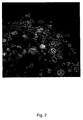
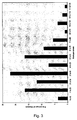
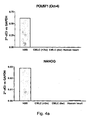


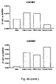
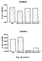


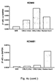

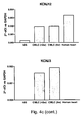

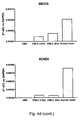


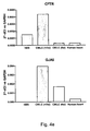


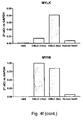

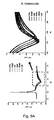
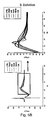


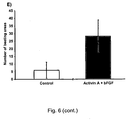
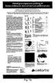


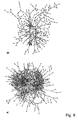


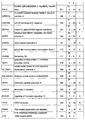


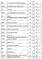


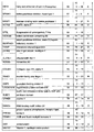
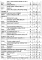
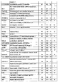


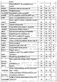


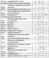


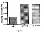


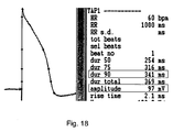


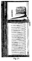


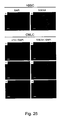
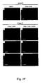
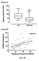



Classifications
-
- C—CHEMISTRY; METALLURGY
- C12—BIOCHEMISTRY; BEER; SPIRITS; WINE; VINEGAR; MICROBIOLOGY; ENZYMOLOGY; MUTATION OR GENETIC ENGINEERING
- C12N—MICROORGANISMS OR ENZYMES; COMPOSITIONS THEREOF; PROPAGATING, PRESERVING, OR MAINTAINING MICROORGANISMS; MUTATION OR GENETIC ENGINEERING; CULTURE MEDIA
- C12N5/00—Undifferentiated human, animal or plant cells, e.g. cell lines; Tissues; Cultivation or maintenance thereof; Culture media therefor
- C12N5/06—Animal cells or tissues; Human cells or tissues
- C12N5/0602—Vertebrate cells
- C12N5/0652—Cells of skeletal and connective tissues; Mesenchyme
- C12N5/0657—Cardiomyocytes; Heart cells
-
- A—HUMAN NECESSITIES
- A61—MEDICAL OR VETERINARY SCIENCE; HYGIENE
- A61P—SPECIFIC THERAPEUTIC ACTIVITY OF CHEMICAL COMPOUNDS OR MEDICINAL PREPARATIONS
- A61P9/00—Drugs for disorders of the cardiovascular system
-
- C—CHEMISTRY; METALLURGY
- C12—BIOCHEMISTRY; BEER; SPIRITS; WINE; VINEGAR; MICROBIOLOGY; ENZYMOLOGY; MUTATION OR GENETIC ENGINEERING
- C12N—MICROORGANISMS OR ENZYMES; COMPOSITIONS THEREOF; PROPAGATING, PRESERVING, OR MAINTAINING MICROORGANISMS; MUTATION OR GENETIC ENGINEERING; CULTURE MEDIA
- C12N2501/00—Active agents used in cell culture processes, e.g. differentation
- C12N2501/10—Growth factors
- C12N2501/115—Basic fibroblast growth factor (bFGF, FGF-2)
-
- C—CHEMISTRY; METALLURGY
- C12—BIOCHEMISTRY; BEER; SPIRITS; WINE; VINEGAR; MICROBIOLOGY; ENZYMOLOGY; MUTATION OR GENETIC ENGINEERING
- C12N—MICROORGANISMS OR ENZYMES; COMPOSITIONS THEREOF; PROPAGATING, PRESERVING, OR MAINTAINING MICROORGANISMS; MUTATION OR GENETIC ENGINEERING; CULTURE MEDIA
- C12N2501/00—Active agents used in cell culture processes, e.g. differentation
- C12N2501/10—Growth factors
- C12N2501/16—Activin; Inhibin; Mullerian inhibiting substance
-
- C—CHEMISTRY; METALLURGY
- C12—BIOCHEMISTRY; BEER; SPIRITS; WINE; VINEGAR; MICROBIOLOGY; ENZYMOLOGY; MUTATION OR GENETIC ENGINEERING
- C12N—MICROORGANISMS OR ENZYMES; COMPOSITIONS THEREOF; PROPAGATING, PRESERVING, OR MAINTAINING MICROORGANISMS; MUTATION OR GENETIC ENGINEERING; CULTURE MEDIA
- C12N2501/00—Active agents used in cell culture processes, e.g. differentation
- C12N2501/20—Cytokines; Chemokines
- C12N2501/23—Interleukins [IL]
- C12N2501/235—Leukemia inhibitory factor [LIF]
-
- C—CHEMISTRY; METALLURGY
- C12—BIOCHEMISTRY; BEER; SPIRITS; WINE; VINEGAR; MICROBIOLOGY; ENZYMOLOGY; MUTATION OR GENETIC ENGINEERING
- C12N—MICROORGANISMS OR ENZYMES; COMPOSITIONS THEREOF; PROPAGATING, PRESERVING, OR MAINTAINING MICROORGANISMS; MUTATION OR GENETIC ENGINEERING; CULTURE MEDIA
- C12N2501/00—Active agents used in cell culture processes, e.g. differentation
- C12N2501/70—Enzymes
-
- C—CHEMISTRY; METALLURGY
- C12—BIOCHEMISTRY; BEER; SPIRITS; WINE; VINEGAR; MICROBIOLOGY; ENZYMOLOGY; MUTATION OR GENETIC ENGINEERING
- C12N—MICROORGANISMS OR ENZYMES; COMPOSITIONS THEREOF; PROPAGATING, PRESERVING, OR MAINTAINING MICROORGANISMS; MUTATION OR GENETIC ENGINEERING; CULTURE MEDIA
- C12N2501/00—Active agents used in cell culture processes, e.g. differentation
- C12N2501/999—Small molecules not provided for elsewhere
-
- C—CHEMISTRY; METALLURGY
- C12—BIOCHEMISTRY; BEER; SPIRITS; WINE; VINEGAR; MICROBIOLOGY; ENZYMOLOGY; MUTATION OR GENETIC ENGINEERING
- C12N—MICROORGANISMS OR ENZYMES; COMPOSITIONS THEREOF; PROPAGATING, PRESERVING, OR MAINTAINING MICROORGANISMS; MUTATION OR GENETIC ENGINEERING; CULTURE MEDIA
- C12N2503/00—Use of cells in diagnostics
- C12N2503/02—Drug screening
-
- C—CHEMISTRY; METALLURGY
- C12—BIOCHEMISTRY; BEER; SPIRITS; WINE; VINEGAR; MICROBIOLOGY; ENZYMOLOGY; MUTATION OR GENETIC ENGINEERING
- C12N—MICROORGANISMS OR ENZYMES; COMPOSITIONS THEREOF; PROPAGATING, PRESERVING, OR MAINTAINING MICROORGANISMS; MUTATION OR GENETIC ENGINEERING; CULTURE MEDIA
- C12N2506/00—Differentiation of animal cells from one lineage to another; Differentiation of pluripotent cells
- C12N2506/02—Differentiation of animal cells from one lineage to another; Differentiation of pluripotent cells from embryonic cells
Definitions
- the present invention relates to novel cardiomyocyte-like cell clusters (CMLC) derived from hBS cells and to the potential use of such cardiomyocyte-like cell clusters in e.g., pharmaceutical drug discovery and development, toxicity testing, cell therapy and medical treatment.
- the clusters contain cells expressing endodermal, mesodermal as well as cardiac markers.
- the invention also relates to a method for preparing the cluster, to composition comprising one or more CMLC for use in therapy and toxicity testing.
- the compositions are stable after storage of the composition at a temperature of at least -80 °C for at least 2 years, i.e. the characteristics and viability of the clusters are not substantially changed during this storage period.
- Mature cardiomyocytes are considered terminally differentiated cells and as such they have no, or very low, proliferative capacity.
- Human cardiomyocytes can be isolated from heart biopsies but the procedure is complicated and it is difficult to obtain viable cell preparations. In addition, the access to human heart tissue is very limited and it is thus not possible to isolate large number of cells. An easy access to cells with a phenotype of human cardiomyocytes is critical for the development of cell therapy interventions for cardiac disease. In addition, due to the lack of donor material as well as the somewhat problematic procedure of cell isolation, human primary cardiomyocytes are not currently available for in vitro testing during pre-clinical drug discovery.
- pluripotent human stem cells can be isolated from the inner cell mass of blastocysts and these cells have the capacity for indefinite, undifferentiated proliferation in vitro (Thomson et al., 1998; Reubinoff et al., 2000; Heins et al., 2004).
- Differentiation of human blastocysts derived stem (hBS) cells may occur spontaneously in vitro, especially during, for undifferentiated cells, sub-optimal culture conditions (Thomson et al., 1998; Reubinoff et al., 2000).
- hBS cells can be coaxed to differentiate in a directed fashion along specific pathways forming a variety of specialized cell types including cardiomyocytes, endothelial cells, neuronal cells, insulin producing ⁇ -cells, and hematopoietic cells (reviewed in: Keller, Genes Dev 2005 ).
- cardiomyocytes endothelial cells
- neuronal cells neuronal cells
- insulin producing ⁇ -cells insulin producing ⁇ -cells
- hematopoietic cells reviewed in: Keller, Genes Dev 2005 .
- relatively little is currently known about how to control and manipulate hBS cell differentiation to produce exclusive populations of specific cell types.
- cardiomyocytes For the derivation of cardiomyocytes from hBS cells, in principle two different procedures have been reported. The first is through differentiation of hBS cells initiated when the cells are cultured in suspension and form embryoid bodies (EBs) (Itskovitz-Eldor et al 2000, Kehat et al 2001). Within these mixed population of cells contracting areas with functional properties of cardiomyocytes can be observed. The second procedure is based on co-culture of hBS cells with END-2 cells (a visceral endoderm-like cell line) which results in the formation of beating clusters of cells that also display characteristics of cardiomyocytes (Mummery et al 2003, Passier et al 2005).
- END-2 cells a visceral endoderm-like cell line
- hBS cell-derived cardiomyocytes During recent years, a number of papers have described, in various ways, the basic characteristics of hBS cell-derived cardiomyocytes. In these reports, cell analysis has been based on the expression of molecular markers for cardiomyocytes, structural architecture, and functionality (reviewed in: Ameen et al 2007 Crit Rev Oncol Hematol ). The morphology and ultrastructure of hBS cell-derived cardiomyocytes share similarities with adult cardiomyocytes although the myofibrillar and sarcomeric organization indicate an immature phenotype in the stem cell derived population (Kehat et al 2001, Snir et al 2003, Norström et al 2006, Yoon et al 2006).
- cardiomyocytes On a molecular level, several markers expressed by cardiomyocytes are also expressed by hBS cell-derived cardiomyocytes, including transcription factors, structural proteins, hormones, ion-channels, and tight junction proteins (Kehat et al 2001, Xu et al 2002, Mummery et al 2003, He et al 2003, Passier et al 2005, Norström et al 2006, Yoon et al 2006).
- cardiomyocytes derived from hBS cells show that these cells share similarities with their adult counterparts. More importantly, however, are the functional characteristics of the cells, and different pharmacological and electrophysiological approaches have been used to examine these properties.
- One major advantage of cardiomyocytes derived from hBS cells is that they can be maintained in culture for extended time periods without loosing their spontaneous contractile capacity. This allows for repeated non-invasive examination of the same cell preparations.
- hBS cell-derived cardiomyocytes respond to ⁇ / ⁇ -adrenergic- and muscarinic stimuli suggesting that the cells express specific surface membrane receptors coupled to a signalling pathway that activate ion channels, membrane transporters and myofilament proteins (Kehat et al 2001, Xu et al 2002, Xue et al 2005, Norström et al 2006).
- action potentials indicative of nodal-, atrial- and ventricular-like origin have been identified in hBS cell-derived cardiomyocytes using intracellular electrophysiological measurements (Xu et al 2002, Mummery et al 2003, He et al 2003).
- cardiomyocyte like cell clusters from hBS cells specifically, has a great potential for several of these in vitro applications were they make up a platform for measurements of functional attributes such as action potentials and conduction in cardiomyocytes.
- the developing clusters may serve as in vitro models to study early events during human cardiogenesis.
- the clusters may be optimal for use in vivo for restoration of cardiac function after injury or disease.
- the present invention presents unique cardiomyocyte-like cell clusters derived from hBS cells for use according to the above.
- the present invention relates to novel cardiomyocyte-like cell clusters (CMLC) and the methods for their preparation from hBS cells.
- CMLC cardiomyocyte-like cell clusters
- the CMLC of the present invention are especially well suited for use in drug discovery and toxicity testing.
- a cluster according to the invention comprises cardiomyocyte-like cells, wherein the cluster has
- the electrical connection between cells mentioned above may be by gap junctions.
- the cluster contains cells that respond to pharmacological stimuli such as beta-adrenergic stimulation (isoproterenol, adrenalin), alpha-adrenergic stimulation (phenylephrine), muscarinic stimulation (carbachol, acetylcholine, atropine), blockage of calcium channels (verapamil), blockage of hERG channels (E4031) and inhibition of funny channels.
- beta-adrenergic stimulation isoproterenol, adrenalin
- alpha-adrenergic stimulation phenylephrine
- muscarinic stimulation carbachol, acetylcholine, atropine
- blockage of calcium channels (verapamil)
- blockage of hERG channels E4031
- inhibition of funny channels such as beta-adrenergic stimulation (isoproterenol, adrenalin), alpha-adrenergic stimulation (phenylephrine), muscarinic stimulation (carbachol, acetyl
- a cluster according to the present invention comprises cardiomyocyte-like cells and the cluster comprising genes that are up-regulated and have,
- cardiomyocyte-like cells suitable for use in e.g. medical applications, drug screenings and cardiotoxicity studies can be isolated in the form of clusters that are relatively easy to prepare in a reproducible manner and without any need for further differentiation into cardiomyocyte cells.
- the clusters also contains markers e.g. for endodermal lineage, express one or more ion channels commonly used for cardiotoxicity testing, and express transcription factors.
- the present invention provides a relative simple means of obtaining a suitable cardiomyocyte-like cell product for medical use as well as for drug screening and testing without the need of purification and complete differentiation to mature cardiomyocytes.
- the present invention provides a composition that presents the CMLC in a form that is easy to use for the end-user.
- the composition is prepared by suspending the CMLC in an aqueous medium notably containing agents that ensure the stability of the CMLC during freezing/vitrification, storage, thawing and use of the cluster. Accordingly, the properties of such an aqueous medium must be chosen in consideration of all these four different processes in order to obtain a sole medium that is suitable for the handling of the clusters from the clusters have been prepared and until use of the end-user.
- the product is presented in fresh or frozen form.
- the term "blastocyst-derived stem cell” is denoted BS cell, and the human form is termed "hBS cells".
- BS cell the human form
- the pluripotent stem cells used in the present invention can be commercially available hBS cells or cell lines.
- any human pluripotent stem cell can be used in the present invention, including differentiated adult cells which are reprogrammed to pluripotent cells by e.g. the treating adult cells with certain transcription factors, such as OCT4, SOX2, NANOG, and LIN28 as disclosed in Junying Yu, et al., 2007.
- DMSO dimethylsulfoxide
- cardiomyocyte-like cells is intended to mean cells sharing features with mature cardiomyocytes. Cardiomyocyte-like cells are further defined by morphological characteristics as well as by specific marker characteristics.
- CMLC cardiomyocyte-like cells clusters
- nodal-like cells is intended to mean cells with the following action potential characteristics:
- Atrial-like cells is intended to mean cells with the following action potential characteristics:
- ventricle-like cells are intended to mean cells with the following action potential characteristics:
- expression is used for gene and/or protein expression, whatever must be relevant in the context.
- cryopreservation denotes the preservation of biological material at an extremely low temperature.
- mixed differentiated hBS cells is used for a population of spontaneously differentiated hBS cells were cells from all three germ layers mesoderm, endoderm and ectoderm are present. This sample is used as control material to distinguish genes up regulated in many types of differentiated cells from genes specifically up regulated in cardiomyocyte-like cell clusters. In the examples herein is given instructions how to obtain mixed differentiated hBS cells.
- force aggregation is used for 3D aggregation of cells by an external force, such as centrifugation with a centrifugal force or sedimentation by gravity.
- feeder cells are intended to mean supporting cell types used alone or in combination.
- the cell type may further be of human or other species origin.
- the tissue from which the feeder cells may be derived include embryonic, fetal, neonatal, juvenile or adult tissue, and it further includes tissue derived from skin, including foreskin, umbilical chord, muscle, lung, epithelium, placenta, fallopian tube, glandula, stroma or breast.
- the feeder cells may be derived from cell types pertaining to the group consisting of human fibroblasts, fibrocytes, myocytes, keratinocytes, endothelial cells and epithelial cells.
- Examples of specific cell types that may be used for deriving feeder cells include embryonic fibroblasts, extraembryonic endoderm cells, extraembryonic mesoderm cells, fetal fibroblasts and/or fibrocytes, fetal muscle cells, fetal skin cells, fetal lung cells, fetal endothelial cells, fetal epithelial cells, umbilical chord mesenchymal cells, placental fibroblasts and/or fibrocytes, placental endothelial cells,
- MEF cells is intended to mean mouse embryonic fibroblasts.
- TGF- ⁇ means transforming growth factor beta, preferably of human and/or recombinant origin.
- TGF- ⁇ is a protein that comes in three isoforms called TGF- ⁇ 1, TGF- ⁇ 2 and TGF- ⁇ 3.
- the TGF- ⁇ family is part of a superfamily of proteins known as the transforming growth factor beta superfamily, which includes inhibins, activin, anti-müllerian hormone, bone morphogenetic protein, decapentaplegic and Vg-1.
- FGF fibroblast growth factor
- bFGF sometimes also referred to as FGF2
- FGF4 fibroblast growth factor
- BMP2 bone-morphogenic protein-2 which is one member of the BMP family of growth factors.
- xeno-free is intended to mean complete circumvention of direct or in-direct exposure to non-human animal components.
- Cardiac stem cell marker
- a cluster according to the invention comprises cardiomyocyte-like cells, wherein the cluster has
- the electrical connection between cells mentioned above may be by gap junctions.
- the cluster does not express any markers for undifferentiated cells.
- the cluster comprising genes that are up-regulated and have,
- the present invention provides a cluster comprising cardiomyocyte-like cells, the cluster comprising genes that are up-regulated and have,
- F MC denoted the fold change for a mixed differentiated hBS cell population.
- mixed differentiated hBS cells is used for a population of spontaneously differentiated hBS cells where cells from all three germ layers mesoderm, endoderm and ectoderm are present. This sample is used as control material to distinguish genes up-regulated in all types of differentiated cells from genes specifically up-regulated in cardiomyocyte-like cell clusters.
- the expression values are obtained by measurement with a specific chip (GeneChip® Human Genome U133 Plus 2.0 (Affymetrix Inc., Santa Clara, CA, USA) as detailed described in Example 2 herein. Use of other chips may give different values and therefore any comparison with the values herein should be made using the same kind of chip or a chip giving similar results.
- a cluster is thus provided wherein the cluster comprising genes which are up-regulated and have,
- the clusters are derived from hBS cells. Accordingly, the starting material may be commercially available hBS cells or cell lines. Such material can be obtained from Cellartis AB (www.cellartis.com ) and is also available through the NIH stem cell registry ( http://stemcells.nih.gov/research/registry ) as well as from the UK Stem Cell Bank ( http://www.ukstemcellbank.org.uk ).
- the cluster may fulfill the above-mentioned requirements.
- the requirements are fulfilled for the cluster and not necessarily for any individual cell.
- the cells contained in the cluster may be of different degree of differentiation; some may be entirely differentiated into cardiomyocyte-like cells, whereas others may possess characteristics like endodermal cells.
- the cluster must contain contracting cells.
- the clusters are typically prepared by subjecting cells to spontaneous differentiation via forced aggregation to obtain 3D-structures containing undifferentiated cells.
- the cardiomyocyte like cell clusters appear after further cultivation of the 3D-structures for up to 22 days.
- an objective of the present invention is to provide a cluster containing different types of cells in their development from hBS cells to cardiomyocytes, i.e. from the very beginning of differentiation via intermediate differentiation to fully differentiated cells into cardiomyocytes.
- a cluster of the invention has a variety of markers.
- the clusters contain a plurality of cells such as from about 10 to about 5000 cells.
- a cluster according to the invention contains cells with up-regulated genes (compared to undifferentiated cells), wherein the expression value is about 750 or more such as, e.g., an expression value of about 1000 or more, about 1500 or more, about 2000 or more, about 2500 or more or about 3000 or more.
- a cluster according to the invention contains cells with up-regulated genes having a FC( CMLC ) value of about 20 or more such as, e.g., about 50 or more, about 100 or more, about 500 or more, about 750 or more, about 1000 or more.
- the ratio of FC CMLC /FC MC is about 15 or more such as, e.g., about 20 or more, about 50 or more, about 100 or more, about 500 or more, about 750 or more, about 1000 or more.
- a set of criteria can be set in order to select clusters with different content of up-regulated genes.
- genes identified in the clusters are genes that are not normally associated with cardiomyocyte-like cells (see the genes marked as "white” in Table II). These findings seem to be of interest and they may play an important role for the functionality of the CMLC with respect to medical use and testing. Specifically cells and tissue of endodermal origin seem to play an essential role in early cardiac development. The CMLC make up a tissue like structure simulating early developing cardiac tissue.
- a cluster according to the invention has the following characteristics (Table II and III):
- the first group consists of genes previously associated with cardiac cells and the second of genes previously associated with endodermal cells, such as hepatocytes.
- Examples of genes belonging to the first group are (marked in light grey in table II): MYH6, MYL7, MYL4, TNNC1, TNNT2, PLN, TTN, MYH7, LDB3, NPPB, GATA6, MYL3, CSRP3, ACTN2, MB, MYOZ2, TBX5, and HSP27.
- genes belonging to the second group are (marked in dark grey in table II): AFP, TF, APOA2, AHSG, SERPINA1, APOA1, ALB, APOC3, TTR, APOB, and RBP4.
- the selection shows an even distribution between endodermal and mesodermal genes expressed in the clusters.
- the mesodermal genes and the endodermal are marked in, light grey and dark grey respectively in Table III.
- a cluster according to the invention has 2 or more such as, e.g., 4 or more, 6 or more, 8 or more, 10 or more, 12 or more or 16 or more of the up-regulated genes are genes associated with cardiac cells, and/or 2 or more such as, e.g., 4 or more, 6 or more, 8 or more of the up-regulated genes are genes associated with endodermal cells, and/or 2 or more such as, e.g., 4 or more, 6 or more, 8 or more of the up-regulated genes are genes associated with non cardiac or non endodermal cells, described in Table II herein, and/or the up-regulated genes comprise 10 or more such as, e.g., 20 or more, 30 or more, 40 or more, 50 or more, 55 or more or all genes listed in Table II herein.
- a cluster comprising genes which are up-regulated and have,
- a cluster comprising genes which are up-regulated and have,
- the cluster is normally derived from hBS cells and the number of cells in the cluster is normally 10 to about 5000 cells or from 10 to about 2000 cells.
- a cluster according to the invention expresses one or more ion channels.
- the ion channels identified are ion channels commonly used in cardiotoxicity testing. This is a further indication of the suitability of the CMLC for cardiotoxicity testing.
- the ion-channel is typically a K-, Na, and/or Ca-ion channel such as a K-voltage-gated channel, a K-inwardly-rectifying channel, a Na-voltage-gated channel, a Na-ligand-gated channel, and/or a Ca-voltage-dependent channel. More specific ion channels are listed in Figure 7 a and b, poster Table 1. In a specific embodiment a cluster expresses at least 3 such as, e.g., at least 4, at least 5 or all of the ion channels listed in Figure 7 a and b, poster Table 1.
- a cluster according to the invention has all genes listed in any of Table II, III and/or listed in Figure 7 a and b, poster Table 1.
- the clusters of cardiomyocyte-like cells described above may also be characterized by measuring their transmembrane action potential.
- the characteristic action potential were recorded and the AP duration at 50%, 70% and 90% of repolarization (dur50 (APD50), dur70 (APD70), dur90 (APD90)), AP amplitude (amp), membrane resting potential (MRP), and maximum rate of rise of the AP upstroke (Vmax) were determined.
- the clusters were thus characterized as predominately nodal-like, atrial-like, or ventricle-like based on the results of transmembrane action potential (TAP) recordings.
- TAP transmembrane action potential
- Cluster, or suspension of clusters were thus found that comprised at least 10%, (such as e.g. at least 13%, at least 15%, or about 17%) of nodal-like cells and/or at least 30% (such as e.g. at least 35%, at least 40%, at least 45 % or about 50%) of atrial-like cells and/or at least 20% (such as e.g. at least 23%, at least 26%, at least 30%, or about 33%) ventricle-like cells.
- a cluster according to the invention typically comprises cells expressing at least 10, at least 15, at least 20, at least 25 or all of the following genes:
- a cluster of the invention comprises cells expressing all of the following genes:
- a cluster of the invention comprises cells expressing at least 10 such as at least 50, at least 100, at least 200, at least 300 or at least 400 of the markers mentioned in Fig. 11 .
- a cluster of the invention comprises cells expression at least 10 such as at least 20, at least 40, at least 60 or all of the following markers: Gene symbol Gene title Avg FC CMLC FC CMLC range Category TNNI1 troponin I type 1 (skeletal, slow) 1990 6078 - 100 O LUM lumican 1375 2989 - 365 O IGFBP7 insulin-like growth factor binding protein 7 1269 2781 - 518 N COL3A1 collagen, type III, alpha 1 1234 2187 - 538 O IGF2 insulin-like growth factor 2 (somatomedin A) 1149 1374 - 965 N NPNT nephronectin 1020 1590 - 708 N TF transferrin 942 1759 - 218 U MGP matrix Gla protein 860 2671 - 182 O FGB fibrinogen beta chain 813 1425 - 299 O LDB3 LIM domain binding 3 750 2195 - 134 O CXCL14 chemokine (C-X-C motif) ligand 14 722
- elegans 193 410 - 85 O MEP1A meprin A, alpha (PABA peptide hydrolase) 191 379 - 48 X WNT2 wingless-type MMTV integration site family member 2 186 390 - 90 X SHOX2 short stature homeobox 2 181 421 - 15 O PITX1 paired-like homeodomain transcription factor 1 172 368 - 77 N HOXB2 homeobox B2 171 369 - 9 X GBP1 guanylate binding protein 1, interferon-inducible.
- elegans 118 147 - 60 X FOXF1 forkhead box F1 113 151 - 81 X RGS4 regulator of G-protein signalling 4 112 174 - 67 X C20orf75 chromosome 20 open reading frame 75 105 244 - 9 X VTN vitronectin 104 256 - 11 N IGFBP1 insulin-like growth factor binding protein 1 103 228 - 44 X BGN biglycan 102 313 - 18 N SULT1E1 sulfotransferase family 1E, estrogen-preferring, member 1 101 239 - 18 O ABCA8 ATP-binding cassette, sub-family A, member 8 101 128 - 74 N
- a suspension or composition of cardiomyocyte-like clusters may contain a mixture of clusters containing nodal-like cells, clusters containing atrial-like cells and clusters containing ventricle-like cells.
- One embodiment the invention thus relates to a suspension or composition of cardiomyocyte-like clusters, wherein the ratio between the number of clusters containing nodal-like cells and the number of clusters containing atrial-like cells is in a range of from about 1:100 to about 50:100 such as from about 10:100 to about 40:100, from about 20:100 to about 40:100, from about 30:100 to about 40:100 or about 33:100-34:100.
- the invention in another embodiment relates to a suspension or composition of cardiomyocyte-like clusters, wherein the ratio between the number of clusters containing nodal-like cells and the number of clusters containing ventricle-like cells is in a range of from about 1:100 to about 80:100 such as from about 10:100 to about 70:100, from about 30:100 to about 70:100, from about 45:100 to about 55:100 or about 50:100.
- the invention relates to a suspension or composition of cardiomyocyte-like clusters, wherein the ratio between the number of clusters containing ventricle-like cells and the number of clusters containing atrial-like cells is in a range of from about 1:100 to about 90:100 such as from about 40:100 to about 80:100, from about 50:100 to about 75:100 or about 66:100.
- the invention relates to a suspension or composition of cardiomyocyte-like clusters, wherein the ratio between the clusters containing nodal-like cells, the clusters containing atrial-like cells and the clusters containing ventricle-like cells is 17:50:33.
- compositions comprising one or more cluster
- a cluster-containing composition i.e. one or more clusters of cardiomyocyte-like cells as described above may be in a composition comprising a carrier, such as e.g. an aqueous medium.
- the composition may further contain one or more additives including one or more cryoprotectants, one or more stabilizers and/or one or more viscosity-adjusting agents.
- the composition may be in liquid or frozen form.
- the one or more cryoprotectants may be selected from the group consisting of ethylene glycol, propylene glycol, dimethylsulfoxide, glycerol, propanediol, and methyl pentanediol, and/or mixtures thereof.
- the additive may also be a sugar or sugar alcohol including sucrose, trehalose, maltose or lactose.
- the cryoprotectant is trehalose in a concentration from about 0.02 M to about 1 M, such as, e.g., from about 0.05 M to about 0.9 M, from about 0.1 M to about 0.8 M, from about 0.15 M to about 0.7 M, from about 0.2 M to about 0.65 M, from about 0.25 M to about 0.6 M.
- concentration of trehalose may preferably be about 0.3 M.
- the cryoprotectant is sucrose in a concentration from about 0.02 M to about 1 M, such as, e.g., from about 0.05 M to about 0.9 M, from about 0.1 M to about 0.8 M, from about 0.15 M to about 0.7 M, from about 0.2 M to about 0.65 M, from about 0.25 M to about 0.6 M.
- concentration of sucrose may preferably be about 0.3 M.
- the cryoprotectant may be DMSO in a concentration that is at least 2.5 % v/v, such as e.g. from about 2.5% to about 40% v/v, from about 5% to about 35% v/v, from about 7% to about 30% v/v, from about 7% to about 25% v/v, from about 7% to about 20% v/v, from about 15% to about 25% v/v, or from about 5% to about 15% v/v.
- the cryoprotectant may be ethylene glycol in a concentration that is at least 2.5 % v/v, such as e.g. from about 2.5% to about 30% v/v, from about 5% to about 25% v/v, from about 5% to about 20% v/v, from about 10% to about 20% v/v, from about 7% to about 10% v/v, or from about 2.5% to about 5% v/v.
- composition may further comprise a viscosity-adjusting agent selected from the group consisting of Ficoll, Percoll, hyaluronic acid, albumin, polyvinyl pyrrolidone, alginic acid, gelatin and glycerol.
- a viscosity-adjusting agent selected from the group consisting of Ficoll, Percoll, hyaluronic acid, albumin, polyvinyl pyrrolidone, alginic acid, gelatin and glycerol.
- the viscosity-adjusting agent may be Ficoll in a concentration at the most about 150 mg/ml, such as, e.g., at the most about 100 mg/ml, at the most about 50 mg/ml, at the most about 25 mg/ml, at the most about 15 mg/ml or at the most about 10 mg/ml.
- a further component of the composition may be one or more growth factors, e.g. basic fibroblast growth factor (bFGF) in a concentration of about 2 ng/ml, such as about 3 ng/ml, about 4, ng/ml, about 5 ng/ml, about 6 ng/ml or more.
- bFGF basic fibroblast growth factor
- composition may also comprise one or more essential and/or non-essential amino acids in appropriate concentrations:
- essential amino acids are e.g. L-Alanine, L-Arginine, L-Asparagine, L-Aspartic acid, L-Cysteine, L-Glutamic acid, L-Glutamine, Glycine, L-Histidine, L-Isoleucine, L-Leucine, L-Lysine, L-Methionine, L-Phenylalanine, L-Proline, L-Serine, L-Threonine, L-Tryptophan, L-Tyrosine, L-Valine in concentrations raging from about 0.01 mM, such about 0.05 mM, about 0.1 mM, about 0.15 mM, about 0.2 mM or more.
- the composition may further include one or more inorganic salts such as Calcium chloride (Anhydrous),Cupric sulfate (CuSO4-5H2O), Ferric sulfate (FeSO4-7H2O), Potassium chloride (KCI), Magnesium chloride (Anhydrous), Sodium chloride (NaCl), Sodium bicarbonate (NaHCO3), Sodium phosphate, dibas (Anhydrous), Zinc sulfate (ZnSO4-7H2O), and/or one vitamins, such as Biotin, D-Calcium pantothenate, Choline chloride, Folic acid, i-Inositol, Niacinamide, Pyridoxine hydrochloride, Riboflavin, Thiamine hydrochloride or Vitamin B12.
- inorganic salts such as Calcium chloride (Anhydrous),Cupric sulfate (CuSO4-5H2O), Ferric sulfate (FeSO4-7H2O),
- DMEM Dulbecco's Modified Eagle's Medium
- MEM media MEM media
- RPMI media RPMI media
- CMLC CMLC were vitrified in closed straws as described in r WO2004 098285 and stored in liquid N 2 .
- the cells were recovered after thawing in culture medium (Knock Out DMEM supplemented with 20% FBS, 1% penicillin-streptomycin, 1% Glutamax, 0.5 mmol/l ⁇ -mercaptoethanol, and 1% non-essential amino acids).
- the CMLC normally has a size of from about 20 ⁇ m to about 30 ⁇ m. Moreover, 50-80 % of the cells in the cluster express cardiac markers.
- a suitable product for shipping contains one or more clusters in KnockOut DMEM medium supplemented with 2-20% FBS, 1% penicillin-streptomycin, 1% Glutamax, 0.5 mmol/l ⁇ -mercaptoethanol and 1% non-essential amino acids (all from Invitrogen, Carlsbad, California).
- a composition according to the present invention provides a composition wherein the cluster retains at least 95% of its described characteristics after storage at a temperature of at least-80 °C for at least 2 years. Further, at least about 50% or more such as, e.g., about 55% or more, about 60% or more, about 65% or more, about 70% or more, about 75% or more, about 80% or more, about 85% or more, about 90% or more or about 95% or more of the cells are viable after storage of the composition at a temperature of at least -80 °C for at least 2 years.
- kits for use in testing of cardiotoxicity of a specific substance comprising
- the kit may comprise a composition of cardiomyocyte-like clusters of nodal-like cells and/or atrial-like cells and/or ventricle-like cells as defined above.
- the kit may further comprise
- kit for use in in vitro testing during drug discovery of a specific substance comprises
- the kit may comprise a composition of cardiomyocyte-like clusters of nodal-like cells and/or atrial-like cells and/or ventricle-like cells as defined above.
- the kit may further comprise
- the kit may comprise a composition of cardiomyocyte-like clusters of nodal-like cells and/or atrial-like cells and/or ventricle-like cells as defined above.
- the cryopreservation of the cardiomyocyte-like cell clusters may be performed by using a slow freezing method or by vitrification, using open or closed pulled straws.
- At least one of the vitrification solutions may contain one or more cryoprotectants or mixtures of cryoprotectants.
- Non-toxic cryoprotectants are of course preferable.
- Cryoprotectants help minimizing shrinking by reducing the mole fraction of other solutes remaining in the non-frozen water. They inhibit the formation of crystalline ice, and thus depress the freezing point of the water. They may also prevent protein denaturation by hydrogen binding with bound water. As cells cool, solvent water converts to extracellular ice, and the increasing extracellular concentration of non-permeating electrolyte or non-electrolytes damages the cells. When treated with a cryoprotectant, cells do not reach the salt concentrations of non-treated cells until they reach much lower temperatures.
- cryoprotectants may also function as osmotically active agents.
- Suitable cryoprotectants can be selected from the group consisting of ethylene glycol, propylene glycol, dimethylsulfoxide, glycerol, propanediol, sugars including sucrose, trehalose, maltose, lactose and methyl pentanediol.
- a vitrification procedure may comprise the following steps,
- the concentration of the individual agents contained in the first and or the second solution is normally in a range of 5-50% v/v such as, e.g. from about 5% to about 40% v/v such as e.g. from about 5% to about 25% v/v (first solution) and from about 5% to about 30% v/v (second solution).
- the total concentration (i.e. calculated as v/v, w/v or M) of the cryoprotectant in the second solution is larger than that in the first solution.
- the first and the second solution may contain one or more cryoprotectants that are the same or different.
- the concentration of the one or more cryoprotectants in the first and the second solution can be the same or different, and normally the total concentration of the cryoprotectant in the second solution is larger than that in the first solution.
- the cryoprotectant is trehalose.
- concentration of trehalose contained in the first and/or the second solution is normally within a range from about 0.02 M to about 1 M, such as, e.g., from about 0.05 M to about 0.9 M, from about 0.1 M to about 0.8 M, from about 0.2 M to about 0.7 M, from about 0.3 M to about 0.65 M, from about 0.4 M to about 0.6 M, from about 0.45 M to about 0.55 M.
- sucrose is used in similar applications.
- Trehalose is a unique, naturally occurring disaccharide and is found in hundreds of plants and animals. Trehalose is an important source of energy and has been shown to be a primary factor in stabilization of organisms during time of freezing.
- trehalose can depress the phase transition temperature of membranes so that they remain in the liquid-crystal state even when dry. Without being bound to any theory, it is hypothesized that this prevents membrane leakage during rehydration, thereby preserving cellular viability. With respect to proteins, trehalose has been shown to inhibit protein denaturation by exclusion of water from the protein surface when the cells are in the hydrated state.
- the cryoprotectant is sucrose.
- concentration of sucrose contained in the first and/or the second solution is normally within a range from about 0.02 M to about 1 M, such as, e.g., from about 0.05 M to about 0.9 M, from about 0.1 M to about 0.8 M, from about 0.2 M to about 0.7 M, from about 0.3 M to about 0.65 M, from about 0.4 M to about 0.6 M, from about 0.45 M to about 0.55 M.
- At least one of the first and second solutions comprises a cryoprotectant.
- At least one of the first and the second solution may comprise a viscosity-adjusting agent.
- Suitable viscosity-adjusting agent for use in the present context may be selected from the group consisting of Ficoll, Percoll, hyaluronic acid, albumin, polyvinyl pyrrolidone, alginic acid, gelatin and glycerol.
- the first and the second solution may contain one or more viscosity-adjusting agents that are the same or different.
- the concentration of the one or more viscosity-adjusting agents in the first and the second solution may be the same or different.
- the viscosity-adjusting agent is Ficoll.
- the concentration of Ficoll contained in the first and/or the second solution is at the most about 150 mg/ml, such as, e.g., at the most about 100 mg/ml, at the most about 50 mg/ml, at the most about 25 mg/ml, at the most about 15 mg/ml or at the most about 10 mg/ml.
- At least one of the first and second solutions may be an aqueous solution.
- xenofree hBS cell lines and culture methods are applied for therapeutic applications of the cardiomyocyte-like cell clusters.
- the hBS cell line constitutes a trisomic cell line.
- Another aspect the present invention relates to a method for preparing CMLCs from hBS cells.
- the novel method has shown to be advantageous compared with prior art method using EB formation as the yield of beating clusters are markedly improved.
- the new method is also more suitable for large scale production.
- the cell lines used in this method may be obtained from a commercial available cell line.
- the present invention provides a method for the preparation of a cluster comprising cardiomyocyte-like cells, the method comprising the steps of:
- the method may also comprise a further step of isolating the one or more clusters by removing the one or more clusters from said plate.
- the method for obtaining cardiomyocyte-like clusters comprises the steps of
- the cells may be detached from the feeder layer by use of mechanical or, preferably, enzymatic treatment, whereby a suspension of primarily undifferentiated hBS cells is obtained.
- the enzymatic treatment may be performed by use of a proteolytic enzyme, such as a collagenase, for an appropriate time, e.g. 2-20 minutes, 5-15 minutes or 10-15 minutes.
- a proteolytic enzyme such as a collagenase
- the cells obtained in the above-mentioned step 1 are mainly undifferentiated, meaning that from about 70% or more, such as e.g. 80 % or more, 85% or more, 90 % or more, 95% or more, or 99% or more of the cells are undifferentiated.
- the cell suspension may thereafter be resuspended in an appropriate media which supports the cells, this media can for example be Knock Out DMEM supplemented with FBS (preferably in the range 15-25%, such as 20%).
- the culture medium can, at various time points during the experiments, be supplemented with different factors such as protein growth factors, chemical compounds, minerals, or other signalling molecules. Examples of such factors are BMP-2, BMP-4, BMP-5, TGF- ⁇ 1, Activin A, Growth hormone, LIF, and PDGF.
- the suspension step is relatively short and does not allow for embryoid bodies to develop. In the present method the suspension step is just a transfer step.
- the resuspended cells are thereafter further dissociated into small aggregates of undifferentiated cells. This dissociation can be performed manually, e.g. by using a pipette, until the cells forms small aggregates of the size 0.2-0.4 mm.
- the culture dish(es) used may be of any kind that supports the cells, but in a preferred embodiment the culture dish(es) are gelatine coated dishes.
- the cell aggregates obtained in step 2) or 3) are subjected to forced aggregation and/or sedimentation before transferring the cells to the culture dish(es) in step 4) and 5).
- 3D structures are formed when spun down through forced aggregation.
- the 3D structures are different from embryoid bodies.
- the advantage is that a more reliant structure is formed when compared to e.g. embryoid bodies, which have a tendency to fall apart, and therefore the present method gives a higher yield between the number of undifferentiated cells used and the resulting number of cardiomyocyte-like clusters.
- the method is also advantageous in that it is faster, more reliable and more scalable and useable for automation compared to the prior art EB protocols.
- the preferred method for obtaining cardiomyocyte-like clusters comprises the steps of
- step 2 By using forced aggregation approximately 4-6 colonies from step 2) or 3) are transferred to a plate or tube appropriate for centrifugation, e.g. a 96-well v-bottom plate (Corning Incoporated, NY, USA). The cells are then centrifuged for 2-20 minutes at 100-800 x g (e.g. 200-600 x g for 5-10 min or, 300-500 x g for 5-10 min, or at 400 x g for 5 min.). These centrifuged cells may, before the transfer mentioned step 6), further be incubated for 1-10 days (such as 1-7 days, or 1-5 days) for the formation of 3D structures, which structures then are transferred according to step 6) above.
- a plate or tube appropriate for centrifugation e.g. a 96-well v-bottom plate (Corning Incoporated, NY, USA).
- the cells are then centrifuged for 2-20 minutes at 100-800 x g (e.g. 200-600
- the forced aggregation may additionally be combined, or replaced, by sedimentation of the cell suspension obtained step 2) or 3), for 1-36 hours (such as 2-24 hours or 3-12 hours), before proceeding to step 6).
- the yield of CMLC from hBS cells can be further improved by supplementing the culture medium used with different factors such as protein growth factors, chemical compounds, minerals, or other signalling molecules.
- the culture medium e.g. DMEM
- the culture medium comprises a member from the transforming growth factor beta superfamily (e.g. Activin A) and/or a member from the fibroblast growth factor family (e.g. bFGF) in e.g. step 5 and/or step 7 of the preferred method (and/or step 2 of the first embodiment of the method)
- the yield increases.
- This yield as shown in figure 6 , and in example 9, increases a 4.7 fold compared to controls grown in the same media, but without bFGF or Activin A added.
- the concentrations of Activin A supplemented to the culture medium comprises from about 5-40 ng/ml, such as e.g. 8 ng/ml, 9 ng/ml, 10 ng/ml, 11 ng/ml, or 12 ng/ml; and the concentration of bFGF supplemented to the culture medium comprises from about 5-40 ng/ml, such as e.g. 10 ng/ml, 11 ng/ml, 12 ng/ml, 13 ng/ml or 14 ng/ml.
- the method for obtaining cardiomyocyte-like clusters thus comprises the steps of Steps Embodiment 1 Embodiment 2 Embodiment 3 Embodiment 4 Obtaining a suspension of primarily undifferentiated hBS cells Detach the cells from the feeder layer by collagenase treatment for 10-15 minutes Detach the cells from the feeder layer by collagenase treatment for 10-15 minutes Detach the cells from the feeder layer by collagenase treatment for 10-15 minutes Detach the cells from the feeder layer by collagenase treatment for 10-15 minutes Resuspending said cell suspension in an appropriate medium
- the medium comprises Knock out DMEM with 20% FBS, and optionally containing 5-20 ng/ml bFGF 5-20 ng/ml Activin A
- the medium comprises Knock out DMEM with 20% FBS, and optionally containing 8-15 ng/ml bFGF 8-15 ng/ml Activin A
- the medium comprises Knock out DMEM with 20% FBS, and optionally containing 10-15 ng/ml bF
- the invention may also take place by sedimenting the cells instead of centrifugation.
- Specific embodiments of the invention thus comprises, Steps Embodiment 1 Embodiment 2 Embodiment 3 Embodiment 4 Obtaining a suspension of primarily undifferentiated hBS cells Detach the cells from the feeder layer by collagenase treatment for 10-15 minutes Detach the cells from the feeder layer by collagenase treatment for 10-15 minutes Detach the cells from the feeder layer by collagenase treatment for 10-15 minutes Detach the cells from the feeder layer by collagenase treatment for 10-15 minutes Resuspending said cell suspension in an appropriate medium
- the medium comprises Knock out DMEM with 20% FBS, and optionally containing 5-20 ng/ml bFGF 5-20 ng/ml Activin A
- the medium comprises Knock out DMEM with 20% FBS, and optionally containing 8-15 ng/ml bFGF 8-15 ng/ml Activin A
- the medium comprises Knock out DMEM with 20% FBS, and optional
- the yield have also been shown to increase (as shown in e.g. figure 15 ) when the medium used in step 5 and/or 7 of the preferred method is further supplemented (either alone or in combination with Activin A, bFGF and/or FBS) with a member of GSK-3 inhibitors and/or p38 MAP kinase inhibitors such as SB 216763 and SKF-860002, respectively.
- the yield increased by 2-3 fold when Activin A, bFGF and SB 216763 or SKF-860002 are added to the culture medium in step 5 of the preferred method compared to controls where only Activin A and bFGF are added.
- This medium may be supplemented at any stage of the method, however in one embodiment the medium is supplemented to the cells during their incubation after the centrifugation in step 5) of the preferred method.
- the concentration of the GSK-3 inhibitor supplemented to the culture medium may be in the range of from about 1-25 ⁇ M, such as e.g. 2-10 ⁇ M or 5 ⁇ M, and the concentration of the p38 MAP-kinase inhibitor supplemented to the culture medium may be in the range of about 1-25 ⁇ M, such as e.g. 2-10 ⁇ M or 5 ⁇ M.
- Steps Embodiment 1 Embodiment 2 Embodiment 3 Embodiment 4 Obtaining a suspension of primarily undifferentiated hBS cells Detach the cells from the feeder layer by collagenase treatment for 10-15 minutes Detach the cells from the feeder layer by collagenase treatment for 10-15 minutes Detach the cells from the feeder layer by collagenase treatment for 10-15 minutes Detach the cells from the feeder layer by collagenase treatment for 10-15 minutes Resuspending said cell suspension in an appropriate medium
- the medium comprises Knock out DMEM with 20% FBS, and optionally containing The medium comprises Knock out DMEM with 20% FBS, and optionally containing The medium comprises Knock out DMEM with 20% FBS, and optionally containing The medium comprises Knock out DMEM with 20% FBS, and optionally containing The medium comprises Knock out DMEM with 20% FBS, and optionally containing 5-20 ng/ml bFGF 5-20 ng/ml Activin A, and 8-15 ng/ml bFGF 8
- the inventors have also shown that by supplementing the medium in step 7 of the preferred method (or the corresponding step 5 of the first mentioned embodiment of the method) containing Activin A and/or bFGF with LIF, the yield improved by approximately a 2 fold compared to controls that only receive medium, as shown in e.g. figure 14 and example 10.
- the concentration for LIF comprises between 100-2000 U/ml LIF, such as e.g. between 500-1500 U/ml, or 1000 U/ml.
- the medium comprising LIF is replaced after 2-8 days, such as after e.g. 4-5 days, with a medium without LIF during the development of cardiomyocyte-like cell clusters in step 7) of the preferred method (or the corresponding step 5 of the first mentioned embodiment of the method).
- Steps Embodiment 1 Embodiment 2 Embodiment 3 Embodiment 4 Obtaining a suspension of primarily undifferentiated hBS cells Detach the cells from the feeder layer by collagenase treatment for 10-15 minutes Detach the cells from the feeder layer by collagenase treatment for 10-15 minutes Detach the cells from the feeder layer by collagenase treatment for 10-15 minutes Detach the cells from the feeder layer by collagenase treatment for 10-15 minutes Resuspending said cell suspension in an appropriate medium
- the medium comprises Knock out DMEM with 20% FBS, and optionally The medium comprises Knock out DMEM with 20% FBS, and optionally The medium comprises Knock out DMEM with 20% FBS, and optionally The medium comprises Knock out DMEM with 20% FBS, and optionally The medium comprises Knock out DMEM with 20% FBS, and optionally 5-20 ng/ml bFGF 5-20 ng/ml Activin A 8-15 ng/ml bFGF 8-15 ng/ml Activin A 8-15
- a cluster according to the invention can be used in in vitro studies (e.g. in drug discovery, drug testing, target identification and target validation, e.g. based on electrophysiological measurement on funny channels as described Reference Examples 11 and 14.
- Spontaneously contracting cells were derived from undifferentiated hBS cells cultured on MEF cells (Heins et al 2004 Stem Cells).
- the cell lines used for this experiment could be the hBS cell line SA002, SA121, SA001, SA002.5, SA461 (Cellartis AB, Göteborg, Sweden, http://www.cellartis.com) and they can be propagated as described Heins et al. 2004. These cell lines are listed in the NIH stem cell registry and the UK Stem Cell bank.
- the hBS cells were detached from the feeder layer by incubation with collagenase IV (200 U/ml), for 10-15 minutes at 37 °C.
- the cell suspension was transferred to a 15 ml tube, and after the cells had sedimented, the supernatant was removed.
- the colonies were resuspended in Knock Out DMEM supplemented with 20% FBS, 1% penicillin-streptomycin, 1% Glutamax, 0.5 mmol/l ⁇ -mercaptoethanol and 1% non-essential amino acids (all from Invitrogen, Carlsbad, California) and dissociated mechanically into small aggregates of undifferentiated cells (0.2-0.4 mm) using a pipette.
- This cell suspension was distributed (200 ⁇ l/well) into a 96-well v-bottom plate (Corning Incorporated, NY, USA) at a concentration of approximately 4 - 6 colonies per well and centrifuged for 5 min at 400 x g. The plate was then incubated at 37 °C for 3 - 8 days. The 3D structures that formed were then transferred to gelatine coated dishes containing culture medium (same as above). After a couple of days (normally 1-4) of culture, spontaneously contracting areas with cluster of contracting cells (CMLC) occurred and could be visually identified using a light microscope. Medium was changed every second to third day. New spontaneously beating areas continued to appear for up to 20 days post-plating.
- CMLC cluster of contracting cells
- FIG. 1a, b and c shows representative illustrations of the appearance of the CMLC derived from hBS cells.
- hBS derived CMLC were differentiated as described above. These were then manually isolated from the surrounding cells and plated onto laminin coated glass. After three days in culture, the clusters were fixed and stained for nuclei and the protein cardiac troponin 1, which is known to be specific for cardiac tissue. These clusters were then examined with confocal microscopy.
- the micrograph in figure 2 shows a CMLC that has been stained to show nuclei (round objects) and cardiac troponin I (line-like structures) (magnification x 600).
- the distribution in beating frequency was evaluated by determining the beats per minute (bpm) of 109 different spontaneously contracting areas.
- the average beating frequency was 44 bpm ⁇ 24 (Std.dev.).
- the overall distribution is illustrated in Figure 3 .
- the culture medium can, at various time points during the experiments, be supplemented with different factors such as protein growth factors, chemical compounds, minerals, or other signalling molecules.
- factors such as protein growth factors, chemical compounds, minerals, or other signalling molecules.
- factors are BMP-2, BMP-4, BMP-5, TGF- ⁇ 1, Activin A, bFGF, Growth hormone, LIF, and PDGF.
- CMLC cardiomyocyte-like cell clusters
- hBS cell line SA002 (Cellartis AB, Göteborg, Sweden, http://www.cellartis.com) was propagated as described Heins et al. 2004. This cell line is listed in the NIH stem cell registry and the UK Stem Cell bank. Differentiation of hBS cells was performed as described in Reference Example 1 above. Using light microscopy clusters of spontaneously contracting cardiomyocyte-like cell clusters (CMLC) are frequently observed when hBS cells are differentiated through this protocol. For microarray experiments, three separate samples (biological replicates) of CMLC were collected.
- CMLC cardiomyocyte-like cell clusters
- the arrays were washed and then stained in a GeneChip® Fluidics Station 450. Scanning was carried out with the GeneChip® Scanner 3000 and image analysis was performed using GeneChip® Operating Software. SCIBLU - Sdorfe Centre for Integrative Biology at Lund University (http://www.lth.se/sciblu) conducted the quality controls, the RNA processing and the hybridization of the arrays. The samples representing undifferentiated hBS cells, differentiated contracting CMLC and a mixed population of differentiated cells were hybridized to the arrays. The arrays were run in duplicates.
- the fold change (FC) in gene expression between undifferentiated hBS cells and the three different biological replicates of CMLC was calculated. Notably, in these calculations the amplified and the un-amplified RNA samples were kept separate. Genes that were called as Absent by the MAS software in all samples were filtered and not included in further analyses. In the amplified samples 17,436 probes were called as Absent in all samples and in the un-amplified samples 18,534 probes were called as Absent in all samples and these genes were filtered from the dataset.
- FC-values had been calculated for each of the CMLC samples
- the average of the FC-values (both amplified and un-amplified FC-values) were determined and subsequently used when filtering the genes.
- Table I illustrates the distribution of up-regulated genes and summarizes the number of genes at specific FC levels (2, 3, 5, and 10). Table I. No. of remaining genes after applying different filters on the dataset.
- a population of "mixed differentiated hBS cells” was included as control material to distinguish genes up regulated in all types of differentiated cells from genes specifically up regulated in cardiomyocyte-like cell clusters.
- the "mixed differentiated hBS cells” thus represent a population of spontaneously differentiated hBS cells where cells from all three germ layers mesoderm, endoderm and ectoderm may be present.
- the remaining 56 genes are listed in Table II and these represent suitable marker genes for the CMLC.
- the first group consists of genes previously associated with cardiac cells and the second of genes previously associated with endodermal cells, such as hepatocytes.
- Examples of genes belonging to the first group are (marked in light grey in table II): MYH6, MYL7, MYL4, TNNC1, TNNT2, PLN, TTN, MYH7, LDB3, NPPB, GATA6, MYL3, CSRP3, ACTN2, MB, MYOZ2, TBX5, and HSP27.
- genes belonging to the second group are (marked in dark grey in table II): AFP, TF, APOA2, AHSG, SERPINA1, APOA1, ALB, APOC3, TTR, APOB, and RBP4.
- the selection shows an even distribution between endodermal and mesodermal genes expressed in the clusters.
- the mesodermal genes and the endodermal are marked in, light grey and dark grey respectively in Table III.
- RNA were pooled at various time points ( ⁇ 22 days) after the onset of contraction.
- Undifferentiated hBS were harvested at day 4-5 after passage for RNA extraction or differentiated to obtain CMLC using cells in passage 23-41.
- Spontaneously contracting clusters were identified by visual inspection using light microscopy and harvested by mechanical dissection. Specific care was taken only to harvest the beating areas with a minimum of surrounding non-contracting cells.
- the material consisted of one pooled sample of undifferentiated hBS and two different biological replicates of pooled clusters.
- GO annotation terms shown in Figure 8 are associated with cardiomyocyte properties and functions.
- about 10% of the up-regulated genes were annotated as "calcium ion binding" in the Molecular function category.
- the results in the Biological process and Cellular component categories also support the presence of cardiac lineage in the hBSC-derived CMLC s.
- the up-regulated genes are typically associated with processes such as "muscle contraction”, “cell differentiation”, and “development”.
- the cellular component annotations have a predominance of "cytoskeletal compartments" and "myofibrillar structures” typical for the contractile apparatus active in cardiomyocytes.
- a protein interaction map was created from these gene products and compared with protein interaction maps from 10 different randomly generated sets of genes, all of equal size. For each protein, an interaction score was calculated as n*2/N, where n is the number of interactions (edges in the map) for the protein in question and N is the total number of input proteins. Furthermore, the number of hub proteins was determined. Following Han et al., we designated a protein as a hub if it had ⁇ 5 interactions with other proteins ( Han JD, Bertin N, Hao T, et al. Evidence for dynamically organized modularity in the yeast protein-protein interaction network. Nature. 2004;430:88-93 .
- the protein interactions are substantially more complex between proteins coded by genes that are up-regulated in hBSC-derived CMLCs than what is observed in randomly generated sets of proteins of equal size.
- considerably more hub proteins characterized by ⁇ 5 interactions with other proteins
- Figure 10 a fully connected interaction network
- Hedgehog signaling pathway also known to be critical in a plethora of developmental processes (Ingham et al 2006) is significantly up-regulated in the hBSC-derived CMLCs.
- Sonic hedgehog appears to be a critical signaling factor for cardiac development in P19 cells (Gianakopoulos et al 2005).
- experimental mice lacking hedgehog signaling show a delay in the expression of Nkx2.5 (Gianakopoulos et al 2005).
- CMLC CMLC derived from undifferentiated hBS cells (SA002) (see Reference Example 1).
- the hBS were detached from the MEF feeder layer by incubation with collagenase IV (200 U/ml), for 10 - 15 minutes at 37 °C.
- the suspension was transferred to a 15 ml tube, and after the cells had sedimented, the supernatant was removed.
- the colonies were re-suspended in culture medium and dissociated mechanically into small aggregates of undifferentiated cells using a pipette. This cell suspension was distributed (200 ⁇ l/well) into a 96 well plate (Costar, Corning, untreated, v-bottom) and centrifuged for 5 min at 400 x g.
- CMLC clusters of cells
- RNA from human adult normal heart tissue (pool from 6 different male donors) (BioChain®) was analyzed in parallel.
- cDNA synthesis was performed using Superscript 3 (cat no 18080-051 Invitrogen). Each PCR reaction was run in quadruplicate. The relative gene expression levels were normalized against the expression of GAPDH and calculated according to the ⁇ C 1 -method.
- the results from the PCR analysis are summarized in Figure 4 .
- the panel of genes is focused on ion channel expression which plays an important role in cardiomyocyte function.
- drugs specific for a cardiac ion channel can be tested on CMLC.
- Indicators such as action potential, beating frequency, and morphological appearance can be analyzed.
- CMLC Cardiomyocyte-like cell clusters
- the CMLC were vitrified in closed straws as described in Patent application "Vitrification" publication number WO2004 098285 and stored in liquid N 2 .
- a sterile filtered vitrification solution including vitro-PBS (Vitrolife AB) supplemented with 10% ethyleneglycol and 10% DMSO was used, as well as vitro-PBS (Vitrolife AB) supplemented with 0.3 M trehalose, 20% ethyleneglycol and 20% DMSO.
- CMLC were vitrified 7 CMLC were recovered from the straw 5 CMLC regained their spontaneous contraction ⁇ 70% recovery rate
- CMLC were vitrified 8 CMLC were recovered from the straw 6 CMLC regained their spontaneous contraction ⁇ 75% recovery rate
- Table IV The results are summarized in Table IV below. Table IV.
- Colonies of undifferentiated hBS cells are separated from their feeder cells, using collagenase (200 U/ml, in DMEM/F12 medium). Colonies are collected either through passive sedimentation or via centrifugation. The colonies are then slightly broken up by gentle pipetting, and resuspended to give a final concentration of 4 average-sized colonies per 200 ⁇ l medium (Medium 1). Colonies are then centrifuged in a 96 well, v-bottom, uncoated plate (Corning article number: 3896) for 5 minutes at 400xg, with 200 ⁇ l (4 colonies) per well.
- the plates are then stored in an humidified incubator at 37 °C, with 5%CO 2 to allow 3D structures containing differentiated cells to form over a 3-8 day period. After this period, the 3D structures are transferred to gelatine coated Petri dishes and cultured for a period of two weeks in medium 2, below, with medium changes every two/three days.
- CMLC clusters with contracting cells
- the distribution of beating frequency is affected by both the plating age of the 3D structures from which the cardiomyocytes-like cell clusters are obtained, and the culturing conditions after the plating of the 3D structures.
- Cardiomyocyte-like cell clusters were derived from undifferentiated hBS cells as described in Reference Example 1.
- the clusters of spontaneously contracting cells were manually dissected and further divided into smaller clusters (about 0.2 - 0.3 mm in diameter).
- the cell clusters were cryopreserved and in others the clusters were dissociated into single cell suspensions using trypsin/EDTA (15 min at 37°C).
- the cell suspensions or cell clusters were transferred to cryopreservation media. The following different media compositions have been tested with comparable results regarding recovery of viable cells post-thawing:
- CMLC Cardiomyocyte-like cell clusters
- the CMLC were thawed and pharmacological test were performed using different platforms, such as microelectrode arrays, voltage-clamp analysis, transmembrane action potential (TAP) measurements, and QT-screen system.
- TAP transmembrane action potential
- QT-screen system The use of the functional clusters is further illustrated in figure 30 .
- CMLC CM-derived and isolated from hBS cells as described in Reference Example 1.
- the CMLC were plated on microelectrode arrays (MEA). After adhesion of the CMLC to the MEA surface, the electrical activity was recorded as extracellular field potential by the substrate integrated electrodes of the MEA. Cardiac field potential properties have been studied before and their correlation with cardiac action potential data has been well described ( Banach et al Am. J. Physiol. 2003, 284 (6): H2114 -H2123 ). The addition of the QT-prolonging drug E-4031 causing hERG channel blockade was measured as a delayed repolarisation of the cardiac field potentials.
- CMLC were derived from undifferentiated hBS cells (cell line SA002, LOTAL002, passage 21, 22, 44 and 51) essentially as described in Reference Example 1.
- the culture medium used before plating of the 3D structures (pre-plating) was supplemented with Activin A (10 ng/ml) and bFGF (12 ng/ml).
- the number of contracting CMLC was visually determined at day 6 or 7 after plating of formed 3D structures.
- 3D structures were plated in CM medium supplemented with 2% FBS instead of 20% FBS.
- the yield of contracting CMLC 7 days after plating was increased 2.5-fold by the combined pre-plating treatment with Activin A and bFGF (data not shown).
- the effect of the treatment on beating area characteristics was determined in terms of distribution in beating frequency, length, width, calculated area and structure in two experiments; one using plating medium with 20% FBS and one with plating medium containing 2% FBS.
- the serum component used has an effect on the yield of beating CMLC from hBS cells.
- FBS as in Reference Example 1
- standard FBS as in Reference Example 1
- dialyzed FBS Invitrogen, art nr 26400-036
- charcoal stripped FBS Invitrogen, art nr 12676-011
- the cells were maintained in the presence of the different FBS (20%) during culture in the 96-well plates.
- the aggregates formed were plated after 3 days in new dishes coated with gelatine and subsequent culture was done in the presence of standard FBS (20%).
- the number of beating CMLC was determined 14 days after plating and the results are shown in Figure 13 . The results indicate that there is a trend of increased yield by using the stripped FBS and by screening a large number of FBS batches it should be possible to identify an optimal batch.
- hBS cells were differentiated in the absence or presence of Activin A and bFGF as described in Reference Example 8.
- the differentiated cells (3D-aggregates) were plated at day 7 in medium supplemented with 1000U/ml LIF. After 4 days the medium was changed to medium without LIF.
- the number of beating CMLC was counted at day 7 and day 14 after plating and the results are shown in Figure 14 .
- treatment with Activin A and bFGF increased the yield of CMLC and the additional treatment with LIF further increased the generation of beating CMLC, especially at the early time point (7 days).
- the day at which the 3D-aggregates are being plated after onset of differentiation also affect the yield of beating CMLC.
- 3D-aggreates of differentiating hBS cells were generated as described in Reference Example 1.
- the 3D-aggregates were subsequently plated at day 7 (control) or earlier (day 3, 4, or 5) in new culture dishes.
- the number of spontaneously beating CMLC was visually determined using a light microscope and the data are summarized in Figure 16 . Plating earlier than day 7 significantly increases the final yield of CMLC.
- TEP Transmembrane action potentials
- the action potentials were recorded on a PC (sampling frequency 1 and 20 kHz) and the different action potential characteristics were analyzed using custom-designed software (Pharm-Lab v6.0b4, AstraZeneca) to determine AP duration at 50%, 70% and 90% of repolarization (dur50, dur70, dur90), AP amplitude (amp), membrane resting potential (MRP), and maximum rate of rise of the AP upstroke (Vmax).
- PC sampling frequency 1 and 20 kHz
- custom-designed software Pharm-Lab v6.0b4, AstraZeneca
- the bath was continuously perfused with Tyrode's solution consisting of (in mmol/L) 130 NaCl, 4 KCI, 0.5 MgSO4•7H20, 1.8 NaH2PO4xH2O, 18 NaHCO3, 5.5 glucose, 1.8 CaCl2 and kept at 37°C.
- CMLC were derived from hBS cells as described in Reference Example 1 and 8 and the clusters were grown on gelatine coated silicon pieces and on cover glasses, which allowed for easy transfer into the bath, and eliminated the problem of keeping the cluster at the same position. Electrical stimulation of the CMLC was done using field stimulation by means of a mobile electrode.
- 0.5-1 ⁇ M E-4031 was used for testing the effect of blocking the rapid rectifier potassium current IKr. Change in APD was calculated and used to determine prolongation and assess triangularization.
- the cells were characterized as either nodal-like, atrial-like, or ventricle-like based on the results of transmembrane action potential (TAP) recordings.
- TAP transmembrane action potential
- the characteristics of action potentials recorded from beating CMLC are shown in Figure 17 and are summarized in the table below.
- Data are ⁇ SD.
- n indicates number of clusters of that type; bpm, beats per minute; RR, distance between APs, APD50/APD75/APD90, duration measured at 50%, 75%, or 90% repolarization; amp, AP amplitude; Vmax, maximum rate of depolarizing upstroke velocity.
- Figure 18 shows a ventrical action potential with a dur 90 of 341ms and an amplitude of 97mV. Out of recordings from 145 different clusters, 24 showed APD(90) above 200ms.
- cardiomyocytes respond to increased beating frequency by shortening of APD. This rate adaptation is fundamental and can be impaired in some diseases.
- clusters were paced by means of electrical field stimulation and APD90 was compared at different frequencies.
- Figure 20 shows that the APD90 is frequency dependent and that clusters with an initial APD90 over 180 ms displayed rate adaptation as pacing frequency increased (1-3Hz). Clusters with a short APD90 were only limitedly affected by a change in pacing frequency.
- E-4031 which is a potent IKr blocker.
- E-4031 induced concentration dependent action potential prolongation in all CMLC phenotypes as summarized in the table below. It also caused triangularization as an effect of the drug, triggered activity, and in some cases EADs were detected ( Figure 22 and 23 ).
- 3D aggregate cultures were derived from undifferentiated hBS cells essentially as described in Reference Example 1.
- the cell line used for derivation was SA002, LOTAL002 (passage 25, 62 and 68).
- the culture medium used before plating was supplemented with different combinations of 1 - 25 uM SB 216763 (GSK-3 inhibitor), 1 - 25 uM SKF-86002 (p38 MAP-kinase inhibitor) in addition to 1 - 20 ng/ml Activin A and 1.2 - 24 ng/ml bFGF. Three concentrations in each range were tested according to the scheme below.
- CMLC The number of contracting CMLC was visually determined ( Figure 24 ), and expression of the heart related genes Troponin T2, Nkx2.5 and GATA4 was measured by means of real-time RT quantitative PCR, at day 5 - 7 after plating of formed 3D structures. The experiment was repeated three times.
- the combination of factors that produced the highest number of beating areas related to the amount of start material was 20 ng/ml Activin A, 24 ng/ml bFGF, 25 ⁇ M SB 216763 and 1 ⁇ M SKF-86002 (group 4, Figure 24 ). However, other concentrations of these factors might also have an equal positive effect or be even more potent.
- the gene expression of Troponin T2, Nkx2.5 and GATA4 was closely related to the number of beating areas per colony (data not shown).
- Reference Example 12 Immunohistochemical analysis of CMLC derived from hBS cells.
- CMLC Spontaneously contracting CMLC were derived from undifferentiated hBS cells (SA002) essentially as described in Reference Example 1.
- the hBS cells were detached from the MEF feeder layer by incubation with collagenase IV (200 U/ml), for 10 - 15 minutes at 37 °C.
- the suspension was transferred to a 15 ml tube, and after the cells had sedimented, the supernatant was removed.
- the colonies were re-suspended in culture medium and dissociated mechanically into small aggregates of undifferentiated cells using a pipette. This cell suspension was distributed (200 ⁇ l/well) into a 96 well plate (Costar, Coming, untreated, v-bottom) and centrifuged for 5 min at 400 x g.
- CMLC spontaneously contracting clusters of cells
- CMLC clusters were fixed in 4% PFA and blocked with 1% BSA/0.01% Triton X-100 in 1xDPBS (GIBCO) buffer.
- the primary antibodies were diluted in blocking buffer and incubated over night at 4°C. Three 5 minutes washes in 1xDPBS buffer were followed by two hours incubation of diluted secondary antibody at room temperature. Nuclei were visualized using blue-fluorescent 4',6-diamindino-2-phenylindole (DAPI, 1:1000).
- CMLC clusters were analysed for each marker and the results were consistent across all the samples. All antibodies were diluted as fallows: rabbit monoclonal anti cardiac Troponin I (Abcam, ab52862) 1:250, mouse-IgG2b anti Oct-3/4 (Santa Cruz, SC-5279) 1:500, mouse-IgG3 anti SSEA-4 (Santa Cruz, SC-21704) 1:200, and mouse-IgM anti TRA-1-60 (Santa Cruz, SC-21705) 1:200. Secondary antibodies were diluted as fallows; donkey anti-rabbit AlexaFluor594 1:500 and donkey anti-mouse AlexaFluor488 1:500 both bought from Molecular Probe Inc (Oregon, USA).
- CMLC were derived from undifferentiated hBS cells essentially as described in Reference Example 1 using cell line SA002.
- Zatebradine is a substance known to block so called funny channels, e.i. hyperpolarization-activated, cyclic nucleotide-gated (HCN) channels 1-4, which are involved in the regulation of cardiomyocyte contraction and cardiac rate.
- HTN hyperpolarization-activated, cyclic nucleotide-gated
- Zatebradine was administered at a concentration of 5 ⁇ M to 32 spontaneously beating clusters of different ages. The contraction rate was registered visually every other minute until the effect of the substance had declined. Gene expression was quantified using real-time RT quantitative PCR.
- HCN4 is the one most highly expressed in the sinoatrial node and is therefore considered the most important in generating and determining pacemaker activity.
- the gene expression of HCN4, as well as Troponin T2 was positively correlated to the beating frequency, i.e. the more HCN4 or Troponin T2 expression the higher contraction rate ( Figure 28 ).
- the expression of HCN4 decreased with the age of the hES-CMC ( Figure 28 ), while there was no correlation between Troponin T2 and age (data not shown).
- the gene expression of HCN4 was positively related to the Troponin T2 expression (data not shown).
- CMLC clusters were prepared and used for immunohistochemcial analysis as described in Reference Example 12. Nuclei were visualized using blue-fluorescent 4',6-diamindino-2-phenylindole (DAPI). The primary antibodies used were: rabbit polyclonal anti KCNH2 (Abcam, ab32585) and rabbit monoclonal anti cardiac Troponin I (Abcam, ab52862). The results are shown in Figure 29 and positive staining was obtained using the anti-hERG (KCNH2) antibody in areas of the CMLC clusters that co-express the specific cardiac cell marker cardiac Troponin I (cTnI).
- DAPI blue-fluorescent 4',6-diamindino-2-phenylindole
- noradrenaline and adrenaline Apoteket, Ume ⁇ , Sweden
- phenylephrine and forskolin Sigma Chemicals, St.Louis, Mo,USA
- phenoxybenzamine and acetylcholine KeLab, Göteborg, Sweden
- atenolol and atropin NM Pharma AB, Täby, Sweden
- labetalol Gaxo Smith Kline, Mölndal, Sweden
- verapamil (Abbot Scandinavia AB, Solna, Sweden).
- Phenylephrine an ⁇ 1-adrenoceptor agonist
- Contractile activity is stimulated in a dose-dependent manner (see Table 1 in Norström et al.). The stimulatory effect is more pronounced in specimens with low basal contraction frequency. It is noted how many beating areas that are non-responsive to phenylephrine. Phenylephrine (10-7 M), administered to arrested areas, initiates contractile activity promptly. The stimulatory effect of phenylephrine is counteracted by phenoxybenzamine, a blocker of ⁇ 1-and ⁇ 2- receptors.
- Irregular contractions can occasionally be observed at maximal stimulation by phenylephrine but normally returns to regular beats following inhibition by phenoxybenzamine. If primarily administrated to contracting clusters of cells, phenoxybenzamine, is expected to reduce the contraction frequency. This effect is immediate and may be followed by a successive restitution of contraction frequency. The inhibition can be reversed by phenylephrine.
- Adrenaline exhibiting predominantly ß1-adrenoceptor agonistic effects in the myocardium, can be used to test stimulated contractile activity.
- the stimulatory effect is registered within minutes and maximal stimulation occurs at a certain. In specimens exhibiting low contraction frequency ( ⁇ 20bpm) an increase of frequency may be observed at certain concentrations.
- the stimulatory effect is reduced within minutes following administration of atenolol, a ß-receptor blocker.
- labetolol, a ß- and ⁇ 1-receptor blocker can be used to show a reduction in the stimulation of contraction frequency exerted by adrenaline.
- Atenolol (10-7 M) may reduced spontaneous contractile activity within 2 min.
- Noradrenaline an ⁇ - and ß1- adrenoceptor agonist, stimulates contractile activity normally in a concentration-response manner. Maximal stimulation is reached within 10 min and this response can be reversed by phenoxybenzamine. Noradrenaline (10-7 M) administered to an arrested cell can initiate contractile activity within minutes.
- Acetylcholine exerting muscarinic effect in the myocardium, reduces the contractile activity. Following administration of 10-4-10-3 M acetylcholine total inhibition may be observed and it may persist for hours.
- Atropin which competes with acetylcholine for a common binding site on the muscarinic receptor, counteracts the inhibition by acetylcholine.
- atropin (10-6 M) stimulates spontaneous contractile activity.
- Verapamil a blocker of the trans-membranous flow of Ca2+ ions and inhibitor of mobilization of intracellular Ca 2+, reduces or abolishes the contractile activity, depending on the concentration primarily administered.
- Spontaneous contractions of basal frequency can be reestablished following washing and exchange of medium within 30 min.
- Forskolin is generally recognized as a stimulator of adenylate-cyclase and formation of cyclic adenosine monophosphate (cAMP).
- a stimulatory response may be registered after 30 min of treatment with 10-10-10-6 M of forskolin as compared with baseline frequency.
- Two beating areas are exposed to pico-molar concentrations of forskolin. In these areas the contractile activity may be inhibited at 10-12 M during registration between 5 and 30 min but stimulated at higher concentrations of forskolin, though an initial but transient inhibition may be observed 5 min after administration.
- contracting clusters did not respond to any chronotropic agent which may imply an impediment of receptor differentiation. It has previously been shown that the response to adrenoceptor agonists is time-dependent and better recognized in more differentiated cardiomyocytes (4).
Landscapes
- Health & Medical Sciences (AREA)
- Engineering & Computer Science (AREA)
- Life Sciences & Earth Sciences (AREA)
- Biomedical Technology (AREA)
- Bioinformatics & Cheminformatics (AREA)
- Organic Chemistry (AREA)
- Chemical & Material Sciences (AREA)
- Biotechnology (AREA)
- Genetics & Genomics (AREA)
- Wood Science & Technology (AREA)
- Zoology (AREA)
- General Health & Medical Sciences (AREA)
- Cardiology (AREA)
- Biochemistry (AREA)
- Microbiology (AREA)
- General Engineering & Computer Science (AREA)
- Cell Biology (AREA)
- Rheumatology (AREA)
- General Chemical & Material Sciences (AREA)
- Chemical Kinetics & Catalysis (AREA)
- Heart & Thoracic Surgery (AREA)
- Medicinal Chemistry (AREA)
- Nuclear Medicine, Radiotherapy & Molecular Imaging (AREA)
- Pharmacology & Pharmacy (AREA)
- Animal Behavior & Ethology (AREA)
- Public Health (AREA)
- Veterinary Medicine (AREA)
- Micro-Organisms Or Cultivation Processes Thereof (AREA)
- Medicines Containing Material From Animals Or Micro-Organisms (AREA)
Claims (20)
- Verfahren zur Herstellung eines Clusters, umfassend kardiomyozytenähnliche Zellen, welches Verfahren die folgenden Schritte umfasst:i) Suspension und Dissoziation undifferenzierter humaner embryonaler (HES) Stammzellen in einem Kulturmedium, wobei das Medium ein Zellkultur-Basismedium wie etwa Knock Out Dulbecco modifiziertes Eagles Medium (DMEM) oder modifiziertes Eagle Medium (MEM) ist, eventuell ergänzt mit einem oder mehreren von Serum wie etwa fetalem Rinderserum, fetalem Kälberserum, humanem Serum oder Serumersatz, Penicillin-Streptomycin, GlutaMAX™ -Mercaptoethanol und nicht-essentiellen Aminosäurenii) Unterziehen der so dissoziierten Aggregate einer erzwungenen Aggregation,iii) Inkubieren der so erzwungenen aggregierten Zellaggregate in das Kulturmedium eventuell umfassend einen von mehreren Wachstumsfaktoren zur Erhaltung einer oder mehrerer 3D-Strukturen,iv) Übertragen einer oder mehrerer 3D-Strukturen an eine oder mehrere Platten und Inkubieren der 3D-Strukturen in das Kulturmedium eventuell umfassend einen oder mehrere Wachstumsfaktoren, um sie zu einem oder mehreren Clustern zu entwickeln, umfassend kontrahierende Zellen, wobei das im Schritt iii) und/oder iv) verwendete Medium einen GSK-3-Hemmer, Aktivin A als ein Mitglied der Superfamilie des transformierenden Wachstumsfaktors beta, wobei die dem Kulturmedium zugegebene Konzentration von Aktivin A 5-40 ng/ml beträgt, und bFGF als ein Mitglied der Familie des Fibroblastenwachstumsfaktors umfasst, wobei die dem Kulturmedium zugegebene Konzentration von bFGF 5-40 ng/ml beträgt.
- Verfahren nach Anspruch 1 zusätzlich umfassend den Schritt des Isolierens eines oder mehrerer Cluster durch Entfernen eines oder mehrerer Cluster von der Platte.
- Verfahren nach Anspruch 1 oder 2, wobei die erzwungene Aggregation durch Zentrifugation durchgeführt wird, die eventuell bei 100-800 x g für 2-20 Minuten wie etwa bei 200-600 x g für 5-10 Minuten oder 300-500 x g für 5-10 Minuten oder bei 400 x g für 5 Minuten durchgeführt wird.
- Verfahren nach Anspruch 1, wobei die erzwungene Aggregation durch Sedimentation für ungefähr 1-36 Stunden wie etwa für ungefähr 2-24 Stunden oder für ungefähr 3-12 Stunden durchgeführt wird.
- Verfahren nach einem der Ansprüche 3-5, wobei die nach der Sedimentation und/oder Zentrifugation erhaltenen Zellen für 1-10 Tage im Schritt iii) wie etwa 1-7 Tage oder 1-5 Tage zur Bildung von 3D-Strukturen inkubiert werden.
- Verfahren nach einem der vorhergehenden Ansprüche, wobei die Entwicklung von Clustern im Schritt iv) 1-30 Tage wie etwa 1-15 Tage, 1-10 Tage oder 1-5 Tage beträgt, bis die kardiomyozytenähnlichen Cluster gebildet werden.
- Verfahren nach einem der vorhergehenden Ansprüche, wobei die Dissoziation im Schritt i) mechanisch durchgeführt wird.
- Verfahren nach Anspruch 1, wobei die Konzentration von jedem des einen oder mehrerer Wachstumsfaktoren ungefähr 5 bis ungefähr 40 ng/ml beträgt.
- Verfahren nach Anspruch 1 oder 8, wobei die dem Kulturmedium zugegebene Konzentration von Aktivin A ungefähr 5 bis 30 ng/ml wie etwa 5 bis 20 ng/ml, 5 bis 15 ng/ml wie etwa 8 ng/ml, 9 ng/ml, 10 ng/ml, 11 ng/ml oder 12 ng/ml beträgt, und wobei die dem Kulturmedium zugegebene Konzentration von bFGF 5 bis 30 ng/ml wie etwa 5 bis 20 ng/ml, 5 bis 15 ng/ml wie etwa 10 ng/ml, 11 ng/ml, 12 ng/ml, 13 ng/ml oder 14 ng/ml beträgt.
- Verfahren nach einem der vorhergehenden Ansprüche, wobei das im Schritt i) und/oder Schritt ii) und/oder iii) von Anspruch 1 verwendete Medium Aktivin A, bFGF und/oder FBS umfasst.
- Verfahren nach einem der vorhergehenden Ansprüche, wobei das im Schritt iii) und/oder iv) von Anspruch 1 verwendete Medium einen p38 MAP-Kinasehemmer umfasst, und wobei das Mitglied der GSK-3-Hemmerfamilie SB 216763 ist, und das Mitglied der p38 MAP-Kinasehemmerfamilie SKF-860002 ist, wobei die dem Medium zugegebene Konzentration des GSK-3-Hemmers 1 bis 25 µM wie etwa 2 bis 10 µM oder 5 µM beträgt, und/oder die dem Kulturmedium zugegebene Konzentration des p38 MAP-Kinasehemmers 1 bis 25 µM wie etwa 2 bis 10 µM oder 5 µM beträgt.
- Verfahren nach einem der vorhergehenden Ansprüche, wobei das im Schritt iii) und/oder iv) von Anspruch 1 verwendete Medium zusätzlich zwischen 100-2000 U/ml LIF wie etwa zwischen 500-1500 U/ml oder 1000 U/m beträgt, und wobei das LIF umfassende Medium nach 2-8 Tagen der Inkubation wie etwa nach 4-5 Tagen mit einem Medium ohne LIF ersetzt wird.
- Verfahren nach einem der vorhergehenden Ansprüche, wobei die Platte bzw. Platten im Schritt iii) von Anspruch 1 eine gelatinebeschichtete Oberfläche aufweist bzw. aufweisen.
- Cluster umfassend kardiomyozytenähnliche Zellen, erhältlich durch das Verfahren nach einem der Ansprüche 1-13, wobei der Cluster aufweist:i) kontrahierende Zellen,ii) elektrisch verbundene Zellen,
und exprimiertiii) kardiale Marker einschließlich Nkx.2.5, Troponin und Myosin,iv) Marker für funktionelle adrenerge Rezeptoren,v) Marker für funktionelle Muscarinrezeptoren, vi) Marker für funktionelle lonenkanäle einschließlich hERG, Na+, Ca2+ und K+ Kanäle,vii) einen oder mehrere endodermale Marker ausgewählt aus der Gruppe bestehend aus AFP, TF, APOA2, AHSG, SERPINA1, APOA1, APOC3, TTR, APOB und RBP4.viii) Abwesenheit der Expression eines oder mehrerer der folgenden Marker für undifferenzierte Zellen: OCT-3/4, SSEA-4, TRA-1-60 undden Cluster umfassend Gene, die hochgeregelt sind und aufweiseni) Expressionswerte von 500 oder mehr,ii) eine fache-Änderung in der Genexpression zwischen kardiomyozytenähnlichen Zellen und undifferenzierten hES-Zellen (FCCMLC) von 10 oder mehr,iii) ein Verhältnis zwischen FCCMLC und FCMC (d.h. die fache-Änderung zwischen gemischten differenzierten hES-Zellen und undifferenzierten hES-Zellen) von 10 oder mehr, und der Cluster alle der folgenden Gene exprimierenden Zellen umfasst:
- Cluster nach Anspruch 14, wobei der Cluster Zellen umfasst, die wenigstens 10, wenigstens 15, wenigstens 20, wenigstens 25 oder alle der folgenden Gene exprimieren:


- nach einem der Ansprüche 14-15 von hES-Zellen abgeleiteter Cluster mit einer oder mehreren der folgenden Eigenschaften:i) die abgeleiteten hES-Zellen sind trisomische ein zusätzliches Chromosom 13 tragende hES-Zellen,ii) die abgeleiteten hES-Zellen sind xeno-freie hES-Zellen, oder der von hES-Zellen abgeleiteter Cluster ist xeno-frei,iii) Der Cluster enthält ungefähr 10 bis ungefähr 5000 Zellen.
- Cluster nach einem der Ansprüche 14-16, mit einer oder mehreren der folgenden genetischen Eigenschaften:i) 2 oder mehr wie beispielsweise 4 oder mehr, 6 oder mehr, 8 oder mehr, 10 oder mehr, 12 oder mehr oder 16 oder mehr der hochregulierten Gene sind mit kardialen Zellen assoziierte Gene (in der hierin beschriebenen Tabelle II);ii) 2 oder mehr wie beispielsweise 4 oder mehr, 6 oder mehr, 8 oder mehr der hochregulierten Gene sind mit endodermalen Zellen assoziierte Gene (in der hierin beschriebenen Tabelle II);iii) 2 oder mehr wie beispielsweise 4 oder mehr, 6 oder mehr, 8 oder mehr der hochregulierten Gene sind mit nicht kardialen oder nicht endodermalen Zellen assoziierte Gene, in der hierin beschriebenen Tabelle II;iv) die hochregulierten Gene umfassen 10 oder mehr wie beispielsweise 20 oder mehr, 30 oder mehr, 40 oder mehr, 50 oder mehr, 55 oder mehr oder alle Gene, die hierin in der Tabelle II aufgeführt sind.
- Zusammensetzung von kardiomyozytenähnlichen Clustern nach einem der Ansprüche 14-17, wobei die Cluster eine Mischung von nodalähnlichen Zellen, atrialähnlichen Zellen, ventrikelähnlichen Zellen enthalten.
- Zusammensetzung von kardiomyozytenähnlichen Clustern nach Anspruch 18, wobei das Verhältnis zwischen der Anzahl von nodalähnlichen Zellen und der Anzahl von atrialähnliche Zellen enthaltenden Clustern innerhalb eines Bereichs von ungefähr 1:100 bis ungefähr 50:100 wie etwa von ungefähr 10:100 bis ungefähr 40:100, von ungefähr 20:100 bis ungefähr 40:100, von ungefähr 30:100 bis ungefähr 40:100 oder ungefähr 33:100-34:100 liegt, oder wobei das Verhältnis zwischen der Anzahl von nodalähnlichen Zellen und der Anzahl von ventrikelähnlichen Zellen innerhalb eines Bereichs von ungefähr 1:100 bis ungefähr 80:100 wie etwa von ungefähr 10:100 bis ungefähr 70:100, von ungefähr 30:100 bis ungefähr 70:100, von ungefähr 45:100 bis ungefähr 55:100 oder ungefähr 50:100 liegt, oder wobei das Verhältnis zwischen der Anzahl von ventrikelähnlichen Zellen und der Anzahl von atrialähnlichen Zellen innerhalb eines Bereichs von ungefähr 1:100 bis ungefähr 90:100 wie etwa von ungefähr 40:100 bis ungefähr 80:100, von ungefähr 50:100 bis ungefähr 75:100 oder ungefähr 66:100 liegt.
- Zusammensetzung von kardiomyozytenähnlichen Clustern nach Anspruch 18, wobei das Verhältnis zwischen den nodalähnlichen Zellen, den atrialähnlichen Zellen und den ventrikelähnlichen Zellen 17:50:33 beträgt.
Applications Claiming Priority (5)
| Application Number | Priority Date | Filing Date | Title |
|---|---|---|---|
| DKPA200701345 | 2007-07-18 | ||
| US96016007P | 2007-09-18 | 2007-09-18 | |
| US99676807P | 2007-12-04 | 2007-12-04 | |
| US12917708P | 2008-06-09 | 2008-06-09 | |
| PCT/EP2008/007835 WO2009036982A1 (en) | 2007-07-18 | 2008-09-18 | Cardiomyocyte-like cell clusters derived from hbs cells |
Publications (2)
| Publication Number | Publication Date |
|---|---|
| EP2198012A1 EP2198012A1 (de) | 2010-06-23 |
| EP2198012B1 true EP2198012B1 (de) | 2014-12-31 |
Family
ID=39111901
Family Applications (1)
| Application Number | Title | Priority Date | Filing Date |
|---|---|---|---|
| EP08802355.1A Not-in-force EP2198012B1 (de) | 2007-07-18 | 2008-09-18 | Von hbs-zellen abgeleitete kardiomyozytenähnliche zellhaufen |
Country Status (8)
| Country | Link |
|---|---|
| US (1) | US20100278787A1 (de) |
| EP (1) | EP2198012B1 (de) |
| JP (1) | JP5722628B2 (de) |
| CN (1) | CN101918541A (de) |
| AU (1) | AU2008300897B2 (de) |
| CA (1) | CA2699860A1 (de) |
| SG (1) | SG184764A1 (de) |
| WO (1) | WO2009036982A1 (de) |
Families Citing this family (11)
| Publication number | Priority date | Publication date | Assignee | Title |
|---|---|---|---|---|
| AU2010355614B2 (en) * | 2010-06-13 | 2015-07-02 | Institute Of Biophysics, Chinese Academy Of Sciences | Methods and compositions for preparing cardiomyocytes from stem cells and uses thereof |
| EP2582792B1 (de) * | 2010-06-18 | 2018-10-31 | FUJIFILM Cellular Dynamics, Inc. | Kardiomyozytenmedium mit dialysiertem serum |
| MX2014013875A (es) | 2012-05-22 | 2015-06-04 | Berg Llc | Ensayo basado en celulas interrogadoras para identificar marcadores de toxicidad inducida por medicamentos. |
| EP2891712B1 (de) | 2012-07-23 | 2026-01-28 | Institute Of Biophysics, Chinese Academy Of Sciences | Verfahren zur induzierung pluripotenter stammzellen zur differenzierung ventrikulärer in myozyten in vitro |
| US10352947B2 (en) | 2012-09-12 | 2019-07-16 | Berg Llc | Use of markers in the identification of cardiotoxic agents and in the diagnosis and monitoring of cardiomyopathy and cardiovascular disease |
| ES2615396T3 (es) | 2013-08-22 | 2017-06-07 | Georg-August-Universität Göttingen Stiftung Öffentlichen Rechts Universitätsmedizin | Método para producir músculo cardiaco por ingeniería (EHM) |
| JP6681825B2 (ja) | 2013-09-20 | 2020-04-15 | レパイロン・ゲーエムベーハーRepairon Gmbh | 多能性幹細胞から機能的心筋へと直接分化させる方法 |
| KR102188256B1 (ko) * | 2019-12-02 | 2020-12-08 | 주식회사 티앤알바이오팹 | 동결보관 및 저산소 조건에서의 생존율을 향상시키는 심근세포 응집체 제작 기술 |
| WO2022235610A1 (en) * | 2021-05-03 | 2022-11-10 | University Of Washington | Electrophysiological modification to suppress arrhythmias |
| CN113774018B (zh) * | 2021-08-30 | 2023-09-05 | 吉林大学 | 一种大鼠心肌细胞和心肌成纤维细胞分离和培养的方法 |
| CN117568435A (zh) * | 2023-09-26 | 2024-02-20 | 南京师范大学 | 一种基于RyR2通道判断待测物是否为心脏毒性化合物的方法 |
Family Cites Families (21)
| Publication number | Priority date | Publication date | Assignee | Title |
|---|---|---|---|---|
| US7732199B2 (en) * | 2001-07-12 | 2010-06-08 | Geron Corporation | Process for making transplantable cardiomyocytes from human embryonic stem cells |
| GB2393734B (en) * | 2001-07-12 | 2005-07-27 | Geron Corp | Cells of the cardiomyocyte lineage produced from human pluripotent stem cells |
| US20030082153A1 (en) * | 2001-10-22 | 2003-05-01 | The Government Of The United States Of America | Stem cells that transform to beating cardiomyocytes |
| US20030113910A1 (en) * | 2001-12-18 | 2003-06-19 | Mike Levanduski | Pluripotent stem cells derived without the use of embryos or fetal tissue |
| US20040014209A1 (en) * | 2002-01-23 | 2004-01-22 | Lassar Andrew B. | Compositions and methods for modulating cell differentiation |
| US20040213770A1 (en) * | 2003-04-22 | 2004-10-28 | Endobionics, Inc. | Methods and systems for treating ischemic cardiac and other tissues |
| CH700956B1 (de) * | 2003-05-08 | 2010-11-15 | Cellartis Ab | Gefrierkonservierung menschlicher, von Blastocysten abgeleiteten Stammzellen nach dem Verfahren der Vitrifizierung im geschlossenen Röhrchen. |
| CA2524611A1 (en) * | 2003-05-08 | 2004-11-18 | Cellartis Ab | A method for efficient transfer of human blastocyst-derived stem cells (hbs cells) from a feeder-supported to a feeder-free culture system |
| US7452718B2 (en) * | 2004-03-26 | 2008-11-18 | Geron Corporation | Direct differentiation method for making cardiomyocytes from human embryonic stem cells |
| US20050214938A1 (en) * | 2004-03-26 | 2005-09-29 | Gold Joseph D | Cardiac bodies: clusters of spontaneously contracting cells for regenerating cardiac function |
| US20050277124A1 (en) * | 2004-06-10 | 2005-12-15 | White Steven M | Cardiac conduction system cells and uses thereof |
| US20060040389A1 (en) * | 2004-08-17 | 2006-02-23 | Murry Charles E | Purified compositions of stem cell derived differentiating cells |
| US8431397B2 (en) * | 2004-09-14 | 2013-04-30 | The Trustees Of Columbia University In The City Of New York | Differentiation of human mesenchymal stem cells to cardiac progenitor cells that promote cardiac repair |
| US20060292694A1 (en) * | 2005-06-22 | 2006-12-28 | Roslin Institute | Reporter hepatocytes and other cells for drug screening and toxicity testing |
| AU2006292021B2 (en) * | 2005-09-12 | 2012-05-31 | Es Cell International Pte Ltd. | Cardiomyocyte production |
| JP4790370B2 (ja) * | 2005-10-17 | 2011-10-12 | 公益財団法人実験動物中央研究所 | 実験動物初期胚のガラス化保存方法 |
| AU2006326853B2 (en) * | 2005-12-22 | 2012-02-16 | Es Cell International Pte Ltd | Direct differentiation of cardiomyocytes from human embryonic stem cells |
| GB0615327D0 (en) * | 2006-03-30 | 2006-09-13 | Univ Edinburgh | Culture medium containing kinase inhibitors and uses thereof |
| US8802431B2 (en) * | 2006-07-13 | 2014-08-12 | Cellectis AB | Population of multipotent cardiac precursor cells derived from human blastocysts derived stem cells |
| WO2008114204A2 (en) * | 2007-03-16 | 2008-09-25 | Cellartis Ab | A combined scalable in vitro differentiation system for human blastocyst-derived stem (hbs) cells or cells derived from hbs cells for direct assay application in multiwell plates |
| US20090081170A1 (en) * | 2007-09-13 | 2009-03-26 | Paul Riley | Cardiac progenitor cells |
-
2008
- 2008-09-18 WO PCT/EP2008/007835 patent/WO2009036982A1/en not_active Ceased
- 2008-09-18 EP EP08802355.1A patent/EP2198012B1/de not_active Not-in-force
- 2008-09-18 SG SG2012069399A patent/SG184764A1/en unknown
- 2008-09-18 CN CN200880116644XA patent/CN101918541A/zh active Pending
- 2008-09-18 AU AU2008300897A patent/AU2008300897B2/en not_active Ceased
- 2008-09-18 CA CA2699860A patent/CA2699860A1/en not_active Abandoned
- 2008-09-18 US US12/678,825 patent/US20100278787A1/en not_active Abandoned
- 2008-09-18 JP JP2010525254A patent/JP5722628B2/ja not_active Expired - Fee Related
Non-Patent Citations (3)
| Title |
|---|
| BEQQALI ABDELAZIZ ET AL: "Genome-wide transcriptional profiling of human embryonic stem cells differentiating to cardiomyocytes", STEM CELLS (MIAMISBURG), vol. 24, no. 8, August 2006 (2006-08-01), pages 1956 - 1967, ISSN: 1066-5099 * |
| CHUNG YOUNG ET AL: "Human embryonic stem cell lines generated without embryo destruction", CELL STEM CELL, CELL PRESS, US, vol. 2, no. 2, 7 February 2008 (2008-02-07), pages 113 - 117, XP002604696, ISSN: 1934-5909, DOI: 10.1016/J.STEM.2007.12.013 * |
| FILIPCZYK A A ET AL: "Cardiovascular development: towards biomedical applicability; Regulation of cardiomyocyte differentiation of embryonic stem cells by extracellular signalling", CMLS CELLULAR AND MOLECULAR LIFE SCIENCES, BIRKHÄUSER-VERLAG, BA LNKD- DOI:10.1007/S00018-007-6523-2, vol. 64, no. 6, 13 February 2007 (2007-02-13), pages 704 - 718, XP019488196, ISSN: 1420-9071 * |
Also Published As
| Publication number | Publication date |
|---|---|
| CN101918541A (zh) | 2010-12-15 |
| JP5722628B2 (ja) | 2015-05-27 |
| EP2198012A1 (de) | 2010-06-23 |
| WO2009036982A1 (en) | 2009-03-26 |
| AU2008300897B2 (en) | 2014-12-04 |
| CA2699860A1 (en) | 2009-03-26 |
| US20100278787A1 (en) | 2010-11-04 |
| AU2008300897A1 (en) | 2009-03-26 |
| SG184764A1 (en) | 2012-10-30 |
| JP2010538665A (ja) | 2010-12-16 |
Similar Documents
| Publication | Publication Date | Title |
|---|---|---|
| AU2008300897B2 (en) | Cardiomyocyte-like cell clusters derived from hBS cells | |
| KR101346047B1 (ko) | 다능성 줄기 세포로부터 심근 세포의 분화를 유도하는 방법 | |
| CN1863904B (zh) | 由干细胞分化诱导心肌细胞的方法 | |
| EP3248597B1 (de) | Therapeutikum für sensorineurale schwerhörigkeit | |
| So et al. | The Rho-associated kinase inhibitor fasudil can replace Y-27632 for use in human pluripotent stem cell research | |
| Lv et al. | Reversine promotes porcine muscle derived stem cells (PMDSCs) differentiation into female germ‐like cells | |
| WO2013063305A2 (en) | Directed cardiomyocyte differentiation of stem cells | |
| JPWO2019107576A1 (ja) | 始原生殖細胞/始原生殖細胞様細胞の維持増幅及び分化誘導方法 | |
| IL297082A (en) | Methods for producing thymus cells in vitro | |
| CN112867497A (zh) | 与体外β细胞分化相关的细胞群和基因表达 | |
| WO2021106765A1 (ja) | ナイーブ型多能性幹細胞からの栄養外胚葉誘導方法 | |
| Park et al. | Effects of extracellular matrix protein-derived signaling on the maintenance of the undifferentiated state of spermatogonial stem cells from porcine neonatal testis | |
| JP2013126405A (ja) | 細胞培養基材、およびそれを用いた細胞培養方法並びに多能性幹細胞の分化誘導方法 | |
| Parsons et al. | Defining conditions for sustaining epiblast pluripotence enables direct induction of clinically-suitable human myocardial grafts from biologics-free human embryonic stem cells | |
| JP7737672B2 (ja) | 下垂体ホルモン産生細胞及びその前駆細胞の分離法 | |
| Wakao | Reprogramming of MAIT cells to pluripotency and redifferentiation | |
| Class et al. | Patent application title: CARDIOMYOCYTE-LIKE CELL CLUSTERS DERIVED FROM HBS CELLS Inventors: Peter Sartipy (Goteborg, SE) Karolina Åkesson (Goteborg, SE) Karolina Åkesson (Goteborg, SE) Karolina Åkesson (Goteborg, SE) Karolina Åkesson (Goteborg, SE) Caroline Améen (Goteborg, SE) Caroline Améen (Goteborg, SE) Jane Synnergren (Lidkoping, SE) Kerstin Dahlenborg (Goteborg, SE) Daniella Steel (Goteborg, SE) Assignees: Cellartis AB | |
| KR20240056604A (ko) | 수임 심장 전구세포의 제조 방법 | |
| Laposa | Stem cells for drug screening | |
| TW201734206A (zh) | 高機能肝細胞及其利用 | |
| WO2023054325A1 (ja) | 細胞表面マーカーを用いた下垂体細胞および視床下部細胞の分離法 | |
| WO2025114599A1 (en) | Improved kidney organoid culture method by modulating pka | |
| HK40112481A (en) | Method for separating pituitary cells and hypothalamic cells using cell surface marker | |
| WO2023104813A1 (en) | Methods of cardiomyocyte production | |
| Chuykin et al. | Spermatogonial stem cells |
Legal Events
| Date | Code | Title | Description |
|---|---|---|---|
| PUAI | Public reference made under article 153(3) epc to a published international application that has entered the european phase |
Free format text: ORIGINAL CODE: 0009012 |
|
| 17P | Request for examination filed |
Effective date: 20100416 |
|
| AK | Designated contracting states |
Kind code of ref document: A1 Designated state(s): AT BE BG CH CY CZ DE DK EE ES FI FR GB GR HR HU IE IS IT LI LT LU LV MC MT NL NO PL PT RO SE SI SK TR |
|
| AX | Request for extension of the european patent |
Extension state: AL BA MK RS |
|
| RIC1 | Information provided on ipc code assigned before grant |
Ipc: C12N 5/077 20100101AFI20100526BHEP |
|
| 17Q | First examination report despatched |
Effective date: 20100927 |
|
| DAX | Request for extension of the european patent (deleted) | ||
| GRAP | Despatch of communication of intention to grant a patent |
Free format text: ORIGINAL CODE: EPIDOSNIGR1 |
|
| INTG | Intention to grant announced |
Effective date: 20140716 |
|
| GRAS | Grant fee paid |
Free format text: ORIGINAL CODE: EPIDOSNIGR3 |
|
| GRAA | (expected) grant |
Free format text: ORIGINAL CODE: 0009210 |
|
| AK | Designated contracting states |
Kind code of ref document: B1 Designated state(s): AT BE BG CH CY CZ DE DK EE ES FI FR GB GR HR HU IE IS IT LI LT LU LV MC MT NL NO PL PT RO SE SI SK TR |
|
| REG | Reference to a national code |
Ref country code: CH Ref legal event code: EP Ref country code: GB Ref legal event code: FG4D |
|
| REG | Reference to a national code |
Ref country code: IE Ref legal event code: FG4D |
|
| REG | Reference to a national code |
Ref country code: DE Ref legal event code: R096 Ref document number: 602008036147 Country of ref document: DE Effective date: 20150212 |
|
| REG | Reference to a national code |
Ref country code: AT Ref legal event code: REF Ref document number: 704426 Country of ref document: AT Kind code of ref document: T Effective date: 20150215 |
|
| REG | Reference to a national code |
Ref country code: DE Ref legal event code: R082 Ref document number: 602008036147 Country of ref document: DE Representative=s name: ZACCO DR. PETERS UND PARTNER, DE |
|
| PG25 | Lapsed in a contracting state [announced via postgrant information from national office to epo] |
Ref country code: LT Free format text: LAPSE BECAUSE OF FAILURE TO SUBMIT A TRANSLATION OF THE DESCRIPTION OR TO PAY THE FEE WITHIN THE PRESCRIBED TIME-LIMIT Effective date: 20141231 Ref country code: FI Free format text: LAPSE BECAUSE OF FAILURE TO SUBMIT A TRANSLATION OF THE DESCRIPTION OR TO PAY THE FEE WITHIN THE PRESCRIBED TIME-LIMIT Effective date: 20141231 Ref country code: NO Free format text: LAPSE BECAUSE OF FAILURE TO SUBMIT A TRANSLATION OF THE DESCRIPTION OR TO PAY THE FEE WITHIN THE PRESCRIBED TIME-LIMIT Effective date: 20150331 |
|
| REG | Reference to a national code |
Ref country code: NL Ref legal event code: VDEP Effective date: 20141231 |
|
| RAP2 | Party data changed (patent owner data changed or rights of a patent transferred) |
Owner name: TAKARA BIO EUROPE AB |
|
| REG | Reference to a national code |
Ref country code: DE Ref legal event code: R082 Ref document number: 602008036147 Country of ref document: DE Representative=s name: ZACCO DR. PETERS UND PARTNER, DE Effective date: 20150413 Ref country code: DE Ref legal event code: R081 Ref document number: 602008036147 Country of ref document: DE Owner name: TAKARA BIO EUROPE AB, SE Free format text: FORMER OWNER: CELLARTIS AB, GOETEBORG, SE Effective date: 20150413 Ref country code: DE Ref legal event code: R082 Ref document number: 602008036147 Country of ref document: DE Representative=s name: ZACCO PATENTANWALTS- UND RECHTSANWALTSGESELLSC, DE Effective date: 20150413 |
|
| REG | Reference to a national code |
Ref country code: LT Ref legal event code: MG4D |
|
| PG25 | Lapsed in a contracting state [announced via postgrant information from national office to epo] |
Ref country code: GR Free format text: LAPSE BECAUSE OF FAILURE TO SUBMIT A TRANSLATION OF THE DESCRIPTION OR TO PAY THE FEE WITHIN THE PRESCRIBED TIME-LIMIT Effective date: 20150401 Ref country code: LV Free format text: LAPSE BECAUSE OF FAILURE TO SUBMIT A TRANSLATION OF THE DESCRIPTION OR TO PAY THE FEE WITHIN THE PRESCRIBED TIME-LIMIT Effective date: 20141231 Ref country code: SE Free format text: LAPSE BECAUSE OF FAILURE TO SUBMIT A TRANSLATION OF THE DESCRIPTION OR TO PAY THE FEE WITHIN THE PRESCRIBED TIME-LIMIT Effective date: 20141231 Ref country code: HR Free format text: LAPSE BECAUSE OF FAILURE TO SUBMIT A TRANSLATION OF THE DESCRIPTION OR TO PAY THE FEE WITHIN THE PRESCRIBED TIME-LIMIT Effective date: 20141231 |
|
| REG | Reference to a national code |
Ref country code: AT Ref legal event code: MK05 Ref document number: 704426 Country of ref document: AT Kind code of ref document: T Effective date: 20141231 |
|
| PG25 | Lapsed in a contracting state [announced via postgrant information from national office to epo] |
Ref country code: NL Free format text: LAPSE BECAUSE OF FAILURE TO SUBMIT A TRANSLATION OF THE DESCRIPTION OR TO PAY THE FEE WITHIN THE PRESCRIBED TIME-LIMIT Effective date: 20141231 |
|
| PG25 | Lapsed in a contracting state [announced via postgrant information from national office to epo] |
Ref country code: CZ Free format text: LAPSE BECAUSE OF FAILURE TO SUBMIT A TRANSLATION OF THE DESCRIPTION OR TO PAY THE FEE WITHIN THE PRESCRIBED TIME-LIMIT Effective date: 20141231 Ref country code: SK Free format text: LAPSE BECAUSE OF FAILURE TO SUBMIT A TRANSLATION OF THE DESCRIPTION OR TO PAY THE FEE WITHIN THE PRESCRIBED TIME-LIMIT Effective date: 20141231 Ref country code: RO Free format text: LAPSE BECAUSE OF FAILURE TO SUBMIT A TRANSLATION OF THE DESCRIPTION OR TO PAY THE FEE WITHIN THE PRESCRIBED TIME-LIMIT Effective date: 20141231 Ref country code: ES Free format text: LAPSE BECAUSE OF FAILURE TO SUBMIT A TRANSLATION OF THE DESCRIPTION OR TO PAY THE FEE WITHIN THE PRESCRIBED TIME-LIMIT Effective date: 20141231 |
|
| REG | Reference to a national code |
Ref country code: FR Ref legal event code: CD Owner name: TAKARA BIO EUROPE AB, SE Effective date: 20150715 |
|
| PG25 | Lapsed in a contracting state [announced via postgrant information from national office to epo] |
Ref country code: PL Free format text: LAPSE BECAUSE OF FAILURE TO SUBMIT A TRANSLATION OF THE DESCRIPTION OR TO PAY THE FEE WITHIN THE PRESCRIBED TIME-LIMIT Effective date: 20141231 Ref country code: IS Free format text: LAPSE BECAUSE OF FAILURE TO SUBMIT A TRANSLATION OF THE DESCRIPTION OR TO PAY THE FEE WITHIN THE PRESCRIBED TIME-LIMIT Effective date: 20150430 Ref country code: AT Free format text: LAPSE BECAUSE OF FAILURE TO SUBMIT A TRANSLATION OF THE DESCRIPTION OR TO PAY THE FEE WITHIN THE PRESCRIBED TIME-LIMIT Effective date: 20141231 |
|
| REG | Reference to a national code |
Ref country code: DE Ref legal event code: R097 Ref document number: 602008036147 Country of ref document: DE |
|
| PG25 | Lapsed in a contracting state [announced via postgrant information from national office to epo] |
Ref country code: DK Free format text: LAPSE BECAUSE OF FAILURE TO SUBMIT A TRANSLATION OF THE DESCRIPTION OR TO PAY THE FEE WITHIN THE PRESCRIBED TIME-LIMIT Effective date: 20141231 Ref country code: EE Free format text: LAPSE BECAUSE OF FAILURE TO SUBMIT A TRANSLATION OF THE DESCRIPTION OR TO PAY THE FEE WITHIN THE PRESCRIBED TIME-LIMIT Effective date: 20141231 |
|
| PLBE | No opposition filed within time limit |
Free format text: ORIGINAL CODE: 0009261 |
|
| STAA | Information on the status of an ep patent application or granted ep patent |
Free format text: STATUS: NO OPPOSITION FILED WITHIN TIME LIMIT |
|
| 26N | No opposition filed |
Effective date: 20151001 |
|
| PG25 | Lapsed in a contracting state [announced via postgrant information from national office to epo] |
Ref country code: IT Free format text: LAPSE BECAUSE OF FAILURE TO SUBMIT A TRANSLATION OF THE DESCRIPTION OR TO PAY THE FEE WITHIN THE PRESCRIBED TIME-LIMIT Effective date: 20141231 |
|
| PG25 | Lapsed in a contracting state [announced via postgrant information from national office to epo] |
Ref country code: SI Free format text: LAPSE BECAUSE OF FAILURE TO SUBMIT A TRANSLATION OF THE DESCRIPTION OR TO PAY THE FEE WITHIN THE PRESCRIBED TIME-LIMIT Effective date: 20141231 |
|
| PG25 | Lapsed in a contracting state [announced via postgrant information from national office to epo] |
Ref country code: LU Free format text: LAPSE BECAUSE OF FAILURE TO SUBMIT A TRANSLATION OF THE DESCRIPTION OR TO PAY THE FEE WITHIN THE PRESCRIBED TIME-LIMIT Effective date: 20150918 Ref country code: MC Free format text: LAPSE BECAUSE OF FAILURE TO SUBMIT A TRANSLATION OF THE DESCRIPTION OR TO PAY THE FEE WITHIN THE PRESCRIBED TIME-LIMIT Effective date: 20141231 |
|
| REG | Reference to a national code |
Ref country code: CH Ref legal event code: PL |
|
| PG25 | Lapsed in a contracting state [announced via postgrant information from national office to epo] |
Ref country code: BE Free format text: LAPSE BECAUSE OF FAILURE TO SUBMIT A TRANSLATION OF THE DESCRIPTION OR TO PAY THE FEE WITHIN THE PRESCRIBED TIME-LIMIT Effective date: 20141231 |
|
| REG | Reference to a national code |
Ref country code: IE Ref legal event code: MM4A |
|
| PG25 | Lapsed in a contracting state [announced via postgrant information from national office to epo] |
Ref country code: CH Free format text: LAPSE BECAUSE OF NON-PAYMENT OF DUE FEES Effective date: 20150930 Ref country code: IE Free format text: LAPSE BECAUSE OF NON-PAYMENT OF DUE FEES Effective date: 20150918 Ref country code: LI Free format text: LAPSE BECAUSE OF NON-PAYMENT OF DUE FEES Effective date: 20150930 |
|
| REG | Reference to a national code |
Ref country code: FR Ref legal event code: PLFP Year of fee payment: 9 |
|
| PGFP | Annual fee paid to national office [announced via postgrant information from national office to epo] |
Ref country code: DE Payment date: 20160913 Year of fee payment: 9 Ref country code: GB Payment date: 20160914 Year of fee payment: 9 |
|
| PGFP | Annual fee paid to national office [announced via postgrant information from national office to epo] |
Ref country code: FR Payment date: 20160816 Year of fee payment: 9 |
|
| PG25 | Lapsed in a contracting state [announced via postgrant information from national office to epo] |
Ref country code: MT Free format text: LAPSE BECAUSE OF FAILURE TO SUBMIT A TRANSLATION OF THE DESCRIPTION OR TO PAY THE FEE WITHIN THE PRESCRIBED TIME-LIMIT Effective date: 20141231 |
|
| PG25 | Lapsed in a contracting state [announced via postgrant information from national office to epo] |
Ref country code: BG Free format text: LAPSE BECAUSE OF FAILURE TO SUBMIT A TRANSLATION OF THE DESCRIPTION OR TO PAY THE FEE WITHIN THE PRESCRIBED TIME-LIMIT Effective date: 20141231 Ref country code: HU Free format text: LAPSE BECAUSE OF FAILURE TO SUBMIT A TRANSLATION OF THE DESCRIPTION OR TO PAY THE FEE WITHIN THE PRESCRIBED TIME-LIMIT; INVALID AB INITIO Effective date: 20080918 |
|
| PG25 | Lapsed in a contracting state [announced via postgrant information from national office to epo] |
Ref country code: CY Free format text: LAPSE BECAUSE OF FAILURE TO SUBMIT A TRANSLATION OF THE DESCRIPTION OR TO PAY THE FEE WITHIN THE PRESCRIBED TIME-LIMIT Effective date: 20141231 |
|
| PG25 | Lapsed in a contracting state [announced via postgrant information from national office to epo] |
Ref country code: TR Free format text: LAPSE BECAUSE OF FAILURE TO SUBMIT A TRANSLATION OF THE DESCRIPTION OR TO PAY THE FEE WITHIN THE PRESCRIBED TIME-LIMIT Effective date: 20141231 |
|
| REG | Reference to a national code |
Ref country code: DE Ref legal event code: R119 Ref document number: 602008036147 Country of ref document: DE |
|
| GBPC | Gb: european patent ceased through non-payment of renewal fee |
Effective date: 20170918 |
|
| REG | Reference to a national code |
Ref country code: FR Ref legal event code: ST Effective date: 20180531 |
|
| PG25 | Lapsed in a contracting state [announced via postgrant information from national office to epo] |
Ref country code: PT Free format text: LAPSE BECAUSE OF FAILURE TO SUBMIT A TRANSLATION OF THE DESCRIPTION OR TO PAY THE FEE WITHIN THE PRESCRIBED TIME-LIMIT Effective date: 20141231 Ref country code: DE Free format text: LAPSE BECAUSE OF NON-PAYMENT OF DUE FEES Effective date: 20180404 Ref country code: GB Free format text: LAPSE BECAUSE OF NON-PAYMENT OF DUE FEES Effective date: 20170918 |
|
| PG25 | Lapsed in a contracting state [announced via postgrant information from national office to epo] |
Ref country code: FR Free format text: LAPSE BECAUSE OF NON-PAYMENT OF DUE FEES Effective date: 20171002 |