EP2116609A2 - Verfahren zur Isolierung von Zellen und Verwendungen dafür - Google Patents
Verfahren zur Isolierung von Zellen und Verwendungen dafür Download PDFInfo
- Publication number
- EP2116609A2 EP2116609A2 EP09007354A EP09007354A EP2116609A2 EP 2116609 A2 EP2116609 A2 EP 2116609A2 EP 09007354 A EP09007354 A EP 09007354A EP 09007354 A EP09007354 A EP 09007354A EP 2116609 A2 EP2116609 A2 EP 2116609A2
- Authority
- EP
- European Patent Office
- Prior art keywords
- cells
- dielectrophoresis
- chip
- maternal
- stained
- Prior art date
- Legal status (The legal status is an assumption and is not a legal conclusion. Google has not performed a legal analysis and makes no representation as to the accuracy of the status listed.)
- Granted
Links
- 238000002955 isolation Methods 0.000 title claims abstract description 47
- 238000000034 method Methods 0.000 claims abstract description 146
- 238000004720 dielectrophoresis Methods 0.000 claims abstract description 138
- 238000010186 staining Methods 0.000 claims abstract description 55
- 210000004369 blood Anatomy 0.000 claims abstract description 46
- 239000008280 blood Substances 0.000 claims abstract description 46
- 238000000926 separation method Methods 0.000 claims abstract description 40
- 238000000432 density-gradient centrifugation Methods 0.000 claims abstract description 9
- 210000004027 cell Anatomy 0.000 claims description 207
- 230000008774 maternal effect Effects 0.000 claims description 93
- 239000002245 particle Substances 0.000 claims description 63
- 210000003743 erythrocyte Anatomy 0.000 claims description 51
- 230000001605 fetal effect Effects 0.000 claims description 51
- 210000000265 leukocyte Anatomy 0.000 claims description 40
- 230000005684 electric field Effects 0.000 claims description 31
- 239000007788 liquid Substances 0.000 claims description 23
- 241000894006 Bacteria Species 0.000 claims description 14
- 239000012530 fluid Substances 0.000 claims description 9
- WQZGKKKJIJFFOK-GASJEMHNSA-N Glucose Natural products OC[C@H]1OC(O)[C@H](O)[C@@H](O)[C@@H]1O WQZGKKKJIJFFOK-GASJEMHNSA-N 0.000 claims description 8
- 239000008103 glucose Substances 0.000 claims description 7
- 239000012528 membrane Substances 0.000 claims description 7
- 238000012360 testing method Methods 0.000 claims description 7
- 238000005119 centrifugation Methods 0.000 claims description 6
- 108010044495 Fetal Hemoglobin Proteins 0.000 claims description 5
- 241000233866 Fungi Species 0.000 claims description 4
- 210000004748 cultured cell Anatomy 0.000 claims description 4
- 210000004102 animal cell Anatomy 0.000 claims description 3
- 239000000523 sample Substances 0.000 description 54
- 239000000975 dye Substances 0.000 description 45
- 210000004698 lymphocyte Anatomy 0.000 description 20
- 230000010287 polarization Effects 0.000 description 13
- 238000003793 prenatal diagnosis Methods 0.000 description 10
- 210000001519 tissue Anatomy 0.000 description 8
- 238000006243 chemical reaction Methods 0.000 description 7
- 230000008569 process Effects 0.000 description 7
- 238000002669 amniocentesis Methods 0.000 description 6
- 238000010817 Wright-Giemsa staining Methods 0.000 description 5
- 230000001419 dependent effect Effects 0.000 description 5
- 238000009828 non-uniform distribution Methods 0.000 description 5
- 210000002381 plasma Anatomy 0.000 description 5
- 239000007787 solid Substances 0.000 description 5
- 241000203069 Archaea Species 0.000 description 4
- 108020004414 DNA Proteins 0.000 description 4
- 241001465754 Metazoa Species 0.000 description 4
- 238000009826 distribution Methods 0.000 description 4
- 239000000243 solution Substances 0.000 description 4
- 238000007447 staining method Methods 0.000 description 4
- 238000002738 Giemsa staining Methods 0.000 description 3
- OKKJLVBELUTLKV-UHFFFAOYSA-N Methanol Chemical compound OC OKKJLVBELUTLKV-UHFFFAOYSA-N 0.000 description 3
- 238000004458 analytical method Methods 0.000 description 3
- 239000013060 biological fluid Substances 0.000 description 3
- 210000002421 cell wall Anatomy 0.000 description 3
- 239000007789 gas Substances 0.000 description 3
- 210000003714 granulocyte Anatomy 0.000 description 3
- 238000002826 magnetic-activated cell sorting Methods 0.000 description 3
- 239000000463 material Substances 0.000 description 3
- 238000002360 preparation method Methods 0.000 description 3
- 239000000126 substance Substances 0.000 description 3
- 239000000758 substrate Substances 0.000 description 3
- 210000002993 trophoblast Anatomy 0.000 description 3
- 210000005253 yeast cell Anatomy 0.000 description 3
- IJGRMHOSHXDMSA-UHFFFAOYSA-N Atomic nitrogen Chemical compound N#N IJGRMHOSHXDMSA-UHFFFAOYSA-N 0.000 description 2
- 241000588724 Escherichia coli Species 0.000 description 2
- LFQSCWFLJHTTHZ-UHFFFAOYSA-N Ethanol Chemical compound CCO LFQSCWFLJHTTHZ-UHFFFAOYSA-N 0.000 description 2
- 229920001917 Ficoll Polymers 0.000 description 2
- 108010054147 Hemoglobins Proteins 0.000 description 2
- 102000001554 Hemoglobins Human genes 0.000 description 2
- 108091092195 Intron Proteins 0.000 description 2
- 241000700159 Rattus Species 0.000 description 2
- 206010043395 Thalassaemia sickle cell Diseases 0.000 description 2
- 210000004381 amniotic fluid Anatomy 0.000 description 2
- 230000003321 amplification Effects 0.000 description 2
- 239000012472 biological sample Substances 0.000 description 2
- 210000000601 blood cell Anatomy 0.000 description 2
- 239000006285 cell suspension Substances 0.000 description 2
- 210000003763 chloroplast Anatomy 0.000 description 2
- 210000004252 chorionic villi Anatomy 0.000 description 2
- 230000003247 decreasing effect Effects 0.000 description 2
- 210000003754 fetus Anatomy 0.000 description 2
- 238000001825 field-flow fractionation Methods 0.000 description 2
- 238000001943 fluorescence-activated cell sorting Methods 0.000 description 2
- 230000003993 interaction Effects 0.000 description 2
- 230000001788 irregular Effects 0.000 description 2
- 238000012886 linear function Methods 0.000 description 2
- 230000033001 locomotion Effects 0.000 description 2
- 210000004379 membrane Anatomy 0.000 description 2
- 239000000203 mixture Substances 0.000 description 2
- 210000003924 normoblast Anatomy 0.000 description 2
- 238000003199 nucleic acid amplification method Methods 0.000 description 2
- 210000002826 placenta Anatomy 0.000 description 2
- 230000035935 pregnancy Effects 0.000 description 2
- 108090000623 proteins and genes Proteins 0.000 description 2
- 102000004169 proteins and genes Human genes 0.000 description 2
- 210000003705 ribosome Anatomy 0.000 description 2
- 238000005070 sampling Methods 0.000 description 2
- 208000007056 sickle cell anemia Diseases 0.000 description 2
- 239000000725 suspension Substances 0.000 description 2
- 238000005406 washing Methods 0.000 description 2
- NALREUIWICQLPS-UHFFFAOYSA-N 7-imino-n,n-dimethylphenothiazin-3-amine;hydrochloride Chemical compound [Cl-].C1=C(N)C=C2SC3=CC(=[N+](C)C)C=CC3=NC2=C1 NALREUIWICQLPS-UHFFFAOYSA-N 0.000 description 1
- 208000037068 Abnormal Karyotype Diseases 0.000 description 1
- 241000251468 Actinopterygii Species 0.000 description 1
- 241000271566 Aves Species 0.000 description 1
- 241000283690 Bos taurus Species 0.000 description 1
- 206010006187 Breast cancer Diseases 0.000 description 1
- 208000026310 Breast neoplasm Diseases 0.000 description 1
- 241000282472 Canis lupus familiaris Species 0.000 description 1
- 241000283707 Capra Species 0.000 description 1
- 241000282693 Cercopithecidae Species 0.000 description 1
- 229920002101 Chitin Polymers 0.000 description 1
- 206010008805 Chromosomal abnormalities Diseases 0.000 description 1
- 208000031404 Chromosome Aberrations Diseases 0.000 description 1
- 108020004638 Circular DNA Proteins 0.000 description 1
- 241000192700 Cyanobacteria Species 0.000 description 1
- 238000000018 DNA microarray Methods 0.000 description 1
- 241000611421 Elia Species 0.000 description 1
- 241000305071 Enterobacterales Species 0.000 description 1
- 241000283086 Equidae Species 0.000 description 1
- 241000206602 Eukaryota Species 0.000 description 1
- 241000282326 Felis catus Species 0.000 description 1
- 241000192125 Firmicutes Species 0.000 description 1
- 241000287828 Gallus gallus Species 0.000 description 1
- 229920001503 Glucan Polymers 0.000 description 1
- 241000124008 Mammalia Species 0.000 description 1
- 241001430197 Mollicutes Species 0.000 description 1
- MSFSPUZXLOGKHJ-UHFFFAOYSA-N Muraminsaeure Natural products OC(=O)C(C)OC1C(N)C(O)OC(CO)C1O MSFSPUZXLOGKHJ-UHFFFAOYSA-N 0.000 description 1
- 241000699670 Mus sp. Species 0.000 description 1
- 206010028980 Neoplasm Diseases 0.000 description 1
- 241000283973 Oryctolagus cuniculus Species 0.000 description 1
- 241001494479 Pecora Species 0.000 description 1
- 108010013639 Peptidoglycan Proteins 0.000 description 1
- 206010036790 Productive cough Diseases 0.000 description 1
- 241000589516 Pseudomonas Species 0.000 description 1
- 241000282887 Suidae Species 0.000 description 1
- NINIDFKCEFEMDL-UHFFFAOYSA-N Sulfur Chemical compound [S] NINIDFKCEFEMDL-UHFFFAOYSA-N 0.000 description 1
- 239000005864 Sulphur Substances 0.000 description 1
- 241000251539 Vertebrata <Metazoa> Species 0.000 description 1
- AXIKDPDWFVPGOD-UHFFFAOYSA-O [7-(dimethylamino)phenothiazin-3-ylidene]-dimethylazanium;2-(2,4,5,7-tetrabromo-3,6-dihydroxyxanthen-10-ium-9-yl)benzoic acid Chemical compound C1=CC(=[N+](C)C)C=C2SC3=CC(N(C)C)=CC=C3N=C21.OC(=O)C1=CC=CC=C1C1=C(C=C(Br)C(O)=C2Br)C2=[O+]C2=C1C=C(Br)C(O)=C2Br AXIKDPDWFVPGOD-UHFFFAOYSA-O 0.000 description 1
- 210000001015 abdomen Anatomy 0.000 description 1
- 239000003242 anti bacterial agent Substances 0.000 description 1
- 230000000844 anti-bacterial effect Effects 0.000 description 1
- 229940088710 antibiotic agent Drugs 0.000 description 1
- 238000013459 approach Methods 0.000 description 1
- 210000001367 artery Anatomy 0.000 description 1
- 230000001580 bacterial effect Effects 0.000 description 1
- 230000003851 biochemical process Effects 0.000 description 1
- 238000005842 biochemical reaction Methods 0.000 description 1
- 238000010170 biological method Methods 0.000 description 1
- 230000015572 biosynthetic process Effects 0.000 description 1
- 210000001124 body fluid Anatomy 0.000 description 1
- 239000010839 body fluid Substances 0.000 description 1
- 210000000170 cell membrane Anatomy 0.000 description 1
- 238000003320 cell separation method Methods 0.000 description 1
- 229920002678 cellulose Polymers 0.000 description 1
- 239000001913 cellulose Substances 0.000 description 1
- 210000001175 cerebrospinal fluid Anatomy 0.000 description 1
- 238000001311 chemical methods and process Methods 0.000 description 1
- 235000013330 chicken meat Nutrition 0.000 description 1
- 229930002875 chlorophyll Natural products 0.000 description 1
- 235000019804 chlorophyll Nutrition 0.000 description 1
- ATNHDLDRLWWWCB-AENOIHSZSA-M chlorophyll a Chemical compound C1([C@@H](C(=O)OC)C(=O)C2=C3C)=C2N2C3=CC(C(CC)=C3C)=[N+]4C3=CC3=C(C=C)C(C)=C5N3[Mg-2]42[N+]2=C1[C@@H](CCC(=O)OC\C=C(/C)CCC[C@H](C)CCC[C@H](C)CCCC(C)C)[C@H](C)C2=C5 ATNHDLDRLWWWCB-AENOIHSZSA-M 0.000 description 1
- 238000003759 clinical diagnosis Methods 0.000 description 1
- 239000002131 composite material Substances 0.000 description 1
- 238000007796 conventional method Methods 0.000 description 1
- 210000000805 cytoplasm Anatomy 0.000 description 1
- 239000008121 dextrose Substances 0.000 description 1
- 238000003745 diagnosis Methods 0.000 description 1
- 238000005553 drilling Methods 0.000 description 1
- 229940079593 drug Drugs 0.000 description 1
- 239000003814 drug Substances 0.000 description 1
- 238000001035 drying Methods 0.000 description 1
- 210000002257 embryonic structure Anatomy 0.000 description 1
- 210000000981 epithelium Anatomy 0.000 description 1
- 150000002148 esters Chemical class 0.000 description 1
- 230000005284 excitation Effects 0.000 description 1
- 238000002474 experimental method Methods 0.000 description 1
- 210000004700 fetal blood Anatomy 0.000 description 1
- 231100000562 fetal loss Toxicity 0.000 description 1
- 238000001914 filtration Methods 0.000 description 1
- 230000002538 fungal effect Effects 0.000 description 1
- 239000011521 glass Substances 0.000 description 1
- 241001148029 halophilic archaeon Species 0.000 description 1
- 238000009396 hybridization Methods 0.000 description 1
- 238000003018 immunoassay Methods 0.000 description 1
- 238000012744 immunostaining Methods 0.000 description 1
- 230000006872 improvement Effects 0.000 description 1
- 238000000338 in vitro Methods 0.000 description 1
- 210000003292 kidney cell Anatomy 0.000 description 1
- 150000002632 lipids Chemical class 0.000 description 1
- 210000004072 lung Anatomy 0.000 description 1
- 210000001165 lymph node Anatomy 0.000 description 1
- 230000007246 mechanism Effects 0.000 description 1
- 238000000968 medical method and process Methods 0.000 description 1
- 230000010291 membrane polarization Effects 0.000 description 1
- 230000004060 metabolic process Effects 0.000 description 1
- 239000011859 microparticle Substances 0.000 description 1
- 238000012986 modification Methods 0.000 description 1
- 230000004048 modification Effects 0.000 description 1
- 210000003097 mucus Anatomy 0.000 description 1
- 210000003205 muscle Anatomy 0.000 description 1
- 210000000944 nerve tissue Anatomy 0.000 description 1
- 229910052757 nitrogen Inorganic materials 0.000 description 1
- 230000009871 nonspecific binding Effects 0.000 description 1
- 210000000056 organ Anatomy 0.000 description 1
- 210000003463 organelle Anatomy 0.000 description 1
- 239000003960 organic solvent Substances 0.000 description 1
- 230000029553 photosynthesis Effects 0.000 description 1
- 238000010672 photosynthesis Methods 0.000 description 1
- 230000000243 photosynthetic effect Effects 0.000 description 1
- 239000000049 pigment Substances 0.000 description 1
- 229920003023 plastic Polymers 0.000 description 1
- 239000004033 plastic Substances 0.000 description 1
- 229920000642 polymer Polymers 0.000 description 1
- 238000012545 processing Methods 0.000 description 1
- 238000001243 protein synthesis Methods 0.000 description 1
- 238000011084 recovery Methods 0.000 description 1
- 230000001850 reproductive effect Effects 0.000 description 1
- 238000011160 research Methods 0.000 description 1
- 230000004044 response Effects 0.000 description 1
- 238000012552 review Methods 0.000 description 1
- 210000003296 saliva Anatomy 0.000 description 1
- 210000000582 semen Anatomy 0.000 description 1
- 210000002966 serum Anatomy 0.000 description 1
- 230000000392 somatic effect Effects 0.000 description 1
- 210000003802 sputum Anatomy 0.000 description 1
- 208000024794 sputum Diseases 0.000 description 1
- 238000003786 synthesis reaction Methods 0.000 description 1
- 210000001138 tear Anatomy 0.000 description 1
- 230000009466 transformation Effects 0.000 description 1
- 230000014616 translation Effects 0.000 description 1
- 210000002700 urine Anatomy 0.000 description 1
- 210000004291 uterus Anatomy 0.000 description 1
- 239000013598 vector Substances 0.000 description 1
- 210000000605 viral structure Anatomy 0.000 description 1
- 239000002699 waste material Substances 0.000 description 1
- XLYOFNOQVPJJNP-UHFFFAOYSA-N water Substances O XLYOFNOQVPJJNP-UHFFFAOYSA-N 0.000 description 1
Images

Classifications
-
- B—PERFORMING OPERATIONS; TRANSPORTING
- B03—SEPARATION OF SOLID MATERIALS USING LIQUIDS OR USING PNEUMATIC TABLES OR JIGS; MAGNETIC OR ELECTROSTATIC SEPARATION OF SOLID MATERIALS FROM SOLID MATERIALS OR FLUIDS; SEPARATION BY HIGH-VOLTAGE ELECTRIC FIELDS
- B03C—MAGNETIC OR ELECTROSTATIC SEPARATION OF SOLID MATERIALS FROM SOLID MATERIALS OR FLUIDS; SEPARATION BY HIGH-VOLTAGE ELECTRIC FIELDS
- B03C5/00—Separating dispersed particles from liquids by electrostatic effect
- B03C5/005—Dielectrophoresis, i.e. dielectric particles migrating towards the region of highest field strength
Definitions
- the invention relates generally to the field of cell separation or isolation.
- the invention provides a method for separating cells, which method comprises: a) selectively staining cells to be separated with a dye so that there is a sufficient difference in a separable property of differentially stained cells; and b) separating said differentially stained cells via said separable property.
- the separable property is dielectrophoretic property of the differentially stained cells and the differentially stained cells are separated or isolated via dielectrophoresis.
- Methods for separating various types of cells in blood samples are also provided. Centrifuge tubes useful in density gradient centrifugation and dielectrophoresis isolation devices useful for separating or isolating various types of cells are further provided.
- Prenatal diagnosis began 30 years ago ( See e . g ., Williamson and Bob, Towards Non-invasive Prenatal Diagnosis, Nature Genetics, 14:239-240 (1996 )). Now, prenatal diagnosis has become a very promising field.
- fetal cells are obtained by using amniocentesis or chorionic villus sampling (CVS). Amniocentesis is the removal of amniotic fluid via a needle inserted through the maternal abdomen into the uterus and amniotic sac.
- CVS is performed during weeks 10-11 of pregnancy, and is performed either transabdominally or transcervically, depending on where the placenta is located; if it is on the front, a transabdominal approach can be used.
- CVS involves inserting a needle (abdominally) or a catheter (cervically) into the substance of the placenta but keeping it from reaching the amniotic sac. Then suction is applied with a syringe, and about 10-15 milligrams of tissue are aspirated into the syringe. The tissue is manually cleaned of maternal uterine tissue and then grown in culture. A karyotype is made in the same way as amniocentesis.
- Amniocentesis and chorionic villus sampling each increases the frequency of fetal loss.
- amniocentesis the possibility is about 0.5%, while for CVS, it is about 1.5% (United States Patent No. 5,948,278 ; and Holzgreve et al., Fetal Cells In the Maternal Circulation, Journal of Reproductive Medicine, 37(5):410-418 (1992 )). Therefore, they are offered mostly to women who have reached the age of 35 years, for whom the risk of bearing a child with an abnormal karyotype is comparable to the procedure-related risk.
- Trophoblasts are the largest cells of the three types of cells. But they have not found widespread application in separation studies because they are degraded in the maternal lung when they first enter the maternal circulation. Because fetal lymphocytes can survive quite a while in maternal blood, false diagnosis is possible due to carry over of lymphocytes from previous fetus. Nucleated red blood cells (NRBC) are the most common cells in fetal blood during early pregnancy.
- FACS fluorescence-activated cell sorting
- MCS magnetic-activated cell sorting
- CFS charge flow separation
- density gradient centrifuge All of these methods result in the enrichment of fetal cells from a large population of maternal cells. They do not enable recovery of pure populations of fetal cells ( Cheung et al., Nature Genetics, 14:264-268 (1996 )).
- fetal NRBC there are very few fetal NRBC in maternal blood although the number is high comparing to fetal trophoblasts and fetal lymphocytes. In maternal blood, the ratio between nucleated cells and fetal NRBC is 4.65X10 6 ⁇ 6X10 6 . About 7-22 fetal NRBC can be obtained from 20 ml maternal blood by MACS ( Cheung et al., Nature Genetics, 14:264-268 (1996 )). Second, there is little difference between fetal NRBC and maternal cells. For fetal NRBC and maternal NRBC, the only difference between them is that there are specific hemoglobin ⁇ and hemoglobin ⁇ in fetal NRBC.
- the present invention is directed to a method for separating cells, which method comprises: a) selectively staining cells to be separated with a dye so that there is a sufficient difference in a separable property of differentially stained cells; and b) separating said differentially stained cells via said separable property.
- the separable property is dielectrophoretic property of the differentially stained cells and the differentially stained cells are separated or isolated via dielectrophoresis.
- the present invention is directed to a method to isolate nucleated red blood cells (NRBC) from a maternal blood sample, which method comprises: a) selectively staining at least one type of cells in a maternal blood sample with a dye so that there is a sufficient difference of dielectrophoretic property of differentially stained cells; and b) isolating fetal NRBC cells from said maternal blood sample via dielectrophoresis.
- NRBC nucleated red blood cells
- the present invention is directed to a method to separate red blood cells from white blood cells, which method comprises: a) preparing a sample comprising red blood cells and white blood cells in a buffer; b) selectively staining said red blood cells and/or said white blood cells in said prepared sample so that there is a sufficient difference of dielectrophoretic property of differentially stained cells; c) separating said red blood cells from said white blood cells via dielectrophoresis.
- the present invention is directed to a centrifuge tube useful in density gradient centrifugation, which centrifuge tube's inner diameter in the middle portion of said tube is narrower than diameters at the top and bottom portion of said tube.
- the present invention is directed to a dielectrophoresis isolation device, which device comprises two dielectrophoresis chips, a gasket, a signal generator and a pump, wherein said gasket comprises channels and said gasket lies between said two dielectrophoresis chips, and said dielectrophoresis chips, said gasket and said pump are in fluid connection.
- chip refers to a solid substrate with a plurality of one-, two- or three-dimensional micro structures or micro-scale structures on which certain processes, such as physical, chemical, biological, biophysical or biochemical processes, etc., can be carried out.
- the micro structures or micro-scale structures such as, channels and wells, electrode elements, electromagnetic elements, are incorporated into, fabricated on or otherwise attached to the substrate for facilitating physical, biophysical, biological, biochemical, chemical reactions or processes on the chip.
- the chip may be thin in one dimension and may have various shapes in other dimensions, for example, a rectangle, a circle, an ellipse, or other irregular shapes.
- the size of the major surface of chips used in the present invention can vary considerably, e .
- the size of the chips is from about 4 mm 2 to about 25 cm 2 with a characteristic dimension from about 1 mm to about 7.5 cm.
- the chip surfaces may be flat, or not flat.
- the chips with non-flat surfaces may include channels or wells fabricated on the surfaces.
- One example of a chip is a solid substrate onto which multiple types of DNA molecules or protein molecules or cells are immobilized.
- medium refers to a fluidic carrier, e . g ., liquid or gas, wherein cells are dissolved, suspended or contained.
- microfluidic application refers to the use of microscale devices, e . g ., the characteristic dimension of basic structural elements is in the range between less than 1 micron to 1 cm scale, for manipulation and process in a fluid-based setting, typically for performing specific biological, biochemical or chemical reactions and procedures.
- the specific areas include, but are not limited to, biochips, i . e ., chips for biologically related reactions and processes, chemchips, i . e ., chips for chemical reactions, or a combination thereof.
- the characteristic dimensions of the basic elements refer to the single dimension sizes. For example, for the microscale devices having circular shape structures (e.g. round electrode pads), the characteristic dimension refers to the diameter of the round electrodes. For the devices having thin, rectangular lines as basic structures, the characteristic dimensions may refer to the width or length of these lines.
- micro-scale structures mean that the structures have characteristic dimension of basic structural elements in the range from about 1 micron to about 20 mm scale.
- plant refers to any of various photosynthetic, eucaryotic multi-cellular organisms of the kingdom Plantae, characteristically producing embryos, containing chloroplasts, having cellulose cell walls and lacking locomotion.
- animal refers to a multi-cellular organism of the kingdom of Animalia, characterized by a capacity for locomotion, nonphotosynthetic metabolism, pronounced response to stimuli, restricted growth and fixed bodily structure.
- animals include birds such as chickens, vertebrates such fish and mammals such as mice, rats, rabbits, cats, dogs, pigs, cows, ox, sheep, goats, horses, monkeys and other non-human primates.
- bacteria refers to small prokaryotic organisms (linear dimensions of around 1 micron) with non-compartmentalized circular DNA and ribosomes of about 705. Bacteria protein synthesis differs from that of eukaryotes. Many anti-bacterial antibiotics interfere with bacteria proteins synthesis but do not affect the infected host.
- eubacteria refers to a major subdivision of the bacteria except the archaebacteria. Most Gram-positive bacteria, cyanobacteria, mycoplasmas, enterobacteria, pseudomonas and chloroplasts are eubacteria. The cytoplasmic membrane of eubacteria contains ester-linked lipids; there is peptidoglycan in the cell wall (if present); and no introns have been discovered in eubacteria.
- archaebacteria refers to a major subdivision of the bacteria except the eubacteria. There are three main orders of archaebacteria: extreme halophiles, methanogens and sulphur-dependent extreme thermophiles. Archaebacteria differs from eubacteria in ribosomal structure, the possession (in some case) of introns, and other features including membrane composition.
- fungus refers to a division of eucaryotic organisms that grow in irregular masses, without roots, stems, or leaves, and are devoid of chlorophyll or other pigments capable of photosynthesis.
- Each organism thallus
- branched somatic structures hypertension
- cell walls containing glucan or chitin or both, and containing true nuclei.
- sample refers to anything which may contain cells to be separated or isolated using the present methods and/or devices.
- the sample may be a biological sample, such as a biological fluid or a biological tissue.
- biological fluids include urine, blood, plasma, serum, saliva, semen, stool, sputum, cerebral spinal fluid, tears, mucus, amniotic fluid or the like.
- Biological tissues are aggregates of cells, usually of a particular kind together with their intercellular substance that form one of the structural materials of a human, animal, plant, bacterial, fungal or viral structure, including connective, epithelium, muscle and nerve tissues. Examples of biological tissues also include organs, tumors, lymph nodes, arteries and individual cell(s).
- Biological tissues may be processed to obtain cell suspension samples.
- the sample may also be a mixture of cells prepared in vitro.
- the sample may also be a cultured cell suspension.
- the sample may be crude samples or processed samples that are obtained after various processing or preparation on the original samples. For example, various cell separation methods (e.g., magnetically activated cell sorting) may be applied to separate or enrich target cells from a body fluid sample such as blood. Samples used for the present invention include such target-cell enriched cell preparation.
- a “liquid (fluid) sample” refers to a sample that naturally exists as a liquid or fluid, e . g ., a biological fluid.
- a “liquid sample” also refers to a sample that naturally exists in a non-liquid status, e . g ., solid or gas, but is prepared as a liquid, fluid, solution or suspension containing the solid or gas sample material.
- a liquid sample can encompass a liquid, fluid, solution or suspension containing a biological tissue.
- the present invention is directed to a method for separating cells, which method comprises: a) selectively staining cells to be separated with a dye so that there is a sufficient difference in a separable property of differentially stained cells; and b) separating said differentially stained cells via said separable property.
- the difference in the separable property of the differentially stained cells should be sufficiently large so that differentially stained cells can be separated from each other or isolated from a sample based on the difference in the separable property.
- the difference can be in kind, e . g ., some cells are stained while other cells are not stained.
- the difference can also be in degree, e . g ., some cells are stained more while other cells are stained less.
- Any suitable separable property can be used in the present method.
- different shapes of differentially stained cells can be used to separate or isolate these cells.
- the present invention is directed to a method for separating cells using dielectrophoresis, which method comprises: a) selectively staining cells to be separated with a dye so that there is a sufficient difference of dielectrophoretic property of differentially stained cells; and b) separating said differentially stained cells via dielectrophoresis.
- the difference in the dielectrophoretic property of the differentially stained cells should be sufficiently large so that differentially stained cells can be separated from each other or isolated from a sample based on the difference in the dielectrophoretic property.
- the difference can be in kind, e . g ., some cells are stained while other cells are not stained or some cells are stained to be reactive to positive dielectrophoresis while other cells are stained to be reactive to negative dielectrophoresis.
- the difference can also be in degree, e . g ., some cells are stained to be more reactive while other cells are stained to be less reactive to same kind of dielectrophoresis.
- the present methods can be used to separate or isolate any types of cells.
- the present methods can be used to separate or isolate animal cells, plant cells, fungus cells, bacterium cells, recombinant cells or cultured cells.
- Cells to be separated or isolated can be stained under any suitable conditions.
- cells can be stained in solid or liquid state.
- cells are stained in liquid without being immobilized.
- the present methods can be used to separate different types of cells from each other.
- the present methods can be used to separate two or more different types of cells.
- the present methods can be used to isolate interested cells from a sample.
- the present methods are used to separate or isolate cells having identical or similar dielectrophoretic property to other cells in the sample before staining.
- the present methods are used to separate or isolate cells having identical or similar dielectrophoretic property before staining and the staining is conducted under suitable dye concentration and staining time conditions so that cells with identical or similar dielectrophoretic property absorb the dye differentially.
- the staining is controlled so that at least one type of cells is stained and at least another type of cells is not stained.
- any suitable staining method or dye can be used in the present methods.
- Giemsa, Wright, Romannowsky, Kleihauser-Betke staining and a combination thereof, e . g ., Wright-Giemsa staining can be used in the present methods.
- Giemsa staining is used.
- dielectrophoresis can be used in the present methods.
- conventional dielectrophoresis or traveling wave dielectrophoresis can be used in the present methods.
- DEP forces on a particle result from a non-uniform distribution of an AC electric field to which the particle is subjected.
- DEP forces arise from the interaction between an electric field induced polarization charge and a non-uniform electric field.
- the polarization charge is induced in particles by the applied field, and the magnitude and direction of the resulting dipole is related to the difference in the dielectric properties between the particles and medium in which the particles are suspended.
- DEP forces may be either traveling-wave dielectrophoresis (twDEP) forces or conventional dielectrophoresis (cDEP) forces.
- twDEP force refers to the force generated on a particle or particles which arises from a traveling-wave electric field.
- the traveling-wave electric field is characterized by AC electric field components which have non-uniform distributions for phase values.
- a cDEP force refers to the force that is generated on a particle or particles which arises from the non-uniform distribution of the magnitude of an AC electric field.
- F ⁇ DEP 2 ⁇ ⁇ m ⁇ r 3 ⁇ Re f CM ⁇ E rms 2 + Im f CM ⁇ E x ⁇ 0 2 ⁇ ⁇ ⁇ x + E y ⁇ 0 2 ⁇ ⁇ ⁇ y + E z ⁇ 0 2 ⁇ ⁇ ⁇ z , where r is the particle radius, ⁇ m is the dielectric permittivity of the particle suspending medium, and E rms is the field RMS magnitude.
- the factor f CM ⁇ p * - ⁇ m * / ⁇ p * + 2 ⁇ ⁇ m * is the dielectric polarization factor (the so-called Clausius-Mossotti factor).
- the dielectric polarization factor depends on the frequency f of the applied field, conductivity ⁇ x , and permittivity ⁇ x of the particle (denoted by p) and its suspending medium (denoted by m).
- dielectrophoretic (DEP) forces generally have two components, i . e ., conventional DEP (cDEP) and traveling-wave DEP (twDEP) forces.
- the cDEP forces are associated with the in-phase component of the field-induced polarization (reflected by the term Re( f CM ) , i . e ., the real part of the factor f CM , which is the conventional DEP polarization factor) interacting with the gradient of the field magnitude ⁇ E rms 2 .
- the traveling-wave DEP forces are associated with the out-of-phase component of the field-induced polarization (reflected by the term Im( f CM ), i .
- an electrical field having non-uniform distribution of phase values of the field components is a traveling electric field.
- the field travels in the direction of decreasing phase values with positions.
- An ideal traveling electric field (see below) has a phase distribution that is a linear function of the position along the traveling direction of the field.
- the cDEP force refers to the force generated on a particle or particles due to a non-uniform distribution of the magnitude of an AC electric field.
- Equation (3) for a cDEP force is consistent with the general expression of DEP forces utilized above.
- ⁇ x * ⁇ x - j ⁇ ⁇ x / 2 ⁇ ⁇ f is the complex permittivity.
- the parameters ⁇ p and ⁇ p are the effective permittivity and conductivity of the particle, respectively, and may be frequency dependent.
- a typical biological cell will have frequency dependent conductivity and permittivity, which arises at least in part because of cytoplasm membrane polarization (Membrane changes associated with the temperature-sensitive P85 gag-mos - dependent transformation of rat kidney cells as determined from dielectrophoresis and electrorotation, Huang et al, Biochim. Biophys. Acta, 1282:76-84 (1996 ); and Becker et al., Separation of human breast cancer cells from blood by differential dielectric affinity, Proc. Nat. Acad. Sci. (USA), 29:860-864 (1995 )).
- ⁇ cDEP >0 When a particle exhibits a positive cDEP polarization factor ( ⁇ cDEP >0), the particle is moved by cDEP forces towards the strong field regions. This is called positive cDEP.
- the cDEP force that causes the particles undergo positive cDEP is positive cDEP force.
- ⁇ cDEP ⁇ 0 When a particle exhibits a negative cDEP polarization factor ( ⁇ cDEP ⁇ 0), the particle is moved by cDEP forces away from the strong field regions and towards the weak field regions.
- the cDEP force that causes the particles undergo negative cDEP is negative cDEP force.
- the twDEP force F twDEP for an ideal traveling wave field acting on a particle of radius r and subjected to a traveling-wave electrical field E twDEP E cos (2 ⁇ ( ft-z / ⁇ 0 ) a x ( i .
- ⁇ x * ⁇ x - j ⁇ ⁇ x / 2 ⁇ ⁇ f is the complex permittivity.
- ⁇ p and ⁇ p are the effective permittivity and conductivity of the particle, respectively, and may be frequency dependent.
- the traveling-wave force component of a DEP force acts on a particle in a direction that is either oriented with or against that of the direction of propagation of the traveling-wave field, depending upon whether the twDEP polarization factor is negative or positive, respectively. If a particle exhibits a positive twDEP-polarization factor ( ⁇ TWD >0) at the frequency of operation, the twDEP force will be exerted on the particle in a direction opposite that of the direction in which the electric field travels.
- traveling-wave DEP forces acting on a particle having a diameter of 10 microns are on the order of 0.01 to 10000 pN.
- dielectrophoresis and field flow fractionation or conventional dielectrophoresis and traveling wave dielectrophoresis can be applied together to get better separation, it is hard to separate fetal NRBC, maternal NRBC and maternal lymphocytes which have very similar dielectric properties ( Huang et al, Introducing Dielectrophoresis as a New Force Field for Field Flow Fractionation, Biophysical Journal, 73:1118-1129 (1997 ); and Wang et al, Dielectrophretic Manipulation of Cells with Spiral Electrodes, Biophysical Journal, 72:1887-1899 (1997 )) without increasing the difference of dielectrophoretic property among these cells.
- the separation or isolation can be used in any suitable format.
- the separation or isolation can be conducted in a chip format.
- Any suitable chips can be used in the present methods.
- a conventional dielectrophoresis chip, a traveling wave dielectrophoresis chip or a particle switch chip based on traveling wave dielectrophoresis can be used in any suitable format.
- the particle switch chip used in the present methods comprises multi-channel particle switches.
- the separation or isolation can be conducted in a non-chip format.
- the separation or isolation can be conducted in a liquid container such as a beaker, a flask, a cylinder, a test tube, an enpindorftube, a centrifugation tube, a culture dish, a multiwell plate and a filter membrane.
- Cells should be stained for a sufficient amount of time, e . g ., from about 10 seconds to about 10 minutes, or at least 30 minutes or longer.
- the present method can further comprise collecting the separated or isolated cells from the chip or liquid container.
- the separated or isolated cells can be collected from the chip or liquid container by any suitable methods, e . g ., via an external pump.
- the present invention is directed to a method to isolate nucleated red blood cells (NRBC) from a maternal blood sample, which method comprises: a) selectively staining at least one type of cells in a maternal blood sample with a dye so that there is a sufficient difference of dielectrophoretic property of differentially stained cells; and b) isolating fetal NRBC cells from said maternal blood sample via dielectrophoresis.
- NRBC nucleated red blood cells
- the present methods can be used to isolate any NRBC, e . g ., maternal NRBC and/or fetal NRBC, from the maternal blood sample.
- the present methods can be further used to separate maternal NRBC from fetal NRBC.
- the present method can further comprise substantially removing red blood cells from the maternal blood sample, e . g ., removing at least 50%, 60%, 70%, 80%, 90%, 95% 99% or 100% of red blood cells, before selectively staining at least one type of cells.
- the maternal blood sample is added into suitable buffer, preferably, isotonic buffer, before selectively staining at least one type of cells.
- suitable buffer preferably, isotonic buffer
- the maternal blood sample is added into an isosmotic or isotonic glucose buffer before selectively staining at least one type of cells.
- the glucose buffer can have any suitable conductivity, e . g ., ranging from about 10 ⁇ s/cm to about 1.5 ms/cm.
- any suitable staining method or dye can be used in the present methods.
- Giemsa, Wright, Romannowsky, Kleihauser-Betke staining and a combination thereof, e . g ., Wright-Giemsa staining can be used in the present methods.
- Giemsa staining is used.
- the dye, e . g ., Giemsa dye can be used at any suitable concentration.
- the ratio of Giemsa dye to buffer can range from about 1:5 (v/v) to about 1:500 (v/v).
- the dye binds specifically to fetal hemoglobin.
- the separation or isolation can be used in any suitable format.
- the separation or isolation can be conducted in a chip format.
- Any suitable chips can be used in the present methods.
- a conventional dielectrophoresis chip, a traveling wave dielectrophoresis chip or a particle switch chip based on traveling wave dielectrophoresis can be used in any suitable format.
- the particle switch chip used in the present methods comprises multi-channel particle switches.
- the maternal white blood cells are captured on an electrode of the chip and stained NRBC are repulsed to a place where electrical field is the weakest on the chip.
- a chip comprising multi-channel particle switches is used to isolate and detect maternal red blood cells, maternal white blood cells, maternal NRBC and fetal NRBC in parallel.
- the separation or isolation can be conducted in a non-chip format.
- the separation or isolation can be conducted in a liquid container such as a beaker, a flask, a cylinder, a test tube, an enpindorftube, a centrifugation tube, a culture dish, a multiwell plate and a filter membrane.
- any single type or multiples types of cells can be isolated from maternal blood sample according to the present methods.
- the multiple types of cells can be isolated from the maternal blood sample sequentially or simultaneously.
- the maternal blood sample is subjected to multiple isolation via dielectrophoresis to isolate different types of cells sequentially.
- Cells should be stained for a sufficient amount of time, e . g ., from about 10 seconds to about 10 minutes, or 30 minutes or longer.
- the present invention is directed to a method to separate red blood cells from white blood cells, which method comprises: a) preparing a sample comprising red blood cells and white blood cells in a buffer; b) selectively staining said red blood cells and/or said white blood cells in said prepared sample so that there is a sufficient difference of dielectrophoretic property of differentially stained cells; c) separating said red blood cells from said white blood cells via dielectrophoresis.
- any suitable staining method or dye can be used in the present methods.
- Giemsa, Wright, Romannowsky, Kleihauser-Betke staining and a combination thereof, e . g ., Wright-Giemsa staining can be used in the present methods.
- Giemsa staining is used.
- the dye, e . g ., Giemsa dye can be used at any suitable concentration.
- the ratio of Giemsa dye to buffer can range from about 1:5 (v/v) to about 1:500 (v/v).
- Cells should be stained for a sufficient amount of time, e . g ., from about 10 seconds to about 10 minutes.
- the red blood cells and/or the white blood cells are stained for at least 30 minutes or longer.
- the separation or isolation can be used in any suitable format.
- the separation or isolation can be conducted in a chip format.
- Any suitable chips can be used in the present methods.
- a conventional dielectrophoresis chip, a traveling wave dielectrophoresis chip or a particle switch chip based on traveling wave dielectrophoresis can be used in any suitable format.
- the particle switch chip used in the present methods comprises multi-channel particle switches.
- the red blood cells are subjected to positive dielectrophoresis and are captured on an electrode of the chip and the stained white blood cells are subjected to negative dielectrophoresis and are repulsed to a place where electrical field is the weakest.
- the present method can further comprise collecting red and/or white blood cells from the chip.
- the separated red and/or white blood cells can be collected from the chip by any suitable methods, e . g ., via an external pump.
- the separation or isolation can be conducted in a non-chip format.
- the separation or isolation can be conducted in a liquid container such as a beaker, a flask, a cylinder, a test tube, an enpindorf tube, a centrifugation tube, a culture dish, a multiwell plate and a filter membrane.
- the present invention is directed to a centrifuge tube useful in density gradient centrifugation, which centrifuge tube's inner diameter in the middle portion of said tube is narrower than diameters at the top and bottom portion of said tube.
- the centrifuge tube can be made of any suitable materials, e . g ., polymers, plastics or other suitable composite materials.
- the present invention is directed to a dielectrophoresis isolation device, which device comprises two dielectrophoresis chips, a gasket, a signal generator and a pump, wherein said gasket comprises channels and said gasket lies between said two dielectrophoresis chips, and said dielectrophoresis chips, said gasket and said pump are in fluid connection.
- the pump can be connected with the dielectrophoresis chip(s) in any suitable manner. In one specific embodiment, there are two tubings in the external pump. One is inlet and the other is outlet. Inlet of the pump is connected with the inlet of the dielectrophoresis chip and outlet of the pump is connected with the outlet of the dielectrophoresis chip.
- One or both of the dielectrophoresis chips can be connected with an input port and/or an output port. Similarly, one or both of the dielectrophoresis chips are connected with multiple input and/or output ports. In one example, the dielectrophoresis chip above the gasket is connected with an input port and/or an output port.
- the channels on the gasket can have any suitable shapes.
- the shapes of channels on the gasket correspond to the shapes of electrodes on the dielectrophoresis chips.
- the channels on the gasket can have any suitable diameters.
- the diameter of the channels within electrodes' effecting area is wider than the diameter of the channels outside the electrodes' effecting area.
- sample cells are first stained to amplify the difference in dielectric properties. Then a dielectrophoresis chip is applied to enrich and purify fetal NRBC for quick, convenient and precise prenatal diagnosis.
- the procedures are as follows:
- Density gradient centrifugation is a conventional biological and medical method to separate different types of cells. There are different density values for plasma and various blood cells. When blood samples are centrifuged in a Ficoll medium, cells with different density will separate into different layers. NRBC and lymphocytes will be in the same layer since they have similar density.
- centrifuge tube After density gradient centrifuge, four layers are formed in Ficoll. Red blood cells will be at the bottom, followed by granulocytes, the complex of lymphocytes and NRBC, and plasma. What we need is the complex of lymphocytes and NRBC. When operated with conventional centrifuge tube, there will be significant loss of target cells because only a few lymphocytes and NRBC anchor in the middle layer of the tube.
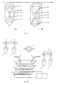
- a specifically designed centrifuge tube shown in figure 1A and figure 1B can be used.
- the centrifuge tube can be designed either as a cylinder shape shown in figure 1A , or as a rectangular shape shown in figure 1B . To get the best enrichment result, it is necessary to perform a preliminary experiment to decide the dimensions of the tube.
- a cylinder tube is designed as shown in figure 1A .
- the volume of the cone 105 at the bottom equals to that of red blood cells and granulocytes.
- the volume equals to that of lymphocytes and NRBC. This way there is only plasma at the top of the tube.
- the separation efficiency will be increased substantially because the diameter of the middle part is very small, and it is easy to distinguish different layers at the interface 101 and 104.
- the middle part 203 can be designed as a thin rectangular slit.
- the bottom part 201 and the top part 205 are designed as triangles.
- the interfaces 202 and 204 are very small so as to increase separation efficiency.
- fast freeze with liquid nitrogen guns can be applied to boundaries of the middle portion with the top and bottom portion. The top layer and frozen part is first removed before the middle layer is collected.
- the sample containing fetal NRBC, maternal NRBC, maternal lymphocytes, granulocytes and maternal red blood cells is preserved in maternal plasma.
- Researcher in this field should know that there are other ways to remove red blood cells from maternal blood, for example filtering.
- the processed sample is diluted into an isosmotic buffer composed of 8.5% glucose, 0.3% dextrose with conductivity between 10 ⁇ s/cm to 1.5 ms/cm.
- an appropriate dye is added into the solution, such as Giemsa dye. By controlling the volume of the dye and staining time, all the NRBC are stained but none of the maternal lymphocytes are stained.
- the ratio between Giemsa dye and buffer can be between 1:5 and 1:500. A typical value is about 1:10. If concentration of the dye is too high, it is hard to identify stained cells because of the intense color in solution. And all the cells, including NRBC and maternal lymphocytes are stained. If concentration of the dye is too low, some NRBC are not dyed and the separation result is not good. Time for staining is another critical parameter.
- the time for dying should be between 10 seconds to 10 minutes. If the time is too long, all the cells, including NRBC and maternal lymphocytes are stained. If the time is too short, some NRBC are not stained and the separation result is not good.
- the sample is added into a dielectrophoresis chip. By applying an appropriate frequency and amplitude through a function generator, maternal lymphocytes are attracted to electrodes by positive dielectrophoresis force; while dying NRBC are repelled to the area with weakest electric field by negative dielectrophoresis force. Then NRBC can be collected by applying external pump. In NRBC collected, there is either fetal NRBC or maternal NRBC.
- fetal NRBC can be distinguished from maternal NRBC by morphology ( Cheung et al., Prenatal Diagnosis of Sickle Cell Anaemia and Thalassaemia by Analysis of Fetal Cells in Maternal Blood, Nature Genetics, 14:264-268 (1996 )). By applying dielectrophoresis chip again, pure fetal NRBC can be obtained for further prenatal diagnosis.
- Concentration of the dye and time for dying should be determined according to the characteristic properties of the dye and the cell types.
- Researcher of this field should know that cDEP chip, complex of cDEP and twDEP chip and particle manipulation chip can all be applied to separate maternal and fetal cells ( WO 02/16647 , PCT/US01/42426 , PCT/US01/42280 , and PCT/US01/29762 ). Then with the help of external pump, fetal cells can be collected. Because there are only very few fetal NRBC in maternal blood, dielectrophoresis separation are preferably be applied twice or more to get pure fetal cells.
- Giemsa dye can also be used to separate other types of cells with similar dielectric properties, such as red blood cells and white blood cells. If the concentration of dye is 1:100, the time for dying need to exceed 30 minutes. All white blood cells are stained but red blood cells are not stained because only nucleus can be stained by Giemsa dye and there is no nucleus in red blood cell. Then the sample is added into a dielectrophoresis chip. By applying a appropriate frequency and amplitude through a function generator, red blood cells are attracted to electrodes by positive dielectrophoresis force; while stained white blood cells are repelled to the area with weakest electric field by negative dielectrophoresis force. Then stained white blood cells can be collected by applying external pump.
- FIG. 2 An exemplary dielectrophoresis system is shown in figure 2 .
- Tubing 1 is connected with the inlet of the valve 7; the outlet of valve 7 is connected with the inlet of cover slide 3 through tubing 8; and the outlet of cover slide 3 is connected with tubing 2 through tubing 9.
- the flow of buffer (container 13), sample (container 12), target sample (container 10) and waste liquid (container 11) is controlled by valves F1, F2, F3 and F4, respectively.
- Dielectrophoresis chip 5 and gasket 4 compose a reaction chamber where samples get separated. Voltage is applied to dielectrophoresis chips by signal generator 6. The thickness of gasket 4 is a critical value for separation.
- the system can be designed as a 3-dimensional structure.
- the cover slide 3 is replaced by another dielectrophoresis chip 14 and two holes of inlet and outlet 141, 142 are formed by drilling and are connected by tubing 8 and 9. This structure will double the efficiency of the previous system. Because the range of dielectrophoresis is doubled, the thickness of gasket 4 can be increased two times, which leads to twice the volume of reaction chamber.
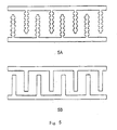
- the flow channel 41 in gasket 4 can be designed according to the structure of electrodes 51, 143 on the surface of dielectrophoresis chip 5, 14. As shown in Figure 4 , the channel is wider over the electrodes and thinner over the other area. This will reduce non-specific binding of cells to the surface without electrodes by decreasing channel cross-section area.
- the shape of the electrodes 51 and 143 can be designed as shown in figure 5A and figure 5B .
- Flow channels of different dimensions and shapes can be designed according to the electrodes of different dimensions and shapes.
- Electrodes can be designed into other shapes as well.
- cDEP chip, twDEP chip, particle manipulation chip or the combination of cDEP and twDEP chip can all be used to separate maternal and fetal cells.
- a multiple cell manipulation switch can be designed according to the mechanism of traveling wave dielectrophoresis to realize separation of maternal red blood cells, maternal lymphocytes, maternal NRBC and fetal NRBC in parallel. An exemplary process is described below.
- a sample is added into flow channel 15, in which maternal RBC and maternal lymphocytes are not stained while maternal and fetal NRBC are stained.
- an appropriate voltage signal is applied, the latter two types of cells are collected at the branch b2 while the former two are collected at the branch b1.
- the maternal and fetal NRBC at branch b1 are stained by the immunoassay method specific for fetal hemoglobin. The dielectric difference between them is amplified, as well as morphology.
- maternal NRBC and fetal NRBC can be collected at branch b5 and b6 respectively by applying an appropriate voltage signal.
- cells are processed first in formide, methanol, ethanol or other organic solvents to get immobilized on glass slide. After washing with water and drying in the air, cells are stained with dyes. In this embodiment, some improvement has been made over conventional staining method. Under appropriate condition, one kind of cells is stained while others are not, which leads to the amplification of their dielectric properties. Then cells can be easily separated by dielectrophoresis chip. The result is a lot different from that of conventional methods.
- the invention relates to the following items:
Landscapes
- Health & Medical Sciences (AREA)
- Life Sciences & Earth Sciences (AREA)
- Chemical & Material Sciences (AREA)
- Chemical Kinetics & Catalysis (AREA)
- Electrochemistry (AREA)
- General Health & Medical Sciences (AREA)
- Molecular Biology (AREA)
- Investigating Or Analysing Biological Materials (AREA)
- Sampling And Sample Adjustment (AREA)
- Apparatus Associated With Microorganisms And Enzymes (AREA)
- Micro-Organisms Or Cultivation Processes Thereof (AREA)
- Electrostatic Separation (AREA)
- Centrifugal Separators (AREA)
- Measuring Or Testing Involving Enzymes Or Micro-Organisms (AREA)
- Local Oxidation Of Silicon (AREA)
- Element Separation (AREA)
Applications Claiming Priority (2)
| Application Number | Priority Date | Filing Date | Title |
|---|---|---|---|
| CNB01110015XA CN100494360C (zh) | 2001-03-22 | 2001-03-22 | 细胞分离方法及其应用 |
| EP02728540A EP1379682B1 (de) | 2001-03-22 | 2002-03-20 | Zellisolierungsverfahren und verwendungen davon |
Related Parent Applications (1)
| Application Number | Title | Priority Date | Filing Date |
|---|---|---|---|
| EP02728540A Division EP1379682B1 (de) | 2001-03-22 | 2002-03-20 | Zellisolierungsverfahren und verwendungen davon |
Publications (3)
| Publication Number | Publication Date |
|---|---|
| EP2116609A2 true EP2116609A2 (de) | 2009-11-11 |
| EP2116609A3 EP2116609A3 (de) | 2010-01-20 |
| EP2116609B1 EP2116609B1 (de) | 2014-02-12 |
Family
ID=4658284
Family Applications (2)
| Application Number | Title | Priority Date | Filing Date |
|---|---|---|---|
| EP09007354.5A Expired - Lifetime EP2116609B1 (de) | 2001-03-22 | 2002-03-20 | Verfahren zur Isolierung von kernhaltigen, roten Blutkörperchen |
| EP02728540A Expired - Lifetime EP1379682B1 (de) | 2001-03-22 | 2002-03-20 | Zellisolierungsverfahren und verwendungen davon |
Family Applications After (1)
| Application Number | Title | Priority Date | Filing Date |
|---|---|---|---|
| EP02728540A Expired - Lifetime EP1379682B1 (de) | 2001-03-22 | 2002-03-20 | Zellisolierungsverfahren und verwendungen davon |
Country Status (8)
| Country | Link |
|---|---|
| US (2) | US7153648B2 (de) |
| EP (2) | EP2116609B1 (de) |
| JP (2) | JP2004522452A (de) |
| CN (1) | CN100494360C (de) |
| AT (1) | ATE466096T1 (de) |
| CA (1) | CA2440385C (de) |
| DE (1) | DE60236145D1 (de) |
| WO (1) | WO2002077269A1 (de) |
Families Citing this family (62)
| Publication number | Priority date | Publication date | Assignee | Title |
|---|---|---|---|---|
| US8986944B2 (en) | 2001-10-11 | 2015-03-24 | Aviva Biosciences Corporation | Methods and compositions for separating rare cells from fluid samples |
| US8980568B2 (en) | 2001-10-11 | 2015-03-17 | Aviva Biosciences Corporation | Methods and compositions for detecting non-hematopoietic cells from a blood sample |
| US7998699B2 (en) | 2002-08-15 | 2011-08-16 | University Of South Florida | Early detection of pathogens in blood |
| WO2005021799A2 (en) | 2003-08-15 | 2005-03-10 | University Of South Florida | Materials and methods for capture of pathogens and removal of aurintricarboxylic acid from a sample |
| US7384791B2 (en) * | 2004-01-21 | 2008-06-10 | Hewlett-Packard Development Company, L.P. | Method of analyzing blood |
| US7390387B2 (en) * | 2004-03-25 | 2008-06-24 | Hewlett-Packard Development Company, L.P. | Method of sorting cells in series |
| US7160425B2 (en) * | 2004-03-25 | 2007-01-09 | Hewlett-Packard Development Company, L.P. | Cell transporter for a biodevice |
| US7390388B2 (en) * | 2004-03-25 | 2008-06-24 | Hewlett-Packard Development Company, L.P. | Method of sorting cells on a biodevice |
| FR2876045B1 (fr) * | 2004-10-04 | 2006-11-10 | Commissariat Energie Atomique | Dispositif pour realiser la separation dielectrophoretique de particules contenues dans un fluide |
| US20060177815A1 (en) * | 2004-11-29 | 2006-08-10 | The Regents Of The University Of California | Dielectrophoretic particle sorter |
| KR100738071B1 (ko) * | 2005-01-21 | 2007-07-12 | 삼성전자주식회사 | 농도구배발생부가 구비된 유전영동 장치, 그를 이용한물질의 분리방법 및 물질 분리의 최적 조건을 탐색하는 방법 |
| WO2007046484A1 (ja) * | 2005-10-19 | 2007-04-26 | Sharp Kabushiki Kaisha | 誘電泳動チップおよび誘電泳動装置並びに誘電泳動システム |
| JP2009014342A (ja) * | 2005-10-19 | 2009-01-22 | Sharp Corp | 誘電泳動チップおよび誘電泳動装置並びに誘電泳動システム |
| EP1986001A4 (de) | 2006-02-10 | 2013-11-13 | Univ Kochi Technology | Eigenschaftsanalysevorrichtung und verfahren mit nutzung der dielektrischen wanderung einer granulären substanz mittels winkelmodulierter welle |
| EP2041299A4 (de) | 2006-07-14 | 2010-01-13 | Aviva Biosciences Corp | Verfahren und zusammensetzungen für den nachweis seltener zellen aus einer biologischen probe |
| CN101177673B (zh) * | 2006-11-06 | 2010-05-12 | 瑞鼎科技股份有限公司 | 排列细胞的方法及其使用的电极图案 |
| ITTO20070307A1 (it) * | 2007-05-04 | 2008-11-05 | Silicon Biosystems Spa | Metodo e dispositivo per la diagnosi prenatale non-invasiva |
| KR101162434B1 (ko) | 2007-10-05 | 2012-07-04 | 고쿠리츠 다이가쿠 호진 큐슈 코교 다이가쿠 | 유전 영동 장치 |
| US8162149B1 (en) | 2009-01-21 | 2012-04-24 | Sandia Corporation | Particle sorter comprising a fluid displacer in a closed-loop fluid circuit |
| US9134221B2 (en) | 2009-03-10 | 2015-09-15 | The Regents Of The University Of California | Fluidic flow cytometry devices and particle sensing based on signal-encoding |
| KR101289535B1 (ko) | 2009-12-07 | 2013-07-24 | 전민용 | 원심분리관 |
| JP5611582B2 (ja) * | 2009-12-25 | 2014-10-22 | 株式会社東芝 | 電気的中性物質の分離方法、及び電気的中性物質の分離装置 |
| JP5846609B2 (ja) | 2010-01-21 | 2016-01-20 | バイオセップ リミテッド | 希少細胞の磁気分離 |
| US8774488B2 (en) | 2010-03-11 | 2014-07-08 | Cellscape Corporation | Method and device for identification of nucleated red blood cells from a maternal blood sample |
| US20110225809A1 (en) * | 2010-03-17 | 2011-09-22 | Alan Francis Daher | Apparatus for removably attaching an item to a surface |
| JP5771917B2 (ja) * | 2010-08-04 | 2015-09-02 | 公益財団法人ヒューマンサイエンス振興財団 | 単核球分離管及び単核球分離システム |
| WO2012054904A2 (en) * | 2010-10-21 | 2012-04-26 | The Regents Of The University Of California | Microfluidics with wirelessly powered electronic circuits |
| KR101284725B1 (ko) * | 2011-07-22 | 2013-07-17 | 한국항공대학교산학협력단 | 유전영동력을 이용한 고속 입자분리 시스템 |
| CA2845713A1 (en) * | 2011-08-19 | 2013-02-28 | David Martin Morrow | Gradient array dielectrophoresis separation (grads) with concomitant light therapy |
| EP2749882B1 (de) | 2011-08-24 | 2017-05-03 | Eiken Kagaku Kabushiki Kaisha | Leukozytenmessvorrichtung und reagenzienkit |
| WO2013036707A1 (en) | 2011-09-09 | 2013-03-14 | Spine Wave, Inc. | Lateral approach expandable spinal implant and method |
| US9120105B2 (en) | 2011-10-31 | 2015-09-01 | Monika Weber | Electronic device for pathogen detection |
| US11198126B2 (en) | 2011-10-31 | 2021-12-14 | Fluid-Screen, Inc. | Apparatus for pathogen detection |
| US8926816B2 (en) | 2011-11-08 | 2015-01-06 | Rarecyte, Inc. | Systems and methods to analyze materials of a suspension by means of dielectrophoresis |
| TWI448678B (zh) * | 2012-03-23 | 2014-08-11 | Univ Nat Cheng Kung | 液體樣本之帶電粒子的分離方法及裝置與該裝置的製作方法 |
| WO2013177560A1 (en) | 2012-05-25 | 2013-11-28 | The Regents Of The University Of California | Microfluidic systems for particle trapping and separation |
| TW201413230A (zh) * | 2012-09-21 | 2014-04-01 | Nat Applied Res Laboratories | 可選擇地濃縮分離待測粒子的方法與晶片 |
| JP5990440B2 (ja) * | 2012-09-26 | 2016-09-14 | テルモ株式会社 | ヘマトクリット値の測定方法 |
| US10816550B2 (en) | 2012-10-15 | 2020-10-27 | Nanocellect Biomedical, Inc. | Systems, apparatus, and methods for sorting particles |
| MX365324B (es) * | 2013-03-15 | 2019-05-29 | Theranos Ip Co Llc | Metodos y dispositivos para recoleccion de muestras y separacion de muestras. |
| SG11201602427UA (en) * | 2013-08-29 | 2016-05-30 | Apocell Inc | Method and apparatus for isolation, capture and molecular analysis of target particles |
| CN106793771B (zh) * | 2014-09-25 | 2021-01-05 | 深圳华大基因科技有限公司 | 用于孕妇外周血样的保存液和孕妇外周血样的保存方法 |
| CN106513179B (zh) * | 2015-09-15 | 2019-05-31 | 王冰 | 一种钣金介电电泳电极结构 |
| WO2017061496A1 (ja) * | 2015-10-07 | 2017-04-13 | 株式会社Afiテクノロジー | 検査装置、検査システム、及び検査方法 |
| US9862941B2 (en) | 2015-10-14 | 2018-01-09 | Pioneer Hi-Bred International, Inc. | Single cell microfluidic device |
| US10549277B2 (en) | 2015-10-14 | 2020-02-04 | The Regents Of The University Of California | Integrated microfluidic platform for selective extraction of single-cell mRNA |
| EP3909686A1 (de) | 2016-04-15 | 2021-11-17 | Fluid-Screen, Inc. | Analytnachweisverfahren und -vorrichtungen unter verwendung von dielektrophorese und elektroosmose |
| CN107881104B (zh) * | 2016-09-30 | 2023-04-14 | 国立大学法人东京大学 | 粒子捕获用微装置以及使用它的粒子的捕获、浓缩或分离方法 |
| WO2018071448A1 (en) | 2016-10-11 | 2018-04-19 | The Regents Of The University Of California | Systems and methods to encapsulate and preserve organic matter for analysis |
| EP3562929B1 (de) | 2016-12-29 | 2024-05-01 | Ador Diagnostics S.r.l. | Elektrophoretischer chip für elektrophoretische anwendungen |
| US10780438B2 (en) | 2017-06-09 | 2020-09-22 | The Regents Of The University Of California | High-efficiency encapsulation in droplets based on hydrodynamic vortices control |
| US11517901B2 (en) | 2017-06-09 | 2022-12-06 | The Regents Of The University Of California | High-efficiency particle encapsulation in droplets with particle spacing and downstream droplet sorting |
| WO2019075409A1 (en) | 2017-10-12 | 2019-04-18 | The Regents Of The University Of California | ISOLATION AND IDENTIFICATION WITHOUT MICROFLUIDIC LABEL OF CELLS USING FLUORESCENCE LIFE IMAGING (FLIM) |
| US11499127B2 (en) | 2017-10-20 | 2022-11-15 | The Regents Of The University Of California | Multi-layered microfluidic systems for in vitro large-scale perfused capillary networks |
| US11745179B2 (en) | 2017-10-20 | 2023-09-05 | The Regents Of The University Of California | Microfluidic systems and methods for lipoplex-mediated cell transfection |
| WO2019147278A1 (en) | 2018-01-29 | 2019-08-01 | Hewlett-Packard Development Company, L.P. | Object separating |
| EP4058055A4 (de) | 2019-11-13 | 2023-12-13 | Fluid-Screen, Inc. | Vorrichtung und verfahren zur schnellen detektion, trennung, reinigung und quantifizierung verschiedener viren aus zellen, kulturmedien und anderen flüssigkeiten |
| EP4058778A4 (de) | 2019-11-13 | 2023-12-27 | Fluid-Screen, Inc. | Verfahren und vorrichtung zum nachweis von bakterien in einer probe mittels dielektrophorese |
| CN110777060A (zh) * | 2019-11-14 | 2020-02-11 | 北京酷搏科技有限公司 | 反应管、反应管阵列、控制参与反应的样品体积的方法及其应用 |
| CN112362711B (zh) * | 2020-11-11 | 2021-08-17 | 重庆大学 | 一种微生物检测装置及检测方法 |
| CN112871229B (zh) * | 2021-01-21 | 2022-06-28 | 中国科学技术大学 | 一种用于水体介电泳细菌分选的芯片 |
| WO2024036174A1 (en) * | 2022-08-08 | 2024-02-15 | Sigil Biosciences, Inc. | Systems and methods for isolating particles in solution by particle permittivity |
Citations (3)
| Publication number | Priority date | Publication date | Assignee | Title |
|---|---|---|---|---|
| US5641628A (en) | 1989-11-13 | 1997-06-24 | Children's Medical Center Corporation | Non-invasive method for isolation and detection of fetal DNA |
| US5948278A (en) | 1994-10-21 | 1999-09-07 | Bioseparations, Inc. | System and method for enrichment of rare cell population from whole blood samples |
| WO2002016647A1 (en) | 2000-08-24 | 2002-02-28 | Aviva Biosciences Corporation | Methods and compositions for identifying nucleic acid molecules using nucleolytic activities and hybridization |
Family Cites Families (22)
| Publication number | Priority date | Publication date | Assignee | Title |
|---|---|---|---|---|
| US3975156A (en) * | 1975-11-10 | 1976-08-17 | Ortho Diagnostics, Inc. | Method and material for detecting and quantitating fetal erythrocytes in adults |
| US4326934A (en) * | 1979-12-31 | 1982-04-27 | Pohl Herbert A | Continuous dielectrophoretic cell classification method |
| US4786387A (en) * | 1986-09-25 | 1988-11-22 | Whitlock David R | Single phase enrichment of super critical fluids |
| GB9301122D0 (en) * | 1993-01-21 | 1993-03-10 | Scient Generics Ltd | Method of analysis/separation |
| GB9306729D0 (en) * | 1993-03-31 | 1993-05-26 | British Tech Group | Improvements in separators |
| US6129828A (en) * | 1996-09-06 | 2000-10-10 | Nanogen, Inc. | Apparatus and methods for active biological sample preparation |
| US6071394A (en) * | 1996-09-06 | 2000-06-06 | Nanogen, Inc. | Channel-less separation of bioparticles on a bioelectronic chip by dielectrophoresis |
| DE19603043C2 (de) * | 1996-01-29 | 1997-11-27 | Ibm | Ionenerzeuger für ionographischen Druckkopf und Verfahren zu dessen Herstellung |
| EP0885055B1 (de) * | 1996-01-31 | 2007-04-04 | The Board Of Regents, The University Of Texas System | Fraktionierung mittels dielektrophoresis und fraktionierung unter anwendung eines flussfeldes |
| US5888370A (en) * | 1996-02-23 | 1999-03-30 | Board Of Regents, The University Of Texas System | Method and apparatus for fractionation using generalized dielectrophoresis and field flow fractionation |
| US5993630A (en) * | 1996-01-31 | 1999-11-30 | Board Of Regents The University Of Texas System | Method and apparatus for fractionation using conventional dielectrophoresis and field flow fractionation |
| US5866071A (en) | 1996-03-06 | 1999-02-02 | National Science Council | Centrifuge tube with a built-in small tubing for separation following density gradients centrifugation |
| AU4113297A (en) * | 1996-09-04 | 1998-03-26 | Technical University Of Denmark | A micro flow system for particle separation and analysis |
| DE19815882A1 (de) | 1998-04-08 | 1999-10-14 | Fuhr Guenther | Verfahren und Vorrichtung zur Manipulierung von Mikropartikeln in Fluidströmungen |
| ATE530891T1 (de) * | 1998-05-22 | 2011-11-15 | California Inst Of Techn | Miniaturisierter zellsortierer |
| DE19983263T1 (de) * | 1998-05-29 | 2001-05-31 | Ind Res Ltd | Verfahren und Vorrichtung zum Konzentrieren und/oder Positionieren von Teilchen oder Zellen |
| JP2002008115A (ja) * | 2000-06-23 | 2002-01-11 | Sony Corp | 情報配信システム、端末装置、サーバ装置、記録媒体、情報配信方法 |
| CN1325909C (zh) * | 2000-09-27 | 2007-07-11 | 清华大学 | 用于微粒操纵与微粒导向的装置及其使用方法 |
| EP1320922A2 (de) * | 2000-09-27 | 2003-06-25 | Aviva Biosciences Corporation | Apparat zur umschalten und behandlen von teilchen und benutzungsverfahren dieses apparates |
| CA2421828A1 (en) | 2000-09-30 | 2002-04-11 | Xiao-Bo Wang | Apparatuses containing multiple force generating elements and uses thereof |
| CN100495030C (zh) * | 2000-09-30 | 2009-06-03 | 清华大学 | 多力操纵装置及其应用 |
| AU2002213426A1 (en) | 2000-09-30 | 2002-04-15 | Aviva Biosciences Corporation | Apparatuses and methods for field flow fractionation of particles using acoustic and other forces |
-
2001
- 2001-03-22 CN CNB01110015XA patent/CN100494360C/zh not_active Expired - Fee Related
-
2002
- 2002-03-20 JP JP2002575311A patent/JP2004522452A/ja active Pending
- 2002-03-20 EP EP09007354.5A patent/EP2116609B1/de not_active Expired - Lifetime
- 2002-03-20 US US10/103,581 patent/US7153648B2/en not_active Expired - Lifetime
- 2002-03-20 WO PCT/US2002/008880 patent/WO2002077269A1/en active IP Right Grant
- 2002-03-20 DE DE60236145T patent/DE60236145D1/de not_active Expired - Lifetime
- 2002-03-20 CA CA2440385A patent/CA2440385C/en not_active Expired - Fee Related
- 2002-03-20 EP EP02728540A patent/EP1379682B1/de not_active Expired - Lifetime
- 2002-03-20 AT AT02728540T patent/ATE466096T1/de not_active IP Right Cessation
-
2005
- 2005-10-25 JP JP2005310561A patent/JP4411266B2/ja not_active Expired - Fee Related
-
2006
- 2006-11-14 US US11/598,848 patent/US7918981B2/en not_active Expired - Fee Related
Patent Citations (3)
| Publication number | Priority date | Publication date | Assignee | Title |
|---|---|---|---|---|
| US5641628A (en) | 1989-11-13 | 1997-06-24 | Children's Medical Center Corporation | Non-invasive method for isolation and detection of fetal DNA |
| US5948278A (en) | 1994-10-21 | 1999-09-07 | Bioseparations, Inc. | System and method for enrichment of rare cell population from whole blood samples |
| WO2002016647A1 (en) | 2000-08-24 | 2002-02-28 | Aviva Biosciences Corporation | Methods and compositions for identifying nucleic acid molecules using nucleolytic activities and hybridization |
Non-Patent Citations (20)
| Title |
|---|
| BECKER ET AL.: "Separation of human breast cancer cells from blood by differential dielectric affinity", PROC. NAT. ACAD. SCI., vol. 29, 1995, pages 860 - 864 |
| BIANCHI DIANA ET AL.: "Isolation of Fetal DNA from Nucleated Erythrocytes in Maternal Blood", PROC. NATL. ACAD. SCI. USA, vol. 86, 1990, pages 3279 - 3283 |
| BIANCHI ET AL.: "Isolation of Fetal DNA from Nucleated Erythrocytes in Maternal Blood", PROC. NATL. ACAD. SCI. USA, vol. 86, 1990, pages 3279 - 3283 |
| CHENG ET AL.: "Preparation and Hybridization Analysis of DNA/RNA from E. coli on Microfabricated Bioelectronic Chips", NATURE BIOTECHNOLOGY, vol. 16, no. 6, 1998, pages 541 - 546 |
| CHEUNG ET AL., NATURE GENETICS, vol. 14, 1996, pages 264 - 268 |
| CHEUNG ET AL.: "Prenatal Diagnosis of Sickle Cell Anaemia and Thalassaemia by Analysis of Fetal Cells in Maternal Blood", NATURE GENETICS, vol. 14, 1996, pages 264 - 268 |
| FUHR ET AL.: "Positioning and manipulation of cells and microparticles using miniaturized electric field traps and travelling waves", SENSORS AND MATERIALS, vol. 7, 1995, pages 131 - 146 |
| HOLZGREVE ET AL.: "Fetal Cells In the Maternal Circulation", JOURNAL OF REPRODUCTIVE MEDICINE, vol. 37, no. 5, 1992, pages 410 - 418 |
| HUANG ET AL., BIOCHIM. BIOPHYS. ACTA, vol. 1282, 1996, pages 76 - 84 |
| HUANG ET AL.: "Introducing Dielectrophoresis as a New Force Field for Field Flow Fractionation", BIOPHYSICAL JOURNAL, vol. 73, 1997, pages 1118 - 1129 |
| HUGHES ET AL.: "Dielectrophretic Forces on Particles in Traveling Electric Fields", J PHYS. APPL. PHYS, vol. 29, 1997, pages 474 - 482 |
| J PHYS. D: APPL. PHYS., vol. 26, 1993, pages 1528 - 1535 |
| MULLER: "A 3-D Microelectrode System for Handling and Caging Single Cells and Particles", BIOSENSORS & BIOELECTRONICS, vol. 14, 1999, pages 247 - 256 |
| PETHIG: "Dielectrophoresis: Using Inhomogeneous AC Electrical Fields to Separate and Manipulate Cells", CRITICAL REVIEWS IN BIOTECHNOLOGY, vol. 16, no. 4, 1996, pages 331 - 348 |
| SIMPSON; ELIAS: "Isolating Fetal Cells in Maternal Circulation for Prenatal Diagnosis", PRENATAL DIAGNOSIS, vol. 14, 1994, pages 1229 - 1242 |
| WANG ET AL.: "A unified theory of dielectrophoresis and travelling-wave dielectrophoresis", J PHYS. D: APPL. PHYS., vol. 27, 1994, pages 1571 - 1574 |
| WANG ET AL.: "Dielectrophoretic Manipulation of Cells Using Spiral Electrodes", BIOPHYS. J, vol. 72, 1997, pages 1887 - 1899 |
| WANG ET AL.: "Non-uniform spatial distributions of both the magnitude and phase of AC electric fields determine dielectrophoretic forces", BIOCHIM BIOPHYS ACTA, vol. 1243, 1995, pages 185 - 194 |
| WILLIAMSON; BOB, TOWARDS: "Non-invasive Prenatal Diagnosis", NATURE GENETICS, vol. 14, 1996, pages 239 - 240 |
| X-B. WANG ET AL.: "Dielectrophoretic manipulation of particles", IEEE/IAS TRANS., vol. 33, 1997, pages 660 - 669 |
Also Published As
| Publication number | Publication date |
|---|---|
| DE60236145D1 (de) | 2010-06-10 |
| US7153648B2 (en) | 2006-12-26 |
| CN100494360C (zh) | 2009-06-03 |
| EP2116609B1 (de) | 2014-02-12 |
| CN1376779A (zh) | 2002-10-30 |
| JP2006126195A (ja) | 2006-05-18 |
| JP2004522452A (ja) | 2004-07-29 |
| US7918981B2 (en) | 2011-04-05 |
| CA2440385C (en) | 2012-01-10 |
| EP1379682A1 (de) | 2004-01-14 |
| EP1379682A4 (de) | 2005-08-10 |
| US20020182654A1 (en) | 2002-12-05 |
| US20070128686A1 (en) | 2007-06-07 |
| EP1379682B1 (de) | 2010-04-28 |
| WO2002077269A1 (en) | 2002-10-03 |
| ATE466096T1 (de) | 2010-05-15 |
| CA2440385A1 (en) | 2002-10-03 |
| EP2116609A3 (de) | 2010-01-20 |
| JP4411266B2 (ja) | 2010-02-10 |
Similar Documents
| Publication | Publication Date | Title |
|---|---|---|
| EP1379682B1 (de) | Zellisolierungsverfahren und verwendungen davon | |
| Sivaramakrishnan et al. | Active microfluidic systems for cell sorting and separation | |
| Pratt et al. | Rare cell capture in microfluidic devices | |
| Gascoyne et al. | Dielectrophoresis-based sample handling in general-purpose programmable diagnostic instruments | |
| US9873126B2 (en) | Devices and methods for separating particles | |
| Low et al. | Benchtop technologies for circulating tumor cells separation based on biophysical properties | |
| US6858439B1 (en) | Compositions and methods for separation of moieties on chips | |
| Talary et al. | Dielectrophoretic separation and enrichment of CD34+ cell subpopulation from bone marrow and peripheral blood stem cells | |
| Hyun et al. | Microfluidic devices for the isolation of circulating rare cells: A focus on affinity‐based, dielectrophoresis, and hydrophoresis | |
| US7862702B2 (en) | Methods and apparatus for electrosmear analysis | |
| US9090663B2 (en) | Systems and methods for the capture and separation of microparticles | |
| WO2002030562A1 (en) | An integrated biochip system for sample preparation and analysis | |
| Kavanagh et al. | Current and emerging techniques of fetal cell separation from maternal blood | |
| EP2956541B1 (de) | Vorrichtungen und verfahren zur reinigung biologischer zellen | |
| Xavier et al. | Skeletal stem cell isolation: A review on the state-of-the-art microfluidic label-free sorting techniques | |
| Menachery et al. | Label-free microfluidic stem cell isolation technologies | |
| Huang et al. | Selectively concentrating cervical carcinoma cells from red blood cells utilizing dielectrophoresis with circular ITO electrodes in stepping electric fields | |
| Jen et al. | A microfabricated module for isolating cervical carcinoma cells from peripheral blood utilizing dielectrophoresis in stepping electric fields | |
| Pandey et al. | Single-Cell Separation | |
| AU2007205731B2 (en) | Cell isolation method and uses thereof | |
| Cristofanilli et al. | Dielectric cell separation of fine needle aspirates from tumor xenografts | |
| AU2002258585B2 (en) | Cell isolation method and uses thereof | |
| CN113262828B (zh) | 一种结合磁泳与介电泳的血细胞微流控分离装置及方法 | |
| AU2002258585A1 (en) | Cell isolation method and uses thereof | |
| RU2423698C1 (ru) | Способ селективного выделения популяции жизнеспособных клеток из биологических жидкостей |
Legal Events
| Date | Code | Title | Description |
|---|---|---|---|
| PUAI | Public reference made under article 153(3) epc to a published international application that has entered the european phase |
Free format text: ORIGINAL CODE: 0009012 |
|
| AC | Divisional application: reference to earlier application |
Ref document number: 1379682 Country of ref document: EP Kind code of ref document: P |
|
| AK | Designated contracting states |
Kind code of ref document: A2 Designated state(s): AT BE CH CY DE DK ES FI FR GB GR IE IT LI LU MC NL PT SE TR |
|
| PUAL | Search report despatched |
Free format text: ORIGINAL CODE: 0009013 |
|
| AK | Designated contracting states |
Kind code of ref document: A3 Designated state(s): AT BE CH CY DE DK ES FI FR GB GR IE IT LI LU MC NL PT SE TR |
|
| 17P | Request for examination filed |
Effective date: 20100304 |
|
| 17Q | First examination report despatched |
Effective date: 20100406 |
|
| REG | Reference to a national code |
Ref country code: DE Ref legal event code: R079 Ref document number: 60245997 Country of ref document: DE Free format text: PREVIOUS MAIN CLASS: C12Q0001040000 Ipc: G01N0033580000 |
|
| GRAP | Despatch of communication of intention to grant a patent |
Free format text: ORIGINAL CODE: EPIDOSNIGR1 |
|
| RIC1 | Information provided on ipc code assigned before grant |
Ipc: G01N 33/58 20060101AFI20130613BHEP Ipc: G01N 27/26 20060101ALI20130613BHEP Ipc: B03C 5/00 20060101ALI20130613BHEP |
|
| INTG | Intention to grant announced |
Effective date: 20130628 |
|
| GRAS | Grant fee paid |
Free format text: ORIGINAL CODE: EPIDOSNIGR3 |
|
| GRAP | Despatch of communication of intention to grant a patent |
Free format text: ORIGINAL CODE: EPIDOSNIGR1 |
|
| INTG | Intention to grant announced |
Effective date: 20131213 |
|
| RAP1 | Party data changed (applicant data changed or rights of an application transferred) |
Owner name: TSINGHUA UNIVERSITY Owner name: CAPITALBIO CORPORATION |
|
| GRAA | (expected) grant |
Free format text: ORIGINAL CODE: 0009210 |
|
| AC | Divisional application: reference to earlier application |
Ref document number: 1379682 Country of ref document: EP Kind code of ref document: P |
|
| AK | Designated contracting states |
Kind code of ref document: B1 Designated state(s): AT BE CH CY DE DK ES FI FR GB GR IE IT LI LU MC NL PT SE TR |
|
| REG | Reference to a national code |
Ref country code: GB Ref legal event code: FG4D |
|
| REG | Reference to a national code |
Ref country code: CH Ref legal event code: EP |
|
| REG | Reference to a national code |
Ref country code: AT Ref legal event code: REF Ref document number: 652360 Country of ref document: AT Kind code of ref document: T Effective date: 20140215 |
|
| REG | Reference to a national code |
Ref country code: IE Ref legal event code: FG4D |
|
| REG | Reference to a national code |
Ref country code: DE Ref legal event code: R096 Ref document number: 60245997 Country of ref document: DE Effective date: 20140327 |
|
| REG | Reference to a national code |
Ref country code: NL Ref legal event code: VDEP Effective date: 20140212 |
|
| REG | Reference to a national code |
Ref country code: AT Ref legal event code: MK05 Ref document number: 652360 Country of ref document: AT Kind code of ref document: T Effective date: 20140212 |
|
| PG25 | Lapsed in a contracting state [announced via postgrant information from national office to epo] |
Ref country code: CY Free format text: LAPSE BECAUSE OF FAILURE TO SUBMIT A TRANSLATION OF THE DESCRIPTION OR TO PAY THE FEE WITHIN THE PRESCRIBED TIME-LIMIT Effective date: 20140212 Ref country code: FI Free format text: LAPSE BECAUSE OF FAILURE TO SUBMIT A TRANSLATION OF THE DESCRIPTION OR TO PAY THE FEE WITHIN THE PRESCRIBED TIME-LIMIT Effective date: 20140212 Ref country code: SE Free format text: LAPSE BECAUSE OF FAILURE TO SUBMIT A TRANSLATION OF THE DESCRIPTION OR TO PAY THE FEE WITHIN THE PRESCRIBED TIME-LIMIT Effective date: 20140212 Ref country code: PT Free format text: LAPSE BECAUSE OF FAILURE TO SUBMIT A TRANSLATION OF THE DESCRIPTION OR TO PAY THE FEE WITHIN THE PRESCRIBED TIME-LIMIT Effective date: 20140612 Ref country code: AT Free format text: LAPSE BECAUSE OF FAILURE TO SUBMIT A TRANSLATION OF THE DESCRIPTION OR TO PAY THE FEE WITHIN THE PRESCRIBED TIME-LIMIT Effective date: 20140212 Ref country code: NL Free format text: LAPSE BECAUSE OF FAILURE TO SUBMIT A TRANSLATION OF THE DESCRIPTION OR TO PAY THE FEE WITHIN THE PRESCRIBED TIME-LIMIT Effective date: 20140212 Ref country code: ES Free format text: LAPSE BECAUSE OF FAILURE TO SUBMIT A TRANSLATION OF THE DESCRIPTION OR TO PAY THE FEE WITHIN THE PRESCRIBED TIME-LIMIT Effective date: 20140212 |
|
| PG25 | Lapsed in a contracting state [announced via postgrant information from national office to epo] |
Ref country code: BE Free format text: LAPSE BECAUSE OF FAILURE TO SUBMIT A TRANSLATION OF THE DESCRIPTION OR TO PAY THE FEE WITHIN THE PRESCRIBED TIME-LIMIT Effective date: 20140212 |
|
| REG | Reference to a national code |
Ref country code: DE Ref legal event code: R119 Ref document number: 60245997 Country of ref document: DE |
|
| PG25 | Lapsed in a contracting state [announced via postgrant information from national office to epo] |
Ref country code: DK Free format text: LAPSE BECAUSE OF FAILURE TO SUBMIT A TRANSLATION OF THE DESCRIPTION OR TO PAY THE FEE WITHIN THE PRESCRIBED TIME-LIMIT Effective date: 20140212 |
|
| REG | Reference to a national code |
Ref country code: CH Ref legal event code: PL |
|
| PG25 | Lapsed in a contracting state [announced via postgrant information from national office to epo] |
Ref country code: MC Free format text: LAPSE BECAUSE OF FAILURE TO SUBMIT A TRANSLATION OF THE DESCRIPTION OR TO PAY THE FEE WITHIN THE PRESCRIBED TIME-LIMIT Effective date: 20140212 |
|
| PLBE | No opposition filed within time limit |
Free format text: ORIGINAL CODE: 0009261 |
|
| STAA | Information on the status of an ep patent application or granted ep patent |
Free format text: STATUS: NO OPPOSITION FILED WITHIN TIME LIMIT |
|
| REG | Reference to a national code |
Ref country code: FR Ref legal event code: ST Effective date: 20141128 |
|
| REG | Reference to a national code |
Ref country code: IE Ref legal event code: MM4A |
|
| REG | Reference to a national code |
Ref country code: DE Ref legal event code: R119 Ref document number: 60245997 Country of ref document: DE Effective date: 20141001 |
|
| 26N | No opposition filed |
Effective date: 20141113 |
|
| PG25 | Lapsed in a contracting state [announced via postgrant information from national office to epo] |
Ref country code: FR Free format text: LAPSE BECAUSE OF NON-PAYMENT OF DUE FEES Effective date: 20140414 Ref country code: LI Free format text: LAPSE BECAUSE OF NON-PAYMENT OF DUE FEES Effective date: 20140331 Ref country code: DE Free format text: LAPSE BECAUSE OF NON-PAYMENT OF DUE FEES Effective date: 20141001 Ref country code: CH Free format text: LAPSE BECAUSE OF NON-PAYMENT OF DUE FEES Effective date: 20140331 Ref country code: IE Free format text: LAPSE BECAUSE OF NON-PAYMENT OF DUE FEES Effective date: 20140320 |
|
| PG25 | Lapsed in a contracting state [announced via postgrant information from national office to epo] |
Ref country code: IT Free format text: LAPSE BECAUSE OF FAILURE TO SUBMIT A TRANSLATION OF THE DESCRIPTION OR TO PAY THE FEE WITHIN THE PRESCRIBED TIME-LIMIT Effective date: 20140212 |
|
| PG25 | Lapsed in a contracting state [announced via postgrant information from national office to epo] |
Ref country code: GR Free format text: LAPSE BECAUSE OF FAILURE TO SUBMIT A TRANSLATION OF THE DESCRIPTION OR TO PAY THE FEE WITHIN THE PRESCRIBED TIME-LIMIT Effective date: 20140513 |
|
| PG25 | Lapsed in a contracting state [announced via postgrant information from national office to epo] |
Ref country code: LU Free format text: LAPSE BECAUSE OF NON-PAYMENT OF DUE FEES Effective date: 20140320 Ref country code: TR Free format text: LAPSE BECAUSE OF FAILURE TO SUBMIT A TRANSLATION OF THE DESCRIPTION OR TO PAY THE FEE WITHIN THE PRESCRIBED TIME-LIMIT Effective date: 20140212 |
|
| PGFP | Annual fee paid to national office [announced via postgrant information from national office to epo] |
Ref country code: GB Payment date: 20190320 Year of fee payment: 18 |
|
| GBPC | Gb: european patent ceased through non-payment of renewal fee |
Effective date: 20200320 |
|
| PG25 | Lapsed in a contracting state [announced via postgrant information from national office to epo] |
Ref country code: GB Free format text: LAPSE BECAUSE OF NON-PAYMENT OF DUE FEES Effective date: 20200320 |