EP2020604B1 - Ligand du recepteur couplé à la protéine G, FPRL2 - Google Patents
Ligand du recepteur couplé à la protéine G, FPRL2 Download PDFInfo
- Publication number
- EP2020604B1 EP2020604B1 EP08159325A EP08159325A EP2020604B1 EP 2020604 B1 EP2020604 B1 EP 2020604B1 EP 08159325 A EP08159325 A EP 08159325A EP 08159325 A EP08159325 A EP 08159325A EP 2020604 B1 EP2020604 B1 EP 2020604B1
- Authority
- EP
- European Patent Office
- Prior art keywords
- polypeptide
- fprl2
- activity
- cells
- hbp
- Prior art date
- Legal status (The legal status is an assumption and is not a legal conclusion. Google has not performed a legal analysis and makes no representation as to the accuracy of the status listed.)
- Active
Links
Images


Classifications
-
- C—CHEMISTRY; METALLURGY
- C07—ORGANIC CHEMISTRY
- C07K—PEPTIDES
- C07K14/00—Peptides having more than 20 amino acids; Gastrins; Somatostatins; Melanotropins; Derivatives thereof
- C07K14/435—Peptides having more than 20 amino acids; Gastrins; Somatostatins; Melanotropins; Derivatives thereof from animals; from humans
- C07K14/46—Peptides having more than 20 amino acids; Gastrins; Somatostatins; Melanotropins; Derivatives thereof from animals; from humans from vertebrates
- C07K14/47—Peptides having more than 20 amino acids; Gastrins; Somatostatins; Melanotropins; Derivatives thereof from animals; from humans from vertebrates from mammals
-
- A—HUMAN NECESSITIES
- A61—MEDICAL OR VETERINARY SCIENCE; HYGIENE
- A61P—SPECIFIC THERAPEUTIC ACTIVITY OF CHEMICAL COMPOUNDS OR MEDICINAL PREPARATIONS
- A61P43/00—Drugs for specific purposes, not provided for in groups A61P1/00-A61P41/00
-
- C—CHEMISTRY; METALLURGY
- C07—ORGANIC CHEMISTRY
- C07K—PEPTIDES
- C07K16/00—Immunoglobulins [IGs], e.g. monoclonal or polyclonal antibodies
- C07K16/18—Immunoglobulins [IGs], e.g. monoclonal or polyclonal antibodies against material from animals or humans
- C07K16/28—Immunoglobulins [IGs], e.g. monoclonal or polyclonal antibodies against material from animals or humans against receptors, cell surface antigens or cell surface determinants
-
- G—PHYSICS
- G01—MEASURING; TESTING
- G01N—INVESTIGATING OR ANALYSING MATERIALS BY DETERMINING THEIR CHEMICAL OR PHYSICAL PROPERTIES
- G01N33/00—Investigating or analysing materials by specific methods not covered by groups G01N1/00 - G01N31/00
- G01N33/48—Biological material, e.g. blood, urine; Haemocytometers
- G01N33/50—Chemical analysis of biological material, e.g. blood, urine; Testing involving biospecific ligand binding methods; Immunological testing
- G01N33/53—Immunoassay; Biospecific binding assay; Materials therefor
- G01N33/566—Immunoassay; Biospecific binding assay; Materials therefor using specific carrier or receptor proteins as ligand binding reagents where possible specific carrier or receptor proteins are classified with their target compounds
-
- C—CHEMISTRY; METALLURGY
- C07—ORGANIC CHEMISTRY
- C07K—PEPTIDES
- C07K2317/00—Immunoglobulins specific features
- C07K2317/70—Immunoglobulins specific features characterized by effect upon binding to a cell or to an antigen
- C07K2317/75—Agonist effect on antigen
-
- G—PHYSICS
- G01—MEASURING; TESTING
- G01N—INVESTIGATING OR ANALYSING MATERIALS BY DETERMINING THEIR CHEMICAL OR PHYSICAL PROPERTIES
- G01N2333/00—Assays involving biological materials from specific organisms or of a specific nature
- G01N2333/435—Assays involving biological materials from specific organisms or of a specific nature from animals; from humans
- G01N2333/705—Assays involving receptors, cell surface antigens or cell surface determinants
- G01N2333/72—Assays involving receptors, cell surface antigens or cell surface determinants for hormones
- G01N2333/726—G protein coupled receptor, e.g. TSHR-thyrotropin-receptor, LH/hCG receptor, FSH
-
- G—PHYSICS
- G01—MEASURING; TESTING
- G01N—INVESTIGATING OR ANALYSING MATERIALS BY DETERMINING THEIR CHEMICAL OR PHYSICAL PROPERTIES
- G01N2500/00—Screening for compounds of potential therapeutic value
- G01N2500/02—Screening involving studying the effect of compounds C on the interaction between interacting molecules A and B (e.g. A = enzyme and B = substrate for A, or A = receptor and B = ligand for the receptor)
Definitions
- the present disclosure is related to the natural ligand for an orphan G protein coupled receptor FPRL2 and methods of use. It further relates to antibodies raised against FPRL2.
- GPCRs G-protein coupled receptors
- GPCRs are proteins responsible for transducing a signal within a cell. GPCRs have usually seven transmembrane domains. Upon binding of a ligand to an extra-cellular portion or fragment of a GPCR, a signal is transduced within the cell that results in a change in a biological or physiological property or behaviour of the cell.
- GPCRs, along with G-proteins and effectors are the components of a modular signalling system that connects the state of intra-cellular second messengers to extra-cellular inputs.
- GPCR genes and gene products can modulate various physiological processes and are potential causative agents of disease.
- the GPCRs seem to be of critical importance to both the central nervous system and peripheral physiological processes.
- the GPCR protein superfamily is represented by five families : Family I, receptors typified by rhodopsin and the beta2-adrenergic receptor and currently represented by over 200 unique members; Family II, the parathyroid hormone/calcitonin/secretin receptor family; Family III, the metabotropic glutamate receptor family, Family IV, the CAMP receptor family, important in the chemotaxis and development of D. discoideum ; and Family V, the fungal mating pheromone receptor such as STE2.
- G proteins represent a family of heterotrimeric proteins composed of ⁇ , ⁇ and ⁇ subunits, that bind guanine nucleotides. These proteins are usually linked to cell surface receptors (receptors containing seven transmembrane domains) for signal transduction. Indeed, following ligand binding to the GPCR, a conformational change is transmitted to the G protein, which causes the ⁇ -subunit to exchange a bound GDP molecule for a GTP molecule and to dissociate from the ⁇ -subunits.
- the GTP-bound form of the ⁇ , ⁇ and ⁇ -subunits typically functions as an effector-modulating moiety, leading to the production of second messengers, such as cAMP (e.g. by activation of adenyl cyclase), diacylglycerol or inositol phosphates.
- cAMP e.g. by activation of adenyl cyclase
- diacylglycerol e.g. by activation of adenyl cyclase
- inositol phosphates e.g. by activation of adenyl cyclase
- G proteins are described extensively in Lodish et al., Molecular Cell Biology (Scientific American Books Inc., New York, N.Y., 1995 ; and also by Downes and Gautam, 1999, The G-Protein Subunit Gene Families. Genomics 62:544-552 ).
- GPCRs currently constitute major targets for drug action and development.
- GPCRs More than 300 GPCRs have been cloned to date, excluding the family of olfactory receptors. Mechanistically, approximately 50-60% of all clinically relevant drugs act by modulating the functions of various GPCRs ( Cudermann et al., J. Mol. Med., 73:51-63, 1995 ).
- Formyl peptide receptor-like 2 (FPRL2) (SEQ ID NO: 1, human polynucleotide sequence, SEQ ID NO: 2, human amino acid sequence) is a member of FPR Family. The members of this family belong to the GPCR family.
- Human FPR (SEQ ID NO: 3, human polynucleotide sequence, SEQ ID NO: 4, human amino acid sequence) was first member of the FRP family defined biochemically, in 1976, as a high affinity binding site on the surface of neutrophils for the prototypic N-formyl peptide formyl-methionine-leucyl-phenylaianine (fMLF). It was then cloned in 1990, by Boulay et al .
- FPR binds fMLF with high affinity (K d ⁇ 1 n M ) and is activated by picomolar to low nanomolar concentrations of fMLF in chemotaxis and calcium ion (Ca 2+ ) mobilization assays.
- FPRL1 FPR-like 1
- SEQ ID NO: 6 human amino acid sequence
- FPRL2 FPR-like 2
- FPRL1 is defined as a low-affinity fMLF receptor, based on its activation only by high concentrations of Fmlf ( ⁇ M range) in vitro [ Murphy, P.M. (1996) Chemoattractant Ligands and their Receptors (Horuk R, ed.), pp. 269-299, CRC Press, Inc., Boca R aton ; Prossnitz, E.R. and Ye, R.D. (1997) Pharmacol. Ther. 74, 73-102 ]. However, it is unclear whether such concentrations of fMLF could be generated at sites of bacterial infection or tissue injury.
- FPRL1 As another bona fide functional fMLF receptor in vivo remains to be determined.
- FPRL2 does not bind or respond to N-formyl peptides [ Durstin, M. et al. (1994) Biochem. Biophys. Res. Commun. 201, 174-179 ] but instead shares some non-formylated chemotactic peptides identified for FPRL1 [ Christophe, T. et al. (2001) J. Biol. Chem. 276, 21585-21593 ; Betten, A. et al. (2001) J. Clin. Invest. 108, 1221-1228 ].
- FPR and FPRL1 were initially detected in phagocytic leukocytes, other cell types also express these receptors but with undefined biological significance. Little information is available about the expression pattern of FPRL2, except that mRNA for this receptor is present in monocytes but not neutrophils [ Durstin, M. et al. (1994) Biochem. Biophys. Res. Commun. 201, 174-179 ]. Functional FPRL2 is also expressed in mature dentritic cells (DCs) [ Yang, D. et al. J. Leukoc. Biol. Vol. 72: 598-607 (2002 )], which express reduced levels of FPR but do not appear to express FPRL1 [ Yang, D. et al. (2001) J. Immunol. 166, 4092-4098 ; Braun, M.C. et al. (2001) Blood 97, 3531-3536 ].
- DCs mature dentritic cells
- HBP Heme Binding Protein
- the human and mouse HBP cDNAs are 567 and 570bp long respectively and encode a protein product of 189 and 190 amino acids respectively. This protein is located into the cytoplasm of the cell.
- HPB binds heme and porphyrins with micromolar Kd.
- HBP may function as a buffer for overproduced porphyrin as well as heme. Expression studies indicated that the mouse mRNA encoding HBP is expressed in liver, spleen and kidney cells ( Blackmon et al; 2002 Arch. of Bichem. and Biophysics 407, p196-201 ).
- HBP heme binding protein
- HBP heme binding protein
- HBP polypeptide is a natural ligand for the orphan G protein coupled receptor FPRL2 polypeptide and on methods of using the binding of this ligand to the receptor in drug screening methods.
- the known ligand and its interaction with the receptor FPRL2 polypeptide also provides for the diagnosis of conditions involving dysregulated receptor activity.
- the disclosure also relates to a kit comprising FPRL2 polypeptide and homologous sequences, its corresponding polynucleotide and/or recombinant cells expressing the polynucleotide, to identify agonist, antagonist ,inverse agonist and modulator compounds of the receptor polypeptide and/or its corresponding polynucleotide.
- kits are useful for the diagnosis, prevention and/or a treatment of diseases and disorders related to FPRL2 polypeptide activity.
- the present disclosure also relates to novel agonist, antagonist ,inverse agonist and modulator compounds of the receptor polypeptide and its corresponding polynucleotide, identified according to the method of the disclosure.
- the present disclosure concerns the finding that a fragment of HBP (HBP polypeptide) is a natural ligand of the orphan receptor FPRL2 (SEQ ID NO: 2).
- HBP polypeptide is a natural ligand of the orphan receptor FPRL2 (SEQ ID NO: 2).
- This disclosure thus relates to the HBP polypeptide ligand/receptor pair, and to functional homologs of the receptor which also bind HBP polypeptide and cells transformed by a vector comprising the nucleotide sequence encoding the receptor (SEQ ID NO: 1) in combination with the HBP polypeptide ligand.
- the disclosure also relates to a composition consisting essentially of an isolated FPLR2 polypeptide and an isolated HBP polypeptide, as well as to methods of identifying agents that modulate the activities of FPRL2 polypeptides. The methods are useful for the identification of agonist, inverse agonist or antagonist compounds useful for the development of new drugs.
- the disclosure encompasses a method of identifying an agent that modulates the function of FPLR2, the method comprising : a) contacting a FPLR2 polypeptide with a HBP polypeptide in the presence and absence of a candidate modulator under conditions permitting the binding of the HBP polypeptide to the FPLR2 polypeptide; and b) measuring binding of the FPLR2 polypeptide to the HBP polypeptide wherein a decrease in binding in the presence of the candidate modulator, relative to binding in the absence of the candidate modulator, identifies the candidate modulator as an agent that modulates the function of FPLR2 polypeptide.
- the disclosure further encompasses a method of detecting, in a sample, the presence of an agent that modulates the function of FPLR2, the method comprising: a) contacting a FPLR2 polypeptide with a HBP polypeptide in the presence and absence of the sample under conditions permitting the binding of the HBP polypeptide to the FPLR2 polypeptide; and b) measuring binding of the FPLR2 polypeptide to the HBP polypeptide wherein a decrease in binding in the presence of the sample, relative to binding in the absence of the sample, indicates the presence, in the sample of an agent that modulates the function of FPLR2.
- the measuring is performed using a method selected from label displacement, surface plasmon resonance, fluorescence resonance energy transfer, fluorescence quenching, and fluorescence polarization.
- the disclosure further encompasses a method of identifying an agent that modulates the function of FPLR2, the method comprising: a) contacting a FPLR2 polypeptide with a HBP polypeptide in the presence and absence of a candidate modulator; and b) measuring a signalling activity of the FPLR2 polypeptide, wherein a change in the activity in the presence of the candidate modulator relative to the activity in the absence of the candidate modulator identifies the candidate modulator as an agent that modulates the function of FPLR2 polypeptide.
- the disclosure further encompasses a method of identifying an agent that modulates the function of FPLR2 polypeptide, the method comprising: a) contacting a FPLR2 polypeptide with a candidate modulator; b) measuring a signalling activity of the FPLR2 polypeptide in the presence of the candidate modulator; and c) comparing the activity measured in the presence of the candidate modulator to the activity measured in a sample in which the FPLR2 polypeptide is contacted with a HBP polypeptide at its EC 50 , wherein the candidate modulator is identified as an agent that modulates the function of FPLR2 polypeptide when the amount of the activity measured in the presence of the candidate modulator is at least 20% of the amount induced by the HBP polypeptide present at its EC 50 .
- the disclosure further encompasses a method of detecting the presence, in a sample, of an agent that modulates the function of FPLR2 polypeptide, the method comprising: a) contacting a FPLR2 polypeptide with HBP polypeptide in the presence and absence of the sample; b) measuring a signalling activity of the FPLR2 polypeptide; and c) comparing the amount of the activity measured in a reaction containing FPLR2 polypeptide and HBP polypeptide without the sample to the amount of the activity measured in a reaction containing FPLR2 polypeptide, HBP polypeptide and the sample, wherein a change in the activity in the presence of the sample relative to the activity in the absence of the sample indicates the presence, in the sample, of an agent that modulates the function of FPLR2 polypeptide.
- the disclosure further encompasses a method of detecting the presence, in a sample, of an agent that modulates the function of FPLR2 polypeptide, the method comprising: a) contacting a FPLR2 polypeptide with the sample; b) measuring a signalling activity of the FPLR2 polypeptide in the presence of the sample; and c) comparing the activity measured in the presence of the sample to the activity measured in a reaction in which the FPLR2 polypeptide is contacted with a HBP polypeptide present at its EC 50 , wherein an agent that modulates the function of FPLR2 polypeptide is detected if the amount of the activity measured in the presence of the sample is at least 20% of the amount induced by the HBP polypeptide present at its EC 50 .
- the HBP polypeptide is detectably labeled.
- the HBP polypeptide is detectably labeled with a moiety selected from the group consisting of a radioisotope, a fluorophore, a quencher of fluorescence, an enzyme, and an affinity tag.
- the contacting is performed in or on a cell expressing the FPLR2 polypeptide.
- the contacting is performed in or on synthetic liposomes.
- the contacting is performed in or on virus-induced budding membranes containing a FPLR2 polypeptide.
- the contacting is performed using a membrane fraction from cells expressing the FPLR2 polypeptide.
- the measuring is performed using a method selected from the group consisting of label displacement, surface plasmon resonance, fluorescence resonance energy transfer, fluorescence quenching, and fluorescence polarization.
- the agent is selected from the group consisting of a natural or synthetic peptide or polypeptide, an antibody or antigen-binding fragment thereof, a lipid, a carbohydrate, a nucleic acid, an antisense nucleotide, and a small organic molecule.
- the step of measuring a signalling activity of the FPLR2 polypeptide comprises detecting a change in the level of a second messenger.
- the step of measuring a signalling activity comprises measurement of guanine nucleotide binding or exchange, adenylate cyclase activity, cAMP, Protein Kinase C activity, phosphatidylinositol breakdown, diacylglycerol, inositol trisphosphate, intracellular calcium, arachinoid acid, MAP kinase activity, tyrosine kinase activity, or reporter gene expression.
- the step of measuring a signalling activity comprises using an aequorin-based assay.
- the instant disclosure further comprises a method of modulating the activity of a FPLR2 polypeptide in a cell, the method comprising the step of delivering to the cell an agent that modulates the activity of a FPLR2 polypeptide, such that the activity of FPLR2 polypeptide is modulated.
- the disclosure further encompasses a method of diagnosing a disease or disorder characterized by dysregulation of FPLR2 polypeptide signalling, the method comprising: a) contacting a tissue sample with an antibody specific for a FPLR2 polypeptide; b) detecting binding of the antibody to the tissue sample; and c) comparing the binding detected in step (b) with a standard, wherein a difference in binding relative to the standard is diagnostic of a disease or disorder characterized by dysregulation of FPLR2 polypeptide.
- the disclosure further encompasses a method of diagnosing a disease or disorder characterized by dysregulation of FPLR2 polypeptide signalling, the method comprising: a) isolating nucleic acid from a tissue sample; b) amplifying a FPLR2 polynucleotide, using the nucleic acid as a template; and c) comparing the amount of amplified FPLR2 polynucleotide produced in step (b) with a standard, wherein a difference in the amount of amplified FPLR2 polynucleotide relative to the standard is diagnostic of a disease or disorder characterized by dysregulation of FPLR2 polypeptide.
- the disclosure further encompasses a method of diagnosing a disease or disorder characterized by dysregulation of FPLR2 polypeptide signalling, the method comprising: a) isolating nucleic acid from a tissue sample; b) amplifying a FPLR2 polynucleotide, using the nucleic acid as a template; and c) comparing the sequence of the amplified FPLR2 polynucleotide produced in step (b) with a standard, wherein a difference in the sequence, relative to the standard is diagnostic of a disease or disorder characterized by dysregulation of FPLR2 polypeptide.
- the step of amplifying comprises RT/PCR.
- the standard is SEQ ID NO: 1.
- the step of comparing the sequence comprises minisequencing.
- the step of comparing the amount is performed using a microarray.
- the disclosure further encompasses a composition comprising or consisting essentially of an isolated FPLR2 polypeptide and an isolated HBP polypeptide.
- An isolated FPLR2 polypeptide and an isolated HBP polypeptide together can form a complex that is useful for the identification of agents that modulate their interaction, the identification of agents that modulate the activity of FPLR2 polypeptides, and the identification of individuals suffering from a disease or disorder mediated by or involving FPLR2 polypeptide.
- Complexed or uncomplexed (i.e., bound or unbound) isolated FPLR2 polypeptide and isolated HBP polypeptide is thus the essential element or basis of the assays and methods of the disclosure.
- composition "consisting essentially of” an isolated FPLR2 polypeptide and an isolated HBP polypeptide can comprise additional components, however, such additional components are not essential to the novel interaction upon which the disclosure is based.
- the composition "consisting essentially of” an isolated FPLR2 polypeptide and an isolated HBP polypeptide is distinct from and excludes naturally occurring complexes between FPLR2 polypeptides and HBP polypeptide, present e.g., in cells, tissues or in cell or tissue extracts.
- the composition of the disclosure is also distinct from and excludes complexes between FPLR2 polypeptides expressed from recombinant constructs and naturally-occurring HBP polypeptide.
- Kits according to the disclosure are useful, for example, for screening for agents that modulate the activity of FPLR2 polypeptide, identifying the presence of an agent that modulates FPLR2 polypeptide in a sample, or for diagnosis of a disease or disorder characterized by dysregulation of FPLR2 polypeptide.
- Kits according to the disclosure will additionally comprise packaging materials necessary for such kits.
- Kits according to the disclosure can additionally comprise a standard. In one aspect, the standard is a sample from an individual not affected by a disease or disorder characterized by dysregulation of FPLR2 polypeptide.
- FPRL2 formyl peptide receptor-like 2
- SEQ ID NO: 2 amino acid identity, preferably 85%, 90%, 95%, or higher, up to and including 100% identity, with SEQ ID NO: 2, and which has FPRL2 activity i . e ., the FPRL2 polypeptide binds a HBP polypeptide or a functional fragment thereof.
- An FPRL2 polypeptide may also be a functional fragment of SEQ ID NO: 2 i . e . a portion of SEQ ID NO:2 which is still capable of binding to a HBP polypeptide or a functional fragment thereof.
- a functional fragment of SEQ ID NO: 2 may comprise at least 10, 20, 30, 40, 50, 60, 70, 80, 90, or 95 % of the amino acids of the sequence represented by SEQ ID NO:2.
- FPRL2 polypeptide also has FPRL2 signalling activity as defined herein.
- FPRL2 polypeptide activity refers to specific binding to or signalling by a HBP polypeptide as defined herein.
- a homologous sequence (which may exist in other mammal species or specific groups of human populations), where homology indicates sequence identity, means a sequence which presents a high sequence identity (more than 80%, 85%, 90%, 95% or 98% sequence identity) with the complete human nucleotide of SEQ ID NO: 1 or the complete human amino acid sequence of SEQ ID NO: 2.
- a functional homolog is characterized by the ability to bind a HBP polypeptide as defined herein or by the ability to initiate or propagate a signal in response to ligand binding, or both.
- Homologous sequences of a sequence according to the disclosure may include an amino acid or nucleotide sequence encoding a similar receptor which exists in other animal species (rat, mouse, cat, dog, etc.) or in specific human population groups, but which are involved in the same biochemical pathway.
- homologous sequences may comprise additions, deletions or substitutions of one or more amino acids or nucleotides, which do not substantially alter the functional characteristics of the receptor according to the disclosure. That is, homologs will have at least 90% of the activity of wt full length human FPRL2 polypeptide and will bind HPB polypeptide specifically.
- homologous sequences can also be nucleotide sequences of more than 50, 100, 200, 300, 400, 600, 800 or 1000 nucleotides which are able to hybridize to the complete human FPRL2 sequence under stringent hybridisation conditions (such as the ones described by SAMBROOK et al., Molecular Cloning, Laboratory Manuel, Cold Spring, Harbor Laboratory press, New York ).
- stringent hybridization conditions is as follows: hybridize in 50% formamide, 5XSSC, 50 mM sodium phosphate (pH 6.8), 0.1% sodium pyrophosphate, 5X Denhardt's solution, 50 ⁇ g/ml sonicated salmon sperm DNA, 0.1% SDS and 10% dextran sulfate at 42°C; and wash at 42°C (or higher, e.g., up to two degrees C below the T m of the perfect complement of the probe sequence) in 0.2X SSC and 0.1 % SDS.
- FPRL2 signalling activity refers to the initiation or propagation of signalling by a FPRL2 polypeptide.
- FPRL2 signalling activity is monitored by measuring a detectable step in a signalling cascade by assaying one or more of the following: stimulation of GDP for GTP exchange on a G protein; alteration of adenylate cyclase activity; protein kinase C modulation; phosphatidylinositol breakdown (generating second messengers diacylglycerol, and inositol trisphosphate); intracellular calcium flux; activation of MAP kinases; modulation of tyrosine kinases; or modulation of gene or reporter gene activity.
- a detectable step in a signalling cascade is considered initiated or mediated if the measurable activity is altered by 10% or more above or below a baseline established in the substantial absence of a HBP polypeptide relative to any of the FPRL2 polypeptide activity assays described herein below.
- the measurable activity can be measured directly, as in, for example, measurement of cAMP or diacylglycerol levels. Alternatively, the measurable activity can be measured indirectly, as in, for example, a reporter gene assay.
- a heme binding protein (HBP) polypeptide refers to a polypeptide having at least 50% or higher identity to SEQ ID NO: 18, and the defined polypeptide specifically binds to and activates a signaling activity of a FPRL2 polypeptide.
- the polypeptide is at least 55%, or higher identity to SEQ ID NO: 18.
- the polypeptide is at least 60%, or 70%, or 80%, 85%, 90%, 95%, or 98 %or higher identity to SEQ ID NO: 18.
- a HBP polypeptide may also be a functional fragment of SEQ ID NO:18 i.e. a portion of SEQ ID NO:18 which is still capable of binding to a FPRL2 polypeptide or a functional fragment thereof.
- a functional fragment of SEQ ID NO: 18 comprise at least 5, 6, 7, 8, 9, 10, 11, 12, 13, 14, 15, 16, 17, 18, 19, 20, or 21, or a number in the range between any two of the aforementioned numbers of amino acids of the sequence represented by SEQ ID NO:18.
- HBP polypeptide specifically refers to a fragment of a polypeptide meeting the preceding definition, wherein the fragment retains at least 50% of the binding activity and level of signaling activation of the full length polypeptide of SEQ ID NO: 18.
- a HBP polypeptide also includes a anolog, variant or some short polypeptide from COOH-terminal end and/or NH2-terminal end of SEQ ID NO 18.
- a HBP polypeptide can also comprise chemical and/or amino acid additions, insertions, deletions or substitutions relative to SEQ ID NO: 18, as long as the resulting polypeptide retains at least 50% of the binding activity and level of signaling activation of the full length polypeptide represented by SEQ ID NO: 18.
- a HBP polypeptide can comprise additional sequences, as in for example, a HBP fusion protein.
- fusion partners include glutathione-S-transferase (GST), maltose binding protein, alkaline phosphatase, thioredoxin, green fluorescent protein (GFP), histidine tags (e.g., 6X or greater His), or epitope tags (e.g., Myc tag, FLAG tag).
- An HBP polypeptide can be a polypeptide sequence represented by SEQ ID NO: 18, with or without an acetyl group at the N-terminus.
- An HBP polypeptide can be a polypeptide sequence represented by SEQ ID NO: 18, with or without a label such as biotin or any other dye (fluorescent dye) or with a radioisotope. Where a label is present, it may attach, for example, through an acetyl group at the N-terminus of the HBP polypeptide.
- Homologous sequences of SEQ ID NO: 18 according to the invention may include an amino acid or nucleotide sequence encoding a similar sequence which exists in other animal species (rat, mouse, human, cat, dog, etc.) or in specific human population groups, but which are involved in the same biochemical pathway.
- homologous sequences may comprise additions, deletions or substitutions of one or more amino acids or nucleotides, which do not substantially alter the functional characteristics of the peptide according to the invention. That is, homologs will have at least 90% of the activity of an amino acid sequence represented by SEQ ID NO:18 and will bind or activate FPRL2 specifically.
- the term "detectable step” refers to a step that can be measured, either directly, e.g., by measurement of a second messenger or detection of a modified (e.g., phosphorylated) protein, or indirectly, e.g., by monitoring a downstream effect of that step.
- adenylate cyclase activation results in the generation of cAMP.
- the activity of adenylate cyclase can be measured directly, e.g., by an assay that monitors the production of cAMP in the assay, or indirectly, by measurement of actual levels of cAMP.
- a recombinant cell according to the disclosure is a recombinant cell transformed by a plasmid, cosmid or viral vector, preferably a baculovirus, an adenovirus, or a semliki forest virus, and the cell is preferably selected from the group consisting of bacterial cells, yeast cells, insect cells or mammal cells.
- the cell is selected from the group consisting of COS-7 cells, a CHO cell, a LM (TK-) cell, a NIH-3T3 cell, HEK-293 cell, K-562 cell or a 1321N1 astrocytoma cell.
- COS-7 cells COS-7 cells
- a CHO cell a LM (TK-) cell
- a NIH-3T3 cell a NIH-3T3 cell
- HEK-293 cell HEK-293 cell
- K-562 cell or a 1321N1 astrocytoma cell.
- the vector comprises regulatory elements operatively linked to the polynucleotide sequence encoding the receptor according to the disclosure, so as to permit expression thereof.
- an "active portion” refers to a portion of a sequence that is of sufficient size to exhibit normal or near normal pharmacology (e.g., receptor activity (as defined herein), the response to an activator or inhibitor, or ligand binding are at least 90% of the level of activity, response, or binding exhibited by a wild type receptor).
- a portion as it refers to a sequence encoding a receptor, refers to less than 100% of the sequence (i.e., 99, 90, 80, 70, 60, 50% etc).
- the active portion could be a receptor which comprises a partial deletion of the complete nucleotide or amino acid sequence and which still maintains the active site(s) and protein domain(s) necessary for the binding of and interaction with a specific ligand, preferably HBP polypeptide.
- the contacting is performed in or on synthetic liposomes (Mirzabekov et al., 2000) or virus-induced budding membranes containing a FPRL2 polypeptide.
- synthetic liposomes Mirzabekov et al., 2000
- virus-induced budding membranes containing a FPRL2 polypeptide see Patent application WO0102551 , Virus-like particles, their Preparation and their Use preferably in Pharmaceutical Screening and Functional Genomics (2001 )).
- ligand refers to a moiety that is capable of associating or binding to a receptor.
- a ligand and a receptor have a binding constant that is sufficiently strong to allow detection of binding by an assay method that is appropriate for detection of a ligand binding to a receptor (e.g. a second messenger assay to detect an increase or decrease in the production of a second messenger in response to ligand binding to the receptor, a binding assay to measure protein-ligand binding or an immunoassay to measure antibody-antigen interactions).
- a ligand according to the disclosure includes the actual molecule that binds a receptor or a ligand may be any nucleotide, antibody, antigen, enzyme, small organic molecule, peptide, polypeptide or nucleic acid capable of binding to the receptor.
- a ligand is preferably a HBP polypeptide, a peptide or a nucleic acid sequence.
- a ligand and receptor specifically bind to each other (e.g. via covalent or hydrogen bonding or via an interaction between, for example, a protein and a ligand, an antibody and an antigen or protein subunits).
- Another aspect of the present disclosure is related to a method for the screening, detection and recovery of candidate modulators of a receptor of the disclosure comprising the steps of: contacting a cell expressing FPRL2 polypeptide with HBP polypeptide under conditions which permit binding of HBP polypeptide to FPRL2 polypeptide, in the presence of the candidate modulator, performing a second messenger assay, and comparing the results of the second messenger assay obtained in the presence or absence of the candidate modulator.
- Another aspect of the present disclosure is related to a method for the screening, detection and possible recovery of candidate modulators of a receptor of the disclosure comprising the steps of: contacting a cell membrane expressing FPRL2 polypeptide with HBP polypeptide under conditions which permit binding of HBP polypeptide to FPRL2 polypeptide, performing a second messenger assay, and comparing the results of the second messenger assay obtained in the presence or absence of the candidate modulator.
- the step of measuring a signalling activity of the FPRL2 polypeptide comprises detecting a change in the level of a second messenger.
- a further aspect of the present disclosure is related to the unknown agonist and/or antagonist compounds identified and/or recovered by the method of the disclosure, as well as to a diagnostic kit comprising the (unknown) compounds or a pharmaceutical composition (including a vaccine) comprising an adequate pharmaceutical carrier and a sufficient amount of the (unknown) compound.
- An antagonist compound according to the disclosure means a molecule or a group of molecules able to bind to the receptor according to the disclosure and block the binding of natural compounds (HBP polypeptide).
- the disclosure further encompasses a method of diagnosing a disease or disorder characterized by dysregulation of FPRL2 polypeptide signalling, the method comprising: a) contacting a tissue sample with an antibody specific for a FPRL2 polypeptide and an antibody specific for a FPRL2 ligand; b) detecting binding of the antibodies to the tissue sample; and c) comparing the binding detected in step (b) with a standard, wherein a difference in binding of either antibody or both, relative to the standard, is diagnostic of a disease or disorder characterized by dysregulation of FPRL2 polypeptide.
- the disclosure further encompasses a method of diagnosing a disease or disorder characterized by dysregulation of FPRL2 polypeptide signalling, the method comprising: a) isolating a tissue sample; b) measuring the concentration of HBP polypeptide; and c) comparing the amount of LIGAND measured in step (b) with a standard, wherein a difference in the amount of HBP polypeptide relative to the standard is diagnostic of a disease or disorder characterized by dysregulation of FPRL2 polypeptide.
- a further aspect of the present disclosure is related to a non-human mammal comprising a homozygous null mutation (homozygous "knock-out") of the polynucleotide sequence encoding the FPRL2 polypeptide receptor according to the disclosure, or a transgenic non-human mammal that over expresses a FPRL2 polypeptide above the natural level of expression.
- a homozygous null mutation homozygous null mutation
- a transgenic non-human mammal that over expresses a FPRL2 polypeptide above the natural level of expression.
- “above the natural level of expression” refers to a level that is at least 2-fold, preferably 5-fold, more preferably 10-fold and most preferably 100-fold or more (i.e., 150-fold, 200-fold, 250-fold, 500-fold, 1000-fold, 10,000-fold etc..) as compared to the level of expression of the endogenous receptor in its normal native context.
- a transgenic non-human mammal according to the disclosure will express the transgene in at least one tissue or cell type but can express the FPRL2 polypeptide transgene in all tissues and cells.
- a transgenic non-human mammal can be obtained by a method well known by a person skilled in the art, for instance, as described in document WO 98/20112 using the classical technique based upon the transfection of embryonic stem cells, preferably according to the method described by Carmeliet et al. (Nature, Vol.380, p.435-439, 1996 ).
- Gene targeting is a type of homologous recombination that occurs when a fragment of genomic DNA is introduced into a mammalian cell and that fragment locates and recombines with endogenous homologous sequences as exemplified in U.S. Pat. No. 5,464,764 , and U.S. Pat. No: 5,777,195 .
- transgenic animal refers to a non-human animal in which one or more, and preferably essentially all, of the cells of the animal contain a transgene introduced by way of human intervention, such as by transgenic techniques known in the art.
- the transgene can be introduced into the cell, directly or indirectly by introduction into a precursor of the cell, by way of deliberate genetic manipulation, such as by microinjection or by infection with a recombinant virus.
- the transgenic non-human mammal overexpressing the polynucleotide encoding the FPRL2 polypeptide receptor according to the disclosure comprises the polynucleotide incorporated in a DNA construct with an inducible promoter allowing the overexpression of the receptor and possibly also tissue and cell-specific regulatory elements.
- kits according to the disclosure comprise reagents for measuring the binding of a HBP polypeptide to a FPRL2 polypeptide.
- the kit comprises reagents for measuring a signalling activity of a FPRL2 polypeptide.
- a screening or diagnostic kit includes a FPRL2 receptor polypeptide or a cellular membrane preparation comprising a FPRL2 polypeptide and one or more HBP polypeptide in separate containers.
- kits can additionally comprise all the necessary means and media for performing a detection of specific binding (for example of HBP polypeptide) to the FPRL2 polypeptide receptor according to the disclosure. Binding or signalling activity can be correlated with a method of monitoring one or more of the symptoms of the diseases described hereafter.
- the diagnostic kits can thus further comprise elements necessary for a specific diagnostic measurement, or, for example, the measurements of bound compounds using high throughput screening techniques known to the person skilled in the art, e.g., the techniques described in WO 00/02045 .
- Such kits can be used, e.g. to monitor dosage and effectiveness of FPRL2 polypeptide modulating agents used for treatment.
- the high throughput screening diagnostic dosage and monitoring can be performed by using various solid supports, such as microtiter plates or biochips selected by the person skilled in the art.
- the adequate pharmaceutical carrier is a carrier of solid, liquid or gaseous form, which can be selected by the person skilled in the art according to the type of administration and the possible side effects of the compound administered to modulate FPRL2 polypeptide activity.
- the pharmaceutical carrier useful according to the disclosure does not include tissue culture medium or other media comprising serum.
- the ratio between the pharmaceutical carrier and the specific compound can be selected by the person skilled in the art according to the patient treated, the administration and the possible side effects of the compound, as well as the type of disease of disorder treated or sought to be prevented.
- the pharmaceutical composition finds advantageous applications in the field of treatment and/or prevention of various diseases or disorders, preferably selected from the group consisting of cell migration, cancer, development of tumours and tumour metastasis, inflammatory and neoplastic processes, wound and bone healing and dysfunction of regulatory growth functions , obesity, anorexia, bulimia, acute heart failure, hypotension, hypertension, urinary retention, osteoporosis, angina pectoris , restenosis, atherosclerosis, thrombosis and other cardiovascular diseases, autoimmune and, diseases characterized by excessive smooth muscle cell proliferation, aneurysms, diseases characterized by loss of smooth muscle cells or reduced smooth muscle cell proliferation, stroke, ischemia, ulcers, allergies, prostatic hypertrophy, migraine, vomiting, psychotic and neurological disorders, including anxiety, schizophrenia, manic depression, depression, delirium, dementia and severe mental retardation, degenerative diseases, neurodegenerative diseases such as Alzheimer's disease or Parkinson's disease, and dyskinasias, such as Huntington's disease or Gilles de la Tourett
- the preferred applications are related to therapeutic agents targeting 7TM receptors that can play a function in preventing, improving or correcting dysfunctions or diseases, including, but not limited to cell migration, cancer, development of tumours and tumour metastasis, inflammatory and neoplastic processes, wound and bone healing and dysfunction of regulatory growth functions , obesity, anorexia, bulimia, acute heart failure, hypotension, hypertension, urinary retention, osteoporosis, angina pectoris , restenosis, atherosclerosis, thrombosis and other cardiovascular diseases, autoimmune and, diseases characterized by excessive smooth muscle cell proliferation, aneurysms, diseases characterized by loss of smooth muscle cells or reduced smooth muscle cell proliferation, stroke, ischemia, ulcers, allergies, prostatic hypertrophy, migraine, vomiting, psychotic and neurological disorders, including anxiety, schizophrenia, manic depression, depression, delirium, dementia and severe mental retardation, degenerative diseases, neurodegenerative diseases such as Alzheimer's disease or Parkinson's disease, and dyskinasias, such as Huntington
- the disclosure further encompasses an agent which modulates FPRL2 polypeptide activity identified by the method or detected in a sample as mentioned above.
- the disclosure further encompasses the use of said agent for the modulation of FPRL2 polypeptide activity.
- the disclosure further encompasses the use of said agent for the manufacture of a medicament for the treatment of FPRL2 polypeptide -related diseases or for the manufacture of a kit for the modulation of FPRL2 polypeptide activity.
- the disclosure further encompasses a pharmaceutical composition
- a pharmaceutical composition comprising an adequate pharmaceutical carrier or diluent and a sufficient amount of said agent.
- the disclosure further encompasses a pharmaceutical composition according to according to the above-mentioned, further comprising a vesicle or an adjuvant able to modulate the immune response of a patient to which it is administered.
- the disclosure further encompasses the use of the above-mentioned pharmaceutical composition for the manufacture of a medicament for the treatment of FPRL2 polypeptide - related diseases or for the manufacture of a kit for the modulation of FPRL2 polypeptide.
- the disclosure also relates to the use of a HBP polypeptide for the modulation of FPRL2 polypeptide activity in vivo and/or in vitro.
- the disclosure also relates to the use of a HBP polypeptide in the validation of a non-human mammal comprising a partial or total deletion of the polynucleotide encoding FPRL2 polypeptide.
- the disclosure also relates to the use of a HBP polypeptide in the validation of a non-human mammal overexpressing the polynucleotide encoding FPRL2 polypeptide.
- an "antagonist” is a ligand which competitively binds to a receptor at the same site as an agonist, but does not activate an intracellular response initiated by an active form of the receptor.
- An antagonist thereby inhibits the intracellular response induced by an agonist, for example HBP polypeptide, by at least 10%, preferably 15-25%, more preferably 25-50% and most preferably, 50-100%, as compared to the intracellular response in the presence of an agonist and in the absence of an antagonist.
- an "agonist” refers to a ligand that activates an intracellular response when it binds to a receptor at concentrations equal to or lower than HBP polypeptide concentrations which induce an intracellular response.
- An agonist according to the disclosure can increase the intracellular response mediated by a receptor by at least 2-fold, preferably 5-fold, more preferably 10-fold and most preferably 100-fold or more (i.e., 150-fold, 200-fold, 250-fold, 500-fold, 1000-fold, 10,000-fold etc...), as compared to the intracellular response in the absence of agonist.
- An agonist according to the disclosure may promotes internalization of a cell surface receptor such that the cell surface expression of a receptor is increased by at least 2-fold, preferably 5-fold, more preferably 10-fold and most preferably, 100-fold or more (i.e., 150-fold, 200-fold, 250-fold, 500-fold, 1000-fold, 10,000-fold etc...), as compared to the number of cell surface receptors present on the surface of a cell in the absence of an agonist.
- an agonist stabilizes a cell surface receptor and increases the cell surface expression of a receptor by at least 2-fold, preferably 5-fold, more preferably 10-fold and most preferably, 100-fold or more (i.e., 200-fold, 250-fold, 500-fold, 1000-fold, 10,000-fold etc...), as compared to the number of cell surface receptors present on the surface of a cell in the absence of agonist.
- an "inverse agonist" refers to a ligand which decreases a constitutive activity of a cell surface receptor when it binds to a receptor.
- An inverse agonist according to the disclosure can decrease the constitutive intracellular response mediated by a receptor by at least 2-fold, preferably 5-fold, more preferably 10-fold and most preferably 100-fold or more (i.e., 150-fold, 200-fold, 250-fold, 500-fold, 1000-fold, 10,000-fold etc...), as compared to the intracellular response in the absence of inverse agonist.
- an “inhibitor” compound according to the disclosure is a molecule directed against the receptor or against the natural ligand for the receptor that decreases the binding of the ligand to the receptor by at least 10%, preferably 15-25%, more preferably 25-50% and most preferably, 50-100%, in the presence of HBP polypeptide, as compared to the binding in the presence of HBP polypeptide and in the absence of inhibitor.
- An “inhibitor” compound of the disclosure can decrease the intracellular response induced by an agonist, for example HBP polypeptide, by at least 10%, preferably 15-25%, more preferably 25-50% and most preferably, 50-100%.
- An “inhibitor” also refers to a nucleotide sequence encoding an inhibitor compound of the disclosure.
- An inhibitor useful according to the present disclosure, includes, but is not limited to an antibody which specifically binds to at least a portion of FPRL2 polypeptide which is required for signal transduction through FPRL2 polypeptide (such as the ligand binding site), or chemical compounds which are capable of blocking or reducing (e.g., by at least 10%) the signal transduction pathway which is coupled to the FPRL2 polypeptide receptor.
- Such inhibitors include, but are not limited to sub-lethal doses of pertussis toxin, N-ethylmaleimide (NEM; Sigma), dibutyryl cAMP (Boehringer Mannheim, Corp.), and H-89 (N-[2-(( p- bromocinnamyl)amino)ethyl]-5-isoquinolinesulfonamide-HCL; Calbiochem).
- natural ligand refers to a naturally occurring ligand, found in nature, which binds to a receptor in a manner that is at least equivalent to HBP polypeptide.
- a “natural ligand” does not refer to an engineered ligand that is not found in nature and that is engineered to bind to a receptor, where it did not formerly do so in a manner different, either in degree or kind, from that which it was engineered to do.
- Such an engineered ligand is no longer naturally-occurring but is "non-natural” and is derived from a naturally occurring molecule.
- a “modulator” refers to a compound that increases or decreases the cell surface expression of a receptor of the disclosure, increases or decreases the binding of a ligand to a receptor of the disclosure, or any compound that increases or decreases the intracellular response initiated by an active form of the receptor of the disclosure, either in the presence or absence or an agonist, and in the presence of a ligand for the receptor, for example HBP polypeptide.
- a modulator includes an agonist, antagonist, inhibitor or inverse agonist, as defined herein.
- a modulator can be for example, a polypeptide, a peptide, an antibody or antigen-binding fragment thereof, a lipid, a carbohydrate, a nucleic acid, and a small organic molecule.
- Candidate modulators can be natural or synthetic compounds, including, for example, synthetic small molecules, compounds contained in extracts of animal, plant, bacterial or fungal cells, as well as conditioned medium from such cells.
- increase and “decrease” refer to a change in ligand binding to the FPRL2 polypeptide receptor and/or cell signalling through FPRL2 polypeptide of at least 10%.
- An “increase” or “decrease” in binding or signalling is preferably measured in response to contacting FPRL2 polypeptide with a ligand in the presence of a candidate modulator, wherein the change in binding or signalling is relative to the binding or signalling in the absence of the candidate modulator.
- small molecule refers to a compound having molecular mass of less than 3000 Daltons, preferably less than 2000 or 1500, still more preferably less than 1000, and most preferably less than 600 Daltons.
- a "small organic molecule” is a small molecule that comprises carbon.
- the terms "change”, “difference”, “decrease”, or “increase” as applied to e.g., binding or signalling activity or amount of a substance refer to an at least 10% increase or decrease in binding , signalling activity, or for example, level of mRNA, polypeptide or ligand relative to a standard in a given assay.
- disregulation refers to the signalling activity of FPRL2 polypeptide in a sample wherein:
- condition permitting the binding of HBP polypeptide to a FPRL2 polypeptide refers to conditions of, for example, temperature, salt concentration, pH and protein concentration under which FPRL2, binds FPRL2 polypeptide.
- Exact binding conditions will vary depending upon the nature of the assay, for example, whether the assay uses viable cells or only a membrane fraction of cells. However, because FPRL2 polypeptide is a cell surface protein favored conditions will generally include physiological salt (90 mM) and pH (about 7.0 to 8.0).
- Temperatures for binding can vary from 15°C to 37°C, but will preferably be between room temperature and about 30°C.
- concentration of HBP polypeptide in a binding reaction will also vary, but will preferably be about 10 pM to 10 nM (e.g., in a reaction with radiolabelled tracer HBP polypeptide).
- sample refers to the source of molecules being tested for the presence of an agent or modulator compound that modulates binding to or signalling activity of a FPRL2 polypeptide .
- a sample can be an environmental sample, a natural extract of animal, plant yeast or bacterial cells or tissues, a clinical sample, a synthetic sample, or a conditioned medium from recombinant cells or a fermentation process.
- tissue sample refers to a tissue that is tested for the presence, abundance, quality or an activity of a FPRL2 polypeptide, a nucleic acid encoding a FPRL2 polypeptide, a FPRL2 ligand or an agent or compound that modifies the ligand binding or activity of a FPRL2 polypeptide.
- tissue is an aggregate of cells that perform a particular function in an organism.
- tissue refers to cellular material from a particular physiological region.
- the cells in a particular tissue can comprise several different cell types.
- a non-limiting example of this would be brain tissue that further comprises neurons and glial cells, as well as capillary endothelial cells and blood cells, all contained in a given tissue section or sample.
- tissue is also intended to encompass non-solid tissues, such as blood.
- membrane fraction refers to a preparation of cellular lipid membranes comprising a FPRL2 polypeptide.
- a “membrane fraction” is distinct from a cellular homogenate, in that at least a portion (i.e., at least 10%, and preferably more) of non-membrane-associated cellular constituents has been removed.
- membrane associated refers to those cellular constituents that are either integrated into a lipid membrane or are physically associated with a component that is integrated into a lipid membrane.
- the "second messenger assay” preferably comprises the measurement of guanine nucleotide binding or exchange, adenylate cyclase, intra-cellular cAMP, intracellular inositol phosphate, intra-cellular diacylglycerol concentration, arachidonic acid concentration, MAP kinase(s) or tyrosine kinase(s), protein kinase C activity, or reporter gene expression or an aequorin-based assay according to methods known in the art and defined herein.
- second messenger refers to a molecule, generated or caused to vary in concentration by the activation of a G-Protein Coupled Receptor, that participates in the transduction of a signal from that GPCR.
- second messengers include cAMP, diacylglycerol, inositol trisphosphate, arachidonic acid release, and intracellular calcium.
- change in the level of a second messenger refers to an increase or decrease of at least 10% in the detected level of a given second messenger relative to the amount detected in an assay performed in the absence of a candidate modulator.
- aequorin-based assay refers to an assay for GPCR activity that measures intracellular calcium flux induced by activated GPCRs, wherein intracellular calcium flux is measured by the luminescence of aequorin expressed in the cell.
- binding refers to the physical association of a ligand (e.g., a ligand such as HBP polypeptide, or an antibody) with a receptor (e.g., FPRL2).
- a ligand e.g., a ligand such as HBP polypeptide, or an antibody
- a receptor e.g., FPRL2
- binding is “specific” if it occurs with an EC 50 or a K d of 1 mM less, generally in the range of 100nM to 10 pM
- binding is specific if the EC 50 or K d is 100 nM, 50nM, 10nM, 1nM, 950pM, 900pM, 850pM, 800pM, 750pM, 700pM, 650pM, 600pM, 550pM, 500pM, 450pM, 350pM, 300pM, 250pM, 200pM, 150pM, 100pM, 75pM, 50pM, 25p
- the term “EC 50 ,” refers to that concentration of a compound at which a given activity, including binding of HBP polypeptide or other ligand and a functional activity of a receptor polypeptide, is 50% of the maximum for that receptor activity measurable using the same assay in the absence of compound. Stated differently, the “EC 50 " is the concentration of compound that gives 50% activation, when 100% activation is set at the amount of activity that does not increase with the addition of more agonist. It should be noted that the "EC 50 " of an analog of HBP polypeptide will vary according to the identity of the analogue used in the assay; for example, HBP polypeptide analogues can have EC 50 values higher than, lower than or the same as HBP polypeptide.
- HBP polypeptide analogue differs from HBP polypeptide
- one of skill in the art can determine the EC 50 for that analogue according to conventional methods.
- the EC 50 of a given HBP polypeptide analogue is measured by performing an assay for the activity of a fixed amount of FPRL2 polypeptide polypeptide in the presence of doses of HBP polypeptide analogues that increase at least until the FPRL2 polypeptide response is saturated or maximal, and then plotting the measured FPRL2 polypeptide activity versus the concentration of HBP polypeptide analogues.
- saturated refers to the concentration of HBP polypeptide or other ligand at which further increases in ligand concentration fail to increase the binding of ligand or FPRL2 polypeptide-specific signalling activity.
- IC 50 is the concentration of an antagonist or inverse agonist that reduces the maximal activation of a FPRL2 polypeptide receptor by 50%.
- LD50 refers to the dose of a particular agent necessary to kill 50% of the subjects to which it is administered.
- the term "decrease in binding” refers to a decrease of at least 10% in the amount of ligand binding detected in a given assay with a known or suspected modulator of FPRL2 polypeptide relative to binding detected in an assay lacking that known or suspected modulator.
- the term "delivering,” when used in reference to a drug or agent, means the addition of the drug or agent to an assay mixture, or to a cell in culture.
- the term also refers to the administration of the drug or agent to an animal.
- Such administration can be, for example, by injection (in a suitable carrier, e.g., sterile saline or water) or by inhalation, or by an oral, transdermal, rectal, vaginal, or other common route of drug administration.
- standard refers to a sample taken from an individual who is not affected by a disease or disorder characterized by dysregulation of FPRL2 polypeptide activity.
- the “standard” is used as a reference for the comparison of FPRL2 mRNA or polypeptide levels and quality (i.e., mutant vs. wild type), as well as for the comparison of FPRL2 polypeptide activities.
- a “standard” also encompasses a reference sequence, e.g., SEQ ID NO: 1 or SEQ ID NO: 2, with which sequences of nucleic acids or their encoded polypeptides are compared.
- amplifying when applied to a nucleic acid sequence, refers to a process whereby one or more copies of a nucleic acid sequence is generated from a template nucleic acid.
- a preferred method of "amplifying” is PCR or RT/PCR.
- GPCR G-Protein coupled receptor
- the term "antibody” is the conventional immunoglobulin molecule, as well as fragments thereof which are also specifically reactive with one of the subject polypeptides.
- Antibodies can be fragmented using conventional techniques and the fragments screened for utility in the same manner as described herein below for whole antibodies. For example, F(ab) 2 fragments can be generated by treating antibody with pepsin. The resulting F(ab) 2 fragment can be treated to reduce disulfide bridges to produce Fab fragments.
- the antibody of the present disclosure is further intended to include bispecific, single-chain, and chimeric and humanised molecules having affinity for a polypeptide conferred by at least one CDR region of the antibody.
- the antibody further comprises a label attached thereto and able to be detected, (e.g., the label can be a radioisotope, fluorescent compound, chemiluminescent compound, enzyme, or enzyme co-factor).
- the label can be a radioisotope, fluorescent compound, chemiluminescent compound, enzyme, or enzyme co-factor.
- the antibodies, monoclonal or polyclonal and its hypervariable portion thereof (F(ab), F(ab')2, etc.) as well as the hybridoma cell producing the antibodies are a further aspect of the present disclosure which find a specific industrial application in the field of diagnostics and monitoring of specific diseases, preferably the ones hereafter described.
- Inhibitors and modulators according to the disclosure include but are not limited to monoclonal or polyclonal antibodies or hypervariable portions of the antibodies.
- humanized immunoglobulin refers to an immunoglobulin comprising portions of immunoglobulins of a different origin, wherein at least one portion is of human origin. Accordingly, the present disclosure relates to a humanized immunoglobulin which binds human FPRL2, said immunoglobulin comprising an antigen-binding region of nonhuman origin (e.g ., rodent) and at least a portion of an immunoglobulin of human origin (e.g ., a human framework region, a human constant region or portion thereof).
- nonhuman origin e.g ., rodent
- an immunoglobulin of human origin e.g a human framework region, a human constant region or portion thereof.
- the humanized antibody can comprise portions derived from an immunoglobulin of nonhuman origin with the requisite specificity, such as a mouse, and from immunoglobulin sequences of human origin (e.g ., a chimeric immunoglobulin), joined together chemically by conventional techniques (e.g ., synthetic) or prepared as contiguous polypeptide using genetic engineering techniques (e.g ., DNA encoding the protein portions of the chimeric antibody can be expresses to produce a contiguous polypeptide chain).
- immunoglobulin of nonhuman origin e.g ., a mouse
- immunoglobulin sequences of human origin e.g ., a chimeric immunoglobulin
- genetic engineering techniques e.g ., DNA encoding the protein portions of the chimeric antibody can be expresses to produce a contiguous polypeptide chain.
- a humanized immunoglobulin of the present disclosure is an immunoglobulin containing one or more immunoglobulin chains comprising a CDR of nonhuman origin (e.g ., one or more CDRs derived from an antibody of nonhuman origin) and a framework region derived from a light and/or heavy chain of human origin (e.g ., CDR-grafted antibodies with or without framework changes).
- a CDR of nonhuman origin e.g ., one or more CDRs derived from an antibody of nonhuman origin
- a framework region derived from a light and/or heavy chain of human origin e.g ., CDR-grafted antibodies with or without framework changes.
- Such humanized immunoglobulins can be produced using synthetic and/or recombinant nucleic acids to prepare genes (e.g ., cDNA) encoding the desired humanized chain.
- nucleic acid (e.g., DNA) sequences coding for humanized variable regions can be constructed using PCR mutagenesis methods to alter DNA sequences encoding a human or humanized chain, such as a DNA template form a previously humanized variable region (see e.g., Kamman, M., et al., Nucleic Acids Res., 17: 5404 (1989 ); Sato, K., et al., Cancer Research, 53: 851-856 (1993 ); Daugherty, B.L.
- variants can also be readily produced.
- cloned variable regions can be mutagenized, and sequences encoding variants with the desired specificity can be selected (e.g., from a phage library; see e.g., Krebber et al., U.S. Pat. No. 5,514,548 ; Hoogenboom et al., WO 93/06213, published Apr. 1, 1993 ; Knappik et al., WO 97/08320, published Mar. 6, 1997 )).
- transgenic animal refers to any animal, preferably a non-human mammal, bird, fish or an amphibian, in which one or more of the cells of the animal contain heterologous nucleic acid introduced by way of human intervention, such as by transgenic techniques well known in the art.
- the nucleic acid is introduced into the cell, directly or indirectly by introduction into a precursor of the cell, by way of deliberate genetic manipulation, such as by microinjection or by infection with a recombinant virus.
- the term genetic manipulation does not include classical cross-breeding, or in vitro fertilization, but rather is directed to the introduction of a recombinant DNA molecule.
- transgenic animal also includes those recombinant animals in which gene disruption of one or more genes is caused by human intervention, including both recombination and antisense techniques.
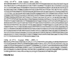
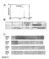
- the disclosure relates to the nucleotide (SEQ ID NO: 1) and amino acid (SEQ ID NO: 2) sequences encoding FPRL2 polypeptide (presented in Figure 1 ).
- the disclosure also relates to sequences that are homologous to the nucleotide and amino acid sequences encoding FPRL2 polypeptide.
- Sequence identity with respect to any of the sequences presented herein can be determined by a simple "eyeball” comparison (i.e. a strict comparison) of any one or more of the sequences with another sequence to see if that other sequence has, for example, at least 80% sequence identity to the sequence(s).
- Relative sequence identity can also be determined by commercially available computer programs that can calculate % identity between two or more sequences using any suitable algorithm for determining identity, using for example default parameters.
- a typical example of such a computer program is CLUSTAL.
- Other computer program methods to determine identity and similarity between two sequences include but are not limited to the GCG program package ( Devereux et al 1984 Nucleic Acids Research 12: 387 ) and FASTA ( Atschul et al 1990 J Molec Biol 403-410 ).
- % homology may be calculated over contiguous sequences, i.e. one sequence is aligned with the other sequence and each amino acid in one sequence is directly compared with the corresponding amino acid in the other sequence, one residue at a time. This is called an "ungapped" alignment. Typically, such ungapped alignments are performed only over a relatively short number of residues.
- a scaled similarity score matrix is generally used that assigns scores to each pairwise comparison based on chemical similarity or evolutionary distance.
- An example of such a matrix commonly used is the BLOSUM62 matrix - the default matrix for the BLAST suite of programs.
- GCG Wisconsin programs generally use either the public default values or a custom symbol comparison table if supplied. It is preferred to use the public default values for the GCG package, or in the case of other software, the default matrix, such as BLOSUM62.
- the BLAST algorithm is employed, with parameters set to default values.
- the BLAST algorithm is described in detail at http://www.ncbi.nih.gov/BLAST/blast_help.html.
- the search parameters are defined as follows, and can be advantageously set to the defined default parameters.
- substantially identical when assessed by BLAST equates to sequences which match with an EXPECT value of at least about 7, preferably at least about 9 and most preferably 10 or more.
- the default threshold for EXPECT in BLAST searching is usually 10.
- BLAST Basic Local Alignment Search Tool
- blastp, blastn, blastx, tblastn, and tblastx these programs ascribe significance to their findings using the statistical methods of Karlin and Altschul ( Karlin and Altschul 1990, Proc. Natl. Acad. Sci. USA 87:2264-68 ; Karlin and Altschul, 1993, Proc. Natl. Acad. Sci. USA 90:5873-7; see http://www.ncbi.nih.gov/BLAST/blast_help.html ) with a few enhancements.
- the BLAST programs are tailored for sequence similarity searching, for example to identify homologues to a query sequence. For a discussion of basic issues in similarity searching of sequence databases, see Altschul et al (1994) Nature Genetics 6:119-129 .
- blastp compares an amino acid query sequence against a protein sequence database
- blastn compares a nucleotide query sequence against a nucleotide sequence database
- blastx compares the six-frame conceptual translation products of a nucleotide query sequence (both strands) against a protein sequence database
- tblastn compares a protein query sequence against a nucleotide sequence database dynamically translated in all six reading frames (both strands)
- tblastx compares the six-frame translations of a nucleotide query sequence against the six-frame translations of a nucleotide sequence database.
- BLAST uses the following search parameters:
- Filtering is only applied to the query sequence (or its translation products), not to database sequences. Default filtering is DUST for BLASTN, SEG for other programs.
- NCBI-gi causes NCBI gi identifiers to be shown in the output, in addition to the accession and/or locus name.
- sequence comparisons are conducted using the simple BLAST search algorithm provided at http://www.ncbi.nlm.nih.gov/BLAST.
- no gap penalties are used when determining sequence identity.
- a cell that is useful according to the disclosure is preferably selected from the group consisting of bacterial cells, yeast cells, insect cells or mammalian cells.
- a cell that is useful according to the disclosure can be any cell into which a nucleic acid sequence encoding a receptor according to the disclosure can be introduced such that the receptor is expressed at natural levels or above natural levels, as defined herein.
- a receptor of the disclosure that is expressed in a cell exhibits normal or near normal pharmacology, as defined herein.
- a receptor of the disclosure that is expressed in a cell comprises the nucleotide represented by SEQ ID NO: 1 or amino acid sequence represented by by SEQ ID NO: 2 or a nucleotide or amino acid sequence that is at least 70% identical to the amino acid sequence represented by SEQ ID NO: 2.
- a receptor of the disclosure that is expressed in a cell will bind HBP polypeptide.
- a cell is selected from the group consisting of COS7-cells, a CHO cell, a LM (TK-) cell, a NIH-3T3 cell, HEK-293 cell, K-562 cell or a 1321N1 astrocytoma cell but also other transfectable cell lines.
- Agents that modulate the activity of FPRL2 polypeptide can be identified in a number of ways that take advantage of the newly discovered interaction of the receptor with HBP polypeptide. For example, the ability to reconstitute FPRL2 polypeptide / HBP polypeptide binding either in vitro, on cultured cells or in vivo provides a target for the identification of agents that disrupt that binding. Assays based on disruption of binding can identify agents, such as small organic molecules, from libraries or collections of such molecules. Alternatively, such assays can identify agents in samples or extracts from natural sources, e.g., plant, fungal or bacterial extracts or even in human tissue samples (e.g., tumor tissue).
- the extracts can be made from cells expressing a library of variant nucleic acids, peptides or polypeptides. Modulators of FPRL2 polypeptide / HBP polypeptide binding can then be screened using a binding assay or a functional assay that measures downstream signalling through the receptor.
- Another approach that uses the FPRL2 polypeptide / HBP polypeptide interaction more directly to identify agents that modulate FPRL2 polypeptide function measures changes in FPRL2 polypeptide downstream signalling induced by candidate agents or candidate modulators.
- These functional assays can be performed in isolated cell membrane fractions or on cells expressing the receptor on their surfaces.
- HBP polypeptide is a ligand of the FPRL2 polypeptide receptor permits screening assays to identify agonists, antagonists and inverse agonists of receptor activity.
- the screening assays have two general approaches, detailed below.
- HBP polypeptide SED ID NO:18
- SED ID NO:18 is used as an exemplary ligand. It should be understood, however, that any HBP polypeptide as defined herein can be used in the assays described.
- HBP polypeptide SEQ ID NO: 18
- HBP polypeptide SEQ ID NO: 18
- cells expressing a FPRL2 polypeptide are incubated in binding buffer with labelled HBP polypeptide in the presence or absence of increasing concentrations of a candidate modulator.
- control competition reactions using increasing concentrations of unlabeled HBP polypeptide can be performed.
- cells are washed extensively, and bound, labelled HBP polypeptide is measured as appropriate for the given label (e.g., scintillation counting, fluorescence, etc.).
- a decrease of at least 10% in the amount of labelled HBP polypeptide bound in the presence of candidate modulator indicates displacement of binding by the candidate modulator.
- Candidate modulators are considered to bind specifically in this or other assays described herein if they displace 50% of labelled HBP polypeptide (sub-saturating HBP polypeptide dose) at a concentration of 1 mM or less.
- binding or displacement of binding can be monitored by surface plasmon resonance (SPR).
- SPR surface plasmon resonance
- Surface plasmon resonance assays can be used as a quantitative method to measure binding between two molecules by the change in mass near an immobilized sensor caused by the binding or loss of binding of HBP polypeptide from the aqueous phase to a FPRL2 polypeptide immobilized in a membrane on the sensor. This change in mass is measured as resonance units versus time after injection or removal of the HBP polypeptide or candidate modulator and is measured using a Biacore Biosensor (Biacore AB).
- Biacore Biosensor Biacore Biosensor
- FPRL2 polypeptide can be immobilized on a sensor chip (for example, research grade CM5 chip; Biacore AB) in a thin film lipid membrane according to methods described by Salamon et al. ( Salamon et al., 1996, Biophys J. 71: 283-294 ; Salamon et al., 2001, Biophys. J. 80: 1557-1567 ; Salamon et al., 1999, Trends Biochem. Sci. 24: 213-219 ).
- Sarrio et al. demonstrated that SPR can be used to detect ligand binding to the GPCR A(1) adenosine receptor immobilized in a lipid layer on the chip ( Sarrio et al., 2000, Mol. Cell. Biol. 20: 5164-5174 ).
- Conditions for HBP polypeptide binding to FPRL2 polypeptide in an SPR assay can be fine-tuned by one of skill in the art using the conditions reported by Sarrio et al. as
- HBP polypeptide can be pre-bound to immobilized FPRL2 polypeptide polypeptide, followed by injection of candidate modulator at a concentration ranging from 0.1 nM to 1 ⁇ M. Displacement of the bound HPB polypeptide can be quantitated, permitting detection of modulator binding.
- the membrane-bound FPRL2 polypeptide can be pre-incubated with candidate modulator and challenged with HBP polypeptide. A difference in HBP polypeptide binding to the FPRL2 polypeptide exposed to modulator relative to that on a chip not pre-exposed to modulator will demonstrate binding or displacement of HBP polypeptide in the presence of modulator.
- a decrease of 10% or more in the amount of HBP polypeptide bound in the presence of candidate modulator, relative to the amount of a HBP polypeptide bound in the absence of candidate modulator indicates that the candidate modulator inhibits the interaction of FPRL2 polypeptide and HBP polypeptide.
- FRET fluorescence resonance energy transfer
- the fluorescence emitted upon excitation of the donor fluorophore will have a different wavelength than that emitted in response to that excitation wavelength when the HBP polypeptide and FPRL2 polypeptide are not bound, providing for quantitation of bound versus unbound molecules by measurement of emission intensity at each wavelength.
- Donor fluorophores with which to label the FPRL2 polypeptide are well known in the art. Of particular interest are variants of the A. victoria GFP known as Cyan FP (CFP, Donor (D)) and Yellow FP (YFP, Acceptor(A)).
- the YFP variant can be made as a fusion protein with FPRL2 polypeptide.
- Vectors for the expression of GFP variants as fusions (Clontech) as well as flurophore-labeled HBP polypeptide compounds (Molecular Probes) are known in the art.
- the addition of a candidate modulator to the mixture of labelled HBP polypeptide and YFP-FPRL2 protein will result in an inhibition of energy transfer evidenced by, for example, a decrease in YFP fluorescence relative to a sample without the candidate modulator.
- a 10% or greater decrease in the intensity of fluorescent emission at the acceptor wavelength in samples containing a candidate modulator, relative to samples without the candidate modulator, indicates that the candidate modulator inhibits the FPRL2 polypeptide: HBP polypeptide interaction.
- a variation on FRET uses fluorescence quenching to monitor molecular interactions.
- One molecule in the interacting pair can be labelled with a fluorophore, and the other with a molecule that quenches the fluorescence of the fluorophore when brought into close apposition with it.
- a change in fluorescence upon excitation is indicative of a change in the association of the molecules tagged with the fluorophore:quencher pair.
- an increase in fluorescence of the labelled FPRL2 polypeptide is indicative that the HBP polypeptide molecule bearing the quencher has been displaced.
- a 10% or greater increase in the intensity of fluorescent emission in samples containing a candidate modulator, relative to samples without the candidate modulator indicates that the candidate modulator inhibits FPRL2 polypeptide: HBP polypeptide interaction.
- fluorescence polarization measurement is useful to quantitate binding.
- the fluorescence polarization value for a fluorescently-tagged molecule depends on the rotational correlation time or tumbling rate.
- Complexes such as those formed by FPRL2 polypeptide associating with a fluorescently labelled HBP polypeptide, have higher polarization values than uncomplexed, labelled HBP polypeptide.
- the inclusion of a candidate inhibitor of the FPRL2 polypeptide: HBP polypeptide interaction results in a decrease in fluorescence polarization, relative to a mixture without the candidate inhibitor, if the candidate inhibitor disrupts or inhibits the interaction of FPRL2 polypeptide with HBP polypeptide.
- Fluorescence polarization is well suited for the identification of small molecules that disrupt the formation of receptor:ligand complexes.
- HBP polypeptide interactions uses a biosensor assay.
- ICS biosensors have been described in the art (Australian Membrane Biotechnology Research Institute; www.ambri.com.au/; Cornell B, Braach-Maksvytis V, King L, Osman P, Raguse B, Wieczorek L, and Pace R. "A biosensor that uses ion-channel switches" Nature 1997, 387, 580 ).
- the association of FPRL2 polypeptide and its ligand is coupled to the closing of gramacidin-facilitated ion channels in suspended membrane bilayers and thus to a measurable change in the admittance (similar to impedence) of the biosensor.
- a modulator will interact at a location removed from the site of interaction and cause; for example, a conformational change in the FPRL2 polypeptide.
- Modulators inhibitors or agonists that act in this manner are nonetheless of interest as agents to modulate the activity of FPRL2 polypeptide.
- any of the binding assays described herein can be performed with a non-HBP polypeptide ligand (for example, agonist, antagonist, etc.) of FPRL2 polypeptide, e.g., a small molecule identified as described herein or HBP polypeptide analogues including but not limited to any of the HBP polypeptide analogues, a natural or synthetic peptide, a polypeptide, an antibody or antigen-binding fragment thereof, a lipid, a carbohydrate, and a small organic molecule.
- a non-HBP polypeptide ligand for example, agonist, antagonist, etc.
- HBP polypeptide analogues including but not limited to any of the HBP polypeptide analogues, a natural or synthetic peptide, a polypeptide, an antibody or antigen-binding fragment thereof, a lipid, a carbohydrate, and a small organic molecule.
- any of the binding assays described can be used to determine the presence of an agent in a sample, e.g., a tissue sample, that binds to the FPRL2 polypeptide receptor molecule, or that affects the binding of HBP polypeptide to the receptor.
- a sample e.g., a tissue sample
- HBP polypeptide or ligand binding is measured as appropriate for the binding assay being used.
- a decrease of 10% or more in the binding of HBP polypeptide or other ligand indicates that the sample contains an agent that modulates HBP polypeptide or ligand binding to the receptor polypeptide.
- GPCRs such as FPRL2 polypeptide
- a measure of receptor activity is the binding of GTP by cell membranes containing receptors.
- membranes isolated from cells expressing the receptor are incubated in a buffer containing 20 mM HEPES, pH 7.4, 100 mM NaCl, and 10 mM MgCl2, 80 pM 35 S-GTP ⁇ S and 3 ⁇ M GDP.
- the assay mixture is incubated for 60 minutes at 30°C, after which unbound labelled GTP is removed by filtration onto GF/B filters. Bound, labelled GTP is measured by liquid or solid (SPA, see below) scintillation counting.
- SPA liquid or solid
- a similar GTP-binding assay can be performed without HBP polypeptide to identify compounds that act as agonists.
- HBP polypeptide -stimulated GTP binding is used as a standard.
- a compound is considered an agonist if it induces at least 50, 40, 30, or preferably 20% of the level of GTP binding induced by HBP polypeptide when the compound is present at 1 ⁇ M or less, and preferably will induce a level the same as or higher than that induced by HBP polypeptide.
- SPA Scintillation Proximity Assay
- GTPase activity is measured by incubating the membranes containing a FPRL2 polypeptide with ⁇ 32 P-GTP. Active GTPase will release the label as inorganic phosphate, which is detected by separation of free inorganic phosphate in a 5% suspension of activated charcoal in 20 mM H 3 PO 4 , followed by scintillation counting. Controls include assays using membranes isolated from cells not expressing FPRL2 polypeptide (mock-transfected), in order to exclude possible non-specific effects of the candidate compound.
- membrane samples are incubated with HBP polypeptide, with or without the modulator, followed by the GTPase assay.
- a change (increase or decrease) of 10% or more in the level of GTP binding or GTPase activity relative to samples without modulator is indicative of FPRL2 polypeptide modulation by a candidate modulator.
- the aequorin assay takes advantage of the responsiveness of mitochondrial apoaequorin to intracellular calcium release induced by the activation of GPCRs ( Stables et al., 1997, Anal. Biochem. 252:115-126 ; Detheux et al., 2000, J. Exp. Med., 192 1501-1508 ). Briefly, FPRL2 polypeptide-expressing clones are transfected to coexpress mitochondrial apoaequorin and G ⁇ 16.
- Cells are incubated with 5 ⁇ M Coelenterazine H (Molecular Probes) for 4 hours at room temperature, washed in DMEM-F12 culture medium and resuspended at a concentration of 0.5 x 10 6 cells/ml. Cells are then mixed with test agonist molecules and light emission by the aequorin is recorded with a luminometer for 30 sec. Results are expressed as Relative Light Units (RLU). Controls include assays using membranes isolated from cells not expressing FPRL2 polypeptide (mock transfected), in order to exclude possible non-specific effects of the candidate compound.
- RLU Relative Light Units
- Aequorin activity or intracellular calcium levels are "changed” if light intensity increases or decreases by 10% or more in a sample of cells, expressing a FPRL2 polypeptide and treated with a candidate modulator, relative to a sample of cells expressing the FPRL2 polypeptide but not treated with the candidate modulator or relative to a sample of cells not expressing the FPRL2 polypeptide (mock-transfected cells) but treated with the candidate modulator.
- the assay When performed in the absence of HBP polypeptide, the assay can be used to identify an agonist of FPRL2 polypeptide activity. When the assay is performed in the presence of HBP polypeptide, it can be used to assay for an antagonist.
- 100 ⁇ l reactions contain 50 mM Tris-Hcl (pH 7.5), 5 mM MgCl 2 , 20 mM creatine phosphate (disodium salt), 10 units (71 ⁇ g of protein) of creatine phosphokinase, 1 mM ⁇ - 32 P-ATP (tetrasodium salt, 2 ⁇ Ci), 0.5 mM cyclic AMP, G- 3 H-labeled cyclic AMP (approximately 10,000 cpm), 0.5 mM Ro20-1724, 0.25% ethanol, and 50-200 ⁇ g of protein homogenate to be tested (i.e., homogenate from cells expressing or not expressing a FPRL2 polypeptide, treated or not treated with HBP polypeptide with or without a candidate modulator).
- protein homogenate to be tested i.e., homogenate from cells expressing or not expressing a FPRL2 polypeptide, treated or not treated with HBP polypeptide with
- Reaction mixtures are generally incubated at 37°C for 60 minutes. Following incubation, reaction mixtures are deproteinized by the addition of 0.9 ml of cold 6% trichloroacetic acid. Tubes are centrifuged at 1800 x g for 20 minutes and each supernatant solution is added to a Dowex AG50W-X4 column. The cAMP fraction from the column is eluted with 4 ml of 0.1 mM imidazole-HCl (pH 7.5) into a counting vial. Assays should be performed in triplicate. Control reactions should also be performed using protein homogenate from cells that do not express a FPRL2 polypeptide.
- adenylate cyclase activity is "changed” if it increases or decreases by 10% or more in a sample taken from cells treated with a candidate modulator of FPRL2 polypeptide activity, relative to a similar sample of cells not treated with the candidate modulator or relative to a sample of cells not expressing the FPRL2 polypeptide (mock-transfected cells) but treated with the candidate modulator.
- Intracellular or extracellular cAMP is measured using a cAMP radioimmunoassay (RIA) or cAMP binding protein according to methods widely known in the art.
- RIA radioimmunoassay
- cAMP binding protein for example, Horton & Baxendale, 1995, Methods Mol. Biol. 41: 91-105 describes an RIA for cAMP.
- kits for the measurement of cAMP are commercially available, such as the High Efficiency Fluorescence Polarization-based homogeneous assay marketed by LJL Biosystems and NEN Life Science Products. Control reactions should be performed using extracts of mock-transfected cells to exclude possible non-specific effects of some candidate modulators.
- the level of cAMP is "changed” if the level of cAMP detected in cells, expressing a FPRL2 polypeptide and treated with a candidate modulator of FPRL2 polypeptide activity (or in extracts of such cells), using the RIA-based assay of Horton & Baxendale, 1995, supra, increases or decreases by at least 10% relative to the cAMP level in similar cells not treated with the candidate modulator.
- Receptors that activate the breakdown of phospholipids can be monitored for changes due to the activity of known or suspected modulators of FPRL2 polypeptide by monitoring phospholipid breakdown, and the resulting production of second messengers DAG and/or inositol trisphosphate (IP 3 ).
- Methods of detecting each of these are described in Phospholipid Signalling Protocols, edited by Ian M. Bird. Totowa, NJ, Humana Press, 1998 . See also Rudolph et al., 1999, J. Biol. Chem. 274: 11824-11831 which also describes an assay for phosphatidylinositol breakdown.
- Assays should be performed using cells or extracts of cells expressing FPRL2 polypeptide, treated or not treated with HBP polypeptide with or without a candidate modulator. Control reactions should be performed using mock-transfected cells, or extracts from them in order to exclude possible non-specific effects of some candidate modulators.
- phosphatidylinositol breakdown, and diacylglycerol and/or inositol trisphosphate levels are "changed” if they increase or decrease by at least 10% in a sample from cells expressing a FPRL2 polypeptide and treated with a candidate modulator, relative to the level observed in a sample from cells expressing a FPRL2 polypeptide that is not treated with the candidate modulator.
- PKC Protein Kinase C
- IAM I intracellular adhesion molecule I
- Assays designed to detect increases in gene products induced by PKC can be used to monitor PKC activation and thereby receptor activity.
- the activity of receptors that signal via PKC can be monitored through the use of reporter gene constructs driven by the control sequences of genes activated by PKC activation. This type of reporter gene-based assay is discussed in more detail below.
- PKC activity For a more direct measure of PKC activity, the method of Kikkawa et al., 1982, J. Biol. Chem. 257: 13341 can be used. This assay measures phosphorylation of a PKC substrate peptide, which is subsequently separated by binding to phosphocellulose paper. This PKC assay system can be used to measure activity of purified kinase, or the activity in crude cellular extracts. Protein kinase C sample can be diluted in 20 mM HEPES/ 2 mM DTT immediately prior to assay.
- the substrate for the assay is the peptide Ac-FKKSFKL-NH2 (SEQ ID NO: 11), derived from the myristoylated alanine-rich protein kinase C substrate protein (MARCKS).
- the K m of the enzyme for this peptide is approximately 50 ⁇ M.
- Other basic, protein kinase C-selective peptides known in the art can also be used, at a concentration of at least 2 -3 times their K m .
- Cofactors required for the assay include calcium, magnesium, ATP, phosphatidylserine and diacylglycerol.
- the assay can be performed to determine the amount of PKC present (activating conditions) or the amount of active PKC present (non-activating conditions).
- activating conditions the amount of PKC present
- non-activating conditions such that the PKC, that is active in the sample when it is isolated, is measured, rather than measuring the PKC that can be activated.
- calcium is omitted from the assay in favor of EGTA.
- the assay is performed in a mixture containing 20 mM HEPES, pH 7.4, 1-2 mM DTT, 5 mM MgCl 2 , 100 ⁇ M ATP, ⁇ 1 ⁇ Ci ⁇ 32 P-ATP, 100 ⁇ g/ml peptide substrate ( ⁇ 100 ⁇ M), 140 ⁇ M / 3.8 ⁇ M phosphatidylserine/diacylglycerol membranes, and 100 ⁇ M calcium (or 500 ⁇ M EGTA).
- 48 ⁇ l of sample, diluted in 20 mM HEPES, pH 7.4, 2 mM DTT is used in a final reaction volume of 80 ⁇ l. Reactions are performed at 30°C for 5-10 minutes, followed by addition of 25 ⁇ l of 100 mM ATP, 100 mM EDTA, pH 8.0, which stops the reactions.
- Assays are performed on extracts from cells expressing a FPRL2 polypeptide, treated or not treated with HBP polypeptide with or without a candidate modulator. Control reactions should be performed using mock-transfected cells, or extracts from them in order to exclude possible non-specific effects of some candidate modulators.
- PKC activity is "changed" by a candidate modulator when the units of PKC measured by either assay described above increase or decrease by at least 10%, in extracts from cells expressing FPRL2 polypeptide and treated with a candidate modulator, relative to a reaction performed on a similar sample from cells not treated with a candidate modulator.
- MAP kinase activity can be assayed using any of several kits available commercially, for example, the p38 MAP Kinase assay kit sold by New England Biolabs (Cat # 9820) or the FlashPlate TM MAP Kinase assays sold by Perkin-Elmer Life Sciences.
- MAP Kinase activity is "changed” if the level of activity is increased or decreased by 10% or more in a sample from cells, expressing a FPRL2 polypeptide, treated with a candidate modulator relative to MAP kinase activity in a sample from similar cells not treated with the candidate modulator.
- kinases e.g., Ser/Thr kinases
- Kinase assays can be performed with both purified kinases and crude extracts prepared from cells expressing a FPRL2 polypeptide, treated with or without HBP polypeptide, with or without a candidate modulator. Control reactions should be performed using mock-transfected cells, or extracts from them in order to exclude possible non-specific effects of some candidate modulators.
- Substrates can be either full-length protein or synthetic peptides representing the substrate.
- Pinna & Ruzzene (1996, Biochem. Biophys. Acta 1314: 191-225 ) list a number of phosphorylation substrate sites useful for detecting kinase activities.
- a number of kinase substrate peptides are commercially available. One that is particularly useful is the "Src-related peptide," RRLIEDAEYAARG (SEQ ID NO: 12; available from Sigma # A7433), which is a substrate for many receptor and nonreceptor tyrosine kinases. Because the assay described below requires binding of peptide substrates to filters, the peptide substrates should have a net positive charge to facilitate binding. Generally, peptide substrates should have at least 2 basic residues and a free amino terminus. Reactions generally use a peptide concentration of 0.7-1.5 mM.
- Assays are generally carried out in a 25 ⁇ l volume comprising 5 ⁇ l of 5X kinase buffer (5 mg/mL BSA, 150 mM Tris-Cl (pH 7.5), 100 mM MgCl 2 ; depending upon the exact kinase assayed for, MnCl 2 can be used in place of or in addition to the MgCl 2 ), 5 ⁇ l of 1.0 mM ATP (0.2 mM final concentration), ⁇ -32P-ATP (100-500 cpm/pmol), 3 ⁇ l of 10 mM peptide substrate (1.2 mM final concentration), cell extract containing kinase to be tested (cell extracts used for kinase assays should contain a phosphatase inhibitor (e.g. 0.1-1 mM sodium orthovanadate)), and H 2 O to 25 ⁇ l. Reactions are performed at 30°C, and are initiated by the addition of the cell extract.
- kinase reactions are performed for 30 seconds to about 30 minutes, followed by the addition of 45 ⁇ l of ice-cold 10% trichloroacetic acid (TCA). Samples are spun for 2 minutes in a microcentrifuge, and 35 ⁇ l of the supernatant is spotted onto Whatman P81 cellulose phosphate filter circles. The filters are washed three times with 500 ml cold 0.5% phosphoric acid, followed by one wash with 200 ml of acetone at room temperature for 5 minutes. Filters are dried and incorporated 32 P is measured by scintillation counting.
- TCA trichloroacetic acid
- the specific activity of ATP in the kinase reaction (e.g., in cpm/pmol) is determined by spotting a small sample (2-5 ⁇ l) of the reaction onto a P81 filter circle and counting directly, without washing. Counts per minute obtained in the kinase reaction (minus blank) are then divided by the specific activity to determine the moles of phosphate transferred in the reaction.
- Tyrosine kinase activity is "changed” if the level of kinase activity is increased or decreased by 10% or more in a sample from cells, expressing a FPRL2 polypeptide, treated with a candidate modulator relative to kinase activity in a sample from similar cells not treated with the candidate modulator.
- the intracellular signal initiated by binding of an agonist to a receptor sets in motion a cascade of intracellular events, the ultimate consequence of which is a rapid and detectable change in the transcription or translation of one or more genes.
- the activity of the receptor can therefore be monitored by detecting the expression of a reporter gene driven by control sequences responsive to FPRL2 activation.
- promoter refers to the transcriptional control elements necessary for receptor-mediated regulation of gene expression, including not only the basal promoter, but also any enhancers or transcription-factor binding sites necessary for receptor-regulated expression.
- Reporter genes such as luciferase, CAT, GFP, ⁇ -lactamase or ⁇ -galactosidase are well known in the art, as are assays for the detection of their products.
- Genes particularly well suited for monitoring receptor activity are the "immediate early" genes, which are rapidly induced, generally within minutes of contact between the receptor and the effector protein or ligand.
- the induction of immediate early gene transcription does not require the synthesis of new regulatory proteins.
- characteristics of preferred genes useful for making reporter constructs include: low or undetectable expression in quiescent cells; induction that is transient and independent of new protein synthesis; subsequent shut-off of transcription requires new protein synthesis; and mRNAs transcribed from these genes have a short half-life. It is preferred, but not necessary that a transcriptional control element have all of these properties for it to be useful.
- c-fos proto-oncogene An example of a gene that is responsive to a number of different stimuli is the c-fos proto-oncogene.
- the c-fos gene is activated in a protein-synthesis-independent manner by growth factors, hormones, differentiation-specific agents, stress, and other known inducers of cell surface proteins.
- the induction of c-fos expression is extremely rapid, often occurring within minutes of receptor stimulation. This characteristic makes the c-fos regulatory regions particularly attractive for use as a reporter of receptor activation.
- the c-fos regulatory elements include (see, Verma et al., 1987, Cell 51: 513-514 ): a TATA box that is required for transcription initiation; two upstream elements for basal transcription, and an enhancer, which includes an element with dyad symmetry and which is required for induction by TPA, serum, EGF, and PMA.
- the 20 bp c-fos transcriptional enhancer element located between -317 and -298 bp upstream from the c-fos mRNA cap site, is essential for serum induction in serum starved NIH 3T3 cells.
- One of the two upstream elements is located at -63 to -57 and it resembles the consensus sequence for cAMP regulation.
- the transcription factor CREB (cyclic AMP responsive element binding protein) is, as the name implies, responsive to levels of intracellular cAMP. Therefore, the activation of a receptor that signals via modulation of cAMP levels can be monitored by detecting either the binding of the transcription factor, or the expression of a reporter gene linked to a CREB-binding element (termed the CRE, or cAMP response element).
- the DNA sequence of the CRE is TGACGTCA. Reporter constructs responsive to CREB binding activity are described in U.S. Patent No. 5,919,649 .
- VIP vasoactive intestinal peptide
- cAMP responsive Fink et al., 1988, Proc. Natl. Acad. Sci. 85:6662-6666
- somatostatin gene promoter cAMP responsive; Montminy et al., 1986, Proc. Natl. Acad. Sci.
- transcriptional control elements that are responsive to changes in GPCR activity include, but are not limited to those responsive to the AP-1 transcription factor and those responsive to NF- ⁇ B activity.
- the consensus AP-1 binding site is the palindrome TGA(C/G)TCA ( Lee et al., 1987, Nature 325: 368-372 ; Lee et al., 1987, Cell 49: 741-752 ).
- the AP-1 site is also responsible for mediating induction by tumor promoters such as the phorbol ester 12-O-tetradecanoylphorbol-beta-acetate (TPA), and are therefore sometimes also referred to as a TRE, for TPA-response element.
- TPA phorbol ester 12-O-tetradecanoylphorbol-beta-acetate
- AP-1 activates numerous genes that are involved in the early response of cells to growth stimuli.
- Examples of AP-1-responsive genes include, but are not limited to the genes for Fos and Jun (which proteins themselves make up AP-1 activity), Fos-related antigens (Fra) 1 and 2, I ⁇ B ⁇ , ornithine decarboxylase, and annexins I and II.
- the NF- ⁇ B binding element has the consensus sequence GGGGACTTTCC (SEQ ID NO: 13).
- a large number of genes have been identified as NF- ⁇ B responsive, and their control elements can be linked to a reporter gene to monitor GPCR activity.
- a small sample of the genes responsive to NF- ⁇ B includes those encoding IL-1 ⁇ ( Hiscott et al., 1993, Mol. Cell. Biol. 13: 6231-6240 ), TNF- ⁇ ( Shakhov et al., 1990, J. Exp. Med. 171: 35-47 ), CCR5 ( Liu et al., 1998, AIDS Res. Hum. Retroviruses 14: 1509-1519 ), P-selection ( Pan & McEver, 1995, J. Biol.
- NF- ⁇ B-responsive reporters are also known in the art or can be readily made by one of skill in the art using, for example, synthetic NF- ⁇ B elements and a minimal promoter, or using the NF- ⁇ B-responsive sequences of a gene known to be subject to NF- ⁇ B regulation. Further, NF- ⁇ B responsive reporter constructs are commercially available from, for example, CLONTECH.
- a given promoter construct should be tested by exposing FPRL2 polypeptide-expressing cells, transfected with the construct, to HBP polypeptide. An increase of at least two-fold in the expression of reporter in response to HBP polypeptide indicates that the reporter is an indicator of FPRL2 polypeptide activity.
- cells that stably express a FPRL2 polypeptide are stably transfected with the reporter construct.
- the cells are left untreated, exposed to candidate modulators, or exposed to HBP polypeptide, and expression of the reporter is measured.
- the HBP polypeptide-treated cultures serve as a standard for the level of transcription induced by a known agonist.
- An increase of at least 50% in reporter expression in the presence of a candidate modulator indicates that the candidate is a modulator of FPRL2 polypeptide activity.
- An agonist will induce at least as much, and preferably the same amount or greater reporter expression than HBP polypeptide alone.
- This approach can also be used to screen for inverse agonists where cells express a FPRL2 polypeptide at levels such that there is an elevated basal activity of the reporter in the absence of HBP polypeptide or another agonist.
- the cells expressing FPRL2 polypeptide and carrying the reporter construct are exposed to HBP polypeptide (or another agonist) in the presence and absence of candidate modulator.
- HBP polypeptide or another agonist
- Controls for transcription assays include cells not expressing FPRL2 polypeptide but carrying the reporter construct, as well as cells with a promoterless reporter construct.
- Compounds that are identified as modulators of FPRL2 polypeptide-regulated transcription should also be analyzed to determine whether they affect transcription driven by other regulatory sequences and by other receptors, in order to determine the specificity and spectrum of their activity.
- the transcriptional reporter assay and most cell-based assays, are well suited for screening expression libraries for proteins for those that modulate FPRL2 polypeptide activity.
- the libraries can be, for example, cDNA libraries from natural sources, e.g., plants, animals, bacteria, etc., or they can be libraries expressing randomly or systematically mutated variants of one or more polypeptides.
- Genomic libraries in viral vectors can also be used to express the mRNA content of one cell or tissue in the different libraries used for screening of FPRL2 polypeptide.
- Cells of the disclosure are labelled for 24 hours with 10 ⁇ Ci/ml [ 3 H] inositol in inositol free DMEM containing 5% FCS, antibiotics, amphotericin, sodium pyruvate and 400 ⁇ g/ml G418.
- Cells are incubated for 2 h in Krebs-Ringer Hepes (KRH) buffer of the following composition (124 mM NaCl, 5 mM KCI, 1.25 mM MgSO 4 , 1.45 mM CaCl 2 , 1.25 mM KH 2 PO 4 , 25 mM Hepes (pH:7.4) and 8 mM glucose).
- KRH Krebs-Ringer Hepes
- the cells are then challenged with HBP polypeptide for 30 min. The incubation is stopped by the addition of an ice cold 3% perchloric acid solution. IP are extracted and separated on Dowex columns as previously described (25).
- the disclosure provides for an assay for detecting the activity of a receptor of the disclosure in a sample.
- FPRL2 polypeptide activity can be measured in a sample comprising a cell or a cell membrane that expresses FPRL2 polypeptide.
- HBP polypeptide SEQ IN NO:18
- the assay is performed by incubating the sample in the presence or absence of HBP polypeptide and carrying out a second messenger assay, as described above.
- the results of the second messenger assay performed in the presence or absence of HBP polypeptide are compared to determine if the FPRL2 polypeptide receptor is active.
- An increase of 10% or more in the detected level of a given second messenger, as defined herein, in the presence of HBP polypeptide relative to the amount detected in an assay performed in the absence of HBP polypeptide is indicative of FPRL2 polypeptide activity.
- any of the assays of receptor activity including but not limited to the GTP-binding, GTPase, adenylate cyclase, cAMP, phospholipid-breakdown, diacylglycerol, inositol trisphosphate, arachidonic acid release (see below), PKC, kinase and transcriptional reporter assays, can be used to determine the presence of an agent in a sample, e.g., a tissue sample, that affects the activity of the FPRL2 polypeptide receptor molecule. To do so, FPRL2 polypeptide is assayed for activity in the presence and absence of the sample or an extract of the sample.
- An increase in FPRL2 polypeptide activity in the presence of the sample or extract relative to the absence of the sample indicates that the sample contains an agonist of the receptor activity.
- a decrease in receptor activity in the presence of HBP polypeptide or another agonist and the sample, relative to receptor activity in the presence of HBP polypeptide alone, indicates that the sample contains an antagonist of FPRL2 polypeptide activity.
- samples can then be fractionated and further tested to isolate or purify the agonist or antagonist.
- the amount of increase or decrease in measured activity necessary for a sample to be said to contain a modulator depends upon the type of assay used. Generally, a 10% or greater change (increase or decrease) relative to an assay performed in the absence of a sample indicates the presence of a modulator in the sample.
- transcriptional reporter assay in which at least a two-fold increase or 10% decrease in signal is necessary for a sample to be said to contain a modulator. It is preferred that an agonist stimulates at least 50%, and preferably 75% or 100% or more, e.g., 2-fold, 5-fold, 10-fold or greater receptor activation than with HBP polypeptide alone.
- Other functional assays include, for example, microphysiometer or biosensor assays (see Hafner, 2000, Biosens. Bioelectron. 15: 149-158 ).
- the intracellular level of arachidonic acid can also be determined as described in Gijon et al., 2000, J. Biol. Chem., 275:20146-20156 .
- FPRL2 polypeptide which is expressed in cells of the lymphocyte lineages, spleen, small intestine, lung, heart, can have a role in immune processes, cancer and associated disorders or diseases.
- FPRL2 polypeptide can be involved in disturbances of cell migration, cancer, development of tumours and tumour metastasis, inflammatory and neoplastic processes, wound and bone healing and dysfunction of regulatory growth functions, obesity, anorexia, bulimia, acute heart failure, hypotension, hypertension, urinary retention, osteoporosis, angina pectoris, , restenosis, atherosclerosis, thrombosis and other cardiovascular diseases, autoimmune and, diseases characterized by excessive smooth muscle cell proliferation, aneurysms, diseases characterized by loss of smooth muscle cells or reduced smooth muscle cell proliferation, stroke, ischemia, ulcers, allergies, , prostatic hypertrophy, migraine, vomiting, psychotic and neurological disorders, including anxiety, schizophrenia, manic depression, depression, delirium, dementia and severe mental retardation, degenerative diseases, neurodegenerative diseases such as Alzheimer's disease or Parkinson's disease, and dyskinasias, such as Huntington
- diagnostic assays for FPRL2 polypeptide-related diseases or disorders can have several different forms.
- diagnostic assays can measure the amount of FPRL2 polypeptides, mRNA or ligand in a sample of tissue.
- Assays that measure the amount of mRNA encoding FPRL2 polypeptide also fit into this category.
- assays can evaluate the qualities of the receptor or the ligand. For example, assays that determine whether an individual expresses a mutant or variant form of FPRL2 polypeptide can be used diagnostically.
- assays that measure one or more activities of FPRL2 polypeptide can be used diagnostically.
- FPRL2 polypeptide levels can be measured and compared to standards in order to determine whether an abnormal level of the receptor or its ligand is present in a sample, either of which indicate probable dysregulation of FPRL2 polypeptide signalling.
- Polypeptide levels are measured, for example, by immunohistochemistry using antibodies specific for the polypeptide.
- a sample isolated from an individual suspected of suffering from a disease or disorder characterized by FPRL2 polypeptide activity is contacted with an antibody for a FPRL2 polypeptide, and binding of the antibody is measured as known in the art (e.g., by measurement of the activity of an enzyme conjugated to a secondary antibody).
- FPRL2 polypeptide levels uses flow cytometry analysis of cells from an affected tissue. Methods of flow cytometry, including the fluorescent labeling of antibodies specific for FPRL2 polypeptide, are well known in the art. Other approaches include radioimmunoassay or ELISA. Methods for each of these are also well known in the art.
- the amount of binding detected is compared to the binding in a sample of similar tissue from a healthy individual, or from a site on the affected individual that is not so affected.
- An increase of 10% or more relative to the standard is diagnostic for a disease or disorder characterized by FPRL2 polypeptide dysregulation.
- FPRL2 polypeptide expression can also be measured by determining the amount of mRNA encoding the polypeptides in a sample of tissue.
- Levels of mRNA can be measured by quantitative or semi-quantitative PCR. Methods of "quantitative" amplification are well known to those of skill in the art, and primer sequences for the amplification of FPRL2 nucleic acid are disclosed herein.
- a common method of quantitative PCR involves simultaneously co-amplifying a known quantity of a control sequence using the same primers. This provides an internal standard that can be used to calibrate the PCR reaction. Detailed protocols for quantitative PCR are provided in PCR Protocols, A Guide to Methods and Applications, Innis et al., Academic Press, Inc.
- An increase of 10% or more in the amount of mRNA encoding FPRL2 polypeptide in a sample, relative to the amount expressed in a sample of like tissue from a healthy individual or in a sample of tissue from an unaffected location in an affected individual is diagnostic for a disease or disorder characterized by dysregulation of FPRL2 polypeptide signalling.
- RNA isolated from a sample is used as a template for PCR amplification of FPRL2 polypeptide.
- the amplified sequences are then either directly sequenced using standard methods, or are first cloned into a vector, followed by sequencing.
- a difference in the sequence that changes one or more encoded amino acids relative to the sequence of wild-type FPRL2 polypeptide can be diagnostic of a disease or disorder characterized by dysregulation of FPRL2 polypeptide signalling.
- amplified sequences can be assayed for the presence of specific mutations using, for example, hybridization of molecular beacons that discriminate between wild type and variant sequences.
- Hybridization assays that discriminate on the basis of changes as small as one nucleotide are well known in the art.
- any of a number of "minisequencing" assays can be performed, including, those described, for example, in U.S. Patents 5,888,819 , 6,004,744 and 6,013,431 .
- These assays and others known in the art can determine the presence, in a given sample, of a nucleic acid with a known polymorphism.
- array or microarray-based methods can be used to analyze the expression or the presence of mutation, in FPRL2 polypeptide sequences.
- Array-based methods for minisequencing and for quantitation of nucleic acid expression are well known in the art.
- Diagnosis of a disease or disorder characterized by the dysregulation of FPRL2 polypeptide signalling can also be performed using functional assays.
- cell membranes or cell extracts prepared from a tissue sample are used in an assay of FPRL2 polypeptide activity as described herein (e.g., ligand binding assays, the GTP-binding assay, GTPase assay, adenylate cyclase assay, cAMP assay, arachidonic acid level, phospholipid breakdown, diacyl glycerol or inositol trisphosphate assays, PKC activation assay, or kinase assay).
- ligand binding assays e.g., the GTP-binding assay, GTPase assay, adenylate cyclase assay, cAMP assay, arachidonic acid level, phospholipid breakdown, diacyl glycerol or inositol trisphosphate as
- the activity detected is compared to that in a standard sample taken from a healthy individual or from an unaffected site on the affected individual.
- a sample or extract of a sample can be applied to cells expressing FPRL2 polypeptide, followed by measurement of FPRL2 polypeptide signalling activity relative to a standard sample.
- a difference of 10% or more in the activity measured in any of these assays, relative to the activity of the standard, is diagnostic for a disease or disorder characterized by dysregulation of FPRL2 polypeptide signalling.
- HBP polypeptide as a ligand of FPRL2 polypeptide provides methods of modulating the activity of a FPRL2 polypeptide polypeptide in a cell.
- FPRL2 polypeptide activity is modulated in a cell by delivering to that cell an agent that modulates the function of a FPRL2 polypeptide polypeptide. This modulation can be performed in cultured cells as part of an assay for the identification of additional modulating agents, or, for example, in an animal, including a human.
- Agents include HBP polypeptide and other ligands as defined herein, as well as additional modulators identified using the screening methods described herein including but not limited to any of the HBP polypeptide analogues.
- An agent can be delivered to a cell by adding it to culture medium.
- the amount to deliver will vary with the identity of the agent and with the purpose for which it is delivered.
- agent e.g., HBP polypeptide that half-maximally activates the receptors (e.g., approximately EC 50 ), preferably without exceeding the dose required for receptor saturation.
- This dose can be determined by titrating the amount of HBP polypeptide to determine the point at which further addition of HBP polypeptide has no additional effect on FPRL2 polypeptide activity.
- a modulator of FPRL2 polypeptide activity When a modulator of FPRL2 polypeptide activity is administered to an animal for the treatment of a disease or disorder, the amount administered can be adjusted by one of skill in the art on the basis of the desired outcome.
- Successful treatment is achieved when one or more measurable aspects of the pathology (e.g., tumor cell growth, accumulation of inflammatory cells) is changed by at least 10% relative to the value for that aspect prior to treatment.
- a candidate modulator is a HBP polypeptide (SEQ ID NO:18), a HBP polypeptide as defined herein above, a ligand as defined herein above or an agent identified according to the disclosure.
- the candidate compound can be a synthetic compound, or a mixture of compounds, or may be a natural product (e.g. a plant extract or culture supernatant).
- a candidate compound according to the disclosure includes but is not limited to a small molecule that can be synthesized, a natural extract, peptides, polypeptides, carbohydrates, lipids, an antibody or antigen-binding fragment thereof, nucleic acids, and a small organic molecules.
- Candidate modulator compounds from large libraries of synthetic or natural compounds can be screened. Numerous means are currently used for random and directed synthesis of saccharide, peptide, and nucleic acid based compounds. Synthetic compound libraries are commercially available from a number of companies including Maybridge Chemical Co. (Trevillet, Cornwall, UK), Comgenex (Princeton, NJ), Brandon Associates (Merrimack, NH), and Microsource (New Milford, CT). A rare chemical library is available from Aldrich (Milwaukee, WI). Combinatorial libraries are available and can be prepared.
- libraries of natural compounds in the form of bacterial, fungal, plant and animal extracts are available from e.g., Pan Laboratories (Bothell, WA) or MycoSearch (NC), or are readily producible by methods well known in the art. Additionally, natural and synthetically produced libraries and compounds are readily modified through conventional chemical, physical, and biochemical means.
- Useful compounds may be found within numerous chemical classes. Useful compounds may be organic compounds, or small organic compounds. Small organic compounds have a molecular weight of more than 50 yet less than about 2,500 daltons, preferably less than about 750, more preferably less than about 350 daltons. Exemplary classes include heterocycles, peptides, saccharides, steroids, and the like. The compounds may be modified to enhance efficacy, stability, pharmaceutical compatibility, and the like. Structural identification of an agent may be used to identify, generate, or screen additional agents.
- peptide agents may be modified in a variety of ways to enhance their stability, such as using an unnatural amino acid, such as a D-amino acid, particularly D-alanine, by functionalizing the amino or carboxylic terminus, e.g. for the amino group, acylation or alkylation, and for the carboxyl group, esterification or amidification, or the like.
- an unnatural amino acid such as a D-amino acid, particularly D-alanine
- a useful concentration of a candidate compound according to the disclosure is from about 10nM to about 100 ⁇ M or more (i.e. 1mM, 10mM, 100mM, or even 1M), but can also be 1 nM and higher, 1 pM and higher, or 1 fM and higher.
- the primary screening concentration will be used as an upper limit, along with nine additional concentrations, wherein the additional concentrations are determined by reducing the primary screening concentration at half-log intervals (e.g. for 9 more concentrations) for secondary screens or for generating concentration curves.
- Antibodies can be made using standard protocols known in the art (See, for example, Antibodies: A Laboratory Manual ed. by Harlow and Lane (Cold Spring Harbor Press: 1988 )).
- a mammal such as a mouse, hamster, or rabbit can be immunized with an immunogenic form of the peptide (e.g., FPRL2 polypeptide or an antigenic fragment which is capable of eliciting an antibody response, or a fusion protein as described herein above).
- Immunogens for raising antibodies are prepared by mixing the polypeptides (e.g., isolated recombinant polypeptides or synthetic peptides) with adjuvants.
- FPRL2 polypeptides or peptides are made as fusion proteins to larger immunogenic proteins.
- Polypeptides can also be covalently linked to other larger immunogenic proteins, such as keyhole limpet hemocyanin.
- plasmid or viral vectors encoding FPRL2 polypeptide, or a fragment of these proteins can be used to express the polypeptides and generate an immune response in an animal as described in Costagliola et al., 2000, J. Clin. Invest. 105:803-811 .
- immunogens are typically administered intradermally, subcutaneously, or intramuscularly to experimental animals such as rabbits, sheep, and mice.
- genetically engineered antibody derivatives can be made, such as single chain antibodies.
- the progress of immunization can be monitored by detection of antibody titers in plasma or serum.
- Standard ELISA, flow cytometry or other immunoassays can also be used with the immunogen as antigen to assess the levels of antibodies.
- Antibody preparations can be simply serum from an immunized animal, or if desired, polyclonal antibodies can be isolated from the serum by, for example, affinity chromatography using immobilized immunogen.
- antibody-producing splenocytes can be harvested from an immunized animal and fused by standard somatic cell fusion procedures with immortalizing cells such as myeloma cells to yield hybridoma cells.
- immortalizing cells such as myeloma cells.
- Such techniques are well known in the art, and include, for example, the hybridoma technique (originally developed by Kohler and Milstein, (1975) Nature, 256: 495-497 ), the human B cell hybridoma technique ( Kozbar et al., (1983) Immunology Today, 4: 72 ), and the EBV-hybridoma technique to produce human monoclonal antibodies ( Cole et al., (1985) Monoclonal Antibodies and Cancer Therapy, Alan R. Liss, Inc. pp. 77-96 ).
- Hybridoma cells can be screened immunochemically for production of antibodies specifically reactive with FPRL2 polypeptide, and monoclonal antibodies isolated from the media of a culture comprising such hybridoma
- a functional fragment of an antibody including fragment of chimeric, humanized, primatized or single chain antibody, can also be produced.
- Functional fragments of the foregoing antibodies retain at least one binding function and/or modulation function of the full-length antibody from which they are derived.
- Preferred functional fragments retain an antigen-binding function of a corresponding full-length antibody (e.g. , retain the ability to bind a human FPRL2).
- Particularly preferred functional fragments retain the ability to inhibit or activate one or more functions characteristic of a FPRL2, such as a binding activity, a signaling activity, and/or stimulation of a cellular response.
- a functional fragment can inhibit or activate the interaction of FPRL2 with one or more of its ligands, and/or can inhibit or activate one or more receptor-mediated functions.
- antibody fragments capable of binding to a human FPRL2 receptor or portion thereof including, but not limited to, scFv, Fv, Fab, Fab' and F(ab') 2 fragments are encompassed by the disclosure.
- Such fragments can be producted by enzymatic cleavage or by recombinant techniques, for example. For instance, papain or pepsin cleavage can generate Fab or F(ab') 2 fragments, respectively.
- Antibodies can also be produced in a variety of truncated forms using antibody genes in which one or more stop codons has been introduced upstream of the natural stop site.
- a chimeric gene encoding a F(ab') 2 heavy chain portion can be designed to include DNA sequences encoding the CH1 domain and hinge region of the heavy chain.
- sequence of an antibody obtainable according to a screening method can be an homologous sequence (which may exist in other mammal species or specific groups of human populations), where homology indicates sequence identity, means a sequence which presents a high sequence identity (more than 80%, 85%, 90%, 95% or 98% sequence identity) with the complete nucleotide or amino acid sequence of an antibody or fragment thereof.
- a functional homolog is characterized by the ability to bind FPRL2 as defined herein or by the ability to inhibit or stimulate a signal in response in FPRL2, or both.
- Homologous sequences of an antibody sequence according to the disclosure may include an amino acid or nucleotide sequence encoding a similar sequence which exists in other animal species (rat, human, cat, dog, etc.) or in specific human population groups, but which are involved in the same biochemical pathway.
- homologous sequences may comprise additions, deletions or substitutions of one or more amino acids or nucleotides, which do not substantially alter the functional characteristics of the antibody or fragment thereof according to the disclosure. That is, homologs will have at least 90% of the activity of an amino acid sequence of an antibody or fragment thereof and will bind, stimulate or inhibit FPRL2 specifically.
- homologous sequences can also be nucleotide sequences of more than 50, 100, 200, 300, 400, 600, 800 or 1000 nucleotides which are able to hybridize to the amino acid sequence of any antibody or fragment thereof under stringent hybridisation conditions (such as the ones described by SAMBROOK et al., Molecular Cloning, Laboratory Manuel, Cold Spring, Harbor Laboratory press, New York ).
- stringent hybridization conditions is as follows: hybridize in 50% formamide, 5XSSC, 50 mM sodium phosphate (pH 6.8), 0.1% sodium pyrophosphate, 5X Denhardt's solution, 50 ⁇ g/ml sonicated salmon sperm DNA, 0.1% SDS and 10% dextran sulfate at 42°C; and wash at 42°C (or higher, e.g. , up to two degrees C below the T m of the perfect complement of the probe sequence) in 0.2X SSC and 0.1 % SDS.
- a high throughput screening kit comprises all the necessary means and media for performing the detection of a modulator compound including an agonist, antagonist, inverse agonist or inhibitor to the receptor of the disclosure in the presence or absence of HBP polypeptide, preferably at a concentration in the range of 1nM to 1 ⁇ M.
- the kit comprises materials to perform the following successive steps.
- Recombinant cells of the disclosure, comprising and expressing the nucleotide sequence encoding the FPRL2 polypeptide receptor are grown on a solid support , such as a microtiter plate, more preferably a 96 well microtiter plate, according to methods well known to the person skilled in the art, especially as described in WO 00/02045 .
- Modulator compounds according to the disclosure at concentrations from about 1 nM to 1 ⁇ M or more, are added to the culture media of defined wells in the presence or absence of an appropriate concentration of HBP polypeptide (preferably in the range of 1 nM to 1 ⁇ M).
- Kits according to the disclosure can also comprise materials necessary for second messenger assays amenable to high throughput screening analysis, including but not limited to the measurement of intracellular levels of cAMP, intracellular inositol phosphate, intracellular diacylglycerol concentrations, arachinoid acid concentration or MAP kinase or tyrosine kinase activity (as decribed above).
- the FPRL2 polypeptide activity as measured in a cyclic AMP assay, is quantified by a radioimmunoassay as previously described (26).
- Results are compared to the baseline level of FPRL2 polypeptide activity obtained from recombinant cells according to the disclosure in the presence of HBP polypeptide but in the absence of added modulator compound.
- kits useful for screening for modulators of FPRL2 polypeptide activity can include an isolated FPRL2 polypeptide (including a membrane-or cell-associated FPRL2 polypeptide, e.g., on isolated membranes, cells expressing FPRL2 polypeptide , or on an SPR chip).
- a kit can also comprise an antibody specific for FPRL2 polypeptide.
- a kit can contain cells transformed to express FPRL2 polypeptide.
- a kit according to the disclosure can contain a polynucleotide encoding a FPRL2 polypeptide.
- kits according to the disclosure may comprise the specific primers useful for amplification of FPRL2 polypeptide as described below. All kits according to the disclosure will comprise the stated items or combinations of items and packaging materials therefor. Kits will also include instructions for use.
- Transgenic mice provide a useful tool for genetic and developmental biology studies and for the determination of the function of a novel sequence.
- additional copies of normal or modified genes are injected into the male pronucleus of the zygote and become integrated into the genomic DNA of the recipient mouse.
- the transgene is transmitted in a Mendelian manner in established transgenic strains.
- Constructs useful for creating transgenic animals comprise genes under the control of either their normal promoters or an inducible promoter, reporter genes under the control of promoters to be analyzed with respect to their patterns of tissue expression and regulation, and constructs containing dominant mutations, mutant promoters, and artificial fusion genes to be studied with regard to their specific developmental outcome.
- Transgenic animals can be created with a construct comprising a candidate gene containing one or more polymorphisms according to the disclosure.
- a transgenic animal expressing a candidate gene containing a single polymorphism can be crossed to a second transgenic animal expressing a candidate gene containing a different polymorphism and the combined effects of the two polymorphisms can be studied in the offspring animals.
- transgenic non-human animals that include but are not limited to transgenic mice, rabbits, rats, pigs, sheep, horses, cows, goats, etc.
- a protocol for the production of a transgenic pig can be found in White and Yannoutsos, Current Topics in Complement Research: 64th Forum in Immunology, pp. 88-94 ; US Patent No. 5,523,226 ; US Patent No. 5,573,933 : PCT Application WO93/25071 ; and PCT Application WO95/04744 .
- a protocol for the production of a transgenic mouse can be found in US Patent No. 5,530,177 .
- a protocol for the production of a transgenic rat can be found in Bader and Ganten, Clinical and Experimental Pharmacology and Physiology, Supp. 3:S81-S87, 1996 .
- a protocol for the production of a transgenic cow can be found in Transgenic Animal Technology, A Handbook, 1994, ed., Carl A. Pinkert, Academic Press, Inc.
- a protocol for the production of a transgenic rabbit can be found in Hammer et al., Nature 315:680-683, 1985 and Taylor and Fan, Frontiers in Bioscience 2:d298-308, 1997 .
- Knock out animals are produced by the method of creating gene deletions with homologous recombination. This technique is based on the development of embryonic stem (ES) cells that are derived from embryos, are maintained in culture and have the capacity to participate in the development of every tissue in the mouse when introduced into a host blastocyst. A knock out animal is produced by directing homologous recombination to a specific target gene in the ES cells, thereby producing a null allele of the gene. The potential phenotypic consequences of this null allele (either in heterozygous or homozygous offspring) can be analyzed (Reeves, supra).
- ES embryonic stem
- the method of targeted homologous recombination has been improved by the development of a system for site-specific recombination based on the bacteriophage P1 site specific recombinase Cre.
- the Cre-loxP site-specific DNA recombinase from bacteriophage P1 is used in transgenic mouse assays in order to create gene knockouts restricted to defined tissues or developmental stages. Regionally restricted genetic deletion, as opposed to global gene knockout, has the advantage that a phenotype can be attributed to a particular cell/tissue ( Marth, 1996, Clin. Invest. 97: 1999 ).
- the Cre-loxP system one transgenic mouse strain is engineered such that loxP sites flank one or more exons of the gene of interest.
- a patient can be treated as follows by the administration of a modulator of FPR2 polypeptide (for example, an agonist, antagonist or inhibitor of FPRL2 polypeptide, of the disclosure).
- a modulator of FPRL2 polypeptide of the disclosure can be administered to the patient, preferably in a biologically compatible solution or a pharmaceutically acceptable delivery vehicle, by ingestion, injection, inhalation or any number of other methods.
- the dosages administered will vary from patient to patient; a "therapeutically effective dose" can be determined, for example, by the level of enhancement of function ( e.g. , as determined in a second messenger assay described herein). Monitoring HBP polypeptide binding will also enable one skilled in the art to select and adjust the dosages administered.
- the dosage of a modulator of FPRL2 polypeptide of the disclosure may be repeated daily, weekly, monthly, yearly, or as considered appropriate by the treating physician.
- a patient can be treated to modulate the signalling activity of a FPRL2 polypeptide receptor by administering to a patient a sublethal dose of an agent which inhibits or promotes the signalling activity of FPRL2 polypeptide.
- a sublethal dose according to the disclosure refers to a dose of an agent for inhibiting or stimulating a FPRL2 polypeptide signalling activity which is at or below the LD50 for the particular agent.
- the dose of an agent which inhibits the signalling activity of FPRL2 polypeptide is between 1 fM and 1 M, preferably between 1 pM and 1 mM, and more preferably between 1 nM and 1 ⁇ M.
- an agent useful for the modulation of FPRL2 polypeptide signalling may be an antibody which specifically binds to the ligand binding site of FPRL2 polypeptide.
- An amount of anti-FPRL2 polypeptide antibody needed to achieve a dosage useful for the modulation of FPRL2 polypeptide signalling will depend upon the level of expression of FPRL2 polypeptide, localization of receptor expression, and general state of the patient's own immune system, but generally range from 0.0005 to 5.0 mg of anti-FPRL2 polypeptide antibody or binding protein thereof per kilogram of body weight, with doses of 0.05 to 2.0 mg/kg/dose being more commonly used.
- compositions comprising a FPRL2 polypeptide modulator according to the disclosure admixed with a physiologically compatible carrier.
- physiologically compatible carrier refers to a physiologically acceptable diluent such as water, phosphate buffered saline, or saline, and further may include an adjuvant.
- adjuvants such as incomplete Freund's adjuvant, aluminium phosphate, aluminium hydroxide, or alum are materials well known in the art.
- compositions In addition to the active ingredients, these pharmaceutical compositions may contain suitable pharmaceutically acceptable carrier preparations which can be used pharmaceutically.
- compositions for oral administration can be formulated using pharmaceutically acceptable carriers well known in the art in dosages suitable for oral administration.
- Such carriers enable the pharmaceutical compositions to be formulated as tablets, pills, dragees, capsules, liquids, gels, syrups, slurries, suspensions and the like, for ingestion by the patient.
- compositions for oral use can be obtained through combination of active compounds with solid excipient, optionally grinding a resulting mixture, and processing the mixture of granules, after adding suitable auxiliaries, if desired, to obtain tablets or dragee cores.
- Suitable excipients are carbohydrate or protein fillers such as sugars, including lactose, sucrose, mannitol, or sorbitol; starch from corn, wheat, rice, potato, or other plants; cellulose such as methyl cellulose, hydroxypropylmethyl-cellulose, or sodium carboxymethyl cellulose; and gums including arabic and tragacanth; and proteins such as gelatin and collagen.
- disintegrating or solubilizing agents may be added, such as the cross-linked polyvinyl pyrrolidone, agar, alginic acid, or a salt thereof, such as sodium alginate.
- Dragee cores are provided with suitable coatings such as concentrated sugar solutions, which may also contain gum arabic, talc, polyvinylpyrrolidone, carbopol gel, polyethylene glycol, and/or titanium dioxide, lacquer solutions, and suitable organic solvents or solvent mixtures.
- Dyestuffs or pigments may be added to the tablets or dragee coatings for product identification or to characterize the quantity of active compound, i.e., dosage.
- Push-fit capsules made of gelatin, as well as soft, sealed capsules made of gelatin and a coating such as glycerol or sorbitol.
- Push-fit capsules can contain active ingredients mixed with a filler or binders such as lactose or starches, lubricants such as talc or magnesium stearate, and, optionally, stabilizers.
- the active compounds may be dissolved or suspended in suitable liquids, such as fatty oils, liquid paraffin, or liquid polyethylene glycol with or without stabilizers.
- compositions for parenteral administration include aqueous solutions of active compounds.
- the pharmaceutical compositions of the disclosure may be formulated in aqueous solutions, preferably in physiologically compatible buffers such as Hank's solution, Ringer' solution, or physiologically buffered saline.
- Aqueous injection suspensions may contain substances which increase the viscosity of the suspension, such as sodium carboxymethyl cellulose, sorbitol, or dextran.
- suspensions of the active solvents or vehicles include fatty oils such as sesame oil, or synthetic fatty acid esters, such as ethyl oleate or triglycerides, or liposomes.
- the suspension may also contain suitable stabilizers or agents which increase the solubility of the compounds to allow for the preparation of highly concentrated solutions.
- penetrants appropriate to the particular barrier to be permeated are used in the formulation. Such penetrants are generally known in the art.
- compositions of the present disclosure may be manufactured in a manner known in the art, e.g. by means of conventional mixing, dissolving, granulating, dragee-making, levitating, emulsifying, encapsulating, entrapping or lyophilizing processes.
- the pharmaceutical composition may be provided as a salt and can be formed with many acids, including but not limited to hydrochloric, sulfuric, acetic, lactic, tartaric, malic, succinic, etc... Salts tend to be more soluble in aqueous or other protonic solvents that are the corresponding free base forms.
- the preferred preparation may be a lyophilized powder in 1mM-50 mM histidine, 0.1%-2% sucrose, 2%-7% mannitol at a pH range of 4.5 to 5.5 that is combined with buffer prior to use.
- compositions comprising a compound of the disclosure formulated in a acceptable carrier
- they can be placed in an appropriate container and labeled for treatment of an indicated condition with information including amount, frequency and method of administration.
- Human FPRL2 was cloned as follows: Oligonucleotides were synthesized, corresponding to the sequence of FPRL2. Oligonucleotide AS-204 had the forward sequence: 5'-ACCGGAATTCACCATGGAAACCAACTTCTCC-3' (SEQ ID NO: 14), and hybridized on the translation initiation codon of FPRL2. Oligonucleotide AS-416 had the sequence: 5'-ATCATCTAGAACGCAGGGTAGAAAGAGACAG-3' (SEQ ID NO: 15), and was complementary to a sequence located downstream of the translation stop codon of FPRL2. A PCR was performed, using human genomic DNA as template, and using oligonucleotides AS-204 and AS-416 as primers, with the following conditions: PCR enzyme: Pfu DNA Polymerase (Stratagene).
- a PCR product of the expected size (1.1 kb) was obtained. This product was cloned in the EcoRI and XbaI sites of an expression vector (PCDNA3), using the EcoRI site introduced by the AS-204 oligonucleotide, and the XbaI site introduced by the AS-416 oligonucleotide and sequenced on both strand ( Figure 1 ).
- RNA spinal cord, thymus, pancreas, uterus, placenta, stomach, lung, spleen, testis, brain, heart, kidney, skeletal muscle, fetal liver, fetal brain, adrenal gland, bone marrow
- total RNA pituitary, small intestine, liver, ovary, fetal aorta, adipose, monocytes, lymph node, T lymphocytes, B lymphocytes, PBMC, PMN, PBL).
- the FPRL2 primers were 5'-CGCACAGTCAACACCATCTG-3' (forward) (SEQ ID NO: 16) and 5'-AGCTGTTAAAAAAGGCCAAG-3' (reverse) (SEQ ID NO: 17), with an expected product size of 717 bp ( Figure 2 ).
- PCR was performed using the Taq polymerase under the following conditions: denaturation at 94°C for 5 min, 30 cycles at 94°C for 1 min, 56°C for 1 min 30 s, and 72°C for 45 s. Aliquots (10 ⁇ l)of the PCRs were analyzed by 1% agarose gel electrophoresis.
- Example 3 Purification of the natural ligand of FPRL2 and identification of a fragment of HBP
- the purification was performed from 350 g of porcine spleen.
- the fresh organ was shopped and frozen in liquid nitrogen.
- Frozen organ was then brought to 4°C in a solution of 20% acetonitrile (ACN) in water, in a 1 ⁇ 4 organ/liquid proportion.
- ACN acetonitrile
- the mixture was centrifuged at 10000 g for 30 minutes at 4°C.
- the supernatant was frozen in liquid nitrogen and stored at -80 °C.
- the fractions corresponding to the two regions of activity were pooled.
- the pool was diluted 4 times in water 0.1 % TFA and loaded on a C18 column 4.6x250 mm at I ml/min.
- the column was submitted to gradient of ACN in the presence of 0.1 % TFA at a rate of 1 %/min between 30 and 50 %. Fractions of 1 ml were collected and tested for functional activity. Two regions of activity were detected. From this step on, the two regions were treated separately.
- fractions corresponding to the second region (higher percentage of acetonitrile) from 2 runs C18 were pooled and concentrated in a speedvac to a final volume of 50 ⁇ l. This was then diluted 3 times in a mixture water/30% ACN/0.1% TFA. This final volume was loaded on a size-exclusion column Superdex peptide PE 7.5x300 mm (Pharmacia Biotech) equilibrated with the dilution buffer. The run was conducted at 0.5 ml/min and fractions of 0.25 ml were collected and tested for functional activity.
- the active fractions from one step 4 were pooled and diluted 4 times in water 0.1 % TFA.
- the final volume (2 ml) was loaded at 0.2 ml/min on a C4 column 2.1x250 mm.
- the column was submitted to a gradient of ACN in the presence of 0.1 % TFA at a rate of 0.3 %/min between 30 and 50%.
- the fractions were collected manually according to the absorbance profile and tested for functional activity. This step was repeated 3 times.
- the purity of the final active fraction was checked by loading an aliquot of it on a C18 column 1x250 mm.
- FPRL2 expressing clones have been obtained by transfection of CHO-K1 cells to coexpressing mitochondrial apoaequorin and Galpha16, limiting dilution and selection by northern blotting. Positive clones were used for screening with porcine spleen extracts prepared as described above. A functional assay based on the luminescence of mitochondrial aequorin intracellular Ca 2+ release ( Stables et al., 1997, Anal. Biochem. 252:115-126 ) was performed as described ( Detheux et al., 2000, J. Exp. Med., 192 1501-1508 ).
- Example 7 FPRL2 is a Gi coupled receptor
- cAMP concentrations were determined using a HTRF kit, according to manufacturer specifications (HTRF kit. Cis bio International, cat n° 62AM2PEC).
- For the agonist assay 6 ⁇ l/well of Forskolin 4X (10 ⁇ M final concentration) and 6 ⁇ l of increasing amounts of tested agonist (4X) (peptide 2478: which corresponds to the peptide disclosed in SEQ ID NO: 18) or Forskolin 4X (10 ⁇ M final concentration) for the FK control are dispensed in 96 well plate (Costar, cat n°: 3694). 12 ⁇ l/well of cell suspension (5000 cells/well) are added onto each well and incubated for 30 min at room temperature.
- the peptide shown by SEQ ID NO: 18 is able to decrease the amount of cAMP produced in cells expressing the FPRL2 receptor in a dose-dependent manner and displays an agonist activity on FPRL2 with an EC 50 of 6.5 +/-2.4 nM.
- the human coding sequences (accession numbers AC005946, M84562 and M60626, respectively) were amplified by PCR from human genomic DNA, cloned into the pcDNA3 (Invitrogen) and pEFIN3 (Euroscreen) vectors and sequenced.
- the pEFIN3 constructs were transfected, using Fugene 6, into CHO-K1 cells, expressing or not G ⁇ 16 and apoaequorin.
- G418-resistant clones were characterized for receptor expression by Northern blotting.
- a functional assay based on the luminescence of mitochondrial aequorin was performed as described (42). Results were expressed as relative light units (RLU) or as the percentage of the response to 20 ⁇ M ATP.
- Frozen porcine spleen 350 g was homogenized in four volumes of ice-cold 20% CH 3 CN in water. The homogenate was centrifuged at 10,000 g for 30 minutes at 4°C and snap-frozen in liquid nitrogen. Aliquots of 200 ml of supernatant were diluted four-fold in 0.1% trifluoroacetic acid (TFA) and loaded on a Poros R2 beads 4.6 x 150mm column (Applied Biosystems) at 5 ml/min. A 5-70% CH 3 CN gradient (6%/min) in 0.1% TFA was applied, and 1.25 ml fractions were collected and tested for functional activity on FPRL2-expressing CHO-K1 cells in an aequorin assay.
- TFA trifluoroacetic acid
- A1 and A2 were diluted three-fold in respectively 30% CH 3 CN/0.05% TFA and 30% CH 3 CN/0.1% TFA, and loaded on size-exclusion columns (SEC) (A1: TSK-gel Alpha-4000 7.8 x 300 mm, Tosoh Biosep; A2: Superdex peptide PE 7.5x 300 mm, Amersham Pharmacia Biotech) submitted to a 0.5 ml/min flow rate of dilution medium.
- SEC size-exclusion columns
- the active 0.25 ml fractions from one SEC were diluted four-fold in 0.1% TFA and loaded at 0.2 ml/min on a C4 2.1 x 250 mm column (Vydac), which was submitted to a 25-45% (A1) or 30-50% (A2) CH 3 CN gradient at 0.3%/min in 0.1% TFA.
- the fractions were collected manually according to the absorbance profile.
- A1 the active fractions from one run were pooled, diluted five-fold in 0.1% TFA, and loaded at 0.05 ml/min on a C18 1 x 250 mm column (Vydac), which was submitted to a 23-50% CH 3 CN gradient at 0.45%/min in 0.1% TFA.
- the fractions were collected manually.
- the purity of the final active fraction was checked by loading an aliquot on a C18 1 x 250 mm column.
- the purification was repeated three times, with different protocols (i.e. the SEC step was replaced by a reverse-phase step on a C18 2.1 x 250 mm column submitted to a CH 3 CN gradient in 0.1% H 3 PO 4 as ion-pairing agent).
- the protein concentration in active fractions was determined following SDS/PAGE, by comparison with aprotinin and lysozyme standards following silver staining.
- the active fractions were vacuum-dried, resuspended in 20 mM ammonium bicarbonate, heated to 100°C for 5 min, digested by trypsin (50 ng) overnight or left intact, and purified by solid-phase extraction (C18 ZipTip, Millipore).
- the peptides were eluted in 1.5 ⁇ l of 70% CH 3 CN/0.1% TFA onto a metallic MALDI target, dried, and then mixed with 1.5 ⁇ l of matrix mix (2 mg/ml 2,5-dihydroxybenzoic acid and 10 mg/ml a -cyano-4-hydroxycinnamic acid, 2 mM fucose, 5 mM ammonium acetate).
- Mass spectrometry analysis was performed on a Q-TOF Ultima Global mass spectrometer equipped with a MALDI source (Micromass), and calibrated using the monoisotopic masses of tryptic and chymotryptic peptides from bovine serum albumin. Ionization was achieved using a nitrogen laser (337 nm beam, 10 Hz) and acquisitions were performed in a V mode reflectron position. Microsequencing was performed by argon-induced fragmentation after selection of the parent ion.
- Acetylated or non-acetylated MLGMIKNSLFGSVETWPWQVL HBP[1-21], F2L in this Example 8
- NSLFGSVETWPWQVL F2L[7-21]
- WKYMVM and MLWRRKIGPQMTLSHAAG (SHAAG peptide derived from CCL23 N-terminus) were synthesized locally by using the solid phase Fmoc strategy (43) or custom made by Eurogentec.
- WKYMVM and WKYMVm were purchased from Phoenix Pharmaceuticals and FMLP from Neosystem. Monoisotopic masses and sequences of all peptides were verified by mass spectrometry. F2L and WKYMVM from different origins displayed the same properties.
- HBP-derived peptides were dissolved in DMSO and heated at 50°C for 10 min, due to their high hydrophobicity. Intermediate dilutions were made in 50% CH 3 CN, and were further diluted 40-fold in assay buffer to reach working concentration.
- FPRL2 transcripts were detected by RT-PCR in cDNA from human blood cell populations obtained commercially (Clontech) or prepared locally as described (44). Primers were 5'-CTGGCCACACCGTTCTGT-3' as forward, 5'-GGCCATGGTAATGAACACGTT-3' as reverse for FPRL2. Amplification of GAPDH transcripts was performed as a control of the quality of cDNA (not shown). FPRL2 transcripts were detected by quantitative RT-PCR (TaqMan) in total or polyA+ RNA samples from human tissues obtained commercially (Clontech and Ambion) or prepared locally (DCs).
- Primers were 5'-TTACCATGGCCAAGGTCTTTCT-3' as forward, 5'-GCAGACTGTGATGATGGACATAGG-3' as reverse and 5'FAM-TCCTCCACTTCATTATTGGCTTCAGCGT-3'DABSYL as probe for FPRL2, and 5'-GAAGGTGAAGGTCGGAGTC-3' as forward, 5'-GAAGATGGTGATGGGATTTC-3' as reverse and 5'FAM-CAAGCTTCCCGTTCTCAGCC-3'DABSYL as probe for the reference housekeeping gene (GAPDH). Primers were used at 900 nM and probes at 200 nM. Standard curves were run systematically for the two genes, and the transcript copy number of FPRL2 was normalized to the GAPDH transcript copy number for each sample.
- Antibodies were generated by injecting BALB/c mice with 100 ⁇ g pcDNA3-FPRL2 as described (45). Sera were tested by FACS on the CHO-K1-FPRL2 cell line, and immune mice were used to generate monoclonal antibodies by standard hybridoma technology, using the NSO myeloma cell line. The Ig class of selected hybridomas was determined with a mouse mAb isotyping kit (IsoStrip, Boehringer Mannheim).
- the antibodies were tested using flow cytometry, performed using anti-FPRL2 antibodies or control IgG2a at 1 ⁇ g/ml (for CHO-K1 cells) or 5 ⁇ g/ml (for primary cells) in PBS containing 0.1% BSA, 0.1% sodium azide; and FITC-conjugated ⁇ -chain-specific goat anti-mouse IgG (Sigma) as secondary antibody. Fluorescence of 10,000 cells was assayed using a FACScan flow cytofluorimeter (Beckton Dickinson). Intracytoplasmic staining was realised using Cytoperm/Cytowash (Becton Dickinson) according to manufacturer's instructions.
- cAMP concentrations were determined using a homogeneous time-resolved fluorescence (HTRF) kit (Cis Bio International). Briefly, cells were detached, resuspended in Krebs Ringer Hepes buffer containing 1 mM 3-Isobutyl-1-methylxanthine, and submitted to 10 ⁇ M forskolin, alone or together with increasing concentrations of agonists for 30 min at room temperature. The reaction was stopped by the successive addition of cAMP-XL665 and anti-cAMP cryptate diluted in lysis buffer. The plates were incubated for 60 min at room temperature and read on a Rubystar fluorimeter (BMG). Results were calculated from the 665 nm/620 nm ratio and expressed in delta F (%).
- HTRF time-resolved fluorescence
- a calibration curve was obtained by plotting delta F% versus cAMP concentrations.
- ERK1/2 activation was assayed by Western blotting, using an anti-phospho-p42/44 monoclonal antibody (E10, Cell Signaling Technology) as described (46).
- the aequorin-based assay was performed with or without overnight pretreatment with 100 ng/ml Pertussis toxin. It was shown that such Pertussis toxin pretreatment did not inhibit the functional response to ATP in these cells.
- FPRL2 polymorphism analysis HEK cells were transiently transfected with empty and wild-type or Asp338His FPRL2-containing pcdna vector using calcium phosphate method. Cells were recovered 48 hours later and used for FACS or cAMP experiments.
- 5 ⁇ g of peptide was labeled with 2 mCi of 125 I using the Iodogen method. Following separation of unbound 125 I, the resulting specific activity of the peptide was estimated to 900 Ci per mmole.
- [ 125 I]-WKYWVm (2200 Ci/mmole) was purchased from Perkin Elmer Life Sciences.
- FPRL2, FPRL1 and FPR-expressing CHO-K1 cells were plated in 24 wells plates (200,000 cells per well for FPRL2, and 100,000 cells per well for the two other receptors).
- the cells were washed twice with a KRH buffer containing 280 mM saccharose and, for FPRL1 and FPR, 0.1 % NaN 3 .
- For saturation binding assays cells were incubated with various amounts of F2L-[ 125 I]Tyr and non-specific binding was determined by using 1 ⁇ M F2L as competitor.
- For competition binding assays cells were incubated with 100,000 cpm of F2L-[ 125 I]Tyr or 10,000 cpm of [ 125 I]-WKYMVm and various amounts of F2L or other peptides as competitors, in KRH buffer supplemented with 5 % BSA, for 90 min at room temperature. Cells were washed twice with ice cold buffer, total radioactivity was recovered with 1 M NaOH and counted in a gamma counter for 2 min.
- Monocyte-derived DCs were generated either from the adherent fraction of PBMCs cultured with GM-CSF (800 U/ml) and Il-4 (500 U/ml), or from Percoll-purified monocytes cultured with GM-CSF (50 ng/ml) and IL-13 (20 ng/ml), for 5 to 7 days.
- monocytes were obtained by negative selection with the Monocyte Isolation Kit II (Miltenyi Biotec). Cell migration in response to F2L and FMLP and Mip1 alpha used as controls was evaluated by using a 48-well microchemotaxis chamber technique as described (48).
- monocyte-derived DCs or monocytes (5 x 10 5 cells/ml in HBSS without phenol red but containing 0.1% BSA and 1 mM Probenecid (Sigma)) were loaded with 4 ⁇ M Fluo 4 (Molecular Probes) for I h at 20°C in the dark.
- the loaded cells were washed twice, resuspended at 1 to 2 x 10 6 cells/ml and 50 ⁇ l of cell suspension was distributed per well of a 96 well plate (Viewplate, Packard Bioscience).
- Reading was performed in a Fluostar fluorimeter (BMG) at 25°C: 50 ⁇ l of ligand-containing medium was injected, and the fluorescence at 520 nm was recorded every second for 1 to 3 min. Each condition was performed in triplicate, the mean fluorescence for each time point was calculated, and the curves were normalized by subtracting the mean value of the five measurements preceding the injection.
- BMG Fluostar fluorimeter
- the two fractions were analyzed by SDS/PAGE and silver staining, in order to quantify approximately the active peptides by comparison with known amounts of aprotinin and lysozyme ( Fig. 10D and F ).
- Both peptides were analyzed by mass spectrometry, either without (A2) or after tryptic digestion (A1 and A2) ( Fig. 10A and B ).
- Fig. 10A and B analysis of the undigested peptide permitted to identify the entire microsequence as matching the first 21 aminoacids of a human intracellular heme-binding protein (HBP, accession number NM 015987) with an amino-terminal acetylation (MW: 2478.28 daltons) ( Fig. 10A and B ).
- HBP human intracellular heme-binding protein
- Fig. 10A and B amino-terminal acetylation
- six peptides, of which four were fragments of the two longest tryptic fragments shown in Fig. 10B were also consistent with HBP.
- the porcine HBP was cloned by PCR from liver cDNA using degenerate primers, which allowed us to confirm the identification, and the perfect conservation of the first 21 aminoacids, as compared to the human sequence.
- the pharmacology and signaling pathways activated by the three members of the human FMLP receptor family were investigated in CHO-K1 cells expressing the receptors, with or without G alpha 16 and apoaequorin ( Fig. 11 ).
- the acetylated 21 amino acid peptide named in Example 8 as F2L (for FPRL2 Ligand), was synthesized and tested in the aequorin-based assay on these three cell lines, as well as on wild-type CHO-K1 cells, and on CHO-K1 cells expressing ChemerinR and other GPCRs.
- the synthetic F2L peptide was shown to activate the FPRL2-expressing cells with a potency similar to that of the native peptide purified from spleen, and, with a much lower efficiency, FPRL1 and FPR (see below), but was completely inactive on all other cell lines tested (data not shown). F2L was also tested in a cAMP accumulation assay on CHO-K1 cells expressing FPRL2 but not G alpha 16. The synthetic peptide was found to inhibit the cAMP accumulation promoted by forskolin, and was unable to stimulate cAMP production by itself.
- F2L also promoted intracellular calcium release at low nanomolar concentrations (not shown) and induced at picomolar concentrations the phosphorylation of the ERK1/2 MAP-kinases ( Fig. 11 I) .
- Kinetics study of MAPK activation showed a maximal phosphorylation at 15 min ( Fig. 11J ).
- Calcium signaling was totally inhibited by Pertussis toxin pretreatment, demonstrating the coupling of the FPRL2 receptor to the Gi family of heterotrimeric G proteins ( Fig. 11G ).
- F2L (SEQ ID N°18) was by far the most potent on FPRL2, with an Ec50 of 10 nM in the aequorin assay and 5 nM in the cAMP assay.
- F2L appeared also as highly specific, as a weak activity was obtained on FPRL1 (EC50 of 567 and 234 nM in the aequorin and cAMP assays, respectively), while on FPR, only partial inhibition of cAMP accumulation was obtained for 1 ⁇ M F2L, and no activity was detected in the aequorin assay up to 5 ⁇ M.
- the EC50 values obtained for FPR and FPRL1 (Table 1) were essentially as described in the literature. However, significant differences with published data were observed when testing the two W-hexapeptides on the FPRL2-expressing cells. Indeed, micromolar concentrations of these peptides were required in order to activate FPRL2 (while active at low nanomolar concentrations on FPR and FPRL1). As described, FMLP and SHAAG were inactive on FPRL2.
- F2L is a specific high affinity ligand for FPRL2
- saturation binding assays performed on FPRL2-expressing CHO-K1 cells allowed K D of 11.7 ⁇ 4.9 nM to be determined, and a Bmax of roughly 30,000 receptors per cell ( Fig. 11C ) for the modified F2L peptide, bearing a carboxy-terminal tyrosine.
- Competition binding assays were performed with F2L, which displayed an IC 50 of 33.4 ⁇ 0.2 nM ( Fig. 11D and Table 1).
- the IC50 values for WKYMVm were 22.5 ⁇ 7.6 nM on FPRL1 ( Fig. 11E ), and 98.4 ⁇ 37.4 nM on FPR ( Fig. 11F ), but no competition was observed for F2L up to concentrations of 3 ⁇ M.
- FPRL2 transcripts were the most abundant in monocytes and immature or mature monocyte-derived DCs. Maturation of DCs was induced by either LPS, LPS + IFN- ⁇ or CD40L for 3 to 24 hours, with no detectable variation of the level of expression of FPRL2 transcripts ( Fig. 12A and not shown). They were either absent, or present at very low levels in all other cell populations tested. Quantitative RT-PCR was performed on a number of tissues, using DCs as reference.
- Transcripts were found at low levels in most tissues, and at higher levels in lymph nodes, small intestine, lung and adipose tissue ( Fig. 12B ).
- Monoclonal antibodies also were generated against human FPRL2 by genetic immunization, and characterized by FACS on CHO-K1 cell lines expressing FPR, FPRL1 or FPRL2 ( Fig. 12C ).
- the results indicate that one of the three monoclonals (1C4) was essentially specific for FPRL2, exhibiting poor recognition of FPRL1.
- the two other antibodies (1D2 and 1E1) recognized equally well both receptors. None however cross-reacted significantly with FPR.
- F2L The biological function of F2L wasinvestigated on leukocyte populations.
- FPR and FPRL1 in chemoattraction, and given the distribution of FPRL2, we focused on the measurement of calcium mobilisation and chemotaxis on monocytes and monocyte-derived DCs.
- a natural ligand for the receptor FPRL2 has been identified.
- F2L has been isolated and characterized, as the first natural agonist displaying both high affinity and high selectivity for FPRL2.
- F2L binds and activates FPRL2 in the low nanomolar range, while the previously described ligands of the receptor (annexin I-derived Ac-1-25, bacterial Hp 2-20, and synthetic W peptides) are essentially FPRL1 agonists displaying weak activities on FPRL2.
- Binding and activation of CHO-K1 cells expressing FPRL2, FPRL1 or FPR by F2L, FMLP, WKYMVm, WKYMVM and SHAAG were studied using a binding assay, an aequorin-based assay and an assay measuring the inhibition of cAMP accumulation.
- the EC50 and IC50 parameters of the dose-response curves were determined by non-linear regression using theGraphpad Prism software. The results represent the mean pEC50 or pIC50 (-Log values of EC50 or IC50 expressed in Mol/L) ⁇ s.e.m. for at least three independent experiments (n). NT: not tested.
- Receptor Ligand pBC 50 (aequorin assay) pEC 50 (cAMP assay) pIC 50 (binding assay)
- cells were collected from plates with PBS containing 5mM EDTA, pelleted, resuspened at 5x10 6 cells /ml in DMEM/F-12 medium containing 0.1% bovine serum albumin, incubated with 5 ⁇ M coelenterazine H (molecular probes, Inc. Eugene, OR) for 4 h at room temperature and diluted in DMEM/F-12 medium at a concentration of 5x10 5 cells/ml. Cells where then mixed in an 96 wells plate with the ligands or purified monoclonal antibodies. The light emission was recorded over 60 sec using a microlumat Luminometer (EG&G Berthold, microplate luminometer LB 96V).
- a microlumat Luminometer EG&G Berthold, microplate luminometer LB 96V
- Reagents, ligands and monoclonal antibodies (modulators) used in assays were used in assays.
- This peptide is disclosed as a ligand for FPRL2 and FPRL1.
- Antibodies were purified with protein A sepharose 4B beads according to Amersham protocol (Amersham Pharmacia Biotech, Uppsala, Sweden). Antibodies FPRL2 145C 4F2 1C4, FPRL2 422F 2B9 1C11 and FPRL2 422F 2G3 1A10 have been-deposited with the BCCM/LMBP Plasmid collection, Department of Molecular Biology, Gent University, Technologiepark 927, B-9052, Gent-Zwijnaarde, Belgium, under the Budapest treaty.
- LMBP 6404CB FPRL2 145C 4F2 1C4
- LMBP 6405CB FPRL2 422F 2B9 1C11
- LMBP 6406CB FPRL2 422F 2G3 1A10.
- the dates of deposition are April 21, 2005 (LMBP 6404CB / FPRL2 145C 4F2 1C4), and April 28, 2005 (LMBP 6405CB / FPRL2 422F 2B9 1C11, and LMBP 6406CB / FPRL2 422F 2G3 1A10).
- Reagents, peptidic ligands and monoclonal antibodies have been used at different concentrations in assays as indicated in Tables 2 and 3. Each data point is in duplicate. F2L and Humanin peptides were used at concentrations from 2.5 ⁇ M to 0.4nM and Mab at concentrations from 500 ⁇ g/ml to 5 ⁇ g/ml. Human FPRL1 expressing cells were used as negative control to Mab. Functionality of these cells has been control with Humanin. Table 2: 96 wells plate scheme in an aequorine assay using human FPRL2 expressing cells.
- F2L peptides activates human FPRL2 expressing cells (B1-D6) and Humanin activates both human FPRL1 (data not shown) and FPRL2 expressing cells (E1-F6) demonstrating the functionality of cells.
- Mab FPRL2 422F 2B9 1C11 (G5-G8 and H5-H8) is clearly able to activate human FPRL2 expressing cells in a dose dependent manner.
- Mab FPRL2 422F 2G3 1A10 (G9-G12 and H9-H12) is also able to active these cells but to a lesser extent.
- the control Mab (F7-F12) does not activate FPRL2 expressing cells. No activation on human FPRL1 expressing cells has been observed with the Mab panel tested (data not shown).
Landscapes
- Health & Medical Sciences (AREA)
- Chemical & Material Sciences (AREA)
- Life Sciences & Earth Sciences (AREA)
- Immunology (AREA)
- Organic Chemistry (AREA)
- Molecular Biology (AREA)
- Engineering & Computer Science (AREA)
- Medicinal Chemistry (AREA)
- General Health & Medical Sciences (AREA)
- Biochemistry (AREA)
- Genetics & Genomics (AREA)
- Proteomics, Peptides & Aminoacids (AREA)
- Biophysics (AREA)
- Biomedical Technology (AREA)
- Urology & Nephrology (AREA)
- Hematology (AREA)
- General Physics & Mathematics (AREA)
- Analytical Chemistry (AREA)
- Gastroenterology & Hepatology (AREA)
- Zoology (AREA)
- Toxicology (AREA)
- Pathology (AREA)
- Biotechnology (AREA)
- Cell Biology (AREA)
- Physics & Mathematics (AREA)
- Microbiology (AREA)
- Food Science & Technology (AREA)
- Public Health (AREA)
- Chemical Kinetics & Catalysis (AREA)
- Pharmacology & Pharmacy (AREA)
- Bioinformatics & Cheminformatics (AREA)
- Nuclear Medicine, Radiotherapy & Molecular Imaging (AREA)
- Veterinary Medicine (AREA)
- General Chemical & Material Sciences (AREA)
- Animal Behavior & Ethology (AREA)
- Peptides Or Proteins (AREA)
- Measuring Or Testing Involving Enzymes Or Micro-Organisms (AREA)
- Preparation Of Compounds By Using Micro-Organisms (AREA)
- Micro-Organisms Or Cultivation Processes Thereof (AREA)
- Investigating Or Analysing Biological Materials (AREA)
Claims (6)
- Polypeptide de protéine liant l'hème (HBP) consistant en :- SEQ ID NO : 18, ou- un polypeptide ayant au moins 90% d'identité avec la séquence représentée par SEQ ID NO : 18 et qui retient au moins 50% de l'activité de liaison au récepteur de type 2 des peptides formylés, FPRL2, et du niveau d'activation de signalisation du récepteur FPRL2, observés pour le polypeptide de longueur totale de SEQ ID NO : 18, ou- un fragment fonctionnel de SEQ ID NO : 18 qui est une portion de SEQ ID NO : 18 qui retient au moins 50% de l'activité de liaison au récepteur de type 2 des peptides formylés, FPRL2, et du niveau d'activation de signalisation du récepteur FPRL2, observés pour le polypeptide de longueur totale de SEQ ID NO : 18.
- Polypeptide selon la revendication 1, dans lequel le fragment fonctionnel a entre 5 et 21 acides aminés de la séquence représentée par SEQ ID NO : 18.
- Acide nucléique encodant un polypeptide conforme à la revendication 1 ou 2.
- Polypeptide de protéine liant l'hème (HBP) consistant en :- SEQ ID NO : 19, ou- un polypeptide ayant au moins 90% d'identité avec la séquence représentée par SEQ ID NO : 19 et qui retient au moins 50% de l'activité de liaison au récepteur de type 2 des peptides formylés, FPRL2, et du niveau d'activation de signalisation du récepteur FPRL2, observés pour le polypeptide de longueur totale de SEQ ID NO : 19, ou- un fragment fonctionnel de SEQ ID NO : 19 qui est une portion de SEQ ID NO : 19 qui retient au moins 50% de l'activité de liaison au récepteur de type 2 des peptides formylés, FPRL2, et du niveau d'activation de signalisation du récepteur FPRL2, observés pour le polypeptide de longueur totale de SEQ ID NO : 19.
- Polypeptide selon la revendication 4, dans lequel le fragment fonctionnel a entre 5 et 21 acides aminés de la séquence représentée par SEQ ID NO : 19.
- Acide nucléique encodant un polypeptide conforme à la revendication 4 ou 5.
Priority Applications (1)
| Application Number | Priority Date | Filing Date | Title |
|---|---|---|---|
| EP08159325A EP2020604B1 (fr) | 2004-05-14 | 2005-05-13 | Ligand du recepteur couplé à la protéine G, FPRL2 |
Applications Claiming Priority (4)
| Application Number | Priority Date | Filing Date | Title |
|---|---|---|---|
| EP04447122 | 2004-05-14 | ||
| EP04447231 | 2004-10-18 | ||
| EP05010407A EP1607745B1 (fr) | 2004-05-14 | 2005-05-13 | Ligand du recepteur couplé à la protéine G, FPRL2, et ses applications |
| EP08159325A EP2020604B1 (fr) | 2004-05-14 | 2005-05-13 | Ligand du recepteur couplé à la protéine G, FPRL2 |
Related Parent Applications (3)
| Application Number | Title | Priority Date | Filing Date |
|---|---|---|---|
| EP05010407 Previously-Filed-Application | 2005-05-13 | ||
| EP05010407.4 Division | 2005-05-13 | ||
| EP05010407A Division EP1607745B1 (fr) | 2004-05-14 | 2005-05-13 | Ligand du recepteur couplé à la protéine G, FPRL2, et ses applications |
Publications (2)
| Publication Number | Publication Date |
|---|---|
| EP2020604A1 EP2020604A1 (fr) | 2009-02-04 |
| EP2020604B1 true EP2020604B1 (fr) | 2011-01-19 |
Family
ID=34982585
Family Applications (2)
| Application Number | Title | Priority Date | Filing Date |
|---|---|---|---|
| EP08159325A Active EP2020604B1 (fr) | 2004-05-14 | 2005-05-13 | Ligand du recepteur couplé à la protéine G, FPRL2 |
| EP05010407A Active EP1607745B1 (fr) | 2004-05-14 | 2005-05-13 | Ligand du recepteur couplé à la protéine G, FPRL2, et ses applications |
Family Applications After (1)
| Application Number | Title | Priority Date | Filing Date |
|---|---|---|---|
| EP05010407A Active EP1607745B1 (fr) | 2004-05-14 | 2005-05-13 | Ligand du recepteur couplé à la protéine G, FPRL2, et ses applications |
Country Status (7)
| Country | Link |
|---|---|
| US (3) | US7582416B2 (fr) |
| EP (2) | EP2020604B1 (fr) |
| JP (2) | JP4694887B2 (fr) |
| AT (2) | ATE496304T1 (fr) |
| CA (2) | CA2810292C (fr) |
| DE (2) | DE602005026072D1 (fr) |
| DK (2) | DK2020604T3 (fr) |
Families Citing this family (17)
| Publication number | Priority date | Publication date | Assignee | Title |
|---|---|---|---|---|
| EP1123401A1 (fr) * | 1998-10-22 | 2001-08-16 | The General Hospital Corporation | PEPTIDES ET DERIVES PEPTIDIQUES BIOACTIFS DE L'HORMONE PARATHYROIDE (PTH) ET DE LA PROTEINE LIEE A L'HORMONE PARATHYROIDE (PTHrP) |
| AU7734800A (en) * | 1999-09-29 | 2001-04-30 | General Hospital Corporation, The | Polypeptide derivatives of parathyroid hormone (pth) |
| CA2454275C (fr) * | 2001-07-23 | 2012-10-23 | The General Hospital Corporation | Analogues d'hormone parathyroide (pth) a contrainte conformationnelle |
| EP1610813A4 (fr) * | 2003-03-19 | 2009-07-01 | Gen Hospital Corp | Hormones parathyroidiennes contraintes de maniere conformationnelle avec des stabilisateurs en helice $g(a) |
| US7910544B2 (en) | 2003-07-17 | 2011-03-22 | The General Hospital Corporation | Conformationally constrained parthyroid hormone (PTH) analogs |
| CA2810292C (fr) * | 2004-05-14 | 2015-09-29 | Euroscreen S.A. | Ligand pour recepteur fprl2 couple aux proteines g et leur utilisation |
| EP2054077A4 (fr) * | 2006-08-04 | 2010-10-13 | Gen Hospital Corp | Dérivés polypeptidiques de l'hormone parathyroïdienne (pth) |
| US20090004755A1 (en) * | 2007-03-23 | 2009-01-01 | Biosite, Incorporated | Methods and compositions for diagnosis and/or prognosis in systemic inflammatory response syndromes |
| US8221995B2 (en) | 2007-03-23 | 2012-07-17 | Seok-Won Lee | Methods and compositions for diagnosis and/or prognosis in systemic inflammatory response syndromes |
| PE20090321A1 (es) | 2007-06-04 | 2009-04-20 | Genentech Inc | Anticuerpos anti-notch1 nrr, metodo de preparacion y composicion farmaceutica |
| WO2008156858A2 (fr) * | 2007-06-20 | 2008-12-24 | University Of Utah Research Foundation | Utilisation de l'épissage d'un pré-arnm dans des cellules de plaquettes pour le diagnostic d'une maladie |
| EP2187902B1 (fr) * | 2007-08-01 | 2013-04-17 | The General Hospital Corporation | Procédés de criblage à récepteurs couplés à la protéine g et compositions apparentées |
| WO2010063652A1 (fr) * | 2008-12-04 | 2010-06-10 | Sanofi-Aventis | Procédés et utilisations impliquant une protéine de liaison 1 hème |
| ES2431298T3 (es) | 2009-05-18 | 2013-11-25 | Actelion Pharmaceuticals Ltd. | Derivados de espiro[2.4]heptano puenteados como agonistas del receptor de ALX y/o FPRL2 |
| WO2011073918A2 (fr) | 2009-12-16 | 2011-06-23 | Actelion Pharmaceuticals Ltd | Peptides au titre de modulateurs de fprl1 et/ou de fprl2 |
| RU2604809C2 (ru) | 2010-05-13 | 2016-12-10 | Дзе Дженерал Хоспитал Корпорейшн | Аналоги паратиреоидного гормона и их применение |
| EP3314255B1 (fr) * | 2015-06-25 | 2019-07-31 | H. Hoffnabb-La Roche Ag | Dosage cellulaire pour la détermination d'une fonction et d'une liaison à un anticorps ou à un ligand |
Family Cites Families (10)
| Publication number | Priority date | Publication date | Assignee | Title |
|---|---|---|---|---|
| US5919649A (en) | 1988-11-18 | 1999-07-06 | The General Hospital Corporation | Camp-responsive transcriptional enhancer binding protein |
| EP0605522B1 (fr) | 1991-09-23 | 1999-06-23 | Medical Research Council | Méthodes de production d'anticorps humanisés |
| DE614989T1 (de) | 1993-02-17 | 1995-09-28 | Morphosys Proteinoptimierung | Verfahren für in vivo Selektion von Ligandenbindende Proteine. |
| ES2176484T3 (es) | 1995-08-18 | 2002-12-01 | Morphosys Ag | Bancos de proteinas/(poli)peptidos. |
| US7119190B2 (en) * | 1997-04-14 | 2006-10-10 | Arena Pharmaceuticals, Inc. | Endogenous and non-endogenous versions of human G protein-coupled receptors |
| US6872538B1 (en) | 1998-07-06 | 2005-03-29 | Euroscreen S.A. | High-throughput screening diagnostic and/or dosage method of an agonist and/or an antagonist for a calcium-coupled receptor |
| AU7494200A (en) * | 1999-09-15 | 2001-04-17 | Incyte Genomics, Inc. | Proteins associated with cell differentiation |
| US6436703B1 (en) * | 2000-03-31 | 2002-08-20 | Hyseq, Inc. | Nucleic acids and polypeptides |
| JP4184875B2 (ja) * | 2002-06-14 | 2008-11-19 | 武田薬品工業株式会社 | 新規スクリーニング方法 |
| CA2810292C (fr) * | 2004-05-14 | 2015-09-29 | Euroscreen S.A. | Ligand pour recepteur fprl2 couple aux proteines g et leur utilisation |
-
2005
- 2005-05-04 CA CA2810292A patent/CA2810292C/fr active Active
- 2005-05-04 CA CA2504920A patent/CA2504920C/fr active Active
- 2005-05-13 AT AT08159325T patent/ATE496304T1/de active
- 2005-05-13 JP JP2005140651A patent/JP4694887B2/ja active Active
- 2005-05-13 DE DE602005026072T patent/DE602005026072D1/de active Active
- 2005-05-13 DK DK08159325.3T patent/DK2020604T3/da active
- 2005-05-13 DK DK05010407.4T patent/DK1607745T3/da active
- 2005-05-13 DE DE602005019495T patent/DE602005019495D1/de active Active
- 2005-05-13 AT AT05010407T patent/ATE459004T1/de not_active IP Right Cessation
- 2005-05-13 US US11/129,107 patent/US7582416B2/en active Active
- 2005-05-13 EP EP08159325A patent/EP2020604B1/fr active Active
- 2005-05-13 EP EP05010407A patent/EP1607745B1/fr active Active
-
2008
- 2008-03-13 US US12/075,971 patent/US7842795B2/en active Active
- 2008-03-13 US US12/075,849 patent/US7659251B2/en active Active
-
2011
- 2011-01-27 JP JP2011015046A patent/JP5314061B2/ja active Active
Also Published As
| Publication number | Publication date |
|---|---|
| CA2810292C (fr) | 2015-09-29 |
| US20090053739A1 (en) | 2009-02-26 |
| EP1607745A2 (fr) | 2005-12-21 |
| CA2504920C (fr) | 2014-04-01 |
| CA2810292A1 (fr) | 2005-11-14 |
| JP5314061B2 (ja) | 2013-10-16 |
| US7842795B2 (en) | 2010-11-30 |
| US7659251B2 (en) | 2010-02-09 |
| US20100009406A1 (en) | 2010-01-14 |
| CA2504920A1 (fr) | 2005-11-14 |
| JP4694887B2 (ja) | 2011-06-08 |
| DE602005019495D1 (de) | 2010-04-08 |
| EP1607745B1 (fr) | 2010-02-24 |
| EP1607745A3 (fr) | 2006-07-26 |
| DK2020604T3 (da) | 2011-05-16 |
| JP2006025787A (ja) | 2006-02-02 |
| ATE496304T1 (de) | 2011-02-15 |
| DE602005026072D1 (de) | 2011-03-03 |
| JP2011078437A (ja) | 2011-04-21 |
| EP2020604A1 (fr) | 2009-02-04 |
| DK1607745T3 (da) | 2010-06-14 |
| ATE459004T1 (de) | 2010-03-15 |
| US20060078559A1 (en) | 2006-04-13 |
| US7582416B2 (en) | 2009-09-01 |
Similar Documents
| Publication | Publication Date | Title |
|---|---|---|
| EP2020604B1 (fr) | Ligand du recepteur couplé à la protéine G, FPRL2 | |
| US7582734B2 (en) | Functional antibodies that specifically bind to the Chemerin receptor | |
| US7842453B2 (en) | Methods of identifying modulators of ChemerinR polypeptides | |
| US7422846B2 (en) | Natural ligand for orphan G protein coupled receptor GPR86 and methods of use | |
| CA2589393A1 (fr) | Ligand pour recepteur gpr72 lie a la proteine-g et utilisations connexes | |
| US20030232756A1 (en) | Ligands for G protein coupled receptor GPR7 and GPR8 and uses thereof | |
| US20030108986A1 (en) | Compositions and methods comprising G-protein coupled receptors | |
| US20070213510A1 (en) | Compositions and methods comprising a ligand of ChemerinR | |
| US8030453B2 (en) | Methods of making an antibody that binds a chemerin polypeptide | |
| US7470772B2 (en) | Antibody that specifically binds to Chemerin receptor ligand precursor | |
| EP1632778A2 (fr) | Ligand naturel du récépteur couplé aux proteines g (gpcr) chemr23 et utilisations de ce ligand |
Legal Events
| Date | Code | Title | Description |
|---|---|---|---|
| PUAI | Public reference made under article 153(3) epc to a published international application that has entered the european phase |
Free format text: ORIGINAL CODE: 0009012 |
|
| 17P | Request for examination filed |
Effective date: 20080630 |
|
| AC | Divisional application: reference to earlier application |
Ref document number: 1607745 Country of ref document: EP Kind code of ref document: P |
|
| AK | Designated contracting states |
Kind code of ref document: A1 Designated state(s): AT BE BG CH CY CZ DE DK EE ES FI FR GB GR HU IE IS IT LI LT LU MC NL PL PT RO SE SI SK TR |
|
| 17Q | First examination report despatched |
Effective date: 20090311 |
|
| AKX | Designation fees paid |
Designated state(s): AT BE BG CH CY CZ DE DK EE ES FI FR GB GR HU IE IS IT LI LT LU MC NL PL PT RO SE SI SK TR |
|
| GRAP | Despatch of communication of intention to grant a patent |
Free format text: ORIGINAL CODE: EPIDOSNIGR1 |
|
| RTI1 | Title (correction) |
Free format text: LIGAND FOR G-PROTEIN COUPLED RECEPTOR FPRL2 |
|
| GRAS | Grant fee paid |
Free format text: ORIGINAL CODE: EPIDOSNIGR3 |
|
| GRAA | (expected) grant |
Free format text: ORIGINAL CODE: 0009210 |
|
| AC | Divisional application: reference to earlier application |
Ref document number: 1607745 Country of ref document: EP Kind code of ref document: P |
|
| AK | Designated contracting states |
Kind code of ref document: B1 Designated state(s): AT BE BG CH CY CZ DE DK EE ES FI FR GB GR HU IE IS IT LI LT LU MC NL PL PT RO SE SI SK TR |
|
| REG | Reference to a national code |
Ref country code: GB Ref legal event code: FG4D |
|
| REG | Reference to a national code |
Ref country code: CH Ref legal event code: EP |
|
| REG | Reference to a national code |
Ref country code: IE Ref legal event code: FG4D |
|
| REG | Reference to a national code |
Ref country code: CH Ref legal event code: NV Representative=s name: LEMAN CONSULTING S.A. |
|
| REF | Corresponds to: |
Ref document number: 602005026072 Country of ref document: DE Date of ref document: 20110303 Kind code of ref document: P |
|
| REG | Reference to a national code |
Ref country code: DE Ref legal event code: R096 Ref document number: 602005026072 Country of ref document: DE Effective date: 20110303 |
|
| REG | Reference to a national code |
Ref country code: SE Ref legal event code: TRGR |
|
| REG | Reference to a national code |
Ref country code: DK Ref legal event code: T3 |
|
| REG | Reference to a national code |
Ref country code: NL Ref legal event code: VDEP Effective date: 20110119 |
|
| LTIE | Lt: invalidation of european patent or patent extension |
Effective date: 20110119 |
|
| PG25 | Lapsed in a contracting state [announced via postgrant information from national office to epo] |
Ref country code: ES Free format text: LAPSE BECAUSE OF FAILURE TO SUBMIT A TRANSLATION OF THE DESCRIPTION OR TO PAY THE FEE WITHIN THE PRESCRIBED TIME-LIMIT Effective date: 20110430 Ref country code: PT Free format text: LAPSE BECAUSE OF FAILURE TO SUBMIT A TRANSLATION OF THE DESCRIPTION OR TO PAY THE FEE WITHIN THE PRESCRIBED TIME-LIMIT Effective date: 20110519 Ref country code: LT Free format text: LAPSE BECAUSE OF FAILURE TO SUBMIT A TRANSLATION OF THE DESCRIPTION OR TO PAY THE FEE WITHIN THE PRESCRIBED TIME-LIMIT Effective date: 20110119 Ref country code: GR Free format text: LAPSE BECAUSE OF FAILURE TO SUBMIT A TRANSLATION OF THE DESCRIPTION OR TO PAY THE FEE WITHIN THE PRESCRIBED TIME-LIMIT Effective date: 20110420 Ref country code: IS Free format text: LAPSE BECAUSE OF FAILURE TO SUBMIT A TRANSLATION OF THE DESCRIPTION OR TO PAY THE FEE WITHIN THE PRESCRIBED TIME-LIMIT Effective date: 20110519 |
|
| PG25 | Lapsed in a contracting state [announced via postgrant information from national office to epo] |
Ref country code: BG Free format text: LAPSE BECAUSE OF FAILURE TO SUBMIT A TRANSLATION OF THE DESCRIPTION OR TO PAY THE FEE WITHIN THE PRESCRIBED TIME-LIMIT Effective date: 20110419 Ref country code: NL Free format text: LAPSE BECAUSE OF FAILURE TO SUBMIT A TRANSLATION OF THE DESCRIPTION OR TO PAY THE FEE WITHIN THE PRESCRIBED TIME-LIMIT Effective date: 20110119 Ref country code: PL Free format text: LAPSE BECAUSE OF FAILURE TO SUBMIT A TRANSLATION OF THE DESCRIPTION OR TO PAY THE FEE WITHIN THE PRESCRIBED TIME-LIMIT Effective date: 20110119 Ref country code: SI Free format text: LAPSE BECAUSE OF FAILURE TO SUBMIT A TRANSLATION OF THE DESCRIPTION OR TO PAY THE FEE WITHIN THE PRESCRIBED TIME-LIMIT Effective date: 20110119 Ref country code: FI Free format text: LAPSE BECAUSE OF FAILURE TO SUBMIT A TRANSLATION OF THE DESCRIPTION OR TO PAY THE FEE WITHIN THE PRESCRIBED TIME-LIMIT Effective date: 20110119 Ref country code: CY Free format text: LAPSE BECAUSE OF FAILURE TO SUBMIT A TRANSLATION OF THE DESCRIPTION OR TO PAY THE FEE WITHIN THE PRESCRIBED TIME-LIMIT Effective date: 20110119 |
|
| PG25 | Lapsed in a contracting state [announced via postgrant information from national office to epo] |
Ref country code: EE Free format text: LAPSE BECAUSE OF FAILURE TO SUBMIT A TRANSLATION OF THE DESCRIPTION OR TO PAY THE FEE WITHIN THE PRESCRIBED TIME-LIMIT Effective date: 20110119 |
|
| PLBE | No opposition filed within time limit |
Free format text: ORIGINAL CODE: 0009261 |
|
| STAA | Information on the status of an ep patent application or granted ep patent |
Free format text: STATUS: NO OPPOSITION FILED WITHIN TIME LIMIT |
|
| PG25 | Lapsed in a contracting state [announced via postgrant information from national office to epo] |
Ref country code: SK Free format text: LAPSE BECAUSE OF FAILURE TO SUBMIT A TRANSLATION OF THE DESCRIPTION OR TO PAY THE FEE WITHIN THE PRESCRIBED TIME-LIMIT Effective date: 20110119 Ref country code: CZ Free format text: LAPSE BECAUSE OF FAILURE TO SUBMIT A TRANSLATION OF THE DESCRIPTION OR TO PAY THE FEE WITHIN THE PRESCRIBED TIME-LIMIT Effective date: 20110119 Ref country code: RO Free format text: LAPSE BECAUSE OF FAILURE TO SUBMIT A TRANSLATION OF THE DESCRIPTION OR TO PAY THE FEE WITHIN THE PRESCRIBED TIME-LIMIT Effective date: 20110119 |
|
| 26N | No opposition filed |
Effective date: 20111020 |
|
| PG25 | Lapsed in a contracting state [announced via postgrant information from national office to epo] |
Ref country code: IT Free format text: LAPSE BECAUSE OF FAILURE TO SUBMIT A TRANSLATION OF THE DESCRIPTION OR TO PAY THE FEE WITHIN THE PRESCRIBED TIME-LIMIT Effective date: 20110119 Ref country code: MC Free format text: LAPSE BECAUSE OF NON-PAYMENT OF DUE FEES Effective date: 20110531 |
|
| REG | Reference to a national code |
Ref country code: DE Ref legal event code: R097 Ref document number: 602005026072 Country of ref document: DE Effective date: 20111020 |
|
| PGFP | Annual fee paid to national office [announced via postgrant information from national office to epo] |
Ref country code: AT Payment date: 20120511 Year of fee payment: 8 |
|
| PG25 | Lapsed in a contracting state [announced via postgrant information from national office to epo] |
Ref country code: LU Free format text: LAPSE BECAUSE OF NON-PAYMENT OF DUE FEES Effective date: 20110513 |
|
| PGFP | Annual fee paid to national office [announced via postgrant information from national office to epo] |
Ref country code: SE Payment date: 20130521 Year of fee payment: 9 |
|
| PG25 | Lapsed in a contracting state [announced via postgrant information from national office to epo] |
Ref country code: TR Free format text: LAPSE BECAUSE OF FAILURE TO SUBMIT A TRANSLATION OF THE DESCRIPTION OR TO PAY THE FEE WITHIN THE PRESCRIBED TIME-LIMIT Effective date: 20110119 |
|
| PG25 | Lapsed in a contracting state [announced via postgrant information from national office to epo] |
Ref country code: HU Free format text: LAPSE BECAUSE OF FAILURE TO SUBMIT A TRANSLATION OF THE DESCRIPTION OR TO PAY THE FEE WITHIN THE PRESCRIBED TIME-LIMIT Effective date: 20110119 |
|
| PGFP | Annual fee paid to national office [announced via postgrant information from national office to epo] |
Ref country code: IE Payment date: 20140527 Year of fee payment: 10 |
|
| PGFP | Annual fee paid to national office [announced via postgrant information from national office to epo] |
Ref country code: DK Payment date: 20140521 Year of fee payment: 10 |
|
| REG | Reference to a national code |
Ref country code: AT Ref legal event code: MM01 Ref document number: 496304 Country of ref document: AT Kind code of ref document: T Effective date: 20140513 |
|
| PG25 | Lapsed in a contracting state [announced via postgrant information from national office to epo] |
Ref country code: SE Free format text: LAPSE BECAUSE OF NON-PAYMENT OF DUE FEES Effective date: 20140514 |
|
| REG | Reference to a national code |
Ref country code: SE Ref legal event code: EUG |
|
| PG25 | Lapsed in a contracting state [announced via postgrant information from national office to epo] |
Ref country code: AT Free format text: LAPSE BECAUSE OF NON-PAYMENT OF DUE FEES Effective date: 20140513 |
|
| REG | Reference to a national code |
Ref country code: DK Ref legal event code: EBP Effective date: 20150531 |
|
| REG | Reference to a national code |
Ref country code: IE Ref legal event code: MM4A |
|
| PG25 | Lapsed in a contracting state [announced via postgrant information from national office to epo] |
Ref country code: DK Free format text: LAPSE BECAUSE OF NON-PAYMENT OF DUE FEES Effective date: 20150531 Ref country code: IE Free format text: LAPSE BECAUSE OF NON-PAYMENT OF DUE FEES Effective date: 20150513 |
|
| REG | Reference to a national code |
Ref country code: FR Ref legal event code: PLFP Year of fee payment: 12 |
|
| REG | Reference to a national code |
Ref country code: DE Ref legal event code: R082 Ref document number: 602005026072 Country of ref document: DE Representative=s name: DOMPATENT VON KREISLER SELTING WERNER - PARTNE, DE Ref country code: DE Ref legal event code: R081 Ref document number: 602005026072 Country of ref document: DE Owner name: OGEDA S.A., BE Free format text: FORMER OWNER: EUROSCREEN S.A., BRUESSEL/BRUSSELS, BE |
|
| REG | Reference to a national code |
Ref country code: CH Ref legal event code: PFA Owner name: ODEGA SA, BE Free format text: FORMER OWNER: EUROSCREEN S.A., BE Ref country code: CH Ref legal event code: PK Free format text: BERICHTIGUNG INHABER |
|
| REG | Reference to a national code |
Ref country code: DE Ref legal event code: R082 Ref document number: 602005026072 Country of ref document: DE Representative=s name: DOMPATENT VON KREISLER SELTING WERNER - PARTNE, DE Ref country code: DE Ref legal event code: R081 Ref document number: 602005026072 Country of ref document: DE Owner name: OGEDA S.A., BE Free format text: FORMER OWNER: OGEDA S.A., BRUESSEL/BRUSSELS, BE |
|
| REG | Reference to a national code |
Ref country code: FR Ref legal event code: PLFP Year of fee payment: 13 |
|
| REG | Reference to a national code |
Ref country code: FR Ref legal event code: CA Effective date: 20170807 Ref country code: FR Ref legal event code: CD Owner name: OGEDA SA, BE Effective date: 20170807 |
|
| REG | Reference to a national code |
Ref country code: CH Ref legal event code: NV Representative=s name: IP PARTNERS J. WENGER, CH |
|
| REG | Reference to a national code |
Ref country code: FR Ref legal event code: PLFP Year of fee payment: 14 |
|
| PGFP | Annual fee paid to national office [announced via postgrant information from national office to epo] |
Ref country code: FR Payment date: 20230525 Year of fee payment: 19 Ref country code: DE Payment date: 20230530 Year of fee payment: 19 Ref country code: CH Payment date: 20230610 Year of fee payment: 19 |
|
| PGFP | Annual fee paid to national office [announced via postgrant information from national office to epo] |
Ref country code: BE Payment date: 20230529 Year of fee payment: 19 |
|
| PGFP | Annual fee paid to national office [announced via postgrant information from national office to epo] |
Ref country code: GB Payment date: 20230529 Year of fee payment: 19 |