EP2014772A2 - Verfahren zur Diagnose und Prognose von Krebs - Google Patents
Verfahren zur Diagnose und Prognose von Krebs Download PDFInfo
- Publication number
- EP2014772A2 EP2014772A2 EP08009585A EP08009585A EP2014772A2 EP 2014772 A2 EP2014772 A2 EP 2014772A2 EP 08009585 A EP08009585 A EP 08009585A EP 08009585 A EP08009585 A EP 08009585A EP 2014772 A2 EP2014772 A2 EP 2014772A2
- Authority
- EP
- European Patent Office
- Prior art keywords
- prb2
- seq
- exon
- cancer
- expression
- Prior art date
- Legal status (The legal status is an assumption and is not a legal conclusion. Google has not performed a legal analysis and makes no representation as to the accuracy of the status listed.)
- Withdrawn
Links
Images
Classifications
-
- C—CHEMISTRY; METALLURGY
- C07—ORGANIC CHEMISTRY
- C07K—PEPTIDES
- C07K14/00—Peptides having more than 20 amino acids; Gastrins; Somatostatins; Melanotropins; Derivatives thereof
- C07K14/435—Peptides having more than 20 amino acids; Gastrins; Somatostatins; Melanotropins; Derivatives thereof from animals; from humans
- C07K14/46—Peptides having more than 20 amino acids; Gastrins; Somatostatins; Melanotropins; Derivatives thereof from animals; from humans from vertebrates
- C07K14/47—Peptides having more than 20 amino acids; Gastrins; Somatostatins; Melanotropins; Derivatives thereof from animals; from humans from vertebrates from mammals
- C07K14/4701—Peptides having more than 20 amino acids; Gastrins; Somatostatins; Melanotropins; Derivatives thereof from animals; from humans from vertebrates from mammals not used
- C07K14/4736—Retinoblastoma protein
-
- C—CHEMISTRY; METALLURGY
- C12—BIOCHEMISTRY; BEER; SPIRITS; WINE; VINEGAR; MICROBIOLOGY; ENZYMOLOGY; MUTATION OR GENETIC ENGINEERING
- C12Q—MEASURING OR TESTING PROCESSES INVOLVING ENZYMES, NUCLEIC ACIDS OR MICROORGANISMS; COMPOSITIONS OR TEST PAPERS THEREFOR; PROCESSES OF PREPARING SUCH COMPOSITIONS; CONDITION-RESPONSIVE CONTROL IN MICROBIOLOGICAL OR ENZYMOLOGICAL PROCESSES
- C12Q1/00—Measuring or testing processes involving enzymes, nucleic acids or microorganisms; Compositions therefor; Processes of preparing such compositions
- C12Q1/68—Measuring or testing processes involving enzymes, nucleic acids or microorganisms; Compositions therefor; Processes of preparing such compositions involving nucleic acids
- C12Q1/6876—Nucleic acid products used in the analysis of nucleic acids, e.g. primers or probes
- C12Q1/6883—Nucleic acid products used in the analysis of nucleic acids, e.g. primers or probes for diseases caused by alterations of genetic material
- C12Q1/6886—Nucleic acid products used in the analysis of nucleic acids, e.g. primers or probes for diseases caused by alterations of genetic material for cancer
-
- G01N33/5752—
-
- G01N33/57545—
-
- G01N33/5755—
-
- G01N33/5758—
-
- G01N33/5759—
-
- C—CHEMISTRY; METALLURGY
- C12—BIOCHEMISTRY; BEER; SPIRITS; WINE; VINEGAR; MICROBIOLOGY; ENZYMOLOGY; MUTATION OR GENETIC ENGINEERING
- C12Q—MEASURING OR TESTING PROCESSES INVOLVING ENZYMES, NUCLEIC ACIDS OR MICROORGANISMS; COMPOSITIONS OR TEST PAPERS THEREFOR; PROCESSES OF PREPARING SUCH COMPOSITIONS; CONDITION-RESPONSIVE CONTROL IN MICROBIOLOGICAL OR ENZYMOLOGICAL PROCESSES
- C12Q1/00—Measuring or testing processes involving enzymes, nucleic acids or microorganisms; Compositions therefor; Processes of preparing such compositions
- C12Q1/68—Measuring or testing processes involving enzymes, nucleic acids or microorganisms; Compositions therefor; Processes of preparing such compositions involving nucleic acids
- C12Q1/6844—Nucleic acid amplification reactions
- C12Q1/6851—Quantitative amplification
-
- C—CHEMISTRY; METALLURGY
- C12—BIOCHEMISTRY; BEER; SPIRITS; WINE; VINEGAR; MICROBIOLOGY; ENZYMOLOGY; MUTATION OR GENETIC ENGINEERING
- C12Q—MEASURING OR TESTING PROCESSES INVOLVING ENZYMES, NUCLEIC ACIDS OR MICROORGANISMS; COMPOSITIONS OR TEST PAPERS THEREFOR; PROCESSES OF PREPARING SUCH COMPOSITIONS; CONDITION-RESPONSIVE CONTROL IN MICROBIOLOGICAL OR ENZYMOLOGICAL PROCESSES
- C12Q2600/00—Oligonucleotides characterized by their use
- C12Q2600/112—Disease subtyping, staging or classification
-
- C—CHEMISTRY; METALLURGY
- C12—BIOCHEMISTRY; BEER; SPIRITS; WINE; VINEGAR; MICROBIOLOGY; ENZYMOLOGY; MUTATION OR GENETIC ENGINEERING
- C12Q—MEASURING OR TESTING PROCESSES INVOLVING ENZYMES, NUCLEIC ACIDS OR MICROORGANISMS; COMPOSITIONS OR TEST PAPERS THEREFOR; PROCESSES OF PREPARING SUCH COMPOSITIONS; CONDITION-RESPONSIVE CONTROL IN MICROBIOLOGICAL OR ENZYMOLOGICAL PROCESSES
- C12Q2600/00—Oligonucleotides characterized by their use
- C12Q2600/118—Prognosis of disease development
Definitions
- the invention relates to methods for the identification of individuals at risk for cancer, and for the detection and evaluation of cancers.
- a decrease in negative control growth regulators and/or their deactivation can cause a cancerous condition.
- an increase in positive control growth regulators can also cause a cancerous condition.
- Rb retinoblastoma susceptibility gene
- pRb retinoblastoma susceptibility gene
- pRb retinoblastoma susceptibility gene
- E2F Wagner et al., Nature, 352, 189-190 (1991 ); Nevins, Science, 258, 424-429 (1992 ); and Hiebert et al., Genes Develop., 6, 177-185 (1992 )
- pRb Upon entrance into the cell cycle, pRb seems to be phosphorylated by cell cycle-dependent kinases ( Lees et al., EMBO J. 10:4279-4290 (1991 ); Hu et al., Mol. Cell. Biol., 12:971-980 (1992 ); Hinds et al., Cell, 70:993-1006 (1992 ); and Matsushime et al., Nature, 35:295-300 )) which is thought to permit its dissociation from transcription factors and, hence, the expression of genes required for progression through the cell cycle.
- the retinoblastoma protein family includes at least three members. Two other proteins, p107, and the recently cloned pRb2/p130. share regions of homology with pRb/p105, especially in two discontinuous domains which make up the "pocket region".
- p107 Two other proteins, p107, and the recently cloned pRb2/p130. share regions of homology with pRb/p105, especially in two discontinuous domains which make up the "pocket region.
- the pRb2/p130 cDNA and putative amino acid sequence are set forth by Li et al .
- the p107 cDNA and putative amino acid sequence are set forth by Ewen et al .
- the entire disclosures of Li et al . and Ewen et al . are incorporated herein by reference.
- pRb2/p130 act as negative regulators of cell cycle progression, blocking the cells in the G1 phase ( Goodrich et al., Cell 67: 293-302 (1991 ); Zhu et al., Genes Dev. 7:1111-1125 (1993 ); Claudio et al., Cancer Res. 54:5556-5560 (1994 ); and Zhu et al., EMBO J. 14:1904-1913 (1995 )).
- the three proteins exhibit different growth suppressive properties in selected cell lines, suggesting that although the different members of the retinoblastoma protein family may complement each other, they are not fully functionally redundant (Claudio et al ., supra ).
- E2F's are heterodimeric transcription factors composed of E2F-like and DP-like subunits that regulate the expression of genes required for progression through G 0 /G 1 S phase of the cell cycle ( Lan Thangue, N.B., Trends Biochem. Sci. 19:108-114 (1994 )).
- the three proteins bind and modulate the activity of distinct E2F/DP1 complexes in different phases of the cell cycle (Sang et al ., supra ; Chellapan et al., Cell 65:1053-1061 (1991 ); Shirodkar et at., Cell 66:157-166 (1992 ); Cobrinik et al., Genes Dev. 7:2392-2404 (1993 ), Hijmans et al., Mol. Cell. Biol. 15:3082-3089 (1995 ); and Vairo et al., Genes Dev. 9:869-881 (1995 )). This suggests distinct roles for these related proteins in the regulation of the cell cycle.
- the Rb, p107, and pRb2/p130 proteins may play a key role in cell cycle regulation in that all three proteins interact with several cyclin/cdk complexes.
- pRb can be regulated by cyclin/cdk complexes, such as cyclin A/cdk2, cyclin E/cdk2 and cyclin D/cdk4, even if stable interaction between pRb and cyclin A/cdk2 or cyclin A/cdk2 has not been found in vivo ( MacLachlan et al., Eukaryotic Gene Exp. 5: 127-156 (1995 )).
- both p107 and pRb2/p130 stably interact in vivo with cyclin E/cdk2 and cyclin A/cdk2 complexes (Li et al ., supra ; Ewen et al., Science 255:85-87 (1992 ); and Faha et al., Science 255:87-90 (1992 )).
- These complexes may be responsible for the existence of different phosphorylated forms of pRb.
- p107 and pRb2/p130 in the various phases of the cell cycle ( Chen et al., Cell 58:1193-1198 (1989 ); De Caprio et al., Proc., Natl. Acad. Sci.
- Chromosome 16 is a region frequently reported to show loss of heterozygosity (LOH) in several human neoplasias, such as breast, ovarian, hepatocellular and prostatic carcinomas ( Yeung et al., Oncogene 8:3465-3468 (1993 )). Chromosome 16, and specifically pRb2/p130, has also been implicated in a rare human skin disease known as hereditary cylindromatosis (HR). HR has been reported as mapping to loci on chromosome 16q12-q13. In that the pRb2/p130 gene maps to chromosome 16q12-q13, it has been put forth as a likely candidate for the tumor suppressor gene involved with the onset of this disease. Biggs et al., Nature Genetics 11:441-443 (December 1995 ).
- the pRb2/p130 gene is a tumor suppressor gene and because it maps to a chromosomal region known to be associated with various carcinomas, there is a need for a method to screen individuals for mutations in this gene. There is also a need to identify sequence polymorphisms in this gene. It is believed that mutations, both within the exon coding sequences and the exon-intron junctions, can occur that will affect pRb2/p130's function. Direct DNA sequence analysis of individual exons taken from genomic DNA extracted from tumors has been used successfully to identify mutations of the p53 gene in ovarian carcinomas and the Rb gene in retinoblastoma rumors.
- Gynecologic cancers include cancers of the uterus, ovary, cervix, vagina, vulva, and fallopian tube as well as gestational trophoblastic disease. Cancers of the uterus include endometrial carcinomas and uterine sarcomas.
- Endometrial cancer is the most common malignancy of the female genital tract. Although this neoplasm is frequently diagnosed at an early stage (75 percent in stage I), approximately 20 percent of the patients will die of the disease, half of which were diagnosed at stage I ( Pettersson, Annual Report On The Results Of Treatment In Gynecological Cancer, Radiumhemmet, Swiss, vol. 22: 65-82 ; Braly, Gynecol Oncol 58: 145-7 (1995 )). The ability to identify patients with a more aggressive disease is crucial to planning an adequate treatment for each case.
- Ovarian cancer is the leading cause of gynecologic cancer death in the United States. Most ovarian malignancies are epithelial carcinomas, with a minority of tumors arising from the germ or stromal cells. In ovarian cancers, the degree of cellular differentiation (histologic grade) is an important independent predictor of both response to treatment and overall survival. Ovarian cancers frequently exhibit chromosomal alterations. The pRb2/p130 gene maps to human chromosome 16q12.2, which is one region that is frequently altered in human ovarian cancers. There is a need for improved methods of grading ovarian tumors. The improved methods would be useful in the diagnosis of disease, in selection of treatment, and as prognostic indicators.
- Lung cancer is the greatest single cause of cancer-related deaths in Western countries. Selecting an appropriate course of therapy for lung cancer requires an accurate determination of the cancer's malignant potential. This determination is typically made by "grading" the tumor.
- the grading of tumors is typically carried out by examination of the character and appearance of tumor sections by skilled pathologists.
- a significant problem in the use of histologic criteria when determining the prognosis and types of treatment for lung cancer is the degree of interobserver and intraobserver variability in reading the same specimens. Determinations are necessarily subjective.
- Detection of latent cancers before the appearance of lung lesions would allow therapeutic intervention at the earliest stages of the disease, thereby maximizing the prospects for a positive therapeutic outcome. It would also be desirable, through a simple genetic test, to identify disease free individuals who are at risk of lung cancer. Such a screening test would be most advantageous for those individuals who, through environmental exposure to carcinogens or through family history of cancer, may be at risk for developing lung cancer.
- the present invention relates to the human pRb2/p130 gene and pRb2/p130 protein, and their use as molecular markers in methods for the diagnosis and prognosis of cancer and for prediction of a predisposition to cancer.
- the cancer is a gynecologic cancer or a non-small cell lung cancer.
- the cancer is endometrial carcinoma, ovarian cancer, a squamous cell carcinoma of the lung, or adenocarcinoma of the lung.
- It is an object of the invention to provide a method for determining a prognosis in a patient afflicted with cancer comprising determining the expression level of the pRb2/p130 gene in a sample from the patient. A decreased level of pRb2/p130 gene expression in the sample is indicative of an unfavorable prognosis.
- Another object of the invention is to provide a method for detecting or identifying a cancerous disease state in a tissue comprising determining the expression level of the pRb2/p130 gene in a sample of the tissue. Evaluation is advantageously conducted by determining the level of pRb2/p130 expression in the sample, and comparing the expression level in the sampled tissue with the pRb2/p130 expression level in normal, non-cancerous tissue. A decreased pRb2/p130 expression level is indicative of the presence of cancer. This method may be used to detect cancer in an individual not otherwise displaying a visible lesion.
- a further object of the invention is to provide a method for identifying disease free individuals at risk for cancer, or individuals at risk for the recurrence of cancer following treatment, comprising determining the level of expression of the pRb2/p130 gene in tissue sampled from an individual and comparing the pRb2/p130 expression level in the sampled tissue with a normal pRb2/p130 expression level.
- a decreased level of pRb2/p130 expression is indicative of the likelihood of disease or disease recurrence.
- a method is provided for identifying the risk of recurrence following hysterectomy, and for evaluating the need for further treatment such as radiation therapy or chemotherapy.
- Another object of the invention is to provide a method for grading a cancer comprising determining the level of expression of the pRb2/p130 gene in a sample of tissue from a patient suffering from cancer.
- the expression level in the sampled tissue is compared with the expression level in normal tissue.
- the degree of the decrement in expression level in the cancer sampled tissue as compared to the normal tissue is indicative of the pathological grade of the cancer. A larger decrement indicates a more aggressive disease state.
- Another object of the invention is to provide an amplification primer of at least 15 nucleotides consisting essentially of a DNA segment having a nucleotide sequence substantially complementary to a segment of a pRb2/p130 intron exclusive of the splice signal dinucleotides of said intron.
- a further object of the invention is to provide methods for identifying polymorphisms and mutations in an exon of a human pRb2/p130 gene.
- One embodiment of the invention includes a method for amplifying and identifying polymorphisms and mutations in an exon of a human pRb2/p130 gene, which method comprises:
- Each primer of the PCR primer pair consists of an amplification primer of at least 15 nucleotides consisting essentially of a DNA segment from the promoter region, from a pRb2/p130 intron exclusive of the splice signal dinucleotides, or from the 3'-noncoding region.
- the amplification primer described above has a nucleotide sequence substantially complementary to the 3'-noncoding region, the promoter region given as SEQ ID NO: 113, or an intron having a nucleotide sequence selected from the group consisting of SEQ ID NO:48, SEQ ID NO:49, SEQ ID NO:50, SEQ ID NO:51, SEQ ID NO:52, SEQ ID NO:53, SEQ ID NO:54, SEQ ID NO:55, SEQ ID NO:56, SEQ ID NO:57, SEQ ID NO:58, SEQ ID NO:59, SEQ ID NO:60, SEQ ID NO:61, SEQ ID NO:62. SEQ ID NO:63, SEQ ID NO:64, SEQ ID NO:65. SEQ ID NO:66. SEQ ID NO:67, and SEQ ID NO:68.
- the amplification primer as described above has a nucleotide sequence selected from the group consisting of SEQ ID NO:69, SEQ ID NO:70. SEQ ID NO:71, SEQ ID NO:72. SEQ ID NO:73. SEQ ID NO:74, SEQ ID NO:75, SEQ ID NO:76. SEQ ID NO:77. SEQ ID NO:78, SEQ ID NO:79, SEQ ID NO:80, SEQ ID NO:81. SEQ ID NO:82, SEQ ID NO:83, SEQ ID NO:84, SEQ ID NO:85, SEQ ID NO:86.
- Another embodiment of the invention includes a method for identifying polymorphisms and mutations in an exon of a human pRb2/p130 gene, which method comprises:
- the invention includes a method for identifying mutations in a human chromosomal sample containing an exon of a human pRb2/p130 gene, which method comprises:
- Allele refers to one or more alternative forms of a gene occupying a given locus on a chromosome.
- Affected tissue means tissue which, through visual or other examination, is believed to contain a purported cancerous or precancerous lesion.
- Amplification product refers to a nucleic acid segment produced by amplification procedures such as PCR, SSCP, and PRINS, which product is complementary to the template segment amplified.
- Downstream identifies sequences which are located in the direction of expression, i.e., on the 3'-side of a given site in a nucleic acid.
- Endometrial cancer or "endometrial carcinoma” means a polypoid growth arising in the endometrium.
- “Expression”, with respect to the pRb2/p130 gene, means the realization of genetic information encoded in the gene to produce a functional RNA or protein.
- the term is thus used in its broadest sense, unless indicated to the contrary, to include either transcription or translation.
- “Expression level” with respect to the pRb2/p130 gene, means not only an absolute expression level, but also a relative expression level as determined by comparison with a standard level of pRb2/p130 expression.
- Genomic DNA refers to all of the DNA sequences composing the genome of a cell or organism. In the invention described herein it includes the exons, introns, and regulatory elements for the pRb2/p130 gene.
- Graming with respect to a tumor sample, means a classification of the perceived degree of malignancy.
- a pathologist or other observer evaluates the degree of differentiation (e. g. grade 1, well differentiated, grade 2, moderately differentiated, grade 3, poorly differentiated) of the tissue.
- Gynecologic cancer means a tumor arising in the uterus, ovary, cervix, vagina, vulva, or fallopian tube, as well as gestational trophoblastic disease.
- Hybridization means the Watson-Crick base-pairing of essentially complementary nucleotide sequences (polymers of nucleic acids) to form a double-stranded molecule.
- 3'-noncoding region means those nucleic acid sequences downstream of the termination codon.
- Non-small cell lung cancer means all forms of lung cancer except small cell lung cancer (SCLC).
- SCLC small cell lung cancer
- non-small cell lung cancer is meant the group of lung cancers including squamous cell carcinomas, adenocarcinomas, bronchiolo-alveolar carcinomas and large cell carcinomas.
- Polymorphic refers to the simultaneous occurrence in the population of genomes showing allelic variations. As used herein the term encompasses alleles producing different phenotypes, as well as proteins for which amino acid variants exist in a population, but for which the variants do not destroy the protein's function.
- Primer refers to an oligonucleotide which contains a free 3' hydroxyl group that forms base pairs with a complementary template strand and is capable of acting as the starting point for nucleic acid synthesis by a polymerase.
- the primer can be single-stranded or double-stranded, however, if in double-stranded form, the primer is first treated in such a way so as to separate it from its complementary strand.
- pRb2/p130 gene means the gene which encodes the pRb2/p130 protein, the cDNA of which is set forth as SEQ ID NO:1, and all allelic variations and mutants thereof.
- pRb2/p130 intron as used herein means a wild type intron segment of the pRb2/p130 gene, as well as any allelic variations thereof.
- pRb2/p130 protein means the translation product of the pRb2/p130 gene, including all allelic variations and mutants thereof.
- the pRb2/p130 amino acid sequence is set forth as SEQ ID NO:2.
- Prognosis is used according to its ordinary medical meaning, that is, the prospect of recovery from a disease.
- Splice junction or “exon-intron junction” refers to the nucleotide sequence immediately surrounding an exon-intron boundary of a nuclear gene. As used herein the term includes the sites of breakage and reunion in the mechanism of RNA splicing.
- “Splice signal dinucleotide” refers to the first two nucleotides (5'-terminal) or the last two nucleotides (3'-terminal) of an intron. In highly conserved genes the 5'-terminal dinucleotide is GT and the 3'-terminal dinucleotide is AG. Alternatively, the 5'-terminal dinucleotide and the 3'-terminal dinucleotide are referred to as the "donor” and "acceptor” sites, respectively.
- Substantially complementary nucleotide sequence means, as between two nucleotide sequences, a relationship such that the sequences demonstrate sufficient Watson-Crick base-pair matching to enable formation of a hybrid duplex under hybridization conditions. It is not required, however, that the base-pair matchings be exact.
- Downstream identifies sequences which are located in the direction of expression, i.e., on the 3'-side of
- Upstream identifies sequences which are located in the direction opposite from expression, i.e. on the 5'-side of a given site in a nucleic acid.
- the present invention provides methods for the identification of individuals at risk for cancer, and for the detection and evaluation of cancers. These methods are of two basic types: methods based on determination of pRb2/p130 expression levels, and methods based on determination of the gnomic structure of pRB2/p130.
- the present invention provides improved methods, based on pRb2/p130 expression levels, for the diagnosis and prognosis of cancers including but not limited to gynecologic cancers and non-small cell lung cancers.
- cancers including but not limited to gynecologic cancers and non-small cell lung cancers.
- gynecologic cancers to which these methods may be applied are ovarian cancer and endometrial cancer.
- ovarian cancer is frequently asymptomatic, or produces only mild symptoms which might be ignored by the patient.
- the majority of ovarian tumors have spread beyond the ovary, and frequently beyond the pelvis, at the time of diagnosis. Improved methods for the diagnosis and prognosis of ovarian cancer will be useful in treatment selection, and should have a favorable effect on patient outcomes.
- the present invention rests on the discovery that in ovarian cancer tissue, there is a correlation between the expression of pRb2/p130 and tumor grade.
- Endometrial cancer often follows a favorable course, however a considerable proportion of these cases behave poorly and ultimately die of the disease.
- Currently used surgical-pathologic parameters do not always allow the identification of this subset of patients.
- surgical procedure should always include peritoneal washing, abdominal hysterectomy, bilateral salpingo-oophorectomy and systematic pelvic and paraaortic lymphadenectomy. Indeed, this operation is often unnecessarily "radical” and potentially dangerous to patients with tumors limited to the uterine corpus.
- the relative pRb2/p130 expression, assayed according to the present invention may be used in the selection of candidates for a less aggressive surgical treatment, without decreasing their chance of cure, as well as being helpful for the identification of high risk patients, to whom every surgical effort should be attempted and post-surgical treatment given.
- pRb2/p130 Normal cells of the endometrium express a relatively high level of pRb2/p130 protein.
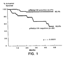
- the present invention rests on the discovery of a highly statistical inverse correlation between the expression of pRb2/p130 in tissues from endometrial cancer patients and the eventual clinical outcome following treatment. Decreased levels of pRb2/p130 are significantly associated with a poor survival. The study results reported herein indicate that the risk of dying of endometrial carcinoma is increased almost fivefold in patients whose tumors were pRb2/p130 negative, regardless of the tumor stage or grade of differentiation.
- Tissue with the greatest malignant potential expresses little or no pRb2/p130.
- a sample is contacted with an antibody specific for pRb2/p130 protein.
- the sample may typically comprise endometrial tissue, and may specifically comprise an endometrial tumor.
- the amount of antibody bound by the sample may be determined relative to the amount of antibody bound by a sample of normal endometrial tissue. The difference in the amount of antibody bound by the normal and test samples is indicative of the patient's prognosis.
- the endometrial carcinoma study described in Example I concurrently tested a known molecular prognostic indicator, i.e., DNA index, various classic clinical-pathologic parameters and pRb2/p130 expression.
- pRb2/p130 Decreased levels of pRb2/p130 were significantly associated with a poorer survival.
- the expression of pRb2/p130 thus represents an independent predictor of clinical outcome in endometrial carcinoma.
- Well known risk factors, such as F.I.G.O. stage and tumor ploidy were also confirmed as independent prognosticators, although of minor strength.
- tumor ploidy resisted as an independent prognostic variable by multivariate analysis.
- Stratification by pRb2/p130 status and ploidy allowed identification of patient subgroups with significant differences in survival (data not shown).
- grade of differentiation stratification by pRb2/p130 status revealed significant differences in survival within each grade group (data not shown).
- Expression of pRb2/p130 was not correlated with tumor stage; pRb2/p130 negative tumors were equally distributed among different tumor stages, thus indicating that this feature is typical of certain tumors, from their onset in early stages.
- the pRb2/p130 expression level may serve as a convenient molecular marker to replace or augment conventional prognostic techniques.
- An important advantage of the use of pRb2/p130 expression over classical surgical pathologic parameters as a prognostic factor is that the former can be determined at the time of the initial diagnosis, before any therapy is initiated. For patients not previously treated by radiotherapy or chemotherapy, low levels of pRb2/p130 can be used to identify tumors with a tendency to behave aggressively.
- prognosis can be evaluated more consistently than conventional prognostic factors which are based upon subjective evaluations of histological type, grade of differentiation, depth of myometrial invasion, degree of lymph nodal metastases, extra-uterine spread, and the other factors upon which endometrial carcinoma prognoses are presently based.
- a sample of lung tissue is removed from an individual by conventional biopsy techniques which are well-known to those skilled in the art.
- the sample is generally collected by needle biopsy. See, for example, Cancer: Principles & Practice of Oncology, V. T. DeVita, Jr. et al., eds. 3rd edition (1989), J. B. Lippincott Co., Philadelphia, PA, p. 616-619 , incorporated herein by reference (transcarinal needle biopsy and transthoracic percutaneous fine-needle aspiration biopsy).
- the sample is taken from the disease lesion.
- the disease lesion is first located by x-ray or other conventional lung lesion imaging techniques known to those skilled in the art.
- the tissue sample may be taken from any site in the lung. Tissue with the greatest malignant potential expresses little or no pRb2/p130.
- Normal lung tissue cells express a high level of pRb2/p130 protein.
- the pRb2/p130 expression level in the cells of the patient lung tumor tissue can be compared with the level in normal lung tissue of the same patient, or with the level in the lung tissue of a normal control group.
- Non-small cell lung cancer includes squamous cell carcinomas, adenocarcinomas, bronchiolo-alveolar carcinomas and large cell carcinomas.
- NSCLC Non-small cell lung cancer
- the pRb2/p130 expression level may serve as a convenient molecular marker to replace or augment conventional tumor grading.
- Accurate tumor grading is a necessary part of designing a course of treatment for the individual patient. Grading is reflective of the malignant potential of the tumor in question and thus the aggressiveness of the ensuing disease course.
- the generation of vital tumor grade information is made easier, by relying on pRb2/p130 as a molecular surrogate for more subjective observations concerning tumor histology.
- molecular-based grading can be performed more consistently than conventional pathological grading which is based upon subjective evaluations by expert pathologists, pRb2/p130 expression levels may also serve as a convenient molecular marker for the presence of active or latent NSCLC, or predisposition to NSCLC.
- Lung lesions may be identified as non-small cell lung carcinomas (NSCLCs) by showing a decrement in the expression of pRb2/p130 in the lesion compared to the level of pRb2/p130 in normal, non-cancerous control lung tissue.
- NSCLCs non-small cell lung carcinomas
- the level of pRb2/p130 expression in lung tissue of individuals with no apparent lung lesion but other symptoms of lung cancer, or in disease-free individuals indicates latent NSCLC or risk of NSCLC, respectively.
- Early diagnosis of NSCLC even before the appearance of visible lung lesions, will permit earlier initiation of treatment and increased survival.
- an at least about one-third decrement in pRb2/p130 expression level in an affected lung tissue sample indicates that the lesion is an NSCLC.
- a pRb2/p130 expression decrement of about one-third or greater in lung tissue of patients who are free of lung lesions but manifest other potential lung cancer symptoms such as sputum cytology irregularities, coughing or bronchitis is indicative of pre-lesion NSCLC.
- An about one-third or greater pRb2/p130 expression decrement in lung tissue of otherwise healthy individuals manifesting no symptoms of lung cancer is believed indicative of a risk of future NSCLC. Decrements in pRb2/p130 expression of about one-half or greater are even more indicative of NSCLC disease or NSCLC predisposition.
- test method may be used to identify individuals at risk of developing NSCLC from among populations exposed to environmental carcinogens, e.g. asbestos workers, miners, textile workers, tobacco smokers and the like, and from among families having a history of NSCLC or other forms of cancer.
- environmental carcinogens e.g. asbestos workers, miners, textile workers, tobacco smokers and the like
- a sample of affected tissue is removed from a cancer patient by conventional biopsy techniques which are well-known to those skilled in the art.
- the sample is preferably obtained from the patient prior to initiation of radiotherapy or chemotherapy.
- the sample is then prepared for a determination of pRb2/p130 expression level.
- Determining the relative level of expression of the pRb2/p130 gene in the tissue sample comprises determining the relative number of pRb2/p130 RNA transcripts, particularly mRNA transcripts in the sample tissue, or determining the relative level of the corresponding pRb2/p130 protein in the sample tissue.
- the relative level of pRb2/p130 protein in the sample tissue is determined by an immunoassay whereby an antibody which binds pRb2/p130 protein is contacted with the sample tissue.
- the relative pRb2/p130 expression level in cells of the sampled tumor is conveniently determined with respect to one or more standards.
- the standards may comprise, for example, a zero expression level on the one hand and the expression level of the gene in normal tissue of the same patient, or the expression level in the tissue of a normal control group on the other hand.
- the standard may also comprise the pRb2/p130 expression level in a standard cell line. The size of the decrement in pRb2/p130 expression in comparison to normal expression levels is indicative of the future clinical outcome following treatment.
- RNA molecules are then separated by gel electrophoresis on agarose gels according to standard techniques, and transferred to nitrocellulose filters by, e.g., the so-called "Northern" blotting technique.
- the RNA is immobilized on the filters by heating.
- RNA Detection and quantification of specific RNA is accomplished using appropriately labelled DNA or RNA probes complementary to the RNA in question. See Molecular Cloning: A Laboratory Manual, J. Sambrook et al., eds., 2nd edition, Cold Spring Harbor Laboratory Press, 1989, Chapter 7 , the disclosure of which is incorporated by reference.
- the mRNA assay test may be carried out according to the technique of in situ hybridization.
- the latter technique requires fewer tumor cells than the Northern blotting technique.
- cytological hybridization the in situ technique involves depositing whole cells onto a microscope cover slip and probing the nucleic acid content of the cell with a solution containing radioactive or otherwise labelled cDNA or cRNA probes. The practice of the in situ hybridization technique is described in more detail in U.S. Patent 5,427,916 , the entire disclosure of which is incorporated herein by reference.
- the nucleic acid probes for the above RNA hybridization methods can be designed based upon the published pRb2/p130 cDNA sequence of Li et al., Genes Dev. 7: 2366-2377 (1993 ), the entire disclosure of which is incorporated herein by reference.
- the nucleotide sequence is reproduced herein as SEQ ID NO: 1.
- the translation initiation codon comprises nucleotides 70-72 of SEQ ID NO:1.
- the translation termination codon comprises nucleotides 3487-3489.
- RNA hybridization can provide a quantitative result for the presence of the target RNA transcript in the RNA donor cells.
- Methods for preparation of labeled DNA and RNA probes, and the conditions for hybridization thereof to target nucleotide sequences, are described in Molecular Cloning, supra , Chapters 10 and 11, incorporated herein by reference.
- the nucleic acid probe may be labeled with, e.g., a radionuclide such as 32 P, 14 C, or 35 S; a heavy metal; or a ligand capable of functioning as a specific binding pair member for a labelled ligand, such as a labelled antibody, a fluorescent molecule, a chemolescent molecule, an enzyme or the like.
- a radionuclide such as 32 P, 14 C, or 35 S
- a heavy metal or a ligand capable of functioning as a specific binding pair member for a labelled ligand, such as a labelled antibody, a fluorescent molecule, a chemolescent molecule, an enzyme or the like.
- Probes may be labelled to high specific activity by either the nick translation method or Rigby et al., J. Mol. Biol. 113: 237-251 (1977 ) or by the random priming method, Fienberg et al., Anal. Biochem. 132: 6-13 (1983 ). The latter is the method of choice for synthesizing 32 P-Iabelled probes of high specific activity from single-stranded DNA or from RNA templates. Both methods are well-known to those skilled in the art and will not be repeated herein. By replacing preexisting nucleotides with highly radioactive nucleotides, it is possible to prepare 32 P-labelled DNA probes with a specific activity well in excess of 10 8 cpm/microgram according to the nick translation method. Autoradiographic detection of hybridization may then be performed by exposing filters on photographic film. Densitometric scanning of the filters provides an accurate measurement of mRNA transcripts.
- the random-primer method may be used to incorporate the dTTP analogue 5-(N-(N-biotinyl-epsilon-aminocaproyl)-3-aminoallyl)deoxyuridine triphosphate into the probe molecule.
- the thus biotinylated probe oligonucleotide can be detected by reaction with biotin binding proteins such as avidin, streptavidin, or anti-biotin antibodies coupled with fluorescent dyes or enzymes producing color reactions.
- the relative number of pRb2/p130 transcripts may also be determined by reverse transcription of mRNA followed by amplification in a polymerase chain reaction (RT-PCR), and comparison with a standard.
- RT-PCR polymerase chain reaction
- the methods for RT-PCR and variations thereon are well known to those of ordinary skill in the art.
- the level of pRb2/p130 expression in cells of the patient tissue is determined by assaying the amount of the corresponding pRb2/p130 protein.
- a cell sample is prepared, typically by dehydration and fixation, followed by reaction with labeled antibodies specific for the gene product coupled, where the labels are usually visually detectable, such as enzymatic labels, florescent labels, luminescent labels, and the like.
- tissue samples are obtained from patients and the samples are embedded then cut to e.g. 3-5 ⁇ m, fixed, mounted and dried according to conventional tissue mounting techniques.
- the fixing agent may advantageously comprise formalin.
- the embedding agent for mounting the specimen may comprise, e. g. , paraffin.
- the samples may be stored in this condition.
- the samples are contacted with an immunoreagent comprising an antibody specific for pRb2/p130.
- the antibody may comprise a polyclonal or monoclonal antibody.
- the antibody may comprise an intact antibody, or fragments thereof capable of specifically binding pRb2/p130 protein. Such fragments include, but are not limited to, Fab and F(ab') 2 fragments.
- the term "antibody” includes both polyclonal and monoclonal antibodies.
- the term “antibody” means not only intact antibody molecules, but also includes fragments thereof which retain antigen binding ability.
- Appropriate polyclonal antisera may be prepared by immunizing appropriate host animals with pRb2/p130 protein and collecting and purifying the antisera according to conventional techniques known to those skilled in the an.
- Monoclonal antibody may be prepared by following the classical technique of Kohler and Milstein, Nature 254:493497 (1975 ), as further elaborated in later works such as Monoclonal Antibodies, Hybridomas: A New Dimension in Biological Analysis, R. H. Kennet et al., eds., Plenum Press, New York and London (1980 ).
- pRb2/p130 for use as an immunogen for raising polyclonal or monoclonal antibodies may be conveniently prepared by recombinant DNA methods.
- pRb2/p130 is prepared in the form of a bacterially expressed glutathione S-transferase (GST) fusion protein.
- GST glutathione S-transferase
- Such fusion proteins may be prepared using commercially available expression systems, following standard expression protocols, e.g., " Expression and Purification of Glutathione-S-Transferase Fusion Proteins", Supplement 10, unit 16.7, in Current Protocols in Molecular Biology (1990 ). Also see Smith and Johnson, Gene 67: 34-40 (1988 ); Frangioni and Neel, Anal. Biochem.
- DNA encoding for pRb2/p130 is subcloned into a pGEX2T vector in the correct reading frame and introduced into E . coli cells.
- Transformants are selected on LB/ampicillin plates; the plates are incubated 12 to 15 hours at 37°C. Transformants are grown in isopropyl- ⁇ -D-thiogalactoside to induce expression of pRb2/p130-GST fusion protein.
- the cells are harvested from the liquid cultures by centrifugation.
- the bacterial pellet is resuspended and the cell pellet sonicated to lyse the cells.
- the lysate is then contacted with glutathione-agarose beads.
- the beads are collected by centrifugation and the fusion protein eluted.
- the GST carrier is then removed by treatment of the fusion protein with thrombin cleavage buffer.
- the released pRb2/p130 protein is recovered.
- antibody against pRb2/p130 can be raised by immunizing appropriate hosts with immunogenic fragments of the whole protein, particularly peptides corresponding to the carboxy terminus of the molecule.
- the antibody either directly or indirectly bears a detectable label.
- the detectable label may be attached to the primary anti-pRb2/p130 antibody directly. More conveniently, the detectable label is attached to a secondary antibody, e.g ., goat anti-rabbit IgG, which binds the primary antibody.
- the label may advantageously comprise, for example, a radionuclide in the case of a radioimmunoassay; a fluorescent moiety in the case of an immunofluorescent assay; a chemiluminescent moiety in the case of a chemiluminescent assay; or an enzyme which cleaves a chromogenic substrate, in the case of an enzyme-linked immunosorbent assay.
- the detectable label comprises an avidin-biotin-peroxidase complex (ABC) which has surplus biotin-binding capacity.
- the secondary antibody is biotinylated.
- the subsequent addition of ABC localizes peroxidase at the site of the specific antigen, since the ABC adheres non-specifically to biotin.
- Peroxidase (and hence antigen) is detected by incubating the section with e . g . H 2 O 2 and diaminobenzidine (which results in the antigenic site being stained brown) or H 2 O 2 and 4-chloro-1-naphthol (resulting in a blue stain).
- the ABC method can be used for paraffin-embedded sections, frozen sections, and smears. Endogenous (tissue or cell) peroxidase may he quenched e.g . with H 2 O 2 in methanol.
- the level of pRb2/p130 expression in tumor samples may be compared on a relative basis to the expression in normal tissue samples by comparing the stain intensities, or comparing the number of stained cells.
- ADL1 a polyclonal antibody raised against pRb2/p130, designated ADL1 was utilized.
- the specificity of the antibody has been confirmed by Western blot analysis, ( Pertile et al., Cell Growth & Diff 6:1659-64 (1995 ); Claudio et al., Cancer Res 56:2003-8 (1996 )), as well as by immunoprecipitation of the antibody with the in vitro translated forms of the cDNAs coding for pRb2/p130 and for the other retinoblastoma related proteins, pRb/p105 and p107.
- the ADL1 antibody was able to immunoprecipitate only the in vitro translated form of the pRb2/p130 protein ( Baldi et al., Clin Cancer Res 2:1239-45 (1996 ).
- the genomic structure of the human pRb2/p130 gene is described herein.
- the pRb2/p130 genomic DNA has been cloned and sequenced.
- the pRb2/p130 gene has been mapped to the long arm of chromosome 16, an area previously reported to show loss of heterozygosity (LOH) for human neoplasias.
- LHO heterozygosity
- the putative promoter for pRb2/p130 has been identified, cloned and sequenced.
- the complete intron-exon organization of the gene has been elucidated.
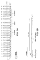
- the pRb2/p130 gene contains 22 exons and 21 introns, spanning over 50 kb of genomic DNA.
- the length of the individual exons ranges from 65 bp to 1517 bp, while the length of individual introns ranges from 82 bp to 9837 bp.
- the organization of these exons and introns are shown in Figure 3A .
- the location and size of each exon and intron of pRb2/p130, as well as the nucleotide sequences at the exon-intron junctions are shown below in Table 7. (SEQ ID NOS:6-47).
- the exon sequences are shown in upper case letters, while the intron sequences are in lower case letters.
- the superscript numbers correspond to the nucleotide positions of the exon-intron boundaries on SEQ ID NO:1.
- Exon 22 With the exception of exon 22, the largest of all the exons (1517 bp in length), the exons found were relatively small, with the shortest, exons 4 and 7, comprising only 65 nucleotides each.
- Exons 10 through 20 code for the region of the pRb2/p130 protein which form the "pocket region”. Exons 10 through 13 and 17 through 20 translate to Domain A and Domain B, respectively. Exons 14, 15, and 16 code for the region of the pRb2/p130 protein, known as the "spacer." The spacer lies between Domains A and B.
- the introns have been completely sequenced.
- the shortest intron, intron 16, lying between exons 16 and 17, is only 82 bp in length, whereas the largest intron, intron 21, spans 9837 bp.
- Intron 21 is located between exons 21 and 22.
- the complete sequences for the introns are given as SEQ ID NOS: 48-68.
- All of the intron sequences of pRb2/p130 conform to the GT-AG rule found to be characteristic of other human genes. Breathnach, R. et al., Annu. Rev. Biochem. 50:349-383 (1981 ). This rule identifies the generic sequence of an intron as GT Vietnamese AG. Introns having this generic form are characterized as conforming to the GT - AG rule.
- Point mutations in splice signal dinucleotides have been associated with aberrant splicing in other genes in vivo and in vitro. See generally , Genes V, B. Lewin. Oxford University Press, pp. 913-916, New York (1994 ) and Yandell et al ., supra at p. 1694. Thus, it is important to identify any mutations to the splice signal dinucleotides or other sequences that are excluded from the RNA transcript during splicing.
- the pRb2/pl30 genomic structure and intron sequences described herein may be used to delineate mutations and rearrangements associated with tumor formation.
- the genomic structure and intron sequences herein may also be used to screen for naturally occurring polymorphisms at the nucleotide level.
- Knowledge of a specific single polymorphism can be used to eliminate a mutation in pRb2/p130 as a causative factor in a tumor if the purported mutation displays the same pattern as the polymorphism.
- Knowledge of polymorphisms in pRb2/p130 can be used to determine the genetic linkage of an identical mutation, and in turn, the tracing of parental origin and family histories without the need for time for time intensive sequencing if mutation is of germline origin.
- polymorphisms can then be utilized for the development of diagnostic approaches for human neoplasias.
- a primer extension analysis was performed to locate the transcription initiation site.
- the protocol for the prime-extension analysis is given in the examples that follow.
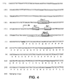
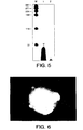
- a twenty four nucleotide segment (SEQ ID NO:114) containing the antisense-strand sequence 26 to 50 nucleotides upstream from the putative ATG codon See Fig. 4 ) was end-labeled and used as a primer for an extension reaction on cyctoplasmatic RNA from HeLa cells.
- a major extended fragment of 78 bp was detected (lane 1) from the primer extension done with HeLa cells as the template. The additional bands detected by the primer extension analysis could represent additional initiation sites.
- the putative transcription factor-binding sites were identified by their similarity to consensus sequences for known transcription factor-binding sites by use of the SIGNAL SCAN program. A description of this program is included in the examples that follow.
- the most recognizable sequence motifs are for the transcription factors Sp1 (two sites), Ker1 and MyoD.
- Fig. 4 shows the location of these motifs. Ker1 is involved in keratinocyte-specific transcription, while MyoD is involved in myogenesis. Leask et al., Genes Dev. 4: 1985-1998 (1990 ); Weintraub, H., Cell 75: 1241-1244 (1993 ).
- the presence in the promoter region for pRb2/p130 of these sequence motifs supports a hypothesis of an involvement of this gene in the complex pathways regulating differentiation of specific cell systems.
- the present invention provides a method for amplifying the genomic DNA of pRb2/p130 and for screening polymorphisms and mutations therein.
- the assay methods described herein can be used to diagnose and characterize certain cancers or to identify a heterozygous carrier state. While examples of methods for amplifying and detecting mutations in pRb2/p130 are given, the invention is not limited to the specific methods exemplified. Other means of amplification and identification that rely on the use of the genomic DNA sequence for pRb2/p130 and/or the use of the primers described herein are also contemplated by this invention.
- the methods described herein involve preparing a nucleic acid sample for screening and then assaying the sample for mutations in one or more alleles.
- the nuclei acid sample is obtained from cells.
- Cellular sources of genomic DNA include cultured cell lines, or isolated cells or cell types obtained from tissue (or whole organs or entire organisms).
- the cell source is peripheral blood lymphocytes.
- the patient sample to be screened is in the form of double-stranded genomic DNA. It is first denatured using methods known to those skilled in the art. Denaturation can be carried out either by melting or subjecting the strands to agents that destabilize the hydrogen bonds, such as alkaline solutions and concentrated solutions of formamide or urea.
- the pRb2/p130 genomic DNA sample is amplified by use of the polymerase chain reaction (PCR), using a primer pair, a buffer mixture, and an enzyme capable of promoting chain elongation.
- PCR polymerase chain reaction
- Methods of conducting PCR are well known to those skilled in the art. See, for example, Beutler et al ., U.S. Patent No. 5,234,811 , or Templeton, N.S., Diag. Mol. Path. 1(1):58-72 (1992 ), which are incorporated herein by reference as if set forth at length.
- the amplification product produced from PCR can then be used to screen for mutations using the techniques known as Single Strand Conformational Polymorphism (SSCP) or Primed In-Situ DNA synthesis (PRINS).
- SSCP Single Strand Conformational Polymorphism
- PRINS Primed In-Situ DNA synthesis
- mutations can also be identified through the more laborious task of sequencing the gene isolates of a patient and comparing the sequence to that for the corresponding wild type pRb2/p130 segment.
- PCR is carried out by thermocycling, i . e ., repeated cycles of heating and cooling the PCR reaction mixture, within a temperature range whose lower end is 37°C to 55°C and upper end is around 90°C to 100°C.
- the specific temperature range chosen is dependent upon the enzyme chosen and the specificity or stringency required. Lower end temperatures are typically used for annealing in amplifications in which high specificity is not required and conversely, higher end temperatures are used where greater stringency is necessary. An example of the latter is when the goal is to amplify one specific target DNA from genomic DNA. A higher annealing temperature will produce fewer DNA segments that are not of the desired sequence.
- the annealing temperature is between 50°C and 65°C. Most preferably, the annealing temperature is 55°C.
- the PCR is generally performed in a buffered aqueous solution, i . e ., a PCR buffer, preferably at a pH of 7-9, most preferably about 8.
- a molar excess of the primar is mixed with the buffer containing the template strand.
- the PCR buffer also contains the deoxynucleotide triphosphates (DATP, dCTP, dGTP, and dTTP) and a polymerase.
- DATP, dCTP, dGTP, and dTTP deoxynucleotide triphosphates
- Polymerases suitable for use in PCR include, but are not limited to, E. coli DNA polymerase I, the Klenow fragment of E. coli DNA polymerase I, T4 DNA polymerase, T7 DNA polymerase, Taq DNA polymerase ( Thermus aquaticus DNA polymerase I), and other heat-stable enzymes which will facilitate the formation of amplification products.
- the primers used herein can be naturally occurring oligonucleotides purified from a nucleic acid restriction digest or produced synthetically using any suitable method, which methods are known to those skilled in the art.
- the primers used herein can be synthesized using automated methods.
- the oligonucleotide primer for any given exon must be designed such that it includes a portion of the complementary sequence for the promoter region, for the 3'-noncoding region, or for the introns flanking the exon to be amplified, provided however that the primer sequence should not include the sequence for the splice signal dinucleotides.
- the complementary sequence for the splice signal dinucleotides is amplified. Including the complementary sequences to the splice signal dinucleotides could result in an amplification product that "plasters over" the splice junction and masks any potential mutation that could occur therein.
- the introns flanking the exon are not limited to the introns immediately adjacent to the exon to be amplified.
- the oligonucleotide primer can be designed such that it includes a portion of the complementary sequence for the introns upstream or downstream from the exon to exon to be amplified. In the latter instance, the amplification product produced would include more than one exon. Preferably at least 20 to 25 nucleotides of the sequence for each flanking intron are included in the primer sequence.
- the primers used herein are selected to be substantially complementary to each strand of the pRb2/p130 segment to be amplified. There must be sufficient base-pair matching to enable formation of a hybrid duplex under hybridization conditions. It is not required, however, that the base-pair matchings be exact. Therefore, the primer sequence may or may not reflect the exact sequence of the pRb2/p130 segment to be amplified. Non-complementary bases or longer sequences can be interspersed into the primer, provided the primer sequence retains sufficient complementarity with the segment to be amplified and thereby form an amplification product.
- the primers must be sufficiently long to prime the synthesis of amplification products in the presence of a polymerizing agent.
- the exact length of the primer to be used is dependent on many factors including, but not limited to, temperature and the source of the primer.
- the primer is comprised of 15 to 30 nucleotides, more preferably 18 to 27 nucleotides, and most preferably 24 to 25 nucleotides. Shorter primers generally require cooler annealing temperatures with which to form a stable hybrid complex with the template.
- Primer pairs are usually the same length, however, the length of some primers was altered to obtain primer pairs with identical annealing temperatures. Primers of less than 15 bp are generally considered to generate non-specific amplification products.
- SSCP is used to analyze polymorphisms and mutations in the exons of pRb2/p130.
- SSCP has the advantages over direct sequencing in that it is simple, fast, and efficient.
- the analysis is performed according to the method of Orita et al., Genomics 5:874-879 (1989 ), the entire disclosure of which is incorporated herein by reference.
- the target sequence is amplified and labeled simultaneously by the use of PCR with radioactively labeled primers or deoxynucleotides. Neither in situ hybridization nor the use of restriction enzymes is necessary for SSCP.
- SSCP detects sequence changes, including single-base substitutions (point mutations), as shifts in the electrophoretic mobility of a molecule within a gel matrix.
- a single nucleotide difference between two similar sequences is sufficient to alter the folded structure of one relative to the other. This conformational change is detected by the appearance of a band shift in the tumor DNA, when compared with the banding pattern for a corresponding wild type DNA segment.
- Single base pair mutations can be detected following SSCP analysis of PCR products up to about 400 bp. PCR products larger than this size must first be digested with a restriction enzyme to produce smaller fragments.
- sequence mutations in pRb2/p130 can be detected utilizing the PRINS technique.
- the PRINS method represents a versatile technique, which combines the accuracy of molecular and cytogenetic techniques, to provide a physical localization of the genes in nuclei and chromosomes. See Cinti et al., Nuc. Acids Res. Vol 21, No. 24: 5799-5800 (1993 ), the entire disclosure of which is incorporated herein by reference.
- the PRINS technique is based on the sequence specific annealing of unlabeled oligodeoxynucleotides in situ .
- the oligodeoxynucleotides operate as a primer for in situ chain elongation catalyzed by Taq I polymerase.
- Labeled nucleotides labeled with a substance such as biotin or Digoxigenin, act as substrate for chain elongation.
- the labeled DNA chain is visualized by exposure to a fluorochrome-conjugated antibody specific for the label substance.
- the label is Digoxigenin and the fluorochrome conjugated antibody is anti-Digoxigenin-FITC. This results in the incorporation of a number of labeled nucleotides far greater than the number of nucleotides in the primer itself. Additionally, the specificity of the hybridization is not vulnerable to the problems that arise when labeled nucleotides are placed in the primer. The bound label will only be found in those places where the primer is annealed and elongated.
- SSCP SSCP
- PRINS PRINS
- Protocols for the use of the SSCP analysis and the PRINS technique are included in the examples that follow.
- the PRINS method of detecting mutations in the pRb2/p 130 gene may be practiced in kit form.
- a carrier is compartmentalized to receive one or more containers, such as vials or test tubes, in close confinement.
- a first container may contain one or more subcontainers, segments or divisions to hold a DNA sample for drying, dehydrating or denaturing.
- a second container may contain the PRINS reaction mixture, which mixture is comprised of a PCR buffer, a DIG DNA labeling mixture, a polymerase such as Taq I DNA polymerase, and the primers designed in accordance with this invention (see Example 7, Table 8).
- the DIG DNA labeling mixture is comprised of a mixture of labeled and unlabeled deoxynucleotides.
- the labeled nucleotides are labeled with either biotin or Digoxigenin. More preferably, the label is Digoxigenin.
- a third container may contain a fluorochrome conjugated antibody specific to the label.
- the fluorochrome conjugated antibody specific for Digoxigenin is anti-Digoxigenin-FITC. Suitable conjugated fluorochromes for biotin include avidin-FITC or avidin Texas Red.
- the fourth container may contain a staining compound, preferably Propidium Iodide (PI).
- the kit may further contain appropriate washing and dilution solutions.
- the stage was evaluated by microscopic analysis of the surgical specimen according to the 1988 International Federation of Gynecology and Obstetrics (FIGO) classification ( Gynecol Oncol 35: 125 ( 1988 ).
- Table 1 summarizes the clinical and pathological characteristics of the study group.
- Surgical treatment included total hysterectomy in 95 cases and extended hysterectomy in five cases. Bilateral salpingo-oophorectomy was always associated. Pelvic and paraaortic lymphadenectomy were performed at the surgeon's discretion, but not systematically. Overall. 43 patients underwent lymphadenectomy. The omentum was removed when appropriate (four cases). Table 1. Clinical And Pathological Features Of 100 Patients In Which pRb2/p130 Expression Was Tested.
- a tumor specimen was taken fresh from a site regarded to be representative of the lesion immediately after hysterectomy. Each tumor sample was later divided into two parts: one for flow cytometry and the other for histological analysis.
- a suspension of tumor cells was obtained by mincing the sample with a lancet and scissors in phosphate-buffered saline.
- the cell suspension was filtered by a 50 micrometer mesh of polyacrylamide, fixed in 70 percent ethanol, and stored at -4°C until assayed.
- Prior to DNA analysis the ethanol was removed by centriguation (1500 revolutions/min for ten minutes); the pellet was then resuspended and washed twice in phosphate-buffered saline.
- the RNA was removed by digestion with ribonuclease (Serva, 0.1 mg/ml in phosphate-buffered saline) for 30 minutes at 37°C.
- the DNA ploidy was given by the DNA index, defined as a proportion of the modal DNA values of the tumor G 0 and G 1 cells (peak channel) to the DNA content of the diploid standard.
- the histograms were based on measurement of more than 10,000 cells and resulted, in general, in a good resolution with a coefficient of variation of three to six percent. Calculation of DNA index was done by processing each histogram in the computer-assisted program Multicycle Autofit, version 2.00 (Phoenix Flow Systems, San Diego, CA).
- Rabbit polyclonal immune serum designated ADL1
- ADL1 Rabbit polyclonal immune serum, designated ADL1
- Rabbits were immunized with a conjugate comprising the peptide Glu-Asn-His-Ser-Ala-Leu-Leu-Arg-Arg-Leu-Gln-Asp-Val-Ala-Asn-Asp-Arg-Gly-Ser-His-Cys (SEQ ID NO:3) coupled to keyhole limpet hemocyanin (KLH).
- the peptide corresponds to the carboxy terminus of the pRb2/p130 protein.
- rabbits were immunized with the SEQ ID NO:3-KLH conjugate by subcutaneous injection once every two weeks until a total of three injections were given.
- the initial injection comprised 1 mg SEQ ID NO:3-KLH conjugate in 500 ⁇ l PBS, plus 500 ⁇ l of complete Freund's adjuvant.
- the second and third injections comprised 500 ⁇ g of the conjugate in 500 ⁇ l PBS, plus 500 ⁇ l of complete Freund's adjuvant.
- the rabbits were bled after the third injection. Subsequent boosts, with the same composition as the second and third injections, were given once a month.
- Sections of each tumor specimen were cut to 5-micrometer, mounted on glass and dried overnight at 37°C. All sections were then deparaffinized in xylene, rehydrated through graded alcohol series and washed in phosphate-buffered saline. This buffer was used for all subsequent washes and for the dilution of the antibodies. Sections were quenched in 0.5 percent hydrogen peroxide and blocked with diluted ten percent normal goat anti-rabbit serum. Slides were then incubated for one hour at room temperature with the ADL1 immune serum at a dilution of 1:1000, then incubated with diluted goat anti-rabbit biotinylated antibody (Vector, Burlingame, Calif.) for 30 minutes at room temperature.
- This pattern of immunoreactivity could be referred to microstructural alterations caused by the fixing and embedding procedures, or might reflect differences in the levels of expression and in the localization of this antigen during the various phases of the cell cycle, as has already been shown at the molecular level ( Claudio et al., Cancer Res 56: 2003-8 (1996 ).
- the patients' disease-free and actuarial survival durations were compared after dividing them into two groups using different cutoff points of percent pRb2/p130 positivity.
- a brown stain indicated the presence of pRb2/p130 in tumor cells.
- the specimens were characterized as having no detectable staining, staining in only a few positive cells (about ten percent), staining in more than 40 percent of the cells, or intense staining in the majority of cells. Tumors with immunostaining in more than 40 percent of cells were considered to be positive for pRb2/p130.
- the DNA index values showed a diploid type in 73 cases and an aneuploid type in 27 cases.
- the DNA index of the aneuploid tumors was hypodiploid in one case, hypertetraploid in four cases; the remaining 22 cases had a modal DNA content in the diploid to tetraploid range (1 ⁇ DNA index ⁇ 2).
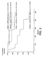
- Table 3 shows the results of Cox proportional-hazards regression analysis in which the response to pRb2/p130 immunostaining, tumor ploidy, FIGO stage and grade of differentiation were tested simultaneously to estimate the rate ratios for the occurrence of death from disease in patients with endometrial cancer.
- Negative immunostaining for pRb2/p130 resulted as the strongest independent predictor of poor outcome. Patents with pRb2/p130 negative tumors had a significantly higher rate ratio for dying due to disease (4.91) than patients with pRb2/p130 positive tumors.
- Figure 2 presents Kaplan Meier survival estimates according to these stratified risk groups. The following is the comparison between the groups by the log-rank test:
- epithelial carcinoma specimens were obtained from the Department of Pathology at Pennsylvania Hospital. The specimens included Grade 1, Grade 2, and Grade 3 tumors.
- Immunohistochemical staining was performed using an automated immunostainer (Ventana ES, Ventana Medical Systems, Arlington. AZ) and a Peroxidase-DAB immunodetection kit (Ventana Medical Systems). Five micron sections were cut from each tumor specimen. The sections were mounted on slides and air-dried. The sections were deparaffinized in xylene and hydrated through a graded alcohol series into water. A polyclonal anti-RB2 primary antibody was applied at a dilution of 1:500 for 30 minutes at 37°C. The slides were then incubated with a biotinylated goat anti-rabbit antibody for 30 minutes. The slides were then incubated with a horseradish peroxidase conjugated-avidin.
- DAB diaminobenzidine
- Rabbit polyclonal immune serum was prepared against p107 (ADL2) by immunizing rabbits with a bacterially expressed GST-p107 fusion protein. Expression of the fusion protein was performed according to the procedure reported by Smith and Johnson, Gene 67:31-40 (1988 ) and Frangioni and Neel, Anal. Biochem. 210:179-187 (1993 ). Rabbits were immunized with the fusion protein by subcutaneous injection once every two weeks until a total of three injections were given. The initial injection (primary immunization) comprised 500 ⁇ g protein in 500 ⁇ l PBS, plus 500 ⁇ l of incomplete Freund's adjuvant.
- the second and third injections comprised 100 ⁇ g of the protein in 500 ⁇ l PBS, plus 500 ⁇ l of incomplete Freund's adjuvant.
- the rabbits were bled after the third injection. Subsequent boosts, with the same composition as the second and third injections, were given once a month.
- Lung tissue specimens from 51 patients with surgically resected lung cancer were obtained from patients who had not received chemo- or radiotherapy prior to surgical resection.
- the samples consisted of 39 squamous cell carcinomas and 12 adenocarcinomas. Histological diagnosis and grading were performed by a skilled lung pathologist. Samples were graded on the scale of 1-2-3 with "3" representing the most malignant disease and "1" representing the least malignant disease.
- Normal lung tissue samples containing the stratified columnar epithelia of trachea, bronchi and adjacent glands were obtained either from biopsy or autopsy performed within 10 hours of the patient's death.
- Sections from each lung tissue specimen were cut at 3-5 ⁇ m, mounted on glass and dried overnight at 37°C. All sections were then deparaffinized in xylene, rehydrated through a graded alcohol series and washed in phosphate-buffered saline (PBS). The same buffer was used for all subsequent washes and for dilution of antibodies.
- PBS phosphate-buffered saline
- Tissue sections for pRb2/p130 and p107 detection were sequentially quenched in 0.5% hydrogen peroxide and blocked with diluted 10% normal goat anti-rabbit serum (Vector Laboratories).
- the slides were incubated for 1 hour at room temperature with the rabbit polyclonal immune serum (ADL1) raised against pRb2/p 130 at a dilution of 1:2000, or the ADL2 antibody against p107 at a dilution of 1:500.
- the slides were then incubated with diluted goat anti-rabbit biotinylated antibody (Vector Laboratories) for 30 minutes at room temperature.
- Sections for pRb/p105 detection were heated twice in a microwave oven for 5 min each at 700 W in citrate buffer (pH6), were quenched sequentially in 0.5% hydrogen peroxide, and were blocked with diluted 10% normal horse anti-mouse serum (Vector Laboratories. Inc.)
- the monoclonal mouse anti-human pRb/p105 antibody XZ77 (at a dilution of 1:500) was added and incubated for 120 min. at room temperature. After being washed in PBS, the slides were incubated with diluted horse anti-mouse biotinylated antibody (Vector Laboratories, Inc.) for 30 min. at room temperature.
- the normal lung tissue samples comprising the stratified epithelia of the trachea, bronchi and adjacent glands were strongly stained, indicating a high expression level.
- the histological diagnoses and classifications of the tumors were based on the WHO criteria, and the postsurgical pathologic TNM stage was determined using the guidelines of the American Joint Committee on Cancer.
- the routine histopathological evaluation of the 158 tumor specimens analyzed was performed independently of the pRb2/p130 immunostaining. Thirty two tumors were adenocarcinomas. 118 were squamous carcinomas. 4 were carcinoids and 4 were small cell lung cancers. Eighty seven tumors (55.1 %) were classified as stage I. 43 tumors (27.1%) were classified as stage II and 28 tumors (17.7%) were classified as stage IIIa. The adenocarcinomas and squamous carcinomas were classified by grade, as shown in Table 6.
- Sections of each specimen were cut at 3-5 ⁇ m, mounted on glass and dried overnight at 37°C. All the sections were then deparaffinized in xylene, rehydrated through a graded alcohol series and washed in PBS. This buffer was used for all subsequent washes and for the dilution of the antibodies. Sections were heated twice in a microwave oven for five minutes each at 700 W in citrate buffer (pH 6), sequentially quenched in 0.5% hydrogen peroxide and blocked with diluted 10% normal goat anti-rabbit serum.
- pRb2/p130 immunostaining was mostly nuclear, but some specimens clearly exhibited cytoplasmatic staining with a low to absent background.
- Immunohistochemical staining patterns of the tumors can be summarized as follows: 50 specimens (31.6%) showed low to undetectable levels of pRb2/p130 (score 1), 73 specimens (46.2%) exhibited medium pRb2/p130 expression levels, while high levels of expression were detected in 35 specimens (22.2%). The small number of small cell lung cancers and carcinoids included in this study did not allow statistical analysis in these histological groups. All the SCLCs specimens exhibited low to undetectable pRb2/p130 expression levels, while a high level of expression of this protein was recognized in all carcinoids.
- a human P1 genomic library (Genome System Inc., St. Louis, MO) was screened by using two primers made from the published cDNA sequence, Li et al., Genes Dev. 7:2366-2377 (1993 ).
- the sequences for the primers used to isolate the genomic clones are GTATACCATTTAGCAGCTGTCCGCC (SEQ ID NO: 116) and the complement to the sequence GTGTGCCATTTATGTGATGGCAAAG (SEQ ID NO: 115).
- the cDNA probe labeled with [ ⁇ - 32 P], corresponded to the first 430 bp after the start codon of the published cDNA sequence, Li et al., supra .
- ⁇ SCR3 Fig. 3B
- a set of oligonucleotide primers were used to sequence the genomic DNA clones.
- the primers were synthesized based upon the cDNA nucleotide sequence of pRb2/p130 such that they annealed to the genomic DNA at roughly 150 bp intervals.
- the exon/intron boundaries were identified from those positions in which the genomic DNA sequence differed from that of the published cDNA sequence.
- Sequencing of the recombinant clones was carried out in part by automated DNA sequencing using the dideoxy terminator reaction chemistry for sequence analysis on the Applied Biosystem Model 373A DNA sequencer and, in part, by using a dsDNA Cycle Sequencing System kit purchased from GIBCO BRL, Gaithersburg. MD, according to the instructions of the manufacturer.
- oligonucleotide primers used herein were synthesized using Applied Biosystems DNA-RNA synthesizer Model 394, using beta-cyanoethyl phosphoramidite chemistry.
- the human pRb2/p130 gene consists of 22 exons and 21 introns and spans more than 50 kb of genomic DNA. The organization of these exons and introns are shown approximately to scale in Figure 3A .
- the location and size of each exon and intron of pRb2/p130, as well as the nucleotide sequences at the exon-intron boundaries are shown in Table 7 (SEQ ID NOS:6-47).
- the exons range in size from 65 to 1517 bp in length.
- the introns which range in size from 82-9837 bp in length, have been completely sequenced.
- the nucleotide sequences are given as SEQ ID NOS:48-68.
- the human HeLa (cervix epithelioid carcinoma) cell line was obtained from the American Type Culture Collection and maintained in culture in Dulbecco's modified Eagle medium (DHEM) with 10% fetal calf serum (FCS) at 37°C in a 10% CO 2 -containing atmosphere. Cytoplasmatic RNA was extracted utilizing the RNAzol B method (CINNA/BIOTECX, Friendswood, TX). TABLE 7 Exon-Intron Boundaries of the Human pRb2/p130 Gene Exon No. (bp) 5' Donor sequence 3' Acceptor sequence Intron No.
- the primer for this analysis was an oligonucleotide, 5'ACCTCAGGTGAGGTGAGGGCCCGG 3' (SEQ ID NO:114), complementary to the pRb2/p130 genomic DNA sequence starting at position -22 ( See Fig. 4 , SEQ ID NO:4).
- the primer was end labeled with [ ⁇ 32 P]ATP and hybridized overnight with 20 ⁇ g of HeLa cytoplasmatic RNA at 42°C.
- RNA was converted into cDNA by avian myeloblastosis virus reverse transcriptase in the presence of 2 mM deoxynucleotides at 42°C for 45 minutes.
- the cDNA product was then analyzed on 7% sequencing gel containing 8 M urea. The position of the transcription start site was mapped from the length of the resulting extension product.
- SIGNAL SCAN VERSION 4.0 is a computer program that was developed by Advanced Biosciences Computing Center at the University of Minnesota, St. Paul, MN. This program aids molecular biologists in finding potential transcription factor binding sites and other elements in a DNA sequence. A complete description of the program can be found in Prestridge, D.S., CABIOS 7: 203-206 (1991 ), the entire disclosure of which is incorporated herein as if set forth at length.
- SIGNAL SCAN finds sequence homologies between published signal sequences and an unknown sequence.
- a signal as defined herein, is any short DNA sequence that may have known significance. Most of the known signals represent transcriptional elements. The program does not interpret the significance of the identified homologies; interpretation of the significance of sequences identified is left up to the user. The significance of the signal elements varies with the signal length, with matches to short segments having a higher probability of random occurrence.
- Figure 5 shows the results of the primer extension analysis done to locate the transcription initiation site for pRb2/p130.
- a major extended fragment of 78 bp was detected (lane 1) from the primer extension done with HeLa Cells as the template.
- the probable position of the identified transcription start site is indicated by the arrow in Fig. 4 .
- Putative transcription factor-binding sites were identified by their similarity to consensus sequences for known transcription factor-binding sites.
- the sequence motifs corresponding to Sp1, Ker1, and MyoD are also indicated in Fig. 4 .
- the genomic DNA used herein was obtained from human peripheral blood lymphocytes.
- the samples were prepared by the methods of Sambrook et al., Molecular Cloning:A Laboratory Manual. Second Edition, pp. 9.16-9.23, Cold Spring Harbor Laboratory Press, Cold Spring Harbor, NY (1989 ).
- the sample DNA was amplified in a Perkin-Elmer Cetus thermocycler.
- the PCR was performed in a 100 ⁇ l reaction volume using 2.5 units of recombinant Taq DNA-polymerase and 40 ng of genomic DNA.
- the reaction mixture was prepared according to the recommendations given in the Gene Amp DNA Amplification kit (Perkin-Elmer Cetus).
- the reaction mixture consisted of 50 mM/l KCl, 10mM/l Tris-HCl (pH 8.3), 1.5 mM MgCl. 200 ⁇ M each deoxynucleotide triphosphate and 1 ⁇ M of each primer.
- PCR cycles Thirty five (35) PCR cycles were carried out, with each cycle consisting of an initial denaturation step at 95°C for one minute, one minute at the annealing temperature (55°C), an extension step at 72°C for one minute, and followed by a final incubation period at 72°C for seven minutes.
- Suitable annealing temperatures are shown in Table 8 for each of the primers designed in accordance with this invention. Minor adjustments in the annealing temperatures may be made to accommodate other primers designed in accordance with this invention.
- the size of the amplification products produced by PCR are shown in Table 8 above.
- the lengths of the PCR products ranged from 196 bp to 413 bp.
- Sequencing of the amplification products of pRb2/p130 can be conducted according to the method set forth in Example 5C above. Sequencing can also be performed by the chain termination technique described by Sanger et al., Proc. Nat'l. Acad. Sci. , U. S. A. 74:5463-5467 (1977 ) or Sambrook et al., Molecular Cloning: A Laboratory Manual, Second Edition, pp. 13.42-13.77, Cold Spring Harbor Laboratory Press, Cold Spring Harbor, NY (1989 ) with appropriate primers based on the pRb2/p130 genomic sequence described herein.
- the SSCP analysis was performed according to the methods of Orita et al., Genomics 5: 874-879 (1989 ) and Hogg et al., Oncogene 7: 1445-1451 (1992 ), each of which is incorporated herein by reference.
- amplification of the individual exons was, in some experiments, performed as described in Example 7 with the exception that 1 ⁇ Ci of [ 32 P]dCTP (3000 Ci mmol -1 ) was added to the mixture in order to obtain a labeled product.
- a 10% aliquot of the PCR-amplified product was diluted with a mixture of 10-20 ⁇ l of 0.1 % SDS and 10 mM EDTA.
- Polymorphisms and mutations were detected by observing a shift in the electrophoretic mobility pattern of the denatured PCR-amplified product relative to a corresponding wild type sample or normal tissue sample from the same patient. Once a band shift was identified, the segment was sequenced to confirm the exact nature of the polymorphism or mutation.
- DNA was extracted from the CCRF-CEM line (human lymphoblastoid cells), and amplified.
- 50 ⁇ l of the PCR reaction mix containing 4 ng of genomic DNA, 0.2 mM of each deoxynucleotide triphosphates. 2 U of Taq polymerase and 0.4 ⁇ M of each primer were used. Fifty-Five cycles of denaturation (95°C, 1 minute), annealing (55°C, 1 minute) and extension (72°C, 1 minute) were carried out in a thermal cycler.
- the SSCP analysis was performed using an MDE mutation detection kit (AT Biochem). The PCR products were heated to 95°C for two minutes and placed directly on ice for several minutes.
- sequences of the PCR products were determined by automated DNA sequencing, using dideoxy-terminator reaction chemistry. Two point mutations were identified: A CC to G CC at position 2950 of SEQ ID NO: 1, resulting in a threonine to alanine substitution; and C C T to C G T at position 3029 of SEQ ID NO: 1, resulting in a proline to arginine substitution.
- NPC primary tumor human nasopharyngeal carcinoma: point mutations in exon 21, point mutation and insertions in exon 22
- 5 NPC primary tumor human nasopharyngeal carcinoma: point mutations and insertion in exon 22.
- the PRINS technique was performed according to the method of Cinti et al., Nuc. Acids Res. Vol. 21, No. 24: 5799-5800 (1993 ) using human peripheral lymphocytes as the source of genomic DNA.
- the oligonucleotide primers were designed such that they included portions of the introns flanking exon 20.
- the sequences of the primers utilized to amplify exon 20 are listed in Table 8 above (SEQ ID NOS:89 and 90).
- the samples were then washed two times in 2 X SSC (pH 7.0) and in 4 X SSC (pH 7.0) for 5 minutes at room temperature.
- the DNA samples were then placed in a solution of 4 X SSC and 0.5% Bovine Serum Albumin (BSA) (pH 7.0), incubated at room temperature for 45 minutes with anti-Digoxigenin-FITC (Boehringer-Mannheim), and diluted 1:100 in 4 X SSC and 0.5% BSA (pH 7.0). After washing the samples in 4 X SSC and 0.05% Triton X-100, the samples were counterstained with 1 ⁇ g/ml Propidium Iodide (PI).
- BSA Bovine Serum Albumin
- the slides were examined under a Confocal Laser Scanning Microscope (CLSM Sarastro, Molecular Dynamics).
- CLSM Sarastro Confocal Laser Scanning Microscope
- the FITC and PI signals were detected simultaneously, independently elaborated and the final projections were superimposed with a Silicon Graphic Computer Personal IRIS-4D/20 workstation.
- Figure 6 shows the results of a PRINS reaction on normal human interphase nuclei.
- the bright spots correspond to a DNA segment containing exon 20 of pRb2/p130.
- This individual is homozygous for the presence of exon 20 of pRb2/p130.
- Had there been a mutation in exon 20 of this individual either one or both of these areas would have been diminished in intensity or not visible in its entirety.
- the patient's pRb2/p130 DNA segment would he sequenced by methods known to those skilled in the art and compared to a wild type sample of pRb2/p130 DNA.
- kits are another embodiment of the present invention.
Landscapes
- Chemical & Material Sciences (AREA)
- Health & Medical Sciences (AREA)
- Life Sciences & Earth Sciences (AREA)
- Organic Chemistry (AREA)
- Proteomics, Peptides & Aminoacids (AREA)
- Genetics & Genomics (AREA)
- Zoology (AREA)
- Engineering & Computer Science (AREA)
- General Health & Medical Sciences (AREA)
- Biophysics (AREA)
- Analytical Chemistry (AREA)
- Molecular Biology (AREA)
- Biochemistry (AREA)
- Pathology (AREA)
- Wood Science & Technology (AREA)
- Immunology (AREA)
- Hospice & Palliative Care (AREA)
- Toxicology (AREA)
- Medicinal Chemistry (AREA)
- Physics & Mathematics (AREA)
- Gastroenterology & Hepatology (AREA)
- Biotechnology (AREA)
- Microbiology (AREA)
- Oncology (AREA)
- Bioinformatics & Cheminformatics (AREA)
- General Engineering & Computer Science (AREA)
- Measuring Or Testing Involving Enzymes Or Micro-Organisms (AREA)
- Polysaccharides And Polysaccharide Derivatives (AREA)
- Medicines Containing Antibodies Or Antigens For Use As Internal Diagnostic Agents (AREA)
- Investigating Or Analysing Biological Materials (AREA)
- Medical Treatment And Welfare Office Work (AREA)
Applications Claiming Priority (5)
| Application Number | Priority Date | Filing Date | Title |
|---|---|---|---|
| US1494396P | 1996-04-05 | 1996-04-05 | |
| US1937296P | 1996-06-05 | 1996-06-05 | |
| US2019696P | 1996-06-21 | 1996-06-21 | |
| US3953297P | 1997-03-03 | 1997-03-03 | |
| EP97920115A EP0906448B1 (de) | 1996-04-05 | 1997-04-03 | Methoden zur diagnose und prognose von krebs |
Related Parent Applications (1)
| Application Number | Title | Priority Date | Filing Date |
|---|---|---|---|
| EP97920115A Division EP0906448B1 (de) | 1996-04-05 | 1997-04-03 | Methoden zur diagnose und prognose von krebs |
Publications (3)
| Publication Number | Publication Date |
|---|---|
| EP2014772A2 true EP2014772A2 (de) | 2009-01-14 |
| EP2014772A8 EP2014772A8 (de) | 2009-04-29 |
| EP2014772A3 EP2014772A3 (de) | 2009-05-27 |
Family
ID=27486431
Family Applications (2)
| Application Number | Title | Priority Date | Filing Date |
|---|---|---|---|
| EP08009585A Withdrawn EP2014772A3 (de) | 1996-04-05 | 1997-04-03 | Verfahren zur Diagnose und Prognose von Krebs |
| EP97920115A Expired - Lifetime EP0906448B1 (de) | 1996-04-05 | 1997-04-03 | Methoden zur diagnose und prognose von krebs |
Family Applications After (1)
| Application Number | Title | Priority Date | Filing Date |
|---|---|---|---|
| EP97920115A Expired - Lifetime EP0906448B1 (de) | 1996-04-05 | 1997-04-03 | Methoden zur diagnose und prognose von krebs |
Country Status (7)
| Country | Link |
|---|---|
| EP (2) | EP2014772A3 (de) |
| JP (2) | JP2000510330A (de) |
| AT (1) | ATE423852T1 (de) |
| AU (1) | AU2438997A (de) |
| CA (1) | CA2250854C (de) |
| DE (1) | DE69739276D1 (de) |
| WO (1) | WO1997038125A1 (de) |
Families Citing this family (7)
| Publication number | Priority date | Publication date | Assignee | Title |
|---|---|---|---|---|
| DE19882435B4 (de) * | 1997-06-02 | 2007-09-20 | Thomas Jefferson University | Verfahren zur Hemmung von Krebszellwachstum mit einem pRb2/p130 exprimierenden Vektor |
| JP2002523760A (ja) | 1998-09-02 | 2002-07-30 | ダイアデクスアス・インコーポレーテッド | 様々な癌を診断、監視、段階づけ、造影及び治療する新規な方法 |
| US6261778B1 (en) * | 1998-10-22 | 2001-07-17 | Ludwig Institute For Cancer Research | Isolated nucleic acid molecules which encode SCP proteins, and uses thereof |
| AU2001275437A1 (en) * | 2000-06-09 | 2001-12-17 | Idec Pharmaceuticals Corporation | Gene targets and ligands that bind thereto for treatment and diagnosis of ovarian carcinomas |
| JP2006503574A (ja) * | 2002-10-22 | 2006-02-02 | カン−フィテ・バイオファーマ・リミテッド | 疾患の状態に関するマーカーとしてのa3ar |
| RU2007139516A (ru) * | 2005-03-25 | 2009-04-27 | Дженентек, Инк. (Us) | Мутации с-мет при раке легких |
| RU2404193C2 (ru) | 2005-03-25 | 2010-11-20 | Дженентек, Инк. | Способ лечения опухоли у субъекта |
Citations (2)
| Publication number | Priority date | Publication date | Assignee | Title |
|---|---|---|---|---|
| US5234811A (en) | 1991-09-27 | 1993-08-10 | The Scripps Research Institute | Assay for a new gaucher disease mutation |
| US5427916A (en) | 1988-05-09 | 1995-06-27 | Temple University Of The Commonwealth System Of Higher Education | Method for predicting the effectiveness of antineoplastic therapy in individual patients |
Family Cites Families (7)
| Publication number | Priority date | Publication date | Assignee | Title |
|---|---|---|---|---|
| DE69019410T2 (de) * | 1989-03-31 | 1995-10-05 | Canji Inc | Retinoblastoma-Genprodukt-Antikörper und deren Verwendungen. |
| EP0631584A4 (de) * | 1991-02-28 | 1998-07-01 | Dana Farber Cancer Inst Inc | Krebs diagnose und therapie. |
| AU2178292A (en) * | 1991-06-03 | 1993-01-08 | Triton Diagnostics, Inc. | Epidermal growth factor receptor ectodomain |
| CA2096497A1 (en) * | 1992-05-26 | 1993-11-27 | Patricia Anne Spears | Mycobacteria probes |
| WO1994011531A1 (en) * | 1992-11-13 | 1994-05-26 | California Institute Of Biological Research | Identification of neoplasms by detection of genetic insertions and deletions |
| WO1995002328A1 (en) * | 1993-07-15 | 1995-01-26 | Indiana University Foundation | Mammalian low molecular weight phospholipase a2 nucleotide and amino acid sequences |
| IL108879A (en) * | 1994-03-07 | 2000-08-31 | Yissum Res Dev Co | Diagnostic assay for malignancies using the H19 gene and kit |
-
1997
- 1997-04-03 AU AU24389/97A patent/AU2438997A/en not_active Abandoned
- 1997-04-03 CA CA2250854A patent/CA2250854C/en not_active Expired - Lifetime
- 1997-04-03 WO PCT/US1997/005598 patent/WO1997038125A1/en not_active Ceased
- 1997-04-03 EP EP08009585A patent/EP2014772A3/de not_active Withdrawn
- 1997-04-03 JP JP09536340A patent/JP2000510330A/ja active Pending
- 1997-04-03 EP EP97920115A patent/EP0906448B1/de not_active Expired - Lifetime
- 1997-04-03 DE DE69739276T patent/DE69739276D1/de not_active Expired - Lifetime
- 1997-04-03 AT AT97920115T patent/ATE423852T1/de not_active IP Right Cessation
-
2008
- 2008-07-23 JP JP2008190243A patent/JP2008283985A/ja active Pending
Patent Citations (2)
| Publication number | Priority date | Publication date | Assignee | Title |
|---|---|---|---|---|
| US5427916A (en) | 1988-05-09 | 1995-06-27 | Temple University Of The Commonwealth System Of Higher Education | Method for predicting the effectiveness of antineoplastic therapy in individual patients |
| US5234811A (en) | 1991-09-27 | 1993-08-10 | The Scripps Research Institute | Assay for a new gaucher disease mutation |
Non-Patent Citations (95)
| Title |
|---|
| "Cancer: Principles & Practice of Oncology", 1989, J. B. LIPPINCOTT CO., pages: 616 - 619 |
| "Molecular Cloning" |
| "Molecular Cloning: A Laboratory Manual", 1989, COLD SPRING HARBOR LABORATORY PRESS |
| "Monoclonal Antibodies, Hybridomas: A New Dimension in Biological Analysis", 1980, PLENUM PRESS |
| B. LEWIN: "Genes V", 1994, OXFORD UNIVERSITY PRESS, pages: 913 - 916 |
| BALDI ET AL., CLIN CANCER RES, vol. 2, 1996, pages 1239 - 45 |
| BEIJERSBERGEN ET AL., GENES DEV., vol. 9, 1993, pages 1340 - 1353 |
| BIGGS ET AL., NATURE GENETICS, vol. 11, December 1995 (1995-12-01), pages 441 - 443 |
| BLIN ET AL., NUC. ACIDS RES., vol. 3, 1976, pages 2303 - 2308 |
| BOOKSTEIN ET AL., SCIENCE, vol. 247, 1990, pages 712 - 715 |
| BRALY, GVNECOL ONCOL, vol. 58, 1995, pages 145 - 7 |
| BREATHNACH, R. ET AL., ANNU. REV. BIOCHEM., vol. 50, 1981, pages 349 - 383 |
| CAO ET AL., NATURE, vol. 355, 1992, pages 176 - 179 |
| CHAMBERS ET AL., GYNECOL ONCOL, vol. 27, 1987, pages 180 - 8 |
| CHELLAPAN ET AL., CELL, vol. 65, 1991, pages 1053 - 1061 |
| CHEN, CELL, vol. 58, 1989, pages 1193 - 1198 |
| CINTI ET AL., NUC. ACIDS RES., vol. 21, no. 24, 1993, pages 5799 - 5800 |
| CLAUDIO ET AL., CANCER RES, vol. 56, 1996, pages 2003 - 8 |
| CLAUDIO ET AL., CANCER RES., vol. 54, 1994, pages 5556 - 5560 |
| CLAUDIO ET AL., CANCER RES., vol. 56, 1996, pages 2003 - 2008 |
| COBRINIK ET AL., GENES DEV., vol. 7, 1993, pages 2392 - 2404 |
| COX, J R STAT SOC, vol. 34, 1972, pages 187 - 220 |
| CURRENT PROTOCOLS IN MOLECULAR BIOLOGY, 1990 |
| DE CAPRIO ET AL., PROC., NAIL. ACAD. SCI. USA, vol. 89, 1992, pages 1795 - 1798 |
| DISAIA ET AL., AM J OBSTET GVNECOL, vol. 151, 1985, pages 1009 - 15 |
| DISAIA ET AL.: "Adenocarcinoma Of The Uterus", CLINICAL GVNECOLOGIC ONCOLOGY, 1993, pages 156 - 93 |
| EWEN ET AL., CELL, vol. 66, 1993, pages 1155 - 1164 |
| EWEN ET AL., SCIENCE, vol. 255, 1992, pages 85 - 87 |
| FAHA ET AL., SCIENCE, vol. 255, 1992, pages 87 - 90 |
| FIENBERG ET AL., ANAL. BIOCHEM., vol. 132, 1983, pages 6 - 13 |
| FIENBERG: "The Analysis Of Cross-Classified Categorical Data", MIT PRESS |
| FRANGIONI; NEEL, ANAL. BIOCHEM., vol. 210, 1993, pages 179 - 187 |
| GOODRICH ET AL., CELL, vol. 67, 1991, pages 293 - 302 |
| GYNECOI ONCOL, vol. 35, 1988, pages 125 |
| HANNON ET AL., GENES DEV., vol. 7, 1993, pages 2378 - 2391 |
| HARLOW; LANE: "Antibodies: A Laboratory Manual", 1988, COLD SPRING LABORATORY PRESS |
| HIEBERT ET AL., GENES DEVELOP., vol. 6, 1992, pages 177 - 185 |
| HIJMANS ET AL., MOL. CELL. BIOL., vol. 15, 1995, pages 3082 - 3089 |
| HINDS ET AL., CELL, vol. 70, 1992, pages 993 - 1006 |
| HOGG ET AL., ONCOGENE, vol. 7, 1992, pages 1445 - 1451 |
| HU ET AL., EMBO J., vol. 9, 1990, pages 1147 - 1153 |
| HU ET AL., MOL. CELL. BIOL., vol. 11, 1991, pages 5792 - 5799 |
| HU ET AL., MOL. CELL. BIOL., vol. 12, 1992, pages 971 - 980 |
| HUANG ET AL., MOL. CELL. BIOL., vol. 10, 1990, pages 3761 - 3769 |
| HUANG ET AL., SCIENCE, vol. 242, 1988, pages 1563 - 1565 |
| KAPLAN ET AL., AM STAT ASSOC, vol. 53, 1958, pages 457 - 81 |
| KNUDSON, PROC. NATL. ACAD. SCI. USA, vol. 68, 1971, pages 820 - 823 |
| KOHLER; MILSTEIN, NATURE, vol. 254, 1975, pages 493 - 497 |
| LAN THANGUE, N.B., TRENDS BIOCHEM. SCI., vol. 19, 1994, pages 108 - 114 |
| LASKO ET AL., ANNU. REV. GENETICS, vol. 25, 1991, pages 281 - 296 |
| LEASK ET AL., GENES DEV., vol. 4, 1990, pages 1985 - 1998 |
| LEES ET AL., EMBO J., vol. 10, 1991, pages 4279 - 4290 |
| LI ET AL., GENES DEV., vol. 7, 1993, pages 2366 - 2377 |
| LI, Y. ET AL., GENES, vol. 7, 1993, pages 2366 - 2377 |
| MACLACHLAN ET AL., EUKARYOTIC GENE EXP., vol. 5, 1995, pages 127 - 156 |
| MACMAHON, GYNECOL ONCOL, vol. 2, 1974, pages 122 |
| MASSI ET AL., AM J OBSTET GVNECOL, vol. 174, 1996, pages 1320 - 6 |
| MATSUSHIME ET AL., NATURE, vol. 35, pages 295 - 300 |
| MAYOL ET AL., ONCOGENE, vol. 8, 1993, pages 1561 - 2566 |
| MILLER: "Survival Analysis.", 1981, JOHN WILEY, pages: 44 - 102 |
| MILNER ET AL., CANCER RESEARCH, vol. 53, 1993, pages 2128 - 2132 |
| MORAN, CURR. OPIN. GENET. DEV., vol. 3, 1993, pages 63 - 70 |
| NEVINS, SCIENCE, vol. 258, 1992, pages 424 - 429 |
| ORITA ET AL., GENOMICS, vol. 5, 1989, pages 874 - 879 |
| ORITA, GENOMICS, vol. 5, 1989, pages 874 - 879 |
| PERTILE ET AL., CELL GROWTH & DIFF, vol. 6, 1995, pages 1659 - 64 |
| PETERS ET AL., AM J OBSTET GYNECOL, vol. 146, 1983, pages 285 - 90 |
| PETTERSSON: "Annual Report On The Results Of Treatment In Gynecological Cancer", RADIUMHEMMET, STOCKHOLM, vol. 22, pages 65 - 82 |
| PITKIN, OBSTET GVNECOL, vol. 49, 1977, pages 567 - 9 |
| PRESTRIDGE, D.S., CABIOS, vol. 7, 1991, pages 203 - 206 |
| RAYCHAUDHURI ET AL., GENES DEVELOP., vol. 5, 1991, pages 1200 - 1207 |
| RIGBY ET AL., J. MOL. BIOL., vol. 113, 1977, pages 237 - 251 |
| SAMBROOK ET AL.: "Molecular Cloning.A Laboratory Manual", 1989, COLD SPRING HARBOR LABORATORY PRESS, pages: 9.16 - 9.23 |
| SAMBROOK ET AL.: "Molecular Cloning: A Laboratory Manual", 1989, COLD SPRING HARBOR LABORATORY PRESS, pages: 13.42 - 13.77 |
| SAMBROOK ET AL.: "Molecular Cloning:A Laboratory Manual", 1989, COLD SPRING HARBOR LABORATORY PRESS, pages: 12.30 - 12.38 |
| SAMBROOK ET AL.: "Molecular Cloning:A Laboratory Manual", 1989, COLD SPRING HARBOR LABORATORY PRESS, pages: 9.16 - 9.23 |
| SANG ET AL., MOL. CELL. DIFFER., vol. 3, 1995, pages 1 - 29 |
| SANGER ET AL., PROC. NAT'L. ACAD. SCI., U.S.A., vol. 74, 1977, pages 5463 - 5467 |
| SHIM ET AL., J NATL CANCER INST, vol. 88, 1996, pages 519 - 29 |
| SHIRODKAR ET AL., CELL, vol. 66, 1992, pages 157 - 166 |
| SILVESTRINI ET AL., J CLIN ONCOL, vol. 14, 1996, pages 1604 - 10 |
| SMITH; JOHNSON, GENE, vol. 67, 1988, pages 31 - 40 |
| SMITH; JOHNSON, GENE, vol. 67, 1988, pages 34 - 40 |
| SUMEGI ET AL., CELL GROWTH DIFFER., vol. 1, 1990, pages 247 - 250 |
| SUSINI ET AL., AM J OBSTET GYNECOL, vol. 170, 1994, pages 527 - 34 |
| TEMPLETON, N.S., DIAG. MOL. PATH., vol. 1, no. 1, 1992, pages 58 - 72 |
| VAIRO ET AL., GENES DEV., vol. 9, 1995, pages 869 - 881 |
| WAGNER ET AL., NATURE, vol. 352, 1991, pages 189 - 190 |
| WEINTRAUB, H., CELL, vol. 75, 1993, pages 1241 - 1244 |
| WINGO ET AL., AM J OBSTET GVNECOL, vol. 152, 1985, pages 803 - 8 |
| YANDELL ET AL., N.E.J. MEDICINE, vol. 321, 1989, pages 1689 - 1695 |
| YEUNG ET AL., ONCOGENE, vol. 8, 1993, pages 3465 - 3468 |
| ZELTERMAN ET AL.: "NEJM Books", 1992, article "Contingency Tables In Medical Studies", pages: 293 - 310 |
| ZHU ET AL., EMBO J., vol. 14, 1995, pages 1904 - 1913 |
| ZHU ET AL., GENES DEV., vol. 7, 1993, pages 1111 - 1125 |
Also Published As
| Publication number | Publication date |
|---|---|
| EP0906448B1 (de) | 2009-02-25 |
| EP0906448A1 (de) | 1999-04-07 |
| AU2438997A (en) | 1997-10-29 |
| ATE423852T1 (de) | 2009-03-15 |
| CA2250854A1 (en) | 1997-10-16 |
| EP0906448A4 (de) | 2004-12-01 |
| EP2014772A3 (de) | 2009-05-27 |
| WO1997038125A1 (en) | 1997-10-16 |
| DE69739276D1 (de) | 2009-04-09 |
| JP2000510330A (ja) | 2000-08-15 |
| CA2250854C (en) | 2011-06-21 |
| EP2014772A8 (de) | 2009-04-29 |
| JP2008283985A (ja) | 2008-11-27 |
Similar Documents
| Publication | Publication Date | Title |
|---|---|---|
| US5840506A (en) | Methods for the diagnosis and prognosis of cancer | |
| Wen et al. | p53 mutations and expression in ovarian cancers: correlation with overall survival | |
| US6268184B1 (en) | Amplifications of chromosomal region 20q13 as a prognostic indicator breast cancer | |
| EP2848700B1 (de) | Marker für endometriosetumor | |
| EP0960197B1 (de) | GENE DES 20q13 AMPLIKONS UND DEREN VERWENDUNGEN | |
| US6475732B1 (en) | Genetic alterations associated with cancer | |
| JP2008524986A (ja) | タキサンに基づく薬物療法に対する悪性腫瘍の応答予測に有用な遺伝子変化 | |
| JP2008283985A (ja) | 癌の診断および予後のための方法 | |
| Lassus et al. | Allelic analysis of serous ovarian carcinoma reveals two putative tumor suppressor loci at 18q22-q23 distal to SMAD4, SMAD2, and DCC | |
| US7811986B2 (en) | Genes from the 20Q13 amplicon and their uses | |
| US8993251B2 (en) | Genes from 20q13 amplicon and their uses | |
| Colomer et al. | Lack of p53 nuclear immunostaining is not indicative of absence of TP53 gene mutations in colorectal adenocarcinomas | |
| US5892010A (en) | Genes from the 20Q13 amplicon and their uses | |
| Edwards et al. | Immunohistochemical expression of BRCA2 protein and allelic loss at the BRCA2 locus in prostate cancer | |
| Høgdall et al. | P53 mutations in tissue from Danish ovarian cancer patients: from the Danish “MALOVA” ovarian cancer study | |
| US6255049B1 (en) | Detection of metastatic cancer cells using PCTA-1 | |
| HK1128731A (en) | Method for the diagnosis and prognosis of cancer | |
| US20040234961A1 (en) | Telomere length determination and applications | |
| Kim et al. | Genetic alterations in microdissected prostate cancer cells by comparative genomic hybridization | |
| JP2007510424A (ja) | 分子マーカー | |
| AU4808601A (en) | Genes from 20q13 amplicon and their uses | |
| JP2007503826A (ja) | 乳癌のリスクの診断 | |
| ODAJIMA et al. | Cancers Detected by Single Strand Conformation Polymorphism Analysis: Comparison with the Protein Accumulation | |
| Bartlett | akusher-lib. ru | |
| Lassus | Genetic and molecular changes in serous gynecological carcinomas:-Comparison with other histological types, and clinical associations |
Legal Events
| Date | Code | Title | Description |
|---|---|---|---|
| PUAI | Public reference made under article 153(3) epc to a published international application that has entered the european phase |
Free format text: ORIGINAL CODE: 0009012 |
|
| AC | Divisional application: reference to earlier application |
Ref document number: 0906448 Country of ref document: EP Kind code of ref document: P |
|
| AK | Designated contracting states |
Kind code of ref document: A2 Designated state(s): AT BE CH DE DK ES FI FR GB GR IE IT LI LU MC NL PT SE |
|
| PUAL | Search report despatched |
Free format text: ORIGINAL CODE: 0009013 |
|
| AK | Designated contracting states |
Kind code of ref document: A3 Designated state(s): AT BE CH DE DK ES FI FR GB GR IE IT LI LU MC NL PT SE |
|
| RIC1 | Information provided on ipc code assigned before grant |
Ipc: C07H 21/02 20060101ALI20090417BHEP Ipc: C07K 1/00 20060101ALI20090417BHEP Ipc: C07K 17/00 20060101ALI20090417BHEP Ipc: G01N 33/574 20060101ALI20090417BHEP Ipc: C12Q 1/00 20060101AFI20081125BHEP Ipc: C12Q 1/68 20060101ALI20090417BHEP Ipc: C07K 14/47 20060101ALI20090417BHEP Ipc: C07K 14/00 20060101ALI20090417BHEP Ipc: C07H 21/04 20060101ALI20090417BHEP Ipc: G01N 33/53 20060101ALI20090417BHEP |
|
| REG | Reference to a national code |
Ref country code: HK Ref legal event code: DE Ref document number: 1128731 Country of ref document: HK |
|
| 17P | Request for examination filed |
Effective date: 20091001 |
|
| 17Q | First examination report despatched |
Effective date: 20091029 |
|
| AKX | Designation fees paid |
Designated state(s): AT BE CH DE DK ES FI FR GB GR IE IT LI LU MC NL PT SE |
|
| STAA | Information on the status of an ep patent application or granted ep patent |
Free format text: STATUS: THE APPLICATION IS DEEMED TO BE WITHDRAWN |
|
| 18D | Application deemed to be withdrawn |
Effective date: 20100309 |
|
| REG | Reference to a national code |
Ref country code: HK Ref legal event code: WD Ref document number: 1128731 Country of ref document: HK |