EP2010226B1 - Transcobalaminrezeptor-Polypeptide, -Nukleinsäuren und -Modulatoren sowie entsprechende Verfahren zur Verwendung für die Modulation von Zellwachstum und die Behandlung von Krebs und Cobalaminmangel - Google Patents
Transcobalaminrezeptor-Polypeptide, -Nukleinsäuren und -Modulatoren sowie entsprechende Verfahren zur Verwendung für die Modulation von Zellwachstum und die Behandlung von Krebs und Cobalaminmangel Download PDFInfo
- Publication number
- EP2010226B1 EP2010226B1 EP07755075.4A EP07755075A EP2010226B1 EP 2010226 B1 EP2010226 B1 EP 2010226B1 EP 07755075 A EP07755075 A EP 07755075A EP 2010226 B1 EP2010226 B1 EP 2010226B1
- Authority
- EP
- European Patent Office
- Prior art keywords
- tcbir
- transcobalamin
- cell
- fragment
- cbl
- Prior art date
- Legal status (The legal status is an assumption and is not a legal conclusion. Google has not performed a legal analysis and makes no representation as to the accuracy of the status listed.)
- Not-in-force
Links
- 102100025240 CD320 antigen Human genes 0.000 title claims description 578
- 108010083125 transcobalamin receptor Proteins 0.000 title claims description 577
- 238000000034 method Methods 0.000 title claims description 199
- 108090000765 processed proteins & peptides Proteins 0.000 title claims description 187
- 102000004196 processed proteins & peptides Human genes 0.000 title claims description 176
- 229920001184 polypeptide Polymers 0.000 title claims description 167
- 206010028980 Neoplasm Diseases 0.000 title claims description 45
- 150000007523 nucleic acids Chemical class 0.000 title description 26
- 102000039446 nucleic acids Human genes 0.000 title description 25
- 108020004707 nucleic acids Proteins 0.000 title description 25
- 201000011510 cancer Diseases 0.000 title description 17
- 230000010261 cell growth Effects 0.000 title description 16
- 208000002670 vitamin B12 deficiency Diseases 0.000 title description 3
- 102000011409 Transcobalamins Human genes 0.000 claims description 196
- 108010023603 Transcobalamins Proteins 0.000 claims description 196
- 239000012634 fragment Substances 0.000 claims description 151
- 102000040430 polynucleotide Human genes 0.000 claims description 117
- 108091033319 polynucleotide Proteins 0.000 claims description 117
- 239000002157 polynucleotide Substances 0.000 claims description 117
- 241000282414 Homo sapiens Species 0.000 claims description 110
- FDJOLVPMNUYSCM-WZHZPDAFSA-L cobalt(3+);[(2r,3s,4r,5s)-5-(5,6-dimethylbenzimidazol-1-yl)-4-hydroxy-2-(hydroxymethyl)oxolan-3-yl] [(2r)-1-[3-[(1r,2r,3r,4z,7s,9z,12s,13s,14z,17s,18s,19r)-2,13,18-tris(2-amino-2-oxoethyl)-7,12,17-tris(3-amino-3-oxopropyl)-3,5,8,8,13,15,18,19-octamethyl-2 Chemical compound [Co+3].N#[C-].N([C@@H]([C@]1(C)[N-]\C([C@H]([C@@]1(CC(N)=O)C)CCC(N)=O)=C(\C)/C1=N/C([C@H]([C@@]1(CC(N)=O)C)CCC(N)=O)=C\C1=N\C([C@H](C1(C)C)CCC(N)=O)=C/1C)[C@@H]2CC(N)=O)=C\1[C@]2(C)CCC(=O)NC[C@@H](C)OP([O-])(=O)O[C@H]1[C@@H](O)[C@@H](N2C3=CC(C)=C(C)C=C3N=C2)O[C@@H]1CO FDJOLVPMNUYSCM-WZHZPDAFSA-L 0.000 claims description 61
- 239000003112 inhibitor Substances 0.000 claims description 50
- 150000001413 amino acids Chemical class 0.000 claims description 43
- 125000003275 alpha amino acid group Chemical group 0.000 claims description 30
- 239000003814 drug Substances 0.000 claims description 28
- 230000003247 decreasing effect Effects 0.000 claims description 23
- 230000001965 increasing effect Effects 0.000 claims description 23
- 208000037265 diseases, disorders, signs and symptoms Diseases 0.000 claims description 22
- 201000010099 disease Diseases 0.000 claims description 16
- 230000002401 inhibitory effect Effects 0.000 claims description 14
- 238000000338 in vitro Methods 0.000 claims description 13
- 230000009261 transgenic effect Effects 0.000 claims description 8
- 208000012902 Nervous system disease Diseases 0.000 claims description 7
- 230000003053 immunization Effects 0.000 claims description 7
- 206010061218 Inflammation Diseases 0.000 claims description 6
- 230000004054 inflammatory process Effects 0.000 claims description 6
- 208000025966 Neurological disease Diseases 0.000 claims description 4
- 230000002708 enhancing effect Effects 0.000 claims description 4
- 210000004027 cell Anatomy 0.000 description 295
- 108090000623 proteins and genes Proteins 0.000 description 166
- 230000027455 binding Effects 0.000 description 149
- 230000014509 gene expression Effects 0.000 description 100
- 102000004169 proteins and genes Human genes 0.000 description 93
- 235000018102 proteins Nutrition 0.000 description 89
- 230000000694 effects Effects 0.000 description 72
- 102000005962 receptors Human genes 0.000 description 71
- 108020003175 receptors Proteins 0.000 description 71
- 108091032973 (ribonucleotides)n+m Proteins 0.000 description 61
- 239000000203 mixture Substances 0.000 description 58
- 108020004459 Small interfering RNA Proteins 0.000 description 45
- 239000004055 small Interfering RNA Substances 0.000 description 43
- 230000007812 deficiency Effects 0.000 description 40
- 108020004999 messenger RNA Proteins 0.000 description 40
- 241001465754 Metazoa Species 0.000 description 38
- 239000011159 matrix material Substances 0.000 description 38
- 239000000427 antigen Substances 0.000 description 37
- 108091007433 antigens Proteins 0.000 description 36
- 102000036639 antigens Human genes 0.000 description 36
- 235000001014 amino acid Nutrition 0.000 description 33
- 239000013598 vector Substances 0.000 description 33
- 108091034117 Oligonucleotide Proteins 0.000 description 32
- 239000003795 chemical substances by application Substances 0.000 description 30
- 108020004414 DNA Proteins 0.000 description 29
- 125000003729 nucleotide group Chemical group 0.000 description 29
- 239000002773 nucleotide Substances 0.000 description 28
- 102000018120 Recombinases Human genes 0.000 description 27
- 108010091086 Recombinases Proteins 0.000 description 27
- 150000001875 compounds Chemical class 0.000 description 27
- 108091023037 Aptamer Proteins 0.000 description 25
- 210000001519 tissue Anatomy 0.000 description 24
- 230000000295 complement effect Effects 0.000 description 23
- 101000934335 Homo sapiens CD320 antigen Proteins 0.000 description 22
- 238000004458 analytical method Methods 0.000 description 22
- -1 e.g. Substances 0.000 description 21
- 230000000692 anti-sense effect Effects 0.000 description 20
- 230000000875 corresponding effect Effects 0.000 description 20
- 241000699666 Mus <mouse, genus> Species 0.000 description 19
- 230000004700 cellular uptake Effects 0.000 description 19
- 239000002299 complementary DNA Substances 0.000 description 19
- 230000003834 intracellular effect Effects 0.000 description 19
- 238000003786 synthesis reaction Methods 0.000 description 19
- 102000040650 (ribonucleotides)n+m Human genes 0.000 description 18
- 230000009368 gene silencing by RNA Effects 0.000 description 18
- 238000012228 RNA interference-mediated gene silencing Methods 0.000 description 17
- 239000002585 base Substances 0.000 description 17
- 230000015572 biosynthetic process Effects 0.000 description 17
- 230000002163 immunogen Effects 0.000 description 17
- 230000004048 modification Effects 0.000 description 17
- 238000012986 modification Methods 0.000 description 17
- 238000000746 purification Methods 0.000 description 17
- 230000001105 regulatory effect Effects 0.000 description 17
- 239000000523 sample Substances 0.000 description 17
- 238000003556 assay Methods 0.000 description 16
- 230000006870 function Effects 0.000 description 16
- 239000000411 inducer Substances 0.000 description 16
- 238000012216 screening Methods 0.000 description 16
- 230000008685 targeting Effects 0.000 description 16
- 239000003446 ligand Substances 0.000 description 15
- 238000013518 transcription Methods 0.000 description 15
- 230000035897 transcription Effects 0.000 description 15
- 210000004881 tumor cell Anatomy 0.000 description 15
- 108091027967 Small hairpin RNA Proteins 0.000 description 14
- 230000000903 blocking effect Effects 0.000 description 14
- 230000001413 cellular effect Effects 0.000 description 14
- 238000012512 characterization method Methods 0.000 description 14
- 229940079593 drug Drugs 0.000 description 14
- 230000008569 process Effects 0.000 description 14
- 108091092562 ribozyme Proteins 0.000 description 14
- 235000000346 sugar Nutrition 0.000 description 14
- 108090000994 Catalytic RNA Proteins 0.000 description 13
- 102000053642 Catalytic RNA Human genes 0.000 description 13
- 108010047041 Complementarity Determining Regions Proteins 0.000 description 13
- 230000037361 pathway Effects 0.000 description 13
- 238000002415 sodium dodecyl sulfate polyacrylamide gel electrophoresis Methods 0.000 description 13
- 241000699670 Mus sp. Species 0.000 description 12
- 238000013459 approach Methods 0.000 description 12
- 230000022131 cell cycle Effects 0.000 description 12
- 125000005647 linker group Chemical group 0.000 description 12
- 230000001404 mediated effect Effects 0.000 description 12
- 230000002829 reductive effect Effects 0.000 description 12
- 210000002966 serum Anatomy 0.000 description 12
- 238000011282 treatment Methods 0.000 description 12
- 108060003951 Immunoglobulin Proteins 0.000 description 11
- FFEARJCKVFRZRR-BYPYZUCNSA-N L-methionine Chemical compound CSCC[C@H](N)C(O)=O FFEARJCKVFRZRR-BYPYZUCNSA-N 0.000 description 11
- 206010025323 Lymphomas Diseases 0.000 description 11
- 241001529936 Murinae Species 0.000 description 11
- FAPWRFPIFSIZLT-UHFFFAOYSA-M Sodium chloride Chemical compound [Na+].[Cl-] FAPWRFPIFSIZLT-UHFFFAOYSA-M 0.000 description 11
- JLCPHMBAVCMARE-UHFFFAOYSA-N [3-[[3-[[3-[[3-[[3-[[3-[[3-[[3-[[3-[[3-[[3-[[5-(2-amino-6-oxo-1H-purin-9-yl)-3-[[3-[[3-[[3-[[3-[[3-[[5-(2-amino-6-oxo-1H-purin-9-yl)-3-[[5-(2-amino-6-oxo-1H-purin-9-yl)-3-hydroxyoxolan-2-yl]methoxy-hydroxyphosphoryl]oxyoxolan-2-yl]methoxy-hydroxyphosphoryl]oxy-5-(5-methyl-2,4-dioxopyrimidin-1-yl)oxolan-2-yl]methoxy-hydroxyphosphoryl]oxy-5-(6-aminopurin-9-yl)oxolan-2-yl]methoxy-hydroxyphosphoryl]oxy-5-(6-aminopurin-9-yl)oxolan-2-yl]methoxy-hydroxyphosphoryl]oxy-5-(6-aminopurin-9-yl)oxolan-2-yl]methoxy-hydroxyphosphoryl]oxy-5-(6-aminopurin-9-yl)oxolan-2-yl]methoxy-hydroxyphosphoryl]oxyoxolan-2-yl]methoxy-hydroxyphosphoryl]oxy-5-(5-methyl-2,4-dioxopyrimidin-1-yl)oxolan-2-yl]methoxy-hydroxyphosphoryl]oxy-5-(4-amino-2-oxopyrimidin-1-yl)oxolan-2-yl]methoxy-hydroxyphosphoryl]oxy-5-(5-methyl-2,4-dioxopyrimidin-1-yl)oxolan-2-yl]methoxy-hydroxyphosphoryl]oxy-5-(5-methyl-2,4-dioxopyrimidin-1-yl)oxolan-2-yl]methoxy-hydroxyphosphoryl]oxy-5-(6-aminopurin-9-yl)oxolan-2-yl]methoxy-hydroxyphosphoryl]oxy-5-(6-aminopurin-9-yl)oxolan-2-yl]methoxy-hydroxyphosphoryl]oxy-5-(4-amino-2-oxopyrimidin-1-yl)oxolan-2-yl]methoxy-hydroxyphosphoryl]oxy-5-(4-amino-2-oxopyrimidin-1-yl)oxolan-2-yl]methoxy-hydroxyphosphoryl]oxy-5-(4-amino-2-oxopyrimidin-1-yl)oxolan-2-yl]methoxy-hydroxyphosphoryl]oxy-5-(6-aminopurin-9-yl)oxolan-2-yl]methoxy-hydroxyphosphoryl]oxy-5-(4-amino-2-oxopyrimidin-1-yl)oxolan-2-yl]methyl [5-(6-aminopurin-9-yl)-2-(hydroxymethyl)oxolan-3-yl] hydrogen phosphate Polymers Cc1cn(C2CC(OP(O)(=O)OCC3OC(CC3OP(O)(=O)OCC3OC(CC3O)n3cnc4c3nc(N)[nH]c4=O)n3cnc4c3nc(N)[nH]c4=O)C(COP(O)(=O)OC3CC(OC3COP(O)(=O)OC3CC(OC3COP(O)(=O)OC3CC(OC3COP(O)(=O)OC3CC(OC3COP(O)(=O)OC3CC(OC3COP(O)(=O)OC3CC(OC3COP(O)(=O)OC3CC(OC3COP(O)(=O)OC3CC(OC3COP(O)(=O)OC3CC(OC3COP(O)(=O)OC3CC(OC3COP(O)(=O)OC3CC(OC3COP(O)(=O)OC3CC(OC3COP(O)(=O)OC3CC(OC3COP(O)(=O)OC3CC(OC3COP(O)(=O)OC3CC(OC3COP(O)(=O)OC3CC(OC3COP(O)(=O)OC3CC(OC3CO)n3cnc4c(N)ncnc34)n3ccc(N)nc3=O)n3cnc4c(N)ncnc34)n3ccc(N)nc3=O)n3ccc(N)nc3=O)n3ccc(N)nc3=O)n3cnc4c(N)ncnc34)n3cnc4c(N)ncnc34)n3cc(C)c(=O)[nH]c3=O)n3cc(C)c(=O)[nH]c3=O)n3ccc(N)nc3=O)n3cc(C)c(=O)[nH]c3=O)n3cnc4c3nc(N)[nH]c4=O)n3cnc4c(N)ncnc34)n3cnc4c(N)ncnc34)n3cnc4c(N)ncnc34)n3cnc4c(N)ncnc34)O2)c(=O)[nH]c1=O JLCPHMBAVCMARE-UHFFFAOYSA-N 0.000 description 11
- 210000004369 blood Anatomy 0.000 description 11
- 239000008280 blood Substances 0.000 description 11
- 239000000872 buffer Substances 0.000 description 11
- 210000004408 hybridoma Anatomy 0.000 description 11
- 102000018358 immunoglobulin Human genes 0.000 description 11
- 150000003839 salts Chemical class 0.000 description 11
- 230000011664 signaling Effects 0.000 description 11
- 230000001225 therapeutic effect Effects 0.000 description 11
- KCXVZYZYPLLWCC-UHFFFAOYSA-N EDTA Chemical compound OC(=O)CN(CC(O)=O)CCN(CC(O)=O)CC(O)=O KCXVZYZYPLLWCC-UHFFFAOYSA-N 0.000 description 10
- 108091023040 Transcription factor Proteins 0.000 description 10
- 102000040945 Transcription factor Human genes 0.000 description 10
- 238000007792 addition Methods 0.000 description 10
- 238000001261 affinity purification Methods 0.000 description 10
- 210000004556 brain Anatomy 0.000 description 10
- 230000004663 cell proliferation Effects 0.000 description 10
- 239000003153 chemical reaction reagent Substances 0.000 description 10
- 230000007423 decrease Effects 0.000 description 10
- 238000012217 deletion Methods 0.000 description 10
- 230000037430 deletion Effects 0.000 description 10
- 238000009472 formulation Methods 0.000 description 10
- 238000002360 preparation method Methods 0.000 description 10
- 239000000243 solution Substances 0.000 description 10
- 229940124597 therapeutic agent Drugs 0.000 description 10
- 101710082863 CD320 antigen Proteins 0.000 description 9
- LFQSCWFLJHTTHZ-UHFFFAOYSA-N Ethanol Chemical compound CCO LFQSCWFLJHTTHZ-UHFFFAOYSA-N 0.000 description 9
- 108091028043 Nucleic acid sequence Proteins 0.000 description 9
- 241000283984 Rodentia Species 0.000 description 9
- 125000000217 alkyl group Chemical group 0.000 description 9
- 210000003169 central nervous system Anatomy 0.000 description 9
- 230000002255 enzymatic effect Effects 0.000 description 9
- 102000006815 folate receptor Human genes 0.000 description 9
- 108020005243 folate receptor Proteins 0.000 description 9
- 230000005714 functional activity Effects 0.000 description 9
- 238000009396 hybridization Methods 0.000 description 9
- 210000004962 mammalian cell Anatomy 0.000 description 9
- 210000004379 membrane Anatomy 0.000 description 9
- 239000012528 membrane Substances 0.000 description 9
- 239000002777 nucleoside Substances 0.000 description 9
- 238000002823 phage display Methods 0.000 description 9
- 230000035755 proliferation Effects 0.000 description 9
- 239000002342 ribonucleoside Substances 0.000 description 9
- 230000014616 translation Effects 0.000 description 9
- 229920000936 Agarose Polymers 0.000 description 8
- TWRXJAOTZQYOKJ-UHFFFAOYSA-L Magnesium chloride Chemical compound [Mg+2].[Cl-].[Cl-] TWRXJAOTZQYOKJ-UHFFFAOYSA-L 0.000 description 8
- 206010035226 Plasma cell myeloma Diseases 0.000 description 8
- 241000700605 Viruses Species 0.000 description 8
- 210000000170 cell membrane Anatomy 0.000 description 8
- 239000013604 expression vector Substances 0.000 description 8
- 238000003780 insertion Methods 0.000 description 8
- 230000037431 insertion Effects 0.000 description 8
- 230000003993 interaction Effects 0.000 description 8
- 150000003833 nucleoside derivatives Chemical class 0.000 description 8
- 238000004064 recycling Methods 0.000 description 8
- 230000010076 replication Effects 0.000 description 8
- 150000003384 small molecules Chemical class 0.000 description 8
- 102000014914 Carrier Proteins Human genes 0.000 description 7
- 108091026890 Coding region Proteins 0.000 description 7
- 208000015914 Non-Hodgkin lymphomas Diseases 0.000 description 7
- 102000004142 Trypsin Human genes 0.000 description 7
- 108090000631 Trypsin Proteins 0.000 description 7
- 125000003118 aryl group Chemical group 0.000 description 7
- 210000003719 b-lymphocyte Anatomy 0.000 description 7
- 230000008901 benefit Effects 0.000 description 7
- 239000000969 carrier Substances 0.000 description 7
- 238000003776 cleavage reaction Methods 0.000 description 7
- 230000001419 dependent effect Effects 0.000 description 7
- 239000003623 enhancer Substances 0.000 description 7
- 238000002474 experimental method Methods 0.000 description 7
- 230000030279 gene silencing Effects 0.000 description 7
- 210000004602 germ cell Anatomy 0.000 description 7
- 238000001727 in vivo Methods 0.000 description 7
- 230000001939 inductive effect Effects 0.000 description 7
- 210000001161 mammalian embryo Anatomy 0.000 description 7
- 150000002894 organic compounds Chemical class 0.000 description 7
- 239000012071 phase Substances 0.000 description 7
- 239000013612 plasmid Substances 0.000 description 7
- 229920006395 saturated elastomer Polymers 0.000 description 7
- 230000007017 scission Effects 0.000 description 7
- 239000000126 substance Substances 0.000 description 7
- 238000006467 substitution reaction Methods 0.000 description 7
- 239000012588 trypsin Substances 0.000 description 7
- 108700028369 Alleles Proteins 0.000 description 6
- 208000023275 Autoimmune disease Diseases 0.000 description 6
- 206010012289 Dementia Diseases 0.000 description 6
- PEDCQBHIVMGVHV-UHFFFAOYSA-N Glycerine Chemical compound OCC(O)CO PEDCQBHIVMGVHV-UHFFFAOYSA-N 0.000 description 6
- 208000000682 Megaloblastic Anemia Diseases 0.000 description 6
- GQPLMRYTRLFLPF-UHFFFAOYSA-N Nitrous Oxide Chemical compound [O-][N+]#N GQPLMRYTRLFLPF-UHFFFAOYSA-N 0.000 description 6
- 108010076504 Protein Sorting Signals Proteins 0.000 description 6
- 210000001744 T-lymphocyte Anatomy 0.000 description 6
- 230000006907 apoptotic process Effects 0.000 description 6
- 108091008324 binding proteins Proteins 0.000 description 6
- 230000004071 biological effect Effects 0.000 description 6
- 150000001720 carbohydrates Chemical class 0.000 description 6
- 230000015556 catabolic process Effects 0.000 description 6
- 238000006731 degradation reaction Methods 0.000 description 6
- 230000002074 deregulated effect Effects 0.000 description 6
- 239000003599 detergent Substances 0.000 description 6
- 208000037765 diseases and disorders Diseases 0.000 description 6
- 208000035475 disorder Diseases 0.000 description 6
- 210000001671 embryonic stem cell Anatomy 0.000 description 6
- 229940014144 folate Drugs 0.000 description 6
- OVBPIULPVIDEAO-LBPRGKRZSA-N folic acid Chemical compound C=1N=C2NC(N)=NC(=O)C2=NC=1CNC1=CC=C(C(=O)N[C@@H](CCC(O)=O)C(O)=O)C=C1 OVBPIULPVIDEAO-LBPRGKRZSA-N 0.000 description 6
- 239000011724 folic acid Substances 0.000 description 6
- 235000019152 folic acid Nutrition 0.000 description 6
- 239000000499 gel Substances 0.000 description 6
- 230000012010 growth Effects 0.000 description 6
- 238000003384 imaging method Methods 0.000 description 6
- 230000028993 immune response Effects 0.000 description 6
- 208000027866 inflammatory disease Diseases 0.000 description 6
- 230000005764 inhibitory process Effects 0.000 description 6
- 229940029329 intrinsic factor Drugs 0.000 description 6
- 208000032839 leukemia Diseases 0.000 description 6
- 239000007788 liquid Substances 0.000 description 6
- 238000004519 manufacturing process Methods 0.000 description 6
- 102000006240 membrane receptors Human genes 0.000 description 6
- 108020004084 membrane receptors Proteins 0.000 description 6
- 229930182817 methionine Natural products 0.000 description 6
- 230000035772 mutation Effects 0.000 description 6
- 201000000050 myeloid neoplasm Diseases 0.000 description 6
- 239000002245 particle Substances 0.000 description 6
- 210000005152 placental membrane Anatomy 0.000 description 6
- 238000000159 protein binding assay Methods 0.000 description 6
- 230000009257 reactivity Effects 0.000 description 6
- 239000011780 sodium chloride Substances 0.000 description 6
- 239000002904 solvent Substances 0.000 description 6
- 241000894007 species Species 0.000 description 6
- 239000000758 substrate Substances 0.000 description 6
- 150000008163 sugars Chemical class 0.000 description 6
- 238000012360 testing method Methods 0.000 description 6
- RYYWUUFWQRZTIU-UHFFFAOYSA-K thiophosphate Chemical compound [O-]P([O-])([O-])=S RYYWUUFWQRZTIU-UHFFFAOYSA-K 0.000 description 6
- 230000005945 translocation Effects 0.000 description 6
- 230000003612 virological effect Effects 0.000 description 6
- 206010002065 Anaemia megaloblastic Diseases 0.000 description 5
- 108020000948 Antisense Oligonucleotides Proteins 0.000 description 5
- 102000053602 DNA Human genes 0.000 description 5
- 108010008532 Deoxyribonuclease I Proteins 0.000 description 5
- 102000007260 Deoxyribonuclease I Human genes 0.000 description 5
- 206010016880 Folate deficiency Diseases 0.000 description 5
- 102000000853 LDL receptors Human genes 0.000 description 5
- 108010001831 LDL receptors Proteins 0.000 description 5
- 208000014767 Myeloproliferative disease Diseases 0.000 description 5
- 108010079855 Peptide Aptamers Proteins 0.000 description 5
- PYMYPHUHKUWMLA-LMVFSUKVSA-N Ribose Natural products OC[C@@H](O)[C@@H](O)[C@@H](O)C=O PYMYPHUHKUWMLA-LMVFSUKVSA-N 0.000 description 5
- 125000000539 amino acid group Chemical group 0.000 description 5
- 239000000074 antisense oligonucleotide Substances 0.000 description 5
- 238000012230 antisense oligonucleotides Methods 0.000 description 5
- PYMYPHUHKUWMLA-UHFFFAOYSA-N arabinose Natural products OCC(O)C(O)C(O)C=O PYMYPHUHKUWMLA-UHFFFAOYSA-N 0.000 description 5
- SRBFZHDQGSBBOR-UHFFFAOYSA-N beta-D-Pyranose-Lyxose Natural products OC1COC(O)C(O)C1O SRBFZHDQGSBBOR-UHFFFAOYSA-N 0.000 description 5
- 235000014633 carbohydrates Nutrition 0.000 description 5
- 238000006243 chemical reaction Methods 0.000 description 5
- 230000002759 chromosomal effect Effects 0.000 description 5
- 238000010367 cloning Methods 0.000 description 5
- 230000003828 downregulation Effects 0.000 description 5
- 238000010828 elution Methods 0.000 description 5
- 230000001747 exhibiting effect Effects 0.000 description 5
- 230000004927 fusion Effects 0.000 description 5
- 238000012226 gene silencing method Methods 0.000 description 5
- 238000001415 gene therapy Methods 0.000 description 5
- 239000001963 growth medium Substances 0.000 description 5
- 125000000623 heterocyclic group Chemical group 0.000 description 5
- 125000002887 hydroxy group Chemical group [H]O* 0.000 description 5
- 210000003405 ileum Anatomy 0.000 description 5
- 238000011534 incubation Methods 0.000 description 5
- 238000002372 labelling Methods 0.000 description 5
- 239000000463 material Substances 0.000 description 5
- 231100001016 megaloblastic anemia Toxicity 0.000 description 5
- 239000008194 pharmaceutical composition Substances 0.000 description 5
- 230000026731 phosphorylation Effects 0.000 description 5
- 238000006366 phosphorylation reaction Methods 0.000 description 5
- 238000006303 photolysis reaction Methods 0.000 description 5
- 229920001223 polyethylene glycol Polymers 0.000 description 5
- 125000002924 primary amino group Chemical group [H]N([H])* 0.000 description 5
- 239000000047 product Substances 0.000 description 5
- 230000002062 proliferating effect Effects 0.000 description 5
- 125000006239 protecting group Chemical group 0.000 description 5
- 230000006798 recombination Effects 0.000 description 5
- 108091008146 restriction endonucleases Proteins 0.000 description 5
- 239000006228 supernatant Substances 0.000 description 5
- 238000012546 transfer Methods 0.000 description 5
- 238000013519 translation Methods 0.000 description 5
- 230000007306 turnover Effects 0.000 description 5
- 229940088594 vitamin Drugs 0.000 description 5
- 235000013343 vitamin Nutrition 0.000 description 5
- 239000011782 vitamin Substances 0.000 description 5
- 229930003231 vitamin Natural products 0.000 description 5
- XLYOFNOQVPJJNP-UHFFFAOYSA-N water Substances O XLYOFNOQVPJJNP-UHFFFAOYSA-N 0.000 description 5
- 102000011848 5-Methyltetrahydrofolate-Homocysteine S-Methyltransferase Human genes 0.000 description 4
- 108010075604 5-Methyltetrahydrofolate-Homocysteine S-Methyltransferase Proteins 0.000 description 4
- 108010062580 Concanavalin A Proteins 0.000 description 4
- 206010010356 Congenital anomaly Diseases 0.000 description 4
- 102100031096 Cubilin Human genes 0.000 description 4
- 108060002716 Exonuclease Proteins 0.000 description 4
- WQZGKKKJIJFFOK-GASJEMHNSA-N Glucose Natural products OC[C@H]1OC(O)[C@H](O)[C@@H](O)[C@@H]1O WQZGKKKJIJFFOK-GASJEMHNSA-N 0.000 description 4
- DHMQDGOQFOQNFH-UHFFFAOYSA-N Glycine Chemical compound NCC(O)=O DHMQDGOQFOQNFH-UHFFFAOYSA-N 0.000 description 4
- NYHBQMYGNKIUIF-UUOKFMHZSA-N Guanosine Chemical class C1=NC=2C(=O)NC(N)=NC=2N1[C@@H]1O[C@H](CO)[C@@H](O)[C@H]1O NYHBQMYGNKIUIF-UUOKFMHZSA-N 0.000 description 4
- ZCYVEMRRCGMTRW-AHCXROLUSA-N Iodine-123 Chemical compound [123I] ZCYVEMRRCGMTRW-AHCXROLUSA-N 0.000 description 4
- 241000713666 Lentivirus Species 0.000 description 4
- 241000124008 Mammalia Species 0.000 description 4
- 101710163270 Nuclease Proteins 0.000 description 4
- 108020004511 Recombinant DNA Proteins 0.000 description 4
- 108010083644 Ribonucleases Proteins 0.000 description 4
- 102000006382 Ribonucleases Human genes 0.000 description 4
- IQFYYKKMVGJFEH-XLPZGREQSA-N Thymidine Chemical compound O=C1NC(=O)C(C)=CN1[C@@H]1O[C@H](CO)[C@@H](O)C1 IQFYYKKMVGJFEH-XLPZGREQSA-N 0.000 description 4
- 230000005856 abnormality Effects 0.000 description 4
- 230000009102 absorption Effects 0.000 description 4
- 238000010521 absorption reaction Methods 0.000 description 4
- 239000002253 acid Substances 0.000 description 4
- 238000010171 animal model Methods 0.000 description 4
- 150000008209 arabinosides Chemical class 0.000 description 4
- WQZGKKKJIJFFOK-VFUOTHLCSA-N beta-D-glucose Chemical compound OC[C@H]1O[C@@H](O)[C@H](O)[C@@H](O)[C@@H]1O WQZGKKKJIJFFOK-VFUOTHLCSA-N 0.000 description 4
- 230000033228 biological regulation Effects 0.000 description 4
- 210000005013 brain tissue Anatomy 0.000 description 4
- 210000001175 cerebrospinal fluid Anatomy 0.000 description 4
- 210000000349 chromosome Anatomy 0.000 description 4
- 238000007796 conventional method Methods 0.000 description 4
- 238000012258 culturing Methods 0.000 description 4
- 210000004443 dendritic cell Anatomy 0.000 description 4
- 235000005911 diet Nutrition 0.000 description 4
- 239000006185 dispersion Substances 0.000 description 4
- 238000005516 engineering process Methods 0.000 description 4
- 102000013165 exonuclease Human genes 0.000 description 4
- 230000001036 exonucleolytic effect Effects 0.000 description 4
- 239000012467 final product Substances 0.000 description 4
- 230000002068 genetic effect Effects 0.000 description 4
- 239000008103 glucose Substances 0.000 description 4
- 230000007062 hydrolysis Effects 0.000 description 4
- 238000006460 hydrolysis reaction Methods 0.000 description 4
- 230000000977 initiatory effect Effects 0.000 description 4
- 108010003082 intrinsic factor-cobalamin receptor Proteins 0.000 description 4
- 229910001629 magnesium chloride Inorganic materials 0.000 description 4
- 239000003550 marker Substances 0.000 description 4
- 230000013011 mating Effects 0.000 description 4
- 239000002609 medium Substances 0.000 description 4
- 238000010369 molecular cloning Methods 0.000 description 4
- 201000006417 multiple sclerosis Diseases 0.000 description 4
- 125000003835 nucleoside group Chemical group 0.000 description 4
- 210000001428 peripheral nervous system Anatomy 0.000 description 4
- 238000002264 polyacrylamide gel electrophoresis Methods 0.000 description 4
- 229940068917 polyethylene glycols Drugs 0.000 description 4
- 230000035752 proliferative phase Effects 0.000 description 4
- 238000001243 protein synthesis Methods 0.000 description 4
- 238000005215 recombination Methods 0.000 description 4
- 210000004989 spleen cell Anatomy 0.000 description 4
- 230000001629 suppression Effects 0.000 description 4
- 238000011830 transgenic mouse model Methods 0.000 description 4
- 241000701161 unidentified adenovirus Species 0.000 description 4
- 241001430294 unidentified retrovirus Species 0.000 description 4
- 150000003722 vitamin derivatives Chemical class 0.000 description 4
- GUQQBLRVXOUDTN-XOHPMCGNSA-N 3-[dimethyl-[3-[[(4r)-4-[(3r,5s,7r,8r,9s,10s,12s,13r,14s,17r)-3,7,12-trihydroxy-10,13-dimethyl-2,3,4,5,6,7,8,9,11,12,14,15,16,17-tetradecahydro-1h-cyclopenta[a]phenanthren-17-yl]pentanoyl]amino]propyl]azaniumyl]-2-hydroxypropane-1-sulfonate Chemical compound C([C@H]1C[C@H]2O)[C@H](O)CC[C@]1(C)[C@@H]1[C@@H]2[C@@H]2CC[C@H]([C@@H](CCC(=O)NCCC[N+](C)(C)CC(O)CS([O-])(=O)=O)C)[C@@]2(C)[C@@H](O)C1 GUQQBLRVXOUDTN-XOHPMCGNSA-N 0.000 description 3
- 108010083590 Apoproteins Proteins 0.000 description 3
- 102000006410 Apoproteins Human genes 0.000 description 3
- 241000894006 Bacteria Species 0.000 description 3
- WQZGKKKJIJFFOK-QTVWNMPRSA-N D-mannopyranose Chemical compound OC[C@H]1OC(O)[C@@H](O)[C@@H](O)[C@@H]1O WQZGKKKJIJFFOK-QTVWNMPRSA-N 0.000 description 3
- 230000006820 DNA synthesis Effects 0.000 description 3
- 102000004190 Enzymes Human genes 0.000 description 3
- 108090000790 Enzymes Proteins 0.000 description 3
- 238000012450 HuMAb Mouse Methods 0.000 description 3
- 108010021625 Immunoglobulin Fragments Proteins 0.000 description 3
- 102000008394 Immunoglobulin Fragments Human genes 0.000 description 3
- 206010025476 Malabsorption Diseases 0.000 description 3
- 208000004155 Malabsorption Syndromes Diseases 0.000 description 3
- 101000934336 Mus musculus CD320 antigen Proteins 0.000 description 3
- 108091007491 NSP3 Papain-like protease domains Proteins 0.000 description 3
- 102000007999 Nuclear Proteins Human genes 0.000 description 3
- 108010089610 Nuclear Proteins Proteins 0.000 description 3
- 229910019142 PO4 Inorganic materials 0.000 description 3
- DNIAPMSPPWPWGF-UHFFFAOYSA-N Propylene glycol Chemical compound CC(O)CO DNIAPMSPPWPWGF-UHFFFAOYSA-N 0.000 description 3
- MEFKEPWMEQBLKI-AIRLBKTGSA-N S-adenosyl-L-methioninate Chemical compound O[C@@H]1[C@H](O)[C@@H](C[S+](CC[C@H](N)C([O-])=O)C)O[C@H]1N1C2=NC=NC(N)=C2N=C1 MEFKEPWMEQBLKI-AIRLBKTGSA-N 0.000 description 3
- 101710204410 Scaffold protein Proteins 0.000 description 3
- 108091081024 Start codon Proteins 0.000 description 3
- 108700009124 Transcription Initiation Site Proteins 0.000 description 3
- 239000007983 Tris buffer Substances 0.000 description 3
- 241000209140 Triticum Species 0.000 description 3
- 235000021307 Triticum Nutrition 0.000 description 3
- 239000012190 activator Substances 0.000 description 3
- 238000001042 affinity chromatography Methods 0.000 description 3
- 239000000556 agonist Substances 0.000 description 3
- 125000003282 alkyl amino group Chemical group 0.000 description 3
- 125000005600 alkyl phosphonate group Chemical group 0.000 description 3
- 125000003277 amino group Chemical group 0.000 description 3
- 239000002246 antineoplastic agent Substances 0.000 description 3
- 239000007864 aqueous solution Substances 0.000 description 3
- 239000002215 arabinonucleoside Substances 0.000 description 3
- 125000001769 aryl amino group Chemical group 0.000 description 3
- 230000001363 autoimmune Effects 0.000 description 3
- 210000002459 blastocyst Anatomy 0.000 description 3
- 125000004432 carbon atom Chemical group C* 0.000 description 3
- 125000003178 carboxy group Chemical group [H]OC(*)=O 0.000 description 3
- 238000004113 cell culture Methods 0.000 description 3
- 230000033077 cellular process Effects 0.000 description 3
- 230000008859 change Effects 0.000 description 3
- 230000008878 coupling Effects 0.000 description 3
- 238000010168 coupling process Methods 0.000 description 3
- 238000005859 coupling reaction Methods 0.000 description 3
- 210000004748 cultured cell Anatomy 0.000 description 3
- 125000000753 cycloalkyl group Chemical group 0.000 description 3
- 230000002950 deficient Effects 0.000 description 3
- 230000000779 depleting effect Effects 0.000 description 3
- 238000001514 detection method Methods 0.000 description 3
- 238000011161 development Methods 0.000 description 3
- 230000018109 developmental process Effects 0.000 description 3
- 238000010586 diagram Methods 0.000 description 3
- 125000004663 dialkyl amino group Chemical group 0.000 description 3
- 125000004986 diarylamino group Chemical group 0.000 description 3
- 230000029087 digestion Effects 0.000 description 3
- 125000005240 diheteroarylamino group Chemical group 0.000 description 3
- 230000013020 embryo development Effects 0.000 description 3
- 230000012202 endocytosis Effects 0.000 description 3
- 229940088598 enzyme Drugs 0.000 description 3
- DEFVIWRASFVYLL-UHFFFAOYSA-N ethylene glycol bis(2-aminoethyl)tetraacetic acid Chemical compound OC(=O)CN(CC(O)=O)CCOCCOCCN(CC(O)=O)CC(O)=O DEFVIWRASFVYLL-UHFFFAOYSA-N 0.000 description 3
- 238000011156 evaluation Methods 0.000 description 3
- 238000000684 flow cytometry Methods 0.000 description 3
- 239000012530 fluid Substances 0.000 description 3
- 210000000285 follicular dendritic cell Anatomy 0.000 description 3
- 235000013305 food Nutrition 0.000 description 3
- 238000003209 gene knockout Methods 0.000 description 3
- 238000010363 gene targeting Methods 0.000 description 3
- 230000013595 glycosylation Effects 0.000 description 3
- 238000006206 glycosylation reaction Methods 0.000 description 3
- 125000005843 halogen group Chemical group 0.000 description 3
- 125000005241 heteroarylamino group Chemical group 0.000 description 3
- 239000000833 heterodimer Substances 0.000 description 3
- 230000001900 immune effect Effects 0.000 description 3
- 230000005847 immunogenicity Effects 0.000 description 3
- 230000008676 import Effects 0.000 description 3
- 238000002743 insertional mutagenesis Methods 0.000 description 3
- 238000007689 inspection Methods 0.000 description 3
- 230000010354 integration Effects 0.000 description 3
- 238000007918 intramuscular administration Methods 0.000 description 3
- 238000001990 intravenous administration Methods 0.000 description 3
- 210000003734 kidney Anatomy 0.000 description 3
- 239000002502 liposome Substances 0.000 description 3
- 238000004895 liquid chromatography mass spectrometry Methods 0.000 description 3
- 210000004185 liver Anatomy 0.000 description 3
- 210000004072 lung Anatomy 0.000 description 3
- 230000003211 malignant effect Effects 0.000 description 3
- 230000007246 mechanism Effects 0.000 description 3
- 230000002503 metabolic effect Effects 0.000 description 3
- 238000007069 methylation reaction Methods 0.000 description 3
- 244000005700 microbiome Species 0.000 description 3
- 239000011859 microparticle Substances 0.000 description 3
- 210000005170 neoplastic cell Anatomy 0.000 description 3
- 210000000653 nervous system Anatomy 0.000 description 3
- 230000007935 neutral effect Effects 0.000 description 3
- 239000001272 nitrous oxide Substances 0.000 description 3
- 210000004940 nucleus Anatomy 0.000 description 3
- 229920001542 oligosaccharide Polymers 0.000 description 3
- 150000002482 oligosaccharides Chemical class 0.000 description 3
- 230000002018 overexpression Effects 0.000 description 3
- 230000002093 peripheral effect Effects 0.000 description 3
- 239000010452 phosphate Substances 0.000 description 3
- 150000004713 phosphodiesters Chemical class 0.000 description 3
- 210000002826 placenta Anatomy 0.000 description 3
- 230000002285 radioactive effect Effects 0.000 description 3
- 238000003259 recombinant expression Methods 0.000 description 3
- 238000010188 recombinant method Methods 0.000 description 3
- 230000009467 reduction Effects 0.000 description 3
- 230000004044 response Effects 0.000 description 3
- 210000000278 spinal cord Anatomy 0.000 description 3
- 238000010186 staining Methods 0.000 description 3
- 238000003860 storage Methods 0.000 description 3
- 125000001424 substituent group Chemical group 0.000 description 3
- 230000008093 supporting effect Effects 0.000 description 3
- 239000000725 suspension Substances 0.000 description 3
- 238000002560 therapeutic procedure Methods 0.000 description 3
- 239000003053 toxin Substances 0.000 description 3
- 231100000765 toxin Toxicity 0.000 description 3
- 108700012359 toxins Proteins 0.000 description 3
- 238000011269 treatment regimen Methods 0.000 description 3
- LENZDBCJOHFCAS-UHFFFAOYSA-N tris Chemical compound OCC(N)(CO)CO LENZDBCJOHFCAS-UHFFFAOYSA-N 0.000 description 3
- 230000003827 upregulation Effects 0.000 description 3
- 239000003981 vehicle Substances 0.000 description 3
- 239000013603 viral vector Substances 0.000 description 3
- 238000001086 yeast two-hybrid system Methods 0.000 description 3
- YBJHBAHKTGYVGT-ZKWXMUAHSA-N (+)-Biotin Chemical compound N1C(=O)N[C@@H]2[C@H](CCCCC(=O)O)SC[C@@H]21 YBJHBAHKTGYVGT-ZKWXMUAHSA-N 0.000 description 2
- ASJSAQIRZKANQN-CRCLSJGQSA-N 2-deoxy-D-ribose Chemical compound OC[C@@H](O)[C@@H](O)CC=O ASJSAQIRZKANQN-CRCLSJGQSA-N 0.000 description 2
- QFVHZQCOUORWEI-UHFFFAOYSA-N 4-[(4-anilino-5-sulfonaphthalen-1-yl)diazenyl]-5-hydroxynaphthalene-2,7-disulfonic acid Chemical compound C=12C(O)=CC(S(O)(=O)=O)=CC2=CC(S(O)(=O)=O)=CC=1N=NC(C1=CC=CC(=C11)S(O)(=O)=O)=CC=C1NC1=CC=CC=C1 QFVHZQCOUORWEI-UHFFFAOYSA-N 0.000 description 2
- 208000030507 AIDS Diseases 0.000 description 2
- QGZKDVFQNNGYKY-UHFFFAOYSA-O Ammonium Chemical compound [NH4+] QGZKDVFQNNGYKY-UHFFFAOYSA-O 0.000 description 2
- 108020005544 Antisense RNA Proteins 0.000 description 2
- IJGRMHOSHXDMSA-UHFFFAOYSA-N Atomic nitrogen Chemical compound N#N IJGRMHOSHXDMSA-UHFFFAOYSA-N 0.000 description 2
- 208000003950 B-cell lymphoma Diseases 0.000 description 2
- DWRXFEITVBNRMK-UHFFFAOYSA-N Beta-D-1-Arabinofuranosylthymine Natural products O=C1NC(=O)C(C)=CN1C1C(O)C(O)C(CO)O1 DWRXFEITVBNRMK-UHFFFAOYSA-N 0.000 description 2
- 102000004506 Blood Proteins Human genes 0.000 description 2
- 108010017384 Blood Proteins Proteins 0.000 description 2
- 208000011691 Burkitt lymphomas Diseases 0.000 description 2
- 108091007914 CDKs Proteins 0.000 description 2
- OYPRJOBELJOOCE-UHFFFAOYSA-N Calcium Chemical compound [Ca] OYPRJOBELJOOCE-UHFFFAOYSA-N 0.000 description 2
- 101710167800 Capsid assembly scaffolding protein Proteins 0.000 description 2
- 108010078791 Carrier Proteins Proteins 0.000 description 2
- 208000015943 Coeliac disease Diseases 0.000 description 2
- 108050006400 Cyclin Proteins 0.000 description 2
- 102000003903 Cyclin-dependent kinases Human genes 0.000 description 2
- 108090000266 Cyclin-dependent kinases Proteins 0.000 description 2
- 229920002307 Dextran Polymers 0.000 description 2
- 238000002965 ELISA Methods 0.000 description 2
- 238000012286 ELISA Assay Methods 0.000 description 2
- 108010042407 Endonucleases Proteins 0.000 description 2
- 102000004533 Endonucleases Human genes 0.000 description 2
- 101000658547 Escherichia coli (strain K12) Type I restriction enzyme EcoKI endonuclease subunit Proteins 0.000 description 2
- 101000658543 Escherichia coli Type I restriction enzyme EcoAI endonuclease subunit Proteins 0.000 description 2
- 101000658546 Escherichia coli Type I restriction enzyme EcoEI endonuclease subunit Proteins 0.000 description 2
- 101000658530 Escherichia coli Type I restriction enzyme EcoR124II endonuclease subunit Proteins 0.000 description 2
- 101000658540 Escherichia coli Type I restriction enzyme EcoprrI endonuclease subunit Proteins 0.000 description 2
- 108700039691 Genetic Promoter Regions Proteins 0.000 description 2
- 239000004471 Glycine Substances 0.000 description 2
- 108091027874 Group I catalytic intron Proteins 0.000 description 2
- 101000658545 Haemophilus influenzae (strain ATCC 51907 / DSM 11121 / KW20 / Rd) Type I restriction enyme HindI endonuclease subunit Proteins 0.000 description 2
- 208000030836 Hashimoto thyroiditis Diseases 0.000 description 2
- 241000724709 Hepatitis delta virus Species 0.000 description 2
- 208000017604 Hodgkin disease Diseases 0.000 description 2
- 208000021519 Hodgkin lymphoma Diseases 0.000 description 2
- 208000010747 Hodgkins lymphoma Diseases 0.000 description 2
- 241000282412 Homo Species 0.000 description 2
- 101000582320 Homo sapiens Neurogenic differentiation factor 6 Proteins 0.000 description 2
- 206010020751 Hypersensitivity Diseases 0.000 description 2
- HEFNNWSXXWATRW-UHFFFAOYSA-N Ibuprofen Chemical compound CC(C)CC1=CC=C(C(C)C(O)=O)C=C1 HEFNNWSXXWATRW-UHFFFAOYSA-N 0.000 description 2
- DGAQECJNVWCQMB-PUAWFVPOSA-M Ilexoside XXIX Chemical compound C[C@@H]1CC[C@@]2(CC[C@@]3(C(=CC[C@H]4[C@]3(CC[C@@H]5[C@@]4(CC[C@@H](C5(C)C)OS(=O)(=O)[O-])C)C)[C@@H]2[C@]1(C)O)C)C(=O)O[C@H]6[C@@H]([C@H]([C@@H]([C@H](O6)CO)O)O)O.[Na+] DGAQECJNVWCQMB-PUAWFVPOSA-M 0.000 description 2
- 108010067060 Immunoglobulin Variable Region Proteins 0.000 description 2
- 102000017727 Immunoglobulin Variable Region Human genes 0.000 description 2
- 108091092195 Intron Proteins 0.000 description 2
- 238000012449 Kunming mouse Methods 0.000 description 2
- FFFHZYDWPBMWHY-VKHMYHEASA-N L-homocysteine Chemical compound OC(=O)[C@@H](N)CCS FFFHZYDWPBMWHY-VKHMYHEASA-N 0.000 description 2
- 102000007330 LDL Lipoproteins Human genes 0.000 description 2
- 108091026898 Leader sequence (mRNA) Proteins 0.000 description 2
- 102000018697 Membrane Proteins Human genes 0.000 description 2
- 108010052285 Membrane Proteins Proteins 0.000 description 2
- 101000658548 Methanocaldococcus jannaschii (strain ATCC 43067 / DSM 2661 / JAL-1 / JCM 10045 / NBRC 100440) Putative type I restriction enzyme MjaIXP endonuclease subunit Proteins 0.000 description 2
- 101000658542 Methanocaldococcus jannaschii (strain ATCC 43067 / DSM 2661 / JAL-1 / JCM 10045 / NBRC 100440) Putative type I restriction enzyme MjaVIIIP endonuclease subunit Proteins 0.000 description 2
- 101000658529 Methanocaldococcus jannaschii (strain ATCC 43067 / DSM 2661 / JAL-1 / JCM 10045 / NBRC 100440) Putative type I restriction enzyme MjaVIIP endonuclease subunit Proteins 0.000 description 2
- BAVYZALUXZFZLV-UHFFFAOYSA-N Methylamine Chemical compound NC BAVYZALUXZFZLV-UHFFFAOYSA-N 0.000 description 2
- 102000019010 Methylmalonyl-CoA Mutase Human genes 0.000 description 2
- 108010051862 Methylmalonyl-CoA mutase Proteins 0.000 description 2
- 208000034578 Multiple myelomas Diseases 0.000 description 2
- 241000699660 Mus musculus Species 0.000 description 2
- CMWTZPSULFXXJA-UHFFFAOYSA-N Naproxen Natural products C1=C(C(C)C(O)=O)C=CC2=CC(OC)=CC=C21 CMWTZPSULFXXJA-UHFFFAOYSA-N 0.000 description 2
- 102100030589 Neurogenic differentiation factor 6 Human genes 0.000 description 2
- 241000221960 Neurospora Species 0.000 description 2
- 238000000636 Northern blotting Methods 0.000 description 2
- 108091093037 Peptide nucleic acid Proteins 0.000 description 2
- 208000031845 Pernicious anaemia Diseases 0.000 description 2
- ISWSIDIOOBJBQZ-UHFFFAOYSA-N Phenol Chemical compound OC1=CC=CC=C1 ISWSIDIOOBJBQZ-UHFFFAOYSA-N 0.000 description 2
- 108010021757 Polynucleotide 5'-Hydroxyl-Kinase Proteins 0.000 description 2
- 102000008422 Polynucleotide 5'-hydroxyl-kinase Human genes 0.000 description 2
- ZLMJMSJWJFRBEC-UHFFFAOYSA-N Potassium Chemical compound [K] ZLMJMSJWJFRBEC-UHFFFAOYSA-N 0.000 description 2
- 101710130420 Probable capsid assembly scaffolding protein Proteins 0.000 description 2
- 102000007056 Recombinant Fusion Proteins Human genes 0.000 description 2
- 108010008281 Recombinant Fusion Proteins Proteins 0.000 description 2
- 108091028664 Ribonucleotide Proteins 0.000 description 2
- 241000714474 Rous sarcoma virus Species 0.000 description 2
- 108020004682 Single-Stranded DNA Proteins 0.000 description 2
- 241000251131 Sphyrna Species 0.000 description 2
- 101001042773 Staphylococcus aureus (strain COL) Type I restriction enzyme SauCOLORF180P endonuclease subunit Proteins 0.000 description 2
- 101000838760 Staphylococcus aureus (strain MRSA252) Type I restriction enzyme SauMRSORF196P endonuclease subunit Proteins 0.000 description 2
- 101000838761 Staphylococcus aureus (strain MSSA476) Type I restriction enzyme SauMSSORF170P endonuclease subunit Proteins 0.000 description 2
- 101000838758 Staphylococcus aureus (strain MW2) Type I restriction enzyme SauMW2ORF169P endonuclease subunit Proteins 0.000 description 2
- 101001042566 Staphylococcus aureus (strain Mu50 / ATCC 700699) Type I restriction enzyme SauMu50ORF195P endonuclease subunit Proteins 0.000 description 2
- 101000838763 Staphylococcus aureus (strain N315) Type I restriction enzyme SauN315I endonuclease subunit Proteins 0.000 description 2
- 101000838759 Staphylococcus epidermidis (strain ATCC 35984 / RP62A) Type I restriction enzyme SepRPIP endonuclease subunit Proteins 0.000 description 2
- 101000838756 Staphylococcus saprophyticus subsp. saprophyticus (strain ATCC 15305 / DSM 20229 / NCIMB 8711 / NCTC 7292 / S-41) Type I restriction enzyme SsaAORF53P endonuclease subunit Proteins 0.000 description 2
- CZMRCDWAGMRECN-UGDNZRGBSA-N Sucrose Chemical compound O[C@H]1[C@H](O)[C@@H](CO)O[C@@]1(CO)O[C@@H]1[C@H](O)[C@@H](O)[C@H](O)[C@@H](CO)O1 CZMRCDWAGMRECN-UGDNZRGBSA-N 0.000 description 2
- 229930006000 Sucrose Natural products 0.000 description 2
- 239000004098 Tetracycline Substances 0.000 description 2
- 108091036066 Three prime untranslated region Proteins 0.000 description 2
- 208000031981 Thrombocytopenic Idiopathic Purpura Diseases 0.000 description 2
- 229930003779 Vitamin B12 Natural products 0.000 description 2
- 108010046516 Wheat Germ Agglutinins Proteins 0.000 description 2
- ZKHQWZAMYRWXGA-KNYAHOBESA-N [[(2r,3s,4r,5r)-5-(6-aminopurin-9-yl)-3,4-dihydroxyoxolan-2-yl]methoxy-hydroxyphosphoryl] dihydroxyphosphoryl hydrogen phosphate Chemical compound C1=NC=2C(N)=NC=NC=2N1[C@@H]1O[C@H](COP(O)(=O)OP(O)(=O)O[32P](O)(O)=O)[C@@H](O)[C@H]1O ZKHQWZAMYRWXGA-KNYAHOBESA-N 0.000 description 2
- 230000002159 abnormal effect Effects 0.000 description 2
- RJURFGZVJUQBHK-UHFFFAOYSA-N actinomycin D Natural products CC1OC(=O)C(C(C)C)N(C)C(=O)CN(C)C(=O)C2CCCN2C(=O)C(C(C)C)NC(=O)C1NC(=O)C1=C(N)C(=O)C(C)=C2OC(C(C)=CC=C3C(=O)NC4C(=O)NC(C(N5CCCC5C(=O)N(C)CC(=O)N(C)C(C(C)C)C(=O)OC4C)=O)C(C)C)=C3N=C21 RJURFGZVJUQBHK-UHFFFAOYSA-N 0.000 description 2
- 230000009471 action Effects 0.000 description 2
- 239000004480 active ingredient Substances 0.000 description 2
- 239000013543 active substance Substances 0.000 description 2
- 239000002671 adjuvant Substances 0.000 description 2
- 230000002411 adverse Effects 0.000 description 2
- 125000003545 alkoxy group Chemical group 0.000 description 2
- 208000026935 allergic disease Diseases 0.000 description 2
- 150000001412 amines Chemical class 0.000 description 2
- 239000005557 antagonist Substances 0.000 description 2
- 239000003242 anti bacterial agent Substances 0.000 description 2
- 230000000844 anti-bacterial effect Effects 0.000 description 2
- 230000000340 anti-metabolite Effects 0.000 description 2
- 229940121375 antifungal agent Drugs 0.000 description 2
- 239000003429 antifungal agent Substances 0.000 description 2
- 229940100197 antimetabolite Drugs 0.000 description 2
- 239000002256 antimetabolite Substances 0.000 description 2
- 150000001480 arabinoses Chemical class 0.000 description 2
- 125000003710 aryl alkyl group Chemical group 0.000 description 2
- 208000006673 asthma Diseases 0.000 description 2
- 238000000376 autoradiography Methods 0.000 description 2
- TZCXTZWJZNENPQ-UHFFFAOYSA-L barium sulfate Chemical compound [Ba+2].[O-]S([O-])(=O)=O TZCXTZWJZNENPQ-UHFFFAOYSA-L 0.000 description 2
- 239000011324 bead Substances 0.000 description 2
- IQFYYKKMVGJFEH-UHFFFAOYSA-N beta-L-thymidine Natural products O=C1NC(=O)C(C)=CN1C1OC(CO)C(O)C1 IQFYYKKMVGJFEH-UHFFFAOYSA-N 0.000 description 2
- 229920001222 biopolymer Polymers 0.000 description 2
- AIYUHDOJVYHVIT-UHFFFAOYSA-M caesium chloride Chemical compound [Cl-].[Cs+] AIYUHDOJVYHVIT-UHFFFAOYSA-M 0.000 description 2
- 239000011575 calcium Substances 0.000 description 2
- 229910052791 calcium Inorganic materials 0.000 description 2
- 230000024245 cell differentiation Effects 0.000 description 2
- 230000032823 cell division Effects 0.000 description 2
- 238000005119 centrifugation Methods 0.000 description 2
- OSASVXMJTNOKOY-UHFFFAOYSA-N chlorobutanol Chemical compound CC(C)(O)C(Cl)(Cl)Cl OSASVXMJTNOKOY-UHFFFAOYSA-N 0.000 description 2
- 238000004587 chromatography analysis Methods 0.000 description 2
- 238000000576 coating method Methods 0.000 description 2
- GUTLYIVDDKVIGB-UHFFFAOYSA-N cobalt atom Chemical group [Co] GUTLYIVDDKVIGB-UHFFFAOYSA-N 0.000 description 2
- 239000005515 coenzyme Substances 0.000 description 2
- 239000003184 complementary RNA Substances 0.000 description 2
- 239000002872 contrast media Substances 0.000 description 2
- 125000004093 cyano group Chemical group *C#N 0.000 description 2
- YPHMISFOHDHNIV-FSZOTQKASA-N cycloheximide Chemical compound C1[C@@H](C)C[C@H](C)C(=O)[C@@H]1[C@H](O)CC1CC(=O)NC(=O)C1 YPHMISFOHDHNIV-FSZOTQKASA-N 0.000 description 2
- 230000001086 cytosolic effect Effects 0.000 description 2
- 229940127089 cytotoxic agent Drugs 0.000 description 2
- 230000007547 defect Effects 0.000 description 2
- 230000037213 diet Effects 0.000 description 2
- 230000000378 dietary effect Effects 0.000 description 2
- 230000004069 differentiation Effects 0.000 description 2
- 239000003085 diluting agent Substances 0.000 description 2
- NAGJZTKCGNOGPW-UHFFFAOYSA-K dioxido-sulfanylidene-sulfido-$l^{5}-phosphane Chemical compound [O-]P([O-])([S-])=S NAGJZTKCGNOGPW-UHFFFAOYSA-K 0.000 description 2
- 239000002612 dispersion medium Substances 0.000 description 2
- 239000000975 dye Substances 0.000 description 2
- 210000002889 endothelial cell Anatomy 0.000 description 2
- 230000003511 endothelial effect Effects 0.000 description 2
- 210000003989 endothelium vascular Anatomy 0.000 description 2
- 239000000284 extract Substances 0.000 description 2
- 201000003444 follicular lymphoma Diseases 0.000 description 2
- 238000002825 functional assay Methods 0.000 description 2
- RWSXRVCMGQZWBV-WDSKDSINSA-N glutathione Chemical compound OC(=O)[C@@H](N)CCC(=O)N[C@@H](CS)C(=O)NCC(O)=O RWSXRVCMGQZWBV-WDSKDSINSA-N 0.000 description 2
- 150000004676 glycans Chemical class 0.000 description 2
- 239000010931 gold Substances 0.000 description 2
- 150000004820 halides Chemical class 0.000 description 2
- 208000005252 hepatitis A Diseases 0.000 description 2
- 208000002672 hepatitis B Diseases 0.000 description 2
- 125000001072 heteroaryl group Chemical group 0.000 description 2
- 230000013632 homeostatic process Effects 0.000 description 2
- 238000002744 homologous recombination Methods 0.000 description 2
- 230000006801 homologous recombination Effects 0.000 description 2
- 210000003917 human chromosome Anatomy 0.000 description 2
- FDGQSTZJBFJUBT-UHFFFAOYSA-N hypoxanthine Chemical compound O=C1NC=NC2=C1NC=N2 FDGQSTZJBFJUBT-UHFFFAOYSA-N 0.000 description 2
- 229960001680 ibuprofen Drugs 0.000 description 2
- 210000000987 immune system Anatomy 0.000 description 2
- 208000026278 immune system disease Diseases 0.000 description 2
- 229940127121 immunoconjugate Drugs 0.000 description 2
- 230000016784 immunoglobulin production Effects 0.000 description 2
- 238000002991 immunohistochemical analysis Methods 0.000 description 2
- 230000002055 immunohistochemical effect Effects 0.000 description 2
- 239000012133 immunoprecipitate Substances 0.000 description 2
- 238000001114 immunoprecipitation Methods 0.000 description 2
- 230000001771 impaired effect Effects 0.000 description 2
- 238000002513 implantation Methods 0.000 description 2
- 230000001976 improved effect Effects 0.000 description 2
- 230000006872 improvement Effects 0.000 description 2
- 238000007901 in situ hybridization Methods 0.000 description 2
- 238000011065 in-situ storage Methods 0.000 description 2
- 230000002779 inactivation Effects 0.000 description 2
- 238000010348 incorporation Methods 0.000 description 2
- 208000015181 infectious disease Diseases 0.000 description 2
- 208000000509 infertility Diseases 0.000 description 2
- 230000036512 infertility Effects 0.000 description 2
- 208000021267 infertility disease Diseases 0.000 description 2
- 238000002347 injection Methods 0.000 description 2
- 239000007924 injection Substances 0.000 description 2
- 150000007529 inorganic bases Chemical class 0.000 description 2
- 150000002484 inorganic compounds Chemical class 0.000 description 2
- 229910010272 inorganic material Inorganic materials 0.000 description 2
- 238000007912 intraperitoneal administration Methods 0.000 description 2
- 108091005979 iodinated proteins Proteins 0.000 description 2
- 230000002427 irreversible effect Effects 0.000 description 2
- 239000007951 isotonicity adjuster Substances 0.000 description 2
- 238000011813 knockout mouse model Methods 0.000 description 2
- 239000004816 latex Substances 0.000 description 2
- 229920000126 latex Polymers 0.000 description 2
- 231100000518 lethal Toxicity 0.000 description 2
- 230000001665 lethal effect Effects 0.000 description 2
- 150000002632 lipids Chemical class 0.000 description 2
- 230000000527 lymphocytic effect Effects 0.000 description 2
- 230000002132 lysosomal effect Effects 0.000 description 2
- 210000003712 lysosome Anatomy 0.000 description 2
- 230000001868 lysosomic effect Effects 0.000 description 2
- 230000004060 metabolic process Effects 0.000 description 2
- 239000002207 metabolite Substances 0.000 description 2
- 229910052751 metal Inorganic materials 0.000 description 2
- 239000002184 metal Substances 0.000 description 2
- 125000002496 methyl group Chemical group [H]C([H])([H])* 0.000 description 2
- 230000011987 methylation Effects 0.000 description 2
- 239000004005 microsphere Substances 0.000 description 2
- 238000002156 mixing Methods 0.000 description 2
- 230000004660 morphological change Effects 0.000 description 2
- 206010028537 myelofibrosis Diseases 0.000 description 2
- 230000002071 myeloproliferative effect Effects 0.000 description 2
- 229960002009 naproxen Drugs 0.000 description 2
- CMWTZPSULFXXJA-VIFPVBQESA-N naproxen Chemical compound C1=C([C@H](C)C(O)=O)C=CC2=CC(OC)=CC=C21 CMWTZPSULFXXJA-VIFPVBQESA-N 0.000 description 2
- 210000004498 neuroglial cell Anatomy 0.000 description 2
- 210000002569 neuron Anatomy 0.000 description 2
- 230000007121 neuropathological change Effects 0.000 description 2
- 208000002154 non-small cell lung carcinoma Diseases 0.000 description 2
- 238000007899 nucleic acid hybridization Methods 0.000 description 2
- 235000015097 nutrients Nutrition 0.000 description 2
- 238000011275 oncology therapy Methods 0.000 description 2
- 150000007530 organic bases Chemical class 0.000 description 2
- 239000008188 pellet Substances 0.000 description 2
- 239000002953 phosphate buffered saline Substances 0.000 description 2
- 125000002467 phosphate group Chemical group [H]OP(=O)(O[H])O[*] 0.000 description 2
- 150000003904 phospholipids Chemical class 0.000 description 2
- PTMHPRAIXMAOOB-UHFFFAOYSA-L phosphoramidate Chemical compound NP([O-])([O-])=O PTMHPRAIXMAOOB-UHFFFAOYSA-L 0.000 description 2
- 230000004962 physiological condition Effects 0.000 description 2
- 230000036470 plasma concentration Effects 0.000 description 2
- 229920001282 polysaccharide Polymers 0.000 description 2
- 239000005017 polysaccharide Substances 0.000 description 2
- 239000011591 potassium Substances 0.000 description 2
- 229910052700 potassium Inorganic materials 0.000 description 2
- 230000003389 potentiating effect Effects 0.000 description 2
- 239000003755 preservative agent Substances 0.000 description 2
- 238000007639 printing Methods 0.000 description 2
- 238000012545 processing Methods 0.000 description 2
- XJMOSONTPMZWPB-UHFFFAOYSA-M propidium iodide Chemical compound [I-].[I-].C12=CC(N)=CC=C2C2=CC=C(N)C=C2[N+](CCC[N+](C)(CC)CC)=C1C1=CC=CC=C1 XJMOSONTPMZWPB-UHFFFAOYSA-M 0.000 description 2
- 238000001742 protein purification Methods 0.000 description 2
- 238000000163 radioactive labelling Methods 0.000 description 2
- 230000010837 receptor-mediated endocytosis Effects 0.000 description 2
- 230000003362 replicative effect Effects 0.000 description 2
- 230000001177 retroviral effect Effects 0.000 description 2
- 238000012552 review Methods 0.000 description 2
- 206010039073 rheumatoid arthritis Diseases 0.000 description 2
- 239000002336 ribonucleotide Substances 0.000 description 2
- 125000002652 ribonucleotide group Chemical group 0.000 description 2
- 238000009738 saturating Methods 0.000 description 2
- 210000004116 schwann cell Anatomy 0.000 description 2
- 238000000926 separation method Methods 0.000 description 2
- 238000012163 sequencing technique Methods 0.000 description 2
- 230000019491 signal transduction Effects 0.000 description 2
- 230000007781 signaling event Effects 0.000 description 2
- 210000001626 skin fibroblast Anatomy 0.000 description 2
- 239000011734 sodium Substances 0.000 description 2
- 229910052708 sodium Inorganic materials 0.000 description 2
- 238000005063 solubilization Methods 0.000 description 2
- 230000007928 solubilization Effects 0.000 description 2
- 210000000952 spleen Anatomy 0.000 description 2
- 239000007921 spray Substances 0.000 description 2
- 238000010561 standard procedure Methods 0.000 description 2
- 238000007920 subcutaneous administration Methods 0.000 description 2
- 239000005720 sucrose Substances 0.000 description 2
- 239000004094 surface-active agent Substances 0.000 description 2
- 230000002194 synthesizing effect Effects 0.000 description 2
- 229960002180 tetracycline Drugs 0.000 description 2
- 229930101283 tetracycline Natural products 0.000 description 2
- 235000019364 tetracycline Nutrition 0.000 description 2
- 150000003522 tetracyclines Chemical class 0.000 description 2
- 125000003396 thiol group Chemical group [H]S* 0.000 description 2
- 229940104230 thymidine Drugs 0.000 description 2
- 231100000419 toxicity Toxicity 0.000 description 2
- 230000001988 toxicity Effects 0.000 description 2
- 230000002103 transcriptional effect Effects 0.000 description 2
- 238000001890 transfection Methods 0.000 description 2
- GETQZCLCWQTVFV-UHFFFAOYSA-N trimethylamine Chemical compound CN(C)C GETQZCLCWQTVFV-UHFFFAOYSA-N 0.000 description 2
- 208000029729 tumor suppressor gene on chromosome 11 Diseases 0.000 description 2
- 210000003606 umbilical vein Anatomy 0.000 description 2
- 238000011144 upstream manufacturing Methods 0.000 description 2
- 210000004291 uterus Anatomy 0.000 description 2
- 235000019163 vitamin B12 Nutrition 0.000 description 2
- 239000011715 vitamin B12 Substances 0.000 description 2
- 238000005406 washing Methods 0.000 description 2
- 238000001262 western blot Methods 0.000 description 2
- HDTRYLNUVZCQOY-UHFFFAOYSA-N α-D-glucopyranosyl-α-D-glucopyranoside Natural products OC1C(O)C(O)C(CO)OC1OC1C(O)C(O)C(O)C(CO)O1 HDTRYLNUVZCQOY-UHFFFAOYSA-N 0.000 description 1
- ZNOVTXRBGFNYRX-STQMWFEESA-N (6S)-5-methyltetrahydrofolic acid Chemical compound C([C@@H]1N(C=2C(=O)N=C(N)NC=2NC1)C)NC1=CC=C(C(=O)N[C@@H](CCC(O)=O)C(O)=O)C=C1 ZNOVTXRBGFNYRX-STQMWFEESA-N 0.000 description 1
- WHTVZRBIWZFKQO-AWEZNQCLSA-N (S)-chloroquine Chemical compound ClC1=CC=C2C(N[C@@H](C)CCCN(CC)CC)=CC=NC2=C1 WHTVZRBIWZFKQO-AWEZNQCLSA-N 0.000 description 1
- NFGXHKASABOEEW-UHFFFAOYSA-N 1-methylethyl 11-methoxy-3,7,11-trimethyl-2,4-dodecadienoate Chemical compound COC(C)(C)CCCC(C)CC=CC(C)=CC(=O)OC(C)C NFGXHKASABOEEW-UHFFFAOYSA-N 0.000 description 1
- IIZPXYDJLKNOIY-JXPKJXOSSA-N 1-palmitoyl-2-arachidonoyl-sn-glycero-3-phosphocholine Chemical compound CCCCCCCCCCCCCCCC(=O)OC[C@H](COP([O-])(=O)OCC[N+](C)(C)C)OC(=O)CCC\C=C/C\C=C/C\C=C/C\C=C/CCCCC IIZPXYDJLKNOIY-JXPKJXOSSA-N 0.000 description 1
- OWEGMIWEEQEYGQ-UHFFFAOYSA-N 100676-05-9 Natural products OC1C(O)C(O)C(CO)OC1OCC1C(O)C(O)C(O)C(OC2C(OC(O)C(O)C2O)CO)O1 OWEGMIWEEQEYGQ-UHFFFAOYSA-N 0.000 description 1
- HYZJCKYKOHLVJF-UHFFFAOYSA-N 1H-benzimidazole Chemical compound C1=CC=C2NC=NC2=C1 HYZJCKYKOHLVJF-UHFFFAOYSA-N 0.000 description 1
- BFSVOASYOCHEOV-UHFFFAOYSA-N 2-diethylaminoethanol Chemical compound CCN(CC)CCO BFSVOASYOCHEOV-UHFFFAOYSA-N 0.000 description 1
- TVZGACDUOSZQKY-LBPRGKRZSA-N 4-aminofolic acid Chemical compound C1=NC2=NC(N)=NC(N)=C2N=C1CNC1=CC=C(C(=O)N[C@@H](CCC(O)=O)C(O)=O)C=C1 TVZGACDUOSZQKY-LBPRGKRZSA-N 0.000 description 1
- 108020005029 5' Flanking Region Proteins 0.000 description 1
- LJUQGASMPRMWIW-UHFFFAOYSA-N 5,6-dimethylbenzimidazole Chemical compound C1=C(C)C(C)=CC2=C1NC=N2 LJUQGASMPRMWIW-UHFFFAOYSA-N 0.000 description 1
- WOVKYSAHUYNSMH-RRKCRQDMSA-N 5-bromodeoxyuridine Chemical compound C1[C@H](O)[C@@H](CO)O[C@H]1N1C(=O)NC(=O)C(Br)=C1 WOVKYSAHUYNSMH-RRKCRQDMSA-N 0.000 description 1
- 101710170920 62 kDa protein Proteins 0.000 description 1
- VVIAGPKUTFNRDU-UHFFFAOYSA-N 6S-folinic acid Natural products C1NC=2NC(N)=NC(=O)C=2N(C=O)C1CNC1=CC=C(C(=O)NC(CCC(O)=O)C(O)=O)C=C1 VVIAGPKUTFNRDU-UHFFFAOYSA-N 0.000 description 1
- ZCYVEMRRCGMTRW-UHFFFAOYSA-N 7553-56-2 Chemical compound [I] ZCYVEMRRCGMTRW-UHFFFAOYSA-N 0.000 description 1
- 239000013607 AAV vector Substances 0.000 description 1
- DLFVBJFMPXGRIB-UHFFFAOYSA-N Acetamide Chemical compound CC(N)=O DLFVBJFMPXGRIB-UHFFFAOYSA-N 0.000 description 1
- 206010000830 Acute leukaemia Diseases 0.000 description 1
- 108010088751 Albumins Proteins 0.000 description 1
- 102000009027 Albumins Human genes 0.000 description 1
- 208000000044 Amnesia Diseases 0.000 description 1
- 102400000068 Angiostatin Human genes 0.000 description 1
- 108010079709 Angiostatins Proteins 0.000 description 1
- 108010083359 Antigen Receptors Proteins 0.000 description 1
- 102000006306 Antigen Receptors Human genes 0.000 description 1
- 208000002109 Argyria Diseases 0.000 description 1
- 206010003445 Ascites Diseases 0.000 description 1
- 102000005427 Asialoglycoprotein Receptor Human genes 0.000 description 1
- 206010003591 Ataxia Diseases 0.000 description 1
- 101710192393 Attachment protein G3P Proteins 0.000 description 1
- 102100032306 Aurora kinase B Human genes 0.000 description 1
- 108090000749 Aurora kinase B Proteins 0.000 description 1
- 206010003827 Autoimmune hepatitis Diseases 0.000 description 1
- 206010050245 Autoimmune thrombocytopenia Diseases 0.000 description 1
- 241000271566 Aves Species 0.000 description 1
- 108090001008 Avidin Proteins 0.000 description 1
- 208000032791 BCR-ABL1 positive chronic myelogenous leukemia Diseases 0.000 description 1
- 101001057129 Bacillus cereus Enterotoxin Proteins 0.000 description 1
- 108010077805 Bacterial Proteins Proteins 0.000 description 1
- 208000008439 Biliary Liver Cirrhosis Diseases 0.000 description 1
- 208000033222 Biliary cirrhosis primary Diseases 0.000 description 1
- 241000283690 Bos taurus Species 0.000 description 1
- 208000003174 Brain Neoplasms Diseases 0.000 description 1
- 208000014644 Brain disease Diseases 0.000 description 1
- 206010006187 Breast cancer Diseases 0.000 description 1
- 208000026310 Breast neoplasm Diseases 0.000 description 1
- 239000002126 C01EB10 - Adenosine Substances 0.000 description 1
- 102100032912 CD44 antigen Human genes 0.000 description 1
- 102000005701 Calcium-Binding Proteins Human genes 0.000 description 1
- 108010045403 Calcium-Binding Proteins Proteins 0.000 description 1
- 102000000584 Calmodulin Human genes 0.000 description 1
- 108010041952 Calmodulin Proteins 0.000 description 1
- 241000283707 Capra Species 0.000 description 1
- 101710132601 Capsid protein Proteins 0.000 description 1
- 101710169873 Capsid protein G8P Proteins 0.000 description 1
- KXDHJXZQYSOELW-UHFFFAOYSA-M Carbamate Chemical compound NC([O-])=O KXDHJXZQYSOELW-UHFFFAOYSA-M 0.000 description 1
- OKTJSMMVPCPJKN-UHFFFAOYSA-N Carbon Chemical compound [C] OKTJSMMVPCPJKN-UHFFFAOYSA-N 0.000 description 1
- BVKZGUZCCUSVTD-UHFFFAOYSA-L Carbonate Chemical compound [O-]C([O-])=O BVKZGUZCCUSVTD-UHFFFAOYSA-L 0.000 description 1
- 241000288673 Chiroptera Species 0.000 description 1
- 108010035563 Chloramphenicol O-acetyltransferase Proteins 0.000 description 1
- 206010008609 Cholangitis sclerosing Diseases 0.000 description 1
- 108010009685 Cholinergic Receptors Proteins 0.000 description 1
- 108010077544 Chromatin Proteins 0.000 description 1
- 208000030939 Chronic inflammatory demyelinating polyneuropathy Diseases 0.000 description 1
- 208000010833 Chronic myeloid leukaemia Diseases 0.000 description 1
- 108091062157 Cis-regulatory element Proteins 0.000 description 1
- 102000005853 Clathrin Human genes 0.000 description 1
- 108010019874 Clathrin Proteins 0.000 description 1
- 108700010070 Codon Usage Proteins 0.000 description 1
- 206010009900 Colitis ulcerative Diseases 0.000 description 1
- 102100031162 Collagen alpha-1(XVIII) chain Human genes 0.000 description 1
- 206010009944 Colon cancer Diseases 0.000 description 1
- 208000035473 Communicable disease Diseases 0.000 description 1
- 108091035707 Consensus sequence Proteins 0.000 description 1
- 241000557626 Corvus corax Species 0.000 description 1
- 208000011231 Crohn disease Diseases 0.000 description 1
- 102000016736 Cyclin Human genes 0.000 description 1
- 102000004127 Cytokines Human genes 0.000 description 1
- 108090000695 Cytokines Proteins 0.000 description 1
- FBPFZTCFMRRESA-KVTDHHQDSA-N D-Mannitol Chemical compound OC[C@@H](O)[C@@H](O)[C@H](O)[C@H](O)CO FBPFZTCFMRRESA-KVTDHHQDSA-N 0.000 description 1
- HMFHBZSHGGEWLO-SOOFDHNKSA-N D-ribofuranose Chemical compound OC[C@H]1OC(O)[C@H](O)[C@@H]1O HMFHBZSHGGEWLO-SOOFDHNKSA-N 0.000 description 1
- 102000004594 DNA Polymerase I Human genes 0.000 description 1
- 108010017826 DNA Polymerase I Proteins 0.000 description 1
- 108091008102 DNA aptamers Proteins 0.000 description 1
- 102000016928 DNA-directed DNA polymerase Human genes 0.000 description 1
- 108010014303 DNA-directed DNA polymerase Proteins 0.000 description 1
- 108010092160 Dactinomycin Proteins 0.000 description 1
- 208000016192 Demyelinating disease Diseases 0.000 description 1
- 108010053770 Deoxyribonucleases Proteins 0.000 description 1
- 102000016911 Deoxyribonucleases Human genes 0.000 description 1
- 206010012438 Dermatitis atopic Diseases 0.000 description 1
- 206010012442 Dermatitis contact Diseases 0.000 description 1
- 208000032131 Diabetic Neuropathies Diseases 0.000 description 1
- BWGNESOTFCXPMA-UHFFFAOYSA-N Dihydrogen disulfide Chemical compound SS BWGNESOTFCXPMA-UHFFFAOYSA-N 0.000 description 1
- 108010024212 E-Selectin Proteins 0.000 description 1
- 102100023471 E-selectin Human genes 0.000 description 1
- 108010059378 Endopeptidases Proteins 0.000 description 1
- 102000005593 Endopeptidases Human genes 0.000 description 1
- 108010079505 Endostatins Proteins 0.000 description 1
- 206010015218 Erythema multiforme Diseases 0.000 description 1
- 208000032027 Essential Thrombocythemia Diseases 0.000 description 1
- PIICEJLVQHRZGT-UHFFFAOYSA-N Ethylenediamine Chemical compound NCCN PIICEJLVQHRZGT-UHFFFAOYSA-N 0.000 description 1
- 208000004332 Evans syndrome Diseases 0.000 description 1
- 241000724791 Filamentous phage Species 0.000 description 1
- MPJKWIXIYCLVCU-UHFFFAOYSA-N Folinic acid Natural products NC1=NC2=C(N(C=O)C(CNc3ccc(cc3)C(=O)NC(CCC(=O)O)CC(=O)O)CN2)C(=O)N1 MPJKWIXIYCLVCU-UHFFFAOYSA-N 0.000 description 1
- 208000004262 Food Hypersensitivity Diseases 0.000 description 1
- 206010016946 Food allergy Diseases 0.000 description 1
- 241000233866 Fungi Species 0.000 description 1
- 206010017577 Gait disturbance Diseases 0.000 description 1
- 241000287828 Gallus gallus Species 0.000 description 1
- 208000007882 Gastritis Diseases 0.000 description 1
- 108010010803 Gelatin Proteins 0.000 description 1
- 206010018364 Glomerulonephritis Diseases 0.000 description 1
- 108010024636 Glutathione Proteins 0.000 description 1
- 102000005744 Glycoside Hydrolases Human genes 0.000 description 1
- 108010031186 Glycoside Hydrolases Proteins 0.000 description 1
- 208000009329 Graft vs Host Disease Diseases 0.000 description 1
- 208000003807 Graves Disease Diseases 0.000 description 1
- 208000015023 Graves' disease Diseases 0.000 description 1
- 208000035186 Hemolytic Autoimmune Anemia Diseases 0.000 description 1
- 208000005176 Hepatitis C Diseases 0.000 description 1
- 208000005331 Hepatitis D Diseases 0.000 description 1
- 208000037319 Hepatitis infectious Diseases 0.000 description 1
- 241000238631 Hexapoda Species 0.000 description 1
- 101000868273 Homo sapiens CD44 antigen Proteins 0.000 description 1
- 101000837994 Homo sapiens Dynactin-associated protein Proteins 0.000 description 1
- 101000935587 Homo sapiens Flavin reductase (NADPH) Proteins 0.000 description 1
- 101000851176 Homo sapiens Pro-epidermal growth factor Proteins 0.000 description 1
- 101000836765 Homo sapiens Protein amnionless Proteins 0.000 description 1
- 101000574648 Homo sapiens Retinoid-inducible serine carboxypeptidase Proteins 0.000 description 1
- UFHFLCQGNIYNRP-UHFFFAOYSA-N Hydrogen Chemical compound [H][H] UFHFLCQGNIYNRP-UHFFFAOYSA-N 0.000 description 1
- 229920002153 Hydroxypropyl cellulose Polymers 0.000 description 1
- 208000001953 Hypotension Diseases 0.000 description 1
- UGQMRVRMYYASKQ-UHFFFAOYSA-N Hypoxanthine nucleoside Natural products OC1C(O)C(CO)OC1N1C(NC=NC2=O)=C2N=C1 UGQMRVRMYYASKQ-UHFFFAOYSA-N 0.000 description 1
- 201000009794 Idiopathic Pulmonary Fibrosis Diseases 0.000 description 1
- 206010021245 Idiopathic thrombocytopenic purpura Diseases 0.000 description 1
- 208000002582 Imerslund-Grasbeck syndrome Diseases 0.000 description 1
- 208000029462 Immunodeficiency disease Diseases 0.000 description 1
- 102000009786 Immunoglobulin Constant Regions Human genes 0.000 description 1
- 108010009817 Immunoglobulin Constant Regions Proteins 0.000 description 1
- 108010054477 Immunoglobulin Fab Fragments Proteins 0.000 description 1
- 102000001706 Immunoglobulin Fab Fragments Human genes 0.000 description 1
- 108700005091 Immunoglobulin Genes Proteins 0.000 description 1
- 206010021639 Incontinence Diseases 0.000 description 1
- 206010061217 Infestation Diseases 0.000 description 1
- 208000022559 Inflammatory bowel disease Diseases 0.000 description 1
- 108020005350 Initiator Codon Proteins 0.000 description 1
- 108010064593 Intercellular Adhesion Molecule-1 Proteins 0.000 description 1
- 102100037877 Intercellular adhesion molecule 1 Human genes 0.000 description 1
- 208000003456 Juvenile Arthritis Diseases 0.000 description 1
- 208000008839 Kidney Neoplasms Diseases 0.000 description 1
- ONIBWKKTOPOVIA-BYPYZUCNSA-N L-Proline Chemical compound OC(=O)[C@@H]1CCCN1 ONIBWKKTOPOVIA-BYPYZUCNSA-N 0.000 description 1
- HNDVDQJCIGZPNO-YFKPBYRVSA-N L-histidine Chemical compound OC(=O)[C@@H](N)CC1=CN=CN1 HNDVDQJCIGZPNO-YFKPBYRVSA-N 0.000 description 1
- FBOZXECLQNJBKD-ZDUSSCGKSA-N L-methotrexate Chemical compound C=1N=C2N=C(N)N=C(N)C2=NC=1CN(C)C1=CC=C(C(=O)N[C@@H](CCC(O)=O)C(O)=O)C=C1 FBOZXECLQNJBKD-ZDUSSCGKSA-N 0.000 description 1
- 208000031671 Large B-Cell Diffuse Lymphoma Diseases 0.000 description 1
- 108090001090 Lectins Proteins 0.000 description 1
- 102000004856 Lectins Human genes 0.000 description 1
- 239000000232 Lipid Bilayer Substances 0.000 description 1
- WHXSMMKQMYFTQS-UHFFFAOYSA-N Lithium Chemical compound [Li] WHXSMMKQMYFTQS-UHFFFAOYSA-N 0.000 description 1
- 108010015372 Low Density Lipoprotein Receptor-Related Protein-2 Proteins 0.000 description 1
- 102100021922 Low-density lipoprotein receptor-related protein 2 Human genes 0.000 description 1
- 101710125418 Major capsid protein Proteins 0.000 description 1
- 101710156564 Major tail protein Gp23 Proteins 0.000 description 1
- GUBGYTABKSRVRQ-PICCSMPSSA-N Maltose Natural products O[C@@H]1[C@@H](O)[C@H](O)[C@@H](CO)O[C@@H]1O[C@@H]1[C@@H](CO)OC(O)[C@H](O)[C@H]1O GUBGYTABKSRVRQ-PICCSMPSSA-N 0.000 description 1
- 229930195725 Mannitol Natural products 0.000 description 1
- 208000026139 Memory disease Diseases 0.000 description 1
- 206010050513 Metastatic renal cell carcinoma Diseases 0.000 description 1
- 108090000143 Mouse Proteins Proteins 0.000 description 1
- 206010048723 Multiple-drug resistance Diseases 0.000 description 1
- 101001026869 Mus musculus F-box/LRR-repeat protein 3 Proteins 0.000 description 1
- UQOFGTXDASPNLL-XHNCKOQMSA-N Muscarine Chemical compound C[C@@H]1O[C@H](C[N+](C)(C)C)C[C@H]1O UQOFGTXDASPNLL-XHNCKOQMSA-N 0.000 description 1
- 208000008238 Muscle Spasticity Diseases 0.000 description 1
- 208000010428 Muscle Weakness Diseases 0.000 description 1
- 206010028372 Muscular weakness Diseases 0.000 description 1
- 241000238367 Mya arenaria Species 0.000 description 1
- 101000700655 Mycobacterium leprae (strain TN) Serine-rich antigen Proteins 0.000 description 1
- 108010083674 Myelin Proteins Proteins 0.000 description 1
- 102000006386 Myelin Proteins Human genes 0.000 description 1
- 201000003793 Myelodysplastic syndrome Diseases 0.000 description 1
- 208000033761 Myelogenous Chronic BCR-ABL Positive Leukemia Diseases 0.000 description 1
- 201000002481 Myositis Diseases 0.000 description 1
- OVRNDRQMDRJTHS-UHFFFAOYSA-N N-acelyl-D-glucosamine Natural products CC(=O)NC1C(O)OC(CO)C(O)C1O OVRNDRQMDRJTHS-UHFFFAOYSA-N 0.000 description 1
- OVRNDRQMDRJTHS-FMDGEEDCSA-N N-acetyl-beta-D-glucosamine Chemical compound CC(=O)N[C@H]1[C@H](O)O[C@H](CO)[C@@H](O)[C@@H]1O OVRNDRQMDRJTHS-FMDGEEDCSA-N 0.000 description 1
- MBLBDJOUHNCFQT-LXGUWJNJSA-N N-acetylglucosamine Natural products CC(=O)N[C@@H](C=O)[C@@H](O)[C@H](O)[C@H](O)CO MBLBDJOUHNCFQT-LXGUWJNJSA-N 0.000 description 1
- 150000001204 N-oxides Chemical class 0.000 description 1
- 108700019961 Neoplasm Genes Proteins 0.000 description 1
- 102000048850 Neoplasm Genes Human genes 0.000 description 1
- 206010029113 Neovascularisation Diseases 0.000 description 1
- 102000005348 Neuraminidase Human genes 0.000 description 1
- 108010006232 Neuraminidase Proteins 0.000 description 1
- 206010029260 Neuroblastoma Diseases 0.000 description 1
- 208000011644 Neurologic Gait disease Diseases 0.000 description 1
- 108020004711 Nucleic Acid Probes Proteins 0.000 description 1
- 108091005461 Nucleic proteins Proteins 0.000 description 1
- 102100021010 Nucleolin Human genes 0.000 description 1
- TTZMPOZCBFTTPR-UHFFFAOYSA-N O=P1OCO1 Chemical compound O=P1OCO1 TTZMPOZCBFTTPR-UHFFFAOYSA-N 0.000 description 1
- 240000007817 Olea europaea Species 0.000 description 1
- 108020005187 Oligonucleotide Probes Proteins 0.000 description 1
- 108010038807 Oligopeptides Proteins 0.000 description 1
- 102000015636 Oligopeptides Human genes 0.000 description 1
- 108700026244 Open Reading Frames Proteins 0.000 description 1
- 241000283973 Oryctolagus cuniculus Species 0.000 description 1
- 206010033128 Ovarian cancer Diseases 0.000 description 1
- 206010061535 Ovarian neoplasm Diseases 0.000 description 1
- 102000004316 Oxidoreductases Human genes 0.000 description 1
- 108090000854 Oxidoreductases Proteins 0.000 description 1
- 102100035593 POU domain, class 2, transcription factor 1 Human genes 0.000 description 1
- 101710084414 POU domain, class 2, transcription factor 1 Proteins 0.000 description 1
- 108090000526 Papain Proteins 0.000 description 1
- 208000000733 Paroxysmal Hemoglobinuria Diseases 0.000 description 1
- 241001494479 Pecora Species 0.000 description 1
- 102000057297 Pepsin A Human genes 0.000 description 1
- 108090000284 Pepsin A Proteins 0.000 description 1
- 108010033276 Peptide Fragments Proteins 0.000 description 1
- 102000007079 Peptide Fragments Human genes 0.000 description 1
- 102000003992 Peroxidases Human genes 0.000 description 1
- 102100036050 Phosphatidylinositol N-acetylglucosaminyltransferase subunit A Human genes 0.000 description 1
- 108091000080 Phosphotransferase Proteins 0.000 description 1
- 241000276498 Pollachius virens Species 0.000 description 1
- 239000002202 Polyethylene glycol Substances 0.000 description 1
- 208000012654 Primary biliary cholangitis Diseases 0.000 description 1
- 241000288906 Primates Species 0.000 description 1
- ONIBWKKTOPOVIA-UHFFFAOYSA-N Proline Natural products OC(=O)C1CCCN1 ONIBWKKTOPOVIA-UHFFFAOYSA-N 0.000 description 1
- 208000000236 Prostatic Neoplasms Diseases 0.000 description 1
- 102100027097 Protein amnionless Human genes 0.000 description 1
- 201000004681 Psoriasis Diseases 0.000 description 1
- 208000028017 Psychotic disease Diseases 0.000 description 1
- 101800001006 Putative helicase Proteins 0.000 description 1
- CZPWVGJYEJSRLH-UHFFFAOYSA-N Pyrimidine Chemical compound C1=CN=CN=C1 CZPWVGJYEJSRLH-UHFFFAOYSA-N 0.000 description 1
- 108091008103 RNA aptamers Proteins 0.000 description 1
- 230000006819 RNA synthesis Effects 0.000 description 1
- 238000011529 RT qPCR Methods 0.000 description 1
- 241000773293 Rappaport Species 0.000 description 1
- 241000700159 Rattus Species 0.000 description 1
- 108091007187 Reductases Proteins 0.000 description 1
- 108700008625 Reporter Genes Proteins 0.000 description 1
- 108010034634 Repressor Proteins Proteins 0.000 description 1
- 102100025483 Retinoid-inducible serine carboxypeptidase Human genes 0.000 description 1
- 206010039085 Rhinitis allergic Diseases 0.000 description 1
- 108010057163 Ribonuclease III Proteins 0.000 description 1
- 102000003661 Ribonuclease III Human genes 0.000 description 1
- ZJUKTBDSGOFHSH-WFMPWKQPSA-N S-Adenosylhomocysteine Chemical compound O[C@@H]1[C@H](O)[C@@H](CSCC[C@H](N)C(O)=O)O[C@H]1N1C2=NC=NC(N)=C2N=C1 ZJUKTBDSGOFHSH-WFMPWKQPSA-N 0.000 description 1
- 101100221606 Saccharomyces cerevisiae (strain ATCC 204508 / S288c) COS7 gene Proteins 0.000 description 1
- 101710146343 Scaffold protein D13 Proteins 0.000 description 1
- 206010039710 Scleroderma Diseases 0.000 description 1
- 108091081021 Sense strand Proteins 0.000 description 1
- 239000012506 Sephacryl® Substances 0.000 description 1
- 229920002684 Sepharose Polymers 0.000 description 1
- 238000012300 Sequence Analysis Methods 0.000 description 1
- 108010034546 Serratia marcescens nuclease Proteins 0.000 description 1
- BQCADISMDOOEFD-UHFFFAOYSA-N Silver Chemical compound [Ag] BQCADISMDOOEFD-UHFFFAOYSA-N 0.000 description 1
- 208000021386 Sjogren Syndrome Diseases 0.000 description 1
- 206010041067 Small cell lung cancer Diseases 0.000 description 1
- 238000002105 Southern blotting Methods 0.000 description 1
- 208000006045 Spondylarthropathies Diseases 0.000 description 1
- 229920002472 Starch Polymers 0.000 description 1
- 208000005716 Subacute Combined Degeneration Diseases 0.000 description 1
- 241000282887 Suidae Species 0.000 description 1
- 201000009594 Systemic Scleroderma Diseases 0.000 description 1
- 208000004732 Systemic Vasculitis Diseases 0.000 description 1
- 206010042953 Systemic sclerosis Diseases 0.000 description 1
- 206010042971 T-cell lymphoma Diseases 0.000 description 1
- GKLVYJBZJHMRIY-OUBTZVSYSA-N Technetium-99 Chemical compound [99Tc] GKLVYJBZJHMRIY-OUBTZVSYSA-N 0.000 description 1
- 108020005038 Terminator Codon Proteins 0.000 description 1
- 208000009205 Tinnitus Diseases 0.000 description 1
- 206010052779 Transplant rejections Diseases 0.000 description 1
- HDTRYLNUVZCQOY-WSWWMNSNSA-N Trehalose Natural products O[C@@H]1[C@@H](O)[C@@H](O)[C@@H](CO)O[C@@H]1O[C@@H]1[C@H](O)[C@@H](O)[C@@H](O)[C@@H](CO)O1 HDTRYLNUVZCQOY-WSWWMNSNSA-N 0.000 description 1
- 206010048302 Tubulointerstitial nephritis Diseases 0.000 description 1
- 201000006704 Ulcerative Colitis Diseases 0.000 description 1
- 108091023045 Untranslated Region Proteins 0.000 description 1
- 208000024780 Urticaria Diseases 0.000 description 1
- 101150117115 V gene Proteins 0.000 description 1
- 241000700618 Vaccinia virus Species 0.000 description 1
- 206010047115 Vasculitis Diseases 0.000 description 1
- 241000251539 Vertebrata <Metazoa> Species 0.000 description 1
- 208000027207 Whipple disease Diseases 0.000 description 1
- 239000003070 absorption delaying agent Substances 0.000 description 1
- KRHYYFGTRYWZRS-BJUDXGSMSA-N ac1l2y5h Chemical compound [18FH] KRHYYFGTRYWZRS-BJUDXGSMSA-N 0.000 description 1
- CPELXLSAUQHCOX-AHCXROLUSA-N ac1l4zwb Chemical compound [76BrH] CPELXLSAUQHCOX-AHCXROLUSA-N 0.000 description 1
- 230000021736 acetylation Effects 0.000 description 1
- 238000006640 acetylation reaction Methods 0.000 description 1
- 102000034337 acetylcholine receptors Human genes 0.000 description 1
- 238000005903 acid hydrolysis reaction Methods 0.000 description 1
- 150000007513 acids Chemical class 0.000 description 1
- 208000033017 acquired idiopathic inflammatory myopathy Diseases 0.000 description 1
- 208000017733 acquired polycythemia vera Diseases 0.000 description 1
- RJURFGZVJUQBHK-IIXSONLDSA-N actinomycin D Chemical compound C[C@H]1OC(=O)[C@H](C(C)C)N(C)C(=O)CN(C)C(=O)[C@@H]2CCCN2C(=O)[C@@H](C(C)C)NC(=O)[C@H]1NC(=O)C1=C(N)C(=O)C(C)=C2OC(C(C)=CC=C3C(=O)N[C@@H]4C(=O)N[C@@H](C(N5CCC[C@H]5C(=O)N(C)CC(=O)N(C)[C@@H](C(C)C)C(=O)O[C@@H]4C)=O)C(C)C)=C3N=C21 RJURFGZVJUQBHK-IIXSONLDSA-N 0.000 description 1
- 108091006088 activator proteins Proteins 0.000 description 1
- 230000001154 acute effect Effects 0.000 description 1
- 125000002252 acyl group Chemical group 0.000 description 1
- 125000004423 acyloxy group Chemical group 0.000 description 1
- 229960001570 ademetionine Drugs 0.000 description 1
- 229960005305 adenosine Drugs 0.000 description 1
- 238000011226 adjuvant chemotherapy Methods 0.000 description 1
- 239000000443 aerosol Substances 0.000 description 1
- 238000000246 agarose gel electrophoresis Methods 0.000 description 1
- 230000032683 aging Effects 0.000 description 1
- 125000003342 alkenyl group Chemical group 0.000 description 1
- 125000006350 alkyl thio alkyl group Chemical group 0.000 description 1
- 125000000304 alkynyl group Chemical group 0.000 description 1
- 230000000172 allergic effect Effects 0.000 description 1
- 201000010105 allergic rhinitis Diseases 0.000 description 1
- HDTRYLNUVZCQOY-LIZSDCNHSA-N alpha,alpha-trehalose Chemical compound O[C@@H]1[C@@H](O)[C@H](O)[C@@H](CO)O[C@@H]1O[C@@H]1[C@H](O)[C@@H](O)[C@H](O)[C@@H](CO)O1 HDTRYLNUVZCQOY-LIZSDCNHSA-N 0.000 description 1
- HMFHBZSHGGEWLO-UHFFFAOYSA-N alpha-D-Furanose-Ribose Natural products OCC1OC(O)C(O)C1O HMFHBZSHGGEWLO-UHFFFAOYSA-N 0.000 description 1
- WNROFYMDJYEPJX-UHFFFAOYSA-K aluminium hydroxide Chemical compound [OH-].[OH-].[OH-].[Al+3] WNROFYMDJYEPJX-UHFFFAOYSA-K 0.000 description 1
- 230000009435 amidation Effects 0.000 description 1
- 238000007112 amidation reaction Methods 0.000 description 1
- 125000002431 aminoalkoxy group Chemical group 0.000 description 1
- 125000004103 aminoalkyl group Chemical group 0.000 description 1
- 229960003896 aminopterin Drugs 0.000 description 1
- BFNBIHQBYMNNAN-UHFFFAOYSA-N ammonium sulfate Chemical compound N.N.OS(O)(=O)=O BFNBIHQBYMNNAN-UHFFFAOYSA-N 0.000 description 1
- 229910052921 ammonium sulfate Inorganic materials 0.000 description 1
- 235000011130 ammonium sulphate Nutrition 0.000 description 1
- 230000003321 amplification Effects 0.000 description 1
- 230000033115 angiogenesis Effects 0.000 description 1
- 150000008064 anhydrides Chemical class 0.000 description 1
- 210000004102 animal cell Anatomy 0.000 description 1
- 238000005349 anion exchange Methods 0.000 description 1
- 150000001450 anions Chemical class 0.000 description 1
- 230000003527 anti-angiogenesis Effects 0.000 description 1
- 230000001772 anti-angiogenic effect Effects 0.000 description 1
- 230000001028 anti-proliverative effect Effects 0.000 description 1
- 230000000259 anti-tumor effect Effects 0.000 description 1
- 238000009175 antibody therapy Methods 0.000 description 1
- 230000000890 antigenic effect Effects 0.000 description 1
- 239000003963 antioxidant agent Substances 0.000 description 1
- 238000002617 apheresis Methods 0.000 description 1
- 239000012736 aqueous medium Substances 0.000 description 1
- 239000008135 aqueous vehicle Substances 0.000 description 1
- PYMYPHUHKUWMLA-WDCZJNDASA-N arabinose Chemical compound OC[C@@H](O)[C@@H](O)[C@H](O)C=O PYMYPHUHKUWMLA-WDCZJNDASA-N 0.000 description 1
- 238000003491 array Methods 0.000 description 1
- 206010003246 arthritis Diseases 0.000 description 1
- 125000004104 aryloxy group Chemical group 0.000 description 1
- 108010006523 asialoglycoprotein receptor Proteins 0.000 description 1
- FZCSTZYAHCUGEM-UHFFFAOYSA-N aspergillomarasmine B Natural products OC(=O)CNC(C(O)=O)CNC(C(O)=O)CC(O)=O FZCSTZYAHCUGEM-UHFFFAOYSA-N 0.000 description 1
- 125000004429 atom Chemical group 0.000 description 1
- 201000008937 atopic dermatitis Diseases 0.000 description 1
- 208000010668 atopic eczema Diseases 0.000 description 1
- 208000010927 atrophic thyroiditis Diseases 0.000 description 1
- 201000000448 autoimmune hemolytic anemia Diseases 0.000 description 1
- 201000003710 autoimmune thrombocytopenic purpura Diseases 0.000 description 1
- 238000011888 autopsy Methods 0.000 description 1
- 230000001580 bacterial effect Effects 0.000 description 1
- 239000013602 bacteriophage vector Substances 0.000 description 1
- 229910052788 barium Inorganic materials 0.000 description 1
- DSAJWYNOEDNPEQ-UHFFFAOYSA-N barium atom Chemical compound [Ba] DSAJWYNOEDNPEQ-UHFFFAOYSA-N 0.000 description 1
- SQVRNKJHWKZAKO-UHFFFAOYSA-N beta-N-Acetyl-D-neuraminic acid Natural products CC(=O)NC1C(O)CC(O)(C(O)=O)OC1C(O)C(O)CO SQVRNKJHWKZAKO-UHFFFAOYSA-N 0.000 description 1
- GUBGYTABKSRVRQ-QUYVBRFLSA-N beta-maltose Chemical compound OC[C@H]1O[C@H](O[C@H]2[C@H](O)[C@@H](O)[C@H](O)O[C@@H]2CO)[C@H](O)[C@@H](O)[C@@H]1O GUBGYTABKSRVRQ-QUYVBRFLSA-N 0.000 description 1
- 230000001588 bifunctional effect Effects 0.000 description 1
- 239000011230 binding agent Substances 0.000 description 1
- 230000008236 biological pathway Effects 0.000 description 1
- 230000005540 biological transmission Effects 0.000 description 1
- 229960000074 biopharmaceutical Drugs 0.000 description 1
- 229960002685 biotin Drugs 0.000 description 1
- 235000020958 biotin Nutrition 0.000 description 1
- 239000011616 biotin Substances 0.000 description 1
- 230000002051 biphasic effect Effects 0.000 description 1
- 239000002981 blocking agent Substances 0.000 description 1
- 239000001045 blue dye Substances 0.000 description 1
- 210000001185 bone marrow Anatomy 0.000 description 1
- 210000004958 brain cell Anatomy 0.000 description 1
- 210000000481 breast Anatomy 0.000 description 1
- 239000007975 buffered saline Substances 0.000 description 1
- 208000019748 bullous skin disease Diseases 0.000 description 1
- 239000003710 calcium ionophore Substances 0.000 description 1
- 125000001369 canonical nucleoside group Chemical group 0.000 description 1
- 229910052799 carbon Inorganic materials 0.000 description 1
- OKTJSMMVPCPJKN-BJUDXGSMSA-N carbon-11 Chemical compound [11C] OKTJSMMVPCPJKN-BJUDXGSMSA-N 0.000 description 1
- 125000002915 carbonyl group Chemical group [*:2]C([*:1])=O 0.000 description 1
- 108020001778 catalytic domains Proteins 0.000 description 1
- 238000005341 cation exchange Methods 0.000 description 1
- 125000002091 cationic group Chemical group 0.000 description 1
- 238000000423 cell based assay Methods 0.000 description 1
- 238000012832 cell culture technique Methods 0.000 description 1
- 230000007910 cell fusion Effects 0.000 description 1
- 239000013592 cell lysate Substances 0.000 description 1
- 230000003833 cell viability Effects 0.000 description 1
- 108091092328 cellular RNA Proteins 0.000 description 1
- 230000036755 cellular response Effects 0.000 description 1
- 239000001913 cellulose Substances 0.000 description 1
- 229920002678 cellulose Polymers 0.000 description 1
- 230000003196 chaotropic effect Effects 0.000 description 1
- 239000013522 chelant Substances 0.000 description 1
- 239000002738 chelating agent Substances 0.000 description 1
- 238000007385 chemical modification Methods 0.000 description 1
- 230000000973 chemotherapeutic effect Effects 0.000 description 1
- 238000002512 chemotherapy Methods 0.000 description 1
- 229940044683 chemotherapy drug Drugs 0.000 description 1
- 235000013330 chicken meat Nutrition 0.000 description 1
- 229960004926 chlorobutanol Drugs 0.000 description 1
- 229960003677 chloroquine Drugs 0.000 description 1
- WHTVZRBIWZFKQO-UHFFFAOYSA-N chloroquine Natural products ClC1=CC=C2C(NC(C)CCCN(CC)CC)=CC=NC2=C1 WHTVZRBIWZFKQO-UHFFFAOYSA-N 0.000 description 1
- 210000003483 chromatin Anatomy 0.000 description 1
- 230000014107 chromosome localization Effects 0.000 description 1
- 230000001684 chronic effect Effects 0.000 description 1
- 201000005795 chronic inflammatory demyelinating polyneuritis Diseases 0.000 description 1
- 229930193282 clathrin Natural products 0.000 description 1
- 239000011248 coating agent Substances 0.000 description 1
- 229910017052 cobalt Inorganic materials 0.000 description 1
- 239000010941 cobalt Substances 0.000 description 1
- 239000000084 colloidal system Substances 0.000 description 1
- 210000001072 colon Anatomy 0.000 description 1
- 208000029742 colonic neoplasm Diseases 0.000 description 1
- 230000009918 complex formation Effects 0.000 description 1
- 238000004590 computer program Methods 0.000 description 1
- 238000012790 confirmation Methods 0.000 description 1
- 238000010276 construction Methods 0.000 description 1
- 208000010247 contact dermatitis Diseases 0.000 description 1
- 239000000356 contaminant Substances 0.000 description 1
- 238000013270 controlled release Methods 0.000 description 1
- 238000011254 conventional chemotherapy Methods 0.000 description 1
- 238000012937 correction Methods 0.000 description 1
- 230000002596 correlated effect Effects 0.000 description 1
- WUPRCGRRQUZFAB-DEGKJRJSSA-N corrin Chemical group N1C2CC\C1=C\C(CC/1)=N\C\1=C/C(CC\1)=N/C/1=C\C1=NC2CC1 WUPRCGRRQUZFAB-DEGKJRJSSA-N 0.000 description 1
- 238000004132 cross linking Methods 0.000 description 1
- 238000012926 crystallographic analysis Methods 0.000 description 1
- 239000012228 culture supernatant Substances 0.000 description 1
- RMRCNWBMXRMIRW-BYFNXCQMSA-M cyanocobalamin Chemical compound N#C[Co+]N([C@]1([H])[C@H](CC(N)=O)[C@]\2(CCC(=O)NC[C@H](C)OP(O)(=O)OC3[C@H]([C@H](O[C@@H]3CO)N3C4=CC(C)=C(C)C=C4N=C3)O)C)C/2=C(C)\C([C@H](C/2(C)C)CCC(N)=O)=N\C\2=C\C([C@H]([C@@]/2(CC(N)=O)C)CCC(N)=O)=N\C\2=C(C)/C2=N[C@]1(C)[C@@](C)(CC(N)=O)[C@@H]2CCC(N)=O RMRCNWBMXRMIRW-BYFNXCQMSA-M 0.000 description 1
- 235000000639 cyanocobalamin Nutrition 0.000 description 1
- 239000011666 cyanocobalamin Substances 0.000 description 1
- ATDGTVJJHBUTRL-UHFFFAOYSA-N cyanogen bromide Chemical compound BrC#N ATDGTVJJHBUTRL-UHFFFAOYSA-N 0.000 description 1
- WZHCOOQXZCIUNC-UHFFFAOYSA-N cyclandelate Chemical compound C1C(C)(C)CC(C)CC1OC(=O)C(O)C1=CC=CC=C1 WZHCOOQXZCIUNC-UHFFFAOYSA-N 0.000 description 1
- XUJNEKJLAYXESH-UHFFFAOYSA-N cysteine Natural products SCC(N)C(O)=O XUJNEKJLAYXESH-UHFFFAOYSA-N 0.000 description 1
- 235000018417 cysteine Nutrition 0.000 description 1
- 210000001151 cytotoxic T lymphocyte Anatomy 0.000 description 1
- 229960000640 dactinomycin Drugs 0.000 description 1
- 230000006378 damage Effects 0.000 description 1
- 101150047356 dec-1 gene Proteins 0.000 description 1
- 230000007850 degeneration Effects 0.000 description 1
- 230000003111 delayed effect Effects 0.000 description 1
- 230000002939 deleterious effect Effects 0.000 description 1
- 206010061811 demyelinating polyneuropathy Diseases 0.000 description 1
- 238000000432 density-gradient centrifugation Methods 0.000 description 1
- 239000005547 deoxyribonucleotide Substances 0.000 description 1
- 125000002637 deoxyribonucleotide group Chemical group 0.000 description 1
- 201000001981 dermatomyositis Diseases 0.000 description 1
- 238000013461 design Methods 0.000 description 1
- 206010012601 diabetes mellitus Diseases 0.000 description 1
- 238000000502 dialysis Methods 0.000 description 1
- 235000018823 dietary intake Nutrition 0.000 description 1
- 206010012818 diffuse large B-cell lymphoma Diseases 0.000 description 1
- 238000009792 diffusion process Methods 0.000 description 1
- 208000010643 digestive system disease Diseases 0.000 description 1
- UGMCXQCYOVCMTB-UHFFFAOYSA-K dihydroxy(stearato)aluminium Chemical compound CCCCCCCCCCCCCCCCCC(=O)O[Al](O)O UGMCXQCYOVCMTB-UHFFFAOYSA-K 0.000 description 1
- 238000007865 diluting Methods 0.000 description 1
- 239000000539 dimer Substances 0.000 description 1
- 230000003292 diminished effect Effects 0.000 description 1
- 239000001177 diphosphate Substances 0.000 description 1
- LOKCTEFSRHRXRJ-UHFFFAOYSA-I dipotassium trisodium dihydrogen phosphate hydrogen phosphate dichloride Chemical compound P(=O)(O)(O)[O-].[K+].P(=O)(O)([O-])[O-].[Na+].[Na+].[Cl-].[K+].[Cl-].[Na+] LOKCTEFSRHRXRJ-UHFFFAOYSA-I 0.000 description 1
- 230000008034 disappearance Effects 0.000 description 1
- KPUWHANPEXNPJT-UHFFFAOYSA-N disiloxane Chemical class [SiH3]O[SiH3] KPUWHANPEXNPJT-UHFFFAOYSA-N 0.000 description 1
- 238000009826 distribution Methods 0.000 description 1
- 230000007783 downstream signaling Effects 0.000 description 1
- 239000003937 drug carrier Substances 0.000 description 1
- 238000012377 drug delivery Methods 0.000 description 1
- 238000007876 drug discovery Methods 0.000 description 1
- 230000009977 dual effect Effects 0.000 description 1
- 235000013399 edible fruits Nutrition 0.000 description 1
- 230000002900 effect on cell Effects 0.000 description 1
- 230000000431 effect on proliferation Effects 0.000 description 1
- 238000004520 electroporation Methods 0.000 description 1
- 231100001129 embryonic lethality Toxicity 0.000 description 1
- 210000002257 embryonic structure Anatomy 0.000 description 1
- 239000000839 emulsion Substances 0.000 description 1
- 238000005538 encapsulation Methods 0.000 description 1
- 230000002616 endonucleolytic effect Effects 0.000 description 1
- 229940066758 endopeptidases Drugs 0.000 description 1
- 230000006862 enzymatic digestion Effects 0.000 description 1
- 210000002919 epithelial cell Anatomy 0.000 description 1
- 235000020774 essential nutrients Nutrition 0.000 description 1
- BEFDCLMNVWHSGT-UHFFFAOYSA-N ethenylcyclopentane Chemical compound C=CC1CCCC1 BEFDCLMNVWHSGT-UHFFFAOYSA-N 0.000 description 1
- CJAONIOAQZUHPN-KKLWWLSJSA-N ethyl 12-[[2-[(2r,3r)-3-[2-[(12-ethoxy-12-oxododecyl)-methylamino]-2-oxoethoxy]butan-2-yl]oxyacetyl]-methylamino]dodecanoate Chemical compound CCOC(=O)CCCCCCCCCCCN(C)C(=O)CO[C@H](C)[C@@H](C)OCC(=O)N(C)CCCCCCCCCCCC(=O)OCC CJAONIOAQZUHPN-KKLWWLSJSA-N 0.000 description 1
- 210000003527 eukaryotic cell Anatomy 0.000 description 1
- 238000000605 extraction Methods 0.000 description 1
- 201000001155 extrinsic allergic alveolitis Diseases 0.000 description 1
- 230000002349 favourable effect Effects 0.000 description 1
- 230000001605 fetal effect Effects 0.000 description 1
- 210000003754 fetus Anatomy 0.000 description 1
- 210000002950 fibroblast Anatomy 0.000 description 1
- GNBHRKFJIUUOQI-UHFFFAOYSA-N fluorescein Chemical compound O1C(=O)C2=CC=CC=C2C21C1=CC=C(O)C=C1OC1=CC(O)=CC=C21 GNBHRKFJIUUOQI-UHFFFAOYSA-N 0.000 description 1
- 125000001153 fluoro group Chemical group F* 0.000 description 1
- VVIAGPKUTFNRDU-ABLWVSNPSA-N folinic acid Chemical compound C1NC=2NC(N)=NC(=O)C=2N(C=O)C1CNC1=CC=C(C(=O)N[C@@H](CCC(O)=O)C(O)=O)C=C1 VVIAGPKUTFNRDU-ABLWVSNPSA-N 0.000 description 1
- 239000011672 folinic acid Substances 0.000 description 1
- 235000008191 folinic acid Nutrition 0.000 description 1
- 230000003325 follicular Effects 0.000 description 1
- 238000005194 fractionation Methods 0.000 description 1
- 239000012458 free base Substances 0.000 description 1
- 125000000524 functional group Chemical group 0.000 description 1
- 108020001507 fusion proteins Proteins 0.000 description 1
- 102000037865 fusion proteins Human genes 0.000 description 1
- 229940083124 ganglion-blocking antiadrenergic secondary and tertiary amines Drugs 0.000 description 1
- 210000001035 gastrointestinal tract Anatomy 0.000 description 1
- 238000001641 gel filtration chromatography Methods 0.000 description 1
- 239000008273 gelatin Substances 0.000 description 1
- 229920000159 gelatin Polymers 0.000 description 1
- 239000007903 gelatin capsule Substances 0.000 description 1
- 235000019322 gelatine Nutrition 0.000 description 1
- 235000011852 gelatine desserts Nutrition 0.000 description 1
- 238000002523 gelfiltration Methods 0.000 description 1
- 238000003500 gene array Methods 0.000 description 1
- 238000001476 gene delivery Methods 0.000 description 1
- 210000001102 germinal center b cell Anatomy 0.000 description 1
- 230000002518 glial effect Effects 0.000 description 1
- 229960003180 glutathione Drugs 0.000 description 1
- 108091005608 glycosylated proteins Proteins 0.000 description 1
- 102000035122 glycosylated proteins Human genes 0.000 description 1
- PCHJSUWPFVWCPO-UHFFFAOYSA-N gold Chemical compound [Au] PCHJSUWPFVWCPO-UHFFFAOYSA-N 0.000 description 1
- 229910052737 gold Inorganic materials 0.000 description 1
- 208000024908 graft versus host disease Diseases 0.000 description 1
- 201000007192 granulomatous hepatitis Diseases 0.000 description 1
- 239000003102 growth factor Substances 0.000 description 1
- 229940029575 guanosine Drugs 0.000 description 1
- 244000005709 gut microbiome Species 0.000 description 1
- 230000036541 health Effects 0.000 description 1
- 230000002489 hematologic effect Effects 0.000 description 1
- 230000002440 hepatic effect Effects 0.000 description 1
- 201000010284 hepatitis E Diseases 0.000 description 1
- 230000001553 hepatotropic effect Effects 0.000 description 1
- 150000002402 hexoses Chemical class 0.000 description 1
- 238000004128 high performance liquid chromatography Methods 0.000 description 1
- 238000012188 high-throughput screening assay Methods 0.000 description 1
- HNDVDQJCIGZPNO-UHFFFAOYSA-N histidine Natural products OC(=O)C(N)CC1=CN=CN1 HNDVDQJCIGZPNO-UHFFFAOYSA-N 0.000 description 1
- 210000005260 human cell Anatomy 0.000 description 1
- 210000004754 hybrid cell Anatomy 0.000 description 1
- 235000011167 hydrochloric acid Nutrition 0.000 description 1
- 229910052739 hydrogen Inorganic materials 0.000 description 1
- 239000001257 hydrogen Substances 0.000 description 1
- 230000002209 hydrophobic effect Effects 0.000 description 1
- 150000004679 hydroxides Chemical class 0.000 description 1
- 239000001863 hydroxypropyl cellulose Substances 0.000 description 1
- 235000010977 hydroxypropyl cellulose Nutrition 0.000 description 1
- 230000009610 hypersensitivity Effects 0.000 description 1
- 208000022098 hypersensitivity pneumonitis Diseases 0.000 description 1
- 230000036543 hypotension Effects 0.000 description 1
- 239000012216 imaging agent Substances 0.000 description 1
- 210000002865 immune cell Anatomy 0.000 description 1
- 238000002649 immunization Methods 0.000 description 1
- 238000003018 immunoassay Methods 0.000 description 1
- 229940072221 immunoglobulins Drugs 0.000 description 1
- 238000003364 immunohistochemistry Methods 0.000 description 1
- 230000001506 immunosuppresive effect Effects 0.000 description 1
- 230000000415 inactivating effect Effects 0.000 description 1
- 239000003701 inert diluent Substances 0.000 description 1
- 238000001802 infusion Methods 0.000 description 1
- 239000007972 injectable composition Substances 0.000 description 1
- 208000036971 interstitial lung disease 2 Diseases 0.000 description 1
- 201000006334 interstitial nephritis Diseases 0.000 description 1
- 238000007917 intracranial administration Methods 0.000 description 1
- XMBWDFGMSWQBCA-OIOBTWANSA-N iodane Chemical compound [124IH] XMBWDFGMSWQBCA-OIOBTWANSA-N 0.000 description 1
- PNDPGZBMCMUPRI-UHFFFAOYSA-N iodine Chemical compound II PNDPGZBMCMUPRI-UHFFFAOYSA-N 0.000 description 1
- 239000011630 iodine Substances 0.000 description 1
- 229910052740 iodine Inorganic materials 0.000 description 1
- 238000005342 ion exchange Methods 0.000 description 1
- JJWLVOIRVHMVIS-UHFFFAOYSA-N isopropylamine Chemical compound CC(C)N JJWLVOIRVHMVIS-UHFFFAOYSA-N 0.000 description 1
- 150000002540 isothiocyanates Chemical class 0.000 description 1
- 210000001630 jejunum Anatomy 0.000 description 1
- 238000005304 joining Methods 0.000 description 1
- 230000000366 juvenile effect Effects 0.000 description 1
- 101150044508 key gene Proteins 0.000 description 1
- 208000017169 kidney disease Diseases 0.000 description 1
- 239000000787 lecithin Substances 0.000 description 1
- 235000010445 lecithin Nutrition 0.000 description 1
- 229940067606 lecithin Drugs 0.000 description 1
- 239000002523 lectin Substances 0.000 description 1
- 229960001691 leucovorin Drugs 0.000 description 1
- ZNOVTXRBGFNYRX-ABLWVSNPSA-N levomefolic acid Chemical compound C1NC=2NC(N)=NC(=O)C=2N(C)C1CNC1=CC=C(C(=O)N[C@@H](CCC(O)=O)C(O)=O)C=C1 ZNOVTXRBGFNYRX-ABLWVSNPSA-N 0.000 description 1
- 229960003208 levomefolic acid Drugs 0.000 description 1
- 229910052744 lithium Inorganic materials 0.000 description 1
- 210000005229 liver cell Anatomy 0.000 description 1
- 230000004807 localization Effects 0.000 description 1
- 230000033001 locomotion Effects 0.000 description 1
- 230000005923 long-lasting effect Effects 0.000 description 1
- 206010025135 lupus erythematosus Diseases 0.000 description 1
- 210000004324 lymphatic system Anatomy 0.000 description 1
- 210000004698 lymphocyte Anatomy 0.000 description 1
- 239000006166 lysate Substances 0.000 description 1
- 230000006674 lysosomal degradation Effects 0.000 description 1
- 210000002540 macrophage Anatomy 0.000 description 1
- 159000000003 magnesium salts Chemical class 0.000 description 1
- 238000012423 maintenance Methods 0.000 description 1
- 230000014759 maintenance of location Effects 0.000 description 1
- 230000036210 malignancy Effects 0.000 description 1
- 239000000594 mannitol Substances 0.000 description 1
- 235000010355 mannitol Nutrition 0.000 description 1
- 125000000311 mannosyl group Chemical group C1([C@@H](O)[C@@H](O)[C@H](O)[C@H](O1)CO)* 0.000 description 1
- 230000008774 maternal effect Effects 0.000 description 1
- 230000006984 memory degeneration Effects 0.000 description 1
- 208000023060 memory loss Diseases 0.000 description 1
- 230000004066 metabolic change Effects 0.000 description 1
- 230000037353 metabolic pathway Effects 0.000 description 1
- 238000006241 metabolic reaction Methods 0.000 description 1
- 229910044991 metal oxide Inorganic materials 0.000 description 1
- 150000004706 metal oxides Chemical class 0.000 description 1
- 150000002739 metals Chemical class 0.000 description 1
- 230000001394 metastastic effect Effects 0.000 description 1
- 206010061289 metastatic neoplasm Diseases 0.000 description 1
- 229960000485 methotrexate Drugs 0.000 description 1
- HOVAGTYPODGVJG-VEIUFWFVSA-N methyl alpha-D-mannoside Chemical compound CO[C@H]1O[C@H](CO)[C@@H](O)[C@H](O)[C@@H]1O HOVAGTYPODGVJG-VEIUFWFVSA-N 0.000 description 1
- JEWJRMKHSMTXPP-BYFNXCQMSA-M methylcobalamin Chemical compound C[Co+]N([C@]1([H])[C@H](CC(N)=O)[C@]\2(CCC(=O)NC[C@H](C)OP(O)(=O)OC3[C@H]([C@H](O[C@@H]3CO)N3C4=CC(C)=C(C)C=C4N=C3)O)C)C/2=C(C)\C([C@H](C/2(C)C)CCC(N)=O)=N\C\2=C\C([C@H]([C@@]/2(CC(N)=O)C)CCC(N)=O)=N\C\2=C(C)/C2=N[C@]1(C)[C@@](C)(CC(N)=O)[C@@H]2CCC(N)=O JEWJRMKHSMTXPP-BYFNXCQMSA-M 0.000 description 1
- 239000011585 methylcobalamin Substances 0.000 description 1
- 235000007672 methylcobalamin Nutrition 0.000 description 1
- MZFOKIKEPGUZEN-FBMOWMAESA-N methylmalonyl-CoA Chemical compound O[C@@H]1[C@H](OP(O)(O)=O)[C@@H](COP(O)(=O)OP(O)(=O)OCC(C)(C)[C@@H](O)C(=O)NCCC(=O)NCCSC(=O)C(C(O)=O)C)O[C@H]1N1C2=NC=NC(N)=C2N=C1 MZFOKIKEPGUZEN-FBMOWMAESA-N 0.000 description 1
- 238000002493 microarray Methods 0.000 description 1
- 238000000520 microinjection Methods 0.000 description 1
- 210000000110 microvilli Anatomy 0.000 description 1
- 150000007522 mineralic acids Chemical class 0.000 description 1
- 238000012544 monitoring process Methods 0.000 description 1
- 230000036651 mood Effects 0.000 description 1
- 125000004573 morpholin-4-yl group Chemical group N1(CCOCC1)* 0.000 description 1
- 210000000472 morula Anatomy 0.000 description 1
- 210000004877 mucosa Anatomy 0.000 description 1
- 210000005012 myelin Anatomy 0.000 description 1
- 230000007498 myristoylation Effects 0.000 description 1
- 229950006780 n-acetylglucosamine Drugs 0.000 description 1
- DVEKCXOJTLDBFE-UHFFFAOYSA-N n-dodecyl-n,n-dimethylglycinate Chemical compound CCCCCCCCCCCC[N+](C)(C)CC([O-])=O DVEKCXOJTLDBFE-UHFFFAOYSA-N 0.000 description 1
- 239000002088 nanocapsule Substances 0.000 description 1
- 239000002105 nanoparticle Substances 0.000 description 1
- 239000002077 nanosphere Substances 0.000 description 1
- 239000007922 nasal spray Substances 0.000 description 1
- 210000000822 natural killer cell Anatomy 0.000 description 1
- MRWXACSTFXYYMV-FDDDBJFASA-N nebularine Chemical compound O[C@@H]1[C@H](O)[C@@H](CO)O[C@H]1N1C2=NC=NC=C2N=C1 MRWXACSTFXYYMV-FDDDBJFASA-N 0.000 description 1
- 230000021766 negative regulation of B cell proliferation Effects 0.000 description 1
- 238000011227 neoadjuvant chemotherapy Methods 0.000 description 1
- 230000009826 neoplastic cell growth Effects 0.000 description 1
- 230000001613 neoplastic effect Effects 0.000 description 1
- GVUGOAYIVIDWIO-UFWWTJHBSA-N nepidermin Chemical compound C([C@@H](C(=O)N[C@@H]([C@@H](C)CC)C(=O)NCC(=O)N[C@@H](CCC(O)=O)C(=O)N[C@@H](CCCNC(N)=N)C(=O)N[C@@H](CS)C(=O)N[C@@H](CCC(N)=O)C(=O)N[C@@H](CC=1C=CC(O)=CC=1)C(=O)N[C@@H](CCCNC(N)=N)C(=O)N[C@@H](CC(O)=O)C(=O)N[C@@H](CC(C)C)C(=O)N[C@@H](CCCCN)C(=O)N[C@@H](CC=1C2=CC=CC=C2NC=1)C(=O)N[C@@H](CC=1C2=CC=CC=C2NC=1)C(=O)N[C@@H](CCC(O)=O)C(=O)N[C@@H](CC(C)C)C(=O)N[C@@H](CCCNC(N)=N)C(O)=O)NC(=O)CNC(=O)[C@@H](NC(=O)[C@@H](NC(=O)[C@H](CS)NC(=O)[C@H](CC(N)=O)NC(=O)[C@H](CS)NC(=O)[C@H](C)NC(=O)[C@H](CC=1C=CC(O)=CC=1)NC(=O)[C@H](CCCCN)NC(=O)[C@H](CC(O)=O)NC(=O)[C@H](CC(C)C)NC(=O)[C@H](C)NC(=O)[C@H](CCC(O)=O)NC(=O)[C@@H](NC(=O)[C@H](CC=1C=CC(O)=CC=1)NC(=O)[C@H](CCSC)NC(=O)[C@H](CS)NC(=O)[C@@H](NC(=O)CNC(=O)[C@H](CC(O)=O)NC(=O)[C@H](CC=1NC=NC=1)NC(=O)[C@H](CC(C)C)NC(=O)[C@H](CS)NC(=O)[C@H](CC=1C=CC(O)=CC=1)NC(=O)CNC(=O)[C@H](CC(O)=O)NC(=O)[C@H](CC=1NC=NC=1)NC(=O)[C@H](CO)NC(=O)[C@H](CC(C)C)NC(=O)[C@H]1N(CCC1)C(=O)[C@H](CS)NC(=O)[C@H](CCC(O)=O)NC(=O)[C@H](CO)NC(=O)[C@H](CC(O)=O)NC(=O)[C@H](CO)NC(=O)[C@@H](N)CC(N)=O)C(C)C)[C@@H](C)CC)C(C)C)C(C)C)C1=CC=C(O)C=C1 GVUGOAYIVIDWIO-UFWWTJHBSA-N 0.000 description 1
- 230000001537 neural effect Effects 0.000 description 1
- 230000009251 neurologic dysfunction Effects 0.000 description 1
- 208000015015 neurological dysfunction Diseases 0.000 description 1
- 230000000926 neurological effect Effects 0.000 description 1
- 230000002981 neuropathic effect Effects 0.000 description 1
- 230000003472 neutralizing effect Effects 0.000 description 1
- 239000002547 new drug Substances 0.000 description 1
- 125000000449 nitro group Chemical group [O-][N+](*)=O 0.000 description 1
- 229910052757 nitrogen Inorganic materials 0.000 description 1
- 239000013631 noncovalent dimer Substances 0.000 description 1
- 230000009871 nonspecific binding Effects 0.000 description 1
- 231100000252 nontoxic Toxicity 0.000 description 1
- 230000003000 nontoxic effect Effects 0.000 description 1
- 238000010899 nucleation Methods 0.000 description 1
- 238000003199 nucleic acid amplification method Methods 0.000 description 1
- 239000002853 nucleic acid probe Substances 0.000 description 1
- 238000001668 nucleic acid synthesis Methods 0.000 description 1
- 108010044762 nucleolin Proteins 0.000 description 1
- 230000000269 nucleophilic effect Effects 0.000 description 1
- 239000003921 oil Substances 0.000 description 1
- 235000019198 oils Nutrition 0.000 description 1
- 239000002751 oligonucleotide probe Substances 0.000 description 1
- 238000002515 oligonucleotide synthesis Methods 0.000 description 1
- 208000008634 oligospermia Diseases 0.000 description 1
- 150000007524 organic acids Chemical class 0.000 description 1
- 235000005985 organic acids Nutrition 0.000 description 1
- 230000003071 parasitic effect Effects 0.000 description 1
- 238000007911 parenteral administration Methods 0.000 description 1
- 201000003045 paroxysmal nocturnal hemoglobinuria Diseases 0.000 description 1
- 230000036961 partial effect Effects 0.000 description 1
- 230000008506 pathogenesis Effects 0.000 description 1
- 231100000915 pathological change Toxicity 0.000 description 1
- 230000036285 pathological change Effects 0.000 description 1
- 150000002972 pentoses Chemical group 0.000 description 1
- LQRJAEQXMSMEDP-XCHBZYMASA-N peptide a Chemical compound N([C@@H](CC=1C2=CC=CC=C2NC=1)C(=O)N[C@@H](C)C(=O)NCCCC[C@@H](NC(=O)[C@H](C)NC(=O)[C@H](CC=1C2=CC=CC=C2NC=1)NC(=O)C(\NC(=O)[C@@H](CCCCN)NC(=O)CNC(C)=O)=C/C=1C=CC=CC=1)C(N)=O)C(=O)C(\NC(=O)[C@@H](CCCCN)NC(=O)CNC(C)=O)=C\C1=CC=CC=C1 LQRJAEQXMSMEDP-XCHBZYMASA-N 0.000 description 1
- 208000033808 peripheral neuropathy Diseases 0.000 description 1
- 210000003200 peritoneal cavity Anatomy 0.000 description 1
- 108040007629 peroxidase activity proteins Proteins 0.000 description 1
- 230000000144 pharmacologic effect Effects 0.000 description 1
- RLZZZVKAURTHCP-UHFFFAOYSA-N phenanthrene-3,4-diol Chemical compound C1=CC=C2C3=C(O)C(O)=CC=C3C=CC2=C1 RLZZZVKAURTHCP-UHFFFAOYSA-N 0.000 description 1
- 229960003742 phenol Drugs 0.000 description 1
- 235000011007 phosphoric acid Nutrition 0.000 description 1
- 150000003016 phosphoric acids Chemical class 0.000 description 1
- LFGREXWGYUGZLY-UHFFFAOYSA-N phosphoryl Chemical class [P]=O LFGREXWGYUGZLY-UHFFFAOYSA-N 0.000 description 1
- 102000020233 phosphotransferase Human genes 0.000 description 1
- 230000035790 physiological processes and functions Effects 0.000 description 1
- 229920001606 poly(lactic acid-co-glycolic acid) Polymers 0.000 description 1
- 229920000058 polyacrylate Polymers 0.000 description 1
- 230000008488 polyadenylation Effects 0.000 description 1
- 208000037244 polycythemia vera Diseases 0.000 description 1
- 229920000642 polymer Polymers 0.000 description 1
- 208000005987 polymyositis Diseases 0.000 description 1
- 229920005862 polyol Polymers 0.000 description 1
- 150000003077 polyols Chemical class 0.000 description 1
- 230000001124 posttranscriptional effect Effects 0.000 description 1
- 239000000843 powder Substances 0.000 description 1
- 230000001376 precipitating effect Effects 0.000 description 1
- 238000001556 precipitation Methods 0.000 description 1
- 230000035935 pregnancy Effects 0.000 description 1
- 230000002335 preservative effect Effects 0.000 description 1
- 230000002265 prevention Effects 0.000 description 1
- 150000003141 primary amines Chemical class 0.000 description 1
- 208000003476 primary myelofibrosis Diseases 0.000 description 1
- MFDFERRIHVXMIY-UHFFFAOYSA-N procaine Chemical compound CCN(CC)CCOC(=O)C1=CC=C(N)C=C1 MFDFERRIHVXMIY-UHFFFAOYSA-N 0.000 description 1
- 229960004919 procaine Drugs 0.000 description 1
- 238000004886 process control Methods 0.000 description 1
- 230000002035 prolonged effect Effects 0.000 description 1
- 210000002307 prostate Anatomy 0.000 description 1
- 235000004252 protein component Nutrition 0.000 description 1
- 230000006916 protein interaction Effects 0.000 description 1
- 230000009822 protein phosphorylation Effects 0.000 description 1
- 201000001474 proteinuria Diseases 0.000 description 1
- 230000006337 proteolytic cleavage Effects 0.000 description 1
- 239000000623 proton ionophore Substances 0.000 description 1
- 238000010379 pull-down assay Methods 0.000 description 1
- 201000009732 pulmonary eosinophilia Diseases 0.000 description 1
- 239000002212 purine nucleoside Substances 0.000 description 1
- 239000002718 pyrimidine nucleoside Substances 0.000 description 1
- 239000002510 pyrogen Substances 0.000 description 1
- 238000011002 quantification Methods 0.000 description 1
- 238000003127 radioimmunoassay Methods 0.000 description 1
- ZAHRKKWIAAJSAO-UHFFFAOYSA-N rapamycin Natural products COCC(O)C(=C/C(C)C(=O)CC(OC(=O)C1CCCCN1C(=O)C(=O)C2(O)OC(CC(OC)C(=CC=CC=CC(C)CC(C)C(=O)C)C)CCC2C)C(C)CC3CCC(O)C(C3)OC)C ZAHRKKWIAAJSAO-UHFFFAOYSA-N 0.000 description 1
- 230000009103 reabsorption Effects 0.000 description 1
- 230000008707 rearrangement Effects 0.000 description 1
- 238000011084 recovery Methods 0.000 description 1
- 230000022532 regulation of transcription, DNA-dependent Effects 0.000 description 1
- 238000011160 research Methods 0.000 description 1
- 239000011347 resin Substances 0.000 description 1
- 229920005989 resin Polymers 0.000 description 1
- 230000000284 resting effect Effects 0.000 description 1
- 230000002441 reversible effect Effects 0.000 description 1
- PYWVYCXTNDRMGF-UHFFFAOYSA-N rhodamine B Chemical compound [Cl-].C=12C=CC(=[N+](CC)CC)C=C2OC2=CC(N(CC)CC)=CC=C2C=1C1=CC=CC=C1C(O)=O PYWVYCXTNDRMGF-UHFFFAOYSA-N 0.000 description 1
- 125000000548 ribosyl group Chemical group C1([C@H](O)[C@H](O)[C@H](O1)CO)* 0.000 description 1
- 102220151757 rs1024196 Human genes 0.000 description 1
- 102220075080 rs796052773 Human genes 0.000 description 1
- 201000000306 sarcoidosis Diseases 0.000 description 1
- 208000010157 sclerosing cholangitis Diseases 0.000 description 1
- 238000007423 screening assay Methods 0.000 description 1
- 230000028327 secretion Effects 0.000 description 1
- 239000006152 selective media Substances 0.000 description 1
- 238000012154 short term therapy Methods 0.000 description 1
- 230000006807 siRNA silencing Effects 0.000 description 1
- SQVRNKJHWKZAKO-OQPLDHBCSA-N sialic acid Chemical compound CC(=O)N[C@@H]1[C@@H](O)C[C@@](O)(C(O)=O)OC1[C@H](O)[C@H](O)CO SQVRNKJHWKZAKO-OQPLDHBCSA-N 0.000 description 1
- 125000005629 sialic acid group Chemical group 0.000 description 1
- 230000003584 silencer Effects 0.000 description 1
- 230000001743 silencing effect Effects 0.000 description 1
- 229910052709 silver Inorganic materials 0.000 description 1
- 239000004332 silver Substances 0.000 description 1
- QFJCIRLUMZQUOT-HPLJOQBZSA-N sirolimus Chemical compound C1C[C@@H](O)[C@H](OC)C[C@@H]1C[C@@H](C)[C@H]1OC(=O)[C@@H]2CCCCN2C(=O)C(=O)[C@](O)(O2)[C@H](C)CC[C@H]2C[C@H](OC)/C(C)=C/C=C/C=C/[C@@H](C)C[C@@H](C)C(=O)[C@H](OC)[C@H](O)/C(C)=C/[C@@H](C)C(=O)C1 QFJCIRLUMZQUOT-HPLJOQBZSA-N 0.000 description 1
- 229960002930 sirolimus Drugs 0.000 description 1
- 208000017520 skin disease Diseases 0.000 description 1
- 208000000587 small cell lung carcinoma Diseases 0.000 description 1
- ZEYOIOAKZLALAP-UHFFFAOYSA-M sodium amidotrizoate Chemical compound [Na+].CC(=O)NC1=C(I)C(NC(C)=O)=C(I)C(C([O-])=O)=C1I ZEYOIOAKZLALAP-UHFFFAOYSA-M 0.000 description 1
- 239000007787 solid Substances 0.000 description 1
- 239000007790 solid phase Substances 0.000 description 1
- 238000010532 solid phase synthesis reaction Methods 0.000 description 1
- 230000003381 solubilizing effect Effects 0.000 description 1
- 229940075582 sorbic acid Drugs 0.000 description 1
- 235000010199 sorbic acid Nutrition 0.000 description 1
- 239000004334 sorbic acid Substances 0.000 description 1
- 125000006850 spacer group Chemical group 0.000 description 1
- 208000018198 spasticity Diseases 0.000 description 1
- 230000009870 specific binding Effects 0.000 description 1
- 201000005671 spondyloarthropathy Diseases 0.000 description 1
- 239000008107 starch Substances 0.000 description 1
- 235000019698 starch Nutrition 0.000 description 1
- 230000000707 stereoselective effect Effects 0.000 description 1
- 230000000638 stimulation Effects 0.000 description 1
- 210000002784 stomach Anatomy 0.000 description 1
- VNOYUJKHFWYWIR-ITIYDSSPSA-N succinyl-CoA Chemical compound O[C@@H]1[C@H](OP(O)(O)=O)[C@@H](COP(O)(=O)OP(O)(=O)OCC(C)(C)[C@@H](O)C(=O)NCCC(=O)NCCSC(=O)CCC(O)=O)O[C@H]1N1C2=NC=NC(N)=C2N=C1 VNOYUJKHFWYWIR-ITIYDSSPSA-N 0.000 description 1
- PXQLVRUNWNTZOS-UHFFFAOYSA-N sulfanyl Chemical class [SH] PXQLVRUNWNTZOS-UHFFFAOYSA-N 0.000 description 1
- 229940124530 sulfonamide Drugs 0.000 description 1
- 150000003456 sulfonamides Chemical class 0.000 description 1
- 150000003457 sulfones Chemical class 0.000 description 1
- 125000004434 sulfur atom Chemical group 0.000 description 1
- 239000012134 supernatant fraction Substances 0.000 description 1
- 239000013589 supplement Substances 0.000 description 1
- 238000001356 surgical procedure Methods 0.000 description 1
- 239000000375 suspending agent Substances 0.000 description 1
- 238000013268 sustained release Methods 0.000 description 1
- 239000012730 sustained-release form Substances 0.000 description 1
- 230000001360 synchronised effect Effects 0.000 description 1
- 208000011580 syndromic disease Diseases 0.000 description 1
- 238000010189 synthetic method Methods 0.000 description 1
- 230000009897 systematic effect Effects 0.000 description 1
- 230000009885 systemic effect Effects 0.000 description 1
- 201000000596 systemic lupus erythematosus Diseases 0.000 description 1
- 239000003826 tablet Substances 0.000 description 1
- 238000004885 tandem mass spectrometry Methods 0.000 description 1
- 229940056501 technetium 99m Drugs 0.000 description 1
- 208000001608 teratocarcinoma Diseases 0.000 description 1
- BKVIYDNLLOSFOA-OIOBTWANSA-N thallium-201 Chemical compound [201Tl] BKVIYDNLLOSFOA-OIOBTWANSA-N 0.000 description 1
- 239000002562 thickening agent Substances 0.000 description 1
- RTKIYNMVFMVABJ-UHFFFAOYSA-L thimerosal Chemical compound [Na+].CC[Hg]SC1=CC=CC=C1C([O-])=O RTKIYNMVFMVABJ-UHFFFAOYSA-L 0.000 description 1
- 229940033663 thimerosal Drugs 0.000 description 1
- 125000005309 thioalkoxy group Chemical group 0.000 description 1
- 150000003568 thioethers Chemical class 0.000 description 1
- 206010043554 thrombocytopenia Diseases 0.000 description 1
- 210000001685 thyroid gland Anatomy 0.000 description 1
- 206010043778 thyroiditis Diseases 0.000 description 1
- 231100000886 tinnitus Toxicity 0.000 description 1
- 230000000699 topical effect Effects 0.000 description 1
- 238000006276 transfer reaction Methods 0.000 description 1
- 230000014621 translational initiation Effects 0.000 description 1
- 102000035160 transmembrane proteins Human genes 0.000 description 1
- 108091005703 transmembrane proteins Proteins 0.000 description 1
- 238000002054 transplantation Methods 0.000 description 1
- 230000032258 transport Effects 0.000 description 1
- 125000002023 trifluoromethyl group Chemical group FC(F)(F)* 0.000 description 1
- 239000001226 triphosphate Substances 0.000 description 1
- 206010044697 tropical sprue Diseases 0.000 description 1
- 241000701447 unidentified baculovirus Species 0.000 description 1
- 229960005486 vaccine Drugs 0.000 description 1
- 235000015112 vegetable and seed oil Nutrition 0.000 description 1
- 239000008158 vegetable oil Substances 0.000 description 1
- 210000001325 yolk sac Anatomy 0.000 description 1
Images

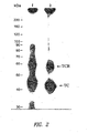

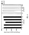
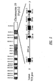
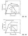
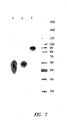

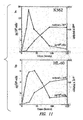


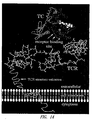


Classifications
-
- G—PHYSICS
- G01—MEASURING; TESTING
- G01N—INVESTIGATING OR ANALYSING MATERIALS BY DETERMINING THEIR CHEMICAL OR PHYSICAL PROPERTIES
- G01N33/00—Investigating or analysing materials by specific methods not covered by groups G01N1/00 - G01N31/00
- G01N33/48—Biological material, e.g. blood, urine; Haemocytometers
- G01N33/50—Chemical analysis of biological material, e.g. blood, urine; Testing involving biospecific ligand binding methods; Immunological testing
- G01N33/82—Chemical analysis of biological material, e.g. blood, urine; Testing involving biospecific ligand binding methods; Immunological testing involving vitamins or their receptors
-
- A—HUMAN NECESSITIES
- A01—AGRICULTURE; FORESTRY; ANIMAL HUSBANDRY; HUNTING; TRAPPING; FISHING
- A01K—ANIMAL HUSBANDRY; AVICULTURE; APICULTURE; PISCICULTURE; FISHING; REARING OR BREEDING ANIMALS, NOT OTHERWISE PROVIDED FOR; NEW BREEDS OF ANIMALS
- A01K67/00—Rearing or breeding animals, not otherwise provided for; New or modified breeds of animals
- A01K67/027—New or modified breeds of vertebrates
- A01K67/0275—Genetically modified vertebrates, e.g. transgenic
- A01K67/0276—Knock-out vertebrates
-
- A—HUMAN NECESSITIES
- A61—MEDICAL OR VETERINARY SCIENCE; HYGIENE
- A61P—SPECIFIC THERAPEUTIC ACTIVITY OF CHEMICAL COMPOUNDS OR MEDICINAL PREPARATIONS
- A61P25/00—Drugs for disorders of the nervous system
-
- A—HUMAN NECESSITIES
- A61—MEDICAL OR VETERINARY SCIENCE; HYGIENE
- A61P—SPECIFIC THERAPEUTIC ACTIVITY OF CHEMICAL COMPOUNDS OR MEDICINAL PREPARATIONS
- A61P29/00—Non-central analgesic, antipyretic or antiinflammatory agents, e.g. antirheumatic agents; Non-steroidal antiinflammatory drugs [NSAID]
-
- A—HUMAN NECESSITIES
- A61—MEDICAL OR VETERINARY SCIENCE; HYGIENE
- A61P—SPECIFIC THERAPEUTIC ACTIVITY OF CHEMICAL COMPOUNDS OR MEDICINAL PREPARATIONS
- A61P35/00—Antineoplastic agents
-
- A—HUMAN NECESSITIES
- A61—MEDICAL OR VETERINARY SCIENCE; HYGIENE
- A61P—SPECIFIC THERAPEUTIC ACTIVITY OF CHEMICAL COMPOUNDS OR MEDICINAL PREPARATIONS
- A61P37/00—Drugs for immunological or allergic disorders
-
- A—HUMAN NECESSITIES
- A61—MEDICAL OR VETERINARY SCIENCE; HYGIENE
- A61P—SPECIFIC THERAPEUTIC ACTIVITY OF CHEMICAL COMPOUNDS OR MEDICINAL PREPARATIONS
- A61P43/00—Drugs for specific purposes, not provided for in groups A61P1/00-A61P41/00
-
- C—CHEMISTRY; METALLURGY
- C07—ORGANIC CHEMISTRY
- C07H—SUGARS; DERIVATIVES THEREOF; NUCLEOSIDES; NUCLEOTIDES; NUCLEIC ACIDS
- C07H23/00—Compounds containing boron, silicon, or a metal, e.g. chelates, vitamin B12
-
- C—CHEMISTRY; METALLURGY
- C07—ORGANIC CHEMISTRY
- C07K—PEPTIDES
- C07K14/00—Peptides having more than 20 amino acids; Gastrins; Somatostatins; Melanotropins; Derivatives thereof
- C07K14/435—Peptides having more than 20 amino acids; Gastrins; Somatostatins; Melanotropins; Derivatives thereof from animals; from humans
- C07K14/705—Receptors; Cell surface antigens; Cell surface determinants
-
- C—CHEMISTRY; METALLURGY
- C07—ORGANIC CHEMISTRY
- C07K—PEPTIDES
- C07K16/00—Immunoglobulins [IGs], e.g. monoclonal or polyclonal antibodies
- C07K16/18—Immunoglobulins [IGs], e.g. monoclonal or polyclonal antibodies against material from animals or humans
- C07K16/28—Immunoglobulins [IGs], e.g. monoclonal or polyclonal antibodies against material from animals or humans against receptors, cell surface antigens or cell surface determinants
-
- C—CHEMISTRY; METALLURGY
- C12—BIOCHEMISTRY; BEER; SPIRITS; WINE; VINEGAR; MICROBIOLOGY; ENZYMOLOGY; MUTATION OR GENETIC ENGINEERING
- C12N—MICROORGANISMS OR ENZYMES; COMPOSITIONS THEREOF; PROPAGATING, PRESERVING, OR MAINTAINING MICROORGANISMS; MUTATION OR GENETIC ENGINEERING; CULTURE MEDIA
- C12N15/00—Mutation or genetic engineering; DNA or RNA concerning genetic engineering, vectors, e.g. plasmids, or their isolation, preparation or purification; Use of hosts therefor
- C12N15/09—Recombinant DNA-technology
- C12N15/11—DNA or RNA fragments; Modified forms thereof; Non-coding nucleic acids having a biological activity
- C12N15/113—Non-coding nucleic acids modulating the expression of genes, e.g. antisense oligonucleotides; Antisense DNA or RNA; Triplex- forming oligonucleotides; Catalytic nucleic acids, e.g. ribozymes; Nucleic acids used in co-suppression or gene silencing
- C12N15/1138—Non-coding nucleic acids modulating the expression of genes, e.g. antisense oligonucleotides; Antisense DNA or RNA; Triplex- forming oligonucleotides; Catalytic nucleic acids, e.g. ribozymes; Nucleic acids used in co-suppression or gene silencing against receptors or cell surface proteins
-
- C—CHEMISTRY; METALLURGY
- C12—BIOCHEMISTRY; BEER; SPIRITS; WINE; VINEGAR; MICROBIOLOGY; ENZYMOLOGY; MUTATION OR GENETIC ENGINEERING
- C12N—MICROORGANISMS OR ENZYMES; COMPOSITIONS THEREOF; PROPAGATING, PRESERVING, OR MAINTAINING MICROORGANISMS; MUTATION OR GENETIC ENGINEERING; CULTURE MEDIA
- C12N15/00—Mutation or genetic engineering; DNA or RNA concerning genetic engineering, vectors, e.g. plasmids, or their isolation, preparation or purification; Use of hosts therefor
- C12N15/09—Recombinant DNA-technology
- C12N15/63—Introduction of foreign genetic material using vectors; Vectors; Use of hosts therefor; Regulation of expression
- C12N15/79—Vectors or expression systems specially adapted for eukaryotic hosts
- C12N15/85—Vectors or expression systems specially adapted for eukaryotic hosts for animal cells
- C12N15/8509—Vectors or expression systems specially adapted for eukaryotic hosts for animal cells for producing genetically modified animals, e.g. transgenic
-
- A—HUMAN NECESSITIES
- A01—AGRICULTURE; FORESTRY; ANIMAL HUSBANDRY; HUNTING; TRAPPING; FISHING
- A01K—ANIMAL HUSBANDRY; AVICULTURE; APICULTURE; PISCICULTURE; FISHING; REARING OR BREEDING ANIMALS, NOT OTHERWISE PROVIDED FOR; NEW BREEDS OF ANIMALS
- A01K2217/00—Genetically modified animals
- A01K2217/07—Animals genetically altered by homologous recombination
- A01K2217/075—Animals genetically altered by homologous recombination inducing loss of function, i.e. knock out
-
- A—HUMAN NECESSITIES
- A01—AGRICULTURE; FORESTRY; ANIMAL HUSBANDRY; HUNTING; TRAPPING; FISHING
- A01K—ANIMAL HUSBANDRY; AVICULTURE; APICULTURE; PISCICULTURE; FISHING; REARING OR BREEDING ANIMALS, NOT OTHERWISE PROVIDED FOR; NEW BREEDS OF ANIMALS
- A01K2227/00—Animals characterised by species
- A01K2227/10—Mammal
- A01K2227/105—Murine
-
- A—HUMAN NECESSITIES
- A01—AGRICULTURE; FORESTRY; ANIMAL HUSBANDRY; HUNTING; TRAPPING; FISHING
- A01K—ANIMAL HUSBANDRY; AVICULTURE; APICULTURE; PISCICULTURE; FISHING; REARING OR BREEDING ANIMALS, NOT OTHERWISE PROVIDED FOR; NEW BREEDS OF ANIMALS
- A01K2267/00—Animals characterised by purpose
- A01K2267/03—Animal model, e.g. for test or diseases
-
- A—HUMAN NECESSITIES
- A61—MEDICAL OR VETERINARY SCIENCE; HYGIENE
- A61K—PREPARATIONS FOR MEDICAL, DENTAL OR TOILETRY PURPOSES
- A61K38/00—Medicinal preparations containing peptides
-
- C—CHEMISTRY; METALLURGY
- C07—ORGANIC CHEMISTRY
- C07K—PEPTIDES
- C07K2317/00—Immunoglobulins specific features
- C07K2317/20—Immunoglobulins specific features characterized by taxonomic origin
- C07K2317/21—Immunoglobulins specific features characterized by taxonomic origin from primates, e.g. man
-
- C—CHEMISTRY; METALLURGY
- C07—ORGANIC CHEMISTRY
- C07K—PEPTIDES
- C07K2319/00—Fusion polypeptide
- C07K2319/30—Non-immunoglobulin-derived peptide or protein having an immunoglobulin constant or Fc region, or a fragment thereof, attached thereto
-
- C—CHEMISTRY; METALLURGY
- C12—BIOCHEMISTRY; BEER; SPIRITS; WINE; VINEGAR; MICROBIOLOGY; ENZYMOLOGY; MUTATION OR GENETIC ENGINEERING
- C12N—MICROORGANISMS OR ENZYMES; COMPOSITIONS THEREOF; PROPAGATING, PRESERVING, OR MAINTAINING MICROORGANISMS; MUTATION OR GENETIC ENGINEERING; CULTURE MEDIA
- C12N2310/00—Structure or type of the nucleic acid
- C12N2310/10—Type of nucleic acid
- C12N2310/11—Antisense
- C12N2310/111—Antisense spanning the whole gene, or a large part of it
-
- C—CHEMISTRY; METALLURGY
- C12—BIOCHEMISTRY; BEER; SPIRITS; WINE; VINEGAR; MICROBIOLOGY; ENZYMOLOGY; MUTATION OR GENETIC ENGINEERING
- C12N—MICROORGANISMS OR ENZYMES; COMPOSITIONS THEREOF; PROPAGATING, PRESERVING, OR MAINTAINING MICROORGANISMS; MUTATION OR GENETIC ENGINEERING; CULTURE MEDIA
- C12N2310/00—Structure or type of the nucleic acid
- C12N2310/10—Type of nucleic acid
- C12N2310/14—Type of nucleic acid interfering N.A.
-
- G—PHYSICS
- G01—MEASURING; TESTING
- G01N—INVESTIGATING OR ANALYSING MATERIALS BY DETERMINING THEIR CHEMICAL OR PHYSICAL PROPERTIES
- G01N2500/00—Screening for compounds of potential therapeutic value
- G01N2500/02—Screening involving studying the effect of compounds C on the interaction between interacting molecules A and B (e.g. A = enzyme and B = substrate for A, or A = receptor and B = ligand for the receptor)
-
- G—PHYSICS
- G01—MEASURING; TESTING
- G01N—INVESTIGATING OR ANALYSING MATERIALS BY DETERMINING THEIR CHEMICAL OR PHYSICAL PROPERTIES
- G01N2500/00—Screening for compounds of potential therapeutic value
- G01N2500/10—Screening for compounds of potential therapeutic value involving cells
Definitions
- the field of the present invention is transcobalamin receptor polypeptides and polynucleotides and uses thereof.
- Vitamin B12 (cobalamin, Cbl) is a structurally complex water soluble molecule consisting of a planar corrin ring containing a central cobalt atom, a lower axial nucleotide (dimethylbenzimidazole) and an upper axial ligand which, in mammalian cells, is either a methyl, 5' deoxyadenosyl or hydroxo group.
- Methyl-Cbl functions in the transfer of the methyl group from N 5 methyltetrahydrofolate(methyl-FH 4 ) to homocysteine in the de novo synthesis of methionine catalyzed by the enzyme methionine synthase (MS).
- 5' deoxyadenosyl (ado-Cbl) is the cofactor for the rearrangement of methylmalonyl-CoA to succinyl-CoA, catalyzed by the enzyme methylmalonyl-CoA mutase (MMU) (14).
- MMU methylmalonyl-CoA mutase
- the coenzyme function of hydroxo-Cbl is not known but it is an intermediary form in the interconversion of Cbl and is the substrate for the Cbl reductases in the neosynthesis of Cbl coenzymes.
- a complex process has evolved in mammals requiring two carrier proteins and two membrane receptors for the assimilation of Cbl from the intestinal tract to its final destination in the tissue cells.
- Cbl released from food binds preferentially to a salivary R-protein favored by the acid pH in the stomach, and subsequently transfers to intrinsic factor (IF) at neutral pH in the jejunum as R-protein is digested by pancreatic trypsin.
- IF-Cbl complex is carried to the distal ileum where it binds to specific receptors on the microvillous membrane.
- IF receptor a 460 kDa membrane-associated protein that was purified from kidney and has been identified in the yolk sac and in the ileum, This protein binds IF-Cbl in the presence of Ca ++ and interacts with amnionless a transmembrane protein involved in the endocytosis of cubilin bound IF-Cbl.
- the absorbed Cbl is released into the circulation bound to transcobalamin (TC), a Cbl binding plasma protein secreted by the vascular endothelium.
- TC transcobalamin
- Megalin a multi-ligand binding protein expressed in the epithelial cells is involved in the reabsorption of TC in the kidney.
- the cDNA encoding cubilin has been cloned and the deduced amino acid sequence indicates that cubilin also belongs to the class of multi-ligand binding proteins.
- Cbl deficiency The absorption of Cbl from the ileum is essential for maintaining Cbl homeostasis. Any perturbation of this process will ultimately lead to intracellular Cbl deficiency. Because of the fundamental role of Cbl in essential metabolic reactions, one of which is coupled to folate metabolism and, as a consequence, to nucleic acid synthesis, Cbl deficiency will lead to abnormal cell division and differentiation as observed in megaloblastic anemia resulting from both Cbl and folate deficiency. The neurological complications of Cbl deficiency are known to cause irreversible damage to the central as well as the peripheral nervous system.
- Cbl malabsorption and proteinuria characterize another disorder known as Imerslund-Grasbeck syndrome in which absorption of IF-Cbl in the distal ileum is affected by a defect in internalizing the receptor bound IF-Cbl.
- TC tissue distribution and cellular uptake of Cbl is mediated by TC, a plasma protein, and a membrane receptor (TCblR) for TC -Cbl that binds and internalizes TC-Cbl by endocytosis.
- TC is also required for the translocation of Cbl absorbed in the distal ileum and this function is impaired in congenital TC deficiency.
- the normal concentration of this protein in plasma is 0.4 - 1.2 pM, of which, approximately 10 - 30 % is saturated with Cbl under conditions of normal Cbl homeostasis. Elevated levels of TC have been reported in certain autoimmune and myeloproliferative disorders.
- TC has been purified from human plasma and the cDNA encoding this protein has been isolated from endothelial cDNA libraries.
- TC and TC mRNA in various tissues did not identify the specific cell in these tissues as the source of the protein.
- studies showing TC synthesis by liver cells, macrophages, fibroblasts, lymphocytes and ileal mucosa in culture do not provide information about the source of TC in vivo. Though all these cells may synthesize TC in culture, the finding that human umbilical vein endothelial cells (HUVEC) synthesize significantly more of this protein than other cells, and that the umbilical vein perfused ex vivo synthesizes TC, provides evidence that the vascular endothelium is the likely source of plasma TC.
- the extensive endothelial surface provides the capacity to maintain the circulating level of TC that has a relatively short half life of 60-120 min.
- TblR transcobalamin receptor
- Cbl uptake in cells requires a specific receptor on the cell surface that binds holo-TC with high affinity.
- Cbl binding proteins in serum have been identified and the role of TC in the cellular uptake of Cbl has been well characterized.
- Information on the cellular uptake of Cbl was derived initially from direct binding of Cbl to a membrane receptor in prokaryotes and from a more complex process in mammalian cells whereby a membrane receptor specifically binds TC-Cbl and internalizes the complex.
- the process is biphasic with an initial Ca ++ dependent and temperature-independent binding of TC-Cbl to the receptor, followed by a slower second temperature-dependent step that translocates the vitamin into the cell and requires metabolic energy.
- the TC mediated uptake of Cbl has been identified in all mammalian cells studied and appears to be the only system for delivering Cbl into cells except for the liver, where uptake of Cbl bound to haptocorrin, another Cbl binding protein in the blood, occurs via the asialoglycoprotein receptor.
- TC-Cbl receptor-mediated endocytosis of TC-Cbl and the subsequent lysosomal degradation of TC to release the Cbl. Later studies showed that the receptors are expressed predominantly on membrane microvilli and the internalization of TC-Cbl occurs via clathrin-coated pits. Between 2000-6000 TC-Cbl receptors per cell are expressed during the proliferative phase of the cell and are down regulated to less than 300 receptors in non-dividing and fully differentiated cells. The increased requirement for intracellular Cbl during the early phase of cellular replication induces higher receptor expression in actively dividing cells that can account for a 10 - 30 fold increase in receptors in these cells.
- This expression of the TC receptor provides a unique target to selectively block Cbl uptake in cells that require the vitamin the most, i.e., actively dividing cells and to deliver antimetabolite analogs and Cbl-drug conjugates preferentially to rapidly proliferating cells.
- TCblR may exist as a non-covalent dimer in the lipid bilayer of the plasma membrane. They have not identified the primary structure and the gene encoding this protein. It is important to note that their results differed from earlier reports.
- Cbl deficiency The neuropathological changes in the peripheral and central nervous system and the consequent functional abnormalities in Cbl deficiency provide compelling evidence in support of a role for this vitamin in maintaining a normal nervous system.
- clear evidence of Cbl deficiency in AD and other dementias is lacking.
- biochemical and morphological changes observed in these disorders hypomethylation of cellular components and apoptotic cell death have been observed in the brain.
- Such pathological changes can be caused by both Cbl and/or folate deficiency.
- the metabolic pathways involving these two vitamins have not been adequately characterized in brain tissue.
- the concentration of Cbl in the adult brain is ⁇ 40-130 pg/mg tissue, a level similar to that in the spleen and kidney.
- the concentration of Cbl in most tissues including the brain is lower at birth and increases with age. In the fetal brain and liver, the Cbl requiring enzyme MS is highest during early embryogenesis and decreases as the fetus develops. The level of MeCbl in this tissue parallels the activity of the enzyme. Very little is known about TC and TCblR in the brain, and nothing is known about the expression of these essential proteins in aging and various brain disorders. The binding of TC-Cbl to brain tissue has been measured and appears to be higher in membranes prepared from the brain cortex than from the spinal cord. In addition, uptake of TC-Cbl has been reported in glial cells in culture.
- TC cerebrospinal fluid
- CSF cerebrospinal fluid
- a role for Cbl and folate deficiency in various dementias could not be established by measuring total Cbl and folate levels in serum and CSF because they have provided ambiguous results.
- studies designed to evaluate methylation in the brain have identified altered methylation ratio of S-adenosylmethionine to S-adenosylhomocysteine (SAM/SAH) in the CSF.
- Cbl deficiency due to malabsorption or poor dietary intake takes several years to present clinically because of the large liver stores of Cbl.
- Infants born with congenital TC deficiency develop normally in the uterus because of the maternal supply of TC-Cbl.
- Cbl deficiency rapidly i.e., after several months following birth because they lack TC.
- It has been difficult to produce an animal model of Cbl deficiency by Cbl depletion because of hepatic stores and contribution to the Cbl pool by the gut microflora.
- N 2 O nitrous oxide
- the strategy to inhibit cellular uptake of Cbl can utilize epitope specific monoclonal antibodies to the Cbl transporter, TC in blood and to the plasma membrane receptor, TCblR that facilitates the cellular uptake of TC-Cbl.
- mAbs to human TC have been generated (see Morgan et al., PCT Patent Publication No. WO 96/08515, published March 21, 1996 ).
- mAbs that prevent the uptake of TC-Cbl by TCblR receptor blocking
- mAb that prevents the binding of Cbl to apo TC Cbl blocking
- mAb that does not compromise the receptor binding and Cbl binding functions of TC binding mAb
- the receptor blocking and the Cbl blocking anti TC mAbs, either alone or in combination, could be used to prevent cellular uptake of Cbl.
- a similar effect could be produced with a mAb to TCblR that blocks the binding of TC-Cbl.
- mAbs could serve as a biological modulator to arrest cellular proliferation by selectively depleting an essential nutrient.
- Studies with anti-TC mAbs show that these mAbs block Cbl uptake and inhibit cellular replication in vitro.
- the TC-Cbl binding mAb when coupled to drugs, toxins or radionuclides, could deliver these compounds to tumor cells via the TC-TCblR pathway.
- TCblR expression is increased during the proliferative phase of the cell cycle and in cancers, a large proportion of cells are actively dividing and therefore, this pathway could be exploited to deliver mAb-drug or Cbl-drug conjugates to replicating cells.
- Radioactive molecules or fluorescent compounds coupled to Cbl have been used to locate tumor mass by imaging techniques because more of these compounds accumulate in tumor cells compared to the surrounding tissue.
- the present invention meets this long-felt need by providing TCblR polypeptides and polynucleotides, which are useful in identifying modulators of CBL uptake and TCblR activity.
- the present invention provides novel TCblR modulators, including siRNA and antibodies specific for TCblR, and methods of making the same, which are used in therapeutic compositions to modulate Cbl uptake and treat cancer and other diseases associated with cell proliferation, immune response, and inflammation.
- the present invention provides the following:
- the present invention is based upon the cloning and molecular characterization of the transcobalamin receptor (TCbIR), including the elucidation of the cDNA and polypeptide sequences of TCblR, as well as the identification of functional domains involved in the biological activity of TCblR and cis sequences within the TCblR gene that regulate the expression of TCblR.
- the invention is further based upon the identification of TCblR's critical role in cobalamin (Cbl) uptake and the identification of regions of TCblR involved in binding and uptake of transcobalamin (TC) and Cbl.
- aspects of the present invention are based upon the characterization of the expression of TCblR, which indicates that TCblR is overexpressed in proliferating and dividing cells, including tumor cells.
- the present description provides novel compositions and methods for regulating Cbl uptake and cell growth, including methods of treating diseases and disorders associated with poor Cbl uptake, as well as diseases and disorders associated with hyperproliferation or deregulated cell growth, such as tumors.
- described are screening methods to identify modulators of TCblR activity and methods of using molecules that bind TCblR to identify and deliver therapeutic agents to tumor cells.
- compositions and methods of the present invention comprise or utilize a TCblR polypeptide or polynucleotide or variant fragment thereof, including recombinantly produced TCblR polypeptide and polynucletoides.
- the TCblR polypeptide sequence identified according to the present invention was previously identified as the 8D6 antigen, or CD320 antigen, although it was not known before that these antigens corresponded to TCblR.
- the 8D6 antigen was originally identified as a follicular dendritic cell molecule that stimulates geminal center B cell growth ( Li, L. et al., J. Exp. Med.. 191:1077-1083 (2000 ) and Zhang, X. et al., J.
- the amino acid sequence of human TCblR protein is set forth in SEQ ID NO:1
- the sequence of the human TCblR cDNA corresponding to the mRNA is set forth in SEQ ID NO:2, with the coding region of the cDNA provided in SEQ ID NO:3.
- transcobalamin receptor polypeptide and polynucleotide sequences are available in NCBI public sequence databases under the following accession numbers: NM_016579, Homo sapiens CD320 antigen (CD320), mRNA, gi
- TCblR TCblR
- non-coding sequences any polynucletoides sequence that encodes a TCblR or fragment thereof, as well as non-coding sequences.
- coding or non-coding sequences are utilized, and either sense or antisense sequences are utilized.
- polypeptide is used in its conventional meaning, i.e., as a sequence of amino acids.
- the polypeptides are not limited to a specific length of the product; thus, peptides, oligopeptides, and proteins are included within the definition of polypeptide, and such terms may be used interchangeably herein unless specifically indicated otherwise.
- This term also does not refer to or exclude post-expression modifications of the polypeptide, for example, glycosylations, acetylations, phosphorylations, amidations, and the like, as well as other modifications known in the art, both naturally occurring and non-naturally occurring.
- a polypeptide may be an entire protein, or a portion thereof.
- polypeptides of interest in the context of this description are amino acid subsequences comprising functional domains of TCblR, including fragments capable of binding TC and/or Cbl, as well as dominant negative mutants, as described infra.
- TC refers to both holo-TC and apo-TC, unless otherwise indicated. Methods of determining whether a TCblR polypeptide binds to TC are known in the art and described herein.
- the present description also includes the use of polypeptide fragments comprising at least 5, 10, 15, 20, 25, 50, 75 or 100 contiguous amino acids, or more, including all intermediate lengths, of a TCblR polypeptide.
- the present invention includes polypeptide fragments comprising, consisting essentially of, or consisting of, the extracellular region of a TCblR polypeptide, as shown in Figure 3 .
- the extracellular domain of TCblR consists of a signal peptide of 31 AA (AA 1-31 of SEQ ID NO:1) and a 198 AA extracellular region (AA 32-229 of SEQ ID NO:1).
- This extracellular fragment has been expressed as a recombinant protein in HEK 293 cells.
- This fragment (AA 1-229 of SEQ ID NO:1) contains the specific high affinity TC-Cbl binding activity and has a 28 fold higher affinity for TC saturated with Cbl (holo-TC) than for the apo protein.
- the invention includes a fragment comprising, consisting essentially of, or consisting of AA 1-229 or AA 32-229 of SEQ ID NO:1, and particular embodiments of methods of the present invention use such fragments.
- Recombinantly-produced or synthetic extracellular fragments of TCblR, or functional fragments thereof that bind to TC, are, thus useful in blocking binding of holo-TC to cell surface TCblR.
- Immunogenic portions of TCblR are also described.
- An "immunogenic portion,” as used herein, is a fragment of an immunogenic polypeptide that itself is immunologically reactive (i.e., specifically binds) with the B-cells and/or T-cell surface antigen receptors that recognize the polypeptide. Immunogenic portions may generally be identified using well known techniques, such as those summarized in Paul, Fundamental Immunology, 3rd ed., 243-247 (Raven Press, 1993 ) and references cited therein. Such techniques include screening polypeptides for the ability to react with antigen-specific antibodies, antisera and/or T-cell lines or clones.
- antisera and antibodies are "antigen-specific” if they specifically bind to an antigen (i.e., they react with the protein in an ELISA or other immunoassay, and do not react detectably with unrelated proteins).
- antisera and antibodies may be prepared as described herein, and using well-known techniques.
- an immunogenic portion of a polypeptide is a portion that reacts with antisera and/or T-cells at a level that is not substantially less than the reactivity of the full-length polypeptide (e.g., in an ELISA and/or T-cell reactivity assay).
- the level of immunogenic activity of the immunogenic portion is at least about 50%, preferably at least about 70% and most preferably greater than about 90% of the immunogenicity for the full-length polypeptide.
- preferred immunogenic portions are identified that have a level of immunogenic activity greater than that of the corresponding full-length polypeptide, e.g., having greater than about 100% or 150% or more immunogenic activity.
- the present description includes the use of variants of TCblR.
- the description contemplates the use of TCblR variants, including variants possessing one or more of TCblR's functions, such as being capable of binding TC.
- a polypeptide "variant,” as the term is used herein, is a polypeptide that typically differs from a polypeptide specifically disclosed herein in one or more substitutions, deletions, additions and/or insertions. Such variants may be naturally occurring or may be synthetically generated, for example, by modifying one or more of the above polypeptide sequences.
- Polypeptide variants will typically exhibit at least about 70%, 75%, 80%, 85%, 90%, 91%, 92%, 93%, 94%, 95%, 96%, 97%, 98%, or 99% or more identity (determined as described below), along its length, to a TCblR polypeptide.
- the TCblR fragments and variants are immunologically reactive with an antibody and/or T-cell that reacts with a full-length TCblR polypeptide.
- the TCblR fragments and variants exhibit a level of immunogenic activity of at least about 50%, preferably at least about 70%, and most preferably at least about 90% or more of that exhibited by a full-length TCblR polypeptide.
- a variant will contain conservative substitutions.
- a "conservative substitution” is one in which an amino acid is substituted for another amino acid that has similar properties, such that one skilled in the art of peptide chemistry would expect the secondary structure and hydropathic nature of the polypeptide to be substantially unchanged.
- two sequences are said to be “identical” if the sequence of amino acids in the two sequences is the same when aligned for maximum correspondence, as described below. Comparisons between two sequences are typically performed by comparing the sequences over a comparison window to identify and compare local regions of sequence similarity.
- a “comparison window” as used herein refers to a segment of at least about 20 contiguous positions, usually 30 to about 75, 40 to about 50, in which a sequence may be compared to a reference sequence of the same number of contiguous positions after the two sequences are optimally aligned.
- Alignment of sequences for comparison may be conducted by a variety of methods, including, e.g., the local identity algorithm of Smith and Waterman (1981) Add. APL. Math 2:482 , by the identity alignment algorithm of Needleman and Wunsch (1970) J. Mol. Biol. 48:443 , by the search for similarity methods of Pearson and Lipman (1988) Proc. Natl. Acad. Sci. USA 85: 2444 , by computerized implementations of these algorithms (GAP, BESTFIT, BLASTN 2.0.5, FASTA, and TFASTA in the Wisconsin Genetics Software Package, Genetics Computer Group (GCG), 575 Science Dr., Madison, WI), or by inspection.
- BLASTN 2.0.5 Another example of algorithms that are suitable for determining percent sequence identity and sequence similarity is the BLASTN 2.0.5 algorithm, which are described in Altschul et al. (1977) Nucl. Acids Res. 25:3389-3402 and Altschul et al. (1990) J. Mol. Biol. 215:403-410 , respectively.
- BLASTN 2.0.5 can be used, for example, with the parameters described herein, to determine percent sequence identity for the polynucleotides and polypeptides.
- Software for performing BLASTN 2.0.5 analyses is publicly available through the National Center for Biotechnology Information.
- TCblR includes TCblR polypeptide and polynucleotide sequences from any species, as well as homologs thereof.
- Polypeptides of the invention are prepared using any of a variety of well known synthetic and/or recombinant techniques. Polypeptides, portions and other variants generally less than about 150 amino acids can be generated by synthetic means, using techniques well known to those of ordinary skill in the art. In one illustrative example, such polypeptides are synthesized using any of the commercially available solid-phase techniques, such as the Merrifield solid-phase synthesis method, where amino acids are sequentially added to a growing amino acid chain. See Merrifield, J. Am. Chem. Soc. 85:2149-2146, 1963 . Equipment for automated synthesis of polypeptides is commercially available from suppliers such as Perkin Elmer/Applied BioSystems Division (Foster City, CA), and may be operated according to the manufacturer's instructions.
- TcblR polypeptides of the present invention may also be recombinantly-produced polypeptides. Methods of recombinantly expressing and isolating or purifying such polypeptides are known in the art. For example, they may be expressed in cells comprising an exogenous TcblR polynucleotide sequence, which may be present in the cell transiently or stably. Such exogenous polynucleotide sequences are typically operatively linked to a promoter, and may be present, e.g., in an expression construct, such as those described infra, or they may be present in the cellular genome.
- exogenous polynucleotides and polypeptide sequences are sequences that have been introduced into a cell, e.g., as a recombinant expression vector comprising the exogenous polynucleotide sequence or an exogenous polypeptide expressed from such an expression vector.
- exogenous does not exclude the possibility that the cell comprising an exogenous sequence also comprises or produces native polynucleotide or polypeptide sequences having the same sequence as the exogenous polynucleotide or polypeptide.
- polypeptides described herein are isolated.
- An "isolated" polypeptide is one that is removed from its original environment.
- a naturally-occurring protein or polypeptide is isolated if it is separated from some or all of the coexisting materials in the natural system.
- polypeptides are also purified, e.g., are at least about 90% pure, more preferably at least about 95% pure, and most preferably at least about 99% pure.
- TCblR polynucleotide compositions include all polynucleotides that encode a TCblR, as well as fragments, variants, and complements thereof.
- TCblR polynucleotides include genes that encode TCblR, as well as mRNA and cDNA sequences thereof.
- TCblR polynucleotides further include single- and double-stranded polynucleotides corresponding to a sense or antisense TCblR polynucleotide sequence. Typically, TCblR polynucleotides are isolated.
- isolated means that a polynucleotide is substantially away from other coding sequences, and that the polynucleotide does not contain large portions of unrelated sequence, such as large chromosomal fragments or other functional genes or polypeptide coding regions. Of course, this refers to the polynucleotide as originally isolated, and does not exclude sequences later added to the TCblR polynucleotide by the hand of man.
- the polynucleotide compositions include genomic sequences, extra-genomic and plasmid-encoded sequences, cDNA sequences, RNA sequences, and smaller engineered gene segments. Such segments may be naturally isolated, or modified synthetically by the hand of man.
- Polynucleotides may be single-stranded (coding or antisense) or double-stranded, and may be DNA (genomic, cDNA or synthetic) or RNA molecules. In certain embodiments, they are double-stranded RNA or DNA molecules, while in other embodiments, they are single-stranded RNA or antisense molecules.
- RNA molecules may include HnRNA molecules, which contain introns and correspond to a DNA molecule in a one-to-one manner, and mRNA molecules, which do not contain introns. Additional coding or non-coding sequences may, but need not, be present within a polynucleotide, and a polynucleotide may, but need not, be linked to other molecules and/or support materials.
- Polynucleotides may comprise a native sequence (i.e., an endogenous sequence that encodes a polypeptide/protein or a portion thereof) or may comprise a sequence that encodes a variant, fragment, or derivative, preferably and immunogenic variant, fragment, or derivative, of such a sequence.
- a native sequence i.e., an endogenous sequence that encodes a polypeptide/protein or a portion thereof
- Polynucleotides may comprise a native sequence (i.e., an endogenous sequence that encodes a polypeptide/protein or a portion thereof) or may comprise a sequence that encodes a variant, fragment, or derivative, preferably and immunogenic variant, fragment, or derivative, of such a sequence.
- polynucleotide compositions comprise some or all of a polynucleotide sequence set forth in SEQ ID NO:2 or SEQ ID NO:3, complements of a polynucleotide sequence set forth in SEQ ID NO:2 or SEQ ID NO:3, corresponding RNA sequences, and degenerate variants of a polynucleotide sequence set forth in SEQ ID NO:2 or SEQ ID NO:3.
- sequences provided in SEQ ID NOs:2 and 3 are DNA sequences.
- RNA sequences may comprise an RNA sequence corresponding to any sequence presented here, including the sequences set forth in SEQ ID NOs:2 and 3.
- RNA sequences are identical to the DNA sequence, except that thymidines are replaced with uridines.
- polynucleotide variants having substantial identity to a TCblR polynucleotide sequence, e.g., the sequences set forth in SEQ ID NO:2 or SEQ ID NO:3, including sequence having at least 70% sequence identity, preferably at least 75%, 80%, 85%, 90%, 95%, 96%, 97%, 98%, or 99% or higher sequence identity compared to a polynucleotide sequence using the methods described herein, (e.g., BLAST analysis using standard parameters, as described below).
- polynucleotides comprise at least a fragment of a polynucleotide having the sequence set forth in Figure 10 or SEQ ID NO:4, or a complement thereof.
- polynucleotide variants will contain one or more substitutions, additions, deletions and/or insertions, preferably such that the immunogenicity of the polypeptide encoded by the variant polynucleotide is not substantially diminished relative to a polypeptide encoded by a polynucleotide sequence specifically set forth herein).
- variants should also be understood to encompasses homologous genes of xenogenic origin.
- polynucleotide fragments comprising various lengths of contiguous stretches of sequence identical to or complementary to one or more of the sequences disclosed herein.
- polynucleotides are provided that comprise at least about 10, 15, 20, 30, 40, 50, 75, 100, 150, 200, 300, 400, 500 or 1000 or more contiguous nucleotides of one or more of the sequences disclosed herein as well as all intermediate lengths there between.
- intermediate lengths means any length between the quoted values, such as 16, 17, 18, 19, etc.; 21, 22, 23, etc.; 30, 31, 32, etc.; 50, 51, 52, 53, etc.; 100, 101, 102, 103, etc.; 150, 151, 152, 153, etc.; including all integers through 200-500; 500-1,000, and the like.
- polynucleotide compositions are provided that are capable of hybridizing under moderate to high stringency conditions to a polynucleotide sequence provided herein, or a fragment thereof, or a complementary sequence thereof.
- Hybridization techniques are well known in the art of molecular biology.
- suitable moderately stringent conditions for testing the hybridization of a polynucleotide with other polynucleotides include prewashing in a solution of 5 X SSC, 0.5% SDS, 1.0 mM EDTA (pH 8.0); hybridizing at 50°C-60°C, 5 X SSC, overnight; followed by washing twice at 65°C for 20 minutes with each of 2X, 0.5X and 0.2X SSC containing 0.1 % SDS.
- suitable highly stringent hybridization conditions include those described above, with the exception that the temperature of hybridization is increased, e.g., to 60-65°C or 65-70°C.
- polynucleotides described above e.g., polynucleotide variants, fragments and hybridizing sequences, encode polypeptides that are immunologically cross-reactive with a polypeptide sequence specifically set forth herein.
- such polynucleotides encode polypeptides that have a level of immunogenic activity of at least about 50%, preferably at least about 70%, and more preferably at least about 90% of that for a polypeptide sequence specifically set forth herein.
- polynucleotides, or fragments thereof regardless of the length of the coding sequence itself, may be combined with other DNA sequences, such as promoters, polyadenylation signals, additional restriction enzyme sites, multiple cloning sites, other coding segments, and the like, such that their overall length may vary considerably. It is therefore contemplated that a nucleic acid fragment of almost any length may be employed, with the total length preferably being limited by the ease of preparation and use in the intended recombinant DNA protocol.
- illustrative polynucleotide segments with total lengths of about 10,000, about 5000, about 3000, about 2,000, about 1,000, about 500, about 200, about 100, about 50 base pairs in length, and the like, (including all intermediate lengths) are contemplated to be useful in many implementations.
- two sequences are said to be “identical” if the sequence of nucleotides in the two sequences is the same when aligned for maximum correspondence, as described below. Comparisons between two sequences are typically performed by comparing the sequences over a comparison window to identify and compare local regions of sequence similarity.
- a “comparison window” as used herein refers to a segment of at least about 20 contiguous positions, usually 30 to about 75, 40 to about 50, in which a sequence may be compared to a reference sequence of the same number of contiguous positions after the two sequences are optimally aligned.
- Optimal alignment of sequences for comparison may be conducted using the Megalign program in the Lasergene suite of bioinformatics software (DNASTAR, Inc., Madison, WI), using default parameters.
- This program embodies several alignment schemes described in the following references: Dayhoff, M.O. (1978) A model of evolutionary change in proteins-Matrices for detecting distant relationships. In Dayhoff, M.O. (ed.) Atlas of Protein Sequence and Structure, National Biomedical Research Foundation, Washington DC Vol. 5, Suppl. 3, pp. 345-358 ; Hein J. (1990) Unified Approach to Alignment and Phylogenes pp. 626-645 Methods in Enzymology vol.
- optimal alignment of sequences for comparison may be conducted by the local identity algorithm of Smith and Waterman ((1981) Add. APL. Math 2:482 ), by the identity alignment algorithm of Needleman and Wunsch ((1970) J. Mol. Biol. 48:443 ), by the search for similarity methods of Pearson and Lipman ((1988) Proc. Natl. Acad. Sci. USA 85: 2444 ), by computerized implementations of these algorithms, e.g., GAP, BESTFIT, BLAST, FASTA, and TFASTA in the Wisconsin Genetics Software Package, Genetics Computer Group (GCG); 575 Science Dr., Madison, WI), or by inspection.
- the "percentage of sequence identity” is determined by comparing two optimally aligned sequences over a window of comparison of at least 20 positions, wherein the portion of the polynucleotide sequence in the comparison window may comprise additions or deletions ( i.e., gaps) of 20 percent or less, usually 5 to 15 percent, or 10 to 12 percent, as compared to the reference sequences (which does not comprise additions or deletions) for optimal alignment of the two sequences.
- additions or deletions i.e., gaps
- the percentage is calculated by determining the number of positions at which the identical nucleic acid bases occurs in both sequences to yield the number of matched positions, dividing the number of matched positions by the total number of positions in the reference sequence ( i.e., the window size) and multiplying the results by 100 to yield the percentage of sequence identity.
- nucleotide sequences that encode a polypeptide as described herein. Some of these polynucleotides bear minimal homology to the nucleotide sequence of any native gene. Nonetheless, polynucleotides that vary due to differences in codon usage are specifically contemplated herein. Alleles are endogenous genes that are altered as a result of one or more mutations, such as deletions, additions and/or substitutions of nucleotides. The resulting mRNA and protein may, but need not, have an altered structure or function. Alleles may be identified using standard techniques (such as hybridization, amplification and/or database sequence comparison).
- the polynucleotide sequences provided herein can be advantageously used as probes or primers for nucleic acid hybridization.
- nucleic acid segments that comprise a sequence region of at least about 15 nucleotide long contiguous sequence that has the same sequence as, or is complementary to, a 15 nucleotide long contiguous sequence disclosed herein will find particular utility.
- Longer contiguous identical or complementary sequences e.g., those of about 20, 30, 40, 50, 100, 200, 500, 1000 (including all intermediate lengths) and even up to full length sequences will also be of use in certain embodiments.
- nucleic acid probes to specifically hybridize to a sequence of interest will enable them to be of use in detecting the presence of complementary sequences in a given sample.
- sequence information for the preparation of mutant species primers, or primers for use in preparing other genetic constructions.
- Small polynucleotide segments or fragments may be readily prepared by, for example, directly synthesizing the fragment by chemical means, as is commonly practiced using an automated oligonucleotide synthesizer. Also, fragments may be obtained by application of nucleic acid reproduction technology, such as the PCRTM technology of U. S. Patent 4,683,202 , by introducing selected sequences into recombinant vectors for recombinant production, and by other recombinant DNA techniques generally known to those of skill in the art of molecular biology.
- TCblR activity or expression is altered through the use of recombinantly engineered constructs that express TCblR, functional fragments or variants thereof, or a modulator (e.g., an agonist or inhibitor) of TCblR.
- expression constructs are transiently present in a cell, while in other embodiments, they are stably integrated into a cellular genome.
- other DNA sequences that encode substantially the same or a functionally equivalent amino acid sequence or variant thereof may be produced and these sequences may be used to express a given polypeptide.
- expression constructs comprise polynucleotide sequences encoding all or a region of a TCblR polypeptide, or comprising all or a region of a TCblR cDNA sequence.
- Regulatory sequences present in an expression vector include those non-translated regions of the vector, e.g., enhancers, promoters, 5' and 3' untranslated regions, which interact with host cellular proteins to carry out transcription and translation. Such elements may vary in their strength and specificity. Depending on the vector system and cell utilized, any number of suitable transcription and translation elements, including constitutive and inducible promoters, may be used.
- promoters from mammalian genes or from mammalian viruses are generally preferred, and a number of viral-based expression systems are generally available.
- transcription enhancers such as the Rous sarcoma virus (RSV) enhancer, may be used to increase expression in mammalian host cells.
- RSV Rous sarcoma virus
- Specific initiation signals may also be used to achieve more efficient translation of sequences encoding a polypeptide of interest. Such signals include the ATG initiation codon and adjacent sequences. Exogenous translational elements and initiation codons may be of various origins, both natural and synthetic. The efficiency of expression may be enhanced by the inclusion of enhancers that are appropriate for the particular cell used, such as those described in the literature ( Scharf, D. et al. (1994) Results Probl. Cell Differ. 20:125-162 ).
- the description provides for the conditional expression of TCblR or fragment or variant thereof, or an inhibitor of TCblR activity.
- conditional expression systems are known and available in the art for use in both cells and animals, and the description contemplates the use of any such conditional expression system to regulate the expression or activity of TCblR.
- the use of prokaryotic repressor or activator proteins is advantageous due to their specificity for a corresponding prokaryotic sequence not normally found in a eukaryotic cell.
- This type of inducible system is the tetracycline-regulated inducible promoter system, of which various useful version have been described (See, e.g. Shockett and Schatz, Proc.
- expression of a molecule can be placed under control of the REV-TET system.
- Components of this system and methods of using the system to control the expression of a gene are well-documented in the literature, and vectors expressing the tetracycline-controlled transactivator (tTA) or the reverse tTA (rtTA) are commercially available (e.g. pTet-Off, pTet-On and ptTA-2/3/4 vectors, Clontech, Palo Alto, CA).
- tTA tetracycline-controlled transactivator
- rtTA reverse tTA
- Such systems are described, for example, in U.S. Patents No. 5650298 , No. 6271348 , No. 5922927 , and related patents.
- TCblR modulators or TCblR polypeptides, or fragments or variants thereof are provided to a cell using a viral or bacteriophage vector.
- viral or bacteriophage vector A wide variety of viral expression systems are known and available in the art, all of which may be used. Therefore, in certain embodiments, polynucleotide inhibitors of TCblR or polynucleotides encoding inhibitors of TCblR or TCblR, or a fragment or variant thereof, are introduced into suitable mammalian host cells or patients using any of a number of known viral-based systems.
- retroviruses provide a convenient and effective platform for gene delivery systems.
- a selected nucleotide sequence can be inserted into a vector and packaged in retroviral particles using techniques known in the art.
- the recombinant virus can then be isolated and delivered to a subject.
- retroviral systems have been described (e.g., U.S. Pat. No. 5,219,740 ; Miller and Rosman (1989) BioTechniques 7:980-990 ; Miller, A. D. (1990) Human Gene Therapy 1:5-14 ; Scarpa et al. (1991) Virology 180:849-852 ; Burns et al. (1993) Proc. Natl. Acad. Sci. USA 90:8033-8037 ; and Boris-Lawrie and Temin (1993) Cur.
- adenovirus-based systems have also been described. Unlike retroviruses which integrate into the host genome, adenoviruses persist extrachromosomally thus minimizing the risks associated with insertional mutagenesis ( Haj-Ahmad and Graham (1986) J. Virol. 57:267-274 ; Bett et al. (1993) J. Virol. 67:5911-5921 ; Mittereder et al. (1994) Human Gene Therapy 5:717-729 ; Seth et al. (1994) J. Virol. 68:933-940 ; Barr et al. (1994) Gene Therapy 1:51-58 ; Berkner, K. L. (1988) BioTechniques 6:616-629 ; and Rich et al. (1993) Human Gene Therapy 4:461-476 ).
- Additional viral vectors useful for delivering the polynucleotides encoding polypeptides by gene transfer include those derived from the pox family of viruses, such as vaccinia virus and avian poxvirus.
- the methods utilize lentiviruses.
- lentiviruses are enveloped viruses that carry a core of RNA encoding their genetic information. Lentiviruses are unique in that lentiviruses are the only retroviruses able to integrate into the chromosome of non-dividing cells.
- Recombinant self-inactivating lentiviral vectors expressing angiostatin and endostatin have previously been shown to have antiangiogenic activities ( Shichinohe T., Cancer Gene Ther. 2001 Nov;B(11):879-89 ), and similar methods are used to deliver TCblR inhibitors or TCblR polynucleotide, polypeptides, or functional fragments or variants thereof.
- methods directed to modulating the activity of TCblR are practiced using a modulator that inhibits or enhances TCblR expression or activity.
- a modulator specifically increase, reduces, or inhibits TCblR's ability to bind TC or mediate cellular uptake of TC.
- a modulator increases or reduces expression of TCblR.
- modulators are polynucleotides, polypeptides, peptides, peptide nucleic acids, antibodies and fragments thereof, viruses, small molecules, inorganic compounds and organic compounds. Modulators include agonists and antagonists of TCblR.
- Modulators that increase TCblR activity include TCblR polypeptides and polynucleotides, including those described supra, as well as other molecules that enhance TCblR expression or activity.
- Modulators that decrease TCblR activity include any molecule that reduces TCblR expression or activity.
- inhibitors interfere with TCblR activity by any of a variety of means including, e.g., inhibiting TCblR binding to TC or inhibiting downstream signaling events leading to Cbl uptake and/or cell growth.
- methods described herein are practiced using peptide or polypeptide modulators of TCblR.
- TCblR expression is increased using an expression construct that expresses full length TCblR, or a functional variant or fragment thereof.
- the activity of TCblR is altered by over expression of a dominant negative inhibitor of TCblR.
- Dominant negative inhibitors of TCblR are typically mutant forms of TCblR, which reduce or block the activity of wild type TCblR, e.g., by competing for binding to TC but failing to fully activate the TCblR signaling or uptake pathway.
- dominant negative inhibitors of TCblR have a reduced ability to promote cell growth as compared to wild type TCblR.
- Examples of various TCblR dominant negative inhibitors include a mutant TCblR having reduced ability to bind TC and a mutant TCblR that binds TC but fails to internalize TC and/or Cbl.
- one dominant negative is a TCblR having one or more amino acid substitutions in the intracellular domain, such that the mutant TCblR binds TC but exhibits reduced uptake of TC and/or Cbl.
- Polypeptide inhibitors also include other variants and fragments of TCblR having reduced biological activity as compared to wild type TCblR.
- a mutant TCblR inhibitor is a TCblR in which the region that binds TC has been mutated so that it has reduced TC binding ability.
- an inhibitor is a soluble fragment of TCbIR that includes the TCbIR extracellular domain and is capable of binding to TC but does not promote TC and/or Cbl uptake.
- an inhibitor of TCbIR comprises the extracellular fragment of TCbIR depicted in Figure 13 or a functional fragment thereof.
- a modulator of TCbIR comprises or consists of the first 229 or 247 amino acids of the TCbIR protein. In related embodiment, it comprises or consists of AA 32-229 or 32-247 of TCbIR. While in certain embodiments, polypeptide modulators comprising or consisting of a fragment of the extracellular domain of TCbIR do not include the signal peptide (AA 1-31), in certain embodiments, the signal peptide is included, e.g., to direct the protein to the plasma membrane for secretion into the medium.
- the extracellular domain of TCbIR consists of a signal peptide of 31 AA (AA 1-31) and a 198 AA extracellular region (AA 32-229).
- This extracellular region contains the specific high affinity TC-Cbl binding activity and has a 28 fold higher affinity for TC saturated with Cbl (holo-TC) than for the apo protein.
- This difference is physiologically relevant in that in-vivo only holo -TC preferentially binds to the receptor to deliver Cbl into cells.
- soluble extracellular domain e.g., with or without the signal peptide may be used to block holo-TC binding to cell surface-bound TCbIR and block cellular uptake of Cbl.
- This property of the receptor provides an additional method to block cellular uptake of Cbl as a strategy in cancer therapy. This could be accomplished by saturating the circulating TC in the patient's blood with the recombinant receptor fragment. This would prevent the holo TC from binding to the receptor on the cell surface and block cellular uptake of Cbl.
- polynucleotides are contemplated for use as modulators of TCbIR expression and/or activity.
- a polynucleotide encoding TCbIR or a functional variant or fragment thereof is used to increase TCbIR expression.
- These polynucleotides include expression vectors and replacement or insertion vectors designed for integration into the genome of a cell, and suitable for gene therapy.
- polynucleotide inhibitors of TCbIR are antisense RNA, ribozymes, aptamers, or RNA interference reagents designed to specifically target TCbIR, according to methods known and available in the art.
- polynucleotide inhibitors include, e.g ., targeting vectors designed for integration into the genome and suitable for deleting all or a portion of a TCbIR allele or mutating a TCbIR allele, e.g ., through insertional mutagenesis.
- a TCbIR inhibitor is an antisense RNA directed to TCbIR polynucleotides, or other components of the TCbIR signaling cascade.
- Antisense oligonucleotides have been demonstrated to be effective and targeted inhibitors of protein synthesis, and, consequently, can be used to specifically inhibit protein synthesis by a targeted gene.
- the efficacy of antisense oligonucleotides for inhibiting protein synthesis is well established. For example, the synthesis of polygalactauronase and the muscarine type 2 acetylcholine receptor are inhibited by antisense oligonucleotides directed to their respective mRNA sequences ( U. S. Patent 5, 739, 119 and U. S.
- Patent 5,759,829 Further, examples of antisense inhibition have been demonstrated with the nuclear protein cyclin, the multiple drug resistance gene (MDG1), ICAM-1, E-selectin, STK-1, striatal GABA A receptor and human EGF ( Jaskulski et al., Science, 1988 Jun 10;240(4858):1544-6 ; Vasanthakumar and Ahmed, Cancer Commun. 1989;1(4):225-32 ; Peris et al., Brain Res Mol Brain Res., 1998 Jun 15;57(2):310-20 ; U. S. Patent 5,801,154 ; U.S. Patent 5,789,573 ; U. S. Patent 5,718,709 and U.S. Patent 5,610,288 ).
- MDG1 multiple drug resistance gene
- antisense constructs have also been described that inhibit and can be used to treat a variety of abnormal cellular proliferations, e.g. cancer ( U. S. Patent 5,747,470 ; U. S. Patent 5,591,317 and U. S. Patent 5,783,683 ).
- the present description relates to methods of providing oligonucleotide sequences that comprise all, or a portion of, any sequence that is capable of specifically binding to a TCbIR target polynucleotide sequence, or a complement thereof.
- the oligonucleotide sequence comprises all, or a portion of, any sequence that is capable of specifically binding to a TCbIR polynucleotide sequence, or a complement thereof.
- the antisense oligonucleotides comprise DNA or derivatives thereof.
- the oligonucleotides comprise RNA or derivatives thereof.
- the antisense oligonucleotides may be modified DNAs comprising a phosphorothioated modified backbone.
- the oligonucleotide sequences may comprise peptide nucleic acids or derivatives thereof.
- preferred compositions comprise a sequence region that is complementary, and more preferably, completely complementary to one or more portions of a TCbIR target gene or polynucleotide sequence.
- an antisense polynucleotide comprises a fragment of a sequence set forth in SEQ ID NOs:2 or 3, or a complement thereof.
- an antisense polynucleotide comprises a fragment of the sequence set forth in Figure 10 , or a complement thereof.
- antisense molecules are known in the art and can be readily adapted to produce an antisense molecule that targets TCbIR. Selection of antisense compositions specific for a given sequence is based upon analysis of the chosen target sequence and determination of secondary structure, T m , binding energy, and relative stability. Antisense compositions may be selected based upon their relative inability to form dimers, hairpins, or other secondary structures that would reduce or prohibit specific binding to the target mRNA in a host cell. Highly preferred target regions of the mRNA include those regions at or near the AUG translation initiation codon and those sequences that are substantially complementary to 5' regions of the mRNA.
- Ribozyme molecules are used to inhibit expression of a TCbIR target gene or polynucleotide sequence.
- Ribozymes are RNA-protein complexes having specific catalytic domains that possess endonuclease activity ( Kim and Cech, Proc Natl Acad Sci U S A., 1987 Dec;84(24):8788-92 ; Forster and Symons, Ce//, 1987 Apr 24;49(2):211-20 ).
- ribozymes accelerate phosphoester transfer reactions with a high degree of specificity, often cleaving only one of several phosphoesters in an oligonucleotide substrate ( Cech et al., Cell, 1981 Dec;27(3 Pt 2):487-96 ; Michel and Westhof, J Mol Biol. 1990 Dec 5;216(3):585-610 ; Reinhold-Hurek and Shub, Nature, 1992 May 14;357(6374):173-6 ).
- This specificity has been attributed to the requirement that the substrate bind via specific base-pairing interactions to the internal guide sequence ("IGS") of the ribozyme prior to chemical reaction.
- IGS internal guide sequence
- enzymatic nucleic acids act by first binding to a target RNA. Such binding occurs through the target binding portion of a enzymatic nucleic acid which is held in close proximity to an enzymatic portion of the molecule that acts to cleave the target RNA. Thus, the enzymatic nucleic acid first recognizes and then binds a target RNA through complementary base-pairing, and once bound to the correct site, acts enzymatically to cut the target RNA.
- RNA Strategic cleavage of such a target RNA will destroy its ability to direct synthesis of an encoded protein. After an enzymatic nucleic acid has bound and cleaved its RNA target, it is released from that RNA to search for another target and can repeatedly bind and cleave new targets.
- the enzymatic nucleic acid molecule may be formed in a hammerhead, hairpin, a hepatitis ⁇ virus, group I intron or RNaseP RNA (in association with an RNA guide sequence) or Neurospora VS RNA motif, for example.
- hammerhead motifs are described by Rossi et al. Nucleic Acids Res., 1992 Sep 11;20(17):4559-65 .
- hairpin motifs are described by Hampel et al. (Eur. Pat. Appl. Publ. No. EP 0360257 ), Hampel and Tritz, Biochemistry, 1989 Jun 13;28(12):4929-33 ; Hampel et al., Nucleic Acids Res.
- enzymatic nucleic acid molecules used have a specific substrate binding site which is complementary to one or more of the target gene DNA or RNA regions, and that they have nucleotide sequences within or surrounding that substrate binding site which impart an RNA cleaving activity to the molecule.
- the ribozyme constructs need not be limited to specific motifs mentioned herein.
- Ribozymes may be designed as described in Int. Pat. Appl. Publ. No. WO 93/23569 and Int. Pat. Appl. Publ. No. WO 94/02595 and synthesized to be tested in vitro and in vivo, as described therein.
- Ribozyme activity can be optimized by altering the length of the ribozyme binding arms or chemically synthesizing ribozymes with modifications that prevent their degradation by serum ribonucleases (see e.g., Int. Pat. Appl. Publ. No. WO 92/07065 ; Int. Pat. Appl. Publ. No. WO 93/15187 ; Int. Pat. Appl. Publ. No. WO 91/03162 ; Eur. Pat. Appl. Publ. No. 92110298.4 ; U. S. Patent 5,334,711 ; and Int. Pat. Appl. Publ. No. WO 94/13688 , which describe various chemical modifications that can be made to the sugar moieties of enzymatic RNA molecules), modifications which enhance their efficacy in cells, and removal of stem II bases to shorten RNA synthesis times and reduce chemical requirements.
- RNA interference methods using RNAi molecules also may be used to disrupt the expression of a gene or polynucleotide of interest, including a TCbIR gene or another gene associated with the TCbIR signaling cascade. While the first described RNAi molecules were RNA:RNA hybrids comprising both an RNA sense and an RNA antisense strand, it has now been demonstrated that DNA sense:RNA antisense hybrids, RNA sense:DNA antisense hybrids, and DNA:DNA hybrids are capable of mediating RNAi ( Lamberton, J.S. and Christian, A.T., (2003) Molecular Biotechnology 24:111-119 ). Accordingly, the description includes the use of RNAi reagents comprising any of these different types of double-stranded molecules.
- RNAi reagents may be used and introduced to cells in a variety of forms. Accordingly, as used herein, RNAi reagents encompasses any and all reagents capable of inducing an RNAi response in cells, including, but not limited to, double-stranded polynucleotides comprising two separate strands, i.e. a sense strand and an antisense strand, polynucleotides comprising a hairpin loop of complementary sequences, which forms a double-stranded region, e.g. , shRNAi molecules, and expression vectors that express one or more polynucleotides capable of forming a double-stranded polynucleotide alone or in combination with another polynucleotide.
- double-stranded polynucleotides comprising two separate strands, i.e. a sense strand and an antisense strand
- polynucleotides comprising a hairpin loop of complementary sequences,
- a dsRNA molecule that targets and induces degradation of a TCbIR polynucleotide is introduced to a cell. While the exact mechanism is not essential to the invention, it is believed the association of the dsRNA to the target gene is defined by the homology between the dsRNA and the actual and/or predicted mRNA transcript. It is believed that this association will affect the ability of the dsRNA to disrupt the target gene. DsRNA methods and reagents are described in PCT applications WO 99/32619 , WO 01/68836 , WO 01/29058 , WO 02/44321 , WO 01/92513 , WO 01/96584 , and WO 01/75164 .
- RNA interference may be used to specifically inhibit target expression of TCbIR.
- Double-stranded RNA-mediated suppression of gene and nucleic acid expression may be accomplished by introducing dsRNA, siRNA or shRNA into cells or organisms. dsRNAs less than 30 nucleotides in length do not appear to induce nonspecific gene suppression, as described supra for long dsRNA molecules. Indeed, the direct introduction of siRNAs to a cell can trigger RNAi in mammalian cells ( Elshabir, S.M., et al., Nature 411:494-498 (2001 )).
- RNA silencing occurred at the RNA level and was specific for the targeted genes, with a strong correlation between RNA and protein suppression ( Caplen, N. et al., Proc. Natl. Acad. Sci. USA 98:9746-9747 (2001 )).
- cell lines including HeLa S3, COS7, 293, NIH/3T3, A549, HT-29, CHO-KI and MCF-7 cells, are susceptible to some level of siRNA silencing ( Brown, D. et al. TechNotes 9(1):1-7, available at http://www.ambion.com/techlib/tn/91/912.html (9/1/02 )).
- RNAi reagents targeting TCbIR can be readily prepared according to procedures known in the art. Structural characteristics of effective siRNA molecules have been identified. Elshabir, S.M. et al. (2001) Nature 411:494-498 and Elshabir, S.M. et al. (2001), EMBO 20:6877-6888 . Accordingly, one of skill in the art would understand that a wide variety of different siRNA molecules may be used to target a specific gene or transcript. In various embodiments, RNAi sequences are less than or equal to 50 nucletoides in length, less than or equal to 40 nucleotides in length, less than or equal to 30 nucleotides in length, or less than or equal to 20 nucleotides in length.
- siRNA molecules are 16 - 30 or 18 - 25 nucleotides in length, including each integer in between. In one embodiment, an siRNA is 21 nucleotides in length. In certain embodiments, siRNAs have 0-7 nucleotide 3' overhangs or 0-4 nucleotide 5' overhangs. In one embodiment, an siRNA molecule has a two nucleotide 3' overhang. In one embodiment, an siRNA is 21 nucleotides in length with two nucleotide 3' overhangs ( i.e . they contain a 19 nucleotide complementary region between the sense and antisense strands). In certain embodiments, the overhangs are UU or dTdT 3' overhangs.
- siRNA molecules are completely complementary to one strand of a target DNA molecule, since even single base pair mismatches have been shown to reduce silencing. Accordingly, in particular embodiments, an siRNA molecule is complementary to a region of a sequence set forth in SEQ ID NOs:2 or 3, or the the sequence shown in Figure 10 .
- siRNAs may have a modified backbone composition, such as, for example, 2'-deoxy- or 2'-O-methyl modifications.
- the entire strand of the siRNA is not made with either 2' deoxy or 2'-O-modified bases.
- siRNA molecules are single- or double-stranded oligonucleotides.
- oligonucleotide refers to a polynucleotide formed from a plurality of linked nucleoside units. Such oligonucleotides can be obtained from existing nucleic acid sources, including genomic or cDNA, but are preferably produced by synthetic methods.
- each nucleoside unit includes a heterocyclic base and a pentofuranosyl, trehalose, arabinose, 2'-deoxy-2'-substituted arabinose, 2'-O-substituted arabinose or hexose sugar group.
- the nucleoside residues can be coupled to each other by any of the numerous known internucleoside linkages.
- Such internucleoside linkages include, without limitation, phosphodiester, phosphorothioate, phosphorodithioate, alkylphosphonate, alkylphosphonothioate, phosphotriester, phosphoramidate, siloxane, carbonate, carboalkoxy, acetamidate, carbamate, morpholino, borano, thioether, bridged phosphoramidate, bridged methylene phosphonate, bridged phosphorothioate, and sulfone internucleoside linkages.
- oligonucleotide also encompasses polynucleosides having one or more stereospecific internucleoside linkage (e.g., (Rp)- or (Sp)-phosphorothioate, alkylphosphonate, or phosphotriester linkages).
- stereospecific internucleoside linkage e.g., (Rp)- or (Sp)-phosphorothioate, alkylphosphonate, or phosphotriester linkages.
- the oligonucleotides can include naturally occurring nucleosides, modified nucleosides, or mixtures thereof, so long as they consist of, consist essentially of or comprise a portion of a TCbIR polynucleotide sequence or complement thereof.
- modified nucleoside is a nucleoside that includes a modified heterocyclic base, a modified sugar moiety, or a combination thereof.
- the modified nucleoside is a non-natural pyrimidine or purine nucleoside, as herein described.
- the modified nucleoside is a 2'-substituted ribonucleoside an arabinonucleoside or a 2'-deoxy-2'-substituted-arabinoside.
- 2'-substituted ribonucleoside or "2'-substituted arabinoside” includes ribonucleosides or arabinonucleoside in which the hydroxyl group at the 2' position of the pentose moiety is substituted to produce a 2'-substituted or 2'-O-substituted ribonucleoside.
- substitution is with a lower alkyl group containing 1-6 saturated or unsaturated carbon atoms, or with an aryl group having 6-10 carbon atoms, wherein such alkyl, or aryl group may be unsubstituted or may be substituted, e.g., with halo, hydroxy, trifluoromethyl, cyano, nitro, acyl, acyloxy, alkoxy, carboxyl, carboalkoxy, or amino groups.
- 2'-O-substituted ribonucleosides or 2'-O-substituted-arabinosides include, without limitation 2'-O-methylribonucleosides or 2'-O-methylarabinosides and 2'-O-methoxyethylribonucleosides or 2'-O-methoxyethylarabinosides.
- 2'-substituted ribonucleoside or "2'-substituted arabinoside” also includes ribonucleosides or arabinonucleosides in which the 2'-hydroxyl group is replaced with a lower alkyl group containing 1-6 saturated or unsaturated carbon atoms, or with an amino or halo group.
- Examples of such 2'-substituted ribonucleosides or 2'-substituted arabinosides include, without limitation, 2'-amino, 2'-fluoro, 2'-allyl, and 2'-propargyl ribonucleosides or arabinosides.
- oligonucleotide includes hybrid and chimeric oligonucleotides.
- a "chimeric oligonucleotide” is an oligonucleotide having more than one type of internucleoside linkage.
- One preferred example of such a chimeric oligonucleotide is a chimeric oligonucleotide comprising a phosphorothioate, phosphodiester or phosphorodithioate region and non-ionic linkages such as alkylphosphonate or alkylphosphonothioate linkages ( see e.g., Pederson et al. U.S. Pat. Nos. 5,635,377 and 5,366,878 ).
- hybrid oligonucleotide is an oligonucleotide having more than one type of nucleoside.
- One preferred example of such a hybrid oligonucleotide comprises a ribonucleotide or 2'-substituted ribonucleotide region, and a deoxyribonucleotide region ( see, e.g., Metelev and Agrawal, U.S. Pat. Nos. 5,652,355 , 6,346,614 and 6,143,881 ).
- siRNA agents discussed herein include otherwise unmodified RNA as well as RNA which have been modified, e.g., to improve efficacy, and polymers of nucleoside surrogates.
- Unmodified RNA refers to a molecule in which the components of the nucleic acid, namely sugars, bases, and phosphate moieties, are the same or essentially the same as that which occur in nature, preferably as occur naturally in the human body.
- the art has referred to rare or unusual, but naturally occurring, RNAs as modified RNAs, see, e.g., Limbach et al., (1994) Nucleic Acids Res. 22: 2183-2196 .
- Such rare or unusual RNAs often termed modified RNAs (apparently because these are typically the result of a post-transcriptional modification) are within the term unmodified RNA, as used herein.
- RNA refers to a molecule in which one or more of the components of the nucleic acid, namely sugars, bases, and phosphate moieties, are different from that which occurs in nature, preferably different from that which occurs in the human body. While they are referred to as modified "RNAs," they will of course, because of the modification, include molecules that are not RNAs.
- Nucleoside surrogates are molecules in which the ribophosphate backbone is replaced with a non-ribophosphate construct that allows the bases to the presented in the correct spatial relationship such that hybridization is substantially similar to what is seen with a ribophosphate backbone, e.g., non-charged mimics of the ribophosphate backbone.
- an oligonucleotide agent can include, for example, 2'-modified ribose units and/or phosphorothioate linkages.
- the 2' hydroxyl group (OH) can be modified or replaced with a number of different "oxy" or "deoxy” substituents.
- MOE methoxyethyl group
- Preferred substitutents are 2'-methoxyethyl, 2'-OCH.sub.3, 2'-O-allyl, 2'-C-allyl, and 2'-fluoro.
- the 2' modifications can be used in combination with one or more phosphate linker modifications (e.g ., phosphorothioate).
- oligonucleotide agent can be further modified by including a 3' cationic group, or by inverting the nucleoside at the 3'-terminus with a 3'-3' linkage.
- the 3'-terminus can be blocked with an aminoalkyl group, e.g., a 3' C5-aminoalkyl dT.
- Other 3' conjugates can inhibit 3'-5' exonucleolytic cleavage.
- a 3' conjugate such as naproxen or ibuprofen
- Even small alkyl chains, aryl groups, or heterocyclic conjugates or modified sugars can block 3'-5'-exonucleases.
- 5' conjugates can inhibit 5'-3' exonucleolytic cleavage.
- a 5' conjugate such as naproxen or ibuprofen, may inhibit exonucleolytic cleavage by sterically blocking the exonuclease from binding to the 5'-end of oligonucleotide.
- Even small alkyl chains, aryl groups, or heterocyclic conjugates or modified sugars can block 3'-5'-exonucleases.
- oligonucleotide agents are 5' phosphorylated or include a phosphoryl analog at the 5' prime terminus.
- 5'-phosphate modifications of the antisense strand include those that are compatible with RISC mediated gene silencing.
- Suitable modifications include: 5'-monophosphate ((HO)2(O)P-O-5'); 5'-diphosphate ((HO)2(O)P--O-P(HO)(O)--O-5'); 5'-triphosphate ((HO)2(O)P--O--(HO)(O)P--O-P(HO)(O)--O-5'); 5'-guanosine cap (7-methylated or non-methylated) (7m-G-O-5'-(HO)(O)P-O--(HO)(O)P--O--P(HO)(O)--O-5'); 5'-adenosine cap (Appp), and any modified or unmodified nucleotide cap structure.
- Other suitable 5'-phosphate modifications will be known to the skilled person.
- siRNA target sites are selected by scanning the target mRNA transcript sequence for the occurrence of AA dinucleotide sequences. Each AA dinucleotide sequence in combination with the 3' adjacent approximately 19 nucleotides are potential siRNA target sites.
- siRNA target sites are preferentially not located within the 5' and 3' untranslated regions (UTRs) or regions near the start codon (within approximately 75 bases), since proteins that bind regulatory regions may interfere with the binding of the siRNP endonuclease complex ( Elshabir, S. et al. Nature 411:494-498 (2001 ); Elshabir, S. et al. EMBO J. 20:6877-6888 (2001 )).
- potential target sites may be compared to an appropriate genome database, such as BLASTN 2.0.5, available on the NCBI server at www.ncbi.nlm, and potential target sequences with significant homology to other coding sequences eliminated.
- Short hairpin RNAs may also be used to inhibit or knockdown gene or nucleic acid expression.
- Short Hairpin RNA is a form of hairpin RNA capable of sequence-specifically reducing expression of a target gene.
- Short hairpin RNAs may offer an advantage over siRNAs in suppressing gene expression, as they are generally more stable and less susceptible to degradation in the cellular environment. It has been established that such short hairpin RNA-mediated gene silencing (also termed SHAGging) works in a variety of normal and cancer cell lines, and in mammalian cells, including mouse and human cells. Paddison, P. et al., Genes Dev. 16(8):948-58 (2002 ).
- transgenic cell lines bearing chromosomal genes that code for engineered shRNAs have been generated. These cells are able to constitutively synthesize shRNAs, thereby facilitating long-lasting or constitutive gene silencing that may be passed on to progeny cells.
- ShRNAs contain a stem loop structure. In certain embodiments, they may contain variable stem lengths, typically from 19 to 29 nucleotides in length, or any number in between. In certain embodiments, hairpins contain 19 to 21 nucleotide stems, while in other embodiments, hairpins contain 27 to 29 nucleotide stems. In certain embodiments, loop size is between 4 to 23 nucleotides in length, although the loop size may be larger than 23 nucleotides without significantly affecting silencing activity. ShRNA molecules may contain mismatches, for example G-U mismatches between the two strands of the shRNA stem without decreasing potency.
- shRNAs are designed to include one or several G-U pairings in the hairpin stem to stabilize hairpins during propagation in bacteria, for example.
- complementarity between the portion of the stem that binds to the target mRNA (antisense strand) and the mRNA is typically required, and even a single base pair mismatch is this region may abolish silencing.
- 5' and 3' overhangs are not required, since they do not appear to be critical for shRNA function, although they may be present ( Paddison et al. (2002) Genes & Dev. 16(8):948-58 ).
- siRNA target sequences in the human and mouse TCbIR mRNA are shown in Figures 15 and 19 . Accordingly, in certain embodiments, siRNAs comprising all or a region of these target sequences or complements or homologs or orthologs thereof are contemplated.
- the activity of TCbIR is altered by mutating a gene encoding the TCbIR molecule or a gene encoding another component of the TCbIR biological pathway.
- a variety of methods of mutating an endogenous gene are known and available in the art, including, e.g., insertional mutagenesis and knockout methods. Accordingly, the description includes methods of knocking out one or more alleles of a TCbIR gene. It is understood that knockout vectors include any vector capable of disrupting expression or activity of a TCbIR gene, including, in certain embodiments, both gene trap and targeting vectors.
- targeting vectors are used to selectively disrupt a TCbIR gene.
- Knockout vectors include those that alter gene expression, for example, by disrupting a regulatory element of a TCbIR gene, including, e.g., inserting a regulatory element that reduces gene expression or deleting or otherwise reducing the activity of an endogenous element that positively affects transcription of the target gene.
- knockout vectors disrupt, e.g., delete or mutate, the 5' region, 3' region or coding region of a TCbIR gene.
- knockout vectors delete a region or the entirety of the coding region of a TCbIR gene.
- knockout vectors delete a region of a TCbIR gene, while in other embodiments, they insert exogenous sequences into a TCbIR gene. In addition, in certain embodiments, including those using replacement vectors, knockout vectors both remove a region of a gene and introduce an exogenous sequence.
- Targeting vectors include all vectors capable of undergoing homologous recombination with an endogenous TCbIR gene, including replacement vectors. Targeting vectors include all those used in methods of positive selection, negative selection, positive-negative selection, and positive switch selection. Targeting vectors employing positive, negative, and positive-negative selection are well known in the art and representative examples are described in Joyner, A.L., Gene Targeting: A Practical Approach, 2nd Ed. (2000 ) and references cited therein.
- aptamers are polynuclotide or peptide molecules that bind a specific target molecule, e.g., TCbIR. Aptamers commonly bind to targets with similar affinity as antibodies. However, aptamers offer advantages over antibodies, since they can be engineered completely in a test tube, are readily produced by chemical synthesis, possess desirable storage properties, and elicit little or no immunogenicity in therapeutic applications. Aptamers may be combined with ribozymes to self-cleave in the presence of their target molecule.
- a therapeutic aptamer termed AS1411 is currently undergoing Icinical trials for the treatment of renal and non-small cell lung cancers.
- This aptamer is a G-rich polynucletoide comprising a single-stranded DNA chain of 26 bases with unmodified phosphodiester linkages.
- AS1411 self anneals to form a quadruplex structure that is resistant to degradation by serum enzymes.
- AS1411 binds to nucleolin, which is expressed on tumor cell surfaces, leading to its internalization and a strong anti-proliferative cellular response.
- Dosage studies found that doses up to 40 mg/kg/day for up to 7 days were well tolerated, and a response rate of 17% and clinical benefit of 75% in patients with advanced metastatic renal cell carcinoma was observed in early testing.
- DNA or RNA aptamers typically consist of short strands of oligonucleotides.
- polynucleotide aptamers are nucleic acid species that have been evolutionary engineered through in vitro selection or equivalently, systematic evolution of ligands by exponential enrichment (SELEX) to bind to TCbIR.
- Peptide aptamers typically consist of a short variable peptide domain. Peptide aptamers typically bind to TCbIR or interfere with the binding of TC or Cbl to TCbIR. In particular embodiments, they comprise or consist of a variable peptide loop attached at both ends to a protein scaffold. This double structural constraint greatly increases the binding affinity of the peptide aptamer to levels comparable to an antibody's (nanomolar range).
- variable loop length is typically 10 to 20 amino acids
- the scaffold may be any protein which have good solubility and compacity properties, such as the bacterial protein Thioredoxin-A is the most used scaffold protein, the variable loop being inserted within the reducing active site, which is a -Cys-Gly-Pro-Cys- (SEQ ID NO:21) loop in the wild protein, the two Cysteine lateral chains being able to form a disulfide bridge.
- Peptide aptamer selection can be made using different systems, but the most used is currently the yeast two-hybrid system.
- the activity of aptamers of the present invention is regulated by ligand binding to the aptamer, i.e., Ligand Regulated Peptide Aptamers (LiRPAs).
- Ligand Regulated Peptide Aptamers LiRPAs
- Aptamers are usually created by selecting them from a large random sequence pool, e.g., using SELEX, in vitro selection, or yeast two-hybrid screening, as described above.
- Various method of identifying and producing aptamers specific for a target of interest, such as TCbIR are described in the following references: Ellington AD, Szostak JW, Nature, 1990 Aug 30;346(6287):818-22 ; Bock LC, Griffin LC, Latham JA, Vermaas EH, Toole JJ, Nature, 1992 Feb 6,355(6360):564-6 ; Hoppe-Seyler F, Butz K, J Mol Med.
- an AvimerTM comprises one or more binding domains that binds to transcobalamin or TCbIR.
- an AvimerTM comprises an immunoglobulin binding domain, e.g., IgG, in addition to the binding domain that bind transcobalamin or TCbIR. Attachment of the immunoglobulin binding domain is understood to result in enhanced serum stability of the AvimerTM.
- the present description contemplates the use of receptorbodies as modulators of TCbIR or transcobalamin uptake.
- a receptorbody comprising the extracellular domain of TCbIR fused to an immunoglobulin constant region.
- the extracellular domain is a fragment of TCbIR capable of binding transcobalamin.
- receptorbodies, aptamers, and AvimersTM may be administered to a patient, wherein they bind and sequester transcobalamin, preventing its binding to cell surface TCbIR and uptake by cells.
- Antibodies, or antigen-binding fragments thereof, that specifically bind TCbIR are also activators or inhibitors of TCbIR according to the methods described herein.
- An antibody, or antigen-binding fragment thereof is said to "specifically bind,” “immunogically bind,” and/or is “immunologically reactive” to a polypeptide of the invention if it reacts at a detectable level (within, for example, an ELISA assay) with the polypeptide, and does not react detectably with unrelated polypeptides under similar conditions.
- Antibodies are considered to specifically bind to a target polypeptide when the binding affinity is at least 1x10 -7 M or, preferably, at least 1x10 -8 M.
- a modulator is an antibody that specifically binds the extracellular domain of TCbIR.
- Antibodies used in the methods of the invention include, but are not limited to, monoclonal antibodies, chimeric antibodies, humanized antibodies, fully human antibodies, Primatized® antibodies, single chain antibodies, Fab fragments, and scFv fragments.
- Antibodies may be prepared by any of a variety of techniques known to those of ordinary skill in the art. See, e.g., Harlow and Lane, Antibodies: A Laboratory Manual, Cold Spring Harbor Laboratory, 1988 . In general, antibodies can be produced by cell culture techniques, including the generation of monoclonal antibodies via conventional techniques known in the art, or via transfection of antibody genes into suitable bacterial or mammalian cell hosts, in order to allow for the production of recombinant antibodies. Monoclonal antibodies specific for an antigenic polypeptide of interest may be prepared, for example, using the technique of Kohler and Milstein, Eur. J. Immunol. 6:511-519, 1976 , and improvements thereto. Methods of making chimeric and humanized antibodies are well known in the art, (See, e.g., U.S. Pat. No. 4,816,567 , International Application No. WO84/03712 , respectively).
- methods of preparing monoclonal antibodies involve the preparation of immortal cell lines capable of producing antibodies having the desired specificity (i.e ., reactivity with the polypeptide of interest).
- Such cell lines may be produced, for example, from spleen cells obtained from an animal immunized as described above.
- the spleen cells are then immortalized by, for example, fusion with a myeloma cell fusion partner, preferably one that is syngeneic with the immunized animal.
- a variety of fusion techniques may be employed.
- the spleen cells and myeloma cells may be combined with a nonionic detergent for a few minutes and then plated at low density on a selective medium that supports the growth of hybrid cells, but not myeloma cells.
- a preferred selection technique uses HAT (hypoxanthine, aminopterin, thymidine) selection. After a sufficient time, usually about 1 to 2 weeks, colonies of hybrids are observed. Single colonies are selected and their culture supernatants tested for binding activity against the polypeptide. Hybridomas having high reactivity and specificity are preferred.
- Monoclonal antibodies may be isolated from the supernatants of growing hybridoma colonies.
- various techniques may be employed to enhance the yield, such as injection of the hybridoma cell line into the peritoneal cavity of a suitable vertebrate host, such as a mouse.
- Monoclonal antibodies may then be harvested from the ascites fluid or the blood.
- Contaminants may be removed from the antibodies by conventional techniques, such as chromatography, gel filtration, precipitation, and extraction.
- the polypeptides described herein may be used in the purification process in, for example, an affinity chromatography step.
- a number of therapeutically useful molecules are known in the art which comprise antigen-binding sites that are capable of exhibiting immunological binding properties of an antibody molecule.
- the proteolytic enzyme papain preferentially cleaves IgG molecules to yield several fragments, two of which (the "F(ab)" fragments) each comprise a covalent heterodimer that includes an intact antigen-binding site.
- the enzyme pepsin is able to cleave IgG molecules to provide several fragments, including the "F(ab') 2 " fragment which comprises both antigen-binding sites.
- An "Fv" fragment can be produced by preferential proteolytic cleavage of an IgM, and on rare occasions IgG or IgA immunoglobulin molecule.
- Fv fragments are, however, more commonly derived using recombinant techniques known in the art.
- the Fv fragment includes a non-covalent V H ::V L heterodimer including an antigen-binding site which retains much of the antigen recognition and binding capabilities of the native antibody molecule.
- V H ::V L heterodimer including an antigen-binding site which retains much of the antigen recognition and binding capabilities of the native antibody molecule.
- a single chain Fv (“sFv”) polypeptide is a covalently linked V H ::V L heterodimer which is expressed from a gene fusion including V H - and V L -encoding genes linked by a peptide-encoding linker.
- a number of methods have been described to discern chemical structures for converting the naturally aggregated--but chemically separated--light and heavy polypeptide chains from an antibody V region into an sFv molecule which will fold into a three dimensional structure substantially similar to the structure of an antigen-binding site. See, e.g., U.S. Pat. Nos. 5,091,513 and 5,132,405, to Huston et al . ; and U.S. Pat. No. 4,946,778, to Ladner et al .
- each of the above-described molecules includes a heavy chain and a light chain CDR set, respectively interposed between a heavy chain and a light chain FR set which provide support to the CDRS and define the spatial relationship of the CDRs relative to each other.
- CDR set refers to the three hypervariable regions of a heavy or light chain V region. Proceeding from the N-terminus of a heavy or light chain, these regions are denoted as "CDR1," "CDR2,” and “CDR3" respectively.
- An antigen-binding site therefore, includes six CDRs, comprising the CDR set from each of a heavy and a light chain V region.
- a polypeptide comprising a single CDR (e.g., a CDR1, CDR2 or CDR3) is referred to herein as a "molecular recognition unit.” Crystallographic analysis of a number of antigen-antibody complexes has demonstrated that the amino acid residues of CDRs form extensive contact with bound antigen, wherein the most extensive antigen contact is with the heavy chain CDR3. Thus, the molecular recognition units are primarily responsible for the specificity of an antigen-binding site.
- FR set refers to the four flanking amino acid sequences which frame the CDRs of a CDR set of a heavy or light chain V region. Some FR residues may contact bound antigen; however, FRs are primarily responsible for folding the V region into the antigen-binding site, particularly the FR residues directly adjacent to the CDRS. Within FRs, certain amino residues and certain structural features are very highly conserved. In this regard, all V region sequences contain an internal disulfide loop of around 90 amino acid residues. When the V regions fold into a binding-site, the CDRs are displayed as projecting loop motifs which form an antigen-binding surface.
- the description further includes veneered framework (FR) antibodies.
- veneered framework (FR) antibodies As used herein, the terms “veneered FRs” and “recombinantly veneered FRs” refer to the selective replacement of FR residues from, e.g., a rodent heavy or light chain V region, with human FR residues in order to provide a xenogeneic molecule comprising an antigen-binding site which retains substantially all of the native FR polypeptide folding structure. Veneering techniques are based on the understanding that the ligand binding characteristics of an antigen-binding site are determined primarily by the structure and relative disposition of the heavy and light chain CDR sets within the antigen-binding surface. Davies et al. (1990) Ann. Rev. Biochem.
- antigen binding specificity can be preserved in a humanized antibody only wherein the CDR structures, their interaction with each other, and their interaction with the rest of the V region domains are carefully maintained.
- exterior (e.g., solvent-accessible) FR residues which are readily encountered by the immune system are selectively replaced with human residues to provide a hybrid molecule that comprises either a weakly immunogenic, or substantially non-immunogenic veneered surface.
- the process of veneering makes use of the available sequence data for human antibody variable domains compiled by Kabat et al., in Sequences of Proteins of Immunological Interest, 4th ed., (U.S. Dept. of Health and Human Services, U.S. Government Printing Office, 1987 ), updates to the Kabat database, and other accessible U.S. and foreign databases (both nucleic acid and protein). Solvent accessibilities of V region amino acids can be deduced from the known three-dimensional structure for human and murine antibody fragments. There are two general steps in veneering a murine antigen-binding site. Initially, the FRs of the variable domains of an antibody molecule of interest are compared with corresponding FR sequences of human variable domains obtained from the above-identified sources.
- the resultant "veneered" murine antigen-binding sites are thus designed to retain the murine CDR residues, the residues substantially adjacent to the CDRs, the residues identified as buried or mostly buried (solvent inaccessible), the residues believed to participate in non-covalent (e.g ., electrostatic and hydrophobic) contacts between heavy and light chain domains, and the residues from conserved structural regions of the FRs which are believed to influence the "canonical" tertiary structures of the CDR loops.
- Fab or F(ab') 2 fragments may be wholly animal or human derived, or they may be in chimeric form, such that the constant domains are derived from the constant regions of human immunoglobulins and the variable regions are derived from the parent murine MAb.
- the Fv, Fab, or F(ab') 2 may be humanized, so that only the complementarity determining regions (CDR) are derived from an animal MAb, and the constant domains and the framework regions of the variable regions are of human origin.
- CDR complementarity determining regions
- a humanized antibody has one or more amino acid residues introduced into it from a source which is non-human. These non-human amino acid residues are often referred to as "import" residues, which are typically taken from an "import” variable domain.
- Humanization can be essentially performed following the method of Winter and co-workers ( Jones et al., Nature, 321:522-525 (1986 ); Riechmann et al., Nature, 332:323-327 (1988 ); Verhoeyen et al., Science, 239:1534-1536 (1988 )), by substituting hypervariable region sequences for the corresponding sequences of a human antibody.
- humanized antibodies are chimeric antibodies ( U.S. Pat. No. 4,816,567 ) wherein substantially less than an intact human variable domain has been substituted by the corresponding sequence from a non-human species.
- humanized antibodies are typically human antibodies in which some hypervariable region residues and possibly some FR residues are substituted by residues from analogous sites in rodent antibodies.
- variable domains both light and heavy
- sequence of the variable domain of a rodent antibody is screened against the entire library of known human variable-domain sequences.
- the human sequence which is closest to that of the rodent is then accepted as the human framework region (FR) for the humanized antibody ( Sims et al., J. Immunol, 151:2296 (1993 ); Chothia et al., J. Mol. Biol., 196:901 (1987 )).
- Another method uses a particular framework region derived from the consensus sequence of all human antibodies of a particular subgroup of light or heavy chains.
- the same framework may be used for several different humanized antibodies ( Carter et al., Proc. Natl. Acad. Sci. USA, 89:4285 (1992 ); Presta et al., J. Immunol., 151:2623 (1993 )).
- humanized antibodies are prepared by a process of analysis of the parental sequences and various conceptual humanized products using three-dimensional models of the parental and humanized sequences.
- Three-dimensional immunoglobulin models are commonly available and are familiar to those skilled in the art.
- Computer programs are available which illustrate and display probable three-dimensional conformational structures of selected candidate immunoglobulin sequences. Inspection of these displays permits analysis of the likely role of the residues in the functioning of the candidate immunoglobulin sequence, i.e., the analysis of residues that influence the ability of the candidate immunoglobulin to bind its antigen.
- FR residues can be selected and combined from the recipient and import sequences so that the desired antibody characteristic, such as increased affinity for the target antigen(s), is achieved.
- the hypervariable region residues are directly and most substantially involved in influencing antigen binding.
- human antibodies can be generated.
- transgenic animals e.g., mice
- transgenic animals e.g., mice
- J H antibody heavy-chain joining region
- transfer of the human germ-line immunoglobulin gene array in such germ-line mutant mice will result in the production of human antibodies upon antigen challenge. See, e.g., Jakobovits et al., Proc. Natl. Acad. Sci.
- humanized or fully human antibodies are prepared according to the methods described in U.S. Patent Nos. 5,770,429 , 5,833,985 , 5,837,243 , 5,922,845 , 6,071,517 , 6,096,311 , 6,111,166 , 6,270,765 , 6,303,755 , 6,365,116 , 6,410,690 , 6,682,928 , and 6,984,720 , all assigned to Medarex, Inc.
- phage display technology McCafferty et al., Nature 348:552-553 (1990 )
- V domain gene repertoires from unimmunized donors.
- antibody V domain genes are cloned in-frame into either a major or minor coat protein gene of a filamentous bacteriophage, such as M13 or fd, and displayed as functional antibody fragments on the surface of the phage particle. Because the filamentous particle contains a single-stranded DNA copy of the phage genome, selections based on the functional properties of the antibody also result in selection of the gene encoding the antibody exhibiting those properties. Thus, the phage mimics some of the properties of the B cell.
- Phage display can be performed in a variety of formats; for their review see, e.g., Johnson, Kevin S. and Chiswell, David J., Current Opinion in Structural Biology 3:564-571 (1993 ).
- V-gene segments can be used for phage display.
- Clackson et al., Nature, 352:624-628 (1991 ) isolated a diverse array of anti-oxazolone antibodies from a small random combinatorial library of V genes derived from the spleens of immunized mice.
- a repertoire of V genes from unimmunized human donors can be constructed and antibodies to a diverse array of antigens (including self-antigens) can be isolated essentially following the techniques described by Marks et al., J.
- Phage diplay may be used to generate synthetic antibodies or human antibodies (and aptamers) according to various methods.
- phage display is utilized to produce a library of fully human antibodies or fragments thereof, which may be screened for their ability to bind a target polypeptide, such as TCbIR.
- the screening of such libraries is commercially available, e.g., by Morphosys (Munich, Germany).
- Libraries of human antibodies and methods of use thereof are described, e.g., in U.S. Patent Nos. 7,049,135 , 6,828,422 , 6,753,138, 6,706 , and 484, 6,696,248 , and U.S. Patent Application Publication Nos. 2006/0121563 , 2006/0003334 , and 2004/0157291 , all assigned to Morphosys.
- Human antibodies may also be generated by in vitro activated B cells (see U.S. Pat. Nos. 5,567,610 and 5,229,275 ).
- an antibody serves as an inhibitor of TCbIR signaling by binding to TCbIR, e.g., the extracellular domain, and thereby inhibiting binding of TC to TCbIR.
- Modulators further include large or small inorganic or organic molecules.
- modulators are small organic molecules, or derivatives or analogs thereof.
- a modulator includes a protecting group.
- protecting group refers to chemical moieties that block at least some reactive moieties and prevent such groups from participating in chemical reactions until the protective group is removed (or “cleaved”). Examples of blocking/protecting groups are described, e.g., in Greene and Wuts, Protective Groups in Organic Synthesis, 3rd Ed., John Wiley & Sons, New York, NY, 1999 .
- any of the modulators may possess one or more chiral centers and each center may exist in the R or S configuration.
- Modulators include all diastereomeric, enantiomeric, and epimeric forms as well as mixtures thereof. Stereoisomers may be obtained, if desired, by methods known in the art as, for example, the separation of stereoisomers by chiral chromatographic columns. Modulators further include of N -oxides, crystalline forms (also known as polymorphs), and pharmaceutically acceptable salts, as well as active metabolites of any inhibitor. All tautomers are included within the scope of the modulators presented herein.
- the modulators described herein can exist in unsolvated as well as solvated forms with pharmaceutically acceptable solvents such as water, ethanol, and the like. The solvated forms of the modulators presented herein are also included within the present description.
- a small molecule inhibitor binds to TCbIR. In one embodiment, a small molecule binds to the extracellular region of TCbIR and Interferes or reduces TC binding to TCbIR.
- Modulators of TCbIR including small organic compounds, may be identified according to routine screening procedures available in the art, e.g., using commercially available libraries of such compounds.
- the description further provides methods of identifying and producing modulators (inhibitors and inducers, including antagonists and agonists) of TCbIR expression and/or activity, including inhibitors and inducers having therapeutic properties.
- inhibitors and inducers modulate one or more of TCbIR's functional properties, such as, e.g., TCbIR's ability to bind TC, promote uptake of Cbl or enhance cell replication or growth.
- modulators of TCbIR are identified by screening candidate molecules, including, e.g., all of the different types of molecules described above. Any assay suitable for determining TCbIR function or activity may be utilized, including, but not limited to, binding assays and biological functional assays described herein.
- Candidate modulators may be screened individually, e.g., when a specific molecule is predicted to function as an inhibitor or inducer/activator. Alternatively, a library of compounds or molecules may be screened. Examples of such libraries, which are readily available commercially, include recombinant expression libraries, libraries of small inorganic compounds, phage display libraries expressing antibodies or fragemtns thereof (including aptamers), and libraries of small organic compounds.
- inhibitors of TCbIR include (1) molecules that decrease a functional activity of TCbIR; and (2) molecules that decrease expression levels of TCbIR.
- An inhibitor of TCbIR is identified as a molecule or compound that reduces one or more of TCbIR's activities or expression by at least 10%, at least 25%, at least 50%, at least 75% or 100%.
- an inducer of TCbIR is identified as a molecule or compound that increases one or more of TCbIR's activities by at least two-fold, at least five-fold, at least ten-fold or more.
- an inducer is a molecule or compound that increases expression of TCbIR at least two-fold, at least five-fold, at least ten-fold or more.
- the TCbIR functional activity is binding to TC.
- inducers or inhibitors are identified by their ability to bind to TCbIR or a functional fragment thereof.
- Routine binding assays suitable for screening candidate molecules and compounds are well known in the art and include, e.g., GST pulldown assays using recombinantly-produced GST-TCbIR fusion polypeptides, affinity chromatography, phage display, immunoprecipitation assays under low stringency conditions suitable for precipitating TCbIR complexes using antibodies to TCbIR, ELISA assays, and radioimmunoassays.
- Modulators of TCbIR may also be identified based upon their ability to alter, e.g., increase or decrease, one or more biological activities of TcbIR or cobalamin.
- a modulator may be identified by performing a cell-based assay to compare the amount of cobalamin taken up by a cell that expresses TcbIR in the presence or absence of a candidate modulator.
- the presence of an inhibitor of TcbIR results in decreased cobalamin uptake
- the presence of an inducer of TcbIR results in increased cobalaim uptake. Since cobalamin is associated with cell proliferation, modulators of TcbIR may also be identified based upon their ability to modulate cell growth or proliferation.
- cells expressing TcbIR are contacted with cobalamin in the presence or absence of a candidate modulator.
- the cells are then cultured for a suitable time and cell proliferation is determined, e.g., by counting the number of viable cells before and after culturing them.
- the presence of an inhibitor of TcbIR in the culture media results in decreased proliferation as compared to proliferation in the absence of such an inhibitor.
- the presence of an inducer of TcbIR in the culture media results in increased proliferation as compard to proliferation in the absence of such an inducer.
- the cells used comprise an exogenous or recombinantly-expressed TcbIR polypeptide.
- screening or modulation of TCbIR is performed using recombinantly expressed TCbIR or a fragment thereof, or using cells comprising exogenous TCbIR or a fragment thereof, which is typically expressed in the cell from a recombinant expression construct.
- Methods utilizing recombinant TCbIR are advantageous in that recombinant protein is typically easier to obtain and purify.
- modulators of TCbIR are aptamers, e.g., polynucleotide or polypeptide aptamers.
- aptamers e.g., polynucleotide or polypeptide aptamers.
- the present description contemplates the screening of aptamers to identify an aptamer that modulates cobalamin uptake into a cell, e.g., using any of the techniques described herein.
- Such aptamers may be used for a variety of diagnostic, prognostic, and therapeutic uses, including those described infra.
- modulators of TcbIR are antibodies, e.g., human antibodies.
- the present description contemplates the screening of antibodies, including human antibodies, to identify an antibody that modulates cobalamin uptake into a cell.
- Such antibodies may be used for a variety of diagnostic, prognostic, and therapeutic uses, including those described infra.
- the present description includes a method of identifying an antibody that modulates cobalamin uptake into a cell, comprising: producing a human monoclonal antibody specific for TcbIR; and testing the antibody for its ability to bind to interfere or promote Tc binding to TcbIR, its ability to modulate cobalamin uptake by a cell expressing TcbIR, or its ability to alter one or more biological activities of cobalamin or TcbIR.
- the antibody is generated by immunizing a transgenic mouse that expresses human antibody chains with purified or isolated TcbIR protein.
- the purified or isolated TcbIR protein is recombinantly-produced TcbIR protein.
- Examples of such mice include the HuMAb-Mouse, the Kirin TC Mouse, and the KM-Mouse, produced by Medarex.
- HuMAB-Mouse transgenic mice the mouse genes for creating antibodies have been inactivated and replaced by human antibody genes.
- Our HuMAb-Mouse transgenic strains contain key gene sequences from unrearranged human antibody genes that code for both the heavy and light chains of human antibodies. Because these genes determine what antibody proteins are made, these transgenic mice make human antibody proteins.
- the Kirin TC Mouse mice contain complete sets of the variable and constant genes found in the corresponding natural human immunoglobulin loci. These mice are "transchromosomic,” meaning that the mouse genes for creating antibodies have been inactivated and have been replaced by the human chromosomes containing all of the human antibody genes, including all heavy chain classes that encode all isotypes (IgG1-4, IgA1-2, IgD, IgM and IgE).
- the TC Mouse also has the ability to make fully human monoclonal antibodies.
- the KM-Mouse is a crossbred mouse that combines the characteristics of our HuMAb-Mouse with the Kirin TC Mouse that retains the capability to produce all human antibody isotypes with a strong immune response.
- Hybridomas are then produced from cells, e.g., spleen cells, obtained from the immunized mice using conventional techniques, and the hybridoma supernatants are screened for antibodies specific for TcbIR.
- the supernatants may, alternatively, or in addition, be tested for their ability to modulate one or more TcbIR activities, e.g., the abiltity to bind Tc or the ability to mediate cobalamin uptake by a cell.
- Hybridomas that produce antibodies specific for TcblR or modulate TcbIR activity are cloned and propogated.
- screening assays are performed using high throughput techniques.
- binding assays may be performed using microtitre dishes with multiple wells, such as a 96-well dish.
- inhibitors or inducers are identified by placing cells expressing TCbIR into the wells of a microtitre dish, adding a different molecule or compound to be tested into individual wells, adding radiolabelled TC to the wells, and determining the amount of bound radiolabelled TC in each well after washing away excess unbound radiolabelled TC.
- modulators of TCbIR are identified using a recombinantly expressed TCbIR polypeptide, such as a full length TCbIR polypeptide or a fragment thereof, e.g., the extracellular domain.
- the methods of screening for modulators of TCbIR are performed in vitro, using purified recombinant TCbIR protein.
- methods of screening for modulators of TCbIR are performed using cells that express TCbIR on their surface.
- the TCbIR may be endogenous TCbIR, but in certain embodiments, the TCbIR is exogenous, and is expressed in the cell via an expression contruct introduced to the cell.
- the cell expresses exogenous TCbIR having the sequence set forth in SEQ ID NO:1 or a fragment thereof.
- modulators are identified by other means of identifying a TCbIR binding partner, such as phage display techniques and yeast two-hybrid screening. Again, these may be performed using cells that express exogenously introduced TCbIR or a fragment thereof, e.g., an extracellular domain thereof.
- the present invention includes a method of producing a human antibody that inhibits cobalamin uptake into a cell, comprising producing a human antibody specific for a transcobalamin receptor by immunizing a transgenic non-human animal that expresses human antibody polypeptides with a purified recombinantly-produced transcobalamin receptor polypeptide or a fragment thereof, wherein said recombinantly-produced transcobalamin polypeptide is purified from a cell comprising an exogenous polynucleotide encoding TCbIR or a fragment thereof, operatively linked to a promoter sequence, such that the cell expresses the recombinantly-produced TCbIR polypeptide or fragment thereof, wherein the transcobalamin receptor polypeptide has the sequence set forth in SEQ ID NO: 1 and wherein the fragment of the transcobalamin receptor polypeptide comprises amino acids 32-229 of the sequence set forth SEQ ID NO: 1.
- the present invention includes a method of identifying a TCbIR modulator (e.g., an aptamer, a human antibody, or a polypeptide fragment of TCbIR) that inhibits cobalamin uptake into a cell, comprising: contacting a a purified recombinantly-produced transcobalamin receptor polypeptide, or a fragment thereof, with transcobalamin, or a fragment thereof, in the presence of a candidate TCbIR modulator (e.g., a human antibody specific for a transcobalamin receptor, wherein the transcobalamin receptor polypeptide has the sequence set forth in SEQ ID NO: 1 and wherein the fragment of the transcobalamin receptor polypeptide comprises amino acids 32-229 of the sequence set forth SEQ ID NO: 1); determining an amount of the transcobalamin bound to the transcobalamin receptor; and comparing the amount of bound transcobalamin to an amount bound in the absence of the candidate TCbIR modulator, wherein a decreased amount
- the present invention includes a method of identifying a TCbIR modulator (e.g., a human antibody) that inhibits cobalamin uptake into a cell, comprising: contacting a cell expressing an exogenous transcobalamin receptor with transcobalamin in a candidate TCbIR modulator, wherein the transcobalamin receptor polypeptide has the sequence set forth in SEQ ID NO: 1 and wherein the fragment of the transcobalamin receptor polypeptide comprises amino acids 32-229 of the sequence set forth SEQ ID NO: 1; determining an amount of the transcobalamin taken up by the cell; and comparing the amount transcobalamin taken up by the cell to an amount taken up in the absence of the candidate TCbIR modulator, wherein a decreased amount of transcobalamin taken up by the cell indicates that the candidate TCbIR modulator is an inhibitor of cobalamin uptake into a cell.
- a TCbIR modulator e.g., a human antibody
- the described is a method of producing a monoclonal antibody that inhibits cobalamin uptake into a cell, comprising: isolating a recombinantly-produced TCbIR polypeptide, or an immunogenic portion thereof, from a transgenic cell comprising an exogenous polynucleotide that encodes a TCbIR polypeptide, or a fragment thereof, operatively linked to a promoter sequence; immunizing an animal with said recombinantly-produced transcobalamin receptor polypeptide; and purifying antibodies specific for said transcobalamin receptor polypeptide from cells obtained from said animal.
- the method may further comprise demonstrating that the antibody or other TCbIR modulator inhibits cobalamin uptake into a cell by contacting a transcobalamin receptor, or a fragment thereof, with transcobalamin, or a fragment thereof, in the presence of the antibody or TCbIR modulator; determining an amount of the transcobalamin bound to the transcobalamin receptor; and comparing the amount of bound transcobalamin to an amount bound in the absence of the antibody or TCbIR modulator, wherein a decreased amount of bound transcobalamin indicates that the antibody or TCbIR modulator inhibits cobalamin uptake into a cell.
- the methods may further comprise demonstrating that the antibody or other TCbIR modulator inhibits cobalamin uptake into a cell by contacting a cell expressing a transcobalamin receptor with transcobalamin in the presence of the antibody or TCbIR modulator; determining an amount of the transcobalamin taken up by the cell; and comparing the amount transcobalamin taken up by the cell to an amount taken up in the absence of the antibody or TCbIR modulator, wherein a decreased amount of transcobalamin taken up by the cell indicates that the antibody or TCbIR modulator is an inhibitor of cobalamin uptake into a cell.
- Inhibitors and inducers of TCbIR may be manufactured, e.g., by identifying such a molecule as described above and producing said identified molecule.
- identified molecules may be derivatized using standard procedures available in the art and further screened or tested to identify a molecule having improved function as an inhibitor or inducer of TCbIR expression or activity, e.g., binding to TC.
- human antibodies that modulate TcbIR are manufactured by immunizing a transgenic animal that expresses one or more human antibody chains with a TcbIR polypeptide or fragment thereof, using cells from the immunized animal to produce hybridomas, screening hybridoma supernatant to identify a hybridima that specifically binds TcbIR, and cloning said hybridoma.
- hybridoma supernatant is also screenign to identify a hybridoma that modulates TcbIR.
- modulators of TCbIR may be coupled to one or more detectable labels or therapeutic agents.
- Suitable labels and therapeutic agents include radionuclides, differentiation inducers, drugs, toxins, and derivatives thereof.
- radionuclides include 90 Y, 123 I, 125 I, 131 I, 186 Re, 188 Re, 211 At, and 212 Bi.
- the modulator is coupled to a non-metallic radionuclide, which may be, e.g., Carbon-11, Iodine-124, Fluorine-18, Bromine-76, or Iodine-123.
- Preferred drugs include chemotherapeutic agents.
- a therapeutic agent may be coupled ( e.g., covalently bonded) to a suitable modulator or monoclonal antibody either directly or indirectly ( e.g., via a linker group).
- a direct reaction between an agent and an antibody is possible when each possesses a substituent capable of reacting with the other.
- a nucleophilic group such as an amino or sulfhydryl group
- on one may be capable of reacting with a carbonyl-containing group, such as an anhydride or an acid halide, or with an alkyl group containing a good leaving group (e.g., a halide) on the other.
- linkers and methods of coupling therapeutic agents such as radionuclides to a modulator are described, e.g., in U.S. Patent Nos. 7,179,445 , 7,141,233 , 6,838, 073 , 6,806,363 , 6,613,305 , 6,211,355 , 6,096,290 , 6,004,533 , and 5,739,313 , all assigned to the Mayo Clinic.
- These patents also describe methods of utilizing cobalamin conjugates for a variety of therapeutic, antitumor, and imaging purposes, which may be readily adapted to utilize a modulator (or conjugate thereof) instead of cobalamin.
- a linker group can function as a spacer to distance an antibody from an agent in order to avoid interference with binding capabilities.
- a linker group can also serve to increase the chemical reactivity of a substituent on an agent or an antibody, and thus increase the coupling efficiency. An increase in chemical reactivity may also facilitate the use of agents, or functional groups on agents, which otherwise would not be possible.
- a linker group that is cleavable during or upon internalization into a cell.
- a number of different cleavable linker groups have been described.
- the mechanisms for the intracellular release of an agent from these linker groups include cleavage by reduction of a disulfide bond (e.g., U.S. Patent No. 4,489,710, to Spitler ), by irradiation of a photolabile bond (e.g., U.S. Patent No. 4,625,014, to Senter et al .), by hydrolysis of derivatized amino acid side chains ( e.g., U.S. Patent No.
- immunoconjugates with more than one agent may be prepared in a variety of ways. For example, more than one agent may be coupled directly to an antibody molecule, or linkers that provide multiple sites for attachment can be used. Alternatively, a carrier can be used.
- a carrier may bear the agents in a variety of ways, including covalent bonding either directly or via a linker group.
- Suitable carriers include proteins such as albumins ( e.g., U.S. Patent No. 4,507,234, to Kato et al .), peptides and polysaccharides such as aminodextran ( e.g., U.S. Patent No. 4,699,784, to Shih et al .).
- a carrier may also bear an agent by noncovalent bonding or by encapsulation, such as within a liposome vesicle ( e.g., U.S. Patent Nos. 4,429,008 and 4,873,088 ).
- Carriers specific for radionuclide agents include radiohalogenated small molecules and chelating compounds.
- U.S. Patent No. 4,735,792 discloses representative radiohalogenated small molecules and their synthesis.
- a radionuclide chelate may be formed from chelating compounds that include those containing nitrogen and sulfur atoms as the donor atoms for binding the metal, or metal oxide, radionuclide.
- U.S. Patent No. 4,673,562 to Davison et al discloses representative chelating compounds and their synthesis.
- the present invention identifies the polypeptide and polynucleotide sequences of TCbIR, thereby permitting the use of these polypeptides and polynucleotides, as well as those identified modulators thereof, to regulate cellular processes associated with TCbIR activity.
- TCbIR is associated with a variety of cellular processes, including those regulated by Cbl. Accordingly, modulators of TCbIR may be used to alter these cellular processes, and treat and prevent associated diseases and disorders in patients.
- a patient is a human or another animal. In one embodiment, a patient is a mammal.
- TCbIR has been shown to be overexpressed in a variety of diseases, including tumors and other proliferative disorders. Specifically, up-regulation in the number of transcobalamin II receptors has been demonstrated in several malignant cell lines during their accelerated thymidine incorporation and DNA synthesis (see, U.S. Patent No. 6,806,363 , J. Lindemans et al., Exp. Cell. Res., 184, 449 (1989 ); T. Amagasaki et al., Blood, 26, 138(1990 ) and J. A. Begly et al., J. Cell Physiol., 156, 43 (1993 ). Thus, modulators of TCbIR and other compositions described herein may be used to treat or prevent diseases characterized by increased expression levels of TCbIR polynucleotides or polypepetides.
- Cbl depletion as a strategy to block cellular proliferation has been shown to be effective in vitro, using anti TC mAbs and hence, anti TCbIR mAb should also be effective. Ultimately a combination of mAbs to both proteins may prove most effective.
- this essential gateway may be used to deliver Cbl-drug conjugates to neoplastic cells because of the over expression of TCbIR in these cells.
- the hypothesis that the arrest of neoplastic proliferation can be realized by selective nutrient depletion is supported by serendipitous findings in patients with undiagnosed leukemia presenting with Cbl or folate deficiency who upon treatment for their deficiency develop full blown leukemia.
- This approach differs from conventional chemotherapy and therefore, may reduce many of the side effects and toxicities associated with chemotherapy.
- TCbIR-based therapeutic strategies are the concerns of toxicity, especially to the hematopoeitic system and the nervous system.
- the neuropathological complications of Cbl deficiency usually take several years to develop and therefore may not be a major concern in short-term therapy.
- the bone marrow and other normal replicating tissues such as the mucosal surfaces are likely to be affected.
- the effect of Cbl deficiency or Cbl antimetabolites could be readily reversed by administering a large bolus of Cbl which can bypass the physiologic pathway, an approach similar to the folinic acid rescue after methotrexate therapy.
- the uptake and turnover of Cbl in proliferating cells is a dynamic process, limited only by the available TC-Cbl and the TCbIR expressed. This property of the cells allows for the rapid exchange or replacement of intracellular Cbl.
- the TCbIR polypeptide sequence was previously identified as the 8D6 antigen, or CD320 antigen, although it was not known that these antigens corresponded to the TCbIR.
- the 8D6 antigen was originally identified as a follicular dendritic cell molecule that stimulates geminal center B cell growth ( Li, L. et al., J. Exp. Med. 191:1077-1083 (2000 ) and Zhang, X. et al., J. Immunol. 167:49-56 (2001 )). 8D6 has since been shown to collaborate with CD44 in supporting lymphomagenesis by a Burkitt lymphoma cell line ( Li, L. et al., Blood 104:815-821 .
- the TCbIR modulators described herein may also be used to diagnose, treat, or prevent a variety of immunological and inflammatory diseases, in addition to tumors, both solid and liquid, e.g., leukemias and lymphomas.
- an siRNA molecule may be used to treat lymphoma, e.g., a B cell lymphoma, multiple myeloma, follicular lymphomas.
- methods directed to treating or preventing a disease or disorder associated with Cbl deficiency involve providing a functional TCbIR polypeptide or polynucleotide, or other enhancer of TCbIR activity or expression, to a cell or patient.
- methods directed to treating or preventing a disease or disorder associated with hyperproliferation, inflammation, or deregulated cell growth typically involve providing an inhibitor of TCbIR to a cell or patient.
- TCbIR is the primary receptor governing cellular uptake of TC or Cbl. Accordingly, Cbl uptake may be inhibited by blocking the binding of TC to TCbIR using a modulator described herein.
- the present description therefore, provides methods of inhibiting TC binding to a cell and methods of inhibiting Cbl uptake by a cell, which include contacting a cell with a modulator of TCbIR activity, wherein said modulator inhibits the binding of holo-TC to TCbIR and the uptake of Cbl by the cell.
- the modulator is an antibody, e.g., a monoclonal antibody that specifically binds to the extracellular domain of TCbIR.
- the modulator is a small organic compound that binds to the extracellular domain of TCbIR.
- the modulator is an siRNA directed to TCbIR polynucleotide sequences.
- the modulator binds at or near the TC binding site on TCbIR and either competitively or sterically inhibits or reduces binding of TC to TCbIR.
- the modulator is a fragment of TCbIR that includes at least a portion of the extracellular domain (AA 1-229 of SEQ ID NO:1).
- this fragment comprises, consists essentially of, or consists of AA 1-229 or 32-229 of SEQ ID NO:1.
- Cells include both cultured cells, as well as cells present in a patient's body. In addition, cells include both normal and diseased cells. In one particular embodiment, the cell is a tumor cell or another cell exhibiting deregulated cell growth or hyperproliferation.
- a modulator is provided to the media of a cultured cell, such that it can readily bind to the extracellular domain of TCbIR.
- the modulator is provided to the blood or serum of a patient, such that it can compete with serum TC for binding to TCbIR.
- TCbIR expression is elevated in tumor cells and cells exhibiting enhanced or deregulated growth or proliferation.
- TCbIR is overexpressed in certain myeloproliferative and autoimmune disorders.
- the TCbIR has been identified as playing a role in B cell and dendritic cell growth and differentiation, as well as supporting lymphomagenesis. It is further understood in the art that tumor cells and other cells exhibiting deregulated proliferation undergo apoptosis when deprived of growth factors.
- the present description provides methods of inhibiting cell growth or replication and inducing apoptosis by contacting a cell or patient with an inhibitor of TCbIR activity or expression. Such methods may also be applied to cells in culture or to cells in a patient. Accordingly, in the context of a patient, these methods are useful in treating or preventing diseases and disorders associated with hyperproliferation of deregulated cell growth, such as cancer, autoimmune and myeloproliferative disorders, and inhibiting angiogenesis. Without being bound to any particular theory, it is believed that such inhibitors reduce intracellular Cbl levels, leading to reduced cell growth and proliferation, and result in apoptosis under certain situations.
- the inhibitors and methods described herein can be used to treat any type of cancer or tumor.
- these methods can be applied to solid tumors or cancers of the blood and lymphatic systems, including lymphomas, leukemia, and myelomas.
- specific cancers that may be treated include, but are not limited to, Hodgkin's and non-Hodgkin's Lymphoma (NHL), including any type of NHL as defined according to any of the various classification systems such as the Working formulation, the Rappaport classification and, preferably, the REAL classification.
- NHL Hodgkin's and non-Hodgkin's Lymphoma
- Such lymphomas include, but are not limited to, low-grade, intermediate-grade, and high-grade lymphomas, as well as both B-cell and T-cell lymphomas.
- myeloproliferative disorders include, e.g., polycythemia vera, essential thrombocythemia, agnogenic myeloid metaplasia, myelofibrosis, myelodysplastic syndrome, and chronic myelocytic leukemia.
- lymphomas can be used for adult or childhood forms of lymphoma, as well as lymphomas at any stage, e.g., stage I, II, III, or IV.
- stage I, II, III, or IV e.g., stage I, II, III, or IV.
- various types of lymphomas are well known to those of skill, and are described, e.g., by the American Cancer Society (see, e.g., www3.cancer.org).
- the modulators and methods described herein may be applied to any form of leukemia, including adult and childhood forms of the disease.
- any acute, chronic, myelogenous, and lymphocytic form of the disease can be treated using the methods described herein.
- Additional types of tumors can also be treated using the methods described herein, such as neuroblastomas, myelomas, prostate cancers, small cell lung cancer, colon cancer, ovarian cancer, non-small cell lung cancer, brain tumors, breast cancer, and others.
- the modulators described herein may be used to treat neurological, immune related, and inflammatory diseases and disorders, including, but not limited to, multiple sclerosis and arthritis. In addition, the modulators described herein may be used to modulate an immune response.
- immune related disease means a disease in which a component of the immune system of a mammal causes, mediates or otherwise contributes to a morbidity in the mammal. Also included are diseases in which stimulation or intervention of the immune response has an ameliorative effect on progression of the disease. Included within this term are immune-mediated inflammatory diseases, non- immune-mediated inflammatory diseases, infectious diseases, immunodeficiency diseases, neoplasia, etc.
- immune-related and inflammatory diseases examples include systemic lupus erythematosis, rheumatoid arthritis, juvenile chronic arthritis, spondyloarthropathies, systemic sclerosis (scleroderma), idiopathic inflammatory myopathies (dermatomyositis, polymyositis),Sjögren's syndrome, systemic vasculitis, sarcoidosis, autoimmune hemolytic anemia (immune pancytopenia, paroxysmal nocturnal hemoglobinuria), autoimmune thrombocytopenia (idiopathic thrombocytopenic purpura, immune-mediated thrombocytopenia), thyroiditis (Grave's disease, Hashimoto's thyroiditis, juvenile lymphocytic thyroiditis, atrophic thyroiditis), diabetes mellitus, immune-mediated renal disease (glomerulonephritis, tubulointerstitial neph
- the modulators and methodsdescribed herein may also be used to treat or prevent autoimmune diseases such as rheumatoid arthritis, systemic lupus erythematous and multiple sclerosis.
- the present methods are employed to treat patients suffering lymphoma, including both Hodgkin's and non-Hodgkin's lymphoma that are follicular lymphoma or diffuse large B cell lymphoma. These lymphomas are most common type of lymphomas in adults in the West.
- the modulators may be administered as first line treatments or as secondary treatments. In addition, they may be administered as a primary chemotherapeutic treatment or as adjuvant or neoadjuvant chemotherapy.
- the modulator is an antibody, e.g., a monoclonal antibody that specifically binds to the extracellular domain of TCbIR.
- the modulator is a small organic compound that binds to the extracellular domain of TCbIR.
- the modulator binds at or near the TC binding site on TCbIR and either competitively or sterically inhibits or reduces binding of TC to TCbIR.
- the modulator is an siRNA molecule, i.e., a single- or doublestranded oligonucletoide that binds to a TCbIR polynucleotide, thereby reducing expression of TCbIR.
- the modulator is a fragment of TCbIR that includes at least a portion of the extracellular domain (e.g., AA 1-229 of SEQ ID NO:1).
- this fragment comprises, consists essentially of, or consists of AA 1-229 or 32-229 of SEQ ID NO:1.
- such fragments are administered to a patient, where they compete with endogenous cell surface TCbIR for binding to holo-TC, thereby preventing uptake of Cbl by a cell, e.g., a tumor cell.
- a modulator of TCbIR activity is provided locally, while in other embodiments, it is provided systemically, e.g., intravenously or intra-arterially.
- a modulator of TCbIR expression or activity e.g., an antibody or extracellular fragment of TCbIR
- a modulator of TCbIR expression or activity is provided to a patient's bloodstream, where it inhibits binding of holo-TC to cell surface TCbIR and corresponding uptake of cobalamin.
- such modulator may be used, e.g., to treat cancer by saturating circulating TC in a patient's blood with a polypeptide comprising, consisting essentially of, or consisting of, the extracellular domain of a TCbIR (e.g., SEQ ID NO:1), thereby preventing holo-TC from binding to the receptor on the cell surface and thereby blocking cellular uptake of Cbl.
- a polypeptide comprising, consisting essentially of, or consisting of, the extracellular domain of a TCbIR (e.g., SEQ ID NO:1)
- TCbIR is overexpressed in proliferating cells as compared resting or non-dividing cells.
- tumor cells demonstrate increased proliferation as compared to normal cells, there is a correspondingly higher expression of TCbIR in tumor cells as compared to normal cells.
- labeled coabalamin derivatives can be used for imaging and detection of a wide variety of tumors, including, e.g., primary and metastatic breast, lung, colon, thyroid, sarcomatous, prostate, and central nervous system malignancies ( Collins, D.A. et al., Mayo Clin. Proc. 75:568-580 (2000 )).
- the present description includes methods of detecting and imaging tumor cells, as well as methods of preferentially delivering a therapeutic agent to a tumor cell, by contacting tumors with an agent that specifically binds to TCbIR, e.g., an agent that binds to TCbIR coupled to a detectable label and/or a therapeutic agent.
- an agent that specifically binds to TCbIR e.g., an agent that binds to TCbIR coupled to a detectable label and/or a therapeutic agent.
- an agent that specifically binds to TCbIR is typically coupled to a detectable label and delivered to a patient. The patient is then examined and the presence and/or location of detectable label determined and correlated with the presence of a tumor.
- the presence of a tumor is associated with the detection of at least two-fold, at least three-fold, or at least five-fold as much label as detected in a normal control patient.
- the presence of tumor cells in a tissue sample obtained from a patient is determined by comparing the amount of binding of an agent that specifically binds TCbIR to the tissue sample to the amount of binding to a control normal tissue sample or a predetermined cut-off value.
- the presence of tumor cells is associated with at least two-fold, at least three-fold, or at least five-fold as much bound TCbIR binding agent as detected in a normal control tissue sample.
- the amount of TCbIR detected using an agent that binds to TCbIR may be determined before and after treatment. If the amount is reduced following treatment, it suggests that the treatment is efficacious. However, if the amount is increased following treatment, it suggests that the treatment is not efficacious.
- the TCbIR binding reagent is an antibody, e.g., a monoclonal antibody that specifically binds to the extracellular domain of TCbIR.
- the TCbIR binding reagent is a small organic compound that binds to the extracellular domain of TCbIR.
- a wide variety of radioactive isotopes may be used, including, e.g ., 14 C, 3 H, 99m Tc, 123 I, 131 I, 32 P, 192 Ir 103 Pd, 198 Au, 111 In, 67 Ga, 201 Tl, 153 Sm, 18 F and 90 Sr.
- Other radioisotopes that may be used include, e.g., thallium-201 or technetium 99m.
- the detectable label is a CT contrast agent, also referred to as "dyes.”
- CT contrast agent also referred to as "dyes.”
- contrast agents include iodine, barium, barium sulfate, and gastrografin.
- the detectable agent is a fluorophore, such as, e.g., fluorescein or rhodamine.
- fluorophore such as, e.g., fluorescein or rhodamine.
- fluorescein e.g., fluorescein or rhodamine.
- rhodamine e.g., rhodamine
- Suitable therapeutic agents include radionuclides, differentiation inducers, drugs, toxins, and derivatives thereof.
- Preferred radionuclides include, e.g., 90 Y, 123 I, 125 I, 131 I, 186 Re, 188 Re, 211 At, and 212 Bi.
- Preferred drugs include chemotherapeutic agents.
- Cbl deficiency is associated with a range of diseases and disorders, including, e.g., megaloblastic anemia and neurological complications affecting both the central and peripheral nervous system, such as degeneration of the spinal cord, peripheral neuropathym and CNS abnormalities. While certain types of Cbl deficiency are the result of insufficient plasma levels of Cbl, other Cbl deficiencies occur in the presence of adequate amounts of plasma levels of Cbl and are due to poor cellular uptake of Cbl. Accordingly, in one embodiment, described is a method of treating diseases and disorders associated with Cbl deficiency by increasing the level or activity of TCbIR, thereby increasing cellular uptake of Cbl.
- the present description includes various methods of increasing the amount of TCbIR in a cell which involve introducing a TCbIR polypeptide or polynucleotide, or functional variant or fragment thereof, into a cell.
- a polynucleotide comprising a sequence encoding TCbIR is introduced into a cell.
- the polynucleotide is an expression vector and includes a promoter and/or other regulatory sequences that drive expression of the encoded TCbIR in the cell, as described supra.
- the polynucleotide is an insertion vector or targeting vector capable of introducing the sequence encoding TCbIR into the genome of the cell.
- the polynucleotide may be either transiently present in the cell or stably integrated into the cellular genome.
- a polynucleotide introduced into a cell using a viral-based vector according to methods described herein and known in the art.
- the methods directed to increasing the amount of TCbIR or the expression of TCbIR in a cell may be used to treat or prevent various diseases or disorders associated with Cbl deficiency, including, but not limited to, megaloblastic anemia, pernicious anaemia, neurological dysfunction (including, e.g., ataxia (shaky movements and unsteady gait), muscle weakness, spasticity, incontinence, hypotension, vision problems, dementia, psychoses, dementia, memory loss, and mood disturbances), asthma, depression, AIDS, multiple sclerosis, tinnitus, diabetic neuropathy, gastritis, celiac disease, and low sperm counts.
- megaloblastic anemia including, e.g., ataxia (shaky movements and unsteady gait), muscle weakness, spasticity, incontinence, hypotension, vision problems, dementia, psychoses, dementia, memory loss, and mood disturbances
- asthma depression, AIDS, multiple sclerosis, tinnitus, diabetic neuropathy
- the present description includes pharmaceutical compositions comprising one or more of the modulators disclosed herein in pharmaceutically-acceptable carriers for administration to a cell or an animal, either alone, or in combination with one or more other modalities of therapy.
- compositions as disclosed herein may be administered in combination with other agents as well, such as, e.g., other proteins or polypeptides or various pharmaceutically-active agents.
- agents such as, e.g., other proteins or polypeptides or various pharmaceutically-active agents.
- additional agents do not cause a significant adverse effect upon contact with the target cells or host tissues.
- the compositions may thus be delivered along with various other agents as required in the particular instance.
- Such compositions may be purified from host cells or other biological sources, or alternatively may be chemically synthesized as described herein.
- such compositions may further comprise substituted or derivatized RNA or DNA compositions.
- compositions comprising one or more of the modulators described herein in combination with a physiologically acceptable carrier.
- any of the compositions described herein can contain pharmaceutically acceptable salts of the modulators described herein.
- Such salts can be prepared, for example, from pharmaceutically acceptable non-toxic bases, including organic bases (e.g., salts of primary, secondary and tertiary amines and basic amino acids) and inorganic bases (e.g., sodium, potassium, lithium, ammonium, calcium and magnesium salts).
- compositions described herein While any suitable carrier known to those of ordinary skill in the art may be employed in the compositions described herein, the type of carrier will typically vary depending on the mode of administration.
- the compositions may be formulated for any appropriate manner of administration, including for example, topical, oral, nasal, mucosal, intravenous, intracranial, intraperitoneal, subcutaneous and intramuscular administration.
- Carriers for use within such pharmaceutical compositions are biocompatible, and may also be biodegradable.
- the formulation preferably provides a relatively constant level of active component release. In other embodiments, however, a more rapid rate of release immediately upon administration may be desired.
- the formulation of such compositions is well within the level of ordinary skill in the art using known techniques.
- Illustrative carriers useful in this regard include microparticles of poly(lactide-co-glycolide), polyacrylate, latex, starch, cellulose, dextran and the like.
- illustrative delayed-release carriers include supramolecular biovectors, which comprise a non-liquid hydrophilic core (e.g., a cross-linked polysaccharide or oligosaccharide) and, optionally, an external layer comprising an amphiphilic compound, such as a phospholipid (see e.g., U.S. Patent No. 5,151,254 and PCT applications WO 94/20078 , WO/94/23701 and WO 96/06638 ).
- the amount of active compound contained within a sustained release formulation depends upon the site of implantation, the rate and expected duration of release and the nature of the condition to be treated or prevented.
- biodegradable microspheres e.g., polylactate polyglycolate
- Suitable biodegradable microspheres are disclosed, for example, in U.S. Patent Nos. 4,897,268 ; 5,075,109 ; 5,928,647 ; 5,811,128 ; 5,820,883 ; 5,853,763 ; 5,814,344 , 5,407,609 and 5,942,252 .
- Modified hepatitis B core protein carrier systems such as described in WO/99 40934 , and references cited therein, will also be useful for many applications.
- Another illustrative carrier/delivery system employs a carrier comprising particulate-protein complexes, such as those described in U.S. Patent No. 5,928,647 , which are capable of inducing a class I-restricted cytotoxic T lymphocyte responses in a host.
- compositions described herein will often further comprise one or more buffers (e.g., neutral buffered saline or phosphate buffered saline), carbohydrates (e.g., glucose, mannose, sucrose or dextrans), mannitol, proteins, polypeptides or amino acids such as glycine, antioxidants, bacteriostats, chelating agents such as EDTA or glutathione, adjuvants (e.g., aluminum hydroxide), solutes that render the formulation isotonic, hypotonic or weakly hypertonic with the blood of a recipient, suspending agents, thickening agents and/or preservatives.
- the compositions may be formulated as a lyophilizate.
- compositions described herein may be presented in unit-dose or multi-dose containers, such as sealed ampoules or vials. Such containers are typically sealed in such a way to preserve the sterility and stability of the formulation until use.
- formulations may be stored as suspensions, solutions or emulsions in oily or aqueous vehicles.
- a composition may be stored in a freeze-dried condition requiring only the addition of a sterile liquid carrier immediately prior to use.
- compositions disclosed herein may be delivered via oral administration to an animal.
- these compositions may be formulated with an inert diluent or with an assimilable edible carrier, or they may be enclosed in hard- or soft-shell gelatin capsule, or they may be compressed into tablets, or they may be incorporated directly with the food of the diet.
- the amount of active compound(s) in each therapeutically useful composition may be prepared is such a way that a suitable dosage will be obtained in any given unit dose of the compound.
- Factors such as solubility, bioavailability, biological half-life, route of administration, product shelf life, as well as other pharmacological considerations will be contemplated by one skilled in the art of preparing such pharmaceutical formulations, and as such, a variety of dosages and treatment regimens may be desirable.
- compositions disclosed herein parenterally intravenously, intramuscularly, or even intraperitoneally.
- solutions of the active compounds as free base or pharmacologically acceptable salts may be prepared in water suitably mixed with a surfactant, such as hydroxypropylcellulose.
- Dispersions may also be prepared in glycerol, liquid polyethylene glycols, and mixtures thereof and in oils. Under ordinary conditions of storage and use, these preparations generally will contain a preservative to prevent the growth of microorganisms.
- Illustrative pharmaceutical forms suitable for injectable use include sterile aqueous solutions or dispersions and sterile powders for the extemporaneous preparation of sterile injectable solutions or dispersions (for example, see U. S. Patent 5,466,468 ).
- the form must be sterile and must be fluid to the extent that easy syringability exists. It must be stable under the conditions of manufacture and storage and must be preserved against the contaminating action of microorganisms, such as bacteria and fungi.
- the carrier can be a solvent or dispersion medium containing, for example, water, ethanol, polyol (e.g., glycerol, propylene glycol, and liquid polyethylene glycol, and the like), suitable mixtures thereof, and/or vegetable oils.
- polyol e.g., glycerol, propylene glycol, and liquid polyethylene glycol, and the like
- suitable mixtures thereof e.g., vegetable oils
- vegetable oils e.g., glycerol, propylene glycol, and liquid polyethylene glycol, and the like
- Proper fluidity may be maintained, for example, by the use of a coating, such as lecithin, by the maintenance of the required particle size in the case of dispersion and/or by the use of surfactants.
- the prevention of the action of microorganisms can be facilitated by various antibacterial and antifungal agents, for example, parabens, chlorobutanol, phenol, sorbic acid, thimerosal
- isotonic agents for example, sugars or sodium chloride.
- Prolonged absorption of the injectable compositions can be brought about by the use in the compositions of agents delaying absorption, for example, aluminum monostearate and gelatin.
- the solution for parenteral administration in an aqueous solution, should be suitably buffered if necessary and the liquid diluent first rendered isotonic with sufficient saline or glucose.
- aqueous solutions are especially suitable for intravenous, intramuscular, subcutaneous and intraperitoneal administration.
- a sterile aqueous medium that can be employed will be known to those of skill in the art in light of the present disclosure.
- one dosage may be dissolved in 1 ml of isotonic NaCl solution and either added to 1000 ml of hypodermoclysis fluid or injected at the proposed site of infusion, (see for example, " Remington's Pharmaceutical Sciences” 15th Edition, pages 1035-1038 and 1570-1580 ). Some variation in dosage will necessarily occur depending on the condition of the subject being treated. Moreover, for human administration, preparations will of course preferably meet sterility, pyrogenicity, and the general safety and purity standards as required by FDA Office of Biologics standards.
- compositions disclosed herein may be formulated in a neutral or salt form.
- Illustrative pharmaceutically-acceptable salts include the acid addition salts (formed with the free amino groups of the protein) and which are formed with inorganic acids such as, for example, hydrochloric or phosphoric acids, or such organic acids as acetic, oxalic, tartaric, mandelic, and the like. Salts formed with the free carboxyl groups can also be derived from inorganic bases such as, for example, sodium, potassium, ammonium, calcium, or ferric hydroxides, and such organic bases as isopropylamine, trimethylamine, histidine, procaine and the like. Upon formulation, solutions will be administered in a manner compatible with the dosage formulation and in such amount as is therapeutically effective.
- the carriers can further comprise any and all solvents, dispersion media, vehicles, coatings, diluents, antibacterial and antifungal agents, isotonic and absorption delaying agents, buffers, carrier solutions, suspensions, colloids, and the like.
- solvents dispersion media, vehicles, coatings, diluents, antibacterial and antifungal agents, isotonic and absorption delaying agents, buffers, carrier solutions, suspensions, colloids, and the like.
- the use of such media and agents for pharmaceutical active substances is well known in the art. Except insofar as any conventional media or agent is incompatible with the active ingredient, its use in the therapeutic compositions is contemplated. Supplementary active ingredients can also be incorporated into the compositions.
- pharmaceutically-acceptable refers to molecular entities and compositions that do not produce an allergic or similar untoward reaction when administered to a human.
- the compositions may be delivered by intranasal sprays, inhalation, and/or other aerosol delivery vehicles.
- Methods for delivering genes, nucleic acids, and peptide compositions directly to the lungs via nasal aerosol sprays has been described, e.g., in U. S. Patent 5,756,353 and U. S. Patent 5,804,212 .
- the delivery of drugs using intranasal microparticle resins Takenaga et al., J Controlled Release 1998 Mar 2;52(1-2):81-7
- lysophosphatidyl-glycerol compounds U. S.
- Patent 5,725,871 are also well-known in the pharmaceutical arts, and illustrative transmucosal drug delivery in the form of a polytetrafluoroetheylene support matrix is described in U. S. Patent 5,780,045 .
- liposomes, nanocapsules, microparticles, lipid particles, vesicles, and the like are used for the introduction of the compositions into suitable host cells/organisms.
- the compositions may be formulated for delivery either encapsulated in a lipid particle, a liposome, a vesicle, a nanosphere, or a nanoparticle or the like.
- the compositions can be bound, either covalently or non-covalently, to the surface of such carrier vehicles.
- the description also provides methods of disrupting expression of the TCbIR gene in an animal and the resulting knockout animals and cells.
- Methods for obtaining knockout animals are well known in the art. Methods of generating a mouse containing an introduced gene disruption are described, for example, in Hogan, B. et al., (1994), Manipulating the Mouse Embryo: A Laboratory Manual, 2nd ed., Cold Spring Harbor Laboratory Press, Cold Spring Harbor, NY and Joyner.
- gene targeting which is a method of using homologous recombination to modify a cell's or animal's genome, can be used to introduce changes into cultured embryonic stem cells. By targeting a TCbIR gene of interest in ES cells, these changes can be introduced into the germlines of animals to generate chimeras and TCbIR knock-out animals.
- the ES cells used to produce the knockout animals will be of the same species as the knockout animal to be generated.
- mouse embryonic stem cells are used for generation of knockout mice.
- Embryonic stem cells are generated and maintained using methods well known in the art such as those described, for example, in Doetschman, T., et al., J. Embryol. Exp. Morphol. 87:27-45 (1985 ), and improvements thereof.
- Any line of ES cells may be used. However, the line chosen is typically selected for the ability of the cells to integrate into and become part of the germ line of a developing mouse embryo so as to create germ line transmission of the knockout construct.
- a mouse strain commonly used for production of ES cells is the 129J strain.
- Examples include the murine cell line D3 (American Type Culture Collection, catalog no. CKL 1934) and the WW6 cell line ( loffe, et al., PNAS 92:7357-7361 ).
- the cells are cultured and prepared for knockout construct insertion using methods well known to one of ordinary skill in the art, such as those set forth by Robertson in: Teratocarcinomas and Embryonic Stem Cells: A Practical Approach, E.J. Robertson, ed. IRL Press, Washington, D.C. (1987 ); by Bradley et al., Current Topics in Devel. Biol. 20:357-371 (1986 ); and by Hogan et al., Manipulating the Mouse Embryo: A Laboratory Manual, Cold Spring Harbor Laboratory Press, Cold Spring Harbor, NY (1986 ).
- a TCbIR gene is disrupted in an embryonic stem cell, and a cell containing the knockout construct in the proper location is identified using routineselection techniques.
- the cells may be inserted into an embryo. Insertion may be accomplished in a variety of ways known to the skilled artisan, however, a preferred method is microinjection.
- the cell may be injected into preimplantation embryos (typically blastocysts).
- the suitable stage of development for the embryo used for insertion of ES cells is very species dependent, however,for mice, one appropriate age is 3.5 days.
- the embryo may be implanted into the uterus of a pseudopregnant foster mother for gestation.
- the ES cells may be aggregated with morula stage embryos to produce mice.
- Germline chimeras are selected according to methods well known in the art, and animals homozygous for the disruption are generated by mating.
- an ES cell is treated with a recombinase prior to injection into a preimplantation embryo, so as to allow normal expression of the disrupted gene and prevent embryonic lethality that might result from a lack of expression of the disrupted gene.
- Animals include all species.
- Preferred animals include mice, humans, primates, rats, chickens, pigs, sheep, and cows.
- Other preferred methods include those methods described in PCT application WO/0042174 and PCT application WO/0051424 , including double nuclear transfer.
- the present description further contemplates the generation of conditional TCbIR knockout cells and animals using vectors comprising recombinase sites.
- an embryonic stem cell containing a disrupted gene is treated with a recombinase prior to the production of chimeras or implantation into an animal. This procedure is particularly advantageous when a disrupted gene is required for embryonic development, as it allows approximately normal gene expression following treatment with the appropriate recombinase.
- the recombinase is delivered after the generation of an animal containing at least one disrupted gene allele, by mating the animal containing a disrupted gene with an animal expressing the recombinase.
- the animal expressing the recombinase may express it ubiquitously, or its expression may be tissue-restricted or temporal-restricted, for example.
- a recombinase may be introduced to an animal by mating, for example, by mating an animal containing a marker cassette with an animal expression a recombinase capable of binding the recombinase sites within the marker cassette.
- a description of such methods is provided, for example, in WO 9953017 A3 .
- conditional knockout vectors of the invention comprise two recombinase recognition sites.
- these recombinase recognition sites flank a region of the TCbIR sequence present in the vector and, in certain embodiments, a marker sequence also present in the vector.
- the recombinase recognition sites are positioned to direct recombinase-mediated deletion of the marker sequence following identification or selection of desired integration events.
- Suitable recombinase sites include FRT sites and IoxP sites, which are recognized by the flp and are recombinases, respectively (See U.S. patents No. 6,080,576 , No. 5,434,066 , and No. 4,959,317 ).
- the Cre-IoxP and Flp-FRT recombinase systems are comprised of two basic elements: the recombinase enzyme and a small sequence of DNA that is specifically recognized by the particular recombinase. Both systems are capable of mediating the deletion, insertion, inversion, or translocation of associated DNA, depending on the orientation and location of the target sites. Recombinase systems are disclosed in U.S. patents No.
- Representative minimal target sites for Cre and Flp are each 34 base pairs in length and are known in the art.
- the orientation of two target sites relative to each other on a segment of DNA directs the type of modification catalyzed by the recombinase: directly orientated sites lead to excision of intervening DNA, while inverted sites cause inversion of intervening DNA.
- mutated recombinase sites may be used to make recombination events irreversible.
- each recombinase target site may contain a different mutation that does not significantly inhibit recombination efficiency when alone, but nearly inactivates a recombinase site when both mutations are present. After recombination, the regenerated recombinase site will contain both mutations, and subsequent recombination will be significantly inhibited.
- Recombinases useful include, but are not limited to, Cre and Flp, and functional variants thereof, including, for example, FIpL, which contains an F70L mutation, and Flpe, which contains P2S, L33S, Y108N, and S294P mutations.
- Cre or Flpe is preferably used in ES cells, since they have been shown to excise a chromosomal substrate in ES cells more efficiently than FIpL or Flp ( Jung, S., Rajewsky, K, and Radbruch, A., (1993), Science, 259, 984 ).
- TCbIR knockout cells and animals may be used for a variety of purposes, including the analysis of TCbIR function, and generating cell and animal models of Cbl deficiency, for example.
- conditional knockouts may be used to identify the role of TCbIR at different stages of development or in different tissues, for example.
- the description also provides methods of using the knockout cells and animals to analyze the function of compounds, including, for example, small molecules, and methods of screening compounds to identify new drugs or new pharmaceutical indications for known drugs.
- described is a means to identify a compound that inhibits or enhances the activity of a TCbIR.
- the invention also provides a high throughput screening assay to identify and select for compounds that effect the activity of TCbIR.
- This example describes the purification and characterization of the TCbIR protein.
- Placental membranes were prepared as described earlier, but without the EDTA washes, and solubilized in 3 volumes of 0.5% Empigen/20mM Tris/150mM NaCl, pH 7.5.
- the 100,000g supernatant fraction (each 300ml aliquot) was mixed with 10ug rhTC (representing a 6 fold molar excess of TC) presaturated with 10 fold molar excess B12.
- the affinity matrix monoclonal antibody to human TC covalently coupled to Sepharose
- the mAb chosen for this matrix was a high affinity binding mAb that did not interfere with the receptor binding site or the Cbl binding site.
- the photo-dissociated protein was iodinated and analysed by SDS-PAGE (7.5% gel). As shown in Figure 2 , the protein released by photo-dissociation contained two major proteins, a 45 kDa protein corresponding to TC (labeled TC) and a 58-60 kDa protein, presumably the TCbIR (labeled TCR). This protein has a similar molecular weight under both reducing (lane 1; 1 mM DTT) and non-reducing (lane 2; without DTT) conditions.
- the procedure described above was scaled up for purification of additional TCbIR.
- the affinity matrix was prepared by mixing overnight 1 ml of the aminopropyl-Emphaze with 0.5mg of partially purified apo rhTC. This resulted in >90% of the TC on the matrix.
- the matrix was extensively washed with buffers including IM NaCl.
- the eluted fractions from the first affinity matrix were assayed for functional activity, pooled, dialyzed and mixed with the second affinity matrix overnight.
- the matrix was recovered, washed with buffers and the TCbIR-TC-Cbl complex was released by photo-dissociation of the Cbl from the matrix.
- the material from the above purification was applied to ConA agarose matrix.
- Two objectives of this third purification step were to remove most of the free TC in the photolysed sample, since a large excess TC-Cbl was expected in this sample, because all the TC on the matrix would be released by this procedure, and to then elute the TCbIR from the matrix, thus adding an additional purification step.
- the protein recovered from the second affinity step was mixed overnight with 0.5ml Con A agarose, washed with buffers and then incubated with 1 ml of eluting buffer containing 0.4 M each of a-methyl-D-mannoside and Maltose in 20mM Tris pH 7.5/5mM CHAPSO for 4 hours.
- the matrix was transferred to a small column, the buffer was collected and the matrix was washed with another ml of the same eluting buffer. This sample was dialyzed, concentrated and separated in an 8% SDS-PAGE gel ( Figure 1C ). As can be seen, a single homogeneous band of ⁇ 58-60 kDa was observed by coomassie blue dye staining. The band was excised, digested with trypsin and analyzed by LC-MS. Peptide peaks were subjected to further MS-MS analysis to yield AA sequences.
- a data base search for sequences using Mascot (Matrix Science) matched the 4 peptide sequences obtained (underlined in Figure 3 ) with 100% identity and a probability based Mowse score of 200 indicating a definite match with a data bank protein now identified as TCbIR.
- the amino acid sequence of human TCbIR is provided in SEQ ID NO:1.
- TCbIR belongs to the LDL receptor family with 2 LdL receptor type A domains, a calcium binding domain, a 31AA signal peptide a 198AA extracellular region, a transmembrane sequence of 24AA and a cytoplasmic domain of 29AA.
- LDL receptor like proteins are multiligand binding proteins.
- TCbIR appeared to be specific for TC-Cbl, since it did not bind intrinsic factor or haptocorrin (data not shown).
- a search of the human genome data bank revealed the complete gene sequence and chromosomal localization of the human TCbIR gene ( Figure 5 ).
- the cDNA sequence of human TCbIR is provided in SEQ ID NO:2.
- TCbIR expression is regulated with maximum expression in actively dividing cells, this differential expression is used to characterize transcriptional regulation of this gene and to identify trans-active factors involved in this regulation.
- Studies on the level of TCbIR transcript and protein expression in various primary cell lines such as peripheral skin fibroblasts, venous endothelial cells and in malignant cells such as K562, HL 60 and ECV 304 provide information on the precise time frame for high- or low-TCbIR expression and TCbIR expression throughout the cell cycle.
- TCbIR chloramphenicol acetyl transferase gene
- the amount of a specific protein synthesized in a cell is a function of steady-state concentration of the mRNA and the rate of translation of the mRNA into protein.
- the initiation of transcription under control of upstream regulatory elements and trans-active factors is an essential first step in generating the primary transcript, which is then processed into mRNA and translated into protein.
- the rate of transcription of the RNA is equal to the rate of decay of the mRNA, and both kinetic parameters can be quantified respectively, by a nuclear runoff assay of transcript synthesis and the rate of mRNA decay.
- the turnover rate (T1/2) of the receptor protein is an important parameter that is quantified by metabolic radio-labeling of the nascent protein.
- the rate of transcription during high TCbIR expression and during down-regulation of TCbIR is determined by nuclear runoff assay of nuclei isolated from cell lines in culture. This system allows the to evaluation of the effects of culture conditions and other factors such as B12 replete or B12 deficient or the TC-Cbl concentration in the culture medium that may affect TCbIR synthesis. At steady state, only the rate of transcription or the rate of mRNA decay need be determined since they must be equal. However, analysis of both parameters during up or down regulation of receptors provides additional information regarding transcriptional and translational events that may precede receptor expression and indicate which of the two processes initiates any change in TCbIR expression. A mathematical analysis using an integrated kinetic equation to calculate mRNA turnover has been provided by Harrold et al. (Anal. Biochem., 198:281-292, 1991 ) and an integrated kinetic equation for analysis of steady state data has been provided by Greenberg (Nature, 240:102-104, 1972 ).
- the standard protocol for measuring mRNA decay is to block transcription with actinomycin D or 5,6 dichloro1- ⁇ - ribofuronosyl benzimidazole and then measure the rate of disappearance of the mRNA by northern blotting of total cellular RNA.
- Real-time quantitative PCR is used to quantify mRNA in cells. This method is faster and provides better quantification of mRNA than northern blotting.
- the information obtained is unique to the TCbIR gene and is an essential component of structural and functional understanding of this gene.
- TCbIR TCbIR mRNA transcript and protein in tissue sections, respectively, by in situ hybridization and immunohistochemistry.
- Polyclonal antiserum specific for TCbIR was obtained from R&D Systems (goat anti-human 8D6 polyclonal antibody; Minneapolis, MN). The polyclonal antiserum to TCbIR reacts well in immunohistochemical analysis as shown in Figure 18 .
- NIH IMAGE software and a phosphor-imager were used to quantify the autoradiographic signal, or the fluorescence signal intensity in tissue sections.
- Tissues for these analyses are obtained from commercial sources for tissue arrays because these have been collected under controlled conditions and tested for integrity of proteins and mRNA using specific markers.
- the utility of tissue microarrays in normal and tumor tissues has been well demonstrated.
- the in situ hybridization and immunohistochemical studies determine the level of TCbIR expression in various tumors as compared to the normal tissue counterpart. This analysis establishes baseline expression of TCbIR mRNA and protein in various tissues and identifies tumors that over-express TCbIR as potential candidates for Cbl depletion strategy or drug targeting via the TCbIR pathway. These studies further identify the structural elements involved, and the association of TCbIR gene expression with cell replication in normal and cancer cells.
- the following experiments provide information regarding the synthesis, translocation, function and fate of the receptor protein.
- the soluble fraction is incubated with antiserum to TCbIR to immunoprecipitate the [ 35 S]methionine labeled protein which is then analyzed by SDS-PAGE and autoradiography. Another aliquot of the cells is treated with trypsin to remove surface bound protein and the cell pellet is then analysed as described above. The difference in the amount of TCbIR in the two cell aliquots provides a measure of TCbIR on the cell surface. Culturing the cells in the presence of cycloheximide to inhibit protein synthesis and then quantifying the decrease in the [ 35 S]methionine labeled protein over time indicates the rate of decay or the turnover rate for the receptor protein.
- TCbIR protein is heavily glycosylated with carbohydrate accounting for roughly 50% of the mass and, therefore, must be processed through the ER and Golgi before it is targeted to the plasma membrane.
- TCbIR expression during the cell cycle was examined using radiolabeled TCThe appearance of receptors on the cell surface increased and peaked during the first 12-30 hr in culture depending on the doubling time of the cell. The receptors decreased as the cell density increased, and the percent of dividing cells in culture decreased with fewer than 200 functional receptors per cell in confluent cultures. This was determined by incubating cells with [ 57 Co]Cbl-TC using K562 and HL-60 cells in culture ( Figure 11 ).
- cells are analyzed by flow cytometry for cell surface TCbIR expression using an antibody to the extracellular domain of TCbIR followed by a second antibody conjugated to fluoroscine isothiocyanate (FITC).
- FITC fluoroscine isothiocyanate
- the cells are fixed in ethanol and incubated with propidium iodide to stain the DNA. Since propidium iodide can also stain RNA, the cells are treated with RNase to degrade endogenous RNA.
- Flow cytometric analysis of these cells sorts the cells in various phases of the cell cycle and the receptor density in these cells is quantified by the FITC fluorescence associated with these cells.
- cells are collected at various time intervals after seeding in culture and subjected to the flow cytometry. This analysis identifies cells expressing TCbIR within a mixed population of cells and also identifies the precise phase of the cell cycle that these cells are in, when TCbIR expression is highest. Dual labeling with cyclin specific antibodies further identifies the association of TCbIR with the cell cycle.
- the cell cycle is synchronized by culturing cells in the presence of 5-bromo-2-deoxyuridine and in the presence of specific inhibitors of cell cycle to evaluate the effect of these manipulations on TCbIR expression.
- This experimental strategy is extended to various neoplastic cell lines in culture to identify cancers suitable for targeted delivery of chemotherapeutic drugs and imaging agents coupled to Cbl or fluorescent Cbl compounds, as well as direct manipulation of TCbIR to inhibit cellular uptake of Cbl and block cell proliferation.
- Extracellular membrane kinases or intracellular cyclin dependent kinases may be involved in regulating the uptake of TC-Cbl. Whether other signaling events regulate this process or if this process controls the expression of signaling molecules is evaluated by manipulating the culture conditions.
- Evidence in support of TCbIR involvement in signaling comes from studies in which anti TCbIR antibody was shown to block the proliferation of germinal center B-cells when co-cultured with dendritic cells. Signaling by dendritic cells is required for this process.
- the TC receptor belongs to the family of LDL receptor like proteins and is likely to have properties similar to many of these receptors for ligand uptake and disposition. Many of these receptors have been well characterized and provide the basic framework to model our studies of the TC receptor with well tested techniques.
- Radiolabeled TC and TCbIR are used to monitor the fate of these two proteins in the process of Cbl uptake into cells.
- the use of iodinated protein is not appropriate because the loss of iodine due to halogenases makes it difficult to monitor the protein.
- 35 S-Met labeled TC is produced in the baculovirus system used for producing rhTC ( Quadros, E.V. et al., Blood, 81:1239-45, 1993 ). Replacing the culture medium 48 hr after infecting insect cells with recombinant virus yields adequate quantity of 35 S -met labeled TC in as little as 2-4hr.
- Radiolabeled cellular proteins are used by culturing cells in 35 S-met.
- the radiolabeling of cellular proteins, specifically TCbIR is optimized by extending the duration of cells in methionine free medium without compromising cell viability and then optimizing the amount of radio-labeled methionine and the duration
- 35 S -met-TC is saturated with Cbl, and incubated with K562 cells at 37°C for 30, 60, 120 and 240 min.
- cells are collected, washed and TC associated with the outside of the cell (EGTA releasable), membrane associated TC (plasma membrane and other cellular membranes solubilized with detergent), and the soluble component is determined by immunoprecipitation.
- the immunoprecipitated radioactivity is analyzed by SDS-PAGE to identify intact protein and degraded fragments that still bound to the antibody. This provides a time course for the binding, uptake, internalization, degradation of TC and release of Cbl in the cell. An identical aliquot kept on ice serves as the control.
- TCbIR For monitoring the fate of TCbIR, the same time course and protocol is followed except for the use of unlabeled TC-Cbl as the ligand and anti TCbIR ab to immunoprecipitate the receptor protein. Trypsin releasable TCbIR from intact cells indicates TCbIR associated with the outside of the cell and membrane bound radioactivity is due to ER, Golgi and lysosomes.
- Inhibitors of receptor recycling, glycosylation and lysosomal processing are used to identify the pathway of TCbIR translocation to the plasma membrane as well as internalization and recycling. For example, incubating cells with lysomotropic compounds such as methylamine and chloroquine indicates if TCbIR is processed in the lysosome to degrade TC and release the Cbl.
- lysomotropic compounds such as methylamine and chloroquine indicates if TCbIR is processed in the lysosome to degrade TC and release the Cbl.
- the calcium ionophore monencin blocks protein release from the Golgi and affects receptor recycling.
- Sulfonamides with proton ionophore activity inhibit ATP hydrolysis, elevate endosomal and lysosomal pH and affect receptor recycling.
- Trifluopipezarine an inhibitor of calmodulin dependent enzymes and phospholipid dependent C-kinase is an inhibitor of both endocytosis and recycling.
- the use of these compounds to define the pathways of protein trafficking both in and out of cells is well documented and will be utilized to define the TD TCbIR pathway for Cbl uptake into cells.
- Protein phosphorylation is also involved in receptor recycling.
- a preliminary analysis of the primary structure of TCbIR suggests potential phosphorylation sites. Signaling via the cyclin dependent kinases is investigated with respect to ligand binding, internalization and downregulation. The application of immunohistochemical analysis complements the biochemical studies.
- B12 deficiency leads to megaloblastic anemia and neurologic disorders in the form of subacute combined degeneration of the spinal cord, peripheral neuropathy and CNS abnormalities.
- the hematologic abnormalities are similar to those in folate deficiency and can be explained on the basis of folate requirement for DNA synthesis and the entrapment of methyl-folate in B12 deficiency.
- the biochemical basis for the neuro-pathologic changes in B12 deficiency has remained an enigma because of the difficulty in producing B12 deficiency either in culture or in an animal model.
- the knockout of the TCbIR gene provides cell and animal models in which the effects of B12 deficiency could be produced rapidly by virtue of the only pathway for transporting B12 into cells being inactive. It also allows the study of the effects of this gene knockout on embryonic development, especially the nervous system.
- siRNA small interfering RNA
- RNAi siRNA interference
- Shown in Figure 15 are the deduced stem-loop structures of the human (A) and mouse (B) TCbIR mRNA with the oligonucleotides to potential target regions. Based on GC content and absence of significant homology with other gene sequences within the respective genomes, 5 regions within the mRNA were selected for siRNA targeting. Synthetic double stranded siRNAs that have been modified to optimize their stability in vivo are introduced into cells by electroporation or by transfection and tested for their ability to block TCbIR expression. These experiments will identify the best candidate siRNA for our studies. A scrambled sequence of this siRNA will serve as the control for these experiments.
- siRNA is incorporated into an adeno-associated viral vector (AAV-siRNA).
- AAV-siRNA adeno-associated viral vector
- the siRNA is cloned into the silencer 1.0-U6 vector in which the U6 promoter controls siRNA expression. This construct is cloned into the AAV vector plasmid pXX-UF(136).
- rAAV-siRNA subconfluent 293 cells are cotransfected with pXX-U6-TCbIRsiRNA and the AAV helper plasmid pACG2-1.
- the cells are then infected with adenovirus Ad5dI312(anE1A-mutant) at a MOI of 2 and the infection allowed to proceed for 60-72 hours.
- the cells are harvested, lysed by freeze/thaw cycles, the DNA in the lysate digested with Benzonase (250U/ml) at 37°C for 10min: and cellular debris removed by centrifugation at 1500g for 15min.
- the cell lysate is subjected to ammonium sulfate fractionation followed by two sequential continuous CsCl gradient centrifugations.
- the fractions containing rAAV are pooled, dialyzed against sterile PBS, heat-treated for 45min. at 56°C and stored at -80°C.
- the objectives of these studies are to evaluate the effects of TCbIR gene silencing on various neoplastic cells and to evaluate the effect on cells in culture.
- One advantage of using rAAV infection is that cells can be maintained with the virus to produce siRNA for longer periods to induce metabolic and structural changes particularly when the gene knockout could be lethal.
- the strategy for these studies is to monitor TCbIR expression and the mRNA in cells along with the level of MMA and HCY levels in the culture medium, the two metabolites expected to be elevated in intracellular Cbl deficiency and observe the effect on proliferation of malignant cells in culture. This approach allows the testing of a large number of cell lines to identify those most susceptible to TCbIR gene silencing.
- metabolic and morphologic changes indicative of Cbl deficiency will be determined. These include the ability of neuronal cells to make connections and Schwann cells to make myelin upon stimulation with Forskolin.
- TCbIR is expressed in all tissues, the systemic effects of chronic TCbIR inhibition in various organs and the vasculature is examined by intravenous administration of rAAV-TCbIR.
- the effects of this siRNA on embryonic development is examined by administering the rAAV-TCbIR to mice both pre and post pregnancy.
- Serum HCY, MMA, TCbIR protein and mRNA are monitored as indicators of intracellular Cbl deficiency and TCbIR inactivation. Tissues from these mice and fetuses are evaluated for structural changes. If indeed Cbl is essential to the developing embryo, the knockdown may produce neuropathological changes commensurate with B12 deficiency.
- TCbIR gene knockout will, for the first time, provide a model to rapidly produce the neurologic as well as hematologic abnormalities of B12 deficiency in an animal model.
- mouse homolog of the TCbIR gene has been cloned and the gene locus has been identified on chromosome 17.
- the structure of mouse TCbIR gene has been defined and genomic clones as well as a BAC clone spanning the TCbIR region are available.
- the organization of the mouse TCbIR gene is shown in Figure 16 .
- the cDNA sequence of the mouse TCbIR gene is provided in SEQ ID NO:3.
- the mouse TCbIR genomic clone is used to construct a replacement gene-targeting vector.
- a 0.9 kb fragment from the 5' end of gene is cut by digestion with DraIII and MluI and isolated by electrophoresis in agarose gel.
- Another 1.6 kb fragment from the 3' end of the gene is generated by digesting the genomic clone with EcoRI and isolated. These fragments can also be generated by PCR from the genomic clone. This strategy allows the inclusion of specific restriction enzyme sites in the primers so that specific sites could be created to insert into the vector as necessary.
- the linearized target contruct is introduced into 129/svev ES cells by electroporation and expanded in culture with G418 to select for neomycin resistance. Resistant colonies are selected for cryopreservation and for extraction of DNA for analysis. Genomic DNA isolated from these cells is digested with restriction enzymes and analyzed by Southern blotting using cDNA fragments corresponding to exons 1 and 2 of the mouse gene as probes to identify homologous recombinants. These results are further confirmed by PCR using forward and reverse primers corresponding to the mouse sequence 5' of the neomycin gene and in the neomycin gene. This confirms insertion of the target vector into the TCbIR gene locus of the mouse ES cells.
- ES cells containing one TCbIR mutant allele are injected into 3.5 day old C57BL/6 blastocysts and surgically placed into the uterus of pseudo-pregnant CD1 mice to generate agouti mice. These animals are bred to generate mice heterozygous for the TCbIR mutant allele. The presence of a mutant allele and a normal allele is confirmed by real time PCR of genomic DNA from these mice using forward and reverse primers in the TCbIR gene and using a second set of primers in which the reverse primer is generated to the Neo r gene. The F1 heterozygotes are mated to create F2 homozygous offsprings. Genomic DNA is be analyzed by PCR, Southern blotting following digestion with restriction enzymes and RNA from selected tissues is subjected to RT-PCR using primers to distinguish between the wild type, the heterozygous and the homozygous mouse.
- mice given only folinic acid are expected to develop abnormalities due to B12 deficiency; those receiving folinic acid and B12 should be normal, those given B12 alone should be normal and those receiving neither of the vitamins should present with abnormalities due to combined deficiency of both vitamins.
- the embryos are examined morphologically as well histologically for structural abnormalities. Successfully rescued fetuses will be followed with the rescuing agent to gradually produce the Cbl deficiency.
- tissue specific knockout Cre/Lox recombinase system is used to study the role of this receptor in the bone marrow and in the CNS, the two tissues in which the effects of B12 deficiency are most pronounced.
- the extracellular (EXC) domain of TCbIR was expressed in NSO myeloma cells.
- the amino acid sequence of the EXC is provided in SEQ ID NO:4.
- This recombinant protein was produced as a secreted fusion protein with the FC region of human IgG and subsequently cleaved and purified to generate the EXC fragment.
- a polyclonal antiserum specific for the EXC region R&D Systems (goat anti-human 8D6 polyclonal antibody; Minneapolis, MN) was used to directly compare many of the properties of the protein purified from human placenta with the recombinant EXC fragment. As shown in Figure 6A and 6B , both proteins had similar binding properties for holo TC.
- the antiserum blocked 92% of the binding to cell surface receptors for TC-Cbl when the cells were pre-incubated with 1 ug of antibody for 1 hour at 4° C, thus confirming the existence of a single receptor on the plasma membrane for the cellular uptake of TC-Cbl.
- new receptors were expressed, as indicated by increased binding in control cells and a decrease in blocked receptors.
- mice For the first round of immunization, three transgenic mice are injected with 10 ug of TCbIR followed by two additional injections of 5 ug each. At this stage the mice are checked for anti TCbIR antibodies and if the titer is low, additional immunizations are done. The mice are boosted with an additional 5 ug TCbIR prior to fusion. Positive clones will be identified by ELISA by screening for human IgG and anti TCbIR antibodies as described for TC. The primary objective of the first two screenings by ELISA is to eliminate non secretors. Positive clones from the above screening are subjected to additional screening to identify clones that are TCbIR specific by the following assays.
- a parallel control reaction is setup without MAb incubated for 1 h (the same time period used for formation of TCbIR-TC complex in the MAb containing reaction) and stopped using Con A agarose. Any decrease in the amount of TCbIR-TC complex formed in samples containing MAb (protein A pellet) compared to the control sample (Con A agarose pellet) would be due to the blocking of TC-Cbl from binding to TCbIR by the MAb.
- Purified MAb or culture supernatants are incubated with 10 6 K562 cells for 1 h at 4°C for the MAb to bind to the TCbIR expressed on the cell surface.
- TC[ 57 Co] Cbl (5000 cpm) is added and the incubation continued for an additional hour.
- the cells are pelleted by centrifugation at 1000 rpm for 5 min.
- the decrease in radioactivity in cells incubated with MAb compared with control cells is due to the blocking of TC-Cbl from binding to the receptor and provides a measure of blocking activity.
- the huMAbs generated according to these procedures are used to therapeutically to treat a variety of diseases and disorders associated with TCbIR. i.e., by blocking TC binding to TCbIR, or to identify tumor cells or target therapeutic agents to tumor cells.
- siRNA directed against TCbIR was demonstrated using three synthetic siRNA constructs to the receptor gene. Each of these three constructs comprised a region of doublestranded RNA that included a TCbIR mRNA sequence shown in Figure 19 . These siRNA molecules or a control siRNA were transiently transfected into HEK293 human kidney embryonic stem cells, and the ability of the cells to bind transcobalamin was determined using radiolabeled transcobalamin.
- the full-length membrane TCbIR consists of an extracellular domain (EXC), a transmembrane region, and a cytoplasmic fragment. Since binding of TC-Cbl occurs to the extracellular domain on the outside of the cell, it was hypothesized that this domain, independent of the membrane anchoring peptide sequence and the cytoplasmic domain, may bind TC-Cbl. This property of the receptor EXC fragment would allow it to be used as a "decoy target" to tie up the circulating TC-Cbl. This strategy is akin to using monoclonal antibodies to TC or the receptor to block B12 uptake into cells and, in effect, would produce a similar result.
- EXC extracellular domain
- the recombinant extracellular fragment of TCbIR was produced in human kidney embryonic stem cells HEK 293.
- the cDNA encoding the receptor was cut with restriction enzymes Kpn1 and Pvull, isolated in an agarose gel, and purified.
- the fragment generated consisted of the first 741 nucleotides of the cDNA and coded for the first 247 amino acids of the receptor protein. It also included the signal peptide to direct the protein to the plasma membrane for secretion into the medium.
- This fragment was cloned into a plasmid pcDNA3.1(+) that was linearized with Kpn1 and EcoRV.
- the plasmid with the cDNA encoding the receptor fragment was propagated in E coli bacteria and purified.
- This recombinant plasmid was transfected into HEK 293 cells, and the culture medium was assayed for TC-CbI binding. Binding of TC-Cbl to a protein in the culture medium indicated the synthesis and secretion of the extracellular fragment of the receptor into the culture medium. This assay also confirmed that the receptor fragment produced and secreted by these cells was functional in that it bound holo TC, i.e., TC containing B12 bound to the EXC with the affinity and specificity of the intact full length receptor protein.
- holo TC i.e., TC containing B12 bound to the EXC with the affinity and specificity of the intact full length receptor protein.
- Stable transfectants were generated by growing cells in the presence of the antibiotic geneticin; taking advantage of the antibiotic resistance gene in the pcDNA plasmid.
- HEK 293 cells selected for antibiotic resistance thus, stably express and secrete the extracellullar fragment of the receptor.
- Stable HEK 293 cell clones expressing the receptor fragment were propagated in DMEM medium containing geneticin and 72-96 hr culture medium was collected.
- the receptor protein in the culture medium was purified by a two step affinity chromatography procedure. First, recombinant human TC saturated with B12 was mixed with 1-2 liters of the culture medium. This resulted in the binding of the TC-Cbl with the receptor fragment.
- Affinity matrix in the form of agarose to which a purified monoclonal antibody to human TC was covalently attached was then added.
- the receptor-TC-Cbl complex was captured on the affinity matrix by virtue of the antibody binding to the TC.
- the matrix was washed, and the receptor fragment was eluted with buffer containing 0.5M MgCl 2 . This elution selectively dissociated the receptor from TC-CbI, leaving the TC-CbI still bound to the monoclonal antibody.
- the eluted receptor was further purified by binding and elution from a wheat germ agglutinin-agarose affinity matrix. The carbohydrate moiety of this highly glycosylated protein binds specifically to wheat germ.
- the final product was homogeneously pure as judged by SDS-PAGE analysis and staining of the protein band ( Figure 21 ).
- the identity of the purified protein was further confirmed by western blot, whereby the protein reacted with an antiserum generated to the extra cellular fragment of the receptor ( Figure 21 ).
- This extracellular fragment of the receptor bound holo TC (TC saturated with B12) with the same specificity and affinity as the membrane bound native receptor.
Landscapes
- Health & Medical Sciences (AREA)
- Life Sciences & Earth Sciences (AREA)
- Chemical & Material Sciences (AREA)
- Engineering & Computer Science (AREA)
- Organic Chemistry (AREA)
- Genetics & Genomics (AREA)
- General Health & Medical Sciences (AREA)
- Molecular Biology (AREA)
- Biomedical Technology (AREA)
- Biotechnology (AREA)
- Zoology (AREA)
- Biochemistry (AREA)
- Medicinal Chemistry (AREA)
- Bioinformatics & Cheminformatics (AREA)
- Immunology (AREA)
- Veterinary Medicine (AREA)
- Wood Science & Technology (AREA)
- Biophysics (AREA)
- General Engineering & Computer Science (AREA)
- Animal Behavior & Ethology (AREA)
- Physics & Mathematics (AREA)
- Microbiology (AREA)
- Chemical Kinetics & Catalysis (AREA)
- Nuclear Medicine, Radiotherapy & Molecular Imaging (AREA)
- Pharmacology & Pharmacy (AREA)
- Public Health (AREA)
- General Chemical & Material Sciences (AREA)
- Proteomics, Peptides & Aminoacids (AREA)
- Cell Biology (AREA)
- Hematology (AREA)
- Urology & Nephrology (AREA)
- Environmental Sciences (AREA)
- Plant Pathology (AREA)
- Pathology (AREA)
- Biodiversity & Conservation Biology (AREA)
- General Physics & Mathematics (AREA)
- Analytical Chemistry (AREA)
- Food Science & Technology (AREA)
- Toxicology (AREA)
- Gastroenterology & Hepatology (AREA)
Claims (13)
- Verfahren zum Identifizieren eines Modulators der Cobalaminaufnahme in eine Zelle, umfassend die Schritte:(a) In-Kontakt-Bringen eines isolierten, rekombinant hergestellten Transcobalaminrezeptorpolypeptids mit einer in SEQ ID NO: 1 dargelegten Aminosäuresequenz oder eines Fragments davon in Anwesenheit eines Modulatorkandidaten mit Transcobalamin oder einem Fragment davon, wobei das Fragment des Transcobalaminrezeptorpolypeptids die Aminosäuren 32 - 229 der in SEQ ID NO: 1 dargelegten Sequenz umfasst;(b) Bestimmen einer Menge des an den Transcobalaminrezeptor oder das Fragment davon gebundenen Transcobalamins und(c) Vergleichen der Menge gebundenen Transcobalamins mit einer in Abwesenheit des Modulatorkandidaten gebundenen Menge, wobei eine verringerte oder erhöhte Menge in Anwesenheit des Modulatorkandidaten verglichen mit in Abwesenheit des Modulatorkandidaten gebundenen Transcobalamins darauf hinweist, dass es sich bei dem Modulatorkandidaten um einen Modulator der Cobalaminaufnahme in eine Zelle handelt.
- Verfahren zum Identifizieren eines Modulators der Cobalaminaufnahme in eine Zelle, umfassend die Schritte:(a) In-Kontakt-Bringen einer Zelle, umfassend ein exogenes Polynucleotid, das ein Transcobalaminrezeptorpolypeptid mit einer in SEQ ID NO: 1 dargelegten Aminosäuresequenz oder ein Fragment davon kodiert, in Anwesenheit eines Modulatorkandidaten mit Transcobalamin, wobei das Fragment des Transcobalaminrezeptorpolypeptids die Aminosäuren 32 - 229 der in SEQ ID NO: 1 dargelegten Sequenz umfasst;(b) Bestimmen einer Menge des durch die Zelle aufgenommenen Transcobalamins und(c) Vergleichen der Menge von durch die Zelle aufgenommenem Transcobalamin mit einer in Abwesenheit des Modulatorkandidaten aufgenommenen Menge, wobei eine verringerte oder erhöhte Menge von in Anwesenheit des Modulatorkandidaten verglichen mit in Abwesenheit des Modulatorkandidaten durch die Zelle aufgenommenem Transcobalamin darauf hinweist, dass es sich bei dem Modulatorkandidaten um einen Modulator der Cobalaminaufnahme in eine Zelle handelt
- Verfahren nach Anspruch 1 oder Anspruch 2, wobei es sich bei dem Modulatorkandidaten um einen Antikörper handelt.
- Verfahren nach Anspruch 3, wobei der Antikörper die Cobalaminaufnahme in eine Zelle inhibiert, und wobei eine verringerte Menge in Anwesenheit des Antikörpers gebundenen Transcobalamins darauf hinweist, dass der Antikörper die Cobalaminaufnahme in eine Zelle inhibiert, oder wobei eine verringerte Menge von durch die Zelle aufgenommenem Transcobalamin darauf hinweist, dass es sich bei dem Antikörper um einen Inhibitor der Cobalaminaufnahme in die Zelle handelt.
- Verfahren zum Herstellen eines menschlichen Antikörpers, der die Cobalaminaufnahme in eine Zelle inhibiert, umfassend das Herstellen eines für einen Transcobalaminrezeptor spezifischen menschlichen Antikörpers durch Immunisieren eines transgenen nichtmenschlichen Tieres, das menschliche Antikörperpolypeptide exprimiert, mit einem gereinigten, rekombinant hergestellten Transcobalaminrezeptorpolypeptid mit einer in SEQ ID NO: 1 dargelegten Aminosäuresequenz oder einem Fragment davon, wobei das Fragment des Transcobalaminrezeptorpolypeptids die Aminosäuren 32 - 229 der in SEQ ID NO: 1 dargelegten Sequenz umfasst; wobei das rekombinant hergestellte Transcobalaminpolypeptid oder Fragment davon aus einer Zelle gereinigt wird, die ein exogenes Polynucleotid, welches das Transcobalaminrezeptorpolypeptid oder Fragment davon kodiert, funktionell verknüpft mit einer Promotorsequenz, umfasst, so dass die Zelle das rekombinant hergestellte Transcobalaminrezeptorpolypeptid oder Fragment davon exprimiert.
- Verfahren nach Anspruch 5, ferner umfassend Demonstrieren, dass der Antikörper die Cobalaminaufnahme in eine Zelle inhibiert, durch:(a) In-Kontakt-Bringen eines isolierten, rekombinant exprimierten Transcobalaminrezeptorpolypeptids mit einer in SEQ ID NO: 1 dargelegten Aminosäuresequenz oder eines Fragments davon in Anwesenheit des Antikörpers mit Transcobalamin oder einem Fragment davon, wobei das Fragment des Transcobalaminrezeptorpolypeptids die Aminosäuren 32 - 229 der in SEQ ID NO: 1 dargelegten Sequenz umfasst;(b) Bestimmen einer Menge des an das Transcobalaminrezeptorpolypeptid oder Fragment davon gebundenen Transcobalamins und(c) Vergleichen der Menge gebundenen Transcobalamins mit einer in Abwesenheit des Antikörpers gebundenen Menge, wobei eine verringerte Menge gebundenen Transcobalamins darauf hinweist, dass der Antikörper die Cobalaminaufnahme in eine Zelle inhibiert.
- Verfahren nach Anspruch 5, ferner umfassend Demonstrieren, dass der Antikörper die Cobalaminaufnahme in eine Zelle inhibiert, durch:(a) In-Kontakt-Bringen einer Zelle, die ein exogenes Transcobalaminrezeptorpolypeptid mit einer in SEQ ID NO: 1 dargelegten Aminosäuresequenz oder ein Fragment davon exprimiert, in Anwesenheit des Antikörpers mit Transcobalamin, wobei das Fragment des Transcobalaminrezeptorpolypeptids die Aminosäuren 32 - 229 der in SEQ ID NO: 1 dargelegten Sequenz umfasst;(b) Bestimmen einer Menge des durch die Zelle aufgenommenen Transcobalamins und(c) Vergleichen der Menge von durch die Zelle aufgenommenem Transcobalamin mit einer in Abwesenheit des Antikörpers aufgenommenen Menge, wobei eine verringerte Menge von durch die Zelle aufgenommenem Transcobalamin darauf hinweist, dass es sich bei dem Antikörper um einen Inhibitor der Cobalaminaufnahme in eine Zelle handelt.
- Verwendung eines Polynucleotids, umfassend eine Sequenz, die einen Transcobalaminrezeptor mit einer in SEQ ID NO: 1 dargelegten Aminosäuresequenz oder ein Fragment davon kodiert, zum Herstellen eines Medikaments zum Verstärken der Cobalaminaufnahme durch eine Zelle, wobei das Fragment des Transcobalaminrezeptorpolypeptids die Aminosäuren 32 - 229 der in SEQ 10 NO: 1 dargelegten Sequenz umfasst.
- Verwendung nach Anspruch 8, wobei das Polynucleotid ferner eine Promotorsequenz, funktionell verbunden mit der Sequenz, die den Transcobalaminrezeptor oder das Fragment davon kodiert, umfasst.
- Verwendung eines Polynucleotids, umfassend eine Sequenz, die einen Transcobalaminrezeptor mit einer in SEQ ID NO: 1 dargelegten Aminosäuresequenz oder ein Fragment davon kodiert, wobei das Fragment des Transcobalaminrezeptorpolypeptids die Aminosäuren 32 - 229 der in SEQ ID NO: 1 dargelegten Sequenz umfasst, zum Verstärken der Cobalaminaufnahme durch eine Zelle in vitro.
- Verwendung nach Anspruch 10, wobei das Polynucleotid ferner eine Promotorsequenz, funktionell verbunden mit der Sequenz, die den Transcobalaminrezeptor oder das Fragment davon kodiert, umfasst.
- Verwendung eines rekombinant hergestellten Polypeptids, umfassend eine extrazelluläre Domäne eines Transcobalaminrezeptors mit einer in SEQ ID NO: 1 dargelegten Aminosäuresequenz, wobei die extrazelluläre Domäne den Aminosäuren 32 - 229 der in SEQ ID NO: 1 dargelegten Sequenz entspricht, wobei das Polypeptid die Aminosäuren 1 - 31 der in SEQ ID NO: 1 dargelegten Sequenz nicht umfasst und wobei ferner das rekombinant hergestellte Polypeptid Transcobalamin bindet, zum Herstellen eines Medikaments zum Inhibieren der Cobalaminaufnahme durch eine Zelle.
- Rekombinant hergestelltes Polypeptid, umfassend eine extrazelluläre Domäne eines Transcobalaminrezeptors mit einer in SEQ ID NO: 1 dargelegten Aminosäuresequenz, wobei die extrazelluläre Domäne den Aminosäuren 32 - 229 der in SEQ ID NO: 1 dargelegten Sequenz entspricht, wobei das Polypeptid die Aminosäuren 1 - 31 der in SEQ ID NO: 1 dargelegten Sequenz nicht umfasst und wobei ferner das rekombinant hergestellte Polypeptid Transcobalamin bindet, zur Verwendung beim Behandeln oder Vorbeugen eines Tumors, einer neurologischen Erkrankung, einer mit dem Immunsystem zusammenhängenden Erkrankung, einer Entzündung, oder einer neurologischen Erkrankung bei einem Patienten.
Applications Claiming Priority (2)
| Application Number | Priority Date | Filing Date | Title |
|---|---|---|---|
| US79033006P | 2006-04-07 | 2006-04-07 | |
| PCT/US2007/008674 WO2007117657A2 (en) | 2006-04-07 | 2007-04-05 | Transcobalamin receptor polypeptides, nucleic acids, and modulators thereof, and related methods of use in modulating cell growth and treating cancer and cobalamin deficiency |
Publications (3)
| Publication Number | Publication Date |
|---|---|
| EP2010226A2 EP2010226A2 (de) | 2009-01-07 |
| EP2010226A4 EP2010226A4 (de) | 2010-04-07 |
| EP2010226B1 true EP2010226B1 (de) | 2014-01-15 |
Family
ID=38581672
Family Applications (1)
| Application Number | Title | Priority Date | Filing Date |
|---|---|---|---|
| EP07755075.4A Not-in-force EP2010226B1 (de) | 2006-04-07 | 2007-04-05 | Transcobalaminrezeptor-Polypeptide, -Nukleinsäuren und -Modulatoren sowie entsprechende Verfahren zur Verwendung für die Modulation von Zellwachstum und die Behandlung von Krebs und Cobalaminmangel |
Country Status (5)
| Country | Link |
|---|---|
| US (1) | US8524454B2 (de) |
| EP (1) | EP2010226B1 (de) |
| JP (1) | JP5524610B2 (de) |
| CA (1) | CA2648718A1 (de) |
| WO (1) | WO2007117657A2 (de) |
Families Citing this family (16)
| Publication number | Priority date | Publication date | Assignee | Title |
|---|---|---|---|---|
| CA2648718A1 (en) | 2006-04-07 | 2007-10-18 | The Research Foundation Of State University Of New York | Transcobalamin receptor polypeptides, nucleic acids, and modulators thereof, and related methods of use in modulating cell growth and treating cancer and cobalamin deficiency |
| US9044461B2 (en) | 2006-04-07 | 2015-06-02 | The Research Foundation Of State University Of New York | Transcobalamin receptor polypeptides, nucleic acids, and modulators thereof, and related methods of use in modulating cell growth and treating cancer and cobalamin deficiency |
| CA2679616C (en) * | 2007-02-06 | 2015-10-06 | David E. Altobelli | Dynamic support apparatus |
| GB0717166D0 (en) * | 2007-09-04 | 2007-10-17 | Univ Brighton | Method for stabilisation of purified P-Glycoprotein |
| BRPI0919975A2 (pt) | 2008-10-29 | 2015-12-15 | Bg Medicine Inc | imunoensaio para galectina-3 |
| AU2010271178B2 (en) | 2009-07-10 | 2015-10-22 | Linzy O. Scott Iii | Methods and compositions for treating thyroid-related medical conditions with reduced folates |
| FR2959994B1 (fr) * | 2010-05-12 | 2012-08-24 | Lfb Biotechnologies | Nouveaux anticorps humanises 12g4 mutes et leurs fragments diriges contre le recepteur humain de l'hormone anti-mullerienne de type ii |
| AR086543A1 (es) | 2011-05-25 | 2014-01-08 | Bg Medicine Inc | Inhibidores de galectina-3 y metodos de uso de los mismos, composicion farmaceutica |
| US9120858B2 (en) | 2011-07-22 | 2015-09-01 | The Research Foundation Of State University Of New York | Antibodies to the B12-transcobalamin receptor |
| WO2013015821A1 (en) * | 2011-07-22 | 2013-01-31 | The Research Foundation Of State University Of New York | Antibodies to the b12-transcobalamin receptor |
| US9642805B2 (en) * | 2011-11-07 | 2017-05-09 | Northwestern University | Aptamer-loaded, biocompatible nanoconstructs for nuclear-targeted cancer therapy |
| CA2955896A1 (en) * | 2014-09-15 | 2016-03-24 | Nestec S.A. | Method for determining vitamin b12 uptake |
| US20180110795A1 (en) * | 2015-04-24 | 2018-04-26 | The Forsyth Institute | Compositions and methods for detecting and treating periodontal disease |
| WO2020139866A2 (en) * | 2018-12-27 | 2020-07-02 | Bioaffinity Technologies, Inc. | Compositions and methods for treating cancer |
| WO2021257961A1 (en) * | 2020-06-18 | 2021-12-23 | Bioaffinity Technologies, Inc. | Compositions and methods for treating cancer |
| WO2023229050A1 (ja) * | 2022-05-24 | 2023-11-30 | 国立研究開発法人 産業技術総合研究所 | 診断用マーカー |
Family Cites Families (105)
| Publication number | Priority date | Publication date | Assignee | Title |
|---|---|---|---|---|
| US4489710A (en) | 1981-06-23 | 1984-12-25 | Xoma Corporation | Composition and method for transplantation therapy |
| US4429008B1 (en) | 1981-12-10 | 1995-05-16 | Univ California | Thiol reactive liposomes |
| US4603112A (en) | 1981-12-24 | 1986-07-29 | Health Research, Incorporated | Modified vaccinia virus |
| US4769330A (en) | 1981-12-24 | 1988-09-06 | Health Research, Incorporated | Modified vaccinia virus and methods for making and using the same |
| US4671958A (en) | 1982-03-09 | 1987-06-09 | Cytogen Corporation | Antibody conjugates for the delivery of compounds to target sites |
| JPS59116229A (ja) | 1982-12-24 | 1984-07-05 | Teijin Ltd | 細胞毒性複合体を活性成分とする癌治療用剤およびその製造法 |
| GB8308235D0 (en) | 1983-03-25 | 1983-05-05 | Celltech Ltd | Polypeptides |
| US4816567A (en) | 1983-04-08 | 1989-03-28 | Genentech, Inc. | Recombinant immunoglobin preparations |
| US4673562A (en) | 1983-08-19 | 1987-06-16 | The Children's Medical Center Corporation | Bisamide bisthiol compounds useful for making technetium radiodiagnostic renal agents |
| US4873088A (en) | 1983-09-06 | 1989-10-10 | Liposome Technology, Inc. | Liposome drug delivery method and composition |
| US4625014A (en) | 1984-07-10 | 1986-11-25 | Dana-Farber Cancer Institute, Inc. | Cell-delivery agent |
| US4542225A (en) | 1984-08-29 | 1985-09-17 | Dana-Farber Cancer Institute, Inc. | Acid-cleavable compound |
| US4638045A (en) | 1985-02-19 | 1987-01-20 | Massachusetts Institute Of Technology | Non-peptide polyamino acid bioerodible polymers |
| US4683202A (en) | 1985-03-28 | 1987-07-28 | Cetus Corporation | Process for amplifying nucleic acid sequences |
| GB8508845D0 (en) | 1985-04-04 | 1985-05-09 | Hoffmann La Roche | Vaccinia dna |
| US4777127A (en) | 1985-09-30 | 1988-10-11 | Labsystems Oy | Human retrovirus-related products and methods of diagnosing and treating conditions associated with said retrovirus |
| CA1293460C (en) | 1985-10-07 | 1991-12-24 | Brian Lee Sauer | Site-specific recombination of dna in yeast |
| US4699784A (en) | 1986-02-25 | 1987-10-13 | Center For Molecular Medicine & Immunology | Tumoricidal methotrexate-antibody conjugate |
| US5635600A (en) | 1986-07-07 | 1997-06-03 | Trustees Of Dartmouth College | Bifunctional and heteroantibodies specific for the high affinity Fc receptor for immunoglobulin G on human mononuclear phagocytes |
| US6071517A (en) | 1986-07-07 | 2000-06-06 | Medarex, Inc. | Bispecific heteroantibodies with dual effector functions |
| US5567610A (en) | 1986-09-04 | 1996-10-22 | Bioinvent International Ab | Method of producing human monoclonal antibodies and kit therefor |
| US5811128A (en) | 1986-10-24 | 1998-09-22 | Southern Research Institute | Method for oral or rectal delivery of microencapsulated vaccines and compositions therefor |
| US5075109A (en) | 1986-10-24 | 1991-12-24 | Southern Research Institute | Method of potentiating an immune response |
| US4987071A (en) | 1986-12-03 | 1991-01-22 | University Patents, Inc. | RNA ribozyme polymerases, dephosphorylases, restriction endoribonucleases and methods |
| GB8702816D0 (en) | 1987-02-07 | 1987-03-11 | Al Sumidaie A M K | Obtaining retrovirus-containing fraction |
| US5219740A (en) | 1987-02-13 | 1993-06-15 | Fred Hutchinson Cancer Research Center | Retroviral gene transfer into diploid fibroblasts for gene therapy |
| US4735792A (en) | 1987-04-28 | 1988-04-05 | The United States Of America As Represented By The United States Department Of Energy | Radioiodinated maleimides and use as agents for radiolabeling antibodies |
| US4897268A (en) | 1987-08-03 | 1990-01-30 | Southern Research Institute | Drug delivery system and method of making the same |
| WO1989001973A2 (en) | 1987-09-02 | 1989-03-09 | Applied Biotechnology, Inc. | Recombinant pox virus for immunization against tumor-associated antigens |
| AP129A (en) | 1988-06-03 | 1991-04-17 | Smithkline Biologicals S A | Expression of retrovirus gag protein eukaryotic cells |
| CA1340323C (en) | 1988-09-20 | 1999-01-19 | Arnold E. Hampel | Rna catalyst for cleaving specific rna sequences |
| GB8823869D0 (en) | 1988-10-12 | 1988-11-16 | Medical Res Council | Production of antibodies |
| US5175384A (en) | 1988-12-05 | 1992-12-29 | Genpharm International | Transgenic mice depleted in mature t-cells and methods for making transgenic mice |
| AU5741590A (en) | 1989-05-04 | 1990-11-29 | Southern Research Institute | Improved encapsulation process and products therefrom |
| EP0845537A1 (de) | 1989-08-18 | 1998-06-03 | Chiron Corporation | Rekombinante Retroviren, die einen Prodrug exprimieren, zur Behandlung von GVhD |
| US5725871A (en) | 1989-08-18 | 1998-03-10 | Danbiosyst Uk Limited | Drug delivery compositions comprising lysophosphoglycerolipid |
| KR920007887B1 (ko) | 1989-08-29 | 1992-09-18 | 스즈키 지도오샤 고오교오 가부시키가이샤 | 내연기관의 배기가스 정화장치 |
| CA2039718C (en) | 1989-08-31 | 2003-02-25 | John J. Rossi | Chimeric dna-rna catalytic sequences |
| US5707644A (en) | 1989-11-04 | 1998-01-13 | Danbiosyst Uk Limited | Small particle compositions for intranasal drug delivery |
| US5229275A (en) | 1990-04-26 | 1993-07-20 | Akzo N.V. | In-vitro method for producing antigen-specific human monoclonal antibodies |
| US6365730B1 (en) | 1990-06-19 | 2002-04-02 | Gene Shears Pty. Limited | DNA-Armed ribozymes and minizymes |
| US5770429A (en) | 1990-08-29 | 1998-06-23 | Genpharm International, Inc. | Transgenic non-human animals capable of producing heterologous antibodies |
| JP3257675B2 (ja) | 1990-10-12 | 2002-02-18 | マックス−プランク−ゲゼルシャフト ツール フェルデルング デル ビッセンシャフテン エー.ファウ. | 修飾リボザイム |
| DE4216134A1 (de) | 1991-06-20 | 1992-12-24 | Europ Lab Molekularbiolog | Synthetische katalytische oligonukleotidstrukturen |
| ES2136092T3 (es) | 1991-09-23 | 1999-11-16 | Medical Res Council | Procedimientos para la produccion de anticuerpos humanizados. |
| US5756353A (en) | 1991-12-17 | 1998-05-26 | The Regents Of The University Of California | Expression of cloned genes in the lung by aerosol-and liposome-based delivery |
| WO1993015191A1 (en) | 1992-01-24 | 1993-08-05 | Life Technologies, Inc. | Modulation of enzyme activities in the in vivo cloning of dna |
| US5652094A (en) | 1992-01-31 | 1997-07-29 | University Of Montreal | Nucleozymes |
| JP3571337B2 (ja) | 1992-02-11 | 2004-09-29 | セル ジェネシス,インコーポレーテッド | 遺伝子標的現象による同型遺伝子接合 |
| US5573905A (en) | 1992-03-30 | 1996-11-12 | The Scripps Research Institute | Encoded combinatorial chemical libraries |
| US7416728B2 (en) * | 1992-05-08 | 2008-08-26 | Kyto Biopharma, Inc. | Growth blocking agents |
| DE69317282D1 (de) * | 1992-05-08 | 1998-04-09 | Receptagen Corp | Anti-Rezeptor Antikörper gegen den Vitamin B12/transcobalamin II Rezeptor |
| WO1993023569A1 (en) | 1992-05-11 | 1993-11-25 | Ribozyme Pharmaceuticals, Inc. | Method and reagent for inhibiting viral replication |
| EP0641192B1 (de) | 1992-05-18 | 1997-07-23 | Minnesota Mining And Manufacturing Company | Einrichtung zur transmucosalen wirkstoffabgabe |
| EP0654077A4 (de) | 1992-07-17 | 1996-03-13 | Ribozyme Pharm Inc | Methode und reagenz zur behandlung einer tierischen krankheit. |
| US5928647A (en) | 1993-01-11 | 1999-07-27 | Dana-Farber Cancer Institute | Inducing cytotoxic T lymphocyte responses |
| FR2702160B1 (fr) | 1993-03-02 | 1995-06-02 | Biovecteurs As | Vecteurs particulaires synthétiques et procédé de préparation. |
| FR2704145B1 (fr) | 1993-04-21 | 1995-07-21 | Pasteur Institut | Vecteur particulaire et composition pharmaceutique le contenant. |
| ES2140359T1 (es) | 1993-06-14 | 2000-03-01 | Basf Ag | Control preciso de expresion genetica en celulas eucarioticas mediante promotores que responden a la tetaciclina. |
| US5814618A (en) | 1993-06-14 | 1998-09-29 | Basf Aktiengesellschaft | Methods for regulating gene expression |
| US5591317A (en) | 1994-02-16 | 1997-01-07 | Pitts, Jr.; M. Michael | Electrostatic device for water treatment |
| US5631359A (en) | 1994-10-11 | 1997-05-20 | Ribozyme Pharmaceuticals, Inc. | Hairpin ribozymes |
| EP0749325B1 (de) | 1994-03-07 | 2002-06-12 | Medarex, Inc. | Bispezifische moleküle mit klinischer verwendbarkeit |
| US5869465A (en) | 1994-04-08 | 1999-02-09 | Receptagen Corporation | Methods of receptor modulation and uses therefor |
| US5739287A (en) | 1994-04-08 | 1998-04-14 | University Of Washington | Biotinylated cobalamins |
| EP0754189B1 (de) | 1994-04-08 | 2002-10-09 | Receptagen Corporation | Rezeptor modulierendes mitteln und entsprechendes verfahren |
| US5840880A (en) | 1994-04-08 | 1998-11-24 | Receptagen Corporation | Receptor modulating agents |
| FR2723849B1 (fr) | 1994-08-31 | 1997-04-11 | Biovector Therapeutics Sa | Procede pour augmenter l'immunogenicite, produit obtenu et composition pharmaceutique |
| WO2000037638A2 (en) * | 1998-12-22 | 2000-06-29 | Genentech, Inc. | Methods and compositions for inhibiting neoplastic cell growth |
| AU3683395A (en) | 1994-09-13 | 1996-03-29 | Receptagen Corporation | Anti-receptor and growth blocking agents to the vitamin b12/transcobalamin ii receptor and binding sites |
| US6111166A (en) | 1994-09-19 | 2000-08-29 | Medarex, Incorporated | Transgenic mice expressing human Fcα and β receptors |
| US5783683A (en) | 1995-01-10 | 1998-07-21 | Genta Inc. | Antisense oligonucleotides which reduce expression of the FGFRI gene |
| US6410690B1 (en) | 1995-06-07 | 2002-06-25 | Medarex, Inc. | Therapeutic compounds comprised of anti-Fc receptor antibodies |
| US5747470A (en) | 1995-06-07 | 1998-05-05 | Gen-Probe Incorporated | Method for inhibiting cellular proliferation using antisense oligonucleotides to gp130 mRNA |
| CA2229043C (en) | 1995-08-18 | 2016-06-07 | Morphosys Gesellschaft Fur Proteinoptimierung Mbh | Protein/(poly)peptide libraries |
| US6696248B1 (en) | 1995-08-18 | 2004-02-24 | Morphosys Ag | Protein/(poly)peptide libraries |
| EP1015475A1 (de) | 1995-10-19 | 2000-07-05 | Receptagen Corporation | Wirkstoffe zur modulation von vitaminen b12? -rezeptoren |
| US5739313A (en) | 1995-11-13 | 1998-04-14 | Regents Of The University Of Minnesota | Radionuclide labeling of vitamin B12 and coenzymes thereof |
| US5922845A (en) | 1996-07-11 | 1999-07-13 | Medarex, Inc. | Therapeutic multispecific compounds comprised of anti-Fcα receptor antibodies |
| EP1005569A2 (de) | 1997-08-01 | 2000-06-07 | MorphoSys AG | Verfahren und phage für die identifizierung von multimere polypeptidkomplexkodierende nukleinsäuresequenzen |
| US20030092002A1 (en) * | 1997-09-17 | 2003-05-15 | Genentech, Inc. | Secreted and transmembrane polypeptides and nucleic acids encoding the same |
| EP1044263A2 (de) | 1997-12-02 | 2000-10-18 | Medarex, Inc. | Anti-fc rezeptor bindende komponenten exprimierende zellen |
| US6506559B1 (en) | 1997-12-23 | 2003-01-14 | Carnegie Institute Of Washington | Genetic inhibition by double-stranded RNA |
| NZ506245A (en) | 1998-02-12 | 2003-08-29 | Immune Complex Corp | Strategically modified hepatitis b core proteins and their derivatives |
| US6080576A (en) | 1998-03-27 | 2000-06-27 | Lexicon Genetics Incorporated | Vectors for gene trapping and gene activation |
| US6461864B1 (en) | 1998-04-15 | 2002-10-08 | Fred Hutchinson Cancer Research Center | Methods and vector constructs for making non-human animals which ubiquitously express a heterologous gene |
| US6753138B1 (en) | 1998-06-04 | 2004-06-22 | Reprogen, Inc. | Use of prothymosin in the diagnosis and treatment of endometriosis |
| US20030185846A1 (en) * | 1998-09-16 | 2003-10-02 | Genentech, Inc. | Secreted and transmembrane polypeptides and nucleic acids encoding the same |
| JP2002534118A (ja) | 1999-01-13 | 2002-10-15 | ピーピーエル セラピューティクス (スコットランド) リミテッド | 二重核移植法及びその結果物 |
| US6231768B1 (en) | 1999-01-19 | 2001-05-15 | Nalco Chemical Company | Rheology modification of settled solids in mineral processing |
| AU780754B2 (en) | 1999-03-04 | 2005-04-14 | Revivicor Inc | Genetic modification of somatic cells and uses thereof |
| US6806363B1 (en) | 1999-04-16 | 2004-10-19 | Mayo Foundation For Medical Education & Research | Cobalamin conjugates useful as antitumor agents |
| DK1133565T3 (da) | 1999-07-02 | 2011-01-24 | Morphosys Ag | Generering af specifikke bindingspartnere til (poly)peptider, som der kodes for af genomiske DNA-fragmenter eller EST'er |
| KR100942863B1 (ko) | 1999-08-24 | 2010-02-17 | 메다렉스, 인코포레이티드 | 인간 씨티엘에이-4 항체 및 그의 용도 |
| CA2387767A1 (en) | 1999-10-15 | 2001-04-26 | Henricus P. C. Hogenkamp | Cobalamin conjugates useful as imaging and therapeutic agents |
| WO2001029058A1 (en) | 1999-10-15 | 2001-04-26 | University Of Massachusetts | Rna interference pathway genes as tools for targeted genetic interference |
| JP2003526367A (ja) | 2000-03-16 | 2003-09-09 | ジェネティカ インコーポレイテッド | Rna干渉の方法とrna干渉組成物 |
| AU2001249622B2 (en) | 2000-03-30 | 2007-06-07 | Massachusetts Institute Of Technology | RNA sequence-specific mediators of RNA interference |
| JP4901051B2 (ja) | 2000-05-30 | 2012-03-21 | ジョンソン アンド ジョンソン リサーチ プロプライアトリー リミテッド | RNAiを増強する因子を使用することによって遺伝子抑制を媒介する方法 |
| AU2001275474A1 (en) | 2000-06-12 | 2001-12-24 | Akkadix Corporation | Materials and methods for the control of nematodes |
| WO2002044321A2 (en) | 2000-12-01 | 2002-06-06 | MAX-PLANCK-Gesellschaft zur Förderung der Wissenschaften e.V. | Rna interference mediating small rna molecules |
| WO2004013276A2 (en) | 2002-07-30 | 2004-02-12 | Morphosys Ip Gmbh | Novel tricistronic vectors and uses therefor |
| JP4566911B2 (ja) * | 2002-11-26 | 2010-10-20 | アクシス−シールド エイエスエイ | コバラミン分析方法 |
| CA2648718A1 (en) | 2006-04-07 | 2007-10-18 | The Research Foundation Of State University Of New York | Transcobalamin receptor polypeptides, nucleic acids, and modulators thereof, and related methods of use in modulating cell growth and treating cancer and cobalamin deficiency |
| US20100239592A1 (en) * | 2009-02-13 | 2010-09-23 | New York Methodist Hospital | Methods of analysis and detecting molecular defects in transcobalamin i deficiency |
-
2007
- 2007-04-05 CA CA002648718A patent/CA2648718A1/en not_active Abandoned
- 2007-04-05 JP JP2009504337A patent/JP5524610B2/ja not_active Expired - Fee Related
- 2007-04-05 WO PCT/US2007/008674 patent/WO2007117657A2/en active Application Filing
- 2007-04-05 US US12/296,254 patent/US8524454B2/en active Active
- 2007-04-05 EP EP07755075.4A patent/EP2010226B1/de not_active Not-in-force
Also Published As
| Publication number | Publication date |
|---|---|
| US20100061974A1 (en) | 2010-03-11 |
| JP5524610B2 (ja) | 2014-06-18 |
| US8524454B2 (en) | 2013-09-03 |
| JP2009533024A (ja) | 2009-09-17 |
| WO2007117657A3 (en) | 2008-06-05 |
| WO2007117657A2 (en) | 2007-10-18 |
| CA2648718A1 (en) | 2007-10-18 |
| EP2010226A2 (de) | 2009-01-07 |
| EP2010226A4 (de) | 2010-04-07 |
Similar Documents
| Publication | Publication Date | Title |
|---|---|---|
| EP2010226B1 (de) | Transcobalaminrezeptor-Polypeptide, -Nukleinsäuren und -Modulatoren sowie entsprechende Verfahren zur Verwendung für die Modulation von Zellwachstum und die Behandlung von Krebs und Cobalaminmangel | |
| CN109793893B (zh) | 用于诊断和治疗癌症的vista调节剂 | |
| CN104619722B (zh) | 新型vista-ig构建体和vista-ig用于治疗自身免疫性、过敏性和炎性疾病的用途 | |
| TW202023629A (zh) | 用於調節單核球及巨噬細胞發炎表型之組合物及方法以及其免疫療法用途 | |
| EA015584B1 (ru) | Антитело к cd38 человека и его применение | |
| WO2012162394A2 (en) | Compositions and methods for treating renal disease | |
| JPWO2016133059A1 (ja) | Fstl1を利用した抗がん剤・転移抑制剤およびその併用剤 | |
| US9044461B2 (en) | Transcobalamin receptor polypeptides, nucleic acids, and modulators thereof, and related methods of use in modulating cell growth and treating cancer and cobalamin deficiency | |
| CN114641310A (zh) | 治疗癌症和其他疾病的靶向α3β1整合素 | |
| JPWO2005097204A1 (ja) | 癌の予防・治療剤 | |
| JPWO2006093337A1 (ja) | 癌の予防・治療剤 | |
| JP5704722B2 (ja) | 細胞接着阻害剤およびその用途 | |
| JPWO2007018316A1 (ja) | 癌の予防・治療剤 | |
| US20090081209A1 (en) | Methods of therapy and diagnosis using targeting of cells that express killer cell immunoglobulin-like receptor-like proteins | |
| ES2800328T3 (es) | Métodos para diagnosticar y tratar la leucemia linfocítica crónica de linfocitos B en base a la detección e inhibición de CD84 | |
| KR20210102331A (ko) | 암 및 기타 질환의 진단 및 치료를 위한 종양 촉진 암종 관련 섬유아세포의 식별 및 표적화 | |
| US20160202269A1 (en) | Screening method for pain suppressors and pharmaceutical composition for prevention or treatment of pain | |
| JP6029019B2 (ja) | 細胞接着阻害剤、細胞増殖阻害剤、並びに癌の検査方法および検査用キット | |
| WO2004047758A2 (en) | Methods of therapy and diagnosis using targeting of cells that express p2y10 | |
| WO2024040261A1 (en) | Gd2 bi-specific invariant natural killer t cell engagers and methods of use thereof | |
| EP1572235A2 (de) | Verfahren zur therapie und diagnose unterverwendung von lax-exprimierenden zellen | |
| US20220144926A1 (en) | Identification and targeting of pathogenic extracellular matrix for diagnosis and treatment of cancer and other diseases | |
| EP1583777A2 (de) | Verfahren zur therapie und diagnose durch targeting von zellen, die ly-9 exprimieren | |
| EP1567863A2 (de) | Therapie- und diagnoseverfahren |
Legal Events
| Date | Code | Title | Description |
|---|---|---|---|
| PUAI | Public reference made under article 153(3) epc to a published international application that has entered the european phase |
Free format text: ORIGINAL CODE: 0009012 |
|
| 17P | Request for examination filed |
Effective date: 20081106 |
|
| AK | Designated contracting states |
Kind code of ref document: A2 Designated state(s): AT BE BG CH CY CZ DE DK EE ES FI FR GB GR HU IE IS IT LI LT LU LV MC MT NL PL PT RO SE SI SK TR |
|
| AX | Request for extension of the european patent |
Extension state: AL BA HR MK RS |
|
| RIC1 | Information provided on ipc code assigned before grant |
Ipc: A61K 49/00 20060101AFI20081022BHEP Ipc: C12N 15/00 20060101ALI20100225BHEP Ipc: C07K 14/705 20060101ALI20100225BHEP Ipc: C07K 14/00 20060101ALI20100225BHEP |
|
| A4 | Supplementary search report drawn up and despatched |
Effective date: 20100308 |
|
| 17Q | First examination report despatched |
Effective date: 20100617 |
|
| DAX | Request for extension of the european patent (deleted) | ||
| REG | Reference to a national code |
Ref country code: DE Ref legal event code: R079 Ref document number: 602007034800 Country of ref document: DE Free format text: PREVIOUS MAIN CLASS: A61K0049000000 Ipc: G01N0033680000 |
|
| RIC1 | Information provided on ipc code assigned before grant |
Ipc: G01N 33/68 20060101AFI20130524BHEP Ipc: A61K 38/00 20060101ALI20130524BHEP Ipc: G01N 33/82 20060101ALI20130524BHEP |
|
| GRAP | Despatch of communication of intention to grant a patent |
Free format text: ORIGINAL CODE: EPIDOSNIGR1 |
|
| INTG | Intention to grant announced |
Effective date: 20130809 |
|
| GRAS | Grant fee paid |
Free format text: ORIGINAL CODE: EPIDOSNIGR3 |
|
| GRAA | (expected) grant |
Free format text: ORIGINAL CODE: 0009210 |
|
| AK | Designated contracting states |
Kind code of ref document: B1 Designated state(s): AT BE BG CH CY CZ DE DK EE ES FI FR GB GR HU IE IS IT LI LT LU LV MC MT NL PL PT RO SE SI SK TR |
|
| REG | Reference to a national code |
Ref country code: GB Ref legal event code: FG4D Ref country code: CH Ref legal event code: EP |
|
| REG | Reference to a national code |
Ref country code: AT Ref legal event code: REF Ref document number: 650060 Country of ref document: AT Kind code of ref document: T Effective date: 20140215 |
|
| REG | Reference to a national code |
Ref country code: IE Ref legal event code: FG4D |
|
| REG | Reference to a national code |
Ref country code: DE Ref legal event code: R096 Ref document number: 602007034800 Country of ref document: DE Effective date: 20140227 |
|
| REG | Reference to a national code |
Ref country code: CH Ref legal event code: NV Representative=s name: BOVARD AG, CH |
|
| REG | Reference to a national code |
Ref country code: NL Ref legal event code: VDEP Effective date: 20140115 |
|
| REG | Reference to a national code |
Ref country code: AT Ref legal event code: MK05 Ref document number: 650060 Country of ref document: AT Kind code of ref document: T Effective date: 20140115 |
|
| REG | Reference to a national code |
Ref country code: LT Ref legal event code: MG4D |
|
| PG25 | Lapsed in a contracting state [announced via postgrant information from national office to epo] |
Ref country code: LT Free format text: LAPSE BECAUSE OF FAILURE TO SUBMIT A TRANSLATION OF THE DESCRIPTION OR TO PAY THE FEE WITHIN THE PRESCRIBED TIME-LIMIT Effective date: 20140115 Ref country code: IS Free format text: LAPSE BECAUSE OF FAILURE TO SUBMIT A TRANSLATION OF THE DESCRIPTION OR TO PAY THE FEE WITHIN THE PRESCRIBED TIME-LIMIT Effective date: 20140515 |
|
| PG25 | Lapsed in a contracting state [announced via postgrant information from national office to epo] |
Ref country code: CY Free format text: LAPSE BECAUSE OF FAILURE TO SUBMIT A TRANSLATION OF THE DESCRIPTION OR TO PAY THE FEE WITHIN THE PRESCRIBED TIME-LIMIT Effective date: 20140115 Ref country code: ES Free format text: LAPSE BECAUSE OF FAILURE TO SUBMIT A TRANSLATION OF THE DESCRIPTION OR TO PAY THE FEE WITHIN THE PRESCRIBED TIME-LIMIT Effective date: 20140115 Ref country code: FI Free format text: LAPSE BECAUSE OF FAILURE TO SUBMIT A TRANSLATION OF THE DESCRIPTION OR TO PAY THE FEE WITHIN THE PRESCRIBED TIME-LIMIT Effective date: 20140115 Ref country code: AT Free format text: LAPSE BECAUSE OF FAILURE TO SUBMIT A TRANSLATION OF THE DESCRIPTION OR TO PAY THE FEE WITHIN THE PRESCRIBED TIME-LIMIT Effective date: 20140115 Ref country code: NL Free format text: LAPSE BECAUSE OF FAILURE TO SUBMIT A TRANSLATION OF THE DESCRIPTION OR TO PAY THE FEE WITHIN THE PRESCRIBED TIME-LIMIT Effective date: 20140115 Ref country code: SE Free format text: LAPSE BECAUSE OF FAILURE TO SUBMIT A TRANSLATION OF THE DESCRIPTION OR TO PAY THE FEE WITHIN THE PRESCRIBED TIME-LIMIT Effective date: 20140115 Ref country code: PT Free format text: LAPSE BECAUSE OF FAILURE TO SUBMIT A TRANSLATION OF THE DESCRIPTION OR TO PAY THE FEE WITHIN THE PRESCRIBED TIME-LIMIT Effective date: 20140515 |
|
| PG25 | Lapsed in a contracting state [announced via postgrant information from national office to epo] |
Ref country code: LV Free format text: LAPSE BECAUSE OF FAILURE TO SUBMIT A TRANSLATION OF THE DESCRIPTION OR TO PAY THE FEE WITHIN THE PRESCRIBED TIME-LIMIT Effective date: 20140115 Ref country code: BE Free format text: LAPSE BECAUSE OF FAILURE TO SUBMIT A TRANSLATION OF THE DESCRIPTION OR TO PAY THE FEE WITHIN THE PRESCRIBED TIME-LIMIT Effective date: 20140115 |
|
| REG | Reference to a national code |
Ref country code: DE Ref legal event code: R097 Ref document number: 602007034800 Country of ref document: DE |
|
| PG25 | Lapsed in a contracting state [announced via postgrant information from national office to epo] |
Ref country code: CZ Free format text: LAPSE BECAUSE OF FAILURE TO SUBMIT A TRANSLATION OF THE DESCRIPTION OR TO PAY THE FEE WITHIN THE PRESCRIBED TIME-LIMIT Effective date: 20140115 Ref country code: RO Free format text: LAPSE BECAUSE OF FAILURE TO SUBMIT A TRANSLATION OF THE DESCRIPTION OR TO PAY THE FEE WITHIN THE PRESCRIBED TIME-LIMIT Effective date: 20140115 Ref country code: DK Free format text: LAPSE BECAUSE OF FAILURE TO SUBMIT A TRANSLATION OF THE DESCRIPTION OR TO PAY THE FEE WITHIN THE PRESCRIBED TIME-LIMIT Effective date: 20140115 Ref country code: EE Free format text: LAPSE BECAUSE OF FAILURE TO SUBMIT A TRANSLATION OF THE DESCRIPTION OR TO PAY THE FEE WITHIN THE PRESCRIBED TIME-LIMIT Effective date: 20140115 |
|
| PLBE | No opposition filed within time limit |
Free format text: ORIGINAL CODE: 0009261 |
|
| STAA | Information on the status of an ep patent application or granted ep patent |
Free format text: STATUS: NO OPPOSITION FILED WITHIN TIME LIMIT |
|
| PG25 | Lapsed in a contracting state [announced via postgrant information from national office to epo] |
Ref country code: PL Free format text: LAPSE BECAUSE OF FAILURE TO SUBMIT A TRANSLATION OF THE DESCRIPTION OR TO PAY THE FEE WITHIN THE PRESCRIBED TIME-LIMIT Effective date: 20140115 Ref country code: SK Free format text: LAPSE BECAUSE OF FAILURE TO SUBMIT A TRANSLATION OF THE DESCRIPTION OR TO PAY THE FEE WITHIN THE PRESCRIBED TIME-LIMIT Effective date: 20140115 Ref country code: MC Free format text: LAPSE BECAUSE OF FAILURE TO SUBMIT A TRANSLATION OF THE DESCRIPTION OR TO PAY THE FEE WITHIN THE PRESCRIBED TIME-LIMIT Effective date: 20140115 Ref country code: LU Free format text: LAPSE BECAUSE OF FAILURE TO SUBMIT A TRANSLATION OF THE DESCRIPTION OR TO PAY THE FEE WITHIN THE PRESCRIBED TIME-LIMIT Effective date: 20140405 |
|
| 26N | No opposition filed |
Effective date: 20141016 |
|
| REG | Reference to a national code |
Ref country code: IE Ref legal event code: MM4A |
|
| REG | Reference to a national code |
Ref country code: DE Ref legal event code: R097 Ref document number: 602007034800 Country of ref document: DE Effective date: 20141016 |
|
| PG25 | Lapsed in a contracting state [announced via postgrant information from national office to epo] |
Ref country code: IE Free format text: LAPSE BECAUSE OF NON-PAYMENT OF DUE FEES Effective date: 20140405 |
|
| PG25 | Lapsed in a contracting state [announced via postgrant information from national office to epo] |
Ref country code: SI Free format text: LAPSE BECAUSE OF FAILURE TO SUBMIT A TRANSLATION OF THE DESCRIPTION OR TO PAY THE FEE WITHIN THE PRESCRIBED TIME-LIMIT Effective date: 20140115 |
|
| REG | Reference to a national code |
Ref country code: FR Ref legal event code: PLFP Year of fee payment: 10 |
|
| PG25 | Lapsed in a contracting state [announced via postgrant information from national office to epo] |
Ref country code: MT Free format text: LAPSE BECAUSE OF FAILURE TO SUBMIT A TRANSLATION OF THE DESCRIPTION OR TO PAY THE FEE WITHIN THE PRESCRIBED TIME-LIMIT Effective date: 20140115 |
|
| PG25 | Lapsed in a contracting state [announced via postgrant information from national office to epo] |
Ref country code: BG Free format text: LAPSE BECAUSE OF FAILURE TO SUBMIT A TRANSLATION OF THE DESCRIPTION OR TO PAY THE FEE WITHIN THE PRESCRIBED TIME-LIMIT Effective date: 20140115 |
|
| PG25 | Lapsed in a contracting state [announced via postgrant information from national office to epo] |
Ref country code: IT Free format text: LAPSE BECAUSE OF FAILURE TO SUBMIT A TRANSLATION OF THE DESCRIPTION OR TO PAY THE FEE WITHIN THE PRESCRIBED TIME-LIMIT Effective date: 20140115 Ref country code: GR Free format text: LAPSE BECAUSE OF FAILURE TO SUBMIT A TRANSLATION OF THE DESCRIPTION OR TO PAY THE FEE WITHIN THE PRESCRIBED TIME-LIMIT Effective date: 20140416 |
|
| PG25 | Lapsed in a contracting state [announced via postgrant information from national office to epo] |
Ref country code: TR Free format text: LAPSE BECAUSE OF FAILURE TO SUBMIT A TRANSLATION OF THE DESCRIPTION OR TO PAY THE FEE WITHIN THE PRESCRIBED TIME-LIMIT Effective date: 20140115 Ref country code: HU Free format text: LAPSE BECAUSE OF FAILURE TO SUBMIT A TRANSLATION OF THE DESCRIPTION OR TO PAY THE FEE WITHIN THE PRESCRIBED TIME-LIMIT; INVALID AB INITIO Effective date: 20070405 |
|
| REG | Reference to a national code |
Ref country code: FR Ref legal event code: PLFP Year of fee payment: 11 |
|
| PGFP | Annual fee paid to national office [announced via postgrant information from national office to epo] |
Ref country code: FR Payment date: 20170327 Year of fee payment: 11 |
|
| PGFP | Annual fee paid to national office [announced via postgrant information from national office to epo] |
Ref country code: DE Payment date: 20170329 Year of fee payment: 11 Ref country code: CH Payment date: 20170412 Year of fee payment: 11 Ref country code: GB Payment date: 20170405 Year of fee payment: 11 |
|
| REG | Reference to a national code |
Ref country code: DE Ref legal event code: R119 Ref document number: 602007034800 Country of ref document: DE |
|
| REG | Reference to a national code |
Ref country code: CH Ref legal event code: PL |
|
| GBPC | Gb: european patent ceased through non-payment of renewal fee |
Effective date: 20180405 |
|
| PG25 | Lapsed in a contracting state [announced via postgrant information from national office to epo] |
Ref country code: DE Free format text: LAPSE BECAUSE OF NON-PAYMENT OF DUE FEES Effective date: 20181101 |
|
| PG25 | Lapsed in a contracting state [announced via postgrant information from national office to epo] |
Ref country code: GB Free format text: LAPSE BECAUSE OF NON-PAYMENT OF DUE FEES Effective date: 20180405 Ref country code: LI Free format text: LAPSE BECAUSE OF NON-PAYMENT OF DUE FEES Effective date: 20180430 Ref country code: CH Free format text: LAPSE BECAUSE OF NON-PAYMENT OF DUE FEES Effective date: 20180430 |
|
| PG25 | Lapsed in a contracting state [announced via postgrant information from national office to epo] |
Ref country code: FR Free format text: LAPSE BECAUSE OF NON-PAYMENT OF DUE FEES Effective date: 20180430 |