EP1963491B1 - In vitro method of evaluating the potential reaction of an animal to an agent - Google Patents
In vitro method of evaluating the potential reaction of an animal to an agent Download PDFInfo
- Publication number
- EP1963491B1 EP1963491B1 EP06848082A EP06848082A EP1963491B1 EP 1963491 B1 EP1963491 B1 EP 1963491B1 EP 06848082 A EP06848082 A EP 06848082A EP 06848082 A EP06848082 A EP 06848082A EP 1963491 B1 EP1963491 B1 EP 1963491B1
- Authority
- EP
- European Patent Office
- Prior art keywords
- cells
- dcs
- transwell
- porous membrane
- cell
- Prior art date
- Legal status (The legal status is an assumption and is not a legal conclusion. Google has not performed a legal analysis and makes no representation as to the accuracy of the status listed.)
- Active
Links
Images


Classifications
-
- C—CHEMISTRY; METALLURGY
- C12—BIOCHEMISTRY; BEER; SPIRITS; WINE; VINEGAR; MICROBIOLOGY; ENZYMOLOGY; MUTATION OR GENETIC ENGINEERING
- C12N—MICROORGANISMS OR ENZYMES; COMPOSITIONS THEREOF; PROPAGATING, PRESERVING, OR MAINTAINING MICROORGANISMS; MUTATION OR GENETIC ENGINEERING; CULTURE MEDIA
- C12N5/00—Undifferentiated human, animal or plant cells, e.g. cell lines; Tissues; Cultivation or maintenance thereof; Culture media therefor
- C12N5/06—Animal cells or tissues; Human cells or tissues
- C12N5/0602—Vertebrate cells
- C12N5/0634—Cells from the blood or the immune system
- C12N5/0639—Dendritic cells, e.g. Langherhans cells in the epidermis
-
- A—HUMAN NECESSITIES
- A61—MEDICAL OR VETERINARY SCIENCE; HYGIENE
- A61K—PREPARATIONS FOR MEDICAL, DENTAL OR TOILETRY PURPOSES
- A61K40/00—Cellular immunotherapy
- A61K40/10—Cellular immunotherapy characterised by the cell type used
- A61K40/19—Dendritic cells
-
- A—HUMAN NECESSITIES
- A61—MEDICAL OR VETERINARY SCIENCE; HYGIENE
- A61K—PREPARATIONS FOR MEDICAL, DENTAL OR TOILETRY PURPOSES
- A61K40/00—Cellular immunotherapy
- A61K40/20—Cellular immunotherapy characterised by the effect or the function of the cells
- A61K40/24—Antigen-presenting cells [APC]
-
- A—HUMAN NECESSITIES
- A61—MEDICAL OR VETERINARY SCIENCE; HYGIENE
- A61K—PREPARATIONS FOR MEDICAL, DENTAL OR TOILETRY PURPOSES
- A61K40/00—Cellular immunotherapy
- A61K40/40—Cellular immunotherapy characterised by antigens that are targeted or presented by cells of the immune system
- A61K40/44—Fungal antigens
-
- C—CHEMISTRY; METALLURGY
- C12—BIOCHEMISTRY; BEER; SPIRITS; WINE; VINEGAR; MICROBIOLOGY; ENZYMOLOGY; MUTATION OR GENETIC ENGINEERING
- C12M—APPARATUS FOR ENZYMOLOGY OR MICROBIOLOGY; APPARATUS FOR CULTURING MICROORGANISMS FOR PRODUCING BIOMASS, FOR GROWING CELLS OR FOR OBTAINING FERMENTATION OR METABOLIC PRODUCTS, i.e. BIOREACTORS OR FERMENTERS
- C12M23/00—Constructional details, e.g. recesses, hinges
- C12M23/02—Form or structure of the vessel
- C12M23/12—Well or multiwell plates
-
- C—CHEMISTRY; METALLURGY
- C12—BIOCHEMISTRY; BEER; SPIRITS; WINE; VINEGAR; MICROBIOLOGY; ENZYMOLOGY; MUTATION OR GENETIC ENGINEERING
- C12M—APPARATUS FOR ENZYMOLOGY OR MICROBIOLOGY; APPARATUS FOR CULTURING MICROORGANISMS FOR PRODUCING BIOMASS, FOR GROWING CELLS OR FOR OBTAINING FERMENTATION OR METABOLIC PRODUCTS, i.e. BIOREACTORS OR FERMENTERS
- C12M23/00—Constructional details, e.g. recesses, hinges
- C12M23/20—Material Coatings
-
- C—CHEMISTRY; METALLURGY
- C12—BIOCHEMISTRY; BEER; SPIRITS; WINE; VINEGAR; MICROBIOLOGY; ENZYMOLOGY; MUTATION OR GENETIC ENGINEERING
- C12M—APPARATUS FOR ENZYMOLOGY OR MICROBIOLOGY; APPARATUS FOR CULTURING MICROORGANISMS FOR PRODUCING BIOMASS, FOR GROWING CELLS OR FOR OBTAINING FERMENTATION OR METABOLIC PRODUCTS, i.e. BIOREACTORS OR FERMENTERS
- C12M25/00—Means for supporting, enclosing or fixing the microorganisms, e.g. immunocoatings
- C12M25/02—Membranes; Filters
-
- C—CHEMISTRY; METALLURGY
- C12—BIOCHEMISTRY; BEER; SPIRITS; WINE; VINEGAR; MICROBIOLOGY; ENZYMOLOGY; MUTATION OR GENETIC ENGINEERING
- C12M—APPARATUS FOR ENZYMOLOGY OR MICROBIOLOGY; APPARATUS FOR CULTURING MICROORGANISMS FOR PRODUCING BIOMASS, FOR GROWING CELLS OR FOR OBTAINING FERMENTATION OR METABOLIC PRODUCTS, i.e. BIOREACTORS OR FERMENTERS
- C12M25/00—Means for supporting, enclosing or fixing the microorganisms, e.g. immunocoatings
- C12M25/14—Scaffolds; Matrices
-
- C—CHEMISTRY; METALLURGY
- C12—BIOCHEMISTRY; BEER; SPIRITS; WINE; VINEGAR; MICROBIOLOGY; ENZYMOLOGY; MUTATION OR GENETIC ENGINEERING
- C12N—MICROORGANISMS OR ENZYMES; COMPOSITIONS THEREOF; PROPAGATING, PRESERVING, OR MAINTAINING MICROORGANISMS; MUTATION OR GENETIC ENGINEERING; CULTURE MEDIA
- C12N2502/00—Coculture with; Conditioned medium produced by
- C12N2502/28—Vascular endothelial cells
-
- C—CHEMISTRY; METALLURGY
- C12—BIOCHEMISTRY; BEER; SPIRITS; WINE; VINEGAR; MICROBIOLOGY; ENZYMOLOGY; MUTATION OR GENETIC ENGINEERING
- C12N—MICROORGANISMS OR ENZYMES; COMPOSITIONS THEREOF; PROPAGATING, PRESERVING, OR MAINTAINING MICROORGANISMS; MUTATION OR GENETIC ENGINEERING; CULTURE MEDIA
- C12N2503/00—Use of cells in diagnostics
Definitions
- DCs Dendritic cells
- APCs antigen-presenting cells
- MHC major histocompatibility complex
- T cell costimulatory ligands both of which are necessary to trigger complete differentiation of na ⁇ ve T cells into competent effector cells. It is also thought that DCs are more capable than other APCs of cross-presenting exogenous proteins through the endogenous (MHC class I) pathway, making them particularly important for generating cytotoxic T lymphocyte responses against tumors and extracellular pathogens.
- Dendritic cells are typically found in most tissues of the body and are derived from circulating monocytes that traverse the vascular endothelium into peripheral tissues. Under normal conditions, these cells have a high capacity for antigen acquisition, but low levels of surface MHC and costimulatory molecule expression.
- Monocytes can be segregated from peripheral blood by antibody separation (e.g .; magnetic beads), but this is cumbersome and costly, because it involves the use of specialized antibodies directed against the cells of interest. Those monocytes must then be further differentiated with exogenous factors, such as IL-4 and GM-CSF ( Romani et al.
- DCs are the most potent of these and the only ones capable of inducing CD4 + and CD8 + T cell responses against na ⁇ ve antigens.
- immature DCs iDCs actively acquire antigen via various pathways of endocytosis, but express low levels of surface major histocompatibility complex (MHC) and T cell costimulatory molecules.
- MHC surface major histocompatibility complex
- Tissue-resident DCs comprise a heterogeneous population of cells that is found in most organs of the body. Short-lived circulating monocytes, which give rise to iDCs, traverse the vascular endothelium into peripheral tissues in a constitutive manner, though infection or injury triggers an increased accumulation of these cells at the inflamed site. Within tissues, a subset of the extravasated monocytes differentiate into iDCs, with the milieu of the local microenvironment often influencing the phenotype and functional activity of APCs residing in a particular site. For example, gut-associated DCs populate Peyer's patches, where they receive antigens from M cells and act as the resident APCs of mucosal tissue. Langerhans cells, on the other hand, are found primarily in the skin and play a key role in the induction of adaptive responses following infection.
- the present invention provides an in vitro method of evaluating the potential reaction of an animal to an agent as described in the claims.
- the present specification describes a method for generating large numbers of dendritic cells comprising:
- the present invention also provides a method of evaluating the potential reaction of an animal to an agent, said method comprising:
- the present specification describes a method for generating large numbers of dendritic cells comprising:
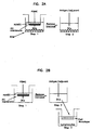
- FIG 1 Schematic diagram of an embodiment of the invention, using a Transwell ® device.
- HUVECs are grown to confluency on Transwell ® membranes and then total PBMC are applied to the upper chamber for ⁇ 1.5 h (step 1). Unbound cells are washed away and the remaining leukocytes are allowed to transmigrate for ⁇ 48 h.
- the Transwell ® is removed and DCs are then collected for analysis or pulsed with antigen for an additional ⁇ 2 days (step 2).
- the complexity of the membrane device can be increased by, for example, the inclusion of secondary cell populations, ECM materials and additional membrane layers.
- Monocytes that traverse through an endothelial monolayer can contact ECM in the lower chamber of the membrane device (A).
- Two membrane devices can be used to mimic the normal pathway of monocyte migration from the blood into the tissue (through the HUVECs) and from the tissue into the lymphatics (through a second cell layer, such as, for example, lymphatic endothelial cells).
- the second monolayer can be cultured on the upper (B) or lower (C) side of the membrane device, mimicking transmigration or reverse transmigration, respectively.
- the membrane can be coated on both sides with the same or different cell types (D); ECM can also be incorporated into the lower chamber with this design,(E).
- a modified Transwell ® can be constructed that contains a central chamber sandwiched between two membranes/cell monolayers (F). Fibroblasts or other cells types that are important in DC differentiation or antigen-presenting activity can be included in the lower chamber of a single membrane device (G or H) or in the middle of a dual membrane device (1 and J). ECM can also be incorporated into the dual-membrane device (H).
- HUVECs form confluent monolayers on Transwell ® membranes. Primary HUVECs were seeded in the upper chamber of Transwell ® s and analyzed for confluency and the formation of tight-gap junctions.
- A On day 7 after seeding, the cells were fixed, surface-labeled with an antibody specific for CD31, and the nuclei were stained with DAPI.
- B At the indicated time points, electrical resistance (TEER) readings were collected and normalized against the values for empty Transwell ® s on the same day. The error bars represent 1 SD of triplicate readings in each well.
- C Diffusion through the endothelial layer was measured with a 70kDa FITC-dextran conjugate at the indicated time points.
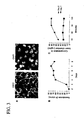
- FIG. 4 Monocyte transmigration through an endothelial monolayer is sufficient to trigger their differentiation towards a DC phenotype.
- A Cells that passed through a PC membrane in the absence (left) or presence (right) of a HUVEC monolayer were imaged by phase microscopy (20 ⁇ objective). Arrows indicate contaminating red blood cells or lymphocytes.
- B CD14-purified monocytes (non-transmigrated) were put into culture and then labeled with monoclonal antibodies specific for the indicated markers ⁇ 2 d later. The dotted line indicates background fluorescence with the appropriate isotype control.
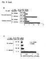
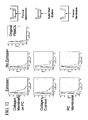
- Transwell ® -derived DC are potent stimulators of antigen-specific T cell responses.
- Transwell ® -derived DC were pulsed with antigen and cultured at a ⁇ 1:20 ratio with autologous T cells that had been labeled with CFSE. About 7 d later, the T cells were restimulated with autologous antigen-pulsed DC ( ⁇ 1:10 ratio to T cells) for -8 h and then assayed for IL-2 production by ICCS. Unpulsed DC were included as a negative control.
- A Dot plots showing representative CFSE and IL-2 staining patterns. The capacity of Transwell ® - and cytokine-derived DCs to stimulate recall C .
- albicans-specific T cell responses were compared in (B), while transmigrated cells from HUVEC-negative and -positive Transwell ® s served as APCs in (C).
- the T cells were analyzed for cytokine production by flow cytometry and the graph shows the frequency of lymphocyte-gated CD3 + CFSEl low IL-2 + cells. Different donors were used in each assay.
- FIG. 6 Example configurations of the vaccination site.
- Figure 7 An example of laser-micromachined polycarbonate (PC) membranes in a Transwell ® -based model and an outline of the process of casting collagen in a well-based model.
- PC polycarbonate
- Figure 8 Cell migration within the collagen membrane and transmigrated cells on the bottom of the plate.
- Figure 9 Cell migration and reverse transmigration.
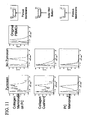
- FIG. 10 Levels of expression of HLA-DR in the migrated cells each model.
- FIG. 11 Levels of expression of CD86 in the migrated cells each model.
- FIG. 12 Levels of expression of CCR7 in the migrated cells each model.
- FIG 14 Antigen introduction into the VS model.
- the flipping collagen membrane model as an example.
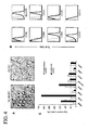
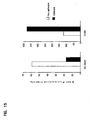
- FIG. 15 Adherent transmigrated monocytes phenotypically resemble macrophages.
- PBMCs were applied to the upper chamber of a Transwell ® containing HUVEC and at ⁇ 48 h the migrated, non-adherent and adherent cells were collected from the lower chamber.
- the cells were labeled with specific antibodies and analyzed by flow cytometry.
- the MFI for each marker on adherent and non-adherent cells are represented graphically.
- FIG 16 Transmigrated APC have phagocytic activity.
- the non-adherent transmigrated APC were harvested from Transwell ® s and incubated with FITC-labeled dextran beads ( ⁇ 1 ⁇ m) or zymosan particles, in the absence (thin line) or presence (thick line) of 20 ⁇ g/mL cytochalasin D, for -24 h.
- the cells were analyzed by flow cytometry in the presence of trypan blue, which quenches any extracellular FITC fluorescence. This ensures that the only signal detected originates from material within the cell.
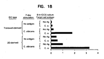
- FIG. 18 A transformed endothelial cell line can he used to trigger monocytes differentiation to DCs in the Transwell ® system.
- the ability of Transwell ® -derived APC from cultures containing primary and transformed HUVEC were compared, as described in Fig. 5 , using PBMC from a single donor. This data is representative of at least 3 experiments.
- Embodiments of the present invention are described in the claims.
- the present specification describes invention a simple and convenient Transwell ® -based culture method for the endothelial cell-mediated differentiation of DCs from blood monocytes.
- This system produces DCs with a frequency and purity comparable to more traditional culture methods for culturing DCs in vitro, but does so in only about two days, in the absence of exogenous factors and without the need for a tissue construct containing a support matrix.
- the transmigrated APCs derived from these cultures resemble classical in vitro DCs in their expression of MHC and costimulatory molecules and capacity to induce antigen-specific T cell responses.
- DCs Human dendritic cells for research and clinical applications are typically derived from purified blood monocytes that are cultured in a cocktail of cytokines for a week or more. Because it has been suggested that these cytokine-derived DCs may be deficient in some important immunological functions and might not accurately represent antigen-presenting cell (APC) populations found under physiologic conditions, there is a continuing need for methods that allow for the generation of DCs in a more physiologically relevant manner. Previous studies have demonstrated that endothelial cells can be used to promote the differentiation of monocytes into DCs.
- APC antigen-presenting cell
- the specification describes a simple and reliable method for generating large numbers of highly purified DCs that is based on a single migration of, for example, human blood monocytes through, for example, human umbilical vein endothelial cells (HUVECs) that are cultured in, for example, a Transwell ® device, or another similar device.
- the resultant APC 3 harvested from the lower Transwell ® chamber, resemble other in vitro -generated DC populations in their expression of major histocompatibility (MHC) and costimulatory molecules, ability to phagocytose foreign antigens, and capacity to trigger antigen-specific T cell responses.
- MHC major histocompatibility
- costimulatory molecules ability to phagocytose foreign antigens
- a fast growing, transformed endothelial cell line may be used, instead of primary HUVECs, to trigger the differentiation of monocytes into iDCs.
- Figure 1 provides a diagrammatic representation of the method, which starts with a layer of endothelial cells being grown to confluency in, for example, a Transwell ® chamber.
- a non-immunogenic and biologically inert polycarbonate (PC) membrane with, for example, ⁇ 1-5 ⁇ m pores, preferably ⁇ 5 ⁇ m pores that permit cell transmigration, provides support for the growth of HUVEC.
- the membrane is housed in an upper chamber that is suspended over, and is separable from, a lower chamber (tissue culture well).
- the endothelial cells permit the selective passage of monocytes through the membrane and concomitantly regulate and promote their differentiation into DCs.
- the upper chamber is removed and antigen, in the presence or absence of additional maturation stimuli; is added to the DCs in the lower chamber.
- the specification describes method using endothelial cells cells to drive the development of DCs in about two days, in the absence of any exogenous growth factors and without the pre-selection of monocytes from bulk PBMC.
- This method loosely replicates the process of blood monocyte extravasation through vessel walls, allows the generation of a highly purified APC population that resemble classical DCs in morphology, phenotype, and function.
- Circulating monocytes can differentiate into either iDCs or macrophages once they traverse the vasculature into tissues.
- the tissue construct described here supports the differentiation of blood monocytes into both cell types; cells that reverse-transmigrate out of the subendothelial collagen resemble iDCs, whereas macrophages remain in the extracellular matrix ( Qu et al, (2003) J lmmunol 170, 1010-1018 ; Randolph et a/. (1998) Science 282, 480-483 ).
- the geometry of the Transwell ® device, with monocytes traversing a confluent endothelial layer in the upper chamber, is such that both subpopulations are collected in the lower chamber.
- the non-adherent/loosely adherent iDCs are readily isolated from the strongly adherent macrophages by gently washing the wells with warm media; this approach yields 90% pure DCs (data not shown).
- ⁇ 100 ⁇ 10 6 PBMC applied to the Transwell ® -endothelial cell system yields ⁇ 5 ⁇ 10 6 non-adherent iDCs, which is comparable to the ⁇ 4 ⁇ 10 6 iDC s that can be expected when monocytes are purified from the same number of PBMC and cultured in exogenous cytokines for ⁇ 7 days (data not shown).
- DCs are a heterogeneous population, with phenotypes that are reflective of the tissue microenvironment in which they are found, it has thus far been difficult to identify a single marker that is common to all DC populations. For this reason, it is important to use several criteria to accurately discriminate DCs from other cell types.
- the non-adherent APCs harvested from the Transwell ® system had many of the functional attributes that are characteristic of DCs derived from other in vivo and in vitro sources. For instance, the cells had long processes, or dendrites, extending from the cell body (data not shown), which have been shown to aid antigen presentation by increasing the surface area of the cell.
- Transwell ® -derived APC had all the functional traits of DCs, they expressed a unique surface profile that differed from other in vitro DC populations.
- Cytokine-derived human DCs i.e ., those generated from monocyte's that have been cultured in GM-CSF and IL-4) are typically negative for the monocyte marker, CD14, and positive for the DC marker, CD1 a.
- Transwell ® -derived DCs had a marker profile that included the expression of CD14 and a lack of CD1a.
- CD1a the lack of CD1a on Transwell ® -derived APCs is not unexpected as it has previously been demonstrated that DCs derived in culture media containing human serum lack expression of this particular surface protein.
- APCs derived from Transwell ® s containing fetal bovine serum would express CD1a. If compared solely against cytokine-derived DC, the retention of CD14 on Transwell ® -derived APCs might suggest that these cells have not fully differentiated into DC.
- the flexibility of the system of the present invention makes it well suited for the study of different DC populations, such as those found in various tissue niches in vivo. While in the examples described here, HUVECs were used to drive the differentiation of monocytes into DCs, in other embodiments of the invention, other endothelial cell populations can be used within the Transwell ® system to preferentially drive the differentiation of monocytes into other tissue-specific DC subpopulations. For example, a previous report showed that intestinal epithelial cells cultured in a Transwell ® bucket gave rise to a unique DC population that lacked costimulatory and MHC class II molecules and were poor stimulators of T cell responses. This in vitro population resembled tolerogenic DCs found in the intestinal mucosa in vivo.
- Transwell ® device allows for multiple embodiments that permit a greater dissection of DC development/differentiation pathways.
- transmigrated APCs harvested from a Transwell ® can be passed through a second Transwell ®e chamber containing a monolayer of lymphatic cells, a process that more closely recapitulate the migration of matured/activated tissue-resident DCs through the lymphatics in vivo.
- fibroblasts contained in the lower chamber of the Transwell ® device can serve as a source of inflammatory signals and act as an antigen depot during the application of certain adjuvants or pathogens.
- Other support cells, such as stromal cells, can also be contained in the lower chamber of the device of the present invention.
- the specification describes a novel and convenient approach for generating large numbers of highly purified human DCs from blood monocytes.
- a flexible and well-characterized Transwell ® device as a support structure for the culture of endothelial cells and transmigration of monocytes through this confluent monolayer, a population of non-adherent APCs was generated that resemble other in vitro DC populations in phenotype and function.
- a transformed endothelial cell line may be promote the development of DCs.
- the specification describes methods which provide a simple means of generating human DCs in a manner that more closely mimics their development in vivo.
- the present invention involves the use of a membrane device, for example, a commercially available Transwell ® , as a means of developing DCs to participate in an immune response.
- a membrane device for example, a commercially available Transwell ®
- a non-immunogenic and biologically inert membrane with pores of a size that permit cell transmigration provides support for the growth of endothelial cells (e.g ., human umbilical vascular endothelial cells (HUVEC) or other mammalian endothelial cells or cell lines), enabling the selective passage of monocytes through the membrane and concomitantly regulating and promoting their differentiation into DCs.
- endothelial cells e.g ., human umbilical vascular endothelial cells (HUVEC) or other mammalian endothelial cells or cell lines
- the membrane can be housed in an upper chamber suspended over and separable from a lower tissue culture well (chamber).
- An embodiment of the invention is shown in figure 1 .
- endothelial cells can be cultured to confluency on the porous membrane and then PBMC can be applied to the upper chamber ( Fig. 1 , step 1).
- PBMC can be applied to the upper chamber
- antigen in the presence or absence of additional stimuli, is added to the lower chamber ( Fig. 1 , step 2).
- the DCs acquire the antigen and then can be used, for example, in T cell stimulation experiments or other APC functional assays.
- a porous membrane bearing an endothelial cell layer that is close to, or has achieved, confluent or even multilayer growth is a convenient method for developing dendritic cells for in vitro experimentation and in vivo therapeutics.
- the membrane supports endothelial cell growth and can provide a barrier that selects for or enriches monocytes from peripheral blood cells. If, for example, peripheral blood cells are added to an upper chamber that has an endothelial cell-layered membrane as its bottom, such as in a Transwell ® , monocytes preferentially migrate through the cell-layered membrane and differentiate into DCs by virtue of their interaction with the endothelial cells ( Qu et al. (2003) J lmmunol 170:1010-1018 ; Randolph et al. (1998) Science 282:480-483 ).
- the transmigrated cells enter a lower chamber that is separate and free from the upper chamber, such that the possibility of "reverse transmigration" observed in the collagen substrate-endothelium model ( Randolph et al. (1998) Science 282:480-483 ) is significantly reduced.
- antigen can be easily added to this separate compartment. for processing by the immature DCs.
- Additional agents such as adjuvants, proinflammatory agents, vaccines, cosmetics, drugs, biologics, immunotherapy candidates, or chemical compounds, can also be added to the lower chamber to assess their effects, independently or together with antigen, on the activation and maturation of the transmigrated cells.
- the modular design of the membrane device allows for multiple cell layers and different matrix materials, and other compounds such as cytokines or stimulants to be introduced into the system.
- the layers can be discrete and separable, thereby allowing the cells to undergo sequential processes without interference from the products or reactants of a previous event.
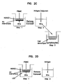
- monocytes that transverse an endothelial layer in the upper chamber can interact with an ECM (extracellular matrix) material in the lower chamber of the Transwell ® device ( Fig. 2A ).
- ECM material used in any of the embodiments of the invention preferably comprises a material selected from the group consisting of gelatin, collagen, synthetic ECM materials, PLGA, PGA, natural ECM materials, chitosan, protosan, and mixtures thereof.
- Transmigrated DC that have processed antigen can also be passed through a second chamber with a membrane bearing a layer of lymphatic or other endothelial cell types on its top ( Fig. 2B ) or bottom ( Fig. 2C ).
- cells can be cultured on both the upper and lower sides of the membrane, such that monocytes pass through two cell monolayers before acquiring antigen or agents (as defined above) ( Fig. 2D ).
- ECM material could also be incorporated into this design ( Fig. 2E ) and also can be present with the endothelial cells being cultured on the membrane.
- monocytes will migrate through one cell layer, acquire antigen or agents (as defined above), and then migrate through a second cell population ( Fig. 2F ).
- the separable membranes with independent chambers allows for the easy addition of reactants and flexibility in the timing of events.
- the migration of monocytes through lymphatic endothelial cells can further promote their differentiation towards DC, similar to the maturation that occurs when the cells migrate into lymphatic vessels under physiologic conditions.
- fibroblasts could serve as a source of inflammatory signals and act as an antigen depot during the application of certain adjuvants or pathogens.
- Other support cells such as stromal cells, can also be contained in the system.
- these cells could be added to the lower chamber of the one-membrane device ( Figs. 2G and 2H ) or between the layers in a dual-membrane device ( Figs. 2I and 2J ).
- ECM can also be added between the membrane layers ( Fig. 2J ).
- Transwell ® s are commercially available in 12-, 24-, and 96-well plate and larger scale formats, and robotic liquid handling systems are available that can automate the transport of cells, liquids, chemical agents, or other materials between wells, and the removal of the upper Transwell ® chamber for access to the lower chamber.
- Endothelial cells are suitable for use in the Transwell ® device.
- Primary endothelial cells are available from medical institutions and can be purchased commercially ( e.g ., Cambrex (East Rutherford, NJ) and VEC Technologies (Rensselaer, NY)). Although freshly thawed cells were used in the experiments described here, expanded primary cells can be used with similar results. Secondary (immortal) endothelial cells are convenient because of their longevity and rapid growth rates. For example, experiments suggest that EA.hy926, a long-term HUVEC line ( Edgell et al. (1983) Proc Natl Acad Sci USA 80:3734-3737 ), and primary endothelial cells trigger transmigrated monocytes to undergo a similar differentiation program.
- HUVECs Primary HUVECs were obtained, for example, at passage #2 from VEC Technologies (Rensselaer, NY). Frozen stocks of primary endothelial cells were thawed and applied directly to 12-well Transwell ® devices (Coming, Corning, NY) at a density of ⁇ 9 ⁇ 10 5 cells/cm 2 in MCBD-131 complete media (VEC Technologies). ⁇ 85% of the media was exchanged every other day and HUVECs were typically cultured on Transwell ® membranes for ⁇ 7 d before being used in monocyte migration assays. Although Transwell ® s with ⁇ 5 ⁇ m polycarbonate membranes were used for these assays, other membranes of various inert materials and/or pore sizes are also suitable.
- HUVEC confluency The formation of tight-gap junctions in HUVEC monolayers was visualized by fluorescence microscopy.
- the staining process involved fixing the cells with 3.2% paraformaldehyde (32% stock from Electron Microscopy Science, Hatfield, PA) for ⁇ 10 min and permeabilizing them with methanol at ⁇ 20°C for ⁇ 5 min.
- the cells were then labeled with a 1:10 dilution of an antibody against human CD31 (M89D3; BD Pharmingen) for ⁇ 1 h at RT in a humidified chamber, followed by 1 mg/mL DAPI (Sigma) for ⁇ 5 min to label the nuclei.
- the cells were fixed again with 3.2% paraformaldehyde for ⁇ 10 min at RT and then covered with GelMount (Biomedia, Foster City, CA). Extensive washes with phosphate-buffered saline (PBS) were included between steps. The labeled cells were examined using an Olympus IX81 fluorescence microscope. The permeability of the endothelial cell monolayer was measured by a standard diffusion assay.
- PBS phosphate-buffered saline
- HUVECs were cultured on membranes as described above, except that the cells were switched into assay media (Iscove's modified Dulbecco's medium (IMDM; Mediatech, Inc., Herndon, VA), containing 5% heat-inactivated (56°C, 30 min.) autologous or human AB serum, 2 mM L-glutamine, 100 U/ml penicillin, and 0.1mg/ml streptomycin) 24 h prior to, and diffusion media (1MDM) supplemented with 1 % BSA) 1 h before the start of the diffusion assay.
- assay media Iscove's modified Dulbecco's medium (IMDM; Mediatech, Inc., Herndon, VA), containing 5% heat-inactivated (56°C, 30 min.) autologous or human AB serum, 2 mM L-glutamine, 100 U/ml penicillin, and 0.1mg/ml streptomycin
- FITC-conjugated dextran 70 kDa; Sigma
- diluted to 1 mg/mL in diffusion media was added to the upper well and four 100 ⁇ L aliquots were taken from the lower well at 30 min intervals.
- an equal volume of fresh diffusion media was added to the lower chamber after the samples were removed.
- the fluorescence of the media samples were measured with a Bio-Tek (Winooski, VT) Synergy HT spectrophotometer, using a 480/520 nm filter set.
- a standard curve established by measuring the fluorescence of known amounts of FITC-dextran, was used to calculate the concentration of dextran that permeated through the HUVEC monolayer.
- Transendothelial electrical resistance was used as a second method to examine the integrity of the HUVEC monolayer .
- Endothelial cells were cultured on Transwell ® membranes in MCBD-131 complete media, switched into assay media for 24 h, and then TEER was calculated with a Voltohmeter (EVOM-ENDOHM-6, World Precision Instruments, Sarasota, FL) using a resistance chamber compatible with the Transwell ® inserts. The voltohmeter was calibrated each day, per the manufacturer's instructions, and 3 individual readings were taken for each well. The TEER readings of the HUVEC monolayers on Transwell ® membranes were normalized against values collected from Transwell ® inserts alone (in the absence of endothelial cells).
- PBMC preparation Human PBMC preparation .
- Enriched leukocytes were obtained from the Central Florida Blood Bank (Orlando, FL). All of the donors were in good health and all blood products were negative for blood-borne pathogens, as detected by standard assays.
- PBMCs were enriched by density centrifugation. Briefly, ⁇ 45-50 mL leukocytes were resuspended in citrate buffer (PBS containing 0.1% BSA and 0.6% Na citrate) to a final volume of ⁇ 140 mL.
- citrate buffer PBS containing 0.1% BSA and 0.6% Na citrate
- Diluted blood ( ⁇ 35 mL) was layered onto ⁇ 15 mL Ficoll-Paque PLUS (Amersham Biosciences, Piscataway, NJ) in a 50 mL conical tube and centrifuged (400 g, 25 min, room temperature). The buffy coats were removed, washed twice with citrate buffer, recentrifuged (400g, 10 min, 4°C), and resuspended in assay media. The PBMC were kept at 4°C for up to 24 h prior to being used in assays or were frozen and stored in liquid nitrogen for extended storage.
- PBMC Monacyte transmigration assays.
- PBMC peripheral blood mononuclear cells
- ⁇ 10 ⁇ 10 6 total PBMC were applied to each 12-well Transwell ® and incubated for ⁇ 1.5 h.
- the upper chambers were washed twice with assay media to remove non-adherent and loosely bound cells, and the Transwell ® plates were incubated for an additional ⁇ 48 h to allow for leukocyte transmigration and differentiation.
- the upper chambers were then removed and the cells in the lower chamber were harvested for phenotypic or functional analyses.
- DC phenotyping PE-, APC-, or PerCP-Cy5.5-conjugated monoclonal antibodies specific for human CD1a (HI149), CD14 (M5E2) CD16 (3G8), CD40 (5C3), CD80 (L307.4), CD86 (2331), CD83 (HB15e), and HLA-DR (L243) were purchased from BD Pharmingen and diluted as suggested by the manufacturer.
- transmigrated cells from HUVEC-negative and - positive Transwell ® s were collected at various times following PBMC seeding and labeled with specific antibody for ⁇ 45 min at 4°C, washed extensively, and fixed with 2% paraformaldehyde.
- the buffer used for cell labeling was PBS with 2% BSA and 0.05% sodium azide. Samples were acquired on a FACSArray (BD Pharmingen) and FlowJo software (Treestar, Ashland, OR) was used for analysis.
- T cell stimulation assay About two days after PBMC were applied to the HUVEC monolayer, the transmigrated cells were pulsed with ⁇ 20 ⁇ g/mL Candida albicans protein antigen extract (Greer Laboratories, Lenoir, NC). ⁇ 48 h later, transmigrated cells were collected, washed, and combined with syngeneic T cells. Cytokine-derived DCs were prepared using standard procedures.
- monocytes were purified from total PBMC using anti-CD14 antibody-conjugated magnetic beads (Miltenyi Biotec) and then cultured for ⁇ 7 d at ⁇ 1 ⁇ 10 6 /ml in assay media containing ⁇ 100 ng/mL GM-CSF (R & D Systems, Minneapolis, MN) and ⁇ 25 ng/mL IL-4 (Endogen, Rockford, IL).
- the cells were pulsed with ⁇ 20 ⁇ g/mL Candida albicans on day 5 of culture.
- Frozen stocks of syngeneic PBMC were used as a source of lymphocytes.
- Total T cells were purified by negative selection, using magnetic beads and the autoMACS system (Miltenyi Biotec (Auburn, CA)).
- Purified T cells were washed with PBS, labeled with 5 ⁇ M CFDA-SE (CFSE; Invitrogen, Carlsbad, CA), and then washed two times with assay media, to quench the labeling reaction.
- the cells were plated at ⁇ 2-3 ⁇ 10 5 /well in 96-well flat-bottom tissue culture plates (Corning, Inc., Corning, NY) and DCs were added at the indicated ratios. Each well contained a final volume of ⁇ 200 ⁇ L.
- Target APCs cytokine-derived DCs
- Candida albicans were prepared as described above.
- TNF ⁇ Endogen
- target DCs were cultured with activated T cells for -8 h at a ⁇ 1:10 ratio in the presence of 1 ⁇ g/mL brefeldin A (Sigma, St. Louis, MO).
- the cells were surface-labeled with an antibody specific for CD3 ⁇ (SK7; BD Pharmingen), and then intracellularly labeled with an antibody specific for human IL-2 (Endogen) using Cytofix/Cytoperm and perm/wash reagents from BD Pharmingen.
- HUVECs affect the differentiation state of transmigrating monocytes
- cells that passed through a Transwell ® membrane in the presence or absence of an endothelial cell layer were labeled with antibodies specific for surface proteins characteristic of DCs. Because it was possible that the simple process of migrating through a porous membrane might trigger an altered phenotype in monocytes, non-migrated cells were included for comparison.
- CD14-positive monocytes were cultured for ⁇ 2, d in assay media without any exogenous cytokines and then profiled by flow cytometry ( Fig. 4B ).
- CD86 and CD80 provide important stimulatory signals for naive T cell activation and proliferation.
- CD86 was already expressed at a high level on purified monocytes ( Fig. 4B )
- CD80 was negative on non-migrated monocytes and only increased after monocytes migrated through an endothelial monolayer ( Figs. 4B and 4C , respectively). Similar results were obtained for CD40, a marker that provides maturation signals to the DC itself.
- Transwell ® -derived DCs were gauged against cytokine-derived DCs from the same donor in a syngeneic T cell stimulation assay. Both cell types efficiently triggered T cell proliferation (CFSE dilution) and elicited a similar frequency of effector cells that were capable of secreting IL-2 following short-term antigen stimulation ( Fig. 5 ). This response was likely antigen-specific as the only T' cells capable of secreting IL-2 at levels above background were those that encountered Candida albicans during both the 7-day stimulation and 8 h ICCS assay ( Fig. 5 ).
- antigenic molecules introduced into an artificial immune system are acquired by dendritic cells (DCs) at the vaccination site (VS).
- the DCs are then transferred to the lymphoid tissue equivalent (LTE), where they present the antigen to T cells, activating their immune function.
- LTE lymphoid tissue equivalent
- Activated helper T cells co-stimulate B cells to induce antibody production, while activated cytotoxic T cells lyse antigen-bearing cells.
- Solublized antigen can also be introduced to the LTE to directly activate B cells.
- Vaccination Site various configurations have been used with collagen, a porous polycarbonate membrane, incorporation of antigen into a membrane-based model, and the ability to increase the complexity of a membrane based VS model in a manufacturable manner (e.g ., the addition of stromal cells, the addition of an epithelium, etc. ) .
- the porous polycarbonate (PC) membrane can act as a support layer for the extracellular matrix (ECM), such as collagen (see examples in Figure, 6 ).
- ECM extracellular matrix
- a collagen cushion in a standard 96-well plate model we examined a collagen cushion in a standard 96-well plate model, a simple Transwell ® -based model, a model with a collagen matrix integrated with a filter membrane, a model with the polycarbonate membranes laser-micromachined to increase porosity and potentially cell flux, and a model in which two endothelial layers were created, one on the top and one on the bottom of the VS membrane construct to examine the influence of one endothelial layer versus two endothelial layers on cell migration pathways, cell migration numbers, DC phenotype, and DC function.
- a collagen membrane was cast in a well-based format.
- a method to cast collagen in a membrane format in a simple well-based system As examples, we have examined three support structures with the collagen membrane: a continuous polycarbonate (PC) membrane ( ⁇ 8 ⁇ m pore diameter), a laser-micromachined PC membrane (a range of pore diamaters available [ ⁇ 100-550 ⁇ m]), and a nylon mesh (a range of mesh size available [ ⁇ 100-500 ⁇ m]).
- Figure 7 shows examples of the laser-micromachined PC membranes and steps taken to create the collagen membrane in either a 96-well format or a simple single-well format.
- Figure 8 shows the appearance of migratory cells throughout the collagen membrane, as well as cells on the bottom of the plate that migrated completely through the construct. Cell migration through the constructs also depended on other factors such as collagen density, the thickness of the collagen membrane, and adding a second HUVEC layer to the bottom of the construct.
- FIG. 10 , 11 , and 12 show the levels of expression of HLA-DR, CD86, and CCR7 of the migrated cells each model, respectively.
- zymosan known to mature DCs
- the levels of HLA-DR expression for the mature migrated cells (exposed to zymosan) from the collagen on PC membrane model was very similar to that of the collagen cushion model, and both were higher than that seen for the Transwell ® model. The same was also seen for the levels of CD86 and CCR7, shown in Figures 11 and 12 , respectively.
- the phenotype analysis showed that the cells migrating from the model of collagen on a PC membrane have a similar phenotype to those migrating from the collagen cushion model and we expect them to function similarly.
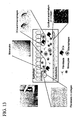
- FIG. 13 we show how complexity can be added.
- we can form a confluent endothelium over the collagen membrane we have been able to observe monocyte transendothelial migration into the collagen membrane, we have been able to observe monocyte differentiation into DCs and resident macrophages, we have been able to introduce fibroblasts into the collagen, and we have been able to show that these embodiments can be manufactured in, for example, a 96-well format.
- FIG. 14 shows an example of how antigen can be added to a membrane-based AIS integrated into a well-based format. This figure shows a means of introducing antigen into a collagen membrane with a confluent HUVEC layer present.
- PBMCs are applied to the HUVEC face and allowed to extravasate through the endothelium. After ⁇ 1.5 h (typical protocol), non-migratory cells were washed off the endothelium surface and the bucket/well can then be inverted and placed into the LTE section of an AIS system. The antigen, the vaccine and/or adjuvants can then be introduced through the back side of the inverted VS construct.
- Maturing DCs can then migrate out of the VS and fall into the LTE below. Antigen uptake occurs while the monocyte-derived DCs are in the collagen membrane. An additional benefit of this approach is that solubilized antigen that is not engulfed by APCs, can also fall into the LTE where it can be processed directly by B cells.
- HUVEC cultures were grown in MCDB-131 complete media, containing 10% fetal bovine serum, 10 ng/mL endothelial growth factor, 1 ⁇ g/mL hydrocortisone, 0.2 mg/mL ENDOGRO, 0.1 mg/mL heparin, and an antibiotic/antimycotic solution (all reagents from VEC technologies).
- the transformed endothelial cell line, EA.hy926 Edgell et al. (1983) Proc Natl Acad Sci USA 80, 3734-3737
- Cora-Jean Edgell Universality of North Carolina at Chapel Hill, Chapel Hill, NC.
- These cells were grown in M199 media (Invitrogen), containing 10% fetal bovine serum and passaged ⁇ 1:10 every -6-7 days.
- IMDM Iscove's modified Dulbecco's medium
- IMDM Iscove's modified Dulbecco's medium
- penicillin 100 U/mL penicillin and 0.1 1mg/mL streptomycin (all from Sigma)
- varying concentrations of heat-inactivated 56°C, 30 min
- human plasma or fetal bovine serum (HyClone Laboratories).
- Transmigratory monocytes were collected ⁇ 2 d after PBMC application and incubated overnight with 1 ⁇ m-diameter orange fluorescent beads or AlexaFluor 488-labeled zymosan particles at a ratio of ⁇ 3:1 to the cells (both reagents from Molecular Probes). Then, the cells were washed once in FACS buffer and analyzed by flow cytometry. In some cases, the APCs were treated with 20 ⁇ g/ ⁇ L cytochalasin D for 2 h at 37°C prior to incubation with the beads or particles to block phagocytic activity ( Fig. 16 ).
- Immune cells and the various activation/maturation states of these populations, are often defined by their expression of a particular pattern of surface proteins. Therefore, the impact of HUVECs on monocyte differentiation was examined initially by comparing the phenotype of transmigrated monocytes that had contacted endothelial cells with those that had passed through an empty Transwell ® bucket. As it was possible that monocytes passing through a porous PC membrane in the absence of a HUVEC layer might also experience a change in their marker profile, non-migrated CD14 + cells that had been cultured for two days in assay media absent of exogenous factors were used to establish a baseline expression level for each marker of interest. In Fig.
- the median fluorescence intensity (MFI) of markers on the non-migrated monocytes was set at 100% and compared against the change in MFI of the same markers on monocytes that had transmigrated through the PC membrane 48 h earlier.
- MFI median fluorescence intensity
- the presence of a HUVEC monolayer caused a marked increase in expression of two molecules, CD40 and CD80, on the transmigrated APCs that provide critical costimulatory/activating signals to DCs and T cells, respectively.
- the low affinity IgG receptor, Fc ⁇ RIII (CD16) which is important for the uptake of antibody-coated proteins, was upregulated on APCs that migrated through a HUVEC layer, though it was also elevated to a lesser extent on cells that passed through a PC membrane lacking an endothelial monolayer.
- Transwell ® -derived cells The increased expression of costimulatory ligands on Transwell ® -derived cells suggested that a single transendothelial migration might be sufficient to trigger the differentiation of these cells into potent APCs. Additional experiments were performed to determine whether the changes in phenotype were also associated with increased functionality of the Transwell ® -derived cells. For instance, a defining characteristic of DCs is their ability to capture soluble and particulate material for MHC class I and II processing and presentation. The ability of APCs to acquire fluorescently labeled ⁇ 1 ⁇ m latex beads and zymosan (yeast) particles is indicative of strong phagocytic activity. As shown in Fig.
- zymosan particles were captured by nearly all of the Transwell ® -derived APC, and about 30% of the cells acquired latex beads. While both materials are captured via mannose receptors, it is possible that the reduced accumulation of latex beads within the APCs could be related to the size of the beads, a it has been previously noted that small ( ⁇ 0.2 ⁇ m) beads are far more efficiently phagocytosed than larger beads. On the other hand, the increased efficiency of yeast particle uptake by the Trdnswell ® -derived cells could be mediated by other receptors, such as TLR2. The addition of cytochalasin D, an inhibitor of phagocytosis, triggered a partial reduction in the uptake of both materials by the Transwell ® -derived APCs, suggesting that the particles were ingested by an active mechanism.
- DCs Another hallmark feature of DCs is their ability to undergo a maturation/activation program, which includes an altered expression of molecules associated with antigen presentation and T cell stimulation, following an encounter with various inflammatory stimuli.
- a maturation/activation program which includes an altered expression of molecules associated with antigen presentation and T cell stimulation, following an encounter with various inflammatory stimuli.
- migrated cells harvested from the lower chamber were stimulated for ⁇ 24 h with TNF- ⁇ and LPS and analyzed by flow cytometry for changes in their surface marker profile ( Fig. 17 ). Markers associated with antigen uptake, such as the low affinity Fc receptor, CD32, decreased on activated DC, while others, such as CD40, CD80 and CD86, that serve important costimulatory functions for the induction of adaptive immunity, were elevated on the TNF- ⁇ /LPS-treated cells.
- MHC class II HLA-DR
- CD14 CD14 were unaffected by the maturation stimuli further highlights the unique phenotype of Transwell ® -derived APCs, as cytokine-derived human matured DCs are typically triggered to upregulate MHC class II and further downregulate CD14 (data not shown).
- transendothelial-migrated APCs from the Transwell ® device were evaluated for their ability to induce antigen-specific T cell responses, including lymphoproliferation and effector function.
- Candida albicans C. albicans
- Transwell ® -derived APC were pulsed with a whole protein antigen from C .
- TNF- ⁇ albicans, matured with TNF- ⁇ , and then cultured for ⁇ 7 d with autologous T cells that had been labeled with the proliferation-sensitive dye, 5-(and-6-)-carboxyfluorescein diacetate, succinimidyl ester (CFDA-SE; CFSE). Thereafter, the T cells were evaluated for proliferation (CFSE dilution) and the production of cytokines following short-term TCR stimulation with target APCs that had been pulsed with specific antigen. The presence of C .
- albicans-specific CFSE low IL-2 + T cells following a short-term antigen restimulation, provides strong evidence of the capacity of the Transwell ® -derived APCs to trigger the complete differentiation of antigen-specific T cells into fully competent effectors.
- Controls in this assay included DCs stimulators and targets that had not been pulsed with C . albicans antigens ( Fig. 18 ).
- the quality of the Transwell ® -derived APCs as stimulators ofT cell responses was gauged against cytokine-derived DCs that were prepared from the same donor. The results of Fig.
- Fig. 4C demonstrate, however, that the interaction of monocytes with endothelial cells is important to promote their complete differentiation into APCs, because cells that passed through a PC membrane alone were unable to trigger specific T-cell responses above background.
- the disparity in the frequency of T cells that responded to Transwell ® -derived DCs in Figs. 4B and 4C is likely due to differences in the immune histories of the two donors that were used in these experiments.
- the increased T cell stimulatory capacity of transendothelial-migrated monocytes could be related to the increased expression of costimulatory ligands, namely CD40 and CD86, on the Transwell ® -derived APCs ( Fig. 4C ), though further experimentation will be necessary to confirm this possibility.
- transendothelial-migrated non-adherent APCs resembled Transwell ® -derived APCs from primary endothelial cultures in surface marker phenotype pre- and post-stimulation with maturation factors (data not shown).
- T cell stimulatory capacity of APC derived from Transwell ® s that contained secondary HUVECs was comparable to other Transwell ® APCs and cytokine-derived DCs.
- Transwell ® -endothelial cell device provides a simple and quick approach to deriving DC-like cells from monocytes.
- the transmigrated monocytes morphologically resemble DCs and have increased expression of costimulatory molecules that are important in triggering complete T cell activation.
- Transwell ® -derived DCs are also very comparable to the well-characterized cytokine-derived DCs in generating T cell responses against Candida albicans in standard T cell assays.
- Transwell ® device The advantages of the Transwell ® device, including the short incubation time required to get DC differentiation from monocytes, the modular design that allows for increasing system complexity that might make DCs more comparable to in vivo APCs, and its relatively low cost, make it an attractive alternative to current methods for generating DCs for in vitro experimentation.
Landscapes
- Health & Medical Sciences (AREA)
- Life Sciences & Earth Sciences (AREA)
- Engineering & Computer Science (AREA)
- Organic Chemistry (AREA)
- Chemical & Material Sciences (AREA)
- Zoology (AREA)
- Wood Science & Technology (AREA)
- Bioinformatics & Cheminformatics (AREA)
- General Health & Medical Sciences (AREA)
- Biomedical Technology (AREA)
- Genetics & Genomics (AREA)
- Biotechnology (AREA)
- Biochemistry (AREA)
- General Engineering & Computer Science (AREA)
- Immunology (AREA)
- Microbiology (AREA)
- Sustainable Development (AREA)
- Epidemiology (AREA)
- Animal Behavior & Ethology (AREA)
- Public Health (AREA)
- Veterinary Medicine (AREA)
- Clinical Laboratory Science (AREA)
- Hematology (AREA)
- Cell Biology (AREA)
- Micro-Organisms Or Cultivation Processes Thereof (AREA)
- Apparatus Associated With Microorganisms And Enzymes (AREA)
- Measuring Or Testing Involving Enzymes Or Micro-Organisms (AREA)
- Investigating Or Analysing Biological Materials (AREA)
Priority Applications (1)
| Application Number | Priority Date | Filing Date | Title |
|---|---|---|---|
| EP10151003A EP2199384B1 (en) | 2005-12-21 | 2006-12-21 | A porous membrane device that promotes the differentiation of monocytes into dendritic cells |
Applications Claiming Priority (2)
| Application Number | Priority Date | Filing Date | Title |
|---|---|---|---|
| US75203305P | 2005-12-21 | 2005-12-21 | |
| PCT/US2006/049128 WO2007076061A1 (en) | 2005-12-21 | 2006-12-21 | A porous membrane device that promotes the differentiation of monocytes into dendritic cells |
Related Child Applications (1)
| Application Number | Title | Priority Date | Filing Date |
|---|---|---|---|
| EP10151003.0 Division-Into | 2010-01-18 |
Publications (2)
| Publication Number | Publication Date |
|---|---|
| EP1963491A1 EP1963491A1 (en) | 2008-09-03 |
| EP1963491B1 true EP1963491B1 (en) | 2011-01-12 |
Family
ID=37963795
Family Applications (2)
| Application Number | Title | Priority Date | Filing Date |
|---|---|---|---|
| EP06848082A Active EP1963491B1 (en) | 2005-12-21 | 2006-12-21 | In vitro method of evaluating the potential reaction of an animal to an agent |
| EP10151003A Active EP2199384B1 (en) | 2005-12-21 | 2006-12-21 | A porous membrane device that promotes the differentiation of monocytes into dendritic cells |
Family Applications After (1)
| Application Number | Title | Priority Date | Filing Date |
|---|---|---|---|
| EP10151003A Active EP2199384B1 (en) | 2005-12-21 | 2006-12-21 | A porous membrane device that promotes the differentiation of monocytes into dendritic cells |
Country Status (9)
| Country | Link |
|---|---|
| US (2) | US20070178076A1 (enExample) |
| EP (2) | EP1963491B1 (enExample) |
| JP (1) | JP2009521228A (enExample) |
| AT (2) | ATE495238T1 (enExample) |
| AU (1) | AU2006330951B2 (enExample) |
| CA (2) | CA2634119C (enExample) |
| DE (1) | DE602006019638D1 (enExample) |
| IL (1) | IL192090A (enExample) |
| WO (1) | WO2007076061A1 (enExample) |
Families Citing this family (22)
| Publication number | Priority date | Publication date | Assignee | Title |
|---|---|---|---|---|
| US7855074B2 (en) | 2004-04-28 | 2010-12-21 | Vaxdesign Corp. | Artificial immune system: methods for making and use |
| US7785806B2 (en) | 2004-04-28 | 2010-08-31 | Vaxdesign Corporation | Method for determining the immunogenicity of an antigen |
| US8298824B2 (en) | 2004-04-28 | 2012-10-30 | Sanofi Pasteur Vaxdesign Corporation | Methods of evaluating a test agent in a diseased cell model |
| US8030070B2 (en) * | 2004-04-28 | 2011-10-04 | Sanofi Pasteur Vaxdesign Corp. | Artificial lymphoid tissue equivalent |
| US7771999B2 (en) | 2004-04-28 | 2010-08-10 | Vaxdesign Corp. | Disease model incorporation into an artificial immune system (AIS) |
| US7709256B2 (en) * | 2004-04-28 | 2010-05-04 | Vaxdesign Corp. | Disease model incorporation into an artificial immune system (AIS) |
| US7785883B2 (en) | 2004-04-28 | 2010-08-31 | Vax Design Corp. | Automatable artificial immune system (AIS) |
| US8071373B2 (en) | 2004-04-28 | 2011-12-06 | Sanofi Pasteur Vaxdesign Corp. | Co-culture lymphoid tissue equivalent (LTE) for an artificial immune system (AIS) |
| US8003387B2 (en) | 2005-12-21 | 2011-08-23 | Sanofi Pasteur Vaxdesign Corp. | In vitro germinal centers |
| JP2009521228A (ja) * | 2005-12-21 | 2009-06-04 | ヴァックスデザイン コーポレーション | 単球の樹状細胞への分化を促進する多孔質膜デバイス |
| ATE525093T1 (de) * | 2006-06-27 | 2011-10-15 | Sanofi Pasteur Vaxdesign Corp | Modelle für die bewertung von impfstoffen |
| US8647837B2 (en) * | 2007-07-16 | 2014-02-11 | Sanofi Pasteur Vaxdesign Corp. | Artificial tissue constructs comprising alveolar cells and methods for using the same |
| AU2010225438A1 (en) * | 2009-03-17 | 2011-10-13 | The Heart Research Institute Ltd | Method of cell differentiation and uses therefor in screening, diagnosis and therapy |
| US8802747B2 (en) * | 2009-08-26 | 2014-08-12 | Molecular Imprints, Inc. | Nanoimprint lithography processes for forming nanoparticles |
| US10731129B2 (en) | 2012-03-07 | 2020-08-04 | Children's Medical Center Corporation | Methods of evaluating immunogenicity of an agent using an artificial tissue construct |
| US9423234B2 (en) | 2012-11-05 | 2016-08-23 | The Regents Of The University Of California | Mechanical phenotyping of single cells: high throughput quantitative detection and sorting |
| FR3045664A1 (fr) * | 2015-12-17 | 2017-06-23 | Oreal | Membrane pour tissu reconstruit comprenant des pores de plusieurs diametres |
| AU2017325009B2 (en) * | 2016-09-08 | 2023-07-20 | Sanofi Pasteur Vaxdesign Corp. | In vitro neonatal biomimetic (NMIMIC) model and methods of using same |
| KR101986092B1 (ko) * | 2016-09-13 | 2019-06-05 | 대한뉴팜(주) | 유두진피 섬유아세포의 고순도 분리 방법 |
| KR102492455B1 (ko) * | 2016-11-24 | 2023-01-27 | 알베오릭스 테크놀러지스 아게 | 세포 배양 시스템 및 방법 |
| US11175282B2 (en) | 2017-06-26 | 2021-11-16 | Biohope Scientfic Solutions For Human Health S.L. | Method for predicting and monitoring clinical response to immunomodulatory therapy |
| CN112221543B (zh) * | 2019-07-15 | 2024-05-10 | 京东方科技集团股份有限公司 | 细胞片层转移器件、运输装置和转移细胞片层的方法 |
Family Cites Families (39)
| Publication number | Priority date | Publication date | Assignee | Title |
|---|---|---|---|---|
| US5160490A (en) * | 1986-04-18 | 1992-11-03 | Marrow-Tech Incorporated | Three-dimensional cell and tissue culture apparatus |
| US5720937A (en) * | 1988-01-12 | 1998-02-24 | Genentech, Inc. | In vivo tumor detection assay |
| US5008116A (en) * | 1988-11-14 | 1991-04-16 | Frederick Cahn | Immunostimulatory microsphere |
| US5562910A (en) * | 1989-09-25 | 1996-10-08 | University Of Utah Research Foundation | Vaccine compositions and method for enhancing an immune response |
| US5188959A (en) * | 1989-09-28 | 1993-02-23 | Trustees Of Tufts College | Extracellular matrix protein adherent t cells |
| US5827641A (en) * | 1992-11-13 | 1998-10-27 | Parenteau; Nancy L. | In vitro cornea equivalent model |
| US5695996A (en) * | 1994-09-23 | 1997-12-09 | The United States Of America As Represented By The Department Of Health And Human Services | Artificial organ culture system |
| US6479258B1 (en) * | 1995-12-07 | 2002-11-12 | Diversa Corporation | Non-stochastic generation of genetic vaccines |
| US5739001A (en) * | 1996-10-29 | 1998-04-14 | E. I. Du Pont De Nemours And Company | Solid phase cell-based assay |
| US6177282B1 (en) * | 1997-08-12 | 2001-01-23 | Mcintyre John A. | Antigens embedded in thermoplastic |
| ATE428769T1 (de) * | 1997-10-27 | 2009-05-15 | Univ Rockefeller | Methode und zusammensetzung zur herstellung von reifen dendritischen zellen |
| US6835550B1 (en) * | 1998-04-15 | 2004-12-28 | Genencor International, Inc. | Mutant proteins having lower allergenic response in humans and methods for constructing, identifying and producing such proteins |
| US20020155108A1 (en) * | 1998-05-04 | 2002-10-24 | Biocrystal, Ltd. | Method for ex vivo loading of antigen presenting cells with antigen, and a vaccine comprising the loaded cells |
| HK1046004B (en) * | 1999-11-12 | 2013-11-08 | Fibrogen, Inc. | Recombinant gelatins |
| EP1231836A4 (en) * | 1999-11-17 | 2004-06-02 | Univ Rochester | EX VIVO HUMAN IMMUNE SYSTEM |
| US6479064B1 (en) * | 1999-12-29 | 2002-11-12 | Children's Medical Center Corporation | Culturing different cell populations on a decellularized natural biostructure for organ reconstruction |
| US6541225B1 (en) * | 2000-01-26 | 2003-04-01 | Raven Biotechnologies, Inc. | Methods and compositions for generating human monoclonal antibodies |
| JP2004522446A (ja) * | 2001-02-07 | 2004-07-29 | コリア アトミック エネルギー リサーチ インスティテュート | 上皮細胞の分離方法、細胞を前条件付けする方法、およびバイオ人工皮膚もしくは真皮を上皮細胞または条件付けされた細胞を用いて調製する方法 |
| EP1437147B1 (en) * | 2001-09-25 | 2012-06-27 | National Institute for Environmental Studies | Method of preparing basement membrane |
| FR2833271B1 (fr) * | 2001-12-10 | 2004-09-17 | Coletica | Production de cellules dendritiques in vitro a partir de monocytes cd14+, pour notamment la realisation de modeles cellulaires et/ou tissulaires en suspension, en monocouches et tridimentionnels; utilisation de ces modeles |
| GB0130789D0 (en) * | 2001-12-21 | 2002-02-06 | King S College London | Application of spores |
| CA2480011A1 (en) * | 2002-03-18 | 2003-12-04 | Sciperio, Inc. | Dentritic cell nodes |
| EP2322928A1 (en) * | 2002-04-11 | 2011-05-18 | Cyclex, Inc. | Method for monitoring the immune response and predicting clinical outcomes in transplant recipients using ATP measurement in lymphocytes |
| US20040009943A1 (en) * | 2002-05-10 | 2004-01-15 | Inex Pharmaceuticals Corporation | Pathogen vaccines and methods for using the same |
| US20050191743A1 (en) * | 2002-10-03 | 2005-09-01 | Wu J.H. D. | Three-dimensional peripheral lymphoid organ cell cultures |
| US20040109876A1 (en) * | 2002-11-25 | 2004-06-10 | Kureha Chemical Industry Co., Ltd. | Vaccine composition, HIV-infection suppression factor and method for the vaccination against HIV |
| US7785806B2 (en) * | 2004-04-28 | 2010-08-31 | Vaxdesign Corporation | Method for determining the immunogenicity of an antigen |
| US8030070B2 (en) * | 2004-04-28 | 2011-10-04 | Sanofi Pasteur Vaxdesign Corp. | Artificial lymphoid tissue equivalent |
| US8071373B2 (en) * | 2004-04-28 | 2011-12-06 | Sanofi Pasteur Vaxdesign Corp. | Co-culture lymphoid tissue equivalent (LTE) for an artificial immune system (AIS) |
| WO2005104755A2 (en) * | 2004-04-28 | 2005-11-10 | Vaxdesign Corporation | Artificial immune system: methods for making and use |
| US7855074B2 (en) * | 2004-04-28 | 2010-12-21 | Vaxdesign Corp. | Artificial immune system: methods for making and use |
| US20070141552A1 (en) * | 2004-04-28 | 2007-06-21 | Warren William L | Automatable artificial immune system (AIS) |
| US20060275270A1 (en) * | 2004-04-28 | 2006-12-07 | Warren William L | In vitro mucosal tissue equivalent |
| US7771999B2 (en) * | 2004-04-28 | 2010-08-10 | Vaxdesign Corp. | Disease model incorporation into an artificial immune system (AIS) |
| US7785883B2 (en) * | 2004-04-28 | 2010-08-31 | Vax Design Corp. | Automatable artificial immune system (AIS) |
| JP2009521228A (ja) * | 2005-12-21 | 2009-06-04 | ヴァックスデザイン コーポレーション | 単球の樹状細胞への分化を促進する多孔質膜デバイス |
| US8003387B2 (en) * | 2005-12-21 | 2011-08-23 | Sanofi Pasteur Vaxdesign Corp. | In vitro germinal centers |
| ATE525093T1 (de) * | 2006-06-27 | 2011-10-15 | Sanofi Pasteur Vaxdesign Corp | Modelle für die bewertung von impfstoffen |
| CA2692689A1 (en) * | 2007-07-06 | 2009-01-15 | Vaxdesign Corporation | Rapid generation of t cell-independent antibody responses to t cell-dependent antigens |
-
2006
- 2006-12-21 JP JP2008547637A patent/JP2009521228A/ja active Pending
- 2006-12-21 DE DE602006019638T patent/DE602006019638D1/de active Active
- 2006-12-21 WO PCT/US2006/049128 patent/WO2007076061A1/en not_active Ceased
- 2006-12-21 US US11/642,926 patent/US20070178076A1/en not_active Abandoned
- 2006-12-21 EP EP06848082A patent/EP1963491B1/en active Active
- 2006-12-21 CA CA2634119A patent/CA2634119C/en active Active
- 2006-12-21 AT AT06848082T patent/ATE495238T1/de not_active IP Right Cessation
- 2006-12-21 CA CA2847310A patent/CA2847310A1/en not_active Abandoned
- 2006-12-21 EP EP10151003A patent/EP2199384B1/en active Active
- 2006-12-21 AU AU2006330951A patent/AU2006330951B2/en active Active
- 2006-12-21 AT AT10151003T patent/ATE531793T1/de active
-
2008
- 2008-06-12 IL IL192090A patent/IL192090A/en active IP Right Grant
-
2009
- 2009-10-30 US US12/609,060 patent/US20100105135A1/en not_active Abandoned
Also Published As
| Publication number | Publication date |
|---|---|
| AU2006330951A1 (en) | 2007-07-05 |
| DE602006019638D1 (de) | 2011-02-24 |
| IL192090A (en) | 2012-01-31 |
| IL192090A0 (en) | 2008-12-29 |
| CA2634119A1 (en) | 2007-07-05 |
| ATE531793T1 (de) | 2011-11-15 |
| ATE495238T1 (de) | 2011-01-15 |
| EP2199384B1 (en) | 2011-11-02 |
| WO2007076061A1 (en) | 2007-07-05 |
| US20100105135A1 (en) | 2010-04-29 |
| JP2009521228A (ja) | 2009-06-04 |
| AU2006330951B2 (en) | 2012-04-05 |
| CA2847310A1 (en) | 2007-07-05 |
| CA2634119C (en) | 2015-02-24 |
| EP1963491A1 (en) | 2008-09-03 |
| EP2199384A1 (en) | 2010-06-23 |
| US20070178076A1 (en) | 2007-08-02 |
Similar Documents
| Publication | Publication Date | Title |
|---|---|---|
| US20100105135A1 (en) | Porous membrane device that promotes the differentiation of monocytes into dendritic cells | |
| EP1758985B1 (en) | Artificial immune system: methods for making and use | |
| US7855074B2 (en) | Artificial immune system: methods for making and use | |
| US5766944A (en) | T cell differentiation of CD34+ stem cells in cultured thymic epithelial fragments | |
| JP5101559B2 (ja) | invitroにおけるCD14陽性単球からの樹状細胞の産出 | |
| AU2006340324B2 (en) | Automatable artificial immune system (AIS) | |
| WO2008073635A2 (en) | Automatable artificial immune system (ais) | |
| US8030070B2 (en) | Artificial lymphoid tissue equivalent | |
| Schanen et al. | A novel approach for the generation of human dendritic cells from blood monocytes in the absence of exogenous factors |
Legal Events
| Date | Code | Title | Description |
|---|---|---|---|
| PUAI | Public reference made under article 153(3) epc to a published international application that has entered the european phase |
Free format text: ORIGINAL CODE: 0009012 |
|
| 17P | Request for examination filed |
Effective date: 20080702 |
|
| AK | Designated contracting states |
Kind code of ref document: A1 Designated state(s): AT BE BG CH CY CZ DE DK EE ES FI FR GB GR HU IE IS IT LI LT LU LV MC NL PL PT RO SE SI SK TR |
|
| RIN1 | Information on inventor provided before grant (corrected) |
Inventor name: SANCHEZ-SCHMITZ, GUZMAN Inventor name: HIGBEE, RUSSELL Inventor name: FAHLENKAMP, HEATHER Inventor name: WARREN, WILLIAM, L. Inventor name: PARKHILL, ROBERT Inventor name: LI, CONAN Inventor name: DRAKE, DONALD, III Inventor name: MOE, DAVID |
|
| 17Q | First examination report despatched |
Effective date: 20090220 |
|
| GRAP | Despatch of communication of intention to grant a patent |
Free format text: ORIGINAL CODE: EPIDOSNIGR1 |
|
| RIC1 | Information provided on ipc code assigned before grant |
Ipc: G01N 33/50 20060101ALI20100423BHEP Ipc: C12N 5/0784 20100101AFI20100423BHEP |
|
| RTI1 | Title (correction) |
Free format text: IN VITRO METHOD OF EVALUATING THE POTENTIAL REACTION AN AN ANIMAL TO AN AGENT |
|
| DAX | Request for extension of the european patent (deleted) | ||
| GRAS | Grant fee paid |
Free format text: ORIGINAL CODE: EPIDOSNIGR3 |
|
| RTI1 | Title (correction) |
Free format text: IN VITRO METHOD OF EVALUATING THE POTENTIAL REACTION AN ANIMAL TO AN AGENT |
|
| GRAA | (expected) grant |
Free format text: ORIGINAL CODE: 0009210 |
|
| RTI1 | Title (correction) |
Free format text: IN VITRO METHOD OF EVALUATING THE POTENTIAL REACTION OF AN ANIMAL TO AN AGENT |
|
| AK | Designated contracting states |
Kind code of ref document: B1 Designated state(s): AT BE BG CH CY CZ DE DK EE ES FI FR GB GR HU IE IS IT LI LT LU LV MC NL PL PT RO SE SI SK TR |
|
| REG | Reference to a national code |
Ref country code: GB Ref legal event code: FG4D |
|
| REG | Reference to a national code |
Ref country code: CH Ref legal event code: NV Representative=s name: DR. JOACHIM LAUER PATENTANWALT Ref country code: CH Ref legal event code: EP |
|
| REG | Reference to a national code |
Ref country code: IE Ref legal event code: FG4D |
|
| REF | Corresponds to: |
Ref document number: 602006019638 Country of ref document: DE Date of ref document: 20110224 Kind code of ref document: P |
|
| REG | Reference to a national code |
Ref country code: DE Ref legal event code: R096 Ref document number: 602006019638 Country of ref document: DE Effective date: 20110224 |
|
| REG | Reference to a national code |
Ref country code: NL Ref legal event code: T3 |
|
| LTIE | Lt: invalidation of european patent or patent extension |
Effective date: 20110112 |
|
| PG25 | Lapsed in a contracting state [announced via postgrant information from national office to epo] |
Ref country code: PT Free format text: LAPSE BECAUSE OF FAILURE TO SUBMIT A TRANSLATION OF THE DESCRIPTION OR TO PAY THE FEE WITHIN THE PRESCRIBED TIME-LIMIT Effective date: 20110512 Ref country code: IS Free format text: LAPSE BECAUSE OF FAILURE TO SUBMIT A TRANSLATION OF THE DESCRIPTION OR TO PAY THE FEE WITHIN THE PRESCRIBED TIME-LIMIT Effective date: 20110512 Ref country code: SE Free format text: LAPSE BECAUSE OF FAILURE TO SUBMIT A TRANSLATION OF THE DESCRIPTION OR TO PAY THE FEE WITHIN THE PRESCRIBED TIME-LIMIT Effective date: 20110112 Ref country code: LV Free format text: LAPSE BECAUSE OF FAILURE TO SUBMIT A TRANSLATION OF THE DESCRIPTION OR TO PAY THE FEE WITHIN THE PRESCRIBED TIME-LIMIT Effective date: 20110112 Ref country code: LT Free format text: LAPSE BECAUSE OF FAILURE TO SUBMIT A TRANSLATION OF THE DESCRIPTION OR TO PAY THE FEE WITHIN THE PRESCRIBED TIME-LIMIT Effective date: 20110112 Ref country code: GR Free format text: LAPSE BECAUSE OF FAILURE TO SUBMIT A TRANSLATION OF THE DESCRIPTION OR TO PAY THE FEE WITHIN THE PRESCRIBED TIME-LIMIT Effective date: 20110413 Ref country code: ES Free format text: LAPSE BECAUSE OF FAILURE TO SUBMIT A TRANSLATION OF THE DESCRIPTION OR TO PAY THE FEE WITHIN THE PRESCRIBED TIME-LIMIT Effective date: 20110423 |
|
| PG25 | Lapsed in a contracting state [announced via postgrant information from national office to epo] |
Ref country code: FI Free format text: LAPSE BECAUSE OF FAILURE TO SUBMIT A TRANSLATION OF THE DESCRIPTION OR TO PAY THE FEE WITHIN THE PRESCRIBED TIME-LIMIT Effective date: 20110112 Ref country code: PL Free format text: LAPSE BECAUSE OF FAILURE TO SUBMIT A TRANSLATION OF THE DESCRIPTION OR TO PAY THE FEE WITHIN THE PRESCRIBED TIME-LIMIT Effective date: 20110112 Ref country code: BG Free format text: LAPSE BECAUSE OF FAILURE TO SUBMIT A TRANSLATION OF THE DESCRIPTION OR TO PAY THE FEE WITHIN THE PRESCRIBED TIME-LIMIT Effective date: 20110412 Ref country code: SI Free format text: LAPSE BECAUSE OF FAILURE TO SUBMIT A TRANSLATION OF THE DESCRIPTION OR TO PAY THE FEE WITHIN THE PRESCRIBED TIME-LIMIT Effective date: 20110112 Ref country code: BE Free format text: LAPSE BECAUSE OF FAILURE TO SUBMIT A TRANSLATION OF THE DESCRIPTION OR TO PAY THE FEE WITHIN THE PRESCRIBED TIME-LIMIT Effective date: 20110112 Ref country code: AT Free format text: LAPSE BECAUSE OF FAILURE TO SUBMIT A TRANSLATION OF THE DESCRIPTION OR TO PAY THE FEE WITHIN THE PRESCRIBED TIME-LIMIT Effective date: 20110112 Ref country code: CY Free format text: LAPSE BECAUSE OF FAILURE TO SUBMIT A TRANSLATION OF THE DESCRIPTION OR TO PAY THE FEE WITHIN THE PRESCRIBED TIME-LIMIT Effective date: 20110112 |
|
| RAP2 | Party data changed (patent owner data changed or rights of a patent transferred) |
Owner name: SANOFI PASTEUR VAXDESIGN CORPORATION |
|
| PG25 | Lapsed in a contracting state [announced via postgrant information from national office to epo] |
Ref country code: DK Free format text: LAPSE BECAUSE OF FAILURE TO SUBMIT A TRANSLATION OF THE DESCRIPTION OR TO PAY THE FEE WITHIN THE PRESCRIBED TIME-LIMIT Effective date: 20110112 Ref country code: EE Free format text: LAPSE BECAUSE OF FAILURE TO SUBMIT A TRANSLATION OF THE DESCRIPTION OR TO PAY THE FEE WITHIN THE PRESCRIBED TIME-LIMIT Effective date: 20110112 |
|
| RAP2 | Party data changed (patent owner data changed or rights of a patent transferred) |
Owner name: SANOFI PASTEUR VAXDESIGN CORPORATION |
|
| REG | Reference to a national code |
Ref country code: NL Ref legal event code: TD Effective date: 20111103 |
|
| PLBE | No opposition filed within time limit |
Free format text: ORIGINAL CODE: 0009261 |
|
| STAA | Information on the status of an ep patent application or granted ep patent |
Free format text: STATUS: NO OPPOSITION FILED WITHIN TIME LIMIT |
|
| PG25 | Lapsed in a contracting state [announced via postgrant information from national office to epo] |
Ref country code: RO Free format text: LAPSE BECAUSE OF FAILURE TO SUBMIT A TRANSLATION OF THE DESCRIPTION OR TO PAY THE FEE WITHIN THE PRESCRIBED TIME-LIMIT Effective date: 20110112 Ref country code: SK Free format text: LAPSE BECAUSE OF FAILURE TO SUBMIT A TRANSLATION OF THE DESCRIPTION OR TO PAY THE FEE WITHIN THE PRESCRIBED TIME-LIMIT Effective date: 20110112 Ref country code: CZ Free format text: LAPSE BECAUSE OF FAILURE TO SUBMIT A TRANSLATION OF THE DESCRIPTION OR TO PAY THE FEE WITHIN THE PRESCRIBED TIME-LIMIT Effective date: 20110112 |
|
| REG | Reference to a national code |
Ref country code: CH Ref legal event code: PFA Owner name: SANOFI PASTEUR VAXDESIGN CORPORATION Free format text: VAXDESIGN CORPORATION#12612 CHALLENGER PARKWAY, SUITE 365#ORLANDO, FL 32826 (US) -TRANSFER TO- SANOFI PASTEUR VAXDESIGN CORPORATION#2501 DISCOVERY DRIVE, SUITE 300#ORLANDO, FL 32826 (US) |
|
| 26N | No opposition filed |
Effective date: 20111013 |
|
| PG25 | Lapsed in a contracting state [announced via postgrant information from national office to epo] |
Ref country code: IT Free format text: LAPSE BECAUSE OF FAILURE TO SUBMIT A TRANSLATION OF THE DESCRIPTION OR TO PAY THE FEE WITHIN THE PRESCRIBED TIME-LIMIT Effective date: 20110112 |
|
| REG | Reference to a national code |
Ref country code: DE Ref legal event code: R082 Ref document number: 602006019638 Country of ref document: DE Representative=s name: SCHWABE SANDMAIR MARX, DE |
|
| REG | Reference to a national code |
Ref country code: DE Ref legal event code: R097 Ref document number: 602006019638 Country of ref document: DE Effective date: 20111013 |
|
| REG | Reference to a national code |
Ref country code: DE Ref legal event code: R082 Ref document number: 602006019638 Country of ref document: DE Representative=s name: SCHWABE SANDMAIR MARX, DE Effective date: 20120131 Ref country code: DE Ref legal event code: R081 Ref document number: 602006019638 Country of ref document: DE Owner name: SANOFI PASTEUR VAXDESIGN CORP., ORLANDO, US Free format text: FORMER OWNER: VAXDESIGN CORP., ORLANDO, FLA., US Effective date: 20120131 Ref country code: DE Ref legal event code: R081 Ref document number: 602006019638 Country of ref document: DE Owner name: SANOFI PASTEUR VAXDESIGN CORP., US Free format text: FORMER OWNER: VAXDESIGN CORP., ORLANDO, US Effective date: 20120131 Ref country code: DE Ref legal event code: R082 Ref document number: 602006019638 Country of ref document: DE Representative=s name: SCHWABE SANDMAIR MARX PATENTANWAELTE RECHTSANW, DE Effective date: 20120131 |
|
| PG25 | Lapsed in a contracting state [announced via postgrant information from national office to epo] |
Ref country code: MC Free format text: LAPSE BECAUSE OF NON-PAYMENT OF DUE FEES Effective date: 20111231 |
|
| REG | Reference to a national code |
Ref country code: IE Ref legal event code: MM4A |
|
| REG | Reference to a national code |
Ref country code: CH Ref legal event code: PCAR Free format text: DR. JOACHIM LAUER C/O RENTSCH PARTNER AG;FRAUMUENSTERSTRASSE 9 POSTFACH 2441;8022 ZUERICH (CH) |
|
| PG25 | Lapsed in a contracting state [announced via postgrant information from national office to epo] |
Ref country code: IE Free format text: LAPSE BECAUSE OF NON-PAYMENT OF DUE FEES Effective date: 20111221 |
|
| PG25 | Lapsed in a contracting state [announced via postgrant information from national office to epo] |
Ref country code: LU Free format text: LAPSE BECAUSE OF NON-PAYMENT OF DUE FEES Effective date: 20111221 |
|
| PG25 | Lapsed in a contracting state [announced via postgrant information from national office to epo] |
Ref country code: TR Free format text: LAPSE BECAUSE OF FAILURE TO SUBMIT A TRANSLATION OF THE DESCRIPTION OR TO PAY THE FEE WITHIN THE PRESCRIBED TIME-LIMIT Effective date: 20110112 |
|
| PG25 | Lapsed in a contracting state [announced via postgrant information from national office to epo] |
Ref country code: HU Free format text: LAPSE BECAUSE OF FAILURE TO SUBMIT A TRANSLATION OF THE DESCRIPTION OR TO PAY THE FEE WITHIN THE PRESCRIBED TIME-LIMIT Effective date: 20110112 |
|
| REG | Reference to a national code |
Ref country code: FR Ref legal event code: PLFP Year of fee payment: 10 |
|
| REG | Reference to a national code |
Ref country code: FR Ref legal event code: PLFP Year of fee payment: 11 |
|
| REG | Reference to a national code |
Ref country code: CH Ref legal event code: PCAR Free format text: NEW ADDRESS: BELLERIVESTRASSE 203 POSTFACH, 8034 ZUERICH (CH) |
|
| REG | Reference to a national code |
Ref country code: FR Ref legal event code: PLFP Year of fee payment: 12 |
|
| PGFP | Annual fee paid to national office [announced via postgrant information from national office to epo] |
Ref country code: DE Payment date: 20241029 Year of fee payment: 19 |
|
| PGFP | Annual fee paid to national office [announced via postgrant information from national office to epo] |
Ref country code: GB Payment date: 20241001 Year of fee payment: 19 |
|
| PGFP | Annual fee paid to national office [announced via postgrant information from national office to epo] |
Ref country code: FR Payment date: 20241008 Year of fee payment: 19 |
|
| PGFP | Annual fee paid to national office [announced via postgrant information from national office to epo] |
Ref country code: CH Payment date: 20250101 Year of fee payment: 19 |
|
| PGFP | Annual fee paid to national office [announced via postgrant information from national office to epo] |
Ref country code: NL Payment date: 20251003 Year of fee payment: 20 |