EP1934567B1 - Systems and method for endoscopic angle-resolved low coherence interferometry - Google Patents
Systems and method for endoscopic angle-resolved low coherence interferometry Download PDFInfo
- Publication number
- EP1934567B1 EP1934567B1 EP06825774A EP06825774A EP1934567B1 EP 1934567 B1 EP1934567 B1 EP 1934567B1 EP 06825774 A EP06825774 A EP 06825774A EP 06825774 A EP06825774 A EP 06825774A EP 1934567 B1 EP1934567 B1 EP 1934567B1
- Authority
- EP
- European Patent Office
- Prior art keywords
- sample
- resolved
- fiber
- angle
- lens
- Prior art date
- Legal status (The legal status is an assumption and is not a legal conclusion. Google has not performed a legal analysis and makes no representation as to the accuracy of the status listed.)
- Not-in-force
Links
- 238000000034 method Methods 0.000 title claims description 32
- 238000005305 interferometry Methods 0.000 title description 8
- 239000000523 sample Substances 0.000 claims description 146
- 239000000835 fiber Substances 0.000 claims description 83
- 238000009826 distribution Methods 0.000 claims description 53
- 230000003287 optical effect Effects 0.000 claims description 37
- 238000003384 imaging method Methods 0.000 claims description 19
- 239000013307 optical fiber Substances 0.000 claims description 15
- 238000001228 spectrum Methods 0.000 claims description 9
- 230000001427 coherent effect Effects 0.000 claims description 8
- 238000001727 in vivo Methods 0.000 claims description 8
- 230000010287 polarization Effects 0.000 claims description 7
- 238000005286 illumination Methods 0.000 claims description 5
- 230000001131 transforming effect Effects 0.000 claims description 3
- 210000001519 tissue Anatomy 0.000 description 11
- 210000004027 cell Anatomy 0.000 description 10
- 238000005259 measurement Methods 0.000 description 9
- 238000000149 argon plasma sintering Methods 0.000 description 7
- 230000003595 spectral effect Effects 0.000 description 6
- 239000004793 Polystyrene Substances 0.000 description 5
- 238000004458 analytical method Methods 0.000 description 5
- 239000011324 bead Substances 0.000 description 5
- 229920002223 polystyrene Polymers 0.000 description 5
- 238000013459 approach Methods 0.000 description 4
- 230000001413 cellular effect Effects 0.000 description 4
- PEDCQBHIVMGVHV-UHFFFAOYSA-N Glycerine Chemical compound OCC(O)CO PEDCQBHIVMGVHV-UHFFFAOYSA-N 0.000 description 3
- 238000004364 calculation method Methods 0.000 description 3
- 230000010355 oscillation Effects 0.000 description 3
- 238000012545 processing Methods 0.000 description 3
- 230000009467 reduction Effects 0.000 description 3
- 208000005623 Carcinogenesis Diseases 0.000 description 2
- 238000010171 animal model Methods 0.000 description 2
- 238000001574 biopsy Methods 0.000 description 2
- 230000036952 cancer formation Effects 0.000 description 2
- 231100000504 carcinogenesis Toxicity 0.000 description 2
- 230000002596 correlated effect Effects 0.000 description 2
- IDLFZVILOHSSID-OVLDLUHVSA-N corticotropin Chemical compound C([C@@H](C(=O)N[C@@H](CO)C(=O)N[C@@H](CCSC)C(=O)N[C@@H](CCC(O)=O)C(=O)N[C@@H](CC=1NC=NC=1)C(=O)N[C@@H](CC=1C=CC=CC=1)C(=O)N[C@@H](CCCNC(N)=N)C(=O)N[C@@H](CC=1C2=CC=CC=C2NC=1)C(=O)NCC(=O)N[C@@H](CCCCN)C(=O)N1[C@@H](CCC1)C(=O)N[C@@H](C(C)C)C(=O)NCC(=O)N[C@@H](CCCCN)C(=O)N[C@@H](CCCCN)C(=O)N[C@@H](CCCNC(N)=N)C(=O)N[C@@H](CCCNC(N)=N)C(=O)N1[C@@H](CCC1)C(=O)N[C@@H](C(C)C)C(=O)N[C@@H](CCCCN)C(=O)N[C@@H](C(C)C)C(=O)N[C@@H](CC=1C=CC(O)=CC=1)C(=O)N1[C@@H](CCC1)C(=O)N[C@@H](CC(N)=O)C(=O)NCC(=O)N[C@@H](C)C(=O)N[C@@H](CCC(O)=O)C(=O)N[C@@H](CC(O)=O)C(=O)N[C@@H](CCC(O)=O)C(=O)N[C@@H](CO)C(=O)N[C@@H](C)C(=O)N[C@@H](CCC(O)=O)C(=O)N[C@@H](C)C(=O)N[C@@H](CC=1C=CC=CC=1)C(=O)N1[C@@H](CCC1)C(=O)N[C@@H](CC(C)C)C(=O)N[C@@H](CCC(O)=O)C(=O)N[C@@H](CC=1C=CC=CC=1)C(O)=O)NC(=O)[C@@H](N)CO)C1=CC=C(O)C=C1 IDLFZVILOHSSID-OVLDLUHVSA-N 0.000 description 2
- 201000010099 disease Diseases 0.000 description 2
- 208000037265 diseases, disorders, signs and symptoms Diseases 0.000 description 2
- 238000002474 experimental method Methods 0.000 description 2
- 208000020082 intraepithelial neoplasia Diseases 0.000 description 2
- 239000011159 matrix material Substances 0.000 description 2
- 230000007935 neutral effect Effects 0.000 description 2
- 210000004940 nucleus Anatomy 0.000 description 2
- 238000012014 optical coherence tomography Methods 0.000 description 2
- 230000003534 oscillatory effect Effects 0.000 description 2
- 230000000737 periodic effect Effects 0.000 description 2
- 238000011160 research Methods 0.000 description 2
- 238000012216 screening Methods 0.000 description 2
- 230000002123 temporal effect Effects 0.000 description 2
- 241001148599 Gorgonidium Species 0.000 description 1
- 230000002411 adverse Effects 0.000 description 1
- 230000002238 attenuated effect Effects 0.000 description 1
- 210000003855 cell nucleus Anatomy 0.000 description 1
- 210000003850 cellular structure Anatomy 0.000 description 1
- 229940124443 chemopreventive agent Drugs 0.000 description 1
- 239000012627 chemopreventive agent Substances 0.000 description 1
- 230000000875 corresponding effect Effects 0.000 description 1
- 238000007405 data analysis Methods 0.000 description 1
- 238000013480 data collection Methods 0.000 description 1
- 230000001934 delay Effects 0.000 description 1
- 238000013461 design Methods 0.000 description 1
- 238000001514 detection method Methods 0.000 description 1
- 230000004069 differentiation Effects 0.000 description 1
- 210000000981 epithelium Anatomy 0.000 description 1
- 238000011156 evaluation Methods 0.000 description 1
- 238000000605 extraction Methods 0.000 description 1
- 238000007429 general method Methods 0.000 description 1
- 239000011521 glass Substances 0.000 description 1
- 230000036541 health Effects 0.000 description 1
- 238000000338 in vitro Methods 0.000 description 1
- 230000009545 invasion Effects 0.000 description 1
- 230000033001 locomotion Effects 0.000 description 1
- 239000004005 microsphere Substances 0.000 description 1
- 239000000203 mixture Substances 0.000 description 1
- 238000012986 modification Methods 0.000 description 1
- 230000004048 modification Effects 0.000 description 1
- 238000012544 monitoring process Methods 0.000 description 1
- ORQBXQOJMQIAOY-UHFFFAOYSA-N nobelium Chemical compound [No] ORQBXQOJMQIAOY-UHFFFAOYSA-N 0.000 description 1
- 210000003463 organelle Anatomy 0.000 description 1
- 230000008520 organization Effects 0.000 description 1
- 238000002360 preparation method Methods 0.000 description 1
- 230000008569 process Effects 0.000 description 1
- 238000011084 recovery Methods 0.000 description 1
- 239000000126 substance Substances 0.000 description 1
- 230000001225 therapeutic effect Effects 0.000 description 1
- 238000013519 translation Methods 0.000 description 1
- XLYOFNOQVPJJNP-UHFFFAOYSA-N water Substances O XLYOFNOQVPJJNP-UHFFFAOYSA-N 0.000 description 1
Images


Classifications
-
- G—PHYSICS
- G01—MEASURING; TESTING
- G01J—MEASUREMENT OF INTENSITY, VELOCITY, SPECTRAL CONTENT, POLARISATION, PHASE OR PULSE CHARACTERISTICS OF INFRARED, VISIBLE OR ULTRAVIOLET LIGHT; COLORIMETRY; RADIATION PYROMETRY
- G01J3/00—Spectrometry; Spectrophotometry; Monochromators; Measuring colours
- G01J3/28—Investigating the spectrum
- G01J3/44—Raman spectrometry; Scattering spectrometry ; Fluorescence spectrometry
- G01J3/4412—Scattering spectrometry
-
- A—HUMAN NECESSITIES
- A61—MEDICAL OR VETERINARY SCIENCE; HYGIENE
- A61B—DIAGNOSIS; SURGERY; IDENTIFICATION
- A61B5/00—Measuring for diagnostic purposes; Identification of persons
- A61B5/0059—Measuring for diagnostic purposes; Identification of persons using light, e.g. diagnosis by transillumination, diascopy, fluorescence
- A61B5/0075—Measuring for diagnostic purposes; Identification of persons using light, e.g. diagnosis by transillumination, diascopy, fluorescence by spectroscopy, i.e. measuring spectra, e.g. Raman spectroscopy, infrared absorption spectroscopy
-
- A—HUMAN NECESSITIES
- A61—MEDICAL OR VETERINARY SCIENCE; HYGIENE
- A61B—DIAGNOSIS; SURGERY; IDENTIFICATION
- A61B5/00—Measuring for diagnostic purposes; Identification of persons
- A61B5/72—Signal processing specially adapted for physiological signals or for diagnostic purposes
- A61B5/7235—Details of waveform analysis
- A61B5/7253—Details of waveform analysis characterised by using transforms
- A61B5/7257—Details of waveform analysis characterised by using transforms using Fourier transforms
-
- G—PHYSICS
- G01—MEASURING; TESTING
- G01B—MEASURING LENGTH, THICKNESS OR SIMILAR LINEAR DIMENSIONS; MEASURING ANGLES; MEASURING AREAS; MEASURING IRREGULARITIES OF SURFACES OR CONTOURS
- G01B9/00—Measuring instruments characterised by the use of optical techniques
- G01B9/02—Interferometers
- G01B9/02041—Interferometers characterised by particular imaging or detection techniques
- G01B9/02044—Imaging in the frequency domain, e.g. by using a spectrometer
-
- G—PHYSICS
- G01—MEASURING; TESTING
- G01B—MEASURING LENGTH, THICKNESS OR SIMILAR LINEAR DIMENSIONS; MEASURING ANGLES; MEASURING AREAS; MEASURING IRREGULARITIES OF SURFACES OR CONTOURS
- G01B9/00—Measuring instruments characterised by the use of optical techniques
- G01B9/02—Interferometers
- G01B9/02083—Interferometers characterised by particular signal processing and presentation
- G01B9/02084—Processing in the Fourier or frequency domain when not imaged in the frequency domain
-
- G—PHYSICS
- G01—MEASURING; TESTING
- G01B—MEASURING LENGTH, THICKNESS OR SIMILAR LINEAR DIMENSIONS; MEASURING ANGLES; MEASURING AREAS; MEASURING IRREGULARITIES OF SURFACES OR CONTOURS
- G01B9/00—Measuring instruments characterised by the use of optical techniques
- G01B9/02—Interferometers
- G01B9/02083—Interferometers characterised by particular signal processing and presentation
- G01B9/02087—Combining two or more images of the same region
-
- G—PHYSICS
- G01—MEASURING; TESTING
- G01B—MEASURING LENGTH, THICKNESS OR SIMILAR LINEAR DIMENSIONS; MEASURING ANGLES; MEASURING AREAS; MEASURING IRREGULARITIES OF SURFACES OR CONTOURS
- G01B9/00—Measuring instruments characterised by the use of optical techniques
- G01B9/02—Interferometers
- G01B9/0209—Low-coherence interferometers
-
- G—PHYSICS
- G01—MEASURING; TESTING
- G01J—MEASUREMENT OF INTENSITY, VELOCITY, SPECTRAL CONTENT, POLARISATION, PHASE OR PULSE CHARACTERISTICS OF INFRARED, VISIBLE OR ULTRAVIOLET LIGHT; COLORIMETRY; RADIATION PYROMETRY
- G01J3/00—Spectrometry; Spectrophotometry; Monochromators; Measuring colours
- G01J3/28—Investigating the spectrum
- G01J3/45—Interferometric spectrometry
- G01J3/453—Interferometric spectrometry by correlation of the amplitudes
- G01J3/4531—Devices without moving parts
-
- G—PHYSICS
- G01—MEASURING; TESTING
- G01N—INVESTIGATING OR ANALYSING MATERIALS BY DETERMINING THEIR CHEMICAL OR PHYSICAL PROPERTIES
- G01N21/00—Investigating or analysing materials by the use of optical means, i.e. using sub-millimetre waves, infrared, visible or ultraviolet light
- G01N21/17—Systems in which incident light is modified in accordance with the properties of the material investigated
- G01N21/25—Colour; Spectral properties, i.e. comparison of effect of material on the light at two or more different wavelengths or wavelength bands
- G01N21/31—Investigating relative effect of material at wavelengths characteristic of specific elements or molecules, e.g. atomic absorption spectrometry
-
- G—PHYSICS
- G01—MEASURING; TESTING
- G01N—INVESTIGATING OR ANALYSING MATERIALS BY DETERMINING THEIR CHEMICAL OR PHYSICAL PROPERTIES
- G01N21/00—Investigating or analysing materials by the use of optical means, i.e. using sub-millimetre waves, infrared, visible or ultraviolet light
- G01N21/17—Systems in which incident light is modified in accordance with the properties of the material investigated
- G01N21/47—Scattering, i.e. diffuse reflection
- G01N21/4795—Scattering, i.e. diffuse reflection spatially resolved investigating of object in scattering medium
-
- A—HUMAN NECESSITIES
- A61—MEDICAL OR VETERINARY SCIENCE; HYGIENE
- A61B—DIAGNOSIS; SURGERY; IDENTIFICATION
- A61B5/00—Measuring for diagnostic purposes; Identification of persons
- A61B5/0059—Measuring for diagnostic purposes; Identification of persons using light, e.g. diagnosis by transillumination, diascopy, fluorescence
- A61B5/0062—Arrangements for scanning
- A61B5/0066—Optical coherence imaging
-
- A—HUMAN NECESSITIES
- A61—MEDICAL OR VETERINARY SCIENCE; HYGIENE
- A61B—DIAGNOSIS; SURGERY; IDENTIFICATION
- A61B5/00—Measuring for diagnostic purposes; Identification of persons
- A61B5/0059—Measuring for diagnostic purposes; Identification of persons using light, e.g. diagnosis by transillumination, diascopy, fluorescence
- A61B5/0082—Measuring for diagnostic purposes; Identification of persons using light, e.g. diagnosis by transillumination, diascopy, fluorescence adapted for particular medical purposes
- A61B5/0084—Measuring for diagnostic purposes; Identification of persons using light, e.g. diagnosis by transillumination, diascopy, fluorescence adapted for particular medical purposes for introduction into the body, e.g. by catheters
-
- G—PHYSICS
- G01—MEASURING; TESTING
- G01N—INVESTIGATING OR ANALYSING MATERIALS BY DETERMINING THEIR CHEMICAL OR PHYSICAL PROPERTIES
- G01N21/00—Investigating or analysing materials by the use of optical means, i.e. using sub-millimetre waves, infrared, visible or ultraviolet light
- G01N21/17—Systems in which incident light is modified in accordance with the properties of the material investigated
- G01N21/47—Scattering, i.e. diffuse reflection
- G01N2021/4704—Angular selective
- G01N2021/4709—Backscatter
-
- G—PHYSICS
- G01—MEASURING; TESTING
- G01N—INVESTIGATING OR ANALYSING MATERIALS BY DETERMINING THEIR CHEMICAL OR PHYSICAL PROPERTIES
- G01N21/00—Investigating or analysing materials by the use of optical means, i.e. using sub-millimetre waves, infrared, visible or ultraviolet light
- G01N21/17—Systems in which incident light is modified in accordance with the properties of the material investigated
- G01N21/47—Scattering, i.e. diffuse reflection
- G01N2021/4735—Solid samples, e.g. paper, glass
-
- G—PHYSICS
- G01—MEASURING; TESTING
- G01N—INVESTIGATING OR ANALYSING MATERIALS BY DETERMINING THEIR CHEMICAL OR PHYSICAL PROPERTIES
- G01N2201/00—Features of devices classified in G01N21/00
- G01N2201/08—Optical fibres; light guides
Definitions
- faLCI Fourier domain angle-resolved low coherence interferometry
- LSS light scattering spectrography
- LCI low-coherence interferometry
- a light source with low temporal coherence such as broadband white light source for example. Interference is only achieved when the path length delays of the interferometer are matched with the coherence time of the light source.
- the axial resolution of the system is determined by the coherent length of the light source and is typically in the micrometer range suitable for the examination of tissue samples.
- Experimental results have shown that using a broadband light source and its second harmonic allows the recovery of information about elastic scattering using LCI.
- LCI has used time depth scans by moving the sample with respect to a reference arm directing the light source onto the sample to receive scattering information from a particular point on the sample. Thus, scan times were on the order of 5-30 minutes in order to completely scan the sample.
- Angle-resolved LCI has been developed as a means to obtain sub-surface structural information regarding the size of a cell.
- Light is split into a reference and sample beam, wherein the sample beam is projected onto the sample at different angles to examine the angular distribution of scattered light.
- the a/LCI technique combines the ability of (LCI) to detect singly scattered light from sub-surface sites with the capability of light scattering methods to obtain structural information with sub-wavelength precision and accuracy to construct depth-resolved tomographic images.
- Structural information is determined by examining the angular distribution of the backscattered light using a single broadband light source is mixed with a reference field with an angle of propagation.
- the size distribution of the cell is determined by comparing the osciallary part of the measured angular distributions to predictions of Mie theory.
- Such a system is described in Cellular Organization and Substructure Measured Using Angle-Resolved Low-Coherence Inteferometry, Biophysical Journal, 82, April 2002, 2256-2265 .
- the a/LCI technique has been successfully applied to measuring cellular morphology and to diagnosing intraepithelial neoplasia in an animal model of carcinogenesis.
- the inventors of the present application described such a system in Determining nuclear morphology using an improved angle-resolved low coherence interferometry system in Optics Express, 2003, 11(25): p. 3473-3484 .
- the a/LCI method of obtaining structural information about a sample has been successfully applied to measuring cellular morphology in tissues and in vitro as well as diagnosing intraepithelial neoplasia and assessing the efficacy of chemopreventive agents in an animal model of carcinogenesis.
- a/LCI has been used to prospectively grade tissue samples without tissue processing, demonstrating the potential of the technique as a biomedical diagnostic.
- U.S. Patent Nos. 6,002,480 and 6,501,551 Other LCI prior systems are disclosed in U.S. Patent Nos. 6,002,480 and 6,501,551 .
- U.S. Patent No. 6,002,480 covers obtaining depth-resolved spectroscopic distributions and discusses obtaining the size of scatterers by observing changes in wavelength due to elastic scattering properties.
- U.S. Patent No. 6,501,551 covers endoscopic application of interferometric imaging and does anticipate the use of Fourier domain concepts to obtain depth resolution.
- U.S. Patent No. 6,501,551 does not discuss measurement of angularly resolved scattering distributions, the use of scattered light to determine scatterer size by analysis of elastic scattering properties, nor the use of an imaging spectrometer to record data in parallel, whether that data is scattering or imaging data.
- U.S. Patent No. 7,061,622 discusses fiber optic means for measuring angular scattering distributions, but does not discuss the Fourier domain concept. Also because it describes an imaging
- the present invention involves a new a/LCI technique called Fourier domain a/LCI (faLCI), which enables data acquisition at rapid rates using a single scan, sufficient to make in vivo applications feasible.
- faLCI Fourier domain a/LCI
- the present invention obtains angle-resolved and depth-resolved spectra information about a sample, in which depth and size information about the sample can be obtained with a single scan, and wherein the reference arm can remain fixed with respect to the sample due to only one scan required.
- a reference signal and a reflected sample signal are cross-correlated and dispersed at a multitude of reflected angles off of the sample, thereby representing reflections from a multitude of points on the sample at the same time in parallel.
- the new data acquisition scheme is significant as it permits data to be obtained in less than one second, a threshold determined to be necessary for acquiring data from in vivo tissues.
- Information about all depths of the sample at each of the multitude of different points on the sample can be obtained with one scan on the order of approximately 40 milliseconds.
- structural (size) information can also be obtained using techniques that allow size information of scatterers to be obtained fro angle-resolved data.
- the faLCI technique of the present invention uses the Fourier domain concept to acquire depth resolved information. Signal-to-noise and commensurate reductions in data acquisition time are possible by recording the depth scan in the Fourier (or spectral) domain.
- the faLCI system combines the Fourier domain concept with the use of an imaging spectrograph to spectrally record the angular distribution in parallel. Thereafter, the depth-resolution of the present invention is achieved by Fourier transforming the spectrum of two mixed fields with the angle-resolved measurements obtained by locating the entrance slit of the imaging spectrograph in a Fourier transform plane to the sample. This converts the spectral information into depth-resolved information and the angular information into a transverse spatial distribution.
- the capabilities of faLCI have been initially demonstrated by extracting the size of polystyrene beads in a depth-resolved measurement.
- an apparatus as recited in Claim 1 there is provided an apparatus as recited in Claim 1.
- the a/LCI method can be a clinically viable method for assessing tissue health without the need for tissue extraction via biopsy or subsequent histopathological evaluation.
- the a/LCI system can be applied for a number of purposes: early detection and screening for dysplastic epithelial tissues, disease staging, monitoring of therapeutic action and guiding the clinician to biopsy sites.
- the non-invasive, non-ionizing nature of the optical a/LCI probe means that it can be applied frequently without adverse affect.
- the potential of a/LCI to provide rapid results will greatly enhance its widespread applicability for disease screening.
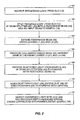
- Figure 2 is a flowchart illustrating the steps performed by the interferometer apparatus to recover depth-resolved spatial cross-correlated information about the sample for analysis;
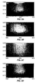
- Figures 3A-D illustrate examples of faLCI data recovered in the spectral domain for an exemplary sample of polystyrene beads, comprising the total acquired signal ( Figure 3A ), the reference field intensity ( Figure 3B ), the signal field intensity ( Figure 3C ), and the extracted, cross-correlated signal between the reference and signal field intensities ( Figure 3D );
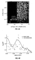
- Figure 4A is an illustration of the axial spatial cross-correlated function performed on the cross-correlated faLCI data illustrated in Figure 3D as a function of depth and angle;
- Figure 4B is an illustration of an angular distribution plot of raw and filtered data regarding scattered sample signal intensity as a function of angle in order to recover size information about the sample;
- Figure 5A is an illustration of the filtered angular distribution of the scattered sample signal intensity compared to the best fit Mie theory to determine size information about the sample;
- Figure 5B is a Chi-squired minimization of size information about the sample to estimate the diameter of cells in the sample
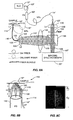
- Figure 6 is a schematic of exemplary faLCI system employing an optical fiber probe which does not constitute an embodiment of the present invention
- Figure 7A is a cutaway view of an a/LCI fiber-optic probe tip that may be employed by the faLCI system illustrated in Figure 6 ;
- Figure 7B illustrates the location of the fiber probe in the faLCI system illustrated in Figure 7A ;
- Figure 8A is an illustration of a fiber-optic faLCI system that may be employed with the present invention.
- Figure 8B is an illustration of sample illumination and scattered light collection with distal end of probe in the faLCI system illustrated in Figure 8B ;
- Figure 8C is an illustration of an image of the illuminated distal end ofprobe of the faLCI system illustrated in Figure 8A .
- the present invention involves a new a/LCI technique called Fourier domain a/LCI (faLCI), which enables data acquisition at rapid rates using a single scan, sufficient to make in vivo applications feasible.
- faLCI Fourier domain a/LCI
- the present invention obtains angle-resolved and depth-resolved spectra information about a sample, in which depth and size information about the sample can be obtained with a single scan, and wherein the reference arm can remain fixed with respect to the sample due to only one scan required.
- a reference signal and a reflected sample signal are cross-correlated and dispersed at a multitude of reflected angles off of the sample, thereby representing reflections from a multitude of points on the sample at the same time in parallel.
- the new data acquisition scheme is significant as it permits data to be obtained in less than one second, a threshold determined to be necessary for acquiring data from in vivo tissues.
- Information about all depths of the sample at each of the multitude of different points on the sample can be obtained with one scan on the order of approximately 40 milliseconds.
- structural (size) information can also be obtained using techniques that allow size information of scatterers to be obtained from angle-resolved data.
- the faLCI technique of the present invention uses the Fourier domain concept to acquire depth resolved information. Signal-to-noise and commensurate reductions in data acquisition time are possible by recording the depth scan in the Fourier (or spectral) domain.
- the faLCI system combines the Fourier domain concept with the use of an imaging spectrograph to spectrally record the angular distribution in parallel. Thereafter, the depth-resolution of the present invention is achieved by Fourier transforming the spectrum of two mixed fields with the angle-resolved measurements obtained by locating the entrance slit of the imaging spectrograph in a Fourier transform plane to the sample. This converts the spectral information into depth-resolved information and the angular information into a transverse spatial distribution.
- the capabilities of faLCI have been initially demonstrated by extracting the size of polystyrene beads in a depth-resolved measurement.
- the key advances of the present invention can be broken down into three components: (1) new rapid data acquisition methods, (2) fiber probe designs, and (3) data analysis schemes.
- the present invention is described in this matter for convenience in its understanding.
- An exemplary apparatus which does not represent an embodiment of the present invention, as well as the steps involved in the process of obtaining angle and depth-resolved distribution data scattered from a sample, are also set forth in Figure 2 .
- the faLCI scheme in accordance with an example not comprising an embodiment of the present invention is based on a modified Mach-Zehnder interferometer as illustrated in Figure 1A .
- Broadband light 10 from a superluminescent diode (SLD) 12 is directed by a mirror 13 (step 60 in Figure 2 ) and split into a reference beam 14 and an input beam 16 to a sample 18 by beamsplitter BS 1 20 (step 62 in Figure 3 ).
- SLD superluminescent diode
- the path length of the reference beam 14 is set by adjusting retroreflector RR 22, but remains fixed during measurement.
- the reference beam 14 is expanded using lenses L 1 (24) and L2 (26) to create illumination (step 64 in Figure 2 ), which is uniform and collimated upon reaching a spectrograph slit 48 in an imaging spectrograph 29.
- L1 may have a focal length of 1.5 centimeters
- L2 26 may have focal length of 15 centimeters.
- Lenses L3 (31) and L4 (38) are arranged to produce a collimated pencil beam 30 incident on the sample 18 (step 66 in Figure 2 ).
- the input beam 30 is made to strike the sample at an angle of 0.10 radians relative to the optical axis.
- This arrangement allows the full angular aperture of lens L4 (38) to be used to collect scattered light 40 from the sample 18.
- Lens L4 (38) may have a focal length of 3.5 centimeters.
- the light 40 scattered by the sample 18 is collected by lens L4 (32) and relayed by a 4f imaging system comprised of lenses L5 (43) and L6 (44) such that the Fourier plane of lens L4 (32) is reproduced in phase and amplitude at the spectrograph slit 48 (step 68 in Figure 2 ).
- the scattered light 40 is mixed with the reference field 14 at a second beamsplitter BS2 42 with the combined fields 46 falling upon the entrance slit (illustrated in Figure 1B as element 48) to the imaging spectrograph 29 (step 70 in Figure 2 ).
- the imaging spectrograph 29 may be the model SP2150i, manufactured by Acton Research for example.
- Figure 1B illustrates the distribution of scattering angle across the dimension of the slit 48.
- the mixed fields are dispersed with a high resolution grating (e.g. 1200 l/mm) and detected using a cooled CCD 50 (e.g. 1340 X 400, 20 ⁇ m X 20 ⁇ m pixels, Spec10:400, manufactured by Princeton Instruments) (step 72 in Figure 2 ).
- a high resolution grating e.g. 1200 l/mm
- a cooled CCD 50 e.g. 1340 X 400, 20 ⁇ m X 20 ⁇ m pixels, Spec10:400, manufactured by Princeton Instruments
- the detected signal 46 is a function of vertical position on the spectrograph slit 48, y , and wavelength ⁇ once the light is dispersed by the spectrograph 29.
- the interference term is extracted by measuring the intensity of the signal 30 and reference beams 16 independently and subtracting them from the total intensity.
- k o ( y o and ⁇ k ( ⁇ y ) represent the center and width of the Gaussian wavevector (spatial) distribution and ⁇ l is the selected path length difference.
- S j represents the amplitude distribution of the scattering originating from the j th interface, located at depth l j .
- y f 4 ⁇ .
- the pixel size of the CCD 50 e.g. 20 ⁇ m
- Figure 3A shows typical data representing the total detected intensity (Equation (1), above) of the sum of the reference field 16 and the field scattered 40 by a sample of polystyrene beads, in the frequency domain given as a function of wavelength and angle, given with respect to the backwards scattering direction. In an example; this data was acquired in 40 milliseconds and records data over 186 mrad, approximately 85% of the expected range, with some loss of signal at higher angles.
- Figures 3B and 3C illustrate the intensity of the reference and signal fields 14, 30 respectively. Upon subtraction of the signal and reference fields 14, 30 from the total detected intensity, the interference 46 between the two fields is realized as illustrated in Figure 3D .
- interference data 46 are interpolated into k-space and Fourier transformed to give the angular depth resolved profiles of the sample 18 as illustrated in Figure 4A .
- the Fourier transform of the angle-resolved, cross correlated signal 46 which is the result of signal 40 scattered at a multitude of reflected angles off the sample 18 and obtained in the Fourier plane of lens L4 (38), produces depth-resolved information about the sample 18 as a function of angle and depth. This provides depth-resolved information about the sample 18. Because the angle-resolved, cross-correlated signal 46 is spectrally dispersed, the data acquisition permits data to be obtained in less than one second. Information about all depths of the sample 18 at each of the multitude of different points (i.e.
- the sample is contained in a round well (8mm diameter, 1mm deep) behind a glass coverslip (thickness, d ⁇ 170 ⁇ m) (not shown).
- the sample beam 30 is incident on the sample 18 through the coverslip.
- the data are ensemble averaged by integrating over one mean free path (MFP).
- MFP mean free path
- the spatial average can enable a reduction of speckle when using low-coherence light to probe a scattering sample.
- the scattering distribution is low pass filtered to produce a smoother curve, with the cutoff frequency chosen to suppress spatial correlations on length scales above 16 ⁇ m.
- the scattering distribution data (i.e. a/LCI data) obtained from the sample 18 using the disclosed data acquisition scheme can also be used to make a size determination of the nucleus using the Mie theory.
- a scattering distribution 74 of the sample 18 is illustrated in Figure 4B as a contour plot. The raw scattered information 74 about the sample 18 is shown as a function of the signal field 30 and angle. A filtered curve is determined using the scattered data 74. Comparison of the filtered scattering distribution curve 76 (i.e. a representation of the scattered data 74) to the prediction of Mie theory (curve 78 in Figure 5A ) enables a size determination to be made.
- the a/LCI signals are processed to extract the oscillatory component which is characteristic of the nucleus size.
- the smoothed data 76 are fit to a low-order polynomial (4 th order was used for example herein, but later studies use a lower 2 nd order), which is then subtracted from the distribution 76 to remove the background trend.
- the resulting oscillatory component is then compared to a database of theoretical predictions obtained using Mie theory 78 from which the slowly varying features were similarly removed for analysis.
- a direct comparison between the filtered a/LCI data 76 and Mie theory data 78 may not possible, as the chi-squared fitting algorithm tends to match the background slope rather than the characteristic oscillations.
- the calculated theoretical predictions include a Gaussian distribution of sizes characterized by a mean diameter (d) and standard deviation ( ⁇ D) as well as a distribution of wavelengths, to accurately model the broad bandwidth source.
- a/LCI data As an alternative to processing the a/LCI data and comparing to Mie theory, there are several other approaches which could yield diagnostic information. These include analyzing the angular data using a Fourier transform to identify periodic oscillations characteristic of cell nuclei. The periodic oscillations can be correlated with nuclear size and thus will possess diagnostic value.
- Another approach to analyzing a/LCI data is to compare the data to a database of angular scattering distributions generated with finite element method (FEM) or T-Matrix calculations. Such calculations may offer superior analysis as there are not subject to the same limitations as Mie theory. For example, FEM or T-Matrix calculations can model non-spherical scatterers and scatterers with inclusions while Mie theory, can only model homogenous spheres.
- FEM finite element method
- T-Matrix calculations can model non-spherical scatterers and scatterers with inclusions while Mie theory, can only model homogenous spheres.
- FIG. 6 a system employing optical fibers to deliver and collect light from the sample of interest to use in the a/LCI system for endoscopic applications.
- the fiber optic a/LCI scheme for this example makes use of the Fourier transform properties of a lens. This property states that when an object is placed in the front focal plane of a lens, the image at the conjugate image plane is the Fourier transform of that object.
- the Fourier transform of a spatial distribution (object or image) is given by the distribution of spatial frequencies, which is the representation of the image's information content in terms of cycles per mm.
- the wavelength retains its fixed, original value and the spatial frequency representation is simply a scaled version of the angular distribution of scattered light.
- the angular distribution is captured by locating the distal end of the fiber bundle in a conjugate Fourier transform plane of the sample using a collecting lens. This angular distribution is then conveyed to the distal end of the fiber bundle where it is imaged using a 4f system onto the entrance slit of an imaging spectrograph.
- a beamsplitter is used to overlap the scattered field with a reference field prior to entering the slit so that low coherence interferometry can also be used to obtain depth resolved measurements.
- FIG. 6 the fiber optic faLCI scheme is shown.
- Light 12' from a broadband light source 10' is split into a reference field 14' and a signal field 16' using a fiber splitter (FS) 80.
- a splitter ratio.of 20:1 is chosen in one embodiment to direct more power to a sample 18' via the signal arm 82 as the light returned by the tissue is typically only a small fraction of the incident power.
- a collimated beam 88 is arranged to be equal in dimension to the end 91 of fiber bundle F3 (90) so that the collimated beam 88 illuminates all fibers in F3 with equal intensity.
- the reference field 14' emerging from the distal tip of F3 (90) is collimated with lens L3 (92) in order to overlap with the scattered field conveyed by fiber F4 (94).
- light emerging from fiber F1 (14') is collimated then expanded using a lens system to produce a broad beam.
- the scattered field is detected using a coherent fiber bundle.
- the scattered field is generated using light in the signal arm 82 which is directed toward the sample 18' of interest using lens L2 (98).
- lens L2 (98) is displaced laterally from the center of single-mode fiber F2 such that a collimated beam is produced which is traveling at an angle relative to the optical axis
- the fact that the incident beam strikes the sample at an oblique angle is essential in separating the elastic scattering information from specular reflections.
- the light scattered by the sample 18' is collected by a fiber bundle consisting of an array of coherent single mode or multi-mode fibers.
- the distal tip of the fiber is maintained one focal length away from lens L2 (98) to image the angular distribution of scattered light.
- the sample 18' is located in the front focal plane of lens L2 (98) using a mechanical mount 100.
- the sample is located in the front focal plane of lens L2 (98) using a transparent sheath (element 102).
- scattered light 104 emerging from a proximal end 105 of the fiber probe F4 (94) is recollimated by lens L4 (104) and overlapped with the reference field 14' using beamsplitter BS (108).
- the two combined fields 110 are re-imaged onto the slit (element 48' in Figure 7 ) of the imaging spectrograph 29' using lens L5 (112).
- the focal length of lens L5 (112) may be varied to optimally fill the slit 48'.
- the resulting optical signal contains information on each scattering angle across the vertical dimension of the slit 48' as described above for the apparatus of Figures 1A and 1B .
- a/LCI fiber-optic probe will collect the angular distribution over a 0.45 radian range (approx. 30 degrees) and will acquire the complete depth resolved scattering distribution 110 in a fraction of a second.
- One possible implementation would be a linear array of single mode fibers in both the signal and reference arms.
- the reference arm 96 could be composed of an individual single mode fiber with the signal arm 82 consisting of either a coherent fiber bundle or linear fiber array.
- the fiber probe tip can also have several implementations which are substantially equivalent. These would include the use of a drum or ball lens in place of lens L2 (98).
- a side-viewing probe could be created using a combination of a lens and a mirror or prism or through the use of a convex mirror to replace the lens-mirror combination. Finally, the entire probe can be made to rotate radially in order to provide a circumferential scan of the probed area.
- a data acquisition embodiment of the present invention is a fa/LCI system is based on a modified Mach-Zehnder interferometer as illustrated in Figure 8A .
- the output 10" from a fiber-coupled superluminescent diode (SLD) source 12" (e.g.
- sample arm delivery fiber 16" is split into sample arm delivery fiber 16" and a reference arm delivery fiber 14" by a 90/10 fiber splitter FS (80') (e.g. manufactured by AC Photonics).
- the sample arm delivery fiber 16" consists of either of the following for example: (1) a single mode fiber with polarization control integrated at the tip; or (2) a polarization maintaining fiber.
- a sample probe 113 is assembled by affixing the delivery fiber 16"( NA ⁇ 0.12) along the ferrule 114 at the distal end of a fiber bundle 116 such that the end face of the delivery fiber 16" is parallel to and flush with the face of the fiber bundle 116.
- Ball lens L1 115
- the optical path of light scattered 122 at three selected scattering angles is shown in Figure 8B .
- the angular distribution is sampled by approximately 130 individual fibers for example, across a vertical strip of the fiber bundle 116", as depicted by the highlighted area in Figure 8C .
- Xie Mukai, S. G. Guo, M. Brenner, and Z. P. Chen in Optics Letters 30(14), 1803-1805 (2005 )
- Xie discloses a multimode coherent fiber bundle into a time-domain optical coherence tomography system and demonstrated that the modes of light coupled into an individual fiber will travel different path lengths.
- the higher order modes are offset from the fundamental mode by 3.75 mm, well beyond the depth ( ⁇ 100 ⁇ m) required for gathering clinically relevant data.
- the power in the higher order modes had a minimal affect on dynamic range as the sample arm power is significantly less than the reference arm power.
- the example of the present invention herein uses 130 fibers to simultaneously collect scattered light across a range of angles in parallel, resulting in rapid data collection.
- the theoretical magnification of the 4f imaging system is ( f 3 / f 2 ) 6.67 in this example.
- the optical path length of the reference arm is matched to that of the fundamental mode of the sample arm.
- a reference field 130 maybe attenuated by a neutral density filter 132 and mixed with the angular scattering distribution at beamsplitter BS (134).
- the mixed fields 136 are dispersed with a high resolution grating (e.g. 1200 lines/mm) and detected using an integrated, cooled CCD (not shown) (e.g. 1024 x 252, 24 ⁇ m x 24 ⁇ m pixels, 0.1 nm resolution) covering a spectral range of 99 nm centered at 840 nm, for example.
- a high resolution grating e.g. 1200 lines/mm
- CCD not shown
- 1024 x 252, 24 ⁇ m x 24 ⁇ m pixels, 0.1 nm resolution covering a spectral range of 99 nm centered at 840 nm, for example.
- I( ⁇ m , ⁇ n ) is uploaded to a PC using LabVIEW manufactured by National Instruments software and processed in 320 ms to produce a depth and angle resolved contour plot of scattered intensity.
- the processing of the angle-resolved scattered field to obtain depth and size information described above, and in particular reference to the data acquisition apparatus of Figures 1A and 1B can then used to obtain angle-resolved, depth-resolved information about the sample 18" using the scattered mixed field 136 generated by the apparatus in Figure 8 .
- the embodiments set forth above represent the necessary information to enable those skilled in the art to practice the invention and illustrate the best mode of practicing the invention.
Landscapes
- Physics & Mathematics (AREA)
- Spectroscopy & Molecular Physics (AREA)
- Health & Medical Sciences (AREA)
- Life Sciences & Earth Sciences (AREA)
- General Physics & Mathematics (AREA)
- Engineering & Computer Science (AREA)
- General Health & Medical Sciences (AREA)
- Signal Processing (AREA)
- Pathology (AREA)
- Biomedical Technology (AREA)
- Medical Informatics (AREA)
- Analytical Chemistry (AREA)
- Immunology (AREA)
- Chemical & Material Sciences (AREA)
- Mathematical Physics (AREA)
- Biophysics (AREA)
- Veterinary Medicine (AREA)
- Heart & Thoracic Surgery (AREA)
- Biochemistry (AREA)
- Molecular Biology (AREA)
- Surgery (AREA)
- Animal Behavior & Ethology (AREA)
- Public Health (AREA)
- Optics & Photonics (AREA)
- Artificial Intelligence (AREA)
- Computer Vision & Pattern Recognition (AREA)
- Physiology (AREA)
- Psychiatry (AREA)
- Investigating Or Analysing Materials By Optical Means (AREA)
Priority Applications (2)
| Application Number | Priority Date | Filing Date | Title |
|---|---|---|---|
| EP15157252.6A EP2950065A1 (en) | 2005-10-11 | 2006-10-11 | Method for fiber-based endoscopic angle-resolved low coherence interferometry |
| EP11176357.9A EP2444783B1 (en) | 2005-10-11 | 2006-10-11 | Systems and method for fiber-based endoscopic angle-resolved low coherence interferometry |
Applications Claiming Priority (2)
| Application Number | Priority Date | Filing Date | Title |
|---|---|---|---|
| US72560305P | 2005-10-11 | 2005-10-11 | |
| PCT/US2006/039771 WO2007044821A1 (en) | 2005-10-11 | 2006-10-11 | Systems and method for endoscopic angle-resolved low coherence interferometry |
Related Child Applications (3)
| Application Number | Title | Priority Date | Filing Date |
|---|---|---|---|
| EP15157252.6A Division EP2950065A1 (en) | 2005-10-11 | 2006-10-11 | Method for fiber-based endoscopic angle-resolved low coherence interferometry |
| EP11176357.9A Division EP2444783B1 (en) | 2005-10-11 | 2006-10-11 | Systems and method for fiber-based endoscopic angle-resolved low coherence interferometry |
| EP11176357.9 Division-Into | 2011-08-02 |
Publications (2)
| Publication Number | Publication Date |
|---|---|
| EP1934567A1 EP1934567A1 (en) | 2008-06-25 |
| EP1934567B1 true EP1934567B1 (en) | 2013-01-16 |
Family
ID=37714242
Family Applications (3)
| Application Number | Title | Priority Date | Filing Date |
|---|---|---|---|
| EP11176357.9A Not-in-force EP2444783B1 (en) | 2005-10-11 | 2006-10-11 | Systems and method for fiber-based endoscopic angle-resolved low coherence interferometry |
| EP15157252.6A Withdrawn EP2950065A1 (en) | 2005-10-11 | 2006-10-11 | Method for fiber-based endoscopic angle-resolved low coherence interferometry |
| EP06825774A Not-in-force EP1934567B1 (en) | 2005-10-11 | 2006-10-11 | Systems and method for endoscopic angle-resolved low coherence interferometry |
Family Applications Before (2)
| Application Number | Title | Priority Date | Filing Date |
|---|---|---|---|
| EP11176357.9A Not-in-force EP2444783B1 (en) | 2005-10-11 | 2006-10-11 | Systems and method for fiber-based endoscopic angle-resolved low coherence interferometry |
| EP15157252.6A Withdrawn EP2950065A1 (en) | 2005-10-11 | 2006-10-11 | Method for fiber-based endoscopic angle-resolved low coherence interferometry |
Country Status (9)
| Country | Link |
|---|---|
| US (3) | US7595889B2 (enExample) |
| EP (3) | EP2444783B1 (enExample) |
| JP (2) | JP2009511909A (enExample) |
| CN (1) | CN101326428B (enExample) |
| AU (1) | AU2006302086B2 (enExample) |
| CA (3) | CA2967964A1 (enExample) |
| ES (2) | ES2541851T3 (enExample) |
| PT (2) | PT2444783E (enExample) |
| WO (1) | WO2007044821A1 (enExample) |
Families Citing this family (111)
| Publication number | Priority date | Publication date | Assignee | Title |
|---|---|---|---|---|
| AU2002230842A1 (en) | 2000-10-30 | 2002-05-15 | The General Hospital Corporation | Optical methods and systems for tissue analysis |
| US9295391B1 (en) | 2000-11-10 | 2016-03-29 | The General Hospital Corporation | Spectrally encoded miniature endoscopic imaging probe |
| US7865231B2 (en) | 2001-05-01 | 2011-01-04 | The General Hospital Corporation | Method and apparatus for determination of atherosclerotic plaque type by measurement of tissue optical properties |
| US7355716B2 (en) | 2002-01-24 | 2008-04-08 | The General Hospital Corporation | Apparatus and method for ranging and noise reduction of low coherence interferometry LCI and optical coherence tomography OCT signals by parallel detection of spectral bands |
| WO2004088361A2 (en) | 2003-03-31 | 2004-10-14 | The General Hospital Corporation | Speckle reduction in optical coherence tomography by path length encoded angular compounding |
| US7102758B2 (en) | 2003-05-06 | 2006-09-05 | Duke University | Fourier domain low-coherence interferometry for light scattering spectroscopy apparatus and method |
| DE602004016998D1 (de) | 2003-06-06 | 2008-11-20 | Gen Hospital Corp | Wellenlängenabstimmbare lichtquelle |
| WO2005031431A1 (de) * | 2003-09-25 | 2005-04-07 | Leica Microsystems Cms Gmbh | Mikroskopobjektiv zur totalinternen-reflexions-mikroskopie und mikroskop |
| CN103082996A (zh) | 2003-10-27 | 2013-05-08 | 通用医疗公司 | 用于使用频域干涉测量法进行光学成像的方法和设备 |
| US8018598B2 (en) | 2004-05-29 | 2011-09-13 | The General Hospital Corporation | Process, system and software arrangement for a chromatic dispersion compensation using reflective layers in optical coherence tomography (OCT) imaging |
| AU2005270037B2 (en) | 2004-07-02 | 2012-02-09 | The General Hospital Corporation | Endoscopic imaging probe comprising dual clad fibre |
| KR101332222B1 (ko) | 2004-08-06 | 2013-11-22 | 더 제너럴 하스피탈 코포레이션 | 광간섭 단층촬영법을 이용해서 샘플 내에서 적어도 하나의 위치를 결정하는 방법, 시스템 및 그 방법을 구현하기 위한 소프트웨어가 저장되어 컴퓨터로 판독 가능한 매체 |
| WO2006024014A2 (en) | 2004-08-24 | 2006-03-02 | The General Hospital Corporation | Process, system and software arrangement for measuring a mechanical strain and elastic properties of a sample |
| JP5324095B2 (ja) | 2004-08-24 | 2013-10-23 | ザ ジェネラル ホスピタル コーポレイション | 血管セグメントを画像化する方法および装置 |
| EP1787105A2 (en) | 2004-09-10 | 2007-05-23 | The General Hospital Corporation | System and method for optical coherence imaging |
| EP2329759B1 (en) | 2004-09-29 | 2014-03-12 | The General Hospital Corporation | System and method for optical coherence imaging |
| EP1825214A1 (en) | 2004-11-24 | 2007-08-29 | The General Hospital Corporation | Common-path interferometer for endoscopic oct |
| EP1816949A1 (en) | 2004-11-29 | 2007-08-15 | The General Hospital Corporation | Arrangements, devices, endoscopes, catheters and methods for performing optical imaging by simultaneously illuminating and detecting multiple points on a sample |
| US8761865B2 (en) * | 2005-03-10 | 2014-06-24 | Anatoly Babchenko | Optical sensor and a method of its use |
| EP1875436B1 (en) | 2005-04-28 | 2009-12-09 | The General Hospital Corporation | Evaluation of image features of an anatomical structure in optical coherence tomography images |
| WO2006130802A2 (en) | 2005-06-01 | 2006-12-07 | The General Hospital Corporation | Apparatus, method and system for performing phase-resolved optical frequency domain imaging |
| KR101387454B1 (ko) | 2005-08-09 | 2014-04-22 | 더 제너럴 하스피탈 코포레이션 | 광간섭 단층촬영법에서 편광 기반 직교 복조를 수행하기위한 장치, 방법 및 저장 매체 |
| CN103479331A (zh) | 2005-09-29 | 2014-01-01 | 通用医疗公司 | 用于经由谱编码进行光学成像的方法和设备 |
| JP4642681B2 (ja) * | 2005-09-30 | 2011-03-02 | 富士フイルム株式会社 | 光断層画像化装置 |
| WO2007044821A1 (en) * | 2005-10-11 | 2007-04-19 | Duke University | Systems and method for endoscopic angle-resolved low coherence interferometry |
| US8537366B2 (en) | 2005-10-11 | 2013-09-17 | Duke University | Systems and methods for endoscopic angle-resolved low coherence interferometry |
| US7889348B2 (en) | 2005-10-14 | 2011-02-15 | The General Hospital Corporation | Arrangements and methods for facilitating photoluminescence imaging |
| WO2007082228A1 (en) | 2006-01-10 | 2007-07-19 | The General Hospital Corporation | Systems and methods for generating data based on one or more spectrally-encoded endoscopy techniques |
| ES2847854T3 (es) | 2006-01-19 | 2021-08-04 | Massachusetts Gen Hospital | Catéter de globo de obtención de imágenes |
| US8145018B2 (en) | 2006-01-19 | 2012-03-27 | The General Hospital Corporation | Apparatus for obtaining information for a structure using spectrally-encoded endoscopy techniques and methods for producing one or more optical arrangements |
| JP5524487B2 (ja) | 2006-02-01 | 2014-06-18 | ザ ジェネラル ホスピタル コーポレイション | コンフォーマルレーザ治療手順を用いてサンプルの少なくとも一部分に電磁放射を放射する方法及びシステム。 |
| US9186066B2 (en) | 2006-02-01 | 2015-11-17 | The General Hospital Corporation | Apparatus for applying a plurality of electro-magnetic radiations to a sample |
| WO2007092911A2 (en) | 2006-02-08 | 2007-08-16 | The General Hospital Corporation | Methods, arrangements and systems for obtaining information associated with an anatomical sample using optical microscopy |
| US7982879B2 (en) * | 2006-02-24 | 2011-07-19 | The General Hospital Corporation | Methods and systems for performing angle-resolved fourier-domain optical coherence tomography |
| JP2009536740A (ja) | 2006-05-10 | 2009-10-15 | ザ ジェネラル ホスピタル コーポレイション | サンプルの周波数領域画像形成を提供するためのプロセス、構成およびシステム |
| CN101500472B (zh) * | 2006-07-21 | 2014-02-12 | 昂科斯科公司 | 特别是用于内窥镜应用的光纤探头的保护探头尖端 |
| EP2054712B1 (en) | 2006-08-25 | 2015-10-07 | The General Hospital Corporation | Apparatus and methods for enhancing optical coherence tomography imaging using volumetric filtering techniques |
| US8838213B2 (en) | 2006-10-19 | 2014-09-16 | The General Hospital Corporation | Apparatus and method for obtaining and providing imaging information associated with at least one portion of a sample, and effecting such portion(s) |
| US7949019B2 (en) | 2007-01-19 | 2011-05-24 | The General Hospital | Wavelength tuning source based on a rotatable reflector |
| US7502119B2 (en) * | 2007-01-29 | 2009-03-10 | Filmetrics, Inc. | Thin-film metrology using spectral reflectance with an intermediate in-line reference |
| WO2008118781A2 (en) | 2007-03-23 | 2008-10-02 | The General Hospital Corporation | Methods, arrangements and apparatus for utilizing a wavelength-swept laser using angular scanning and dispersion procedures |
| US10534129B2 (en) | 2007-03-30 | 2020-01-14 | The General Hospital Corporation | System and method providing intracoronary laser speckle imaging for the detection of vulnerable plaque |
| WO2008131082A1 (en) | 2007-04-17 | 2008-10-30 | The General Hospital Corporation | Apparatus and methods for measuring vibrations using spectrally-encoded endoscopy techniques |
| WO2008157790A2 (en) * | 2007-06-20 | 2008-12-24 | The Trustees Of Dartmouth College | Pulsed lasers in frequency domain diffuse optical tomography and spectroscopy |
| JP5917803B2 (ja) | 2007-07-31 | 2016-05-18 | ザ ジェネラル ホスピタル コーポレイション | 高速ドップラー光周波数領域撮像法のためのビーム走査パターンを放射するシステムおよび方法 |
| EP2191254B1 (en) | 2007-08-31 | 2017-07-19 | The General Hospital Corporation | System and method for self-interference fluorescence microscopy, and computer-accessible medium associated therewith |
| JP2009063407A (ja) * | 2007-09-06 | 2009-03-26 | Yokogawa Electric Corp | 照射集光装置 |
| JP5579606B2 (ja) * | 2007-09-13 | 2014-08-27 | デユーク・ユニバーシテイ | 低コヒーレンス干渉法(lci)のための装置、システムおよび方法 |
| WO2009059034A1 (en) | 2007-10-30 | 2009-05-07 | The General Hospital Corporation | System and method for cladding mode detection |
| US20090177094A1 (en) * | 2008-01-08 | 2009-07-09 | Oncoscope, Inc. | Systems and methods for tissue examination, diagnostic, treatment, and/or monitoring |
| WO2009105537A2 (en) * | 2008-02-19 | 2009-08-27 | Trustees Of Tufts College | Non-invasive optical characterization of biomaterial mineralization |
| DE102008016973B4 (de) * | 2008-04-03 | 2009-12-31 | Precitec Optronik Gmbh | Interferometer und Verfahren zum Betreiben eines Interferometers |
| US7898656B2 (en) * | 2008-04-30 | 2011-03-01 | The General Hospital Corporation | Apparatus and method for cross axis parallel spectroscopy |
| JP5607610B2 (ja) | 2008-05-07 | 2014-10-15 | ザ ジェネラル ホスピタル コーポレイション | 構造の特徴を決定する装置、装置の作動方法およびコンピュータアクセス可能な媒体 |
| CN102037343B (zh) * | 2008-06-12 | 2013-10-02 | 东卡莱罗纳大学 | 用于三维衍射成像的流式细胞仪系统及方法 |
| JP5795531B2 (ja) | 2008-06-20 | 2015-10-14 | ザ ジェネラル ホスピタル コーポレイション | フューズドファイバオプティックカプラ構造、及びその使用方法 |
| WO2010009136A2 (en) | 2008-07-14 | 2010-01-21 | The General Hospital Corporation | Apparatus and methods for color endoscopy |
| KR101109968B1 (ko) * | 2008-07-23 | 2012-02-17 | 올림푸스 메디칼 시스템즈 가부시키가이샤 | 피검체 관측 장치 및 피검체 관측 방법 |
| CN102144154B (zh) | 2008-10-01 | 2015-04-22 | 东卡莱罗纳大学 | 用结构入射光测定混浊介质光学特征的方法与系统 |
| US8004688B2 (en) * | 2008-11-26 | 2011-08-23 | Zygo Corporation | Scan error correction in low coherence scanning interferometry |
| EP2359121A4 (en) | 2008-12-10 | 2013-08-14 | Gen Hospital Corp | SYSTEMS, DEVICE AND METHOD FOR EXPANDING THE IMAGING DEPTH RANGE IN OPTICAL COHERENCE TOMOPOMAGRAPH BY OPTICAL SUB-TESTING |
| US8537367B2 (en) * | 2009-01-17 | 2013-09-17 | Luna Innovations Incorporated | Optical imaging for optical device inspection |
| WO2010090837A2 (en) | 2009-01-20 | 2010-08-12 | The General Hospital Corporation | Endoscopic biopsy apparatus, system and method |
| JP2012515930A (ja) | 2009-01-26 | 2012-07-12 | ザ ジェネラル ホスピタル コーポレーション | 広視野の超解像顕微鏡を提供するためのシステム、方法及びコンピューターがアクセス可能な媒体 |
| US9351642B2 (en) | 2009-03-12 | 2016-05-31 | The General Hospital Corporation | Non-contact optical system, computer-accessible medium and method for measurement at least one mechanical property of tissue using coherent speckle technique(s) |
| JP5325679B2 (ja) * | 2009-07-03 | 2013-10-23 | 富士フイルム株式会社 | 低コヒーレンス光源を用いた動的光散乱測定装置及び光散乱強度測定方法 |
| EP2453791B1 (en) | 2009-07-14 | 2023-09-06 | The General Hospital Corporation | Apparatus for measuring flow and pressure within a vessel |
| TWI425188B (zh) * | 2009-08-31 | 2014-02-01 | Zygo Corp | 顯微鏡系統和成像干涉儀系統 |
| JP5560628B2 (ja) * | 2009-09-04 | 2014-07-30 | ソニー株式会社 | 検査装置および検査方法 |
| JP2011095181A (ja) * | 2009-10-30 | 2011-05-12 | Sysmex Corp | 粒子分析装置 |
| JP2013518256A (ja) * | 2010-01-22 | 2013-05-20 | デユーク・ユニバーシテイ | 分光光コヒーレンストモグラフィ(oct)およびフーリエドメイン低コヒーレンス干渉法のための多重ウィンドウ処理スキーム |
| US9823127B2 (en) | 2010-01-22 | 2017-11-21 | Duke University | Systems and methods for deep spectroscopic imaging of biological samples with use of an interferometer and spectrometer |
| LT2542154T (lt) | 2010-03-05 | 2020-12-10 | The General Hospital Corporation | Ėminio švitinimo elektromagnetine spinduliuote aparatas |
| EP2556331A1 (en) * | 2010-03-19 | 2013-02-13 | Duke University | Single-mode optical fiber-based angle-resolved low coherence interferometric (lci) (a/lci) and non-interferometric systems and methods |
| US9069130B2 (en) | 2010-05-03 | 2015-06-30 | The General Hospital Corporation | Apparatus, method and system for generating optical radiation from biological gain media |
| WO2011150069A2 (en) | 2010-05-25 | 2011-12-01 | The General Hospital Corporation | Apparatus, systems, methods and computer-accessible medium for spectral analysis of optical coherence tomography images |
| EP2575597B1 (en) | 2010-05-25 | 2022-05-04 | The General Hospital Corporation | Apparatus for providing optical imaging of structures and compositions |
| EP2575591A4 (en) | 2010-06-03 | 2017-09-13 | The General Hospital Corporation | Apparatus and method for devices for imaging structures in or at one or more luminal organs |
| US8462349B1 (en) * | 2010-07-20 | 2013-06-11 | Science Applications International Corporation | System and method for a self-referencing interferometer |
| US9510758B2 (en) | 2010-10-27 | 2016-12-06 | The General Hospital Corporation | Apparatus, systems and methods for measuring blood pressure within at least one vessel |
| US9330092B2 (en) | 2011-07-19 | 2016-05-03 | The General Hospital Corporation | Systems, methods, apparatus and computer-accessible-medium for providing polarization-mode dispersion compensation in optical coherence tomography |
| EP3835718B1 (en) | 2011-08-25 | 2023-07-26 | The General Hospital Corporation | Apparatus for providing micro-optical coherence tomography inside a respiratory system |
| EP2565625A1 (en) * | 2011-09-05 | 2013-03-06 | Ludwig-Maximilians-Universität München | Optical measurement system and method for operating an optical measurement system |
| WO2013066631A1 (en) | 2011-10-18 | 2013-05-10 | The General Hospital Corporation | Apparatus and methods for producing and/or providing recirculating optical delay(s) |
| US9629528B2 (en) | 2012-03-30 | 2017-04-25 | The General Hospital Corporation | Imaging system, method and distal attachment for multidirectional field of view endoscopy |
| EP2852315A4 (en) | 2012-05-21 | 2016-06-08 | Gen Hospital Corp | DEVICE, APPARATUS AND METHOD FOR CAPSULE MICROSCOPY |
| JP6560126B2 (ja) | 2013-01-28 | 2019-08-14 | ザ ジェネラル ホスピタル コーポレイション | 光周波数ドメインイメージングに重ね合わせされる拡散分光法を提供するための装置および方法 |
| US10893806B2 (en) | 2013-01-29 | 2021-01-19 | The General Hospital Corporation | Apparatus, systems and methods for providing information regarding the aortic valve |
| WO2014121082A1 (en) | 2013-02-01 | 2014-08-07 | The General Hospital Corporation | Objective lens arrangement for confocal endomicroscopy |
| WO2014124537A1 (en) * | 2013-02-14 | 2014-08-21 | Verisante Technology, Inc. | Method and apparatus for optical measurements under ambient light conditions |
| EP2967491B1 (en) | 2013-03-15 | 2022-05-11 | The General Hospital Corporation | A transesophageal endoscopic system for determining a mixed venous oxygen saturation of a pulmonary artery |
| US20140275765A1 (en) | 2013-03-15 | 2014-09-18 | Steven C. Gebhart | Probe assembly and disposable cover particularly for use in endoscope applications of low coherence interferometry |
| WO2014186353A1 (en) | 2013-05-13 | 2014-11-20 | The General Hospital Corporation | Detecting self-interefering fluorescence phase and amplitude |
| EP3692887B1 (en) | 2013-07-19 | 2024-03-06 | The General Hospital Corporation | Imaging apparatus which utilizes multidirectional field of view endoscopy |
| EP3021735A4 (en) | 2013-07-19 | 2017-04-19 | The General Hospital Corporation | Determining eye motion by imaging retina. with feedback |
| US9668652B2 (en) | 2013-07-26 | 2017-06-06 | The General Hospital Corporation | System, apparatus and method for utilizing optical dispersion for fourier-domain optical coherence tomography |
| WO2015105870A1 (en) | 2014-01-08 | 2015-07-16 | The General Hospital Corporation | Method and apparatus for microscopic imaging |
| WO2015116986A2 (en) | 2014-01-31 | 2015-08-06 | The General Hospital Corporation | System and method for facilitating manual and/or automatic volumetric imaging with real-time tension or force feedback using a tethered imaging device |
| US10228556B2 (en) | 2014-04-04 | 2019-03-12 | The General Hospital Corporation | Apparatus and method for controlling propagation and/or transmission of electromagnetic radiation in flexible waveguide(s) |
| JP6588083B2 (ja) * | 2014-04-25 | 2019-10-09 | エックス−ライト, インコーポレイテッドX−Rite, Incorporated | 照準システムおよび方法 |
| WO2016015052A1 (en) | 2014-07-25 | 2016-01-28 | The General Hospital Corporation | Apparatus, devices and methods for in vivo imaging and diagnosis |
| EP3201310B1 (en) * | 2014-10-01 | 2021-02-17 | Purdue Research Foundation | Microorganism identification |
| WO2016145393A1 (en) * | 2015-03-12 | 2016-09-15 | Purdue Research Foundation | Biodynamic microscopes and methods of use thereof |
| DE102016218290A1 (de) * | 2016-07-15 | 2018-01-18 | Carl Zeiss Meditec Ag | Verfahren zur hochsensitiven Messung von Abständen und Winkeln im menschlichen Auge |
| US10434970B2 (en) * | 2016-12-08 | 2019-10-08 | Toyota Jidosha Kabushiki Kaisha | Vehicle side section structure |
| GB201803523D0 (en) * | 2018-03-05 | 2018-04-18 | Malvern Panalytical Ltd | Improved particle sizing by optical diffraction |
| CN109620134B (zh) * | 2019-01-21 | 2020-05-22 | 浙江大学 | 基于光纤阵列多通道并行探测的微血管造影方法和系统 |
| JP7149532B2 (ja) * | 2019-04-26 | 2022-10-07 | 株式会社日立製作所 | 粒子線実験データ解析装置 |
| US11333487B2 (en) * | 2019-10-28 | 2022-05-17 | Kla Corporation | Common path mode fiber tip diffraction interferometer for wavefront measurement |
| CN113091896B (zh) * | 2021-03-18 | 2023-03-14 | 西北工业大学 | 基于偏振光栅的动态测量任意光场完整信息的方法及光路 |
| CN113670854B (zh) * | 2021-08-12 | 2024-06-11 | 之江实验室 | 一种微分干涉对比显微内窥成像系统及内窥成像方法 |
Citations (1)
| Publication number | Priority date | Publication date | Assignee | Title |
|---|---|---|---|---|
| US20030137669A1 (en) * | 2001-08-03 | 2003-07-24 | Rollins Andrew M. | Aspects of basic OCT engine technologies for high speed optical coherence tomography and light source and other improvements in optical coherence tomography |
Family Cites Families (60)
| Publication number | Priority date | Publication date | Assignee | Title |
|---|---|---|---|---|
| JPS61110033A (ja) * | 1984-11-02 | 1986-05-28 | Toray Ind Inc | 凝集反応の測定装置 |
| US4646722A (en) * | 1984-12-10 | 1987-03-03 | Opielab, Inc. | Protective endoscope sheath and method of installing same |
| US4699513A (en) * | 1985-02-08 | 1987-10-13 | Stanford University | Distributed sensor and method using coherence multiplexing of fiber-optic interferometric sensors |
| US4772128A (en) * | 1986-03-25 | 1988-09-20 | Dolan-Jenner Industries, Inc. | Fiber optic imaging system for on-line monitoring |
| US6134003A (en) * | 1991-04-29 | 2000-10-17 | Massachusetts Institute Of Technology | Method and apparatus for performing optical measurements using a fiber optic imaging guidewire, catheter or endoscope |
| US6564087B1 (en) * | 1991-04-29 | 2003-05-13 | Massachusetts Institute Of Technology | Fiber optic needle probes for optical coherence tomography imaging |
| US6501551B1 (en) | 1991-04-29 | 2002-12-31 | Massachusetts Institute Of Technology | Fiber optic imaging endoscope interferometer with at least one faraday rotator |
| US5956355A (en) | 1991-04-29 | 1999-09-21 | Massachusetts Institute Of Technology | Method and apparatus for performing optical measurements using a rapidly frequency-tuned laser |
| US5386817A (en) * | 1991-06-10 | 1995-02-07 | Endomedical Technologies, Inc. | Endoscope sheath and valve system |
| US5208466A (en) | 1991-10-08 | 1993-05-04 | Beckman Instruments, Inc. | Apparatus and method for aligning capillary column and detection optics |
| JPH0695036A (ja) * | 1992-07-27 | 1994-04-08 | Nikon Corp | 光学素子 |
| CA2143639C (en) * | 1992-09-01 | 2004-07-20 | Edwin L. Adair | Sterilizable endoscope with separable disposable tube assembly |
| US5643175A (en) * | 1992-09-01 | 1997-07-01 | Adair; Edwin L. | Sterilizable endoscope with separable disposable tube assembly |
| AU5672194A (en) * | 1992-11-18 | 1994-06-22 | Spectrascience, Inc. | Apparatus for diagnostic imaging |
| DE4411017C2 (de) * | 1994-03-30 | 1995-06-08 | Alexander Dr Knuettel | Optische stationäre spektroskopische Bildgebung in stark streuenden Objekten durch spezielle Lichtfokussierung und Signal-Detektion von Licht unterschiedlicher Wellenlängen |
| US5771327A (en) * | 1996-11-18 | 1998-06-23 | Optical Biopsy | Optical fiber probe protector |
| US6002480A (en) | 1997-06-02 | 1999-12-14 | Izatt; Joseph A. | Depth-resolved spectroscopic optical coherence tomography |
| US6091984A (en) | 1997-10-10 | 2000-07-18 | Massachusetts Institute Of Technology | Measuring tissue morphology |
| US5930440A (en) * | 1998-02-18 | 1999-07-27 | Optical Biopsy Technologies, Llc | Fiber optic probe protector |
| US20040116682A1 (en) | 1998-03-06 | 2004-06-17 | Nordine Cheikh | Nucleic acid molecules and other molecules associated with the carbon assimilation pathway |
| US6174291B1 (en) * | 1998-03-09 | 2001-01-16 | Spectrascience, Inc. | Optical biopsy system and methods for tissue diagnosis |
| US6404497B1 (en) | 1999-01-25 | 2002-06-11 | Massachusetts Institute Of Technology | Polarized light scattering spectroscopy of tissue |
| NZ513118A (en) * | 1999-01-25 | 2004-02-27 | Newton Lab Inc | Imaging of a tissue by detecting polarized and unpolarized images to form a processed image of a region of interest |
| US6263133B1 (en) | 1999-03-29 | 2001-07-17 | Scimed Life Systems, Inc. | Optical focusing, collimating and coupling systems for use with single mode optical fiber |
| US6233373B1 (en) | 1999-06-21 | 2001-05-15 | The United States Of America As Represented By The Secretary Of The Navy | Optical spectrometer with improved geometry and data processing for monitoring fiber optic bragg gratings |
| US20040215296A1 (en) * | 1999-11-16 | 2004-10-28 | Barrx, Inc. | System and method for treating abnormal epithelium in an esophagus |
| JP5134177B2 (ja) * | 2000-04-28 | 2013-01-30 | マサチユセツツ・インスチチユート・オブ・テクノロジイ | 電場に基づいた光散乱分光法を用いたシステム |
| US6853457B2 (en) * | 2000-09-04 | 2005-02-08 | Forskningscenter Riso | Optical amplification in coherence reflectometry |
| US6697652B2 (en) | 2001-01-19 | 2004-02-24 | Massachusetts Institute Of Technology | Fluorescence, reflectance and light scattering spectroscopy for measuring tissue |
| WO2002071042A2 (en) * | 2001-01-29 | 2002-09-12 | Izatt Joseph A | Frequency-encoded parallel oct and associated systems and methods |
| US6879851B2 (en) * | 2001-06-07 | 2005-04-12 | Lightlab Imaging, Llc | Fiber optic endoscopic gastrointestinal probe |
| US20030042438A1 (en) | 2001-08-31 | 2003-03-06 | Lawandy Nabil M. | Methods and apparatus for sensing degree of soiling of currency, and the presence of foreign material |
| US6863651B2 (en) * | 2001-10-19 | 2005-03-08 | Visionscope, Llc | Miniature endoscope with imaging fiber system |
| US7355716B2 (en) * | 2002-01-24 | 2008-04-08 | The General Hospital Corporation | Apparatus and method for ranging and noise reduction of low coherence interferometry LCI and optical coherence tomography OCT signals by parallel detection of spectral bands |
| US6879741B2 (en) * | 2002-11-04 | 2005-04-12 | C Technologies, Inc | Sampling end for fiber optic probe |
| US20090075391A1 (en) | 2003-01-17 | 2009-03-19 | Newton Laboratories, Inc. | Spectroscopic diagnostic method and system based on scattering of polarized light |
| EP1596716B1 (en) * | 2003-01-24 | 2014-04-30 | The General Hospital Corporation | System and method for identifying tissue using low-coherence interferometry |
| WO2004073501A2 (en) * | 2003-02-20 | 2004-09-02 | Gutin Mikhail | Optical coherence tomography with 3d coherence scanning |
| US7079254B2 (en) * | 2003-03-26 | 2006-07-18 | Southwest Sciences Incorporated | Method and apparatus for imaging internal structures of transparent and translucent materials |
| US7102758B2 (en) * | 2003-05-06 | 2006-09-05 | Duke University | Fourier domain low-coherence interferometry for light scattering spectroscopy apparatus and method |
| WO2005003743A2 (en) | 2003-05-20 | 2005-01-13 | University Of Maryland | Apparatus and methods for surface plasmon-coupled directional emission |
| CN1813174A (zh) * | 2003-06-25 | 2006-08-02 | 阿克伦大学 | 多光谱、多融合、激光偏振光学成像系统 |
| GB2407155A (en) * | 2003-10-14 | 2005-04-20 | Univ Kent Canterbury | Spectral interferometry method and apparatus |
| AU2005270037B2 (en) * | 2004-07-02 | 2012-02-09 | The General Hospital Corporation | Endoscopic imaging probe comprising dual clad fibre |
| US7417740B2 (en) * | 2004-11-12 | 2008-08-26 | Medeikon Corporation | Single trace multi-channel low coherence interferometric sensor |
| JP4429886B2 (ja) * | 2004-12-09 | 2010-03-10 | 富士フイルム株式会社 | 光断層映像装置 |
| WO2006078839A2 (en) * | 2005-01-20 | 2006-07-27 | Duke University | Methods, systems and computer program products for characterizing structures based on interferometric phase data |
| EP1853874B1 (en) * | 2005-01-20 | 2009-09-02 | Zygo Corporation | Interferometer for determining characteristics of an object surface |
| CA2610086A1 (en) * | 2005-06-06 | 2006-12-14 | Board Of Regents, The University Of Texas System | Oct using spectrally resolved bandwidth |
| US7391520B2 (en) * | 2005-07-01 | 2008-06-24 | Carl Zeiss Meditec, Inc. | Fourier domain optical coherence tomography employing a swept multi-wavelength laser and a multi-channel receiver |
| JP2007029603A (ja) * | 2005-07-29 | 2007-02-08 | Fujinon Corp | 光診断治療装置 |
| WO2007044821A1 (en) | 2005-10-11 | 2007-04-19 | Duke University | Systems and method for endoscopic angle-resolved low coherence interferometry |
| US7636168B2 (en) * | 2005-10-11 | 2009-12-22 | Zygo Corporation | Interferometry method and system including spectral decomposition |
| US7982879B2 (en) * | 2006-02-24 | 2011-07-19 | The General Hospital Corporation | Methods and systems for performing angle-resolved fourier-domain optical coherence tomography |
| US7366372B2 (en) | 2006-02-27 | 2008-04-29 | Honeywell International, Inc. | Waveguide device having improved spatial filter configurations |
| US8131348B2 (en) | 2006-05-12 | 2012-03-06 | Northshore University Healthsystem | Systems, methods and apparatuses of elastic light scattering spectroscopy and low coherence enhanced backscattering spectroscopy |
| CN101548153A (zh) | 2006-05-12 | 2009-09-30 | 西北大学 | 低相干增强背散射光谱的系统、方法和设备 |
| US20080058629A1 (en) | 2006-08-21 | 2008-03-06 | University Of Washington | Optical fiber scope with both non-resonant illumination and resonant collection/imaging for multiple modes of operation |
| US20080255461A1 (en) * | 2007-03-26 | 2008-10-16 | Robert Weersink | Real-time optical monitoring system and method for thermal therapy treatment |
| US7583872B2 (en) | 2007-04-05 | 2009-09-01 | University Of Washington | Compact scanning fiber device |
-
2006
- 2006-10-11 WO PCT/US2006/039771 patent/WO2007044821A1/en not_active Ceased
- 2006-10-11 CA CA2967964A patent/CA2967964A1/en not_active Abandoned
- 2006-10-11 EP EP11176357.9A patent/EP2444783B1/en not_active Not-in-force
- 2006-10-11 PT PT111763579T patent/PT2444783E/pt unknown
- 2006-10-11 ES ES11176357.9T patent/ES2541851T3/es active Active
- 2006-10-11 US US11/548,468 patent/US7595889B2/en not_active Expired - Fee Related
- 2006-10-11 AU AU2006302086A patent/AU2006302086B2/en not_active Ceased
- 2006-10-11 PT PT68257740T patent/PT1934567E/pt unknown
- 2006-10-11 ES ES06825774T patent/ES2402796T3/es active Active
- 2006-10-11 EP EP15157252.6A patent/EP2950065A1/en not_active Withdrawn
- 2006-10-11 CA CA2786755A patent/CA2786755C/en not_active Expired - Fee Related
- 2006-10-11 CN CN2006800464014A patent/CN101326428B/zh not_active Expired - Fee Related
- 2006-10-11 JP JP2008535655A patent/JP2009511909A/ja active Pending
- 2006-10-11 CA CA2626116A patent/CA2626116C/en not_active Expired - Fee Related
- 2006-10-11 EP EP06825774A patent/EP1934567B1/en not_active Not-in-force
-
2009
- 2009-08-10 US US12/538,309 patent/US7903254B2/en not_active Expired - Fee Related
-
2012
- 2012-05-08 JP JP2012106902A patent/JP5555277B2/ja not_active Expired - Fee Related
- 2012-07-17 US US13/551,348 patent/US20120281224A1/en not_active Abandoned
Patent Citations (1)
| Publication number | Priority date | Publication date | Assignee | Title |
|---|---|---|---|---|
| US20030137669A1 (en) * | 2001-08-03 | 2003-07-24 | Rollins Andrew M. | Aspects of basic OCT engine technologies for high speed optical coherence tomography and light source and other improvements in optical coherence tomography |
Also Published As
| Publication number | Publication date |
|---|---|
| AU2006302086A1 (en) | 2007-04-19 |
| US20070133002A1 (en) | 2007-06-14 |
| CA2786755A1 (en) | 2007-04-19 |
| CA2626116A1 (en) | 2007-04-19 |
| JP5555277B2 (ja) | 2014-07-23 |
| EP2444783A1 (en) | 2012-04-25 |
| US20120281224A1 (en) | 2012-11-08 |
| CN101326428B (zh) | 2011-05-18 |
| US7903254B2 (en) | 2011-03-08 |
| PT1934567E (pt) | 2013-04-24 |
| US7595889B2 (en) | 2009-09-29 |
| WO2007044821A1 (en) | 2007-04-19 |
| US20100014090A1 (en) | 2010-01-21 |
| JP2009511909A (ja) | 2009-03-19 |
| AU2006302086B2 (en) | 2011-08-18 |
| EP2444783B1 (en) | 2015-03-04 |
| CA2786755C (en) | 2017-06-20 |
| PT2444783E (pt) | 2015-06-17 |
| ES2402796T3 (es) | 2013-05-09 |
| CA2626116C (en) | 2012-08-21 |
| CN101326428A (zh) | 2008-12-17 |
| JP2012198221A (ja) | 2012-10-18 |
| ES2541851T3 (es) | 2015-07-27 |
| CA2967964A1 (en) | 2007-04-19 |
| EP2950065A1 (en) | 2015-12-02 |
| EP1934567A1 (en) | 2008-06-25 |
Similar Documents
| Publication | Publication Date | Title |
|---|---|---|
| EP1934567B1 (en) | Systems and method for endoscopic angle-resolved low coherence interferometry | |
| US10292595B2 (en) | Systems and methods for endoscopic angle-resolved low coherence interferometry | |
| JP5579606B2 (ja) | 低コヒーレンス干渉法(lci)のための装置、システムおよび方法 | |
| US20160290867A1 (en) | Multiple window processing schemes for spectroscopic optical coherence tomography (oct) and fourier domain low coherence interferometry | |
| AU2011244958B2 (en) | Systems and method for endoscopic angle-resolved low coherence interferometry | |
| AU2014250634B2 (en) | Apparatuses, systems, and methods for low-coherence interferometry (LCI) | |
| Pyhtila et al. | Endoscopic Fourier-domain angle-resolved low coherence interferometry for assessing nuclear morphology in human epithelial tissues |
Legal Events
| Date | Code | Title | Description |
|---|---|---|---|
| PUAI | Public reference made under article 153(3) epc to a published international application that has entered the european phase |
Free format text: ORIGINAL CODE: 0009012 |
|
| 17P | Request for examination filed |
Effective date: 20080331 |
|
| AK | Designated contracting states |
Kind code of ref document: A1 Designated state(s): AT BE BG CH CY CZ DE DK EE ES FI FR GB GR HU IE IS IT LI LT LU LV MC NL PL PT RO SE SI SK TR |
|
| 17Q | First examination report despatched |
Effective date: 20090723 |
|
| DAX | Request for extension of the european patent (deleted) | ||
| GRAP | Despatch of communication of intention to grant a patent |
Free format text: ORIGINAL CODE: EPIDOSNIGR1 |
|
| GRAS | Grant fee paid |
Free format text: ORIGINAL CODE: EPIDOSNIGR3 |
|
| GRAA | (expected) grant |
Free format text: ORIGINAL CODE: 0009210 |
|
| AK | Designated contracting states |
Kind code of ref document: B1 Designated state(s): AT BE BG CH CY CZ DE DK EE ES FI FR GB GR HU IE IS IT LI LT LU LV MC NL PL PT RO SE SI SK TR |
|
| REG | Reference to a national code |
Ref country code: GB Ref legal event code: FG4D |
|
| REG | Reference to a national code |
Ref country code: CH Ref legal event code: EP |
|
| REG | Reference to a national code |
Ref country code: IE Ref legal event code: FG4D |
|
| REG | Reference to a national code |
Ref country code: CH Ref legal event code: EP Ref country code: AT Ref legal event code: REF Ref document number: 594117 Country of ref document: AT Kind code of ref document: T Effective date: 20130215 |
|
| REG | Reference to a national code |
Ref country code: DE Ref legal event code: R096 Ref document number: 602006034284 Country of ref document: DE Effective date: 20130314 |
|
| REG | Reference to a national code |
Ref country code: CH Ref legal event code: NV Representative=s name: MURGITROYD AND COMPANY, CH |
|
| REG | Reference to a national code |
Ref country code: PT Ref legal event code: SC4A Free format text: AVAILABILITY OF NATIONAL TRANSLATION Effective date: 20130416 |
|
| REG | Reference to a national code |
Ref country code: ES Ref legal event code: FG2A Ref document number: 2402796 Country of ref document: ES Kind code of ref document: T3 Effective date: 20130509 |
|
| REG | Reference to a national code |
Ref country code: NL Ref legal event code: T3 |
|
| REG | Reference to a national code |
Ref country code: LT Ref legal event code: MG4D |
|
| PG25 | Lapsed in a contracting state [announced via postgrant information from national office to epo] |
Ref country code: IS Free format text: LAPSE BECAUSE OF FAILURE TO SUBMIT A TRANSLATION OF THE DESCRIPTION OR TO PAY THE FEE WITHIN THE PRESCRIBED TIME-LIMIT Effective date: 20130516 Ref country code: CY Free format text: LAPSE BECAUSE OF FAILURE TO SUBMIT A TRANSLATION OF THE DESCRIPTION OR TO PAY THE FEE WITHIN THE PRESCRIBED TIME-LIMIT Effective date: 20130116 Ref country code: LT Free format text: LAPSE BECAUSE OF FAILURE TO SUBMIT A TRANSLATION OF THE DESCRIPTION OR TO PAY THE FEE WITHIN THE PRESCRIBED TIME-LIMIT Effective date: 20130116 Ref country code: SE Free format text: LAPSE BECAUSE OF FAILURE TO SUBMIT A TRANSLATION OF THE DESCRIPTION OR TO PAY THE FEE WITHIN THE PRESCRIBED TIME-LIMIT Effective date: 20130116 Ref country code: BG Free format text: LAPSE BECAUSE OF FAILURE TO SUBMIT A TRANSLATION OF THE DESCRIPTION OR TO PAY THE FEE WITHIN THE PRESCRIBED TIME-LIMIT Effective date: 20130416 |
|
| PG25 | Lapsed in a contracting state [announced via postgrant information from national office to epo] |
Ref country code: LV Free format text: LAPSE BECAUSE OF FAILURE TO SUBMIT A TRANSLATION OF THE DESCRIPTION OR TO PAY THE FEE WITHIN THE PRESCRIBED TIME-LIMIT Effective date: 20130116 Ref country code: PL Free format text: LAPSE BECAUSE OF FAILURE TO SUBMIT A TRANSLATION OF THE DESCRIPTION OR TO PAY THE FEE WITHIN THE PRESCRIBED TIME-LIMIT Effective date: 20130116 Ref country code: SI Free format text: LAPSE BECAUSE OF FAILURE TO SUBMIT A TRANSLATION OF THE DESCRIPTION OR TO PAY THE FEE WITHIN THE PRESCRIBED TIME-LIMIT Effective date: 20130116 Ref country code: GR Free format text: LAPSE BECAUSE OF FAILURE TO SUBMIT A TRANSLATION OF THE DESCRIPTION OR TO PAY THE FEE WITHIN THE PRESCRIBED TIME-LIMIT Effective date: 20130417 Ref country code: FI Free format text: LAPSE BECAUSE OF FAILURE TO SUBMIT A TRANSLATION OF THE DESCRIPTION OR TO PAY THE FEE WITHIN THE PRESCRIBED TIME-LIMIT Effective date: 20130116 |
|
| PG25 | Lapsed in a contracting state [announced via postgrant information from national office to epo] |
Ref country code: RO Free format text: LAPSE BECAUSE OF FAILURE TO SUBMIT A TRANSLATION OF THE DESCRIPTION OR TO PAY THE FEE WITHIN THE PRESCRIBED TIME-LIMIT Effective date: 20130116 Ref country code: SK Free format text: LAPSE BECAUSE OF FAILURE TO SUBMIT A TRANSLATION OF THE DESCRIPTION OR TO PAY THE FEE WITHIN THE PRESCRIBED TIME-LIMIT Effective date: 20130116 Ref country code: EE Free format text: LAPSE BECAUSE OF FAILURE TO SUBMIT A TRANSLATION OF THE DESCRIPTION OR TO PAY THE FEE WITHIN THE PRESCRIBED TIME-LIMIT Effective date: 20130116 Ref country code: DK Free format text: LAPSE BECAUSE OF FAILURE TO SUBMIT A TRANSLATION OF THE DESCRIPTION OR TO PAY THE FEE WITHIN THE PRESCRIBED TIME-LIMIT Effective date: 20130116 Ref country code: CZ Free format text: LAPSE BECAUSE OF FAILURE TO SUBMIT A TRANSLATION OF THE DESCRIPTION OR TO PAY THE FEE WITHIN THE PRESCRIBED TIME-LIMIT Effective date: 20130116 |
|
| PLBE | No opposition filed within time limit |
Free format text: ORIGINAL CODE: 0009261 |
|
| STAA | Information on the status of an ep patent application or granted ep patent |
Free format text: STATUS: NO OPPOSITION FILED WITHIN TIME LIMIT |
|
| 26N | No opposition filed |
Effective date: 20131017 |
|
| REG | Reference to a national code |
Ref country code: DE Ref legal event code: R097 Ref document number: 602006034284 Country of ref document: DE Effective date: 20131017 |
|
| PG25 | Lapsed in a contracting state [announced via postgrant information from national office to epo] |
Ref country code: TR Free format text: LAPSE BECAUSE OF FAILURE TO SUBMIT A TRANSLATION OF THE DESCRIPTION OR TO PAY THE FEE WITHIN THE PRESCRIBED TIME-LIMIT Effective date: 20130116 |
|
| PG25 | Lapsed in a contracting state [announced via postgrant information from national office to epo] |
Ref country code: HU Free format text: LAPSE BECAUSE OF FAILURE TO SUBMIT A TRANSLATION OF THE DESCRIPTION OR TO PAY THE FEE WITHIN THE PRESCRIBED TIME-LIMIT; INVALID AB INITIO Effective date: 20061011 |
|
| REG | Reference to a national code |
Ref country code: FR Ref legal event code: PLFP Year of fee payment: 10 |
|
| REG | Reference to a national code |
Ref country code: FR Ref legal event code: PLFP Year of fee payment: 11 |
|
| REG | Reference to a national code |
Ref country code: FR Ref legal event code: PLFP Year of fee payment: 12 |
|
| REG | Reference to a national code |
Ref country code: FR Ref legal event code: PLFP Year of fee payment: 13 |
|
| PGFP | Annual fee paid to national office [announced via postgrant information from national office to epo] |
Ref country code: LU Payment date: 20181029 Year of fee payment: 13 |
|
| PGFP | Annual fee paid to national office [announced via postgrant information from national office to epo] |
Ref country code: NL Payment date: 20181026 Year of fee payment: 13 |
|
| PGFP | Annual fee paid to national office [announced via postgrant information from national office to epo] |
Ref country code: IE Payment date: 20181115 Year of fee payment: 13 Ref country code: IE Payment date: 20181025 Year of fee payment: 13 |
|
| PGFP | Annual fee paid to national office [announced via postgrant information from national office to epo] |
Ref country code: ES Payment date: 20181102 Year of fee payment: 13 Ref country code: CH Payment date: 20181105 Year of fee payment: 13 Ref country code: BE Payment date: 20181029 Year of fee payment: 13 Ref country code: IT Payment date: 20181023 Year of fee payment: 13 Ref country code: FR Payment date: 20181025 Year of fee payment: 13 Ref country code: GB Payment date: 20181029 Year of fee payment: 13 |
|
| PGFP | Annual fee paid to national office [announced via postgrant information from national office to epo] |
Ref country code: PT Payment date: 20190925 Year of fee payment: 14 Ref country code: MC Payment date: 20190920 Year of fee payment: 14 |
|
| PGFP | Annual fee paid to national office [announced via postgrant information from national office to epo] |
Ref country code: AT Payment date: 20190919 Year of fee payment: 14 |
|
| REG | Reference to a national code |
Ref country code: DE Ref legal event code: R119 Ref document number: 602006034284 Country of ref document: DE |
|
| REG | Reference to a national code |
Ref country code: CH Ref legal event code: PL |
|
| REG | Reference to a national code |
Ref country code: NL Ref legal event code: MM Effective date: 20191101 |
|
| PG25 | Lapsed in a contracting state [announced via postgrant information from national office to epo] |
Ref country code: DE Free format text: LAPSE BECAUSE OF NON-PAYMENT OF DUE FEES Effective date: 20200501 Ref country code: CH Free format text: LAPSE BECAUSE OF NON-PAYMENT OF DUE FEES Effective date: 20191031 Ref country code: LI Free format text: LAPSE BECAUSE OF NON-PAYMENT OF DUE FEES Effective date: 20191031 Ref country code: LU Free format text: LAPSE BECAUSE OF NON-PAYMENT OF DUE FEES Effective date: 20191011 |
|
| REG | Reference to a national code |
Ref country code: BE Ref legal event code: MM Effective date: 20191031 |
|
| PG25 | Lapsed in a contracting state [announced via postgrant information from national office to epo] |
Ref country code: BE Free format text: LAPSE BECAUSE OF NON-PAYMENT OF DUE FEES Effective date: 20191031 Ref country code: NL Free format text: LAPSE BECAUSE OF NON-PAYMENT OF DUE FEES Effective date: 20191101 |
|
| GBPC | Gb: european patent ceased through non-payment of renewal fee |
Effective date: 20191011 |
|
| PG25 | Lapsed in a contracting state [announced via postgrant information from national office to epo] |
Ref country code: IT Free format text: LAPSE BECAUSE OF NON-PAYMENT OF DUE FEES Effective date: 20191011 Ref country code: IE Free format text: LAPSE BECAUSE OF NON-PAYMENT OF DUE FEES Effective date: 20191011 Ref country code: GB Free format text: LAPSE BECAUSE OF NON-PAYMENT OF DUE FEES Effective date: 20191011 Ref country code: FR Free format text: LAPSE BECAUSE OF NON-PAYMENT OF DUE FEES Effective date: 20191031 |
|
| REG | Reference to a national code |
Ref country code: ES Ref legal event code: FD2A Effective date: 20210302 |
|
| PG25 | Lapsed in a contracting state [announced via postgrant information from national office to epo] |
Ref country code: ES Free format text: LAPSE BECAUSE OF NON-PAYMENT OF DUE FEES Effective date: 20191012 |
|
| REG | Reference to a national code |
Ref country code: AT Ref legal event code: MM01 Ref document number: 594117 Country of ref document: AT Kind code of ref document: T Effective date: 20201011 |
|
| PG25 | Lapsed in a contracting state [announced via postgrant information from national office to epo] |
Ref country code: MC Free format text: LAPSE BECAUSE OF NON-PAYMENT OF DUE FEES Effective date: 20201103 |
|
| PG25 | Lapsed in a contracting state [announced via postgrant information from national office to epo] |
Ref country code: PT Free format text: LAPSE BECAUSE OF NON-PAYMENT OF DUE FEES Effective date: 20210412 |
|
| PG25 | Lapsed in a contracting state [announced via postgrant information from national office to epo] |
Ref country code: AT Free format text: LAPSE BECAUSE OF NON-PAYMENT OF DUE FEES Effective date: 20201011 |