EP1861118B1 - Identifizierung von oberflächen-assoziierten antigenen für die tumordiagnose und -therapie - Google Patents
Identifizierung von oberflächen-assoziierten antigenen für die tumordiagnose und -therapie Download PDFInfo
- Publication number
- EP1861118B1 EP1861118B1 EP06723678.6A EP06723678A EP1861118B1 EP 1861118 B1 EP1861118 B1 EP 1861118B1 EP 06723678 A EP06723678 A EP 06723678A EP 1861118 B1 EP1861118 B1 EP 1861118B1
- Authority
- EP
- European Patent Office
- Prior art keywords
- tumor
- nucleic acid
- associated antigen
- cells
- antibody
- Prior art date
- Legal status (The legal status is an assumption and is not a legal conclusion. Google has not performed a legal analysis and makes no representation as to the accuracy of the status listed.)
- Active
Links
Images


Classifications
-
- G01N33/5759—
-
- G—PHYSICS
- G01—MEASURING; TESTING
- G01N—INVESTIGATING OR ANALYSING MATERIALS BY DETERMINING THEIR CHEMICAL OR PHYSICAL PROPERTIES
- G01N33/00—Investigating or analysing materials by specific methods not covered by groups G01N1/00 - G01N31/00
- G01N33/48—Biological material, e.g. blood, urine; Haemocytometers
- G01N33/50—Chemical analysis of biological material, e.g. blood, urine; Testing involving biospecific ligand binding methods; Immunological testing
- G01N33/53—Immunoassay; Biospecific binding assay; Materials therefor
- G01N33/574—Immunoassay; Biospecific binding assay; Materials therefor for cancer
- G01N33/57484—Immunoassay; Biospecific binding assay; Materials therefor for cancer involving compounds serving as markers for tumor, cancer, neoplasia, e.g. cellular determinants, receptors, heat shock/stress proteins, A-protein, oligosaccharides, metabolites
- G01N33/57492—Immunoassay; Biospecific binding assay; Materials therefor for cancer involving compounds serving as markers for tumor, cancer, neoplasia, e.g. cellular determinants, receptors, heat shock/stress proteins, A-protein, oligosaccharides, metabolites involving compounds localized on the membrane of tumor or cancer cells
-
- A—HUMAN NECESSITIES
- A61—MEDICAL OR VETERINARY SCIENCE; HYGIENE
- A61P—SPECIFIC THERAPEUTIC ACTIVITY OF CHEMICAL COMPOUNDS OR MEDICINAL PREPARATIONS
- A61P35/00—Antineoplastic agents
-
- A—HUMAN NECESSITIES
- A61—MEDICAL OR VETERINARY SCIENCE; HYGIENE
- A61P—SPECIFIC THERAPEUTIC ACTIVITY OF CHEMICAL COMPOUNDS OR MEDICINAL PREPARATIONS
- A61P35/00—Antineoplastic agents
- A61P35/02—Antineoplastic agents specific for leukemia
-
- A—HUMAN NECESSITIES
- A61—MEDICAL OR VETERINARY SCIENCE; HYGIENE
- A61P—SPECIFIC THERAPEUTIC ACTIVITY OF CHEMICAL COMPOUNDS OR MEDICINAL PREPARATIONS
- A61P35/00—Antineoplastic agents
- A61P35/04—Antineoplastic agents specific for metastasis
-
- A—HUMAN NECESSITIES
- A61—MEDICAL OR VETERINARY SCIENCE; HYGIENE
- A61P—SPECIFIC THERAPEUTIC ACTIVITY OF CHEMICAL COMPOUNDS OR MEDICINAL PREPARATIONS
- A61P43/00—Drugs for specific purposes, not provided for in groups A61P1/00-A61P41/00
-
- C—CHEMISTRY; METALLURGY
- C12—BIOCHEMISTRY; BEER; SPIRITS; WINE; VINEGAR; MICROBIOLOGY; ENZYMOLOGY; MUTATION OR GENETIC ENGINEERING
- C12Q—MEASURING OR TESTING PROCESSES INVOLVING ENZYMES, NUCLEIC ACIDS OR MICROORGANISMS; COMPOSITIONS OR TEST PAPERS THEREFOR; PROCESSES OF PREPARING SUCH COMPOSITIONS; CONDITION-RESPONSIVE CONTROL IN MICROBIOLOGICAL OR ENZYMOLOGICAL PROCESSES
- C12Q1/00—Measuring or testing processes involving enzymes, nucleic acids or microorganisms; Compositions therefor; Processes of preparing such compositions
- C12Q1/68—Measuring or testing processes involving enzymes, nucleic acids or microorganisms; Compositions therefor; Processes of preparing such compositions involving nucleic acids
- C12Q1/6876—Nucleic acid products used in the analysis of nucleic acids, e.g. primers or probes
- C12Q1/6883—Nucleic acid products used in the analysis of nucleic acids, e.g. primers or probes for diseases caused by alterations of genetic material
- C12Q1/6886—Nucleic acid products used in the analysis of nucleic acids, e.g. primers or probes for diseases caused by alterations of genetic material for cancer
-
- G—PHYSICS
- G16—INFORMATION AND COMMUNICATION TECHNOLOGY [ICT] SPECIALLY ADAPTED FOR SPECIFIC APPLICATION FIELDS
- G16B—BIOINFORMATICS, i.e. INFORMATION AND COMMUNICATION TECHNOLOGY [ICT] SPECIALLY ADAPTED FOR GENETIC OR PROTEIN-RELATED DATA PROCESSING IN COMPUTATIONAL MOLECULAR BIOLOGY
- G16B30/00—ICT specially adapted for sequence analysis involving nucleotides or amino acids
- G16B30/10—Sequence alignment; Homology search
-
- C—CHEMISTRY; METALLURGY
- C12—BIOCHEMISTRY; BEER; SPIRITS; WINE; VINEGAR; MICROBIOLOGY; ENZYMOLOGY; MUTATION OR GENETIC ENGINEERING
- C12Q—MEASURING OR TESTING PROCESSES INVOLVING ENZYMES, NUCLEIC ACIDS OR MICROORGANISMS; COMPOSITIONS OR TEST PAPERS THEREFOR; PROCESSES OF PREPARING SUCH COMPOSITIONS; CONDITION-RESPONSIVE CONTROL IN MICROBIOLOGICAL OR ENZYMOLOGICAL PROCESSES
- C12Q2600/00—Oligonucleotides characterized by their use
- C12Q2600/118—Prognosis of disease development
-
- C—CHEMISTRY; METALLURGY
- C12—BIOCHEMISTRY; BEER; SPIRITS; WINE; VINEGAR; MICROBIOLOGY; ENZYMOLOGY; MUTATION OR GENETIC ENGINEERING
- C12Q—MEASURING OR TESTING PROCESSES INVOLVING ENZYMES, NUCLEIC ACIDS OR MICROORGANISMS; COMPOSITIONS OR TEST PAPERS THEREFOR; PROCESSES OF PREPARING SUCH COMPOSITIONS; CONDITION-RESPONSIVE CONTROL IN MICROBIOLOGICAL OR ENZYMOLOGICAL PROCESSES
- C12Q2600/00—Oligonucleotides characterized by their use
- C12Q2600/158—Expression markers
Definitions
- tumor-associated antigens or epitopes whose effector functions are to be intensified interventionally.
- Tumor cells differ biologically significantly from their non-malignant cells of origin. These differences are due to genetic alterations acquired during tumor development and cause, inter alia, also for the formation of qualitatively or quantitatively altered molecular structures in the cancer cells. If such tumor-associated structures are recognized by the specific immune system of the tumor-bearing host, one speaks of tumor-associated antigens.
- tumor-associated antigens arises from the fact that the recognition of antigens on neoplastic cells by the immune system leads to the initiation of cytotoxic effector mechanisms and in the presence of T helper cells can cause the elimination of cancer cells ( Pardoll, Nat. Med. 4: 525-31, 1998 ). Accordingly, a central goal of tumor immunology is to define these structures molecularly. The molecular nature of these antigens has long remained enigmatic.
- cytotoxic T lymphocyte CTL
- circulating autoantibodies Sahin et al., Curr. Opin. Immunol. 9: 709-16, 1997
- cDNA expression libraries were prepared from fresh tumor tissue and recombinantly expressed in suitable systems as proteins. Immune effectors isolated from patients, namely CTL clones with tumor-specific lysis patterns, or circulating autoantibodies have been used to clone the respective antigens.
- CTA Cancer / Testis antigens
- cancer / Testis antigens CTA and their coding genes (cancer / testis genes or CTG) are defined by their characteristic expression pattern ( Moseci et al., Mol. Med. Today 3: 342-9, 1997 ). They are not found in normal tissues except for testis or germ cells, but are expressed in a number of human malignancies and not tumor specific, but with varying frequency in tumor entities of very different origin ( Chen & Old, Cancer J. Sci. At the. 5: 16-7, 1999 ). Also serum reactivities against CTA are not found in healthy controls, but only in tumor patients.
- this class of antigen is of particular value for immunotherapeutic purposes and is being tested in current clinical patient studies ( Marchand et al., Int. J. Cancer 80: 219-30, 1999 ; Knuth et al., Cancer Chemother. Pharmacol. 46: S46-51, 2000 ).
- a strategy for identifying and providing tumor-associated expressed antigens and the nucleic acids coding therefor was pursued.
- This strategy is based on the evaluation of human protein and nucleic acid databases with regard to potential, cell-surface-accessible, cancer-specific antigens.
- the definition of the necessary filter criteria together with a high-throughput methodology for the analysis of as many proteins as possible form the central component of the invention.
- tissue-specific groups including tumor tissue
- proteins so identified become e.g. evaluated for their aberrant activation in tumors by expression analysis and protein chemistry.
- transcriptomes from normal tissue banks are usually electronically subtracted from tumor tissue banks, assuming that the remaining genes are tumor specific ( Schmitt et al., Nucleic Acids Res. 27: 4251-60, 1999 ; Vasmatzis et al., Proc. Natl. Acad. Sci. USA. 95: 300-4, 1998 ; Scheurle et al., Cancer Res. 60: 4037-43, 2000 ).
- the inventive concept which has proven to be much more successful, relies on exploiting data mining to electronically extract all genes encoding cell surface-accessible cancer-specific antigens and then evaluating them for ectopic expression in tumors.
- the invention is based on a strategy for the identification of genes expressed differentially in tumors. This combines data mining of public sequence banks ( "in silico” ) with subsequent evaluating laboratory experiments ("wet bench”) investigations.
- Example 1 A combined strategy based on different bioinformatic scripts allowed according to the invention the identification of the gene described in Example 1, which codes for a cell-surface-accessible, cancer-specific antigen. The identification and provision of this tumor-associated gene and the gene product encoded thereby was independent of their immunogenic effect.
- the tumor-associated antigen of the invention can be used as a diagnostic marker for cancers in the head and neck and for leukemia due to its tumor-specific expression.
- the PCT application WO2005 / 021793 describes fetal RNA markers in maternal blood that can be used for prenatal diagnosis of Down's syndrome.
- the nucleic acid sequence designated "SEQ ID NO: 13" which is identical to the nucleic acid sequence according to the invention according to SEQ ID NO: 13 is also described PCT application WO98 / 37094 the cloning of a variety of cDNAs and speculates that these cDNAs and the derived translation products could have a diagnostic or therapeutic significance.
- DA306_4 contains an open reading frame which codes for an amino acid sequence which is identical to the amino acid sequence according to the invention according to SEQ ID NO: 14.
- WO98 / 37094 There are no indications that a corresponding protein is expressed in tumor-specific manner in tumors of the head and neck and in leukemias.
- PCT application WO2005 / 030250 in that the tumor-associated antigen according to the invention is expressed in specific tumor tissues.
- WO2005 / 030250 there is no indication that tumor diseases from the head and neck area and leukemias are characterized by an expression or overexpression of the tumor-associated antigen according to the invention.
- the tumor-associated antigen identified according to the invention has an amino acid sequence which is encoded by a nucleic acid which comprises a nucleic acid sequence according to SEQ ID NO: 13 of the sequence listing.
- the tumor-associated antigen identified according to the invention has an amino acid sequence according to SEQ ID NO: 14 of the sequence listing.
- the present invention relates generally to the use of the tumor-associated antigen described in detail in detail of the nucleic acid encoding it or of nucleic acids which are coding against the coding Nucleic acids are directed, or of antibodies which are directed against the inventively identified tumor-associated antigens, for the diagnosis.
- This use concerns, for example, methods for the diagnosis and follow-up of carcinomas in the head and neck area or of leukemias.
- Particularly suitable for this purpose is a part of the tumor-associated antigen identified according to the invention which corresponds to or is comprised by the non-transmembrane component, in particular the extracellular component of the antigen.
- part of the tumor-associated antigens identified according to the invention which corresponds to or is comprised of the non-transmembrane portion of the antigens or a corresponding portion of the nucleic acids coding for the antigens identified according to the invention is preferred for diagnosis.
- Splice variants are useful as targets for the diagnosis of tumor diseases.
- the altered splicing of a gene leads to an altered transcript sequence (splice variant). If a splice variant is translated in the region of its altered sequence, this results in a modified protein, which may differ significantly from the original structure and function.
- tumor-associated splice variants tumor-associated transcripts and tumor-associated proteins / antigens can arise. These can be used as molecular markers for the detection of tumor cells.
- the detection of tumor cells e.g. in blood, serum, bone marrow, sputum, bronchial lavage, body secretions and tissue biopsies may e.g. after extraction of nucleic acids by PCR amplification with splice variant-specific oligonucleotides.
- all sequence-dependent detection systems are suitable according to the invention.
- these are eg gene chip / microarray systems, Northern blot, RNAse protection assays (RDA) and others.
- All detection systems have in common that the detection is based on a specific hybridization with at least one splice variant-specific nucleic acid sequence.
- the detection of tumor cells can also be carried out according to the invention by antibodies which encoded by the splice variant recognize specific epitope.
- the antibodies peptides for immunization can be used that are specific for this splice variant.
- the detection of tumor cells can also be carried out by antibodies that recognize tumor-specific altered Glykosyl michleukin.
- peptide regions can be used which differ in tumor cells and healthy cells due to glycosylation.
- asparagine is transformed into aspartic acid.
- the proteins described herein may be tumor-specific sequence-modified and thus have different biochemical and antibody binding properties.
- Particularly suitable for the immunization are the amino acids which have distinct epitope differences to the variant (s) of the gene product, which are preferably formed in healthy cells.
- the detection of tumor cells with antibodies can be done on a sample isolated from the patient or as imaging with intravenously applied antibodies.
- splice variants that exhibit new or altered epitopes are attractive targets for immunotherapy.
- the epitopes can be used to target therapeutically effective monoclonal antibodies or T lymphocytes.
- T lymphocytes In passive immunotherapy, antibodies or T lymphocytes that recognize splice variant-specific epitopes are adoptively transferred.
- the generation of antibodies, as with other antigens, can also be done using standard technologies (immunization of animals, panning strategies for the isolation of recombinant antibodies) using polypeptides containing these epitopes.
- nucleic acids encoding oligo- or polypeptides containing these epitopes may be used.
- Aberrant expression of genes in tumor cells may be due to altered methylation patterns of their promoters ( De Smet C. et al., Mol. Cell Biol. 24: 4781-90, 2004 ; De Smet C., et al., Mol. Cell Biol. 19: 7327-35, 1999 ; De Smet C. et al., Proc. Natl. Acad. Sci. USA. 93: 7149-53, 1996 ). These methylation differences can be used as an indirect marker for the tumor-altered state of the corresponding gene. Accordingly, the increase or decrease in base methylations in the promoter region can be used for diagnostic purposes.
- post-translationally modified protein domains such as, for example, glycosylations or myristilations are also described.
- This type of modification can lead to a differential recognition pattern of an antigen by, for example, an antibody and recognize various, possibly disease-associated conditions.
- this differentiation of an antigen can be used both diagnostically and therapeutically.
- tumor cells it has been reported that tumor-associated cellular degeneration can lead to altered post-translational modifications ( Durand & Seta, Clin. Chem. 46: 795-8052000 ; Granovsky et al., Nat. Med. 6: 306-312, 2000 ).
- glycosylation patterns are greatly altered on tumor cells.
- epitopes can diagnostically distinguish tumor cells from non-carcinogenic cells. Should a post-translationally modifiable epitope be glycosylated in normal, non-degenerate cells and deglycosylated in tumor cells, this situation allows the development of a tumor-specific therapeutic antibody.
- glycosylations which as a rule have a size of several kDa, lead to a larger total mass of the target protein, which can be separated by SDS-PAGE.
- protein lysates are incubated with O- or N-glycosylases prior to denaturation by SDS (according to the manufacturer's instructions, eg PNgase, endoglycosidase F, endoglycosidase H, Roche Diagnostics). This is followed by a Western Blot.
- Target protein When reducing the size of a Target protein can thus be detected after incubation with a glycosidase specific glycosylation and analyzed in this way, the tumor specificity of a modification.
- a glycosidase specific glycosylation Of particular interest are protein regions that are differentially glycosylated in tumor cells and healthy cells. However, such glycosylation differences have hitherto been described for a few cell surface proteins (eg, Muc1).
- the invention relates to methods of diagnosis or monitoring, i. Determining the regression, course and / or outbreak of a cancer characterized by the expression or abnormal expression of one or more tumor-associated antigens.
- the diagnostic methods and / or methods according to the invention generally relate to the use of means for the detection and / or the determination or monitoring of the amount of (i) a nucleic acid which codes for the tumor-associated antigen, or a part thereof and / or (ii) the tumor-associated antigen or a part thereof, in a biological sample isolated from a patient.
- Diagnostic methods may also use the tumor-associated antigen identified according to the invention as prognostic markers to detect metastatization, e.g. by predicting the migration behavior of cells and therefore predicating a worsened disease course, which among other things, allows the planning of a more aggressive therapy.
- the invention relates to a method for determining the regression, progression or onset of a cancer characterized by the expression or abnormal expression of the tumor-associated antigen identified according to the invention, comprising monitoring a sample from a patient having the disease or is suspected of contracting the disease.
- a detection of a nucleic acid or a part thereof or a determination or monitoring of the amount of a nucleic acid or a part thereof can be carried out according to the invention with a polynucleotide probe which is specific with the nucleic acid or the part hybridized thereto, or may be by selective amplification of the nucleic acid or portion thereof.
- the polynucleotide probe comprises, for example, a sequence of 6-50, in particular 10-30, 15-30 or 20-30 contiguous nucleotides from the nucleic acid.
- a selective amplification of the promoter region or of a part of a nucleic acid which codes for the tumor-associated antigen and is present in the form of genomic DNA takes place after treatment with a bisulfite-containing reagent.
- the nucleic acid can be isolated from a sample of a patient to be examined prior to treatment with the bisulfite-containing reagent.
- the oligonucleotides used in such an amplification have a sequence which bind to the nucleic acid treated with the bisulfite-containing reagent, preferably being completely complementary thereto.
- the oligonucleotides are adapted to a different degree of methylation of the nucleic acid and cause differentiable amplification products.
- Detection of a tumor-associated antigen or portion thereof or determination of the amount of a tumor-associated antigen or portion thereof may be with an antibody that specifically binds to the tumor-associated antigen or portion thereof.
- the tumor-associated antigen or part thereof to be detected may be present in a complex with an MHC molecule, in particular an HLA molecule.
- the polynucleotide probe or antibody used for detection or detection or monitoring is detectably labeled.
- the detectable marker being e.g. may be a radioactive marker or an enzyme label.
- the invention relates to the use of an antibody which binds to a tumor-associated antigen and is coupled to a diagnostic agent for the manufacture of a pharmaceutical composition for the diagnosis or monitoring of a cancer, characterized in that the tumor-associated Antigen is overexpressed in tumor tissue compared to a comparable healthy tissue, the tumor-associated antigen having a sequence encoded by a nucleic acid comprising the nucleic acid sequence according to SEQ ID NO: 13 of the Sequence Listing, the cancer comprising a carcinoma in the head Neck area or leukemia is.
- the antibody may be a monoclonal antibody.
- the antibody is a chimeric or humanized antibody or a fragment of a natural antibody.
- Conceivable methods of treatment are methods for treating a patient with a cancer characterized by the expression or abnormal expression of a tumor-associated antigen, comprising (i) identifying a nucleic acid encoding the tumor-associated antigen identified according to the invention which is expressed by cells (ii) transfecting a host cell with the nucleic acid or a portion thereof, (iii) culturing the transfected host cell for expression of the nucleic acid (this is not obligatory upon reaching a high transfection rate), and (iv ) introducing the host cells or an extract thereof into the patient in an amount capable of increasing the immune response to the cells of the patient associated with the disease.
- Such a method may further comprise identifying an MHC molecule presenting the tumor-associated antigen or a portion thereof, wherein the host cell expresses the identified MHC molecule and presents the tumor-associated antigen or portion thereof.
- the immune response may include a B-cell response or a T-cell response.
- a T cell response may involve the production of cytolytic T cells and / or helper T cells specific for the host cells presenting the tumor-associated antigen or portion thereof, or specifically for patient cells which express the tumor-associated antigen or a part thereof.
- a further conceivable method of treatment is a method for the treatment of a cancer which is manifested by the expression or abnormal expression of the tumor-associated antigen identified according to the invention, comprising (i) the identification of cells from the patient which express abnormal amounts of the tumor-associated antigen, (ii) the Isolating a sample of the cells, (iii) culturing the cells, and (iv) introducing the cells into the patient in an amount effective to induce an immune response to the cells.
- Oligonucleotides which hybridize with the nucleic acid identified according to the invention can be used as genetic probes.
- Nucleic acid molecules in the form of oligonucleotide primers or competent samples which hybridize to the nucleic acid identified according to the invention can be used to find nucleic acids which are homologous to the nucleic acid identified according to the invention.
- PCR amplification, Southern and Northern hybridization can be used to detect homologous nucleic acids.
- Hybridization can occur under low, better under medium, and most preferably under high stringency conditions.
- stringent conditions according to the invention relates to conditions that allow specific hybridization between polynucleotides.
- Immunogenic fragments of the tumor-associated antigen identified according to the invention can bind to a human HLA receptor or human antibody.
- a fragment has e.g. a sequence of at least 6, in particular at least 8, at least 10, at least 12, at least 15, at least 20, at least 30 or at least 50 amino acids.
- Example 1 describes peptides which can be used for the production of antibodies and have a sequence selected from the group consisting of SEQ ID NOs: 287-290.
- the methods and uses of the invention relate to an agent which binds to the tumor-associated antigen identified according to the invention.
- the agent is an antibody.
- the antibody is a chimeric, a humanized antibody or a fragment of an antibody.
- An antibody of the invention may be a monoclonal antibody.
- the antibody is a chimeric or humanized antibody or a fragment of an antibody.
- the uses according to the invention relate to a conjugate between an agent according to the invention which binds to the tumor-associated antigen identified according to the invention, or an antibody according to the invention and a diagnostic agent or substance.
- the diagnostic agent is a toxin.
- a gene is described as a gene which is selectively expressed or aberrantly expressed in tumor cells and represents a tumor-associated antigen.
- This gene and / or its gene products and / or its derivatives and / or parts may be target structures for therapeutic approaches.
- the therapeutic approaches may target an inhibition of the activity of the selectively expressed tumor-associated gene product. This is useful if the aberrant or selective expression is functionally of tumor pathogenetic importance and their suppression is accompanied by a selective damage to the corresponding cells.
- Other therapeutic concepts consider tumor-associated antigens as Markers that selectively recruit effector mechanisms with cell-damaging potential to tumor cells.
- the function of the target molecule itself and its role in tumorigenesis is completely irrelevant.
- derivatives of a nucleic acid are meant that single or multiple nucleotide substitutions, deletions, and / or additions are present in the nucleic acid.
- derivative also includes a chemical derivatization of a nucleic acid on a nucleotide base, on the sugar or on the phosphate of a nucleotide.
- derivative also includes nucleic acids containing non-naturally occurring nucleotides and nucleotide analogs.
- a nucleic acid is, for example, deoxyribonucleic acid (DNA) or ribonucleic acid (RNA). Nucleic acids include, for example, genomic DNA, cDNA, mRNA, recombinantly produced and chemically synthesized molecules. A nucleic acid may be present as a single-stranded or double-stranded and linear or covalently circular closed molecule.
- isolated nucleic acid means that the nucleic acid (i) was amplified in vitro , for example by polymerase chain reaction (PCR), (ii) was produced recombinantly by cloning, (iii) was purified, for example by cleavage and gel electrophoretic separation, or (iv) was synthesized, for example by chemical synthesis.
- An isolated nucleic acid is a nucleic acid available for manipulation by recombinant DNA techniques.
- a nucleic acid is then "complementary" to another nucleic acid if the two sequences hybridize with each other and can form a stable duplex molecule, the hybridization preferably taking place under conditions allowing specific hybridization between polynucleotides (stringent conditions).
- Stringent conditions are for example in Molecular Cloning: A Laboratory Manual, J. Sambrook et al., Eds., 2nd Ed., Cold Spring Harbor Laboratory Press, Cold Spring Harbor, New York, 1989 , or Current Protocols in Molecular Biology, FM Ausubel et al., Ed., John Wiley & Sons, Inc., New York pertaining, for example, to hybridization at 65 ° C.
- hybridization buffer 3.5 ⁇ SSC, 0.02% Ficoll, 0.02% polyvinylpyrrolidone, 0.02% bovine serum albumin, 2.5 mM NaH 2 PO 4 (pH 7), 0.5% SDS, 2mM EDTA).
- SSC is a solution of 0.15 M sodium chloride and sodium citrate, pH 7, respectively.
- the membrane to which the DNA has been transferred becomes, for example, 2 ⁇ SSC at room temperature and then in 0.1 - 0.5 x SSC / 0.1 x SDS at temperatures up to 68 ° C.
- Complementary nucleic acids have for example at least 40%, in particular at least 50%, at least 60%, at least 70%, at least 80%, at least 90% and preferably at least 95%, at least 98% or at least 99% identity of the nucleotides.
- Nucleic acids encoding tumor-associated antigens may be present alone or in combination with other nucleic acids, particularly heterologous nucleic acids.
- a nucleic acid may be operatively associated with expression control sequences or regulatory sequences that may be homologous or heterologous relative to the nucleic acid.
- a coding sequence and a regulatory sequence are then "operably linked" if covalently linked such that the expression or transcription of the coding sequence is under the control or under the influence of the regulatory sequence.
- the coding sequence is to be translated into a functional protein, in a functional connection of a regulatory sequence with the coding sequence, induction of the regulatory sequence will result in transcription of the coding sequence without resulting in frameshifting in the coding sequence or in failure
- the coding sequence comes to be translated into the desired protein or peptide.
- control sequence includes promoters, enhancers and other control elements that direct the expression of a gene.
- the precise structure of regulatory sequences may vary species-dependent or cell-type dependent, but generally includes 5'-untranscribed and 5'-untranslated sequences involved in the initiation of transcription, such as TATA box, capping sequence , CAAT sequence and the like.
- 5 'untranscribed regulatory sequences include a promoter region that includes a promoter sequence for transcriptional control of the functionally linked gene. Regulatory sequences may also include enhancer sequences or upstream activator sequences.
- nucleic acids which code for a tumor-associated antigen identified according to the invention can be used for transfection of host cells.
- nucleic acids is meant both recombinant DNA and RNA.
- Recombinant RNA can be produced by in vitro transcription from a DNA template. You can further advance Application can be modified by stabilizing sequences, capping and polyadenylation.
- the term "host cell” refers to any cell that is transformable or transfectable with an exogenous nucleic acid.
- the term "host cells” includes prokaryotic (eg E. coli ) or eukaryotic (eg dendritic cells, B cells, CHO cells, COS cells, K562 cells, yeast cells and insect cells). and in particular mammalian cells such as human, mouse, hamster, pig, goat and primate cells.
- the cells can be derived from a variety of tissue types and include primary cells and cell lines. Specific examples include keratinocytes, peripheral blood leukocytes, bone marrow stem cells and embryonic stem cells.
- the host cell is an antigen-presenting cell, in particular a dendritic cell, a monocyte or macrophage.
- a nucleic acid may be present in the host cell in single or multiple copies and in one embodiment is expressed in the host cell.
- RNA or of RNA and protein are examples of mammalian cells. It also includes partial expression of nucleic acids. Furthermore, the expression can be transient or stable. Examples of expression systems in mammalian cells include pcDNA3.1 and pRc / CMV (Invitrogen, Carlsbad, CA), which contain a selectable marker such as a gene that confers resistance to G418 (thus allowing selection of stably transfected cell lines), and the enhancers Promoter sequences of cytomegalovirus (CMV).
- CMV cytomegalovirus
- kits of the invention comprise a pair of amplification primers that hybridize to the nucleic acid encoding the tumor-associated antigen.
- the primers preferably comprise a sequence of 6-50, especially 10-30, 15-30 or 20-30 contiguous nucleotides from the nucleic acid and are non-overlapping to avoid the formation of primer dimers.
- One of the primers will hybridize to one strand of the nucleic acid encoding the tumor-associated antigen, and the other primer will hybridize to the complementary strand in an arrangement that permits amplification of the nucleic acid encoding the tumor-associated antigen ,
- Antisense molecules or “antisense” nucleic acids can be used to regulate, in particular reduce, the expression of a nucleic acid.
- the term “antisense molecule” or “antisense nucleic acid” refers to an oligonucleotide which is an oligoribonucleotide, oligodeoxyribonucleotide, modified oligoribonucleotide or modified oligodeoxyribonucleotide and which under physiological conditions hybridizes to DNA comprising a particular gene or mRNA of that gene the transcription of this gene and / or the translation of this mRNA is inhibited.
- an "antisense molecule” also includes a construct containing a nucleic acid or portion thereof in reverse orientation relative to its natural promoter.
- An antisense transcript of a nucleic acid or portion thereof may incorporate a duplex molecule with the naturally occurring mRNA that specifies the enzyme, thereby preventing accumulation of or translation of the mRNA into the active enzyme.
- Another possibility is the use of ribozymes for inactivating a nucleic acid.
- antisense oligonucleotides have a sequence of 6-50, more preferably 10-30, 15-30, or 20-30 contiguous nucleotides from the target nucleic acid and may be fully complementary to the target nucleic acid or a portion thereof.
- antisense oligonucleotides may hybridize to an N-terminal or 5'-upstream site, such as a translation initiation, transcription initiation or promoter site, or to a 3'-untranslated region or mRNA splice site.
- An oligonucleotide according to the invention may consist of ribonucleotides, deoxyribonucleotides or a combination thereof.
- the 5'-end of a nucleotide and the 3'-end of another nucleotide are linked together by a phosphodiester bond.
- These oligonucleotides can be synthesized in a conventional manner or produced recombinantly.
- an oligonucleotide of the invention may be a "modified" oligonucleotide.
- the oligonucleotide in order to increase its stability, for example, can be modified in many different ways without its ability to bind to its target being impaired.
- modified oligonucleotide means an oligonucleotide in which (i) at least two of its nucleotides are linked together by a synthetic internucleoside bond (ie an internucleoside linkage that is not a phosphodiester bond) and / or (ii) a chemical moiety is covalently linked to the oligonucleotide which does not normally occur with nucleic acids.
- Preferred synthetic internucleoside linkages are phosphorothioates, alkylphosphonates, phosphorodithioates, phosphate esters, alkylphosphonothioates, phosphoramidates, carbamates, carbonates, phosphate triesters, acetamidates, carboxymethyl esters and peptides.
- modified oligonucleotide also includes oligonucleotides with a covalently modified base and / or sugar.
- modified oligonucleotides include oligonucleotides having sugar moieties covalently bonded to low molecular weight organic groups that are not a hydroxyl group at the 3'-position and no phosphate group at the 5'-position.
- Modified oligonucleotides may include, for example, a 2'-O-alkylated ribose residue or another sugar instead of ribose, such as arabinose.
- proteins and polypeptides described in this invention may be isolated.
- An isolated protein or polypeptide may be in a substantially purified state.
- substantially purified means that the protein or polypeptide is substantially free of other substances present in nature or in vivo .
- Proteins and polypeptides described according to the invention can be isolated from biological samples such as tissue or cell homogenates and can also be expressed recombinantly in a multiplicity of pro- or eukaryotic expression systems.
- “Derivatives" of a protein or polypeptide or amino acid sequence include amino acid insertion variants, amino acid deletion variants and / or amino acid substitution variants.
- Derivatives of proteins or polypeptides also include single or multiple substitutions, deletions and / or additions of any molecules associated with the enzyme, such as carbohydrates, lipids and / or proteins or polypeptides. Furthermore, the term “derivative” also extends to all functional chemical equivalents of the proteins or polypeptides.
- a portion or fragment of the tumor-associated antigen may have a functional property of the polypeptide from which it is derived. Such functional properties include interaction with antibodies, interaction with other polypeptides or proteins, selective binding of nucleic acids, and enzymatic activity.
- An important feature is the ability to complex with HLA and, where appropriate, to produce an immune response. This immune response may be due to stimulation of cytotoxic or helper T cells.
- a portion or fragment of a tumor-associated antigen includes a sequence of at least 6, in particular at least 8, at least 10, at least 12, at least 15, at least 20, at least 30 or at least 50 consecutive amino acids from the tumor-associated antigen.
- a portion or fragment of a tumor-associated antigen is a portion of the tumor-associated antigen that corresponds to or is comprised by the non-transmembrane portion, particularly the extracellular portion of the antigen.
- a part or a fragment of a nucleic acid which codes for a tumor-associated antigen relates, according to the invention, to the part of the nucleic acid which codes at least for the tumor-associated antigen and / or for a part or a fragment of the tumor-associated antigen as defined above ,
- a portion or fragment of a nucleic acid encoding a tumor-associated antigen is that portion corresponding to the open reading frame, particularly as indicated in the Sequence Listing.
- the isolation and identification of genes that encode tumor-associated antigens also allows for the diagnosis of a disease characterized by the expression of one or more tumor-associated antigens.
- These methods include the determination of one or more nucleic acids encoding a tumor-associated antigen and / or the determination of the encoded tumor-associated antigens and / or peptides derived therefrom.
- the antibodies used in the methods and uses of the invention can recognize proteins in the native and / or denatured state ( Anderson et al., J. Immunol. 143: 1899-1904, 1989 ; Gardsvoll, J. Immunol. Methods 234: 107-116, 2000 ; Kayyem et al., Eur. J. Biochem. 208: 1-8, 1992 ; Spiller et al., J. Immunol. Methods 224: 51-60, 1999 ).
- Antisera containing specific antibodies that specifically bind to the target protein can be prepared by a variety of standard methods; see. for example " Monoclonal Antibodies: A Practical Approach “by Philip Shepherd, Christopher Dean ISBN 0-19-963722-9 , " Antibodies: A Laboratory Manual “by Ed Harlow, David Lane ISBN: 0879693142 and " Using Antibodies: A Laboratory Manual: Portable Protocol NO “by Edward Harlow, David Lane, Ed Harlow ISBN: 0879695447 , It is also possible to generate affine and specific antibodies which recognize complex membrane proteins in their native form ( Azorsa et al., J. Immunol. Methods 229: 35-48, 1999 ; Anderson et al., J. Immunol.
- Monoclonal antibodies are traditionally made using Hybridoma technology (technical details: see “ Monoclonal Antibodies: A Practical Approach “by Philip Shepherd, Christopher Dean ISBN 0-19-963722-9 ; “ Antibodies: A Laboratory Manual “by Ed Harlow, David Lane ISBN: 0879693142 , " Using Antibodies: A Laboratory Manual: Portable Protocol NO “by Edward Harlow, David Lane, Ed Harlow ISBN: 0879695447 ).
- the pFc 'and Fc regions are, for example, effectors of the complement cascade but are not involved in antigen binding.
- an antibody from which the Fc region has been enzymatically cleaved or that carries no Fc region prepared as Fab fragment, an antigen binding site of an intact antibody molecule.
- Fab fragments consist of one covalent The Fd fragments are the major determinants of antibody specificity (a single Fd fragment can be associated with as many as ten different light chains without the antibody.) Specificity of the antibody) and Fd fragments retain the ability to bind to an epitope upon isolation.
- CDRs complementarity determining regions
- FRs framework regions
- Both the Fd fragment of the heavy chain and the IgG immunoglobulin light chain contain four framework regions (FR1 to FR4), each separated by three complementarity determining regions (CDR1 to CDR3).
- the CDRs and in particular the CDR3 regions, and more particularly the CDR3 heavy chain region, are largely responsible for antibody specificity.
- non-CDR regions of a mammalian antibody can be replaced by similar regions of antibodies of the same or different specificity, while retaining the specificity for the epitope of the original antibody.
- B cells are isolated from whole blood and the cells are monoclonalized. Subsequently, the supernatant of the isolated B cell is analyzed for its antibody specificity. In contrast to hybridoma technology, the variable region of the antibody gene is subsequently amplified by a single-cell PCR and cloned into a suitable vector. In this way, the recovery of monoclonal antibodies is accelerated ( de Wildt et al. J. Immunol. Methods 207: 61-67, 1997 ).
- Another example describes the WO 92/04381 the production and use of mouse humanized RSV antibodies in which at least a portion of mouse FR regions have been replaced by FR regions of a human origin.
- Such Antibodies including fragments of intact antibodies with antigen-binding capacity, are often referred to as "chimeric" antibodies.
- an antibody used according to the invention is against a sequence according to SEQ ID NO 14.
- binders such as polypeptides are described which bind specifically to tumor-associated antigens.
- polypeptide binding agents can be provided by degenerate peptide libraries that can be readily prepared in solution in immobilized form or as phage display libraries.
- Combinatorial libraries of peptides with one or more amino acids can also be prepared.
- libraries of peptoids and non-peptidic synthetic residues can be made.
- Phage display may be particularly effective in identifying binding peptides of the invention.
- a phage library using, for example, the m13, fd, or lambda phage is prepared which presents inserts of 4 to about 80 amino acid residues in length.
- Phages are then selected that carry inserts that bind to the tumor-associated antigen. This process can be repeated over several cycles of reselection of phages that bind to the tumor-associated antigen. Repeated rounds lead to an accumulation of phages that carry certain sequences.
- An analysis of DNA sequences may be made to identify the sequences of the expressed polypeptides. The smallest linear portion of the sequence that binds to the tumor-associated antigen can be determined.
- the yeast two-hybrid system can also be used to identify polypeptides that bind to a tumor-associated antigen.
- Tumor-associated antigens or fragments thereof described in accordance with the invention can be used for screening peptide libraries, including phage display libraries, to identify and select peptide binding partners of the tumor-associated antigens.
- Such molecules may be used, for example, for screening assays, purification protocols, for interference with the function of the tumor-associated antigen, and for other purposes known to those skilled in the art.
- the antibodies and other binding molecules described above can be used to identify tissue that expresses a tumor-associated antigen.
- Antibodies may also be coupled to specific diagnostic agents for imaging cells and tissues that express tumor-associated antigens. They can also be coupled to therapeutically useful substances.
- Diagnostic agents include, but are not limited to, barium sulfate, iocetamic acid, iopanoic acid, calcium ipodate, sodium diatrizoate, meglumine diatrizoate, metrizamide, sodium tylopanoate, and radiodiagnostics, including positron emitters such as fluorine-18 and carbon-11, gamma-emitters Iodine-123, technetium-99m, iodine-131 and indium-111, nuclides for nuclear magnetic resonance such as fluorine and gadolinium.
- patient means according to the invention human, non-human primate or another animal, in particular mammal such as cow, horse, pig, sheep, goat, dog, cat or rodent such as mouse and rat.
- patient is a human.
- abnormal expression means that the expression is changed, preferably increased, in relation to the condition in a healthy individual.
- An increase in expression relates to an increase of at least 10%, in particular at least 20%, at least 50% or at least 100%.
- the tumor-associated antigen is expressed only in tissue of a diseased individual, while expression is repressed in a healthy individual.
- cancer An example of such a disease is cancer, wherein the term "cancer” in the present invention includes leukemias, seminomas, melanomas, teratomas, gliomas, colon, colon, rectal, colorectal, gastric, gastrointestinal, esophageal, throat, nasal, Ear (ENT), kidney, adrenal, thyroid, lymph node, breast, prostate, uterine, ovarian, endometrial, liver, pancreatic, skin, brain and lung cancers and their metastases.
- cancer includes leukemias, seminomas, melanomas, teratomas, gliomas, colon, colon, rectal, colorectal, gastric, gastrointestinal, esophageal, throat, nasal, Ear (ENT), kidney, adrenal, thyroid, lymph node, breast, prostate, uterine, ovarian, endometrial, liver, pancreatic, skin, brain and lung cancers and their metastases.
- a biological sample may be a tissue and / or cellular sample according to the invention and may be recovered for use in the various methods described herein in a conventional manner, such as tissue biopsy, including punch biopsy, and blood, bronchial aspirate, urine, faeces or other body fluids.
- compositions of the invention are generally administered in pharmaceutically acceptable amounts and in pharmaceutically acceptable compositions.
- pharmaceutically acceptable refers to a non-toxic material that does not interfere with the action of the active ingredient of the pharmaceutical composition.
- Such preparations may usually contain salts, buffers, preservatives, carriers and optionally other therapeutic agents.
- the salts should be pharmaceutically acceptable.
- non-pharmaceutically acceptable salts can be used for the preparation of pharmaceutically acceptable salts thereof and are included in the invention.
- Such pharmacologically and pharmaceutically acceptable salts include, but are not limited to, those prepared from the following acids: Hydrochloric, hydrobromic, sulfuric, nitric, phosphoric, maleic, acetic, salicylic, citric, formic, malonic, succinic and the like.
- Pharmaceutically acceptable salts may also be used as alkali metal or alkaline earth metal salts, such as sodium. Potassium or calcium salts are produced.
- a pharmaceutical composition of the invention may comprise a pharmaceutically acceptable carrier.
- pharmaceutically acceptable carrier refers to one or more compatible solid or liquid fillers, diluents or capsule substances suitable for administration to a human.
- carrier refers to an organic or inorganic ingredient, natural or synthetic, in which the active ingredient is combined to facilitate application.
- the components of the pharmaceutical composition according to the invention are usually such that no interaction occurs which substantially impairs the desired pharmaceutical activity.
- compositions of the invention may contain suitable buffers such as acetic acid in a salt, citric acid in a salt, boric acid in a salt, and phosphoric acid in a salt.
- compositions may also optionally contain suitable preservatives such as benzalkonium chloride, chlorobutanol, parabens and thimerosal.
- suitable preservatives such as benzalkonium chloride, chlorobutanol, parabens and thimerosal.
- compositions are usually presented in a unit dosage form and can be prepared in a manner known per se.
- pharmaceutical compositions of the present invention may be in the form of capsules, tablets, lozenges, solutions, suspensions, syrups, elixirs or as an emulsion.
- compositions suitable for parenteral administration usually comprise a sterile aqueous or non-aqueous preparation of the active ingredient, which is preferably isotonic with the blood of the recipient.
- Compatible carriers and solvents include Ringer's solution and isotonic sodium chloride solution.
- sterile, fixed oils are usually used as the solution or suspension medium.
- the starting point was the potential genes predicted mainly from the human genome project, which are described in the "RefSeq" database ( Pruitt et al., Trends Genet. Jan; 16 (1): 44-47, 2000 ) of the National Center for Biotechnology Information (NCBI) are stored as purely modeled protein (XP) or mRNA (XM) entries.
- XP purely modeled protein
- XM mRNA
- the validated protein entries (NP-) or the corresponding mRNAs (NM-) of the same database were analyzed in the same way. Following the basic principle (hypothetical) gene to mRNA to protein, the proteins were first examined for the presence of transmembrane domains by combining several prediction programs for protein analysis.
- tissue-specific origin of the underlying cDNA library as well as the name of the library were determined.
- the resulting tissues were divided into four distinct groups, ranging from dispensable organs (group 3) to absolutely vital organs (group 0).
- Another group 4 formed all samples derived from cancerous tissue.
- the distribution of hits on the five groups was recorded in a table sorted by the best ratio of the sum of groups 3 and 4 versus the sum of groups 0-2.
- Outstanding mRNAs achieved EST hits exclusively from cancerous tissue, followed by those still found in the tissues of group 3 dispensable organs. Another criterion for the significance of this distribution was the number of independent cDNA libraries from which the ESTs were obtained and which was recorded in a separate column in the table.
- each mRNA was analyzed using the program "Spidey" ( Wheelan et al., Genome Res. 11 (11): 1952-1957, 2001 ) compared to the predicted gene locus. Only those transcripts that have at least one splicing operation, ie that are distributed over at least 2 exons, were used for further analyzes.
- the validation of the inventively identified targets is of central importance. The validation is carried out by expression analysis on both RNA and protein level.
- the first characterization of the identified tumor antigens is carried out with the aid of RNA, which is obtained from various tissues or from tissue-specific cell lines. Because the differential expression pattern of healthy tissue compared to tumor tissue is of crucial importance for the subsequent therapeutic application, the characterization of the target genes is preferably carried out with the aid of these tissue samples.
- RNA isolation from native tissue samples or from tumor cell lines is accomplished by methods standard in molecular biology. For example, isolation using the RNeasy Maxi kit (Qiagen, cat. No. 75162) may be done as prescribed by the manufacturer. This isolation method relies on the use of guanidinium isothiocyanate as a chaotropic reagent. Alternatively, the isolation can be carried out with acidic phenol ( Chomczynski & Sacchi, Anal. Biochem. 162: 156-159, 1987 ). After working up the tissue by means of guanidinium isothiocyanate, the RNA is extracted with acidic phenol, then the RNA is precipitated with isopropanol and taken up in DEPC-treated water.
- RNA 2-4 ⁇ g of the thus isolated RNA are then added e.g. rewritten by means of Superscript II (Invitrogen) according to the manufacturer's protocol in cDNA.
- the priming of the cDNA synthesis is carried out with the aid of random hexamers (for example Roche Diagnostics) according to standard protocols of the respective manufacturer.
- the cDNAs are amplified with primers in 30 cycles that are specific for the low-level p53 gene. Only p53 positive cDNA samples are used for the further reaction steps.
- an expression analysis by means of PCR or quantitative PCR is carried out on the basis of a cDNA archive isolated from various normal and tumor tissues as well as from tumor cell lines.
- a DNA polymerase for example 1 U HotStarTaq DNA polymerase, Qiagen
- the amplification mixture contains 0.3 mM dNTPs, reaction buffer (final concentration 1 ⁇ , depending on the manufacturer of the DNA polymerase) and 0.3 mM each of the gene-specific "sense" and "antisense” primers.
- the specific primers of the target gene are, where possible, selected so that they are located in two different exons and thus genomic contaminations do not lead to false positive results.
- the cDNA is typically incubated at 95 ° C for 15 minutes to denature the DNA and to activate the hot-start enzyme. Subsequently, the DNA is amplified in 35 cycles (1 min 95 ° C, 1 min primer specific hybridization temperature (about 55-65 ° C), 1 min 72 ° C for elongation of the amplificates). 10 ⁇ l of the PCR mixture are then placed on agarose gels applied and separated in the electric field. By staining with ethidium bromide the DNA in the gels is visualized and the result of the PCR is documented by a photo.
- the expression analysis of a target gene can also be carried out by quantitative real-time PCR.
- Various analytical systems are now available for this analysis, the most well-known being the ABI PRISM Sequence Detection System (TaqMan, Applied Biosystems), the iCycler (Biorad) and the Light Cycler (Roche Diagnostics).
- a specific PCR approach is run in real-time PCR devices.
- a DNA intercalating dye e.g., ethidium bromide, CybrGreen
- the newly synthesized DNA is visualized by specific light excitation (as reported by the dye manufacturer).
- the entire process can be followed and a quantitative determination of the nucleic acid concentration of the target gene can be carried out.
- the normalization of the PCR approach is performed by measuring a "housekeeping gene" (e.g., 18S RNA, ⁇ -actin).
- a "housekeeping gene” e.g., 18S RNA, ⁇ -actin.
- Alternative strategies via fluorescence-labeled DNA probes also allow the quantitative determination of the target gene from a specific tissue sample (see TaqMan applications from Applied Biosystems).
- the cloning of the entire target gene which is necessary for the further characterization of the tumor antigen, is carried out by conventional molecular biological methods (eg in " Current Protocols in Molecular Biology ", John Wiley & Sons Ltd., Wiley InterScience ).
- a DNA polymerase with "proof reading function” eg pfu, Roche Diagnostics.
- the amplificate is then ligated by standard methods into a cloning vector. Positive clones are identified by sequence analysis and then characterized using prediction programs and known algorithms.
- genes found according to the invention are gene new discoveries for which clones the full-length gene, determines the open reading frame and the protein sequence must be derived and analyzed.
- the characterization of the tumor-associated antigens identified according to the invention takes place, for example, by the use of antibodies. Furthermore, the invention encompasses the diagnostic use of antibodies. Antibodies can recognize proteins in native and / or denatured state ( Anderson et al., J. Immunol. 143: 1899-1904, 1989 ; Gardsvoll, J. Immunol. Methods 234: 107-116, 2000 ; Kayyem et al., Eur. J. Biochem. 208: 1-8, 1992 ; Spiller et al., J. Immunol. Methods 224: 51-60, 1999 ).
- Antisera containing specific antibodies that specifically bind to the target protein can be prepared by a variety of standard methods; see. for example " Monoclonal Antibodies: A Practical Approach “by Philip Shepherd, Christopher Dean ISBN 0-19-963722-9 , " Antibodies: A Laboratory Manual “by Ed Harlow, David Lane ISBN: 0879693142 and " Using Antibodies: A Laboratory Manual: Portable Protocol NO “by Edward Harlow, David Lane, Ed Harlow ISBN: 0879695447 , It is also possible to generate affine and specific antibodies, the complex membrane proteins in their Recognize native form ( Azorsa et al., J. Immunol. Methods 229: 35-48, 1999 ; Anderson et al., J. Immunol.
- One species e.g., rabbits, mice
- a second or third immunization within a defined period of time enhances the immune response of the animal to the immunogen.
- blood is taken from the animals and immune serum is obtained.
- the immune sera thus obtained contain polyclonal antibodies with which the target protein can be detected and characterized in the Western blot, by flow cytometry, immunofluorescence or immunohistochemistry.
- Monoclonal antibodies are traditionally made using Hybridoma technology (technical details: see “ Monoclonal Antibodies: A Practical Approach “by Philip Shepherd, Christopher Dean ISBN 0-19-963722-9 ; " Antibodies: A Laboratory Manual “by Ed Harlow, David Lane ISBN: 0879693142 , " Using Antibodies: A Laboratory Manual: Portable Protocol NO “by Edward Harlow, David Lane, Ed Harlow ISBN: 0879695447 ).
- SLAM Sta new method.
- B cells are isolated from whole blood and the cells are monoclonalized. Subsequently, the supernatant of the isolated B cell is analyzed for its antibody specificity.
- variable region of the antibody gene is subsequently amplified by a single-cell PCR and cloned into a suitable vector. In this way, the recovery of monoclonal antibodies is accelerated ( de Wildt et al. J. Immunol. Methods 207: 61-67, 1997 ).
- the antibodies which can be prepared as described above can be used to make a number of important statements about the target protein. Specifically, the following analyzes are useful for validating the target protein:
- cell culture-based tests with subsequent Western Blot are best suited (various variations are for example " Current Protocols in Protein Chemistry ", John Wiley & Sons Ltd, Wiley InterScience s).
- a strong eukaryotic promoter eg cytomegalovirus promoter, CMV.
- CMV cytomegalovirus promoter
- the membrane is first incubated with the antibody which recognizes the target protein (dilution about 1: 20-1: 200, depending on the specificity of the antibody) for 60 minutes.
- the membrane is incubated with a second antibody coupled with a marker (eg enzymes such as peroxidase or alkaline phosphatase) which recognizes the first antibody.
- a marker eg enzymes such as peroxidase or alkaline phosphatase
- the target protein can subsequently be visualized on the membrane (eg ECL, Amersham Bioscience).
- An antibody with a high specificity for the target protein should ideally recognize only the desired protein itself.
- Various methods are used to confirm the membrane localization of the target protein identified in the "in silico" approach.
- An important and well-established method using the antibodies described above is immunofluorescence (IF).
- IF immunofluorescence
- cells of established cell lines are used, which either synthesize the target protein (detection of the RNA in the RT-PCR or the protein in the Western Blot) or have been transfected with plasmid DNA.
- a variety of methods eg electroporation, liposome-based transfection, calcium phosphate precipitation
- electroporation liposome-based transfection, calcium phosphate precipitation
- the transfected into the cells plasmid can encode the unmodified protein in immunofluorescence or even couple different amino acid marker to the target protein.
- the most important markers are, for example, the fluorescent green fluorescent protein (GFP) in its various differentially fluorescent forms, short peptide sequences of 6-12 amino acids for which high affinity and specific antibodies are available, or the short amino acid sequence Cys-Cys-XX -Cys-Cys, which can bind specific fluorescent substances via its cysteines (Invitrogen).
- GFP fluorescent green fluorescent protein
- Cells that synthesize the target protein are fixed with paraformaldehyde or methanol, for example. Subsequently, if required, the cells can be permeabilized by incubation with detergents (eg 0.2% Triton X-100).
- the cells are incubated with a primary antibody directed against the target protein or directed against one of the coupled markers.
- a second antibody coupled with a fluorescent marker eg, fluorescein, Texas Red, Dako
- a fluorescent marker eg, fluorescein, Texas Red, Dako
- the cells marked in this way are covered with glycerol and analyzed with the aid of a fluorescence microscope according to the manufacturer's instructions. Specific fluorescence emissions are achieved by specific excitation, depending on the substances used.
- the analysis usually allows the safe localization of the target protein, which is stained in addition to the target protein and the coupled amino acid marker or other marker proteins for confirmation of antibody quality and the target protein in double staining, the localization is already described in the literature.
- a special case is the GFP and its derivatives, which can be excited directly and fluoresce themselves.
- Membrane permeability which can be controlled by the use of detergents, allows in immunofluorescence the demonstration of whether an immunogenic epitope is located inside or outside the cell. The prediction of the selected proteins can thus be confirmed experimentally. Alternatively, the detection of extracellular domains can be done by flow cytometry.
- cells are fixed under non-permeabilizing conditions (eg with PBS / Na-azide / 2% FCS / 5 mM EDTA) and analyzed in the flow cytometer according to the manufacturer's instructions. Only extracellular epitopes can be recognized in this method by the antibody to be analyzed. In contrast to immunofluorescence, it is possible to distinguish between dead and living cells by using, for example, propidium iodide or trypan blue, thus avoiding false-positive results.
- non-permeabilizing conditions eg with PBS / Na-azide / 2% FCS / 5 mM EDTA
- IHC immunohistochemistry
- the aim of this method is to identify the localization of a protein in a functionally intact tissue association.
- the IHC serves to (1) estimate the amount of target protein in tumor and normal tissues, (2) to analyze how many cells in tumor and healthy tissue synthesize the target gene, and (3) the cell type in to define a tissue (tumor, healthy cells) in which the target protein is detectable.
- the protein levels of a target gene can be quantitated by tissue immunofluorescence using a digital camera and appropriate software (eg, Tillvision, Till-photonics, Germany).
- histologically defined tumor tissue and healthy tissue comparable to reference are used in the IHC.
- positive and negative controls can also serve cell lines in which the presence of the target gene by RT-PCR analysis is known. A background check is always to be carried.
- Formalin-fixed another fixation method with eg methanol is also possible
- paraffin-embedded pieces of tissue with a thickness of 4 .mu.m are applied to a glass substrate and, for example, deparaffinized with xylene.
- the samples are washed with TBS-T and blocked in serum. This is followed by incubation with the first antibody (dilution: 1: 2 to 1: 2000) for 1-18 hours, affinity-purified antibodies being generally used. After a washing step, an incubation is carried out for about 30-60 minutes with a second antibody coupled with an alkaline phosphatase (alternatively: eg peroxidase) and directed against the first antibody.
- an alkaline phosphatase alternatively: eg peroxidase
- glycosylations which as a rule have a size of several kDa, lead to a larger total mass of the target protein, which can be separated by SDS-PAGE.
- protein lysates are incubated with O- or N-glycosylases prior to denaturation by SDS (according to the manufacturer's instructions, for example PNgase, endoglycosidase F, endoglycosidase H, Roche Diagnostics). Subsequently, a Western blot as described above. By reducing the size of a target protein, specific glycosylation can be detected after incubation with a glycosidase and the tumor specificity of a modification can be analyzed in this way.
- the function of the target molecule may be critical to its therapeutic utility, so that functional analysis is an important building block in the characterization of therapeutically useful molecules.
- the functional analysis can be carried out either in cells in cell culture experiments or in vivo with the aid of animal models.
- the gene of the target molecule is either knocked out by mutation or the target sequence inserted into the cell or the organism ("knockin").
- knockin One can thus analyze functional changes in the cellular context on the one hand by the loss of function of the gene to be analyzed (“loss of function").
- loss of function loss of function
- gain of function it is possible to analyze changes caused by the addition of the analyzed gene
- the gene of the target molecule must be transferred to the cell.
- cells are transfected with a DNA, which allows the synthesis of the target molecule.
- the gene of the target molecule is under the control of a strong eukaryotic promoter (eg cytomegalovirus promoter, CMV).
- CMV cytomegalovirus promoter
- For the transfection of cell lines with DNA a variety of methods (eg electroporation, liposome-based transfection, calcium phosphate precipitation) are well established (eg Lemoine et al., Methods Mol. Biol. 75: 441-7, 1997 ).
- the gene can either be transiently transduced without genomic integration or stably synthesized with genomic integration after selection with eg neomycin.
- RNA interference RNA interference
- siRNA RNA interference
- These cells are transfected with short, about 20-25 nucleotide long, double-stranded RNA molecules that are specific for the target molecule.
- An enzymatic process then leads to degradation of the specific RNA of the target gene, resulting in decreased expression of the target protein, thereby enabling functional analysis of the target gene.
- the gene LOC90625 (nucleic acid sequence: SEQ ID NO: 13) is a previously uncharacterized gene on chromosome 21 (21q22.3). It codes for a protein (amino acid sequence: SEQ ID NO: 14) with a transmembrane domain but otherwise no homologies to already known proteins.
- LOC90625-specific RT-PCR primer pair SEQ ID NO: 15 and 16
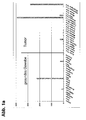
- the amount of gene-specific transcripts in healthy tissue and in carcinoma samples was investigated ( Fig. 1a . b . c ).
- LOC90625 is very selectively expressed in healthy tissue, specific transcripts are detectable especially in the testis. Little or no detectable LOC90625-specific expression in all other healthy tissues analyzed ( Fig. 1a . b ).
- Fig. 1a . b . c LOC90625-specific RT-PCR
- LOC90625 is a selectively expressed antigen which is apparently expressed more prominently in proliferating tissues. This selective overexpression in tumors can be used diagnostically and therapeutically.
- the extracellular domain of LOC90625 can be used according to the invention as the target structure of monoclonal antibodies.
- This can e.g. 1-19 (SEQ ID NO: 287) or amino acids 40-160 (SEQ ID NO: 288).
- SEQ ID NO: 287 amino acids 40-160
- SEQ ID NO: 288 amino acids 40-160
Landscapes
- Health & Medical Sciences (AREA)
- Life Sciences & Earth Sciences (AREA)
- Chemical & Material Sciences (AREA)
- Engineering & Computer Science (AREA)
- Physics & Mathematics (AREA)
- General Health & Medical Sciences (AREA)
- Organic Chemistry (AREA)
- Proteomics, Peptides & Aminoacids (AREA)
- Analytical Chemistry (AREA)
- Immunology (AREA)
- Biotechnology (AREA)
- Bioinformatics & Cheminformatics (AREA)
- Oncology (AREA)
- Biophysics (AREA)
- Pathology (AREA)
- Medicinal Chemistry (AREA)
- Molecular Biology (AREA)
- Wood Science & Technology (AREA)
- Zoology (AREA)
- Genetics & Genomics (AREA)
- Chemical Kinetics & Catalysis (AREA)
- Theoretical Computer Science (AREA)
- Spectroscopy & Molecular Physics (AREA)
- Medical Informatics (AREA)
- General Chemical & Material Sciences (AREA)
- Evolutionary Biology (AREA)
- Nuclear Medicine, Radiotherapy & Molecular Imaging (AREA)
- Pharmacology & Pharmacy (AREA)
- Animal Behavior & Ethology (AREA)
- Public Health (AREA)
- Veterinary Medicine (AREA)
- Bioinformatics & Computational Biology (AREA)
- Cell Biology (AREA)
- Hospice & Palliative Care (AREA)
- Microbiology (AREA)
- Biochemistry (AREA)
- Hematology (AREA)
- Biomedical Technology (AREA)
- Urology & Nephrology (AREA)
- General Engineering & Computer Science (AREA)
Priority Applications (4)
| Application Number | Priority Date | Filing Date | Title |
|---|---|---|---|
| EP20100010912 EP2327417B1 (de) | 2005-03-24 | 2006-03-23 | Identifizierung von Oberflächen-assoziierten Antigenen für die Tumordiagnose und- therapie |
| EP10010914A EP2314309A3 (de) | 2005-03-24 | 2006-03-23 | Identifizierung von Oberflächen-assoziierten Antigenen für die Tumordiagnose und- therapie |
| EP10010913A EP2311487A3 (de) | 2005-03-24 | 2006-03-23 | Identifizierung von Oberflächen-assoziierten Antigenen für die Tumordiagnose und- therapie |
| EP10010915A EP2314310A3 (de) | 2005-03-24 | 2006-03-23 | Identifizierung von Oberflächen-assoziierten Antigenen für die Tumordiagnose und- therapie |
Applications Claiming Priority (2)
| Application Number | Priority Date | Filing Date | Title |
|---|---|---|---|
| DE102005013846A DE102005013846A1 (de) | 2005-03-24 | 2005-03-24 | Identifizierung von Oberflächen-assoziierten Antigenen für die Tumordiagnose und -therapie |
| PCT/EP2006/002695 WO2006100089A2 (de) | 2005-03-24 | 2006-03-23 | Identifizierung von oberflächen-assoziierten antigenen für die tumordiagnose und -therapie |
Related Child Applications (5)
| Application Number | Title | Priority Date | Filing Date |
|---|---|---|---|
| EP10010913A Division-Into EP2311487A3 (de) | 2005-03-24 | 2006-03-23 | Identifizierung von Oberflächen-assoziierten Antigenen für die Tumordiagnose und- therapie |
| EP10010915A Division-Into EP2314310A3 (de) | 2005-03-24 | 2006-03-23 | Identifizierung von Oberflächen-assoziierten Antigenen für die Tumordiagnose und- therapie |
| EP20100010912 Division EP2327417B1 (de) | 2005-03-24 | 2006-03-23 | Identifizierung von Oberflächen-assoziierten Antigenen für die Tumordiagnose und- therapie |
| EP20100010912 Division-Into EP2327417B1 (de) | 2005-03-24 | 2006-03-23 | Identifizierung von Oberflächen-assoziierten Antigenen für die Tumordiagnose und- therapie |
| EP10010914A Division-Into EP2314309A3 (de) | 2005-03-24 | 2006-03-23 | Identifizierung von Oberflächen-assoziierten Antigenen für die Tumordiagnose und- therapie |
Publications (2)
| Publication Number | Publication Date |
|---|---|
| EP1861118A2 EP1861118A2 (de) | 2007-12-05 |
| EP1861118B1 true EP1861118B1 (de) | 2015-05-06 |
Family
ID=36613498
Family Applications (5)
| Application Number | Title | Priority Date | Filing Date |
|---|---|---|---|
| EP06723678.6A Active EP1861118B1 (de) | 2005-03-24 | 2006-03-23 | Identifizierung von oberflächen-assoziierten antigenen für die tumordiagnose und -therapie |
| EP20100010912 Active EP2327417B1 (de) | 2005-03-24 | 2006-03-23 | Identifizierung von Oberflächen-assoziierten Antigenen für die Tumordiagnose und- therapie |
| EP10010915A Withdrawn EP2314310A3 (de) | 2005-03-24 | 2006-03-23 | Identifizierung von Oberflächen-assoziierten Antigenen für die Tumordiagnose und- therapie |
| EP10010913A Withdrawn EP2311487A3 (de) | 2005-03-24 | 2006-03-23 | Identifizierung von Oberflächen-assoziierten Antigenen für die Tumordiagnose und- therapie |
| EP10010914A Withdrawn EP2314309A3 (de) | 2005-03-24 | 2006-03-23 | Identifizierung von Oberflächen-assoziierten Antigenen für die Tumordiagnose und- therapie |
Family Applications After (4)
| Application Number | Title | Priority Date | Filing Date |
|---|---|---|---|
| EP20100010912 Active EP2327417B1 (de) | 2005-03-24 | 2006-03-23 | Identifizierung von Oberflächen-assoziierten Antigenen für die Tumordiagnose und- therapie |
| EP10010915A Withdrawn EP2314310A3 (de) | 2005-03-24 | 2006-03-23 | Identifizierung von Oberflächen-assoziierten Antigenen für die Tumordiagnose und- therapie |
| EP10010913A Withdrawn EP2311487A3 (de) | 2005-03-24 | 2006-03-23 | Identifizierung von Oberflächen-assoziierten Antigenen für die Tumordiagnose und- therapie |
| EP10010914A Withdrawn EP2314309A3 (de) | 2005-03-24 | 2006-03-23 | Identifizierung von Oberflächen-assoziierten Antigenen für die Tumordiagnose und- therapie |
Country Status (6)
| Country | Link |
|---|---|
| US (4) | US9090940B2 (enExample) |
| EP (5) | EP1861118B1 (enExample) |
| JP (5) | JP5253999B2 (enExample) |
| DE (1) | DE102005013846A1 (enExample) |
| ES (1) | ES2537319T3 (enExample) |
| WO (1) | WO2006100089A2 (enExample) |
Families Citing this family (26)
| Publication number | Priority date | Publication date | Assignee | Title |
|---|---|---|---|---|
| DE102005013846A1 (de) | 2005-03-24 | 2006-10-05 | Ganymed Pharmaceuticals Ag | Identifizierung von Oberflächen-assoziierten Antigenen für die Tumordiagnose und -therapie |
| DE102006060824B4 (de) * | 2006-12-21 | 2011-06-01 | Johannes-Gutenberg-Universität Mainz | Nachweis von individuellen T-Zell-Reaktionsmustern gegen Tumor-assoziierte Antigene (TAA) in Tumorpatienten als Basis für die individuelle therapeutische Vakzinierung von Patienten |
| EP2060583A1 (en) * | 2007-10-23 | 2009-05-20 | Ganymed Pharmaceuticals AG | Identification of tumor-associated markers for diagnosis and therapy |
| EP2249159A1 (en) * | 2009-04-29 | 2010-11-10 | Ganymed Pharmaceuticals AG | Identification of tumor-associated markers for diagnosis and therapy |
| CN107674114B (zh) | 2009-07-29 | 2022-10-25 | 凯伊药品公司 | 用于降低甲状旁腺激素水平的治疗剂 |
| US8486657B2 (en) * | 2010-03-22 | 2013-07-16 | Xiaohong Cai | Anti-preproprotein and anti-preprotein antibodies as immunohistochemical markers |
| TW201302800A (zh) * | 2011-06-10 | 2013-01-16 | 腫瘤療法 科學股份有限公司 | Sema5b胜肽及含其之疫苗 |
| KR20150091311A (ko) * | 2012-12-04 | 2015-08-10 | 온코세라피 사이언스 가부시키가이샤 | Sema5b 펩타이드 및 이를 포함하는 백신 |
| WO2015075939A1 (ja) | 2013-11-21 | 2015-05-28 | Repertoire Genesis株式会社 | T細胞受容体およびb細胞受容体レパトアの解析システムならびにその治療および診断への利用 |
| US11318163B2 (en) | 2015-02-18 | 2022-05-03 | Enlivex Therapeutics Ltd | Combination immune therapy and cytokine control therapy for cancer treatment |
| EP3258943B1 (en) | 2015-02-18 | 2021-05-12 | Enlivex Therapeutics Ltd. | Combination immune therapy and cytokine control therapy for cancer treatment |
| US11304976B2 (en) | 2015-02-18 | 2022-04-19 | Enlivex Therapeutics Ltd | Combination immune therapy and cytokine control therapy for cancer treatment |
| US11000548B2 (en) | 2015-02-18 | 2021-05-11 | Enlivex Therapeutics Ltd | Combination immune therapy and cytokine control therapy for cancer treatment |
| US11596652B2 (en) | 2015-02-18 | 2023-03-07 | Enlivex Therapeutics R&D Ltd | Early apoptotic cells for use in treating sepsis |
| US11497767B2 (en) | 2015-02-18 | 2022-11-15 | Enlivex Therapeutics R&D Ltd | Combination immune therapy and cytokine control therapy for cancer treatment |
| CN113106053A (zh) | 2015-04-21 | 2021-07-13 | 恩立夫克治疗有限责任公司 | 治疗性汇集的血液凋亡细胞制剂与其用途 |
| GB201520564D0 (en) | 2015-11-23 | 2016-01-06 | Immunocore Ltd & Adaptimmune Ltd | Peptides |
| GB201520568D0 (en) | 2015-11-23 | 2016-01-06 | Immunocore Ltd | Peptides |
| GB201520550D0 (en) | 2015-11-23 | 2016-01-06 | Immunocore Ltd & Adaptimmune Ltd | Peptides |
| TWI765875B (zh) | 2015-12-16 | 2022-06-01 | 美商磨石生物公司 | 新抗原辨識、製造及用途 |
| WO2017123808A1 (en) | 2016-01-15 | 2017-07-20 | The J. David Gladstone Institutes | Methods of treating disease by metabolic control of t-cell differentiation |
| EP3416661A4 (en) | 2016-02-18 | 2020-03-04 | Enlivex Therapeutics Ltd. | COMBINATION OF IMMUNOTHERAPY AND CYTOKINE CONTROL THERAPY FOR THE TREATMENT OF CANCER |
| EP3648790A1 (en) * | 2017-07-07 | 2020-05-13 | Immatics Biotechnologies GmbH | Novel peptides and combination of peptides for use in immunotherapy against lung cancer, including nsclc, sclc and other cancers |
| WO2019075112A1 (en) | 2017-10-10 | 2019-04-18 | Gritstone Oncology, Inc. | IDENTIFICATION OF NEO-ANTIGENS USING HOT POINTS |
| AU2018373154B2 (en) | 2017-11-22 | 2025-08-07 | Seattle Project Corp. | Reducing junction epitope presentation for neoantigens |
| WO2022256620A1 (en) * | 2021-06-03 | 2022-12-08 | The Broad Institute, Inc. | Novel targets for enhancing anti-tumor immunity |
Citations (1)
| Publication number | Priority date | Publication date | Assignee | Title |
|---|---|---|---|---|
| WO2005030250A2 (de) * | 2003-09-26 | 2005-04-07 | Ganymed Pharmaceuticals Ag | Identifizierung von tumorassoziierten zelloberflächen-antigenen für die diagnose und therapie |
Family Cites Families (20)
| Publication number | Priority date | Publication date | Assignee | Title |
|---|---|---|---|---|
| US2191A (en) | 1841-07-23 | Constructing the surgical instrument denominated the | ||
| GB9019812D0 (en) | 1990-09-11 | 1990-10-24 | Scotgen Ltd | Novel antibodies for treatment and prevention of infection in animals and man |
| US5677139A (en) | 1995-04-21 | 1997-10-14 | President And Fellows Of Harvard College | In vitro differentiation of CD34+ progenitor cells into T lymphocytes |
| UA56132C2 (uk) | 1995-04-25 | 2003-05-15 | Смітклайн Бічем Байолоджікалс С.А. | Композиція вакцини (варіанти), спосіб стабілізації qs21 відносно гідролізу (варіанти), спосіб приготування композиції вакцини |
| ATE244300T1 (de) | 1996-01-17 | 2003-07-15 | Imp College Innovations Ltd | Immunotherapie mit verwendung von zytotoxischen t lymphozyten (ctl) |
| EP0971950A2 (en) | 1997-02-24 | 2000-01-19 | Genetics Institute, Inc. | Secreted proteins and polynucleotides encoding them |
| AU7549498A (en) * | 1997-06-03 | 1998-12-21 | Protegene Inc. | Human proteins having transmembrane domains and dnas encoding these prot eins |
| CA2354369A1 (en) * | 1998-12-22 | 2000-06-29 | Genset S.A. | Complementary dna's encoding proteins with signal peptides |
| US20030204075A9 (en) | 1999-08-09 | 2003-10-30 | The Snp Consortium | Identification and mapping of single nucleotide polymorphisms in the human genome |
| WO2002012343A2 (en) | 2000-08-07 | 2002-02-14 | Curagen Corporation | Proteins and nucleic acids encoding g-protein coupled receptors |
| USH2191H1 (en) | 2000-10-24 | 2007-06-05 | Snp Consortium | Identification and mapping of single nucleotide polymorphisms in the human genome |
| WO2002068579A2 (en) * | 2001-01-10 | 2002-09-06 | Pe Corporation (Ny) | Kits, such as nucleic acid arrays, comprising a majority of human exons or transcripts, for detecting expression and other uses thereof |
| JP2005527180A (ja) * | 2001-04-18 | 2005-09-15 | プロテイン デザイン ラブス, インコーポレイテッド | 肺がんの診断方法、肺がんの修飾因子の組成及びスクリーニングの方法 |
| JP2005500832A (ja) * | 2001-06-18 | 2005-01-13 | ロゼッタ・インファーマティクス・インコーポレーテッド | 乳癌患者の診断および予後 |
| US7171311B2 (en) * | 2001-06-18 | 2007-01-30 | Rosetta Inpharmatics Llc | Methods of assigning treatment to breast cancer patients |
| US20040076955A1 (en) * | 2001-07-03 | 2004-04-22 | Eos Biotechnology, Inc. | Methods of diagnosis of bladder cancer, compositions and methods of screening for modulators of bladder cancer |
| DE10211088A1 (de) * | 2002-03-13 | 2003-09-25 | Ugur Sahin | Differentiell in Tumoren exprimierte Genprodukte und deren Verwendung |
| DE10254601A1 (de) * | 2002-11-22 | 2004-06-03 | Ganymed Pharmaceuticals Ag | Differentiell in Tumoren exprimierte Genprodukte und deren Verwendung |
| AU2003263660A1 (en) | 2003-08-29 | 2005-03-16 | Pantarhei Bioscience B.V. | Prenatal diagnosis of down syndrome by detection of fetal rna markers in maternal blood |
| DE102005013846A1 (de) | 2005-03-24 | 2006-10-05 | Ganymed Pharmaceuticals Ag | Identifizierung von Oberflächen-assoziierten Antigenen für die Tumordiagnose und -therapie |
-
2005
- 2005-03-24 DE DE102005013846A patent/DE102005013846A1/de not_active Ceased
-
2006
- 2006-03-23 EP EP06723678.6A patent/EP1861118B1/de active Active
- 2006-03-23 JP JP2008502334A patent/JP5253999B2/ja active Active
- 2006-03-23 EP EP20100010912 patent/EP2327417B1/de active Active
- 2006-03-23 WO PCT/EP2006/002695 patent/WO2006100089A2/de not_active Ceased
- 2006-03-23 US US11/886,758 patent/US9090940B2/en active Active
- 2006-03-23 ES ES06723678.6T patent/ES2537319T3/es active Active
- 2006-03-23 EP EP10010915A patent/EP2314310A3/de not_active Withdrawn
- 2006-03-23 EP EP10010913A patent/EP2311487A3/de not_active Withdrawn
- 2006-03-23 EP EP10010914A patent/EP2314309A3/de not_active Withdrawn
-
2012
- 2012-04-02 JP JP2012083549A patent/JP5731436B2/ja active Active
- 2012-11-27 JP JP2012258431A patent/JP5779167B2/ja active Active
-
2013
- 2013-04-17 US US13/864,411 patent/US9194004B2/en active Active
-
2014
- 2014-01-16 JP JP2014005640A patent/JP5922159B2/ja active Active
-
2015
- 2015-06-19 US US14/744,845 patent/US10036753B2/en active Active
-
2016
- 2016-02-24 JP JP2016032631A patent/JP6174177B2/ja active Active
- 2016-03-07 US US14/887,979 patent/US10302647B2/en active Active
Patent Citations (1)
| Publication number | Priority date | Publication date | Assignee | Title |
|---|---|---|---|---|
| WO2005030250A2 (de) * | 2003-09-26 | 2005-04-07 | Ganymed Pharmaceuticals Ag | Identifizierung von tumorassoziierten zelloberflächen-antigenen für die diagnose und therapie |
Non-Patent Citations (1)
| Title |
|---|
| L. BUONAGURO ET AL: "Translating Tumor Antigens into Cancer Vaccines", CLINICAL AND VACCINE IMMUNOLOGY, vol. 18, no. 1, 1 January 2011 (2011-01-01), pages 23 - 34, XP055009295, ISSN: 1556-6811, DOI: 10.1128/CVI.00286-10 * |
Also Published As
| Publication number | Publication date |
|---|---|
| US20140017679A1 (en) | 2014-01-16 |
| EP2327417B1 (de) | 2015-05-06 |
| EP2327417A2 (de) | 2011-06-01 |
| JP2012207020A (ja) | 2012-10-25 |
| JP5779167B2 (ja) | 2015-09-16 |
| WO2006100089A3 (de) | 2007-01-11 |
| ES2537319T3 (es) | 2015-06-05 |
| EP2314309A2 (de) | 2011-04-27 |
| JP2008537546A (ja) | 2008-09-18 |
| JP5731436B2 (ja) | 2015-06-10 |
| EP1861118A2 (de) | 2007-12-05 |
| US20170052187A1 (en) | 2017-02-23 |
| US10302647B2 (en) | 2019-05-28 |
| JP6174177B2 (ja) | 2017-08-02 |
| JP5922159B2 (ja) | 2016-05-24 |
| JP2014139171A (ja) | 2014-07-31 |
| WO2006100089A2 (de) | 2006-09-28 |
| EP2314310A3 (de) | 2011-08-10 |
| US9090940B2 (en) | 2015-07-28 |
| EP2314309A3 (de) | 2011-08-10 |
| EP2311487A2 (de) | 2011-04-20 |
| DE102005013846A1 (de) | 2006-10-05 |
| JP5253999B2 (ja) | 2013-07-31 |
| EP2327417A3 (de) | 2011-08-03 |
| JP2013099331A (ja) | 2013-05-23 |
| EP2314310A2 (de) | 2011-04-27 |
| JP2016138115A (ja) | 2016-08-04 |
| US9194004B2 (en) | 2015-11-24 |
| EP2311487A3 (de) | 2011-08-10 |
| US20090214550A1 (en) | 2009-08-27 |
| US20150368723A1 (en) | 2015-12-24 |
| US10036753B2 (en) | 2018-07-31 |
Similar Documents
| Publication | Publication Date | Title |
|---|---|---|
| EP1861118B1 (de) | Identifizierung von oberflächen-assoziierten antigenen für die tumordiagnose und -therapie | |
| EP2280082B1 (de) | Identifizierung von Oberflächen-assoziierten Antigenen für die Tumordiagnose und -therapie | |
| EP2267012B1 (de) | Identifizierung von tumorassoziierten Zelloberflächen-Antigenen für die Diagnose und Therapie | |
| EP1483389B1 (de) | Differentiell in tumoren exprimierte genprodukte und deren verwendung |
Legal Events
| Date | Code | Title | Description |
|---|---|---|---|
| PUAI | Public reference made under article 153(3) epc to a published international application that has entered the european phase |
Free format text: ORIGINAL CODE: 0009012 |
|
| 17P | Request for examination filed |
Effective date: 20070823 |
|
| AK | Designated contracting states |
Kind code of ref document: A2 Designated state(s): AT BE BG CH CY CZ DE DK EE ES FI FR GB GR HU IE IS IT LI LT LU LV MC NL PL PT RO SE SI SK TR |
|
| AX | Request for extension of the european patent |
Extension state: AL BA HR MK YU |
|
| 17Q | First examination report despatched |
Effective date: 20071217 |
|
| RIN1 | Information on inventor provided before grant (corrected) |
Inventor name: SCHLUETER, VOLKER Inventor name: KOSLOWSKI, MICHAEL Inventor name: USENER, DIRK Inventor name: TUERECI, OEZLEM Inventor name: SAHIN, UGUR Inventor name: HELFTENBEIN, GERD |
|
| RIN1 | Information on inventor provided before grant (corrected) |
Inventor name: SCHLUETER, VOLKER Inventor name: USENER, DIRK Inventor name: SAHIN, UGUR Inventor name: TUERECI, OEZLEM Inventor name: KOSLOWSKI, MICHAEL Inventor name: HELFTENBEIN, GERD |
|
| RAP1 | Party data changed (applicant data changed or rights of an application transferred) |
Owner name: BIONTECH AG |
|
| GRAP | Despatch of communication of intention to grant a patent |
Free format text: ORIGINAL CODE: EPIDOSNIGR1 |
|
| INTG | Intention to grant announced |
Effective date: 20141029 |
|
| GRAS | Grant fee paid |
Free format text: ORIGINAL CODE: EPIDOSNIGR3 |
|
| GRAA | (expected) grant |
Free format text: ORIGINAL CODE: 0009210 |
|
| AK | Designated contracting states |
Kind code of ref document: B1 Designated state(s): AT BE BG CH CY CZ DE DK EE ES FI FR GB GR HU IE IS IT LI LT LU LV MC NL PL PT RO SE SI SK TR |
|
| AX | Request for extension of the european patent |
Extension state: AL BA HR MK YU |
|
| REG | Reference to a national code |
Ref country code: GB Ref legal event code: FG4D Free format text: NOT ENGLISH |
|
| REG | Reference to a national code |
Ref country code: CH Ref legal event code: EP |
|
| RAP2 | Party data changed (patent owner data changed or rights of a patent transferred) |
Owner name: BIONTECH AG |
|
| REG | Reference to a national code |
Ref country code: IE Ref legal event code: FG4D Free format text: LANGUAGE OF EP DOCUMENT: GERMAN |
|
| REG | Reference to a national code |
Ref country code: ES Ref legal event code: FG2A Ref document number: 2537319 Country of ref document: ES Kind code of ref document: T3 Effective date: 20150605 |
|
| REG | Reference to a national code |
Ref country code: NL Ref legal event code: T3 |
|
| REG | Reference to a national code |
Ref country code: DE Ref legal event code: R096 Ref document number: 502006014322 Country of ref document: DE Effective date: 20150611 |
|
| REG | Reference to a national code |
Ref country code: AT Ref legal event code: REF Ref document number: 725231 Country of ref document: AT Kind code of ref document: T Effective date: 20150615 Ref country code: CH Ref legal event code: NV Representative=s name: MICHELI AND CIE SA, CH |
|
| REG | Reference to a national code |
Ref country code: NL Ref legal event code: T3 |
|
| REG | Reference to a national code |
Ref country code: LT Ref legal event code: MG4D |
|
| PG25 | Lapsed in a contracting state [announced via postgrant information from national office to epo] |
Ref country code: FI Free format text: LAPSE BECAUSE OF FAILURE TO SUBMIT A TRANSLATION OF THE DESCRIPTION OR TO PAY THE FEE WITHIN THE PRESCRIBED TIME-LIMIT Effective date: 20150506 Ref country code: PT Free format text: LAPSE BECAUSE OF FAILURE TO SUBMIT A TRANSLATION OF THE DESCRIPTION OR TO PAY THE FEE WITHIN THE PRESCRIBED TIME-LIMIT Effective date: 20150907 Ref country code: LT Free format text: LAPSE BECAUSE OF FAILURE TO SUBMIT A TRANSLATION OF THE DESCRIPTION OR TO PAY THE FEE WITHIN THE PRESCRIBED TIME-LIMIT Effective date: 20150506 |
|
| PG25 | Lapsed in a contracting state [announced via postgrant information from national office to epo] |
Ref country code: LV Free format text: LAPSE BECAUSE OF FAILURE TO SUBMIT A TRANSLATION OF THE DESCRIPTION OR TO PAY THE FEE WITHIN THE PRESCRIBED TIME-LIMIT Effective date: 20150506 Ref country code: IS Free format text: LAPSE BECAUSE OF FAILURE TO SUBMIT A TRANSLATION OF THE DESCRIPTION OR TO PAY THE FEE WITHIN THE PRESCRIBED TIME-LIMIT Effective date: 20150906 Ref country code: GR Free format text: LAPSE BECAUSE OF FAILURE TO SUBMIT A TRANSLATION OF THE DESCRIPTION OR TO PAY THE FEE WITHIN THE PRESCRIBED TIME-LIMIT Effective date: 20150807 Ref country code: BG Free format text: LAPSE BECAUSE OF FAILURE TO SUBMIT A TRANSLATION OF THE DESCRIPTION OR TO PAY THE FEE WITHIN THE PRESCRIBED TIME-LIMIT Effective date: 20150806 |
|
| PG25 | Lapsed in a contracting state [announced via postgrant information from national office to epo] |
Ref country code: DK Free format text: LAPSE BECAUSE OF FAILURE TO SUBMIT A TRANSLATION OF THE DESCRIPTION OR TO PAY THE FEE WITHIN THE PRESCRIBED TIME-LIMIT Effective date: 20150506 Ref country code: EE Free format text: LAPSE BECAUSE OF FAILURE TO SUBMIT A TRANSLATION OF THE DESCRIPTION OR TO PAY THE FEE WITHIN THE PRESCRIBED TIME-LIMIT Effective date: 20150506 |
|
| REG | Reference to a national code |
Ref country code: DE Ref legal event code: R097 Ref document number: 502006014322 Country of ref document: DE |
|
| PG25 | Lapsed in a contracting state [announced via postgrant information from national office to epo] |
Ref country code: CZ Free format text: LAPSE BECAUSE OF FAILURE TO SUBMIT A TRANSLATION OF THE DESCRIPTION OR TO PAY THE FEE WITHIN THE PRESCRIBED TIME-LIMIT Effective date: 20150506 Ref country code: PL Free format text: LAPSE BECAUSE OF FAILURE TO SUBMIT A TRANSLATION OF THE DESCRIPTION OR TO PAY THE FEE WITHIN THE PRESCRIBED TIME-LIMIT Effective date: 20150506 Ref country code: SK Free format text: LAPSE BECAUSE OF FAILURE TO SUBMIT A TRANSLATION OF THE DESCRIPTION OR TO PAY THE FEE WITHIN THE PRESCRIBED TIME-LIMIT Effective date: 20150506 Ref country code: RO Free format text: LAPSE BECAUSE OF NON-PAYMENT OF DUE FEES Effective date: 20150506 |
|
| PLBE | No opposition filed within time limit |
Free format text: ORIGINAL CODE: 0009261 |
|
| STAA | Information on the status of an ep patent application or granted ep patent |
Free format text: STATUS: NO OPPOSITION FILED WITHIN TIME LIMIT |
|
| REG | Reference to a national code |
Ref country code: FR Ref legal event code: PLFP Year of fee payment: 11 |
|
| 26N | No opposition filed |
Effective date: 20160209 |
|
| PG25 | Lapsed in a contracting state [announced via postgrant information from national office to epo] |
Ref country code: SI Free format text: LAPSE BECAUSE OF FAILURE TO SUBMIT A TRANSLATION OF THE DESCRIPTION OR TO PAY THE FEE WITHIN THE PRESCRIBED TIME-LIMIT Effective date: 20150506 |
|
| PG25 | Lapsed in a contracting state [announced via postgrant information from national office to epo] |
Ref country code: BE Free format text: LAPSE BECAUSE OF NON-PAYMENT OF DUE FEES Effective date: 20160331 |
|
| PG25 | Lapsed in a contracting state [announced via postgrant information from national office to epo] |
Ref country code: LU Free format text: LAPSE BECAUSE OF FAILURE TO SUBMIT A TRANSLATION OF THE DESCRIPTION OR TO PAY THE FEE WITHIN THE PRESCRIBED TIME-LIMIT Effective date: 20160323 Ref country code: MC Free format text: LAPSE BECAUSE OF FAILURE TO SUBMIT A TRANSLATION OF THE DESCRIPTION OR TO PAY THE FEE WITHIN THE PRESCRIBED TIME-LIMIT Effective date: 20150506 |
|
| REG | Reference to a national code |
Ref country code: IE Ref legal event code: MM4A |
|
| PG25 | Lapsed in a contracting state [announced via postgrant information from national office to epo] |
Ref country code: IE Free format text: LAPSE BECAUSE OF NON-PAYMENT OF DUE FEES Effective date: 20160323 |
|
| REG | Reference to a national code |
Ref country code: FR Ref legal event code: PLFP Year of fee payment: 12 |
|
| PG25 | Lapsed in a contracting state [announced via postgrant information from national office to epo] |
Ref country code: SE Free format text: LAPSE BECAUSE OF FAILURE TO SUBMIT A TRANSLATION OF THE DESCRIPTION OR TO PAY THE FEE WITHIN THE PRESCRIBED TIME-LIMIT Effective date: 20150506 Ref country code: IT Free format text: LAPSE BECAUSE OF NON-PAYMENT OF DUE FEES Effective date: 20160323 |
|
| PG25 | Lapsed in a contracting state [announced via postgrant information from national office to epo] |
Ref country code: IT Free format text: LAPSE BECAUSE OF NON-PAYMENT OF DUE FEES Effective date: 20160323 |
|
| PGRI | Patent reinstated in contracting state [announced from national office to epo] |
Ref country code: IT Effective date: 20170710 |
|
| REG | Reference to a national code |
Ref country code: FR Ref legal event code: PLFP Year of fee payment: 13 |
|
| PG25 | Lapsed in a contracting state [announced via postgrant information from national office to epo] |
Ref country code: HU Free format text: LAPSE BECAUSE OF FAILURE TO SUBMIT A TRANSLATION OF THE DESCRIPTION OR TO PAY THE FEE WITHIN THE PRESCRIBED TIME-LIMIT; INVALID AB INITIO Effective date: 20060323 Ref country code: CY Free format text: LAPSE BECAUSE OF FAILURE TO SUBMIT A TRANSLATION OF THE DESCRIPTION OR TO PAY THE FEE WITHIN THE PRESCRIBED TIME-LIMIT Effective date: 20150506 |
|
| REG | Reference to a national code |
Ref country code: DE Ref legal event code: R082 Ref document number: 502006014322 Country of ref document: DE Representative=s name: ZWICKER SCHNAPPAUF & PARTNER PATENTANWAELTE PA, DE |
|
| P01 | Opt-out of the competence of the unified patent court (upc) registered |
Effective date: 20230512 |
|
| PGFP | Annual fee paid to national office [announced via postgrant information from national office to epo] |
Ref country code: NL Payment date: 20250219 Year of fee payment: 20 |
|
| PGFP | Annual fee paid to national office [announced via postgrant information from national office to epo] |
Ref country code: DE Payment date: 20250218 Year of fee payment: 20 |
|
| PGFP | Annual fee paid to national office [announced via postgrant information from national office to epo] |
Ref country code: AT Payment date: 20250220 Year of fee payment: 20 |
|
| PGFP | Annual fee paid to national office [announced via postgrant information from national office to epo] |
Ref country code: FR Payment date: 20250219 Year of fee payment: 20 |
|
| PGFP | Annual fee paid to national office [announced via postgrant information from national office to epo] |
Ref country code: IT Payment date: 20250218 Year of fee payment: 20 Ref country code: GB Payment date: 20250221 Year of fee payment: 20 |
|
| PGFP | Annual fee paid to national office [announced via postgrant information from national office to epo] |
Ref country code: TR Payment date: 20250228 Year of fee payment: 20 |
|
| PGFP | Annual fee paid to national office [announced via postgrant information from national office to epo] |
Ref country code: ES Payment date: 20250401 Year of fee payment: 20 |
|
| PGFP | Annual fee paid to national office [announced via postgrant information from national office to epo] |
Ref country code: CH Payment date: 20250401 Year of fee payment: 20 |