EP1861118B1 - Identifizierung von oberflächen-assoziierten antigenen für die tumordiagnose und -therapie - Google Patents
Identifizierung von oberflächen-assoziierten antigenen für die tumordiagnose und -therapie Download PDFInfo
- Publication number
- EP1861118B1 EP1861118B1 EP06723678.6A EP06723678A EP1861118B1 EP 1861118 B1 EP1861118 B1 EP 1861118B1 EP 06723678 A EP06723678 A EP 06723678A EP 1861118 B1 EP1861118 B1 EP 1861118B1
- Authority
- EP
- European Patent Office
- Prior art keywords
- tumor
- nucleic acid
- associated antigen
- cells
- antibody
- Prior art date
- Legal status (The legal status is an assumption and is not a legal conclusion. Google has not performed a legal analysis and makes no representation as to the accuracy of the status listed.)
- Expired - Lifetime
Links
Images


Classifications
-
- G—PHYSICS
- G01—MEASURING; TESTING
- G01N—INVESTIGATING OR ANALYSING MATERIALS BY DETERMINING THEIR CHEMICAL OR PHYSICAL PROPERTIES
- G01N33/00—Investigating or analysing materials by specific methods not covered by groups G01N1/00 - G01N31/00
- G01N33/48—Biological material, e.g. blood, urine; Haemocytometers
- G01N33/50—Chemical analysis of biological material, e.g. blood, urine; Testing involving biospecific ligand binding methods; Immunological testing
- G01N33/53—Immunoassay; Biospecific binding assay; Materials therefor
- G01N33/575—Immunoassay; Biospecific binding assay; Materials therefor for cancer
- G01N33/5758—Immunoassay; Biospecific binding assay; Materials therefor for cancer involving compounds serving as markers for tumours, cancers or neoplasias, e.g. cellular determinants, receptors, heat shock/stress proteins, A-protein, oligosaccharides or metabolites
- G01N33/5759—Immunoassay; Biospecific binding assay; Materials therefor for cancer involving compounds serving as markers for tumours, cancers or neoplasias, e.g. cellular determinants, receptors, heat shock/stress proteins, A-protein, oligosaccharides or metabolites involving compounds localised on the membrane of tumour or cancer cells
-
- A—HUMAN NECESSITIES
- A61—MEDICAL OR VETERINARY SCIENCE; HYGIENE
- A61P—SPECIFIC THERAPEUTIC ACTIVITY OF CHEMICAL COMPOUNDS OR MEDICINAL PREPARATIONS
- A61P35/00—Antineoplastic agents
-
- A—HUMAN NECESSITIES
- A61—MEDICAL OR VETERINARY SCIENCE; HYGIENE
- A61P—SPECIFIC THERAPEUTIC ACTIVITY OF CHEMICAL COMPOUNDS OR MEDICINAL PREPARATIONS
- A61P35/00—Antineoplastic agents
- A61P35/02—Antineoplastic agents specific for leukemia
-
- A—HUMAN NECESSITIES
- A61—MEDICAL OR VETERINARY SCIENCE; HYGIENE
- A61P—SPECIFIC THERAPEUTIC ACTIVITY OF CHEMICAL COMPOUNDS OR MEDICINAL PREPARATIONS
- A61P35/00—Antineoplastic agents
- A61P35/04—Antineoplastic agents specific for metastasis
-
- A—HUMAN NECESSITIES
- A61—MEDICAL OR VETERINARY SCIENCE; HYGIENE
- A61P—SPECIFIC THERAPEUTIC ACTIVITY OF CHEMICAL COMPOUNDS OR MEDICINAL PREPARATIONS
- A61P43/00—Drugs for specific purposes, not provided for in groups A61P1/00-A61P41/00
-
- C—CHEMISTRY; METALLURGY
- C12—BIOCHEMISTRY; BEER; SPIRITS; WINE; VINEGAR; MICROBIOLOGY; ENZYMOLOGY; MUTATION OR GENETIC ENGINEERING
- C12Q—MEASURING OR TESTING PROCESSES INVOLVING ENZYMES, NUCLEIC ACIDS OR MICROORGANISMS; COMPOSITIONS OR TEST PAPERS THEREFOR; PROCESSES OF PREPARING SUCH COMPOSITIONS; CONDITION-RESPONSIVE CONTROL IN MICROBIOLOGICAL OR ENZYMOLOGICAL PROCESSES
- C12Q1/00—Measuring or testing processes involving enzymes, nucleic acids or microorganisms; Compositions therefor; Processes of preparing such compositions
- C12Q1/68—Measuring or testing processes involving enzymes, nucleic acids or microorganisms; Compositions therefor; Processes of preparing such compositions involving nucleic acids
- C12Q1/6876—Nucleic acid products used in the analysis of nucleic acids, e.g. primers or probes
- C12Q1/6883—Nucleic acid products used in the analysis of nucleic acids, e.g. primers or probes for diseases caused by alterations of genetic material
- C12Q1/6886—Nucleic acid products used in the analysis of nucleic acids, e.g. primers or probes for diseases caused by alterations of genetic material for cancer
-
- G—PHYSICS
- G16—INFORMATION AND COMMUNICATION TECHNOLOGY [ICT] SPECIALLY ADAPTED FOR SPECIFIC APPLICATION FIELDS
- G16B—BIOINFORMATICS, i.e. INFORMATION AND COMMUNICATION TECHNOLOGY [ICT] SPECIALLY ADAPTED FOR GENETIC OR PROTEIN-RELATED DATA PROCESSING IN COMPUTATIONAL MOLECULAR BIOLOGY
- G16B30/00—ICT specially adapted for sequence analysis involving nucleotides or amino acids
- G16B30/10—Sequence alignment; Homology search
-
- C—CHEMISTRY; METALLURGY
- C12—BIOCHEMISTRY; BEER; SPIRITS; WINE; VINEGAR; MICROBIOLOGY; ENZYMOLOGY; MUTATION OR GENETIC ENGINEERING
- C12Q—MEASURING OR TESTING PROCESSES INVOLVING ENZYMES, NUCLEIC ACIDS OR MICROORGANISMS; COMPOSITIONS OR TEST PAPERS THEREFOR; PROCESSES OF PREPARING SUCH COMPOSITIONS; CONDITION-RESPONSIVE CONTROL IN MICROBIOLOGICAL OR ENZYMOLOGICAL PROCESSES
- C12Q2600/00—Oligonucleotides characterized by their use
- C12Q2600/118—Prognosis of disease development
-
- C—CHEMISTRY; METALLURGY
- C12—BIOCHEMISTRY; BEER; SPIRITS; WINE; VINEGAR; MICROBIOLOGY; ENZYMOLOGY; MUTATION OR GENETIC ENGINEERING
- C12Q—MEASURING OR TESTING PROCESSES INVOLVING ENZYMES, NUCLEIC ACIDS OR MICROORGANISMS; COMPOSITIONS OR TEST PAPERS THEREFOR; PROCESSES OF PREPARING SUCH COMPOSITIONS; CONDITION-RESPONSIVE CONTROL IN MICROBIOLOGICAL OR ENZYMOLOGICAL PROCESSES
- C12Q2600/00—Oligonucleotides characterized by their use
- C12Q2600/158—Expression markers
Definitions
- tumor-associated antigens or epitopes whose effector functions are to be intensified interventionally.
- Tumor cells differ biologically significantly from their non-malignant cells of origin. These differences are due to genetic alterations acquired during tumor development and cause, inter alia, also for the formation of qualitatively or quantitatively altered molecular structures in the cancer cells. If such tumor-associated structures are recognized by the specific immune system of the tumor-bearing host, one speaks of tumor-associated antigens.
- tumor-associated antigens arises from the fact that the recognition of antigens on neoplastic cells by the immune system leads to the initiation of cytotoxic effector mechanisms and in the presence of T helper cells can cause the elimination of cancer cells ( Pardoll, Nat. Med. 4: 525-31, 1998 ). Accordingly, a central goal of tumor immunology is to define these structures molecularly. The molecular nature of these antigens has long remained enigmatic.
- cytotoxic T lymphocyte CTL
- circulating autoantibodies Sahin et al., Curr. Opin. Immunol. 9: 709-16, 1997
- cDNA expression libraries were prepared from fresh tumor tissue and recombinantly expressed in suitable systems as proteins. Immune effectors isolated from patients, namely CTL clones with tumor-specific lysis patterns, or circulating autoantibodies have been used to clone the respective antigens.
- CTA Cancer / Testis antigens
- cancer / Testis antigens CTA and their coding genes (cancer / testis genes or CTG) are defined by their characteristic expression pattern ( Moseci et al., Mol. Med. Today 3: 342-9, 1997 ). They are not found in normal tissues except for testis or germ cells, but are expressed in a number of human malignancies and not tumor specific, but with varying frequency in tumor entities of very different origin ( Chen & Old, Cancer J. Sci. At the. 5: 16-7, 1999 ). Also serum reactivities against CTA are not found in healthy controls, but only in tumor patients.
- this class of antigen is of particular value for immunotherapeutic purposes and is being tested in current clinical patient studies ( Marchand et al., Int. J. Cancer 80: 219-30, 1999 ; Knuth et al., Cancer Chemother. Pharmacol. 46: S46-51, 2000 ).
- a strategy for identifying and providing tumor-associated expressed antigens and the nucleic acids coding therefor was pursued.
- This strategy is based on the evaluation of human protein and nucleic acid databases with regard to potential, cell-surface-accessible, cancer-specific antigens.
- the definition of the necessary filter criteria together with a high-throughput methodology for the analysis of as many proteins as possible form the central component of the invention.
- tissue-specific groups including tumor tissue
- proteins so identified become e.g. evaluated for their aberrant activation in tumors by expression analysis and protein chemistry.
- transcriptomes from normal tissue banks are usually electronically subtracted from tumor tissue banks, assuming that the remaining genes are tumor specific ( Schmitt et al., Nucleic Acids Res. 27: 4251-60, 1999 ; Vasmatzis et al., Proc. Natl. Acad. Sci. USA. 95: 300-4, 1998 ; Scheurle et al., Cancer Res. 60: 4037-43, 2000 ).
- the inventive concept which has proven to be much more successful, relies on exploiting data mining to electronically extract all genes encoding cell surface-accessible cancer-specific antigens and then evaluating them for ectopic expression in tumors.
- the invention is based on a strategy for the identification of genes expressed differentially in tumors. This combines data mining of public sequence banks ( "in silico” ) with subsequent evaluating laboratory experiments ("wet bench”) investigations.
- Example 1 A combined strategy based on different bioinformatic scripts allowed according to the invention the identification of the gene described in Example 1, which codes for a cell-surface-accessible, cancer-specific antigen. The identification and provision of this tumor-associated gene and the gene product encoded thereby was independent of their immunogenic effect.
- the tumor-associated antigen of the invention can be used as a diagnostic marker for cancers in the head and neck and for leukemia due to its tumor-specific expression.
- the PCT application WO2005 / 021793 describes fetal RNA markers in maternal blood that can be used for prenatal diagnosis of Down's syndrome.
- the nucleic acid sequence designated "SEQ ID NO: 13" which is identical to the nucleic acid sequence according to the invention according to SEQ ID NO: 13 is also described PCT application WO98 / 37094 the cloning of a variety of cDNAs and speculates that these cDNAs and the derived translation products could have a diagnostic or therapeutic significance.
- DA306_4 contains an open reading frame which codes for an amino acid sequence which is identical to the amino acid sequence according to the invention according to SEQ ID NO: 14.
- WO98 / 37094 There are no indications that a corresponding protein is expressed in tumor-specific manner in tumors of the head and neck and in leukemias.
- PCT application WO2005 / 030250 in that the tumor-associated antigen according to the invention is expressed in specific tumor tissues.
- WO2005 / 030250 there is no indication that tumor diseases from the head and neck area and leukemias are characterized by an expression or overexpression of the tumor-associated antigen according to the invention.
- the tumor-associated antigen identified according to the invention has an amino acid sequence which is encoded by a nucleic acid which comprises a nucleic acid sequence according to SEQ ID NO: 13 of the sequence listing.
- the tumor-associated antigen identified according to the invention has an amino acid sequence according to SEQ ID NO: 14 of the sequence listing.
- the present invention relates generally to the use of the tumor-associated antigen described in detail in detail of the nucleic acid encoding it or of nucleic acids which are coding against the coding Nucleic acids are directed, or of antibodies which are directed against the inventively identified tumor-associated antigens, for the diagnosis.
- This use concerns, for example, methods for the diagnosis and follow-up of carcinomas in the head and neck area or of leukemias.
- Particularly suitable for this purpose is a part of the tumor-associated antigen identified according to the invention which corresponds to or is comprised by the non-transmembrane component, in particular the extracellular component of the antigen.
- part of the tumor-associated antigens identified according to the invention which corresponds to or is comprised of the non-transmembrane portion of the antigens or a corresponding portion of the nucleic acids coding for the antigens identified according to the invention is preferred for diagnosis.
- Splice variants are useful as targets for the diagnosis of tumor diseases.
- the altered splicing of a gene leads to an altered transcript sequence (splice variant). If a splice variant is translated in the region of its altered sequence, this results in a modified protein, which may differ significantly from the original structure and function.
- tumor-associated splice variants tumor-associated transcripts and tumor-associated proteins / antigens can arise. These can be used as molecular markers for the detection of tumor cells.
- the detection of tumor cells e.g. in blood, serum, bone marrow, sputum, bronchial lavage, body secretions and tissue biopsies may e.g. after extraction of nucleic acids by PCR amplification with splice variant-specific oligonucleotides.
- all sequence-dependent detection systems are suitable according to the invention.
- these are eg gene chip / microarray systems, Northern blot, RNAse protection assays (RDA) and others.
- All detection systems have in common that the detection is based on a specific hybridization with at least one splice variant-specific nucleic acid sequence.
- the detection of tumor cells can also be carried out according to the invention by antibodies which encoded by the splice variant recognize specific epitope.
- the antibodies peptides for immunization can be used that are specific for this splice variant.
- the detection of tumor cells can also be carried out by antibodies that recognize tumor-specific altered Glykosyl michleukin.
- peptide regions can be used which differ in tumor cells and healthy cells due to glycosylation.
- asparagine is transformed into aspartic acid.
- the proteins described herein may be tumor-specific sequence-modified and thus have different biochemical and antibody binding properties.
- Particularly suitable for the immunization are the amino acids which have distinct epitope differences to the variant (s) of the gene product, which are preferably formed in healthy cells.
- the detection of tumor cells with antibodies can be done on a sample isolated from the patient or as imaging with intravenously applied antibodies.
- splice variants that exhibit new or altered epitopes are attractive targets for immunotherapy.
- the epitopes can be used to target therapeutically effective monoclonal antibodies or T lymphocytes.
- T lymphocytes In passive immunotherapy, antibodies or T lymphocytes that recognize splice variant-specific epitopes are adoptively transferred.
- the generation of antibodies, as with other antigens, can also be done using standard technologies (immunization of animals, panning strategies for the isolation of recombinant antibodies) using polypeptides containing these epitopes.
- nucleic acids encoding oligo- or polypeptides containing these epitopes may be used.
- Aberrant expression of genes in tumor cells may be due to altered methylation patterns of their promoters ( De Smet C. et al., Mol. Cell Biol. 24: 4781-90, 2004 ; De Smet C., et al., Mol. Cell Biol. 19: 7327-35, 1999 ; De Smet C. et al., Proc. Natl. Acad. Sci. USA. 93: 7149-53, 1996 ). These methylation differences can be used as an indirect marker for the tumor-altered state of the corresponding gene. Accordingly, the increase or decrease in base methylations in the promoter region can be used for diagnostic purposes.
- post-translationally modified protein domains such as, for example, glycosylations or myristilations are also described.
- This type of modification can lead to a differential recognition pattern of an antigen by, for example, an antibody and recognize various, possibly disease-associated conditions.
- this differentiation of an antigen can be used both diagnostically and therapeutically.
- tumor cells it has been reported that tumor-associated cellular degeneration can lead to altered post-translational modifications ( Durand & Seta, Clin. Chem. 46: 795-8052000 ; Granovsky et al., Nat. Med. 6: 306-312, 2000 ).
- glycosylation patterns are greatly altered on tumor cells.
- epitopes can diagnostically distinguish tumor cells from non-carcinogenic cells. Should a post-translationally modifiable epitope be glycosylated in normal, non-degenerate cells and deglycosylated in tumor cells, this situation allows the development of a tumor-specific therapeutic antibody.
- glycosylations which as a rule have a size of several kDa, lead to a larger total mass of the target protein, which can be separated by SDS-PAGE.
- protein lysates are incubated with O- or N-glycosylases prior to denaturation by SDS (according to the manufacturer's instructions, eg PNgase, endoglycosidase F, endoglycosidase H, Roche Diagnostics). This is followed by a Western Blot.
- Target protein When reducing the size of a Target protein can thus be detected after incubation with a glycosidase specific glycosylation and analyzed in this way, the tumor specificity of a modification.
- a glycosidase specific glycosylation Of particular interest are protein regions that are differentially glycosylated in tumor cells and healthy cells. However, such glycosylation differences have hitherto been described for a few cell surface proteins (eg, Muc1).
- the invention relates to methods of diagnosis or monitoring, i. Determining the regression, course and / or outbreak of a cancer characterized by the expression or abnormal expression of one or more tumor-associated antigens.
- the diagnostic methods and / or methods according to the invention generally relate to the use of means for the detection and / or the determination or monitoring of the amount of (i) a nucleic acid which codes for the tumor-associated antigen, or a part thereof and / or (ii) the tumor-associated antigen or a part thereof, in a biological sample isolated from a patient.
- Diagnostic methods may also use the tumor-associated antigen identified according to the invention as prognostic markers to detect metastatization, e.g. by predicting the migration behavior of cells and therefore predicating a worsened disease course, which among other things, allows the planning of a more aggressive therapy.
- the invention relates to a method for determining the regression, progression or onset of a cancer characterized by the expression or abnormal expression of the tumor-associated antigen identified according to the invention, comprising monitoring a sample from a patient having the disease or is suspected of contracting the disease.
- a detection of a nucleic acid or a part thereof or a determination or monitoring of the amount of a nucleic acid or a part thereof can be carried out according to the invention with a polynucleotide probe which is specific with the nucleic acid or the part hybridized thereto, or may be by selective amplification of the nucleic acid or portion thereof.
- the polynucleotide probe comprises, for example, a sequence of 6-50, in particular 10-30, 15-30 or 20-30 contiguous nucleotides from the nucleic acid.
- a selective amplification of the promoter region or of a part of a nucleic acid which codes for the tumor-associated antigen and is present in the form of genomic DNA takes place after treatment with a bisulfite-containing reagent.
- the nucleic acid can be isolated from a sample of a patient to be examined prior to treatment with the bisulfite-containing reagent.
- the oligonucleotides used in such an amplification have a sequence which bind to the nucleic acid treated with the bisulfite-containing reagent, preferably being completely complementary thereto.
- the oligonucleotides are adapted to a different degree of methylation of the nucleic acid and cause differentiable amplification products.
- Detection of a tumor-associated antigen or portion thereof or determination of the amount of a tumor-associated antigen or portion thereof may be with an antibody that specifically binds to the tumor-associated antigen or portion thereof.
- the tumor-associated antigen or part thereof to be detected may be present in a complex with an MHC molecule, in particular an HLA molecule.
- the polynucleotide probe or antibody used for detection or detection or monitoring is detectably labeled.
- the detectable marker being e.g. may be a radioactive marker or an enzyme label.
- the invention relates to the use of an antibody which binds to a tumor-associated antigen and is coupled to a diagnostic agent for the manufacture of a pharmaceutical composition for the diagnosis or monitoring of a cancer, characterized in that the tumor-associated Antigen is overexpressed in tumor tissue compared to a comparable healthy tissue, the tumor-associated antigen having a sequence encoded by a nucleic acid comprising the nucleic acid sequence according to SEQ ID NO: 13 of the Sequence Listing, the cancer comprising a carcinoma in the head Neck area or leukemia is.
- the antibody may be a monoclonal antibody.
- the antibody is a chimeric or humanized antibody or a fragment of a natural antibody.
- Conceivable methods of treatment are methods for treating a patient with a cancer characterized by the expression or abnormal expression of a tumor-associated antigen, comprising (i) identifying a nucleic acid encoding the tumor-associated antigen identified according to the invention which is expressed by cells (ii) transfecting a host cell with the nucleic acid or a portion thereof, (iii) culturing the transfected host cell for expression of the nucleic acid (this is not obligatory upon reaching a high transfection rate), and (iv ) introducing the host cells or an extract thereof into the patient in an amount capable of increasing the immune response to the cells of the patient associated with the disease.
- Such a method may further comprise identifying an MHC molecule presenting the tumor-associated antigen or a portion thereof, wherein the host cell expresses the identified MHC molecule and presents the tumor-associated antigen or portion thereof.
- the immune response may include a B-cell response or a T-cell response.
- a T cell response may involve the production of cytolytic T cells and / or helper T cells specific for the host cells presenting the tumor-associated antigen or portion thereof, or specifically for patient cells which express the tumor-associated antigen or a part thereof.
- a further conceivable method of treatment is a method for the treatment of a cancer which is manifested by the expression or abnormal expression of the tumor-associated antigen identified according to the invention, comprising (i) the identification of cells from the patient which express abnormal amounts of the tumor-associated antigen, (ii) the Isolating a sample of the cells, (iii) culturing the cells, and (iv) introducing the cells into the patient in an amount effective to induce an immune response to the cells.
- Oligonucleotides which hybridize with the nucleic acid identified according to the invention can be used as genetic probes.
- Nucleic acid molecules in the form of oligonucleotide primers or competent samples which hybridize to the nucleic acid identified according to the invention can be used to find nucleic acids which are homologous to the nucleic acid identified according to the invention.
- PCR amplification, Southern and Northern hybridization can be used to detect homologous nucleic acids.
- Hybridization can occur under low, better under medium, and most preferably under high stringency conditions.
- stringent conditions according to the invention relates to conditions that allow specific hybridization between polynucleotides.
- Immunogenic fragments of the tumor-associated antigen identified according to the invention can bind to a human HLA receptor or human antibody.
- a fragment has e.g. a sequence of at least 6, in particular at least 8, at least 10, at least 12, at least 15, at least 20, at least 30 or at least 50 amino acids.
- Example 1 describes peptides which can be used for the production of antibodies and have a sequence selected from the group consisting of SEQ ID NOs: 287-290.
- the methods and uses of the invention relate to an agent which binds to the tumor-associated antigen identified according to the invention.
- the agent is an antibody.
- the antibody is a chimeric, a humanized antibody or a fragment of an antibody.
- An antibody of the invention may be a monoclonal antibody.
- the antibody is a chimeric or humanized antibody or a fragment of an antibody.
- the uses according to the invention relate to a conjugate between an agent according to the invention which binds to the tumor-associated antigen identified according to the invention, or an antibody according to the invention and a diagnostic agent or substance.
- the diagnostic agent is a toxin.
- a gene is described as a gene which is selectively expressed or aberrantly expressed in tumor cells and represents a tumor-associated antigen.
- This gene and / or its gene products and / or its derivatives and / or parts may be target structures for therapeutic approaches.
- the therapeutic approaches may target an inhibition of the activity of the selectively expressed tumor-associated gene product. This is useful if the aberrant or selective expression is functionally of tumor pathogenetic importance and their suppression is accompanied by a selective damage to the corresponding cells.
- Other therapeutic concepts consider tumor-associated antigens as Markers that selectively recruit effector mechanisms with cell-damaging potential to tumor cells.
- the function of the target molecule itself and its role in tumorigenesis is completely irrelevant.
- derivatives of a nucleic acid are meant that single or multiple nucleotide substitutions, deletions, and / or additions are present in the nucleic acid.
- derivative also includes a chemical derivatization of a nucleic acid on a nucleotide base, on the sugar or on the phosphate of a nucleotide.
- derivative also includes nucleic acids containing non-naturally occurring nucleotides and nucleotide analogs.
- a nucleic acid is, for example, deoxyribonucleic acid (DNA) or ribonucleic acid (RNA). Nucleic acids include, for example, genomic DNA, cDNA, mRNA, recombinantly produced and chemically synthesized molecules. A nucleic acid may be present as a single-stranded or double-stranded and linear or covalently circular closed molecule.
- isolated nucleic acid means that the nucleic acid (i) was amplified in vitro , for example by polymerase chain reaction (PCR), (ii) was produced recombinantly by cloning, (iii) was purified, for example by cleavage and gel electrophoretic separation, or (iv) was synthesized, for example by chemical synthesis.
- An isolated nucleic acid is a nucleic acid available for manipulation by recombinant DNA techniques.
- a nucleic acid is then "complementary" to another nucleic acid if the two sequences hybridize with each other and can form a stable duplex molecule, the hybridization preferably taking place under conditions allowing specific hybridization between polynucleotides (stringent conditions).
- Stringent conditions are for example in Molecular Cloning: A Laboratory Manual, J. Sambrook et al., Eds., 2nd Ed., Cold Spring Harbor Laboratory Press, Cold Spring Harbor, New York, 1989 , or Current Protocols in Molecular Biology, FM Ausubel et al., Ed., John Wiley & Sons, Inc., New York pertaining, for example, to hybridization at 65 ° C.
- hybridization buffer 3.5 ⁇ SSC, 0.02% Ficoll, 0.02% polyvinylpyrrolidone, 0.02% bovine serum albumin, 2.5 mM NaH 2 PO 4 (pH 7), 0.5% SDS, 2mM EDTA).
- SSC is a solution of 0.15 M sodium chloride and sodium citrate, pH 7, respectively.
- the membrane to which the DNA has been transferred becomes, for example, 2 ⁇ SSC at room temperature and then in 0.1 - 0.5 x SSC / 0.1 x SDS at temperatures up to 68 ° C.
- Complementary nucleic acids have for example at least 40%, in particular at least 50%, at least 60%, at least 70%, at least 80%, at least 90% and preferably at least 95%, at least 98% or at least 99% identity of the nucleotides.
- Nucleic acids encoding tumor-associated antigens may be present alone or in combination with other nucleic acids, particularly heterologous nucleic acids.
- a nucleic acid may be operatively associated with expression control sequences or regulatory sequences that may be homologous or heterologous relative to the nucleic acid.
- a coding sequence and a regulatory sequence are then "operably linked" if covalently linked such that the expression or transcription of the coding sequence is under the control or under the influence of the regulatory sequence.
- the coding sequence is to be translated into a functional protein, in a functional connection of a regulatory sequence with the coding sequence, induction of the regulatory sequence will result in transcription of the coding sequence without resulting in frameshifting in the coding sequence or in failure
- the coding sequence comes to be translated into the desired protein or peptide.
- control sequence includes promoters, enhancers and other control elements that direct the expression of a gene.
- the precise structure of regulatory sequences may vary species-dependent or cell-type dependent, but generally includes 5'-untranscribed and 5'-untranslated sequences involved in the initiation of transcription, such as TATA box, capping sequence , CAAT sequence and the like.
- 5 'untranscribed regulatory sequences include a promoter region that includes a promoter sequence for transcriptional control of the functionally linked gene. Regulatory sequences may also include enhancer sequences or upstream activator sequences.
- nucleic acids which code for a tumor-associated antigen identified according to the invention can be used for transfection of host cells.
- nucleic acids is meant both recombinant DNA and RNA.
- Recombinant RNA can be produced by in vitro transcription from a DNA template. You can further advance Application can be modified by stabilizing sequences, capping and polyadenylation.
- the term "host cell” refers to any cell that is transformable or transfectable with an exogenous nucleic acid.
- the term "host cells” includes prokaryotic (eg E. coli ) or eukaryotic (eg dendritic cells, B cells, CHO cells, COS cells, K562 cells, yeast cells and insect cells). and in particular mammalian cells such as human, mouse, hamster, pig, goat and primate cells.
- the cells can be derived from a variety of tissue types and include primary cells and cell lines. Specific examples include keratinocytes, peripheral blood leukocytes, bone marrow stem cells and embryonic stem cells.
- the host cell is an antigen-presenting cell, in particular a dendritic cell, a monocyte or macrophage.
- a nucleic acid may be present in the host cell in single or multiple copies and in one embodiment is expressed in the host cell.
- RNA or of RNA and protein are examples of mammalian cells. It also includes partial expression of nucleic acids. Furthermore, the expression can be transient or stable. Examples of expression systems in mammalian cells include pcDNA3.1 and pRc / CMV (Invitrogen, Carlsbad, CA), which contain a selectable marker such as a gene that confers resistance to G418 (thus allowing selection of stably transfected cell lines), and the enhancers Promoter sequences of cytomegalovirus (CMV).
- CMV cytomegalovirus
- kits of the invention comprise a pair of amplification primers that hybridize to the nucleic acid encoding the tumor-associated antigen.
- the primers preferably comprise a sequence of 6-50, especially 10-30, 15-30 or 20-30 contiguous nucleotides from the nucleic acid and are non-overlapping to avoid the formation of primer dimers.
- One of the primers will hybridize to one strand of the nucleic acid encoding the tumor-associated antigen, and the other primer will hybridize to the complementary strand in an arrangement that permits amplification of the nucleic acid encoding the tumor-associated antigen ,
- Antisense molecules or “antisense” nucleic acids can be used to regulate, in particular reduce, the expression of a nucleic acid.
- the term “antisense molecule” or “antisense nucleic acid” refers to an oligonucleotide which is an oligoribonucleotide, oligodeoxyribonucleotide, modified oligoribonucleotide or modified oligodeoxyribonucleotide and which under physiological conditions hybridizes to DNA comprising a particular gene or mRNA of that gene the transcription of this gene and / or the translation of this mRNA is inhibited.
- an "antisense molecule” also includes a construct containing a nucleic acid or portion thereof in reverse orientation relative to its natural promoter.
- An antisense transcript of a nucleic acid or portion thereof may incorporate a duplex molecule with the naturally occurring mRNA that specifies the enzyme, thereby preventing accumulation of or translation of the mRNA into the active enzyme.
- Another possibility is the use of ribozymes for inactivating a nucleic acid.
- antisense oligonucleotides have a sequence of 6-50, more preferably 10-30, 15-30, or 20-30 contiguous nucleotides from the target nucleic acid and may be fully complementary to the target nucleic acid or a portion thereof.
- antisense oligonucleotides may hybridize to an N-terminal or 5'-upstream site, such as a translation initiation, transcription initiation or promoter site, or to a 3'-untranslated region or mRNA splice site.
- An oligonucleotide according to the invention may consist of ribonucleotides, deoxyribonucleotides or a combination thereof.
- the 5'-end of a nucleotide and the 3'-end of another nucleotide are linked together by a phosphodiester bond.
- These oligonucleotides can be synthesized in a conventional manner or produced recombinantly.
- an oligonucleotide of the invention may be a "modified" oligonucleotide.
- the oligonucleotide in order to increase its stability, for example, can be modified in many different ways without its ability to bind to its target being impaired.
- modified oligonucleotide means an oligonucleotide in which (i) at least two of its nucleotides are linked together by a synthetic internucleoside bond (ie an internucleoside linkage that is not a phosphodiester bond) and / or (ii) a chemical moiety is covalently linked to the oligonucleotide which does not normally occur with nucleic acids.
- Preferred synthetic internucleoside linkages are phosphorothioates, alkylphosphonates, phosphorodithioates, phosphate esters, alkylphosphonothioates, phosphoramidates, carbamates, carbonates, phosphate triesters, acetamidates, carboxymethyl esters and peptides.
- modified oligonucleotide also includes oligonucleotides with a covalently modified base and / or sugar.
- modified oligonucleotides include oligonucleotides having sugar moieties covalently bonded to low molecular weight organic groups that are not a hydroxyl group at the 3'-position and no phosphate group at the 5'-position.
- Modified oligonucleotides may include, for example, a 2'-O-alkylated ribose residue or another sugar instead of ribose, such as arabinose.
- proteins and polypeptides described in this invention may be isolated.
- An isolated protein or polypeptide may be in a substantially purified state.
- substantially purified means that the protein or polypeptide is substantially free of other substances present in nature or in vivo .
- Proteins and polypeptides described according to the invention can be isolated from biological samples such as tissue or cell homogenates and can also be expressed recombinantly in a multiplicity of pro- or eukaryotic expression systems.
- “Derivatives" of a protein or polypeptide or amino acid sequence include amino acid insertion variants, amino acid deletion variants and / or amino acid substitution variants.
- Derivatives of proteins or polypeptides also include single or multiple substitutions, deletions and / or additions of any molecules associated with the enzyme, such as carbohydrates, lipids and / or proteins or polypeptides. Furthermore, the term “derivative” also extends to all functional chemical equivalents of the proteins or polypeptides.
- a portion or fragment of the tumor-associated antigen may have a functional property of the polypeptide from which it is derived. Such functional properties include interaction with antibodies, interaction with other polypeptides or proteins, selective binding of nucleic acids, and enzymatic activity.
- An important feature is the ability to complex with HLA and, where appropriate, to produce an immune response. This immune response may be due to stimulation of cytotoxic or helper T cells.
- a portion or fragment of a tumor-associated antigen includes a sequence of at least 6, in particular at least 8, at least 10, at least 12, at least 15, at least 20, at least 30 or at least 50 consecutive amino acids from the tumor-associated antigen.
- a portion or fragment of a tumor-associated antigen is a portion of the tumor-associated antigen that corresponds to or is comprised by the non-transmembrane portion, particularly the extracellular portion of the antigen.
- a part or a fragment of a nucleic acid which codes for a tumor-associated antigen relates, according to the invention, to the part of the nucleic acid which codes at least for the tumor-associated antigen and / or for a part or a fragment of the tumor-associated antigen as defined above ,
- a portion or fragment of a nucleic acid encoding a tumor-associated antigen is that portion corresponding to the open reading frame, particularly as indicated in the Sequence Listing.
- the isolation and identification of genes that encode tumor-associated antigens also allows for the diagnosis of a disease characterized by the expression of one or more tumor-associated antigens.
- These methods include the determination of one or more nucleic acids encoding a tumor-associated antigen and / or the determination of the encoded tumor-associated antigens and / or peptides derived therefrom.
- the antibodies used in the methods and uses of the invention can recognize proteins in the native and / or denatured state ( Anderson et al., J. Immunol. 143: 1899-1904, 1989 ; Gardsvoll, J. Immunol. Methods 234: 107-116, 2000 ; Kayyem et al., Eur. J. Biochem. 208: 1-8, 1992 ; Spiller et al., J. Immunol. Methods 224: 51-60, 1999 ).
- Antisera containing specific antibodies that specifically bind to the target protein can be prepared by a variety of standard methods; see. for example " Monoclonal Antibodies: A Practical Approach “by Philip Shepherd, Christopher Dean ISBN 0-19-963722-9 , " Antibodies: A Laboratory Manual “by Ed Harlow, David Lane ISBN: 0879693142 and " Using Antibodies: A Laboratory Manual: Portable Protocol NO “by Edward Harlow, David Lane, Ed Harlow ISBN: 0879695447 , It is also possible to generate affine and specific antibodies which recognize complex membrane proteins in their native form ( Azorsa et al., J. Immunol. Methods 229: 35-48, 1999 ; Anderson et al., J. Immunol.
- Monoclonal antibodies are traditionally made using Hybridoma technology (technical details: see “ Monoclonal Antibodies: A Practical Approach “by Philip Shepherd, Christopher Dean ISBN 0-19-963722-9 ; “ Antibodies: A Laboratory Manual “by Ed Harlow, David Lane ISBN: 0879693142 , " Using Antibodies: A Laboratory Manual: Portable Protocol NO “by Edward Harlow, David Lane, Ed Harlow ISBN: 0879695447 ).
- the pFc 'and Fc regions are, for example, effectors of the complement cascade but are not involved in antigen binding.
- an antibody from which the Fc region has been enzymatically cleaved or that carries no Fc region prepared as Fab fragment, an antigen binding site of an intact antibody molecule.
- Fab fragments consist of one covalent The Fd fragments are the major determinants of antibody specificity (a single Fd fragment can be associated with as many as ten different light chains without the antibody.) Specificity of the antibody) and Fd fragments retain the ability to bind to an epitope upon isolation.
- CDRs complementarity determining regions
- FRs framework regions
- Both the Fd fragment of the heavy chain and the IgG immunoglobulin light chain contain four framework regions (FR1 to FR4), each separated by three complementarity determining regions (CDR1 to CDR3).
- the CDRs and in particular the CDR3 regions, and more particularly the CDR3 heavy chain region, are largely responsible for antibody specificity.
- non-CDR regions of a mammalian antibody can be replaced by similar regions of antibodies of the same or different specificity, while retaining the specificity for the epitope of the original antibody.
- B cells are isolated from whole blood and the cells are monoclonalized. Subsequently, the supernatant of the isolated B cell is analyzed for its antibody specificity. In contrast to hybridoma technology, the variable region of the antibody gene is subsequently amplified by a single-cell PCR and cloned into a suitable vector. In this way, the recovery of monoclonal antibodies is accelerated ( de Wildt et al. J. Immunol. Methods 207: 61-67, 1997 ).
- Another example describes the WO 92/04381 the production and use of mouse humanized RSV antibodies in which at least a portion of mouse FR regions have been replaced by FR regions of a human origin.
- Such Antibodies including fragments of intact antibodies with antigen-binding capacity, are often referred to as "chimeric" antibodies.
- an antibody used according to the invention is against a sequence according to SEQ ID NO 14.
- binders such as polypeptides are described which bind specifically to tumor-associated antigens.
- polypeptide binding agents can be provided by degenerate peptide libraries that can be readily prepared in solution in immobilized form or as phage display libraries.
- Combinatorial libraries of peptides with one or more amino acids can also be prepared.
- libraries of peptoids and non-peptidic synthetic residues can be made.
- Phage display may be particularly effective in identifying binding peptides of the invention.
- a phage library using, for example, the m13, fd, or lambda phage is prepared which presents inserts of 4 to about 80 amino acid residues in length.
- Phages are then selected that carry inserts that bind to the tumor-associated antigen. This process can be repeated over several cycles of reselection of phages that bind to the tumor-associated antigen. Repeated rounds lead to an accumulation of phages that carry certain sequences.
- An analysis of DNA sequences may be made to identify the sequences of the expressed polypeptides. The smallest linear portion of the sequence that binds to the tumor-associated antigen can be determined.
- the yeast two-hybrid system can also be used to identify polypeptides that bind to a tumor-associated antigen.
- Tumor-associated antigens or fragments thereof described in accordance with the invention can be used for screening peptide libraries, including phage display libraries, to identify and select peptide binding partners of the tumor-associated antigens.
- Such molecules may be used, for example, for screening assays, purification protocols, for interference with the function of the tumor-associated antigen, and for other purposes known to those skilled in the art.
- the antibodies and other binding molecules described above can be used to identify tissue that expresses a tumor-associated antigen.
- Antibodies may also be coupled to specific diagnostic agents for imaging cells and tissues that express tumor-associated antigens. They can also be coupled to therapeutically useful substances.
- Diagnostic agents include, but are not limited to, barium sulfate, iocetamic acid, iopanoic acid, calcium ipodate, sodium diatrizoate, meglumine diatrizoate, metrizamide, sodium tylopanoate, and radiodiagnostics, including positron emitters such as fluorine-18 and carbon-11, gamma-emitters Iodine-123, technetium-99m, iodine-131 and indium-111, nuclides for nuclear magnetic resonance such as fluorine and gadolinium.
- patient means according to the invention human, non-human primate or another animal, in particular mammal such as cow, horse, pig, sheep, goat, dog, cat or rodent such as mouse and rat.
- patient is a human.
- abnormal expression means that the expression is changed, preferably increased, in relation to the condition in a healthy individual.
- An increase in expression relates to an increase of at least 10%, in particular at least 20%, at least 50% or at least 100%.
- the tumor-associated antigen is expressed only in tissue of a diseased individual, while expression is repressed in a healthy individual.
- cancer An example of such a disease is cancer, wherein the term "cancer” in the present invention includes leukemias, seminomas, melanomas, teratomas, gliomas, colon, colon, rectal, colorectal, gastric, gastrointestinal, esophageal, throat, nasal, Ear (ENT), kidney, adrenal, thyroid, lymph node, breast, prostate, uterine, ovarian, endometrial, liver, pancreatic, skin, brain and lung cancers and their metastases.
- cancer includes leukemias, seminomas, melanomas, teratomas, gliomas, colon, colon, rectal, colorectal, gastric, gastrointestinal, esophageal, throat, nasal, Ear (ENT), kidney, adrenal, thyroid, lymph node, breast, prostate, uterine, ovarian, endometrial, liver, pancreatic, skin, brain and lung cancers and their metastases.
- a biological sample may be a tissue and / or cellular sample according to the invention and may be recovered for use in the various methods described herein in a conventional manner, such as tissue biopsy, including punch biopsy, and blood, bronchial aspirate, urine, faeces or other body fluids.
- compositions of the invention are generally administered in pharmaceutically acceptable amounts and in pharmaceutically acceptable compositions.
- pharmaceutically acceptable refers to a non-toxic material that does not interfere with the action of the active ingredient of the pharmaceutical composition.
- Such preparations may usually contain salts, buffers, preservatives, carriers and optionally other therapeutic agents.
- the salts should be pharmaceutically acceptable.
- non-pharmaceutically acceptable salts can be used for the preparation of pharmaceutically acceptable salts thereof and are included in the invention.
- Such pharmacologically and pharmaceutically acceptable salts include, but are not limited to, those prepared from the following acids: Hydrochloric, hydrobromic, sulfuric, nitric, phosphoric, maleic, acetic, salicylic, citric, formic, malonic, succinic and the like.
- Pharmaceutically acceptable salts may also be used as alkali metal or alkaline earth metal salts, such as sodium. Potassium or calcium salts are produced.
- a pharmaceutical composition of the invention may comprise a pharmaceutically acceptable carrier.
- pharmaceutically acceptable carrier refers to one or more compatible solid or liquid fillers, diluents or capsule substances suitable for administration to a human.
- carrier refers to an organic or inorganic ingredient, natural or synthetic, in which the active ingredient is combined to facilitate application.
- the components of the pharmaceutical composition according to the invention are usually such that no interaction occurs which substantially impairs the desired pharmaceutical activity.
- compositions of the invention may contain suitable buffers such as acetic acid in a salt, citric acid in a salt, boric acid in a salt, and phosphoric acid in a salt.
- compositions may also optionally contain suitable preservatives such as benzalkonium chloride, chlorobutanol, parabens and thimerosal.
- suitable preservatives such as benzalkonium chloride, chlorobutanol, parabens and thimerosal.
- compositions are usually presented in a unit dosage form and can be prepared in a manner known per se.
- pharmaceutical compositions of the present invention may be in the form of capsules, tablets, lozenges, solutions, suspensions, syrups, elixirs or as an emulsion.
- compositions suitable for parenteral administration usually comprise a sterile aqueous or non-aqueous preparation of the active ingredient, which is preferably isotonic with the blood of the recipient.
- Compatible carriers and solvents include Ringer's solution and isotonic sodium chloride solution.
- sterile, fixed oils are usually used as the solution or suspension medium.
- the starting point was the potential genes predicted mainly from the human genome project, which are described in the "RefSeq" database ( Pruitt et al., Trends Genet. Jan; 16 (1): 44-47, 2000 ) of the National Center for Biotechnology Information (NCBI) are stored as purely modeled protein (XP) or mRNA (XM) entries.
- XP purely modeled protein
- XM mRNA
- the validated protein entries (NP-) or the corresponding mRNAs (NM-) of the same database were analyzed in the same way. Following the basic principle (hypothetical) gene to mRNA to protein, the proteins were first examined for the presence of transmembrane domains by combining several prediction programs for protein analysis.
- tissue-specific origin of the underlying cDNA library as well as the name of the library were determined.
- the resulting tissues were divided into four distinct groups, ranging from dispensable organs (group 3) to absolutely vital organs (group 0).
- Another group 4 formed all samples derived from cancerous tissue.
- the distribution of hits on the five groups was recorded in a table sorted by the best ratio of the sum of groups 3 and 4 versus the sum of groups 0-2.
- Outstanding mRNAs achieved EST hits exclusively from cancerous tissue, followed by those still found in the tissues of group 3 dispensable organs. Another criterion for the significance of this distribution was the number of independent cDNA libraries from which the ESTs were obtained and which was recorded in a separate column in the table.
- each mRNA was analyzed using the program "Spidey" ( Wheelan et al., Genome Res. 11 (11): 1952-1957, 2001 ) compared to the predicted gene locus. Only those transcripts that have at least one splicing operation, ie that are distributed over at least 2 exons, were used for further analyzes.
- the validation of the inventively identified targets is of central importance. The validation is carried out by expression analysis on both RNA and protein level.
- the first characterization of the identified tumor antigens is carried out with the aid of RNA, which is obtained from various tissues or from tissue-specific cell lines. Because the differential expression pattern of healthy tissue compared to tumor tissue is of crucial importance for the subsequent therapeutic application, the characterization of the target genes is preferably carried out with the aid of these tissue samples.
- RNA isolation from native tissue samples or from tumor cell lines is accomplished by methods standard in molecular biology. For example, isolation using the RNeasy Maxi kit (Qiagen, cat. No. 75162) may be done as prescribed by the manufacturer. This isolation method relies on the use of guanidinium isothiocyanate as a chaotropic reagent. Alternatively, the isolation can be carried out with acidic phenol ( Chomczynski & Sacchi, Anal. Biochem. 162: 156-159, 1987 ). After working up the tissue by means of guanidinium isothiocyanate, the RNA is extracted with acidic phenol, then the RNA is precipitated with isopropanol and taken up in DEPC-treated water.
- RNA 2-4 ⁇ g of the thus isolated RNA are then added e.g. rewritten by means of Superscript II (Invitrogen) according to the manufacturer's protocol in cDNA.
- the priming of the cDNA synthesis is carried out with the aid of random hexamers (for example Roche Diagnostics) according to standard protocols of the respective manufacturer.
- the cDNAs are amplified with primers in 30 cycles that are specific for the low-level p53 gene. Only p53 positive cDNA samples are used for the further reaction steps.
- an expression analysis by means of PCR or quantitative PCR is carried out on the basis of a cDNA archive isolated from various normal and tumor tissues as well as from tumor cell lines.
- a DNA polymerase for example 1 U HotStarTaq DNA polymerase, Qiagen
- the amplification mixture contains 0.3 mM dNTPs, reaction buffer (final concentration 1 ⁇ , depending on the manufacturer of the DNA polymerase) and 0.3 mM each of the gene-specific "sense" and "antisense” primers.
- the specific primers of the target gene are, where possible, selected so that they are located in two different exons and thus genomic contaminations do not lead to false positive results.
- the cDNA is typically incubated at 95 ° C for 15 minutes to denature the DNA and to activate the hot-start enzyme. Subsequently, the DNA is amplified in 35 cycles (1 min 95 ° C, 1 min primer specific hybridization temperature (about 55-65 ° C), 1 min 72 ° C for elongation of the amplificates). 10 ⁇ l of the PCR mixture are then placed on agarose gels applied and separated in the electric field. By staining with ethidium bromide the DNA in the gels is visualized and the result of the PCR is documented by a photo.
- the expression analysis of a target gene can also be carried out by quantitative real-time PCR.
- Various analytical systems are now available for this analysis, the most well-known being the ABI PRISM Sequence Detection System (TaqMan, Applied Biosystems), the iCycler (Biorad) and the Light Cycler (Roche Diagnostics).
- a specific PCR approach is run in real-time PCR devices.
- a DNA intercalating dye e.g., ethidium bromide, CybrGreen
- the newly synthesized DNA is visualized by specific light excitation (as reported by the dye manufacturer).
- the entire process can be followed and a quantitative determination of the nucleic acid concentration of the target gene can be carried out.
- the normalization of the PCR approach is performed by measuring a "housekeeping gene" (e.g., 18S RNA, ⁇ -actin).
- a "housekeeping gene” e.g., 18S RNA, ⁇ -actin.
- Alternative strategies via fluorescence-labeled DNA probes also allow the quantitative determination of the target gene from a specific tissue sample (see TaqMan applications from Applied Biosystems).
- the cloning of the entire target gene which is necessary for the further characterization of the tumor antigen, is carried out by conventional molecular biological methods (eg in " Current Protocols in Molecular Biology ", John Wiley & Sons Ltd., Wiley InterScience ).
- a DNA polymerase with "proof reading function” eg pfu, Roche Diagnostics.
- the amplificate is then ligated by standard methods into a cloning vector. Positive clones are identified by sequence analysis and then characterized using prediction programs and known algorithms.
- genes found according to the invention are gene new discoveries for which clones the full-length gene, determines the open reading frame and the protein sequence must be derived and analyzed.
- the characterization of the tumor-associated antigens identified according to the invention takes place, for example, by the use of antibodies. Furthermore, the invention encompasses the diagnostic use of antibodies. Antibodies can recognize proteins in native and / or denatured state ( Anderson et al., J. Immunol. 143: 1899-1904, 1989 ; Gardsvoll, J. Immunol. Methods 234: 107-116, 2000 ; Kayyem et al., Eur. J. Biochem. 208: 1-8, 1992 ; Spiller et al., J. Immunol. Methods 224: 51-60, 1999 ).
- Antisera containing specific antibodies that specifically bind to the target protein can be prepared by a variety of standard methods; see. for example " Monoclonal Antibodies: A Practical Approach “by Philip Shepherd, Christopher Dean ISBN 0-19-963722-9 , " Antibodies: A Laboratory Manual “by Ed Harlow, David Lane ISBN: 0879693142 and " Using Antibodies: A Laboratory Manual: Portable Protocol NO “by Edward Harlow, David Lane, Ed Harlow ISBN: 0879695447 , It is also possible to generate affine and specific antibodies, the complex membrane proteins in their Recognize native form ( Azorsa et al., J. Immunol. Methods 229: 35-48, 1999 ; Anderson et al., J. Immunol.
- One species e.g., rabbits, mice
- a second or third immunization within a defined period of time enhances the immune response of the animal to the immunogen.
- blood is taken from the animals and immune serum is obtained.
- the immune sera thus obtained contain polyclonal antibodies with which the target protein can be detected and characterized in the Western blot, by flow cytometry, immunofluorescence or immunohistochemistry.
- Monoclonal antibodies are traditionally made using Hybridoma technology (technical details: see “ Monoclonal Antibodies: A Practical Approach “by Philip Shepherd, Christopher Dean ISBN 0-19-963722-9 ; " Antibodies: A Laboratory Manual “by Ed Harlow, David Lane ISBN: 0879693142 , " Using Antibodies: A Laboratory Manual: Portable Protocol NO “by Edward Harlow, David Lane, Ed Harlow ISBN: 0879695447 ).
- SLAM Sta new method.
- B cells are isolated from whole blood and the cells are monoclonalized. Subsequently, the supernatant of the isolated B cell is analyzed for its antibody specificity.
- variable region of the antibody gene is subsequently amplified by a single-cell PCR and cloned into a suitable vector. In this way, the recovery of monoclonal antibodies is accelerated ( de Wildt et al. J. Immunol. Methods 207: 61-67, 1997 ).
- the antibodies which can be prepared as described above can be used to make a number of important statements about the target protein. Specifically, the following analyzes are useful for validating the target protein:
- cell culture-based tests with subsequent Western Blot are best suited (various variations are for example " Current Protocols in Protein Chemistry ", John Wiley & Sons Ltd, Wiley InterScience s).
- a strong eukaryotic promoter eg cytomegalovirus promoter, CMV.
- CMV cytomegalovirus promoter
- the membrane is first incubated with the antibody which recognizes the target protein (dilution about 1: 20-1: 200, depending on the specificity of the antibody) for 60 minutes.
- the membrane is incubated with a second antibody coupled with a marker (eg enzymes such as peroxidase or alkaline phosphatase) which recognizes the first antibody.
- a marker eg enzymes such as peroxidase or alkaline phosphatase
- the target protein can subsequently be visualized on the membrane (eg ECL, Amersham Bioscience).
- An antibody with a high specificity for the target protein should ideally recognize only the desired protein itself.
- Various methods are used to confirm the membrane localization of the target protein identified in the "in silico" approach.
- An important and well-established method using the antibodies described above is immunofluorescence (IF).
- IF immunofluorescence
- cells of established cell lines are used, which either synthesize the target protein (detection of the RNA in the RT-PCR or the protein in the Western Blot) or have been transfected with plasmid DNA.
- a variety of methods eg electroporation, liposome-based transfection, calcium phosphate precipitation
- electroporation liposome-based transfection, calcium phosphate precipitation
- the transfected into the cells plasmid can encode the unmodified protein in immunofluorescence or even couple different amino acid marker to the target protein.
- the most important markers are, for example, the fluorescent green fluorescent protein (GFP) in its various differentially fluorescent forms, short peptide sequences of 6-12 amino acids for which high affinity and specific antibodies are available, or the short amino acid sequence Cys-Cys-XX -Cys-Cys, which can bind specific fluorescent substances via its cysteines (Invitrogen).
- GFP fluorescent green fluorescent protein
- Cells that synthesize the target protein are fixed with paraformaldehyde or methanol, for example. Subsequently, if required, the cells can be permeabilized by incubation with detergents (eg 0.2% Triton X-100).
- the cells are incubated with a primary antibody directed against the target protein or directed against one of the coupled markers.
- a second antibody coupled with a fluorescent marker eg, fluorescein, Texas Red, Dako
- a fluorescent marker eg, fluorescein, Texas Red, Dako
- the cells marked in this way are covered with glycerol and analyzed with the aid of a fluorescence microscope according to the manufacturer's instructions. Specific fluorescence emissions are achieved by specific excitation, depending on the substances used.
- the analysis usually allows the safe localization of the target protein, which is stained in addition to the target protein and the coupled amino acid marker or other marker proteins for confirmation of antibody quality and the target protein in double staining, the localization is already described in the literature.
- a special case is the GFP and its derivatives, which can be excited directly and fluoresce themselves.
- Membrane permeability which can be controlled by the use of detergents, allows in immunofluorescence the demonstration of whether an immunogenic epitope is located inside or outside the cell. The prediction of the selected proteins can thus be confirmed experimentally. Alternatively, the detection of extracellular domains can be done by flow cytometry.
- cells are fixed under non-permeabilizing conditions (eg with PBS / Na-azide / 2% FCS / 5 mM EDTA) and analyzed in the flow cytometer according to the manufacturer's instructions. Only extracellular epitopes can be recognized in this method by the antibody to be analyzed. In contrast to immunofluorescence, it is possible to distinguish between dead and living cells by using, for example, propidium iodide or trypan blue, thus avoiding false-positive results.
- non-permeabilizing conditions eg with PBS / Na-azide / 2% FCS / 5 mM EDTA
- IHC immunohistochemistry
- the aim of this method is to identify the localization of a protein in a functionally intact tissue association.
- the IHC serves to (1) estimate the amount of target protein in tumor and normal tissues, (2) to analyze how many cells in tumor and healthy tissue synthesize the target gene, and (3) the cell type in to define a tissue (tumor, healthy cells) in which the target protein is detectable.
- the protein levels of a target gene can be quantitated by tissue immunofluorescence using a digital camera and appropriate software (eg, Tillvision, Till-photonics, Germany).
- histologically defined tumor tissue and healthy tissue comparable to reference are used in the IHC.
- positive and negative controls can also serve cell lines in which the presence of the target gene by RT-PCR analysis is known. A background check is always to be carried.
- Formalin-fixed another fixation method with eg methanol is also possible
- paraffin-embedded pieces of tissue with a thickness of 4 .mu.m are applied to a glass substrate and, for example, deparaffinized with xylene.
- the samples are washed with TBS-T and blocked in serum. This is followed by incubation with the first antibody (dilution: 1: 2 to 1: 2000) for 1-18 hours, affinity-purified antibodies being generally used. After a washing step, an incubation is carried out for about 30-60 minutes with a second antibody coupled with an alkaline phosphatase (alternatively: eg peroxidase) and directed against the first antibody.
- an alkaline phosphatase alternatively: eg peroxidase
- glycosylations which as a rule have a size of several kDa, lead to a larger total mass of the target protein, which can be separated by SDS-PAGE.
- protein lysates are incubated with O- or N-glycosylases prior to denaturation by SDS (according to the manufacturer's instructions, for example PNgase, endoglycosidase F, endoglycosidase H, Roche Diagnostics). Subsequently, a Western blot as described above. By reducing the size of a target protein, specific glycosylation can be detected after incubation with a glycosidase and the tumor specificity of a modification can be analyzed in this way.
- the function of the target molecule may be critical to its therapeutic utility, so that functional analysis is an important building block in the characterization of therapeutically useful molecules.
- the functional analysis can be carried out either in cells in cell culture experiments or in vivo with the aid of animal models.
- the gene of the target molecule is either knocked out by mutation or the target sequence inserted into the cell or the organism ("knockin").
- knockin One can thus analyze functional changes in the cellular context on the one hand by the loss of function of the gene to be analyzed (“loss of function").
- loss of function loss of function
- gain of function it is possible to analyze changes caused by the addition of the analyzed gene
- the gene of the target molecule must be transferred to the cell.
- cells are transfected with a DNA, which allows the synthesis of the target molecule.
- the gene of the target molecule is under the control of a strong eukaryotic promoter (eg cytomegalovirus promoter, CMV).
- CMV cytomegalovirus promoter
- For the transfection of cell lines with DNA a variety of methods (eg electroporation, liposome-based transfection, calcium phosphate precipitation) are well established (eg Lemoine et al., Methods Mol. Biol. 75: 441-7, 1997 ).
- the gene can either be transiently transduced without genomic integration or stably synthesized with genomic integration after selection with eg neomycin.
- RNA interference RNA interference
- siRNA RNA interference
- These cells are transfected with short, about 20-25 nucleotide long, double-stranded RNA molecules that are specific for the target molecule.
- An enzymatic process then leads to degradation of the specific RNA of the target gene, resulting in decreased expression of the target protein, thereby enabling functional analysis of the target gene.
- the gene LOC90625 (nucleic acid sequence: SEQ ID NO: 13) is a previously uncharacterized gene on chromosome 21 (21q22.3). It codes for a protein (amino acid sequence: SEQ ID NO: 14) with a transmembrane domain but otherwise no homologies to already known proteins.
- LOC90625-specific RT-PCR primer pair SEQ ID NO: 15 and 16
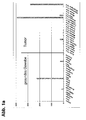
- the amount of gene-specific transcripts in healthy tissue and in carcinoma samples was investigated ( Fig. 1a . b . c ).
- LOC90625 is very selectively expressed in healthy tissue, specific transcripts are detectable especially in the testis. Little or no detectable LOC90625-specific expression in all other healthy tissues analyzed ( Fig. 1a . b ).
- Fig. 1a . b . c LOC90625-specific RT-PCR
- LOC90625 is a selectively expressed antigen which is apparently expressed more prominently in proliferating tissues. This selective overexpression in tumors can be used diagnostically and therapeutically.
- the extracellular domain of LOC90625 can be used according to the invention as the target structure of monoclonal antibodies.
- This can e.g. 1-19 (SEQ ID NO: 287) or amino acids 40-160 (SEQ ID NO: 288).
- SEQ ID NO: 287 amino acids 40-160
- SEQ ID NO: 288 amino acids 40-160
Landscapes
- Health & Medical Sciences (AREA)
- Life Sciences & Earth Sciences (AREA)
- Chemical & Material Sciences (AREA)
- Engineering & Computer Science (AREA)
- General Health & Medical Sciences (AREA)
- Physics & Mathematics (AREA)
- Proteomics, Peptides & Aminoacids (AREA)
- Organic Chemistry (AREA)
- Analytical Chemistry (AREA)
- Immunology (AREA)
- Biotechnology (AREA)
- Bioinformatics & Cheminformatics (AREA)
- Pathology (AREA)
- Molecular Biology (AREA)
- Biophysics (AREA)
- Medicinal Chemistry (AREA)
- Hematology (AREA)
- Genetics & Genomics (AREA)
- Wood Science & Technology (AREA)
- Zoology (AREA)
- Oncology (AREA)
- Microbiology (AREA)
- Biochemistry (AREA)
- Chemical Kinetics & Catalysis (AREA)
- Biomedical Technology (AREA)
- Urology & Nephrology (AREA)
- Nuclear Medicine, Radiotherapy & Molecular Imaging (AREA)
- Pharmacology & Pharmacy (AREA)
- Animal Behavior & Ethology (AREA)
- Public Health (AREA)
- Veterinary Medicine (AREA)
- Bioinformatics & Computational Biology (AREA)
- Evolutionary Biology (AREA)
- Medical Informatics (AREA)
- Spectroscopy & Molecular Physics (AREA)
- Theoretical Computer Science (AREA)
- General Chemical & Material Sciences (AREA)
- Hospice & Palliative Care (AREA)
- General Engineering & Computer Science (AREA)
- Cell Biology (AREA)
Description
- Trotz interdisziplinärer Ansätze und Ausreizung klassischer Therapiemodalitäten gehören Krebserkrankungen weiterhin zu den führenden Todesursachen. Neuere therapeutische Konzepte zielen darauf ab, das patienteneigene Immunsystem durch Einsatz von rekombinanten Tumorvakzinen und anderen spezifischen Maßnahmen wie Antikörpertherapie in das therapeutische Gesamtkonzept mit einzubeziehen. Voraussetzung für den Erfolg einer solchen Strategie ist die Erkennung von Tumor-spezifischen oder Tumor-assoziierten Antigenen bzw. Epitopen durch das Immunsystem des Patienten, dessen Effektorfunktionen interventionell verstärkt werden sollen. Tumorzellen unterscheiden sich biologisch wesentlich von ihren nichtmalignen Ursprungszellen. Diese Differenzen sind durch während der Tumorentwicklung erworbene genetische Veränderungen bedingt und führen u.a. auch zur der Bildung qualitativ oder quantitativ veränderter molekularer Strukturen in den Krebszellen. Werden solche Tumor-assoziierten Strukturen vom spezifischen Immunsystem des tumortragenden Wirtes erkannt, spricht man von Tumor-assoziierten Antigenen.
- An der spezifischen Erkennung von Tumor-assoziierten Antigenen sind zelluläre und humorale Mechanismen beteiligt, die zwei miteinander funktionell vernetzte Einheiten darstellen: CD4+ und CD8+ T-Lymphozyten erkennen prozessierte Antigene, die auf den Molekülen der MHC- (Major Histocompatibility complex = Histokompatibilitäts-Antigene) Klassen II bzw. 1 präsentiert werden, während B-Lymphozyten zirkulierende Antikörpermoleküle produzieren, die direkt an unprozessierte Antigene binden.
- Die potentielle klinisch-therapeutische Bedeutung von Tumor-assoziierten Antigenen ergibt sich aus der Tatsache, dass die Erkennung von Antigenen auf neoplastischen Zellen durch das Immunsystem zur Initiierung von cytotoxischen Effektormechanismen führt und bei Vorhandensein von T-Helferzellen die Elimination der Krebszellen bewirken kann (Pardoll, Nat. Med. 4:525-31, 1998). Entsprechend ist es eine zentrale Zielsetzung der Tumorimmunologie, diese Strukturen molekular zu definieren. Die molekulare Natur dieser Antigene blieb lange enigmatisch. Erst als entsprechende Klonierungstechniken entwickelt wurden, gelang es, durch Analyse der Zielstrukturen von cytotoxischen T-Lymphozyten (CTL) (van der Bruggen et al., Science 254:1643-7, 1991) bzw. mit zirkulierenden Autoantikörpern (Sahin et al., Curr. Opin. Immunol. 9:709-16, 1997) als Sonden cDNA-Expressionbanken von Tumoren systematisch auf Tumor-assoziierte Antigene zu screenen. Hierzu wurden cDNA-Expressionsbanken aus frischem Tumorgewebe hergestellt und in geeigneten Systemen als Proteine rekombinant exprimiert. Aus Patienten isolierte Immuneffektoren, nämlich CTL-Klone mit Tumor-spezifischem Lysemuster, oder zirkulierende Autoantikörper wurden genutzt, um die respektiven Antigene zu klonieren.
- Durch diese Ansätze sind in den letzten Jahren eine Vielzahl von Antigenen in verschiedenen Neoplasien definiert worden. Von großem Interesse ist dabei die Klasse der Cancer/Testis-Antigene (CTA). CTA und sie kodierende Gene (Cancer/Testis-Gene oder CTG) sind durch ihr charakteristisches Expressionsmuster definiert (Türeci et al., Mol. Med. Today 3:342-9, 1997). Sie finden sich nicht in Normalgeweben bis auf Testis bzw. Keimzellen, werden jedoch in einer Reihe von humanen Malignomen exprimiert und zwar nicht tumortypspezifisch, sondern mit unterschiedlicher Häufigkeit in Tumorentitäten ganz unterschiedlicher Herkunft (Chen & Old, Cancer J. Sci. Am. 5:16-7, 1999). Auch Serumreaktivitäten gegen CTA finden sich nicht in gesunden Kontrollen, sondern lediglich in Tumorpatienten. Insbesondere aufgrund ihrer Gewebeverteilung ist diese Antigenklasse von besonderem Wert für immuntherapeutische Vorhaben und wird in derzeit laufenden klinischen Patientenstudien getestet (Marchand et al., Int. J. Cancer 80:219-30, 1999; Knuth et al., Cancer Chemother. Pharmacol. 46: S46-51, 2000).
- Allerdings nutzen die oben dargestellten klassischen Verfahren zur Antigenidentifizierung Immuneffektoren (zirkulierende Autoantikörper oder CTL-Klone) aus Patienten mit in der Regel bereits fortgeschrittenem Krebs als Sonden. Aus einer Reihe von Daten geht hervor, dass Tumore z.B. zur Tolerisierung und Anergisierung von T-Zellen führen können und gerade im Verlauf der Erkrankung diejenigen Spezifitäten aus dem Immuneffektorenrepertoire verloren gehen, die eine effektive Immunerkennung bewirken könnten. Aus laufenden Patientenstudien hat sich noch kein gesicherter Beweis für eine tatsächliche Wirkung der bisher entdeckten und genutzten Tumor-assoziierten Antigene ergeben. Entsprechend kann nicht ausgeschlossen werden, dass spontane Immunantworten evozierende Proteine die falschen Zielstrukturen sind.
- Es war die Aufgabe der vorliegenden Erfindung Zielstrukturen für eine Diagnose und Therapie von Krebserkrankungen bereitzustellen.
- Diese Aufgabe wird erfindungsgemäß durch den Gegenstand der Patentansprüche gelöst.
- Erfindungsgemäß wurde eine Strategie für eine Identifizierung und Bereitstellung Tumorassoziiert exprimierter Antigene und der dafür kodierenden Nukleinsäuren verfolgt. Diese Strategie beruht auf der Auswertung humaner Protein- und Nukleinsäuredatenbanken im Hinblick auf potenzielle, auf der Zelloberfläche zugängliche, krebsspezifische Antigene. Die Definition der dafür notwendigen Filterkriterien zusammen mit einer Hochdurchsatz-Methodik zur Analyse möglichst aller Proteine bilden den zentralen Bestandteil der Erfindung. Durch Datamining wird zunächst eine möglichst komplette Liste aller bekannter Gene aufgestellt, die dem Grundprinzip Gen zu mRNA zu Protein folgend auf das Vorhandensein einer oder mehrerer Transmembrandomänen hin untersucht werden. Hieran schließen sich eine Homologiesuche, eine Einteilung der Treffer in gewebsspezifische Gruppen (u.a. Tumorgewebe) und eine Überprüfung der realen Existenz der mRNA an. Schließlich werden die so identifizierten Proteine z.B. durch Expressionsanalysen und proteinchemische Verfahren auf ihre aberrante Aktivierung in Tumoren evaluiert.
- Datamining ist ein bekanntes Verfahren zur Identifizierung von Tumor-assoziierten Genen. Bei den herkömmlichen Strategien werden allerdings in der Regel Transkriptome von Normalgewebebanken elektronisch von Tumorgewebsbanken subtrahiert unter der Annahme, dass die verbleibenden Gene Tumor-spezifisch sind (Schmitt et al., Nucleic Acids Res. 27:4251-60,1999; Vasmatzis et al., Proc. Natl. Acad. Sci. USA. 95:300-4, 1998; Scheurle et al., Cancer Res. 60:4037-43, 2000).
- Das erfindungsgemäße Konzept, das sich als viel erfolgreicher erwiesen hat, beruht jedoch darauf, Datamining zur elektronischen Extraktion aller Gene, die für auf der Zelloberfläche zugängliche, krebsspezifische Antigene kodieren, zu nutzen und diese sodann auf ektope Expression in Tumoren zu evaluieren.
- Somit liegt der Erfindung eine Strategie zur Identifizierung von differentiell in Tumoren exprimierten Genen zugrunde. Diese kombiniert Datamining von öffentlichen Sequenzbanken ("in silico") mit darauffolgenden evaluierenden laborexperimentellen ("wet bench") Untersuchungen.
- Eine kombinierte Strategie basierend auf unterschiedlichen bioinformatischen Skripten ermöglichte erfindungsgemäß die Identifizierung des in Beispiel 1 beschriebenen Gens, das für ein auf der Zelloberfläche zugängliches, krebsspezifisches Antigen kodiert. Die Identifizierung und Bereitstellung dieses Tumor-assoziierten Gens und des dadurch kodierten Genprodukts erfolgte unabhängig von deren immunogener Wirkung.
- Wie in Beispiel 1 detailliert dargestellt, kann das erfindungsgemäße Tumor-assoziierte Antigen aufgrund seiner tumorspezifischen Expression als diagnostischer Marker für Krebserkrankungen im Kopf-Hals-Bereich und für Leukämien verwendet werden. Die
PCT-Anmeldung WO2005/021793 beschreibt fetale RNA Marker im mütterlichen Blut, die zur pränatalen Diagnose des Down-Syndroms herangezogen werden können. In diesem Zusammenhang wird auch die als "SEQ ID NO: 13" bezeichnete Nukleinsäuresequenz beschrieben, die identisch ist mit der erfindungsgemäßen Nukleinsäuresequenz gemäß SEQ ID NO: 13. Weiterhin beschreibt diePCT-Anmeldung WO98/37094 WO98/37094 PCT-Anmeldung WO2005/030250 , dass das erfindungsgemäße Tumor-assoziierte Antigen in spezifischen Tumorgeweben exprimiert wird. InWO2005/030250 findet sich jedoch kein Hinweis darauf, dass sich Tumorerkrankungen aus dem Kopf-Hals-Bereich und Leukämien durch eine Expression oder Überexpression des erfindungsgemäßen Tumor-assoziierten Antigens auszeichnen. - Das erfindungsgemäß identifizierte Tumor-assoziierte Antigen weist eine Aminosäuresequenz auf, die von einer Nukleinsäure kodiert wird, die eine Nukleinsäuresequenz gemäß SEQ ID NO: 13 des Sequenzprotokolls umfasst. Das erfindungsgemäß identifizierte Tumor-assoziierte Antigen weist eine Aminosäuresequenz gemäß SEQ ID NO: 14 des Sequenzprotokolls auf.
- Die vorliegende Erfindung betrifft allgemein die Verwendung des in Beispiel 1 detailliert beschriebenen Tumor-assoziierten Antigens der dafür kodierenden Nukleinsäure oder von Nukleinsäuren, die gegen die kodierenden Nukleinsäuren gerichtet sind, oder von Antikörpern, die gegen das erfindungsgemäß identifizierte Tumor-assoziierte Antigene gerichtet sind, für die Diagnose. Diese Nutzung betrifft z.B. Verfahren zur Diagnose, und Verlaufskontrolle von Karzinomen im Kopf-Hals-Bereich oder von Leukämien.
- Die Eigenschaft des erfindungsgemäß identifizierten Tumor-assoziierten Antigens, dass es auf oder an der Zelloberfläche lokalisiert ist, qualifziert es als geeignete Zielestruktur für die Diagnose. Geeignet hierfür ist insbesondere ein Teil des erfindungsgemäß identifizierten Tumor-assoziierten Antigens, der dem Nichttransmembrananteil, insbesondere dem extrazellulären Anteil des Antigens entspricht oder davon umfasst wird. Somit ist erfindungsgemäß ein Teil der erfindungsgemäß identifizierten Tumor-assoziierten Antigene, der dem Nichttransmembrananteil der Antigene entspricht oder davon umfasst ist, oder ein entsprechender Teil der für die erfindungsgemäß identifizierten Antigene kodierenden Nukleinsäuren für eine Diagnose bevorzugt. Ähnlich ist die Verwendung von Antikörpern bevorzugt, die gegen einen Teil der des erfindungsgemäß identifizierten Tumor-assoziierten Antigens gerichtet sind, der dem Nichttransmembrananteil des Antigens entspricht oder davon umfasst ist.
- Spleißvarianten sind als Targets für die Diagnostik von Tumorerkrankungen verwendbar.
- Für die Entstehung von Spleißvarianten können verschiedenste Mechanismen ursächlich sein, beispielsweise
- die Nutzung variabler Transkriptionsinitiationsstellen
- die Nutzung zusätzlicher Exons
- vollständiges oder unvollständiges Ausspleißen von einzelnen oder mehreren Exons,
- über Mutation veränderte Spleißregulatorsequenzen (Deletion bzw. Schaffung neuer Donor/Akzeptorsequenzen),
- die unvollständige Elimination von Intronsequenzen.
- Das veränderte Spleißen eines Gens führt zu einer veränderten Transkriptsequenz (Spleißvariante). Wird eine Spleißvariante im Bereich ihrer veränderten Sequenz translatiert, resultiert ein verändertes Protein, welches sich von dem ursprünglichen in Struktur und Funktion deutlich unterscheiden kann. Bei tumorassoziierten Spleißvarianten können tumorassoziierte Transkripte und tumorassoziierte Proteine/Antigene entstehen. Diese können als molekulare Marker zum Nachweis von Tumorzellen genutzt werden. Die Detektion von Tumorzellen z.B. im Blut, Serum, Knochenmark, Sputum, Bronchial-Lavage, Körpersekreten und Gewebsbiopsien kann z.B. nach Extraktion von Nukleinsäuren durch PCR-Amplifikation mit Spleißvarianten-spezifischen Oligonukleotiden erfolgen.
- Zum Nachweis eignen sich erfindungsgemäß alle Sequenz-abhängigen Detektionssysteme. Neben der PCR sind diese z.B. Genchip-/Microarraysysteme, Northern-Blot, RNAse protection assays (RDA) und andere. Allen Detektionssystemen ist gemeinsam, dass die Detektion auf einer spezifischen Hybridisierung mit mindestens einer Spleißvarianten-spezifischen Nukleinsäuresequenz basiert. Die Detektion von Tumorzellen kann jedoch auch erfindungsgemäß durch Antikörper erfolgen, die ein durch die Spleißvariante kodiertes spezifisches Epitop erkennen. Für die Herstellung der Antikörper können Peptide zur Immunisierung verwendet werden, die für diese Spleißvariante spezifisch sind. Die Detektion von Tumorzellen kann auch durch Antikörper erfolgen, die tumorspezifisch veränderte Glykosylierungsvarianten erkennen. Für die Generierung von derartigen Antikörpern können Peptidregionen genutzt werden, die sich in Tumorzellen und gesunden Zellen glykosylierungsbedingt unterscheiden. Durch endogene Deglykosylierung von N-gekoppelten Zuckerresten wird Asparagin in Asparaginsäure transformiert. Erfindungsgemäß können daher die hier beschriebenen Proteine tumorspezifisch sequenzverändert sein und damit andere biochemische und Antikörper-Bindungseigenschaften aufweisen. Für die Immunisierung eignen sich besonders die Aminosäuren, die deutliche Epitopunterschiede zu der/den Variante(n) des Genprodukts aufweisen, welche bevorzugt in gesunden Zellen gebildet wird/werden. Der Nachweis der Tumorzellen mit Antikörper kann dabei an einer vom Patienten isolierten Probe oder als Imaging mit intravenös applizierten Antikörpern erfolgen.
- Neben der diagnostischen Nutzbarkeit stellen Spleißvarianten, die neue oder veränderte Epitope aufweisen, attraktive Targets für die Immuntherapie dar. Die Epitope können zum Targeting von therapeutisch wirksamen monoklonalen Antikörpern oder T-Lymphozyten genutzt werden. Bei der passiven Immuntherapie werden hierbei Antikörper oder T-Lymphozyten adoptiv transferiert, die Spleißvarianten-spezifische Epitope erkennen. Die Generierung von Antikörpern kann wie bei anderen Antigenen auch unter Nutzung von Standardtechnologien (Immunisierung von Tieren, Panningstrategien zur Isolation von rekombinanten Antikörpern) unter Nutzung von Polypeptiden, die diese Epitope beinhalten, erfolgen. Alternativ können zur Immunisierung Nukleinsäuren genutzt werden, die für Oligo- oder Polypeptide kodieren, die diese Epitope beinhalten. Verschiedene Techniken zur in vitro oder in vivo Generierung von epitopspezifischen T-Lymphozyten sind bekannt und ausführlich beschrieben z.B. (Kessler JH, et al. 2001, Sahin et al., 1997) und basieren ebenfalls auf der Nutzung von Oligo- oder Polypeptide, die die Spleißvarianten-spezifischen Epitope beinhalten, oder Nukleinsäuren, die für diese kodieren. Oligo- oder Polypeptide, die die Spleißvarianten-spezifischen Epitope beinhalten, oder Nukleinsäuren, die für diese Polypeptide kodieren, sind auch für die Nutzung als pharmazeutisch wirksame Substanzen bei der aktiven Immuntherapie (Vakzinierung, Vakzintherapie) verwendbar.
- Die aberrante Expression von Genen in Tumorzellen kann auf veränderte Methylierungsmuster ihrer Promotoren beruhen (De Smet C. et al., Mol. Cell Biol. 24:4781-90, 2004; De Smet C., et al., Mol. Cell Biol. 19:7327-35, 1999; De Smet C. et al., Proc. Natl. Acad. Sci. USA. 93:7149-53, 1996). Diese Methylierungsunterschiede können als indirekter Marker für den im Tumor veränderten Zustand des entsprechenden Gens genutzt werden. Dementsprechend kann die Zunahme oder Abnahme von Basenmethylierungen im Promotorbereich für diagnostische Zwecke genutzt werden.
- Vorliegend werden auch posttranslational modifizierte Proteindomänen wie zum Beispiel Glykosylierungen oder Myristilierungen beschrieben. Diese Art von Modifikationen kann zu einem differentiellen Erkennungsmuster eines Antigens durch z.B. einen Antikörper führen und verschiedene, unter Umständen krankheitsassoziierte Zustände erkennen. Vor allem durch den Einsatz von Antikörpern kann diese Differenzierung eines Antigens sowohl diagnostisch als auch therapeutisch genutzt werden. Für Tumorzellen ist publiziert, dass die tumorassoziierte zelluläre Entartung zu veränderten posttranslationalen Modifikationen führen kann (Durand & Seta, Clin. Chem. 46:795-8052000; Granovsky et al., Nat. Med. 6:306-312, 2000). Insbesondere Glykosylierungsmuster sind auf Tumorzellen stark verändert. Diese speziellen Epitope können diagnostisch Tumorzellen von nicht karzinogenen Zellen unterscheiden. Sollte ein posttranslational modifizierbares Epitop in normalen, nicht entarteten Zellen glykosyliert und in Tumorzellen deglykosyliert sein, erlaubt diese Situation die Entwicklung eines tumorspezifischen therapeutischen Antikörpers.
- Die Analyse von Proteinmodifikationen kann im Western-Blot erfolgen. Vor allem Glykosylierungen, die in der Regel eine Größe von mehreren kDa haben, führen zu einer größeren Gesamtmasse des Zielproteins, die sich in der SDS-PAGE auftrennen lässt. Zum Nachweis von spezifischen O- und N-glykosidischen Bindungen werden Proteinlysate vor der Denaturierung durch SDS mit O- oder N-Glykosylasen inkubiert (nach Angaben des jeweiligen Herstellers, z.B. PNgase, Endoglykosidase F, Endoglykosidase H, Roche Diagnostics). Anschließend erfolgt ein Western Blot. Bei Verringerung der Größe eines Zielproteins kann so nach Inkubation mit einer Glykosidase eine spezifische Glykosylierung nachgewiesen und auf diesem Weg auch die Tumorspezifität einer Modifikation analysiert werden. Von besonderem Interesse sind Proteinbereiche, die in Tumorzellen und gesunden Zellen differenziell glykosyliert sind. Derartige Glykosylierungsunterschiede sind jedoch bisher für wenige Zelloberflächenproteine (z.B. Muc1) beschrieben.
- Des weiteren betrifft die Erfindung Verfahren zur Diagnose oder Überwachung, d.h. Bestimmung der Regression, des Verlaufs und/oder des Ausbruchs, einer Krebserkrankung, die sich durch die Expression oder abnormale Expression eines oder mehrerer Tumor-assoziierter Antigene auszeichnet.
- Die erfindungsgemäßen Diagnoseverfahren und/oder Verfahren zur Überwachung betreffen allgemein die Verwendung von Mitteln für den Nachweis und/oder die Bestimmung bzw. Überwachung der Menge (i) einer Nukleinsäure, die für das Tumor-assoziierte Antigen kodiert, oder eines Teils davon und/oder (ii) des Tumor-assoziierten Antigens oder eines Teils davon, in einer aus einem Patienten isolierten biologischen Probe.
- In einem Aspekt betrifft die Erfindung ein Verfahren zur Diagnose oder Überwachung einer Krebserkrankung, die sich dadurch auszeichnet, dass ein Tumor-assoziiertes Antigen in Tumorgewebe im Vergleich zu vergleichbarem gesunden Gewebe überexprimiert wird, umfassend
- (i) den Nachweis einer Nukleinsäure, die für das Tumor-assoziierte Antigen kodiert, und /oder
- (ii) den Nachweis des Tumor-assoziierten Antigens,
- Diagnoseverfahren können das erfindungsgemäß identifizierten Tumor-assoziierten Antigen auch als prognostischen Marker nutzen, um eine Metastatisierung z.B. durch Testen des Migrationsverhalten von Zellen und daher einen verschlechterten Krankheitsverlauf zu prädizieren, wodurch unter anderem die Planung einer aggressiveren Therapie ermöglicht wird.
- Somit betrifft die Erfindung ein Verfahren zur Bestimmung der Regression, des Verlaufs oder des Ausbruchs einer Krebserkrankung, die sich durch die Expression oder abnormale Expression des erfindungsgemäß identifizierten Tumor-assoziierten Antigens auszeichnet, umfassend die Überwachung einer Probe aus einem Patienten, der die Erkrankung aufweist oder in Verdacht steht, an der Erkrankung zu erkranken.
- Ein Nachweis einer Nukleinsäure oder eines Teils davon oder eine Bestimmung bzw. Überwachung der Menge einer Nukleinsäure oder eines Teils davon kann erfindungsgemäß mit einer Polynukleotid-Sonde erfolgen, die spezifisch mit der Nukleinsäure oder dem Teil davon hybridisiert, oder kann durch selektive Amplifikation der Nukleinsäure oder des Teils davon erfolgen. Die Polynukleotid-Sonde umfasst z.B.eine Sequenz von 6-50, insbesondere 10-30, 15-30 oder 20-30 zusammenhängenden Nukleotiden aus der Nukleinsäure.
- Im Rahmen des Diagnoseverfahrens erfolgt beispielsweise eine selektive Amplifikation des Promotorbereichs oder eines Teils einer Nukleinsäure, die für das Tumor-assoziierte Antigen kodiert und in Form von genomischer DNA vorliegt, nach Behandlung mit einem bisulfithaltigen Reagenz. Die Nukleinsäure kann vor einer Behandlung mit dem bisulfithaltigen Reagenz aus einer Probe eines zu untersuchenden Patienten isoliert werden. Die bei einer solchen Amplifikation verwendeten Oligonukleotide weisen eine Sequenz auf, die an die mit dem bisulfithaltigen Reagenz behandelte Nukleinsäure binden, vorzugsweise dazu vollständig komplementär sind. Vorzugsweise sind die Oligonukleotide einem unterschiedlichen Grad einer Methylierung der Nukleinsäure angepasst und bedingen differenzierbare Amplifikationsprodukte.
- Ein Nachweis eines Tumor-assoziierten Antigens oder eines Teils davon oder eine Bestimmung bzw. Überwachung der Menge eines Tumor-assoziierten Antigens oder eines Teils davon kann mit einem Antikörper erfolgen, der spezifisch an das Tumor-assoziierte Antigen oder den Teil davon bindet.
- Das nachzuweisende Tumor-assoziierte Antigen oder ein Teil davon kann in einem Komplex mit einem MHC-Molekül, insbesondere einem HLA-Molekül vorliegen.
- Die für einen Nachweis oder für eine Bestimmung bzw. Überwachung verwendete Polynukleotid-Sonde oder, der Antikörper sind beispielsweise nachweisbar markiert. wobei der nachweisbare Marker z.B. ein radioaktiver Marker oder ein Enzymmarker sein kann.
- In einem weiteren Aspekt betrifft die Erfindung die Verwendung eines Antikörpers, der an ein Tumor-assoziiertes Antigen bindet und mit einem diagnostischen Mittel gekoppelt ist, zur Herstellung einer pharmazeutischen Zusammensetzung zur Diagnose oder Überwachung einer Krebserkrankung, die sich dadurch auszeichnet, dass das Tumor-assoziierte Antigen in Tumorgewebe im Vergleich zu vergleichbarem gesunden Gewebe überexprimiert wird, wobei das Tumor-assoziierte Antigen eine Sequenz aufweist, die von einer Nukleinsäure kodiert wird, die die Nukleinsäuresequenz gemäß SEQ ID NO: 13 des Sequenzprotokolls umfasst, wobei die Krebserkrankung ein Karzinom im Kopf-Hals-Bereich oder eine Leukämie ist. Der Antikörper kann ein monoklonaler Antikörper sein. In weiteren Ausführungsformen ist der Antikörper ein chimärer oder humanisierter Antikörper oder ein Fragment eines natürlichen Antikörpers.
- Der Fachmann kann die Erkenntnisse der erfindungsgemäßen Verfahren auch in Zusammenhang mit Behandlungsverfahren verwenden.
- Denkbare Behandlungsverfahren sind Verfahren zur Behandlung eines Patienten mit einer Krebserkrankung, die sich durch die Expression oder abnormale Expression eines Tumor-assoziierten Antigens auszeichnet, umfassend (i) die Identifizierung einer für das erfindungsgemäß identifiziertes Tumor-assoziiertes Antigen kodierenden Nukleinsäure, die von Zellen exprimiert wird, die mit der Erkrankung assoziiert sind, (ii) die Transfektion einer Wirtszelle mit der Nukleinsäure oder einem Teil davon, (iii) die Kultivierung der transfizierten Wirtszelle für eine Expression der Nukleinsäure (dies ist bei Erreichen einer hohen Transfektionsrate nicht obligat) und (iv) das Einbringen der Wirtszellen oder eines Extrakts davon in den Patienten in einer Menge, die geeignet ist, die Immunreaktion gegen die Zellen des Patienten, die mit der Erkrankung assoziiert sind, zu erhöhen. Ein solches Verfahren kann ferner die Identifizierung eines MHC-Moleküls, das das Tumor-assoziierte Antigen oder einen Teil davon präsentiert, umfassen, wobei die Wirtszelle das identifizierte MHC-Molekül exprimiert und das Tumor-assoziierte Antigen oder einen Teil davon präsentiert. Die Immunreaktion kann eine B-Zellen-Reaktion oder eine T-Zellen-Reaktion umfassen. Des weiteren kann eine T-Zellen-Reaktion die Produktion von cytolytischen T-Zellen und/oder Helfer-T-Zellen umfassen, die spezifisch für die Wirtszellen sind, die das Tumor-assoziierte Antigen oder einen Teil davon präsentieren oder spezifsch für Zellen des Patienten sind, die das Tumor-assoziierte Antigen oder einen Teil davon exprimieren.
- Ein weiterhin denkbares Behandlungsverfahren ist ein Verfahren zur Behandlung einer Krebserkrankung, die sich durch die Expression oder abnormale Expression des erfindungsgemäß identifizierten Tumor-assoziierten Antigens auszeichnet, umfassend (i) die Identifikation von Zellen aus dem Patienten, die abnormale Mengen des Tumor-assoziierten Antigens exprimieren, (ii) die Isolierung einer Probe der Zellen, (iii) die Kultivierung der Zellen und (iv) das Einbringen der Zellen in den Patienten in einer Menge, die geeignet ist, eine Immunreaktion gegen die Zellen auszulösen.
- Oligonukleotide, die mit der erfindungsgemäß identifizierten Nukleinsäure hybridisieren können als genetische Sonden verwendet werden. Nukleinsäuremoleküle in der Form von Oligonukleotid-Primern oder kompetenten Proben, die mit der erfindungsgemäß identifizierten Nukleinsäure hybridisieren, können zum Auffinden von Nukleinsäuren verwendet werden, die zu der erfindungsgemäß identifizierten Nukleinsäure homolog sind. PCR-Amplifikation, Southern- und Northern-Hybridisierung können zum Auffinden homologer Nukleinsäuren eingesetzt werden. Die Hybridisierung kann unter niedrig-, besser unter mittel- und am besten unter hoch-stringenten Bedingungen erfolgen. Der Begriff "stringente Bedingungen" betrifft erfindungsgemäß Bedingungen, die eine spezifische Hybridisierung zwischen Polynukleotiden erlauben.
- Immunogene Fragmente des erfindungsgemäß identifizierten Tumor-assoziierten Antigens können an einen menschlichen HLA-Rezeptor oder menschlichen Antikörper binden. Ein solches Fragment hat z.B. eine Sequenz von mindestens 6, insbesondere mindestens 8, mindestens 10, mindestens 12, mindestens 15, mindestens 20, mindestens 30 oder mindestens 50 Aminosäuren.
- In Beispiel 1 werden Peptide beschrieben, die zur Herstellung von Antikörpern verwendet werden können und eine Sequenz aufweisen, ausgewählt aus der Gruppe bestehend aus SEQ ID NOs: 287-290.
- In einem weiteren Aspekt betreffen die erfindungsgemäßen Verfahren und Verwendungen ein Mittel, das an das erfindungsgemäß identifizierte Tumor-assoziierte Antigen bindet. In einer bevorzugten Ausführungsform ist das Mittel ein Antikörper. In weiteren Ausführungsformen ist der Antikörper ein chimärer, ein humanisierter Antikörper oder ein Fragment eines Antikörpers. Ein erfindungsgemäßer Antikörper kann ein monoklonaler Antikörper sein. In weiteren Ausführungsformen ist der Antikörper ein chimärer oder humanisierter Antikörper oder ein Fragment eines Antikörpers.
- Des weiteren betreffen die erfindungsgemäßen Verwendungen ein Konjugat zwischen einem erfindungsgemäßen Mittel, das an das erfindungsgemäß identifiziertes Tumor-assoziiertes Antigen bindet, oder einem erfindungsgemäßen Antikörper und einem diagnostischen Mittel oder Stoff. Beispielsweise ist das diagnostische Mittel ein Toxin.
- In einem weiteren Aspekt betrifft die Erfindung die Verwendung eines Kit zum Nachweis einer Krebserkrankung, die sich dadurch auszeichnet, dass ein Tumor-assoziiertes Antigen in Tumorgewebe im Vergleich zu vergleichbarem gesunden Gewebe überexprimiert wird, wobei der Kit Mittel zum Nachweis
- (i) der Nukleinsäure, die für das Tumor-assoziierte Antigen kodiert, und / oder
- (ii) des Tumor-assoziierten Antigens,
- Erfindungsgemäß wird ein Gen ein Gen beschrieben, das in Tumorzellen selektiv exprimiert oder aberrant exprimiert wird und ein Tumor-assoziiertes Antigen darstellt.
- Dieses Gene und/oder dessen Genprodukte und/oder dessen Derivate und/oder Teile können Zielstrukturen für therapeutische Ansätze sein. Konzeptionell können die therapeutischen Ansätze auf eine Hemmung der Aktivität des selektiv exprimierten Tumor-assoziierten Genproduktes zielen. Dies ist dann sinnvoll, wenn die aberrante respektive selektive Expression funktionell von tumorpathogenetischer Bedeutung ist und ihre Unterbindung mit einer selektiven Schädigung der entsprechenden Zellen einhergeht. Andere therapeutische Konzepte betrachten Tumor-assoziierte Antigene als Markierungen, die Effektormechanismen mit zellschädigendem Potential selektiv zu Tumorzellen rekrutieren. Hierbei ist die Funktion des Zielmoleküls selbst und seine Rolle bei der Tumorentstehung vollkommen unerheblich.
- Mit "Derivat" einer Nukleinsäure ist gemeint, dass einzelne oder multiple Nukleotidsubstitutionen, -deletionen und/oder -additionen in der Nukleinsäure vorliegen. Weiterhin umfasst der Begriff "Derivat" auch eine chemische Derivatisierung einer Nukleinsäure an einer Nukleotidbase, am Zucker oder am Phosphat eines Nukleotids. Der Begriff "Derivat" umfasst auch Nukleinsäuren, die nicht in der Natur vorkommende Nukleotide und Nukleotidanaloga enthalten.
- Eine Nukleinsäure ist beispielsweise Desoxyribonukleinsäure (DNA) oder Ribonukleinsäure (RNA). Nukleinsäuren umfassen beispielsweise genomische DNA, cDNA, mRNA, rekombinant hergestellte und chemisch synthetisierte Moleküle. Eine Nukleinsäure kann als einzelsträngiges oder doppelsträngiges und lineares oder kovalent kreisförmig geschlossenes Molekül vorliegen.
- Die beschriebenen Nukleinsäuren können isoliert vorliegen. Der Begriff "isolierte Nukleinsäure" bedeutet erfindungsgemäß, dass die Nukleinsäure (i) in vitro amplifiziert wurde, zum Beispiel durch Polymerase-Kettenreaktion (PCR), (ii) rekombinant durch Klonierung produziert wurde, (iii) gereinigt wurde, zum Beispiel durch Spaltung und gelelektrophoretische Auftrennung, oder (iv) synthetisiert wurde, zum Beispiel durch chemische Synthese. Eine isolierte Nukleinsäure ist eine Nukleinsäure, die für eine Manipulierung durch rekombinante DNA-Techniken zur Verfügung steht.
- Eine Nukleinsäure ist dann zu einer anderen Nukleinsäure "komplementär", wenn die beiden Sequenzen miteinander hybridisieren und ein stabiles Duplexmolekül eingehen können, wobei die Hybridisierung vorzugsweise unter Bedingungen erfolgt, die eine spezifische Hybridisierung zwischen Polynukleotiden erlauben (stringente Bedingungen). Stringente Bedingungen sind beispielsweise in Molecular Cloning: A Laboratory Manual, J. Sambrook et al., Hrsg., 2. Auflage, Cold Spring Harbor Laboratory Press, Cold Spring Harbor, New York, 1989, oder Current Protocols in Molecular Biology, F.M. Ausubel et al., Hrsg., John Wiley & Sons, Inc., New York beschrieben und betreffen beispielsweise die Hybridisierung bei 65°C in Hybridisierungspuffer (3,5 x SSC, 0,02% Ficoll, 0,02% Polyvinylpyrrolidon, 0,02% Rinderserumalbumin, 2,5 mM NaH2PO4 (pH 7), 0,5% SDS, 2 mM EDTA). SSC ist eine Lösung mit jeweils 0,15 M Natriumchlorid und Natriumcitrat, pH 7. Nach der Hybridisierung wird die Membran, auf die die DNA übertragen wurde, beispielsweise in 2 x SSC bei Raumtemperatur und sodann in 0,1 - 0.5 x SSC / 0,1 x SDS bei Temperaturen bis 68°C gewaschen.
- Komplementäre Nukleinsäuren weisen beispielsweise mindestens 40%, insbesondere mindestens 50%, mindestens 60%, mindestens 70%, mindestens 80%, mindestens 90% und vorzugsweise mindestens 95%, mindestens 98% oder mindestens 99% Identität der Nukleotide auf.
- Nukleinsäuren, die für Tumor-assoziierte Antigene kodieren, können alleine oder in Kombination mit anderen Nukleinsäuren, insbesondere heterologen Nukleinsäuren, vorliegen. Eine Nukleinsäure kann funktionell in Verbindung mit Expressionskontrollsequenzen oder regulatorischen Sequenzen vorliegen, die in Bezug zu der Nukleinsäure homolog oder heterolog sein können. Eine kodierende Sequenz und eine regulatorische Sequenz sind dann "funktionell" miteinander verbunden, falls sie derart kovalent miteinander verknüpft sind, dass die Expression oder Transkription der kodierenden Sequenz unter der Kontrolle oder unter dem Einfluss der regulatorischen Sequenz steht. Falls die kodierende Sequenz in ein funktionelles Protein translatiert werden soll, führt bei einer funktionellen Verbindung einer regulatorischen Sequenz mit der kodierenden Sequenz eine Induktion der regulatorischen Sequenz zu einer Transkription der kodierenden Sequenz, ohne dass es zu einer Leserasterverschiebung in der kodierenden Sequenz oder zu einem Unvermögen der kodierenden Sequenz kommt, in das gewünschte Protein oder Peptid translatiert zu werden.
- Der Begriff "Expressionskontrollsequenz" oder "regulatorische Sequenz" umfasst Promotoren, Enhancer und andere Kontrollelemente, die die Expression eines Gens steuern. Die genaue Struktur von regulatorischen Sequenzen kann speziesabhängig oder zelltypusabhängig variieren, umfasst jedoch im allgemeinen 5'-nicht-transkribierte und 5'-nicht-translatierte Sequenzen, die an der Initiation der Transkription bzw. Translation beteiligt sind wie TATA-Box, Capping-Sequenz, CAAT-Sequenz und ähnliches. Insbesondere umfassen 5'-nicht-transkribierte Regulationssequenzen eine Promotorregion, die eine Promotorsequenz für eine transkriptionelle Kontrolle des funktionell verbundenen Gens einschließt. Regulatorische Sequenzen können auch Enhancer-Sequenzen oder stromaufwärts gelegene Aktivatorsequenzen umfassen.
- Prinzipiell können Nukleinsäuren, die für ein erfindungsgemäß identifiziertes Tumor-assoziiertes Antigen kodieren, für eine Transfektion von Wirtszellen eingesetzt werden. Mit Nukleinsäuren ist dabei sowohl rekombinante DNA wie auch RNA gemeint. Rekombinante RNA kann durch in vitro-Transkription von einer DNA-Matritze hergestellt werden. Sie kann des weiteren vor Applikation durch stabilisierende Sequenzen, Capping und Poly-Adenylierung modifiziert werden.
- Der Begriff "Wirtszelle" betrifft jede Zelle, die mit einer exogenen Nukleinsäure transformierbar oder transfizierbar ist. Der Begriff "Wirtszellen" umfasst prokaryontische (z.B. E. coli) oder eukaryontische (z.B. dendritische Zellen, B-Zellen, CHO-Zellen, COS-Zellen, K562-Zellen, Hefezellen und Insektenzellen). und insbesondere auch Säugerzellen wie Zellen aus Mensch, Maus, Hamster, Schwein, Ziege und Primaten. Die Zellen können aus einer Vielzahl von Gewebetypen abgeleitet sein und umfassen primäre Zellen und Zelllinien. Spezifische Beispiele umfassen Keratinozyten, periphere Blutleukozyten, Stammzellen des Knochenmarks und embryonale Stammzellen. In weiteren Ausführungsformen ist die Wirtszelle eine Antigen-präsentierende Zelle, insbesondere eine dendritische Zelle, ein Monozyt oder Makrophage. Eine Nukleinsäure kann in der Wirtszelle in einer einzigen oder in mehreren Kopien vorliegen und wird in einer Ausführungsform in der Wirtszelle exprimiert.
- Der Begriff "Expression" wird in seiner allgemeinsten Bedeutung verwendet und umfasst die Produktion von RNA oder von RNA und Protein. Er umfasst auch eine teilweise Expression von Nukleinsäuren. Des weiteren kann die Expression transient oder stabil erfolgen. Beispiele für Expressionssysteme in Säugerzellen umfassen pcDNA3.1 und pRc/CMV (Invitrogen, Carlsbad, CA), die einen selektierbaren Marker enthalten wie ein Gen, das eine Resistenz gegenüber G418 verleiht (und somit eine Selektion stabil transfizierter Zelllinien ermöglicht), und die Enhancer-Promotor-Sequenzen von Cytomegalovirus (CMV).
- Erfindungsgemäße Kits umfassen beispielsweise ein Paar von Amplifikationsprimern, die an die Nukleinsäure hybridisieren, die für das Tumor-assoziierte Antigen kodiert. Die Primer umfassen vorzugsweise eine Sequenz von 6-50, insbesondere 10-30, 15-30 oder 20-30 zusammenhängenden Nukleotiden aus der Nukleinsäure und sind nicht-überlappend, um die Bildung von Primer-Dimeren zu vermeiden. Einer der Primer wird an einen Strang der Nukleinsäure hybridisieren, die für das Tumor-assoziierte Antigen kodiert, und der andere Primer wird an den komplementären Strang in einer Anordnung hybridisieren, die eine Amplifikation der Nukleinsäure, die für das Tumor-assoziierte Antigen kodiert, erlaubt.
- "Antisense"-Moleküle oder "Antisense"-Nukleinsäuren können zur Regulierung, insbesondere der Reduktion der Expression einer Nukleinsäure verwendet werden. Der Begriff "Antisense-Molekül" oder "Antisense-Nukleinsäure" betrifft ein Oligonukleotid, das ein Oligoribonukleotid, Oligodesoxyribonukleotid, modifiziertes Oligoribonukleotid oder modifiziertes Oligodesoxyribonukleotid ist und das unter physiologischen Bedingungen an DNA, die ein bestimmtes Gen umfasst, oder mRNA dieses Gens hybridisiert, wodurch die Transkription dieses Gens und/oder die Translation dieser mRNA gehemmt wird. Ein "Antisense-Molekül" umfasst auch ein Konstrukt, das eine Nukleinsäure oder einen Teil davon in reverser Orientierung in Bezug auf ihren natürlichen Promotor enthält. Ein Antisense-Transkript einer Nukleinsäure oder eines Teils davon kann ein Duplexmolekül mit der natürlich vorkommenden mRNA, die das Enzym spezifiziert, eingehen und so eine Akkumulation von oder die Translation der mRNA in das aktive Enzym verhindern. Eine weitere Möglichkeit ist die Verwendung von Ribozymen zur Inaktivierung einer Nukleinsäure. AntisenseOligonukleotide weisen beispielsweise eine Sequenz von 6-50, insbesondere 10-30,15-30 oder 20-30 zusammenhängenden Nukleotiden aus der Ziel-Nukleinsäure auf und können vollständig zu der Ziel-Nukleinsäure oder einem Teil davon komplementär sein. Antisense-Oligonukleotide können beispielsweise mit einer N-terminalen oder 5'-stromaufwärts gelegenen Stelle wie einer Translationsinitiations-, Transkriptionsinitiations- oder Promotorstelle hybridisieren oder mit einer 3'-nicht-translatierten Region oder mRNA-Spleiß-Stelle.
- Ein erfindungsgemäßes Oligonukleotid kann aus Ribonukleotiden, Desoxyribonukleotiden oder einer Kombination davon bestehen. Dabei sind das 5'-Ende eines Nukleotids und das 3'-Ende eines anderen Nukleotids durch eine Phosphodiesterbindung miteinander verknüpft. Diese Oligonukleotide können in herkömmlicher Weise synthetisiert oder rekombinant produziert werden.
- Alternativ kann ein erfindungsgemäßes Oligonukleotid ein "modifiziertes" Oligonukleotid sein. Dabei kann das Oligonukleotid, um beispielsweise seine Stabilität zu erhöhen, auf verschiedenste Art und Weise modifiziert sein ohne dass seine Fähigkeit, an sein Ziel zu binden, beeinträchtigt wird. Der Begriff "modifiziertes Oligonukleotid" bedeutet erfindungsgemäß ein Oligonukleotid, bei dem (i) mindestens zwei seiner Nukleotide durch eine synthetische Internukleosidbindung (d.h. eine Internukleosidbindung, die keine Phosphodiesterbindung ist) miteinander verknüpft sind und/oder (ii) eine chemische Gruppe kovalent mit dem Oligonukleotid verbunden ist, die normalerweise nicht bei Nukleinsäuren auftritt. Bevorzugte synthetische Internukleosidbindungen sind Phosphorothioate, Alkylphosphonate, Phosphorodithioate, Phosphatester, Alkylphosphonothioate, Phosphoramidate, Carbamate, Carbonate, Phosphattriester, Acetamidate, Carboxymethylester und Peptide.
- Der Begriff "modifiziertes Oligonukleotid" umfasst auch Oligonukleotide mit einer kovalent modifizierten Base und/oder Zucker. "Modifizierte Oligonukleotide" umfassen beispielsweise Oligonukleotide mit Zuckerresten, die kovalent an organische Gruppen mit einem geringen Molekulargewicht gebunden sind, die keine Hydroxylgruppe an der 3'-Position und keine Phosphatgruppe an der 5'-Position sind. Modifizierte Oligonukleotide können beispielsweise einen 2'-O-alkylierten Riboserest oder einen anderen Zucker anstelle von Ribose wie Arabinose umfassen.
- Die erfindungsgemäß beschriebenen Proteine und Polypeptide können isoliert vorliegen. Die Begriffe "isoliertes Protein" oder "isoliertes Polypeptid" bedeuten, dass das Protein oder Polypeptid von seiner natürlichen Umgebung getrennt ist. Ein isoliertes Protein oder Polypeptid kann in einem im wesentlichen aufgereinigten Zustand vorliegen. Der Begriff "im wesentlichen aufgereinigt" bedeutet, dass das Protein oder Polypeptid im wesentlichen frei von anderen Substanzen vorliegt, mit denen es in der Natur oder in vivo vorliegt.
- Solche Proteine und Polypeptide dienen beispielsweise der Herstellung von Antikörpern und sind in einem immunologischen oder diagnostischen Assay einsetzbar. Erfindungsgemäß beschriebene Proteine und Polypeptide können aus biologischen Proben wie Gewebe- oder Zellhomogenaten isoliert werden und können auch rekombinant in einer Vielzahl pro- oder eukaryontischer Expressionssysteme exprimiert werden.
- "Derivate" eines Proteins oder Polypeptids oder einer Aminosäuresequenz umfassen Aminosäure-Insertionsvarianten, Aminosäure-Deletionsvarianten und/oder Aminosäure-Substitutionsvarianten.
- Aminosäure-Insertionsvarianten umfassen amino- und/oder carboxyterminale Fusionen, sowie Insertionen von einzelnen oder mehreren Aminosäuren in einer bestimmten Aminosäuresequenz. Bei Aminosäure-Sequenzvarianten mit einer Insertion werden ein oder mehrere Aminosäurereste in eine vorbestimmte Stelle in einer Aminosäuresequenz eingebracht, obwohl eine zufällige Insertion mit geeignetem Screening des resultierenden Produkts auch möglich ist. Aminosäure-Deletionsvarianten sind durch das Entfernen von einer oder mehreren Aminosäuren aus der Sequenz charakterisiert. Aminosäure-Substitutionsvarianten zeichnen sich dadurch aus, dass wenigstens ein Rest in der Sequenz entfernt und ein anderer Rest an dessen Stelle eingefügt wird. Vorzugsweise befinden sich die Modifikationen an Positionen in der Aminosäuresequenz, die zwischen homologen Proteinen oder Polypeptiden nicht konserviert sind. Vorzugsweise werden Aminosäuren durch andere mit ähnlichen Eigenschaften, wie Hydrophobizität, Hydrophilizität, Elektronegativität, Volumen der Seitenkette und ähnliches, ersetzt (konservative Substitution). Konservative Substitutionen betreffen beispielsweise den Austausch einer Aminosäure durch eine andere, wobei beide Aminosäuren in derselben nachstehenden Gruppe aufgeführt sind:
- 1. kleine aliphatische, nicht-polare oder leicht-polare Reste: Ala, Ser, Thr (Pro, Gly)
- 2. negativ geladene Reste und ihre Amide: Asn, Asp, Glu, Gln
- 3. positiv geladene Reste: His, Arg, Lys
- 4. große aliphatische, nicht-polare Reste: Met, Leu, Ile, Val (Cys)
- 5. große aromatische Reste: Phe, Tyr, Trp.
- Drei Reste sind aufgrund ihrer besonderen Rolle für die Proteinarchitektur in Klammern gesetzt. Gly ist der einzige Rest ohne eine Seitenkette und verleiht der Kette somit Flexibilität. Pro besitzt eine ungewöhnliche Geometrie, die die Kette stark einschränkt. Cys kann eine Disulfidbrücke bilden.
- "Derivate" von Proteinen oder Polypeptiden umfassen auch einzelne oder multiple Substitutionen, Deletionen und/oder Additionen jeglicher Moleküle, die mit dem Enzym assoziiert sind, wie Kohlenhydrate, Lipide und/oder Proteine oder Polypeptide. Ferner erstreckt sich der Begriff "Derivat" auch auf alle funktionellen chemischen Äquivalente der Proteine oder Polypeptide.
- Ein Teil oder Fragment des Tumor-assoziierten Antigens kann eine funktionelle Eigenschaft des Polypeptids aufweisen, aus dem es abgeleitet ist. Solche funktionellen Eigenschaften umfassen die Interaktion mit Antikörpern, die Interaktion mit anderen Polypeptiden oder Proteinen, die selektive Bindung von Nukleinsäuren und eine enzymatische Aktivität. Eine bedeutende Eigenschaft ist die Fähigkeit, einen Komplex mit HLA einzugehen und gegebenenfalls eine Immunreaktion zu erzeugen. Diese Immunreaktion kann auf Stimulation von cytotoxischen oder Helfer-T-Zellen beruhen. Ein Teil oder Fragment eines Tumor-assoziierten Antigens umfasst z.B. eine Sequenz von mindestens 6, insbesondere mindestens 8, mindestens 10, mindestens 12, mindestens 15, mindestens 20, mindestens 30 oder mindestens 50 aufeinanderfolgenden Aminosäuren aus dem Tumor-assoziierten Antigen. Ein Teil oder Fragment eines Tumor-assoziierten Antigens ist beispielsweise ein Teil des Tumor-assoziierten Antigens, der dem Nichttransmembrananteil, insbesondere dem extrazellulären Anteil des Antigens entspricht oder davon umfasst wird.
- Ein Teil oder ein Fragment einer Nukleinsäure, die für ein Tumor-assoziiertes Antigen kodiert, betrifft erfindungsgemäß den Teil der Nukleinsäure, der zumindest für das Tumor-assoziierte Antigen und/oder für einen Teil oder ein Fragment des Tumor-assoziierten Antigens wie vorstehend definiert kodiert. Vorzugsweise ist ein Teil oder Fragment einer Nukleinsäure, die für ein Tumor-assoziiertes Antigen kodiert, derjenige Teil, der dem offenen Leserahmen, insbesondere wie im Sequenzprotokoll angegeben entspricht.
- Die Isolierung und Identifizierung von Genen, die für Tumor-assoziierte Antigene kodieren, ermöglicht auch die Diagnose einer Erkrankung, die sich durch die Expression von einem oder mehreren Tumor-assoziierten Antigenen auszeichnet. Diese Verfahren umfassen die Bestimmung einer oder mehrerer Nukleinsäuren, die für ein Tumor-assoziiertes Antigen kodieren, und/oder die Bestimmung der kodierten Tumor-assoziierten Antigene und/oder von davon abgeleiteten Peptiden. Eine Bestimmung der Nukleinsäure kann in herkömmlicher Weise erfolgen, einschließlich durch Polymerase-Kettenreaktion oder Hybridisierung mit einer markierten Sonde. Eine Bestimmung von Tumor-assoziierten Antigenen oder davon abgeleiteten Peptiden kann durch ein Screening von Patienten-Antiseren in Bezug auf eine Erkennung des Antigens und/oder der Peptide erfolgen. Sie kann auch durch ein Screening von T-Zellen des Patienten auf Spezifität für das entsprechende Tumor-assoziierte Antigen erfolgen.
- Die im Rahmen der erfindungsgemäßen Verfahren und Verwendungen eingesetzten Antikörper können Proteine in nativem und/oder denaturiertem Zustand erkennen (Anderson et al., J. Immunol. 143: 1899-1904, 1989; Gardsvoll, J. Immunol. Methods 234: 107-116, 2000; Kayyem et al., Eur. J. Biochem. 208: 1-8, 1992; Spiller et al., J. Immunol. Methods 224: 51-60, 1999).
- Antiseren, die spezifische Antikörper enthalten, die an das Zielprotein spezifisch binden, können über verschiedene Standardverfahren hergestellt werden; vgl. beispielsweise "Monoclonal Antibodies: A Practical Approach" von Philip Shepherd, Christopher Dean ISBN 0-19-963722-9, "Antibodies: A Laboratory Manual" von Ed Harlow, David Lane ISBN: 0879693142 und "Using Antibodies: A Laboratory Manual: Portable Protocol NO" von Edward Harlow, David Lane, Ed Harlow ISBN: 0879695447. Dabei ist auch möglich, affine und spezifische Antikörper zu generieren, die komplexe Membranproteine in ihrer nativen Form erkennen (Azorsa et al., J. Immunol. Methods 229: 35-48, 1999; Anderson et al., J. Immunol. 143: 1899-1904, 1989; Gardsvoll, J. Immunol. Methods. 234: 107-116, 2000). Dies ist vor allem für die Herstellung von Antikörpern von Bedeutung, die im Rahmen von diagnostischen Anwendungen eingesetzt werden. Dazu kann sowohl mit dem gesamten Protein, mit extrazellulären Teilsequenzen, wie auch mit Zellen, die das Zielmolekül in physiologisch gefalteter Form exprimieren, immunisiert werden.
- Monoklonale Antikörper werden traditionell mit Hilfe der Hybridoma-Technologie hergestellt (Technische Details: siehe "Monoclonal Antibodies: A Practical Approach" von Philip Shepherd, Christopher Dean ISBN 0-19-963722-9; "Antibodies: A Laboratory Manual" von Ed Harlow, David Lane ISBN: 0879693142, "Using Antibodies: A Laboratory Manual: Portable Protocol NO" von Edward Harlow, David Lane, Ed Harlow ISBN: 0879695447).
- Es ist bekannt, dass nur ein kleiner Teil eines Antikörpermoleküls, das Paratop, an der Bindung des Antikörpers an sein Epitop beteiligt ist (vgl. Clark, W.R. (1986), The Experimental Foundations of modern Immunology, Wiley & Sons, Inc., New York; Roitt, 1. (1991), Essential Immunology, 7. Auflage, Blackwell Scientific Publications, Oxford). Die pFc'- und Fc-Regionen sind z.B. Effektoren der Komplementkaskade, sind jedoch nicht an der Antigenbindung beteiligt. Ein Antikörper, von dem die pFc'-Region enzymatisch abgespalten wurde oder der ohne die pFc'-Region hergestellt wurde, bezeichnet als F(ab')2-Fragment, trägt beide Antigenbindestellen eines vollständigen Antikörpers. In ähnlicher Weise trägt ein Antikörper, von dem die Fc-Region enzymatisch abgespalten wurde oder der ohne die Fc-Region hergestellt wurde, bezeichnet als Fab-Fragment, eine Antigenbindestelle eines intakten Antikörpermoleküls. Des weiteren bestehen Fab-Fragmente aus einer kovalent gebundenen leichten Kette eines Antikörpers und einem Teil der schweren Kette des Antikörpers, bezeichnet als Fd. Die Fd-Fragmente sind die Haupt-Determinanten der Antikörper-Spezifität (ein einzelnes Fd-Fragment kann mit bis zu zehn verschiedenen leichten Ketten assoziiert werden, ohne die Spezifität des Antikörpers zu verändern) und Fd-Fragmente behalten bei einer Isolierung die Fähigkeit, an ein Epitop zu binden.
- Innerhalb des Antigen-bindenden Teils eines Antikörpers befinden sich komplementaritätsbestimmende Regionen (CDRs), die direkt mit dem Epitop des Antigens wechselwirken, und Gerüstregionen (FRs), die die Tertiärstruktur des Paratops aufrechterhalten. Sowohl in dem Fd-Fragment der schweren Kette als auch in der leichten Kette von IgG-Immunglobulinen befinden sich vier Gerüstregionen (FR1 bis FR4), die jeweils durch drei komplementaritätsbestimmende Regionen (CDR1 bis CDR3) getrennt sind. Die CDRs und insbesondere die CDR3-Regionen und noch mehr die CDR3-Region der schweren Kette sind größtenteils für die Antikörper-Spezifität verantwortlich.
- Man weiß, dass die Nicht-CDR-Regionen eines Säuger-Antikörpers durch ähnliche Regionen von Antikörpern mit der gleichen oder einer anderen Spezifität ersetzt werden können, wobei die Spezifität für das Epitop des ursprünglichen Antikörpers erhalten bleibt Dies ermöglichte die Entwicklung sogenannter "humanisierter" Antikörper, bei denen nicht-menschliche CDRs kovalent mit menschlichen FR- und/oder Fc/pFc'-Regionen für die Herstellung eines funktionellen Antikörpers verbunden sind.
- Dies nutzt die sogenannte "SLAM"-Technologie. Hierbei werden B-Zellen aus Vollblut isoliert und die Zellen monoklonalisiert. Anschließend wird der Überstand der vereinzelten B-Zelle auf ihre Antikörperspezifität hin analysiert. Im Gegensatz zur Hybridomatechnologie wird anschließend die variable Region des Antikörpergens durch eine Einzelzell-PCR amplifiziert und in einen geeigneten Vektor kloniert. Auf diese Art und Weise wird die Gewinnung von monoklonalen Antikörpern beschleunigt (de Wildt et al. J. Immunol. Methods 207:61-67, 1997).
- Als anderes Beispiel beschreibt die
WO 92/04381 - Erfindungsgemäß werden auch F(ab')2-, Fab-, Fv- und Fd-Fragmente von Antikörpern, chimäre Antikörper, bei denen die Fc- und/oder FR- und/oder CDR1- und/oder CDR2-und/oder leichte Kette-CDR3-Regionen durch homologe menschliche oder nicht-menschliche Sequenzen ersetzt wurden, chimäre F(ab')2-Fragment-Antikörper, bei denen die FR- und/oder CDR1 - und/oder CDR2- und/oder leichte Kette-CDR3-Regionen durch homologe menschliche oder nicht-menschliche Sequenzen ersetzt wurden, chimäre Fab-Fragment-Antikörper, bei denen die FR- und/oder CDR1- und/oder CDR2- und/oder leichte Kette-CDR3-Regionen durch homologe menschliche oder nicht-menschliche Sequenzen ersetzt wurden, und chimäre Fd-Fragment-Antikörper, bei denen die FR- und/oder CDR1- und/oder CDR2-Regionen durch homologe menschliche oder nicht-menschliche Sequenzen ersetzt wurden, bereitgestellt. Erfindungsgemäß umfasst sind auch sogenannte einzelkettige Antikörper.
- Vorzugsweise ist ein erfindungsgemäß verwendeter Antikörper gegen eine Sequenz gemäß SEQ ID NOs 14.
- Vorliegend werden auch Bindestoffe wie z.B. Polypeptide beschrieben, die spezifisch an Tumor-assoziierte Antigene binden. Beispielsweise können solche Polypeptid-Bindestoffe durch degenerierte Peptid-Bibliotheken bereitgestellt werden, die einfach in Lösung in einer immobilisierten Form oder als Phagen-Display-Bibliotheken hergestellt werden können. Kombinatorische Bibliotheken aus Peptiden mit einer oder mehreren Aminosäuren können ebenfalls hergestellt werden. Ferner können Bibliotheken aus Peptoiden und nicht peptidischen synthetischen Resten hergestellt werden. Phagen-Display kann besonders wirksam bei der Identifizierung erfindungsgemäßer Bindepeptide sein. Dabei wird beispielsweise eine Phagen-Bibliothek (durch Verwendung beispielsweise des m13-, fd- oder lambda-Phagen) hergestellt, die Inserts einer Länge von 4 bis etwa 80 Aminosäureresten präsentiert. Es werden sodann Phagen ausgewählt, die Inserts tragen, die an das Tumor-assoziierte Antigen binden. Dieser Prozess kann über mehrere Zyklen einer Rückselektion von Phagen wiederholt werden, die an das Tumor-assoziierte Antigen binden. Wiederholte Runden führen zu einer Anreicherung von Phagen, die bestimmte Sequenzen tragen. Es kann eine Analyse von DNA-Sequenzen erfolgen, um die Sequenzen der exprimierten Polypeptide zu identifizieren. Der kleinste lineare Anteil der Sequenz, der an das Tumor-assoziierte Antigen bindet, kann bestimmt werden. Das "two-hybrid-System" aus Hefe kann auch für die Identifizierung von Polypeptiden eingesetzt werden, die an ein Tumor-assoziiertes Antigen binden. Erfindungsgemäß beschriebene Tumor-assoziierte Antigene oder Fragmente davon können für ein Screening von Peptid-Bibliotheken, einschließlich Phagen-Display-Bibliotheken, eingesetzt werden, um Peptid-Bindepartner der Tumor-assoziierten Antigene zu identifizieren und selektieren. Solche Moleküle können beispielsweise für Screening-Assays, Aufreinigungsprotokolle, für eine Interferenz mit der Funktion des Tumor-assoziierten Antigens und für andere Zwecke, die dem Fachmann bekannt sind, verwendet werden.
- Die vorstehend beschriebenen Antikörper und andere Bindemoleküle können beispielsweise für die Identifizierung von Gewebe verwendet werden, das ein Tumor-assoziiertes Antigen exprimiert. Antikörper können auch an spezifische diagnostische Stoffe für eine Darstellung von Zellen und Geweben gekoppelt werden, die Tumor-assoziierte Antigene exprimieren. Sie können ferner an therapeutisch nützliche Stoffe gekoppelt werden. Diagnostische Stoffe umfassen in nicht begrenzender Weise Bariumsulfat, Iocetaminsäure, Iopansäure, Calcium-Ipodat, Natrium-Diatrizoat, Meglumin-Diatrizoat, Metrizamid, Natrium-Tyropanoat und Radiodiagnostika, einschließlich Positronen-Emitter wie Fluor-18 und Kohlenstoff-11, gamma-Emitter wie Iod-123, Technetium-99m, Iod-131 und Indium-111, Nuklide für magnetische Kernresonanz wie Fluor und Gadolinium.
- Der Begriff "Patient" bedeutet erfindungsgemäß Mensch, nicht menschlicher Primat oder ein anderes Tier, insbesondere Säugetier wie Kuh, Pferd, Schwein, Schaf, Ziege, Hund, Katze oder Nagetier wie Maus und Ratte. In einer besonders bevorzugten Ausführungsform ist der Patient ein Mensch.
- Der Begriff "Erkrankung" betrifft erfindungsgemäß jeden pathologischen Zustand, bei dem Tumor-assoziierte Antigene exprimiert oder abnormal exprimiert werden. "Abnormale Expression" bedeutet erfindungsgemäß, dass die Expression gegenüber dem Zustand bei einem gesunden Individuum verändert, vorzugsweise erhöht ist. Eine Erhöhung der Expression betrifft eine Erhöhung um mindestens 10%, insbesondere mindestens 20%, mindestens 50% oder mindestens 100%. In einer Ausführungsform wird das Tumor-assoziierte Antigen nur in Gewebe eines erkrankten Individuums exprimiert, während die Expression bei einem gesunden Individuum reprimiert ist. Ein Beispiel einer solchen Erkrankung ist Krebs, wober der Begriff "Krebs" erfindungsgemäß Leukämien, Seminome, Melanome, Teratome, Gliome, Darm-, Colon-, Rektal-, Kolorektal-, Magen-, Gastrointestinal-, Speiseröhren-, Hals, Nasen, Ohren (HNO)-, Nieren-, Nebennieren-, Schilddrüsen-, Lymphknoten-, Brust-, Prostata-, Gebärmutter-, Ovarial-, Endometrial-, Leber, Pankreas-, Haut-, Gehirn- und Lungenkrebs und deren Metastasen umfasst.
- Eine biologische Probe kann erfindungsgemäß eine Gewebe- und/oder zelluläre Probe sein und kann für eine Verwendung in den verschiedenen, hier beschriebenen Verfahren in herkömmlicher Weise gewonnen werden, wie durch Gewebebiopsie, einschließlich Stanzbiopsie, und Entnahme von Blut, Bronchialaspirat, Urin, Fäces oder anderen Körperflüssigkeiten.
- Die erfindungsgemäßen pharmazeutischen Zusammensetzungen werden im Allgemeinen in pharmazeutisch verträglichen Mengen und in pharmazeutisch verträglichen Zusammensetzungen verabreicht. Der Begriff "pharmazeutisch verträglich" betrifft ein nichttoxisches Material, das nicht mit der Wirkung des aktiven Bestandteils der pharmazeutischen Zusammensetzung wechselwirkt. Solche Zubereitungen können gewöhnlich Salze, Pufferstoffe, Konservierungsstoffe, Träger und gegebenenfalls andere therapeutische Wirkstoffe enthalten. Bei einer Verwendung in der Medizin sollten die Salze pharmazeutisch verträglich sein. Nicht-pharmazeutisch verträgliche Salze können jedoch für die Herstellung pharmazeutisch verträglicher Salze davon verwendet werden und sind erfindungsgemäß umfasst. Solche pharmakologisch und pharmazeutisch verträglichen Salze umfassen in nicht begrenzender Weise diejenigen, die aus den nachstehenden Säuren hergestellt werden: Chlorwasserstoff-, Bromwasserstoff-, Schwefel-, Salpeter-, Phosphor-, Malein-, Essig-, Salicyl-, Citronen-, Ameisen-, Malon-, Bernsteinsäure und ähnliches. Pharmazeutisch verträgliche Salze können auch als Alkalimetall- oder Erdalkalimetallsalze wie Natrium-. Kalium- oder Calciumsalze hergestellt werden.
- Eine erfindungsgemäße pharmazeutische Zusammensetzung kann einen pharmazeutisch verträglichen Träger umfassen. Der Begriff "pharmazeutisch verträglicher Träger" betrifft erfindungsgemäß einen oder mehrere kompatible feste oder flüssige Füllstoffe, Verdünnungsmittel oder Kapselsubstanzen, die für eine Verabreichung an einen Menschen geeignet sind. Der Begriff "Träger" betrifft einen organischen oder anorganischen Bestandteil, natürlicher oder synthetischer Natur, in dem der aktive Bestandteil kombiniert wird, um eine Anwendung zu erleichtern. Die Bestandteile der erfindungsgemäßen pharmazeutischen Zusammensetzung sind gewöhnlich derart, dass keine Interaktion auftritt, die die gewünschte pharmazeutische Wirksamkeit wesentlich beeinträchtigt.
- Die erfindungsgemäßen pharmazeutischen Zusammensetzungen können geeignete Pufferstoffe wie Essigsäure in einem Salz, Citronensäure in einem Salz, Borsäure in einem Salz und Phosphorsäure in einem Salz enthalten.
- Die pharmazeutischen Zusammensetzungen können auch gegebenenfalls geeignete Konservierungsstoffe wie Benzalkoniumchlorid, Chlorbutanol, Parabene und Thimerosal enthalten.
- Die pharmazeutischen Zusammensetzungen werden gewöhnlich in einer einheitlichen Dosisform dargeboten und können in an sich bekannter Weise hergestellt werden. Erfindungsgemäße pharmazeutische Zusammensetzungen können beispielsweise in Form von Kapseln, Tabletten, Lutschpastillen, Lösungen, Suspensionen, Sirupen, Elixieren oder als Emulsion vorliegen.
- Zusammensetzungen, die für eine parenterale Verabreichung geeignet sind, umfassen gewöhnlich eine sterile wässrige oder nicht-wässrige Zubereitung des Wirkstoffs, die vorzugsweise mit dem Blut des Empfängers isotonisch ist. Verträgliche Träger und Lösungsmittel sind beispielsweise Ringer-Lösung und isotonische Natriumchloridlösung. Zusätzlich werden gewöhnlich sterile, fixierte Öle als Lösungs- oder Suspensionsmedium eingesetzt.
- Die vorliegende Erfindung wird durch die nachstehenden Abbildungen und Beispiele ausführlich beschrieben.
-
-
Abb. 1a , b: qPCR-Analyse der LOC90625-spezifischen Expression in Normal- und Tumorgeweben. - 1a: Quantitative Expressionsanalyse von LOC90625 in Normalgeweben (links) und Tumorgeweben (Pools aus jeweils 3-5 Einzelproben, rechts). Lineare Darstellung der relativen Expression (-fache Aktivierung).
- 1b: Logarithmische Darstellung der Expression in Normal- und Tumorgeweben.
- 1c: RT-PCR-Expressionsanalyse von LOC90625 in A: Leukämien (n=14) und B: Kopf/Hals-Karzinomen (n=5).
-
Abb. 2a : Färbung von LOC90625-Transfektanten mit spezifischen Antikörpern. Zellen wurden mit dem spezifischen Gen als Fusionskonstrukt mit eGFP transfiziert (A) und mit einem spezifischen Antikörper gegen dieses Genprodukt angefärbt (B). Spezifität wird durch die Überlagerung (C) der Färbungen mittels Immunfluoreszenz nachgewiesen. -
Abb. 2b : Färbung von LOC90625-Transfektanten mit spezifischen Antikörpern im Western Blot. Spuren 1 & 6: 293-Zellen, Spuren 2 & 7: 293-Zellen, transfiziert mit LOC90625 und getaggt mit GFP, Spuren 3, 4, 9 & 8: 293-Zellen, transfiziert mit LOC90625 allein. -
Abb. 2c : Immunhistochemie in Gewebeschnitten mit LOC90625-Antikörpern. Keimzellen des Hodens sind die einzigen normalen Zellen, die dieses Genprodukt exprimieren. Tumoren wie das hier dargestellte Prostatakarzinom exprimieren ebenfalls. - Die Begriffe "in silico ", und "elektronisch" beziehen sich rein auf die Nutzung von auf Datenbanken beruhenden Verfahren, mit denen auch Labor-experimentelle Vorgänge simuliert werden können.
- Alle anderen Begriffe und Termini sind, falls nicht explizit anders definiert, so verwendet, wie sie der Fachmann versteht. Die genannten Techniken und Verfahren erfolgen in an sich bekannter Weise und sind z.B. in Sambrook et al., Molecular Cloning: A Laboratory Manual, 2. Auflage (1989), Cold Spring Harbor Laboratory Press, Cold Spring Harbor, N.Y. beschrieben. Alle Verfahren, die die Verwendung von Kits und Reagenzien einschließen, sind entsprechend den Angaben der Hersteller durchgeführt.
- Erfindungsgemäß wurden öffentliche humane Protein- und Nukleinsäuredatenbanken im Hinblick auf krebsspezifische Antigene untersucht, die auf der Zelloberfläche zugänglich sind. Die Definition der dafür notwendigen Filterkriterien zusammen mit einer Hochdurchsatz-Methodik zur Analyse möglichst aller Proteine bildeten den zentralen Bestandteil dieser Strategie.
- Den Ausgangspunkt bildeten die hauptsächlich aus dem humanen Genomprojekt vorhergesagten potenziellen Gene, die in der "RefSeq" Datenbank (Pruitt et al., Trends Genet. Jan;16(1):44-47, 2000) des "National Center for Biotechnology Information" (NCBI) als rein modellhafte Protein- (XP-) bzw. mRNA-Einträge (XM-) abgelegt sind. In einem weiteren Ansatz wurden auch die validierten Proteineinträge (NP-) bzw. die korrespondierenden mRNAs (NM-) derselben Datenbank in gleicher Weise analysiert. Dem Grundprinzip (hypothetisches) Gen zu mRNA zu Protein folgend wurden die Proteine unter Kombination mehrerer Prädiktionsprogramme für Proteinanalyse zunächst auf das Vorhanden sein von Transmembrandomänen hin untersucht. Aus der humanen XP-Fraktion der "RefSeq" Datenbank wurden insgesamt 19.544 Einträge analysiert, wobei 2.025 hypothetische Proteine den Filterkriterien genügten. Die humane NP-Fraktion lieferte insgesamt 19.110 Einträge mit einem Anteil von 4.634 gefilterten Proteinen.
- Die korrespondierende mRNA jedes dieser 2.025 bzw. 4.634 Proteine wurde anschließend einer Homologiesuche in der EST-Datenbank (Boguski et al., Nat. Genet. 4(4):332-333, 1993) des NCBI mit Hilfe des "BLAST" Algorithmus (Altschul et al., Nucleic Acids Res.25:3389-3402, 1997) unterzogen. Die Filterkriterien wurden bei dieser Suche stringent eingestellt. Insgesamt 1.270 hypothetische mRNAs erzielten dabei mindestens einen Treffer in der EST-Datenbank, wobei die Anzahl der Treffer in Einzelfällen mehr als 1.000 betrug.
- Für jeden Einzelnen dieser validen Treffer wurde anschließend die gewebsspezifische Herkunft der zugrunde liegenden cDNA Bibliothek sowie der Name der Bibliothek ermittelt. Die daraus resultierenden Gewebe wurden in vier verschiedene Gruppen eingeteilt, die von dispensiblen Organen (Gruppe 3) bis hin zu absolut lebensnotwendigen Organen reichten (Gruppe 0). Eine weitere Gruppe 4 bildeten alle Proben, die aus Krebsgewebe gewonnen wurden. Die Verteilung der Treffer auf die fünf Gruppen wurde in einer Tabelle festgehalten, die nach dem besten Verhältnis der Summe der Gruppen 3 und 4 gegenüber der Summe der Gruppen 0-2 sortiert wurde. Dabei erreichten diejenigen mRNAs einen Spitzenplatz, deren EST Treffer ausschließlich Krebsgewebe entstammten, gefolgt von denjenigen, die darüber hinaus noch in Geweben dispensibler Organe der Gruppe 3 zu finden sind. Ein weiteres Kriterium für die Aussagekraft dieser Verteilung bildete die Anzahl der unabhängigen cDNA-Bibliotheken, aus denen die ESTs gewonnen wurden und die in einer eigenen Spalte in der Tabelle festgehalten wurde.
- Da es sich bei den im ersten Ansatz ermittelten Transkripten und den korrespondierenden Proteinen zunächst um hypothetische Konstrukte handelt, wurden noch weitere Filterkriterien hinzugezogen, die die reale Existenz der mRNAs und damit auch der Proteine belegen sollten. Dazu wurde jede mRNA mit Hilfe des Programms "Spidey" (Wheelan et al., Genome Res. 11(11):1952-1957, 2001) dem vorhergesagten Genlokus verglichen. Nur diejenigen Transkripte, die mindestens einen Spleißvorgang aufweisen, d.h. die sich auf mindestens 2 Exons verteilen, wurden für weitergehende Analysen verwendet.
- Die sequenzielle Anwendung aller genannten Filter führte zu den erfindungsgemäßen Tumor-assoziierten Antigenen, die aufgrund einer vorhergesagten Transmembrandomäne und der damit verbundenen Topologie als von extrazellulär zugänglich anzusehen sind. Das aus den EST-Daten abgeleitete Expressionsprofil weist in allen Fällen auf eine krebsspezifische Expression hin, die sich höchstens noch auf dispensible Organe erstrecken kann.
- Zur Nutzung der Targets für immuntherapeutische Zwecke (Antikörpertherapie mittels monoklonaler Antikörper, Vakzinierung, T-Zell Rezeptor-vermittelte therapeutische Ansätze; vgl.
EP-B-0 879 282 ) oder andere zielgerichtete Ansätze (small compounds, siRNA etc.) bei der Krebstherapie sowie für diagnostische Fragestellungen ist die Validierung der erfindungsgemäß identifizierten Targets von zentraler Bedeutung. Die Validierung erfolgt dabei durch Expressionsanalyse sowohl auf RNA als auch auf Proteinebene. - Die erste Charakterisierung der identifizierten Tumorantigene erfolgt mit Hilfe von RNA, die aus verschiedenen Geweben bzw. aus gewebespezifischen Zelllinien gewonnen wird. Weil das differentielle Expressionsmuster aus gesundem Gewebe im Vergleich zu Tumorgewebe eine entscheidende Bedeutung für die spätere therapeutische Anwendung hat, erfolgt die Charakterisierung der Zielgene bevorzugt mit Hilfe dieser Gewebeproben.
- Die Isolierung von Gesamt-RNA aus nativen Gewebeproben oder aus Tumorzelllinien erfolgt mit Verfahren, die in der Molekularbiologie Standard sind. Zum Beispiel kann die Isolierung mit Hilfe des RNeasy Maxi Kits (Qiagen, Kat. Nr. 75162) nach Vorschrift durch den Hersteller erfolgen. Dieses Isolierungsverfahren beruht auf der Verwendung von Guanidiniumisothiocyanat als chaotropes Reagenz. Alternativ kann die Isolierung mit saurem Phenol durchgeführt werden (Chomczynski & Sacchi, Anal. Biochem. 162: 156-159, 1987). Nach Aufarbeitung des Gewebes mittels Guanidiniumisothiocyanat wird die RNA mit saurem Phenol extrahiert, anschließend die RNA mit Isopropanol gefällt und in DEPC-behandeltes Wasser aufgenommen.
- 2-4 µg der so isolierten RNA werden anschließend z.B. mittels Superscript II (Invitrogen) entsprechend dem Protokoll des Herstellers in cDNA umgeschrieben. Das Priming der cDNA Synthese erfolgt dabei mit Hilfe von zufälligen Hexamcren (z.B. Roche Diagnostics) nach Standardprotokollen des jeweiligen Herstellers. Zur Qualitätskontrolle werden die cDNAs mit Primern in 30 Zyklen amplifiziert, die spezifisch für das nur gering exprimierte p53 Gen sind. Nur p53 positive cDNA Proben werden für die weiteren Reaktionsschritte verwendet.
- Zur detaillierten Analyse der Targets wird auf Basis eines cDNA-Archivs, das aus verschiedenen Normal- und Tumorgeweben sowie aus Tumorzelllinien isoliert wurde, eine Expressionsanalyse mittels PCR bzw. quantitativer PCR (qPCR) durchgeführt. Dazu werden 0,5 µl cDNA aus dem obigen Ansatz mit einer DNA-Polymerase (z.B. 1 U HotStarTaq DNA-Polymerase, Qiagen) analog den Protokollen des jeweiligen Herstellers amplifiziert (Gesamtvolumen des Ansatzes: 25-50 µl). Neben der Polymerase enthält der Amplifikationsansatz 0,3 mM dNTPs, Reaktionsbuffer (Endkonzentration 1 x, abhängig vom Hersteller der DNA-Polymerase) und je 0,3 mM des gen-spezifischen "sense"- und "antisense"-Primers.
- Die spezifischen Primer des Zielgens werden, soweit möglich, so ausgewählt, das sie in zwei unterschiedlichen Exons liegen und somit genomische Kontaminationen nicht zu falsch positiven Ergebnissen führen. Bei einer nicht quantitativen Endpunkt-PCR wird die cDNA typischerweise 15 Minuten bei 95°C inkubiert, um die DNA zu denaturieren und um das Hot-Start-Enzyms zu aktivieren. Anschließend wird die DNA in 35 Zyklen amplifiziert (1 min 95°C, 1 min Primer spezifische Hybridisierungstemperatur (ca. 55-65°C), 1 min 72°C zur Elongation der Amplifikate). 10 µl des PCR Ansatzes werden anschließend auf Agarosegelen aufgetragen und im elektrischen Feld aufgetrennt. Durch Färben mit Ethidiumbromid wird die DNA in den Gelen sichtbar gemacht und das Ergebnis der PCR durch ein Foto dokumentiert.
- Alternativ zur konventionellen PCR kann die Expressionsanalyse eines Zielgens auch durch quantitative real time PCR erfolgen. Zu dieser Analyse sind inzwischen verschiedene Analysesysteme erhältlich, die bekanntesten sind das ABI PRISM Sequence detection system (TaqMan, Applied Biosystems), der iCycler (Biorad) sowie der Light cycler (Roche Diagnostics). Wie oben beschrieben wird ein spezifischer PCR Ansatz einem Lauf in den "real time"-PCR-Geräten unterzogen. Durch Zusatz eines DNA interkalierenden Farbstoffes (z.B Ethidiumbromid, CybrGreen) wird die neu synthetisierte DNA durch spezifische Lichtanregung (nach Angaben der Farbstoffhersteller) sichtbar gemacht. Durch eine Vielzahl von Messpunkten während der Amplifikation kann der gesamte Prozess verfolgt und eine quantitative Bestimmung der Nukleinsäurekonzentration des Zielgens durchgeführt werden. Die Normalisierung des PCR Ansatzes erfolgt durch Messung eines "housekeeping Gens" (z.B. 18S RNA, ß-Actin). Alternative Strategien über Fluoreszenz- markierte DNA-Sonden erlauben ebenfalls die quantitative Bestimmung des Zielgens aus einer spezifischen Gewebeprobe (siehe TaqMan Applikationen der Fa. Applied Biosystems).
- Die Klonierung des gesamten Zielgens, die für die weitere Charakterisierung des Tumorantigens notwendig ist, erfolgt nach gängigen molekularbiologischen Verfahren (z.B. in "Current Protocols in Molecular Biology", John Wiley & Sons Ltd., Wiley InterScience). Zur Klonierung bzw. Sequenzanalyse des Zielgens wird dieses zunächst mit einer DNA-Polymerase mit "proof reading Funktion" (z.B. pfu, Roche Diagnostics) amplifiziert. Das Amplifikat wird anschließend mit Standardverfahren in einen Klonierungsvektor ligiert. Positive Klone werden durch Sequenzanalyse identifiziert und anschließend mit Hilfe von Prädiktionsprogrammen und bekannten Algorithmen charakterisiert.
- Viele erfindungsgemäß gefundene Gene (insbesondere aus der XM Domäne der RefSeq) sind Gen-Neuentdeckungen, für die das Volllänge-Gen kloniert, das offene Leseraster ermittelt und die Proteinsequenz abgeleitet und analysiert werden muss.
- Für die Volllängeklonierung der Sequenz haben wir gängige Protokolle zur "Rapid amplification of cDNA ends", sowie Screening von cDNA Expressionsbanken mit gen-spezifischen Sonden verwendet ( Sambrook et al., Molekular Cloning: A Laboratory Manual, 2. Auflage (1989), Cold Spring Harbor Laboratory Press, Cold Spring Harbor, N.Y.). Nach Zusammensetzung der so gefundenen Fragmente wurden potentielle Offene Leseraster (ORF) durch gängige Prädiktionsprogramme prädiziert. Da durch die Position des PolyA-Schwanzes und Polyadenylierungs-Motiven die Orientierung des potentiellen Genproduktes vorgegeben wird, verbleiben von möglichen 6 Leserastern nur noch die 3 der jeweiligen Orientierung. Oft ergibt sich aus diesen nur ein hinreichend großes offenes Leseraster, das für ein Protein kodieren kann, während die anderen Leseraster zu viele Stop-Codons aufweisen und für kein realistisches Proteine kodieren würden. Bei alternativen offenen Leserastem unterstützt die Berücksichtigung der Kozak-Kriterien für optimale Transkriptions-Initierung sowie die Analyse der sich potentiell ergebenden abgeleiteten Proteinsequenzen die Identifizierung des authentischen ORF. Dies wird weiter verifiziert durch Generierung von Immunseren gegen abgeleitete Proteine der potentiellen ORFs und ihre Analyse auf Erkennung eines realen Proteins in Geweben und Zelllinien.
- Die Charakterisierung der erfindungsgemäß identifizierten Tumor-assoziierten Antigene erfolgt beispielsweise durch die Verwendung von Antikörpern. Ferner umfasst die Erfindung die diagnostische Verwendung von Antikörpern. Dabei können Antikörper Proteine in nativem und/oder denaturierten Zustand erkennen (Anderson et al., J. Immunol. 143: 1899-1904, 1989; Gardsvoll, J. Immunol. Methods 234: 107-116, 2000; Kayyem et al., Eur. J. Biochem. 208: 1-8, 1992; Spiller et al., J. Immunol. Methods 224: 51-60,1999).
- Antiseren, die spezifische Antikörper enthalten, die an das Zielprotein spezifisch binden, können über verschiedene Standardverfahren hergestellt werden; vgl. beispielsweise "Monoclonal Antibodies: A Practical Approach" von Philip Shepherd, Christopher Dean ISBN 0-19-963722-9, "Antibodies: A Laboratory Manual" von Ed Harlow, David Lane ISBN: 0879693142 und "Using Antibodies: A Laboratory Manual: Portable Protocol NO" von Edward Harlow, David Lane, Ed Harlow ISBN: 0879695447. Dabei ist auch möglich, affine und spezifische Antikörper zu generieren, die komplexe Membranproteine in ihrer nativen Form erkennen (Azorsa et al., J. Immunol. Methods 229: 35-48, 1999; Anderson et al., J. Immunol. 143: 1899-1904, 989; Gardsvoll, J. Immunol. Methods. 234: 107-116, 2000). Dies ist vor allem für die Herstellung von therapeutischen Antikörpern von Bedeutung, hat aber auch für viele diagnostische Anwendungen eine hohe Bedeutung. Dazu kann sowohl mit dem gesamten Protein als auch mit extrazellulären Teilsequenzen immunisiert werden.
- Verschiedenste Immunisierungsprotokolle sind publiziert. Eine Spezies (z.B. Kaninchen, Mäuse) wird durch eine erste Injektion des gewünschten Zielproteins immunisiert. Durch eine zweite oder dritte Immunisierung innerhalb eines definierten Zeitraums (ca. 2-4 Wochen nach der letzten Immunisierung) lässt sich die Immunantwort des Tieres gegen das Immunogen verstärken. Wiederum nach verschiedenen definierten Zeitabständen (1. Blutung nach 4 Wochen, anschließend alle 2-3 Wochen bis zu 5 Entnahmen) wird den Tieren Blut entnommen und Immunserum gewonnen. Die so entnommenen Immunseren enthalten polyklonalen Antikörper, mit denen das Zielprotein im Western Blot, durch die Durchflusszytometrie, Immunfluoreszenz oder Immunhistochemie nachgewiesen und charakterisiert werden kann.
- Die Immunisierung der Tiere erfolgt in der Regel über eines von vier gut etablierten Verfahren, wobei auch andere Verfahren existieren. Immunisiert werden kann dabei mit für das Zielprotein spezifischen Peptiden, dem gesamten Protein, mit extrazellulären Teilsequenzen eines Proteins, das experimentell oder über Prädiktionsprogramme identifiziert werden kann. Da die Prädiktionsprogramme nicht immer fehlerfrei arbeiten wird u.U. auch mit zwei Domänen gearbeitet, die voneinander durch eine Transmembrandomäne getrennt sind. Eine der beiden Domänen muss dann extrazellulär sein, was dann experimentell belegt werden kann (siehe nachstehend). Die Durchführung einer Immunisierung wird als Service von verschiedenen Dienstleistern kommerziell angeboten.
- (1) Im ersten Fall werden Peptide (Länge: 8-12 Aminosäuren) über in vitro Verfahren synthetisiert (durch einen kommerziellen Service möglich) und diese Peptide zur Immunisierung verwendet. In der Regel erfolgen 3 Immunisierungen (z.B. mit einer Konzentration von 5-100 µg/Immunisierung).
- (2) Alternativ kann die Immunisierung durch rekombinante Proteine erfolgen. Dazu wird die klonierte DNA des Zielgens in einen Expressionsvektor kloniert und das Zielprotein analog den Bedingungen des jeweiligen Herstellers (z.B. Roche Diagnostics, Invitrogen, Clontech, Qiagen) z.B. zellfrei in vitro, in Bakterien (z.B. E. coli), in Hefe (z.B. S. pombe), in Insektenzellen oder in Säugetierzellen synthetisiert. Dabei ist auch die Synthese des Zielproteins mit Hilfe von viralen Expressionssystemen möglich (z.B. Baculovirus, Vacciniavirus, Adenovirus). Nach Synthese in einem der Systeme wird das Zielprotein aufgereinigt. Die Aufreinigung erfolgt dabei in der Regel über chromatographische Verfahren. Dabei können auch Proteine für die Immunisierung verwendet werden, die über einen molekularen Anker als Hilfsmittel zur Reinigung verfügen (z.B. His-Tag, Qiagen; FLAG-Tag, Roche Diagnostics; GST-Fusionsproteine). Eine Vielzahl von Protokollen befinden sich z.B. in den "Current Protocols in Molecular Biology", John Wiley & Sons Ltd., Wiley InterScience. Nach Reinigung des Zielproteins erfolgt eine Immunisierung wie vorstehend beschrieben.
- (3) Falls eine Zelllinie zur Verfügung steht, die das gewünschte Protein endogen synthetisiert, kann auch diese Zelllinie direkt zur Herstellung des spezifischen Antiserums verwendet werden. Die Immunisierung erfolgt dabei in 1-3 Injektionen mit jeweils ca. 1-5 x 10⁷ Zellen.
- (4) Die Immunisierung kann auch durch Injektion von DNA (DNA-Immunisierung) erfolgen. Dazu wird das Zielgen zunächst in einen Expressionsvektor kloniert, so dass die Zielsequenz unter der Kontrolle eines starken eukaryontischen Promotors steht (z.B. CMV-Promotor). Anschließend wird DNA (z.B. 1-10 µg pro Injektion) als Immunogen mit einer "gene gun" in stark durchblutete, kapillare Bereiche eines Organismus transferiert (z.B. Maus, Kaninchen). Die transferierte DNA wird von Zellen des Tieres aufgenommen, das Zielgen wird exprimiert und das Tier entwickelt schließlich eine Immunantwort gegen das Zielprotein (Jung et al., Mol. Cells 12: 41-49, 2001; Kasinrerk et al., Hybrid Hybridomics 21: 287-293, 2002).
- Monoklonale Antikörper werden traditionell mit Hilfe der Hybridoma Technologie hergestellt (Technische Details: siehe "Monoclonal Antibodies: A Practical Approach" von Philip Shepherd, Christopher Dean ISBN 0-19-963722-9; "Antibodies: A Laboratory Manual" von Ed Harlow, David Lane ISBN: 0879693142, "Using Antibodies: A Laboratory Manual: Portable Protocol NO" von Edward Harlow, David Lane, Ed Harlow ISBN: 0879695447). Als ein neues Verfahren wird auch die so genannte "SLAM" Technologie eingesetzt. Hierbei werden B-Zellen aus Vollblut isoliert und die Zellen monoklonalisiert. Anschließend wird der Überstand der vereinzelten B-Zelle auf ihre Antikörperspezifität hin analysiert. Im Gegensatz zur Hybridomatechnologie wird anschließend die variable Region des Antikörpergens durch eine Einzelzell-PCR amplifiziert und in einen geeigneten Vektor kloniert. Auf diese Art und Weise wird die Gewinnung von monoklonalen Antikörpern beschleunigt (de Wildt et al. J. Immunol. Methods 207:61-67, 1997).
- Mit den Antikörpern, die wie vorstehend beschrieben herstellbar sind, lassen sich eine Reihe von wichtigen Aussagen zu dem Targetprotein treffen. Im Einzelnen sind die nachstehenden Analysen zur Validierung des Zielproteins sinnvoll:
- Um zu zeigen, dass ein Antikörper spezifisch nur an das gewünschte Zielprotein bindet, eignen sich am besten auf Zellkultur-basierende Tests mit anschließendem Western Blot (verschiedene Variationen sind z.B. in "Current Protocols in Proteinchemistry", John Wiley & Sons Ltd., Wiley InterScience, beschrieben). Für den Nachweis werden Zellen mit einer cDNA für das Zielprotein transfiziert, die unter Kontrolle eines starken eukaryontischen Promotors steht (z.B. Cytomegalievirus-Promotor; CMV). Zur Transfektion von Zelllinien mit DNA sind die verschiedensten Verfahren (z.B. Elektroporation, auf Liposomen basierende Transfektion, Kalziumphosphatpräzipitation) gut etabliert (z.B. Lemoine et al., Methods Mol. Biol. 75: 441-7, 1997). Alternativ können auch Zelllinien verwendet werden, die das Zielgen endogen exprimieren (Nachweis über Zielgen-spezifische RT-PCR). Zur Kontrolle werden im Experiment im Idealfall homologe Gene mit transfiziert, um im folgenden Western Blot die Spezifität des analysierten Antikörpers nachweisen zu können.
- Im anschließenden Western Blot werden Zellen aus Zellkultur oder Gewebeproben, die das Zielprotein enthalten könnten, in einer 1%eigen SDS Lösung lysiert und die Proteine dabei denaturiert. Die Lysate werden auf 8-15%igen denaturierenden Polyacrylamidgelen (enthalten 1% SDS) der Größe nach elekrophoretisch aufgetrennt (SDS-Polyacrylamid Gelelektrophorese, SDS-PAGE). Anschließend werden die Proteine durch eines von mehreren Blotting Verfahren (z.B. semi-dry Elektroblot; Biorad) auf eine spezifische Membran transferiert (z.B. Nitrozellulose, Schleicher & Schüll). Auf dieser Membran kann das gewünschte Protein sichtbar gemacht werden. Dazu wird die Membran zunächst mit dem Antikörper, der das Zielprotein erkennt (Verdünnung ca. 1:20-1:200, je nach Spezifität des Antikörpers), für 60 Minuten inkubiert. Nach einem Waschschritt wird die Membran mit einem zweiten, mit einem Marker (z.B. Enzyme wie Peroxidase oder alkalische Phosphatase) gekoppelten Antikörper inkubiert, der den ersten Antikörper erkennt. In einer Farb- oder chemilumineszenten Reaktion kann anschließend das Zielprotein auf der Membran sichtbar gemacht werden (z.B. ECL, Amersham Bioscience). Ein Antikörper mit einer hohen Spezifität für das Zielprotein sollte im Idealfall nur das gewünschte Protein selbst erkennen.
- Zur Bestätigung der im "in silico"-Ansatz identifizierten Membranlokalisation des Zielproteins werden verschiedene Verfahren verwendet. Ein wichtiges und gut etabliertes Verfahren unter Verwendung der vorstehend beschriebenen Antikörper ist die Immunfluoreszenz (IF). Dazu werden Zellen etablierter Zelllinien benutzt, die entweder das Zielprotein synthetisieren (Nachweis der RNA in der RT-PCR oder des Proteins im Western Blot) oder aber mit Plasmid-DNA transfiziert worden sind. Zur Transfektion von Zelllinien mit DNA sind die verschiedensten Verfahren (z.B. Elektroporation, auf Liposomen basierende Transfektion, Kalziumphosphatpräzipitation) gut etabliert (z.B. Lemoine et al., Methods Mol. Biol. 75: 441-7, 1997). Das in die Zellen transfizierte Plasmid kann bei der Immunfluoreszenz das unmodifizierte Protein kodieren oder aber auch unterschiedliche Aminosäuremarker an das Zielprotein koppeln. Die wichtigsten Marker sind z.B. das fluoreszierende "green fluorescent protein" (GFP) in seinen verschiedenen differentiell fluoreszierenden Formen, kurze Peptidsequenzen von 6-12 Aminosäuren, für die hoch affine und spezifische Antikörper zur Verfügung stehen, oder die kurze Aminosäuresequenz Cys-Cys-X-X-Cys-Cys, die über ihre Cysteine spezifische fluoreszierende Substanzen binden kann (Invitrogen). Zellen, die das Zielprotein synthetisieren, werden z.B. mit Paraformaldehyd oder Methanol fixiert. Anschließend können die Zellen bei Bedarf durch Inkubation mit Detergenzien (z.B. 0,2% Triton X-100) permeabilisiert werden. Anschließend werden die Zellen mit einem primären Antikörper inkubiert, der gegen das Zielprotein oder gegen einen der gekoppelten Marker gerichtet ist. Nach einem Waschschritt wird der Ansatz mit einem zweiten, mit einem fluoreszierenden Marker (z.B. Fluorescin, Texas Red, Dako) gekoppelten Antikörper inkubiert, der an den ersten Antikörper bindet. Anschließend werden die so markierten Zellen mit Glycerin überschichtet und mit Hilfe eines Fluoreszenzmikroskops nach den Angaben des Herstellers analysiert. Spezifische Fluoreszenzemissionen werden dabei, abhängig von den eingesetzten Substanzen, durch spezifische Anregung erreicht. Die Analyse erlaubt in der Regel die sichere Lokalisation des Zielproteins, wobei zur Bestätigung der Antikörperqualität und des Zielproteins in Doppelfärbungen zusätzlich zum Zielprotein auch die gekoppelten Aminosäuremarker oder andere Markerproteine angefärbt werden, deren Lokalisation bereits in der Literatur beschrieben ist. Ein Sonderfall stellt das GFP und seine Derivate dar, die direkt angeregt werden können und selbst fluoreszieren. Die Membranpermeabilität, die durch den Einsatz von Detergenzien gesteuert werden kann, erlaubt in der Immunfluoreszenz die Demonstration, ob ein immunogenes Epitop innerhalb oder außerhalb der Zelle lokalisiert ist. Die Prädiktion der ausgewählten Proteine kann so experimentell untermauert werden. Alternativ kann der Nachweis von extrazellulären Domänen mittels Durchflusszytometrie erfolgen. Dazu werden Zellen unter nicht permeabilisierenden Bedingungen (z.B. mit PBS/Na-Azid/2% FCS/ 5 mM EDTA) fixiert und im Durchflusszytometer nach Angaben des Herstellers analysiert. Nur extrazelluläre Epitope können bei diesem Verfahren von dem zu analysierenden Antikörper erkannt werden. Im Unterschied zu Immunfluoreszenz kann durch Verwendung von z.B. Propidiumiodid oder Trypanblau zwischen toten und lebenden Zellen unterschieden werden und damit falsch positive Ergebnisse vermieden werden.
- Ein weiterer wichtiger Nachweis erfolgt durch die Immunhistochemie (IHC) an spezifischen Gewebeproben. Ziel dieses Verfahrens ist es, die Lokalisation eines Proteins in einem funktionell intakten Gewebeverband zu identifizieren. Die IHC dient im einzelnen dazu, um (1) die Menge an Zielprotein in Tumor- und Normalgeweben abschätzen zu können, (2) zu analysieren, wie viele Zellen in Tumor- und gesundem Gewebe das Zielgen synthetisieren, und (3) den Zelltyp in einem Gewebe (Tumor, gesunde Zellen) zu definieren, in dem das Zielprotein nachweisbar ist. Alternativ können die Proteinmengen eines Zielgens durch Gewebsimmunfluoreszenz mittels Digitalkamera und geeigneter Software (z.B. Tillvision, Till-photonics, Deutschland) quantifiziert werden. Die Technologie ist häufig publiziert worden, Details für Färbung und Mikroskopie sind daher z.B. "Diagnostic Immunohistochemistry" von David J., MD Dabbs ISBN: 0443065667 oder in "Microscopy, Immunohistochemistry, and Antigen Retrieval Methods: For Light and Electron Microscopy" ISBN: 0306467704 zu entnehmen. Zu beachten ist, dass aufgrund der Eigenschaften von Antikörpern unterschiedliche Protokolle verwendet werden müssen (nachstehend ist ein Beispiel beschrieben), um zu einem aussagekräftigen Ergebnis zu kommen.
- In der Regel werden histologisch definierte Tumorgewebe und als Referenz vergleichbare gesunde Gewebe in der IHC eingesetzt. Als Positiv- und Negativkontrollen können dabei auch Zelllinien dienen, bei denen die Präsenz des Zielgens durch RT-PCR Analysen bekannt ist. Eine Hintergrundkontrolle ist immer mitzuführen.
- Formalin-fixierte (ein anderes Fixierungsverfahren mit z.B. Methanol ist auch möglich) und in Paraffin eingebettete Gewebestücke mit einer Dicke von 4µm werden auf einem Glasträger aufgebracht und z.B. mit Xylol deparaffiniert. Die Proben werden mit TBS-T gewaschen und in Serum blockiert. Anschließend erfolgt die Inkubation mit dem ersten Antikörper (Verdünnung: 1:2 bis 1:2000) für 1-18 Stunden, wobei in der Regel affinitätsgereinigete Antikörper verwendet werden. Nach einem Waschschritt erfolgt eine ca. 30-60 minütige Inkubation mit einem zweiten Antikörper, der mit einer Alkalischen Phosphatase (alternativ: z.B. Peroxidase) gekoppelt und gegen den ersten Antikörper gerichtet ist. Anschließend erfolgt eine Farbreaktion unter Verwendung der Alkalischen Phosphatase (vgl. beispielsweise Shi et al., J. Histochem. Cytochem. 39: 741-748, 1991; Shin et al., Lab. Invest. 64: 693-702, 1991). Zum Nachweis der Antikörper-Spezifität kann die Reaktion durch vorherige Zugabe des Immunogens kompetitiert werden.
- Sekundäre Proteinmodifikationen wie zum Beispiel N- und O-Glykosylierungen oder Myristilierungen können die Zugänglichkeit von immunogenen Epitopen behindern oder sogar ganz verhindern und damit die Wirksamkeit von Antikörpertherapien in Frage stellen. Zudem konnte vielfach nachgewiesen werden, dass sich Art und Menge der sekundären Modifikationen in Normal- und Tumorgewebe unterscheiden (z.B. Durand & Seta, 2000; Clin. Chem. 46: 795-805; Hakomori, 1996; Cancer Res. 56: 5309-18). Die Analyse dieser Modifikationen ist daher essentiell für den Therapieerfolg eines Antikörpers. Potentielle Bindestellen lassen sich durch spezifische Algorithmen prädizieren.
- Die Analyse von Proteinmodifikationen erfolgt in der Regel im Western Blot (siehe vorstehend). Vor allem Glykosylierungen, die in der Regel eine Größe von mehreren kDa haben, führen zu einer größeren Gesamtmasse des Zielproteins, die sich in der SDS-PAGE auftrennen lässt. Zum Nachweis von spezifischen O- und N-glycosidischen Bindungen werden Proteinlysate vor der Denaturierung durch SDS mit O- oder N- Glykosylasen inkubiert (nach Angaben des jeweiligen Herstellers, z.B. PNgase, Endoglykosidase F, Endoglykosidase H, Roche Diagnostics). Anschließend erfolgt ein Western Blot wie vorstehend beschrieben. Bei Verringerung der Größe eines Zielproteins kann so nach Inkubation mit einer Glykosidase eine spezifische Glykosylierung nachgewiesen und auf diesem Weg auch die Tumorspezifität einer Modifikation analysiert werden.
- Die Funktion des Zielmoleküls kann entscheidend für seinen therapeutischen Nutzen sein, so dass funktionelle Analysen ein wichtiger Baustein bei der Charakterisierung von therapeutisch nutzbaren Molekülen sind. Die Funktionsanalyse kann entweder in Zellen in Zellkulturexperimenten oder aber in vivo mit Hilfe von Tiermodellen erfolgen. Dabei wird das Gen des Zielmoleküls entweder durch Mutation ausgeschaltet ("knockout") oder aber die Zielsequenz in die Zelle bzw. den Organismus eingefügt ("knockin"). Man kann so funktionelle Veränderungen im zellulären Kontext einerseits durch den Funktionsverlust des zu analysierenden Genes ("loss of function") analysieren. Im zweiten Fall lassen sich Veränderungen analysieren, die durch die Ergänzung des analysierten Genes verursacht werden ("gain of function").
- Transfektion. Zur Analyse des "gain of function" muss das Gen des Zielmoleküls in die Zelle transferiert werden. Dazu werden Zellen mit einer DNA transfiziert, die die Synthese des Zielmoleküls erlauben. In der Regel steht das Gen des Zielmoleküls dabei unter Kontrolle eines starken eukaryontischen Promotors (z.B. Cytomegalievirus-Promotor; CMV). Zur Transfektion von Zelllinien mit DNA sind die verschiedensten Verfahren (z.B. Elektroporation, auf Liposomen basierende Transfektion, Kalziumphosphatpräzipitation) gut etabliert (z.B. Lemoine et al., Methods Mol. Biol. 75: 441-7, 1997). Das Gen kann dabei entweder ohne genomische Integration transient oder aber mit genomischer Integration nach Selektion mit z.B. Neomycin stabil synthetisiert werden.
- RNA interference (siRNA). Eine Expressionsinhibition des Zielgens, die unter Umständen einen vollständigen Funktionsverlust des Zielmoleküls in Zellen induziert, kann durch die "RNA interference" (siRNA) Technologie in Zellen erzeugt werden (Hannon, GJ. 2002. RNA interference. Nature 418: 244-51; Czauderna et al. 2003. Nucl. Acid Res. 31: 670-82). Dazu werden Zellen mit kurzen, ca. 20-25 Nukleotide langen, doppelsträngigen RNA Molekülen transfiziert, die für das Zielmolekül spezifisch sind. Ein enzymatischer Prozess führt anschließend zum Abbau der spezifischen RNA des Zielgens, was in einer verminderten Expression des Zielproteins resultiert, und ermöglicht damit die funktionelle Analyse des Zielgens.
- Das Gen LOC90625 (Nukleinsäuresequenz: SEQ ID NO: 13) ist ein bisher nicht charakterisiertes Gen auf Chromosom 21 (21q22.3). Es kodiert für ein Protein (Aminosäuresequenz: SEQ ID NO:14) mit einer Transmembrandomäne aber ansonsten keinerlei Homologien zu bereits bekannten Proteinen.
- Erfindungsgemäß wurde nach der Etablierung einer LOC90625-spezifischen RT-PCR (Primerpaar SEQ ID NO: 15 und 16) die Menge an genspezifischen Transkripten in gesundem Gewebe und in Karzinomproben untersucht (
Abb. 1a ,b ,c ). LOC90625 ist in gesundem Gewebe sehr selektiv exprimiert, spezifische Transkripte sind vor allem in dem Testis nachweisbar. Gering oder gar nicht nachweisbar war die LOC90625-spezifische Expression in allen anderen analysierten gesunden Geweben (Abb. 1a ,b ). Überraschenderweise detektierten wir LOC90625-spezifische Überexpression in verschiedenen Tumortypen. Insbesondere in Prostata-, Ösophagus-, Kopf/Hals- Pankreaskarzinomen und Leukämien war LOC90625 im Vergleich zu den jeweiligen gesunden Gewebeproben stark überexprimiert (Abb. 1a ,b ,c ). - LOC90625 ist ein selektiv exprimiertes Antigen, das offensichtlich in proliferierenden Geweben verstärkt exprimiert wird. Diese selektive Überexpression in Tumoren ist diagnostisch und therapeutisch nutzbar.
- Insbesondere die extrazelluläre Domäne von LOC90625 kann erfindungsgemäß als Zielstruktur von monoklonalen Antikörpern genutzt werden. Dies können z.B. 1-19 (SEQ ID NO: 287) oder aber die Aminosäuren 40-160 (SEQ ID NO: 288) sein. Zur Herstellung von LOC203413-spezifischen Antikörpern wurden die Peptide gemäß SEQ ID NO: 289 und 290 verwendet.
- Wir konnten nachweisen, dass ein solcher Antikörper spezifisch Zellen, die mit dem genannten Genprodukt transfiziert sind und es exprimieren, anfärben kann
(Abb. 2a ). Auch weist dieser Antikörper im Western Blot eindeutig ein Protein in der richtigen Größe nach (Abb. 2b ). Immunhistochemische Färbungen von Testis als Positivkontrolle aber auch von Prostatatumoren (Abb. 2c ) zeigten, dass dieses neu entdeckte Genprodukt tatsächlich zu Protein translatiert wird, dass es nicht in Normalgeweben außer Testis, aber in Tumoren nachweisbar ist, und dass Tumorzellen eines Patienten tatsächlich auf der Oberfläche angefärbt werden. Des weiteren unterstützen diese Daten, dass dieses Genprodukt mit seiner außerordentlichen Tumorzell-Selektivität als Zielstruktur für einen therapeutisch und diagnostisch einsetzbaren Antikörper dienen kann, wie auch dass prinzipiell solche Antikörper gegen dieses Genprodukt generiert werden können. - Therapeutisch sind auch andere zielorientierte Ansätze wie Vakzine und Therapien mit "small compounds" denkbar, die nur dieses Gen als Zielstruktur haben und somit keine gesunden Zellen betreffen. Auch diagnostisch kann dieses Gen aufgrund seiner Selektivität für Tumorzellen genutzt werden.
-
- <110> Ganymed Pharmaceuticals AG
- <120> Identifizierung von Oberflächen-assoziierten Antigenen für die Tumordiagnose und -therapie
- <130> 342-26PCTEP
- <150>
DE 10 2005 013 846.2
<151> 2005-03-24 - <160> 8
- <170> PatentIn version 3.3
- <210> 13
<211> 2761
<212> DNA
<213> Homo sapiens - <400> 13


- <210> 14
<211> 160
<212> PRT
<213> Homo sapiens - <400> 14


- <210> 15
<211> 21
<212> DNA
<213> künstliche Sequenz - <220>
<223> Oligonukleotid - <400> 15
cctctagtcg cctctctgtg c 21 - <210> 16
<211> 17
<212> DNA
<213> künstliche Sequenz - <220>
<223> Oligonukleotid - <400> 16
accctggtgc gatggat 17 - <210> 287
<211> 19
<212> PRT
<213> Homo sapiens - <400> 287

- <210> 288
<211> 121
<212> PRT
<213> Homo sapiens - <400> 288

- <210> 289
<211> 15
<212> PRT
<213> Homo sapiens - <400> 289

- <210> 290
<211> 13
<212> PRT
<213> Homo sapiens - <400> 290

Claims (8)
- Verfahren zur Diagnose oder Überwachung einer Krebserkrankung, die sich dadurch auszeichnet, dass ein Tumor-assoziiertes Antigen in Tumorgewebe im Vergleich zu vergleichbarem gesunden Gewebe überexprimiert wird, umfassend(i) den Nachweis einer Nukleinsäure, die für das Tumor-assoziierte Antigen kodiert, und/oder(ii) den Nachweis des Tumor-assoziierten Antigens,in einer aus einem Patienten isolierten biologischen Probe,
wobei das Tumor-assoziierte Antigen eine Sequenz aufweist, die von einer Nukleinsäure kodiert wird, die die Nukleinsäuresequenz gemäß SEQ ID NO: 13 des Sequenzprotokolls umfasst, wobei die Krebserkrankung ein Karzinom im Kopf-Hals-Bereich oder eine Leukämie ist. - Verfahren nach Anspruch 1, wobei der Nachweis der Nukleinsäure mit einer Polynukleotid-Sonde, die spezifisch mit der Nukleinsäure hybridisiert, oder durch selektive Amplifikation der Nukleinsäure erfolgt.
- Verfahren nach Anspruch 1, wobei der Nachweis des Tumor-assoziierten Antigens mit einem Antikörper erfolgt, der spezifisch an das Tumor-assoziierte Antigen bindet.
- Verwendung eines Antikörpers, der an ein Tumor-assoziiertes Antigen bindet und mit einem diagnostischen Mittel gekoppelt ist, zur Herstellung einer pharmazeutischen Zusammensetzung zur Diagnose oder Überwachung einer Krebserkrankung, die sich dadurch auszeichnet, dass das Tumor-assoziierte Antigen in Tumorgewebe im Vergleich zu vergleichbarem gesunden Gewebe überexprimiert wird, wobei das Tumor-assoziierte Antigen eine Sequenz aufweist, die von einer Nukleinsäure kodiert wird, die die Nukleinsäuresequenz gemäß SEQ ID NO: 13 des Sequenzprotokolls umfasst, wobei die Krebserkrankung ein Karzinom im Kopf-Hals-Bereich oder eine Leukämie ist.
- Verfahren nach Anspruch 3 oder Verwendung nach Anspruch 4, wobei der Antikörper ein monoklonaler, chimäre oder humanisierter Antikörper oder ein Fragment eines Antikörpers ist.
- Verfahren oder Verwendung nach einem der Ansprüche 1-5, wobei das Tumor-assoziierte Antigen die Aminosäuresequenz gemäß SEQ ID NO: 14 des Sequenzprotokolls umfasst.
- Verwendung eines Kits zum Nachweis einer Krebserkrankung, die sich dadurch auszeichnet, dass ein Tumor-assoziiertes Antigen in Tumorgewebe im Vergleich zu vergleichbarem gesunden Gewebe überexprimiert wird, wobei der Kit Mittel zum Nachweis(i) der Nukleinsäure, die für das Tumor-assoziierte Antigen kodiert, und /oder(ii) des Tumor-assoziierten Antigens,umfasst, wobei das Tumor-assoziierte Antigen eine Sequenz aufweist, die von einer Nukleinsäure kodiert wird, die die Nukleinsäuresequenz gemäß SEQ ID NO: 13 des Sequenzprotokolls umfasst, wobei die Krebserkrankung ein Karzinom im Kopf-Hals-Bereich oder eine Leukämie ist.
- Verwendung nach Anspruch 7, wobei die Mittel zum Nachweis der Nukleinsäure, die für das Tumor-assoziierte Antigen kodiert, Nukleinsäuremoleküle für die selektive Amplifikation der Nukleinsäure sind.
Priority Applications (4)
| Application Number | Priority Date | Filing Date | Title |
|---|---|---|---|
| EP10010914A EP2314309A3 (de) | 2005-03-24 | 2006-03-23 | Identifizierung von Oberflächen-assoziierten Antigenen für die Tumordiagnose und- therapie |
| EP20100010912 EP2327417B1 (de) | 2005-03-24 | 2006-03-23 | Identifizierung von Oberflächen-assoziierten Antigenen für die Tumordiagnose und- therapie |
| EP10010913A EP2311487A3 (de) | 2005-03-24 | 2006-03-23 | Identifizierung von Oberflächen-assoziierten Antigenen für die Tumordiagnose und- therapie |
| EP10010915A EP2314310A3 (de) | 2005-03-24 | 2006-03-23 | Identifizierung von Oberflächen-assoziierten Antigenen für die Tumordiagnose und- therapie |
Applications Claiming Priority (2)
| Application Number | Priority Date | Filing Date | Title |
|---|---|---|---|
| DE102005013846A DE102005013846A1 (de) | 2005-03-24 | 2005-03-24 | Identifizierung von Oberflächen-assoziierten Antigenen für die Tumordiagnose und -therapie |
| PCT/EP2006/002695 WO2006100089A2 (de) | 2005-03-24 | 2006-03-23 | Identifizierung von oberflächen-assoziierten antigenen für die tumordiagnose und -therapie |
Related Child Applications (5)
| Application Number | Title | Priority Date | Filing Date |
|---|---|---|---|
| EP20100010912 Division EP2327417B1 (de) | 2005-03-24 | 2006-03-23 | Identifizierung von Oberflächen-assoziierten Antigenen für die Tumordiagnose und- therapie |
| EP20100010912 Division-Into EP2327417B1 (de) | 2005-03-24 | 2006-03-23 | Identifizierung von Oberflächen-assoziierten Antigenen für die Tumordiagnose und- therapie |
| EP10010913A Division-Into EP2311487A3 (de) | 2005-03-24 | 2006-03-23 | Identifizierung von Oberflächen-assoziierten Antigenen für die Tumordiagnose und- therapie |
| EP10010915A Division-Into EP2314310A3 (de) | 2005-03-24 | 2006-03-23 | Identifizierung von Oberflächen-assoziierten Antigenen für die Tumordiagnose und- therapie |
| EP10010914A Division-Into EP2314309A3 (de) | 2005-03-24 | 2006-03-23 | Identifizierung von Oberflächen-assoziierten Antigenen für die Tumordiagnose und- therapie |
Publications (2)
| Publication Number | Publication Date |
|---|---|
| EP1861118A2 EP1861118A2 (de) | 2007-12-05 |
| EP1861118B1 true EP1861118B1 (de) | 2015-05-06 |
Family
ID=36613498
Family Applications (5)
| Application Number | Title | Priority Date | Filing Date |
|---|---|---|---|
| EP10010913A Withdrawn EP2311487A3 (de) | 2005-03-24 | 2006-03-23 | Identifizierung von Oberflächen-assoziierten Antigenen für die Tumordiagnose und- therapie |
| EP06723678.6A Expired - Lifetime EP1861118B1 (de) | 2005-03-24 | 2006-03-23 | Identifizierung von oberflächen-assoziierten antigenen für die tumordiagnose und -therapie |
| EP10010914A Withdrawn EP2314309A3 (de) | 2005-03-24 | 2006-03-23 | Identifizierung von Oberflächen-assoziierten Antigenen für die Tumordiagnose und- therapie |
| EP20100010912 Expired - Lifetime EP2327417B1 (de) | 2005-03-24 | 2006-03-23 | Identifizierung von Oberflächen-assoziierten Antigenen für die Tumordiagnose und- therapie |
| EP10010915A Withdrawn EP2314310A3 (de) | 2005-03-24 | 2006-03-23 | Identifizierung von Oberflächen-assoziierten Antigenen für die Tumordiagnose und- therapie |
Family Applications Before (1)
| Application Number | Title | Priority Date | Filing Date |
|---|---|---|---|
| EP10010913A Withdrawn EP2311487A3 (de) | 2005-03-24 | 2006-03-23 | Identifizierung von Oberflächen-assoziierten Antigenen für die Tumordiagnose und- therapie |
Family Applications After (3)
| Application Number | Title | Priority Date | Filing Date |
|---|---|---|---|
| EP10010914A Withdrawn EP2314309A3 (de) | 2005-03-24 | 2006-03-23 | Identifizierung von Oberflächen-assoziierten Antigenen für die Tumordiagnose und- therapie |
| EP20100010912 Expired - Lifetime EP2327417B1 (de) | 2005-03-24 | 2006-03-23 | Identifizierung von Oberflächen-assoziierten Antigenen für die Tumordiagnose und- therapie |
| EP10010915A Withdrawn EP2314310A3 (de) | 2005-03-24 | 2006-03-23 | Identifizierung von Oberflächen-assoziierten Antigenen für die Tumordiagnose und- therapie |
Country Status (6)
| Country | Link |
|---|---|
| US (4) | US9090940B2 (de) |
| EP (5) | EP2311487A3 (de) |
| JP (5) | JP5253999B2 (de) |
| DE (1) | DE102005013846A1 (de) |
| ES (1) | ES2537319T3 (de) |
| WO (1) | WO2006100089A2 (de) |
Families Citing this family (26)
| Publication number | Priority date | Publication date | Assignee | Title |
|---|---|---|---|---|
| DE102005013846A1 (de) | 2005-03-24 | 2006-10-05 | Ganymed Pharmaceuticals Ag | Identifizierung von Oberflächen-assoziierten Antigenen für die Tumordiagnose und -therapie |
| DE102006060824B4 (de) * | 2006-12-21 | 2011-06-01 | Johannes-Gutenberg-Universität Mainz | Nachweis von individuellen T-Zell-Reaktionsmustern gegen Tumor-assoziierte Antigene (TAA) in Tumorpatienten als Basis für die individuelle therapeutische Vakzinierung von Patienten |
| EP2060583A1 (de) * | 2007-10-23 | 2009-05-20 | Ganymed Pharmaceuticals AG | Identifikation Tumor-assoziierter Marker für Diagnose und Therapie |
| EP2249159A1 (de) * | 2009-04-29 | 2010-11-10 | Ganymed Pharmaceuticals AG | Identifikation Tumor-assoziierter Marker für Diagnose und Therapie |
| ES2729051T3 (es) | 2009-07-29 | 2019-10-30 | Kai Pharmaceuticals Inc | Agentes terapéuticos para reducir los niveles de hormona paratiroidea |
| US8486657B2 (en) * | 2010-03-22 | 2013-07-16 | Xiaohong Cai | Anti-preproprotein and anti-preprotein antibodies as immunohistochemical markers |
| TW201302800A (zh) * | 2011-06-10 | 2013-01-16 | 腫瘤療法 科學股份有限公司 | Sema5b胜肽及含其之疫苗 |
| SG11201503918XA (en) * | 2012-12-04 | 2015-06-29 | Oncotherapy Science Inc | Sema5b peptides and vaccines containing the same |
| US11203783B2 (en) | 2013-11-21 | 2021-12-21 | Repertoire Genesis Incorporation | T cell receptor and B cell receptor repertoire analysis system, and use of same in treatment and diagnosis |
| US11596652B2 (en) | 2015-02-18 | 2023-03-07 | Enlivex Therapeutics R&D Ltd | Early apoptotic cells for use in treating sepsis |
| US11318163B2 (en) | 2015-02-18 | 2022-05-03 | Enlivex Therapeutics Ltd | Combination immune therapy and cytokine control therapy for cancer treatment |
| US11304976B2 (en) | 2015-02-18 | 2022-04-19 | Enlivex Therapeutics Ltd | Combination immune therapy and cytokine control therapy for cancer treatment |
| US11497767B2 (en) | 2015-02-18 | 2022-11-15 | Enlivex Therapeutics R&D Ltd | Combination immune therapy and cytokine control therapy for cancer treatment |
| US11000548B2 (en) | 2015-02-18 | 2021-05-11 | Enlivex Therapeutics Ltd | Combination immune therapy and cytokine control therapy for cancer treatment |
| US11512289B2 (en) | 2015-02-18 | 2022-11-29 | Enlivex Therapeutics Rdo Ltd | Combination immune therapy and cytokine control therapy for cancer treatment |
| EP4140492A1 (de) | 2015-04-21 | 2023-03-01 | Enlivex Therapeutics Rdo Ltd | Therapeutische gepoolte blutapoptotische zellpräparate und verwendungen davon |
| GB201520564D0 (en) | 2015-11-23 | 2016-01-06 | Immunocore Ltd & Adaptimmune Ltd | Peptides |
| GB201520568D0 (en) | 2015-11-23 | 2016-01-06 | Immunocore Ltd | Peptides |
| GB201520550D0 (en) | 2015-11-23 | 2016-01-06 | Immunocore Ltd & Adaptimmune Ltd | Peptides |
| US20170199961A1 (en) | 2015-12-16 | 2017-07-13 | Gritstone Oncology, Inc. | Neoantigen Identification, Manufacture, and Use |
| EP3402571A4 (de) | 2016-01-15 | 2019-08-28 | The J. David Gladstone Institutes | Verfahren zur behandlung von erkrankungen durch metabolische kontrolle der t-zell-differenzierung |
| JP6884155B2 (ja) | 2016-02-18 | 2021-06-09 | エンリヴェックス セラピューティクス リミテッド | 癌治療のための併用免疫療法及びサイトカイン制御療法 |
| EP4316597A3 (de) * | 2017-07-07 | 2024-11-06 | immatics biotechnologies GmbH | Neuartige peptide und kombination aus peptiden zur verwendung in der immuntherapie gegen lungenkrebs, einschliesslich nsclc, sclc und andere krebsarten |
| ES3029080T3 (en) | 2017-10-10 | 2025-06-23 | Seattle Project Corp | Neoantigen identification using hotspots |
| US11885815B2 (en) | 2017-11-22 | 2024-01-30 | Gritstone Bio, Inc. | Reducing junction epitope presentation for neoantigens |
| WO2022256620A1 (en) * | 2021-06-03 | 2022-12-08 | The Broad Institute, Inc. | Novel targets for enhancing anti-tumor immunity |
Citations (1)
| Publication number | Priority date | Publication date | Assignee | Title |
|---|---|---|---|---|
| WO2005030250A2 (de) * | 2003-09-26 | 2005-04-07 | Ganymed Pharmaceuticals Ag | Identifizierung von tumorassoziierten zelloberflächen-antigenen für die diagnose und therapie |
Family Cites Families (20)
| Publication number | Priority date | Publication date | Assignee | Title |
|---|---|---|---|---|
| US2191A (en) | 1841-07-23 | Constructing the surgical instrument denominated the | ||
| GB9019812D0 (en) | 1990-09-11 | 1990-10-24 | Scotgen Ltd | Novel antibodies for treatment and prevention of infection in animals and man |
| US5677139A (en) | 1995-04-21 | 1997-10-14 | President And Fellows Of Harvard College | In vitro differentiation of CD34+ progenitor cells into T lymphocytes |
| UA56132C2 (uk) | 1995-04-25 | 2003-05-15 | Смітклайн Бічем Байолоджікалс С.А. | Композиція вакцини (варіанти), спосіб стабілізації qs21 відносно гідролізу (варіанти), спосіб приготування композиції вакцини |
| ES2203782T3 (es) | 1996-01-17 | 2004-04-16 | Imperial College Innovations Limited | Inmunoterapia que utiliza linfocitos t citotoxicos (ctl). |
| WO1998037094A2 (en) | 1997-02-24 | 1998-08-27 | Genetics Institute, Inc. | Secreted proteins and polynucleotides encoding them |
| WO1998055508A2 (en) * | 1997-06-03 | 1998-12-10 | Sagami Chemical Research Center | HUMAN PROTEINS HAVING TRANSMEMBRANE DOMAINS AND DNAs ENCODING THESE PROTEINS |
| WO2000037491A2 (en) * | 1998-12-22 | 2000-06-29 | Genset | Dnas encoding proteins with signal sequences |
| US20030204075A9 (en) * | 1999-08-09 | 2003-10-30 | The Snp Consortium | Identification and mapping of single nucleotide polymorphisms in the human genome |
| AU2001288237A1 (en) * | 2000-08-07 | 2002-02-18 | Curagen Corporation | Proteins and nucleic acids encoding g-protein coupled receptors |
| USH2191H1 (en) * | 2000-10-24 | 2007-06-05 | Snp Consortium | Identification and mapping of single nucleotide polymorphisms in the human genome |
| WO2002068579A2 (en) * | 2001-01-10 | 2002-09-06 | Pe Corporation (Ny) | Kits, such as nucleic acid arrays, comprising a majority of human exons or transcripts, for detecting expression and other uses thereof |
| JP2005527180A (ja) * | 2001-04-18 | 2005-09-15 | プロテイン デザイン ラブス, インコーポレイテッド | 肺がんの診断方法、肺がんの修飾因子の組成及びスクリーニングの方法 |
| US7171311B2 (en) * | 2001-06-18 | 2007-01-30 | Rosetta Inpharmatics Llc | Methods of assigning treatment to breast cancer patients |
| DK1410011T3 (da) * | 2001-06-18 | 2011-07-18 | Netherlands Cancer Inst | Diagnose og prognose for brystcancerpatienter |
| US20040076955A1 (en) | 2001-07-03 | 2004-04-22 | Eos Biotechnology, Inc. | Methods of diagnosis of bladder cancer, compositions and methods of screening for modulators of bladder cancer |
| DE10211088A1 (de) * | 2002-03-13 | 2003-09-25 | Ugur Sahin | Differentiell in Tumoren exprimierte Genprodukte und deren Verwendung |
| DE10254601A1 (de) * | 2002-11-22 | 2004-06-03 | Ganymed Pharmaceuticals Ag | Differentiell in Tumoren exprimierte Genprodukte und deren Verwendung |
| WO2005021793A1 (en) * | 2003-08-29 | 2005-03-10 | Pantarhei Bioscience B.V. | Prenatal diagnosis of down syndrome by detection of fetal rna markers in maternal blood |
| DE102005013846A1 (de) | 2005-03-24 | 2006-10-05 | Ganymed Pharmaceuticals Ag | Identifizierung von Oberflächen-assoziierten Antigenen für die Tumordiagnose und -therapie |
-
2005
- 2005-03-24 DE DE102005013846A patent/DE102005013846A1/de not_active Ceased
-
2006
- 2006-03-23 EP EP10010913A patent/EP2311487A3/de not_active Withdrawn
- 2006-03-23 EP EP06723678.6A patent/EP1861118B1/de not_active Expired - Lifetime
- 2006-03-23 EP EP10010914A patent/EP2314309A3/de not_active Withdrawn
- 2006-03-23 US US11/886,758 patent/US9090940B2/en active Active
- 2006-03-23 JP JP2008502334A patent/JP5253999B2/ja not_active Expired - Lifetime
- 2006-03-23 EP EP20100010912 patent/EP2327417B1/de not_active Expired - Lifetime
- 2006-03-23 ES ES06723678.6T patent/ES2537319T3/es not_active Expired - Lifetime
- 2006-03-23 EP EP10010915A patent/EP2314310A3/de not_active Withdrawn
- 2006-03-23 WO PCT/EP2006/002695 patent/WO2006100089A2/de not_active Ceased
-
2012
- 2012-04-02 JP JP2012083549A patent/JP5731436B2/ja not_active Expired - Lifetime
- 2012-11-27 JP JP2012258431A patent/JP5779167B2/ja not_active Expired - Lifetime
-
2013
- 2013-04-17 US US13/864,411 patent/US9194004B2/en active Active
-
2014
- 2014-01-16 JP JP2014005640A patent/JP5922159B2/ja not_active Expired - Lifetime
-
2015
- 2015-06-19 US US14/744,845 patent/US10036753B2/en not_active Expired - Lifetime
-
2016
- 2016-02-24 JP JP2016032631A patent/JP6174177B2/ja not_active Expired - Lifetime
- 2016-03-07 US US14/887,979 patent/US10302647B2/en active Active
Patent Citations (1)
| Publication number | Priority date | Publication date | Assignee | Title |
|---|---|---|---|---|
| WO2005030250A2 (de) * | 2003-09-26 | 2005-04-07 | Ganymed Pharmaceuticals Ag | Identifizierung von tumorassoziierten zelloberflächen-antigenen für die diagnose und therapie |
Non-Patent Citations (1)
| Title |
|---|
| L. BUONAGURO ET AL: "Translating Tumor Antigens into Cancer Vaccines", CLINICAL AND VACCINE IMMUNOLOGY, vol. 18, no. 1, 1 January 2011 (2011-01-01), pages 23 - 34, XP055009295, ISSN: 1556-6811, DOI: 10.1128/CVI.00286-10 * |
Also Published As
| Publication number | Publication date |
|---|---|
| EP2314310A3 (de) | 2011-08-10 |
| EP2314309A2 (de) | 2011-04-27 |
| EP2311487A2 (de) | 2011-04-20 |
| US20150368723A1 (en) | 2015-12-24 |
| US20170052187A1 (en) | 2017-02-23 |
| US20140017679A1 (en) | 2014-01-16 |
| WO2006100089A3 (de) | 2007-01-11 |
| ES2537319T3 (es) | 2015-06-05 |
| JP5731436B2 (ja) | 2015-06-10 |
| EP2327417A2 (de) | 2011-06-01 |
| JP5253999B2 (ja) | 2013-07-31 |
| EP2314310A2 (de) | 2011-04-27 |
| EP1861118A2 (de) | 2007-12-05 |
| JP2014139171A (ja) | 2014-07-31 |
| US10036753B2 (en) | 2018-07-31 |
| EP2327417B1 (de) | 2015-05-06 |
| JP5779167B2 (ja) | 2015-09-16 |
| US20090214550A1 (en) | 2009-08-27 |
| JP2016138115A (ja) | 2016-08-04 |
| JP6174177B2 (ja) | 2017-08-02 |
| EP2311487A3 (de) | 2011-08-10 |
| JP2008537546A (ja) | 2008-09-18 |
| US9090940B2 (en) | 2015-07-28 |
| US10302647B2 (en) | 2019-05-28 |
| DE102005013846A1 (de) | 2006-10-05 |
| EP2327417A3 (de) | 2011-08-03 |
| JP5922159B2 (ja) | 2016-05-24 |
| WO2006100089A2 (de) | 2006-09-28 |
| US9194004B2 (en) | 2015-11-24 |
| JP2012207020A (ja) | 2012-10-25 |
| JP2013099331A (ja) | 2013-05-23 |
| EP2314309A3 (de) | 2011-08-10 |
Similar Documents
| Publication | Publication Date | Title |
|---|---|---|
| EP1861118B1 (de) | Identifizierung von oberflächen-assoziierten antigenen für die tumordiagnose und -therapie | |
| EP2280082B1 (de) | Identifizierung von Oberflächen-assoziierten Antigenen für die Tumordiagnose und -therapie | |
| EP2267012B1 (de) | Identifizierung von tumorassoziierten Zelloberflächen-Antigenen für die Diagnose und Therapie | |
| EP1483389B1 (de) | Differentiell in tumoren exprimierte genprodukte und deren verwendung |
Legal Events
| Date | Code | Title | Description |
|---|---|---|---|
| PUAI | Public reference made under article 153(3) epc to a published international application that has entered the european phase |
Free format text: ORIGINAL CODE: 0009012 |
|
| 17P | Request for examination filed |
Effective date: 20070823 |
|
| AK | Designated contracting states |
Kind code of ref document: A2 Designated state(s): AT BE BG CH CY CZ DE DK EE ES FI FR GB GR HU IE IS IT LI LT LU LV MC NL PL PT RO SE SI SK TR |
|
| AX | Request for extension of the european patent |
Extension state: AL BA HR MK YU |
|
| 17Q | First examination report despatched |
Effective date: 20071217 |
|
| RIN1 | Information on inventor provided before grant (corrected) |
Inventor name: SCHLUETER, VOLKER Inventor name: KOSLOWSKI, MICHAEL Inventor name: USENER, DIRK Inventor name: TUERECI, OEZLEM Inventor name: SAHIN, UGUR Inventor name: HELFTENBEIN, GERD |
|
| RIN1 | Information on inventor provided before grant (corrected) |
Inventor name: SCHLUETER, VOLKER Inventor name: USENER, DIRK Inventor name: SAHIN, UGUR Inventor name: TUERECI, OEZLEM Inventor name: KOSLOWSKI, MICHAEL Inventor name: HELFTENBEIN, GERD |
|
| RAP1 | Party data changed (applicant data changed or rights of an application transferred) |
Owner name: BIONTECH AG |
|
| GRAP | Despatch of communication of intention to grant a patent |
Free format text: ORIGINAL CODE: EPIDOSNIGR1 |
|
| INTG | Intention to grant announced |
Effective date: 20141029 |
|
| GRAS | Grant fee paid |
Free format text: ORIGINAL CODE: EPIDOSNIGR3 |
|
| GRAA | (expected) grant |
Free format text: ORIGINAL CODE: 0009210 |
|
| AK | Designated contracting states |
Kind code of ref document: B1 Designated state(s): AT BE BG CH CY CZ DE DK EE ES FI FR GB GR HU IE IS IT LI LT LU LV MC NL PL PT RO SE SI SK TR |
|
| AX | Request for extension of the european patent |
Extension state: AL BA HR MK YU |
|
| REG | Reference to a national code |
Ref country code: GB Ref legal event code: FG4D Free format text: NOT ENGLISH |
|
| REG | Reference to a national code |
Ref country code: CH Ref legal event code: EP |
|
| RAP2 | Party data changed (patent owner data changed or rights of a patent transferred) |
Owner name: BIONTECH AG |
|
| REG | Reference to a national code |
Ref country code: IE Ref legal event code: FG4D Free format text: LANGUAGE OF EP DOCUMENT: GERMAN |
|
| REG | Reference to a national code |
Ref country code: ES Ref legal event code: FG2A Ref document number: 2537319 Country of ref document: ES Kind code of ref document: T3 Effective date: 20150605 |
|
| REG | Reference to a national code |
Ref country code: NL Ref legal event code: T3 |
|
| REG | Reference to a national code |
Ref country code: DE Ref legal event code: R096 Ref document number: 502006014322 Country of ref document: DE Effective date: 20150611 |
|
| REG | Reference to a national code |
Ref country code: AT Ref legal event code: REF Ref document number: 725231 Country of ref document: AT Kind code of ref document: T Effective date: 20150615 Ref country code: CH Ref legal event code: NV Representative=s name: MICHELI AND CIE SA, CH |
|
| REG | Reference to a national code |
Ref country code: NL Ref legal event code: T3 |
|
| REG | Reference to a national code |
Ref country code: LT Ref legal event code: MG4D |
|
| PG25 | Lapsed in a contracting state [announced via postgrant information from national office to epo] |
Ref country code: FI Free format text: LAPSE BECAUSE OF FAILURE TO SUBMIT A TRANSLATION OF THE DESCRIPTION OR TO PAY THE FEE WITHIN THE PRESCRIBED TIME-LIMIT Effective date: 20150506 Ref country code: PT Free format text: LAPSE BECAUSE OF FAILURE TO SUBMIT A TRANSLATION OF THE DESCRIPTION OR TO PAY THE FEE WITHIN THE PRESCRIBED TIME-LIMIT Effective date: 20150907 Ref country code: LT Free format text: LAPSE BECAUSE OF FAILURE TO SUBMIT A TRANSLATION OF THE DESCRIPTION OR TO PAY THE FEE WITHIN THE PRESCRIBED TIME-LIMIT Effective date: 20150506 |
|
| PG25 | Lapsed in a contracting state [announced via postgrant information from national office to epo] |
Ref country code: LV Free format text: LAPSE BECAUSE OF FAILURE TO SUBMIT A TRANSLATION OF THE DESCRIPTION OR TO PAY THE FEE WITHIN THE PRESCRIBED TIME-LIMIT Effective date: 20150506 Ref country code: IS Free format text: LAPSE BECAUSE OF FAILURE TO SUBMIT A TRANSLATION OF THE DESCRIPTION OR TO PAY THE FEE WITHIN THE PRESCRIBED TIME-LIMIT Effective date: 20150906 Ref country code: GR Free format text: LAPSE BECAUSE OF FAILURE TO SUBMIT A TRANSLATION OF THE DESCRIPTION OR TO PAY THE FEE WITHIN THE PRESCRIBED TIME-LIMIT Effective date: 20150807 Ref country code: BG Free format text: LAPSE BECAUSE OF FAILURE TO SUBMIT A TRANSLATION OF THE DESCRIPTION OR TO PAY THE FEE WITHIN THE PRESCRIBED TIME-LIMIT Effective date: 20150806 |
|
| PG25 | Lapsed in a contracting state [announced via postgrant information from national office to epo] |
Ref country code: DK Free format text: LAPSE BECAUSE OF FAILURE TO SUBMIT A TRANSLATION OF THE DESCRIPTION OR TO PAY THE FEE WITHIN THE PRESCRIBED TIME-LIMIT Effective date: 20150506 Ref country code: EE Free format text: LAPSE BECAUSE OF FAILURE TO SUBMIT A TRANSLATION OF THE DESCRIPTION OR TO PAY THE FEE WITHIN THE PRESCRIBED TIME-LIMIT Effective date: 20150506 |
|
| REG | Reference to a national code |
Ref country code: DE Ref legal event code: R097 Ref document number: 502006014322 Country of ref document: DE |
|
| PG25 | Lapsed in a contracting state [announced via postgrant information from national office to epo] |
Ref country code: CZ Free format text: LAPSE BECAUSE OF FAILURE TO SUBMIT A TRANSLATION OF THE DESCRIPTION OR TO PAY THE FEE WITHIN THE PRESCRIBED TIME-LIMIT Effective date: 20150506 Ref country code: PL Free format text: LAPSE BECAUSE OF FAILURE TO SUBMIT A TRANSLATION OF THE DESCRIPTION OR TO PAY THE FEE WITHIN THE PRESCRIBED TIME-LIMIT Effective date: 20150506 Ref country code: SK Free format text: LAPSE BECAUSE OF FAILURE TO SUBMIT A TRANSLATION OF THE DESCRIPTION OR TO PAY THE FEE WITHIN THE PRESCRIBED TIME-LIMIT Effective date: 20150506 Ref country code: RO Free format text: LAPSE BECAUSE OF NON-PAYMENT OF DUE FEES Effective date: 20150506 |
|
| PLBE | No opposition filed within time limit |
Free format text: ORIGINAL CODE: 0009261 |
|
| STAA | Information on the status of an ep patent application or granted ep patent |
Free format text: STATUS: NO OPPOSITION FILED WITHIN TIME LIMIT |
|
| REG | Reference to a national code |
Ref country code: FR Ref legal event code: PLFP Year of fee payment: 11 |
|
| 26N | No opposition filed |
Effective date: 20160209 |
|
| PG25 | Lapsed in a contracting state [announced via postgrant information from national office to epo] |
Ref country code: SI Free format text: LAPSE BECAUSE OF FAILURE TO SUBMIT A TRANSLATION OF THE DESCRIPTION OR TO PAY THE FEE WITHIN THE PRESCRIBED TIME-LIMIT Effective date: 20150506 |
|
| PG25 | Lapsed in a contracting state [announced via postgrant information from national office to epo] |
Ref country code: BE Free format text: LAPSE BECAUSE OF NON-PAYMENT OF DUE FEES Effective date: 20160331 |
|
| PG25 | Lapsed in a contracting state [announced via postgrant information from national office to epo] |
Ref country code: LU Free format text: LAPSE BECAUSE OF FAILURE TO SUBMIT A TRANSLATION OF THE DESCRIPTION OR TO PAY THE FEE WITHIN THE PRESCRIBED TIME-LIMIT Effective date: 20160323 Ref country code: MC Free format text: LAPSE BECAUSE OF FAILURE TO SUBMIT A TRANSLATION OF THE DESCRIPTION OR TO PAY THE FEE WITHIN THE PRESCRIBED TIME-LIMIT Effective date: 20150506 |
|
| REG | Reference to a national code |
Ref country code: IE Ref legal event code: MM4A |
|
| PG25 | Lapsed in a contracting state [announced via postgrant information from national office to epo] |
Ref country code: IE Free format text: LAPSE BECAUSE OF NON-PAYMENT OF DUE FEES Effective date: 20160323 |
|
| REG | Reference to a national code |
Ref country code: FR Ref legal event code: PLFP Year of fee payment: 12 |
|
| PG25 | Lapsed in a contracting state [announced via postgrant information from national office to epo] |
Ref country code: SE Free format text: LAPSE BECAUSE OF FAILURE TO SUBMIT A TRANSLATION OF THE DESCRIPTION OR TO PAY THE FEE WITHIN THE PRESCRIBED TIME-LIMIT Effective date: 20150506 Ref country code: IT Free format text: LAPSE BECAUSE OF NON-PAYMENT OF DUE FEES Effective date: 20160323 |
|
| PG25 | Lapsed in a contracting state [announced via postgrant information from national office to epo] |
Ref country code: IT Free format text: LAPSE BECAUSE OF NON-PAYMENT OF DUE FEES Effective date: 20160323 |
|
| PGRI | Patent reinstated in contracting state [announced from national office to epo] |
Ref country code: IT Effective date: 20170710 |
|
| REG | Reference to a national code |
Ref country code: FR Ref legal event code: PLFP Year of fee payment: 13 |
|
| PG25 | Lapsed in a contracting state [announced via postgrant information from national office to epo] |
Ref country code: HU Free format text: LAPSE BECAUSE OF FAILURE TO SUBMIT A TRANSLATION OF THE DESCRIPTION OR TO PAY THE FEE WITHIN THE PRESCRIBED TIME-LIMIT; INVALID AB INITIO Effective date: 20060323 Ref country code: CY Free format text: LAPSE BECAUSE OF FAILURE TO SUBMIT A TRANSLATION OF THE DESCRIPTION OR TO PAY THE FEE WITHIN THE PRESCRIBED TIME-LIMIT Effective date: 20150506 |
|
| REG | Reference to a national code |
Ref country code: DE Ref legal event code: R082 Ref document number: 502006014322 Country of ref document: DE Representative=s name: ZWICKER SCHNAPPAUF & PARTNER PATENTANWAELTE PA, DE |
|
| P01 | Opt-out of the competence of the unified patent court (upc) registered |
Effective date: 20230512 |
|
| PGFP | Annual fee paid to national office [announced via postgrant information from national office to epo] |
Ref country code: NL Payment date: 20250219 Year of fee payment: 20 |
|
| PGFP | Annual fee paid to national office [announced via postgrant information from national office to epo] |
Ref country code: DE Payment date: 20250218 Year of fee payment: 20 |
|
| PGFP | Annual fee paid to national office [announced via postgrant information from national office to epo] |
Ref country code: AT Payment date: 20250220 Year of fee payment: 20 |
|
| PGFP | Annual fee paid to national office [announced via postgrant information from national office to epo] |
Ref country code: FR Payment date: 20250219 Year of fee payment: 20 |
|
| PGFP | Annual fee paid to national office [announced via postgrant information from national office to epo] |
Ref country code: IT Payment date: 20250218 Year of fee payment: 20 Ref country code: GB Payment date: 20250221 Year of fee payment: 20 |
|
| PGFP | Annual fee paid to national office [announced via postgrant information from national office to epo] |
Ref country code: TR Payment date: 20250228 Year of fee payment: 20 |
|
| PGFP | Annual fee paid to national office [announced via postgrant information from national office to epo] |
Ref country code: ES Payment date: 20250401 Year of fee payment: 20 |
|
| PGFP | Annual fee paid to national office [announced via postgrant information from national office to epo] |
Ref country code: CH Payment date: 20250401 Year of fee payment: 20 |