EP1801120A1 - Co-crystalline complex of IGF-1 and methods of identifying indirect agonists of IGF-1 using the same - Google Patents
Co-crystalline complex of IGF-1 and methods of identifying indirect agonists of IGF-1 using the same Download PDFInfo
- Publication number
- EP1801120A1 EP1801120A1 EP06026833A EP06026833A EP1801120A1 EP 1801120 A1 EP1801120 A1 EP 1801120A1 EP 06026833 A EP06026833 A EP 06026833A EP 06026833 A EP06026833 A EP 06026833A EP 1801120 A1 EP1801120 A1 EP 1801120A1
- Authority
- EP
- European Patent Office
- Prior art keywords
- igf
- binding
- igfbp
- crystal
- deoxycholamine
- Prior art date
- Legal status (The legal status is an assumption and is not a legal conclusion. Google has not performed a legal analysis and makes no representation as to the accuracy of the status listed.)
- Withdrawn
Links
Images


Classifications
-
- C—CHEMISTRY; METALLURGY
- C07—ORGANIC CHEMISTRY
- C07K—PEPTIDES
- C07K14/00—Peptides having more than 20 amino acids; Gastrins; Somatostatins; Melanotropins; Derivatives thereof
- C07K14/435—Peptides having more than 20 amino acids; Gastrins; Somatostatins; Melanotropins; Derivatives thereof from animals; from humans
- C07K14/575—Hormones
- C07K14/65—Insulin-like growth factors, i.e. somatomedins, e.g. IGF-1, IGF-2
-
- A—HUMAN NECESSITIES
- A61—MEDICAL OR VETERINARY SCIENCE; HYGIENE
- A61P—SPECIFIC THERAPEUTIC ACTIVITY OF CHEMICAL COMPOUNDS OR MEDICINAL PREPARATIONS
- A61P13/00—Drugs for disorders of the urinary system
- A61P13/12—Drugs for disorders of the urinary system of the kidneys
-
- A—HUMAN NECESSITIES
- A61—MEDICAL OR VETERINARY SCIENCE; HYGIENE
- A61P—SPECIFIC THERAPEUTIC ACTIVITY OF CHEMICAL COMPOUNDS OR MEDICINAL PREPARATIONS
- A61P25/00—Drugs for disorders of the nervous system
-
- A—HUMAN NECESSITIES
- A61—MEDICAL OR VETERINARY SCIENCE; HYGIENE
- A61P—SPECIFIC THERAPEUTIC ACTIVITY OF CHEMICAL COMPOUNDS OR MEDICINAL PREPARATIONS
- A61P3/00—Drugs for disorders of the metabolism
- A61P3/04—Anorexiants; Antiobesity agents
-
- A—HUMAN NECESSITIES
- A61—MEDICAL OR VETERINARY SCIENCE; HYGIENE
- A61P—SPECIFIC THERAPEUTIC ACTIVITY OF CHEMICAL COMPOUNDS OR MEDICINAL PREPARATIONS
- A61P3/00—Drugs for disorders of the metabolism
- A61P3/08—Drugs for disorders of the metabolism for glucose homeostasis
- A61P3/10—Drugs for disorders of the metabolism for glucose homeostasis for hyperglycaemia, e.g. antidiabetics
-
- A—HUMAN NECESSITIES
- A61—MEDICAL OR VETERINARY SCIENCE; HYGIENE
- A61P—SPECIFIC THERAPEUTIC ACTIVITY OF CHEMICAL COMPOUNDS OR MEDICINAL PREPARATIONS
- A61P31/00—Antiinfectives, i.e. antibiotics, antiseptics, chemotherapeutics
- A61P31/12—Antivirals
- A61P31/14—Antivirals for RNA viruses
- A61P31/18—Antivirals for RNA viruses for HIV
-
- A—HUMAN NECESSITIES
- A61—MEDICAL OR VETERINARY SCIENCE; HYGIENE
- A61P—SPECIFIC THERAPEUTIC ACTIVITY OF CHEMICAL COMPOUNDS OR MEDICINAL PREPARATIONS
- A61P37/00—Drugs for immunological or allergic disorders
- A61P37/02—Immunomodulators
-
- A—HUMAN NECESSITIES
- A61—MEDICAL OR VETERINARY SCIENCE; HYGIENE
- A61P—SPECIFIC THERAPEUTIC ACTIVITY OF CHEMICAL COMPOUNDS OR MEDICINAL PREPARATIONS
- A61P43/00—Drugs for specific purposes, not provided for in groups A61P1/00-A61P41/00
-
- A—HUMAN NECESSITIES
- A61—MEDICAL OR VETERINARY SCIENCE; HYGIENE
- A61P—SPECIFIC THERAPEUTIC ACTIVITY OF CHEMICAL COMPOUNDS OR MEDICINAL PREPARATIONS
- A61P9/00—Drugs for disorders of the cardiovascular system
-
- C—CHEMISTRY; METALLURGY
- C07—ORGANIC CHEMISTRY
- C07K—PEPTIDES
- C07K14/00—Peptides having more than 20 amino acids; Gastrins; Somatostatins; Melanotropins; Derivatives thereof
- C07K14/435—Peptides having more than 20 amino acids; Gastrins; Somatostatins; Melanotropins; Derivatives thereof from animals; from humans
- C07K14/575—Hormones
- C07K14/62—Insulins
-
- C—CHEMISTRY; METALLURGY
- C07—ORGANIC CHEMISTRY
- C07K—PEPTIDES
- C07K16/00—Immunoglobulins [IGs], e.g. monoclonal or polyclonal antibodies
- C07K16/18—Immunoglobulins [IGs], e.g. monoclonal or polyclonal antibodies against material from animals or humans
- C07K16/22—Immunoglobulins [IGs], e.g. monoclonal or polyclonal antibodies against material from animals or humans against growth factors ; against growth regulators
-
- C—CHEMISTRY; METALLURGY
- C07—ORGANIC CHEMISTRY
- C07K—PEPTIDES
- C07K2299/00—Coordinates from 3D structures of peptides, e.g. proteins or enzymes
Definitions
- This invention is directed to a crystalline form of human insulin-like growth factor-1 (IGF-1) and more particularly to a crystal of human IGF-1, a method of crystallization thereof, and its structure, obtained by x-ray diffraction.
- the invention relates to methods of identifying new IGF-1 agonist molecules based on biophysical and biochemical data suggesting that a single detergent molecule that contacts residues known to be important for IGF-1 binding protein (IGFBP) interactions binds to IGF-1 specifically, and blocks binding of IGFBP-1 and IGFBP-3.
- IGFBP IGF-1 binding protein
- IGF-1 IGF-1, IGF-2, and IGF variants.
- Human IGF-1 is a serum protein of 70 amino acids and 7649 daltons with a pI of 8.4 ( Rinderknecht and Humbel, Proc. Natl. Acad. Sci. USA, 73: 2365 (1976 ); Rinderknecht and Humbel, J. Biol. Chem., 253: 2769 (1978 )) belonging to a family of somatomedins with insulin-like and mitogenic biological activities that modulate the action of growth hormone (GH) ( Van Wyk et al., Recent Prog. Horm. Res., 30: 259 (1974 ); Binoux, Ann.
- GH growth hormone
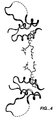
- IGFs share a high sequence identity with insulin, being about 49% identical thereto. Unlike insulin, however, which is synthesized as a precursor protein containing a 33-amino-acid segment known as the C-peptide (which is excised to yield a covalently linked dimer of the remaining A and B chains), IGFs are single polypeptides (see Figure 1).
- IGF-1 is a powerful mitogen, regulating diverse cellular functions such as cell cycle progression, apoptosis, and cellular differentiation ( LeRoith, Endocrinology, 141: 1287-1288 (2000 )).
- IGFs have been implicated in a variety of cellular functions and disease processes, including cell cycle progression, proliferation, differentiation, and insulin-like effects in insulin-resistant diabetes. Thus, IGF has been suggested as a therapeutic tool in a variety of diseases and injuries (for review, see Lowe, Scientific American (March/April 1996), p. 62 ). Due to this range of activities, IGF-1 has been tested in mammals for such widely disparate uses as wound healing, treatment of kidney disorders, treatment of diabetes, reversal of whole-body catabolic states such as AIDS-related wasting, treatment of heart conditions such as congestive heart failure, and treatment of neurological disorders ( Guler et al., Proc. Natl. Acad. Sci.
- the IGF system is also composed of membrane-bound receptors for IGF-1, IGF-2, and insulin.
- the Type 1 IGF receptor (IGF-1R) is closely related to the insulin receptor in structure and shares some of its signaling pathways ( Jones and Clemmons, Endocr. Rev., 16: 3-34 (1995 )).
- the IGF-2 receptor is a clearance receptor that appears not to transmit an intracellular signal (Jones and Clemmons, supra ). Since IGF-1 and IGF-2 bind to IGF-1R with a much higher affinity than to the insulin receptor, it is most likely that most of the effects of IGF-1 and IGF-2 are mediated by IGF-1R ( Humbel, Eur. J Biochem.
- IGF-1R is an ⁇ 2 ⁇ 2 heterotetramer of disulfide-linked ⁇ and ⁇ subunits. ⁇ dimers are themselves disulfide linked on the cell surface to form a covalent heterotetramer.
- IGF-1 binds to the IGF-1R with a 1:2 stoichiometry ( De Meyts, Diabetologia, 37: S135-S148 (1994 )), with a high affinity site (K d about 0.4 nM) and a low affinity site (K d about 6 nM) ( Tollefren and Thompson, J. Biol. Chem., 263: 16267-16273 (1988 )).
- the x-ray crystal structure of the first three domains of IGF-1R has been determined ( Garrett et al., Nature, 394, 395-399 (1998 )). It contains three distinct domains (L1, Cys-rich, L2). Mutations that affect IGF-1 binding map to the concave surface of the receptor.
- IGF-1R is a key factor in normal cell growth and development ( Isaksson et al., Endocrine Reviews, 8: 426-438 (1987 ); Daughaday and Rotwein, Endocrine Rev., 10:68-91 (1989 )). Increasing evidence suggests, however, that IGF-1R signaling also plays a critical role in growth of tumor cells, cell transformation, and tumorigenesis ( Baserga, Cancer Res., 55:249-252 (1995 )).
- the IGFs are potent breast cancer cell mitogens based on the observation that IGF-1 enhanced breast cancer cell proliferation in vitro ( Cullen et al., Cancer Res., 50:48-53 (1990 )).
- Breast cancers express IGF-2 and IGF-1R, providing all the required effectors for an autocrine-loop-based proliferation paradigm ( Quinn et al., J Biol. Chem., 271:11477-11483 (1996 );
- Sachr et al., Cancer Res., 56:1761-1765 (1996) Because breast cancer is a common malignancy affecting approximately one in every eight women and is a leading cause of death from cancer in North American women ( LeRoith et al., Ann. Int.
- IGF-1 can suppress apoptosis, and therefore cells lacking IGF-IRs or having compromised IGF-1R signaling pathways may give rise to tumor cells that selectively die via apoptosis ( Long et al., Cancer Res., 55:1006-1009 (1995 )). Furthermore, it has recently become evident that alterations in IGF signaling in the context of other disease states, such as diabetes, may be responsible for exacerbating the complications of retinopathy ( Smith et al., Science, 276:1706-1709 (1997 )) and nephropathy ( Horney et al., Am. J Physiol. 274: F1045-F1053 (1998 )).
- IGF-1 in vivo is mostly found in complex with a family of at least six serum proteins known as IGFBPs (Jones and Clemmons, supra ; Bach and Rechler, Diabetes Reviews, 3: 38-61 (1995 )), that modulate access of the IGFs to the IGF-1R. They also regulate the concentrations of IGF-1 and IGF-2 in the circulation and at the level of the tissue IGF-1R ( Clemmons et al., Anal. NY Acad. Sci. USA, 692:10-21 (1993 )). The IGFBPs bind IGF-1 and IGF-2 with varying affinities and specificities (Jones and Clemmons, supra ; Bach and Rechler, supra ).
- IGFBP-3 binds IGF-1 and IGF-2 with a similar affinity
- IGFBP-2 and IGFBP-6 bind IGF-2 with a much higher affinity than they bind IGF-1 (Bach and Rechler, supra ; Oh et al., Endocrinology, 132, 1337-1344 (1993 )).
- the major carrier protein is IGFBP-3.
- IGF-1 naturally occurs in human body fluids, for example, blood and human cerebral spinal fluid. Although IGF-1 is produced in many tissues, most circulating IGF-1 is believed to be synthesized in the liver.
- the IGFBPs are believed to modulate the biological activity of IGF-1 (Jones and Clemmons, supra ), with IGFBP-1 ( Lee et al., Proc. Soc. Exp. Biol. & Med., 204: 4-29 (1993 )) being implicated as the primary binding protein involved in glucose metabolism ( Baxter, "Physiological roles of IGF binding proteins", in: Spencer (Ed.), Modern Concepts of Insulin-like Growth Factors (Elsevier, New York, 1991), pp. 371-380 ).
- IGFBP-1 production by the liver is regulated by nutritional status, with insulin directly suppressing its production ( Suikkari et al., J. Clin. Endocrinol. Metab., 66: 266-272 (1988 )).
- IGFBP-1 The function of IGFBP-1 in vivo is poorly understood.
- the administration of purified human IGFBP-1 to rats has been shown to cause an acute, but small, increase in blood glucose ( Lewitt et al., Endocrinology, 129: 2254-2256 (1991 )).
- the regulation of IGFBP-1 is somewhat better understood. It has been proposed ( Lewitt and Baxter, Mol. Cell Endocrinology, 79: 147-152 (1991 )) that when blood glucose rises and insulin is secreted, IGFBP-1 is suppressed, allowing a slow increase in "free" IGF-1 levels that might assist insulin action on glucose transport.
- IGFBP-1 places the function of IGFBP-1 as a direct regulator of blood glucose.
- IGFBP-3 is a target gene of the tumor suppressor, p53 ( Buckbinder et al., Nature, 377:646-649 (1995 )). This suggests that the suppressor activity of p53 is, in part, mediated by IGFBP-3 production and the consequential blockade of IGF action (Buckbinder et al., supra ). These results indicate that the IGFBPs can block cell proliferation by modulating paracrine/autocrine processes regulated by IGF-I/IGF-2.
- PSA prostate-specific antigen
- IGFBP-3-protease an IGFBP-3-protease, which upon activation, increases the sensitivity of tumor cells to the actions of IGF-1/IGF-2 due to the proteolytic inactivation of IGFBP-3 ( Cohen et al., J. Endocr., 142:407-415 (1994 )).
- the IGFBPs complex with IGF-1/IGF-2 and interfere with the access of IGF-1/IGF-2 to IGF-1Rs ( Clemmons et al., Anal. NY Acad. Sci. USA, 692:10-21 (1993 )).
- IGFBP-1, -2 and -3 inhibit cell growth following addition to cells in vitro ( Lee et al., J Endocrinol.,152:39 (1997 ); Feyen et al., J Biol. Chem., 266:19469-19474 (1991 )). Further, IGFBP-1 ( McGuire et al., J Natl. Cancer Inst, 84:1335-1341(1992 ); Figueroa et al., J Cell Physiol., 157:229-236 (1993) ), IGFBP-3 (Oh et al. (1995), supra; Pratt and Pollak, Biophys. Res.
- IGFBP-2 have all been shown to inhibit IGF-1 or estrogen-induced breast cancer cell proliferation at nanomolar concentrations in vitro .
- IGFBPs are potent antagonists of IGF action.
- IGFBP-3 has also evidence for a direct effect of IGFBP-3 on cells through its own cell surface receptor, independent of IGF interactions ( Oh et al., J Biol. Chem., 268:14964-14971 (1993 ); Valentinis et al., Mol. Endocrinol., 9:361-367 (1995 )).
- these findings underscore the importance of IGF and IGF-1R as targets for therapeutic use.
- IGFs have mitogenic and anti-apoptotic influences on normal and transformed prostate epithelial cells ( Hsing et al., Cancer Research, 56: 5146 (1996 ); Culig et al., Cancer Research, 54: 5474 (1994 ); Cohen et al., Hormone and Metabolic Research, 26: 81 (1994 ); Iwamura et al., Prostate, 22: 243 (1993 ); Cohen et al., J. Clin. Endocrin. & Metabol., 73: 401 (1991 ); Rajah et al., J. Biol. Chem., 272: 12181 (1997 )).
- IGF-1 Most circulating IGF-1 originates in the liver, but IGF bioactivity in tissues is related not only to levels of circulating IGFs and IGFBPs, but also to local production ofIGFs, IGFBPs, and IGFBP proteases ( Jones and Clemmons, Endocrine Reviews, 16: 3 (1995 )). Person-to-person variability in levels of circulating IGF-1 and IGFBP-3 (the major circulating IGFBP (Jones and Clemmons, supra )) is considerable ( Juul et al., J. Clin. Endocrinol. & Metabol., 78: 744 (1994 ); Juul et al., J. Clin.
- the IGFs are present in high concentrations in the circulation, but only a small fraction of the IGFs is not protein bound. For example, it is generally known that in humans or rodents, less than 1% of the IGFs in blood is in a "free" or unbound form ( Juul et al., Clin. Endocrinol., 44: 515-523 (1996 ); Hizuka et al., Growth Regulation, 1: 51-55 (1991 ); Hasegawa et al., J. Clin. Endocrinol. Metab., 80: 3284-3286 (1995) ).
- the ternary complex of an IGF, IGFBP-3, and ALS has a molecular weight of approximately 150,000 daltons, and it has been suggested that the function of this complex in the circulation may be to serve as a reservoir and buffer for IGF-1 and IGF-2, preventing rapid changes in free IGF-1 or IGF-2.
- IGF-1 and IGF-2 are used to bind to the IGFBPs from those used to bind to IGF-1R. It is therefore possible to produce IGF variants that show reduced binding to the IGFBPs, but, because they bind well to IGF-1R, show maintained activity in in vitro activity assays.
- IGF variant that binds to IGFBPs but not to IGF receptors and therefore shows reduced activity in in vitro activity assays ( Bar et al., Endocrinology, 127: 3243-3245 (1990) ).
- this variant designated (1-27,gly 4 , 38-70)-hIGF-1, residues 28-37 of the C-region of human IGF-1 are replaced by a four-residue glycine bridge.
- IGF-1 variants are disclosed.
- EP 742,228 discloses two-chain IGF-1 superagonists, which are derivatives of the naturally occurring, single-chain IGF-1 having an abbreviated C-region.
- the IGF-1 analogs are of the formula: BC n , A wherein B is the B-region of IGF-1 or a functional analog thereof, C is the C-region of IGF-1 or a functional analog thereof, n is the number of amino acids in the C-region and is from about 6 to about 12, and A is the A-region of IGF-1 or a functional analog thereof.
- Cascieri et al., Biochemistry, 27: 3229-3233 (1988 ) discloses four mutants of IGF-1, three of which have reduced affinity to IGF-1R. These mutants are: (Phe 23 , Phe 24 , Tyr 25 )IGF-1 (which is equipotent to human IGF-1 in its affinity to the Types 1 and 2 IGF and insulin receptors), (Leu 24 )IGF-1 and (Ser 24 )IGF-1 (which have a lower affinity than IGF-1 to the human placental IGF-1R, the placental insulin receptor, and the IGF-1R of rat and mouse cells), and desoctapeptide (Leu 24 )IGF-1 (in which the loss of aromaticity at position 24 is combined with the deletion of the carboxyl-terminal D-region of hIGF-1, which has lower affinity than (Leu 24 )IGF-1 for the IGF-1R and higher affinity for the insulin receptor). These four mutants have normal affinities for human serum binding proteins.
- Bayne et al., J. Biol. Chem., 263: 6233-6239 (1988 ) discloses four structural analogs of human IGF-1: a B-chain mutant in which the first 16 amino acids of IGF-1 were replaced with the first 17 amino acids of the B-chain of insulin, (Gln 3 ,Ala 4 )IGF-1, (Tyr 15 ,Leu 16 )IGF-1, and (Gln 3 ,Ala 4 ,Tyr 15 ,Leu 16 )IGF-1. These studies identify some of the regions of IGF-1 that are responsible for maintaining high-affinity binding with the serum binding protein and the Type 2 IGF receptor.
- WO 94/04569 discloses a specific binding molecule, other than a natural IGFBP, that is capable of binding to IGF-1 and can enhance the biological activity of IGF-1.
- WO 00/23469 discloses the portions of IGFBP and IGF peptides that account for IGF-IGFBP binding, i.e., an isolated IGF binding domain of an IGFBP or modification thereof that binds IGF with at least about the same binding affinity as the full-length IGFBP.
- the patent publication also discloses an IGF antagonist that reduces binding of IGF to an IGF receptor, and/or binds to a binding domain of IGFBP.
- EP 639981 discloses pharmaceutical compositions comprising short peptides that function as IGF-1 receptor antagonists.
- the peptides used in the pharmaceutical compositions consist of less than 25 amino acids, comprise at least a portion of the C- or D-region from IGF-1, and inhibit IGF-1-induced autophosphorylation of IGF-1 receptors.
- Polypeptides including the IGF molecules, have a three-dimensional structure determined by the primary amino acid sequence and the environment surrounding the polypeptide. This three-dimensional structure establishes the activity, stability, binding affinity, binding specificity, and other biochemical attributes of the polypeptide. Thus, knowledge of the three-dimensional structure of a protein can provide much guidance in designing agents that mimic, inhibit, or improve its biological activity in soluble or membrane-bound forms.
- the three-dimensional structure of a polypeptide may be determined in a number of ways. Many of the most precise methods employ x-ray crystallography ( Van Holde, Physical Biochemistry (Prentice Hall: N.J., 1971), pp. 221-239 ). This technique relies on the ability of crystalline lattices to diffract x-ray or other forms of radiation. Diffraction experiments suitable for determining the three-dimensional structure of macromolecules typically require high-quality crystals. Unfortunately, such crystals have been unavailable for IGF-1 as well as many other proteins of interest. Crystals have been described for M-CSF ( EP 668,914B1 ), CD40 ligand ( WO 97/00895 ), and a BC2 Fab fragment ( WO 99/01476 ), for example.
- insulin crystal suspensions that are used therapeutically include suspensions of rhombohedral zinc-insulin crystals that are stable in the presence of 0.8 to 2.5% of zinc (based on the weight of insulin) at a neutral pH value and exhibit a delayed action, and isophane insulin protamine crystals, which are used in delayed action products in the form of small rods.
- a few other crystal modifications of insulin are furthermore known, but these have hitherto been of interest only for X-ray structure analysis.
- a protein of sufficient purity it must be crystallized to a size and clarity that is useful for x-ray diffraction and subsequent structure resolution.
- this sequence information does not allow an accurate prediction of the crystal structure of the protein.
- sequence information afford an understanding of the structural, conformational, and chemical interactions between a ligand such as an IGFBP and its protein target.
- crystal structures can provide a wealth of valuable information in the field of drug design and discovery, crystals of certain biologically relevant compounds such as IGF-1 are not readily available to those skilled in the art. High-quality, diffracting crystals of IGF-1 would assist the determination of its three-dimensional structure.
- IGF-1 antagonists have been restricted, at least in part, because of difficulties in studying the structure of IGF and IGFBPs. Due to the inability to obtain crystals of IGF-1 suitable for diffraction studies, for example, an extrapolation of IGF-1 structure based on the crystal structure of porcine insulin was the most important structural road map for IGF-1 available ( Blundell et al., Proc. Natl. Acad. Sci. USA, 75:180-184 (1978 )). See also Blundell et al., Fed. Proc., 42: 2592-2597 (1983 ), which discloses tertiary structures, receptor binding, and antigenicity of IGFs. Based on studies of chemically modified and mutated IGF-1, a number of common residues between IGF-I and insulin have been identified as being part of the IGF-1R-insulin receptor contact site, in particular, the aromatic residues at positions 23-25.
- Chem., 275: 10009-10015 discloses the solution structure and backbone dynamics of long-[Arg(3)]IGF-1. See also Laajoki et al., FEBS Lett., 420: 97-102 (1997 )).
- the small number ofNMR models available for IGF-1 are not very well defined, as there are large RMSDs between the backbone atoms of the helical segments.
- the best NMR model is of IGF-2 in which three alpha-helices are shown. See Torres et al., J. Mol. Biol., 248: 385-401 (1995 ), which discloses the solution structure of human IGF-2 and its relationship to receptor and binding protein interactions. In all structures, the C- and D-regions are very poorly defined.
- crystalline polypeptides provide other advantages. For example, the crystallization process itself further purifies the polypeptide and satisfies one of the classical criteria for homogeneity. In fact, crystallization frequently provides unparalleled purification quality, removing impurities that are not removed by other purification methods such as HPLC, dialysis, conventional column chromatography, etc. Moreover, crystalline polypeptides are often stable at ambient temperatures and free of protease contamination and other degradation associated with solution storage. Crystalline polypeptides may also be useful as pharmaceutical preparations. Finally, crystallization techniques in general are largely free of problems such as denaturation associated with other stabilization methods (e.g. lyophilization).
- crystallographic data provides useful structural information that may assist the design of peptides that may serve as agonists or antagonists.
- the crystal structure provides information useful to map the receptor-binding domain, which could then be mimicked by a small non-peptide molecule that may serve as an antagonist or agonist.
- findings regarding the detergent's inhibition of the binding of IGFBP to IGF-1 can be used to identify new IGF-1 agonists.
- IGF-1 has been crystallized and its structure determined using multiwavelength anomalous diffraction (MAD) at 1.8 angstroms resolution by exploiting the anomalous scattering of a single bromide ion and six of the seven sulfur atoms of IGF-1.
- the C-region of IGF-1 which is ordered in the crystal structure, forms a type II beta-turn and mediates a crystal packing interaction across a crystallographic dyad.
- the solution state of IGF-1 was characterized by analytical ultracentrifugation, and the results indicate that IGF-1 exists primarily as a monomer at neutral pH, with only a slight tendency to dimerize at millimolar concentrations.
- a molecule of detergent N, N-bis(3-D-gluconamidopropyl)-deoxycholamine (deoxy big CHAPS), mediates a crystal packing contact between symmetry-related molecules.
- Solution experiments confirm that the IGF-1:N, N-bis(3-D-gluconamidopropyl)-deoxycholamine complex does form in solution, and that detergent binding measurably blocks IGFBP binding.
- the invention provides a crystal formed by IGF-1 that diffracts x-ray radiation to produce a diffraction pattern representing the three-dimensional structure of the IGF-1.
- the IGF-1 contains an A-, B-, C-, and D-region and forms a dimer in the crystal, and further preferred is the crystal comprising a receptor binding site at the dimer interface.
- the invention also provides a composition comprising the above crystal.
- the IGF-1 is biologically active when resolubilized.
- the invention further provides a method of treating a mammal suffering from an agonist disorder, preferably a human patient, said method comprising administering to said mammal an effective amount of the above resolubilized composition.
- the invention also provides a method of crystallizing IGF-1 comprising the steps of:
- the invention also provides crystalline IGF-1 produced by the above method.
- the invention provides a method for determining a three-dimensional structure of IGF-1 comprising:
- the invention provides a machine-readable data storage medium comprising a data storage material encoded with machine-readable data that, when read by an appropriate machine, displays a three-dimensional representation of a crystal of a molecule comprising IGF-1.
- the invention provides an IGF-1 crystal with the structural coordinates shown in Appendix 1.
- the invention provides a method of using a three-dimensional structure of IGF-1 derived from an IGF-1 crystal wherein the three-dimensional structure of IGF-1 includes an IGF-1 receptor-binding region, the method comprising identifying compounds having structures that interact with the receptor-binding region of the three-dimensional structure of IGF-1 and function as an IGF-1 agonist or antagonist.
- the three-dimensional structure of IGF-1 includes alpha-carbon coordinates substantially the same as those of the structural information presented in Appendix 1.
- the invention provides a method of identifying IGF-1 agonists or antagonists comprising the steps of:
- the invention includes a method of designing a compound, such as a peptidomimetic, that mimics the 3-dimensional surface structure of IGF-1 comprising the steps of:
- the invention provides a method for identifying a peptidomimetic that binds IGF-1 and blocks binding of an IGFBP or a receptor that binds to IGF-1 comprising the steps of:
- the invention also provides a method for determining at least a portion of a three-dimensional structure of a molecular complex, said complex comprising IGF-1 and said method comprising the steps of:
- the invention also provides a method for evaluating the ability of a chemical entity to associate with IGF-1 or a complex thereof, the method comprising the steps of
- the invention also provides a chemical entity identified by the above method that interferes with the in vivo or in vitro association between IGF-1 and its receptor or between IGF-1 and at least one of its binding proteins, or associates with a binding site on IGF-1.
- the invention also comprises a method of computationally or experimentally evaluating a chemical entity to obtain information about its association with one or more binding sites of IGF-1 using a crystal of IGF-1 having the structural coordinates described in Appendix 1.
- Any peptide analogs and other chemical entities identified using the above methods of the present invention are useful in the therapeutic methods described herein and as pharmaceutical compositions.
- the invention also provides a method of identifying indirect agonists of IGF-1 comprising the steps of
- the comparison is accomplished by competition assay between N, N-bis(3-D-gluconamidopropyl)-deoxycholamine and the candidate agonist.
- inhibition of binding is measured by pre-incubating N, N-bis(3-D-gluconamidopropyl)-deoxycholamine or the candidate agonist with IGF-1 expressed on bacteriophage particles and measuring residual binding of IGF-I to IGFBP-1 or IGFBP-3 in a plate-based ELISA assay.
- the invention further provides a method of identifying indirect agonists of IGF-1 comprising co-crystallizing a candidate indirect agonist of IGF-1 with IGF-1 to form a co-crystalline structure and determining if the candidate agonist binds to one or both of two patches on IGF-1, wherein one patch has the amino acid residues Glu 3, Thr 4, Leu 5, Asp 12, Ala 13, Phe 16, Val 17, Cys 47, Ser 51, Cys 52, Asp 53, Leu 54, and Leu 57, and the second patch has the amino acid residues Val 11, Gln 15, Phe 23, Phe 25, Asn 26, Val 44, Phe 49, and Arg 55, and wherein binding occurs if there is at least one contact between each listed amino acid residue of a given patch and the candidate agonist that is less than or equal to 6 angstroms in the co-crystalline structure.
- the candidate agonist inhibits binding of IGFBP-1 or -3 to IGF-1 at least as well as N, N-bis(3-D-gluconamidopropyl)-deoxycholamine. More preferred is the method wherein inhibition of binding is measured using a competition assay between N, N-bis(3-D-gluconamidopropyl)-deoxycholamine and the candidate agonist. Most preferred is the method wherein inhibition of binding is measured by pre-incubating N, N-bis(3-D-gluconamidopropyl)-deoxycholamine or the candidate agonist with IGF-1 expressed on bacteriophage particles and measuring residual binding of IGF-1 to IGFBP-1 or IGFBP-3 in a plate-based ELISA assay.
- Also provided herein is a method for treating an IGF-1 agonist disorder in a mammal comprising administering to the mammal an effective amount ofN, N-bis(3-D-gluconamidopropyl)-deoxycholamine.
- IGF-1 refers to human insulin-like growth factor-1 unless otherwise noted, and has the human native mature IGF-1 sequence without a N-terminal methionine, as described, for example, by EP 230,869 published August 5, 1987 ; EP 128,733 published December 19, 1984 ; or EP 288,451 published October 26, 1988 .
- IGFBP insulin growth factor
- IGFBP-7 prostacyclin-stimulating factor
- ESM-1 endothelial cell-specific molecule
- IGFBP-1 and IGFBP-3 bind to different residues of IGF-1.
- human IGF-1 receptor or just “IGF-1 receptor” refers to any receptor for IGF-1 found in humans and includes the Type 1 and Type 2 IGF receptors in humans to which human IGF-1 binds, such as the placental IGF-1R, etc .
- IGF-1 an "indirect agonist of IGF-1" is a molecule that releases IGF-1 in situ from IGFBP-3 or IGFBP-1 so that the IGF-1 released is active and interacts with its receptor.
- Proteins are molecules having at least two amino acids and include polypeptides having at least about 60 amino acids. Preferably, the peptides have about 10 to about 60 amino acids, more preferably about 10-25, and most preferably about 12-25 amino acids.
- the definition includes linear and cyclic peptides, peptide derivatives, their salts, or optical isomers.
- mammal for purposes of treatment refers to any animal classified as a mammal, including humans, domestic, and farm animals, and zoo, sports, or pet animals, such as dogs, horses, cats, sheep, pigs, cows, etc.
- the preferred mammal herein is a human.
- non-adult refers to mammals that are from perinatal age (such as low-birth-weight infants) up to the age of puberty, the latter being those that have not yet reached full growth potential.
- treating refers to both therapeutic treatment and prophylactic or preventative measures. Those in need of treatment include those already with the disorder as well as those prone to having the disorder or diagnosed with the disorder or those in which the disorder is to be prevented.
- a “disorder” is any condition that would benefit from treatment with an IGF-1 agonist ("agonist disorder”) or antagonist ("antagonist disorder”). This includes chronic and acute disorders or diseases including those pathological conditions that predispose the mammal to the disorder in question.
- the disorder being treated may be a combination of two or more of the agonist or antagonist disorders listed below.
- Non-limiting examples of antagonist disorders include benign and malignant tumors, leukemias and lymphoid malignancies, neuronal, glial, astrocytal, hypothalamic and other glandular, macrophagal, epithelial, stromal and blastocoelic disorders, and inflammatory, angiogenic and immunologic disorders, diabetic complications such as diabetic retinopathies or neuropathies, age-related macular degeneration, ophthalmic surgery such as cataract extraction, a corneal transplant, glaucoma filtration surgery and keratoplasty, surgery to correct refraction, i.e., a radial keratotomy, also in sclera macular holes and degeneration, retinal tears, vitreoretinopathy, miscellaneous disorders, cataract disorders of the cornea such as the sequelae of radial keratotomy, dry eye, viral conjunctivitis, ulcerative conjunctivitis, wounds such as corneal epithelial wounds, Sjogren'
- such antagonist disorders include diabetic complications exacerbated by IGF-1, ischemic injury, and diseases associated with undesirable cell proliferation such as cancer, restenosis, and asthma.
- the disorder is a diabetic complication exacerbated by IGF-1, such complication can include diabetic retinopathy or diabetic nephropathy.
- the efficacy of the treatment can be evidenced by a reduction in clinical manifestations or symptoms, including, for example, improved renal clearance, improved vision, or a reduction in the amount of IGF-1 available for binding to an IGF-1 receptor.
- the disorder is an ischemic injury, it can include strokes, myocardial ischemia, and ischemic injury to the kidneys.
- agonist disorders for purposes herein include any condition that would benefit from treatment with an IGF-1, including but not limited to, for example, lung diseases, hyperglycemic disorders as set forth below, renal disorders, such as acute and chronic renal insufficiency, end-stage chronic renal failure, glomerulonephritis, interstitial nephritis, pyelonephritis, glomerulosclerosis, e .
- lung diseases such as acute and chronic renal insufficiency, end-stage chronic renal failure, glomerulonephritis, interstitial nephritis, pyelonephritis, glomerulosclerosis, e .
- the preferred agonist disorders targeted for treatment herein are diabetes and obesity, heart dysfunctions, AIDS-related wasting, kidney disorders, neurological disorders, whole body growth disorders, and immunological disorders.
- hyperglycemic disorders refers to all forms of diabetes and disorders resulting from insulin resistance, such as Type I and Type II diabetes, as well as severe insulin resistance, hyperinsulinemia, and hyperlipidemia, e.g., obese subjects, and insulin-resistant diabetes, such as Mendenhall's Syndrome, Werner Syndrome, leprechaunism, lipoatrophic diabetes, and other lipoatrophies.
- the preferred hyperglycemic disorder is diabetes, especially Type 1 and Type II diabetes.
- Diabetes itself refers to a progressive disease of carbohydrate metabolism involving inadequate production or utilization of insulin and is characterized by hyperglycemia and glycosuria.
- Biologically active IGF-1 refers to IGF-1 that exhibits a biological property conventionally associated with an IGF-1 agonist or antagonist, such as a property that would allow treatment of one or more of the disorders listed above.
- the term "effective amount” refers to an amount of IGF-1 or a peptidomimetic or other compound, including chemical entities, effective to treat a disease or disorder in a mammal.
- the effective amount of the peptide may reduce the number of cancer cells; reduce the tumor size; inhibit ( i . e ., slow to some extent and preferably stop) cancer cell infiltration into peripheral organs; inhibit ( i . e ., slow at least to some extent and preferably stop) tumor metastasis; inhibit, to some extent, tumor growth; promote apoptosis; and/or relieve to some extent one or more of the symptoms associated with the disorder.
- a "precipitant” is an agent in a reservoir solution that precipitates IGF-1 when mixed with an aqueous solution of IGF-1 and allowed to equilibrate so as to form IGF-1 crystals.
- examples include chaotropic agents such as ammonium sulfate, polyethylene glycols (of a wide variety of molecular weights ranging, for example, from about 2000 to 20,000), sodium citrate, sodium cacodylate, or a mixture thereof.
- a “reservoir solution” is a solution of a precipitant and any other ingredient needed to provide IGF-1 crystals, for example, a detergent such as C 12 E 9 (nonaethylene glycol monododecyl ether, nonaethylene glycol monolauryl ether, polyoxyethylene (9) ether), C 12 E 8 (octaethylene glycol monododecyl ether, octaethylene glycol monolauryl ether, polyoxyethylene (8) lauryl ether), dodecyl-beta-D-maltopyranoside, lauric acid sucrose ester, cyclohexyl-pentyl-beta-D-maltoside, nonaethylene glycol octylphenol ether, cetyltrimethylammonium bromide, decyl-beta-D-maltopyranoside, lauryldimethylamine oxide, cyclohexyl-pentyl-beta-D-maltoside,
- Recrystallization refers to the procedure, after the initial crystals are grown and determined not to be very large or useful, of adding a substance to the crystals, such as methyl pentanediol, which has the effect of dissolving the crystals, but not diluting anything else much in the crystallization mixture. Then over the course of several days, as the crystallization droplet re-equilibrates with its reservoir solution, the crystals regrow, but this time much larger and more well ordered.
- association refers to a condition of proximity between IGF-1 and a chemical entity, or portions thereof.
- the association may be non-covalent, wherein the juxtaposition is energetically favored by hydrogen bonding, van der Waals interaction, or electrostatic interaction, or it may be a covalent association.
- binding site refers to any or all of the sites where a chemical entity binds or associates with IGF-1.
- structural coordinates refers to the coordinates derived from mathematical equations related to the patterns obtained on diffraction of a monochromatic beam of x-rays by the atoms (scattering centers) of a molecule in crystal form.
- the diffraction data can be used to calculate an electron density map of the repeating units of the crystal.
- An electron density map may be used to establish the positions of the individual atoms within the unit cell of the crystal.
- Appendix 1 shows the atomic coordinates of IGF-1.
- any set of structural coordinates of IGF-1 that have a root mean square deviation of equivalent protein backbone atoms of less than about 2 ⁇ when superimposed-using backbone atomson the structural coordinates in Appendix 1 shall be considered identical.
- the deviation is less than about 1 ⁇ and more preferably less than about 0.5 ⁇ .
- heavy-atom derivatization refers to a method of producing a chemically modified form of a crystallized IGF-1.
- a crystal is soaked in a solution containing heavy metal atom salts, or organometallic compounds, e.g., lead chloride, gold thiomalate, thimerosal, or uranyl acetate, which can diffuse through the crystal and bind to the surface of the protein.
- the location of the bound heavy metal atom(s) can be determined by x-ray diffraction analysis of the soaked crystal. This information can be used to generate the phase information used to construct the three-dimensional structure of the molecule.
- unit cell refers to a basic shaped block.
- the entire volume of a crystal may be constructed by regular assembly of such blocks.
- Each unit cell comprises a complete representation of the unit of pattern, the repetition of which builds up the crystal.
- space group refers to the arrangement of symmetry elements of a crystal
- molecular replacement refers to a method that involves generating a preliminary structural model of a crystal whose structural coordinates are unknown, by orienting and positioning a molecule whose structural coordinates are known, e.g., the IGF-1 coordinates in Appendix 1, within the unit cell of the unknown crystal, so as to best account for the observed diffraction pattern of the unknown crystal. Phases can then be calculated from this model, and combined with the observed amplitudes to give an approximated Fourier synthesis of the structure whose coordinates are unknown. This in turn can be subject to any of the several forms of refinement to provide a final accurate structure of the unknown crystal.
- molecular replacement may be used to determine the structural coordinates of a crystalline co-complex, unknown ligand, mutant, or homolog, or of a different crystalline form of IGF-1. Additionally, the claimed crystal and its coordinates may be used to determine the structural coordinates of a chemical entity that associates with IGF-1.
- chemical entity or “compound” as used herein means any molecule, molecular complex, compound, peptidomimetic, or fragment thereof that is not IGF-1. Preferably it is a molecule with high oral bioavailability, such as an organic chemical molecule, or a peptide.
- the following detailed description of the invention encompasses the crystal structure of IGF-1, methods of making an IGF-1 crystal, and methods of using an IGF-1 crystal and its structural coordinates.
- the ribbon structure thereof is shown in Figure 2 having three helices, with the N-terminal B-region corresponding to residues 3-28, the C-region from residues 29-34, a stretch of poorly ordered residues from residues 35-40, and the A-region from residues 42-62.
- the D-region (residues 63-70) is essentially disordered.
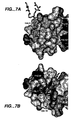
- Figures 4 and 7 show that the detergent used in the crystallization binds into a small hydrophobic cleft at the base of the B-helix of the structure.
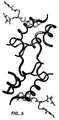
- the IGF-1 can form a dimer in the crystal, as shown in Fig. 5, wherein the two tails are positioned at the dimer interface.
- the buried surface area is 689 ⁇ 2 /monomer, which is 1378 ⁇ 2 total.
- the residues important for IGF-IR binding cluster at the dimer interface as shown in Figure 6.
- the claimed invention relates to methods of preparing crystalline forms of IGF-1 by first providing an aqueous solution comprising IGF-1.
- a reservoir solution comprising a precipitant is then mixed with a volume of the IGF-1 solution and the resultant mixed volume is then crystallized.
- the crystals are again dissolved and recrystallized.
- An example of a reagent that can be used for recrystallization is methyl pentanediol, which is preferred.
- the crystals are typically dissolved with this reagent in a small amount to minimize dilution effects of the other reagents and left to regrow for a period of time.
- the crystalline IGF-1 is isolated from the mixed volume.
- the IGF-1 is obtained from a prokaryotic cell, more preferably a bacterial cell, most preferably E . coli . Preferably it is secreted into the periplasm and prepared as described in U.S. Pat No. 5,723,310 .
- the concentration of IGF-1 in the aqueous solution may vary, but is preferably about 1 to 50 mg/ml, more preferably about 5 to 15 mg/ml.
- precipitants used in the invention may vary, and may be selected from any precipitant known in the art.
- the precipitant is selected from the group consisting of sodium citrate, ammonium sulfate, polyethylene glycol, sodium cacodylate, or a mixture thereof. More preferably the precipitant is polyethylene glycol buffered with sodium citrate or sodium cacodylate.
- the reservoir solution further comprises a detergent.
- the detergent is present in an amount of about 10 to 50 mM.
- the detergent is N, N-bis(3-D-gluconamidopropyl)-deoxycholamine.
- the pH of the reservoir solution may also be varied, preferably between about 4 to 10, most preferably about 6.5.
- Various methods of crystallization can be used in the claimed invention, including vapor diffusion, batch, liquid-bridge, or dialysis crystallization.
- Vapor diffusion crystallization is preferred. See, e.g. McPherson et al., Preparation and Analysis of Protein Crystals, Glick, ed. (John Wiley & Co., 1982), pp. 82-159 ; Jancarik et al., J. Appl. Crystallogr., 24: 409-411 (1991 ).
- a small volume i.e., a few milliliters
- a solution containing a precipitant This mixed volume is suspended over a well containing a small amount, i.e. about 1 ml, of precipitant. Vapor diffusion from the drop to the well will result in crystal formation in the drop.
- the dialysis method of crystallization utilizes a semipermeable size-exclusion membrane that retains the protein but allows small molecules (i.e. buffers and precipitants) to diffuse in and out In dialysis, rather than concentrating the protein and the precipitant by evaporation, the precipitant is allowed to slowly diffuse through the membrane and reduce the solubility of the protein while keeping the protein concentration fixed.
- small molecules i.e. buffers and precipitants
- the batch methods generally involve the slow addition of a precipitant to an aqueous solution of protein until the solution just becomes turbid; at this point the container can be sealed and left undisturbed for a period of time until crystallization occurs.
- the most preferred method of crystallization involves the method wherein the IGF-1, after isolation from the cell and formulation in, for example, an acetate, citrate, or succinate buffer, as described, for example, in U.S. Pat No. 5,681,814 and WO 99/51272 , is optionally desalted if necessary to a pH of about 4-5, preferably about 4.5, to form an aqueous solution.
- a droplet of the aqueous solution is mixed with about 24% polyethylene glycol buffered to about pH 6.5 with either about 0.1M sodium citrate or about 0.1M sodium cacodylate and with about 1 ⁇ l of about 1.4 mM N, N-bis(3-D-gluconamidopropyl)-deoxycholamine as detergent.
- This solution is then equilibrated by vapor diffusion crystallization with about 1 mL of about 24% polyethylene glycol buffered to about pH 6.5 with either about 0.1M sodium citrate or about 0.1M sodium cacodylate until crystallization droplets are formed, usually about 4-5 days.
- about 2 ⁇ l of about 100% methyl pentanediol is added to the crystallization droplets so as to dissolve the crystals overnight and thereby form new crystals, usually within a week's time.
- the crystal structure was determined by combined anomalous scattering from intrinsic sulfur and fortuitous bromide ion as discussed in detail in the Example below.
- the crystalline IGF-1 herein can be used for various purposes.
- the crystallization process itself further purifies the IGF-1 to homogeneity.
- one such purpose is to provide a highly purified IGF-1 that can be used as a standard or control in a diagnostic setting, for example, as a molecular weight marker, or as an ELISA, radioassay, or radioreceptor assay control.
- crystalline IGF-1 is stable at room temperature, can be lyophilized readily, and is less apt to degrade than less pure compositions.
- crystals of IGF-1 of a size and quality to allow performance of x-ray diffraction studies enable those of skill in the art to conduct studies relating to the binding properties of IGF-1, as well as the binding properties of IGFBPs, IGF-1 receptors, and ALS that associate with the IGF-1.
- structural information derived from a peptide crystal structure can be used for the identification of chemical entities, for example, small organic and bioorganic molecules such as peptidomimetics and synthetic organic molecules that bind IGF-1 and preferably block or prevent an IGF-1-mediated or -associated process or event, or that act as IGF-1 agonists.
- small organic and bioorganic molecules such as peptidomimetics and synthetic organic molecules that bind IGF-1 and preferably block or prevent an IGF-1-mediated or -associated process or event, or that act as IGF-1 agonists.
- the skilled artisan constructs a model of the IGF-1 such as those depicted in Figures 2 and 5. Since every atom of a peptide or polypeptide can be depicted as a sphere of the appropriate van der Waals radius, a detailed surface map of the folded IGF-1 can be constructed. The surface that results is known as the van der Waals surface.
- the "solvent-accessible surface” is the surface that is accessible to a chemical probe, a water molecule herein, and is constructed by rolling a water molecule of appropriate radius on the outside of the peptide maintaining contact with the van der Waals surface.
- Such chemical entities presenting a solvent-accessible surface that mimics the solvent-accessible surface of the IGF-1 can be constructed by those skilled in the art.
- the skilled artisan can search three-dimensional structural databases of compounds to identify those compounds that position appropriate functional groups in similar 3-dimensional structural arrangement, then build combinatorial chemistry libraries around such chemical entities to identify those with high affinity.
- One approach enabled by this invention is the use of the structural coordinates of IGF-1 to design chemical entities that bind to or associate with IGF-1 and alter the physical properties of the chemical entities in different ways.
- properties such as, for example, solubility, affinity, specificity, potency, on/off rates, or other binding characteristics may all be altered and/or maximized.
- the invention also contemplates computational screening of small-molecule databases or designing of chemical entities that can bind in whole or in part to IGF-1. They may also be used to solve the crystal structure of mutants, co-complexes, or the crystalline form of any other molecule homologous to, or capable of associating with, at least a portion of IGF-1.
- An unknown crystal structure which may be any unknown structure, such as, for example, another crystal form of IGF-1, an IGF-1 mutant or peptide, or a co-complex with IGF-1, or any other unknown crystal of a chemical entity that associates with IGF-1 that is of interest, may be determined using the structural coordinates as set forth in Appendix 1.
- Co-complexes with IGF-1 may include, but are not limited to, IGF-1-IGFBP-3, IGF-1-IGFBP-3-ALS, IGF-1-receptor, IGF-1-peptide, or IGF-1-small molecule. This method will provide an accurate structural form for the unknown crystal far more quickly and efficiently than attempting to determine such information without the invention herein.
- the information obtained can thus be used to obtain maximally effective inhibitors or agonists of IGF-1.
- the design of chemical entities that inhibit or agonize IGF-1 generally involves consideration of at least two factors. First, the chemical entity must be capable of physically or structurally associating with IGF-1.
- the association may be any physical, structural, or chemical association, such as, for example, covalent or noncovalent bonding, or van der Waals, hydrophobic, or electrostatic interactions.
- the chemical entity must be able to assume a conformation that allows it to associate with IGF-1. Although not all portions of the chemical entity will necessarily participate in the association with IGF-1, those non-participating portions may still influence the overall conformation of the molecule. This in turn may have a significant impact on the desirability of the chemical entity.
- conformational requirements include the overall three-dimensional structure and orientation of the chemical entity in relation to all or a portion of the binding site.
- the potential inhibitory or binding effect of a chemical entity on IGF-1 may be analyzed prior to its actual synthesis and testing by the use of computer-modeling techniques. If the theoretical structure of the given chemical entity suggests insufficient interaction and association between it and IGF-1, the need for synthesis and testing of the chemical entity is obviated. However, if computer modeling indicates a strong interaction, the molecule may then be synthesized and tested for its ability to bind to IGF-1. Thus, expensive and time-consuming synthesis of inoperative compounds may be avoided.
- An inhibitory or other binding compound of IGF-1 may be computationally evaluated and designed by means of a series of steps in which chemical entities or fragments are screened and selected for their ability to associate with the individual binding sites of IGF-1.
- one skilled in the art may use one of several methods to screen chemical entities or fragments for their ability to associate with IGF-1.
- This process may begin by visual inspection of, for example, the binding site on a computer screen based on the IGF-1 coordinates in Appendix 1. Selected fragments or chemical entities may then be positioned in a variety of orientations, or "docked,” within an individual binding pocket of IGF-1. Docking may be accomplished using software such as Quanta and Sybyl, followed by energy minimization and molecular dynamics with standard molecular mechanics force fields, such as CHARMM and AMBER.
- Specialized computer programs may be of use for selecting interesting fragments or chemical entities. These programs include, for example, GRID, available from Oxford University, Oxford, UK; MCSS or CATALYST, available from Molecular Simulations, Burlington, MA; AUTODOCK, available from Scripps Research Institute, La Jolla, CA; DOCK, available from University of California, San Francisco, CA, and XSITE, available from University College of London, UK.
- GRID available from Oxford University, Oxford, UK
- MCSS or CATALYST available from Molecular Simulations, Burlington, MA
- AUTODOCK available from Scripps Research Institute, La Jolla, CA
- DOCK available from University of California, San Francisco, CA
- XSITE available from University College of London, UK.
- Assembly may be by visual inspection of the relationship of the fragments to each other on the three-dimensional image displayed on a computer screen, in relation to the structural coordinates disclosed herein.
- any molecular modeling techniques may be employed in accordance with the invention; these techniques are known, or readily available to those skilled in the art. It will be understood that the methods and compositions disclosed herein can be used to identify, design, or characterize not only entities that will associate or bind to IGF-1, but alternatively to identify, design, or characterize entities that, like IGF-1, will bind to the receptor, thereby disrupting the IGF-1-receptor interaction.
- the claimed invention is intended to encompass these methods and compositions broadly.
- the efficiency with which that compound may bind to IGF-1 may be tested and modified for the maximum desired characteristic(s) using computational or experimental evaluation.
- Various parameters can be maximized depending on the desired result. These include, but are not limited to, specificity, affinity, on/off rates, hydrophobicity, solubility, and other characteristics readily identifiable by the skilled artisan.
- the invention is useful for the production of small-molecule drug candidates.
- the claimed crystal structures may be also used to obtain information about the crystal structures of complexes of the IGF-1 and small-molecule inhibitors. For example, if the small-molecule inhibitor is co-crystallized with IGF-1, then the crystal structure of the complex can be solved by molecular replacement using the known coordinates of IGF-1 for the calculation of phases.
- Such information is useful, for example, for determining the nature of the interaction between the IGF-1 and the small-molecule inhibitor, and thus may suggest modifications that would improve binding characteristics such as affinity, specificity, and kinetics.
- the invention herein is also useful in providing a method of identifying indirect agonists of IGF-1 based on the inhibitory properties of N, N-bis(3-D-gluconamidopropyl)-deoxycholamine with respect to IGFBPs.
- This method comprises the steps of: comparing the ability ofN, N-bis(3-D-gluconamidopropyl)-deoxycholamine to inhibit binding of IGFBP-1 or -3 to IGF-1 with the ability of a candidate IGF-1 indirect agonist to inhibit such binding; and determining whether the candidate IGF-1 indirect agonist can inhibit such binding at least as well as N, N-bis(3-D-gluconamidopropyl)-deoxycholamine can so inhibit the binding.
- the comparison is accomplished by competition assay between N, N-bis(3-D-gluconamidopropyl)-deoxycholamine and the candidate IGF-1 indirect agonist, using IC 50 to measure ability to inhibit IGFBP binding.
- inhibition of binding is measured by pre-incubating N, N-bis(3-D-gluconamidopropyl)-deoxycholamine or the candidate agonist molecule with IGF-1 expressed on bacteriophage particles and measuring residual binding of IGF-1 to IGFBP-1 or IGFBP-3 in a plate-based assay, such as an ELISA
- the invention further provides a method of identifying indirect agonists of IGF-1 comprising co-crystallizing the candidate agonist with IGF-1 to form a co-crystalline structure and determining if the candidate agonist molecule binds to one or both of two patches on IGF-1.
- the first patch contains the amino acid residues Glu 3, Thr 4, Leu 5, Asp 12, Ala 13, Phe 16, Val 17, Cys 47, Ser 51, Cys 52, Asp 53, Leu 54, and Leu 57
- the second patch contains the amino acid residues Val 11, Gln 15, Phe 23, Phe 25, Asn 26, Val 44, Phe 49, and Arg 55.
- binding means that there is at least one contact between each listed amino acid residue of a given patch and the candidate agonist molecule that is less than or equal to 6 angstroms in the co-crystalline structure.
- a candidate agonist molecule will have the property of inhibiting binding of IGFBP-1 or IGFBP-3 to IGF-1.
- the preferred such candidate agonist molecule will inhibit binding of IGFBP-1 or -3 to IGF-1 at least as well as N, N-bis(3-D-gluconamidopropyl)-deoxycholamine. More preferred is the method wherein inhibition of binding is measured using a competition assay between N, N-bis(3-D-gluconamidopropyl)-deoxycholamine and the candidate agonist molecule.
- N, N-bis(3-D-gluconamidopropyl)-deoxycholamine detergent herein can be used as a template to perform design of small-molecule drugs that elicit the same effect as the detergent (compete with IGF-1 for IGFBP binding and subsequent disruption of the interaction of IGFBP with IGF-1 to free IGF-1 in situ so that it is active and will interact with the receptor.
- N, N-bis(3-D-gluconamidopropyl)-deoxycholamine lacks an oxygen atom at position C10. This region of the detergent is in close contact with the side-chain atoms of residues Leu 5, Leu 54, and Leu 57 of IGF-1. Molecules with this same type of conformation would work as indirect IGF-1 agonists.
- the indirect agonist so identified can be used in a method for treating an agonist disorder wherein an effective amount of the indirect agonist of IGF-1 is administered to a mammal with such a disorder.
- agonist may be used therapeutically in a pharmaceutical preparation, for example, in clinical trials or commercialized for the agonist disorders as defined herein.
- the formulation of the indirect agonist herein can be used to treat any condition that would benefit from treatment with IGF-1, including, for example, diabetes, chronic and acute renal disorders, such as chronic renal insufficiency, necrosis, etc., obesity, hyperinsulinemia, GH-insufficiency, Turner's syndrome, short stature, undesirable symptoms associated with aging such as increasing lean-mass-to-fat ratios, immuno-deficiencies including increasing CD4 counts and increasing immune tolerance, catabolic states associated with wasting, etc., Laron dwarfism, insulin resistance, and so forth.
- diabetes chronic and acute renal disorders, such as chronic renal insufficiency, necrosis, etc., obesity, hyperinsulinemia, GH-insufficiency, Turner's syndrome, short stature, undesirable symptoms associated with aging such as increasing lean-mass-to-fat ratios, immuno-deficiencies including increasing CD4 counts and increasing immune tolerance, catabolic states associated with wasting, etc., Laron dwarfism, insulin resistance,
- the indirect agonist composition herein may be directly administered to the mammal by any suitable technique, including orally, parenterally, intranasally, or intrapulmonarily, and can be administered locally or systemically.
- suitable technique including orally, parenterally, intranasally, or intrapulmonarily, and can be administered locally or systemically.
- the specific route of administration will depend, e.g., on the medical history of the patient, including any perceived or anticipated side or reduced effects using IGF-1, and the disorder to be treated.
- parenteral administration include subcutaneous, intramuscular, intravenous, intraarterial, and intraperitoneal administration. Most preferably, the administration is by continuous infusion (using, e.g., minipumps such as osmotic pumps), or by injection (using, e.g., intravenous or subcutaneous means).
- the administration may also be as a single bolus or by slow-release depot formulation.
- the direct agonist is administered orally or by infusion or injection, at a frequency of, preferably, one-half, once, twice, or three times daily, most preferably daily.
- the agonist composition to be used in the therapy will be formulated and dosed in a fashion consistent with good medical practice, taking into account the clinical condition of the individual patient (especially the side effects of treatment with the agonist), the site of delivery of the agonist composition, the method of administration, the scheduling of administration, and other factors known to clinical practitioners.
- the "effective amount" of agonist for purposes herein is thus determined by such considerations and must be an amount that treats the disorder in question.
- the total pharmaceutically effective amount of agonist administered parenterally per dose will be in the range of about 1 ⁇ g/kg/day up to about 100 mg/kg/day, preferably 10 ⁇ g/kg/day up to about 10 mg/kg/day.
- the agonist is generally administered in doses of about 1 ⁇ g/kg/hour up to about 100 ⁇ g/kg/hour, either by about 1-4 injections per day or by continuous subcutaneous infusions, for example, using a minipump or a portable infusion pump. An intravenous bag solution may also be employed.
- the key factor in selecting an appropriate dose is the result obtained as measured by criteria as are deemed appropriate by the practitioner.
- the agonist is administered together with insulin, the latter is used in lower amounts than if used alone, down to amounts which by themselves have little effect on blood glucose, i.e., in amounts of between about 0.1 IU/kg/24 hour to about 0.5 IU/kg/24 hour.
- the agonist is formulated generally by mixing it at the desired degree of purity, in a unit dosage injectable form (solution, suspension, or emulsion), with a pharmaceutically acceptable carrier, i.e., one that is non-toxic to recipients at the dosages and concentrations employed and is compatible with other ingredients of the formulation.
- a pharmaceutically acceptable carrier i.e., one that is non-toxic to recipients at the dosages and concentrations employed and is compatible with other ingredients of the formulation.
- the formulation preferably does not include oxidizing agents and other compounds that are known to be deleterious to polypeptides.
- the formulation is prepared by contacting the agonist uniformly and intimately with a liquid carrier or a finely divided solid carrier or both.
- a liquid carrier or a finely divided solid carrier or both.
- the carrier is a parenteral carrier, more preferably a solution that is isotonic with the blood of the recipient
- carrier vehicles include water, saline, Ringer's solution, and dextrose solution.
- Non-aqueous vehicles such as fixed oils and ethyl oleate are also useful herein, as well as liposomes.
- the carrier suitably contains minor amounts of additives such as substances that enhance isotonicity and chemical stability.
- additives such as substances that enhance isotonicity and chemical stability.
- Such materials are non-toxic to recipients at the dosages and concentrations employed, and include buffers such as phosphate, citrate, succinate, acetic acid, and other organic acids or their salts; antioxidants such as ascorbic acid; low-molecular-weight (less than about ten residues) polypeptides, e.g., polyarginine or tripeptides; proteins, such as serum albumin, gelatin, or immunoglobulins; hydrophilic polymers such as polyvinylpyrrolidone; glycine; amino acids such as glutamic acid, aspartic acid, or arginine; monosaccharides, disaccharides, and other carbohydrates including cellulose or its derivatives, glucose, mannose, or dextrins; chelating agents such as EDTA; sugar alcohols such as mannitol or sorb
- the agonist is typically formulated individually in such vehicles at a concentration of about 0.1 mg/ml to 100 mg/ml, preferably 1-10 mg/ml, at a pH of about 4.5 to 8.
- the final formulation if a liquid, is preferably stored at a temperature of about 2-8°C for up to about four weeks.
- the formulation can be lyophilized and provided as a powder for reconstitution with water for injection that is stored as described for the liquid formulation.
- the agonist to be used for therapeutic administration must be sterile. Sterility is readily accomplished by filtration through sterile filtration membranes (e.g., 0.2 micron membranes).
- Therapeutic agonist compositions generally are placed into a container having a sterile access port, for example, an intravenous solution bag or vial having a stopper pierceable by a hypodermic injection needle.
- the agonist ordinarily will be stored in unit or multi-dose containers, for example, sealed ampoules or vials, as an aqueous solution or as a lyophilized formulation for reconstitution.
- Recombinant human IGF-1 (rhIGF-1) was obtained as described in the Examples of U.S. Pat. No. 5,723,310 using a polymer/salt combination for phase-forming species and formulated as described in the Examples of U.S. Pat. No. 5,681,814 (acetate, NaCl, polysorbate 20, and benzyl alcohol), and placed in a vial containing 7 ml of 10 mg/ml rhIGF-1. It was desalted into 0.15 M NaCl, 20 mM NaOAc, pH 4.5, and diluted to a final concentration of 10 mg/ml.
- a 4- ⁇ l droplet of the IGF-1 solution was mixed with 5 ⁇ l of reservoir solution (24% polyethylene glycol 3350 buffered to pH 6.5 with 0.1M sodium cacodylate) and 1 ⁇ l of 14 mM ofN, N-bis(3-D-gluconamidopropyl)-deoxycholamine, which is obtained in a CRYSTAL SCREEN TM reagent kit used for crystallization condition screenings and available from Hampton Research, Madison Nigel, CA.
- This solution was allowed to equilibrate via vapor diffusion (Jancarik et al., supra ) with 1 mL of reservoir solution.
- a drop of the mixture was suspended under a plastic cover slip over the reservoir solution.
- crystallization conditions can be varied. By varying the crystallization conditions, other crystal forms of IGF-1 may be obtained. Such variations may be used alone or in combination, and include, for example, varying final protein concentrations between about 5 and 35 mg/ml; varying the IGF-1-to precipitant ratio, varying precipitant concentrations between about 20 and 30% for polyethylene glycol, varying pH ranges between about 5.5 and 7.5, varying the concentration or type of detergent, varying the temperature between about -5 and 30°C, and crystallizing IGF-1 by batch, liquid bridge, or dialysis methods using the above conditions or variations thereof. See McPherson et al. (1982), supra.
- a single crystal was transferred from the mother liquor to a cryo-protectant solution consisting of 25% (w/v) polyethylene glycol 3350,30% MPD, 0.2 M sodium cacodylate pH 6.5, 2.8 mM ofN, N-bis(3-D-gluconamidopropyl)-deoxycholamine, and 1 M NaBr.
- the diffraction was to 1.8 ⁇ .
- the crystals were flash-cooled by plunging the solution into liquid nitrogen. The technique of freezing the crystals essentially immortalizes them and produces a much higher quality data set. All subsequent manipulations and x-ray data collection were performed at 100° Kelvin.
- a 4-wavelength MAD data set was collected at beamline 9-2 at the Stanford Synchrotron Radiation Laboratory, with the order of the data sets as follows: Br peak ( ⁇ 1), low-energy remote ( ⁇ 2), Br inflection ( ⁇ 3), and high-energy remote ( ⁇ 4).
- the Br peak and inflection points were estimated from fluorescence scans of the crystal, and the low-energy remote was chosen to be 1.54 angstroms, to maximize the small sulfur anomalous signal at this wavelength while minimizing absorption effects. No inverse beam geometry was used.
- Data reduction was performed using Denzo and Scalepack ( Otwinowski and Minor, Methods in Enzymology, 276: 307-326 (1997 )). To determine the most accurate scale and B -factors possible, data for all four wavelengths were initially scaled together, assuming no anomalous signal. The scale and B -factors determined from this scaling run were then applied to each of the four data sets.
- the asymmetric unit of the crystals contained a monomer of IGF-1 bound to a single detergent molecule, yielding a Matthew's coefficient of 2.4 ⁇ 3 /Da, or 48.1% solvent. The solvent content of the crystals was about 55%.
- the coordinates of the single-bound bromide were determined by manual inspection of the anomalous and dispersive difference Patterson maps.
- the hand ambiguity was resolved by phase refinement using the program SHARP ( De La Fortelle and Bricogne, Methods in Enzymology, 276: 472-494 (1997 )) from Global Phasing Limited, 43 Newton Road, Cambridge CB2 2AL, ENGLAND, followed by examination of anomalous-difference Fourier maps calculated using the ⁇ 2 Bijvoet differences.
- a cluster of six peaks for one hand of the Br coordinates was consistent with the disulfide structure of insulin (PDB code: 1ZNI).
- Density modification (solvent flattening and histogram mapping) was performed using DM ( Collaborative Computational Project Number 4, Acta Crystallogr., D50: 760-763 (1994 ); Cowtan, Joint CCP4 and ESF-EACBM Newsletter on Protein Crystallography, 31: 34-38 (1994 )), and the resulting electron-density maps were of high quality. Approximately 50% of the structure, corresponding to the three helical regions of IGF-1, was built directly into the experimental electron-density maps using the programs O ( Jones et al., Acta Crystallogr., A47: 110-119 (1991 )) and QUANTA (version 97.0, MSI, San Diego, CA).
- the final model contains residues 3-34 and 41-64 of IGF-1, one N, N-bis(3-D-gluconamidopropyl)-deoxycholamine molecule, one Br, and 50 water molecules.
- the model was refined against the ⁇ 3 data set, since the data statistics demonstrated this data set to be of higher quality than the others. All data from 20- to 1.8-angstrom resolution were included in the refinement, with no application of a sigma cutoff. Secondary structure assignments were made with the program PROMOTIF ( Hutchinson and Thornton, Protein Science, 5: 212-220 (1996 )).
- the final model shown in Figure 2 contains residues 3-34 and 41-64 of IGF-1, a single-bound detergent molecule, and 46 water molecules.
- the R factor to 1.8 ⁇ is 23.7%, and the free R factor is 26.9%, with good stereochemistry.
- the N-terminal B-region corresponds to residues 3-28, the C-region from 29-34, a stretch of poorly ordered residues from 35-40, and the A-region from 42-62.
- the D-region (63-70) is essentially disordered.
- IGF-1 The structure of IGF-1 is similar to insulin (see Figure 3), with a Root-Mean-Squared-Deviation (RMSD) of 3 ⁇ over backbone atoms that are conserved between the two molecules. Most of these deviations occur in the flexible regions, and when only the helical regions are considered, the RMSD between alpha-carbon atoms is about 0.47 ⁇ . The major difference is the extension of the C-region, for which there is no counterpart in mature insulin, away from the body of the molecule. This loop contains many of the residues that are known to be important for receptor binding.
- RMSD Root-Mean-Squared-Deviation
- the detergent molecule binds into a small hydrophobic cleft at the base of the B-helix.
- the preliminary results suggest, without being limited to any one theory, that the detergent does not inhibit binding of these proteins to IGF-1.
- the opposite face of the detergent is making a symmetry contact to the opposite face of IGF-1.
- Figure 6 shows that the residues known to be important for receptor binding cluster at this dimer interface. Shown are Tyr24, Thr29, Tyr31, and Tyr60. Mutation of these residues results in anywhere from 6-20X loss in affinity for receptor for individual mutations, or 240->1200X loss in affinity for double mutations. Also shown are Phe23 and Phe25, which are interchangeable with Phe24 and Tyr26 of insulin, with no loss of affinity.
- IGF-1 is composed primarily of three helical segments corresponding to the B-helix (IGF-1 residues 7-18) and two A-helices (IGF-1 residues 43-47 and 54-58) of insulin.
- the hydrophobic core is essentially identical to that described for the NMR structures of IGF-1, including the three disulfide linkages between Cys 6 and Cys 48, Cys 18 and Cys 61, and Cys 47 and Cys 52, as noted in the references above.
- Residues 3 through 6 do not form any regular secondary structure and hence the structure described herein can be classified as being most similar to the T-form of insulin ( Derewenda et al., Nature, 338: 594-596 (1989 )); indeed, when IGF-1 and the T-form of insulin are superimposed on the C ⁇ positions of their respective helical segments (IGF-1 residues 8-19, 42-49, and 54-61; insulin residues B9-B20, A1-A8, and A13-A20) the RMSD is only 0.93 angstroms. As in insulin, residues 18-21 at the end of the B-helix form a type II' ⁇ -turn, which redirects the backbone from the B-helix into an extended region.
- Residues 24-27 form a type VIII ⁇ -turn in to accommodate the C-region, which extends away from the core of IGF-1, and interacts with a symmetry-related molecule.
- Residues 30-33 form a well-defined type II beta-turn, prominently displaying Tyr 31 at the i+1 position.
- Residues 35-40 have not been modeled, as the electron density in this region is weak and disconnected. Only the first two residues of the D-region (residues 63 and 64) are ordered in the structure.
- the C-region of IGF-1 mediates a two-fold symmetric crystal-packing interaction across the ⁇ -axis of the unit cell.
- This interaction buries 689 ⁇ 2 of solvent-accessible surface area from each molecule of IGF-1, or 1378 ⁇ 2 total, and is the largest interface in the crystal.
- a total of 28 intermolecular contacts of distance 3.6 ⁇ or less are formed via this interface, with the next most extensive crystal packing interaction forming only nine contacts.
- the core of the interface is dominated by Tyr24 and Pro28 from each monomer, which bury 39 ⁇ 2 and 57 ⁇ 2 of solvent-accessible surface area, respectively.
- Tyr 31 which lies at the tip of the loop at the furthest point from the core of IGF-1, packs against the phenolic rings of Phe 23 and Phe 25 of the symmetry-related molecule.
- two main-chain hydrogen bonds (Tyr 31 N-Phe 23 O; Ser 34 N-Asp 20 O) are present in the dimer interface.
- Residues from the D-region (62-64) are also partially sequestered by dimer formation. Because of these interactions, most of the C-region in the crystal is well-ordered, providing the first high-resolution view of the conformation of this biologically important loop.
- N, N-bis(3-D-gluconamidopropyl)-deoxycholamine interacts with residues, forming a small hydrophobic cleft on one surface of IGF-1 (Leu 5, Phe 16, Val 17, Leu 54, and Leu 57) (Fig. 7A).
- the preference for N, N-bis(3-D-gluconamidopropyl)-deoxycholamine is explained, without being limited to any one theory, by the absence of an oxygen atom at position C10 in the detergent molecule. This region of the detergent is in close contact with the side chain atoms of residues Leu 5, Leu 54, and Leu 57 in IGF-1.
- One patch has the amino acid residues Glu 3, Thr 4, Leu 5, Asp 12, Ala 13, Phe 16, Val 17, Cys 47, Ser 51, Cys 52, Asp 53, Leu 54, and Leu 57
- the second patch has the amino acid residues Val 11, Gln 15, Phe 23, Phe 25, Asn 26, Val 44, Phe 49, and Arg 55. Binding is defined by having at least one contact between each listed amino acid residue and the candidate agonist molecule that is less than or equal to 6 angstroms.
- the C-region in the IGF-1 crystal structure extends out from the core of the molecule, with residues 30-33 forming a canonical type II beta-turn, and the remainder of the C-region forming a crystallographic dimer with a symmetry-related molecule.
- Tyr 31 has been implicated as being a critical determinant for IGF-1R binding, and its location at the tip of this extension places it in an ideal location to interact with a receptor molecule. While this region of IGF-1 is not well-defined by NMR data, the conformation of the C-region in the crystal is likely to reflect the solution conformation. There is evidence of a reverse turn at the tip of the loop and a hinge bending at the loop termini of IGF-2 (Torres et al., supra ). Thus, while crystal packing forces undoubtedly help stabilize the orientation of this loop, its conformation is consistent with the solution structure of the closely related IGF-2.
- the size of the interface formed by the crystallographic dimer is well within the range of buried surface area in known biological complexes ( Janin and Chothia, J. Biol. Chem., 264: 16027-16030 (1990 )). In addition, this interaction partially excludes from solvent several of the residues known to be important for binding to the IGF-1R, including Phe 23 (69% buried), Tyr 24 (64%), Phe 25 (29%), and Tyr 31 (38%). Other groups have also reported homodimeric interactions of IGF-1 and IGF-2.
- the known binding stoichiometry of one IGF-1 molecule per receptor dimer makes it difficult to rationalize the biological significance of IGF-1 dimerization.
- the IGF-1 dimer in this crystal form results from the high concentration of IGF-1 in the crystallization experiment, and does not represent a physiologically relevant form of the molecule.
- the crystal structure of IGF-1 has been determined using anomalous scattering from the intrinsic sulfur atoms and a Br- ion bound at a fortuitous halide-binding site.
- the structure is very similar to insulin, with the only major difference being the C-region, which protrudes from the body of the protein and mediates a homodimeric interaction.
- the amount of buried surface area is consistent with the fact that at neutral pH, IGF-1 undergoes self-association in a concentration-dependent manner.
- residues that are important for receptor binding are found at this dimer interface, suggesting, without being limited to any one theory, that effects on receptor binding by mutation of these residues may be a result of disruption of the dimer, rather than direct contact with the receptor surface.
- NMR-derived diffusion measurements were used to estimate the K d for the interaction between IGF-1 and N, N-bis(3-D-gluconamidopropyl)-deoxycholamine.
- Samples were prepared in 50 mM phosphate buffer in D 2 O, pH 6.5 (uncorrected meter reading), and contained: 1.0mM N, N-bis(3-D-gluconamidopropyl)-deoxycholamine + 0.5 mM IGF-1; 0.5 mM N, N-bis(3-D-gluconamidopropyl)-deoxycholamine + 0.25 mM IGF-1; 0.25 mM N, N-bis(3-D-gluconamidopropyl)-deoxycholamine + 0.125 mM IGF-1; or N, N-bis(3-D-gluconamidopropyl)-deoxycholamine only (1.0, 0.5, or 0.25 mM).
- Spectra were collected with 128 to 1024 transients as the z-gradient strength was increased from 0.009 to 0.45 T•m -1 in 18 equal increments; measurements were made at least twice on each sample. Spectra were processed and peak heights extracted with the program FELIX (v98.0, MSI, San Diego). Diffusion constants, proportion of bound detergent, and resulting K d were extracted as described by Fejzo et al., Chemistry & Biology, 6: 755-769 (1999 ).
- E. coli cells (XL1-Blue, Stratagene) freshly transformed with the phage vector pIGF-g3 displaying human IGF-1 as described in Dubaquie and Lowman, supra , were grown overnight in 5 ml of 2YT medium ( Sambrook et al., Molecular Cloning: A Laboratory Handbook (Cold Spring Harbor Laboratory Press, Cold Spring Harbor, NY, 1989 )).
- the phage particles displaying IGF-1 were titered against IGFBP-1 and IGFBP-3 to obtain a 500-1000-fold dilution for preincubation with serial dilutions of the detergents and binding protein standards for 35 minutes.
- Microwell clear polystyrene immunoplates with a MAMSORP TM surface were coated with IGFBP-1 or IGFBP-3 protein overnight at 4°C (50 ⁇ l at 3 ⁇ g/mL in 50 mM carbonate buffer, pH 9.6), blocked with 0.5% TWEEN® 20 polyoxyethylene sorbitan monolaurate (Atlas Chemical Co.), and PBS and washed eight times with PBS, 0.05% TWEEN® 20 polyoxyethylene sorbitan monolaurate. The samples were added to the plates for 30 minutes.
- Plates were washed eight times with PBS, 0.05% TWEEN® 20 polyoxyethylene sorbitan monolaurate, incubated with 50 ⁇ L of 1:10,000 horseradish peroxidase/anti-M13 antibody conjugate (Amersham Pharmacia Biotech, Piscataway, NJ) in PBS, 0.5% BSA for 30 minutes, and then washed eight times with PBS, 0.05% TWEEN® 20 polyoxyethylene sorbitan monolaurate and two times with PBS. Plates were developed using a tetramethylbenzidine substrate (Kirkegaard and Perry, Gaithersburg, MD), stopped with 1.0 H 3 PO 4 , and read spectrophotometically at 450 nm.
- the self-association of IGF-1 was determined by sedimentation equilibrium analysis.
- the experiments were conducted at 20°C in an OPTIMA TM XL-A/XL-I analytical ultracentrifuge (Beckman Coulter, Inc.).
- the samples were prepared in 0.1 M citrate buffer, pH 6.5, 75 mM NaCl with a loading concentration from 1 mM to 0.01 mM.
- the concentration gradients were measured at rotor speeds of 25000 and 30000 rpm at 280 nm or 285 nm using a scanning absorption optical system.
- the attainment of an equilibrium state was verified by comparing successive scans after approximately 16 hours.
- the partial specific volume of IGF-1 was calculated from its amino acid composition.
- the data were fit as a single ideal species or the ideal dimer self-association models using a non-linear least-squares fitting program, NONLIN ( Johnson et al., Biophys. J., 36: 578-588 (1981) ).
- the association constants were determined from the best-fit values of the model, returned by non-linear least-squares regression.
- N, N-bis(3-D-gluconamidopropyl)-deoxycholamine binds to IGF-1 in solution.
- K d values of 220, 440, and 430 ⁇ M were obtained, respectively.
- This technique has routinely been applied to small molecules (several hundred Daltons molecular weight or less) binding to large proteins.
- the ligand is relatively large (862 Da) and the protein is relatively small (7648 Da); hence, the differential decrease in diffusion constant on binding is small. This increases the uncertainty with which the dissociation constant can be measured.
- the data described above suggest that the K d for the interaction between N, N-bis(3-D-gluconamidopropyl)-deoxycholamine and IGF-1 is 300 ⁇ 150 ⁇ M.
- a similar analysis of the (3-(3-cholamidopropyl) dimethylammonio)-1-propane sulphonate diffusion data suggests that that K d in this case is greater than 3 mM.
- N, N-bis(3-D-gluconamidopropyl)-deoxycholamine blocks IGFBP-1 and IGFBP-3 binding.
- the detergent was preincubated with IGF-1 expressed on bacteriophage particles, and the level of residual binding to IGFBP-1 and IGFBP-3 was measured in a plate-based assay (ELISA).
- ELISA plate-based assay
- N N-bis(3-D-gluconamidopropyl)-deoxycholamine completely inhibited IGF-1 on phage from binding to IGFBP-1 and IGFBP-3 with IC 50 values of 740 ⁇ 260 ⁇ M and 231 ⁇ 29 ⁇ M, respectively. These numbers must be interpreted conservatively, however, since the critical micelle concentration ofN, N-bis(3-D-gluconamidopropyl)-deoxycholamine (1.4 mM) presents an upper limit on the curve in Figure 8.
- the sedimentation equilibrium data show that IGF-1 undergoes self-association in solution.
- the average molecular weight increased with increasing protein concentration from 0.01 mM to 1 mM.
- the average molecular weight at the highest concentration studied (1 mM) is about 37% higher than the monomer molecular weight (10.4 KDa at 1 mM versus 7.6 KDa monomer molecular weight).
- concentrations below 0.05 mM no self-association was observed, and IGF-1 exists only as a monomer in solution at neutral pH. If it is assumed that the higher-molecular-weight species are IGF-1 dimers, the sedimentation data can be fit as a monomer-dimer model with a K d of 3.6 ⁇ 1.0 mM ( Figure 9).
- Patch I consists of Glu 7, Leu 10, Val 11, Leu 14, Phe 25, Ile 43, and Val 44
- patch 2 consists of Glu 3, Thr 4, Leu 5, Phe 16, Val 17, and Leu 54.
- these two patches are involved in detergent-mediated crystal packing contacts.
- Patch 1 of the crystal structure of IGF-1 consists of amino acid residues Glu 3, Thr 4, Leu 5, Asp 12, Ala 13, Phe 16, Val 17, Cys 47, Ser 51, Cys 52, Asp 53, Leu 54, and Leu 57
- Patch 2 of the crystal structure of IGF-1 consists of amino acid residues Val 11, Gln 15, Phe 23, Phe 25, Asn 26, Val 44, Phe 49, and Arg 55, wherein binding occurs if there is at least one contact between each listed amino acid residue and the candidate agonist molecule that is less than or equal to 6 angstroms.
- N, N-bis(3-D-gluconamidopropyl)-deoxycholamine as an inhibitor of IGFBP interactions allows the ability to develop small-molecule drugs or peptidomimetics that disrupt the IGF-1/IGFBP complex in vivo, thereby releasing receptor-active TGF-1 from the systemic, inactive pool.
- drugs include orally bioavailable therapy for metabolic disease such as diabetes.
- IGF-F1-1 a phage-derived IGF-1 antagonist peptide designated IGF-F1-1 (RNCFESVAALRRCMYG (SEQ ID NO:4)
- a portion of the A-chain (helix III) is mobile in solution, and adopts slightly different conformations when bound to different ligands (detergent, peptide, binding protein).
- IGF-F1-1 The complex between peptide IGF-F1-1 and IGF-1 was determined from NMR spectroscopy data collected at 600 and 800 MHz. IGF-1 uniformly labeled with 13 C and 15 N was prepared using the scheme outlined by Reilly and Fairbrother, J. Biomol NMR, 4: 459-462 (1994 ) and purified according to the protocol in Vajdos et al ., supra. A slight molar excess of unlabeled IGF-FI-1 was mixed with a 1.5 mM solution of 13 C/ 15 N IGF-1 and 1 H, 13 C, and 15 N N NMR resonances assigned from double- and triple-resonance NMR experiments as described by Cavanagh et al.
- Intermolecular restraints between IGF-1 and the peptide were obtained from an ⁇ 1-filtered, ⁇ 2-edited 13 C HSQC-NOESY spectrum ( Lee et al., FEBS Lett., 350: 87-90 (1994 )). Intrapeptide distance restraints were obtained from a 2-D 13 C-filtered NOESY spectrum. In addition, ⁇ dihedral angle restraints were obtained from an HNHA spectrum (Cavanagh et al ., supra ), and ⁇ 1 restraints were derived from HNHB and short-mixing-time TOCSY spectra ( Clore et al., J. Biomolec NMR, 1: 13-22 (1991 )).
- IGF-F1-1 adopts a conformation very similar to that determined for the peptide by itself in solution.
- the conformation of IGF-1 contains three helices (residues 7-18, 43-49, and 54-60) and is similar to that seen at lower resolution in previous NMR studies of uncomplexed IGF-1 (see e . g . Cooke et al ., supra ; Sato et al ., supra ; and Laajoki et al ., supra ).
- Fig. 10 shows the comparison for the detergent and phage peptide complexes.
- Fig. 10A shows a ribbon diagram of a complex of IGF-1 and N, N-bis(3-D-gluconamidopropyl)-deoxycholamine), and
- Fig. 10B shows a complex of IGF-1 bound to the phage-derived peptide IGF-F1-1.
- the B-region (helix I) adopts a very similar conformation in both complexes.
- the C-loop is only partially ordered in the detergent complex, and ill defined in the peptide complex.
Landscapes
- Health & Medical Sciences (AREA)
- Chemical & Material Sciences (AREA)
- Life Sciences & Earth Sciences (AREA)
- Organic Chemistry (AREA)
- General Health & Medical Sciences (AREA)
- Medicinal Chemistry (AREA)
- Veterinary Medicine (AREA)
- Nuclear Medicine, Radiotherapy & Molecular Imaging (AREA)
- Chemical Kinetics & Catalysis (AREA)
- Pharmacology & Pharmacy (AREA)
- General Chemical & Material Sciences (AREA)
- Animal Behavior & Ethology (AREA)
- Diabetes (AREA)
- Public Health (AREA)
- Bioinformatics & Cheminformatics (AREA)
- Engineering & Computer Science (AREA)
- Molecular Biology (AREA)
- Proteomics, Peptides & Aminoacids (AREA)
- Endocrinology (AREA)
- Genetics & Genomics (AREA)
- Biophysics (AREA)
- Biochemistry (AREA)
- Gastroenterology & Hepatology (AREA)
- Zoology (AREA)
- Toxicology (AREA)
- Immunology (AREA)
- Virology (AREA)
- Hematology (AREA)
- Obesity (AREA)
- Heart & Thoracic Surgery (AREA)
- Tropical Medicine & Parasitology (AREA)
- Communicable Diseases (AREA)
- Oncology (AREA)
- Urology & Nephrology (AREA)
- Neurology (AREA)
- Biomedical Technology (AREA)
- Child & Adolescent Psychology (AREA)
- Cardiology (AREA)
- Neurosurgery (AREA)
- AIDS & HIV (AREA)
Applications Claiming Priority (3)
| Application Number | Priority Date | Filing Date | Title |
|---|---|---|---|
| US26797701P | 2001-02-09 | 2001-02-09 | |
| US28707201P | 2001-04-27 | 2001-04-27 | |
| EP02724908A EP1358209B1 (en) | 2001-02-09 | 2002-02-01 | Crystallization of igf-1 |
Related Parent Applications (1)
| Application Number | Title | Priority Date | Filing Date |
|---|---|---|---|
| EP02724908A Division EP1358209B1 (en) | 2001-02-09 | 2002-02-01 | Crystallization of igf-1 |
Publications (1)
| Publication Number | Publication Date |
|---|---|
| EP1801120A1 true EP1801120A1 (en) | 2007-06-27 |
Family
ID=26952796
Family Applications (3)
| Application Number | Title | Priority Date | Filing Date |
|---|---|---|---|
| EP06026833A Withdrawn EP1801120A1 (en) | 2001-02-09 | 2002-02-01 | Co-crystalline complex of IGF-1 and methods of identifying indirect agonists of IGF-1 using the same |
| EP06026832A Expired - Lifetime EP1772464B1 (en) | 2001-02-09 | 2002-02-01 | Methods of identifying indirect agonists of IGF-1 |
| EP02724908A Expired - Lifetime EP1358209B1 (en) | 2001-02-09 | 2002-02-01 | Crystallization of igf-1 |
Family Applications After (2)
| Application Number | Title | Priority Date | Filing Date |
|---|---|---|---|
| EP06026832A Expired - Lifetime EP1772464B1 (en) | 2001-02-09 | 2002-02-01 | Methods of identifying indirect agonists of IGF-1 |
| EP02724908A Expired - Lifetime EP1358209B1 (en) | 2001-02-09 | 2002-02-01 | Crystallization of igf-1 |
Country Status (19)
| Country | Link |
|---|---|
| US (8) | US7084240B2 (enExample) |
| EP (3) | EP1801120A1 (enExample) |
| JP (2) | JP4489352B2 (enExample) |
| KR (1) | KR100872613B1 (enExample) |
| CN (1) | CN100439397C (enExample) |
| AT (2) | ATE439378T1 (enExample) |
| AU (1) | AU2002255508B2 (enExample) |
| BR (1) | BR0207422A (enExample) |
| CA (1) | CA2431033A1 (enExample) |
| DE (3) | DE60217066D1 (enExample) |
| DK (2) | DK1772464T3 (enExample) |
| ES (2) | ES2278020T3 (enExample) |
| HU (1) | HUP0500733A3 (enExample) |
| IL (2) | IL156435A0 (enExample) |
| MX (1) | MXPA03007042A (enExample) |
| NZ (3) | NZ526672A (enExample) |
| PL (1) | PL374181A1 (enExample) |
| WO (1) | WO2002064627A2 (enExample) |
| ZA (1) | ZA200304900B (enExample) |
Families Citing this family (24)
| Publication number | Priority date | Publication date | Assignee | Title |
|---|---|---|---|---|
| EP1401476A4 (en) * | 2001-03-14 | 2006-03-08 | Genentech Inc | IGF ANTAGONIST PEPTIDES |
| US7244703B2 (en) | 2001-06-22 | 2007-07-17 | Bentley Pharmaceuticals, Inc. | Pharmaceutical compositions and methods for peptide treatment |
| DE60224419D1 (de) * | 2001-09-18 | 2008-02-14 | Bioexpertise Llc | Von igf-bindungsprotein stammendes peptid |
| US6914049B2 (en) * | 2001-09-18 | 2005-07-05 | Bioexpertise, Llc | IGF-binding protein-derived peptide or small molecule |
| CA2565308A1 (en) * | 2003-05-06 | 2004-11-25 | New Century Pharmaceuticals | Albumin binding sites for evaluating drug interactions and methods of evaluating or designing drugs based on their albumin binding properties |
| WO2005041895A2 (en) * | 2003-11-03 | 2005-05-12 | New Century Pharmaceuticals, Inc. | Albumin binding sites for evaluating drug interactions and methods of evaluating or designing drugs based on their albumin binding properties |
| ES2348303T3 (es) | 2003-12-08 | 2010-12-02 | Cpex Pharmaceuticals, Inc. | Composiciones farmacauticas y procedimientos de tratamiento con insulina. |
| WO2005108424A1 (en) * | 2004-05-06 | 2005-11-17 | Genentech, Inc. | Crystal structure of the complex of hepatocyte growth factor bata chain with met receptor and methods of use |
| US20060003931A1 (en) * | 2004-05-06 | 2006-01-05 | Genentech, Inc. | Crystal structure of the hepatocyte growth factor and methods of use |
| WO2006014673A2 (en) * | 2004-07-19 | 2006-02-09 | Nobex Corporation | Insulin-oligomer conjugates, formulations and uses thereof |
| WO2006039622A2 (en) * | 2004-10-01 | 2006-04-13 | Invitrogen Corporation | Feeding buffers, systems, and methods for in vitro synthesis of biomolecules |
| US7556776B2 (en) * | 2005-09-08 | 2009-07-07 | President And Fellows Of Harvard College | Microfluidic manipulation of fluids and reactions |
| US7994127B2 (en) * | 2007-06-08 | 2011-08-09 | Massachusetts Institute Of Technology | Treatment of rett syndrome |
| WO2009026172A2 (en) * | 2007-08-17 | 2009-02-26 | The Regents Of The University Of California | New approach for designing diabetes drugs |
| CN101139391B (zh) * | 2007-08-21 | 2012-07-25 | 陈志南 | Cd147胞外区晶体结构及应用 |
| WO2009134395A2 (en) | 2008-04-28 | 2009-11-05 | President And Fellows Of Harvard College | Microfluidic device for storage and well-defined arrangement of droplets |
| WO2011011071A2 (en) * | 2009-07-22 | 2011-01-27 | Ipsen Pharma S.A.S. | Analogues of insulin-like growth factor-1 (igf-1) |
| JP5571186B2 (ja) * | 2009-07-22 | 2014-08-13 | イプセン ファルマ ソシエテ パール アクシオン サンプリフィエ | 59位にアミノ酸置換を有するインスリン様増殖因子−1(igf−1)の類似体 |
| WO2011047204A1 (en) * | 2009-10-14 | 2011-04-21 | Mount Sinai School Of Medicine | Method of treating memory disorders and enhancing memory using igf-ii compounds |
| US20150119394A1 (en) * | 2012-03-28 | 2015-04-30 | Merck Sharp & Dohme Corp. | Insulin-Like Growth Factor-1 Receptor Inhibitors |
| KR101711584B1 (ko) * | 2012-12-18 | 2017-03-02 | 재단법인 의약바이오컨버젼스연구단 | 변형된 eprs 단백질의 결정 구조 및 이의 결정화 방법 |
| US10279013B2 (en) | 2013-12-19 | 2019-05-07 | Puretein Bioscience Llc | Methods for treating an animal |
| CA3153776A1 (en) | 2019-09-09 | 2021-03-18 | The University Of Chicago | Combination therapy for the treatment of migraines |
| US20240050455A1 (en) * | 2020-06-01 | 2024-02-15 | Loma Linda University Health | Methods of treatment of a cytokine storm |
Citations (3)
| Publication number | Priority date | Publication date | Assignee | Title |
|---|---|---|---|---|
| US5681814A (en) | 1990-06-07 | 1997-10-28 | Genentech, Inc. | Formulated IGF-I Composition |
| US5723310A (en) | 1993-08-20 | 1998-03-03 | Genentech, Inc. | Aqueous multiple-phase isolation of polypeptide |
| WO1998045427A2 (en) * | 1997-04-04 | 1998-10-15 | Genentech, Inc. | Insulin-like growth factor agonist peptides |
Family Cites Families (52)
| Publication number | Priority date | Publication date | Assignee | Title |
|---|---|---|---|---|
| US589867A (en) * | 1897-09-14 | Clothes-line hanger | ||
| US577276A (en) * | 1897-02-16 | Quarter-saver iv | ||
| US4672108A (en) * | 1981-12-07 | 1987-06-09 | Hoffmann-La Roche Inc. | Crystalline human leukocyte interferon |
| DE3327709A1 (de) * | 1983-07-29 | 1985-02-07 | Hoechst Ag, 6230 Frankfurt | Insulin-derivat-kristallsuspensionen, verfahren zu deren herstellung und deren verwendung |
| WO1987001038A1 (en) * | 1985-08-22 | 1987-02-26 | Commonwealth Scientific And Industrial Research Or | Peptide analogues of mammalian insulin-like growth factor-1 |
| DE3717370A1 (de) * | 1987-05-22 | 1988-12-01 | Hoechst Ag | Mischkristalle aus insulin und insulinderivaten, verfahren zur herstellung dieser mischkristalle, diese mischkristalle enthaltende pharmazeutische mittel und ihre verwendung zur behandlung von diabetes mellitus |
| US4833233A (en) * | 1987-08-20 | 1989-05-23 | The United States Of America As Represented By The Administrator Of The National Aeronautics And Space Administration | Human serum albumin crystals and method of preparation |
| SE8703625D0 (sv) | 1987-09-18 | 1987-09-18 | Kabivitrum Ab | New medical use |
| US4876242A (en) * | 1987-09-21 | 1989-10-24 | Merck & Co., Inc. | Human insulin-like growth factor analoges with reduced binding to serum carrier proteins and their production in yeast |
| US5470828A (en) * | 1987-12-24 | 1995-11-28 | Gropep Pty. Ltd. | Peptide analogs of insulin-like growth factor II |
| US5164370A (en) * | 1987-12-24 | 1992-11-17 | Gropep Pty. Ltd. | Peptide analogues of insulin-like growth factor 1 (igf-1) or factor 2 (igf-2) |
| US4988675A (en) * | 1988-02-05 | 1991-01-29 | Ciba-Geigy Corporation | Method for preventing secondary effects |
| EP0327503B1 (en) * | 1988-02-05 | 1993-03-10 | Ciba-Geigy Ag | Use of igf i in the manufacture of a medicament for the treatment of renal diseases |
| US5093317A (en) * | 1989-06-05 | 1992-03-03 | Cephalon, Inc. | Treating disorders by application of insulin-like growth factor |
| GB8920381D0 (en) | 1989-09-08 | 1989-10-25 | Greater Glasgow Health Board | Treatment of insulin-resistant diabetes |
| US5028224A (en) * | 1990-01-09 | 1991-07-02 | Kimberly-Clark Corporation | Apparatus for intermittently depositing particulate material in a substrate |
| US5126324A (en) * | 1990-06-07 | 1992-06-30 | Genentech, Inc. | Method of enhancing growth in patients using combination therapy |
| US5374620A (en) * | 1990-06-07 | 1994-12-20 | Genentech, Inc. | Growth-promoting composition and its use |
| SE9100099D0 (sv) | 1991-01-11 | 1991-01-11 | Kabi Pharmacia Ab | Use of growth factor |
| US5187151A (en) * | 1991-02-12 | 1993-02-16 | Genentech, Inc. | Use of binding protein with igf-i as an anabolic growth promoting agent |
| US5202119A (en) * | 1991-06-28 | 1993-04-13 | Genentech, Inc. | Method of stimulating immune response |
| DE69218948T2 (de) * | 1991-08-01 | 1997-07-31 | Auckland Uniservices Ltd | IGF-I zur Verbesserung der neuronale Lage |
| US5126314A (en) * | 1991-09-06 | 1992-06-30 | Eastman Kodak Company | Mixture of dyes for black dye donor for thermal color proofing |
| US6310040B1 (en) | 1991-11-08 | 2001-10-30 | Cephalon, Inc. | Treating retinal neuronal disorders by the application of insulin-like growth factors and analogs |
| JPH07508025A (ja) | 1992-05-08 | 1995-09-07 | トーマス・ジェファーソン・ユニバーシティ | インスリン様増殖因子(igf−1)類似体 |
| SE9201573D0 (sv) | 1992-05-19 | 1992-05-19 | Kabi Pharmacia Ab | Use of igf-1 |
| JPH08507916A (ja) | 1992-06-09 | 1996-08-27 | カイロン コーポレイション | M−csfの結晶化 |
| DE69327402T2 (de) | 1992-06-12 | 2000-05-04 | Albert Einstein College Of Medicine Of Yeshiva University, Bronx | Vorbeugung und behandlung der peripheren neuropathie |
| GB9217696D0 (en) | 1992-08-20 | 1992-09-30 | Agricultural & Food Res | Use of specific binding molecules |
| US5273961A (en) * | 1992-09-22 | 1993-12-28 | Genentech, Inc. | Method of prophylaxis of acute renal failure |
| JPH08500123A (ja) | 1993-01-25 | 1996-01-09 | ザ ベス イスラエル ホスピタル アソシエイション | Igf−i感受性細胞のバリヤー特性の修飾方法,診断方法及びスクリーニング方法 |
| DK72793D0 (da) | 1993-06-21 | 1993-06-21 | Novo Nordisk As | Nyt produkt |
| US5534488A (en) * | 1993-08-13 | 1996-07-09 | Eli Lilly And Company | Insulin formulation |
| US5504188A (en) * | 1994-06-16 | 1996-04-02 | Eli Lilly And Company | Preparation of stable zinc insulin analog crystals |
| US5461031A (en) * | 1994-06-16 | 1995-10-24 | Eli Lilly And Company | Monomeric insulin analog formulations |
| SE9402370D0 (sv) | 1994-07-04 | 1994-07-04 | Pharmacia Ab | Use of IGF-I |
| US5597893A (en) * | 1994-10-31 | 1997-01-28 | Eli Lilly And Company | Preparation of stable insulin analog crystals |
| YU18596A (sh) * | 1995-03-31 | 1998-07-10 | Eli Lilly And Company | Analogne formulacije monomernog insulina |
| SE9501472D0 (sv) | 1995-04-21 | 1995-04-21 | Pharmacia Ab | Truncated IGF-I |
| DK0833847T3 (da) | 1995-06-22 | 2003-12-29 | Biogen Inc | Krystaller af fregmenter af CD40-ligand og deres anvendelse |
| US5948751A (en) * | 1996-06-20 | 1999-09-07 | Novo Nordisk A/S | X14-mannitol |
| JP3417230B2 (ja) * | 1996-09-25 | 2003-06-16 | 信越化学工業株式会社 | 型取り母型用光硬化性液状シリコーンゴム組成物 |
| US5898067A (en) * | 1997-02-07 | 1999-04-27 | Novo Nordisk A/S | Crystallization of proteins |
| US5898028A (en) * | 1997-03-20 | 1999-04-27 | Novo Nordisk A/S | Method for producing powder formulation comprising an insulin |
| CA2294833A1 (en) | 1997-07-03 | 1999-01-14 | Smithkline Beecham Corporation | Crystal structures of anti-factor ix fab fragments and methods of use for peptidomimetic design |
| US5876242A (en) * | 1997-07-21 | 1999-03-02 | Williams; Hugh D. | Remote battery extension apparatus |
| NZ506091A (en) | 1998-01-21 | 2003-07-25 | Brigham & Womens Hospital | Circulating insulin-like growth factor-i and prostate cancer risk |
| AU6515499A (en) | 1998-10-16 | 2000-05-08 | Musc Foundation For Research Development | Fragments of insulin-like growth factor binding protein and insulin-like growth factor, and uses thereof |
| EP1383793B1 (en) | 2000-03-29 | 2011-10-19 | DGI BioTechnologies, L.L.C. | Insulin and igf-1 receptor agonists and antagonists |
| WO2002098914A2 (en) | 2001-06-07 | 2002-12-12 | F. Hoffmann-La Roche Ag | Mutants of igf binding proteins and methods of production of antagonists thereof |
| US20040137518A1 (en) * | 2002-01-31 | 2004-07-15 | Lambert Millard Hurst | CRYSTALLIZED PPARa LIGAND BINDING DOMAIN POLYPEPTIDE AND SCREENING METHODS EMPLOYING SAME |
| JP3680801B2 (ja) * | 2002-02-27 | 2005-08-10 | 株式会社デンソー | 回転電機の巻線接合方法 |
-
2002
- 2002-02-01 PL PL02374181A patent/PL374181A1/xx not_active Application Discontinuation
- 2002-02-01 AT AT06026832T patent/ATE439378T1/de active
- 2002-02-01 NZ NZ526672A patent/NZ526672A/en not_active IP Right Cessation
- 2002-02-01 DE DE60217066A patent/DE60217066D1/de not_active Expired - Lifetime
- 2002-02-01 HU HU0500733A patent/HUP0500733A3/hu unknown
- 2002-02-01 MX MXPA03007042A patent/MXPA03007042A/es active IP Right Grant
- 2002-02-01 DK DK06026832T patent/DK1772464T3/da active
- 2002-02-01 ES ES02724908T patent/ES2278020T3/es not_active Expired - Lifetime
- 2002-02-01 JP JP2002564956A patent/JP4489352B2/ja not_active Expired - Fee Related
- 2002-02-01 DK DK02724908T patent/DK1358209T3/da active
- 2002-02-01 CA CA002431033A patent/CA2431033A1/en not_active Abandoned
- 2002-02-01 IL IL15643502A patent/IL156435A0/xx unknown
- 2002-02-01 WO PCT/US2002/003156 patent/WO2002064627A2/en not_active Ceased
- 2002-02-01 BR BR0207422-2A patent/BR0207422A/pt not_active Application Discontinuation
- 2002-02-01 EP EP06026833A patent/EP1801120A1/en not_active Withdrawn
- 2002-02-01 DE DE60217066T patent/DE60217066T4/de not_active Expired - Lifetime
- 2002-02-01 AU AU2002255508A patent/AU2002255508B2/en not_active Ceased
- 2002-02-01 EP EP06026832A patent/EP1772464B1/en not_active Expired - Lifetime
- 2002-02-01 AT AT02724908T patent/ATE349469T1/de active
- 2002-02-01 EP EP02724908A patent/EP1358209B1/en not_active Expired - Lifetime
- 2002-02-01 NZ NZ548359A patent/NZ548359A/en not_active IP Right Cessation
- 2002-02-01 CN CNB028047079A patent/CN100439397C/zh not_active Expired - Fee Related
- 2002-02-01 US US10/066,009 patent/US7084240B2/en not_active Expired - Fee Related
- 2002-02-01 ES ES06026832T patent/ES2331150T3/es not_active Expired - Lifetime
- 2002-02-01 DE DE60233359T patent/DE60233359D1/de not_active Expired - Lifetime
- 2002-02-01 KR KR1020037010465A patent/KR100872613B1/ko not_active Expired - Fee Related
- 2002-02-01 NZ NZ548358A patent/NZ548358A/en not_active IP Right Cessation
-
2003
- 2003-06-12 IL IL156435A patent/IL156435A/en not_active IP Right Cessation
- 2003-06-24 ZA ZA2003/04900A patent/ZA200304900B/en unknown
-
2005
- 2005-04-14 US US11/107,672 patent/US7238658B2/en not_active Expired - Fee Related
-
2006
- 2006-06-20 US US11/471,897 patent/US7433788B2/en not_active Expired - Fee Related
- 2006-06-20 US US11/472,121 patent/US7354769B2/en not_active Expired - Fee Related
- 2006-06-20 US US11/471,896 patent/US7596455B2/en not_active Expired - Fee Related
- 2006-06-20 US US11/472,122 patent/US20060270839A1/en not_active Abandoned
- 2006-06-20 US US11/471,895 patent/US20060276397A1/en not_active Abandoned
- 2006-06-20 US US11/471,912 patent/US7297763B2/en not_active Expired - Fee Related
-
2009
- 2009-04-06 JP JP2009092074A patent/JP2009215299A/ja not_active Withdrawn
Patent Citations (3)
| Publication number | Priority date | Publication date | Assignee | Title |
|---|---|---|---|---|
| US5681814A (en) | 1990-06-07 | 1997-10-28 | Genentech, Inc. | Formulated IGF-I Composition |
| US5723310A (en) | 1993-08-20 | 1998-03-03 | Genentech, Inc. | Aqueous multiple-phase isolation of polypeptide |
| WO1998045427A2 (en) * | 1997-04-04 | 1998-10-15 | Genentech, Inc. | Insulin-like growth factor agonist peptides |
Non-Patent Citations (7)
| Title |
|---|
| "Structure Based Drug Design", 1997, MARCELL DEKKER |
| BÜHLER H ET AL: "Inhibition of rat renal 11 beta-hydroxysteroid dehydrogenase by steroidal compounds and triterpenoids;structure/function relationship.", BIOCHIMICA ET BIOPHYSICA ACTA. NETHERLANDS 31 OCT 1991, vol. 1075, no. 3, 31 October 1991 (1991-10-31), pages 206 - 212, XP009008761, ISSN: 0006-3002 * |
| CREIGHTON, THOMAS E.: "Proteins: structure and molecular properties", 1984, W. H. FREEMAN AND COMPANY, pages: 227 - 229 |
| HJELMELAND L M ET AL: "A NEW CLASS OF NONIONIC DETERGENTS WITH A GLUCONAMIDE POLAR GROUP", ANALYTICAL BIOCHEMISTRY, vol. 130, no. 2, 1983, pages 485 - 490, XP009008760, ISSN: 0003-2697 * |
| SEDZIK JAN ET AL: "Solubilization of PNS myelin membrane proteins by detergents.", NEUROREPORT, vol. 11, no. 11, 2000, pages 2559 - 2563, XP009008759, ISSN: 0959-4965 * |
| VAJDOS FELIX F ET AL: "Crystal structure of human insulin-like growth factor-1: Detergent binding inhibits binding protein interactions.", BIOCHEMISTRY, vol. 40, no. 37, 18 September 2001 (2001-09-18), pages 11022 - 11029, XP002221195, ISSN: 0006-2960 * |
| ZESLAWSKI WOJCIECH ET AL: "The interaction of insulin-like growth factor-I with the N-terminal domain of IGFBP-5.", EMBO (EUROPEAN MOLECULAR BIOLOGY ORGANIZATION) JOURNAL, vol. 20, no. 14, 16 July 2001 (2001-07-16), pages 3638 - 3644, XP002178135, ISSN: 0261-4189 * |
Also Published As
Similar Documents
| Publication | Publication Date | Title |
|---|---|---|
| EP1358209B1 (en) | Crystallization of igf-1 | |
| AU2002255508A1 (en) | Crystallization of IGF-1 | |
| Vajdos et al. | Crystal structure of human insulin-like growth factor-1: detergent binding inhibits binding protein interactions | |
| EP0972020B1 (en) | Insulin-like growth factor agonist molecules | |
| US20090117662A1 (en) | Mutants of IGF Binding Proteins and Methods of Production of Antagonists Thereof | |
| HK1060138B (en) | Crystallization of igf-1 | |
| Watson | Insulin analogues for insulin receptor studies and medical applications |
Legal Events
| Date | Code | Title | Description |
|---|---|---|---|
| PUAI | Public reference made under article 153(3) epc to a published international application that has entered the european phase |
Free format text: ORIGINAL CODE: 0009012 |
|
| 17P | Request for examination filed |
Effective date: 20061222 |
|
| AC | Divisional application: reference to earlier application |
Ref document number: 1358209 Country of ref document: EP Kind code of ref document: P |
|
| AK | Designated contracting states |
Kind code of ref document: A1 Designated state(s): AT BE CH CY DE DK ES FI FR GB GR IE IT LI LU MC NL PT SE TR |
|
| AKX | Designation fees paid |
Designated state(s): AT BE CH CY DE DK ES FI FR GB GR IE IT LI LU MC NL PT SE TR |
|
| 17Q | First examination report despatched |
Effective date: 20090508 |
|
| GRAP | Despatch of communication of intention to grant a patent |
Free format text: ORIGINAL CODE: EPIDOSNIGR1 |
|
| STAA | Information on the status of an ep patent application or granted ep patent |
Free format text: STATUS: THE APPLICATION IS DEEMED TO BE WITHDRAWN |
|
| 18D | Application deemed to be withdrawn |
Effective date: 20110125 |