EP1383793B1 - Insulin and igf-1 receptor agonists and antagonists - Google Patents
Insulin and igf-1 receptor agonists and antagonists Download PDFInfo
- Publication number
- EP1383793B1 EP1383793B1 EP00918510A EP00918510A EP1383793B1 EP 1383793 B1 EP1383793 B1 EP 1383793B1 EP 00918510 A EP00918510 A EP 00918510A EP 00918510 A EP00918510 A EP 00918510A EP 1383793 B1 EP1383793 B1 EP 1383793B1
- Authority
- EP
- European Patent Office
- Prior art keywords
- amino acid
- igf
- acid sequence
- peptide
- binding
- Prior art date
- Legal status (The legal status is an assumption and is not a legal conclusion. Google has not performed a legal analysis and makes no representation as to the accuracy of the status listed.)
- Expired - Lifetime
Links
- ZRCPSKCUVFEOBX-UHFFFAOYSA-N COc1cc(C=O)cc(-c(cc(C=O)cc2O)c2O)c1O Chemical compound COc1cc(C=O)cc(-c(cc(C=O)cc2O)c2O)c1O ZRCPSKCUVFEOBX-UHFFFAOYSA-N 0.000 description 1
- RYYVVCNGQOENKM-UHFFFAOYSA-N O=CC(C=O)c1ccncc1 Chemical compound O=CC(C=O)c1ccncc1 RYYVVCNGQOENKM-UHFFFAOYSA-N 0.000 description 1
- KUCOHFSKRZZVRO-UHFFFAOYSA-N O=Cc1ccc(C=O)cc1 Chemical compound O=Cc1ccc(C=O)cc1 KUCOHFSKRZZVRO-UHFFFAOYSA-N 0.000 description 1
Images


Classifications
-
- G—PHYSICS
- G01—MEASURING; TESTING
- G01N—INVESTIGATING OR ANALYSING MATERIALS BY DETERMINING THEIR CHEMICAL OR PHYSICAL PROPERTIES
- G01N33/00—Investigating or analysing materials by specific methods not covered by groups G01N1/00 - G01N31/00
- G01N33/48—Biological material, e.g. blood, urine; Haemocytometers
- G01N33/50—Chemical analysis of biological material, e.g. blood, urine; Testing involving biospecific ligand binding methods; Immunological testing
- G01N33/74—Chemical analysis of biological material, e.g. blood, urine; Testing involving biospecific ligand binding methods; Immunological testing involving hormones or other non-cytokine intercellular protein regulatory factors such as growth factors, including receptors to hormones and growth factors
-
- A—HUMAN NECESSITIES
- A61—MEDICAL OR VETERINARY SCIENCE; HYGIENE
- A61P—SPECIFIC THERAPEUTIC ACTIVITY OF CHEMICAL COMPOUNDS OR MEDICINAL PREPARATIONS
- A61P3/00—Drugs for disorders of the metabolism
- A61P3/08—Drugs for disorders of the metabolism for glucose homeostasis
- A61P3/10—Drugs for disorders of the metabolism for glucose homeostasis for hyperglycaemia, e.g. antidiabetics
-
- A—HUMAN NECESSITIES
- A61—MEDICAL OR VETERINARY SCIENCE; HYGIENE
- A61P—SPECIFIC THERAPEUTIC ACTIVITY OF CHEMICAL COMPOUNDS OR MEDICINAL PREPARATIONS
- A61P35/00—Antineoplastic agents
-
- A—HUMAN NECESSITIES
- A61—MEDICAL OR VETERINARY SCIENCE; HYGIENE
- A61P—SPECIFIC THERAPEUTIC ACTIVITY OF CHEMICAL COMPOUNDS OR MEDICINAL PREPARATIONS
- A61P43/00—Drugs for specific purposes, not provided for in groups A61P1/00-A61P41/00
-
- C—CHEMISTRY; METALLURGY
- C07—ORGANIC CHEMISTRY
- C07K—PEPTIDES
- C07K1/00—General methods for the preparation of peptides, i.e. processes for the organic chemical preparation of peptides or proteins of any length
- C07K1/04—General methods for the preparation of peptides, i.e. processes for the organic chemical preparation of peptides or proteins of any length on carriers
- C07K1/047—Simultaneous synthesis of different peptide species; Peptide libraries
-
- C—CHEMISTRY; METALLURGY
- C07—ORGANIC CHEMISTRY
- C07K—PEPTIDES
- C07K14/00—Peptides having more than 20 amino acids; Gastrins; Somatostatins; Melanotropins; Derivatives thereof
- C07K14/001—Peptides having more than 20 amino acids; Gastrins; Somatostatins; Melanotropins; Derivatives thereof by chemical synthesis
-
- C—CHEMISTRY; METALLURGY
- C07—ORGANIC CHEMISTRY
- C07K—PEPTIDES
- C07K7/00—Peptides having 5 to 20 amino acids in a fully defined sequence; Derivatives thereof
- C07K7/04—Linear peptides containing only normal peptide links
- C07K7/08—Linear peptides containing only normal peptide links having 12 to 20 amino acids
-
- A—HUMAN NECESSITIES
- A61—MEDICAL OR VETERINARY SCIENCE; HYGIENE
- A61K—PREPARATIONS FOR MEDICAL, DENTAL OR TOILETRY PURPOSES
- A61K38/00—Medicinal preparations containing peptides
-
- C—CHEMISTRY; METALLURGY
- C07—ORGANIC CHEMISTRY
- C07K—PEPTIDES
- C07K2319/00—Fusion polypeptide
-
- G—PHYSICS
- G01—MEASURING; TESTING
- G01N—INVESTIGATING OR ANALYSING MATERIALS BY DETERMINING THEIR CHEMICAL OR PHYSICAL PROPERTIES
- G01N2333/00—Assays involving biological materials from specific organisms or of a specific nature
- G01N2333/435—Assays involving biological materials from specific organisms or of a specific nature from animals; from humans
- G01N2333/575—Hormones
- G01N2333/65—Insulin-like growth factors (Somatomedins), e.g. IGF-1, IGF-2
-
- G—PHYSICS
- G01—MEASURING; TESTING
- G01N—INVESTIGATING OR ANALYSING MATERIALS BY DETERMINING THEIR CHEMICAL OR PHYSICAL PROPERTIES
- G01N2333/00—Assays involving biological materials from specific organisms or of a specific nature
- G01N2333/435—Assays involving biological materials from specific organisms or of a specific nature from animals; from humans
- G01N2333/705—Assays involving receptors, cell surface antigens or cell surface determinants
- G01N2333/71—Assays involving receptors, cell surface antigens or cell surface determinants for growth factors; for growth regulators
-
- G—PHYSICS
- G01—MEASURING; TESTING
- G01N—INVESTIGATING OR ANALYSING MATERIALS BY DETERMINING THEIR CHEMICAL OR PHYSICAL PROPERTIES
- G01N2500/00—Screening for compounds of potential therapeutic value
- G01N2500/02—Screening involving studying the effect of compounds C on the interaction between interacting molecules A and B (e.g. A = enzyme and B = substrate for A, or A = receptor and B = ligand for the receptor)
-
- G—PHYSICS
- G01—MEASURING; TESTING
- G01N—INVESTIGATING OR ANALYSING MATERIALS BY DETERMINING THEIR CHEMICAL OR PHYSICAL PROPERTIES
- G01N2500/00—Screening for compounds of potential therapeutic value
- G01N2500/04—Screening involving studying the effect of compounds C directly on molecule A (e.g. C are potential ligands for a receptor A, or potential substrates for an enzyme A)
Definitions
- This invention relates to the field of hormone receptor activation or inhibition. More specifically, this invention relates to the identification of molecular structures, especially peptides, which are capable of acting at either the insulin or insulin-like growth factor receptors as agonists or antagonists. Also related to this invention is the field of molecular modeling whereby useful molecular structures are derived from known structures.
- Insulin is a potent metabolic and growth promoting hormone that acts on cells to stimulate glucose, protein, and lipid metabolism, as well as RNA and DNA synthesis.
- a well-known effect of insulin is the regulation of the level of glucose at a whole body level. This effect by insulin occurs predominantly in liver, fat, and muscle. In liver, insulin stimulates glucose incorporation into glycogen and inhibits the production of glucose. In muscle and fat, insulin stimulates glucose uptake, storage, and metabolism. Disruptions of glucose utilization are very common in the population in giving rise to diabetes.
- IR insulin receptor
- the binding leads to conformational changes in the extracellular domain of the receptor, which are transmitted across the cell membrane and result in activation of the receptor's tyrosine kinase activity. This, in turn, leads to autophosphorylation of the insulin receptor's tyrosine kinase, and the binding of soluble effector molecules that contain SH2 domains such as phophoinositol-3-kinase, Ras GTPase-activating protein, and phospholipase Cy to IR (Lee and Pilch, 1994).
- SH2 domains such as phophoinositol-3-kinase, Ras GTPase-activating protein, and phospholipase Cy to IR
- IGF-1 Insulin-like growth factor 1
- MW 7,500 Da
- MW 7,500 Da
- It is similar in size, sequence and structure to insulin, but has 100-1,000-fold lower affinity for the insulin receptor (Mynarcik et al., 1997).
- IGF-1 recombinant human IGF-1 has been investigated for the treatment of several diseases, including type I diabetes (Carroll et al., 1997; Crowne et al., 1998), amyotropic lateral sclerosis (Lai et al., 1997), and diabetic motor neuropathy (Apfel and Kessler, 1996).
- Other potential therapeutic applications of IGF-1 such as osteoporosis (Canalis, 1997), immune modulation (Clark, 1997) and nephrotic syndrome (Feld and Hirshberg, 1996) are being examined.
- IGF-1 insulin growth factor-1
- PSA prostate-specific antigen
- Serum IGF-1 levels are regulated by the presence of IGF binding proteins (IGFBP) which bind to IGF-1 and prevent its interaction with the IGF-1 R (reviewed in Conover, 1996 and Rajaram et al., 1997).
- IGFBP IGF binding proteins
- PSA has been shown to be a protease that cleaves IGFBP-3, resulting in an increase of free IGF-1 in serum (Cohen et al., 1992; Cohen et al, 1994; Lilja, 1995).
- IGFBP IGF binding proteins
- the type-1 insulin-like growth-factor receptor (IGF-1 R) and insulin receptor (IR) are rotated members of the tyrosine-kinase receptor superfamily of growth factor receptors. Both types of receptors are composed of two ⁇ and two ⁇ subunits which form a disulfide-linked heterotetramer ( ⁇ - ⁇ - ⁇ - ⁇ ). They have an extracellular ligand binding domain, a single transmembrane domain, and a cytoplasmic domain displaying the tyrosine kinase activity. The extracellular domain is composed of the entire subunits and a portion of the N-terminus of the ⁇ subunits, while the intracellular portion of the ⁇ subunits contains the tyrosine kinase domain. Besides IR and IGF-1R, the other known member of the IR family is the insulin-related receptor (IRR), for which no natural ligand is known.
- IRR insulin-related receptor
- IGF-1 and insulin receptors serve different physiological functions.
- the IR is primarily involved in metabolic functions whereas the IGF-1R mediates growth and differentiation.
- both insulin and IGF-1 can induce both mitogenic and metabolic effects. Whether each ligand elicits both activities via its own receptor, or whether insulin exerts its mitogenic effects through its weak affinity binding to the IGF-1 receptor, and IGF-1 its metabolic effects through the insulin receptor, remains controversial. (De Meyts, 1994).
- the insulin receptor is a glycoprotein having molecular weight of 350-400 kDa (depending of the level of glycosylation). It is synthesized as a single polypeptide chain and proteolytically cleaved yielding the disulfide-linked monomer ⁇ - ⁇ insulin receptor. Two ⁇ - ⁇ monomers are linked by disulfide bonds between the ⁇ -subunits to form a dimeric form of the receptor ( ⁇ - ⁇ - ⁇ - ⁇ -type configuration).
- the ⁇ subunit is comprised of 723 amino acids, and it can be divided into two large homologous domains, L1 (amino acids 1-155) and L2 (amino acids 313-468), separated by a cysteine rich region (amino acids 156-312) (Ward et al., 1995). Many determinants of insulin binding seem to reside in the ⁇ -subunit. A unique feature of the insulin receptor is that it is dimeric in the absence of ligand.
- the sequence of IR is highly homologous to the sequence of the type-1 insulin-like growth factor receptor (IGF-1R).
- the homology level varies from about 40% to 70%, depending on the position within the ⁇ -subunit.
- the three-dimensional structures of both receptors may therefore be similar.
- the crystal structure of the first three domains of IGF-1 R has been determined (Garrett et al., 1998).
- the L domains consist of a single-stranded right-handed ⁇ -helix (a helical arrangement of ⁇ -strands), while the cysteine-rich region is composed of eight disulfide-bonded modules.
- the ⁇ -subunit of the insulin receptor has 620 amino acid residues and three domains: extracellular, transmembrane, and cytosolic.
- the extracellular domain is linked by disulfide bridges to the ⁇ -subunit.
- the cytosolic domain includes the tyrosine kinase domain, the three-dimensional structure of which has been solved (Hubbard et al., 1994).
- a soluble form of a membrane-bound receptor was constructed by replacing the transmembrane domain and the intracellular domain of IR with constant domains from immunoglobulin Fc or ⁇ subunits (Bass et al ., 1996).
- the recombinant gene was expressed in human embryonic kidney 293 cells.
- the expressed protein was a fully processed heterotetramer and the ability to bind insulin was similar to that of the full-length holoreceptor.
- IGF-1 and insulin competitively cross-react with IGF-1R and IR. (Schäffer, 1994). Despite 45% overall amino acid homology, insulin and IGF-1 bind only weakly to each other's receptor. The affinity of each peptide for the non-cognate receptor is about 3 orders of magnitude lower than that for the cognate receptor. (Mynarcik, et al., 1997). The differences in binding affinities may be partly explained by the differences in amino acids and unique domains which contribute to unique tertiary structures of ligands. (Blakesley et al., 1996).
- Both insulin and IGF-1 are expressed as precursor proteins comprising, among other regions, contiguous A, B, and C peptide regions, with the C peptide being an intervening peptide connecting the A and B peptides.
- a mature insulin molecule is composed of the A and B chains connected by disulfide bonds, whereas the connecting C peptide has been removed during post-translational processing.
- IGF-1 retains its smaller C-peptide as well as a small D extension at the C-terminal end of the A chain, making the mature IGF-1 slightly larger than insulin. (Blakesley, 1996).
- the C region of human insulin-like growth factor (IGF-1) appears to be required for high affinity binding to the type I IGF receptor. (Pietrzkowski et al., 1992).
- tyrosine 31 located within this region appears to be essential for high affinity binding. Furthermore, deletion of the D domain of IGF-1 increased the affinity of the mutant IGF-1 for binding to the IR, while decreasing its affinity for the IGF-1R receptor. (Pietrzkowski et al., 1992). A further structural distinction between the two hormones is that, unlike insulin, IGF-1 has very weak self-association and does not hexamerize. (De Meyts, 1994).
- the ⁇ -subunits which contain the ligand binding region of the IR and IGF-1 R, demonstrate between 47-67% overall amino acid homology.
- Three general domains have been reported for both receptors from sequence analysis of the ⁇ subunits, L1-Cys-rich-L2.
- the cysteine residues in the C-rich region are highly conserved between the two receptors; however, the cysteine-rich domains have only 48% overall amino acid homology.
- a further distinction between the binding regions of the IR and IGF-1 R is their differing dependence on the N-terminal and C-terminal regions. Both the N-terminal and C-terminal regions (located within the putative L1 and L2 domains) of the IR are important for high-affinity insulin binding but appear to have little effect on IGF-1 binding. Replacing residues in the N-terminus of IGF-1R (amino acids 1-62) with the corresponding residues of IR (amino acids 1-68) confers insulin-binding ability on IGF-1 R. Within this region residues Phe-39, Arg-41 and Pro-42 are reported as major contributors to the interaction with insulin. (Williams et al., 1995).
- a molecule developed as an insulin agonist should have little or no IGF-1 activity in order to avoid the mitogenic activity of IGF-1 and a potential for facilitating neoplastic growth.
- insulin and IGF-1 share common three-dimensional structures but which have sufficient differences to confer selectivity for their respective receptors. Similarly, it would be desirable to identify other molecular structures which mimic the active binding regions of insulin and/or IGF-1 and which impart selective agonist or antagonist activity.
- TPO human thrombopoietin
- WO 96/04557 reports the use of peptides and antibodies which bind to active sites of biological targets and which are then used in competition assays to identify small molecules which are agonist or antagonists at the biological targets.
- This invention relates to the identification of amino acid sequences that specifically recognize sites involved in IR and/or IGF-1R activation, Specific amino acid sequences are identified and their agonist or antagonist activity at IR or IGF-1R has been determined. Such sequences may be developed as potential therapeutics or as lead compounds to develop other more efficacious ones. In addition, these sequences may be used in highthroughput screens to identify and provide information on small molecules which bind at these sites and mimic or antagonize the functions of insulin or IGF-1. Furthermore, the peptide sequences provided by this invention can be used to design secondary peptide libraries, which can be used to identify sequence variants that increase or modulate the binding and/or activity of the original peptide at IR or IGF-1R.
- the present invention provides an in vitro method of modulating insulin activity in mammalian cells, said method comprising administering to said cells an amino acid sequence which binds IR and comprises the amino acid sequence X 1 X 2 X 3 X 4 X 5 , wherein X 1 , X 2 , X 4 , and X 5 are aromatic amino acids, and X 3 is any polar amino acid wherein the amino acid sequence comprises FYDWF or FYEWF, or FHEN is bound to the amino terminal of X 1 X 2 X 3 X 4 X 5 to produce an amino acid sequence comprising FHENX 1 X 2 X 3 X 4 X 5 , and wherein said amino acid sequence is an insulin agonist.
- the present invention also provides an amino acid sequence which specifically binds IR such that binding to IGF-1R is at or below background and wherein said amino acid sequence comprises X 1 X 2 X 3 X 4 X 5 wherein X 1 , X 2 , and X 5 are selected from the group consisting of phenylalanine and tyrosine, X 3 is selected from the group consisting of aspartic acid, glutamic acid, glycine and serine, and X 4 is selected from group consisting of tryptophan, tyrosine and phenylalanine, wherein the amino acid sequence is selected from the group consisting of: D101, D102, D1 04, D1 05, D1 07, D1 08, D109, D110, D111, D112, D113, D114, D1115, D1117, D125, D126, C1 H2C, NG-G33, RP-1, RP-2, RP-3, RP-7, RP-9, RP-11, RP
- the present invention also provides a kit for identifying a compound which binds IR, comprising IR and an amino acid sequence X 1 X 2 X 3 X 4 X 5 , wherein X 1 , X 2 , and X 5 are selected from the group consisting of phenylalanine and tyrosine, X 3 is selected from the group consisting of aspartic acid, glutamic acid, glycine and serine, and X 4 is selected from the group consisting of tryptophan, tyrosine and phenylalanine or an amino acid sequence selected from the group consisting of: D101, D102, D104, D105, D107, D108, D109, D110, D111, D112, D113, D114, D115, D117, D125, D126, C1, H2C, NG-G33, RP-1, RP-2, RP-3, RP-7, RP-9, RP-11, RP-13, RP-14, RP-19, S167,
- the present invention also provides a pharmaceutical composition
- a pharmaceutical composition comprising an amino acid sequence which binds specifically to IR at Site 1 and is an insulin agonist, and a pharmaceutically acceptable carrier, wherein the amino acid sequence comprises X 1 X 2 X 3 X 4 X 5 wherein X 1 , X 2 , and X 5 are selected from the group consisting of phenylalanine and tyrosine, X 3 is selected from the group consisting of aspartic acid, glutamic acid, glycine and serine, and X4 is selected from the group consisting of tryptophan, tyrosine and phenylalanine and the amino acid sequence comprises:
- the present invention also provides a method of screening for a compound which binds to IR comprising: i) immobilizing IR, or a fragment thereof, on a surface ; ii) incubating the IR, or fragment thereof, with a known amount of labelled amino acid sequence X 1 X 2 X 3 X 4 X 5 , wherein X 1 , X 2 , and X 5 are selected from the group consisting of phenylalanine and tyrosine, X 3 is selected from the group consisting of aspartic acid, glutamic acid, glycine and serine, and X 4 is selected from the group consisting of tryptophan, tyrosine and phenylalanine, or an amino acid sequence selected from the group consisting of: D101, D102, D104, D105, D107, D108, D109, D110, D111, D112, D113, D114, D115, D117, D125, D126, C1, H2C, NG-
- the present invention also provides a recombinant peptide library comprising members wherein the majority of the members comprise an amino acid sequence X 1 X 2 X 3 X 4 X 5 , wherein X 1 , X 2 , and X 5 are selected from the group consisting of phenylalanine and tyrosine, X 3 is selected from the group consisting of aspartic acid, glutamic acid, glycine and serine, and X 4 is selected from the group consisting of tryptophan, tyrosine and phenylalanine.
- amino acid sequences of this disclosure which bind IR and/or IGR1R include:
- FYX 3 WF (“A6" motif) and FYX 8 X 9 L/IX 11 X 12 L (“B6"motif) have been identified which competitively bind to sites on IR and IGF-1R and possess either agonist or antagonist activity.
- FYX 3 WF which possesses agonist activity at IGF-1R, can possess agonist or antagonist activity at IR.
- FY X 8 X 9 L/IX 11 X 12 L which is an antagonist at IGF-1R, possesses agonist activity at IR.
- At least two distinct binding sites on IR and IGF-1R are identified based on the differing ability of certain of the peptides to compete with one another and insulin or IGF-1 for binding to IR and IGF-1R. Accordingly, this disclosure provides amino acid sequences which bind specifically to one or both sites of IR and/or IGF-1R. Furthermore, specific amino acid sequences are provided which have either agonist or antagonist characteristics based on their ability to bind to the specific sites of IR.
- Amino acid sequences which bind to one or more sites of IR or IGF-1R may be covalently linked together to form multivalent ligands. These multivalent ligands are capable of forming complexes with a plurality of IR or IGF-1R. Either the same or different amino acid sequences may be covalently bound together to form homo- or heterocomplexes. Dimers of the same amino acid sequence, for example, may be used to form receptor complexes bound through the same corresponding sites. Alternatively, heterodimers may be used to bind to different sites on one receptor or to cause receptor complexing through different sites.
- Assays for identifying compounds that mimic the binding characteristics of insulin are described. Such compounds may act as antagonists or agonists of insulin function in cell based assays.
- Amino acid sequences such as peptides and recombinant antibody variable regions (rVab) are provided that inhibit binding of insulin to the insulin receptor. Such amino acid sequences and rVabs are used in assays to identify compounds that mimic insulin.
- Kits are provided for identifying compounds that bind to the insulin receptor. Therapeutic compounds that bind the insulin receptor are further provided.
- Assays for identifying compounds which mimic the binding characteristics of IGF-1 are provided. Such compounds act as antagonists or agonists of IGF-1 hormone function in cell based assays.
- Amino acid sequences such as peptides and rVabs which inhibit binding of IGF-1 to IGF-1R are provided. Such amino acid sequences and rVabs are used in assays to identity compounds which mimic IGF-1.
- nucleic acid sequences encoding the described amino acid sequences.
- Vectors containing the nucleic acids and host cells which express the genes encoding the amino acid sequences which bind at IR or IGF-1R and possess agonist or antagonist activity are described.
- Amino acid sequences are provided which bind to active sites of IR and/or IGF-1R and to identify structural criteria for conferring agonist or antagonist activity at IR and/or IGF-1R.
- amino acid sequences which possess agonist, partial agonist or antagonist activity at either IR or IGF-1R. Such amino acid sequences are potentially useful as therapeutics themselves or may be used to identify other molecules, especially small organic molecules, which possess agonist or antagonist activity at IR or IGF-1R.
- Structural information derived from the amino acid sequences disclosed is provided which may be used to construct other molecules possessing the desired activity at the relevant IR of IGF-1R binding site.
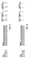
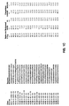
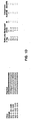
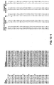
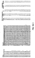
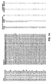
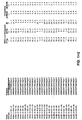
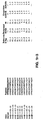
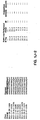
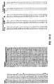
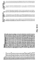
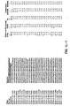
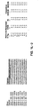
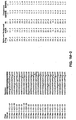
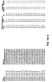
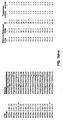
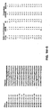
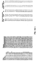
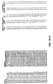
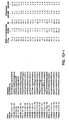
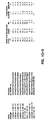
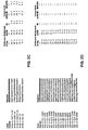
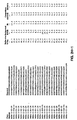
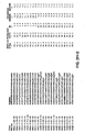
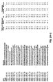
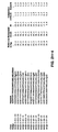
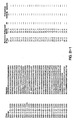
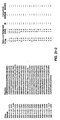
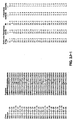
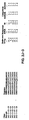
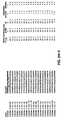
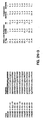
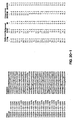
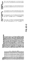
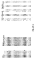
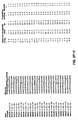
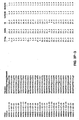
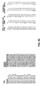
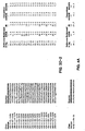
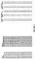
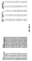
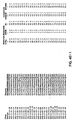
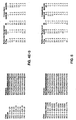
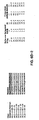
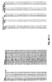
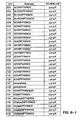
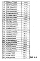
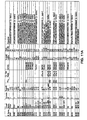
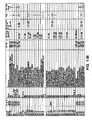
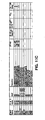
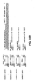
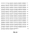
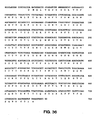
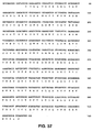
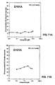
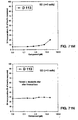
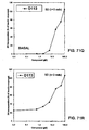
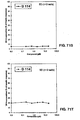
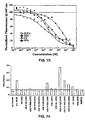
- FIGS. 1A-10G Amino acid sequences comprising the motif of Formulas 1 through 10. Sequences were identified by panning peptide libraries against IGF-1R and/or IR. The amino acids are represented by their one-letter abbreviation.
- the ratios over background are determined by dividing the signal at 405 nm (E-Tag, IGF-1R, or IR) by the signal at 405 nm for non-fat milk.
- the IGF-1R/IR Ratio Comparison is determined by dividing the ratio of IGF-1R by the ratio of IR.
- the IR/IGF-1R Ratio Comparison is determined by dividing the ratio of IR by the ratio of IGF-1 R.
- each library is shown in the first line in bold.
- symbol 'X' indicates a random position
- an underlined amino acid indicates a doped position at the nucleotide level, and other positions are held constant.
- Additional abbreviations in the B6H library are: 'O'indicates an NGY codon where Y is C or T; 'J' indicates an RHR codon where R is A or G, and H is A, C, or T; and 'U' indicates an VVY codon where V is A, C, or G, and Y is C or T.
- the 'h' in the 20E2 libraries indicates an NTN codon.
- This invention relates to amino acid sequences comprising motifs which bind to the insulin receptor (IR).
- the amino acid sequences also possess either agonist, partial agonist or antagonist activity at the receptor.
- this invention surprisingly provides amino acid sequences which define common binding motifs on IR and IGF-1 R which are capable of conferring agonist and/or antagonist activity at these receptors.
- this invention identifies multiple binding sites (Sites 1 and 2) on IR which appear to be allosterically coupled.
- amino acid sequences are neither based on insulin or IGF-1 native sequences, nor do they reflect an obvious homology to any such sequence.
- amino acid sequences of the invention may be peptides, polypeptides, or proteins.
- the amino acid sequences confer insulin agonist activity.
- the amino acid sequences of the invention are typically artificial, i.e. non-naturally occurring peptides or polypeptides.
- Amino acid sequences useful in the invention may be obtained through various means such as chemical synthesis, phage display, cleavage of proteins or polypeptides into fragments, or by any means which amino acid sequences of sufficient length to possess binding ability may be made or obtained.
- the amino acid sequences provided by this invention should have an affinity for IR sufficient to provide adequate binding for the intended purpose.
- the peptide, polypeptide or protein provided by this invention should have an affinity (K d ) of between about 10 -7 to about 10 -15 M. More preferably the affinity is 10 -8 to about 10 -12 M. Most preferably, the affinity is 10 -9 to about 10 -11 M.
- the amino acid sequence preferably has affinity for the receptor of between about 10 -5 to about 10 -12 M.
- a further consideration in identifying peptides provided by this invention for use as therapeutics is the relative activity at either IR or IGF-IR.
- a peptide which has efficacy at IR and clinically insignificant activity of IGF-IR may be a useful therapeutic even though such a peptide may bind IGF-IR with relatively high affinity.
- binding motifs At least ten different binding motifs have been identified which bind to active sites on IR; at least four of these also bind to IGF-1 R.
- the binding motifs are defined based on the analysis of several different amino acid sequences and analyzing the frequency that particular amino acids or types of amino acids occur at a particular position of the amino acid sequence.
- amino acids possessing alcohol groups are serine (S) and threonine (T).
- Aliphatic amino acids are isoleucine (I), leucine (L), valine (V), and methionine (M).
- Aromatic amino acids are phenylalanine (F), histidine (H), tryptophan (W), and tyrosine (Y).
- Hydrophobic amino acids are alanine (A), cysteine (C), phenylalanine (F), glycine (G), histidine (H), isoleucine (I), lysine (L), methionine (M), arginine (R), threonine (T), valine (V), tryptophan (W), and tyrosine (Y).
- Negative amino acids are aspartic acid (D) and glutamic acid (E).
- the following amino acids are polar amino acids: cysteine (C), aspartic acid (D), glutamic acid (E), histidine (H), lysine (K), asparagine (N), glutamine (Q), arginine (R), serine (S), and threonine (T). Positive amino acids are histidine (H), lysine (K), and arginine (R).
- Small amino acids are alanine (A), cysteine (C), aspartic acid (D), glycine (G), asparagine (N), proline (P), serine (S), threonine (T), and valine (V). Very small amino acids are alanine (A), glycine (G) and serine (S).
- Amino acids likely to be involved in a turn formation are alanine (A), cysteine (C), aspartic acid (D), glutamic acid (E), glycine (G), histidine (H), lysine (K), asparagine (N), glutamine (Q), arginine (R), serine (S), proline (P), and threonine (T).
- amino acid sequences containing substitutions, additions, or deletions based on the teachings disclosed herein might bind to IR with the same or altered affinity.
- amino acid residues located at the carboxy and amino terminal regions of the consensus motifs described below which amino acid residues are not associated with a strong preference for a particular amino acid, could be deleted providing for truncated sequences.
- Certain amino acids such as lysine which promote the stability of the amino acids sequences may be deleted depending on the use of the sequence, as for example, expression of the sequence as part of a larger sequence which is soluble, or linked to a solid support.
- motifs In addition to the motifs stated above, preferred sequences at the amino terminal or carboxyl terminal ends are capable of enhancing binding of the motifs to either IR, IGF-1R, or both.
- the use of the extensions described below does not preclude the possible use of the motifs with other substitutions, additions or deletions which allow for binding to IR, IGF-1R or both.
- Any amino acid sequence may be used for extensions of the amino terminal end of A6, although certain amino acids in amino terminal extensions may be identified which modulate activity.
- Preferred carboxy terminal extensions for A6 are A6 X 93 X 94 X 95 X 96 X 97 wherein X 93 may be any amino acid, but is preferably selected from the group consisting of alanine, valine, aspartic acid, glutamic acid, and arginine, and X 94 and X 97 are any amino acid; X 95 is preferably glutamine, glutamic acid, alanine or lysine but most preferably glutamine. The presence of glutamic acid at X 95 however may confer some IR selectivity.
- X 95 is preferably a hydrophobic or aliphatic amino acid, more preferably leucine, isoleucine, valine, or tryptophan but most preferably leucine. Hydrophobic residues, especially tryptophan at X 96 may be used to enhance IR selectivity.
- B6 with amino terminal and carboxy terminal extensions may be represented as X 98 X 99 B6 X 100 .
- X 98 is optionally aspartic acid and X 99 is independently an amino acid selected from the group consisting of glycine, glutamine, and proline.
- the presence of an aspartic acid at X 98 and a proline at X 99 is associated with an enhancement of binding for both IR and IGF-1R.
- a hydrophobic amino acid is preferred for the amino acid at X 100 , an aliphatic amino acid is more preferred.
- Negatively charged amino acids are preferred at both the amino and carboxy terminals of Formula 2A.
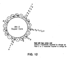
- B6 and reverse B6 motifs participate in alpha helix formation such that the most highly preferred residues at positions X 6 , X 7 , X 10 and X 13 (B6) and X 14 X 17 X 20 and X 21 (rB6) reside on the same side of a helix. See Figure 12 . Because both B6 and RB6 motifs form structurally analogous motifs from their palindrome sequences, the use of D-amino acids instead of typical L-amino acids would be expected to produce amino acid sequences having similar properties to the L-amino acid sequences.
- D-amino acids may be advantageous, as the resultant sequences may be more resistant to enzymatic degradation than L-amino acid sequences.
- the second and third amino acids of B6 (X 7 and X 8 ) are oriented at opposite sides of the helix. See Figure 12 .
- the amino acid sequences containing the motifs of this invention may be constructed to have enhanced selectivity for either IR or IGF-1R by choosing appropriate amino acids at specific positions of the motifs or the regions flanking them.
- this invention provides the means for constructing amino acid sequences with minimized activity at the non-cognate receptor.
- the amino acid sequences disclosed herein with high affinity and activity for IR and low affinity and activity for IGF-1R are desirable as IR agonist as their propensity to promote undesirable cell proliferation, an activity of IGF-1 agonists, is reduced. Ratios of IR binding affinity to IGF-1R binding affinity for specific sequences are provided in Figures 1A-10I .
- the IR/IGF-1R binding affinity ratio is preferably greater than 100. Conversely, for use as an IGF-1R therapeutic, the IR/IGF-1R ratio should be less than 0.01. Examples of peptides that selectively bind to IGF-1 R are shown below.
- MOTIF 1 Ratios over Background Comparisons Clone Sequence E-Tag IGF-1R IR IGF-1R/IR IR/IGF-1R A6L-0-E6-IR YRGMLVLGRSSDGAGKVAFERPARIGQTVFAVNFYDWFV 31.0 31.0 1.8 17.0 0.1 H2CA-4-G9-IGFR GIISQSCPESFYDWFAGQVSDPWWCW 8.6 9.5 0.6 16.0 0.1 H2CA-4-H6-IGFR VGRASGFPENFYDWFGRQLSLQSGEQ 4.9 10.5 0.7 14.6 0.1 A6L-0-E4-IR YRGMLVLGRISDGAG#VASEPPARIGRKVFAVNFYDWFV 26.0 16.0 1.3 13.0 0.1 A6L-0-H3-IR YRGMLVLGRISGGAGKAASERPARIGQKVSAVNFYDWFV 27.0 26.0 2.0 13.0 0.1 H2CA-4-F5-IGFR
- relative efficacy at the cognate receptor is another important consideration for choosing a potential therapeutic.
- a sequence which is efficacious at IR but has little or no significant activity at IGF-1R may also be considered as an important IR therapeutic, irrespective of the relative binding affinities at IR and IGF-1R.
- A6 selectivity for IR may be enhanced by including glutamic acid in a carboxyl terminal extension at position X 95 .
- IR selectivity of the B6 motif may be enhanced by having a tryptophan or phenylalanine at X 11 . Tryptophan at X 13 also favors selectivity of IR.
- a tryptophan amino acid at X 13 rather than leucine at that position also may be used to enhance selectivity for IR.
- a large amino acid at X 15 favors IR selectivity.
- small amino acids may confer specificity for IGF-1R.
- an L in position X 23 is essentially required for IR binding.
- tryptophan at X 31 is also highly preferred.
- glycine is preferred for IR selectivity.
- the competition data disclosed herein reveals that at least two separate binding sites are present on IR and IGF-1 R which recognize the different sequence motifs provided by this invention.
- competition data indicates that peptides comprising the A6, B6, revB6, and F2 motifs compete for binding to the same site on IR (Site 1) whereas the F8 and D8 motifs compete for a second site (Site 2).
- the decrease of dissociation of B6 motif peptide (20E2) from IGF-1 R by a D8 ligand indicates multiple interacting binding sites.
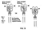
- IR and IGF-1 R The identification of peptides which bind to separate binding sites on IR and IGF-1 R provides for various schemes of binding to IR or IGF-1 R to increase or decrease its activity. Examples of such schemes for IR are illustrated in Figure 15 .
- F8-derived (Long C-C loop): Clone Sequence F8 HLCVLEELFWGASLFGYCSG F8-C12 FQSLLEELVWGAPLFRYGTG F8-Des2 PLCVLEELFWGASLFGYCSG F8-F12 PLCVLEELFWGASLFGQCSG F8-B9 HLCVLEELFWGASLFGQCSG F8-B12 DLRVLCELFGGAYVLGYCSE NNKH-2B3 HRSVLKQLSWGASLFGQWAG NNKH-2F9- HLSVGEELSWWVALLGQWAR NNKH-4H4- APVSTEELRWGALLFGQWAG D8-derived (Small C-C loop): Clone Sequence D8 KWLDQEWAWVQCEVYGRGCPSKK D8-G1 QLEEWAGVQCEVYGRECPS DB-BS ⁇ ALEEEWAWVQVRSIRSGLPL D8-A7 SLDQEWAWVQC
- This disclosure provides ligands which preferentially bind different sites on IR and IGF-1R.
- the amino acid motifs which bind IR at one site are A6, B6, revB6, and F2.
- a second in site (Site 2, Figure 13 ) binds F8 and D8.
- multimeric ligands may be prepared according to the invention by covalently linking amino acid sequences.
- amino acid sequences which bind the same or different sites may be combined to form a single molecule.
- the amino acid sequences of the ligand for binding to the receptors may be the same or different, provided that if different amino acid sequences are used, they both bind to the same site.
- Multivalent ligands may be prepared by either expressing amino acid sequences which bind to the individual sites separately and then covalently linking them together, or by expressing the multivalent ligand as a single amino acid sequence which comprises within it the combination of specific amino acid sequences for binding.
- Various combinations of amino acid sequences may be combined to produce multivalent ligands having specific desirable properties.
- agonists may be combined with agonists, antagonists combined with antagonists, and agonists combined with antagonists.
- Combining amino acid sequences which bind to the same site to form a multivalent ligand may be useful to produce molecules which are capable of cross-linking together multiple receptor units.
- Multivalent ligands may also be constructed to combine amino acid sequences which bind to different sites ( Figure 15 ).
- preparation of multivalent ligands may be useful to prepare ligands having more desirable pharmacokinetic properties due to the presence of multiple bind sites on a single molecule.
- combining amino acid sequences which bind to different sites with different affinities provides a means for modulating the overall potency and affinity of the ligand for IR or IGF-1R.
- hybrids of at least two peptides may be produced as recombinant fusion polypeptides which are expressed in any suitable expression system.
- the polypeptides may bind the receptor as either fusion constructs containing amino acid sequences besides the ligand binding sequences or as cleaved proteins from which signal sequences or other sequences unrelated to ligand binding are removed. Sequences for facilitating purification of the fusion protein may also be expressed as part of the construct. Such sequences optionally may be subsequently removed to produce the mature binding ligand.
- Recombinant expression also provides means for producing large quantities of ligand.
- recombinant expression may be used to express different combinations of amino acid sequences and to vary the orientation of their combination, i.e., amino to carboxyl terminal orientation.
- the various amino acid sequences may be coupled through linkers of various lengths.
- linkers for connecting the two amino acid sequences would typically range from about 3 to about 12 amino acids corresponding to from about 12 to about 48 A. Accordingly, the preferred distance between binding sequences is from about 2 to about 50 A. More preferred is 4 to about 40.
- the degree of flexibility of the linker between the amino acid sequences may be modulated by the choice of amino acids used to construct the linker. The combination of glycine and serine is useful for producing a flexible, relatively unrestrictive linker. A more rigid linker may be constructed by using amino acids with more complex side chains within the linkage sequence.
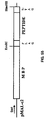
- MBP-FLAG-PEPTIDE-(G,S)n-PEPTIDE-E-TAG a fusion construct producing a dipeptide comprises a maltose binding protein amino acid sequence (MBP) or similar sequence useful for enabling the affinity chromatography purification of the expressed peptide sequences.
- MBP maltose binding protein amino acid sequence
- This purification facilitating sequence may then be attached to a flag sequence to provide a cleavage site to remove the initial sequence.
- the peptide dimer then follows which includes the intervening linker and a tag sequence may be included at the carboxyl terminal portion to facilitate identification/purification of the expression of peptide.
- G and S are glycine and serine residues, which make up the linker sequence.
- dimers may also be produced by peptide synthesis whereby a synthetic technique such as Merrifield synthesis (Merrifield, 1997), may be used to construct the entire peptide.
- dimers include introducing a linker molecule which activates the terminal end of a peptide so that it can covalently bind to a second peptide.
- linkers include diaminoproprionic acid activated with an oxyamino function.
- Linkers may be used to link dimers either to the carboxyl terminal or the amino terminal.
- the concentrations of these fusions vary depending on the expression quality. There are 2 sets of each fusion: uncleaved (-) and cleaved with factor Xa (+).
- the fusion proteins are in Tris buffer (20 mM Tris, 200 mM NaCl, 1 mM EDTA, 50 mM maltose, pH 7.5) and the cleaved fusions (+) are in the same Tris buffer (500 ⁇ l) + 12 ⁇ g Factor Xa. (Source of Factor Xa: New England Biolabs).
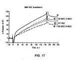
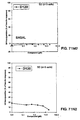
- a linker of only 3 amino acids (12 A) provided a ligand of greater affinity for Site 1 of IR than a corresponding ligand prepared with a 9 amino acid (36 A) linking region.
- Figure 17 a linker of only 3 amino acids (12 A) provided a ligand of greater affinity for Site 1 of IR than a corresponding ligand prepared with a 9 amino acid (36 A) linking region.
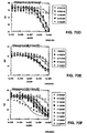
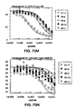
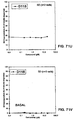
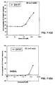
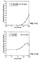
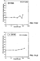
- fusion peptides show IR agonist activity as determined by an IR autophosphorylation assay (see Example 20).
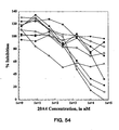
- Figure 74 fusion peptides 439, 436, 449, and 463 show significant IR agonist activity ( Figure 74 ).
- the corresponding DNA sequences may be cloned into any suitable vectors for expression in intact host cells or in cell-free translation systems by methods well known in the art (see Sambrook et al., 1989).
- the particular choice of the vector, host, or translation system is not critical to the practice of the invention.
- Cloning vectors for the expression of recombinant peptides include, but are not limited to, pUC, pBluescript (Stratagene, La Jolla, CA), pET (Novagen, Inc., Madison, WI), pMAL (New England Biolabs, Beverly, MA), or pREP (Invitrogen Corp., San Diego, CA) vectors.
- Vectors can contain one or more replication and inheritance systems for cloning or expression, one or more markers for selection in the host (e.g. antibiotic resistance), and one or more expression cassettes.
- the inserted coding sequences can be synthesized by standard methods, isolated from natural sources, or prepared as hybrids, etc.
- Ligation of the coding sequences to transcriptional regulatory elements and/or to other amino acid coding sequences can be carried out using established methods.
- DNA sequences can be optimized, if desired, for more efficient expression in a given host organism.
- codons can be altered to conform to the preferred codon usage in a given host cell or cell-free translation system using techniques routinely practiced in the art.
- Suitable cell-free systems for expressing recombinant peptides include, for example, rabbit reticulocyte lysate, wheat germ extract, canine pancreatic microsomal membranes, Escherichia coli ( E. coli ) S30 extract, and coupled transcription/translation systems (Promega Corp., Madison, WI). Such systems allow expression of recombinant polypeptides upon the addition of cloning vectors, DNA fragments, or RNA sequences containing coding regions and appropriate promoter elements.
- Host cells for cloning vectors include bacterial, archebacterial, fungal, plant, insect and animal cells, especially mammalian cells.
- mammalian cells Of particular interest are E. coli, Bacillus subtilis, Staphylococcus aureus, Saccharomyces cerevisiae, Schizosaccharomyces pombe, Neurospora crassa, SF9, C129, 293, NIH 3T3, CHO, COS, and HeLa cells.
- These cells can be transformed, transfected, or transduced, as appropriate, by any suitable method including electroporation, CaCl 2 -, LiCl-, LiAc/PEG-, spheroplasting-, Ca-Phosphate, DEAE-dextran, liposome-mediated DNA uptake, injection, microinjection, microprojectile bombardment, or other established methods.
- tags include c-myc, haemagglutinin (HA), polyhistidine (6X-HIS), GLU-GLU, and DYKDDDDK (FLAG®) epitope tags.
- Epitope tags can be added to peptides by a number of established methods. DNA sequences of epitope tags can be inserted into peptide coding sequences as oligonucleotides or through primers used in PCR amplification.
- peptide coding sequences can be cloned into specific vectors that create fusions with epitope tags; for example, pRSET vectors (Invitrogen Corp., San Diego, CA).
- the expressed, tagged peptides can then be purified from a crude lysate of the cell-free translation system or host cell by chromatography on an appropriate solid-phase matrix.
- peptides from natural sources such as cellular or extracellular lysates are well known in the art (see Harris and Angal, 1989). Such methods include, without limitation, sodium dodecylsulfate-polyacrylamide gel electrophoresis (SDS-PAGE), preparative disc-gel electrophoresis, isoelectric focusing, high-performance liquid chromatography (HPLC), reversed-phase HPLC, gel filtration, ion exchange and partition chromatography, countercurrent distribution, and combinations thereof.
- Naturally occurring peptides can be purified from many possible sources, for example, plasma, body tissues, or body fluid lysates derived from human or animal, including mammalian, bird, fish, and insect sources.
- Antibody-based methods may also be used to purify naturally occurring or recombinantly produced peptides. Antibodies that recognize these peptides or fragments derived therefrom can be produced and isolated. The peptide can then be purified from a crude lysate by chromatography on an antibody-conjugated solid-phase matrix (see Harlow and Lane, 1998). '
- peptides may be chemically synthesized by commercially available automated procedures, including, without limitation, exclusive solid phase synthesis, partial solid phase methods, fragment condensation or classical solution synthesis.
- the polypeptides are preferably prepared by solid-phase peptide synthesis; for example, as described by Merrifield (1965; 1997).
- recombinant and synthetic methods of polypeptide production can be combined to produce semi-synthetic polypeptides.
- screening assays to identify pharmacologically active ligands at IR and/or IGF-1R are provided.
- the screening assays provided in accordance with this invention are based on those disclosed in International application WO 96/04557 which is incorporated herein in its entirety. Briefly, WO 96/04557 discloses the use of reporter peptides which bind to active sites on targets and possess agonist or antagonist activity at the target. These reporters are identified from recombinant libraries and are either peptides with random amino acid sequences or variable antibody regions with at least one CDR region which has been randomized (rVab). The reporter peptides may be expressed in cell recombinant expression systems, such as for example in E .
- the reporters identified from the libraries may then be used in accordance with this invention either as therapeutics themselves, or in competition binding assays to screen for other molecules, preferably small, active molecules, which possess similar properties to the reporters and may be developed as drug candidates to provide agonist or antagonist activity.
- these small organic molecules are orally active.
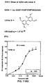
- the basic format of an in vitro competitive receptor binding assay as the basis of a heterogeneous screen for small organic molecular replacements for insulin may be as follows: occupation of the active site of IR is quantified by time-resolved fluorometric detection (TRFD) with streptavidin-labeled europium (saEu) complexed to biotinylated peptides (bP). In this assay, saEu forms a ternary complex with bP and IR (i.e., IR:bP:saEu complex).
- TRFD assay format is well established, sensitive, and quantitative (Tompkins et al., 1993).
- the assay can use a single-chain antibody or a biotinylated peptide. Furthermore, both assay formats faithfully report the competition of the biotinylated ligands binding to the active site of IR by insulin.
- soluble IR is coated on the surface of microtiter wells, blocked by a solution of 0.5% BSA and 2% non-fat milk in PBS, and then incubated with biotinylated peptide or rVab. Unbound bP is then washed away and saEu is added to complex with receptor-bound bP. Upon addition of the acidic enhancement solution, the bound europium is released as free Eu 3+ which rapidly forms a highly fluorescent and stable complex with components of the enhancement solution. The IR:bP bound saEu is then converted into its highly fluorescent state and detected by a detector such as Wallac Victor II (EG&G Wallac, Inc.)
- the designing of mimetics to a known pharmaceutically active compound is a known approach to the development of pharmaceuticals based on a "lead" compound. This might be desirable where the active compound is difficult or expensive to synthesize or where it is unsuitable for a particular method of administration, e.g. peptides are generally unsuitable active agents for oral compositions as they tend to be quickly degraded by proteases in the alimentary canal.
- Mimetic design, synthesis and testing is generally used to avoid randomly screening large number of molecules for a target property.
- the pharmacophore Once the pharmacophore has been found, its structure is modeled according to its physical properties (e.g. stereochemistry, bonding, size and/or charge), using data from a range of sources (e.g. spectroscopic techniques, X-ray diffraction data and NMR). Computational analysis, similarity mapping (which modes the charge and/or volume of a pharmacophore, rather than the bonding between atoms), and other techniques can be used in this modeling process.
- physical properties e.g. stereochemistry, bonding, size and/or charge
- sources e.g. spectroscopic techniques, X-ray diffraction data and NMR.
- Computational analysis, similarity mapping which modes the charge and/or volume of a pharmacophore, rather than the bonding between atoms
- other techniques can be used in this modeling process.
- the three dimensional structure of the ligand and its binding partner are modeled. This can be especially useful where the ligand and/or binding partner change conformation on binding, allowing the model to take account of this in the design of the mimetic.
- a template molecule is then selected onto which chemical groups which mimic the pharmacophore can be grafted.
- the template molecule and the chemical groups grafted on to it can conveniently be selected so that the mimetic is easy to synthesize, is likely to be pharmacologically acceptable, does not degrade in vivo, and retains the biological activity of the lead compound.
- the mimetics found are then screened to ascertain the extent they exhibit the target property, or to what extent they inhibit it. Further optimization or modification can then be carried out to arrive at one or more final mimetics for in vivo or clinical testing.
- This invention provides specific amino acid sequences which function as agonists or antagonists at IR and/or IGF-1R.
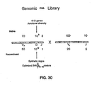
- phage display libraries suitable for use in this invention include one such library containing randomized 40 amino acid peptides (RAPIDLIBTM, Figure 16 ), another library containing rVab derived from human genomic antibody DNA (GRABLIBTM, Figure 30 ).
- the peptides provided by this invention are useful as potential therapeutics in pharmaceutical compositions, lead compounds for identifying other more potent or selective therapeutics, assay reagents for identifying other useful ligands by, for example, competition screening assays, and as research tools for further analysis of IR and IGF-1R.
- the peptide sequences provided by this invention can be used to design secondary peptide libraries, which include members that bind to Site 1 and/or Site 2 of IR or IGF-1 R. Such libraries can be used to identify sequence variants that increase or modulate the binding and/or activity of the original peptide at IR or IGF-1R.
- IR agonist amino acid sequences provided by this invention are useful as insulin analogs and may therefore be developed as treatments for diabetes or other diseases associated with a decreased response or production of insulin.
- preferred amino acid sequence are: FHENFYDWFVRQVSK (D117, H2C), DYKDFYDAIQLVRSARAGGTRDKK (D118, 20E2), KDRAFYNGLRDLVGAVYGAWDKK (D119, 20C11), DYKDLCQSWGVRIGWLAGLCPKK (D116, JBA5), DYKDVTFTSAVFHENFYDWFVRQVSKK (D113, H2), and GRVDWLQRNANFYDWFVAELG (S175).
- More preferred IR agonists are: FHENFYDWFVRQVSK (D117, H2C) and GRVDWLQRNANFYDWFVAELG (S175). Most preferred is GRVDWLQRNANFYDWFVAELG (S175).
- Preferred dimer sequences are represented by S170, S171, S172, S232, S300 sequences (see Table 15).
- IGF-1 R antagonist amino acid sequences are useful as treatments for cancers, including, but not limited to, breast and prostate cancers. Human and breast cancers are responsible for over 40,000 deaths per year, as present treatments such as surgery, chemotherapy, radiation therapy, and immunotherapy show limited success.
- the IGF-1R antagonist amino acid sequences disclosed herein are also useful for the treatment or prevention of diabetic retinopathy. Recent reports have shown that a previously identified IGF-1R antagonist can suppress retinal neovascularization, which causes diabetic retinopathy (Smith et al., 1999).
- IGF-1R agonist amino acid sequences are useful for development as treatments for neurological disorders, including stroke and diabetic neuropathy. Reports of several different groups implicate IGF-1 R in the reduction of global brain ischemia, and support the use of IGF-1 for the treatment of diabetic neuropathy (reviewed in Auer et al ., 1998; Apfel, 1999).
- the amino acid sequences of this invention may be administered as pharmaceutical compositions comprising standard carriers known in the art for delivering proteins and peptides and by gene therapy. Due to the labile nature of the amino acid sequences parenteral administration is preferred. Preferred modes of administration include aerosols for nasal or bronchial absorption; suspensions for intravenous, intramuscular, intrasternal or subcutaneous, injection; and compounds for oral administration. Other modes of administration and examples of suitable formulative components for use with this embodiment are discussed below. Other modes of administration include intranasal, intrathecal, intracutaneous, percutaneous, enteral, and sublingual.
- the composition is in sterile solution or suspension or may be emulsified in pharmaceutically- and physiologically-acceptable aqueous or oleaginous vehicles, which may contain preservatives, stabilizers, and material for rendering the solution or suspension isotonic with body fluids (i.e. blood) of the recipient.
- Excipients suitable for use are water, phosphate buffered saline, pH 7.4, 0.15 M aqueous sodium chloride solution, dextrose, glycerol, dilute ethanol, and the like, and mixtures thereof.
- Illustrative stabilizers are polyethylene glycol, proteins, saccharides, amino acids, inorganic acids, and organic acids, which may be used either on their own or as admixtures.
- the amounts or quantities, as well as routes of administration, used are determined on an individual basis, and correspond to the amounts used in similar types of applications or indications known to those of skill in the art.
- constructs as described herein may also be used in gene transfer and gene therapy methods to allow the expression of one or more amino acid sequences of the present invention.
- Using the amino acid sequences of the present invention for gene therapy may provide an alternative method of treating diabetes which does not rely on the administration or expression of insulin. Expressing insulin for use in gene therapy requires the expression of a precursor product, which must then undergo processing including cleavage and disulfide bond formation to form the active product.
- the amino acid sequences of this invention, which possess activity, are relatively small, and thus do not require the complex processing steps to become active. Accordingly, these sequences provide a more suitable product for gene therapy.
- Gene transfer systems known in the art may be useful in the practice of the invention. Both viral and non-viral methods are suitable. Examples of such transfer systems include, but are not limited to, delivery via liposomes or via viruses, such as adeno-associated or vaccinia virus. Numerous viruses have been used as gene transfer vectors, including papovaviruses (e.g., SV40, adenovirus, vaccinia virus, adeno-associated virus, herpes viruses, including HSV and EBV, and retroviruses of avian, murine, and human origin). As is appreciated by those in the art, most human gene therapy protocols have been based on disabled murine retroviruses. Recombinant retroviral DNA can also be employed with amphotrophic packaging cell lines capable of producing high titer stocks of helper-free recombinant retroviruses (e.g., Cone and Mulligan, 1984).
- papovaviruses e.g., SV40, adenovirus, vac
- a recombinant retroviral vector may contain the following parts: an intact 5' LTR from an appropriate retrovirus, such as MMTV, followed by DNA containing the retroviral packaging signal sequence; the insulator element placed between an enhancer and the promoter of a transcription unit containing the gene to be introduced into a specific cell for replacement gene therapy; a selectable gene as described below; and a 3' LTR which contains a deletion in the viral enhancer region, or deletions in both the viral enhancer and promoter regions.
- the selectable gene may or may not have a 5' promoter that is active in the packaging cell line, as well as in the transfected cell.
- the recombinant retroviral vector DNA can be transfected into the amphotrophic packaging cell line ⁇ -AM (see Cone and Mulligan, 1984) or other packaging cell lines which are capable of producing high titer stocks of helper-free recombinant retroviruses. After transfection, the packaging cell line is selected for resistance to G418, present at appropriate concentration in the growth medium.
- Adenoviral vectors e.g. DNA virus vectors
- replication-defective adenovirus vectors particularly replication-defective adenovirus vectors, or adeno-associated vectors, have been described in the art (Kochanek et al., 1996; Ascadi et al., 1994; Ali et al ., 1994).
- Nonviral gene transfer methods known in the art include chemical techniques, such as calcium phosphate co-precipitation, direct DNA uptake and receptor-mediated DNA transfer, and mechanical means, such as microinjection and membrane fusion-mediated liposomal transfer.
- viral-mediated gene transfer can be combined with direct in vivo gene transfer using liposomes, thereby allowing the delivery or the viral vectors to tumor cells, for example, and not to surrounding non-proliferating cells.
- a description of various liposomes which are stated as being useful for transferring DNA or RNA into cells is present in United States Patents 5,283,185 and 5,795,587 .
- the retroviral vector producer cell line can also be injected directly into specific cell types, e.g., tumors, to provide a continuous source of viral particles, such as has been approved for use in patients afflicted with inoperable brain tumors.
- Receptor-mediated gene transfer methods allow targeting of the DNA in the construct directly to particular tissues. This is accomplished by the conjugation of DNA (frequently in the form of a covalently closed supercoiled plasmid) to a protein ligand via polylysine.
- DNA frequently in the form of a covalently closed supercoiled plasmid
- polylysine The appropriate or suitable ligands are selected on the basis of the presence of the corresponding ligand receptors on the cell surface of the target cell or tissue type.
- These ligand-DNA conjugates can be injected directly into the blood, if desired, and are directed to the target tissue where receptor binding and DNA-protein complex internalization occur. Co-infection with adenovirus to disrupt endosome function can be used to overcome the problem of intracellular destruction of DNA.
- An approach that combines biological and physical gene transfer methods utilizes plasmid DNA of any size combined with a polylysine-conjugated antibody specifically reactive with the adenovirus hexon protein.
- the resulting complex is bound to an adenovirus vector.
- the trimolecular complex is then used to infect cells.
- the adenovirus vector allows efficient binding to the cell, internalization, and degradation of the endosome before the coupled DNA can be damaged.
- cells and cell lines e.g. primary cell lines or established cell lines
- tissues are capable of being stably transfected by or receiving the constructs of the invention.
- cells include, but are not limited to, stem cells, B lymphocytes, T lymphocytes, macrophages, other white blood lymphocytes (e.g. myelocytes, macrophages, monocytes), immune system cells of different developmental stages, erythroid lineage cells, pancreatic cells, lung cells, muscle cells, liver cells, fat cells, neuronal cells, glial cells, other brain cells, transformed cells of various cell lineages corresponding to normal cell counterparts (e.g.
- constructs of the present invention may be transferred by various means directly into tissues, where they would stably integrate into the cells comprising the tissues.
- constructs containing the DNA sequences of the peptides of the invention can be introduced into primary cells at various stages of development, including the embryonic and fetal stages, so as to effect gene therapy at early stages of development.
- the described constructs may be administered in the form of a pharmaceutical preparation or composition containing a pharmaceutically acceptable carrier and a physiological excipient, in which preparation the vector may be a viral vector construct, or the like, to target the cells, tissues, or organs of the recipient organism of interest, including human and non-human mammals.
- the composition may be formed by dispersing the components in a suitable pharmaceutically acceptable liquid or solution such as sterile physiological saline or other injectable aqueous liquids.
- the amounts of the components to be used in such compositions may be routinely determined by those having skill in the art.
- the compositions may be administered by parenteral routes of injection, including subcutaneous, intravenous, intramuscular, and intrasternal.
- Soluble IGF-1R was obtained from R&D Systems (Cat. # 391-GR/CF). Insulin receptor was prepared according to Bass et al., 1996. The insulin is either from Sigma (Cat. # I-0259) or Boehringer. The IGF-1 is from PeproTech (Cat. # 100-11). All synthetic peptides were synthesized by Novo Nordisk, AnaSpec, Inc. (San Jose, CA), PeptioGenics (Livermore, CA), or Research Genetics (Huntsville, AL) at >80% purity. The Maxisorb Plates are from Nunc via Fisher (Cat. # 12565347). The HRP/Anti-M13 Conjugate is from Pharmacia (Cat. # 27-9421-01). The ABTS solution is from BioF/X (Cat. # ABTS-0100-04).
- the schematic for the peptide library "RAPIDLIB TM " on filamentous phage is shown in Figure 16 .
- DNA fragments coding for peptides containing 40 random amino acids were generated in the following manner.
- N A, C, T, or G
- K G or T.
- This oligonucleotide was used as the template in a PCR amplification along with two shorter oligonucleotide primers, both of which were biotinylated at their 5' ends.
- the resulting 190 bp product was purified and concentrated with QIAquick spin columns (QIAGEN, Inc.
- the ligation product was purified using QIAquick spin columns (QIAGEN). Electroporations were performed at 1500 v in an electroporation cuvette (0.1 mm gap; 0.5 ml volume) containing 12.5 ⁇ g of DNA and 500 ⁇ l of TG1 electrocompetent cells (see below). Immediately after the pulse, 12.5 ml of pre-warmed (40°C) 2xYT medium containing 2% glucose (2xYT-G) was added and the transformants were grown at 37°C for 1 h. Cell transformants were pooled, the volume measured, and an aliquot was plated onto 2xYT-G containing 100 g/ml ampicillin (2xYT-AG) plates to determine the total number of transformants.
- QIAquick spin columns QIAquick spin columns
- Another phage library expressing 20mer peptides was constructed according to a similar procedure.
- the diversity of the library is 1.1 x 10 11 different clones.
- the culture was poured into pre-chilled centrifuge bottles and incubated on ice for 30 min prior to centrifugation at 2000 x g for 30 min (2°C).
- the supernatant was poured off and the cell pellet was resuspended in a total of 400 ml of ice cold sterile distilled water.
- the process of centrifugation and resuspension was repeated 2 times. After the last centrifugation, the pellet was resuspended in a total of 25 ml of ice cold water containing 10% glycerol.
- the cell suspension was transferred to pre-chilled 35 ml centrifuge bottles, and was then pelleted at 2000 x g for 10 min at 4°C.
- the cells were then suspended in 0.3 ml of the same 10% glycerol solution, aliquotted into smaller tubes, and snap-frozen on dry ice. The aliquots were stored at -80°C.
- the transformants were inoculated into 4 I of 2xYT-AG medium and allowed to grow until the A 600 increased approximately 400 times.
- the cells were pelleted by centrifugation at 3000 x g for 20 min, then resuspended in 40 ml 2xYT-AG to which glycerol was added to a final concentration of 8%.
- the library was stored at -80°C.
- the cell pellet was resuspended in the initial culture volume of 2xYT-A (no glucose) containing 50 mg/ml kanamycin and grown overnight at 30°C with shaking (250 rpm).
- the cells from the overnight culture were pelleted at 3000 x g for 30 min at 4°C and the supernatant containing the phage was recovered.
- the solution was adjusted to 4% PEG, 500 mM NaCl and chilled on ice for 1 h.
- the precipitated phage were pelleted by centrifugation at 10,000 x g for 30 min, then resuspended in phosphate-buffered saline (1/100 of the initial culture volume) and passed through a 0.45 ⁇ m filter.
- the phage were titered by infecting TG1 cells.
- the phage titer for the 40mer peptide library was 4 x 10 13 cfu/ml.
- the phage titer for the 20mer library was 3 x 10 -3 .
- the transformants were inoculated into 4 I of 2xYT-AG medium and allowed to grow until the OD 600 increased approximately 400 times.
- the cells were pelleted by centrifugation at 3000 x g for 20 min, then resuspended in 40 ml 2xYT-AG to which glycerol was added to a final concentration of 8%.
- the library was stored at -80°C.
- sIGF-1 R The soluble IGF-1R
- 50 mM sodium carbonate buffer, pH 9.5 One hundred microliters of this solution was added to an appropriate number of wells in a 96-well microtiter plate (MaxiSorp plates, Nunc) and incubated overnight at 4°C. Wells were then blocked with MPBS (PBS buffer pH 7.5 containing 2% Carnation® non-fat dry milk) at room temperature (RT) for 1 h.
- MPBS PBS buffer pH 7.5 containing 2% Carnation® non-fat dry milk
- phage pools cells from frozen stocks were grown and phage were prepared as described above. For analysis of individual clones, colonies were picked and phage prepared as described above. Subsequent steps are the same for pooled and clonal phage. Microtiter wells were coated and blocked as described above. Wells were coated with either IGF-1 R or a control IgG mAb. Phage resuspended in MPBS were added to duplicate wells (100 ⁇ l/well) and incubated at RT for 1 h. The phage solution was then removed, and the wells were washed 3 times with PBS at RT.
- Anti-M13 antibody conjugated to horseradish peroxidase (Pharmacia) was diluted 1:3000 in MPBS and added to each well (100 ⁇ l/well). Incubation was for 1 h at RT, followed by PBS washes as described. Color was developed by addition of ABTS solution (100 ⁇ l/well; Boehringer). Color development was stopped by adjusting each well to 0.5% SDS. Plates were analyzed at 405 nm using a SpectraMax 340 plate reader (Molecular Devices Corp., Sunnyvale CA) and SoftMax Pro software. Data points were averaged after subtraction of appropriate blanks. A clone was considered "positive" if the A 405 of the well was ⁇ 2-fold over background.
- IC 50 determinations in a competitive ELISA microtiter plates were coated with IGF-1R and blocked as described. Phage were prepared as described. Prior to addition of phage to plates, the peptide or recombinant variable antibody or fragment ("rVab"), or an appropriate control, was diluted in PBS and added to duplicate wells (100 ⁇ l/well). After incubation for 1 h at RT, the prepared phage were added to each well (100 ⁇ l/well) without removing the peptide or rVab solution. After incubation for 1 h at RT, the wells were washed and the color developed as described above.
- rVab recombinant variable antibody or fragment
- the clones were next analyzed for binding to the receptor's active site ( Figures 20A and 20B ). Competitions of phage binding were done with the cognate ligand (i.e., IGF-1). All four phage clones tested, B6, F6, C6 and E5, bound to same site as IGF-1 since the binding of the clones to the immobilized IGF-1R could be inhibited with IGF-1.
- IGF-1 cognate ligand
- the human IGF-1 R (25 g/ml) was immobilized onto a CM-5 (BIAcore) sensor chip using amino coupling chemistry and the manufacturer's recommended protocol. The final surface density was 1000 RU. A monoclonal antibody was immobilized onto another flow cell as a control surface. Phage were directly injected (30-100 ⁇ l) with a buffer flow rate of 1 ⁇ l/min. Background binding to the control surface was subtracted prior to further analysis.
- Class 1 (Formula motif 2) and Class II (Formula motif 1; Figure 21 ).
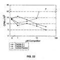
- Class I peptides contain the consensus sequence D-x-F-Y-x-x-L-s-x-L, and are shown to be antagonistic in cell-based assays ( Figure 22 ).
- Class II peptides contain the consensus N-F-Y-D-W-F-V, and are shown to be agonistic in cell-based assays ( Figure 23 ). Neither of these consensus sequences have any significant linear sequence similarities greater than 2 or 3 amino acids with mature IGF-1.
- Synthetic peptides were obtained from a commercial supplier (Anaspec). The peptides were supplied greater than 90% pure by HPLC. The molecular weights of the peptides as determined by mass spectroscopy agreed with the expected values.
- IGF-1R (100 ⁇ g/ml) was immobilized onto one flow-cell of a CM-5 sensor chip (Biosensor) using amine coupling chemistry and the manufacturer's recommended protocol.
- An unrelated IgG was immobilized in the same manner to another flow cell of the same chip as a control surface.
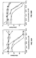
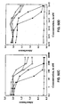
- Increasing concentrations of synthetic peptide were injected over both surfaces, and the binding responses were allowed to come to equilibrium. After subtraction of background binding from the control surface, the results were used to derive an equilibrium dissociation constant using Scatchard analysis ( Figure 24A ).
- IGF-1 R (100 ⁇ g/ml) was immobilized onto a CM-5 sensor chip as described above, and an unrelated IgG was immobilized in the same manner to another flow cell of the same chip.
- the results shown in Figure 24B indicate that the 5.1 peptide inhibits the binding of IGF-1, and the inhibition is increased by increasing amounts of the peptide.
- the results support the idea of an overlap of the peptide 5.1 binding site and the IGF-1 binding site on IGF-1 R.
- Two phage libraries were designed on the basis of the sequences of the Class II binders known to possess agonistic properties in cell-based assays. The goal was to bring the affinity into a range that would allow the peptide to be used in a receptor binding assay and tested in a cell based assay for activity.
- mutagenesis methods we chose one based on gene synthesis and phage display. In this method a library of doped oligonucleotides carrying several mutations in any single DNA molecule is used to obtain a pool of mutant genes, the expression products of which are phage displayed.
- the approach used was the doped synthesis of the oligonucleotide encoding the sequence of the peptide.
- the sequence encoding the peptide and the sequence of the synthetic oligonucleotide made are shown in Figures 25A-25B .
- the amino acid residues belonging to the consensus sequence were kept constant and were not mutated.
- the ratio of nucleosides in each condensation was chosen to provide an average of 6 nucleotide sequence changes at the DNA level and 4-5 mutations at the amino acid level over the length of the peptides.
- the regions corresponding to the FLAG, Sfi I and Not I sites were not mutated.
- the DNA sequence encoding the A6 peptide was optimized for E . coli codon usage by replacing a total of 24 nucleotides as shown in Figure 25A .
- the TAG stop codons (suppressed in the TG1 E. coli strain used) were replaced with CAG (glutamine).
- the oligonucleotide sequence was designed to include doped nucleosides at positions corresponding to the coding region for the A6 peptide, except for the consensus NFYDWFV ( Figure 25A ). This synthetic oligonucleotide ( Figure 25B ) was then used as a template in a PCR reaction.
- Peptide H5 (LCQRLGVGWPGWLSGWCA) was identified in an independent experiment as a binder to the rat growth hormone binding protein.
- This peptide and four other H5-like peptides, including 2C3-60 ( Figure 27 ), were found in cell culture experiments to possess agonistic activity toward IGF-1 R + cells, but not against IGF-1 R - cells. Further, subsequent in vitro experiments showed that the H5-like peptides are not competed by IGF. This suggests that these peptides recognize a second allosteric site on IGF-1R.
- BIAcore analysis showed that binding of the 2C3-60 peptide to IGF-1 R is ⁇ 20 ⁇ M. Subsequently, a phage library of mutants of the H5 sequence was constructed and used for panning against IGF-1R.
- H5 secondary library Gene synthesis to introduce mutations and phage display were used to construct an H5 secondary library.
- a library of doped oligonucleotides carrying several mutations in any single DNA molecule is used to obtain a pool of mutant genes which are phage displayed.
- This method allowed the encoding of both the original H5 peptide as control as well as versions containing high numbers of mutations per peptide in a very large library (>10 10 ).
- the H5 secondary mutant library was designed to contain an average of four amino acid changes (mutations) per peptide.
- the number of possible mutant H5 peptide sequences having four mutations is 1.0 x 10 10 and is equivalent to the actual size of the secondary phage library. Sequence analysis indicates that of these peptides 30% have 3-4, 33% have 1-2 and 32 % have 5-6 mutations. There also was a small percent with 7-8 mutations and 5% clones without any mutation.
- oligonucleotide based on the DNA sequence encoding the H5 peptide was synthesized.
- the sequence of the oligonucleotide is:
- Example 4 The two secondary libraries of Example 4 were used in a panning experiment against IGF-1 R. Approximately 50 clones from each four rounds of panning were analyzed in a phage ELISA to identify the clones that bind to the receptor. The positive clones were subjected to DNA sequencing and protein sequence comparison.
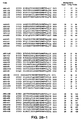
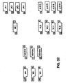
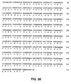
- Figure 28 provides a listing of different sequences obtained from panning with the A6S library. The results show that a variety of phage peptide sequences can bind to IGF-1 R, while the consensus sequence NFYDWFV is preserved in the majority of instances.
- the H5 secondary phage library was panned against IGF-1 R to find H5-like peptides with higher affinities for IGF-1 R
- the H5 Library has a diversity of ⁇ 2.6 x 10 10 clones with a phage titer of 1.0 x 10 13 phage ml -1 .
- a total of three rounds of panning were performed.
- Table 2 summarizes the results from the three rounds of panning and shows the ELISA results for the individual clones selected from each round, the number of clones examined in each round of panning, as well as the number and percentage of E-Tag + clones and IGF-1 R + clones.
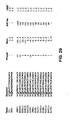
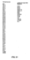
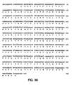
- IGF-1R + clones were sequenced, as were 15 IGF-1R - clones with high E-Tag values (Absorbance >9.0). These sequences are shown in Figure 29 . There is no discernible difference between binding sequences and the non-binding sequences with the exception that all of the binding sequences hold the Gly at position 6 constant. All sequences, binding and non-binding, hold the TAG stop codon constant at position 3 (the E . coli strain used in phage production contains the supE44 mutation, therefore Gln replaces the TAG and it denoted in Figure 29 by Q ). This suggests TAG stop codon is required for phage production and not binding.
- variable portion of the heavy chain (V H ) is linked to the variable portion of the light chain (V L ) by a flexible peptide linker. Random combinations of V H and V L genes can be genetically combined to provide some of the diversity required for a library of recombinant variable region antibodies (rVabs) ( Figure 30 ). In our library, further diversity is provided by full randomization of the 6-12 amino acids comprising the V H CDR3 (indicated as "D" in Figure 30 ).
- a total of 49 human genomic v h genes and ten human genomic v l genes ( Figure 31 ) were isolated from total human genomic DNA by PCR.
- the other genetic components of the library (V h , CDR3, j h , linker, and j l ) were derived from synthetic oligonucleotides. Assembly of these components was done using directional cloning as outlined in Figure 32 and Figure 33 .
- FIG 30 The general schematic for the assembly of the rVab library (“GRABLIB TM ”) is provided in Figure 30 .
- Four gene fragments VH, VHCDR3/JH/LINKER, VL and JL were ligated together in the proper orientation and cloned into pCANTAB 5E (Pharmacia).
- Directional cloning was achieved using the BsrDI restriction enzyme ( Figure 32 ).
- Forty-nine germline VH segments and ten VL segments encoding many of the genes from the human VH and VL repertoire were isolated ( Figure 31 ) using the polymerase chain reaction.
- VH CDR3 (ranging from 6 to 12 amino acids) /JH/Linker fragments were generated by ligation of four oligonucleotides (WM 2.1, 2.2, 2.3 and 2.4) and cloning the resulting fragment into the plasmid pUC18 previously cut with KpnI and Hind III.
- the insert was then amplified using PCR and oligonucleotide primers to introduce a synthetic D-segment of 6 to 12 amino acids having a random sequence and the BsrDl restriction site.
- the JL gene fragments were assembled as a result of annealing of two synthetic oligonucleotides.
- the assembled fragments (200 ng) were used as template in a PCR amplification along with two shorter oligonucleotide primers, both of which were biotinylated at their 5' ends.
- the resulting 800 bp product was purified and concentrated with QIAquick spin columns (QIAGEN), then digested with the Sfil and Not I restriction enzymes. Streptavidin-agarose (GibcoBRL) was added to the digestion mixture to remove the cleaved ends of the PCR product as well as any uncut DNA.
- the resulting 800 bp fragment was purified by passing DNA over QlAquick spin columns.
- Phagemid pCANTAB 5E (Pharmacia) was digested with the Sfi l and Not I restriction enzymes, which was followed by the alkaline phosphatase treatment to dephosphorylate the ends of the restriction fragments generated.
- the digested DNA was purified by running the digested plasmid DNA on a 1% agarose gel, followed by the DNA purification using the QIAEX II (QIAGEN) column.
- the vector and insert DNA were ligated overnight at 16°C.
- the ligation product was purified using QlAquick spin columns (QIAGEN) and electroporations were performed at 1500 v in a electroporation cuvette (0.1 mm gap; 0.5 ml volume, BTX, Inc.).
- the amount of DNA in one electroporation was 12.5 ⁇ g per 500 ⁇ l of TG1 electrocompetent cells.
- 12.5 ml of a pre-warmed (40°C) 2xYT medium containing 2% glucose (2xYT-G) was added, and the transformants were grown at 37°C for 1 h.
- the transformants were pooled, the volume measured, and an aliquot was plated onto the 2xYT-G medium containing 100 ⁇ g/ml ampicillin (2xYT-AG) plates to determine the total number of transformants.
- the number of different transformants and the diversity of the library was 3 x 10 10 .
- the electrocompetent cell preparation, phage library amplification, library phage rescue, phage preparations and coating of microtiter plates were done as described above for the peptide library.
- Microtiter wells were coated with IGF-1R as described above, with eight wells being used for each round of panning.
- the phage were incubated with MPBS for 30 min at RT, then 100 ⁇ l of the phage suspension was added to each well.
- the input phage titer was 8 x 10 13 cfu/ml.
- the input phage titer was approximately 10 11 cfu/ml. Phage were allowed to bind for 2 to 3 h at RT. The wells were then quickly washed 13 times with 200 ⁇ l /well of MPBS.
- Bound phage were eluted by incubation with 100 ⁇ l/well of 20 mM glycine-HCI, pH 2.2 for 30 s. The resulting solution was then neutralized with Tris-HCI, pH 8.0. Log phase TG1 cells were infected with the eluted phage, then plated onto two 4 cm x 4 cm plates containing 2XYT-AG. The plates were incubated at 30°C overnight. The next morning, cells were removed by scraping and stored in 10% glycerol at -80°C. For subsequent rounds of affinity enrichment, cells from these frozen stocks were grown and phage were prepared as described above.
- phage pools cells from frozen stocks were grown and phage were prepared as described above. Microtiter wells were coated and blocked as described above. The wells were coated with either IGF-1R (R&D Systems, Inc.) or with control BSA. Phage resuspended in MPBS were added to duplicate wells (100 ⁇ /well) and incubated at RT for 1 h. The phage solution was then removed, and the wells were washed 3 times with PBS at RT. Anti-M13 antibody conjugated to horseradish peroxidase (Pharmacia) was diluted 1:3000 in MPBS and added to each well (100 ⁇ l/well). Incubation was for 1h at RT, followed by PBS washes as described.
- IGF-1R R&D Systems, Inc.
- control BSA Phage resuspended in MPBS were added to duplicate wells (100 ⁇ /well) and incubated at RT for 1 h. The phage solution was then removed, and the wells
- microtiter plates were coated with IGF-1R and blocked as described. Phage and soluble rVabs were prepared as described above. Prior to addition of phage or soluble rVabs to the plates, IGF-1 solution in PBS (1 ⁇ g/ml) was added to duplicate wells (100 ⁇ l/well). After incubation for 1 h at RT, the prepared phage were added to each well (100 ⁇ l/well) without removing the IGF-1 solution. After incubation for 1 h at RT, the wells were washed and the color was developed as described above.
- E. coli HB2151 carrying the rVab genes on the pCANTAB5E plasmid were grown in 2xYT supplemented with 100 ⁇ g/ml ampicillin and 1% glucose at 37° C overnight and then subcultured in the absence of glucose at an OD 600 of 0.1, and grown at 21° C until OD 600 was 1.0. Expression was induced by the addition of IPTG to 1 mM and the cells were grown for 16 h at 30° C.
- the cells and culture supernatant were separated by centrifugation and samples of the cell pellet and supernatant were analyzed on a 15% SDS-PAGE gel followed by the Western blot analysis using the mouse monoclonal antibody anti-E-Tag-HRP conjugate (Pharmacia) to visualize the expressed product.
- the expressed rVabs were purified from the supernatant by precipitation with ammonium sulphate (which was added to 70% saturation) at 21° C, followed by centrifugation at 10,000 g for 15 min. The aqueous phase was discarded, and the pellet resuspended and dialyzed in PBS (phosphate buffered saline, pH 7.4) at 4° C overnight.
- PBS phosphate buffered saline, pH 7.4
- Insoluble material was removed by centrifugation at 10,000 g, and the supernatant was filtered through a 0.22 ⁇ m membrane and purified on an anti-E-Tag antibody affinity column (Pharmacia).
- the affinity resin was equilibrated in TBS (0.025 M Tris-buffered saline, pH 7.4) and the bound protein was eluted with the Elution buffer (100 mM glycine, pH 3.0).
- the rVab was concentrated to 1 mg/ml, dialyzed against TBS and stored at 4° C.
- the SDS-PAGE, Western blot analysis and N-terminal sequence analysis of the affinity purified material were performed according to standard protocols.
- the affinity purified rVabs were fractionated by size exclusion FPLC on a Superdex 75 HR10/30 column (Pharmacia) to determine the molecular size and aggregation state of the rVabs.
- High and Low Molecular Weight Gel Filtration Calibration Kits (Pharmacia) were used. Fractions from several chromatographic separations corresponding to a molecular weight of 30 kDa were pooled and concentrated to 0.7-1.0 mg/ml using Amicon XM10 membranes. Protein concentrations were determined using the BCA protein assay kit (Pierce Chemical Co., Rockford, IL).
- IGF-1 R was immobilized onto one flow cell of a CM-5 sensor chip (Biosensor) using amine coupling chemistry and the manufacturer's recommended protocols.
- BSA was immobilized in the same manner to another flow cell of the same chip as a control surface.
- Increasing concentrations of the affinity-purified rVabs were injected over both surfaces, and the binding responses were allowed to come to equilibrium. After a subtraction of the background binding (from the control surface), the equilibrium dissociation constant was derived using Scatchard analysis.
- TRFD time-resolved fluorometric detection
- saEu streptavidin-labeled europium
- bP biotinylated peptides
- saEu forms a ternary complex with bP and IGF-1 receptor (i.e., IGF-1R:bP:saEu complex).
- TRFD assay format is well established, sensitive, and quantitative (Tompkins et al., 1993).
- soluble IGF-1 receptor is coated on the surface of microtiter wells, blocked by PBS containing milk and BSA, and then incubated with biotinylated peptide or rVab. Unbound bP is then washed away and saEu is added to complex with receptor-bound bP. Upon addition of the acidic enhancement solution, the bound europium is released as free Eu +3 which rapidly forms a highly fluorescent and stable complex with components of the enhancement solution. The IGF-1 R:bP bound saEu is then converted into its highly fluorescent state and detected by TRFD.
- IGF-1 R (5 ⁇ g/ml in 50 mM NaHC0 3 ) was coated onto low-fluorescence MaxiSorp (Nunc) plates (100 ⁇ l/well) overnight at 4°C. The plates were blocked with PBS containing 2% non-fat milk and 0.05% BSA for 2 h at RT, followed by three PBS washes. For competitive ELISA, serial dilutions of unlabelled IGF-1 (0.1 nM-100 ⁇ M) were added to the plates (100 ⁇ l/well) and incubated at RT.for 1-2 h.
- DELFIA enhancement solution 100 mM acetone-potassium hydrogen phtalate, pH 3.2; 15 mM 2-naphtyltrifluoroacetate; 50 mM tri(n-octyl)-phosphine oxide; 0.1 % Triton X-100
- DELFIA 1234 fluorometer EG&G Wallac
- the dose response of TRFD of Eu was studied in microtiter wells. Detection is linear over the range 0.2 to 200 fmol with a limit of detection (twice background) of 0.05 fmol. There are 6010 fluorescent units (FU) per fmol of Eu. Binding and detection of Eu-SA, (4.7 mol Eu/mol streptavidin) to wells coated with biotinylated BSA (bBSA) (6 mol biotin/mol BSA) is linear over the entire range tested. The specific fluorescent activity of streptavidin Eu-SA (with 4.7 mol Eu/mol SA) is 28 kfu/fmol and the limits of detection (i.e., twice background) are 0.030 fmol.
- Coating with IGF-1R was linear up to inputs of 200 ng/well and thereafter appeared to saturate at about 660 ng bIGF-1 (biotinylated IGF-1) per well. This is the expected amount based on the manufacturer's information about protein saturation densities of these wells (Nunc manual). These studies show a limit of detection of bIGF-1 (i.e., twice background) of 0.05 fmol bIGF-1. The ability of this assay format to detect specifically bound bIGF-1 (or bPeptides) to IGF-1 R coated wells was determined.
- Figures 46A-46D demonstrates the binding properties of the 43G7 antibody.
- the binding of the Eu-labeled 43G7 antibody is competed by peptide 5.1 (clone B6) ( Figure 46A ) and by the non-labeled 43G7 ( Figure 46B ), as well as by rVab 39F7 ( Figure 46C ) and rVab 1G2P ( Figure 46D ).
- the sequences of rVabs 1 G2P and 39F7 are provided in Figure 35 and Figure 36 , respectively.
- IGF-1-specific peptides were tested in FDCP2 cells (NIH) which express IGF-1R.
- the cell line requires either lL-3 or IGF-1 for growth, and the cells were maintained in RPMI 1640 medium containing 15% FCS (fetal calf serum).
- Agonism activity assays were performed in a total volume of 100 ⁇ l in 96 well plates (flat bottom). Cells were seeded at 30,000 cells/well in 50 ⁇ l RPMI 1640 (without IL-3) medium containing 15% FCS in triplicate wells. To each well, 50 ⁇ l of a solution containing either IGF-1, rVabs or peptides at different concentrations was added, followed by incubation for 42 h in a CO 2 incubator at 37°C.
- Assays to measure the antagonistic activity were performed in a total volume of 100 ⁇ l in 96 well plates. An IGF-1-specific peptide, rVab or an appropriate control was added to wells containing 0.003 ⁇ M of human IGF-1 and incubated at 37°C for 18 h in CO 2 incubator. Proliferation assays were performed using WST-1 reagent. The WST-1 tetrazolium salt (slightly red) is cleaved to formazan (dark red) by the succinate-tetrazolium reductase system, which is active only in viable cells. An increase in the number of cells results in an increase in the overall activity of the dehydrogenase which results in a higher absorbance at 450 nm.
- results described demonstrate the feasibility of both the chemical synthesis of and construction of a recombinant expression vector to make sufficient soluble peptide (free or as fusion with some carrier protein) or rVab for testing agonist and antagonist activities.
- the results provide peptide-receptor pairs to be used in a site directed competition binding assay wherein IGF-1R can be used as one member of the pair, with the peptide or a rVab as the other member. Labeling of each member, and detection of pair formation, using either member in radioactive or nonradioactive labeled forms, is possible by a variety of methods known to those skilled in the art of building competition binding assays. This assay provides a high throughput screening assay to identify small organic molecules which bind to the active site of IGF-1R.
- This library was designed based on the "core" sequence of the Class I binders Site 1(B6) which posses antagonistic activities in a cell proliferation assay.
- the core sequence was determined as DPFYHKLSEL, where the residues F (position 3, X 6 of Formula 2), Y (position 4, corresponding to X 7 of Formula 2), L (position 7, corresponding to X 10 of Formula 2) and L (position 10, corresponding to X 13 of Formula 2) were the only residues observed at those positions.
- the purpose of this library was to test the possibility that some binders will show deviations from the core sequence, especially at the positions where substitutions had not previously been observed.
- the library was therefore made from doped oligonucleotides so that, on average, half of the amino acid residues were altered per peptide.
- the library was made as described in the original B6 library, i.e., synthetic oligonucleotides were first amplified in a PCR reaction. The resultant products were cloned into pACANTAB5E (Pharmacia) via Sfi l and Not l restriction sites as previously described. Over 10 10 different clones were obtained in the final library.
- the libraries were affinity selected against IGF-1 R. 96 clones from round 3 of panning from B6-2 library and 96 clones from round 4 from the random 20mer library were analyzed in a phage ELISA to identify binders. The DNA of binders was then determined. The results from both libraries show that positions other than positions 3, 4, 7 and 10 as described above can vary relatively at ease (see tables below), while variability at positions 3, 4, 7 and 10 is much more restricted.
- the results from the B6-2 library show that the restricted core residues were maintained in all binders except one, which happened only in one instance, L (position 7) can be substituted by another hydrophobic residue, M, at that position.
- a composite of amino acid residues observed in sequences of random 20mer, 40mer and A6 (Formula 1) clones is illustrated below: A6 CORE N F Y D W F D6 A E E9 G6 G2 H3 Q4 K R P S Q8 S T V
- a standard method was used to coat and block all microtiter plates IR (prepared according to Bass et al. , 1996) was diluted to 2 ⁇ g/ml in PBS. Fifty microliters of this solution was added to an appropriate number of wells in a 96-well microtiter plate (MaxiSorp plates, Nunc) and incubated overnight at 4°C. Wells were then blocked with a solution of 2% non-fat milk in PBS (MPBS) at RT for at least 1 h.
- MPBS non-fat milk in PBS
- Log phase TG1 cells were infected with the eluted phage, amplified in the 2xYT medium for 1 h at 37°C prior to the addition of helper phage, ampicillin and glucose (2% final concentration). After incubation for 1 h at 37°C, the cells were spun down and resuspended in the 2xYT-AK medium. The cells were then returned to the shaker and incubated overnight at 37°C. The overnight phage was then precipitated and subjected to the next round of panning. A total of 96 clones were picked at random from rounds 3 and 4 and screened for binding activity.
- each library was panned against a soluble form of the human IR.
- This IR is composed of the extracellular domains of both the ⁇ and ⁇ chains of the natural receptor, as well as the constant domain from immunoglobulin Fc, retaining the ⁇ - ⁇ - ⁇ - ⁇ structure described above. Because the IR is expressed in a eukaryotic system, disulfide bond formation and glycosylation patterns should mimic the wild-type receptor. The details of this recombinant protein construct are described in Bass et al. (1996).
- the IR was immobilized directly onto a protein-binding plastic surface, and four rounds of panning and enrichment were carried out. Analysis of phage clones from rounds three and four showed that 114 of the 216 clones from the 20mer random peptide library and 17 of the 120 from the 40mer random peptide library bound to IR ( Figures 1A, 1B , 2A , 2C , 4A , 6A , 10A and 10B ). Of those clones tested competitively against insulin for receptor binding, all were blocked by the presence of natural ligand. This result indicated that these phage clones and insulin bind to the same site (or at least overlapping sites) on IR.
- Groups 1 through 8 ( Figures 1-8 ) ( Figures 47 and 48 ).
- Group 1 (Formula 1 motif) peptides contain the consensus sequence FYxWF, and are believed to be agonistic in cell-based assays.
- Group 2 (Formula 6 motif) is composed of two peptides having a consensus sequence VYGR and two cysteine residues each. Thus, Group 2 peptides are capable of forming a cyclic peptide bridged with a disulfide bond.
- Group 3 (Formula 2 motif) peptides comprise the preferred consensus sequence F-Y-x-A/G-L/I-x-x-L (A/G denotes the alanine or glycine residue, and L/I denotes the leucine or isoleucine residue). Certain Group 3 peptides exhibit agonistic activity in cell-based assays ( Figure 49 ). Neither of these consensus sequences have any significant linear sequence similarities greater than 2 or 3 amino acids with mature insulin.
- Group 7 (Formula 4 motif) is composed of two exemplary peptides which do not have any significant sequence homology, but have two cysteine residues 13-14 residues apart, thus being capable of forming a cyclic peptide with a long loop anchored by a disulfide bridge.
- Each phage culture was started by the addition of 30 ⁇ l of the master stock to 20 ml 2xYT-AG in 50 ml centrifugation tubes. Cultures were incubated at 37°C until OD 600 ⁇ 0.6-1.0. M13K07 helper phage were added to a concentration of -5 x 10 10 cfu ml -1 and incubated at RT for 30 min. The cultures were centrifuged at -2500g and 4°C for 20 min. The bacterial pellet was resuspended in 30 ml 2xYT-AK. The culture was transferred into 250 ml bottles and incubated O/N at 37°C. The culture was centrifuged at -2500g and 4°C for 20 min (in 50 ml centrifuge tubes). The supernatant was transferred to new 50 ml centrifuge tubes.
- Phage ELISA Phage ELISA. Each well of the Nunc-Immuno TM plates with the MaxiSorp TM surface were coated with either 50 ⁇ l of 2 ng/ ⁇ l either IR or sIGF-1 R in PBS overnight at 4°C. The wells were blocked with 200 ⁇ l of MPBS for 1.5 h at RT. Phage were added at 100 ⁇ l per well. Peptides were added as noted below and allowed to incubate at RT for 3 h. The plates were washed 3 times with PBST. A solution of 1:3000 diluted HRP:Anti-M13 conjugate at 100 ⁇ l per well of was added for 1 h. Following a repeat of the washing, 100 ⁇ l of ABTS was added for 15-30 min. The OD was measured using a SpectraMax 340 Microplate Spectrophotometer (Molecular Devices) at 405 nm.
- Peptide Competition Competition of phage displayed peptides by the addition of soluble peptides was carried out using the phage ELISA as described above. Twenty microliters of the stock synthetic-peptide solution was added to Row A. A series of 20 ⁇ l into 100 ⁇ l dilutions were performed until Row G. Twenty microliters were discarded from Row G to maintain 100 ⁇ l per well. Row H was reserved as no peptide wells. The starting concentration of the B6 peptide was 68 ⁇ M for both receptors. For IR, the starting concentration for the C1 peptide was 48.5 ⁇ M. Only 2 ⁇ l of the C1 peptide were added to Row A of wells containing IGF-1 R. Therefore, the starting concentration was 4.9 ⁇ M. The volume was maintained by the addition of 18 ⁇ l of the phage solution to Row A.
- the "Ligand First" experiments were performed by added 50 ⁇ l of 2 ⁇ M, 200 nM, or 20 nM insulin or IGF-1 in PBS containing 0.5% Tween-20 to non-phage containing wells and allowed to incubate 15 min. Fifty microliters of the phage solution was then added to the wells and mixed well. The mixture was allowed to incubate for 2 h at RT and continue with the phage ELISA.
- Phage ELISA experiments show that the IGF-1 R peptides H2 and E4 have detectable binding to IR while expressed as a phage fusion.
- Other IGF-1R-specific peptides such as A6, C1, B6, and JBA5 do not have detectable binding to IR when expressed as phage.
- phage culture was started by the addition of 40 ⁇ l of the MASTER stock to 20 ml 2xYT-AG in 50 ml centrifugation tubes. Cultures were incubated at 37°C until OD 600 ⁇ 0.6-1.0. M13K07 helper phage were added to a concentration of -5 x 10 10 cfu/ml and incubated at RT for 30 min. The cultures were centrifuged at -2500g and 4°C for 20 min. The bacterial pellet was resuspended in 20 ml 2xYT-AK and incubated O/N at 37°C. The culture was centrifuged at -2500 x g and 4°C for 20 min. The supernatant was transferred to new 50 ml centrifuge tubes
- Phage ELISA Phage ELISA. Each well of the Nunc-Immuno TM plates with the MaxiSorp TM surface were coated with 50 ⁇ l of 2 ng/ ⁇ l either IR or IGF-1 R in PBS O/N at 4°C. The wells were blocked with 200 ⁇ l of 2% (w/v) Carnation non-fat dry milk in PBS for 1.5 h at RT. Phage were added at 100 ⁇ l per well. Peptides were added as noted below and allowed to incubate at RT for 3 h. The plates were washed 3X with PBST. A solution of 1:3000 diluted HRP:Anti-M13 Conjugate at 100 ⁇ l per well of was added for 1 h. Following a repeat of the washing, 100 ⁇ l of ABTS was added for 15-30 min. The OD 405 was measured using a SpectraMax 340 Microplate Spectrophotometer.
- Peptide Competition Peptide Competition Curves were produced during the phage ELISA across rows in triplicate. The stock synthetic peptide solution was added to Column 12 so that the total volume totaled 150 ⁇ l (additional phage solution was added when necessary). A serial dilution was made by transferring 50 ⁇ l from Column 12 into 100 ⁇ l in Column 11, 50 ⁇ l from Column 11 into 100 ⁇ l in Column 10, and continuing the serial dilution until Column 2. Fifty microliters were discarded from Column 2 to maintain 100 ⁇ l per well. Column 1 was reserved as no peptide wells.
- the starting working concentrations for each peptide was: H2 - 50 ⁇ M; H2C -100 ⁇ M; C1C -100 ⁇ M; D2C -100 ⁇ M; E4 - 33.3 ⁇ M; C1 - 50 ⁇ M; A6 -100 ⁇ M; and p53 - 100 ⁇ M.
- IGF-1 R peptides have the ability to compete phage that bind to IR. Competition will occur in either IR- or IGF-1 R-coated wells.
- the first, 20D3, ( Figures 51A , 51C ) is a phage discovered during panning of IR, but is also positive for binding to IGF-1 R.
- the second, H2, ( Figures 51B , 51 D ) is a phage found during panning of the IGF-1 R, but is also positive for binding to IR.
- a p53-like peptide that binds to MDM2 was used as a negative control.
- H4 most likely binds non-specifically to the material from which the microtiter plate is made.
- Receptors were coated at 100 ⁇ g/ml, 50 ⁇ l/well After blocking with MPBS and washing 3x with PBST, insulin was added in the presence of 0.1 % Tween-20 at 2 ⁇ M, 100 nM, and 5 nM for 15 min before the addition of IR binding phage. The final concentration of insulin was 1 ⁇ M, 50 nM and 2.5 nM. Reaction was incubated at RT for 1 h and wells were washed 3x with PBST (PBS with 0.05% Tween-20). Anti-M13 HRP was added and incubated for 1 h at RT. Wells were washed 3x with PBST before the addition of ABTS. Plates were read at 405 nm.
- the results are included in Table 7.
- the peptide 20A4 (D122) (A7) competes with Group 2 members (Formula 6 motif), Group 4 member (miscellaneous) D10, and Group 7 member (Formula 4 motif) F8 (D124). There is a partial inhibition of Group 6 member F2. The data is consistent with the conclusion that the site for 20A4 binding is different from the site for Group 1, Group 3, and Group 6.
- MBP maltose binding protein
- Figure 55 shows a schematic drawing of the MBP-peptide construct.
- the N-terminus of the peptide sequence is fused to the C-terminus of the MBP.
- Two peptide-flanking epitope tags are included, a shortened-FLAG at the N-terminus and E-Tag at the C-terminus.
- the corresponding gene fusion was generated by ligating a vector fragment encoding the MBP in frame with a PCR product encoding the peptide of interest.
- the vector fragment was obtained by digesting the plasmid pMAL-c2 (New England Biolabs) with EcoRl and Hind III and then treating the fragment with shrimp alkaline phosphatase (SAP; Amersham).
- the digested DNA fragment was resolved on a 1% agarose gel, excised, and purified by QIAEXII (QIAGEN).
- the 20-amino acid peptide sequences of interest were initially encoded in the phage display vector pCANTAB5E (Pharmacia).
- primers were synthesized which anneal to sequences encoding the shortened FLAG or E-Tag epitopes and also contain the required restriction enzyme sites Eco RI and Hind III .
- PCR products were obtained from individual phage clones and digested with restriction enzymes to yield the insert fragment.
- the vector and insert were ligated overnight at 15°C.
- the ligation product was purified using QlAquick spin columns (QIAGEN) and electroporations were performed at 1500 v in an electroporation cuvette (0.1 mm gap; 0.5 ml volume) containing 10 ng of DNA and 40 ⁇ l of E. coli strain ER2508 (RR1 Ion:mini Tn 10 (Tet r ) ( malB ) (argF-lac)U169 Pro + zjc ::Tn5(Kan r ) fhuA2 ) electrocompetent cells (New England Biolabs).
- QIAGEN QlAquick spin columns
- E.coli ER2508 (New England Biolabs) carrying the plasmids encoding MBP-peptide fusion proteins were grown in 2xYT-AG at 37°C overnight (250 rpm). The following day the cultures were used to inoculate media (2x YT containing-G) to achieve an OD 600 of 0.1. When the cultures reached an OD 600 of 0.6, expression was induced by the addition of IPTG to a final concentration of 0.3 mM and then cells were grown for 3 h. The cells were pelleted by centrifugation and samples from total cells were analyzed by SDS-PAGE electrophoresis. The production of the correct molecular weight fusion proteins was confirmed by Western blot analysis using the monoclonal antibody anti-E-Tag-HRP conjugate (Pharmacia).
- E. coli ER2508 carrying plasmids encoding the MBP-peptide fusion proteins were grown in 2xYT-AG media for 8 h (250 rpm, 37°C). The cultures were subcultured in 2xYT-AG to achieve an OD 600 of 0.1 and grown at 30°C overnight.
- This culture was used to inoculate a fermentor with medium of following composition (g/l): Glucose 3.00 (NH 4 ) 2 SO 4 5.00 MgSO 4 •7H2O 0.25 KH 2 PO 4 3.00 Citric Acid 3.00 Peptone 10.00 Yeast extract 5.00 pH 6.8
- the cell pellets were disrupted mechanically by sonication or chemically by treatment with the mild detergent Triton X-100. After removal of cell debris by centrifugation, the soluble proteins were prepared for chromatographic purification by dilution or dialysis into the appropriate starting buffer.
- the MBP fusions were initially purified either by amylose affinity chromatography or by anion exchange chromatography. Final purification was performed using anti-E-Tag antibody affinity columns (Pharmacia).
- the affinity resin was equilibrated in TBS (0.025 M Tris-buffered saline, pH 7.4) and the bound protein was eluted with Elution buffer (100 mM glycine, pH 3.0).
- Elution buffer 100 mM glycine, pH 3.0
- the concentration of each peptide was about 40 ⁇ M and the concentration of IR was 450 nM.
- the 447 peptide sequence is: LCQRLGVGWPGWLSGWCA.
- the 2A9 peptide sequence is: LCQSWGVRIGWLTGLCP.
- Example 15 Insulin Receptor Competition ELISA Using Phage Displayed Peptides and MBP-Peptide Fusion Proteins
- the purified fusion proteins were used in ELISA competition experiments with phage displayed peptides from various groups. Phage-displayed peptides, which were able to bind to IR, were classified into various groups according to consensus sequences identified (see Figures 47 and 48 ). Peptide sequences of interest were fused to the C-terminus of MBP as previously described. The protein fusion constructs were expressed as soluble proteins, purified, and the protein concentrations were determined. The purified fusion proteins were used in ELISA competition experiments with phage displayed peptides from the various groups as shown in Table 11.
- the fusion proteins containing A7 (20A4), D8, D10, and F8 peptides were able to compete the corresponding identical peptide sequence displayed on phage in the range of 28-54% of the control value.
- the fusion protein, MBP-A7 was able to significantly compete ( ⁇ 54%) phage-displayed peptides D8, D10, and F8.
- the other fusion protein from Group 2 (Formula 6 motif), MBP-D8, was able to compete A7 and D10 peptides displayed on phage.
- the Group 7 (Formula 4 motif) fusion protein MBP-F8 competed A7 and D10 phage displayed peptides.
- Figures 56A and 56B show the plotted data from Table 11.
- IR was coated on a 96-well plate with 50 ⁇ l of 2 ng/ ⁇ l IR and incubated overnight at 4°C. The wells were then blocked with MPBS for 1 h. The fusion proteins (mixed #1:5 with MPBS) were added to the wells and incubated at RT for 30 min. An equal volume of phage (displaying various peptides from each of the groups) was then added and incubated for 1.5 h. The control well contained only phage and an equal volume of buffer. The plate was washed 3 times in PBST and then incubated with HRP/anti-M13 conjugate for 45 min. The plate was washed again and then the ABTS substrate added.
- the values indicate readings taken at OD 405 which were normalized as percent control.
- the control fusion protein MBP-447 contains a peptide that binds the growth hormone receptor. Peptides in bold type contain cysteine residues. Underlined and in bold are values which are ⁇ 54% of control values.
- Human insulin receptor was diluted to 1 mg/ml in 50 mM sodium carbonate buffer, pH 9.5. One hundred microliters of this solution was added to an appropriate number of wells in a 96-well microtiter plate (MaxiSorp plates, Nunc) and incubated overnight at 4°C. The wells were then blocked by adding 100 ⁇ l of MPBS to each well and incubating at RT for 1 h.
- the phage were incubated with MPBS for 30 min at RT, then 100 ⁇ l of the phage solution were added to each well and incubated for 2 h at RT.
- the input phage titer was about 10 13 cfu/ml.
- the input phage titer was about 10 11 cfu/ml in subsequent rounds.
- the wells were washed 13 times with 200 ⁇ l/well of MPBS, then washed once with PBS (200 ⁇ l/well).
- the bound phage were eluted by adding to each well 100 ⁇ l of 20 mM glycine-HCI, pH 2.2. After 30 s, the phage was transferred to an Eppendorf tube and the solution was neutralized by adding 50 ⁇ l of 1 M Tris-HCI, pH 8.0, per volume from each well.
- the input phage for the subsequent rounds of biopanning was prepared by growing 100 ⁇ l of the cells from these frozen stocks, followed by phage preparation according to the Protocol Preparation of Phage described below.
- the Pharmacia RPSA Purification Module kit was used (Cat. #17-1362-01), and purification was performed according to the manufacturer's directions.
- microtiter plates were coated with IR and blocked as in Example 9.
- Soluble rVabs were prepared as described in Example 9. Prior to addition of soluble rVabs to the plates, 100 l/well of 100 nM insulin solution in PBS was added to duplicate wells. After incubation for 1 h at RT, the prepared soluble rVabs were added to each well (100 ⁇ l/well) without removing the insulin solution. After incubation for 1 h at RT, the wells were washed and the color was developed as described in Example 9.
- IR-specific soluble rVabs were tested in 969 cells stably transfected with the gene encoding the human IR and IRS-1 (insulin receptor substrate). The resulting cell line requires IL-3, IL-4, or insulin for growth. Negative control cell lines do not require IRS-1 for growth.
- the cells were grown in RPMI 1640 media containing 10% FCS and 20 units of IL-3 per ml. Cells were seeded at 30,000 cells/well in 50 ⁇ l PRMI1640 (without IL-3) media containing horse serum instead of FCS to reduce the background. Fifty microliters of either insulin or soluble rVabs at different concentrations were added to duplicate wells, followed by incubation for 18 h in a CO 2 incubator.
- the cell proliferation assays were performed using WST-1 reagent.
- the WST-1 tetrazolium salt is cleaved to form formazan by the succinate-tetrazolium reductase system that is active only in viable cells.
- An increase in the number of cells results in an increase of the overall enzymatic activity of the dehydrogenase that results in a higher absorbance at 450 nm.
- Ten microliters of WST-1 reagent were added and the plate was incubated for 1-4 h at 36°C.
- Figure 60 shows the results of these studies. As can be seen, rVab 12h10 was able to induce an agonist response in 32D cells expressing IR with an ED 50 of approximately 50 nM.
- the kinase receptor activation ELISA is a functional assay based on the ability of a sample to stimulate or inhibit autophosphorylation of the insulin receptor construct that has been transfected into 32D cells (Wang et al ., 1993; clone 969).
- the assay procedure begins with the cell stimulation.
- the IR transfected 32D cells were seeded at 5 x 10 6 cells/well in 96-well tissue culture plates and incubated overnight at 37°C. Samples were diluted 1:10 in the stimulation medium (PRIM1640 with 25 nM HEPES pH 7.2) plus or minus insulin.
- the culture media was decanted from the cell culture plates, and the diluted samples were added to the cells. The plates were incubated at 37°C for 30 min.
- the stimulation medium was decanted from the plates, and cell lysis buffer (50 mM HEPES pH 7.2, 150 mM NaCl, 0.5% Triton X-100, 1 mM AEBSF, 10 KIU/ml aprotinin, 50 ⁇ M leupeptin, and 2 mM sodium orthovanadate) was added. The cells were lysed for 30 min.
- cell lysis buffer 50 mM HEPES pH 7.2, 150 mM NaCl, 0.5% Triton X-100, 1 mM AEBSF, 10 KIU/ml aprotinin, 50 ⁇ M leupeptin, and 2 mM sodium orthovanadate
- the cell lysates were added to the BSA-blocked anti-IR unit mAb (Upstate Biotechnology, Lake Placid, NY) coated ELISA plates. After a 2 h incubation, the plates were washed 6 times with PBST and biotinylated anti-phosphotyrosine antibody (Upstate Biotechnology) is added. After another 2 h incubation, the plates were again washed 6 times. Streptavidin-Eu was then added, and the plates were incubated for 1 h.
- BSA-blocked anti-IR unit mAb Upstate Biotechnology, Lake Placid, NY
- EG&G Wallac enhancement solution 100 mM acetone-potassium hydrogen pthalate, pH 3.2; 15 mM 2-naphtyltrifluoroacetate; 50 mM tri(n-octyl)-phosphine oxide; 0.1 % Triton X-100 was added into each well, and the plates were placed onto a shaker for 20 min at RT. Fluorescence of samples in each well was measured at 615 nm using a VICTOR 1420 Multilabel Counter (EG&G Wallac).
- IR autophosphorylation was determined using a holoenzyme phosphorylation assay.
- 1 ⁇ l of purified insulin receptor isolated from a Wheat Germ Agglutinin Expression System
- 25 nM insulin or 10 or 50 ⁇ M peptide
- 50 ⁇ l autophosphorylation buffer 50 mM HEPES pH. 8.0, 150 mM NaCl, 0.025% Triton-X-100, 5 mM Mn 2 Cl, 50 ⁇ M sodium orthovanadate
- the reaction was stopped by adding 50 ⁇ l of gel loading buffer containing ⁇ -mercaptoethanol (Bio-Rad Laboratories, Inc., Hercules, CA). The samples were run on 4-12% SDS-polyacrylamide gels. Western Blot analysis was performed by transferring the proteins onto nitrocellulose membrane. The membrane was blocked in PBS containing 3% milk overnight. The membrane was incubated with anti-phosphotyrosine 4G10 HRP labeled antibody (Upstate Biotechnology) for 2 h. Protein bands were visualized using SuperSignal West Dura Extended Duration Substrate Chemiluminescence Detection System (Pierce Chemical Co.).
- H2C peptide at 30 ⁇ M appears to compete for binding to IR with biotinylated peptides from group 1 (Formula 1 motif) (20D1 and 20D3) and the two A6-based peptides (C1 and H2) but not compete with peptides from group 2 (Formula 6 motif) (20A4 and D8), group 3 (Formula 2 motif) (20C11) or the IGF peptide A9.
- the 33 F7 soluble rVab antibody competes with group 1 and 2 peptides as well as C1 peptide, however, it does not compete with 20C11 or 2A9.
- Figure 66 shows that H2C competition with biotinylated peptides, 20D3, H2, and C1, binding to IR is dose-dependent.
- C1C peptide also competes with C1 for IR binding ( Figure 67 ).
- IR insulin binding assays
- IR was incubated with 125 I-labeled insulin at various concentrations of test substance and the K d was calculated.
- human insulin receptor (HIR) or human IGF-1 receptor (HIGF-1R) was purified from transfected cells after solubilization with Triton X-100.
- the assay buffer contains 100 mM HEPES (pH 7.8), 100 mM NaCl, 10 mM MSG, 0.5% human serum albumin, 0.2% gammaglobulin and 0.025% Triton X-100.
- the receptor concentration was chosen to give 30-60% binding of 2000 cpm (3 pM) of its 125 I-labeled ligand (TyrA14 -125 I-HI or Tyr31- 125 I-IGF1) and a dilution series of the substance to be tested was added. After equilibration for 2 days at 4°C, each sample (200 ⁇ l) was precipitated by addition of 400 ⁇ l 25% PEG 6000, centrifuged, washed with 1 ml 15% PEG 6000, and counted in a gamma-counter.
- the insulin/IGF-1 competition curve was fitted to a one-site binding model and the calculated parameters for receptor concentration, insulin affinity, and non-specific binding were used in calculating the binding constants of the test substances. Representative curves for insulin and IGF-1 are shown in Figures 71A-71 N .
- the peptides are all biotinylated in the side chain of the C-terminal lysine (except D117A).
- the peptides produced recombinantly are C-terminal acids, whereas the synthetic peptides are C-terminal amides.
- Insulin increases uptake of 3 H glucose into adipocytes and its conversion into lipid. Incorporation of 3 H into the lipid phase was determined by partitioning of lipid phase into a scintillant mixture, which excludes watersoluble 3 H products. The effect of compounds on the incorporation of 3 H glucose at a sub-maximal insulin dose was determined, and the results expressed as increase relative to full insulin response. The method was adapted from Moody et al. (1974).
- the assay was started by addition of 3 H glucose (Amersham TRK 239), in a final concentration of 0.45 mM glucose.
- the assay was incubated for 2 h, 36.5°C, in a Labshaker incubation tower, 400 rpm, then terminated by the addition of Permablend/Toluene scintillant (or equivalent), and the plates sealed, before standing for at least 1 h and detection in a Packard Top Counter or equivalent.
- a full insulin standard curve (8 dose) was run as control on each plate. Data are presented graphically, as effect of compound on an (approx) ED 20 insulin response, with data normalized to a full insulin response.
- the assay can also be run at basal or maximal insulin concentration.
- D101A 2 Subjective ranking, on a scale of -2 (antagonist) to +2 (agonist) 3 Includes experiments run at basal and sub maximal insulin concentrations 4 Estimated, not calculated values. 5 "Precipitates” indicates precipitate in diluted stock prior to adding to assay. May be soluble under assay conditions
- the binding assays showed that most of the peptides completely inhibited insulin binding to HIR with IC 50 -values ranging from 0.3 to 20 ⁇ M.
- One peptide (D124) was active at lower concentration but only displaced insulin partially (see Figure 71 ).
- One peptide (D112) had high affinity for HIGF-1R, but all the others showed 2-20 fold selectivity for HIR (see Figure 71 ).
- these peptides represent the first non-insulin compounds ever found to elicit a maximal insulin response by binding to the insulin receptor. Such peptides may be useful for development as therapeutics themselves.
- the peptides could also be useful as leads for further characterization of molecular requirements for binding to and activation of IR, and/or as tools for identification of the mechanisms involved in the activation.
- Example 27 Formula 8 synthetic Peptides with Their Affinities for the Human Insulin Receptor HIR
- a commercial phage display peptide library (New England Biolabs Ph.D.-C7C Disulfide Constrained Peptide Library) was screened for members which bind to IR.
- Binding of phage with displayed peptides was detected by ELISA assay. Plates were coated with anti-FC antibody for 2 h at RT or overnight at 4°C. Nonspecific sites were blocked with skim milk (2%) for 1 h at RT. 'sIR-Fc, a modified form of IR in which the cytoplasmic region is substituted with an IgG-Fc fragment (Bass et al., 1990), was then added to the wells for 2 h at RT. Phage were then added to wells and incubated with or without competing peptides for 2 h at RT. Binding was detected with an anti-phage HRP antibody which was added to the wells and incubated for 2.5 h. at RT. OPD (o-phenylenediame) color reaction was detected between 5 and 10 min.
- OPD o-phenylenediame
- J101 (see Figure 8 ), the peptide expressed by phage CP42, and containing the Formula 8 motif was found to displace insulin from IR with an IC 50 of about 5 ⁇ m and to be an antagonist in the receptor autophosphorylation and fat cell assays. J101 also does not bind the IR ⁇ 703 construct and is not displaced from IR by insulin. Accordingly, J101, may bind IR outside of the insulin binding site. J101, which contains two cysteine residues is likely to have a cyclic structure.
- Phage displaying IR binding peptides were also identified by binding phage to plates coated with sIR-Fc as discussed above and washing away non-binding phages. Binding phage were eluted with glycine-HCI, pH 2.2 for 10 min.
- N-Fmoc (fluorenylmethoxycarboxyl)-amino acids were purchased from Novabiochem (Switzerland), 1-hydroxy-7-azabenzotriazole (HOAt) from Perspective Biosystems and N,N'- diisopropylcarbodiimide (DIC) from Fluka.
- the molecular weights of the peptides were determined using matrix-assisted laser desorption time of-flight mass spectroscopy (MALDI-MS), recorded on a Voyager-DE (Perseptive Biosystems). A matrix of sinapinic acid was used.
- Analytical and semi-preparative high-pressure liquid chromatography were performed using a Waters RCM 8 x 10 module and with a C-18 column (19 x 300 mm) and a C-18 column (25 x 300 mm), respectively, at 40°C.
- the solvent system for both analytical and semi-preparative HPLC was buffer A; 0.1 % TFA in water and buffer B; 0.07% TFA in 100% and UV detection was at 215 nm.
- the gradient for analytical HPLC (1.5 ml/min); a linear gradient of 5-90% buffer B over 25 min and semi-preparative HPLC (4 ml/min); an isocratic gradient of 20% buffer B over 5 min, followed by a linear gradient of 20-60% buffer B over 40 min.
- the peptide monomer available for ligation was synthesized manually in plastic syringes using a preloaded Rink amide linker (RAM)-TentaGel (0.26 mmol/g).
- Fully protected N-Fmoc amino acids (3 equiv.) were used and the temporary Fmoc protecting group was removed after each cycle by 30% piperidine in N-methylpyrrolidone (NMP).
- NMP N-methylpyrrolidone
- the natural amino acids were coupled as their free acids in NMP using DIC (3 mol equiv.) and HOAt (3 mol equiv.) as coupling additive.
- Fmoc-Dap(Alloc) was coupled as described above.
- the alloc group was then removed by Pd(0) (3 mol equiv.) in CHCl 3 /AcOH/N-methylmorpholine (37:2:1, v/v/v) under helium.
- the resin was washed with 5% in NMP containing 2% diethyldithiocarbamide, Na salt.
- the resin was washed with NMP containing HOBt (hydroxybenzotriazole).
- the protected oxyamino acetic acid (3 mol equiv.) was then coupled on the side-chain of Dap (diaminopropionic acid). The completion of all the acylation reactions was monitored visually by the use of bromophenol blue.
- the resin was washed with NMP (x 6).
- the peptide was washed with DCM (dichloromethane) (x 3).
- the peptides were cleaved simultaneously from the resin and the side-chain protecting groups were removed by treatment with 95% aqueous TFA containing triisopropylsilan (TIS) (4 molar equiv.) for 1.5 h.
- TFA triisopropylsilan
- the resin was rinsed with 95% aqueous acetic acid (x 4). Both TFA and acetic acid were evaporated and the peptide was finally precipitated in diethyl ether and lyophilized overnight.
- the peptide was both analyzed by analytical HPLC and MALDI-MS. Analysis by MALDI-MS; m/z 2287.5 (M + H)+ (requires m/z, 2288.3) confirmed the expected product.
- Table 19 summarizes the results of binding of phages of D117 (Formula 1 Motif), D123 (Formula 6 motif), D124 (Formula 4 motif), and CP42 (phage expressing peptide J101, Formula 8 motif) monomer to constructs of IR consisting of the L1-cys-L2 region, L1-cys-L2-FnIII ⁇ region and L2-FnIII ⁇ region.
- IR Construct Peptides Bound Motif L1-cys-L2 D117 A6 Only L1-cys-L2-FnIII ⁇ D117, CP42, D123, 124 A6, D8, F8 L2-FnIII ⁇ CP42, D123, D124 D8, F8
- Insulin receptor was coated on a 96-well plate with 50 ⁇ l of a 2 ng/ ⁇ l solution of IR and incubated overnight at 4°C. The wells were then blocked with MPBS for 1 h.
- Dimers were prepared by expressing them as MBP fusion products. See, Table 1, supra. The sequences of the MBP- cleaved dimers are shown below (core peptide sequences are underlined):
- #426 (D8) AQPAMA WLDQEWAWVQCEVYGRGCPS AAAGAPVPYPDPLEPRAA. #429(D8-6-D8) #459 (short flag RB6) ISEFGSADYKD LDALDRLMRYFEERPSL AAAGAPVPYPDPLEPRAA. #430 (H2C-4-RB6) #464 (H2C) DYKDDDD FHENFYDWFVRQVS AAAGAPVPYPDPLEPRAA. #446 (F8) DYKDDDD HLCVLEELFWGASLFGYCSG AAAGAPVPYPDPLEPRAA.
- the MBP- cleaved fusion protein mixtures were appropriately diluted, added to the wells, and incubated at RT for 30 min. An equal volume of F8 or H2C phage displayed peptide was then added to each well and incubated for 1 h.
- the control wells (100% phage binding) contained only phage and an equal volume of buffer.
- the control cleaved fusion protein mixture contains a peptide derived from the lacZ gene.
- the plate was washed 3 times in PBST and then incubated with HRP/anti-M13 conjugate for 45 min. The plate was washed again and then the ABTS substrate added. The values indicate readings taken at OD 405 .
- Figure 72A shows competition between cleaved monomers and dimers and F7 phage for binding to Site 2 of IR.
- Figure 72B shows competition for binding to Site 1 between H2C and the cleaved and uncleaved monomers and dimers.
- IC 50 values are shown in Table 20.
- Fusion peptides were prepared as described above, and then assayed for IR activation (see Example 20). The results of these experiments shown in Figure 74 indicate that the H2C monomer and H2C-H2C homodimers stimulate autophosphorylation of IR in vivo.
- H2C dimers (Site 1-Site 1) with a 6 amino acid linker (H2C-6-H2C) were most active in the autophosphorylation assay.
- Other active dimers are also shown in Figure 74 , particularly H2C-9-H2C, H2C-12-H2C, H2C-3-H2C, and F8.
- Peptides that meet the proper criteria of affinity, selectivity, and activity may be used to develop site-directed assays to identify active molecules which bind to sites on IGF-1R.
- Assays have been developed using Time-Resolved Fluorescence Resonance Energy Transfer (FRET). These assays are not radioactive, homogeneous (no wash steps), and can be rapidly carried out in 96- or 384-well microtiter plate format facilitating their use in high-throughput screening assays for small organic molecules.
- FRET Time-Resolved Fluorescence Resonance Energy Transfer
- This assay can be used to assess the affinity of peptides for IGF-1R or can be used to find small organic molecule leads in a high-throughput capacity.
- the determination of the IC 50 for several peptides is described below.
- IGF-1R was purchased from R&D System, Cat. # 391-GR/CF. The IGF-1R was labeled with Europium (Eu) by EG&G Wallac. Ten milligrams of IGF-1R was sent to Wallac and the IGF-1R was labeled with Wallac's W-1024 Eu-chelate.
- the Streptavidin-Allophycocynanin was obtained from Prozyme Cat. # PJ25S.
- the biotinylated 20E2 [DYKDFYDAIDQLVRGSARAGGTRDKK( ⁇ -biotin)] (“b-20E2") was synthesized by Novo Nordisk or by PeptidoGenic Research & Co., Inc.
- the IGF-1 was commercially available from PeproTech Cat. # 100-11.
- a competition dissociation experiment was performed to determine if any peptides altered the dissociation rate of the 20E2 (B6 motif) peptide in the IGF-1R Peptide Assay. An alteration of the dissociation rate suggests the peptide used in the competition binds to a second site on IGF-1R thus enhancing or slowing the 20E2 dissociation rate through an allosteric interaction.
- IGF-1R was purchased from R&D System, Cat. # 391-GR/CF. The IGF-1R was labeled with Europium (Eu) by EG&G Wallac. Ten milligrams of IGF-1R was sent to Wallac and the IGF-1R was labeled with Wallac's W-1024 Eu-chelate.
- the Streptavidin-Allophycocynanin obtained from Prozyme Cat. # PJ25S.
- the biotinylated 20E2 [DYKDFYDAIDQLVRGSARAGGTRDKK( ⁇ -biotin)] was synthesized by Novo Nordisk or by PeptidoGenic Research & Co., Inc.
- the IGF-1 was commercially available from PeproTech Cat. # 100-11.
- FIG 14 shows the results of one of the experiments.
- IGF-1 and D8 (B12) cause a much slower dissociation rate than the 20E2 (motif 2), H2C (motif 1), C1 (motif 1), and RP6 (motif 2) peptides. This suggests that IGF-1 and D8 (B12) contact IGF-1 R in different locations than that of 20E2, H2C, C1, and RP6.
Abstract
Description
- This invention relates to the field of hormone receptor activation or inhibition. More specifically, this invention relates to the identification of molecular structures, especially peptides, which are capable of acting at either the insulin or insulin-like growth factor receptors as agonists or antagonists. Also related to this invention is the field of molecular modeling whereby useful molecular structures are derived from known structures.
- Insulin is a potent metabolic and growth promoting hormone that acts on cells to stimulate glucose, protein, and lipid metabolism, as well as RNA and DNA synthesis. A well-known effect of insulin is the regulation of the level of glucose at a whole body level. This effect by insulin occurs predominantly in liver, fat, and muscle. In liver, insulin stimulates glucose incorporation into glycogen and inhibits the production of glucose. In muscle and fat, insulin stimulates glucose uptake, storage, and metabolism. Disruptions of glucose utilization are very common in the population in giving rise to diabetes.
- Signal transduction in target cells is initiated by binding of insulin to a specific cell-surface receptor, the insulin receptor (IR). The binding leads to conformational changes in the extracellular domain of the receptor, which are transmitted across the cell membrane and result in activation of the receptor's tyrosine kinase activity. This, in turn, leads to autophosphorylation of the insulin receptor's tyrosine kinase, and the binding of soluble effector molecules that contain SH2 domains such as phophoinositol-3-kinase, Ras GTPase-activating protein, and phospholipase Cy to IR (Lee and Pilch, 1994).
- Insulin-like growth factor 1 (IGF-1) is a small, single-chain protein (MW = 7,500 Da) that is involved in many aspects of tissue growth and repair, and recently has been implicated in various forms cancer including prostrate, breast, colorectal, and ovarian. It is similar in size, sequence and structure to insulin, but has 100-1,000-fold lower affinity for the insulin receptor (Mynarcik et al., 1997).
- Clinically, recombinant human IGF-1 has been investigated for the treatment of several diseases, including type I diabetes (Carroll et al., 1997; Crowne et al., 1998), amyotropic lateral sclerosis (Lai et al., 1997), and diabetic motor neuropathy (Apfel and Kessler, 1996). Other potential therapeutic applications of IGF-1 such as osteoporosis (Canalis, 1997), immune modulation (Clark, 1997) and nephrotic syndrome (Feld and Hirshberg, 1996) are being examined.
- A number of studies have analyzed the role of natural IGF-1 in various disease states. Most interestingly, several reports have shown that IGF-1 promotes the growth of normal and cancerous prostate cells both in vitro and in vivo (Angelloz-Nicoud and Binoux, 1995; Figueroa et al., 1995; Torring et al., 1997). Additionally, elevated serum IGF-1 levels have been connected with increased risks of prostate cancer, and may be an earlier predictor of cancer than is prostate-specific antigen (PSA) (Chan et al., 1998). Recent studies have indicated a connection between IGF-1 and other cancers such as breast, colorectal, and ovarian. Serum IGF-1 levels are regulated by the presence of IGF binding proteins (IGFBP) which bind to IGF-1 and prevent its interaction with the IGF-1 R (reviewed in Conover, 1996 and Rajaram et al., 1997). Interestingly, PSA has been shown to be a protease that cleaves IGFBP-3, resulting in an increase of free IGF-1 in serum (Cohen et al., 1992; Cohen et al, 1994; Lilja, 1995). Clearly, regulation of IGF-1 R activity can play an important role in several disease states, indicating that there are potential clinical applications for both IGF-1 agonists and antagonists.
- The type-1 insulin-like growth-factor receptor (IGF-1 R) and insulin receptor (IR) are rotated members of the tyrosine-kinase receptor superfamily of growth factor receptors. Both types of receptors are composed of two α and two β subunits which form a disulfide-linked heterotetramer (β-α-α-β). They have an extracellular ligand binding domain, a single transmembrane domain, and a cytoplasmic domain displaying the tyrosine kinase activity. The extracellular domain is composed of the entire subunits and a portion of the N-terminus of the β subunits, while the intracellular portion of the β subunits contains the tyrosine kinase domain. Besides IR and IGF-1R, the other known member of the IR family is the insulin-related receptor (IRR), for which no natural ligand is known.
- While similar in structure, IGF-1 and insulin receptors serve different physiological functions. The IR is primarily involved in metabolic functions whereas the IGF-1R mediates growth and differentiation. However, both insulin and IGF-1 can induce both mitogenic and metabolic effects. Whether each ligand elicits both activities via its own receptor, or whether insulin exerts its mitogenic effects through its weak affinity binding to the IGF-1 receptor, and IGF-1 its metabolic effects through the insulin receptor, remains controversial. (De Meyts, 1994).
- The insulin receptor is a glycoprotein having molecular weight of 350-400 kDa (depending of the level of glycosylation). It is synthesized as a single polypeptide chain and proteolytically cleaved yielding the disulfide-linked monomer α-β insulin receptor. Two α-β monomers are linked by disulfide bonds between the α-subunits to form a dimeric form of the receptor (β-α-α-β-type configuration). The α subunit is comprised of 723 amino acids, and it can be divided into two large homologous domains, L1 (amino acids 1-155) and L2 (amino acids 313-468), separated by a cysteine rich region (amino acids 156-312) (Ward et al., 1995). Many determinants of insulin binding seem to reside in the α-subunit. A unique feature of the insulin receptor is that it is dimeric in the absence of ligand.
- The sequence of IR is highly homologous to the sequence of the type-1 insulin-like growth factor receptor (IGF-1R). The homology level varies from about 40% to 70%, depending on the position within the α-subunit. The three-dimensional structures of both receptors may therefore be similar. The crystal structure of the first three domains of IGF-1 R has been determined (Garrett et al., 1998). The L domains consist of a single-stranded right-handed β-helix (a helical arrangement of β-strands), while the cysteine-rich region is composed of eight disulfide-bonded modules.
- The β-subunit of the insulin receptor has 620 amino acid residues and three domains: extracellular, transmembrane, and cytosolic. The extracellular domain is linked by disulfide bridges to the α-subunit. The cytosolic domain includes the tyrosine kinase domain, the three-dimensional structure of which has been solved (Hubbard et al., 1994).
- To aid in drug discovery efforts, a soluble form of a membrane-bound receptor was constructed by replacing the transmembrane domain and the intracellular domain of IR with constant domains from immunoglobulin Fc or λ subunits (Bass et al., 1996). The recombinant gene was expressed in human embryonic kidney 293 cells. The expressed protein was a fully processed heterotetramer and the ability to bind insulin was similar to that of the full-length holoreceptor.
- IGF-1 and insulin competitively cross-react with IGF-1R and IR. (Schäffer, 1994). Despite 45% overall amino acid homology, insulin and IGF-1 bind only weakly to each other's receptor. The affinity of each peptide for the non-cognate receptor is about 3 orders of magnitude lower than that for the cognate receptor. (Mynarcik, et al., 1997). The differences in binding affinities may be partly explained by the differences in amino acids and unique domains which contribute to unique tertiary structures of ligands. (Blakesley et al., 1996).
- Both insulin and IGF-1 are expressed as precursor proteins comprising, among other regions, contiguous A, B, and C peptide regions, with the C peptide being an intervening peptide connecting the A and B peptides. A mature insulin molecule is composed of the A and B chains connected by disulfide bonds, whereas the connecting C peptide has been removed during post-translational processing. IGF-1 retains its smaller C-peptide as well as a small D extension at the C-terminal end of the A chain, making the mature IGF-1 slightly larger than insulin. (Blakesley, 1996). The C region of human insulin-like growth factor (IGF-1) appears to be required for high affinity binding to the type I IGF receptor. (Pietrzkowski et al., 1992). Specifically,
tyrosine 31 located within this region appears to be essential for high affinity binding. Furthermore, deletion of the D domain of IGF-1 increased the affinity of the mutant IGF-1 for binding to the IR, while decreasing its affinity for the IGF-1R receptor. (Pietrzkowski et al., 1992). A further structural distinction between the two hormones is that, unlike insulin, IGF-1 has very weak self-association and does not hexamerize. (De Meyts, 1994). - The α-subunits, which contain the ligand binding region of the IR and IGF-1 R, demonstrate between 47-67% overall amino acid homology. Three general domains have been reported for both receptors from sequence analysis of the α subunits, L1-Cys-rich-L2. The cysteine residues in the C-rich region are highly conserved between the two receptors; however, the cysteine-rich domains have only 48% overall amino acid homology.
- Despite the similarities observed between these two receptors, the role of the domains in specific ligand binding are distinct. Through chimeric receptor studies, (domain swapping of the IR and IGF-1 R α-subunits), researchers have reported that the sites of interaction of the ligands with their specific receptors differ. (Blakesley, et al., 1996). For example, the cysteine-rich domain of the IGF-1 R (amino acids 191-290) was determined to be essential for high-affinity IGF binding, but not insulin binding by introducing this IGF-1R region into the corresponding region of the IR (amino acids 198-300) and observing that the IR demonstrated high affinity binding of IGF-1 while maintaining high-affinity insulin binding. Conversely, when the corresponding region of the IR was introduced into the IGF-1R, the affinity for IGF-1 was not detectable while the affinity for insulin remained undisturbed.
- A further distinction between the binding regions of the IR and IGF-1 R is their differing dependence on the N-terminal and C-terminal regions. Both the N-terminal and C-terminal regions (located within the putative L1 and L2 domains) of the IR are important for high-affinity insulin binding but appear to have little effect on IGF-1 binding. Replacing residues in the N-terminus of IGF-1R (amino acids 1-62) with the corresponding residues of IR (amino acids 1-68) confers insulin-binding ability on IGF-1 R. Within this region residues Phe-39, Arg-41 and Pro-42 are reported as major contributors to the interaction with insulin. (Williams et al., 1995). When these residues are introduced into the equivalent site of the IGF-1 R, the affinity for insulin is markedly increased, whereas, substitution of these residues by alanine in the IR results in markedly decreased insulin affinity. Similarly, the region between amino acids 704-717 of the C-terminus of IR has been shown to play a major role in insulin specificity. Substitution of these residues with alanine also disrupts insulin binding. (Mynarcik et al., 1996).
- Further studies of alanine scanning of the receptors suggest that insulin and IGF-1 may use some common contacts to bind to the IGF-1 receptor but that those contacts differ from those that insulin utilizes to bind to the insulin receptor. (Mynarcik et al., 1997). Hence, the data in the literature has led one commentator to state that even though "the binding interfaces for insulin and IGF-1 on their respective receptors may be homologous within this interface the side chains which make actual contact and determine specificity may be quite different between the two ligand-receptor systems." (De Meyts, 1994).
- The identification of molecular structures having a high degree of specificity for one or the other receptor is important to developing efficacious and safe therapeutics. For example, a molecule developed as an insulin agonist should have little or no IGF-1 activity in order to avoid the mitogenic activity of IGF-1 and a potential for facilitating neoplastic growth.
- It is therefore important to determine whether insulin and IGF-1 share common three-dimensional structures but which have sufficient differences to confer selectivity for their respective receptors. Similarly, it would be desirable to identify other molecular structures which mimic the active binding regions of insulin and/or IGF-1 and which impart selective agonist or antagonist activity.
- Although certain proteins are important drugs, their use as therapeutics presents several difficult problems, including the high cost of production and formulation, administration usually via injection and limited stability in the bloodstream. Therefore, replacing proteins, including insulin or IGF-1, with small molecular weight drugs has received much attention. However, none of these efforts has resulted in finding a successful drug.
- Peptides mimicking functions of protein hormones have been previously reported. Yanofsky et al. (1996) reports the isolation of a monomer peptide antagonistic to IL-1 with nanomolar affinity for the IL-1 receptor. This effort required construction and use of many phage displayed peptide libraries and sophisticated phage panning procedures.
- Wrighton et al. (1996) and Livnah et al. (1996) reported dimer peptides that bind to the erythropoietin (EPO) receptor with full agonistic activity in vivo. These peptides are cyclical and have intra-peptide disulfide bonds; like the IL-1 receptor antagonist, they show no significant sequence identity to the natural ligand. Importantly, X-ray crystallography revealed that it was the spontaneous formation of non-covalent peptide homodimers which enabled the dimerization two EPO receptors.
- Most recently, Cwirla et al. (1997) reported the identification of two families of peptides that bind to the human thrombopoietin (TPO) receptor and are competed by the binding of the natural TPO ligand. The peptide with the highest affinity, when dimerized by chemical means proved to be as potent an in vivo agonist as TPO, the natural ligand .
-
WO 96/04557 - This invention relates to the identification of amino acid sequences that specifically recognize sites involved in IR and/or IGF-1R activation, Specific amino acid sequences are identified and their agonist or antagonist activity at IR or IGF-1R has been determined. Such sequences may be developed as potential therapeutics or as lead compounds to develop other more efficacious ones. In addition, these sequences may be used in highthroughput screens to identify and provide information on small molecules which bind at these sites and mimic or antagonize the functions of insulin or IGF-1. Furthermore, the peptide sequences provided by this invention can be used to design secondary peptide libraries, which can be used to identify sequence variants that increase or modulate the binding and/or activity of the original peptide at IR or IGF-1R.
- In one aspect of this invention large numbers of peptides have been screened for their IR or IGF-1R binding and activity characteristics. Analysis of their amino acid sequences has identified certain consensus sequences which may be used themselves or as core sequences in larger amino acid sequences conferring upon them agonist or antagonist activity. At least ten generic amino acid sequences have been identified which bind IR and IGF-IR with varying degrees of agonist or antagonist activity depending on the specific sequence of the various peptides identified within each motif group. Also provided are amino or carboxyl terminal extensions capable of modifying the affinity and/or pharmacological activity of the consensus sequences when part of a larger amino acid sequence.
- The present invention provides an in vitro method of modulating insulin activity in mammalian cells, said method comprising administering to said cells an amino acid sequence which binds IR and comprises the amino acid sequence X1X2X3X4X5, wherein X1, X2, X4, and X5 are aromatic amino acids, and X3 is any polar amino acid
wherein the amino acid sequence comprises FYDWF or FYEWF, or
FHEN is bound to the amino terminal of X1X2X3X4X5 to produce an amino acid sequence comprising FHENX1X2X3X4X5,
and wherein said amino acid sequence is an insulin agonist. - The present invention also provides an amino acid sequence which specifically binds IR such that binding to IGF-1R is at or below background and wherein said amino acid sequence comprises X1X2X3X4X5 wherein X1, X2, and X5 are selected from the group consisting of phenylalanine and tyrosine, X3 is selected from the group consisting of aspartic acid, glutamic acid, glycine and serine, and X4 is selected from group consisting of tryptophan, tyrosine and phenylalanine, wherein the amino acid sequence is selected from the group consisting of: D101, D102, D1 04, D1 05, D1 07,
D1 08, D109, D110, D111, D112, D113, D114, D1115, D1117, D125, D126, C1 H2C, NG-G33, RP-1, RP-2, RP-3, RP-7, RP-9, RP-11, RP-13, RP-14, RP-19, S167, S175, S176, S179 and S264 fromFigure 11 . - The present invention also provides a kit for identifying a compound which binds IR, comprising IR and an amino acid sequence X1X2X3X4X5, wherein X1, X2, and X5 are selected from the group consisting of phenylalanine and tyrosine, X3 is selected from the group consisting of aspartic acid, glutamic acid, glycine and serine, and X4 is selected from the group consisting of tryptophan, tyrosine and phenylalanine or an amino acid sequence selected from the group consisting of: D101, D102, D104, D105, D107, D108, D109, D110, D111, D112, D113, D114, D115, D117, D125, D126, C1, H2C, NG-G33, RP-1, RP-2, RP-3, RP-7, RP-9, RP-11, RP-13, RP-14, RP-19, S167, S175, S176, S179 and S264 from
Figure 11 , which bind IR atSite 1 orSite 2. - The present invention also provides a pharmaceutical composition comprising an amino acid sequence which binds specifically to IR at
Site 1 and is an insulin agonist, and a pharmaceutically acceptable carrier, wherein the amino acid sequence comprises X1X2X3X4X5 wherein X1, X2, and X5 are selected from the group consisting of phenylalanine and tyrosine, X3 is selected from the group consisting of aspartic acid, glutamic acid, glycine and serine, and X4 is selected from the group consisting of tryptophan, tyrosine and phenylalanine and the amino acid sequence comprises: - i) FYDWF or
- ii) FYEWF or
- iii) FHEN is bound to the amino terminal of X1X2X3X4X5 to produce an amino acid sequence comprising FHEN X1X2X3X4X5 or
- iv) is selected from the group consisting of: D101, D102, D104, D105, D107, D108, D109, D110, D111, D112, D113, D114, D115, D117, D125, D126, C1, H2C, NG-G33, RP-1, RP-2, RP-3, RP-7, RP-9, RP-11, RP-13, RP-14, RP-19, S167, S175, S176, S179 and S264 from
Figure 11 . - The present invention also provides a method of screening for a compound which binds to IR comprising: i) immobilizing IR, or a fragment thereof, on a surface ; ii) incubating the IR, or fragment thereof, with a known amount of labelled amino acid sequence X1X2X3X4X5, wherein X1, X2, and X5 are selected from the group consisting of phenylalanine and tyrosine, X3 is selected from the group consisting of aspartic acid, glutamic acid, glycine and serine, and X4 is selected from the group consisting of tryptophan, tyrosine and phenylalanine, or an amino acid sequence selected from the group consisting of: D101, D102, D104, D105, D107, D108, D109, D110, D111, D112, D113, D114, D115, D117, D125, D126, C1, H2C, NG-G33, RP-1, RP-2, RP-3, RP-7, RP-9, RP-11, RP-13, RP-14, RP-19, S167, S175, S176, S179 and S264 from
Figure 11 , which binds IR, and a compound to be screened under conditions which provide for binding of the labelled amino acid sequence to bind IR ; iii) measuring the amount of labelled amino acid sequence bound to IR; iv) determining from the amount of bound labelled peptide whether the compound has competitively bound to IR. - The present invention also provides a recombinant peptide library comprising members wherein the majority of the members comprise an amino acid sequence X1X2X3X4X5, wherein X1, X2, and X5 are selected from the group consisting of phenylalanine and tyrosine, X3 is selected from the group consisting of aspartic acid, glutamic acid, glycine and serine, and X4 is selected from the group consisting of tryptophan, tyrosine and phenylalanine.
- The amino acid sequences of this disclosure which bind IR and/or IGR1R include:
- a. X1 X2 X3 X4 X5 wherein X1 X2, X4 and X5 are aromatic amino acids, and X3 is any polar amino acid;
- b. X6X7X8X9X10X11X12X13 wherein X6 and X7 are aromatic amino acids, X8, X9, X11 and X12 are any amino acid, and X10 and X13 are hydrophobic amino acids;
- c. X14 X15 X16 X17 X18 X19 X20 X21 wherein X14, and X17 are hydrophobic amino acids, X15, X16, X18 and X19 are any amino acid, and X20 and X21 are aromatic amino acids.
- d. X22 X23 X24 X25 X26 X27 X28 X29 X30 X31 X32 X33 X34 X35 X36 X37 X38 X39 X40 X41 wherein X22, X25, X28, X29, X30, X33, X34, X35, X36, X37, X38, X40, and X41 are any amino acid, X35 and X37 may be any amino acid for binding to IR, whereas X35 is preferably a hydrophobic amino acid and X37 is preferably glycine for binding to IGF-1 R and possess agonist or antagonist activity. X23 and X26 are hydrophobic amino acids. This sequence further comprises at least two cysteine residues, preferably at X25 and X40 X31 and X32 are small amino acids.
- e. X42 X43 X44 X45 X46 X47 X48 X49 X50 X51 X52 X53 X54 X55 X56 X57 X58 X59 X60 X61 wherein X42, X43, X44, X45, X53, X55, X56, X58, X60 and X61 may be any amino acid, X43, X46, X49, X50, X54 are hydrophobic amino acids, X47 and X59 are preferably cysteines, X48 is a polar amino acid, and X51, X52 and X57 are small amino acids.
- f. X62 X63 X64 X65 X66 X67 X68 X69 X70 X71 X72 X73 X74 X75 X76 X77 X78 X79 X80 X81 wherein X62, X65, X68, X69, X71, X73, X76, X77, X78, X80, and X81 may be any amino acid; X63, X70, X74 are hydrophobic amino acids; X64 is a polar amino acid, X67 and X75 are aromatic amino acids and X72 and X79 are preferably cysteines capable of forming a loop.
- g. H X82 X83 X84 X85 X86 X87 X88 X89 X90 X91 X92 wherein X82 is proline or alanine, X83 is a small amino acid, X84 is selected from leucine, serine or threonine, X85 is a polar amino acid, X86, X88, X89 and X90 are any amino acid, and X87, X91 and X92 are an aliphatic amino acid.
- h. X104 X105 X106 X107 X108 X109 X110 X111 X112 X113 X114 wherein at least one of the amino acids of X106 through X111, and preferably two, are tryptophan separated by three amino acids, and wherein at least one of X104, X105 and X106 and at least one of X112, X113 and X114 are cysteine; and
- i. an amino acid sequence comprising the sequence DYKDLCQSWGVRIGWLAGLCPKK.
- j. WX123 GYX124WX125X126 wherein X123 is selected from proline, glycine, serine, arginine, alanine or leucine, but more preferably proline; X124 is any amino acid, but preferably a charged or aromatic amino acid; X125 is a hydrophobic amino acid preferably leucine or phenylalanine, and most preferably leucine. X126 is any amino acid, but preferably a small amino acid.
- Amino acid sequences FYX3 WF ("A6" motif) and FYX8 X9 L/IX11 X12 L ("B6"motif) have been identified which competitively bind to sites on IR and IGF-1R and possess either agonist or antagonist activity. Surprisingly FYX3 WF which possesses agonist activity at IGF-1R, can possess agonist or antagonist activity at IR. Similarly, FY X8X9 L/IX11X12 L, which is an antagonist at IGF-1R, possesses agonist activity at IR.
- At least two distinct binding sites on IR and IGF-1R are identified based on the differing ability of certain of the peptides to compete with one another and insulin or IGF-1 for binding to IR and IGF-1R. Accordingly, this disclosure provides amino acid sequences which bind specifically to one or both sites of IR and/or IGF-1R. Furthermore, specific amino acid sequences are provided which have either agonist or antagonist characteristics based on their ability to bind to the specific sites of IR.
- Amino acid sequences which bind to one or more sites of IR or IGF-1R may be covalently linked together to form multivalent ligands. These multivalent ligands are capable of forming complexes with a plurality of IR or IGF-1R. Either the same or different amino acid sequences may be covalently bound together to form homo- or heterocomplexes. Dimers of the same amino acid sequence, for example, may be used to form receptor complexes bound through the same corresponding sites. Alternatively, heterodimers may be used to bind to different sites on one receptor or to cause receptor complexing through different sites.
- Assays for identifying compounds that mimic the binding characteristics of insulin are described. Such compounds may act as antagonists or agonists of insulin function in cell based assays.
- Amino acid sequences such as peptides and recombinant antibody variable regions (rVab) are provided that inhibit binding of insulin to the insulin receptor. Such amino acid sequences and rVabs are used in assays to identify compounds that mimic insulin.
- Kits are provided for identifying compounds that bind to the insulin receptor. Therapeutic compounds that bind the insulin receptor are further provided.
- Assays for identifying compounds which mimic the binding characteristics of IGF-1 are provided. Such compounds act as antagonists or agonists of IGF-1 hormone function in cell based assays.
- Amino acid sequences such as peptides and rVabs which inhibit binding of IGF-1 to IGF-1R are provided. Such amino acid sequences and rVabs are used in assays to identity compounds which mimic IGF-1.
- Also described are the nucleic acid sequences encoding the described amino acid sequences. Vectors containing the nucleic acids and host cells which express the genes encoding the amino acid sequences which bind at IR or IGF-1R and possess agonist or antagonist activity are described.
- Amino acid sequences are provided which bind to active sites of IR and/or IGF-1R and to identify structural criteria for conferring agonist or antagonist activity at IR and/or IGF-1R.
- Also provided are specific amino acid sequences which possess agonist, partial agonist or antagonist activity at either IR or IGF-1R. Such amino acid sequences are potentially useful as therapeutics themselves or may be used to identify other molecules, especially small organic molecules, which possess agonist or antagonist activity at IR or IGF-1R.
- Structural information derived from the amino acid sequences disclosed is provided which may be used to construct other molecules possessing the desired activity at the relevant IR of IGF-1R binding site.
-
Figures 1A-10G . Amino acid sequences comprising the motif ofFormulas 1 through 10. Sequences were identified by panning peptide libraries against IGF-1R and/or IR. The amino acids are represented by their one-letter abbreviation. The ratios over background are determined by dividing the signal at 405 nm (E-Tag, IGF-1R, or IR) by the signal at 405 nm for non-fat milk. The IGF-1R/IR Ratio Comparison is determined by dividing the ratio of IGF-1R by the ratio of IR. The IR/IGF-1R Ratio Comparison is determined by dividing the ratio of IR by the ratio of IGF-1 R. - The design of each library is shown in the first line in bold. In the design, symbol 'X' indicates a random position, an underlined amino acid indicates a doped position at the nucleotide level, and other positions are held constant. Additional abbreviations in the B6H library are: 'O'indicates an NGY codon where Y is C or T; 'J' indicates an RHR codon where R is A or G, and H is A, C, or T; and 'U' indicates an VVY codon where V is A, C, or
G, and Y is C or T. The 'h' in the 20E2 libraries indicates an NTN codon. - Symbols in the listed sequences are: Q- TAG Stop; # -TAA Stop; *-TGA Stop; and ? - Unknown Amino Acid. It is believed that a W replaces the TGA Stop Codon when expressed. Except for the 20C, A6L, and B6L libraries, all libraries are designed with the short FLAG Epitope DYKD (Hopp et al., 1988) at the N-terminus of the listed sequence and AAAGAP at the C-terminus. The 20C, A6L, and B6L libraries have the full length FLAG epitope DYKDDDDDK.
-
Figure 1A :Formula 1 motif peptide sequences obtained from a random 40mer library panned against IR. -
Figure 1B :Formula 1 motif peptide sequence obtained from a random 40mer library panned against IGF-1 R. -
Figure 1C :Formula 1 motif peptide sequences obtained from a random 20mer library panned against IR. -
Figure 1 D:Formula 1 motif peptide sequences obtained from a random 20mer library panned against IGF-1 R. -
Figure 1 E:Formula 1 motif peptide sequences obtained from a 21 mer library constructed to contain X1-10NFYDWFVX18-21 (also referred to as "A6S") panned against IR. -
Figure 1F :Formula 1 motif peptide sequences obtained from a 21mer library constructed to contain X1-10NFYDWFVX18-21 (also referred to as "A6S") panned against IGF-1 R. -
Figure 1 G:Formula 1 motif peptide sequences obtained from a library constructed to contain variations outside the consensus core of the A6 peptide as indicated (referred to as "A6L") panned against IR. -
Figure 1H :Formula 1 motif peptide sequences obtained from a library constructed to contain variations outside the consensus core of the A6 peptide as indicated (referred to as "A6L") panned against IGF-1 R. -
Figure 1I :Formula 1 motif peptide sequences obtained from a library constructed to contain variations in the consensus core of the E4D peptide (as indicated) panned against IR. -
Figure 1J :Formula 1 motif peptide sequences obtained from a library constructed to contain variations in the consensus core of the E4D peptide (as indicated) panned against IGF-1 R. -
Figure 1K :Formula 1 motif peptide sequences obtained from a library constructed using the sequence X1-6FHENFYDWFVRQVSX21-26 (H2C-A) panned against IR. -
Figure 1 L:Formula 1 motif peptide sequences obtained from a library constructed using the sequence X1-6FHENFYDWFVRQVSX21-26 (H2C-A) panned against IGF-1 R. -
Figure 1 M:Formula 1 motif peptide sequences obtained from a library constructed using the sequence X1-6FHXXFYXWFX16-21 (H2C-B) and panned against IR. -
Figure 1 N:Formula 1 motif peptide sequences obtained from a library constructed using the sequence X1-6FHXXFYXWFX16-21 (H2C-B) and panned against IGF-1R. -
Figure 1O :Formula 1 motif peptide sequences obtained from other libraries panned against IR. -
Figure 2A :Formula 2 motif peptide sequence identified from a random 40mer library panned against IR. -
Figure 2B :Formula 2 motif peptide sequences identified from a random 40mer library panned against IGF-1R. -
Figure 2C :Formula 2 motif peptide sequences identified from a random 20mer library panned against IR. -
Figure 2D :Formula 2 motif peptide sequences identified from a random 20mer library panned against IGF-1R. -
Figure 2E :Formula 2 motif peptide sequences identified from a X1-4CX6-20 library panned against IGF-1R. -
Figure 2F :Formula 2 motif peptide sequences identified from a library constructed to contain variations outside the consensus core of the B6 peptide as indicated (referred to as "B6L") and panned against IR. -
Figure 2G :Formula 2 motif peptide sequences identified from a library constructed to contain variations outside the consensus core of the B6 peptide as indicated (referred to as "B6L") and panned against IGF-1R. -
Figure 2H :Formula 2 motif peptide sequences identified from a library constructed to contain a helix-tum-helix based on the B6 peptide as indicated (referred to as "B6H") and panned against IR. -
Figure 2I :Formula 2 motif peptide sequences identified from a library constructed to contain a helix-turn-helix based on the B6 peptide as indicated (referred to as "B6H") and panned against IGF-1R. -
Figure 2J :Formula 2 motif peptide sequences identified from a library constructed to contain variations in the consensus core of B6 peptide as indicated (referred to as "B6C") and panned against IR. -
Figure 2K :Formula 2 motif peptide sequences identified from a library constructed to contain variations in the consensus core of B6 peptide as indicated (referred to as "B6C") and panned against IGF-1R. -
Figure 2L :Formula 2 motif peptide sequences identified from a library constructed using the sequence X1-6 FYDAIDQLVX 16-21 (20E2-A) panned against IR. -
Figure 2M :Formula 2 motif peptide sequences identified from a library constructed using the sequence X 1-6 FYDAIDQLVX 16-21 (20E2-A) panned against IGF-1R. -
Figure 2N :Formula 2 motif peptide sequences identified from a library constructed using the sequence X1-6 FYXXhXXhhX16-21 (20E2-B) panned against IR. -
Figure 2O :Formula 2 motif peptide sequences identified from a library constructed using the sequence X1-6 FYXXhXXhhX16-21 (20E2-B) panned against IGF-1R. -
Figure 2P :Formula 2 motif peptide sequences identified from a library constructed using the sequence X1-6 FYRYFXXLLX16-21 (NNRP) panned against IR. -
Figure 3A :Formula 3 motif peptide sequences identified from a random 20mer library panned against IGF-1R. -
Figure 3B :Formula 3 motif peptide sequences identified from a X1-4CX6-20 library panned against IGF-1R. -
Figure 3C :Formula 3 motif peptide sequences identified from a library constructed using the sequence X3LXXLXXYFX12-17 (reverse B6; rB6) panned against IR. -
Figure 3D :Formula 3 motif peptide sequences identified from a library constructed using the sequence X3LXXLXXYFX12-17 (reverse B6; rB6) panned against IGF-1R. -
Figure 4A :Formula 4 motif peptide sequences identified from a random 20mer library panned against IR. -
Figure 4B :Formula 4 motif peptide sequences identified from a library constructed to contain variations in the F8 peptide as indicated (15% dope; referred to as "F815") panned against IR. -
Figure 4C :Formula 4 motif peptide sequences identified from a library constructed to contain variations in the F8 peptide as indicated (15% dope; referred to as "F815") panned against IGF-1R. -
Figure 4D :Formula 4 motif peptide sequences identified from a library constructed to contain variations in the F8 peptide as indicated (20% dope; referred to as "F820") panned against IR. -
Figure 4E :Formula 4 motif peptide sequences identified from other libraries panned against IR. -
Figure 5 :Formula 5 motif peptide sequences identified from a library constructed to contain variations in the F8 peptide as indicated (15% dope; referred to as "F815") panned against IGF-1R. -
Figure 6A :Formula 6 motif peptide sequences identified from a random 20mer library and panned against IR. -
Figure 6B :Formula 6 motif peptide sequences identified from a library constructed to contain variations in the D8 peptide as indicated (15% dope; referred to as "D815") panned against IR. -
Figure 6C :Formula 6 motif peptide sequences identified from a library constructed to contain variations in the D8 peptide as indicated (20% dope; referred to as "D820") panned against IR. -
Figure 6D :Formula 6 motif peptide sequences identified from a library constructed to contain variations in the D8 peptide as indicated (20% dope; referred to as "D820") panned against IGF-1R. -
Figure 6E :Formula 6 motif peptide sequences identified from other libraries panned against IR. -
Figure 7 :Formula 7 motif peptide sequences. -
Figure 8 :Formula 8 motif peptide sequences identified from a commercial phage display peptide library and synthetic sequences. Small letters denote D-amino acids. Unnatural amino acids are denoted with a 3-letter abbreviation in certain sequences. Kd values greater than 2 x 10⁻⁵ are approximate. -
Figure 9A :Formula 9 motif peptide sequences identified from a library constructed to contain variations in the H5 peptide as indicated (referred to as "H5") panned against IGF-1R. -
Figure 9B :Formula 9 motif peptide sequences identified from a library constructed to contain variations in the JBA5 peptide as indicated (referred to as "JBA5") panned against IGF-1R. -
Figure 9C :Formula 9 motif peptide sequences identified from a library constructed to contain variations in the JBA5 peptide as indicated (referred to as "JBA5") panned against IR. -
Figure 10A :Formula 10 motif peptide sequences identified from random 20mer libraries panned against IGF-1R. -
Figure 10B :Formula 10 motif peptide sequences identified from random 20mer libraries panned against IR. -
Figure 10C : Miscellaneous peptide sequences identified from a random 20mer library panned against IR. -
Figure 10D : Miscellaneous peptide sequences identified from a random 40mer library panned against IR. -
Figure 10E : Miscellaneous peptide sequences identified from a random 20mer library panned against IGF-1R. -
Figure 10F : Miscellaneous peptide sequences identified from a X1-4CX6-20 and panned against IGF-1R. -
Figure 10G : Miscellaneous peptide sequences identified from a library constructed to contain variations of the F8 peptide as indicated (F815) panned against IGF-1R. -
Figure 10H : Miscellaneous peptide sequences identified from a library constructed to contain variations in the F8A11 peptide as indicated (referred to as "NNKH") panned against IR. -
Figure 10I : Miscellaneous peptide sequences identified from a library constructed to contain variations in the F8A11 peptide as indicated (referred to as "NNKH") panned against IGF-1R. -
Figure 11A : Summary of specific representative amino acid sequences fromFormulas 1 through 11. -
Figure 11B : Summary of specific representative amino acid sequences fromFormulas 1 through 11. -
Figure 12 : Illustration of helix wheels applied toFormula -
Figure 13 : Illustration of 2 binding site domains on IR based on competition data. -
Figure 14 : Dissociation of 20E2 peptide from IGF-1R in the presence of buffer (filled circle), 30 µM IGF-1 (open circle), 100 µM H2C (filled square), 100 µM 20E2 (filled triangle), 100 µM D8 (B12; open square), 100 µM C1 (filled, inverted triangle) and 100 µM RPG (filled diamond). -
Figure 15 : Schematic illustration of potential binding schemes to the multiple binding sites on IR. -
Figure 16 : Schematic diagram of the phage-displayed peptide library. The peptide is displayed as a protein fusion to the N-terminus of gene III encoding the minor coat protein of the phage. -
Figure 17 : BIAcore analysis of competition binding between IR and MBP fusion H2C-9-H2C, H2C and H2C-3-H2C. -
Figure 18 : Sequence alignments of Class I and Class II peptides. The Class I peptides have been shown to be IGF1R antagonists, while the Class II peptides are IGF-1 R agonists. -
Figure 19 : DNA sequences of the frameshifted clones. -
Figures 20A and 20B : Results of the phage ELISA for binding to IGF-1 R. Wells were coated with 100 ng/well IGF-1R and blocked. Competitor, the IGF-1 native ligand, was present prior (1 h) and during the phage incubation (1 h). Phage were detected with HRP-anti M13 phage antibody and reported as OD405 as described. Total Binding is shown inFigure 20A and Percent Inhibition is shown inFigure 20B . -
Figure 21 : Sequences of the designed IGF-1R-specific synthetic peptides. -
Figure 22 : Assay results showing thatMotif 2 peptides (5.1 and 5.2) antagonize the effects of IGF-1 on IGF-1R+ cells. -
Figure 23 : Assay results showing thatMotif 1 peptides (5.3 and 5.4) stimulate growth of IGF-1R+ cells. Cells expressing human IGF-1R (30,000 cells per well) were incubated with the 5.4 peptide for 42 h at 37°C. Experiments were done in triplicate. Background signal A450=0.15. Proliferation was measured using WST-1 reagent (Boehringer Mannheim Biochemicals/Roche Molecular Biochemicals, Indianapolis, IN). -
Figures 24A and 24B : Demonstration of binding of peptide 5.1 to IGF-1R using BIAcore.Figure 24A : Binding as a function of the peptide concentration.Figure 24B : Inhibition of IGF-1 binding by peptide 5.1. RU - refractive units. -
Figures 25A and 25B : Design of the secondary phage library A6L based on the Class II peptide sequences.Figure 25A : Design of the sequence of the gene. Underlined residues indicate positions mutated to optimize the codons for expression in E. coli.Figure 25B : Synthetic oligonucleotide for the A6L secondary library. Underlined residues were doped in the chemical DNA synthesis. Definitions of mixes (all mixes are equimolar) are as follows: N = A, C, G, or T; K = G or T. Nucleosides were premixed in the bottle (and not line mixed) to improve the accuracy of nucleoside mixes. The sequence of the FLAG epitope is shown in bold. -
Figures 26A and 26B : Design of the secondary phage library A6S based on theMotif 1 peptide sequences.Figure 26A : Sequence design for the A6S secondary phage library.Figure 26B : Synthetic oligonucleotide for the A6L secondary library. Definitions of mixes (all mixes are equimolar) are as follows: N = A, C, G, or T; K = G or T. Nucleosides were premixed in the bottle (and not line mixed) to improve the accuracy of nucleoside mixes. The sequence of the FLAG epitope is shown in bold. -
Figure 27 : Sequences of the five H5-like peptides that show agonistic activity toward IGF-1 R. The C-terminal lysine contains a biotin moiety linked to the amino group of the side chain. -
Figure 28 : Listing of amino acid sequences obtained from panning with the A6S library. -
Figure 29 : Listing of amino acid sequences obtained from panning with the H5 secondary phage library. -
Figure 30 : Schematic of the genomic rVab library. -
Figure 31 : Listing of the VH, kappa and lambda genes used to assemble the rVab antibody library for IGF-1R binders. -
Figure 32 . Schematic of the assembly of the single-chain IGF-I and insulin antibody libraries from restriction fragments. -
Figure 33 : Sequences of the restriction fragments used to assemble the rVab libraries. -
Figure 34 : Nucleotide sequence of the gene encoding the 43G7 rVab specific for IGF-1 R. The predicted amino acid sequence of the rVab is shown below the nucleic acid sequence. -
Figure 35 : Nucleotide sequence of the gene encoding the 1 G2P rVab specific for IGF-1 R. The predicted amino acid sequence of the rVab is shown below the nucleic acid sequence. -
Figure 36 : Nucleotide sequence of gene encoding the 39F7 rVab specific for IGF-1R. The predicted amino acid sequence of the rVab is shown below the nucleic acid sequence. -
Figure 37 : Nucleotide sequence of gene encoding the M100 rVab specific for IGF-1 R. The predicted protein sequence of the rVab is shown below the nucleic acid sequence. -
Figure 38 : Nucleotide sequence of gene encoding the 46A7 rVab specific for IGF-1R. The predicted protein sequence of the rVab is shown below the nucleic acid sequence. -
Figure 39 : Nucleotide sequence of gene encoding the 49E8 rVab specific for IGF-1R. The predicted protein sequence of the rVab is shown below the nucleic acid sequence. -
Figure 40 : Assay results demonstrating the binding of soluble forms of three rVabs to IGF-1R. -
Figure 41 : Assay results showing that the 43G7 rVab stimulates growth of IGF-1R+ cells. -
Figure 42 : Assay results showing that the stimulation by rVab 43G7 is antagonized by the 1 G2P, 49E8, and 46A7 rVabs. The assay was done on IGF-1R+ cells. -
Figure 43 : Eu-based fluorescence assay results showing that the binding of peptide 5.1 to IGF-1 R can be competed by the IGF-1 ligand. -
Figure 44 : Results of the time-resolved fluorescence assay showing that the binding of 43G7 rVab to IGF-1R is effectively competed by IGF-1. -
Figure 45 : Eu-based fluorescence assay showing that the binding of the B6 peptide to IGF-1R is effectively competed by the 43G7 rVab. -
Figures 46A-46D : Results of the Eu-based fluorescence assay showing that the binding of the europium-labeled 43G7 rVab to IGF-1R is effectively competed by selected scAbs specific for IGF-1R. -
Figure 47 : Biopanning results and sequence alignments ofGroup 1 of IR-binding peptides. The number of sequences found is indicated on the right side of the figure together with data on the phage binding to either IR or IGF-1R receptor. Absorbance signals are indicated by: ++++, >30X over background; +++, 15-30X; ++, 5-15X; +, 2-5X; and 0, <2X. -
Figure 48 : Biopanning results and sequence alignments ofGroups 2 through 7 of IR-binding peptides. The number of sequences found is indicated on the right side of the figure together with data on the phage binding to either IR or IGF-1R receptor. -
Figure 49A-49D : Dose response curve of D118 peptide (Formula 2 motif) stimulated increase of 3H-glucose into mouse adipocytes. -
Figures 50A-50D : Titration of the synthetic peptides C1 (Figures 50A ,50C ) or B6 (Figures 50B ,50D ) against constant concentration of phage bound to IR (Figures 50A, 50B ) or IGF-1R (Figures 50C, 50D ). Phage are represented by: open circle - 20D3; open square - 20A4; open triangle - 20E2; open diamond - F2; filled circle - F8; and filled square - D8. -
Figure 51A-51D : Titration of the IGF-1R synthetic peptides against constant concentration of phage. Symbols for the peptides are: open circles - H2; filled circles - H2C; open square - C1; filled square - C1C; open triangle - D2C; filled triangle - E4; open diamond - A6; and filled diamond p53. -
Figure 52A-52D : Hill plot analysis of phage clones. The detailed data are provided in Table 7. Symbols are the same as inFigure 51 . -
Figure 53 : Competition between the insulin and the IR-binding phage. The results for seven different groups (categories) of phage binders are shown. -
Figure 54 : Titration of the synthetic peptide 20A4 against constant concentration of phage. Phage binding to IR are represented by: open circle - 20D3; filled circle B8; open square - 20A4; filled square - D8; open up triangle - 20E2; open down triangle - D10; filled down triangle - A2; open diamond - F2; filled diamond - E8; and cross-filled circle - F8. -
Figure 55 : A schematic drawing for the construction of protein fusions of the maltose binding protein and peptides from phage libraries. -
Figure 56A-56C : Insulin Receptor Competition ELISA using MBP-Peptide Fusion Proteins.Figure 54A . Competition with fusion proteins containing cysteine residues. The hatched bars indicate value is ≤ 54 % control value.Figure 54B . Competition with fusion proteins containing the consensus sequence. The notation, c-c, indicates phage displayed peptides with cysteine residues.Figure 54C . Competition with fusion protein containing a control peptide. -
Figure 57 : Nucleotide and predicted amino acid sequence of the gene encoding the 6f6 rVab that binds to IR. -
Figure 58 : Nucleotide and predicted amino acid sequence of the gene encoding the 14c8 rVab that binds to IR. -
Figure 59 : Comparison of the VH CDR3 sequences of different rVabs that bind to IR, and competitions of these rVabs and insulin for binding to IR. -
Figure 60 : Biological response of insulin, rVab 12h10, and rVab 13h9 in 32D cells expressing or not expressing IR. -
Figure 61 : Competition of rVab 6f6 and insulin for binding to IR. -
Figure 62 : Competition of rVab 6f6 and IGF-1 for binding to IR. -
Figure 63 : Competition of synthetic peptides and soluble rVab antibodies for binding of biotinylated peptides to insulin receptor. Synthetic peptides or soluble rVab at indicated concentrations were incubated with biotinylated peptides overnight using the heterogeneous TRFA. -
Figure 64 : Binding of C1 to IR and IGF-1R. -
Figure 65 : Competition of peptides for binding to IR. -
Figure 66 : H2C competition for b-peptide binding to IR. Biotinylated peptides at indicated concentrations were competed by increasing concentrations of H2C for binding to IR using the heterogeneous TRFA. -
Figure 67 : C1C competition for b-C1 binding to IR. Biotinylated C1 peptide at 0.3 µM was competed by increasing concentrations of C1C for binding to IR using the heterogeneous TRFA. -
Figure 68 : Competition of peptides for binding of rVab 12H10 to insulin receptor. Synthetic peptides at indicated concentration were incubated with rVab 12H10 overnight using the heterogeneous TRFA. -
Figure 69 : Competition of MBP-peptide fusion proteins to rVab 12H10 binding to insulin receptor. Four MBP-peptides fusion proteins at indicated concentrations were incubated with rVab 12H10 overnight using the heterogeneous TRFA. -
Figures 70A-70N : Peptide binding displacement curves showing the displacement of 25insulin or 125IGF-1 from HIR or HIGF-1R in the presence of various peptides. -
Figures 71A-71Z ;71A2-71Z2 ;71A3-71B3 : Concentration dependent modulation of 3H-glucose into adipocytes by various peptides.Formula 1 motif peptide responses are shown inFigures 71A-71V ;71A2-71J2 ;Formula 9 motif peptide response is shown inFigures 71W-71Z ;Formula 2 motif peptide response is shown inFigures 71 K2-71 L2;Miscellaneous peptide motif 10 peptide responses are shown inFigures 71 M2-71 P2;Formula 6 motif peptide response is shown inFigure 71 Q2-Figure 71 R2; andFormula 4 motif peptide response is shown inFigure 71S2-Figure 71W2 .Formula 1 andFormula 2 motif peptide response is shown inFigure 71X2-Figure 71A3 . Fusion peptide S291 response is shown inFigure 71B3 . -
Figures 72A and 72B : Competition of Site 1 (Figure 72B ) and Site 2 (Figure 72A ) phage displayed peptides with recombinant cleaved dipeptides. -
Figure 73 : Competition of IGF-1 R, peptide H2C (D117), peptide C1 (D112), and peptide RP6 (20C-3-G3-IGFR) in a homogeneous fluorescent-resonance energy transfer assay based on the binding of IGF-1R to peptide 20E2 (D118). -
Figure 74 : Stimulation of IR autophosphorylation in vivo by MBP-fusion peptides. - This invention relates to amino acid sequences comprising motifs which bind to the insulin receptor (IR). In addition to binding to IR, the amino acid sequences also possess either agonist, partial agonist or antagonist activity at the receptor. Based on the differing regions of IR and IGF-1R which are reported to be important for binding and activity, this invention surprisingly provides amino acid sequences which define common binding motifs on IR and IGF-1 R which are capable of conferring agonist and/or antagonist activity at these receptors. In addition, this invention identifies multiple binding sites (
Sites 1 and 2) on IR which appear to be allosterically coupled. - Although capable of binding to IR at sites which participate in conferring agonist or antagonist activity, the amino acid sequences are neither based on insulin or IGF-1 native sequences, nor do they reflect an obvious homology to any such sequence.
- The amino acid sequences of the invention may be peptides, polypeptides, or proteins.
- In preferred embodiments, the amino acid sequences confer insulin agonist activity. The amino acid sequences of the invention are typically artificial, i.e. non-naturally occurring peptides or polypeptides. Amino acid sequences useful in the invention may be obtained through various means such as chemical synthesis, phage display, cleavage of proteins or polypeptides into fragments, or by any means which amino acid sequences of sufficient length to possess binding ability may be made or obtained.
- The amino acid sequences provided by this invention should have an affinity for IR sufficient to provide adequate binding for the intended purpose. Thus, for use as a therapeutic, the peptide, polypeptide or protein provided by this invention should have an affinity (Kd) of between about 10-7 to about 10-15 M. More preferably the affinity is 10-8 to about 10-12 M. Most preferably, the affinity is 10-9 to about 10-11 M. For use as a reagent in a competitive binding assay to identify other ligands, the amino acid sequence preferably has affinity for the receptor of between about 10-5 to about 10-12 M.
- A further consideration in identifying peptides provided by this invention for use as therapeutics is the relative activity at either IR or IGF-IR. Thus, a peptide which has efficacy at IR and clinically insignificant activity of IGF-IR may be a useful therapeutic even though such a peptide may bind IGF-IR with relatively high affinity.
- At least ten different binding motifs have been identified which bind to active sites on IR; at least four of these also bind to IGF-1 R. The binding motifs are defined based on the analysis of several different amino acid sequences and analyzing the frequency that particular amino acids or types of amino acids occur at a particular position of the amino acid sequence.
- For the purposes of this invention, the amino acids are grouped as follows: amino acids possessing alcohol groups are serine (S) and threonine (T). Aliphatic amino acids are isoleucine (I), leucine (L), valine (V), and methionine (M). Aromatic amino acids are phenylalanine (F), histidine (H), tryptophan (W), and tyrosine (Y). Hydrophobic amino acids are alanine (A), cysteine (C), phenylalanine (F), glycine (G), histidine (H), isoleucine (I), lysine (L), methionine (M), arginine (R), threonine (T), valine (V), tryptophan (W), and tyrosine (Y). Negative amino acids are aspartic acid (D) and glutamic acid (E). The following amino acids are polar amino acids: cysteine (C), aspartic acid (D), glutamic acid (E), histidine (H), lysine (K), asparagine (N), glutamine (Q), arginine (R), serine (S), and threonine (T). Positive amino acids are histidine (H), lysine (K), and arginine (R). Small amino acids are alanine (A), cysteine (C), aspartic acid (D), glycine (G), asparagine (N), proline (P), serine (S), threonine (T), and valine (V). Very small amino acids are alanine (A), glycine (G) and serine (S). Amino acids likely to be involved in a turn formation are alanine (A), cysteine (C), aspartic acid (D), glutamic acid (E), glycine (G), histidine (H), lysine (K), asparagine (N), glutamine (Q), arginine (R), serine (S), proline (P), and threonine (T).
- In addition, synthetic or non-naturally occurring amino acids may also be used in accordance with this invention.
- Amino acid sequences containing substitutions, additions, or deletions based on the teachings disclosed herein might bind to IR with the same or altered affinity. For example, amino acid residues located at the carboxy and amino terminal regions of the consensus motifs described below, which amino acid residues are not associated with a strong preference for a particular amino acid, could be deleted providing for truncated sequences. Certain amino acids such as lysine which promote the stability of the amino acids sequences may be deleted depending on the use of the sequence, as for example, expression of the sequence as part of a larger sequence which is soluble, or linked to a solid support.
- Peptides that bind to IGF-1 R, and methods and kits for identifying such peptides, have been disclosed by
Beasley et al., U.S. Application Serial No. 09/146,127, filed September 2, 1998 - The following motifs have been identified as conferring binding activity to IR and/or IGF-1R:
- 1. X1X2X3X4X5 (
Formula 1, the A6 motif) wherein X1, X2, X4 and X5 are aromatic amino acids, preferably, phenylalanine or tyrosine. Most preferably, X1 and X5 are phenylalanine and X2 is tyrosine. X3 may be any small polar amino acid, but is preferably selected from aspartic acid, glutamic acid, glycine, or serine, and is most preferably aspartic acid or glutamic acid. X4 is most preferably tryptophan, tyrosine, or phenylalanine and most preferably tryptophan. Particularly preferred embodiments of the A6 motif are FYDWF and FYEWF. The A6 motif possesses agonist activity at IGF-1R, but agonist or antagonist activity at IR depending on the identity of amino acids flanking A6. SeeFigure 11A . Two amino acid sequences comprising the A6 motif possess agonist activity at IR are FHENFYDWFVRQVSKK (D117; H2C) and
GRVDWLQRNANFYDWFVAELG-NH2(S175). Nonlimiting examples ofFormula 1 amino acid sequences are shown inFigures 1A-1O . - 2. X6X7X8X9X10X11X12X13 (
Formula 2, the B6 motif) wherein X6 and X7 are aromatic amino acids, preferably, phenylalanine or tyrosine. Most preferably, X6 is phenylalanine and X7 is tyrosine. X8, X9, X11 and X12 may be any amino acid. X10 and X13 are hydrophobic amino acids, preferably leucine, isoleucine, phenylalanine, tryptophan or methionine, but more preferably leucine or isoleucine. X10 is most preferably isoleucine for binding to IR and leucine for binding to IGF-1R. X13 is most preferably leucine. Amino acid sequences ofFormula 2 may function as an antagonist at the IGF-1R, or as an agonist at the IR. Preferred consensus sequences of theFormula 2 motif are FYX8 X9 L X11 X12L, FYX8 X9 IX11 X12 L FYX8 AIX11 X12L, and FYX8 YFX11 X12 L.
AnotherFormula 2 motif for use with this invention comprises FYX8 YFX11 X12 L and is shown as Formula 2A ("NNRP") below: X115X116X117X118 FY X8 YF X11 X12 LX119 X120 X121 X122, wherein X115-X118 and X118-X122 may be any amino acid which allows for binding to IR or IGF-1 R. X115 is preferably selected from the group consisting of tryptophan, glycine, aspartic acid, glutamic acid and arginine. Aspartic acid, glutamic acid, glycine, and arginine are more preferred. Tryptophan is most preferred. The preference for tryptophan is based on its presence in clones at a frequency three to five fold higher than that expected over chance for a random substitution, whereas aspartic acid, glutamic acid and arginine are present about two fold over the frequency expected for random substitution.
X116 preferably is an amino acid selected from the group consisting of aspartic acid, histidine, glycine, and asparagine. X117 and X118 are preferably glycine, aspartic acid, glutamic acid, asparagine or alanine. More preferably X117 is glycine, aspartic acid, glutamic acid and asparagine whereas X118 is more preferably glycine, aspartic acid, glutamic acid or alanine.
X8 when present in the Formula 2A motif is preferably arginine, glycine, glutamic acid, or serine.
X11 when present in the Formula 2A motif is preferably glutamic acid, asparagine, glutamine, or tryptophan, but most preferably glutamic acid.
X12 when present in the Formula 2A motif is preferably aspartic acid, glutamic acid, glycine, lysine or glutamine, but most preferably aspartic acid.
X119 is preferably glutamic acid, glycine, glutamine, aspartic acid or alanine, but most preferably glutamic acid.
X120 is preferably glutamic acid, aspartic acid, glycine or glutamine, but most preferably glutamic acid.
X121 is preferably tryptophan, tyrosine, glutamic acid, phenylalanine, histidine, or aspartic acid, but most preferably tryptophan or tyrosine.
X122 is preferably glutamic acid, aspartic acid or glycine; but most preferably glutamic acid.
Preferred amino acid residue are identified based on their frequency in clones over two fold over that expected for a random event, whereas the more preferred sequences occur about 3-5 times as frequently as expected.
Nonlimiting examples of amino acid sequences having theFormula 2 and 2A motifs are described inFigures 2A-2P . - 3. X14X15X16X17X18X19X20X21 (
Formula 3, reverse B6, revB6), wherein X14 and X17 are hydrophobic amino acids; X14, X17 are preferably leucine, isoleucine, and valine, but most preferably leucine; X15, X16, X18 and X19 may be any amino acid; X20 is an aromatic amino acid, preferably tyrosine or histidine, but most preferably tyrosine; and X21 is an aromatic amino acid, but preferably phenylalanine or tyrosine, and most preferably phenylalanine. For use as an IGF-1R binding ligand, an aromatic amino acid is strongly preferred at X18. SeeFigures 3A-3D for nonlimiting examples ofFormula 3 amino acid sequences. - 4. X22X23X24X25X26X27X28X29X30X31X32X33X34X35X36X37X38X39X40X41 (
Formula 4, "F8") wherein X22, X25, X26, X28, X29, X30 X33, X34, X35, X36, X37, X38, X40, and X41 are any amino acid. X35 and X37 may be any amino acid when the F8 motif is used as an IR binding ligand or as a component of an IR binding ligand, however for use as an IGF-1 R binding ligand, glycine is strongly preferred at X37 and a hydrophobic amino acid, particularly, leucine, is preferred at X35. X23 is a hydrophobic amino acid. Methionine, valine, leucine or isoleucine are preferred amino acids for X23, however, leucine which is most preferred for preparation of an IGF-1 R binding ligand is especially preferred for preparation of an IR binding ligand. At least one cysteine is located at X24 through X27, and one at X39 or X40. Together the cysteines are capable of forming a cysteine cross-link to create a looped amino acid sequence. In addition, although a spacing of 14 amino acids in between the two cysteine residues is preferred, other spacings may also be used provided binding to IGF-1R or IR is maintained. Accordingly, other amino acids may be substituted for the cysteines at positions X24 and X39 if the cysteines occupy other positions. In one embodiment, for example, the cysteine at position X24 may occur at position X27 which will produce a smaller loop provided that the cysteine is maintained at position X39. These smaller looped peptides are described herein asFormula 5, infra. X27 is any polar amino acid, but is preferably selected from glutamic acid, glutamine, aspartic acid, asparagine, or as discussed above cysteine. The presence of glutamic acid at position X27 decreases binding to IR but has less of an effect on binding to IGF-1R. X31 is any aromatic amino acid and X32 is any small amino acid. For binding to IGF-1R, glycine or serine are preferred at position X31, however, tryptophan is highly preferred for binding to IR. At position X32, glycine is preferred for both IGF-1R and IR binding. X36 is an aromatic amino acid. A preferred consensus sequence for F8 is X22 LC X25 X26 E X28 X29 X30 WG X33 X34 X35 X36 X37 X38 C X40 X41 whereas the amino acids are defined above. A more preferred F8 sequence is HLCVLEELFWGASLFGYCSG ("F8"). Amino acid sequences comprising the F8 sequence motif preferably bind to IR over IGF-1R.Figures 4A-4E list nonlimiting examples ofFormula 4 amino acid sequences. - 5. X42 X43 X44 X45 X46 X47 X48 X49 X50 X51 X52 X53 X54 X55 X56X 57 X58 X59 X60 X61 ("mini F8", Formula 5) wherein X42, X43, X44, X45, X53, X55, X56, X58, X60 and X61 are any amino acid. X43, X46, X49, X50 and X54 are hydrophobic amino acids, however, X43 and X46 are preferably leucine, whereas X50 is preferably phenylalanine or tyrosine but most preferably phenylalanine. X47 and X59 are cysteines. X48 is preferably a polar amino acid, i.e. aspartic acid or glutamic acid, but most preferably glutamic acid. Use of the small amino acid at
position 54 may confer IGF-1R specificity. X51, X52 and X57 are small amino acids, preferably glycine. A preferred consensus sequence for mini F8 is X42 X43 X44 X45 LCEX49 FGGX53 X54 X55 X56 GX58 CX60 X61. Amino acid sequences comprising the sequence ofFormula 5 preferably bind to IGF-1R or IR. Nonlimiting examples ofFormula 5 amino acid sequences are described inFigure 5 . - 6. X62 X63 X64 X65 X66 X67 X68 X69 X70 X71 X72 X73 X74 X75 X76 X77 X78 X79 X80 X81 (
Formula 6, "D8") wherein X62, X65, X68, X69, X71, X73, X76, X77, X78, X80 and X81 may be any amino acid. X66 may also be any amino acid, however, there is a strong preference for glutamic acid. Substitution of X66 with glutamine or valine may result in attenuation of binding. X63, X70, and X74 are hydrophobic amino acids. X63 is preferably leucine, isoleucine, methionine, or valine, but most preferably leucine. X70 and X74 are preferably valine, isoleucine, leucine, or methionine. X74 is most preferably valine. X64 is a polar amino acid, more preferably aspartic acid or glutamic acid, and most preferably glutamic acid. X67 and X75 are aromatic amino acids. Whereas tryptophan is highly preferred at X67, X75 is preferably tyrosine or tryptophan but most preferably tyrosine. X72 and X79 are cysteines which again are believed to form a loop which position amino acid may be altered by shifting the cysteines in the amino acid sequence. D8 is most useful as an amino acid sequence having a preference for binding to IR as only a few D8 sequences capable of binding to IGF-1R over background have been detected. A preferred sequence for binding to IR is X62 L X64 X65 X66 W X68 X69 X70 X71 C X73 X74 X75 X76 X77 X78 C X80 X81. Nonlimiting examples ofFormula 6 amino acid sequences are described inFigures 6A-6E . - 7. HX82, X83, X84 X85 X86 X87 X88 X89 X90 X91 X92 (Formula 7) wherein X82 is proline or alanine but most preferably proline; X83 is a small amino acid more preferably proline, serine or threonine and most preferably proline; X84 is selected from leucine, serine or threonine but most preferably leucine; X85 is a polar amino acid preferably glutamic acid, serine, lysine or asparagine but more preferably serine; X86 may be any amino acid but is preferably a polar amino acid such as histidine, glutamic acid, aspartic acid, or glutamine; X87 is an aliphatic amino acid preferably leucine, methionine or isoleucine and most preferably leucine; amino acid X88, X89 and X90 may be any amino acids; X91 is an aliphatic amino acid with a strong preference for leucine as is X92. Phenylalanine may also be used at position 92. A preferred consensus sequence of
Formula 7 is HPPLSX86LX88X89X90LL. TheFormula 7 motif binds to IR with little or no binding to IGF-1R. Nonlimiting examples ofFormula 7 amino acid sequences are described inFigure 7 . - 8. Another sequence is X104, X105 X106 X107 X108 X109 X110 X111 X112 X113 X114. (Formula 8) which comprises eleven amino acids wherein at least one, and preferably two of the amino acids of X106 through X111 are tryptophan. In addition, it is also preferred that when two tryptophan amino acids are present in the sequence they are separated by three amino acids, which are preferably, in sequential order proline, threonine and tyrosine with proline being adjacent to the tryptophan at the amino terminal end. Accordingly, the most preferred sequence for X107 X108 X109 X110 X111 is WPTYW. At least one of the three amino acids on the amino terminal (X104, X105 X106) and at least one of the amino acids carboxy terminal (X112 X113 X114) ends immediately flanking X107-X111 are preferably a cysteine residue, most preferably at X105 and X113 respectively. Without being bound by theory, the cysteines are preferably spaced so as to allow for the formation of a loop structure. X104 and X114 are both small amino acids such as, for example, alanine and glycine. Most preferably, X104 is alanine and X114 is glycine. X105 may be any amino acid but is preferably valine. X112 is preferably asparagine. Thus, the most preferred sequence is ACVWPTYWNCG. The IR binding displayed amino acid sequences are described in
Figure 8 . - 9. An amino acid sequence comprising DYKDLCQSWGVRIGWLAGLCPKK (
Formula 9, JBA5). TheFormula 9 motif is another motif believed to form a cysteine loop which possesses agonist activity at both IR and IGF-1R. Although IR binding is not detectable by ELISA, binding ofFormula 9 to IR is competed by insulin and is agonistic. SeeFigure 11A . Binding ofFormula 9 through IGF-1 R is detected by ELISA. Nonlimiting examples ofFormula 9 amino acid sequences are described inFigures 9A-9C . - 10. WX123 GYX124 WX125 X126 (
Formula 10,Group 6 Secondary Library) wherein X123 is selected from proline, glycine, serine, arginine, alanine or leucine, but more preferably proline; X124 is any amino acid, but preferably a charged or aromatic amino acid; X125 is a hydrophobic amino acid preferably leucine or phenylalanine, and most preferably leucine. X126 is any amino acid, but preferably a small amino acid. Nonlimiting examples ofFormula 10 amino acid sequences are described inFigures 10A-10B . - 11. Other Motifs
Another motif includes WPGY. Examples of specific peptide sequences comprising this motif include KVRGFQGGTVWPGYEWLRNAAKK (E8), and KSMFVAGSDRWPGYGVLADWLKK (F2). - Various amino acid sequences which bind IR and/or IGF-1R have been identified through panning of various libraries designed to identify preferred IR or IGF-1R consensus sequences which do not correspond to one of the motifs described above. Such sequences are described in
Figures 10C-10I . - In addition to the motifs stated above, preferred sequences at the amino terminal or carboxyl terminal ends are capable of enhancing binding of the motifs to either IR, IGF-1R, or both. In addition, the use of the extensions described below does not preclude the possible use of the motifs with other substitutions, additions or deletions which allow for binding to IR, IGF-1R or both.
- Any amino acid sequence may be used for extensions of the amino terminal end of A6, although certain amino acids in amino terminal extensions may be identified which modulate activity. Preferred carboxy terminal extensions for A6 are A6 X93 X94 X95 X96 X97 wherein X93 may be any amino acid, but is preferably selected from the group consisting of alanine, valine, aspartic acid, glutamic acid, and arginine, and X94 and X97 are any amino acid; X95 is preferably glutamine, glutamic acid, alanine or lysine but most preferably glutamine. The presence of glutamic acid at X95 however may confer some IR selectivity. Further, the failure to obtain sequences having an asparagine or aspartic acid at position X95 may indicate that these amino acids should be avoided to maintain or enhance sufficient binding to IR and IGF-1R. X96 is preferably a hydrophobic or aliphatic amino acid, more preferably leucine, isoleucine, valine, or tryptophan but most preferably leucine. Hydrophobic residues, especially tryptophan at X96 may be used to enhance IR selectivity.
- B6 with amino terminal and carboxy terminal extensions may be represented as X98 X99 B6 X100. X98 is optionally aspartic acid and X99 is independently an amino acid selected from the group consisting of glycine, glutamine, and proline. The presence of an aspartic acid at X98 and a proline at X99 is associated with an enhancement of binding for both IR and IGF-1R. A hydrophobic amino acid is preferred for the amino acid at X100, an aliphatic amino acid is more preferred. Most preferably leucine, for IR and valine for IGF-1R. Negatively charged amino acids are preferred at both the amino and carboxy terminals of Formula 2A.
- An amino terminal extension of
Formula 3 defined as X101 X102 X103 revB6 wherein X103 is a hydrophobic amino acid, preferably leucine, isoleucine or valine, and X102 and X101 are preferably polar amino acids, more preferably aspartic acid or glutamic acid may be useful for enhancing binding to IR and IGF-1R. No preference is apparent for the amino acids at the carboxy terminal end ofFormula 3. - Without being bound by theory, it is believed that the B6 and reverse B6 motifs participate in alpha helix formation such that the most highly preferred residues at positions X6, X7, X10 and X13 (B6) and X14 X17 X20 and X21 (rB6) reside on the same side of a helix. See
Figure 12 . Because both B6 and RB6 motifs form structurally analogous motifs from their palindrome sequences, the use of D-amino acids instead of typical L-amino acids would be expected to produce amino acid sequences having similar properties to the L-amino acid sequences. D-amino acids may be advantageous, as the resultant sequences may be more resistant to enzymatic degradation than L-amino acid sequences. In addition, to maintain the appropriate orientation of highly preferred amino acid sequences on the appropriate side of the helix, it is important to maintain the spacing of those residues along the amino acid sequence. For example, the second and third amino acids of B6 (X7 and X8) are oriented at opposite sides of the helix. SeeFigure 12 . - As indicated above, the amino acid sequences containing the motifs of this invention may be constructed to have enhanced selectivity for either IR or IGF-1R by choosing appropriate amino acids at specific positions of the motifs or the regions flanking them. By providing amino acid preferences for IR or IGF-1R, this invention provides the means for constructing amino acid sequences with minimized activity at the non-cognate receptor. For example, the amino acid sequences disclosed herein with high affinity and activity for IR and low affinity and activity for IGF-1R are desirable as IR agonist as their propensity to promote undesirable cell proliferation, an activity of IGF-1 agonists, is reduced. Ratios of IR binding affinity to IGF-1R binding affinity for specific sequences are provided in
Figures 1A-10I . As an insulin therapeutic, the IR/IGF-1R binding affinity ratio is preferably greater than 100. Conversely, for use as an IGF-1R therapeutic, the IR/IGF-1R ratio should be less than 0.01. Examples of peptides that selectively bind to IGF-1 R are shown below. -
MOTIF 1 (A6-like): Ratios over Background Comparisons Clone Sequence E-Tag IGF-1R IR IGF-1R/IR IR/IGF-1R A6L-0-E6-IR YRGMLVLGRSSDGAGKVAFERPARIGQTVFAVNFYDWFV 31.0 31.0 1.8 17.0 0.1 H2CA-4-G9-IGFR GIISQSCPESFYDWFAGQVSDPWWCW 8.6 9.5 0.6 16.0 0.1 H2CA-4-H6-IGFR VGRASGFPENFYDWFGRQLSLQSGEQ 4.9 10.5 0.7 14.6 0.1 A6L-0-E4-IR YRGMLVLGRISDGAG#VASEPPARIGRKVFAVNFYDWFV 26.0 16.0 1.3 13.0 0.1 A6L-0-H3-IR YRGMLVLGRISGGAGKAASERPARIGQKVSAVNFYDWFV 27.0 26.0 2.0 13.0 0.1 H2CA-4-F5-IGFR VGYQGQGDENFYDWFIRQVSGRLGVQ 5.5 9.7 0.8 12.3 0.1 H2CA-4-H8-IGFR SACQFDCHENFYDWFARQVSGGAAYG 5.6 9.2 1.0 9.4 0.1 H2CA-4-F11-IGFR SAAQLFFQESFYDWFLRQVAESSQPN 3.5 6.8 1.0 6.7 0.1 H2CA-4-F6-IGFR AVRATRFDEAFYDWFVRQISDGQGNK 3.9 7.3 1.1 6.4 0.2 H2CA-4-F10-IGFR VNQSGSIHENFYDWFERQVSHQRGVR 4.9 5.7 1.0 5.9 0.2 H2CA-1-A3-IGFR APDPSDFQEIFYDWFVRQVSRMPGGG 7.7 3.8 0.8 5.1 0.2 H2CA-3-C8-IGFR SSCDGAGHESFYEWFVRQVSGCRSV 15.1 5.6 1.2 4.8 0.2 H2CA-2-B9-IGFR RAGSSDFHEDFYEWFVRQVSLSLKGK 9.3 7.0 1.7 4.2 0.2 H2CA-4-H4-IGFR QAVQPGFHEEFYDWFVRQVSTGVGGG 3.9 4.1 1.0 4.2 0.2 E4Dα-4-H2-IR GFREGNFYEWFQAQVT 37.8 33.9 8.2 4.1 0.2 H2CA-4-F7-IGFR SSIGGGFHENFYDWFSRQLSQSPPLK 1.5 3.2 0.8 4.1 0.2 H2CA-3-D6-IGFR QSPVGSSHEDFYDWFFRQVAQSGAHQ 8.3 9.0 2.2 4.0 0.3 H2CA-3-D8-IGFR NYRRQVFNGNFYDWFDRQVFSLVTPG 10.9 7.2 1.8 4.0 0.3 H2CA-4-G11-IGFR TLDGGSFEEQFYDWFVRQLSYRTNPD 10.8 9.5 2.5 3.9 0.3 H2CA-4-F1-IGFR FYVQQWGHENFYDWFDRQVSQSGGAG 5.8 3.5 0.9 3.8 0.3 H2CA-3-D7-IGFR LRRQAPVEENFYDWFVRQVSGDRVGG 13.3 3.0 0.8 3.7 0.3 H2CA-1-A7-IGFR RCGRELYHSTFYDWFDRQVAGRTCPS 8.0 2.2 0.6 3.7 0.3 H2CA-2-B4-IGFR CCLLCRFQQNFYDWFVCQGISRLRPL 3.5 4.1 1.1 3.6 0.3 H2CA-2-B3-IGFR PPLASDLDVQFYGWFVQQVSPPGRGG 7.7 3.8 1.0 3.6 0.3 H2CA-2-B2-IGFR GAPVDQLHEDFYDWFVRQVSQAATG 4.1 3.4 1.0 3.5 0.3 E4Dα-2-D11-IR GFREGSFYDWFQAQVT 40.2 11.1 3.3 3.4 0.3 20E2Bβ-4-G6-IR SQAGSAFYAWFDQVLRTVHSA 22.4 6.2 1.9 3.3 0.3 H2CA-4-H9-IGFR RGAVAGFHDQFYDWFDRQVSRVHKFG 8.7 5.6 1.9 3.0 0.3 H2CA-2-B11-IGFR AICDAGFHEHFYDWFALQVSDCGRQS 11.9 4.6 1.6 3.0 0.3 H2CA-3-E8-IGFR LGYQEPFQQNFYDWFVRQVSGAENAG 13.2 6.3 2.2 2.9 0.3 A6S-2-D11-IR EAASLGSQDRNFYDWFVRQVV 48.4 37.4 13.5 2.8 0.4 A6S-2-D1-IR VERSASSQDGNFYDWFVVQIR 37.8 30.6 12.0 2.6 0.4 A6S-3-E2-IR TSEVQRRSQDNFYDWFVAQVA 33.1 24.7 9.8 2.5 0.4 H2CA-3-E11-IGFR HLADGQFHEKFYDWFERQISSRCNDC 4.7 2.2 1.0 2.2 0.5 H2CA-3-C11-IGFR FRTLAAQHDSFYDWFDRQVSGAAGER 9.3 3.3 1.6 2.1 0.5 A6-PD1-IGFR SFHEDFYDWFDRQVSGSLKK H2C-PD1-IGFR(RP9) GSLDESFYDWFERQLGKK MOTIF 2 (B6-like): Ratios over Background Comparisons Clone Sequence E-Tag IGF-1R IR IGF-1R/IR IR/IGF-1R 20C-3-G3-IGFR TFYSCLASLLTGTPQPNRGPWERCR 33.1 32.3 1.2 27.0 <0.1 20C-4-C7-IGFR FFYDCLAALLQGVARYHDLCAVEIT 35.3 28.0 1.3 21.8 <0.1 B6Hα-1-B5-IR CCTTEMVVMDARDDPFYHKLSELVTGG 41.5 20.5 1.0 20.5 0.0 R20β-4-A6-IR RGQSDAFYSGLWALIGLSDG 9.3 25.9 1.5 17.3 0.1 20E2B-1-A6-IGFR GVRAMSFYDALVSVLGLGPSG 18.6 18.1 1.1 16.8 0.1 R20α-4-20A12-IR RLFYCGIQALGANLGYSGCV 48.6 39.9 2.4 16.6 0.1 20F2Bβ-4-G7-IR LQPCSGFYECIERLIGVKLSG 19.9 25.2 1.6 15.8 0.1 NNRPγ-4-B11-IR LKDGFYDYFWQRLHLGS 4.1 18.7 1.2 15.5 0.1 20E2B-3-C6-IGFR VEGRGLFYDLLRQLLARRQNG 17.9 16.8 1.1 14.8 0.1 B6Hα-1-A2-IR RGCNDDGGKGWSDDPFYHKLSELICGG 22.3 14.6 1.0 14.6 0.1 20E2A-4-F11-IGFR QGGSASFYDAIDRLLRMRIGG 21.3 18.8 1.3 14.6 0.1 B6Hα-3-E9-IR RCEEKQAEVGPSSDPFYHKMSELLGCR 44.6 24.2 1.7 14.2 0.1 20C-3-F6-IGFR DRDFCRFYERLTALVGGQVDGGWPC 33.5 26.1 1.9 14.1 0.1 20E2B-4-H3-IGFR KLHNLMFYYGLQRLVWGAGLG 11.2 14.8 1.1 13.9 0.1 20E2B-3-C2-IGFR GNGDGMFYQLLSLLVGRDMHV 13.1 8.9 0.6 13.8 0.1 20C-3-A1-IGFR SSYGCDGFYLMLFSLGLVASQELEC 26.5 20.8 1.5 13.7 0.1 20E2B-3-E3-IGFR PDLHKGFYAQLAQLIRGQLLS 22.4 16.3 1.3 13.1 0.1 R20α-3-20E2-IR FYDAIDQLVRGSARAGGTRD 46.3 39.9 3.1 12.9 0.1 20E2B-4-H12-IGFR YSCGDGFYSLLSDLLGGQFRC 6.5 9.7 0.8 12.8 0.1 B6Hα-3-F11-IR RGMKEEVLVGGSTDPFYHKLSELLQGS 49.5 18.7 1.6 11.7 0.1 20E2B-3-D2-IGFR IQQELTFYDLLHRLVRSELGS 20.7 12.4 1.1 11.7 0.1 20E2B-3-D8-IGFR GGTEVDFYRALERLVRGQLGL 20.4 17.7 1.6 11.3 0.1 20E2B-3-E8-IGFR LRIANLFYQRLWDLAFGGGG 15.7 16.7 1.5 11.1 0.1 B6Hα-2-C4-IR RCGRW*AEMGAGDDPFYHKLSELVCG 20.7 9.9 0.9 11.0 0.1 R20α-4-20C11-IR DRAFYNGLRDLVGAVYGAWD 43.7 30.8 3.0 10.3 0.1 20E2B-4-F8-IGFR PVGVQGFYEGLSRLVLGRGGW 12.3 7.3 0.8 9.7 0.1 20E2B-1-A11-IGFR RFSTDGFYQYLLALVGGGPVG 15.0 9.5 1.0 9.7 0.1 20E2B-3-D4-IGFR NSRDGGFYLQLERLLGFPVTG 8.1 7.9 0.8 9.6 0.1 20E2B-2-B11-IGFR VVTPVNFYRALEALVRG.RLG 13.9 10.6 1.1 9.4 0.1 20E2B-3-C8-IGFR QPAPDGFYSALMKLIGRGGVS 18.5 15.6 1.8 8.9 0.1 20E2B-2-B2-IGFR PGTDLGFYQALRCVVIQGACD 11.7 4.9 0.6 8.1 0.1 20E2B-4-F10-IGFR AQPCGGFYGLLEQLVGRSVCD 19.0 17.3 2.2 7.8 0.1 20E2B-4-F9-IGFR QPDHSYFYSLLQELVGSEERL 11.9 14.7 1.9 7.7 0.1 20C-3-A4-IGFR QFYGCLLDLSLGVPSFGWRRRCITA 17.7 8.8 1.2 7.6 0.1 20E2B-3-D11-IGFR LGVTDGFYAALGYLIHGVGQF 14.3 12.2 1.6 7.6 0.1 20E2B-3-C11-IGFR CMM.DGFYAGLGCLLTAGEGR 15.3 15.4 2.1 7.5 0.1 20E2B-2-B3-IGFR ICTGQGFYQVLCGLLRGTSAR 9.1 5.3 0.7 7.4 0.1 20E2B-3-D12-IGFR QGNVLDFYGWIGRLLAKQGSD 10.3 6.2 0.9 7.3 0.1 20E2B-3-E12-IGFR VATSQGFYSGLSELLQGGGNV 13.9 6.0 0.8 7.3 0.1 20E2B-2-B8-TGFR IWATGDFYRLLSQLVMGRVGT 17.4 5.7 0.8 7.2 0.1 NNRPγ-4-A9-IR EGSGFYGYFFSLLGLQG 3.0 10.0 1.4 7.1 0.1 20E2B-4-G11-IGFR RQGTGSFYLMLEQLLVGARGP 8.9 4.5 0.6 7.0 0.1 20E2B-3-D6-IGFR DSVGDNFYQLLESLVGGHGVG 20.7 17.8 2.6 6.9 0.1 B6Hα-2-C7-IR RGIVAMVEATEVGSDHDPFYHKLSELVQGS 45.1 6.7 1.0 6.7 0.1 20E2B-2-B7-IGFR LSSDGQFYRALNLLLQGSAGR 18.0 6.1 0.9 6.7 0.1 20E2B-3-C4-IGFR ASSASGFYELLQRLAGLGLEV 23.4 20.4 3.3 6.2 0.2 20C-3-E4-IGFR FFYRCLSRLLGGQLGSRLGLSCIGD 37.7 7.7 1.3 6.0 0.2 NNRPγ-4-A1-IR IIGGFYSYFNSVLRLGT 9.7 10.9 1.8 6.0 0.2 20E2B-4-H8-IGFR PAGPCGFYCGLGLLLHGDQSP 7.2 5.3 0.9 5.9 0.2 20E2B-4-H9-IGFR RCQGTGFYTCIQELIGFGDPD 4.5 5.2 0.9 5.6 0.2 B6Hα-2-C10-IR SGAKVIVVTGDSGDPFYHKLSELLQGS 46.9 5.8 1.1 5.3 0.2 20E2A-3-C7-TGFR VGTVAGFYDAIAQLVARASRV 17.6 5.4 1.1 5.1 0.2 20E2B-1-A8-IGFR TLRSPTFYDWLEMVLTHGQGG 16.1 4.4 0.9 5.0 0.2 NNRPγ-4-A7-IR RFDPFYSYFVNLLGASA 2.5 6.3 1.3 4.9 0.2 B6Hα-3-E8-IR RGKTAAVIVGRPADPFYHKLSELLQGG 47.6 5.3 1.1 4.8 0.2 B6Hα-3-F10-IR GCVVEWQKWHGASDPFYHKLSELGGCS 47.2 8.8 1.9 4.6 0.2 B6Hα-2-D6-IR GRTMAVMAAGGPDDPFYHKLSELVQGG 33.5 4.4 1.0 4.4 0.2 B6Hα-3-E7-IR GCAVVEEAERSRGDPFYHKLSELIQGC 47.0 5.6 1.3 4.3 0.2 B6Hα-2-D1-IR GCEVIVEEGDSADPFYHKLSELCQGS 11.7 5.4 1.3 4.2 0.2 20E2A-3-D10-IGFR MMVVDGFYDALHQLWAQSLG 20.6 6.9 1.8 3.9 0.3 20E2A-3-A12-IGFR LSVALSFYDALGQLVAGEGRW 16.1 4.3 1.1 3.9 0.3 B6Hα-4-G8-IR GGTKAVAKVGTRDDPFYHKLSELLQGS 32.3 6.1 1.7 3.6 0.3 B6L-4-D7-IR AETSVQVGWIRLQSVWPGEHWNTVDPFYHKLSELLRGSGA 14.3 4.8 1.4 3.4 0.3 B6Hα-1-A3-IR SRAKVEAEMPDSGDPFYHKLSELLASG 37.4 2.6 0.8 3.3 0.3 B6Hα-3-F7-IR SRVAATKEKRPSDDPFYHKLSELLQGS 41.5 3.1 1.0 3.1 0.3 B6Hα-2-D8-IR SSETAKMVTGTRDDPFYHKLSELVQGS 19.3 3.0 1.0 3.0 0.3 B6Hα-1-B3-IR GCITAENGAGDPFYHKLSELGGCS 33.1 3.2 1.1 2.9 0.3 B6Hα-3-E5-IR RCGDEEGWQENRRDDPFYHKLSELFGGC 28.8 2.9 1.0 2.9 0.3 20E2A-4-G11-IGFR MNVFVSFYDAIDQLVCQRIGC 20.7 3.3 1.3 2.6 0.4 20E2Bβ-3-C7-IR QSGSGDFYDWLSRLIRGNGDG 1.5 3.1 1.5 2.0 0.5 B6Hα-3-E6-IR CGAKMTGTPNDPFYHKLSELLQRG 18.2 2.3 1.2 1.9 0.5 20E2A-3-A3-IGFR GHYFGSFYDAIDQLVAGMLPG 5.2 3.0 1.5 1.9 0.5 B6L-4-A7-IR AGTPAQVG*NRLWSVWPGEHWNTVDPFYNKLSBLLRESGA 11.6 6 3.4 1.9 1.8 0.6 B6Hα-3-F1-IR CSMAAVAEAGDDDDPFYHKLSELCQGS 22.5 2.4 1.3 1.8 0.5 B6L-3-G6-IR VDTPAQVGWNRLWSVGPGEHWYTDDPFYH*LSELLRESGA 7.6 2.5 1.8 1.4 0.7 B6L-3-G5-IR AETSAQVGWQRLWSVWPGDHWSTLDPFYHKLSELLRESGA 11.5 2.0 1.4 1.4 0.7 20E2A-3-A4-IGFR AGSVTSFYDAMEQLVATGTSA 16.8 2.5 1.8 1.4 0.7 B6-PD1-IGFR TDDGFYDALEQLVQGSKK 20E2-PD1-IGFR (RP10) GSFYEALQRLVGGEQGKK MOTIF 10 (Group6): Ratios over Background Comparisons Clone Sequence E-Tag IGF-1R IR IGF-1R/IR IR/IGF-1R R20β-4-E8-IR VRGFQGGTVWPGYEWLRNAA 41.0 34.9 3.6 9.7 0.1 40F-4-D1-IGFR LSCLAYSRHGIWRPSTDLGLGRSVGEGSVSTRWRGYDWFE 4.9 4.6 0.3 13.1 0.1 40F-4-B1-IGFR GLDHSDAVGVHLGFAWPAQARGRWEAGGLEDTWAGYDWL 4.1 3.0 0.2 13.1 0.1 40F-4-D10-IGFR W.GYAWLS 4.9 4.5 0.4 11.7 0.1 R20β-4-E8-IR VRGFQGGTVWPGYEWLRNAA 41.0 3.6 34.9 0.1 9.7 - Besides relative binding at IR or IGF-1 R, relative efficacy at the cognate receptor is another important consideration for choosing a potential therapeutic. Thus, a sequence which is efficacious at IR but has little or no significant activity at IGF-1R may also be considered as an important IR therapeutic, irrespective of the relative binding affinities at IR and IGF-1R.
- A6 selectivity for IR may be enhanced by including glutamic acid in a carboxyl terminal extension at position X95. IR selectivity of the B6 motif may be enhanced by having a tryptophan or phenylalanine at X11. Tryptophan at X13 also favors selectivity of IR. A tryptophan amino acid at X13 rather than leucine at that position also may be used to enhance selectivity for IR. In the reverse B6 motif, a large amino acid at X15 favors IR selectivity. Conversely, small amino acids may confer specificity for IGF-1R. In the F8 motif, an L in position X23 is essentially required for IR binding. In addition, tryptophan at X31 is also highly preferred. At X32, glycine is preferred for IR selectivity.
- The competition data disclosed herein reveals that at least two separate binding sites are present on IR and IGF-1 R which recognize the different sequence motifs provided by this invention.
- As shown in
Figure 13 , competition data (See Example 15) indicates that peptides comprising the A6, B6, revB6, and F2 motifs compete for binding to the same site on IR (Site 1) whereas the F8 and D8 motifs compete for a second site (Site 2). Similarly, the decrease of dissociation of B6 motif peptide (20E2) from IGF-1 R by a D8 ligand indicates multiple interacting binding sites. - The identification of peptides which bind to separate binding sites on IR and IGF-1 R provides for various schemes of binding to IR or IGF-1 R to increase or decrease its activity. Examples of such schemes for IR are illustrated in
Figure 15 . - The table below shows sequences based on their groups, which bind to
Site 1 orSite 2. -
A6-like (FYxWF): Clone Sequence G3 KRGGGTFYEWFESALRKHGAGKK H2 VTFTSAVFHENFYDWFVRQVSKK H2C FHENFYDWFVRQVSKK A6S-IR3-E12 GRVDWLQRNANFYDWFVAELG A6S-IR4-G1 NGVERAGTGDNFYDWFVAQLH H2CB-R3-B12 QSDSGTVHDRFYGWFRDTWAS 20E2A-R3-B11 GRFYGWFQDAIDQLMPWGFDP rB6-F6 RYGRWGLAQQFYDWFDR E4Dα-1-B8-IR- GFREGQRWYWFVAQVT H2CA-4-F11-IR TYKARFLHENFYDWFNRQVSQYFGRV H2CB-R3-D2 WTDVDGFHSGFYRWFQNQWER H2CB-R3-D12 VASGHVLHGQFYRWFVDQFAL H2CB-R4-H5 QARVGNVHQQFYEWFREVMQG H2C-B-E8* TGHRLGLDEQFYWWFRDALSG H2CB-3-B6-IR- VGDFCVSHDCFYGWFLRESMQ A6S-IR2-C1 RMYFSTGAPQNFYDWFVQEWD B6-like (FYxxLxxL): Clone Sequence 20C11 KDRAFYNGLRDLVGAVYGAWDKK 20E2 DYKDFYDAIDQLVRGSARAGGTRDKK B62-R3-C7 EHWNTVDPFYFTLFEWLRESG B62-R3-C10 EHWNTVDPFYQYFSELLRESG 20E2B-3-B3-IR AGVNAGFYRYFSTLLDWWDQG 20E2-B-E3* IQGWEPFYGWFDDVVAQMFEE 20E2A-R4-F9 PPWGARFYDAIEQLVFDNLCC RPNN-4-G6-HOLO* RWPNFYGYFESLLTHFS RPNN-4-F3-HOLO* HYNAFYEYFQVLLAETW 20E2A-R4-E2 IGRVRSFYDAIDKLFQSDWER RPNN-2-C1-IR* EGWDFYSYFSGLLASVT 20E2B-4-F12-IR SVKEVQFYRYFYDLLQSEESG 20E2-B-E12 GNSGGSFYRYFQLLLDSDGMS 20E2A-R3-B6 RDAGSSFYDAIDQLVCLTYFC Reverse B6-like (LxxLxxYF): Clone Sequenc rB6-A12 LDALDRLMRYFEERPSL rB6-F9 PLAELWAYFEHSEQGRSSAH rB6-4-E7-IR LDPLDALLQYFWSVPGH rB6-4-F9-IR RGRLGSLSTQFYNWFAE rB6-E6 ADELEWLLDYFMHQPRP rB6-4-F12-IR DGVLEELFSYFSATVGP Group 6 (WPxYxWL): Clone Sequence R20β-4-A4-IR WPGYLFFEEALQDWRGSTED Peptides by design**: Clone Sequence H2C-PD1-IR AAVHEQFYDWFADQYKK A6S-PD1-IR- QAPSNFYDWFVREWDKK 20E2-PD1-IR- QSFYDYIEELLGGEWKK B6C-PD1-IR- DPFYQGLWEWLRESGKK -
F8-derived (Long C-C loop): Clone Sequence F8 HLCVLEELFWGASLFGYCSG F8-C12 FQSLLEELVWGAPLFRYGTG F8-Des2 PLCVLEELFWGASLFGYCSG F8-F12 PLCVLEELFWGASLFGQCSG F8-B9 HLCVLEELFWGASLFGQCSG F8-B12 DLRVLCELFGGAYVLGYCSE NNKH-2B3 HRSVLKQLSWGASLFGQWAG NNKH-2F9- HLSVGEELSWWVALLGQWAR NNKH-4H4- APVSTEELRWGALLFGQWAG D8-derived (Small C-C loop): Clone Sequence D8 KWLDQEWAWVQCEVYGRGCPSKK D8-G1 QLEEWAGVQCEVYGRECPS DB-BS~ ALEEEWAWVQVRSIRSGLPL D8-A7 SLDQEWAWVQCEVYGRGCLS D8-F1~ WLEHEWAQIQCELYGRGCTY Midi C-C loop: Clone Sequence DB-F10 GLEQGCPWVGLEVQCRGCPS F8-B12- DLRVLCELFGGAYVLGYCSE F8-A9 PLWGLCELFGGASLFGYCSS **Based on analysis of entire panning data, amino acid preferences at each position were calculated to define these "idealized" peptides.
* Peptides synthesized and currently being purified
- Peptides planned - This disclosure provides ligands which preferentially bind different sites on IR and IGF-1R. The amino acid motifs which bind IR at one site (
Site 1,Figure 13 ) are A6, B6, revB6, and F2. A second in site (Site 2,Figure 13 ) binds F8 and D8. Accordingly, multimeric ligands may be prepared according to the invention by covalently linking amino acid sequences. Depending on the purpose intended for the multivalent ligand, amino acid sequences which bind the same or different sites may be combined to form a single molecule. Where the Multivalent ligand is constructed to bind to the same corresponding site on different receptors, or different subunits of a receptor, the amino acid sequences of the ligand for binding to the receptors may be the same or different, provided that if different amino acid sequences are used, they both bind to the same site. - Multivalent ligands may be prepared by either expressing amino acid sequences which bind to the individual sites separately and then covalently linking them together, or by expressing the multivalent ligand as a single amino acid sequence which comprises within it the combination of specific amino acid sequences for binding.
- Various combinations of amino acid sequences may be combined to produce multivalent ligands having specific desirable properties. Thus, agonists may be combined with agonists, antagonists combined with antagonists, and agonists combined with antagonists. Combining amino acid sequences which bind to the same site to form a multivalent ligand may be useful to produce molecules which are capable of cross-linking together multiple receptor units. Multivalent ligands may also be constructed to combine amino acid sequences which bind to different sites (
Figure 15 ). - In view of the discovery disclosed herein of monomers having agonist properties at IR or IGF-1R, preparation of multivalent ligands may be useful to prepare ligands having more desirable pharmacokinetic properties due to the presence of multiple bind sites on a single molecule. In addition, combining amino acid sequences which bind to different sites with different affinities provides a means for modulating the overall potency and affinity of the ligand for IR or IGF-1R.
- In one embodiment, hybrids of at least two peptides may be produced as recombinant fusion polypeptides which are expressed in any suitable expression system. The polypeptides may bind the receptor as either fusion constructs containing amino acid sequences besides the ligand binding sequences or as cleaved proteins from which signal sequences or other sequences unrelated to ligand binding are removed. Sequences for facilitating purification of the fusion protein may also be expressed as part of the construct. Such sequences optionally may be subsequently removed to produce the mature binding ligand. Recombinant expression also provides means for producing large quantities of ligand. In addition, recombinant expression may be used to express different combinations of amino acid sequences and to vary the orientation of their combination, i.e., amino to carboxyl terminal orientation.
- Whether produced by recombinant gene expression or by conventional chemical linkage technology, the various amino acid sequences may be coupled through linkers of various lengths. Where linked sequences are expressed recombinantly, and based on an average amino acid length of about 4 angstroms, the linkers for connecting the two amino acid sequences would typically range from about 3 to about 12 amino acids corresponding to from about 12 to about 48 A. Accordingly, the preferred distance between binding sequences is from about 2 to about 50 A. More preferred is 4 to about 40. The degree of flexibility of the linker between the amino acid sequences may be modulated by the choice of amino acids used to construct the linker. The combination of glycine and serine is useful for producing a flexible, relatively unrestrictive linker. A more rigid linker may be constructed by using amino acids with more complex side chains within the linkage sequence.
- In a preferred embodiment shown below (
Figure 16 )
MBP-FLAG-PEPTIDE-(G,S)n-PEPTIDE-E-TAG a fusion construct producing a dipeptide comprises a maltose binding protein amino acid sequence (MBP) or similar sequence useful for enabling the affinity chromatography purification of the expressed peptide sequences. This purification facilitating sequence may then be attached to a flag sequence to provide a cleavage site to remove the initial sequence. The peptide dimer then follows which includes the intervening linker and a tag sequence may be included at the carboxyl terminal portion to facilitate identification/purification of the expression of peptide. In the representative construct illustrated above, G and S are glycine and serine residues, which make up the linker sequence. - In addition to producing the dimer peptides by recombinant protein expression, dimers may also be produced by peptide synthesis whereby a synthetic technique such as Merrifield synthesis (Merrifield, 1997), may be used to construct the entire peptide.
- Other methods of constructing dimers include introducing a linker molecule which activates the terminal end of a peptide so that it can covalently bind to a second peptide. Examples of such linkers include diaminoproprionic acid activated with an oxyamino function. A preferred linker is a dialdehyde having the formula
O=CH-(CH2)n-CH=O,
Wherein n is 2 to 6, but is preferably 6 to produce a linker of about 25 to 30 angstroms in length. Linkers may be used to link dimers either to the carboxyl terminal or the amino terminal. - Specific dimers which bind with high affinity to
Site 1,Site 2, or bothSite 1 andSite 2 of the insulin receptor are shown in Table 1. Although agonist activity has been observed for the Site 1-Site 1 dimers, the Site 1-Site 2 dimers may also possess desirable properties.TABLE 1 Fusion Seq. Action Site Fusion Concentration MW (kDa) Kd(HIR) 426 D8 N 2 0.76 52.2 1.4 x 10⁻⁶ 429 D8-6aa-D8 N-N 2-2 3.2 55.3 1.3 x 10⁻⁶ 430 H2C-6aa-RB6 A- 1-1 0.17 54.5 2.1 x 10⁻⁶ 431 H2C-6aa-F8 A-N 1-2 3.3 54.8 4.7 x 10⁻⁸ 432 H2C-12aa-F8 A-N 1-2 2.9 55.5 3.5 x 10⁻⁸ 433 H2C-9aa-F8 A-N 1-2 2.8 55.2 2.1 x 10⁻⁸ 434 G3-12aa-G3 N-N 1-1 0.01 56 3.2 x 10⁻⁶ 436 H2C-9aa-H2C A 1-1 1.1 54.2 4.1 x 10⁻⁷ 437 H2C N-N 1 0.3 51.5 8.3 x 10⁻⁶ 427 G3-6aa-G3 N-N 1-1 0.02 55.3 3.3 x 10⁻⁶ 435 H2C-3-H2C-3-H2C A-A-A 1-1-1 2.1 55.5 2:0 x 10-6 439 H2C-6aa-H2C A-A 1-1 1.4 53.9 5.5 x 10⁻⁷ 449 H2C-12aa-H2C 1-1 1.5 51.8 6.2 x 10⁻⁷ 452 G3 1 0.15 48.8 7.8 x 10⁻⁷ 463 H2C-3aa-H2C A-A 1-1 1.8 50.1 9.6 x 10⁻⁷ 464 LF-H2C 1 0.045 48.4 3.9 x 10⁻⁸ 446 LF-F8 2 1.9 49.1 7.7 x 10⁻⁷ 459 SF-RB6 0.069 48.1 7.7 x 10⁻⁸ MBP* lacZ 5.1 50 >1x10⁻⁵ *MBP (negative control for the fusions) is fused to a small fragment of beta-galactosidase (lacZ).
N = Antagonist
A = Agonist
LF = Long FLAG epitope (DYKDDDDK)
SF = Short FLAG epitope (DYKD) - Additional binding data for the fusion peptides are shown below:
Fusion Highest conc. tested (µM) Kd (HIR) µM 431- 0.2 0.033 431 + 0.2 0.0074 432- 0.2 0.02 432+ 0.2 0.0038 433- 0.2 0.03 433+ 0.2 0.004 - The concentrations of these fusions vary depending on the expression quality. There are 2 sets of each fusion: uncleaved (-) and cleaved with factor Xa (+). The fusion proteins are in Tris buffer (20 mM Tris, 200 mM NaCl, 1 mM EDTA, 50 mM maltose, pH 7.5) and the cleaved fusions (+) are in the same Tris buffer (500 µl) + 12 µg Factor Xa. (Source of Factor Xa: New England Biolabs).
- Regarding preparation of a
Site 1 agonist comprising two D117 (H2C) peptides, a linker of only 3 amino acids (12 A) provided a ligand of greater affinity forSite 1 of IR than a corresponding ligand prepared with a 9 amino acid (36 A) linking region.Figure 17 . - Notably, several fusion peptides show IR agonist activity as determined by an IR autophosphorylation assay (see Example 20).
Figure 74 . In particular,fusion peptides Figure 74 ). - Many conventional techniques in molecular biology, protein biochemistry, and immunology may be used to produce the amino acid sequences for use with this invention.
- To obtain recombinant peptides, the corresponding DNA sequences may be cloned into any suitable vectors for expression in intact host cells or in cell-free translation systems by methods well known in the art (see Sambrook et al., 1989). The particular choice of the vector, host, or translation system is not critical to the practice of the invention.
- Cloning vectors for the expression of recombinant peptides include, but are not limited to, pUC, pBluescript (Stratagene, La Jolla, CA), pET (Novagen, Inc., Madison, WI), pMAL (New England Biolabs, Beverly, MA), or pREP (Invitrogen Corp., San Diego, CA) vectors. Vectors can contain one or more replication and inheritance systems for cloning or expression, one or more markers for selection in the host (e.g. antibiotic resistance), and one or more expression cassettes. The inserted coding sequences can be synthesized by standard methods, isolated from natural sources, or prepared as hybrids, etc. Ligation of the coding sequences to transcriptional regulatory elements and/or to other amino acid coding sequences can be carried out using established methods. DNA sequences can be optimized, if desired, for more efficient expression in a given host organism. For example, codons can be altered to conform to the preferred codon usage in a given host cell or cell-free translation system using techniques routinely practiced in the art.
- Suitable cell-free systems for expressing recombinant peptides include, for example, rabbit reticulocyte lysate, wheat germ extract, canine pancreatic microsomal membranes, Escherichia coli (E. coli) S30 extract, and coupled transcription/translation systems (Promega Corp., Madison, WI). Such systems allow expression of recombinant polypeptides upon the addition of cloning vectors, DNA fragments, or RNA sequences containing coding regions and appropriate promoter elements.
- Host cells for cloning vectors include bacterial, archebacterial, fungal, plant, insect and animal cells, especially mammalian cells. Of particular interest are E. coli, Bacillus subtilis, Staphylococcus aureus, Saccharomyces cerevisiae, Schizosaccharomyces pombe, Neurospora crassa, SF9, C129, 293, NIH 3T3, CHO, COS, and HeLa cells. These cells can be transformed, transfected, or transduced, as appropriate, by any suitable method including electroporation, CaCl2-, LiCl-, LiAc/PEG-, spheroplasting-, Ca-Phosphate, DEAE-dextran, liposome-mediated DNA uptake, injection, microinjection, microprojectile bombardment, or other established methods.
- For some purposes, it may be preferable to produce peptides in a recombinant system in which they carry additional sequence tags to facilitate purification. Non-limiting examples of tags include c-myc, haemagglutinin (HA), polyhistidine (6X-HIS), GLU-GLU, and DYKDDDDK (FLAG®) epitope tags. Epitope tags can be added to peptides by a number of established methods. DNA sequences of epitope tags can be inserted into peptide coding sequences as oligonucleotides or through primers used in PCR amplification. As an alternative, peptide coding sequences can be cloned into specific vectors that create fusions with epitope tags; for example, pRSET vectors (Invitrogen Corp., San Diego, CA). The expressed, tagged peptides can then be purified from a crude lysate of the cell-free translation system or host cell by chromatography on an appropriate solid-phase matrix.
- Methods for directly purifying peptides from natural sources such as cellular or extracellular lysates are well known in the art (see Harris and Angal, 1989). Such methods include, without limitation, sodium dodecylsulfate-polyacrylamide gel electrophoresis (SDS-PAGE), preparative disc-gel electrophoresis, isoelectric focusing, high-performance liquid chromatography (HPLC), reversed-phase HPLC, gel filtration, ion exchange and partition chromatography, countercurrent distribution, and combinations thereof. Naturally occurring peptides can be purified from many possible sources, for example, plasma, body tissues, or body fluid lysates derived from human or animal, including mammalian, bird, fish, and insect sources.
- Antibody-based methods may also be used to purify naturally occurring or recombinantly produced peptides. Antibodies that recognize these peptides or fragments derived therefrom can be produced and isolated. The peptide can then be purified from a crude lysate by chromatography on an antibody-conjugated solid-phase matrix (see Harlow and Lane, 1998). '
- Alternately, peptides may be chemically synthesized by commercially available automated procedures, including, without limitation, exclusive solid phase synthesis, partial solid phase methods, fragment condensation or classical solution synthesis. The polypeptides are preferably prepared by solid-phase peptide synthesis; for example, as described by Merrifield (1965; 1997). In addition, recombinant and synthetic methods of polypeptide production can be combined to produce semi-synthetic polypeptides.
- In another embodiment of this invention, screening assays to identify pharmacologically active ligands at IR and/or IGF-1R are provided. The screening assays provided in accordance with this invention are based on those disclosed in International application
WO 96/04557 WO 96/04557 WO 96/04557 - The basic format of an in vitro competitive receptor binding assay as the basis of a heterogeneous screen for small organic molecular replacements for insulin may be as follows: occupation of the active site of IR is quantified by time-resolved fluorometric detection (TRFD) with streptavidin-labeled europium (saEu) complexed to biotinylated peptides (bP). In this assay, saEu forms a ternary complex with bP and IR (i.e., IR:bP:saEu complex). The TRFD assay format is well established, sensitive, and quantitative (Tompkins et al., 1993). The assay can use a single-chain antibody or a biotinylated peptide. Furthermore, both assay formats faithfully report the competition of the biotinylated ligands binding to the active site of IR by insulin.
- In these assays, soluble IR is coated on the surface of microtiter wells, blocked by a solution of 0.5% BSA and 2% non-fat milk in PBS, and then incubated with biotinylated peptide or rVab. Unbound bP is then washed away and saEu is added to complex with receptor-bound bP. Upon addition of the acidic enhancement solution, the bound europium is released as free Eu3+ which rapidly forms a highly fluorescent and stable complex with components of the enhancement solution. The IR:bP bound saEu is then converted into its highly fluorescent state and detected by a detector such as Wallac Victor II (EG&G Wallac, Inc.)
- The designing of mimetics to a known pharmaceutically active compound is a known approach to the development of pharmaceuticals based on a "lead" compound. This might be desirable where the active compound is difficult or expensive to synthesize or where it is unsuitable for a particular method of administration, e.g. peptides are generally unsuitable active agents for oral compositions as they tend to be quickly degraded by proteases in the alimentary canal. Mimetic design, synthesis and testing is generally used to avoid randomly screening large number of molecules for a target property.
- There are several steps commonly taken in the design of a mimetic from a compound having a given target property. First, the particular parts of the compound that are critical and/or important in determining the target property are determined. In the case of a peptide, this can be done by systematically varying the amino acid residues in the peptide (e.g. by substituting each residue in turn). These parts or residues constituting the active region of the compound are known as its "pharmacophore".
- Once the pharmacophore has been found, its structure is modeled according to its physical properties (e.g. stereochemistry, bonding, size and/or charge), using data from a range of sources (e.g. spectroscopic techniques, X-ray diffraction data and NMR). Computational analysis, similarity mapping (which modes the charge and/or volume of a pharmacophore, rather than the bonding between atoms), and other techniques can be used in this modeling process.
- In a variant of this approach, the three dimensional structure of the ligand and its binding partner are modeled. This can be especially useful where the ligand and/or binding partner change conformation on binding, allowing the model to take account of this in the design of the mimetic.
- A template molecule is then selected onto which chemical groups which mimic the pharmacophore can be grafted. The template molecule and the chemical groups grafted on to it can conveniently be selected so that the mimetic is easy to synthesize, is likely to be pharmacologically acceptable, does not degrade in vivo, and retains the biological activity of the lead compound. The mimetics found are then screened to ascertain the extent they exhibit the target property, or to what extent they inhibit it. Further optimization or modification can then be carried out to arrive at one or more final mimetics for in vivo or clinical testing.
- This invention provides specific amino acid sequences which function as agonists or antagonists at IR and/or IGF-1R. Examples of phage display libraries suitable for use in this invention include one such library containing randomized 40 amino acid peptides (RAPIDLIB™,
Figure 16 ), another library containing rVab derived from human genomic antibody DNA (GRABLIB™,Figure 30 ). Details of the construction and analyses of these libraries, as well as the basic procedures for biopanning and selection of binders, have been described elsewhere (WO 96/04557 - The peptides provided by this invention are useful as potential therapeutics in pharmaceutical compositions, lead compounds for identifying other more potent or selective therapeutics, assay reagents for identifying other useful ligands by, for example, competition screening assays, and as research tools for further analysis of IR and IGF-1R. In particular, the peptide sequences provided by this invention can be used to design secondary peptide libraries, which include members that bind to
Site 1 and/orSite 2 of IR or IGF-1 R. Such libraries can be used to identify sequence variants that increase or modulate the binding and/or activity of the original peptide at IR or IGF-1R. - IR agonist amino acid sequences provided by this invention are useful as insulin analogs and may therefore be developed as treatments for diabetes or other diseases associated with a decreased response or production of insulin. For use as an insulin supplement or replacement, preferred amino acid sequence are: FHENFYDWFVRQVSK (D117, H2C),
DYKDFYDAIQLVRSARAGGTRDKK (D118, 20E2), KDRAFYNGLRDLVGAVYGAWDKK (D119, 20C11), DYKDLCQSWGVRIGWLAGLCPKK (D116, JBA5), DYKDVTFTSAVFHENFYDWFVRQVSKK (D113, H2), and GRVDWLQRNANFYDWFVAELG (S175). More preferred IR agonists are: FHENFYDWFVRQVSK (D117, H2C) and GRVDWLQRNANFYDWFVAELG (S175). Most preferred is GRVDWLQRNANFYDWFVAELG (S175). Preferred dimer sequences are represented by S170, S171, S172, S232, S300 sequences (see Table 15). - IGF-1 R antagonist amino acid sequences are useful as treatments for cancers, including, but not limited to, breast and prostate cancers. Human and breast cancers are responsible for over 40,000 deaths per year, as present treatments such as surgery, chemotherapy, radiation therapy, and immunotherapy show limited success. The IGF-1R antagonist amino acid sequences disclosed herein are also useful for the treatment or prevention of diabetic retinopathy. Recent reports have shown that a previously identified IGF-1R antagonist can suppress retinal neovascularization, which causes diabetic retinopathy (Smith et al., 1999).
- IGF-1R agonist amino acid sequences are useful for development as treatments for neurological disorders, including stroke and diabetic neuropathy. Reports of several different groups implicate IGF-1 R in the reduction of global brain ischemia, and support the use of IGF-1 for the treatment of diabetic neuropathy (reviewed in Auer et al., 1998; Apfel, 1999).
- The amino acid sequences of this invention may be administered as pharmaceutical compositions comprising standard carriers known in the art for delivering proteins and peptides and by gene therapy. Due to the labile nature of the amino acid sequences parenteral administration is preferred. Preferred modes of administration include aerosols for nasal or bronchial absorption; suspensions for intravenous, intramuscular, intrasternal or subcutaneous, injection; and compounds for oral administration. Other modes of administration and examples of suitable formulative components for use with this embodiment are discussed below. Other modes of administration include intranasal, intrathecal, intracutaneous, percutaneous, enteral, and sublingual. For injectable administration, the composition is in sterile solution or suspension or may be emulsified in pharmaceutically- and physiologically-acceptable aqueous or oleaginous vehicles, which may contain preservatives, stabilizers, and material for rendering the solution or suspension isotonic with body fluids (i.e. blood) of the recipient. Excipients suitable for use are water, phosphate buffered saline, pH 7.4, 0.15 M aqueous sodium chloride solution, dextrose, glycerol, dilute ethanol, and the like, and mixtures thereof. Illustrative stabilizers are polyethylene glycol, proteins, saccharides, amino acids, inorganic acids, and organic acids, which may be used either on their own or as admixtures. The amounts or quantities, as well as routes of administration, used are determined on an individual basis, and correspond to the amounts used in similar types of applications or indications known to those of skill in the art.
- The constructs as described herein may also be used in gene transfer and gene therapy methods to allow the expression of one or more amino acid sequences of the present invention. Using the amino acid sequences of the present invention for gene therapy may provide an alternative method of treating diabetes which does not rely on the administration or expression of insulin. Expressing insulin for use in gene therapy requires the expression of a precursor product, which must then undergo processing including cleavage and disulfide bond formation to form the active product. The amino acid sequences of this invention, which possess activity, are relatively small, and thus do not require the complex processing steps to become active. Accordingly, these sequences provide a more suitable product for gene therapy.
- Gene transfer systems known in the art may be useful in the practice of the invention. Both viral and non-viral methods are suitable. Examples of such transfer systems include, but are not limited to, delivery via liposomes or via viruses, such as adeno-associated or vaccinia virus. Numerous viruses have been used as gene transfer vectors, including papovaviruses (e.g., SV40, adenovirus, vaccinia virus, adeno-associated virus, herpes viruses, including HSV and EBV, and retroviruses of avian, murine, and human origin). As is appreciated by those in the art, most human gene therapy protocols have been based on disabled murine retroviruses. Recombinant retroviral DNA can also be employed with amphotrophic packaging cell lines capable of producing high titer stocks of helper-free recombinant retroviruses (e.g., Cone and Mulligan, 1984).
- A recombinant retroviral vector may contain the following parts: an intact 5' LTR from an appropriate retrovirus, such as MMTV, followed by DNA containing the retroviral packaging signal sequence; the insulator element placed between an enhancer and the promoter of a transcription unit containing the gene to be introduced into a specific cell for replacement gene therapy; a selectable gene as described below; and a 3' LTR which contains a deletion in the viral enhancer region, or deletions in both the viral enhancer and promoter regions. The selectable gene may or may not have a 5' promoter that is active in the packaging cell line, as well as in the transfected cell.
- The recombinant retroviral vector DNA can be transfected into the amphotrophic packaging cell line ψ-AM (see Cone and Mulligan, 1984) or other packaging cell lines which are capable of producing high titer stocks of helper-free recombinant retroviruses. After transfection, the packaging cell line is selected for resistance to G418, present at appropriate concentration in the growth medium. Adenoviral vectors (e.g. DNA virus vectors), particularly replication-defective adenovirus vectors, or adeno-associated vectors, have been described in the art (Kochanek et al., 1996; Ascadi et al., 1994; Ali et al., 1994).
- Nonviral gene transfer methods known in the art include chemical techniques, such as calcium phosphate co-precipitation, direct DNA uptake and receptor-mediated DNA transfer, and mechanical means, such as microinjection and membrane fusion-mediated liposomal transfer. In addition, viral-mediated gene transfer can be combined with direct in vivo gene transfer using liposomes, thereby allowing the delivery or the viral vectors to tumor cells, for example, and not to surrounding non-proliferating cells. A description of various liposomes which are stated as being useful for transferring DNA or RNA into cells is present in United States Patents
5,283,185 and5,795,587 . The retroviral vector producer cell line can also be injected directly into specific cell types, e.g., tumors, to provide a continuous source of viral particles, such as has been approved for use in patients afflicted with inoperable brain tumors. - Receptor-mediated gene transfer methods allow targeting of the DNA in the construct directly to particular tissues. This is accomplished by the conjugation of DNA (frequently in the form of a covalently closed supercoiled plasmid) to a protein ligand via polylysine. The appropriate or suitable ligands are selected on the basis of the presence of the corresponding ligand receptors on the cell surface of the target cell or tissue type. These ligand-DNA conjugates can be injected directly into the blood, if desired, and are directed to the target tissue where receptor binding and DNA-protein complex internalization occur. Co-infection with adenovirus to disrupt endosome function can be used to overcome the problem of intracellular destruction of DNA.
- An approach that combines biological and physical gene transfer methods utilizes plasmid DNA of any size combined with a polylysine-conjugated antibody specifically reactive with the adenovirus hexon protein. The resulting complex is bound to an adenovirus vector. The trimolecular complex is then used to infect cells. The adenovirus vector allows efficient binding to the cell, internalization, and degradation of the endosome before the coupled DNA can be damaged.
- Many types of cells and cell lines (e.g. primary cell lines or established cell lines) and tissues are capable of being stably transfected by or receiving the constructs of the invention. Examples of cells that may be used include, but are not limited to, stem cells, B lymphocytes, T lymphocytes, macrophages, other white blood lymphocytes (e.g. myelocytes, macrophages, monocytes), immune system cells of different developmental stages, erythroid lineage cells, pancreatic cells, lung cells, muscle cells, liver cells, fat cells, neuronal cells, glial cells, other brain cells, transformed cells of various cell lineages corresponding to normal cell counterparts (e.g. K562, HEL, HL60, and MEL cells), and established or otherwise transformed cells lines derived from all of the foregoing. In addition, the constructs of the present invention may be transferred by various means directly into tissues, where they would stably integrate into the cells comprising the tissues. Further, the constructs containing the DNA sequences of the peptides of the invention can be introduced into primary cells at various stages of development, including the embryonic and fetal stages, so as to effect gene therapy at early stages of development.
- The described constructs may be administered in the form of a pharmaceutical preparation or composition containing a pharmaceutically acceptable carrier and a physiological excipient, in which preparation the vector may be a viral vector construct, or the like, to target the cells, tissues, or organs of the recipient organism of interest, including human and non-human mammals. The composition may be formed by dispersing the components in a suitable pharmaceutically acceptable liquid or solution such as sterile physiological saline or other injectable aqueous liquids. The amounts of the components to be used in such compositions may be routinely determined by those having skill in the art. The compositions may be administered by parenteral routes of injection, including subcutaneous, intravenous, intramuscular, and intrasternal.
- The following non-limiting examples illustrate various aspects and embodiments of the invention and should not be contrived as limiting the scope of the invention.
- The following materials were used in the examples described below. Soluble IGF-1R was obtained from R&D Systems (Cat. # 391-GR/CF). Insulin receptor was prepared according to Bass et al., 1996. The insulin is either from Sigma (Cat. # I-0259) or Boehringer. The IGF-1 is from PeproTech (Cat. # 100-11). All synthetic peptides were synthesized by Novo Nordisk, AnaSpec, Inc. (San Jose, CA), PeptioGenics (Livermore, CA), or Research Genetics (Huntsville, AL) at >80% purity. The Maxisorb Plates are from Nunc via Fisher (Cat. # 12565347). The HRP/Anti-M13 Conjugate is from Pharmacia (Cat. # 27-9421-01). The ABTS solution is from BioF/X (Cat. # ABTS-0100-04).
- The schematic for the peptide library "RAPIDLIB™" on filamentous phage is shown in
Figure 16 . DNA fragments coding for peptides containing 40 random amino acids were generated in the following manner. A 145 base oligonucleotide was synthesized to contain the sequence (NNK)40, where N = A, C, T, or G, and K = G or T. This oligonucleotide was used as the template in a PCR amplification along with two shorter oligonucleotide primers, both of which were biotinylated at their 5' ends. The resulting 190 bp product was purified and concentrated with QIAquick spin columns (QIAGEN, Inc. Valencia, CA), then digested with Sfil and NotI. Streptavidin-agarose (GibcoBRL Life Technologies, Inc., Rockville, MD) was added to the digestion mixture to remove the cleaved ends of the PCR product as well as any uncut DNA. The resulting 150 bp fragment was again purified over QIAquick spin columns. The phagemid pCANTAB5E (Amersham Pharmacia Biotech, Inc., Piscataway, NJ) was digested with SfiI and NotI, followed by phosphatase treatment. The digested DNA was purified using a 1% agarose gel followed by QIAEX II (QIAGEN). The vector and insert were ligated overnight at 15°C. The ligation product was purified using QIAquick spin columns (QIAGEN). Electroporations were performed at 1500 v in an electroporation cuvette (0.1 mm gap; 0.5 ml volume) containing 12.5 µg of DNA and 500 µl of TG1 electrocompetent cells (see below). Immediately after the pulse, 12.5 ml of pre-warmed (40°C) 2xYT medium containing 2% glucose (2xYT-G) was added and the transformants were grown at 37°C for 1 h. Cell transformants were pooled, the volume measured, and an aliquot was plated onto 2xYT-G containing 100 g/ml ampicillin (2xYT-AG) plates to determine the total number of transformants. - Sequence analysis of randomly selected clones indicated that 54% of all clones are in-frame (Mandecki et al., 1997). The FLAG sequence (Hopp et al., 1988) was incorporated into the library as an immunoaffinity tag as shown in
Figure 16 . - Another phage library expressing 20mer peptides, was constructed according to a similar procedure. The diversity of the library is 1.1 x 10¹¹ different clones.
- To prepare electrocompetent cells, an overnight culture of E. coli TG1 cells (F'_traD36 lacI q Δ(lacZ)M15 proAB) / supE Δ(hsdM-mcrB)5 rk -mk - McrB- ) thi Δ(lac-proAB) was diluted to an OD600 = 0.05-0.1 in 500 ml 2xYT, then grown at 37°C in 4 liter Ehrlenmyer flasks to an OD600 = 0.5-0.6. The culture was poured into pre-chilled centrifuge bottles and incubated on ice for 30 min prior to centrifugation at 2000 x g for 30 min (2°C). The supernatant was poured off and the cell pellet was resuspended in a total of 400 ml of ice cold sterile distilled water. The process of centrifugation and resuspension was repeated 2 times. After the last centrifugation, the pellet was resuspended in a total of 25 ml of ice cold water containing 10% glycerol. The cell suspension was transferred to pre-chilled 35 ml centrifuge bottles, and was then pelleted at 2000 x g for 10 min at 4°C. The cells were then suspended in 0.3 ml of the same 10% glycerol solution, aliquotted into smaller tubes, and snap-frozen on dry ice. The aliquots were stored at -80°C.
- To amplify the library, the transformants were inoculated into 4 I of 2xYT-AG medium and allowed to grow until the A600 increased approximately 400 times. The cells were pelleted by centrifugation at 3000 x g for 20 min, then resuspended in 40 ml 2xYT-AG to which glycerol was added to a final concentration of 8%. The library was stored at -80°C.
- This process was carried out using the standard phage preparation protocol with the following changes. Five individual recombinant cell libraries, with a total diversity of 1.6 x 10¹⁰, were combined and grown to OD600= 0.5 in 2xYT-AG at 30°C with shaking (250 rpm). Helper phage (M13K07) was then added (multiplicity of infection (MOI) = 15), and the cells were incubated for 30 min at 37° C without shaking, followed by 30 min at 37°C with shaking (250 rpm). Following infection, cells were pelleted and the supernatant containing the helper phage was discarded. The cell pellet was resuspended in the initial culture volume of 2xYT-A (no glucose) containing 50 mg/ml kanamycin and grown overnight at 30°C with shaking (250 rpm). The cells from the overnight culture were pelleted at 3000 x g for 30 min at 4°C and the supernatant containing the phage was recovered. The solution was adjusted to 4% PEG, 500 mM NaCl and chilled on ice for 1 h. The precipitated phage were pelleted by centrifugation at 10,000 x g for 30 min, then resuspended in phosphate-buffered saline (1/100 of the initial culture volume) and passed through a 0.45 µm filter. The phage were titered by infecting TG1 cells. The phage titer for the 40mer peptide library was 4 x 10¹³ cfu/ml. The phage titer for the 20mer library was 3 x 10⁻³.
- To amplify the library, the transformants were inoculated into 4 I of 2xYT-AG medium and allowed to grow until the OD600 increased approximately 400 times. The cells were pelleted by centrifugation at 3000 x g for 20 min, then resuspended in 40 ml 2xYT-AG to which glycerol was added to a final concentration of 8%. The library was stored at -80°C.
- A standard method was used to coat and block all microtiter plates. The soluble IGF-1R ("sIGF-1 R") was diluted to 1 mg/ml in 50 mM sodium carbonate buffer, pH 9.5. One hundred microliters of this solution was added to an appropriate number of wells in a 96-well microtiter plate (MaxiSorp plates, Nunc) and incubated overnight at 4°C. Wells were then blocked with MPBS (PBS buffer pH 7.5 containing 2% Carnation® non-fat dry milk) at room temperature (RT) for 1 h.
- Eight wells were used for each round of panning. The phage were incubated with MPBS for 30 min at RT, then 100 µl was added to each well. For the first round, the input phage titer was 4 x 10¹³ cfu/ml. For
rounds Figure 18 . Some of the clones (Figure 19 ) were frameshifted, that is, the relevant peptide sequence was encoded not in the FLAG frame, but in either frame + 1 or -1. - For phage pools, cells from frozen stocks were grown and phage were prepared as described above. For analysis of individual clones, colonies were picked and phage prepared as described above. Subsequent steps are the same for pooled and clonal phage. Microtiter wells were coated and blocked as described above. Wells were coated with either IGF-1 R or a control IgG mAb. Phage resuspended in MPBS were added to duplicate wells (100 µl/well) and incubated at RT for 1 h. The phage solution was then removed, and the wells were washed 3 times with PBS at RT. Anti-M13 antibody conjugated to horseradish peroxidase (Pharmacia) was diluted 1:3000 in MPBS and added to each well (100 µl/well). Incubation was for 1 h at RT, followed by PBS washes as described. Color was developed by addition of ABTS solution (100 µl/well; Boehringer). Color development was stopped by adjusting each well to 0.5% SDS. Plates were analyzed at 405 nm using a SpectraMax 340 plate reader (Molecular Devices Corp., Sunnyvale CA) and SoftMax Pro software. Data points were averaged after subtraction of appropriate blanks. A clone was considered "positive" if the A405 of the well was ≥ 2-fold over background.
- For IC50 determinations in a competitive ELISA, microtiter plates were coated with IGF-1R and blocked as described. Phage were prepared as described. Prior to addition of phage to plates, the peptide or recombinant variable antibody or fragment ("rVab"), or an appropriate control, was diluted in PBS and added to duplicate wells (100 µl/well). After incubation for 1 h at RT, the prepared phage were added to each well (100 µl/well) without removing the peptide or rVab solution. After incubation for 1 h at RT, the wells were washed and the color developed as described above.
- The clones were next analyzed for binding to the receptor's active site (
Figures 20A and 20B ). Competitions of phage binding were done with the cognate ligand (i.e., IGF-1). All four phage clones tested, B6, F6, C6 and E5, bound to same site as IGF-1 since the binding of the clones to the immobilized IGF-1R could be inhibited with IGF-1. - To determine the rank order for phage peptides, the human IGF-1 R (25 g/ml) was immobilized onto a CM-5 (BIAcore) sensor chip using amino coupling chemistry and the manufacturer's recommended protocol. The final surface density was 1000 RU. A monoclonal antibody was immobilized onto another flow cell as a control surface. Phage were directly injected (30-100 µl) with a buffer flow rate of 1 µl/min. Background binding to the control surface was subtracted prior to further analysis.
- Sequence analysis of several clones shows that there are two distinct populations, designated as Class 1 (Formula motif 2) and Class II (
Formula motif 1;Figure 21 ). Several of these have been chemically synthesized for subsequent testing. Class I peptides contain the consensus sequence D-x-F-Y-x-x-L-s-x-L, and are shown to be antagonistic in cell-based assays (Figure 22 ). Class II peptides contain the consensus N-F-Y-D-W-F-V, and are shown to be agonistic in cell-based assays (Figure 23 ). Neither of these consensus sequences have any significant linear sequence similarities greater than 2 or 3 amino acids with mature IGF-1. - Four synthetic peptides, 5.1, 5.2, 5.3 and 5.4 (
Figure 21 ) were made to study the properties of the artificial peptide ligands from phage display. - Synthetic peptides were obtained from a commercial supplier (Anaspec). The peptides were supplied greater than 90% pure by HPLC. The molecular weights of the peptides as determined by mass spectroscopy agreed with the expected values.
- IGF-1R (100 µg/ml) was immobilized onto one flow-cell of a CM-5 sensor chip (Biosensor) using amine coupling chemistry and the manufacturer's recommended protocol. An unrelated IgG was immobilized in the same manner to another flow cell of the same chip as a control surface. Increasing concentrations of synthetic peptide were injected over both surfaces, and the binding responses were allowed to come to equilibrium. After subtraction of background binding from the control surface, the results were used to derive an equilibrium dissociation constant using Scatchard analysis (
Figure 24A ). - In another experiment, IGF-1 R (100 µg/ml) was immobilized onto a CM-5 sensor chip as described above, and an unrelated IgG was immobilized in the same manner to another flow cell of the same chip. IGF-1 alone, peptide 5.1 alone (corresponding to the B6 phage clone), or different mixes of the two, were injected over the derivatized chip surfaces. The results shown in
Figure 24B indicate that the 5.1 peptide inhibits the binding of IGF-1, and the inhibition is increased by increasing amounts of the peptide. The results support the idea of an overlap of the peptide 5.1 binding site and the IGF-1 binding site on IGF-1 R. - Two phage libraries were designed on the basis of the sequences of the Class II binders known to possess agonistic properties in cell-based assays. The goal was to bring the affinity into a range that would allow the peptide to be used in a receptor binding assay and tested in a cell based assay for activity. Among several available mutagenesis methods, we chose one based on gene synthesis and phage display. In this method a library of doped oligonucleotides carrying several mutations in any single DNA molecule is used to obtain a pool of mutant genes, the expression products of which are phage displayed.
- The approach used was the doped synthesis of the oligonucleotide encoding the sequence of the peptide. The sequence encoding the peptide and the sequence of the synthetic oligonucleotide made are shown in
Figures 25A-25B . The amino acid residues belonging to the consensus sequence were kept constant and were not mutated. The ratio of nucleosides in each condensation was chosen to provide an average of 6 nucleotide sequence changes at the DNA level and 4-5 mutations at the amino acid level over the length of the peptides. The regions corresponding to the FLAG, SfiI and NotI sites were not mutated. - The DNA sequence encoding the A6 peptide was optimized for E. coli codon usage by replacing a total of 24 nucleotides as shown in
Figure 25A . The TAG stop codons (suppressed in the TG1 E. coli strain used) were replaced with CAG (glutamine). Then, the oligonucleotide sequence was designed to include doped nucleosides at positions corresponding to the coding region for the A6 peptide, except for the consensus NFYDWFV (Figure 25A ). This synthetic oligonucleotide (Figure 25B ) was then used as a template in a PCR reaction. The product of this PCR reaction was then purified, cut with SfiI and NotI restriction enzymes and cloned into the pCANTAB5E vector as described for the original peptide library. Over 1010 different clones were obtained in the final library. - While the consensus sequence NFYDWFV was kept constant in the A6S library, the flanking regions were randomized in the A6S library as shown in
Figure 26A . The codons in the random region were of the NNK type to reduce the frequency of stop codons (N = A, C, G, or T; K = G or T). The sequence of the synthetic oligonucleotide made is given inFigure 26B . This synthetic oligonucleotide was then used as a template in a PCR reaction. The product of this PCR reaction was then purified, cut with SfiI and NotI restriction enzymes and cloned into the pCANTAB5E vector as described for the original peptide library. Over 109 different clones were obtained in the final library. - Peptide H5 (LCQRLGVGWPGWLSGWCA) was identified in an independent experiment as a binder to the rat growth hormone binding protein. This peptide and four other H5-like peptides, including 2C3-60 (
Figure 27 ), were found in cell culture experiments to possess agonistic activity toward IGF-1 R+ cells, but not against IGF-1 R- cells. Further, subsequent in vitro experiments showed that the H5-like peptides are not competed by IGF. This suggests that these peptides recognize a second allosteric site on IGF-1R. BIAcore analysis showed that binding of the 2C3-60 peptide to IGF-1 R is ~20 µM. Subsequently, a phage library of mutants of the H5 sequence was constructed and used for panning against IGF-1R. - Gene synthesis to introduce mutations and phage display were used to construct an H5 secondary library. In this method a library of doped oligonucleotides carrying several mutations in any single DNA molecule is used to obtain a pool of mutant genes which are phage displayed. This method allowed the encoding of both the original H5 peptide as control as well as versions containing high numbers of mutations per peptide in a very large library (>1010).
- Therefore, the H5 secondary mutant library was designed to contain an average of four amino acid changes (mutations) per peptide. The number of possible mutant H5 peptide sequences having four mutations is 1.0 x 10¹⁰ and is equivalent to the actual size of the secondary phage library. Sequence analysis indicates that of these
peptides 30% have 3-4, 33% have 1-2 and 32 % have 5-6 mutations. There also was a small percent with 7-8 mutations and 5% clones without any mutation. - An oligonucleotide based on the DNA sequence encoding the H5 peptide was synthesized. The sequence of the oligonucleotide is:

- The underlined base positions were synthesized as mixtures of four nucleosides as follows:
- A = 90% A; 3.3% C; 3.3% G; and 3.3% T
- C = 3.3% C; 90% C; 3.3% G; and 3.3% T
- G = 3.3% C; 3.3% C; 90% G; and 3.3% T
- T = 3.3% C; 3.3% C; 3.3% G; and 90% T
- Forty-eight randomly picked clones from each of the secondary libraries (
Round 0, before panning) were rescued and the phage was assayed in an ELISA for binding to the anti-E-tag mAb, as well as for binding to IGF-1 R (E-tag is used as an indicator of expression of displayed peptides on phage surfaces). The results showed that although most of the clones in the two libraries (70%) display a peptide (i.e., are positive for E-tag), only about 6% of the clones from the A6 long (A6L) library bind to IGF-1 R by phage ELISA, and none of the 24 clones tested from the A6 short (A6S) library bind to IGF-1R. This indicates that the most common outcome of random mutagenesis is the loss of IGF-1R affinity. Nevertheless, some mutants do retain their binding properties and some have improved affinities (see below). - The two secondary libraries of Example 4 were used in a panning experiment against IGF-1 R. Approximately 50 clones from each four rounds of panning were analyzed in a phage ELISA to identify the clones that bind to the receptor. The positive clones were subjected to DNA sequencing and protein sequence comparison.
Figure 28 provides a listing of different sequences obtained from panning with the A6S library. The results show that a variety of phage peptide sequences can bind to IGF-1 R, while the consensus sequence NFYDWFV is preserved in the majority of instances. - The H5 secondary phage library was panned against IGF-1 R to find H5-like peptides with higher affinities for IGF-1 R
- The H5 Library has a diversity of ~2.6 x 10¹⁰ clones with a phage titer of 1.0 x 10¹³ phage ml-1. A total of three rounds of panning were performed. Table 2 summarizes the results from the three rounds of panning and shows the ELISA results for the individual clones selected from each round, the number of clones examined in each round of panning, as well as the number and percentage of E-Tag+ clones and IGF-1 R+ clones.
-
TABLE 2: Results of panning with the H5 secondary phage library. Round Total E-Tag+,a IGF-1R+,b Number % Number % Total 0 32 22 69% 0 0 % 1 128 116 91% 1 1 % 2 128 108 84% 2 2 % 3 160 116 91% 65 51% aE-Tag+ means ELISA absorbance values >2X background. bIGF-1R+ means ELISA Absorbance >2X background. Background absorbance values are 0.05 to 0.075. - Each of the IGF-1R+ clones were sequenced, as were 15 IGF-1R- clones with high E-Tag values (Absorbance >9.0). These sequences are shown in
Figure 29 . There is no discernible difference between binding sequences and the non-binding sequences with the exception that all of the binding sequences hold the Gly atposition 6 constant. All sequences, binding and non-binding, hold the TAG stop codon constant at position 3 (the E. coli strain used in phage production contains the supE44 mutation, therefore Gln replaces the TAG and it denoted inFigure 29 by Q). This suggests TAG stop codon is required for phage production and not binding. - The design, expression and purification of single-chain antibodies has been reviewed (Rader and Barbas, 1997; Hoogenboom, 1997). Briefly, the variable portion of the heavy chain (VH) is linked to the variable portion of the light chain (VL) by a flexible peptide linker. Random combinations of VH and VL genes can be genetically combined to provide some of the diversity required for a library of recombinant variable region antibodies (rVabs) (
Figure 30 ). In our library, further diversity is provided by full randomization of the 6-12 amino acids comprising the VH CDR3 (indicated as "D" inFigure 30 ). - A total of 49 human genomic vh genes and ten human genomic vl genes (
Figure 31 ) were isolated from total human genomic DNA by PCR. The other genetic components of the library (Vh, CDR3, jh, linker, and jl) were derived from synthetic oligonucleotides. Assembly of these components was done using directional cloning as outlined inFigure 32 andFigure 33 . - The general schematic for the assembly of the rVab library ("GRABLIB™") is provided in
Figure 30 . Four gene fragments (VH, VHCDR3/JH/LINKER, VL and JL) were ligated together in the proper orientation and cloned into pCANTAB 5E (Pharmacia). Directional cloning was achieved using the BsrDI restriction enzyme (Figure 32 ). Forty-nine germline VH segments and ten VL segments encoding many of the genes from the human VH and VL repertoire were isolated (Figure 31 ) using the polymerase chain reaction. VH CDR3 (ranging from 6 to 12 amino acids) /JH/Linker fragments were generated by ligation of four oligonucleotides (WM 2.1, 2.2, 2.3 and 2.4) and cloning the resulting fragment into the plasmid pUC18 previously cut with KpnI and HindIII. The insert was then amplified using PCR and oligonucleotide primers to introduce a synthetic D-segment of 6 to 12 amino acids having a random sequence and the BsrDl restriction site. The JL gene fragments were assembled as a result of annealing of two synthetic oligonucleotides. The assembled fragments (200 ng) were used as template in a PCR amplification along with two shorter oligonucleotide primers, both of which were biotinylated at their 5' ends. The resulting 800 bp product was purified and concentrated with QIAquick spin columns (QIAGEN), then digested with the Sfil and NotI restriction enzymes. Streptavidin-agarose (GibcoBRL) was added to the digestion mixture to remove the cleaved ends of the PCR product as well as any uncut DNA. The resulting 800 bp fragment was purified by passing DNA over QlAquick spin columns. - Phagemid pCANTAB 5E (Pharmacia) was digested with the Sfil and NotI restriction enzymes, which was followed by the alkaline phosphatase treatment to dephosphorylate the ends of the restriction fragments generated. The digested DNA was purified by running the digested plasmid DNA on a 1% agarose gel, followed by the DNA purification using the QIAEX II (QIAGEN) column. The vector and insert DNA were ligated overnight at 16°C. The ligation product was purified using QlAquick spin columns (QIAGEN) and electroporations were performed at 1500 v in a electroporation cuvette (0.1 mm gap; 0.5 ml volume, BTX, Inc.). The amount of DNA in one electroporation was 12.5 µg per 500 µl of TG1 electrocompetent cells. Immediately after the pulse, 12.5 ml of a pre-warmed (40°C) 2xYT medium containing 2% glucose (2xYT-G) was added, and the transformants were grown at 37°C for 1 h. The transformants were pooled, the volume measured, and an aliquot was plated onto the 2xYT-G medium containing 100 µg/ml ampicillin (2xYT-AG) plates to determine the total number of transformants. The number of different transformants and the diversity of the library was 3 x 10¹⁰.
- The electrocompetent cell preparation, phage library amplification, library phage rescue, phage preparations and coating of microtiter plates were done as described above for the peptide library.
- Panning of the antibody library was done essentially as described for the peptide library, for a total of four rounds. Of the 200 clones tested, approximately 10% bound specifically to sIGF-1R. Among these specific binders, 40% can be competed by IGF-1 for receptor binding. The clonal analysis and DNA sequencing (
Figures 31-39 ) followed by ELISA and cell-based assays (Figures 40-46 ) have shown that two clones, 43G7 and M100, are agonistic with ED50 values of approximately 20 nM (a plot for the 43G7 antibody is shown inFigure 41 ). Two other rVabs, 1G2P and 39F7, have been shown to be antagonistic, with IC50 values of approximately 20 nM (Figure 42 ). - Microtiter wells were coated with IGF-1R as described above, with eight wells being used for each round of panning. The phage were incubated with MPBS for 30 min at RT, then 100 µl of the phage suspension was added to each well. For the first round, the input phage titer was 8 x 10¹³ cfu/ml. For
rounds - To prepare the phage pools, cells from frozen stocks were grown and phage were prepared as described above. Microtiter wells were coated and blocked as described above. The wells were coated with either IGF-1R (R&D Systems, Inc.) or with control BSA. Phage resuspended in MPBS were added to duplicate wells (100 µ/well) and incubated at RT for 1 h. The phage solution was then removed, and the wells were washed 3 times with PBS at RT. Anti-M13 antibody conjugated to horseradish peroxidase (Pharmacia) was diluted 1:3000 in MPBS and added to each well (100 µl/well). Incubation was for 1h at RT, followed by PBS washes as described. Color was developed by addition of ABTS solution (100 µl/well; Boehringer). Color development was stopped by adjusting each well to 0.5% SDS. Plates were analyzed at 405 nm using a SpectraMax 340 plate reader (Molecular Devices) and SoftMax Pro software. Data points were averaged after a subtraction of appropriate blanks. Phage pools was considered "positive" if the A405 of the well was> 2-fold over background.
- For IC50 determinations, microtiter plates were coated with IGF-1R and blocked as described. Phage and soluble rVabs were prepared as described above. Prior to addition of phage or soluble rVabs to the plates, IGF-1 solution in PBS (1 µg/ml) was added to duplicate wells (100 µl/well). After incubation for 1 h at RT, the prepared phage were added to each well (100 µl/well) without removing the IGF-1 solution. After incubation for 1 h at RT, the wells were washed and the color was developed as described above.
- Six rVab clones bound specifically to IGF-1 R. The sequences of the clones are shown in
Figure 34-39 . - E. coli HB2151 carrying the rVab genes on the pCANTAB5E plasmid (Pharmacia) were grown in 2xYT supplemented with 100 µg/ml ampicillin and 1% glucose at 37° C overnight and then subcultured in the absence of glucose at an OD600 of 0.1, and grown at 21° C until OD600 was 1.0. Expression was induced by the addition of IPTG to 1 mM and the cells were grown for 16 h at 30° C. The cells and culture supernatant were separated by centrifugation and samples of the cell pellet and supernatant were analyzed on a 15% SDS-PAGE gel followed by the Western blot analysis using the mouse monoclonal antibody anti-E-Tag-HRP conjugate (Pharmacia) to visualize the expressed product. The expressed rVabs were purified from the supernatant by precipitation with ammonium sulphate (which was added to 70% saturation) at 21° C, followed by centrifugation at 10,000 g for 15 min. The aqueous phase was discarded, and the pellet resuspended and dialyzed in PBS (phosphate buffered saline, pH 7.4) at 4° C overnight. Insoluble material was removed by centrifugation at 10,000 g, and the supernatant was filtered through a 0.22 µm membrane and purified on an anti-E-Tag antibody affinity column (Pharmacia). The affinity resin was equilibrated in TBS (0.025 M Tris-buffered saline, pH 7.4) and the bound protein was eluted with the Elution buffer (100 mM glycine, pH 3.0). The rVab was concentrated to 1 mg/ml, dialyzed against TBS and stored at 4° C. The SDS-PAGE, Western blot analysis and N-terminal sequence analysis of the affinity purified material were performed according to standard protocols.
- The affinity purified rVabs were fractionated by size exclusion FPLC on a Superdex 75 HR10/30 column (Pharmacia) to determine the molecular size and aggregation state of the rVabs. For calibration of the column, High and Low Molecular Weight Gel Filtration Calibration Kits (Pharmacia) were used. Fractions from several chromatographic separations corresponding to a molecular weight of 30 kDa were pooled and concentrated to 0.7-1.0 mg/ml using Amicon XM10 membranes. Protein concentrations were determined using the BCA protein assay kit (Pierce Chemical Co., Rockford, IL).
- IGF-1 R was immobilized onto one flow cell of a CM-5 sensor chip (Biosensor) using amine coupling chemistry and the manufacturer's recommended protocols. BSA was immobilized in the same manner to another flow cell of the same chip as a control surface. Increasing concentrations of the affinity-purified rVabs were injected over both surfaces, and the binding responses were allowed to come to equilibrium. After a subtraction of the background binding (from the control surface), the equilibrium dissociation constant was derived using Scatchard analysis.
- We have selected the basic format of an in vitro competitive receptor binding assay as the basis of a heterogeneous screen for small organic molecular replacements for IGF-1. In the present assay, occupation of the active site of IGF-1 receptor is quantified by time-resolved fluorometric detection (TRFD) with streptavidin-labeled europium (saEu) complexed to biotinylated peptides (bP). In this assay, saEu forms a ternary complex with bP and IGF-1 receptor (i.e., IGF-1R:bP:saEu complex). The TRFD assay format is well established, sensitive, and quantitative (Tompkins et al., 1993). We demonstrate the assay using 43G7 rVab or a biotinylated peptide. Furthermore, we show that both assay formats faithfully report the competition of the biotinylated ligands binding to the active site of IGF-1R by IGF-1.
- In these assays, soluble IGF-1 receptor is coated on the surface of microtiter wells, blocked by PBS containing milk and BSA, and then incubated with biotinylated peptide or rVab. Unbound bP is then washed away and saEu is added to complex with receptor-bound bP. Upon addition of the acidic enhancement solution, the bound europium is released as free Eu+3 which rapidly forms a highly fluorescent and stable complex with components of the enhancement solution. The IGF-1 R:bP bound saEu is then converted into its highly fluorescent state and detected by TRFD.
- One milligram of rVab 43G7 (the sequence is provided in
Figure 34 ) was added to 300 nmol Eu3+-chelated N1(P-isothiocyanatobenzyl)-diethylenetriamine-N1,N2,N3-tetracetic acid (Wallac). The reaction was conducted at pH 8.5. The tube was mixed gently and placed at ambient temperature. When the reaction was complete (16 h), the sample was diluted 10-fold into the Tris-buffered saline,(TBS), pH 7.5, and the separation of the labeled rVab from the unlabeled rVab and free-Eu3+ was achieved by using the PD-10 column. The protein concentration and labeling efficiency were determined using a Europium standard solution (Wallac). - IGF-1 R (5 µg/ml in 50 mM NaHC03) was coated onto low-fluorescence MaxiSorp (Nunc) plates (100 µl/well) overnight at 4°C. The plates were blocked with PBS containing 2% non-fat milk and 0.05% BSA for 2 h at RT, followed by three PBS washes. For competitive ELISA, serial dilutions of unlabelled IGF-1 (0.1 nM-100 µM) were added to the plates (100 µl/well) and incubated at RT.for 1-2 h. 100 µl [Eu3+ rVab 43G7 in Wallac's DELFIA assay buffer (100 mM Tris-HCI, pH 7.8; 150 mM NaCI; 0.5% BSA, 0.05% bovine Ig; 0.05% NaN3; 0.01% Tween-20) was added and incubated for 1.5 h at RT. The plates were then washed 5 times with TTBS (TBS buffer containing Tween-20; Wallac) and tapped dry. Subsequently, 100 µl of DELFIA enhancement solution (100 mM acetone-potassium hydrogen phtalate, pH 3.2; 15 mM 2-naphtyltrifluoroacetate; 50 mM tri(n-octyl)-phosphine oxide; 0.1 % Triton X-100) was added to each well, and the plates were shaken for 10 min at RT. Fluorescence of each sample well was measured at 615 nm using a DELFIA 1234 fluorometer (EG&G Wallac).
- The dose response of TRFD of Eu was studied in microtiter wells. Detection is linear over the range 0.2 to 200 fmol with a limit of detection (twice background) of 0.05 fmol. There are 6010 fluorescent units (FU) per fmol of Eu. Binding and detection of Eu-SA, (4.7 mol Eu/mol streptavidin) to wells coated with biotinylated BSA (bBSA) (6 mol biotin/mol BSA) is linear over the entire range tested. The specific fluorescent activity of streptavidin Eu-SA (with 4.7 mol Eu/mol SA) is 28 kfu/fmol and the limits of detection (i.e., twice background) are 0.030 fmol. Coating with IGF-1R was linear up to inputs of 200 ng/well and thereafter appeared to saturate at about 660 ng bIGF-1 (biotinylated IGF-1) per well. This is the expected amount based on the manufacturer's information about protein saturation densities of these wells (Nunc manual). These studies show a limit of detection of bIGF-1 (i.e., twice background) of 0.05 fmol bIGF-1. The ability of this assay format to detect specifically bound bIGF-1 (or bPeptides) to IGF-1 R coated wells was determined.
- ELISA was performed on selected rVabs. We found that the native IGF-1 ligand inhibits the binding of peptide 5.1 (the sequence of which originates from the phage clone B6) as shown in
Figure 43 . The detection of the peptide involved a sandwich configuration with the Eu-labeled streptavidin. It was determined that the binding of Eu-labeled rVab 43G7 to IGF-1 R is inhibited by IGF-1 with an IC50 of approximately 2 nM, as shown inFigure 44 . The binding of the biotinylated peptide 5.1 is inhibited by rVab 43G7 with an IC50 of about 10 nM (Figure 45 ), indicating that both the peptide and rVab bind to the same site on the IGF-1 R molecule. -
Figures 46A-46D demonstrates the binding properties of the 43G7 antibody. The binding of the Eu-labeled 43G7 antibody is competed by peptide 5.1 (clone B6) (Figure 46A ) and by the non-labeled 43G7 (Figure 46B ), as well as by rVab 39F7 (Figure 46C ) and rVab 1G2P (Figure 46D ). The sequences ofrVabs 1 G2P and 39F7 are provided inFigure 35 andFigure 36 , respectively. - The above results support the use of this assay procedure as a high throughput screen for agents, with affinities for sites on the human IGF-1 R which bind IGF-1. The studies show the IGF-1-specific peptides bind in a dose-dependent, saturable manner and are blocked from binding by agents known to bind to the active site of the receptor. This competition is reproducible and easily quantified. Furthermore, the TRFD assay, which is automatable, is much more sensitive than is an ELISA.
- Agonistic and antagonistic activities of the IGF-1-specific peptides were tested in FDCP2 cells (NIH) which express IGF-1R. The cell line requires either lL-3 or IGF-1 for growth, and the cells were maintained in RPMI 1640 medium containing 15% FCS (fetal calf serum). Agonism activity assays were performed in a total volume of 100 µl in 96 well plates (flat bottom). Cells were seeded at 30,000 cells/well in 50 µl RPMI 1640 (without IL-3) medium containing 15% FCS in triplicate wells. To each well, 50 µl of a solution containing either IGF-1, rVabs or peptides at different concentrations was added, followed by incubation for 42 h in a CO2 incubator at 37°C.
- Assays to measure the antagonistic activity were performed in a total volume of 100 µl in 96 well plates. An IGF-1-specific peptide, rVab or an appropriate control was added to wells containing 0.003 µM of human IGF-1 and incubated at 37°C for 18 h in CO2 incubator. Proliferation assays were performed using WST-1 reagent. The WST-1 tetrazolium salt (slightly red) is cleaved to formazan (dark red) by the succinate-tetrazolium reductase system, which is active only in viable cells. An increase in the number of cells results in an increase in the overall activity of the dehydrogenase which results in a higher absorbance at 450 nm. Ten microliters of WST-1 reagent was added to each well and the plates incubated for 1-4 h at 37°C. Proliferation was measured by absorbance at 450 nm. Both 5.3 and 5.4 peptides showed an agonistic activity at the 10 µM concentration (
Figure 23 ). Peptides 5.1 and 5.2 showed a significant antagonistic activity in the 3-30 µM concentration range (Figure 22 ). Control peptide showed no antagonistic activity at the concentrations tested. - The results described demonstrate the feasibility of both the chemical synthesis of and construction of a recombinant expression vector to make sufficient soluble peptide (free or as fusion with some carrier protein) or rVab for testing agonist and antagonist activities. The results provide peptide-receptor pairs to be used in a site directed competition binding assay wherein IGF-1R can be used as one member of the pair, with the peptide or a rVab as the other member. Labeling of each member, and detection of pair formation, using either member in radioactive or nonradioactive labeled forms, is possible by a variety of methods known to those skilled in the art of building competition binding assays. This assay provides a high throughput screening assay to identify small organic molecules which bind to the active site of IGF-1R.
- This library was designed based on the "core" sequence of the Class I binders Site 1(B6) which posses antagonistic activities in a cell proliferation assay. The core sequence was determined as DPFYHKLSEL, where the residues F (
position 3, X6 of Formula 2), Y (position 4, corresponding to X7 of Formula 2), L (position 7, corresponding to X10 of Formula 2) and L (position 10, corresponding to X13 of Formula 2) were the only residues observed at those positions. The purpose of this library was to test the possibility that some binders will show deviations from the core sequence, especially at the positions where substitutions had not previously been observed. The library was therefore made from doped oligonucleotides so that, on average, half of the amino acid residues were altered per peptide. The library was made as described in the original B6 library, i.e., synthetic oligonucleotides were first amplified in a PCR reaction. The resultant products were cloned into pACANTAB5E (Pharmacia) via Sfil and Notl restriction sites as previously described. Over 1010 different clones were obtained in the final library. - The libraries were affinity selected against IGF-1 R. 96 clones from
round 3 of panning from B6-2 library and 96 clones fromround 4 from the random 20mer library were analyzed in a phage ELISA to identify binders. The DNA of binders was then determined. The results from both libraries show that positions other thanpositions positions position 7. In addition, the restricted residue at position 10 (L) can also be substituted with amino acid residue M. Thus, 2 of the previously identified restricted residues (L atpositions 7 and 10) are not absolute, even though L is preferred at these positions. It should be noted that the failure to observe a substitution at a particular residue position does not necessarily indicate that substitutions cannot be made without losing activity, rather such an absence of substitution is indicative of a preference or an aversion for substitution. The findings are summarized below: - Combined results from binding clones isolated from B6-2 (doped core) and random 20 libraries of the
Formula 2 motif are shown below in Table 3. Sequences from 25 clones from B6-2 and 29 clones from the random 20mer library were analyzed. Numbers adjacent the amino acid residues represent the frequency with which a specific amino acid was observed at the corresponding position.TABLE 3 B6 CORE D37 P34 *
F54*
Y54H12 K15 *
L46S16 E27 *
L53L30 E A A4 A10 17 A5 A6 M A6 G3 D4 D7 G7 M D3 D3 I K4 E E9 I E4 G2 K R2 G10 G L8 F5 K S2 S5 L K3 M3 G4 L T T Q L2 N H Q3 V13 V S M Q L6 R6 T N R5 M S4 Q7 T N V R4 V Q2 S W R2 T T2 V Y2 - Based on the substitutions observed above, the following preferences shown in Table 4 are preferred for substitutions in the amino acid sequence of
Formula 2 for binding to IGF-1 R.TABLE 4 25X98 X99 X6 X7 X8 X9 X10 X11 X12 X13 X100 1(D) 2(P) 3(F) 4(Y) 5(H) 6(K) 7(L) 8(S) 9(E) 10(L) 11(L) no aromatics; no large aliphatics; no c no aromatics; no c; no + charged no aromatics; no C; no P; no I no aromatics, except W; no - charged; no C; no P no C; noP no aromatics; no C; no P no aromatics; no aliphatics; no C; no P - A composite of amino acid residues observed in sequences of random 20mer, 40mer and A6 (Formula 1) clones is illustrated below:
A6 CORE N F Y D W F D6 A E E9 G6 G2 H3 Q4 K R P S Q8 S T V - A summary of preferences for A6 residues is shown in Table 5 below. An illustration of residues which are characteristic of IGF-1 R binding sequences (above parental sequence) and those which are not typically associated with binding sequences (below parental sequence). Table 6.
TABLE 5 X1 X2 X3 X4 X5 1(N) 2(F) 3(Y) 4(D) 5(W) 6(F) no aromatics; no large aliphatics; no C; no P no hydrophobics, except tiny; no C; no P 
- A standard method was used to coat and block all microtiter plates IR (prepared according to Bass et al., 1996) was diluted to 2µg/ml in PBS. Fifty microliters of this solution was added to an appropriate number of wells in a 96-well microtiter plate (MaxiSorp plates, Nunc) and incubated overnight at 4°C. Wells were then blocked with a solution of 2% non-fat milk in PBS (MPBS) at RT for at least 1 h.
- Eight wells coated with lR were used for each round of panning. One hundred microliters of phage were added to each well. For the first panning round, the input phage titer was 4 x10¹³ cfu/ml. For subsequent rounds, the input phage titer was approximately 1011 cfu/ml. Phage were allowed to bind for 2-3 h at RT. The wells were then quickly washed 13 times with 300 µvwell of PBS containing 0.5% Tween-20 (PBST). Bound phage were eluted by incubation with 150 µl/well of 50 mM glycine-HCI, pH 2.0 for 15 min. The resulting solution was pooled and then neutralized with Tris-HCI, pH 8.0. An equal volume of log-phase TG1 cells were infected with the eluted phage, then plated onto two 24 cm x 24 cm plates containing 2xYT-AG. The plates were incubated at 30°C overnight. The next morning, cells were removed by scraping and stored in 10% glycerol at -80°C. For subsequent rounds of affinity enrichment, cells from these frozen stocks were grown and phage were prepared as described above. A total of 216 clones from the 20mer library and 120 clones from the 40mer library were picked at random from the third and fourth rounds of panning and screened for IR binding activity. DNA sequencing of the clones revealed the abundance of sequences as summarized in
Figures 1A, 1B ,2A ,2C ,10A and 10B . - Log phase TG1 cells were infected with the eluted phage, amplified in the 2xYT medium for 1 h at 37°C prior to the addition of helper phage, ampicillin and glucose (2% final concentration). After incubation for 1 h at 37°C, the cells were spun down and resuspended in the 2xYT-AK medium. The cells were then returned to the shaker and incubated overnight at 37°C. The overnight phage was then precipitated and subjected to the next round of panning. A total of 96 clones were picked at random from
rounds - To isolate specific binders, each library was panned against a soluble form of the human IR. This IR is composed of the extracellular domains of both the α and β chains of the natural receptor, as well as the constant domain from immunoglobulin Fc, retaining the β-α-α-β structure described above. Because the IR is expressed in a eukaryotic system, disulfide bond formation and glycosylation patterns should mimic the wild-type receptor. The details of this recombinant protein construct are described in Bass et al. (1996).
- In panning with the peptide library, the IR was immobilized directly onto a protein-binding plastic surface, and four rounds of panning and enrichment were carried out. Analysis of phage clones from rounds three and four showed that 114 of the 216 clones from the 20mer random peptide library and 17 of the 120 from the 40mer random peptide library bound to IR (
Figures 1A, 1B ,2A ,2C ,4A ,6A ,10A and 10B ). Of those clones tested competitively against insulin for receptor binding, all were blocked by the presence of natural ligand. This result indicated that these phage clones and insulin bind to the same site (or at least overlapping sites) on IR. - Sequence analysis of several clones shows that there are several distinct populations, designated as
Groups 1 through 8 (Figures 1-8 ) (Figures 47 and48 ). Several of the peptides based on the sequences for these groups have been chemically synthesized for subsequent testing. Group 1 (Formula 1 motif) peptides contain the consensus sequence FYxWF, and are believed to be agonistic in cell-based assays. Group 2 (Formula 6 motif) is composed of two peptides having a consensus sequence VYGR and two cysteine residues each. Thus,Group 2 peptides are capable of forming a cyclic peptide bridged with a disulfide bond. Group 3 (Formula 2 motif) peptides comprise the preferred consensus sequence F-Y-x-A/G-L/I-x-x-L (A/G denotes the alanine or glycine residue, and L/I denotes the leucine or isoleucine residue).Certain Group 3 peptides exhibit agonistic activity in cell-based assays (Figure 49 ). Neither of these consensus sequences have any significant linear sequence similarities greater than 2 or 3 amino acids with mature insulin. - Group 7 (
Formula 4 motif) is composed of two exemplary peptides which do not have any significant sequence homology, but have two cysteine residues 13-14 residues apart, thus being capable of forming a cyclic peptide with a long loop anchored by a disulfide bridge. - This series of experiments was designed to help characterize the different groups of consensus sequences found during the biopanning of IR. Phage were prepared from each group (two unique sequences each were attempted). Each phage was bound to insulin receptor and competition experiments were performed.
- Phage Production. Each phage culture was started by the addition of 30 µl of the master stock to 20 ml 2xYT-AG in 50 ml centrifugation tubes. Cultures were incubated at 37°C until OD600 ∼0.6-1.0. M13K07 helper phage were added to a concentration of -5
x 1010 cfu ml-1 and incubated at RT for 30 min. The cultures were centrifuged at -2500g and 4°C for 20 min. The bacterial pellet was resuspended in 30 ml 2xYT-AK. The culture was transferred into 250 ml bottles and incubated O/N at 37°C. The culture was centrifuged at -2500g and 4°C for 20 min (in 50 ml centrifuge tubes). The supernatant was transferred to new 50 ml centrifuge tubes. - Phage ELISA. Each well of the Nunc-Immuno™ plates with the MaxiSorp™ surface were coated with either 50 µl of 2 ng/µl either IR or sIGF-1 R in PBS overnight at 4°C. The wells were blocked with 200 µl of MPBS for 1.5 h at RT. Phage were added at 100 µl per well. Peptides were added as noted below and allowed to incubate at RT for 3 h. The plates were washed 3 times with PBST. A solution of 1:3000 diluted HRP:Anti-M13 conjugate at 100 µl per well of was added for 1 h. Following a repeat of the washing, 100 µl of ABTS was added for 15-30 min. The OD was measured using a SpectraMax 340 Microplate Spectrophotometer (Molecular Devices) at 405 nm.
- Peptide Competition. Competition of phage displayed peptides by the addition of soluble peptides was carried out using the phage ELISA as described above. Twenty microliters of the stock synthetic-peptide solution was added to Row A. A series of 20 µl into 100 µl dilutions were performed until Row G. Twenty microliters were discarded from Row G to maintain 100 µl per well. Row H was reserved as no peptide wells. The starting concentration of the B6 peptide was 68 µM for both receptors. For IR, the starting concentration for the C1 peptide was 48.5 µM. Only 2 µl of the C1 peptide were added to Row A of wells containing IGF-1 R. Therefore, the starting concentration was 4.9 µM. The volume was maintained by the addition of 18 µl of the phage solution to Row A.
- Natural Ligand Competition. The "Phage First" experiments were performed by adding 10 µl of 5.5 µM, 550 nM, or 55 nM insulin or IGF-1 in PBS to phage-containing wells in the phage ELISA. The working concentrations were 500 nM, 50 nM, and 5 nM. The volume of no ligand wells was maintained by the addition of 10 µl PBS.
- The "Ligand First" experiments were performed by added 50 µl of 2 µM, 200 nM, or 20 nM insulin or IGF-1 in PBS containing 0.5% Tween-20 to non-phage containing wells and allowed to incubate 15 min. Fifty microliters of the phage solution was then added to the wells and mixed well. The mixture was allowed to incubate for 2 h at RT and continue with the phage ELISA.
- The data are provided in Table 7 and
Figures 50A-50D . Sequences were confirmed on all clones by DNA sequencing.TABLE 7: Phage Characterization Summary Absorbance Values IR Competitions sIGF-1 R Competitions NFM sIGF-1R IR C1 B6 C1 B6 Group 1 20D3 0.09 2.26 1.29 Y Y - B8 0.10 2.55 1.30 Y Y - Group 220A4 0.15 0.21 1.61 N N - - D8 0.09 2.19 1.42 N N Y Y Group 3 20E2 0.11 2.15 1.01 Y Y Group 4 D10 0.12 0.14 0.73 N* N - - A2 1.35 2.00 1.79 N N N Group 5 D9-2 1.02 2.53 1.64 N N - - H4 1.16 1.14 1.41 N* N - - Group 6E8 0.10 2.00 1.34 Y Y - F2 0.09 2.08 1.43 Y Y - Group 7F8 0.14 2.06 1.49 N N Y Y Group 8 40A2 0.56 0.55 1.90 Y* Y - - 40H4 0.75 0.83 0.84 - - - - NFM = Non-fat milk
C1 peptide (D112) has the FYX3WF Formula 1 motif and an amino acid sequence of DYKDCWARPCGDAANFYDWFVQQASKK
B6 peptide has the FYX8X9LX11X12L Formula 2 motif and an amino acid sequence of WNTVDPFYHKLSELLREKK -
- 1. The C1 and B6 peptides bind to IR. The C1 and B6 peptides expressed as phage-displayed peptides are negatively charged.
- 2.
Groups Formulas - 3. Group 2 (
Formula 6 motif) phage clones have different properties despite their sequence similarity. The phage 20A4 is an IR-specific clone. Its binding to IR is not inhibited by C1 or B6 peptides and therefore binds toSite 2. The phage D8 binds to both IR and IGF-1R. Inhibition by C1 peptide and B6 peptide occurs only when binding to IGF-1 R. D8 is more sensitive to C1 and B6 peptide inhibition thanGroup - 4. Some phage appear to have a plastic-binding component (binding to the wells of microtiter plates) in their sequences when high amounts of phage are used. The phage A2, D9-2, H4, 40F10, 40A2, and 40H4 have a significant background to their signals. With the exception of 40H4, all signals increase over this background signal in the presence of IR. The signals for phage A2 and D9-2 also increase over background for IGF-1 R. It should be noted the phage for the IGF-1 R binder B6 shows this similar characteristic.
- 5. The
Group 2 phage 20A4 andGroup 4 phage D10 are specific for IR - there is no detectable binding to IGF-1R. D10 may be inhibited by C1 peptide to a small extent. - 6. The phage for
Group 7, F8 (Formula 4 motif) has characteristics similar toGroup 2, D8 (Formula 6 motif). This clone binds to both IR and IGF-1 R, but the C1 and B6 peptides only affect D8 binding when bound to IGF-1 R. F8 is more sensitive to C1 and B6 peptide inhibition thanGroup Formula - Phage ELISA experiments show that the IGF-1 R peptides H2 and E4 have detectable binding to IR while expressed as a phage fusion. Other IGF-1R-specific peptides such A6, C1, B6, and JBA5 do not have detectable binding to IR when expressed as phage.
- Phage Production. Each phage culture was started by the addition of 40 µl of the MASTER stock to 20 ml 2xYT-AG in 50 ml centrifugation tubes. Cultures were incubated at 37°C until OD600 ∼0.6-1.0. M13K07 helper phage were added to a concentration of -5
x 1010 cfu/ml and incubated at RT for 30 min. The cultures were centrifuged at -2500g and 4°C for 20 min. The bacterial pellet was resuspended in 20 ml 2xYT-AK and incubated O/N at 37°C. The culture was centrifuged at -2500 x g and 4°C for 20 min. The supernatant was transferred to new 50 ml centrifuge tubes - Phage ELISA. Each well of the Nunc-Immuno™ plates with the MaxiSorp™ surface were coated with 50 µl of 2 ng/µl either IR or IGF-1 R in PBS O/N at 4°C. The wells were blocked with 200 µl of 2% (w/v) Carnation non-fat dry milk in PBS for 1.5 h at RT. Phage were added at 100 µl per well. Peptides were added as noted below and allowed to incubate at RT for 3 h. The plates were washed 3X with PBST. A solution of 1:3000 diluted HRP:Anti-M13 Conjugate at 100 µl per well of was added for 1 h. Following a repeat of the washing, 100 µl of ABTS was added for 15-30 min. The OD405 was measured using a SpectraMax 340 Microplate Spectrophotometer.
- Peptide Competition. Peptide Competition Curves were produced during the phage ELISA across rows in triplicate. The stock synthetic peptide solution was added to
Column 12 so that the total volume totaled 150 µl (additional phage solution was added when necessary). A serial dilution was made by transferring 50 µl fromColumn 12 into 100 µl inColumn Column 11 into 100 µl inColumn 10, and continuing the serial dilution untilColumn 2. Fifty microliters were discarded fromColumn 2 to maintain 100 µl per well.Column 1 was reserved as no peptide wells. The starting working concentrations for each peptide was: H2 - 50 µM; H2C -100 µM; C1C -100 µM; D2C -100 µM; E4 - 33.3 µM; C1 - 50 µM; A6 -100 µM; and p53 - 100 µM. - An experiment was designed to ascertain whether IGF-1 R peptides have the ability to compete phage that bind to IR. Competition will occur in either IR- or IGF-1 R-coated wells. The IGF-1 R peptides H2, H2C, C1C, D2C, E4, C1, and A6 were tested for competition with two separate phage. The first, 20D3, (
Figures 51A ,51C ) is a phage discovered during panning of IR, but is also positive for binding to IGF-1 R. The second, H2, (Figures 51B ,51 D ) is a phage found during panning of the IGF-1 R, but is also positive for binding to IR. A p53-like peptide that binds to MDM2 was used as a negative control. - The Hill Plot data are provided in Table 8 below, and presented graphically in
Figures 52A-52D .TABLE 8: Hill Plot Data IGF Receptor Insulin Receptor 20D3 Phage H2 Phage 20D3 Phage H2 Phage Pept. n Kd r2 n Kd r2 n Kd r2 n Kd r2 H2 129 4958 0.991 1.21 9812 0.979 1.07 1133 0.978 0.71 762 0.981 H2C 0.81 5055 0.975 1.02 3720 0.987 1.03 564 0.976 0.62 480 0.926 C1 137 19 0.988 0.96 40 0.976 0.83 324 0.999 0.46 132 0.922 C1C 132 13475 0.990 1.00 34198 0.945 0.70 1190 0.988 0.53 532 0.956 D2C 1.50 12454 0.995 1.34 33124 0.999 0.81 2491 0.995 0.96 2964 0.983 E4 1.53 6522 0.995 1.11 5868 0.961 0.79 1435 0.979 0.71 387 0.994 -
- a. These peptides can bind to IR and inhibit binding of phage found by either panning IR (20D3) or IGF-1 R (H2). This crossover event between the two receptors occurs despite negative results of many of these same phage-displayed peptides.
- b. Although the C1 peptide is the most potent inhibitor,of phage binding, C1 peptide loses much of its potency advantage over the other peptides binding IR instead of IGF-1R. In addition, A6 gains potency when binding to IR relative to the other peptides. Combined, this suggests that the adjacent surfaces to this active site of the receptors are sufficiently different that peptides and small organic molecules specific for either receptor can be found.
- c. The Hill Coefficient of the peptides binding to IGF-1 R is always 1.5 to 2-fold higher than the same phage and peptide binding to IR.
- Many different peptides isolated from the random peptide libraries were tested for the ability to compete the natural ligand insulin. Clones tested were B8 (D103) (Formula motif 1), F4 (Formula motif 1), A7 (D122) (20A4) (Formula motif 6), D8 (D123; data not shown) (Formula motif 6), C6 (Formula motif 2), E8 (Formula motif 10), H4 (
group 5; data not shown), A4 (group 6), G8 (group 7), G7 (Fc binder). H4 most likely binds non-specifically to the material from which the microtiter plate is made. - Receptors were coated at 100 µg/ml, 50 µl/well After blocking with MPBS and
washing 3x with PBST, insulin was added in the presence of 0.1 % Tween-20 at 2 µM, 100 nM, and 5 nM for 15 min before the addition of IR binding phage. The final concentration of insulin was 1 µM, 50 nM and 2.5 nM. Reaction was incubated at RT for 1 h and wells were washed 3x with PBST (PBS with 0.05% Tween-20). Anti-M13 HRP was added and incubated for 1 h at RT. Wells were washed 3x with PBST before the addition of ABTS. Plates were read at 405 nm. - At high insulin dosage, all clones, except F4, G7, and H4 # (not shown), were inhibited; B8 showed the best inhibition, >50%. The apparent lack of binding of F4 (group 1) might be due to the insufficient level of phage present. G7 is a Fc binding phage is should not by inhibited by insulin. H4 is suspected to be a plastic-binding phage. The results are presented in
Figure 54 . - Insulin competition with a representative member from each group indicated that almost all of the groups competed with insulin; only the "plastic binders" and Fc binding phage did not compete. Different degrees of inhibition by these peptides (phage) imply that the peptides recognize different epitopes on or in the close proximity of the receptor active site.
- This experiment was performed as in Example 12. The 20A4 peptide (D122) starting concentration was 58 µM.
- Results. The results are included in Table 7. The peptide 20A4 (D122) (A7) competes with
Group 2 members (Formula 6 motif),Group 4 member (miscellaneous) D10, andGroup 7 member (Formula 4 motif) F8 (D124). There is a partial inhibition ofGroup 6 member F2. The data is consistent with the conclusion that the site for 20A4 binding is different from the site forGroup 1,Group 3, andGroup 6. - The transfer of interesting peptide sequences from phage display to display as maltose binding protein (MBP) -fusions is desirable for several reasons. First, to obtain a more sensitive affinity estimate, the polyvalency of phage display peptides should be converted to a monovalent system. For this purpose, the peptide sequences are fused to MBP that generally exists as a monomer with no cysteine residues. Second, competition experiments can be carried out with the same or different peptides, one phage displayed and the other fused to MBP. Lastly, purified peptides can be obtained by cleavage of the fusion protein at a site engineered in the DNA sequence.
-
Figure 55 shows a schematic drawing of the MBP-peptide construct. In the construct, the N-terminus of the peptide sequence is fused to the C-terminus of the MBP. Two peptide-flanking epitope tags are included, a shortened-FLAG at the N-terminus and E-Tag at the C-terminus. The corresponding gene fusion was generated by ligating a vector fragment encoding the MBP in frame with a PCR product encoding the peptide of interest. The vector fragment was obtained by digesting the plasmid pMAL-c2 (New England Biolabs) with EcoRl and HindIII and then treating the fragment with shrimp alkaline phosphatase (SAP; Amersham). The digested DNA fragment was resolved on a 1% agarose gel, excised, and purified by QIAEXII (QIAGEN). The 20-amino acid peptide sequences of interest were initially encoded in the phage display vector pCANTAB5E (Pharmacia). To obtain these sequences, primers were synthesized which anneal to sequences encoding the shortened FLAG or E-Tag epitopes and also contain the required restriction enzyme sites EcoRI and HindIII. PCR products were obtained from individual phage clones and digested with restriction enzymes to yield the insert fragment. The vector and insert were ligated overnight at 15°C. The ligation product was purified using QlAquick spin columns (QIAGEN) and electroporations were performed at 1500 v in an electroporation cuvette (0.1 mm gap; 0.5 ml volume) containing 10 ng of DNA and 40 µl of E. coli strain ER2508 (RR1 Ion:miniTn10(Tetr) (malB) (argF-lac)U169 Pro+ zjc::Tn5(Kanr) fhuA2) electrocompetent cells (New England Biolabs). Immediately after the pulse, 1 ml of pre-warmed (40°C) 2xYT medium containing 2 % glucose (2xYT-G) was added and the transformants were grown at 37°C for 1 h. Cell transformants were plated onto 2xYT-AG plates and grown overnight at 37°C. Sequencing confirmed the clones contained the correct constructs. - E.coli ER2508 (New England Biolabs) carrying the plasmids encoding MBP-peptide fusion proteins were grown in 2xYT-AG at 37°C overnight (250 rpm). The following day the cultures were used to inoculate media (2x YT containing-G) to achieve an OD600 of 0.1. When the cultures reached an OD600 of 0.6, expression was induced by the addition of IPTG to a final concentration of 0.3 mM and then cells were grown for 3 h. The cells were pelleted by centrifugation and samples from total cells were analyzed by SDS-PAGE electrophoresis. The production of the correct molecular weight fusion proteins was confirmed by Western blot analysis using the monoclonal antibody anti-E-Tag-HRP conjugate (Pharmacia).
- E. coli ER2508 carrying plasmids encoding the MBP-peptide fusion proteins were grown in 2xYT-AG media for 8 h (250 rpm, 37°C). The cultures were subcultured in 2xYT-AG to achieve an OD600 of 0.1 and grown at 30°C overnight. This culture was used to inoculate a fermentor with medium of following composition (g/l):
Glucose 3.00 (NH4)2SO4 5.00 MgSO4 •7H2O 0.25 KH2PO4 3.00 Citric Acid 3.00 Peptone 10.00 Yeast extract 5.00 pH 6.8 - The culture was grown at 700 rpm, 37°C until the glucose from the medium was consumed (OD600 = ∼6.0- 7.0). Expression of the fusion protein was induced by the addition of 0.3 mM IPTG and the culture was grown for 2 h in fed-batch mode fermentation with feeding by 50 % glucose at a constant rate of 2 g/l/h. The cells were removed from the medium by centrifugation. Samples of the cell pellet were analyzed by SDS-PAGE followed by the Western blot analysis using the mouse monoclonal antibody anti-E-Tag-HRP conjugate (Pharmacia) to visualize the expressed product.
- The cell pellets were disrupted mechanically by sonication or chemically by treatment with the mild detergent Triton X-100. After removal of cell debris by centrifugation, the soluble proteins were prepared for chromatographic purification by dilution or dialysis into the appropriate starting buffer. The MBP fusions were initially purified either by amylose affinity chromatography or by anion exchange chromatography. Final purification was performed using anti-E-Tag antibody affinity columns (Pharmacia). The affinity resin was equilibrated in TBS (0.025 M Tris-buffered saline, pH 7.4) and the bound protein was eluted with Elution buffer (100 mM glycine, pH 3.0). The purified proteins were analyzed for purity and integrity by SDS-PAGE and Western blot analysis according to standard protocols.
- For BlAcore analysis of fusion protein and synthetic peptide binding to insulin receptor, insulin (50 µg/ml in 10 mM sodium acetate buffer pH 5) was immobilized on the CM5 sensor chip (Flowcell-2) by amine coupling. Flowcell-1 was used for background binding to correct for any non-specific binding. Insulin receptor (450 nM) was injected into the flow cell and the binding of IR to insulin was measured in resonance units (RUs). Receptor bound to insulin gave a reading of 220 RU. The surface was regenerated with 25 mM NaOH. Pre-incubation of receptor with insulin in a tube at RT completely abrogated the response units to 16 RU. Thus, the system was validated for competition studies. Several maltose-binding fusion proteins, peptides and rVabs were pre-incubated with insulin receptor before injecting over the insulin chip for competition studies. The decrease in binding/resonance units indicates that several MBP-fusion proteins can block the insulin binding site. The results are shown in Tables 9 and 10. The amino acid sequences referred to in the tables are identified in
Figures 47 and48 , except the 447 and 2A9 sequences, which are shown below.TABLE 9: BIAcore Results-Fusion Proteins Compete for Binding to IR Incubation Mixtures Result (RUs) Sequence Type Controls Insulin Receptor (IR) 450 nM 220 Positive Control Insulin ( 8.7 mM) 16 Negative Control MBP Fus. Prots. A7 (20A4)-MBP (4.1 MM) + IR 43 Formula 6 MotifD8-MBP (1.6 µM) + IR 56 Formula 6 MotifD10-MBP (3.4 µM) + IR 81 Formula 11 Motif447-MBP (11.5 µM) + IR 195 hGH Pept. Fus. MBP (13 µM) + IR 209 Negative Control TABLE 10: BIAcore Results-Synthetic peptides compete for binding to IR Incubation Mix % Binding Result (RUs) Sequence Type IR 100 128 Positive control IR + 20D1 41 51.8 Formula 1 MotifIR + D8 33 41.6 Formula 6 MotifIR + 20C11 38 49 Formula 2 Motif (bkg high)IR + H2 27 34.6 IGF (phosphorylated band) IR + 2A9 100 128 IGF(bkg high) IR + 20A4 33 41.8 Formula 6 MotifIR + p53wt 97 124.5 P53 wild type - The concentration of each peptide was about 40 µM and the concentration of IR was 450 nM. The 447 peptide sequence is: LCQRLGVGWPGWLSGWCA. The 2A9 peptide sequence is: LCQSWGVRIGWLTGLCP.
- To determine whether the binding sites (contact sites) on the insulin receptor for the various peptides are similar, the purified fusion proteins were used in ELISA competition experiments with phage displayed peptides from various groups. Phage-displayed peptides, which were able to bind to IR, were classified into various groups according to consensus sequences identified (see
Figures 47 and48 ). Peptide sequences of interest were fused to the C-terminus of MBP as previously described. The protein fusion constructs were expressed as soluble proteins, purified, and the protein concentrations were determined. The purified fusion proteins were used in ELISA competition experiments with phage displayed peptides from the various groups as shown in Table 11. - As expected, the fusion proteins containing A7 (20A4), D8, D10, and F8 peptides were able to compete the corresponding identical peptide sequence displayed on phage in the range of 28-54% of the control value. The fusion protein, MBP-A7, was able to significantly compete (<54%) phage-displayed peptides D8, D10, and F8. The other fusion protein from Group 2 (
Formula 6 motif), MBP-D8, was able to compete A7 and D10 peptides displayed on phage. Furthermore, the Group 7 (Formula 4 motif) fusion protein MBP-F8 competed A7 and D10 phage displayed peptides.Figures 56A and 56B show the plotted data from Table 11. InFigure 56A , a clear pattern is seen where significant ≤ 54%) competition reactions occur between fusion proteins and phage-displayed peptides which have in common the presence of at least two cysteine residues (seeFigures 47 and48 for peptide sequences). - Also striking is the observation that the cysteine containing fusion proteins were not able to compete phage displayed peptides from Group 1 (
Formula 1 motif), which contain the consensus (IGF A6-like) sequences and are without cysteine residues (Figure 56A ). InFigure 56B , the fusion proteins containing the Group 1 (Formula 1 motif) consensus sequences were not able to compete to a significant extent any of the phage-displayed peptides from any of the groups. It should be noted that the corresponding identical phage fromGroup 1 was not tested. The data support the conclusion that the cysteine-containing peptides bind to a contact site (Site 2) which is different than the contact site (Site 1) required for the consensus containing peptides (Group 1, (Formula 1 motif)) to bind the insulin receptor.TABLE 11 Phage Displayed Group 1Group 2Group 4Group 7Control Peptides MBP-E7 1.6 µM MBP-H8 1.6 µM MBP-A7 (20A4) 5 µM MBP- D8 2 µMMBP- D10 4 µMMBP-F8 2.8 µM MBP-447 14 µM Group1 B8 265 264 329 267 274 240 299 20 D3 196 196 250 170 218 208 186 Group 2D8 138 135 53 54 129 111 160 A7 (20A4) 133 103 28 54 125 21 116 Group 320 E2 80 106 100 69 84 161 100 Group 4A2 92 92 88 74 105 98 79 D10 92 60 20 20 36 20 117 Group 6F2 91 97 88 83 92 83 101 E8 86 75 164 99 94 86 110 Group 7F8 99 93 44 63 82 43 138 Group 840 A2 80 74 118 84 95 80 90 - Data reported in the table above was obtained as follows: IR was coated on a 96-well plate with 50 µl of 2 ng/µl IR and incubated overnight at 4°C. The wells were then blocked with MPBS for 1 h. The fusion proteins (mixed #1:5 with MPBS) were added to the wells and incubated at RT for 30 min. An equal volume of phage (displaying various peptides from each of the groups) was then added and incubated for 1.5 h. The control well contained only phage and an equal volume of buffer. The plate was washed 3 times in PBST and then incubated with HRP/anti-M13 conjugate for 45 min. The plate was washed again and then the ABTS substrate added. The values indicate readings taken at OD405 which were normalized as percent control. The control fusion protein MBP-447 contains a peptide that binds the growth hormone receptor. Peptides in bold type contain cysteine residues. Underlined and in bold are values which are ≤ 54% of control values.
- The same rVab library described in Example 5 and panned for members that bound IGF-1 R was also panned for members that bind IR. Human insulin receptor was diluted to 1 mg/ml in 50 mM sodium carbonate buffer, pH 9.5. One hundred microliters of this solution was added to an appropriate number of wells in a 96-well microtiter plate (MaxiSorp plates, Nunc) and incubated overnight at 4°C. The wells were then blocked by adding 100 µl of MPBS to each well and incubating at RT for 1 h.
- The phage were incubated with MPBS for 30 min at RT, then 100 µl of the phage solution were added to each well and incubated for 2 h at RT. In the first round, the input phage titer was about 1013 cfu/ml. The input phage titer was about 1011cfu/ml in subsequent rounds.
- The wells were washed 13 times with 200 µl/well of MPBS, then washed once with PBS (200 µl/well). The bound phage were eluted by adding to each well 100 µl of 20 mM glycine-HCI, pH 2.2. After 30 s, the phage was transferred to an Eppendorf tube and the solution was neutralized by adding 50 µl of 1 M Tris-HCI, pH 8.0, per volume from each well.
- TG1 cells were grown to the mid-log phase (OD600 = 0.5). Equal volumes of the TG1 cell culture and the neutralized phage solution were mixed together, incubated for 1 h at 37°C without shaking, and then plated onto two 24 cm x 24 cm 2xYT-AG agar plates. The next morning, cells were removed by scraping the surface of the agar plates, and were then suspended in 24 ml 2xYT and stored in 10% glycerol at -80°C.
- The input phage for the subsequent rounds of biopanning was prepared by growing 100 µl of the cells from these frozen stocks, followed by phage preparation according to the Protocol Preparation of Phage described below.
- The general protocol for phage preparations used to prepare phage displayed rVabs is described below.
- 1. Phagemid-containing TG1 cells were grown to OD6oo = 0.5 in 2xYT-AG media at 37°C with shaking (250 rpm).
- 2. M13K07 helper phage were then added (at MOl = 20), and the cells were incubated for 1 h at 37°C with gentle shaking (150 rpm).
- 3. Following infection, cells were pelleted by centrifugation at 1,000 g for 20 min and the supernatant containing the helper phage were discarded.
- 4. The cell pellet was resuspended in the initial culture volume in 2xYT-AK and grown overnight at 30°C with shaking (250 rpm).
- 5. The cells from the overnight culture were pelleted at 3,000 g for 30 min at 4°C and the supernatant containing the phage was recovered.
- 6. The supernatant was adjusted to contain 4% PEG, 500 mM NaCl and chilled on ice for 1 h. The precipitated phage was pelleted by centrifugation at 10,000 x g for 30 min. The pellet was resuspended in MPBS.
-
- a. To prepare the log-phase cells, 2xYT media was inoculated with E. coli strain HB2151 cells (genotype) from a fresh minimal medium plate, and the cells were grown to OD600 = 0.5 at 37°C with shaking (250 rpm).
- b. Fifty microliters of the pool phage from biopanning round 3 (or round 4) were transferred to 2 ml of the log phase HB2151 cells. The cells were incubated with gentle shaking for 1 h at 37°C.
- c. The cells were diluted appropriately with the 2xYT media, plated on 2xYT-AGN plates and incubated overnight at 30°C.
-
- a. Four hundred microliters of 2xYT-AG media were added to each cluster tube (in a rack of 96 tubes in a microtiter format, Costar #4411).
- b. The media in cluster tubes were inoculated by transferring the individual well-isolated colonies from the 2xYT-AGN plates using sterile toothpicks; the cluster tubes were then incubated overnight at 30°C with shaking (250 rpm). The array of bacterial cultures in cluster tubes constitutes the Master Plate.
- c. The next day, the Master Plate was duplicated by transferring 40 µl of the saturated culture from each tube of the Master Plate to 400 µl of 2xYT-AG medium in a new set of cluster tubes. The new array of duplicated cultures in the microtiter plate format was labeled S1.
- d. Plate S1 was incubated for 2 h at 30°C with shaking (250 rpm), and then centrifuged at 1,000 X g for 20 min at RT in a centrifuge equipped with microtiter plate adapters.
- e. The supernatant was carefully removed from each cluster tube and discarded to an appropriate waste container. Four hundred microliters of the 2xYT-Al medium (no glucose added) was added to each tube in plate S1, and the plate was incubated overnight at 30°C with shaking (250 rpm).
- f. Plate S1 was centrifuged as described above, and 320 µl of each supernatant (containing the soluble recombinant antibodies) was carefully transferred to a corresponding tube in a new set of 96 cluster tubes. The new plate was labeled S2.
- g. Eighty microliters of the MPBS blocking buffer was added to each tube of plate S2 (already containing 320 µl of the supernatant) and incubated for 10 min at RT. This rVab preparation was now ready to be used in an ELISA performed described above.
-
- a. A microtiter plate was coated with the target protein and blocked as previously described. Some of the wells of the microtiter plate were coated with an unrelated antigen to serve as a negative control.
- b. The rVab preparation prepared above was diluted two-fold with the MPBS blocking buffer. Two hundred microliters of this solution was added to a set of antigen-coated and control wells.
- c. The plate was incubated for 2 h at RT, and then washed 3 times with PBST.
- d. The HRP/Anti-E-Tag conjugate was diluted 1:4,000 in the MPBS blocking buffer. Two hundred microliters of the diluted conjugate was added to each well, and the plate was incubated for 1 h at RT.
- e. The microtiter plate was washed 3 times with PBST.
- f. Two hundred microliters of the ABTS solution was added to each well, the microtiter plate was incubated for 20 min at RT, and the absorbance of each well was read at 405 nm in an appropriate microtiter plate reader.
-
- a. A suitable rVab clone in HB2151 cells was transferred from a 2xYT plate to 3 ml of 2xYT-AG media, and the culture was incubated overnight at 30°C with shaking (250 rpm).
- b. Part of the overnight culture (2.5 ml) was added to 25 ml of the 2xYT media and incubated for 1 h at 30°C with shaking (250 rpm).
- c. The culture was centrifuged at 1000 g for 20 min at RT, and the supernatant was removed from the pelleted cells and discarded. The pelleted cells were resuspended in 25 ml of 2xYT-Al media (no glucose is added) and were incubated overnight at 30°C with shaking (250 rpm).
- The Pharmacia RPSA Purification Module kit was used (Cat. #17-1362-01), and purification was performed according to the manufacturer's directions.
- a. A syringe was filled with the Elution Buffer (100 mM glycine, pH 3.0).
- b. The stopper on the top of the anti-E-Tag column was removed and a drop of the Elution Buffer was added to the top of the column. The syringe was connected to the column with the Luer adapter. The connection was "drop to drop" to avoid introducing air into the column.
- c. The twist-off end was removed and the column was washed with 15 ml of the Elution Buffer at a flow rate of 5 ml/min, followed immediately by 25 ml Binding Buffer (10X Binding Buffer: 0.20 M Phosphate Buffer, 0.05% NaN3, pH 7.0).
- d. Sample was applied with a peristaltic pump P-1 (Pharmacia, Cat. #19-4611-02) at a flow rate of 5 ml/min at 4°C.
- e. The column was washed with 25 ml of the Binding Buffer at a flow rate of 5 ml/min to remove unbound E. coli proteins.
- f. Bound rVab was eluted from the anti-E-Tag column with the Elution Buffer. The first 4.5 ml of material eluted from the column was discarded.
- g. The next 5 ml (containing the purified E-tagged rVab) was collected in either one or several fractions.
- h. The column was immediately re-equilibrate with 25 ml of the Binding Buffer for use with the next sample.
- For IC50 determinations, microtiter plates were coated with IR and blocked as in Example 9. Soluble rVabs were prepared as described in Example 9. Prior to addition of soluble rVabs to the plates, 100 l/well of 100 nM insulin solution in PBS was added to duplicate wells. After incubation for 1 h at RT, the prepared soluble rVabs were added to each well (100 µl/well) without removing the insulin solution. After incubation for 1 h at RT, the wells were washed and the color was developed as described in Example 9.
- Agonistic and antagonistic activities of IR-specific soluble rVabs were tested in 969 cells stably transfected with the gene encoding the human IR and IRS-1 (insulin receptor substrate). The resulting cell line requires IL-3, IL-4, or insulin for growth. Negative control cell lines do not require IRS-1 for growth. The cells were grown in RPMI 1640 media containing 10% FCS and 20 units of IL-3 per ml. Cells were seeded at 30,000 cells/well in 50 µl PRMI1640 (without IL-3) media containing horse serum instead of FCS to reduce the background. Fifty microliters of either insulin or soluble rVabs at different concentrations were added to duplicate wells, followed by incubation for 18 h in a CO2 incubator. The cell proliferation assays were performed using WST-1 reagent. The WST-1 tetrazolium salt is cleaved to form formazan by the succinate-tetrazolium reductase system that is active only in viable cells. An increase in the number of cells results in an increase of the overall enzymatic activity of the dehydrogenase that results in a higher absorbance at 450 nm. Ten microliters of WST-1 reagent were added and the plate was incubated for 1-4 h at 36°C.
Figure 60 shows the results of these studies. As can be seen, rVab 12h10 was able to induce an agonist response in 32D cells expressing IR with an ED50 of approximately 50 nM. - The kinase receptor activation ELISA is a functional assay based on the ability of a sample to stimulate or inhibit autophosphorylation of the insulin receptor construct that has been transfected into 32D cells (Wang et al., 1993; clone 969). The assay procedure begins with the cell stimulation. The IR transfected 32D cells were seeded at 5 x 10⁶ cells/well in 96-well tissue culture plates and incubated overnight at 37°C. Samples were diluted 1:10 in the stimulation medium (PRIM1640 with 25 nM HEPES pH 7.2) plus or minus insulin. The culture media was decanted from the cell culture plates, and the diluted samples were added to the cells. The plates were incubated at 37°C for 30 min. The stimulation medium was decanted from the plates, and cell lysis buffer (50 mM HEPES pH 7.2, 150 mM NaCl, 0.5% Triton X-100, 1 mM AEBSF, 10 KIU/ml aprotinin, 50 µM leupeptin, and 2 mM sodium orthovanadate) was added. The cells were lysed for 30 min.
- In the ELISA portion of the assay, the cell lysates were added to the BSA-blocked anti-IR unit mAb (Upstate Biotechnology, Lake Placid, NY) coated ELISA plates. After a 2 h incubation, the plates were washed 6 times with PBST and biotinylated anti-phosphotyrosine antibody (Upstate Biotechnology) is added. After another 2 h incubation, the plates were again washed 6 times. Streptavidin-Eu was then added, and the plates were incubated for 1 h. After washing the plates again, EG&G Wallac enhancement solution (100 mM acetone-potassium hydrogen pthalate, pH 3.2; 15 mM 2-naphtyltrifluoroacetate; 50 mM tri(n-octyl)-phosphine oxide; 0.1 % Triton X-100) was added into each well, and the plates were placed onto a shaker for 20 min at RT. Fluorescence of samples in each well was measured at 615 nm using a VICTOR 1420 Multilabel Counter (EG&G Wallac).
- Alternatively, IR autophosphorylation was determined using a holoenzyme phosphorylation assay. In accordance with this assay, 1 µl of purified insulin receptor (isolated from a Wheat Germ Agglutinin Expression System) was incubated with 25 nM insulin, or 10 or 50 µM peptide in 50 µl autophosphorylation buffer (50 mM HEPES pH. 8.0, 150 mM NaCl, 0.025% Triton-X-100, 5 mM Mn2Cl, 50 µM sodium orthovanadate) containing 10 µM ATP for 45 min at 22°C. The reaction was stopped by adding 50 µl of gel loading buffer containing β-mercaptoethanol (Bio-Rad Laboratories, Inc., Hercules, CA). The samples were run on 4-12% SDS-polyacrylamide gels. Western Blot analysis was performed by transferring the proteins onto nitrocellulose membrane. The membrane was blocked in PBS containing 3% milk overnight. The membrane was incubated with anti-phosphotyrosine 4G10 HRP labeled antibody (Upstate Biotechnology) for 2 h. Protein bands were visualized using SuperSignal West Dura Extended Duration Substrate Chemiluminescence Detection System (Pierce Chemical Co.).
-
- a. Heterogeneous Time-Resolved Fluorescence Assay. Sixty microliters of insulin receptor (60 ng/well) was coated onto 96-well low-fluorescence MaxiSorp (Nunc) plates overnight at 4°C. The plates were blocked with TBS containing 2% milk and 0.5% BSA for 1 h at RT followed by three TBS washes. To test binding of peptides to insulin receptor, serial dilutions of biotinylated peptides were added to IR coated plates for 2 h to overnight. After TBS wash, europium-labeled streptavidin at 1 µg/ml in assay buffer (100 mM Tris-HCl, pH 7.8; 150 mM NaCl; 0.5% BSA; 0.05% bovine Ig; 0.05% NaN3; 0.01% Tween-20) was added to the plates and incubated for 1 h. To test binding of rVab antibodies to IR, Eu-labeled rVab antibodies in assay buffer were added to the plates and incubated for 2 h to overnight. After incubation with Eu-labeled streptavidin (for peptide test) or europium-labeled rVabs, the plates were washed 5 times with Tris-buffered saline (pH 7.5) containing 0.1 % Tween-20 (TTBS) and tapped dry. Sixty microliters of EG&G Wallac enhancement solution (100 mM acetone-potassium hydrogen pthalate, pH 3.2; 15 mM 2-naphtyltrifluoroacetate; 50 mM tri(n-octyl)-phosphine oxide; 0.1 % Triton X-100) was added into each well, and the plates were placed onto a shaker for 20 min at RT. Fluorescence of samples in each well was measured at 615 nm using a VICTOR 1420 Multilabel Counter (EG &G Wallac).
- b. Homogeneous Time-Resolved Fluorescence Assay. A mixture of 27 nM Cy5-labeled rVab 43G7 and 6-8 nM LANCE-labeled IGF-1R (EG&G Wallac) in Tris-buffered saline containing 0.1 % BSA is added to 96-or 384-well white low-fluorescence plates (Nunc) for 2 h or overnight. For library screening, 20 µM of small organic molecules in 2 % DMSO are included in the mixture. Unlabeled rVab 43G7 at 50 nM or IGF at 3 µM are used as positive controls. Fluorescence of samples in each well is measured at both 615 nm and 665 nm using a VICTOR 1420 Multilabel Counter (EG &G Wallac).
- A series of synthetic peptides were synthesized and biotinylated (Anaspec, Inc., San Jose, CA). The binding affinities of these peptides to IR and IGF-1R were tested. Most of these peptides bind to IR at micromolar range (
Figure 63 ). Comparison of binding of biotinylated C1 peptide to IGF-1 R and IR is shown inFigure 64 , which indicates that binding of C1 to IGF-1 R is at the nM range while binding to IR is at the micromolar range. A series of unlabeled peptides or soluble rVab were added to test competition binding to IR (Figure 65 ). H2C peptide at 30 µM appears to compete for binding to IR with biotinylated peptides from group 1 (Formula 1 motif) (20D1 and 20D3) and the two A6-based peptides (C1 and H2) but not compete with peptides from group 2 (Formula 6 motif) (20A4 and D8), group 3 (Formula 2 motif) (20C11) or the IGF peptide A9. The 33 F7 soluble rVab antibody competes withgroup Figure 66 shows that H2C competition with biotinylated peptides, 20D3, H2, and C1, binding to IR is dose-dependent. C1C peptide also competes with C1 for IR binding (Figure 67 ). - Several peptides and four MBP-peptide fusion peptides were tested for competition of binding to IR with soluble rVab 12H10.
Figure 68 shows that C1 and H2C at 30 µM inhibit binding to 40-50% of control and C1C at 30 µM inhibit to 60%. B6 and growth hormone do not compete with binding of 12H10 to IR. Four MBP-peptide fusion proteins (D10, 20A4, E7 and H8) all inhibit binding of 12H10 to IR to 20-30 % of control (Figure 69 ). - Organic molecules positive for binding to IGF-1 R and negative controls can be tested for their effects on phosphorylation of insulin receptor.
- In other insulin binding assays, IR was incubated with 125I-labeled insulin at various concentrations of test substance and the Kd was calculated. According to this method, human insulin receptor (HIR) or human IGF-1 receptor (HIGF-1R) was purified from transfected cells after solubilization with Triton X-100. The assay buffer contains 100 mM HEPES (pH 7.8), 100 mM NaCl, 10 mM MSG, 0.5% human serum albumin, 0.2% gammaglobulin and 0.025% Triton X-100. The receptor concentration was chosen to give 30-60% binding of 2000 cpm (3 pM) of its 125I-labeled ligand (TyrA14-125I-HI or Tyr31-125I-IGF1) and a dilution series of the substance to be tested was added. After equilibration for 2 days at 4°C, each sample (200 µl) was precipitated by addition of 400
µl 25% PEG 6000, centrifuged, washed with 1ml 15% PEG 6000, and counted in a gamma-counter. - The insulin/IGF-1 competition curve was fitted to a one-site binding model and the calculated parameters for receptor concentration, insulin affinity, and non-specific binding were used in calculating the binding constants of the test substances. Representative curves for insulin and IGF-1 are shown in
Figures 71A-71 N . - The sequences of certain peptides analyzed are shown in Table 12, except for peptides D125 and D126. Synthetic peptides are numbered D1XX. D117K is an analog of D117 with an extra N-terminal lysine added for facilitate solubility. Peptides produced recombinantly by phage are indicated as D1XXA.
- The peptides are all biotinylated in the side chain of the C-terminal lysine (except D117A). The peptides produced recombinantly are C-terminal acids, whereas the synthetic peptides are C-terminal amides.
- The results of the binding and phosphorylation assays are shown in Table 13.
TABLE 12 Name Sequence Motif D101 KIGGQGQHQDGNFYDWFVEALAKK 1 D102 KVLQARHGCDSVSDCFYEWFAKK 1 D103 KWSALLSVMDTGFYAWFDDAVKK 1 D104 KGHSWALVRHVDRLFYEWFDLKK 1 D105 KRDKPTDQEEQNWSFYEWFRHKK 1 D106 KVFWNCRSQQLDFYEWFEQAAKK 1 D107 KLESHYVVPQAALDRLFYSWFSKK 1 D108 KFYGWFSRQLSLTPRDDWGLPKK 1 D109 KSAPGLVSNKQDGLFYSWFREKK 1 D110 KRGGGTFYEWFESALRKHGAGKK 1 D111 KDPERMQSDVGFYEWFRAAVGKK 1 D112 DYKDCWARPCGDAANFYDWFVQQASKK 1 D113 DYKDVTFTSAVFHENFYDWFVRQVSKK 1 D114 SAKNFYDWFVKK 1 D115 ADKNFYDWFMAAKK 1 D116 DYKDLCQSWGVRIGWLAGLCPKK 9 D117 FHENFYDWFVRQVSKK 1 D117K KFHENFYDWFVRQVSKK 1 D118 DYKDFYDAIDQLVRGSARAGGTRDKK 2 D119 KDRAFYNGLRDLVGAVYGAWDKK 2 D120 KVRGFQGGTVWPGYEWLRNAAKK 10 D121 KSMFVAGSDRWPGYGVLADWLKK 10 D122 KEIEAEWGRVRCLVYGRCVGGKK 10 D123 KWLDQEWAWVQCEVYGRGCPSKK 6 D124 KHLCVLEELFWGASLFGYCSGKK 4 D101A KIGGQGQHQDGNFYDWFVEALAKK 1 D102A KVLQARHGCDSVSDCFYEWFAKK 1 D112A DYKDCWARPCGDAANFYDWFVQQASKK 1 D113A DYKDVTFTSAVFHENFYDWFVRQVSKK 1 D117A FHENFYDWFVRQVSKK 1 D119A KDRAFYNGLRDLVGAVYGAWDKK 2 D122A KEIEAEWGRVRCLVYGRCVGGKK 10 D123A KWLDQEWAWVQCEVYGRGCPSKK 6 D124A KHLCVLEELFWGASLFGYCSGKK 4 TABLE 13 Name Kd (µM) HIR Kd (µM) HIGF1R Ratio Autophosph. Blot D101 0.51 13 25 - D102 1.2 7.4 6.2 - D103 0.74 15 20 - D104 20 >20 - D105 2.8 12 4.3 - D106 0.97 6.2 6.4 - D107 1.1 9.7 8.8 + D108 2.3 19 8.3 - D109 3.6 12 3.3 - D110 0.84 1.4 1.7 - D111 0.62 3.2 5.2 - D112 0.49 0.05 0.1 - D113 0.75 5.4 7.2 - (prec) D114 8.1 >20 >2.5 0 D115 8.1 >20 >2.5 0 D116 4.4 8.1 1.8 0 D117 0.70 6.1 8.6 + D117K 0.82 9.1 11.1 D118 0.25 1.3 5.2 + D119 4.5 13 2.9 + D120 0.37 2.2 5.9 - D121 1.1 7.4 6.7 - D122 1.2 >20 >17 0 D123 0.55 16 29 0 D124 0.04 8.2 200 - D101A 0.27 11.0 41 D102A 0.97 16.0 16 D112A 0.19 0.02* 0.1 D113A D117A 0.60 5.1 8.5 D119A 3.0 2.5 0.8 D122A 1.0 >20 >20 D123A 1.3 >20 >15 D124A 0.09* >20 >200 D125A 2.6 >20 >8 D126A 1.4 18 13 - Insulin increases uptake of 3H glucose into adipocytes and its conversion into lipid. Incorporation of 3H into the lipid phase was determined by partitioning of lipid phase into a scintillant mixture, which excludes watersoluble 3H products. The effect of compounds on the incorporation of 3H glucose at a sub-maximal insulin dose was determined, and the results expressed as increase relative to full insulin response. The method was adapted from Moody et al. (1974).
- Mouse epididymal fat pads were dissected out, minced into degradation buffer (Krebs-
Ringer 25 mM HEPES, 4% HSA, 1.1 mM glucose, 0.4 mg/ml Collagenase Type 1, pH 7.4), and degraded for up to 1.5 h at 36.5°C. After filtration, washing (Krebs-Ringer HEPES , 1% HSA) and resuspension in assay buffer (Krebs-Ringer HEPES, 1% HSA), cells were pipetted into 96-well Picoplates (Packard), containing test solution and approximately an ED20 insulin. The assay was started by addition of 3H glucose (Amersham TRK 239), in a final concentration of 0.45 mM glucose. The assay was incubated for 2 h, 36.5°C, in a Labshaker incubation tower, 400 rpm, then terminated by the addition of Permablend/Toluene scintillant (or equivalent), and the plates sealed, before standing for at least 1 h and detection in a Packard Top Counter or equivalent. A full insulin standard curve (8 dose) was run as control on each plate. Data are presented graphically, as effect of compound on an (approx) ED20 insulin response, with data normalized to a full insulin response. The assay can also be run at basal or maximal insulin concentration. Representative dose-response curves for insulin and IGF-1 are shown infigures 71A-71Z ;71A2-71Z2 ;71A3-B3 . Qualitative references are shown in Table 14.TABLE 14 Comp. 1 Resp. 2 #expts 3ED 50 4Comments 5D101 0 4 D102 0 2 Precipitates D103 0 2 D104 0 2 Precipitates D105 0 2 D106 0 2 Precipitates D107 -2 2 D108 -1 2 D110 -1 2 D110 -2 4 D111 0 2 D112 0 5 Precipitates D113 +2 7 Approx 20 µMInsoluble, especially after freeze-thaw, resulting in inconsistent results. Some response at basal insulin. D114 0 2 D115 0 3 D116 +2 4 > 20 µM Slight effect at basal insulin D117 +2 8 Approx 20 µMPrecipitates. Under assay conditions, soluble at least up to 20µM (no ppt in microscope, low magnification). Some response at basal insulin. D117K +2 2 > 20 µM D118 +2 5 Approx 20 µMBiphasic dose response curve (needs repeating) D119 +1 2 D120 -1 4 D121 -1 3 D122 -1 6 D123 -1 5 Precipitates D124 0 5 Precipitates D125 0 2 D126 0 2 1 Includes series "A" e.g. D101A
2 Subjective ranking, on a scale of -2 (antagonist) to +2 (agonist)
3 Includes experiments run at basal and sub maximal insulin concentrations
4 Estimated, not calculated values.
5 "Precipitates" indicates precipitate in diluted stock prior to adding to assay. May be soluble under assay conditions - The binding assays showed that most of the peptides completely inhibited insulin binding to HIR with IC50-values ranging from 0.3 to 20 µM. One peptide (D124) was active at lower concentration but only displaced insulin partially (see
Figure 71 ). One peptide (D112) had high affinity for HIGF-1R, but all the others showed 2-20 fold selectivity for HIR (seeFigure 71 ). - In the effect assay (FFC), several of the peptides had no effect, some were antagonists, and a few were agonists reaching a response comparable to that of full insulin stimulation. The ED50 for the best peptides (D113 and D117) was around 20-30 µM.
- Despite a right shifted does response curve relative to insulin, these peptides represent the first non-insulin compounds ever found to elicit a maximal insulin response by binding to the insulin receptor. Such peptides may be useful for development as therapeutics themselves.
- The peptides could also be useful as leads for further characterization of molecular requirements for binding to and activation of IR, and/or as tools for identification of the mechanisms involved in the activation.
- Analysis of affinity and activity of another group of peptides is shown in Table 15. In addition to presenting data on the single chain or looped peptide, Table 15 also reports data showing high affinity binding of certain dimers.
TABLE 15 Name Sequence HIR affinity mol/l FFC S105 FHENFYDWFVRQVAKK-NH2 3.1*10-7 ++ S106 FHENFYDWFVRQASKK-NH2 4.2*10-7 ++ S107 FHENFYDWFVRAVSKK-NH2 10.0*10-7 + S108 FHENFYDWFVAQVSKK-NH2 7.5*10-7 + S109 FHENFYDWFARQVSKK-NH2 2.3*10-7 ++ S110 FHEAFYDWFVRQVSKK-NH2 2.2*10-7 ++ S111 FHANFYDWFVRQVSKK-NH2 3.3*10-7 0 S112 FAENFYDWFVRQVSKK-NH2 6.1*10-7 + S113 AHENFYDWFVRQVSKK-NH2 5.9*10-7 + S114 fhenfydwfvrqvskk 8.3*10-6 0 S115 EFHENFYDWFVRQVSEE 6.5*10-7 + S116 FHENFYGWFVRQVSKK 1.4*10-6 ++ S117 HETFYSMIRSLAK 2.7*10-6 0 S118 SDGFYNAIELLS 2.4*10-6 + S119 SLNFYDALQLLAKK 1.8*10-6 0 S120 HDPFYSMMKSLLK 2.0*10-6 0 S121 NSFYEALRMLSSK 3.1*10-6 0 S122 HPTSKEIYAKLLK 9.3*10-6 0 S123 HPSTNQMLMKLFK 1.6*10-5 0 S124 HPPLSELKLFLIKK 2.3*10-5 0 S125 HAPLSVLVQALLKK 0 S126 HPSLSDMRWILLK S127 WSDFYSYFQGLD 1.2*10-6 0 S128 D117-Dap(D117) 1.1*10-6 ++ S129 SSNFYQALMLLS 2.9*10-6 0 S131 D117-Dap(CO-CH2-O-NH2) 1.2*10-6 + S137 HENFYGWFVRQVSKK 7.7*10-7 0 S145 D117-Lys(D117) 1.5*10-6 ++ S147 D117-b-Ala-Lys(D117) 9.3*10-7 ++ S148 D117-b-Ala-Dap(b-Ala-D117) 1.1*10-6 ++ S149 D117-Gly-Lys(Gly-D117) 2.0*10-6 ++ S150 D117-b-Ala-Lys(b-Ala-D117) 6.2*10-7 ++ S152 D117-Dab(D117) 5.2*10-6 + S153 D117-Orn(D117) 3.9*10-6 + S154 D117-Dap(b-Ala-D117) 3.6*10-6 + S155 D117-b-Ala-Om(b-Ala-D117) 2.5*10-6 ++ S156 1-(Thia-b-Ala-D117)2 S157 FHENFYDWFVRQVS S158 FHENFYDWFVRQVSK 8.1*10-7 + S159 FHENFYDWFVQVSK S160 FHENFYDWFWSK S161 FHENFYDWFVSK S162 FHENFYDWFVK S165 FYDWF-NH2 >2*10-5 0 S166 FYDWFKK-NH2 >2*10-5 0 S167 AFYDWFAKK-NH2 >2*10-5 (-) S168 AAAAFYDWFAAAAAKK-NH2 3.8*10-6 0 S169 (D117)2-12 5.8*10-7 ++ S170 (Cys-Gly-D117)2 7.0*10-7 +++ S171 Cys-Gly-D117 2.9*10-6 +++ S172 (D117)2-14 4.8*10-6 +++ S173 LDALDRLMRYFEERPSL-NH2 1.2*10-6 0 S174 PLAELWAYFEHSEQGRSSAH-NH2 1.6*10-5 0 S175 GRVDWLQRNANFYDWFVAELG-NH2 2.3*10-7 +++ S176 NGVERAGTGDNFYDWFVAQLH-NH2 4.7*10-7 + S177 EHWNTVDPFYFTLFEWLRESG-NH2 2.7*10-6 0 S178 EHWNTVDPFYQYFSELLRESG-NH2 1.3*10-7 ++ S179 QSDSGTVHDRFYGWFRDTWAS-NH2 5.4*10-7 + S180 AFYDWFAK-NH2 >2*10-5 0 S181 AFYDWFA-NH2 >2*10-5 0 S182 AFYDWF-NH2 >2*10-5 0 S183 FYDWDA-NH2 >2*10-5 0 S184 Ac-FYDWF-NH2 >2*10-5 0 S203 Lig-FHENFYDWFVRQVSKK S204 Lig-GGGFHENFYDWFVRQVSKK S205 FHENFYDWFVRQVSKKGGG-Lig S206 Lig-CAWPTYWNCG S207 ACAWPTYWNCG-Lig S208 ACAWPTYWNCGGGG-Lig S209 Lig-SDGFYNAIELLS S210 SDGFYNAIELLS-Lig S211 SDGFYNAIELLSGGG-Lig S212 KHLCVLEELFWGASLFGYCSGKK-Lig S213 AFYDWFAKK-Lig S214 AFYEWFAKK-NH2 >2*10-5 0 S215 AFYGWFAKK-NH2 >2*10-5 0 S216 AFYKWFAKK-NH2 >2*10-5 0 S217 (SDGFYNAIELLS-Lig)2-14 3.9*10-8 ++ S218 (AFYDWFAKK-Lig)2-14 1,1*10-5 0 S219 FHENAYDWFVRQVSKK >2*10-5 0 S220 FHENFADWFVRQVSKK >2*10-5 0 S221 FHENFYAWFVRQVSKK 1.1*10-6 (+) S222 FHENFYDAFVRQVSKK >2*10-5 0 S223 FHENFTDWAVRQVSKK >2*10-5 0 S224 FQSLLEELVWGAPLFRYGTG >2*10-6 0 S225 PLCVLEELFWGASLFGQCSG S226 QLEEEWAGVQCEVYGRECPS 1.6*10-6 S227 Cys-(Gly)2-D117 5.1*10-7 ++ S228 (Cys-(Gly)2-D117)2 3.6*10-7 ++ S229 (S210)-14-(S212) 4.4*10-9 0 S230 (S131)-14-(S212) S231 (S205)2-14 2.7*10-7 + S232 (S204)2-14 3.8*10-7 +++ S233 (S131)-14-(S210) 2.6*10-7 + S234 RVDWLQRNANFYDWFVAELG 1.3*10-7 ++ S235 VDWLQRNANFYDWFVAELG 5.3*10-8 ++ S236 DWLQRNANFYDWFVAELG 1.0*10-7 ++ S237 WLQRNANFYDWFVAELG 8.5*10-7 0 S238 LQRNANFYDWFVAELG 8.5*10-7 0 S239 QRNANFYDWFVAELG 1.3*10-6 0 S240 RNANFYDWFVAELG 1.4*10-6 S241 NANFYDWFVAELG 1.6*10-6 S242 ANFYDWFVAELG 2.0*10-6 S243 NFYDWFVAELG 2.0*10-6 S244 GRVDWLQRNANFYDWFVAELG-Lig 2.2*10-7 ++ S245 Lig-GRVDWLQRNANFYDWFVAELG 2.2*10-7 + S246 (S208)-14-(S131) 5.0*10-6 S247 (S208)-14-(S209) S248 GRVDWLQRNANFYDWFVAEL 6.3*10-8 ++ S249 GRVDWLQRNANFYDWFVAE 7.4*10-7 0 S250 GRVDWLQRNANFYDWFVA 8.9*10-6 0 S251 GRVDWLQRNANFYDWFV 5.6*10-6 S252 14-(SDGFYNAIELLS-Lig)2 4.4*10-7 0 S253 (GRVDWLQRNANFYDWFVAELG)-14 2.2*10-8 ++ S254 14-(GRVDWLQRNANFYDWFVAE LG) S255 (SDGFYNAIELLSGGG)2-14 1.6*10-6 0 S256 H-Acy-CLEE-w-GASL-Tic-QCSG-NH2 9.0*10-6 (-) S257 RWPNFYGYFESLLTHFS-NH2 1.4*10-5 0 S258 HYNAFYEYFQVLLAETW-NH2 S259 EGWDFYSYFSGLLASVT-NH2 7.7*10-6 0 S260 LDRQFYRYFQDLLVGFM-NH2 2.3*10-6 0 S261 WGRSFYRYFETLLAQGI-NH2 >2*10-5 0 S262 PLCFLQELFGGASLGGYCSG-NH2 1.9*10-5 0 S263 WLEQERAWIWCEIQGSGCRA-NH2 >2*10-5 0 S264 IQGWEPFYGWFDDWAQMFEE-NH2 1.9*10-7 0 S265 TGHRLGLDEQFYWWFRDALSG-NH2 1.1*10-7 0 S266 H-Abu-CLEE-w-GASL-Tic-QCSG-NH2 >2*10-5 0 S267 14-(Dap-CAWPTYWNCG)2 S268 RDHypFYDWFDDi-NH2 4.5*10-7 0 S273 S131-14-S209 1.5*10-6 + S274 S294-14-S210 S275 S295-14-S210 S276 S294-14-204 S277 S295-14-S204 S278 GFREGQRWYWFVAQVT-NH2 >2*10-5 0 S279 VASGHVLHGQFYRWFVDQFALEE-NH2 S280 VGDFCVSHDCFYGWFLRESMQ-NH2 S281 DLRVLCELFGGAYVLGYCSE-NH2 1.1*10-5 0 S282 HLSVGEELSWWVALLGQWAR-NH2 >2*10-5 0 S283 APVSTEELRWGALLFGQWAG-NH2 >2*10-5 0 S284 ALEEEWAWVQVRSIRSGLPL-NH2 >2*10-5 0 S285 WLEHEWAQIQCELYGRGCTY-NH2 8.3*10-7 S286 AAVHEQFYDWFADQYEE-NH2 S287 QAPSNFYDWFVREWDEE-NH2 5.9*10-6 0 S288 QSFYDYIEELLGGEWKK-NH2 4.3*10-6 0 S289 DPFYQGLWEWLRESGEE-NH2 >2*10-5 0 S290 (S204)2-7 9.0*10-7 ++ S291 (S204)2-9 1.2*10-6 ++++ S292 (S204)2-12 7.5**10-7 ++ S293 (S204)2-13 1.2*10-7 ++ S294 DWLQRNANFYDWFVAEL-Lig 1.3*10-7 ++ S295 Lig-DWLQRNANFYDWFVAEL 4.8*10-7 + S296 (S209)2-9 S297 (S210)2-9 S298 LigKHLCVLEELFWGASLFGYCSGKKKK S299 KHLCVLEELFWGASLFGYCSGKKKK-Lig S300 (S294)2-14 5.0*10-8 +++ S301 (S295)2-14 6.4*10-7 + S302 S-D-G-F-Y-N-A-Acy-E-L-L-S S303 S-G-P-F-Y-E-E-Acy-E-L-L-W-Aib S304 G-G-S-F-Y-D-D-Acy-E-Aib-L-W-Aib S305 N-Aib-P-F-Y-D-E-Acy-D-E-Cha-W-Aib S306 GRVDWLQRNANFYDWFVAEAcyG-NH2 7, 9, 12, 13, and 14 represent specific chemical linkers (see Table 18) FFC: 0 is no effect, + is agonist, - is antagonist - A commercial phage display peptide library (New England Biolabs Ph.D.-C7C Disulfide Constrained Peptide Library) was screened for members which bind to IR.
- Binding of phage with displayed peptides was detected by ELISA assay. Plates were coated with anti-FC antibody for 2 h at RT or overnight at 4°C. Nonspecific sites were blocked with skim milk (2%) for 1 h at RT. 'sIR-Fc, a modified form of IR in which the cytoplasmic region is substituted with an IgG-Fc fragment (Bass et al., 1990), was then added to the wells for 2 h at RT. Phage were then added to wells and incubated with or without competing peptides for 2 h at RT. Binding was detected with an anti-phage HRP antibody which was added to the wells and incubated for 2.5 h. at RT. OPD (o-phenylenediame) color reaction was detected between 5 and 10 min.
- Fifteen different phage were isolated from a linear 12-mer peptide library (New England Biolabs) panned against a dimer of the LI portion of IR (IR Δ703) (Kristensen et al., 1998) Table 16. The displayed sequences were divided into three groups based on their consensus sequences which correspond to
Formula motifs motif 7 bind strongly to sIR but not sIGF-1 R-FC. - The ability of certain peptides identified in the phage library to compete with other peptides is shown in Table 17 below.
- J101 (see
Figure 8 ), the peptide expressed by phage CP42, and containing theFormula 8 motif was found to displace insulin from IR with an IC50 of about 5 µm and to be an antagonist in the receptor autophosphorylation and fat cell assays. J101 also does not bind the IR Δ703 construct and is not displaced from IR by insulin. Accordingly, J101, may bind IR outside of the insulin binding site. J101, which contains two cysteine residues is likely to have a cyclic structure. - Phage displaying IR binding peptides were also identified by binding phage to plates coated with sIR-Fc as discussed above and washing away non-binding phages. Binding phage were eluted with glycine-HCI, pH 2.2 for 10 min.
- The sequences of the displayed peptides which bind IR are shown in
Figure 8 . - A few of the peptides (e.g. J101 and J115) (
Figure 8 ) were tested in the fat cell assay and all were full antagonists.TABLE 16 Relative binding to Formula IM no. Isolate Displayed peptide sequence No. Found siR sIGF-1R-Fc Motif IM445 A-12-3 #6: APTFYAWFNQQT-GGGS. 1 [~J229] IC50~2,4 µm +++ + 1 IM447 Δ-12-3 #45: SFYEAIHQLLGV-GGGS. 23 [~J227] +++ +++ 2 IM460 Δ-12-3* #76: NSFYEALRMLSS-GGGS. 2 IC50~6,4 µm ++ (+) 2 IM453 Δ-12-3 #112: SLNFYDALQLLA-GGGS. 1 ++++ ++ 2 IM466 A-12-3* #146 SSNFYQALMLLS-GGGS. 1 +++ ++ 2 IM448 Δ-12-3 #40 SDGFYNAIELLS-GGGS. 3 + (+) 2 IM446 Δ-12-3 #24 HETFYSMIRSLA-GGGS. 60 ++++ +++ 2 IM455 A-12-3* #10 HDPFYSMMKSLL-GGGS. 1 +++ ++ 2 IM465 Δ-12-3 #193 WSDFYSYFQGLD-GGGS. 1 +++ (+) 2 ≥50% Consensus: . _FY_AI_L_ IM452 Δ-12-3#23: HPPLEHLKAFLL-GGGS. 4 [~J228] +++++ + 7 IM451 Δ-12-3 #34: HPPLSELKLFLI-GGGS.. 33 IC50~24 µm S124 +++++ + 7 IM459 Δ-12-3* #60: HPSLSDMRWILL-GGGS. 2 ++++ + 7 IM458 Δ-12-3* #30: HAPLSVLOALL-GGGS. 2 ++ + 7 IM449 Δ-12-3#43: HPTSKEIYAKLL-GGGS. 14 ++ + 7 4 IM450 Δ-12-3 #28: HPSTNQMLMKLF-GGGS. 40 S122 ++ + 7 ≥50% Consensus: HPPLS_L_LL 2123 TABLE 17 Phage Sequence Formula Motif D103 1 D118 2 D119 2 D120 10 D121 10 D122 10 D123 6 D124 4 Insulin IM332(~J101) Cyclic % 85 %100 %100 % 100 %100 %100 %100 +71 % 98 IM445(~J229) APTFYAWFNQQT ++ 0 ++ 2 ++ 0 ++ 0 ++ 0 % 100 % 100 + 68 ++ 11 IM447(~J227) SFYEAIHQLLGV ++ 0 ++ 0 ++ 0 ++ 0 ++ 0 % 85 + 58 + 46 ++ 3 IM452(~J228) HPPLEHLKAFLL ++ 0 ++ 0 ++ 10 ++ 0 ++ 0 % 95 nd % 84 + 26 IM242(-ILPI) ILPI +37 ++ 17 +46 +30 +66 +57 +55 ++ 19 ++ 0 % :>80% signal (not displaced)
+: 20-70% signal
++: <20% signal (fully displaced) - Generally, suitably protected N-Fmoc (fluorenylmethoxycarboxyl)-amino acids were purchased from Novabiochem (Switzerland), 1-hydroxy-7-azabenzotriazole (HOAt) from Perspective Biosystems and N,N'-diisopropylcarbodiimide (DIC) from Fluka. The molecular weights of the peptides were determined using matrix-assisted laser desorption time of-flight mass spectroscopy (MALDI-MS), recorded on a Voyager-DE (Perseptive Biosystems). A matrix of sinapinic acid was used. Analytical and semi-preparative high-pressure liquid chromatography (HPLC) were performed using a Waters RCM 8 x 10 module and with a C-18 column (19 x 300 mm) and a C-18 column (25 x 300 mm), respectively, at 40°C. The solvent system for both analytical and semi-preparative HPLC was buffer A; 0.1 % TFA in water and buffer B; 0.07% TFA in 100% and UV detection was at 215 nm. The gradient for analytical HPLC (1.5 ml/min); a linear gradient of 5-90% buffer B over 25 min and semi-preparative HPLC (4 ml/min); an isocratic gradient of 20% buffer B over 5 min, followed by a linear gradient of 20-60% buffer B over 40 min.
- The peptide monomer available for ligation was synthesized manually in plastic syringes using a preloaded Rink amide linker (RAM)-TentaGel (0.26 mmol/g). Fully protected N-Fmoc amino acids (3 equiv.) were used and the temporary Fmoc protecting group was removed after each cycle by 30% piperidine in N-methylpyrrolidone (NMP). The natural amino acids were coupled as their free acids in NMP using DIC (3 mol equiv.) and HOAt (3 mol equiv.) as coupling additive.
- First, Fmoc-Dap(Alloc) was coupled as described above. The alloc group was then removed by Pd(0) (3 mol equiv.) in CHCl3/AcOH/N-methylmorpholine (37:2:1, v/v/v) under helium. After 2 h. at RT, the resin was washed with 5% in NMP containing 2% diethyldithiocarbamide, Na salt. Finally, the resin was washed with NMP containing HOBt (hydroxybenzotriazole). The protected oxyamino acetic acid (3 mol equiv.) was then coupled on the side-chain of Dap (diaminopropionic acid). The completion of all the acylation reactions was monitored visually by the use of bromophenol blue. Between the Fmoc-deprotection and the acylation reaction, the resin was washed with NMP (x 6).
- After synthesis, the peptide was washed with DCM (dichloromethane) (x 3). The peptides were cleaved simultaneously from the resin and the side-chain protecting groups were removed by treatment with 95% aqueous TFA containing triisopropylsilan (TIS) (4 molar equiv.) for 1.5 h. The resin was rinsed with 95% aqueous acetic acid (x 4). Both TFA and acetic acid were evaporated and the peptide was finally precipitated in diethyl ether and lyophilized overnight. The peptide was both analyzed by analytical HPLC and MALDI-MS. Analysis by MALDI-MS; m/z 2287.5 (M + H)+ (requires m/z, 2288.3) confirmed the expected product.
- To the peptide monomer, FHENFYDWFVRQVSKK-Dap(CO-CH2-O-NH2) (9.1 mg, 3.9 mol) was added the dialdehyde linker (0.81 mol) dissolved in 80% DMSO (aqueous) (28 I). The pH was then adjusted to 5 with solid sodium acetate. The solution was left overnight at 37°C and progress of the reaction was monitored by RP-HPLC. The formed dimer (see Table 18) was purified by semi-preparative HPLC. Analysis by MALDI-MS confirmed the expected product (see Table 18). The molecular weights and inter peptide distance of various linkers is shown below.
TABLE 18 Structure Number MW MW (- 2H2O) 
1 100.1 64.1 
2 58.04 22.04 
3 149.15 113.15 
4 150.14 114.14 
5 134.13 98.13 
6 134.13 98.13 
7 134.13 98.13 
8 234.25 198.25 
9 302.3 266.3 
10 72.06 36.06 
11 86.09 50.09 
12 114.14 78.14 
13 128.08 92.08 
14 142.19 106.19 - Table 19 summarizes the results of binding of phages of D117 (
Formula 1 Motif), D123 (Formula 6 motif), D124 (Formula 4 motif), and CP42 (phage expressing peptide J101,Formula 8 motif) monomer to constructs of IR consisting of the L1-cys-L2 region, L1-cys-L2-FnIIIα region and L2-FnIIIα region.TABLE 19 IR Construct Peptides Bound Motif L1-cys-L2 D117 A6 Only L1-cys-L2-FnIIIα D117, CP42, D123, 124 A6, D8, F8 L2-FnIIIα CP42, D123, D124 D8, F8 - The data above is consistent with a conclusion that the A6 (
Formula 1 motif) and F8 (Formula 4 Motif) motifs are physically distinct and on separate parts of IR. Competition data, supra, further indicates that the binding site for the B6 (Formula 2 motif) is on the same subunit as that for the A6 motif. - As shown below, BlAcore competition studies are consistent with the separation of Sites 1 (A6, B6) and 2 (D8, F8, J101).
- Insulin receptor was coated on a 96-well plate with 50 µl of a 2 ng/µl solution of IR and incubated overnight at 4°C. The wells were then blocked with MPBS for 1 h.
- Dimers were prepared by expressing them as MBP fusion products. See, Table 1, supra. The sequences of the MBP- cleaved dimers are shown below (core peptide sequences are underlined):
-
#426 (D8) AQPAMAWLDQEWAWVQCEVYGRGCPSAAAGAPVPYPDPLEPRAA. #429(D8-6-D8) 
#459 (short flag RB6) ISEFGSADYKDLDALDRLMRYFEERPSLAAAGAPVPYPDPLEPRAA. #430 (H2C-4-RB6) 
#464 (H2C) DYKDDDDFHENFYDWFVRQVSAAAGAPVPYPDPLEPRAA. #446 (F8) DYKDDDDHLCVLEELFWGASLFGYCSGAAAGAPVPYPDPLEPRAA. #431 (H2C-6-F8) 
#433 (H2C-9-F8) 
#432 (H2C-12-F8) 
#452 (G3) AQPAMARGGGTFYEWFESALRKHGAGAAAGAPVPYPDPLEPRAA. #427 (G3-6-G3) 
#428 (G3-12-G3) 
#434 (G3-12-G3) 
#437 (H2C) AQPAMAFHENFYDWFVRQVSAAAGAPVPYPDPLEPRAA. #463 (H2C-3-H2C) 
#435 (H2C-3-H2C-3-H2C) 
#439 (H2C-6-H2C) 
#436 (H2C-9-H2C) 
#449 (H2C-12-H2C) 
MBP* 
- The MBP- cleaved fusion protein mixtures were appropriately diluted, added to the wells, and incubated at RT for 30 min. An equal volume of F8 or H2C phage displayed peptide was then added to each well and incubated for 1 h. The control wells (100% phage binding) contained only phage and an equal volume of buffer. The control cleaved fusion protein mixture contains a peptide derived from the lacZ gene. The plate was washed 3 times in PBST and then incubated with HRP/anti-M13 conjugate for 45 min. The plate was washed again and then the ABTS substrate added. The values indicate readings taken at OD405.
Figure 72A shows competition between cleaved monomers and dimers and F7 phage for binding toSite 2 of IR.Figure 72B shows competition for binding toSite 1 between H2C and the cleaved and uncleaved monomers and dimers. IC50 values are shown in Table 20.TABLE 20 Dimers Site 1/Site 2 IC50 ValuesPhage Signal H2C F8 Cleavage - + - + Dimers LF-H2C(6)F8 0.2 0.19 0.3 5 LF-H2C(9)F8 0.4 0.11 3 15 LF-H2C(12)F8 0.3 0.19 >16 16 LF-F8 mono - - >20 12 LF-H2C mono 0.145 0.11 >1 >1 H2C mono 0.3 0.2 >0.5 >0.5 MBP-lacZ control - - - - - = uncleaved
+ = cleaved - Fusion peptides were prepared as described above, and then assayed for IR activation (see Example 20). The results of these experiments shown in
Figure 74 indicate that the H2C monomer and H2C-H2C homodimers stimulate autophosphorylation of IR in vivo. - H2C dimers (Site 1-Site 1) with a 6 amino acid linker (H2C-6-H2C) were most active in the autophosphorylation assay. Other active dimers are also shown in
Figure 74 , particularly H2C-9-H2C, H2C-12-H2C, H2C-3-H2C, and F8. - Peptides that meet the proper criteria of affinity, selectivity, and activity may be used to develop site-directed assays to identify active molecules which bind to sites on IGF-1R. Assays have been developed using Time-Resolved Fluorescence Resonance Energy Transfer (FRET). These assays are not radioactive, homogeneous (no wash steps), and can be rapidly carried out in 96- or 384-well microtiter plate format facilitating their use in high-throughput screening assays for small organic molecules.
- This assay can be used to assess the affinity of peptides for IGF-1R or can be used to find small organic molecule leads in a high-throughput capacity. The determination of the IC50 for several peptides is described below.
- IGF-1R was purchased from R&D System, Cat. # 391-GR/CF. The IGF-1R was labeled with Europium (Eu) by EG&G Wallac. Ten milligrams of IGF-1R was sent to Wallac and the IGF-1R was labeled with Wallac's W-1024 Eu-chelate.
- The Streptavidin-Allophycocynanin (SA-APC) was obtained from Prozyme Cat. # PJ25S. The biotinylated 20E2 [DYKDFYDAIDQLVRGSARAGGTRDKK(ε-biotin)] ("b-20E2") was synthesized by Novo Nordisk or by PeptidoGenic Research & Co., Inc. The IGF-1 was commercially available from PeproTech Cat. # 100-11.
-
- a. Preparation of the Assay Mix. A 2X concentration of Assay Mix consisting of 4 nM Eu-labeled IGF-1R, 30 nM b-20E2, 4 nM SA-APC, and 0.1 % BSA was prepared. This mixture was allowed to pre-incubate at RT in the dark for 1-2 h before competitor was added.
- b. Dilutions of the Competitors were carried out on a 96-well microtiter plate (Costar Cat. #3912). 100 µl of Buffer (TBS pH 8.0 + 0.1 % BSA) were dispensed to wells in
columns 1 through 11. Competitors and Buffer were added toColumn 12 wells so that the total volume is 150 µl. - c. To identify small organic compounds which also bind the active sites of IGF-1R, dilutions of the small organic compounds are also performed on a 96-well microtiter plate (Costar Cat. #3912). Compounds are dissolved in 100% DMSO. Therefore, 100 µl of Buffer (TBS pH 8.0 + 0.1 % BSA) with 4% DMSO are dispensed to wells in
columns 1 through 10.Column 11 contains 100 µl of Buffer with 2.7% DMSO. Compounds (6 µl) are added into 144 µl Buffer (No DMSO) toColumn 12 wells. - d. Dilutions were performed across columns on the plate. Once competitors were dispensed into
Column 12 and mixed, 50 µl of thesolution Column 12 were transferred to wells in 11 and mixed. 50 µl of theColumn 11 mixture was transferred toColumn 10 wells. This was repeated until 50 µl ofColumn 3 mixture was transferred toColumn 2 wells. Once accomplished toColumn Column 2 were removed and discarded.Column 1 wells were reserved for No Competitor Wells. 100 µl volume was therefore maintained across all columns. - e. 50 µl of the Assay Mix was dispensed into wells on a new 96-well microtiter plate. 50 µl from the Dilutions Plate were then added to this plate.
- f. 30 µl from Assay Mix Plate were transferred from the 96-well in duplicate on a 384-well microtiter plate (Nunc Cat. # 264512). This covered plate was allowed to incubate at RT overnight.
- g. Binding was measured using Wallac's Victor II fluorometer by excitation at 340 nm and measuring emission at 665 and 615 nm.
- h. The working concentrations of this assay were 2 nM Eu-labeled IGF-1R, 15 nM b-20E2, 2 nM SA-APC, and 0.1% BSA. Peptides were normally diluted starting from 100 µM, where IGF-1 begins at 30 µM.
- The IC50 and holoenzyme phosphorylation activity (see Example 20) values for certain peptides are shown below.
-
Name Sequence IGF-1R IC 50 Holo. Phos. IGF-1 Natural Ligand -1-10 nM C1 A6S-4-C1-IGFR or D112 ~10 nM RP9 H2C Design 33 nM ++ 20E2 R20a-3-20E2-IR or D118 ~100 nM G8 20E2B-3-C6-IGFR 139 nM - RP2 H2CB-3-B9-IR 163 nM + E8 R20b-4-E8-IR or D120 175 nM G33 H2CA-4-G9-IGFR 178 nM +++ RP6 20C-4-G3-IGFR 184 nM +++++ RP14 H2CA-4-H8-IGFR 225 nM S178 B6C-3-C10-IR 240 nM RP10 20E2 Design 315 nM + S176 A6S-4-G1-IR 418 nM H2C A6S-4-H2-IGFR or D117 ~600 nM + B6 R40-3-B6-IGFR 631 nM RP13 H2CA-4-H6-IGFR 818 nM G8 20E2B-3-C6-IGFR 1330 nM - S174 R20-4-F9-IGFR 1460 nM RP8 20E2 Design 1800 nM + S177 B6C-3-C7-IR 2040 nM S175 A6S-3-E12-IR 2050 nM ++ RP1 H2CB-4-G11-IR 2790 nM + bS175 A6S-3-E12-IR 3230 nM NG C2 20E2-3-C2-IGFR 4020 nM S179 H2CBa-3-B12-IR 5350 nM S173 rB6-4-A12-IR 5620 nM RP5 20E2B-3-B3-IR 7450 nM - G9 20E2B-1-A6-IGFR 7550 nM - RP4 20E2A-4-F9-IR 8110 nM + D8 (B12) D820-4-B12-IR 11300 nM RP24 R20b-4-A4-IR 17800 nM RP11 A6S Design 18800 nM + D8 R20b-4-D8-IR 21650 nM A6 R40-3-A6-IGFR 46600 nM RP17 R20b-4-A6-IR 50000 nM S167 Short A6 ~100 µM RP3 20E2A-3-B11-IR ~100 µM - KC F9 D820-4-F9-IR ~100 µM JB3 CONTROL ~100 µM KC G1 D820-4-F10-IGFR ~100 µM C3-MDM2 CONTROL >100 µM RP21 40F-4-C1-IGFR >100 µM RP22 40F-4-D10-IGFR >100 µM RP23 40F-4-C1-IR >100 µM KC G2 D820-4-F10-IGFR >100 µM KC G7 F815-4-G7-IGFR >100 µM - A competition dissociation experiment was performed to determine if any peptides altered the dissociation rate of the 20E2 (B6 motif) peptide in the IGF-1R Peptide Assay. An alteration of the dissociation rate suggests the peptide used in the competition binds to a second site on IGF-1R thus enhancing or slowing the 20E2 dissociation rate through an allosteric interaction.
- IGF-1R was purchased from R&D System, Cat. # 391-GR/CF. The IGF-1R was labeled with Europium (Eu) by EG&G Wallac. Ten milligrams of IGF-1R was sent to Wallac and the IGF-1R was labeled with Wallac's W-1024 Eu-chelate.
- The Streptavidin-Allophycocynanin (SA-APC) obtained from Prozyme Cat. # PJ25S. The biotinylated 20E2
[DYKDFYDAIDQLVRGSARAGGTRDKK(ε-biotin)] was synthesized by Novo Nordisk or by PeptidoGenic Research & Co., Inc. The IGF-1 was commercially available from PeproTech Cat. # 100-11. -
- a. Preparation of the Assay Mix. A 1.25X concentration of Assay Mix consisting of 2.5 nM Eu-labeled IGF-1R, 18.75 nM b-20E2, 2.5 nM SA-APC, and 0.1% BSA was prepared. This mixture was allowed to pre-incubate.
- b. 20 µl of Competitor and Buffer were added to a 96-well microtiter plate (Costar Cat. #3912).
- c. Wallac Victor II Fluorometer was readied to read at 665 nm only in multiple repeats (99) of only the wells containing material.
- d. 80 µl of the 1.25X Assay Mix was added to the 96-well microtiter plate and promptly placed onto the Victor II for readings.
- e. After the original 99 repeat readings were taken, periodic readings were taken until equilibrium had been established.
NOTE: Different conditions can be used for these experiments. For example, a 1.1X concentration of assay mix can be initially made. Then first add 10 µl of Competitor and Buffer to the microtiter plate followed by 90 µl of the Assay Mix. - f. The working concentrations of this assay were 2 nM Eu-labeled IGF-1R, 15 nM b-20E2, 2 nM SA-APC, and 0.1% BSA. Peptides were normally competed at 100 µM, whereas IGF-1 was competed at 30 µM. Results are shown in
Figure 14 . -
Figure 14 shows the results of one of the experiments. Clearly, IGF-1 and D8 (B12) cause a much slower dissociation rate than the 20E2 (motif 2), H2C (motif 1), C1 (motif 1), and RP6 (motif 2) peptides. This suggests that IGF-1 and D8 (B12) contact IGF-1 R in different locations than that of 20E2, H2C, C1, and RP6. - Previous data (EXAMPLE 28) suggests that the
motif 6 series binds to a location of IGF-1R that differs frommotifs - The following publications, some of which have been cited herein, are cited for general background information and are incorporated by reference in their entirety.
-
- Angelloz-Nicoud P and Binoux M (1995). Autocrine Regulation of Cell Proliferation by the Insulin-Like Growth Factor (IGF) and IGF Binding Protein-3 Protease System in a Human Prostate Carcinoma Cell Line (PC-3). Endocrinology 136:5485-5492. Apfel SC and Kessler JA (1996). Neurotrophic Factors in the Treatment of Peripheral Neuropathy. Ciba Found. Symp. 196:98-108. Apfel SC (1999). Neurotrophic factors in the therapy of diabetic neuropathy. Am. J. Med. 107:34S-42S. Auer RN (1998). Insulin, blood glucose levels, and ischemic brain damage. Neurology 51:S39-43. Bass J, Kurose T, Pashmforoush M, and Steiner DF (1996). Fusion of Insulin Receptor Ectodomains to Immunoglobulin Constant Domains Reproduces High-affinity Insulin Binding in vitro. J. Biol. Chem. 271:19367-19375. Canalis E (1997). Insulin-Like Growth Factors and Osteoporosis. Bone 21:215-216 Carcamo J, Ravera MW, Brissette R, Dedova O, Beasley JR, Alam-Moghé A, Wan C, Blume A, and Mandecki W (1998). Unexpected Frameshifts from Gene to Expressed Protein in a Phage-displayed Peptide Library. Proc. Natl. Acad. Sci. U.S.A. 95:11146-11151. Carroll PV, Umpleby M, Ward GS, Imuere S, Alexander E, Dunger D, Sonksen PH, and Russell-Jones DL (1997). rhIGF-I Administration Reduces Insulin Requirements, Decreases Growth Hormone Secretion, and Improves the Lipid Profile in Adults with IDDM. Diabetes 46:1453-1458.
- Chan JM, Stampfer MJ, Giovannucci E, Gann PH, Ma J, Wilkinson P, Hennekens CH, and Pollak M (1998). Plasma Insulin-Like Growth Factor-I and Prostate Cancer Risk: A Prospective Study. Science 279:563-566. Chen YCJ., Delbrook K, Dealwis C, Mimms L. Mushawar IK, and Mandecki W (1996). Discontinuous Epitopes of Hepatitis B Surface Antigen derived from a Filamentous Phage Peptide Library. Proc. Natl. Acad. Sci. U.S.A. 93:1997-2001.
- Clark R (1997). The Somatogenic Hormones and Insulin-Like Growth Factor-1: Stimulators of Lymphopoiesis and Immune Function. Endocr. Rev. 18:157-179.
- Cohen P, Graves HC, Peehl DM, Kamarei M, Giudice LC, and Rosenfeld RG (1992). Prostate-Specific Antigen (PSA) is an Insulin-Like Growth Factor Binding Protein-3 Protease Found in Seminal Plasma. J. Clin. Endocrinol. Metab. 75:1046-1053.
- Cohen P, Peehl DM, Graves HC and Rosenfeld RG (1994). Biological Effects of Prostate Specific Antigen as an Insulin-Like Growth Factor Binding Protein-3 Protease. J. Endocrinol. 142:407-415.
- Conover CA (1996). Regulation and Physiological Role of Insulin-Like Growth Factor Binding Proteins. Endocr. J. 43S:S43-S48.
- Crowne EC, Samra JS, Cheetham T, Watts A, Holly JM, Dunger DB (1998). Recombinant Human Insulin-Like Growth Factor-I Abolishes Changes in Insulin Requirements Consequent Upon Growth Hormone Pulsatility in Young Adults with Type I Diabetes Mellitus. Metabolism 47:31-38.
- Cwirla SE, Balasubramanian P, Duffin DJ, Wagstrom CR, Gates CM, Singer SC, Davis AM, Tansik RL, Mattheakis LC, Boytos CM, Schatz PJ, Baccanari DP, Wrighton, NC, Barrett RW, and Dower WJ (1997). Peptide Agonist of the Thrombopoietin Receptor as Potent as the Natural Cytokine. Science 276:1696-1698.
- De Meyts P, Wallach B, Christoffersen CT, Ursø B, Gronskov K, Latus L, Yakushiji F, Ilondo M, and Shymko RM. (1994). The Insulin-Like Growth Factor-1 Receptor Structure, Ligand-Binding Mechanism and Signal Transduction. Horm. Res. 42:152-169.
- Feld SM and Hirschberg R (1996). Insulin-Like Growth Factor-I and Insulin-Like Growth Factor-Binding Proteins in the Nephrotic Syndrome. Pediatr. Nephrol. 10:355-358.
- Figueroa JA, Lee AV, Jackson JG, and Yee D (1995). Proliferation of Cultured Human Prostate Cancer Cells is Inhibited by Insulin-Like Growth Factor (IGF) Binding Protein-1: Evidence for an IGF-II Autocrine Growth Loop. J. Clin. Endocrino/. Metab. 80:3476-3482.
- Garrett, TPJ, McKem NM, Lou M, Frenkel MJ, Bentley JD, Lovrecz GO, Elleman TC, Cosgrove LJ, and Ward CW(1998). Crystal Structure of the First Three Domains of the Type-1 Insulin-like Growth Factor Receptor. Nature 394:395-399.
- Grihalde ND, Chen YC, Golden A, Gubbins E, and Mandecki W (1995). Epitope Mapping of Anti-HIV and Anti-HCV Monoclonal Antibodies and Characterization of Epitope Mimics using a Filamentous Phage Peptide Library. Gene 166:187-195.
- Hoogenboom HR (1997) Designing and optimizing library selection strategies for generating high-affinity antibodies. Trends Biotechnol. 15, 62-70.
- Hopp TP, Prickett KS, Price V, Libby RT, March CJ, Cerretti P, Urdal DL, and Conlon PJ (1988). A Short Polypeptide Marker Sequence useful for Recombinant Protein Identification and Purification. Bio/Technology 6:1205-1210. ,
- Hubbard SR, Wei L, Ellis L, Hendrickson WA (1994). Crystal structure of the tyrosine kinase domain of the human insulin receptor. Nature 372:746-754.
- Kay BK, Adey NB, He YS, Manfredi JP, Mataragnon AH, and Fowlkes DM (1993). An M13 Phage Library Displaying Random 38-amino-acid Peptides as a Source of Novel Sequences with Affinity to Selected Targets. Gene 128:59-65.
- Kristensen C, Wiberg FC, Schaffer L, Andersen AS (1998). Expression and characterization of a 70-kDa fragment of the insulin receptor that binds insulin. Minimizing ligand binding domain of the insulin receptor. J. Biol. Chem. 273:17780-6.
- Lai EC, Felice KJ, Festoff BW, Gawel MJ, Gelinas DF, Kratz R, Murphy MF, Natter HM, Norris FH, and Rudnicki SA (1997). Effects of Recombinant Human Insulin-Like Growth Factor-I on Progression of ALS. A Placebo-Controlled Study. The North America ALS/IGF-I Study Group. Neurology 49:1621-1630.
- Lee J and Pilch PF (1994). The Insulin Receptor: Structure, Function, and Signaling. Am. J. Physiol. 266:C319-C334.
- Lilja H (1995). Regulation of the Enzymatic Activity of Prostate-Specific Antigen and its Reactions with Extracellular Protease Inhibitors in Prostate Cancer. Scand. J. Clin. Lab. Invest. Suppl. 220:47-56.
- Livnah O, Stura EA, Johnson DL, Middleton SA, Mulcahy LS, Wrighton NC, Dower WJ, Jolliffe LK, and Wilson IA (1996). Functional Mimicry of a Protein Hormone by a Peptide Agonist: the EPO Receptor Complex at 2.8 Å. Science 273:464-71.
- Mandecki W, Brissette R, Carcamo J, Cheng W, Dedova O, Hsiao KC, Moghe A, Ravera M, Shen H, Tang P, and Blume A (1997). Display Technologies - Novel Targets and Strategies. P. Guttry (Ed.). International Business Communications, Inc., Southborough, MA, pp. 231-254. Mandecki W, Brissette R, Carcamo J, Cheng W, Dedova O, Hsiao KC, Moghe A, Ravera M, Shen H, Tang P, and Blume A (1997). Display Technologies - Novel Targets and Strategies. P. Guttry.(ed). International Business Communications, Inc., Southborough, MA, pp. 231-254. Mynarcik DC, Williams PF, Schaffer L, Yu GQ, and Whittaker J (1997). Identification of Common Ligand Binding Determinants of the Insulin and Insulin-.
- Rader C and Barbas CF III (1997). Phage Display of Combinatorial Antibody Libraries. Curr. Opin. Biotechnol. 8:503-508.
- Rajaram S, Baylink DJ, and Mohan S (1997). Insulin-Like Growth Factor-Binding Proteins in Serum and other Biological Fluids: Regulation and Functions. Endocr. Rev. 18:801-831.
- Ravera MW, Carcamo J, Brissette R, Alam-Moghe A, Dedova O, Cheng W, Hsiao KC, Klebanov D, Shen H, Tang P, Blume A, and Mandecki W (1998). Identification of an Allosteric Binding Site on the Transcription Factor p53 Using a Phage-Displayed Peptide Library. Oncogene 16:1993-1999. Renschler MF, Bhatt RR, Dower WJ, and Levy R (1994). Synthetic Peptide Ligands of the Antigen Binding Receptor induce Programmed Cell Death in a Human B-cell Lymphoma. Proc. Natl. Acad. Sci. U.S.A. 91:3623-3627. Scott JK and Smith GP (1990). Searching for Peptide Libraries with an Epitope Library. Science 249:386-390.
- Smith LE, Shen W, Perruzzi C, Soker S, Kinose F, Xu X, Robinson G, Driver S, Bischoff J, Zhang B, Schaeffer JM, Senger DR (1999). Regulation of vascular endothelial growth factor-dependent retinal neovascularization by insulin-like growth factor-1 receptor. Nat. Med. 5:1390-5
- Tompkins SM, Rota PA, Moore JC, and Jensen PE (1993). A Europium Fluoroimmunoassay for Measuring Binding of Antigen to Class II MHC Glycoproteins. J. Immunological Methods 163:209-216.
- Torring N, Vinter-Jensen L, Pedersen SB, Sorensen FB, Flybjerg A, Nexo E (1997). Systemic Administration of Insulin-Like Growth Factor I (IGF-I) Causes Growth of the Rat Prostate. J. Urol. 158:222-227.
- Wang LM, Myers MG Jr, Sun XJ, Aaronson SA, White M, Pierce JH (1993) IRS-1: essential for insulin- and IL-4-stimulated mitogenesis in hematopoietic cells. Science 261:1591-1594.
- Ward CW, Hoyne PA, and Flegg RH (1995). Insulin and Epidermal Growth Factor Receptors contain the Cysteine Repeat Motif found in the Tumor Necrosis Factor Receptor. Protein Struct. Funct. Genet. 22:141-153. Wrighton NC, Farrell FX, Chang R, Kashyap AK, Barbone FP, Mulcahy LS, Johnson DL, Barrett RW, Jolliffe LK, and Dower WJ (1996). Small Peptides as Potent Mimetics of the Protein Hormone Erythropoietin. Science 273:458-464. -
- Yanofsky SD, Balldwin DN, Butler JH, Holden FR, Jacobs JW, Balsubramanian P, Cinn JP, Cwirla SE, Petter-Bhatt E, Whitehorn EA, Tate EH, Akeson A, Bowlin TL, Dower WJ, and Barrett RW (1996). High affinity Type I Interleukin 1 Receptor Antagonists discovered by Screening Recombinant Peptide Libraries. Proc. Natl. Acad. Sci. U.S.A. 93:7381-7386.
Claims (35)
- An in vitro method of modulating insulin activity in mammalian cells, said method comprising administering to said cells an amino acid sequence which binds IR and comprises the amino acid sequence X1X2X3X4X5, wherein X1, X2, X4, and X5 are aromatic amino acids, and X3 is any polar amino acid
wherein the amino acid sequence comprises FYDWF or FYEWF, or
FHEN is bound to the amino terminal of X1X2X3X4X5 to produce an amino acid sequence comprising FHENX1X2X3X4X5,
and wherein said amino acid sequence is an insulin agonist. - The method according to claim 1, wherein when FHEN is bound to the amino terminal of X1X2X3X4X5 to produce an amino acid sequence comprising FHEN X1X2X3X4X5, X3 is selected from the group consisting of glutamic acid and serine, and X4 is selected from group consisting of, tyrosine and phenylalanine.
- The method according to claim 1 wherein the amino acid sequence is FHENFYDWF.
- The method according to claim 1 wherein the amino acid sequence X1X2X3X4X5 further comprises the amino acid sequence X93 X94 X95 X96 X97 located at the carboxyl terminal end adjacent to X5, wherein X93, X94 and X97 may be any amino acid, X95 is selected from the group consisting of glutamine, glutamic acid, alanine and lysine, and X96 is a hydrophobic or aliphatic amino acid.
- The method according to claim 4 wherein X93 is selected from the group consisting of alanine, aspartic acid, glutamic acid, arginine, and valine, X95 is glutamine or glutamic acid, and X96 is selected from the group consisting of leucine, isoleucine, valine and tryptophan.
- The method according to claim 5 wherein X96 is leucine or tryptophan.
- The method according to claim 6 wherein X96 is leucine.
- The method according to claim 5 wherein X95 is glutamine or glutamic acid, and X96 is tryptophan.
- The method according to claim 5 wherein X95 is glutamic acid and the amino acid sequence is an insulin agonist.
- The method according to claim 5 wherein asparagine is present as the amino acid bound to the amino terminal of X1 and wherein X1X2X2X4X5X93 is FYDWFV.
- The method according to claim 1 wherein the amino acid sequence is selected from the group of amino acid sequences listed in Figure 1.
- The method according to claim 1 wherein the sequence is selected from the group consisting of FHENFYDWFVRQVSK, DYKDVTFTSAVFHENFYDWFVRQVSKK and GRVDWLQRNANFYDWFVAELG.
- The method according to claim 1 wherein the sequence is selected from the group consisting of
FHENFYDWFVRQVAKK-NH2
FHENFYDWFVRQASKK- NH2
FHENFYDWFVRAVSKK- NH2
FHENFYDWFVAQVSKK- NH2
FHENFYDWFARQVSKK- NH2
FHEAFYDWFVRQVSKK- NH2
FHANFYDWFVRQVSKK- NH2
FAENFYDWFVRQVSKK- NH2
AHENFYDWFVRQVSKK- NH2
EFHENFYDWFVRQVSEE
D117-Dap(D117)
D117-Dap (CO-CH2-0-NH2)
D117-LYS(D117)
D117-β-Ala-Lys(D117)
D117-β-Ala-Dap(b-Ala-D117)
D117-Gly-Lys(Gly-D117)
D117-β-Ala-Lys(b-Ala-D117)
D117-Dab (D117)
D117-Orn(D117)
D117-Dap(β-Ala-D117)
D117-β-Ala-Orn (b-Ala-D119)
1-(Thia-β-Ala-D117)2
FHENFYDWFVRQVS
FHENFYDWFVRQVSK
FHENFYDWFVRQVSK
FHENFYDWFWSK
FHENFYDWFVSK
FHENFYDWFVK
FYDWF-NH2
FYDWFKK- NH2
AFYD WFAKK- NH2
AAAAFY DW FAAAAAKK-NH2
(D117)2-12
(CYS-Gly-D117)2
CYS-Gly-D117
(D 117)2-14
GRVDWLQRNANFYDWFVAELG- NH2
NGVERAGTGDNFYDWFVAQLH- NH2
AFYDWFAK- NH2
AFYDWFA-NH2
AFYDWF-NH2
AC-FYDWF-NH2
Lig-FHENFYDWFVRQVSKK
Lig-GGGFHENFYDWFVRQVSKK
FHENFYDWFVRQVSKKGGG-Lig
AFY DW FAKK-Lig
AFYEW FAKK-NH2
(AFYDWFAKK-Lig)2-14
Cys-(Gly)2-D117
(Cys-(Gly)2-D117)2
(S131)-14-(S212)
(S205)2-14
(S204)2-14
(S131)-14-(S210)
RVDWLQRNANFYDWFVAELG
VDWLQRNANFYDWFVAELG
DWLQRNANFYDWFVAELG
WLQRNANFYDWFVAELG
LQRNANFYDWFVAELG
QRNANFYDWFVAELG
RNANFYDWFVAELG
NANFYDWFVAELG
ANFYDWFVAELG
NFYDWFVAELG
GRVDWLQRNANFYDWFVAELG-Lig
Lig-GRVDWLQRNANFYDWFVAELG
(S208)-14-(S131)
GRVDWLQRNANFYDWFVAEL
GRVDWLQRNANFYDWFVAE
GRVDWLQRNANFYDWFVA
GRVDWLQRNANFYDWFV
(GRVDWLQRNANFYDWFVAELG)-14
14-(GRVDWLQRNANFYDWFVAE LG)
RDHypFYDWFDDi-NH2
S131-14-S209
S294-14-S210
S295-14-S210
S294-14-S204
S295-14-S204
AAVHEQFYDWFADQYEE-NH2
QAPSNFYDWFVREWDEE- NH2
(S204)2-7
(S204)2-9
(S204)2-12
(S204)2-13
DWLQRNANFYDVWFVAEL-Lig
Lig-DWLQRNANFYDWFVAEL
(S294)2-14
(S295)2-14
GRVDWLQRNANFYDWFVAEAcyG-NH2
and
wherein the numbers 7, 9, 12, 13 and 14 represent the following linkers:-


- The method according to claim 2 wherein the amino acid sequence binds to the insulin receptor with an affinity of at least about 10-5 M.
- The method according to claim 14 wherein the affinity is at least about 10-7 M.
- The method according to claim 14 wherein the affinity is at least about 10-9 M.
- An in vitro method of modulating insulin activity in mammalian cells according to claim 1, wherein said amino acid sequence is selected from the group consisting of: D101, D102, D104, D105, D107, D108, D109, D110, D111, D112, D113, D114, D115, D117, D125, D126, C1, H2C, NG-G33, RP-1, RP-2, RP-3, RP-7, RP-9, RP-11, RP-13, RP-14, RP- 19, S167, S175, S176, S179 and S264 from Figure 11.
- An amino acid sequence which specifically binds IR such that binding to IGF-1R is at - or below background and wherein said amino acid sequence is selected from the group consisting of: D101, D102, D104, D105, D107, D108, D109, D110, D111, D112, D113, D114, D115, D117, D125, D126, C1, H2C, NG-G33, RP-1, RP-2, RP-3, RP-7, RP-9, RP-11, RP-13, RP-14, RP-19, S167, S175, S176, S179 and S264 from Figure 11.
- An amino acid sequence comprising at least two amino acid sequences which independently bind IR, wherein at least one amino acid sequence comprises an amino acid sequence according to claim 18.
- The amino acid sequence according to claim 19 wherein the two amino acid sequences bind to Site I of IR.
- The amino acid sequence according to claim 19 wherein one amino acid sequence binds to Site 1, and the other binds to Site 2 of IR.
- The amino acid sequence according to claim 19 wherein at least one of the sequences comprises FYX3WF.
- The amino acid sequence according to claim 19 wherein the two amino acid sequences are connected by a peptide or non-peptide linker.
- The amino acid sequence according to claim 23 wherein the linker is a peptide consisting of 2 to 16 amino acids.
- The amino acid sequence according to claim 23 wherein the linker is a non peptide.
- The amino acid sequence according to claim 23 wherein the linker is dialdehyde.
- A nucleic acid sequence encoding an amino acid sequence according to claim 18.
- The nucleic acid sequence according to claim 27 wherein the nucleic acid sequence encodes for an amino acid sequence selected from the group consisting of:FHENFYDWFVRQVSK, DYKDVTFTSAVFHENFYDWFVRQVSKK, andGRVDWLQRNANFYDWFVAELG.
- A kit for identifying a compound which binds IR, comprising IR and an amino acid sequence selected from the group consisting of: D101, D102, D104, D105, D107, D108, D109, D110, D111, D112, D113, D114, D115, D117, D125, D126, C1, H2C, NG-G33, RP-1, RP-2, RP-3, RP-7, RP-9, RP-11, RP-13, RP-14, RP-19, S167, S175, S176, S179 and S264 from Figure 11, which bind IR at Site 1 or Site 2.
- A pharmaceutical composition comprising an amino acid sequence which binds specifically to IR at Site 1 and is an insulin agonist, and a pharmaceutically acceptable carrier, wherein the amino acid sequence comprises X1X2X3X4X5 wherein X1, X2, and X5 are selected from the group consisting of phenylalanine and tyrosine, X3 is selected from the group consisting of aspartic acid, glutamic acid, glycine and serine, and X4 is selected from the group consisting of tryptophan, tyrosine and phenylalanine and the amino acid sequence comprises:i) FYDWF orii) FYEWF oriii) FHEN is bound to the amino terminal of X1X2X3X4X5 to produce an amino acid sequence comprising FHEN X1X2C3X4X5 oriv) is selected from the group consisting of: D101, D102, D104, D105, D107, D108, D109, D110, D111, D112, D113, D114, D115, D117, D125, D126, C1, H2C, NG-G33, RP-1, RP-2, RP-3, RP-7, RP-9, RP-11, RP-13, RP-14, RP-19, S167, S175, S176, S179 and S264 from Figure 11.
- The pharmaceutical composition according to claim 30, wherein the peptide comprises the amino acid sequence FYDWF.
- An amino acid sequence according to claim 18 which binds IR at Site 1 and is an insulin agonist, for use in the treatment of diabetes.
- A recombinant vector which expresses an amino acid sequence according to claim 32 for use in the treatment of diabetes.
- A method of screening for a compound which binds to IR comprising: i) immobilizing IR, or a fragment thereof, on a surface ; ii) incubating the IR, or fragment thereof, with a known amount of labelled amino acid sequence X1X2X3X4X5, wherein X1, X2, and X5 are selected from the group consisting of phenylalanine and tyrosine, X3 is selected from the group consisting of aspartic acid, glutamic acid, glycine and serine, and X4 is selected from the group consisting of tryptophan, tyrosine and phenylalanine, or an amino acid sequence selected from the group consisting of: D101, D102, D104, D105, D107, D108, D109, D110, D111, D112, D113, D114, D115, D117, D125, D126, C1, H2C, NG-G33, RP-1, RP-2, RP-3, RP-7, RP-9, RP-11, RP-13, RP-14, RP-19, S167, S175, S176, S179 and S264 from Figure 11, which binds IR, and a compound to be screened under conditions which provide for binding of the labelled amino acid sequence to bind IR ; iii) measuring the amount of labelled amino acid sequence bound to IR ; iv) determining from the amount of bound labelled peptide whether the compound has competitively bound to IR.
- A recombinant peptide library comprising members wherein the majority of the members comprise an amino acid sequence X1X2X3X4X5, wherein X1, X2, and X5 are selected from the group consisting of phenylalanine and tyrosine, X3 is selected from the group consisting of aspartic acid, glutamic acid, glycine and serine, and X4 is selected from the group consisting of tryptophan, tyrosine and phenylalanine.
Priority Applications (1)
| Application Number | Priority Date | Filing Date | Title |
|---|---|---|---|
| EP10186251A EP2368902A3 (en) | 2000-03-29 | 2000-03-29 | Insulin and IGF-1 receptor agonists and antagonists |
Applications Claiming Priority (1)
| Application Number | Priority Date | Filing Date | Title |
|---|---|---|---|
| PCT/US2000/008528 WO2001072771A2 (en) | 2000-03-29 | 2000-03-29 | Insulin and igf-1 receptor agonists and antagonists |
Related Child Applications (1)
| Application Number | Title | Priority Date | Filing Date |
|---|---|---|---|
| EP10186251.4 Division-Into | 2010-10-01 |
Publications (2)
| Publication Number | Publication Date |
|---|---|
| EP1383793A2 EP1383793A2 (en) | 2004-01-28 |
| EP1383793B1 true EP1383793B1 (en) | 2011-10-19 |
Family
ID=21741219
Family Applications (2)
| Application Number | Title | Priority Date | Filing Date |
|---|---|---|---|
| EP00918510A Expired - Lifetime EP1383793B1 (en) | 2000-03-29 | 2000-03-29 | Insulin and igf-1 receptor agonists and antagonists |
| EP10186251A Withdrawn EP2368902A3 (en) | 2000-03-29 | 2000-03-29 | Insulin and IGF-1 receptor agonists and antagonists |
Family Applications After (1)
| Application Number | Title | Priority Date | Filing Date |
|---|---|---|---|
| EP10186251A Withdrawn EP2368902A3 (en) | 2000-03-29 | 2000-03-29 | Insulin and IGF-1 receptor agonists and antagonists |
Country Status (6)
| Country | Link |
|---|---|
| EP (2) | EP1383793B1 (en) |
| JP (1) | JP2004512010A (en) |
| AT (1) | ATE529435T1 (en) |
| AU (1) | AU2000239309A1 (en) |
| CA (1) | CA2402780A1 (en) |
| WO (1) | WO2001072771A2 (en) |
Families Citing this family (20)
| Publication number | Priority date | Publication date | Assignee | Title |
|---|---|---|---|---|
| US6420518B1 (en) | 1997-04-04 | 2002-07-16 | Genetech, Inc. | Insulin-like growth factor agonist molecules |
| US6875741B2 (en) * | 1998-09-02 | 2005-04-05 | Renuka Pillutla | Insulin and IGF-1 receptor agonists and antagonists |
| ATE389416T1 (en) | 2000-05-16 | 2008-04-15 | Genentech Inc | TREATMENT OF CARTILARY DISEASES |
| ES2278020T3 (en) | 2001-02-09 | 2007-08-01 | Genentech, Inc. | CRYSTALLIZATION OF IGF-1. |
| CA2439735A1 (en) | 2001-03-14 | 2002-09-19 | Genentech, Inc. | Igf antagonist peptides |
| IL164376A0 (en) * | 2002-04-03 | 2005-12-18 | Applied Research Systems | Ox4or binding agents, their preparation and pharmaceutical compositions containing them |
| PL210412B1 (en) | 2002-05-24 | 2012-01-31 | Schering Corp | Neutralizing human anti-igfr antibody |
| CA2490280A1 (en) | 2002-07-01 | 2004-01-08 | Human Genome Sciences, Inc. | Antibodies that specifically bind to reg iv |
| KR20120060250A (en) * | 2002-12-20 | 2012-06-11 | 암겐 인코포레이티드 | Binding agents which inhibit myostatin |
| US7192738B2 (en) | 2003-10-03 | 2007-03-20 | Genentech, Inc. | IGF binding proteins |
| CN1938428A (en) | 2003-11-12 | 2007-03-28 | 先灵公司 | Plasmid system for multigene expression |
| EP1812082B1 (en) * | 2004-10-21 | 2013-08-14 | IGF Oncology, LLC | Toxins and radionuclides coupled to IGF-1 receptor ligands for the treatment of cancer |
| EP2283831A3 (en) | 2004-12-03 | 2013-10-23 | Merck Sharp & Dohme Corp. | Biomakers for pre-selection of patients for anti-IGF1R therapy |
| EP2333067A1 (en) | 2005-03-25 | 2011-06-15 | National Research Council of Canada | Method for isolation of soluble polypeptides |
| CA2650656A1 (en) * | 2005-05-05 | 2007-01-11 | Valorisation Hsj, Societe En Commandite | Cytokine receptor modulators and uses thereof |
| US7659362B2 (en) * | 2006-05-30 | 2010-02-09 | Academia Sinica | Metal-binding motif compositions and methods |
| AU2008244496A1 (en) | 2007-04-24 | 2008-11-06 | Antyra Inc. | IGF-1R binding proteins and antagonists |
| US20100316639A1 (en) | 2009-06-16 | 2010-12-16 | Genentech, Inc. | Biomarkers for igf-1r inhibitor therapy |
| JP2014156401A (en) * | 2011-05-25 | 2014-08-28 | Hoya Corp | Igf-1r-binding peptide |
| CN111377773A (en) * | 2020-04-16 | 2020-07-07 | 山东省现代中药研究院有限公司 | Seaweed compound microbial organic fertilizer containing endophytes and used for peanuts |
Family Cites Families (15)
| Publication number | Priority date | Publication date | Assignee | Title |
|---|---|---|---|---|
| US4963665A (en) * | 1986-01-07 | 1990-10-16 | Washington University | Human preproinsulin-like growth factor I |
| US4876242A (en) * | 1987-09-21 | 1989-10-24 | Merck & Co., Inc. | Human insulin-like growth factor analoges with reduced binding to serum carrier proteins and their production in yeast |
| US5283185A (en) | 1991-08-28 | 1994-02-01 | University Of Tennessee Research Corporation | Method for delivering nucleic acids into cells |
| US6010861A (en) | 1994-08-03 | 2000-01-04 | Dgi Biotechnologies, Llc | Target specific screens and their use for discovering small organic molecular pharmacophores |
| DE4432943A1 (en) * | 1994-09-15 | 1996-03-21 | Radulescu Razvan T Dr Med M D | Selection of peptide(s) with high binding affinity for target protein |
| US5795587A (en) | 1995-01-23 | 1998-08-18 | University Of Pittsburgh | Stable lipid-comprising drug delivery complexes and methods for their production |
| JPH11506722A (en) * | 1995-06-07 | 1999-06-15 | ベイラー・カレッジ・オブ・メディスン | Nucleic acid carriers for delivering nucleic acids to cells |
| RU2078769C1 (en) * | 1995-08-18 | 1997-05-10 | Научно-исследовательский институт биомедицинской химии РАМН | Peptide fragment showing biological activity of insulin |
| US5912160A (en) * | 1995-08-22 | 1999-06-15 | Thomas Jefferson University | Gab1, Grb2 binding protein, and compositions for making and methods of using the same |
| AU3568697A (en) * | 1996-06-07 | 1998-01-05 | Genetics Institute Inc. | Polynucleotides from human adult pbmc endocing secreted proteins |
| EP0960335B1 (en) * | 1997-01-15 | 2005-05-25 | Telik, Inc. | Modulators of insulin receptor activity |
| US6413517B1 (en) * | 1997-01-23 | 2002-07-02 | Epimmune, Inc. | Identification of broadly reactive DR restricted epitopes |
| WO1999028347A1 (en) * | 1997-11-27 | 1999-06-10 | Commonwealth Scientific And Industrial Research Organisation | Method of designing agonists and antagonists to igf receptor |
| AU2321999A (en) * | 1998-01-16 | 1999-08-02 | Genzyme Corporation | Adenoviral vectors with modified capsid proteins |
| IL140918A0 (en) * | 1998-07-27 | 2002-02-10 | Genentech Inc | Improved transformation efficiency in phage display through modification of a coat protein |
-
2000
- 2000-03-29 JP JP2001571702A patent/JP2004512010A/en active Pending
- 2000-03-29 EP EP00918510A patent/EP1383793B1/en not_active Expired - Lifetime
- 2000-03-29 CA CA002402780A patent/CA2402780A1/en not_active Abandoned
- 2000-03-29 EP EP10186251A patent/EP2368902A3/en not_active Withdrawn
- 2000-03-29 AT AT00918510T patent/ATE529435T1/en not_active IP Right Cessation
- 2000-03-29 AU AU2000239309A patent/AU2000239309A1/en not_active Abandoned
- 2000-03-29 WO PCT/US2000/008528 patent/WO2001072771A2/en active Application Filing
Also Published As
| Publication number | Publication date |
|---|---|
| ATE529435T1 (en) | 2011-11-15 |
| JP2004512010A (en) | 2004-04-22 |
| EP2368902A2 (en) | 2011-09-28 |
| WO2001072771A3 (en) | 2003-12-04 |
| EP1383793A2 (en) | 2004-01-28 |
| EP2368902A3 (en) | 2011-12-21 |
| CA2402780A1 (en) | 2001-10-04 |
| WO2001072771A2 (en) | 2001-10-04 |
| AU2000239309A1 (en) | 2001-10-08 |
Similar Documents
| Publication | Publication Date | Title |
|---|---|---|
| EP1383793B1 (en) | Insulin and igf-1 receptor agonists and antagonists | |
| US7173005B2 (en) | Insulin and IGF-1 receptor agonists and antagonists | |
| US6875741B2 (en) | Insulin and IGF-1 receptor agonists and antagonists | |
| US20090192072A1 (en) | Insulin and IGF-1 Receptor Agonists and Antagonists | |
| Veselovsky et al. | Protein–protein interactions: mechanisms and modification by drugs | |
| Liu et al. | Synthesis and screening of a cyclic peptide library: discovery of small-molecule ligands against human prolactin receptor | |
| JP2003518075A (en) | Methods and compositions for extending the elimination half-life of bioactive compounds | |
| CN101389648A (en) | Oxyntomodulin derivatives | |
| AU2002335930A1 (en) | Serum albumin binding moieties | |
| WO2002076489A1 (en) | Serum albumin binding moieties | |
| Skelton et al. | Structure− Function Analysis of a Phage Display-Derived Peptide That Binds to Insulin-like Growth Factor Binding Protein 1 | |
| Reissmann et al. | Development of conformationally restricted analogues of bradykinin and somatostatin using constrained amino acids and different types of cyclization | |
| Suich et al. | Template-constrained cyclic peptide analogues of somatostatin: subtype-selective binding to somatostatin receptors and antiangiogenic activity | |
| CN108026162B (en) | Cystine knot scaffold platform | |
| US6387879B1 (en) | Compounds that bind growth to hormone receptor | |
| Boeglin et al. | Calcitonin gene-related peptide analogues with aza and indolizidinone amino acid residues reveal conformational requirements for antagonist activity at the human calcitonin gene-related peptide 1 receptor | |
| Chuman et al. | Identification of a peptide binding motif for secreted frizzled-related protein-1 | |
| US7148193B2 (en) | Compounds that bind to growth hormone receptor | |
| GB2471693A (en) | Parathyroid hormone related protein antagonists | |
| Thakur et al. | Synthetic Multivalent Disulfide-Constrained Peptide Agonists Potentiate Wnt1/β-Catenin Signaling via LRP6 Coreceptor Clustering | |
| Quartararo | De novo discovery of synthetic peptide binders to protein-protein interfaces | |
| Pepanian et al. | In-depth characterization of Gαs protein activity by probing different guanine nucleotides | |
| WO2011132940A2 (en) | Rtk-bpb specifically binding to rtk | |
| Rajagopal | Directed evolution of a beta-sheet scaffold for targeting proteins involved in human disease–Thrombin and the vascular endothelial growth factor (VEGF) | |
| WO2004031722A2 (en) | Identification of a polypeptide antagonist to the peripheral-type benzodiazepine receptor (pbr) |
Legal Events
| Date | Code | Title | Description |
|---|---|---|---|
| PUAI | Public reference made under article 153(3) epc to a published international application that has entered the european phase |
Free format text: ORIGINAL CODE: 0009012 |
|
| AK | Designated contracting states |
Kind code of ref document: A2 Designated state(s): AT BE CH CY DE DK ES FI FR GB GR IE IT LI LU MC NL PT SE |
|
| RIC1 | Information provided on ipc code assigned before grant |
Ipc: 7C 07K 7/08 A Ipc: 7G 01N 33/68 B Ipc: 7A 61K 38/16 B Ipc: 7C 07K 14/00 B Ipc: 7C 12N 5/12 B Ipc: 7C 12N 15/11 B Ipc: 7A 61K 38/10 B |
|
| 17P | Request for examination filed |
Effective date: 20040604 |
|
| 17Q | First examination report despatched |
Effective date: 20070419 |
|
| GRAP | Despatch of communication of intention to grant a patent |
Free format text: ORIGINAL CODE: EPIDOSNIGR1 |
|
| GRAS | Grant fee paid |
Free format text: ORIGINAL CODE: EPIDOSNIGR3 |
|
| GRAA | (expected) grant |
Free format text: ORIGINAL CODE: 0009210 |
|
| AK | Designated contracting states |
Kind code of ref document: B1 Designated state(s): AT BE CH CY DE DK ES FI FR GB GR IE IT LI LU MC NL PT SE |
|
| REG | Reference to a national code |
Ref country code: GB Ref legal event code: FG4D |
|
| REG | Reference to a national code |
Ref country code: CH Ref legal event code: EP |
|
| REG | Reference to a national code |
Ref country code: IE Ref legal event code: FG4D |
|
| REG | Reference to a national code |
Ref country code: DE Ref legal event code: R096 Ref document number: 60046577 Country of ref document: DE Effective date: 20111229 |
|
| REG | Reference to a national code |
Ref country code: NL Ref legal event code: VDEP Effective date: 20111019 |
|
| REG | Reference to a national code |
Ref country code: AT Ref legal event code: MK05 Ref document number: 529435 Country of ref document: AT Kind code of ref document: T Effective date: 20111019 |
|
| PG25 | Lapsed in a contracting state [announced via postgrant information from national office to epo] |
Ref country code: BE Free format text: LAPSE BECAUSE OF FAILURE TO SUBMIT A TRANSLATION OF THE DESCRIPTION OR TO PAY THE FEE WITHIN THE PRESCRIBED TIME-LIMIT Effective date: 20111019 |
|
| PG25 | Lapsed in a contracting state [announced via postgrant information from national office to epo] |
Ref country code: NL Free format text: LAPSE BECAUSE OF FAILURE TO SUBMIT A TRANSLATION OF THE DESCRIPTION OR TO PAY THE FEE WITHIN THE PRESCRIBED TIME-LIMIT Effective date: 20111019 Ref country code: SE Free format text: LAPSE BECAUSE OF FAILURE TO SUBMIT A TRANSLATION OF THE DESCRIPTION OR TO PAY THE FEE WITHIN THE PRESCRIBED TIME-LIMIT Effective date: 20111019 Ref country code: PT Free format text: LAPSE BECAUSE OF FAILURE TO SUBMIT A TRANSLATION OF THE DESCRIPTION OR TO PAY THE FEE WITHIN THE PRESCRIBED TIME-LIMIT Effective date: 20120220 Ref country code: GR Free format text: LAPSE BECAUSE OF FAILURE TO SUBMIT A TRANSLATION OF THE DESCRIPTION OR TO PAY THE FEE WITHIN THE PRESCRIBED TIME-LIMIT Effective date: 20120120 |
|
| PG25 | Lapsed in a contracting state [announced via postgrant information from national office to epo] |
Ref country code: CY Free format text: LAPSE BECAUSE OF FAILURE TO SUBMIT A TRANSLATION OF THE DESCRIPTION OR TO PAY THE FEE WITHIN THE PRESCRIBED TIME-LIMIT Effective date: 20111019 |
|
| PG25 | Lapsed in a contracting state [announced via postgrant information from national office to epo] |
Ref country code: DK Free format text: LAPSE BECAUSE OF FAILURE TO SUBMIT A TRANSLATION OF THE DESCRIPTION OR TO PAY THE FEE WITHIN THE PRESCRIBED TIME-LIMIT Effective date: 20111019 |
|
| PLBE | No opposition filed within time limit |
Free format text: ORIGINAL CODE: 0009261 |
|
| STAA | Information on the status of an ep patent application or granted ep patent |
Free format text: STATUS: NO OPPOSITION FILED WITHIN TIME LIMIT |
|
| PG25 | Lapsed in a contracting state [announced via postgrant information from national office to epo] |
Ref country code: IT Free format text: LAPSE BECAUSE OF FAILURE TO SUBMIT A TRANSLATION OF THE DESCRIPTION OR TO PAY THE FEE WITHIN THE PRESCRIBED TIME-LIMIT Effective date: 20111019 |
|
| 26N | No opposition filed |
Effective date: 20120720 |
|
| PG25 | Lapsed in a contracting state [announced via postgrant information from national office to epo] |
Ref country code: MC Free format text: LAPSE BECAUSE OF NON-PAYMENT OF DUE FEES Effective date: 20120331 |
|
| REG | Reference to a national code |
Ref country code: CH Ref legal event code: PL |
|
| REG | Reference to a national code |
Ref country code: DE Ref legal event code: R097 Ref document number: 60046577 Country of ref document: DE Effective date: 20120720 |
|
| REG | Reference to a national code |
Ref country code: IE Ref legal event code: MM4A |
|
| PG25 | Lapsed in a contracting state [announced via postgrant information from national office to epo] |
Ref country code: LI Free format text: LAPSE BECAUSE OF NON-PAYMENT OF DUE FEES Effective date: 20120331 Ref country code: AT Free format text: LAPSE BECAUSE OF FAILURE TO SUBMIT A TRANSLATION OF THE DESCRIPTION OR TO PAY THE FEE WITHIN THE PRESCRIBED TIME-LIMIT Effective date: 20111019 Ref country code: CH Free format text: LAPSE BECAUSE OF NON-PAYMENT OF DUE FEES Effective date: 20120331 Ref country code: IE Free format text: LAPSE BECAUSE OF NON-PAYMENT OF DUE FEES Effective date: 20120329 |
|
| PG25 | Lapsed in a contracting state [announced via postgrant information from national office to epo] |
Ref country code: ES Free format text: LAPSE BECAUSE OF FAILURE TO SUBMIT A TRANSLATION OF THE DESCRIPTION OR TO PAY THE FEE WITHIN THE PRESCRIBED TIME-LIMIT Effective date: 20120130 |
|
| PG25 | Lapsed in a contracting state [announced via postgrant information from national office to epo] |
Ref country code: FI Free format text: LAPSE BECAUSE OF FAILURE TO SUBMIT A TRANSLATION OF THE DESCRIPTION OR TO PAY THE FEE WITHIN THE PRESCRIBED TIME-LIMIT Effective date: 20111019 |
|
| PG25 | Lapsed in a contracting state [announced via postgrant information from national office to epo] |
Ref country code: LU Free format text: LAPSE BECAUSE OF NON-PAYMENT OF DUE FEES Effective date: 20120329 |
|
| REG | Reference to a national code |
Ref country code: FR Ref legal event code: PLFP Year of fee payment: 17 |
|
| REG | Reference to a national code |
Ref country code: FR Ref legal event code: PLFP Year of fee payment: 18 |
|
| PGFP | Annual fee paid to national office [announced via postgrant information from national office to epo] |
Ref country code: FR Payment date: 20170221 Year of fee payment: 18 Ref country code: DE Payment date: 20170222 Year of fee payment: 18 |
|
| PGFP | Annual fee paid to national office [announced via postgrant information from national office to epo] |
Ref country code: GB Payment date: 20170224 Year of fee payment: 18 |
|
| REG | Reference to a national code |
Ref country code: DE Ref legal event code: R119 Ref document number: 60046577 Country of ref document: DE |
|
| GBPC | Gb: european patent ceased through non-payment of renewal fee |
Effective date: 20180329 |
|
| PG25 | Lapsed in a contracting state [announced via postgrant information from national office to epo] |
Ref country code: DE Free format text: LAPSE BECAUSE OF NON-PAYMENT OF DUE FEES Effective date: 20181002 |
|
| PG25 | Lapsed in a contracting state [announced via postgrant information from national office to epo] |
Ref country code: GB Free format text: LAPSE BECAUSE OF NON-PAYMENT OF DUE FEES Effective date: 20180329 |
|
| PG25 | Lapsed in a contracting state [announced via postgrant information from national office to epo] |
Ref country code: FR Free format text: LAPSE BECAUSE OF NON-PAYMENT OF DUE FEES Effective date: 20180331 |