EP1799707B1 - Kunitz-type recombinant inhibitor. - Google Patents
Kunitz-type recombinant inhibitor. Download PDFInfo
- Publication number
- EP1799707B1 EP1799707B1 EP05782904A EP05782904A EP1799707B1 EP 1799707 B1 EP1799707 B1 EP 1799707B1 EP 05782904 A EP05782904 A EP 05782904A EP 05782904 A EP05782904 A EP 05782904A EP 1799707 B1 EP1799707 B1 EP 1799707B1
- Authority
- EP
- European Patent Office
- Prior art keywords
- treatment
- cells
- polypeptide
- cancer
- amblyomin
- Prior art date
- Legal status (The legal status is an assumption and is not a legal conclusion. Google has not performed a legal analysis and makes no representation as to the accuracy of the status listed.)
- Expired - Lifetime
Links
Images


Classifications
-
- C—CHEMISTRY; METALLURGY
- C07—ORGANIC CHEMISTRY
- C07K—PEPTIDES
- C07K14/00—Peptides having more than 20 amino acids; Gastrins; Somatostatins; Melanotropins; Derivatives thereof
- C07K14/81—Protease inhibitors
- C07K14/8107—Endopeptidase (E.C. 3.4.21-99) inhibitors
- C07K14/811—Serine protease (E.C. 3.4.21) inhibitors
- C07K14/8114—Kunitz type inhibitors
-
- A—HUMAN NECESSITIES
- A61—MEDICAL OR VETERINARY SCIENCE; HYGIENE
- A61P—SPECIFIC THERAPEUTIC ACTIVITY OF CHEMICAL COMPOUNDS OR MEDICINAL PREPARATIONS
- A61P35/00—Antineoplastic agents
-
- A—HUMAN NECESSITIES
- A61—MEDICAL OR VETERINARY SCIENCE; HYGIENE
- A61P—SPECIFIC THERAPEUTIC ACTIVITY OF CHEMICAL COMPOUNDS OR MEDICINAL PREPARATIONS
- A61P35/00—Antineoplastic agents
- A61P35/04—Antineoplastic agents specific for metastasis
-
- A—HUMAN NECESSITIES
- A61—MEDICAL OR VETERINARY SCIENCE; HYGIENE
- A61P—SPECIFIC THERAPEUTIC ACTIVITY OF CHEMICAL COMPOUNDS OR MEDICINAL PREPARATIONS
- A61P37/00—Drugs for immunological or allergic disorders
- A61P37/02—Immunomodulators
- A61P37/04—Immunostimulants
-
- A—HUMAN NECESSITIES
- A61—MEDICAL OR VETERINARY SCIENCE; HYGIENE
- A61P—SPECIFIC THERAPEUTIC ACTIVITY OF CHEMICAL COMPOUNDS OR MEDICINAL PREPARATIONS
- A61P41/00—Drugs used in surgical methods, e.g. surgery adjuvants for preventing adhesion or for vitreum substitution
-
- A—HUMAN NECESSITIES
- A61—MEDICAL OR VETERINARY SCIENCE; HYGIENE
- A61P—SPECIFIC THERAPEUTIC ACTIVITY OF CHEMICAL COMPOUNDS OR MEDICINAL PREPARATIONS
- A61P43/00—Drugs for specific purposes, not provided for in groups A61P1/00-A61P41/00
-
- A—HUMAN NECESSITIES
- A61—MEDICAL OR VETERINARY SCIENCE; HYGIENE
- A61P—SPECIFIC THERAPEUTIC ACTIVITY OF CHEMICAL COMPOUNDS OR MEDICINAL PREPARATIONS
- A61P7/00—Drugs for disorders of the blood or the extracellular fluid
- A61P7/02—Antithrombotic agents; Anticoagulants; Platelet aggregation inhibitors
-
- A—HUMAN NECESSITIES
- A61—MEDICAL OR VETERINARY SCIENCE; HYGIENE
- A61P—SPECIFIC THERAPEUTIC ACTIVITY OF CHEMICAL COMPOUNDS OR MEDICINAL PREPARATIONS
- A61P9/00—Drugs for disorders of the cardiovascular system
-
- C—CHEMISTRY; METALLURGY
- C07—ORGANIC CHEMISTRY
- C07K—PEPTIDES
- C07K14/00—Peptides having more than 20 amino acids; Gastrins; Somatostatins; Melanotropins; Derivatives thereof
- C07K14/435—Peptides having more than 20 amino acids; Gastrins; Somatostatins; Melanotropins; Derivatives thereof from animals; from humans
- C07K14/43504—Peptides having more than 20 amino acids; Gastrins; Somatostatins; Melanotropins; Derivatives thereof from animals; from humans from invertebrates
- C07K14/43513—Peptides having more than 20 amino acids; Gastrins; Somatostatins; Melanotropins; Derivatives thereof from animals; from humans from invertebrates from arachnidae
- C07K14/43527—Peptides having more than 20 amino acids; Gastrins; Somatostatins; Melanotropins; Derivatives thereof from animals; from humans from invertebrates from arachnidae from ticks
Definitions
- This application described the process for the obtainence of a recombinant protein with inhibiting activity on Factor X activated of blood coagulation; characterized as a Kunitz-type inhibitor, obtained from a DNA library of salivary glands of the Amblyomma cajennense tick; the process for the obtainence of the clone oligonucleotide sequence and the amino acid sequence of the recombinant protein, the process for determining the inhibiting activity of this recombinant protein on Factor X activated, the process for determining the anti-coagulant activity in plasma, the process for determining the apoptotic activity in lineage of tumoral cells, the process for determining the anti-metastatic activity in melanoma tumor, the process for determining the anti-cancer activity (melanoma, colon, breast, lungs and leukemia), in vitro and in vivo .
- a Kunitz-type inhibitor obtained from a DNA library of salivary glands of the Amblyomma caje
- Proteinase inhibitors are molecules acting in normal control mechanisms of the proteolytic enzyme activity and are related to many physiological processes as for example coagulation, fibrinolysis, digestion and also in pathologies as cancer, hemorrhagic disturbances, inflammation and blood pressure balance (DECLERCK & IMREM, 1994).
- One of the most efficient means for controlling coagulation is the action of proteinase inhibitors.
- salivary glands of the majority of the hematophages species as ticks for instance produce several anti-coagulant substances aiming to turn blood more fluid then optimizing its feeding. (RIBEIRO, 1995).
- FXa Factor X activated
- TFPI tissue Factor Pathway Inhibitor
- the human TFPI is a protein composed of three Kunitz- type domains (K1, K2 and K3), and K1, the first domain, has an acid region at the N-terminus portion where the binding site to the FVIIa is placed.
- K1 Kunitz- type domains
- the second domain we can find the area responsible for the FXa binding
- the third domain presents a basic region at its C-terminus portion of which function has not yet been elucidated but probably contains a heparin binding site (Rao, 1995).
- the human TFPI inhibition mechanism comprehends two phases. In the first phase the inhibitor associates itself to factors FXa and FVIIa, however the inhibition will really occur in the following phase where a formation of a quaternary complex is seen with the presence of Tissue Factor (TF), FXa, FVIIa and human TFPI (TFPI : FXa: FVIIa:TF) (Sandset & Bendz, 1997).
- TF Tissue Factor
- FXa FVIIa
- human TFPI FXa: FVIIa:TF
- TFPI-2 tissue factor pathway inhibitor - 2
- the inhibitor of the human tissue factor type-2 "tissue factor pathway inhibitor - 2" is a protein of 32-kDa consisting in three Kunitz-type domains (Chand et. al., 2004).
- TFPI-2 inhibits a variety of serine proteases involved in coagulation and fibrinolysis probably due to the arginine residue found in the position 24 (R24) of the first Kunitz-type domain.
- R24 the first Kunitz-type domain.
- residues of glutamin and serine respectively can be found.
- R24Q TFPI-2 the arginine residue was modified by a glutamin residue
- this mutation originates around 90% of the inhibiting activity loss on bovine trypsin. This fact demonstrates the importance of this residue in maintaining the inhibitory activity (Kamei et. al., 1999).
- carcinogen of biological, chemical or physical natures
- metastases Ruoslahti E., 1996
- Phase I activation
- Phase II detoxification
- the persistency of the DNA damages depends on repairing mechanisms and on the cellular life lasting of the damaged tissue, that way, if the damage persists or is not repaired, there is an expansion of a mutant cellular clone (Duke et al, 1996; Wainscoat e Fey; 1990).
- promoters are not genotoxic. They are specific-tissue and have multiple action mechanisms acting in an epigenetic form resulting in tissue homeostasis disturbances (Hermo et al, 1987).
- the promotion mechanisms include the activation of cellular surface receptors, activation or inhibition of cytosolic enzymes, activation of transcription and translation factors (by Kinase), proliferation stimulation, apoptosis inhibition, and direct cytotoxi - city.
- the tumoral cells In the progression stage, the tumoral cells also show capacity of forming new blood vessels which will feed them, provoking uncontrolled increasing since they invade tissues around them at first and can be reaching the inside of a lymphatic or blood vessel and through them be spread into other organs. (Meyer et. al., 1998, Matsuda et al 2003).
- the cells usually react to several intrinsic damages generated from intermediate metabolism products, from severe or chronic inflammatory reactions and from process causing oxygen and nitrogen unstable reactive metabolites. Besides that, there are extrinsic-damaging factors as for example physical, chemical and biological agents eliminated by homeostatic process.
- the last stage includes invasion and metastasization.
- pre-existing or pre-neoplasia lesions are added to aleatory mutational alterations including aberration of specific-sequence, duplication, deletion and/or loss of heterozygosity in specific genes as oncogenes, tumor suppressor genes, metastogeneses and repairing genes.
- Oncogenes are inactive in physiological conditions and can be activate by changing an amino acid, or by the amplification of a gene in a chromosome originating several copies of this gene with the increase of its activity and finally, by the recombination among genes of distinct chromosomes.
- the difference of these genes is that they are usually active, vigilant for avoiding the uncontrolled growing of the cells (Budillon, 1995).
- the invasive nature of the tumoral progression is associated to the increase of mobility of tumoral cells, to the proteolysis capacity and to the loss of inhibition of cell-cell and cell-matrix contact.
- the metastatic cells are then disseminated through the circulatory, lymphatic systems by local extension or even by the implanting process. Therefore, standards for metastasis locations will vary depending on the kind of primary cancer and on organs since some of them like muscles, skin, thymus and spleen will rarely present metastases (Goel, et al., 2004).
- Cancer treatment is based, in general, on surgical removing of solid tumors placed in situ, radiotherapy for tumors in patients without clinical conditions or technical possibilities for its complete removing and chemotherapy for cases of non-solid tumors or of solid tumor spreading.
- Chemotherapy, radiotherapy and surgery turned to be therapeutical methods of intensive administration associated with other treatments and introducing the concept of adjuvant treatments has been an innovation in the process of control and cure of many sick patients, improving their results (Tsao, et. al., 2004) .
- TF Tissue Factor
- tumoral cells come to express the receptor of the urokinase-type plasminogen activator (u- PA) and the plasminogen activators promote the activation of plasminogen into plasmine.
- u- PA urokinase-type plasminogen activator
- This system is responsible for the tissue coordination and remodeling.
- TNF tumoral necrosis factor
- VEGF vascular endothelial growing factor
- This invention verifies and proves that a protein present in the saliva of the Amblyomma cajennense tick has apoptotic activity in tumoral cells (of human melanoma- SKMEL28, MEL85, MEWO, murine melanoma B16F10, leukemia: promyelocytic HL60, erythroleukemia K562, lymphoblastic T JURKAT, leukemia T U937, murine lymphocytic T, YAC-I; lung cancer H292, breast cancer MCF-7 and MCF-IO and rectum colon cancer SW613-12A1, SW613-B3), and anti-tumoral activity in vivo (melanoma), as well as antimetastatic and anti- angiogenic activity.
- tumoral cells of human melanoma- SKMEL28, MEL85, MEWO, murine melanoma B16F10, leukemia: promyelocytic HL60, erythroleukemia K
- This invention also shows that the recombinant protein is able to stimulate the phagocytic activity of macrophages (spreading and phagocytary rates mediated by complement or antibody) and does not have cytotoxic activity in normal cells as human endothelial cells derived from umbilical cord, fibroblasts, platelets, polymorphonuclear, lymphocytes and human macrophages, as well as in internal organs as kidneys, liver, heart, spleen and lungs, in which no histopathologic alteration was observed after a prolonged treatment with the protein.
- a recombinant protein is obtained, a new Factor X activated inhibitor, a new anti- tumoral, anti-metastatic and anti-angiogenic agent, obtained from a gene of a cDNA library of the salivary glands of the Amblyomma cajennense tick.
- What was obtained through this invention is composed of a sequence of amino acids determined from the cDNA of the Amblyomma cajennense gland and can be defined as a polypeptide or protein of which sequence was determined from the cDNA that codifies between bases 1 and 505.
- Amblyomin-X a recombinant protein called Amblyomin-X and has a two-domain protein presenting homology to the Kunitz- and MAP Kinase- type domains.
- the present invention concerns in particular an isolated nucleic acid comprising the nucleotide sequence of SEQ ID NO:1.
- composition comprising a purified polypeptide comprising the amino acid sequence SEQ ID NO: 3 or a purified polypeptide comprising the amino acid sequence of SEQ ID NO:2.
- the present invention also relates to a pharmaceutical composition
- a pharmaceutical composition comprising a purified polypeptide comprising the amino acid sequence SEQ ID NO: 3 or a purified polypeptide comprising the amino acid sequence of SEQ ID NO:2, for use in the treatment of cancer in a subject.
- Another object of the present invention concerns the polypeptide comprising the amino acid sequence SEQ ID NO: 3 or the polypeptide comprising the amino acid sequence of SEQ ID NO:2 for use in the treatment of cancer in a subject.
- Still another object of the present invention concerns a method of producing a Kunitz-type inhibitor of Factor Xa (FXa), the method comprising culturing a host cell comprising a vector comprising the nucleic acid of SEQ ID NO: 1 under conditions that result in the expression of the inhibitor encoded by the nucleic acid.
- FXa Kunitz-type inhibitor of Factor Xa
- This application presents anti-cancer use action as well as enables vaccine development from the cDNA.
- this invention is based initially on a cDNA library built up from salivary glands of the Amblyomma cajennense tick and subsequent sequencing of aleatory genes.
- SERPINS serine protease inhibitors
- the complete sequence of the elected gene was analyzed in a data bank and showed 17% of homology with the human TFPI of type 1 (Tissue Factor Pathway Inhibitor) and 21% of homology with the Ixolaris ( Ixodes scapularis tick isolated inhibitor).
- the protein was expressed in E. coli bacteria in form of inclusion corpuscle and obtained after solubilization with urea and ß-mercaptoethanol and later purified in affinity column of Ni-Sepharose.
- the recombinant protein is a 13,5 kDa and is able to inhibit Factor X activated in purified systems only when in presence of phosphatidylcholine and phosphatidylserine phospholipids. Besides that, the recombinant protein is able to prolong plasma coagulation time, observed in global coagulation tests as TTPA and TP.
- the recombinant protein did not demonstrate effect on normal endothelial cells (HUVECs-Human Vein Endothelial Cells) or on normal human fibroblast lineages. However, it produces cellular death by apoptosis in several lineages of tumoral cells; among them, the murine melanoma (B16F10).
- mice C57BI/6J with B16F10 melanotic melanoma were treated with the recombinant protein by intraperitoneal and subcutaneous route using different doses and in different time spacing. It was observed that the treated animals showed a significant reduction of tumoral mass (dorsal tumor) . Besides that, the indices of metastasis were dramatically reduced when the treatment was conducted during 14 days after the tumoral implanting. When the treatment was conducted after the third day of the tumoral implanting, a complete remission of the tumor and the absence of metastases were observed. The protein seems to have anti- angiogenic activity since it inhibited vessel formation around the implanted tumor when compared to the control (without treatment).
- the protein also demonstrated to activate the phagocytic activity of macrophages both in vitro and in vivo.
- the methodology applied in the obtainence process and in the analysis of the referred recombinant protein can be summarized by the following procedures: Feeding the ticks and extracting their salivary glands; Obtainence of the total RNA by the guanidine phenol isothiocyanate method; Quantification of RNA; Agarose gel electrophoresis for DNA; Agarose gel electrophoresis for RNA; Purification of mRNA in oligo affinity column (dT) ; Construction of a cDNA library; Synthesis of the first strand; Synthesis of the second strand; Size screening of cDNA fragments in agarose gel; cDNA binding to Eco RI Adaptors; Digestion with Not I; Size screening of cDNA in agarose gel; cDNA binding to the vector; Transformation of competent bacteria; Preparation of plasmidial DNA by the alkaline lysis method; Preparing samples for sequencing; Searching for sequences; Amplification of selected clone; Synthesis of the initiator oligonucleo
- pAE is a high expression vector that combines the efficiency of promoter T7 and a great number of pRSETA plasmid copies, with a N-terminus fusion of six non-removable histidines of pET3-His, that allows the purification of recombinant proteins by affinity chromatography with immobilized metal ("Immobilized Metal Affinity Chromatography", IMAC) .
- IMAC immobilized Metal Affinity Chromatography
- the salivary glands were sunk in guanidine isothiocyanate phenol reagent according to the methodology recommended by the manufacturer manual.
- the accessories of the electrophoresis system were treated with hydrogen peroxide (H 2 O 2 ) 3% for eliminating RNAase and were washed with water treated with DEPC autoclaved.
- Agarose gel 1,5% in sodium phosphate buffer 10 mM, pH 7,0, was deposited in the plate.
- Two samples containing 10 or 15 ⁇ l of total RNA (16,7 ng/ ⁇ l), 5 ⁇ l of sample buffer and DEPC treated H 2 O for a final volume of 25 ⁇ l were applied in the gel.
- the migration of the samples was conducted in 5 V/cm until the bromophenol reaches 2/3 of the gel.
- RNA was purified in oligo dT cellulose affinity column washed with NaOH 0,1 N and balanced with 1 ml of binding buffer Tris-HCl 10 mM, EDTA 1 mM, NaCl 300 mM, SDS 0,1%, pH 7,0. 3 ml of binding buffer was added to the total RNA followed by incubation at 70°C for 5 minutes, cooling it in ice for another 5 minutes and applying it in affinity column. The column was drained by gravity and washed with more 4 ml of binding buffer for eliminating all the RNA that were not mRNA.
- the mRNA was eluded with 1,5 ml of buffer Tris-HCl 10 mM, EDTA 1 mM, SDS 0,1%, pH 7,0 and collected in clean treated tube, heated at 70°C for 5 minutes and cooled in ice for another 5 minutes. After incubation for 20 minutes at room temperature, 90 ⁇ l de NaCl 5 M was added to the material and it was applied again in the column rebalanced with binding buffer. After a new wash with 4 ml of binding buffer and eluded with 1,5 ml of the elution buffer the collected material was precipitated with 90 ⁇ l of NaCl 5 M and 3 ml of absolute ethylic alcohol at -80°C "overnight".
- the material was then centrifuged in 7000 g for 20 minutes at 4°C and the supernatant was eliminated.
- mRNA was washed with 1 ml of ethylic alcohol 75% and centrifuged in 7000 g for 2 minutes at 4°C. After drying it the mRNA precipitated was re-suspended in 20 ⁇ l of DEPC treated H 2 O and stored at -80°C.
- RNA A 260 x D x 40 ⁇ g / ml
- D dilution factor
- DNA was purified from the gel using preferably the Concert Gel Extraction Systems (Life Technologies) kit and the cDNA eluded with 50 ⁇ l of H 2 O heated at 65°C and concentrated into 30 ⁇ l in a vacuum concentrator device.
- the cDNA was extracted with 55 ⁇ l of phenol/chloroform/ isoamilic alcohol (25:24:1), stirred and centrifuged in 14000g for 5 minutes at room temperature. The upper aqueous phase was transferred to another tube and added with 2 volumes of absolute ethanol and 1 volume of sodium acetate 3 M and cooled at -80°C for 1 hour. After centrifuging it in 14000 g for 20 minutes and washing it with 500 ⁇ l of ethanol 70%, cDNA was dried up in flow for about 5 minutes.
- the high weight fraction (14 ⁇ l) was added with 4 ⁇ l of T4 DNA ligase buffer, 1 ⁇ l of clonage vector, preferably pGEM11Zf (+) (previously digested with Eco R I- Not I enzymes) and 1 ⁇ l of T4 DNA ligase and incubated at 16°C for 18 hours.
- High weight DNA (2 ⁇ l) was added to 50 ⁇ l of calcium competent bacteria (DH5 ⁇ ) stored at -80°C and previously defrosted in ice for 15 minutes. The solutions were incubated for 30 minutes in ice and submitted right after to a thermal chock at 42°C for 2 minutes and again to ice for 5 minutes.
- DH5 ⁇ calcium competent bacteria
- cDNA was plated (2001 of high molecular weight cDNA) with 2YT-ampicillin medium and the plates were incubated for 18 hours at 37°C. 20 colonies containing inserts were incubated in two plates at 37°C in 2,5 ml of 2YT-ampicilina 100 ⁇ g/ml medium for 18 hours under stirring of 200 rpm.
- the cDNAs were purified using preferably the mini-prep - Concert Rapid Plasmid (Life Technologies) kit eluded with 50 ⁇ l of TE at 65°C.
- Plasmids (4 ⁇ l) were digested at 37°C for 2 hours in presence of 1 ⁇ l of specific reaction buffer, 4 ⁇ l of water; 0,5 ⁇ l of Eco RI (10U/ ⁇ l) enzyme. The 0,5 ⁇ l of Hind III (10U/ ⁇ l) and the fragments generated were analyzed in agarose gel 1% with ethidium bromide. All the analyzed plasmids were submitted to sequencing.
- the PCR "Polymerase Chain Reaction" reactions prepared for a final volume of 50 ⁇ l contained 1 ⁇ l of dNTPs 10 mM, 5 ⁇ l of Buffer for Taq DNA Polymerize 10x, 1,5 ⁇ l of MgSO4 50 mM, 0,5 ⁇ l of Taq DNA polymerize 2,5 U.
- For amplifying the cDNA that codifies for the inhibitor 4 ⁇ l of the plasmidial DNA amplified, 4 ⁇ l of the oligonucleotide PI 10 pM and 2 ⁇ l of the oligonucleotide SP6 10 pM were used.
- the reaction was incubated in a thermocycler in which a program of initial denaturation at 94°C for 3 min, 30 cycles of denaturation (94°C for 45 seconds), annealing (50°C for 25 seconds), extension (72°C for 4 min) and a final extension at 72°C for 15 min was conducted. After that, the samples were applied in agarose gel 1%. After 2 h of eletrophoretic migration in 80 V, the bands correspondent to the expected amplification products were cut out of the gel and the DNA was extracted and eluded in 30 ⁇ l of H 2 O for binding to a second clonage vector, preferably the "pGEM-T Easy Vector Systems" (Promega).
- a second clonage vector preferably the "pGEM-T Easy Vector Systems" (Promega).
- E . coli DH5 ⁇ were incubated with 5 ⁇ l of binding reaction vector-insert and plated. Out of the formed colonies, 20 were collected for pre-inoculum and "mini-prep", exactly according to the protocol described for the transformation of DH5 ⁇ competent E . coli.
- the digestion products were analyzed in agarose gel 1%.
- the library clones and the DNAs subcloned in "easy" pGEM-T were sequenced.
- the method of chain termination by dideoxynucleotide was conducted adapting it for the automatic sequencer.
- 400 ng of plasmidial DNA were prepared through the purification by mini-preps that were used as mold in the sequencing reaction.
- the described oligonucleotides, T7 and SPß were used in the reactions.
- the amplification products were separated in DNA sequencing gel of 36 cm of length (4,25% acrylamide:bis-acrylamide in proportion of 19:1, in IX TBE and 7 M Urea).
- the detecting system of this device is composed of a laser source and a fluorescence detector placed at the lower part of the sequencing gel.
- Each dNTP emits a specific fluorescence captured by the detector and it sends the information to a computer that automatically registers the nucleotide position in the electropherogram.
- the running was conducted for 7 hours. All the sequenced DNAs were compared with the "GenBank" sequences through the sites www.ncbi.nlm.nih.gov/, based on the algorithm for BLASTx and BLASTn programs or, www.ebi.ac.uk/ for FASTA program.
- the positive clones in which the sequenced inserts confirmed the Amblyomin-X sequence were incubated in 7 ml of LB/ampicillin at 37°C for 18 hours, and were then submitted to mini-preps and eluted with 50 ⁇ l of water.
- the purified DNAs were digested at 37°C for 5 h in a solution containing 20 ⁇ l of plasmidial DNA, 5 ⁇ l of specific reaction buffer, 1,0 ⁇ l of Eco R I (10 U/ ⁇ l), 1,0 ⁇ l of Xho I (10 U/ ⁇ l) and 23 ⁇ l of H 2 O for a final volume of 50 ⁇ l. After preparative electrophoresis in agarose gel 1%, the bands with around 1000 pb were purified from the gel and eluded with 30 ⁇ l of H 2 O and dried in vacuum at 45°C for 1 h.
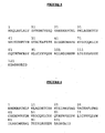
- the plasmid was re-suspended in 10 ⁇ l of H 2 O and 3,5 ⁇ l of it was incubated at 16°C for 18 h with 3,5 ⁇ l of the pAE expression vector ( figure 4 ), 2 ⁇ l of buffer 5x for DNA ligase and 1 ⁇ l of DNA ligase.
- the clones subcloned in pAE vector were also sequenced.
- the BL21 (DE3) strain of the E. coli bacteria was preferably used for the expression of this protein since it is a strain presenting fast growing, easy culture and maintenance and high quantity of recombinant protein.
- This strain of E . coli is lysogenic and does not have post-translation modification systems.
- the supernatant that had the soluble expressed protein was centrifuged in 15000 rpm for 30 min for clarification and applied in affinity column of Ni-sepharose previously balanced with lysis buffer.
- the column was washed with buffer imidazol 80 mM, ß-mercaptoethanol 5 mM, NaCl 500 mM, Tris HCl 50 mM pH 6,8 and the washing volume was collected.
- the protein was eluded using Tris-HCl 50 mM pH 8.0, imidazol IM, NaCl 100 mM with flow of ImI/ 5 min.
- the " pellet " (corpuscles) of the medium submitted to the French press and centrifuged was re-suspended in 20 ml of buffer Tris-HCl 50 mM, Urea 1 M, Triton X-100 1%, pH 8,0 for eliminating hydrophobic components and centrifuged in 5000 rpm for 15 min at 4°C.
- the separated precipitated was incubated at room temperature overnight with 10 ml of buffer Tris-HCl 50 mM, NaCl 500 mM, Urea 8 M, ß-mercaptoethanol 10 mM pH 8,0 for the solubilization of the corpuscles.
- the column was washed with 180 ml of buffer Tris-HCl 50 mM, NaCl 500 mM, Imidazol 20 mM pH 6,8 and eluded with Tris-HCl 50 mM pH 8.0, imidazol IM, NaCl 100 mM with flow of 1ml/ 5 min.
- the recombinant protein obtained was tested concerning its inhibitory capacity on Factor Xa, coagulation tests and control of tumoral cells.
- the biochemical assays for determining the inhibition of FXa were conducted in presence of phospholipids.
- the phospholipid membranes were prepared as described by Barenholz et al., 1977.
- the FXa inhibition in presence of the phospholipid membrane was determined by the measure of the residual enzymatic activity on the amino-methyl-coumarin substrate with the hydrolysis site for FXa, or commercial chromogenic substrate.
- the hydrolysis reactions conducted at 37°C in buffer Tris/HCl 50 mM, pH 7.5 - 8,0 were monitored by the fluorescence in 380 nm (excitation) and 460 nm (emission) wave lengths by a spectrofluorimeter or spectrophotometer in 405 nm wave length in case of chromogenic substrates.
- the recombinant protein of this invention when used in 5 - 10 uM doses, inhibits the amidolytic activity of human Factor X activated, in purified conditions, in presence of phosphatidylserine/phosphatidylcholine and in the plasma.
- the protein induces apoptosis (programmed cellular death) in tumoral lineage cells (B16FI0, SKMEL28) with cellular cycle stop in G2/M.
- the Amblyomin-X is able to cause complete remission of the tumoral mass and volume and impede significantly the formation of metastases.
- Hematological analysis of the animals with tumor submitted to the treatment showed that the recombinant protein improves significantly the hematological condition, maintaining the hematocrit, hemoglobin and cellularity within normality levels when compared to the non-treated ones with severe anemia.
- a composting is obtained containing a sequence from the cDNA of the Amblyomma cajennense gland.
- This compost is composed of a polypeptide or protein which sequence was determined from the cDNA that codifies from base 1 to base 505.
- the recombinant protein obtained from the compost was named Amblyomin-X. It is a protein of two domains, of Kunitz- and MAP Kinase- types, and it is useful in tumor treatments as well as for developing vaccines from cDNA. It does also present biologic activity for Factor Xa, X, and VII proteins and also in the Factor VIIa/TF complex, showing to be a potential candidate for the modulation of anticoagulant function.
- the administration of the peptide, polypeptide or protein improves conditions of patients with diseases related to cardiovascular and thromboembolic complications; decreases metastases of malign tumors; decreases malign tumors growing and dissemination; inhibits metastases of malign tumors; anti-coagulant performance in pre and post surgery conditions it can be used for preventing thromboembolism in conditions that generate apoptotic bodies (molecules rich in phospholipids), in degenerative diseases and malign tumors it can be used for improving the immune response. It can be used as a co-treatment in chemotherapy treatment of malign tumors; as a co-treatment in radiotherapy treatment of malign tumors; as a co-treatment in chemotherapy treatment of malign tumors with thromboembolic complications.
- It can also be used as a co- treatment in radiotherapy treatment of malign tumors with thromboembolic complications; can modulate the programmed cellular death (apoptosis); can modulate the formation of new blood vessels in the tumor and can be used as an agent for target therapy.
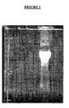
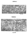
- RNA integrity extracted from the salivary glands of the A. cajennese tick was verified in agarose gel in presence of formaldehyde ( figure 1 ).
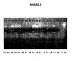
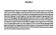
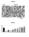
- RNA For the isolation of the mRNA, 48 ul of total RNA was diluted in 500 ul of 10mM Tris buffer in presence of ImM EDTA and submitted to the oligo (dT) cellulose affinity column. 17,28 ug of mRNA was obtained. The ratio A 260/280 for the mRNA was 1,71 (ug/ul). The extracted mRNA, free of degradation products, was used for the construction of the cDNA library ( Figure 2 ).
- aleatory clones were selected and digested with Eco RI and Hind III and a variety of different size inserts was presented ( figure 3 ).
- the sequence obtained refers to the protein with the signal peptide, and by the http://www.cbs.dtu.dk/services/SignalP/ web-site it was possible the theoretical determination of this peptide, that was found between the positions 1 and 28 of this amino acid sequence.
- the sequence of the mature protein contains 108 amino acids, with theoretical molecular mass of 12,295,6 Da and theoretical isoeletric point (pI) of 4,67.
- the mature protein sequence is represented in figure 6 .
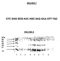
- EXAMPLE 8 SEQUENCE 5' -3' OF THE "PRIMER” CONSTRUCTED FOR THE CLONE AMPLIFICATION
- the amplification product obtained with the "primer” constructed and the SP6 "primer” presented a band of approximately 400 pb. This band was cut out of the gel and purified.
- the purified cDNA was subcloned in vector and transformed into DH5 ⁇ competent bacteria.
- the product was plated and from this procedure 20 aleatory selected clones were isolated and submitted to the extraction process by phenol-chloroform for the evaluation of insert presence.
- the clone sequence was confirmed and the insert liberated by the digestion was subcloned in the expression vector that inserts an end of six histidine residues in the N-terminus portion of the recombinant protein, expressed after the induction with IPTG.
- the interest protein was found as inclusion corpuscle that after the urea and ⁇ -mercaptoethanol solubilization was purified in Ni- affinity column as SDS-PAGE demonstrate in figure 8 .
- the human TFPI presents 3 domains, DI that is composed of residues 53 through 103, D2 composed of residues 124 through 174 and finally the D3 domain composed of residues 222 through 273. Therefore, the Amblyomin-X primary structure (with only 2 domains) was compared to each one of the domains of the human TFPI-I molecule.
- the recombinant inhibitor is able to prolong the coagulation time, either the time of thromboplastin partial activated (TTPA) and the time of prothrombin (TP) .
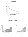
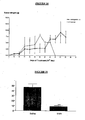
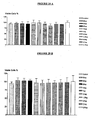
- Figure 11a shows the TTPA prolonging was from 43,4 seconds (control) to 137,5 seconds in presence of 50 ug of the recombinant inhibitor, what reflects an inhibition of 60%.
- Figure lib shows the prolonging of TP from 14,3 seconds in absence of the inhibitor to 89,3 seconds in presence of 50 ug of the recombinant inhibitor reflecting a theoretical inhibition of approximately 85%.
- EXAMPLE 14 CYTOLOGIC ASPECTS CAUSED BY AMBLYOMIN-X IN B16F10 CELLS, 6 HOURS AFTER TREATMENT: ANALYSIS OF THE MORPHOLOGIC ALTERATIONS AND CYTOTOXICITY
- the tumoral cells of B16F10 melanoma treated with different concentrations of Amblyomin-X were cultivated in plates of 96 wells.
- the cytotoxic alterations and adhesion loss were firstly observed cytologically, and images were achieved by the Capture System.
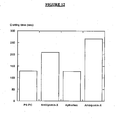
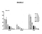
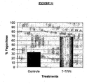
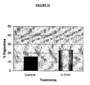
- Viability loss was observed in concentrations from 0,3 uM to 0,1 nM. The supernatant was collected; the number and cellular viability were determined by the exclusion test with Blue Trypan. The cellular concentration was determined in Mallassez haemocytometric Chamber. In these concentrations the viability was also dose dependent, over 75% when 5ug was used. It showed the highest percentage of cellular detachment, as can be seen in figure 17 .
- 2,5x100 5 B16F10 tumoral cells were injected in mice by subcutaneous route and after the 12 th day, 1mg/kg of Amblyomin-X was injected in animals with dorsal tumor by subcutaneous administration until the 14 th day.
- the same volume of saline solution was administered by the same route in animals of the control group after the 12 th day of inoculation of the tumoral cells.
- the animals were daily observed and the diameter of the tumor determined through a pachymeter.
- C57BL/6J mice groups received 5x10 4 tumoral cells, by endovenous route, and after the 12 th day of the injection they were submitted to treatment with 0,5mg/Kg of Amblyomin-X. This treatment was daily administered by intraperitoneal route during 14 days and the animals were daily observed.
- B16F10 animals with dorsal tumor received daily treatment with 1 or 0,5 mg/Kg of Amblyomin-X after the 3 rd day followed the injection of the tumoral cells.
- the animals with tumor were daily treated by subcutaneous administration for 42 consecutive days.
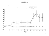
- the results showed that the treatment is efficient with a significant reduction of tumoral mass and volume in treated animals compared to those of the control group. It was observed that animals with injection of tumoral cells did not present tumoral growing or even tumoral mass disappearance was observed probably by the induction of tumor remission.
- a group of animals was dissected and the existing tumors were macroscopically small, pigmented, not nodular and not presenting necrosis areas. It could also be seen that tumor irrigation was not increased and it did not present system recruitment of peripheral vessels adjacent to the lesion. Neo-angiogenesis was not observed either ( figure 24 ).
- the histopathologic aspects of dorsal tumors of the control group that received saline solution during the therapeutic protocol showed high cellularity tumoral mass similar to the sincicial mass with extended area irrigated by average and small vessels as well as capillary vessels. Around the tumoral mass a discrete inflammatory infiltration of monomorphonuclear leukocytes was observed.
- Tumors of groups treated with Amblyomin-X (1mg/Kg) taken after the 14 th day of treatment showed tumoral mass with low proliferative activity, low amount of sustentation fibril elements (few connective tissue /stroma) without occurring peritumoral inflammatory infiltration.
- the pulmonary metastatic lesions of this group showed to be of small tumors with low cellularity, in the pulmonary parenchyma, presenting alveoli thickness and deposition and brownish pigment of melanin type. Bronchial intraseptal inflammatory infiltration was not observed in these lesions.
- the dorsal tumors obtained from the animals treated with AMBLYOMIN-X during 42 days showed that the tumoral cells are organized like in islands permeated by extensive necrosis areas (figure 25B), differently than the control ( Figure 25 A) .
- By the haematoxylin/Eosin stain technique neo-formed blood vessels were not detected.
- the other internal organs analyzed, as: kidneys, liver, spleen and heart did not present histologic alterations. Functional loss or direct toxicity effects in these organs caused by the prolonged treatment with Amblyomin-X were not observed.
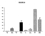
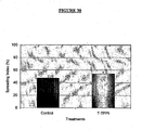
- the cells obtained from lesions of the pulmonary metastases analyzed by flow cytometry were in significant number in phase G2/M of the cellular cycle representing a blockage of the proliferative capacity of these cells and the treatment efficiency ( Figure 27 ).
- TTPA thromboplastin partial activated
- TP prothrombin
- TTPA is the test in which the intrinsic route of coagulation is evaluated, that is, the plasmatic callicrein and factors XII, IX, XI and X were monitored.
- the essay control was conducted in absence of the Amblyomin-X and consisted of one incubation during 3 min at 37°C of 100 ul of healthy human plasma, 100 ul of cefaline and 100 ul of tris HCl 0.1 M pH 8,0 buffer, followed by the medium re-calcifying by the addition of 100 ul of CaCl 2 0,025 M.
- the time of the clot formation was measured by a semi- automated coagulometer.
- 100 ul of Amblyomin-X solution prepared in tris HCl 0.1 M pH 8,0 buffer was used.
- TP verifies the coagulation extrinsic route, that is, it monitors factors II, V, VII and X.
- the control of the essay was conducted incubating for 2 min at 37°C, 100 ul of plasma with 100 ul of tris-HCl 0.1 M pH 8,0 buffer and for adding afterwards 100 ul of the reagent. The clot formation time was verified just like the test mentioned before. Instead of the buffer, 100 ul of Amblyomin-X solution prepared in tris HCl 0.1 M pH 8,0 buffer was incubated for evaluating inhibition.
- a curve was idealized for standardizing the coagulation test named PCA "Pro Coagulant Activity" and toward this, 3-min pre-incubation at 37°C was conducted for a final volume of 60 ul of Factor VII activated (FVIIa) 1 nM with Factor X human (FX) 0,25 and CaCl 2 8,3 mM with different concentrations of recombinant tissue Factor (r-TF) (1,56 - 12,5 ng/ml) in Hepes 250 mM buffer containing bovine albumin (BSA) (1%) After that, 100 ul of a mixture containing normal human plasma (87,5 ul), phosphatidyl serine: phosphatidyl choline PS 30: PC 70 (7,5 ul) and Hepes (50 inM) were added and the mixture was incubated for another 3 minutes. The reaction started by adding 20 ul of CaCl 2 250 mM.
- FVIIa Factor VII activated
- FX Factor X human
- Amblyomin-X was pre-incubated with apoptotic bodies produced in CHO cells and it was observed that the coagulation time induced by the apoptotic bodies was prolonged (Sorensen et al., 2003).
- the tumoral lineages: HL-60, K562, U937, YAC-I, JURKAT, Mel-85, Mewo, MCF-7, SW613-12A1, SW613-B3, the SW613-12A1 and SW613 colon cancer lineages and the B16F10 murine melanoma lineage were cultivated in culture bottles of 75cm3 in RPMI-1640 medium supplemented with 10% of bovine fetal serum, 2 mM L-glutamin, 1 mM sodium pyruvate and streptomycin 0, 1 mg/ml and ampicillin 0,1 mg/ml antibiotics. Before reaching confluence (adherent cells), the cells were cultivated for amplifying and maintenance lineages, frozen in RPMI-1640 culture medium containing 10% dimethylsulphoxide and maintained in liquid nitrogen.
- the suspensions of adherent cells (B16F10, SW613-12A1, SW613-B3 and MCF-7) for all the experimental procedures were obtained by the treatment of the culture bottles with trypsin 0,2% for 5 minutes and inactivation with 10% bovine fetal serum.
- the detached cells were centrifuged twice, re- suspended in RPMI-1640 supplemented medium.
- the cells counting was conducted in Malassez chamber and the cellular concentration was adjusted in 5x10 5 cells/ml in RPMI-1640 medium supplemented with 10% of bovine fetal serum and 7ug of Polymixine-B.
- the cellular viability was determined by the exclusion test of Blue Trypan showing over 95% of viable cells.
- the 2x10 5 concentration cells were cultivated in plates of 96 wells, maintained for 24 hours in CO2 sterilizer at 37[deg.]C. After this period the plates were centrifuged for 5 minutes in 2000 rpm at 4°C, the supernatant was excluded and different concentrations of Amblyomin-X were added from 0,3 uM to 0,3 nM, diluted in RPMI-1640 culture medium, supplemented and added with 7ug of Polymixine -B. After 6, 12 and 24 hours the cytological alterations were observed and photo-documented in image capturing system.
- the flow cytometry applied in the study of cellular cycle registers the kinetic parameters of the cells showing the DNA index, the ploidia, the cellular proliferation fraction and the percentage of cells found in phases S and G2/M, indicating uni or multivariable parameters for prognosis and possible therapeutic procedures.
- the analysis of the percentage of cells provides the percentage of cells that are synthesizing DNA ("labeling index"), duration of phase S (Ts) and the time of potential duplication (Tpot).
- the cells were incubated with 375 ⁇ l of trypsin 0,03 g/l for 10 minutes at room temperature and neutralized with the inhibitor of trypsin 0,5 g/l, ribonuclease A 0,1 g/l and spermine 1,2 g/l.
- the samples were transferred to tubes of flow cytometry and the number of cells in different phases of the cycle, the apoptosis level (Sub-G1) and the DNA content in phase S were analyzed.
- the adherent cellular suspensions (B16F10) used for the implantation in the dorsal flank of the animals were obtained after treating the culture bottles with trypsin 0,2% for 5 minutes and inactivating with bovine fetal serum 10%.
- the detached cells were centrifuged (2.000 rpm) twice and re-suspended in RPMI-1640 medium supplemented with 10% bovine fetal serum inactivated at 56°C for I h, 2 mM L-glutamin, 1 mM sodium pyruvate and streptomycin 0,1 mg/ml and ampicillin 0,1 mg/ml antibiotics.
- the cells counting was conducted in Malassez chamber and cellular concentration adjusted for 5x10 4 cells/ml.
- mice maintained in the laboratory animal care facilities, belonging to the C57BL/6J lineage were used. The animals were kept with light/dark 12-hour cycles, at constant temperature of 20°C, filtrated and sterilized water and food "ad libitum".
- mice For the growing experiments of primary dorsal tumors, 2,5x10 4 tumoral cells of B16F10 murine melanoma were injected in the dorsa of mice (groups of 10 animals) by subcutaneous route.
- groups of C57BL/6J mice received 5x10 4 B16F10 tumoral cells by endovenous route through the ocular retro-orbital venous plexus. After the 12 th day of the inoculation this group of animals was treated with 0,5mg/Kg of Amblyomin-X, administered by intraperitoneal route, and observed until the 14 th day of the treatment.
- the animals received saline solution by the same route of the treatment and after the 14 th day of treatment.
- the animals were anesthetized and killed by cervical dislocation.
- Necropsy was conducted and the dorsal tumors were analyzed, the macroscopic internal lesions identified, measured and photo-documented.
- Samples of the tumors of the different groups of treatment and of the control group, non-treated, were processed for analyzing the contents of DNA, cellular cycle and anatomopathologic.
- the Amblyomin-X cytotoxic activity was determined in suspensions of the cellular lineages and of the tumoral cells ("ex-vivo"), detached by surgery and in sterile conditions incubated in plates of 96 wells. To these cells 10 ⁇ l of MTT (5mg/ml) was added and then they were incubated for 3 hours in sterilizer containing 5% of CO 2 at 37°C. After this period, the medium was removed and 100 ⁇ l of sulphoxide dimethyl was added for the dissolution of formazan crystals that were formed and presented as precipitated. Absorbency was monitored in 540 nm.
- mice with B16F10 melanoma and control group that received saline solution
- Amblyomin-X the animals were anesthetized and killed by cervical dislocation.
- the abdominal cavity of treated and control animals was exposed and 2 ml of saline solution containing 5000 U cool heparin was injected.
- the cavity was massaged and right after the peritoneal washed was collected, centrifuged in 2000 rpm for 10 minutes at 4°C.
- the cellular suspension was re-suspended in RPMI-1640 culture medium supplemented with bovine fetal serum 10% and the number of cells adjusted for 10 6 /ml in Mallassez haemocytometric chamber.
- Fragments of human skin (of approximately 1 cm X 1 cm) previously selected were collected aseptically and immediately placed in a sterile conic tube containing Ham-F12 culture medium with 20% bovine fetal serum. Each fragment was transferred to a 35-mm Petri dish containing culture medium for washing and blood excess withdrawing. Fat and degenerative tissue was taken out using scissors and tongs. The "clean" fragment was cut into smaller fragments and they were then distributed in 3 Petri dishes ( ⁇ 15 pieces each) containing Ham-F12 culture medium supplemented with 10% of bovine fetal serum.
- the plates were maintained in humidified sterilizer at 37°C and 5% of CO 2 and examined in inverted microscope 3 times a week.
- the culture medium was exchanged in the same time interval.
- the cells were exposed to trypsin (Trypsin 0,2%) for 5 minutes and the inactivation was conducted with bovine fetal serum 10, the cells were centrifuged for 10 minutes in 2000 rpm at 4°C and plated in bottles of 25 cm 2 .
Landscapes
- Health & Medical Sciences (AREA)
- Chemical & Material Sciences (AREA)
- Life Sciences & Earth Sciences (AREA)
- Organic Chemistry (AREA)
- Medicinal Chemistry (AREA)
- General Health & Medical Sciences (AREA)
- Public Health (AREA)
- Nuclear Medicine, Radiotherapy & Molecular Imaging (AREA)
- General Chemical & Material Sciences (AREA)
- Pharmacology & Pharmacy (AREA)
- Animal Behavior & Ethology (AREA)
- Chemical Kinetics & Catalysis (AREA)
- Veterinary Medicine (AREA)
- Bioinformatics & Cheminformatics (AREA)
- Engineering & Computer Science (AREA)
- Biophysics (AREA)
- Proteomics, Peptides & Aminoacids (AREA)
- Molecular Biology (AREA)
- Genetics & Genomics (AREA)
- Biochemistry (AREA)
- Gastroenterology & Hepatology (AREA)
- Tropical Medicine & Parasitology (AREA)
- Insects & Arthropods (AREA)
- Immunology (AREA)
- Zoology (AREA)
- Toxicology (AREA)
- Diabetes (AREA)
- Hematology (AREA)
- Oncology (AREA)
- Cardiology (AREA)
- Heart & Thoracic Surgery (AREA)
- Surgery (AREA)
- Medicines That Contain Protein Lipid Enzymes And Other Medicines (AREA)
- Peptides Or Proteins (AREA)
- Preparation Of Compounds By Using Micro-Organisms (AREA)
- Micro-Organisms Or Cultivation Processes Thereof (AREA)
- Enzymes And Modification Thereof (AREA)
Applications Claiming Priority (2)
| Application Number | Priority Date | Filing Date | Title |
|---|---|---|---|
| BRPI0406057-1A BRPI0406057B1 (pt) | 2004-09-15 | 2004-09-15 | Sequência nucleotídica de um inibidor de proteases do tipo kunitz obtida a partir de uma biblioteca de cdna de glândulas salivarares de carrapatos amblyomma cajennese uso da proteína recombinante |
| PCT/BR2005/000185 WO2006029492A1 (en) | 2004-09-15 | 2005-09-15 | Kunitz-type recombinant inhibitor. |
Publications (2)
| Publication Number | Publication Date |
|---|---|
| EP1799707A1 EP1799707A1 (en) | 2007-06-27 |
| EP1799707B1 true EP1799707B1 (en) | 2013-03-27 |
Family
ID=36273230
Family Applications (1)
| Application Number | Title | Priority Date | Filing Date |
|---|---|---|---|
| EP05782904A Expired - Lifetime EP1799707B1 (en) | 2004-09-15 | 2005-09-15 | Kunitz-type recombinant inhibitor. |
Country Status (10)
| Country | Link |
|---|---|
| US (1) | US8440795B2 (enExample) |
| EP (1) | EP1799707B1 (enExample) |
| JP (1) | JP4980220B2 (enExample) |
| CN (1) | CN101142232B (enExample) |
| AU (1) | AU2005284615B2 (enExample) |
| BR (1) | BRPI0406057B1 (enExample) |
| CA (2) | CA2581001C (enExample) |
| DK (1) | DK1799707T3 (enExample) |
| ES (1) | ES2410160T3 (enExample) |
| WO (1) | WO2006029492A1 (enExample) |
Families Citing this family (6)
| Publication number | Priority date | Publication date | Assignee | Title |
|---|---|---|---|---|
| WO2008109976A1 (en) * | 2007-03-14 | 2008-09-18 | União Química Farmacêutica Nacional S/A | Kunitz-type recombinant inhibitor |
| BRPI0406057B1 (pt) | 2004-09-15 | 2021-10-26 | União Química Farmacêutica Nacional S/A | Sequência nucleotídica de um inibidor de proteases do tipo kunitz obtida a partir de uma biblioteca de cdna de glândulas salivarares de carrapatos amblyomma cajennese uso da proteína recombinante |
| NZ593815A (en) | 2008-12-19 | 2013-05-31 | Tissue factor pathway inhibitor (tfpi) inhibitors and methods of use | |
| US8772238B2 (en) | 2009-03-18 | 2014-07-08 | The United States Of America, As Represented By The Secretary Of The Department Of Health And Human Services | Use of ixolaris, a tissue factor inhibitor, for the treatment of cancer |
| BR102018074043A2 (pt) * | 2018-11-22 | 2020-06-02 | Inst Butantan | composto para a modulação de vias de rlr, tlr, oas e/ou oncostatina m, uso do mesmo para a preparação de um medicamento, composição e método para modulação das ditas vias |
| BR102018074037A2 (pt) * | 2018-11-22 | 2021-05-11 | Inst Butantan | composto para direcionar entidades moleculares para o domínio intracelular de células neoplásicas, ingrediente farmacêutico ativo compreendendo o referido composto, composição farmacêutica, método para o diagnóstico, prognóstico e/ou tratamento de câncer |
Family Cites Families (3)
| Publication number | Priority date | Publication date | Assignee | Title |
|---|---|---|---|---|
| DE3006207C2 (de) * | 1980-02-15 | 1982-10-21 | Siemens AG, 1000 Berlin und 8000 München | Elektrische Maschine mit einem Ständerblechpaket aus kornorientierten Blechen |
| DE3819078A1 (de) * | 1988-06-04 | 1989-12-07 | Hoechst Ag | Amblyommin, ein neuer wirkstoff fuer die antikoagulationstherapie |
| BRPI0406057B1 (pt) | 2004-09-15 | 2021-10-26 | União Química Farmacêutica Nacional S/A | Sequência nucleotídica de um inibidor de proteases do tipo kunitz obtida a partir de uma biblioteca de cdna de glândulas salivarares de carrapatos amblyomma cajennese uso da proteína recombinante |
-
2004
- 2004-09-15 BR BRPI0406057-1A patent/BRPI0406057B1/pt not_active IP Right Cessation
-
2005
- 2005-09-15 AU AU2005284615A patent/AU2005284615B2/en not_active Ceased
- 2005-09-15 CN CN2005800389926A patent/CN101142232B/zh not_active Expired - Fee Related
- 2005-09-15 JP JP2007531547A patent/JP4980220B2/ja not_active Expired - Lifetime
- 2005-09-15 ES ES05782904T patent/ES2410160T3/es not_active Expired - Lifetime
- 2005-09-15 DK DK05782904.6T patent/DK1799707T3/da active
- 2005-09-15 WO PCT/BR2005/000185 patent/WO2006029492A1/en not_active Ceased
- 2005-09-15 EP EP05782904A patent/EP1799707B1/en not_active Expired - Lifetime
- 2005-09-15 CA CA2581001A patent/CA2581001C/en not_active Expired - Lifetime
-
2007
- 2007-03-14 US US11/724,557 patent/US8440795B2/en not_active Expired - Fee Related
-
2008
- 2008-03-14 CA CA002680759A patent/CA2680759A1/en not_active Abandoned
Also Published As
| Publication number | Publication date |
|---|---|
| BRPI0406057B1 (pt) | 2021-10-26 |
| CA2581001C (en) | 2011-05-03 |
| US20090042786A1 (en) | 2009-02-12 |
| ES2410160T3 (es) | 2013-07-01 |
| WO2006029492A1 (en) | 2006-03-23 |
| DK1799707T3 (da) | 2013-05-21 |
| CN101142232A (zh) | 2008-03-12 |
| BRPI0406057A (pt) | 2006-05-02 |
| JP2008512992A (ja) | 2008-05-01 |
| AU2005284615A1 (en) | 2006-03-23 |
| JP4980220B2 (ja) | 2012-07-18 |
| US8440795B2 (en) | 2013-05-14 |
| CA2680759A1 (en) | 2008-09-18 |
| AU2005284615B2 (en) | 2011-03-31 |
| EP1799707A1 (en) | 2007-06-27 |
| CA2581001A1 (en) | 2006-03-23 |
| CN101142232B (zh) | 2013-03-13 |
Similar Documents
| Publication | Publication Date | Title |
|---|---|---|
| US6689582B1 (en) | Polynucleotide molecules encoding proteins having proteinase inhibitor activity | |
| Moreau et al. | Multifaceted roles of human elafin and secretory leukocyte proteinase inhibitor (SLPI), two serine protease inhibitors of the chelonianin family | |
| Mende et al. | Dipetalogastin, a potent thrombin inhibitor from the blood‐sucking insectDipetalogaster maximus: cDNA cloning, expression and characterization | |
| US8440795B2 (en) | Kunitz-type recombinant inhibitor | |
| Hou et al. | MMP-12 activates protease-activated receptor-1, upregulates placenta growth factor, and leads to pulmonary emphysema | |
| Batista et al. | A new Factor Xa inhibitor from Amblyomma cajennense with a unique domain composition | |
| JPH0584083A (ja) | 新規ポリペプチド、それをコードする新規dna、新規ポリペプチドの製造方法、新規医薬組成物、および新規酵素阻害方法 | |
| Lukas et al. | Hirudins of the Asian medicinal leech, Hirudinaria manillensis: same same, but different | |
| Jedličková et al. | A novel Kunitz protein with proposed dual function from Eudiplozoon nipponicum (Monogenea) impairs haemostasis and action of complement in vitro | |
| Ranasinghe et al. | A novel coagulation inhibitor from Schistosoma japonicum | |
| Chen et al. | Kunitzins: Prototypes of a new class of protease inhibitor from the skin secretions of European and Asian frogs | |
| He et al. | Serpin-9 and-13 regulate hemolymph proteases during immune responses of Manduca sexta | |
| Le et al. | Gene duplication of coagulation factor V and origin of venom prothrombin activator in Pseudonaja textilis snake | |
| WO2008109976A1 (en) | Kunitz-type recombinant inhibitor | |
| CA2161796A1 (en) | Hookworm anticoagulant | |
| KR20060010740A (ko) | 프로테아제 억제 단백질 및 그 이용 | |
| Hakim et al. | Discoveries of serine protease inhibitors from scorpions | |
| Sutherland et al. | Investigating serpin–enzyme complex formation and stability via single and multiple residue reactive centre loop substitutions in heparin cofactor II | |
| BR112018071701B1 (pt) | Processo de obtenção da proteína recombinante e uso da esculptina ou proteína recombinante para preparar um medicamento ou composição farmacêutica para profilaxia e/ou tratamento de doenças tromboembólicas ou como inibidor direto e específico da trombina | |
| Gonzalez-Gonzalez | The trypsin inhibitor panulirin regulates | |
| CN106928334A (zh) | 约安巨马陆抗血栓肽Joannsin及其应用 | |
| WO1999060126A9 (en) | Protein z-dependent protease inhibitor | |
| Zhang et al. | Identification and analysis of the tissue factor pathway inhibitor 2 of Sciaenops ocellatus | |
| KR101374194B1 (ko) | 대하로부터 분리한 쿠니츠형 세린 프로테아제 저해인자 | |
| TW200846356A (en) | Preparation and use of variants of the kunitz domain 2 of the human placental bikunin gene |
Legal Events
| Date | Code | Title | Description |
|---|---|---|---|
| PUAI | Public reference made under article 153(3) epc to a published international application that has entered the european phase |
Free format text: ORIGINAL CODE: 0009012 |
|
| 17P | Request for examination filed |
Effective date: 20070410 |
|
| AK | Designated contracting states |
Kind code of ref document: A1 Designated state(s): AT BE BG CH CY CZ DE DK EE ES FI FR GB GR HU IE IS IT LI LT LU LV MC NL PL PT RO SE SI SK TR |
|
| DAX | Request for extension of the european patent (deleted) | ||
| RIC1 | Information provided on ipc code assigned before grant |
Ipc: A61K 38/55 20060101ALI20071220BHEP Ipc: C07K 14/81 20060101AFI20071220BHEP |
|
| 17Q | First examination report despatched |
Effective date: 20080319 |
|
| GRAP | Despatch of communication of intention to grant a patent |
Free format text: ORIGINAL CODE: EPIDOSNIGR1 |
|
| RIC1 | Information provided on ipc code assigned before grant |
Ipc: C07K 14/435 20060101AFI20121002BHEP Ipc: C07K 14/81 20060101ALI20121002BHEP Ipc: A61K 38/55 20060101ALI20121002BHEP |
|
| GRAS | Grant fee paid |
Free format text: ORIGINAL CODE: EPIDOSNIGR3 |
|
| GRAA | (expected) grant |
Free format text: ORIGINAL CODE: 0009210 |
|
| AK | Designated contracting states |
Kind code of ref document: B1 Designated state(s): AT BE BG CH CY CZ DE DK EE ES FI FR GB GR HU IE IS IT LI LT LU LV MC NL PL PT RO SE SI SK TR |
|
| REG | Reference to a national code |
Ref country code: GB Ref legal event code: FG4D |
|
| REG | Reference to a national code |
Ref country code: CH Ref legal event code: EP |
|
| REG | Reference to a national code |
Ref country code: AT Ref legal event code: REF Ref document number: 603358 Country of ref document: AT Kind code of ref document: T Effective date: 20130415 |
|
| RAP2 | Party data changed (patent owner data changed or rights of a patent transferred) |
Owner name: FUNDACAO DE AMPARO A PESQUISA DO ESTADO DE SAO PAU Owner name: UNIAO QUIMICA FARMACEUTICA NACIONAL S/A Owner name: CHUDZINSKI-TAVASSI, ANA MARISA |
|
| REG | Reference to a national code |
Ref country code: IE Ref legal event code: FG4D |
|
| REG | Reference to a national code |
Ref country code: DK Ref legal event code: T3 |
|
| REG | Reference to a national code |
Ref country code: DE Ref legal event code: R096 Ref document number: 602005038788 Country of ref document: DE Effective date: 20130529 |
|
| REG | Reference to a national code |
Ref country code: DE Ref legal event code: R082 Ref document number: 602005038788 Country of ref document: DE Representative=s name: LAVOIX MUNICH, DE |
|
| REG | Reference to a national code |
Ref country code: ES Ref legal event code: FG2A Ref document number: 2410160 Country of ref document: ES Kind code of ref document: T3 Effective date: 20130701 |
|
| RAP2 | Party data changed (patent owner data changed or rights of a patent transferred) |
Owner name: UNIAO QUIMICA FARMACEUTICA NACIONAL S/A Owner name: FUNDACAO DE AMPARO A PESQUISA DO ESTADO DE SAO PAU Owner name: CHUDZINSKI-TAVASSI, ANA MARISA |
|
| PG25 | Lapsed in a contracting state [announced via postgrant information from national office to epo] |
Ref country code: BG Free format text: LAPSE BECAUSE OF FAILURE TO SUBMIT A TRANSLATION OF THE DESCRIPTION OR TO PAY THE FEE WITHIN THE PRESCRIBED TIME-LIMIT Effective date: 20130627 Ref country code: LT Free format text: LAPSE BECAUSE OF FAILURE TO SUBMIT A TRANSLATION OF THE DESCRIPTION OR TO PAY THE FEE WITHIN THE PRESCRIBED TIME-LIMIT Effective date: 20130327 Ref country code: SE Free format text: LAPSE BECAUSE OF FAILURE TO SUBMIT A TRANSLATION OF THE DESCRIPTION OR TO PAY THE FEE WITHIN THE PRESCRIBED TIME-LIMIT Effective date: 20130327 |
|
| REG | Reference to a national code |
Ref country code: AT Ref legal event code: MK05 Ref document number: 603358 Country of ref document: AT Kind code of ref document: T Effective date: 20130327 |
|
| REG | Reference to a national code |
Ref country code: LT Ref legal event code: MG4D |
|
| PG25 | Lapsed in a contracting state [announced via postgrant information from national office to epo] |
Ref country code: LV Free format text: LAPSE BECAUSE OF FAILURE TO SUBMIT A TRANSLATION OF THE DESCRIPTION OR TO PAY THE FEE WITHIN THE PRESCRIBED TIME-LIMIT Effective date: 20130327 Ref country code: SI Free format text: LAPSE BECAUSE OF FAILURE TO SUBMIT A TRANSLATION OF THE DESCRIPTION OR TO PAY THE FEE WITHIN THE PRESCRIBED TIME-LIMIT Effective date: 20130327 Ref country code: GR Free format text: LAPSE BECAUSE OF FAILURE TO SUBMIT A TRANSLATION OF THE DESCRIPTION OR TO PAY THE FEE WITHIN THE PRESCRIBED TIME-LIMIT Effective date: 20130628 Ref country code: FI Free format text: LAPSE BECAUSE OF FAILURE TO SUBMIT A TRANSLATION OF THE DESCRIPTION OR TO PAY THE FEE WITHIN THE PRESCRIBED TIME-LIMIT Effective date: 20130327 |
|
| REG | Reference to a national code |
Ref country code: NL Ref legal event code: VDEP Effective date: 20130327 |
|
| PG25 | Lapsed in a contracting state [announced via postgrant information from national office to epo] |
Ref country code: BE Free format text: LAPSE BECAUSE OF FAILURE TO SUBMIT A TRANSLATION OF THE DESCRIPTION OR TO PAY THE FEE WITHIN THE PRESCRIBED TIME-LIMIT Effective date: 20130327 |
|
| PG25 | Lapsed in a contracting state [announced via postgrant information from national office to epo] |
Ref country code: IS Free format text: LAPSE BECAUSE OF FAILURE TO SUBMIT A TRANSLATION OF THE DESCRIPTION OR TO PAY THE FEE WITHIN THE PRESCRIBED TIME-LIMIT Effective date: 20130727 Ref country code: PT Free format text: LAPSE BECAUSE OF FAILURE TO SUBMIT A TRANSLATION OF THE DESCRIPTION OR TO PAY THE FEE WITHIN THE PRESCRIBED TIME-LIMIT Effective date: 20130729 Ref country code: SK Free format text: LAPSE BECAUSE OF FAILURE TO SUBMIT A TRANSLATION OF THE DESCRIPTION OR TO PAY THE FEE WITHIN THE PRESCRIBED TIME-LIMIT Effective date: 20130327 Ref country code: AT Free format text: LAPSE BECAUSE OF FAILURE TO SUBMIT A TRANSLATION OF THE DESCRIPTION OR TO PAY THE FEE WITHIN THE PRESCRIBED TIME-LIMIT Effective date: 20130327 Ref country code: EE Free format text: LAPSE BECAUSE OF FAILURE TO SUBMIT A TRANSLATION OF THE DESCRIPTION OR TO PAY THE FEE WITHIN THE PRESCRIBED TIME-LIMIT Effective date: 20130327 Ref country code: NL Free format text: LAPSE BECAUSE OF FAILURE TO SUBMIT A TRANSLATION OF THE DESCRIPTION OR TO PAY THE FEE WITHIN THE PRESCRIBED TIME-LIMIT Effective date: 20130327 Ref country code: CZ Free format text: LAPSE BECAUSE OF FAILURE TO SUBMIT A TRANSLATION OF THE DESCRIPTION OR TO PAY THE FEE WITHIN THE PRESCRIBED TIME-LIMIT Effective date: 20130327 Ref country code: RO Free format text: LAPSE BECAUSE OF FAILURE TO SUBMIT A TRANSLATION OF THE DESCRIPTION OR TO PAY THE FEE WITHIN THE PRESCRIBED TIME-LIMIT Effective date: 20130327 |
|
| PG25 | Lapsed in a contracting state [announced via postgrant information from national office to epo] |
Ref country code: CY Free format text: LAPSE BECAUSE OF FAILURE TO SUBMIT A TRANSLATION OF THE DESCRIPTION OR TO PAY THE FEE WITHIN THE PRESCRIBED TIME-LIMIT Effective date: 20130327 Ref country code: PL Free format text: LAPSE BECAUSE OF FAILURE TO SUBMIT A TRANSLATION OF THE DESCRIPTION OR TO PAY THE FEE WITHIN THE PRESCRIBED TIME-LIMIT Effective date: 20130327 |
|
| PLBE | No opposition filed within time limit |
Free format text: ORIGINAL CODE: 0009261 |
|
| STAA | Information on the status of an ep patent application or granted ep patent |
Free format text: STATUS: NO OPPOSITION FILED WITHIN TIME LIMIT |
|
| 26N | No opposition filed |
Effective date: 20140103 |
|
| REG | Reference to a national code |
Ref country code: DE Ref legal event code: R097 Ref document number: 602005038788 Country of ref document: DE Effective date: 20140103 |
|
| PG25 | Lapsed in a contracting state [announced via postgrant information from national office to epo] |
Ref country code: MC Free format text: LAPSE BECAUSE OF FAILURE TO SUBMIT A TRANSLATION OF THE DESCRIPTION OR TO PAY THE FEE WITHIN THE PRESCRIBED TIME-LIMIT Effective date: 20130327 |
|
| REG | Reference to a national code |
Ref country code: CH Ref legal event code: PL |
|
| REG | Reference to a national code |
Ref country code: IE Ref legal event code: MM4A |
|
| PG25 | Lapsed in a contracting state [announced via postgrant information from national office to epo] |
Ref country code: LI Free format text: LAPSE BECAUSE OF NON-PAYMENT OF DUE FEES Effective date: 20130930 Ref country code: IE Free format text: LAPSE BECAUSE OF NON-PAYMENT OF DUE FEES Effective date: 20130915 Ref country code: CH Free format text: LAPSE BECAUSE OF NON-PAYMENT OF DUE FEES Effective date: 20130930 |
|
| PG25 | Lapsed in a contracting state [announced via postgrant information from national office to epo] |
Ref country code: TR Free format text: LAPSE BECAUSE OF FAILURE TO SUBMIT A TRANSLATION OF THE DESCRIPTION OR TO PAY THE FEE WITHIN THE PRESCRIBED TIME-LIMIT Effective date: 20130327 |
|
| PG25 | Lapsed in a contracting state [announced via postgrant information from national office to epo] |
Ref country code: LU Free format text: LAPSE BECAUSE OF NON-PAYMENT OF DUE FEES Effective date: 20130915 Ref country code: HU Free format text: LAPSE BECAUSE OF FAILURE TO SUBMIT A TRANSLATION OF THE DESCRIPTION OR TO PAY THE FEE WITHIN THE PRESCRIBED TIME-LIMIT; INVALID AB INITIO Effective date: 20050915 |
|
| REG | Reference to a national code |
Ref country code: FR Ref legal event code: PLFP Year of fee payment: 11 |
|
| REG | Reference to a national code |
Ref country code: FR Ref legal event code: PLFP Year of fee payment: 12 |
|
| REG | Reference to a national code |
Ref country code: FR Ref legal event code: PLFP Year of fee payment: 13 |
|
| REG | Reference to a national code |
Ref country code: FR Ref legal event code: PLFP Year of fee payment: 14 |
|
| PGFP | Annual fee paid to national office [announced via postgrant information from national office to epo] |
Ref country code: IT Payment date: 20190917 Year of fee payment: 15 Ref country code: DK Payment date: 20190822 Year of fee payment: 15 |
|
| PGFP | Annual fee paid to national office [announced via postgrant information from national office to epo] |
Ref country code: GB Payment date: 20190919 Year of fee payment: 15 |
|
| PGFP | Annual fee paid to national office [announced via postgrant information from national office to epo] |
Ref country code: ES Payment date: 20191016 Year of fee payment: 15 |
|
| REG | Reference to a national code |
Ref country code: DK Ref legal event code: EBP Effective date: 20200930 |
|
| GBPC | Gb: european patent ceased through non-payment of renewal fee |
Effective date: 20200915 |
|
| PG25 | Lapsed in a contracting state [announced via postgrant information from national office to epo] |
Ref country code: GB Free format text: LAPSE BECAUSE OF NON-PAYMENT OF DUE FEES Effective date: 20200915 |
|
| PG25 | Lapsed in a contracting state [announced via postgrant information from national office to epo] |
Ref country code: IT Free format text: LAPSE BECAUSE OF NON-PAYMENT OF DUE FEES Effective date: 20200915 |
|
| PG25 | Lapsed in a contracting state [announced via postgrant information from national office to epo] |
Ref country code: DK Free format text: LAPSE BECAUSE OF NON-PAYMENT OF DUE FEES Effective date: 20200930 |
|
| REG | Reference to a national code |
Ref country code: ES Ref legal event code: FD2A Effective date: 20220118 |
|
| PG25 | Lapsed in a contracting state [announced via postgrant information from national office to epo] |
Ref country code: ES Free format text: LAPSE BECAUSE OF NON-PAYMENT OF DUE FEES Effective date: 20200916 |
|
| PGFP | Annual fee paid to national office [announced via postgrant information from national office to epo] |
Ref country code: FR Payment date: 20230829 Year of fee payment: 19 Ref country code: DE Payment date: 20230911 Year of fee payment: 19 |
|
| REG | Reference to a national code |
Ref country code: DE Ref legal event code: R119 Ref document number: 602005038788 Country of ref document: DE |
|
| PG25 | Lapsed in a contracting state [announced via postgrant information from national office to epo] |
Ref country code: DE Free format text: LAPSE BECAUSE OF NON-PAYMENT OF DUE FEES Effective date: 20250401 |
|
| PG25 | Lapsed in a contracting state [announced via postgrant information from national office to epo] |
Ref country code: FR Free format text: LAPSE BECAUSE OF NON-PAYMENT OF DUE FEES Effective date: 20240930 |