EP1706033B1 - Vorrichtung zur minimal-invasiven blutabnahme bei tieren mittels blutsaugender raubwanzen - Google Patents
Vorrichtung zur minimal-invasiven blutabnahme bei tieren mittels blutsaugender raubwanzen Download PDFInfo
- Publication number
- EP1706033B1 EP1706033B1 EP05714892A EP05714892A EP1706033B1 EP 1706033 B1 EP1706033 B1 EP 1706033B1 EP 05714892 A EP05714892 A EP 05714892A EP 05714892 A EP05714892 A EP 05714892A EP 1706033 B1 EP1706033 B1 EP 1706033B1
- Authority
- EP
- European Patent Office
- Prior art keywords
- blood
- container
- bug
- animal
- wall
- Prior art date
- Legal status (The legal status is an assumption and is not a legal conclusion. Google has not performed a legal analysis and makes no representation as to the accuracy of the status listed.)
- Expired - Lifetime
Links
Images


Classifications
-
- A—HUMAN NECESSITIES
- A61—MEDICAL OR VETERINARY SCIENCE; HYGIENE
- A61B—DIAGNOSIS; SURGERY; IDENTIFICATION
- A61B5/00—Measuring for diagnostic purposes; Identification of persons
- A61B5/15—Devices for taking samples of blood
- A61B5/150007—Details
- A61B5/150374—Details of piercing elements or protective means for preventing accidental injuries by such piercing elements
- A61B5/150381—Design of piercing elements
-
- A—HUMAN NECESSITIES
- A01—AGRICULTURE; FORESTRY; ANIMAL HUSBANDRY; HUNTING; TRAPPING; FISHING
- A01K—ANIMAL HUSBANDRY; AVICULTURE; APICULTURE; PISCICULTURE; FISHING; REARING OR BREEDING ANIMALS, NOT OTHERWISE PROVIDED FOR; NEW BREEDS OF ANIMALS
- A01K1/00—Housing animals; Equipment therefor
- A01K1/02—Pigsties; Dog-kennels; Rabbit-hutches or the like
- A01K1/03—Housing for domestic or laboratory animals
- A01K1/031—Cages for laboratory animals; Cages for measuring metabolism of animals
-
- A—HUMAN NECESSITIES
- A01—AGRICULTURE; FORESTRY; ANIMAL HUSBANDRY; HUNTING; TRAPPING; FISHING
- A01K—ANIMAL HUSBANDRY; AVICULTURE; APICULTURE; PISCICULTURE; FISHING; REARING OR BREEDING ANIMALS, NOT OTHERWISE PROVIDED FOR; NEW BREEDS OF ANIMALS
- A01K1/00—Housing animals; Equipment therefor
- A01K1/02—Pigsties; Dog-kennels; Rabbit-hutches or the like
- A01K1/03—Housing for domestic or laboratory animals
- A01K1/031—Cages for laboratory animals; Cages for measuring metabolism of animals
- A01K1/0317—Monitoring or measuring animal properties or behaviour
-
- A—HUMAN NECESSITIES
- A61—MEDICAL OR VETERINARY SCIENCE; HYGIENE
- A61B—DIAGNOSIS; SURGERY; IDENTIFICATION
- A61B5/00—Measuring for diagnostic purposes; Identification of persons
- A61B5/15—Devices for taking samples of blood
- A61B5/150007—Details
- A61B5/150015—Source of blood
- A61B5/150022—Source of blood for capillary blood or interstitial fluid
-
- A—HUMAN NECESSITIES
- A61—MEDICAL OR VETERINARY SCIENCE; HYGIENE
- A61B—DIAGNOSIS; SURGERY; IDENTIFICATION
- A61B5/00—Measuring for diagnostic purposes; Identification of persons
- A61B5/15—Devices for taking samples of blood
- A61B5/151—Devices specially adapted for taking samples of capillary blood, e.g. by lancets, needles or blades
- A61B5/15186—Devices loaded with a single lancet, i.e. a single lancet with or without a casing is loaded into a reusable drive device and then discarded after use; drive devices reloadable for multiple use
-
- A—HUMAN NECESSITIES
- A61—MEDICAL OR VETERINARY SCIENCE; HYGIENE
- A61B—DIAGNOSIS; SURGERY; IDENTIFICATION
- A61B5/00—Measuring for diagnostic purposes; Identification of persons
- A61B5/15—Devices for taking samples of blood
- A61B5/150007—Details
- A61B5/150374—Details of piercing elements or protective means for preventing accidental injuries by such piercing elements
- A61B5/150381—Design of piercing elements
- A61B5/150389—Hollow piercing elements, e.g. canulas, needles, for piercing the skin
-
- A—HUMAN NECESSITIES
- A61—MEDICAL OR VETERINARY SCIENCE; HYGIENE
- A61B—DIAGNOSIS; SURGERY; IDENTIFICATION
- A61B5/00—Measuring for diagnostic purposes; Identification of persons
- A61B5/15—Devices for taking samples of blood
- A61B5/150007—Details
- A61B5/150374—Details of piercing elements or protective means for preventing accidental injuries by such piercing elements
- A61B5/150381—Design of piercing elements
- A61B5/150503—Single-ended needles
Definitions
- the invention relates to a device for minimally invasive blood sampling on animals by means of blood-sucking predatory bugs for research, experimental and diagnostic purposes.
- Blood samples are needed in many experimental settings, which is why taking blood is one of the most common procedures.
- drug approvals require blood tests in animal experiments to confirm the safety of the ingredients for later human use.
- blood samples are also needed in normal veterinary examinations in order to find out the causes of the diseases of an animal.
- the amount of blood that can be taken from an animal does not only depend on the maximum volume of blood that can be withdrawn (for example, in a rat with a body weight of 100 g and 5 ml total blood volume 1 ml, which corresponds to 20%), but also on the technique of blood collection and the skill of the performer Person. Therefore, it is necessary to choose a sampling technique that does not overload the animal and ensures a constant and adequate blood supply.
- a sampling technique that does not overload the animal and ensures a constant and adequate blood supply.
- Another blood collection technique is retrobulbar blood sampling, in which the retrobulbar venous plexus is punctured behind the eye.
- the anatomical proximity to the eye in addition to the practical difficulty of exact localization of the ophthalmic vein, also constitutes a normative problem, since repeated removal may result in incorrect manipulations such as eye hematomas or blindness.
- Pursuant to ⁇ 13 I, 13a II TSchG onerous animal experiments must be restricted to the extent necessary or only be carried out with a license.
- the object of the invention is to overcome the negative effects of conventional conventional blood collection techniques.
- a gentle blood sampling can take place, which can additionally be used repeatedly and which does not distort the results.
- animal species where it is so far not possible to lose blood, with the predator bug can be sampled.
- the bug after the blood meal can be easily fixed in order to avoid their flight and so suck the blood from her abdomen easily.
- a device with the features of claim 1, in that it consists of at least one container having at least one perforated outer wall, in which the predator bug is positionable on the animal that the predatory bug can suck blood from this animal. Subsequently, the predator bug can be fixed in this container by means of a movable intermediate wall at a location of the perforated outer wall, that the sucked from the living and stored in the abdomen blood of the predatory bug can be removed directly and quickly from the bug by means of a cannula.
- the container consists of a hollow cylinder which is perforated at its one bottom surface, with which the predatory bug located in this container can be positioned on the living being. After the predator bug has sucked blood out of the animal through this perforated bottom surface, the predator bug can be gently fixed to a position of the perforated bottom surface by means of a piston rod supported on the opposite bottom surface and the attached piston constituting the movable partition wall Blood can be taken from the abdomen of the bug.
- the device consists of two adjacent containers, wherein the predator bug is in the first container and the animal to be sampled in the second container. Between these two containers, an at least partially perforated partition wall is arranged. The animal is fixed by means of a movable slide on the perforated partition so that the predatory bug can suck.
- the first container in which the predator bug is located has, analogous to the first preferred embodiment, a movable partition with the the predatory bug can be fixed to the perforated bottom surface by means of the movable piston rod.
- One embodiment is to provide an approximately spherical container with at least one perforated outer wall and an elastic and deformable partition wall, wherein the intermediate wall depending on the material property can change their position and shape by external forces and is adapted to the bug between perforated outer wall and intermediate wall to fix.
- the shape and position change of the intermediate wall can be done for example by mechanical action. After the end of the suction process, the bug can be fixed in the spherical container for blood collection.
- a conceivable possibility could be a deformable container with a mesh-like fabric structure.
- the robbery bug can be positioned for blood sampling in the flexible container by means of a flexible holding device on the focus animal so that they suck blood and can be removed after the end of the suction process by means of the flexible holding device from the focus animal again.
- the bug By folding the mesh-like fabric structure, the bug can then be fixed in the container.
- the blood sample of the bug is analogous to the above design variants with a cannula.
- the device for collecting blood by bugs has the advantage over the prior art that the robbery bug capable of independent blood sampling is accommodated in a sealed container which can pass through a perforated intermediate wall to certain body areas or zones of the animal to be examined for blood sampling and can then be fixed to remove the blood. If the bug has found a vein, it pierces the focus animal and begins the suction process. The predatory bug recognizes the focus animal by its body temperature at appropriate proximity (about 50cm) and stands out as soon as it has the opportunity. The complete suction process usually lasts between four to eight minutes and, depending on the required amount of blood, can be repeated more often, ie from more and more new bugs.
- the container 1 of the device consists of a hollow cylinder whose bottom surface is 1.1 perforated. On the opposite bottom surface of a piston rod is mounted with a piston attached thereto, which is the movable partition 1.2 .
- Fig. 1a is shown, is located in this container 1, between the perforated bottom surface 1.1 and the movable partition 1.2, the predator bug W.
- the container 1 is now positioned with its perforated bottom surface 1.1 at the focus animal F that the bug for blood collection to the focus animal F approaches and pierce through the perforated bottom surface with their proboscis and 1.1 can suck the blood from the focus animal F.
- the Fig. 1b shows the bug W in the container 1 on the perforated outer wall 1.1 shortly before the end of the suction process, recognizable by the swollen abdomen of the bug W.
- Fig. 1c It can be seen that the bug after the end of the suction by means of a movable partition 1.2, which is depressed via a held on the opposite bottom surface piston rod to the perforated bottom surface 1.1 , whereby the freedom of movement of the bug has been limited to almost zero, is fixed. With syringe and cannula 3 can then be deducted from the fixed bug W, the blood from the swollen abdomen.
- the device according to the invention consists of two adjacent containers 1, 2, wherein the at least partially perforated outer wall of the container 1 is the common partition 1.1 to the second container 2 .
- the predator bug W is in the first container 1 and the focus animal F in the second container 2.
- the focus animal F in this case a small mammal from which the blood is to be removed, is fixed by means of a movable slide 2.1 on the perforated partition 1.1 such that the predatory bug W can stab and suck blood.
- the Fig. 2b again shows the bug W in the first container 1 to the perforated partition 1.1 shortly before the end of the blood intake, which is recognizable on the swollen abdomen of the bug W.
- Fig. 2c shows how after completion of blood uptake by the predatory bug W this, according to the first preferred embodiment according to.
- Fig. 1 is fixed by means of the movable partition 1.2 on a second perforated outer wall 1.3 . From the thus fixed predator bug W can now with syringe and cannula 3, the extracted from the focus animal F blood are removed from the bug W again.
- one device is to provide an approximately spherical container 1 with a partially perforated outer wall 1.1 and a movable partition 1.2 , which is elastically deformable.
- the intermediate wall 1.2 for example, a foam rubber body which rests approximately in the center of the container 1 on the entire circumference of the outer wall of the spherical container.
- the predatory bug W is located in the one part of the spherical container 1 between the intermediate wall 1.2 and the perforated outer wall 1.1.
- the blood-sampling bug W can be fixed with a syringe and cannula 3 in the spherical container 1 between the perforated outer wall 1.1 and the movable intermediate wall 1.2 .
- This spherical or egg-shaped container is preferably suitable as a device for taking blood in birds by this container is placed in the appropriate nests.
- a flexibly deformable container 1 with a mesh-like fabric structure can serve as a variant of the device, which is positioned on the focus animal F by means of a flexible holding device consisting of, for example, a fishing rod and a fishing line attached, such that the bug there sweeps the blood from the focus animal F can suck.
- the flexibly deformable container 1 with the predatory bug W located therein is removed from the focus animal F by means of the fishing rod and fishing line.
- a fixation of the bug W required for blood collection can be achieved, for example, by folding the mesh-like tissue structure of the container 1 together.
- the blood is taken from the predator bug W analogously to the above embodiments by means of cannula and syringe . 3
Landscapes
- Health & Medical Sciences (AREA)
- Life Sciences & Earth Sciences (AREA)
- General Health & Medical Sciences (AREA)
- Animal Behavior & Ethology (AREA)
- Engineering & Computer Science (AREA)
- Veterinary Medicine (AREA)
- Biophysics (AREA)
- Biomedical Technology (AREA)
- Heart & Thoracic Surgery (AREA)
- Medical Informatics (AREA)
- Molecular Biology (AREA)
- Surgery (AREA)
- Physics & Mathematics (AREA)
- Hematology (AREA)
- Public Health (AREA)
- Pathology (AREA)
- Zoology (AREA)
- Environmental Sciences (AREA)
- Clinical Laboratory Science (AREA)
- Animal Husbandry (AREA)
- Biodiversity & Conservation Biology (AREA)
- Investigating Or Analysing Biological Materials (AREA)
- Catching Or Destruction (AREA)
- Sampling And Sample Adjustment (AREA)
- Measurement Of The Respiration, Hearing Ability, Form, And Blood Characteristics Of Living Organisms (AREA)
Description
- Die Erfindung betrifft eine Vorrichtung zur minimal-invasiven Blutabnahme an Tieren mittels blutsaugender Raubwanzen zu Forschungs-, Versuchs- und Diagnosezwecken.
- Blutproben werden im Rahmen vieler Versuchsanordnungen benötigt, weshalb Blutabnahmen zu den sehr häufigen Eingriffen zählen. Zum einen werden für die Zulassungen von Medikamenten Blutabnahmen in Tierversuchen benötigt, die die Unbedenklichkeit der Inhaltsstoffe für die spätere menschliche Anwendung bestätigen sollen. Zum anderen werden Blutabnahmen auch bei gewöhnlichen tierärztlichen Untersuchungen benötigt, um die Ursachen für die Erkrankungen eines Tieres herauszufinden.
- Während Blutabnahmen bei großen Tieren infolge ihrer gut zugänglichen Gefäße und ihres großen Blutvolumens unproblematisch sind, stellen Blutabnahmen bei kleineren Tieren wegen der geringeren Blutmengen und der filigranen und oftmals schlecht zugänglichen Blutgefäße eine Schwierigkeit dar. Aufgrund dieser schlechten Blutabnahmemöglichkeiten wird bei Tierversuchen im Labormaßstab das gesetzliche Maß der im Versuch zulässigen Belastungen sehr weit ausgeschöpft. Neben der Tatsache, dass durch die Entnahme der Tod der Versuchstiere billigend in Kauf genommen und zum Teil gezielt herbeigeführt werden muss (terminale Blutabnahme aus der Herzkammer), stellt sich das weitergehende Problem, dass Diagnose und Kinetikstudien einer kontinuierlichen Blutabnahme bedürfen, um eine zeitliche Wirkstoffveränderung oder Konzentrationsabnahme von Medikamentenverabreichungen bestimmen zu können.
- Bei Haustieren gestaltet sich eine Blutabnahme schwierig, da durch die Atmosphäre in einer Tierarztpraxis, eine Unruhe bzw. eine Gegenwehr des Tieres zu befürchten ist, die nur durch Zwang oder durch Anästhetika zu überwinden ist. Beides hat aber Einfluss auf das Blutbild des Tieres, so dass mit der gängigen Blutabnahme ein Ausschluss externer Einflüsse auf das Blutbild nicht gewährleistet werden kann. Im Allgemeinen erfolgen Blutabnahmen durch besondere Instrumente wie eine Spritze oder Kappilarröhrchen. Dabei wird durch Punktion einer von außen zugänglichen Vene eine Blutabnahme durchgeführt. Je nach Größe der Fokustiere ist jedoch das Volumen der abzunehmenden Blutmenge begrenzt und die Zugänglichkeit der Gefäße schwierig. Die Größe der Entnahmevolumina muss unter Berücksichtigung der Entnahmefrequenz, den Grenzen der pathophysiologischen Kompensationsmechanismen und der Regenerationszeit entsprechend gewählt werden. Die bei einem Tier entnehmbare Blutmenge hängt jedoch nicht nur vom maximal entnehmbaren Blutvolumen ab (sie beträgt z.B. bei einer Ratte mit einem Körpergewicht von 100g und 5ml Gesamtblutvolumen 1ml. Das entspricht 20%), sondern auch von der Technik der Blutabnahme und dem Geschick der ausführenden Person. Daher müssen Abnahmetechniken gewählt werden, die das entsprechende Tier nicht übermäßig stark belasten und eine konstante und ausreichende Blutmenge gewährleisten. Bei der Blutabnahme an kleinen Tieren (mit weniger als ca. 3 kg Körpergewicht) können so nur Blutmengen von weniger als 1 ml entnommen werden, die bei den gebräuchlichen Abnahmetechniken z. B. der Punktion des Herzens, das An- oder oftmals Durchstechen der Venen oder ein wiederholtes, stückweises Abtrennen des Schwanzes erfordern.
- Auch das Anstechen der Zungenvenen von Ratten ist als eine Blutabnahmemöglichkeit beschrieben worden [Bundesamt für Veterinärwesen: "Blutabnahme bei Labornagetieren und Kaninchen zu Versuchszwecken", Information Tierschutz 3.02, Seite 7], bei der die Zunge des Versuchstieres mit den Fingern ergriffen wird und mit einer dünnen Nadel angestochen wird. Diese Prozedur kann zwar mehrmals wiederholt werden. Allerdings ist das Tier bei einer anschließenden Futterverweigerung von mehr als einem Tag zu töten, womit ein reproduzierbares und aussagekräftiges Versuchsergebnis oftmals nicht abschließend gewährleistet werden kann. Die Punktion wird zudem unter Anästhesie durchgeführt, was zu einer Veränderung der Blutzusammensetzung führt und als ein weiterer Punkt der Reproduzierbarkeit des Versuchsansatzes entgegensteht.
- Eine weitere Blutabnahmetechnik stellt die retrobulbäre Blutabnahme dar, bei der der retrobulbäre Venenplexus hinter dem Auge punktiert wird. Die anatomischen Nähe zum Auge stellt neben der praktischen Schwierigkeit der exakten Lokalisierung der Augenvene auch ein normatives Problem dar, da bei wiederholter Entnahme Fehlmanipulationen wie Hämatome am Auge oder Blindheit auftreten können. Gem. §§ 13 I, 13a II TSchG sind belastende Tierversuche auf das unerlässliche Maß zu beschränken bzw. nur mit einer Bewilligung durchzuführen. Dabei ist, wie in der Beschreibung der: Tierärztliche Vereinigung für Tierschutz e.V., "Hinweise zur Blutabnahme bei kleinen Versuchstieren, Seite 3 dargelegt, der Einsatz dieser Abnahmetechnik mit einem Anästhetikum durchzuführen und die dadurch erhaltenen Blutmengen sind begrenzt, inkonstant und mit Fremdstoffen kontaminiert. Bei allen bekannten Methoden wird durch ein medizinisches Instrument eine Punktion einer Blutbahn durchgeführt und damit Blut entnommen. Die dabei anfallenden Volumina sind aber gering und eine exakte Lokalisation der Blutgefäße kann von außen nicht immer gewährleistet werden. Zudem müssen, wie bei den oben genannten Methoden beschrieben, die Tiere gegebenenfalls anästhesiert werden.
- Eine völlig andere Methode der Blutabnahme bei kleinen Tieren wird von O. v. Helversen, M. Volleth und J. Núnez in "A new method for obtaining blood from a small mammal without injuring the animal: use of Triatomid bugs", Experientia 42 (1986) S. 809 und 810 beschrieben. Zur Abnahme geringer Blutmengen von kleinen Säugetieren wurden Raubwanzen verwendet, die beispielsweise von Fledermäusen Blut gesaugt haben. Das im Abdomen der Raubwanzen gespeicherte Blut der Fledermäuse wurde mittels üblicher Kanülen aus der Wanze abgenommen und konnte für diverse Untersuchungen verwendet werden. Oftmals gestaltete es sich jedoch als schwierig, die Wanze nach der Blutmahlzeit zu fixieren, da diese schnell an dunkle Orte zu flüchten versuchen.
- Von C.C.Voigt, M. Faßbender, M. Dehnhard, G. Wibbelt, K. Jegenow wird in "Validation of a minimally invasive blood-sampling technique for the analysis of hormones in domestic rabbits, Oryctolagus cuniculus (Lagomorpha)" in General and Comparative Endocrinology 135 (2004) S. 100-107, verglichen wie sich die Blutzusammensetzung anhand hormoneller Konzentrationsveränderungen im Blut bei der Durchführung von Blutentnahmen mit Kanülen im Gegensatz zu Blutentnahmen mit Raubwanzen verändert. Es konnte in dieser Versuchsreihe anhand von Kortisolmessungen festgestellt werden, dass der Streßfaktor der Versuchstiere bei der Durchführung der Blutentnahmetechnik mit Kanülen größer ist als bei der Blutentnahme durch Raubwanzen. Darüberhinaus verändert die Blutentnahme durch Raubwanzen die Blutzusammensetzung nicht und stellt ein gegenüber der Blutentnahmetechnik mit Kanülen stressfreieres Verfahren zur Blutentnahme bei Tieren dar.
- Aufgabe der Erfindung ist es, die negativen Wirkungen der üblichen konventionellen Blutabnahmetechniken zu überwinden. Es soll zum einen gewährleistet werden, dass bei kleinen Tieren, die bislang mit konventionellen Blutabnahmemethoden zu Schaden kommen, eine schonende Blutabnahme erfolgen kann, die zusätzlich wiederholt angewandt werden kann und die die Ergebnisse nicht verfälscht. Zum anderen sollen Tierarten, bei denen es bislang überhaupt nicht möglich ist, Blut abzunehmen, mit der Raubwanze beprobt werden können. Des weiteren soll die Wanze nach der Blutmahlzeit einfach fixiert werden können, um ihr Fliehen zu vermeiden und so das Blut problemlos aus ihrem Abdomen saugen zu können.
- Aus
US-A-5464360 ist ein Behälter mit perforiertem außenwand und bewegbaren Schieber bekannt. - Die Aufgabe wird erfindungsgemäß durch eine Vorrichtung mit den Merkmalen des Anspruchs 1 gelöst, indem diese aus mindestens einem Behälter besteht, der mindestens eine perforierte Außenwand aufweist, in dem die Raubwanze derart am Tier positionierbar ist, dass die Raubwanze von diesem Tier Blut saugen kann. Anschließend lässt sich die Raubwanze in diesem Behälter mittels einer bewegbaren Zwischenwand derart an einer Stelle der perforierten Außenwand fixieren, dass das aus dem Lebewesen gesaugte und im Abdomen gespeicherte Blut der Raubwanze mittels einer Kanüle direkt und schnell aus der Wanze entnommen werden kann.
- In einer Ausgestaltung besteht der Behälter aus einem Hohlzylinder, der an seiner einen Bodenfläche perforiert ist, mit der die in diesem Behälter befindliche Raubwanze an dem Lebewesen positioniert werden kann. Nachdem die Raubwanze durch diese perforierte Bodenfläche aus dem Tier Blut gesaugt hat, kann die Raubwanze mittels einer an der gegenüberliegenden Bodenfläche gehalterten Kolbenstange und dem daran befestigten Kolben, der die bewegbare Zwischenwand darstellt, sanft an einer Stelle der perforierten Bodenfläche fixiert werden, so dass die Blutabnahme aus dem Abdomen der Wanze erfolgen kann.
- In einer Ausgestaltung der Erfindung besteht die Vorrichtung aus zwei benachbarten Behältern, wobei sich die Raubwanze in dem ersten Behälter befindet und das zu beprobende Tier in dem zweiten Behälter. Zwischen diesen beiden Behältern ist eine zumindest teilweise perforierte Trennwand angeordnet. Das Tier, wird mittels eines beweglichen Schiebers derart an der perforierten Trennwand fixiert, dass die Raubwanze saugen kann. Der erste Behälter, in dem sich die Raubwanze befindet, verfügt, analog zu der ersten bevorzugten Ausführungsform, über eine bewegbare Zwischenwand mit der die Raubwanze mittels der beweglichen Kolbenstange an der perforierten Bodenfläche fixiert werden kann.
- Eine Ausgestaltung besteht darin, einen annähernd kugelförmigen Behälter mit mindestens einer perforierten Außenwand und einer elastischen und verformbaren Zwischenwand zu versehen, wobei die Zwischenwand in Abhängigkeit von der Materialeigenschaft ihre Lage und Form durch von außen einwirkende Kräfte verändern kann und dazu geeignet ist, die Wanze zwischen perforierter Außenwand und Zwischenwand zu fixieren. Die Form- und Lageveränderung der Zwischenwand kann beispielweise durch mechanische Einwirkung erfolgen. Nach Ende des Saugvorganges kann so die Wanze zur Blutabnahme in dem kugelförmigen Behälter fixiert werden.
- Als denkbare Möglichkeit könnte ein verformbarer Behälter mit maschenartiger Gewebestruktur dienen. Die Raubwanze kann zur Blutabnahme in dem flexiblen Behälter mittels einer flexiblen Haltevorrichtung am Fokustier so positioniert werden, dass sie Blut saugen und nach Ende des Saugvorganges mittels der flexiblen Haltevorrichtung vom Fokustier wieder entfernt werden kann. Durch Zusammenfalten der maschenartigen Gewebestruktur kann die Wanze dann im Behälter fixiert werden. Die Blutabnahme der Wanze erfolgt analog der vorstehenden Ausgestaltungsvarianten mit einer Kanüle.
- Die Vorrichtung zur Blutabnahme durch Raubwanzen hat gegenüber dem Stand der Technik den Vorteil, dass die zur selbständigen Blutabnahme fähige Raubwanze in einem abgeschlossenen Behälter untergebracht ist, die durch eine perforierte Zwischenwand an bestimmte Körperbereiche bzw. - zonen des zu untersuchenden Tieres zur Blutabnahme gelangen kann und anschließend zur Entnahme des Blutes fixierbar ist. Hat die Wanze eine Vene gefunden, so sticht sie das Fokustier und beginnt mit dem Saugvorgang. Die Raubwanze erkennt bei entsprechender Nähe (ca. 50cm) das Fokustier durch dessen Körpertemperatur und sticht zu, sobald sie die Möglichkeit dazu hat. Der komplette Saugvorgang dauert in der Regel zwischen vier bis acht Minuten und kann je nach benötigter Blutmenge auch öfter, d.h. von immer neuen Wanzen, wiederholt werden. Der Biss und das Saugen wird von dem Tier nicht bemerkt, so dass von einer Beeinträchtigung des Tieres durch die Applikation der Wanze nicht auszugehen ist. Es bleibt keine Wunde zurück. Zudem wird das Blut des Versuchstieres durch die Speicherung im Abdomen weitgehend nicht verändert, wie es in Experimentia 42 (1986): "A new method for obtaining blood from a small mammal without injuring the animal: use of triatomid bugs" von O.v.Helversen, M.Volleth und J. Núnez beschrieben ist. Auch ist eine mehrfache Blutabnahme mit differenziertem definiertem Volumen, bedingt durch Verwendung unterschiedlicher Entwicklungsstadien (i.e. Größen) von Raubwanzen, möglich. Die Abnahmemenge beträgt bis zu 4ml je nach Größe und Entwicklungsstadium der Wanze. Das Blut steht sofort nach der Entnahme aus der Wanze für Untersuchungen zur Verfügung. Das Blut hat durch die Aufnahme und Speicherung im Abdomen der Wanze seine Zusammensetzung und seine molekularen Eigenschaften weitgehend nicht verändert. Dieser Umstand ist in: "Validation of Non-Invasive Blood-Sampling Technique for Double-Labelled Water Experiments" von C. Voigt, O. v. Helversen, R. Michener und T. Kunz in Journal of Experimental Zoology 296A: 87-97 [88] (2003) beschrieben. Dort ist eine nahezu vollständige Kontaminationsfreiheit des entnommenen Blutes durch wanzeneigene Stoffe nachgewiesen worden. Es ist daher eine rückstands- und fremdstofffreie Untersuchung des gewonnenen Blutes für viele Teilbereiche in der Tiermedizin, der Wissenschaft und der pharmakologischen Forschung möglich.
- Die Erfindung wird nachstehend anhand von Ausführungsbeispiel gemäß
Figur 2 näher erläutert; die weitere ausführungen sind kein Teil der Erfindung. - Die zugehörigen Zeichnungen stellen dar:
-
Fig. 1 : Ausführung der Vorrichtung als Hohlzylinder -
Fig. 2 : Ausführung der Vorrichtung mit zwei benachbarten Behältern -
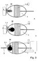
Fig. 3 : Ausführung der Vorrichtung mit einem kugelförmigen Behälter - Bei einer Ausführung wie sie in
Fig. 1 dargestellt ist, besteht der Behälter 1 der Vorrichtung aus einem Hohlzylinder dessen eine Bodenfläche 1.1 perforiert ist. An der gegenüberliegenden Bodenfläche ist eine Kolbenstange mit einem daran befestigten Kolben gehaltert, der die bewegbare Zwischenwand 1.2 darstellt. - Wie in
Fig. 1a gezeigt, befindet sich in diesem Behälter 1, zwischen der perforierten Bodenfläche 1.1 und der bewegbaren Zwischenwand 1.2, die Raubwanze W. Der Behälter 1 wird nun mit seiner perforierten Bodenfläche 1.1 derart am Fokustier F positioniert, dass die Wanze zur Blutabnahme an das Fokustier F herankommt und mit ihrem Saugrüssel durch die perforierte Bodenfläche stechen und 1.1 das Blut aus dem Fokustier F saugen kann. DieFig. 1b zeigt die Wanze W in dem Behälter 1 an der perforierten Außenwand 1.1 kurz vor Ende des Saugvorganges, erkennbar an dem angeschwollenen Abdomen der Wanze W. - In
Fig. 1c ist zu erkennen, dass die Wanze nach Ende des Saugvorganges mittels einer bewegbaren Zwischenwand 1.2, die über eine an der gegenüberliegenden Bodenfläche gehalterte Kolbenstange an die perforierte Bodenfläche 1.1 niedergedrückt ist, wodurch die Bewegungsfreiheit der Wanze auf nahezu Null eingeschränkt worden ist, fixiert ist. Mit Spritze und Kanüle 3 kann dann von der fixierten Wanze W das Blut aus dem angeschwollenen Abdomen abgezogen werden. - Eine Ausführung der erfindungsgemäßen Vorrichtung ist in der
Fig. 2 dargestellt. Hierbei besteht die erfindungsgemäße Vorrichtung aus zwei benachbarten Behältern 1, 2, wobei die zumindest teilweise perforierte Außenwand des Behälters 1 die gemeinsame Trennwand 1.1 zum zweiten Behälter 2 darstellt. Dabei befindet sich die Raubwanze W in dem ersten Behälter 1 und das Fokustier F in dem zweiten Behälter 2. - In der
Fig. 2a ist dargestellt, dass das Fokustier F, in diesem Fall ein Kleinsäuger von dem das Blut abgenommen werden soll, mittels eines beweglichen Schiebers 2.1 derart an der perforierten Trennwand 1.1 fixiert wird, dass die Raubwanze W zustechen und Blut saugen kann. - Die
Fig. 2b zeigt wiederum die Wanze W in dem ersten Behälter 1 an der perforierten Trennwand 1.1 kurz vor Ende der Blutaufnahme, was an dem angeschwollenen Abdomen der Wanze W erkennbar ist. - Die
Fig. 2c zeigt, wie nach beendeter Blutaufnahme durch die Raubwanze W diese, analog zu der ersten bevorzugten Ausführungsform gem.Fig. 1 , mittels der bewegbaren Zwischenwand 1.2 an einer zweiten perforierten Außenwand 1.3 fixiert wird. Aus der so fixierten Raubwanze W kann nun mit Spritze und Kanüle 3 das aus dem Fokustier F abgesaugte Blut wieder aus der Wanze W entnommen werden. - Wie in
Fig. 3 dargestellt, besteht eine Vorrichtung darin, einen annähernd kugelförmigen Behälter 1 mit einer teilweise perforierten Außenwand 1.1 und einer bewegbaren Zwischenwand 1.2 zu versehen, die elastisch verformbar ist. Die Zwischenwand 1.2 ist beispielsweise ein Schaumgummikörper, der etwa in der Mitte des Behälters 1 am gesamten Umfang an der Außenwand des kugelförmigen Behälters anliegt. Die Raubwanze W befindet sich in dem einen Teil des kugelförmigen Behälters 1 zwischen der Zwischenwand 1.2 und der perforierten Außenwand 1.1. Nach Ende des Saugvorganges kann die Wanze W zur Blutabnahme mit einer Spritze und Kanüle 3 in dem kugelförmigen Behälter 1 zwischen der perforierten Außenwand 1.1 und der bewegbaren Zwischenwand 1.2 fixiert werden. Das geschieht beispielsweise dadurch, dass durch mechanisches Verschieben der verformbaren Zwischenwand 1.2 zuerst die flexible Zwischenwand 1.2 an der Wanze W anliegt und durch weiteres Verschieben der verformbaren Zwischenwand 1.2 die Wanze W an der perforierten Außenwand 1.1 fixiert wird. Dieser kugel- oder eiförmige Behälter ist vorzugsweise als Vorrichtung zur Blutabnahme bei Vögeln geeignet, indem dieser Behälter in die entsprechenden Nester gelegt wird. - Weiterhin kann ein flexibel verformbarer Behälter 1 mit maschenartiger Gewebestruktur als eine Ausführungsvariante der Vorrichtung dienen, die mittels einer flexiblen Haltevorrichtung, die beispielsweise aus einer Angelrute sowie einer daran befestigten Angelschnur besteht, derart am Fokustier F positioniert werden, dass die Wanze dort das Blut vom Fokustier F saugen kann. Nach Ende des Saugvorganges wird der flexibel verformbare Behälter 1 mit der darin befindlichen Raubwanze W mittels der Angelrute und Angelschnur vom Fokustier F entfernt. Ein zur Blutabnahme erforderliches Fixieren der Wanze W kann beispielsweise durch ein Zusammenfalten der maschenartigen Gewebestruktur des Behälters 1 erreicht werden. Die Blutabnahme aus der Raubwanze W erfolgt analog der vorstehenden Ausführungsvarianten mittels Kanüle und Spritze 3.
Claims (2)
- Vorrichtung zur minimal-invasiven Blutabnahme bei Tieren mittels blutsaugender Raubwanzen
dadurch gekennzeichnet, dass
die Vorrichtung aus einem ersten Behälter (1), der eine erste perforierte Außenwand (1.1), eine zweite perforierte Außenwand (1.3) und eine bewegbare Zwischenwand (1.2) aufweist, und einem zweiten Behälter (2), der mit dem ersten Behälter (1) verbunden ist, besteht, wobei die erste perforierte Außenwand (1.1) des ersten Behälters (1) die gemeinsame Trennwand (1.1) zu dem zweiten Behälter (2) darstellt, wobei sich die Raubwanze (W) in dem ersten Behälter (1) befinden kann und ein Fokustier (F) in dem zweiten Behälter (2) an der perforierten gemeinsamen Trennwand (1.1) positionierbar ist, so dass eine Blutaufnahme durch die Raubwanze (W) an dem Fokustier (F) erfolgen kann und nach erfolgter Blutaufnahme, die Raubwanze (W) mittels der bewegbaren Zwischenwand (1.2) an der zweiten perforierten Außenwand (1.3) fixierbar ist, derart, dass das Blut aus dem Abdomen der Raubwanze (W) mittels Injektionsspritze wieder entnehmbar ist. - Vorrichtung nach Anspruch 1
dadurch gekennzeichnet, dass
der zweite Behälter (2) einen beweglichen Schieber (2.1) aufweist, mit welchem das Fokustier (F) in dem zweiten Behälter (2) an der perforierten gemeinsamen Trennwand (1.1) positionierbar ist.
Applications Claiming Priority (2)
| Application Number | Priority Date | Filing Date | Title |
|---|---|---|---|
| DE102004004066A DE102004004066B3 (de) | 2004-01-20 | 2004-01-20 | Vorrichtung zur minimal-invasiven Blutabnahme bei Tieren mittels blutsaugender Raubwanzen |
| PCT/DE2005/000087 WO2005067798A1 (de) | 2004-01-20 | 2005-01-20 | Vorrichtung zur minimal-invasiven blutabnahme bei tieren mittels blutsaugender raubwanzen |
Publications (2)
| Publication Number | Publication Date |
|---|---|
| EP1706033A1 EP1706033A1 (de) | 2006-10-04 |
| EP1706033B1 true EP1706033B1 (de) | 2010-07-14 |
Family
ID=34559867
Family Applications (1)
| Application Number | Title | Priority Date | Filing Date |
|---|---|---|---|
| EP05714892A Expired - Lifetime EP1706033B1 (de) | 2004-01-20 | 2005-01-20 | Vorrichtung zur minimal-invasiven blutabnahme bei tieren mittels blutsaugender raubwanzen |
Country Status (5)
| Country | Link |
|---|---|
| US (1) | US20080221536A1 (de) |
| EP (1) | EP1706033B1 (de) |
| AT (1) | ATE473682T1 (de) |
| DE (2) | DE102004004066B3 (de) |
| WO (1) | WO2005067798A1 (de) |
Cited By (1)
| Publication number | Priority date | Publication date | Assignee | Title |
|---|---|---|---|---|
| US9226489B2 (en) | 2011-03-18 | 2016-01-05 | Ecolab Usa Inc. | Heat system for killing pests |
Families Citing this family (4)
| Publication number | Priority date | Publication date | Assignee | Title |
|---|---|---|---|---|
| CN108175421B (zh) * | 2017-12-30 | 2019-07-23 | 枣庄市妇幼保健院 | 高效节能血液采集存储试管 |
| RU196880U1 (ru) * | 2019-12-16 | 2020-03-18 | Федеральное государственное казенное военное образовательное учреждение высшего образования "Военный учебно-научный центр Военно-Морского Флота "Военно-морская академия им. Адмирала Флота Советского Союза Н.Г. Кузнецова" | Одноразовый шприц для временного хранения и транспортировки иксодового клеща |
| CN111938666B (zh) * | 2020-07-15 | 2024-07-23 | 张国华 | 一种仿蚊子式危险动物血液提取装置 |
| MX2023013218A (es) * | 2023-11-08 | 2025-06-02 | Jesus Raul Beltran Ramirez | Dispositivo para investigacion de la enfermedad de lyme con chinches y garrapatas. |
Family Cites Families (6)
| Publication number | Priority date | Publication date | Assignee | Title |
|---|---|---|---|---|
| US5320069A (en) * | 1992-04-28 | 1994-06-14 | The United States Of America As Represented By The Secretary Of The Army | Small animal restraint device |
| US5849262A (en) * | 1994-05-17 | 1998-12-15 | Heska Corporation | Bioassay system for arthropods which elastically attaches to an animal |
| US5464360A (en) * | 1994-07-11 | 1995-11-07 | Carlson; Robert R. | Container for shipping and utilizing stinging insects |
| US6226919B1 (en) * | 2000-05-30 | 2001-05-08 | Donald R. Septer | Insect vacuum trap |
| US20040069235A1 (en) * | 2002-09-27 | 2004-04-15 | Rasa Cordelia G. | Flea feeding apparatus |
| DE10259651A1 (de) * | 2002-12-18 | 2004-07-01 | Universität Regensburg | Insektenfalle |
-
2004
- 2004-01-20 DE DE102004004066A patent/DE102004004066B3/de not_active Expired - Fee Related
-
2005
- 2005-01-20 US US10/586,472 patent/US20080221536A1/en not_active Abandoned
- 2005-01-20 EP EP05714892A patent/EP1706033B1/de not_active Expired - Lifetime
- 2005-01-20 DE DE502005009895T patent/DE502005009895D1/de not_active Expired - Lifetime
- 2005-01-20 WO PCT/DE2005/000087 patent/WO2005067798A1/de not_active Ceased
- 2005-01-20 AT AT05714892T patent/ATE473682T1/de not_active IP Right Cessation
Cited By (6)
| Publication number | Priority date | Publication date | Assignee | Title |
|---|---|---|---|---|
| US9226489B2 (en) | 2011-03-18 | 2016-01-05 | Ecolab Usa Inc. | Heat system for killing pests |
| US10070639B2 (en) | 2011-03-18 | 2018-09-11 | Ecolab Usa Inc. | Heat system for killing pests |
| US11013226B2 (en) | 2011-03-18 | 2021-05-25 | Ecolab Usa Inc. | Heat system for killing pests |
| US11737445B2 (en) | 2011-03-18 | 2023-08-29 | Ecolab Usa Inc. | Heat system for killing pests |
| US12063921B2 (en) | 2011-03-18 | 2024-08-20 | Ecolab Usa Inc. | Heat system for killing pests |
| US12408650B2 (en) | 2011-03-18 | 2025-09-09 | Ecolab Usa Inc. | Heat system for killing pests |
Also Published As
| Publication number | Publication date |
|---|---|
| EP1706033A1 (de) | 2006-10-04 |
| DE502005009895D1 (de) | 2010-08-26 |
| US20080221536A1 (en) | 2008-09-11 |
| ATE473682T1 (de) | 2010-07-15 |
| WO2005067798A1 (de) | 2005-07-28 |
| DE102004004066B3 (de) | 2005-06-09 |
Similar Documents
| Publication | Publication Date | Title |
|---|---|---|
| DE69326605T2 (de) | Vorrichtung zur Implantation von Sensoren | |
| DE69027838T2 (de) | Einwegvorrichtung zur Biopsie von Weichgewebe | |
| DE69929433T2 (de) | Katheter zur entnahme von zellulärem material aus der brustdrüse | |
| DE3887145T2 (de) | Medizinische Testvorrichtung. | |
| DE3108766C2 (de) | Medizinische Stichsondenmeßvorrichtung | |
| DE69121917T2 (de) | Vorrichtung zum anbringen eines drains | |
| DE60016579T2 (de) | Kit zur diagnose und behandlung des herzens | |
| DE10057215B4 (de) | Systeme zur Extrapolation einer Glucosekonzentration und zur Ermittlung von zu verabreichenden Insulingaben oder vorzunehmender Kohlenhydrataufnahmen | |
| DE69932612T2 (de) | Gerät zur erleichterten hemostase eines biopsietraktes | |
| EP1371329A1 (de) | Blutentnahmesystem | |
| DE2819574A1 (de) | Verfahren und vorrichtung zur verarbeitung einer koerperfluessigkeitsprobe | |
| DE69221036T2 (de) | Verfahren zur Blutabnahme | |
| DE102004004066B3 (de) | Vorrichtung zur minimal-invasiven Blutabnahme bei Tieren mittels blutsaugender Raubwanzen | |
| DE102021002430A1 (de) | System zur Entnahme, Aufbewahrung, Transport und Weiterverarbeitung von Blutproben, sowie Anwendungsverfahren dafür. | |
| DE3850728T2 (de) | Methode zur Bestimmung des Verhaltens von Medikamenten in Lebewesen. | |
| DE102010025921A1 (de) | Verfahren zur stabilen Lagerung einer in einen Patienten einzuführenden Nadel, Haltevorrichtung und medizinische Behandlungsvorrichtung | |
| DE102018107024A1 (de) | Vorrichtung zur intravitrealen Injektion in ein Auge | |
| DE2723240A1 (de) | Verfahren und einrichtung zur entnahme bzw. gewinnung von untersuchungsmaterialproben aus zellkulturen | |
| DE1791178A1 (de) | Punktionsgeraet zur Entnahme von Koerpergewebe fuer die histologische Diagnosestellung in der Human- und Veterinaermedizin | |
| DE19520883A1 (de) | Vorrichtung zur Blutentnahme mit einem zweikammrigen System | |
| DE102014106170A1 (de) | Vorrichtung, Einrichtung und Verfahren zum Auf- und Einbringen wenigstens einer Allergen-Testlösung auf und in die Haut eines Körperteils einer zu testenden Person | |
| WO2011051443A1 (de) | Analyseeinrichtung und verfahren zur ganzheitlichen analyse einer testperson | |
| DE1083506B (de) | Injektionseinrichtung | |
| DE102006049800B4 (de) | Gerät zur Blutentnahme | |
| DE10064458C1 (de) | Nadelhalter |
Legal Events
| Date | Code | Title | Description |
|---|---|---|---|
| PUAI | Public reference made under article 153(3) epc to a published international application that has entered the european phase |
Free format text: ORIGINAL CODE: 0009012 |
|
| 17P | Request for examination filed |
Effective date: 20060706 |
|
| AK | Designated contracting states |
Kind code of ref document: A1 Designated state(s): AT BE BG CH CY CZ DE DK EE ES FI FR GB GR HU IE IS IT LI LT LU MC NL PL PT RO SE SI SK TR |
|
| RIN1 | Information on inventor provided before grant (corrected) |
Inventor name: THOMSEN, RUTH Inventor name: VOIGT, CHRISTIAN |
|
| DAX | Request for extension of the european patent (deleted) | ||
| 17Q | First examination report despatched |
Effective date: 20080922 |
|
| GRAJ | Information related to disapproval of communication of intention to grant by the applicant or resumption of examination proceedings by the epo deleted |
Free format text: ORIGINAL CODE: EPIDOSDIGR1 |
|
| GRAP | Despatch of communication of intention to grant a patent |
Free format text: ORIGINAL CODE: EPIDOSNIGR1 |
|
| RIN1 | Information on inventor provided before grant (corrected) |
Inventor name: VOIGT, CHRISTIAN Inventor name: THOMSEN-HAEFLIGER, RUTH |
|
| GRAP | Despatch of communication of intention to grant a patent |
Free format text: ORIGINAL CODE: EPIDOSNIGR1 |
|
| GRAS | Grant fee paid |
Free format text: ORIGINAL CODE: EPIDOSNIGR3 |
|
| GRAA | (expected) grant |
Free format text: ORIGINAL CODE: 0009210 |
|
| AK | Designated contracting states |
Kind code of ref document: B1 Designated state(s): AT BE BG CH CY CZ DE DK EE ES FI FR GB GR HU IE IS IT LI LT LU MC NL PL PT RO SE SI SK TR |
|
| REG | Reference to a national code |
Ref country code: GB Ref legal event code: FG4D Free format text: NOT ENGLISH |
|
| REG | Reference to a national code |
Ref country code: CH Ref legal event code: EP |
|
| REG | Reference to a national code |
Ref country code: IE Ref legal event code: FG4D |
|
| REF | Corresponds to: |
Ref document number: 502005009895 Country of ref document: DE Date of ref document: 20100826 Kind code of ref document: P |
|
| REG | Reference to a national code |
Ref country code: CH Ref legal event code: NV Representative=s name: PATENTANWAELTE SCHAAD, BALASS, MENZL & PARTNER AG |
|
| REG | Reference to a national code |
Ref country code: NL Ref legal event code: VDEP Effective date: 20100714 |
|
| LTIE | Lt: invalidation of european patent or patent extension |
Effective date: 20100714 |
|
| PG25 | Lapsed in a contracting state [announced via postgrant information from national office to epo] |
Ref country code: LT Free format text: LAPSE BECAUSE OF FAILURE TO SUBMIT A TRANSLATION OF THE DESCRIPTION OR TO PAY THE FEE WITHIN THE PRESCRIBED TIME-LIMIT Effective date: 20100714 Ref country code: NL Free format text: LAPSE BECAUSE OF FAILURE TO SUBMIT A TRANSLATION OF THE DESCRIPTION OR TO PAY THE FEE WITHIN THE PRESCRIBED TIME-LIMIT Effective date: 20100714 Ref country code: FI Free format text: LAPSE BECAUSE OF FAILURE TO SUBMIT A TRANSLATION OF THE DESCRIPTION OR TO PAY THE FEE WITHIN THE PRESCRIBED TIME-LIMIT Effective date: 20100714 |
|
| REG | Reference to a national code |
Ref country code: IE Ref legal event code: FD4D |
|
| PG25 | Lapsed in a contracting state [announced via postgrant information from national office to epo] |
Ref country code: PL Free format text: LAPSE BECAUSE OF FAILURE TO SUBMIT A TRANSLATION OF THE DESCRIPTION OR TO PAY THE FEE WITHIN THE PRESCRIBED TIME-LIMIT Effective date: 20100714 Ref country code: CY Free format text: LAPSE BECAUSE OF FAILURE TO SUBMIT A TRANSLATION OF THE DESCRIPTION OR TO PAY THE FEE WITHIN THE PRESCRIBED TIME-LIMIT Effective date: 20100714 Ref country code: SI Free format text: LAPSE BECAUSE OF FAILURE TO SUBMIT A TRANSLATION OF THE DESCRIPTION OR TO PAY THE FEE WITHIN THE PRESCRIBED TIME-LIMIT Effective date: 20100714 Ref country code: BG Free format text: LAPSE BECAUSE OF FAILURE TO SUBMIT A TRANSLATION OF THE DESCRIPTION OR TO PAY THE FEE WITHIN THE PRESCRIBED TIME-LIMIT Effective date: 20101014 Ref country code: IS Free format text: LAPSE BECAUSE OF FAILURE TO SUBMIT A TRANSLATION OF THE DESCRIPTION OR TO PAY THE FEE WITHIN THE PRESCRIBED TIME-LIMIT Effective date: 20101114 Ref country code: PT Free format text: LAPSE BECAUSE OF FAILURE TO SUBMIT A TRANSLATION OF THE DESCRIPTION OR TO PAY THE FEE WITHIN THE PRESCRIBED TIME-LIMIT Effective date: 20101115 |
|
| PG25 | Lapsed in a contracting state [announced via postgrant information from national office to epo] |
Ref country code: SE Free format text: LAPSE BECAUSE OF FAILURE TO SUBMIT A TRANSLATION OF THE DESCRIPTION OR TO PAY THE FEE WITHIN THE PRESCRIBED TIME-LIMIT Effective date: 20100714 Ref country code: GR Free format text: LAPSE BECAUSE OF FAILURE TO SUBMIT A TRANSLATION OF THE DESCRIPTION OR TO PAY THE FEE WITHIN THE PRESCRIBED TIME-LIMIT Effective date: 20101015 |
|
| PG25 | Lapsed in a contracting state [announced via postgrant information from national office to epo] |
Ref country code: DK Free format text: LAPSE BECAUSE OF FAILURE TO SUBMIT A TRANSLATION OF THE DESCRIPTION OR TO PAY THE FEE WITHIN THE PRESCRIBED TIME-LIMIT Effective date: 20100714 Ref country code: IE Free format text: LAPSE BECAUSE OF FAILURE TO SUBMIT A TRANSLATION OF THE DESCRIPTION OR TO PAY THE FEE WITHIN THE PRESCRIBED TIME-LIMIT Effective date: 20100714 |
|
| PLBE | No opposition filed within time limit |
Free format text: ORIGINAL CODE: 0009261 |
|
| STAA | Information on the status of an ep patent application or granted ep patent |
Free format text: STATUS: NO OPPOSITION FILED WITHIN TIME LIMIT |
|
| PG25 | Lapsed in a contracting state [announced via postgrant information from national office to epo] |
Ref country code: SK Free format text: LAPSE BECAUSE OF FAILURE TO SUBMIT A TRANSLATION OF THE DESCRIPTION OR TO PAY THE FEE WITHIN THE PRESCRIBED TIME-LIMIT Effective date: 20100714 Ref country code: RO Free format text: LAPSE BECAUSE OF FAILURE TO SUBMIT A TRANSLATION OF THE DESCRIPTION OR TO PAY THE FEE WITHIN THE PRESCRIBED TIME-LIMIT Effective date: 20100714 Ref country code: IT Free format text: LAPSE BECAUSE OF FAILURE TO SUBMIT A TRANSLATION OF THE DESCRIPTION OR TO PAY THE FEE WITHIN THE PRESCRIBED TIME-LIMIT Effective date: 20100714 Ref country code: CZ Free format text: LAPSE BECAUSE OF FAILURE TO SUBMIT A TRANSLATION OF THE DESCRIPTION OR TO PAY THE FEE WITHIN THE PRESCRIBED TIME-LIMIT Effective date: 20100714 Ref country code: EE Free format text: LAPSE BECAUSE OF FAILURE TO SUBMIT A TRANSLATION OF THE DESCRIPTION OR TO PAY THE FEE WITHIN THE PRESCRIBED TIME-LIMIT Effective date: 20100714 |
|
| 26N | No opposition filed |
Effective date: 20110415 |
|
| PG25 | Lapsed in a contracting state [announced via postgrant information from national office to epo] |
Ref country code: ES Free format text: LAPSE BECAUSE OF FAILURE TO SUBMIT A TRANSLATION OF THE DESCRIPTION OR TO PAY THE FEE WITHIN THE PRESCRIBED TIME-LIMIT Effective date: 20101025 |
|
| REG | Reference to a national code |
Ref country code: DE Ref legal event code: R097 Ref document number: 502005009895 Country of ref document: DE Effective date: 20110415 |
|
| BERE | Be: lapsed |
Owner name: FORSCHUNGSVERBUND BERLIN E.V. Effective date: 20110131 |
|
| PG25 | Lapsed in a contracting state [announced via postgrant information from national office to epo] |
Ref country code: MC Free format text: LAPSE BECAUSE OF NON-PAYMENT OF DUE FEES Effective date: 20110131 |
|
| REG | Reference to a national code |
Ref country code: FR Ref legal event code: ST Effective date: 20110930 |
|
| PG25 | Lapsed in a contracting state [announced via postgrant information from national office to epo] |
Ref country code: FR Free format text: LAPSE BECAUSE OF NON-PAYMENT OF DUE FEES Effective date: 20110131 |
|
| PG25 | Lapsed in a contracting state [announced via postgrant information from national office to epo] |
Ref country code: BE Free format text: LAPSE BECAUSE OF NON-PAYMENT OF DUE FEES Effective date: 20110131 |
|
| REG | Reference to a national code |
Ref country code: AT Ref legal event code: MM01 Ref document number: 473682 Country of ref document: AT Kind code of ref document: T Effective date: 20110120 |
|
| PG25 | Lapsed in a contracting state [announced via postgrant information from national office to epo] |
Ref country code: AT Free format text: LAPSE BECAUSE OF NON-PAYMENT OF DUE FEES Effective date: 20110120 |
|
| PG25 | Lapsed in a contracting state [announced via postgrant information from national office to epo] |
Ref country code: LU Free format text: LAPSE BECAUSE OF NON-PAYMENT OF DUE FEES Effective date: 20110120 |
|
| PG25 | Lapsed in a contracting state [announced via postgrant information from national office to epo] |
Ref country code: TR Free format text: LAPSE BECAUSE OF FAILURE TO SUBMIT A TRANSLATION OF THE DESCRIPTION OR TO PAY THE FEE WITHIN THE PRESCRIBED TIME-LIMIT Effective date: 20100714 |
|
| PG25 | Lapsed in a contracting state [announced via postgrant information from national office to epo] |
Ref country code: HU Free format text: LAPSE BECAUSE OF FAILURE TO SUBMIT A TRANSLATION OF THE DESCRIPTION OR TO PAY THE FEE WITHIN THE PRESCRIBED TIME-LIMIT Effective date: 20100714 |
|
| PGFP | Annual fee paid to national office [announced via postgrant information from national office to epo] |
Ref country code: DE Payment date: 20180129 Year of fee payment: 14 Ref country code: GB Payment date: 20180125 Year of fee payment: 14 Ref country code: CH Payment date: 20180125 Year of fee payment: 14 |
|
| REG | Reference to a national code |
Ref country code: DE Ref legal event code: R119 Ref document number: 502005009895 Country of ref document: DE |
|
| REG | Reference to a national code |
Ref country code: CH Ref legal event code: PL |
|
| GBPC | Gb: european patent ceased through non-payment of renewal fee |
Effective date: 20190120 |
|
| PG25 | Lapsed in a contracting state [announced via postgrant information from national office to epo] |
Ref country code: DE Free format text: LAPSE BECAUSE OF NON-PAYMENT OF DUE FEES Effective date: 20190801 |
|
| PG25 | Lapsed in a contracting state [announced via postgrant information from national office to epo] |
Ref country code: CH Free format text: LAPSE BECAUSE OF NON-PAYMENT OF DUE FEES Effective date: 20190131 Ref country code: LI Free format text: LAPSE BECAUSE OF NON-PAYMENT OF DUE FEES Effective date: 20190131 Ref country code: GB Free format text: LAPSE BECAUSE OF NON-PAYMENT OF DUE FEES Effective date: 20190120 |