EP1700120B1 - Marker for neuromyelitis optica - Google Patents
Marker for neuromyelitis optica Download PDFInfo
- Publication number
- EP1700120B1 EP1700120B1 EP04812269A EP04812269A EP1700120B1 EP 1700120 B1 EP1700120 B1 EP 1700120B1 EP 04812269 A EP04812269 A EP 04812269A EP 04812269 A EP04812269 A EP 04812269A EP 1700120 B1 EP1700120 B1 EP 1700120B1
- Authority
- EP
- European Patent Office
- Prior art keywords
- nmo
- antigenic polypeptide
- aquaporin
- biological sample
- individual
- Prior art date
- Legal status (The legal status is an assumption and is not a legal conclusion. Google has not performed a legal analysis and makes no representation as to the accuracy of the status listed.)
- Expired - Lifetime
Links
Images
Classifications
-
- C—CHEMISTRY; METALLURGY
- C07—ORGANIC CHEMISTRY
- C07K—PEPTIDES
- C07K16/00—Immunoglobulins [IG], e.g. monoclonal or polyclonal antibodies
- C07K16/18—Immunoglobulins [IG], e.g. monoclonal or polyclonal antibodies against material from animals or humans
-
- C—CHEMISTRY; METALLURGY
- C07—ORGANIC CHEMISTRY
- C07K—PEPTIDES
- C07K14/00—Peptides having more than 20 amino acids; Gastrins; Somatostatins; Melanotropins; Derivatives thereof
- C07K14/435—Peptides having more than 20 amino acids; Gastrins; Somatostatins; Melanotropins; Derivatives thereof from animals; from humans
- C07K14/705—Receptors; Cell surface antigens; Cell surface determinants
-
- A—HUMAN NECESSITIES
- A61—MEDICAL OR VETERINARY SCIENCE; HYGIENE
- A61P—SPECIFIC THERAPEUTIC ACTIVITY OF CHEMICAL COMPOUNDS OR MEDICINAL PREPARATIONS
- A61P25/00—Drugs for disorders of the nervous system
- A61P25/28—Drugs for disorders of the nervous system for treating neurodegenerative disorders of the central nervous system, e.g. nootropic agents, cognition enhancers, drugs for treating Alzheimer's disease or other forms of dementia
-
- A—HUMAN NECESSITIES
- A61—MEDICAL OR VETERINARY SCIENCE; HYGIENE
- A61P—SPECIFIC THERAPEUTIC ACTIVITY OF CHEMICAL COMPOUNDS OR MEDICINAL PREPARATIONS
- A61P37/00—Drugs for immunological or allergic disorders
-
- C—CHEMISTRY; METALLURGY
- C07—ORGANIC CHEMISTRY
- C07K—PEPTIDES
- C07K16/00—Immunoglobulins [IG], e.g. monoclonal or polyclonal antibodies
- C07K16/18—Immunoglobulins [IG], e.g. monoclonal or polyclonal antibodies against material from animals or humans
- C07K16/28—Immunoglobulins [IG], e.g. monoclonal or polyclonal antibodies against material from animals or humans against receptors, cell surface antigens or cell surface determinants
-
- G—PHYSICS
- G01—MEASURING; TESTING
- G01N—INVESTIGATING OR ANALYSING MATERIALS BY DETERMINING THEIR CHEMICAL OR PHYSICAL PROPERTIES
- G01N33/00—Investigating or analysing materials by specific methods not covered by groups G01N1/00 - G01N31/00
- G01N33/48—Biological material, e.g. blood, urine; Haemocytometers
- G01N33/50—Chemical analysis of biological material, e.g. blood, urine; Testing involving biospecific ligand binding methods; Immunological testing
- G01N33/53—Immunoassay; Biospecific binding assay; Materials therefor
- G01N33/564—Immunoassay; Biospecific binding assay; Materials therefor for pre-existing immune complex or autoimmune disease, i.e. systemic lupus erythematosus, rheumatoid arthritis, multiple sclerosis, rheumatoid factors or complement components C1-C9
-
- C—CHEMISTRY; METALLURGY
- C07—ORGANIC CHEMISTRY
- C07K—PEPTIDES
- C07K2317/00—Immunoglobulins specific features
- C07K2317/20—Immunoglobulins specific features characterized by taxonomic origin
- C07K2317/21—Immunoglobulins specific features characterized by taxonomic origin from primates, e.g. man
-
- G—PHYSICS
- G01—MEASURING; TESTING
- G01N—INVESTIGATING OR ANALYSING MATERIALS BY DETERMINING THEIR CHEMICAL OR PHYSICAL PROPERTIES
- G01N2800/00—Detection or diagnosis of diseases
- G01N2800/28—Neurological disorders
- G01N2800/285—Demyelinating diseases; Multipel sclerosis
Definitions
- This invention relates generally to neurological disorders, and more particularly, to an autoantibody marker for neuromyelitis optica, relapsing myelitis or optic neuritis, and the Asian opticospinal form of multiple sclerosis (MS).
- MS Asian opticospinal form of multiple sclerosis
- NMO Neuromyelitis optica
- MS multiple sclerosis
- non-Caucasians represent a higher frequency of patients with NMO than the frequency of those with classical MS.
- the characteristic inflammatory demyelinating lesions of NMO selectively and repeatedly affect the optic nerves and the spinal cord, thereby causing both blindness and paralysis.
- Haase et al (2001, Neuroscli. Lett., 307(2):131-3 ), describes the detection of brain-specific autoantibodies to myelin oligodendrocyte glycoprotein, S100beta and myelin basic protein in patients with Devic's neuromyelitis optica. Auto antibody responses to myelin and astroglial antigens in four patients with NMO was examined.
- NMO neuromyelitis optica
- MS Asian opticospinal form of MS.
- NMO-specific antibody can be used to distinguish NMO from MS, and also can be used to diagnose NMO at an early stage of the disease before all clinical criteria are fulfilled, thus justifying early initiation of NMO-appropriate immunosuppressive or immunomodulatory therapy.
- the invention provides methods of detecting the presence or absence of a NMO-specific autoantibody in a biological sample from an individual.
- a method includes contacting the biological sample with a NMO antigenic polypeptide or fragment thereof, where the NMO antigenic polypeptide is aquaporin-4 or a protein that interacts with the astrocytic dystrophin protein complex; and detecting the presence or absence of binding of the NMO antigenic polypeptide to the NMO-specific autoantibody in the biological sample.
- the NMO antigenic polypeptide is a recombinantly-expressed NMO antigenic polypeptide.
- the NMO-specific polypeptide is in a solid tissue selected from the group consisting of brain, spinal cord, optic nerve, kidney, or stomach.
- the presence of the NMO-specific autoantibody in the biological sample can be associated with vision impairment, weakness, numbness, spasms or abnormal or painful sensations, and/or loss of bladder and/or bowel control in the individual.
- the presence of the NMO-specific autoantibody is generally associated with NMO in the individual.
- Representative biological sample is selected from the group consisting of blood, serum, plasma, and cerebrospinal fluid.
- the invention provides methods of detecting the presence or absence of a NMO antigenic polypeptide in a biological sample from an individual.
- a method includes contacting the biological sample with an anti-NMO antigen antibody, where the NMO antigen is aquaporin-4; and detecting binding of the anti-NMO antigen antibody to the biological sample, wherein binding is indicative of the presence of the NMO antigenic polypeptide in the biological sample.
- the presence of the NMO antigenic polypeptide in the biological sample is indicative of NMO in the individual.
- Representative biological samples include blood, serum, plasma, cerebrospinal fluid, brain biopsy, and spinal cord biopsy.
- the invention provides the use of an article of manufacture including a NMO antigenic polypeptide for in vitro diagnosis of NMO to detect an anti-NMO antigen autoantibody in an individual.
- the NMO antigenic polypeptide is aquaporin-4.
- Such an article of manufacture can be used to diagnose NMO in the individual.
- the article of manufacture can further include a monoclonal antibody having specific binding affinity for a NMO antigenic polypeptide.
- Methods of treating an individual having NMO include withdrawing a body fluid from the individual wherein the body fluid contains one or more autoantibodies that bind to aquaporin-4; removing a substantial portion of the autoantibodies from the body fluid; and returning the body fluid to the subject.
- the invention provides medical uses for methods of treating an individual having NMO by administering a NMO antigenic polypeptide to the individual.
- the NMO antigenic polypeptide is aquaporin-4.
- administration is by a method such as oral, intravenous, and parenteral administration.
- the invention provides medical uses for methods of treating an individual having NMO by administering a nucleic acid encoding a NMO antigenic polypeptide to the individual.
- the NMO antigenic polypeptide is aquaporin-4.
- a specific IgG autoantibody marker has been identified in serum and cerebrospinal fluid of patients with neuromyelitis optica (NMO) and Asian opticospinal MS (NMO is used synonymously in this application to refer to both NMO and Asian opticospinal MS).
- NMO neuromyelitis optica
- NMO Asian opticospinal MS
- this autoantibody can be found in patients with a spectrum of inflammatory disorders involving spinal cord, optic nerve, and more rarely, brainstem or other regions of the brain, that were not hitherto recognized as being related to NMO.
- NMO-specific antibody can be used to diagnose NMO at an early stage of the disease before all clinical criteria are fulfilled, thus justifying early initiation of NMO-appropriate immunosuppressant therapy, and further can be used to distinguish NMO from MS. Detecting NMO-specific antibody also can provide a quantifiable biomarker for monitoring disease progression and response to therapy. The pattern of immunostaining also can be used to classify disorders related to NMO, including variants of multiple sclerosis, isolated optic neuritides, certain myelopathies and "optico-spinal" accompaniments of systemic lupus erythematosus and Sjogren's syndrome.
- an antibody associated with NMO provides a valuable IgG tool for developing animal models (e.g., by passive transfer of NMO-specific antibodies or active immunization with NMO antigenic polypeptides or DNA vaccines encoding such antigenic polypeptides) to investigate the pathogenesis of NMO lesions and to test potential new therapies for NMO.
- the present invention provides for methods of detecting NMO-specific autoantibodies in an individual using NMO antigenic polypeptides.
- Individuals for whom the methods of the invention might be used typically present with vision impairment and/or tingling, numbness, weakness, limb spasms, loss of bladder and/or bowel control, or other neurological symptoms of unknown origin.
- the method of the invention is based on an association between the abnormal neurological symptoms and the presence of the NMO-specific autoantibodies in the individual.
- NMO antigenic polypeptides include to one or more epitopic sites. Epitopes of NMO antigenic polypeptides that are pertinent to T-cell activation and suppression are also disclosed. Computer algorithms are available for predicting binding epitopes, e.g ., MHC-I and MHC-II binding epitopes. See, for example, bimas.dcrt.nih.gov:80/molbio/hla bind/ on the World Wide Web ( Parker et al., J. Immunol., 152:163 (1994 ); Southwood et al., J. Immunol., 160:3363 (1998 )).
- characteristic in this context means that the epitopic site allows immunologic detection of NMO-specific antibody in sera with reasonable assurance. Usually, it is desirable that the epitopic site be antigenically distinct from other closely related antigens (e.g., other members of a family of polypeptides).
- a representative antigenic fragment can include, for example, the extracellular domain of a membrane-bound protein.
- the NMO antigenic polypeptides may be obtained from cells ( e.g ., transfected host cells) expressing a nucleic acid, or the polypeptides may be synthetic.
- a DNA molecule encoding a NMO antigenic polypeptide or fragment thereof may itself be natural or synthetic, with natural genes obtainable from human tissues by conventional techniques.
- the NMO antigenic polypeptides can be obtained in a substantially pure form.
- purified refers to a polypeptide that constitutes the major component in a mixture of components, e.g., 50% or more, 60% or more, 70% or more, 80% or more, 90% or more, or 95% or more, by weight.
- Purified polypeptides are typically obtained by purification from an organism that makes the polypeptide, although chemical synthesis is also feasible.
- the polypeptides may be purified by routine protein purification methods, including affinity chromatography or immunosorbant affinity column.
- NMO antigenic polypeptides used in the present invention may be used with or without modification for the detection of NMO-specific autoantibodies.
- polypeptides are labeled by either covalently or non-covalently combining the polypeptide with a second substance that provides for detectable signal.
- labels and conjugation techniques are known in the art and are reported extensively in both the scientific and patent literature. Some of the labels include radioisotopes, enzymes, substrates, cofactors, inhibitors, fluorescers, chemiluminescers, magnetic particles, and the like.
- NMO antigenic polypeptides prepared as described above can be used in various immunological techniques for detecting NMO-specific autoantibodies in biological samples, such as from serum and cerebrospinal fluid. Depending on the nature of the sample, either or both immunoassays and immunocytochemical staining techniques may be used. Enzyme-linked immunosorbent assays (ELISA), Western blot, and radioimmunoassays are routine methods in the art and may be used to detect the presence of NMO-specific autoantibodies in sera.
- ELISA enzyme-linked immunosorbent assays
- Western blot Western blot
- radioimmunoassays are routine methods in the art and may be used to detect the presence of NMO-specific autoantibodies in sera.
- kits containing one or more NMO antigenic polypeptides may further include a second substance that provides for detectable signal.
- a kit typically also includes directions for using the NMO antigenic polypeptide and/or for practicing a method of the invention ( i.e ., detecting NMO-specific autoantibodies in a biological sample).
- the present invention also provides for methods of detecting NMO antigenic polypeptides in a biological sample from an individual.
- the method describes an association between the presence of abnormal levels or pattern of expression of the NMO antigenic polypeptide, the subsequently produced NMO-specific autoantibody, and the resulting NMO in the individuals. This test is most widely applicable to those individuals who present with neurological symptoms such as blindness, and those individuals who are suspected of having MS or NMO.
- Detection of a polypeptide is typically performed using an antibody, referred to herein as an anti-NMO antigen antibody to distinguish such animal- or recombinantly-generated antibodies from NMO-specific autoantibodies produced by an individual's immune system.
- the invention also provides for the use an antibody, including a monoclonal antibody with specific binding affinity for NMO antigenic polypeptides.
- anti-NMO antigen antibodies having specific binding affinity for the NMO antigenic polypeptide may be produced by techniques well known to those of ordinary skill in this art.
- anti-NMO antigen antibodies having "specific binding affinity" for NMO antigenic polypeptides are defined as those antibodies that bind NMO antigenic polypeptides but that do not bind other polypeptides, for example, other members of a family of polypeptides.
- anti-NMO antigen antibody refers to whole antibodies of any class, i.e ., IgG, IgA, IgM, IgE, or any other known class, and also includes portions or fragments of whole antibodies (e.g ., Fab or (Fab) 2 fragments) having the desired specific binding affinity, an engineered single chain Fv molecule, or a chimeric molecule, e.g ., an antibody that contains the binding specificity of one antibody (e.g ., of murine origin) and the remaining portions of another antibody (e.g ., of human origin).
- Anti-NMO antigen antibodies used in the present invention may be used with or without modification for the detection of NMO antigenic polypeptides.
- Anti-NMO antigen antibodies can be labeled either directly or indirectly, and a wide variety of labels, including radioisotopes, enzymes, substrates, cofactors, inhibitors, fluorescers, chemiluminescers and magnetic particles, and conjugation techniques are known and are reported extensively in both the scientific and patent literature.
- Anti-NMO antigen antibodies prepared as described above can be used in various immunological techniques for detecting NMO antigenic polypeptides in a biological sample.
- a "biological sample,” as used herein, is generally a sample from an individual. Non-limiting examples of biological samples include blood, serum, plasma, or cerebrospinal fluid. Additionally, solid tissues, for example, spinal cord or brain biopsies may be used. The use of antibodies in protein binding assays is well established. Depending on the nature of the sample, immunoassays (e.g ., radioimmunoassays) and/or immunohistochemical/immunocytochemical staining techniques may be used.
- immunoassays e.g ., radioimmunoassays
- immunohistochemical/immunocytochemical staining techniques may be used.
- Liquid phase immunoassays e.g., competitive inhibition radioimmunoassays
- solid phase immunoassays e.g., antigen-capture or Western blot analysis
- ELISA enzyme-linked immunosorbent assays
- An example of one such competitive assay for detecting the presence of a NMO antigenic polypeptide in a biological sample such as serum comprises: contacting a NMO antigenic polypeptide (either labeled or unlabeled) with an anti-NMO antigen antibody (either labeled or unlabeled) and the biological sample.
- the NMO antigenic polypeptide may be, for example, attached to a solid surface.
- the relative amount of NMO antigenic polypeptide in a biological sample can be determined.
- kits containing anti-NMO antigen antibodies having binding affinity for NMO antigenic polypeptides or fragments thereof may also include NMO antigenic polypeptides or fragments thereof to be used as binding controls or to generate a standardized quantitative curve.
- the kit may further include a second substance that provides for detectable label.
- a kit typically includes directions for using an anti-NMO antigen antibody and/or practicing a method of the invention ( i.e ., detecting NMO antigenic polypeptides in a biological sample).
- an anti-NMO antigen antibody having specific binding affinity for NMO antigenic polypeptides conjugated to a detectable marker.
- Suitable detectable markers include, but are not limited to, enzymes, radioisotopes, dyes and biotin.
- This disclosure further provides an anti-NMO antigen antibody having specific binding affinity for NMO antigenic polypeptides conjugated to an imaging agent.
- Suitable imaging agents include, but are not limited to, radioisotopes, such as 32 P, 99 Tc, 111 In and 131 I.
- NMO N-specific immune mechanisms.
- Methods of treating an individual with NMO include, without limitation, apheresis and T cell receptor-based immunotherapy.
- the disclosure provides a method of removing NMO-specific autoantibodies from a body fluid of an individual.
- the method involves withdrawing a body fluid from a subject; removing a substantial portion of NMO-specific autoantibodies from the fluid; and returning the fluid to the subject.
- Antibodies removed can be of any class, e.g., IgG (such as IgG1, IgG2, IgG3, IgG4), IgM, IgD, IgA, or IgE antibodies.
- a "substantial portion” means removing at least 20% (e.g., at least: 20%; 30%; 40%; 50%; 60%; 65%; 70%; 75%; 80%; 85%; 90%; 93%; 95%; 96%; 97%; 98%; 99%; 99.5%; 99.8%; or even 100%) of the NMO-specific autoantibodies that were present in the body fluid prior to removal.
- the body fluid can be blood plasma or any other body fluid, e.g., lymph or cerebrospinal fluid. According to the methods of the disclosure , depleting NMO-specific autoantibodies from individuals with NMO will result in a decrease in symptoms.
- NMO-specific autoantibodies Removal of NMO-specific autoantibodies is generally performed by contacting a body fluid with a NMO antigenic polypeptide.
- the NMO antigenic polypeptide can be bound to a solid support.
- Such solid supports can be, without limitation, membranes, fibers, spherical beads, or granules and can be made with a water-insoluble, preferably porous, biocompatible material, e.g., organic polymers such as agarose, dextran, and polyacrylamide, or inorganic porous materials such as porous glass or porous silica gel.
- Such materials are suitable or can be adapted (e.g., derivatized with appropriate chemical groups) for attachment of a NMO antigenic polypeptide.
- the plasma and/or white blood cells can be separated from red blood cells (e.g., erythrocytes) and the red blood cells can be returned to the individual with or without white blood cells.
- red blood cells e.g., erythrocytes
- the blood cells are returned to the individual with artificial rather than their original blood plasma.
- the "replacement fluid" e.g., physiological saline
- the NMO-specific autoantibodies can be selectively removed from the blood plasma in the course of apheresis and the blood cells can be mixed with the NMO-specific autoantibody-depleted plasma and then re-infused as a mixture into the individual.
- the system can be a continuous one in which, for example, blood is pumped out of a blood vessel (e.g., an artery or a vein) passed over a solid support derivatized with NMO antigenic polypeptides and pumped directly back into a blood vessel of the subject.
- a blood vessel e.g., an artery or a vein
- solid support derivatized with NMO antigenic polypeptides and pumped directly back into a blood vessel of the subject.
- blood cells can be separated from plasma prior to passing of the plasma over the solid support.
- T cell receptor therapy Methods of T cell receptor therapy are known in the art. See, for example, U.S. Patent No. 5,614,192 ; Matsumoto et al., 2000, J. Immunol., 164:2248-54 ; and Mackay, 2000, British Med. J., 321:93-6 .
- Monoclonal or polyclonal antibodies having specific binding affinity for the antigen(s) expressed by the NMO antigen-receptor or other marker on the T cell population responsible for inducing and maintaining the production of NMO-specific autoantibodies can be used to deplete or suppress one or more pathogenic T cells.
- CDR3 spectratyping of T cell receptors can be used to identify autoimmune disease-associated T cell receptors (Matsumoto et al., supra; and Jambou et al., 2003, J. Clin. Invest., 112:254-74 ).
- activation of T cells can be inhibited in an individual by administering a cytokine or an antibody having specific binding affinity for a cytokine.
- a cytokine such as interleukin (IL)-4, IL-10, or IL-13, or an antibody specific for a cytokine such as IL-12 or interferon (IFN)-K
- a cytokine such as IL-12 or IFN-K or an antibody specific for IL-4, IL-10, or IL-13 can be administered to an individual.
- a therapeutic method can include administering an effective amount of a pharmaceutical composition (e.g., a NMO antigenic polypeptide or a nucleic acid such as an antisense oligonucleotide or a nucleic acid encoding an NMO antigenic polypeptide) to the individual.
- a pharmaceutical composition e.g., a NMO antigenic polypeptide or a nucleic acid such as an antisense oligonucleotide or a nucleic acid encoding an NMO antigenic polypeptide
- An effective amount is an amount of NMO antigenic polypeptide that deviates the individual's NMO antigenic polypeptide-mediated immune response, thereby modulating a neurological disorder in the individual.
- modulating a neurological disorder can refer to reducing the severity of one or more symptoms, eliminating all symptoms, or any level of symptoms therebetween.
- an “antisense oligonucleotide” is an oligonucleotide that can specifically hybridize to a target nucleic acid, and the modulation of expression of a target nucleic acid by an antisense oligonucleotide is generally referred to as "antisense technology.”
- antisense technology refers to hydrogen bonding, which can be Watson-Crick, Hoogsteen, or reversed Hoogsteen hydrogen bonding, between complementary regions of the target nucleic acid and the antisense oligonucleotide.
- Specifically hybridizable is used to indicate a sufficient degree of complementarity or precise pairing such that stable and specific binding occurs between the antisense oligonucleotide and the target nucleic acid. It is understood in the art that the sequence of an antisense oligonucleotide need not be 100% complementary to that of its target nucleic acid to be specifically hybridizable.
- antisense technology can disrupt replication and transcription.
- antisense technology can disrupt, for example, translocation of the RNA to the site of protein translation, splicing of the RNA to yield one or more mRNA species, catalytic activity of the RNA, and translation of protein from the RNA.
- the overall effect of such interference with target nucleic acid function is, in the case of a nucleic acid encoding a NMO antigenic polypeptide, modulation of disease symptoms associated with NMO.
- antisense technology can be used to decrease expression of a gene encoding a NMO antigenic polypeptide (e.g., due to inhibition of transcription) and/or decrease the cellular levels of the NMO antigenic polypeptide (e.g., due to inhibition of translation).
- Preferred target sites for antisense oligonucleotides have included the regions encompassing the translation initiation or termination codon of the open reading frame (ORF) of the target gene.
- ORF open reading frame
- the open reading frame has been targeted effectively in antisense technology, as have the 5' and 3' untranslated regions.
- antisense oligonucleotides have been successfully directed at intron regions and intron-exon junction regions.
- multiple antisense oligonucleotides can be used that each specifically hybridize to a different region of a target gene.
- Antisense oligonucleotides useful in methods of the invention are generally from about 10 to about 50 nucleotides in length (e.g., 12 to 40, 14 to 30, or 15 to 25 nucleotides in length), but can be longer or shorter so long as the antisense oligonucleotide is able to modulate the symptoms associated with NMO.
- antisense oligonucleotides include oligonucleotides composed of naturally occurring nucleobases, sugars, and covalent internucleoside (backbone) linkages, as well as oligonucleotides containing modified backbones (e.g., substituted sugar moieties) or non-natural internucleoside linkages.
- Antisense oligonucleotides also include oligonucleotide analogs such as peptide nucleic acids (PNAs) or chimeric oligonucleotides.
- PNAs peptide nucleic acids
- antisense oligonucleotides used in the invention can be modified by chemical linkage to one or more moieties or conjugates that enhance the activity, cellular distribution or cellular uptake of the oligonucleotide. See, for example, U.S. Patent Nos. 5,218,105 and 5,214,136 .
- an antisense oligonucleotide to inhibit expression and/or production of a NMO antigenic polypeptide can be assessed, for example, by measuring levels of mRNA or protein in an individual before and after treatment. Methods for measuring mRNA and protein levels in tissues or biological samples are well known in the art.
- NMO antigenic polypeptides also can be delivered in vivo by administering a vector appropriately expressing a nucleic acid encoding a NMO antigenic polypeptide to the individual.
- Vectors for delivering nucleic acids that encode biologically useful proteins e.g ., a NMO antigenic polypeptide
- Current virus-based nucleic acid delivery vectors are typically derived from animal viruses, such as adenovirus, adeno-associated virus, retroviruses, lentiviruses, vaccinia virus, herpes viruses, and bovine papilloma virus.
- Vectors for nucleic acid delivery usually have been genetically modified such that the native tropism and pathogenicity of the virus have been altered or removed.
- the genome of a virus also can be modified to increase its infectivity and to accommodate packaging of nucleic acids encoding, for example, a biologically useful protein.
- non-viral vectors and methods of using such vectors for nucleic acid delivery are known to those of skill in the art.
- administering refers to a method of delivering a composition of the invention (e.g., a NMO antigenic polypeptide, an antisense oligonucleotide that hybridizes specifically to a portion of the nucleic acid encoding an NMO antigenic polypeptide, or a nucleic acid encoding a NMO antigenic polypeptide) to the patient.
- a composition of the invention e.g., a NMO antigenic polypeptide, an antisense oligonucleotide that hybridizes specifically to a portion of the nucleic acid encoding an NMO antigenic polypeptide, or a nucleic acid encoding a NMO antigenic polypeptide
- Such methods are well known to those skilled in the art and include, but are not limited to, oral, nasal, intravenous, intramuscular, intraperitoneal, subcutaneous, intrathecal, intradermal, or topical administration. The route of administration can depend on a variety of factors, such as the therapeutic goals.
- compositions used in the invention may be administered on a continuous or an intermittent basis.
- Methods for formulating and subsequently administering therapeutic compositions are well known to those skilled in the art. See, for example, Remington, 2000, The Science and Practice of Pharmacy, 20th Ed., Gennaro & Gennaro, eds., Lippincott, Williams & Wilkins .
- the dose administered will depend on many factors, including the mode of administration and the formulation.
- the amount in a single dose is an amount that effectively reduces the level of NMO antigenic polypeptides or NMO-specific autoantibodies in an individual without exacerbating the disease symptoms.
- a NMO antigenic polypeptide additionally can contain a pharmaceutically acceptable carrier for in vivo administration to an individual, including, without limitation, sterile aqueous or non-aqueous solutions, suspensions, and emulsions.
- a pharmaceutically acceptable carrier for in vivo administration to an individual, including, without limitation, sterile aqueous or non-aqueous solutions, suspensions, and emulsions.
- non-aqueous solvents include, without limitation, propylene glycol, polyethylene glycol, vegetable oils, and injectable organic esters.
- Aqueous carriers include water, alcohol, saline, and buffered solutions.
- Pharmaceutically acceptable carriers can also include physiologically acceptable aqueous vehicles (e.g ., physiological saline or artificial cerebrospinal fluid) or other known carriers appropriate to specific routes of administration.
- NMO antigenic polypeptide such as steroids, mucolytic agents, antiinflammatory agents, immunosuppressants, dilators, vasoconstrictors, or combinations thereof.
- Preservatives, flavorings, and other additives such as, for example, antimicrobials, anti-oxidants, chelating agents, inert gases, and the like may also be present.
- a composition can be modified by attaching a ligand (e.g ., an antibody or antibody fragment) that recognizes a brain-specific or neuron-specific receptor.
- a ligand e.g ., an antibody or antibody fragment
- methods of enhancing transport of molecules across the blood-brain barrier are known, and take advantage of passive diffusion (e.g ., using electromagnetic fields, nitric oxide donors or sodium caprate) or receptor-mediated endocytosis (e.g ., attachment of the virus particle to, for example, an anti-transferrin antibody or to putrescine).
- Expression of a viral vector carrying nucleic acid sequence encoding a NMO antigenic polypeptide also can be targeted using brain-specific or neuron-specific promoter and/or transcriptional regulatory elements (see, for example, US Patent Nos. 5,976,872 or 6,066,726 ).
- a particularly useful promoter for astrocyte foot process-specific expression of a nucleic acid encoding a NMO antigenic polypeptide is a glial fibrillary acidic protein (GFAP), which is a marker of astrocyte differentiation.
- GFAP glial fibrillary acidic protein
- Also provided by this disclosure is a method of imaging NMO antigenic polypeptide-expressing cells in a patient.
- the method comprises administering to the patient an effective amount of an anti-NMO antigen antibody having specific binding affinity for a NMO antigenic polypeptide labeled with an imaging agent, for example, 32 P, 99 Tc, 111 In or 131 I, to bind to a NMO antigenic polypeptide released from, or accessible in, cells, and detecting any complex so formed.
- an imaging agent for example, 32 P, 99 Tc, 111 In or 131 I
- a suitable amount of an anti-NMO antigen antibody is any amount that is effective to image cells, for example, about 0.1 mCi to about 50.0 mCi.
- an effective amount of the an anti-NMO antigen antibody may be an amount from about 0.01 mg to about 100 mg.
- Suitable methods of administering the imaging agent are as described above an can be targeted ( e.g ., to the brain) as described above. Methods of imaging are dependent upon the agent used and are well known to those of skill in this art.
- NMO antigenic polypeptide-specific T-lymphocytes in an individual. This method may be used, for example, to monitor an individual's immune response or for immunotherapy using NMO antigenic polypeptide-specific cytotoxic T-cells.
- the method comprises contacting a biological sample containing lymphocytes with tetrameric soluble class I or class II major histocompatibility complex (MHC) bearing identical NMO antigenic polypeptide fragments.
- MHC major histocompatibility complex
- Linker molecules such as avidin and biotin are used to produce the NMO antigenic polypeptide-MHC tetrameric complex, which can subsequently be labeled with an indicator molecule such that those T-cells that recognize the NMO antigenic polypeptide-MHC tetrameric complex are enumerated or isolated (e.g., using FACS analysis). See, for example, Schwartz, 1998, New England J. Med., 339:1076-8 , and references therein.
- NMO antigenic polypeptide is aquaporin-4.
- aquaporin-4" refers to a member of the aquaporin family.
- the aquaporin family has 10 known members.
- Aquaporin-4 is expressed in the astrocytic foot process membrane contacting capillaries in the central nervous system, and also in the basolateral membrane of renal distal collecting tubules and gastric epithelial cells. Examples of a nucleotide sequence encoding a human aquaporin-4 polypeptide are shown in GenBank Accession Nos. U63622 and U63623. The predicted amino acid sequences of representative human aquaporin-4 polypeptides are shown in GenBank Accession Nos.
- Nucleic acid and amino acid sequences encoding aquaporin-4 from other organisms can be found by searching the GenBank database (ncbi.nlm.nih.gov on the World Wide Web) using "aquaporin-4" as the search word.
- nucleic acid refers to RNA or DNA.
- isolated refers to (i) a nucleic acid sequence encoding part or all of the human aquaporin-4 polypeptide, but free of coding sequences that normally flank one or both sides of the nucleic acid sequences encoding aquaporin-4 in the human genome; or (ii) a nucleic acid incorporated into a vector or into the genomic DNA of an organism such that the resulting molecule is not identical to any naturally-occurring vector or genomic DNA.
- the invention includes uses of fragments of the human aquaporin-4 nucleic acid and polypeptide.
- fragments refer to nucleic acids or polypeptides corresponding to less than the entire aquaporin-4 sequence.
- Nucleic acid fragments may include those fragments of about 100 nucleotides in length as well as fragments that are several hundred nucleotides in length of GenBank Accession Nos. U63622 or U63623; or fragments of GenBank Accession Nos. U63622 or U63623 of about 10 to 50 nucleotides in length.
- Fragments include, for example, nucleotides 166-266, 283-306, 404-1104, 575-925, 648-698, 712-747, and 891-906 of GenBank Accession Nos. U63622 or U63623. Such fragments may, for example, encode an aquaporin-4 antigenic polypeptide fragment, or have utility as hybridization probes or amplification primers.
- Figure 2 shows the relative position of various restriction enzyme sites within a human aquaporin-4 nucleic acid sequence that, by way of example, define positions, which, in various combinations, can be used to generate useful nucleic acid fragments.
- nucleotide sequence of a human aquaporin-4 polypeptide virtually any nucleic acid fragment can be generated by known means (e.g ., restriction enzyme digestion, the polymerase chain reaction) and, if so desired, expressed to produce the corresponding polypeptide fragment.
- the human aquaporin-4 polypeptide can be cleaved ( e.g ., proteolytically) to directly generate polypeptide fragments.
- a human aquaporin-4 nucleic acid or nucleic acid fragment may have a sequence that deviates from that shown in GenBank Accession Nos. U63622 or U63623.
- a nucleic acid sequence can have at least 80% sequence identity to the nucleotide sequence shown in GenBank Accession Nos. U63622 and U63623.
- the nucleic acid sequence can have at least 85% sequence identity, 90% sequence identity, 95% sequence identity, or at least 99% sequence identity to GenBank Accession Nos. U63622 and U63623. See, for example, GenBank Accession Nos. BC022286, NM_004028, and NM_001650 for variant nucleic acid sequences of aquaporin-4.
- Percent sequence identity is calculated by determining the number of matched positions in aligned nucleic acid or polypeptide sequences, dividing the number of matched positions by the total number of aligned nucleotides or amino acids, respectively, and multiplying by 100.
- a matched position refers to a position in which identical nucleotides or amino acids occur at the same position in aligned sequences.
- the total number of aligned nucleotides or amino acids refers to the minimum number of aquaporin-4 nucleotides or amino acids that are necessary to align the second sequence, and does not include alignment ( e.g ., forced alignment) with non-aquaporin-4 sequences, such as those fused to aquaporin-4.
- the total number of aligned nucleotides or amino acids may correspond to the entire aquaporin-4 sequence or may correspond to fragments of the full-length aquaporin-4 sequence as defined herein.
- Sequences can be aligned using the using the algorithm described by Altschul et al. (1997, Nucleic Acids Res., 25:3389-3402 ) as incorporated into BLAST (basic local alignment search tool) programs, available at ncbi.nlm.nih.gov on the World Wide Web.
- BLAST searches or alignments can be performed to determine percent sequence identity between an aquaporin-4 nucleic acid molecule and any other sequence or portion thereof using the Altschul et al. algorithm.
- BLASTN is the program used to align and compare the identity between nucleic acid sequences
- BLASTP is the program used to align and compare the identity between amino acid sequences.
- a nucleic acid encoding a human aquaporin-4 polypeptide may be obtained from, for example, a cDNA library made from a human cell line, or can be obtained by other means, including, but not limited to, the polymerase chain reaction (PCR).
- PCR refers to a procedure or technique in which target nucleic acids are amplified. PCR can be used to amplify specific sequences from DNA as well as RNA, including sequences from total genomic DNA or total cellular RNA.
- Various PCR methods are described, for example, in PCR Primer: A Laboratory Manual, Dieffenbach & Dveksler, Eds., Cold Spring Harbor Laboratory Press, 1995 .
- sequence information from the ends of the region of interest or beyond is employed to design oligonucleotide primers that are identical or similar in sequence to opposite strands of the template to be amplified.
- Human aquaporin-4 nucleic acids can be detected by, for example, a variety of hybridization techniques. Hybridization between nucleic acid molecules is discussed in detail in Sambrook et al. (1989, Molecular Cloning: A Laboratory Manual, 2nd Ed., Cold Spring Harbor Laboratory Press, Cold Spring Harbor, NY; Sections 7.37-7.57, 9.47-9.57, 11.7-11.8, and 11.45-11.57 ).
- oligonucleotide probes less than about 100 nucleotides For oligonucleotide probes less than about 100 nucleotides, Sambrook et al. discloses suitable Southern blot conditions in Sections 11.45-11.46. The Tm between a sequence that is less than 100 nucleotides in length and a second sequence can be calculated using the formula provided in Section 11.46. Sambrook et al. additionally discloses prehybridization and hybridization conditions for a Southern blot that uses oligonucleotide probes greater than about 100 nucleotides (see Sections 9.47-9.52). Hybridizations with an oligonucleotide greater than 100 nucleotides generally are performed 15-25°C below the Tm.
- the Tm between a sequence greater than 100 nucleotides in length and a second sequence can be calculated using the formula provided in Sections 9.50-9.51 of Sambrook et al. Additionally, Sambrook et al. recommends the conditions indicated in Section 9.54 for washing a Southern blot that has been probed with an oligonucleotide greater than about 100 nucleotides.
- the conditions under which membranes containing nucleic acids are prehybridized and hybridized, as well as the conditions under which membranes containing nucleic acids are washed to remove excess and non-specifically bound probe can play a significant role in the stringency of the hybridization.
- Such hybridizations can be performed, where appropriate, under moderate or high stringency conditions. Such conditions are described, for example, in Sambrook et al. section 11.45-11.46.
- washing conditions can be made more stringent by decreasing the salt concentration in the wash solutions and/or by increasing the temperature at which the washes are performed.
- interpreting the amount of hybridization can be affected, for example, by the specific activity of the labeled oligonucleotide probe, by the number of probe-binding sites on the template nucleic acid to which the probe has hybridized, and by the amount of exposure of an autoradiograph or other detection medium.
- hybridization and washing conditions can be used to examine hybridization of a probe nucleic acid molecule to immobilized target nucleic acids, it is more important to examine hybridization of a probe to target nucleic acids under identical hybridization, washing, and exposure conditions.
- the target nucleic acids are on the same membrane.
- a nucleic acid molecule is deemed to hybridize to a first target nucleic acid but not to a second target nucleic acid if hybridization to the first nucleic acid is at least 5-fold (e.g ., at least 6-fold, 7-fold, 8-fold, 9-fold, 10-fold, 20-fold, 50-fold, or 100-fold) greater than hybridization to the second nucleic acid.
- the amount of hybridization can be quantitated directly on a membrane or from an autoradiograph using, for example, a PhosphorImager or a Densitometer (Molecular Dynamics, Sunnyvale, CA).
- the present disclosure further includes vectors containing a human aquaporin-4 nucleic acid (see, for example, GenBank Accession Nos. U63622 and U63623) or the complements thereof, aquaporin-4 nucleic acid fragments or the complements thereof, and those nucleic acids having at least 80% sequence identity to an aquaporin-4 nucleic acid or fragments generated therefrom (or the complements thereof).
- a human aquaporin-4 nucleic acid see, for example, GenBank Accession Nos. U63622 and U63623
- vectors containing a human aquaporin-4 nucleic acid see, for example, GenBank Accession Nos. U63622 and U63623
- aquaporin-4 nucleic acid fragments or the complements thereof and those nucleic acids having at least 80% sequence identity to an aquaporin-4 nucleic acid or fragments generated therefrom (or the complements thereof).
- Vectors suitable for use in the present invention are commercially available and used routinely by those of ordinary skill.
- Vectors may additionally comprise elements necessary for expression operably linked to a human aquaporin-4 nucleic acid sequence.
- elements necessary for expression include promoter sequences, and additionally may include regulatory elements, such as enhancer sequences, response elements or inducible elements that modulate expression of the human aquaporin-4 nucleic acid sequence.
- operably linked refers to positioning of a promoter and/or other regulatory element(s) in a construct relative to the human aquaporin-4 nucleic acid sequences in such a way as to direct or regulate expression of the aquaporin-4 nucleic acid.
- Such constructs are commercially available (e.g ., expression vectors) and/or produced by recombinant DNA technology methods routine in the art.
- the choice of expression systems depends upon several factors, including, but not limited to, replication efficiency, selectability, inducibility, targeting, the level of expression desired, ease of recovery and the ability of the host to perform post-translational modifications.
- host or "host cell” is meant to include not only prokaryotes, such as E. coli, but also eukaryotes, such as yeast, insect, plant and animal cells.
- Animal cells include, for example, COS cells and HeLa cells.
- a host cell can be transformed or transfected with a DNA molecule (e.g ., a vector) using any of the techniques commonly known to those of ordinary skill in this art, such as calcium phosphate or lithium acetate precipitation, electroporation, lipofection and particle bombardment.
- Host cells containing a vector for use in the present invention may be used for purpose such as propagating the vector, producing human aquaporin-4 nucleic acid (e.g ., DNA, RNA, antisense RNA), or expressing the human aquaporin-4 polypeptide or fragments thereof.
- human aquaporin-4 nucleic acid e.g ., DNA, RNA, antisense RNA
- methods of producing aquaporin-4 polypeptides include, but are not limited to, culturing host cells containing an aquaporin-4 expression vector under conditions permissive for expression of aquaporin-4, and recovering the aquaporin-4 polypeptides. Methods of culturing bacteria and recovering expressed polypeptides are well known to those of ordinary skill in this art.
- nucleic acids for use in the present invention may be detected by methods such as Southern or Northern blot analysis (i.e ., hybridization), PCR, or in situ hybridization analysis.
- Aquaporin-4 proteins are typically detected by immunocytochemistry in transfected cell lines or by sodium dodecyl sulphate (SDS)-polyacrylamide gel electrophoresis followed by Coomassie Blue-staining or Western blot analysis using antibodies (monoclonal or polyclonal) that have specific binding affinity for a human aquaporin-4 polypeptide.
- each patient's serum was preabsorbed with liver antigens by mixing 40 mg of commercial guinea pig tissue powder (Sigma Chemical Co., St. Louis, MO) with 10 ⁇ L of serum diluted in 590 ⁇ L of phosphate-buffered saline (PBS) containing 1% bovine serum albumen. After gently mixing for 1 hour at room temperature, insoluble residue was removed by centrifuging (20,800 xg for 10 minutes). Fresh liver powder (40 mg) was then immediately added to the serum supernatant, and the process was repeated an additional two times for a total of 3 consecutive absorptions.
- PBS phosphate-buffered saline
- a frozen composite block of three normal mammalian tissues e.g., mouse brain (booth cerebellum and midbrain), stomach and kidney
- These slides were purchased as a custom product (from MeDiCa, Encinitas, CA) and stored at -70°C in individual sealed packets containing desiccant.
- each packet was equilibrated at room temperature.
- Chilled detergent 1% CHAPS in PBS
- PBS containing 10% normal goat serum at room temperature was applied and aspirated after 60 minutes.
- diluted patient and control sera (40 ⁇ L volumes) were applied individually to wells containing the above-described treated tissue sections. After 40 minutes at room temperature, each well was washed thoroughly with chilled PBS. A commercial fluorochrome-conjugated IgG specific for human IgG (e.g., fluoresceinated goat anti-human IgG, Southern Biotechnology Assoc., Inc., Birmingham, AL) is then applied at the appropriate dilution. After 35 minutes at room temperature, the wells were washed thoroughly in chilled PBS and a glass coverslip (#1 thickness) was applied to each slide with mounting medium containing an anti-fade reagent. The slides were evaluated by fluorescence microscopy (20X objective) for the characteristic NMO pattern of tissue-bound IgG.
- fluoresceinated goat anti-human IgG Southern Biotechnology Assoc., Inc., Birmingham, AL
- the NMO antigen was localized on the abluminal face of capillaries in the cerebellar cortex, midbrain and spinal cord; in optic nerve, the NMO antigen was associated with pia and astrocytic processes in the region of capillaries amongst axon columns.
- immunofluorescence confocal microscopy suggested that the NMO antigen is a component of the blood-brain barrier. Immunoreactivity was inherent in the glia limitans of the astrocytic-pial junction, extending into the Virchow-Robin space to microvessels in white matter and gray matter.
- the NMO antigen is not detectable in sections of spleen or liver parenchyma but, like aquaporin-4, it is prominently associated with basolateral membranes of distal collecting tubules in the kidney, and with basal elements of deep gastric mucosal epithelium.
- the resistance of the NMO antigen to detergent extraction is consistent with the proposed tethering of the cytoplasmic C-terminus of aquaporin-4 to a PDZ-domain of the scaffolding adapter protein ⁇ -syntrophin, which is a component of the dystrophin protein complex ( Neely et al., PNAS 98:14108, 2001 ).
- Other components of the blood-brain barrier's dystrophin protein complex may also be pertinent targets for NMO autoantivbodies (Neely et al., supra ).
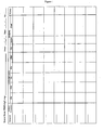
- Table 1 shows the characteristic features that were evaluated in each of the indicated tissues: Table 1 cerebellum pia, white matter matrix and capillaries, granular layer microvessels, and molecular layer microvessels midbrain pia, subpia, white matter and microvessels kidney distal collecting tubules (binds NMO-IgG most avidly) stomach basal epithelium of deep mucosa (binds NMO-IgG least avidly)
- a positive result requires a minimum of a ' ⁇ ' score to be assigned to the kidney's distal collecting tubules and to cerebellar or midbrain pia or microvessels.
- Serum was analyzed from patients classified as "definite" NMO, the Asian opticospinal form of MS, or classical MS by clinical, imaging and spinal fluid criteria, and from control patients, for autoantibodies that might bind selectively to CNS tissues.
- the experiments described herein demonstrate the value of seropositivity for discriminating NMO from the classic form of multiple sclerosis (MS).
- Indirect immunofluorescence was performed with a standard composite substrate of mouse brain, stomach and kidney; sera were preabsorbed with liver extract as described above in Example 1.
- NMO-IgG a distinctive staining pattern associated with microvessels throughout the cerebellar cortex and midbrain, and with pia and a subpial "mesh” (prominent in midbrain).
- the microvascular pattern was not seen in gut mucosa, kidney, or liver, and NMO-IgG was not noted in any control disease group.
- Sera from 16 out of 35 patients (46%) who were at high risk for NMO yielded the staining pattern distinctive for NMO-IgG. None of the 19 patients diagnosed with classic MS had detectable staining patterns of NMO-IgG, while sera from 2 out of the 22 patients (9%) who presented with optic neuritis/myelitis possessed the NMO-IgG.
- NMO-IgG was identified incidentally in 14 patients amongst 85 thousand whose sera were submitted to Mayo Clinic's Neuroimmunology Laboratory for blinded paraneoplastic autoantibody testing on a service basis. Their subsequently-obtained histories revealed that 3 fulfilled clinical criteria for the diagnosis of NMO, 9 were classified as high risk for NMO (7 had longitudinally extensive myelitis and 2 had recurrent optic neuritis), 1 had new onset myelopathy, and 1 had unclassified steroid-responsive CNS inflammatory disorder.
- NMO-IgG autoantibody is the first specific biological marker of NMO and is able to distinguish NMO from MS.
- a GST fusion protein containing recombinant rat aquaporin-4 (C terminal residues 249-323; Alamone Labs, Jerusalem, Israel) was electrophoresed in a 10% polyacrylamide gel in standard Laemmli SDS buffer containing ⁇ -mercaptoethanol, and a Western blot was performed using NMO patients' and immune rabbit's serum as a positive control to determine whether or not the patients' IgG would bind to the 38 kDa GST-aquaporin-4 fusion protein.
- the blot was contacted with human sera (1:50 dilution), which included 4 NMO patients, 3 normal persons, 1 control myelopathy, 2 patients with classic MS, and 3 patients with miscellaneous neuropsychiatric disorders. Serum from the four NMO patients and from the immune rabbit, but none of the sera from the control patients or from patients exhibiting the other disorders, bound the 38 kDa aquaporin-4 fusion protein.
Landscapes
- Health & Medical Sciences (AREA)
- Chemical & Material Sciences (AREA)
- Life Sciences & Earth Sciences (AREA)
- Immunology (AREA)
- Organic Chemistry (AREA)
- Molecular Biology (AREA)
- Medicinal Chemistry (AREA)
- General Health & Medical Sciences (AREA)
- Engineering & Computer Science (AREA)
- Biochemistry (AREA)
- Biomedical Technology (AREA)
- Hematology (AREA)
- Proteomics, Peptides & Aminoacids (AREA)
- Genetics & Genomics (AREA)
- Biophysics (AREA)
- Urology & Nephrology (AREA)
- Cell Biology (AREA)
- Bioinformatics & Cheminformatics (AREA)
- Animal Behavior & Ethology (AREA)
- Pathology (AREA)
- Public Health (AREA)
- Pharmacology & Pharmacy (AREA)
- Rehabilitation Therapy (AREA)
- Rheumatology (AREA)
- Nuclear Medicine, Radiotherapy & Molecular Imaging (AREA)
- Biotechnology (AREA)
- General Chemical & Material Sciences (AREA)
- Microbiology (AREA)
- Chemical Kinetics & Catalysis (AREA)
- Food Science & Technology (AREA)
- Physics & Mathematics (AREA)
- Analytical Chemistry (AREA)
- General Physics & Mathematics (AREA)
- Veterinary Medicine (AREA)
- Neurosurgery (AREA)
- Neurology (AREA)
- Toxicology (AREA)
- Zoology (AREA)
- Gastroenterology & Hepatology (AREA)
- Hospice & Palliative Care (AREA)
Applications Claiming Priority (2)
| Application Number | Priority Date | Filing Date | Title |
|---|---|---|---|
| US10/723,180 US7101679B2 (en) | 2003-11-25 | 2003-11-25 | Marker for neuromyelitis optica |
| PCT/US2004/039710 WO2005051178A2 (en) | 2003-11-25 | 2004-11-24 | Marker for neuromyelitis optica |
Publications (3)
| Publication Number | Publication Date |
|---|---|
| EP1700120A2 EP1700120A2 (en) | 2006-09-13 |
| EP1700120A4 EP1700120A4 (en) | 2007-11-14 |
| EP1700120B1 true EP1700120B1 (en) | 2009-03-04 |
Family
ID=34592190
Family Applications (1)
| Application Number | Title | Priority Date | Filing Date |
|---|---|---|---|
| EP04812269A Expired - Lifetime EP1700120B1 (en) | 2003-11-25 | 2004-11-24 | Marker for neuromyelitis optica |
Country Status (9)
| Country | Link |
|---|---|
| US (5) | US7101679B2 (enExample) |
| EP (1) | EP1700120B1 (enExample) |
| JP (1) | JP4538464B2 (enExample) |
| CN (1) | CN1910456B (enExample) |
| AT (1) | ATE424560T1 (enExample) |
| DE (1) | DE602004019812D1 (enExample) |
| ES (1) | ES2320006T3 (enExample) |
| IN (1) | IN2014DN05011A (enExample) |
| WO (1) | WO2005051178A2 (enExample) |
Cited By (4)
| Publication number | Priority date | Publication date | Assignee | Title |
|---|---|---|---|---|
| DE102011011280A1 (de) | 2011-02-15 | 2012-08-16 | Euroimmun Medizinische Labordiagnostika Ag | Diagnosekit sowie ein Verfahren zur Untersuchung einer menschlichen Patientenprobe auf das Vorhandensein von Neuromyelitis-optica-spezifischen Antikörpern |
| WO2013177116A1 (en) * | 2012-05-21 | 2013-11-28 | The Regents Of The University Of California | Enzymatic modification of anti-aqp4 autoantibody for modulating neuromyelitis optica |
| WO2014154907A1 (de) | 2013-03-28 | 2014-10-02 | Protagen Ag | Verfahren zur diagnose von neuromyelitis optica |
| US10654916B2 (en) | 2011-04-21 | 2020-05-19 | The Regents Of The University Of California, A California Corporation | Compositions and methods for the treatment of neuromyelitis optica |
Families Citing this family (27)
| Publication number | Priority date | Publication date | Assignee | Title |
|---|---|---|---|---|
| US7101679B2 (en) | 2003-11-25 | 2006-09-05 | Mayo Foundation For Medical Education And Research | Marker for neuromyelitis optica |
| US20070093673A1 (en) * | 2005-10-24 | 2007-04-26 | Andre Vachereau | Boron-containing compounds, uses and preparation thereof |
| JP4273235B2 (ja) * | 2006-06-01 | 2009-06-03 | 国立大学法人 新潟大学 | アクアポリン4阻害薬 |
| US8889102B2 (en) * | 2007-09-20 | 2014-11-18 | Mayo Foundation For Medical Education And Research | Neuromyelitis optica autoantibodies as a marker for neoplasia |
| JP5117336B2 (ja) * | 2008-09-18 | 2013-01-16 | 国立大学法人 千葉大学 | 多発性硬化症またはnmoの検査マーカーの測定方法 |
| US9891219B2 (en) * | 2008-10-10 | 2018-02-13 | Mayo Foundation For Medical Education And Research | Methods for treating neuromyelitis optica (NMO) by administration of eculizumab to an individual that is aquaporin-4 (AQP4)-IgG autoantibody positive |
| GR1007341B (el) | 2010-04-21 | 2011-07-05 | ΕΛΛΗΝΙΚΟ ΙΝΣΤΙΤΟΥΤΟ ΠΑΣΤΕΡ (κατά ποσοστό 40%), | Διαγνωστικος προσδιορισμος |
| JP2013528799A (ja) * | 2010-05-13 | 2013-07-11 | ユニバーシティ オブ メディスン アンド デンティストリー オブ ニュー ジャージー | 神経変性疾患の検知および診断のための診断用自己抗体プロファイル |
| WO2013010003A1 (en) | 2011-07-12 | 2013-01-17 | University Of Medicine And Dentistry Of New Jersey | Diagnostic biomarker profiles for the detection and diagnosis of alzheimer's disease |
| CN104040344A (zh) * | 2011-10-21 | 2014-09-10 | 德克萨斯系统大学评议委员会 | 视神经脊髓炎的密码子标签 |
| WO2013147081A1 (ja) * | 2012-03-30 | 2013-10-03 | コニカミノルタ株式会社 | 生体物質検出方法 |
| CN102898513B (zh) * | 2012-10-31 | 2016-03-16 | 徐俊 | ELISpot视神经脊髓炎诊断试剂盒及其应用 |
| US20170080063A1 (en) * | 2014-05-19 | 2017-03-23 | The Johns Hopkins University | Highly soluble aquaporin-4 extracellular loop c peptide immunization for treatment of neuromyelitis optica |
| US20170130269A1 (en) * | 2014-06-30 | 2017-05-11 | Siemens Healthcare Gmbh | Diagnosis of neuromyelitis optica vs. multiple sclerosis using mirna biomarkers |
| CN104698190A (zh) * | 2015-03-16 | 2015-06-10 | 中国人民解放军总医院 | 用于中枢神经系统脱髓鞘疾病的生物诊断标记物 |
| US10098935B2 (en) * | 2015-05-01 | 2018-10-16 | The Board Of Trustees Of The Leland Stanford Junior University | Aquaporin tolerizing vaccines and methods of use thereof |
| WO2017197293A1 (en) * | 2016-05-13 | 2017-11-16 | C.R. Bard, Inc. | Peripherally inserted central catheter systems, devices, and methods thereof for pediatrics |
| CN106318974A (zh) * | 2016-08-29 | 2017-01-11 | 南方医科大学南方医院 | 细胞固定工艺及通过该工艺制备aqp4抗体检测试剂盒 |
| KR101977843B1 (ko) | 2017-02-16 | 2019-05-13 | 국립암센터 | 시신경척수염 및 다발성경화증의 선별을 위한 바이오마커 및 이의 이용 |
| WO2018190365A1 (ja) * | 2017-04-12 | 2018-10-18 | 国立大学法人九州大学 | 神経障害性疼痛マーカー及びその使用 |
| KR102089042B1 (ko) * | 2017-05-16 | 2020-03-13 | 서울대학교병원 | 보체불활성화법을 이용한 항-아쿠아포린 4 항체의 개선된 검출 방법 |
| CN108998450A (zh) * | 2018-08-08 | 2018-12-14 | 昆明医科大学第附属医院 | 引物、cDNA、载体、AQP4单克隆抗体及制备方法 |
| CN110618264A (zh) * | 2019-09-10 | 2019-12-27 | 南方医科大学 | 基于量子点聚苯乙烯微球检测抗aqp4抗体的方法 |
| CN111272998A (zh) * | 2020-01-09 | 2020-06-12 | 天津天海新域生物科技有限公司 | 一种同时检测中枢脱髓鞘自身抗体aqp4、mog和mbp的方法 |
| EP4277524A4 (en) * | 2021-01-13 | 2025-06-04 | Duke University | Compositions for and methods of improving fluid flux in the brain |
| CN115232795B (zh) * | 2021-07-22 | 2023-09-19 | 北京和合医学诊断技术股份有限公司 | 稳定表达aqp4-m23蛋白的细胞株及其构建方法、应用 |
| CN113358881B (zh) * | 2021-08-10 | 2021-11-30 | 首都医科大学附属北京天坛医院 | 用于nmosd预测或复发监测的生物标志物及其应用 |
Family Cites Families (22)
| Publication number | Priority date | Publication date | Assignee | Title |
|---|---|---|---|---|
| US37273A (en) * | 1863-01-06 | Improved canteen | ||
| US4708713A (en) * | 1984-11-16 | 1987-11-24 | Anisa Medical, Inc. | Method and system for removing immunosuppressive components from the blood of mammals |
| US5258503A (en) * | 1987-09-08 | 1993-11-02 | Kanegafuchi Kagaku Kogyo Kabushiki Kaisha | Autoantibody adsorbent and apparatus for removing autoantibodies using the same |
| US5614192A (en) * | 1989-07-19 | 1997-03-25 | Connective Therapeutics, Inc. | T cell receptor peptides as therapeutics for immune-related disease |
| US5214136A (en) * | 1990-02-20 | 1993-05-25 | Gilead Sciences, Inc. | Anthraquinone-derivatives oligonucleotides |
| US5218105A (en) * | 1990-07-27 | 1993-06-08 | Isis Pharmaceuticals | Polyamine conjugated oligonucleotides |
| US5741671A (en) * | 1991-12-12 | 1998-04-21 | The Johns Hopkins University | Isolation cloning and expression of transmembrane water channel aquaporin 1(AQP1) |
| US5324638A (en) * | 1992-05-13 | 1994-06-28 | Sloan-Kettering Institute For Cancer Research | Brain transcription factor, nucleic acids encoding same and uses thereof |
| DE4227695C1 (de) * | 1992-08-21 | 1993-10-07 | Fresenius Ag | Zentrifuge zum Auftrennen von Blut in seine Bestandteile |
| US5676644A (en) | 1995-06-07 | 1997-10-14 | Cobe Laboratories, Inc. | Extracorporeal blood processing methods and apparatus |
| WO1997017369A2 (en) * | 1995-11-09 | 1997-05-15 | Trustees Of Boston University | Dna comprising a neuron-specific transcriptional promoter and its use in a gene therapy vector |
| AU763929B2 (en) | 1998-11-25 | 2003-08-07 | Amarillo Biosciences, Inc. | Interferon-alpha mediated upregulation of aquaporin expression |
| US20040014087A1 (en) | 1999-06-01 | 2004-01-22 | Incyte Corporation | Molecules for diagnostics and therapeutics |
| US6409726B1 (en) * | 1999-11-08 | 2002-06-25 | Alan G. Ellman | Electrosurgical instrument for ear surgery |
| US20030072737A1 (en) | 2000-12-29 | 2003-04-17 | Michael Brines | Tissue protective cytokines for the protection, restoration, and enhancement of responsive cells, tissues and organs |
| US20040048253A1 (en) | 2001-02-21 | 2004-03-11 | Panzer Scott R. | Molecules for diagnostics and therapeutics |
| US7251568B2 (en) | 2001-04-18 | 2007-07-31 | Wyeth | Methods and compositions for regulating bone and cartilage formation |
| US7026121B1 (en) | 2001-06-08 | 2006-04-11 | Expression Diagnostics, Inc. | Methods and compositions for diagnosing and monitoring transplant rejection |
| US6905827B2 (en) | 2001-06-08 | 2005-06-14 | Expression Diagnostics, Inc. | Methods and compositions for diagnosing or monitoring auto immune and chronic inflammatory diseases |
| US20040115629A1 (en) | 2002-01-09 | 2004-06-17 | Panzer Scott R | Molecules for diagnostics and therapeutics |
| DE10326526B4 (de) * | 2002-06-22 | 2006-06-22 | Spinner Gmbh | Koaxialer Steckverbinder |
| US7101679B2 (en) | 2003-11-25 | 2006-09-05 | Mayo Foundation For Medical Education And Research | Marker for neuromyelitis optica |
-
2003
- 2003-11-25 US US10/723,180 patent/US7101679B2/en not_active Expired - Lifetime
-
2004
- 2004-11-24 AT AT04812269T patent/ATE424560T1/de not_active IP Right Cessation
- 2004-11-24 CN CN2004800408513A patent/CN1910456B/zh not_active Expired - Lifetime
- 2004-11-24 DE DE602004019812T patent/DE602004019812D1/de not_active Expired - Lifetime
- 2004-11-24 JP JP2006541455A patent/JP4538464B2/ja not_active Expired - Fee Related
- 2004-11-24 EP EP04812269A patent/EP1700120B1/en not_active Expired - Lifetime
- 2004-11-24 WO PCT/US2004/039710 patent/WO2005051178A2/en not_active Ceased
- 2004-11-24 IN IN5011DEN2014 patent/IN2014DN05011A/en unknown
- 2004-11-24 ES ES04812269T patent/ES2320006T3/es not_active Expired - Lifetime
-
2006
- 2006-07-14 US US11/457,685 patent/US7947254B2/en not_active Expired - Lifetime
-
2008
- 2008-04-18 US US12/105,755 patent/US8524508B2/en not_active Expired - Lifetime
-
2012
- 2012-05-14 US US13/470,740 patent/US20120219969A1/en not_active Abandoned
-
2014
- 2014-06-02 US US14/293,105 patent/US20140314796A1/en not_active Abandoned
Cited By (6)
| Publication number | Priority date | Publication date | Assignee | Title |
|---|---|---|---|---|
| DE102011011280A1 (de) | 2011-02-15 | 2012-08-16 | Euroimmun Medizinische Labordiagnostika Ag | Diagnosekit sowie ein Verfahren zur Untersuchung einer menschlichen Patientenprobe auf das Vorhandensein von Neuromyelitis-optica-spezifischen Antikörpern |
| WO2012110024A2 (de) | 2011-02-15 | 2012-08-23 | Euroimmun Medizinische Labordiagnostika Ag | Diagnosekit sowie ein verfahren zur untersuchung einer menschlichen patientenprobe auf das vorhandensein neuromyelitis-optica-spezifischer antikörpern |
| US10654916B2 (en) | 2011-04-21 | 2020-05-19 | The Regents Of The University Of California, A California Corporation | Compositions and methods for the treatment of neuromyelitis optica |
| US11390667B2 (en) | 2011-04-21 | 2022-07-19 | The Regents Of The University Of California | Compositions and methods for the treatment of neuromyelitis optica |
| WO2013177116A1 (en) * | 2012-05-21 | 2013-11-28 | The Regents Of The University Of California | Enzymatic modification of anti-aqp4 autoantibody for modulating neuromyelitis optica |
| WO2014154907A1 (de) | 2013-03-28 | 2014-10-02 | Protagen Ag | Verfahren zur diagnose von neuromyelitis optica |
Also Published As
| Publication number | Publication date |
|---|---|
| DE602004019812D1 (de) | 2009-04-16 |
| WO2005051178A2 (en) | 2005-06-09 |
| JP4538464B2 (ja) | 2010-09-08 |
| US20080145870A1 (en) | 2008-06-19 |
| CN1910456A (zh) | 2007-02-07 |
| ES2320006T3 (es) | 2009-05-18 |
| US7947254B2 (en) | 2011-05-24 |
| US20050112116A1 (en) | 2005-05-26 |
| EP1700120A4 (en) | 2007-11-14 |
| US8524508B2 (en) | 2013-09-03 |
| ATE424560T1 (de) | 2009-03-15 |
| WO2005051178A3 (en) | 2005-12-29 |
| US7101679B2 (en) | 2006-09-05 |
| IN2014DN05011A (enExample) | 2015-07-10 |
| US20120219969A1 (en) | 2012-08-30 |
| US20090143710A1 (en) | 2009-06-04 |
| EP1700120A2 (en) | 2006-09-13 |
| US20140314796A1 (en) | 2014-10-23 |
| CN1910456B (zh) | 2011-04-13 |
| JP2007518068A (ja) | 2007-07-05 |
Similar Documents
| Publication | Publication Date | Title |
|---|---|---|
| EP1700120B1 (en) | Marker for neuromyelitis optica | |
| US7718386B1 (en) | Methods for the diagnosis of diabetes | |
| CN108728526B (zh) | 神经自身免疫疾病的诊断 | |
| EP3415909B1 (en) | Diagnosis of a neuroautoimmune disease | |
| EP3786641A1 (en) | Detection of an autoantibody | |
| US20120114666A1 (en) | Neurological autoimmune disorders | |
| US5512447A (en) | Methods for the diagnosis and treatment of diabetes | |
| CN110824156B (zh) | 神经自身免疫疾病的诊断 | |
| EP3519592B1 (en) | Materials and methods for evaluating cancer | |
| WO2013057599A1 (en) | Biomarker | |
| US20140377883A1 (en) | Peripherin-specific autoantibodies as a marker for neurological and endocrinological disease | |
| CN112147324A (zh) | 自身抗体的检测 | |
| WO2024182718A2 (en) | Compositions and methods related to transcobalamin receptor autoantibodies | |
| WO2008112801A1 (en) | Macular degeneration | |
| US8889102B2 (en) | Neuromyelitis optica autoantibodies as a marker for neoplasia | |
| US20220120744A1 (en) | Assessing and treating germ cell tumors and paraneoplastic autoimmunity | |
| CN111175484A (zh) | 神经自身免疫性疾病的诊断 | |
| CN112285363A (zh) | 自身免疫类神经疾病的诊断 |
Legal Events
| Date | Code | Title | Description |
|---|---|---|---|
| PUAI | Public reference made under article 153(3) epc to a published international application that has entered the european phase |
Free format text: ORIGINAL CODE: 0009012 |
|
| 17P | Request for examination filed |
Effective date: 20060609 |
|
| AK | Designated contracting states |
Kind code of ref document: A2 Designated state(s): AT BE BG CH CY CZ DE DK EE ES FI FR GB GR HU IE IS IT LI LU MC NL PL PT RO SE SI SK TR |
|
| DAX | Request for extension of the european patent (deleted) | ||
| A4 | Supplementary search report drawn up and despatched |
Effective date: 20071012 |
|
| 17Q | First examination report despatched |
Effective date: 20080429 |
|
| GRAP | Despatch of communication of intention to grant a patent |
Free format text: ORIGINAL CODE: EPIDOSNIGR1 |
|
| GRAS | Grant fee paid |
Free format text: ORIGINAL CODE: EPIDOSNIGR3 |
|
| GRAA | (expected) grant |
Free format text: ORIGINAL CODE: 0009210 |
|
| AK | Designated contracting states |
Kind code of ref document: B1 Designated state(s): AT BE BG CH CY CZ DE DK EE ES FI FR GB GR HU IE IS IT LI LU MC NL PL PT RO SE SI SK TR |
|
| REG | Reference to a national code |
Ref country code: GB Ref legal event code: FG4D |
|
| REG | Reference to a national code |
Ref country code: CH Ref legal event code: EP |
|
| REG | Reference to a national code |
Ref country code: IE Ref legal event code: FG4D |
|
| REG | Reference to a national code |
Ref country code: CH Ref legal event code: NV Representative=s name: KIRKER & CIE S.A. |
|
| REF | Corresponds to: |
Ref document number: 602004019812 Country of ref document: DE Date of ref document: 20090416 Kind code of ref document: P |
|
| REG | Reference to a national code |
Ref country code: ES Ref legal event code: FG2A Ref document number: 2320006 Country of ref document: ES Kind code of ref document: T3 |
|
| PG25 | Lapsed in a contracting state [announced via postgrant information from national office to epo] |
Ref country code: SI Free format text: LAPSE BECAUSE OF FAILURE TO SUBMIT A TRANSLATION OF THE DESCRIPTION OR TO PAY THE FEE WITHIN THE PRESCRIBED TIME-LIMIT Effective date: 20090304 Ref country code: NL Free format text: LAPSE BECAUSE OF FAILURE TO SUBMIT A TRANSLATION OF THE DESCRIPTION OR TO PAY THE FEE WITHIN THE PRESCRIBED TIME-LIMIT Effective date: 20090304 Ref country code: FI Free format text: LAPSE BECAUSE OF FAILURE TO SUBMIT A TRANSLATION OF THE DESCRIPTION OR TO PAY THE FEE WITHIN THE PRESCRIBED TIME-LIMIT Effective date: 20090304 |
|
| NLV1 | Nl: lapsed or annulled due to failure to fulfill the requirements of art. 29p and 29m of the patents act | ||
| PG25 | Lapsed in a contracting state [announced via postgrant information from national office to epo] |
Ref country code: SE Free format text: LAPSE BECAUSE OF FAILURE TO SUBMIT A TRANSLATION OF THE DESCRIPTION OR TO PAY THE FEE WITHIN THE PRESCRIBED TIME-LIMIT Effective date: 20090604 Ref country code: PL Free format text: LAPSE BECAUSE OF FAILURE TO SUBMIT A TRANSLATION OF THE DESCRIPTION OR TO PAY THE FEE WITHIN THE PRESCRIBED TIME-LIMIT Effective date: 20090304 Ref country code: AT Free format text: LAPSE BECAUSE OF FAILURE TO SUBMIT A TRANSLATION OF THE DESCRIPTION OR TO PAY THE FEE WITHIN THE PRESCRIBED TIME-LIMIT Effective date: 20090304 |
|
| PG25 | Lapsed in a contracting state [announced via postgrant information from national office to epo] |
Ref country code: BE Free format text: LAPSE BECAUSE OF FAILURE TO SUBMIT A TRANSLATION OF THE DESCRIPTION OR TO PAY THE FEE WITHIN THE PRESCRIBED TIME-LIMIT Effective date: 20090304 |
|
| PG25 | Lapsed in a contracting state [announced via postgrant information from national office to epo] |
Ref country code: EE Free format text: LAPSE BECAUSE OF FAILURE TO SUBMIT A TRANSLATION OF THE DESCRIPTION OR TO PAY THE FEE WITHIN THE PRESCRIBED TIME-LIMIT Effective date: 20090304 Ref country code: PT Free format text: LAPSE BECAUSE OF FAILURE TO SUBMIT A TRANSLATION OF THE DESCRIPTION OR TO PAY THE FEE WITHIN THE PRESCRIBED TIME-LIMIT Effective date: 20090818 Ref country code: CZ Free format text: LAPSE BECAUSE OF FAILURE TO SUBMIT A TRANSLATION OF THE DESCRIPTION OR TO PAY THE FEE WITHIN THE PRESCRIBED TIME-LIMIT Effective date: 20090304 |
|
| PG25 | Lapsed in a contracting state [announced via postgrant information from national office to epo] |
Ref country code: RO Free format text: LAPSE BECAUSE OF FAILURE TO SUBMIT A TRANSLATION OF THE DESCRIPTION OR TO PAY THE FEE WITHIN THE PRESCRIBED TIME-LIMIT Effective date: 20090304 Ref country code: IS Free format text: LAPSE BECAUSE OF FAILURE TO SUBMIT A TRANSLATION OF THE DESCRIPTION OR TO PAY THE FEE WITHIN THE PRESCRIBED TIME-LIMIT Effective date: 20090704 Ref country code: SK Free format text: LAPSE BECAUSE OF FAILURE TO SUBMIT A TRANSLATION OF THE DESCRIPTION OR TO PAY THE FEE WITHIN THE PRESCRIBED TIME-LIMIT Effective date: 20090304 |
|
| PLBE | No opposition filed within time limit |
Free format text: ORIGINAL CODE: 0009261 |
|
| STAA | Information on the status of an ep patent application or granted ep patent |
Free format text: STATUS: NO OPPOSITION FILED WITHIN TIME LIMIT |
|
| PG25 | Lapsed in a contracting state [announced via postgrant information from national office to epo] |
Ref country code: DK Free format text: LAPSE BECAUSE OF FAILURE TO SUBMIT A TRANSLATION OF THE DESCRIPTION OR TO PAY THE FEE WITHIN THE PRESCRIBED TIME-LIMIT Effective date: 20090304 Ref country code: BG Free format text: LAPSE BECAUSE OF FAILURE TO SUBMIT A TRANSLATION OF THE DESCRIPTION OR TO PAY THE FEE WITHIN THE PRESCRIBED TIME-LIMIT Effective date: 20090604 |
|
| 26N | No opposition filed |
Effective date: 20091207 |
|
| PG25 | Lapsed in a contracting state [announced via postgrant information from national office to epo] |
Ref country code: MC Free format text: LAPSE BECAUSE OF NON-PAYMENT OF DUE FEES Effective date: 20091130 |
|
| REG | Reference to a national code |
Ref country code: IE Ref legal event code: MM4A |
|
| PG25 | Lapsed in a contracting state [announced via postgrant information from national office to epo] |
Ref country code: IE Free format text: LAPSE BECAUSE OF NON-PAYMENT OF DUE FEES Effective date: 20091124 Ref country code: GR Free format text: LAPSE BECAUSE OF FAILURE TO SUBMIT A TRANSLATION OF THE DESCRIPTION OR TO PAY THE FEE WITHIN THE PRESCRIBED TIME-LIMIT Effective date: 20090605 |
|
| PG25 | Lapsed in a contracting state [announced via postgrant information from national office to epo] |
Ref country code: LU Free format text: LAPSE BECAUSE OF NON-PAYMENT OF DUE FEES Effective date: 20091124 |
|
| PG25 | Lapsed in a contracting state [announced via postgrant information from national office to epo] |
Ref country code: HU Free format text: LAPSE BECAUSE OF FAILURE TO SUBMIT A TRANSLATION OF THE DESCRIPTION OR TO PAY THE FEE WITHIN THE PRESCRIBED TIME-LIMIT Effective date: 20090905 |
|
| PG25 | Lapsed in a contracting state [announced via postgrant information from national office to epo] |
Ref country code: TR Free format text: LAPSE BECAUSE OF FAILURE TO SUBMIT A TRANSLATION OF THE DESCRIPTION OR TO PAY THE FEE WITHIN THE PRESCRIBED TIME-LIMIT Effective date: 20090304 |
|
| PG25 | Lapsed in a contracting state [announced via postgrant information from national office to epo] |
Ref country code: CY Free format text: LAPSE BECAUSE OF FAILURE TO SUBMIT A TRANSLATION OF THE DESCRIPTION OR TO PAY THE FEE WITHIN THE PRESCRIBED TIME-LIMIT Effective date: 20090304 |
|
| REG | Reference to a national code |
Ref country code: FR Ref legal event code: PLFP Year of fee payment: 12 |
|
| REG | Reference to a national code |
Ref country code: FR Ref legal event code: PLFP Year of fee payment: 13 |
|
| REG | Reference to a national code |
Ref country code: FR Ref legal event code: PLFP Year of fee payment: 14 |
|
| P01 | Opt-out of the competence of the unified patent court (upc) registered |
Effective date: 20230602 |
|
| PGFP | Annual fee paid to national office [announced via postgrant information from national office to epo] |
Ref country code: GB Payment date: 20231127 Year of fee payment: 20 |
|
| PGFP | Annual fee paid to national office [announced via postgrant information from national office to epo] |
Ref country code: ES Payment date: 20231201 Year of fee payment: 20 |
|
| PGFP | Annual fee paid to national office [announced via postgrant information from national office to epo] |
Ref country code: IT Payment date: 20231122 Year of fee payment: 20 Ref country code: FR Payment date: 20231127 Year of fee payment: 20 Ref country code: DE Payment date: 20231129 Year of fee payment: 20 Ref country code: CH Payment date: 20231202 Year of fee payment: 20 |
|
| REG | Reference to a national code |
Ref country code: DE Ref legal event code: R071 Ref document number: 602004019812 Country of ref document: DE |
|
| REG | Reference to a national code |
Ref country code: CH Ref legal event code: PL |
|
| REG | Reference to a national code |
Ref country code: ES Ref legal event code: FD2A Effective date: 20241202 |
|
| REG | Reference to a national code |
Ref country code: GB Ref legal event code: PE20 Expiry date: 20241123 |
|
| PG25 | Lapsed in a contracting state [announced via postgrant information from national office to epo] |
Ref country code: GB Free format text: LAPSE BECAUSE OF EXPIRATION OF PROTECTION Effective date: 20241123 |
|
| PG25 | Lapsed in a contracting state [announced via postgrant information from national office to epo] |
Ref country code: ES Free format text: LAPSE BECAUSE OF EXPIRATION OF PROTECTION Effective date: 20241125 |
|
| PG25 | Lapsed in a contracting state [announced via postgrant information from national office to epo] |
Ref country code: GB Free format text: LAPSE BECAUSE OF EXPIRATION OF PROTECTION Effective date: 20241123 Ref country code: ES Free format text: LAPSE BECAUSE OF EXPIRATION OF PROTECTION Effective date: 20241125 |