EP1624812B1 - Elemente, gerät und programm für die knochenkorrektur - Google Patents
Elemente, gerät und programm für die knochenkorrektur Download PDFInfo
- Publication number
- EP1624812B1 EP1624812B1 EP04709755.5A EP04709755A EP1624812B1 EP 1624812 B1 EP1624812 B1 EP 1624812B1 EP 04709755 A EP04709755 A EP 04709755A EP 1624812 B1 EP1624812 B1 EP 1624812B1
- Authority
- EP
- European Patent Office
- Prior art keywords
- bone
- model
- shows
- osteotomy
- assisting member
- Prior art date
- Legal status (The legal status is an assumption and is not a legal conclusion. Google has not performed a legal analysis and makes no representation as to the accuracy of the status listed.)
- Expired - Lifetime
Links
- 210000000988 bone and bone Anatomy 0.000 title claims description 910
- 238000012937 correction Methods 0.000 title claims description 172
- 238000000034 method Methods 0.000 claims description 245
- 238000011282 treatment Methods 0.000 claims description 161
- 238000005520 cutting process Methods 0.000 claims description 141
- 239000012634 fragment Substances 0.000 claims description 126
- 230000008569 process Effects 0.000 claims description 74
- 238000003780 insertion Methods 0.000 claims description 44
- 230000037431 insertion Effects 0.000 claims description 44
- 230000002159 abnormal effect Effects 0.000 claims description 12
- 238000003384 imaging method Methods 0.000 claims description 12
- 238000004088 simulation Methods 0.000 description 49
- 210000003857 wrist joint Anatomy 0.000 description 31
- 230000007547 defect Effects 0.000 description 29
- 210000000245 forearm Anatomy 0.000 description 26
- 208000010392 Bone Fractures Diseases 0.000 description 25
- 238000012545 processing Methods 0.000 description 25
- 210000001519 tissue Anatomy 0.000 description 25
- 206010017076 Fracture Diseases 0.000 description 22
- 210000004027 cell Anatomy 0.000 description 21
- 238000013519 translation Methods 0.000 description 21
- 230000002980 postoperative effect Effects 0.000 description 20
- 230000006870 function Effects 0.000 description 19
- 210000000623 ulna Anatomy 0.000 description 19
- 238000002591 computed tomography Methods 0.000 description 18
- 238000000465 moulding Methods 0.000 description 18
- 210000000056 organ Anatomy 0.000 description 18
- PXFBZOLANLWPMH-UHFFFAOYSA-N 16-Epiaffinine Natural products C1C(C2=CC=CC=C2N2)=C2C(=O)CC2C(=CC)CN(C)C1C2CO PXFBZOLANLWPMH-UHFFFAOYSA-N 0.000 description 17
- 208000037265 diseases, disorders, signs and symptoms Diseases 0.000 description 17
- 208000035475 disorder Diseases 0.000 description 17
- 239000000463 material Substances 0.000 description 17
- 238000004364 calculation method Methods 0.000 description 16
- 239000011159 matrix material Substances 0.000 description 16
- 238000011426 transformation method Methods 0.000 description 15
- 238000004422 calculation algorithm Methods 0.000 description 13
- 238000005094 computer simulation Methods 0.000 description 13
- 238000002595 magnetic resonance imaging Methods 0.000 description 13
- 238000002513 implantation Methods 0.000 description 12
- 206010017085 Fracture malunion Diseases 0.000 description 11
- 208000000536 Malunited Fractures Diseases 0.000 description 11
- 201000008482 osteoarthritis Diseases 0.000 description 11
- 229910052588 hydroxylapatite Inorganic materials 0.000 description 10
- 238000004519 manufacturing process Methods 0.000 description 10
- XYJRXVWERLGGKC-UHFFFAOYSA-D pentacalcium;hydroxide;triphosphate Chemical compound [OH-].[Ca+2].[Ca+2].[Ca+2].[Ca+2].[Ca+2].[O-]P([O-])([O-])=O.[O-]P([O-])([O-])=O.[O-]P([O-])([O-])=O XYJRXVWERLGGKC-UHFFFAOYSA-D 0.000 description 10
- 206010020649 Hyperkeratosis Diseases 0.000 description 9
- 230000006378 damage Effects 0.000 description 9
- 230000000694 effects Effects 0.000 description 9
- 241000469816 Varus Species 0.000 description 8
- 239000001506 calcium phosphate Substances 0.000 description 8
- 229910000389 calcium phosphate Inorganic materials 0.000 description 8
- 235000011010 calcium phosphates Nutrition 0.000 description 8
- 229910052751 metal Inorganic materials 0.000 description 8
- 239000002184 metal Substances 0.000 description 8
- QORWJWZARLRLPR-UHFFFAOYSA-H tricalcium bis(phosphate) Chemical compound [Ca+2].[Ca+2].[Ca+2].[O-]P([O-])([O-])=O.[O-]P([O-])([O-])=O QORWJWZARLRLPR-UHFFFAOYSA-H 0.000 description 8
- 208000027418 Wounds and injury Diseases 0.000 description 7
- 238000006073 displacement reaction Methods 0.000 description 7
- 210000003414 extremity Anatomy 0.000 description 7
- 210000002758 humerus Anatomy 0.000 description 7
- 210000001621 ilium bone Anatomy 0.000 description 7
- 208000014674 injury Diseases 0.000 description 7
- 208000008558 Osteophyte Diseases 0.000 description 6
- RTAQQCXQSZGOHL-UHFFFAOYSA-N Titanium Chemical compound [Ti] RTAQQCXQSZGOHL-UHFFFAOYSA-N 0.000 description 6
- 229920000249 biocompatible polymer Polymers 0.000 description 6
- 238000006243 chemical reaction Methods 0.000 description 6
- 201000010934 exostosis Diseases 0.000 description 6
- 239000004033 plastic Substances 0.000 description 6
- 229920003023 plastic Polymers 0.000 description 6
- 229920005989 resin Polymers 0.000 description 6
- 239000011347 resin Substances 0.000 description 6
- 239000000126 substance Substances 0.000 description 6
- 238000012360 testing method Methods 0.000 description 6
- 239000010936 titanium Substances 0.000 description 6
- 229910052719 titanium Inorganic materials 0.000 description 6
- 230000008859 change Effects 0.000 description 5
- 210000003275 diaphysis Anatomy 0.000 description 5
- 238000011156 evaluation Methods 0.000 description 5
- 230000001900 immune effect Effects 0.000 description 5
- 238000011221 initial treatment Methods 0.000 description 5
- 238000002372 labelling Methods 0.000 description 5
- 229920000642 polymer Polymers 0.000 description 5
- 229910001220 stainless steel Inorganic materials 0.000 description 5
- 239000010935 stainless steel Substances 0.000 description 5
- 230000009466 transformation Effects 0.000 description 5
- 238000011179 visual inspection Methods 0.000 description 5
- PMATZTZNYRCHOR-CGLBZJNRSA-N Cyclosporin A Chemical compound CC[C@@H]1NC(=O)[C@H]([C@H](O)[C@H](C)C\C=C\C)N(C)C(=O)[C@H](C(C)C)N(C)C(=O)[C@H](CC(C)C)N(C)C(=O)[C@H](CC(C)C)N(C)C(=O)[C@@H](C)NC(=O)[C@H](C)NC(=O)[C@H](CC(C)C)N(C)C(=O)[C@H](C(C)C)NC(=O)[C@H](CC(C)C)N(C)C(=O)CN(C)C1=O PMATZTZNYRCHOR-CGLBZJNRSA-N 0.000 description 4
- 108010036949 Cyclosporine Proteins 0.000 description 4
- 206010016997 Forearm fracture Diseases 0.000 description 4
- 239000000919 ceramic Substances 0.000 description 4
- 238000004590 computer program Methods 0.000 description 4
- 238000013461 design Methods 0.000 description 4
- 238000005516 engineering process Methods 0.000 description 4
- 229960003444 immunosuppressant agent Drugs 0.000 description 4
- 239000003018 immunosuppressive agent Substances 0.000 description 4
- 239000007788 liquid Substances 0.000 description 4
- 206010070918 Bone deformity Diseases 0.000 description 3
- 241001465754 Metazoa Species 0.000 description 3
- 241000251539 Vertebrata <Metazoa> Species 0.000 description 3
- 238000013459 approach Methods 0.000 description 3
- LMEKQMALGUDUQG-UHFFFAOYSA-N azathioprine Chemical compound CN1C=NC([N+]([O-])=O)=C1SC1=NC=NC2=C1NC=N2 LMEKQMALGUDUQG-UHFFFAOYSA-N 0.000 description 3
- 230000015572 biosynthetic process Effects 0.000 description 3
- 238000002059 diagnostic imaging Methods 0.000 description 3
- 230000001861 immunosuppressant effect Effects 0.000 description 3
- 208000024891 symptom Diseases 0.000 description 3
- 210000000707 wrist Anatomy 0.000 description 3
- 241000282472 Canis lupus familiaris Species 0.000 description 2
- CURLTUGMZLYLDI-UHFFFAOYSA-N Carbon dioxide Chemical compound O=C=O CURLTUGMZLYLDI-UHFFFAOYSA-N 0.000 description 2
- 235000014653 Carica parviflora Nutrition 0.000 description 2
- 241000243321 Cnidaria Species 0.000 description 2
- WZUVPPKBWHMQCE-UHFFFAOYSA-N Haematoxylin Chemical compound C12=CC(O)=C(O)C=C2CC2(O)C1C1=CC=C(O)C(O)=C1OC2 WZUVPPKBWHMQCE-UHFFFAOYSA-N 0.000 description 2
- 208000006358 Hand Deformities Diseases 0.000 description 2
- 206010062016 Immunosuppression Diseases 0.000 description 2
- 241000283973 Oryctolagus cuniculus Species 0.000 description 2
- 241000700159 Rattus Species 0.000 description 2
- 230000009471 action Effects 0.000 description 2
- 238000010171 animal model Methods 0.000 description 2
- 229960002170 azathioprine Drugs 0.000 description 2
- 230000008901 benefit Effects 0.000 description 2
- 239000000560 biocompatible material Substances 0.000 description 2
- 239000004621 biodegradable polymer Substances 0.000 description 2
- 229920002988 biodegradable polymer Polymers 0.000 description 2
- 210000000845 cartilage Anatomy 0.000 description 2
- 210000000589 cicatrix Anatomy 0.000 description 2
- 229960001265 ciclosporin Drugs 0.000 description 2
- 230000001054 cortical effect Effects 0.000 description 2
- 229930182912 cyclosporin Natural products 0.000 description 2
- 238000013500 data storage Methods 0.000 description 2
- 229940079593 drug Drugs 0.000 description 2
- 239000003814 drug Substances 0.000 description 2
- -1 for example Substances 0.000 description 2
- 230000035876 healing Effects 0.000 description 2
- 230000036541 health Effects 0.000 description 2
- 230000001506 immunosuppresive effect Effects 0.000 description 2
- 239000007943 implant Substances 0.000 description 2
- 230000006872 improvement Effects 0.000 description 2
- 238000005304 joining Methods 0.000 description 2
- 238000010030 laminating Methods 0.000 description 2
- 230000007246 mechanism Effects 0.000 description 2
- VNWKTOKETHGBQD-UHFFFAOYSA-N methane Chemical compound C VNWKTOKETHGBQD-UHFFFAOYSA-N 0.000 description 2
- 244000005700 microbiome Species 0.000 description 2
- 238000012986 modification Methods 0.000 description 2
- 230000004048 modification Effects 0.000 description 2
- 210000005036 nerve Anatomy 0.000 description 2
- 231100000862 numbness Toxicity 0.000 description 2
- 230000003287 optical effect Effects 0.000 description 2
- 239000004065 semiconductor Substances 0.000 description 2
- 238000007493 shaping process Methods 0.000 description 2
- 238000004904 shortening Methods 0.000 description 2
- 239000003270 steroid hormone Substances 0.000 description 2
- 238000001356 surgical procedure Methods 0.000 description 2
- 230000001988 toxicity Effects 0.000 description 2
- 231100000419 toxicity Toxicity 0.000 description 2
- 230000001131 transforming effect Effects 0.000 description 2
- 238000012800 visualization Methods 0.000 description 2
- 241000251468 Actinopterygii Species 0.000 description 1
- 239000004956 Amodel Substances 0.000 description 1
- 208000036487 Arthropathies Diseases 0.000 description 1
- 208000020084 Bone disease Diseases 0.000 description 1
- 208000003044 Closed Fractures Diseases 0.000 description 1
- 241001481828 Glyptocephalus cynoglossus Species 0.000 description 1
- 241000282412 Homo Species 0.000 description 1
- 206010020751 Hypersensitivity Diseases 0.000 description 1
- 206010061218 Inflammation Diseases 0.000 description 1
- 208000012659 Joint disease Diseases 0.000 description 1
- 208000002565 Open Fractures Diseases 0.000 description 1
- 206010037660 Pyrexia Diseases 0.000 description 1
- 241001222723 Sterna Species 0.000 description 1
- 210000001744 T-lymphocyte Anatomy 0.000 description 1
- QJJXYPPXXYFBGM-LFZNUXCKSA-N Tacrolimus Chemical compound C1C[C@@H](O)[C@H](OC)C[C@@H]1\C=C(/C)[C@@H]1[C@H](C)[C@@H](O)CC(=O)[C@H](CC=C)/C=C(C)/C[C@H](C)C[C@H](OC)[C@H]([C@H](C[C@H]2C)OC)O[C@@]2(O)C(=O)C(=O)N2CCCC[C@H]2C(=O)O1 QJJXYPPXXYFBGM-LFZNUXCKSA-N 0.000 description 1
- 208000007536 Thrombosis Diseases 0.000 description 1
- 241001227561 Valgus Species 0.000 description 1
- 206010048049 Wrist fracture Diseases 0.000 description 1
- 230000001154 acute effect Effects 0.000 description 1
- 208000030961 allergic reaction Diseases 0.000 description 1
- 238000004458 analytical method Methods 0.000 description 1
- 210000003484 anatomy Anatomy 0.000 description 1
- 210000000784 arm bone Anatomy 0.000 description 1
- 230000000740 bleeding effect Effects 0.000 description 1
- 230000023555 blood coagulation Effects 0.000 description 1
- 210000004204 blood vessel Anatomy 0.000 description 1
- 239000001569 carbon dioxide Substances 0.000 description 1
- 229910002092 carbon dioxide Inorganic materials 0.000 description 1
- 210000003010 carpal bone Anatomy 0.000 description 1
- 239000003795 chemical substances by application Substances 0.000 description 1
- 238000004891 communication Methods 0.000 description 1
- 230000006835 compression Effects 0.000 description 1
- 238000007906 compression Methods 0.000 description 1
- 238000012790 confirmation Methods 0.000 description 1
- 210000002808 connective tissue Anatomy 0.000 description 1
- 238000007796 conventional method Methods 0.000 description 1
- 230000002950 deficient Effects 0.000 description 1
- 238000011161 development Methods 0.000 description 1
- 238000004141 dimensional analysis Methods 0.000 description 1
- 210000002310 elbow joint Anatomy 0.000 description 1
- YQGOJNYOYNNSMM-UHFFFAOYSA-N eosin Chemical compound [Na+].OC(=O)C1=CC=CC=C1C1=C2C=C(Br)C(=O)C(Br)=C2OC2=C(Br)C(O)=C(Br)C=C21 YQGOJNYOYNNSMM-UHFFFAOYSA-N 0.000 description 1
- 239000003822 epoxy resin Substances 0.000 description 1
- 230000003203 everyday effect Effects 0.000 description 1
- 238000002474 experimental method Methods 0.000 description 1
- 206010016165 failure to thrive Diseases 0.000 description 1
- 230000002349 favourable effect Effects 0.000 description 1
- 210000004247 hand Anatomy 0.000 description 1
- 210000002411 hand bone Anatomy 0.000 description 1
- 230000007062 hydrolysis Effects 0.000 description 1
- 238000006460 hydrolysis reaction Methods 0.000 description 1
- 230000008105 immune reaction Effects 0.000 description 1
- 210000000987 immune system Anatomy 0.000 description 1
- 230000036046 immunoreaction Effects 0.000 description 1
- 229940073062 imuran Drugs 0.000 description 1
- 230000004054 inflammatory process Effects 0.000 description 1
- 230000003993 interaction Effects 0.000 description 1
- 230000001788 irregular Effects 0.000 description 1
- 210000000281 joint capsule Anatomy 0.000 description 1
- 230000002045 lasting effect Effects 0.000 description 1
- 210000003991 lunate bone Anatomy 0.000 description 1
- 210000004072 lung Anatomy 0.000 description 1
- 238000003754 machining Methods 0.000 description 1
- 238000005259 measurement Methods 0.000 description 1
- 230000004660 morphological change Effects 0.000 description 1
- 210000003205 muscle Anatomy 0.000 description 1
- 229940063121 neoral Drugs 0.000 description 1
- 238000012634 optical imaging Methods 0.000 description 1
- 230000001575 pathological effect Effects 0.000 description 1
- 229920000647 polyepoxide Polymers 0.000 description 1
- 238000002360 preparation method Methods 0.000 description 1
- 238000007639 printing Methods 0.000 description 1
- 230000000750 progressive effect Effects 0.000 description 1
- 230000000069 prophylactic effect Effects 0.000 description 1
- 238000011084 recovery Methods 0.000 description 1
- 230000000717 retained effect Effects 0.000 description 1
- 210000000614 rib Anatomy 0.000 description 1
- 238000005070 sampling Methods 0.000 description 1
- 210000003189 scaphoid bone Anatomy 0.000 description 1
- 210000000824 sesamoid bone Anatomy 0.000 description 1
- 230000037380 skin damage Effects 0.000 description 1
- 210000004872 soft tissue Anatomy 0.000 description 1
- 239000002689 soil Substances 0.000 description 1
- 150000003431 steroids Chemical class 0.000 description 1
- 239000000758 substrate Substances 0.000 description 1
- 230000003319 supportive effect Effects 0.000 description 1
- 229960001967 tacrolimus Drugs 0.000 description 1
- QJJXYPPXXYFBGM-SHYZHZOCSA-N tacrolimus Natural products CO[C@H]1C[C@H](CC[C@@H]1O)C=C(C)[C@H]2OC(=O)[C@H]3CCCCN3C(=O)C(=O)[C@@]4(O)O[C@@H]([C@H](C[C@H]4C)OC)[C@@H](C[C@H](C)CC(=C[C@@H](CC=C)C(=O)C[C@H](O)[C@H]2C)C)OC QJJXYPPXXYFBGM-SHYZHZOCSA-N 0.000 description 1
- 238000002054 transplantation Methods 0.000 description 1
- XLYOFNOQVPJJNP-UHFFFAOYSA-N water Substances O XLYOFNOQVPJJNP-UHFFFAOYSA-N 0.000 description 1
- 239000002023 wood Substances 0.000 description 1
Images












Classifications
-
- A—HUMAN NECESSITIES
- A61—MEDICAL OR VETERINARY SCIENCE; HYGIENE
- A61B—DIAGNOSIS; SURGERY; IDENTIFICATION
- A61B17/00—Surgical instruments, devices or methods
- A61B17/14—Surgical saws
- A61B17/15—Guides therefor
-
- A—HUMAN NECESSITIES
- A61—MEDICAL OR VETERINARY SCIENCE; HYGIENE
- A61B—DIAGNOSIS; SURGERY; IDENTIFICATION
- A61B17/00—Surgical instruments, devices or methods
- A61B17/56—Surgical instruments or methods for treatment of bones or joints; Devices specially adapted therefor
- A61B17/58—Surgical instruments or methods for treatment of bones or joints; Devices specially adapted therefor for osteosynthesis, e.g. bone plates, screws or setting implements
- A61B17/68—Internal fixation devices, including fasteners and spinal fixators, even if a part thereof projects from the skin
-
- A—HUMAN NECESSITIES
- A61—MEDICAL OR VETERINARY SCIENCE; HYGIENE
- A61B—DIAGNOSIS; SURGERY; IDENTIFICATION
- A61B17/00—Surgical instruments, devices or methods
- A61B17/56—Surgical instruments or methods for treatment of bones or joints; Devices specially adapted therefor
- A61B17/58—Surgical instruments or methods for treatment of bones or joints; Devices specially adapted therefor for osteosynthesis, e.g. bone plates, screws or setting implements
- A61B17/88—Osteosynthesis instruments; Methods or means for implanting or extracting internal or external fixation devices
-
- A—HUMAN NECESSITIES
- A61—MEDICAL OR VETERINARY SCIENCE; HYGIENE
- A61B—DIAGNOSIS; SURGERY; IDENTIFICATION
- A61B90/00—Instruments, implements or accessories specially adapted for surgery or diagnosis and not covered by any of the groups A61B1/00 - A61B50/00, e.g. for luxation treatment or for protecting wound edges
- A61B90/36—Image-producing devices or illumination devices not otherwise provided for
-
- G—PHYSICS
- G16—INFORMATION AND COMMUNICATION TECHNOLOGY [ICT] SPECIALLY ADAPTED FOR SPECIFIC APPLICATION FIELDS
- G16H—HEALTHCARE INFORMATICS, i.e. INFORMATION AND COMMUNICATION TECHNOLOGY [ICT] SPECIALLY ADAPTED FOR THE HANDLING OR PROCESSING OF MEDICAL OR HEALTHCARE DATA
- G16H50/00—ICT specially adapted for medical diagnosis, medical simulation or medical data mining; ICT specially adapted for detecting, monitoring or modelling epidemics or pandemics
- G16H50/50—ICT specially adapted for medical diagnosis, medical simulation or medical data mining; ICT specially adapted for detecting, monitoring or modelling epidemics or pandemics for simulation or modelling of medical disorders
-
- G—PHYSICS
- G16—INFORMATION AND COMMUNICATION TECHNOLOGY [ICT] SPECIALLY ADAPTED FOR SPECIFIC APPLICATION FIELDS
- G16Z—INFORMATION AND COMMUNICATION TECHNOLOGY [ICT] SPECIALLY ADAPTED FOR SPECIFIC APPLICATION FIELDS, NOT OTHERWISE PROVIDED FOR
- G16Z99/00—Subject matter not provided for in other main groups of this subclass
-
- A—HUMAN NECESSITIES
- A61—MEDICAL OR VETERINARY SCIENCE; HYGIENE
- A61B—DIAGNOSIS; SURGERY; IDENTIFICATION
- A61B17/00—Surgical instruments, devices or methods
- A61B17/56—Surgical instruments or methods for treatment of bones or joints; Devices specially adapted therefor
- A61B17/58—Surgical instruments or methods for treatment of bones or joints; Devices specially adapted therefor for osteosynthesis, e.g. bone plates, screws or setting implements
- A61B17/68—Internal fixation devices, including fasteners and spinal fixators, even if a part thereof projects from the skin
- A61B17/80—Cortical plates, i.e. bone plates; Instruments for holding or positioning cortical plates, or for compressing bones attached to cortical plates
- A61B17/8095—Wedge osteotomy devices
-
- A—HUMAN NECESSITIES
- A61—MEDICAL OR VETERINARY SCIENCE; HYGIENE
- A61B—DIAGNOSIS; SURGERY; IDENTIFICATION
- A61B17/00—Surgical instruments, devices or methods
- A61B17/56—Surgical instruments or methods for treatment of bones or joints; Devices specially adapted therefor
- A61B2017/564—Methods for bone or joint treatment
-
- A—HUMAN NECESSITIES
- A61—MEDICAL OR VETERINARY SCIENCE; HYGIENE
- A61B—DIAGNOSIS; SURGERY; IDENTIFICATION
- A61B34/00—Computer-aided surgery; Manipulators or robots specially adapted for use in surgery
- A61B34/10—Computer-aided planning, simulation or modelling of surgical operations
- A61B2034/108—Computer aided selection or customisation of medical implants or cutting guides
-
- A—HUMAN NECESSITIES
- A61—MEDICAL OR VETERINARY SCIENCE; HYGIENE
- A61B—DIAGNOSIS; SURGERY; IDENTIFICATION
- A61B34/00—Computer-aided surgery; Manipulators or robots specially adapted for use in surgery
- A61B34/10—Computer-aided planning, simulation or modelling of surgical operations
-
- A—HUMAN NECESSITIES
- A61—MEDICAL OR VETERINARY SCIENCE; HYGIENE
- A61F—FILTERS IMPLANTABLE INTO BLOOD VESSELS; PROSTHESES; DEVICES PROVIDING PATENCY TO, OR PREVENTING COLLAPSING OF, TUBULAR STRUCTURES OF THE BODY, e.g. STENTS; ORTHOPAEDIC, NURSING OR CONTRACEPTIVE DEVICES; FOMENTATION; TREATMENT OR PROTECTION OF EYES OR EARS; BANDAGES, DRESSINGS OR ABSORBENT PADS; FIRST-AID KITS
- A61F2/00—Filters implantable into blood vessels; Prostheses, i.e. artificial substitutes or replacements for parts of the body; Appliances for connecting them with the body; Devices providing patency to, or preventing collapsing of, tubular structures of the body, e.g. stents
- A61F2/02—Prostheses implantable into the body
- A61F2/30—Joints
- A61F2/42—Joints for wrists or ankles; for hands, e.g. fingers; for feet, e.g. toes
- A61F2/4261—Joints for wrists or ankles; for hands, e.g. fingers; for feet, e.g. toes for wrists
-
- A—HUMAN NECESSITIES
- A61—MEDICAL OR VETERINARY SCIENCE; HYGIENE
- A61F—FILTERS IMPLANTABLE INTO BLOOD VESSELS; PROSTHESES; DEVICES PROVIDING PATENCY TO, OR PREVENTING COLLAPSING OF, TUBULAR STRUCTURES OF THE BODY, e.g. STENTS; ORTHOPAEDIC, NURSING OR CONTRACEPTIVE DEVICES; FOMENTATION; TREATMENT OR PROTECTION OF EYES OR EARS; BANDAGES, DRESSINGS OR ABSORBENT PADS; FIRST-AID KITS
- A61F2/00—Filters implantable into blood vessels; Prostheses, i.e. artificial substitutes or replacements for parts of the body; Appliances for connecting them with the body; Devices providing patency to, or preventing collapsing of, tubular structures of the body, e.g. stents
- A61F2/02—Prostheses implantable into the body
- A61F2/30—Joints
- A61F2/46—Special tools for implanting artificial joints
- A61F2/4644—Preparation of bone graft, bone plugs or bone dowels, e.g. grinding or milling bone material
-
- A—HUMAN NECESSITIES
- A61—MEDICAL OR VETERINARY SCIENCE; HYGIENE
- A61F—FILTERS IMPLANTABLE INTO BLOOD VESSELS; PROSTHESES; DEVICES PROVIDING PATENCY TO, OR PREVENTING COLLAPSING OF, TUBULAR STRUCTURES OF THE BODY, e.g. STENTS; ORTHOPAEDIC, NURSING OR CONTRACEPTIVE DEVICES; FOMENTATION; TREATMENT OR PROTECTION OF EYES OR EARS; BANDAGES, DRESSINGS OR ABSORBENT PADS; FIRST-AID KITS
- A61F2/00—Filters implantable into blood vessels; Prostheses, i.e. artificial substitutes or replacements for parts of the body; Appliances for connecting them with the body; Devices providing patency to, or preventing collapsing of, tubular structures of the body, e.g. stents
- A61F2/02—Prostheses implantable into the body
- A61F2/28—Bones
- A61F2002/2835—Bone graft implants for filling a bony defect or an endoprosthesis cavity, e.g. by synthetic material or biological material
-
- A—HUMAN NECESSITIES
- A61—MEDICAL OR VETERINARY SCIENCE; HYGIENE
- A61F—FILTERS IMPLANTABLE INTO BLOOD VESSELS; PROSTHESES; DEVICES PROVIDING PATENCY TO, OR PREVENTING COLLAPSING OF, TUBULAR STRUCTURES OF THE BODY, e.g. STENTS; ORTHOPAEDIC, NURSING OR CONTRACEPTIVE DEVICES; FOMENTATION; TREATMENT OR PROTECTION OF EYES OR EARS; BANDAGES, DRESSINGS OR ABSORBENT PADS; FIRST-AID KITS
- A61F2/00—Filters implantable into blood vessels; Prostheses, i.e. artificial substitutes or replacements for parts of the body; Appliances for connecting them with the body; Devices providing patency to, or preventing collapsing of, tubular structures of the body, e.g. stents
- A61F2/02—Prostheses implantable into the body
- A61F2/30—Joints
- A61F2/3094—Designing or manufacturing processes
- A61F2/30942—Designing or manufacturing processes for designing or making customized prostheses, e.g. using templates, CT or NMR scans, finite-element analysis or CAD-CAM techniques
- A61F2002/30943—Designing or manufacturing processes for designing or making customized prostheses, e.g. using templates, CT or NMR scans, finite-element analysis or CAD-CAM techniques using mathematical models
-
- A—HUMAN NECESSITIES
- A61—MEDICAL OR VETERINARY SCIENCE; HYGIENE
- A61F—FILTERS IMPLANTABLE INTO BLOOD VESSELS; PROSTHESES; DEVICES PROVIDING PATENCY TO, OR PREVENTING COLLAPSING OF, TUBULAR STRUCTURES OF THE BODY, e.g. STENTS; ORTHOPAEDIC, NURSING OR CONTRACEPTIVE DEVICES; FOMENTATION; TREATMENT OR PROTECTION OF EYES OR EARS; BANDAGES, DRESSINGS OR ABSORBENT PADS; FIRST-AID KITS
- A61F2/00—Filters implantable into blood vessels; Prostheses, i.e. artificial substitutes or replacements for parts of the body; Appliances for connecting them with the body; Devices providing patency to, or preventing collapsing of, tubular structures of the body, e.g. stents
- A61F2/02—Prostheses implantable into the body
- A61F2/30—Joints
- A61F2/3094—Designing or manufacturing processes
- A61F2/30942—Designing or manufacturing processes for designing or making customized prostheses, e.g. using templates, CT or NMR scans, finite-element analysis or CAD-CAM techniques
- A61F2002/30952—Designing or manufacturing processes for designing or making customized prostheses, e.g. using templates, CT or NMR scans, finite-element analysis or CAD-CAM techniques using CAD-CAM techniques or NC-techniques
-
- A—HUMAN NECESSITIES
- A61—MEDICAL OR VETERINARY SCIENCE; HYGIENE
- A61F—FILTERS IMPLANTABLE INTO BLOOD VESSELS; PROSTHESES; DEVICES PROVIDING PATENCY TO, OR PREVENTING COLLAPSING OF, TUBULAR STRUCTURES OF THE BODY, e.g. STENTS; ORTHOPAEDIC, NURSING OR CONTRACEPTIVE DEVICES; FOMENTATION; TREATMENT OR PROTECTION OF EYES OR EARS; BANDAGES, DRESSINGS OR ABSORBENT PADS; FIRST-AID KITS
- A61F2/00—Filters implantable into blood vessels; Prostheses, i.e. artificial substitutes or replacements for parts of the body; Appliances for connecting them with the body; Devices providing patency to, or preventing collapsing of, tubular structures of the body, e.g. stents
- A61F2/02—Prostheses implantable into the body
- A61F2/30—Joints
- A61F2/46—Special tools for implanting artificial joints
- A61F2/4644—Preparation of bone graft, bone plugs or bone dowels, e.g. grinding or milling bone material
- A61F2002/4649—Bone graft or bone dowel harvest sites
-
- A—HUMAN NECESSITIES
- A61—MEDICAL OR VETERINARY SCIENCE; HYGIENE
- A61F—FILTERS IMPLANTABLE INTO BLOOD VESSELS; PROSTHESES; DEVICES PROVIDING PATENCY TO, OR PREVENTING COLLAPSING OF, TUBULAR STRUCTURES OF THE BODY, e.g. STENTS; ORTHOPAEDIC, NURSING OR CONTRACEPTIVE DEVICES; FOMENTATION; TREATMENT OR PROTECTION OF EYES OR EARS; BANDAGES, DRESSINGS OR ABSORBENT PADS; FIRST-AID KITS
- A61F2310/00—Prostheses classified in A61F2/28 or A61F2/30 - A61F2/44 being constructed from or coated with a particular material
- A61F2310/00005—The prosthesis being constructed from a particular material
- A61F2310/00179—Ceramics or ceramic-like structures
- A61F2310/00293—Ceramics or ceramic-like structures containing a phosphorus-containing compound, e.g. apatite
Definitions
- the present invention has an objective of providing simple and accurate treatment for a bone deformity.
- rotation axis refers to one of the symmetric factors of a point group. For example, when a rigid body (e.g., a bone) is rotated, a set of points which does not move during the rotation is the rotation axis. It is assumed that a construct is entirely rotated at a certain angle using a straight line as an axis. If the post-rotation construct matches the pre-rotation construct, this axis is the rotation axis.
- rotation axis specifically refers to a rotation axis when a portion of a bone model needs to be rotated in order to realize a target bone model.
- graft refer to a homogenous or heterologous tissue, cell group or artifact which should be inserted to a specific site of the body (for example, calcium phosphate construct) and becomes a part of the body after insertion.
- a graft is, for example, a part of a bone (e.g., natural bone (e.g., autobone, homogenous bone, heterologous bone) or a calcium phosphate construct (e.g., hydroxyapatite)), but is not limited to these. Therefore, the term a "graft" encompasses all which is inserted to a defective site of a portion and used to compensate for the defect.
- a graft which does not cause an immunological rejection reaction is used.
- a graft which does not cause an immunological rejection reaction
- autograft, allograft or heterograft e.g., coral to human
- grafts are not limited to these.
- autograft bone, tissue, cell, organ, etc.
- a certain individual refers to a graft derived from that individual (bone, tissue, cell, organ, etc.).
- autograft bone, tissue, cell, organ, etc.
- the term “autograft (bone, tissue, cell, organ, etc.)” may encompass a graft (bone, tissue, cell, organ, etc.) from another individual which is genetically the same (e.g., identical twins) in a broad sense.
- auto is used interchangeably with “derived from the test subject”. Accordingly, in this specification, the expression “not derived from the subject” has the same meaning as that of "non-auto”.
- the term "recipient” refers to an individual receiving the graft (bone, tissue, cell, organ, etc.) or a graft body (bone, tissue, cell, organ, etc.), and is also referred to as an "implant".
- An individual providing the graft (bone, tissue, cell, organ, etc.) or the graft body (bone, tissue, cell, organ, etc.) is referred to as a "donor”.
- proximal refers to one of two portions or positions of a bone or the like which is closer to the heart.
- biodegradable refers to apropertyof a substance, by which the substance is degradable in an organism or by the action of microorganisms.
- a biodegradable polymer may be degraded, for example, into water, carbon dioxide, methane and the like by hydrolysis.
- Bioabsorbability which is a part of biodegradability, of the substance is determined by performing an implantation test using rats, rabbits, dogs or other laboratory animals for several days to several years.
- Degradability by microorganisms of the substance is determined by, for example, performing a placing and destruction of a sheet-type polymer in the soil for several days to several years.
- the slit 12 is provided so as to correspond to the cutting section of the bone.
- the cutting section is defined so as to be such a position that a post-correction bone shape and the shape of a normal bone are closest to each other.
- the post-correction bone shape is obtained by dividing the bone along the cutting section into a proximal portion and a distal portion and moving and/or rotating the distal portion.
- rod models MR are respectively attached to each of the bone fragment models M21 and M22 of the post-correction bone model M2a such that the rod models MR are parallel to each other.
- the attachment positions of the rod models MR on the bone fragment models M21 and M22 of the bone model M2 are obtained by calculation.
- the three-dimensional model M1 of the osteotomy assisting member 1 having the fitting surface which can fit the surface feature portion of the bone model is created so as to include the cutting section P determined as described above.
- a slit is formed in the model M1 at a position corresponding to the cutting section P, and guide holes are formed in the model M1 at positions corresponding to the rod models MR.
- the present method realizes a simulation of the treatment process to be performed.
- the present method realizes a simulation of the treatment process to be performed.
- Any of the steps included in the method for simulating bone treatment can be performed using any of the devices or techniques described in the "Surgical operation method" section above.
- the template assisting member includes at least two openings. With two openings, it is easily determined whether or not the correction position determination assisting members are parallel to each other. More preferably, the template assistingmember includes three openings, and still more preferably four openings. With four openings, it is more easily determined by visual inspection whether or not the correction position determination assisting members are parallel to each other.
- the correction position determination assisting member is preferably a wire having a translation assisting function, but is not limited to this as long as the member has a translation assisting function.
- the wire may be formed of any material, preferably stainless steel.
- the correction position determination assisting member has at least one function selected from the group consisting of a translation assisting function for translation and a rotation assisting function for rotation.
- the bone treatment kit may include a plurality of correction position determination assisting members, each of which has both the translation assisting function and the rotation assisting function.
- the bone treatment kit has both the translation assisting function and the rotation assisting function.
- the translation assisting function can be realized by marking a member extending in a direction of a longer axis of the bone, for example, a wire.
- the rotation assisting function may be realized using a linearly extending member, for example, a wire.
- the bone treatment kit further includes a fixation assisting member for fixing the bone after a surgical operation.
- a fixation assisting member may be any member.
- a conventionally used device such as an external fixation device or the like may be used.
- the computer By executing the program for making a computer execute processing for determining a treatment process to be performed on a bone, the computer provides a method for determining the treatment process. By executing the program for making a computer execute processing for determining a treatment process to be performed on a bone, the computer acts as an apparatus for determining the treatment process.
- the matching may be performed either manually or by use of a computer using an ICP (Iterative Closest Point) algorithm.
- ICP Intelligent Closest Point
- open wedge osteotomy is applicable to the present invention.
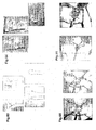
- a case of a 21-year-old male patient with a left forearm fracture malunion was used. This patient showed an excessive deformity of the radius ( Figure 43 ). An forearm fracture is often seen among men in early youth to youth, and is considered to often cause a malunion when treated in a cast.
- the deformity can be corrected even for a middle-aged woman.
- a 48-year-old female patient having a fracture malunion on the distal end of left radius was used.
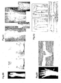
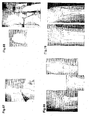
- Figure 69 a postoperative x-ray was taken after the bone was fixed by the template ( Figure 69 ). As shown, the correction is finely done.
- the central photo of Figure 69 is a reversed image of the healthy side. Through the comparison, it was confirmed that the deformity was corrected to the state which is substantially the same as that of the healthy side.
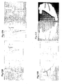
- FIG 91 A treatment simulation of this patient was performed ( Figure 91 ).
- a three-dimensional model of the radius was matched to the mirror image of the healthy side. It is observed that the distal end is shortened and the articular surface is tilted. Thus, the screw axis was obtained ( Figure 92 ). Since the axis was distanced from the bone, the correction was to be performed while distracting the bone. In the case where the screw axis is distanced from the bone like this, osteotomy accompanies distraction (or may accompany shortening) ( Figure 93).
- Figure 94 shows the pre-correction state and the post-correction state.
- the third support was made to be extended using a nut.
- the two rings were tilted at 40 degrees, such that gradual correction and distraction could be performed about the screw axis by rotating the nut.
- the right photo of Figure 151 shows the state when the correction was finished. In actuality, the cut portion is distracted by 1 mm per day by callus being generated, and the distracted portion is buried with bone.
- the present inventor developed a system for simulating a surgical operation of a deformed scaphoid nonunion using a three-dimensional image, and applied the system to 8 clinical cases.
- a surface model of the scaphoid was re-constructed on a computer from CT data of both wrist joints, and a distal portion and a proximal portion of the nonunion model were matched to those of a mirror image of the healthy, opposite side.
- an appropriate site and direction of the estimated defect of the bone and screw insertion were obtained by a simulation.
- a full-size photo printout model of the scaphoid was produced. In an actual surgical operation, restoration, bone implantation and screw insertion were performed using the model as a guide.
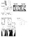
- the wrist joint was fixed for 4 weeks after the surgical operation, and a wrist joint split was attached until the bone union was confirmed on the x-ray ( Figures 164H and 164I ).
- the defect had an anterior thickness of 5.5 mm on average (3.8 to 7.3 mm), a depth of 9.7 mm on average (7.5 to 12.1 mm), and a width of 10.7 mm on average (9.1 to 12.8 mm) as summarized in Table II.
- Table II Results of three-dimensional computer simulation Table II. Results of preoperative 3D computer Case Volar rotation of the distal fragment (degrees) Estimated bone defect (mm) anterior thickness depth width 1 24.5 4 8.9 9.1 2 70.2 6.7 8.6 12.8 3 28.6 4.3 8.9 10.7 4 36.9 6 11.3 9.2 5 40.8 4 8.1 10 6 23.9 6.7 12.1 12.7 7 18.7 5.3 11.9 11.3 8 73.9 7.3 7.5 9.6
- the postoperative score for the wrist joint was 65 to 100, with an average of 89.
- the movable range of flexion and extension was 138 degrees on average (115 to 160 degrees), and the movable range of the radius and the ulna was 64 degrees on average (50 to 75 degrees).
- the grip strength was 80 to 100%, witch the average being 90%, of that of the healthy side (Table I). In 1 case (case 4), fine bone union was observed at the most recent follow-up survey but the patient complained of the ache when extending the wrist joint and the restriction of the movable range.
- the computer 1000 includes a CPU 1010, a memory 1020, an input interface 1030, an output interface 1040, a user interface 1050, and a bus 1060.
- Figure 181 shows an exemplary procedure for performing step 2030 shown in Figure 180 . This procedure is performed by a program which is executed by the CPU 1010.
- the assisting member model may be, for example, a model representing a graft (graft model).
- graft model can be created by obtaining a difference between the bone model and the target bone model.
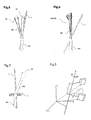
- the relative matrix represents the direction and the amount of movement of the distal bone fragment model with respect to the proximal bone fragment model in accordance with the affine transformation method.
- the screw axis L , the rotating angle ⁇ , and the moving distance t along the screw axis L represent the direction and the amount of movement of the distal bone fragment model with respect to the proximal bone fragment model in accordance with the Screw Displacement-Axis method.
- transformation of the relative matrix into the representation by the Screw Displacement-Axis method means transforming the relative movement information by the representation of the affine transformation method into the relative movement information by the representation of the Screw Displacement-Axis.
- Such transformation of the relative movement information is equivalent to representing a movement of an object in a space by a different coordinate system.
Landscapes
- Health & Medical Sciences (AREA)
- Life Sciences & Earth Sciences (AREA)
- Surgery (AREA)
- Engineering & Computer Science (AREA)
- Medical Informatics (AREA)
- Public Health (AREA)
- General Health & Medical Sciences (AREA)
- Biomedical Technology (AREA)
- Orthopedic Medicine & Surgery (AREA)
- Veterinary Medicine (AREA)
- Heart & Thoracic Surgery (AREA)
- Animal Behavior & Ethology (AREA)
- Nuclear Medicine, Radiotherapy & Molecular Imaging (AREA)
- Molecular Biology (AREA)
- Pathology (AREA)
- Oral & Maxillofacial Surgery (AREA)
- Databases & Information Systems (AREA)
- Primary Health Care (AREA)
- Epidemiology (AREA)
- Data Mining & Analysis (AREA)
- Neurology (AREA)
- Dentistry (AREA)
- Surgical Instruments (AREA)
- Processing Or Creating Images (AREA)
- Prostheses (AREA)
Claims (5)
- Osteotomieassistiervorrichtung (1) zum Assistieren beim Schneiden und Teilen eines Knochens, der in einer abnormen Form verformt ist, in Knochenfragmente und zum Ändern und Korrigieren einer Positionsbeziehung der Knochenfragmente in eine Positionsbeziehung eines Knochens in einer normale Form, wobei die Osteotomieassistiervorrichtung umfasst:ein Positionierelement (1) mit einer passenden Oberfläche (11), welche ausgebildet ist, um zur Oberfläche des Knochens passen, der geschnitten und in Knochenfragmente geteilt werden soll; undeinen Schneidespalt (12) ausgebildet an einer Position, die einem Schneidebereich entspricht, entlang dem das Schneiden und Teilen des Knochens mit dem auf der Knochenoberfläche angebrachten Positionierelement (1) durchgeführt werden sollen, wobei der Schneidespalt (12) eine Form aufweist, die in der Lage ist, eine Schneidevorrichtung in Richtung des Schneidebereichs zu führen;wobei die Osteotomieassistiervorrichtung (1) mit einer Vielzahl von Führungslöchern (13) versehen ist, die ausgelegt sind, die Befestigung einer Vielzahl von Stäben (2) an die Knochenfragmente zu führen, wobei die Stäbe (2) zum Durchbohren eines Knochens ausgebildet sind; und wobei die Vielzahl der Führungslöchern (13) so angeordnet sind, dass jedes Knochenfragment mit wenigstens zwei hineingebohrten Stäben (2) versehen werden kann; und einen Block (3) versehen mit einer Vielzahl von Insertionslöchern (31);dadurch gekennzeichnet, dass jedes der Insertionslöcher (31) eine Form aufweist, welche es einem der Stäbe (2), der an dem jeweiligen Knochenfragment befestigt ist, ermöglicht, dort hindurchgeführt zu werden, wenn die Knochenfragmente im Verhältnis zueinander um eine Achse, die senkrecht zu der Ebene des Schneidebereichs steht, gedreht werden.
- Apparat zum Herstellen einer Osteotomieassistiervorrichtung zum Festlegen einer Knochenbehandlung zum Schneiden und Teilen eines Knochens, der in einer abnormen Form verformt ist, und Verändern und Korrigieren einer Positionsbeziehung der geteilten Knochen in eine Positionsbeziehung eines Knochens in einer normale Form, wobei der Apparat umfasst:A) ein Mittel zum Erhalten eines Knochenmodells zum Erhalten von dreidimensionalen Daten des Knochens von einer bildgebenden Vorrichtung zum Abbilden eines zu behandelnden Knochens, und basierend auf den erhaltenen dreidimensionalen Daten, Erhalten eines dreidimensionalen Knochenmodells, welches den zu behandelnden Knochen darstellt;B) ein Mittel zum Erhalten eines Zielknochenmodells zu Erhalten eines Zielknochenmodells welches ein Behandlungsziel bei der Korrektur des Knochens ist; undC) als ein Mittel zum Festlegen der Behandlung zum Festlegen basierend auf dem Knochenmodell und dem Zielknochenmodell eines Behandlungsprozesses, welcher an dem zu behandelnden Knochen durchgeführt werden soll:C1) ein Mittel zum Festlegen der Bewegung eines Knochenfragmentmodells zum Festsetzen eines Korrekturschneidebereichs für das Knochenmodell und Erstellen von Knochenfragmentmodellen, welche durch Schneiden des Knochenmodells im Schneidebereich erhalten werden,dadurch gekennzeichnet, dass
das Mittel zu Erhalten des Zielknochenmodells das Zielknochenmodell durch Verwenden eines Spiegelbildes des Knochenmodells einer normalen Seite erhält;
das Mittel zum Festlegen der Bewegung des Knochenfragmentmodells als eine Korrekturposition ein Position festlegt, wo ein Unterschied in der Form zwischen den Knochenfragmentmodellen und dem Zielknochenmodell am kleinsten ist; und
das Mittel zum Festlegen der Behandlung weiterhin umfasst:C2) ein Mittel zum Befestigen eines Stabmodells zum Simulieren eines Zustandes worin wenigstens ein Modell eines zum Durchbohren eines Kochens ausgebildeten Stabs (2) jeweils an das Knochenfragmentmodell in der Korrekturposition und an ein Knochenfragmentmodell auf einer weiteren Seite des Schneidebereichs in der Korrekturposition befestigt ist, erstellt durch Schneiden des Knochenmodells,C3) Mittel zum Berechnen einer Stabmodellposition zum Berechnen von Positionen der jeweiligen Stabmodelle im einem Fall, wo sich jedes der Knochenfragmentmodelle an der Position des Knochenmodells befindet, basierend auf Positionen der jeweiligen Stabmodelle befestigt an den jeweiligen Fragmentmodellen durch die Mittel zum Befestigen der Stabmodelle, undC4) ein Mittel zum Erstellen eines Modells einer Osteotomieassistiervorrichtung zum Erstellen eines Modells einer Osteotomieassistiervorrichtung, welche eine passende Oberfläche aufweist, die zu einer Oberfläche des Knochenmodells passt, welches den Schneidebereich aufweist, wobei das Modell der Osteotomieassistiervorrichtung einen Spalt aufweist, der in einem Abschnitt ausgebildet ist, die dem Schneidebereich entspricht, und Führungsbohrungen aufweist, die in Abschnitten ausgebildet sind, die jenen der jeweiligen Stabmodellen zum Einführen der jeweiligen Stabmodelle entsprechen. - Einrichtung nach Anspruch 2 weiterhin umfassendC5) ein Mittel zum Erstellen eines Blockmodells zum Erstellen eines Blockmodells, welches mit Insertionslöchern (31) zum Einführen der jeweiligen Stabmodelle in Abschnitten versehen ist, die jenen der jeweiligen Stabmodellen entsprechen, die an den jeweiligen Knochenfragmentmodellen mit den Mitteln zum Befestigen der Stabmodelle in der korrigierten Position befestigt sind.
- Programm zum Herstellen einer Osteotomieassistiervorrichtung zum Festlegen einer Knochenbehandlung zum Schneiden und Teilen eines Knochens, der in einer abnormen Form verformt ist, und Verändern und Korrigieren einer Positionsbeziehung der geteilten Knochen in eine Positionsbeziehung eines Knochens in eine normale Form, wobei das Programm einen Computer veranlasst zu fungieren als:A) ein Mittel zu Erhalten eines Knochenmodells zum Erhalten von dreidimensionalen Daten des Knochens von einer bildgebenden Vorrichtung zum Abbilden eines zu behandelnden Knochens, und basierend auf den erhaltenen dreidimensionalen Daten, Erhalten eines dreidimensionalen Knochenmodells, welches den zu behandelnden Knochen darstellt;B) ein Mittel zum Erhalten eines Zielknochenmodells zu Erhalten eines Zielknochenmodells, welches ein Behandlungsziel bei der Korrektur des Knochens ist, durch Verwendung eines Spiegelbildes des Knochenmodells von einer normalen Seite; undC) als ein Mittel zum Festlegen der Behandlung zum Festlegen eines Behandlungsprozesses, welcher an dem zu behandelnden Knochen durchgeführt werden soll, basierend auf dem Knochenmodell und dem Zielknochenmodell;C1) ein Mittel zum Festlegen der Bewegung eines Knochenfragmentmodells zum Festsetzen eines Korrekturschneidebereichs für das Knochenmodell, wobei Knochenfragmentmodelle erstellt werden, welche durch Schneiden des Knochenmodells im Schneidebereich erhalten wurden, und Festlegen als eine Korrekturposition eine Position, wo ein Unterschied in der Form zwischen den Knochenfragmentmodell und dem Zielknochenfragment am kleinsten ist,C2) ein Mittel zum Befestigen eines Stabmodells zum Simulieren eines Zustandes worin wenigstens ein Modell eines zum Durchbohren eines Knochens ausgebildeten Stabes (2) jeweils an das Knochenfragmentmodell in der Korrekturposition und an ein Knochenfragmentmodell auf der anderen Seite des Schneidebereichs in der Korrekturposition befestigt ist, erstellt durch Schneiden des Knochenmodells,C3) Mittel zum Berechnen einer Stabmodellposition zum Berechnen von Positionen der jeweiligen Stabmodelle im einem Fall, wo sich jedes der Knochenfragmentmodelle an einer Position des Knochenmodells befindet, basierend auf Positionen der jeweiligen Stabmodelle befestigt an den jeweiligen Knochenfragmentmodellen durch Mittel zum Befestigen der Stabmodelle, undC4) ein Mittel zum Erstellen eines Modells einer Osteotomieassistiervorrichtung zum Erstellen eines Modells einer Osteotomieassistiervorrichtung, welches eine passende Oberfläche aufweist, die zu einer Oberfläche des Knochenmodells passt, welches einen Schneidebereich aufweist, wobei das Modell der Osteotomieassistiervorrichtung einen Spalt aufweist, der in einem Abschnitt ausgebildet ist, der dem Schneidebereich entspricht, und Führungslöcher aufweist, die in Abschnitten ausgebildet sind, die jenen der jeweiligen Stabmodellen zum Einführen der jeweiligen Stabmodelle entsprechen.
- Programm nach Anspruch 4 welches den Computer weiterhin veranlasst zu fungieren als:C5) ein Mittel zum Erstellen eines Blockmodells zum Erstellen eines Blockmodells, welches mit Insertionslöchern (31) zum Einführen der jeweiligen Stabmodelle in Abschnitten versehen ist, die jenen der jeweiligen Stabmodellen entsprechen, die an den jeweiligen Knochenfragmentmodellen mit den Mitteln zum Befestigen der Stabmodelle in der korrigierten Position befestigt sind.
Applications Claiming Priority (2)
| Application Number | Priority Date | Filing Date | Title |
|---|---|---|---|
| PCT/JP2003/001455 WO2004071314A1 (ja) | 2003-02-12 | 2003-02-12 | 罹患骨切断補助部材及び矯正位置判断補助部材 |
| PCT/JP2004/001440 WO2004071309A1 (en) | 2003-02-12 | 2004-02-10 | Method, members, system and program for bone correction |
Publications (2)
| Publication Number | Publication Date |
|---|---|
| EP1624812A1 EP1624812A1 (de) | 2006-02-15 |
| EP1624812B1 true EP1624812B1 (de) | 2015-04-15 |
Family
ID=32866104
Family Applications (1)
| Application Number | Title | Priority Date | Filing Date |
|---|---|---|---|
| EP04709755.5A Expired - Lifetime EP1624812B1 (de) | 2003-02-12 | 2004-02-10 | Elemente, gerät und programm für die knochenkorrektur |
Country Status (6)
| Country | Link |
|---|---|
| US (1) | US20070173815A1 (de) |
| EP (1) | EP1624812B1 (de) |
| JP (1) | JP2006519636A (de) |
| AU (1) | AU2003211935A1 (de) |
| CA (1) | CA2515921A1 (de) |
| WO (2) | WO2004071314A1 (de) |
Families Citing this family (62)
| Publication number | Priority date | Publication date | Assignee | Title |
|---|---|---|---|---|
| US8175683B2 (en) * | 2003-12-30 | 2012-05-08 | Depuy Products, Inc. | System and method of designing and manufacturing customized instrumentation for accurate implantation of prosthesis by utilizing computed tomography data |
| US8055487B2 (en) * | 2005-02-22 | 2011-11-08 | Smith & Nephew, Inc. | Interactive orthopaedic biomechanics system |
| GB0504172D0 (en) * | 2005-03-01 | 2005-04-06 | King S College London | Surgical planning |
| DE102005052993B4 (de) * | 2005-11-07 | 2014-08-21 | Siemens Aktiengesellschaft | Verfahren zur automatisierten Auswertung eines dreidimensionalen Abbildes eines seitensymmetrischen Organsystems |
| US8565853B2 (en) | 2006-08-11 | 2013-10-22 | DePuy Synthes Products, LLC | Simulated bone or tissue manipulation |
| US7603192B2 (en) * | 2007-02-13 | 2009-10-13 | Orthohelix Surgical Designs, Inc. | Method of making orthopedic implants and the orthopedic implants |
| WO2008109751A1 (en) | 2007-03-06 | 2008-09-12 | The Cleveland Clinic Foundation | Method and apparatus for preparing for a surgical procedure |
| US10758283B2 (en) | 2016-08-11 | 2020-09-01 | Mighty Oak Medical, Inc. | Fixation devices having fenestrations and methods for using the same |
| US8221430B2 (en) * | 2007-12-18 | 2012-07-17 | Otismed Corporation | System and method for manufacturing arthroplasty jigs |
| US20110004317A1 (en) * | 2007-12-18 | 2011-01-06 | Hacking Adam S | Orthopaedic implants |
| US8549888B2 (en) | 2008-04-04 | 2013-10-08 | Nuvasive, Inc. | System and device for designing and forming a surgical implant |
| JP5463496B2 (ja) * | 2008-12-03 | 2014-04-09 | 国立大学法人 東京大学 | 歯槽骨再生用生体吸収性3次元メンブレンの製造方法 |
| JP5410525B2 (ja) * | 2009-07-15 | 2014-02-05 | 剛 村瀬 | 補填用人工骨モデル、補填用人工骨の形成方法、並びに医療用シミュレーションシステム |
| FI122920B (fi) * | 2009-10-05 | 2012-08-31 | Aalto Korkeakoulusaeaetioe | Anatomisesti personoitu ja mobilisoiva eksternaalituki, menetelmä sen valmistamiseksi sekä invasiivisesti kiinnitetyn eksternaalituen osan käyttö tuettavan nivelen liikeradan määrittämisessä |
| US9737336B2 (en) | 2009-10-05 | 2017-08-22 | Aalto University Foundation | Anatomically personalized and mobilizing external support and method for controlling a path of an external auxiliary frame |
| CA2778057C (en) | 2009-10-29 | 2019-02-19 | Zimmer, Inc. | Patient-specific mill guide |
| JP5773340B2 (ja) * | 2010-01-30 | 2015-09-02 | アルスロデザイン株式会社 | 人工膝関節置換術用手術器械 |
| KR101687053B1 (ko) * | 2010-04-29 | 2016-12-15 | 신세스 게엠바하 | 악교정 임플란트 및 그 이용 방법 |
| US8540749B2 (en) * | 2010-06-02 | 2013-09-24 | Covidien Lp | Apparatus for performing an electrosurgical procedure |
| US12357413B2 (en) | 2010-06-29 | 2025-07-15 | Mighty Oak Medical, Inc. | Patient-matched apparatus for use in spine related surgical procedures and methods for using the same |
| US11039889B2 (en) | 2010-06-29 | 2021-06-22 | Mighty Oak Medical, Inc. | Patient-matched apparatus and methods for performing surgical procedures |
| US11376073B2 (en) | 2010-06-29 | 2022-07-05 | Mighty Oak Medical Inc. | Patient-matched apparatus and methods for performing surgical procedures |
| BR112012032947A2 (pt) | 2010-06-29 | 2017-01-10 | George Frey | dispositivo cirúrgico formado pos dados anatômicos para um paciente específico, sistema e método para facilitar um/uns procedimento(s) cirúrgico(s), guia cirúrgico personalizado, molde cirúrgico pré-configurado e método para criar um molde para uso em uma operação cirúrgica |
| US11806197B2 (en) | 2010-06-29 | 2023-11-07 | Mighty Oak Medical, Inc. | Patient-matched apparatus for use in spine related surgical procedures and methods for using the same |
| US11633254B2 (en) | 2018-06-04 | 2023-04-25 | Mighty Oak Medical, Inc. | Patient-matched apparatus for use in augmented reality assisted surgical procedures and methods for using the same |
| CA2816337C (en) | 2010-10-29 | 2019-10-22 | The Cleveland Clinic Foundation | System and method for association of a guiding aid with a patient tissue |
| US20120276509A1 (en) | 2010-10-29 | 2012-11-01 | The Cleveland Clinic Foundation | System of preoperative planning and provision of patient-specific surgical aids |
| WO2012058355A1 (en) | 2010-10-29 | 2012-05-03 | The Cleveland Clinic Foundation | System of preoperative planning and provision of patient-specific surgical aids |
| ES2599850T3 (es) | 2011-02-01 | 2017-02-03 | Nextremity Solutions, Inc. | Dispositivo de reparación de defectos óseos |
| US9167989B2 (en) * | 2011-09-16 | 2015-10-27 | Mako Surgical Corp. | Systems and methods for measuring parameters in joint replacement surgery |
| WO2013055203A1 (en) | 2011-10-14 | 2013-04-18 | Academisch Medisch Centrum Bij De Universiteit Van Amsterdam | Method to provide at least one patient specific device to be used for bone correction, a treatment kit, a method of operating a data-processing system, a computer program, and a correction and fixation device and a cutting assisting device for bone correction. |
| EP2838458B1 (de) * | 2012-04-18 | 2018-09-12 | Materialise N.V. | Orthopädische knochenfixiersysteme und verfahren |
| FR2999071A1 (fr) * | 2012-12-12 | 2014-06-13 | Obl | Procede de repositionnement de fragments osseux pour la chirurgie osseuse, base sur l'utilisation d'implants et de guides sur mesure |
| US9204937B2 (en) | 2013-02-19 | 2015-12-08 | Stryker Trauma Gmbh | Software for use with deformity correction |
| US11086970B2 (en) * | 2013-03-13 | 2021-08-10 | Blue Belt Technologies, Inc. | Systems and methods for using generic anatomy models in surgical planning |
| US20140303990A1 (en) * | 2013-04-05 | 2014-10-09 | Biomet Manufacturing Corp. | Integrated orthopedic planning and management process |
| US10467752B2 (en) | 2013-06-11 | 2019-11-05 | Atsushi Tanji | Bone cutting support system, information processing apparatus, image processing method, and image processing program |
| US10398510B2 (en) | 2013-11-12 | 2019-09-03 | Makoto Goto | Manufacturing method of bone cutting assist device, non-transitory computer-readable recording medium having program for manufacturing bone cutting assist device recorded thereon, and bone cutting assist device |
| FR3023044B1 (fr) * | 2014-06-26 | 2017-10-13 | Blue Frog Robotics | Procede de determination d'une transformation geometrique entre deux modeles d'un objet en trois dimensions |
| JP2016096889A (ja) | 2014-11-19 | 2016-05-30 | 株式会社東芝 | 画像解析装置、画像解析方法、およびプログラム |
| EP4631446A3 (de) * | 2015-02-13 | 2025-12-24 | Circinus Medical Technology, LLC | Elektronische vorrichtung zur bestimmung der ausrichtung eines instruments |
| JP6559482B2 (ja) * | 2015-06-30 | 2019-08-14 | 大阪冶金興業株式会社 | 骨切りガイド、骨切りシステム、および、骨切り装置 |
| WO2017011576A2 (en) * | 2015-07-13 | 2017-01-19 | Mako Surgical Corp. | Lower extremities leg length calculation method |
| US9532845B1 (en) * | 2015-08-11 | 2017-01-03 | ITKR Software LLC | Methods for facilitating individualized kinematically aligned total knee replacements and devices thereof |
| US10433921B2 (en) * | 2015-12-28 | 2019-10-08 | Mako Surgical Corp. | Apparatus and methods for robot assisted bone treatment |
| CN105816232A (zh) * | 2016-05-17 | 2016-08-03 | 南方医科大学 | 一种个体化骨骼模型的解剖型接骨板的设计及成型方法 |
| US10251705B2 (en) | 2016-06-02 | 2019-04-09 | Stryker European Holdings I, Llc | Software for use with deformity correction |
| US12016573B2 (en) | 2016-08-11 | 2024-06-25 | Mighty Oak Medical, Inc. | Drill apparatus and surgical fixation devices and methods for using the same |
| US10743890B2 (en) | 2016-08-11 | 2020-08-18 | Mighty Oak Medical, Inc. | Drill apparatus and surgical fixation devices and methods for using the same |
| JP6748299B2 (ja) * | 2016-08-30 | 2020-08-26 | マコー サージカル コーポレイション | 術中骨盤見当合わせ用のシステム及び方法 |
| EP3590450B1 (de) * | 2017-03-03 | 2023-11-01 | Teijin Nakashima Medical Co., Ltd. | Osteotomie-hilfe-set |
| USD895111S1 (en) | 2018-06-04 | 2020-09-01 | Mighty Oak Medical, Inc. | Sacro-iliac guide |
| USD948717S1 (en) | 2018-06-04 | 2022-04-12 | Mighty Oak Medical, Inc. | Sacro-iliac guide |
| CN113576643B (zh) * | 2020-03-10 | 2022-08-30 | 河北医科大学第三医院 | 用于下肢骨折断骨侧向畸形的复位系统 |
| AU2021202888B2 (en) * | 2020-05-26 | 2025-09-11 | Howmedica Osteonics Corp. | Medial trochanteric plate fixation |
| CN111956318A (zh) * | 2020-07-07 | 2020-11-20 | 济南大学 | 定位导板及其制作方法、定位导板模型生成方法及系统 |
| AU2021261864A1 (en) | 2020-11-03 | 2022-05-19 | Mako Surgical Corp. | Multi-component locking implant |
| USD992114S1 (en) | 2021-08-12 | 2023-07-11 | Mighty Oak Medical, Inc. | Surgical guide |
| US11759216B2 (en) * | 2021-09-22 | 2023-09-19 | Arthrex, Inc. | Orthopaedic fusion planning systems and methods of repair |
| CN115153833B (zh) * | 2022-07-21 | 2025-06-06 | 武汉理工大学 | 一种前臂成角畸形智能规划方法及系统 |
| US12440276B2 (en) | 2023-03-14 | 2025-10-14 | Mighty Oak Medical, Inc. | Systems and methods for presurgical planning |
| WO2025212925A1 (en) * | 2024-04-03 | 2025-10-09 | Arc Techtonics, Inc. | Osteotomy plate |
Citations (3)
| Publication number | Priority date | Publication date | Assignee | Title |
|---|---|---|---|---|
| US4565191A (en) * | 1984-01-12 | 1986-01-21 | Slocum D Barclay | Apparatus and method for performing cuneiform osteotomy |
| US5021056A (en) * | 1989-09-14 | 1991-06-04 | Intermedics Orthopedics, Inc. | Upper tibial osteotomy system |
| GB2334214A (en) * | 1998-02-12 | 1999-08-18 | John Knowles Stanley | Surgical cutting guide for shortening a bone |
Family Cites Families (9)
| Publication number | Priority date | Publication date | Assignee | Title |
|---|---|---|---|---|
| GB8424579D0 (en) * | 1984-09-28 | 1984-11-07 | Univ London | Fracture reduction apparatus |
| US5203346A (en) * | 1990-03-30 | 1993-04-20 | Whiplash Analysis, Inc. | Non-invasive method for determining kinematic movement of the cervical spine |
| IT1244249B (it) * | 1990-08-03 | 1994-07-08 | Ascanio Campopiano | Manipolatore per l'esecuzione di manovre riduttive in uso in medicina traumatologica ed ortopedica applicabile a fissatori esterni. |
| JP3372608B2 (ja) * | 1993-09-30 | 2003-02-04 | 京セラ株式会社 | 骨形状修正用スペーサー |
| US5769092A (en) * | 1996-02-22 | 1998-06-23 | Integrated Surgical Systems, Inc. | Computer-aided system for revision total hip replacement surgery |
| IT1293941B1 (it) * | 1997-02-13 | 1999-03-11 | Orthofix Srl | Attrezzo ortopedico particolarmente per la correzione chirurgica di deformazioni ossee |
| US6701174B1 (en) * | 2000-04-07 | 2004-03-02 | Carnegie Mellon University | Computer-aided bone distraction |
| US6711432B1 (en) * | 2000-10-23 | 2004-03-23 | Carnegie Mellon University | Computer-aided orthopedic surgery |
| EP1219239A1 (de) * | 2000-12-30 | 2002-07-03 | Istituti Ortopedici Rizzoli | Verfahren und Gerät zur gleichzeitigen anatomischen und funktionellen Erfassung eines Gelenkes |
-
2003
- 2003-02-12 WO PCT/JP2003/001455 patent/WO2004071314A1/ja not_active Ceased
- 2003-02-12 AU AU2003211935A patent/AU2003211935A1/en not_active Abandoned
-
2004
- 2004-02-10 JP JP2006502665A patent/JP2006519636A/ja active Pending
- 2004-02-10 WO PCT/JP2004/001440 patent/WO2004071309A1/en not_active Ceased
- 2004-02-10 CA CA002515921A patent/CA2515921A1/en not_active Abandoned
- 2004-02-10 EP EP04709755.5A patent/EP1624812B1/de not_active Expired - Lifetime
- 2004-02-10 US US10/545,458 patent/US20070173815A1/en not_active Abandoned
Patent Citations (3)
| Publication number | Priority date | Publication date | Assignee | Title |
|---|---|---|---|---|
| US4565191A (en) * | 1984-01-12 | 1986-01-21 | Slocum D Barclay | Apparatus and method for performing cuneiform osteotomy |
| US5021056A (en) * | 1989-09-14 | 1991-06-04 | Intermedics Orthopedics, Inc. | Upper tibial osteotomy system |
| GB2334214A (en) * | 1998-02-12 | 1999-08-18 | John Knowles Stanley | Surgical cutting guide for shortening a bone |
Also Published As
| Publication number | Publication date |
|---|---|
| AU2003211935A1 (en) | 2004-09-06 |
| EP1624812A1 (de) | 2006-02-15 |
| WO2004071314A1 (ja) | 2004-08-26 |
| JP2006519636A (ja) | 2006-08-31 |
| WO2004071309A1 (en) | 2004-08-26 |
| CA2515921A1 (en) | 2004-08-26 |
| US20070173815A1 (en) | 2007-07-26 |
Similar Documents
| Publication | Publication Date | Title |
|---|---|---|
| US20070173815A1 (en) | Method, members, system and program for bone correction | |
| JP5323885B2 (ja) | 骨矯正のための方法、部材、システムおよびプログラム | |
| US11986251B2 (en) | Patient-specific osteotomy instrumentation | |
| TWI584796B (zh) | 患者可選擇式關節置換術裝置及外科工具 | |
| Pham et al. | Computer modeling and intraoperative navigation in maxillofacial surgery | |
| Lin et al. | Accurate mandible reconstruction by mixed reality, 3D printing, and robotic-assisted navigation integration | |
| CN104706425A (zh) | 能够准确定位的截骨导航装置及其制作方法和使用方法 | |
| US20240108414A1 (en) | Apparatus, system, and method for generating patient-specific implants and/or instrumentation | |
| US20250195081A1 (en) | Patient-specific osteotomy systems, methods, and instrumentation | |
| Rieger et al. | CT virtual reality in the preoperative workup of malunited distal radius fractures: preliminary results | |
| Murase et al. | Does three-dimensional computer simulation improve results of scaphoid nonunion surgery? | |
| Kormi et al. | Accuracy of patient-specific meshes as a reconstruction of orbital floor blow-out fractures | |
| US20240252180A1 (en) | Apparatus, system, and method for osteotomies | |
| Fuller et al. | Computer applications in facial plastic and reconstructive surgery | |
| Romero et al. | Customized surgical protocols for guided bone regeneration using 3D printing technology: a retrospective clinical trial | |
| Li et al. | Quantitative analysis of regional specific pelvic symmetry | |
| US20250057547A1 (en) | Apparatus, system, and method for patient-specific systems, methods, and instrumentation | |
| Sedigh et al. | Cubitus varus corrective osteotomy and graft fashioning using computer simulated bone reconstruction and 3D printed custom-made cutting guides | |
| US20240268966A1 (en) | Apparatus, system, and method for lapidus correction | |
| Chen et al. | Clinical efficacy of 3D-Printing assisted orbital fracture repair with absorbable plates versus preformed titanium mesh: a comparative study | |
| Park | How to reduce inferomedial orbital wall fracture using a navigation system: tips and pearls | |
| Cedillo et al. | A comparative study of analog preoperative planning versus virtual preoperative planning for mandibular reconstruction with fibula free flap | |
| Menzel et al. | Orbit in a box: a simplified technique for patient-specific virtually planned orbital floor reconstruction | |
| Stewart | An image-guided platform for on-the-fly mandibular reconstruction surgery | |
| Moura et al. | Three-dimensional printed model and transantral endoscopy to orbital fracture repair |
Legal Events
| Date | Code | Title | Description |
|---|---|---|---|
| PUAI | Public reference made under article 153(3) epc to a published international application that has entered the european phase |
Free format text: ORIGINAL CODE: 0009012 |
|
| 17P | Request for examination filed |
Effective date: 20050912 |
|
| AK | Designated contracting states |
Kind code of ref document: A1 Designated state(s): AT BE BG CH CY CZ DE DK EE ES FI FR GB GR HU IE IT LI LU MC NL PT RO SE SI SK TR |
|
| DAX | Request for extension of the european patent (deleted) | ||
| 17Q | First examination report despatched |
Effective date: 20090127 |
|
| GRAP | Despatch of communication of intention to grant a patent |
Free format text: ORIGINAL CODE: EPIDOSNIGR1 |
|
| INTG | Intention to grant announced |
Effective date: 20141212 |
|
| GRAS | Grant fee paid |
Free format text: ORIGINAL CODE: EPIDOSNIGR3 |
|
| GRAA | (expected) grant |
Free format text: ORIGINAL CODE: 0009210 |
|
| AK | Designated contracting states |
Kind code of ref document: B1 Designated state(s): AT BE BG CH CY CZ DE DK EE ES FI FR GB GR HU IE IT LI LU MC NL PT RO SE SI SK TR |
|
| REG | Reference to a national code |
Ref country code: GB Ref legal event code: FG4D Ref country code: CH Ref legal event code: EP |
|
| REG | Reference to a national code |
Ref country code: IE Ref legal event code: FG4D |
|
| REG | Reference to a national code |
Ref country code: AT Ref legal event code: REF Ref document number: 721497 Country of ref document: AT Kind code of ref document: T Effective date: 20150515 |
|
| REG | Reference to a national code |
Ref country code: DE Ref legal event code: R096 Ref document number: 602004046997 Country of ref document: DE Effective date: 20150528 |
|
| REG | Reference to a national code |
Ref country code: AT Ref legal event code: MK05 Ref document number: 721497 Country of ref document: AT Kind code of ref document: T Effective date: 20150415 |
|
| PG25 | Lapsed in a contracting state [announced via postgrant information from national office to epo] |
Ref country code: PT Free format text: LAPSE BECAUSE OF FAILURE TO SUBMIT A TRANSLATION OF THE DESCRIPTION OR TO PAY THE FEE WITHIN THE PRESCRIBED TIME-LIMIT Effective date: 20150817 Ref country code: FI Free format text: LAPSE BECAUSE OF FAILURE TO SUBMIT A TRANSLATION OF THE DESCRIPTION OR TO PAY THE FEE WITHIN THE PRESCRIBED TIME-LIMIT Effective date: 20150415 Ref country code: ES Free format text: LAPSE BECAUSE OF FAILURE TO SUBMIT A TRANSLATION OF THE DESCRIPTION OR TO PAY THE FEE WITHIN THE PRESCRIBED TIME-LIMIT Effective date: 20150415 |
|
| PG25 | Lapsed in a contracting state [announced via postgrant information from national office to epo] |
Ref country code: GR Free format text: LAPSE BECAUSE OF FAILURE TO SUBMIT A TRANSLATION OF THE DESCRIPTION OR TO PAY THE FEE WITHIN THE PRESCRIBED TIME-LIMIT Effective date: 20150716 Ref country code: AT Free format text: LAPSE BECAUSE OF FAILURE TO SUBMIT A TRANSLATION OF THE DESCRIPTION OR TO PAY THE FEE WITHIN THE PRESCRIBED TIME-LIMIT Effective date: 20150415 |
|
| REG | Reference to a national code |
Ref country code: DE Ref legal event code: R097 Ref document number: 602004046997 Country of ref document: DE |
|
| PG25 | Lapsed in a contracting state [announced via postgrant information from national office to epo] |
Ref country code: DK Free format text: LAPSE BECAUSE OF FAILURE TO SUBMIT A TRANSLATION OF THE DESCRIPTION OR TO PAY THE FEE WITHIN THE PRESCRIBED TIME-LIMIT Effective date: 20150415 Ref country code: EE Free format text: LAPSE BECAUSE OF FAILURE TO SUBMIT A TRANSLATION OF THE DESCRIPTION OR TO PAY THE FEE WITHIN THE PRESCRIBED TIME-LIMIT Effective date: 20150415 |
|
| REG | Reference to a national code |
Ref country code: FR Ref legal event code: PLFP Year of fee payment: 13 |
|
| PLBE | No opposition filed within time limit |
Free format text: ORIGINAL CODE: 0009261 |
|
| STAA | Information on the status of an ep patent application or granted ep patent |
Free format text: STATUS: NO OPPOSITION FILED WITHIN TIME LIMIT |
|
| PG25 | Lapsed in a contracting state [announced via postgrant information from national office to epo] |
Ref country code: CZ Free format text: LAPSE BECAUSE OF FAILURE TO SUBMIT A TRANSLATION OF THE DESCRIPTION OR TO PAY THE FEE WITHIN THE PRESCRIBED TIME-LIMIT Effective date: 20150415 Ref country code: RO Free format text: LAPSE BECAUSE OF NON-PAYMENT OF DUE FEES Effective date: 20150415 Ref country code: SK Free format text: LAPSE BECAUSE OF FAILURE TO SUBMIT A TRANSLATION OF THE DESCRIPTION OR TO PAY THE FEE WITHIN THE PRESCRIBED TIME-LIMIT Effective date: 20150415 |
|
| 26N | No opposition filed |
Effective date: 20160118 |
|
| PG25 | Lapsed in a contracting state [announced via postgrant information from national office to epo] |
Ref country code: IT Free format text: LAPSE BECAUSE OF FAILURE TO SUBMIT A TRANSLATION OF THE DESCRIPTION OR TO PAY THE FEE WITHIN THE PRESCRIBED TIME-LIMIT Effective date: 20150415 |
|
| PG25 | Lapsed in a contracting state [announced via postgrant information from national office to epo] |
Ref country code: BE Free format text: LAPSE BECAUSE OF NON-PAYMENT OF DUE FEES Effective date: 20160229 Ref country code: SI Free format text: LAPSE BECAUSE OF FAILURE TO SUBMIT A TRANSLATION OF THE DESCRIPTION OR TO PAY THE FEE WITHIN THE PRESCRIBED TIME-LIMIT Effective date: 20150415 |
|
| PG25 | Lapsed in a contracting state [announced via postgrant information from national office to epo] |
Ref country code: BE Free format text: LAPSE BECAUSE OF FAILURE TO SUBMIT A TRANSLATION OF THE DESCRIPTION OR TO PAY THE FEE WITHIN THE PRESCRIBED TIME-LIMIT Effective date: 20150415 |
|
| PG25 | Lapsed in a contracting state [announced via postgrant information from national office to epo] |
Ref country code: LU Free format text: LAPSE BECAUSE OF FAILURE TO SUBMIT A TRANSLATION OF THE DESCRIPTION OR TO PAY THE FEE WITHIN THE PRESCRIBED TIME-LIMIT Effective date: 20160210 Ref country code: MC Free format text: LAPSE BECAUSE OF FAILURE TO SUBMIT A TRANSLATION OF THE DESCRIPTION OR TO PAY THE FEE WITHIN THE PRESCRIBED TIME-LIMIT Effective date: 20150415 |
|
| REG | Reference to a national code |
Ref country code: IE Ref legal event code: MM4A |
|
| PG25 | Lapsed in a contracting state [announced via postgrant information from national office to epo] |
Ref country code: IE Free format text: LAPSE BECAUSE OF NON-PAYMENT OF DUE FEES Effective date: 20160210 |
|
| REG | Reference to a national code |
Ref country code: FR Ref legal event code: PLFP Year of fee payment: 14 |
|
| PG25 | Lapsed in a contracting state [announced via postgrant information from national office to epo] |
Ref country code: SE Free format text: LAPSE BECAUSE OF FAILURE TO SUBMIT A TRANSLATION OF THE DESCRIPTION OR TO PAY THE FEE WITHIN THE PRESCRIBED TIME-LIMIT Effective date: 20150415 |
|
| REG | Reference to a national code |
Ref country code: FR Ref legal event code: PLFP Year of fee payment: 15 |
|
| PG25 | Lapsed in a contracting state [announced via postgrant information from national office to epo] |
Ref country code: HU Free format text: LAPSE BECAUSE OF FAILURE TO SUBMIT A TRANSLATION OF THE DESCRIPTION OR TO PAY THE FEE WITHIN THE PRESCRIBED TIME-LIMIT; INVALID AB INITIO Effective date: 20040210 Ref country code: CY Free format text: LAPSE BECAUSE OF FAILURE TO SUBMIT A TRANSLATION OF THE DESCRIPTION OR TO PAY THE FEE WITHIN THE PRESCRIBED TIME-LIMIT Effective date: 20150415 |
|
| PG25 | Lapsed in a contracting state [announced via postgrant information from national office to epo] |
Ref country code: TR Free format text: LAPSE BECAUSE OF FAILURE TO SUBMIT A TRANSLATION OF THE DESCRIPTION OR TO PAY THE FEE WITHIN THE PRESCRIBED TIME-LIMIT Effective date: 20150415 |
|
| PG25 | Lapsed in a contracting state [announced via postgrant information from national office to epo] |
Ref country code: BG Free format text: LAPSE BECAUSE OF FAILURE TO SUBMIT A TRANSLATION OF THE DESCRIPTION OR TO PAY THE FEE WITHIN THE PRESCRIBED TIME-LIMIT Effective date: 20150415 |
|
| REG | Reference to a national code |
Ref country code: DE Ref legal event code: R081 Ref document number: 602004046997 Country of ref document: DE Owner name: TEIJIN NAKASHIMA MEDICAL CO., LTD., OKAYAMA-CI, JP Free format text: FORMER OWNER: MURASE, TSUYOSHI, TOYONAKA, OSAKA, JP |
|
| REG | Reference to a national code |
Ref country code: NL Ref legal event code: PD Owner name: TEJIN NAKASHIMA MEDICAL CO., LTD.; JP Free format text: DETAILS ASSIGNMENT: CHANGE OF OWNER(S), ASSIGNMENT; FORMER OWNER NAME: MURASE, TSUYOSHI Effective date: 20220408 |
|
| REG | Reference to a national code |
Ref country code: GB Ref legal event code: 732E Free format text: REGISTERED BETWEEN 20230119 AND 20230125 |
|
| PGFP | Annual fee paid to national office [announced via postgrant information from national office to epo] |
Ref country code: NL Payment date: 20230216 Year of fee payment: 20 |
|
| PGFP | Annual fee paid to national office [announced via postgrant information from national office to epo] |
Ref country code: FR Payment date: 20230220 Year of fee payment: 20 Ref country code: CH Payment date: 20230307 Year of fee payment: 20 |
|
| PGFP | Annual fee paid to national office [announced via postgrant information from national office to epo] |
Ref country code: GB Payment date: 20230220 Year of fee payment: 20 Ref country code: DE Payment date: 20230216 Year of fee payment: 20 |
|
| REG | Reference to a national code |
Ref country code: DE Ref legal event code: R071 Ref document number: 602004046997 Country of ref document: DE |
|
| REG | Reference to a national code |
Ref country code: NL Ref legal event code: MK Effective date: 20240209 |
|
| REG | Reference to a national code |
Ref country code: CH Ref legal event code: PL |
|
| REG | Reference to a national code |
Ref country code: GB Ref legal event code: PE20 Expiry date: 20240209 |
|
| PG25 | Lapsed in a contracting state [announced via postgrant information from national office to epo] |
Ref country code: GB Free format text: LAPSE BECAUSE OF EXPIRATION OF PROTECTION Effective date: 20240209 |