EP1559781A1 - Verfahren zum nachweis der paget-krankheit - Google Patents
Verfahren zum nachweis der paget-krankheit Download PDFInfo
- Publication number
- EP1559781A1 EP1559781A1 EP03810659A EP03810659A EP1559781A1 EP 1559781 A1 EP1559781 A1 EP 1559781A1 EP 03810659 A EP03810659 A EP 03810659A EP 03810659 A EP03810659 A EP 03810659A EP 1559781 A1 EP1559781 A1 EP 1559781A1
- Authority
- EP
- European Patent Office
- Prior art keywords
- seq
- chondroitin
- gene
- bone
- paget disease
- Prior art date
- Legal status (The legal status is an assumption and is not a legal conclusion. Google has not performed a legal analysis and makes no representation as to the accuracy of the status listed.)
- Withdrawn
Links
- 208000016738 bone Paget disease Diseases 0.000 title claims abstract description 50
- 238000000034 method Methods 0.000 title claims abstract description 28
- 108090000623 proteins and genes Proteins 0.000 claims abstract description 57
- 208000010191 Osteitis Deformans Diseases 0.000 claims abstract description 49
- 208000027067 Paget disease of bone Diseases 0.000 claims abstract description 49
- 229920002567 Chondroitin Polymers 0.000 claims abstract description 46
- DLGJWSVWTWEWBJ-HGGSSLSASA-N chondroitin Chemical compound CC(O)=N[C@@H]1[C@H](O)O[C@H](CO)[C@H](O)[C@@H]1OC1[C@H](O)[C@H](O)C=C(C(O)=O)O1 DLGJWSVWTWEWBJ-HGGSSLSASA-N 0.000 claims abstract description 46
- 230000035772 mutation Effects 0.000 claims abstract description 27
- 108010009115 chondroitin synthase Proteins 0.000 claims abstract description 23
- 230000014509 gene expression Effects 0.000 claims abstract description 23
- 241001465754 Metazoa Species 0.000 claims abstract description 9
- FWMNVWWHGCHHJJ-SKKKGAJSSA-N 4-amino-1-[(2r)-6-amino-2-[[(2r)-2-[[(2r)-2-[[(2r)-2-amino-3-phenylpropanoyl]amino]-3-phenylpropanoyl]amino]-4-methylpentanoyl]amino]hexanoyl]piperidine-4-carboxylic acid Chemical compound C([C@H](C(=O)N[C@H](CC(C)C)C(=O)N[C@H](CCCCN)C(=O)N1CCC(N)(CC1)C(O)=O)NC(=O)[C@H](N)CC=1C=CC=CC=1)C1=CC=CC=C1 FWMNVWWHGCHHJJ-SKKKGAJSSA-N 0.000 claims abstract description 4
- 125000003275 alpha amino acid group Chemical group 0.000 claims abstract 3
- 230000000694 effects Effects 0.000 claims description 47
- 239000002773 nucleotide Substances 0.000 claims description 37
- 125000003729 nucleotide group Chemical group 0.000 claims description 37
- 108700023372 Glycosyltransferases Proteins 0.000 claims description 17
- 102000051366 Glycosyltransferases Human genes 0.000 claims description 16
- 150000001720 carbohydrates Chemical group 0.000 claims description 16
- 125000000539 amino acid group Chemical group 0.000 claims description 7
- WQZGKKKJIJFFOK-SVZMEOIVSA-N (+)-Galactose Chemical compound OC[C@H]1OC(O)[C@H](O)[C@@H](O)[C@H]1O WQZGKKKJIJFFOK-SVZMEOIVSA-N 0.000 claims description 6
- 125000000969 xylosyl group Chemical group C1([C@H](O)[C@@H](O)[C@H](O)CO1)* 0.000 claims description 6
- OVRNDRQMDRJTHS-CBQIKETKSA-N N-Acetyl-D-Galactosamine Chemical group CC(=O)N[C@H]1[C@@H](O)O[C@H](CO)[C@H](O)[C@@H]1O OVRNDRQMDRJTHS-CBQIKETKSA-N 0.000 claims description 4
- AEMOLEFTQBMNLQ-WAXACMCWSA-N alpha-D-glucuronic acid Chemical group O[C@H]1O[C@H](C(O)=O)[C@@H](O)[C@H](O)[C@H]1O AEMOLEFTQBMNLQ-WAXACMCWSA-N 0.000 claims description 3
- 238000001514 detection method Methods 0.000 abstract description 9
- 230000007170 pathology Effects 0.000 abstract description 4
- SQDAZGGFXASXDW-UHFFFAOYSA-N 5-bromo-2-(trifluoromethoxy)pyridine Chemical compound FC(F)(F)OC1=CC=C(Br)C=N1 SQDAZGGFXASXDW-UHFFFAOYSA-N 0.000 description 51
- 229920001287 Chondroitin sulfate Polymers 0.000 description 51
- 229940059329 chondroitin sulfate Drugs 0.000 description 51
- OVRNDRQMDRJTHS-KEWYIRBNSA-N N-acetyl-D-galactosamine Chemical compound CC(=O)N[C@H]1C(O)O[C@H](CO)[C@H](O)[C@@H]1O OVRNDRQMDRJTHS-KEWYIRBNSA-N 0.000 description 38
- 102000004190 Enzymes Human genes 0.000 description 33
- 108090000790 Enzymes Proteins 0.000 description 33
- 229940088598 enzyme Drugs 0.000 description 33
- 239000000047 product Substances 0.000 description 33
- MBLBDJOUHNCFQT-UHFFFAOYSA-N N-acetyl-D-galactosamine Natural products CC(=O)NC(C=O)C(O)C(O)C(O)CO MBLBDJOUHNCFQT-UHFFFAOYSA-N 0.000 description 32
- 239000000243 solution Substances 0.000 description 27
- 238000006243 chemical reaction Methods 0.000 description 25
- 238000012546 transfer Methods 0.000 description 21
- 108010023736 Chondroitinases and Chondroitin Lyases Proteins 0.000 description 20
- 102100031688 N-acetylgalactosamine-6-sulfatase Human genes 0.000 description 20
- 239000000758 substrate Substances 0.000 description 19
- FAPWRFPIFSIZLT-UHFFFAOYSA-M Sodium chloride Chemical compound [Na+].[Cl-] FAPWRFPIFSIZLT-UHFFFAOYSA-M 0.000 description 18
- 108090000765 processed proteins & peptides Proteins 0.000 description 16
- 241000699666 Mus <mouse, genus> Species 0.000 description 15
- 150000001413 amino acids Chemical group 0.000 description 15
- 230000015572 biosynthetic process Effects 0.000 description 15
- 108091032973 (ribonucleotides)n+m Proteins 0.000 description 14
- 108020004414 DNA Proteins 0.000 description 14
- 238000003786 synthesis reaction Methods 0.000 description 14
- 210000004027 cell Anatomy 0.000 description 13
- SRBFZHDQGSBBOR-IOVATXLUSA-N D-xylopyranose Chemical compound O[C@@H]1COC(O)[C@H](O)[C@H]1O SRBFZHDQGSBBOR-IOVATXLUSA-N 0.000 description 12
- PEDCQBHIVMGVHV-UHFFFAOYSA-N Glycerine Chemical compound OCC(O)CO PEDCQBHIVMGVHV-UHFFFAOYSA-N 0.000 description 12
- 239000007787 solid Substances 0.000 description 12
- 239000012505 Superdex™ Substances 0.000 description 11
- 238000007796 conventional method Methods 0.000 description 11
- 239000012634 fragment Substances 0.000 description 11
- 230000037431 insertion Effects 0.000 description 11
- 238000003780 insertion Methods 0.000 description 11
- 239000000523 sample Substances 0.000 description 11
- 239000002299 complementary DNA Substances 0.000 description 10
- 230000029087 digestion Effects 0.000 description 10
- 238000005259 measurement Methods 0.000 description 10
- 230000037430 deletion Effects 0.000 description 9
- 238000012217 deletion Methods 0.000 description 9
- 239000011148 porous material Substances 0.000 description 9
- 102000004169 proteins and genes Human genes 0.000 description 9
- 239000011780 sodium chloride Substances 0.000 description 9
- QKNYBSVHEMOAJP-UHFFFAOYSA-N 2-amino-2-(hydroxymethyl)propane-1,3-diol;hydron;chloride Chemical compound Cl.OCC(N)(CO)CO QKNYBSVHEMOAJP-UHFFFAOYSA-N 0.000 description 8
- 238000002360 preparation method Methods 0.000 description 8
- 238000003757 reverse transcription PCR Methods 0.000 description 8
- 102100039847 Globoside alpha-1,3-N-acetylgalactosaminyltransferase 1 Human genes 0.000 description 7
- 101000887519 Homo sapiens Globoside alpha-1,3-N-acetylgalactosaminyltransferase 1 Proteins 0.000 description 7
- LFTYTUAZOPRMMI-NESSUJCYSA-N UDP-N-acetyl-alpha-D-galactosamine Chemical compound O1[C@H](CO)[C@H](O)[C@H](O)[C@@H](NC(=O)C)[C@H]1O[P@](O)(=O)O[P@](O)(=O)OC[C@@H]1[C@@H](O)[C@@H](O)[C@H](N2C(NC(=O)C=C2)=O)O1 LFTYTUAZOPRMMI-NESSUJCYSA-N 0.000 description 7
- HSCJRCZFDFQWRP-ABVWGUQPSA-N UDP-alpha-D-galactose Chemical compound O[C@@H]1[C@@H](O)[C@@H](O)[C@@H](CO)O[C@@H]1OP(O)(=O)OP(O)(=O)OC[C@@H]1[C@@H](O)[C@@H](O)[C@H](N2C(NC(=O)C=C2)=O)O1 HSCJRCZFDFQWRP-ABVWGUQPSA-N 0.000 description 7
- LFTYTUAZOPRMMI-UHFFFAOYSA-N UNPD164450 Natural products O1C(CO)C(O)C(O)C(NC(=O)C)C1OP(O)(=O)OP(O)(=O)OCC1C(O)C(O)C(N2C(NC(=O)C=C2)=O)O1 LFTYTUAZOPRMMI-UHFFFAOYSA-N 0.000 description 7
- 229940024606 amino acid Drugs 0.000 description 7
- PYMYPHUHKUWMLA-UHFFFAOYSA-N arabinose Natural products OCC(O)C(O)C(O)C=O PYMYPHUHKUWMLA-UHFFFAOYSA-N 0.000 description 7
- 238000003556 assay Methods 0.000 description 7
- SRBFZHDQGSBBOR-UHFFFAOYSA-N beta-D-Pyranose-Lyxose Natural products OC1COC(O)C(O)C1O SRBFZHDQGSBBOR-UHFFFAOYSA-N 0.000 description 7
- 229930182470 glycoside Natural products 0.000 description 7
- 150000002338 glycosides Chemical class 0.000 description 7
- 239000006228 supernatant Substances 0.000 description 7
- 239000000725 suspension Substances 0.000 description 7
- 230000005945 translocation Effects 0.000 description 7
- 241000251730 Chondrichthyes Species 0.000 description 6
- OIRDTQYFTABQOQ-KQYNXXCUSA-N adenosine Chemical compound C1=NC=2C(N)=NC=NC=2N1[C@@H]1O[C@H](CO)[C@@H](O)[C@H]1O OIRDTQYFTABQOQ-KQYNXXCUSA-N 0.000 description 6
- 210000004369 blood Anatomy 0.000 description 6
- 239000008280 blood Substances 0.000 description 6
- 210000000845 cartilage Anatomy 0.000 description 6
- 239000007795 chemical reaction product Substances 0.000 description 6
- 208000037265 diseases, disorders, signs and symptoms Diseases 0.000 description 6
- 238000006911 enzymatic reaction Methods 0.000 description 6
- 239000013604 expression vector Substances 0.000 description 6
- 238000011813 knockout mouse model Methods 0.000 description 6
- 210000005087 mononuclear cell Anatomy 0.000 description 6
- MTCFGRXMJLQNBG-REOHCLBHSA-N (2S)-2-Amino-3-hydroxypropansäure Chemical compound OC[C@H](N)C(O)=O MTCFGRXMJLQNBG-REOHCLBHSA-N 0.000 description 5
- 206010064571 Gene mutation Diseases 0.000 description 5
- 229910021380 Manganese Chloride Inorganic materials 0.000 description 5
- GLFNIEUTAYBVOC-UHFFFAOYSA-L Manganese chloride Chemical compound Cl[Mn]Cl GLFNIEUTAYBVOC-UHFFFAOYSA-L 0.000 description 5
- 238000004458 analytical method Methods 0.000 description 5
- 150000001875 compounds Chemical class 0.000 description 5
- 201000010099 disease Diseases 0.000 description 5
- 238000001727 in vivo Methods 0.000 description 5
- 239000011565 manganese chloride Substances 0.000 description 5
- 210000005259 peripheral blood Anatomy 0.000 description 5
- 239000011886 peripheral blood Substances 0.000 description 5
- 238000011002 quantification Methods 0.000 description 5
- 238000010791 quenching Methods 0.000 description 5
- 230000000171 quenching effect Effects 0.000 description 5
- ZKHQWZAMYRWXGA-UHFFFAOYSA-N Adenosine triphosphate Natural products C1=NC=2C(N)=NC=NC=2N1C1OC(COP(O)(=O)OP(O)(=O)OP(O)(O)=O)C(O)C1O ZKHQWZAMYRWXGA-UHFFFAOYSA-N 0.000 description 4
- 241000283690 Bos taurus Species 0.000 description 4
- VMHLLURERBWHNL-UHFFFAOYSA-M Sodium acetate Chemical compound [Na+].CC([O-])=O VMHLLURERBWHNL-UHFFFAOYSA-M 0.000 description 4
- 239000002253 acid Substances 0.000 description 4
- 210000004556 brain Anatomy 0.000 description 4
- 239000007853 buffer solution Substances 0.000 description 4
- YBYRMVIVWMBXKQ-UHFFFAOYSA-N phenylmethanesulfonyl fluoride Chemical compound FS(=O)(=O)CC1=CC=CC=C1 YBYRMVIVWMBXKQ-UHFFFAOYSA-N 0.000 description 4
- 230000006798 recombination Effects 0.000 description 4
- 238000005215 recombination Methods 0.000 description 4
- 239000001632 sodium acetate Substances 0.000 description 4
- 235000017281 sodium acetate Nutrition 0.000 description 4
- 239000000126 substance Substances 0.000 description 4
- 238000012360 testing method Methods 0.000 description 4
- UHDGCWIWMRVCDJ-UHFFFAOYSA-N 1-beta-D-Xylofuranosyl-NH-Cytosine Natural products O=C1N=C(N)C=CN1C1C(O)C(O)C(CO)O1 UHDGCWIWMRVCDJ-UHFFFAOYSA-N 0.000 description 3
- 239000002126 C01EB10 - Adenosine Substances 0.000 description 3
- UHDGCWIWMRVCDJ-PSQAKQOGSA-N Cytidine Natural products O=C1N=C(N)C=CN1[C@@H]1[C@@H](O)[C@@H](O)[C@H](CO)O1 UHDGCWIWMRVCDJ-PSQAKQOGSA-N 0.000 description 3
- 108700024394 Exon Proteins 0.000 description 3
- 239000007987 MES buffer Substances 0.000 description 3
- 229960005305 adenosine Drugs 0.000 description 3
- 238000005119 centrifugation Methods 0.000 description 3
- KXKPYJOVDUMHGS-OSRGNVMNSA-N chondroitin sulfate Chemical compound CC(=O)N[C@H]1[C@H](O)O[C@H](OS(O)(=O)=O)[C@H](O)[C@@H]1O[C@H]1[C@H](O)[C@@H](O)[C@H](O)[C@@H](C(O)=O)O1 KXKPYJOVDUMHGS-OSRGNVMNSA-N 0.000 description 3
- 238000010367 cloning Methods 0.000 description 3
- UHDGCWIWMRVCDJ-ZAKLUEHWSA-N cytidine Chemical compound O=C1N=C(N)C=CN1[C@H]1[C@H](O)[C@@H](O)[C@H](CO)O1 UHDGCWIWMRVCDJ-ZAKLUEHWSA-N 0.000 description 3
- 230000011164 ossification Effects 0.000 description 3
- YCRVFQPJJACUHE-JSCKKFHOSA-N phosphono dihydrogen phosphate;(2s,3s,4s,5r)-2,3,4,5-tetrahydroxy-6-oxohexanoic acid Chemical compound OP(O)(=O)OP(O)(O)=O.O=C[C@H](O)[C@@H](O)[C@H](O)[C@H](O)C(O)=O YCRVFQPJJACUHE-JSCKKFHOSA-N 0.000 description 3
- 238000003753 real-time PCR Methods 0.000 description 3
- 238000012163 sequencing technique Methods 0.000 description 3
- 125000003607 serino group Chemical group [H]N([H])[C@]([H])(C(=O)[*])C(O[H])([H])[H] 0.000 description 3
- 239000013598 vector Substances 0.000 description 3
- RGOFBPTWSOFRNJ-NQZVPSPJSA-N (2r,3s,4s,5r)-2,3,4,5,6-pentahydroxyhexanal;phosphono dihydrogen phosphate Chemical compound OP(O)(=O)OP(O)(O)=O.OC[C@@H](O)[C@H](O)[C@H](O)[C@@H](O)C=O RGOFBPTWSOFRNJ-NQZVPSPJSA-N 0.000 description 2
- FNEHAOQZWPHONV-UHFFFAOYSA-N 9h-carbazole;sulfuric acid Chemical compound OS(O)(=O)=O.C1=CC=C2C3=CC=CC=C3NC2=C1 FNEHAOQZWPHONV-UHFFFAOYSA-N 0.000 description 2
- -1 ATP sodium salt Chemical class 0.000 description 2
- ZKHQWZAMYRWXGA-KQYNXXCUSA-J ATP(4-) Chemical compound C1=NC=2C(N)=NC=NC=2N1[C@@H]1O[C@H](COP([O-])(=O)OP([O-])(=O)OP([O-])([O-])=O)[C@@H](O)[C@H]1O ZKHQWZAMYRWXGA-KQYNXXCUSA-J 0.000 description 2
- 229920000936 Agarose Polymers 0.000 description 2
- 238000011740 C57BL/6 mouse Methods 0.000 description 2
- IAJILQKETJEXLJ-UHFFFAOYSA-N Galacturonsaeure Natural products O=CC(O)C(O)C(O)C(O)C(O)=O IAJILQKETJEXLJ-UHFFFAOYSA-N 0.000 description 2
- 102000053187 Glucuronidase Human genes 0.000 description 2
- 108010060309 Glucuronidase Proteins 0.000 description 2
- 229920002683 Glycosaminoglycan Polymers 0.000 description 2
- 108010003272 Hyaluronate lyase Proteins 0.000 description 2
- 102000009066 Hyaluronoglucosaminidase Human genes 0.000 description 2
- 101800001691 Inter-alpha-trypsin inhibitor light chain Proteins 0.000 description 2
- GDBQQVLCIARPGH-UHFFFAOYSA-N Leupeptin Natural products CC(C)CC(NC(C)=O)C(=O)NC(CC(C)C)C(=O)NC(C=O)CCCN=C(N)N GDBQQVLCIARPGH-UHFFFAOYSA-N 0.000 description 2
- 241000712079 Measles morbillivirus Species 0.000 description 2
- 108091028043 Nucleic acid sequence Proteins 0.000 description 2
- NORZOLAPCOWSRR-DBKUKYHUSA-N OP(O)(=O)OP(O)(O)=O.CC(=O)N[C@H]1C(O)O[C@H](CO)[C@H](O)[C@@H]1O Chemical compound OP(O)(=O)OP(O)(O)=O.CC(=O)N[C@H]1C(O)O[C@H](CO)[C@H](O)[C@@H]1O NORZOLAPCOWSRR-DBKUKYHUSA-N 0.000 description 2
- 238000010222 PCR analysis Methods 0.000 description 2
- 102100032859 Protein AMBP Human genes 0.000 description 2
- 102000016611 Proteoglycans Human genes 0.000 description 2
- 108010067787 Proteoglycans Proteins 0.000 description 2
- 238000010802 RNA extraction kit Methods 0.000 description 2
- 241000700159 Rattus Species 0.000 description 2
- 101100221606 Saccharomyces cerevisiae (strain ATCC 204508 / S288c) COS7 gene Proteins 0.000 description 2
- 238000002105 Southern blotting Methods 0.000 description 2
- 102000004357 Transferases Human genes 0.000 description 2
- 108090000992 Transferases Proteins 0.000 description 2
- DRTQHJPVMGBUCF-XVFCMESISA-N Uridine Chemical compound O[C@@H]1[C@H](O)[C@@H](CO)O[C@H]1N1C(=O)NC(=O)C=C1 DRTQHJPVMGBUCF-XVFCMESISA-N 0.000 description 2
- 230000005856 abnormality Effects 0.000 description 2
- 238000002835 absorbance Methods 0.000 description 2
- 239000008351 acetate buffer Substances 0.000 description 2
- 230000003213 activating effect Effects 0.000 description 2
- 230000003321 amplification Effects 0.000 description 2
- 230000005540 biological transmission Effects 0.000 description 2
- 210000001109 blastomere Anatomy 0.000 description 2
- 210000000988 bone and bone Anatomy 0.000 description 2
- 229910052799 carbon Inorganic materials 0.000 description 2
- 230000002759 chromosomal effect Effects 0.000 description 2
- 210000002808 connective tissue Anatomy 0.000 description 2
- 239000012153 distilled water Substances 0.000 description 2
- 238000004520 electroporation Methods 0.000 description 2
- 238000000605 extraction Methods 0.000 description 2
- 229930182830 galactose Natural products 0.000 description 2
- 238000002523 gelfiltration Methods 0.000 description 2
- 238000010363 gene targeting Methods 0.000 description 2
- 102000006602 glyceraldehyde-3-phosphate dehydrogenase Human genes 0.000 description 2
- 108020004445 glyceraldehyde-3-phosphate dehydrogenase Proteins 0.000 description 2
- 125000005842 heteroatom Chemical group 0.000 description 2
- 229960002773 hyaluronidase Drugs 0.000 description 2
- GDBQQVLCIARPGH-ULQDDVLXSA-N leupeptin Chemical compound CC(C)C[C@H](NC(C)=O)C(=O)N[C@@H](CC(C)C)C(=O)N[C@H](C=O)CCCN=C(N)N GDBQQVLCIARPGH-ULQDDVLXSA-N 0.000 description 2
- 108010052968 leupeptin Proteins 0.000 description 2
- 210000004185 liver Anatomy 0.000 description 2
- 210000004962 mammalian cell Anatomy 0.000 description 2
- 210000001161 mammalian embryo Anatomy 0.000 description 2
- 238000002156 mixing Methods 0.000 description 2
- 238000012544 monitoring process Methods 0.000 description 2
- 238000003199 nucleic acid amplification method Methods 0.000 description 2
- 108020004707 nucleic acids Proteins 0.000 description 2
- 102000039446 nucleic acids Human genes 0.000 description 2
- 150000007523 nucleic acids Chemical class 0.000 description 2
- 229920001542 oligosaccharide Polymers 0.000 description 2
- 150000002482 oligosaccharides Chemical class 0.000 description 2
- 230000001717 pathogenic effect Effects 0.000 description 2
- 230000001575 pathological effect Effects 0.000 description 2
- 229950000964 pepstatin Drugs 0.000 description 2
- 108010091212 pepstatin Proteins 0.000 description 2
- FAXGPCHRFPCXOO-LXTPJMTPSA-N pepstatin A Chemical compound OC(=O)C[C@H](O)[C@H](CC(C)C)NC(=O)[C@H](C)NC(=O)C[C@H](O)[C@H](CC(C)C)NC(=O)[C@H](C(C)C)NC(=O)[C@H](C(C)C)NC(=O)CC(C)C FAXGPCHRFPCXOO-LXTPJMTPSA-N 0.000 description 2
- 238000011176 pooling Methods 0.000 description 2
- 230000010076 replication Effects 0.000 description 2
- 229960001153 serine Drugs 0.000 description 2
- 230000008685 targeting Effects 0.000 description 2
- 210000001550 testis Anatomy 0.000 description 2
- 150000004044 tetrasaccharides Chemical group 0.000 description 2
- XLYOFNOQVPJJNP-UHFFFAOYSA-N water Chemical compound O XLYOFNOQVPJJNP-UHFFFAOYSA-N 0.000 description 2
- 102000007469 Actins Human genes 0.000 description 1
- 108010085238 Actins Proteins 0.000 description 1
- 208000006386 Bone Resorption Diseases 0.000 description 1
- XDTMQSROBMDMFD-UHFFFAOYSA-N C1CCCCC1 Chemical compound C1CCCCC1 XDTMQSROBMDMFD-UHFFFAOYSA-N 0.000 description 1
- HAQXAQBWVBZXTP-QGNDPXNASA-N CC(=O)N[C@H]1C(O)O[C@H](CO)[C@H](O)[C@@H]1O.C1=NC=2C(=O)NC(N)=NC=2N1[C@@H]1O[C@H](COP(O)(=O)OP(O)(O)=O)[C@@H](O)[C@H]1O Chemical compound CC(=O)N[C@H]1C(O)O[C@H](CO)[C@H](O)[C@@H]1O.C1=NC=2C(=O)NC(N)=NC=2N1[C@@H]1O[C@H](COP(O)(=O)OP(O)(O)=O)[C@@H](O)[C@H]1O HAQXAQBWVBZXTP-QGNDPXNASA-N 0.000 description 1
- OKTJSMMVPCPJKN-UHFFFAOYSA-N Carbon Chemical group [C] OKTJSMMVPCPJKN-UHFFFAOYSA-N 0.000 description 1
- 241000282693 Cercopithecidae Species 0.000 description 1
- 102000005598 Chondroitin Sulfate Proteoglycans Human genes 0.000 description 1
- 108010059480 Chondroitin Sulfate Proteoglycans Proteins 0.000 description 1
- 238000000018 DNA microarray Methods 0.000 description 1
- 238000002965 ELISA Methods 0.000 description 1
- 241000282326 Felis catus Species 0.000 description 1
- 101000644537 Homo sapiens Sequestosome-1 Proteins 0.000 description 1
- 101000648503 Homo sapiens Tumor necrosis factor receptor superfamily member 11A Proteins 0.000 description 1
- 241000124008 Mammalia Species 0.000 description 1
- 238000000636 Northern blotting Methods 0.000 description 1
- 241000283973 Oryctolagus cuniculus Species 0.000 description 1
- 238000002944 PCR assay Methods 0.000 description 1
- 241000009328 Perro Species 0.000 description 1
- 238000011529 RT qPCR Methods 0.000 description 1
- 238000012300 Sequence Analysis Methods 0.000 description 1
- 102100020814 Sequestosome-1 Human genes 0.000 description 1
- MTCFGRXMJLQNBG-UHFFFAOYSA-N Serine Natural products OCC(N)C(O)=O MTCFGRXMJLQNBG-UHFFFAOYSA-N 0.000 description 1
- 108020004459 Small interfering RNA Proteins 0.000 description 1
- 102100028787 Tumor necrosis factor receptor superfamily member 11A Human genes 0.000 description 1
- 208000036142 Viral infection Diseases 0.000 description 1
- ZQVDLMNWALGJLM-DKEFOWNISA-N [(2r,3s,4r,5r)-5-(2-amino-6-oxo-3h-purin-9-yl)-3,4-dihydroxyoxolan-2-yl]methyl phosphono hydrogen phosphate;(2r,3s,4s,5r)-2,3,4,5,6-pentahydroxyhexanal Chemical compound OC[C@@H](O)[C@H](O)[C@H](O)[C@@H](O)C=O.C1=NC=2C(=O)NC(N)=NC=2N1[C@@H]1O[C@H](COP(O)(=O)OP(O)(O)=O)[C@@H](O)[C@H]1O ZQVDLMNWALGJLM-DKEFOWNISA-N 0.000 description 1
- UFZKZJQCLASMFS-VTAAODLHSA-N [(2r,3s,4r,5r)-5-(2-amino-6-oxo-3h-purin-9-yl)-3,4-dihydroxyoxolan-2-yl]methyl phosphono hydrogen phosphate;(2s,3s,4s,5r)-2,3,4,5-tetrahydroxy-6-oxohexanoic acid Chemical compound O=C[C@H](O)[C@@H](O)[C@H](O)[C@H](O)C(O)=O.C1=NC=2C(=O)NC(N)=NC=2N1[C@@H]1O[C@H](COP(O)(=O)OP(O)(O)=O)[C@@H](O)[C@H]1O UFZKZJQCLASMFS-VTAAODLHSA-N 0.000 description 1
- LFTYTUAZOPRMMI-CBKXPRBGSA-N [(3r,4r,5r,6r)-3-acetamido-4,5-dihydroxy-6-(hydroxymethyl)oxan-2-yl] [[(2r,3s,4r)-5-(2,4-dioxopyrimidin-1-yl)-3,4-dihydroxyoxolan-2-yl]methoxy-hydroxyphosphoryl] hydrogen phosphate Chemical compound O1[C@H](CO)[C@H](O)[C@H](O)[C@@H](NC(=O)C)C1OP(O)(=O)OP(O)(=O)OC[C@@H]1[C@@H](O)[C@@H](O)C(N2C(NC(=O)C=C2)=O)O1 LFTYTUAZOPRMMI-CBKXPRBGSA-N 0.000 description 1
- 230000002159 abnormal effect Effects 0.000 description 1
- 238000010171 animal model Methods 0.000 description 1
- DRTQHJPVMGBUCF-PSQAKQOGSA-N beta-L-uridine Natural products O[C@H]1[C@@H](O)[C@H](CO)O[C@@H]1N1C(=O)NC(=O)C=C1 DRTQHJPVMGBUCF-PSQAKQOGSA-N 0.000 description 1
- 210000001185 bone marrow Anatomy 0.000 description 1
- 230000024279 bone resorption Effects 0.000 description 1
- 150000001721 carbon Chemical group 0.000 description 1
- 150000002016 disaccharides Chemical class 0.000 description 1
- 210000000981 epithelium Anatomy 0.000 description 1
- 238000002474 experimental method Methods 0.000 description 1
- 229940097043 glucuronic acid Drugs 0.000 description 1
- 230000006801 homologous recombination Effects 0.000 description 1
- 238000002744 homologous recombination Methods 0.000 description 1
- CBOIHMRHGLHBPB-UHFFFAOYSA-N hydroxymethyl Chemical compound O[CH2] CBOIHMRHGLHBPB-UHFFFAOYSA-N 0.000 description 1
- 125000000896 monocarboxylic acid group Chemical group 0.000 description 1
- 238000010172 mouse model Methods 0.000 description 1
- 210000000963 osteoblast Anatomy 0.000 description 1
- 210000002997 osteoclast Anatomy 0.000 description 1
- 208000009932 paget disease of bone 4 Diseases 0.000 description 1
- 230000002093 peripheral effect Effects 0.000 description 1
- 229920000642 polymer Polymers 0.000 description 1
- 102000054765 polymorphisms of proteins Human genes 0.000 description 1
- 238000000746 purification Methods 0.000 description 1
- 210000005132 reproductive cell Anatomy 0.000 description 1
- 239000004055 small Interfering RNA Substances 0.000 description 1
- 159000000000 sodium salts Chemical class 0.000 description 1
- YEENEYXBHNNNGV-XEHWZWQGSA-M sodium;3-acetamido-5-[acetyl(methyl)amino]-2,4,6-triiodobenzoate;(2r,3r,4s,5s,6r)-2-[(2r,3s,4s,5r)-3,4-dihydroxy-2,5-bis(hydroxymethyl)oxolan-2-yl]oxy-6-(hydroxymethyl)oxane-3,4,5-triol Chemical compound [Na+].CC(=O)N(C)C1=C(I)C(NC(C)=O)=C(I)C(C([O-])=O)=C1I.O[C@H]1[C@H](O)[C@@H](CO)O[C@]1(CO)O[C@@H]1[C@H](O)[C@@H](O)[C@H](O)[C@@H](CO)O1 YEENEYXBHNNNGV-XEHWZWQGSA-M 0.000 description 1
- QAOWNCQODCNURD-UHFFFAOYSA-N sulfuric acid group Chemical group S(O)(O)(=O)=O QAOWNCQODCNURD-UHFFFAOYSA-N 0.000 description 1
- 230000008719 thickening Effects 0.000 description 1
- 210000001519 tissue Anatomy 0.000 description 1
- 238000013518 transcription Methods 0.000 description 1
- 230000035897 transcription Effects 0.000 description 1
- 238000013519 translation Methods 0.000 description 1
- 230000001960 triggered effect Effects 0.000 description 1
- DRTQHJPVMGBUCF-UHFFFAOYSA-N uracil arabinoside Natural products OC1C(O)C(CO)OC1N1C(=O)NC(=O)C=C1 DRTQHJPVMGBUCF-UHFFFAOYSA-N 0.000 description 1
- 229940045145 uridine Drugs 0.000 description 1
- 238000012795 verification Methods 0.000 description 1
- 230000009385 viral infection Effects 0.000 description 1
- 238000001262 western blot Methods 0.000 description 1
Images




Classifications
-
- A—HUMAN NECESSITIES
- A01—AGRICULTURE; FORESTRY; ANIMAL HUSBANDRY; HUNTING; TRAPPING; FISHING
- A01K—ANIMAL HUSBANDRY; AVICULTURE; APICULTURE; PISCICULTURE; FISHING; REARING OR BREEDING ANIMALS, NOT OTHERWISE PROVIDED FOR; NEW BREEDS OF ANIMALS
- A01K67/00—Rearing or breeding animals, not otherwise provided for; New or modified breeds of animals
- A01K67/027—New or modified breeds of vertebrates
- A01K67/0275—Genetically modified vertebrates, e.g. transgenic
- A01K67/0276—Knock-out vertebrates
-
- C—CHEMISTRY; METALLURGY
- C12—BIOCHEMISTRY; BEER; SPIRITS; WINE; VINEGAR; MICROBIOLOGY; ENZYMOLOGY; MUTATION OR GENETIC ENGINEERING
- C12N—MICROORGANISMS OR ENZYMES; COMPOSITIONS THEREOF; PROPAGATING, PRESERVING, OR MAINTAINING MICROORGANISMS; MUTATION OR GENETIC ENGINEERING; CULTURE MEDIA
- C12N15/00—Mutation or genetic engineering; DNA or RNA concerning genetic engineering, vectors, e.g. plasmids, or their isolation, preparation or purification; Use of hosts therefor
- C12N15/09—Recombinant DNA-technology
- C12N15/63—Introduction of foreign genetic material using vectors; Vectors; Use of hosts therefor; Regulation of expression
- C12N15/79—Vectors or expression systems specially adapted for eukaryotic hosts
- C12N15/85—Vectors or expression systems specially adapted for eukaryotic hosts for animal cells
- C12N15/8509—Vectors or expression systems specially adapted for eukaryotic hosts for animal cells for producing genetically modified animals, e.g. transgenic
-
- C—CHEMISTRY; METALLURGY
- C12—BIOCHEMISTRY; BEER; SPIRITS; WINE; VINEGAR; MICROBIOLOGY; ENZYMOLOGY; MUTATION OR GENETIC ENGINEERING
- C12N—MICROORGANISMS OR ENZYMES; COMPOSITIONS THEREOF; PROPAGATING, PRESERVING, OR MAINTAINING MICROORGANISMS; MUTATION OR GENETIC ENGINEERING; CULTURE MEDIA
- C12N9/00—Enzymes; Proenzymes; Compositions thereof; Processes for preparing, activating, inhibiting, separating or purifying enzymes
- C12N9/10—Transferases (2.)
- C12N9/13—Transferases (2.) transferring sulfur containing groups (2.8)
-
- C—CHEMISTRY; METALLURGY
- C12—BIOCHEMISTRY; BEER; SPIRITS; WINE; VINEGAR; MICROBIOLOGY; ENZYMOLOGY; MUTATION OR GENETIC ENGINEERING
- C12N—MICROORGANISMS OR ENZYMES; COMPOSITIONS THEREOF; PROPAGATING, PRESERVING, OR MAINTAINING MICROORGANISMS; MUTATION OR GENETIC ENGINEERING; CULTURE MEDIA
- C12N9/00—Enzymes; Proenzymes; Compositions thereof; Processes for preparing, activating, inhibiting, separating or purifying enzymes
- C12N9/14—Hydrolases (3)
- C12N9/16—Hydrolases (3) acting on ester bonds (3.1)
-
- C—CHEMISTRY; METALLURGY
- C12—BIOCHEMISTRY; BEER; SPIRITS; WINE; VINEGAR; MICROBIOLOGY; ENZYMOLOGY; MUTATION OR GENETIC ENGINEERING
- C12Q—MEASURING OR TESTING PROCESSES INVOLVING ENZYMES, NUCLEIC ACIDS OR MICROORGANISMS; COMPOSITIONS OR TEST PAPERS THEREFOR; PROCESSES OF PREPARING SUCH COMPOSITIONS; CONDITION-RESPONSIVE CONTROL IN MICROBIOLOGICAL OR ENZYMOLOGICAL PROCESSES
- C12Q1/00—Measuring or testing processes involving enzymes, nucleic acids or microorganisms; Compositions therefor; Processes of preparing such compositions
- C12Q1/68—Measuring or testing processes involving enzymes, nucleic acids or microorganisms; Compositions therefor; Processes of preparing such compositions involving nucleic acids
- C12Q1/6876—Nucleic acid products used in the analysis of nucleic acids, e.g. primers or probes
- C12Q1/6883—Nucleic acid products used in the analysis of nucleic acids, e.g. primers or probes for diseases caused by alterations of genetic material
-
- A—HUMAN NECESSITIES
- A01—AGRICULTURE; FORESTRY; ANIMAL HUSBANDRY; HUNTING; TRAPPING; FISHING
- A01K—ANIMAL HUSBANDRY; AVICULTURE; APICULTURE; PISCICULTURE; FISHING; REARING OR BREEDING ANIMALS, NOT OTHERWISE PROVIDED FOR; NEW BREEDS OF ANIMALS
- A01K2217/00—Genetically modified animals
- A01K2217/07—Animals genetically altered by homologous recombination
- A01K2217/075—Animals genetically altered by homologous recombination inducing loss of function, i.e. knock out
-
- A—HUMAN NECESSITIES
- A01—AGRICULTURE; FORESTRY; ANIMAL HUSBANDRY; HUNTING; TRAPPING; FISHING
- A01K—ANIMAL HUSBANDRY; AVICULTURE; APICULTURE; PISCICULTURE; FISHING; REARING OR BREEDING ANIMALS, NOT OTHERWISE PROVIDED FOR; NEW BREEDS OF ANIMALS
- A01K2227/00—Animals characterised by species
- A01K2227/10—Mammal
- A01K2227/105—Murine
-
- A—HUMAN NECESSITIES
- A01—AGRICULTURE; FORESTRY; ANIMAL HUSBANDRY; HUNTING; TRAPPING; FISHING
- A01K—ANIMAL HUSBANDRY; AVICULTURE; APICULTURE; PISCICULTURE; FISHING; REARING OR BREEDING ANIMALS, NOT OTHERWISE PROVIDED FOR; NEW BREEDS OF ANIMALS
- A01K2267/00—Animals characterised by purpose
- A01K2267/03—Animal model, e.g. for test or diseases
-
- C—CHEMISTRY; METALLURGY
- C12—BIOCHEMISTRY; BEER; SPIRITS; WINE; VINEGAR; MICROBIOLOGY; ENZYMOLOGY; MUTATION OR GENETIC ENGINEERING
- C12Q—MEASURING OR TESTING PROCESSES INVOLVING ENZYMES, NUCLEIC ACIDS OR MICROORGANISMS; COMPOSITIONS OR TEST PAPERS THEREFOR; PROCESSES OF PREPARING SUCH COMPOSITIONS; CONDITION-RESPONSIVE CONTROL IN MICROBIOLOGICAL OR ENZYMOLOGICAL PROCESSES
- C12Q2600/00—Oligonucleotides characterized by their use
- C12Q2600/156—Polymorphic or mutational markers
-
- C—CHEMISTRY; METALLURGY
- C12—BIOCHEMISTRY; BEER; SPIRITS; WINE; VINEGAR; MICROBIOLOGY; ENZYMOLOGY; MUTATION OR GENETIC ENGINEERING
- C12Q—MEASURING OR TESTING PROCESSES INVOLVING ENZYMES, NUCLEIC ACIDS OR MICROORGANISMS; COMPOSITIONS OR TEST PAPERS THEREFOR; PROCESSES OF PREPARING SUCH COMPOSITIONS; CONDITION-RESPONSIVE CONTROL IN MICROBIOLOGICAL OR ENZYMOLOGICAL PROCESSES
- C12Q2600/00—Oligonucleotides characterized by their use
- C12Q2600/158—Expression markers
Definitions
- This invention relates to a method of detecting Paget disease of bone and, more specifically, to a method of detecting Paget disease of bone by associating Paget disease of bone with the mutation in a gene coding for an enzyme that takes part in the synthesis of chondroitin/chondroitin sulfate or the amount of expression of said gene.
- Paget disease of bone is a disease characterized by concomitant increase of osteoclasts and osteoblasts which leads to localized abnormal enhancement of bone resorption and the accompanying secondary bone formation, eventually causing bone deformation and thickening. It is held predominantly that the onset of Paget disease of bone is triggered by a viral infection and Reference 1 states that measles virus could be detected by reverse transcription-polymerase chain reaction method (RT-PCR method) using RNA extracted from peripheral blood- and bone marrow-derived mononuclear cells from patients. However, the reference does not show that measles virus is a direct cause of Paget disease of bone and the pathological etiology for Paget disease of bone is unknown. Patients with this disease are rare in Japan but a lot of patients are found in Europe and the United States of America and according to Reference 2, the incidence of the disease increases with age and is higher in men than in women.
- RT-PCR method reverse transcription-polymerase chain reaction method
- Mutant gene loci that are responsible for Paget disease of bone are already known and described in Reference 3; however, more than one gene is present on those loci and it has not been known which gene should mutate to induce Paget disease of bone. Therefore, in order to diagnose Paget disease of bone by a convenient method, it has been necessary to identify which gene should mutate for the disease to manifest itself.
- the present inventors made intensive studies with a view to solving the above-described problem and found that each of the gene mutations leading to Paget disease of bone occurred in the locus of DNA that would take part in the synthesis of chondroitin/chondroitin sulfate; the inventors applied this finding to the detection of Paget disease of bone and eventually completed the present invention.
- the present invention is summarized as a method of detecting Paget disease of bone by associating the disease with the mutation in a gene coding a chondroitin synthase or the amount of expression of said gene.
- the detection method of the present invention is one for detecting Paget disease of bone by associating Paget disease of bone with the mutation of a gene coding a chondroitin synthase or the amount of expression of said gene.
- chondroitin synthase that can be used in the detection method of the present invention is not limited in any particular way as long as it is an enzyme that takes part in the synthesis of chondroitin or chondroitin sulfate.
- chondroitin and chondroitin sulfate are usually synthesized in vivo as proteoglycans, so not only enzymes that take part in extending the building blocks of chondroitin/chondroitin sulfate but also other enzymes such as those that synthesize the linkage between the protein of proteoglycan and glycosaminoglycan are encompassed. If these enzymes mutate or if they are not expressed, there will be no synthesis of chondroitin/chondroitin sulfate.
- the "chondroitin synthase” that can be used in such detection method of the present invention is preferably a glycosyltransferase having activity for transferring a D-glucuronic acid residue or an N-acetyl-D-galactosamine residue to the saccharide residue at the non-reducing terminal of chondroitin/chondroitin sulfate (which may hereunder be also designated “glycosyltransferase 1" for convenience sake) or a glycosyltransferase by means of which a xylose residue linked to an amino acid residue has D-galactose linked thereto by a ⁇ 1,4-glycoside linkage (which may hereunder be also designated “glycosyltransferase 2" for convenience sake).
- glycosyltransferase 1 is not limited in any particular way as long as it has activity for transferring a D-glucuronic acid (hereunder also referred to simply as GlcUA) residue or an N-acetyl-D-galactosamine (hereunder also referred to simply as N-acetylgalactosamine or GalNAc) residue to the saccharide residue at the non-reducing terminal of chondroitin/chondroitin sulfate.
- GlcUA D-glucuronic acid
- N-acetyl-D-galactosamine hereunder also referred to simply as N-acetylgalactosamine or GalNAc
- Such glycosyltransferase 1 takes part in extending chondroitin/chondroitin sulfate and if it mutates or is suppressed in expression, normal synthesis of chondroitin/chondroitin sulfate is believed to be prevented, leading to Paget disease of bone which can cause abnormalities in bone formation.
- Chondroitin as it appears in the above definition of "glycosyltransferase 1" is a kind of glycosaminoglycan having the building blocks in which disaccharides each composed of a uronic acid (hereunder also referred to simply as "UA”) residue and a GalNAc residue are linked by a 1,3 glycoside linkage are repeatedly linked by a ⁇ 1,4 glycoside linkage and which are represented by -[4UAl-3GalNAc ⁇ 1-] n (n is an integer of 2 or more). Chondroitin has no sulfuric acid group linked to these building blocks but chondroitin sulfate has the carbon atom sulfated at 2-, 4- or 6-position of the GalNAc residue or at 2-position of the uronic acid residue.
- saccharide residue at the non-reducing terminal of chondroitin/chondroitin sulfate is any one of the GalNAc residue and the UA residue that make up the building blocks of chondroitin/chondroitin sulfate
- glycosyltransferase 1 is an enzyme that transfers UA when the saccharide residue under consideration is the GalNAc residue and transfers GalNAc when it is the UA residue but which at least has the activity for transferring either UA or GalNAc.
- glycosyltransferase 1 examples include a chondroitin synthase containing a protein comprising the amino acid sequence of amino acid Nos. 1-882 depicted as SEQ ID NO:2.
- Said enzyme has the activity for transferring the GalNAc residue from a GalNAc donor substrate (e.g. UDP-GalNAc) to a GalNAc acceptor containing a structure represented by formula 1 shown below to synthesize a compound containing a structure represented by formula 2 shown below, as well as the activity for transferring the GlcUA residue from a GlcUA donor substrate (e.g. UDP-GlcUA) to a GlcUA acceptor containing a structure represented by formula 3 shown below to synthesize a compound containing a structure represented by formula 4 shown below (see Reference Example 2 in this specification).
- a GalNAc donor substrate e.g. UDP-GalNAc
- a GalNAc acceptor containing a structure represented by formula 1 shown below to synthesize a compound containing a structure represented by formula 2 shown below
- the activity for transferring the GlcUA residue from a GlcUA donor substrate e.g. UDP-GlcUA
- - represents a glycoside linkage and the numerals designate the carbon positions in the saccharide ring at which said glycoside linkage is located;
- ⁇ and ⁇ refer to anomers having said glycoside linkage at 1-position of the saccharide ring, with “ ⁇ ” having a trans-configuration to CH 2 OH or COOH at 5-position and " ⁇ ” having a cis -configuration;
- the gene for the enzyme under consideration which comprised the nucleotide sequence of SEQ ID NO:1 was subjected to a locus search using OMIM (Online Mendelian Inheritance in Man) and it was found to exist at 5q31.1 (see Reference Example 1).
- glycosyltransferase 1 is a chondroitin synthase containing a protein comprising the amino acid sequence of amino acid Nos. 1-755 depicted in SEQ ID NO:4".
- Said enzyme has the activity for transferring the GalNAc residue from a GalNAc donor (e.g. UDP-GalNAc) to a GalNAc acceptor containing a structure represented by formula 1 shown above to synthesize a compound containing a structure represented by formula 2 shown above, as well as the activity for transferring the GlcUA residue from a GlcUA donor (e.g. UDP-GlcUA) to a GlcUA acceptor containing a structure represented by formula 3 shown above to synthesize a compound containing a structure represented by formula 4 shown above (see Reference Example 4 in this specification).
- a GalNAc donor e.g. UDP-GalNAc
- a GalNAc acceptor containing a structure represented by formula 1 shown above to synthesize a compound containing a structure represented by formula 2 shown above
- the activity for transferring the GlcUA residue from a GlcUA donor e.g. UDP-GlcUA
- the gene for the enzyme under consideration which comprised the nucleotide sequence of SEQ ID NO:3 was subjected to a locus search using OMIM and it was found to exist at 2q36.3 (see Reference Example 3 in this specification).
- a GlcUA donor substrate or a GalNAc donor substrate that are labeled, for example, with radioactivity and a non-radiolabeled acceptor substrate for chondroitin/chondroitin sulfate, which may be reduced in molecular weight to make lower molecular weight chondroitin/lower molecular weight chondroitin sulfate (sugar chains not longer than eicosasaccharide), may be used to perform enzymatic reaction and the activity under consideration can be easily detected by subjecting the reaction product to gel filtration and analysis with an autoradiograph or scintillation counter.
- the above-mentioned GlcUA donor substrate is preferably a sugar nucleotide having GlcUA and may be exemplified by adenosine diphosphate-D-glucuronic acid (ADP-GlcUA), uridine diphosphate-D-glucuronic acid (UDP-GlcUA), guanosine diphosphate-D-glucuronic acid (GDP-GlcUA), cytidine diphosphate-D-glucuronic acid (CDP-GlcUA), etc., with UDP-GlcUA being most preferred.
- ADP-GlcUA adenosine diphosphate-D-glucuronic acid
- UDP-GlcUA uridine diphosphate-D-glucuronic acid
- GDP-GlcUA guanosine diphosphate-D-glucuronic acid
- CDP-GlcUA cytidine diphosphate-D-glucuronic acid
- UDP-GlcUA works in
- the above-mentioned GalNAc donor substrate is preferably a sugar nucleotide having GalNAc and may be exemplified by adenosine diphosphate-N-acetyl-D-galactosamine (ADP-GalNAc), uridine diphosphate-N-acetyl-D-galactosamine (UDP-GalNAc), guanosine diphosphate-N-acetyl-D-galactosamine (GDP-GalNAc), cytidine diphosphate-N-acetyl-D-galactosamine (CDP-GalNAc), etc., with UDP-GalNAc being most preferred.
- ADP-GalNAc adenosine diphosphate-N-acetyl-D-galactosamine
- UDP-GalNAc uridine diphosphate-N-acetyl-D-galactosamine
- GDP-GalNAc guanos
- glycosyltransferase 2 is not limited in any particular way as long as it is a glycosyltransferase which functions such that a xylose residue linked to an amino acid residue has D-galactose (hereunder also referred to simply as galactose or Gal) transferred thereto by a ⁇ 1,4 glycoside linkage.
- D-galactose hereunder also referred to simply as galactose or Gal
- Such glycosyltransferase 2 plays a role at the early stage of synthesis of chondroitin/chondroitin sulfate and if it mutates, normal synthesis of chondroitin/chondroitin sulfate is believed to be prevented, leading to Paget disease of bone which can cause abnormalities in bone formation.
- amino acid residue as it appears in the above definition of "glycosyltransferase 2" is preferably an amino acid that forms a peptide (protein), most preferably an amino acid having linked thereto xylose which is present at the reducing terminal of a characteristic tetrasaccharide structure (GlcUA ⁇ 1-3Gal ⁇ 1-3Gal ⁇ 1-4Xyl: this is hereunder designated "linkage tetrasaccharide”) that exists at the linkage between a peptide (protein) and chondroitin/chondroitin sulfate in the structure of an in vivo chondroitin/chondroitin sulfate proteoglycan.
- linkage tetrasaccharide is L-serine.
- glycosyltransferase 2 has the activity for transferring a Gal residue from a Gal donor substrate (e.g. UDP-Gal) to a Gal acceptor containing a structure represented by formula 5 shown below to synthesize a compound containing a structure represented by formula 6 shown below (see Reference Example 5 in this specification).
- a Gal donor substrate e.g. UDP-Gal
- Gal acceptor containing a structure represented by formula 5 shown below to synthesize a compound containing a structure represented by formula 6 shown below (see Reference Example 5 in this specification).
- the gene for the enzyme under consideration which comprised the nucleotide sequence (SEQ ID NO:5) was subjected to a locus search using OMIM and it was found to exist at 5q35 (see Reference Example 5 in the present specification).
- a Gal donor substrate that is labeled, for example, with radioactivity and a non-radiolabeled xylose linked peptide (e.g. the N-terminal sequence of bikunin having xylose linked as a side chain to a serine residue which is the 9th amino acid residue in SEQ ID NO:15; produced by Peptide Institute) may be used to perform reaction and the activity under consideration can be easily detected by subjecting the reaction product to gel filtration and analysis with an autoradiograph or scintillation counter.
- a non-radiolabeled xylose linked peptide e.g. the N-terminal sequence of bikunin having xylose linked as a side chain to a serine residue which is the 9th amino acid residue in SEQ ID NO:15; produced by Peptide Institute
- the above-mentioned Gal donor substrate is preferably a sugar nucleotide having Gal and may be exemplified by adenosine diphosphate-D-galactose (ADP-Gal), uridine diphosphate-D-galactose (UDP-Gal), guanosine diphosphate-D-galactose (GDP-Gal), cytidine diphosphate-D-galactose (CDP-Gal), etc., with UDP-Gal being most preferred.
- ADP-Gal adenosine diphosphate-D-galactose
- UDP-Gal uridine diphosphate-D-galactose
- GDP-Gal guanosine diphosphate-D-galactose
- CDP-Gal cytidine diphosphate-D-galactose
- the expression which reads "associating Paget disease of bone with the mutation of a gene coding a chondroitin synthase or the amount of expression of said gene", as used in the present specification means that a detection of a mutation in a chondroitin synthase gene or an amount of expression of that gene which is lower than the value for a normal subject is diagnosed as Paget disease of bone.
- gene mutation as used in the present specification means a structural change in gene and “mutation” encompasses point mutation, inversion, deletion, insertion, duplication and translocation.
- a “mutant gene” means a nucleotide sequence as depicted in SEQ ID NO: 1, SEQ ID NO: 3, SEQ ID NO: 5, SEQ ID NO: 65, SEQ ID NO: 67 or SEQ ID NO: 69, provided that one or more nucleotides are replaced or deleted or that one or more nucleotides are inserted or added to said nucleotide sequence.
- the term “more” as used herein preferably means at least 2, more preferably at least 4, even more preferably at least 6, and most preferably at least 10.
- Modes of mutation in a chondroitin synthase gene are preferably point mutation, deletion, insertion, inversion, replication and translocation, more preferably point mutation, deletion, insertion, inversion and translocation, even more preferably point mutation, deletion, insertion and translocation, and most preferably point mutation, deletion and insertion.
- gene expression typically means the transcription from DNA or a portion thereof to RNA or the translation from said RNA or a portion thereof to a protein. Therefore, when we say “the amount of gene expression”, we mean the amount of RNA transcribed from a gene or the amount of a protein translated from said RNA.
- RNA expression can be determined, for example, by PCR analysis using the primers mentioned in Example 1 to be described later.
- PCR analysis the use of a quantitative PCR assay is preferred, and RT-PCR assay and quantitative real-time PCR assay may be given as examples for kinetics analysis. According to the present invention, these are not the only methods for quantitating RNA and Northern blot, dot blot and DNA microarrays can also be employed.
- nucleic acids of genes that commonly occur widely in the same tissue and the like for example, nucleic acids that code for glyceraldehyde 3-phosphate dehydrogenase (GAPDH) and ⁇ -actin can be used as control.
- GPDH glyceraldehyde 3-phosphate dehydrogenase
- ⁇ -actin the amount of protein expression can be determined by, for example, ELISA and Western blot.
- the detection method of the present invention can specifically be performed in the following way.
- total RNA can be extracted and purified using, for example, an existing RNA preparing kit (e.g. RNA Extraction Kit of Amersham Biosciences K.K.) Using the extracted and purified total RNA, the amounts of K3, K11 and ⁇ 4Gal-T7 expressed can be determined by RT-PCR and other methods using an existing kit (e.g.
- Exemplary combinations of 5'-primer, 3'-primer and probe that can be employed include the combination of SEQ ID NO:56, SEQ ID NO:57 and SEQ ID NO:58 for the case of determining the amount of K3 expression, the combination of SEQ ID NO:59, SEQ ID NO:60 and SEQ ID NO:61 for the case of determining the amount of K11 expression, and the combination of SEQ ID NO:62, SEQ ID NO:63 and SEQ ID NO:64 for the case of determining the amount of ⁇ Gal-T7 expression. It should be noted here that a skilled artisan can choose and prepare suitable combinations as appropriate for the specific purpose of quantification.
- those quantifications can be effected by determining the amount of the expressed gene K3, K11 or ⁇ 4Gal-T7 from the quantity of RNA; alternatively, an antibody against K3, K11 or ⁇ 4Gal-T7 may be prepared in accordance with a conventional technique and it may be used to determine directly the quantity of the protein from K3, K11 or ⁇ 4Gal-T7; yet alternatively, the activity of any one of those enzymes in a specimen may be directly measured to determine the quantity of that enzyme so that it is associated with Paget disease of bone.
- the ratio in the amount of expression of a chondroitin synthase gene in peripheral blood-derived mononuclear cells from a patient with Paget disease of bone and in normal peripheral blood-derived mononuclear cells may be measured by quantitative PCR to evaluate for Paget disease of bone.
- the ratio in the amount of gene expression that can be relied upon to evaluate for Paget disease of bone is 1/2 or less, preferably 1/5 or less, and more preferably 1/10 or less.
- the term "measurement" as used in the present invention covers any one of detection, amplification, quantification and semi-quantification.
- Paget disease of bone can be detected by confirming the presence of a mutation in a chondroitin synthase gene.
- the nucleotide sequence of a chondroitin synthase gene extracted from mononuclear cells in the peripheral blood of a patient with Paget disease of bone is compared with the nucleotide sequence of the chondroitin synthase gene extracted from mononuclear cells in normal peripheral blood and the detection of any mutation can be identified as indicative of Paget disease of bone.
- primers prepared appropriately on the basis of the nucleotide sequences of K3, K11 and ⁇ 4Gal-T7 described in the Sequence Listing as accompanying the present specification are used to amplify the gene encoding K3, K11 or ⁇ 4Gal-T7 directly from genomic DNA by a suitable technique such as PCR assay and its nucleotide sequence is analyzed in accordance with a known nucleotide sequencing method, with the presence of any mutation in one of those genes being held as indicative of Paget disease of bone.
- the number of mutant nucleotides that can be considered to be indicative of Paget disease of bone is at least 1, preferably at least 4, more preferably at least 6, and most preferably at least 10.
- Modes of mutation in the chondroitin synthase genes are preferably point mutation, deletion, insertion, inversion, replication and translocation, more preferably point mutation, deletion, insertion, inversion and translocation, even more preferably point mutation, deletion, insertion and translocation, and most preferably point mutation, deletion and insertion.
- K3, K11 and ⁇ 4Gal-T7 are associated with Paget disease of bone
- a knockout animal which shows the pathology of Paget disease of bone can be prepared by suppressing the expression of those genes either partly or completely in a laboratory animal.
- Preferred knockout animals are mammals including mouse, rat, rabbit, dog, cat and monkey, with mouse and rat being most preferred since the techniques of preparing knockout animals from them have been established to some extent; however, those are not the sole examples of the knockout animals that can be prepared in the present invention.
- knockout mice pathological model mice
- pathology of Paget disease of bone can be prepared in accordance with the descriptions in "Latest Technology of Gene Targeting” (ed. by T. Yagi, Yodosha Co., LTD.) and "Gene Targeting” (translated and supervised by T. Noda, Medical Sciences International, LTD.)
- a mouse gene to be knocked out is K3 (mK3: SEQ ID NO:65), K11 (mK11: SEQ ID NO:67) or ⁇ 4Gal-T7 (m ⁇ 4Gal-T7: SEQ ID NO:69).
- a linear targeting vector (80 ⁇ g) with an insert of chromosomal fragment ca.
- a knockout mouse can also be prepared by a method for suppressing gene expression, such as the small interfering RNA assay (T.R. Brummelkamp et al., Science, 296, 550-553 (2002)).
- a method for suppressing gene expression such as the small interfering RNA assay (T.R. Brummelkamp et al., Science, 296, 550-553 (2002)).
- a knockout mouse can be prepared by first transferring a chondroitin synthase gene into ES cells and then using clones that have undergone the intended homologous recombination to establish a mouse lineage in a reproductive cell line.
- the gene comprising the nucleotide sequence depicted in SEQ ID NO: 1 is at least expressed in the human brain and this was verified by RT-PCR assay using Marathon-Ready cDNA (CLONTECH Laboratories, Inc.) as a template (as PCR primers, the combinations of 5' primer and 3' primer according to SEQ ID NO: 9 and SEQ ID NO: 10, as well as SEQ ID NO: 11 and SEQ ID NO: 12 were used).
- PCR primers the combinations of 5' primer and 3' primer according to SEQ ID NO: 9 and SEQ ID NO: 10, as well as SEQ ID NO: 11 and SEQ ID NO: 12 were used.
- a soluble region of the gene product of interest other than a region including the transmembrane region (the region to be excluded comprising amino acid Nos.
- PCR was performed according to a conventional technique using the two primers of SEQ ID NO: 7 and SEQ ID NO: 8.
- the template cDNA used was Marathon-Ready cDNA human brain (CLONTECH Laboratories, Inc.)
- the amplified band of about 2.3 kb was digested with HindIII and XbaI according to a conventional technique and inserted between HindIII and XbaI sites of a mammalian cell expression vector pFLAG-CMV-1 (Sigma) according to a conventional technique, thereby making an expression vector (K3-FLAG-CMV1).
- Nucleotide sequencing of the obtained expression vector showed the insertion of a DNA fragment comprising a nucleotide sequence of nucleotide Nos. 388-2649 in the nucleotide sequence depicted in SEQ ID NO: 1.
- the genomic position of DNA having that nucleotide sequence was identified by using OMIM and it was found to exist at 5q31.1.
- K3-FLAG-CMV1 (15 ⁇ g) was set on TransFast (Promega Corp.) which was operated according to the protocol to transfer the gene into COS7 cells that had been cultivated on 100 mm culture dishes to produce 70% confluent cultures. After 3-day cultivation, the supernatant was recovered and passed through a 0.22 ⁇ m filter; thereafter, 100 ⁇ l of Anti-FLAG M2-Agarose Affinity Gel (Sigma) was added to 10 ml of the supernatant and mixing by inverting of the tube was performed overnight at 4°C.
- TransFast Promega Corp.
- the gel was washed three times with 50 mM Tris-HCl (pH 7.4)/20% glycerol and the unwanted wash solution was removed with a syringe fitted with a 27G needle.
- the gel was suspended in50 mmol/L Tris-HCl at pH 7.4 (20 glycerol, 10 mmol/L phenylmethyl sulfonyl fluoride, 1 ⁇ g/ml leupeptin and 1 ⁇ g/ml pepstatin) to make 50 (v/v); after centrifugation, the supernatant was removed to prepare an enzyme adsorbed gel suspension.
- Chondroitin shark cartilage derived chondroitin sulfate as chemically desulfurylated: product of Seikagaku Corporation
- chondroitin sulfate as derived from shark cartilage: product of Seikagaku Corporation: also called chondroitin sulfate C
- bovine testis hyaluronidase Sigma
- the reaction solution was loaded on a Superdex 30 column (60 x 1.6 cm: product of Amersham Biosciences K.K.; chromatographic conditions: mobile phase, 0.2 mol/L NH 4 HCO 3 ; flow rate, 2 ml/min) and the effluent was fractionated for each 2 ml with monitoring at an absorbance of 225 nm, followed by pooling of fractions equivalent to decasaccharide.
- Those fractions were desalted by PD10 column (Amersham Biosciences K.K.) and after uronic acid was quantitated by the carbazole sulfate method according to a conventional technique, the fractions were freeze-dried.
- chondroitin derived decasaccharide is hereunder designated CH10, and chondroitin sulfate derived decasaccharide as CS10).
- reaction solutions were passed through a microfilter with a pore size of 0.22 ⁇ m (Millipore Corp.); thereafter, it was separated by a Superdex peptide column (30 x 1.0 cm: product of Amersham Biosciences K.K.; chromatographic conditions: mobile phase, 0.2 mol/L NaCl; flow rate, 1.0 ml/min) and the effluent was recovered at 0.5 ml fractions, followed by radioactivity measurement with a scintillation counter (Fig. 1).
- chondroitinase ACII 100 mU of chondroitinase ACII (Seikagaku Corporation) was dissolved in 100 ⁇ l of CS11(K3) fraction, digested enzymatically at 37°C for 10 hours, and heated to deactivate the enzyme; the resulting product was designated CS11(K3)B).
- CS11(K3)A and CS11(K3)B were passed through a microfilter with a pore size of 0.22 ⁇ m (Millipore Corp.); thereafter, they were separated by a Superdex peptide column (30 x 10 mm: product Amersham Biosciences K.K.; chromatographic conditions: mobile phase, 0.2 mol/L NaCl; flow rate, 0.5 ml/min) and the effluent was recovered at 0.5 ml fractions, followed by radioactivity measurement with a scintillation counter to give a radioactivity peak shift to a monosaccharaide fraction in CS11(K3)B (Fig. 2). From this result, it became clear that K3 transferred GalNAc to GlcUA at the non-reducing terminal of the chondroitin sulfate derived decasaccharide via ⁇ 1,4 linkage to prepare an undecasaccharide.
- Chondroitin shark cartilage derived chondroitin sulfate as chemically desulfurylated: product of Seikagaku Corporation
- chondroitin sulfate as derived from shark cartilage: product of Seikagaku Corporation: also called chondroitin sulfate C
- bovine testis hyaluronidase Sigma
- the reaction solution was held at 100°C for 10 minutes until the enzyme was deactivated thermally.
- the reaction solution was centrifuged at 10,000xg for 10 minutes and the recovered supernatant was further digested with bovine liver derived ⁇ glucuronidase (Sigma).
- the enzymatic reaction was quenched by keeping the reaction solution at 100°C for 10 minutes.
- the reaction solution was then loaded on a Superdex 30 column (60 x 1.6 cm: product of Amersham Biosciences K.K.; chromatographic conditions: mobile phase, 0.2 mol/L NH 4 HCO 3 ; flow rate, 2 ml/min) and the effluent was fractionated for each 2 ml with monitoring at an absorbance of 225 nm, followed by pooling of fractions equivalent to undecasaccharide.
- chondroitin derived undecasaccharide is hereunder designated CH11, and chondroitin sulfate derived decasaccharide as CS11).
- chondroitin shark cartilage derived chondroitin sulfate as chemically desulfurylated: product of Seikagaku Corporation
- chondroitin sulfate as derived from shark cartilage: product of Seikagaku Corporation: also called chondroitin sulfate C
- bovine liver derived ⁇ glucuronidase Sigma
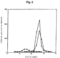
- reaction solutions were passed through a microfilter with a pore size of 0.22 ⁇ m (Millipore Corp.); thereafter, it was separated by a Superdex peptide column (30 x 1.0 cm: product of Amersham Biosciences K.K.; chromatographic conditions: mobile phase, 0.2 mol/L NaCl; flow rate, 0.5 ml/min) and the effluent was recovered at 0.5 ml fractions, followed by radioactivity measurement with a scintillation counter (Fig. 3).
- chondroitinase ACII 100 munits of chondroitinase ACII (Seikagaku Corporation) was dissolved in 100 ⁇ l of CS11(K3) fraction, digested enzymatically at 37°C for 10 hours, and heated to deactivate the enzyme; the resulting product was designated CS12(K3)B).
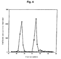
- CS12(K3)A and CS12(K3)B were passed through a microfilter with a pore size of 0.22 ⁇ m (Millipore Corp.); thereafter, they were separated by a Superdex peptide column (30 x 1.0 cm: product of Amersham Biosciences K.K.; chromatographic conditions: mobile phase, 0.2 mol/L NaCl; flow rate, 0.5 ml/min) and the effluent was recovered at 0.5 ml fractions, followed by radioactivity measurement with a scintillation counter to give a radioactivity peak shift to a disaccharaide fraction in CS12(K3)B (Fig. 4). From this result, it became clear that K3 transferred GlcUA to the chondroitin sulfate derived undecasaccharide via ⁇ 1,3 linkage to prepare a dodecasaccharide.
- the gene comprising the nucleotide sequence depicted in SEQ ID NO: 3 is at least expressed in the human brain and this was verified by RT-PCR assay using Marathon-Ready cDNA (CLONTECH Laboratories, Inc.) as a template.
- PCR was performed according to a conventional technique using the two primers of SEQ ID NO: 13 and SEQ ID NO: 14.
- the template cDNA used was Marathon-Ready cDNA human brain (CLONTECH Laboratories, Inc.)
- the amplified band of about 2 kb was digested with EcoRI and BamHI according to a conventional technique and inserted between EcoRI and BamHI sites of a mammalian cell expression vector pFLAG-CMV-1 (Sigma) according to a conventional technique, thereby making K11-FLAG-CMV1.
- Nucleotide sequencing of the obtained vector showed the insertion of a DNA fragment comprising a nucleotide sequence of nucleotide Nos. 287-2328 in the nucleotide sequence depicted in SEQ ID NO: 3.
- the genomic position of DNA having that nucleotide sequence was identified by using OMIM and it was found to exist at 2q36.3.
- K11-FLAG-CMV1 (15 mg) was set on TransFast (Promega Corp.) which was operated according to the protocol to transfer the gene into COS7 cells that had been cultivated on 100 mm culture dishes to produce 70% confluent cultures. After 3-day cultivation, the supernatant was recovered and passed through a 0.22 ⁇ m filter; thereafter, 100 ⁇ l of Anti-FLAG M2-Agarose Affinity Gel (Sigma) was added to 10 ml of the supernatant and mixing by inverting of the tube was performed overnight at 4°C.
- TransFast Promega Corp.
- the gel was washed three times with 50 mmol/L Tris-HCl at pH 7.4 (20 glycerol) and the unwanted wash solution was removed with a syringe fitted with a 27G needle.
- the gel was suspended in 50 mmol/L Tris-HCl at pH 7.4 (20% glycerol, 10 mmol/L phenylmethyl sulfonyl fluoride, 1 ⁇ g/ml leupeptin and 1 ⁇ g/ml pepstatin) to make 50 (v/v); after centrifugation, the supernatant was removed to prepare an enzyme adsorbed gel suspension.
- Each of the reaction solutions was passed through a microfilter with a pore size of 0.22 ⁇ m (Millipore Corp.); thereafter, it was separated by a Superdex peptide column (30 x 1.0 cm: product of Amersham Biosciences K.K.; chromatographic conditions: mobile phase, 0.2 mol/L NaCl; flow rate, 0.5 ml/min) and the effluent was recovered at 0.5 ml fractions, followed by radioactivity measurement with a scintillation counter (Fig. 5).
- Fig. 5 radioactivity measurement with a scintillation counter
- chondroitinase ACII 100 munits of chondroitinase ACII (Seikagaku Corporation) was dissolved in 100 ⁇ l of CS11(K11) fraction, digested with an enzyme at 37°C for 10 hours, and heated to deactivate the enzyme; the resulting product was designated CS11(K11)B).
- CS11(K11)A and CS11(K11)B were passed through a microfilter with a pore size of 0.22 ⁇ m (Millipore Corp.); thereafter, they were separated by a Superdex peptide column (30 x 10 mm: product of Amersham Biosciences K.K.; chromatographic conditions: mobile phase, 0.2 mol/L NaCl; flow rate, 0.5 ml/min) and the effluent was recovered at 0.5 ml fractions, followed by radioactivity measurement with a scintillation counter to give a radioactivity peak shift to a trisaccharaide fraction in CS11(K11)B (Fig. 6). From this result, it became clear that K11 transferred GalNAc to GlcUA at the non-reducing terminal of the chondroitin sulfate derived decasaccharide via ⁇ 1,4 linkage to prepare an undecasaccharide.
- reaction solutions were passed through a microfilter with a pore size of 0.22 ⁇ m (Millipore Corp.); thereafter, it was separated by a Superdex peptide column (30 x 1.0 cm: product of Amersham Biosciences K.K.; chromatographic conditions: mobile phase, 0.2 mol/L NaCl; flow rate, 0.5 ml/min) and the effluent was recovered at 0.5 ml fractions, followed by radioactivity measurement with a scintillation counter (Fig. 3).
- chondroitinase ACII 100 munits of chondroitinase ACII (Seikagaku Corporation) was dissolved in 100 ⁇ l of CS11(K11) fraction, digested enzymatically at 37°C for 10 hours, and heated to deactivate the enzyme; the resulting product was designated CS12(K11)B).
- CS12(K11)A and CS12(K11)B were passed through a microfilter with a pore size of 0.22 ⁇ m (Millipore Corp.); thereafter, they were separated by a Superdex peptide column (30 x 1.0 cm: product of Amersham Biosciences K.K.; chromatographic conditions: mobile phase, 0.2 mol/L NaCl; flow rate, 0.5 ml/min) and the effluent was recovered at 0.5 ml fractions, followed by radioactivity measurement with a scintillation counter to give a radioactivity peak shift to a disaccharaide fraction in CS12(K11)B (Fig. 4). From this result, it became clear that the enzyme of the present invention could transfer GlcUA to the chondroitin sulfate derived undecasaccharide via ⁇ 1,3 linkage to prepare a dodecasaccharide.
- ⁇ 4Gal-T7 was expressed and there was prepared a suspension of an affinity carrier having the ⁇ 4Gal-T7 linked thereto; thereafter, 10 ⁇ l of the gel, 6.25 mg of a acceptor substrate (xylose linked peptide [the N-terminal sequence of bikunin having xylose linked as a side chain to serine which was the amino acid residue at 9-position of SEQ ID NO: 15]: product of Peptide Institute), 171 ⁇ mol of adenosine triphosphate (ATP) sodium salt and 0.036 mol of [ 3 H]UDP-Gal were added to 50 mmol of MES buffer solution (pH 6.5) containing 10 nmol/L MnCl 2 to make a total volume of 30 ⁇ l and reaction was carried out at 37°C for 1 hour. Thereafter, the reaction solution was kept at 100
- the reaction solution was passed through a microfilter with a pore size of 0.22 ⁇ m (Millipore Corp.); thereafter, it was separated by a Superdex peptide column (30 x 1.0 cm: product of Amersham Biosciences K.K.; chromatographic conditions: mobile phase, 0.2 mol/L NaCl; flow rate, 1.0 ml/min) and the effluent was recovered at 0.5 ml fractions, followed by radioactivity measurement with a scintillation counter. As the result, strong radioactivity was observed in a polymer fraction other than the eluted fraction of [ 3 H]UDP-Gal. This supported the linking by ⁇ 4Gal-T7 of galactose to the acceptor substrate.
- DNA was extracted from 1 ml of blood using GFX Genomic Blood DNA Purification Kit (Amersham Biosciences K.K.) Of the 14-16 ⁇ g of DNA obtained, 250 ng was used as a template to amplify the exons in the genes of chondroitin sulfate synthases (K3, K11, ⁇ 4Gal-T7) with the aid of various primers (see Table 1 below). Using the thus obtained fragments as templates, the nucleotide sequences of the genes were analyzed by a conventional technique and compared with those of the genes of normal types of the respective chondroitin synthases; in this way, one could easily check for the presence of mutations and single nucleotide polymorphisms.
- PBMC peripheral blood-derived mononuclear cells
- RNA Extraction Kit (Amersham Biosciences K.K.)
- 1st strand cDNA was synthesized by SUPERSCRIPT First-Stand Synthesis System for RT-PCR (Invitrogen Co.)
- real-time PCR assay was performed to determine the amounts of various genes expressed.
- the primers and probes used are shown in Table 2.
- Knockout mice were obtained by the following procedure using a mouse gene K3 (mK3: SEQ ID NO:65), K11 (mK11: SEQ ID NO:67) or ⁇ 4Gal-T7 (m ⁇ 4Gal-T7: SEQ ID NO:69).
Landscapes
- Life Sciences & Earth Sciences (AREA)
- Health & Medical Sciences (AREA)
- Chemical & Material Sciences (AREA)
- Engineering & Computer Science (AREA)
- Genetics & Genomics (AREA)
- Zoology (AREA)
- Organic Chemistry (AREA)
- Wood Science & Technology (AREA)
- Bioinformatics & Cheminformatics (AREA)
- Biotechnology (AREA)
- General Health & Medical Sciences (AREA)
- General Engineering & Computer Science (AREA)
- Biomedical Technology (AREA)
- Microbiology (AREA)
- Molecular Biology (AREA)
- Biochemistry (AREA)
- Proteomics, Peptides & Aminoacids (AREA)
- Medicinal Chemistry (AREA)
- Veterinary Medicine (AREA)
- Physics & Mathematics (AREA)
- Biophysics (AREA)
- Environmental Sciences (AREA)
- Analytical Chemistry (AREA)
- Pathology (AREA)
- Immunology (AREA)
- Animal Behavior & Ethology (AREA)
- Animal Husbandry (AREA)
- Biodiversity & Conservation Biology (AREA)
- Plant Pathology (AREA)
- Enzymes And Modification Thereof (AREA)
- Measuring Or Testing Involving Enzymes Or Micro-Organisms (AREA)
Applications Claiming Priority (3)
| Application Number | Priority Date | Filing Date | Title |
|---|---|---|---|
| JP2002323438 | 2002-11-07 | ||
| JP2002323438 | 2002-11-07 | ||
| PCT/JP2003/014211 WO2004042055A1 (ja) | 2002-11-07 | 2003-11-07 | 骨パジェット病の検出方法 |
Publications (2)
| Publication Number | Publication Date |
|---|---|
| EP1559781A1 true EP1559781A1 (de) | 2005-08-03 |
| EP1559781A4 EP1559781A4 (de) | 2007-07-25 |
Family
ID=32310419
Family Applications (1)
| Application Number | Title | Priority Date | Filing Date |
|---|---|---|---|
| EP03810659A Withdrawn EP1559781A4 (de) | 2002-11-07 | 2003-11-07 | Verfahren zum nachweis der paget-krankheit |
Country Status (5)
| Country | Link |
|---|---|
| US (1) | US20060141463A1 (de) |
| EP (1) | EP1559781A4 (de) |
| AU (1) | AU2003277616A1 (de) |
| CA (1) | CA2505707A1 (de) |
| WO (1) | WO2004042055A1 (de) |
Cited By (2)
| Publication number | Priority date | Publication date | Assignee | Title |
|---|---|---|---|---|
| WO2011128646A3 (en) * | 2010-04-14 | 2012-02-23 | The University Court Of The University Of Edinburgh | Genetic markers for paget's disease |
| WO2015177744A1 (en) * | 2014-05-21 | 2015-11-26 | Consiglio Nazionale Delle Ricerche | A diagnostic marker for paget disease |
Families Citing this family (1)
| Publication number | Priority date | Publication date | Assignee | Title |
|---|---|---|---|---|
| CN110055234B (zh) * | 2019-04-30 | 2021-07-30 | 华熙生物科技股份有限公司 | 一种硫酸软骨素合酶及其编码基因与应用 |
Family Cites Families (3)
| Publication number | Priority date | Publication date | Assignee | Title |
|---|---|---|---|---|
| US5869273A (en) * | 1996-09-13 | 1999-02-09 | Glyko, Inc. | Chondroitin sulfate as a marker of bone resorption |
| JP4377987B2 (ja) * | 1999-04-28 | 2009-12-02 | 生化学工業株式会社 | ガラクトース転移酵素及びそれをコードするdna |
| AU3774300A (en) * | 1999-06-02 | 2000-12-18 | Genentech Inc. | Secreted and transmembrane polypeptides and nucleic acids encoding the same |
-
2003
- 2003-11-07 CA CA002505707A patent/CA2505707A1/en not_active Abandoned
- 2003-11-07 WO PCT/JP2003/014211 patent/WO2004042055A1/ja not_active Ceased
- 2003-11-07 US US10/534,002 patent/US20060141463A1/en not_active Abandoned
- 2003-11-07 EP EP03810659A patent/EP1559781A4/de not_active Withdrawn
- 2003-11-07 AU AU2003277616A patent/AU2003277616A1/en not_active Abandoned
Non-Patent Citations (8)
| Title |
|---|
| DAROSZEWSKA ANNA ET AL: "Genetics of Paget's disease of bone" CLINICAL SCIENCE (LONDON), vol. 109, no. 3, September 2005 (2005-09), pages 257-263, XP002436298 ISSN: 0143-5221 * |
| GOOD DAVID A ET AL: "Linkage of Paget disease of bone to a novel region on human chromosome 18q23" AMERICAN JOURNAL OF HUMAN GENETICS, vol. 70, no. 2, February 2002 (2002-02), pages 517-525, XP002436296 ISSN: 0002-9297 * |
| HOCKING L J ET AL: "Domain-specific mutations in sequestosome 1 (SQSTM1) cause familial and sporadic Paget's disease" HUMAN MOLECULAR GENETICS, OXFORD UNIVERSITY PRESS, SURREY, GB, vol. 11, no. 22, 2002, pages 2735-2739, XP002250054 ISSN: 0964-6906 * |
| HOCKING LYNNE J ET AL: "Genomewide search in familial Paget disease of bone shows evidence of genetic heterogeneity with candidate loci on chromosomes 2q36, 10p13, and 5q35" AMERICAN JOURNAL OF HUMAN GENETICS, vol. 69, no. 5, November 2001 (2001-11), pages 1055-1061, XP002436295 ISSN: 0002-9297 * |
| LAURIN N ET AL: "Recurrent mutation of the gene encoding sequestosome 1 (SQSTM1/p62) in Paget disease of bone" AMERICAN JOURNAL OF HUMAN GENETICS, AMERICAN SOCIETY OF HUMAN GENETICS, CHICAGO, IL, US, vol. 70, no. 6, June 2002 (2002-06), pages 1582-1588, XP002250053 ISSN: 0002-9297 * |
| LAURIN NANCY ET AL: "Paget disease of bone: Mapping of two loci at 5q35-qter and 5q31" AMERICAN JOURNAL OF HUMAN GENETICS, vol. 69, no. 3, September 2001 (2001-09), pages 528-543, XP002436297 ISSN: 0002-9297 * |
| OKAJIMA TETSUYA ET AL: "Molecular basis for the progeroid variant of Ehlers-Danlos syndrome: Identification and characterization of two mutations in galactosyltransferase I gene" JOURNAL OF BIOLOGICAL CHEMISTRY, vol. 274, no. 41, 8 October 1999 (1999-10-08), pages 28841-28844, XP002436294 ISSN: 0021-9258 * |
| See also references of WO2004042055A1 * |
Cited By (2)
| Publication number | Priority date | Publication date | Assignee | Title |
|---|---|---|---|---|
| WO2011128646A3 (en) * | 2010-04-14 | 2012-02-23 | The University Court Of The University Of Edinburgh | Genetic markers for paget's disease |
| WO2015177744A1 (en) * | 2014-05-21 | 2015-11-26 | Consiglio Nazionale Delle Ricerche | A diagnostic marker for paget disease |
Also Published As
| Publication number | Publication date |
|---|---|
| EP1559781A4 (de) | 2007-07-25 |
| US20060141463A1 (en) | 2006-06-29 |
| AU2003277616A1 (en) | 2004-06-07 |
| WO2004042055A1 (ja) | 2004-05-21 |
| CA2505707A1 (en) | 2004-05-21 |
Similar Documents
| Publication | Publication Date | Title |
|---|---|---|
| US20080070996A1 (en) | Chondroitin synthase and nucleic acid encoding the enzyme | |
| JP4333880B2 (ja) | コンドロイチン合成酵素および該酵素をコードするdna | |
| US20060141463A1 (en) | Method of detecting bone paget's disease | |
| JP4791676B2 (ja) | 硫酸基転移酵素及び該酵素をコードするdna | |
| JP2003047467A (ja) | コンドロイチン合成酵素 | |
| JPWO2002000889A1 (ja) | 硫酸基転移酵素及び該酵素をコードするdna | |
| EP0745668A2 (de) | Fur Sulfotransferase kodierende DNA | |
| JP4226693B2 (ja) | 硫酸基転移酵素及びそれをコードするdna | |
| JP2004166702A (ja) | 骨パジェット病の検出方法 | |
| JP4377987B2 (ja) | ガラクトース転移酵素及びそれをコードするdna | |
| US8623625B2 (en) | Sulfotransferase, peptide thereof and DNA encoding the same | |
| JP4253704B2 (ja) | 新規n−アセチルガラクトサミン転移酵素及びそれをコードする核酸 | |
| JP2005065687A (ja) | β1,3−N−アセチル−D−ガラクトサミン転移酵素タンパク質及びそれをコードする核酸、並びにそれを用いた癌化検定方法 | |
| JP5105261B2 (ja) | β1,3−N−アセチル−D−ガラクトサミン転移酵素タンパク質及びそれをコードする核酸、並びにそれを用いた癌化検定方法 | |
| JP4514708B2 (ja) | コンドロイチン重合化因子の同定と、その利用 | |
| JP4629214B2 (ja) | N−アセチルグルコサミン転移酵素 | |
| JP4288327B2 (ja) | 硫酸基転移酵素、そのポリペプチド及びそれをコードするdna | |
| Aikawa | Heparan Sulfate/Heparin N-Deacetylase/N-Sulfotransferase-3 and-4 | |
| Takeshita et al. | Bhlhe40, a potential diabetic modifier gene on Dbm1 locus, negatively controls myocyte fatty acid oxidation | |
| JP2005065565A (ja) | 改変酵素 | |
| JP2004215619A (ja) | 新規ガラクトース転移酵素及びそれをコードする核酸 | |
| JP2004033064A (ja) | 新規グルクロン酸転移酵素及びその遺伝子 |
Legal Events
| Date | Code | Title | Description |
|---|---|---|---|
| PUAI | Public reference made under article 153(3) epc to a published international application that has entered the european phase |
Free format text: ORIGINAL CODE: 0009012 |
|
| 17P | Request for examination filed |
Effective date: 20050506 |
|
| AK | Designated contracting states |
Kind code of ref document: A1 Designated state(s): AT BE BG CH CY CZ DE DK EE ES FI FR GB GR HU IE IT LI LU MC NL PT RO SE SI SK TR |
|
| AX | Request for extension of the european patent |
Extension state: AL LT LV MK |
|
| DAX | Request for extension of the european patent (deleted) | ||
| A4 | Supplementary search report drawn up and despatched |
Effective date: 20070622 |
|
| 17Q | First examination report despatched |
Effective date: 20071116 |
|
| STAA | Information on the status of an ep patent application or granted ep patent |
Free format text: STATUS: THE APPLICATION IS DEEMED TO BE WITHDRAWN |
|
| 18D | Application deemed to be withdrawn |
Effective date: 20080327 |