EP1541676A1 - Gegen das Prothrombin-Fragment F1+2 gerichtete Antikörper, ihre Herstellung und Verwendung - Google Patents
Gegen das Prothrombin-Fragment F1+2 gerichtete Antikörper, ihre Herstellung und Verwendung Download PDFInfo
- Publication number
- EP1541676A1 EP1541676A1 EP04022637A EP04022637A EP1541676A1 EP 1541676 A1 EP1541676 A1 EP 1541676A1 EP 04022637 A EP04022637 A EP 04022637A EP 04022637 A EP04022637 A EP 04022637A EP 1541676 A1 EP1541676 A1 EP 1541676A1
- Authority
- EP
- European Patent Office
- Prior art keywords
- antibodies
- antibody
- pro
- gln
- analyte
- Prior art date
- Legal status (The legal status is an assumption and is not a legal conclusion. Google has not performed a legal analysis and makes no representation as to the accuracy of the status listed.)
- Granted
Links
Classifications
-
- C—CHEMISTRY; METALLURGY
- C12—BIOCHEMISTRY; BEER; SPIRITS; WINE; VINEGAR; MICROBIOLOGY; ENZYMOLOGY; MUTATION OR GENETIC ENGINEERING
- C12N—MICROORGANISMS OR ENZYMES; COMPOSITIONS THEREOF; PROPAGATING, PRESERVING, OR MAINTAINING MICROORGANISMS; MUTATION OR GENETIC ENGINEERING; CULTURE MEDIA
- C12N9/00—Enzymes; Proenzymes; Compositions thereof; Processes for preparing, activating, inhibiting, separating or purifying enzymes
- C12N9/14—Hydrolases (3)
- C12N9/48—Hydrolases (3) acting on peptide bonds (3.4)
- C12N9/50—Proteinases, e.g. Endopeptidases (3.4.21-3.4.25)
- C12N9/64—Proteinases, e.g. Endopeptidases (3.4.21-3.4.25) derived from animal tissue
- C12N9/6421—Proteinases, e.g. Endopeptidases (3.4.21-3.4.25) derived from animal tissue from mammals
- C12N9/6424—Serine endopeptidases (3.4.21)
- C12N9/6429—Thrombin (3.4.21.5)
-
- A—HUMAN NECESSITIES
- A61—MEDICAL OR VETERINARY SCIENCE; HYGIENE
- A61P—SPECIFIC THERAPEUTIC ACTIVITY OF CHEMICAL COMPOUNDS OR MEDICINAL PREPARATIONS
- A61P37/00—Drugs for immunological or allergic disorders
- A61P37/02—Immunomodulators
- A61P37/04—Immunostimulants
-
- C—CHEMISTRY; METALLURGY
- C07—ORGANIC CHEMISTRY
- C07K—PEPTIDES
- C07K16/00—Immunoglobulins [IGs], e.g. monoclonal or polyclonal antibodies
- C07K16/40—Immunoglobulins [IGs], e.g. monoclonal or polyclonal antibodies against enzymes
-
- C—CHEMISTRY; METALLURGY
- C12—BIOCHEMISTRY; BEER; SPIRITS; WINE; VINEGAR; MICROBIOLOGY; ENZYMOLOGY; MUTATION OR GENETIC ENGINEERING
- C12Y—ENZYMES
- C12Y304/00—Hydrolases acting on peptide bonds, i.e. peptidases (3.4)
- C12Y304/21—Serine endopeptidases (3.4.21)
- C12Y304/21005—Thrombin (3.4.21.5)
Definitions
- the invention relates to the prothrombin fragment F 1 + 2 directed antibodies, their preparation and use.
- prothrombin into active thrombin with the formation of fragments is a central event in the course of the coagulation cascade.
- prothrombin fragment F 1 + 2 With the immunochemical determination of the prothrombin fragment F 1 + 2 , a quantification of the actually formed thrombin is possible.
- F 1 + 2 prothrombin fragment F 1 + 2
- F 1 + 2 tests have the problem that prothrombin in the sample is in large excess in comparison to F 1 + 2 , so that the anti-F 1 + 2 antibodies to be used in the test are very specific between released by cleavage Prothrombin fragments F 2 (hereinafter "F 2 " called) and F 1 + 2 on the one hand and intact prothrombin on the other hand must differ.
- F 2 cleavage Prothrombin fragments F 2
- the object is achieved by the provision of monoclonal antibodies against the prothrombin fragment F 1 + 2 , which bind to an epitope on the N-terminal half of the F 2 fragment of prothrombin (hereinafter referred to as "secondary antibody").
- secondary antibodies in combination with antibodies whose epitopes comprise the four carboxyterminal amino acids of the F 2 and F 1 + 2 fragments (hereinafter referred to as "primary antibodies"), form the basis for an improved sandwich immunoassay for the determination of F 1 + 2 ,
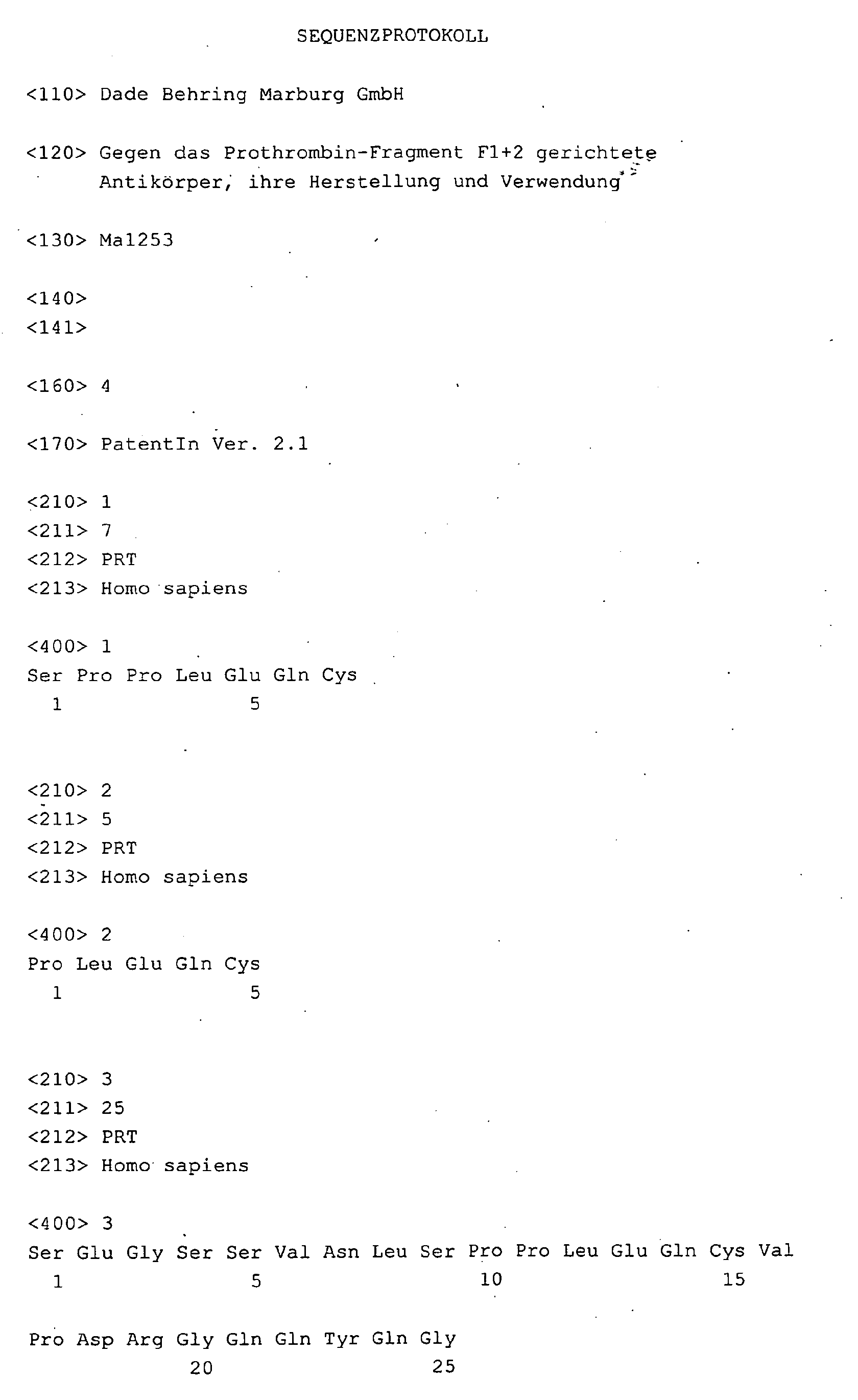
- the epitope was used as a peptide with the Amino acid sequence Ser-Pro-Pro-Leu-Glu-Gln-Cys uniquely identified.
- An object of this invention are peptides consisting of 5-25 amino acids, preferably from 5 to 21 amino acids, most preferably from 5-12 Amino acids, characterized in that they have the amino acid sequence Pro-Leu-Glu-Gln-Cys contain.
- Preferred peptides according to the invention are those with the amino acid sequence Ser-Glu-Gly-Ser-Ser-Val-Asn-Leu-Ser-Pro-Pro-Leu-Glu-Gln-Cys-Val-Pro-Asp-Arg-Gly-Gln-Gln-Tyr-Gln- Gly or a fragment thereof, in particular a peptide having the amino acid sequence Ser-Pro-Pro-Leu-Glu-Gln-Cys.
- peptides in the context of this invention comprises acid amides which are known from Hydrolysis into amino acids, for example amino acid polymers such as e.g. Polypeptides, oligopeptides, proteins or protein fragments.
- the peptides according to the invention can be used as immunizing antigen for the preparation of the antibodies according to the invention or also for the affinity chromatographic purification of the antibodies according to the invention. Furthermore, the peptides according to the invention can also be used in a method for the quantitative or qualitative detection of an analyte, preferably F 1 + 2 .
- the peptides of the invention may also be associated with a solid phase and / or a component of a signal-forming system.
- antibodies which are characterized in that they bind to an epitope on the N-terminal half of the F 2 fragment of prothrombin, ie antibodies which bind to Ser-Glu-Gly-Ser-Ser Val-Asn-Leu-Ser-Pro-Pro-Leu-Glu-Gln-Cys-Val-Pro-Asp-Arg-Gly-Gln-Gln-Tyr-Gln-Gly-Arg-Leu-Ala-Val-Thr- Thr-His-Gly-Leu-Pro-Cys-Leu-Ala-Trp-Ala-Ser-Ala-Gln-Ala-Lys-Ala-Leu-Ser-Lys-His-Gln-Asp-Phe-Asn-Ser Tie Ala-Val-Gln-Leu-Val-Glu-Asn.
- an antibody means an immunoglobulin, eg an immunoglobulin of the class or subclass IgA, IgD, IgE, IgG 1 , IgG 2a , IgG 2b , IgG 3 , IgG 4 , IgM.
- An antibody has at least one binding site (often called paratope) for an epitope (often called an antigenic determinant) on an antigen or hapten.
- an epitope is characterized, for example, by its spatial structure and / or by the presence of polar and / or apolar groups.
- the binding site of the antibody is complementary to the epitope.
- the antigen-antibody reaction or the hapten-antibody reaction works according to the so-called "key-keyhole principle", and is usually specific to a high degree, ie the antibodies are capable of small deviations in the primary structure, in the charge to distinguish in the spatial configuration and the steric arrangement of the antigen or hapten.
- the so-called “complementarity determining regions" of the antibody contribute to the binding of the antibody to the antigen or hapten.
- antigens includes monovalent and polyvalent antigens.
- One polyvalent antigen is a molecule or complex of molecules to which more than an immunoglobulin can bind simultaneously, while in a monovalent Antigen only one antibody at a time can bind.
- a hapten is usually called a molecule that is not immunogenic by itself, but usually bound to a carrier for immunization purposes becomes.
- antibodies is to be understood as meaning not only complete antibodies but also expressly antibody fragments, such as Fab, Fv, F (ab ') 2 , Fab'; as well as chimeric, humanized, bi- or oligospecific, or "single chain”antibodies; also aggregates, polymers and conjugates of immunoglobulins and / or fragments thereof, provided the binding properties to the antigen or hapten are obtained.
- Antibody fragments can be prepared, for example, by enzymatic cleavage of antibodies with enzymes such as pepsin or papain.
- Antibody aggregates, polymers and conjugates can be generated by a variety of methods, including heat treatment, reaction with substances such as glutaraldehyde, reaction with immunoglobulin-binding molecules, biotinylation of antibodies and subsequent reaction with streptavidin or avidin, etc.
- An antibody according to this invention may be a monoclonal or a polyclonal antibody.
- the antibody may have been prepared by the usual methods, for example by immunizing the human or an animal such as mouse, rat, guinea pig, rabbit, horse, sheep, goat, chicken (see Messerschmid (1996) BIOforum, 11 : 500-502 ), and subsequent extraction of the antiserum; or by the establishment of hybridoma cells and subsequent purification of the secreted antibodies; or by cloning and expression of the nucleotide sequences or modified versions thereof, which encode the amino acid sequences responsible for the binding of the natural antibody to the antigen and / or hapten.
- Antibodies according to the invention are, in particular, those antibodies which bind to a peptide consisting of 5-25 amino acids, preferably from 5 to 21 amino acids, whole particularly preferably from 5-12 amino acids, which bind the Amino acid sequence Pro-Leu-Glu-Gln-Cys comprises.
- Very preferred antibodies in the For purposes of this invention are antibodies that bind to the peptide with the Amino acid sequence Ser-Glu-Gly-Ser-Ser-Val-Asn-Leu-Ser-Pro-Pro-Leu-Glu-Gln-Cys-Val-Pro-Asp-Arg-Gly-Gln-Gln-Tyr-Gln-Gly or to a fragment of this peptide bind specifically, in particular to a peptide having the amino acid sequence Ser-Pro-Pro-Leu-Glu-Gln-Cys.
- Antibodies within the meaning of this invention are also the Antibodies produced by the hybridoma cell line 92-195 / 097. These Hybridoma cell line became on 15 August 2003 with the DSMZ German collection of Microorganisms and Cell Cultures GmbH, Mascheroder Weg 1b, 38124 Braunschweig, Germany, under the accession number DSM ACC2607.
- Another object of this invention are specific binding partners, the bind an epitope recognized by an antibody of the invention.
- a specific binding partner is a member of a specific To understand bond pairs.
- these are two molecules, each of which forms at least one structure of the have other molecule complementary structure, the two molecules able to bind via a binding of the complementary structures.
- Term molecule also includes molecular complexes such as e.g. Enzymes derived from apo- and Coenzyme, proteins that consist of multiple subunits, Lipoproteins consisting of protein and lipids, etc.
- Specific binding partners may be naturally occurring but also e.g. by chemical synthesis, microbiological and / or genetic engineering techniques Be substances. To illustrate the term specific binding partner, but not to be understood as limiting, are e.g.
- thyroxine binding Globulin steroid-binding proteins, antibodies, antigens, haptens, enzymes, lectins, Nucleic acids, repressors, oligo- and polynucleotides, protein A, protein G, avidin, Streptavidin, biotin, complement component C1q, nucleic acid-binding proteins, etc.
- Specific binding pairs are, for example: antibody antigen, antibody hapten, Operator repressor, nuclease nucleotide, biotin-avidin, lectin polysaccharide, Steroid-steroid binding protein, drug-drug receptor, Hormone hormone receptor, enzyme substrate, IgG protein A, complementary oligo- or Polynucleotides, etc.
- the subject matter of this invention is also an antibody according to the invention which is compatible with a solid phase and / or a component of a signal-generating system is associated.
- solid phase in the sense of this invention includes an article which of porous and / or non-porous, usually water-insoluble material exists and can have many different forms, such as, B. Vessel, Tube, microtiter plate, sphere, microparticles, rods, strips, filters or Chromatography paper, etc.
- the surface of the solid phase is hydrophilic or can be rendered hydrophilic.
- the solid phase can be from the different materials exist such. made of inorganic and / or aus organic materials, of synthetic, of naturally occurring and / or from modified naturally occurring materials.
- examples for Solid phase materials are polymers such as e.g.
- Cellulose nitrocellulose, Cellulose acetate, polyvinyl chloride, polyacrylamide, crosslinked dextran molecules, Agarose, polystyrene, polyethylene, polypropylene, polymethacrylate or nylon; ceramics; Glass; Metals, in particular precious metals such as gold and silver; magnetite; Mixtures or combinations thereof; etc. Also cells, liposomes or Phospholipid vesicles are included in the term solid phase.
- the solid phase may have a coating of one or more layers, e.g. from proteins, carbohydrates, lipophilic substances, biopolymers, organic polymers or mixtures thereof, for example, the suppress non-specific binding of sample components to the solid phase or to prevent or, for example, to achieve improvements with regard to the suspension stability of particulate solid phases, the Storage stability, shaping stability or resistance to UV light, Microbes or other destructive agents.
- layers e.g. from proteins, carbohydrates, lipophilic substances, biopolymers, organic polymers or mixtures thereof, for example, the suppress non-specific binding of sample components to the solid phase or to prevent or, for example, to achieve improvements with regard to the suspension stability of particulate solid phases, the Storage stability, shaping stability or resistance to UV light, Microbes or other destructive agents.
- a signal-forming system may be one or more Components act, wherein it is at least one component to a detectable label acts.
- a label is any molecule that understands itself produces a signal or can induce the production of a signal, e.g. a fluorescent substance, a radioactive substance, an enzyme, or a chemiluminescent substance.
- the signal can, for example, based on the Enzyme activity, luminescence, light absorption, light scattering, emitted electromagnetic or radioactive radiation, or a chemical reaction can be detected or measured.
- a label is able to produce a detectable signal, so no additional components are necessary.
- Many organic molecules absorb ultraviolet and visible light, transmitted through light absorption Energy these molecules can come into an excited state of energy, and The absorbed energy in the form of light of a different wavelength than that of the emitted light.
- Still other labels can enter directly generate detectable signal, e.g. radioactive isotopes or dyes.
- Still other labels require additional components for signal generation, i. the signal producing system in such a case closes all the for the Signaling required components with such.
- Suitable labels include, for example, enzymes including horseradish peroxidase, alkaline phosphatase, glucose-6.
- Phosphate dehydrogenase Phosphate dehydrogenase, alcohol dehydrogenase, glucose oxidase, ⁇ -galactosidase, luciferase, urease and acetylcholinesterase; dyes; fluorescent substances including fluorescein isothiocyanate, rhodamine, phycoerythrin, phycocyanin, ethidium bromide, 5-dimethylaminonaphthalene-1-sulfonyl chloride and rare earth fluorescent chelates; chemiluminescent substances including luminol, isoluminol, acridinium compounds, olefin, enol ether, enamine, aryl vinyl ether, dioxene, arylimidazole, lucigenin, luciferin and aequorin; Sensitizers including eosin, 9,10-dibromoanthracene,
- a signal-forming system may also comprise components which, when in close proximity to each other, may undergo a detectable interaction, for example in the form of energy donors and energy receivers such as photosensitizers and chemiluminescers (EP-A2-0 515 194), photosensitizers and fluorophores (WO 95 / 06877), radioactive iodine-125 and fluorophores (Udenfriend et al., (1985) Proc Natl Acad Sci. 82 : 8672-8676), fluorophores and fluorophores (Mathis (1993) Clin. Chem. 39 : 1953-1959). or fluorophores and fluorescence quenchers (US 3,996,345).
- energy donors and energy receivers such as photosensitizers and chemiluminescers (EP-A2-0 515 194), photosensitizers and fluorophores (WO 95 / 06877), radioactive iodine-125 and fluorophores (Udenfriend et al
- the direct transmission of energy between the components is the direct transmission of energy between the components, e.g. by light or electron radiation as well as short-lived reactive chemical molecules.
- This also includes processes in which the activity of a component is inhibited or intensified by one or more others, for example the Inhibition or increase in enzyme activity or inhibition, enhancement or Change (e.g., wavelength shift, polarization) of the influenced component emitted electromagnetic radiation.
- the Interaction between the components also includes enzyme cascades.
- the components are enzymes, at least one of which Substrate for another supplies, so a maximum or minimum Reaction rate of the coupled substrate reaction results.
- An effective interaction between the components usually takes place, if these are spatially adjacent, e.g. within a Distance range of a few microns, especially within a distance range of less than 600 nm, preferably less than 400 nm, most preferably less than 200 nm.
- Microparticles are often used as a solid phase and / or as a label.
- the term "microparticles" is to be understood as meaning particles which an approximate diameter of at least 20 nm and not more than 20 ⁇ m usually between 40 nm and 10 ⁇ m, preferably between 0.1 and 10 microns, more preferably between 0.1 and 5 microns, most preferably between 0.15 and 2 ⁇ m.
- the microparticles can be regular or irregular be shaped. You can use balls, spheroids, balls with more or less big ones Represent cavities or pores.
- the microparticles can be made of organic, from inorganic material or a mixture or combination of both consist.
- the microparticles can be made of a porous or non-porous, a swellable or non-swellable material.
- the microparticles have any density, but preferred are particles with a density that the density of water, such as about 0.7 to about 1.5 g / ml.
- the preferred ones Microparticles are suspendible in aqueous solutions and as long as possible suspension stable. They like transparent, partly transparent or be opaque.
- the microparticles can consist of several layers like For example, the so-called "core-and-shell" particles with a core and a or several enveloping layers.
- microparticle includes for example, dye crystals, metal sols, silica particles, glass particles, Magnetic particles, polymer particles, oil drops, lipid particles, dextran, and Protein aggregates.
- Preferred microparticles are in aqueous solutions suspendable particles consisting of water-insoluble polymer material, in particular of substituted polyethylenes.
- Very particularly preferred are Latex particles e.g. from polystyrene, acrylic acid polymers, methacrylic acid polymers, Acrylonitrile polymers, acrylonitrile butadiene styrene, polyvinyl acetate acrylate, Polyvinylpyridine, vinyl chloride acrylate.
- latex particles with reactive groups on their surface such as carboxyl, amino or Aldehyde groups containing a covalent bond e.g. of specific Allow binding partners to the latex particles.
- reactive groups on their surface such as carboxyl, amino or Aldehyde groups containing a covalent bond e.g. of specific Allow binding partners to the latex particles.
- the production of latex particles is described for example in EP 0 080 614, EP 0 227 054 and EP 0 246 446.
- association is to be understood broadly and includes, for example, a covalent and noncovalent bonding, direct and indirect bonding, the adsorption to a surface and the inclusion in a depression or a Cavity, etc.
- covalent bond are the antibodies or Binding partner via a chemical bond to the solid phase or to the label bound.
- non-covalent bond are the Surface adsorption, inclusion in cavities or binding of two specific binding partners.
- direct bond to the solid phase or the label may also indirectly transfer the antibodies or binding partners specific interaction with other specific binding partners to the Solid phase or the label to be bound (see EP-A2-0 411 945).
- the biotinylated antibody can label-bound avidin bound to the label; or it can be Fluorescein antibody conjugate via solid phase-bound anti-fluorescein antibodies be bound to the solid phase; or the antibody can over Immunoglobulin-binding proteins bound to the solid phase or the label become.
- Another object of this invention are antibodies according to the invention or specific binding partners used as an in vitro diagnostic agent or as a Component of an in vitro diagnostic agent can be used.
- the analyte to be detected eg F 1 + 2
- F 1 + 2 the analyte to be detected in a sample outside a living human or animal body, or its concentration or amount is determined.
- sample is to be understood as meaning the material which the substance to be detected (examples of the term “analyte” see EP-A2-0 515 194, Pages 8-15).
- sample includes, for example, biological Fluids or tissues, especially of humans and animals, such as blood, Plasma, serum, sputum, exudate, bronchoalveolar lavage, lymphatic fluid, Synovial fluid, seminal fluid, vaginal mucus, feces, urine, cerebrospinal fluid, hair, Skin, tissue samples or sections.
- samples must be pretreated to access the analyte for the detection method or to remove interfering sample components.
- pretreatment Samples may include the separation and / or lysis of cells that Precipitate, hydrolysis or denaturation of sample components such as e.g. Proteins, centrifuging samples, treating the sample with organic solvents such as e.g. Alcohols, in particular methanol; the Treat the sample with detergents. Often the sample is transferred to another, mostly aqueous, medium transfers with the detection method should not interfere as possible.
- the antibodies according to the invention can be used in a method for the quantitative or qualitative detection of an analyte, preferably F 2 and / or F 1 + 2 and / or prothrombin in a sample.
- the amount, the concentration or the Activity e.g., enzyme activity
- quantitative detection also includes semiquantitative methods, which only the approximate amount, concentration or activity of the analyte in the sample or only to a relative quantity, concentration or activity indication can serve.
- Qualitative proof is the proof of Presence of the analyte in the sample at all or displaying that the Concentration or activity of the analyte in the sample below or above one certain or more specific thresholds.
- the invention thus also relates to methods for quantitatively or qualitatively detecting an analyte, preferably F 2 and / or F 1 + 2 , in a sample and suitable reagents therefor.
- Binding assays For the detection of analytes often binding tests are used in which by a specific binding of analyte to be detected to analyte-specific Binding partner on the presence, absence or amount of the analyte in a sample can be closed. Immunoassays or procedures which oligo- or polynucleotides are hybridized are examples of Binding assays.
- the so-called “heterogeneous binding tests” are by one or more Separation steps and / or washing steps.
- the separation can for example, by immunoprecipitation, precipitation with substances such as polyethylene glycol or ammonium sulfate, filtration, magnetic separation, attachment to a Solid phase.
- a Solid phase consists of porous and / or not porous, usually water-insoluble material. She can be the most diverse Have shapes such as: vessel, tube, microtiter plate, ball, Microparticles, sticks, strips, filter or chromatography paper, etc.
- heterogeneous bond testing in sandwich format is usually one of the binds the analyte-specific binding partner to a solid phase and serves for Separation of the binding complex "analyte / analyte-specific binding partner" from the liquid phase, while the other analyte - specific binding partner to the Detection of the binding complex, a detectable label (e.g., an enzyme, a Fluorescent or chemiluminescent label, etc.).
- a detectable label e.g., an enzyme, a Fluorescent or chemiluminescent label, etc.
- test batch which contains the analyte-specific binding partners, the signal-forming components and the sample, is measured after or even during the binding reaction without a further separation and / or washing step and the corresponding measurement signal is determined.
- Examples of homogeneous immunoassays are many turbidimetric or nephelometric methods wherein the analyte-specific binding partners used for detection may be associated with latex particles; EMIT® tests; CEDIA tests; Fluorescence polarization immunoassays; Luminescent Oxygen Channeling Immunoassays ("LOCI”, see EP-A2-0 515 194; Ullman et al. (1994) Proc. Natl. Acad Sci., 91 : 5426-5430; Ullman et al. (1996) Clinical Chemistry , 42 : 1518-1526); etc.
- LOCI see EP-A2-0 515 194; Ullman et al. (1994) Proc. Natl. Acad Sci., 91 : 5426-5430; Ullman et al. (1996) Clinical Chemistry , 42 : 1518-1526); etc.
- a homogeneous sandwich immunoassay such as a nephelometric latex assay
- the antibody reagents are incubated with the sample together and measurement of the signal is performed during and / or after incubation without any separation or washing step being performed prior to measurement , In other words, there is no separation of the antibody-bound analyte from the free analyte or antibodies that did not bind an analyte.
- Homogeneous and heterogeneous binding tests can also take the form of a so-called “Sandwich assays" are performed.
- the analyte is e.g. at a heterogeneous binding assay of a solid phase-associated analyte-specific Binding partner and an analyte-specific binding partner, with a Component of a signal-forming system is associated, bound.
- Sandwich immunoassays can be antibodies or antigens or haptens form analyte-specific binding partner.
- the analyte is in this case Antibody.
- analyte and reagent analyte for example, a "modified analyte” such as e.g. a labeled or labeled analyte, analyte portion or analyte analogue
- analyte and reagent analyte for example, a "modified analyte” such as e.g. a labeled or labeled analyte, analyte portion or analyte analogue
- the detection of F 1 + 2 with the antibodies according to the invention can also be carried out by methods such as: Western blot, dot blot, immunoelectrophoresis, immunofixation electrophoresis, electroimmuno-diffusion, immunoprecipitation, radial immunodiffusion, immuno-fixation, immunochromatography, latex agglutination, turbidimetric or nephelometric Test, homogeneous or heterogeneous binding test, one- or two-step test, sandwich test, indirect test, competitive test, point-of-care tests, etc.
- Western blot dot blot, immunoelectrophoresis, immunofixation electrophoresis, electroimmuno-diffusion, immunoprecipitation, radial immunodiffusion, immuno-fixation, immunochromatography, latex agglutination, turbidimetric or nephelometric Test, homogeneous or heterogeneous binding test, one- or two-step test, sandwich
- POC tests includes tests in which no separate analysis or measuring device for test execution or test evaluation is needed.
- POC tests are in many cases based on immunochromatographic Procedures, Immunkomplexabtrennungen by filtration and / or Immunofixation techniques.
- the POC tests are particularly useful for on-site measurements, e.g. at the bedside or at home, for the ambulance and / or the resident Doctor and less intended for the large laboratory.
- POC tests can also be used of persons who have no in-depth medical-technical training and Experience in the field of laboratory medicine.
- the term "POC tests” also includes so-called Home tests or OTC tests understood by medical laymen may be allowed, e.g.
- POC tests include, for example, the Detection of heart attack markers, drugs, pharmaceuticals, infectious and Inflammatory markers. Many POC tests are or will be over the course of Test procedure specific binding partner on or on filter or ChromatographiestMail or disks associated.
- a positive or negative Detection reaction may, for example, with the appearance or non-appearance of a Farbbande be linked in a particular test field and / or the appearance or no appearance of a particular symbol, e.g. a "+", a "-" and / or the intensity of the respective measuring signal.
- a POC assay for F 1 + 2 may be constructed as follows: Sample and labeled antibodies capable of binding to F 1 + 2 (preferably secondary antibodies) are applied to a test strip. Suitable labels are, for example, colored latex particles, colloidal gold, enzymes, etc. If F 1 + 2 is contained in the sample, F 1 + 2 / antibody complexes will form. These complexes move, for example, by means of capillary force in the direction of a region in which antibodies capable of binding to other F 1 + 2 epitopes (preferably primary antibodies), for example in the form of a band are fixed or fixed during the test procedure (eg via a biotin-avidin bridge).
- the labeled F 1 + 2 / antibody complexes are bound in this area and form a sandwich complex with the fixed antibodies.
- the intensity of the label signal is here proportional to the F 1 + 2 sample concentration.
- F 1 + 2 and / or F 1 + 2 fragments may be fixed in a region of the test strip or fixed in the course of the test procedure. This fixed F 1 + 2 would compete with F 1 + 2 from the sample for binding to labeled anti-F 1 + 2 antibody.
- fixed anti-F 1 + 2 antibodies and labeled F 1 + 2 can be used to construct a competitive F 1 + 2 test.
- a particularly preferred embodiment of the method according to the invention is a nephelometric or a turbidimetric test, especially such a test in the inventive antibody - preferably on microparticles (in particular latex particles) - used.
- test kit containing one or more contains the antibodies of the invention and / or peptides.
- a kit are usually all or only some components of a test in packaged Form included.
- the antibodies and / or peptides according to the invention can for example, with one or more solid phases and / or one or more Components of a signal-generating system to be associated.
- the test kit can for example standards; controls; and other reagents, e.g. Buffer, Washing solutions, measurement signal-triggering solutions and / or enzyme substrate; cuvettes; Contain pipettes and / or test instructions.
- a special preferred test kit according to the invention contains latex particles associated Antibodies according to the invention and / or peptides according to the invention.
- the antibodies and peptides according to the invention can also be used for the Use affinity chromatography.
- affinity chromatography is a method for the purification and isolation of substances, in particular Biopolymers, to understand that is based on the fact that many substances with For them specific binding partners a selective, noncovalent, reversible Binding can enter.
- the principle of the procedure is that the specific binding partners to an insoluble matrix (e.g., porous glasses, gels Agarose, cellulose, dextran, polymer and silica gel) are generally covalent is bound and brought into contact with a sample containing the substance.
- the substance sought is due to their specific interaction with the interaction Immobilized and retained matrix-bound specific binding partner, while all other substances contained in the sample are separated by elution become. Subsequently, the substance sought with a suitable Eluent, which is the noncovalent bond between substance and specific Binding partner abolishes, detached from the matrix (see E. Buddecke (1989). Floor Plans of Biochemistry, Walter de Gruyter, Chapter 7 "Proteins").
- Another subject of this invention comprises antibodies of the invention or peptides of the invention in a pharmaceutically acceptable, sterile Injection medium.
- a pharmaceutically acceptable, sterile Injection medium is, for example, a germ-free, pyrogen-free solution, e.g. saltworks or any other electrolytic solution, as commonly understood intravenous, intramuscular, intraperitoneal or subcutaneous administration of medicines, vaccines or contrast media.
- Yet another object of this invention is the use of the Antibodies according to the invention as a diagnostic agent or as part of a Diagnostic.
- Another object of this invention is a process for the preparation of a Antibody according to the invention, which is characterized in that Immunization one or more peptides consisting of 5-25 amino acids, preferably from 5 to 21 amino acids, most preferably from 5-12 Amino acids, which have the amino acid sequence Pro-Leu-Glu-Gln-Cys contain.
- Methods as Immunization Antigen Peptides having the amino acid sequence Ser-Glu-Gly-Ser-Ser-Val-Asn-Leu-Ser-Pro-Pro-Leu-Glu-Gln-Cys-Val-Pro-Asp-Arg-Gly-Gln-Gln Tyr-Gln-Gly or fragments thereof prefers the peptide with the Amino acid sequence Ser-Pro-Pro-Leu-Glu-Gln-Cys used.
- the antibodies according to the invention can also be prepared by the use of naturally occurring and / or recombinant F 1 + 2 , F 2 or prothrombin as immunizing antigen.
- the peptides used as immunization antigen may be unbound and / or carrier straps are used for immunization.
- Typical. are carriers
- proteins such as e.g. Ovalbumin, albumin or hemocyanin, or Polymers, e.g. Polyethylene glycol, polyacrylamide or poly-d-glutamine-d-lysine.
- the peptides can, for example, with the aid of carbodiimide or glutaraldehyde these carriers are bound or by means of a bifunctional reagent, which can also act as a spacer ("spacer") (examples and Coupling methods s. e.g. Wong S. (1993) Chemistry of Protein Conjugation and Cross-linking, CRC Press, Inc, Boca Raton).
- the immunizing antigen may be, for example, in phosphate-buffered saline be absorbed and be added with Freund's adjuvant. These Emulsion can then be e.g. intradermally, intraperitoneally and / or subcutaneously to an animal, for example, a rabbit, a mouse, a rat, a guinea pig, a horse, a sheep, a goat, a chicken, etc., are applied.
- Booster injections wherein the immunizing antigen also with incomplete Freund'schen Adjuvant emulsified can help boost the immune response.
- Polyclonal antibodies according to the invention can be obtained from the antiserum of the immunized animals and can be further purified by affinity chromatography over a matrix to which, for example, F 1 + 2 or the peptides used as immunizing antigens have been bound.
- monoclonal antibodies production and characterization according to the well-known methods (see eg Harlow & Lane (1988) Antibodies: A Laboratory Manual, Cold Spring Harbor Laboratory, Cold Spring Harbor, Peters et al. Springer Verlag) fused the immune cells of immunized animals, such as a mouse, with myeloma cells to produce monoclonal antibody producing hybridoma cells and then isolated suitable clones.
- immunized animals such as a mouse
- myeloma cells to produce monoclonal antibody producing hybridoma cells and then isolated suitable clones.
- the selection of the clones producing the desired monoclonal antibodies is carried out by means of specific screening methods.
- the binding specificity of the antibodies released into the cell culture supernatant is checked, for example, with the immunization antigen, with any carrier of the immunizing antigen, with F 1 + 2 , with prothrombin, for example by means of enzyme immunoassay, radioimmunoassay and / or Western Blot.
- Hybridomas producing antibodies of the invention are cloned.
- the hybridoma cell lines thus obtained are then available for a permanent production of monoclonal antibodies. Larger antibody quantities can be obtained, for example, from cell culture supernatant, in particular from fermenters or roller cultures, as well as from ascites.
- Fab fragments
- F (ab ') 2 fragments
- Fab' fragments fragments that can be produced, for example, using the enzymatic cleavage method known to those skilled in the art (sazB Harlow & Lane (1988) Antibodies: A Laboratory Manual, Cold Spring Harbor Laboratory, Cold Spring Harbor).
- the antigen binding sites of an antibody reside in the so-called variable domains encoded by the V genes.
- V genes can also the corresponding nucleic acid sequence of an antibody according to the invention are determined and thereby also the corresponding amino acid sequence, if this has not already been known by amino acid sequencing.
- the hybridoma cells or the antibody-producing immune cells of immunized animals can be used.
- humanized, chimeric, bi or oligospecific antibodies as well as of the. Can be determined by conventional genetic and molecular biological methods (see Johnson & Chiswell (1993) Current Opinion in Structural Biology, 3 : 564-571) "complementarity determining region" derived peptides ("minimal recognition units"), single-chain fragments, and / or functional fusion products, such as recombinantly produced antibody-enzyme constructs are produced (see, for example, Larrick & Fry (1991) Human Antibodies and Hybridomas, 2 : 172-189; Kitano et al. (1986) Appl. Microbiol.
- peptides encompassed by the term "antibodies” can be used to achieve a reduction in immunogenicity and / or enhanced efficacy when administered as a drug or in vivo diagnostic agent and / or for use as or in an in vitro diagnostic agent.
- the antibodies can also be prepared, if necessary with the aid of genetic engineering methods in plant - such as yeast cells - (Fischer et al (1999) Biol Chem, 380: 825-839; Hiatt et al (1992) Genetic Engineering, 14 49.... -64)), animal and prokaryotic cells (see, for example, WO 95/25172) and isolated human cells.
- plant - such as yeast cells - (Fischer et al (1999) Biol Chem, 380: 825-839; Hiatt et al (1992) Genetic Engineering, 14 49.... -64)
- animal and prokaryotic cells see, for example, WO 95/25172
- Another object of this invention are also animal, vegetable or prokaryotic cells as well as isolated human cells carrying a produce antibodies according to the invention.
- a preferred embodiment includes hybridoma cell lines containing the antibodies of the invention produce, for example, the hybridoma cell line 92-195 / 097. These Hybridoma cell line was at the DSMZ German collection of microorganisms and Cell Cultures GmbH, Mascheroder Weg 1 b, 38124 Braunschweig, Germany, with the accession number DSM ACC2607.
- the monoclonal primary antibody can be prepared according to the methods described in EP-0303983 and US Pat 6,541,275.
- purified prothrombin (see Example 1) used as immunization antigen (20 ⁇ g per mouse).
- mice were each challenged with 20 ⁇ g immunization antigen (prothrombin) in immunized intraperitoneally with Freund's complete adjuvant. After 4 weeks a booster was carried out with in each case 20 ⁇ g immunizing antigen in incomplete Freund's adjuvant (ICN Biomedical GmbH, Eschwege, Germany) and After 8 weeks, each with 20 ug Immunmaschinesantigen without Freund'sches Adjuvant. The last 3 days before the merger, the mice were given intravenously with each 20 ⁇ g immunization antigen boosted.
- immunization antigen prothrombin
- incomplete Freund's adjuvant ICN Biomedical GmbH, Eschwege, Germany
- mice After killing the mice by CO 2 inhalation, the spleens were removed and single cell suspensions were prepared in serum-free Dulbecco's modified Eagle medium (DMEM, CC Pro GmbH, Neustadt / W, Germany). The cells were centrifuged (652 g) and washed twice in DMEM. Subsequently, the cell number was determined by trypan blue staining. 2x10 7 myeloma cells (Sp2 / 0) were added to approximately 10 8 spleen cells.
- DMEM Dulbecco's modified Eagle medium
- the fused cells were centrifuged off (326 g) and the pellet resuspended in DMEM + 20% FCS (fetal calf serum, Bio Whittaker Europe, Verviers, Belgium) + HAT solution (CC Pro GmbH, Neustadt / W, Germany) and bottled in 24 well cell culture plates (Costar).
- FCS fetal calf serum
- HAT solution CC Pro GmbH, Neustadt / W, Germany
- the approximate cell concentration per well was 5x10 4 to 5x10 6 cells.
- the specificity of the antibodies released into the cell culture was in a first Test step with the aid of prothrombin-coated microtiter plates (Nunc, type B), coating 1 ⁇ g / ml ⁇ 0.15 ⁇ g / well.
- Single cells of hybrids producing prothrombin-specific antibodies were cloned with a micromanipulator (Leitz, Wetzlar, Germany). Culture supernatants of these clones were purified as described under g) and as under e), h) and i) characterized in detail.
- An inventive Antibody (binding to an epitope in the N-terminal half / region of the human Prothrombin fragment F 2) is produced, for example, from clone 92-195 / 097.
- This hybridoma cell line was used by the DSMZ German Collection of Microorganisms and Cell Cultures GmbH, Mascheroder Weg 1b, 38124 Braunschweig, Germany, with the accession number DSM ACC2607 deposited.
- the subclass of antibody 92-195 / 097 was determined to be IgG 1 using IsoStrip TM mouse Monoclonal Antibody Isotyping Kit from Boehringer Mannheim, Germany.
- the corresponding Transferred cell clones in roller bottles (Corning Costar Germany, Bodenheim), and expanded to the desired final volume at + 37 ° C. After that, the Roll culture suspension to remove the cells filtered over 0.22 microns. The now cell-free antibody solution is administered via ultrafilter (cut-off limit 30,000 daltons) concentrated and then purified.
- the resulting antibody solution is added to 0.14 M phosphate buffer pH 8.6 rebuffered and loaded with rProtein A Sepharose Fast Flow (Amersham Pharmacia) (per 10 mg to be cleaned Antibodies are used 1 ml rProtein A Sepharose Fast Flow). Not all bound components are washed by washing the column with 0.14 M Phosphate buffer pH 8.6 removed.
- the bound antibody is 0.1 M Citric acid pH 3.0 eluted from the column and to 0.05M sodium acetate + 0.5M NaCl + 0.05 M Tris + 0.01% sodium azide pH 7.0 dialysed.
- the solid phase used is a microtiter plate containing rabbit anti-mouse IgG is coated.
- the anti-prothrombin antibodies will be from culture supernatants coupled with it. After a washing step, the incubation with purified Thrombin. After a further washing step, the binding of the antibody to Thrombin by a conjugate consisting of monoclonal anti-thrombin antibodies from the mouse and the enzyme peroxidase followed by Color reaction detected.
- the solid phase used is a microtiter plate coated with rabbit anti-mouse IgG.
- the anti-prothrombin antibodies from culture supernatants are coupled thereto. After a washing step, the incubation is carried out with purified prothrombin fragment F 2 . After a further washing step, the binding of the antibody to F 2 is detected by a conjugate consisting of rabbit polyclonal anti-F 2 antibodies and the enzyme peroxidase with subsequent color reaction.
- the suitability was tested in the sandwich ELISA, as in Example 3a) described.
- the key decision criteria for suitability was one optimal signal strength, size of the measuring range, lower detection limit, and the Linearity of the calibration curve.
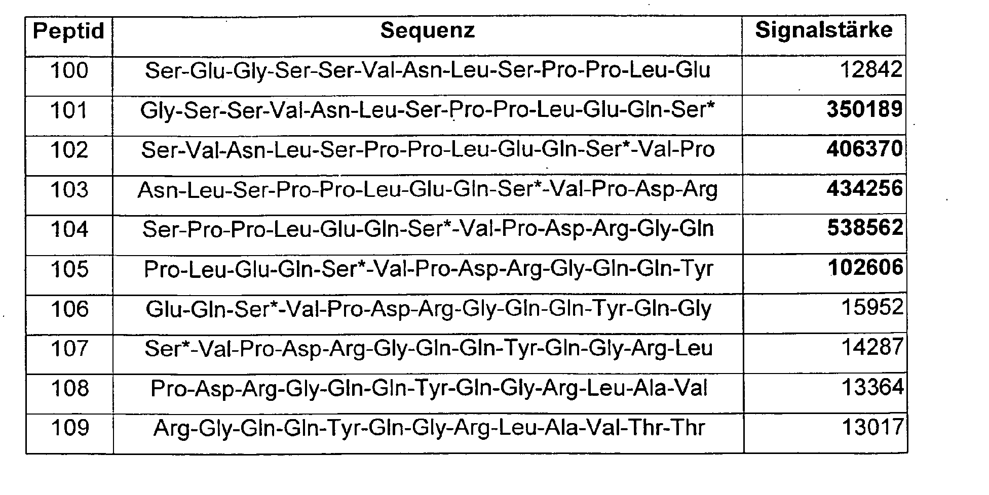
- the prothrombin fragment F 1 + 2 was subdivided into 13-mer peptides, each of which overlaps by 11 amino acids and thus reproduces successively in 2-step increments from the N-terminus to the C-terminus.
- These peptides were synthesized, coupled to a membrane and the binding of the antibody to be analyzed to each of these peptides examined:
- the antibody to be examined was previously covalently coupled to horseradish peroxidase.
- the horseradish peroxidase converts a chemiluminescent substrate whose signal is quantified using an imaging system. That is, the more antibody has bound to a particular peptide, the stronger the signal measured with that peptide.
- the reason for the low yield in the search for a suitable secondary antibody is apparently that for combination with a primary antibody against the free C-terminus of the prothrombin F 1 + 2 fragment, a specific binding site of the secondary antibody preferably in the N-terminal half of F 2 fragment is required.
- a specific binding to the amino acid sequence Ser-Pro-Pro-Leu-Glu-Gln-Gys is preferred:
- the secondary antibodies according to the invention were used in combination with the Primary antibodies in an enzyme immunoassay according to the sandwich principle used:
- the F 1 + 2 antigen present in the sample binds to the primary antibodies directed against F 1 + 2 , which are fixed to the surface of wells of a microtitration plate.
- secondary peroxidase-conjugated secondary antibodies according to the invention are bound to the free F 1 + 2 -determinants in a second reaction.
- the excess enzyme-conjugated secondary antibodies are washed out.
- the bound enzyme activity in the wells is determined.
- the enzymatic reaction of hydrogen peroxide and tetramethylbenzidine is interrupted by the addition of dilute sulfuric acid.
- the color intensity proportional to the concentration of F 1 + 2 is determined photometrically and quantified using a calibration curve from the standards supplied.
- Such a sandwich immunoassay according to the invention exhibits improved results with regard to the following properties compared to known F 1 + 2 tests:
Landscapes
- Health & Medical Sciences (AREA)
- Chemical & Material Sciences (AREA)
- Organic Chemistry (AREA)
- Life Sciences & Earth Sciences (AREA)
- Engineering & Computer Science (AREA)
- Genetics & Genomics (AREA)
- Bioinformatics & Cheminformatics (AREA)
- General Health & Medical Sciences (AREA)
- Biochemistry (AREA)
- Wood Science & Technology (AREA)
- Zoology (AREA)
- Medicinal Chemistry (AREA)
- Immunology (AREA)
- General Engineering & Computer Science (AREA)
- Biomedical Technology (AREA)
- Molecular Biology (AREA)
- Microbiology (AREA)
- Biotechnology (AREA)
- Biophysics (AREA)
- Proteomics, Peptides & Aminoacids (AREA)
- Pharmacology & Pharmacy (AREA)
- General Chemical & Material Sciences (AREA)
- Nuclear Medicine, Radiotherapy & Molecular Imaging (AREA)
- Chemical Kinetics & Catalysis (AREA)
- Animal Behavior & Ethology (AREA)
- Public Health (AREA)
- Veterinary Medicine (AREA)
- Peptides Or Proteins (AREA)
- Preparation Of Compounds By Using Micro-Organisms (AREA)
- Micro-Organisms Or Cultivation Processes Thereof (AREA)
- Medicines That Contain Protein Lipid Enzymes And Other Medicines (AREA)
- Medicines Containing Antibodies Or Antigens For Use As Internal Diagnostic Agents (AREA)
Abstract
Description

| Probe A | nmol/L | nmol/L x Verdünnung | Wiederfindung % |
| Unverd. | 3,940 | 3,94 | 100 % |
| 1 : 2 | 2,060 | 4,12 | 105 % |
| 1 : 4 | 0,985 | 3,94 | 100 % |
| 1 : 16 | 0,206 | 3,30 | 84 % |
| Probe B | nmol/L | nmol/L x Verdünnung | Wiederfindung % |
| Unverd. | 0,900 | 0,90 | 100% |
| 1 : 2 | 0,430 | 0,86 | 96 % |
| 1 : 5 | 0,166 | 0,83 | 92 % |
| 1 : 8 | 0,114 | 0,91 | 101 % |
| 1 : 10 | 0,081 | 0,81 | 90 % |
| 1 : 15 | 0,064 | 0,96 | 107 % |
| Diagnostische Sensitivität | Mit monoklonalen Antikörpern | Stand der Technik (kommerziell erhältlicher Test) |
| Bei oral antikoagulierten Patienten (n = 18): | 83% | 78 % |
| Bei Thrombophilie-Patienten (n = 18): | 67% | 61 % |

Claims (21)
- Peptid bestehend aus 5-25 Aminosäuren, bevorzugt aus 5 bis 21 Aminosäuren, ganz besonders bevorzugt aus 5-12 Aminosäuren, dadurch gekennzeichnet, dass es die Aminosäuresequenz Pro-Leu-Glu-Gln-Cys umfasst.
- Peptid nach Anspruch 1 mit der Aminosäuresequenz Ser-Glu-Gly-Ser-Ser-Val-Asn-Leu-Ser-Pro-Pro-Leu-Glu-Gln-Cys-Val-Pro-Asp-Arg-Gly-Gln-Gln-Tyr-Gln-Gly oder ein Fragment desselben, insbesondere ein Peptid mit Aminosäuresequenz Ser-Pro-Pro-Leu-Glu-Gln-Cys.
- Peptid nach Anspruch 1 oder 2, dadurch gekennzeichnet, dass dieses mit einer Festphase und/oder einer Komponente eines signalbildenden Systems assoziiert ist.
- Verwendung eines Peptides nach Anspruch 1 oder 2 zur Immunisierung und/oder zur Reinigung von Antikörpern.
- Verwendung eines Peptids nach einem der Ansprüche 1-3 in einem Verfahren zum quantitativen oder qualitativen Nachweis eines Analyten, bevorzugt F1+2.
- Antikörper, dadurch gekennzeichnet, dass dieser spezifisch an ein Epitop auf der N-terminalen Hälfte des F2-Fragmentes von Prothrombin bindet.
- Antikörper nach Anspruch 6, dadurch gekennzeichnet, dass dieser ein monoklonaler oder ein polyklonaler Antikörper ist.
- Antikörper nach Anspruch 6 oder 7, dadurch gekennzeichnet, dass dieser an ein Peptid gemäß einem der Ansprüche 1-3 spezifisch bindet.
- Antikörper nach einem der Ansprüche 6-8, dadurch gekennzeichnet, dass dieser von der Hybridomazelllinie DSM ACC2607 produziert wird.
- Antikörper nach einem der Ansprüche 6-9, dadurch gekennzeichnet, dass der Antikörper mit einer Festphase und/oder einer Komponente eines signalbildenden Systems assoziiert ist.
- Antikörper nach einem der Ansprüche 6-9 in einem pharmazeutisch verträglichen, sterilen Injektionsmedium.
- Verwendung eines Antikörpers nach einem der Ansprüche 6-9 in einem Verfahren zum quantitativen oder qualitativen Nachweis eines Analyten, bevorzugt F1+2.
- Verwendung eines Antikörpers nach einem der Ansprüche 6-9 in der Affinitätschromatographie.
- Verwendung eines Antikörpers nach einem der Ansprüche 6-9 sowie 11 als Diagnostikum oder als Bestandteil eines Diagnostikums.
- Reagenz enthaltend ein oder mehrere Peptide nach einem der Ansprüche 1-3 und/oder einen oder mehrere Antikörper nach einem der Ansprüche 6-11.
- Testkit enthaltend ein oder mehrere Peptide nach einem der Ansprüche 1-3 und/oder einen oder mehrere Antikörper nach einem der Ansprüche 6-11 und/oder ein Reagenz gemäß Anspruch 15.
- Tierische, pflanzliche oder prokaryontische Zelle sowie isolierte menschliche Zelle, dadurch gekennzeichnet, dass diese Antikörper nach einem der Ansprüche 6-9 produziert.
- Hybridomazellinie nach Anspruch 17.
- Hybridomazelllinie nach Anspruch 18, die bei der DSMZ unter der Eingangsnummer DSM ACC2607 hinterlegt wurde.
- Verfahren zum quantitativen oder qualitativen Nachweis der ProthrombinFragmente F1+2 und/oder F2 in einer Probe unter Verwendung eines oder mehrerer Peptide nach einem der Ansprüche 1-3 und/oder eines oder mehrerer Antikörper nach einem der Ansprüche 6-11.
- Verfahren nach Anspruch 20, dadurch gekennzeichnet, dass Antikörper nach einem der Ansprüche 6-11 in Kombination mit Antikörpern, deren Epitope die vier carboxyterminalen Aminosäuren der F2- und F1+2-Fragmente umfassen, verwendet werden.
Applications Claiming Priority (2)
| Application Number | Priority Date | Filing Date | Title |
|---|---|---|---|
| DE10354403A DE10354403A1 (de) | 2003-11-20 | 2003-11-20 | Gegen das Prothrombin-Fragment F 1+2 gerichtete Antikörper, ihre Herstellung und Verwendung |
| DE10354403 | 2003-11-20 |
Publications (2)
| Publication Number | Publication Date |
|---|---|
| EP1541676A1 true EP1541676A1 (de) | 2005-06-15 |
| EP1541676B1 EP1541676B1 (de) | 2008-12-24 |
Family
ID=34485233
Family Applications (1)
| Application Number | Title | Priority Date | Filing Date |
|---|---|---|---|
| EP04022637A Expired - Lifetime EP1541676B1 (de) | 2003-11-20 | 2004-09-23 | Gegen das Prothrombin-Fragment F1+2 gerichtete Antikörper, ihre Herstellung und Verwendung |
Country Status (8)
| Country | Link |
|---|---|
| US (2) | US7795403B2 (de) |
| EP (1) | EP1541676B1 (de) |
| JP (1) | JP5588585B2 (de) |
| AT (1) | ATE418604T1 (de) |
| CA (1) | CA2488127C (de) |
| DE (2) | DE10354403A1 (de) |
| DK (1) | DK1541676T3 (de) |
| ES (1) | ES2316911T3 (de) |
Cited By (3)
| Publication number | Priority date | Publication date | Assignee | Title |
|---|---|---|---|---|
| EP2168985A1 (de) | 2008-09-30 | 2010-03-31 | Siemens Healthcare Diagnostics Products GmbH | Antikörper zur Bestimmung des Prothrombin-Fragments F2/F1+2 in einem homogenen Immunoassay |
| EP4239338A1 (de) | 2022-03-03 | 2023-09-06 | Siemens Healthcare Diagnostics Products GmbH | Globaltest zur feststellung des status des blutgerinnungssystems |
| EP4325224A1 (de) | 2022-08-18 | 2024-02-21 | Siemens Healthcare Diagnostics Products GmbH | Thrombozytenaktivierungstest zur diagnose einer heparin-induzierten thrombozytopenie |
Families Citing this family (3)
| Publication number | Priority date | Publication date | Assignee | Title |
|---|---|---|---|---|
| JP6032470B2 (ja) * | 2012-08-09 | 2016-11-30 | 富士レビオ株式会社 | Pivka−ii測定方法、測定試薬及び測定キット |
| KR102652906B1 (ko) | 2019-04-17 | 2024-04-01 | 노보 노르디스크 에이/에스 | 이중 특이적 항체 |
| JP2021124314A (ja) * | 2020-02-03 | 2021-08-30 | 東ソー株式会社 | フルオレセインを介したHBe抗体検出方法及び検出試薬 |
Citations (3)
| Publication number | Priority date | Publication date | Assignee | Title |
|---|---|---|---|---|
| WO2000014209A1 (en) * | 1998-09-07 | 2000-03-16 | Genotech Corp. | Endothelial cell proliferation inhibitors derived from human prothrombin |
| US6541275B1 (en) * | 1988-02-03 | 2003-04-01 | Dade Behring Inc. | Immunoassay for F1.2 prothrombin fragment |
| WO2004111636A2 (en) * | 2003-06-17 | 2004-12-23 | Vib Vzw | Peptide combos and their uses |
Family Cites Families (4)
| Publication number | Priority date | Publication date | Assignee | Title |
|---|---|---|---|---|
| US4208479A (en) * | 1977-07-14 | 1980-06-17 | Syva Company | Label modified immunoassays |
| DE3727610A1 (de) * | 1987-08-19 | 1989-03-02 | Behringwerke Ag | Synthetische peptide, gegen diese gerichtete antikoerper und deren verwendung |
| IE903930A1 (en) | 1989-11-06 | 1991-05-08 | Akzo Nv | Immunoassays for and monoclonal antibodies to prothrombin¹activation peptides and their degradation products |
| ATE449107T1 (de) * | 2002-01-25 | 2009-12-15 | Univ Aberdeen | Aus fisch gewonnene peptide und nukleinsäuren der cathelicidin familie und deren verwendung |
-
2003
- 2003-11-20 DE DE10354403A patent/DE10354403A1/de not_active Withdrawn
-
2004
- 2004-09-23 EP EP04022637A patent/EP1541676B1/de not_active Expired - Lifetime
- 2004-09-23 AT AT04022637T patent/ATE418604T1/de active
- 2004-09-23 DE DE502004008729T patent/DE502004008729D1/de not_active Expired - Lifetime
- 2004-09-23 ES ES04022637T patent/ES2316911T3/es not_active Expired - Lifetime
- 2004-09-23 DK DK04022637T patent/DK1541676T3/da active
- 2004-11-19 JP JP2004335578A patent/JP5588585B2/ja not_active Expired - Fee Related
- 2004-11-19 US US10/992,351 patent/US7795403B2/en active Active
- 2004-11-19 CA CA2488127A patent/CA2488127C/en not_active Expired - Lifetime
-
2009
- 2009-11-16 US US12/591,303 patent/US8063180B2/en not_active Expired - Fee Related
Patent Citations (3)
| Publication number | Priority date | Publication date | Assignee | Title |
|---|---|---|---|---|
| US6541275B1 (en) * | 1988-02-03 | 2003-04-01 | Dade Behring Inc. | Immunoassay for F1.2 prothrombin fragment |
| WO2000014209A1 (en) * | 1998-09-07 | 2000-03-16 | Genotech Corp. | Endothelial cell proliferation inhibitors derived from human prothrombin |
| WO2004111636A2 (en) * | 2003-06-17 | 2004-12-23 | Vib Vzw | Peptide combos and their uses |
Cited By (5)
| Publication number | Priority date | Publication date | Assignee | Title |
|---|---|---|---|---|
| EP2168985A1 (de) | 2008-09-30 | 2010-03-31 | Siemens Healthcare Diagnostics Products GmbH | Antikörper zur Bestimmung des Prothrombin-Fragments F2/F1+2 in einem homogenen Immunoassay |
| DE102008049601A1 (de) | 2008-09-30 | 2010-04-01 | Siemens Healthcare Diagnostics Products Gmbh | Antikörper zur Bestimmung des Prothrombin-Fragments F2/F1+2 in einem homogenen Immunoassay |
| US9096674B2 (en) | 2008-09-30 | 2015-08-04 | Siemens Healthcare Diagnostics Products Gmbh | Antibodies for determining the prothrombin fragment F2/F1+2 in a homogeneous immunoassay |
| EP4239338A1 (de) | 2022-03-03 | 2023-09-06 | Siemens Healthcare Diagnostics Products GmbH | Globaltest zur feststellung des status des blutgerinnungssystems |
| EP4325224A1 (de) | 2022-08-18 | 2024-02-21 | Siemens Healthcare Diagnostics Products GmbH | Thrombozytenaktivierungstest zur diagnose einer heparin-induzierten thrombozytopenie |
Also Published As
| Publication number | Publication date |
|---|---|
| DE10354403A1 (de) | 2005-06-23 |
| ES2316911T3 (es) | 2009-04-16 |
| JP5588585B2 (ja) | 2014-09-10 |
| US7795403B2 (en) | 2010-09-14 |
| CA2488127C (en) | 2012-10-23 |
| US8063180B2 (en) | 2011-11-22 |
| DE502004008729D1 (de) | 2009-02-05 |
| EP1541676B1 (de) | 2008-12-24 |
| CA2488127A1 (en) | 2005-05-20 |
| US20050113562A1 (en) | 2005-05-26 |
| DK1541676T3 (da) | 2009-04-14 |
| JP2005154440A (ja) | 2005-06-16 |
| ATE418604T1 (de) | 2009-01-15 |
| US20100158932A1 (en) | 2010-06-24 |
Similar Documents
| Publication | Publication Date | Title |
|---|---|---|
| EP1110970B1 (de) | Gegen Procalcitonin gerichtete Antikörper, ihre Herstellung und Verwendung | |
| DE69521608T2 (de) | Verwendung von humanem neutrophil-lipocalin(hnl) als diagnostischer marker und anti-hnl-antikörperzubereitung | |
| DE69420556T2 (de) | Assay für troponin i aus dem herzmuskel | |
| CH658131A5 (de) | Reagenz und verfahren fuer immunologische analyse. | |
| EP2168985B1 (de) | Antikörper zur Bestimmung des Prothrombin-Fragments F2/F1+2 in einem homogenen Immunoassay | |
| EP1111050A2 (de) | Humanes Procalcitonin, dessen Herstellung und Verwendung | |
| US7951910B2 (en) | Peptides with the marburg I polymorphism of factor VII-activating protease (FSAP) and their preparation and uses | |
| US8063180B2 (en) | Antibodies directed against prothrombin fragment F1+2, the preparation and use thereof | |
| EP0600326A2 (de) | Streptolysin O Peptidantigene und Verfahren zur Bestimmung von Streptolysin-Antikörper | |
| EP2399933B1 (de) | Bindungspartner des plazentalen Wachstumsfaktors insbesondere gegen den plazentalen Wachstumsfaktor gerichtete Antikörper, ihre Herstellung und Verwendung | |
| DE68911574T2 (de) | Reagenzsystem zur Bestimmung des Komplexes des menschlichen Plasminogenaktivator-Inhibitors und des menschlichen Gewebeplasminogenaktivators und Testsatz dafür. | |
| DE10041215A1 (de) | Gegen Procalcitonin gerichtete Antikörper, ihre Herstellung und Verwendung | |
| DE3855007T2 (de) | METHODE ZUR REINIGUNG REKOMBINANTER t-PA VON DEN IHREN ENTSPRECHENDEN WIRTSZELLPROTEINEN | |
| DE19721113A1 (de) | Diagnosekit | |
| EP1378521A1 (de) | Carbohydrate Deficient Transferrin (CDT)-spezifische Antikörper, ihre Herstellung und Verwendung | |
| EP0614912B1 (de) | Lipoprotein (a)-Peptide und deren Verwendung | |
| DE10027954A1 (de) | Humanes Procalcitonin, dessen Herstellung und Verwendung | |
| DE4310516A1 (de) | Lipoprotein (a)-Peptide und deren Verwendung |
Legal Events
| Date | Code | Title | Description |
|---|---|---|---|
| PUAI | Public reference made under article 153(3) epc to a published international application that has entered the european phase |
Free format text: ORIGINAL CODE: 0009012 |
|
| AK | Designated contracting states |
Kind code of ref document: A1 Designated state(s): AT BE BG CH CY CZ DE DK EE ES FI FR GB GR HU IE IT LI LU MC NL PL PT RO SE SI SK TR |
|
| AX | Request for extension of the european patent |
Extension state: AL HR LT LV MK |
|
| 17P | Request for examination filed |
Effective date: 20051215 |
|
| AKX | Designation fees paid |
Designated state(s): AT BE BG CH CY CZ DE DK EE ES FI FR GB GR HU IE IT LI LU MC NL PL PT RO SE SI SK TR |
|
| 17Q | First examination report despatched |
Effective date: 20070215 |
|
| GRAP | Despatch of communication of intention to grant a patent |
Free format text: ORIGINAL CODE: EPIDOSNIGR1 |
|
| GRAS | Grant fee paid |
Free format text: ORIGINAL CODE: EPIDOSNIGR3 |
|
| RAP1 | Party data changed (applicant data changed or rights of an application transferred) |
Owner name: SIEMENS HEALTHCARE DIAGNOSTICS PRODUCTS GMBH |
|
| GRAA | (expected) grant |
Free format text: ORIGINAL CODE: 0009210 |
|
| AK | Designated contracting states |
Kind code of ref document: B1 Designated state(s): AT BE BG CH CY CZ DE DK EE ES FI FR GB GR HU IE IT LI LU MC NL PL PT RO SE SI SK TR |
|
| REG | Reference to a national code |
Ref country code: GB Ref legal event code: FG4D Free format text: NOT ENGLISH |
|
| REG | Reference to a national code |
Ref country code: CH Ref legal event code: NV Representative=s name: SIEMENS SCHWEIZ AG Ref country code: CH Ref legal event code: EP |
|
| REG | Reference to a national code |
Ref country code: IE Ref legal event code: FG4D Free format text: LANGUAGE OF EP DOCUMENT: GERMAN |
|
| REF | Corresponds to: |
Ref document number: 502004008729 Country of ref document: DE Date of ref document: 20090205 Kind code of ref document: P |
|
| REG | Reference to a national code |
Ref country code: SE Ref legal event code: TRGR |
|
| REG | Reference to a national code |
Ref country code: CH Ref legal event code: PCAR Free format text: SIEMENS SCHWEIZ AG;INTELLECTUAL PROPERTY FREILAGERSTRASSE 40;8047 ZUERICH (CH) |
|
| REG | Reference to a national code |
Ref country code: DK Ref legal event code: T3 |
|
| REG | Reference to a national code |
Ref country code: ES Ref legal event code: FG2A Ref document number: 2316911 Country of ref document: ES Kind code of ref document: T3 |
|
| PG25 | Lapsed in a contracting state [announced via postgrant information from national office to epo] |
Ref country code: FI Free format text: LAPSE BECAUSE OF FAILURE TO SUBMIT A TRANSLATION OF THE DESCRIPTION OR TO PAY THE FEE WITHIN THE PRESCRIBED TIME-LIMIT Effective date: 20081224 Ref country code: PL Free format text: LAPSE BECAUSE OF FAILURE TO SUBMIT A TRANSLATION OF THE DESCRIPTION OR TO PAY THE FEE WITHIN THE PRESCRIBED TIME-LIMIT Effective date: 20081224 Ref country code: SI Free format text: LAPSE BECAUSE OF FAILURE TO SUBMIT A TRANSLATION OF THE DESCRIPTION OR TO PAY THE FEE WITHIN THE PRESCRIBED TIME-LIMIT Effective date: 20081224 |
|
| PG25 | Lapsed in a contracting state [announced via postgrant information from national office to epo] |
Ref country code: RO Free format text: LAPSE BECAUSE OF FAILURE TO SUBMIT A TRANSLATION OF THE DESCRIPTION OR TO PAY THE FEE WITHIN THE PRESCRIBED TIME-LIMIT Effective date: 20081224 Ref country code: EE Free format text: LAPSE BECAUSE OF FAILURE TO SUBMIT A TRANSLATION OF THE DESCRIPTION OR TO PAY THE FEE WITHIN THE PRESCRIBED TIME-LIMIT Effective date: 20081224 Ref country code: BG Free format text: LAPSE BECAUSE OF FAILURE TO SUBMIT A TRANSLATION OF THE DESCRIPTION OR TO PAY THE FEE WITHIN THE PRESCRIBED TIME-LIMIT Effective date: 20090324 |
|
| PG25 | Lapsed in a contracting state [announced via postgrant information from national office to epo] |
Ref country code: CZ Free format text: LAPSE BECAUSE OF FAILURE TO SUBMIT A TRANSLATION OF THE DESCRIPTION OR TO PAY THE FEE WITHIN THE PRESCRIBED TIME-LIMIT Effective date: 20081224 Ref country code: PT Free format text: LAPSE BECAUSE OF FAILURE TO SUBMIT A TRANSLATION OF THE DESCRIPTION OR TO PAY THE FEE WITHIN THE PRESCRIBED TIME-LIMIT Effective date: 20090525 |
|
| PG25 | Lapsed in a contracting state [announced via postgrant information from national office to epo] |
Ref country code: SK Free format text: LAPSE BECAUSE OF FAILURE TO SUBMIT A TRANSLATION OF THE DESCRIPTION OR TO PAY THE FEE WITHIN THE PRESCRIBED TIME-LIMIT Effective date: 20081224 |
|
| PLBE | No opposition filed within time limit |
Free format text: ORIGINAL CODE: 0009261 |
|
| STAA | Information on the status of an ep patent application or granted ep patent |
Free format text: STATUS: NO OPPOSITION FILED WITHIN TIME LIMIT |
|
| 26N | No opposition filed |
Effective date: 20090925 |
|
| PG25 | Lapsed in a contracting state [announced via postgrant information from national office to epo] |
Ref country code: MC Free format text: LAPSE BECAUSE OF NON-PAYMENT OF DUE FEES Effective date: 20090930 |
|
| PG25 | Lapsed in a contracting state [announced via postgrant information from national office to epo] |
Ref country code: GR Free format text: LAPSE BECAUSE OF FAILURE TO SUBMIT A TRANSLATION OF THE DESCRIPTION OR TO PAY THE FEE WITHIN THE PRESCRIBED TIME-LIMIT Effective date: 20090325 |
|
| PG25 | Lapsed in a contracting state [announced via postgrant information from national office to epo] |
Ref country code: LU Free format text: LAPSE BECAUSE OF NON-PAYMENT OF DUE FEES Effective date: 20090923 |
|
| PG25 | Lapsed in a contracting state [announced via postgrant information from national office to epo] |
Ref country code: HU Free format text: LAPSE BECAUSE OF FAILURE TO SUBMIT A TRANSLATION OF THE DESCRIPTION OR TO PAY THE FEE WITHIN THE PRESCRIBED TIME-LIMIT Effective date: 20090625 |
|
| PG25 | Lapsed in a contracting state [announced via postgrant information from national office to epo] |
Ref country code: TR Free format text: LAPSE BECAUSE OF FAILURE TO SUBMIT A TRANSLATION OF THE DESCRIPTION OR TO PAY THE FEE WITHIN THE PRESCRIBED TIME-LIMIT Effective date: 20081224 |
|
| PG25 | Lapsed in a contracting state [announced via postgrant information from national office to epo] |
Ref country code: CY Free format text: LAPSE BECAUSE OF FAILURE TO SUBMIT A TRANSLATION OF THE DESCRIPTION OR TO PAY THE FEE WITHIN THE PRESCRIBED TIME-LIMIT Effective date: 20081224 |
|
| PGFP | Annual fee paid to national office [announced via postgrant information from national office to epo] |
Ref country code: DK Payment date: 20110926 Year of fee payment: 8 |
|
| PGFP | Annual fee paid to national office [announced via postgrant information from national office to epo] |
Ref country code: SE Payment date: 20120911 Year of fee payment: 9 |
|
| PGFP | Annual fee paid to national office [announced via postgrant information from national office to epo] |
Ref country code: BE Payment date: 20121010 Year of fee payment: 9 |
|
| BERE | Be: lapsed |
Owner name: SIEMENS HEALTHCARE DIAGNOSTICS PRODUCTS G.M.B.H. Effective date: 20130930 |
|
| REG | Reference to a national code |
Ref country code: SE Ref legal event code: EUG |
|
| PG25 | Lapsed in a contracting state [announced via postgrant information from national office to epo] |
Ref country code: SE Free format text: LAPSE BECAUSE OF NON-PAYMENT OF DUE FEES Effective date: 20130924 |
|
| REG | Reference to a national code |
Ref country code: DK Ref legal event code: EBP Effective date: 20130930 |
|
| PG25 | Lapsed in a contracting state [announced via postgrant information from national office to epo] |
Ref country code: BE Free format text: LAPSE BECAUSE OF NON-PAYMENT OF DUE FEES Effective date: 20130930 |
|
| PG25 | Lapsed in a contracting state [announced via postgrant information from national office to epo] |
Ref country code: DK Free format text: LAPSE BECAUSE OF NON-PAYMENT OF DUE FEES Effective date: 20130930 |
|
| REG | Reference to a national code |
Ref country code: FR Ref legal event code: PLFP Year of fee payment: 13 |
|
| REG | Reference to a national code |
Ref country code: FR Ref legal event code: PLFP Year of fee payment: 14 |
|
| REG | Reference to a national code |
Ref country code: FR Ref legal event code: PLFP Year of fee payment: 15 |
|
| PGFP | Annual fee paid to national office [announced via postgrant information from national office to epo] |
Ref country code: NL Payment date: 20200915 Year of fee payment: 17 |
|
| PGFP | Annual fee paid to national office [announced via postgrant information from national office to epo] |
Ref country code: AT Payment date: 20210805 Year of fee payment: 18 Ref country code: IT Payment date: 20210920 Year of fee payment: 18 Ref country code: IE Payment date: 20210922 Year of fee payment: 18 |
|
| REG | Reference to a national code |
Ref country code: NL Ref legal event code: MM Effective date: 20211001 |
|
| PG25 | Lapsed in a contracting state [announced via postgrant information from national office to epo] |
Ref country code: NL Free format text: LAPSE BECAUSE OF NON-PAYMENT OF DUE FEES Effective date: 20211001 |
|
| PGFP | Annual fee paid to national office [announced via postgrant information from national office to epo] |
Ref country code: DE Payment date: 20220620 Year of fee payment: 19 |
|
| PGFP | Annual fee paid to national office [announced via postgrant information from national office to epo] |
Ref country code: FR Payment date: 20220921 Year of fee payment: 19 |
|
| PGFP | Annual fee paid to national office [announced via postgrant information from national office to epo] |
Ref country code: GB Payment date: 20221011 Year of fee payment: 19 |
|
| PGFP | Annual fee paid to national office [announced via postgrant information from national office to epo] |
Ref country code: CH Payment date: 20221220 Year of fee payment: 19 |
|
| PGFP | Annual fee paid to national office [announced via postgrant information from national office to epo] |
Ref country code: ES Payment date: 20221227 Year of fee payment: 19 |
|
| REG | Reference to a national code |
Ref country code: AT Ref legal event code: MM01 Ref document number: 418604 Country of ref document: AT Kind code of ref document: T Effective date: 20220923 |
|
| PG25 | Lapsed in a contracting state [announced via postgrant information from national office to epo] |
Ref country code: IE Free format text: LAPSE BECAUSE OF NON-PAYMENT OF DUE FEES Effective date: 20220923 Ref country code: AT Free format text: LAPSE BECAUSE OF NON-PAYMENT OF DUE FEES Effective date: 20220923 |
|
| PG25 | Lapsed in a contracting state [announced via postgrant information from national office to epo] |
Ref country code: IT Free format text: LAPSE BECAUSE OF NON-PAYMENT OF DUE FEES Effective date: 20220923 |
|
| REG | Reference to a national code |
Ref country code: DE Ref legal event code: R119 Ref document number: 502004008729 Country of ref document: DE |
|
| REG | Reference to a national code |
Ref country code: CH Ref legal event code: PL |
|
| GBPC | Gb: european patent ceased through non-payment of renewal fee |
Effective date: 20230923 |
|
| PG25 | Lapsed in a contracting state [announced via postgrant information from national office to epo] |
Ref country code: GB Free format text: LAPSE BECAUSE OF NON-PAYMENT OF DUE FEES Effective date: 20230923 |
|
| PG25 | Lapsed in a contracting state [announced via postgrant information from national office to epo] |
Ref country code: CH Free format text: LAPSE BECAUSE OF NON-PAYMENT OF DUE FEES Effective date: 20230930 |
|
| PG25 | Lapsed in a contracting state [announced via postgrant information from national office to epo] |
Ref country code: GB Free format text: LAPSE BECAUSE OF NON-PAYMENT OF DUE FEES Effective date: 20230923 Ref country code: FR Free format text: LAPSE BECAUSE OF NON-PAYMENT OF DUE FEES Effective date: 20230930 Ref country code: DE Free format text: LAPSE BECAUSE OF NON-PAYMENT OF DUE FEES Effective date: 20240403 Ref country code: CH Free format text: LAPSE BECAUSE OF NON-PAYMENT OF DUE FEES Effective date: 20230930 |
|
| REG | Reference to a national code |
Ref country code: ES Ref legal event code: FD2A Effective date: 20241104 |
|
| PG25 | Lapsed in a contracting state [announced via postgrant information from national office to epo] |
Ref country code: ES Free format text: LAPSE BECAUSE OF NON-PAYMENT OF DUE FEES Effective date: 20230924 |
|
| PG25 | Lapsed in a contracting state [announced via postgrant information from national office to epo] |
Ref country code: ES Free format text: LAPSE BECAUSE OF NON-PAYMENT OF DUE FEES Effective date: 20230924 |